EP2730576A2 - Anti-infective compounds - Google Patents
Anti-infective compounds Download PDFInfo
- Publication number
- EP2730576A2 EP2730576A2 EP13198407.2A EP13198407A EP2730576A2 EP 2730576 A2 EP2730576 A2 EP 2730576A2 EP 13198407 A EP13198407 A EP 13198407A EP 2730576 A2 EP2730576 A2 EP 2730576A2
- Authority
- EP
- European Patent Office
- Prior art keywords
- nmr
- mhz
- cdcl
- pyrido
- compounds
- Prior art date
- Legal status (The legal status is an assumption and is not a legal conclusion. Google has not performed a legal analysis and makes no representation as to the accuracy of the status listed.)
- Withdrawn
Links
- 0 CC(CC1(*)*)=NN1C(c1ccncc1)=O Chemical compound CC(CC1(*)*)=NN1C(c1ccncc1)=O 0.000 description 7
- GCUMCLJEVVFELW-CQSZACIVSA-N [O-][N+](c1cc(C(N(CC2)C[C@@H]2NCc2ccccc2F)=O)cc([N+]([O-])=O)c1)=O Chemical compound [O-][N+](c1cc(C(N(CC2)C[C@@H]2NCc2ccccc2F)=O)cc([N+]([O-])=O)c1)=O GCUMCLJEVVFELW-CQSZACIVSA-N 0.000 description 2
- RAJQFYWDFHRVJY-UHFFFAOYSA-N CC(C)C(OCC1=C(Nc2ccccc2)N=C(C=CC=C2)N2C1=O)=O Chemical compound CC(C)C(OCC1=C(Nc2ccccc2)N=C(C=CC=C2)N2C1=O)=O RAJQFYWDFHRVJY-UHFFFAOYSA-N 0.000 description 1
- OUUKZTKMVHZTKS-UHFFFAOYSA-N CC(C1=NC(NCc2cccc(Cl)c2)=C2C(OC)=O)=CC=CN1C2=O Chemical compound CC(C1=NC(NCc2cccc(Cl)c2)=C2C(OC)=O)=CC=CN1C2=O OUUKZTKMVHZTKS-UHFFFAOYSA-N 0.000 description 1
- FVRVLTGEWQGMFV-UHFFFAOYSA-N CC(C1=NC(Nc2cc(C(F)(F)F)ccc2)=C2C=O)=CC=CN1C2=O Chemical compound CC(C1=NC(Nc2cc(C(F)(F)F)ccc2)=C2C=O)=CC=CN1C2=O FVRVLTGEWQGMFV-UHFFFAOYSA-N 0.000 description 1
- LAUHQWMBDPCMHK-UHFFFAOYSA-N CC(C1=NC(Nc2cc(F)ccc2)=C2C(O)=O)=CC=CN1C2=O Chemical compound CC(C1=NC(Nc2cc(F)ccc2)=C2C(O)=O)=CC=CN1C2=O LAUHQWMBDPCMHK-UHFFFAOYSA-N 0.000 description 1
- FCDSNWYQUHTNIE-UHFFFAOYSA-N CC(C=CN12)=CC1=NC(Nc(cc1)cc(Cl)c1Cl)=C(CO)C2=O Chemical compound CC(C=CN12)=CC1=NC(Nc(cc1)cc(Cl)c1Cl)=C(CO)C2=O FCDSNWYQUHTNIE-UHFFFAOYSA-N 0.000 description 1
- VDZFJXXJFXMJJO-UHFFFAOYSA-N CC(C=CN12)=CC1=NC(O)=CC2=O Chemical compound CC(C=CN12)=CC1=NC(O)=CC2=O VDZFJXXJFXMJJO-UHFFFAOYSA-N 0.000 description 1
- PSGPXWYGJGGEEG-UHFFFAOYSA-N CCCCOC(C1(c(cccc2)c2-c2c1cccc2)O)=O Chemical compound CCCCOC(C1(c(cccc2)c2-c2c1cccc2)O)=O PSGPXWYGJGGEEG-UHFFFAOYSA-N 0.000 description 1
- NOILOVITPZSFJU-UHFFFAOYSA-N CCOC(C(C(N(C=CC=C1)C1=N1)=O)=C1Oc1ccccc1)=O Chemical compound CCOC(C(C(N(C=CC=C1)C1=N1)=O)=C1Oc1ccccc1)=O NOILOVITPZSFJU-UHFFFAOYSA-N 0.000 description 1
- OVKWVGOBNIFKFC-UHFFFAOYSA-N CN(CC1)CCN1C(C=CN12)=CC1=NC(NC(C1)=CC=CC1F)=C(CO)C2=O Chemical compound CN(CC1)CCN1C(C=CN12)=CC1=NC(NC(C1)=CC=CC1F)=C(CO)C2=O OVKWVGOBNIFKFC-UHFFFAOYSA-N 0.000 description 1
- UQOYSIWSGQIBEP-UHFFFAOYSA-N CN(CC1)CCN1C(C=CN12)=CC1=NC(Nc1cc(Cl)ccc1)=C(C(O)=O)C2=O Chemical compound CN(CC1)CCN1C(C=CN12)=CC1=NC(Nc1cc(Cl)ccc1)=C(C(O)=O)C2=O UQOYSIWSGQIBEP-UHFFFAOYSA-N 0.000 description 1
- ITSIREUAUWBMRO-UHFFFAOYSA-N CN(CC1)CCN1C(C=CN12)=CC1=NC(Nc1cc(F)ccc1)=C(CO)C2=O Chemical compound CN(CC1)CCN1C(C=CN12)=CC1=NC(Nc1cc(F)ccc1)=C(CO)C2=O ITSIREUAUWBMRO-UHFFFAOYSA-N 0.000 description 1
- ZCFQCGGHHMXWNV-UHFFFAOYSA-N COC(C1=NC(Nc(cc2)ccc2Cl)=C2CO)=CC=CN1C2=O Chemical compound COC(C1=NC(Nc(cc2)ccc2Cl)=C2CO)=CC=CN1C2=O ZCFQCGGHHMXWNV-UHFFFAOYSA-N 0.000 description 1
- WBNVGTNHRAGRMJ-UHFFFAOYSA-N COC(C1=NC(Nc2ccccc2)=C2CO)=CC=CN1C2=O Chemical compound COC(C1=NC(Nc2ccccc2)=C2CO)=CC=CN1C2=O WBNVGTNHRAGRMJ-UHFFFAOYSA-N 0.000 description 1
- OSHLMWVSSNWSMK-UHFFFAOYSA-N COC(c(cc1)ccc1OCCCNC(C(C=C(C1)[N+]([O-])=O)=CC1[N+]([O-])=O)=O)O Chemical compound COC(c(cc1)ccc1OCCCNC(C(C=C(C1)[N+]([O-])=O)=CC1[N+]([O-])=O)=O)O OSHLMWVSSNWSMK-UHFFFAOYSA-N 0.000 description 1
- ILUNPADYTLUUBK-OMOCHNIRSA-N COC1C=CC(O[C@H](CC2)CN2C(c2cc(O)ccc2)=O)=CC1 Chemical compound COC1C=CC(O[C@H](CC2)CN2C(c2cc(O)ccc2)=O)=CC1 ILUNPADYTLUUBK-OMOCHNIRSA-N 0.000 description 1
- XYZNNDHOWOFZJL-UHFFFAOYSA-N COc(cc1)ccc1N(CC1)CCN1C(c1cc([N+]([O-])=O)cc([N+]([O-])=O)c1)=O Chemical compound COc(cc1)ccc1N(CC1)CCN1C(c1cc([N+]([O-])=O)cc([N+]([O-])=O)c1)=O XYZNNDHOWOFZJL-UHFFFAOYSA-N 0.000 description 1
- RJTDYBWABFIJMR-UHFFFAOYSA-N COc(cc1)ccc1OC(CC1)CN1C(c1cc([N+]([O-])=O)cc([N+]([O-])=O)c1)=O Chemical compound COc(cc1)ccc1OC(CC1)CN1C(c1cc([N+]([O-])=O)cc([N+]([O-])=O)c1)=O RJTDYBWABFIJMR-UHFFFAOYSA-N 0.000 description 1
- FTNQVPSPGPSTFD-QGZVFWFLSA-N COc(cc1)ccc1O[C@H](CC1)CN1C(c1cc(Cl)cc(Cl)c1)=O Chemical compound COc(cc1)ccc1O[C@H](CC1)CN1C(c1cc(Cl)cc(Cl)c1)=O FTNQVPSPGPSTFD-QGZVFWFLSA-N 0.000 description 1
- VLBFELDYYXACOM-GOSISDBHSA-N COc(cc1)ccc1O[C@H](CC1)CN1C(c1cccc(OC)c1)=O Chemical compound COc(cc1)ccc1O[C@H](CC1)CN1C(c1cccc(OC)c1)=O VLBFELDYYXACOM-GOSISDBHSA-N 0.000 description 1
- MMNWQJRYIIXOOE-MRXNPFEDSA-N COc(cc1)ccc1O[C@H](CC1)CN1C(c1ccncc1)=O Chemical compound COc(cc1)ccc1O[C@H](CC1)CN1C(c1ccncc1)=O MMNWQJRYIIXOOE-MRXNPFEDSA-N 0.000 description 1
- MNFYKOLHEXLPJO-OAHLLOKOSA-N COc(cc1)ccc1O[C@H](CC1)CN1C(c1cncnc1)=O Chemical compound COc(cc1)ccc1O[C@H](CC1)CN1C(c1cncnc1)=O MNFYKOLHEXLPJO-OAHLLOKOSA-N 0.000 description 1
- WMZUQJBSMHCQQK-UHFFFAOYSA-N COc1ccc(COCCNC(c2cc([N+]([O-])=O)cc([N+]([O-])=O)c2)=O)cc1 Chemical compound COc1ccc(COCCNC(c2cc([N+]([O-])=O)cc([N+]([O-])=O)c2)=O)cc1 WMZUQJBSMHCQQK-UHFFFAOYSA-N 0.000 description 1
- OWEVCZJORNSBDT-NSHDSACASA-N CS(O[C@@H](CC1)CN1C(c1cc([N+]([O-])=O)cc([N+]([O-])=O)c1)=O)(=O)=O Chemical compound CS(O[C@@H](CC1)CN1C(c1cc([N+]([O-])=O)cc([N+]([O-])=O)c1)=O)(=O)=O OWEVCZJORNSBDT-NSHDSACASA-N 0.000 description 1
- DYPLGTRQMGMANI-UHFFFAOYSA-N NNC(c1ccc(COC2=CC=[I]C=C2)[o]1)=O Chemical compound NNC(c1ccc(COC2=CC=[I]C=C2)[o]1)=O DYPLGTRQMGMANI-UHFFFAOYSA-N 0.000 description 1
- PDLOLBVRMQJLJS-UHFFFAOYSA-N O=C(c1cc(OC(F)(F)F)ccc1)NCCOCc1cccc(Cl)c1 Chemical compound O=C(c1cc(OC(F)(F)F)ccc1)NCCOCc1cccc(Cl)c1 PDLOLBVRMQJLJS-UHFFFAOYSA-N 0.000 description 1
- YUOAPRUWSCLOBF-UHFFFAOYSA-N O=C(c1ccccc1)NCCOCC1=CC(C(F)(F)F)=CCC1 Chemical compound O=C(c1ccccc1)NCCOCC1=CC(C(F)(F)F)=CCC1 YUOAPRUWSCLOBF-UHFFFAOYSA-N 0.000 description 1
- WIEFUZWEMCRFAV-OEAKJJBVSA-N O=C1N(C=CC=C2)C2=NC(Nc2cccc(Cl)c2)=C1/C=N/C1CCCCC1 Chemical compound O=C1N(C=CC=C2)C2=NC(Nc2cccc(Cl)c2)=C1/C=N/C1CCCCC1 WIEFUZWEMCRFAV-OEAKJJBVSA-N 0.000 description 1
- OMNNICHWWMBMJW-UHFFFAOYSA-N OCC1=C(NC2=CC=CCC2O)N=C(CCCC2)N2C1=O Chemical compound OCC1=C(NC2=CC=CCC2O)N=C(CCCC2)N2C1=O OMNNICHWWMBMJW-UHFFFAOYSA-N 0.000 description 1
- OCBOOAIIHLARHK-UHFFFAOYSA-N OCC1=C(Nc2cc(C(F)(F)F)ccc2)N=C(C=CC=C2)N2C1=O Chemical compound OCC1=C(Nc2cc(C(F)(F)F)ccc2)N=C(C=CC=C2)N2C1=O OCBOOAIIHLARHK-UHFFFAOYSA-N 0.000 description 1
- NCACEFQFRUEQHP-UHFFFAOYSA-N OCC1=C(Nc2cc(Cl)ccc2)N=C(C=CC=C2)N2C1=O Chemical compound OCC1=C(Nc2cc(Cl)ccc2)N=C(C=CC=C2)N2C1=O NCACEFQFRUEQHP-UHFFFAOYSA-N 0.000 description 1
- NRMZGMPFABWFDV-UHFFFAOYSA-N OCC1=C(Nc2cc(Cl)ccc2)N=C(CCCC2)N2C1=O Chemical compound OCC1=C(Nc2cc(Cl)ccc2)N=C(CCCC2)N2C1=O NRMZGMPFABWFDV-UHFFFAOYSA-N 0.000 description 1
- JTYSJYIPIRLSCT-UHFFFAOYSA-N OCC1=C(Nc2cccc(Cl)c2)N=C(C(F)=CC(F)=C2)N2C1=O Chemical compound OCC1=C(Nc2cccc(Cl)c2)N=C(C(F)=CC(F)=C2)N2C1=O JTYSJYIPIRLSCT-UHFFFAOYSA-N 0.000 description 1
- JONWQSWXITTWKB-UHFFFAOYSA-N OCC1=C(Nc2cccc(OC(F)(F)F)c2)N=C(C=CC=C2)N2C1=O Chemical compound OCC1=C(Nc2cccc(OC(F)(F)F)c2)N=C(C=CC=C2)N2C1=O JONWQSWXITTWKB-UHFFFAOYSA-N 0.000 description 1
- TZSBWYDFSYOWAH-UHFFFAOYSA-N OCC1=C(Nc2ccccc2)N=C(C=C(C=C2)N3CCOCC3)N2C1=O Chemical compound OCC1=C(Nc2ccccc2)N=C(C=C(C=C2)N3CCOCC3)N2C1=O TZSBWYDFSYOWAH-UHFFFAOYSA-N 0.000 description 1
- OJZJZPXCUILGQR-QSVWIEALSA-N [O-][N+](C(CC(C(N(CC1)C[C@@H]1Nc(cc1)ccc1OC1=CCCC=C1)=O)=C1)C=C1[N+]([O-])=O)=O Chemical compound [O-][N+](C(CC(C(N(CC1)C[C@@H]1Nc(cc1)ccc1OC1=CCCC=C1)=O)=C1)C=C1[N+]([O-])=O)=O OJZJZPXCUILGQR-QSVWIEALSA-N 0.000 description 1
- WDYSFJZZOYCDQS-GOSISDBHSA-N [O-][N+](c1cc(C(N(CC2)C[C@@H]2Nc(cc2)ccc2Oc2ccccc2)=O)cc([N+]([O-])=O)c1)=O Chemical compound [O-][N+](c1cc(C(N(CC2)C[C@@H]2Nc(cc2)ccc2Oc2ccccc2)=O)cc([N+]([O-])=O)c1)=O WDYSFJZZOYCDQS-GOSISDBHSA-N 0.000 description 1
- ULGMUYNEOPGRHA-SNVBAGLBSA-N [O-][N+](c1cc(C(N(CC2)C[C@@H]2O)=O)cc([N+]([O-])=O)c1)=O Chemical compound [O-][N+](c1cc(C(N(CC2)C[C@@H]2O)=O)cc([N+]([O-])=O)c1)=O ULGMUYNEOPGRHA-SNVBAGLBSA-N 0.000 description 1
- HKOIGYKNZMEXAX-CQSZACIVSA-N [O-][N+](c1cc(C(N(CC2)C[C@@H]2Oc2ccccc2F)=O)cc([N+]([O-])=O)c1)=O Chemical compound [O-][N+](c1cc(C(N(CC2)C[C@@H]2Oc2ccccc2F)=O)cc([N+]([O-])=O)c1)=O HKOIGYKNZMEXAX-CQSZACIVSA-N 0.000 description 1
- IFDRKFCLMJTDIW-UHFFFAOYSA-N [O-][N+](c1cc(C(NCCOCc2cccc(Cl)c2)=O)ccc1)=O Chemical compound [O-][N+](c1cc(C(NCCOCc2cccc(Cl)c2)=O)ccc1)=O IFDRKFCLMJTDIW-UHFFFAOYSA-N 0.000 description 1
- BKIFWIVRNXIZPB-UHFFFAOYSA-N [O-][N+](c1cc(C(NCCOc(cc2)ccc2Cl)=O)cc([N+]([O-])=O)c1)=O Chemical compound [O-][N+](c1cc(C(NCCOc(cc2)ccc2Cl)=O)cc([N+]([O-])=O)c1)=O BKIFWIVRNXIZPB-UHFFFAOYSA-N 0.000 description 1
- IEVZMFGHWKTRMF-UHFFFAOYSA-N [O-][N+](c1cc([N+]([O-])=O)cc(C(NCCOCc2cc(Cl)ccc2)=O)c1)=O Chemical compound [O-][N+](c1cc([N+]([O-])=O)cc(C(NCCOCc2cc(Cl)ccc2)=O)c1)=O IEVZMFGHWKTRMF-UHFFFAOYSA-N 0.000 description 1
- YERDMWQYJOEUAL-UHFFFAOYSA-N [O-][N+](c1cc([N+]([O-])=O)cc(C(NCCOc(cccc2)c2F)=O)c1)=O Chemical compound [O-][N+](c1cc([N+]([O-])=O)cc(C(NCCOc(cccc2)c2F)=O)c1)=O YERDMWQYJOEUAL-UHFFFAOYSA-N 0.000 description 1
Images




Classifications
-
- C—CHEMISTRY; METALLURGY
- C12—BIOCHEMISTRY; BEER; SPIRITS; WINE; VINEGAR; MICROBIOLOGY; ENZYMOLOGY; MUTATION OR GENETIC ENGINEERING
- C12Q—MEASURING OR TESTING PROCESSES INVOLVING ENZYMES, NUCLEIC ACIDS OR MICROORGANISMS; COMPOSITIONS OR TEST PAPERS THEREFOR; PROCESSES OF PREPARING SUCH COMPOSITIONS; CONDITION-RESPONSIVE CONTROL IN MICROBIOLOGICAL OR ENZYMOLOGICAL PROCESSES
- C12Q1/00—Measuring or testing processes involving enzymes, nucleic acids or microorganisms; Compositions therefor; Processes of preparing such compositions
- C12Q1/02—Measuring or testing processes involving enzymes, nucleic acids or microorganisms; Compositions therefor; Processes of preparing such compositions involving viable microorganisms
- C12Q1/18—Testing for antimicrobial activity of a material
-
- A—HUMAN NECESSITIES
- A61—MEDICAL OR VETERINARY SCIENCE; HYGIENE
- A61P—SPECIFIC THERAPEUTIC ACTIVITY OF CHEMICAL COMPOUNDS OR MEDICINAL PREPARATIONS
- A61P31/00—Antiinfectives, i.e. antibiotics, antiseptics, chemotherapeutics
- A61P31/04—Antibacterial agents
-
- A—HUMAN NECESSITIES
- A61—MEDICAL OR VETERINARY SCIENCE; HYGIENE
- A61K—PREPARATIONS FOR MEDICAL, DENTAL OR TOILETRY PURPOSES
- A61K31/00—Medicinal preparations containing organic active ingredients
- A61K31/33—Heterocyclic compounds
- A61K31/395—Heterocyclic compounds having nitrogen as a ring hetero atom, e.g. guanethidine or rifamycins
- A61K31/495—Heterocyclic compounds having nitrogen as a ring hetero atom, e.g. guanethidine or rifamycins having six-membered rings with two or more nitrogen atoms as the only ring heteroatoms, e.g. piperazine or tetrazines
- A61K31/505—Pyrimidines; Hydrogenated pyrimidines, e.g. trimethoprim
- A61K31/519—Pyrimidines; Hydrogenated pyrimidines, e.g. trimethoprim ortho- or peri-condensed with heterocyclic rings
-
- A—HUMAN NECESSITIES
- A61—MEDICAL OR VETERINARY SCIENCE; HYGIENE
- A61P—SPECIFIC THERAPEUTIC ACTIVITY OF CHEMICAL COMPOUNDS OR MEDICINAL PREPARATIONS
- A61P31/00—Antiinfectives, i.e. antibiotics, antiseptics, chemotherapeutics
- A61P31/04—Antibacterial agents
- A61P31/06—Antibacterial agents for tuberculosis
-
- C—CHEMISTRY; METALLURGY
- C07—ORGANIC CHEMISTRY
- C07C—ACYCLIC OR CARBOCYCLIC COMPOUNDS
- C07C255/00—Carboxylic acid nitriles
- C07C255/63—Carboxylic acid nitriles containing cyano groups and nitrogen atoms further bound to other hetero atoms, other than oxygen atoms of nitro or nitroso groups, bound to the same carbon skeleton
- C07C255/65—Carboxylic acid nitriles containing cyano groups and nitrogen atoms further bound to other hetero atoms, other than oxygen atoms of nitro or nitroso groups, bound to the same carbon skeleton with the nitrogen atoms further bound to nitrogen atoms
- C07C255/66—Carboxylic acid nitriles containing cyano groups and nitrogen atoms further bound to other hetero atoms, other than oxygen atoms of nitro or nitroso groups, bound to the same carbon skeleton with the nitrogen atoms further bound to nitrogen atoms having cyano groups and nitrogen atoms being part of hydrazine or hydrazone groups bound to the same carbon skeleton
-
- C—CHEMISTRY; METALLURGY
- C07—ORGANIC CHEMISTRY
- C07C—ACYCLIC OR CARBOCYCLIC COMPOUNDS
- C07C335/00—Thioureas, i.e. compounds containing any of the groups, the nitrogen atoms not being part of nitro or nitroso groups
- C07C335/40—Thioureas, i.e. compounds containing any of the groups, the nitrogen atoms not being part of nitro or nitroso groups having nitrogen atoms of thiourea or isothiourea groups further bound to other hetero atoms
-
- C—CHEMISTRY; METALLURGY
- C07—ORGANIC CHEMISTRY
- C07D—HETEROCYCLIC COMPOUNDS
- C07D223/00—Heterocyclic compounds containing seven-membered rings having one nitrogen atom as the only ring hetero atom
- C07D223/14—Heterocyclic compounds containing seven-membered rings having one nitrogen atom as the only ring hetero atom condensed with carbocyclic rings or ring systems
- C07D223/18—Dibenzazepines; Hydrogenated dibenzazepines
- C07D223/22—Dibenz [b, f] azepines; Hydrogenated dibenz [b, f] azepines
- C07D223/24—Dibenz [b, f] azepines; Hydrogenated dibenz [b, f] azepines with hydrocarbon radicals, substituted by nitrogen atoms, attached to the ring nitrogen atom
-
- C—CHEMISTRY; METALLURGY
- C07—ORGANIC CHEMISTRY
- C07D—HETEROCYCLIC COMPOUNDS
- C07D233/00—Heterocyclic compounds containing 1,3-diazole or hydrogenated 1,3-diazole rings, not condensed with other rings
- C07D233/54—Heterocyclic compounds containing 1,3-diazole or hydrogenated 1,3-diazole rings, not condensed with other rings having two double bonds between ring members or between ring members and non-ring members
- C07D233/66—Heterocyclic compounds containing 1,3-diazole or hydrogenated 1,3-diazole rings, not condensed with other rings having two double bonds between ring members or between ring members and non-ring members with hetero atoms or with carbon atoms having three bonds to hetero atoms with at the most one bond to halogen, e.g. ester or nitrile radicals, directly attached to ring carbon atoms
- C07D233/84—Sulfur atoms
-
- C—CHEMISTRY; METALLURGY
- C07—ORGANIC CHEMISTRY
- C07D—HETEROCYCLIC COMPOUNDS
- C07D251/00—Heterocyclic compounds containing 1,3,5-triazine rings
- C07D251/02—Heterocyclic compounds containing 1,3,5-triazine rings not condensed with other rings
- C07D251/12—Heterocyclic compounds containing 1,3,5-triazine rings not condensed with other rings having three double bonds between ring members or between ring members and non-ring members
- C07D251/26—Heterocyclic compounds containing 1,3,5-triazine rings not condensed with other rings having three double bonds between ring members or between ring members and non-ring members with only hetero atoms directly attached to ring carbon atoms
- C07D251/40—Nitrogen atoms
- C07D251/48—Two nitrogen atoms
- C07D251/52—Two nitrogen atoms with an oxygen or sulfur atom attached to the third ring carbon atom
-
- C—CHEMISTRY; METALLURGY
- C07—ORGANIC CHEMISTRY
- C07D—HETEROCYCLIC COMPOUNDS
- C07D251/00—Heterocyclic compounds containing 1,3,5-triazine rings
- C07D251/02—Heterocyclic compounds containing 1,3,5-triazine rings not condensed with other rings
- C07D251/12—Heterocyclic compounds containing 1,3,5-triazine rings not condensed with other rings having three double bonds between ring members or between ring members and non-ring members
- C07D251/26—Heterocyclic compounds containing 1,3,5-triazine rings not condensed with other rings having three double bonds between ring members or between ring members and non-ring members with only hetero atoms directly attached to ring carbon atoms
- C07D251/40—Nitrogen atoms
- C07D251/54—Three nitrogen atoms
- C07D251/66—Derivatives of melamine in which a hetero atom is directly attached to a nitrogen atom of melamine
-
- C—CHEMISTRY; METALLURGY
- C07—ORGANIC CHEMISTRY
- C07D—HETEROCYCLIC COMPOUNDS
- C07D271/00—Heterocyclic compounds containing five-membered rings having two nitrogen atoms and one oxygen atom as the only ring hetero atoms
- C07D271/02—Heterocyclic compounds containing five-membered rings having two nitrogen atoms and one oxygen atom as the only ring hetero atoms not condensed with other rings
- C07D271/10—1,3,4-Oxadiazoles; Hydrogenated 1,3,4-oxadiazoles
- C07D271/113—1,3,4-Oxadiazoles; Hydrogenated 1,3,4-oxadiazoles with oxygen, sulfur or nitrogen atoms, directly attached to ring carbon atoms, the nitrogen atoms not forming part of a nitro radical
-
- C—CHEMISTRY; METALLURGY
- C07—ORGANIC CHEMISTRY
- C07D—HETEROCYCLIC COMPOUNDS
- C07D401/00—Heterocyclic compounds containing two or more hetero rings, having nitrogen atoms as the only ring hetero atoms, at least one ring being a six-membered ring with only one nitrogen atom
- C07D401/02—Heterocyclic compounds containing two or more hetero rings, having nitrogen atoms as the only ring hetero atoms, at least one ring being a six-membered ring with only one nitrogen atom containing two hetero rings
- C07D401/12—Heterocyclic compounds containing two or more hetero rings, having nitrogen atoms as the only ring hetero atoms, at least one ring being a six-membered ring with only one nitrogen atom containing two hetero rings linked by a chain containing hetero atoms as chain links
-
- C—CHEMISTRY; METALLURGY
- C07—ORGANIC CHEMISTRY
- C07D—HETEROCYCLIC COMPOUNDS
- C07D409/00—Heterocyclic compounds containing two or more hetero rings, at least one ring having sulfur atoms as the only ring hetero atoms
- C07D409/02—Heterocyclic compounds containing two or more hetero rings, at least one ring having sulfur atoms as the only ring hetero atoms containing two hetero rings
- C07D409/12—Heterocyclic compounds containing two or more hetero rings, at least one ring having sulfur atoms as the only ring hetero atoms containing two hetero rings linked by a chain containing hetero atoms as chain links
-
- C—CHEMISTRY; METALLURGY
- C07—ORGANIC CHEMISTRY
- C07D—HETEROCYCLIC COMPOUNDS
- C07D417/00—Heterocyclic compounds containing two or more hetero rings, at least one ring having nitrogen and sulfur atoms as the only ring hetero atoms, not provided for by group C07D415/00
- C07D417/02—Heterocyclic compounds containing two or more hetero rings, at least one ring having nitrogen and sulfur atoms as the only ring hetero atoms, not provided for by group C07D415/00 containing two hetero rings
- C07D417/12—Heterocyclic compounds containing two or more hetero rings, at least one ring having nitrogen and sulfur atoms as the only ring hetero atoms, not provided for by group C07D415/00 containing two hetero rings linked by a chain containing hetero atoms as chain links
-
- C—CHEMISTRY; METALLURGY
- C07—ORGANIC CHEMISTRY
- C07D—HETEROCYCLIC COMPOUNDS
- C07D471/00—Heterocyclic compounds containing nitrogen atoms as the only ring hetero atoms in the condensed system, at least one ring being a six-membered ring with one nitrogen atom, not provided for by groups C07D451/00 - C07D463/00
- C07D471/02—Heterocyclic compounds containing nitrogen atoms as the only ring hetero atoms in the condensed system, at least one ring being a six-membered ring with one nitrogen atom, not provided for by groups C07D451/00 - C07D463/00 in which the condensed system contains two hetero rings
- C07D471/04—Ortho-condensed systems
-
- C—CHEMISTRY; METALLURGY
- C07—ORGANIC CHEMISTRY
- C07D—HETEROCYCLIC COMPOUNDS
- C07D498/00—Heterocyclic compounds containing in the condensed system at least one hetero ring having nitrogen and oxygen atoms as the only ring hetero atoms
- C07D498/02—Heterocyclic compounds containing in the condensed system at least one hetero ring having nitrogen and oxygen atoms as the only ring hetero atoms in which the condensed system contains two hetero rings
- C07D498/04—Ortho-condensed systems
Definitions
- the present invention relates to small molecule compounds and their use in the treatment of bacterial infections, in particular Tuberculosis.
- Tuberculosis (TB) as a disease continues to result in millions of deaths each year.
- Inadequate use of chemotherapy has led to an increasing number of drug resistant cases. This situation is likely to worsen with the emergence of extremely resistant strains to all currently known drugs (Van Rie and Enarson, 2006).
- the internationally recommended TB control strategy also referred to as directly observed short-course chemotherapy (DOTS), relies on a combination of five antibacterial agents to be taken for a protracted period of more than six months (http://www.who.int/tb/dots/en/).
- tuberculosis is thought to reside in primary granulomas under hypoxic conditions (Lenaerts et al., 2007) as well as to hide within various types of cells (Houben et al., 2006; Neyrolles et al., 2006).
- the bacillus mainly localizes inside phagocytic cells, such as macrophages and dendritic cells, and it has clearly been established that the tubercle bacillus adopts a different phenotype in the host macrophage's phagosome compared to growth in extracellular conditions (Rohde et al., 2007; Schnappinger et al., 2003).
- phagocytic cells such as macrophages and dendritic cells
- the present invention relates to compounds having the general formula VIII: wherein
- the term "optionally substituted” as used herein is meant to indicate that a group, such as alkyl, alkylen, alkynyl, aryl, cycloalkyl, heterocycloalkyl, or heteroaryl, may be unsubstituted or substituted with one or more substituents. "Substituted” in reference to a group indicates that a hydrogen atom attached to a member atom within a group is replaced.
- the present invention relates to compounds having the general formula VIIIa: wherein
- alkyl as used herein is meant to indicate that a group, such as substituted or non-substituted C 1 -C 10 alkyl group which has the straight or branched chain.
- cycloalkyl as used herein is meant to indicate that a group, such as substituted or non-substituted cyclic compound of C 3 -C 8 ring structure.
- heteroaryl as used herein is meant to indicate that a group, such as substituted or non-substituted 5- to 9-membered aromatic compounds which have more than one heteroatom ofN, O, and S in the ring structure itself.
- R 31 and R 32 are each independently selected from the group comprising hydrogen, alkyl, alkyloxy, alkylamino, alkylcarbonyl, alkylcarbonylamino, alkylcarbonyloxy, alkylaminocarbonyl, alkyloxycarbonyl, cycloalkyl, cycloalkyloxy, cycloalkylamino, cycloalkylcarbonyl, cycloalkylcarbonylamino, cycloalkylcarbonyloxy, cycloalkylaminocarbonyl, cycloalkyloxycarbonyl, heteroaryl, heteroaryloxy, heteroaryl amino, heteroaryl carbonyl, heteroaryl carbonylamino, heteroaryl carbonyloxy, heteroaryl aminocarbonyl, heteroaryl oxycarbonyl, heteroaryl alkyl, heteroaryl alkyloxy, heteroaryl alkylamino, heteroaryl alkylcarbonyl, heteroaryl alkylcarbonyl, heteroaryl
- the present invention relates to compounds having one of the formulas 125-301 as shown in Example 7, preferably 132-135, 137, 139-140, 147, 151-152, 160, 163, 173, 180, 184-185, 193, 195, 199-201, 204, 206-222, 224, 226, 229, 231-243, 245-278, 280-286 and 290-301 as shown in Table 4.
- Particularly preferred compounds are compounds having one of the formulas 133, 232 and 264 as shown in Table 4.
- the present invention relates to compounds having the general formula II: wherein
- the present invention relates to compounds with the general formula II, wherein R 5 and R 6 are connected, having the general formula IIa: wherein
- the present invention relates to compounds having one of the formulas with the general formula/scaffold II as shown in Table 2, as well as one of the formulas 1-123 as shown in Example 6, preferably 1-24, 26-34, 54, 56, 58-61, 63-64, 67, 90-101, 103-105, 107-109, 112, 114-116 and 118-121 as shown in Table 4.
- Particularly preferred compounds are compounds having one of the formulas 4 and 24 as shown in Table 4.
- the compounds as defined above have an inhibitory activity, preferably an inhibitory activity above 65%, on bacterial growth, preferably on the growth of M. tuberculosis, inside a host cell, preferably a macrophage, at a concentration between 5-20 ⁇ M, preferably less than 5 ⁇ M.
- the present invention relates to compounds as defined above for use in the treatment of bacterial infections.
- the present invention relates to compounds as defined above for use in the treatment of Tuberculosis.
- the present invention relates to a pharmaceutical composition
- a pharmaceutical composition comprising a compound as defined above.
- the present invention relates to a method of treatment of Tuberculosis, comprising the application of a suitable amount of a compound as defined above to a person in need thereof.
- the present invention relates to compounds having one of the general formulas/scaffolds I, III-VII and IX-XX as shown in Table 3.
- the present invention relates to compounds having the general formula I: wherein
- the present invention relates to compounds having the general formula III: wherein
- the present invention relates to compounds having the general formula IV: wherein
- the present invention relates to compounds having the general formula V: wherein
- the present invention relates to compounds having the general formula VI: wherein
- the present invention relates to compounds having the general formula VII: wherein
- the present invention relates to compounds having one of the formulas with the general formulas I, III-VII and IX-XX as shown in Table 2.
- the present invention relates to a compound listed in Table 1.
- the present invention relates to compounds as defined above for use in the treatment of bacterial infections.
- the present invention relates to compounds as defined above for use in the treatment of Tuberculosis.
- the present invention relates to a pharmaceutical composition
- a pharmaceutical composition comprising a compound as defined above.
- the present invention relates to a method of treatment of Tuberculosis, comprising the application of a suitable amount of a compound as defined above to a person in need thereof.
- the present invention relates to a screening method comprising the steps of
- the screening method according to the present invention represents a phenotypic cell-based assay enabling the search for drugs that interfere with the multiplication of M tuberculosis within host macrophages.
- the assay makes use of fluorescently labeled living macrophages infected with fluorescently labeled mycobacteria and uses automated confocal fluorescence microscopy to measure intracellular mycobacterial growth.
- the assay has been set-up for the high throughput screening (HTS) of large scale chemical libraries.
- H37Rv-GFP green fluorescent protein
- Electrocompetent cells for M tuberculosis H37Rv-GFP were prepared from 400 mL of a 15 days old Middlebrook 7H9 culture (Difco, Sparks MD, USA) supplemented with albumin-dextrose-catalase (ADC, Difco, Sparks MD, USA), glycerol and 0.05% Tween 80. Bacilli were harvested by centrifugation at 3000 g for 20 min, washed twice with H 2 O at room temperature, and resuspended in 1-2 mL of 10% glycerol at room temperature after recentrifugation.
- ADC albumin-dextrose-catalase
- bacilli 250 ⁇ l of bacilli were mixed with green fluorescent protein encoding plasmid and electroporated using a Biorad Gene Pulser (Biorad). After electroporation, bacilli were resuspended in medium and left one day at 37°C. Transformants were selected on Middlebrook 7H11 medium (Difco, Sparks MD, USA) supplemented with oleic acid-albumin-dextrose-catalase (OADC, Difco, Sparks MD, USA) and 50 ⁇ g/mL hygromycin (Invitrogen, Carlsbad, CA USA). The selected hygromycin-resistant and green fluorescent colonies appeared after 3 weeks.
- Middlebrook 7H11 medium Difco, Sparks MD, USA
- OADC oleic acid-albumin-dextrose-catalase
- hygromycin 50 ⁇ g/mL hygromycin
- a 100 mL culture of the H37Rv-GFP strain was grown in Middlebrook 7H9-ADC medium supplemented with 0.05% Tween 80 and 50 ⁇ g/mL of hygromycin. Bacteria were harvested, washed twice and suspended in 50 mM sodium phosphate buffer (pH 7.5). The bacteria were then sonicated and allowed to stand for 1 hour to allow residual aggregates to settle. The bacterial suspensions were then aliquoted and frozen at -80°C. A single defrosted aliquot was used to quantify the CFUs (colony forming units) prior to inoculation and typical stock concentrations were about 2 to 5 x 10 8 CFU/mL.
- CFUs colony forming units
- the small synthetic molecules from the screening libraries were suspended in pure DMSO (Sigma, D5 879-500 mL) at a concentration of 10 mM (Master plates) in Corning 96 well clear V-bottom polypropylene plates (Corning, #3956). The compounds were then reformatted in Greiner 384 well V-shape polypropylene plates (Greiner, #781280) and diluted to a final concentration of 2 mM in pure DMSO. The compounds were kept frozen until use. For screening, compound plates were incubated at room temperature until thawed.
- the compounds were directly added into the assay plates from the DMSO stock using an EVObird liquid handler (Evotec Technologies), which transfers 250 nl of compound twice to reach a final dilution of 1:100. This one-step dilution reduces the risk of compound precipitation in intermediate plates and allows for a low final DMSO concentration (1%).
- Cells were first seeded in 50 ⁇ l at a density of 20,000 cells per well of a 384-well plate (Evotec technologies #781058) for 16 hours and then infected with bacterial suspensions at a multiplicity of infection (MOI) varying from 10:1 to 1:1 (bacteria:host cells). After 2 hours, cells were washed three times with phosphate buffered saline (PBS) and the compounds diluted in fresh culture medium were added. Cells were incubated at 37 °C, 5% CO 2 for up to seven days.
- MOI multiplicity of infection
- Cells (1.5 x 10 8 cells) were infected with H37Rv-GFP suspension at a MOI of 1:1 in 300 mL for 2 hours at 37 °C with shaking (100 rpm). After two washes by centrifugation at 1100 rpm (Beckman SX4250, 165 g) for 5 min., the remaining extracellular bacilli from the infected cells suspension were killed by a 1 hour amykacin (20 ⁇ M, Sigma, A2324-5G) treatment. After a final centrifugation step, cells were dispensed with the Wellmate (Matrix) into 384-well Evotec plates (#781058) preplated with 10 ⁇ l of the respective compound diluted in cell medium.
- Infected cells were then incubated in the presence of the compound for 5 days at 37 °C, 5% CO 2 . After five days, macrophages were stained with SYTO 60 (Invitrogen, S 11342) followed by plate sealing and image acquisition. During screening, staining of the live cells was carried out on a set of three plates every two hours to limit cell death due to prolonged incubation with cell chemical stain.
- Infected cells are then defined as those having at least a given number of pixels (usually 3) whose intensity in the green channel is above a given intensity threshold. The ratio of infected cells to the total number of cells is the measure of interest (named infection ratio). For each well, 4 pictures were recorded and for each parameter, the mean of the four images was used.
- IDBS ActivityBase
- a frozen aliquot of M tuberculosis H37Rv-GFP was diluted at 1.5 x 10 6 CFU /mL in Middlebrook 7H9-ADC medium supplemented with 0.05% Tween 80.
- Greiner ⁇ clear-black 384-well plates (Greiner, #781091) were first preplated with 0.5 ⁇ l of compound dispensed by EVOBird (Evotec) in 10 ⁇ l of Middlebrook 7H9-ADC medium supplemented with 0.05% Tween 80.
- 40 ⁇ l of the diluted H37Rv-GFP bacterial suspension was then added on top of the diluted compound resulting in a final volume of 50 ⁇ l containing 1% DMSO. Plates were incubated at 37 °C, 5% CO 2 for 10 days after which GFP-fluorescence was recorded using a Victor 3 reader (Perkin-Elmer Life Sciences).
- Raw 264.7 (ATCC # TIB-71) (1.5*10 8 cells) were infected with H37Rv-GFP (Abadie et al., 2005, Cremer et al., 2002) in suspension at a MOI of 1:1 for 2 hours at 37 °C with shaking. After two washes by centrifugation, the remaining extracellular bacilli from the infected cell suspension were killed by a 1 hour Amikacin (20 ⁇ M, Sigma, A2324) treatment. After a final centrifugation step, cells were dispensed into 384-well Evotec plates (#781058) preplated with compounds and controls. Infected cells were then incubated for 5 days at 37°C, 5% CO 2 .
- BMDM Murine Bone Marrow-Derived Macrophages
- FCS heat-inactivated fetal calf serum
- PBMC Peripheral Blood Mononuclear Cells
- Buffy coat diluted in PBS supplemented with 1% FCS was treated with 15 ml of Ficoll-Paque Plus (Amersham Biosciences, Sweden) and centrifuged at 2500 ⁇ g for 20 min.
- PBMC were obtained by CD14 + bead separation (Miltenyi Biotec, Germany), washed 3-times with PBS (1% FCS) and transferred to 75 cm 2 culture flasks containing RPMI 1640 media, 10% FCS and 50 ng/ml of recombinant-human macrophage colony stimulating factor (R & D systems, Minneapolis).
- Mycobacteria-GFP were detected using a 488-nm laser coupled with a 535/50 nm detection filter and cells labeled with a 635-nm laser coupled with a 690/40 nm detection filter. Four fields were recorded for each plate well and each image was then processed using dedicated in-house image analysis software (IM) as described elsewhere (Fenistein et al., in press).
- IM in-house image analysis software
- Mycobacterium tuberculosis H37Rv, H37Ra and BCG Pasteur were used as reference strains. All strains were diluted at 1.5 x 10 6 CFU /mL in Middlebrook 7H9-ADC medium supplemented with 0.05% Tween 80. 384-well plates (Greiner, #781091) were first preplated with 0.5 ⁇ l of compound dispensed by EVOBird (Evotec) in 10 ⁇ l of Middlebrook 7H9-ADC medium supplemented with 0.05% Tween 80. Forty microliters of the diluted H37Rv-GFP bacterial suspension was then added to the diluted compound resulting in a final volume of 50 ⁇ l containing 1% DMSO.
- resazurin test In order to address compound toxicity, seven cell lines originating from different body tissues were cultivated in the presence of 3-fold dilutions of compounds starting from 100 ⁇ M. After 5 days of culture, cell viability was assessed by the resazurin test. Briefly, cells were incubated with 10 ⁇ g/mL of resazurin (Sigma-Aldrich St. Louis, MO) for 4 h at 37°C under 5% CO2. Resofurin fluorescence (RFU) was measured as indicated above.
- Cytotoxicity (%) (RFU DMSO -RfU Blank ) - (RFU compound -RFU blank ) / (RFU DMSO -RFU Blank ) ⁇ 100. Percentage of cytotoxicity was plotted against compound concentration and the minimal toxic concentration (MTC 50 ) was determined by non-linear regression analysis as the lowest compound concentration where fifty percent toxicity was observed on the corresponding cell line.
- the frequency of spontaneous mutations was determined on 7H10 plates containing increasing concentrations of dintirobenzamide (0.2, 0.8, 1.6 and 3.2 ⁇ g/ml) or pyridopyrimidinone (0.4, 0.8, 1.6 and 3.2 ⁇ g/ml) compounds.
- 10 6 , 10 7 and 10 8 CFU containing bacterial suspensions were spread on compound containing agar plates. After 5-6 weeks at 37°C, colonies were counted and frequency of mutation was evaluated as the ratio of colonies relative to the original inoculum.
- DMSO and INH were used as negative and positive controls, respectively.
- Example 1 Phenotypic macrophage-based assay set-up and automated image quantification
- Raw264.7 macrophages were first infected with mycobacteria that constitutively express green fluorescent protein (GFP) at different multiplicities of infection (MOI) followed by kinetic analysis.
- GFP green fluorescent protein
- MOI multiplicities of infection
- the host live cells were daily labeled with the red chemical fluorescent dye Syto60, and confocal images of live samples were acquired using an automated confocal microscope. Typical images are displayed in Figure 1a .
- a few discrete weakly fluorescent bacteria localized within the cells.
- the average number of cells had increased and mycobacteria had started to spread into neighboring cells leading to zones of strongly fluorescent bacteria.
- the localization of the green signal is always within a distance of 5 ⁇ m to that of the red cell signal and in most cases actually overlaps with the cell signal. This confirms the intracellular nature of the mycobacteria growth.
- the cell number has significantly diminished and the bacteria have formed large, highly fluorescent aggregates, which cover almost the entire image from day 5 onwards.
- non-infected cells grew up to confluence at day 2 and remained alive until day 7.
- an in-house image analysis script was developed. This script enables the automated quantification of the number of cells and the percentage of infected cells, whereby an infected cell is a cell containing at least three green pixels with an intensity above a defined threshold ( Figure 1b ). 2 hours after infection, between 2 and 10% of Raw264.7 cells were found to harbor a low number of bacilli ( Figure 1c ). The percentage of infected cells, hereafter named infection ratio, continued to increase from 72 hours post-infection reaching up to 70% at seven days post infection. This increase in infection ratio correlated with an increase in cell mortality ( Figure 1d/e ).
- Example 2 Comparative minimal inhibitory concentration of known anti-tubercular drugs
- macrophages were infected in batch with M. tuberculosis before being dispensed onto the compounds.
- the batch infection was carried out with macrophages in suspension at 37°C under mild shaking. Free unbound mycobacteria were removed by washing three times with PBS and differential centrifugation, as well as by an additional one-hour incubation step with amykacin, an antibiotic known to selectively kill extracellular microbes ( Figure 6a ).
- M tuberculosis infected macrophages were then seeded in plates that had been previously dispensed with the compounds, DMSO or antibiotic controls. The day-to-day as well as plate-to plate reproducibility was first tested.
- Example 4 Primary screening of a large library of small synthetic compounds using the phenotypic cell-based assay
- a 26500 small molecule compound library that was selected for its high chemical diversity and drug-like properties according to the Lipinski rules (Lipinski et al., 2001), was chosen as the first library to be screened using the validated phenotypic cell-based assay.
- the primary screen was carried out with compounds at 20 ⁇ M in singleton.
- the throughput was set to about 6000 compounds per working day encompassing 25 plates.
- the screening was performed with Raw264.7 cells that had been expanded from frozen stocks for ten days before infection with M tuberculosis H37Rv-GFP.
- the MICs obtained from 2 serial dilutions of INH and Rifampin processed at the beginning and at the end of the screening day should show similar results compared to the values obtained during the validation (see above).
- Each screened plate is then accepted by the quality control procedure if the window between DMSO and INH (1 ⁇ g/ml) is higher than 3 and the CV calculated for the 320 compounds present in each plate is lower than 25.
- Such quality control criteria allow the identification of hits with an activity higher than 75%.
- the percent inhibition for each compound was determined relative to the corresponding mean infection ratio between 1 ⁇ g/mL INH (100%) and DMSO (0%) in the same 384-well plate. The percent inhibition distribution is centered around -20% of inhibition ( Figure 4a ). It was decided to select compounds that have an inhibitory effect greater than 65% which corresponds to a little less than 1.5 % of the total compounds.
- cell cytotoxicity An important parameter that can be measured during image analysis is the total cell number, also referred to as cell cytotoxicity.
- a low cell number can be the result of two independent phenomena, the compound toxicity and M tuberculosis growth mediated cell toxicity. Indeed, at day 5 after infection with M tuberculosis, the cell number decreased to less than 100 cells per image compared to more than 500 cells per image for uninfected cells ( Figure 1e ). In contrast, a high cell number is obtained only when the compound is not toxic and prevents mycobacterial growth. This turns out to be a second relevant measurement of a compound's anti-mycobacterial activity.
- the 657 selected hits were first confirmed at 3 different concentrations, 20 ⁇ M, 2 ⁇ M and 0.2 ⁇ M.
- the activity was confirmed either at 20 ⁇ M or 2 ⁇ M, on the intracellular or the in vitro assay (see Table 1). From this latter list, 121 compounds demonstrated an inhibitory activity above 65% at 2 ⁇ M without any apparent cell toxicity at 20 ⁇ M and consequently were selected for further confirmation by ten 3-fold serial dilutions (see Table 2). All 121 compounds were confirmed by serial dilution with a MIC ranging between 250 nM and 20 ⁇ M.
- the 121 confirmed hits can be clustered as 20 independent/general scaffolds (Table 3).
- the number of compounds for each scaffold varied, ranging from 1 to 69 molecules.
- the molecules from the 69-compound scaffold share a common structure which is similar to INH thereby validating the screening results.
- One scaffold contains molecules that were only active in the intracellular assay and its mechanism of action will be the focus of further investigation.
- benzamide compounds (scaffold II; see Table 3) underwent derivatization according to the methods outlined below (Schemes 1-7). Formation of the amide can be performed under general conditions using EDC or DCC coupling reagents with acids instead of acyl chloride. Resulting derivatives were examined for inhibitory activity using the assay described above and the results are summarized in Table 4.
- reaction mixture was diluted with methylene chloride (10 mL) and washed with 1 M HCl aqueous solution (30 mL), saturated Na 2 CO 3 aqueous solution (30 mL) and brine (30 mL). The organic layer was dried over anhydrous MgSO 4 and concentrated in vacuo.
- the crude product was purified by silica gel flash column chromatography (3:1 hexanes/ethyl acetate) and recrystallized from a mixture of hexanes and ethyl acetate to give A2.
- G3 (36.6 ⁇ mol) was dissolved in 760 ⁇ l of tert -butyl alcohol and 180 ⁇ l of 2- methyl-2-butene.
- Mutation frequency of M tuberculosis H37Rv was determined for compounds 4 and 24. Increasing numbers of bacteria grew on 7H10 agar medium supplemented with different concentrations of compounds. After a 6-week growth, colonies were counted in order to evaluate the proportion of spontaneous mutational frequency (Table 7). For compound 4, 1 ⁇ 10 -6 and 1 ⁇ 10 -8 frequencies of resistance were found at 0.2 ⁇ g/ml and 3.2 ⁇ g/ml, respectively. Spontaneous mutational rate was therefore calculated to be 1 ⁇ 10 -7 . For compound 24, at 0.2 ⁇ g/ml and 3.2 ⁇ g/ml, frequency of mutation was 7 ⁇ 10 -7 and 1 ⁇ 10 -8 , respectively which corresponds to a mean frequency of 3.5 ⁇ 10 -7 . Overall, these values were superior to frequency of mutation observed for INH-resistant mutants (3 ⁇ 10 -6 ). These results, thus, demonstrate that this class of compounds result in a low frequency of mutation.
- Table 6 shows the minimal inhibitory concentration (MIC) of one representative compound, 133, on different Mycobacterial species. While it has no effect on the fast growing Mycobacterium smegmatis mc 2 , it was able to inhibit typical laboratory strains such as H37Rv, H37Ra and BCG Pasteur with an MIC of 2 ⁇ M. More importantly, the antimicrobial activity of 133 was also tested against clinical isolates strains of mycobacteria. The MIC values for multi-drug-resistant (MDR-TB) and extensive-drug-resistant (XDR-TB) isolates strains were within the micromolar range.
- MDR-TB multi-drug-resistant
- XDR-TB extensive-drug-resistant
- compound 133 was tested on a panel of seven cell lines derived from different body tissue. Cells were incubated with increasing amounts of compound and cell viability was assessed with resazurin after 5 days of co-incubation. Percentage of cytotoxicity was determined by taking as a reference the resofurin fluorescence measured by DMSO containing wells. The concentration where fifty percent of the cells died was defined as the Minimal Toxic Concentration (MTC 50 ). Compound 133 showed no cytotoxicity for all tested cell lines up to 100 ⁇ M (Table 6). The selectivity index, which consists of the ratio between antitubercular activitiy and cytotoxicity was therefore above 50 for both extracellular and intracellular mycobacteria suggesting this series of compounds to be promising new anti-tuberculosis drugs.
- Mutation frequency of M. tuberculosis H37Rv was determined for compound 264. Increasing numbers of bacteria grew on 7H10 agar medium supplemented with different concentrations of compound. After a 6-week growth, colonies were counted in order to evaluate the proportion of spontaneous mutational frequency (Table 7). Compound 264 gave frequencies of resistance of 3.4 ⁇ 10 -6 and 8 ⁇ 10 -6 at 0.4 and 0.8 ⁇ g/ml, respectively, and 2 ⁇ 10 -8 at both 1.6 ⁇ g/ml and 3.2 ⁇ g/ml. Accordingly, spontaneous mutational rate was calculated to be 7 ⁇ 10 -7 . Overall, these values are better than the frequency of mutation observed for INH (2.9 ⁇ 10 -6 ). These results, therefore, demonstrate that this class of compounds result in a low frequency of mutation.
- M tuberculosis bacilli are able to survive in macrophages for prolonged periods of time and, unlike other bacteria, are able to actively replicate.
- the intraphagosomal profile of M Tuberculosis is complex; a large variety of genes are over-expressed and timely regulated and are also dependent on environmental factors. Altogether, this makes the identification of one specific tubercle factor that could be selected as the ideal target difficult. Consequently, non-target cell-based assays are a critical tool in the search of intracellular M tuberculosis inhibitors.
- the centrifugation conditions used during the wash steps were set up in order to recover only the infected cells and discard most of the extracellular bacteria.
- unbound mycobacteria represented less than 10% of the total bacterial load (data not shown).
- Mycobacteria are able to grow independently of host cells and consequently any remaining extracellular bacilli would greatly compromise the validity of the inventors' model.
- an additional amikacin treatment step was added to the protocol to further eliminate any remaining mycobacteria.
- the obtained results also demonstrate that it is specifically the effect on the intracellular mycobacteria that is being measured with compound treatment.
- the microscopy based detection of fluorescent bacteria is not sufficiently sensitive to distinguish between dead and live bacilli as the GFP signal is stable for several days. Indeed, at a high concentration of INH, rifampin or active compound, there is always 10% of the cells that appear to be infected, which is similar to the initial infection ratio. Surprisingly, no CFU could be recovered after plating such samples. Owing to the fact that latent bacilli are able to recover growth (Cho et al ., 2007), the microscopy-detected bacilli must be dead bacilli rather than latent bacilli. Thus, the inventors' assay detects compounds that interfere with bacilli growth within macrophages.
Landscapes
- Chemical & Material Sciences (AREA)
- Organic Chemistry (AREA)
- Health & Medical Sciences (AREA)
- Life Sciences & Earth Sciences (AREA)
- General Health & Medical Sciences (AREA)
- Medicinal Chemistry (AREA)
- Public Health (AREA)
- Pharmacology & Pharmacy (AREA)
- Veterinary Medicine (AREA)
- Animal Behavior & Ethology (AREA)
- Engineering & Computer Science (AREA)
- Proteomics, Peptides & Aminoacids (AREA)
- Wood Science & Technology (AREA)
- Zoology (AREA)
- Nuclear Medicine, Radiotherapy & Molecular Imaging (AREA)
- General Chemical & Material Sciences (AREA)
- Chemical Kinetics & Catalysis (AREA)
- Oncology (AREA)
- Communicable Diseases (AREA)
- Biotechnology (AREA)
- Physics & Mathematics (AREA)
- Toxicology (AREA)
- General Engineering & Computer Science (AREA)
- Bioinformatics & Cheminformatics (AREA)
- Biochemistry (AREA)
- Molecular Biology (AREA)
- Microbiology (AREA)
- Immunology (AREA)
- Biophysics (AREA)
- Analytical Chemistry (AREA)
- Genetics & Genomics (AREA)
- Pulmonology (AREA)
- Epidemiology (AREA)
- Pharmaceuticals Containing Other Organic And Inorganic Compounds (AREA)
- Nitrogen Condensed Heterocyclic Rings (AREA)
- Hydrogenated Pyridines (AREA)
- Plural Heterocyclic Compounds (AREA)
- Pyrrole Compounds (AREA)
- Acyclic And Carbocyclic Compounds In Medicinal Compositions (AREA)
- Organic Low-Molecular-Weight Compounds And Preparation Thereof (AREA)
Abstract
Description
- The present invention relates to small molecule compounds and their use in the treatment of bacterial infections, in particular Tuberculosis.
- Tuberculosis (TB) as a disease continues to result in millions of deaths each year. Inadequate use of chemotherapy has led to an increasing number of drug resistant cases. This situation is likely to worsen with the emergence of extremely resistant strains to all currently known drugs (Van Rie and Enarson, 2006). The internationally recommended TB control strategy, also referred to as directly observed short-course chemotherapy (DOTS), relies on a combination of five antibacterial agents to be taken for a protracted period of more than six months (http://www.who.int/tb/dots/en/). With the use of a mathematical model, taking into consideration treatment duration and TB dynamics, benefits of reduced treatment length were predicted to be substantial and likely to greatly contribute to a reduced global TB burden (Salomon et al., 2006).
- Current chemotherapy consists of compounds that directly target Mycobacterium tuberculosis bacillus, either by neutralizing general information pathways and critical processes such as RNA polymerization and protein synthesis inhibition or by interfering with mycobacterial specific cell envelope synthesis. The most widely used dedicated anti-tubercular drugs isoniazid, ethionamide and pyrazinamide are pro-drugs that first require activation. As active forms, they demonstrate inhibitory activity on a wide range of mycobacterial targets, which have not yet been fully characterized. As for other chronic infectious diseases like human immunodeficiency virus, a multi-therapy approach, including drugs that target a wide range of critical features of M tuberculosis, proved to be the most successful strategy to date. It is, thus, likely that a combination of current drug inhibitors, having different mechanisms of action against M tuberculosis, will be the solution for the control of the disease.
- The most challenging approaches for discovering new anti-TB drugs rely on screening for active compounds that target critical features essential for the survival of the bacillus. Although there is still a lack of understanding of the biological mechanisms behind tubercle bacillus persistence, i.e. the location and state of latent bacteria, in humans, M tuberculosis is thought to reside in primary granulomas under hypoxic conditions (Lenaerts et al., 2007) as well as to hide within various types of cells (Houben et al., 2006; Neyrolles et al., 2006). The bacillus mainly localizes inside phagocytic cells, such as macrophages and dendritic cells, and it has clearly been established that the tubercle bacillus adopts a different phenotype in the host macrophage's phagosome compared to growth in extracellular conditions (Rohde et al., 2007; Schnappinger et al., 2003). Upon infection, an inflammatory response is induced, thereby initiating recruitment of T lymphocytes that release interleukins and cytokines, which in turn activate the infected macrophages to enable the destruction of the pathogen. Upon the appropriate trigger, the host macrophage is, thus, able to eliminate the invading bacillus. This is further supported by the fact that of the people that inhale M tuberculosis, more than 95% percent do not develop the disease, suggesting that the human host response is sufficient in most cases to thwart M tuberculosis induced pathogenesis. This gives rise to the hypothesis that small molecular compounds could mimic the immune cell response signals and induce the host cells to clear the mycobacteria.
- Accordingly, it was an object of the present invention to develop a phenotypic cell-based assay suitable for high throughput screening that allows for the search of compounds that would prevent M tuberculosis multiplication inside the host macrophage.
- Up to now, this type of investigation of the tubercle bacillus growth within host cells relied on colony forming units (CFUs) determination after host cell lysis followed by serial dilutions and a 3-week incubation period required for bacterial growth on agar plates. Luciferase-expressing mycobacteria have been shown to be efficient in reducing the experiment duration, although cell lysis and luciferin substrate addition steps are still required (Arain et al., 1996). Also, these types of experiments are not easily amenable to large scale screening.
- It was another object of the present invention to identify compounds effective against bacterial infections, in particular compounds that would prevent M tuberculosis multiplication inside the host macrophage.
- In one aspect, the present invention relates to compounds having the general formula VIII:

- m is 0, 1, 2, or 3;
- X3 is selected from the group comprising CH2, O, S and NH;
- X4 is selected from the group comprising halide, alkyl, OR23, SR24 and NR25R26;
- R20 is selected from the group comprising acyl, alkoxy, alkyl, alkylamino, alkylcarboxylic acid, arylcarboxylic acid, alkylcarboxylic alkylester, alkylene, alkylether, alkylhydroxy, alkylthio, alkynyl, amido, amino, aryl, arylalkoxy, arylamino, arylthio, carboxylic acid, cyano, cycloalkyl, carboxylic acid, ester, halo, haloalkoxy, haloalkyl, haloalkylether, heteroaryl, heteroarylamino, heterocycloalkyl and hydrogen, any of which is optionally substituted;
- R21 and R22 are each independently selected from the group comprising alkoxy, alkyl, alkylamino, alkylene, alkylether, alkylthio, alkynyl, amido, amino, aryl, arylether, arylalkoxy, arylamino, arylthio, carboxy, cyano, cycloalkyl, ester, halo, haloalkoxy, haloalkyl, heteroaryl, heteroarylamino, heterocycloalkyl, hydroxyl, hydrogen, nitro, thio, sulfonate, sulfonyl and sulfonylamino, any of which is optionally substituted;
- R23 is selected from the group comprising acyl, alkyl, alkylamino, alkylene, alkynyl, aryl, arylalkoxy, arylamino, arylthio, carboxy, cycloalkyl, ester, ether, haloalkyl, heteroaryl, heteroarylamino, heterocycloalkyl, hydrogen, thio, sulfonate, and sulfonylamino, any of which is optionally substituted;
- R24 is selected from the group comprising alkyl, alkylaryl, alkylene, alkynyl, aryl, cycloalkyl, ester, halo, haloalkyl, heteroaryl, heterocycloalkyl, and hydrogen, any of which is optionally substituted; and
- R25 and R26 are each independently selected from the group comprising acyl, alkyl, aminoalkyl, alkylene, alkylthio, alkynyl, aryl, arylalkoxy, arylamino, arylthio, carboxy, cycloalkyl, ester, ether, halo, haloalkoxy, haloalkyl, haloalkylether, heteroaryl, heteroarylamino, heterocycloalkyl and hydrogen, any of which is optionally substituted.
- In general, the term "optionally substituted" as used herein is meant to indicate that a group, such as alkyl, alkylen, alkynyl, aryl, cycloalkyl, heterocycloalkyl, or heteroaryl, may be unsubstituted or substituted with one or more substituents. "Substituted" in reference to a group indicates that a hydrogen atom attached to a member atom within a group is replaced.
- In another aspect, the present invention relates to compounds having the general formula VIIIa:

- X5 is selected from the group comprising CH2, C=O and C=S;
- Z1 and Z2 are each independently selected from the group comprising alkoxy, alkyl, alkylamino, alkylene, alkylether, alkylthio, alkynyl, amido, amino, aryl, arylether, arylalkoxy, arylamino, arylthio, carboxy, cyano, cycloalkyl, ester, halo, haloalkoxy, haloalkyl, heteroaryl, heteroarylamino, heterocycloalkyl, hydroxyl, and hydrogen, or two groups are connected each other to make five or six membered cyclic, heterocyclic and heteroaryl rings, any of which is optionally substituted;
- R27 and R28 are each independently selected from the group comprising alkoxy, alkyl, alkylamino, alkylene, alkylether, alkylthio, alkynyl, amido, amino, aryl, arylether, arylalkoxy, arylamino, arylthio, carboxy, cyano, cycloalkyl, ester, halo, haloalkoxy, haloalkyl, heteroaryl, heteroarylamino, heterocycloalkyl, hydroxyl, hydrogen, nitro, thio, sulfonate, sulfonyl and sulfonylamino, any of which is optionally substituted;
- R29 and R30 are each independently selected from the group comprising alkoxy, alkyl, alkylamino, alkylene, alkylether, alkylthio, alkynyl, amido, amino, aryl, arylether, arylalkoxy, arylamino, arylthio, carboxy, cyano, cycloalkyl, ester, halo, haloalkoxy, haloalkyl, heteroaryl, heteroarylamino, heterocycloalkyl, hydroxyl, hydrogen, nitro, thio, sulfonate, sulfonyl and sulfonylamino, or two groups are connected each other to make five or six membered cyclic, heterocyclic, aryl, and heteroaryl rings, any of which is optionally substituted.
- The term "alkyl" as used herein is meant to indicate that a group, such as substituted or non-substituted C1-C10 alkyl group which has the straight or branched chain.
- The term "cycloalkyl" as used herein is meant to indicate that a group, such as substituted or non-substituted cyclic compound of C3-C8 ring structure.
- The term "heteroaryl" as used herein is meant to indicate that a group, such as substituted or non-substituted 5- to 9-membered aromatic compounds which have more than one heteroatom ofN, O, and S in the ring structure itself.
- The term "optionally substituted" as used herein is meant to indicates that a hydrogen atom attached to a member atom within a group is possibly replaced by group, such as C1-C10 alkyl, halogen including fluorine, OH, NO2, OR31, CN, NR31R32, COR31, SOR32, SO2R31, SO2NR31, CR31=CR31R32, CR31=NR32, aryl, aryloxy, C4-C10 heteroaryl group, or NR31-COR32, -O-COR31.
- R31 and R32 are each independently selected from the group comprising hydrogen, alkyl, alkyloxy, alkylamino, alkylcarbonyl, alkylcarbonylamino, alkylcarbonyloxy, alkylaminocarbonyl, alkyloxycarbonyl, cycloalkyl, cycloalkyloxy, cycloalkylamino, cycloalkylcarbonyl, cycloalkylcarbonylamino, cycloalkylcarbonyloxy, cycloalkylaminocarbonyl, cycloalkyloxycarbonyl, heteroaryl, heteroaryloxy, heteroaryl amino, heteroaryl carbonyl, heteroaryl carbonylamino, heteroaryl carbonyloxy, heteroaryl aminocarbonyl, heteroaryl oxycarbonyl, heteroaryl alkyl, heteroaryl alkyloxy, heteroaryl alkylamino, heteroaryl alkylcarbonyl, heteroaryl alkylcarbonylamino, heteroaryl alkylcarbonyloxy, heteroaryl alkylaminocarbonyl, heteroaryl alkyloxycarbonyl, phenyl, phenyloxy, phenylamino, phenylcarbonyl, phenylcarbonylamino, phenylcarbonyloxy, phenylaminocarbonyl, and phenyloxycarbonyl, any of which is optionally substituted.
- In another aspect, the present invention relates to compounds having one of the formulas 125-301 as shown in Example 7, preferably 132-135, 137, 139-140, 147, 151-152, 160, 163, 173, 180, 184-185, 193, 195, 199-201, 204, 206-222, 224, 226, 229, 231-243, 245-278, 280-286 and 290-301 as shown in Table 4. Particularly preferred compounds are compounds having one of the formulas 133, 232 and 264 as shown in Table 4.
- In one aspect, the present invention relates to compounds having the general formula II:

- R5 and R6 are each independently selected from the group comprising acyl, alkyl, alkylamino, alkylene, alkylthio, alkynyl, aryl, arylalkoxy, arylamino, arylthio, carboxy, cycloalkyl, ester, haloalkoxy, haloalkyl, heteroaryl, heteroarylamino, heterocycloalkyl, hydroxyl, hydrogen, sulfonate and sulfonyl , any of which is optionally substituted and
- R7, R8 and R9 are each independently selected from the group comprising alkoxy, alkyl, alkylamino, alkylene, alkylthio, alkynyl, amido, amino, aryl, arylalkoxy, arylamino, arylthio, carboxy, cyano, cycloalkyl, ester, halo, haloalkoxy, haloalkyl, heteroaryl, heteroarylamino, heterocycloalkyl, hydroxyl, hydrogen, nitro, thio, sulfonate, sulfonyl and sulfonylamino, any of which is optionally substituted.
- In another aspect, the present invention relates to compounds with the general formula II, wherein R5 and R6 are connected, having the general formula IIa:

- n is 0, 1, 2, or 3;
- Y and Z are each independently selected from the group comprising CH2, CHOR10, CHNR10R11, CR10R11 and NR10; and
- R10 and R11 are each independently selected from the group comprising acyl, alkyl, alkylamino, alkylene, alkylthio, alkynyl, aryl, arylalkoxy, arylamino, arylthio, carboxy, cycloalkyl, ester, haloalkoxy, haloalkyl, heteroaryl, heteroarylamino, heterocycloalkyl, hydrogen, sulfonate and sulfonyl , any of which is optionally substituted.
- In another aspect, the present invention relates to compounds having one of the formulas with the general formula/scaffold II as shown in Table 2, as well as one of the formulas 1-123 as shown in Example 6, preferably 1-24, 26-34, 54, 56, 58-61, 63-64, 67, 90-101, 103-105, 107-109, 112, 114-116 and 118-121 as shown in Table 4. Particularly preferred compounds are compounds having one of the
formulas - Preferably, the compounds as defined above have an inhibitory activity, preferably an inhibitory activity above 65%, on bacterial growth, preferably on the growth of M. tuberculosis, inside a host cell, preferably a macrophage, at a concentration between 5-20 µM, preferably less than 5 µM.
- In one aspect, the present invention relates to compounds as defined above for use in the treatment of bacterial infections.
- In one aspect, the present invention relates to compounds as defined above for use in the treatment of Tuberculosis.
- In one aspect, the present invention relates to a pharmaceutical composition comprising a compound as defined above.
- In one aspect, the present invention relates to a method of treatment of Tuberculosis, comprising the application of a suitable amount of a compound as defined above to a person in need thereof.
- In another aspect, the present invention relates to compounds having one of the general formulas/scaffolds I, III-VII and IX-XX as shown in Table 3.
- In one aspect, the present invention relates to compounds having the general formula I:

- X1 and X2 are each independently selected from the group comprising CH and NH;
- R1 and R2 are each independently selected from the group comprising alkoxy, alkyl, alkylamino, alkylene, alkylthio, alkynyl, amido, amino, aryl, arylalkoxy, arylamino, arylthio, carboxy, cyano, cycloalkyl, ester, halo, haloalkoxy, haloalkyl, heteroaryl, heteroarylamino, heterocycloalkyl, hydroxyl, hydrogen, nitro, thio, sulfonate, sulfonyl and sulfonylamino, any of which is optionally substituted; and
- R3 and R4 are each independently selected from the group comprising alkoxy, alkyl, alkylamino, alkylene, alkylthio, alkynyl, aryl, arylalkoxy, arylamino, arylthio, cyano, cycloalkyl, haloalkoxy, haloalkyl, heteroaryl, heteroarylamino, heterocycloalkyl and hydrogen, any of which is optionally substituted.
- In one aspect, the present invention relates to compounds having the general formula III:

- R10 and R11 are each independently selected from the group comprising alkoxy, alkyl, alkylamino, alkylene, alkylthio, alkynyl, amido, amino, aryl, arylalkoxy, arylamino, arylthio, carboxy, cyano, cycloalkyl, ester, halo, haloalkoxy, haloalkyl, heteroaryl, heteroarylamino, heterocycloalkyl, hydroxyl, hydrogen, nitro, thio, sulfonate, sulfonyl and sulfonylamino, any of which is optionally substituted.
- In another aspect, the present invention relates to compounds having the general formula IV:

- A is an optionally substituted heteroaryl, naphthyl and phenyl and
- R12 is selected from the group comprising alkoxy, alkyl, alkylamino, alkylene, alkylthio, alkynyl, amido, amino, aryl, arylalkoxy, arylamino, arylthio, carboxy, cyano, cycloalkyl, ester, halo, haloalkoxy, haloalkyl, heteroaryl, heteroarylamino, heterocycloalkyl, hydroxyl, hydrogen, nitro, thio, sulfonate, sulfonyl and sulfonylamino, any of which is optionally substituted.
- In one aspect, the present invention relates to compounds having the general formula V:

- R13, R14 and R15 are each independently selected from the group comprising alkoxy, alkyl, alkylamino, alkylene, alkylthio, alkynyl, amido, amino, aryl, arylalkoxy, arylamino, arylthio, carboxy, cyano, cycloalkyl, ester, halo, haloalkoxy, haloalkyl, heteroaryl, heteroarylamino, heterocycloalkyl, hydroxyl, hydrogen, nitro, thio, sulfonate, sulfonyl and sulfonylamino, any of which is optionally substituted.
- In another aspect, the present invention relates to compounds having the general formula VI:

- R16 is selected from the group comprising alkoxy, alkyl, alkylamino, alkylene, alkynyl, amino, aryl, arylalkoxy, arylamino, arylthio, cycloalkyl, haloalkoxy, haloalkyl, heteroaryl, heteroarylamino, heterocycloalkyl, hydroxyl and hydrogen, any of which is optionally substituted and
- R17 is selected from the group comprising alkoxy, alkyl, alkylamino, alkylene, alkylthio, alkynyl, amido, amino, aryl, arylalkoxy, arylamino, arylthio, carboxy, cyano, cycloalkyl, ester, halo, haloalkoxy, haloalkyl, heteroaryl, heteroarylamino, heterocycloalkyl, hydroxyl, hydrogen, nitro, thio, sulfonate, sulfonyl and sulfonylamino, any of which is optionally substituted.
- In one aspect, the present invention relates to compounds having the general formula VII:
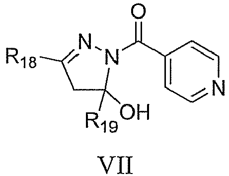
- R18 and R19 are each independently selected from the group comprising alkoxy, alkyl, alkylamino, alkylene, alkylthio, alkynyl, amido, amino, aryl, arylalkoxy, arylamino, arylthio, carboxy, cyano, cycloalkyl, ester, halo, haloalkoxy, haloalkyl, heteroaryl, heteroarylamino, heterocycloalkyl and hydrogen, any of which is optionally substituted.
- In another aspect, the present invention relates to compounds having one of the formulas with the general formulas I, III-VII and IX-XX as shown in Table 2.
- In one aspect, the present invention relates to a compound listed in Table 1.
- In one aspect, the present invention relates to compounds as defined above for use in the treatment of bacterial infections.
- In one aspect, the present invention relates to compounds as defined above for use in the treatment of Tuberculosis.
- In one aspect, the present invention relates to a pharmaceutical composition comprising a compound as defined above.
- In one aspect, the present invention relates to a method of treatment of Tuberculosis, comprising the application of a suitable amount of a compound as defined above to a person in need thereof.
- In another aspect, the present invention relates to a screening method comprising the steps of
- (a) batch infection of host cells with fluorescently labeled M. tuberculosis mycobacteria;
- (b) removing free unbound mycobacteria;
- (c) adding compounds that are to be tested to a multi-well plate;
- (d) seeding said host cells infected with fluorescently labeled M. tuberculosis mycobacteria into said multi-well plate containing said compounds;
- (e) incubating said multi-well plate containing host cells infected with fluorescently labeled M tuberculosis mycobacteria and said compounds;
- (f) fluorescently labeling said host cells;
- (g) analyzing said multi-well plate using automated confocal microscopy.
- The screening method according to the present invention represents a phenotypic cell-based assay enabling the search for drugs that interfere with the multiplication of M tuberculosis within host macrophages. The assay makes use of fluorescently labeled living macrophages infected with fluorescently labeled mycobacteria and uses automated confocal fluorescence microscopy to measure intracellular mycobacterial growth. The assay has been set-up for the high throughput screening (HTS) of large scale chemical libraries.
- In the following, the invention shall be described by the following embodiments:
- 1. Compound having the general formula VIII:

- m is 0, 1, 2, or 3;
- X3 is selected from the group comprising CH2, O, S and NH;
- X4 is selected from the group comprising halide, alkyl, OR23, SR24 and NR25R26;
- R20 is selected from the group comprising acyl, alkoxy, alkyl, alkylamino, alkylcarboxylic acid, arylcarboxylic acid, alkylcarboxylic alkylester, alkylene, alkylether, alkylhydroxy, alkylthio, alkynyl, amido, amino, aryl, arylalkoxy, arylamino, arylthio, carboxylic acid, cyano, cycloalkyl, carboxylic acid, ester, halo, haloalkoxy, haloalkyl, haloalkylether, heteroaryl, heteroarylamino, heterocycloalkyl and hydrogen, any of which is optionally substituted;
- R21 and R22 are each independently selected from the group comprising alkoxy, alkyl, alkylamino, alkylene, alkylether, alkylthio, alkynyl, amido, amino, aryl, arylether, arylalkoxy, arylamino, arylthio, carboxy, cyano, cycloalkyl, ester, halo, haloalkoxy, haloalkyl, heteroaryl, heteroarylamino, heterocycloalkyl, hydroxyl, hydrogen, nitro, thio, sulfonate, sulfonyl and sulfonylamino, any of which is optionally substituted;
- R23 is selected from the group comprising acyl, alkyl, alkylamino, alkylene, alkynyl, aryl, arylalkoxy, arylamino, arylthio, carboxy, cycloalkyl, ester, ether, haloalkyl, heteroaryl, heteroarylamino, heterocycloalkyl, hydrogen, thio, sulfonate, and sulfonylamino, any of which is optionally substituted;
- R24 is selected from the group comprising alkyl, alkylaryl, alkylene, alkynyl, aryl, cycloalkyl, ester, halo, haloalkyl, heteroaryl, heterocycloalkyl, and hydrogen, any of which is optionally substituted; and
- R25 and R26 are each independently selected from the group comprising acyl, alkyl, aminoalkyl, alkylene, alkylthio, alkynyl, aryl, arylalkoxy, arylamino, arylthio, carboxy, cycloalkyl, ester, ether, halo, haloalkoxy, haloalkyl, haloalkylether, heteroaryl, heteroarylamino, heterocycloalkyl and hydrogen, any of which is optionally substituted.
- 2. Compound according to
embodiment 1 having the general formula VIIIa:
- X5 is selected from the group comprising CH2, C=O and C=S;
- Z1 and Z2 are each independently selected from the group comprising alkoxy, alkyl, alkylamino, alkylene, alkylether, alkylthio, alkynyl, amido, amino, aryl, arylether, arylalkoxy, arylamino, arylthio, carboxy, cyano, cycloalkyl, ester, halo, haloalkoxy, haloalkyl, heteroaryl, heteroarylamino, heterocycloalkyl, hydroxyl, and hydrogen, or two groups are connected each other to make five or six membered cyclic, heterocyclic and heteroaryl rings, any of which is optionally substituted;
- R27 and R28 are each independently selected from the group comprising alkoxy, alkyl, alkylamino, alkylene, alkylether, alkylthio, alkynyl, amido, amino, aryl, arylether, arylalkoxy, arylamino, arylthio, carboxy, cyano, cycloalkyl, ester, halo, haloalkoxy, haloalkyl, heteroaryl, heteroarylamino, heterocycloalkyl, hydroxyl, hydrogen, nitro, thio, sulfonate, sulfonyl and sulfonylamino, any of which is optionally substituted;
- R29 and R30 are each independently selected from the group comprising alkoxy, alkyl, alkylamino, alkylene, alkylether, alkylthio, alkynyl, amido, amino, aryl, arylether, arylalkoxy, arylamino, arylthio, carboxy, cyano, cycloalkyl, ester, halo, haloalkoxy, haloalkyl, heteroaryl, heteroarylamino, heterocycloalkyl, hydroxyl, hydrogen, nitro, thio, sulfonate, sulfonyl and sulfonylamino, or two groups are connected each other to make five or six membered cyclic, heterocyclic, aryl, and heteroaryl rings, any of which is optionally substituted.
- 3. Compound according to
embodiment 1 having one of the formulas 125-301 as shown in Example 7, preferably 132-135, 137, 139-140, 147, 151-152, 160, 163, 173, 180, 184-185, 193, 195, 199-201, 204, 206-222, 224, 226, 229, 231-243, 245-278, 280-286 and 290-301 as shown in Table 4. - 4. Compound having the general formula II:

- R5 and R6 are each independently selected from the group comprising acyl, alkyl, alkylamino, alkylene, alkylthio, alkynyl, aryl, arylalkoxy, arylamino, arylthio, carboxy, cycloalkyl, ester, haloalkoxy, haloalkyl, heteroaryl, heteroarylamino, heterocycloalkyl, hydroxyl, hydrogen, sulfonate and sulfonyl, any of which is optionally substituted and
- R7, R8 and R9 are each independently selected from the group comprising alkoxy, alkyl, alkylamino, alkylene, alkylthio, alkynyl, amido, amino, aryl, arylalkoxy, arylamino, arylthio, carboxy, cyano, cycloalkyl, ester, halo, haloalkoxy, haloalkyl, heteroaryl, heteroarylamino, heterocycloalkyl, hydroxyl, hydrogen, nitro, thio, sulfonate, sulfonyl and sulfonylamino, any of which is optionally substituted.
- 5. Compound according to
embodiment 4, wherein R5 and R6 are connected, having the general formula IIa:
- n is 0, 1, 2, or 3;
- Y and Z are each independently selected from the group comprising CH2, CHOR10, CHNR10R11, CR10R11 and NR10; and
- R10 and R11 are each independently selected from the group comprising acyl, alkyl, alkylamino, alkylene, alkylthio, alkynyl, aryl, arylalkoxy, arylamino, arylthio, carboxy, cycloalkyl, ester, haloalkoxy, haloalkyl, heteroaryl, heteroarylamino, heterocycloalkyl, hydrogen, sulfonate and sulfonyl , any of which is optionally substituted.
- 6. Compound according to
embodiment 4 having one of the formulas with the general formula II as shown in Table 2, as well as one of the formulas 1-123 as shown in Example 6, preferably 1-24, 26-34, 54, 56, 58-61, 63-64, 67, 90-101, 103-105, 107-109, 112, 114-116 and 118-121 as shown in Table 4. - 7. Compound having one of the general formulas I, III-VII and IX-XX as shown in Table 3.
- 8. Compound according to any of embodiments 1-7 for use in the treatment of bacterial infections.
- 9. Compound according to any of embodiments 1-6 for use in the treatment of bacterial infections.
- 10. Compound according to any of embodiments 1-7 for use in the treatment of Tuberculosis.
- 11. Compound according to any of embodiments 1-6 for use in the treatment of Tuberculosis.
- 12. Pharmaceutical composition comprising a compound according to any of embodiments 1-7.
- 13. Pharmaceutical composition comprising a compound according to any of embodiments 1-6.
- 14. Method of treatment of Tuberculosis, comprising the application of a suitable amount of a compound according to any of embodiments 1-7 to a person in need thereof.
- 15. Method of treatment of Tuberculosis, comprising the application of a suitable amount of a compound according to any of embodiments 1-6 to a person in need thereof.
- Reference is now made to the figures and tables, wherein
-
Figure 1 shows the monitoring of tubercle bacillus intracellular growth inside macrophages by automated confocal microscopy: (a) Representative pictures of Raw264.7 cells infected with M tuberculosis H37Rv-GFP at different time points after infection. (b) Image analysis: 1: Typical 2-color image; 2: Circled object corresponds to detected cells; 3: Circled object corresponds to bacterial aggregates; 4: Filled purple cells correspond to infected cells. (c,d,e) Image-based quantification of the percentage of infected cells and the mean number of cells from 2 hours today 7 after infection with H37Rv-GFP at a multiplicity of infection of 0.5 (gray square), 1 (black circle) and 2 (dark gray triangle). Non-infected cells (black diamonds) were used as the negative control; -
Figure 2 shows the pharmacological validation and MIC (minimal inhibitory concentration) comparison of the reference drugs in the in vitro growth fluorescence assay and the phenotypic cell-based assay: (a) Representative pictures of infected cells in presence of INH at 1 µg/mL or DMSO control. (b,c,d) Dose-response of INH, rifampin and ethionamide; black square and line corresponds to growth inhibition in cell-based assay; gray circle and line correspond to in vitro growth inhibition; shown is a representative data set; -
Figure 3 shows assay automation validation of the phenotypic cell-based assay: (a) Percent of M tuberculosis infected cells relative to 384-plate well-index. Black square, dark gray square, gray square and open square correspond to INH 1µg/mL, rifampin 5µg/mL, PBS and DMSO control respectively. (b,c) Percent of M. tuberculosis infected cells relative to INH and rifampin concentration. Experiments were performed on four different plates on two independent days; -
Figure 4 shows primary screening results for the phenotypic cell-based assay and the in vitro growth assay for 26500 compounds: (a) Percent inhibition based on infection ratio relative to each compound and distribution. (b) Percent inhibition based on RFU relative to each compound and distribution. (c) Comparison of inhibition percentage for the phenotypic cell-based assay and the in vitro growth assay for each compound; -
Figure 5 shows serial dilution results from the in vitro growth fluorescence assay and the phenotypic cell-based assay: Typical curves for compounds inhibiting (a,b,c) in vitro bacterial growth (d,e,f) both in vitro and intracellular growth and (g,h,i) intracellular growth only. - (a,d,g) Infection ratio relative to compound concentration. (b,e,h) Cell number relative to compound concentration. (c,f,i) Relative fluorescence intensity relative to compound concentration. Compound concentration is given in M;
-
Figure 6 shows (a) a scheme of assay automation. (b) a 384-plate format description; (c) a 384-plate dose-response curve description, A to P and a to b correspond to 2-fold serial dilution of INH and Rifampin respectively with a starting concentration of 20 mg/mL in well A or a; RIF:Rifampin 5 µg/mL, Cpd: compound, INH100 1 µg/mL, INH50 0.05 µg/mL; -
Figure 7 shows the anti-tuberculosis effect ofcompounds 4 and 24 (5 µM) on M tuberculosis H37Rv-GFP in (a) Raw267.4 (104 cells), (b) mouse bone marrow-derived macrophages and (c) human primary macrophages differentiated with 50 ng/mL rhM-CSF (1.5*104) after 7 days of infection with MOI 2.5:1 (control INH at 5 µM); -
Figure 8 illustrates the colony forming units (CFUs) recovered from macrophages at different time points after infection with M. tuberculosis H37Rv. Either Raw264.7 cells (a) or murine BMDM (b) were infected at an MOI of 1:1 and treated with the indicated amount of pyridopyrimidione compound 232 (20 µM) with DMSO, INH (10 µM) and RIF (10 µM) as controls; - Table 1 lists 340 hits whose inhibitory activity was confirmed in an intracellular (QIM) assay or an in vitro (QUM) assay , wherein the abbreviation "QIM" stands for Quantification of Intracellular Mycobacteria, the abbreviation "QUM" stands for Quantification of in vitro grown Mycobacteria, and the abbreviation "CellNb" stands for cell number;
- Table 2 lists 121 compounds which demonstrated an inhibitory activity above 65% at 2 µM without any apparent cell toxicity at 20 µM and consequently were selected for further confirmation by ten 3-fold serial dilutions;
- Table 3 summarizes the independent/general molecular scaffolds/formulas of the 121 hits listed in Table 2;
- Table 4 lists dinitrobenzamide and pyridopyrimidinone derivatives (general scaffold II and VIII, respectively, see Table 3) with their respective inhibitory activities, wherein the numbers in bold print refer to the compounds listed in Examples 6 and 7;
- Table 5 shows the cytotoxicity and antibacterial spectrum of
dinitrobenzamide compounds 4 and 24 (see Table 4); - Table 6 shows the cytotoxicity and antibacterial spectrum of pyridopyrimidinone compound 133 (see Table 4); and
- Table 7 shows the frequency of spontaneous resistance for representative dinitrobenzamide and pyridopyrimidinone compounds.
- The invention is now further described by reference to the following examples which are intended to illustrate, not to limit the scope of the invention.
- A recombinant strain of M tuberculosis H37Rv expressing the green fluorescent protein (H37Rv-GFP) was obtained by transformation of an integrative plasmid (Abadie et al., 2005; Cremer et al., 2002). Within this plasmid, which is derived from the Ms6 mycobacteriophage, the gfp gene is cloned and constitutively expressed under the strong mycobacterial promoter pBlaF. Electrocompetent cells for M tuberculosis H37Rv-GFP were prepared from 400 mL of a 15 days old Middlebrook 7H9 culture (Difco, Sparks MD, USA) supplemented with albumin-dextrose-catalase (ADC, Difco, Sparks MD, USA), glycerol and 0.05
% Tween 80. Bacilli were harvested by centrifugation at 3000 g for 20 min, washed twice with H2O at room temperature, and resuspended in 1-2 mL of 10% glycerol at room temperature after recentrifugation. 250 µl of bacilli were mixed with green fluorescent protein encoding plasmid and electroporated using a Biorad Gene Pulser (Biorad). After electroporation, bacilli were resuspended in medium and left one day at 37°C. Transformants were selected on Middlebrook 7H11 medium (Difco, Sparks MD, USA) supplemented with oleic acid-albumin-dextrose-catalase (OADC, Difco, Sparks MD, USA) and 50 µg/mL hygromycin (Invitrogen, Carlsbad, CA USA). The selected hygromycin-resistant and green fluorescent colonies appeared after 3 weeks. A 100 mL culture of the H37Rv-GFP strain was grown in Middlebrook 7H9-ADC medium supplemented with 0.05% Tween - Mouse macrophage cell lines Raw 264.7 (ATCC # TIB-71), J774A.1 (ATCC # TIB-67) or human monocytes (ATCC # TIB-202) differentiated with 50 ng/mL PMA (Sigma) were grown in RPMI 1640 (Gibco) with 10% heat-inactivated fetal calf serum (Gibco).
- The small synthetic molecules from the screening libraries were suspended in pure DMSO (Sigma, D5 879-500 mL) at a concentration of 10 mM (Master plates) in Corning 96 well clear V-bottom polypropylene plates (Corning, #3956). The compounds were then reformatted in Greiner 384 well V-shape polypropylene plates (Greiner, #781280) and diluted to a final concentration of 2 mM in pure DMSO. The compounds were kept frozen until use. For screening, compound plates were incubated at room temperature until thawed. The compounds were directly added into the assay plates from the DMSO stock using an EVObird liquid handler (Evotec Technologies), which transfers 250 nl of compound twice to reach a final dilution of 1:100. This one-step dilution reduces the risk of compound precipitation in intermediate plates and allows for a low final DMSO concentration (1%).
- Positive control antibiotics (Isoniazid (Sigma, I3377-50G) and Rifampin (Euromedex, 1059-8, 5 g)) as well as negative controls (DMSO) were added manually in each plate in columns 1-2 and 23-24 (see
Figure 6 b for plate description). - A total of 26500 compounds were tested. These compounds came from commercial libraries from Timtec (25000 from the ActiProbe diverse library, 1000 from the Kinase inhibitors ActiTargK library and 500 from the Protease inhibitors ActitargP library). The screened compounds were selected based on high diversity and drug-like properties (using Lipinski rule-of-five (Lipinski et al., 2001)). They were first screened at one concentration (primary screen, concentration = 20 µM). The "positive" compounds selected from the primary screen were then confirmed by testing at 3 concentrations (20, 2 and 0.2 µM) to identify the most active and/or by ten 3-fold ten dilutions (from 20 µM to 0.5 nM).
- Cells were first seeded in 50 µl at a density of 20,000 cells per well of a 384-well plate (Evotec technologies #781058) for 16 hours and then infected with bacterial suspensions at a multiplicity of infection (MOI) varying from 10:1 to 1:1 (bacteria:host cells). After 2 hours, cells were washed three times with phosphate buffered saline (PBS) and the compounds diluted in fresh culture medium were added. Cells were incubated at 37 °C, 5% CO2 for up to seven days.
- Cells (1.5 x 10⁸ cells) were infected with H37Rv-GFP suspension at a MOI of 1:1 in 300 mL for 2 hours at 37 °C with shaking (100 rpm). After two washes by centrifugation at 1100 rpm (Beckman SX4250, 165 g) for 5 min., the remaining extracellular bacilli from the infected cells suspension were killed by a 1 hour amykacin (20 µM, Sigma, A2324-5G) treatment. After a final centrifugation step, cells were dispensed with the Wellmate (Matrix) into 384-well Evotec plates (#781058) preplated with 10 µl of the respective compound diluted in cell medium. Infected cells were then incubated in the presence of the compound for 5 days at 37 °C, 5% CO2. After five days, macrophages were stained with SYTO 60 (Invitrogen, S 11342) followed by plate sealing and image acquisition. During screening, staining of the live cells was carried out on a set of three plates every two hours to limit cell death due to prolonged incubation with cell chemical stain.
- Confocal images were recorded on an automated fluorescent confocal microscope Opera™ (Evotec Technologies) using a 20X-water objective (NA 0.70), 488-nm and 635-nm lasers and a 488/635 primary dichroic mirror. Each image was then processed using dedicated in-house image analysis software (IM). Parameters determined were the total cell number and the number of infected cells. Briefly, the algorithm first segments the cells on the red channel using a sequence of processing steps as described elsewhere (Fenistein et al., manuscript in press). It is generally based on a succession of 1) thresholding the histogram of the original image (3 classes K-means) 2) gaussian filtering the original image with a standard deviation that is set equal to the cells' average radius, 3) searching for the local maxima of the filtered image that provides cell centers as seeds for 4) region growing that defines each cell's own surface and finally 5) removing extremely small cells as potential artifacts or noise. This step provides the total number of cells in the red channel. Infected cells are then defined as those having at least a given number of pixels (usually 3) whose intensity in the green channel is above a given intensity threshold. The ratio of infected cells to the total number of cells is the measure of interest (named infection ratio). For each well, 4 pictures were recorded and for each parameter, the mean of the four images was used.
- Data obtained from either the intracellular assay image analysis or from the conventional antibacterial assay (see below) were then processed using ActivityBase (IDBS) to calculate the statistical data (% of inhibition, Z score for each compound, Z', CV etc. for the control plates) and to store the data in an Oracle database. Additional analyses with regards to both quality control of the screens and hit identification were performed with various software packages including Spotfire (Tibco) and Pipelinepilot (Accelrys).
- A frozen aliquot of M tuberculosis H37Rv-GFP was diluted at 1.5 x 10⁶ CFU /mL in Middlebrook 7H9-ADC medium supplemented with 0.05
% Tween 80. Greiner µclear-black 384-well plates (Greiner, #781091) were first preplated with 0.5 µl of compound dispensed by EVOBird (Evotec) in 10 µl of Middlebrook 7H9-ADC medium supplemented with 0.05% Tween 80. 40 µl of the diluted H37Rv-GFP bacterial suspension was then added on top of the diluted compound resulting in a final volume of 50 µl containing 1% DMSO. Plates were incubated at 37 °C, 5% CO2 for 10 days after which GFP-fluorescence was recorded using aVictor 3 reader (Perkin-Elmer Life Sciences). - Raw 264.7 (ATCC # TIB-71) (1.5*108 cells) were infected with H37Rv-GFP (Abadie et al., 2005, Cremer et al., 2002) in suspension at a MOI of 1:1 for 2 hours at 37 °C with shaking. After two washes by centrifugation, the remaining extracellular bacilli from the infected cell suspension were killed by a 1 hour Amikacin (20 µM, Sigma, A2324) treatment. After a final centrifugation step, cells were dispensed into 384-well Evotec plates (#781058) preplated with compounds and controls. Infected cells were then incubated for 5 days at 37°C, 5% CO2. Murine Bone Marrow-Derived Macrophages (BMDM) were produced as described previously (Brodin et al., 2006). Briefly, cells were extracted from the femurs and tibia of 6 weeks old female mice (C57BL/6, Orientbio) and cultivated in RPMI 1640 media containing 10% heat-inactivated fetal calf serum (FCS) (both from Gibco® at Invitrogen, Carlsbad, CA) and 10% L-929 cell conditioned medium. Peripheral Blood Mononuclear Cells (PBMC) were isolated from Buffy coat from healthy volunteers. Buffy coat diluted in PBS supplemented with 1% FCS was treated with 15 ml of Ficoll-Paque Plus (Amersham Biosciences, Sweden) and centrifuged at 2500 × g for 20 min. PBMC were obtained by CD14+ bead separation (Miltenyi Biotec, Germany), washed 3-times with PBS (1% FCS) and transferred to 75 cm2 culture flasks containing RPMI 1640 media, 10% FCS and 50 ng/ml of recombinant-human macrophage colony stimulating factor (R & D systems, Minneapolis). Six day old adherent murine BMDM and PBMC derived human macrophages were infected with H37Rv-GFP (Abadie et al., 2005) in suspension at a MOI of 1:1 for 2 hours at 37°C and then extensively washed and finally incubated with compounds or controls. After several days, macrophages were stained with SYTO 60 (Invitrogen, S11342) and image acquisition was performed on an EVOscreen-MarkIII fully automated platform (PerkinElmer) integrated with an Opera™ (20X-water objective, NA 0.70) and located in a BSL-3 safety laboratory. Mycobacteria-GFP were detected using a 488-nm laser coupled with a 535/50 nm detection filter and cells labeled with a 635-nm laser coupled with a 690/40 nm detection filter. Four fields were recorded for each plate well and each image was then processed using dedicated in-house image analysis software (IM) as described elsewhere (Fenistein et al., in press).
- Mycobacterium tuberculosis H37Rv, H37Ra and BCG Pasteur were used as reference strains. All strains were diluted at 1.5 x 10⁶ CFU /mL in Middlebrook 7H9-ADC medium supplemented with 0.05
% Tween 80. 384-well plates (Greiner, #781091) were first preplated with 0.5 µl of compound dispensed by EVOBird (Evotec) in 10 µl of Middlebrook 7H9-ADC medium supplemented with 0.05% Tween 80. Forty microliters of the diluted H37Rv-GFP bacterial suspension was then added to the diluted compound resulting in a final volume of 50 µl containing 1% DMSO. Plates were incubated at 37°C, 5% CO2 for 10 days. Mycobacterial growth was determined by measuring GFP-fluorescence using aVictor 3 reader (Perkin-Elmer Life Sciences) for H37Rv-GFP or with resazurin method. Isoniazid at 0.05 µg/mL and 1 µg/mL (Sigma, I3377), Rifampin at 1 µg/mL (Euromedex) and DMSO were used as controls. - In order to address compound toxicity, seven cell lines originating from different body tissues were cultivated in the presence of 3-fold dilutions of compounds starting from 100 µM. After 5 days of culture, cell viability was assessed by the resazurin test. Briefly, cells were incubated with 10 µg/mL of resazurin (Sigma-Aldrich St. Louis, MO) for 4 h at 37°C under 5% CO2. Resofurin fluorescence (RFU) was measured as indicated above. Percentage of toxicity on cells was calculated as follows: Cytotoxicity (%) = (RFUDMSO-RfUBlank) - (RFUcompound-RFUblank) / (RFUDMSO-RFUBlank) × 100. Percentage of cytotoxicity was plotted against compound concentration and the minimal toxic concentration (MTC50) was determined by non-linear regression analysis as the lowest compound concentration where fifty percent toxicity was observed on the corresponding cell line.
- The frequency of spontaneous mutations was determined on 7H10 plates containing increasing concentrations of dintirobenzamide (0.2, 0.8, 1.6 and 3.2 µg/ml) or pyridopyrimidinone (0.4, 0.8, 1.6 and 3.2 µg/ml) compounds. 106, 107 and 108 CFU containing bacterial suspensions were spread on compound containing agar plates. After 5-6 weeks at 37°C, colonies were counted and frequency of mutation was evaluated as the ratio of colonies relative to the original inoculum. DMSO and INH were used as negative and positive controls, respectively.
- To set-up the optimal conditions of M. tuberculosis infection, Raw264.7 macrophages were first infected with mycobacteria that constitutively express green fluorescent protein (GFP) at different multiplicities of infection (MOI) followed by kinetic analysis. Up to 7 days post bacillus infection, the host live cells were daily labeled with the red chemical fluorescent dye Syto60, and confocal images of live samples were acquired using an automated confocal microscope. Typical images are displayed in
Figure 1a . During the first 24 hours, a few discrete weakly fluorescent bacteria localized within the cells. Byday 2, the average number of cells had increased and mycobacteria had started to spread into neighboring cells leading to zones of strongly fluorescent bacteria. The localization of the green signal is always within a distance of 5 µm to that of the red cell signal and in most cases actually overlaps with the cell signal. This confirms the intracellular nature of the mycobacteria growth. Byday 4, the cell number has significantly diminished and the bacteria have formed large, highly fluorescent aggregates, which cover almost the entire image fromday 5 onwards. As a control, non-infected cells grew up to confluence atday 2 and remained alive untilday 7. - In order to automatically quantify the intracellular bacterial load, an in-house image analysis script was developed. This script enables the automated quantification of the number of cells and the percentage of infected cells, whereby an infected cell is a cell containing at least three green pixels with an intensity above a defined threshold (
Figure 1b ). 2 hours after infection, between 2 and 10% of Raw264.7 cells were found to harbor a low number of bacilli (Figure 1c ). The percentage of infected cells, hereafter named infection ratio, continued to increase from 72 hours post-infection reaching up to 70% at seven days post infection. This increase in infection ratio correlated with an increase in cell mortality (Figure 1d/e ). - To validate the assay set-up, the effect of current anti-tuberculosis drugs on M. tuberculosis intracellular growth was investigated. 2-fold serial dilutions of isoniazid (INH), rifampin and ethionamide were performed, followed by testing on macrophages that had previously been infected with M tuberculosis H37Rv-GFP. After 5 days of incubation, macrophages were stained, and images acquired on an automated confocal microscope as described above. A larger number of cells and a fewer number of bacteria are clearly seen on pictures corresponding to samples that were incubated with INH compared to the DMSO negative control. This shows that INH prevents both intracellular M. tuberculosis growth and bacillus mediated cytotoxicity (
Figure 2a ). A clear inhibition dose-response curve was obtained by image-extracted analysis (Figure 2b ). In parallel, inhibition of M. tuberculosis H37Rv-GFP in vitro growth by INH was monitored by recording green fluorescence intensity under the same conditions. In both experiments, the minimal inhibitory concentration (MIC) for INH was 0.1 µg/mL, which is in accordance with the MIC reported in the literature for extracellular M tuberculosis growth (Andries et al., 2005). Similar results were obtained with the standard anti-tuberculosis drugs ethionamide (Figure 2c ) and ethambutol (data not shown), whereas for rifampin, there was a log-fold decrease in the MIC in the cell-based assay compared to the in vitro assay (Figure 2d ). The diminished efficacy of rifampin in the cell-based assay is likely due to impaired cell penetration and further demonstrates that it is the intracellular antibacterial activity that is being monitored in this assay. Thus, adaptation of both the intracellular and the in vitro M. tuberculosis growth assay for high throughput screening (HTS) was performed. - To simplify the protocol for HTS purposes, macrophages were infected in batch with M. tuberculosis before being dispensed onto the compounds. The batch infection was carried out with macrophages in suspension at 37°C under mild shaking. Free unbound mycobacteria were removed by washing three times with PBS and differential centrifugation, as well as by an additional one-hour incubation step with amykacin, an antibiotic known to selectively kill extracellular microbes (
Figure 6a ). M tuberculosis infected macrophages were then seeded in plates that had been previously dispensed with the compounds, DMSO or antibiotic controls. The day-to-day as well as plate-to plate reproducibility was first tested. To this end, either serial dilutions of INH or rifampin were dispensed into 8 plates along with the regular DMSO and positive control (INH at 1 µg/mL (MIC100) and at 0.05 µg/mL (MIC90) and rifampin at 1 µg/mL) wells that were subsequently seeded with infected cells. The same experiment was repeated over 2 consecutive days. After incubation for 5 days and macrophage staining, pictures from each plate were acquired. The mean infection ratio determined for the DMSO negative controls in each plate for the 2 days of experiments was between 50% and 70%, whereas for the INH and rifampin samples, the mean infection ratio fell to below 20% (Figure 3a ). Despite some variation in the mean infection ratio between the two experiments, the difference between the INH-positive and DMSO-negative controls was above five-fold for both days. P values calculated for each plate using a paired t-student test also confirmed a significant difference between the positive and negative controls (p < 0.000001, data not shown). In addition, the inventors performed an experiment to determine if inhibitors of M. tuberculosis intracellular growth infection dispensed in any well on the plate could be detected by performing double-blind controls (spike of INH and rifampin at 3 different concentrations). Indeed, one hundred percent of the spikes were identified (data not shown). Taken together, these results prove that the assay is sensitive enough to be able to identify inhibitors under HTS conditions. Finally, the robustness of the assay was checked by monitoring the dose-response of reference compounds. Almost identical MICs for the antibiotic positive controls were determined independent of the plate or the day of the experiment (Figure 3b/c ). Calculated MICs from the image based quantification of the infection ratio were 0.16 +/-0.05 µg/mL and 2.4 +/-1.3 µg/mL for INH and rifampin, respectively, and were confirmed by CFU plating (data not shown). In parallel, the extracellular growth assay was validated with a similar approach (data not shown). - A 26500 small molecule compound library, that was selected for its high chemical diversity and drug-like properties according to the Lipinski rules (Lipinski et al., 2001), was chosen as the first library to be screened using the validated phenotypic cell-based assay. The primary screen was carried out with compounds at 20 µM in singleton. The throughput was set to about 6000 compounds per working day encompassing 25 plates. The screening was performed with Raw264.7 cells that had been expanded from frozen stocks for ten days before infection with M tuberculosis H37Rv-GFP. To accept the screening results, the MICs obtained from 2 serial dilutions of INH and Rifampin processed at the beginning and at the end of the screening day should show similar results compared to the values obtained during the validation (see above). Each screened plate is then accepted by the quality control procedure if the window between DMSO and INH (1µg/ml) is higher than 3 and the CV calculated for the 320 compounds present in each plate is lower than 25. Such quality control criteria allow the identification of hits with an activity higher than 75%. Subsequently, the percent inhibition for each compound was determined relative to the corresponding mean infection ratio between 1 µg/mL INH (100%) and DMSO (0%) in the same 384-well plate. The percent inhibition distribution is centered around -20% of inhibition (
Figure 4a ). It was decided to select compounds that have an inhibitory effect greater than 65% which corresponds to a little less than 1.5 % of the total compounds. - In parallel, the same compounds were only tested for their inhibitory activity on the M. tuberculosis H37Rv-GFP bacterial growth. The results from this assay, which are based on classical fluorescence intensity, showed a higher degree of reproducibility and the criteria for plate validation was set to a Z' value (DMSO/INH) greater than 0.35. The throughput for this fluorescence based assay was approximately 20,000 compounds per day. Compounds that prevented M tuberculosis growth in vitro with an inhibitory effect above 65% were then selected as hits (1.4%) as they belong to a clear independent population compared to the inactive population centered to 0% (
Figure 4b ). - The results gathered from the two different screenings were then compiled and compared (
Figure 4c ). Four different populations could be identified: compounds that are i) active only on extracellular bacteria, ii) active only on intracellular bacteria, iii) active in both settings or iv) not active. 657 compounds (2.5%) belonged to one of the first three categories and, thus, were selected for further investigation. - An important parameter that can be measured during image analysis is the total cell number, also referred to as cell cytotoxicity. A low cell number can be the result of two independent phenomena, the compound toxicity and M tuberculosis growth mediated cell toxicity. Indeed, at
day 5 after infection with M tuberculosis, the cell number decreased to less than 100 cells per image compared to more than 500 cells per image for uninfected cells (Figure 1e ). In contrast, a high cell number is obtained only when the compound is not toxic and prevents mycobacterial growth. This turns out to be a second relevant measurement of a compound's anti-mycobacterial activity. However, this criterion was not applied for the selection of hits from the primary screen as a low cell number was found for only a few compounds and the inventors wanted to avoid failing to select highly active compounds that would later on prove to be active at much lower concentrations despite a cell toxicity at 20 µM. An additional validation criterion of a Z' (DMSO/INH) value of the total cell number greater than 0.2 was added for the following screening steps. - The 657 selected hits were first confirmed at 3 different concentrations, 20 µM, 2 µM and 0.2 µM. For 340 hits the activity was confirmed either at 20 µM or 2 µM, on the intracellular or the in vitro assay (see Table 1). From this latter list, 121 compounds demonstrated an inhibitory activity above 65% at 2 µM without any apparent cell toxicity at 20 µM and consequently were selected for further confirmation by ten 3-fold serial dilutions (see Table 2). All 121 compounds were confirmed by serial dilution with a MIC ranging between 250 nM and 20 µM. The results shown in
Figure 5 are representative of the three types of behavior observed: most of the compounds exhibited a clear dose response curve when activity was measured as infection ratio (Figure 5b /e/h). Compounds active on the bacilli level present a similar activity in the extracellular assay (Figure 5c/f ) even if the MIC is different from one assay to the other. A few compounds don't present clear activity on the in vitro bacilli (Figure 5i ) and may represent drugs acting through a cellular target or on a bacilli target involved only during the infection process. Furthermore, toxic compounds can be identified thanks to a dramatic decrease in the cell number when the compound concentration increases (Figure 5d ) and activity of non-toxic compounds are validated by a dose response protective effect on the cell number (Figure 5a ). Consequently cell number detection represents an independent secondary assay in the same well as the primary assay. The serial dilution results from all 121 compounds are presented in Table 2. - The 121 confirmed hits can be clustered as 20 independent/general scaffolds (Table 3). The number of compounds for each scaffold varied, ranging from 1 to 69 molecules. The molecules from the 69-compound scaffold share a common structure which is similar to INH thereby validating the screening results. One scaffold contains molecules that were only active in the intracellular assay and its mechanism of action will be the focus of further investigation.
- The benzamide compounds (scaffold II; see Table 3) underwent derivatization according to the methods outlined below (Schemes 1-7). Formation of the amide can be performed under general conditions using EDC or DCC coupling reagents with acids instead of acyl chloride. Resulting derivatives were examined for inhibitory activity using the assay described above and the results are summarized in Table 4.


- To a solution of 2-(2-hydroxyethyl)isoindoline-1,3-dione (1.68 mmol) in methylene chloride (10 mL) was added ADDP (1.68 mmol), triphenylphosphine (1.68 mmol) and phenol (3.18 mmol) and stirred at room temperature. After stirring overnight, the reaction mixture was diluted with methylene chloride (30 mL) and washed with 1 M NaOH aqueous solution (50 mL), and brine (50 mL). The organic layer was dried over anhydrous MgSO4 and concentrated in vacuo. The crude product was purified by silica gel flash column chromatography (4:1 hexanes/ethyl acetate) and recrystallized from a mixture of hexanes and ethyl acetate to give A1.
- To a solution of A1 (1.14 mmol) in methanol (10 mL) was added hydrazine monohydrate (1.42 mmol) and the resulting mixture was refluxed under a nitrogen atmosphere. After 3 h, the reaction mixture was allowed to cool to room temperature and concentrated in vacuo. The residue was precipitated with methylene chloride (10 mL). The resulting precipitate was filtered through Celite and the filtrate was concentrated in vacuo to afford an amine intermediate. To a solution of the amine in methylene chloride (10 mL) was added triethylamine (0.45 mmol) and a benzoylchloride (0.45 mmol) at 0 °C and the resulting mixture was stirred at room temperature. After 3 h, the reaction mixture was diluted with methylene chloride (10 mL) and washed with 1 M HCl aqueous solution (30 mL), saturated Na2CO3 aqueous solution (30 mL) and brine (30 mL). The organic layer was dried over anhydrous MgSO4 and concentrated in vacuo. The crude product was purified by silica gel flash column chromatography (3:1 hexanes/ethyl acetate) and recrystallized from a mixture of hexanes and ethyl acetate to give A2.
-

-

-

-

-

-

-

-

-

-

-

-

-

-

-

-

-

-

-

-

-

-

-


- To a solution of 2-(2-hydroxyethyl)isoindoline-1,3-dione (1.17 mmol) in dimethyl formamide (10 mL) was added sodium hydride (2.34 mmol) and a benzyl bromide (1.40 mmol) at 0°C and the resulting mixture was stirred at room temperature. After stirring overnight, distilled water (50 mL) was added and the resulting precipitate was collected by filtration to afford B1. To a solution of B1 (0.85 mmol) in methanol (10 mL) was added hydrazine monohydrate (0.85 mmol) and the resulting mixture was refluxed under a nitrogen atmosphere. After 3 h, the reaction mixture was allowed to cool to room temperature and concentrated in vacuo. The residue was precipitated with methylene chloride (10 mL). The resulting precipitate was filtered off through Celite, and the filtrate was concentrated in vacuo to afford an amine. To a solution of the amine in methylene chloride (10 mL) was added triethylamine (113 µl, 0.81 mmol) and a benzoylchloride (0.81 mmol) at 0 °C and the resulting mixture was stirred at room temperature. After 3 h, the reaction mixture was diluted with methylene chloride (30 mL) and washed with 1 M HCl aqueous solution (50 mL), saturated Na2CO3 aqueous solution (50 mL) and brine (50 mL). The organic layer was dried over anhydrous MgSO4 and concentrated in vacuo. The crude product was purified by silica gel flash column chromatography (3:1 hexanes/ethyl acetate) and recrystallized from a mixture of hexanes and ethyl acetate to give B2.
-

-

-

-

-

-

-

-

-

-

-

-

-
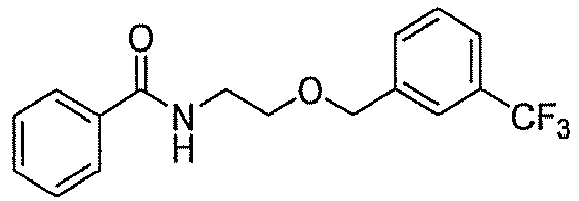
-

-
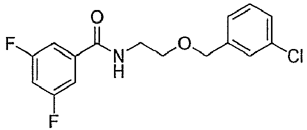
-

-

-

-

-

-

-

-

-

-

-

-

-

-

-


- To a solution of (S)-3-pyrrolidinol (10 mmol) and triethylamine (11 mmol) in methylene chloride (50 mL) was added benzoyl chloride (8.67 mmol) at 0 °C. The reaction temperature was brought up to room temperature. After 2 h, the reaction mixture was diluted with methylene chloride (50 mL) and then washed with 0.5 M HCl aqueous solution (100 mL) and brine (100 mL). The organic layer was dried over anhydrous MgSO4 and concentrated in vacuo. The crude product was purified by silica gel flash column chromatography (2:1 hexanes/ethyl acetate) to give C1.
- To a solution of C1 (1.07 mmol) in methylene chloride (10 mL) was added ADDP (1.28 mmol), triphenylphosphine (1.28 mmol) and a phenol (1.28 mmol) at room temperature. After stirring overnight, the reaction mixture was diluted with methylene chloride (30 mL) and washed with 1 M HCl aqueous solution (50 mL), saturated Na2CO3 aqueous solution (50 mL) and brine (50 mL). The organic layer was dried over anhydrous MgSO4 and concentrated in vacuo. The crude was purified by silica gel flash column chromatography (2:1 hexanes/ethyl acetate) and recrystallized from a mixture of hexanes and ethyl acetate to give C2.
-

1H), 2.30-2.34 (m, 1H), 3.54-3.64 (m, 1H), 3.72 & 3.76 (s, 3H), 3.81-3.99 (m, 3H), 4.86-4.94 (m, 1H), 6.74-6.84 (m, 4H), 8.68 & 8.75 (d, J= 1.6 Hz, 2H), 9.05 & 9.08 (brs, 1H); 13C NMR (100 MHz, CDCl3) δ 30.6, 32.4, 45.2, 47.7, 52.8, 54.8, 55.8, 55.9, 75.7, 115.0, 117.1, 117.3, 120.1, 120.2, 127.7, 127.9, 139.9, 140.0, 148.6, 150.4, 150.8, 154.8, 154.8, 164.7, 165.1; LC-MS (ESI, m/z): 388 [M+H]+. -

-

-

-
 22.1, 30.0, 32.4, 45.2, 47.7, 52.2, 52.8, 54.8, 74.9, 76.3, 115.0, 120.36, 123.7, 123.8, 127.8, 127.9, 132.0, 139.7, 148.6, 160.2, 160.5, 164.7, 166.7.
-
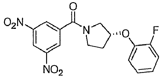
-
 29.9, 31.9, 44.1, 44.7, 52.2, 54.2, 55.1, 55.2, 76.0, 77.5, 114.82, 114.88, 117.2, 119.6, 127.7, 127.8, 140.5, 148.7, 151.1, 151.3, 154.7, 164.6, 164.7.
-
 31.9, 44.7, 52.2, 54.2, 55.0, 55.1, 65.8, 75.9, 77.5, 114.81, 114.87, 117.2, 119.6, 127.7, 127.8, 128.6, 129.8, 140.4, 148.7, 151.3, 154.7, 164.6, 164.7.
-
 24.1, 24.2, 30.0, 32.2, 45.2, 47.7, 52.7, 54.7, 75.1, 76.6, 115.9, 120.0, 120.1, 127.7, 127.8, 128.7, 128.8, 131.6, 132.0, 132.4, 132.6, 132.7, 132.8, 139.7, 148.4, 153.1, 165.0, 169.1.
-

-
 29.1, 31.1, 44.1, 51.5, 53.6, 76.4, 77.9, 115.7, 115.8, 115.9, 116.0, 117.1 (d, J= 22.3 Hz, due to F), 119.2 (d, J= 3.7 Hz, due to F), 121.7, 121.83, 121.88, 121.9, 124.2 (d, J = 3.7 Hz, due to F), 127.0, 139.0, 144.1, 144.4, 148.0, 152.8 (d, J= 242.6 Hz, due to F), 164.6, 164.7.
-
 30.3, 32.4, 44.3, 47.5, 52.1, 54.8, 78.0, 79.0, 116.8, 117.0, 117.9, 118.6, 122.6, 122.7, 122.9, 123.0, 124.6 (d, J = 3.7 Hz due to F), 127.2, 127.4, 128.5, (d, J= 3.7 Hz, due to F), 130.1, 130.3, 136.7, 136.9, 144.7 (d, J= 20.1 Hz due to F), 153.8 (d, J= 245.6 Hz, due to F), 155.2, 170.0, 170.2.
-
 30.2, 32.3, 44.4, 47.6, 52.1, 54.8, 55.8, 55.9, 76.0, 114.9, 115.0, 117.1, 117.3, 127.3, 127.4, 128.50, 128.54, 130.1, 130.2, 136.8, 137.0, 150.9, 151.1, 154.5, 154.6, 169.9, 170.2; LC-MS (ESI, m/z): 298.1 [M+H]+.
-
 33.0, 34.9, 45.1, 47.6, 55.5, 57.5, 69.4, 70.9, 120.1, 120.2, 127.8, 139.8, 139.9, 148.5, 165.1, 165.3.
-

-

-

-

-

-

-

-

-

-

-

-

-

-

-

-

-

-

-

-

-

-

-

-

-
 30.0, 31.2, 39.3, 44.6, 55.8, 71.9, 115.0, 117.9, 119.8, 127.5, 139.6, 148.7, 150.8, 154.6, 165.4.
-

-
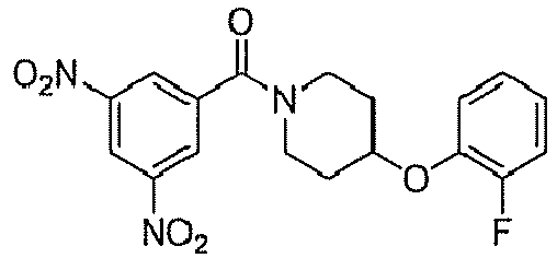 31.1, 31.9, 45.5, 49.6, 75.0, 117.6 (d, J = 18.6 Hz, due to F), 119.5, 120.5, 123.3 (d, J = 6.7 Hz, due to F), 126.0 (d, J = 3.7 Hz, due to F), 128.6, 140.6, 146.1, 149.8, 154.8 (d, J= 242.6 Hz, due to F), 166.9.
-
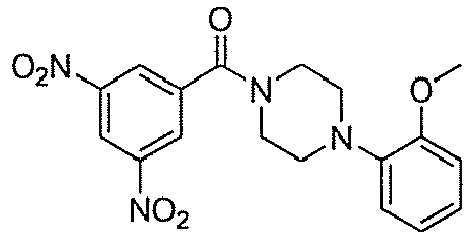
-
 42.4, 47.7, 50.5, 50.9, 54.9, 114.4, 118.8, 119.3, 127.7, 139.9, 145.6, 148.8, 154.5, 165.2; LC-MS (ESI, m/z): 387 [M+H]+.
-
 43.3, 48.7, 51.6, 52.1, 120.0, 122.0, 125.3, 128.5, 128.9, 129.4, 131.4, 140.6, 149.6, 149.8, 166.1; LC-MS (ESI, m/z): 391 [M+H]+.

- To a solution of (R)-tert-butyl 3-hydroxypyrrolidine-1-carboxylate (3.2 mmol) in dimethyl formamide (10 mL) was added sodium hydride (3.2 mmol) and benzyl bromide (3.2 mmol) at 0°C and the resulting mixture was stirred at room temperature. After stirring overnight, distilled water (50 mL) was added and the resulting precipitate was collected by filtration to afford D1.
- D1 (0.43 mmol) was dissolved in trifluoro acetic acid (5 mL) and stirred at room temperature. After 1 h, the reaction mixture was concentrated in vacuo to afford an amine. To a solution of the amine in methylene chloride (5 mL) was added triethylamine (0.51 mmol) and a benzoylchloride (0.51 mmol) at 0°C and the resulting mixture was stirred at room temperature. After 3 h, the reaction mixture was diluted with methylene chloride (30 mL) and washed with 1 M HCl aqueous solution (30 mL), saturated Na2CO3 aqueous solution (30 mL) and brine (30 mL). The organic layer was dried over anhydrous MgSO4 and concentrated in vacuo. The crude product was purified by silica gel flash column chromatography (3:1 hexanes/ethyl acetate) and recrystallized from a mixture of hexanes and ethyl acetate to give D2.
-

-

-

-

-


- To a solution of (S)-(+)-N-Boc-3-pyrrolidinol (2.67 mmol) and triethylamine (4.01 mmol) in methylene chloride (50 mL) was added methansulfonyl chloride (4.01 mmol) under ice-bath and the resulting mixture was futher stirred at 4°C. After 2 h, the residue was diluted with methylene chloride (50 mL) and washed with water (100 mL) and brine (100 mL). The organic layer was dried over anhydrous MgSO4 and concentrated in vacuo. The crude product was purified by silica gel flash column chromatography (2:1 hexanes/ethyl acetate) to give E1.
- A solution of E1 (0.75 mmol) and an amine (3.75 mmol) was stirred at 100°C. After stirring overnight, the residue was dissolved in methylene chloride (30 mL) and washed with water (30 mL) and brine (30 mL). The organic layer was dried over anhydrous MgSO4 and concentrated in vacuo. The crude product was purified by silica gel flash column chromatography (1:1 hexanes/ethyl acetate) to give E2.
- To a solution of E2 (0.96 mmol) in methylene chloride (20 mL) was added trifluoroacetic acid (0.5 mL). After 3 h, the solvent was removed in vacuo. The reaction mixture was dissolved in methylene chloride (20 mL) and cooled to 0°C. Triethylamine (4.83 mmol) and a benzoyl chloride (1.05 mmol) was added. After 2 h, the residue was diluted with methylene chloride (20 mL) and washed with water (40 mL) and brine (40 mL). The organic layer was dried over anhydrous MgSO4 and concentrated in vacuo. The crude was purified by silica gel flash column chromatography (1:1 hexanes/ethyl acetate) to give E3.
-

-

-

-

-

-

-
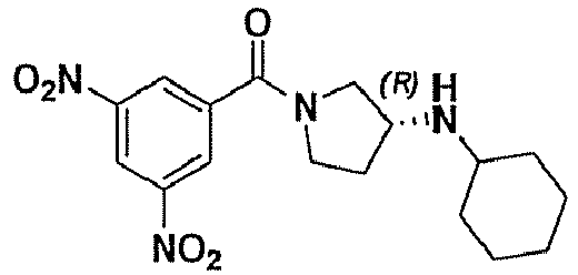
-

-

-

-

-

-

-
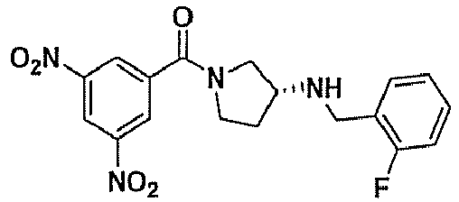
-

-
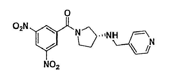

- To a solution of F1 (3.77 mmol) in DMF (15 mL) was added sodium azide (11.00 mmol) and the resulting mixture was warmed to 70°C. After 3 h, the solvent was removed in vacuo, dissolved in ethylacetate (50 mL) and washed with water (50 mL) and brine (50 mL). The organic layer was dried over anhydrous MgSO4 and concentrated in vacuo. The crude product was purified by silical gel flash column chromatography (1:1 hexanes/ethyl acetate) to give F2.
- To a solution of F2 (2.68 mmol) was added 10% palladium on activated carbon and stirred overnight under hydrogen atmosphere. The reaction mixture was filtered using cellite 545 and the resulting filtrate was concentrated in vacuo to give F3.
- To a solution of F3 (0.77 mmol) and triethylamine (1.16 mmol) in methylene chloride (10 mL) was added benzoyl chloride (1.00 mmol) under ice bath. The reaction mixture was brought up to room temperature. After 2 h, the raction mixture was diluted with methylene chloride (20 mL) and washed with water (30 mL) and brine (30 mL). The organic layer was dried over anhydrous MgSO4 and concentrated in vacuo. The crude product was purified by silica gel flash column chromatography (2:1 hexanes/ethyl acetate) to give F4.
- To a solution of F4 (0.59 mmol) in methylene chloride (10 mL) was added trifluoroacetic acid (0.5 mL) and stirred at room temperature. After 3 h, the solvent was removed in vacuo. The crude product was dissolved in methylene chloride (10 mL) and triethylamine (0.41 mL, 2.96 mmol) was added. The reaction mixture was cooled to 0°C and then 3,5-dichlorobenzoyl chloride (0.65 mmol) was added. The resulting mixture was brought up to room temperature. After 2 h, the solvent was removed in vacuo and the crude residue was purified by silica gel flash column chromatography (1:1 hexanes/ethyl acetate) to give F5.
-

-
 30.0, 32.8, 45.2, 48.0, 48.9, 50.5, 52.2, 55.2, 55.6, 60.6, 113.9, 120.2, 125.6, 126.1, 127.7, 127.8, 129.0, 139.5, 148.5, 162.7, 164.9, 165.0, 167.4.
-

-

-

- The pyridopyrimidinone compounds (scaffold VIII; see Table 3) underwent derivatization according to the methods outlined below (Schemes 8-10). Resulting derivatives were examined for inhibitory activity using the assay described above and the results are summarized in Table 4.

- 2-Amino-3-picoline (1.0 mmol) was dissolved in diethyl malonate (1.0 mmol). The solution was heated to 170°C for 12 h. After cooling, the dark residue was triturated with CH2Cl2 (10 mL). The residual pale solid was collected by filtration and washed with CH2Cl2 to give G1.
- To a DMF (2.0 mL) was added POCl3 (3.0 mmol) at 0°C. After the mixture was stirred at 0 °C for 40 min, a solution of G1 (1.0 mmol) in DMF (2.0 mL) was added and stirred at 80 °C for 1 h. The mixture was cooled and concentrated in vacuo. The residue was diluted with water and extracted with CH2Cl2 (10 mLx 3). The combined organic layers were washed with brine, dried over MgSO4 and concentrated. The residue was purified by flash column chromatography to give G2.
- To a stirred solution of G2 (1.0 mmol) in THF (2.0 mL) was added Et3N (2.0 mmol). The mixture was cooled to 0°C. After 5 min, an amine (1.0 mmol) was added dropwise and the mixture was stirred at room temperature overnight. The reaction mixture was diluted with CH2Cl2 (10 mL) and washed with brine (10 mL). The organic layer was dried over anhydrous MgSO4 and concentrated in vacuo. The crude product was purified by flash column chromatography to give G3.
- G2 (0.5 mmol) was dissolved in 10.4 mL of tert-butyl alcohol and 2.5 mL of 2-methyl-2-butene. A solution of sodium chlorite (4.59 mmol) and sodium dihydrogenphosphate (3.46 mmol) in 4.2 mL of water was added dropwise. The reaction mixture was stirred at room temperature overnight. Volatile components were then removed under vacuum, and the residue was dissolved in 10 ml of water and extracted with two 10 ml portions of hexane. The aqueous layer was acidified to pH=3 with HCl(aq) and extracted with 10 mL portions of methylene chloride. The combined organic layers were washed with 20 mL of cold water, dried and concentrated to give G4.
- G3 (36.6 µmol) was dissolved in 760 µl of tert-butyl alcohol and 180 µl of 2- methyl-2-butene. A solution of sodium chlorite (335 µmol) and sodium dihydrogenphosphate (253 µmol) in 300µl of water was added dropwise. The reaction mixture was stirred at room temperature overnight. Volatile components were then removed under vacuum and the residue was dissolved in 10 ml of water and extracted with two 10 ml portions of hexane. The aqueous layer was acidified to pH=3 with HCl(aq) and extracted with 10 ml portions of methylene chloride. The combined organic layers were washed with 20 ml of cold water, dried and concentrated to give G5.
- To a stirred solution of G4 (1.0 mmol) in DMF (2.0 mL) was added Et3N (2.0 mmol) and amine (1.5 mmol) and the mixture was stirred at 60°C overnight. The reaction mixture was diluted with CH2Cl2 (10 mL) and washed with brine (10 ml). The organic layer was dried over anhydrous MgSO4 and concentrated in vacuo. The crude product was purified by recrystallization from a mixture of hexanes and methylene chloride to give G5.
- The solution of 2-amino-3-picoline (4.0 mmol) in a solution of CH2Cl2 (3 mL) and dried pyridine (1 mL) was added dropwise at room temperature to a stirred solution of ethyl 3-chloro-3-oxo-propionate (5.3 mmol) in CH2Cl2 (3 mL) (an exothermic reaction with emission of white fume occurred during the addition). The resulting warm mixture was stirred at room temperature for 30 min and then poured into 30 mL of cold water; an excess of sodium carbonate was carefully added with stirring and the mixture was further stirred at room temperature for 1 h. The organic layer was then collected and the aqueous phase was extracted several times with CH2Cl2. The combined organic layers were washed with water, dried over anhydrous Na2SO4, and concentrated in vacuo. The crude product was purified by flash column chromatography to give G6.
- A mixture of G6 (1.83 mmol), POCl3 (0.5 mL) and polyphosphoric acid (137 mg) was heated with stirring at 130°C for 3 h. After cooling, anhydrous ethanol was added and the mixture was refluxed for 30 min, then allowed to cool. The mixture was treated with aqueous sodium carbonate and exhaustively extracted with CH2Cl2 (10 mLx 3). The combined layers were washed with water (10 mL), brine (10 mL), dried over MgSO4, filtered and concentrated in vacuo. The crude product was purified by flash column chromatography to give G7.
- To a solution of G6 (1 mmol) in DMF (0.96 mL) was added potassium carbonate (5.0 mmol) followed by phenol (1.94 mmol). After 12 h at 100 °C, the solution was allowed to cool to 23 °C. The reaction mixture was washed with H2O (50 mL), and the aqueous layer was extracted with CH2Cl2 (20 mL x 3). The combined organic layers were washed with 1 N HCl (20 mL x 2), filtered, and concentrated in vacuo. The crude product was purified by flash column chromatography to give G8.
- To DMF (2.0 mL) was added POCl3 (3.0 mmol) at 0°C. After the mixture was stirred at 0 °C for 40 min, a solution of G8 (1.0 mmol) in DMF (2.0 mL) was added and stirred at 80 °C for 1 h. The mixture was cooled and concentrated in vacuo. The residue was diluted with water and extracted with CH2Cl2 (10 mL x 3). The combined organic layers were washed with brine, dried over MgSO4 and concentrated. The residue was purified by flash column chromatography to give G9.
-
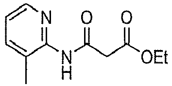
-
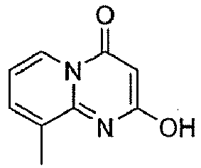
-
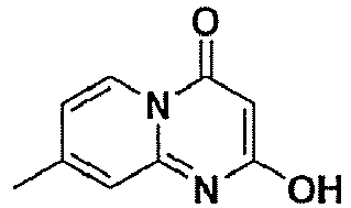
-

-

-

-

-

-

-

-

-

-
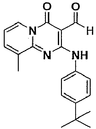
-

-

-

-

-

-

-

-

-

-

-

-

-

-

-

-

-

-

-
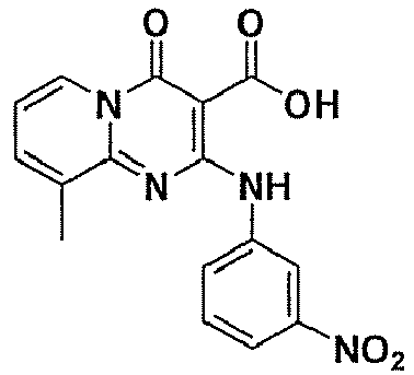
-

-

-

-

-
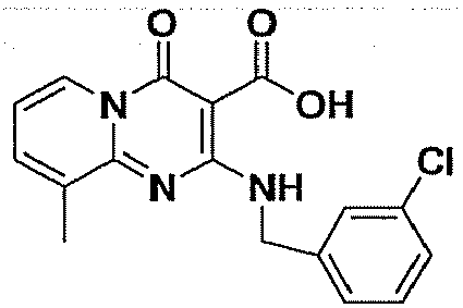
-

-

-
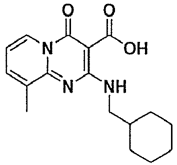
-

-

-
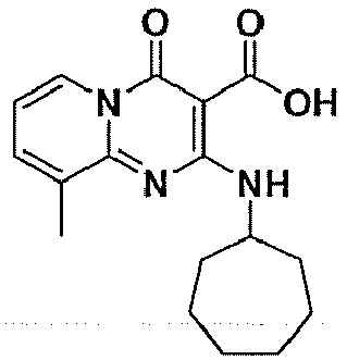
-

-

-

-
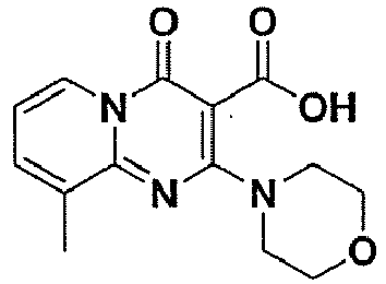
-

-

-


- 2-Amino-3-picoline (1.0 mmol) was dissolved in diethyl ethoxymethylenemalonate (1.0 mmol). The solution was heated to 170 °C for 12 h. After cooling, the dark residue was triturated with EtOAc (10 mL). The residual pale solid was collected by filtration and washed with EtOAc to give H1.
- To a stirred solution of H1 (0.43 mmol) in H2O (3.0 mL) and EtOH (1.0 mL) was added LiOH (0.86 mmol). The mixture was stirred at room temperature for 3 h. The reaction mixture was diluted with CH2Cl2 (10 mL) and washed with 1 N HC1 (10 ml). The organic layer was dried over anhydrous MgSO4 and concentrated in vacuo. The crude product was purified by flash column chromatography to give H2.
- To a stirred solution of H1 (0.38 mmol) in THF (2.0 mL) was added LiAlH4 (0.57 mmol) at 0 °C. The reaction mixture was stirred at 0 °C for 3 h. After reaction was completed, 1N NaOH (2 mL) was added dropwise. The mixture was diluted with CH2Cl2 (10 mL) and washed with H2O (10 ml). The organic layer was dried over anhydrous MgSO4 and concentrated in vacuo. The crude product was purified by flash column chromatography to give H3.
- To a stirred solution of H3 (95 µmol) in CH2Cl2 (1.0 mL) was added NaHCO3 (285 µmol) and Dess-Martin Periodinane (114 µmol) at 0 °C. The mixture was stirred at 0 °C for 1 h. The reaction mixture was filtered off and concentrated in vacuo. The crude product was purified by flash column chromatography to give H4.
- To a stirred solution of 2-Amino-pyridine (10.6 mmol) in xylene (10.0 mL) was added diethyl ethoxymethylenemalonate (21.2 mmol). The mixture was stirred at 140 °C for 3 hr. After reaction was completed, the residual pale solid was collected by filtration and washed with diethyl ether to give H5.
- To a stirred solution of H5 (0.42 mmol) in THF (5.0 mL) was added triethylamine (0.63 mmol) and p-toluenesulfonylchloride (0.46 mmol) at 0 °C. The reaction mixture was stirred at room temperature for overnight. After reaction was completed, the mixture was diluted with CH2Cl2 (40 mL) and washed with 1N HCl (50 ml), saturated NaHCO3 (50 ml) and brine (50 ml). The organic layer was dried over anhydrous MgSO4 and concentrated in vacuo. The crude product was purified by flash column chromatography to give H6.
- To a stirred solution of H6 (0.25 mmol) in THF (1.2 mL) was added triethylamine (0.5 mmol) and an amine (0.26 mmol) at 0 °C. The reaction mixture was stirred at room temperature for overnight. After reaction was completed, the mixture was diluted with CH2Cl2 (10 mL) and washed with 1N HCl (10 ml), saturated NaHCO3 (10 ml) and brine (10 ml). The organic layer was dried over anhydrous MgSO4 and concentrated in vacuo. The crude product was purified by flash column chromatography to give H7.
- To a stirred solution of H7 (0.27 mmol) in ethylene glycol (3.0 mL) was added methylamine (2 N solution in THF 1.3 mL). The mixture was stirred at 150 °C for 3 hr. The reaction mixture was added with ethylacetate (10 mL) and the residual pale solid was collected by filtration and washed with EtOAc. The crude product was purified by flash column chromatography to give H8.
- To a stirred solution of H5 (2.13 mmol) in MeOH (8.0 mL) was added Pd/C (113 mg). The mixture was stirred at room temperature under H2 for 3 h. After reaction was completed, the reaction mixture was filtered off and concentrated in vacuo. The crude product was recrystallized with EtOAc and hexane (1:4) to give H9.
- To a stirred solution of H9 (0.42 mmol) in CH2Cl2 (5.0 mL) was added triethylamine (0.63 mmol) and p-toluenesulfonylchloride (0.46 mmol) at 0 °C. The reaction mixture was stirred at room temperature for overnight. After reaction was completed, the mixture was diluted with CH2Cl2 (40 mL) and washed with 1N HCl (50 ml), saturated NaHCO3 (50 ml) and brine (50 ml). The organic layer was dried over anhydrous MgSO4 and concentrated in vacuo. The crude product was purified by flash column chromatography (Hexane : EtOAc = 1: 2) to give H10.
- To a stirred solution of H10 (0.25 mmol) in THF (2.0 mL) was added triethylamine (0.5 mmol) and an amine (0.37 mmol) at 0 °C. The reaction mixture was stirred at room temperature for overnight. After reaction was completed, the mixture was diluted with CH2Cl2 (10 mL) and washed with 1N HCl (10 ml), saturated NaHCO3 (10 ml) and brine (10 ml). The organic layer was dried over anhydrous MgSO4 and concentrated in vacuo. The crude product was purified by flash column chromatography (Hexane : EtOAc = 1: 1) to give H11.
- A solution of G3 (1.0 mmol), an amine (1.1 mmol) and triethylamine (2.0 mmol) in THF (2 mL) was refluxed for 1 h and cooled to room temperature. The solvent was evaporated to dryness, which was extracted with CH2Cl2 (20 mL x 3).
- The reaction mixture was washed with 5% sodium bicarbonate. The organic layer was dried (MgSO4), filtered, and concentrated in vacuo. The crude product was purified by flash column chromatography to give H12.
- To a solution of G3 (1.1 mmol), an amine (1.0 mmol) in CH2Cl2 (5 mL) were added sodium triacetoxyborohydride (2.0 mmol) and glacial acetic acid (2.0 mmol) at room temperature for 20 h. The reaction mixture was added saturated ammonium chloride solution and stirred for 10 min. The reaction mixture was extracted with CH2Cl2 (20 mL). The organic layer was dried (MgSO4), filtered, and concentrated in vacuo. The crude product was purified by flash column chromatography to give H13.
-

-

-

-

-

-

-

-

-

-
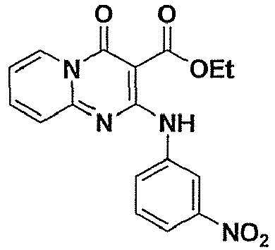
-

-

-

-

-
 18.0, 52.1, 85.3, 113.7, 119.6, 121.9, 123.5, 126.4, 129.4, 133.2, 134.1, 138.4, 139.9, 151.0, 156.2, 158.6, 170.1.
-

-

-

-

-

-

-

-

-

-

-

-
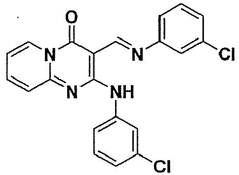 , J= 0.8, 1.2, 8.0 Hz, 1H), 7.28 (d, J= 8.4 Hz, 1H), 7.29 (dd, J = 2.0, 4.0 Hz, 1H), 7.3 (d, J = 8.0 Hz, 1H), 7.44 (d, J = 8.8 Hz, 1H), 7.52 (ddd, J = 0.8, 1.2, 8.0 Hz, 1H), 7.17 - 7.76 (m, 1H), 8.02 - 8.04 (m, 1H), 8.98 (dd, J = 0.8, 6.8 Hz, 1H), 9.17 (s, 1H), 12.94 (brs, 1H); 13C NMR (100 MHz, CDCl3) δ 92.6, 114.0, 119.5, 119.8, 121.8, 123.9, 125.0, 125.7, 128.0, 129.7, 130.2, 134.4, 134.8, 138.7, 140.1, 151.3, 151.8, 157.0, 158.0, 158.9.
-
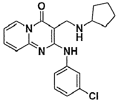
-

-

-

-


- To a solution of an aldehyde (0.9 mmol) in methanol (0.5 mL) was added NaBH4 (1.35 mmol) at room temperature. After stirring 1 h, the reaction mixture was diluted with methylene chloride (10 mL) and washed with brine (10 ml). The organic layer was dried over MgSO4 and concentrated in vacuo. The crude product was purified by recrystallization from a mixture of hexanes and ethyl acetate to give J1.
- To a stirred solution of an ester (0.06 mmol) in THF (1.0 mL) was added LiAlH4 (0.09 mmol). The reaction mixture was stirred at room temperature for 1 hr. After reaction was completed, H2O (0.1 mL) was added dropwise. The reaction mixture was filtered off and concentrated in vacuo. The crude product was purified by flash column chromatography to give J2.
- To a stirred solution of J1 or J2 (0.19 mmol) in CH2Cl2 (0.6 mL) was added triethylamine (0.38 mmol) and a benzoyl chloride (0.28 mmol) at 0 °C. The reaction mixture was stirred at room temperature for 1 h. After reaction was completed, the mixture was diluted with CH2Cl2 (10 mL) and washed with brine (10 ml). The organic layer was dried over anhydrous MgSO4 and concentrated in vacuo. The crude product was purified by flash column chromatography (Hexane : EtOAc = 2: 1) to give J3.
-
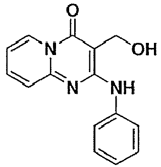
-

-

-

-

-

-

-

-

-

-

-

-

-

-

-

-

-

-

-

-

-

-

-

-

-

-

-

-

-

-

-

-

-
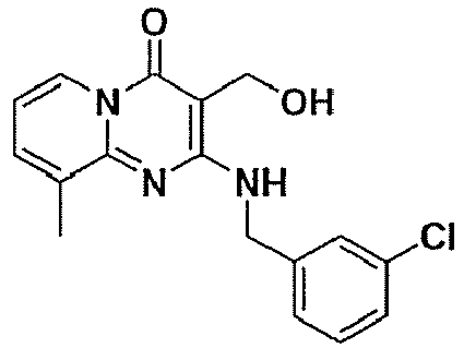
-

-

-

-

-
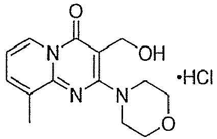
-

-

-

-

-

-

-

-

-

-

-

-
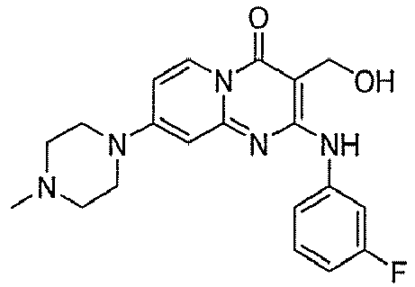
-

-

-
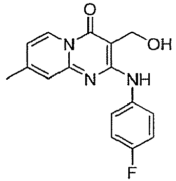
-

-

-

-

-

-

-

-

-

-

-

-

-

-

-

-

-

-

-
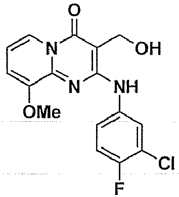
-

-

-

-

-

-

-

-

-

-
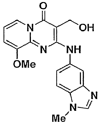
-

-

-

-

-

-

-

-

-

-

-

-

-

- Two representative molecules, compounds 4 and 24, were re-synthesized in-house and subjected to conventional CFU-based activity testing in primary macrophages (
Figure 7 ). A ten-fold decrease in the number of CFUs, similar to that seen with INH, was observed for both compounds five days after infection on three different cell lines. This confirms the potency of this series of compounds. - To address the issue of toxicity, compounds 4 and 24 were tested on a panel of five cell lines derived from different body tissue. Cells were incubated with increasing amounts of compound and cell viability was assessed with resazurin after 5 days of co-incubation. Percentage cytotoxicity was determined by taking as a reference the resofurin fluorescence measured by DMSO containing wells. The concentration where fifty percent of the cells died was defined as the Minimal Toxic Concentration (MTC50). Both
compounds - To gain insight into the possible specificity of acitivity of
compounds - Mutation frequency of M tuberculosis H37Rv was determined for
compounds compound compound 24, at 0.2 µg/ml and 3.2 µg/ml, frequency of mutation was 7×10⁻⁷ and 1×10⁻⁸, respectively which corresponds to a mean frequency of 3.5×10⁻⁷. Overall, these values were superior to frequency of mutation observed for INH-resistant mutants (3×10⁻⁶). These results, thus, demonstrate that this class of compounds result in a low frequency of mutation. - Table 6 shows the minimal inhibitory concentration (MIC) of one representative compound, 133, on different Mycobacterial species. While it has no effect on the fast growing Mycobacterium smegmatis mc2, it was able to inhibit typical laboratory strains such as H37Rv, H37Ra and BCG Pasteur with an MIC of 2 µM. More importantly, the antimicrobial activity of 133 was also tested against clinical isolates strains of mycobacteria. The MIC values for multi-drug-resistant (MDR-TB) and extensive-drug-resistant (XDR-TB) isolates strains were within the micromolar range.
- To address the issue of toxicity, compound 133 was tested on a panel of seven cell lines derived from different body tissue. Cells were incubated with increasing amounts of compound and cell viability was assessed with resazurin after 5 days of co-incubation. Percentage of cytotoxicity was determined by taking as a reference the resofurin fluorescence measured by DMSO containing wells. The concentration where fifty percent of the cells died was defined as the Minimal Toxic Concentration (MTC50). Compound 133 showed no cytotoxicity for all tested cell lines up to 100 µM (Table 6). The selectivity index, which consists of the ratio between antitubercular activitiy and cytotoxicity was therefore above 50 for both extracellular and intracellular mycobacteria suggesting this series of compounds to be promising new anti-tuberculosis drugs.
- The effect of this series of compounds on primary macrophages was further determined. Host cells that had priory been incubated with compound 232 harbored fewer bacteria compared to DMSO control and were more abundant at
day 5 after infection as shown inFigure 8 . Similar data were obtained for compound 133 (data not shown). Conventional CFU determination was then performed seven days after infection to quantify the remaining bacterial load. A ten-fold decrease in the number of CFUs, similar to that seen with INH, was observed for both compounds on both human and mouse cells (Figure 8 ). This confirms the potency of this series of compounds. - Mutation frequency of M. tuberculosis H37Rv was determined for compound 264. Increasing numbers of bacteria grew on 7H10 agar medium supplemented with different concentrations of compound. After a 6-week growth, colonies were counted in order to evaluate the proportion of spontaneous mutational frequency (Table 7). Compound 264 gave frequencies of resistance of 3.4×10⁻⁶ and 8×10⁻⁶ at 0.4 and 0.8 µg/ml, respectively, and 2×10⁻⁸ at both 1.6 µg/ml and 3.2 µg/ml. Accordingly, spontaneous mutational rate was calculated to be 7×10⁻⁷. Overall, these values are better than the frequency of mutation observed for INH (2.9×10⁻⁶). These results, therefore, demonstrate that this class of compounds result in a low frequency of mutation.
- One of the current challenges for TB drug discovery is the identification of compounds that are active against persistent bacteria. Although the location and state of latent bacteria remains a matter of debate, one commonly shared hypothesis for mycobacterial persistence is that M tuberculosis bacilli are able to survive in macrophages for prolonged periods of time and, unlike other bacteria, are able to actively replicate. The intraphagosomal profile of M Tuberculosis is complex; a large variety of genes are over-expressed and timely regulated and are also dependent on environmental factors. Altogether, this makes the identification of one specific tubercle factor that could be selected as the ideal target difficult. Consequently, non-target cell-based assays are a critical tool in the search of intracellular M tuberculosis inhibitors.
- Investigation of bacillus growth inhibitors within macrophages has long been limited due to cumbersome CFU plating, slow bacillus growth, safety requirements and difficulties in setting-up appropriate infection conditions. As a consequence, this approach was always used as a secondary assay after the initial selection of compounds that are active on in vitro extracellular growth. With the advent of automated confocal microscopy, the above mentioned limitations could be readdressed and the inventors show the feasibility of large scale compound screening. It was decided to perform suspension macrophage batch infection in order to minimize the steps and to meet safety requirements. To this end, careful attention was paid to the removal of the extracellular non-phagocytosed mycobacteria. The centrifugation conditions used during the wash steps were set up in order to recover only the infected cells and discard most of the extracellular bacteria. By microscopy the inventors confirmed that unbound mycobacteria represented less than 10% of the total bacterial load (data not shown). Mycobacteria are able to grow independently of host cells and consequently any remaining extracellular bacilli would greatly compromise the validity of the inventors' model. To this end, an additional amikacin treatment step was added to the protocol to further eliminate any remaining mycobacteria. Thus with the optimized protocol, there is almost no non-phagocytosed mycobacteria left by the time compound is added. The obtained results also demonstrate that it is specifically the effect on the intracellular mycobacteria that is being measured with compound treatment. Indeed, the inventors observed a weak inhibition with rifampin, an antibiotic that is known to poorly penetrate cells. The 50-fold reproducible decrease in MIC for rifampin in the intracellular assay compared to the in vitro growth assay proved that the targeted bacteria are not extracellular. Otherwise no difference would have been seen in MIC between the two assays. Similarly, compounds able to inhibit mycobacterial growth in the phenotypic cell-based assay, but not the in vitro growth assay were also identified. In addition, the fact that the compounds are mixed with previously infected cells should decrease the chance for the identification of primary infection inhibitors. However, such compounds may still be identified as blockers of neighboring cell infection.
- Compared to a conventional CFU-plating method, the microscopy based detection of fluorescent bacteria is not sufficiently sensitive to distinguish between dead and live bacilli as the GFP signal is stable for several days. Indeed, at a high concentration of INH, rifampin or active compound, there is always 10% of the cells that appear to be infected, which is similar to the initial infection ratio. Surprisingly, no CFU could be recovered after plating such samples. Owing to the fact that latent bacilli are able to recover growth (Cho et al., 2007), the microscopy-detected bacilli must be dead bacilli rather than latent bacilli. Thus, the inventors' assay detects compounds that interfere with bacilli growth within macrophages.
- As it is well established and confirmed (
Figure 1a ), macrophages are able to support high bacterial loads which end up encompassing a large part of the cell cytoplasm and eventually lead to macrophage cell death. It is obvious when M Tuberculosis is the infectious agent compared to BCG (Bacille Calmette-Guerin), which even at high MOI fails to induce much cytotoxicity (data not shown). Taking this into account, it was decided to set the data acquisition atday 5 post-infection when the cell number in the DMSO samples had significantly decreased relative to the antibiotically protected controls. Thus, monitoring cell number was an additional parameter enabling the inventors to confirm the compound's antibacterial activity. - Unlike direct fluorescence based assays, analysis for image-based assays proved to be much more variable. Several parameters that are inherent to the biology of the assay partially explain the lower Z'-values that are usually accepted for HTS validation. The remaining fluorescent dead bacilli do not have much of an impact on the Z'-value, rather the variability in the infection ratio for the DMSO controls seems to account for the discrepancy. Also of importance is the fact that, upon infection, the macrophages had a tendency to migrate which in turn led to a heterogeneous set of images (
Figure 2a ). However, the aim of the primary screen was to identify compounds fully active at a concentration of 20 µM. Thus, for this purpose, a positive Z' for the infection ratio (INH/DMSO) was considered an acceptable value. The best proof of the validity of the hit selection according to the present invention comes from the subsequent serial dilution analysis, whereby almost 100% of the hits were confirmed. For each of the hits, a nicely fitted dose-response curve for the infection ratio was obtained as well as for the non-toxic compound in terms of cell number. Again, cell number brought an additional confirmation of the results that is totally independent of green fluorescence emission and GFP expression. - Obviously compounds found to be active against both intracellular and in vitro M. tuberculosis growth are the most promising. The best inhibitors isolated from this library have an inhibitory activity within the same range as INH. Further structure activity relationship studies will contribute to determine if their activity could be improved. In the course of another study using this phenotypic cell-based model, MIC down to the ng/mL scale was obtained for compounds with known in vitro antibacterial efficacy showing that compounds with a lower MIC than INH can be identified by the assay according to the present invention (data not shown). Of utmost interest are the compounds that are active only in the intracellular bacteria assay as they are likely to have a new mechanism of action independent of the infecting strain suggesting that they may also be active on the non-curable multi-drug-resistant (MDR)-strains.
- Taken together, the above results show that monitoring M tuberculosis growth with automated fluorescence microscopy is highly robust and reliable and that this method enables fast selection of potent anti-TB compounds.
-
- Abadie, V., Badell, E., Douillard, P., Ensergueix, D., Leenen, P. J., Tanguy, M., Fiette, L., Saeland, S., Gicquel, B., and Winter, N. (2005). Neutrophils rapidly migrate via lymphatics after Mycobacterium bovis BCG intradermal vaccination and shuttle live bacilli to the draining lymph nodes. Blood 106, 1843-1850.
- Andries, K., Verhasselt, P., Guillemont, J., Gohlmann, H. W., Neefs, J. M., Winkler, H., Van Gestel, J., Timmerman, P., Zhu, M., Lee, E., et al. (2005). A diarylquinoline drug active on the ATP synthase of Mycobacterium tuberculosis. Science 307, 223-227.
- Arain, T. M., Resconi, A. E., Singh, D. C., and Stover, C. K. (1996). Reporter gene technology to assess activity of antimycobacterial agents in macrophages. .
- Brodin, P., Majlessi, L., Marsollier, L., de Jonge, M.I., Bottai, D., Demangel, Cl., Hinds, J., Neyrolles, O., Butcher, P.D., Leclerc, C., Coles, S.T., Brosch, R., (2006). Dissection of ESAT-6 .
- Cho, S. H., Warit, S., Wan, B., Hwang, C. H., Pauli, G. F., and Franzblau, S. G. (2007). Low-oxygen-recovery assay for high-throughput screening of compounds against nonreplicating Mycobacterium tuberculosis. Antimicrob Agents Chemother 51, 1380-1385.
- Cremer, I., Dieu-Nosjean, M. C., Marechal, S., Dezutter-Dambuyant, C., Goddard, S., Adams, D., Winter, N., Menetrier-Caux, C., Sautes-Fridman, C., Fridman, W. H., and Mueller, C. G. (2002). Long-lived immature dendritic cells mediated by TRANCE-RANK interaction. .
- Fenistein, D., Lenseigne, B., Christophe, T., Brodin, P., and Genovesio, A. (2008). A fast fully automated cell segmentation algorithm for high throughput and high content screening. Cytometry part A, in press.
- Houben, E. N., Nguyen, L., and Pieters, J. (2006). Interaction of pathogenic mycobacteria with the host immune system. .
- Lenaerts, A. J., Hoff, D., Aly, S., Ehlers, S., Andries, K., Cantarero, L., Orme, I. M., and Basaraba, R. J. (2007). Location of persisting mycobacteria in a Guinea pig model of tuberculosis revealed by r207910. Antimicrob Agents Chemother 51, 3338-3345.
- Lipinski, C. A., Lombardo, F., Dominy, B. W., and Feeney, P. J. (2001). Experimental and computational approaches to estimate solubility and permeability in drug discovery and development settings. Advanced Drug Delivery Reviews 46, 3-26.
- Neyrolles, O., Hernandez-Pando, R., Pietri-Rouxel, F., Fornes, P., Tailleux, L., Barrios Payan, J. A., Pivert, E., Bordat, Y., Aguilar, D., Prevost, M. C., et al. (2006). Is adipose tissue a place for Mycobacterium tuberculosis persistence? PLoS ONE 1, e43.
- Rohde, K. H., Abramovitch, R. B., and Russell, D. G. (2007). Mycobacterium tuberculosis invasion of macrophages: linking bacterial gene expression to environmental cues. .
- Salomon, J. A., Lloyd-Smith, J. O., Getz, W. M., Resch, S., Sanchez, M. S., Porco, T. C., and Borgdorff, M. W. (2006). Prospects for advancing tuberculosis control efforts through novel therapies. .
- Schnappinger, D., Ehrt, S., Voskuil, M. I., Liu, Y., Mangan, J. A., Monahan, I. M., Dolganov, G., Efron, B., Butcher, P. D., Nathan, C., and Schoolnik, G. K. (2003). Transcriptional Adaptation of Mycobacterium tuberculosis within Macrophages: Insights into the Phagosomal Environment. J Exp Med 198, 693-704.
- Van Rie, A., and Enarson, D. (2006). XDR tuberculosis: an indicator of public-health negligence. Lancet 368, 1554-1556.


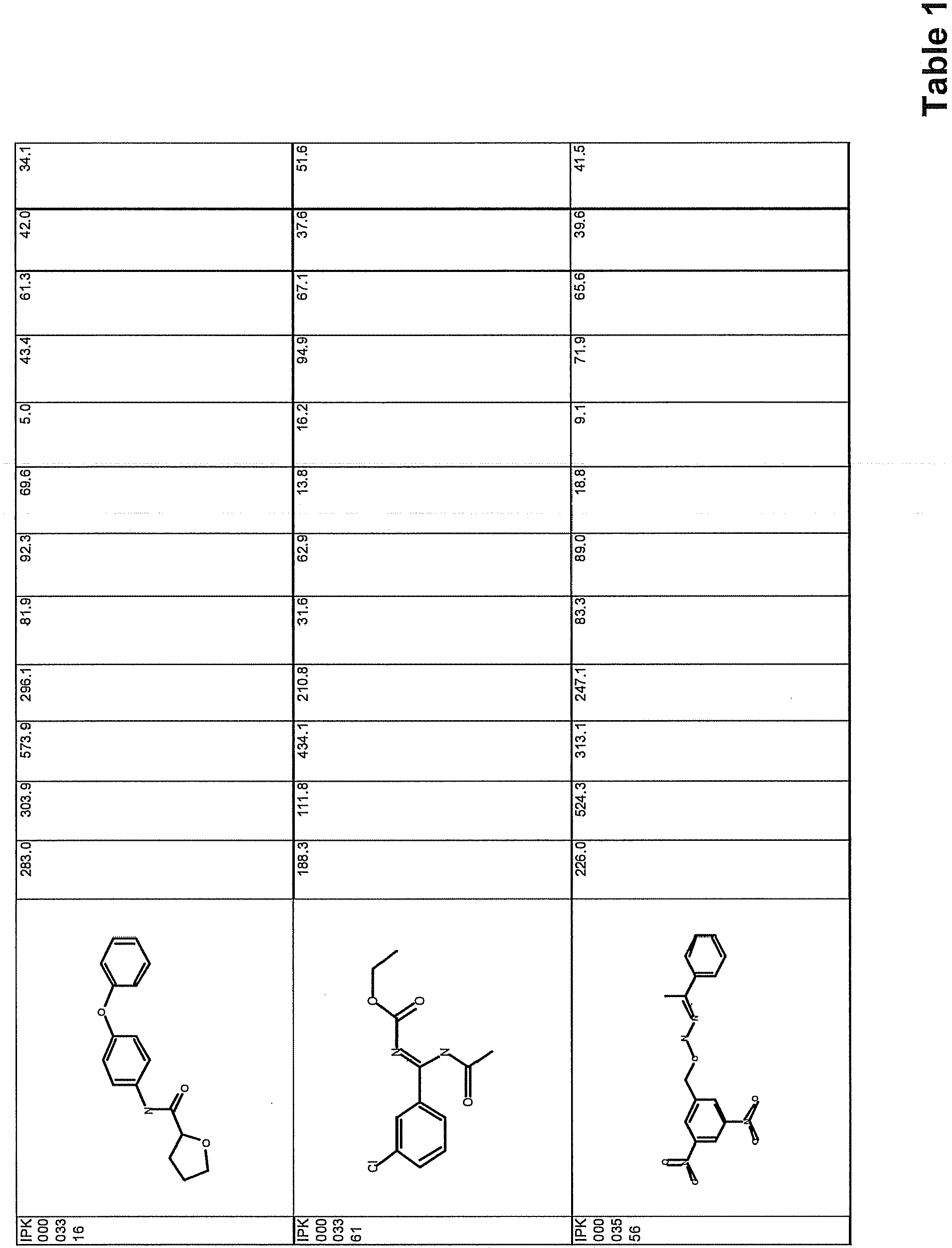


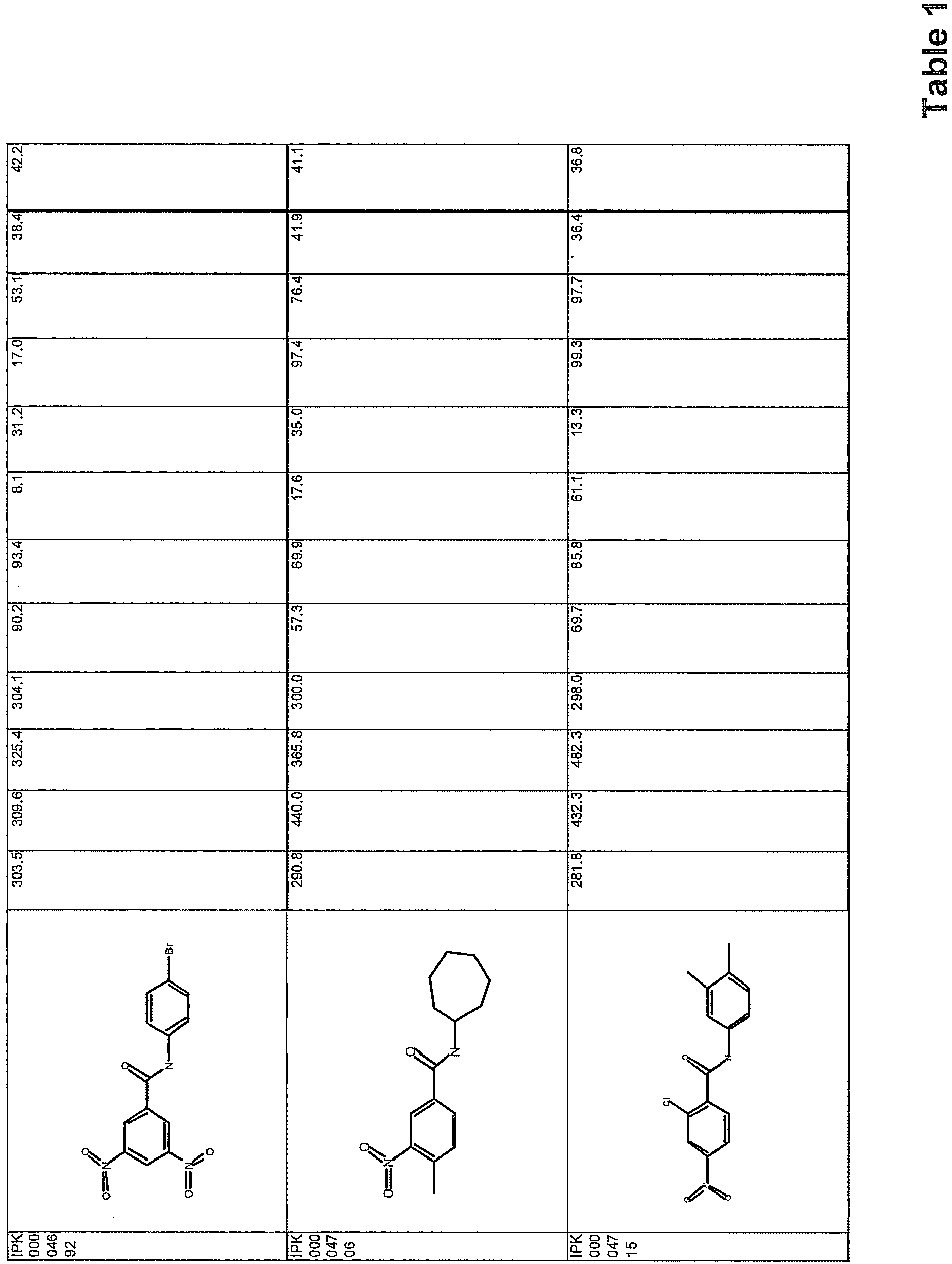


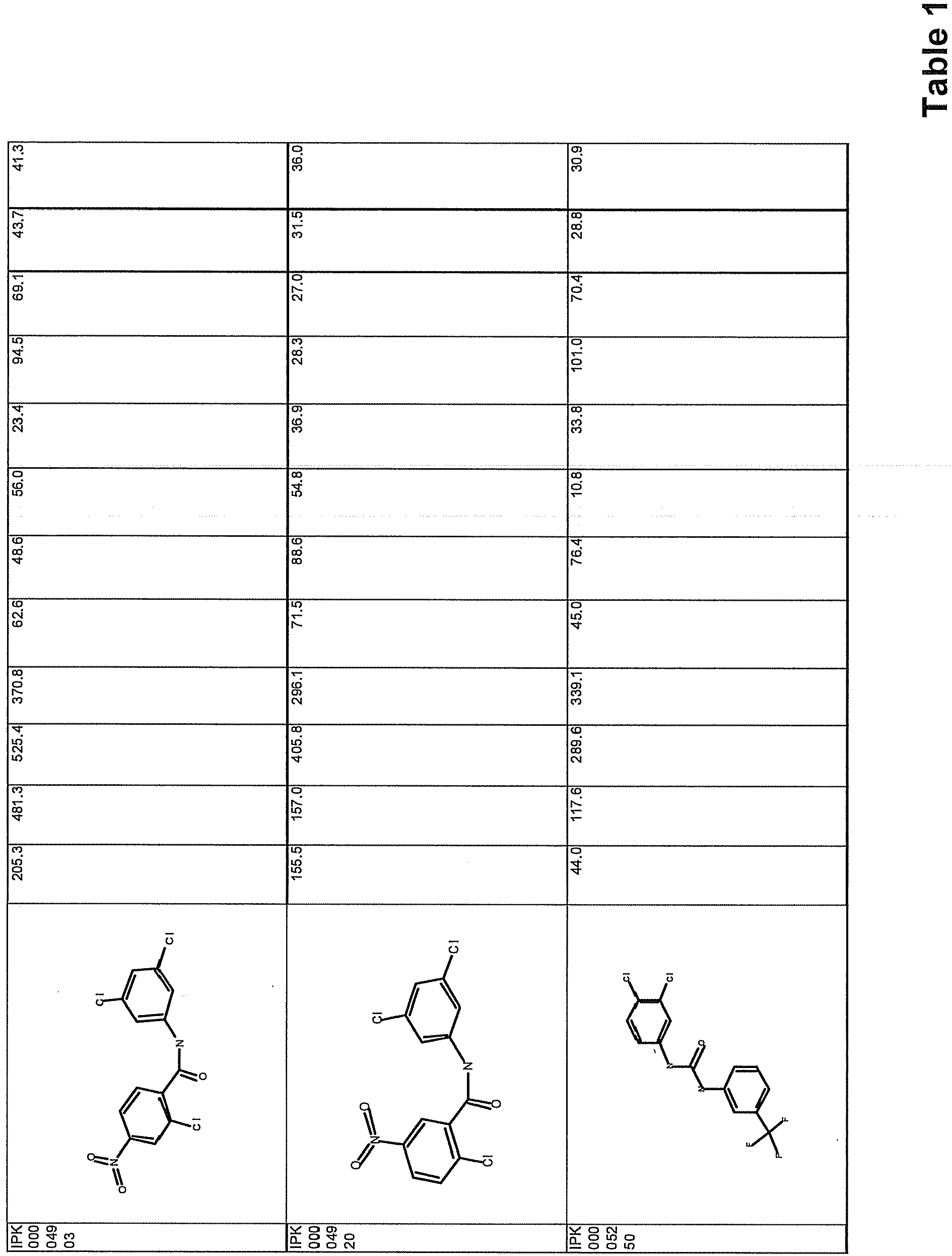

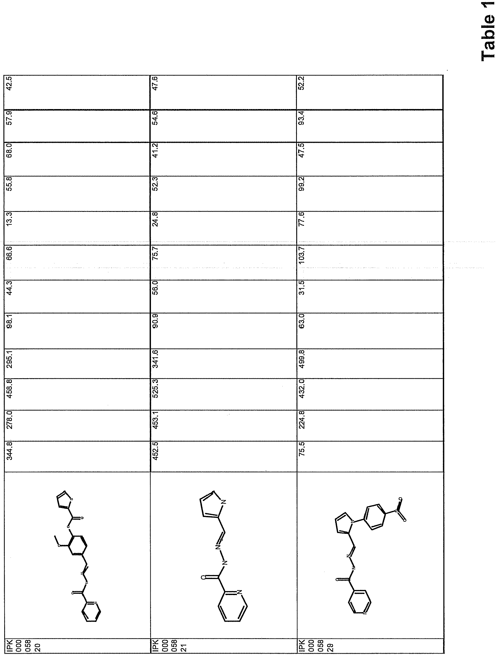


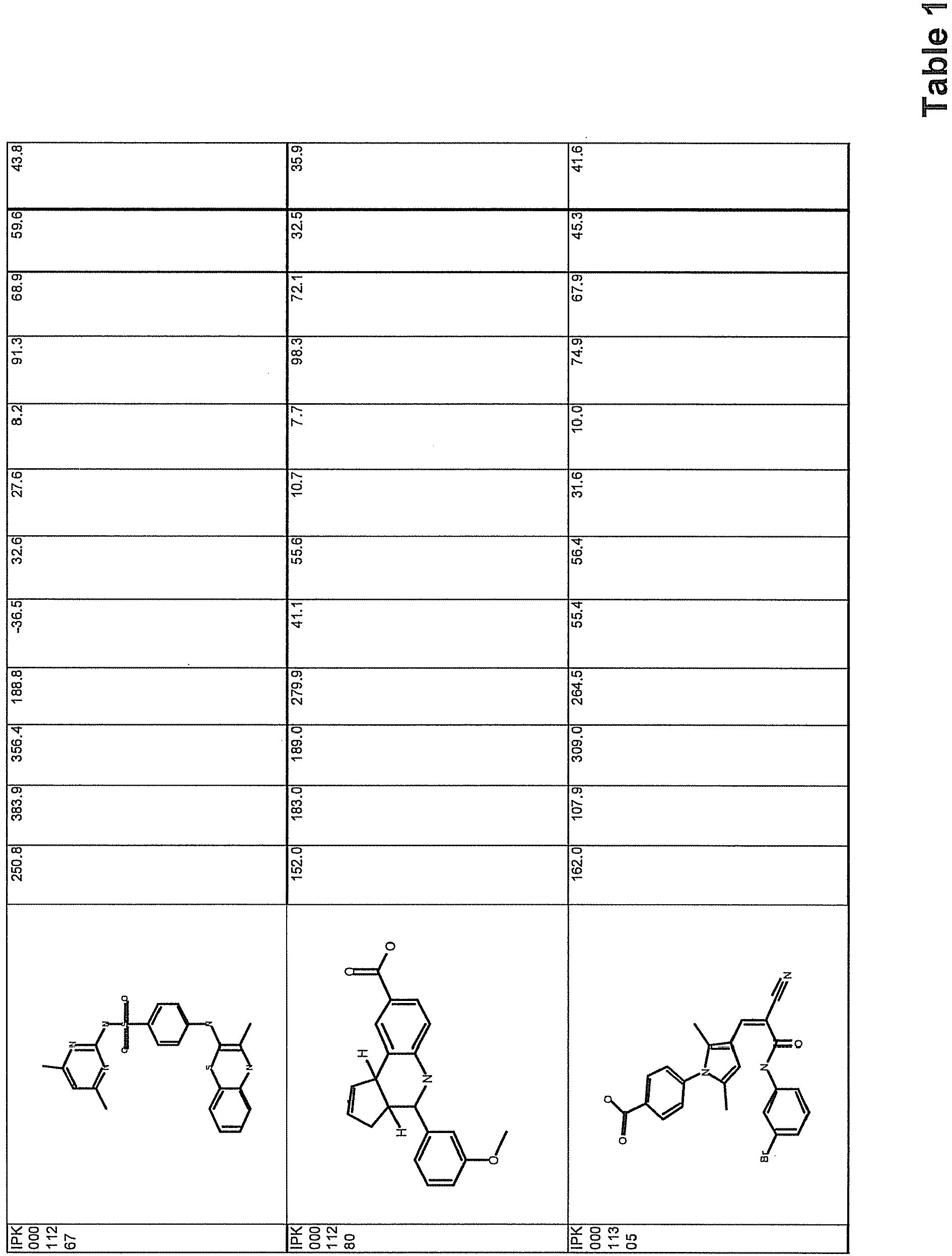


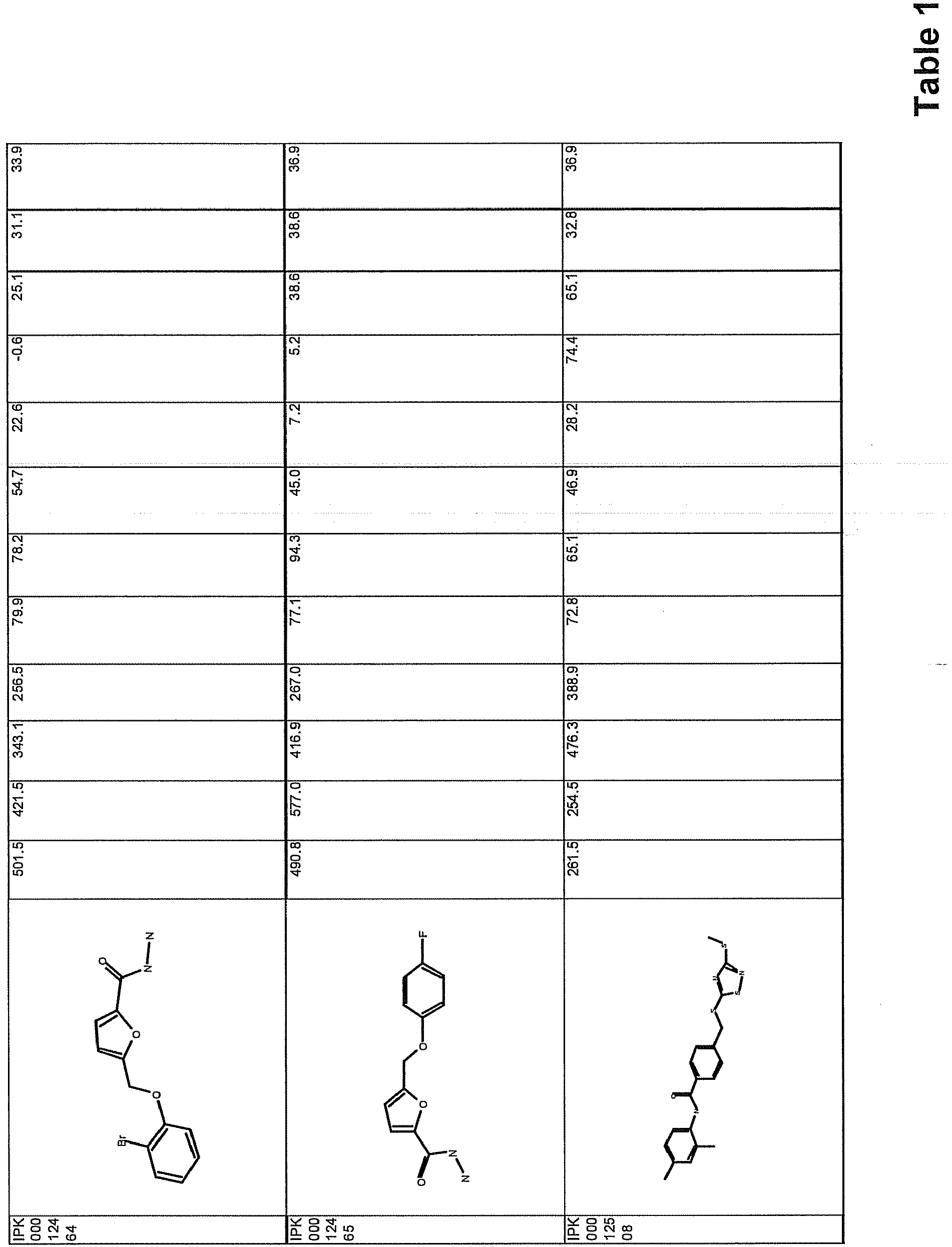


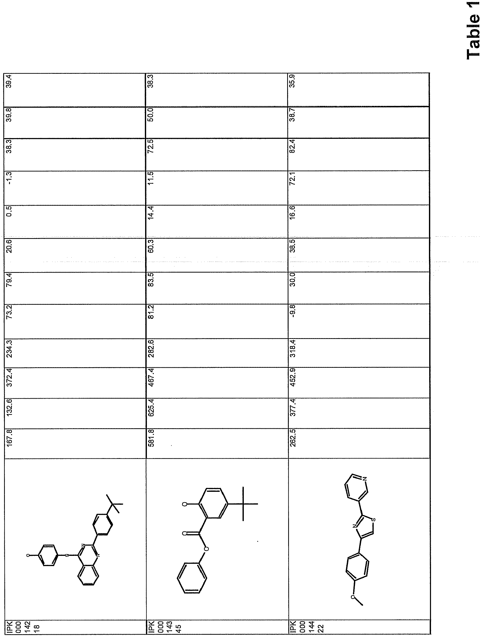
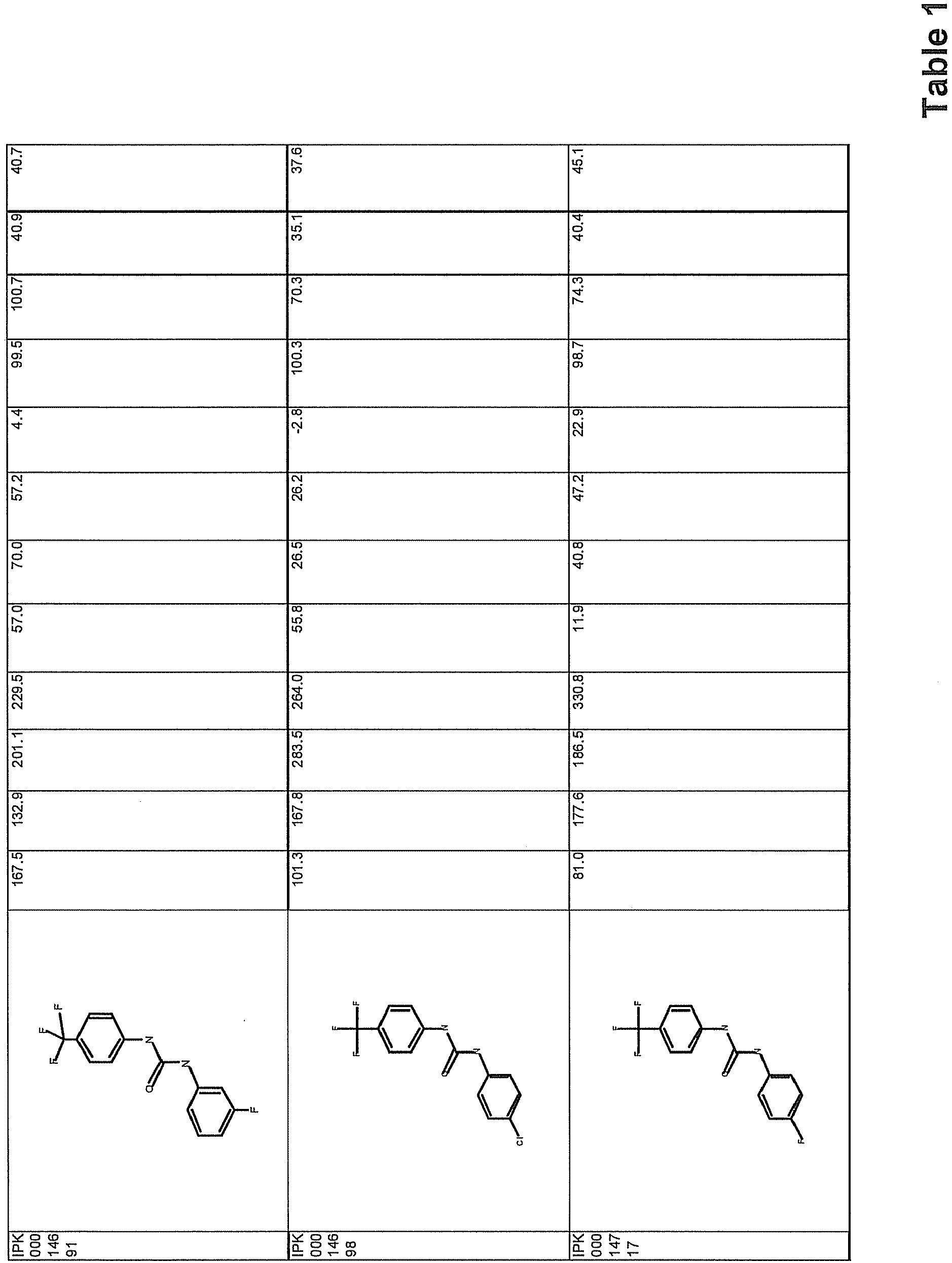


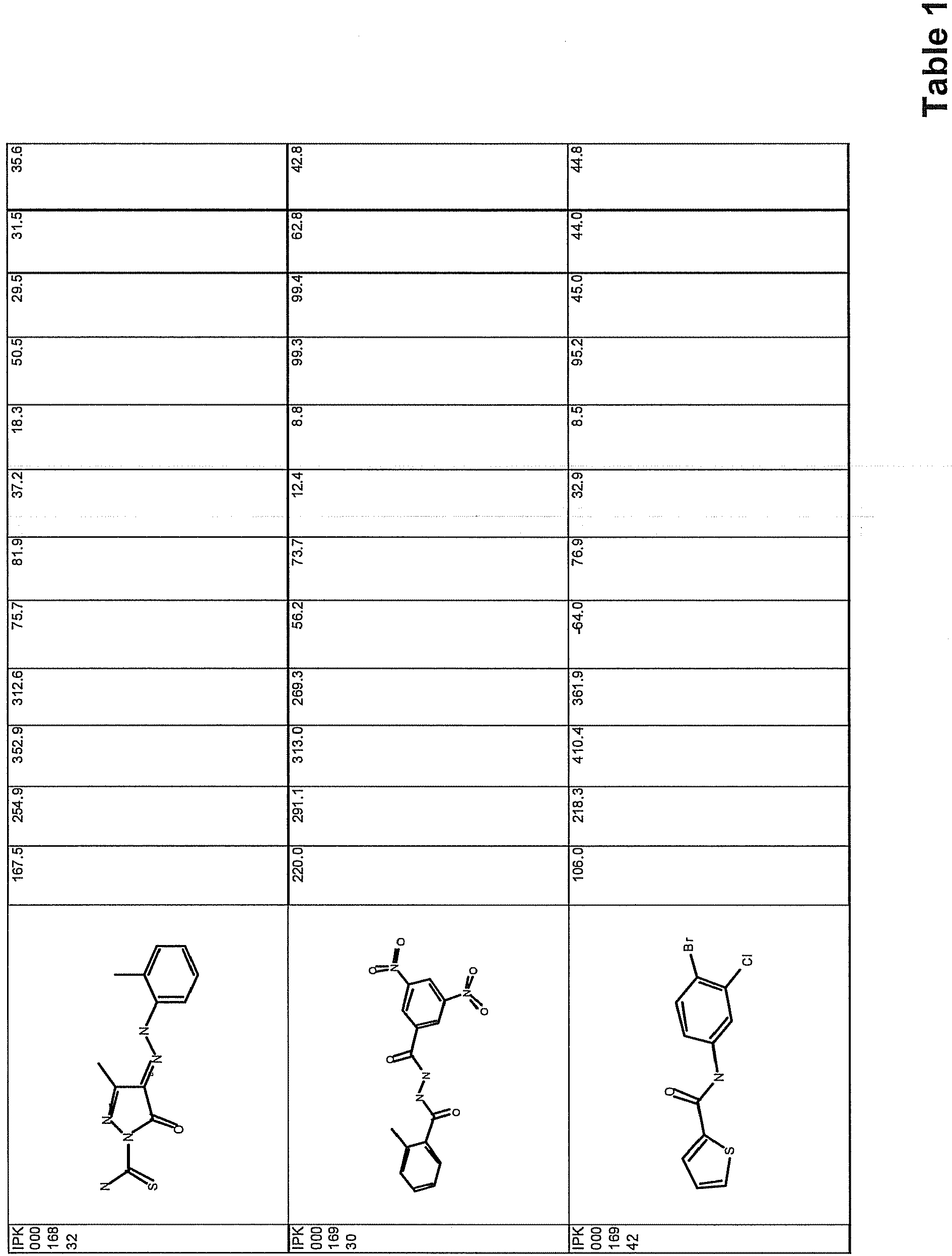


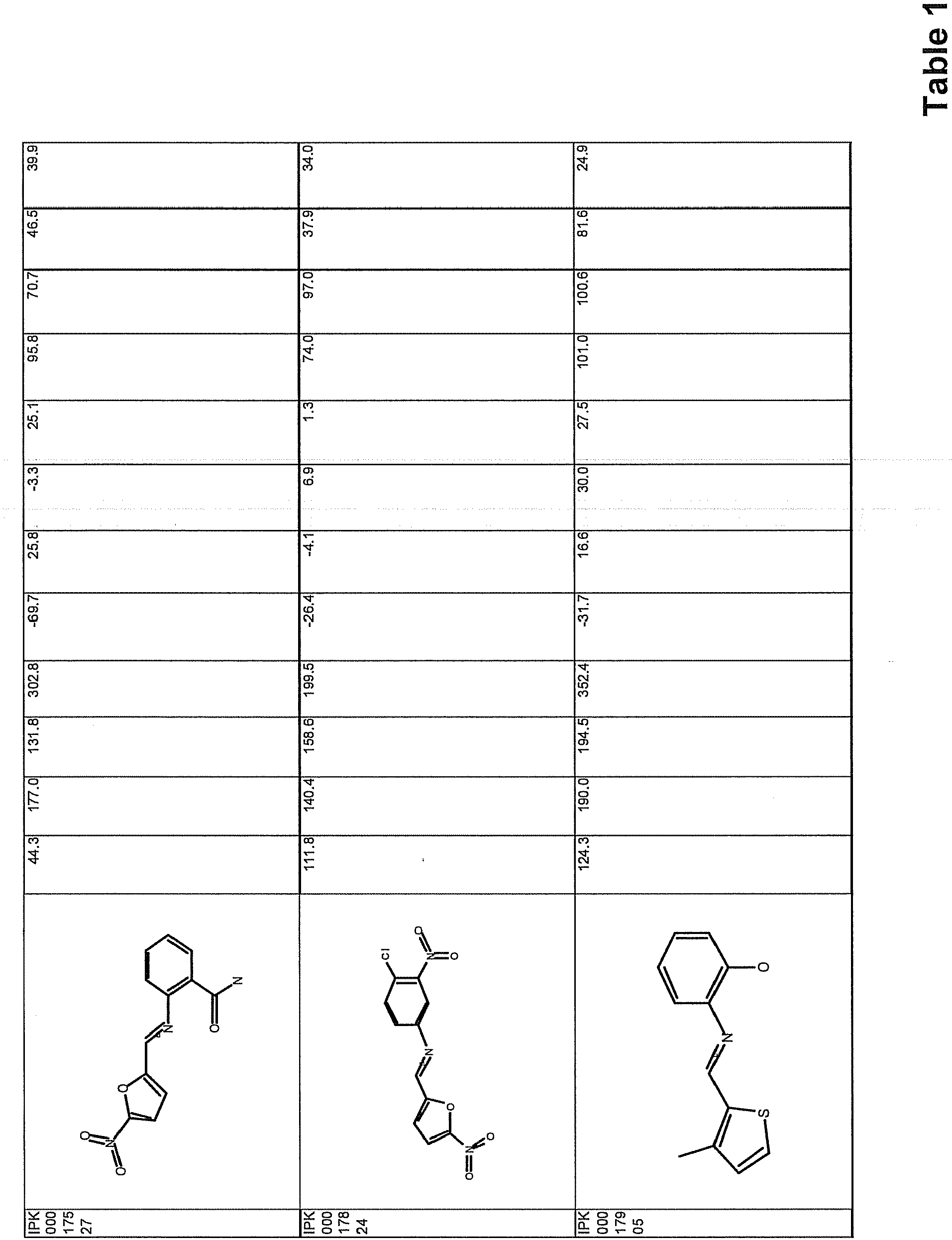


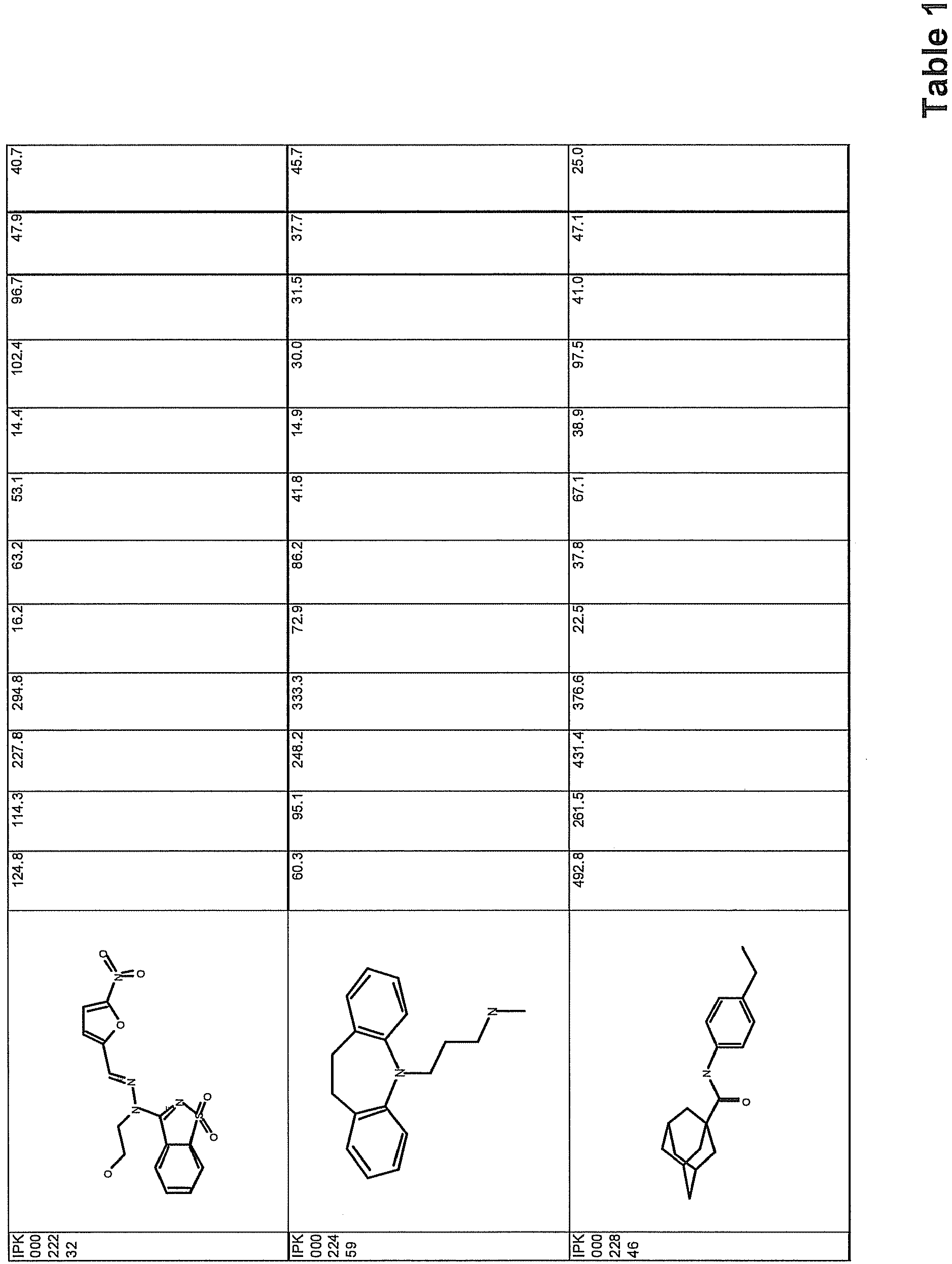
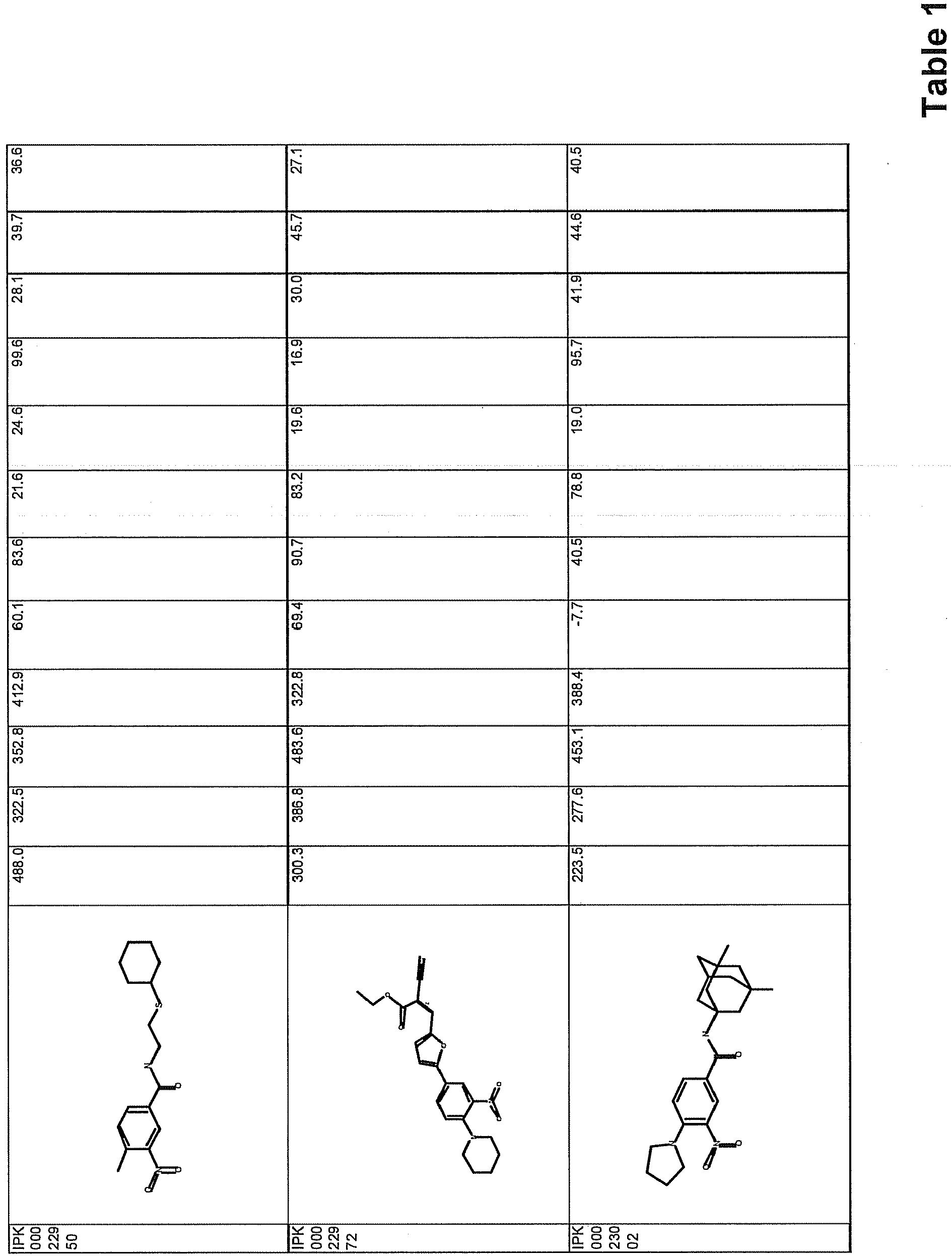


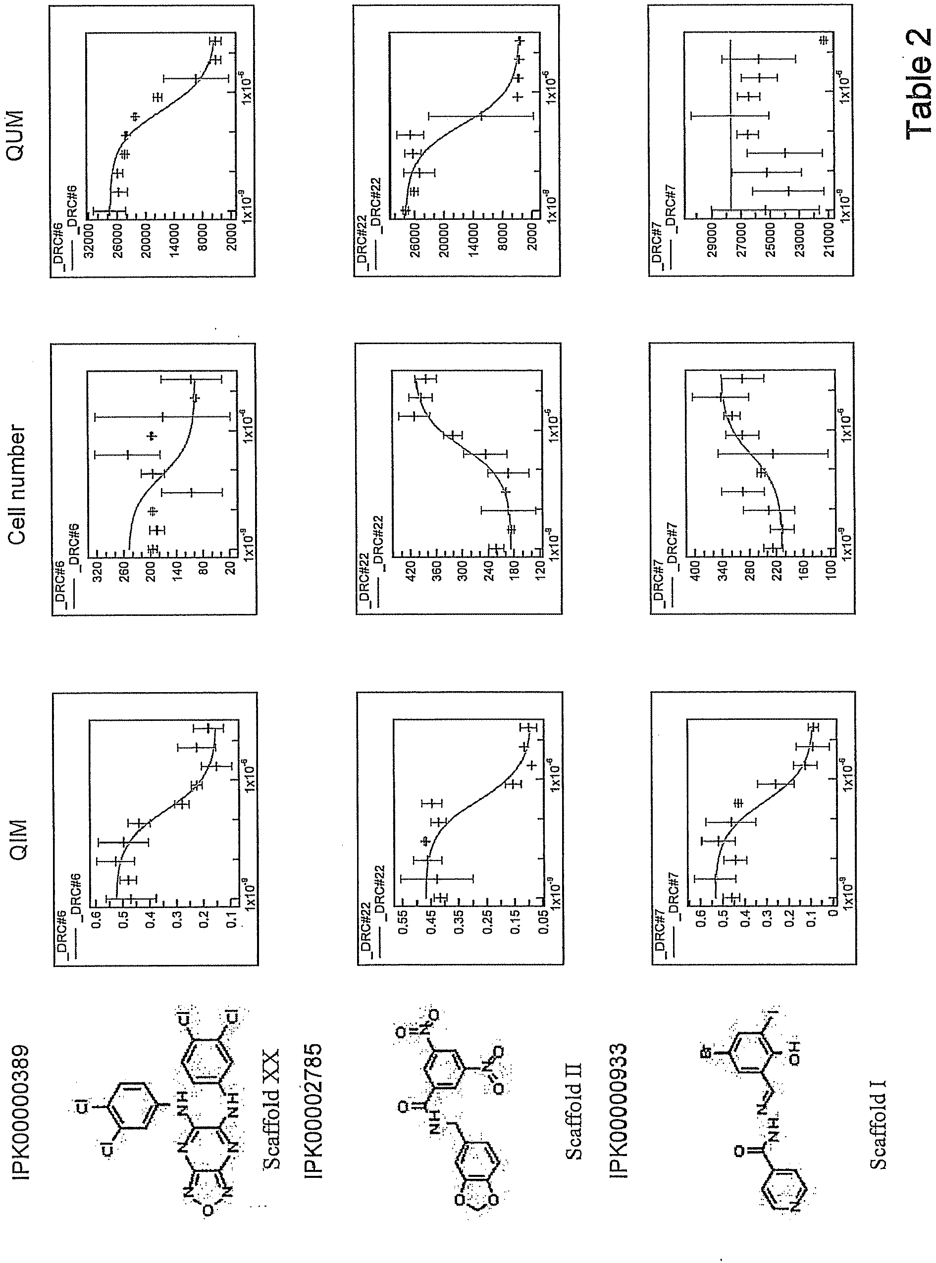
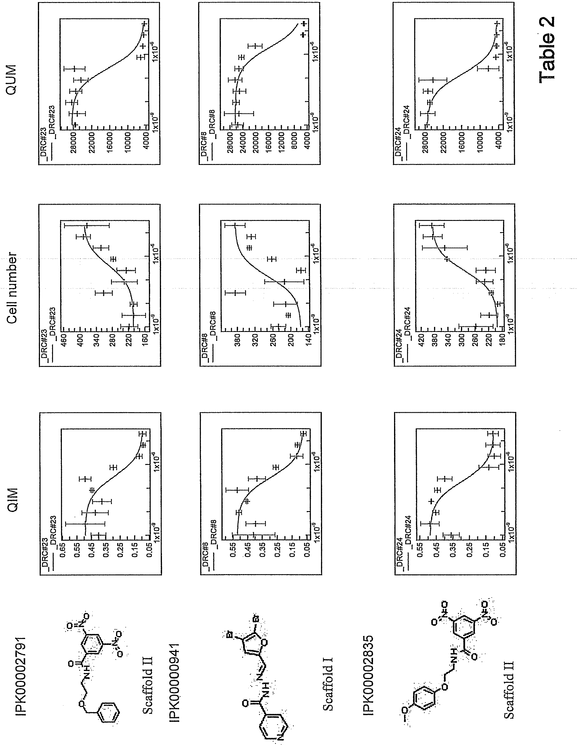


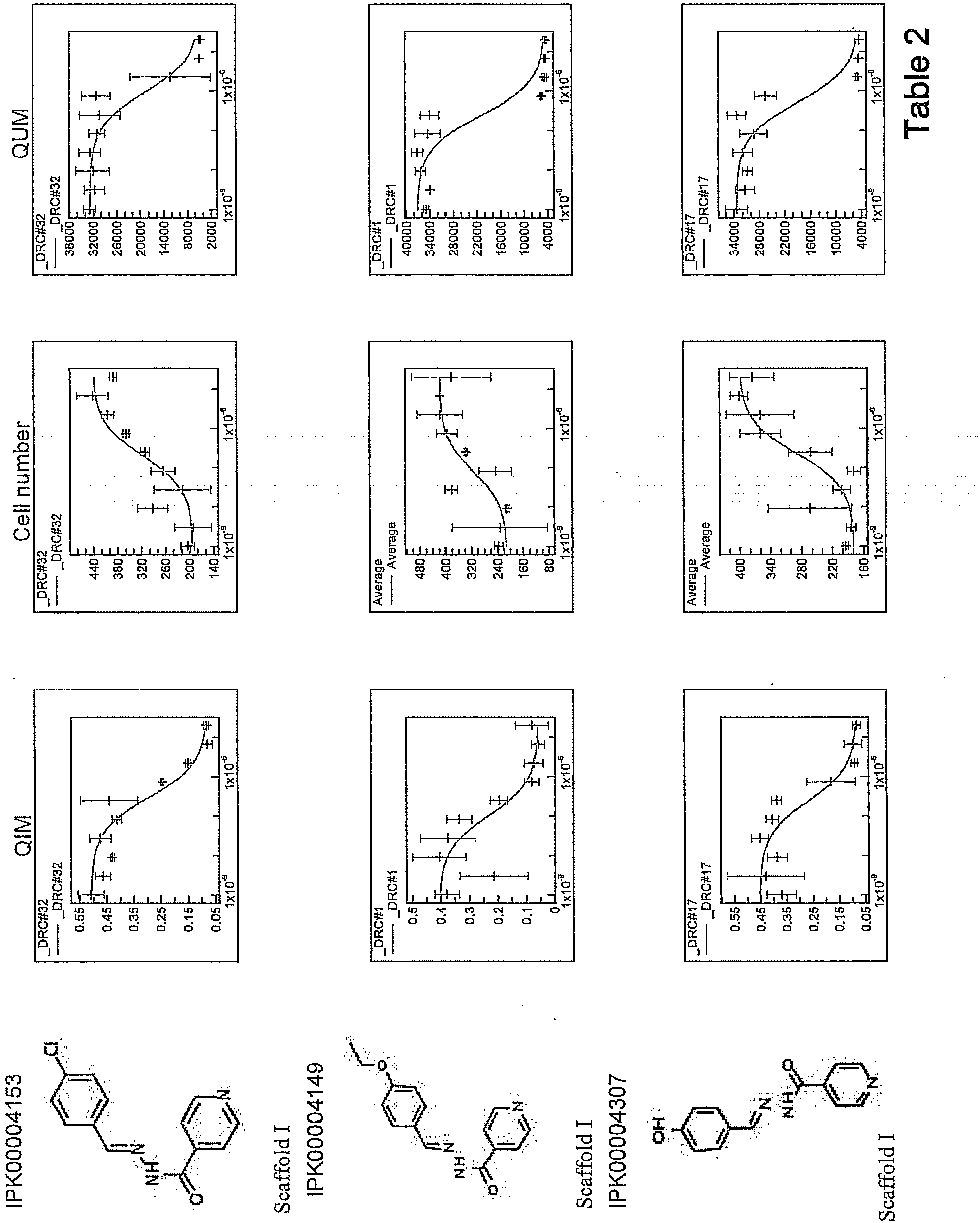


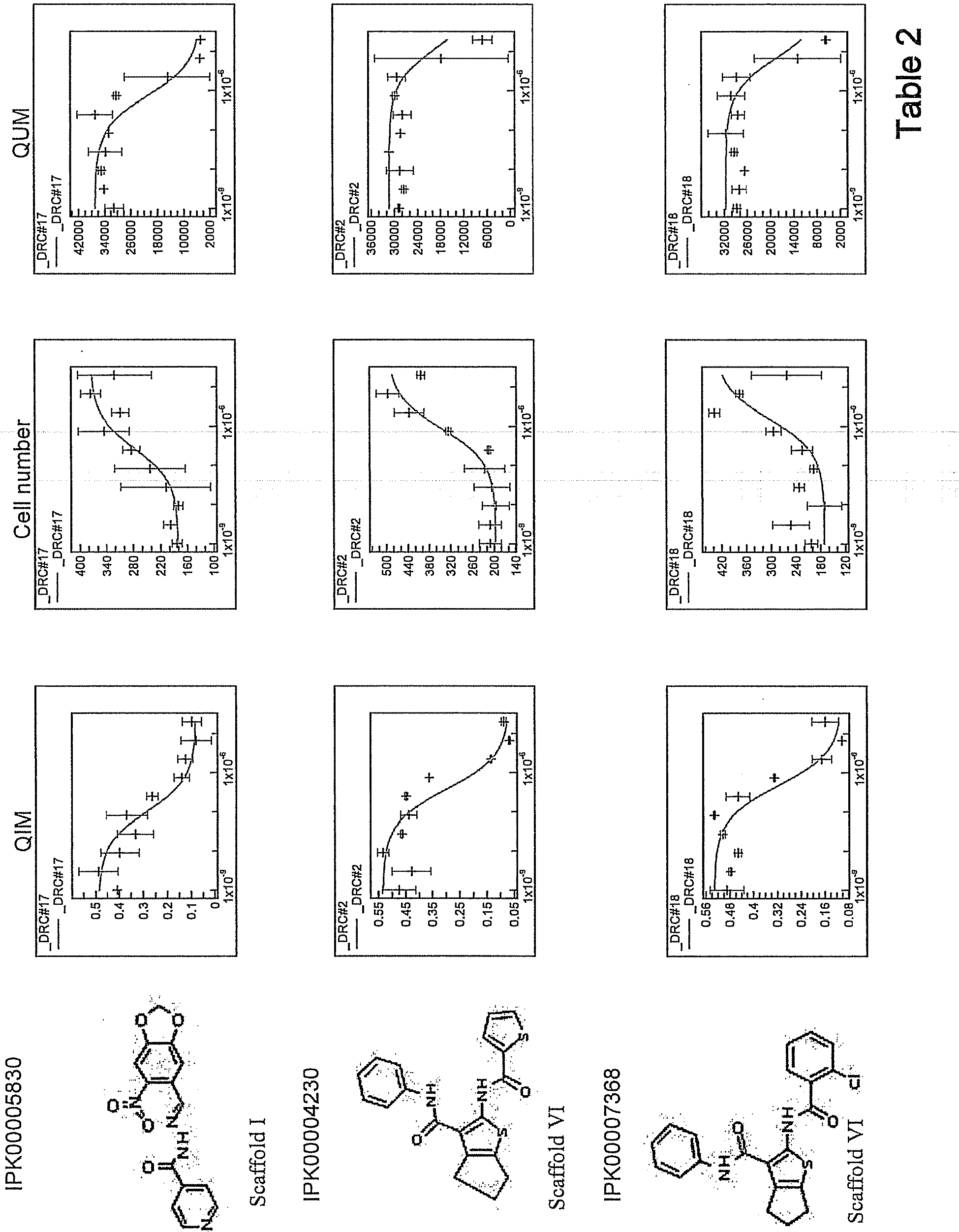


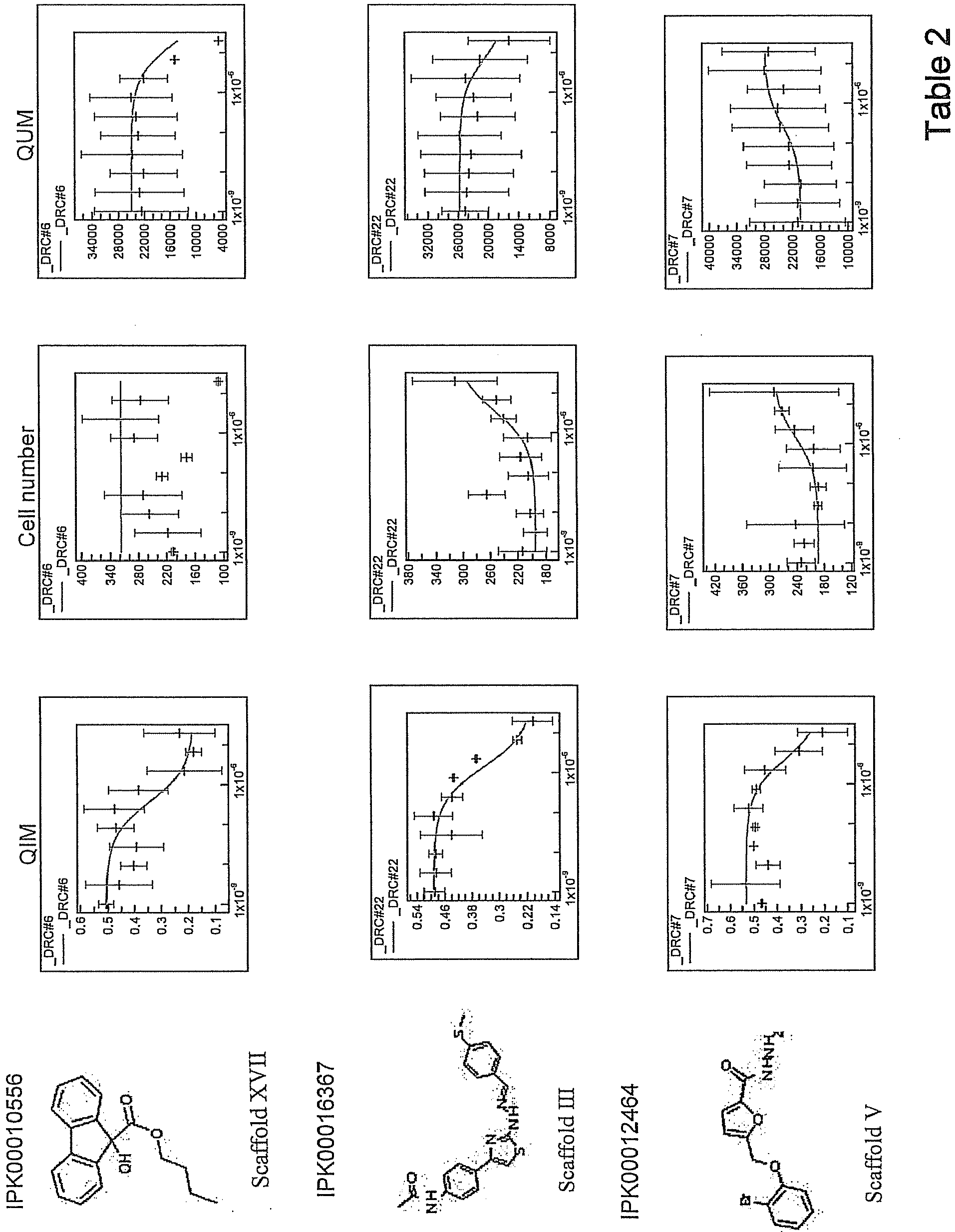


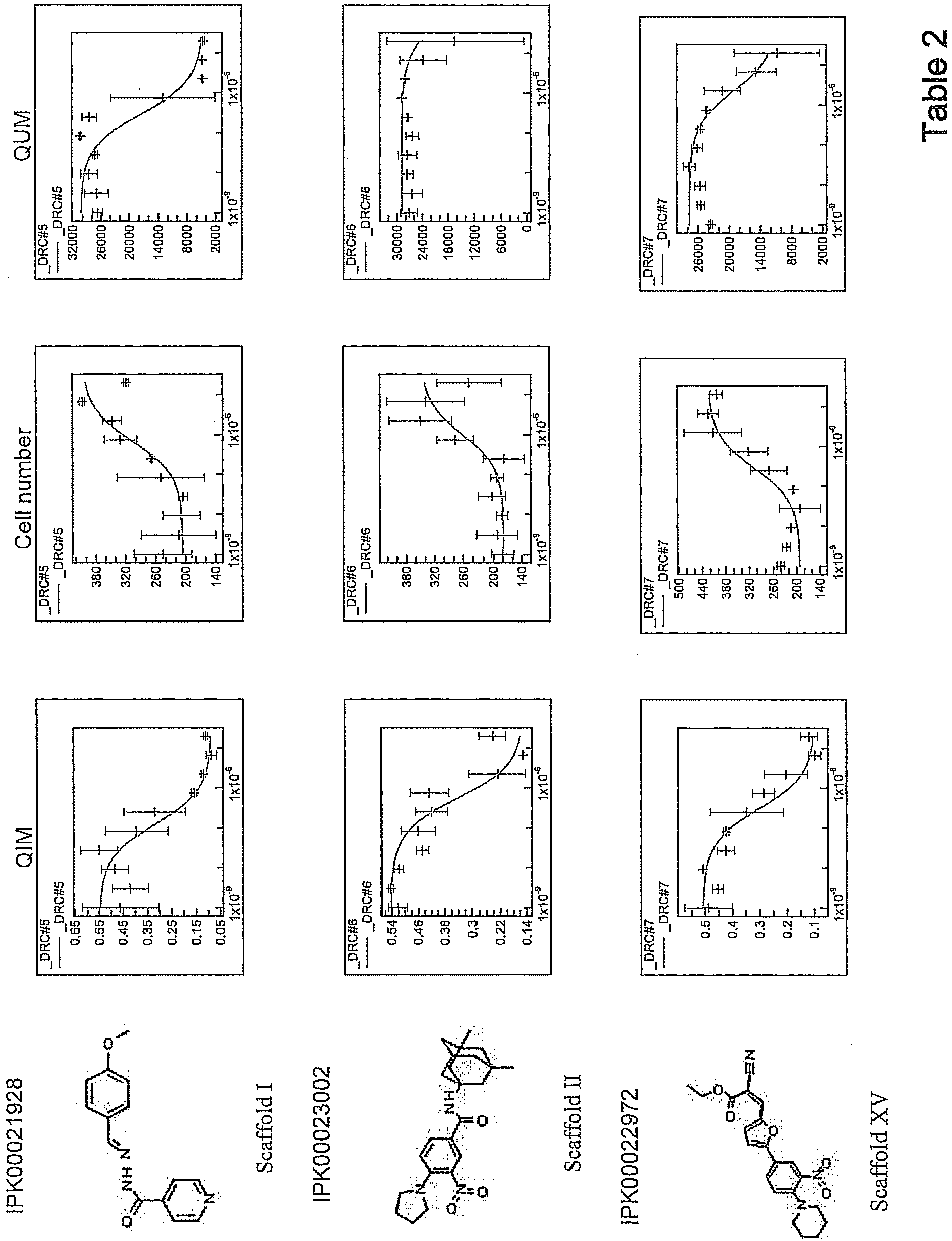


| Scaffold Name | Scaffold Coding | Number of Compounds | Scaffold Structure |
| Isonicotinohydrazides | I | 69 |
 |
| Benzamides | II | 19 |
 |
| Thiazolhydrazides | III | 6 |
 |
| Hydrazinecarbothioamides | IV | 5 |
 |
| Furancarbohydrazides | V | 4 |
 |
| Thiophenes | VI | 3 |
 |
| Pyrazole-pyridines | VII | 2 |
 |
| Pyridopyrimidinone | VIII | 1 |
 |
| One hit compound | IX | 1 |
 |
| One hit compound | X | 1 |
 |
| One hit compound | XI | 1 |
 |
| One hit compound | XII | 1 |
 |
| One hit compound | XIII | 1 |
 |
| One hit compound | XIV | 1 |
 |
| One hit compound | XV | 1 |
 |
| One hit compound | XVI | 1 |
 |
| One hit compound | XVII | 1 |
 |
| One hit compound | XVIII | 1 |
 |
| One hit compound | XIX | 1 |
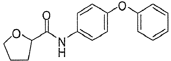 |
| One hit compound | XX | 1 |
 |
| Compound | QIM (µM) | QUM (µM) | Compound | QIM (µM) | QUM (µM) |
 |
+++ | +++ |
 |
++ | +++ |
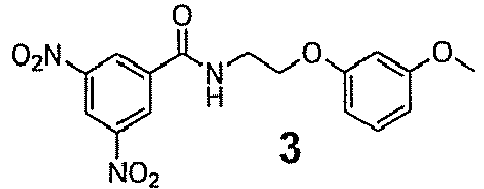 |
++ | +++ |
 |
++ | +++ |
 |
++ | +++ |
 |
++ | +++ |
 |
++ | +++ |
 |
+++ | +++ |
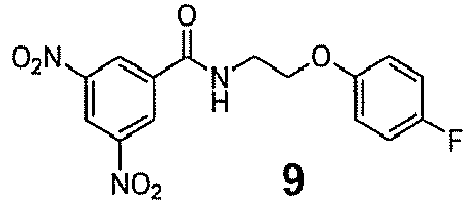 |
++ | +++ |
 |
++ | +++ |
 |
+++ | +++ |
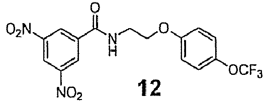 |
+++ | +++ |
 |
++ | +++ |
 |
++ | ++ |
 |
++ | +++ |
 |
+ | + |
 |
+ | + |
 |
+ | + |
 |
+ | ++ |
 |
+ | + |
 |
++ | +++ |
 |
++ | +++ |
 |
+++ | +++ |
 |
+++ | +++ |
 |
++ | +++ |
 |
+++ | +++ |
 |
+++ | +++ |
 |
+++ | +++ |
 |
+++ | +++ |
 |
+++ | +++ |
 |
+++ | +++ |
 |
++ | +++ |
 |
+ | + |
 |
++ | +++ |
 |
+ | ++ |
 |
+ | +++ |
 |
++ | +++ |
 |
+ | +++ |
 |
+ | +++ |
 |
+ | ++ |
 |
++ | +++ |
 |
+ | + |
 |
+ | + |
 |
++ | +++ |
 |
+ | +++ |
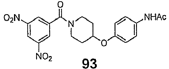 |
+ | ++ |
 |
++ | +++ |
 |
+ | ++ |
 |
++ | ++ |
 |
+ | +++ |
 |
+ | ++ |
 |
+ | ++ |
 |
+ | +++ |
 |
+ | ++ |
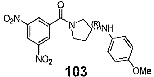 |
++ | +++ |
 |
++ | +++ |
 |
+++ | ++ |
 |
++ | +++ |
 |
++ | +++ |
 |
+ | + |
 |
+ | ++ |
 |
+ | ++ |
 |
+ | ++ |
 |
+ | ++ |
 |
+ | ++ |
 |
+ | +++ |
 |
+ | +++ |
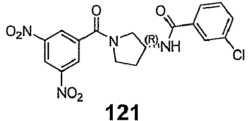 |
+ | ++ |
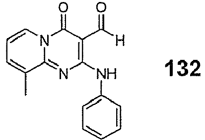 |
+ | ++ |
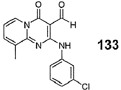 |
++ | +++ |
 |
+ | ++ |
 |
+ | ++ |
 |
+ | + |
 |
+ | + |
 |
+ | + |
 |
+ | + |
 |
++ | + |
 |
+ | + |
 |
+ | + |
 |
+ | + |
 |
+ | + |
 |
+ | + |
 |
+ | + |
 |
+ | ++ |
 |
+ | + |
 |
+ | + |
 |
+ | ++ |
 |
+ | + |
 |
+ | + |
 |
+ | + |
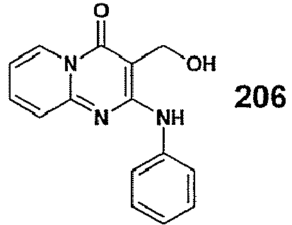 |
+++ | +++ |
 |
+++ | +++ |
 |
+++ | +++ |
 |
+++ | +++ |
 |
+++ | +++ |
 |
+ | +++ |
 |
+ | + |
 |
+ | +++ |
 |
+ | ++ |
 |
+ | + |
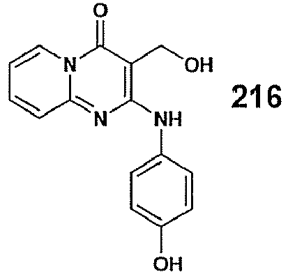 |
+ | + |
 |
+ | +++ |
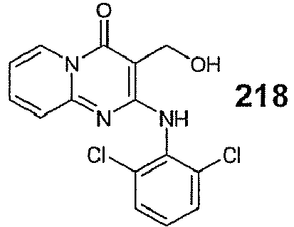 |
+ | + |
 |
+ | + |
 |
++ | +++ |
 |
+ | + |
 |
+ | + |
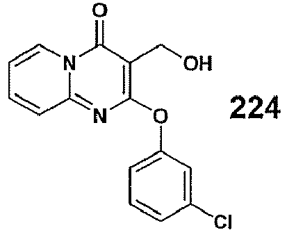 |
+ | + |
 |
+ | + |
 |
+ | + |
 |
+++ | +++ |
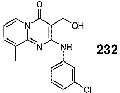 |
+++ | ++ |
 |
+ | + |
 |
+ | + |
 |
+++ | +++ |
 |
++ | +++ |
 |
+ | +++ |
 |
+ | + |
 |
+ | + |
 |
++ | ++ |
 |
+ | + |
 |
+ | + |
 |
+ | + |
 |
+ | ++ |
 |
+ | + |
 |
+ | +++ |
 |
+ | +++ |
 |
+ | + |
 |
+ | + |
 |
+ | + |
 |
+ | + |
 |
+ | + |
 |
+ | + |
 |
+ | + |
 |
+ | + |
 |
+++ | +++ |
 |
+++ | +++ |
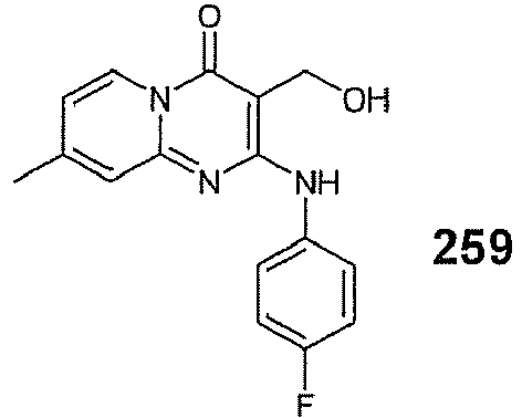 |
++ | +++ |
 |
+ | ++ |
 |
+++ | +++ |
 |
+ | + |
 |
+ | + |
 |
++ | +++ |
 |
+++ | +++ |
 |
+ | +++ |
 |
+++ | +++ |
 |
+ | ++ |
 |
++ | ++ |
 |
+++ | +++ |
 |
+ | +++ |
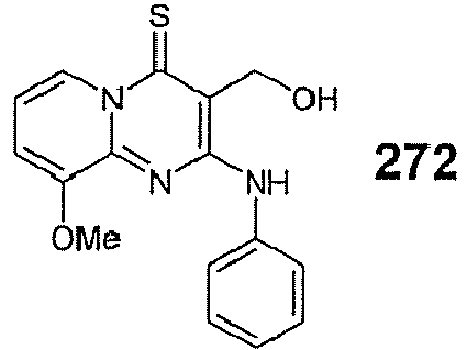 |
+ | + |
 |
+++ | +++ |
 |
+ | +++ |
 |
+ | +++ |
 |
+ | ++ |
 |
+ | ++ |
 |
+++ | +++ |
 |
+ | +++ |
 |
+ | +++ |
 |
+ | + |
 |
+ | + |
 |
+ | +++ |
 |
+ | +++ |
 |
+ | +++ |
 |
+ | + |
 |
+ | + |
 |
+ | + |
 |
+ | + |
 |
+ | + |
 |
++ | +++ |
 |
+ | + |
 |
+ | + |
 |
+ | + |
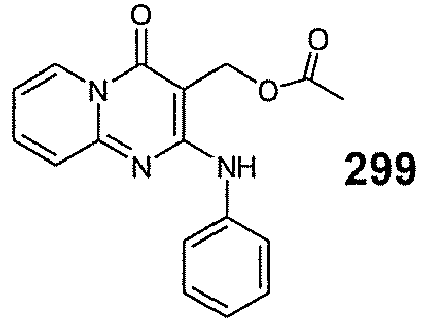 |
++ | +++ |
 |
++ | +++ |
 |
++ | +++ |
| Activity range: +++ indicates <5 uM, ++ indicates between 5-20 uM, + indicates > 20 uM |
| Cytotoxicity | |||||
| Compounds | 4 | 24 | |||
| Host Cells | Range of MTC50 (µM) | ||||
| SK-N-SH-Brain | >100 | >100 | |||
| HepG2-Hepatocytes | >100 | >100 | |||
| MRC5-Lung | >100 | >100 | |||
| BJ- Skin | >100 | >100 | |||
| HEK293-Kidney | >100 | >100 | |||
| Antibacterial activity & Specificity | |||||
| Mycobacterium | |||||
| Strains/Isolates | Type | Origin | Number | Range of MICs for multiple strains (µM) | |
| M. tuberculosis clinical isolates1 | Drug Sensitive | Tissue | 1 | 0.38 | 0.31 |
| RIFR | Sputum | 2 | 0.05 | 0.08 | |
| Tissue | 2 | 0.02-0.05 | 0.08 | ||
| INHR RIFR StrepR | Sputum | 1 | 0.1 | 0.08 | |
| Tissue | 3 | 0.05-0.1 | 0.04-0.08 | ||
| XDR | Sputum | 5 | 0.02-0.05 | 0.04-0.08 | |
| Tissue | 0 | 0.05-0.1 | 0.08 | ||
| MDR | Sputum | 3 | 0.05-0.1 | 0.04-0.08 | |
| Tissue | 5 | 0.05-0.1 | 0.04-0.08 | ||
| M tuberculosis laboratory strains | H37Rv | 0.6 | 0.6 | ||
| H37Ra | 1.2 | 1.3 | |||
| Beijing 1237 | 0.3 | 0.1 | |||
| M bovis BCG | BCG Tokyo | 1.2 | 0.6 | ||
| BCG Pasteur | 1.2 | 1.2 | |||
| M smegmatis | mc2 155 | 1.2 | 0.6 | ||
| Gram-negative | |||||
| Acinetobacter baumannii, Escherichia coli, Enterobacter cloacae, E. aerogenes, Klebsiella oxytoca, Pseudomonas aeruginosa, Salmonella enteridis, Vibrio mimicus | >250 | >250 | |||
| Gram-positive | |||||
| Staphylococcus aureus, S. epidermis, S. capitis, S. xylosus, Micrococcus luteus, Listeria innocua, Lactobacillus gallinarum, group G Streptococcus, Streptococcus agalactiae, S. pyogenes, Enterococcus faecalis, E. faecium, E. gallinarum, Bacillus pumilus | >250 | >250 | |||
| Corynebacterium | |||||
| C. striatum | 27 | 27 | |||
| C. jeikeium | 2.7 | 2.7 |
| Fungi | |||||
| Candida albicans, C. glabrata, C. parapsilosis | >250 | >250 | |||
| INH: Isoniazid, RIF: Rifampin, Strep: Streptomycin, R: resistant. 1The clinical isolates were isolated either from resected lung tissue or sputum specimen, which were collected from active tuberculosis in-patients from the National Masan Tuberculosis Hospital during October 2003 to March 2007. |
| Cytotoxicity | ||||
| Compounds | 133 | |||
| Host Cells | Range of MTC50 (µM) | |||
| SK-N-SH-Brain | >100 | |||
| HepG2-Hepatocytes | >100 | |||
| MRC5-Lung | >100 | |||
| BJ- Skin | >100 | |||
| HEK293-Kidney | >100 | |||
| Jurkat - T-cell | >100 | |||
| THP-1 - Monocytes | >100 | |||
| Primary BMDM | >100 | |||
| Primary human macrophages | >100 | |||
| Antibacterial activity & Specificity | ||||
| Mycobacterium | ||||
| Strains/Isolates | Type | Origin | Number | Range of MICs for multiple strains (µM) |
| M. tuberculosis clinical isolates1 | Drug Sensitive | Sputum | 2 | 5->20 |
| Tissue | 2 | 2.5-5 | ||
| RIFR | Sputum | 1 | 2.5 | |
| Tissue | 1 | 1.2 | ||
| INHR RIFR StrepR | Sputum | 3 | 0.3-1.2 | |
| Tissue | 1 | 1.2 | ||
| XDR | Sputum | 4 | 0.6-2.5 | |
| Tissue | 5 | 0.3-5 | ||
| MDR | Sputum | 3 | 0.3-1.2 | |
| Tissue | 1 | 1.2 | ||
| M tuberculosis laboratory strains | H37Rv | 2 | ||
| H37Ra | 2 | |||
| BCG Pasteur-Tokyo | 2 | |||
| M smegmatis | mc2 155 | >100 | ||
| Gram-negative | ||||
| Acinetobacter baumannii, Escherichia coli, Enterobacter cloacae, E. aerogenes, Klebsiella oxytoca, Pseudomonas aeruginosa, Salmonella enteridis, Vibrio mimicus | NE | |||
| Gram-positive | ||||
| Staphylococcus aureus, S. epidermis, S. capitis, S. xylosus, Micrococcus luteus, Listeria innocua, Lactobacillus gallinarum, group G Streptococcus, Streptococcus agalactiae, S. pyogenes, Enterococcus faecalis, E. faecium, E. gallinarum, Bacillus pumilus | NE | |||
| Corynebacterium | ||||
| C. striatum | NE | |||
| C. jeikeium | NE |
| Fungi | ||||
| Candida albicans, C. glabrata, C. parapsilosis | NE | |||
| INH: Isoniazid, RIF: Rifampin, Strep: Streptomycin, R: resistant. 1The clinical isolates were isolated either from resected lung tissue or sputum specimen, which were collected from active tuberculosis in-patients from the National Masan Tuberculosis Hospital during October 2003 to March 2007. NE: No effect up to 100 µg/mL equivalent to 320 µM. The antimicrobial spectrum was performed on clinical isolates from CHU d'Angers, France. |
| Compound | Concentration (µg/ml) | Bacteria inoculum (CFU) |
Frequency of |
|||
| 105 | 106 | 107 | 108 | |||
| 4 | 0.2 | - | - | 12 | >100 | 1×10⁻⁶ |
| 0.8 | - | - | - | <100 | ||
| 1.6 | - | - | - | <100 | ||
| 3.2 | - | - | - | 1 | 1×10⁻⁸ | |
| 24 | 0.2 | - | - | 7 | >100 | 7×10⁻⁷ |
| 0.8 | - | - | - | <100 | ||
| 1.6 | - | - | - | >100 | ||
| 3.2 | - | - | - | 1 | 1×10⁻⁸ | |
| INH- |
10 | ND | ND | 33 | |
3×10⁻⁶ |
| Compound | Concentration (µg/ml) | Bacteria inoculum (CFU) |
Frequency of |
|||
| 106 | 107 | 108 | ||||
| 264 | 0.4 | - | 37 | 306 | 3.4×10⁻⁶ | |
| 0.8 | - | 5 | 117 | 8×10⁻⁶ | ||
| 1.6 | - | - | 22 | 2×10⁻⁸ | ||
| 3.2 | - | - | 2 | 2×10⁻⁸ | ||
| INH-control | 10 | - | 4 | 18 | 2.9×10⁻⁶ | |
| ND: not done; -: no colonies -: no colonies |
Claims (13)
- A screening method comprising the steps of(a) batch infection of host cells with fluorescently labeled M tuberculosis mycobacteria;(b) removing free unbound mycobacteria;(c) adding compounds that are to be tested to a multi-well plate;(d) seeding said host cells infected with fluorescently labeled M tuberculosis mycobacteria into said multi-well plate containing said compounds;(e) incubating said multi-well plate containing host cells infected with fluorescently labeled M tuberculosis mycobacteria and said compounds;(f) fluorescently labeling said host cells;(g) analyzing said multi-well plate using automated confocal microscopy.
- The method of claim 1, wherein the screening method searches for compounds that interfere with the multiplication of M. tuberculosis within said host cells.
- The method of claim 1 or 2, wherein the host cells are macrophages, preferably living macrophages.
- The method of any one of claims 1 to 3, wherein the automated confocal fluorescence microscopy measures intracellular mycobacterial growth.
- The method of any one of claims 1 to 4, wherein step (g) comprises determining for each compound- the total host cell number, e.g. total macrophage number;- the percentage of infected host cells; and/or- the percent inhibition of infection.
- The method of claim 5, wherein(i) a low (total) cell number is indicative for the compound toxicity and/or of the unrestricted growth of M. tuberculosis inside macrophages;
and/or(ii) a high (total) cell number is indicative that the compound is not toxic and prevents mycobacterial growth. - The method of any of the preceding claims, comprising the use of control(s) selected from at least one of the following- adding a compound with known anti-tuberculosis activity instead of the compounds in step (c) (as positive control), such as isoniazid, rifampin, ethionamide or moxifloxacin;- adding DMSO or antibiotic control(s) instead of the compounds in step (c) (as negative control).
- The method of any of the preceding claims, wherein the M tuberculosis mycobacteria are labeled with GFP.
- The method of any of the preceding claims, wherein the host cells are labeled with SYTO 60.
- The method of any of the preceding claims, wherein after step (b) an incubation with antibiotic, preferably amykacin, is carried out.
- The method of any one of the preceding claims, used for the high throughput screening (HTS) of large scale chemical libraries.
- A compound that interferes with the multiplication of M tuberculosis within host cells, preferably within host macrophages, identified in the method of any one of claims 1 to 11.
- The compound of claim 12, which is represented by formula

Applications Claiming Priority (2)
| Application Number | Priority Date | Filing Date | Title |
|---|---|---|---|
| US13228508P | 2008-06-17 | 2008-06-17 | |
| EP09776764.4A EP2310388B1 (en) | 2008-06-17 | 2009-06-17 | Pyridopyrimidine compounds as anti-tubercular agents |
Related Parent Applications (2)
| Application Number | Title | Priority Date | Filing Date |
|---|---|---|---|
| EP09776764.4A Division EP2310388B1 (en) | 2008-06-17 | 2009-06-17 | Pyridopyrimidine compounds as anti-tubercular agents |
| EP09776764.4A Division-Into EP2310388B1 (en) | 2008-06-17 | 2009-06-17 | Pyridopyrimidine compounds as anti-tubercular agents |
Publications (2)
| Publication Number | Publication Date |
|---|---|
| EP2730576A2 true EP2730576A2 (en) | 2014-05-14 |
| EP2730576A3 EP2730576A3 (en) | 2014-09-03 |
Family
ID=41037902
Family Applications (2)
| Application Number | Title | Priority Date | Filing Date |
|---|---|---|---|
| EP09776764.4A Not-in-force EP2310388B1 (en) | 2008-06-17 | 2009-06-17 | Pyridopyrimidine compounds as anti-tubercular agents |
| EP13198407.2A Withdrawn EP2730576A3 (en) | 2008-06-17 | 2009-06-17 | Anti-infective compounds |
Family Applications Before (1)
| Application Number | Title | Priority Date | Filing Date |
|---|---|---|---|
| EP09776764.4A Not-in-force EP2310388B1 (en) | 2008-06-17 | 2009-06-17 | Pyridopyrimidine compounds as anti-tubercular agents |
Country Status (9)
| Country | Link |
|---|---|
| US (2) | US8785452B2 (en) |
| EP (2) | EP2310388B1 (en) |
| JP (2) | JP5739329B2 (en) |
| KR (1) | KR101574332B1 (en) |
| CN (2) | CN103983627A (en) |
| AU (1) | AU2009267519B2 (en) |
| BR (1) | BRPI0914254A2 (en) |
| CA (1) | CA2727651C (en) |
| WO (1) | WO2010003533A2 (en) |
Families Citing this family (29)
| Publication number | Priority date | Publication date | Assignee | Title |
|---|---|---|---|---|
| KR20130088002A (en) * | 2010-01-13 | 2013-08-07 | 재단법인 한국파스퇴르연구소 | Anti-infective pyrido(1,2-a)pyrimidines |
| HUP1000034A2 (en) * | 2010-01-21 | 2011-09-28 | Uratim Kft | Pyrido[1,2-a]pyrimidine derivatives and pharmaceutical preparations thereof having anti - mycobacterial activity |
| RU2576662C2 (en) | 2010-03-18 | 2016-03-10 | Энститю Пастер Корея | Antiinfective compounds |
| MX2012012421A (en) | 2010-04-28 | 2012-12-17 | Astellas Pharma Inc | Tetrahydrobenzothiophene compound. |
| WO2012006104A2 (en) | 2010-06-28 | 2012-01-12 | Academia Sinica, Taiwan | Compounds and methods for treating tuberculosis infection |
| WO2012057599A1 (en) * | 2010-10-25 | 2012-05-03 | Universiti Sains Malaysia | Anti-mycobacterial agents |
| WO2012103336A1 (en) * | 2011-01-26 | 2012-08-02 | University Of Rochester | Small molecule rnase inhibitors and methods of use |
| US8877940B2 (en) | 2011-02-22 | 2014-11-04 | Institut National De La Recherche Scientifique | Antibiotic tolerance inhibitors |
| US20130158112A1 (en) * | 2011-06-20 | 2013-06-20 | Jonathan D. SMITH | Compositions and methods for inhibiting beta amyloid secretion |
| CN102871992A (en) * | 2011-09-30 | 2013-01-16 | 中国医学科学院医药生物技术研究所 | Application of 3,5-dinitrobenzene formamide in preparation of anti-tuberculosis medicaments |
| CA2864331C (en) * | 2012-02-14 | 2018-10-09 | Children's Hospital Medical Center | Use of small molecule inhibitors targeting the interaction between rac gtpase and p67(phox) |
| KR20130118612A (en) * | 2012-04-20 | 2013-10-30 | (주)네오믹스 | Novel aminopyridine derivatives and use thereof |
| US9492448B2 (en) | 2012-06-20 | 2016-11-15 | University Of Virginia Patent Foundation | Compositions and methods for regulating glucose homeostasis and insulin action |
| KR102529578B1 (en) * | 2014-08-29 | 2023-05-09 | (주)아모레퍼시픽 | Novel adamatan derivative compound |
| EA031505B1 (en) * | 2014-12-02 | 2019-01-31 | Прана Байотекнолоджи Лимитед | 4H-PYRIDO[1,2-a]PYRIMIDIN-4-ONE COMPOUNDS |
| CZ306245B6 (en) * | 2014-12-11 | 2016-10-26 | Univerzita Karlova v Praze Farmaceutická fakulta v Hradci Králové | Substituted phenyltetrazole, its use and pharmaceutical composition containing thereof |
| CN105712931A (en) * | 2016-03-16 | 2016-06-29 | 山东师范大学 | 7-phenyl-5-oxo-4-substituted-1,4,5,6,7,8-hexahydroquinoline-3-carboxylic acid ethyl ester and non-catalyzed synthesis method thereof |
| EP3463338A4 (en) * | 2016-05-27 | 2020-01-29 | Arkansas State University-Jonesboro | ANTIMICROBIAL AGENTS AND METHOD FOR SYNTHESISING THE ANTIMICROBIAL ACTIVE SUBSTANCES |
| US11110080B2 (en) | 2017-03-27 | 2021-09-07 | The Regents Of The University Of Colorado, A Body Corporate | Small molecule inhibitors of bacterial efflux pumps and methods of using same |
| CN111511723A (en) * | 2017-08-21 | 2020-08-07 | 米可如比奥提克斯有限公司 | Metabolically stable N-acylaminooxadiazoles for use as antibacterial agents |
| US10357485B1 (en) | 2018-09-27 | 2019-07-23 | King Saud University | Anti-cancer compound |
| GB2589912A (en) * | 2019-12-12 | 2021-06-16 | Chemestmed Ltd | Method of suppressing cancer by RNA m6A methyltransferase mettl16 inhibitors |
| CN112336719A (en) * | 2020-10-19 | 2021-02-09 | 济南大学 | Thiazole derivative as alpha-glucosidase inhibitor and application thereof |
| CN113214249B (en) * | 2021-04-23 | 2023-09-19 | 成都大学 | Synthesis method of pyrido [1,2-a ] pyrimidine-4-thioketone compound |
| EP4467539A4 (en) | 2022-01-21 | 2025-11-05 | Pasteur Institut Korea | OXAZOLDERIVATE COMPOUND WITH ANTIBACTERIAL EFFECT AND MEDICINAL USE OF IT |
| AR130352A1 (en) * | 2022-08-31 | 2024-11-27 | Texas Crop Science | APIRASE INHIBITORS |
| EP4345092A1 (en) * | 2022-09-29 | 2024-04-03 | Faculdade de Farmácia da Universidade de Lisboa | Nitrobenzamide compounds, methods and uses thereof |
| EP4345091A1 (en) * | 2022-09-29 | 2024-04-03 | Faculdade de Farmácia da Universidade de Lisboa | Benzoic acid derivatives, methods and uses thereof |
| WO2025122558A1 (en) * | 2023-12-05 | 2025-06-12 | University Of Maryland, Baltimore | Pyrazinamide-mimicking small molecules as treatment for tuberculosis |
Family Cites Families (18)
| Publication number | Priority date | Publication date | Assignee | Title |
|---|---|---|---|---|
| HU178910B (en) | 1977-08-19 | 1982-07-28 | Chinoin Gyogyszer Es Vegyeszet | Process for preparing 2,3-disubstituted-4-oxo-4h-pyrido/1,2-a/-pyrimidines |
| AU588917B2 (en) * | 1985-09-25 | 1989-09-28 | Shionogi & Co., Ltd. | 9-(substituted thio)-4h-pryido(1,2-a) pyrimidin-4-one derivatives |
| JPH0655740B2 (en) | 1985-09-25 | 1994-07-27 | 塩野義製薬株式会社 | Pyridopyrimidine derivative |
| HU201551B (en) | 1988-02-03 | 1990-11-28 | Chinoin Gyogyszer Es Vegyeszet | Process for producing 4-oxo-4h-pyrido(1,2-a)pyrimidine-3-carboxylic acid amide derivatives and pharmaceutical compositions comprising same |
| EP0640083A1 (en) * | 1992-05-13 | 1995-03-01 | E.I. Du Pont De Nemours And Company | Substituted pyrido 1,2-a]pyrimidinone derivatives as fungicides |
| FR2691460B1 (en) * | 1992-05-21 | 1994-07-22 | Rhone Poulenc Rorer Sa | NEW TAXANE DERIVATIVES, THEIR PREPARATION AND THE COMPOSITIONS CONTAINING THEM. |
| AU3223801A (en) * | 2000-02-09 | 2001-08-20 | Daiichi Seiyaku Co | Anti-acid-fast bacterial agents containing pyridonecarboxylic acids as the active ingredient |
| US6645505B2 (en) * | 2001-03-27 | 2003-11-11 | Council Of Scientific And Industrial Research | Reporter gene based method for the screening of anti-tuberculosis drugs by using essential and regulatory genes of mycobacteria as drug target |
| JPWO2002087589A1 (en) * | 2001-04-26 | 2004-08-12 | 第一製薬株式会社 | Drug efflux pump inhibitor |
| AU2003269346A1 (en) * | 2002-10-16 | 2004-05-04 | Council Of Scientific And Industrial Research | Screening method |
| WO2004037159A2 (en) | 2002-10-23 | 2004-05-06 | Obetherapy Biotechnology | Compounds, compositions and methods for modulating fat metabolism |
| CN1580276A (en) * | 2003-07-30 | 2005-02-16 | 中国科学院大连化学物理研究所 | Method for screening high-flux antituberculosis bacillus medicine |
| US20070179163A1 (en) * | 2003-09-26 | 2007-08-02 | Rao Guntu S | Process for the preparation of risperidone |
| WO2005089068A2 (en) * | 2004-03-24 | 2005-09-29 | Rimonyx Pharmaceuticals Ltd. | Pharmaceutical compositions comprising anti-inflammatory quinazolinecarboxamide derivatives |
| CN101008029A (en) * | 2006-01-27 | 2007-08-01 | 中国医学科学院医药生物技术研究所 | Antitubercular drug screening model using tubercle bacillus isocitrate lyase as target |
| EP1916249A1 (en) * | 2006-10-10 | 2008-04-30 | LEK Pharmaceuticals D.D. | 3-(benzo[d][1,3]dioxol-5-ylmethyl)-4-(thio)oxo-2-(thio)oxo-azolidin-5-ylidene derivatives as antibacterial agents |
| CN1945324A (en) * | 2006-10-19 | 2007-04-11 | 复旦大学 | Method for screening Mycobacterium tuberculosis drug-resistant protein |
| JP2011132142A (en) * | 2009-12-22 | 2011-07-07 | Kowa Co | Phenyloxadiazole compound having erythropoietin production-promoting action |
-
2009
- 2009-06-17 CA CA2727651A patent/CA2727651C/en not_active Expired - Fee Related
- 2009-06-17 AU AU2009267519A patent/AU2009267519B2/en not_active Ceased
- 2009-06-17 WO PCT/EP2009/004379 patent/WO2010003533A2/en not_active Ceased
- 2009-06-17 US US12/999,095 patent/US8785452B2/en not_active Expired - Fee Related
- 2009-06-17 CN CN201410219962.6A patent/CN103983627A/en active Pending
- 2009-06-17 CN CN200980128440.2A patent/CN102105470B/en not_active Expired - Fee Related
- 2009-06-17 BR BRPI0914254A patent/BRPI0914254A2/en not_active IP Right Cessation
- 2009-06-17 JP JP2011513947A patent/JP5739329B2/en not_active Expired - Fee Related
- 2009-06-17 EP EP09776764.4A patent/EP2310388B1/en not_active Not-in-force
- 2009-06-17 KR KR1020117000648A patent/KR101574332B1/en not_active Expired - Fee Related
- 2009-06-17 EP EP13198407.2A patent/EP2730576A3/en not_active Withdrawn
-
2014
- 2014-04-28 US US14/263,218 patent/US20150018543A1/en not_active Abandoned
-
2015
- 2015-04-23 JP JP2015088505A patent/JP2015187110A/en active Pending
Non-Patent Citations (15)
| Title |
|---|
| ABADIE, V.; BADELL, E.; DOUILLARD, P.; ENSERGUEIX, D.; LEENEN, P. J.; TANGUY, M.; FIETTE, L.; SAELAND, S.; GICQUEL, B.; WINTER, N.: "Neutrophils rapidly migrate via lymphatics after Mycobacterium bovis BCG intradennal vaccination and shuttle live bacilli to the draining lymph nodes", BLOOD, vol. 106, 2005, pages 1843 - 1850 |
| ANDRIES, K.; VERHASSELT, P.; GUILLEMONT, J.; GOHLMANN, H. W.; NEEFS, J. M.; WINLDER, H.; VAN GESTEL, J.; TIMMERMAN, P.; ZHU, M.; L: "A diarylquinoline drug active on the ATP synthase of Mycobacterium tuberculosis", SCIENCE, vol. 307, 2005, pages 223 - 227, XP002358962, DOI: doi:10.1126/science.1106753 |
| ARAIN, T. M.; RESCONI, A. E.; SINGH, D. C.; STOVER, C. K.: "Reporter gene technology to assess activity of antimycobacterial agents in macrophages", ANTIMICROB AGENTS CHEMOTHER, vol. 40, 1996, pages 1542 - 1544, XP002916136 |
| BRODIN, P.; MAJLESSI, L.; MARSOLLIER, L.; DE JONGE, M.I.; BOTTAI, D.; DEMANGEL, CL.; HINDS, J.; NEYROLLES, O.; BUTCHER, P.D.; LECL: "Dissection of ESAT-6 system 1 of Mycobacterium tuberculosis and impact on immunogenicity and virulence", INFECT IMMUN, vol. 74, 2006, pages 88 - 98, XP002474771, DOI: doi:10.1128/IAI.74.1.88-98.2006 |
| CHO, S. H.; WARIT, S.; WAN, B.; HWANG, C. H.; PAULI, G. F.; FRANZBLAU, S. G.: "Low- oxygen-recovery assay for high-throughput screening of compounds against nonreplicating Mycobacterium tuberculosis", ANTIMICROB AGENTS CHEMOTHER, vol. 51, 2007, pages 1380 - 1385 |
| CREMER, L; DIEU-NOSJEAN, M. C.; MARECHAL, S.; DEZUTTER-DAMBUYANT, C.; GODDARD, S.; ADAMS, D.; WINTER, N.; MENETRIER-CAUX, C.; SAUT: "Long-lived immature dendritic cells mediated by TRANCE-RANK interaction", BLOOD, vol. 100, 2002, pages 3646 - 3655, XP002444107, DOI: doi:10.1182/blood-2002-01-0312 |
| FENISTEIN, D.; LENSEIGNE, B.; CHRISTOPHE, T.; BRODIN, P.; GENOVESIO, A.: "A fast fully automated cell segmentation algorithm for high throughput and high content screening", CYTOMETRY, 2008 |
| HOUBEN, E. N.; NGUYEN, L.; PIETERS, J.: "Interaction of pathogenic mycobacteria with the host immune system", CURR OPIN MICROBIOL, vol. 9, 2006, pages 76 - 85, XP028033898, DOI: doi:10.1016/j.mib.2005.12.014 |
| LENAERTS, A. J.; HOFF, D.; ALY, S.; EHLERS, S.; ANDRIES, K.; CANTARERO, L.; ORME, 1. M.; BASARABA, R. J.: "Location of persisting mycobacteria in a Guinea pig model of tuberculosis revealed by r207910", ANTIMICROB AGENTS CHEMOTHER, vol. 51, 2007, pages 3338 - 3345 |
| LIPINSKI, C. A.; LOMBARDO, F.; DOMINY, B. W.; FEENEY, P. J.: "Experimental and computational approaches to estimate solubility and permeability in drug discovery and development settings", ADVANCED DRUG DELIVERY REVIEWS, vol. 46, 2001, pages 3 - 26, XP001097746, DOI: doi:10.1016/S0169-409X(00)00129-0 |
| NEYROLLES, O.; HERNANDEZ-PANDO, R.; PIETRI-ROUXEL, F.; FORNES, P.; TAILLEUX, L.; BARRIOS PAYAN, J. A.; PIVERT, E.; BORDAT, Y.; AGU: "Is adipose tissue a place for Mycobacterium tuberculosis persistence?", PLOS ONE, vol. 1, 2006, pages E43 |
| ROHDE, K. H.; ABRAMOVITCH, R. B.; RUSSELL, D. G.: "Mycobacterium tuberculosis invasion of macrophages: linking bacterial gene expression to environmental cues", CELL HOST MICROBE, vol. 2, 2007, pages 352 - 364 |
| SALOMON, J. A.; LLOYD-SMITH, J. O.; GETZ, W. M.; RESCH, S.; SANCHEZ, M. S.; PORCO, T. C.; BORGDORFF, M. W.: "Prospects for advancing tuberculosis control efforts through novel therapies", PLOS MED, vol. 3, 2006, pages E273 |
| SCHNAPPINGER, D.; EHRT, S.; VOSKUIL, M. I.; LIU, Y.; MANGAN, J. A.; MONAHAN, 1. M.; DOLGANOV, G.; EFRON, B.; BUTCHER, P. D.; NATHA: "Transcriptional Adaptation of Mycobacterium tuberculosis within Macrophages: Insights into the Phagosomal Environment", J EXP MED, vol. 198, 2003, pages 693 - 704 |
| VAN RIE, A.; ENARSON, D.: "XDR tuberculosis: an indicator of public-health negligence", LANCET, vol. 368, 2006, pages 1554 - 1556, XP025093602, DOI: doi:10.1016/S0140-6736(06)69575-5 |
Also Published As
| Publication number | Publication date |
|---|---|
| US20110178077A1 (en) | 2011-07-21 |
| WO2010003533A2 (en) | 2010-01-14 |
| CA2727651C (en) | 2016-04-05 |
| JP2015187110A (en) | 2015-10-29 |
| JP2011524391A (en) | 2011-09-01 |
| JP5739329B2 (en) | 2015-06-24 |
| US20150018543A1 (en) | 2015-01-15 |
| AU2009267519A1 (en) | 2010-01-14 |
| EP2730576A3 (en) | 2014-09-03 |
| WO2010003533A3 (en) | 2010-11-25 |
| CN102105470A (en) | 2011-06-22 |
| AU2009267519B2 (en) | 2014-11-27 |
| KR101574332B1 (en) | 2015-12-08 |
| EP2310388A2 (en) | 2011-04-20 |
| CN103983627A (en) | 2014-08-13 |
| HK1159113A1 (en) | 2012-07-27 |
| CN102105470B (en) | 2014-06-04 |
| EP2310388B1 (en) | 2015-08-05 |
| CA2727651A1 (en) | 2010-01-14 |
| US8785452B2 (en) | 2014-07-22 |
| BRPI0914254A2 (en) | 2015-11-03 |
| KR20110029148A (en) | 2011-03-22 |
Similar Documents
| Publication | Publication Date | Title |
|---|---|---|
| US8785452B2 (en) | Anti-infective compounds | |
| US20130012506A1 (en) | Anti-infective pyrido (1,2-a) pyrimidines | |
| EP2528897B9 (en) | Di-substituted pyridine derivatives as anticancers | |
| Chen et al. | Synthesis and biological evaluation of piperidine-substituted triazine derivatives as HIV-1 non-nucleoside reverse transcriptase inhibitors | |
| Marvadi et al. | Synthesis and evaluation of novel substituted 1, 2, 3-triazolyldihydroquinolines as promising antitubercular agents | |
| EP3174852B1 (en) | Arylhydrazides containing a 2-pyridone moiety as selective antibacterial agents | |
| UA129244C2 (en) | METHOD OF USING TRI-SUBSTITUTED BENZOTRIAZOLE DERIVATIVES AS DIHYDROOROTATOXIGENASE INHIBITORS | |
| HK1197410A (en) | Anti-infective compounds | |
| Sbardella et al. | New 6-nitroquinolones: synthesis and antimicrobial activities | |
| Desai et al. | Synthesis of a novel series of imines containing nitrogen heterocycles as promising antibacterial and antifungal agents | |
| WO2021161084A1 (en) | N-(4-fluorophenyl)-5-phenyl-[1,2,4] triazolo [1,5-a] pyridine-2-carboxamide derivatives and their synthesis thereof | |
| Pflégr et al. | Enhancing the antimycobacterial efficacy of pyridine-4-carbohydrazide: linkage to additional antimicrobial agents via oxocarboxylic acids | |
| HK1159113B (en) | Pyridopyrimidine compounds as anti-tubercular agents | |
| CN107250111B (en) | Alpha-ketoesteryl isoniazid compounds, methods for preparing these compounds, and use of compounds for the treatment of tuberculosis | |
| US9663478B2 (en) | Benzotriazine oxides as drugs targeting Mycobacterium tuberculosis | |
| US10285978B2 (en) | Heterocycle analogs of CAI-1 as agonists of quorum sensing in vibrio | |
| WO2022003712A1 (en) | Quinazolinones derivatives for treatment of non-alcoholic fatty liver disease, preparation and use thereof | |
| CN116768877B (en) | ISR inhibitors, their preparation methods, and applications | |
| Sinha et al. | Synthesis and antimycobacterial activity of some N, N’-disubstituted isonicotinohydrazide derivatives | |
| HK1215251A1 (en) | Spiroisoxazoline compounds having an activity potentiating the activity of an antibiotic |
Legal Events
| Date | Code | Title | Description |
|---|---|---|---|
| PUAI | Public reference made under article 153(3) epc to a published international application that has entered the european phase |
Free format text: ORIGINAL CODE: 0009012 |
|
| 17P | Request for examination filed |
Effective date: 20131219 |
|
| AC | Divisional application: reference to earlier application |
Ref document number: 2310388 Country of ref document: EP Kind code of ref document: P |
|
| AK | Designated contracting states |
Kind code of ref document: A2 Designated state(s): AT BE BG CH CY CZ DE DK EE ES FI FR GB GR HR HU IE IS IT LI LT LU LV MC MK MT NL NO PL PT RO SE SI SK TR |
|
| RIN1 | Information on inventor provided before grant (corrected) |
Inventor name: KANG, SUNHEE Inventor name: JEON, HEEKYOUNG Inventor name: KIM, EUN, HYE Inventor name: NO, ZAESUNG Inventor name: BRODIN, PRISCILLE Inventor name: CONTRERAS DOMINGUEZ, MONICA Inventor name: CHRISTOPHE, THIERRY Inventor name: NAM, JI, YOUN Inventor name: GENOVESIO, AUGUSTE Inventor name: LEE, SAEYEON Inventor name: FENISTEIN, DENIS, PHILIPPE, CEDRIC Inventor name: SEO, MIN, JUNG Inventor name: PARK, EUNJUNG Inventor name: KIM, JAESEUNG Inventor name: EWANN, FANNY, ANNE |
|
| PUAL | Search report despatched |
Free format text: ORIGINAL CODE: 0009013 |
|
| AK | Designated contracting states |
Kind code of ref document: A3 Designated state(s): AT BE BG CH CY CZ DE DK EE ES FI FR GB GR HR HU IE IS IT LI LT LU LV MC MK MT NL NO PL PT RO SE SI SK TR |
|
| RIC1 | Information provided on ipc code assigned before grant |
Ipc: C07D 471/04 20060101AFI20140728BHEP Ipc: A61P 31/06 20060101ALI20140728BHEP Ipc: A61K 31/519 20060101ALI20140728BHEP |
|
| REG | Reference to a national code |
Ref country code: HK Ref legal event code: DE Ref document number: 1197410 Country of ref document: HK |
|
| R17P | Request for examination filed (corrected) |
Effective date: 20150303 |
|
| RBV | Designated contracting states (corrected) |
Designated state(s): AT BE BG CH CY CZ DE DK EE ES FI FR GB GR HR HU IE IS IT LI LT LU LV MC MK MT NL NO PL PT RO SE SI SK TR |
|
| 17Q | First examination report despatched |
Effective date: 20160315 |
|
| RIN1 | Information on inventor provided before grant (corrected) |
Inventor name: CHRISTOPHE, THIERRY Inventor name: GENOVESIO, AUGUSTE Inventor name: BRODIN, PRISCILLE Inventor name: KIM, EUN, HYE Inventor name: EWANN, FANNY, ANNE Inventor name: FENISTEIN, DENIS, PHILIPPE, CEDRIC Inventor name: CONTRERAS DOMINGUEZ, MONICA Inventor name: JEON, HEEKYOUNG |
|
| GRAP | Despatch of communication of intention to grant a patent |
Free format text: ORIGINAL CODE: EPIDOSNIGR1 |
|
| INTG | Intention to grant announced |
Effective date: 20161021 |
|
| STAA | Information on the status of an ep patent application or granted ep patent |
Free format text: STATUS: THE APPLICATION IS DEEMED TO BE WITHDRAWN |
|
| 18D | Application deemed to be withdrawn |
Effective date: 20170301 |
|
| REG | Reference to a national code |
Ref country code: HK Ref legal event code: WD Ref document number: 1197410 Country of ref document: HK |