EP2723392B1 - Mri contrast agent for lymphography based on iron oxide nanoparticles and method for imaging lymph node using the same - Google Patents
Mri contrast agent for lymphography based on iron oxide nanoparticles and method for imaging lymph node using the same Download PDFInfo
- Publication number
- EP2723392B1 EP2723392B1 EP12802287.8A EP12802287A EP2723392B1 EP 2723392 B1 EP2723392 B1 EP 2723392B1 EP 12802287 A EP12802287 A EP 12802287A EP 2723392 B1 EP2723392 B1 EP 2723392B1
- Authority
- EP
- European Patent Office
- Prior art keywords
- polyethyleneimine
- contrast agent
- polyethyleneglycol
- poly3
- dihydroxyphenylalanine
- Prior art date
- Legal status (The legal status is an assumption and is not a legal conclusion. Google has not performed a legal analysis and makes no representation as to the accuracy of the status listed.)
- Active
Links
Images


Classifications
-
- A—HUMAN NECESSITIES
- A61—MEDICAL OR VETERINARY SCIENCE; HYGIENE
- A61K—PREPARATIONS FOR MEDICAL, DENTAL OR TOILETRY PURPOSES
- A61K49/00—Preparations for testing in vivo
- A61K49/06—Nuclear magnetic resonance [NMR] contrast preparations; Magnetic resonance imaging [MRI] contrast preparations
- A61K49/18—Nuclear magnetic resonance [NMR] contrast preparations; Magnetic resonance imaging [MRI] contrast preparations characterised by a special physical form, e.g. emulsions, microcapsules, liposomes
- A61K49/1818—Nuclear magnetic resonance [NMR] contrast preparations; Magnetic resonance imaging [MRI] contrast preparations characterised by a special physical form, e.g. emulsions, microcapsules, liposomes particles, e.g. uncoated or non-functionalised microparticles or nanoparticles
- A61K49/1821—Nuclear magnetic resonance [NMR] contrast preparations; Magnetic resonance imaging [MRI] contrast preparations characterised by a special physical form, e.g. emulsions, microcapsules, liposomes particles, e.g. uncoated or non-functionalised microparticles or nanoparticles coated or functionalised microparticles or nanoparticles
- A61K49/1824—Nuclear magnetic resonance [NMR] contrast preparations; Magnetic resonance imaging [MRI] contrast preparations characterised by a special physical form, e.g. emulsions, microcapsules, liposomes particles, e.g. uncoated or non-functionalised microparticles or nanoparticles coated or functionalised microparticles or nanoparticles coated or functionalised nanoparticles
- A61K49/1827—Nuclear magnetic resonance [NMR] contrast preparations; Magnetic resonance imaging [MRI] contrast preparations characterised by a special physical form, e.g. emulsions, microcapsules, liposomes particles, e.g. uncoated or non-functionalised microparticles or nanoparticles coated or functionalised microparticles or nanoparticles coated or functionalised nanoparticles having a (super)(para)magnetic core, being a solid MRI-active material, e.g. magnetite, or composed of a plurality of MRI-active, organic agents, e.g. Gd-chelates, or nuclei, e.g. Eu3+, encapsulated or entrapped in the core of the coated or functionalised nanoparticle
- A61K49/1851—Nuclear magnetic resonance [NMR] contrast preparations; Magnetic resonance imaging [MRI] contrast preparations characterised by a special physical form, e.g. emulsions, microcapsules, liposomes particles, e.g. uncoated or non-functionalised microparticles or nanoparticles coated or functionalised microparticles or nanoparticles coated or functionalised nanoparticles having a (super)(para)magnetic core, being a solid MRI-active material, e.g. magnetite, or composed of a plurality of MRI-active, organic agents, e.g. Gd-chelates, or nuclei, e.g. Eu3+, encapsulated or entrapped in the core of the coated or functionalised nanoparticle having a (super)(para)magnetic core coated or functionalised with an organic macromolecular compound, i.e. oligomeric, polymeric, dendrimeric organic molecule
- A61K49/1857—Nuclear magnetic resonance [NMR] contrast preparations; Magnetic resonance imaging [MRI] contrast preparations characterised by a special physical form, e.g. emulsions, microcapsules, liposomes particles, e.g. uncoated or non-functionalised microparticles or nanoparticles coated or functionalised microparticles or nanoparticles coated or functionalised nanoparticles having a (super)(para)magnetic core, being a solid MRI-active material, e.g. magnetite, or composed of a plurality of MRI-active, organic agents, e.g. Gd-chelates, or nuclei, e.g. Eu3+, encapsulated or entrapped in the core of the coated or functionalised nanoparticle having a (super)(para)magnetic core coated or functionalised with an organic macromolecular compound, i.e. oligomeric, polymeric, dendrimeric organic molecule the organic macromolecular compound being obtained otherwise than by reactions only involving carbon-to-carbon unsaturated bonds, e.g. PLGA
-
- A—HUMAN NECESSITIES
- A61—MEDICAL OR VETERINARY SCIENCE; HYGIENE
- A61K—PREPARATIONS FOR MEDICAL, DENTAL OR TOILETRY PURPOSES
- A61K49/00—Preparations for testing in vivo
- A61K49/06—Nuclear magnetic resonance [NMR] contrast preparations; Magnetic resonance imaging [MRI] contrast preparations
- A61K49/08—Nuclear magnetic resonance [NMR] contrast preparations; Magnetic resonance imaging [MRI] contrast preparations characterised by the carrier
- A61K49/10—Organic compounds
- A61K49/12—Macromolecular compounds
- A61K49/124—Macromolecular compounds dendrimers, dendrons, hyperbranched compounds
-
- A—HUMAN NECESSITIES
- A61—MEDICAL OR VETERINARY SCIENCE; HYGIENE
- A61K—PREPARATIONS FOR MEDICAL, DENTAL OR TOILETRY PURPOSES
- A61K49/00—Preparations for testing in vivo
- A61K49/06—Nuclear magnetic resonance [NMR] contrast preparations; Magnetic resonance imaging [MRI] contrast preparations
- A61K49/18—Nuclear magnetic resonance [NMR] contrast preparations; Magnetic resonance imaging [MRI] contrast preparations characterised by a special physical form, e.g. emulsions, microcapsules, liposomes
- A61K49/1818—Nuclear magnetic resonance [NMR] contrast preparations; Magnetic resonance imaging [MRI] contrast preparations characterised by a special physical form, e.g. emulsions, microcapsules, liposomes particles, e.g. uncoated or non-functionalised microparticles or nanoparticles
- A61K49/1821—Nuclear magnetic resonance [NMR] contrast preparations; Magnetic resonance imaging [MRI] contrast preparations characterised by a special physical form, e.g. emulsions, microcapsules, liposomes particles, e.g. uncoated or non-functionalised microparticles or nanoparticles coated or functionalised microparticles or nanoparticles
- A61K49/1824—Nuclear magnetic resonance [NMR] contrast preparations; Magnetic resonance imaging [MRI] contrast preparations characterised by a special physical form, e.g. emulsions, microcapsules, liposomes particles, e.g. uncoated or non-functionalised microparticles or nanoparticles coated or functionalised microparticles or nanoparticles coated or functionalised nanoparticles
- A61K49/1827—Nuclear magnetic resonance [NMR] contrast preparations; Magnetic resonance imaging [MRI] contrast preparations characterised by a special physical form, e.g. emulsions, microcapsules, liposomes particles, e.g. uncoated or non-functionalised microparticles or nanoparticles coated or functionalised microparticles or nanoparticles coated or functionalised nanoparticles having a (super)(para)magnetic core, being a solid MRI-active material, e.g. magnetite, or composed of a plurality of MRI-active, organic agents, e.g. Gd-chelates, or nuclei, e.g. Eu3+, encapsulated or entrapped in the core of the coated or functionalised nanoparticle
- A61K49/1851—Nuclear magnetic resonance [NMR] contrast preparations; Magnetic resonance imaging [MRI] contrast preparations characterised by a special physical form, e.g. emulsions, microcapsules, liposomes particles, e.g. uncoated or non-functionalised microparticles or nanoparticles coated or functionalised microparticles or nanoparticles coated or functionalised nanoparticles having a (super)(para)magnetic core, being a solid MRI-active material, e.g. magnetite, or composed of a plurality of MRI-active, organic agents, e.g. Gd-chelates, or nuclei, e.g. Eu3+, encapsulated or entrapped in the core of the coated or functionalised nanoparticle having a (super)(para)magnetic core coated or functionalised with an organic macromolecular compound, i.e. oligomeric, polymeric, dendrimeric organic molecule
- A61K49/1857—Nuclear magnetic resonance [NMR] contrast preparations; Magnetic resonance imaging [MRI] contrast preparations characterised by a special physical form, e.g. emulsions, microcapsules, liposomes particles, e.g. uncoated or non-functionalised microparticles or nanoparticles coated or functionalised microparticles or nanoparticles coated or functionalised nanoparticles having a (super)(para)magnetic core, being a solid MRI-active material, e.g. magnetite, or composed of a plurality of MRI-active, organic agents, e.g. Gd-chelates, or nuclei, e.g. Eu3+, encapsulated or entrapped in the core of the coated or functionalised nanoparticle having a (super)(para)magnetic core coated or functionalised with an organic macromolecular compound, i.e. oligomeric, polymeric, dendrimeric organic molecule the organic macromolecular compound being obtained otherwise than by reactions only involving carbon-to-carbon unsaturated bonds, e.g. PLGA
- A61K49/186—Nuclear magnetic resonance [NMR] contrast preparations; Magnetic resonance imaging [MRI] contrast preparations characterised by a special physical form, e.g. emulsions, microcapsules, liposomes particles, e.g. uncoated or non-functionalised microparticles or nanoparticles coated or functionalised microparticles or nanoparticles coated or functionalised nanoparticles having a (super)(para)magnetic core, being a solid MRI-active material, e.g. magnetite, or composed of a plurality of MRI-active, organic agents, e.g. Gd-chelates, or nuclei, e.g. Eu3+, encapsulated or entrapped in the core of the coated or functionalised nanoparticle having a (super)(para)magnetic core coated or functionalised with an organic macromolecular compound, i.e. oligomeric, polymeric, dendrimeric organic molecule the organic macromolecular compound being obtained otherwise than by reactions only involving carbon-to-carbon unsaturated bonds, e.g. PLGA the organic macromolecular compound being polyethyleneglycol [PEG]
-
- B—PERFORMING OPERATIONS; TRANSPORTING
- B82—NANOTECHNOLOGY
- B82Y—SPECIFIC USES OR APPLICATIONS OF NANOSTRUCTURES; MEASUREMENT OR ANALYSIS OF NANOSTRUCTURES; MANUFACTURE OR TREATMENT OF NANOSTRUCTURES
- B82Y15/00—Nanotechnology for interacting, sensing or actuating, e.g. quantum dots as markers in protein assays or molecular motors
Definitions
- the present invention relates to a contrast agent for contrast enhanced lymphography, which includes iron oxide nanoparticles dispersed and stabilized in an aqueous medium by a mussel adhesive protein-mimetic copolymer, as well as a method for contrast enhanced lymphography using the same.
- the mussel adhesive protein-mimetic copolymer is a polyethyleneimine- graft- (polyethyleneglycol;poly3,4-dihydroxyphenylalanine) (PEI- graft -(PEG;PDOPA)).
- the graft polymer is composed of two parts, one is including polyethyleneimine grafted with a polyethyleneglycol-based biocompatible polymer, which has an affinity to an aqueous medium (sometimes, abbrev. to 'polyethyleneglycol grafted polyethyleneimine'), and the other is poly3,4-dihydroxyphenylalanine (PDOPA), which has an affinity to the surface of nanoparticles.
- the present invention relates to a contrast agent for contrast enhanced lymphography, which includes a colloidal solution prepared by modifying the surface of iron oxide particles using the foregoing mussel adhesive protein-mimetic copolymer and then dispersing the particles in water.
- the present invention relates to a method for contrast enhanced lymphography using the contrast agent.
- Nanoparticles are used in a broad range of applications such as nano-electronic convergence technology, in vivo imaging technology, medical applications, etc.
- super-paramagnetic iron oxide nanoparticles are widely used in a variety of biomedical applications including, for example, a magnetic resonance imaging ('MRI') contrast agent, cell therapy, hyperthermia, drug delivery, cell separation, nucleic acid preparation, or the like.
- 'MRI' magnetic resonance imaging
- the high quality nanoparticle may mean nanoparticles with features of; (i) uniformity of particle size, (ii) easy control of particle size, (iii) particle crystallinity, (iv)possibility in controlling particle morphology, etc.
- Nanoparticles commercially available in the art are mostly synthesized in an aqueous system or may be obtained by synthesis in a gas phase. Nanoparticles generated by the foregoing processes have difficulty in preparing particles with a uniform shape and generally show low crystallinity. Further, it is difficult to manufacture nanoparticles having a uniform size and control the size of a particle.
- the metal oxide nanoparticles may be easily dispersed in a hydrophobic organic solvent. However, when they are mixed with water, they do not have sufficient stability.
- nanoparticles prepared in an organic solvent hydrophobic properties of the surface of the nanoparticles may obscure stable dispersion of the nanoparticles in water, thus causing a problem for use in biomedical applications. Therefore, in order to use the nanoparticles in the foregoing applications, there is a need to develop a biocompatible dispersion stabilizer that reforms (or modifies) the surface of the nanoparticles in order to have hydrophilic properties and ensures a suitable condition so as to be homogeneously dispersed in an aqueous medium. In addition, development of a nanoparticle colloidal solution that is prepared using the biocompatible dispersion stabilizer described above, wherein the dispersion state is stably maintained in an aqueous system, is also required.
- the silica coating method using micro-emulsion entails a problem in that, since an amount of nanoparticles to be coated at just one time is very small, an amount of nanoparticle dispersion in an aqueous system manufactured in a single process is also greatly reduced. Moreover, according to the amount of nanoparticle colloids manufactured in the single process or an amount of polyoxyethylene nonylphenylether, the conditions of the micro-emulsion are altered.
- the disclosed method uses a simple preparation method and utilizes ligand exchange to disperse nanoparticles in water.
- phosphorus atom (P) is likely to oxidize and become a phosphoryl group
- a coating polymer must be synthesized in an inert atmosphere using nitrogen or argon.
- the polymer is in a cross-linked state, a problem in introducing a functional group to bond functional ligands in vivo such as DNA, RNA, a monoclonal antibody or other functional proteins, still remains.
- Mussels generate and secrete a sticky material which is functionally differentiated to allow the mussels to be stationary or anchor in the water, in a marine environment having characteristics of salinity, humidity, tidal flow, turbulent flow, waves, etc.
- the mussel strongly adheres to the surface of a material in water, using threads composed of a fiber bundle secreted from legs thereof.
- a plaque comprising a water-proof adhesive is present to allow the mussel to adhere to a wet solid surface.
- Such thread protein contains a large quantity of 3,4-dihydroxyphynyl-L-alanine (DOPA), which is an amino acid obtained by hydroxylation of tyrosine groups using a polyphenol oxidase.
- DOPA 3,4-dihydroxyphynyl-L-alanine
- 3,4-dihydroxyphenyl (catechol) on a side branch of DOPA may create a very strong hydrogen bond with the hydrophilic surface and/or be strongly bonded with metal ions, metal oxides (Fe 3+ , Mn 3+ ), semi-metal (silicon), or the like.
- Occurrence of a metastatic cancer decisively influences prognosis and treatment of a cancer.
- the occurrence of a metastatic cancer may be determined by presence or absence of lymph node metastasis and occurrence of a metastatic lymph node cancer is currently diagnosed by surgically biopsying the lymph node.
- this is an invasive method involving significant difficulties to both a patient and a physician.
- non-invasive techniques such as use of CT, MRI, PET, etc. may be employed to detect occurrence of a metastatic cancer, but, incurs a problem in that cancer may generally be detected when its size is 5 mm or more. Accordingly, there is still a need for a method capable of non-invasively diagnosing metastatic cancer having a relatively small size.
- the treated nanoparticles are introduced in vivo and, after a predetermined time, lymph node tissues with cancer as well as normal lymph node tissues are observed through an MRI. Based on a difference in observed results therebetween, the occurrence of cancer may be diagnosed.
- AMAG Co. United States
- the above contrast agent often causes adverse effects after in vivo administration and/or has poor selectivity, thus, is not widely used. Accordingly, there is still a need for developing an improved iron oxide-based contrast agent for contrast enhanced lymphography, having excellent selectivity and reduced adverse effects.
- WO 2012/091452 A2 discloses a mussel adhesive protein-mimetic dispersion stabilizing agent to disperse nanoparticles in an aqueous medium, a colloidal solution including nanoparticles dispersed and stabilized by the dispersion stabilizing agent, and a contrast agent including the colloidal solution.
- the mussel adhesive protein-mimetic dispersion stabilizer is a polyethyleneimine-graft-(polyethyleneglycol : polyDOPA) PEI-graft-(PEG : PDOPA).
- the graft polymer is formed of two parts.
- One is polyethyleneglycol grafted with a polyethylenimine which has an affinity to an aqueous medium, and the other is polyDOPA which has an affinity to the surface of nanoparticles. Because of those characteristics, the stabilizer shows a stable dispersion of nano particles in the aqueous medium.
- the use of the contrast agent in bio-imaging applications such as magnetic resonance imaging (MRI) is mentioned in a general manner.
- Andrew Elias ET AL "Imaging circulating cells and lymphoid tissues with iron oxide nanoparticles", Hematology, 1 January 2009, pages 720-726 discloses polymeric coatings to improve the biocompatibility and stability of iron oxide nanoparticles in molecular imaging.
- the present inventors have completed a biocompatible dispersion stabilizer that can reform the surface of nanoparticles into a hydrophilic state so as to disperse the nanoparticles in an aqueous system and have found that using the same (the stabilizer) may enable the nanoparticles to be dispersed and stabilized ('dispersion stabilization') in the aqueous system, thereby being effectively used for biomedical applications.
- the nanoparticles dispersed and stabilized by the biocompatible dispersion stabilizer of the present invention may be applicable to a nano-electronic fusion technique field such as a quantum dot (Q-dot) light emitting device, a bio-imaging field such as a MRI contrast agent, a tissue engineering field such as cell therapy, a biomedical field such as hyperthermia, drug delivery, and so forth.
- a nano-electronic fusion technique field such as a quantum dot (Q-dot) light emitting device
- a bio-imaging field such as a MRI contrast agent
- tissue engineering field such as cell therapy
- a biomedical field such as hyperthermia, drug delivery, and so forth.
- An object of the present invention is to provide a contrast agent for contrast enhanced lymphography, which includes iron oxide nanoparticles dispersed and stabilized in an aqueous medium by a mussel adhesive protein-mimetic copolymer, wherein the surface of various nanoparticles is reformed into a hydrophilic state through a simple process by mimicking mussel protein, so as to stabilize dispersion of the nanoparticles in an aqueous medium while enabling application thereof in biomedical fields.
- Another object of the present invention is to provide a method for contrast enhanced lymphography using the foregoing contrast agent.
- the present invention provides a contrast agent for contrast enhanced lymphography, which includes iron oxide nanoparticles dispersed and stabilized in an aqueous medium by a mussel adhesive protein-mimetic copolymer, a method for contrast enhanced lymphography using the same, and a method for diagnosing cancer of lymph nodes using the same.
- the mussel adhesive protein-mimetic copolymer is a polyethyleneimine- graft -(polyethyleneglycol;poly3,4-dihydroxyphenylalanine)(PEI- graft -(PEG;PDOPA)), including polyethyleneimine grafted with a polyethyleneglycol-based biocompatible polymer having affinity to an aqueous medium (sometimes, abbrev. to 'polyethyleneglycol grafted polyethyleneimine'), and poly3,4-dihydroxyphenylalanine (PDOPA) having affinity to the surface of nanoparticles.
- aqueous medium sometimes, abbrev. to 'polyethyleneglycol grafted polyethyleneimine'
- POPA poly3,4-dihydroxyphenylalanine
- the present invention also provides a contrast agent for contrast enhanced lymphography, which includes a colloidal solution prepared by modifying the surface of iron oxide particles using the foregoing mussel adhesive protein-mimetic copolymer and then dispersing the particles in water, and a method for contrast enhanced lymphography using the foregoing contrast agent.
- a contrast agent for contrast enhanced lymphography which includes a colloidal solution prepared by modifying the surface of iron oxide particles using the foregoing mussel adhesive protein-mimetic copolymer and then dispersing the particles in water, and a method for contrast enhanced lymphography using the foregoing contrast agent.
- the mussel adhesive protein-mimetic copolymer used in the present invention is a polyethyleneimine- graft -(polyethyleneglycol;poly3,4-dihydroxyphenylalanine) (PEI- graft -(PEG;PDOPA)), including polyethyleneimine grafted with a polyethyleneglycol-based biocompatible polymer having affinity to an aqueous medium (sometimes, abbrev. to 'polyethyleneglycol grafted polyethyleneimine'), and poly3,4-dihydroxyphenylalanine (PDOPA) having affinity to the surface of nanoparticles, and contains a mussel adhesive amino acid, that is, 3,4-dihydroxyphynylalanine (DOPA).
- DOPA 3,4-dihydroxyphynylalanine
- polyethyleneimine- graft - polyethyleneglycol;poly3,4-dihydroxyphenylalanine
- polyethyleneglycol and polyethyleneimine are first combined through covalent bonding to form a polyethyleneimine- graft -polyethyleneglycol.
- the formed product is used as a biocompatible macro-initiator.
- the polyethyleneglycol used herein may be polyethyleneglycol having a number average molecular weight of 300 to 50,000 and a hydroxyl group or carboxyl group at an end thereof.
- the polyethyleneglycol is methoxy polyethyleneglycol carboxylic acid having a methoxy group at one end and a carboxyl group substituted at the other end.
- the polyethyleneimine used herein may be a branched polyethyleneimine without toxicity, which has a number average molecular weight of 100 to 10,100, preferably 100 to 2,000. If the number average molecular weight of the branched polyethyleneimine is less than 100, the produced copolymer of the present invention cannot be suitably combined with a physiologically active material useful therefore. On the other hand, when the number average molecular weight is 10,100 or more, difficulties in excreting the above material out of the body through kidneys may be incurred. Accordingly, the present invention preferably uses polyethyleneimine having a number weight molecular weight present in the foregoing range.
- the poly3,4-dihydroxyphenylalanine (PDOPA) used in the present invention may be a condensation polymer which has 3,4-dihydroxyphynylalanine (DOPA) as a monomer.
- DOPA 3,4-dihydroxyphynylalanine
- the repeating units are connected via amide bonds.
- the number of repeating units ranges from 1 to 100.
- the poly3,4-dihydroxyphenylalanine (PDOPA) may be polymerized by solid phase synthesis and liquid phase synthesis using various coupling methods; carbodiimide-mediated reaction, a symmetrical anhydride method, a mixed anhydride method, an active ester method, an azide method, an acyl-chloride method and an N-carboxy anhydride method.
- poly3,4-dihydroxyphenylalanine POPA
- POPA poly3,4-dihydroxyphenylalanine
- the poly3,4-dihydroxyphenylalanine (PDOPA) used in the present invention may be prepared by the several methods described hereinbefore, preferably by the N-carboxy anhydride method.
- the polyethyleneimine- graft -(polyethyleneglycol; poly3,4-dihydroxyphenylalanine) used in the present invention may include; a polyethyleneglycol unit represented by the following structure (A), a polyethyleneimine unit represented by the following structure (B), and a poly3,4-dihydroxyphenylalanine (PDOPA) unit represented by the following structure (C).
- A a polyethyleneglycol unit represented by the following structure
- B polyethyleneimine unit represented by the following structure
- POPA poly3,4-dihydroxyphenylalanine
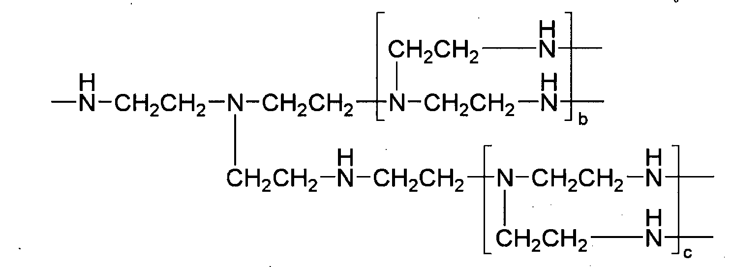
- the above polyethyleneimine unit (B) may particularly be represented by the following structure. wherein b and c are each independently ranging from 1 to 100, preferably, 1 to 30.
- the poly3,4-dihydroxyphenylalanine (PDOPA) used in the present invention is synthesized from N-carboxyl anhydride(NCA) of 3,4-dihydroxyphynylalanine (DOPA) wherein the DOPA is one of the mussel adhesive amino acids and preferably at least one selected from L-DOPA (L-3,4-dihydroxyl phenylalanine) and D-DOPA (D-3,4-dihydroxyl phenylalanine).
- the PDOPA may be selected from a group consisting of L-PDOPA synthesized from N-carboxyl anhydride (NCA) of L-DOPA (L-3,4-dihydroxyl phenylalanine), D-PDOPA synthesized from N-carboxyl anhydride (NCA) of D-DOPA (D-3,4-dihydroxyl phenylalanine) and L,D-PDOPA synthesized from N-carboxyl anhydride(NCA) of L,D-DOPA(L,D-3,4-dihydroxyl phenylalanine, mixture of L-DOPA and D-DOPA).
- NCA N-carboxyl anhydride
- NCA N-carboxyl anhydride
- D-PDOPA synthesized from N-carboxyl anhydride
- D-PDOPA synthesized from N-carboxyl anhydride
- the mussel adhesive protein-mimetic copolymer that is, the polyethyleneimine- graft -(polyethyleneglycol;poly3,4-dihydroxyphenylalanine) may be fabricated by the following operations:
- Operation (a) is a process of preparing a biocompatible macro-initiator used for manufacturing a mussel adhesive protein-mimetic copolymer.
- covalent bonding of polyethyleneglycol and polyethyleneimine may be implemented using dicyclohexylcarbodiimide (DCC)/N-hydroxysuccinimide (NHS) or, otherwise, hexamethylene diisocyanate (HMDI).
- DCC and NHS activate a carboxyl group in polyethyleneglycol having both methoxy and carboxyl group ends, in order to react with a primary amine of polyethyleneimine, thus forming a peptide covalent bond.
- HMDI activates a hydroxyl group of polyethyleneglycol having a methoxy end and serves to bond it to a primary amine of polyethyleneimine.
- Covalent bonding between polyethyleneglycol and polyethyleneimine activated by HMDI may include any reaction to form a covalent bond between the foregoing two polymers.
- the polyethyleneglycol solution is added drop by drop to the polyethyleneimine solution, thus enabling these two polymers to be covalently bonded.
- polyethyleneimine- graft -polyethyleneglycol in which polyethyleneglycol and polyethyleneimine are covalently bonded.
- a structure of polyethyleneglycol activated by DCC/NHS and a covalent bond structure of the activated polyethyleneglycol and a branch type polyethyleneimine (PEI) are illustrated as follows: [wherein, a ranges from 2 to 1200.]
- Synthesis of 3,4-dihydroxyphynylalanine N-carboxyl anhydride (DOPA-NCA) in operation (b) may be executed using at least one selected from the mussel adhesive amino acids, that is, L-DOPA (L-3,4-dihydroxyl phenylalanine) and D-DOPA (D-3,4-dihydroxyl phenylalanine) as a starting material and by any method for preparation of amino acid N-carboxyl anhydride (NCA) known in the related art.
- the foregoing substance (NCA) is prepared by reacting the mussel adhesive amino acid (D-DOPA or L-DOPA or L,D-DOPA) in a proper solvent in the presence of a triphosgene catalyst.
- DOPA is dissolved in acetic acid, using acetic acid anhydride as well as hydrochloric acid, followed by acetylation of a hydroxyl group of L-DOPA to synthesize (AC) 2 -DOPA while protecting the hydroxyl group. Thereafter, using triphosgene in an organic solvent composed of tetrahydrofurane (THF), N-carboxyl anhydride(NCA) of DOPA is synthesized [see below].
- THF tetrahydrofurane
- NCA N-carboxyl anhydride
- DOPA-NCA 3,4-dihydroxyphynylalanine N-carboxyl anhydride
- Preparation of a polyethyleneimine- graft - (polyethyleneglycol;poly3,4-dihydroxyphenylalanine)in operation (c) may be executed by multi-initiation of the polyethyleneimine- graft -polyethyleneglycol formed in operation (a) and the 3,4-dihydroxyphynylalanine N-carboxyl anhydride (DOPA-NCA) synthesized in operation (b) in an organic solvent, enabling polymerization thereof.
- DOPA-NCA 3,4-dihydroxyphynylalanine N-carboxyl anhydride
- the poly DOPA in the copolymer is synthesized by inducing polymerization of 3,4-dihydroxyphynylalanine N-carboxyl anhydride (DOPA-NCA) using the primary amine present in the polyethyleneimine- graft -polyethyleneglycol as a multi-initiator.
- DOPA-NCA 3,4-dihydroxyphynylalanine N-carboxyl anhydride
- POPA poly3,4-dihydroxyphenylalanine
- POPA poly3,4-dihydroxyphenylalanine
- the copolymer of the present invention may have controlled bonding ability and hydrophobic property ('hydrophobicity').
- a relative molar ratio of the polyethyleneimine- graft - polyethyleneglycol to 3,4-dihydroxyphynylalanine N-carboxyl anhydride (DOPA-NCA) ranges from 1:1 to 1:50. If the molar ratio is out of the above range, problems of increasing hydrophobicity or decreasing the bonding ability of the mussel adhesive protein-mimetic copolymer may arise.
- the organic solvent used in operation (c) may include at least one selected from dimethyl sulfoxide (DMSO), tetrahydrofuran (THF), and chloroform (ClCH 3 ).
- DMSO dimethyl sulfoxide
- THF tetrahydrofuran
- ClCH 3 chloroform
- operation (d) which is a process for de-protection of a protective hydroxyl group of the poly3,4-dihydroxyphenylalanine (PDOPA) may be further included.
- POPA poly3,4-dihydroxyphenylalanine
- the product is dispersed in dimethyl formamide (DMF).
- a hydroxyl group of the DOPA protected with an acetyl group may be deacetylated, in turn resulting in a polyethyleneimine- graft - (polyethyleneglycol;poly3,4-dihydroxyphenylalanine)represented by the following structure: [wherein a ranges from 2 to 1200, and d and d each independently ranges from 1 to 100.]
- the polyethyleneimine- graft -(polyethyleneglycol; poly3,4-dihydroxyphenylalanine) used in the present invention may be a biocompatible branch type dispersion stabilizer comprising a mussel adhesive protein-mimetic biocompatible polymer and containing poly3,4-dihydroxyphenylalanine, and useful in dispersing and stabilizing nanoparticles in an aqueous medium.
- Such nanoparticles may comprise metal, metal oxide, metal alloys, organic polymer and/or inorganic polymer, and have a size of 100 nm or less. More particularly, at least one selected from a group consisting of; iron oxide, cobalt ferrite (CoFe 2 O 4 ), manganese ferrite (MnFe 2 O 4 ), iron-platinum alloys (Fe-Pt alloy), cobalt-platinum alloys (Co-Pt alloy), cobalt (Co), cadmium selenide (CdSe), cadmium telluride (CdTe), cadmium selenide/zinc sulfide core/shell (CdSe/ZnS core/shell), cadmium selenide/ zinc selenide core/shell (CdSe/ZnSe core/shell), cadmium selenide/cadmium sulfide core/shell (CdSe/CdS core/shell), cadmium telluride/zin
- the iron oxide may include FeO, Fe 3 O 4 (magnetite), ⁇ -Fe 2 O 3 , ⁇ -Fe 2 O 3 , ⁇ -Fe 2 O 3 (maghemite), ⁇ -Fe 2 O 3 , Fe(OH) 2 , Fe(OH) 3 , ⁇ -FeOOH, ⁇ -FeOOH, ⁇ -FeOOH, ⁇ -FeOOH, Fe 5 HO 8 H 2 O, 5Fe 2 O 3 ⁇ H 2 O, FeOOH ⁇ 0.4H 2 O, Fe 8 O 8 (OH) 6 (SO) ⁇ nH 2 O, Fe 16 O 16 (OH ⁇ O 4 ) 12-13 ⁇ 10-12H 2 O, and so forth. More preferably, Fe 3 O 4 (magnetite), ⁇ -Fe 2 O 3 (maghemite) or a mixture of Fe 3 O 4 (magnetite) and ⁇ -Fe 2 O 3 (maghemite) is used.
- a core diameter of iron oxide used in the present invention may range from 1 to 25 nm.
- the core diameter of iron oxide may be observed by a transmission electron microscopy (TEM). If the core diameter of iron oxide is less than 1 nm, MR T2 contrast effects are reduced. On the contrary, when the core diameter of iron oxide exceeds 25 nm, the compound shows ferrimagnetic property and cannot be use for MRI.
- TEM transmission electron microscopy
- a hydrodynamic diameter of each of the iron oxide nanoparticles stabilized in an aqueous medium by the mussel adhesive protein-mimetic copolymer, polyethyleneimine- graft -(polyethyleneglycol;poly3,4-dihydroxyphenylalanine), may range from 3 to 100 nm.
- the hydrodynamic diameter may be measured by dynamic light scattering (DLS) while dispersing the nanoparticles in water.
- DLS dynamic light scattering
- iron oxide nanoparticles introduced in vivo and being hydrophilic may be easily excreted out of the body before depositing on the lymph nodes.
- the iron oxide nanoparticles introduced in vivo and being hydrophilic may be removed too early by an immune system in vivo (macrophage, etc.), thus not or minimally depositing on the lymph nodes.
- the lymph node contrast imaging method may utilize a variation or difference in strength of MRI signals generated from lymph nodes before and after administration of a contrast agent for lymph node imaging, which includes a colloidal solution prepared by modifying the surface of iron oxide particles using the mussel adhesive protein-mimetic copolymer described above and then dispersing the same in water.
- the variation in strength of MRI signals in the lymph node may be determined on the basis of changes in T2 signals.
- introduction of the contrast agent used in the present invention may be conducted by intravenous injection, oral administration, etc.
- the intravenous injection is more preferably used to maximize deposition of iron oxide on the lymph node.
- a T2 signal attenuation ratio in the lymph node is observed to be about 80% or more, thus presuming that the iron oxide nanoparticles are useful for diagnosis of a lymph node metastastic cancer.
- the mussel adhesive protein-mimetic copolymer used in the present invention has poly3,4-dihydroxyphenylalanine (PDOPA) formed through multi-initiative polymerization, in turn having at least one unit of DOPA per molecule, that is multiple interaction ligands(MIL) and exhibiting a high bonding strength to a hydrophilic surface. Since the above copolymer has a positive charge, it may impart additional electrostatic bonding force to the surface of nanoparticles having a negative charge. Moreover, since numerous polyethyleneglycol molecules having hydrophilic property are bonded to branches of a branch type polyethyleneimine, high aqueous dispersion stabilization may be achieved through hydrophilic property and stereoscopic effects.
- POPA poly3,4-dihydroxyphenylalanine
- a contrast agent for contrast enhanced lymphography which includes the foregoing copolymer, and a lymph node contrast imaging method, may accomplish excellent in vivo contrast imaging effects, thereby being useful for diagnosis of lymph node metastatic cancer.
- L-3,4-dihydroxy phenylalanine ('L-DOPA'), PEG-OH (5,000 Da), dimethyl formamide (DMF), N-hydroxy succinimide (NHS), N,N'-dicyclohexyl carbodiimide (DCC), acetic acid anhydride, glacial acetic acid, methylene chloride (MC) and chloroform were purchased from Sigma Chemical Company (St. Louis, MO), and PEI (1,800 Da (PEI 1,800)) was purchased from Alfa Aesar. For 48 hours before use, these materials were dried 40°C under vacuum.
- methoxy polyethyleneglycol carboxyl (PEG-COOH, 5000) (10 g) was dissolved in methylene chloride (CHCl 2 ) (50 ml) in a 250 ml flask. Then, N-hydroxysuccinimide (NHS) (0.52 g) and dicyclohexylcarbodiimide (DCC) (0.74 g) were added thereto, followed by a reaction at room temperature for 20 hours. After removing dicyclohexylurea through filtration, this material was precipitated in diethylether, resulting in polyethyleneglycol in an activated state (PEG-NHS).
- NHS N-hydroxysuccinimide
- DCC dicyclohexylcarbodiimide
- HDMI hexamethylene diisocyanate
- the activated polyethyleneglycol (PEG-NHS) (2 g) obtained in Example ⁇ 1-1> was dissolved in chloroform (200 ml). Then, after dissolving polyethyleneimine (Alfa Aesar, 1800 Da, 0.5 g) in chloroform (50 ml), the polyethyleneglycol solution was added drop by drop thereto to conduct a covalent bonding reaction between polyethyleneglycol and polyethyleneimine. Here, the reaction was executed for 24 hours and, after completing the reaction, the resultant product was concentrated to reach a total volume of 30 ml by a vacuum concentrator.
- PEG-NHS polyethyleneimine
- DOPA-NCA DOPA amino acid N-carboxyl anhydride
- DOPA amino acid having a protected hydroxyl group synthesized in Example ⁇ 1-3> as well as 0.5 g of triphosgene were dispersed in THF (50 ml) and reacted in an oil bath at 60°C.
- the reaction product was precipitated in hexane (800 ml), dissolved in THF (50 ml) and precipitated again in hexane.
- the foregoing procedure was repeated three times. After purifying, the product was dried using a vacuum dryer, resulting in DOPA N-carboxyl anhydride (DOPA(AC 2 )-NCA).
- PEI- graft -PEG polyethyleneglycol
- DOPA(AC 2 )-NCA DOPA N-carboxyl anhydride
- Table 1 shows assayed results of a structure of the mussel adhesive protein-mimetic copolymer of the present invention and characteristics thereof, wherein the structure of a polymeric copolymer and characteristics thereof have been assayed through 1 H-NMR and UV-Vis spectroscopy and using a fluorescent label.
- Table 1 [Table 1] Analysis of structure and characteristics of mussel adhesive protein-mimetic copolymer Name of sample Copolymer Number average molecular weight Molar ratio along with individual materials M n a CMC (g/L) b PEG M n PEI M n PEG PEI DOPA MIL0 copolym er PEI- graft -PEG 5,000 1,800 8 1 - 41,800 - MIL1 copolym er PEI- graft -(PE G;PDOPA) 5,000 1,800 8 1 5 42,800 0.015 MIL2 copolym er PEI- graft -(PE G;PDOPA) 5,000 1,800 8 1 15 44,800 0.005 a M n : number average molecular weight, which is assayed using 1 H-NMR and UV-Vis spectra b CMC: Critical Micelle Concentration, which is assayed using a fluorescent label (Hoechst 33342)
- Table 2 Colloidal solution of iron oxide (Fe 3 O 4 ) stabilized using mussel adhesive protein-mimetic copolymer Name of sample Diameter of iron oxide nanoparticle (nm) 1) Number average molecular weight Molar ratio Hydrodyna mic diameter (nm) 2) PEG M n PEG M n PEG PEI DOPA PD1 colloidal solution 10 5000 1800 7 1 20 30 PD2 colloidal solution 10 2000 1800 6 1 20 16 1) Diameter of iron oxide nanoparticle: a diameter of iron oxide particle only excluding a dispersion stabilizing material, which is measured by Transmission Electron Microscopy (TEM)2) Hydrodynamic diameter: a diameter of a material dispersed in water and containing a dispersion stabilizing material, which is a number average diameter measured in a dispersed state in an aqueous system by a dynamic light scattering (DLS) device (Malvern Co., Zetasizer NanoZS)
- a concentration was determined using a inductively coupled plasma-atomic emission spectroscopy (ICP-AES).
- ICP-AES inductively coupled plasma-atomic emission spectroscopy
- the PD1 and PD2 were taken with an aliquot by 200 ⁇ L, respectively, and the aliquot was mixed with 200 ⁇ L of a medium (DMEM: Dulbeco"s Modified Eagles"s Medium 89%, Pluronic® F-127 10%, AA: antibiotic-antimycotic 1%) to prepare Sample 0.350 ⁇ L of the sample 0 was mixed with 350 ⁇ L of a medium to prepare Sample 1.
- DMEM Dulbeco"s Modified Eagles"s Medium 89%
- Pluronic® F-127 10% Pluronic® F-127 10%
- AA antibiotic-antimycotic 1
- Sample 1 350 ⁇ L of Sample 1 was mixed with 350 ⁇ L of the medium to prepare another sample named Sample 2. As such, the initial concentration was diluted to 1/2 thereof to prepare nine (9) samples from 1 to 9 for PD1 and PD2, respectively. Moreover, Sample 10 was 100% medium with neither PD1 nor PD2. As controls, PBS solutions were prepared for PD1 and PD2, respectively, according to the foregoing procedures (see TABLE 3).
- Table 3 [Table 3] Iron oxide (Fe 3 O 4 ) nanoparticles colloid sample for cell viability test No. PD1 colloid PD2 colloid Control Sample + medium Iron content (ppm) Sample + medium Iron content (ppm) Sample + medium Crude solution 1680 Crude solution 1320 Crude solution (0.01M PBS solution) 0 Crude solution, 350 ⁇ L + medium 350 ⁇ L 840 Crude solution, 350 ⁇ L + medium 350 ⁇ L 660 Crude solution, 350 ⁇ L + medium 350 ⁇ L 1 No. 0, 200 ⁇ L + medium 200 ⁇ L 420 No. 0, 200 ⁇ L + medium 200 ⁇ L 330 No. 0, 200 ⁇ L + medium 200 ⁇ L 2 No.
- the cells After proliferating HepG2 cells through subculture, the cells were charged in 96-well plate to obtain 100,000 cells/well. Here, among these 96 wells, only 60 wells were used except those in periphery of the plate. In order to fix HepG2 cells on the bottom of the well plate, these were incubated at 37°C for 24 hours.
- the medium was removed from the well plate in which HepG2 prepared in ⁇ 1-2> was incubated.
- Each well of the well plate free from the medium was charged with 100 ⁇ L of each of respective samples 1 to 10 of PD1, PD2 and controls, respectively, which were prepared in ⁇ 1-1> shown in Table 3.
- each of the samples and controls was prepared in three (3) folds. After charging the sample, incubation was executed at 37°C for 24 hours.
- MTT test is an experimentation to determine cell viability using a principle wherein a yellow MTT reagent based on tetrazolium salt is altered into violet formazan crystals by metabolically activated cells.
- the MTT (3-(4,5-dimethylthiazol-2-yl)-2,5-diphenyl-2H-tetrazolium bromide) reagent used in the cell viability test was dissolved in a water bath at 37°C, 1 hour before the test.
- 20 ⁇ L of the prepared MTT reagent was added to each well of the 96-well plate into which the samples and controls prepared in ⁇ 1-3> were introduced.
- special precaution is needed to prevent the test well from being exposed to light.
- the test well was incubated at 37°C for 4 hours after addition of the MTT reagent.
- cell viability OD i , 550 ⁇ OD i , 690 ODS i , 550 ⁇ ODS i , 690 ⁇ 100
- FIG. 6 is graphs illustrating results of cell viability test.
- an acceptable error range (commonly, tolerance) is generally ⁇ 20%. From results of the present test, it can be found that 80% or more of cell viability was exhibited in the most part of a concentration section to be tested. Such results
- iron oxide (Fe 3 O 4 ) nanoparticles stabilized by the mussel adhesive protein-mimetic copolymer have no cellular toxicity and may be safely used in vivo.
- the colloidal solutions containing iron oxide (Fe 3 O 4 ) nanoparticles stabilized using the mussel adhesive protein-mimetic copolymers were prepared according to the method in Example 1.
- a contrast agent was prepared.
- a loop coil was used in an MRI diagnosis device (Trio 3.0T, Simens) to assay in vivo T2 contrast imaging performance.
- nude mouse (Balb/nude).
- the nude mouse weighed 30 g. After anesthetizing, the mouse was horizontally placed in the MRI diagnosis device and observed along a cross-section thereof. MRI images of the nude mouse were subjected to measurement before and 24 hours after injecting the contrast agent, and lymph nodes before and after injecting the contrast agent were observed and compared to each other.
- PBS powder was added to the colloidal solution to reach a concentration of 0.01 M, to prepare a desired contrast agent.
- Iron content was analyzed through ICP-AES and an administration dose was calculated (Table 4) in consideration of the weight of the mouse, to prepare a contrast agent with a total volume of 500 ⁇ L.
- the contrast agent was injected through a tail vein of the mouse.
- T2Me3d pulse sequence provided by Siemens Co. and particular parameters are as follows.
- TR (repetition time) 40.0 msec
- TE (echo time) 22.0 msec
- FOV 49 mm x 70 mm
- Matrix size 256 x 180
- slice thickness 0.6 mm
- number of acquisition 6
- FIGS. 7 to 10 partially illustrate in vivo MRI images obtained under the foregoing conditions.
- one cross-section was selected from the highest visible lymph nodes, that is, inguinal and brachial lymph nodes, and discolored parts of the lymph node were selected as ROI (Region of Interest).
- signal intensity was measured. Since an overall signal intensity of the acquired MRI images is altered whenever it is measured, it is difficult to evaluate the contrast imaging performance based on an absolute value of the signal intensity (S: signal intensity).
- S signal intensity
- N noise
- N noise
- T2 signal attenuation ratio ( ⁇ R2) was calculated. The results are illustrated by the graphs in FIG. 11 .
- the following Equation 2 is a method for calculation of T2 signal attenuation ratio ( ⁇ R2).
- SNR 0 SNR before administration of the constrast agent signal intensity of ROI before administration signal intensity of muscle before administration
- FIGS. 7 to 10 it can be seen that the lymph nodes of the nude mouse darkened after administration of the contrast agent.
- the Tables 5 and 6 include signal to noise ratio(SNR) and signal attenuation ratios ( ⁇ R2) before and after administration of a contrast agent sample to each of the inguinal lymph node and the brachial lymph node.
- FIG. 11 illustrates the signal to noise ratio (SNR) before and after administration of the contrast agent sample. It was found that signal attenuation after administration of the contrast agent was noticeably high, compared to that before administration thereof.
- T2 signal in inguinal lymph node of a nude mouse by contrast agents Name of sample Inguinal lymph node LEFT RIGHT Before administration After administration Signal attenuation ratio Before administration After administration Signal attenuation ratio SNR SNR SNR SNR PDIE1 contrast agent 3.94 0.21 94.8% 2.68 1.26 53.0% PDIE2 contrast agent 1.68 0.31 81.5% 2.15 0.30 86.2% PDIE3 contrast agent 2.92 0.23 92.3% 1.29 0.25 81.0% PDIE4 contrast agent 2.65 0.84 68.5% 1.74 0.63 63.9%
- T2 signal in brachial lymph node of a nude mouse by contrast agents Name of sample Brachial lymph node LEFT RIGHT Before administration After administration Signal attenuation ratio Before administration After administration Signal attenuation ratio SNR SNR SNR SNR PDIE1 contrast agent 2.01 0.61 69.5% 1.87 0.46 75.6% PDIE2 contrast agent 1.39 0.32 76.8% 1.40 0.57 59.4% PDIE3 contrast agent 1.59 0.73 54.3% 1.43 1.00 29.9% PDIE4 contrast agent 1.37 0.33 76.1% 1.30 0.68 47.7%
- FIG. 12 are TEM photographs illustrating iron oxide (Fe 3 O 4 ) nanoparticles before reformed by MIL2 (a) and after reformed by MIL2(b).
- the iron oxide (Fe 3 O 4 ) nanoparticles stably dispersed in water have substantially the same morphology and size as those of iron oxide (Fe 3 O 4 ) nanoparticles dissolved in a hydrophobic solvent.
- a hydrodynamic diameter of each of the iron oxide (Fe 3 O 4 ) nanoparticle colloids is also illustrated in FIG. 12 .
- the hydrodynamic diameter of the iron oxide (Fe 3 O 4 ) nanoparticle colloid is a number average hydrodynamic diameter measured by a dynamic light scattering (DLS) method and is observed to be uniform in size.
- DLS dynamic light scattering
- FIG. 13 illustrates stability of iron oxide (Fe 3 O 4 ) nanoparticle colloids stably dispersed in an aqueous medium by a mussel adhesive protein-mimetic copolymer, along with various pH values (a) and ionic concentrations (b).
- hydrodynamic diameters were measured at various pH values and ionic concentrations through DLS.
- iron oxide(Fe 3 O 4 ) nanoparticle colloids stably dispersed in an aqueous medium using the mussel adhesive protein-mimetic copolymer (MIL1 ( ⁇ ), MIL2 ( ⁇ )) are relatively stable at various concentrations and pH values, compared to iron oxide (Fe 3 O 4 ) nanoparticles without application of the mussel adhesive protein-mimetic copolymer (MIL0 ( ⁇ )).
- Example 4 Metastatic cancer diagnosis using the contrast agent including colloidal solution of iron oxide (Fe 3 O 4 ) nanoparticles stabilized by mussel adhesive protein-mimetic copolymer
- a colloidal solution of iron oxide (Fe 3 O 4 ) nanoparticles stabilized by the mussel adhesive protein-mimetic copolymer was prepared according to the methods in Example 1 and 2.
- Table 7 Colloidal solution of iron oxide (Fe 3 O 4 ) nanoparticles stabilized by mussel adhesive protein-mimetic copolymer for Example 4. Name of sample Diameter of iron oxide nanoparticl e(nm) PEGnum ber average molecular weight PEInum ber average molecular weight Molar ratio Hydrody namic diameter (nm) Dose of administration (mg Fe/kg rabbit) PEG PEI DOPA PDIE5co lloidal solution 14 5000 1800 10 1 20 28 10.4
- the colloidal solution in Table 7 was diluted by using a saline solution (0.9% NaCl) for injection, and then a contrast agent having an iron concentration of 6240 ppm and a total volume of 5 mL was prepared while the iron concentration was assayed through ICP-AES and the dose of administration was calculated considering the weight of rabbits.
- the contrast agent was intravenously injected through an ear of the rabbit.
- metastatic cancer models were established by expressing VX2 cancer cells in iliac lymph nodes of 12 New Zealand white rabbits, and then used to evaluate performance of the contrast agent.
- the rabbit with metastatic cancer was weighted about 3kg. After anesthetizing, the rabbit was horizontally placed in the MRI device (Trio 3.0T, Simens, knee coil), and then cross sections thereof were observed. MRI images of the rabbit were measured before and 24 hours after injecting the contrast agent, and the iliac lymph nodes before and after injecting the contrast agent were observed and compared to each other. The iliac lymph nodes of every rabbits were isolated and stained with hematoxylin-eosin for examination by a pathologist.
- Table 8 shows the evaluation results for the metastatic cancer model rabbits.
- Table 8 shows the number of lymph nodes with metastasis cancer and the number of normal lymph nodes for the 12 rabbits metastatic cancer models. The sensitivity and specificity was 100%, respectively.
- FIG. 14 shows images of contrast enhanced lymphography and result of histopathology of Rabbit No. 002, before and after injecting the contrast agent.
- the parts marked in (+) indicate lymph nodes evaluated to be metastatic cancer as the MRI scanning, and the parts marked in (-) indicate lymph nodes examined to be normal as the MRI scanning.
- the mussel adhesive protein-mimetic copolymer used in the present invention has multiple units of DOPA in a molecule.
- DOPA can interact with an inorganic nanoparticle and exhibit high bond strength to the nanoparticles' surface.
- the above copolymer has a positive charge, it may impart electrostatic bonding force to the surface of nanoparticles having a negative charge.
- numerous polyethyleneglycol molecules having hydrophilic property are bonded to the branches of polyethyleneimine, high aqueous dispersion stabilization may be achieved through hydrophilic property and stereoscopic effects.
- the PEG on the surface make the iron oxide nanoparticles circulate in the body for a long-time and so many of the iron oxide nanoparticles can reach to the lymph nodes.
- the lymph nodes have to take nanoparticles up as many as possible. So it is crucial of the iron oxide nanoparticles circulate in the body for a long time.
- the contrast agent including iron oxide nanoparticles reformed by mussel adhesive protein-mimetic copolymer seems to be more taken up and so shows more darkening in the normal lymph nodes and less darkening in the metastatic lymph nodes.
- the iron oxide nanoparticles reformed by mussel adhesive protein-mimetic copolymer have a great potential for the detection of lymph node metastasis as a contrast agent for contrast enhanced lymphography.
Landscapes
- Health & Medical Sciences (AREA)
- Chemical & Material Sciences (AREA)
- Nanotechnology (AREA)
- Engineering & Computer Science (AREA)
- Life Sciences & Earth Sciences (AREA)
- Nuclear Medicine, Radiotherapy & Molecular Imaging (AREA)
- General Health & Medical Sciences (AREA)
- Molecular Biology (AREA)
- Radiology & Medical Imaging (AREA)
- Epidemiology (AREA)
- Animal Behavior & Ethology (AREA)
- Public Health (AREA)
- Veterinary Medicine (AREA)
- Immunology (AREA)
- Crystallography & Structural Chemistry (AREA)
- Medicinal Chemistry (AREA)
- Medicines Containing Antibodies Or Antigens For Use As Internal Diagnostic Agents (AREA)
- Medicinal Preparation (AREA)
Description
- The present invention relates to a contrast agent for contrast enhanced lymphography, which includes iron oxide nanoparticles dispersed and stabilized in an aqueous medium by a mussel adhesive protein-mimetic copolymer, as well as a method for contrast enhanced lymphography using the same. More particularly, the mussel adhesive protein-mimetic copolymer is a polyethyleneimine-graft-(polyethyleneglycol;poly3,4-dihydroxyphenylalanine) (PEI-graft-(PEG;PDOPA)). The graft polymer is composed of two parts, one is including polyethyleneimine grafted with a polyethyleneglycol-based biocompatible polymer, which has an affinity to an aqueous medium (sometimes, abbrev. to 'polyethyleneglycol grafted polyethyleneimine'), and the other is poly3,4-dihydroxyphenylalanine (PDOPA), which has an affinity to the surface of nanoparticles. The present invention relates to a contrast agent for contrast enhanced lymphography, which includes a colloidal solution prepared by modifying the surface of iron oxide particles using the foregoing mussel adhesive protein-mimetic copolymer and then dispersing the particles in water. The present invention relates to a method for contrast enhanced lymphography using the contrast agent.
- Subject-matter which is not encompassed by the scope of the claims does not form part of the presently claimed invention.
- Nanoparticles are used in a broad range of applications such as nano-electronic convergence technology, in vivo imaging technology, medical applications, etc. Specifically, super-paramagnetic iron oxide nanoparticles are widely used in a variety of biomedical applications including, for example, a magnetic resonance imaging ('MRI') contrast agent, cell therapy, hyperthermia, drug delivery, cell separation, nucleic acid preparation, or the like.
- The most important requirement for application in biomedical applications is primarily to ensure high quality nanoparticles and, in addition, to allow nanoparticles to have excellent dispersibility and stability in an aqueous medium. Here, the high quality nanoparticle may mean nanoparticles with features of; (i) uniformity of particle size, (ii) easy control of particle size, (iii) particle crystallinity, (iv)possibility in controlling particle morphology, etc.
- However, nanoparticles commercially available in the art are mostly synthesized in an aqueous system or may be obtained by synthesis in a gas phase. Nanoparticles generated by the foregoing processes have difficulty in preparing particles with a uniform shape and generally show low crystallinity. Further, it is difficult to manufacture nanoparticles having a uniform size and control the size of a particle.
- Recently, numerous studies have been executed to develop a novel method for manufacturing metal oxide nanoparticles in an organic system, which have relatively high quality, that is, uniform size and favorable crystallinity, compared to nanoparticles synthesized in an aqueous system according to the related art.
- As such, in the case where nanoparticles are synthesized in an organic solvent, uniformity and size control of the nanoparticles may sometimes be achieved by stabilization thereof using an organic additive during a synthesizing process. In this regard, since the surface condition of nanoparticles is influenced by a hydrophobic organic additive, the metal oxide nanoparticles may be easily dispersed in a hydrophobic organic solvent. However, when they are mixed with water, they do not have sufficient stability.
- For such nanoparticles prepared in an organic solvent, hydrophobic properties of the surface of the nanoparticles may obscure stable dispersion of the nanoparticles in water, thus causing a problem for use in biomedical applications. Therefore, in order to use the nanoparticles in the foregoing applications, there is a need to develop a biocompatible dispersion stabilizer that reforms (or modifies) the surface of the nanoparticles in order to have hydrophilic properties and ensures a suitable condition so as to be homogeneously dispersed in an aqueous medium. In addition, development of a nanoparticle colloidal solution that is prepared using the biocompatible dispersion stabilizer described above, wherein the dispersion state is stably maintained in an aqueous system, is also required.
- Among methods for dispersing nanoparticles in an aqueous system according to the techniques in the related art, use of a thin silica layer has currently been disclosed in the Journal of American Chemical Society, 2005, 127, 4990. According to the foregoing article, polyoxyethylene nonylphenylether is introduced to a cyclohexane solution and mixed with the same to form micro-micelle emulsion drops. Next, a solgel reaction of tetraethyl ortho-silicate (TEOS) is induced and nanoparticles are coated with a silica layer and dispersed in water. The above document described a process of coating the outer side of the nanoparticles with a hydrophilic silica layer to disperse the nanoparticles in water, wherein the nanoparticles were prepared in an organic solvent. In this case, the silica coating method using micro-emulsion entails a problem in that, since an amount of nanoparticles to be coated at just one time is very small, an amount of nanoparticle dispersion in an aqueous system manufactured in a single process is also greatly reduced. Moreover, according to the amount of nanoparticle colloids manufactured in the single process or an amount of polyoxyethylene nonylphenylether, the conditions of the micro-emulsion are altered. Therefore, there are difficulties in finely regulating a desired thickness of a silica layer, and attaining uniformity of the coated particles since the number of nanoparticles contained in the silica layer is altered. In the case where nanoparticles are stabilized by a silica layer, the foregoing techniques in the related art entail problems in that silane functional groups on the surface of the silica are not sufficiently stable but react to one another, therefore, the nanoparticles coated with the silica and dispersed in water were combined and became agglomerated over time. As a result, it was difficult to ensure storage stability of the dispersion over a long period of time.
- In recent years, a method for dispersing nanoparticles in water using a polymer composed of phosphine oxide and polyethyleneglycol has been disclosed in the Journal of America Chemical Society (2005, 127, 4556). More particularly, the foregoing article described a nanoparticle dispersing method wherein, after reacting polyethyleneglycol with 1,2-bis(dichlorophosphino)ethane to synthesize a polymer having polyethyleneglycols bonded together, the polymer is subjected to a ligand exchange reaction with nanoparticles dispersed in a hydrophobic solvent, thereby enabling dispersion stabilization of the nanoparticles and uniformly dispersing the same in water. The disclosed method uses a simple preparation method and utilizes ligand exchange to disperse nanoparticles in water. However, since phosphorus atom (P) is likely to oxidize and become a phosphoryl group, a coating polymer must be synthesized in an inert atmosphere using nitrogen or argon. Further, since the polymer is in a cross-linked state, a problem in introducing a functional group to bond functional ligands in vivo such as DNA, RNA, a monoclonal antibody or other functional proteins, still remains.
- Scientists have recently conducted a number of studies upon mussels as a potential origin of bio-adhesives. Mussels generate and secrete a sticky material which is functionally differentiated to allow the mussels to be stationary or anchor in the water, in a marine environment having characteristics of salinity, humidity, tidal flow, turbulent flow, waves, etc. The mussel strongly adheres to the surface of a material in water, using threads composed of a fiber bundle secreted from legs thereof. At the end of each fiber, a plaque comprising a water-proof adhesive is present to allow the mussel to adhere to a wet solid surface. Such thread protein contains a large quantity of 3,4-dihydroxyphynyl-L-alanine (DOPA), which is an amino acid obtained by hydroxylation of tyrosine groups using a polyphenol oxidase. 3,4-dihydroxyphenyl (catechol) on a side branch of DOPA may create a very strong hydrogen bond with the hydrophilic surface and/or be strongly bonded with metal ions, metal oxides (Fe3+, Mn 3+), semi-metal (silicon), or the like.
- Occurrence of a metastatic cancer decisively influences prognosis and treatment of a cancer. The occurrence of a metastatic cancer may be determined by presence or absence of lymph node metastasis and occurrence of a metastatic lymph node cancer is currently diagnosed by surgically biopsying the lymph node. However, this is an invasive method involving significant difficulties to both a patient and a physician. On the other hand, non-invasive techniques such as use of CT, MRI, PET, etc. may be employed to detect occurrence of a metastatic cancer, but, incurs a problem in that cancer may generally be detected when its size is 5 mm or more. Accordingly, there is still a need for a method capable of non-invasively diagnosing metastatic cancer having a relatively small size.
- A method for detecting metastatic cancer by injecting iron oxide nanoparticles in vivo and using an magnetic resonance image to show iron oxide deposition on lymph nodes has been introduced (Mukesh G. Harisinghani, M.D., Jelle Barentsz, M.D., Ph.D., Peter F. Hahn, M.D., Ph.D., Willem M. Deserno, M.D., Shahin Tabatabaei, M.D., Christine Hulsbergen van de Kaa, M.D., Ph.D., Jean de la Rosette, M.D., Ph.D., and Ralph Weissleder, M.D., Ph.D., and New England Journal of Medicine 2003; 348:2491-2499). According to the disclosed method, after stabilizing iron oxide nanoparticles in an aqueous system using a hydrophilic material, the treated nanoparticles are introduced in vivo and, after a predetermined time, lymph node tissues with cancer as well as normal lymph node tissues are observed through an MRI. Based on a difference in observed results therebetween, the occurrence of cancer may be diagnosed. Using the foregoing method, AMAG Co. (United States) has developed a MRI contrast agent named 'COMBIDEX ' for contrast enhanced lymphography. However, the above contrast agent often causes adverse effects after in vivo administration and/or has poor selectivity, thus, is not widely used. Accordingly, there is still a need for developing an improved iron oxide-based contrast agent for contrast enhanced lymphography, having excellent selectivity and reduced adverse effects.
-
WO 2012/091452 A2 discloses a mussel adhesive protein-mimetic dispersion stabilizing agent to disperse nanoparticles in an aqueous medium, a colloidal solution including nanoparticles dispersed and stabilized by the dispersion stabilizing agent, and a contrast agent including the colloidal solution. More particularly, the mussel adhesive protein-mimetic dispersion stabilizer is a polyethyleneimine-graft-(polyethyleneglycol : polyDOPA) PEI-graft-(PEG : PDOPA). The graft polymer is formed of two parts. One is polyethyleneglycol grafted with a polyethylenimine which has an affinity to an aqueous medium, and the other is polyDOPA which has an affinity to the surface of nanoparticles. Because of those characteristics, the stabilizer shows a stable dispersion of nano particles in the aqueous medium. The use of the contrast agent in bio-imaging applications such as magnetic resonance imaging (MRI) is mentioned in a general manner. - Andrew Elias ET AL: "Imaging circulating cells and lymphoid tissues with iron oxide nanoparticles", Hematology, 1 January 2009, pages 720-726 discloses polymeric coatings to improve the biocompatibility and stability of iron oxide nanoparticles in molecular imaging.
- Therefore, as a result of intensive and extensive efforts to overcome the above problems in the related art, the present inventors have completed a biocompatible dispersion stabilizer that can reform the surface of nanoparticles into a hydrophilic state so as to disperse the nanoparticles in an aqueous system and have found that using the same (the stabilizer) may enable the nanoparticles to be dispersed and stabilized ('dispersion stabilization') in the aqueous system, thereby being effectively used for biomedical applications. In addition, it was also found that the nanoparticles dispersed and stabilized by the biocompatible dispersion stabilizer of the present invention may be applicable to a nano-electronic fusion technique field such as a quantum dot (Q-dot) light emitting device, a bio-imaging field such as a MRI contrast agent, a tissue engineering field such as cell therapy, a biomedical field such as hyperthermia, drug delivery, and so forth.
- An object of the present invention is to provide a contrast agent for contrast enhanced lymphography, which includes iron oxide nanoparticles dispersed and stabilized in an aqueous medium by a mussel adhesive protein-mimetic copolymer, wherein the surface of various nanoparticles is reformed into a hydrophilic state through a simple process by mimicking mussel protein, so as to stabilize dispersion of the nanoparticles in an aqueous medium while enabling application thereof in biomedical fields.
- Another object of the present invention is to provide a method for contrast enhanced lymphography using the foregoing contrast agent.
- In one general aspect, the present invention provides a contrast agent for contrast enhanced lymphography, which includes iron oxide nanoparticles dispersed and stabilized in an aqueous medium by a mussel adhesive protein-mimetic copolymer, a method for contrast enhanced lymphography using the same, and a method for diagnosing cancer of lymph nodes using the same. More particularly, the mussel adhesive protein-mimetic copolymer is a polyethyleneimine-graft-(polyethyleneglycol;poly3,4-dihydroxyphenylalanine)(PEI-graft-(PEG;PDOPA)), including polyethyleneimine grafted with a polyethyleneglycol-based biocompatible polymer having affinity to an aqueous medium (sometimes, abbrev. to 'polyethyleneglycol grafted polyethyleneimine'), and poly3,4-dihydroxyphenylalanine (PDOPA) having affinity to the surface of nanoparticles. The present invention also provides a contrast agent for contrast enhanced lymphography, which includes a colloidal solution prepared by modifying the surface of iron oxide particles using the foregoing mussel adhesive protein-mimetic copolymer and then dispersing the particles in water, and a method for contrast enhanced lymphography using the foregoing contrast agent.
- Hereinafter, the present invention will be described in more detail.
- The mussel adhesive protein-mimetic copolymer used in the present invention is a polyethyleneimine-graft-(polyethyleneglycol;poly3,4-dihydroxyphenylalanine) (PEI-graft-(PEG;PDOPA)), including polyethyleneimine grafted with a polyethyleneglycol-based biocompatible polymer having affinity to an aqueous medium (sometimes, abbrev. to 'polyethyleneglycol grafted polyethyleneimine'), and poly3,4-dihydroxyphenylalanine (PDOPA) having affinity to the surface of nanoparticles, and contains a mussel adhesive amino acid, that is, 3,4-dihydroxyphynylalanine (DOPA).
- In order to prepare the polyethyleneimine-graft - (polyethyleneglycol;poly3,4-dihydroxyphenylalanine), polyethyleneglycol and polyethyleneimine are first combined through covalent bonding to form a polyethyleneimine-graft-polyethyleneglycol. The formed product is used as a biocompatible macro-initiator.
- The polyethyleneglycol used herein may be polyethyleneglycol having a number average molecular weight of 300 to 50,000 and a hydroxyl group or carboxyl group at an end thereof. According to one embodiment of the present invention, the polyethyleneglycol is methoxy polyethyleneglycol carboxylic acid having a methoxy group at one end and a carboxyl group substituted at the other end.
- The polyethyleneimine used herein may be a branched polyethyleneimine without toxicity, which has a number average molecular weight of 100 to 10,100, preferably 100 to 2,000. If the number average molecular weight of the branched polyethyleneimine is less than 100, the produced copolymer of the present invention cannot be suitably combined with a physiologically active material useful therefore. On the other hand, when the number average molecular weight is 10,100 or more, difficulties in excreting the above material out of the body through kidneys may be incurred. Accordingly, the present invention preferably uses polyethyleneimine having a number weight molecular weight present in the foregoing range.
- The poly3,4-dihydroxyphenylalanine (PDOPA) used in the present invention may be a condensation polymer which has 3,4-dihydroxyphynylalanine (DOPA) as a monomer. The repeating units are connected via amide bonds. The number of repeating units ranges from 1 to 100. The poly3,4-dihydroxyphenylalanine (PDOPA) may be polymerized by solid phase synthesis and liquid phase synthesis using various coupling methods; carbodiimide-mediated reaction, a symmetrical anhydride method, a mixed anhydride method, an active ester method, an azide method, an acyl-chloride method and an N-carboxy anhydride method. Such example methods which are described hereinbefore are provided to offer a clear understanding of the poly3,4-dihydroxyphenylalanine (PDOPA). However the poly3,4-dihydroxyphenylalanine (PDOPA) are not limited to the polymers which are synthesized by the above methods. The poly3,4-dihydroxyphenylalanine (PDOPA) used in the present invention may be prepared by the several methods described hereinbefore, preferably by the N-carboxy anhydride method.
- The polyethyleneimine-graft-(polyethyleneglycol; poly3,4-dihydroxyphenylalanine) used in the present invention may include; a polyethyleneglycol unit represented by the following structure (A), a polyethyleneimine unit represented by the following structure (B), and a poly3,4-dihydroxyphenylalanine (PDOPA) unit represented by the following structure (C).


- The above polyethyleneimine unit (B) may particularly be represented by the following structure.
- The poly3,4-dihydroxyphenylalanine (PDOPA) used in the present invention is synthesized from N-carboxyl anhydride(NCA) of 3,4-dihydroxyphynylalanine (DOPA) wherein the DOPA is one of the mussel adhesive amino acids and preferably at least one selected from L-DOPA (L-3,4-dihydroxyl phenylalanine) and D-DOPA (D-3,4-dihydroxyl phenylalanine). The PDOPA may be selected from a group consisting of L-PDOPA synthesized from N-carboxyl anhydride (NCA) of L-DOPA (L-3,4-dihydroxyl phenylalanine), D-PDOPA synthesized from N-carboxyl anhydride (NCA) of D-DOPA (D-3,4-dihydroxyl phenylalanine) and L,D-PDOPA synthesized from N-carboxyl anhydride(NCA) of L,D-DOPA(L,D-3,4-dihydroxyl phenylalanine, mixture of L-DOPA and D-DOPA).
- The mussel adhesive protein-mimetic copolymer, that is, the polyethyleneimine-graft -(polyethyleneglycol;poly3,4-dihydroxyphenylalanine) may be fabricated by the following operations:
- (a) combining polyethyleneglycol as a hydrophilic polymer with polyethyleneimine through covalent bonding, to form a polyethyleneimine-graft-polyethyleneglycol; (b) after protecting hydroxyl groups of 3,4-dihydroxyphynylalanine (DOPA), synthesizing 3,4-dihydroxyphynylalanine N-carboxyl anhydride (DOPA-NCA) in the presence of a triphosgene catalyst; and (c) reacting the polyethyleneimine-graft-polyethyleneglycol prepared in operation (a) and the 3,4-dihydroxyphynylalanine N-carboxyl anhydride (DOPA-NCA) synthesized in operation (b) in an organic solvent, to thus prepare a polyethyleneimine-graft-(polyethyleneglycol;poly3,4-dihydroxyphenylalanine).
- Operation (a) is a process of preparing a biocompatible macro-initiator used for manufacturing a mussel adhesive protein-mimetic copolymer. In operation (a), covalent bonding of polyethyleneglycol and polyethyleneimine may be implemented using dicyclohexylcarbodiimide (DCC)/N-hydroxysuccinimide (NHS) or, otherwise, hexamethylene diisocyanate (HMDI). Here, DCC and NHS activate a carboxyl group in polyethyleneglycol having both methoxy and carboxyl group ends, in order to react with a primary amine of polyethyleneimine, thus forming a peptide covalent bond. Alternatively, HMDI activates a hydroxyl group of polyethyleneglycol having a methoxy end and serves to bond it to a primary amine of polyethyleneimine. Covalent bonding between polyethyleneglycol and polyethyleneimine activated by HMDI may include any reaction to form a covalent bond between the foregoing two polymers. In one embodiment of the present invention, after dissolving polyethyleneglycol and polyethyleneimine activated by DCC/NHS in chloroform, respectively, the polyethyleneglycol solution is added drop by drop to the polyethyleneimine solution, thus enabling these two polymers to be covalently bonded. After completing a reaction, the reacted solution is concentrated and precipitated in diethylether to produce a polyethyleneimine-graft-polyethyleneglycol in which polyethyleneglycol and polyethyleneimine are covalently bonded. A structure of polyethyleneglycol activated by DCC/NHS and a covalent bond structure of the activated polyethyleneglycol and a branch type polyethyleneimine (PEI) are illustrated as follows:

-

- Synthesis of 3,4-dihydroxyphynylalanine N-carboxyl anhydride (DOPA-NCA) in operation (b) may be executed using at least one selected from the mussel adhesive amino acids, that is, L-DOPA (L-3,4-dihydroxyl phenylalanine) and D-DOPA (D-3,4-dihydroxyl phenylalanine) as a starting material and by any method for preparation of amino acid N-carboxyl anhydride (NCA) known in the related art. Preferably, the foregoing substance (NCA) is prepared by reacting the mussel adhesive amino acid (D-DOPA or L-DOPA or L,D-DOPA) in a proper solvent in the presence of a triphosgene catalyst.
- According to one embodiment of the present invention, as illustrated below, DOPA is dissolved in acetic acid, using acetic acid anhydride as well as hydrochloric acid, followed by acetylation of a hydroxyl group of L-DOPA to synthesize (AC)2-DOPA while protecting the hydroxyl group. Thereafter, using triphosgene in an organic solvent composed of tetrahydrofurane (THF), N-carboxyl anhydride(NCA) of DOPA is synthesized [see below].

- Preparation of a polyethyleneimine-graft - (polyethyleneglycol;poly3,4-dihydroxyphenylalanine)in operation (c) may be executed by multi-initiation of the polyethyleneimine-graft-polyethyleneglycol formed in operation (a) and the 3,4-dihydroxyphynylalanine N-carboxyl anhydride (DOPA-NCA) synthesized in operation (b) in an organic solvent, enabling polymerization thereof. The poly DOPA in the copolymer is synthesized by inducing polymerization of 3,4-dihydroxyphynylalanine N-carboxyl anhydride (DOPA-NCA) using the primary amine present in the polyethyleneimine-graft-polyethyleneglycol as a multi-initiator. According to the foregoing processes, the synthesized poly3,4-dihydroxyphenylalanine (PDOPA) is combined with the polyethyleneimine-graft-polyethyleneglycol resulting in a final product, that is, the polyethyleneimine-graft-(polyethyleneglycol;poly3,4-dihydroxyphenylalanine).
- In operation (c), by regulating an added amount of 3,4-dihydroxyphynylalanine N-carboxyl anhydride (DOPA-NCA) used as a bio-mimetic conjugate site, the copolymer of the present invention may have controlled bonding ability and hydrophobic property ('hydrophobicity'). Preferably, a relative molar ratio of the polyethyleneimine-graft - polyethyleneglycol to 3,4-dihydroxyphynylalanine N-carboxyl anhydride (DOPA-NCA) ranges from 1:1 to 1:50. If the molar ratio is out of the above range, problems of increasing hydrophobicity or decreasing the bonding ability of the mussel adhesive protein-mimetic copolymer may arise.
- The organic solvent used in operation (c) may include at least one selected from dimethyl sulfoxide (DMSO), tetrahydrofuran (THF), and chloroform (ClCH3).
- After completing polymerization in operation (c), operation (d) which is a process for de-protection of a protective hydroxyl group of the poly3,4-dihydroxyphenylalanine (PDOPA) may be further included. According to one embodiment of the present invention, after completing polymerization of the polyethyleneimine-graft - (polyethyleneglycol;poly3,4-dihydroxyphenylalanine), the product is dispersed in dimethyl formamide (DMF). Thereafter, by adding a proper amount of piperidine thereto, a hydroxyl group of the DOPA protected with an acetyl group may be deacetylated, in turn resulting in a polyethyleneimine-graft - (polyethyleneglycol;poly3,4-dihydroxyphenylalanine)represented by the following structure:

- The polyethyleneimine-graft-(polyethyleneglycol; poly3,4-dihydroxyphenylalanine) used in the present invention may be a biocompatible branch type dispersion stabilizer comprising a mussel adhesive protein-mimetic biocompatible polymer and containing poly3,4-dihydroxyphenylalanine, and useful in dispersing and stabilizing nanoparticles in an aqueous medium.
- Such nanoparticles may comprise metal, metal oxide, metal alloys, organic polymer and/or inorganic polymer, and have a size of 100 nm or less. More particularly, at least one selected from a group consisting of; iron oxide, cobalt ferrite (CoFe2O4), manganese ferrite (MnFe2O4), iron-platinum alloys (Fe-Pt alloy), cobalt-platinum alloys (Co-Pt alloy), cobalt (Co), cadmium selenide (CdSe), cadmium telluride (CdTe), cadmium selenide/zinc sulfide core/shell (CdSe/ZnS core/shell), cadmium selenide/ zinc selenide core/shell (CdSe/ZnSe core/shell), cadmium selenide/cadmium sulfide core/shell (CdSe/CdS core/shell), cadmium telluride/zinc sulfide core/shell (CdTe/ZnS core/shell), cadmium telluride/zinc selenide core/shell (CdTe/ZnSe core/shell), cadmium telluride/cadmium sulfide core/shell (CdTe/CdS core/shell), cadmium telluride/cadmium selenide core/shell (CdTe/CdSe core/shell), zinc sulfide (ZnS), cadmium sulfide (CdS), indium arsenide (InAs), indium phosphide (InP), indium arsenide/indium phosphide core/shell (InAs/InP core/shell), indium arsenide/cadmium selenide core/shell (InAs/CdSe core/shell), indium arsenide/zinc sulfide core/shell (InAs/ZnS core/shell), indium arsenide/zinc selenide core/shell (InAs/ZnSe core/shell), indium phosphide/cadmium selenide core/shell (InP/CdSe core/shell), indium phosphide/zinc sulfide core/shell (InP/ZnS core/shell), indium phosphide/zinc selenide core/shell (InP/ZnSe core/shell), gold (Au), palladium (Pd), platinum (Pt), polystyrene and Teflon.
- The iron oxide may include FeO, Fe3O4(magnetite), α-Fe2O3, β-Fe2O3, γ-Fe2O3 (maghemite), ε-Fe2O3, Fe(OH)2, Fe(OH)3, α-FeOOH, β-FeOOH, γ-FeOOH, δ-FeOOH, Fe5HO8H2O, 5Fe2O3·H2O, FeOOH·0.4H2O, Fe8O8(OH)6(SO)·nH2O, Fe16O16(OH·O4) 12-13·10-12H2O, and so forth. More preferably, Fe3O4(magnetite), γ-Fe2O3(maghemite) or a mixture of Fe3O4(magnetite) and γ-Fe2O3(maghemite) is used.
- A core diameter of iron oxide used in the present invention may range from 1 to 25 nm. The core diameter of iron oxide may be observed by a transmission electron microscopy (TEM). If the core diameter of iron oxide is less than 1 nm, MR T2 contrast effects are reduced. On the contrary, when the core diameter of iron oxide exceeds 25 nm, the compound shows ferrimagnetic property and cannot be use for MRI.
- A hydrodynamic diameter of each of the iron oxide nanoparticles stabilized in an aqueous medium by the mussel adhesive protein-mimetic copolymer, polyethyleneimine-graft-(polyethyleneglycol;poly3,4-dihydroxyphenylalanine), may range from 3 to 100 nm. The hydrodynamic diameter may be measured by dynamic light scattering (DLS) while dispersing the nanoparticles in water. When the hydrodynamic diameter is less than 3 nm, iron oxide nanoparticles introduced in vivo and being hydrophilic may be easily excreted out of the body before depositing on the lymph nodes. On the other hand, if the hydrodynamic diameter exceeds 100 nm, the iron oxide nanoparticles introduced in vivo and being hydrophilic may be removed too early by an immune system in vivo (macrophage, etc.), thus not or minimally depositing on the lymph nodes.
- The lymph node contrast imaging method may utilize a variation or difference in strength of MRI signals generated from lymph nodes before and after administration of a contrast agent for lymph node imaging, which includes a colloidal solution prepared by modifying the surface of iron oxide particles using the mussel adhesive protein-mimetic copolymer described above and then dispersing the same in water. The variation in strength of MRI signals in the lymph node may be determined on the basis of changes in T2 signals.
- In order to maximize contrast imaging effects in the MRI method, introduction of the contrast agent used in the present invention may be conducted by intravenous injection, oral administration, etc. Among these, the intravenous injection is more preferably used to maximize deposition of iron oxide on the lymph node.
- Among various nanoparticles described above, in the case where iron oxide nanoparticles dispersed and stabilized by the foregoing mussel adhesive protein-mimetic copolymer are used for in vivo MRI contrast imaging, a T2 signal attenuation ratio in the lymph node is observed to be about 80% or more, thus presuming that the iron oxide nanoparticles are useful for diagnosis of a lymph node metastastic cancer.
- Other features and aspects will be apparent from the following detailed description, the drawings, and the claims.
- The mussel adhesive protein-mimetic copolymer used in the present invention has poly3,4-dihydroxyphenylalanine (PDOPA) formed through multi-initiative polymerization, in turn having at least one unit of DOPA per molecule, that is multiple interaction ligands(MIL) and exhibiting a high bonding strength to a hydrophilic surface. Since the above copolymer has a positive charge, it may impart additional electrostatic bonding force to the surface of nanoparticles having a negative charge. Moreover, since numerous polyethyleneglycol molecules having hydrophilic property are bonded to branches of a branch type polyethyleneimine, high aqueous dispersion stabilization may be achieved through hydrophilic property and stereoscopic effects. Accordingly, iron oxide nanoparticle colloids dispersed and stabilized in an aqueous medium by the mussel adhesive protein-mimetic copolymer, as described in the present invention, a contrast agent for contrast enhanced lymphography, which includes the foregoing copolymer, and a lymph node contrast imaging method, may accomplish excellent in vivo contrast imaging effects, thereby being useful for diagnosis of lymph node metastatic cancer.
-
-
FIG. 1 illustrates a chemical structure of a mussel adhesive protein-mimetic copolymer and stabilization of nanoparticles used in the present invention; -
FIG. 2 illustrates analyzed results of 1H-NMR (in DMSO) of PEI-graft - (PEG;PDOPA5) prepared in Preparative Example <1-6>; -
FIG. 3 illustrates analyzed results of 1H-NMR (in CDCl3) of PEI-graft-(PEG;PDOPA 5) prepared in Preparative Example <1-6>; -
FIG. 4 illustrates analyzed results of 1H-NMR (in DMSO) of PEI-graft - (PEG;PDOPA15) prepared in Preparative Example <1-6>; -
FIG. 5 illustrates analyzed results of 1H-NMR (in CDCl3) of PEI-graft-(PEG;PDOPA 15) prepared in Preparative Example <1-6>; -
FIG. 6 illustrates viability of Hep G2 cells along with iron content of a colloidal solution in Example 1; -
FIG. 7 is an image showing measured results of T2 signals in a nude mouse before and after administration of a contrast agent (PDIE1) in Example 2; -
FIG. 8 is an image showing measured results of T2 signals in a nude mouse before and after administration of another contrast agent (PDIE2) in Example 2; -
FIG. 9 is an image showing measured results of T2 signals in a nude mouse before and after administration of another contrast agent (PDIE3) in Example 2; -
FIG. 10 is an image showing measured results of T2 signals in a nude mouse before and after administration of another contrast agent (PDIE4) in Example 2; -
FIG. 11 is a graph showing T2 signal attenuation effects before and after administration of a contrast agent in Example 2; -
FIG. 12 illustrates transmission electron microscopy (TEM) images of iron oxide nanoparticle (a: 11 nm) dispersed before stabilization, as well as iron oxide nanoparticle colloid (b: 11 nm), which are dispersed in water after stabilization thereof using a mussel adhesive protein-mimetic copolymer (MIL2); and a hydrodynamic diameter (c) of each of the dispersed iron oxide nanoparticle colloids stably dispersed in water by a mussel adhesive protein-mimetic copolymer and by means of a dynamic light scattering (DLS) device; -
FIG. 13 illustrates experimental results of stabilities of iron oxide nanoparticles and iron oxide nanoparticle colloid modified by a mussel adhesive protein-mimetic copolymer, and dispersed and stabilized in water, along with various pH values and ionic concentrations, wherein the hydrodynamic diameter (HD) is measured using the DLS device, followed by comparison of measured results (MIL0 (◆), MIL1 (▲) and MIL2 (○)); and -
FIG. 14 shows images of contrast enhanced lymphography of metastatic cancer model (a: before injecting the contrast agent, b: after injecting the contrast agent) and result of histopathology (c) of rabbit metastatic cancer models in Example 4. InFIG. 14 , parts marked in (+) indicate a lymph node evaluated to be metastatic cancer as the MRI scanning and parts marked in (-) indicate a lymph node examined to be normal as the MRI scanning. - The advantages, features and aspects of the present invention will become apparent from the following description of the embodiments with reference to the accompanying drawings, which is set forth hereinafter. However, such embodiments are provided to offer a clearer understanding of the present invention and the scope of technical configurations of the present invention should not be construed as limited the embodiments set forth herein. Rather, various modifications and/or alterations of principal concepts of the present invention and performance thereof may be easily made by those having ordinary knowledge in the related art.
- Hereinafter, exemplary embodiments will be described in detail with reference to the accompanying drawings.
- L-3,4-dihydroxy phenylalanine ('L-DOPA'), PEG-OH (5,000 Da), dimethyl formamide (DMF), N-hydroxy succinimide (NHS), N,N'-dicyclohexyl carbodiimide (DCC), acetic acid anhydride, glacial acetic acid, methylene chloride (MC) and chloroform were purchased from Sigma Chemical Company (St. Louis, MO), and PEI (1,800 Da (PEI 1,800)) was purchased from Alfa Aesar. For 48 hours before use, these materials were dried 40°C under vacuum.
-

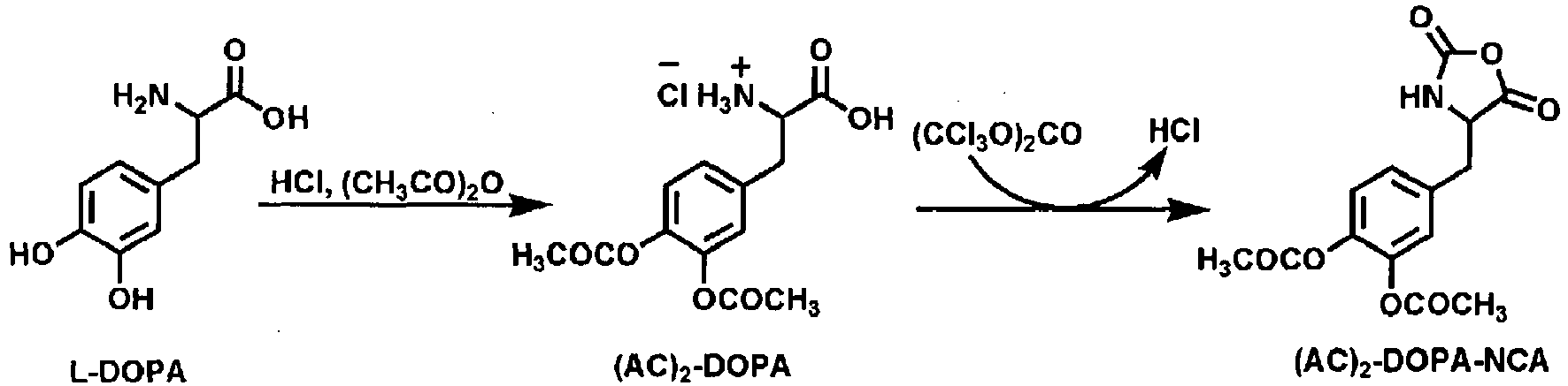

- After mounting a reflux condenser, methoxy polyethyleneglycol carboxyl (PEG-COOH, 5000) (10 g) was dissolved in methylene chloride (CHCl2) (50 ml) in a 250 ml flask. Then, N-hydroxysuccinimide (NHS) (0.52 g) and dicyclohexylcarbodiimide (DCC) (0.74 g) were added thereto, followed by a reaction at room temperature for 20 hours. After removing dicyclohexylurea through filtration, this material was precipitated in diethylether, resulting in polyethyleneglycol in an activated state (PEG-NHS). (Yield =87%) 1H NMR (300MHz, CDCl3): 4.1 (b,-CO-CH2-CH2-CH 2-O-), 3.5-3.8 (m, -CH 2CH 2O-), 2.8 (b, -CO-CH 2-CH 2-CO-), 1.8 (b, -CO-CH2-CH 2-CH2 - CH 2-CH2-O-), 1.2 (b, -CO-CH2-CH2-CH 2-CH2-CH2-O-).
- After mounting a reflux condenser, methoxy polyethyleneglycol (PEG-OH) (15.23 g) was dissolved in chloroform (CHCl3) (15 ml) in a 100 ml flask. Then, the solution was treated using hexamethylene diisocyanate (HMDI) (60 ml), followed by a reaction for 24 hours to prepare a polymer. After completing the reaction, the polymer was precipitated in petroleum ether to purify the same and, after washing with petroleum ether (400 ml) three times, the washed material was dissolved again in chloroform (CHCl3) (20 ml). After then, the solution was precipitated again in petroleum ether (500 ml) to purify the same. The foregoing procedure was repeated 10 times, followed by drying under vacuum, resulting in polyethyleneglycol in an activated state (Yield = 80%).
- The activated polyethyleneglycol (PEG-NHS) (2 g) obtained in Example <1-1> was dissolved in chloroform (200 ml). Then, after dissolving polyethyleneimine (Alfa Aesar, 1800 Da, 0.5 g) in chloroform (50 ml), the polyethyleneglycol solution was added drop by drop thereto to conduct a covalent bonding reaction between polyethyleneglycol and polyethyleneimine. Here, the reaction was executed for 24 hours and, after completing the reaction, the resultant product was concentrated to reach a total volume of 30 ml by a vacuum concentrator. Following this, the concentrated material was precipitated in diethylether to obtain a polyethyleneimine-graft - polyethyleneglycol (PEI-graft-PEI). (Yield=85%) 1H NMR (300 MHz, D2O). As a result of measurement (-CH 2CH 2O- of PEG at 3.5-3.8 ppm and -CH 2CH 2NH- of PEI at 2.5-3.2 ppm), Mn of PEI-graft-PEG was found to be about 41,800 Da. It was presumed that each PEI-graft-PEG has almost 8 (hereinafter, referred to as PEI1-graft-PEG8). 1H NMR (300 MHz, D2O): 3.5-3.8 (m, -CH 2CH 2O-), 3.3 (s, CH 3O-), 2.5-3.2 (m, -CH 2CH 2 NH-).
- L-DOPA (3 g) was suspended in glacial acetic acid (100 ml), followed by purging dried gas of hydrochloric acid at room temperature for 5 hours. After adding acetic acid anhydride (3 ml) and reacting at room temperature for 1 hour 30 minutes, 3 ml of acetic acid anhydride was further added and a reaction was conducted in an oil bath at 60°C for 30 minutes. The reaction product was concentrated in a vacuum concentrator and unreacted acetic acid anhydride was removed by adding ethanol. Thereafter, the remaining product was precipitated in diethylether to obtain DOPA amino acid having a protected hydroxyl group (DOPA(Ac)2). (Yield=80%) 1H NMR (300MHz, D2O): 6.7-6.9 (m, -C6 H 3(OH)2), 4.0 (m, C6H3(OH)2-CH2-CH(N-)-C(O)N-), 3.2 (m, C6H3(OH)2 -CH 2-CH-), 2.4 (s, CH 3(CO)-).
- 0.5g of the DOPA amino acid having a protected hydroxyl group synthesized in Example <1-3> as well as 0.5 g of triphosgene were dispersed in THF (50 ml) and reacted in an oil bath at 60°C. Next, the reaction product was precipitated in hexane (800 ml), dissolved in THF (50 ml) and precipitated again in hexane. The foregoing procedure was repeated three times. After purifying, the product was dried using a vacuum dryer, resulting in DOPA N-carboxyl anhydride (DOPA(AC2)-NCA). (Yield=65%) 1H NMR (300MHz, DMSO): 6.2-6.9 (m, -C6 H 3(OH)2), 4.3 (t,-NHCH CO-), 3.7 (m, C6H3(OH)2-CH2-CH(N-)-C(O)N-), 3.3 (m, C6H3(OH)2-CH 2-CH-), 2.4 (s, CH 3 (CO)-).
- 0.5g of the polyethyleneimine-graft-polyethyleneglycol (PEI-graft-PEG) prepared in Example <1-2> and the DOPA N-carboxyl anhydride (DOPA(AC2)-NCA) prepared in Example <1-4> were used in different molar ratios (1:5 and 1:15, respectively) and reacted in THF solvent, thus synthesizing a polyethyleneimine-graft - (polyethyleneglycol;poly3,4-dihydroxyphenylalanine) (Yield, molar ratio 1:5 = 85%, molar ratio 1:15 = 87%).
- After dissolving 0.5 g of the polyethyleneimine-graft - (polyethyleneglycol;poly3,4-dihydroxyphenylalanine) synthesized in Example <1-5> in DMF (30 ml), 8 ml of piperidine was added thereto. 15 minutes after reaction, the reaction product was precipitated in diethylether to obtain a polyethyleneimine-graft - (polyethyleneglycol;poly3,4-dihydroxyphenylalanine) in which a protective hydroxyl group of DOPA is de-protected. (Yield, molar ratio 1:5 = 80% [
FIGS. 2 and3 ], molar ratio 1:15 = 81% [FIGS. 4 and5 ]). - The following Table 1 shows assayed results of a structure of the mussel adhesive protein-mimetic copolymer of the present invention and characteristics thereof, wherein the structure of a polymeric copolymer and characteristics thereof have been assayed through 1H-NMR and UV-Vis spectroscopy and using a fluorescent label.
- Table 1
[Table 1] Analysis of structure and characteristics of mussel adhesive protein-mimetic copolymer Name of sample Copolymer Number average molecular weight Molar ratio along with individual materials Mn a CMC (g/L)b PEG Mn PEI Mn PEG PEI DOPA MIL0 copolym er PEI-graft-PEG 5,000 1,800 8 1 - 41,800 - MIL1 copolym er PEI-graft-(PE G;PDOPA) 5,000 1,800 8 1 5 42,800 0.015 MIL2 copolym er PEI-graft-(PE G;PDOPA) 5,000 1,800 8 1 15 44,800 0.005 aMn: number average molecular weight, which is assayed using 1H-NMR and UV-Vis spectrabCMC: Critical Micelle Concentration, which is assayed using a fluorescent label (Hoechst 33342) - 10 mg of iron oxide nanoparticles (Fe3O4) which were synthesized in an organic solvent and stabilized with oleic acid, as well as 60 mg of mussel adhesive protein-mimetic copolymers (MIL1 and MIL2, respectively), were dispersed in 10 ml of chloroform (CHCl3) and agitated at room temperature for 30 minutes. After evaporating chloroform (CHCl3) and adding distilled water to the residue, the dispersed product was filtrated through a 200 nm syringe filter (MCE syringe filter, Fisher Scientific). After centrifuging to remove unreacted stabilizer, filtration was repeated 3 to 5 times using a spin filter (Millipore, 10K NMWL, 10000xg, 10 min). The resultant product was dispersed in an aqueous system having pH 7 (0.01M PBS: Phosphate Buffered Saline solution).
- The iron oxide (Fe3O4) stabilized using the mussel adhesive protein-mimetic copolymer, which has physical properties shown in the following Table 2, according to the methods proposed in Preparative Examples 1 and 2, was dispersed in water to prepare a colloidal solution.
- Table 2
[Table 2] Colloidal solution of iron oxide (Fe3O4) stabilized using mussel adhesive protein-mimetic copolymer Name of sample Diameter of iron oxide nanoparticle (nm)1) Number average molecular weight Molar ratio Hydrodyna mic diameter (nm)2) PEG M n PEG M n PEG PEI DOPA PD1 colloidal solution 10 5000 1800 7 1 20 30 PD2 colloidal solution 10 2000 1800 6 1 20 16 1) Diameter of iron oxide nanoparticle: a diameter of iron oxide particle only excluding a dispersion stabilizing material, which is measured by Transmission Electron Microscopy (TEM)2) Hydrodynamic diameter: a diameter of a material dispersed in water and containing a dispersion stabilizing material, which is a number average diameter measured in a dispersed state in an aqueous system by a dynamic light scattering (DLS) device (Malvern Co., Zetasizer NanoZS) - To the dispersed and stabilized iron oxide (Fe3O4) nanoparticles shown in Table 2, PBS powder was added to reach a concentration of 0.01M. In order to identify bio- applicability, MTT assay was executed with Hep G2 cells.
- After passing PD1 and PD2 containing PBS through a 0.2µm sterile syringe filter, respectively, a concentration was determined using a inductively coupled plasma-atomic emission spectroscopy (ICP-AES). The PD1 and PD2 were taken with an aliquot by 200 µL, respectively, and the aliquot was mixed with 200 µL of a medium (DMEM: Dulbeco"s Modified Eagles"s Medium 89%, Pluronic® F-127 10%, AA: antibiotic-antimycotic 1%) to prepare Sample 0.350 µL of the
sample 0 was mixed with 350 µL of a medium to prepareSample 1. Also, 350 µL ofSample 1 was mixed with 350 µL of the medium to prepare another sample namedSample 2. As such, the initial concentration was diluted to 1/2 thereof to prepare nine (9) samples from 1 to 9 for PD1 and PD2, respectively. Moreover,Sample 10 was 100% medium with neither PD1 nor PD2. As controls, PBS solutions were prepared for PD1 and PD2, respectively, according to the foregoing procedures (see TABLE 3). - Table 3
[Table 3] Iron oxide (Fe3O4) nanoparticles colloid sample for cell viability test No. PD1 colloid PD2 colloid Control Sample + medium Iron content (ppm) Sample + medium Iron content (ppm) Sample + medium Crude solution 1680 Crude solution 1320 Crude solution (0.01M PBS solution) 0 Crude solution, 350 µL + medium 350 µL 840 Crude solution, 350 µL + medium 350 µL 660 Crude solution, 350 µL + medium 350 µL 1 No. 0, 200 µL + medium 200 µL 420 No. 0, 200 µL + medium 200 µL 330 No. 0, 200 µL + medium 200 µL 2 No. 1, 200 µL + medium 200 µL 210 No. 1, 200 µL + medium 200 µL 165 No. 1,200 µL + medium 200 µL 3 No. 2, 200 µL + medium 200 µL 105 No. 2, 200 µL + medium 200 µL 82.5 No. 2, 200 µL + medium 200 µL 4 No. 3, 200 µL + medium 200 µL 52.5 No. 3, 200 µL + medium 200 µL 41.25 No. 3, 200 µL + medium 200 µL 5 No. 4, 200 µL + medium 200 µL 26.25 No. 4, 200 µL + medium 200 µL 20.63 No. 4, 200 µL + medium 200 µL 6 No. 5, 200 µL + medium 200 µL 13.13 No. 5, 200 µL + medium 200 µL 10.31 No. 5, 200 µL + medium 200 µL 7 No. 6, 200 µL + medium 200 µL 6.56 No. 6, 200 µL + medium 200 µL 5.16 No. 6, 200 µL + medium 200 µL 8 No. 7, 200 µL + medium 200µL 3.28 No. 7, 200 µL + medium 200 µL 2.58 No. 7, 200 µL + medium 200 µL 9 No. 8, 200 L + medium 200 µL 1.64 No. 8, 200 µL + medium 200 µL 1.29 No. 8, 200 µL + medium 200 µL 10 Medium 400 µL 0 Medium 400 µL 0 Medium 400 µL - After proliferating HepG2 cells through subculture, the cells were charged in 96-well plate to obtain 100,000 cells/well. Here, among these 96 wells, only 60 wells were used except those in periphery of the plate. In order to fix HepG2 cells on the bottom of the well plate, these were incubated at 37°C for 24 hours.
- The medium was removed from the well plate in which HepG2 prepared in <1-2> was incubated. Each well of the well plate free from the medium, was charged with 100 µL of each of
respective samples 1 to 10 of PD1, PD2 and controls, respectively, which were prepared in <1-1> shown in Table 3. Here, each of the samples and controls was prepared in three (3) folds. After charging the sample, incubation was executed at 37°C for 24 hours. - MTT test is an experimentation to determine cell viability using a principle wherein a yellow MTT reagent based on tetrazolium salt is altered into violet formazan crystals by metabolically activated cells.
- The MTT (3-(4,5-dimethylthiazol-2-yl)-2,5-diphenyl-2H-tetrazolium bromide) reagent used in the cell viability test was dissolved in a water bath at 37°C, 1 hour before the test. To each well of the 96-well plate into which the samples and controls prepared in <1-3> were introduced, 20 µL of the prepared MTT reagent was added. Here, special precaution is needed to prevent the test well from being exposed to light. In order to identify the metabolically activated cells, the test well was incubated at 37°C for 4 hours after addition of the MTT reagent. After incubation, in order to dissolve violet formazan crystals, 100 µL of a soluble solution was added to each well and incubation was continued at 37°C for 24 hours. After incubation, optical densities at 550 nm and 690 nm, respectively, were measured using a microplate reader (Molecular Devices Co., Spectra Max 190) and cell viability was calculated from the measured optical densities. The cell viability may be calculated according to the following
Equation 1.
- ODi,550: Optical density at 550 nm of well plate No. i in which the sample is introduced.
- ODi,690: Optical density at 690 nm of well plate No. i in which the sample is introduced.
- ODSi,550: Optical density at 550 nm of control well plate No. i.
- ODSi,690: Optical density at 690 nm of control well plate No. i.
-
FIG. 6 is graphs illustrating results of cell viability test. In cell viability test, an acceptable error range (commonly, tolerance) is generally ±20%. From results of the present test, it can be found that 80% or more of cell viability was exhibited in the most part of a concentration section to be tested. Such results - demonstrated that the iron oxide (Fe3O4) nanoparticles stabilized by the mussel adhesive protein-mimetic copolymer have no cellular toxicity and may be safely used in vivo.
- The colloidal solutions containing iron oxide (Fe3O4) nanoparticles stabilized using the mussel adhesive protein-mimetic copolymers were prepared according to the method in Example 1.
- Table 4
[Table 4] Iron oxide (Fe3O4) nanoparticle colloidal solution stabilized by mussel adhesive protein-mimetic copolymer for example 2 Name of sample Diameter of iron oxide nanoparti cle (nm) Number average molecular weight Molar ratio Hydrody namic diameter (nm) Dose of administr ation (mg Fe/kg mouse) PEG M n PEG M n PEG PEI DOPA PDIE1 colloidal solution 10 2000 1800 8 1 15 26 10.4 PDIE2 colloidal solution 10 5000 1800 4 1 15 51 5.2 PDIE3 colloidal solution 20 5000 1800 8 1 20 48 5.2 PDIE4 colloidal solution 10 5000 1800 7 1 20 38 10.4 - Adding a phosphate buffered saline (PBS) to each of the colloidal solutions, wherein iron oxide (Fe3O4) nanoparticles are dispersed and stabilized therein, as shown in Table 4, a contrast agent was prepared. In order to evaluate usability of the prepared contrast agent as an MRI contrast agent, a loop coil was used in an MRI diagnosis device (Trio 3.0T, Simens) to assay in vivo T2 contrast imaging performance.
- Evaluation of in vivo contrast imaging performance was executed with nude mouse (Balb/nude). The nude mouse weighed 30 g. After anesthetizing, the mouse was horizontally placed in the MRI diagnosis device and observed along a cross-section thereof. MRI images of the nude mouse were subjected to measurement before and 24 hours after injecting the contrast agent, and lymph nodes before and after injecting the contrast agent were observed and compared to each other. PBS powder was added to the colloidal solution to reach a concentration of 0.01 M, to prepare a desired contrast agent. Iron content was analyzed through ICP-AES and an administration dose was calculated (Table 4) in consideration of the weight of the mouse, to prepare a contrast agent with a total volume of 500 µL. The contrast agent was injected through a tail vein of the mouse.
- In vivo T2 imaging performance was determined using T2Me3d pulse sequence provided by Siemens Co. and particular parameters are as follows.
- TR (repetition time) = 40.0 msec, TE (echo time) = 22.0 msec, FOV = 49 mm x 70 mm, Matrix size = 256 x 180, slice thickness = 0.6 mm, number of acquisition = 6
-
FIGS. 7 to 10 partially illustrate in vivo MRI images obtained under the foregoing conditions. - In order to quantify T2 attenuation effects of the prepared contrast agent, one cross-section was selected from the highest visible lymph nodes, that is, inguinal and brachial lymph nodes, and discolored parts of the lymph node were selected as ROI (Region of Interest). In the selected parts, signal intensity was measured. Since an overall signal intensity of the acquired MRI images is altered whenever it is measured, it is difficult to evaluate the contrast imaging performance based on an absolute value of the signal intensity (S: signal intensity). For the inguinal lymph node, the right hind leg muscle was used as a control (N: noise). On the other hand, for the brachial lymph node, the right paw muscle was used as a control (N: noise). Compared to the controls, signal to noise ratio(SNR) was calculated. By comparing SNR values before and after using the contrast agent, T2
signal attenuation ratio (ΔR2) was calculated. The results are illustrated by the graphs inFIG. 11 . The followingEquation 2 is a method for calculation of T2 signal attenuation ratio (ΔR2).


- From
FIGS. 7 to 10 , it can be seen that the lymph nodes of the nude mouse darkened after administration of the contrast agent. The Tables 5 and 6 include signal to noise ratio(SNR) and signal attenuation ratios (ΔR2) before and after administration of a contrast agent sample to each of the inguinal lymph node and the brachial lymph node.FIG. 11 illustrates the signal to noise ratio (SNR) before and after administration of the contrast agent sample. It was found that signal attenuation after administration of the contrast agent was noticeably high, compared to that before administration thereof. - Table 5
[Table 5] T2 signal in inguinal lymph node of a nude mouse by contrast agents Name of sample Inguinal lymph node LEFT RIGHT Before administration After administration Signal attenuation ratio Before administration After administration Signal attenuation ratio SNR SNR SNR SNR PDIE1 contrast agent 3.94 0.21 94.8% 2.68 1.26 53.0% PDIE2 contrast agent 1.68 0.31 81.5% 2.15 0.30 86.2% PDIE3 contrast agent 2.92 0.23 92.3% 1.29 0.25 81.0% PDIE4 contrast agent 2.65 0.84 68.5% 1.74 0.63 63.9% - Table 6
[Table 6] T2 signal in brachial lymph node of a nude mouse by contrast agents Name of sample Brachial lymph node LEFT RIGHT Before administration After administration Signal attenuation ratio Before administration After administration Signal attenuation ratio SNR SNR SNR SNR PDIE1 contrast agent 2.01 0.61 69.5% 1.87 0.46 75.6% PDIE2 contrast agent 1.39 0.32 76.8% 1.40 0.57 59.4% PDIE3 contrast agent 1.59 0.73 54.3% 1.43 1.00 29.9% PDIE4 contrast agent 1.37 0.33 76.1% 1.30 0.68 47.7% -
FIG. 12 are TEM photographs illustrating iron oxide (Fe3O4) nanoparticles before reformed by MIL2 (a) and after reformed by MIL2(b). - According to the TEM photographs in
FIG. 12 , it can be understood that the iron oxide (Fe3O4) nanoparticles stably dispersed in water, have substantially the same morphology and size as those of iron oxide (Fe3O4) nanoparticles dissolved in a hydrophobic solvent. Further, a hydrodynamic diameter of each of the iron oxide (Fe3O4) nanoparticle colloids is also illustrated inFIG. 12 . The hydrodynamic diameter of the iron oxide (Fe3O4) nanoparticle colloid is a number average hydrodynamic diameter measured by a dynamic light scattering (DLS) method and is observed to be uniform in size. -
FIG. 13 illustrates stability of iron oxide (Fe3O4) nanoparticle colloids stably dispersed in an aqueous medium by a mussel adhesive protein-mimetic copolymer, along with various pH values (a) and ionic concentrations (b). Here, hydrodynamic diameters were measured at various pH values and ionic concentrations through DLS. It can be identified that iron oxide(Fe3O4) nanoparticle colloids stably dispersed in an aqueous medium using the mussel adhesive protein-mimetic copolymer (MIL1 (▲), MIL2 (○)) are relatively stable at various concentrations and pH values, compared to iron oxide (Fe3O4) nanoparticles without application of the mussel adhesive protein-mimetic copolymer (MIL0 (◆)). - A colloidal solution of iron oxide (Fe3O4) nanoparticles stabilized by the mussel adhesive protein-mimetic copolymer was prepared according to the methods in Example 1 and 2.
- Table 7
[Table 7] Colloidal solution of iron oxide (Fe3O4) nanoparticles stabilized by mussel adhesive protein-mimetic copolymer for Example 4. Name of sample Diameter of iron oxide nanoparticl e(nm) PEGnum ber average molecular weight PEInum ber average molecular weight Molar ratio Hydrody namic diameter (nm) Dose of administration (mg Fe/kg rabbit) PEG PEI DOPA PDIE5co lloidal solution 14 5000 1800 10 1 20 28 10.4 - The colloidal solution in Table 7 was diluted by using a saline solution (0.9% NaCl) for injection, and then a contrast agent having an iron concentration of 6240 ppm and a total volume of 5 mL was prepared while the iron concentration was assayed through ICP-AES and the dose of administration was calculated considering the weight of rabbits. The contrast agent was intravenously injected through an ear of the rabbit. For experiment, metastatic cancer models were established by expressing VX2 cancer cells in iliac lymph nodes of 12 New Zealand white rabbits, and then used to evaluate performance of the contrast agent.
- The rabbit with metastatic cancer was weighted about 3kg. After anesthetizing, the rabbit was horizontally placed in the MRI device (Trio 3.0T, Simens, knee coil), and then cross sections thereof were observed. MRI images of the rabbit were measured before and 24 hours after injecting the contrast agent, and the iliac lymph nodes before and after injecting the contrast agent were observed and compared to each other. The iliac lymph nodes of every rabbits were isolated and stained with hematoxylin-eosin for examination by a pathologist.
- Specific parameters of the MRI device are as follows.
- TR(repetition time)=6.2msec, TE(echo time)=2.4msec, FOV=145mmx145mm, Matrix size,=129x154, slice thickness=6.0mm, number of acquisition=1
- Table 8
[Table 8] Evaluation results for metastatic cancer model rabbits Rabbit number MRI scanning + Histo-pathology+ MRI scanning - Histo-pathology- 1) 001 3 3 1 1 002 3 3 2 2 003 3 3 1 1 004 1 1 0 0 005 2 2 1 1 006 3 3 0 0 007 1 1 1 1 008 3 3 2 2 009 3 3 1 1 010 2 2 1 1 011 3 3 1 1 012 2 2 1 1 Total 29 29 12 12 Sensitivity: 100%2) Specificity: 100%3) 1) MRI scanning +: The number of lymph nodes evaluated to be metastatic cancer on the MRI readoutHistopathology +: The number of lymph nodes examined to be metastatic cancer from histopathology resultsImage scanning -: The number of lymph nodes evaluated as normalHistopathology -: The number of lymph nodes examined as normal2) Sensitivity: Ratio of the number of lymph nodes evaluated as cancer by MRI scanning to number of lymph nodes examined as cancer by histopathology3) Specificity: Ratio of number of normal lymph nodes evaluated by MRI scanning to number of normal lymph nodes examined by histopathology - The evaluation results for the metastatic cancer model rabbits are shown in Table 8 and
FIG. 14 . Table 8 shows the number of lymph nodes with metastasis cancer and the number of normal lymph nodes for the 12 rabbits metastatic cancer models. The sensitivity and specificity was 100%, respectively.FIG. 14 shows images of contrast enhanced lymphography and result of histopathology of Rabbit No. 002, before and after injecting the contrast agent. The parts marked in (+) indicate lymph nodes evaluated to be metastatic cancer as the MRI scanning, and the parts marked in (-) indicate lymph nodes examined to be normal as the MRI scanning. - The mussel adhesive protein-mimetic copolymer used in the present invention has multiple units of DOPA in a molecule. DOPA can interact with an inorganic nanoparticle and exhibit high bond strength to the nanoparticles' surface. Additionally, the above copolymer has a positive charge, it may impart electrostatic bonding force to the surface of nanoparticles having a negative charge. Moreover, since numerous polyethyleneglycol molecules having hydrophilic property are bonded to the branches of polyethyleneimine, high aqueous dispersion stabilization may be achieved through hydrophilic property and stereoscopic effects. The PEG on the surface make the iron oxide nanoparticles circulate in the body for a long-time and so many of the iron oxide nanoparticles can reach to the lymph nodes. For better T2 MR imaging, the lymph nodes have to take nanoparticles up as many as possible. So it is crucial of the iron oxide nanoparticles circulate in the body for a long time. The contrast agent including iron oxide nanoparticles reformed by mussel adhesive protein-mimetic copolymer seems to be more taken up and so shows more darkening in the normal lymph nodes and less darkening in the metastatic lymph nodes.
- In conclusion, the iron oxide nanoparticles reformed by mussel adhesive protein-mimetic copolymer have a great potential for the detection of lymph node metastasis as a contrast agent for contrast enhanced lymphography.
Claims (12)
- A contrast agent comprising:iron oxide nanoparticles dispersed and stabilized in an aqueous medium by a polyethyleneimine-graft-(polyethyleneglycol;poly3,4-dihydroxyphenylalanine) for use in contrast enhanced lymphography for diagnosing cancer of lymph nodes.
- The contrast agent for use according to claim 1, wherein the iron oxide nanoparticles have a diameter of 1 to 25 nm.
- The contrast agent for use according to claim 1, wherein the iron oxide nanoparticles dispersed in the aqueous medium have a hydrodynamic diameter of 3 to 100 nm.
- The contrast agent for use according to claim 1, wherein the polyethyleneimine-graft-(polyethyleneglycol;poly3,4-dihydroxyphenylalanine) comprises of polyethyleneglycol, polyethyleneimine and poly3,4-dihydroxyphenylalanine, and wherein the polyethyleneglycol has a number average molecular weight of 300 to 50,000 and a hydroxyl group or carboxyl group at one end thereof.
- The contrast agent for use according to claim 1, wherein the polyethyleneimine-graft-(polyethyleneglycol;poly3,4-dihydroxyphenylalanine) comprises of polyethleneglycol, polyethyleneimine and poly3,4-dihydroxyphenylalanine, and wherein the polyethyleneimine is a branch type polyethyleneimine having a number average molecular weight of 100 to 10,100.
- The contrast agent for use according to claim 1, wherein the polyethyleneimine-graft-(polyethyleneglycol;poly3,4-dihydroxyphenylalanine) comprises of polyethyleneglyocl, polyethyleneimine and poly3,4-dihydroxyphenylalanine, and wherein the poly3,4-dihydroxyphenylalanine (PDOPA) is selected from L-PDOPA synthesized from N-carboxyl anhydride of L-DOPA(L-3,4-dihydroxyl phenylalanine) and D-PDOPA synthesized from N-carboxyl anhydride of D-DOPA(D-3,4-dihydroxyl phenylalanine) and L,D-PDOPA synthesized from N-carboxyl anhydride of L,D-DOPA.
- The contrast agent for use according to claim 1, wherein the polyethyleneimine-graft-(polyethyleneglycol;poly3,4-dihydroxyphenylalanine) includes a polyethyleneglycol unit represented by Structure A, a polyethyleneimine unit represented by Structure B, and a poly3,4-dihydroxyphenylalanine unit represented by Structure C:


- The contrast agent for use according to claim 7, wherein the polyethyleneimine unit is represented by the following structure:

- A method for contrast enhanced lymphography, using the contrast agent of any one of claims 1 to 8.
- The method of claim 9, wherein the method of contrast enhanced lymphography is executed by contrast imaging the lymph node utilizing a variation or difference in intensity of an MRI signal between an MRI signal of a lymph node before administration of the contrast agent and another MRI signal of the lymph node after administration of the contrast agent.
- The method of claim 10, wherein the administration of the contrast agent is conducted by intravenous injection.
- The method of claim 10, wherein the variation in intensity of an MRI signal in the lymph node is based on a change in T2 signal intensity.
Applications Claiming Priority (2)
| Application Number | Priority Date | Filing Date | Title |
|---|---|---|---|
| KR1020110060438A KR101721570B1 (en) | 2011-06-22 | 2011-06-22 | MRI Contrast Agent for Lymph Node Based on Iron Oxide Nanoparticles and Method for Imaging Lymph Node Using The Same |
| PCT/KR2012/004861 WO2012177039A2 (en) | 2011-06-22 | 2012-06-20 | Mri contrast agent for lymphography based on iron oxide nanoparticles and method for imaging lymph node using the same |
Publications (3)
| Publication Number | Publication Date |
|---|---|
| EP2723392A2 EP2723392A2 (en) | 2014-04-30 |
| EP2723392A4 EP2723392A4 (en) | 2014-12-17 |
| EP2723392B1 true EP2723392B1 (en) | 2016-09-28 |
Family
ID=47423073
Family Applications (1)
| Application Number | Title | Priority Date | Filing Date |
|---|---|---|---|
| EP12802287.8A Active EP2723392B1 (en) | 2011-06-22 | 2012-06-20 | Mri contrast agent for lymphography based on iron oxide nanoparticles and method for imaging lymph node using the same |
Country Status (10)
| Country | Link |
|---|---|
| US (1) | US8906346B2 (en) |
| EP (1) | EP2723392B1 (en) |
| JP (1) | JP5799161B2 (en) |
| KR (1) | KR101721570B1 (en) |
| CN (1) | CN103458932B (en) |
| AU (1) | AU2012274234A1 (en) |
| BR (1) | BR112013023812A2 (en) |
| CA (1) | CA2827752C (en) |
| MX (1) | MX2013009212A (en) |
| WO (1) | WO2012177039A2 (en) |
Families Citing this family (14)
| Publication number | Priority date | Publication date | Assignee | Title |
|---|---|---|---|---|
| KR101685646B1 (en) * | 2010-12-29 | 2016-12-13 | 한화케미칼 주식회사 | Biocompatible Agent for Dispersing Nanoparticles into Aqueous Solution using Mussel Adhesive Protein Mimic polymer |
| CA2935858C (en) * | 2014-01-07 | 2022-06-21 | Colorobbia Italia S.P.A. | Magnetic nanoparticles functionalized with cathecol, production and use thereof |
| KR101510835B1 (en) * | 2014-01-09 | 2015-04-10 | 원광대학교산학협력단 | Mussel adhesive protein derived chitooligosaccharide ligand and contrast agent comprising the same |
| KR101508600B1 (en) * | 2014-01-09 | 2015-04-07 | 원광대학교산학협력단 | Bioinspired chitooligosaccharide ligand and contrast agent comprising the same |
| CN104258421B (en) * | 2014-09-12 | 2016-09-21 | 温州大学 | A kind of noble metal/paramagnetic metal composite nanoparticle and application thereof |
| KR101765335B1 (en) * | 2014-09-26 | 2017-08-22 | 서울대학교산학협력단 | MRI Contrast Agent for Contrasting Cancer Cell |
| KR101720046B1 (en) * | 2014-09-26 | 2017-03-29 | 서울대학교산학협력단 | Self-Assembled Pharmaceutical Composition for Photodynamic Therapy |
| KR101722503B1 (en) * | 2015-03-13 | 2017-04-04 | 포항공과대학교 산학협력단 | PH-responsive nanoparticle using mussel adhesive protein for drug delivery and method for preparing the same |
| CN105400517B (en) * | 2015-10-30 | 2018-03-13 | 玉林师范学院 | Detect difunctional magnetic fluorescence probe preparation method and its application of cadmium ion |
| CN110496231B (en) * | 2018-05-16 | 2020-09-18 | 浙江大学 | An amphiphilic polymer nanomicelle containing polydopa amino acid chelating ferric ions and its application |
| EP3860342A4 (en) * | 2018-10-03 | 2022-08-03 | Prescient Pharma LLC | COMPOSITIONS AND METHODS FOR ISOLATING CIRCULATING CELLS |
| KR102386849B1 (en) | 2019-10-14 | 2022-04-13 | 포항공과대학교 산학협력단 | Bioresponsive therapeutic antibody delivery platform for immunotherapy and method for preparing the same |
| EP4403192A4 (en) * | 2021-09-18 | 2025-07-30 | Nonsuch Biotechnology Inc | Use of iron oxide nanoparticles in the production of a parathyroid and/or lymph node contrast agent |
| KR20240124228A (en) * | 2023-02-08 | 2024-08-16 | 주식회사 인벤테라 | Lymphatic contrast agent without venous contamination |
Family Cites Families (9)
| Publication number | Priority date | Publication date | Assignee | Title |
|---|---|---|---|---|
| JPS6323670A (en) * | 1986-04-25 | 1988-01-30 | バイオ−ポリマ−ズ インコ−ポレ−テツド | Adhesive coating composition and its production |
| US4908404A (en) * | 1988-08-22 | 1990-03-13 | Biopolymers, Inc. | Synthetic amino acid-and/or peptide-containing graft copolymers |
| JPH10120597A (en) * | 1996-10-22 | 1998-05-12 | Eiken Chem Co Ltd | Lymph node highly accumulating colloid particles |
| US20020012698A1 (en) * | 1999-09-01 | 2002-01-31 | Edmund Bauerlein | Magnetosomes, method for making and using |
| WO2006073419A2 (en) * | 2004-04-01 | 2006-07-13 | Gang Zheng | Lipoprotein nanoplatforms |
| US20090280063A1 (en) * | 2008-05-09 | 2009-11-12 | General Electric Company | Novel pei-peg graft copolymer coating of iron oxide nanoparticles for inflammation imaging |
| EP2289553A1 (en) * | 2009-09-01 | 2011-03-02 | ETH Zurich | Dispersant stabilization of inorganic non-metallic particles |
| JP2011079781A (en) * | 2009-10-08 | 2011-04-21 | Keio Gijuku | Sentinel lymph node contrast medium or identifying agent and thermotherapy agent |
| KR101685646B1 (en) * | 2010-12-29 | 2016-12-13 | 한화케미칼 주식회사 | Biocompatible Agent for Dispersing Nanoparticles into Aqueous Solution using Mussel Adhesive Protein Mimic polymer |
-
2011
- 2011-06-22 KR KR1020110060438A patent/KR101721570B1/en active Active
-
2012
- 2012-06-20 WO PCT/KR2012/004861 patent/WO2012177039A2/en not_active Ceased
- 2012-06-20 AU AU2012274234A patent/AU2012274234A1/en not_active Abandoned
- 2012-06-20 CA CA2827752A patent/CA2827752C/en not_active Expired - Fee Related
- 2012-06-20 US US14/000,301 patent/US8906346B2/en active Active
- 2012-06-20 CN CN201280011330.XA patent/CN103458932B/en active Active
- 2012-06-20 JP JP2014503614A patent/JP5799161B2/en active Active
- 2012-06-20 BR BR112013023812A patent/BR112013023812A2/en not_active IP Right Cessation
- 2012-06-20 EP EP12802287.8A patent/EP2723392B1/en active Active
- 2012-06-20 MX MX2013009212A patent/MX2013009212A/en not_active Application Discontinuation
Also Published As
| Publication number | Publication date |
|---|---|
| AU2012274234A1 (en) | 2013-08-22 |
| CN103458932B (en) | 2015-12-09 |
| CN103458932A (en) | 2013-12-18 |
| EP2723392A4 (en) | 2014-12-17 |
| WO2012177039A3 (en) | 2013-04-04 |
| JP2014514301A (en) | 2014-06-19 |
| KR20130008095A (en) | 2013-01-22 |
| KR101721570B1 (en) | 2017-03-30 |
| EP2723392A2 (en) | 2014-04-30 |
| MX2013009212A (en) | 2013-08-29 |
| WO2012177039A2 (en) | 2012-12-27 |
| CA2827752C (en) | 2016-05-17 |
| US20140023594A1 (en) | 2014-01-23 |
| US8906346B2 (en) | 2014-12-09 |
| BR112013023812A2 (en) | 2016-12-13 |
| CA2827752A1 (en) | 2012-12-27 |
| JP5799161B2 (en) | 2015-10-21 |
Similar Documents
| Publication | Publication Date | Title |
|---|---|---|
| US8906346B2 (en) | MRI contrast agent for lymphography based on iron oxide nanoparticles and method for imaging lymph node using the same | |
| EP2658896B1 (en) | Biocompatible agent for dispersing nanoparticles into an aqueous medium using mussel adhesive protein-mimetic polymer | |
| Kim et al. | A cancer-recognizable MRI contrast agents using pH-responsive polymeric micelle | |
| Liu et al. | A superparamagnetic polymersome with extremely high T2 relaxivity for MRI and cancer-targeted drug delivery | |
| Bae et al. | Bioinspired synthesis and characterization of gadolinium-labeled magnetite nanoparticles for dual contrast T 1-and T 2-weighted magnetic resonance imaging | |
| Zhang et al. | Imaging and cell targeting characteristics of magnetic nanoparticles modified by a functionalizable zwitterionic polymer with adhesive 3, 4-dihydroxyphenyl-l-alanine linkages | |
| Scialabba et al. | Folate targeted coated SPIONs as efficient tool for MRI | |
| Maurizi et al. | Influence of surface charge and polymer coating on internalization and biodistribution of polyethylene glycol-modified iron oxide nanoparticles | |
| Park et al. | Multi-modal transfection agent based on monodisperse magnetic nanoparticles for stem cell gene delivery and tracking | |
| CN103143043B (en) | A kind of preparation method of Fe3O4/Au composite nanoparticles | |
| Yang et al. | A direct surface modification of iron oxide nanoparticles with various poly (amino acid) s for use as magnetic resonance probes | |
| Zarei et al. | Theragnostic magnetic core-shell nanoparticle as versatile nanoplatform for magnetic resonance imaging and drug delivery | |
| Sun et al. | A unique nanogel-based platform for enhanced dual mode tumor MR/CT imaging | |
| Yang et al. | Biodegradable yolk-shell microspheres for ultrasound/MR dual-modality imaging and controlled drug delivery | |
| Abushrida et al. | A simple and efficient method for polymer coating of iron oxide nanoparticles | |
| Nguyen et al. | Integration of iron oxide nanoparticles and polyaspartamide biopolymer for MRI image contrast enhancement and an efficient drug-delivery system in cancer therapy | |
| KR101143257B1 (en) | MRI contrast agent coated with mannose-conjugated polymer and method for producing the same | |
| Amraee et al. | Curcumin Coated Ultra-Small Iron Oxide Nanoparticles as T1 Contrast Agents for Magnetic Resonance Imaging of Cancer Cells | |
| Xu et al. | Modification of MR molecular imaging probes with cysteine‐terminated peptides and their potential for in vivo tumour detection | |
| US20140328768A1 (en) | Iron Oxide Nanocapsule, Method of Manufacturing the Same, and MRI Contrast Agent Using the Same | |
| JP2014515020A (en) | Anionic polymer and cationic polymer ion complex-based high-sensitivity magnetic resonance imaging nanocontrast agent and method for producing the same | |
| KR101952297B1 (en) | A method to improve the cancer diagnostic efficiency by using the anticancer contrast agent that can control the contrast ability by adjusting the loading efficiency of anticancer agent | |
| Gu et al. | Preparation of amphiphilic superparamagnetic composite particles with tumor targeted MRI contrast agent | |
| Woo | Charge-Modulated Synthesis of Highly Stable Iron Oxide Nanoparticles for Bio-Application | |
| WO2011053252A1 (en) | Polymer coated magnetic particles |
Legal Events
| Date | Code | Title | Description |
|---|---|---|---|
| PUAI | Public reference made under article 153(3) epc to a published international application that has entered the european phase |
Free format text: ORIGINAL CODE: 0009012 |
|
| 17P | Request for examination filed |
Effective date: 20131112 |
|
| AK | Designated contracting states |
Kind code of ref document: A2 Designated state(s): AL AT BE BG CH CY CZ DE DK EE ES FI FR GB GR HR HU IE IS IT LI LT LU LV MC MK MT NL NO PL PT RO RS SE SI SK SM TR |
|
| DAX | Request for extension of the european patent (deleted) | ||
| A4 | Supplementary search report drawn up and despatched |
Effective date: 20141118 |
|
| RIC1 | Information provided on ipc code assigned before grant |
Ipc: B82Y 15/00 20110101ALI20141112BHEP Ipc: A61K 49/18 20060101ALI20141112BHEP Ipc: A61K 49/08 20060101ALI20141112BHEP Ipc: A61K 49/06 20060101ALI20141112BHEP Ipc: A61K 49/12 20060101AFI20141112BHEP |
|
| 17Q | First examination report despatched |
Effective date: 20150327 |
|
| GRAP | Despatch of communication of intention to grant a patent |
Free format text: ORIGINAL CODE: EPIDOSNIGR1 |
|
| INTG | Intention to grant announced |
Effective date: 20160419 |
|
| GRAS | Grant fee paid |
Free format text: ORIGINAL CODE: EPIDOSNIGR3 |
|
| GRAA | (expected) grant |
Free format text: ORIGINAL CODE: 0009210 |
|
| AK | Designated contracting states |
Kind code of ref document: B1 Designated state(s): AL AT BE BG CH CY CZ DE DK EE ES FI FR GB GR HR HU IE IS IT LI LT LU LV MC MK MT NL NO PL PT RO RS SE SI SK SM TR |
|
| REG | Reference to a national code |
Ref country code: GB Ref legal event code: FG4D |
|
| REG | Reference to a national code |
Ref country code: CH Ref legal event code: EP |
|
| REG | Reference to a national code |
Ref country code: AT Ref legal event code: REF Ref document number: 832187 Country of ref document: AT Kind code of ref document: T Effective date: 20161015 |
|
| REG | Reference to a national code |
Ref country code: IE Ref legal event code: FG4D |
|
| REG | Reference to a national code |
Ref country code: DE Ref legal event code: R096 Ref document number: 602012023578 Country of ref document: DE |
|
| REG | Reference to a national code |
Ref country code: LT Ref legal event code: MG4D |
|
| PG25 | Lapsed in a contracting state [announced via postgrant information from national office to epo] |
Ref country code: FI Free format text: LAPSE BECAUSE OF FAILURE TO SUBMIT A TRANSLATION OF THE DESCRIPTION OR TO PAY THE FEE WITHIN THE PRESCRIBED TIME-LIMIT Effective date: 20160928 Ref country code: NO Free format text: LAPSE BECAUSE OF FAILURE TO SUBMIT A TRANSLATION OF THE DESCRIPTION OR TO PAY THE FEE WITHIN THE PRESCRIBED TIME-LIMIT Effective date: 20161228 Ref country code: HR Free format text: LAPSE BECAUSE OF FAILURE TO SUBMIT A TRANSLATION OF THE DESCRIPTION OR TO PAY THE FEE WITHIN THE PRESCRIBED TIME-LIMIT Effective date: 20160928 Ref country code: RS Free format text: LAPSE BECAUSE OF FAILURE TO SUBMIT A TRANSLATION OF THE DESCRIPTION OR TO PAY THE FEE WITHIN THE PRESCRIBED TIME-LIMIT Effective date: 20160928 Ref country code: LT Free format text: LAPSE BECAUSE OF FAILURE TO SUBMIT A TRANSLATION OF THE DESCRIPTION OR TO PAY THE FEE WITHIN THE PRESCRIBED TIME-LIMIT Effective date: 20160928 |
|
| REG | Reference to a national code |
Ref country code: NL Ref legal event code: MP Effective date: 20160928 |
|
| REG | Reference to a national code |
Ref country code: AT Ref legal event code: MK05 Ref document number: 832187 Country of ref document: AT Kind code of ref document: T Effective date: 20160928 |
|
| PG25 | Lapsed in a contracting state [announced via postgrant information from national office to epo] |
Ref country code: SE Free format text: LAPSE BECAUSE OF FAILURE TO SUBMIT A TRANSLATION OF THE DESCRIPTION OR TO PAY THE FEE WITHIN THE PRESCRIBED TIME-LIMIT Effective date: 20160928 Ref country code: NL Free format text: LAPSE BECAUSE OF FAILURE TO SUBMIT A TRANSLATION OF THE DESCRIPTION OR TO PAY THE FEE WITHIN THE PRESCRIBED TIME-LIMIT Effective date: 20160928 Ref country code: GR Free format text: LAPSE BECAUSE OF FAILURE TO SUBMIT A TRANSLATION OF THE DESCRIPTION OR TO PAY THE FEE WITHIN THE PRESCRIBED TIME-LIMIT Effective date: 20161229 Ref country code: LV Free format text: LAPSE BECAUSE OF FAILURE TO SUBMIT A TRANSLATION OF THE DESCRIPTION OR TO PAY THE FEE WITHIN THE PRESCRIBED TIME-LIMIT Effective date: 20160928 |
|
| REG | Reference to a national code |
Ref country code: FR Ref legal event code: PLFP Year of fee payment: 6 |
|
| PG25 | Lapsed in a contracting state [announced via postgrant information from national office to epo] |
Ref country code: RO Free format text: LAPSE BECAUSE OF FAILURE TO SUBMIT A TRANSLATION OF THE DESCRIPTION OR TO PAY THE FEE WITHIN THE PRESCRIBED TIME-LIMIT Effective date: 20160928 Ref country code: EE Free format text: LAPSE BECAUSE OF FAILURE TO SUBMIT A TRANSLATION OF THE DESCRIPTION OR TO PAY THE FEE WITHIN THE PRESCRIBED TIME-LIMIT Effective date: 20160928 |
|
| PG25 | Lapsed in a contracting state [announced via postgrant information from national office to epo] |
Ref country code: CZ Free format text: LAPSE BECAUSE OF FAILURE TO SUBMIT A TRANSLATION OF THE DESCRIPTION OR TO PAY THE FEE WITHIN THE PRESCRIBED TIME-LIMIT Effective date: 20160928 Ref country code: PL Free format text: LAPSE BECAUSE OF FAILURE TO SUBMIT A TRANSLATION OF THE DESCRIPTION OR TO PAY THE FEE WITHIN THE PRESCRIBED TIME-LIMIT Effective date: 20160928 Ref country code: BG Free format text: LAPSE BECAUSE OF FAILURE TO SUBMIT A TRANSLATION OF THE DESCRIPTION OR TO PAY THE FEE WITHIN THE PRESCRIBED TIME-LIMIT Effective date: 20161228 Ref country code: BE Free format text: LAPSE BECAUSE OF FAILURE TO SUBMIT A TRANSLATION OF THE DESCRIPTION OR TO PAY THE FEE WITHIN THE PRESCRIBED TIME-LIMIT Effective date: 20160928 Ref country code: SM Free format text: LAPSE BECAUSE OF FAILURE TO SUBMIT A TRANSLATION OF THE DESCRIPTION OR TO PAY THE FEE WITHIN THE PRESCRIBED TIME-LIMIT Effective date: 20160928 Ref country code: ES Free format text: LAPSE BECAUSE OF FAILURE TO SUBMIT A TRANSLATION OF THE DESCRIPTION OR TO PAY THE FEE WITHIN THE PRESCRIBED TIME-LIMIT Effective date: 20160928 Ref country code: AT Free format text: LAPSE BECAUSE OF FAILURE TO SUBMIT A TRANSLATION OF THE DESCRIPTION OR TO PAY THE FEE WITHIN THE PRESCRIBED TIME-LIMIT Effective date: 20160928 Ref country code: PT Free format text: LAPSE BECAUSE OF FAILURE TO SUBMIT A TRANSLATION OF THE DESCRIPTION OR TO PAY THE FEE WITHIN THE PRESCRIBED TIME-LIMIT Effective date: 20170130 Ref country code: SK Free format text: LAPSE BECAUSE OF FAILURE TO SUBMIT A TRANSLATION OF THE DESCRIPTION OR TO PAY THE FEE WITHIN THE PRESCRIBED TIME-LIMIT Effective date: 20160928 Ref country code: IS Free format text: LAPSE BECAUSE OF FAILURE TO SUBMIT A TRANSLATION OF THE DESCRIPTION OR TO PAY THE FEE WITHIN THE PRESCRIBED TIME-LIMIT Effective date: 20170128 |
|
| REG | Reference to a national code |
Ref country code: DE Ref legal event code: R097 Ref document number: 602012023578 Country of ref document: DE |
|
| PG25 | Lapsed in a contracting state [announced via postgrant information from national office to epo] |
Ref country code: DK Free format text: LAPSE BECAUSE OF FAILURE TO SUBMIT A TRANSLATION OF THE DESCRIPTION OR TO PAY THE FEE WITHIN THE PRESCRIBED TIME-LIMIT Effective date: 20160928 |
|
| PLBE | No opposition filed within time limit |
Free format text: ORIGINAL CODE: 0009261 |
|
| STAA | Information on the status of an ep patent application or granted ep patent |
Free format text: STATUS: NO OPPOSITION FILED WITHIN TIME LIMIT |
|
| 26N | No opposition filed |
Effective date: 20170629 |
|
| PG25 | Lapsed in a contracting state [announced via postgrant information from national office to epo] |
Ref country code: SI Free format text: LAPSE BECAUSE OF FAILURE TO SUBMIT A TRANSLATION OF THE DESCRIPTION OR TO PAY THE FEE WITHIN THE PRESCRIBED TIME-LIMIT Effective date: 20160928 |
|
| PG25 | Lapsed in a contracting state [announced via postgrant information from national office to epo] |
Ref country code: MC Free format text: LAPSE BECAUSE OF FAILURE TO SUBMIT A TRANSLATION OF THE DESCRIPTION OR TO PAY THE FEE WITHIN THE PRESCRIBED TIME-LIMIT Effective date: 20160928 |
|
| REG | Reference to a national code |
Ref country code: CH Ref legal event code: PL |
|
| GBPC | Gb: european patent ceased through non-payment of renewal fee |
Effective date: 20170620 |
|
| REG | Reference to a national code |
Ref country code: FR Ref legal event code: PLFP Year of fee payment: 7 |
|
| REG | Reference to a national code |
Ref country code: IE Ref legal event code: MM4A |
|
| PG25 | Lapsed in a contracting state [announced via postgrant information from national office to epo] |
Ref country code: CH Free format text: LAPSE BECAUSE OF NON-PAYMENT OF DUE FEES Effective date: 20170630 Ref country code: IE Free format text: LAPSE BECAUSE OF NON-PAYMENT OF DUE FEES Effective date: 20170620 Ref country code: GB Free format text: LAPSE BECAUSE OF NON-PAYMENT OF DUE FEES Effective date: 20170620 Ref country code: LI Free format text: LAPSE BECAUSE OF NON-PAYMENT OF DUE FEES Effective date: 20170630 Ref country code: LU Free format text: LAPSE BECAUSE OF NON-PAYMENT OF DUE FEES Effective date: 20170620 |
|
| PG25 | Lapsed in a contracting state [announced via postgrant information from national office to epo] |
Ref country code: MT Free format text: LAPSE BECAUSE OF NON-PAYMENT OF DUE FEES Effective date: 20170620 |
|
| PG25 | Lapsed in a contracting state [announced via postgrant information from national office to epo] |
Ref country code: AL Free format text: LAPSE BECAUSE OF FAILURE TO SUBMIT A TRANSLATION OF THE DESCRIPTION OR TO PAY THE FEE WITHIN THE PRESCRIBED TIME-LIMIT Effective date: 20160928 |
|
| PG25 | Lapsed in a contracting state [announced via postgrant information from national office to epo] |
Ref country code: HU Free format text: LAPSE BECAUSE OF FAILURE TO SUBMIT A TRANSLATION OF THE DESCRIPTION OR TO PAY THE FEE WITHIN THE PRESCRIBED TIME-LIMIT; INVALID AB INITIO Effective date: 20120620 |
|
| PGFP | Annual fee paid to national office [announced via postgrant information from national office to epo] |
Ref country code: IT Payment date: 20190621 Year of fee payment: 8 |
|
| PG25 | Lapsed in a contracting state [announced via postgrant information from national office to epo] |
Ref country code: CY Free format text: LAPSE BECAUSE OF NON-PAYMENT OF DUE FEES Effective date: 20160928 |
|
| PG25 | Lapsed in a contracting state [announced via postgrant information from national office to epo] |
Ref country code: MK Free format text: LAPSE BECAUSE OF FAILURE TO SUBMIT A TRANSLATION OF THE DESCRIPTION OR TO PAY THE FEE WITHIN THE PRESCRIBED TIME-LIMIT Effective date: 20160928 |
|
| PG25 | Lapsed in a contracting state [announced via postgrant information from national office to epo] |
Ref country code: TR Free format text: LAPSE BECAUSE OF FAILURE TO SUBMIT A TRANSLATION OF THE DESCRIPTION OR TO PAY THE FEE WITHIN THE PRESCRIBED TIME-LIMIT Effective date: 20160928 |
|
| PG25 | Lapsed in a contracting state [announced via postgrant information from national office to epo] |
Ref country code: IT Free format text: LAPSE BECAUSE OF NON-PAYMENT OF DUE FEES Effective date: 20200620 |
|
| P01 | Opt-out of the competence of the unified patent court (upc) registered |
Effective date: 20230608 |
|
| PGFP | Annual fee paid to national office [announced via postgrant information from national office to epo] |
Ref country code: DE Payment date: 20250402 Year of fee payment: 14 |
|
| PGFP | Annual fee paid to national office [announced via postgrant information from national office to epo] |
Ref country code: FR Payment date: 20250409 Year of fee payment: 14 |