EP2717883B1 - Protection against endothelial barrier dysfunction through inhibition of the tyrosine kinase abl-related gene (arg) - Google Patents
Protection against endothelial barrier dysfunction through inhibition of the tyrosine kinase abl-related gene (arg) Download PDFInfo
- Publication number
- EP2717883B1 EP2717883B1 EP11731142.3A EP11731142A EP2717883B1 EP 2717883 B1 EP2717883 B1 EP 2717883B1 EP 11731142 A EP11731142 A EP 11731142A EP 2717883 B1 EP2717883 B1 EP 2717883B1
- Authority
- EP
- European Patent Office
- Prior art keywords
- arg
- inhibitor
- abl
- imatinib
- function
- Prior art date
- Legal status (The legal status is an assumption and is not a legal conclusion. Google has not performed a legal analysis and makes no representation as to the accuracy of the status listed.)
- Active
Links
Images






Classifications
-
- C—CHEMISTRY; METALLURGY
- C12—BIOCHEMISTRY; BEER; SPIRITS; WINE; VINEGAR; MICROBIOLOGY; ENZYMOLOGY; MUTATION OR GENETIC ENGINEERING
- C12N—MICROORGANISMS OR ENZYMES; COMPOSITIONS THEREOF; PROPAGATING, PRESERVING, OR MAINTAINING MICROORGANISMS; MUTATION OR GENETIC ENGINEERING; CULTURE MEDIA
- C12N15/00—Mutation or genetic engineering; DNA or RNA concerning genetic engineering, vectors, e.g. plasmids, or their isolation, preparation or purification; Use of hosts therefor
- C12N15/09—Recombinant DNA-technology
- C12N15/11—DNA or RNA fragments; Modified forms thereof; Non-coding nucleic acids having a biological activity
- C12N15/113—Non-coding nucleic acids modulating the expression of genes, e.g. antisense oligonucleotides; Antisense DNA or RNA; Triplex- forming oligonucleotides; Catalytic nucleic acids, e.g. ribozymes; Nucleic acids used in co-suppression or gene silencing
- C12N15/1135—Non-coding nucleic acids modulating the expression of genes, e.g. antisense oligonucleotides; Antisense DNA or RNA; Triplex- forming oligonucleotides; Catalytic nucleic acids, e.g. ribozymes; Nucleic acids used in co-suppression or gene silencing against oncogenes or tumor suppressor genes
-
- A—HUMAN NECESSITIES
- A61—MEDICAL OR VETERINARY SCIENCE; HYGIENE
- A61K—PREPARATIONS FOR MEDICAL, DENTAL OR TOILETRY PURPOSES
- A61K31/00—Medicinal preparations containing organic active ingredients
- A61K31/33—Heterocyclic compounds
- A61K31/395—Heterocyclic compounds having nitrogen as a ring hetero atom, e.g. guanethidine or rifamycins
- A61K31/495—Heterocyclic compounds having nitrogen as a ring hetero atom, e.g. guanethidine or rifamycins having six-membered rings with two or more nitrogen atoms as the only ring heteroatoms, e.g. piperazine or tetrazines
- A61K31/505—Pyrimidines; Hydrogenated pyrimidines, e.g. trimethoprim
- A61K31/506—Pyrimidines; Hydrogenated pyrimidines, e.g. trimethoprim not condensed and containing further heterocyclic rings
-
- A—HUMAN NECESSITIES
- A61—MEDICAL OR VETERINARY SCIENCE; HYGIENE
- A61K—PREPARATIONS FOR MEDICAL, DENTAL OR TOILETRY PURPOSES
- A61K31/00—Medicinal preparations containing organic active ingredients
- A61K31/70—Carbohydrates; Sugars; Derivatives thereof
- A61K31/7088—Compounds having three or more nucleosides or nucleotides
- A61K31/713—Double-stranded nucleic acids or oligonucleotides
-
- C—CHEMISTRY; METALLURGY
- C12—BIOCHEMISTRY; BEER; SPIRITS; WINE; VINEGAR; MICROBIOLOGY; ENZYMOLOGY; MUTATION OR GENETIC ENGINEERING
- C12N—MICROORGANISMS OR ENZYMES; COMPOSITIONS THEREOF; PROPAGATING, PRESERVING, OR MAINTAINING MICROORGANISMS; MUTATION OR GENETIC ENGINEERING; CULTURE MEDIA
- C12N15/00—Mutation or genetic engineering; DNA or RNA concerning genetic engineering, vectors, e.g. plasmids, or their isolation, preparation or purification; Use of hosts therefor
- C12N15/09—Recombinant DNA-technology
- C12N15/11—DNA or RNA fragments; Modified forms thereof; Non-coding nucleic acids having a biological activity
- C12N15/113—Non-coding nucleic acids modulating the expression of genes, e.g. antisense oligonucleotides; Antisense DNA or RNA; Triplex- forming oligonucleotides; Catalytic nucleic acids, e.g. ribozymes; Nucleic acids used in co-suppression or gene silencing
- C12N15/1137—Non-coding nucleic acids modulating the expression of genes, e.g. antisense oligonucleotides; Antisense DNA or RNA; Triplex- forming oligonucleotides; Catalytic nucleic acids, e.g. ribozymes; Nucleic acids used in co-suppression or gene silencing against enzymes
-
- C—CHEMISTRY; METALLURGY
- C12—BIOCHEMISTRY; BEER; SPIRITS; WINE; VINEGAR; MICROBIOLOGY; ENZYMOLOGY; MUTATION OR GENETIC ENGINEERING
- C12N—MICROORGANISMS OR ENZYMES; COMPOSITIONS THEREOF; PROPAGATING, PRESERVING, OR MAINTAINING MICROORGANISMS; MUTATION OR GENETIC ENGINEERING; CULTURE MEDIA
- C12N2310/00—Structure or type of the nucleic acid
- C12N2310/10—Type of nucleic acid
- C12N2310/14—Type of nucleic acid interfering nucleic acids [NA]
Definitions
- the invention relates to the field of endothelial barrier dysfunction.
- the invention relates to new methods for the treatment of syndromes/diseases characterized by endothelial barrier dysfunction, such as inflammatory edema.
- Abl protein non-receptor tyrosine kinases are involved in the regulation of a number of cell processes including proliferation, survival, and migration.
- Abl 1 Abl 1
- Abl related gene Abl 2
- the tyrosine kinase domains of c-Abl and ARG are 94% identical, however their similarity diverges at the C-terminus.
- c-Abl and ARG contain a calponin-homology F-actin binding domain
- c-Abl has a DNA binding domain and a G-actin binding domain and ARG additionally contains a talin-like F-actin binding domain and a microtubule binding domain.
- ARG has been implicated in affecting cytoskeleton processes. For example, ARG activation leads to actin polymerization and F-actin filament formation through F-actin bundling. 32,33 Moreover, ARG regulates the actin filament structure by ARG-dependent phosphorylation of the actin-adapting proteins cortactin 34 and N-WASp. 35 The involvement of ARG in cytoskeleton regulation yields an important role for ARG in cell movements like migration, cell contraction, lamellipodia formation etc. The exact function of ARG in these processes may depend on the spatial distribution and temporal regulation of ARG activity.
- c-Abl is best known for its role as a proto-oncogene.
- the fusion of Bcr and c-Abl genes results in a chimeric Bcr-Abl protein having constitutively activated Abl tyrosine kinase activity.
- Bcr-Abl is the underlying cause of chronic myeloid leukemia.
- One of the treatments for Brc-Abl positive leukaemia, 5-7 is the use of imatinib.
- Imatinib is a small molecule inhibitor that blocks the ATPase activity of c-Abl as well as ARG, PDGFR and c-KIT. 4
- c-Abl is also involved in regulating endothelial barrier function by phosphorylating nonmuscle myosin light chain kinase.
- Endothelial barrier dysfunction is involved in the pathophysiology of a wide variety of diseases. As the endothelium tightly controls the flux of fluid from the circulation to the surrounding tissues, dysfunction of this barrier in a pathological state (e.g. inflammation) leads to uncontrolled fluid extravasation and edema. 1,2 If vital organs are involved, edema may lead to organ failure with consequent morbidity and mortality.
- Van Nieuw Amerongen et al. describes the involvement of Rho Kinase in endothelial barrier maintenance ( Arteriosclerosis, Thrombosis, and Vascular Biology 2007 27:2332-2339 ).
- Wang et al. describes the involvement of RhoA and Calcium/calmodulin-dependent protein kinase II delta 6 in thrombin induced endothelial barrier dysfuntion ( J Bio Chem 2010 28:21303-21312 ).
- Birukova et al. describes the role of PAK1 in LPS-induced lung vascular leak ( AM J PHYSIOL-LUNG C 2010 299:652-663 ). Armulik et al.
- Thrombin-induced adherence junction dissociation and loss of cell-cell adhesion involves Ca 2+ and RhoA/Rho kinase mediated actin-myosin contraction.
- imatinib-sensitive kinases c-Abl 25 , PDGFR 26 and Arg 27,28
- imatinib attenuates the thrombin-induced changes in actin cytoskeleton via inhibition of the RhoA/Rho kinase pathway.
- the endothelial cell barrier protective effect of imatinib was independent of the RhoA/Rho kinase pathway ( Figure 2B-E ).
- the disclosure provides a method for the treatment of an individual suffering from or at risk of suffering from inflammatory edema comprising administering an inhibitor of Abl-related gene (ARG) function to said individual thereby treating said edema in said individual.
- AGT Abl-related gene
- Edema can be defined as an abnormal accumulation of fluid in the interstices of cells in tissue spaces or in body cavities. It can be caused either by excessive movement of fluid from the vascular system into the tissues or inadequate movement of fluid from the tissues back to the vascular system. The normal interchange of fluid between these two compartments depends on the balance of osmotic pressure and hydrostatic pressure acting in opposite directions across the semi-permeable capillary walls. Edema is the result of an imbalance in these forces, which tends to cause abnormal accumulation of fluid in the interstitial spaces.
- Edema may be characterized as inflammatory or non-inflammatory.
- the composition of the extravascular fluid that accumulates in edema varies according to its etiology.
- the fluid comprises a relatively low protein concentration and is of less specific gravity, indicating that the endothelium of the affected site is normal.
- the transudate is essentially an ultrafilterate of blood plasma.
- This kind of non-inflammatory edema is primarily caused by alterations in the hemodynamic forces across the capillary wall and is also known as hemodynamic edema.
- the extravascular fluid in the case of edema that is caused by an inflammatory response, the extravascular fluid (exudate) comprises a high concentration of protein, cells and cellular debris and has high specific gravity. This indicates a significant alteration in the normal permeability of the small blood vessels in the affected area.
- vascular permeability is one of the main characteristics of the inflammatory response of the body against stimuli, especially in the case of acute inflammation.
- edema is one of the main signs of acute inflammation.
- the chemical factors derived from plasma and triggered by inflammatory stimuli mediate a number of vascular and cellular responses in the affected site.
- These structural changes in the microvasculature result in increased permeability of the blood vessel membrane, causing movement of plasma proteins and cells, e.g. leukocytes from the circulation to the intersititium, which ultimately results in site-specific edema.
- Inflammatory edema can be largely attributed to the direct action of histamine, bradykinin and other the substances released.
- the main mechanisms of increased vascular permeability in inflammation include endothelial cell contraction, junctional retraction, direct injury, leukocyte-dependent leakage, regenerating endothelium, amongst others.
- Increased fluid filtration towards the interstitium is further enhanced by the arteriolar vasodilator action of the inflammatory mediators, which increases the blood flow, the perfused surface area, capillary hydrostatic pressure, and facilitates edema by other mechanisms as well.
- the inflammatory edema affects an organ other than the lung and/or the brain.
- the inflammatory edema treated is thrombin-induced edema, e.g., edema formation after intracerebral hemorrhage.
- the inflammatory edema is histamine induced edema.
- the inflammatory edema is due to endothelial barrier dysfunction.
- Endothelial barrier dysfunction may be defined functionally, e.g., as an increased passage of macromolecules over a cultured endothelial monolayer or a decreased resistance over an endothelial monolayer, or morphologically, e.g., by the presence of intercellular gaps in an endothelial monolayer.
- an individual suffering from or at risk of suffering from inflammatory edema is suffering from sepsis, systemic capillary leak syndrome, Acute Lung Injury/Acute Respiratory Distress Syndrome (ALI/ARDS), preeclampsia, no reflow stenosis, pulmonary edema (preferably post-radiation pulmonary edema or post-endarteriectomy pulmonary edema).
- Sepsis is defined as the presence and growth of micro-organisms in the circulation.
- One means for diagnosis includes if two or more of the following criteria are present: abnormal body temperature ( ⁇ 36 °C or >38 °C), abnormal white blood cell counts ( ⁇ 4 or >12 x109/L), a microbiologically proven or clinical source of infection, tachycardia (>90/min) and tachypnea (>20/min or a partial pressure of arterial carbon dioxide (P a CO 2 ) ⁇ 32 mmHg).
- ALI/ARDS may develop as a consequence of pulmonary diseases (e.g. pneumonia, lung contusion or drowning), or as a consequence of systemic disease (e.g. sepsis).
- ALI and ARDS are diagnosed according to the American European Consensus Conference criteria.
- Severe pulmonary edema may follow radiotherapy, depending on the dose and duration of irradiation.
- Pulmonary edema upon irradiation is diagnosed by evidence for pulmonary edema on chest X-ray or CT of the thorax and hypoxemia.
- Reperfusion pulmonary edema occurs usually occurs within 72h after pulmonary endarterectomy ( Levinson et al. J Thorac Cardiovasc Surg. 1990 Apr;99(4):670-8 ; Levinson et al. Am Rev Respir Dis. 1986 Dec;134(6):1241-5 ), and is diagnosed by presence of pulmonary edema on chest X-ray or CT (thorax), arterial oxygen tension and hemoglobin oxygen saturation.
- No reflow stenosis is a phenomen commonly observed during heart catheterization for stenosis of coronary arteries, which is likely caused by edema formation in the vascular wall surrounding the lesion.
- Preeclampsia refers to a condition in which hypertension arises in association with significant amounts of protein in the urine.
- a method for inhibiting endothelial barrier dysfunction in a collection of endothelial cells comprising an endothelial barrier that dysfunctions or is at risk of dysfunction, said method comprising providing an in vitro collection of endothelial cells with an inhibitor of Abl- related gene (ARG) function.
- ARG Abl- related gene
- a collection of endothelial cells comprising an endothelial barrier that dysfunctions or is at risk of dysfunction, said cells comprising an inhibitor of Abl-related gene (ARG) function.
- ATG Abl-related gene
- An inhibitor of Abl -related gene (ARG) function refers to the inhibition or alteration of the ARG gene, ARG mRNA, or ARG protein.
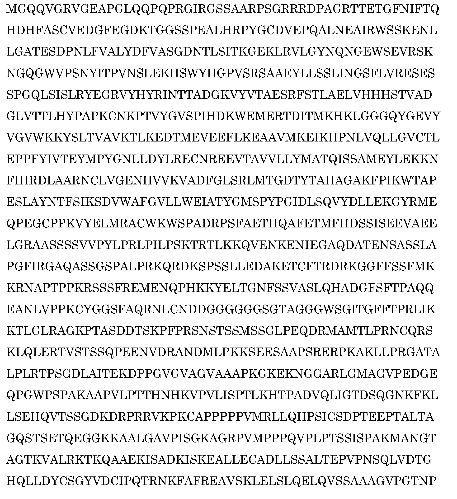
- An exemplary human ARG protein sequence is Inhibitors of ARG function are provided for use in the treatment of an individual suffering from or at risk of suffering from inflammatory edema as described herein.
- the ARG inhibitor preferentially inhibits the non-myristoylated isoform Arg 1a.
- the ARG inhibitor preferentially inhibits the non-myristoylated isoform Arg 1b 40 .
- An inhibitor of ARG function inhibits the tyrosine kinase activity of the protein encoded by ARG and is selected from nilotinib or a pharmaceutically acceptable salt thereof, dasatinib or a pharmaceutically acceptable salt thereof, and imatinib or a pharmaceutically acceptable salt thereof.
- Preferable examples of such inhibitors include nilotinib or a pharmaceutically acceptable salt thereof (preferably nilotinib hydrochloride monohydrate, i.e., TasignaTM); and dasatinib (SprycelTM) or a pharmaceutically acceptable salt thereof.
- Nilotinib is chemically described as 4-methyl-N-[3-(4-methyl-1H-imidazol-1-yl)-5-(trifluoromethyl)phenyl]-3-[[- 4-(3-pyridinyl)-2-pyrimidinyl]amino]-benzamide and its preparation is described in U.S. Patent No. 7,169,791 .
- Dasatinib is chemically described as N-(2-chloro-6-methylphenyl)-2-[[6-[4-(2-hydroxyethyl)-1-pipe razinyl]-2-methyl-4- pyrimidinyl]amino]-5-thiazolecarboxamide and its preparation is described in U.S. Patent No. 6.596,746 .
- the side effects of dasatinib will be unacceptable and the inhibitor used will not be dasatinib.
- the inhibitor is not nilotinib.
- More preferred compounds are imatinib or a pharmaceutically acceptable salt thereof mesylate (preferably imatinib mesylate, also referred to as STI-571, CGP 57148 and GleevecTM).
- Imatinib is chemically described as 4-[(4-methyl-1-piperazinyl) methyl]-N-[4-methyl-3-[[(4-(3-pyridinyl)-2-pyrimidinyl]amino]-phenyl]benzamide methane sulfonate and its preparation is described in U.S. Patent No. 5,521,184 .
- Inhibitors of ARG function also include antibodies that bind ARG and inhibit function.
- antibody includes, for example, both naturally occurring and non-naturally occurring antibodies, polyclonal and monoclonal antibodies, chimeric antibodies and wholly synthetic antibodies and fragments thereof, such as, for example, the Fab', F(ab')2, Fv or Fab fragments, or other antigen recognizing immunoglobulin fragments. Methods of making antibodies are well known in the art.
- an inhibitor of ARG function inhibits the tyrosine kinase activity of the ARG protein by at least 10%, 20%, 30%, 40%, 50%, 80%, 90% or more as compared to the tyrosine kinase activity of untreated ARG protein.
- an ARG inhibitor exhibits an IC50 concentration for ARG tyrosince kinase activity of less than 1micromolar, more preferably the ARG inhibitor has an IC50 concentration of 0.5 micromolar or less.
- the IC50 of imatinib for ARG tyrosine kinase activity has been reported as 0.5 micromolar ( Okuda et al. Blood, 2001 ).
- an inhibitor of ARG tyrosine kinase activity specifically inhibits the tyrosine kinase activity of the protein encoded by ARG.
- Specific inhibition refers to an inhibitor that inhibits ARG tyrosine kinase activity at least two times, preferably at least five times, or at least 10 times greater than it inhibits the tyrosine kinase activity of c-Abl.
- the specific tyrosine kinase activity inhibitor does not significantly inhibit the tyrosine kinase activity of C-kit, PDGFR-alpha and/or C-Abl.
- the ARG inhibitor is a nucleic acid molecule whose presence in a cell causes the degradation of or inhibits the function, transcription, or translation of its target gene, i.e., ARG, in a sequence-specific manner.
- exemplary nucleic acid molecules include aptamers, siRNA, artificial microRNA, interfering RNA or RNAi, dsRNA, ribozymes, antisense oligonucleotides, and DNA expression cassettes encoding said nucleic acid molecules.
- the nucleic acid molecule is an antisense oligonucleotide.
- Antisense oligonucleotides generally inhibit their target by binding target mRNA and sterically blocking expression by obstructing the ribosome. ASOs can also inhibit their target by binding target mRNA thus forming a DNA-RNA hybrid that can be a substance for RNase H. ASOs may also be produced as composite structures of two or more oligonucleotides, modified oligonucleotides, oligonucleosides, oligonucleotide mimetics, or regions or portions thereof. Such compounds have also been referred to in the art as hybrids or gapmers. Methods for designing and modifying such gapmers are described in, for example, U.S. Patent Publication Nos. 20110092572 and 20100234451 .
- antisense oligonucleotides can also be used to alter pre-mRNA processing, herein referred to as "exon-skipping" AONs.
- Antisense oligonucleotides for exon-skipping typically enable skipping of an exon or the 5' or 3' part of it.
- Antisense oligonucleotides can be directed toward the 5' splice site, the 3' splice, to both splice sites, to one or more exon-internal sites and to intron sequences, for instance specific for the branch point. The latter enables skipping of the upstream exon.
- An oligonucleotide is said to be directed toward an exon internal sequence if the complementarity region that contains the sequence identity to the reverse complement of the target pre-mRNA is within the exon boundary.
- the AONs useful in the methods described herein have a least one chemical modification that increases resistance of the compound to RNase H cleavage.
- modifications include, but are not limited to, nucleotides with sugar modifications.
- a nucleotide with a modified sugar includes, but is not limited to one or more sugar moieties that are mono- or disubstituted at the 2', 3' and/or 5' position such as a -OH; -F; substituted or unsubstituted, linear or branched lower (Cl-ClO) alkyl, alkenyl, alkynyl, alkaryl, allyl, aryl, or aralkyl, that may be interrupted by one or more heteroatoms; O-, S-, or N-alkyl; O-, S-, or N-alkenyl; O-, S-or N-alkynyl; O-, S-, or N-allyl; O-alkyl-O-al
- the sugar moiety can be a pyranose or derivative thereof, or a deoxypyranose or derivative thereof, preferably a ribose or a derivative thereof, or a deoxyribose or a derivative thereof.
- Such preferred derivatized sugar moieties comprise Locked Nucleic Acid (LNA), in which the 2'-carbon atom is linked to the 3' or 4' carbon atom of the sugar ring thereby forming a bicyclic sugar moiety.
- LNA Locked Nucleic Acid
- a preferred LNA comprises 2'-0,4'-C-ethylene-bridged nucleic acid ( Morita et al. 2001. Nucleic Acid Res Supplement No. 1: 241-242 ). These substitutions render the nucleotide analogue or equivalent RNase H and nuclease resistant and increase the affinity for the target RNA.
- AONs may also comprise modified backbones, such as morpholino backbones, carbamate backbones, siloxane backbones, sulfide, sulfoxide and sulfone backbones, formacetyl and thioformacetyl backbones, methyleneformacetyl backbones, riboacetyl backbones, alkene containing backbones, sulfamate, sulfonate and sulfonamide backbones, methyleneimino and methylenehydrazino backbones, and amide backbones.
- the AON has a phosphorodiamidate morpholino backbone.
- oligomers have an uncharged backbone in which the deoxyribose sugar of DNA is replaced by a six membered ring and the phosphodiester linkage is replaced by a phosphorodiamidate linkage.
- a preferred nucleotide analogue or equivalent comprises a Peptide Nucleic Acid (PNA), having a modified polyamide backbone ( Nielsen, et al. (1991) Science 254, 1497-1500 ).
- a further preferred backbone comprises a morpholino nucleotide analog or equivalent, in which the ribose or deoxyribose sugar is replaced by a 6-membered morpholino ring.
- a most preferred nucleotide analog or equivalent comprises a phosphorodiamidate morpholino oligomer (PMO), in which the ribose or deoxyribose sugar is replaced by a 6-membered morpholino ring, and the anionic phosphodiester linkage between adjacent morpholino rings is replaced by a non-ionic phosphorodiamidate linkage.
- PMO phosphorodiamidate morpholino oligomer
- an AON comprises a substitution of one of the non-bridging oxygens in the phosphodiester linkage. This modification slightly destabilizes base-pairing but adds significant resistance to nuclease degradation.
- a preferred nucleotide analogue or equivalent comprises phosphorothioate, chiral phosphorothioate, phosphorodithioate, phosphotriester, aminoalkylphosphotriester, H-phosphonate, methyl and other alkyl phosphonate including 3'-alkylene phosphonate, 5'-alkylene phosphonate and chiral phosphonate, phosphinate, phosphoramidate including 3'-amino phosphoramidate and aminoalkylphosphoramidate, thionophosphoramidate, thionoalkylphosphonate, thionoalkylphosphotriester, selenophosphate or boranophosphate.
- an oligonucleotide of the invention has at least two different types of analogues or equivalents.
- a preferred AON comprises a T-O alkyl phosphorothioate antisense oligonucleotide, such as 2'-O-methyl modified ribose (RNA), 2'-0-ethyl modified ribose, 2'-O-propyl modified ribose, and/or substituted derivatives of these modifications such as halogenated derivatives.
- RNA 2'-O-methyl modified ribose
- a most preferred AON according to the invention comprises of 2'-0-methyl phosphorothioate ribose.
- AONs typically comprise between 12 to 80, preferably between 15 to 40, nucleobases.
- the AONs comprise a stretch of at least 8 nucleobases having 100% complementarity with the target mRNA.
- the nucleic acid molecule is an RNAi molecule, i.e., RNA interference molecule.
- RNAi molecules include siRNA, shRNA, and artificial miRNA.
- siRNA comprises a double stranded structure typically containing 15 to 50 base pairs and preferably 19 to 25 base pairs and having a nucleotide sequence identical or nearly identical to an expressed target gene or RNA within the cell.
- An siRNA may be composed of two annealed polynucleotides or a single polynucleotide that forms a hairpin structure.
- shRNA or "small hairpin RNA” (also called stem loop) is a type of siRNA.
- these shRNAs are composed of a short, e.g. about 10 to about 25 nucleotide, antisense strand, followed by a nucleotide loop of about 5 to about 9 nucleotides, and the analogous sense strand.
- the sense strand can precede the nucleotide loop structure and the antisense strand can follow.
- siRNA molecules The design and production of siRNA molecules is well known to one of skill in the art ( Hajeri PB, Singh SK. Drug Discov Today. 2009 14(17-18):851-8 ). Methods of administration of therapeutic siRNA is also well-known to one of skill in the art ( Manjunath N, and Dykxhoorn DM. Discov Med. 2010 May;9(48):418-30 ; Guo J et al., Mol Biosyst. 2010 Jul 15;6(7):1143-61 ).
- siRNA molecule comprises an antisense strand having about 15 to about 30 (e.g., about 15, 16, 17, 18, 19, 20, 21, 22, 23, 24, 25, 26, 27, 28, 29, or 30) nucleotides, wherein the antisense strand is complementary to a RNA sequence or a portion thereof encoding.
- siRNA against ARG is also commercially available (see, e.g., Invitrogen TM catalog numbers HSS100051, HSS100052, and HSS178493) and described in U.S. Patent Publication No. 20050246794 .
- Artificial miRNA molecules are pre-miRNA or pri-miRNA comprising a stem-loop structure(s) derived from a specific endogenous miRNA in which the stem(s) of the stem-loop structure(s) incorporates a mature strand-star strand duplex where the mature strand sequence is distinct from the endogenous mature strand sequence of the specific referenced endogenous miRNA.
- the sequence of the star strand of a non-naturally occurring miRNA of the disclosure is also distinct from the endogenous star strand sequence of the specific referenced endogenous miRNA.
- microRNA precursors can be used, including without limitation a microRNA comprising a backbone design of miR-15a, -16, -19b, -20, -23a, -27b, -29a, -30b, -30c, -104, -132s, -181, -191, - 223 (See, e.g., U.S. Patent Publication Nos. 20050075492 and 20100292310 for the design and production of artificial miRNA molecules).
- RNA interference refers to a decrease in the mRNA level in a cell for a heterologous target gene by at least about 5%, about 10%, about 20%, about 30%, about 40%, about 50%, about 60%, about 70%, about 80%, about 90%, about 95%, about 99%, about 100% of the mRNA level found in the cell without the presence of the, e.g., miRNA or siRNA interference molecule
- RNAi molecules may also include chemical analogues such as, e.g., 2'-O-Methyl ribose analogues of RNA, DNA, LNA and RNA chimeric oligonucleotides, and other chemical analogues of nucleic acid oligonucleotides.
- chemical analogues such as, e.g., 2'-O-Methyl ribose analogues of RNA, DNA, LNA and RNA chimeric oligonucleotides, and other chemical analogues of nucleic acid oligonucleotides.
- the nucleic acid molecule inhibitors may be chemically synthesized and provided directly to cells of interest.
- the nucleic acid compound may be provided to a cell as part of a gene delivery vehicle.
- a gene delivery vehicle is preferably a liposome or a viral gene delivery vehicle. Liposomes are well known in the art and many variants are available for gene transfer purposes.
- Various viral gene delivery are currently used to transfer genes into target cells. In the present disclosure it is preferred to use those viral vectors that do not express their own genes but only the transferred genes.
- the nucleic acid compound may be present as such in the gene delivery vehicle.
- the nucleic acid compound is preferably provided as an expression cassette wherein the expression cassette encodes a transcript comprising said compound.
- a “vector” is a recombinant nucleic acid construct, such as plasmid, phase genome, virus genome, cosmid, or artificial chromosome, to which another DNA segment may be attached.
- the term “vector” includes both viral and nonviral means for introducing the nucleic acid into a cell in vitro, ex vivo or in vivo.
- Non-viral vectors include plasmids, liposomes, electrically charged lipids (cytofectins), DNA-protein complexes, and biopolymers.
- Viral vectors include retrovirus, adeno-associated virus, pox, baculovirus, vaccinia, herpes simplex, Epstein-Barr and adenovirus vectors, as set forth in greater detail below.
- Vector sequences may also contain one or more regulatory regions, and/or selectable markers useful in selecting, measuring, and monitoring nucleic acid transfer results (transfer to which tissues, duration of expression, etc.).
- the vector sequences provide delivery of the nucleic acid molecules to the appropriate cells.
- a preferred delivery vehicle is a viral vector such as an adeno-associated virus vector (AAV), or a retroviral vector such as a lentivirus vector and the like ( Goyenvalle A, et al. Science 2004;306(5702):1796-9 ).
- plasmids artificial chromosomes, plasmids suitable for targeted homologous recombination and integration in the human genome of cells may be suitably applied for delivery of a nucleic acid compound.
- Preferred are those vectors wherein transcription is driven from Pol III promoters, and/or wherein transcripts are in the form fusions with U1 or U7 transcripts, which yield good results for delivering small transcripts. It is within the skill of the artisan to design suitable transcripts.
- Pol III driven transcripts are preferred.
- the present disclosure demonstrates that short-term treatment with imatinib protects against endothelial barrier dysfunction. Accordingly, it is preferred in the methods described herein that the inhibitor of ARG function is provided to an individual for a time period of between 1-20 weeks. Preferably, the inhibitor is provided for a time period of between 1-12 weeks, more preferably the inhibitor is provided for up to 3 weeks.
- the inhibitor of ARG function may be formulated into any suitable pharmaceutical preparation for administration and further comprising, e.g., a pharmaceutically acceptable carrier, filler, preservative, adjuvant, solubilizer, diluent and/or excipient.
- a pharmaceutically acceptable carrier, filler, preservative, adjuvant, solubilizer, diluent and/or excipient may for instance be found in Remington: The Science and Practice of Pharmacy, 20th Edition. Baltimore, MD: Lippincott Williams & Wilkins, 2000 .
- the preferred excipients are capable of forming complexes, vesicles and/or liposomes that deliver such a through a cell membrane.
- Suitable excipients include polyethylenimine (PEI) or similar cationic polymers, including polypropyleneimine or polyethylenimine copolymers (PECs) and derivatives, ExGen 500, synthetic amphiphils (SAINT-18), lipofectinTM, and DOTAP.
- PEI polyethylenimine
- PECs polypropyleneimine or polyethylenimine copolymers
- SAINT-18 synthetic amphiphils
- lipofectinTM lipofectinTM
- DOTAP DOTAP
- a skilled person can determine the administration route for such pharmaceutical preparations, such as by oral, intravenous, subcutaneous, intramuscular, intrathecal and/or intraventricular administration.
- the inhibitor is administered orally or intravenously.
- An exemplary dose of imatinib is a daily dose of 400 mg resulting in imatinib plasma levels between 1 and 10 micromolar ( Singh et al, Eur J Clin Pharmacol (2009) 65:545-549 ). If clinical response is not observed within a few days from onset of treatment the dosage can be increased, for example to 400mg twice daily.
- Example 1 Imatinib attenuates thrombin-induced endothelial hyperpermeability.
- HUVECs were pretreated with imatinib, and subsequently stimulated with thrombin (1U/mL).
- thrombin was used to mimick inflammatory conditions, as thrombin is a potent inducer of endothelial permeability, 18,19 and is increased in sepsis and at sites of vascular injury.
- Endothelial barrier function was evaluated both by the passage of HRP across endothelial monolayers grown on porous filters and by electrical resistance of endothelial monolayers grown on gold electrodes (Electrical Cell-substrate Impedance Sensing [ECIS]).
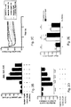
- Imatinib did not affect basal endothelial barrier function in both measurements ( Fig 1A & C ). Thrombin induced a 5- to 10-fold increase in HRP passage across endothelial monolayers ( Fig. 1A ), which was attenuated dose-dependently by imatinib with a maximal effect at 10 ⁇ M ( Figure 5A ). Imatinib 10 ⁇ M reduced the thrombin-induced HRP passage similarly at all time points (35-44% inhibition, P ⁇ 0.001; Fig. 1A ). In ECIS measurements thrombin strongly reduced endothelial electrical resistance reaching a maximum after 30min, followed by a gradual recovery of EC resistance ( Fig. 1B ).
- Imatinib attenuated the thrombin-induced decrease in endothelial resistance, as indicated by a significantly smaller drop in resistance, and a faster recovery towards baseline ( Fig. 1B and 1C , maximal decrease in normalized resistance of -0.54 ⁇ 0.02 in DMSO pretreated vs. -0.41 ⁇ 0.03 in imatinib pretreated cells, P ⁇ 0.001).
- HPMVECs human pulmonary microvascular endothelial cells
- HFMVECs human foreskin microvascular endothelial cells
- imatinib reduced the thrombin-induced macromolecule passage ( Fig. 1D ) and attenuated the maximal drop in endothelial resistance ( Fig. 5B ).
- imatinib attenuated the thrombin-induced maximal drop in endothelial resistance in HFMVECs ( Fig. 5C ).
- Thrombin-induced AJ dissociation and loss of cell-cell adhesion involves Ca 2+ and RhoA/Rho kinase mediated actin-myosin contraction. 2,24
- actin-myosin contraction HUVECs were stained for F-actin.
- Thrombin (15min) altered the F-actin cytoskeleton from a predominant peripheral F-actin band ( Fig. 2A , upper left panel) into a new pattern with robust F-actin stress fibers (arrows, Fig. 2A , upper right panel).
- F-actin was not organized in F-actin stress fibers, but in a fine-structured network of small F-actin filaments at the cell periphery.
- the absence of stress fibers in imatinib pretreated cells was associated with loss of contractile cell morphology and less intercellular gaps ( Fig. 2A , lower right panel).
- imatinib-sensitive kinases c-Abl 25 , PDGFR 26 and Arg 27,28 .
- imatinib attenuates the thrombin-induced changes in actin cytoskeleton via inhibition of the RhoA/Rho kinase pathway.
- HUVECs were pretreated with imatinib and/or the Rho kinase inhibitor Y-27632, and stimulated with thrombin. Both imatinib and Y-27632 reduced the thrombin-induced endothelial permeability as determined by HRP passage and transendothelial electrical resistance.
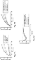
- the inhibitory effects of imatinib and Y-27632 on HRP passage were independent and additive ( Fig. 2B , P ⁇ 0.05).
- the ECIS measurements pointed to an additive effect of imatinib and Y-27632 ( Fig. 2C and D , maximal drop in electrical resistance of -0.44 ⁇ 0.02 in cells pretreated with imatinib alone vs.-0.18 ⁇ 0.04 in cells pretreated with a combination of imatinib and Y-27632, P ⁇ 0.05).
- RhoA activity strongly increased upon thrombin stimulation, but was not affected by pretreatment with imatinib ( Fig. 2E ). Since imatinib does not affect RhoA activation and has an additive effect to ROCK inhibition, imatinib attenuates endothelial permeability independent of the RhoA/Rho kinase pathway. Using BAPTA-AM as a chelator of intracellular calcium, an effect of imatinib on cytoplasmic Ca 2+ was also excluded (data not shown).
- Example 5 The tyrosine kinase Arg is a mediator of thrombin-induced hyperpermeability.
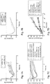
- Imatinib pretreatment had an additive effect to Arg knock down on the thrombin response, as the passage of HRP was significantly lower in Arg deficient cells pretreated with imatinib, compared to Arg deficient cells pretreated with DMSO ( Fig. 4B , P ⁇ 0.01).
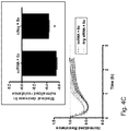
- Arg knock down attenuated the thrombin response as indicated by a lower drop in endothelial resistance and a faster recovery towards baseline values ( Fig. 4C ).
- Knock down of Arg mimicked pretreatment with imatinib as a similar decrease in normalized resistance was observed in Arg deficient cells and cells pretreated with imatinib, while pretreatment with imatinib had no additive effect ( Fig. 4D and E ).
- Sepsis is diagnosed if two or more of the following criteria are present: abnormal body temperature ( ⁇ 36 °C or >38 °C), abnormal white blood cell counts ( ⁇ 4 or >12 x109/L), a microbiologically proven or clinical source of infection, tachycardia (>90/min) and tachypnea (>20/min or a partial pressure of arterial carbon dioxide (P a CO 2 ) ⁇ 32 mmHg).
- imatinib treatment Upon diagnosis of sepsis, imatinib treatment will be initiated at a dose of 400mg daily, administered orally or intravenously.
- Indicators of clinical response are: presence of pulmonary edema on chest X-ray or CT, hemoglobin oxygen saturation, partial pressure of arterial oxygen (P a O 2 )/fraction of inspired oxygen (FiO 2 ) ratio, ventilator dependency, vasopressor dependency and systemic blood pressure. If clinical response remains absent for 2 days, the imatinib dose can be increased to 400mg twice daily. The maximal duration of treatment is 3 weeks.
- Severe pulmonary edema may follow radiotherapy, depending on the dose and duration of irradiation.
- Pulmonary edema upon irradiation is diagnosed by evidence for pulmonary edema on chest X-ray or CT of the thorax and hypoxemia.
- imatinib treatment will be initiated with an initial dose of 400mg daily administered orally or intravenously.
- Parameters for follow-up of pulmonary edema include: presence of pulmonary edema on chest X-ray or CT (thorax), arterial oxygen tension and hemoglobin oxygen saturation. If clinical response remains absent for 2 days, the imatinib dose can be increased to 400mg twice daily. The maximal duration of treatment is exceed 3 weeks.
- Example 8 Treatment of Reperfusion pulmonary edema after pulmonary endarterectomy with ARG inhibitor
- Reperfusion pulmonary edema occurs usually occurs within 72h after pulmonary endarterectomy ( Levinson et al. J Thorac Cardiovasc Surg. 1990 Apr;99(4):670-8 ; Levinson et al. Am Rev Respir Dis. 1986 Dec;134(6):1241-5 ), and is diagnosed by presence of pulmonary edema on chest X-ray or CT (thorax), arterial oxygen tension and hemoglobin oxygen saturation. After performance of pulmonary endarterectomy, imatinib treatment will be initiated with an initial dose of 400mg daily administered orally or intravenously to prevent development of pulmonary edema.
- Parameters for follow-up of pulmonary edema include: presence of pulmonary edema on chest X-ray or CT (thorax), arterial oxygen tension, hemoglobin oxygen saturation and ventilator dependency. If clinical response remains absent for 2 days, the imatinib dose can be increased to 400mg twice daily. The maximal duration of treatment is 3 weeks.
- No reflow stenosis is a phenomen commonly observed during heart catheterization for stenosis of coronary arteries, which is likely caused by edema formation in the vascular wall surrounding the lesion.
- Imatinib treatment will be initiated 12h (single dose of 400mg administered orally or intravenously) before the start of heart catheterization to prevent development of edema in the vascular wall during the procedure and continued after the procedure (400mg daily) to prevent edema formation in the postoperative phase.
- the maximal duration of treatment is 1 week.
- ALI/ARDS may develop as a consequence of pulmonary diseases (e.g. pneumonia, lung contusion or drowning), or as a consequence of systemic disease (e.g. sepsis).
- ALI and ARDS are diagnosed according to the American European Consensus Conference criteria.
- imatinib treatment will be initiated on top of regular treatment as indicated in guidelines on ALI/ARDS treatment. Starting dose of imatinib will be 400mg daily, administered orally or intravenously.
- Indicators of clinical response are: presence of pulmonary edema on chest X-ray or CT, hemoglobin oxygen saturation, partial pressure of arterial oxygen (P a O 2 )/fraction of inspired oxygen (FiO 2 ) ratio, ventilator dependency and extravascular lung water as measured by single indicator thermodilution technique. If clinical response remains absent for 2 days, the imatinib dose can be increased to 400mg twice daily. The maximal duration of treatment is 3 weeks.
- Imatinib mesylate was purchased from ChemieTek (Indianapolis, IN, USA) and dissolved in dimethylsulphoxide (DMSO) to a stock concentration of 10mM.
- Tyrphostin AG1296 was purchased from Sigma Aldrich (Steinheim, Germany) and dissolved in DMSO to a stock concentration of 5mM.
- Thrombin was purchased from Sigma Aldrich (Zwijndrecht, The Netherlands).
- Y-27632 was purchased from Tocris Cookson Ltd (London, UK).
- Small interference RNAs (siRNAs) against c-Abl, PDGFR- ⁇ and - ⁇ , Arg, c-KIT and scrambled RNAs (scRNA) were from Santa Cruz Biotechnology (Santa Cruz, CA, USA).
- anti c-Abl #2862
- anti PDGFR- ⁇ #3174
- - ⁇ #3169
- anti ⁇ -actin Sigma Aldrich
- anti VE-cadherin SC-6458, Santa Cruz
- Arg SC-6356, Santa Cruz
- ⁇ -catenin clone 8E7, Upstate/Millipore, Temecula, CA, USA
- umbilical vein endothelial cells For human umbilical vein endothelial cells (HUVECs), umbilical cords were obtained from the Amstelland Ziekenhuis, Amstelveen. Cells were isolated from healthy donors, and extensively characterized as previously described. 15 After isolation, cells were resuspended in M199 medium (Biowhittaker/Lonza, Verviers, Belgium), supplemented with penicilline 100U/mL and streptomycin 100mg/mL (Biowhittaker/Lonza, Verviers, Belgium), heat-inactivated human serum 10% (Sanquin CLB, Amsterdam, The Netherlands), heat-inactivated new-born calf serum 10% (Gibco, Grand Island, New York), crude endothelial cell growth factor 150 ⁇ g/mL (prepared from bovine brains), L-glutamine 2mmol/L (Biowhittaker/Lonza), and heparin 5U/mL (Leo Pharmaceutical Products, Weesp, The
- EGM2-MV culture medium EBM2 medium supplemented with 5% foetal bovine serum, human epidermal growth factor, fibroblast growth factor, vascular endothelial growth factor, insulin-like growth factor, hydrocortisone, ascorbic acid, gentamicin and amphotericin according to the manufacturers protocol [BioWhittaker/Lonza], and with penicillin [100U/mL] and streptomycin [100mg/mL]
- EGM2-MV culture medium EBM2 medium supplemented with 5% foetal bovine serum, human epidermal growth factor, fibroblast growth factor, vascular endothelial growth factor, insulin-like growth factor, hydrocortisone, ascorbic acid, gentamicin and amphotericin according to the manufacturers protocol [BioWhittaker/Lonza], and with penicillin [100U/mL] and streptomycin [100mg/mL]
- Cells were grown to confluence at 37 ⁇ C and 5% CO 2 , with a
- endothelial cells were extensively characterized as endothelial cells by the presence of endothelial markers and the absence of epithelial, lymphatic and smooth muscle cell markers. Cells were cultured up to passage 7, for experiments passage 4-7 cells were used. Human foreskin microvascular cells were isolated from human foreskin 17 , and cultured with EGM2-MV culture medium. Cells were grown to confluence at 37°C and 5% CO 2 , with a change of culture medium every other day, and similarly characterized as endothelial cells. 17
- HPMVEC human pulmonary microvascular endothelial cells
- EGM2-MV culture medium EBM2 medium supplemented with 5% fetal bovine serum, human epidermal growth factor, fibroblast growth factor, vascular endothelial growth factor, insulin-like growth factor, hydrocortisone, ascorbic acid according to the manufacturers protocol [Lonza, Basel, Switzerland], and with penicillin [100U/mL] and streptomycin [100mg/mL, Lonza, Biowhittaker, Verviers, Belgium]). After digestion, the suspension was flushed through a 100 ⁇ m and a 70 ⁇ m cell strainer (BD Biosciences, Bedford, USA).
- the filtered suspension was centrifuged for 5min at 230xG/21° C, and the pellet resuspended in 120 ⁇ L EGM2-MV, 40 ⁇ L FcR blocking reagent and 40 ⁇ L CD31-coated magnetic beads (Miltenyi Biotec GmbH, Bergisch Gladbach, Germany) and incubated for 15min at 4° C.
- CD31+ endothelial cells were separated from other cells by passing the cell suspension through a magnetic field. Freshly isolated endothelial cells were seeded in gelatin- and fibronectin-coated 25cm2 culture flasks. After 7-10 days of culture, cells were further purified by repeating the magnetic bead procedure until a 95-100% pure culture was reached.
- Cells were extensively characterized as endothelial cells by the presence of endothelial markers (CD31, VE-cadherin, vWF, Tie2 and Ulex lectin binding), and the absence of epithelial cell (pancytokeratin), vascular smooth muscle cell (smooth muscle ⁇ -actin) or lymphatic markers (LYVE-1). Cells were further characterized as microvascular encothelial cells (Ulex lectin).
- Endothelial barrier function was evaluated with horse radish peroxidase (HRP) passage and Electrical Cell-substrate Impedance Sensing (ECIS).
- HRP passage confluent cells were seeded in 1:1 density on 0.33cm 2 Costar polycarbonate filters, pore-size 3.0 ⁇ m (Corning, Lowell, MA, USA) and grown to confluence in 4 or 5 days, with change of culture medium every other day.
- pharmacological inhibitors or vector were dissolved in M199 supplemented with 1% human serum albumin (HSA, Sanquin CLB), and after removal of culture medium, solutions were added to the upper compartment of the filters during 60min.
- HSA human serum albumin
- pretreatment medium was changed for 1%HSA/M199 containing designated inhibitors, HRP 5 ⁇ g/mL (Sigma Aldrich) and thrombin 1U/mL.
- 1%HSA/M199 was added to the lower compartment.
- samples were taken from the lower compartment; HRP concentration in samples was detected by measuring chemoluminiscence after addition of TMB/E (Upstate/Millipore).
- ECIS measurements cells were trypsinized and seeded in 1:1 density on gelatine-coated ECIS arrays, each containing 8 wells with 10 gold electrodes/well (8W10E, Applied Biophysics, Troy, New York, USA). Culture medium was refreshed 24h after seeding, while experiments were performed 48h after seeding.
- designated pharmacological inhibitors or vector were dissolved in M199 supplemented with 1% HSA.
- thrombin was added directly to the cells for a final concentration of 1U/mL.
- impedance was measured at multiple frequencies to allow for calculation of resistance attributable to cell-cell adhesion (Rb) and resistance attributable to cell-matrix interaction (alpha). The absolute resistance was measured, while the relative resistance was calculated by normalization to the time point just before addition of the thrombin, to correct for baseline differences.
- Cells were seeded on glass coverslips coated with gelatine 1% and pretreated with glutaraldehyde 0.5% (Fluka, St. Gallen, Switzerland). Cells were seeded in 1:1 density and grown to confluence in 48-72 hours.
- culture medium was changed to 1%HSA/M199, containing pharmacological inhibitors or vector.
- thrombin was added directly to the wells for a final concentration of 1U/mL.
- medium was removed and warm paraformaldehyde 2% (Sigma Aldrich) was added to the cells, while kept on ice for 15min.
- Cells were permeabilized with Triton X-100 0.05% (Sigma Aldrich) in phosphate-buffered saline (PBS), and incubated overnight with primary antibodies for ⁇ -catenin (1:100), VE-cadherin (1:400) or Arg (1:150) in 0.1%HSA/PBS. Subsequently, cells were washed and incubated with FITC-labeled secondary antibodies (Invitrogen, Paisly, United Kingdom) and rhodamine/phalloidin (Invitrogen) for 1h at room temperature.
- Triton X-100 0.05% Sigma Aldrich
- PBS phosphate-buffered saline
- DAPI Vector Laboratories Inc., Burlingham, CA, USA
- Immunofluorescent imaging was performed with an Axiovert 200 MarianasTM inverted wide-field fluorescence microscope, using a 40x Zeiss air lens 0.75) and a 63x Zeiss oil lens (NA 1.4). All images were worked out with Slidebook software (Intelligent Imaging Innovation, Denver, CO, USA).
- RhoA activity 10cm 2 confluent cells/condition were pretreated with imatinib (10 ⁇ M) or vector (DMSO 0.1%) and stimulated with thrombin (1U/mL) during indicated intervals. After stimulation, cells were washed with ice-cold PBS and lysed with lysis buffer containing 0.02mM Tris/HCl pH 8.0, 0.15M NaCl, 0.09M KCl, 2mM EDTA/NaOH pH 8.0, 5% Igepal, and 0.5% Triton X-100, further supplemented with PhosStop protease inhibitor (Roche Diagnostics, Mannheim, Germany), and Complete EDTA-free phosphatase inhibitor (Roche Diagnostics).
- Relative RhoA activity was determined with a G-LISA RhoA Activity Assay (Cytoskeleton Inc, Denver, CO, USA), according to the manufacturers protocol.
- Cells were transfected with Amaxa Technology (Amaxa Biosystems, Lonza), according to the manufacturers protocol. 80-90% confluent cells (passage 1) were trypsinized and transfected with indicated siRNAs (0.05nmol for 10cm 2 cells) according to the manufacturers protocol. After transfection, cells were seeded on gelatine-coated ECIS arrays or 5cm 2 culture wells. For control of protein expression in transfected cells, 5cm 2 confluent cells/condition where lysed 48h or 120h after transfection.
Landscapes
- Health & Medical Sciences (AREA)
- Life Sciences & Earth Sciences (AREA)
- Engineering & Computer Science (AREA)
- Genetics & Genomics (AREA)
- Chemical & Material Sciences (AREA)
- Biomedical Technology (AREA)
- Molecular Biology (AREA)
- General Health & Medical Sciences (AREA)
- Organic Chemistry (AREA)
- Biotechnology (AREA)
- General Engineering & Computer Science (AREA)
- Bioinformatics & Cheminformatics (AREA)
- Zoology (AREA)
- Wood Science & Technology (AREA)
- Biochemistry (AREA)
- Pharmacology & Pharmacy (AREA)
- Veterinary Medicine (AREA)
- Public Health (AREA)
- Animal Behavior & Ethology (AREA)
- Epidemiology (AREA)
- Medicinal Chemistry (AREA)
- Physics & Mathematics (AREA)
- Biophysics (AREA)
- Plant Pathology (AREA)
- Microbiology (AREA)
- Oncology (AREA)
- Virology (AREA)
- Pharmaceuticals Containing Other Organic And Inorganic Compounds (AREA)
- Medicines That Contain Protein Lipid Enzymes And Other Medicines (AREA)
Description
- The invention relates to the field of endothelial barrier dysfunction. In particular, the invention relates to new methods for the treatment of syndromes/diseases characterized by endothelial barrier dysfunction, such as inflammatory edema.
- Abl protein non-receptor tyrosine kinases are involved in the regulation of a number of cell processes including proliferation, survival, and migration. There are two Abl family members in mammals: c-Abl (Abl 1) and Abl related gene (ARG) also known as Abl 2 (Kruh, et al. Science Dec 1986: 1545-1548). The tyrosine kinase domains of c-Abl and ARG are 94% identical, however their similarity diverges at the C-terminus. While both c-Abl and ARG contain a calponin-homology F-actin binding domain, c-Abl has a DNA binding domain and a G-actin binding domain and ARG additionally contains a talin-like F-actin binding domain and a microtubule binding domain.
- ARG has been implicated in affecting cytoskeleton processes. For example, ARG activation leads to actin polymerization and F-actin filament formation through F-actin bundling.32,33 Moreover, ARG regulates the actin filament structure by ARG-dependent phosphorylation of the actin-adapting proteins cortactin34 and N-WASp.35 The involvement of ARG in cytoskeleton regulation yields an important role for ARG in cell movements like migration, cell contraction, lamellipodia formation etc. The exact function of ARG in these processes may depend on the spatial distribution and temporal regulation of ARG activity.
- c-Abl is best known for its role as a proto-oncogene. The fusion of Bcr and c-Abl genes results in a chimeric Bcr-Abl protein having constitutively activated Abl tyrosine kinase activity. (Lugo et al., Science 1990, 247:1079-1082). Bcr-Abl is the underlying cause of chronic myeloid leukemia. One of the treatments for Brc-Abl positive leukaemia,5-7 is the use of imatinib. Imatinib is a small molecule inhibitor that blocks the ATPase activity of c-Abl as well as ARG, PDGFR and c-KIT.4
- c-Abl is also involved in regulating endothelial barrier function by phosphorylating nonmuscle myosin light chain kinase. Endothelial barrier dysfunction is involved in the pathophysiology of a wide variety of diseases. As the endothelium tightly controls the flux of fluid from the circulation to the surrounding tissues, dysfunction of this barrier in a pathological state (e.g. inflammation) leads to uncontrolled fluid extravasation and edema.1,2 If vital organs are involved, edema may lead to organ failure with consequent morbidity and mortality.
- Mechanisms involved in endothelial barrier dysfunction have been previously described. For example, Van Nieuw Amerongen et al. describes the involvement of Rho Kinase in endothelial barrier maintenance (Arteriosclerosis, Thrombosis, and Vascular Biology 2007 27:2332-2339). Wang et al. describes the involvement of RhoA and Calcium/calmodulin-dependent protein kinase II delta 6 in thrombin induced endothelial barrier dysfuntion (J Bio Chem 2010 28:21303-21312). Birukova et al. describes the role of PAK1 in LPS-induced lung vascular leak (AM J PHYSIOL-LUNG C 2010 299:652-663). Armulik et al. investigates the role of pericyte cells on the blood-brain barrier by generating a mouse mutation having a reduced number of pericytes in the CNS (Nature 2010 468:557-561)
siRNA against c-Abl resulted in a decrease in endothelial cell barrier integrity after treatment with a barrier-enhancing agonist, i.e, sphingosine 1-phosphate (Dudek et al. Mol Bio Cell 2010 21:4042-4056). The effect of inhibition of c-Abl on disrupting endothelial cell barrier integrity is also supported by one of the side effects of long-term treatment with imatinib, namely edema.7 - In contrast to studies reporting subcutaneous edema as a consequence of chronic imatinib treatment, the present disclosure demonstrates that short-term treatment with imatinib protects against endothelial barrier dysfunction.
Figure 1A-D demonstrates that short-term treatment of imatinib attenuates thrombin-induced endothelial hypermeability. Imatinib exerts these effects by preservation of adherence junction (AJ) integrity and cell-cell adhesion (Figure 1E-F ). Considering previous reports on subcutaneous edema as a side effect7 of imatinib treatment, it is also important to note that in the concentrations used in the study, imatinib did not affect basal endothelial barrier integrity. These findings suggest that the subcutaneous edema as a side-effect of imatinib treatment most likely results from chronic PDGFR inhibition in pericytes and consequent disturbed vascular support. - Thrombin-induced adherence junction dissociation and loss of cell-cell adhesion involves Ca2+ and RhoA/Rho kinase mediated actin-myosin contraction. 2,24 Since most imatinib-sensitive kinases (c-Abl25, PDGFR26 and Arg27,28) have been reported to affect Rho kinase activity, we hypothesized that imatinib attenuates the thrombin-induced changes in actin cytoskeleton via inhibition of the RhoA/Rho kinase pathway. Surprisingly, the endothelial cell barrier protective effect of imatinib was independent of the RhoA/Rho kinase pathway (
Figure 2B-E ). - As imatinib was primarily developed to target c-Abl, we hypothesized that inhibition of c-Abl underlies the protective effects of imatinib. Surprisingly, neither the inhibition of c-Abl, PDGFR, or c-KIT is responsible for the barrier protective effects of imatinib observed (
Figure 3 ). siRNA against ARG, however, demonstrated that it is in fact the inhibition of ARG kinase activity which mediates the barrier protective effects of imatinib (Figure 4 ). Our data suggest that, whereas Arg mediates endothelial barrier dysfunction, c-Abl supports endothelial barrier function (Fig. 3A andFig. 7A ). - These findings suggest that short term treatment with an ARG inhibitor, e.g., imatinib, would benefit syndromes characterized by endothelial barrier dysfunction.
- Accordingly, in one aspect, the disclosure provides a method for the treatment of an individual suffering from or at risk of suffering from inflammatory edema comprising administering an inhibitor of Abl-related gene (ARG) function to said individual thereby treating said edema in said individual.
- Edema can be defined as an abnormal accumulation of fluid in the interstices of cells in tissue spaces or in body cavities. It can be caused either by excessive movement of fluid from the vascular system into the tissues or inadequate movement of fluid from the tissues back to the vascular system. The normal interchange of fluid between these two compartments depends on the balance of osmotic pressure and hydrostatic pressure acting in opposite directions across the semi-permeable capillary walls. Edema is the result of an imbalance in these forces, which tends to cause abnormal accumulation of fluid in the interstitial spaces.
- Edema may be characterized as inflammatory or non-inflammatory. The composition of the extravascular fluid that accumulates in edema varies according to its etiology. In the case of edema caused by non-inflammatory mechanisms, the fluid (transudate) comprises a relatively low protein concentration and is of less specific gravity, indicating that the endothelium of the affected site is normal. In such cases, the transudate is essentially an ultrafilterate of blood plasma. This kind of non-inflammatory edema is primarily caused by alterations in the hemodynamic forces across the capillary wall and is also known as hemodynamic edema. On the other hand, in the case of edema that is caused by an inflammatory response, the extravascular fluid (exudate) comprises a high concentration of protein, cells and cellular debris and has high specific gravity. This indicates a significant alteration in the normal permeability of the small blood vessels in the affected area.
- An increase in vascular permeability is one of the main characteristics of the inflammatory response of the body against stimuli, especially in the case of acute inflammation. In fact, edema is one of the main signs of acute inflammation. During inflammation, the chemical factors derived from plasma and triggered by inflammatory stimuli mediate a number of vascular and cellular responses in the affected site. These structural changes in the microvasculature result in increased permeability of the blood vessel membrane, causing movement of plasma proteins and cells, e.g. leukocytes from the circulation to the intersititium, which ultimately results in site-specific edema. Inflammatory edema can be largely attributed to the direct action of histamine, bradykinin and other the substances released. The main mechanisms of increased vascular permeability in inflammation include endothelial cell contraction, junctional retraction, direct injury, leukocyte-dependent leakage, regenerating endothelium, amongst others. Increased fluid filtration towards the interstitium is further enhanced by the arteriolar vasodilator action of the inflammatory mediators, which increases the blood flow, the perfused surface area, capillary hydrostatic pressure, and facilitates edema by other mechanisms as well. Preferably, the inflammatory edema affects an organ other than the lung and/or the brain.
- Preferably, the inflammatory edema treated is thrombin-induced edema, e.g., edema formation after intracerebral hemorrhage.Preferably, the inflammatory edema is histamine induced edema. Preferably, the inflammatory edema is due to endothelial barrier dysfunction. Endothelial barrier dysfunction may be defined functionally, e.g., as an increased passage of macromolecules over a cultured endothelial monolayer or a decreased resistance over an endothelial monolayer, or morphologically, e.g., by the presence of intercellular gaps in an endothelial monolayer.
- Preferably, an individual suffering from or at risk of suffering from inflammatory edema is suffering from sepsis, systemic capillary leak syndrome, Acute Lung Injury/Acute Respiratory Distress Syndrome (ALI/ARDS), preeclampsia, no reflow stenosis, pulmonary edema (preferably post-radiation pulmonary edema or post-endarteriectomy pulmonary edema). Sepsis is defined as the presence and growth of micro-organisms in the circulation. One means for diagnosis includes if two or more of the following criteria are present: abnormal body temperature (<36 °C or >38 °C), abnormal white blood cell counts (<4 or >12 x109/L), a microbiologically proven or clinical source of infection, tachycardia (>90/min) and tachypnea (>20/min or a partial pressure of arterial carbon dioxide (PaCO2) <32 mmHg). ALI/ARDS may develop as a consequence of pulmonary diseases (e.g. pneumonia, lung contusion or drowning), or as a consequence of systemic disease (e.g. sepsis). ALI and ARDS are diagnosed according to the American European Consensus Conference criteria. Severe pulmonary edema may follow radiotherapy, depending on the dose and duration of irradiation. Pulmonary edema upon irradiation is diagnosed by evidence for pulmonary edema on chest X-ray or CT of the thorax and hypoxemia. Reperfusion pulmonary edema occurs usually occurs within 72h after pulmonary endarterectomy (Levinson et al. J Thorac Cardiovasc Surg. 1990 Apr;99(4):670-8; Levinson et al. Am Rev Respir Dis. 1986 Dec;134(6):1241-5), and is diagnosed by presence of pulmonary edema on chest X-ray or CT (thorax), arterial oxygen tension and hemoglobin oxygen saturation. No reflow stenosis is a phenomen commonly observed during heart catheterization for stenosis of coronary arteries, which is likely caused by edema formation in the vascular wall surrounding the lesion. Preeclampsia refers to a condition in which hypertension arises in association with significant amounts of protein in the urine.
- In an additional aspect, a method is provided for inhibiting endothelial barrier dysfunction in a collection of endothelial cells comprising an endothelial barrier that dysfunctions or is at risk of dysfunction, said method comprising providing an in vitro collection of endothelial cells with an inhibitor of Abl-related gene (ARG) function.
- In an additional aspect, a collection of endothelial cells is provided comprising an endothelial barrier that dysfunctions or is at risk of dysfunction, said cells comprising an inhibitor of Abl-related gene (ARG) function.
- An inhibitor of Abl-related gene (ARG) function refers to the inhibition or alteration of the ARG gene, ARG mRNA, or ARG protein. An exemplary human ARG protein sequence is
 Inhibitors of ARG function are provided for use in the treatment of an individual suffering from or at risk of suffering from inflammatory edema as described herein.
In a preferred embodiment, the ARG inhibitor preferentially inhibits the non-myristoylated isoform Arg 1a. In a preferred embodiment, the ARG inhibitor preferentially inhibits the non-myristoylated isoform Arg 1b40. - An inhibitor of ARG function inhibits the tyrosine kinase activity of the protein encoded by ARG and is selected from nilotinib or a pharmaceutically acceptable salt thereof, dasatinib or a pharmaceutically acceptable salt thereof, and imatinib or a pharmaceutically acceptable salt thereof. Preferable examples of such inhibitors include nilotinib or a pharmaceutically acceptable salt thereof (preferably nilotinib hydrochloride monohydrate, i.e., Tasigna™); and dasatinib (Sprycel™) or a pharmaceutically acceptable salt thereof. Nilotinib is chemically described as 4-methyl-N-[3-(4-methyl-1H-imidazol-1-yl)-5-(trifluoromethyl)phenyl]-3-[[- 4-(3-pyridinyl)-2-pyrimidinyl]amino]-benzamide and its preparation is described in
U.S. Patent No. 7,169,791 . Dasatinib is chemically described as N-(2-chloro-6-methylphenyl)-2-[[6-[4-(2-hydroxyethyl)-1-pipe razinyl]-2-methyl-4- pyrimidinyl]amino]-5-thiazolecarboxamide and its preparation is described inU.S. Patent No. 6.596,746 . In some embodiments, the side effects of dasatinib will be unacceptable and the inhibitor used will not be dasatinib. In some embodiments, the inhibitor is not nilotinib. - More preferred compounds are imatinib or a pharmaceutically acceptable salt thereof mesylate (preferably imatinib mesylate, also referred to as STI-571, CGP 57148 and Gleevec™). Imatinib is chemically described as 4-[(4-methyl-1-piperazinyl) methyl]-N-[4-methyl-3-[[(4-(3-pyridinyl)-2-pyrimidinyl]amino]-phenyl]benzamide methane sulfonate and its preparation is described in
U.S. Patent No. 5,521,184 . - Inhibitors of ARG function also include antibodies that bind ARG and inhibit function. The term "antibody" includes, for example, both naturally occurring and non-naturally occurring antibodies, polyclonal and monoclonal antibodies, chimeric antibodies and wholly synthetic antibodies and fragments thereof, such as, for example, the Fab', F(ab')2, Fv or Fab fragments, or other antigen recognizing immunoglobulin fragments. Methods of making antibodies are well known in the art. Preferably, an inhibitor of ARG function inhibits the tyrosine kinase activity of the ARG protein by at least 10%, 20%, 30%, 40%, 50%, 80%, 90% or more as compared to the tyrosine kinase activity of untreated ARG protein.
- Preferably, an ARG inhibitor exhibits an IC50 concentration for ARG tyrosince kinase activity of less than 1micromolar, more preferably the ARG inhibitor has an IC50 concentration of 0.5 micromolar or less. The IC50 of imatinib for ARG tyrosine kinase activity has been reported as 0.5 micromolar (Okuda et al. Blood, 2001).
- Preferably, an inhibitor of ARG tyrosine kinase activity specifically inhibits the tyrosine kinase activity of the protein encoded by ARG. Specific inhibition refers to an inhibitor that inhibits ARG tyrosine kinase activity at least two times, preferably at least five times, or at least 10 times greater than it inhibits the tyrosine kinase activity of c-Abl.
- Preferably, the specific tyrosine kinase activity inhibitor does not significantly inhibit the tyrosine kinase activity of C-kit, PDGFR-alpha and/or C-Abl.
- In some embodiments, the ARG inhibitor is a nucleic acid molecule whose presence in a cell causes the degradation of or inhibits the function, transcription, or translation of its target gene, i.e., ARG, in a sequence-specific manner. Exemplary nucleic acid molecules include aptamers, siRNA, artificial microRNA, interfering RNA or RNAi, dsRNA, ribozymes, antisense oligonucleotides, and DNA expression cassettes encoding said nucleic acid molecules.
- Preferably, the nucleic acid molecule is an antisense oligonucleotide. Antisense oligonucleotides (ASO) generally inhibit their target by binding target mRNA and sterically blocking expression by obstructing the ribosome. ASOs can also inhibit their target by binding target mRNA thus forming a DNA-RNA hybrid that can be a substance for RNase H. ASOs may also be produced as composite structures of two or more oligonucleotides, modified oligonucleotides, oligonucleosides, oligonucleotide mimetics, or regions or portions thereof. Such compounds have also been referred to in the art as hybrids or gapmers. Methods for designing and modifying such gapmers are described in, for example,
U.S. Patent Publication Nos. 20110092572 and20100234451 . - Recently it has been demonstrated that antisense oligonucleotides can also be used to alter pre-mRNA processing, herein referred to as "exon-skipping" AONs. Antisense oligonucleotides for exon-skipping typically enable skipping of an exon or the 5' or 3' part of it. Antisense oligonucleotides can be directed toward the 5' splice site, the 3' splice, to both splice sites, to one or more exon-internal sites and to intron sequences, for instance specific for the branch point. The latter enables skipping of the upstream exon. An oligonucleotide is said to be directed toward an exon internal sequence if the complementarity region that contains the sequence identity to the reverse complement of the target pre-mRNA is within the exon boundary. Methods for designing exon-skipping oligonucleotides have been described (see, e.g., Aartsma-Rus et al Mol Ther 17(3):548 (2009)).
- Preferably, the AONs useful in the methods described herein have a least one chemical modification that increases resistance of the compound to RNase H cleavage. Such modifications include, but are not limited to, nucleotides with sugar modifications. As used herein, a nucleotide with a modified sugar includes, but is not limited to one or more sugar moieties that are mono- or disubstituted at the 2', 3' and/or 5' position such as a -OH; -F; substituted or unsubstituted, linear or branched lower (Cl-ClO) alkyl, alkenyl, alkynyl, alkaryl, allyl, aryl, or aralkyl, that may be interrupted by one or more heteroatoms; O-, S-, or N-alkyl; O-, S-, or N-alkenyl; O-, S-or N-alkynyl; O-, S-, or N-allyl; O-alkyl-O-alkyl, -methoxy, -aminopropoxy; -amino xy; methoxyethoxy; -dimethylaminooxyethoxy; and -dimethylaminoethoxyethoxy. The sugar moiety can be a pyranose or derivative thereof, or a deoxypyranose or derivative thereof, preferably a ribose or a derivative thereof, or a deoxyribose or a derivative thereof. Such preferred derivatized sugar moieties comprise Locked Nucleic Acid (LNA), in which the 2'-carbon atom is linked to the 3' or 4' carbon atom of the sugar ring thereby forming a bicyclic sugar moiety. A preferred LNA comprises 2'-0,4'-C-ethylene-bridged nucleic acid (Morita et al. 2001. Nucleic Acid Res Supplement No. 1: 241-242). These substitutions render the nucleotide analogue or equivalent RNase H and nuclease resistant and increase the affinity for the target RNA.
- AONs may also comprise modified backbones, such as morpholino backbones, carbamate backbones, siloxane backbones, sulfide, sulfoxide and sulfone backbones, formacetyl and thioformacetyl backbones, methyleneformacetyl backbones, riboacetyl backbones, alkene containing backbones, sulfamate, sulfonate and sulfonamide backbones, methyleneimino and methylenehydrazino backbones, and amide backbones. Preferably, the AON has a phosphorodiamidate morpholino backbone. These oligomers have an uncharged backbone in which the deoxyribose sugar of DNA is replaced by a six membered ring and the phosphodiester linkage is replaced by a phosphorodiamidate linkage. A preferred nucleotide analogue or equivalent comprises a Peptide Nucleic Acid (PNA), having a modified polyamide backbone (Nielsen, et al. (1991) Science 254, 1497-1500).
- A further preferred backbone comprises a morpholino nucleotide analog or equivalent, in which the ribose or deoxyribose sugar is replaced by a 6-membered morpholino ring. A most preferred nucleotide analog or equivalent comprises a phosphorodiamidate morpholino oligomer (PMO), in which the ribose or deoxyribose sugar is replaced by a 6-membered morpholino ring, and the anionic phosphodiester linkage between adjacent morpholino rings is replaced by a non-ionic phosphorodiamidate linkage.
- In yet a further embodiment, an AON comprises a substitution of one of the non-bridging oxygens in the phosphodiester linkage. This modification slightly destabilizes base-pairing but adds significant resistance to nuclease degradation. A preferred nucleotide analogue or equivalent comprises phosphorothioate, chiral phosphorothioate, phosphorodithioate, phosphotriester, aminoalkylphosphotriester, H-phosphonate, methyl and other alkyl phosphonate including 3'-alkylene phosphonate, 5'-alkylene phosphonate and chiral phosphonate, phosphinate, phosphoramidate including 3'-amino phosphoramidate and aminoalkylphosphoramidate, thionophosphoramidate, thionoalkylphosphonate, thionoalkylphosphotriester, selenophosphate or boranophosphate.
- It is understood by a skilled person that it is not necessary for all positions in an oligonucleotide to be modified uniformly. In addition, more than one of the aforementioned analogues or equivalents may be incorporated in a single oligonucleotide or even at a single position within an oligonucleotide. In certain embodiments, an oligonucleotide of the invention has at least two different types of analogues or equivalents.
- A preferred AON comprises a T-O alkyl phosphorothioate antisense oligonucleotide, such as 2'-O-methyl modified ribose (RNA), 2'-0-ethyl modified ribose, 2'-O-propyl modified ribose, and/or substituted derivatives of these modifications such as halogenated derivatives. A most preferred AON according to the invention comprises of 2'-0-methyl phosphorothioate ribose.
- AONs typically comprise between 12 to 80, preferably between 15 to 40, nucleobases. Preferably, the AONs comprise a stretch of at least 8 nucleobases having 100% complementarity with the target mRNA.
- Preferably, the nucleic acid molecule is an RNAi molecule, i.e., RNA interference molecule. Preferred RNAi molecules include siRNA, shRNA, and artificial miRNA.
- siRNA comprises a double stranded structure typically containing 15 to 50 base pairs and preferably 19 to 25 base pairs and having a nucleotide sequence identical or nearly identical to an expressed target gene or RNA within the cell. An siRNA may be composed of two annealed polynucleotides or a single polynucleotide that forms a hairpin structure. As used herein "shRNA" or "small hairpin RNA" (also called stem loop) is a type of siRNA. In one embodiment, these shRNAs are composed of a short, e.g. about 10 to about 25 nucleotide, antisense strand, followed by a nucleotide loop of about 5 to about 9 nucleotides, and the analogous sense strand. Alternatively, the sense strand can precede the nucleotide loop structure and the antisense strand can follow.
- The design and production of siRNA molecules is well known to one of skill in the art (Hajeri PB, Singh SK. Drug Discov Today. 2009 14(17-18):851-8). Methods of administration of therapeutic siRNA is also well-known to one of skill in the art (Manjunath N, and Dykxhoorn DM. Discov Med. 2010 May;9(48):418-30; Guo J et al., Mol Biosyst. 2010 ). siRNA molecule comprises an antisense strand having about 15 to about 30 (e.g., about 15, 16, 17, 18, 19, 20, 21, 22, 23, 24, 25, 26, 27, 28, 29, or 30) nucleotides, wherein the antisense strand is complementary to a RNA sequence or a portion thereof encoding. siRNA against ARG is also commercially available (see, e.g., Invitrogen™ catalog numbers HSS100051, HSS100052, and HSS178493) and described in
U.S. Patent Publication No. 20050246794 . - Artificial miRNA molecules are pre-miRNA or pri-miRNA comprising a stem-loop structure(s) derived from a specific endogenous miRNA in which the stem(s) of the stem-loop structure(s) incorporates a mature strand-star strand duplex where the mature strand sequence is distinct from the endogenous mature strand sequence of the specific referenced endogenous miRNA. The sequence of the star strand of a non-naturally occurring miRNA of the disclosure is also distinct from the endogenous star strand sequence of the specific referenced endogenous miRNA. Many microRNA precursors can be used, including without limitation a microRNA comprising a backbone design of miR-15a, -16, -19b, -20, -23a, -27b, -29a, -30b, -30c, -104, -132s, -181, -191, - 223 (See, e.g.,
U.S. Patent Publication Nos. 20050075492 and20100292310 for the design and production of artificial miRNA molecules). - RNA interference refers to a decrease in the mRNA level in a cell for a heterologous target gene by at least about 5%, about 10%, about 20%, about 30%, about 40%, about 50%, about 60%, about 70%, about 80%, about 90%, about 95%, about 99%, about 100% of the mRNA level found in the cell without the presence of the, e.g., miRNA or siRNA interference molecule
- RNAi molecules may also include chemical analogues such as, e.g., 2'-O-Methyl ribose analogues of RNA, DNA, LNA and RNA chimeric oligonucleotides, and other chemical analogues of nucleic acid oligonucleotides.
- The nucleic acid molecule inhibitors may be chemically synthesized and provided directly to cells of interest. The nucleic acid compound may be provided to a cell as part of a gene delivery vehicle. Such a vehicle is preferably a liposome or a viral gene delivery vehicle. Liposomes are well known in the art and many variants are available for gene transfer purposes. Various viral gene delivery are currently used to transfer genes into target cells. In the present disclosure it is preferred to use those viral vectors that do not express their own genes but only the transferred genes. The nucleic acid compound may be present as such in the gene delivery vehicle. In a viral vector, the nucleic acid compound is preferably provided as an expression cassette wherein the expression cassette encodes a transcript comprising said compound.
- Accordingly, the nucleic acid molecule inhibitors may be provided in. A "vector" is a recombinant nucleic acid construct, such as plasmid, phase genome, virus genome, cosmid, or artificial chromosome, to which another DNA segment may be attached. The term "vector" includes both viral and nonviral means for introducing the nucleic acid into a cell in vitro, ex vivo or in vivo. Non-viral vectors include plasmids, liposomes, electrically charged lipids (cytofectins), DNA-protein complexes, and biopolymers. Viral vectors include retrovirus, adeno-associated virus, pox, baculovirus, vaccinia, herpes simplex, Epstein-Barr and adenovirus vectors, as set forth in greater detail below. Vector sequences may also contain one or more regulatory regions, and/or selectable markers useful in selecting, measuring, and monitoring nucleic acid transfer results (transfer to which tissues, duration of expression, etc.).
- Preferably, the vector sequences provide delivery of the nucleic acid molecules to the appropriate cells. A preferred delivery vehicle is a viral vector such as an adeno-associated virus vector (AAV), or a retroviral vector such as a lentivirus vector and the like (Goyenvalle A, et al. Science 2004;306(5702):1796-9).
- Also plasmids, artificial chromosomes, plasmids suitable for targeted homologous recombination and integration in the human genome of cells may be suitably applied for delivery of a nucleic acid compound. Preferred are those vectors wherein transcription is driven from Pol III promoters, and/or wherein transcripts are in the form fusions with U1 or U7 transcripts, which yield good results for delivering small transcripts. It is within the skill of the artisan to design suitable transcripts. Preferred are Pol III driven transcripts.
- In contrast to studies reporting subcutaneous edema as a consequence of long term imatinib treatment, the present disclosure demonstrates that short-term treatment with imatinib protects against endothelial barrier dysfunction. Accordingly, it is preferred in the methods described herein that the inhibitor of ARG function is provided to an individual for a time period of between 1-20 weeks. Preferably, the inhibitor is provided for a time period of between 1-12 weeks, more preferably the inhibitor is provided for up to 3 weeks.
- The inhibitor of ARG function may be formulated into any suitable pharmaceutical preparation for administration and further comprising, e.g., a pharmaceutically acceptable carrier, filler, preservative, adjuvant, solubilizer, diluent and/or excipient. Such pharmaceutically acceptable carrier, filler, preservative, adjuvant, solubilizer, diluent and/or excipient may for instance be found in Remington: The Science and Practice of Pharmacy, 20th Edition. Baltimore, MD: Lippincott Williams & Wilkins, 2000. If the inhibitor is a nucleic acid compound, the preferred excipients are capable of forming complexes, vesicles and/or liposomes that deliver such a through a cell membrane. Suitable excipients include polyethylenimine (PEI) or similar cationic polymers, including polypropyleneimine or polyethylenimine copolymers (PECs) and derivatives,
ExGen 500, synthetic amphiphils (SAINT-18), lipofectin™, and DOTAP. - A skilled person can determine the administration route for such pharmaceutical preparations, such as by oral, intravenous, subcutaneous, intramuscular, intrathecal and/or intraventricular administration. Preferably the inhibitor is administered orally or intravenously.
- A skilled person can readily determine a suitable dose and dosage regiment of inhibitor for administration. An exemplary dose of imatinib, for example, is a daily dose of 400 mg resulting in imatinib plasma levels between 1 and 10 micromolar (Singh et al, Eur J Clin Pharmacol (2009) 65:545-549). If clinical response is not observed within a few days from onset of treatment the dosage can be increased, for example to 400mg twice daily.
-
-
Figure 1 - Imatinib attenuates thrombin-induced endothelial barrier dysfunction through stabilization of adherens junctions. To test the effects of imatinib on endothelial barrier, HUVECs were seeded on polycarbonate filters (HRP passage) or ECIS gold electrodes (resistance measurements) and grown to confluence. Confluent cells were pretreated with imatinib or DMSO (0.1%), and stimulated with thrombin (IIa, 1U/mL) or vector. A) Time curve of HRP passage in the presence or absence of imatinib. Mean ± SEM of N=4 experiments, each representing triplo measurements. ** P<0.01, *** P<0.001 compared to DMSO + IIa. B) Effects of imatinib on normalized endothelial resistance during thrombin stimulation. Absolute resistance was normalized to the moment just before addition of thrombin. Mean ± SEM of N=5 experiments, each representing duplo measurements. C) Quantification of the maximum decrease in normalized resistance as measured in B. Mean ± SEM of N=4 experiments, each representing duplo measurements. *** P<0.001. D) HRP passage over human pulmonary microvascular endothelial cell (HPMVEC) monolayers grown on polycarbonate filters (HRP passage). Cells were pretreated with imatinib (10µM) or (DMSO 0.1%), and subsequently stimulated with thrombin (IIa, 1U/mL) or vector. Mean ± SEM of 5-6 measurements in N=2 experiments. E) HUVECs were grown to confluence on glass coverslips, pretreated with DMSO (upper panels) or imatinib 10µM (lower panels) and stimulated with thrombin for 30min. Fixated cells were stained for VE-cadherin (green) and the nucleus (DAPI, blue). Representative images of N=3 experiments. F) Immunofluorescence staining of β-catenin after 30min of thrombin stimulation. Representative images of N=2 experiments. Scale bars represent 20µm. All arrows indicate loss of staining at the membrane, suggesting intercellular gaps. Imaging was performed with aAxiovert 200 Marianas inverted wide-field fluorescence microscope, using a 40x Zeiss air lens (NA 0.75). -
Figure 2 - Imatinib inhibits endothelial barrier dysfunction without affecting RhoA/Rho kinase (ROCK) signaling. HUVECs were seeded on polycarbonate filters, ECIS gold electrodes, or glass cover slips and grown to confluence. Confluent cells were pretreated with imatinib (10µM), the ROCK inhibitor Y-27632 (10µM) or DMSO (0.1%), and subsequently stimulated with thrombin (IIa, 1U/mL) or vector. A) F-actin staining (rhodamine/phalloidine) of confluent cells pretreated with imatinib or DMSO and stimulated with thrombin or vector. Scale bars represent 10 µm. Imaging was performed with aAxiovert 200 Marianas inverted wide-field fluorescence microscope, using a 63x Zeiss oil lens (NA 1.4). Representative images of N=2 experiments. B) The effects of imatinib and/or Y-27632 pre-treatment on thrombin-induced HRP passage over HUVEC monolayers after 30min. Mean ± SEM of 6 measurements in N=2 experiments. C) Electrical resistance of HUVEC monolayers during thrombin stimulation, after pretreatment with imatinib, Y-27632 or DMSO. Resistance was normalized to the moment just before addition of thrombin. Mean ± SEM of N=3 experiments, each representing duplo measurements. D) Quantification of the maximum decrease in relative resistance as measured in C. Mean ± SEM of N=3 experiments. E) Effects of imatinib on thrombin-induced RhoA activation in HUVEC, expressed in arbitrary units (A.U.). Mean ± SEM of 4 measurements in N=2 experiments. * P<0.05, *** P<0.001. -
Figure 3 - Inhibition of c-Abl, PDGFR or c-KIT does not underlie the protective effects of imatinib. HUVECs were transfected with siRNA against c-Abl, PDGFR-α and -β, c-KIT or scrambled RNA (scRNA), using Amaxa Technology. After transfection, cells were seeded on 5cm2 wells for Western Blot analysis or on ECIS gold electrodes, and grown to confluence for 48h. Cells were pretreated with 1% HSA/M199, and subsequently stimulated with thrombin (IIa, 1U/mL). A) siRNA treatment resulted in an 80-90% decrease in c-Abl protein expression, as confirmed by Western Blot analysis for total c-Abl. β-actin served as loading control (insert). Absolute resistance under control or thrombin stimulated conditions. Mean ± SEM of 4 measurements in N=2 experiments. B) Thrombin-induced decrease in normalized endothelial resistance after transfection with c-Abl siRNA or scRNA. Resistance was normalized to the moment just before addition of thrombin. Mean ± SEM of N=4 measurements in 2 independent experiments. C) Thrombin-induced decrease in normalized endothelial resistance after knock down of PDGFR-α,-β or the combination. Resistance was normalized to the moment just before addition of thrombin. Mean ± SEM of 4 measurements in N=2 experiments. D) The effect of c-KIT knock down on thrombin-induced drop in normalized endothelial resistance. Resistance was normalized to the moment just before addition of thrombin. Mean ± SEM of N=3 experiments, each representing duplo measurements. -
Figure 4 - Inhibition of Arg mimics the effects of imatinib during thrombin stimulation. HUVECs were transfected with siRNA against Arg, seeded on polycarbonate filters, ECIS gold electrodes or 5cm2 wells, and grown to confluence for 48h (ECIS measurements and WesternBlot) or 120h (HRP passage and WesternBlot). Confluent cells were pretreated with imatinib (10µM), DMSO (0.1%) or 1%HSA/M199, and stimulated with thrombin (IIa, 1U/mL) or vector. A) Time curve of HRP passage in scRNA or Arg siRNA transfected cells after stimulation with thrombin or vector. Mean ± SEM of N=4 experiments. ** P<0.01 compared to scRNA + IIa. B) Time curve of HRP passage in cells transfected with scRNA or Arg siRNA, pretreated with imatinib or DMSO, and subsequently stimulated with thrombin. Mean ± SEM of N=4 experiments. scRNA + DMSO + IIa vs. siArg + DMSO + IIa, P<0.05; scRNA + DMSO + IIa vs. scRNA + Imatinib 10µM + IIa, P<0.001; scRNA + DMSO + IIa vs. siArg + Imatinib 10µM + IIa, P<0.001; siArg + DMSO + IIa vs. scRNA + Imatinib 10µM + IIa, P<0.05; siArg + DMSO + IIa vs. siArg + Imatinib 10µM + IIa, P<0.01 in Repeated Measures ANOVA. C) Normalized endothelial resistance during thrombin stimulation in HUVECs transfected with scRNA or Arg siRNA. Resistance was normalized to the moment just before thrombin stimulation. The thrombin response was quantified by calculating the maximal decrease in normalized resistance (insert). Mean ± SEM of N=4 experiments. * P<0.05. D) Normalized resistance in cells transfected with scRNA or Arg siRNA, pretreated with imatinib or DMSO, and stimulated with thrombin. Mean ± SEM of N=5 experiments. E) The thrombin response was quantified by calculating the maximal decrease in normalized resistance. Mean ± SEM of N=5 experiments. * P<0.05, ** P<0.01. F) Arg protein expression in scRNA or Arg siRNA transfected cells, lysated 48h or 120h after transfection (insert). Protein expression was quantified with a semi-quantitive method, and normalized for loading with β-actin. Mean of N=2 experiments. -
Figure 5 - A) HUVECs were seeded on polycarbonate filters and grown to confluence for 120h. Confluent cells were pretreated with increasing concentrations of imatinib (1, 2, 5, 10µM) or DMSO (in corresponding concentrations), and stimulated with thrombin (IIa, 1U/mL) or vector. Mean of N=2 experiments, each representing triplo measurements. B) HPMVECs were seeded on ECIS gold electrodes and grown to confluence. Absolute endothelial resistance of HPMVEC monolayers pretreated with imatinib (10µM) or DMSO (0.1%), and subsequently stimulated with thrombin (IIa, 1U/mL). Mean ± standard deviation (SD) of 3 measurements in 1 experiment. C) HFMVECs were seeded on ECIS gold electrodes and grown to confluence. Absolute endothelial resistance of human skin microvascular endothelial cell (HSMVEC) monolayers pretreated with imatinib (10µM) or DMSO (0.1%) and subsequently stimulated with thrombin (IIa, 1U/mL). Mean ± SD of N=3 experiments, each representing duplo measurements.
-
Figure 6 HUVECs or HPMVECs were seeded on ECIS gold electrodes. A) Resistance of cell-cell interaction (Rb) in HUVECs during thrombin stimulation (IIa, 1U/mL) after pretreatment with DMSO (0.1%) or imatinib (10µM). Mean ± SEM of N=3 experiments, each representing duplo measurements. B) Resistance of cell-matrix interaction (Alpha) in HUVECs during thrombin stimulation after pretreatment with DMSO or imatinib. Mean ± SEM of N=3 experiments, each representing duplo measurements. C) Resistance caused by cell-cell interaction (Rb) in HPMVECs pretreated with DMSO (0.1%) or imatinib (10µM), and stimulated with thrombin (IIa, 1U/mL). D) Resistance caused by cell-matrix interaction (Alpha) in HPMVECs pretreated with DMSO or imatinib, and stimulated with thrombin. -
Figure 7 - A) HUVECs were transfected with scRNA or c-Abl siRNA and seeded on ECIS gold electrodes. Cells were grown to confluence for 48h, pretreated with 1% HSA and stimulated with thrombin (IIa, 1U/mL). From ECIS measurements the resistance caused by cell-cell interaction (Rb) and the resistance caused by cell-matrix interaction (alpha) were calculated. The effect of c-Abl knock down on the thrombin-induced decrease in normalized Rb is represented here. Mean ± SEM of 4 measurements in N=2 experiments. B) Cells were transfected with scRNA or c-Abl siRNA and grown to confluence. After 48h cells were pretreated with imatinib (10µM) and subsequently stimulated with thrombin (IIa, 1U/mL). Mean of N=2 experiments, each representing duplo measurements. C) HUVECs were transfected with scRNA, PDGFR-α or -β siRNA or the combination of PDGFR-α and -β siRNA, seeded on ECIS gold electrodes, and grown to confluence. After 48h cells were pretreated with 1%HSA, and stimulated with thrombin (IIa, 1U/mL). Data represent absolute endothelial resistance. D) Time curve of HRP passage in cells transfected with scRNA, PDGFR-α or -β siRNA or a combination of PDGFR-α and -β siRNA after stimulation with thrombin or vector. Mean ± SEM of 3-6 measurements in N=2 experiments. E) HUVECs were seeded in 5cm2 culture wells and grown to confluence. Confluent cells were serum starved for 24h with medium containing 1% (1%s) or 2% (2%s) new-born calf serum, or cultured under normal growth medium (cM199). After 24h cells were lysated and cell lysates were analysed for expression of PDGFR-α or -β by Western Blot. Lysates of human lung fibroblasts (FB) served as positive control for PDGFR expression. Representative images of N=3 experiments. F) Quantification of the maximal decrease in normalized resistance in HUVECs pretreated with increasing concentrations of Tyrphostin AG1296 (2, 10, 50µM) or DMSO (0.1%) and stimulated with thrombin (IIa, 1U/mL) or vector. Mean ± SEM of N=5 experiments, *** P<0.001 compared to DMSO + IIa.
-
- 1. Lee WL, Slutsky AS. Sepsis and endothelial permeability. N Engl J Med. 2010;363(7):689-691.
- 2. Mehta D, Malik AB. Signaling mechanisms regulating endothelial permeability. Physiol Rev. 2006;86(1):279-367.
- 3. Overbeek MJ, van Nieuw Amerongen GP, Boonstra A, Smit EF, Vonk-Noordegraaf A. Possible role of imatinib in clinical pulmonary veno-occlusive disease. Eur Respir J. 2008;32(1):232-235.
- 4. Waller CF. Imatinib mesylate. Recent Results Cancer Res. 2010;184:3-20.
- 5. Schiffer CA. BCR-ABL tyrosine kinase inhibitors for chronic myelogenous leukemia. N Engl J Med. 2007;357(3):258-265.
- 6. Deininger MW, Goldman JM, Lydon N, Melo JV. The tyrosine kinase inhibitor CGP57148B selectively inhibits the growth of BCR-ABL-positive cells. Blood. 1997;90(9):3691-3698.
- 7. Druker BJ, Talpaz M, Resta DJ et al. Efficacy and safety of a specific inhibitor of the BCR-ABL tyrosine kinase in chronic myeloid leukemia. N Engl J Med. 2001;344(14):1031-1037.
- 8. van Oosterom AT, Judson I, Verweij J et al. Safety and efficacy of imatinib (STI571) in metastatic gastrointestinal stromal tumours: a phase I study. Lancet. 2001;358(9291):1421-1423.
- 9. Daniels CE, Wilkes MC, Edens M et al. Imatinib mesylate inhibits the profibrogenic activity of TGF-beta and prevents bleomycin-mediated lung fibrosis. J Clin Invest. 2004;114(9):1308-1316.
- 10. Schermuly RT, Dony E, Ghofrani HA et al. Reversal of experimental pulmonary hypertension by PDGF inhibition. J Clin Invest. 2005;115(10):2811-2821.
- 11. Kerkela R, Grazette L, Yacobi R et al. Cardiotoxicity of the cancer therapeutic agent imatinib mesylate. Nat Med. 2006;12(8):908-916.
- 12. Hellberg C, Ostman A, Heldin CH. PDGF and vessel maturation. Recent Results Cancer Res. 2010;180:103-114.
- 13. Su EJ, Fredriksson L, Geyer M et al. Activation of PDGF-CC by tissue plasminogen activator impairs blood-brain barrier integrity during ischemic stroke. Nat Med. 2008;14(7):731-737.
- 14. Armulik A, Genove G, Mae M et al. Pericytes regulate the blood-brain barrier. Nature. 2010;468(7323):557-561.
- 15. van Nieuw Amerongen GP, Draijer R, Vermeer MA, van Hinsbergh VWM. Transient and prolonged increase in endothelial permeability induced by histamine and thrombin: role of protein kinases, calcium, and RhoA. Circ Res. 1998;83(11):1115-1123.
- 16. Shelton JL, Wang L, Cepinskas G et al. Albumin leak across human pulmonary microvascular vs. umbilical vein endothelial cells under septic conditions. Microvasc Res. 2006;71(1):40-47.
- 17. van Hinsbergh VWM, Spengers ED, Kooistra T. Effect of thrombin on the production of plasminogen activators and PA inhibitor-1 by human foreskin microvascular endothelial cells. Thromb Haemost. 1987;57(2):148-153.
- 18. Vogel SM, Gao X, Mehta D et al. Abrogation of thrombin-induced increase in pulmonary microvascular permeability in PAR-1 knockout mice. Physiol Genomics. 2000;4(2):137-145.
- 19. Tauseef M, Kini V, Knezevic N et al. Activation of sphingosine kinase-1 reverses the increase in lung vascular permeability through sphingosine-1-phosphate receptor signaling in endothelial cells. Circ Res. 2008;103(10):1164-1172.
- 20. Minami T, Sugiyama A, Wu SQ et al. Thrombin and phenotypic modulation of the endothelium. Arterioscler Thromb Vasc Biol. 2004;24(1):41-53.
- 21. Giaever I, Keese CR. Micromotion of mammalian cells measured electrically. Proc Natl Acad Sci U S A. 1991;88(17):7896-7900.
- 22. Lo CM, Keese CR, Giaever I. Cell-substrate contact: another factor may influence transepithelial electrical resistance of cell layers cultured on permeable filters. Exp Cell Res. 1999;250(2):576-580.
- 23. Dejana E, Orsenigo F, Lampugnani MG. The role of adherens junctions and VE-cadherin in the control of vascular permeability. J Cell Sci. 2008;121(Pt 13):2115-2122.
- 24. van Nieuw Amerongen GP, van Delft S, Vermeer MA, Collard JG, van Hinsbergh VWM. Activation of RhoA by thrombin in endothelial hyperpermeability: role of Rho kinase and protein tyrosine kinases. Circ Res. 2000;87(4):335-340.
- 25. Huang X, Wu D, Jin H, Stupack D, Wang JYJ. Induction of cell retraction by the combined actions of Abl-CrkII and Rho-ROCK1 signaling. J Cell Biol. 2008;183(4):711-723.
- 26. Kim J, Wu Q, Zhang Y et al. PDGF signaling is required for epicardial function and blood vessel formation in regenerating zebrafish hearts. Proc Natl Acad Sci U S A. 2010;107(40):17206-17210.
- 27. Hernandez SE, Settleman J, Koleske AJ. Adhesion-dependent regulation of p190RhoGAP in the developing brain by the Abl-related gene tyrosine kinase. Curr Biol. 2004;14(8):691-696.
- 28. Bradley WD, Hernandez SE, Settleman J, Koleske AJ. Integrin signaling through Arg activates p190RhoGAP by promoting its binding to p120RasGAP and recruitment to the membrane. Mol Biol Cell. 2006;17(11):4827-4836.
- 29. Kurimoto N, Nan YS, Chen ZY et al. Effects of specific signal transduction inhibitors on increased permeability across rat endothelial monolayers induced by neuropeptide Y or VEGF. Am J Physiol Heart Circ Physiol. 2004;287(1):H100-H106.
- 30. Singh N, Kumar L, Meena R, Velpandian T. Drug monitoring of imatinib levels in patients undergoing therapy for chronic myeloid leukaemia: comparing plasma levels of responders and non-responders. Eur J Clin Pharmacol. 2009;65(6):545-549.
- 31. Demetri GD, Wang Y, Wehrle E et al. Imatinib plasma levels are correlated with clinical benefit in patients with unresectable/metastatic gastrointestinal stromal tumors. J Clin Oncol. 2009;27(19):3141-3147.
- 32. Wang Y, Miller AL, Mooseker MS, Koleske AJ. The Abl-related gene (Arg) nonreceptor tyrosine kinase uses two F-actin-binding domains to bundle F-actin. Proc Natl Acad Sci U S A. 2001;98(26):14865-14870.
- 33. Galkin VE, Orlova A, Koleske AJ, Egelman EH. The Arg non-receptor tyrosine kinase modifies F-actin structure. J Mol Biol. 2005;346(2):565-575.
- 34. Lapetina S, Mader CC, Machida K, Mayer BJ, Koleske AJ. Arg interacts with cortactin to promote adhesion-dependent cell edge protrusion. J Cell Biol. 2009;185(3):503-519.
- 35. Miller MM, Lapetina S, MacGrath SM et al. Regulation of actin polymerization and adhesion-dependent cell edge protrusion by the Abl-related gene (Arg) tyrosine kinase and N-WASp. Biochemistry. 2010;49(10):2227-2234.
- 36. Boyle SN, Koleske AJ. Use of a chemical genetic technique to identify myosin IIb as a substrate of the Abl-related gene (Arg) tyrosine kinase. Biochemistry. 2007;46(41):11614-11620.
- 37. Kolega J. Asymmetric distribution of myosin IIB in migrating endothelial cells is regulated by a rho-dependent kinase and contributes to tail retraction. Mol Biol Cell. 2003;14(12):4745-4757.
- 38. Peacock JG, Couch BA, Koleske AJ. The Abl and Arg non-receptor tyrosine kinases regulate different zones of stress fiber, focal adhesion, and contractile network localization in spreading fibroblasts. Cytoskeleton (Hoboken). 2010;67(10):666-675.
- 39. Shikata Y, Birukov KG, Garcia JGN. SIP induces FA remodeling in human pulmonary endothelial cells: role of Rac, GIT1, FAK, and paxillin. J Appl Physiol. 2003;94(3):1193-1203.
- 40. Bradley WD, Koleske AJ. Regulation of cell migration and morphogenesis by Abl-family kinases: emerging mechanisms and physiological contexts. J Cell Sci. 2009;122 (Pt 19):3441-3454.
- 41. Zandy NL, Playford M, Pendergast AM. Abl tyrosine kinases regulate cell-cell adhesion through Rho GTPases. Proc Natl Acad Sci U S A. 2007;104 (45):17686-17691.
- 42. Wang B, Kruh GD. Subcellular localization of the Arg protein tyrosine kinase. Oncogene. 1996;13(1):193-197.
- 43. Dudek SM, Chiang ET, Camp SM et al. Abl tyrosine kinase phosphorylates nonmuscle Myosin light chain kinase to regulate endothelial barrier function. Mol Biol Cell. 2010;21(22):4042-4056.
- The invention is further explained in the following examples. These examples do not limit the scope of the invention, but merely serve to clarify the invention.
- To evaluate the effects of imatinib on endothelial barrier function, HUVECs were pretreated with imatinib, and subsequently stimulated with thrombin (1U/mL). Thrombin was used to mimick inflammatory conditions, as thrombin is a potent inducer of endothelial permeability,18,19 and is increased in sepsis and at sites of vascular injury.20 Endothelial barrier function was evaluated both by the passage of HRP across endothelial monolayers grown on porous filters and by electrical resistance of endothelial monolayers grown on gold electrodes (Electrical Cell-substrate Impedance Sensing [ECIS]). Imatinib did not affect basal endothelial barrier function in both measurements (
Fig 1A & C ). Thrombin induced a 5- to 10-fold increase in HRP passage across endothelial monolayers (Fig. 1A ), which was attenuated dose-dependently by imatinib with a maximal effect at 10µM (Figure 5A ). Imatinib 10µM reduced the thrombin-induced HRP passage similarly at all time points (35-44% inhibition, P<0.001;Fig. 1A ). In ECIS measurements thrombin strongly reduced endothelial electrical resistance reaching a maximum after 30min, followed by a gradual recovery of EC resistance (Fig. 1B ). Imatinib attenuated the thrombin-induced decrease in endothelial resistance, as indicated by a significantly smaller drop in resistance, and a faster recovery towards baseline (Fig. 1B and 1C , maximal decrease in normalized resistance of -0.54±0.02 in DMSO pretreated vs. -0.41±0.03 in imatinib pretreated cells, P<0.001). - To evaluate whether the observed effects of imatinib also occur in other types of endothelial cells, permeability assays were performed in human pulmonary microvascular endothelial cells (HPMVECs) and human foreskin microvascular endothelial cells (HFMVECs). In HPMVECs, imatinib reduced the thrombin-induced macromolecule passage (
Fig. 1D ) and attenuated the maximal drop in endothelial resistance (Fig. 5B ). In a similar fashion, imatinib attenuated the thrombin-induced maximal drop in endothelial resistance in HFMVECs (Fig. 5C ). - Electrical resistance over an endothelial monolayer results from both cell-cell interaction (intercellular adhesion) and cell-substrate interaction (cell adhesion to subcellular matrix).21,22 Calculation of the resistance caused by cell-cell interaction (Rb) revealed that thrombin strongly decreases cell-cell adhesion, which was attenuated by imatinib (
Fig. 6A , maximal decrease in normalized Rb -0.97±0.02 in DMSO pretreated vs. -0.87±0.03 in imatinib pretreated cells, P=0.05). This was accompanied by attenuation of the thrombin-induced drop in cell-matrix interaction (Fig. 6B ). Comparable effects of imatinib on Rb and alpha were observed in HPMVECs (Fig. 6C & D ). - To further analyse the effect of imatinib on cell-cell adhesion, we performed immunofluorescence staining of the AJ proteins VE-cadherin and β-catenin, both of which are required for AJ integrity.2,23 At 15min (data not shown) and 30min, thrombin induced loss of VE-cadherin (
Fig. 1E ) and β-catenin (Fig. IF) from the membrane, as indicated by a reduction of fluorescence signal at the cell periphery, and formation of intercellular gaps (arrows,Fig. 1E & F ). The loss of VE-cadherin and β-catenin from the membrane could be partly reversed by pretreatment with imatinib, indicated by preservation of membrane staining and reduction of the number of intercellular gaps. - Altogether, these data demonstrate that imatinib protects against endothelial barrier dysfunction during thrombin stimulation. Imatinib exerts these effects by preservation of AJ integrity and cell-cell adhesion.
- Thrombin-induced AJ dissociation and loss of cell-cell adhesion involves Ca2+ and RhoA/Rho kinase mediated actin-myosin contraction.2,24 To evaluate actin-myosin contraction, HUVECs were stained for F-actin. Thrombin (15min) altered the F-actin cytoskeleton from a predominant peripheral F-actin band (
Fig. 2A , upper left panel) into a new pattern with robust F-actin stress fibers (arrows,Fig. 2A , upper right panel). In imatinib pretreated cells F-actin was not organized in F-actin stress fibers, but in a fine-structured network of small F-actin filaments at the cell periphery. The absence of stress fibers in imatinib pretreated cells was associated with loss of contractile cell morphology and less intercellular gaps (Fig. 2A , lower right panel). - Since most imatinib-sensitive kinases (c-Abl25, PDGFR26 and Arg27,28) have been reported to affect Rho kinase activity, we hypothesized that imatinib attenuates the thrombin-induced changes in actin cytoskeleton via inhibition of the RhoA/Rho kinase pathway. HUVECs were pretreated with imatinib and/or the Rho kinase inhibitor Y-27632, and stimulated with thrombin. Both imatinib and Y-27632 reduced the thrombin-induced endothelial permeability as determined by HRP passage and transendothelial electrical resistance. The inhibitory effects of imatinib and Y-27632 on HRP passage were independent and additive (
Fig. 2B , P<0.05). Similarly, the ECIS measurements pointed to an additive effect of imatinib and Y-27632 (Fig. 2C and D , maximal drop in electrical resistance of -0.44±0.02 in cells pretreated with imatinib alone vs.-0.18±0.04 in cells pretreated with a combination of imatinib and Y-27632, P<0.05). - To analyse the effect of imatinib on RhoA activation, cell lysates were analysed for RhoA activity with a G-LISA RhoA Activity Assay. RhoA activity strongly increased upon thrombin stimulation, but was not affected by pretreatment with imatinib (
Fig. 2E ). Since imatinib does not affect RhoA activation and has an additive effect to ROCK inhibition, imatinib attenuates endothelial permeability independent of the RhoA/Rho kinase pathway. Using BAPTA-AM as a chelator of intracellular calcium, an effect of imatinib on cytoplasmic Ca2+ was also excluded (data not shown). - These data indicate that imatinib reduces F-actin stress fiber formation and cell contraction without affecting the RhoA/Rho kinase pathway. It implicates that, during endothelial barrier dysfunction, there is regulation of F-actin structure independent of the RhoA/Rho kinase pathway.
- To determine which of the imatinib-sensitive kinases (c-Abl, PDGFR, Arg and c-KIT) is involved in thrombin-induced endothelial hyperpermeability, a systematic siRNA knock down of the imatinib sensitive kinases was performed. As imatinib was primarily developed to target c-Abl, we hypothesized that inhibition of c-Abl underlies the protective effects of imatinib. HUVECs were transfected with c-Abl siRNA to knock down c-Abl, resulting in an 85% reduction in c-Abl protein expression compared to transfection with scrambled RNA (scRNA,
Fig. 3A insert). ECIS measurements demonstrated that c-Abl knock down somewhat impaired basal endothelial barrier integrity (reduction of 100Ω compared to control cells,Fig. 3A ). Thrombin induced an identical decrease in electrical resistance in control and c-Abl deficient cells (Fig. 3A and 3B ). However, calculation of Rb revealed that the thrombin-response was more prominent in c-Abl deficient endothelial cells (Fig. 7A ). In c-Abl deficient cells, imatinib still attenuated the thrombin response (Fig. 7B ). Thus, inhibition of c-Abl does not underlie the protective effects of imatinib on endothelial barrier function. - Subsequently, we tested whether inhibition of PDGFR was responsible for the effects of imatinib on endothelial permeability. HUVECs were transfected with siRNA against PDGFR-α or -β, or the combination. Although combined knock down of PDGFR-α and -β increased basal endothelial resistance (
Fig. 7C ), no protective effect on the thrombin response was observed (Fig. 3C ). Similarly, no difference in HRP passage was detected between scRNA and PDGFR siRNA transfected cells (Fig. 7D ). Protein expression of PDGFR-α and -β was below detection limit (Fig. 7E ), further supporting the finding that PDGFR is not involved in the thrombin response. - Similarly, knock down of c-KIT with siRNA did not affect the thrombin response (
Fig. 3D ). To confirm the results of PDGFR and c-KIT knock down, HUVECs were pretreated with Tyrphostin AG1296, a pharmacological inhibitor of both PDGFR and c-KIT. Similar to combined knock down of PDGFR-α and -β, AG1296 improved basal barrier function, but did not inhibit the thrombin response (Fig. 7F ). Altogether, these data indicate that neither inhibition of c-Abl, nor inhibition of PDGFR or c-KIT underlie the barrier protective effects of imatinib. - Finally, we investigated whether Arg is involved in thrombin-induced endothelial barrier dysfunction. Knock down of Arg significantly reduced the thombin-induced passage of HRP. The HRP passage was lower in Arg siRNA transfected cells compared to scRNA transfected cells (
Fig. 4A ; 15.5±1.1% vs. 20.1±1.5%, respectively, P<0.01 after 1h and 21.3±1.5% vs. 27.3±1.8%, respectively, P<0.01 after 2h). Imatinib pretreatment had an additive effect to Arg knock down on the thrombin response, as the passage of HRP was significantly lower in Arg deficient cells pretreated with imatinib, compared to Arg deficient cells pretreated with DMSO (Fig. 4B , P<0.01). In ECIS measurements Arg knock down attenuated the thrombin response as indicated by a lower drop in endothelial resistance and a faster recovery towards baseline values (Fig. 4C ). Knock down of Arg mimicked pretreatment with imatinib, as a similar decrease in normalized resistance was observed in Arg deficient cells and cells pretreated with imatinib, while pretreatment with imatinib had no additive effect (Fig. 4D andE ). The efficiency of Arg knock down was higher after 48h than after 120h (Fig. 4F ). As the interval between transfection and measurement differs for ECIS measurements (48h) and HRP passage experiments (120h), the difference in Arg knock down likely explains the observation that Arg knock down exactly mimicked imatinib in ECIS measurements, but not in HRP passage experiments. - These data indicate that imatinib attenuates thrombin-induced hyperpermeability through inhibition of Arg. Furthermore, these data identify Arg as a novel mediator of the thrombin response. Arg importantly contributes to thrombin-induced endothelial barrier dysfunction, as inhibition of Arg reduces the thrombin-induced macromolecule passage up to 40%.
- Sepsis is diagnosed if two or more of the following criteria are present: abnormal body temperature (<36 °C or >38 °C), abnormal white blood cell counts (<4 or >12 x109/L), a microbiologically proven or clinical source of infection, tachycardia (>90/min) and tachypnea (>20/min or a partial pressure of arterial carbon dioxide (PaCO2) <32 mmHg). Upon diagnosis of sepsis, imatinib treatment will be initiated at a dose of 400mg daily, administered orally or intravenously. Indicators of clinical response are: presence of pulmonary edema on chest X-ray or CT, hemoglobin oxygen saturation, partial pressure of arterial oxygen (PaO2)/fraction of inspired oxygen (FiO2) ratio, ventilator dependency, vasopressor dependency and systemic blood pressure. If clinical response remains absent for 2 days, the imatinib dose can be increased to 400mg twice daily. The maximal duration of treatment is 3 weeks.
- Severe pulmonary edema may follow radiotherapy, depending on the dose and duration of irradiation. Pulmonary edema upon irradiation is diagnosed by evidence for pulmonary edema on chest X-ray or CT of the thorax and hypoxemia. Upon diagnosis of post-radiation pulmonary edema imatinib treatment will be initiated with an initial dose of 400mg daily administered orally or intravenously. Parameters for follow-up of pulmonary edema include: presence of pulmonary edema on chest X-ray or CT (thorax), arterial oxygen tension and hemoglobin oxygen saturation. If clinical response remains absent for 2 days, the imatinib dose can be increased to 400mg twice daily. The maximal duration of treatment is exceed 3 weeks.
- Reperfusion pulmonary edema occurs usually occurs within 72h after pulmonary endarterectomy (Levinson et al. J Thorac Cardiovasc Surg. 1990 Apr;99(4):670-8; Levinson et al. Am Rev Respir Dis. 1986 Dec;134(6):1241-5), and is diagnosed by presence of pulmonary edema on chest X-ray or CT (thorax), arterial oxygen tension and hemoglobin oxygen saturation. After performance of pulmonary endarterectomy, imatinib treatment will be initiated with an initial dose of 400mg daily administered orally or intravenously to prevent development of pulmonary edema. Parameters for follow-up of pulmonary edema include: presence of pulmonary edema on chest X-ray or CT (thorax), arterial oxygen tension, hemoglobin oxygen saturation and ventilator dependency. If clinical response remains absent for 2 days, the imatinib dose can be increased to 400mg twice daily. The maximal duration of treatment is 3 weeks.
- No reflow stenosis is a phenomen commonly observed during heart catheterization for stenosis of coronary arteries, which is likely caused by edema formation in the vascular wall surrounding the lesion. Imatinib treatment will be initiated 12h (single dose of 400mg administered orally or intravenously) before the start of heart catheterization to prevent development of edema in the vascular wall during the procedure and continued after the procedure (400mg daily) to prevent edema formation in the postoperative phase. The maximal duration of treatment is 1 week.
- ALI/ARDS may develop as a consequence of pulmonary diseases (e.g. pneumonia, lung contusion or drowning), or as a consequence of systemic disease (e.g. sepsis). ALI and ARDS are diagnosed according to the American European Consensus Conference criteria. Upon diagnosis of ALI/ARDS, imatinib treatment will be initiated on top of regular treatment as indicated in guidelines on ALI/ARDS treatment. Starting dose of imatinib will be 400mg daily, administered orally or intravenously. Indicators of clinical response are: presence of pulmonary edema on chest X-ray or CT, hemoglobin oxygen saturation, partial pressure of arterial oxygen (PaO2)/fraction of inspired oxygen (FiO2) ratio, ventilator dependency and extravascular lung water as measured by single indicator thermodilution technique. If clinical response remains absent for 2 days, the imatinib dose can be increased to 400mg twice daily. The maximal duration of treatment is 3 weeks.
- Imatinib mesylate was purchased from ChemieTek (Indianapolis, IN, USA) and dissolved in dimethylsulphoxide (DMSO) to a stock concentration of 10mM. Tyrphostin AG1296 was purchased from Sigma Aldrich (Steinheim, Germany) and dissolved in DMSO to a stock concentration of 5mM. Thrombin was purchased from Sigma Aldrich (Zwijndrecht, The Netherlands). Y-27632 was purchased from Tocris Cookson Ltd (London, UK). Small interference RNAs (siRNAs) against c-Abl, PDGFR-α and -β, Arg, c-KIT and scrambled RNAs (scRNA) were from Santa Cruz Biotechnology (Santa Cruz, CA, USA). The following antibodies were used: anti c-Abl (#2862), anti PDGFR-α (#3174) and -β (#3169), all from Cell Signaling Technologies (Danvers, MA, USA), anti β-actin (Sigma Aldrich), anti VE-cadherin (SC-6458, Santa Cruz) and Arg (SC-6356, Santa Cruz) and β-catenin (clone 8E7, Upstate/Millipore, Temecula, CA, USA).
- For human umbilical vein endothelial cells (HUVECs), umbilical cords were obtained from the Amstelland Ziekenhuis, Amstelveen. Cells were isolated from healthy donors, and extensively characterized as previously described.15 After isolation, cells were resuspended in M199 medium (Biowhittaker/Lonza, Verviers, Belgium), supplemented with penicilline 100U/mL and streptomycin 100mg/mL (Biowhittaker/Lonza, Verviers, Belgium), heat-inactivated
human serum 10% (Sanquin CLB, Amsterdam, The Netherlands), heat-inactivated new-borncalf serum 10% (Gibco, Grand Island, New York), crude endothelial cell growth factor 150µg/mL (prepared from bovine brains), L-glutamine 2mmol/L (Biowhittaker/Lonza), and heparin 5U/mL (Leo Pharmaceutical Products, Weesp, The Netherlands). Cells were cultured at 37°C and 5% CO2, with a change of culture medium every other day. Cells were cultured up topassage 2, for experiments passage 1-2 cells were used. - Human pulmonary microvascular cells were isolated from human lung tissue as previously described.16 Cells were cultured in EGM2-MV culture medium (EBM2 medium supplemented with 5% foetal bovine serum, human epidermal growth factor, fibroblast growth factor, vascular endothelial growth factor, insulin-like growth factor, hydrocortisone, ascorbic acid, gentamicin and amphotericin according to the manufacturers protocol [BioWhittaker/Lonza], and with penicillin [100U/mL] and streptomycin [100mg/mL]) and seeded on gelatine-coated 25cm2 culture flasks. Cells were grown to confluence at 37□C and 5% CO2, with a change of culture medium every other day. They were extensively characterized as endothelial cells by the presence of endothelial markers and the absence of epithelial, lymphatic and smooth muscle cell markers. Cells were cultured up to passage 7, for experiments passage 4-7 cells were used. Human foreskin microvascular cells were isolated from human foreskin17, and cultured with EGM2-MV culture medium. Cells were grown to confluence at 37°C and 5% CO2, with a change of culture medium every other day, and similarly characterized as endothelial cells.17
- Isolation of human pulmonary microvascular endothelial cells (HPMVEC) Lung tissue was obtained from patients undergoing lobectomy for primary lung cancer or metastasis (Department of Thoracic Surgery, VU University Medical Centre, Amsterdam). After rinsing lung tissue in phosphate-buffered saline (PBS), pleural tissue was removed and the remaining tissue was minced during 5 minutes. The minced tissue was incubated in 20mL 0.3% type II collagenase solution (Sigma, St. Louis, Missouri, USA) in PBS during 60min under gentle shaking. Digestion was stopped by addition of EGM2-MV culture medium (EBM2 medium supplemented with 5% fetal bovine serum, human epidermal growth factor, fibroblast growth factor, vascular endothelial growth factor, insulin-like growth factor, hydrocortisone, ascorbic acid according to the manufacturers protocol [Lonza, Basel, Switzerland], and with penicillin [100U/mL] and streptomycin [100mg/mL, Lonza, Biowhittaker, Verviers, Belgium]). After digestion, the suspension was flushed through a 100µm and a 70µm cell strainer (BD Biosciences, Bedford, USA). The filtered suspension was centrifuged for 5min at 230xG/21° C, and the pellet resuspended in 120µL EGM2-MV, 40µL FcR blocking reagent and 40µL CD31-coated magnetic beads (Miltenyi Biotec GmbH, Bergisch Gladbach, Germany) and incubated for 15min at 4° C. CD31+ endothelial cells were separated from other cells by passing the cell suspension through a magnetic field. Freshly isolated endothelial cells were seeded in gelatin- and fibronectin-coated 25cm2 culture flasks. After 7-10 days of culture, cells were further purified by repeating the magnetic bead procedure until a 95-100% pure culture was reached. Cells were extensively characterized as endothelial cells by the presence of endothelial markers (CD31, VE-cadherin, vWF, Tie2 and Ulex lectin binding), and the absence of epithelial cell (pancytokeratin), vascular smooth muscle cell (smooth muscle α-actin) or lymphatic markers (LYVE-1). Cells were further characterized as microvascular encothelial cells (Ulex lectin).
- Endothelial barrier function was evaluated with horse radish peroxidase (HRP) passage and Electrical Cell-substrate Impedance Sensing (ECIS). For measurement of HRP passage, confluent cells were seeded in 1:1 density on 0.33cm2 Costar polycarbonate filters, pore-size 3.0µm (Corning, Lowell, MA, USA) and grown to confluence in 4 or 5 days, with change of culture medium every other day. For pretreatment, pharmacological inhibitors or vector were dissolved in M199 supplemented with 1% human serum albumin (HSA, Sanquin CLB), and after removal of culture medium, solutions were added to the upper compartment of the filters during 60min. For stimulation, pretreatment medium was changed for 1%HSA/M199 containing designated inhibitors, HRP 5µg/mL (Sigma Aldrich) and
thrombin 1U/mL. 1%HSA/M199 was added to the lower compartment. At indicated time points samples were taken from the lower compartment; HRP concentration in samples was detected by measuring chemoluminiscence after addition of TMB/E (Upstate/Millipore). - For ECIS measurements, cells were trypsinized and seeded in 1:1 density on gelatine-coated ECIS arrays, each containing 8 wells with 10 gold electrodes/well (8W10E, Applied Biophysics, Troy, New York, USA). Culture medium was refreshed 24h after seeding, while experiments were performed 48h after seeding. For pretreatment, designated pharmacological inhibitors or vector were dissolved in M199 supplemented with 1% HSA. After 90min of pretreatment, thrombin was added directly to the cells for a final concentration of 1U/mL. During stimulation impedance was measured at multiple frequencies to allow for calculation of resistance attributable to cell-cell adhesion (Rb) and resistance attributable to cell-matrix interaction (alpha). The absolute resistance was measured, while the relative resistance was calculated by normalization to the time point just before addition of the thrombin, to correct for baseline differences.
- Cells were seeded on glass coverslips coated with
gelatine 1% and pretreated with glutaraldehyde 0.5% (Fluka, St. Gallen, Switzerland). Cells were seeded in 1:1 density and grown to confluence in 48-72 hours. For pretreatment, culture medium was changed to 1%HSA/M199, containing pharmacological inhibitors or vector. After 60min of pretreatment, thrombin was added directly to the wells for a final concentration of 1U/mL. At indicated time points, medium was removed andwarm paraformaldehyde 2% (Sigma Aldrich) was added to the cells, while kept on ice for 15min. Cells were permeabilized with Triton X-100 0.05% (Sigma Aldrich) in phosphate-buffered saline (PBS), and incubated overnight with primary antibodies for β-catenin (1:100), VE-cadherin (1:400) or Arg (1:150) in 0.1%HSA/PBS. Subsequently, cells were washed and incubated with FITC-labeled secondary antibodies (Invitrogen, Paisly, United Kingdom) and rhodamine/phalloidin (Invitrogen) for 1h at room temperature. Cells were washed and sealed in Vectashield mounting medium containing DAPI (Vector Laboratories Inc., Burlingham, CA, USA) for nuclear staining. Immunofluorescent imaging was performed with anAxiovert 200 Marianas™ inverted wide-field fluorescence microscope, using a 40x Zeiss air lens 0.75) and a 63x Zeiss oil lens (NA 1.4). All images were worked out with Slidebook software (Intelligent Imaging Innovation, Denver, CO, USA). - For analysis of RhoA activity, 10cm2 confluent cells/condition were pretreated with imatinib (10µM) or vector (DMSO 0.1%) and stimulated with thrombin (1U/mL) during indicated intervals. After stimulation, cells were washed with ice-cold PBS and lysed with lysis buffer containing 0.02mM Tris/HCl pH 8.0, 0.15M NaCl, 0.09M KCl, 2mM EDTA/NaOH pH 8.0, 5% Igepal, and 0.5% Triton X-100, further supplemented with PhosStop protease inhibitor (Roche Diagnostics, Mannheim, Germany), and Complete EDTA-free phosphatase inhibitor (Roche Diagnostics). After centrifugation of the cell lysates, the supernatant was snap-frozen and stored at -80°C. Relative RhoA activity was determined with a G-LISA RhoA Activity Assay (Cytoskeleton Inc, Denver, CO, USA), according to the manufacturers protocol.
- Cells were transfected with Amaxa Technology (Amaxa Biosystems, Lonza), according to the manufacturers protocol. 80-90% confluent cells (passage 1) were trypsinized and transfected with indicated siRNAs (0.05nmol for 10cm2 cells) according to the manufacturers protocol. After transfection, cells were seeded on gelatine-coated ECIS arrays or 5cm2 culture wells. For control of protein expression in transfected cells, 5cm2 confluent cells/condition where lysed 48h or 120h after transfection. 20µg of total protein/condition was electrophoresed, transferred to nitrocellulose membranes and immunoblotted for c-Abl (1:1000, overnight at 4°C), PDGFR-α and -β (both 1:200, overnight at 4°C), and Arg (1:150, overnight at 4°C). β-actin (1:100000) served as loading control.
- All data are reported as Mean ± Standard Error of the Mean (SEM). N refers to the number of independent experiments. For comparison of 2 groups a Students t-test was used, for comparison of >2 groups an one-way ANOVA with Tukey post-hoc test was used. A P-value <0.05 was considered significant.
Claims (12)
- An inhibitor of Abl-related gene (ARG) function for use in the treatment of an individual suffering from or at risk of suffering from inflammatory edema, wherein said inhibitor is selected from:a) an inhibitor of tyrosine kinase activity of the protein encoded by said Abl-related gene (ARG) selected from nilotinib or a pharmaceutically acceptable salt thereof, dasatinib or a pharmaceutically acceptable salt thereof, and imatinib or a pharmaceutically acceptable salt thereof;b) an anti-ARG antibody that binds ARG and inhibits function; andc) a nucleic acid molecule that causes the degradation of ARG or inhibits the function, transcription, or translation of ARG.
- An inhibitor for use according to claim 1 wherein said inflammatory edema is thrombin-induced edema.
- An inhibitor for use according to claim 1 or claim 2, wherein said individual suffering from or at risk of suffering from inflammatory edema is suffering from sepsis, ALI/ARDS, preeclampsia, no reflow stenosis, pulmonary edema, and preferably post-radiation pulmonary edema or post-endarteriectomy pulmonary edema.
- An inhibitor for use according to any of the preceding claims, wherein said tyrosine kinase activity inhibitor does not significantly inhibit the tyrosine kinase activity of C-kit, PDGFR-alpha and/or C-Abl.
- An inhibitor for use according to any one of claims 1-4, wherein said inhibitor of Abl-related gene (ARG) function is imatinib or a pharmaceutically acceptable salt thereof.
- An inhibitor for use according to any one of claims 1-4, wherein said inhibitor of Abl-related gene (ARG) function comprises an antisense oligonucleotide specific for a pre-mRNA encoded by said Abl-related gene (ARG) gene.
- An inhibitor for use according to any one of claims 1-4, wherein said inhibitor of Abl-related gene (ARG) function comprises an siRNA specific for mRNA encoded by said Abl-related gene (ARG) gene.
- An inhibitor for use according to any one of claims 1-7, wherein said inhibitor of Abl-related gene (ARG) function is provided for a time period of between 1-20 weeks.
- A method for inhibiting endothelial barrier dysfunction in an in vitro collection of endothelial cells comprising an endothelial barrier that dysfunctions or is at risk of dysfunction, said method comprising providing said collection of endothelial cells with an inhibitor of Abl-related gene (ARG) function, wherein said inhibitor is selected from:a) an inhibitor of tyrosine kinase activity of the protein encoded by said Abl-related gene (ARG) selected from nilotinib or a pharmaceutically acceptable salt thereof, dasatinib or a pharmaceutically acceptable salt thereof, and imatinib or a pharmaceutically acceptable salt thereof;b) an anti-ARG antibody that binds ARG and inhibits function; andc) a nucleic acid molecule that causes the degradation of ARG or inhibits the function, transcription, or translation of ARG.
- A method according to claim 9, wherein said inhibitor of Abl-related gene (ARG) function is imatinib or a pharmaceutically acceptable salt thereof
- A method according to claim 9, wherein said inhibitor of Abl-related gene (ARG) function is a nucleic acid molecule that causes the degradation of ARG or inhibits the function, transcription, or translation of ARG.
- A collection of endothelial cells comprising an endothelial barrier that dysfunctions or is at risk of dysfunction, said cells comprising an inhibitor of Abl-related gene (ARG) function, wherein said inhibitor is selected from:a) an inhibitor of tyrosine kinase activity of the protein encoded by said Abl-related gene (ARG) selected from nilotinib or a pharmaceutically acceptable salt thereof, dasatinib or a pharmaceutically acceptable salt thereof, and imatinib or a pharmaceutically acceptable salt thereof;b) an anti-ARG antibody that binds ARG and inhibits function; andc) a nucleic acid molecule that causes the degradation of ARG or inhibits the function, transcription, or translation of ARG.
Priority Applications (2)
| Application Number | Priority Date | Filing Date | Title |
|---|---|---|---|
| HRP20170553TT HRP20170553T1 (en) | 2011-05-02 | 2011-05-02 | Protection against endothelial barrier dysfunction through inhibition of the tyrosine kinase abl-related gene (arg) |
| SI201131153A SI2717883T1 (en) | 2011-05-02 | 2011-05-02 | Protection against endothelial barrier dysfunction through inhibition of the tyrosine kinase abl-related gene (arg) |
Applications Claiming Priority (1)
| Application Number | Priority Date | Filing Date | Title |
|---|---|---|---|
| PCT/NL2011/050299 WO2012150857A1 (en) | 2011-05-02 | 2011-05-02 | Protection against endothelial barrier dysfunction through inhibition of the tyrosine kinase abl-related gene (arg) |
Publications (2)
| Publication Number | Publication Date |
|---|---|
| EP2717883A1 EP2717883A1 (en) | 2014-04-16 |
| EP2717883B1 true EP2717883B1 (en) | 2017-03-22 |
Family
ID=44583314
Family Applications (1)
| Application Number | Title | Priority Date | Filing Date |
|---|---|---|---|
| EP11731142.3A Active EP2717883B1 (en) | 2011-05-02 | 2011-05-02 | Protection against endothelial barrier dysfunction through inhibition of the tyrosine kinase abl-related gene (arg) |
Country Status (10)
| Country | Link |
|---|---|
| US (1) | US9290765B2 (en) |
| EP (1) | EP2717883B1 (en) |
| DK (1) | DK2717883T3 (en) |
| ES (1) | ES2622112T3 (en) |
| HR (1) | HRP20170553T1 (en) |
| LT (1) | LT2717883T (en) |
| PL (1) | PL2717883T3 (en) |
| PT (1) | PT2717883T (en) |
| SI (1) | SI2717883T1 (en) |
| WO (1) | WO2012150857A1 (en) |
Families Citing this family (8)
| Publication number | Priority date | Publication date | Assignee | Title |
|---|---|---|---|---|
| DK2717883T3 (en) | 2011-05-02 | 2017-04-24 | Stichting Vumc | PROTECTION AGAINST ENDOTELIAL BARRIER DYSFUNCTION BY INHIBITION OF THE TYROSINKINASE ABL RELATED GENES (ARG) |
| CN104602682B (en) | 2012-05-08 | 2018-12-14 | 埃罗米克斯公司 | Compounds for the treatment of aquaporin-mediated diseases |
| AU2014346682B2 (en) | 2013-11-06 | 2020-03-12 | Aeromics, Inc. | Novel formulations |
| WO2018013912A1 (en) * | 2016-07-15 | 2018-01-18 | The Board Of Regents Of The University Of Oklahoma | Anti-arid3a treatments for inflammatory disorders |
| CN112852853B (en) * | 2021-01-08 | 2023-06-02 | 重庆医科大学附属儿童医院 | A high-intensity focused ultrasound biological targeting synergist and its application |
| EP4046639A1 (en) | 2021-02-22 | 2022-08-24 | Stichting VUmc | Prevention of pulmonary vascular leak in covid-19 |
| GB202204070D0 (en) * | 2022-03-23 | 2022-05-04 | Exvastat Ltd | Treatment |
| US11752152B1 (en) | 2022-04-25 | 2023-09-12 | Exvastat Ltd | Pharmaceutical formulations of imatinib and uses thereof |
Family Cites Families (10)
| Publication number | Priority date | Publication date | Assignee | Title |
|---|---|---|---|---|
| US5521184A (en) | 1992-04-03 | 1996-05-28 | Ciba-Geigy Corporation | Pyrimidine derivatives and processes for the preparation thereof |
| US6774288B1 (en) | 1997-07-07 | 2004-08-10 | Basf Plant Science L.L.C. | Animal feed with low phytic acid, oil, burdened and protein laden grain |
| NZ513639A (en) | 1999-04-15 | 2004-02-27 | Bristol Myers Squibb Co | Cyclic protein tyrosine kinase inhibitors |
| GB0215676D0 (en) | 2002-07-05 | 2002-08-14 | Novartis Ag | Organic compounds |
| JP2006507841A (en) | 2002-11-14 | 2006-03-09 | ダーマコン, インコーポレイテッド | Functional and ultrafunctional siRNA |
| US7846906B2 (en) | 2003-02-28 | 2010-12-07 | Isis Pharmaceuticals, Inc. | Modulation of growth hormone receptor expression and insulin-like growth factor expression |
| US8106180B2 (en) | 2003-08-07 | 2012-01-31 | Whitehead Institute For Biomedical Research | Methods and products for expression of micro RNAs |
| EP2160477B1 (en) | 2007-05-23 | 2013-08-07 | Dharmacon, Inc. | Micro-RNA scaffolds and non-naturally occurring micro-RNAs |
| KR20100110298A (en) | 2007-11-26 | 2010-10-12 | 산타리스 팔마 에이/에스 | Lna antagonists targeting the androgen receptor |
| DK2717883T3 (en) | 2011-05-02 | 2017-04-24 | Stichting Vumc | PROTECTION AGAINST ENDOTELIAL BARRIER DYSFUNCTION BY INHIBITION OF THE TYROSINKINASE ABL RELATED GENES (ARG) |
-
2011
- 2011-05-02 DK DK11731142.3T patent/DK2717883T3/en active
- 2011-05-02 SI SI201131153A patent/SI2717883T1/en unknown
- 2011-05-02 HR HRP20170553TT patent/HRP20170553T1/en unknown
- 2011-05-02 US US14/115,279 patent/US9290765B2/en active Active
- 2011-05-02 EP EP11731142.3A patent/EP2717883B1/en active Active
- 2011-05-02 PL PL11731142T patent/PL2717883T3/en unknown
- 2011-05-02 LT LTEP11731142.3T patent/LT2717883T/en unknown
- 2011-05-02 PT PT117311423T patent/PT2717883T/en unknown
- 2011-05-02 ES ES11731142.3T patent/ES2622112T3/en active Active
- 2011-05-02 WO PCT/NL2011/050299 patent/WO2012150857A1/en not_active Ceased
Non-Patent Citations (1)
| Title |
|---|
| ANNIKA ARMULIK ET AL: "Pericytes regulate the blood-brain barrier", NATURE, vol. 468, no. 7323, 13 October 2010 (2010-10-13), pages 557 - 561, XP055179629, ISSN: 0028-0836, DOI: 10.1038/nature09522 * |
Also Published As
| Publication number | Publication date |
|---|---|
| US20140187605A1 (en) | 2014-07-03 |
| WO2012150857A1 (en) | 2012-11-08 |
| HRP20170553T1 (en) | 2017-06-02 |
| US9290765B2 (en) | 2016-03-22 |
| EP2717883A1 (en) | 2014-04-16 |
| ES2622112T3 (en) | 2017-07-05 |
| PT2717883T (en) | 2017-04-19 |
| DK2717883T3 (en) | 2017-04-24 |
| PL2717883T3 (en) | 2017-07-31 |
| LT2717883T (en) | 2017-04-25 |
| SI2717883T1 (en) | 2017-06-30 |
Similar Documents
| Publication | Publication Date | Title |
|---|---|---|
| US9290765B2 (en) | Protection against endothelial barrier dysfunction through inhibition of the tyrosine kinase abl-related gene (ARG) | |
| JP2026062949A (en) | Treatment and diagnosis of melanoma | |
| US20200031950A9 (en) | Lin28- mediated control of let-7 biogenesis | |
| US10053690B2 (en) | Anti-miR-27b and anti-miR-148a oligonucleotides as therapeutic tools for treating dyslipidemias and cardiovascular diseases | |
| JP7224652B2 (en) | Pancreatic cancer cell invasion and metastasis inhibitor | |
| US9322015B2 (en) | Methods of using microRNA-26a to promote angiogenesis | |
| US20210380988A1 (en) | Reducing Prominin2-Mediated Resistance to Ferroptotic Cell Death | |
| Yuan et al. | RAB5A promotes the formation of filopodia in pancreatic cancer cells via the activation of cdc42 and β1-integrin | |
| Yang et al. | miR‑760 mediates hypoxia-induced proliferation and apoptosis of human pulmonary artery smooth muscle cells via targeting TLR4 Retraction in/10.3892/ijmm. 2025.5642 | |
| Zhang et al. | Knockdown of S100A4 blocks growth and metastasis of anaplastic thyroid cancer cells in vitro and in vivo | |
| US20050153918A1 (en) | Methods and compositions relating to hnRNP A1, A1B, A2, and B1 nucleic acid molecules | |
| US10626397B2 (en) | Therapeutic compositions for breast cancer containing protein kinase D1 inhibitor | |
| US20220340908A1 (en) | Methods for depletion of deleterious mitochondrial genomes | |
| US20230287427A1 (en) | Inhibition of lncExACT1 to Treat Heart Disease | |
| US10260067B2 (en) | Enhancing dermal wound healing by downregulating microRNA-26a | |
| JP7033072B2 (en) | Treatment for fibrosis targeting SMOC2 | |
| US9951331B2 (en) | Targeting microRNA-26a/b for the treatment of neurodegenerative disease | |
| KR102181813B1 (en) | Therapeutic compositions for breast cancer containing protein kinase D1 | |
| CN110946873B (en) | Application of microRNA-29a-3p as a target for the prevention and treatment of glioma vascular mimicry | |
| US20220202827A1 (en) | Enhancement of Melanocyte Migration Using ROCK Inhibitors | |
| US12281308B2 (en) | Inhibition of protein kinases to treat Friedreich ataxia | |
| Clark | Effect of morpholino-mediated knockdowns of oncofetal RNA-binding proteins on cancer cell biology. | |
| Liu et al. | Role of MED15 in Enhancing EMT and Metastasis in Bladder Cancer Through YAP1 Stabilization | |
| HK40122379A (en) | Treatment methods for fibrosis targeting smoc2 | |
| EP3268051A1 (en) | Methods to accelerate wound healing in diabetic subjects |
Legal Events
| Date | Code | Title | Description |
|---|---|---|---|
| PUAI | Public reference made under article 153(3) epc to a published international application that has entered the european phase |
Free format text: ORIGINAL CODE: 0009012 |
|
| 17P | Request for examination filed |
Effective date: 20131202 |
|
| AK | Designated contracting states |
Kind code of ref document: A1 Designated state(s): AL AT BE BG CH CY CZ DE DK EE ES FI FR GB GR HR HU IE IS IT LI LT LU LV MC MK MT NL NO PL PT RO RS SE SI SK SM TR |
|
| DAX | Request for extension of the european patent (deleted) | ||
| 17Q | First examination report despatched |
Effective date: 20150401 |
|
| REG | Reference to a national code |
Ref country code: DE Ref legal event code: R079 Ref document number: 602011036219 Country of ref document: DE Free format text: PREVIOUS MAIN CLASS: A61K0031713000 Ipc: C12N0015113000 |
|
| GRAP | Despatch of communication of intention to grant a patent |
Free format text: ORIGINAL CODE: EPIDOSNIGR1 |
|
| RIC1 | Information provided on ipc code assigned before grant |
Ipc: A61K 31/713 20060101ALI20160601BHEP Ipc: C12N 15/113 20100101AFI20160601BHEP Ipc: A61K 31/506 20060101ALI20160601BHEP Ipc: C07K 16/18 20060101ALI20160601BHEP |
|
| INTG | Intention to grant announced |
Effective date: 20160705 |
|
| GRAS | Grant fee paid |
Free format text: ORIGINAL CODE: EPIDOSNIGR3 |
|
| GRAA | (expected) grant |
Free format text: ORIGINAL CODE: 0009210 |
|
| RAP1 | Party data changed (applicant data changed or rights of an application transferred) |
Owner name: STICHTING VUMC |
|
| AK | Designated contracting states |
Kind code of ref document: B1 Designated state(s): AL AT BE BG CH CY CZ DE DK EE ES FI FR GB GR HR HU IE IS IT LI LT LU LV MC MK MT NL NO PL PT RO RS SE SI SK SM TR |
|
| REG | Reference to a national code |
Ref country code: GB Ref legal event code: FG4D |
|
| REG | Reference to a national code |
Ref country code: FR Ref legal event code: PLFP Year of fee payment: 7 |
|
| REG | Reference to a national code |
Ref country code: CH Ref legal event code: EP Ref country code: CH Ref legal event code: NV Representative=s name: MICHELI AND CIE SA, CH |
|
| REG | Reference to a national code |
Ref country code: HR Ref legal event code: TUEP Ref document number: P20170553 Country of ref document: HR |
|
| REG | Reference to a national code |
Ref country code: RO Ref legal event code: EPE |
|
| REG | Reference to a national code |
Ref country code: AT Ref legal event code: REF Ref document number: 877814 Country of ref document: AT Kind code of ref document: T Effective date: 20170415 |
|
| REG | Reference to a national code |
Ref country code: NL Ref legal event code: FP Ref country code: IE Ref legal event code: FG4D Ref country code: PT Ref legal event code: SC4A Ref document number: 2717883 Country of ref document: PT Date of ref document: 20170419 Kind code of ref document: T Free format text: AVAILABILITY OF NATIONAL TRANSLATION Effective date: 20170405 |
|
| REG | Reference to a national code |
Ref country code: DK Ref legal event code: T3 Effective date: 20170419 |
|
| REG | Reference to a national code |
Ref country code: SE Ref legal event code: TRGR |
|
| REG | Reference to a national code |
Ref country code: DE Ref legal event code: R096 Ref document number: 602011036219 Country of ref document: DE |
|
| REG | Reference to a national code |
Ref country code: NO Ref legal event code: T2 Effective date: 20170322 |
|
| REG | Reference to a national code |
Ref country code: HR Ref legal event code: T1PR Ref document number: P20170553 Country of ref document: HR |
|
| REG | Reference to a national code |
Ref country code: ES Ref legal event code: FG2A Ref document number: 2622112 Country of ref document: ES Kind code of ref document: T3 Effective date: 20170705 |
|
| PG25 | Lapsed in a contracting state [announced via postgrant information from national office to epo] |
Ref country code: LU Free format text: LAPSE BECAUSE OF NON-PAYMENT OF DUE FEES Effective date: 20170531 Ref country code: BG Free format text: LAPSE BECAUSE OF FAILURE TO SUBMIT A TRANSLATION OF THE DESCRIPTION OR TO PAY THE FEE WITHIN THE PRESCRIBED TIME-LIMIT Effective date: 20170622 Ref country code: RS Free format text: LAPSE BECAUSE OF FAILURE TO SUBMIT A TRANSLATION OF THE DESCRIPTION OR TO PAY THE FEE WITHIN THE PRESCRIBED TIME-LIMIT Effective date: 20170322 Ref country code: LV Free format text: LAPSE BECAUSE OF FAILURE TO SUBMIT A TRANSLATION OF THE DESCRIPTION OR TO PAY THE FEE WITHIN THE PRESCRIBED TIME-LIMIT Effective date: 20170322 |
|
| REG | Reference to a national code |
Ref country code: GR Ref legal event code: EP Ref document number: 20170401018 Country of ref document: GR Effective date: 20170831 |
|
| REG | Reference to a national code |
Ref country code: SK Ref legal event code: T3 Ref document number: E 24011 Country of ref document: SK |
|
| PG25 | Lapsed in a contracting state [announced via postgrant information from national office to epo] |
Ref country code: EE Free format text: LAPSE BECAUSE OF FAILURE TO SUBMIT A TRANSLATION OF THE DESCRIPTION OR TO PAY THE FEE WITHIN THE PRESCRIBED TIME-LIMIT Effective date: 20170322 |
|
| PG25 | Lapsed in a contracting state [announced via postgrant information from national office to epo] |
Ref country code: SM Free format text: LAPSE BECAUSE OF FAILURE TO SUBMIT A TRANSLATION OF THE DESCRIPTION OR TO PAY THE FEE WITHIN THE PRESCRIBED TIME-LIMIT Effective date: 20170322 Ref country code: IS Free format text: LAPSE BECAUSE OF FAILURE TO SUBMIT A TRANSLATION OF THE DESCRIPTION OR TO PAY THE FEE WITHIN THE PRESCRIBED TIME-LIMIT Effective date: 20170722 |
|
| REG | Reference to a national code |
Ref country code: DE Ref legal event code: R097 Ref document number: 602011036219 Country of ref document: DE |
|
| PLBE | No opposition filed within time limit |
Free format text: ORIGINAL CODE: 0009261 |
|
| STAA | Information on the status of an ep patent application or granted ep patent |
Free format text: STATUS: NO OPPOSITION FILED WITHIN TIME LIMIT |
|
| REG | Reference to a national code |
Ref country code: HU Ref legal event code: AG4A Ref document number: E034041 Country of ref document: HU |
|
| PG25 | Lapsed in a contracting state [announced via postgrant information from national office to epo] |
Ref country code: MC Free format text: LAPSE BECAUSE OF FAILURE TO SUBMIT A TRANSLATION OF THE DESCRIPTION OR TO PAY THE FEE WITHIN THE PRESCRIBED TIME-LIMIT Effective date: 20170322 |
|
| 26N | No opposition filed |
Effective date: 20180102 |
|
| REG | Reference to a national code |
Ref country code: FR Ref legal event code: PLFP Year of fee payment: 8 |
|
| PG25 | Lapsed in a contracting state [announced via postgrant information from national office to epo] |
Ref country code: LU Free format text: LAPSE BECAUSE OF NON-PAYMENT OF DUE FEES Effective date: 20170502 |
|
| PG25 | Lapsed in a contracting state [announced via postgrant information from national office to epo] |
Ref country code: MT Free format text: LAPSE BECAUSE OF NON-PAYMENT OF DUE FEES Effective date: 20170502 |
|
| REG | Reference to a national code |
Ref country code: HR Ref legal event code: ODRP Ref document number: P20170553 Country of ref document: HR Payment date: 20190304 Year of fee payment: 9 |
|
| REG | Reference to a national code |
Ref country code: AT Ref legal event code: UEP Ref document number: 877814 Country of ref document: AT Kind code of ref document: T Effective date: 20170322 |
|
| PG25 | Lapsed in a contracting state [announced via postgrant information from national office to epo] |
Ref country code: CY Free format text: LAPSE BECAUSE OF NON-PAYMENT OF DUE FEES Effective date: 20170322 |
|
| PG25 | Lapsed in a contracting state [announced via postgrant information from national office to epo] |
Ref country code: MK Free format text: LAPSE BECAUSE OF FAILURE TO SUBMIT A TRANSLATION OF THE DESCRIPTION OR TO PAY THE FEE WITHIN THE PRESCRIBED TIME-LIMIT Effective date: 20170322 |
|
| REG | Reference to a national code |
Ref country code: HR Ref legal event code: ODRP Ref document number: P20170553 Country of ref document: HR Payment date: 20200312 Year of fee payment: 10 |
|
| PG25 | Lapsed in a contracting state [announced via postgrant information from national office to epo] |
Ref country code: AL Free format text: LAPSE BECAUSE OF FAILURE TO SUBMIT A TRANSLATION OF THE DESCRIPTION OR TO PAY THE FEE WITHIN THE PRESCRIBED TIME-LIMIT Effective date: 20170322 |
|
| REG | Reference to a national code |
Ref country code: HR Ref legal event code: ODRP Ref document number: P20170553 Country of ref document: HR Payment date: 20210312 Year of fee payment: 11 |
|
| REG | Reference to a national code |
Ref country code: HR Ref legal event code: ODRP Ref document number: P20170553 Country of ref document: HR Payment date: 20220407 Year of fee payment: 12 |
|
| PGFP | Annual fee paid to national office [announced via postgrant information from national office to epo] |
Ref country code: RO Payment date: 20220408 Year of fee payment: 12 Ref country code: LT Payment date: 20220407 Year of fee payment: 12 |
|
| REG | Reference to a national code |
Ref country code: HR Ref legal event code: ODRP Ref document number: P20170553 Country of ref document: HR Payment date: 20230321 Year of fee payment: 13 |
|
| REG | Reference to a national code |
Ref country code: LT Ref legal event code: MM4D Effective date: 20230502 |
|
| PG25 | Lapsed in a contracting state [announced via postgrant information from national office to epo] |
Ref country code: RO Free format text: LAPSE BECAUSE OF NON-PAYMENT OF DUE FEES Effective date: 20230502 Ref country code: LT Free format text: LAPSE BECAUSE OF NON-PAYMENT OF DUE FEES Effective date: 20230502 |
|
| REG | Reference to a national code |
Ref country code: NL Ref legal event code: PD Owner name: STICHTING AMSTERDAM UMC; NL Free format text: DETAILS ASSIGNMENT: CHANGE OF OWNER(S), MERGE; FORMER OWNER NAME: STICHTING VUMC Effective date: 20240223 |
|
| PGFP | Annual fee paid to national office [announced via postgrant information from national office to epo] |
Ref country code: GR Payment date: 20240312 Year of fee payment: 14 |
|
| PGFP | Annual fee paid to national office [announced via postgrant information from national office to epo] |
Ref country code: IE Payment date: 20240311 Year of fee payment: 14 Ref country code: NL Payment date: 20240312 Year of fee payment: 14 |
|
| PGFP | Annual fee paid to national office [announced via postgrant information from national office to epo] |
Ref country code: FI Payment date: 20240312 Year of fee payment: 14 Ref country code: CZ Payment date: 20240327 Year of fee payment: 14 Ref country code: GB Payment date: 20240311 Year of fee payment: 14 |
|
| REG | Reference to a national code |
Ref country code: HR Ref legal event code: ODRP Ref document number: P20170553 Country of ref document: HR Payment date: 20240315 Year of fee payment: 14 |
|
| PGFP | Annual fee paid to national office [announced via postgrant information from national office to epo] |
Ref country code: SE Payment date: 20240311 Year of fee payment: 14 Ref country code: PL Payment date: 20240315 Year of fee payment: 14 Ref country code: NO Payment date: 20240312 Year of fee payment: 14 Ref country code: IT Payment date: 20240320 Year of fee payment: 14 Ref country code: HR Payment date: 20240315 Year of fee payment: 14 Ref country code: FR Payment date: 20240311 Year of fee payment: 14 Ref country code: DK Payment date: 20240311 Year of fee payment: 14 Ref country code: BE Payment date: 20240312 Year of fee payment: 14 |
|
| PGFP | Annual fee paid to national office [announced via postgrant information from national office to epo] |
Ref country code: DE Payment date: 20240312 Year of fee payment: 14 |
|
| PGFP | Annual fee paid to national office [announced via postgrant information from national office to epo] |
Ref country code: CH Payment date: 20240602 Year of fee payment: 14 |
|
| PGFP | Annual fee paid to national office [announced via postgrant information from national office to epo] |
Ref country code: ES Payment date: 20240603 Year of fee payment: 14 |
|
| PGFP | Annual fee paid to national office [announced via postgrant information from national office to epo] |
Ref country code: AT Payment date: 20240312 Year of fee payment: 14 |
|
| PGFP | Annual fee paid to national office [announced via postgrant information from national office to epo] |
Ref country code: SK Payment date: 20240415 Year of fee payment: 14 |
|
| PGFP | Annual fee paid to national office [announced via postgrant information from national office to epo] |
Ref country code: SI Payment date: 20240312 Year of fee payment: 14 |
|
| PGFP | Annual fee paid to national office [announced via postgrant information from national office to epo] |
Ref country code: PT Payment date: 20240410 Year of fee payment: 14 |
|
| REG | Reference to a national code |
Ref country code: BE Ref legal event code: HC Free format text: DETAILS ASSIGNMENT: CHANGE OF OWNER(S), CHANGE OF OWNER(S) NAME Effective date: 20240229 |
|
| PGFP | Annual fee paid to national office [announced via postgrant information from national office to epo] |
Ref country code: TR Payment date: 20240422 Year of fee payment: 14 Ref country code: HU Payment date: 20240313 Year of fee payment: 14 |
|
| REG | Reference to a national code |
Ref country code: DE Ref legal event code: R081 Ref document number: 602011036219 Country of ref document: DE Owner name: STICHTING AMSTERDAM UMC, NL Free format text: FORMER OWNER: STICHTING VUMC, AMSTERDAM, NL |
|
| REG | Reference to a national code |
Ref country code: BE Ref legal event code: HC Owner name: STICHTING AMSTERDAM UMC; NL Free format text: DETAILS ASSIGNMENT: CHANGE OF OWNER(S), CHANGE OF OWNER(S) NAME; FORMER OWNER NAME: STICHTING VUMC Effective date: 20250310 |
|
| REG | Reference to a national code |
Ref country code: HR Ref legal event code: PBON Ref document number: P20170553 Country of ref document: HR Effective date: 20250502 |
|
| REG | Reference to a national code |
Ref country code: DE Ref legal event code: R119 Ref document number: 602011036219 Country of ref document: DE |
|
| PG25 | Lapsed in a contracting state [announced via postgrant information from national office to epo] |
Ref country code: PT Free format text: LAPSE BECAUSE OF NON-PAYMENT OF DUE FEES Effective date: 20251103 |
|
| REG | Reference to a national code |
Ref country code: DK Ref legal event code: EBP Effective date: 20250531 |
|
| REG | Reference to a national code |
Ref country code: CH Ref legal event code: H13 Free format text: ST27 STATUS EVENT CODE: U-0-0-H10-H13 (AS PROVIDED BY THE NATIONAL OFFICE) Effective date: 20251223 Ref country code: SK Ref legal event code: MM4A Ref document number: E 24011 Country of ref document: SK Effective date: 20250502 |
|
| REG | Reference to a national code |
Ref country code: NL Ref legal event code: MM Effective date: 20250601 |
|
| PG25 | Lapsed in a contracting state [announced via postgrant information from national office to epo] |
Ref country code: NO Free format text: LAPSE BECAUSE OF NON-PAYMENT OF DUE FEES Effective date: 20250531 |
|
| PG25 | Lapsed in a contracting state [announced via postgrant information from national office to epo] |
Ref country code: AT Free format text: LAPSE BECAUSE OF NON-PAYMENT OF DUE FEES Effective date: 20250502 |
|
| PG25 | Lapsed in a contracting state [announced via postgrant information from national office to epo] |
Ref country code: FI Free format text: LAPSE BECAUSE OF NON-PAYMENT OF DUE FEES Effective date: 20250502 |
|
| PG25 | Lapsed in a contracting state [announced via postgrant information from national office to epo] |
Ref country code: HU Free format text: LAPSE BECAUSE OF NON-PAYMENT OF DUE FEES Effective date: 20250503 Ref country code: HR Free format text: LAPSE BECAUSE OF NON-PAYMENT OF DUE FEES Effective date: 20250502 |
|
| REG | Reference to a national code |
Ref country code: AT Ref legal event code: MM01 Ref document number: 877814 Country of ref document: AT Kind code of ref document: T Effective date: 20250502 |
|
| PG25 | Lapsed in a contracting state [announced via postgrant information from national office to epo] |
Ref country code: GR Free format text: LAPSE BECAUSE OF NON-PAYMENT OF DUE FEES Effective date: 20251205 |
|
| PG25 | Lapsed in a contracting state [announced via postgrant information from national office to epo] |
Ref country code: CH Free format text: LAPSE BECAUSE OF NON-PAYMENT OF DUE FEES Effective date: 20250531 |
|
| PG25 | Lapsed in a contracting state [announced via postgrant information from national office to epo] |
Ref country code: CZ Free format text: LAPSE BECAUSE OF NON-PAYMENT OF DUE FEES Effective date: 20250502 Ref country code: SE Free format text: LAPSE BECAUSE OF NON-PAYMENT OF DUE FEES Effective date: 20250503 |
|
| PG25 | Lapsed in a contracting state [announced via postgrant information from national office to epo] |
Ref country code: SK Free format text: LAPSE BECAUSE OF NON-PAYMENT OF DUE FEES Effective date: 20250502 |
|
| REG | Reference to a national code |
Ref country code: SE Ref legal event code: EUG |
|
| GBPC | Gb: european patent ceased through non-payment of renewal fee |
Effective date: 20250502 |
|
| REG | Reference to a national code |
Ref country code: BE Ref legal event code: MM Effective date: 20250531 |
|
| PG25 | Lapsed in a contracting state [announced via postgrant information from national office to epo] |
Ref country code: NL Free format text: LAPSE BECAUSE OF NON-PAYMENT OF DUE FEES Effective date: 20250601 |
|
| PG25 | Lapsed in a contracting state [announced via postgrant information from national office to epo] |
Ref country code: GB Free format text: LAPSE BECAUSE OF NON-PAYMENT OF DUE FEES Effective date: 20250502 |
|
| PG25 | Lapsed in a contracting state [announced via postgrant information from national office to epo] |
Ref country code: DE Free format text: LAPSE BECAUSE OF NON-PAYMENT OF DUE FEES Effective date: 20251202 Ref country code: DK Free format text: LAPSE BECAUSE OF NON-PAYMENT OF DUE FEES Effective date: 20250531 Ref country code: IE Free format text: LAPSE BECAUSE OF NON-PAYMENT OF DUE FEES Effective date: 20250502 |
|
| PG25 | Lapsed in a contracting state [announced via postgrant information from national office to epo] |
Ref country code: BE Free format text: LAPSE BECAUSE OF NON-PAYMENT OF DUE FEES Effective date: 20250531 Ref country code: IT Free format text: LAPSE BECAUSE OF NON-PAYMENT OF DUE FEES Effective date: 20250502 |
|
| PG25 | Lapsed in a contracting state [announced via postgrant information from national office to epo] |
Ref country code: FR Free format text: LAPSE BECAUSE OF NON-PAYMENT OF DUE FEES Effective date: 20250531 |