EP2710378B1 - Method to prognose viral infection by measuring t cell response or auto-antibodies to apoptotic epitopes - Google Patents
Method to prognose viral infection by measuring t cell response or auto-antibodies to apoptotic epitopes Download PDFInfo
- Publication number
- EP2710378B1 EP2710378B1 EP12721318.9A EP12721318A EP2710378B1 EP 2710378 B1 EP2710378 B1 EP 2710378B1 EP 12721318 A EP12721318 A EP 12721318A EP 2710378 B1 EP2710378 B1 EP 2710378B1
- Authority
- EP
- European Patent Office
- Prior art keywords
- human
- prt
- homo sapiens
- cells
- infection
- Prior art date
- Legal status (The legal status is an assumption and is not a legal conclusion. Google has not performed a legal analysis and makes no representation as to the accuracy of the status listed.)
- Not-in-force
Links
- 208000036142 Viral infection Diseases 0.000 title claims description 28
- 230000009385 viral infection Effects 0.000 title claims description 28
- 238000000034 method Methods 0.000 title claims description 22
- 230000005867 T cell response Effects 0.000 title claims description 21
- 230000001640 apoptogenic effect Effects 0.000 title description 114
- 108090000765 processed proteins & peptides Proteins 0.000 claims description 116
- 208000015181 infectious disease Diseases 0.000 claims description 104
- 210000001744 T-lymphocyte Anatomy 0.000 claims description 101
- 102000004196 processed proteins & peptides Human genes 0.000 claims description 90
- 208000037581 Persistent Infection Diseases 0.000 claims description 43
- 210000003819 peripheral blood mononuclear cell Anatomy 0.000 claims description 32
- 108010074328 Interferon-gamma Proteins 0.000 claims description 22
- 108050003558 Interleukin-17 Proteins 0.000 claims description 21
- 102000013691 Interleukin-17 Human genes 0.000 claims description 21
- 241000700605 Viruses Species 0.000 claims description 16
- 238000002560 therapeutic procedure Methods 0.000 claims description 15
- 238000003114 enzyme-linked immunosorbent spot assay Methods 0.000 claims description 13
- 238000011510 Elispot assay Methods 0.000 claims description 12
- 108090000978 Interleukin-4 Proteins 0.000 claims description 11
- 239000012472 biological sample Substances 0.000 claims description 10
- 239000000523 sample Substances 0.000 claims description 7
- 239000013068 control sample Substances 0.000 claims description 4
- 102000008070 Interferon-gamma Human genes 0.000 claims description 2
- 229960003130 interferon gamma Drugs 0.000 claims description 2
- 241000282414 Homo sapiens Species 0.000 description 643
- 101001030232 Homo sapiens Myosin-9 Proteins 0.000 description 165
- 102000044470 human MYH9 Human genes 0.000 description 165
- 210000004027 cell Anatomy 0.000 description 136
- 102100035071 Vimentin Human genes 0.000 description 54
- 108010065472 Vimentin Proteins 0.000 description 54
- 230000001154 acute effect Effects 0.000 description 54
- 210000005048 vimentin Anatomy 0.000 description 54
- 230000004044 response Effects 0.000 description 50
- 102100026517 Lamin-B1 Human genes 0.000 description 47
- 108010052263 lamin B1 Proteins 0.000 description 47
- 230000003612 virological effect Effects 0.000 description 44
- 102000004127 Cytokines Human genes 0.000 description 40
- 108090000695 Cytokines Proteins 0.000 description 40
- 235000018102 proteins Nutrition 0.000 description 40
- 102000004169 proteins and genes Human genes 0.000 description 40
- 108090000623 proteins and genes Proteins 0.000 description 40
- 230000001684 chronic effect Effects 0.000 description 35
- 101800001554 RNA-directed RNA polymerase Proteins 0.000 description 27
- 108010074032 HLA-A2 Antigen Proteins 0.000 description 24
- 102000025850 HLA-A2 Antigen Human genes 0.000 description 24
- 238000010186 staining Methods 0.000 description 21
- 102100037850 Interferon gamma Human genes 0.000 description 20
- 101100215371 Homo sapiens ACTB gene Proteins 0.000 description 19
- 238000004458 analytical method Methods 0.000 description 17
- 239000012634 fragment Substances 0.000 description 17
- 210000004443 dendritic cell Anatomy 0.000 description 16
- 239000012636 effector Substances 0.000 description 15
- 235000001014 amino acid Nutrition 0.000 description 14
- 230000009258 tissue cross reactivity Effects 0.000 description 14
- 238000000684 flow cytometry Methods 0.000 description 13
- 239000000427 antigen Substances 0.000 description 12
- 230000002401 inhibitory effect Effects 0.000 description 12
- 102100036475 Alanine aminotransferase 1 Human genes 0.000 description 11
- 108010082126 Alanine transaminase Proteins 0.000 description 11
- 108700028369 Alleles Proteins 0.000 description 11
- 108091007433 antigens Proteins 0.000 description 11
- 102000036639 antigens Human genes 0.000 description 11
- 238000003556 assay Methods 0.000 description 11
- 108700039791 Hepatitis C virus nucleocapsid Proteins 0.000 description 10
- 101710125507 Integrase/recombinase Proteins 0.000 description 10
- 125000003275 alpha amino acid group Chemical group 0.000 description 10
- 210000003205 muscle Anatomy 0.000 description 10
- 108010002350 Interleukin-2 Proteins 0.000 description 9
- 102000000588 Interleukin-2 Human genes 0.000 description 9
- 238000004519 manufacturing process Methods 0.000 description 9
- 102000003505 Myosin Human genes 0.000 description 8
- 108060008487 Myosin Proteins 0.000 description 8
- 101800001019 Non-structural protein 4B Proteins 0.000 description 8
- 239000000090 biomarker Substances 0.000 description 8
- 208000006454 hepatitis Diseases 0.000 description 8
- 238000012360 testing method Methods 0.000 description 8
- 230000008957 viral persistence Effects 0.000 description 8
- 102000007469 Actins Human genes 0.000 description 7
- 108010085238 Actins Proteins 0.000 description 7
- 101000851526 Homo sapiens Transmembrane emp24 domain-containing protein 7 Proteins 0.000 description 7
- 101800001014 Non-structural protein 5A Proteins 0.000 description 7
- 230000006907 apoptotic process Effects 0.000 description 7
- 230000002596 correlated effect Effects 0.000 description 7
- 238000001514 detection method Methods 0.000 description 7
- 230000001681 protective effect Effects 0.000 description 7
- 229940124101 Caspase 3 inhibitor Drugs 0.000 description 6
- 102000011727 Caspases Human genes 0.000 description 6
- 108010076667 Caspases Proteins 0.000 description 6
- 230000006472 autoimmune response Effects 0.000 description 6
- 238000003776 cleavage reaction Methods 0.000 description 6
- 230000008602 contraction Effects 0.000 description 6
- 230000006870 function Effects 0.000 description 6
- 230000007017 scission Effects 0.000 description 6
- 210000002966 serum Anatomy 0.000 description 6
- 230000002459 sustained effect Effects 0.000 description 6
- 102100023990 60S ribosomal protein L17 Human genes 0.000 description 5
- 108010088729 HLA-A*02:01 antigen Proteins 0.000 description 5
- 102000004388 Interleukin-4 Human genes 0.000 description 5
- 101710204108 Myosin-9 Proteins 0.000 description 5
- 102100038938 Myosin-9 Human genes 0.000 description 5
- 101710089372 Programmed cell death protein 1 Proteins 0.000 description 5
- 101710185720 Putative ethidium bromide resistance protein Proteins 0.000 description 5
- 108010004469 allophycocyanin Proteins 0.000 description 5
- 230000014102 antigen processing and presentation of exogenous peptide antigen via MHC class I Effects 0.000 description 5
- 238000002474 experimental method Methods 0.000 description 5
- 230000007246 mechanism Effects 0.000 description 5
- XJMOSONTPMZWPB-UHFFFAOYSA-M propidium iodide Chemical compound [I-].[I-].C12=CC(N)=CC=C2C2=CC=C(N)C=C2[N+](CCC[N+](C)(CC)CC)=C1C1=CC=CC=C1 XJMOSONTPMZWPB-UHFFFAOYSA-M 0.000 description 5
- 102000013088 rho Guanine Nucleotide Dissociation Inhibitor beta Human genes 0.000 description 5
- 108010065332 rho Guanine Nucleotide Dissociation Inhibitor beta Proteins 0.000 description 5
- 238000007619 statistical method Methods 0.000 description 5
- 150000001413 amino acids Chemical class 0.000 description 4
- 230000000840 anti-viral effect Effects 0.000 description 4
- 230000007423 decrease Effects 0.000 description 4
- 238000011161 development Methods 0.000 description 4
- 230000018109 developmental process Effects 0.000 description 4
- 238000003745 diagnosis Methods 0.000 description 4
- 238000010494 dissociation reaction Methods 0.000 description 4
- 230000005593 dissociations Effects 0.000 description 4
- 230000001747 exhibiting effect Effects 0.000 description 4
- MHMNJMPURVTYEJ-UHFFFAOYSA-N fluorescein-5-isothiocyanate Chemical compound O1C(=O)C2=CC(N=C=S)=CC=C2C21C1=CC=C(O)C=C1OC1=CC(O)=CC=C21 MHMNJMPURVTYEJ-UHFFFAOYSA-N 0.000 description 4
- 230000003834 intracellular effect Effects 0.000 description 4
- 208000019423 liver disease Diseases 0.000 description 4
- 238000000575 proteomic method Methods 0.000 description 4
- 238000011084 recovery Methods 0.000 description 4
- 108010029697 CD40 Ligand Proteins 0.000 description 3
- 102100032937 CD40 ligand Human genes 0.000 description 3
- 206010008909 Chronic Hepatitis Diseases 0.000 description 3
- 102000005646 Heterogeneous-Nuclear Ribonucleoprotein K Human genes 0.000 description 3
- 108010084680 Heterogeneous-Nuclear Ribonucleoprotein K Proteins 0.000 description 3
- 101000946889 Homo sapiens Monocyte differentiation antigen CD14 Proteins 0.000 description 3
- 108010052550 MDL 201053 Proteins 0.000 description 3
- 102100035877 Monocyte differentiation antigen CD14 Human genes 0.000 description 3
- 102100036042 Proteasome subunit alpha type-1 Human genes 0.000 description 3
- 101710186655 Proteasome subunit alpha type-1 Proteins 0.000 description 3
- GBJVAVGBSGRRKN-JYEBCORGSA-N Z-DEVD-FMK Chemical compound COC(=O)C[C@@H](C(=O)CF)NC(=O)[C@H](C(C)C)NC(=O)[C@H](CCC(=O)OC)NC(=O)[C@H](CC(=O)OC)NC(=O)OCC1=CC=CC=C1 GBJVAVGBSGRRKN-JYEBCORGSA-N 0.000 description 3
- 231100000354 acute hepatitis Toxicity 0.000 description 3
- 230000000890 antigenic effect Effects 0.000 description 3
- 230000005784 autoimmunity Effects 0.000 description 3
- 108010071933 benzoylcarbonyl-aspartyl-glutamyl-valyl-aspartyl-fluoromethyl ketone Proteins 0.000 description 3
- ASXVEBPEZMSPHB-YJBOKZPZSA-N benzyl n-[(2s)-1-[[(2s)-4-fluoro-3-oxobutan-2-yl]amino]-1-oxo-3-phenylpropan-2-yl]carbamate Chemical group C([C@@H](C(=O)N[C@@H](C)C(=O)CF)NC(=O)OCC=1C=CC=CC=1)C1=CC=CC=C1 ASXVEBPEZMSPHB-YJBOKZPZSA-N 0.000 description 3
- 239000000872 buffer Substances 0.000 description 3
- 230000000875 corresponding effect Effects 0.000 description 3
- 230000001419 dependent effect Effects 0.000 description 3
- 230000028993 immune response Effects 0.000 description 3
- 238000001727 in vivo Methods 0.000 description 3
- 238000011534 incubation Methods 0.000 description 3
- 210000004185 liver Anatomy 0.000 description 3
- 244000052769 pathogen Species 0.000 description 3
- 230000000770 proinflammatory effect Effects 0.000 description 3
- 238000009738 saturating Methods 0.000 description 3
- 102100026112 60S acidic ribosomal protein P2 Human genes 0.000 description 2
- 101710122996 60S acidic ribosomal protein P2 Proteins 0.000 description 2
- 102100034540 Adenomatous polyposis coli protein Human genes 0.000 description 2
- 108090000672 Annexin A5 Proteins 0.000 description 2
- 102000004121 Annexin A5 Human genes 0.000 description 2
- 208000023275 Autoimmune disease Diseases 0.000 description 2
- 208000017667 Chronic Disease Diseases 0.000 description 2
- 101000713602 Homo sapiens T-box transcription factor TBX21 Proteins 0.000 description 2
- 101000819111 Homo sapiens Trans-acting T-cell-specific transcription factor GATA-3 Proteins 0.000 description 2
- 102100030703 Interleukin-22 Human genes 0.000 description 2
- 206010067125 Liver injury Diseases 0.000 description 2
- 101800001020 Non-structural protein 4A Proteins 0.000 description 2
- PXIPVTKHYLBLMZ-UHFFFAOYSA-N Sodium azide Chemical compound [Na+].[N-]=[N+]=[N-] PXIPVTKHYLBLMZ-UHFFFAOYSA-N 0.000 description 2
- 108091008874 T cell receptors Proteins 0.000 description 2
- 102100036840 T-box transcription factor TBX21 Human genes 0.000 description 2
- 102100021386 Trans-acting T-cell-specific transcription factor GATA-3 Human genes 0.000 description 2
- 210000003719 b-lymphocyte Anatomy 0.000 description 2
- 239000011324 bead Substances 0.000 description 2
- 230000024245 cell differentiation Effects 0.000 description 2
- 230000003915 cell function Effects 0.000 description 2
- 230000036755 cellular response Effects 0.000 description 2
- 238000006243 chemical reaction Methods 0.000 description 2
- 231100000749 chronicity Toxicity 0.000 description 2
- 230000016396 cytokine production Effects 0.000 description 2
- 230000001086 cytosolic effect Effects 0.000 description 2
- 230000007547 defect Effects 0.000 description 2
- 230000004069 differentiation Effects 0.000 description 2
- 201000010099 disease Diseases 0.000 description 2
- 208000037265 diseases, disorders, signs and symptoms Diseases 0.000 description 2
- 231100000673 dose–response relationship Toxicity 0.000 description 2
- 230000004064 dysfunction Effects 0.000 description 2
- 230000000694 effects Effects 0.000 description 2
- 230000002163 immunogen Effects 0.000 description 2
- 230000001939 inductive effect Effects 0.000 description 2
- 230000008595 infiltration Effects 0.000 description 2
- 238000001764 infiltration Methods 0.000 description 2
- 208000027866 inflammatory disease Diseases 0.000 description 2
- 230000002757 inflammatory effect Effects 0.000 description 2
- 230000024949 interleukin-17 production Effects 0.000 description 2
- 108010074109 interleukin-22 Proteins 0.000 description 2
- 230000000670 limiting effect Effects 0.000 description 2
- 230000007774 longterm Effects 0.000 description 2
- 210000001616 monocyte Anatomy 0.000 description 2
- 210000000440 neutrophil Anatomy 0.000 description 2
- 210000005259 peripheral blood Anatomy 0.000 description 2
- 239000011886 peripheral blood Substances 0.000 description 2
- 230000002085 persistent effect Effects 0.000 description 2
- 230000037452 priming Effects 0.000 description 2
- 238000004393 prognosis Methods 0.000 description 2
- 238000011002 quantification Methods 0.000 description 2
- 230000004043 responsiveness Effects 0.000 description 2
- DAEPDZWVDSPTHF-UHFFFAOYSA-M sodium pyruvate Chemical compound [Na+].CC(=O)C([O-])=O DAEPDZWVDSPTHF-UHFFFAOYSA-M 0.000 description 2
- 230000000638 stimulation Effects 0.000 description 2
- 230000000153 supplemental effect Effects 0.000 description 2
- 230000009885 systemic effect Effects 0.000 description 2
- 230000001225 therapeutic effect Effects 0.000 description 2
- 230000003614 tolerogenic effect Effects 0.000 description 2
- 208000030090 Acute Disease Diseases 0.000 description 1
- 239000012103 Alexa Fluor 488 Substances 0.000 description 1
- 102100034283 Annexin A5 Human genes 0.000 description 1
- 101100293180 Arabidopsis thaliana MYB28 gene Proteins 0.000 description 1
- 102000008096 B7-H1 Antigen Human genes 0.000 description 1
- 108010074708 B7-H1 Antigen Proteins 0.000 description 1
- 208000035143 Bacterial infection Diseases 0.000 description 1
- 101710149863 C-C chemokine receptor type 4 Proteins 0.000 description 1
- 102100025074 C-C chemokine receptor-like 2 Human genes 0.000 description 1
- 102100032976 CCR4-NOT transcription complex subunit 6 Human genes 0.000 description 1
- 101150013553 CD40 gene Proteins 0.000 description 1
- 241001227713 Chiron Species 0.000 description 1
- 208000003322 Coinfection Diseases 0.000 description 1
- 208000035473 Communicable disease Diseases 0.000 description 1
- 108050003114 Coronin 1A Proteins 0.000 description 1
- 102100028233 Coronin-1A Human genes 0.000 description 1
- 102000004190 Enzymes Human genes 0.000 description 1
- 108090000790 Enzymes Proteins 0.000 description 1
- LFQSCWFLJHTTHZ-UHFFFAOYSA-N Ethanol Chemical compound CCO LFQSCWFLJHTTHZ-UHFFFAOYSA-N 0.000 description 1
- 206010016654 Fibrosis Diseases 0.000 description 1
- 208000031886 HIV Infections Diseases 0.000 description 1
- 208000005176 Hepatitis C Diseases 0.000 description 1
- 102000006479 Heterogeneous-Nuclear Ribonucleoproteins Human genes 0.000 description 1
- 108010019372 Heterogeneous-Nuclear Ribonucleoproteins Proteins 0.000 description 1
- 241000282412 Homo Species 0.000 description 1
- 101000780122 Homo sapiens Annexin A5 Proteins 0.000 description 1
- 101000716068 Homo sapiens C-C chemokine receptor type 6 Proteins 0.000 description 1
- 101000686034 Homo sapiens Nuclear receptor ROR-gamma Proteins 0.000 description 1
- 101000611023 Homo sapiens Tumor necrosis factor receptor superfamily member 6 Proteins 0.000 description 1
- 108010065637 Interleukin-23 Proteins 0.000 description 1
- 101100020366 Mus musculus Krtap14 gene Proteins 0.000 description 1
- 241000699670 Mus sp. Species 0.000 description 1
- 102100023421 Nuclear receptor ROR-gamma Human genes 0.000 description 1
- 102000016978 Orphan receptors Human genes 0.000 description 1
- 108070000031 Orphan receptors Proteins 0.000 description 1
- 102100037389 Phosphoglycerate mutase 1 Human genes 0.000 description 1
- 108050006040 Phosphoglycerate mutase 1 Proteins 0.000 description 1
- 102000004245 Proteasome Endopeptidase Complex Human genes 0.000 description 1
- 108090000708 Proteasome Endopeptidase Complex Proteins 0.000 description 1
- 108010026552 Proteome Proteins 0.000 description 1
- 239000012980 RPMI-1640 medium Substances 0.000 description 1
- 102000004389 Ribonucleoproteins Human genes 0.000 description 1
- 108010081734 Ribonucleoproteins Proteins 0.000 description 1
- 102100035934 Serine-threonine kinase receptor-associated protein Human genes 0.000 description 1
- 101710192171 Serine-threonine kinase receptor-associated protein Proteins 0.000 description 1
- 230000006044 T cell activation Effects 0.000 description 1
- 230000037453 T cell priming Effects 0.000 description 1
- 210000000662 T-lymphocyte subset Anatomy 0.000 description 1
- 210000000447 Th1 cell Anatomy 0.000 description 1
- 210000000068 Th17 cell Anatomy 0.000 description 1
- 108090000340 Transaminases Proteins 0.000 description 1
- 108091023040 Transcription factor Proteins 0.000 description 1
- 102000040945 Transcription factor Human genes 0.000 description 1
- 108050000002 Tropomodulin-3 Proteins 0.000 description 1
- 102100035287 Tropomodulin-3 Human genes 0.000 description 1
- 102100040245 Tumor necrosis factor receptor superfamily member 5 Human genes 0.000 description 1
- 102100040403 Tumor necrosis factor receptor superfamily member 6 Human genes 0.000 description 1
- 206010058874 Viraemia Diseases 0.000 description 1
- 208000010399 Wasting Syndrome Diseases 0.000 description 1
- 238000001793 Wilcoxon signed-rank test Methods 0.000 description 1
- 239000002253 acid Substances 0.000 description 1
- 230000004913 activation Effects 0.000 description 1
- 230000007503 antigenic stimulation Effects 0.000 description 1
- 239000003443 antiviral agent Substances 0.000 description 1
- 238000013459 approach Methods 0.000 description 1
- 230000001363 autoimmune Effects 0.000 description 1
- 208000022362 bacterial infectious disease Diseases 0.000 description 1
- 230000008901 benefit Effects 0.000 description 1
- 230000000903 blocking effect Effects 0.000 description 1
- 210000004369 blood Anatomy 0.000 description 1
- 239000008280 blood Substances 0.000 description 1
- KQNZDYYTLMIZCT-KQPMLPITSA-N brefeldin A Chemical compound O[C@@H]1\C=C\C(=O)O[C@@H](C)CCC\C=C\[C@@H]2C[C@H](O)C[C@H]21 KQNZDYYTLMIZCT-KQPMLPITSA-N 0.000 description 1
- JUMGSHROWPPKFX-UHFFFAOYSA-N brefeldin-A Natural products CC1CCCC=CC2(C)CC(O)CC2(C)C(O)C=CC(=O)O1 JUMGSHROWPPKFX-UHFFFAOYSA-N 0.000 description 1
- 230000001413 cellular effect Effects 0.000 description 1
- 230000007882 cirrhosis Effects 0.000 description 1
- 208000019425 cirrhosis of liver Diseases 0.000 description 1
- 238000012790 confirmation Methods 0.000 description 1
- 230000001276 controlling effect Effects 0.000 description 1
- 230000000139 costimulatory effect Effects 0.000 description 1
- 230000001186 cumulative effect Effects 0.000 description 1
- 230000003013 cytotoxicity Effects 0.000 description 1
- 231100000135 cytotoxicity Toxicity 0.000 description 1
- 230000006378 damage Effects 0.000 description 1
- 230000003247 decreasing effect Effects 0.000 description 1
- 230000005860 defense response to virus Effects 0.000 description 1
- 238000013461 design Methods 0.000 description 1
- 238000012137 double-staining Methods 0.000 description 1
- 239000003814 drug Substances 0.000 description 1
- 229940079593 drug Drugs 0.000 description 1
- 230000009977 dual effect Effects 0.000 description 1
- 210000003162 effector t lymphocyte Anatomy 0.000 description 1
- 230000007613 environmental effect Effects 0.000 description 1
- 210000002919 epithelial cell Anatomy 0.000 description 1
- 230000017214 establishment of T cell polarity Effects 0.000 description 1
- 230000034964 establishment of cell polarity Effects 0.000 description 1
- 230000017188 evasion or tolerance of host immune response Effects 0.000 description 1
- 238000001943 fluorescence-activated cell sorting Methods 0.000 description 1
- ZDXPYRJPNDTMRX-UHFFFAOYSA-N glutamine Natural products OC(=O)C(N)CCC(N)=O ZDXPYRJPNDTMRX-UHFFFAOYSA-N 0.000 description 1
- 230000009931 harmful effect Effects 0.000 description 1
- 230000036541 health Effects 0.000 description 1
- 244000000013 helminth Species 0.000 description 1
- 231100000234 hepatic damage Toxicity 0.000 description 1
- 231100000753 hepatic injury Toxicity 0.000 description 1
- 208000002672 hepatitis B Diseases 0.000 description 1
- 208000010710 hepatitis C virus infection Diseases 0.000 description 1
- 210000003494 hepatocyte Anatomy 0.000 description 1
- 230000035874 hyperreactivity Effects 0.000 description 1
- 230000005934 immune activation Effects 0.000 description 1
- 208000026278 immune system disease Diseases 0.000 description 1
- 230000036039 immunity Effects 0.000 description 1
- 238000000338 in vitro Methods 0.000 description 1
- 230000006698 induction Effects 0.000 description 1
- 230000003993 interaction Effects 0.000 description 1
- 238000010212 intracellular staining Methods 0.000 description 1
- PGHMRUGBZOYCAA-UHFFFAOYSA-N ionomycin Natural products O1C(CC(O)C(C)C(O)C(C)C=CCC(C)CC(C)C(O)=CC(=O)C(C)CC(C)CC(CCC(O)=O)C)CCC1(C)C1OC(C)(C(C)O)CC1 PGHMRUGBZOYCAA-UHFFFAOYSA-N 0.000 description 1
- PGHMRUGBZOYCAA-ADZNBVRBSA-N ionomycin Chemical compound O1[C@H](C[C@H](O)[C@H](C)[C@H](O)[C@H](C)/C=C/C[C@@H](C)C[C@@H](C)C(/O)=C/C(=O)[C@@H](C)C[C@@H](C)C[C@@H](CCC(O)=O)C)CC[C@@]1(C)[C@@H]1O[C@](C)([C@@H](C)O)CC1 PGHMRUGBZOYCAA-ADZNBVRBSA-N 0.000 description 1
- 230000002427 irreversible effect Effects 0.000 description 1
- 229930027917 kanamycin Natural products 0.000 description 1
- SBUJHOSQTJFQJX-NOAMYHISSA-N kanamycin Chemical compound O[C@@H]1[C@@H](O)[C@H](O)[C@@H](CN)O[C@@H]1O[C@H]1[C@H](O)[C@@H](O[C@@H]2[C@@H]([C@@H](N)[C@H](O)[C@@H](CO)O2)O)[C@H](N)C[C@@H]1N SBUJHOSQTJFQJX-NOAMYHISSA-N 0.000 description 1
- 229960000318 kanamycin Drugs 0.000 description 1
- 229930182823 kanamycin A Natural products 0.000 description 1
- 239000003446 ligand Substances 0.000 description 1
- 230000008818 liver damage Effects 0.000 description 1
- 208000018191 liver inflammation Diseases 0.000 description 1
- 239000000463 material Substances 0.000 description 1
- 238000001840 matrix-assisted laser desorption--ionisation time-of-flight mass spectrometry Methods 0.000 description 1
- 230000000813 microbial effect Effects 0.000 description 1
- 230000001617 migratory effect Effects 0.000 description 1
- 239000013642 negative control Substances 0.000 description 1
- 238000007474 nonparametric Mann- Whitney U test Methods 0.000 description 1
- 210000000056 organ Anatomy 0.000 description 1
- 230000036961 partial effect Effects 0.000 description 1
- 230000001717 pathogenic effect Effects 0.000 description 1
- 230000002093 peripheral effect Effects 0.000 description 1
- 230000010287 polarization Effects 0.000 description 1
- 230000002028 premature Effects 0.000 description 1
- 238000002360 preparation method Methods 0.000 description 1
- 230000008569 process Effects 0.000 description 1
- 238000012545 processing Methods 0.000 description 1
- 239000000092 prognostic biomarker Substances 0.000 description 1
- 230000002250 progressing effect Effects 0.000 description 1
- 230000007425 progressive decline Effects 0.000 description 1
- 230000035755 proliferation Effects 0.000 description 1
- 230000002035 prolonged effect Effects 0.000 description 1
- 108020003175 receptors Proteins 0.000 description 1
- 102000005962 receptors Human genes 0.000 description 1
- 230000000284 resting effect Effects 0.000 description 1
- 150000004492 retinoid derivatives Chemical class 0.000 description 1
- 238000012552 review Methods 0.000 description 1
- 229940054269 sodium pyruvate Drugs 0.000 description 1
- 239000000243 solution Substances 0.000 description 1
- 239000002594 sorbent Substances 0.000 description 1
- 230000002269 spontaneous effect Effects 0.000 description 1
- 230000004936 stimulating effect Effects 0.000 description 1
- 239000000758 substrate Substances 0.000 description 1
- 230000004083 survival effect Effects 0.000 description 1
- 208000024891 symptom Diseases 0.000 description 1
- 230000000451 tissue damage Effects 0.000 description 1
- 231100000827 tissue damage Toxicity 0.000 description 1
- 208000037816 tissue injury Diseases 0.000 description 1
- 239000003053 toxin Substances 0.000 description 1
- 231100000765 toxin Toxicity 0.000 description 1
- 108700012359 toxins Proteins 0.000 description 1
- 102000014898 transaminase activity proteins Human genes 0.000 description 1
- 230000001052 transient effect Effects 0.000 description 1
- 230000007306 turnover Effects 0.000 description 1
- 239000011534 wash buffer Substances 0.000 description 1
- 238000005406 washing Methods 0.000 description 1
- 230000003442 weekly effect Effects 0.000 description 1
Images












Classifications
-
- G—PHYSICS
- G01—MEASURING; TESTING
- G01N—INVESTIGATING OR ANALYSING MATERIALS BY DETERMINING THEIR CHEMICAL OR PHYSICAL PROPERTIES
- G01N33/00—Investigating or analysing materials by specific methods not covered by groups G01N1/00 - G01N31/00
- G01N33/48—Biological material, e.g. blood, urine; Haemocytometers
- G01N33/50—Chemical analysis of biological material, e.g. blood, urine; Testing involving biospecific ligand binding methods; Immunological testing
- G01N33/5005—Chemical analysis of biological material, e.g. blood, urine; Testing involving biospecific ligand binding methods; Immunological testing involving human or animal cells
- G01N33/5091—Chemical analysis of biological material, e.g. blood, urine; Testing involving biospecific ligand binding methods; Immunological testing involving human or animal cells for testing the pathological state of an organism
-
- G—PHYSICS
- G01—MEASURING; TESTING
- G01N—INVESTIGATING OR ANALYSING MATERIALS BY DETERMINING THEIR CHEMICAL OR PHYSICAL PROPERTIES
- G01N33/00—Investigating or analysing materials by specific methods not covered by groups G01N1/00 - G01N31/00
- G01N33/48—Biological material, e.g. blood, urine; Haemocytometers
- G01N33/50—Chemical analysis of biological material, e.g. blood, urine; Testing involving biospecific ligand binding methods; Immunological testing
- G01N33/53—Immunoassay; Biospecific binding assay; Materials therefor
- G01N33/564—Immunoassay; Biospecific binding assay; Materials therefor for pre-existing immune complex or autoimmune disease, i.e. systemic lupus erythematosus, rheumatoid arthritis, multiple sclerosis, rheumatoid factors or complement components C1-C9
-
- G—PHYSICS
- G01—MEASURING; TESTING
- G01N—INVESTIGATING OR ANALYSING MATERIALS BY DETERMINING THEIR CHEMICAL OR PHYSICAL PROPERTIES
- G01N2333/00—Assays involving biological materials from specific organisms or of a specific nature
- G01N2333/005—Assays involving biological materials from specific organisms or of a specific nature from viruses
- G01N2333/01—DNA viruses
- G01N2333/02—Hepadnaviridae, e.g. hepatitis B virus
-
- G—PHYSICS
- G01—MEASURING; TESTING
- G01N—INVESTIGATING OR ANALYSING MATERIALS BY DETERMINING THEIR CHEMICAL OR PHYSICAL PROPERTIES
- G01N2333/00—Assays involving biological materials from specific organisms or of a specific nature
- G01N2333/005—Assays involving biological materials from specific organisms or of a specific nature from viruses
- G01N2333/08—RNA viruses
- G01N2333/15—Retroviridae, e.g. bovine leukaemia virus, feline leukaemia virus, feline leukaemia virus, human T-cell leukaemia-lymphoma virus
- G01N2333/155—Lentiviridae, e.g. visna-maedi virus, equine infectious virus, FIV, SIV
- G01N2333/16—HIV-1, HIV-2
-
- G—PHYSICS
- G01—MEASURING; TESTING
- G01N—INVESTIGATING OR ANALYSING MATERIALS BY DETERMINING THEIR CHEMICAL OR PHYSICAL PROPERTIES
- G01N2333/00—Assays involving biological materials from specific organisms or of a specific nature
- G01N2333/005—Assays involving biological materials from specific organisms or of a specific nature from viruses
- G01N2333/08—RNA viruses
- G01N2333/18—Togaviridae; Flaviviridae
-
- G—PHYSICS
- G01—MEASURING; TESTING
- G01N—INVESTIGATING OR ANALYSING MATERIALS BY DETERMINING THEIR CHEMICAL OR PHYSICAL PROPERTIES
- G01N2333/00—Assays involving biological materials from specific organisms or of a specific nature
- G01N2333/435—Assays involving biological materials from specific organisms or of a specific nature from animals; from humans
- G01N2333/52—Assays involving cytokines
- G01N2333/54—Interleukins [IL]
-
- G—PHYSICS
- G01—MEASURING; TESTING
- G01N—INVESTIGATING OR ANALYSING MATERIALS BY DETERMINING THEIR CHEMICAL OR PHYSICAL PROPERTIES
- G01N2333/00—Assays involving biological materials from specific organisms or of a specific nature
- G01N2333/435—Assays involving biological materials from specific organisms or of a specific nature from animals; from humans
- G01N2333/52—Assays involving cytokines
- G01N2333/54—Interleukins [IL]
- G01N2333/5406—IL-4
-
- G—PHYSICS
- G01—MEASURING; TESTING
- G01N—INVESTIGATING OR ANALYSING MATERIALS BY DETERMINING THEIR CHEMICAL OR PHYSICAL PROPERTIES
- G01N2800/00—Detection or diagnosis of diseases
- G01N2800/52—Predicting or monitoring the response to treatment, e.g. for selection of therapy based on assay results in personalised medicine; Prognosis
-
- G—PHYSICS
- G01—MEASURING; TESTING
- G01N—INVESTIGATING OR ANALYSING MATERIALS BY DETERMINING THEIR CHEMICAL OR PHYSICAL PROPERTIES
- G01N2800/00—Detection or diagnosis of diseases
- G01N2800/56—Staging of a disease; Further complications associated with the disease
Definitions
- the invention relates to a method to monitor viral infection progression.
- the method is based on measuring T cell response or auto-antibodies to apoptotic epitopes (antigenic determinants).
- T EM effector memory T
- T CM central memory T
- GATA-binding protein 3 controls the development of the type-2 cell lineage that is characterized by the production of IL-4, -5, and -13, which is critical for immunity against helminths and other extracellular pathogens (2).
- Retinoid acid-related orphan receptor (ROR)- ⁇ t in mice and the human ortholog RORC in humans represent the master regulators of type-17 cell differentiation that leads to the production of IL-17, which is specifically required for protection against several types of extracellular and intracellular bacterial infections (1, 3-5).
- All these (type-1, -2, -17) functions can elicit either protective or harmful effects, depending on whether they are executed by pathogen-specific or autoreactive T cells or whether the pathogen- specific are involved during an acute resolving infection or a chronic infection, respectively.
- autoreactive CD8 + T cells correlate with the proportion of apoptotic CD4 + T cells in vivo and are involved in establishing polyclonal T cell activation that in the long run results in generalized T cell dysfunction/depletion (14).
- apoptotic cells derived from activated T cells retain the expression of CD40 ligand (L) and can then condition CD40 + DCs to acquire high capacities to prime or cross-prime autoreactive T cells (18, 19).
- CD40L on apoptotic T cells can bypass the tolerogenic effects of conventional apoptotic cells (i.e., those derived from epithelial cells) and influence the switch from tolerogenic immature DCs to mature DCs with high stimulatory and migratory capacities (18, 19).
- conventional apoptotic cells i.e., those derived from epithelial cells
- This mechanism is consistent with the evidence that the signals provided by CD40L + apoptotic cells and not those provided by conventional apoptotic cells facilitate the emergence of autoreactive T cell responses to apoptotic self-antigens (18, 19).
- the authors used the HCV infection as a human model of acute infection that generally undergoes chronic progression to verify whether CD8 + T cells that are specific for apoptotic self-epitopes have a distinct effector type-1, -2, or -17 phenotype, to distinguish which of them may participate in determining the fate of a viral infection (recovery versus chronicity), and to ascertain the mechanisms whereby these responses are induced and maintained.
- the present invention is based on autoimmune response capacities against antigen peptides derived from apoptotic cells (apoptotic epitopes) both to be used as biomarkers in the diagnosis, prognosis and to evaluate chronic evolution of various viral infections or autoimmune diseases.
- apoptotic epitopes apoptotic epitopes
- the present results clearly specify that such responses predict the chronic evolution of acute infections such as Hepatitis C (HCV), Hepatitis B (HBV) and HIV.
- the present invention represents a unique biomarker that predicts the fate (resolution vs. chronicization) of an acute infection, since that no other known clinical biomarker is available to date.
- autoimmune responses are represented by autoreactive polyfunctional CD8 T lymphocytes, since they produce high levels of different cytokines (INF- ⁇ , IL-17, IL-23, IL-4, IL-2%) in response to apoptotic epitopes.
- cytokines INF- ⁇ , IL-17, IL-23, IL-4, IL-2
- polyfunctional responses are predictive of chronic infection. It is interesting to note that these polyfunctional responses decrease and/or modify prematurely in relation to the resolution of infection treated with different anti-viral drugs. Therefore, such responses can be used as:
- the present invention defines the first predictive biomarker for chronic evolution of acute infections, development of protective effects of therapies, of prognosis and therapies of autoimmune disease and their sequences.
- the biological sample is a sample of PBMCs or blood.
- the at least one peptide of step a) consist of the sequence selected from LLQDSVDFSL (SEQ ID No. 204), QLFNHTMFI (SEQ ID No. 64) or VLMIKALEL (SEQ ID No. 78).
- step b) comprises incubating the biological sample with a pool of peptides selected among the following pools:
- the T cell response is a CD8 + T cell response.
- the T cell response is measured by means of an Elispot assay.
- the Elispot assay is an interferon-gamma, IL-17 or IL-4 Elispot assay.
- It is a further object of the invention a method to monitor the progress of a viral infection and/or to monitor the efficacy of a therapy for a viral infection in a subject affected by said viral infection comprising the step of detecting and/or quantifying the amount of auto-antibodies directed against at least one peptide as defined above, wherein said viral infection is caused by a virus selected from the group of HCV, HBV, thereby discriminating patients experiencing chronic infection and those undergoing infection resolution.
- the amount of auto-antibodies directed against at least one peptide may be compared to values measured in a suitable control sample.
- the viral infection is caused by a virus selected from the group of HCV and HBV.
- the outcome of a viral infection may be a resolution of the infection or the progression toward chronic infection.
- the methods of the present invention may comprise isolating a fresh biological sample or using a frozen or cryopreserved biological sample T cells response may also be measured by methods known by the skilled person in the art such as intracellular staining methods, cytofluorimetric methods, ect.
- T cells response or detection and/or quantification of auto-antibodies against the peptides may be measured at regular intervals (either weekly, or every 2 nd , 3 rd or 4 th week or at longer, shorter and variable intervals) during the course of the viral infection or during the course of the therapy.
- the analysis of the responses or auto-antibodies detection/quantification at the different time points may be compared to the first and/or to previous examinations to identify relative changes in responses.
- a suitable control sample may be a sample obtained or isolated before the start of a therapy, the sample of a healthy subject, the sample of a subject whose infection progress towards resolution, a sample obtained or isolated at various time points during the course of the infection or during the course of the therapy.
- the study cohort included 16 patients with acute HCV infection (4 women, 12 men, median age 31 years, range 22-57 years). Diagnosis of acute HCV infection was based on (1) high levels of serum ALT; (2) sero-conversion assessed by third generation enzyme linked immune-sorbent assay, or anti-HCV positivity at the time of the diagnosis with an anti-HCV negative test in the previous 12 months; (3) presence of HCV-RNA in at least the first serum sample, and (4) sudden onset of liver disease symptoms. Alternative causes of acute hepatitis, such as other viral infection, autoimmunity, alcohol, drugs, and toxins were excluded. Patients with concomitant immunological disorders or with HIV co-infection were also excluded from the study.
- PBMCs were isolated and T cell clones were generated as previously described (48).
- CD8 + T cells were purified from PBMCs by positive selection coupled to magnetic beads (MiltenyiBiotec) as previously described (38). Flow cytometry analysis demonstrated >99% CD8 + cells in the positively purified population and ⁇ 5% in the CD8-depleted population.
- PBMCs were stimulated with the relevant peptide plus anti-CD28 (4 ⁇ g/ml) (BD Pharmingen).
- Spontaneous apoptosis of PBMCs from patients was determined by staining with Annexin-V (ApoAlert Apoptosis Kit, Clontech Laboratories Inc), propidium iodide (PI) (Sigma-Aldrich) and PE-Cy7-labeled anti-CD3 (BD Pharmingen) before and after 18 hours incubation at 37°C.
- Immature (i)DCs were derived from peripheral monocytes that had been purified by positive selection with anti CD14 mAb coupled to magnetic beads (MiltenyiBiotec).
- CD14 + cells were incubated for 5 days in RPMI 1640 medium containing 5% FCS, 2 mM glutamine, 1% nonessential amino acids, 1% sodium pyruvate, 50 ⁇ g/ml kanamycin (Gibco BRL), 50 ng/ml rGM-CFS (Novartis Pharma), and 1000 U/ml rIL-4 (gently provided by A. Lanzavecchia, Bellinzona, CH). Mature DCs were obtained by a 40-h stimulation of iDCs with soluble rCD40L molecules (Alexis Biochemicals, Alexis Corporation).
- monocyte-derived DCs were based on their surface phenotype profile by staining with anti CD14, anti-CD86 (Caltag Laboratories), anti CD1a, anti-CD1c, anti-CD11c, anti-CD32, anti-CD80 (BD PharMingen) mAbs, Annexin-V (ApoAlert Apoptosis Kit, Clontech Laboratories Inc), propidium iodide (PI) (Sigma-Aldrich), and the appropriate secondary labeled antibodies (BD PharMingen).
- CD8 + T cells (1 x 10 5 ) from PBMCs were stimulated for 4-6 h with eight independent pools of viral-peptides (genotype 1b) and eight independent pools of viral-peptides (genotype 2c) (see Supplemental Table 1A-C, and Supplemental Table 2A-D below) (5 ⁇ g/ml per single peptide) and irradiated autologous CD8-depleted PBMCs, used as APCs, and tested by an ELISPOT assay, as described (14).
- PBMCs were incubated with APC-labeled-HLA-A*0201 pentamers (complexed to vimentin 78-87 [LLQDSVDFSL], non-muscle myosin 478-486 [QLFNHTMFI], or nonmuscle myosin 741-749 [VLMIKALEL] peptide) for apoptotic epitopes and APC-labeled-HLA-A*0201 pentamers (complexed to HCV-NS3 1073-1081 [CINGVCWTV], HCV-NS3 1406-1415 [KLVALGINAV] or HCV-Core 132-140 [DLMGYIPAV] peptide) for viral epitopes (ProImmune Limited, Oxford, United Kingdom), in FACS buffer (PBS 1 ⁇ , 2% human AB serum) for 10 min at 37°C, followed by washing and further incubation with APC-Cy7-labeled mAb to CD8 (BD Pharmingen, San
- Negative controls were obtained by staining cells with an irrelevant isotype-matched mAb. Cells were washed, acquired with a FACSCanto flow cytometer and FACSDiva analysis software (Becton Dickinson) or FlowJo software version 7.5.5 (Tree star, Inc. San Carlos, CA).
- PBMCs were stained with pentamers and mAb to CD8, and then stimulated with or without different concentrations of the corresponding uncomplexed peptide (ProImmune Limited) plus anti-CD28 mAb (4 ⁇ g/ml) (BD Pharmingen), or with PMA (50ng/ml) plus ionomycin (1 ⁇ g/ml) (Sigma Aldrich, Milan, Italy), for 6 h at 37°C. At the 2nd h, 10 ⁇ g/ml Brefeldin A (Sigma-Aldrich) was added.
- Cells were washed, fixed and permeabilized using Cytofix/Cytoperm solution (BD Pharmingen) at 4°C for 20 min, re-washed with Perm Wash Buffer (BD Pharmingen), and intracellularly stained with different combinations of Alexa Fluor 488-labeled anti-IL17A (eBioscience San Diego, CA), PE- labeled anti-IFN- ⁇ , PE-labeled anti-IL-2, FITC-labeled anti-IL-4 (BD Pharmingen), PE-labeled anti-RORC (eBioscience) or purified anti-T-bet (Santa Cruz Biotechnology Santa Cruz, California) for 30 min at 4°C.
- Alexa Fluor 488-labeled anti-IL17A eBioscience San Diego, CA
- PE-labeled anti-IFN- ⁇ PE-labeled anti-IL-2
- FITC-labeled anti-IL-4 FITC-labeled anti-IL-4
- CD8 + CD95 + T cells (10-100 x 10 6 ) were incubated in the presence or absence of 14 mg/ml C3I (Z-DEVD-FMK), or a negative caspase control (K, Z-FA-FMK) (BD Pharmingen) for 1 h at 37°C in a 24-well plate. Then, cells were induced to apoptosis by incubation with 500 ng/ml anti-Fas (anti-CD95 mAb [clone CH11], Upstate Biotechnology) for at least 6 h.
- C3I Z-DEVD-FMK
- K, Z-FA-FMK negative caspase control
- BD Pharmingen BD Pharmingen
- Apoptotic cells were determined by staining with Annexin- V (ApoAlert Apoptosis Kit, Clontech Laboratories Inc), PI (Sigma-Aldrich) and flow-cytometry analysis.
- PBMCs were double-stained with pentamers and mAb to CD8 and cultured with iDCs that had been pulsed or not with apoptotic cloned T cells. After 6-8 h, cells were tested for their capacity to produce IL-17 and IFN- ⁇ by intracellular cytokine staining (ICS) as described above. Cells were washed, acquired with a FACSCanto flow cytometer and analyzed with FACSDiva analysis software (Becton Dickinson) or FlowJo software (Treestar).
- ICS intracellular cytokine staining
- Pentamer staining decay (dissociation kinetics)
- PBMCs were stained with saturating amounts of APC-labeled-HLA-A*0201 pentamers and APC-Cy7-labeled-CD8 (BD Pharmingen) for 45 min at room temperature (28). Then, cells were washed three times with buffer (2% FCS, 0.01 sodium azide in PBS) and resuspended in 500 ⁇ l of buffer with saturanting amounts of mAb to HLA-A2 (BB7.2, ATCC). At various time points (0, 30', 1h, 2h and 3h), an aliquot cells was washed and the fluorescence intesity was determined by flow cytometry analysis.
- Double staining using an anti-human TCR ⁇ / ⁇ (BD Pharmingen) and pentamers was performed in parallel to normalize pentamer fluorescence against the expressed TCR. The values were then normalized to percent of the total fluorescence at the initial time point and plotted on a logarithmic scale.
- t 1/2 are determined by calculating the (ln2)/mean slope value of plots of the natural logarithm (ln) of the pentamer fluorescence normalized for the TCR fluorescence. The slope is equivalent to ln(Fa/Fb)/t, where Fa is the normalized fluorescence at the start of the interval, Fb is the normalized fluorescence at the end of the interval, and t is the length of the interval (minutes).
- PBMCs peripheral blood mononuclear cells
- specific peptides apoptotic peptides or viral peptides
- anti-PD-L1 10 ⁇ g/ml
- isotype control antibody 10 ⁇ g/ml
- Recombinant IL-2 50 U/ml
- cells were stained with surface antibodies, pentamers, anti-IL-17A, anti-IFN- ⁇ , anti-IL-2 and anti-IL-4 mAbs.
- Cells were washed, acquired with a FACSCanto flow cytometer and analyzed with FACSDiva analysis software (Becton Dickinson) or FlowJo software (Tree star).
- PBMCs were incubated for 10 days at 37°C with specific peptides (apoptotic or viral peptides), human rIL-6 (50 ng/ml), rIL-1 ⁇ (10 ng/ml), rIL-23 (50 ng/ml) and rTGF- ⁇ (10 ng/ml) (R&D Systems) for the Th17 cell polarization.
- PBMCs were antigen-stimulated in the presence of recombinant human rIL-12 (10 ng/ml) and rIFN- ⁇ (100 U/ml) (R&D Systems).
- Recombinant IL-2 was added on day 4 of culture (50 U/ml).
- HLA-A2 binding affinity 9 to 10 amino acid long fragments of the proteins identified and reported in Table 1. The results are reported in Tables 2-9. Table 2 . HLA-A2 binding affinity of synthetic peptides derived from fragmented proteins. Binding capacity (IC50 ⁇ nM) Alleles bound SEQ ID No.
- the author analyzed longitudinally the responses of 16 patients with acute HCV infection.
- the follow-up ranged from the onset of acute disease ( clinical onset ) to 15 to 24 months (the sixth month being considered the time of conversion from an acute to a chronic infection).
- 6 patients had a self-limited infection and 10 patients exhibited a chronic evolution of infection ( Table 10 ).
- Table 10 Table 10 .
- the effector responses were determined by the capacity of freshly isolated CD8 + T cells from either HLA-A2 + patients or healthy controls to form IFN- ⁇ spots within 4 to 6 h of contact with nine pools of synthetic apoptotic peptides ( Tables 11 - 13 ) or eight pools of HCV peptides ( Tables 14 - 17 ) selected for their capacity to bind the HLA-A2 molecule (14, 20), in an ELISPOT assay.
- Table 11 HLA-A2 binding peptides derived from apoptotic cell-associated proteins appearing with their cleavage products in proteomic analysis.
- Binding capacity (IC50 ⁇ nM) Alleles bound Pool Organism Protein 1 st Pos° Sequence Length A*0201 A*0202 A*0203 A*0206 A*6802 1 Human ACTB" 131 AMYVAIQAV 9 2.8 1.5 1.6 7.1 190 5 Human ACTB 319 ALAPSTMKI 9 4.3 2.9 2.5 40 5.5 5 Human ACTB 266 FLGMESCGI 9 2.7 71 11 16 3979 4 Human ACTB 312 RMQKEITAL 9 14 0.29 6.5 28 6667 4 Human ACTB 348 SLSTFQQMWI 10 5.6 4.5 7.7 791 299 4 Human ACTB 46 GMGQKDSYV 9 196 17 156 13547 17632 3 2 Human ROK ⁇ 154 SLAGGIIGV 9 0.56 1.0 1.1 0.14 49 5 Human ROK 67 ALRTDYNASV 10 8.9 5.0 11 349 1354 4 Human ROK 193 VLIGGKPDRV 10 14 8.5 12 1282 7417
- HLA class I binding capacity of potential HCV epitopes Pool Organism Protein 1st Pos° Sequence SEQ ID No. Length 1 Human HCV Core protein 35 YLLPRRGPRL 253 10 Human HCV Core protein 132 DLMGYIPLV 254 9 Human HCV Core protein 177 FLLALLSCL 255 9 Human HCV Core protein 177 FLLALLSCLT 256 10 Human HCV Core protein 180 ALLSCLTTPA 257 10 Human HCV Core protein 181 LLSCLTTPA 258 9 2 Human E1 protein 322a MMMNWSPTT 259 9 Human E1 protein 322b MMMNWSPTTA 260 10 Human E1 protein 362 YSMAGNWAKV 261 10 Human E1 protein 371 VLIVMLLFA 262 9 Human E1 protein 372 LIVMLLFAGW 263 10 Human E2 protein 614 RLWHYPCTV 264 9 Human E2 protein 665 LLLSTTEWQV 265 10 Human E2 protein 666 LLSTTEWQV
- HLA class I binding capacity of potential HCV epitopes Pool Organism Protein 1st Pos° Sequence SEQ ID No. Length 6 Human NS4A 1661 VLVGGVLAAL 289 10 Human NS4B 1764 HMWNFISGI 290 9 Human NS4B 1768 FISGIQYLA 291 9 Human NS4B 1789 SLMAFTASI 292 9 Human NS4B 1807 LLFNILGGWV 293 10 Human NS4B 1851 ILAGYGAGV 294 9 7 Human NS5A 1998 KLLPQLPGV 295 9 Human NS5A 2077 RLIVFPDLGV 296 10 Human NS5A 2093 ALYDVVSTL 297 9 Human NS5B 2726 KLQDCTMLV 298 9 Human NS5B 3732 MLVNGDDLVV 299 10 Human NS5B 2827 WLGNIIMYA 300 9 Human NS5B 2842 MILMTHFFSI 301 10 8 Human NS5B 2843 ILMTH
- HLA class I binding capacity of potential HCV epitopes Pool Organism Protein 1th Pos° Sequence SEQ ID No. Length 1 Human HCV Core protein 177 FLLALLSCI 311 9 Human HCV Core protein 178 LLALLSCISV 312 10 Human HCV Core protein 180 ALLSCISVPV 313 10 Human HCV Core protein 181 LLSCISVPV 314 9 2 Human E 1 protein 283 ALMIAAQVVV 315 9 Human E 1 protein 332 MLLAYLVRI 316 10 Human E 1 protein 355 VMFGLAYFSM 317 10 Human E 1 protein 362 FSMQGAWAKV 318 9 Human E 2 protein 397 HLFTSMFSL 319 10 Human E 2 protein 437 FLAALFYTS 320 9 Human E 2 protein 705 YLYGLSPAI 321 10 3 Human p27 768 GLLYFILFFV 322 10 Human p27 769 LLYFILFFV 323 9 Human p27 769 LLYFILFFVA 324 10 Human p
- HLA class I binding capacity of potential HCV epitopes Pool Organism Protein 1th Pos° Sequence SEQ ID No. Length 6 Human NS4A 1665 VLAGGVLAAV 344 10 Human NS4B 1793 SMMSFSAAL 345 9 Human NS4B 1855 VLAGYGAGI 346 9 Human NS4B 1871 KIMSGEKPSV 347 10 7 Human NS5A 2002 KLFPRLPGI 348 9 Human NS5A 2144 FMRDEVSFSV 349 10 Human NS5A 2254 VLMVDSFDPV 350 10 Human NS5A 2255 LMVDSFDPV 351 9 Human NS5B 2505 LLDSHYESV 352 9 Human NS5B 2604 RLIVYPDLGV 353 10 Human NS5B 2734 SMGNTLTCYV 354 10 Human NS5B 2759 MLVCGDDLVV 355 10 Human NS5B 2869 MVLMTHFFSV 356 10 8 Human NS5B 2870
- the synthetic apoptotic peptides used were prepared according to the sequence of caspase-cleaved proteins that had been previously identified by the proteomic analyses of apoptotic T cells (i.e., fragments of actin cytoplasmic 1 [ACTB], heterogeneous nuclear ribonucleo protein [ROK], lamin B1 [LAM1], non muscle myosin heavy chain 9 [MYH9], vimentin [VIME], or proteasome component C2 [PSA1]) (14).
- fragments of actin cytoplasmic 1 [ACTB] fragments of actin cytoplasmic 1 [ACTB], heterogeneous nuclear ribonucleo protein [ROK], lamin B1 [LAM1], non muscle myosin heavy chain 9 [MYH9], vimentin [VIME], or proteasome component C2 [PSA1]
- the author found a significantly larger repertoire of IFN- ⁇ + CD8 + T EM cells (as detected by ELISPOT) specific to multiple apoptotic epitopes in acutely HCV infected patients undergoing chronic evolution than in patients with self-limited disease ( Figure 1A and Figures 2 to 18 ).
- the repertoire of IFN- ⁇ + CD8 + T EM cells specific to multiple viral epitopes was wider in patients undergoing recovery ( Figure 1B and Figures 2 -18 ).
- the apoptotic epitope repertoire recognized by CD8+ Tcells was significantly larger in patients undergoing chronic infection than in those undergoing recovery (data not shown).
- the author analyzed the frequencies of freshly isolated CD8 + pentamer + T cells that produced a wide array of cytokines (IL-17, IFN-g IL-4, IL-2 within a few h of contact with the relevant peptides and optimal concentrations of anti-CD28 mAb, which served as a surrogate costimulatory signal.
- cytokines IL-17, IFN-g IL-4, IL-2
- cytokine production ( ⁇ 0.1% cells) was observed when either apoptotic epitope- or viral epitope-specific CD8 + pentamer + T cells of 20 HLA-A2 + healthy individuals were stimulated with this procedure; thus, the author considered 1% cytokine-producing cells in pentamer + cells as the sensitive level of this assay.
- apoptotic epitope-specific CD8 + pentamer + T EM cells promptly produced notable and sustained amounts of all the cytokines tested within a few hours of contact with the relevant epitopes, much more in patients experiencing chronic infection than in those undergoing infection resolution ( Figure 19A- C ).
- the virus-specific CD8 + T EM cell frequencies were substantially lower than the apoptotic epitope-specific and decreased over time ( Figure 22C ).
- the polyfunctional responses in the majority of patients were maintained by the parallel presence of different antigen-specific CD8 + T cell subsets, each of which produced a single cytokine in all time-points tested (mixed polyfunctional populations) ( Figure 23A , B ).
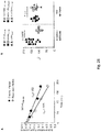
- the t 1/2 for apoptotic epitope-complexed pentamer staining to fresh CD8 + T cells from patients experiencing chronic infection was significantly longer than the decay of pentamer staining to CD8 + T cells from patients undergoing infection resolution ( Figure 28A,B ).
- the t 1/2 for viral epitope-complexed pentamers to CD8 + T cells did not differ between patients experiencing chronic infection and those undergoing infection resolution ( Figure 28A,B ).
- the t 1/2 for apoptotic epitope-complexed pentamer staining to CD8 + T cells tended (although not significantly) to be directly correlated with the viral load ( Figure 29 ).
- control experiments in which HLA-A*0201 pentamers were complexed to a non-natural irrelevant peptide showed that the t 1/2 for staining to CD8 + T cells was undetectable (data not shown).
- the author demonstrates for the first time that the emergence of mixed polyfunctional (type-1, -2, -17) CD8 + T EM cell responses to apoptotic self-epitopes is related to the chronic evolution of an acute infection.
- CD8 + T EM cell responses directed to apoptotic self-epitopes were incredibly wide and robust during the acute phase of HCV infection, particularly in patients experiencing chronic progression compared with those undergoing infection resolution.
- the responses were then sustained over time in relation to viral persistence, suggesting that they can contribute to the immunopathology and chronic evolution of acute HCV infection.
- HCV-specific CD8 T cells have been reported to express a dysfunctional phenotype (weak proliferation, IFN- ⁇ production, and cytotoxicity) and increased levels of PD-1, known to be associated with the exhausted phenotype, irrespective of infection progression (Refs in 35).
- Polycytokine (IL-17, IFN- ⁇ , IL-2, and IL-4) production by autoreactive CD8 + T EM cells may have crucial implications in HCV-related immunopathology, which stresses the importance of infections in inducing and maintaining autoimmunity (36).
- IL-17 production can account for the transient intra-hepatic infiltration of neutrophils found only in the early phase of acute HCV infection (37). Then, the timely decline in hepatocyte damage and the consequent evolution toward a state of chronic low-level liver disease, in patients developing viral persistence, may be achieved through the simultaneous presence of autoreactive type-1,-2, and -17 CD8 + TE M cells, which may regulate each other, and even the capacity of type-17 CD8 + T EM cells to express a certain degree of plasticity (2, 21-24), and to convert to CD8 + T EM cells with combined features of type-17 and -1 cells. Therefore, the environmental setting during an acute inflammatory disease seems to be addressed to guarantee the coexisting polarization of type-1, -2, and -17 CD8 + T EM cells.
- autoreactive CD8 + T cells might be less susceptible of the exhaustion, likely because they are primed later than the virus-specific, that in contrast show a profound exhaustion phenotype in patients with acute or chronic hepatitis (27, 33, 35).
- the PD-1-dependent tuning of autoreactive CD8 + T EM cell functions, as well as the high frequencies of apoptotic epitope-specific CD8 + T EM cells producing IL-4, may play a pivotal role in limiting excessive functional responses, thus allowing the evolution toward chronic low-level inflammatory liver disease (6, 38-40).
- the author's data demonstrate that autoreactive CD8 T cell responses against apoptotic T cell-associated epitopes are strictly dependent on the viral persistence and correlate with immunopathology via the exuberant production of inflammatory cytokines.
- This model represents a remarkable example of the importance of infections in inducing and particularly maintaining autoimmunity.
- our study provides a possible explanation for why the enormous expansion of activated T cells, during persisting viral infections, is only minimally attributable to virus-specific T cells (14).
- the author can envisage the following multistep model. In the conditions of HCV immune evasion, continuous waves of dysfunctional virus-specific T cells are generated, and can deliver the initial apoptotic antigenic stimuli for priming apoptotic antigen-specific T cells.
Landscapes
- Health & Medical Sciences (AREA)
- Life Sciences & Earth Sciences (AREA)
- Immunology (AREA)
- Engineering & Computer Science (AREA)
- Hematology (AREA)
- Molecular Biology (AREA)
- Chemical & Material Sciences (AREA)
- Biomedical Technology (AREA)
- Urology & Nephrology (AREA)
- Food Science & Technology (AREA)
- General Physics & Mathematics (AREA)
- Cell Biology (AREA)
- Biotechnology (AREA)
- Pathology (AREA)
- Microbiology (AREA)
- Medicinal Chemistry (AREA)
- Physics & Mathematics (AREA)
- Analytical Chemistry (AREA)
- Biochemistry (AREA)
- General Health & Medical Sciences (AREA)
- Rehabilitation Therapy (AREA)
- Rheumatology (AREA)
- Physiology (AREA)
- Tropical Medicine & Parasitology (AREA)
- Measuring Or Testing Involving Enzymes Or Micro-Organisms (AREA)
- Peptides Or Proteins (AREA)
- Medicines That Contain Protein Lipid Enzymes And Other Medicines (AREA)
- Medicines Containing Antibodies Or Antigens For Use As Internal Diagnostic Agents (AREA)
Description
- The invention relates to a method to monitor viral infection progression. The method is based on measuring T cell response or auto-antibodies to apoptotic epitopes (antigenic determinants).
- Successful priming of naive CD4+ or CD8+ T cells results in the generation of both effector memory T (TEM) cells expressing various differentiation programs (type-1, -2, -17), according to the environment in which they are exposed (1-8), and central memory T (TCM) cells that promptly proliferate and generate new waves of effector cells on demand (9-11). The transcription factor T-box-containing protein expressed in T cells (T-bet) is the master regulator of the type-1 cell differentiation program that is associated with the production of IFN-γ, which is required for the development of protective immune responses against intracellular pathogens (2). GATA-binding protein 3 (GATA-3) controls the development of the type-2 cell lineage that is characterized by the production of IL-4, -5, and -13, which is critical for immunity against helminths and other extracellular pathogens (2). Retinoid acid-related orphan receptor (ROR)-γt in mice and the human ortholog RORC in humans represent the master regulators of type-17 cell differentiation that leads to the production of IL-17, which is specifically required for protection against several types of extracellular and intracellular bacterial infections (1, 3-5). All these (type-1, -2, -17) functions can elicit either protective or harmful effects, depending on whether they are executed by pathogen-specific or autoreactive T cells or whether the pathogen- specific are involved during an acute resolving infection or a chronic infection, respectively.
- The fate of the enormous number of apoptotic cells that derive from effector T cells undergoing apoptosis after performing their functions during acute or chronic infections remain to be determined (12, 13). In previous studies, the author found that the proteome of apoptotic T cells includes prominent caspase-cleaved cellular proteins and that a high proportion of distinct T cell epitopes in these fragments (apoptotic epitopes) can be cross-presented by DCs to a wide repertoire of autoreactive CD8+ T cells (14). Recent reports have confirmed the role of caspase cleavage in the processing and presentation of epitopes that are derived from apoptotic cells in different models (15-17). In chronic HIV infection, these autoreactive CD8+ T cells correlate with the proportion of apoptotic CD4+ T cells in vivo and are involved in establishing polyclonal T cell activation that in the long run results in generalized T cell dysfunction/depletion (14). In addition, apoptotic cells derived from activated T cells (in contrast to those derived from resting T cells) retain the expression of CD40 ligand (L) and can then condition CD40+ DCs to acquire high capacities to prime or cross-prime autoreactive T cells (18, 19). Therefore, the expression of CD40L on apoptotic T cells can bypass the tolerogenic effects of conventional apoptotic cells (i.e., those derived from epithelial cells) and influence the switch from tolerogenic immature DCs to mature DCs with high stimulatory and migratory capacities (18, 19). This mechanism is consistent with the evidence that the signals provided by CD40L+ apoptotic cells and not those provided by conventional apoptotic cells facilitate the emergence of autoreactive T cell responses to apoptotic self-antigens (18, 19).
- Virological tools to diagnose and monitor hepatitis C virus infection are described in the review Chevaliez S., Clin. Microbiol. Infect. 2011; 17:116-121 .
- In the present invention, the authors used the HCV infection as a human model of acute infection that generally undergoes chronic progression to verify whether CD8+ T cells that are specific for apoptotic self-epitopes have a distinct effector type-1, -2, or -17 phenotype, to distinguish which of them may participate in determining the fate of a viral infection (recovery versus chronicity), and to ascertain the mechanisms whereby these responses are induced and maintained.
- Caspase-dependent cleavage of antigens associated with apoptotic cells has a prominent role in the generation of CD8+ T cell responses in various infectious diseases. The author found that the emergence of a large population of autoreactive CD8+ T effector cells specific for apoptotic T cell-associated self-epitopes exceeds the antiviral responses in patients with acute HCV infection. Importantly, they endow mixed polyfunctional type-1, type-2 and type-17 responses and correlate with the chronic progression of infection. This evolution is related to the selection of autoreactive CD8+ T cells with higher TCR avidity, whereas those with lower avidity undergo prompt contraction in patients who clear infection. The development of mixed responses with divergent differentiation requirements is consistent with distinct sites or kinetics of CD8+ T cell priming in vivo. These findings demonstrate a previously undescribed strict link between the emergence of high frequencies of mixed autoreactive CD8+ T cells producing a broad array of cytokines (IFN-γ, IL-17, IL-4, IL-2...) and the progression toward chronic disease in a human model of acute infection.
- The present invention is based on autoimmune response capacities against antigen peptides derived from apoptotic cells (apoptotic epitopes) both to be used as biomarkers in the diagnosis, prognosis and to evaluate chronic evolution of various viral infections or autoimmune diseases. The present results clearly specify that such responses predict the chronic evolution of acute infections such as Hepatitis C (HCV), Hepatitis B (HBV) and HIV.
- In fact, patients with acute infections as HCV etc. that, in early phases show high levels of such autoimmune responses, progress to chronic infection. The responses are correlated with tissue damage signs and high viral load. Contrarily, patients with the same acute infections that do not present such responses undergo infection resolution. In other words, said autoimmune responses predict the infection fate. In confirmation of this, the author monitored such patients with a follow-up from acute phase to chronic phase (chronic hepatitis/cirrhosis) of HCV and verified that such autoimmune responses were sustained over time only in patients progressing to chronic infection in relation with viral persistence and high transaminase levels (liver injury indication).
- Therefore, the present invention represents a unique biomarker that predicts the fate (resolution vs. chronicization) of an acute infection, since that no other known clinical biomarker is available to date.
- In the present invention it is confirmed that such autoimmune responses are represented by autoreactive polyfunctional CD8 T lymphocytes, since they produce high levels of different cytokines (INF-γ, IL-17, IL-23, IL-4, IL-2...) in response to apoptotic epitopes. Again, such polyfunctional responses are predictive of chronic infection. It is interesting to note that these polyfunctional responses decrease and/or modify prematurely in relation to the resolution of infection treated with different anti-viral drugs. Therefore, such responses can be used as:
- i) Early biomarkers that predict the fate of an acute infection with recoils not only in diagnosis, but also in early therapeutic outline: consequently, adequate therapies can be set up prior to progression towards chronicization;
- ii) Biomarkers that can be detected to determine protective effect of conventional or innovative anti-viral therapies, with enormous advantages for national health service since it could be early predicted if certain therapy will work or not.
- The previous published study (14) identifies apoptotic epitopes only in molecular and immunogenic terms. In the study, it is also demonstrated that relative autoimmune responses are present in HIV chronic infection and correlates to viraemia, but there are no data or hypothesis that such responses, when present in acute infections, are predictive of disease evolution. This last point is at the base of the definition of biomarker: a biomarker cannot be defined as such if it does not have predictability value.
- The present invention defines the first predictive biomarker for chronic evolution of acute infections, development of protective effects of therapies, of prognosis and therapies of autoimmune disease and their sequences.
- It is therefore an object of the invention a method to predict the outcome of a viral infection and/or to monitor the progress of a viral infection and/or to monitor the efficacy of a therapy for a viral infection in a biological sample comprising T cell obtained from a subject affected by said viral infection comprising the steps of
- a) incubating the isolated biological sample with at least one peptide consisting of a sequence selected from SEQ ID No.1 to SEQ ID No. 252 under appropriate conditions to generate a T cell response;
- b) measuring the T cell response;
- c) comparing the measured T cell response to the measured T cell response of a suitable control sample,
- Preferably the biological sample is a sample of PBMCs or blood.
- Preferably the at least one peptide of step a) consist of the sequence selected from LLQDSVDFSL (SEQ ID No. 204), QLFNHTMFI (SEQ ID No. 64) or VLMIKALEL (SEQ ID No. 78).
- Still preferably step b) comprises incubating the biological sample with a pool of peptides selected among the following pools:
- pool 1: SEQ ID Nos. 1, 2, 4, 5, 8 and 9;
- pool 2: SEQ ID Nos. 14, 16, 18, 19 and 20;
- pool 3: SEQ ID Nos. 27, 28, 29, 30, 34, 35, 36, 37 and 40;
- pool 4: SEQ ID Nos. 58, 59, 60, 62, 63, 64, 65, 66 and 67;
- pool 5: SEQ ID Nos. 68, 69, 73, 74, 75, 76, 77, 78, 81, 83, 84 and 88;
- pool 6: SEQ ID Nos. 89, 90, 91, 92, 99, 100, 104, 105, 106, 111, 117, 123, 126 and 127;
- pool 7: SEQ ID Nos. 198, 199, 200, 201, 202, 204, 206, 210, 211 and 220;
- pool 8: SEQ ID Nos. 232, 233, 234, 235, 236, 237, 238, 239, 240, 241, 242, 243 and 244 or
- pool 9: SEQ ID Nos. 245, 246, 247, 248 and 249.
- In a preferred embodiment the T cell response is a CD8+ T cell response.
- Preferably the T cell response is measured by means of an Elispot assay.
- Still preferably the Elispot assay is an interferon-gamma, IL-17 or IL-4 Elispot assay.
- It is a further object of the invention a method to monitor the progress of a viral infection and/or to monitor the efficacy of a therapy for a viral infection in a subject affected by said viral infection comprising the step of detecting and/or quantifying the amount of auto-antibodies directed against at least one peptide as defined above, wherein said viral infection is caused by a virus selected from the group of HCV, HBV, thereby discriminating patients experiencing chronic infection and those undergoing infection resolution.
- The amount of auto-antibodies directed against at least one peptide may be compared to values measured in a suitable control sample.
- In a preferred embodiment the viral infection is caused by a virus selected from the group of HCV and HBV.
- In the present invention the outcome of a viral infection may be a resolution of the infection or the progression toward chronic infection.
- The methods of the present invention may comprise isolating a fresh biological sample or using a frozen or cryopreserved biological sample T cells response may also be measured by methods known by the skilled person in the art such as intracellular staining methods, cytofluorimetric methods, ect.
- T cells response or detection and/or quantification of auto-antibodies against the peptides may be measured at regular intervals (either weekly, or every 2nd, 3rd or 4th week or at longer, shorter and variable intervals) during the course of the viral infection or during the course of the therapy. The analysis of the responses or auto-antibodies detection/quantification at the different time points may be compared to the first and/or to previous examinations to identify relative changes in responses.
- A suitable control sample may be a sample obtained or isolated before the start of a therapy, the sample of a healthy subject, the sample of a subject whose infection progress towards resolution, a sample obtained or isolated at various time points during the course of the infection or during the course of the therapy.
- The invention will be now illustrated by means of non-limiting figures.
-
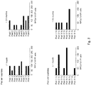
Figure 1 : CD8+ T cell multispecificity to apoptotic epitopes in chronic HCV progression. (A,B) Mean number of spot-forming cells (SFCs) by fresh CD8+ T effector memory (TEM) cells (by ELISPOT assay) in response to pools (see Tables 11-13 and 14-17) of apoptotic self-epitopes (A) or viral epitopes (B) in patients with acute HCV infection experiencing chronic infection (filled circles) or undergoing infection resolution (empty circles), evaluated at different time points of the infection. Each circle represents a single patient. NS = not significant. (C) Representative correlation (6th month after clinical onset) between the mean number of SFCs formed by fresh CD8+ TEM cells in response to pools of apoptotic self-epitopes and the viral load (HCV-RNA copies/ml) in patients experiencing chronic infection (filled circles) or undergoing infection resolution (empty circles). The inset graph highlights the same correlation in patients whose viral load variations were not evident in the larger graph. Statistical analysis was performed using non-parametric Spearman's test. (D) Representative flow cytometry analyses of peripheral blood mononuclear cells (PBMCs), double-stained with a monoclonal antibody (mAb) to CD8 and pentamers expressing the indicated peptides of non muscle myosin heavy chain 9 (MYH9) or vimentin (VIME). Counterplot analyses show the percentage of CD8+pentamer+ cells. The percentage of cells is indicated in each quadrant. The pentamer values were <0.02% (mean ± s.d. = 0.0173% ± 0.0009% for all pentamers complexed to apoptotic epitopes; 0.0052% ± 0.0035% for those complexed to viral epitopes) in 20 HLA-A2+ healthy individuals; thus 0.02% was considered the sensitive level of the assay. (E,F) Percentages of CD8+pentamer+ cells from all patients studied (filled and empty symbols represent patients experiencing chronic infection or undergoing infection resolution, respectively), evaluated at different time points. Eachsymbo 1 represents the indicated pentamer specificity. NS = not significant. -
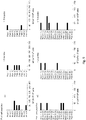
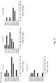
Figure 2 to 18 : Effector CD8+ T cells specific to apoptotic or viral epitopes in patients with acute HCV infection. Fresh CD8+ T cells, purified from PBMCs of patients (Pt.) undergoing chronic (C) or self-limited (S) HCV infection, were able to form IFN-γ spots promptly within 6 h of contact with autologous irradiated CD8-depleted PBMCs, as APCs, plus peptide ex vivo, as detected by an ELISPOT assay. Results are expressed as spot-forming cells (SFCs) in 1 x 10⁶ CD8+ T cells. The SFC values were subtracted from the background (BG). Because of the limited number of PBMCs obtained from patients, freshly isolated CD8+ T cells were tested against 9 pools of apoptotic epitopes (Pool self peptides, see also Supplementary Table 1A-C) and 8 pools of viral peptides (Pool viral peptides, see also Supplementary Table 2A-D).Fig. 2 to 12 represent responses from PBMCs of patients undergoing chronic HCV infection,Fig. 13 to 18 represent responses from PBMCs of patients undergoing self-limited HCV infection. Pools of apoptotic self-epitopes or viral epitopes are indicated in Tables 11-13 and 14-17. -
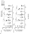
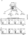
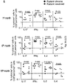
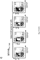
Figure 19 : Polyfunctional CD8+ TEM cells specific to apoptotic epitopes distinguish patients experiencing chronic infection. (A,B) Representative flow cytometry analyses of PBMCs from patients with acute HCV infection experiencing chronic infection (A) or undergoing infection resolution (B) that were stained with mAb to CD8 and pentamers complexed to the indicated apoptotic or viral epitopes. Cells were then stimulated with the relevant soluble peptides plus anti-CD28 mAb and processed for the detection of IL-17, IL-22, IFN-γ, IL-4, and IL-2 by ICS assay with the relevant mAbs. Counterplot analyses are gated on CD8+pentamer+ cells and show percentages of cytokine-producing cells. The percentage of cells is reported in each quadrant. The small histograms show ICS analyses of representative cytokine (IL-17, IL-22, IFN-γ, IL-4, or IL-2) production without antigenic stimulation by gated CD8+pentamer+ cells. Irrelevant cytokine production (<0.1% cells) was observed when either apoptotic epitope- or viral epitope-specific CD8+pentamer+ T cells of 20 HLA-A2+ healthy individuals were stimulated with this procedure; thus, the author considered 1% cytokine-producing cells in pentamer+ cells as the sensitive level of this assay. (C) Percentage of cells producing the indicated cytokines in CD8+pentamer+ cells in response to the indicated apoptotic epitopes (evaluated at the indicated time point by flow cytometry analyses) from patients with acute HCV infection experiencing chronic infection (filled symbols) or undergoing infection resolution (empty symbols). The horizontal dashed line delimits an arbitrary background, which is based on the values of 20 HLA-A2+ healthy individuals exhibiting < 0.1% cytokine-producing cells in each test. -
Figure 20 : Kinetics of cytokine-producing CD8+pentamer+ cells in patients with acute HCV infection. (A,B) Peptide dose-response curves of cytokine-producing CD8+pentamer+ cells from patients with acute HCV infection experiencing chronic infection (A) or undergoing infection resolution (B). Cells were stained with the pentamers expressing the indicated peptides and stimulated for 6 h with the same soluble peptides. They were then processed for the detection of the different cytokines indicated by ICS assay. Values are shown with the background subtracted. (C) Polyfunctional CD8+ TEM cells specific to apoptotic epitopes in patients with acute HCV infection. Percentage of cells producing the indicated cytokines in CD8+pentamer+ cells in response to the indicated apoptotic epitopes (evaluated at the indicated time points by flow cytometry analyses) from patients with acute HCV infection experiencing chronic infection (filled symbols) or undergoing infection resolution (empty symbols). The horizontal dashed line delimits an arbitrary background, which is based on the values of 20 HLA-A2+ healthy individuals exhibiting < 0.1% cytokine-producing cells in each test. -
Figure 21 : Polyfunctional CD8+ TEM cells specific to viral epitopes in patients with acute HCV infection. Percentage of cells producing the indicated cytokines in CD8+pentamer+ cells in response to the indicated viral epitopes (evaluated at the indicated time points by flow cytometry analyses) from patients with acute HCV infection experiencing chronic infection (filled symbols) or undergoing infection resolution (empty symbols). The horizontal dashed line delimits an arbitrary background, which is based on the values of 20 HLA-A2+ healthy individuals exhibiting < 0.1% cytokine-producing cells in each test. -
Figure 22 : Polyfunctional CD8+ TEM cells specific to apoptotic epitopes predict the chronic progression of HCV infection. (A) Kinetics of plasma HCV-RNA and alanineaminotransferase (ALT) levels until 15-24 months from clinical onset of infection. Each symbol represents a single patient. (B,C) Kinetics of the means of all the CD8+pentamer+ (pent) cell percentages promptly producing the indicated cytokines in response to the apoptotic (B) or viral (C) epitopes. The cells were calculated as a percentage of the absolute number of cells expressing CD8, pentamer ligands, and cytokines in the PBMCs. Values are shown with the background subtracted. Filled circles represent patients with acute HCV infection experiencing chronic infection; empty circles represent a HCV-patients undergoing infection resolution. -
Figure 23 : (A,B) Number of patients exhibiting a wide repertoire of polyfunctional CD8+ T cells producing one or two cytokines in response to apoptotic (A) or viral (B) epitopes. -
Figure 24 : (A,B) Correlation between CD8+pentamer+ TEM cells producing IFN-γ (A) or IL-17 (B) in response to apoptotic epitopes and the viral load (HCV-RNA copies) or ALT in patients with acute HCV infection experiencing chronic infection or infection resolution. Statistical analysis was performed using non-parametric Spearman's test. -
Figure 25 : CD8+ TEM cells specific to apoptotic epitopes are activated by apoptotic epitopes naturally processed and cross-presented by dendritic cells (DCs). (A) One representative of five experiments in which PBMCs from one patient with acute HCV infection were double-stained with mAb to CD8 and pentamers complexed with the indicated apoptotic epitope, and cultured with autologous DCs that had been pulsed or not with the relevant soluble peptide, apoptotic cloned T cells (ACs = apoptotic cells), ACs previously treated with a negative caspase control (K = Z-FA-FMK), or ACs previously treated with thecaspase 3 inhibitor (C3I = Z-DEVD-FMK). After 6 h, CD8+pentamer+ cells were tested for their capacity to produce the different cytokines indicated by the ICS assay. Counterplots are gated on CD8+pentamer+ cells and show percentages of the different cytokine-producing cells in each quadrant. (B) Cumulative experiments in 5 independent patients, performed as described in (A), showing the percentages of cells producing the indicated cytokines in CD8+pentamer+ cells in response to the following stimuli: autologous DCs alone (a); DCs that had been pulsed with the MYH9741-749 peptide (b); DCs that had been pulsed with ACs, previously treated with a negative caspase control (Z-FA-FMK) (c); or DCs that had been pulsed with ACs previously treated with thecaspase 3 inhibitor (Z-DEVD-FMK) (d). Each symbol represents a single patient. (C) Correlation (analysed at the 15th month after clinical onset) between the percentage of CD8+pentamer+ cells specific to apoptotic epitopes and the percentage of circulating apoptotic T cells in patients with acute HCV infection experiencing chronic infection (filled circles) or infection resolution (empty circles). Statistical analysis was performed using non-parametric Spearman's test. -
Figure 26 : No intrinsic defect of effector functions in CD8+ T cells from patients with acute HCV infection. (A) One representative flow cytometry analysis in which PBMCs from patients with acute HCV infection were stained with mAb to CD8 and the indicated pentamer, stimulated with PMA and iono for 6 h, and processed for the detection of the indicated cytokines by ICS assay with the relevant mAbs. Counterplot analyses are gated on CD8+pentamer+ cells and show percentages of cytokine-producing cells. The percentage of cells is reported in each quadrant. (B) Percentage of cells producing the indicated cytokines in CD8+pentamer+ cells in response to PMA and iono (evaluated at the indicated time points by flow cytometry analyses) from patients with acute HCV infection experiencing chronic infection (filled symbols) or undergoing infection resolution (empty symbols). -
Figure 27 : naïve, central, and effector memory CD8+ T cells specific to apoptotic or viral epitopes. (A) One representative flow cytometry analysis, the values of the total patients are shown in (B), in which PBMCs from a patient with acute HCV infection were stained with mAbs to CD8, CD45RO, and CD127 and with the pentamer complexed with the indicated apoptotic peptide. Counterplot analyses are gated on CD8+pentamer cells and show percentages of CD45RO+ and/or CD127+ cells. The percentage of cells is reported in each quadrant. (B) Percentages of CD45RO+CD127-, CD45RO-CD127+, or CD45RO+CD127+ cells in CD8+pentamer+ cells from HCV patients experiencing chronic infection (filled circles) or infection resolution (empty circles). (C) One representative of six flow cytometry analyses in which PBMCs from a patient with acute HCV infection were stained with mAbs to CD8 and CD127 and with the pentamer complexed with the indicated peptide. Cells were stimulated with the same soluble peptide for 6 h and then processed for the detection of the indicated cytokines by ICS assay with the relevant mAbs. -
Figure 28 : Decay kinetics of pentamer staining for CD8+ T cells. (A) Representative decay plot of the natural logarithm of the normalized fluorescence versus time after anti-HLA-A2 mAb addition for fresh PBMCs stained with pentamers that were complexed with the indicated apoptotic epitope. The t 1/2 represents the staining half-times for the pentamers to CD8+ T cells. Filled or empty circles represent PBMCs from patients with acute HCV infection experiencing chronic infection or undergoing infection resolution, respectively. (B) The t 1/2 for the pentamers (complexed with the indicated apoptotic or viral epitopes) binding CD8+ T cells from patients with acute HCV infection experiencing chronic infection (filled symbols) or undergoing infection resolution (empty symbols). -
Figure 29 : Link between viral load and the decay kinetics of pentamer staining for CD8+ T cells. Correlation between plasma HCV-RNA copies and the staining half-times (t 1/2) for the pentamers to CD8+ T cells from patients with acute HCV infection experiencing chronic infection (filled circles) or infection resolution (empty circles). NS = not significant. - The study cohort included 16 patients with acute HCV infection (4 women, 12 men, median age 31 years, range 22-57 years). Diagnosis of acute HCV infection was based on (1) high levels of serum ALT; (2) sero-conversion assessed by third generation enzyme linked immune-sorbent assay, or anti-HCV positivity at the time of the diagnosis with an anti-HCV negative test in the previous 12 months; (3) presence of HCV-RNA in at least the first serum sample, and (4) sudden onset of liver disease symptoms. Alternative causes of acute hepatitis, such as other viral infection, autoimmunity, alcohol, drugs, and toxins were excluded. Patients with concomitant immunological disorders or with HIV co-infection were also excluded from the study.
- PBMCs were isolated and T cell clones were generated as previously described (48). CD8+ T cells were purified from PBMCs by positive selection coupled to magnetic beads (MiltenyiBiotec) as previously described (38). Flow cytometry analysis demonstrated >99% CD8+ cells in the positively purified population and <5% in the CD8-depleted population. To purify antigen-specific type-17 CD8+ cells, PBMCs were stimulated with the relevant peptide plus anti-CD28 (4 µg/ml) (BD Pharmingen). Then, cells were stained with allophycocyanin (APC)-labeled anti-CCR6 (R&D System) and PE-cyanine 7 (PE-Cy7)-labeled anti-CCR4 (BD Pharmingen) and processed with FACSAria (Becton Dickinson) to sort CCR6+CCR4+ cells: >98% of these cells both produced IL-17 in response to the relevant peptide and were susceptible of staining with the relevant pentamers, as detected below. Spontaneous apoptosis of PBMCs from patients was determined by staining with Annexin-V (ApoAlert Apoptosis Kit, Clontech Laboratories Inc), propidium iodide (PI) (Sigma-Aldrich) and PE-Cy7-labeled anti-CD3 (BD Pharmingen) before and after 18 hours incubation at 37°C. Immature (i)DCs were derived from peripheral monocytes that had been purified by positive selection with anti CD14 mAb coupled to magnetic beads (MiltenyiBiotec). CD14+ cells were incubated for 5 days in RPMI 1640 medium containing 5% FCS, 2 mM glutamine, 1% nonessential amino acids, 1% sodium pyruvate, 50 µg/ml kanamycin (Gibco BRL), 50 ng/ml rGM-CFS (Novartis Pharma), and 1000 U/ml rIL-4 (gently provided by A. Lanzavecchia, Bellinzona, CH). Mature DCs were obtained by a 40-h stimulation of iDCs with soluble rCD40L molecules (Alexis Biochemicals, Alexis Corporation). The definition of monocyte-derived DCs was based on their surface phenotype profile by staining with anti CD14, anti-CD86 (Caltag Laboratories), anti CD1a, anti-CD1c, anti-CD11c, anti-CD32, anti-CD80 (BD PharMingen) mAbs, Annexin-V (ApoAlert Apoptosis Kit, Clontech Laboratories Inc), propidium iodide (PI) (Sigma-Aldrich), and the appropriate secondary labeled antibodies (BD PharMingen).
- Highly purified CD8+ T cells (1 x 10⁵) from PBMCs were stimulated for 4-6 h with eight independent pools of viral-peptides (
genotype 1b) and eight independent pools of viral-peptides (genotype 2c) (see Supplemental Table 1A-C, and Supplemental Table 2A-D below) (5 µg/ml per single peptide) and irradiated autologous CD8-depleted PBMCs, used as APCs, and tested by an ELISPOT assay, as described (14). - PBMCs were incubated with APC-labeled-HLA-A*0201 pentamers (complexed to vimentin78-87 [LLQDSVDFSL], non-muscle myosin478-486 [QLFNHTMFI], or nonmuscle myosin741-749 [VLMIKALEL] peptide) for apoptotic epitopes and APC-labeled-HLA-A*0201 pentamers (complexed to HCV-NS31073-1081 [CINGVCWTV], HCV-NS31406-1415 [KLVALGINAV] or HCV-Core132-140 [DLMGYIPAV] peptide) for viral epitopes (ProImmune Limited, Oxford, United Kingdom), in FACS buffer (
PBS 1×, 2% human AB serum) for 10 min at 37°C, followed by washing and further incubation with APC-Cy7-labeled mAb to CD8 (BD Pharmingen, San Diego, CA), FITC-labeled anti-PD-1 (BD Pharmingen), PE-labeled anti-CD127 (BD Pharmingen), FITC-labeled anti-CD45RO (Caltag Laboratories, Burlingame, CA) for 20 min at 4°C. Negative controls were obtained by staining cells with an irrelevant isotype-matched mAb. Cells were washed, acquired with a FACSCanto flow cytometer and FACSDiva analysis software (Becton Dickinson) or FlowJo software version 7.5.5 (Tree star, Inc. San Carlos, CA). - PBMCs were stained with pentamers and mAb to CD8, and then stimulated with or without different concentrations of the corresponding uncomplexed peptide (ProImmune Limited) plus anti-CD28 mAb (4 µg/ml) (BD Pharmingen), or with PMA (50ng/ml) plus ionomycin (1 µg/ml) (Sigma Aldrich, Milan, Italy), for 6 h at 37°C. At the 2nd h, 10 µg/ml Brefeldin A (Sigma-Aldrich) was added. Cells were washed, fixed and permeabilized using Cytofix/Cytoperm solution (BD Pharmingen) at 4°C for 20 min, re-washed with Perm Wash Buffer (BD Pharmingen), and intracellularly stained with different combinations of Alexa Fluor 488-labeled anti-IL17A (eBioscience San Diego, CA), PE- labeled anti-IFN-γ, PE-labeled anti-IL-2, FITC-labeled anti-IL-4 (BD Pharmingen), PE-labeled anti-RORC (eBioscience) or purified anti-T-bet (Santa Cruz Biotechnology Santa Cruz, California) for 30 min at 4°C. When stained with unlabeled specific antibody to detect T-bet, cells were washed and stained with the appropriate secondary FITC-labeled antibody. Cells were washed, acquired with a FACSCanto flow cytometer and FACSDiva analysis software (Becton Dickinson) or FlowJo software (Tree star).
- Cloned CD8+CD95+ T cells (10-100 x 106) were incubated in the presence or absence of 14 mg/ml C3I (Z-DEVD-FMK), or a negative caspase control (K, Z-FA-FMK) (BD Pharmingen) for 1 h at 37°C in a 24-well plate. Then, cells were induced to apoptosis by incubation with 500 ng/ml anti-Fas (anti-CD95 mAb [clone CH11], Upstate Biotechnology) for at least 6 h. Apoptotic cells were determined by staining with Annexin- V (ApoAlert Apoptosis Kit, Clontech Laboratories Inc), PI (Sigma-Aldrich) and flow-cytometry analysis. PBMCs were double-stained with pentamers and mAb to CD8 and cultured with iDCs that had been pulsed or not with apoptotic cloned T cells. After 6-8 h, cells were tested for their capacity to produce IL-17 and IFN-γ by intracellular cytokine staining (ICS) as described above. Cells were washed, acquired with a FACSCanto flow cytometer and analyzed with FACSDiva analysis software (Becton Dickinson) or FlowJo software (Treestar).
- PBMCs were stained with saturating amounts of APC-labeled-HLA-A*0201 pentamers and APC-Cy7-labeled-CD8 (BD Pharmingen) for 45 min at room temperature (28). Then, cells were washed three times with buffer (2% FCS, 0.01 sodium azide in PBS) and resuspended in 500 µl of buffer with saturanting amounts of mAb to HLA-A2 (BB7.2, ATCC). At various time points (0, 30', 1h, 2h and 3h), an aliquot cells was washed and the fluorescence intesity was determined by flow cytometry analysis. Double staining using an anti-human TCRα/β (BD Pharmingen) and pentamers was performed in parallel to normalize pentamer fluorescence against the expressed TCR. The values were then normalized to percent of the total fluorescence at the initial time point and plotted on a logarithmic scale. t 1/2 are determined by calculating the (ln2)/mean slope value of plots of the natural logarithm (ln) of the pentamer fluorescence normalized for the TCR fluorescence. The slope is equivalent to ln(Fa/Fb)/t, where Fa is the normalized fluorescence at the start of the interval, Fb is the normalized fluorescence at the end of the interval, and t is the length of the interval (minutes).
- PBMCs were incubated for 10 days a 37°C with specific peptides (apoptotic peptides or viral peptides) and anti-PD-L1 (10 µg/ml) (R&D systems Minneapolis, MN) or isotype control antibody (10 µg/ml). Recombinant IL-2 (50 U/ml) (CHIRON Emeryville, California) was added on
day 4 of culture. Onday 10, cells were stained with surface antibodies, pentamers, anti-IL-17A, anti-IFN-γ, anti-IL-2 and anti-IL-4 mAbs. Cells were washed, acquired with a FACSCanto flow cytometer and analyzed with FACSDiva analysis software (Becton Dickinson) or FlowJo software (Tree star). - PBMCs were incubated for 10 days at 37°C with specific peptides (apoptotic or viral peptides), human rIL-6 (50 ng/ml), rIL-1β (10 ng/ml), rIL-23 (50 ng/ml) and rTGF-β (10 ng/ml) (R&D Systems) for the Th17 cell polarization. For the Th1 cell polarizing condition, PBMCs were antigen-stimulated in the presence of recombinant human rIL-12 (10 ng/ml) and rIFN-γ (100 U/ml) (R&D Systems). Recombinant IL-2 was added on
day 4 of culture (50 U/ml). Onday 10, cells were stained with surface antibodies, pentamers, anti IL-17A, anti-IFN-γ, anti-IL-2 and anti-IL-4 mAbs. Cells were washed, acquired with a FACSCanto flow cytometer and analyzed with FACSDiva analysis software (Becton Dickinson) or FlowJo software (Tree star). - All statistical analyses were performed with Prism 4 (GraphPad) software using nonparametric Spearman's correlation test, nonparametric Mann-Whitney U-test for unpaired data and Wilcoxon test for paired data. The differences were considered significant at P<0.05.
- As reported in (14), the author identified proteins differentially expressed in apoptotic cells. The results are reported in Table 1 below.
Table 1 Relative abundance of spots identified in apoptotic or control T cell clones by 2DE and MALDI-TOF analyses Spot no.2 AN ID Description Relative abundance M/pl Theoretical Found Matching peptides Percentage sequence coverage (AA) Note CCs ACs 23 P08670 VIME Vimentin ++ + 53/5 53/4.8 25 60(33-465) Whole 3 P08670 VIME Vimentin - + 53/5 25/4.5 13 39(196-423) Fragment 4 P08670 VIME Vimentin - + 53/5 40/4.5 9 22(100-389) Fragment 6 P08670 VIME Vimentin - + 53/5 30/5.2 7 20(13-183) Fragment 13 P08670 VIME Vimentin - + 53/5 20/4.5 6 14(100-183) Fragment 1 P35579 MYH9 Nonmuscle myosin + ++ 226/5.5 90/5.2 40 6(126-1930) Fragment 19 P20700 LAM1 Lamin B1 ++ + 67/5.1 70/5 8 15(79-386) Whole 2 P20700 LAM1 Lamin B1 - + 67/5.1 30/4.7 13 21(14-220) Fragment 17 P52566 GDIS Rho GDP dissociation inhibitor 2 ++ + 23/5.1 23/4.8 8 49(22-196) Whole 8 P52566 GDIS Rho GDP dissociation inhibitor 2 - + 23/5.1 21/6.6 12 52(22-164) Fragment 9 P52566 GDIS Rho GDP dissociation inhibitor 2 - + 23/5.1 21/6.9 5 30(34-131) Fragment 5 Q07244 ROK Heterogeneous nuclear ribonucleoprotein K + ++ 51/5.4 45/6.2 15 24(22-326) Fragment 30 Q07244 ROK Heterogeneous nuclear ribonucleoprotein K ++ + 51/5.4 65/5 5 15(70-33) Whole 7 P02570 ACTB Actin cytoplasmatic 1 - + 42/5.3 30/5.1 8 23(12-278) Fragment 10 P02570 ACTB Actin cytoplasmatic 1 - + 42/5.3 12/5.8 5 17(12-106) Fragment 16 P02570 ACTB Actin cytoplasmatic 1 - + 42/5.3 40/5 8 28(19-312) Fragment 27 P02570 ACTB Actin cytoplasmatic 1 + ++ 42/5.3 32/4.7 4 14(29-206) Fragment 18 P18669 PMG1 Phosphoglycerate mutase 1 ++ + 29/6.7 28/7 4 24(90-239) Whole 20 Q9NYL9 TMO3 Ubiquitous tropomodulin ++ + 40/5.1 40/5 6 29(12328) Whole 21 Q9Y3F4 UNRI UNR interacting protein ++ + 39/4.9 38/4.7 4 18(45-290) Whole 24 P25786 PSA1 Proteasome subunit a type 1 + ++ 30/6.5 30/6.2 7 31(1-214) Whole 25 P05387 RLA2 60S acidic ribosomal protein P2 + ++ 12/4.4 12/4.3 6 72(1-94) Whole 28 P08758 ANX5 annexin V ++ + 36/4.9 36/4.6 7 22(6-259) Whole 33 P31146 CO1A Coronin like protein p57 ++ + 52/6.2 70/6.7 4 9(11-393) Whole 34 P78371 TCBP Tcomplex protein 1, b subunit ++ + 58/6 60/6.5 7 24(26-516) Whole 35 n.i. ++ + 36/5.7 36 n.i. ++ + 38/5.6 37 n.i. ++ + 39/5.5 38 n.i. ++ + 37/5.4 39 n.i. ++ + 55/5.6 40 n.i. ++ + 55/5.9 41 n.i. ++ + 65/6 42 n.i. ++ + 43/6.7 43 n.i. ++ + 65/7 44 n.i. ++ + 90/6.9 45 n.i. ++ + 29/6.4 11 n.i. - + 12/5.9 12 n.i. - ++ 20/4.5 14 n.i. + + 10/4.4 15 n.i. - + 15/3.5 31 n.i. ++ + 80/4.5 32 n.i. ++ + 55/5 22 n.i. ++ + 15/4.6 26 n.i. + ++ 36/6.7 29 n.i. ++ + 17/5.4 *Spot number corresponding to those shown m 2DE analysis in Figure1 ; n.i., spot not identified; -, spot absent; +, spot present; ++, spot whose relative abundance is at least twice than in the corresponding + spot; AN, access nuntber in Swiss-Prot database; ID, identity; CCs, control cells; ACs, apoptotic cells; Mr, relative molecular mass; AA, amino acids; RI, isoelectric point; - The author tested the HLA-A2 binding affinity of 9 to 10 amino acid long fragments of the proteins identified and reported in Table 1. The results are reported in Tables 2-9.
Table 2. HLA-A2 binding affinity of synthetic peptides derived from fragmented proteins. Binding capacity (IC50§ nM) Alleles bound SEQ ID No. Organism Protein 1th Pos° Sequence Length A*0201 A*0202 A*0203 A*0206 A*6802 1 Human ACTB" 131 AMYVAIQAV 9 2,8 1,5 1,6 7.1 190 5 2 Human ACTB 319 ALAPSTMKI 9 4,3 2,9 2,5 40 5,5 5 3 Human ACTB 170 ALPHAILRL 9 128 98 7,9 186 34091 4 4 Human ACTB 266 FLGMESCGI 9 2,7 71 11 16 3979 4 5 Human ACTB 312 RMQKEITAL 9 14 0,29 6,5 28 6667 4 6 Human ACTB 66 TLKYPIEHGI 10 369 202 138 26442 290 4 7 Human ACTB 131 AMYVAIQAVL 10 351 21 12195 363 234 4 8 Human ACTB 348 SLSTFQQMWI 10 5,6 4,5 7,7 791 299 4 9 Human ACTB 46 GMGQKDSYV 9 196 17 156 13541 17632 3 10 Human ACTB 241 ELPDGQVITI 10 974 140 1953 3728 100 2 11 Human ACTB 184 DLTDYLMKI 9 2029 218 1542 2065 4464 1 12 Human ACTB 177 RLDLAGRDL 9 838 2263 5373 - - 0 13 Human ACTB 184 DLTDYLMKIL 10 1031 5668 10229 1980 8682 0 14 Human ROK^ 154 SIAGGIIGV 9 0,56 1,0 1,1 0,14 49 5 15 Human ROK 41 EMVELRILL 9 92 211 870 152 80 4 16 Human ROK 67 ALRTDYNASV 10 8,9 5,0 11 349 1354 4 17 Human ROK 100 ILKKIIPTL 9 121 15 5,8 914 24029 3 18 Human ROK 193 VLIGGKPDRV 10 14 8,5 12 1282 7417 3 19 Human ROK 209 ILDLISESPI 10 360 65 1272 2598 3083 2 20 Human ROK 122 QLPLESDAV 9 246 601 1270 925 1999 1 21 Human ROK 48 LLQSKNAGAV 10 5026 534 31 11017 - 1 22 Human ROK 146 ELRLLIHQSL 10 1506 135 1581 1191 1130 1 23 Human ROK 426 PIEGSEDRI 9 16751 3509 26921 - 15762 0 24 Human ROK 124 PLESDAVECL 10 - 4230 11892 - - 0 25 Human ROK 169 ELRENTQTTI 10 3392 2513 1828 - 2532 0 26 Human ROK 426 PLEGSEDRII 10 - - - - - 0 27 Human LAM1* 496 TIWAANAGV 9 8,9 263 308 22 20 5 28 Human LAM1 41 RLAVYIDKV 9 0,16 0,84 0,18 14 1004 4 29 Human LAM1 301 SLSSQLSNL 9 1,6 0,57 2,4 104 1377 4 30 Human LAM1 361 QLLDVKLAL 9 0,060 98 288 4.0 5848 4 31 Human LAM1 182 QLADETLLKV 10 39 14 82 61 14354 4 32 Human LAM1 133 KLREYEAAL 9 80 28 22 4326 33708 3 33 Human LAM1 140 ALNSKDAAL 9 34 1,9 5,9 3625 - 3 #=identity, "=actin cytoplasmic I; ^=eterogeneous nuclear ribonucleoprotein K; *=lamin B1°=1st amino acid position; §=IC50 indicates binding affinity and it is expressed as 50% inhibitory nanomolar concentration, a dash ("-") indicates an IC50 >50000 nM; peptides with IC50 <50nM are classified as high affinity peptides, peptides with IC50>50 and <500nM Table 3. HLA-A2 binding affinity of synthetic peptides derived from fragmented proteins. Binding capacity (IC50§ nM) Alleles bound SEQ ID No. Organism Protein 1th Pos° Sequence Length A*0201 A*0202 A*0203 A*0206 A*6802 34 Human LAM1* 291 ELMESRMRI 9 246 1226 284 25188 45 3 35 Human LAM1 355 QLNDYEQLL 9 2,0 0,23 18 1493 4808 3 36 Human LAM1 388 KLSPSPSSRV 10 50 18 45 563 20979 3 37 Human LAM1 488 VLKAGQTVTI 10 263 37 23 4060 15429 3 38 Human LAM1 37 ELNDRLAVYI 10 1161 134 1224 30389 61 2 39 Human LAM1 60 QLQYIEREEV 10 16688 1297 197 237 - 2 40 Human LAM1 378 KLLEGEEERL 10 30 29 3395 1944 20515 2 41 Human LAM1 529 ILKNSQGEEV 10 1647 58 371 - 2705 2 42 Human LAM1 51 SLETENSAL 9 695 124 5974 8911 30303 1 43 Human LAM1 84 ELADARRAL 9 - 1902 7437 - 97 1 44 Human LAM1 157 SLEGDLEDL 9 1068 202 6246 3738 - 1 45 Human LAM1 164 DLKDQIAQL 9 15715 2616 114 - 991 1 46 Human LAM1 175 SLAAAKKQL 9 23555 172 612 23009 36585 1 47 Human LAM1 379 LLEGEEERL 9 537 61 616 20505 - 1 48 Human LAM1 20 PLSPTRLSRL 10 4761 227 3342 - 22244 1 49 Human LAM1 76 GLKALYETEL 10 1582 15 785 - - 1 50 Human LAM1 34 ELRELNDRL 9 2510 571 22553 37575 2837 0 51 Human LAM1 126 DLNGAQIKL 9 - 2616 - - 3571 0 52 Human LAM1 161 DLEDLKDQI 9 24324 16376 13617 - 44118 0 53 Human LAM1 192 DLENRCQSL 9 12584 12360 32752 - - 0 54 Human LAM1 247 EMREQHDAQV 10 8828 510 3948 - 548 0 55 Human LAM1 262 ELEQTYHAKL 10 40156 2838 - - 11673 0 56 Human LAM1 308 NLQKESRACL 10 9872 1130 4730 - 23622 0 57 Human LAM1 370 DMEISAYRKL 10 - 11689 27629 - - 0 58 Human MYH9** 9 YLYVDKNFI 9 9,3 0,85 4,8 45 217 5 59 Human MYH9 108 GLIYTYSGL 9 22 11 10 197 58 5 60 Human MYH9 111 YTYSGLFCV 9 0,12 2.1 3,0 0,84 3,2 5 61 Human MYH9 126 NLPIYSEEI 9 67 77 390 27 47 5 62 Human MYH9 145 EMPPHIYAI 9 323 67 110 85 1,8 5 63 Human MYH9 186 KVIQYLAYV 9 0,67 0,17 1,2 0,21 1,8 5 64 Human MYH9 478 QLFNHTMFI 9 1,4 0,71 3,2 14 14 5 65 Human MYH9 584 WLMKNMDPL 9 9,7 0,36 14 6,0 1,9 5 #=identity; *=lamin B1; **=mn muscle myosin; °=1st aminoacid posttion; §=IC50 indicates biding affinity and it is expressed as 50% inhibitory nanomolar concentration, a dash ("-") indicates an IC50 >50000 nM; peptides with IC50 <50nM are classified as high affinity peptides, peptides with IC50>50 and <300nM are classified as intermediate affinity peptides, peptides with IC50>500nM are classified as low affinity peptides. Table 4. HLA-A2 binding affinity of synthetic peptides derived from fragmented proteins. Binding capacity (IC50§ nM) Alleles bound SEQ ID No. Organism Protein 1th Pos° Sequence Length A*0201 A*0202 A*0203 A*0206 A*6802 66 Human MYH9** 653 QLAKLMATL 9 5,4 1,4 2,9 26 18 5 67 Human MYH9 111 YTYSGLFCVV 10 0,21 0,34 0,12 4,2 0,4 5 68 Human MYH9 424 RMFRWLVLRI 10 1,1 5,0 10 71 398 5 69 Human MYH9 478 QLFNHTMFIL 10 0,97 0,42 8,2 49 82 5 70 Human MYH9 257 YLLEKSRAI 9 39 57 16 94 17045 4 71 Human MYH9 272 RTFHIFYYL 9 - 5,8 238 1,3 144 4 72 Human MYH9 280 LLSGAGEHL 9 199 5,7 19 361 - 4 73 Human MYH9 302 FLSNGHVTI 9 0,11 1,0 3,8 11 3050 4 74 Human MYH9 338 GLLRVISGV 9 0,21 3,0 3,1 4,3 8823 4 75 Human MYH9 412 FAIEALAKA 9 26 1,7 4,0 0,43 - 4 76 Human MYH9 450 ILDIAGFEJ 9 5,5 50 257 21 3064 4 77 Human MYH9 733 FMDGKQACV 9 0,18 7,8 33 6,6 10345 4 78 Human MYH9 741 VLMIKALEL 9 0,15 15 38 78 870 4 79 Human MYH9 829 RLF7KVKPL 9 205 9,7 6,2 421 - 4 80 Human MYH9 1084 ELQAALARV 9 558 117 57 40 2,5 4 81 Human MYH9 1277 KLQVELDNV 9 4,1 8,8 30 9,2 - 4 82 Human MYH9 1326 SLSTKLKQV 9 157 54 10 422 28642 4 83 Human MYH9 1843 KLKDVLLQV 9 3,3 2,5 6,7 0,20 18519 4 84 Human MYH9 279 YLLSGAGEHL 10 4,9 6,3 175 11 - 4 85 Human MYH9 322 TMEAMRIMGI 10 123 137 132 568 26 4 86 Human MYH9 338 GLLRVISGVL 10 68 304 118 390 736 4 87 Human MYH9 648 QLYKEQIAKL 10 78 4,6 16 147 9146 4 88 Human MYH9 733 FMDGKQACVL 10 21 27 158 683 374 4 89 Human MYH9 1920 KLRRGDLPFV 10 2,9 15 9,1 5,5 - 4 90 Human MYH9 210 QLLQANPIL 9 4,5 183 1507 19 - 3 91 Human MYH9 847 MMAKEEELV 9 9,1 1,2 276 1070 5639 3 92 Human MYH9 877 QLMAEKLQL 9 8,2 7,9 205 657 4360 3 93 Human MYH9 940 KMQQNIQEL 9 13 15 82 536 28571 3 94 Human MYH9 1270 ELADKVTKL 9 2513 33 114 38752 3,8 3 95 Human MYH9 1298 KLTKDFSAL 9 550 20 34 29 1592 3 96 Human MYH9 1372 KMEDSVGCL 9 284 22 176 2846 - 3 97 Human MYH9 1389 KLQKDLEGL 9 29 25 118 619 15139 3 #=identity; **=non muscle myosin; °=1st aminoacid position; §=IC50 indicates binding affinity and it is expressed as 50% inhibitory nanomolar concentration, a dash ("-") indicates an IC50 >50000 nM; peptides with IC50 <50nM are classified as high affinity peptides, peptides with IC50>50 and <500nM are classified as intermediate affinity peptides, peptides with Table 5. HLA-A2 binding affinity of synthetic peptides derived from fragmented proteins. Binding capacity (IC50§ nM) Alleles bound SEQ ID No. Organism Protein 1th Pos° Sequence Length A*0201 A*0202 A*0203 A*0206 A*6802 98 Human MYH9" 1536 EMKTQLEEL 9 20769 83 81 22770 284 3 99 Human MYH9 1726 RLEARIAQL 9 433 7,7 15 2292 46881 3 100 Human MYH9 1793 KLQEMEGTV 9 23 99 48 867 19355 3 101 Human MYH9 1811 ALEAKIAQL 9 103 15 47 1100 8772 3 102 Human MYH9 1909 AMNREVSSL 9 92 1,6 3,9 2994 11952 3 103 Human MYH9 74 KMNPPKFSKV 10 34 2457 50 154 20000 3 104 Human MYH9 660 TLRNTNPNFV 10 21 107 40 - 593 3 105 Human MYH9 688 VLDQLRCNGV 10 6,6 186 61 868 - 3 106 Human MYH9 752 NLYRIGQSKV 10 322 87 76 17848 1024 3 107 Human MYH9 811 VLQRNCAAYL 10 96 18 95 2180 - 3 108 Human MYH9 829 RLFTKVKPLL 10 53 78 96 4039 16393 3 109 Human MYH9 1027 AMITDLEERL 10 127 10 193 543 941 3 110 Human MYH9 1228 ELANEVKVLL 10 66 17 1255 24371 2,9 3 111 Human MYH9 248 YIVGANIET 9 422 622 9009 81 626 2 112 Human MYH9 591 PLNDNIATL 9 1374 137 294 - 21277 2 113 Human MYH9 641 GMFRTVGQL 9 867 74 89 14107 - 2 114 Human MYH9 975 KLEEEQIIL 9 72 74 2552 940 21583 2 115 Human MYH9 982 ILEDQNCKL 9 90 41 997 6043 - 2 116 Human MYH9 1309 QLQDTQELL 9 49 5,0 2451 4684 7879 2 117 Human MYH9 1540 QLEELEDEL 9 404 176 - 28866 4718 2 118 Human MYH9 1646 QMKDCMREL 9 3622 155 14 9819 - 2 119 Human MYH9 1677 SMEAEMIQL 9 40 70 2382 1093 - 2 120 Human MYH9 1681 EMIQLQEEL 9 3097 123 35881 23126 105 2 121 Human MYH9 1751 RLKKANLQI 9 1847 269 151 3769 - 2 122 Human MYH9 100 NLKERYYSGL 10 7214 1,6 214 - 5455 2 123 Human MYH9 161 MMQDREDQSI 10 2,1 25 542 1245 4005 2 124 Human MYH9 428 WLVLRINKAL 10 976 23 257 556 5329 2 125 Human MYH9 599 LLHQSSDKFV 10 808 14 78 17504 3571 2 126 Human MYH9 821 KLANWQW WRL 10 56 54 1037 5442 - 2 127 Human MYH9 846 EMMAKEEELV 10 33 9,6 958 2456 2809 2 128 Human MYH9 1143 ALKTELEDTL 10 652 27 431 17831 - 2 129 Human MYH9 1316 LLQEENRQKL 10 245 120 1903 3764 - 2 #=identity; **=non muscle myosin °=1st aminoacid position; §=IC50 indicates binding affinity and it is expressed as 50% inhibitory nanomolar concentration, a dash ("-") indicates an IC50 >50000 nM; peptides with IC50 <50nM are classified as high affinity peptides, peptides with IC50>50 and <500nM are classified as intermediate affinity peptides, peptides with Table 6. HLA-A2 binding affinity of synthetic peptides derived from fragmented proteins. Binding capacity (IC50§ nM) Alleles bound SEQ ID No. Organism Protein 1th Pos° Sequence Length A*0201 A*0202 A*0203 A*0206 A*6802 130 Human MYH9** 1417 RLQQELDDLL 10 92 5,0 13611 1041 17072 2 131 Human MYH9 1674 KLKSMEAEMI 10 1465 93 314 7835 9009 2 132 Human MYH9 339 LLRVISGVL 9 1756 1769 133 1148 3191 1 133 Human MYH9 588 NMDPLNDNI 9 1928 108 2492 25916 - 1 134 Human MYH9 748 ELDSNLYRI 9 353 522 13587 4010 1099 1 135 Human MYH9 846 EMMAKEEEL 9 3226 67 16679 24202 13889 1 136 Human MYH9 870 EMETLQSQL 9 6093 389 48703 - 14778 1 137 Human MYH9 894 ELCAEAEEL 9 16191 194 18303 24732 1274 1 138 Human MYH9 989 KLAKEKKLL 9 9793 197 1293 37909 17143 1 139 Human MYH9 1045 ELEKTRRKL 9 8215 275 32650 45176 8403 1 140 Human MYH9 1052 KLEGDSTDL 9 2001 482 25287 10828 2463 1 141 Human MYH9 1059 DLSDQIAEL 9 2217 72 2924 11939 583 1 142 Human MYH9 1066 ELQAQIAEL 9 1093 1022 4539 29473 14 1 143 Human MYH9 1077 QLAKKEEEL 9 7887 110 29889 - 15625 1 144 Human MYH9 1108 ELESQISEL 9 - 95 23119 45805 - 1 145 Human MYH9 1228 ELANEVKVL 9 7891 1008 914 38221 116 1 146 Human MYH9 1281 ELDNVTGLL 9 9654 22 2539 49404 - 1 147 Human MYH9 1354 NLEKQIATL 9 1280 393 662 31973 26470 1 148 Human MYH9 1410 KLEKTKTRL 9 19721 1346 4972 106 - 1 149 Human MYH9 1417 RLQQELDDL 9 960 106 1628 6747 - 1 150 Human MYH9 1421 ELDDLLVDL 9 926 180 25362 20240 12533 1 151 Human MYH9 1508 DLMSSKDDV 9 4292 10433 19470 - 352 1 152 Human MYH9 88 ELTCLNEASV 10 4576 699 3482 3739 69 1 153 Human MYH9 561 QLKDKADFCI 10 859 4,3 1235 32059 16129 1 154 Human MYH9 591 PLNDNIATLL 10 15874 165 1315 10005 8929 1 155 Human MYH9 853 ELVKVREKQL 10 1134 120 1593 9037 5190 1 156 Human MYH9 972 KLKKIFEEQI 10 4265 133 1487 - - 1 157 Human MYH9 1020 KLKNKHEAMI 10 4078 161 566 - 10250 1 158 Human MYH9 1396 GLSQRHEEKV 10 1016 429 1754 - 12672 1 159 Human MYH9 1547 ELQATEDAKL 10 20843 246 36430 8677 12970 1 160 Human MYH9 1718 ALALEEKRRL 10 12186 91 1478 - - 1 161 Human MYH9 57 ELVENGKKV 9 5352 3186 8839 9575 7634 0 #=identity; **=non muscle myosin;°=1st aminoacid position; §=IC50 indicates binding affinity and it is expressed as 50% inhibitory nanomolar concentration, a dash ("-") indicates an IC50 >50000 nM; peptides with IC50 <50nM are classified as high affinity peptides, peptides with IC50>50 and <500nM are classified as intermediate affinity peptides, peptides with Table 7. HLA-A2 binding affinity of synthetic peptides derived from fragmented proteins. Binding capacity (IC50§ nM) Alleles bound SEQ ID No. Organism Protein 1th Pos° Sequence Length A*0201 A-0202 A*0203 A*0206 A*6802 162 Human MYH9** 507 DLQPCIDLI 9 14684 562 5210 21315 1685 0 163 Human MYH9 610 ELWKDVDRI 9 1619 2655 12174 11071 2254 0 164 Human MYH9 776 DLKITDVII 9 1640 8882 1538 1201 1813 0 165 Human MYH9 905 RLTAKKQI1 9 19580 1134 2436 - 25862 0 166 Human MYH9 912 ELEEICHDL 9 10924 1221 10150 - 36585 0 167 Human MYH9 1136 DLGEELEAL 9 26540 3367 27494 - - 0 168 Human MYH9 1140 ELEALKTEL 9 - 4688 14696 - 15905 0 169 Human MYH9 1214 NLEKAKQTL 9 23964 6332 46974 43929 693 0 170 Human MYH9 1221 TLENERGEL 9 28583 1344 18763 45571 21456 0 171 Human MYH9 1448 QLLAEEKTI 9 4386 988 2603 18097 - 0 172 Human MYH9 1522 ELEKSKRAL 9 30314 19745 39690 - - 0 173 Human MYH9 1614 KIEMDLKDL 9 1091 3124 3816 2548 - 0 174 Human MYH9 1618 DLKDLEAHI 9 - 14337 46029 - 21127 0 175 Human MYH9 1782 QLERQNKEL 9 10043 1401 38094 22548 25210 0 176 Human MYH9 1871 RLKQLKRQL 9 3813 5498 832 - 9967 0 177 Human MYH9 126 NLPIYSEEIV 10 2778 614 37384 9609 1179 0 178 Human MYH9 460 DLNSFEQLCI 10 4742 1388 13212 - 3344 0 179 Human MYH9 505 GLDLQPCIDL 10 690 1202 16159 18387 - 0 180 Human MYH9 610 ELWKDVDRII 10 8866 5426 24375 - 13216 0 181 Human MYH9 770 HLEEERDLKI 10 5209 1336 21844 - - 0 182 Human MYH9 1100 NMALKKIREL 10 16361 4234 791 892 - 0 183 Human MYH9 1160 ELRSKREQEV 10 18756 5446 29769 - 3033 0 184 Human MYH9 1189 EMRQKHSQAV 10 33936 8731 7945 - 615 0 185 Human MYH9 1368 DMKKKMEDSV 10 15027 14863 38466 - 10676 0 186 Human MYH9 1564 AMKAQFERDL 10 8294 961 687 9690 - 0 187 Human MYH9 1606 SMAVAARKKL 10 - 755 572 - 926 0 188 Human GDIS^^ 100 VLKEGSEYRV 10 342 10 149 1003 234 4 189 Human GDIS 186 HLSWEWNLSI 10 6,1 13 68 56 1093 4 190 Human GDIS 37 EMDKDDESL 9 340 637 1099 360 42 3 191 Human GDIS 51 TLLGDGPVV 9 20 547 675 236 4354 2 192 Human GDIS 93 ALKKETIVL 9 834 38 42 25437 12508 2 193 Human GDIS 44 SLIKYKKTL 9 3549 281 1735 3982 - 1 #=identity; **=non muscle myosin; ^^=rho GDP dissociation inhibitor 2; °=1st aminoacid position; §=IC50 indicates binding affinity and it is expressed as 50% inhibitory nanomolar concentration, a dash ("-") idicates an IC50>50000 nM; peptides wih IC50<50nM are classified as hgh affinity peptides, peptides with IC50>50 and <500nM are classified as intermediate affinity peptides, peptides with IC30>500nM are classfied as low afinity peptides Table 8. HLA-A2 binding affinity of synthetic peptides derived from fragmented proteins. Binding capacity (IC50§ nM) Alleles bound SEQ ID No. Organism Protein 1th Pos° Sequence Length A*0201 A*0202 A*0203 A*0206 A*6802 194 Human GDIS^^ 86 DLTGDLEAL 9 16949 714 24638 14995 18411 0 195 Human GDIS 37 EMDKDDESLI 10 25743 1900 17268 - - 0 196 Human GDIS 44 SLIKYKKTLL 10 1057 885 532 4916 779 0 197 Human GDIS 90 DLEALKKETI 10 - 21231 23588 - - 0 198 Human VIME% 176 NLAEDIMRL 9 1,0 0,31 4,2 136 83 5 199 Human VIME 50 SLYASSPGGV 10 2,2 4,3 2,4 461 452 5 200 Human VIME 68 RLRSSVPGV 9 3,3 27 1,6 30 9901 4 201 Human VIME 129 ILLAELEQL 9 35 6,5 231 7,5 - 4 202 Human VIME 225 SLQEEIAFL 9 0,99 0,37 19 44 4777 4 203 Human VIME 331 ALKGTNESL 9 108 9,5 47 445 771 4 204 Human VIME 78 LLQDSVDFSL 10 3,6 1,1 149 9,2 1931 4 205 Human VIME 411 SLPLPNFSSL 10 61 199 16 641 1,2 4 206 Human VIME 79 LQDSVDFSL 9 5,9 47 10155 2,3 774 3 207 Human VIME 324 SLTCEVDAL 9 54 6,3 398 1080 3119 3 208 Human VIME 378 HLREYQDLL 9 59 4,2 171 8893 1641 3 209 Human VIME 384 DLLNVKMAL 9 181 265 109 787 647 3 210 Human VIME 419 SLNLRETNL 9 402 1,4 22 16458 - 3 211 Human VIME 122 FLEQQNKILL 10 20 0,43 63 828 10669 3 212 Human VIME 232 FLKKLHEEEI 10 372 9,7 53 18182 11931 3 213 Human VIME 235 KLHEEEIQEL 10 226 18 278 4828 - 3 214 Human VIME 401 KLLEGEESR1 10 127 35 166 1172 - 3 215 Human VIME 122 FIEQQNKIL 9 496 25 814 12281 - 2 216 Human VIME 155 ELRRQVDQL 9 434 89 2151 9148 1449 2 217 Human VIME 201 TLQSFRQDV 9 154 290 1264 7375 2074 2 218 Human VIME 218 DLERKVESL 9 121 70 3058 3374 - 2 219 Human VIME 108 ELNDRFANYI 10 1786 38 543 45376 14 2 220 Human VIME 370 NMKEEMARHL 10 260 433 703 10578 9346 2 221 Human VIME 385 LLNVKMALDI 10 4638 222 162 562 1201 2 222 Human VIME 421 NLRETNLDSL 10 5263 156 83 8250 2672 2 223 Human VIME 263 DLTAALRDV 9 10569 735 147 28139 2459 1 224 Human VIME 402 LLEGEESRI 9 856 296 921 1472 877 1 225 Human VIME 42 ALRPSTSRSL 10 3996 2489 284 42830 - 1 226 Human VIME 147 DLYEEEMREL 10 14986 485 827 33261 16216 1 227 Human VIME 413 PLPNFSSLNL 10 3769 172 2927 - 17910 1 228 Human VIME 152 EMRELRRQV 9 30508 3455 3015 - 40541 0 229 Human VIME 162 QLTNDKARV 9 810 567 634 1689 695 0 230 Human VIME 136 QLKGQGKSRL 10 30885 2925 3801 - - 0 #=identity; ^^=rho GDP dissociation inhibitor 2; %=vimentin; °=1st aminoacid position; §=IC50 indicates binding affinity and it is expressed as 50% inhibitory nanomolar concentration : dash ("-") indicates an IC50>50000 nM; peptides wih IC50<50nM are classified as high affinity peptites, peptiles with IC50>50 and <500nM are classified as intermediate affinity peptides, peptides wih IC50>500nM are classified as bw affinity peptides. Table 9. HLA-A2 binding affinity of synthetic peptides derived from not fragmented proteins. Binding capacity (IC50§ nM) Alleles bound SEQ ID No. Organism Protein 1th Pos° Sequence Length A*0201 A-0202 A*0203 A*0206 A*6802 231 Human RLA2* 38 RLNKVISEL 9 15433 107 157 - 14162 2 232 Human PSA1** 179 FMECNLNEL 9 596 27 55 15067 14822 2 233 Human PSA1 183 NLNELVKHGL 10 145 12 11 4834 3618 3 234 Human PSA1 175 HMSEFMECNL 10 59 11 70 109 35360 4 235 Human PSA1 63 ILHVDNHIGI 10 - - 16720 - - 0 236 Human PSA1 37 GLKSKTHAV 9 - 4552 924 - 19982 0 237 Human PSA1 110 SLIGSKTQI 9 - 15913 511 34100 17135 0 238 Human PSA1 179 FMECNLNELV 10 12715 194 1 - - 2 239 Human PSA1 48 ALKRAQSEL 9 - - 18952 - 29040 0 240 Human PSA1 76 GLTADARLL 9 163 4,7 1 7,8 12 5 241 Human PSA1 102 PLPVSRLVSL 10 4,5 1,2 0,21 40 1659 4 242 Human PSA1 204 DLTTKNVSI 9 465 3,6 54 4278 7386 3 243 Human PSA1 45 ELNGKNIEDV 10 104 1,2 0,21 1776 14417 3 244 Human PSA1 55 ELAAHQKKI 9 38 2 1,1 100 9790 4 245 Human PSA1 186 ELVKHGLRAL 10 296 15 1,6 8411 - 3 246 Human PSA1 37 GLKSKTHAVL 10 2366 116 24 10609 21672 2 247 Human PSA1 191 GLRALRETL 9 41760 45 23 - - 2 248 Human PSA1 55 ELAAHQKKIL 10 8856 146 362 16700 - 2 249 Human PSA1 97 FVFDRPLPV 9 - 8417 2175 9732 44603 0 250 Human RLA2 3 YVASYLLAA 9 - 6236 2649 - 1908 0 251 Human RLA2 26 ILDSVGIEA 9 1271 235 112 1756 1598 2 252 Human RLA2 3 YVASYLLAAL 10 1 0,88 0,5 2,4 1,5 5 #=identity;*RLA2=60S acidic ribosomal protein P2;**PSA1= Proteasome component C2; °=1st aminoacid position; §=IC50 indicates binding affinity and it is expressed as 50% inhibitory nanomolar concentration, a dash ("-") indicates an IC50 >50000 nM; peptides with IC50<50nM are classified as high affinity peptides, peptides with IC50>50 and <500nM - The author analyzed longitudinally the responses of 16 patients with acute HCV infection. The follow-up ranged from the onset of acute disease (clinical onset) to 15 to 24 months (the sixth month being considered the time of conversion from an acute to a chronic infection). Of the 16 patients, 6 patients had a self-limited infection and 10 patients exhibited a chronic evolution of infection (Table 10).
Table 10. Clinical parameters of patients with acute HCV infection Pt Genotype Outcome ALT (U/ml) HCV-RNA (copies/ml) 1C 1b Chronic 107 556000 2C 2a/2c Chronic 2700 3310 3C 3a Chronic 1113 156000 4C 2a/2c Chronic 600 915000 5C 1b Chronic 1515 600 6C 3a Chronic 324 940000 7C 1b Chronic 792 462000 8C 3a Chronic 745 900000 9C 1a Chronic 904 500000 10C 3a Chronic 803 919000 1S 1b Self-limited 2710 106000 2S 3a Self-limited 1967 2120 3S 2a/2c Self-limited 5917 361000 4S 1a Self-limited 1359 96100 5S n.d. Self-limited 2860 210200 6S n.d. Self-limited 2650 69500 - Initially, the effector responses were determined by the capacity of freshly isolated CD8+ T cells from either HLA-A2+ patients or healthy controls to form IFN-γ spots within 4 to 6 h of contact with nine pools of synthetic apoptotic peptides (Tables 11-13) or eight pools of HCV peptides (Tables 14-17) selected for their capacity to bind the HLA-A2 molecule (14, 20), in an ELISPOT assay.
Table 11. HLA-A2 binding peptides derived from apoptotic cell-associated proteins appearing with their cleavage products in proteomic analysis. Binding capacity (IC50§ nM) Alleles bound Pool Organism Protein 1st Pos° Sequence Length A*0201 A*0202 A*0203 A*0206 A*6802 1 Human ACTB" 131 AMYVAIQAV 9 2.8 1.5 1.6 7.1 190 5 Human ACTB 319 ALAPSTMKI 9 4.3 2.9 2.5 40 5.5 5 Human ACTB 266 FLGMESCGI 9 2.7 71 11 16 3979 4 Human ACTB 312 RMQKEITAL 9 14 0.29 6.5 28 6667 4 Human ACTB 348 SLSTFQQMWI 10 5.6 4.5 7.7 791 299 4 Human ACTB 46 GMGQKDSYV 9 196 17 156 13547 17632 3 2 Human ROK^ 154 SLAGGIIGV 9 0.56 1.0 1.1 0.14 49 5 Human ROK 67 ALRTDYNASV 10 8.9 5.0 11 349 1354 4 Human ROK 193 VLIGGKPDRV 10 14 8.5 12 1282 7417 3 Human ROK 209 ILDLISESPI 10 360 65 1272 2598 3083 2 Human ROK 122 QLPLESDAV 9 246 601 1270 925 1999 1 3 Human LAM1* 496 TIWAANAGV 9 8.9 263 308 22 20 5 Human LAM1 41 RLAVYIDKV 9 0.16 0.84 0.18 14 1004 4 Human LAM1 301 SLSSQLSNL 9 1.6 0.57 2.4 104 1377 4 Human LAM1 361 QLLDVKLAL 9 0.060 98 288 4.0 5848 4 Human LAM1 291 ELMESRMRI 9 246 1226 284 25188 45 3 Human LAM1 355 QLNDYEQLL 9 2.0 0.23 18 1493 4808 3 Human LAM1 388 KLSPSPSSRV 10 50 18 45 563 20979 3 Human LAM1 488 VLKAGQTVTI 10 263 37 23 4060 15429 3 Human LAM1 378 KLLEGEEERL 10 30 29 3395 1944 20515 2 4 Human MYH9" 9 YLYVDKNFI 9 9.3 0.85 4.8 45 217 5 Human MYH9 108 GLIYTYSGL 9 22 11 10 197 58 5 Human MYH9 111 YTYSGLFCV 9 0.12 2.1 3.0 0.84 3.2 5 Human MYH9 145 EMPPHIYAl 9 323 67 110 85 1.8 5 Human MYH9 186 KVIQYLAYV 9 0.67 0.17 1.2 0.21 1.8 5 Human MYH9 478 QLFNHTMFI 9 1.4 0.71 3.2 14 14 5 Human MYH9 584 WLMKNMDPL 9 9.7 0.36 14 6.0 1.9 5 Human MYH9 653 QLAKLMATL 9 5.4 1.4 2.9 26 18 5 Human MYH9 111 YTYSGLFCW 10 0.21 0.34 0.12 4.2 0.4 5 °=1st amino acid position; §=IC50 indicates binding affinity and it is expressed as 50% inhibitory nanomolar concentration, a dash ("-") indicates an IC50 >50000 nM; peptides with IC50 <50nM are classified as high affinity peptides, peptides with IC50>50 and <500nM are classified as intermediate affinity peptides, peptides with IC50>500nM are classified as low affinity peptides; "=actin cytoplasmatic 1; ^=heterogeneous nuclear ribonucleoprotein K; *=lamin B1; **=non muscle myosin. Table 12. HLA-A2 binding peptides derived from apoptotic cell-associated proteins appearing with their cleavage products in proteomic analysis. Binding capacity (IC50§ nM) Alleles bound Pool Organism Protein 1st Pos° Sequence Length A*0201 A*0202 A*0203 A*0206 A*6802 5 Human MYH9** 424 RMFRWLVLRI 10 1.1 5.0 10 71 398 5 Human MYH9 478 QLFNHTMFIL 10 0.97 0.42 8.2 49 82 5 Human MYH9 302 FLSNGHVTI 9 0.11 1.0 3.8 11 3050 4 Human MYH9 338 GLLRVISGV 9 0.21 3.0 3.1 4.3 8823 4 Human MYH9 412 FAIEALAKA 9 26 1.7 4.0 0.43 - 4 Human MYH9 450 ILDIAGFEI 9 5.5 50 257 21 3064 4 Human MYH9 733 FMDGKQACV 9 0.18 7.8 33 6.6 10345 4 Human MYH9 741 VLMIKALEL 9 0.15 15 38 78 870 4 Human MYH9 1277 KLQVELDNV 9 4.1 8.8 30 9.2 - 4 Human MYH9 1843 KLKDVLLQV 9 3.3 2.5 6.7 0.20 18519 4 Human MYH9 279 YLLSGAGEHL 10 4.9 6.3 175 11 - 4 Human MYH9 733 FMDGKQACVL 10 21 27 158 683 374 4 6 Human MYH9 1920 KLRRGDLPFV 10 2.9 15 9.1 5.5 - 4 Human MYH9 210 QLLQANPIL 9 4.5 183 1507 19 - 3 Human MYH9 847 MMAKEEELV 9 9.1 1.2 276 1070 5639 3 Human MYH9 877 QLMAEKLQL 9 8.2 7.9 205 657 4360 3 Human MYH9 1726 RLEARIAQL 9 433 7.7 15 2292 46881 3 Human MYH9 1793 KLQEMEGTV 9 23 99 48 867 19355 3 Human MYH9 660 TLRNTNPNFV 10 21 107 40 - 593 3 Human MYH9 688 VLDQLRCNGV 10 6.6 186 61 868 - 3 Human MYH9 752 NLYRIGQSKV 10 322 87 76 17848 1024 3 Human MYH9 248 YIVGANIET 9 422 622 9009 81 626 2 Human MYH9 1540 QLEELEDEL 9 404 176 - 28866 4718 2 Human MYH9 161 MMQDREDQSI 10 2.1 25 542 1245 4005 2 Human MYH9 821 KLRNWQWWRL 10 56 54 1037 5442 - 2 Human MYH9 846 EMMAKEEELV 10 33 9.6 958 2456 2809 2 °=1st amino acid position; §=IC50 indicates binding affinity and it is expressed as 50% inhibitory nanomolar concentration, a dash ("-") indicates an IC50 >50000 nM; peptides with IC50 <50nM are classified as high affinity peptides, peptides with IC50>50 and <500nM are classified as intermediate affinity peptides, peptides with IC50>500nM are classified as low affinity peptides; **=non muscle myosin. Table 13.HLA-A2 binding peptides derived from apoptotic cell-associated proteins appearing with their cleavage products in proteomic analysis. Binding capacity (IC50§ nM) Alleles bound Pool Organism Protein 1st Poso Sequence Length A*0201 A*0202 A*0203 A*0206 A*6802 7 Human VIME% 176 NLAEDIMRL 9 1.0 0.31 4.2 136 83 5 Human VIME 50 SLYASSPGGV 10 2.2 4.3 2.4 461 452 5 Human VIME 68 RLRSSVPGV 9 3.3 27 1.6 30 9901 4 Human VIME 129 ILLAELEQL 9 35 6.5 231 7.5 - 4 Human VIME 225 SLQEEIAFL 9 0.99 0.37 19 44 4777 4 Human VIME 78 LLQDSVDFSL 10 3.6 1.1 149 9.2 1931 4 Human VIME 79 LQDSVDFSL 9 5.9 47 10155 2.3 774 3 Human VIME 419 SLNLRETNL 9 402 1.4 22 16458 - 3 Human VIME 122 FLEQQNKILL 10 20 0.43 63 828 10669 3 Human VIME 370 NMKEEMARHL 10 260 433 703 10578 9346 2 8 Human PSA1*** 179 FMECNLNEL 9 596 27 55 15067 14822 2 Human PSA1 183 NLNELVKHGL 10 145 12 11 4834 3618 3 Human PSA1 175 HMSEFMECNL 10 59 11 70 109 35360 4 Human PSA1 63 ILHVDNHIGI 10 - - 16720 - - 0 Human PSA1 37 GLKSKTHAV 9 - 4552 924 - 19982 0 Human PSA1 110 SLIGSKTQI 9 - 15913 511 34100 17135 0 Human PSA1 179 FMECNLNELV 10 12715 194 1 - - 2 Human PSA1 48 ALKRAQSEL 9 - - 18952 - 29040 0 Human PSA1 76 GLTADARLL 9 163 4.7 1 7.8 12 5 Human PSA1 102 PLPVSRLVSL 10 4.5 1.2 0.21 40 1659 4 Human PSA1 204 DLTTKNVSI 9 465 3.6 54 4278 7386 3 Human PSA1 45 ELNGKNIEDV 10 104 1.2 0.21 1776 14417 3 Human PSA1 55 ELAAHQKKI 9 38 2 1.1 100 9790 4 9 Human PSA1 186 ELVKHGLRAL 10 296 15 1.6 8411 - 3 Human PSA1 37 GLKSKTHAVL 10 2366 116 24 10609 21672 2 Human PSA1 191 GLRALRETL 9 41760 45 23 - - 2 Human PSA1 55 ELAAHQKKIL 10 8856 146 362 16700 - 2 Human PSA1 97 FVFDRPLPV 9 - 8417 2175 9732 44603 0 °=1st amino acid position; §=IC50 indicates binding affinity and it is expressed as 50% inhibitory nanomolar concentration, a dash ("-") indicates an IC50 >50000 nM; peptides with IC50 <50nM are classified as high affinity peptides, peptides with IC50>50 and <500nM are classified as intermediate affinity peptides, peptides with IC50>500nM are classified as low affinity peptides; %=vimentin; ***PSA1= Proteasome component C2. Table 14. HLA class I binding capacity of potential HCV epitopes ( Genotype 1b)Pool Organism Protein 1st Pos° Sequence SEQ ID No. Length 1 Human HCV Core protein 35 YLLPRRGPRL 253 10 Human HCV Core protein 132 DLMGYIPLV 254 9 Human HCV Core protein 177 FLLALLSCL 255 9 Human HCV Core protein 177 FLLALLSCLT 256 10 Human HCV Core protein 180 ALLSCLTTPA 257 10 Human HCV Core protein 181 LLSCLTTPA 258 9 2 Human E1 protein 322a MMMNWSPTT 259 9 Human E1 protein 322b MMMNWSPTTA 260 10 Human E1 protein 362 YSMAGNWAKV 261 10 Human E1 protein 371 VLIVMLLFA 262 9 Human E1 protein 372 LIVMLLFAGW 263 10 Human E2 protein 614 RLWHYPCTV 264 9 Human E2 protein 665 LLLSTTEWQV 265 10 Human E2 protein 666 LLSTTEWQV 266 9 3 Human E2 protein 701 YLYGIGSAV 267 9 Human E2 protein 701 YLYGIGSAVV 268 10 Human E2 protein 718 YVLLLFLLLA 269 10 Human E2 protein 719 VLLLFLLLA 270 9 Human E2 protein 723 FLLLADARV 271 9 Human E2 protein 736 WMMLLIAQA 272 9 Human E2 protein 738 MLLIAQAEA 273 9 4 Human NS2 838 FLARLIWWL 274 9 Human NS2 875 LLMCAVHPEL 275 10 Human NS2 890 KLLIAILGPL 276 10 Human NS2 891 LLIAILGPL 277 9 Human NS2 901 VLQAGITRV 278 9 Human NS2 917 GLIHACMLV 279 9 Human NS2 940 KLGALTGYI 280 10 Human NS2 948 YIYNHLTPL 281 9 5 Human NS3 1038 GLLGCIITSL 282 10 Human NS3 1039 LLGCIITSL 283 9 Human NS3 1069 FLATCVNGV 284 9 Human NS3 1406 KLSGLGINAV 285 10 Human NS3 1542 YLNTPGLPV 286 9 Human NS3 1585 YLVAYQATV 287 9 Human NS3 1606 QMWKCLIRL 288 9 Table 15. HLA class I binding capacity of potential HCV epitopes ( Genotype 1b)Pool Organism Protein 1st Pos° Sequence SEQ ID No. Length 6 Human NS4A 1661 VLVGGVLAAL 289 10 Human NS4B 1764 HMWNFISGI 290 9 Human NS4B 1768 FISGIQYLA 291 9 Human NS4B 1789 SLMAFTASI 292 9 Human NS4B 1807 LLFNILGGWV 293 10 Human NS4B 1851 ILAGYGAGV 294 9 7 Human NS5A 1998 KLLPQLPGV 295 9 Human NS5A 2077 RLIVFPDLGV 296 10 Human NS5A 2093 ALYDVVSTL 297 9 Human NS5B 2726 KLQDCTMLV 298 9 Human NS5B 3732 MLVNGDDLVV 299 10 Human NS5B 2827 WLGNIIMYA 300 9 Human NS5B 2842 MILMTHFFSI 301 10 8 Human NS5B 2843 ILMTHFFSI 302 9 Human NS5B 2843 ILMTHFFSIL 303 10 Human NS5B 2861 ALDCQIYGA 304 9 Human NS5B 2884 RLHGLSAFSL 305 10 Human NS5B 2991 FMLCLLLLS 306 9 Human NS5B 299 FMLCLLLLSV 307 10 Human NS5B 2992 MLCLLLLSV 308 9 Human NS5B 2995 LLLLSVGVGI 309 10 Human NS5B 2997 LLSVGVGIYL 310 10 Table 16. HLA class I binding capacity of potential HCV epitopes ( Genotype 2c)Pool Organism Protein 1th Pos° Sequence SEQ ID No. Length 1 Human HCV Core protein 177 FLLALLSCI 311 9 Human HCV Core protein 178 LLALLSCISV 312 10 Human HCV Core protein 180 ALLSCISVPV 313 10 Human HCV Core protein 181 LLSCISVPV 314 9 2 Human E1 protein 283 ALMIAAQVVV 315 9 Human E1 protein 332 MLLAYLVRI 316 10 Human E1 protein 355 VMFGLAYFSM 317 10 Human E1 protein 362 FSMQGAWAKV 318 9 Human E2 protein 397 HLFTSMFSL 319 10 Human E2 protein 437 FLAALFYTS 320 9 Human E2 protein 705 YLYGLSPAI 321 10 3 Human p27 768 GLLYFILFFV 322 10 Human p27 769 LLYFILFFV 323 9 Human p27 769 LLYFILFFVA 324 10 Human p27 792 YTLLGCWSFV 325 10 Human p27 793 TLLGCWSFV 326 9 Human p27 793 TLLGCWSFVL 327 10 Human p27 803 LLMALPHQA 328 9 4 Human NS2 826 LLIAITAF 329 9 Human NS2 841 ILLSRCLWWT 330 10 Human NS2 846 CLWWTCYMLV 331 10 Human NS2 851 YMLVLAEALI 332 10 Human NS2 859 LIQDWIPPL 333 9 Human NS2 880 AMTMFYPGV 334 9 Human NS2 1001 ALLRMCALV 335 9 Human NS2 1027 RLLAPITAYA 336 10 Human NS2 1028 LLAPITAYA 337 9 5 Human NS3 1073 FLGTSISGV 338 9 Human NS3 1076 TSISGVLWTV 339 10 Human NS3 1077 SISGVLWTV 340 9 Human NS3 1081 VLWTVFHGA 341 9 Human NS3 1135 YLVTRNADV 342 9 Human NS3 1610 VMWKCLIRL 343 9 Table 17. HLA class I binding capacity of potential HCV epitopes ( Genotype 2c)Pool Organism Protein 1th Pos° Sequence SEQ ID No. Length 6 Human NS4A 1665 VLAGGVLAAV 344 10 Human NS4B 1793 SMMSFSAAL 345 9 Human NS4B 1855 VLAGYGAGI 346 9 Human NS4B 1871 KIMSGEKPSV 347 10 7 Human NS5A 2002 KLFPRLPGI 348 9 Human NS5A 2144 FMRDEVSFSV 349 10 Human NS5A 2254 VLMVDSFDPV 350 10 Human NS5A 2255 LMVDSFDPV 351 9 Human NS5B 2505 LLDSHYESV 352 9 Human NS5B 2604 RLIVYPDLGV 353 10 Human NS5B 2734 SMGNTLTCYV 354 10 Human NS5B 2759 MLVCGDDLVV 355 10 Human NS5B 2869 MVLMTHFFSV 356 10 8 Human NS5B 2870 VLMTHFFSV 357 9 Human NS5B 2870 VLMTHFFSVL 358 10 Human NS5B 291 RLHGLEAFSL 359 10 Human NS5B 2989 RLLDLSSWFT 360 10 Human NS5B 3016 RLLLLGLLLL 361 10 Human NS5B 3017 LLLLGLLLL 362 9 Human NS5B 3018 LLLGLLLLCV 363 10 Human NS5B 3019 LLGLLLLCV 364 9 Human NS5B 3021 GLLLLCVGV 365 9 - The synthetic apoptotic peptides used were prepared according to the sequence of caspase-cleaved proteins that had been previously identified by the proteomic analyses of apoptotic T cells (i.e., fragments of actin cytoplasmic 1 [ACTB], heterogeneous nuclear ribonucleo protein [ROK], lamin B1 [LAM1], non muscle myosin heavy chain 9 [MYH9], vimentin [VIME], or proteasome component C2 [PSA1]) (14). The author found a significantly larger repertoire of IFN-γ+CD8+ TEM cells (as detected by ELISPOT) specific to multiple apoptotic epitopes in acutely HCV infected patients undergoing chronic evolution than in patients with self-limited disease (
Figure 1A andFigures 2 to 18 ). By contrast, the repertoire of IFN-γ+CD8+ TEM cells specific to multiple viral epitopes was wider in patients undergoing recovery (Figure 1B andFigures 2 -18). In addition, the apoptotic epitope repertoire recognized by CD8+ Tcells was significantly larger in patients undergoing chronic infection than in those undergoing recovery (data not shown). - Interestingly, the mean number of IFN-γ spots promptly formed by CD8+ TEM cells in response to each pool of apoptotic epitopes (but not viral epitopes) was directly correlated with the viral (plasma HCV-RNA) load, thus supporting the relationship between these responses and chronic evolution (
Figure 1C ). None of the 21 HLA-A2+ healthy donors exhibited significant effector responses against any of the apoptotic or viral peptides ex vivo. The HLA-restriction of these responses was demonstrated both by blocking responses with an appropriate anti-class I mAb and by determining that no response was observed in HLA-A2- patients. - The author enumerated specific CD8+ T cells directly in the peripheral blood of patients by using pentamers of HLA-A*0201 molecules complexed to either apoptotic (MYH9478-486, MYH9741-749, VIME78-87) or viral epitopes (NS31073-1081, NS31406-1415, Core132-140) that had been previously identified as the most immunogenic among all patients tested (
Figure 1D ). The background pentamer values were <0.02% (mean ±s.d. = 0.0173% ± 0.0009% for all pentamers complexed to apoptotic epitopes; 0.0052% ± 0.0035% for those complexed to viral epitopes) in 20 HLA-A2+ healthy individuals; thus 0.02% was considered the sensitive level of the assay. Control HLA-A*0201 pentamers complexed to a non-natural irrelevant peptide were unable to stain CD8+ T cells in all PBMCs tested. The total frequencies of either apoptotic or viral epitope-specific CD8+ T cells, as detected with pentamers, did not differ between patients undergoing chronic or recovery evolution at each time point tested (Figure 1D-F ), but they were substantially higher than those of IFN-γ+CD8+TEM cells that had been previously detected by ELISPOT assay. To determine whether additional subsets with different effector functions were present within the total CD8+pentamer+ + T cells, the author analyzed the frequencies of freshly isolated CD8+pentamer+ T cells that produced a wide array of cytokines (IL-17, IFN-g IL-4, IL-2 within a few h of contact with the relevant peptides and optimal concentrations of anti-CD28 mAb, which served as a surrogate costimulatory signal. Irrelevant cytokine production (<0.1% cells) was observed when either apoptotic epitope- or viral epitope-specific CD8+pentamer+ T cells of 20 HLA-A2+ healthy individuals were stimulated with this procedure; thus, the author considered 1% cytokine-producing cells in pentamer+ cells as the sensitive level of this assay. Importantly, apoptotic epitope-specific CD8+pentamer+ TEM cells promptly produced notable and sustained amounts of all the cytokines tested within a few hours of contact with the relevant epitopes, much more in patients experiencing chronic infection than in those undergoing infection resolution (Figure 19A- C ). Peptide dose-response curves of cytokine-producing CD8+pentamer+ cells emphasized this difference (Figure 20 A,B). The polycytokine production by apoptotic epitope-specific CD8+ pentamer+ TEM cells was significantly higher in patients experiencing chronic infection at the 6th month after the clinical onset (Figure 19C ), and tended to be higher also in the other time points tested (Figure 20C ). Compared with the apoptotic epitope- specific CD8+ TEM cells, the virus-specific CD8+pentamer+ TEM cells produced lower amounts of the same cytokine in both categories of patients without any differences between them (Figure 19A ,B,Figure 20A ,B andFigure 21 ). Time course analyses, performed longitudinally throughout the follow-up, revealed that the frequencies of polyfunctional apoptotic epitope-specific CD8+ TEM cells (calculated in terms of their absolute number) were sustained over time in relation to the sustained viral load (HCV- RNA copies, sign of viral persistence) and the progressive decline of alanineaminotransferase (ALT) levels (sign of the termination of acute infection) only in patients who evolved into chronic infection (Figure 22A,B ). Then, the majority of these cell frequencies tended to decline considerably later in patients who evolved into chronic infection than in those resolving infection (Figure 22B ). By contrast, no substantial difference was confirmed in the time course of the virus-specific effector response between the two categories of patients: the virus-specific CD8+ TEM cell frequencies were substantially lower than the apoptotic epitope-specific and decreased over time (Figure 22C ). Notably, the polyfunctional responses in the majority of patients were maintained by the parallel presence of different antigen-specific CD8+ T cell subsets, each of which produced a single cytokine in all time-points tested (mixed polyfunctional populations) (Figure 23A ,B). By contrast, 4 out of the 16 patients studied also showed cells simultaneously producing more than one cytokine: in particular, 4 patients showed cells simultaneously producing significant amounts of IFN-γ and IL-17, and two of these 4 patients showed cells simultaneously producing significant amounts of IL-17 and IL-4 (Figure 19A ,B andFigure 23A ,B). Importantly, the frequencies of CD8+pentamer+ TEM cells promptly producing IFN-γ or IL-17 in response to the relevant apoptotic epitopes (Figure 24A,B ), but not to the viral epitopes, tended to be directly correlated with the plasma viral load or the serum ALT levels. - It is for interest the finding that fresh apoptotic epitope-specific CD8+pentamer+ TEM cells promptly produced IFN-γ or IL-17 ex vivo within a few hours of contact with DCs that had been pulsed with apoptotic T cells (i.e., through the cross-presentation mechanism) (
Figure 25A ,B ). The cross-presentation resulted in a marked decrease in IFN-γ or IL-17 production when apoptotic cells had been previously treated with a selective caspase-3 inhibitor (C3I) (Figure 25A ,B ). This phenomenon was confirmed in five independent patients (Figure 25B ). The frequencies of apoptotic epitope-specific CD8+ T cells (but not those of viral epitope-specific CD8+ T cells [data not shown]) correlated with the number of circulating apoptotic T cells (Figure 25C ). The percentage of apoptotic T cells in PBMCs was significantly higher in patients than in the 20 healthy donors tested (11.0 ± 7.7 versus 3.9 ± 3.9; P< .001). To understand why the self-epitope-specific cells of patients undergoing resolution display significantly lower polyfunctional functions than patients experiencing chronic infection (Figure 19A-D andFigure 20 ), despite the similar frequency of total CD8+pentamer+ cells (Figure 1E ,F), the author performed a series of functional experiments. First, the author ruled out the possibilities that apoptotic epitope-specific CD8+ T cells from the recovered patients expressed an intrinsic defect of effector cell functions, or were derived from the naive CD8+ T cell repertoire (Figure 26 and27 ). - Finally, the author hypothesized that differences in TCR avidity might account for the different apoptotic epitope-specific CD8+ T cell responsiveness between patients experiencing chronic infection and those undergoing infection resolution. To assess TCR avidity, the author evaluated the dissociation kinetics of peptide/HLA-A*0201 pentamer binding to antigen-specific CD8+ T cells that were isolated from the two groups of patients (28). Specifically, the author stained fresh CD8+ T cells with saturating amounts of HLA A*0201 pentamers that were complexed to either apoptotic or viral epitopes and an anti- CD8 mAb for 45 minutes at room temperature. Cells were then washed and incubated at 4°C with saturating amounts of an anti-HLA-A2 mAb to prevent rebinding of pentamers during the pentamer dissociation assay. The rate of decay was measured by flow cytometry at appropriate time points. The author obtained linear decay plots of the natural logarithm of the normalized fluorescence versus time in all experiments performed, indicating that the pentamer decay was occurring stochastically and that the resulting pentamer staining t 1/2 should be proportional to the t 1/2 of respective pentamer/TCR complexes (
Figure 28A ). - The t 1/2 for apoptotic epitope-complexed pentamer staining to fresh CD8+ T cells from patients experiencing chronic infection was significantly longer than the decay of pentamer staining to CD8+ T cells from patients undergoing infection resolution (
Figure 28A,B ). By contrast, the t 1/2 for viral epitope-complexed pentamers to CD8+ T cells did not differ between patients experiencing chronic infection and those undergoing infection resolution (Figure 28A,B ). The t 1/2 for apoptotic epitope-complexed pentamer staining to CD8+ T cells tended (although not significantly) to be directly correlated with the viral load (Figure 29 ). Finally, control experiments in which HLA-A*0201 pentamers were complexed to a non-natural irrelevant peptide showed that the t 1/2 for staining to CD8+ T cells was undetectable (data not shown). - In the present invention, the author demonstrates for the first time that the emergence of mixed polyfunctional (type-1, -2, -17) CD8+ TEM cell responses to apoptotic self-epitopes is related to the chronic evolution of an acute infection. Indeed, the multispecificity, magnitude, and polyfunctional strength of CD8+ TEM cell responses directed to apoptotic self-epitopes were amazingly wide and robust during the acute phase of HCV infection, particularly in patients experiencing chronic progression compared with those undergoing infection resolution. The responses were then sustained over time in relation to viral persistence, suggesting that they can contribute to the immunopathology and chronic evolution of acute HCV infection. This hypothesis is further supported by the evidence of a direct correlation between the frequencies of CD8+pentamer+ TEM cells promptly producing IFN-γ or IL-17 in response to the relevant apoptotic epitopes, and the plasma viral load or the serum ALT levels (which represent the most important clinical sign of liver damage). Recently, several models of chronic viral infection demonstrated that virus-specific CD4+ or CD8+ T cells producing elevated levels of IL-17 correlated with either viral persistence or a wasting syndrome with a multiple organ neutrophil infiltration (7, 29, 30). Currently, the author's data suggest that the emergence of high frequencies of mixed autoreactive CD8+ T cells producing a broad array of cytokines (including IL-17) rather than the dysfunction or altered quality of virus-specific CD8+ T cells is related to the progression toward chronic disease in the human model of acute HCV infection (31-35). Indeed, the author found that the frequencies of virus-specific effector cells (producing the different cytokines analyzed) were extremely low as compared with the apoptotic epitope-specific.
- This is consistent with the majority of studies revealing that the magnitude of HCV-specific CD8 T cell effector responses does not correlate with the clinical or viral outcome in acute HCV infection (Refs in 35). In particular, HCV-specific CD8 T cells have been reported to express a dysfunctional phenotype (weak proliferation, IFN-γ production, and cytotoxicity) and increased levels of PD-1, known to be associated with the exhausted phenotype, irrespective of infection progression (Refs in 35). Polycytokine (IL-17, IFN-γ, IL-2, and IL-4) production by autoreactive CD8+ TEM cells may have crucial implications in HCV-related immunopathology, which stresses the importance of infections in inducing and maintaining autoimmunity (36). IL-17 production can account for the transient intra-hepatic infiltration of neutrophils found only in the early phase of acute HCV infection (37). Then, the timely decline in hepatocyte damage and the consequent evolution toward a state of chronic low-level liver disease, in patients developing viral persistence, may be achieved through the simultaneous presence of autoreactive type-1,-2, and -17 CD8+ TEM cells, which may regulate each other, and even the capacity of type-17 CD8+ TEM cells to express a certain degree of plasticity (2, 21-24), and to convert to CD8+ TEM cells with combined features of type-17 and -1 cells. Therefore, the environmental setting during an acute inflammatory disease seems to be addressed to guarantee the coexisting polarization of type-1, -2, and -17 CD8+ TEM cells.
- This event is likely required to limit excessive host tissue injury by fine-polarized type-1 or type-17 autoreactive CD8+ TEM cells, despite the polyfunctional responses override those virus-specific ones, and can provide chronic immunopathology in a state of viral persistence. In this context, it is intriguing the observation that PD-1 expression (27) is not correlated with the exhaustion of the autoreactive CD8+ T cells, since they continue to produce a vast array of cytokines ex vivo, and they are limited only partially by the inhibitory PD-1 capacity in vitro. A possible explanation of this is that these autoreactive CD8+ T cells might be less susceptible of the exhaustion, likely because they are primed later than the virus-specific, that in contrast show a profound exhaustion phenotype in patients with acute or chronic hepatitis (27, 33, 35). The PD-1-dependent tuning of autoreactive CD8+ TEM cell functions, as well as the high frequencies of apoptotic epitope-specific CD8+ TEM cells producing IL-4, may play a pivotal role in limiting excessive functional responses, thus allowing the evolution toward chronic low-level inflammatory liver disease (6, 38-40). In support of this hypothesis, our parallel study in patients with long-term chronic HCV infection demonstrates that CD8+ TEM cells specific to apoptotic self-epitopes are compartmentalized in the inflamed liver, where they produce levels of IFN-γ and IL-2 that are significantly lower than in patients with acute hepatitis, as well as very low or undetectable levels of IL-17. In the light of this evidence, the author can reasonably assume that the mixed polyfunctional autoreactive CD8+ TEM cells should provoke a robust auto-inflammatory response in the liver through the by-stander effect of the pro-inflammatory cytokines in patients with acute HCV infection, despite difficulty to readily obtain access to liver-infiltrating T cells from patients with acute hepatitis for obvious ethical reasons. This acute response does not seem to threaten the host survival, likely because of the various controlling mechanisms mentioned above, and it progressively undergoes partial exhaustion/contraction in patients with long-term chronic hepatitis.
- Several forms of inflammatory diseases (14, 41), including HCV infection (42) correlate with tremendous polyclonal hyper-reactivity of T and B cells (systemic immune activation), which results in a continuous turnover of apoptotic T and B cells (42). In previous studies, the author provided evidence that caspase cleavage permits the release of self-antigenic fragments from apoptotic cells and the subsequent cross-presentation of the relevant processed peptides to a huge number of autoreactive CD8+ T cells (14). The observation that cross-presentation of apoptotic T cells by DCs promptly activates CD8+ TEM cells specific for caspase-cleaved apoptotic self-epitopes ex vivo indicates that this mechanism might be operative in the induction of the resulting polyfunctional autoreactive responses in patients with acute HCV infection who are experiencing chronic progression. This form of systemic autoimmune activation ultimately correlates with the evolution of infection from acute to chronic and can participate in the liver immunopathology through the by-stander effect of the pro-inflammatory cytokines. An important facet of the present findings is that they demonstrate a link between the TCR avidity of autoreactive CD8+ T cells and the difference in the responsiveness of apoptotic epitope-specific CD8+ T cells exhibited by patients experiencing chronic infection and those undergoing infection resolution. The dissociation kinetics of peptide/HLA-A*0201 pentamer binding to antigen-specific CD8+ T cells clearly demonstrated that the t 1/2 for apoptotic epitope-complexed pentamer staining to CD8+ T cells from patients experiencing chronic infection was significantly longer than the decay of pentamer staining from patients undergoing infection resolution. The t 1/2 for pentamer staining was detected on freshly isolated CD8+ T cells, suggesting that TCR avidity measured by this system likely reflects what occurs in vivo. In the original study, this methodological approach made it possible to postulate that T cells with TCRs that bind peptide/MHC complexes for a longer duration are selectively preserved in comparison to T cells that express TCRs with lower avidity (28). Recent studies suggested that in response to different microbial infections, initially naive T cells with a wide range of avidity are efficiently recruited and expanded (43, 44); subsequently, those with lower avidity undergo premature contraction, whereas those with higher avidity are selected because of a more prolonged expansion and correlate with protection (43, 44). The author's results provide an additional challenge to this model, and suggest that autoreactive CD8+ T cells with higher avidity for apoptotic self-epitopes are sustained over time and correlate with the progression toward chronic infection. The selection of the autoreactive CD8+T cells with higher avidity likely occurs because of a stronger stimulation by apoptotic antigens (14). By contrast, lower avidity CD8+ T cells in the presence of weaker stimuli would undergo rapid contraction, as seen in the peripheral blood of patients with self-limited HCV infection. The level of the initial viral load, which the author found to be linked with the level of TCR avidity, may provide the conditions that influence the availability of strong apoptotic antigenic stimuli. Moreover, our model does not exclude the possibility that cross-reactive CD8+ T cells, even expressing dual TCR (45), may intervene in this process.
- In conclusion, the author's data demonstrate that autoreactive CD8 T cell responses against apoptotic T cell-associated epitopes are strictly dependent on the viral persistence and correlate with immunopathology via the exuberant production of inflammatory cytokines. This model represents a remarkable example of the importance of infections in inducing and particularly maintaining autoimmunity. In addition, our study provides a possible explanation for why the enormous expansion of activated T cells, during persisting viral infections, is only minimally attributable to virus-specific T cells (14). The author can envisage the following multistep model. In the conditions of HCV immune evasion, continuous waves of dysfunctional virus-specific T cells are generated, and can deliver the initial apoptotic antigenic stimuli for priming apoptotic antigen-specific T cells.
- Then, the latter can perform their pro-inflammatory functions through the production of a vast array of cytokines, undergo apoptosis, become themselves a new substrate for the selection of apoptotic antigen-specific T cells with high TCR avidity, and so on. In the long run, this vicious spiral may contribute to the irreversible outcome toward the chronic liver disease. By contrast, it would not take place in patients resolving infection, because the virus is efficiently cleared by the protective immune responses, and the dramatic contraction of the latter would not allow the establishment of the pre-requisites (previously illustrated) required to initiate and to sustain apoptotic antigen-specific high avidity T cell responses. To investigate if these responses are however capable to auto-maintain themselves irrespective of the viral presence in particular circumstances, additional studies are in progress to verify if they disappear in HCV-infected patients responding to the antiviral therapy, or persist in that minority of patients continuing to show liver inflammation, despite the viral clearance.
- Finally, the author's results have clinical implications. They provide an important platform for the design of innovative therapeutic strategies to re-engineer protective immune responses in persisting infections. Polyfunctional CD8+ T cells that are specific to apoptotic epitopes could predict chronic infection in other acute (i.e., HBV or HIV) infections that develop viral persistence. As a consequence, antiviral therapies can be administered earlier so as to improve viral clearance rates of infected patients who are expected to enter a chronic infection state (46, 47). The detection of these autoreactive CD8+ T cells may also be relevant in determining whether the contraction or the quality variation of the polyfunctional responses can be used as biomarkers to verify the protective effects of conventional or innovative antiviral therapies.
-
- 1. Bettelli E, et al., Nature. 2008;453(7198):1051-1057.
- 2. Zhou L, Chong MM, and Littman DR. Immunity. 2009;30(5):646-655.
- 3. Zhou L, and Littman DR. Curr Opin Immunol. 2009;21(2):146-152.
- 4. Aujla SJ, et al. Nat Med. 2008;14(3):275-281.
- 5. Lin Y, et al. Immunity. 2009;31(5):799-810.
- 6. Prezzi C, et al., Eur J Immunol. 2001;31(3):894-906.
- 7. Intlekofer AM, et al., Science. 2008;321(5887):408-411.
- 8. Yoo JK, et al., J Exp Med. 2010;207(7):1435-1451.
- 9. Sallusto F, et al., Nature. 1999;401(6754):708-712.
- 10. Sallusto F, et al., Annu Rev Immunol. 2000;18593-620.
- 11. Ahmed R, et al., Nat Rev Immunol. 2009;9(9):662-668.
- 12. Green DR, et al., Nat Rev Immunol. 2009;9(5):353-363.
- 13. Ravichandran KS. J Exp Med. 2010;207(9):1807-1817.
- 14. Rawson PM, et al. Nat Med. 2007;13(12):1431-1439.
- 15. Pang B, et al., J Immunol. 2009;183(2):1083-1090.
- 16. Lopez D, et al., J Immunol. 2010;184(9):5193-5199.
- 17. Rock KL, et al., J Immunol. 2010;184(1):9-15.
- 18. Propato A, et al., Nat Med. 2001;7(7):807-813.
- 19. Gurung P, et al., J Immunol. 2009;183(10):6114-6123.
- 20. Wentworth PA, et al., Int Immunol. 1996;8(5):651-659.
- 21. Bending D, et al., J Clin Invest. 2009.
- 22. Curtis MM, et al., J Immunol. 2009;183(1):381-387.
- 23. Mukasa R, et al., Immunity. 2010;32(5):616-627.
- 24. Yen HR, et al. J Immunol. 2009;183(11):7161-7168.
- 25. Acosta-Rodriguez EV, et al., Nat Immunol. 2007;8(6):639-646.
- 26. Acosta-Rodriguez EV, et al., Nat Immunol. 2007;8(9):942-949.
- 27. Sharpe AH et al., Nat Immunol. 2007;8(3):239-245.
- 28. Savage PA et al., Immunity. 1999;10(4):485-492.
- 29. Hou W et al., J Exp Med. 2009;206(2):313-328.
- 30. Zhang JY et al., Hepatology. 2010;51(1):81-91.
- 31. Francavilla V et al., Eur J Immunol. 2004;34(2):427-437.
- 32. Urbani S et al., Hepatology. 2006;44(1):126-139.
- 33. Urbani S et al., J Virol. 2006;80(22):11398-11403.
- 34. Kaplan DE, et al., Gastroenterology. 2007;132(2):654-666.
- 35. Rehermann B. J Clin Invest. 2009;119(7):1745-1754.
- 36. Wu HJ et al., Immunity. 2010;32(6):815-827.
- 37. Johnson K et al., Am J Surg Pathol. 2007;31(11):1754-1758.
- 38. Accapezzato D et al., J Clin Invest. 2004;113(7):963-972.
- 39. Barnaba V. J Hepatol. 2010;53(4):752-761.
- 40. Franceschini D et al., J Clin Invest. 2009; 119(3):551-564.
- 41. Bangs SC et al., Trends Immunol. 2006;27(11):518-524.
- 42. Nuti S et al., Eur J Immunol. 1998;28(11):3448-3455.
- 43. Zehn D et al. Nature. 2009;458(7235):211-214.
- 44. van Heijst JW et al., Science. 2009;325(5945):1265-1269.
- 45. Ji Q et al., Nat Immunol. 2010;11(7):628-634.
- 46. Kitahata MM, et al., N Engl J Med. 2009;360(18):1815-1826.
- 47. Dore GJ et al., Gastroenterology. 2010;138(1):123-135 e121-122.
- 48. Barnaba V Nature. 1990;345(6272):258-260.
-
- <110> Università Degli Studi di Roma "La Sapienza" BARNABA, Vincenzo
- <120> METHOD TO PROGNOSE VIRAL INFECTION BY MEASURING T CELL RESPONSE OR AUTO-ANTIBODIES TO APOPTOTIC EPITOPES
- <130> PCT 117415
- <150>
RM2011A000248
<151> 2011-05-20 - <160> 379
- <170> PatentIn version 3.5
- <210> 1
<211> 9
<212> PRT
<213> Homo sapiens - <400> 1

- <210> 2
<211> 9
<212> PRT
<213> Homo sapiens - <400> 2

- <210> 3
<211> 9
<212> PRT
<213> Homo sapiens - <400> 3

- <210> 4
<211> 9
<212> PRT
<213> Homo sapiens - <400> 4

- <210> 5
<211> 9
<212> PRT
<213> Homo sapiens - <400> 5

- <210> 6
<211> 10
<212> PRT
<213> Homo sapiens - <400> 6

- <210> 7
<211> 10
<212> PRT
<213> Homo sapiens - <400> 7

- <210> 8
<211> 10
<212> PRT
<213> Homo sapiens - <400> 8

- <210> 9
<211> 9
<212> PRT
<213> Homo sapiens - <400> 9

- <210> 10
<211> 10
<212> PRT
<213> Homo sapiens - <400> 10

- <210> 11
<211> 9
<212> PRT
<213> Homo sapiens - <400> 11

- <210> 12
<211> 9
<212> PRT
<213> Homo sapiens - <400> 12

- <210> 13
<211> 10
<212> PRT
<213> Homo sapiens - <400> 13

- <210> 14
<211> 9
<212> PRT
<213> Homo sapiens - <400> 14

- <210> 15
<211> 9
<212> PRT
<213> Homo sapiens - <400> 15

- <210> 16
<211> 10
<212> PRT
<213> Homo sapiens - <400> 16

- <210> 17
<211> 9
<212> PRT
<213> Homo sapiens - <400> 17

- <210> 18
<211> 10
<212> PRT
<213> Homo sapiens - <400> 18

- <210> 19
<211> 10
<212> PRT
<213> Homo sapiens - <400> 19

- <210> 20
<211> 9
<212> PRT
<213> Homo sapiens - <400> 20

- <210> 21
<211> 10
<212> PRT
<213> Homo sapiens - <400> 21

- <210> 22
<211> 10
<212> PRT
<213> Homo sapiens - <400> 22

- <210> 23
<211> 9
<212> PRT
<213> Homo sapiens - <400> 23

- <210> 24
<211> 10
<212> PRT
<213> Homo sapiens - <400> 24

- <210> 25
<211> 10
<212> PRT
<213> Homo sapiens - <400> 25

- <210> 26
<211> 10
<212> PRT
<213> Homo sapiens - <400> 26

- <210> 27
<211> 9
<212> PRT
<213> Homo sapiens - <400> 27

- <210> 28
<211> 9
<212> PRT
<213> Homo sapiens - <400> 28

- <210> 29
<211> 9
<212> PRT
<213> Homo sapiens - <400> 29

- <210> 30
<211> 9
<212> PRT
<213> Homo sapiens - <400> 30

- <210> 31
<211> 10
<212> PRT
<213> Homo sapiens - <400> 31

- <210> 32
<211> 9
<212> PRT
<213> Homo sapiens - <400> 32

- <210> 33
<211> 9
<212> PRT
<213> Homo sapiens - <400> 33

- <210> 34
<211> 9
<212> PRT
<213> Homo sapiens - <400> 34

- <210> 35
<211> 9
<212> PRT
<213> Homo sapiens - <400> 35

- <210> 36
<211> 10
<212> PRT
<213> Homo sapiens - <400> 36

- <210> 37
<211> 10
<212> PRT
<213> Homo sapiens - <400> 37

- <210> 38
<211> 10
<212> PRT
<213> Homo sapiens - <400> 38

- <210> 39
<211> 10
<212> PRT
<213> Homo sapiens - <400> 39

- <210> 40
<211> 10
<212> PRT
<213> Homo sapiens - <400> 40

- <210> 41
<211> 10
<212> PRT
<213> Homo sapiens - <400> 41

- <210> 42
<211> 9
<212> PRT
<213> Homo sapiens - <400> 42

- <210> 43
<211> 9
<212> PRT
<213> Homo sapiens - <400> 43

- <210> 44
<211> 9
<212> PRT
<213> Homo sapiens - <400> 44

- <210> 45
<211> 9
<212> PRT
<213> Homo sapiens - <400> 45

- <210> 46
<211> 9
<212> PRT
<213> Homo sapiens - <400> 46

- <210> 47
<211> 9
<212> PRT
<213> Homo sapiens - <400> 47

- <210> 48
<211> 10
<212> PRT
<213> Homo sapiens - <400> 48

- <210> 49
<211> 10
<212> PRT
<213> Homo sapiens - <400> 49

- <210> 50
<211> 9
<212> PRT
<213> Homo sapiens - <400> 50

- <210> 51
<211> 9
<212> PRT
<213> Homo sapiens - <400> 51

- <210> 52
<211> 9
<212> PRT
<213> Homo sapiens - <400> 52

- <210> 53
<211> 9
<212> PRT
<213> Homo sapiens - <400> 53

- <210> 54
<211> 10
<212> PRT
<213> Homo sapiens - <400> 54

- <210> 55
<211> 10
<212> PRT
<213> Homo sapiens - <400> 55

- <210> 56
<211> 10
<212> PRT
<213> Homo sapiens - <400> 56


- <210> 57
<211> 10
<212> PRT
<213> Homo sapiens - <400> 57

- <210> 58
<211> 9
<212> PRT
<213> Homo sapiens - <400> 58

- <210> 59
<211> 9
<212> PRT
<213> Homo sapiens - <400> 59

- <210> 60
<211> 9
<212> PRT
<213> Homo sapiens - <400> 60

- <210> 61
<211> 9
<212> PRT
<213> Homo sapiens - <400> 61

- <210> 62
<211> 9
<212> PRT
<213> Homo sapiens - <400> 62

- <210> 63
<211> 9
<212> PRT
<213> Homo sapiens - <400> 63

- <210> 64
<211> 9
<212> PRT
<213> Homo sapiens - <400> 64

- <210> 65
<211> 9
<212> PRT
<213> Homo sapiens - <400> 65

- <210> 66
<211> 9
<212> PRT
<213> Homo sapiens - <400> 66

- <210> 67
<211> 10
<212> PRT
<213> Homo sapiens - <400> 67

- <210> 68
<211> 10
<212> PRT
<213> Homo sapiens - <400> 68

- <210> 69
<211> 10
<212> PRT
<213> Homo sapiens - <400> 69

- <210> 70
<211> 9
<212> PRT
<213> Homo sapiens - <400> 70

- <210> 71
<211> 9
<212> PRT
<213> Homo sapiens - <400> 71

- <210> 72
<211> 9
<212> PRT
<213> Homo sapiens - <400> 72

- <210> 73
<211> 9
<212> PRT
<213> Homo sapiens - <400> 73

- <210> 74
<211> 9
<212> PRT
<213> Homo sapiens - <400> 74

- <210> 75
<211> 9
<212> PRT
<213> Homo sapiens - <400> 75

- <210> 76
<211> 9
<212> PRT
<213> Homo sapiens - <400> 76

- <210> 77
<211> 9
<212> PRT
<213> Homo sapiens - <400> 77

- <210> 78
<211> 9
<212> PRT
<213> Homo sapiens - <400> 78

- <210> 79
<211> 9
<212> PRT
<213> Homo sapiens - <400> 79

- <210> 80
<211> 9
<212> PRT
<213> Homo sapiens - <400> 80

- <210> 81
<211> 9
<212> PRT
<213> Homo sapiens - <400> 81

- <210> 82
<211> 9
<212> PRT
<213> Homo sapiens - <400> 82

- <210> 83
<211> 9
<212> PRT
<213> Homo sapiens - <400> 83

- <210> 84
<211> 10
<212> PRT
<213> Homo sapiens - <400> 84

- <210> 85
<211> 10
<212> PRT
<213> Homo sapiens - <400> 85

- <210> 86
<211> 10
<212> PRT
<213> Homo sapiens - <400> 86

- <210> 87
<211> 10
<212> PRT
<213> Homo sapiens - <400> 87

- <210> 88
<211> 10
<212> PRT
<213> Homo sapiens - <400> 88

- <210> 89
<211> 10
<212> PRT
<213> Homo sapiens - <400> 89

- <210> 90
<211> 9
<212> PRT
<213> Homo sapiens - <400> 90

- <210> 91
<211> 9
<212> PRT
<213> Homo sapiens - <400> 91

- <210> 92
<211> 9
<212> PRT
<213> Homo sapiens - <400> 92

- <210> 93
<211> 9
<212> PRT
<213> Homo sapiens - <400> 93

- <210> 94
<211> 9
<212> PRT
<213> Homo sapiens - <400> 94

- <210> 95
<211> 9
<212> PRT
<213> Homo sapiens - <400> 95

- <210> 96
<211> 9
<212> PRT
<213> Homo sapiens - <400> 96

- <210> 97
<211> 9
<212> PRT
<213> Homo sapiens - <400> 97

- <210> 98
<211> 9
<212> PRT
<213> Homo sapiens - <400> 98

- <210> 99
<211> 9
<212> PRT
<213> Homo sapiens - <400> 99

- <210> 100
<211> 9
<212> PRT
<213> Homo sapiens - <400> 100

- <210> 101
<211> 9
<212> PRT
<213> Homo sapiens - <400> 101

- <210> 102
<211> 9
<212> PRT
<213> Homo sapiens - <400> 102

- <210> 103
<211> 10
<212> PRT
<213> Homo sapiens - <400> 103

- <210> 104
<211> 10
<212> PRT
<213> Homo sapiens - <400> 104

- <210> 105
<211> 10
<212> PRT
<213> Homo sapiens - <400> 105

- <210> 106
<211> 10
<212> PRT
<213> Homo sapiens - <400> 106

- <210> 107
<211> 10
<212> PRT
<213> Homo sapiens - <400> 107

- <210> 108
<211> 10
<212> PRT
<213> Homo sapiens - <400> 108

- <210> 109
<211> 10
<212> PRT
<213> Homo sapiens - <400> 109

- <210> 110
<211> 10
<212> PRT
<213> Homo sapiens - <400> 110

- <210> 111
<211> 9
<212> PRT
<213> Homo sapiens - <400> 111

- <210> 112
<211> 9
<212> PRT
<213> Homo sapiens - <400> 112

- <210> 113
<211> 9
<212> PRT
<213> Homo sapiens - <400> 113

- <210> 114
<211> 9
<212> PRT
<213> Homo sapiens - <400> 114

- <210> 115
<211> 9
<212> PRT
<213> Homo sapiens - <400> 115

- <210> 116
<211> 9
<212> PRT
<213> Homo sapiens - <400> 116

- <210> 117
<211> 9
<212> PRT
<213> Homo sapiens - <400> 117

- <210> 118
<211> 9
<212> PRT
<213> Homo sapiens - <400> 118

- <210> 119
<211> 9
<212> PRT
<213> Homo sapiens - <400> 119


- <210> 120
<211> 9
<212> PRT
<213> Homo sapiens - <400> 120

- <210> 121
<211> 9
<212> PRT
<213> Homo sapiens - <400> 121

- <210> 122
<211> 10
<212> PRT
<213> Homo sapiens - <400> 122

- <210> 123
<211> 10
<212> PRT
<213> Homo sapiens - <400> 123

- <210> 124
<211> 10
<212> PRT
<213> Homo sapiens - <400> 124

- <210> 125
<211> 10
<212> PRT
<213> Homo sapiens - <400> 125

- <210> 126
<211> 10
<212> PRT
<213> Homo sapiens - <400> 126

- <210> 127
<211> 10
<212> PRT
<213> Homo sapiens - <400> 127

- <210> 128
<211> 10
<212> PRT
<213> Homo sapiens - <400> 128

- <210> 129
<211> 10
<212> PRT
<213> Homo sapiens - <400> 129

- <210> 130
<211> 10
<212> PRT
<213> Homo sapiens - <400> 130

- <210> 131
<211> 10
<212> PRT
<213> Homo sapiens - <400> 131

- <210> 132
<211> 9
<212> PRT
<213> Homo sapiens - <400> 132

- <210> 133
<211> 9
<212> PRT
<213> Homo sapiens - <400> 133

- <210> 134
<211> 9
<212> PRT
<213> Homo sapiens - <400> 134

- <210> 135
<211> 9
<212> PRT
<213> Homo sapiens - <400> 135

- <210> 136
<211> 9
<212> PRT
<213> Homo sapiens - <400> 136

- <210> 137
<211> 9
<212> PRT
<213> Homo sapiens - <400> 137

- <210> 138
<211> 9
<212> PRT
<213> Homo sapiens - <400> 138

- <210> 139
<211> 9
<212> PRT
<213> Homo sapiens - <400> 139

- <210> 140
<211> 9
<212> PRT
<213> Homo sapiens - <400> 140

- <210> 141
<211> 9
<212> PRT
<213> Homo sapiens - <400> 141

- <210> 142
<211> 9
<212> PRT
<213> Homo sapiens - <400> 142

- <210> 143
<211> 9
<212> PRT
<213> Homo sapiens - <400> 143

- <210> 144
<211> 9
<212> PRT
<213> Homo sapiens - <400> 144

- <210> 145
<211> 9
<212> PRT
<213> Homo sapiens - <400> 145

- <210> 146
<211> 9
<212> PRT
<213> Homo sapiens - <400> 146

- <210> 147
<211> 9
<212> PRT
<213> Homo sapiens - <400> 147

- <210> 148
<211> 9
<212> PRT
<213> Homo sapiens - <400> 148

- <210> 149
<211> 9
<212> PRT
<213> Homo sapiens - <400> 149

- <210> 150
<211> 9
<212> PRT
<213> Homo sapiens - <400> 150

- <210> 151
<211> 9
<212> PRT
<213> Homo sapiens - <400> 151

- <210> 152
<211> 10
<212> PRT
<213> Homo sapiens - <400> 152

- <210> 153
<211> 10
<212> PRT
<213> Homo sapiens - <400> 153

- <210> 154
<211> 10
<212> PRT
<213> Homo sapiens - <400> 154

- <210> 155
<211> 10
<212> PRT
<213> Homo sapiens - <400> 155

- <210> 156
<211> 10
<212> PRT
<213> Homo sapiens - <400> 156

- <210> 157
<211> 10
<212> PRT
<213> Homo sapiens - <400> 157

- <210> 158
<211> 10
<212> PRT
<213> Homo sapiens - <400> 158

- <210> 159
<211> 10
<212> PRT
<213> Homo sapiens - <400> 159

- <210> 160
<211> 10
<212> PRT
<213> Homo sapiens - <400> 160

- <210> 161
<211> 9
<212> PRT
<213> Homo sapiens - <400> 161

- <210> 162
<211> 9
<212> PRT
<213> Homo sapiens - <400> 162

- <210> 163
<211> 9
<212> PRT
<213> Homo sapiens - <400> 163

- <210> 164
<211> 9
<212> PRT
<213> Homo sapiens - <400> 164

- <210> 165
<211> 9
<212> PRT
<213> Homo sapiens - <400> 165

- <210> 166
<211> 9
<212> PRT
<213> Homo sapiens - <400> 166

- <210> 167
<211> 9
<212> PRT
<213> Homo sapiens - <400> 167

- <210> 168
<211> 9
<212> PRT
<213> Homo sapiens - <400> 168

- <210> 169
<211> 9
<212> PRT
<213> Homo sapiens - <400> 169

- <210> 170
<211> 9
<212> PRT
<213> Homo sapiens - <400> 170

- <210> 171
<211> 9
<212> PRT
<213> Homo sapiens - <400> 171

- <210> 172
<211> 9
<212> PRT
<213> Homo sapiens - <400> 172

- <210> 173
<211> 9
<212> PRT
<213> Homo sapiens - <400> 173

- <210> 174
<211> 9
<212> PRT
<213> Homo sapiens - <400> 174

- <210> 175
<211> 9
<212> PRT
<213> Homo sapiens - <400> 175

- <210> 176
<211> 9
<212> PRT
<213> Homo sapiens - <400> 176

- <210> 177
<211> 10
<212> PRT
<213> Homo sapiens - <400> 177

- <210> 178
<211> 10
<212> PRT
<213> Homo sapiens - <400> 178

- <210> 179
<211> 10
<212> PRT
<213> Homo sapiens - <400> 179

- <210> 180
<211> 10
<212> PRT
<213> Homo sapiens - <400> 180

- <210> 181
<211> 10
<212> PRT
<213> Homo sapiens - <400> 181

- <210> 182
<211> 10
<212> PRT
<213> Homo sapiens - <400> 182


- <210> 183
<211> 10
<212> PRT
<213> Homo sapiens - <400> 183

- <210> 184
<211> 10
<212> PRT
<213> Homo sapiens - <400> 184

- <210> 185
<211> 10
<212> PRT
<213> Homo sapiens - <400> 185

- <210> 186
<211> 10
<212> PRT
<213> Homo sapiens - <400> 186

- <210> 187
<211> 10
<212> PRT
<213> Homo sapiens - <400> 187

- <210> 188
<211> 10
<212> PRT
<213> Homo sapiens - <400> 188

- <210> 189
<211> 10
<212> PRT
<213> Homo sapiens - <400> 189

- <210> 190
<211> 9
<212> PRT
<213> Homo sapiens - <400> 190

- <210> 191
<211> 9
<212> PRT
<213> Homo sapiens - <400> 191

- <210> 192
<211> 9
<212> PRT
<213> Homo sapiens - <400> 192

- <210> 193
<211> 9
<212> PRT
<213> Homo sapiens - <400> 193

- <210> 194
<211> 9
<212> PRT
<213> Homo sapiens - <400> 194

- <210> 195
<211> 10
<212> PRT
<213> Homo sapiens - <400> 195

- <210> 196
<211> 10
<212> PRT
<213> Homo sapiens - <400> 196

- <210> 197
<211> 10
<212> PRT
<213> Homo sapiens - <400> 197

- <210> 198
<211> 9
<212> PRT
<213> Homo sapiens - <400> 198

- <210> 199
<211> 10
<212> PRT
<213> Homo sapiens - <400> 199

- <210> 200
<211> 9
<212> PRT
<213> Homo sapiens - <400> 200

- <210> 201
<211> 9
<212> PRT
<213> Homo sapiens - <400> 201

- <210> 202
<211> 9
<212> PRT
<213> Homo sapiens - <400> 202

- <210> 203
<211> 9
<212> PRT
<213> Homo sapiens - <400> 203

- <210> 204
<211> 10
<212> PRT
<213> Homo sapiens - <400> 204>
- <210> 205
<211> 10
<212> PRT
<213> Homo sapiens - <400> 205

- <210> 206
<211> 9
<212> PRT
<213> Homo sapiens - <400> 206

- <210> 207
<211> 9
<212> PRT
<213> Homo sapiens - <400> 207

- <210> 208
<211> 9
<212> PRT
<213> Homo sapiens - <400> 208

- <210> 209
<211> 9
<212> PRT
<213> Homo sapiens - <400> 209

- <210> 210
<211> 9
<212> PRT
<213> Homo sapiens - <400> 210

- <210> 211
<211> 10
<212> PRT
<213> Homo sapiens - <400> 211

- <210> 212
<211> 10
<212> PRT
<213> Homo sapiens - <400> 212

- <210> 213
<211> 10
<212> PRT
<213> Homo sapiens - <400> 213

- <210> 214
<211> 10
<212> PRT
<213> Homo sapiens - <400> 214

- <210> 215
<211> 9
<212> PRT
<213> Homo sapiens - <400> 215

- <210> 216
<211> 9
<212> PRT
<213> Homo sapiens - <400> 216

- <210> 217
<211> 9
<212> PRT
<213> Homo sapiens - <400> 217

- <210> 218
<211> 9
<212> PRT
<213> Homo sapiens - <400> 218

- <210> 219
<211> 10
<212> PRT
<213> Homo sapiens - <400> 219

- <210> 220
<211> 10
<212> PRT
<213> Homo sapiens - <400> 220

- <210> 221
<211> 10
<212> PRT
<213> Homo sapiens - <400> 221

- <210> 222
<211> 10
<212> PRT
<213> Homo sapiens - <400> 222

- <210> 223
<211> 9
<212> PRT
<213> Homo sapiens - <400> 223

- <210> 224
<211> 9
<212> PRT
<213> Homo sapiens - <400> 224

- <210> 225
<211> 10
<212> PRT
<213> Homo sapiens - <400> 225

- <210> 226
<211> 10
<212> PRT
<213> Homo sapiens - <400> 226

- <210> 227
<211> 10
<212> PRT
<213> Homo sapiens - <400> 227

- <210> 228
<211> 9
<212> PRT
<213> Homo sapiens - <400> 228

- <210> 229
<211> 9
<212> PRT
<213> Homo sapiens - <400> 229

- <210> 230
<211> 10
<212> PRT
<213> Homo sapiens - <400> 230

- <210> 231
<211> 9
<212> PRT
<213> Homo sapiens - <400> 231

- <210> 232
<211> 9
<212> PRT
<213> Homo sapiens - <400> 232

- <210> 233
<211> 10
<212> PRT
<213> Homo sapiens - <400> 233

- <210> 234
<211> 10
<212> PRT
<213> Homo sapiens - <400> 234

- <210> 235
<211> 10
<212> PRT
<213> Homo sapiens - <400> 235

- <210> 236
<211> 9
<212> PRT
<213> Homo sapiens - <400> 236

- <210> 237
<211> 9
<212> PRT
<213> Homo sapiens - <400> 237

- <210> 238
<211> 10
<212> PRT
<213> Homo sapiens - <400> 238

- <210> 239
<211> 9
<212> PRT
<213> Homo sapiens - <400> 239

- <210> 240
<211> 9
<212> PRT
<213> Homo sapiens - <400> 240

- <210> 241
<211> 10
<212> PRT
<213> Homo sapiens - <400> 241

- <210> 242
<211> 9
<212> PRT
<213> Homo sapiens - <400> 242

- <210> 243
<211> 10
<212> PRT
<213> Homo sapiens - <400> 243

- <210> 244
<211> 9
<212> PRT
<213> Homo sapiens - <400> 244

- <210> 245
<211> 10
<212> PRT
<213> Homo sapiens - <400> 245


- <210> 246
<211> 10
<212> PRT
<213> Homo sapiens - <400> 246

- <210> 247
<211> 9
<212> PRT
<213> Homo sapiens - <400> 247

- <210> 248
<211> 10
<212> PRT
<213> Homo sapiens - <400> 248

- <210> 249
<211> 9
<212> PRT
<213> Homo sapiens - <400> 249

- <210> 250
<211> 9
<212> PRT
<213> Homo sapiens - <400> 250

- <210> 251
<211> 9
<212> PRT
<213> Homo sapiens - <400> 251

- <210> 252
<211> 10
<212> PRT
<213> Homo sapiens - <400> 252

- <210> 253
<211> 10
<212> PRT
<213> Homo sapiens - <400> 253

- <210> 254
<211> 9
<212> PRT
<213> Homo sapiens - <400> 254

- <210> 255
<211> 9
<212> PRT
<213> Homo sapiens - <400> 255

- <210> 256
<211> 10
<212> PRT
<213> Homo sapiens - <400> 256

- <210> 257
<211> 10
<212> PRT
<213> Homo sapiens - <400> 257

- <210> 258
<211> 9
<212> PRT
<213> Homo sapiens - <400> 258

- <210> 259
<211> 9
<212> PRT
<213> Homo sapiens - <400> 259

- <210> 260
<211> 10
<212> PRT
<213> Homo sapiens - <400> 260

- <210> 261
<211> 10
<212> PRT
<213> Homo sapiens - <400> 261

- <210> 262
<211> 9
<212> PRT
<213> Homo sapiens - <400> 262

- <210> 263
<211> 10
<212> PRT
<213> Homo sapiens - <400> 263

- <210> 264
<211> 9
<212> PRT
<213> Homo sapiens - <400> 264

- <210> 265
<211> 10
<212> PRT
<213> Homo sapiens - <400> 265

- <210> 266
<211> 9
<212> PRT
<213> Homo sapiens - <400> 266

- <210> 267
<211> 9
<212> PRT
<213> Homo sapiens - <400> 267

- <210> 268
<211> 10
<212> PRT
<213> Homo sapiens - <400> 268

- <210> 269
<211> 10
<212> PRT
<213> Homo sapiens - <400> 269

- <210> 270
<211> 9
<212> PRT
<213> Homo sapiens - <400> 270

- <210> 271
<211> 9
<212> PRT
<213> Homo sapiens - <400> 271

- <210> 272
<211> 9
<212> PRT
<213> Homo sapiens - <400> 272

- <210> 273
<211> 9
<212> PRT
<213> Homo sapiens - <400> 273

- <210> 274
<211> 9
<212> PRT
<213> Homo sapiens - <400> 274

- <210> 275
<211> 10
<212> PRT
<213> Homo sapiens - <400> 275

- <210> 276
<211> 10
<212> PRT
<213> Homo sapiens - <400> 276

- <210> 277
<211> 9
<212> PRT
<213> Homo sapiens - <400> 277

- <210> 278
<211> 9
<212> PRT
<213> Homo sapiens - <400> 278

- <210> 279
<211> 9
<212> PRT
<213> Homo sapiens - <400> 279

- <210> 280
<211> 9
<212> PRT
<213> Homo sapiens - <400> 280

- <210> 281
<211> 9
<212> PRT
<213> Homo sapiens - <400> 281

- <210> 282
<211> 10
<212> PRT
<213> Homo sapiens - <400> 282

- <210> 283
<211> 9
<212> PRT
<213> Homo sapiens - <400> 283

- <210> 284
<211> 9
<212> PRT
<213> Homo sapiens - <400> 284

- <210> 285
<211> 10
<212> PRT
<213> Homo sapiens - <400> 285

- <210> 286
<211> 9
<212> PRT
<213> Homo sapiens - <400> 286

- <210> 287
<211> 9
<212> PRT
<213> Homo sapiens - <400> 287

- <210> 288
<211> 9
<212> PRT
<213> Homo sapiens - <400> 288

- <210> 289
<211> 10
<212> PRT
<213> Homo sapiens - <400> 289

- <210> 290
<211> 9
<212> PRT
<213> Homo sapiens - <400> 290

- <210> 291
<211> 9
<212> PRT
<213> Homo sapiens - <400> 291

- <210> 292
<211> 9
<212> PRT
<213> Homo sapiens - <400> 292

- <210> 293
<211> 10
<212> PRT
<213> Homo sapiens - <400> 293

- <210> 294
<211> 9
<212> PRT
<213> Homo sapiens - <400> 294

- <210> 295
<211> 9
<212> PRT
<213> Homo sapiens - <400> 295

- <210> 296
<211> 10
<212> PRT
<213> Homo sapiens - <400> 296

- <210> 297
<211> 9
<212> PRT
<213> Homo sapiens - <400> 297

- <210> 298
<211> 9
<212> PRT
<213> Homo sapiens - <400> 298

- <210> 299
<211> 10
<212> PRT
<213> Homo sapiens - <400> 299

- <210> 300
<211> 9
<212> PRT
<213> Homo sapiens - <400> 300

- <210> 301
<211> 10
<212> PRT
<213> Homo sapiens - <400> 301

- <210> 302
<211> 9
<212> PRT
<213> Homo sapiens - <400> 302

- <210> 303
<211> 10
<212> PRT
<213> Homo sapiens - <400> 303

- <210> 304
<211> 9
<212> PRT
<213> Homo sapiens - <400> 304

- <210> 305
<211> 10
<212> PRT
<213> Homo sapiens - <400> 305

- <210> 306
<211> 9
<212> PRT
<213> Homo sapiens - <400> 306

- <210> 307
<211> 10
<212> PRT
<213> Homo sapiens - <400> 307

- <210> 308
<211> 9
<212> PRT
<213> Homo sapiens - <400> 308


- <210> 309
<211> 10
<212> PRT
<213> Homo sapiens - <400> 309

- <210> 310
<211> 10
<212> PRT
<213> Homo sapiens - <400> 310

- <210> 311
<211> 9
<212> PRT
<213> Homo sapiens - <400> 311

- <210> 312
<211> 10
<212> PRT
<213> Homo sapiens - <400> 312

- <210> 313
<211> 10
<212> PRT
<213> Homo sapiens - <400> 313

- <210> 314
<211> 9
<212> PRT
<213> Homo sapiens - <400> 314

- <210> 315
<211> 10
<212> PRT
<213> Homo sapiens - <400> 315

- <210> 316
<211> 9
<212> PRT
<213> Homo sapiens - <400> 316

- <210> 317
<211> 10
<212> PRT
<213> Homo sapiens - <400> 317

- <210> 318
<211> 10
<212> PRT
<213> Homo sapiens - <400> 318

- <210> 319
<211> 9
<212> PRT
<213> Homo sapiens - <400> 319

- <210> 320
<211> 9
<212> PRT
<213> Homo sapiens - <400> 320

- <210> 321
<211> 9
<212> PRT
<213> Homo sapiens - <400> 321

- <210> 322
<211> 10
<212> PRT
<213> Homo sapiens - <400> 322

- <210> 323
<211> 9
<212> PRT
<213> Homo sapiens - <400> 323

- <210> 324
<211> 10
<212> PRT
<213> Homo sapiens - <400> 324

- <210> 325
<211> 10
<212> PRT
<213> Homo sapiens - <400> 325

- <210> 326
<211> 9
<212> PRT
<213> Homo sapiens - <400> 326

- <210> 327
<211> 10
<212> PRT
<213> Homo sapiens - <400> 327

- <210> 328
<211> 9
<212> PRT
<213> Homo sapiens - <400> 328

- <210> 329
<211> 8
<212> PRT
<213> Homo sapiens - <400> 329

- <210> 330
<211> 10
<212> PRT
<213> Homo sapiens - <400> 330

- <210> 331
<211> 10
<212> PRT
<213> Homo sapiens - <400> 331

- <210> 332
<211> 10
<212> PRT
<213> Homo sapiens - <400> 332

- <210> 333
<211> 9
<212> PRT
<213> Homo sapiens - <400> 333

- <210> 334
<211> 9
<212> PRT
<213> Homo sapiens - <400> 334

- <210> 335
<211> 9
<212> PRT
<213> Homo sapiens - <400> 335

- <210> 336
<211> 10
<212> PRT
<213> Homo sapiens - <400> 336

- <210> 337
<211> 9
<212> PRT
<213> Homo sapiens - <400> 337

- <210> 338
<211> 9
<212> PRT
<213> Homo sapiens - <400> 338

- <210> 339
<211> 10
<212> PRT
<213> Homo sapiens - <400> 339

- <210> 340
<211> 9
<212> PRT
<213> Homo sapiens - <400> 340

- <210> 341
<211> 9
<212> PRT
<213> Homo sapiens - <400> 341

- <210> 342
<211> 9
<212> PRT
<213> Homo sapiens - <400> 342

- <210> 343
<211> 9
<212> PRT
<213> Homo sapiens - <400> 343

- <210> 344
<211> 10
<212> PRT
<213> Homo sapiens - <400> 344

- <210> 345
<211> 9
<212> PRT
<213> Homo sapiens - <400> 345

- <210> 346
<211> 9
<212> PRT
<213> Homo sapiens - <400> 346

- <210> 347
<211> 10
<212> PRT
<213> Homo sapiens - <400> 347

- <210> 348
<211> 9
<212> PRT
<213> Homo sapiens - <400> 348

- <210> 349
<211> 10
<212> PRT
<213> Homo sapiens - <400> 349

- <210> 350
<211> 10
<212> PRT
<213> Homo sapiens - <400> 350

- <210> 351
<211> 9
<212> PRT
<213> Homo sapiens - <400> 351

- <210> 352
<211> 9
<212> PRT
<213> Homo sapiens - <400> 352

- <210> 353
<211> 10
<212> PRT
<213> Homo sapiens - <400> 353

- <210> 354
<211> 10
<212> PRT
<213> Homo sapiens - <400> 354

- <210> 355
<211> 10
<212> PRT
<213> Homo sapiens - <400> 355

- <210> 356
<211> 10
<212> PRT
<213> Homo sapiens - <400> 356

- <210> 357
<211> 9
<212> PRT
<213> Homo sapiens - <400> 357

- <210> 358
<211> 10
<212> PRT
<213> Homo sapiens - <400> 358

- <210> 359
<211> 10
<212> PRT
<213> Homo sapiens - <400> 359

- <210> 360
<211> 10
<212> PRT
<213> Homo sapiens - <400> 360

- <210> 361
<211> 10
<212> PRT
<213> Homo sapiens - <400> 361

- <210> 362
<211> 9
<212> PRT
<213> Homo sapiens - <400> 362

- <210> 363
<211> 10
<212> PRT
<213> Homo sapiens - <400> 363

- <210> 364
<211> 9
<212> PRT
<213> Homo sapiens - <400> 364

- <210> 365
<211> 9
<212> PRT
<213> Homo sapiens - <400> 365

- <210> 366
<211> 466
<212> PRT
<213> Homo sapiens - <400> 366

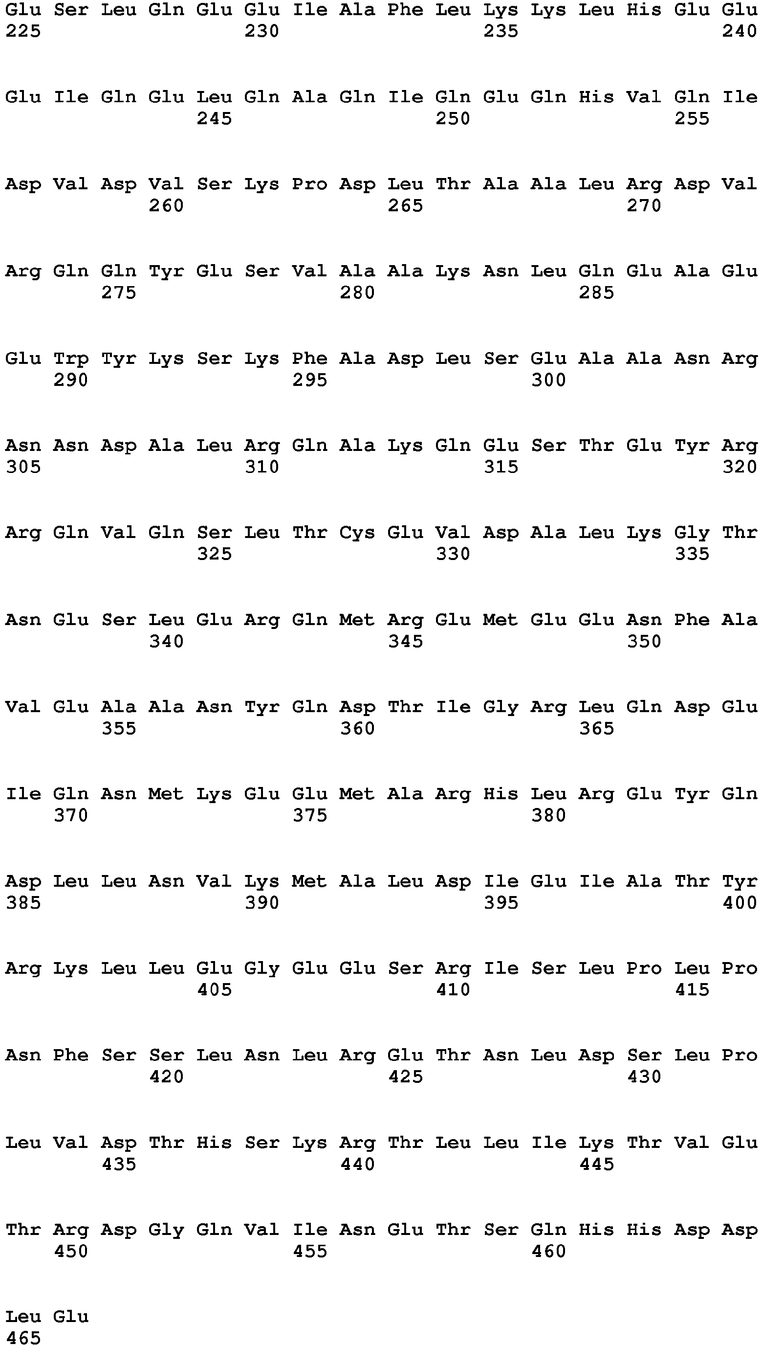
- <210> 367
<211> 1960
<212> PRT
<213> Homo sapiens - <400> 367


- <210> 368
<211> 586
<212> PRT
<213> Homo sapiens - <400> 368


- <210> 369
<211> 201
<212> PRT
<213> Homo sapiens - <400> 369


- <210> 370
<211> 463
<212> PRT
<213> Homo sapiens - <400> 370


- <210> 371
<211> 375
<212> PRT
<213> Homo sapiens - <400> 371


- <210> 372
<211> 254
<212> PRT
<213> Homo sapiens - <400> 372

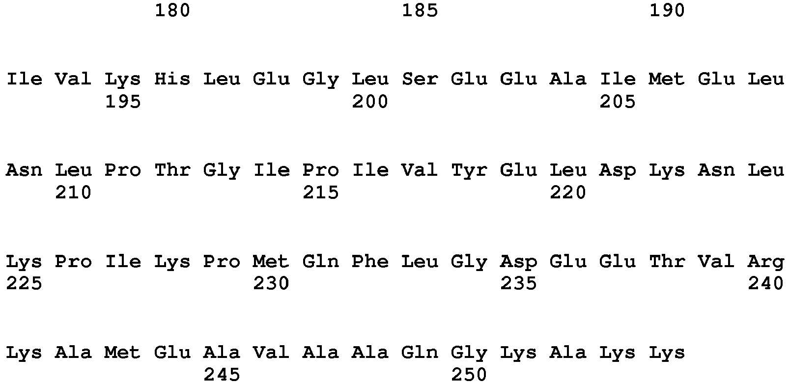
- <210> 373
<211> 352
<212> PRT
<213> Homo sapiens - <400> 373


- <210> 374
<211> 350
<212> PRT
<213> Homo sapiens - <400> 374


- <210> 375
<211> 263
<212> PRT
<213> Homo sapiens - <400> 375


- <210> 376
<211> 115
<212> PRT
<213> Homo sapiens - <400> 376


- <210> 377
<211> 320
<212> PRT
<213> Homo sapiens - <400> 377


- <210> 378
<211> 535
<212> PRT
<213> Homo sapiens - <400> 378


- <210> 379
<211> 461
<212> PRT
<213> Homo sapiens - <400> 379


Claims (8)
- A method to monitor the progress of a viral infection and/or to monitor the efficacy of a therapy for a viral infection in a biological sample comprising T cell obtained from a subject affected by said viral infection comprising the steps of:a) incubating the isolated biological sample with at least one peptide consisting of a sequence selected from SEQ ID No.1 to SEQ ID No. 252 under appropriate conditions to generate a T cell response;b) measuring the T cell response;c) comparing the measured T cell response to the measured T cell response of a suitable control sample,wherein said viral infection is caused by a virus selected from the group of HCV, HBV, thereby discriminating patients experiencing chronic infection and those undergoing infection resolution.
- The method according to claim 1 wherein the biological sample is a sample of PBMCs.
- The method according to any one of previous claims wherein the at least one peptide of step a) consists of the sequence selected from LLQDSVDFSL (SEQ ID No. 204), QLFNHTMFI (SEQ ID No. 64) or VLMIKALEL (SEQ ID No. 78).
- The method according to any one of previous claim wherein step b) comprises incubating the biological sample with a pool of peptides selected among the following pools:- pool 1: SEQ ID Nos. 1, 2, 4, 5, 8 and 9;- pool 2: SEQ ID Nos. 14, 16, 18, 19 and 20;- pool 3: SEQ ID Nos.27, 28, 29, 30, 34, 35, 36, 37 and 40;- pool 4: SEQ ID Nos. 58, 59, 60, 62, 63, 64, 65, 66 and 67;- pool 5: SEQ ID Nos. 68, 69, 73, 74, 75, 76, 77, 78, 81, 83, 84 and 88;- pool 6: SEQ ID Nos. 89, 90, 91, 92, 99, 100, 104, 105, 106, 111, 117, 123, 126 and 127;- pool 7: SEQ ID Nos. 198, 199, 200, 201, 202, 204, 206, 210, 211 and 220;- pool 8: SEQ ID Nos. 232, 233, 234, 235, 236, 237, 238, 239, 240, 241, 242, 243 and 244 or- pool 9: SEQ ID Nos. 245, 246, 247, 248 and 249.
- The method according to any one of previous claims wherein the T cell response is a CD8+ T cell response.
- The method according to any one of previous claims wherein the T cell response is measured by means of an Elispot assay.
- The method according to claim 6 wherein the Elispot assay is an interferon-gamma, IL-17 or IL-4 Elispot assay.
- A method to monitor the progress of a viral infection and/or to monitor the efficacy of a therapy for a viral infection in a subject affected by said viral infection comprising the step of detecting and/or quantifying the amount of auto-antibodies directed against at least one peptide as defined in any one of claim 1, 3 or 4, wherein said viral infection is caused by a virus selected from the group of HCV, HBV, thereby discriminating patients experiencing chronic infection and those undergoing infection resolution.
Applications Claiming Priority (2)
| Application Number | Priority Date | Filing Date | Title |
|---|---|---|---|
| IT000248A ITRM20110248A1 (en) | 2011-05-20 | 2011-05-20 | METHOD TO PROGNOSE VIRAL INFECTION BY MEASURING T CELL RESPONSE OR AUTO-ANTIBODIES TO APOPTOTIC EPITOPES |
| PCT/EP2012/059275 WO2012159993A1 (en) | 2011-05-20 | 2012-05-18 | Method to prognose viral infection by measuring t cell response or auto-antibodies to apoptotic epitopes |
Publications (2)
| Publication Number | Publication Date |
|---|---|
| EP2710378A1 EP2710378A1 (en) | 2014-03-26 |
| EP2710378B1 true EP2710378B1 (en) | 2017-03-15 |
Family
ID=44555141
Family Applications (1)
| Application Number | Title | Priority Date | Filing Date |
|---|---|---|---|
| EP12721318.9A Not-in-force EP2710378B1 (en) | 2011-05-20 | 2012-05-18 | Method to prognose viral infection by measuring t cell response or auto-antibodies to apoptotic epitopes |
Country Status (3)
| Country | Link |
|---|---|
| EP (1) | EP2710378B1 (en) |
| IT (1) | ITRM20110248A1 (en) |
| WO (1) | WO2012159993A1 (en) |
Cited By (1)
| Publication number | Priority date | Publication date | Assignee | Title |
|---|---|---|---|---|
| EP3867353A4 (en) * | 2018-10-18 | 2022-07-27 | Berkeley Lights, Inc. | SYNTHETIC SURFACES FOR THE PRESENTATION OF PROTO-ANTIGENS, ACTIVATED T LYMPHOCYTES AND ASSOCIATED USES |
Families Citing this family (2)
| Publication number | Priority date | Publication date | Assignee | Title |
|---|---|---|---|---|
| WO2015140567A1 (en) * | 2014-03-21 | 2015-09-24 | Immunocore Limited | Tumor associated antigens derived from traip |
| US20170261505A1 (en) | 2014-09-16 | 2017-09-14 | Universita' Degli Studi Di Roma "La Sapienza" | Method and kit for the predictive prognosis of responsiveness to treatments of autoimmune diseases |
-
2011
- 2011-05-20 IT IT000248A patent/ITRM20110248A1/en unknown
-
2012
- 2012-05-18 EP EP12721318.9A patent/EP2710378B1/en not_active Not-in-force
- 2012-05-18 WO PCT/EP2012/059275 patent/WO2012159993A1/en not_active Ceased
Non-Patent Citations (4)
| Title |
|---|
| A FRANCISCUS: "Similarities and Differences between HIV and HCV", HCSP FACT SHEET VERSION 4.5 JULY 2015 HCSP, 1 July 2015 (2015-07-01), XP055266660, Retrieved from the Internet <URL:http://hcvadvocate.org/hepatitis/factsheets_pdf/Similarities_and_Differences_between_HIV_and_HCV.pdf> [retrieved on 20160419] * |
| CITRO ALESSANDRA ET AL: "From T cell apoptosis to chronic immune activation in inflammatory diseases", INTERNATIONAL ARCHIVES OF ALLERGY AND IMMUNOLOGY, KARGER AG, CH, vol. 164, no. 2, 1 August 2014 (2014-08-01), pages 140 - 146, XP008176438, ISSN: 1018-2438, [retrieved on 20140702], DOI: 10.1159/000363385 * |
| MAURICE R. HILLEMAN: "Comparative Biology and Pathogenesis of AIDS and Hepatitis B Viruses: Related but Different", AIDS RESEARCH AND HUMAN RETROVIRUSES., vol. 10, no. 11, 1 November 1994 (1994-11-01), US, pages 1409 - 1419, XP055266658, ISSN: 0889-2229, DOI: 10.1089/aid.1994.10.1409 * |
| S. CHEVALIEZ: "Virological tools to diagnose and monitor hepatitis C virus infection", CLINICAL MICROBIOLOGY AND INFECTION., vol. 17, no. 2, 1 February 2011 (2011-02-01), United Kingdom, Switzerland, pages 116 - 121, XP055266593, ISSN: 1198-743X, DOI: 10.1111/j.1469-0691.2010.03418.x * |
Cited By (1)
| Publication number | Priority date | Publication date | Assignee | Title |
|---|---|---|---|---|
| EP3867353A4 (en) * | 2018-10-18 | 2022-07-27 | Berkeley Lights, Inc. | SYNTHETIC SURFACES FOR THE PRESENTATION OF PROTO-ANTIGENS, ACTIVATED T LYMPHOCYTES AND ASSOCIATED USES |
Also Published As
| Publication number | Publication date |
|---|---|
| EP2710378A1 (en) | 2014-03-26 |
| ITRM20110248A1 (en) | 2012-11-21 |
| WO2012159993A1 (en) | 2012-11-29 |
Similar Documents
| Publication | Publication Date | Title |
|---|---|---|
| Wragg et al. | Establishment and recall of SARS-CoV-2 spike epitope-specific CD4+ T cell memory | |
| Bartleson et al. | SARS-CoV-2, COVID-19 and the aging immune system | |
| Melenotte et al. | Immune responses during COVID-19 infection | |
| Woldemeskel et al. | Healthy donor T cell responses to common cold coronaviruses and SARS-CoV-2 | |
| De Milito et al. | Mechanisms of hypergammaglobulinemia and impaired antigen-specific humoral immunity in HIV-1 infection | |
| Grifoni et al. | Global assessment of dengue virus-specific CD4+ T cell responses in dengue-endemic areas | |
| US11261231B2 (en) | Methods for inhibiting HIV or HCV infection by administering TIM-3 binding inhibitors | |
| US9598491B2 (en) | Methods for the treatment of infections and tumors | |
| Kohler et al. | The early cellular signatures of protective immunity induced by live viral vaccination | |
| Smyk-Pearson et al. | Spontaneous recovery in acute human hepatitis C virus infection: functional T-cell thresholds and relative importance of CD4 help | |
| Heide et al. | Broadly directed SARS-CoV-2-specific CD4+ T cell response includes frequently detected peptide specificities within the membrane and nucleoprotein in patients with acute and resolved COVID-19 | |
| Alanio et al. | Bystander hyperactivation of preimmune CD8+ T cells in chronic HCV patients | |
| Lim et al. | Cross-reactivity and anti-viral function of dengue capsid and NS3-specific memory T cells toward Zika virus | |
| Ma et al. | Hantaan virus infection induces both Th1 and ThGranzyme B+ cell immune responses that associated with viral control and clinical outcome in humans | |
| Ferrando-Martinez et al. | HBeAg seroconversion is associated with a more effective PD-L1 blockade during chronic hepatitis B infection | |
| HUE030139T2 (en) | Preparations and procedures for treating inflammations | |
| Lolli et al. | Increased CD8+ T cell responses to apoptotic T cell-associated antigens in multiple sclerosis | |
| JP5710482B2 (en) | Monitoring and inhibition of human immunodeficiency virus infection by modulation of HMGB1-dependent induced HIV-1 replication and persistence | |
| Lineburg et al. | Rapid detection of SARS‐CoV‐2‐specific memory T‐cell immunity in recovered COVID‐19 cases | |
| Chen et al. | Lymphocyte activation gene 3 negatively regulates the function of intrahepatic hepatitis C virus‐specific CD8+ T cells | |
| Franceschini et al. | Polyfunctional type-1,-2, and-17 CD8+ T cell responses to apoptotic self-antigens correlate with the chronic evolution of hepatitis C virus infection | |
| Fernández‐Ruiz et al. | T cell–mediated response to SARS‐CoV‐2 in liver transplant recipients with prior COVID‐19 | |
| Villemonteix et al. | Comparison between enzyme‐linked immunospot assay and intracellular cytokine flow cytometry assays for the evaluation of T cell response to SARS‐CoV‐2 after symptomatic COVID‐19 | |
| Tang et al. | Novel identified HLA-A* 0201-restricted hantaan virus glycoprotein cytotoxic T-cell epitopes could effectively induce protective responses in HLA-A2. 1/Kb transgenic mice may associate with the severity of hemorrhagic fever with renal syndrome | |
| WO2012159993A1 (en) | Method to prognose viral infection by measuring t cell response or auto-antibodies to apoptotic epitopes |
Legal Events
| Date | Code | Title | Description |
|---|---|---|---|
| PUAI | Public reference made under article 153(3) epc to a published international application that has entered the european phase |
Free format text: ORIGINAL CODE: 0009012 |
|
| 17P | Request for examination filed |
Effective date: 20131205 |
|
| AK | Designated contracting states |
Kind code of ref document: A1 Designated state(s): AL AT BE BG CH CY CZ DE DK EE ES FI FR GB GR HR HU IE IS IT LI LT LU LV MC MK MT NL NO PL PT RO RS SE SI SK SM TR |
|
| DAX | Request for extension of the european patent (deleted) | ||
| 17Q | First examination report despatched |
Effective date: 20150604 |
|
| RIC1 | Information provided on ipc code assigned before grant |
Ipc: G01N 33/564 20060101ALI20160825BHEP Ipc: G01N 33/50 20060101ALI20160825BHEP Ipc: G01N 33/569 20060101AFI20160825BHEP |
|
| GRAP | Despatch of communication of intention to grant a patent |
Free format text: ORIGINAL CODE: EPIDOSNIGR1 |
|
| INTG | Intention to grant announced |
Effective date: 20161109 |
|
| GRAS | Grant fee paid |
Free format text: ORIGINAL CODE: EPIDOSNIGR3 |
|
| GRAA | (expected) grant |
Free format text: ORIGINAL CODE: 0009210 |
|
| AK | Designated contracting states |
Kind code of ref document: B1 Designated state(s): AL AT BE BG CH CY CZ DE DK EE ES FI FR GB GR HR HU IE IS IT LI LT LU LV MC MK MT NL NO PL PT RO RS SE SI SK SM TR |
|
| REG | Reference to a national code |
Ref country code: CH Ref legal event code: EP Ref country code: GB Ref legal event code: FG4D |
|
| REG | Reference to a national code |
Ref country code: IE Ref legal event code: FG4D |
|
| REG | Reference to a national code |
Ref country code: AT Ref legal event code: REF Ref document number: 876115 Country of ref document: AT Kind code of ref document: T Effective date: 20170415 |
|
| REG | Reference to a national code |
Ref country code: DE Ref legal event code: R096 Ref document number: 602012029830 Country of ref document: DE |
|
| REG | Reference to a national code |
Ref country code: NL Ref legal event code: MP Effective date: 20170315 |
|
| REG | Reference to a national code |
Ref country code: LT Ref legal event code: MG4D |
|
| PG25 | Lapsed in a contracting state [announced via postgrant information from national office to epo] |
Ref country code: GR Free format text: LAPSE BECAUSE OF FAILURE TO SUBMIT A TRANSLATION OF THE DESCRIPTION OR TO PAY THE FEE WITHIN THE PRESCRIBED TIME-LIMIT Effective date: 20170616 Ref country code: NO Free format text: LAPSE BECAUSE OF FAILURE TO SUBMIT A TRANSLATION OF THE DESCRIPTION OR TO PAY THE FEE WITHIN THE PRESCRIBED TIME-LIMIT Effective date: 20170615 Ref country code: HR Free format text: LAPSE BECAUSE OF FAILURE TO SUBMIT A TRANSLATION OF THE DESCRIPTION OR TO PAY THE FEE WITHIN THE PRESCRIBED TIME-LIMIT Effective date: 20170315 Ref country code: FI Free format text: LAPSE BECAUSE OF FAILURE TO SUBMIT A TRANSLATION OF THE DESCRIPTION OR TO PAY THE FEE WITHIN THE PRESCRIBED TIME-LIMIT Effective date: 20170315 Ref country code: LT Free format text: LAPSE BECAUSE OF FAILURE TO SUBMIT A TRANSLATION OF THE DESCRIPTION OR TO PAY THE FEE WITHIN THE PRESCRIBED TIME-LIMIT Effective date: 20170315 |
|
| REG | Reference to a national code |
Ref country code: AT Ref legal event code: MK05 Ref document number: 876115 Country of ref document: AT Kind code of ref document: T Effective date: 20170315 |
|
| PG25 | Lapsed in a contracting state [announced via postgrant information from national office to epo] |
Ref country code: LU Free format text: LAPSE BECAUSE OF NON-PAYMENT OF DUE FEES Effective date: 20170531 Ref country code: LV Free format text: LAPSE BECAUSE OF FAILURE TO SUBMIT A TRANSLATION OF THE DESCRIPTION OR TO PAY THE FEE WITHIN THE PRESCRIBED TIME-LIMIT Effective date: 20170315 Ref country code: RS Free format text: LAPSE BECAUSE OF FAILURE TO SUBMIT A TRANSLATION OF THE DESCRIPTION OR TO PAY THE FEE WITHIN THE PRESCRIBED TIME-LIMIT Effective date: 20170315 Ref country code: BG Free format text: LAPSE BECAUSE OF FAILURE TO SUBMIT A TRANSLATION OF THE DESCRIPTION OR TO PAY THE FEE WITHIN THE PRESCRIBED TIME-LIMIT Effective date: 20170615 Ref country code: SE Free format text: LAPSE BECAUSE OF FAILURE TO SUBMIT A TRANSLATION OF THE DESCRIPTION OR TO PAY THE FEE WITHIN THE PRESCRIBED TIME-LIMIT Effective date: 20170315 |
|
| PG25 | Lapsed in a contracting state [announced via postgrant information from national office to epo] |
Ref country code: NL Free format text: LAPSE BECAUSE OF FAILURE TO SUBMIT A TRANSLATION OF THE DESCRIPTION OR TO PAY THE FEE WITHIN THE PRESCRIBED TIME-LIMIT Effective date: 20170315 |
|
| PG25 | Lapsed in a contracting state [announced via postgrant information from national office to epo] |
Ref country code: CZ Free format text: LAPSE BECAUSE OF FAILURE TO SUBMIT A TRANSLATION OF THE DESCRIPTION OR TO PAY THE FEE WITHIN THE PRESCRIBED TIME-LIMIT Effective date: 20170315 Ref country code: AT Free format text: LAPSE BECAUSE OF FAILURE TO SUBMIT A TRANSLATION OF THE DESCRIPTION OR TO PAY THE FEE WITHIN THE PRESCRIBED TIME-LIMIT Effective date: 20170315 Ref country code: IT Free format text: LAPSE BECAUSE OF FAILURE TO SUBMIT A TRANSLATION OF THE DESCRIPTION OR TO PAY THE FEE WITHIN THE PRESCRIBED TIME-LIMIT Effective date: 20170315 Ref country code: ES Free format text: LAPSE BECAUSE OF FAILURE TO SUBMIT A TRANSLATION OF THE DESCRIPTION OR TO PAY THE FEE WITHIN THE PRESCRIBED TIME-LIMIT Effective date: 20170315 Ref country code: RO Free format text: LAPSE BECAUSE OF FAILURE TO SUBMIT A TRANSLATION OF THE DESCRIPTION OR TO PAY THE FEE WITHIN THE PRESCRIBED TIME-LIMIT Effective date: 20170315 Ref country code: EE Free format text: LAPSE BECAUSE OF FAILURE TO SUBMIT A TRANSLATION OF THE DESCRIPTION OR TO PAY THE FEE WITHIN THE PRESCRIBED TIME-LIMIT Effective date: 20170315 Ref country code: SK Free format text: LAPSE BECAUSE OF FAILURE TO SUBMIT A TRANSLATION OF THE DESCRIPTION OR TO PAY THE FEE WITHIN THE PRESCRIBED TIME-LIMIT Effective date: 20170315 |
|
| PG25 | Lapsed in a contracting state [announced via postgrant information from national office to epo] |
Ref country code: PL Free format text: LAPSE BECAUSE OF FAILURE TO SUBMIT A TRANSLATION OF THE DESCRIPTION OR TO PAY THE FEE WITHIN THE PRESCRIBED TIME-LIMIT Effective date: 20170315 Ref country code: SM Free format text: LAPSE BECAUSE OF FAILURE TO SUBMIT A TRANSLATION OF THE DESCRIPTION OR TO PAY THE FEE WITHIN THE PRESCRIBED TIME-LIMIT Effective date: 20170315 Ref country code: PT Free format text: LAPSE BECAUSE OF FAILURE TO SUBMIT A TRANSLATION OF THE DESCRIPTION OR TO PAY THE FEE WITHIN THE PRESCRIBED TIME-LIMIT Effective date: 20170717 Ref country code: IS Free format text: LAPSE BECAUSE OF FAILURE TO SUBMIT A TRANSLATION OF THE DESCRIPTION OR TO PAY THE FEE WITHIN THE PRESCRIBED TIME-LIMIT Effective date: 20170715 |
|
| REG | Reference to a national code |
Ref country code: DE Ref legal event code: R119 Ref document number: 602012029830 Country of ref document: DE |
|
| REG | Reference to a national code |
Ref country code: CH Ref legal event code: PL |
|
| PLBE | No opposition filed within time limit |
Free format text: ORIGINAL CODE: 0009261 |
|
| STAA | Information on the status of an ep patent application or granted ep patent |
Free format text: STATUS: NO OPPOSITION FILED WITHIN TIME LIMIT |
|
| PG25 | Lapsed in a contracting state [announced via postgrant information from national office to epo] |
Ref country code: DK Free format text: LAPSE BECAUSE OF FAILURE TO SUBMIT A TRANSLATION OF THE DESCRIPTION OR TO PAY THE FEE WITHIN THE PRESCRIBED TIME-LIMIT Effective date: 20170315 Ref country code: MC Free format text: LAPSE BECAUSE OF FAILURE TO SUBMIT A TRANSLATION OF THE DESCRIPTION OR TO PAY THE FEE WITHIN THE PRESCRIBED TIME-LIMIT Effective date: 20170315 |
|
| 26N | No opposition filed |
Effective date: 20171218 |
|
| REG | Reference to a national code |
Ref country code: IE Ref legal event code: MM4A |
|
| GBPC | Gb: european patent ceased through non-payment of renewal fee |
Effective date: 20170615 |
|
| PG25 | Lapsed in a contracting state [announced via postgrant information from national office to epo] |
Ref country code: SI Free format text: LAPSE BECAUSE OF FAILURE TO SUBMIT A TRANSLATION OF THE DESCRIPTION OR TO PAY THE FEE WITHIN THE PRESCRIBED TIME-LIMIT Effective date: 20170315 Ref country code: LI Free format text: LAPSE BECAUSE OF NON-PAYMENT OF DUE FEES Effective date: 20170531 Ref country code: CH Free format text: LAPSE BECAUSE OF NON-PAYMENT OF DUE FEES Effective date: 20170531 |
|
| REG | Reference to a national code |
Ref country code: FR Ref legal event code: ST Effective date: 20180131 |
|
| PG25 | Lapsed in a contracting state [announced via postgrant information from national office to epo] |
Ref country code: LU Free format text: LAPSE BECAUSE OF NON-PAYMENT OF DUE FEES Effective date: 20170518 |
|
| REG | Reference to a national code |
Ref country code: BE Ref legal event code: MM Effective date: 20170531 |
|
| PG25 | Lapsed in a contracting state [announced via postgrant information from national office to epo] |
Ref country code: DE Free format text: LAPSE BECAUSE OF NON-PAYMENT OF DUE FEES Effective date: 20171201 Ref country code: GB Free format text: LAPSE BECAUSE OF NON-PAYMENT OF DUE FEES Effective date: 20170615 Ref country code: IE Free format text: LAPSE BECAUSE OF NON-PAYMENT OF DUE FEES Effective date: 20170518 |
|
| PG25 | Lapsed in a contracting state [announced via postgrant information from national office to epo] |
Ref country code: FR Free format text: LAPSE BECAUSE OF NON-PAYMENT OF DUE FEES Effective date: 20170531 |
|
| PG25 | Lapsed in a contracting state [announced via postgrant information from national office to epo] |
Ref country code: BE Free format text: LAPSE BECAUSE OF NON-PAYMENT OF DUE FEES Effective date: 20170531 |
|
| PG25 | Lapsed in a contracting state [announced via postgrant information from national office to epo] |
Ref country code: MT Free format text: LAPSE BECAUSE OF NON-PAYMENT OF DUE FEES Effective date: 20170518 |
|
| PG25 | Lapsed in a contracting state [announced via postgrant information from national office to epo] |
Ref country code: HU Free format text: LAPSE BECAUSE OF FAILURE TO SUBMIT A TRANSLATION OF THE DESCRIPTION OR TO PAY THE FEE WITHIN THE PRESCRIBED TIME-LIMIT; INVALID AB INITIO Effective date: 20120518 |
|
| PG25 | Lapsed in a contracting state [announced via postgrant information from national office to epo] |
Ref country code: CY Free format text: LAPSE BECAUSE OF NON-PAYMENT OF DUE FEES Effective date: 20170315 |
|
| PG25 | Lapsed in a contracting state [announced via postgrant information from national office to epo] |
Ref country code: MK Free format text: LAPSE BECAUSE OF FAILURE TO SUBMIT A TRANSLATION OF THE DESCRIPTION OR TO PAY THE FEE WITHIN THE PRESCRIBED TIME-LIMIT Effective date: 20170315 |
|
| PG25 | Lapsed in a contracting state [announced via postgrant information from national office to epo] |
Ref country code: TR Free format text: LAPSE BECAUSE OF FAILURE TO SUBMIT A TRANSLATION OF THE DESCRIPTION OR TO PAY THE FEE WITHIN THE PRESCRIBED TIME-LIMIT Effective date: 20170315 |
|
| PG25 | Lapsed in a contracting state [announced via postgrant information from national office to epo] |
Ref country code: AL Free format text: LAPSE BECAUSE OF FAILURE TO SUBMIT A TRANSLATION OF THE DESCRIPTION OR TO PAY THE FEE WITHIN THE PRESCRIBED TIME-LIMIT Effective date: 20170315 |