EP2702073B1 - Polymutant tau protein variants and their use for recapitulating human tauopathies - Google Patents
Polymutant tau protein variants and their use for recapitulating human tauopathies Download PDFInfo
- Publication number
- EP2702073B1 EP2702073B1 EP11716418.6A EP11716418A EP2702073B1 EP 2702073 B1 EP2702073 B1 EP 2702073B1 EP 11716418 A EP11716418 A EP 11716418A EP 2702073 B1 EP2702073 B1 EP 2702073B1
- Authority
- EP
- European Patent Office
- Prior art keywords
- cell
- tau
- cells
- impedance
- tau protein
- Prior art date
- Legal status (The legal status is an assumption and is not a legal conclusion. Google has not performed a legal analysis and makes no representation as to the accuracy of the status listed.)
- Not-in-force
Links
- 108010026424 tau Proteins Proteins 0.000 title claims description 220
- 102000013498 tau Proteins Human genes 0.000 title claims description 217
- 208000034799 Tauopathies Diseases 0.000 title claims description 17
- 241000282414 Homo sapiens Species 0.000 title description 21
- 210000004027 cell Anatomy 0.000 claims description 251
- 238000000034 method Methods 0.000 claims description 51
- 238000012360 testing method Methods 0.000 claims description 46
- 150000001875 compounds Chemical class 0.000 claims description 42
- 230000035772 mutation Effects 0.000 claims description 36
- 150000001413 amino acids Chemical class 0.000 claims description 33
- 230000014509 gene expression Effects 0.000 claims description 33
- 230000004770 neurodegeneration Effects 0.000 claims description 33
- 239000003550 marker Substances 0.000 claims description 32
- 238000004113 cell culture Methods 0.000 claims description 31
- 201000011240 Frontotemporal dementia Diseases 0.000 claims description 29
- 150000007523 nucleic acids Chemical class 0.000 claims description 26
- 239000013598 vector Substances 0.000 claims description 24
- 102000039446 nucleic acids Human genes 0.000 claims description 23
- 108020004707 nucleic acids Proteins 0.000 claims description 23
- 239000000126 substance Substances 0.000 claims description 21
- 239000003795 chemical substances by application Substances 0.000 claims description 19
- 208000015122 neurodegenerative disease Diseases 0.000 claims description 18
- 239000013612 plasmid Substances 0.000 claims description 18
- 210000002682 neurofibrillary tangle Anatomy 0.000 claims description 16
- 210000002569 neuron Anatomy 0.000 claims description 16
- 230000002159 abnormal effect Effects 0.000 claims description 15
- 230000000694 effects Effects 0.000 claims description 15
- 230000007423 decrease Effects 0.000 claims description 13
- 230000003376 axonal effect Effects 0.000 claims description 10
- 230000007850 degeneration Effects 0.000 claims description 9
- 238000012258 culturing Methods 0.000 claims description 8
- 238000012217 deletion Methods 0.000 claims description 8
- 230000037430 deletion Effects 0.000 claims description 8
- 238000011282 treatment Methods 0.000 claims description 7
- 208000027089 Parkinsonian disease Diseases 0.000 claims description 6
- 206010034010 Parkinsonism Diseases 0.000 claims description 6
- 239000001963 growth medium Substances 0.000 claims description 6
- 230000000946 synaptic effect Effects 0.000 claims description 6
- 210000000349 chromosome Anatomy 0.000 claims description 5
- 239000003814 drug Substances 0.000 claims description 5
- 238000011161 development Methods 0.000 claims description 4
- 238000012216 screening Methods 0.000 claims description 4
- 230000002265 prevention Effects 0.000 claims description 3
- 125000003275 alpha amino acid group Chemical group 0.000 claims 2
- 230000008335 axon cargo transport Effects 0.000 claims 1
- 108090000623 proteins and genes Proteins 0.000 description 25
- 102000029749 Microtubule Human genes 0.000 description 24
- 108091022875 Microtubule Proteins 0.000 description 24
- 210000004688 microtubule Anatomy 0.000 description 24
- 230000004069 differentiation Effects 0.000 description 19
- QNDVLZJODHBUFM-WFXQOWMNSA-N okadaic acid Chemical compound C([C@H](O1)[C@H](C)/C=C/[C@H]2CC[C@@]3(CC[C@H]4O[C@@H](C([C@@H](O)[C@@H]4O3)=C)[C@@H](O)C[C@H](C)[C@@H]3[C@@H](CC[C@@]4(OCCCC4)O3)C)O2)C(C)=C[C@]21O[C@H](C[C@@](C)(O)C(O)=O)CC[C@H]2O QNDVLZJODHBUFM-WFXQOWMNSA-N 0.000 description 19
- VEFJHAYOIAAXEU-UHFFFAOYSA-N okadaic acid Natural products CC(CC(O)C1OC2CCC3(CCC(O3)C=CC(C)C4CC(=CC5(OC(CC(C)(O)C(=O)O)CCC5O)O4)C)OC2C(O)C1C)C6OC7(CCCCO7)CCC6C VEFJHAYOIAAXEU-UHFFFAOYSA-N 0.000 description 19
- 102000004169 proteins and genes Human genes 0.000 description 19
- WSFSSNUMVMOOMR-UHFFFAOYSA-N Formaldehyde Chemical compound O=C WSFSSNUMVMOOMR-UHFFFAOYSA-N 0.000 description 18
- 230000002776 aggregation Effects 0.000 description 18
- 238000004220 aggregation Methods 0.000 description 18
- 208000024827 Alzheimer disease Diseases 0.000 description 16
- 108020004414 DNA Proteins 0.000 description 16
- 108090000765 processed proteins & peptides Proteins 0.000 description 16
- 230000007170 pathology Effects 0.000 description 15
- 101000891579 Homo sapiens Microtubule-associated protein tau Proteins 0.000 description 14
- 102000004196 processed proteins & peptides Human genes 0.000 description 14
- 108010029485 Protein Isoforms Proteins 0.000 description 12
- 102000001708 Protein Isoforms Human genes 0.000 description 12
- 230000026731 phosphorylation Effects 0.000 description 12
- 238000006366 phosphorylation reaction Methods 0.000 description 12
- 108091033319 polynucleotide Proteins 0.000 description 12
- 102000040430 polynucleotide Human genes 0.000 description 12
- 229920001184 polypeptide Polymers 0.000 description 12
- 108020004999 messenger RNA Proteins 0.000 description 11
- 239000002157 polynucleotide Substances 0.000 description 11
- 231100000331 toxic Toxicity 0.000 description 11
- 230000002588 toxic effect Effects 0.000 description 11
- 230000015572 biosynthetic process Effects 0.000 description 10
- 230000001413 cellular effect Effects 0.000 description 10
- 102000057063 human MAPT Human genes 0.000 description 10
- 230000001939 inductive effect Effects 0.000 description 10
- IQFVPQOLBLOTPF-HKXUKFGYSA-L congo red Chemical compound [Na+].[Na+].C1=CC=CC2=C(N)C(/N=N/C3=CC=C(C=C3)C3=CC=C(C=C3)/N=N/C3=C(C4=CC=CC=C4C(=C3)S([O-])(=O)=O)N)=CC(S([O-])(=O)=O)=C21 IQFVPQOLBLOTPF-HKXUKFGYSA-L 0.000 description 9
- 230000001419 dependent effect Effects 0.000 description 9
- 230000006870 function Effects 0.000 description 9
- 230000001225 therapeutic effect Effects 0.000 description 9
- 206010029260 Neuroblastoma Diseases 0.000 description 8
- 108700019146 Transgenes Proteins 0.000 description 8
- 230000006951 hyperphosphorylation Effects 0.000 description 8
- 230000001575 pathological effect Effects 0.000 description 8
- 238000002474 experimental method Methods 0.000 description 7
- 238000001566 impedance spectroscopy Methods 0.000 description 7
- 239000003112 inhibitor Substances 0.000 description 7
- 230000001404 mediated effect Effects 0.000 description 7
- YAEMHJKFIIIULI-UHFFFAOYSA-N n-(4-methoxybenzyl)-n'-(5-nitro-1,3-thiazol-2-yl)urea Chemical compound C1=CC(OC)=CC=C1CNC(=O)NC1=NC=C([N+]([O-])=O)S1 YAEMHJKFIIIULI-UHFFFAOYSA-N 0.000 description 7
- 241000894007 species Species 0.000 description 7
- 238000013518 transcription Methods 0.000 description 7
- 230000035897 transcription Effects 0.000 description 7
- 238000010361 transduction Methods 0.000 description 7
- 230000026683 transduction Effects 0.000 description 7
- 238000001890 transfection Methods 0.000 description 7
- 102100040243 Microtubule-associated protein tau Human genes 0.000 description 6
- 101710115937 Microtubule-associated protein tau Proteins 0.000 description 6
- 108091028043 Nucleic acid sequence Proteins 0.000 description 6
- 241000700605 Viruses Species 0.000 description 6
- 208000037265 diseases, disorders, signs and symptoms Diseases 0.000 description 6
- 238000000338 in vitro Methods 0.000 description 6
- 238000003752 polymerase chain reaction Methods 0.000 description 6
- 239000002243 precursor Substances 0.000 description 6
- 239000000047 product Substances 0.000 description 6
- 231100000167 toxic agent Toxicity 0.000 description 6
- 108091032973 (ribonucleotides)n+m Proteins 0.000 description 5
- 108010090849 Amyloid beta-Peptides Proteins 0.000 description 5
- 102000013455 Amyloid beta-Peptides Human genes 0.000 description 5
- 102000004160 Phosphoric Monoester Hydrolases Human genes 0.000 description 5
- 108090000608 Phosphoric Monoester Hydrolases Proteins 0.000 description 5
- HKSZLNNOFSGOKW-UHFFFAOYSA-N ent-staurosporine Natural products C12=C3N4C5=CC=CC=C5C3=C3CNC(=O)C3=C2C2=CC=CC=C2N1C1CC(NC)C(OC)C4(C)O1 HKSZLNNOFSGOKW-UHFFFAOYSA-N 0.000 description 5
- 239000012634 fragment Substances 0.000 description 5
- 230000006698 induction Effects 0.000 description 5
- 230000001537 neural effect Effects 0.000 description 5
- 238000007423 screening assay Methods 0.000 description 5
- HKSZLNNOFSGOKW-FYTWVXJKSA-N staurosporine Chemical compound C12=C3N4C5=CC=CC=C5C3=C3CNC(=O)C3=C2C2=CC=CC=C2N1[C@H]1C[C@@H](NC)[C@@H](OC)[C@]4(C)O1 HKSZLNNOFSGOKW-FYTWVXJKSA-N 0.000 description 5
- CGPUWJWCVCFERF-UHFFFAOYSA-N staurosporine Natural products C12=C3N4C5=CC=CC=C5C3=C3CNC(=O)C3=C2C2=CC=CC=C2N1C1CC(NC)C(OC)C4(OC)O1 CGPUWJWCVCFERF-UHFFFAOYSA-N 0.000 description 5
- 238000001262 western blot Methods 0.000 description 5
- 102000009664 Microtubule-Associated Proteins Human genes 0.000 description 4
- 210000004556 brain Anatomy 0.000 description 4
- 239000002299 complementary DNA Substances 0.000 description 4
- 230000003247 decreasing effect Effects 0.000 description 4
- 201000010099 disease Diseases 0.000 description 4
- 238000009509 drug development Methods 0.000 description 4
- -1 from genomic sources Chemical class 0.000 description 4
- 238000011534 incubation Methods 0.000 description 4
- 210000004962 mammalian cell Anatomy 0.000 description 4
- 238000004519 manufacturing process Methods 0.000 description 4
- 230000007246 mechanism Effects 0.000 description 4
- 239000000203 mixture Substances 0.000 description 4
- 230000004962 physiological condition Effects 0.000 description 4
- 230000008569 process Effects 0.000 description 4
- 230000009467 reduction Effects 0.000 description 4
- 210000001519 tissue Anatomy 0.000 description 4
- 239000003440 toxic substance Substances 0.000 description 4
- 231100000419 toxicity Toxicity 0.000 description 4
- 230000001988 toxicity Effects 0.000 description 4
- 230000009261 transgenic effect Effects 0.000 description 4
- 238000011830 transgenic mouse model Methods 0.000 description 4
- 230000032258 transport Effects 0.000 description 4
- 108091026890 Coding region Proteins 0.000 description 3
- 208000011990 Corticobasal Degeneration Diseases 0.000 description 3
- 206010012289 Dementia Diseases 0.000 description 3
- IAZDPXIOMUYVGZ-UHFFFAOYSA-N Dimethylsulphoxide Chemical compound CS(C)=O IAZDPXIOMUYVGZ-UHFFFAOYSA-N 0.000 description 3
- 101150070547 MAPT gene Proteins 0.000 description 3
- 108010020004 Microtubule-Associated Proteins Proteins 0.000 description 3
- 108091000080 Phosphotransferase Proteins 0.000 description 3
- 208000000609 Pick Disease of the Brain Diseases 0.000 description 3
- 239000008186 active pharmaceutical agent Substances 0.000 description 3
- 230000004075 alteration Effects 0.000 description 3
- 206010002026 amyotrophic lateral sclerosis Diseases 0.000 description 3
- 238000000540 analysis of variance Methods 0.000 description 3
- 238000004458 analytical method Methods 0.000 description 3
- 238000010171 animal model Methods 0.000 description 3
- 238000003491 array Methods 0.000 description 3
- 238000003556 assay Methods 0.000 description 3
- 238000001514 detection method Methods 0.000 description 3
- 230000018109 developmental process Effects 0.000 description 3
- 238000009826 distribution Methods 0.000 description 3
- 238000004520 electroporation Methods 0.000 description 3
- 210000003527 eukaryotic cell Anatomy 0.000 description 3
- 239000013604 expression vector Substances 0.000 description 3
- 102000054767 gene variant Human genes 0.000 description 3
- 229940043355 kinase inhibitor Drugs 0.000 description 3
- 239000000463 material Substances 0.000 description 3
- 230000004048 modification Effects 0.000 description 3
- 238000012986 modification Methods 0.000 description 3
- 238000012544 monitoring process Methods 0.000 description 3
- 102000020233 phosphotransferase Human genes 0.000 description 3
- 239000003757 phosphotransferase inhibitor Substances 0.000 description 3
- 238000010149 post-hoc-test Methods 0.000 description 3
- 238000010186 staining Methods 0.000 description 3
- 238000003786 synthesis reaction Methods 0.000 description 3
- 238000011179 visual inspection Methods 0.000 description 3
- 108091003079 Bovine Serum Albumin Proteins 0.000 description 2
- OYPRJOBELJOOCE-UHFFFAOYSA-N Calcium Chemical compound [Ca] OYPRJOBELJOOCE-UHFFFAOYSA-N 0.000 description 2
- 102000053602 DNA Human genes 0.000 description 2
- UPEZCKBFRMILAV-JNEQICEOSA-N Ecdysone Natural products O=C1[C@H]2[C@@](C)([C@@H]3C([C@@]4(O)[C@@](C)([C@H]([C@H]([C@@H](O)CCC(O)(C)C)C)CC4)CC3)=C1)C[C@H](O)[C@H](O)C2 UPEZCKBFRMILAV-JNEQICEOSA-N 0.000 description 2
- 208000023105 Huntington disease Diseases 0.000 description 2
- 241000829100 Macaca mulatta polyomavirus 1 Species 0.000 description 2
- 241001465754 Metazoa Species 0.000 description 2
- 241000699660 Mus musculus Species 0.000 description 2
- 101100480715 Mus musculus Mapt gene Proteins 0.000 description 2
- 102000008763 Neurofilament Proteins Human genes 0.000 description 2
- 108010088373 Neurofilament Proteins Proteins 0.000 description 2
- 241000276398 Opsanus tau Species 0.000 description 2
- 208000018737 Parkinson disease Diseases 0.000 description 2
- 229940122907 Phosphatase inhibitor Drugs 0.000 description 2
- 208000024777 Prion disease Diseases 0.000 description 2
- 239000004098 Tetracycline Substances 0.000 description 2
- 102000004243 Tubulin Human genes 0.000 description 2
- 108090000704 Tubulin Proteins 0.000 description 2
- 230000005856 abnormality Effects 0.000 description 2
- 230000035508 accumulation Effects 0.000 description 2
- 238000009825 accumulation Methods 0.000 description 2
- UPEZCKBFRMILAV-UHFFFAOYSA-N alpha-Ecdysone Natural products C1C(O)C(O)CC2(C)C(CCC3(C(C(C(O)CCC(C)(C)O)C)CCC33O)C)C3=CC(=O)C21 UPEZCKBFRMILAV-UHFFFAOYSA-N 0.000 description 2
- DZHSAHHDTRWUTF-SIQRNXPUSA-N amyloid-beta polypeptide 42 Chemical compound C([C@@H](C(=O)N[C@@H](C)C(=O)N[C@@H](CCC(O)=O)C(=O)N[C@@H](CC(O)=O)C(=O)N[C@H](C(=O)NCC(=O)N[C@@H](CO)C(=O)N[C@@H](CC(N)=O)C(=O)N[C@@H](CCCCN)C(=O)NCC(=O)N[C@@H](C)C(=O)N[C@H](C(=O)N[C@@H]([C@@H](C)CC)C(=O)NCC(=O)N[C@@H](CC(C)C)C(=O)N[C@@H](CCSC)C(=O)N[C@@H](C(C)C)C(=O)NCC(=O)NCC(=O)N[C@@H](C(C)C)C(=O)N[C@@H](C(C)C)C(=O)N[C@@H]([C@@H](C)CC)C(=O)N[C@@H](C)C(O)=O)[C@@H](C)CC)C(C)C)NC(=O)[C@H](CC=1C=CC=CC=1)NC(=O)[C@@H](NC(=O)[C@H](CC(C)C)NC(=O)[C@H](CCCCN)NC(=O)[C@H](CCC(N)=O)NC(=O)[C@H](CC=1N=CNC=1)NC(=O)[C@H](CC=1N=CNC=1)NC(=O)[C@@H](NC(=O)[C@H](CCC(O)=O)NC(=O)[C@H](CC=1C=CC(O)=CC=1)NC(=O)CNC(=O)[C@H](CO)NC(=O)[C@H](CC(O)=O)NC(=O)[C@H](CC=1N=CNC=1)NC(=O)[C@H](CCCNC(N)=N)NC(=O)[C@H](CC=1C=CC=CC=1)NC(=O)[C@H](CCC(O)=O)NC(=O)[C@H](C)NC(=O)[C@@H](N)CC(O)=O)C(C)C)C(C)C)C1=CC=CC=C1 DZHSAHHDTRWUTF-SIQRNXPUSA-N 0.000 description 2
- 230000008901 benefit Effects 0.000 description 2
- 239000011575 calcium Substances 0.000 description 2
- 229910052791 calcium Inorganic materials 0.000 description 2
- 230000015556 catabolic process Effects 0.000 description 2
- 230000030833 cell death Effects 0.000 description 2
- 230000003915 cell function Effects 0.000 description 2
- 210000003169 central nervous system Anatomy 0.000 description 2
- 238000006243 chemical reaction Methods 0.000 description 2
- 210000004978 chinese hamster ovary cell Anatomy 0.000 description 2
- 208000010877 cognitive disease Diseases 0.000 description 2
- 210000000805 cytoplasm Anatomy 0.000 description 2
- 230000006378 damage Effects 0.000 description 2
- 230000003412 degenerative effect Effects 0.000 description 2
- 229940079593 drug Drugs 0.000 description 2
- UPEZCKBFRMILAV-JMZLNJERSA-N ecdysone Chemical compound C1[C@@H](O)[C@@H](O)C[C@]2(C)[C@@H](CC[C@@]3([C@@H]([C@@H]([C@H](O)CCC(C)(C)O)C)CC[C@]33O)C)C3=CC(=O)[C@@H]21 UPEZCKBFRMILAV-JMZLNJERSA-N 0.000 description 2
- 238000005516 engineering process Methods 0.000 description 2
- 108010048367 enhanced green fluorescent protein Proteins 0.000 description 2
- 239000012091 fetal bovine serum Substances 0.000 description 2
- 238000000198 fluorescence anisotropy Methods 0.000 description 2
- 238000002866 fluorescence resonance energy transfer Methods 0.000 description 2
- 238000010353 genetic engineering Methods 0.000 description 2
- 238000011532 immunohistochemical staining Methods 0.000 description 2
- 238000000099 in vitro assay Methods 0.000 description 2
- 238000002372 labelling Methods 0.000 description 2
- 238000005259 measurement Methods 0.000 description 2
- 239000002609 medium Substances 0.000 description 2
- 239000012528 membrane Substances 0.000 description 2
- 238000010369 molecular cloning Methods 0.000 description 2
- 238000010172 mouse model Methods 0.000 description 2
- 201000006417 multiple sclerosis Diseases 0.000 description 2
- 210000005044 neurofilament Anatomy 0.000 description 2
- 210000004498 neuroglial cell Anatomy 0.000 description 2
- 230000004031 neuronal differentiation Effects 0.000 description 2
- 239000002773 nucleotide Substances 0.000 description 2
- 125000003729 nucleotide group Chemical group 0.000 description 2
- 235000015097 nutrients Nutrition 0.000 description 2
- 230000002018 overexpression Effects 0.000 description 2
- 239000002245 particle Substances 0.000 description 2
- 230000000750 progressive effect Effects 0.000 description 2
- 201000002212 progressive supranuclear palsy Diseases 0.000 description 2
- 238000011002 quantification Methods 0.000 description 2
- 230000010076 replication Effects 0.000 description 2
- 230000001177 retroviral effect Effects 0.000 description 2
- 239000000523 sample Substances 0.000 description 2
- 238000002741 site-directed mutagenesis Methods 0.000 description 2
- 150000003384 small molecules Chemical class 0.000 description 2
- 230000000087 stabilizing effect Effects 0.000 description 2
- 208000024891 symptom Diseases 0.000 description 2
- 229960002180 tetracycline Drugs 0.000 description 2
- 229930101283 tetracycline Natural products 0.000 description 2
- 235000019364 tetracycline Nutrition 0.000 description 2
- 150000003522 tetracyclines Chemical class 0.000 description 2
- 230000001052 transient effect Effects 0.000 description 2
- 238000013519 translation Methods 0.000 description 2
- 239000003981 vehicle Substances 0.000 description 2
- 230000035899 viability Effects 0.000 description 2
- 230000003612 virological effect Effects 0.000 description 2
- 102000040650 (ribonucleotides)n+m Human genes 0.000 description 1
- FWBHETKCLVMNFS-UHFFFAOYSA-N 4',6-Diamino-2-phenylindol Chemical compound C1=CC(C(=N)N)=CC=C1C1=CC2=CC=C(C(N)=N)C=C2N1 FWBHETKCLVMNFS-UHFFFAOYSA-N 0.000 description 1
- 102000007469 Actins Human genes 0.000 description 1
- 108010085238 Actins Proteins 0.000 description 1
- HJCMDXDYPOUFDY-WHFBIAKZSA-N Ala-Gln Chemical compound C[C@H](N)C(=O)N[C@H](C(O)=O)CCC(N)=O HJCMDXDYPOUFDY-WHFBIAKZSA-N 0.000 description 1
- 108010011170 Ala-Trp-Arg-His-Pro-Gln-Phe-Gly-Gly Proteins 0.000 description 1
- 206010003694 Atrophy Diseases 0.000 description 1
- 239000012583 B-27 Supplement Substances 0.000 description 1
- 241000894006 Bacteria Species 0.000 description 1
- UXVMQQNJUSDDNG-UHFFFAOYSA-L Calcium chloride Chemical compound [Cl-].[Cl-].[Ca+2] UXVMQQNJUSDDNG-UHFFFAOYSA-L 0.000 description 1
- 208000028698 Cognitive impairment Diseases 0.000 description 1
- 108010051219 Cre recombinase Proteins 0.000 description 1
- 241000699800 Cricetinae Species 0.000 description 1
- 229920002307 Dextran Polymers 0.000 description 1
- 101000876610 Dictyostelium discoideum Extracellular signal-regulated kinase 2 Proteins 0.000 description 1
- 102100024746 Dihydrofolate reductase Human genes 0.000 description 1
- 239000006144 Dulbecco’s modified Eagle's medium Substances 0.000 description 1
- 102000010834 Extracellular Matrix Proteins Human genes 0.000 description 1
- 108010037362 Extracellular Matrix Proteins Proteins 0.000 description 1
- 102100039556 Galectin-4 Human genes 0.000 description 1
- 102000053171 Glial Fibrillary Acidic Human genes 0.000 description 1
- 101710193519 Glial fibrillary acidic protein Proteins 0.000 description 1
- HVLSXIKZNLPZJJ-TXZCQADKSA-N HA peptide Chemical compound C([C@@H](C(=O)N[C@@H](CC(O)=O)C(=O)N[C@@H](C(C)C)C(=O)N1[C@@H](CCC1)C(=O)N[C@@H](CC(O)=O)C(=O)N[C@@H](CC=1C=CC(O)=CC=1)C(=O)N[C@@H](C)C(O)=O)NC(=O)[C@H]1N(CCC1)C(=O)[C@@H](N)CC=1C=CC(O)=CC=1)C1=CC=C(O)C=C1 HVLSXIKZNLPZJJ-TXZCQADKSA-N 0.000 description 1
- HTTJABKRGRZYRN-UHFFFAOYSA-N Heparin Chemical compound OC1C(NC(=O)C)C(O)OC(COS(O)(=O)=O)C1OC1C(OS(O)(=O)=O)C(O)C(OC2C(C(OS(O)(=O)=O)C(OC3C(C(O)C(O)C(O3)C(O)=O)OS(O)(=O)=O)C(CO)O2)NS(O)(=O)=O)C(C(O)=O)O1 HTTJABKRGRZYRN-UHFFFAOYSA-N 0.000 description 1
- 241000238631 Hexapoda Species 0.000 description 1
- 101000823051 Homo sapiens Amyloid-beta precursor protein Proteins 0.000 description 1
- 101000608765 Homo sapiens Galectin-4 Proteins 0.000 description 1
- 101001052493 Homo sapiens Mitogen-activated protein kinase 1 Proteins 0.000 description 1
- 108010001336 Horseradish Peroxidase Proteins 0.000 description 1
- 108060003951 Immunoglobulin Proteins 0.000 description 1
- 238000012404 In vitro experiment Methods 0.000 description 1
- 208000032382 Ischaemic stroke Diseases 0.000 description 1
- FBOZXECLQNJBKD-ZDUSSCGKSA-N L-methotrexate Chemical compound C=1N=C2N=C(N)N=C(N)C2=NC=1CN(C)C1=CC=C(C(=O)N[C@@H](CCC(O)=O)C(O)=O)C=C1 FBOZXECLQNJBKD-ZDUSSCGKSA-N 0.000 description 1
- 239000012741 Laemmli sample buffer Substances 0.000 description 1
- 108091077621 MAPRE family Proteins 0.000 description 1
- 102100024193 Mitogen-activated protein kinase 1 Human genes 0.000 description 1
- 208000001089 Multiple system atrophy Diseases 0.000 description 1
- 102000007474 Multiprotein Complexes Human genes 0.000 description 1
- 108010085220 Multiprotein Complexes Proteins 0.000 description 1
- 108010021466 Mutant Proteins Proteins 0.000 description 1
- 102000008300 Mutant Proteins Human genes 0.000 description 1
- 239000012580 N-2 Supplement Substances 0.000 description 1
- 229930193140 Neomycin Natural products 0.000 description 1
- 208000028389 Nerve injury Diseases 0.000 description 1
- 206010029350 Neurotoxicity Diseases 0.000 description 1
- 238000000636 Northern blotting Methods 0.000 description 1
- 239000002033 PVDF binder Substances 0.000 description 1
- 206010033799 Paralysis Diseases 0.000 description 1
- 229930182555 Penicillin Natural products 0.000 description 1
- JGSARLDLIJGVTE-MBNYWOFBSA-N Penicillin G Chemical compound N([C@H]1[C@H]2SC([C@@H](N2C1=O)C(O)=O)(C)C)C(=O)CC1=CC=CC=C1 JGSARLDLIJGVTE-MBNYWOFBSA-N 0.000 description 1
- 108091005804 Peptidases Proteins 0.000 description 1
- 206010034719 Personality change Diseases 0.000 description 1
- 108010020346 Polyglutamic Acid Proteins 0.000 description 1
- 239000004365 Protease Substances 0.000 description 1
- 229940124158 Protease/peptidase inhibitor Drugs 0.000 description 1
- 108010076504 Protein Sorting Signals Proteins 0.000 description 1
- 102100037486 Reverse transcriptase/ribonuclease H Human genes 0.000 description 1
- BQCADISMDOOEFD-UHFFFAOYSA-N Silver Chemical compound [Ag] BQCADISMDOOEFD-UHFFFAOYSA-N 0.000 description 1
- 241000710960 Sindbis virus Species 0.000 description 1
- 108020004682 Single-Stranded DNA Proteins 0.000 description 1
- 102000001435 Synapsin Human genes 0.000 description 1
- 108050009621 Synapsin Proteins 0.000 description 1
- 102000004874 Synaptophysin Human genes 0.000 description 1
- 108090001076 Synaptophysin Proteins 0.000 description 1
- 102000006601 Thymidine Kinase Human genes 0.000 description 1
- 108020004440 Thymidine kinase Proteins 0.000 description 1
- 206010044221 Toxic encephalopathy Diseases 0.000 description 1
- RTKIYFITIVXBLE-UHFFFAOYSA-N Trichostatin A Natural products ONC(=O)C=CC(C)=CC(C)C(=O)C1=CC=C(N(C)C)C=C1 RTKIYFITIVXBLE-UHFFFAOYSA-N 0.000 description 1
- 229920004890 Triton X-100 Polymers 0.000 description 1
- 239000013504 Triton X-100 Substances 0.000 description 1
- 206010046865 Vaccinia virus infection Diseases 0.000 description 1
- 208000036142 Viral infection Diseases 0.000 description 1
- 206010073696 Wallerian degeneration Diseases 0.000 description 1
- 230000021736 acetylation Effects 0.000 description 1
- 238000006640 acetylation reaction Methods 0.000 description 1
- 230000009471 action Effects 0.000 description 1
- 206010064930 age-related macular degeneration Diseases 0.000 description 1
- 230000032683 aging Effects 0.000 description 1
- SHGAZHPCJJPHSC-YCNIQYBTSA-N all-trans-retinoic acid Chemical compound OC(=O)\C=C(/C)\C=C\C=C(/C)\C=C\C1=C(C)CCCC1(C)C SHGAZHPCJJPHSC-YCNIQYBTSA-N 0.000 description 1
- VREFGVBLTWBCJP-UHFFFAOYSA-N alprazolam Chemical compound C12=CC(Cl)=CC=C2N2C(C)=NN=C2CN=C1C1=CC=CC=C1 VREFGVBLTWBCJP-UHFFFAOYSA-N 0.000 description 1
- 229960000723 ampicillin Drugs 0.000 description 1
- AVKUERGKIZMTKX-NJBDSQKTSA-N ampicillin Chemical compound C1([C@@H](N)C(=O)N[C@H]2[C@H]3SC([C@@H](N3C2=O)C(O)=O)(C)C)=CC=CC=C1 AVKUERGKIZMTKX-NJBDSQKTSA-N 0.000 description 1
- 239000003242 anti bacterial agent Substances 0.000 description 1
- 229940088710 antibiotic agent Drugs 0.000 description 1
- 238000013459 approach Methods 0.000 description 1
- 230000037444 atrophy Effects 0.000 description 1
- 210000003050 axon Anatomy 0.000 description 1
- 230000004888 barrier function Effects 0.000 description 1
- 230000003542 behavioural effect Effects 0.000 description 1
- 239000012472 biological sample Substances 0.000 description 1
- 229930189065 blasticidin Natural products 0.000 description 1
- 210000004899 c-terminal region Anatomy 0.000 description 1
- 102000014823 calbindin Human genes 0.000 description 1
- 108060001061 calbindin Proteins 0.000 description 1
- 239000001110 calcium chloride Substances 0.000 description 1
- 229910001628 calcium chloride Inorganic materials 0.000 description 1
- 239000001506 calcium phosphate Substances 0.000 description 1
- 229910000389 calcium phosphate Inorganic materials 0.000 description 1
- 235000011010 calcium phosphates Nutrition 0.000 description 1
- 235000014633 carbohydrates Nutrition 0.000 description 1
- 150000001720 carbohydrates Chemical class 0.000 description 1
- 230000005779 cell damage Effects 0.000 description 1
- 208000037887 cell injury Diseases 0.000 description 1
- 239000013592 cell lysate Substances 0.000 description 1
- 210000000170 cell membrane Anatomy 0.000 description 1
- 230000003833 cell viability Effects 0.000 description 1
- 210000002421 cell wall Anatomy 0.000 description 1
- 230000007541 cellular toxicity Effects 0.000 description 1
- 208000013677 cerebrovascular dementia Diseases 0.000 description 1
- 230000008859 change Effects 0.000 description 1
- 239000003153 chemical reaction reagent Substances 0.000 description 1
- 238000002038 chemiluminescence detection Methods 0.000 description 1
- 230000001684 chronic effect Effects 0.000 description 1
- 238000003776 cleavage reaction Methods 0.000 description 1
- 238000010367 cloning Methods 0.000 description 1
- 238000011278 co-treatment Methods 0.000 description 1
- 230000000295 complement effect Effects 0.000 description 1
- 238000005520 cutting process Methods 0.000 description 1
- 230000034994 death Effects 0.000 description 1
- 230000007812 deficiency Effects 0.000 description 1
- 238000006731 degradation reaction Methods 0.000 description 1
- 210000001787 dendrite Anatomy 0.000 description 1
- 230000008021 deposition Effects 0.000 description 1
- 229960002086 dextran Drugs 0.000 description 1
- 229960000633 dextran sulfate Drugs 0.000 description 1
- 108020001096 dihydrofolate reductase Proteins 0.000 description 1
- 208000035475 disorder Diseases 0.000 description 1
- 239000000975 dye Substances 0.000 description 1
- 238000001493 electron microscopy Methods 0.000 description 1
- 238000001962 electrophoresis Methods 0.000 description 1
- 239000003797 essential amino acid Substances 0.000 description 1
- 235000020776 essential amino acid Nutrition 0.000 description 1
- 230000001747 exhibiting effect Effects 0.000 description 1
- 210000002744 extracellular matrix Anatomy 0.000 description 1
- 210000002950 fibroblast Anatomy 0.000 description 1
- 239000012530 fluid Substances 0.000 description 1
- 238000002060 fluorescence correlation spectroscopy Methods 0.000 description 1
- 238000000475 fluorescence cross-correlation spectroscopy Methods 0.000 description 1
- 238000000799 fluorescence microscopy Methods 0.000 description 1
- 238000009472 formulation Methods 0.000 description 1
- 239000012737 fresh medium Substances 0.000 description 1
- 108020001507 fusion proteins Proteins 0.000 description 1
- 102000037865 fusion proteins Human genes 0.000 description 1
- 229920000370 gamma-poly(glutamate) polymer Polymers 0.000 description 1
- 239000011521 glass Substances 0.000 description 1
- 210000005046 glial fibrillary acidic protein Anatomy 0.000 description 1
- 230000013595 glycosylation Effects 0.000 description 1
- 238000006206 glycosylation reaction Methods 0.000 description 1
- PCHJSUWPFVWCPO-UHFFFAOYSA-N gold Chemical compound [Au] PCHJSUWPFVWCPO-UHFFFAOYSA-N 0.000 description 1
- 229910052737 gold Inorganic materials 0.000 description 1
- 239000010931 gold Substances 0.000 description 1
- 230000012010 growth Effects 0.000 description 1
- 230000036541 health Effects 0.000 description 1
- 229960002897 heparin Drugs 0.000 description 1
- 229920000669 heparin Polymers 0.000 description 1
- 230000013632 homeostatic process Effects 0.000 description 1
- 230000003054 hormonal effect Effects 0.000 description 1
- 230000001900 immune effect Effects 0.000 description 1
- 238000003365 immunocytochemistry Methods 0.000 description 1
- 102000018358 immunoglobulin Human genes 0.000 description 1
- 230000002055 immunohistochemical effect Effects 0.000 description 1
- 238000002847 impedance measurement Methods 0.000 description 1
- 238000001453 impedance spectrum Methods 0.000 description 1
- 238000005470 impregnation Methods 0.000 description 1
- 238000007901 in situ hybridization Methods 0.000 description 1
- 238000010874 in vitro model Methods 0.000 description 1
- 238000001727 in vivo Methods 0.000 description 1
- 230000005764 inhibitory process Effects 0.000 description 1
- 230000000977 initiatory effect Effects 0.000 description 1
- 239000000201 insect hormone Substances 0.000 description 1
- 239000000543 intermediate Substances 0.000 description 1
- 210000004020 intracellular membrane Anatomy 0.000 description 1
- 230000005865 ionizing radiation Effects 0.000 description 1
- 210000003292 kidney cell Anatomy 0.000 description 1
- 150000002611 lead compounds Chemical class 0.000 description 1
- 239000003446 ligand Substances 0.000 description 1
- 230000029226 lipidation Effects 0.000 description 1
- 239000002502 liposome Substances 0.000 description 1
- 210000005229 liver cell Anatomy 0.000 description 1
- 230000007774 longterm Effects 0.000 description 1
- 208000002780 macular degeneration Diseases 0.000 description 1
- 238000012423 maintenance Methods 0.000 description 1
- 239000011159 matrix material Substances 0.000 description 1
- 230000035800 maturation Effects 0.000 description 1
- 206010027175 memory impairment Diseases 0.000 description 1
- 230000002503 metabolic effect Effects 0.000 description 1
- 230000004060 metabolic process Effects 0.000 description 1
- WSFSSNUMVMOOMR-NJFSPNSNSA-N methanone Chemical compound O=[14CH2] WSFSSNUMVMOOMR-NJFSPNSNSA-N 0.000 description 1
- 229960000485 methotrexate Drugs 0.000 description 1
- 208000027061 mild cognitive impairment Diseases 0.000 description 1
- 208000005264 motor neuron disease Diseases 0.000 description 1
- 201000003631 narcolepsy Diseases 0.000 description 1
- 230000012106 negative regulation of microtubule depolymerization Effects 0.000 description 1
- 229960004927 neomycin Drugs 0.000 description 1
- 230000008764 nerve damage Effects 0.000 description 1
- 230000016273 neuron death Effects 0.000 description 1
- 230000006764 neuronal dysfunction Effects 0.000 description 1
- 230000002981 neuropathic effect Effects 0.000 description 1
- 231100000189 neurotoxic Toxicity 0.000 description 1
- 230000002887 neurotoxic effect Effects 0.000 description 1
- 230000007135 neurotoxicity Effects 0.000 description 1
- 231100000228 neurotoxicity Toxicity 0.000 description 1
- 230000000508 neurotrophic effect Effects 0.000 description 1
- 210000004940 nucleus Anatomy 0.000 description 1
- 230000003287 optical effect Effects 0.000 description 1
- 210000003463 organelle Anatomy 0.000 description 1
- 208000021090 palsy Diseases 0.000 description 1
- 230000003950 pathogenic mechanism Effects 0.000 description 1
- 229940049954 penicillin Drugs 0.000 description 1
- 230000007030 peptide scission Effects 0.000 description 1
- 230000008823 permeabilization Effects 0.000 description 1
- 230000035790 physiological processes and functions Effects 0.000 description 1
- 239000002504 physiological saline solution Substances 0.000 description 1
- 239000000419 plant extract Substances 0.000 description 1
- 210000001778 pluripotent stem cell Anatomy 0.000 description 1
- 229920002401 polyacrylamide Polymers 0.000 description 1
- 229920002981 polyvinylidene fluoride Polymers 0.000 description 1
- 230000001323 posttranslational effect Effects 0.000 description 1
- 239000002244 precipitate Substances 0.000 description 1
- 238000002360 preparation method Methods 0.000 description 1
- 230000001737 promoting effect Effects 0.000 description 1
- ZAHRKKWIAAJSAO-UHFFFAOYSA-N rapamycin Natural products COCC(O)C(=C/C(C)C(=O)CC(OC(=O)C1CCCCN1C(=O)C(=O)C2(O)OC(CC(OC)C(=CC=CC=CC(C)CC(C)C(=O)C)C)CCC2C)C(C)CC3CCC(O)C(C3)OC)C ZAHRKKWIAAJSAO-UHFFFAOYSA-N 0.000 description 1
- 102000005962 receptors Human genes 0.000 description 1
- 230000022532 regulation of transcription, DNA-dependent Effects 0.000 description 1
- 230000008439 repair process Effects 0.000 description 1
- 238000011160 research Methods 0.000 description 1
- 108091008146 restriction endonucleases Proteins 0.000 description 1
- 229930002330 retinoic acid Natural products 0.000 description 1
- 230000007017 scission Effects 0.000 description 1
- 230000019491 signal transduction Effects 0.000 description 1
- 229910052709 silver Inorganic materials 0.000 description 1
- 239000004332 silver Substances 0.000 description 1
- 229960002930 sirolimus Drugs 0.000 description 1
- QFJCIRLUMZQUOT-HPLJOQBZSA-N sirolimus Chemical compound C1C[C@@H](O)[C@H](OC)C[C@@H]1C[C@@H](C)[C@H]1OC(=O)[C@@H]2CCCCN2C(=O)C(=O)[C@](O)(O2)[C@H](C)CC[C@H]2C[C@H](OC)/C(C)=C/C=C/C=C/[C@@H](C)C[C@@H](C)C(=O)[C@H](OC)[C@H](O)/C(C)=C/[C@@H](C)C(=O)C1 QFJCIRLUMZQUOT-HPLJOQBZSA-N 0.000 description 1
- 239000000243 solution Substances 0.000 description 1
- 238000001228 spectrum Methods 0.000 description 1
- 238000010561 standard procedure Methods 0.000 description 1
- 238000007619 statistical method Methods 0.000 description 1
- 210000000130 stem cell Anatomy 0.000 description 1
- 239000011550 stock solution Substances 0.000 description 1
- UCSJYZPVAKXKNQ-HZYVHMACSA-N streptomycin Chemical compound CN[C@H]1[C@H](O)[C@@H](O)[C@H](CO)O[C@H]1O[C@@H]1[C@](C=O)(O)[C@H](C)O[C@H]1O[C@@H]1[C@@H](NC(N)=N)[C@H](O)[C@@H](NC(N)=N)[C@H](O)[C@H]1O UCSJYZPVAKXKNQ-HZYVHMACSA-N 0.000 description 1
- 210000000225 synapse Anatomy 0.000 description 1
- 230000008685 targeting Effects 0.000 description 1
- 239000003053 toxin Substances 0.000 description 1
- 231100000765 toxin Toxicity 0.000 description 1
- 108700012359 toxins Proteins 0.000 description 1
- 230000005030 transcription termination Effects 0.000 description 1
- 230000002463 transducing effect Effects 0.000 description 1
- 238000012546 transfer Methods 0.000 description 1
- 230000010474 transient expression Effects 0.000 description 1
- 230000014621 translational initiation Effects 0.000 description 1
- 229960001727 tretinoin Drugs 0.000 description 1
- QORWJWZARLRLPR-UHFFFAOYSA-H tricalcium bis(phosphate) Chemical compound [Ca+2].[Ca+2].[Ca+2].[O-]P([O-])([O-])=O.[O-]P([O-])([O-])=O QORWJWZARLRLPR-UHFFFAOYSA-H 0.000 description 1
- RTKIYFITIVXBLE-QEQCGCAPSA-N trichostatin A Chemical compound ONC(=O)/C=C/C(/C)=C/[C@@H](C)C(=O)C1=CC=C(N(C)C)C=C1 RTKIYFITIVXBLE-QEQCGCAPSA-N 0.000 description 1
- 230000001960 triggered effect Effects 0.000 description 1
- 241000701447 unidentified baculovirus Species 0.000 description 1
- 241001515965 unidentified phage Species 0.000 description 1
- 241001430294 unidentified retrovirus Species 0.000 description 1
- 208000007089 vaccinia Diseases 0.000 description 1
- 238000010200 validation analysis Methods 0.000 description 1
- 230000009385 viral infection Effects 0.000 description 1
- 239000013603 viral vector Substances 0.000 description 1
- 150000003722 vitamin derivatives Chemical class 0.000 description 1
- 230000008734 wallerian degeneration Effects 0.000 description 1
- 238000005406 washing Methods 0.000 description 1
- XLYOFNOQVPJJNP-UHFFFAOYSA-N water Substances O XLYOFNOQVPJJNP-UHFFFAOYSA-N 0.000 description 1
Images


Classifications
-
- C—CHEMISTRY; METALLURGY
- C07—ORGANIC CHEMISTRY
- C07K—PEPTIDES
- C07K14/00—Peptides having more than 20 amino acids; Gastrins; Somatostatins; Melanotropins; Derivatives thereof
- C07K14/435—Peptides having more than 20 amino acids; Gastrins; Somatostatins; Melanotropins; Derivatives thereof from animals; from humans
- C07K14/46—Peptides having more than 20 amino acids; Gastrins; Somatostatins; Melanotropins; Derivatives thereof from animals; from humans from vertebrates
- C07K14/47—Peptides having more than 20 amino acids; Gastrins; Somatostatins; Melanotropins; Derivatives thereof from animals; from humans from vertebrates from mammals
-
- C—CHEMISTRY; METALLURGY
- C07—ORGANIC CHEMISTRY
- C07K—PEPTIDES
- C07K14/00—Peptides having more than 20 amino acids; Gastrins; Somatostatins; Melanotropins; Derivatives thereof
- C07K14/435—Peptides having more than 20 amino acids; Gastrins; Somatostatins; Melanotropins; Derivatives thereof from animals; from humans
- C07K14/46—Peptides having more than 20 amino acids; Gastrins; Somatostatins; Melanotropins; Derivatives thereof from animals; from humans from vertebrates
- C07K14/47—Peptides having more than 20 amino acids; Gastrins; Somatostatins; Melanotropins; Derivatives thereof from animals; from humans from vertebrates from mammals
- C07K14/4701—Peptides having more than 20 amino acids; Gastrins; Somatostatins; Melanotropins; Derivatives thereof from animals; from humans from vertebrates from mammals not used
- C07K14/4711—Alzheimer's disease; Amyloid plaque core protein
-
- C—CHEMISTRY; METALLURGY
- C12—BIOCHEMISTRY; BEER; SPIRITS; WINE; VINEGAR; MICROBIOLOGY; ENZYMOLOGY; MUTATION OR GENETIC ENGINEERING
- C12N—MICROORGANISMS OR ENZYMES; COMPOSITIONS THEREOF; PROPAGATING, PRESERVING, OR MAINTAINING MICROORGANISMS; MUTATION OR GENETIC ENGINEERING; CULTURE MEDIA
- C12N15/00—Mutation or genetic engineering; DNA or RNA concerning genetic engineering, vectors, e.g. plasmids, or their isolation, preparation or purification; Use of hosts therefor
- C12N15/09—Recombinant DNA-technology
- C12N15/11—DNA or RNA fragments; Modified forms thereof; Non-coding nucleic acids having a biological activity
-
- C—CHEMISTRY; METALLURGY
- C12—BIOCHEMISTRY; BEER; SPIRITS; WINE; VINEGAR; MICROBIOLOGY; ENZYMOLOGY; MUTATION OR GENETIC ENGINEERING
- C12N—MICROORGANISMS OR ENZYMES; COMPOSITIONS THEREOF; PROPAGATING, PRESERVING, OR MAINTAINING MICROORGANISMS; MUTATION OR GENETIC ENGINEERING; CULTURE MEDIA
- C12N5/00—Undifferentiated human, animal or plant cells, e.g. cell lines; Tissues; Cultivation or maintenance thereof; Culture media therefor
- C12N5/10—Cells modified by introduction of foreign genetic material
Definitions
- NFT neurofibrillary tangles
- AD Alzheimer's disease
- FTD hereditary frontotemporal dementias
- missense mutations have also been found in progressive supranuclear palsy, corticobasal degeneration and in conditions that closely resemble Pick's disease, thus providing evidence that disrupting tau homeostasis is sufficient to cause neurodegeneration-driven dementias (reviewed in van Swieten & Spillantini, 2007).
- AD mounting evidence suggests that (wild-type) tau mediates Abeta/amyloid toxicity by hyperphosphorylation and/or redistribution of tau protein (Ittner et al. 2010; Zempel et al.
- tau-mediated neurodegeneration is caused by a combination of toxic gains of function triggered by abnormalities in tau conformation, as well as from any harmful consequences that may result from the loss of normal tau functions.
- tau-mediated neurodegeneration is caused by a combination of toxic gains of function triggered by abnormalities in tau conformation, as well as from any harmful consequences that may result from the loss of normal tau functions.
- the exact mechanisms by which abnormalities in tau conformation initiate, or contribute, to neuronal cell death are not entirely understood.
- the at best fragmentary understanding of the pathomechanism presents an enormous obstacle for drug development for lack of validated targets for therapeutic intervention. Nevertheless, drug development is pursued in functional models that may recapitulate authentically at least aspects of human tauopathies in vitro and in vivo .
- NFT neurodegeneration
- animal models recapitulating NFT pathology to investigate the mechanism of disease. It has long been postulated that the aggregation of tau into filaments and NFT results in a toxic gain of function. This view has been substantially challenged by observations that neuronal loss and memory impairment can experimentally be cured despite ongoing NFT formation (Santacruz et al. 2005). In some animal models, the tau-mediated loss of neurons does not even require NFT development. Thus, it is assumed that non-filamentous but already aggregated, globular tau intermediates on route to assemble into larger helical filaments may represent the neurotoxic tau species.
- tau is normally a highly soluble protein that does not readily aggregate
- this matter has been difficult to assess in experimental models because of the resistance of tau to aggregate within an ideal time-frame for culture studies or within an animal's relatively short lifespan.
- high concentrations of tau are required to promote tau aggregation in experimental models, it is believed that the enhanced ability of tau to form small globular aggregates in the cytoplasm of neurons and glia in human tauopathies may be due to pathological conditions that locally increase the pool of tau available for aggregation.
- the amount of tau in various tauopathies is as high as in cell culture and animal models that artificially force massive tau overexpression and, therefore, much caution is needed extrapolating results from such model systems to the human condition.
- transgenic mice that overexpress high levels of htau isoforms containing aggregation-promoting mutations (e.g. P301 L tau) can develop tau pathology even in the presence of endogenous mouse tau.
- the P301 L and P301 S mutations are among the first described FTD mutations and show a very early mean onset for FTD in man. Tau transgenic mouse models with expression of these mutants display onset of first signs of tau pathology starting at 2.5 to 5 months (Schindowski et al. 2006).
- WO 01/53340 A2 discloses mouse models expressing wildtype or tau with one mutation like the named P301L mutation for generating a neurodegenerative disease model as well as tool for drug development. Furthermore, a transgenic mouse model with tau cDNA bearing three FTD mutations the tau pathology has been described (Lim et al., 2001). In addition, it has been described that distinct FTDP-17 missense mutations in tau produce tau aggregates and other pathological phenotypes in transfected CHO cells (Vogelsberg-Ragaglia et al., 2000).
- the present invention relates to a tau protein comprising at least four or at least five different mutations associated with the condition frontotemporal dementia and parkinsonism linked to chromosome 17 (FTDP-17), wherein the tau protein has at least the mutations
- a second aspect of the invention is a nucleic acid encoding the tau protein of the present invention.
- a third aspect of the invention is a plasmid or vector comprising the nucleic acid of the present invention.
- a fourth aspect of the invention is a cell comprising the nucleic acid or the vector or plasmid of the present invention.
- a fifth aspect of the invention is a cell expressing the mutated tau protein of the present invention, wherein said cell exhibits a decrease in impedance after 24 hours of cell culture.
- a sixth aspect of the present invention is a method for identifying an agent for treating or preventing neurogenerative disease, comprising
- a seventh aspect of the invention is the use of the tau protein of the present invention, of the nucleic acid of the present invention, of the vector or plasmid of the present invention, or of the cell of the present invention for screening an agent or agents capable of modulating one or more markers of neurodegeneration.
- An eighth aspect of the invention is the use of the tau protein of the present invention, of the nucleic acid of the present invention, of the vector or plasmid of the present invention, or of the cell of the present invention for the development of medicaments for the treatment or prevention of neurodegenerative diseases.
- a ninth aspect of the invention is a method for recapitulating a tauopathy, comprising the following steps:
- the present invention pertains to a tau protein comprising at least four or at least five different mutations selected from the group consisting of mutations associated with the condition frontotemporal dementia and parkinsonism linked to chromosome 17 (FTDP-17), wherein the tau protein has at least the mutations
- tau protein denotes a polypeptide having sequence similarity to a wild type tau protein.
- the amino acid sequence of a tau protein has at least 90%, more preferably at least 95% sequence identity to the amino acid sequence as shown in SEQ ID NO:1.
- the degree of identity of an amino acid sequence to SEQ ID NO 1 may be determined by comparing the amino acid sequence in question and SEQ ID NO 1 using the program "BLAST 2 SEQUENCES (blastp)” ( Tatusova et al. (1999) FEMS Microbiol. Lett.
- the sequence comparison covers at least 200 amino acids, preferably at least 300 amino acids, more preferably at least 350 amino acids, and most preferably at least about 380 amino acids.
- amino acid sequence of a tau protein is selected from the group consisting of SEQ ID NO:1, SEQ ID NO:6 and SEQ ID NO:8. These are the amino acid sequences of tau isoforms 0N4R, 2N4R and 1 N4R, respectively.
- the tau protein is preferably a human tau protein or a variant thereof.
- the amino acid sequence of a wild type human tau protein is shown in SEQ ID NO:1. (Homo sapiens microtubule-associated protein tau (MAPT): NM 016834/NP_058518).
- the amino acid sequences of further human tau isoforms are shown in SEQ ID NO:6 and SEQ ID NO:8.
- variant refers to any polypeptide or protein, in reference to polypeptides and proteins disclosed in the present invention, in which one or more amino acids are added and/or substituted and/or deleted and/or inserted at the N-terminus, and/or the C-terminus, and/or within the native amino acid sequences of the native polypeptides or proteins of the present invention. Furthermore, the term “variant” includes any shorter or longer version of a polypeptide or protein. Variants comprise proteins and polypeptides which can be isolated from nature or be produced by recombinant and/or synthetic means. Native proteins or polypeptides refer to naturally-occurring truncated or secreted forms, naturally occurring variant forms (e.g. splice-variants) and naturally occurring allelic variants. The terms “variant” and “isoform” are used interchangeably herein.
- the numbering of amino acids in the human tau sequences as used herein refers to the tau isoform having 441 amino acids which is shown in SEQ ID NO:2.
- the tau protein of this invention comprises at least four or at least five different mutations selected from the group consisting of mutations which are associated with the condition "frontotemporal dementia and parkinsonism linked to chromosome 17" (FTDP-17), wherein the tau protein has at least the mutations
- the tau protein of the present invention may comprise the amino acid sequence as shown in SEQ ID NO:1, except for the mutations K257T, delK280, P301L, V337M and R406W.
- the tau protein may comprise the amino acid sequence as shown in SEQ ID NO:3, which is the most preferred embodiment.
- the tau protein of the present invention may comprise the amino acid sequence as shown in SEQ ID NO:6, except for the mutations K257T, delK280, P301 L, V337M and R406W.
- the tau protein of the present invention may comprise the amino acid sequence as shown in SEQ ID NO:8, except for the mutations K257T, delK280, P301 L, V337M and R406W.
- the invention further relates to nucleic acids encoding the proteins of the invention described herein.
- nucleic acid generally refers to any polyribonucleotide or polydeoxyribonucleotide that may be unmodified RNA or DNA or modified RNA or DNA.
- the polynucleotide may be single- or double-stranded DNA, single or double-stranded RNA. It will be appreciated that a variety of modifications may be made to DNA and RNA that serve many useful purposes known to those of skill in the art.
- nucleic acid as it is employed herein embraces such chemically, enzymatically or metabolically modified forms of polynucleotides, as well as the chemical forms of DNA and RNA characteristic of viruses and cells, including, for example, simple and complex cells.
- Nucleic acid sequences which encode the appropriate proteins or polypeptides can be readily prepared by the skilled person using the information and references contained herein and techniques known in the art (for example, see Sambrook et al., "Molecular Cloning, A Laboratory Manual, 3rd ed. 2001, CSH Press, Cold Spring Harbor, N.Y ., and Ausubel et al., Short Protocols in Molecular Biology, John Wiley and Sons, 1992 ). These techniques include the use of the polymerase chain reaction (PCR) to amplify samples of the relevant nucleic acid, e.g. from genomic sources, chemical synthesis, and/or preparation of cDNA sequences. DNA encoding e.g.
- PCR polymerase chain reaction
- tau may be generated and used in any suitable way known to those of skilled in the art, including by taking encoding DNA, identifying suitable restriction enzyme recognition sites either side of the portion to be expressed, and cutting out said portion from the DNA. Mutation can be introduced into tau-encoding sequences, e.g. using site directed mutagenesis.
- cDNA sequences encoding tau proteins are known in the art (e.g. Gen-ID NM016834). The skilled person can therefore easily manipulate the DNA by known techniques to provide polynucleotides that encode the desired tau protein or variant thereof.
- the DNA sequence of wild type tau is shown in SEQ ID NO:4.
- vectors and plasmids containing a nucleic acid of the invention are vectors and plasmids containing a nucleic acid of the invention.
- a “vector” is a replicon, such as plasmid, phage, cosmid, or virus to which another polynucleotide segment may be operably inserted so as to bring about the replication or expression of the segment.
- the vector may particularly be a plasmid, a cosmid, a virus or a bacteriophage used conventionally in genetic engineering that comprise a polynucleotide encoding tau protein or a variant thereof.
- Methods which are well known to those skilled in the art can be used to construct recombinant viral vectors; see, for example, the techniques described in Sambrook et al., "Molecular Cloning, A Laboratory Manual, 3rd ed. 2001, CSH Press, Cold Spring Harbor, N.Y . and Ausubel et al., Current Protocols in Molecular Biology, Green Publishing Associates and Wiley Interscience, N.Y. (1989 ).
- recombinant means, for example, that a polynucleotide sequence is made by an artificial combination of two otherwise separated segments of sequence, e.g., by chemical synthesis or by the manipulation of isolated polynucleotides by genetic engineering techniques
- Expression vectors will contain a promoter which is operably linked to the protein-encoding nucleic acid sequence of interest, so as to direct mRNA synthesis. Promoters recognized by a variety of potential host cells are well known. "Operably linked” means joined as part of the same nucleic acid molecule, suitably positioned and oriented for transcription to be initiated from the promoter. DNA operably linked to a promoter is "under transcriptional control" of the promoter. Transcription from vectors in mammalian host cells may be controlled, for example, by promoters obtained from the genomes of viruses such as Simian Virus 40 (SV40), from heterologous mammalian promoters, e.g.
- SV40 Simian Virus 40
- expression vectors used in eukaryotic host cells will also contain sequences necessary for the termination of transcription and for stabilizing the mRNA.
- the promoter used herein may be constitutive or inducible.
- expression control sequences specify induced expression of the polypeptide of the invention. Transcription of the messenger RNA encoding the polypeptide of the invention is induced upon addition or withdrawal of an external signal, such as a small chemical like tetracycline or a hormone like Ecdysone.
- the external signal can also be an increase or decrease in temperature or ionizing radiation.
- inducible expression can be brought about by inducible translation initiation of the messenger RNA or a system in which mRNA stability is controlled in an inducible fashion.
- a further inducible expression system for use in both cell culture and transgenic animals is based on the insect hormone Ecdysone, for example described by Hoppe et al. (2000) Mol Ther, 1: 159-164 ; or by No et al. (1996) Proc Natl Acad Sci USA, 93: 3346-3351 .
- Another inducible expression system is the GAL4 system ( Ornitz et al. (1991) Proc Natl Acad Sci USA, Feb 1; 88 (3): 698-702 ) which allows conditional expression at 26-29 degrees, or also a Rapamycin based conditional expression system ( Ho et al. (1996) Nature, Aug 29; 382 (6594): 822-6 ; and Pollock et al.
- a temperature-sensitive expression system is based on a Sindbis virus expression cassette ( Boorsma et al. (2000) Nat Biotechnol, Apr; 18 (4): 429-32 ) and predominantly suitable for controlled expression in cell culture systems.
- Expression vectors of the invention may also contain one or more selection genes.
- Typical selection genes encode proteins that confer resistance to antibiotics or other toxins (e.g. ampicillin, neomycin, methotrexate, or tetracycline), complement auxotrophic deficiencies, or supply critical nutrients not available from complex media.
- selectable markers for mammalian cells include DHFR and thymidine kinase.
- a typical of the present invention may include an origin of replication, one or more protein sequence(s) operably linked to a constitutive or inducible promoter as appropriate, a transcription termination sequence, and a marker gene.
- the vectors and plasmids containing the polynucleotide described herein can be transferred into the host cell by well-known techniques, which vary depending on the type of cellular host (see infra ).
- a further aspect of the invention is a cell comprising the nucleic acid or the vector or plasmid described herein.
- the cell is preferably an isolated cell, i.e. it is not in its natural environment such as a tissue. More preferably, the cell is a cell culture cell in a culture medium.
- the cell is preferably a living cell which can be cultured in cell culture.
- the cell is a eukaryotic cell, more preferably it is a mammalian cell.
- Mammalian cell lines available in the art for expression of a heterologous polypeptide include fibroblast 3T3 cells, HeLa cells, baby hamster kidney cells, COS cells, Chinese hamster ovary cells, human liver cells (Hep G2); and many others.
- the cell of the invention is a neuronal cell or a precursor thereof.
- Neuronal cells as used herein, are cells that express the marker protein microtubule-associated protein-2 (MAP-2). They may further express the marker proteins neurofilament and/or calbindin.
- MAP-2 microtubule-associated protein-2
- Suitable neuronal cells include, but are not limited to primary neuronal cells preferably of the central nervous system (CNS), pluripotent stem cells (ES and iPS) derived neurons and transdifferentiated neurons. Such cells are described, e.g., in Otto et al. Journal of Neuroscience Methods 128 (2003) 173-181 (primary neuronal cells); Pankratz et al., 2007, Stem Cells 25(6): 1511-1520 (ES cells); Hu et al., 2010, PNAS 107(9): 4335-4340 (iPS cells); and Vierbuchen et al., 2010, Nature 463, 1035-1041 (transdifferentiated neurons).
- CNS central nervous system
- ES and iPS pluripotent stem cells
- the precursor cell of a neuronal cell can be converted into a cell having neuronal phenotype by exposing the cell to a differentiation stimulus.
- differentiation into a neuronal phenotype can be induced by adding a differentiation agent to the precursor cells in cell culture.
- Suitable precursor cells include, but are not limited to, SH-SY5Y cells ( Agholme et al., 2010, Journal of Alzheimer's Disease 20: 1069-1082 ), Neuro2A cells ( Trembley et al., 2010, Journal of Neuroscience Methods 186: 60-67 ), NG108-15 cells ( Zhong et al.
- Suitable differentiation agents include staurosporine, retinoic acid, trichostatin A, differentiation promoting media formulation e.g. Neurobasal (A) + B27-Supplement, N2-Supplement and combinations thereof.
- the nucleic acid, vector or plasmid of the invention can be introduced into the cells using techniques known to those skilled in the art.
- the introduction may employ any available technique.
- suitable techniques may include calcium phosphate transfection, DEAE-Dextran, electroporation, liposome-mediated transfection and transduction using retrovirus or other virus, e.g. vaccinia or, for insect cells, baculovirus.
- the calcium treatment employing calcium chloride, as described in Sambrook et al., supra, or electroporation is generally used for prokaryotes or other cells that contain substantial cell-wall barriers.
- transduction is used to describe the delivery and introduction of polynucleotide to eukaryotic cells using viral mediated delivery systems, such as, adenoviral, AAV, retroviral, or plasmid delivery gene transfer methods. These methods are known to those of skill in the art, with the exact compositions and execution being apparent in light of the present disclosure.
- transfection is used to describe the delivery and introduction of polynucleotide to a cell using non-viral mediated means, these methods include, e.g., calcium phosphate- or dextran sulfate-mediated transfection; electroporation; glass projectile targeting; and the like. These methods are known to those of skill in the art, with the exact compositions and execution being apparent in light of the present disclosure.
- the transfection or transduction may be stable or transient.
- the transfection or transduction is transient. This generally refers to transient expression of the DNA construct introduced into the cells.
- Host cells transfected or transduced with expression or cloning vectors described herein may be cultured in conventional nutrient media modified as appropriate for inducing promoters, selecting transformants, or amplifying the genes encoding the desired sequences.
- the culture conditions such as media, temperature, pH and the like, can be selected by the skilled artisan without undue experimentation. In general, principles, protocols, and practical techniques for maximizing the productivity of cell cultures can be found in " Mammalian Cell Biotechnology: a Practical Approach", M. Butler, ed. JRL Press, (1991 ) and Sambrook et al, supra.
- An additional aspect of the invention is a cell expressing the mutated tau protein of the present invention, wherein said cell exhibits reduced electric impedance.
- the cell is preferably an isolated cell, i.e. it is not in its natural environment such as a tissue. More preferably, the cell is a cell culture cell in a culture medium.
- the general cell types of this aspect of the invention are the same as described hereinabove, and cell culturing can be carried out according to established methods.
- Gene expression can be confirmed, for example, by Northern blotting to quantitate the transcription of mRNA, or in situ hybridization, using an appropriately labeled probe, based on the sequence of the recombinant nucleic acid introduced into the cell.
- Gene expression alternatively, may be measured by immunological methods such as immunohistochemical staining of cells to quantitate directly the expression of gene product.
- Antibodies useful for immunohistochemical staining and/or assay of sample fluids may be either monoclonal or polyclonal.
- the mutated tau protein is preferably detected using antibodies directed against tau.
- the mutated tau protein may be fused to a label, e.g. a heterologous peptide sequence (FLAG tag, HA tag etc.) which can be detected by methods known in the art.
- the mutated tau protein is fused to a heterologous amino acid sequence, e.g. EGFP, FLAG tag, or Strep tag. This allows to specifically detect the mutated tau in a Western blot using anti-tau antibodies.
- the mutated tau can be distinguished from the endogenous wild type tau due to its different size on a Western blot.
- the mutated tau is not strongly overexpressed in the cell.
- the amount of mutated tau in the cell is preferably less than the ten-fold amount of wild type tau expressed in the same cell or in a control cell, e.g. a non-transfected cell of the same cell type. More preferably, the amount of mutated tau in the cell is less than the five-fold amount of wild type tau expressed in the same cell or in a control cell, e.g. a non-transfected cell of the same cell type. Still more preferably, the amount of mutated tau in the cell is less than the two-fold amount of wild type tau expressed in the same cell or in a control cell, e.g.
- the amount of mutated tau in the cell is about 75% to about 150%, or about 75% to about 125%, of the amount of wild type tau expressed in the same cell or in a control cell, e.g. a non-transfected cell of the same cell type.
- the level of expression is preferably determined by Western blotting and subsequent detection of mutated tau and endogenous wildtype tau using anti-tau antibodies, and optionally anti-tag antibodies.
- the desired level of expression is obtained by an inducible expression system, such as the known Tet-on-off system, see supra .
- Tau protein having abnormal conformation can be detected by using specific antibodies.
- antibodies include the monoclonal antibodies MC-1 ( Jicha, G.A., Bowser, R., Kazam, I.G., Davies, P., 1997. J. Neurosci. Res. 48 (2), 128-132 ) and Alz50 ( Davis et al., 1994, Journal of Neuroscience Research 39(5): 589-594 ).
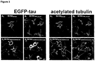
- the "cell expressing a mutated tau protein” is a cell in which a protein can be detected using the monoclonal antibody MC-1, preferably using the experimental conditions described with respect to Figure 2 infra .
- Impedance spectroscopy also known as cellular dielectric spectroscopy (CDS) or electric impedance spectroscopy (EIS) - can be used to measure frequency dependent alterations of passive electrical properties of single cells by applying defined alternate currents and/or voltages.
- the bio-impedance of single cells can be measured with a working electrode and a counter electrode.
- Different cellular parameters such as the capacitance and resistance of the cell membranes as well as intracellular membranes of organelles, the resistance of the extracellular medium and intrinsic cytoplasm, the extracellular matrix and the contact between cell and electrode contribute to the overall cellular impedance.
- an alternate voltage is applied to a biological sample.
- the applied current can flow from an active working electrode through the cells whereby the remaining current is collected by a counter electrode.
- alterations of certain cellular compartments can be identified.
- impedance spectroscopy is preferably carried out as described in Jahnke et al. (Lab Chip, 2009, 9, 1422-1428 ).
- the device for measuring the impedance of cells may be one as described in EP 2103933 A1 .
- the cell according to this aspect has an electric impedance which is reduced relative to a control cell.
- the control cell is a cell identical to the cell expressing the mutated tau, except that it does not express the mutated tau.
- the control cell is of the same cell type as the cell of the invention but does not contain a recombinant nucleic acid encoding said mutated tau.
- the control cell is an untransfected cell or wild type cell of the same cell type as the cell of the present invention.
- the control cell has been transfected with a mock vector, e.g. a plasmid without coding sequence.
- control cell is a cell transfected with a vector comprising a nucleic acid sequence encoding wildtype tau, e.g. tau represented by SEQ ID NO:1, 6 or 8.
- expression level in the control cell of the wild-type tau is substantially the same as that of the mutated tau in the cell of the present invention.
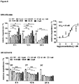
- the electrical impedance is preferably reduced by at least 5%, more preferably by at least 10%, most preferably by at least 20%, relative to the (untransfected) control cell not expressing mutated tau, determined under the conditions used in Example 2 / Figure 4 .
- the cell of the invention has a reduced electric impedance in the absence of okadaic acid.
- the cell of the invention has a reduced electric impedance in the absence of phosphatase inhibitors, e.g., okadaic acid.
- the cell of the invention has a reduced electric impedance in the absence of okadaic acid and Congo Red.
- the cell of the invention has a reduced electric impedance in the absence of phosphatase inhibitors (e.g. okadaic acid) and Congo Red. More preferably, the cell of the invention has a reduced electric impedance in the absence of okadaic acid, Congo Red and formaldehyde.
- the cell of the invention has a reduced electric impedance in the absence of phosphatase inhibitors (e.g. okadaic acid), Congo Red and formaldehyde. Still more preferably, the cell of the invention has a reduced electric impedance in the absence of okadaic acid, Congo Red, formaldehyde and any other toxic substance. Most preferably, the cell of the invention has a reduced electric impedance in the absence of phosphatase inhibitors (e.g. okadaic acid), Congo Red, formaldehyde and any other toxic substance. In yet another embodiment, the cell of the invention has a reduced electric impedance in the absence of phosphatase inhibitors (e.g. okadaic acid), Congo Red, formaldehyde, polyanionic substances (e.g. heparin, polyglutamate) and any other toxic substance.
- phosphatase inhibitors e.g. okadaic acid
- Congo Red e.g. oka
- the cell is a neuronal cell expressing mutated tau which is detectable by the antibody MC-1, and exhibiting an electric impedance reduced by at least 10% relative to a control cell not expressing mutated tau, determined under the conditions used in Example 2.
- the cell of the invention is a neuronal cell or precursor cell thereof, characterized by the following features:
- the reduction in electric impedance occurs within 18 hours, more preferably within 12 hours, most preferably within 6 hours after addition of the differentiation agent. It is also preferred that no toxic agent is present in the culture medium of the cell (e.g. okadaic acid, Congo Red, formaldehyde etc.).
- no toxic agent is present in the culture medium of the cell (e.g. okadaic acid, Congo Red, formaldehyde etc.).
- the invention relates to a method for identifying an agent for treating or preventing a tauopathy, comprising
- Neurodegenerative diseases or disorders in accordance the present invention comprise Alzheimer's disease, Parkinson's disease, Huntington's disease, amyotrophic lateral sclerosis, prion diseases, Pick's disease, fronto-temporal dementia, progressive nuclear palsy, corticobasal degeneration, cerebro-vascular dementia, multiple system atrophy, and mild-cognitive impairment.
- Further conditions involving neurodegenerative processes are, for instance, ischemic stroke, age-related macular degeneration, narcolepsy, motor neuron diseases, nerve injury and repair, and multiple sclerosis.
- the neurodegenerative disease is a taupathy.
- tauopathies are disorders and diseases, characterized by the presence of neuronal tau aggregation, in particular the presence of neurofilbrillary tangles.
- Typical tauopathies are Alzheimer's disease and other neurodegenerative disorders such as FTDP-17, Pick's disease, progressive supranuclear palsy, amyotrophic lateral sclerosis/Parkinsonism-dementia complex of Guam and corticobasal degeneration.
- the neurodegenerative disease is selected from the group consisting of Alzheimer's disease, Parkinson's disease, multiple sclerosis, amyotrophic lateral sclerosis, huntington's disease, tauopathies and prion diseases. Most preferably, the neurodegenerative disease is Alzheimer's disease.
- markers refers to any parameter that indicates the onset or presence of a neurodegenerative disease on a cellular level. Suitable markers include the presence of neurofibrillary tangles, a decrease in impedance, phosporylation of tau, dendritic/axonal dystrophy, axonal degeneration, ⁇ -amyloid production, synaptic dystrophy, microtubule and overall cytosceleton function/integrity/-based transport/distribution, proteasomal function, cellular morphology, cellular adhesion, signal transduction, receptor distribution/function, and all other cellular functions which may be affected.
- Preferred markers include the presence of neurofibrillary tangles, decrease in impedance, phosporylation of tau, dendritic/axonal dystrophy, axonal degeneration, ⁇ -amyloid production and synaptic dystrophy, as well as cellular morphology.
- a test compound "modulates" a marker indicative of a neurodegenerative disease when it is capable of changing or altering the level and/or the activity of the marker.
- Said modulation may be an increase or a decrease in the level and/or the activity of the marker.
- said modulation may be an increase or decrease in phosporylation of tau, dendritic/axonal dystrophy, axonal degeneration, synaptic dystrophy and/or the amount of neurofibrillary tangles present.
- neurofibrillary tangles in cells may be determined by the Gallyas silver impregnation method followed by light/ electron microscopy or other appropriate methods ( Braak, H. and Braak, E. (1995) Neurobiol Aging 16, 271-8 ; discussion 278-84).
- One such other appropriate method comprises the utilization of conformation-dependent antibodies that are capable of recognizing and discriminating the tau molecule in the context of neurofibrillary tangles from tau molecules existing in other states of aggregation and that can be detected, for instance, by fluorescence microscopy and/or fluorescence resonance energy transfer (FRET) technology.
- FRET fluorescence resonance energy transfer
- neurofibrillary tangles could also be detected by the use of other optical methodologies such as fluorescence polarisation spectroscopy, fluorescence correlation spectroscopy, fluorescence cross-correlation spectroscopy, fluorescence intensity distribution analysis, fluorescence lifetime measurements, fluorescence anisotropy measurements, or combinations thereof.
- fluorescence polarisation spectroscopy fluorescence correlation spectroscopy
- fluorescence cross-correlation spectroscopy fluorescence intensity distribution analysis
- fluorescence lifetime measurements fluorescence anisotropy measurements, or combinations thereof.
- an assay to monitor and quantify the formation and aggregation of paired helical filaments in solution has been described by Friedhoff et al. (1998, Biochemistry, 37: 10223-30 ).
- said conformation-dependent antibodies are optically labeled, preferably fluorescently labeled.
- the method of the invention comprises determining the phosphorylation at one or more of the phosphorylation sites Ser198, Ser199, Ser202, T231, S235, S396, S404, S409, S413 and S422.
- Phosphorylation of disease-specific phosphorylation sites confirms that tau-transfected cells are in a process of AD-like degeneration: Phosphorylation of Ser202/Thr205 (known as the AT8 site) has been identified as a diagnostic marker epitope, i.e. it is indicative of early stages of neurodegeneration, preceding tangle formation and neuronal loss in Alzheimer's disease.
- the method of the invention comprises determining the phosphorylation at Ser202 and/or Thr205.
- the extent of phosphorylation of tau at the respective epitopes may be determined using suitable antibodies which specifically recognize phosphoepitopes of tau or non-phosphorylated regions of tau.
- suitable antibodies have been described, for instance, in Johnson, G. V. and Hartigan, J. A. (1999) J Alzheimers Dis 1, 329-51 and are commercially available.
- Quantification of Tau phosphoepitopes may be done by Western blotting followed by densitometrical analysis of the signals according to standard procedures.
- Dendritic or axonal dystrophy/degeneration can be determined by biochemical determination, visual inspection and/or tissue section (staining) labelling employing suitable markers (etc. antibodies detecting the heavy isoform of neurofilament as detailed in Mack et al. (2001) Nat Neurosci 4, 1199-206 .). Morphologically, Wallerian degeneration of axons has been described in detail in Beirowski et al. (2005) BMC Neurosci 6, 6 .
- Synaptic dystrophy can be determined by biochemical determination, visual inspection and/or tissue section labelling (staining) employing suitable markers such as synapsin or synaptophysin ( Rutten et al., 2005, Am J Pathol 167, 161-73 ).
- the method further comprises the step of co-treatment of the cells with ⁇ -amyloid precursor protein ( ⁇ -APP) or a fragment or derivative or variant thereof (see supra ).
- ⁇ -amyloid precursor protein ⁇ -APP
- the ⁇ -amyloid precursor protein ( ⁇ -APP) or a fragment or derivative or variant thereof may be expressed in the cell, preferably under the control of an inducible system, as described above.
- This embodiment may comprise the step of transfecting or transducing said cells with a recombinant vector comprising a polynucleotide encoding ⁇ -amyloid precursor protein or a fragment or derivative or variant thereof.
- the preferred fragment is the ⁇ -amyloid peptide A ⁇ 1-42 .
- the ⁇ -amyloid peptide is derived from a larger Type I membrane spanning protein, ⁇ -APP, which has several alternatively spliced transcripts.
- ⁇ -APP Type I membrane spanning protein
- the amino acid sequences of ⁇ -APP and A ⁇ 1-42 are described in Kang J. et al., 1987; Knauer M.F et al., 1992; Homo sapiens APP (Gen-ID): NM 201414.
- fragment as used herein is meant to comprise e.g. an alternatively spliced, or truncated, or otherwise cleaved transcription product or translation product.
- derivative refers to a mutant, or an RNA-edited, or a chemically modified, or otherwise altered transcription product, or to a mutant, or chemically modified, or otherwise altered translation product.
- a “derivative” may be generated by processes such as altered phosphorylation, or glycosylation, or, acetylation, or lipidation, or by altered signal peptide cleavage or other types of maturation cleavage. These processes may occur post-translationally.
- the step of determining whether the test substance modulates at least one marker indicative of the neurodegenerative disease may comprise the following steps: (i) measuring the marker in the cell or in the cell culture which has been contacted with the test substance; (ii) measuring the marker in a cell or cell culture which has not been contacted with the test substance (control cell or control cell culture); and optionally (iii) comparing the level or activity of marker determined in (i) and (ii).
- the step of determining whether the test substance modulates at least one marker indicative of the neurodegenerative disease may comprise the following steps: (i) measuring the marker in the cell or in the cell culture which has been contacted with the test substance; (ii) measuring the marker in a control cell (or control cell culture) which does not express mutated tau and which has not been contacted with the test substance; and optionally (iii) comparing the level or activity of marker determined in (i) and (ii).
- the method may comprise the step of selecting or identifying as an agent for treating or preventing neurodegenerative disease a test compound which is capable of decreasing the level or activity of the marker indicative of the neurodegenerative disease.
- the test compound may be selected if it is capable of significantly decreasing the level or activity of the marker.
- the marker can be measured in a quantitative manner, the test compound is selected if it is capable of decreasing the level or activity of the marker by at least 10%, preferably by at least 20%, more preferably by at least 30%, even more preferably by at least 40%, most preferably by at least 50% as compared to the control cell or control cell culture.
- the test compound may be selected if it is capable of significantly increasing the electric impedance of the cell of the invention.
- the test compound is selected if it is capable of increasing the electric impedance of the cell of the invention by at least 5%, preferably by at least 10%, more preferably by at least 15%, even more preferably by at least 20%, most preferably by at least 25% as compared to the control cell or control cell culture which has not been contacted with the test compound.
- the test compound is selected if it is capable of increasing the electric impedance of the cell of the invention such that the electric impedance of the cell of the invention differs by less than 10%, more preferably by less than 5% from that of a control cell which does not express mutated tau.
- the method may further comprise the step of measuring the viability of the cell of the invention. This may be done by using methods such as: visual inspection under a microscope; staining using vital dyes stains and immunohistochemical reagents specific for cell types or moieties present in normal and injured brain; reaction with antibodies to neurofilaments, glial fibrillary acidic protein, S100, microtubule associated proteins, and synaptic proteins; biochemical assessment of metabolic activity; measurement of total or specific protein content; assessment of cellular function; and assessment of neural activity.
- the viability/integrity of the cell or cell culture may be assessed at the initiation of each experiment in order to demonstrate the health of the cells as well as to provide a measure of the amount of viable cells present in the pretreated cell culture.
- Compounds that are assayed in the above method can be randomly selected or rationally selected or designed.
- a compound is said to be randomly selected when the compound is chosen randomly without considering the structure of other identified active compounds.
- An example of randomly selected compounds is the use a chemical library, a peptide combinatorial library, a growth broth of an organism, or a plant extract.
- a compound is said to be rationally selected or designed when the compound is chosen on a nonrandom basis. Rational selection can be based on the target of action or the structure of previously identified active compounds. Specifically, compounds can be rationally selected or rationally designed by utilizing the structure of compounds that are presently being investigated for use in treating Alzheimer's disease.
- test compounds can be, as examples, peptides, small molecules, and vitamin derivatives, as well as carbohydrates.
- the test compounds may be nucleic acids, natural or synthetic peptides or protein complexes, or fusion proteins. They may also be antibodies, organic or inorganic molecules or compositions, drugs and any combinations of any of said agents above. They may be used for testing, for diagnostic or for therapeutic purposes. A skilled artisan can readily recognize that there is no limit as to the structural nature of the test compounds to be used in accordance with the present invention.
- a cell culture comprising cells according to the present invention is typically provided.
- the culture media can either have a test compound present prior to the introduction of the cells, or a test compound can be added to the media after the cells have been placed in the culture dish.
- a test substance will be first dissolved in appropriate vehicle, such as, but not limited to, DMSO, water, physiological saline, or media, to make a stock solution and then diluted into the media.
- vehicle control test may be included when the present invention is used.
- a range of doses is tested.
- the range tested initially may be informed by prior knowledge of the effects of the test compound or closely related substances on purified proteins, cells in culture, or toxicity in other test systems.
- the dose range is preferably from about 1 nM to about 100 ⁇ M.
- a skilled artisan can readily develop a testing range for any particular compound or series of compounds.
- the test compound is typically applied to the cell or cell culture for about one hour to about 21 days, preferably from about 3 hours to about 7 days, more preferably from about 6 hours to about 3 days, most preferably from about 18 hours to about 2 days, e.g. about 24 hours.
- fresh media containing test compound can be applied periodically; more frequently if rapid loss of test compound due to chemical conversion or to metabolism is suspected.
- the cells of the invention are contacted with a differentiation agent as described above.
- the differentiation agent may be added to the cells prior to exposing the cells to the test substance (e.g. 24 or 48 hours before), or the differentiation agent may be added simultaneously with the test substance, and the cells are then cultured further.
- the invention relates to the use of the tau protein of the present invention, of the nucleic acid of the present invention, of the vector or plasmid of the present invention, or of the cell of the present invention for screening an agent or agents capable of modulating one or more markers of neurodegeneration.
- the invention relates to the use of the tau protein of the present invention, of the nucleic acid of the present invention, of the vector or plasmid of the present invention, or of the cell of the present invention for the development of medicaments for the treatment or prevention of neurodegenerative diseases.
- the invention relates to a method for recapitulating a tauopathy, comprising the following steps:
- the cDNA from human wildtype tau (0N4) was amplified by PCR from isolated human neuronal cell mRNA. During PCR, a Bgl II site was inserted at the 5' and a Sal I site at the 3'-end of the tau gene. The PCR product was cloned into the pEGFP-C1 vector C-terminal to the EGFP coding sequence. For lentiviral transduction the EGFP-tau wildtype coding sequence was amplified by PCR with Bam HI (5') and Xho I (3') restrictions sites and cloned into a lentiviral transduction vector.
- the EGFP-tau (poly)mutants were obtained by site-directed mutagenesis (QuickChange Lightning, Stratagene).
- site-directed mutagenesis the Lentiviral TOPO Expression Kit (Invitrogen) was used.
- SH-SY5Y cell lines were transduced by lentiviral particles and cells stably expressing the EGFp-tau construct were selected by blasticidin.
- SH-SY5Y cells Human neuroblastoma SH-SY5Y cells were grown in DMEM medium, supplemented with 15% fetal bovine serum (FBS), non-essential amino acids, glutamax and penicillin/streptomycine (Invitrogen). Differentiation of SH-SY5Y cells was induced by incubation of the SH-SY5Y cells with 20nM stauropsprin (Sigma-Aldrich) for 48 hours. For the appropriate experiments differentiated cells were treated with okadaic acid (Sigma-Aldrich) in a concentration of 25 nM or in case of reference compound testing with SRN-003-556 and AR-A014418, respectively.
- FBS fetal bovine serum
- non-essential amino acids glutamax
- Penicillin/streptomycine Invitrogen.
- Differentiation of SH-SY5Y cells was induced by incubation of the SH-SY5Y cells with 20nM stauropsprin (Sigma-
- Cells were grown on coverslips and fixed with 4% formaldehyde at room temperature for 30 min, followed by permeabilization with 0,1% Triton X-100. Cells were incubated with anti-acetylated tau antibody(Abcam, 1:1000) for two hours at room temperature. After washing three times with PBS, cells were incubated with Cy3-linked secondary antibody (Dianova, 1:100) for 1.5 hours. Nuclei were stained with DAPI (Sigma-Aldrich). Confocal images were taken with a Nikon Eclipse C1 LSM microscope.
- Cells were harvested and protease as well as phosphatase inhibitor cocktail (Sigma-Aldrich) was added. Proteins were extracted by sonification (Hielscher GmbH) and protein concentration was determined using the Roti-Nanoquant Assay (Carl-Roth GmbH). Laemmli sample buffer was added to 60 ⁇ g cell lysate. Samples were separated on 10% SDS-polyacrylamide gel and electro transferred onto polyvinylidene fluoride membranes and subsequently immunolabeled with anti-MC1 specific antibody (Peter Davis Department of Pathology, Albert Einstein College of Medicine, 1:100) at 4°C over night. Secondary antibody was horseradish peroxidase conjugated (Dianova, 1:5000). Specific protein signals were detected using Chemiluminescence Detection Kit (MobiTec) and ChemiDoc-XRS (BioRad).
- IDAT calculates the impedance of cell-covered electrodes relative to electrodes without cells (relative impedance: (
- pathologically misfolded tau species also reveal themselves by migrating slightly faster than species in a physiological conformation (i.e. wild-type control tau) during PAGE electrophoresis, presumably due to a more compact conformation ( FIG. 2 ; middle panel showing total tau; most compact and loose conformational isoforms marked by arrows).
- Fig. 2 middle panel showing total tau; most compact and loose conformational isoforms marked by arrows.
- This finding is also perfectly in line with results from Jeganathan et al. (2008) suggesting that pathological tau from AD brains recognised by the conformation-dependent antibody MC1 persists in a compact 'paperclip' conformation which favours aggregation but opposes microtubule stabilization.
- the 'hyper-mutated' tau variant was also unique with respect to microtubule integrity. Mutant tau not only has a high tendency to aggregate, and by doing so may loose its physiological function, but on top of that clearly seems to carry out a toxic gain of function resulting in specific neurotoxicity (not affecting glial cells). However, the exact nature of this toxic gain of function is not yet understood.
- the stability of the microtubule network in three mutated tau and a wild-type tau control cell line was compared ( Fig. 3 )
- the inventors observed thick bundles of stable microtubules in cells expressing eGFP-tagged tau transgenes (white arrows). In those cells, acetylated tubulin as a marker for post-translational modified, stabile microtubules strongly colocalized with transgenic tau giving rise to a bright yellow filamentous pattern
- Aggregated tau oligomers may constitute the toxic tau species attacking the microtubule network and, ultimately, may be responsible for interruption of vital microtubule-dependent transport function slowly atrophying the cell.
- the results are corroborated by growing evidence that microtubule-based axonal and dendritic transport and, thus, synapse/spine maintenance is primarily affected by tau-dependent degeneration in AD (see for example Zempel et al. 2010).
- okadaic is commonly used for artificial induction of tau hyperphosphorylation in in vitro experiments under non-physiological conditions
- the inventors were able to detect impedimetrically an initial Hyper-tau mutant specific degenerative effect after 3 and 6 hours, respectively.
- toxic side effects of okadaic acid leads to overall cell toxicity in all tau mutant cell lines resulting in a strong decrease of impedance.
- the inventors took advantage of the poly-mutant tau, especially the Hyper-tau mutant that show neuropathological phenotype only by induction of neuronal differentiation (see Fig.1 ). Therefore the wildtype and poly-mutant tau expressing cells were differentiated for 48 hours followed by impedimetric monitoring for 24 hours ( Fig.5 ). The mean values from 3 experiments with 4-6 replicates reveals that the single mutant P301L tau expressing cells show the same impedimetric characteristic like the wildtype tau expressing cells.
- Hyper-mutant tau could be used in a functional tau pathology in vitro screening assay for identification and efficiency quantification of potential active pharmaceutical ingredients. Taking the significant difference between the physiological condition (wildtype tau expressing cells) and the pathological condition (Hyper-tau expressing cells) that can be monitored via impedance spectroscopy, the efficiency of compounds to attenuate the pathological cellular degeneration can be quantitatively determined.
- SRN-003-556 a developed lead compound with moderate specificity to ERK2, and AR-A014418, a specific GSK3ß-inhibitor were tested. Both inhibitors showed a therapeutic effect within tau pathology in vitro assays that are based on artificial hyperphosphorylation using toxic compounds like okadaic acid (LeCorre et al. 2006, Selenica et al. 2007, Jahnke - unpublished data). But only SRN-003-556 showed a therapeutic effect in a tauopathy mouse model while AR-A014418 does not (LeCorre et al. 2006, Selenica et al. 2007).
- SEQ ID NO:1 shows the amino acid sequence of a wild type human tau protein (Homo sapiens microtubule-associated protein tau (MAPT): NM 016834/NP_058518).
- SEQ ID NO:2 shows the amino acid sequence of the human the tau isoform having 441 amino acids.
- SEQ ID NO:3 shows the amino acid sequence of a modified human tau protein having five FTDP-17 mutations relative to the wild type sequence.
- SEQ ID NO:4 shows a nucleotide sequence encoding SEQ ID NO:1.
- SEQ ID NO:5 shows a nucleotide sequence encoding SEQ ID NO:3.
- SEQ ID NO:6 and 7 show the amino acid and nucleotide sequences, respectively, of another isoform of human tau protein, with the following details: LOCUS NM_005910 1326 bp mRNA linear PRI 03-APR-2011 DEFINITION Homo sapiens microtubule-associated protein tau (MAPT), transcript variant 2, mRNA.
- MTT microtubule-associated protein tau
- SEQ ID NO:8 and 9 show the amino acid and nucleotide sequences, respectively, of yet another isoform of human tau protein, with the following details: LOCUS NM_001123067 1239 bp mRNA linear PRI 03-APR-2011 DEFINITION Homo sapiens microtubule-associated protein tau (MAPT), transcript variant 5, mRNA.
Landscapes
- Health & Medical Sciences (AREA)
- Chemical & Material Sciences (AREA)
- Life Sciences & Earth Sciences (AREA)
- Organic Chemistry (AREA)
- Genetics & Genomics (AREA)
- Engineering & Computer Science (AREA)
- Zoology (AREA)
- Biomedical Technology (AREA)
- Molecular Biology (AREA)
- Biochemistry (AREA)
- General Health & Medical Sciences (AREA)
- Biophysics (AREA)
- Medicinal Chemistry (AREA)
- Gastroenterology & Hepatology (AREA)
- Toxicology (AREA)
- Proteomics, Peptides & Aminoacids (AREA)
- Wood Science & Technology (AREA)
- Biotechnology (AREA)
- Bioinformatics & Cheminformatics (AREA)
- General Engineering & Computer Science (AREA)
- Neurology (AREA)
- Microbiology (AREA)
- Cell Biology (AREA)
- Physics & Mathematics (AREA)
- Plant Pathology (AREA)
- Micro-Organisms Or Cultivation Processes Thereof (AREA)
- Investigating Or Analysing Biological Materials (AREA)
- Measuring Or Testing Involving Enzymes Or Micro-Organisms (AREA)
- Peptides Or Proteins (AREA)
Description
- The accumulation of proteinaceous aggregates is a pathological hallmark of most if not all chronic dementias characterized by incipient neuronal dysfunction and eventual cell death. In tauopathies, as the name implies, these aggregates take the form of neurofibrillary tangles (NFT) composed essentially of the microtubule-associated protein tau. Though the MAPT gene encoding tau is not genetically linked to Alzheimer's disease (AD), mutations in MAPT cause hereditary frontotemporal dementias (FTD), and missense mutations have also been found in progressive supranuclear palsy, corticobasal degeneration and in conditions that closely resemble Pick's disease, thus providing evidence that disrupting tau homeostasis is sufficient to cause neurodegeneration-driven dementias (reviewed in van Swieten & Spillantini, 2007). In AD, mounting evidence suggests that (wild-type) tau mediates Abeta/amyloid toxicity by hyperphosphorylation and/or redistribution of tau protein (Ittner et al. 2010; Zempel et al. 2010) and that reducing tau levels can prevent AD symptoms (Vossel et al. 2010). Tau mutations are known to alter the relative proportion of various tau isoforms and, more importantly, enhance the tendency of tau to adopt an abnormal conformation and, consequently, aggregate into filaments. It is therefore expected that tau-mediated neurodegeneration is caused by a combination of toxic gains of function triggered by abnormalities in tau conformation, as well as from any harmful consequences that may result from the loss of normal tau functions. Unfortunately, the exact mechanisms by which abnormalities in tau conformation initiate, or contribute, to neuronal cell death are not entirely understood. The at best fragmentary understanding of the pathomechanism presents an enormous obstacle for drug development for lack of validated targets for therapeutic intervention. Nevertheless, drug development is pursued in functional models that may recapitulate authentically at least aspects of human tauopathies in vitro and in vivo.
- Since NFT are the most striking pathological feature in tauopathies, much attention has focused on understanding how the deposition of NFT may cause neurodegeneration, in essence using animal models recapitulating NFT pathology to investigate the mechanism of disease. It has long been postulated that the aggregation of tau into filaments and NFT results in a toxic gain of function. This view has been substantially challenged by observations that neuronal loss and memory impairment can experimentally be cured despite ongoing NFT formation (Santacruz et al. 2005). In some animal models, the tau-mediated loss of neurons does not even require NFT development. Thus, it is assumed that non-filamentous but already aggregated, globular tau intermediates on route to assemble into larger helical filaments may represent the neurotoxic tau species.
- Elevated levels of free tau, not bound to microtubules, presumably increases its likelihood to become misfolded, as well as undergo modifications or conformational changes that promote the formation of aggregated small globular oligomers that eventually will assemble into insoluble filaments. Covalent modifications stabilizing conformational changes most likely include phosphorylation since tau protein isolated from AD brain was found to be abnormally high phosphorylated at multiple critical sites ('hyperphosphorylated tau') and it was demonstrated that pseudo-hyperphosphorylation (i.e. pseudo-phosphorylated at multiple sites along the protein) can facilitate abnormal conformation of tau protein (Jeganathan et al. 2008). In this context tau hyperphosphorylation was identified as diagnostic target (
WO9311231A1 WO2007088400A1 ). - Given that tau is normally a highly soluble protein that does not readily aggregate, this matter has been difficult to assess in experimental models because of the resistance of tau to aggregate within an ideal time-frame for culture studies or within an animal's relatively short lifespan. Because high concentrations of tau are required to promote tau aggregation in experimental models, it is believed that the enhanced ability of tau to form small globular aggregates in the cytoplasm of neurons and glia in human tauopathies may be due to pathological conditions that locally increase the pool of tau available for aggregation. Yet it is unlikely that the amount of tau in various tauopathies is as high as in cell culture and animal models that artificially force massive tau overexpression and, therefore, much caution is needed extrapolating results from such model systems to the human condition.
- Further complicating matters is evidence that mouse tau appears to prevent tau aggregation in transgenic mice overexpressing wild-type human tau (htau). Nonetheless, transgenic mice that overexpress high levels of htau isoforms containing aggregation-promoting mutations (e.g. P301 L tau) can develop tau pathology even in the presence of endogenous mouse tau. The P301 L and P301 S mutations are among the first described FTD mutations and show a very early mean onset for FTD in man. Tau transgenic mouse models with expression of these mutants display onset of first signs of tau pathology starting at 2.5 to 5 months (Schindowski et al. 2006).
WO 01/53340 A2 - To accelerate tau aggregation in vitro, polyanionic cofactors or small molecule ligands are often used to facilitate tau aggregation. For example, in a cell culture model overexpressing full length tau, Congo red treatment stimulates the formation of filamentous tau aggregates and decreases cell viability (Bandyopadhyay et al. 2007). These and other results suggest that also in cell culture models tau aggregation causes cell death or, at least, accelerates its onset. However, no cell culture model has been described so far that does not force aggregation of tau by either artificial high concentrations of tau or addition of, at higher doses toxic, compounds in order to facilitate or precipitate aggregation (Ko et al. 2004, Tsukane et al. 2007, Nie et al. 2007). In view of modelling tauopathy disease mechanisms for drug development purposes, both strategies bear a high risk of producing artificial results since the mechanism of degeneration may significantly differ from tau pathology in the AD brain.
- It has been found that cells expressing tau with multiple mutations show signs of neurodegeneration within 24 hours of cell culture. Cells expressing tau proteins with 4 FTDP-17 mutations showed lowered impedance (Example 2 /
Fig. 5 ). This was shown for two different tau proteins having different 4x mutations. The effect was even more pronounced for a tau protein having five different FTDP-17 mutations. - In a first aspect, the present invention relates to a tau protein comprising at least four or at least five different mutations associated with the condition frontotemporal dementia and parkinsonism linked to chromosome 17 (FTDP-17), wherein the tau protein has at least the mutations
- a) P301L, V337M, R406W, and the deletion K280,
- b) K257T, P301 L, V337M, R406W, or
- c) K257T, P301 L, V337M, R406W and the deletion K280,
- A second aspect of the invention is a nucleic acid encoding the tau protein of the present invention.
- A third aspect of the invention is a plasmid or vector comprising the nucleic acid of the present invention.
- A fourth aspect of the invention is a cell comprising the nucleic acid or the vector or plasmid of the present invention.
- A fifth aspect of the invention is a cell expressing the mutated tau protein of the present invention, wherein said cell exhibits a decrease in impedance after 24 hours of cell culture.
- A sixth aspect of the present invention is a method for identifying an agent for treating or preventing neurogenerative disease, comprising
- (a) contacting a test compound with the cell of the present invention; and
- (b) determining whether the test substance modulates at least one marker indicative of the neurodegeneration.
- A seventh aspect of the invention is the use of the tau protein of the present invention, of the nucleic acid of the present invention, of the vector or plasmid of the present invention, or of the cell of the present invention for screening an agent or agents capable of modulating one or more markers of neurodegeneration.
- An eighth aspect of the invention is the use of the tau protein of the present invention, of the nucleic acid of the present invention, of the vector or plasmid of the present invention, or of the cell of the present invention for the development of medicaments for the treatment or prevention of neurodegenerative diseases.
- A ninth aspect of the invention is a method for recapitulating a tauopathy, comprising the following steps:
- (a) providing the cell of the present invention;
- (b) culturing said cell under conditions to allow expression of tau protein having abnormal conformation.
-
-
Fig 1 : Abnormal tau aggregation in a hyper-mutated tau expressing cell line. SH-SY5Y neuroblastoma cell lines stably expressing wildtype or mutant tau at a comparable level were used to induce neuronal differentiation by application of 20nM staurosporine. 48 h of differentiation (+ STS) is sufficient for the formation of abnormal tau aggregates in the Hyper-tau expressing cells (arrow). -
Fig. 2 : Abnormal tau conformation in polymutant tau cell lines. Poly-mutant tau variants were developed and, using those variants, poly-mutant neuroblastoma cell lines were produced. In those cell lines tau adopting an abnormal/pathological conformation under physiological conditions was observed (MC1 abnormal tau). A 5x mutated tau gene variant (SH-Tau5x mut) demonstrated a particular high tendency for an abnormal conformation as detected by the MC1 marker (red arrowhead) despite similar expression levels compared to wild-type or single mutant protein. Corroborating recent results that phosphorylation may stabilize abnormal conformation the amount of MC1-positive tau species could be decreased by treating the cells with a tau kinase inhibitor (SRN-003-556). Thus, polymutant tau protein is present in an Alzheimer-like phosphorylation state in differentiated neuroblastoma cells and kinase inhibition decreases tau missfolding and may also suppress subsequent aggregation. -
Fig 3 : Microtubule network pathology in 5x polymutant tau neuroblastoma cells after differentiation into neuronal phenotype. Indicated MAPT gene variants were transduced using retroviral vectors into the human neuroblastoma cell line SH-SY5Y and stable expressing cell lines were selected. Differentiated cells were analyzed for transgene expression (EGFP-tau, A1-D1) and changes in the microtubule network detected by an antibody against acetylated tubulin (A2-D2), a marker for stable microtubules. Double-label fluorescence shows a filamentous pattern of stabile microtubules in infected cells and local colacalization of microtubules with transgenic tau (A-D, white arrows), as would be expected of a microtubule-associated protein. In contrast, in a subpopulation of cells (D, open arrows) transduced with the 5x polymutant variant stable microtubules were absent. Concomitantly, whose cells display high levels of EGFP-tagged tau transgene and appear to have lost their processes. A collapse of the (stable) microtubule network is expected to cause a disruption of the microtubule-dependent transport and a slow atrophy of the cell culminating in death of affected cells. -
Fig 4 : Impedimetric detection of consequences by okadaic acid induced artificial hyperphosphorylation. While the incubation with 25 nM okadaic acid initially shows a Hyper-tau mutant specific degenerative effect at 3 h and 6 h the unspecific toxic side effects of okadaic acid resulted in massive cell damage in all tau mutants after 24 h. (n = 3, 2D-ANOVA, Bonferoni post-hoc test, * p < 0.05, ** p < 0.01, *** p < 0.001) -
Fig 5 : Differentiation induces neurodegeneration in poly-mutant tau expressing SH-SY5Y cells. Differentiation of SH-SY5Y cells were induced by 48 h incubation with 20 nM staurosporine. Afterwards the cells were impedimetrically monitored for 24 h. While wildtype tau and P301 L-tau expressing cells showed no decrease of impedance, K257q and dK280q-tau cells showed lowered impedance. Strikingly, the Hyper-tau expressing cells showed a more significant impedance decrease as a result of neurodegeneration. (n = 4, 2D-ANOVA, Bonferoni post-hoc test, * p < 0.05, ** p < 0.01) -
Fig 6 : Use of Hyper-tau cells for quantitative detection of compound efficiency. Taking the developed Hyper-tau cells, where pathology was induced by differentiation (20 nM staurosporine for 48 h) in comparison to the wildtype tau expressing cells (physiological condition) the efficiency of potential active pharmaceutical ingredients could be quantified. The effect of two reference compounds, in detail the kinase inhibitor SRN-003-556 and AR-A014418 were used to demonstrate screening capability of the Hyper-tau based functional tau pathology screening assay. While SRN-003-556 show a significant reduction of Hyper-tau induced pathology, AR-A014418 shows no significant therapeutic effect. Observable therapeutic effects can be quantified, as demonstrated by EC50 determination exemplarily for SRN-003-556 at 24h (values are normalized to WT, n = 4, all groups compared to Hyper, 2D-ANOVA, Bonferoni post-hoc test, * p < 0.05, ** p < 0.01, *** p < 0.001) - As used herein and in the appended claims, the singular forms "a", "an" and "the" include plural reference unless the context clearly dictates otherwise. Thus, for example, reference to a "molecule" is a reference to one or more molecules and equivalents thereof known to those skilled in the art, and so forth. The reference to a "cell" includes the reference to a population of cells.
- The present invention pertains to a tau protein comprising at least four or at least five different mutations selected from the group consisting of mutations associated with the condition frontotemporal dementia and parkinsonism linked to chromosome 17 (FTDP-17), wherein the tau protein has at least the mutations
- a) P301 L, V337M, R406W, and the deletion K280,
- b) K257T, P301L, V337M, R406W, or
- c) K257T, P301 L, V337M, R406W and the deletion K280,
- The phrase "tau protein", as used herein, denotes a polypeptide having sequence similarity to a wild type tau protein. Preferably, the amino acid sequence of a tau protein has at least 90%, more preferably at least 95% sequence identity to the amino acid sequence as shown in SEQ ID NO:1. The degree of identity of an amino acid sequence to
SEQ ID NO 1 may be determined by comparing the amino acid sequence in question andSEQ ID NO 1 using the program "BLAST 2 SEQUENCES (blastp)" (Tatusova et al. (1999) FEMS Microbiol. Lett. 174, 247-250) with the following parameters: Matrix BLOSUM62; Open gap 11 andextension gap 1 penalties; gap x_dropoff50; expect 10.0 word size 3; Filter: none. According to the present invention, the sequence comparison covers at least 200 amino acids, preferably at least 300 amino acids, more preferably at least 350 amino acids, and most preferably at least about 380 amino acids. - Most preferably, the amino acid sequence of a tau protein is selected from the group consisting of SEQ ID NO:1, SEQ ID NO:6 and SEQ ID NO:8. These are the amino acid sequences of tau isoforms 0N4R, 2N4R and 1 N4R, respectively.
- The tau protein is preferably a human tau protein or a variant thereof. The amino acid sequence of a wild type human tau protein is shown in SEQ ID NO:1. (Homo sapiens microtubule-associated protein tau (MAPT): NM 016834/NP_058518). The amino acid sequences of further human tau isoforms are shown in SEQ ID NO:6 and SEQ ID NO:8. The term "variant" as used herein refers to any polypeptide or protein, in reference to polypeptides and proteins disclosed in the present invention, in which one or more amino acids are added and/or substituted and/or deleted and/or inserted at the N-terminus, and/or the C-terminus, and/or within the native amino acid sequences of the native polypeptides or proteins of the present invention. Furthermore, the term "variant" includes any shorter or longer version of a polypeptide or protein. Variants comprise proteins and polypeptides which can be isolated from nature or be produced by recombinant and/or synthetic means. Native proteins or polypeptides refer to naturally-occurring truncated or secreted forms, naturally occurring variant forms (e.g. splice-variants) and naturally occurring allelic variants. The terms "variant" and "isoform" are used interchangeably herein.
- Unless indicated otherwise, the numbering of amino acids in the human tau sequences as used herein refers to the tau isoform having 441 amino acids which is shown in SEQ ID NO:2.
- The tau protein of this invention comprises at least four or at least five different mutations selected from the group consisting of mutations which are associated with the condition "frontotemporal dementia and parkinsonism linked to chromosome 17" (FTDP-17), wherein the tau protein has at least the mutations
- a) P301L, V337M, R406W, and the deletion K280,
- b) K257T, P301L, V337M, R406W, or
- c) K257T, P301 L, V337M, R406W and the deletion K280,
- The tau protein of the present invention may comprise the amino acid sequence as shown in SEQ ID NO:1, except for the mutations K257T, delK280, P301L, V337M and R406W. The tau protein may comprise the amino acid sequence as shown in SEQ ID NO:3, which is the most preferred embodiment. In another embodiment, the tau protein of the present invention may comprise the amino acid sequence as shown in SEQ ID NO:6, except for the mutations K257T, delK280, P301 L, V337M and R406W. In another embodiment, the tau protein of the present invention may comprise the amino acid sequence as shown in SEQ ID NO:8, except for the mutations K257T, delK280, P301 L, V337M and R406W.
- The invention further relates to nucleic acids encoding the proteins of the invention described herein.
- The term "nucleic acid" generally refers to any polyribonucleotide or polydeoxyribonucleotide that may be unmodified RNA or DNA or modified RNA or DNA. The polynucleotide may be single- or double-stranded DNA, single or double-stranded RNA. It will be appreciated that a variety of modifications may be made to DNA and RNA that serve many useful purposes known to those of skill in the art. The term "nucleic acid" as it is employed herein embraces such chemically, enzymatically or metabolically modified forms of polynucleotides, as well as the chemical forms of DNA and RNA characteristic of viruses and cells, including, for example, simple and complex cells.
- Nucleic acid sequences which encode the appropriate proteins or polypeptides can be readily prepared by the skilled person using the information and references contained herein and techniques known in the art (for example, see Sambrook et al., "Molecular Cloning, A Laboratory Manual, 3rd ed. 2001, CSH Press, Cold Spring Harbor, N.Y., and Ausubel et al., Short Protocols in Molecular Biology, John Wiley and Sons, 1992). These techniques include the use of the polymerase chain reaction (PCR) to amplify samples of the relevant nucleic acid, e.g. from genomic sources, chemical synthesis, and/or preparation of cDNA sequences. DNA encoding e.g. tau may be generated and used in any suitable way known to those of skilled in the art, including by taking encoding DNA, identifying suitable restriction enzyme recognition sites either side of the portion to be expressed, and cutting out said portion from the DNA. Mutation can be introduced into tau-encoding sequences, e.g. using site directed mutagenesis.
- cDNA sequences encoding tau proteins are known in the art (e.g. Gen-ID NM016834). The skilled person can therefore easily manipulate the DNA by known techniques to provide polynucleotides that encode the desired tau protein or variant thereof. The DNA sequence of wild type tau is shown in SEQ ID NO:4.
- Further aspects of the invention are vectors and plasmids containing a nucleic acid of the invention.
- A "vector" is a replicon, such as plasmid, phage, cosmid, or virus to which another polynucleotide segment may be operably inserted so as to bring about the replication or expression of the segment. The vector may particularly be a plasmid, a cosmid, a virus or a bacteriophage used conventionally in genetic engineering that comprise a polynucleotide encoding tau protein or a variant thereof. Methods which are well known to those skilled in the art can be used to construct recombinant viral vectors; see, for example, the techniques described in Sambrook et al., "Molecular Cloning, A Laboratory Manual, 3rd ed. 2001, CSH Press, Cold Spring Harbor, N.Y. and Ausubel et al., Current Protocols in Molecular Biology, Green Publishing Associates and Wiley Interscience, N.Y. (1989).
- The term "recombinant" means, for example, that a polynucleotide sequence is made by an artificial combination of two otherwise separated segments of sequence, e.g., by chemical synthesis or by the manipulation of isolated polynucleotides by genetic engineering techniques
- Expression vectors will contain a promoter which is operably linked to the protein-encoding nucleic acid sequence of interest, so as to direct mRNA synthesis. Promoters recognized by a variety of potential host cells are well known. "Operably linked" means joined as part of the same nucleic acid molecule, suitably positioned and oriented for transcription to be initiated from the promoter. DNA operably linked to a promoter is "under transcriptional control" of the promoter. Transcription from vectors in mammalian host cells may be controlled, for example, by promoters obtained from the genomes of viruses such as Simian Virus 40 (SV40), from heterologous mammalian promoters, e.g. the actin promoter or an immunoglobulin promoter, and from heat-shock promoters, provided such promoters, are compatible with the host cell systems. Expression vectors used in eukaryotic host cells will also contain sequences necessary for the termination of transcription and for stabilizing the mRNA.
- The promoter used herein may be constitutive or inducible. In a preferred embodiment expression control sequences specify induced expression of the polypeptide of the invention. Transcription of the messenger RNA encoding the polypeptide of the invention is induced upon addition or withdrawal of an external signal, such as a small chemical like tetracycline or a hormone like Ecdysone. The external signal can also be an increase or decrease in temperature or ionizing radiation. Also, inducible expression can be brought about by inducible translation initiation of the messenger RNA or a system in which mRNA stability is controlled in an inducible fashion. Examples of expression control sequences allowing induction of polypeptide production are disclosed in the following publications: the Tet-off/Tet-on system, for example described by Gossen and Bujard (1992) Proc Natl Acad Sci USA, 15; 89 (12): 5547-51 ; or by Gossen et al. (1995) Science, Jun 23; 268 (5218) : 1766-9; or by Kistner et al. (1996) Proc Natl Acad Sci USA 93: 10933-10938, but also the expression control system based on Cre-recombinase based methods. A further inducible expression system, for use in both cell culture and transgenic animals is based on the insect hormone Ecdysone, for example described by Hoppe et al. (2000) Mol Ther, 1: 159-164; or by No et al. (1996) Proc Natl Acad Sci USA, 93: 3346-3351. Another inducible expression system is the GAL4 system (Ornitz et al. (1991) Proc Natl Acad Sci USA, ) which allows conditional expression at 26-29 degrees, or also a Rapamycin based conditional expression system (Ho et al. (1996) Nature, Aug 29; 382 (6594): 822-6; and Pollock et al. (2000) Proc Natl Acad Sci USA, Nov 21; 97 (24): 13221-6). A temperature-sensitive expression system is based on a Sindbis virus expression cassette (Boorsma et al. (2000) Nat Biotechnol, Apr; 18 (4): 429-32) and predominantly suitable for controlled expression in cell culture systems.
- Expression vectors of the invention may also contain one or more selection genes. Typical selection genes encode proteins that confer resistance to antibiotics or other toxins (e.g. ampicillin, neomycin, methotrexate, or tetracycline), complement auxotrophic deficiencies, or supply critical nutrients not available from complex media. Examples of selectable markers for mammalian cells include DHFR and thymidine kinase. Thus a typical of the present invention may include an origin of replication, one or more protein sequence(s) operably linked to a constitutive or inducible promoter as appropriate, a transcription termination sequence, and a marker gene.
- The vectors and plasmids containing the polynucleotide described herein can be transferred into the host cell by well-known techniques, which vary depending on the type of cellular host (see infra).
- A further aspect of the invention is a cell comprising the nucleic acid or the vector or plasmid described herein. The cell is preferably an isolated cell, i.e. it is not in its natural environment such as a tissue. More preferably, the cell is a cell culture cell in a culture medium.
- The cell is preferably a living cell which can be cultured in cell culture. Preferably, the cell is a eukaryotic cell, more preferably it is a mammalian cell. Mammalian cell lines available in the art for expression of a heterologous polypeptide include fibroblast 3T3 cells, HeLa cells, baby hamster kidney cells, COS cells, Chinese hamster ovary cells, human liver cells (Hep G2); and many others. In a particular embodiment, the cell of the invention is a neuronal cell or a precursor thereof. "Neuronal cells" as used herein, are cells that express the marker protein microtubule-associated protein-2 (MAP-2). They may further express the marker proteins neurofilament and/or calbindin. Suitable neuronal cells include, but are not limited to primary neuronal cells preferably of the central nervous system (CNS), pluripotent stem cells (ES and iPS) derived neurons and transdifferentiated neurons. Such cells are described, e.g., in Otto et al. Journal of Neuroscience Methods 128 (2003) 173-181 (primary neuronal cells); Pankratz et al., 2007, Stem Cells 25(6): 1511-1520 (ES cells); Hu et al., 2010, PNAS 107(9): 4335-4340 (iPS cells); and Vierbuchen et al., 2010, Nature 463, 1035-1041 (transdifferentiated neurons).
- The precursor cell of a neuronal cell can be converted into a cell having neuronal phenotype by exposing the cell to a differentiation stimulus. Usually, differentiation into a neuronal phenotype can be induced by adding a differentiation agent to the precursor cells in cell culture. Suitable precursor cells include, but are not limited to, SH-SY5Y cells (Agholme et al., 2010, Journal of Alzheimer's Disease 20: 1069-1082), Neuro2A cells (Trembley et al., 2010, Journal of Neuroscience Methods 186: 60-67), NG108-15 cells (Zhong et al. Journal of Neurochemistry 68(6): 2291-2299; ATCC HB-12317), IMR32 cells (ATCC CCL-127) and PC-12 cells (Schimmelpfenig et al., 2004, Journal of Neuroscience Methods 139: 299-306). Suitable differentiation agents include staurosporine, retinoic acid, trichostatin A, differentiation promoting media formulation e.g. Neurobasal (A) + B27-Supplement, N2-Supplement and combinations thereof.
- The nucleic acid, vector or plasmid of the invention can be introduced into the cells using techniques known to those skilled in the art. The introduction may employ any available technique. For eukaryotic cells, suitable techniques may include calcium phosphate transfection, DEAE-Dextran, electroporation, liposome-mediated transfection and transduction using retrovirus or other virus, e.g. vaccinia or, for insect cells, baculovirus. The calcium treatment employing calcium chloride, as described in Sambrook et al., supra, or electroporation is generally used for prokaryotes or other cells that contain substantial cell-wall barriers.
- As used herein, the term "transduction," is used to describe the delivery and introduction of polynucleotide to eukaryotic cells using viral mediated delivery systems, such as, adenoviral, AAV, retroviral, or plasmid delivery gene transfer methods. These methods are known to those of skill in the art, with the exact compositions and execution being apparent in light of the present disclosure.
- As used herein, the term "transfection" is used to describe the delivery and introduction of polynucleotide to a cell using non-viral mediated means, these methods include, e.g., calcium phosphate- or dextran sulfate-mediated transfection; electroporation; glass projectile targeting; and the like. These methods are known to those of skill in the art, with the exact compositions and execution being apparent in light of the present disclosure.
- The transfection or transduction may be stable or transient. Preferably the transfection or transduction is transient. This generally refers to transient expression of the DNA construct introduced into the cells.
- Host cells transfected or transduced with expression or cloning vectors described herein may be cultured in conventional nutrient media modified as appropriate for inducing promoters, selecting transformants, or amplifying the genes encoding the desired sequences. The culture conditions, such as media, temperature, pH and the like, can be selected by the skilled artisan without undue experimentation. In general, principles, protocols, and practical techniques for maximizing the productivity of cell cultures can be found in "Mammalian Cell Biotechnology: a Practical Approach", M. Butler, ed. JRL Press, (1991) and Sambrook et al, supra.
- An additional aspect of the invention is a cell expressing the mutated tau protein of the present invention, wherein said cell exhibits reduced electric impedance. The cell is preferably an isolated cell, i.e. it is not in its natural environment such as a tissue. More preferably, the cell is a cell culture cell in a culture medium.
- The general cell types of this aspect of the invention are the same as described hereinabove, and cell culturing can be carried out according to established methods. Gene expression can be confirmed, for example, by Northern blotting to quantitate the transcription of mRNA, or in situ hybridization, using an appropriately labeled probe, based on the sequence of the recombinant nucleic acid introduced into the cell. Gene expression, alternatively, may be measured by immunological methods such as immunohistochemical staining of cells to quantitate directly the expression of gene product. Antibodies useful for immunohistochemical staining and/or assay of sample fluids may be either monoclonal or polyclonal.
- Expression of the mutated tau protein is preferably detected using antibodies directed against tau. Alternatively, the mutated tau protein may be fused to a label, e.g. a heterologous peptide sequence (FLAG tag, HA tag etc.) which can be detected by methods known in the art. In one embodiment, the mutated tau protein is fused to a heterologous amino acid sequence, e.g. EGFP, FLAG tag, or Strep tag. This allows to specifically detect the mutated tau in a Western blot using anti-tau antibodies. According to this embodiment, the mutated tau can be distinguished from the endogenous wild type tau due to its different size on a Western blot.
- Preferably, the mutated tau is not strongly overexpressed in the cell. In one embodiment, the amount of mutated tau in the cell is preferably less than the ten-fold amount of wild type tau expressed in the same cell or in a control cell, e.g. a non-transfected cell of the same cell type. More preferably, the amount of mutated tau in the cell is less than the five-fold amount of wild type tau expressed in the same cell or in a control cell, e.g. a non-transfected cell of the same cell type. Still more preferably, the amount of mutated tau in the cell is less than the two-fold amount of wild type tau expressed in the same cell or in a control cell, e.g. a non-transfected cell of the same cell type. Most preferably, the amount of mutated tau in the cell is about 75% to about 150%, or about 75% to about 125%, of the amount of wild type tau expressed in the same cell or in a control cell, e.g. a non-transfected cell of the same cell type. The level of expression is preferably determined by Western blotting and subsequent detection of mutated tau and endogenous wildtype tau using anti-tau antibodies, and optionally anti-tag antibodies. In a particular embodiment, the desired level of expression is obtained by an inducible expression system, such as the known Tet-on-off system, see supra.
- Tau protein having abnormal conformation can be detected by using specific antibodies. Examples of such antibodies include the monoclonal antibodies MC-1 (Jicha, G.A., Bowser, R., Kazam, I.G., Davies, P., 1997. J. Neurosci. Res. 48 (2), 128-132) and Alz50 (Davis et al., 1994, Journal of Neuroscience Research 39(5): 589-594). In a specific embodiment, the "cell expressing a mutated tau protein" is a cell in which a protein can be detected using the monoclonal antibody MC-1, preferably using the experimental conditions described with respect to
Figure 2 infra. - Impedance spectroscopy - also known as cellular dielectric spectroscopy (CDS) or electric impedance spectroscopy (EIS) - can be used to measure frequency dependent alterations of passive electrical properties of single cells by applying defined alternate currents and/or voltages. The bio-impedance of single cells can be measured with a working electrode and a counter electrode. Different cellular parameters such as the capacitance and resistance of the cell membranes as well as intracellular membranes of organelles, the resistance of the extracellular medium and intrinsic cytoplasm, the extracellular matrix and the contact between cell and electrode contribute to the overall cellular impedance. To analyze alterations of impedance of living cells, an alternate voltage is applied to a biological sample. Depending on the dielectric properties of sub-cellular compartments and molecules the applied current can flow from an active working electrode through the cells whereby the remaining current is collected by a counter electrode. Depending on the frequency of the applied voltage, alterations of certain cellular compartments can be identified.
- For the present invention, impedance spectroscopy is preferably carried out as described in Jahnke et al. (Lab Chip, 2009, 9, 1422-1428). The device for measuring the impedance of cells may be one as described in
EP 2103933 A1 . - The cell according to this aspect has an electric impedance which is reduced relative to a control cell. The control cell is a cell identical to the cell expressing the mutated tau, except that it does not express the mutated tau. For example, the control cell is of the same cell type as the cell of the invention but does not contain a recombinant nucleic acid encoding said mutated tau. In a preferred embodiment, the control cell is an untransfected cell or wild type cell of the same cell type as the cell of the present invention. In another embodiment, the control cell has been transfected with a mock vector, e.g. a plasmid without coding sequence. In yet another embodiment, the control cell is a cell transfected with a vector comprising a nucleic acid sequence encoding wildtype tau, e.g. tau represented by SEQ ID NO:1, 6 or 8. In the latter embodiment, the expression level in the control cell of the wild-type tau is substantially the same as that of the mutated tau in the cell of the present invention.
- In the cell of the invention the electrical impedance is preferably reduced by at least 5%, more preferably by at least 10%, most preferably by at least 20%, relative to the (untransfected) control cell not expressing mutated tau, determined under the conditions used in Example 2 /
Figure 4 . - The cell of the invention has a reduced electric impedance in the absence of okadaic acid. In another embodiment, the cell of the invention has a reduced electric impedance in the absence of phosphatase inhibitors, e.g., okadaic acid. Preferably, the cell of the invention has a reduced electric impedance in the absence of okadaic acid and Congo Red. In another preferred embodiment, the cell of the invention has a reduced electric impedance in the absence of phosphatase inhibitors (e.g. okadaic acid) and Congo Red. More preferably, the cell of the invention has a reduced electric impedance in the absence of okadaic acid, Congo Red and formaldehyde. In another more preferred embodiment, the cell of the invention has a reduced electric impedance in the absence of phosphatase inhibitors (e.g. okadaic acid), Congo Red and formaldehyde. Still more preferably, the cell of the invention has a reduced electric impedance in the absence of okadaic acid, Congo Red, formaldehyde and any other toxic substance. Most preferably, the cell of the invention has a reduced electric impedance in the absence of phosphatase inhibitors (e.g. okadaic acid), Congo Red, formaldehyde and any other toxic substance. In yet another embodiment, the cell of the invention has a reduced electric impedance in the absence of phosphatase inhibitors (e.g. okadaic acid), Congo Red, formaldehyde, polyanionic substances (e.g. heparin, polyglutamate) and any other toxic substance.
- In a preferred embodiment, the cell is a neuronal cell expressing mutated tau which is detectable by the antibody MC-1, and exhibiting an electric impedance reduced by at least 10% relative to a control cell not expressing mutated tau, determined under the conditions used in Example 2.
- In another preferred embodiment, the cell of the invention is a neuronal cell or precursor cell thereof, characterized by the following features:
- the cell expresses the mutated tau protein of the present invention;
- after addition of a differentiation agent to the cell in cell culture, the cell shows a reduction in electric impedance within 24 hours after addition of the differentiation agent, wherein said reduction is by at least 10% relative to a control cell not expressing mutated tau, determined under the conditions used in Example 2 / Figure.
- Preferably, the reduction in electric impedance occurs within 18 hours, more preferably within 12 hours, most preferably within 6 hours after addition of the differentiation agent. It is also preferred that no toxic agent is present in the culture medium of the cell (e.g. okadaic acid, Congo Red, formaldehyde etc.).
- In one aspect, the invention relates to a method for identifying an agent for treating or preventing a tauopathy, comprising
- (a) contacting a test compound with the cell of the present invention; and
- (b) determining whether the test substance modulates at least one marker indicative of the neurodegeneration.
- Neurodegenerative diseases or disorders in accordance the present invention comprise Alzheimer's disease, Parkinson's disease, Huntington's disease, amyotrophic lateral sclerosis, prion diseases, Pick's disease, fronto-temporal dementia, progressive nuclear palsy, corticobasal degeneration, cerebro-vascular dementia, multiple system atrophy, and mild-cognitive impairment. Further conditions involving neurodegenerative processes are, for instance, ischemic stroke, age-related macular degeneration, narcolepsy, motor neuron diseases, nerve injury and repair, and multiple sclerosis.
- Preferably, the neurodegenerative disease is a taupathy. "Tauopathies"are disorders and diseases, characterized by the presence of neuronal tau aggregation, in particular the presence of neurofilbrillary tangles. Typical tauopathies are Alzheimer's disease and other neurodegenerative disorders such as FTDP-17, Pick's disease, progressive supranuclear palsy, amyotrophic lateral sclerosis/Parkinsonism-dementia complex of Guam and corticobasal degeneration.
- Preferably, the neurodegenerative disease is selected from the group consisting of Alzheimer's disease, Parkinson's disease, multiple sclerosis, amyotrophic lateral sclerosis, huntington's disease, tauopathies and prion diseases. Most preferably, the neurodegenerative disease is Alzheimer's disease.
- The phrase "marker" as used herein refers to any parameter that indicates the onset or presence of a neurodegenerative disease on a cellular level. Suitable markers include the presence of neurofibrillary tangles, a decrease in impedance, phosporylation of tau, dendritic/axonal dystrophy, axonal degeneration, β-amyloid production, synaptic dystrophy, microtubule and overall cytosceleton function/integrity/-based transport/distribution, proteasomal function, cellular morphology, cellular adhesion, signal transduction, receptor distribution/function, and all other cellular functions which may be affected. Preferred markers include the presence of neurofibrillary tangles, decrease in impedance, phosporylation of tau, dendritic/axonal dystrophy, axonal degeneration, β-amyloid production and synaptic dystrophy, as well as cellular morphology.
- A test compound "modulates" a marker indicative of a neurodegenerative disease when it is capable of changing or altering the level and/or the activity of the marker. Said modulation may be an increase or a decrease in the level and/or the activity of the marker. For instance, said modulation may be an increase or decrease in phosporylation of tau, dendritic/axonal dystrophy, axonal degeneration, synaptic dystrophy and/or the amount of neurofibrillary tangles present.
- The formation of neurofibrillary tangles in cells may be determined by the Gallyas silver impregnation method followed by light/ electron microscopy or other appropriate methods (Braak, H. and Braak, E. (1995) Neurobiol Aging 16, 271-8; discussion 278-84). One such other appropriate method comprises the utilization of conformation-dependent antibodies that are capable of recognizing and discriminating the tau molecule in the context of neurofibrillary tangles from tau molecules existing in other states of aggregation and that can be detected, for instance, by fluorescence microscopy and/or fluorescence resonance energy transfer (FRET) technology. The formation of neurofibrillary tangles could also be detected by the use of other optical methodologies such as fluorescence polarisation spectroscopy, fluorescence correlation spectroscopy, fluorescence cross-correlation spectroscopy, fluorescence intensity distribution analysis, fluorescence lifetime measurements, fluorescence anisotropy measurements, or combinations thereof. For instance, an assay to monitor and quantify the formation and aggregation of paired helical filaments in solution has been described by Friedhoff et al. (1998, Biochemistry, 37: 10223-30). In a preferred embodiment, said conformation-dependent antibodies are optically labeled, preferably fluorescently labeled.
- According to another embodiment, the method of the invention comprises determining the phosphorylation at one or more of the phosphorylation sites Ser198, Ser199, Ser202, T231, S235, S396, S404, S409, S413 and S422. Phosphorylation of disease-specific phosphorylation sites confirms that tau-transfected cells are in a process of AD-like degeneration: Phosphorylation of Ser202/Thr205 (known as the AT8 site) has been identified as a diagnostic marker epitope, i.e. it is indicative of early stages of neurodegeneration, preceding tangle formation and neuronal loss in Alzheimer's disease. In another embodiment, the method of the invention comprises determining the phosphorylation at Ser202 and/or Thr205.
- The extent of phosphorylation of tau at the respective epitopes may be determined using suitable antibodies which specifically recognize phosphoepitopes of tau or non-phosphorylated regions of tau. Currently available antibodies have been described, for instance, in Johnson, G. V. and Hartigan, J. A. (1999) and are commercially available. Quantification of Tau phosphoepitopes may be done by Western blotting followed by densitometrical analysis of the signals according to standard procedures.
- Dendritic or axonal dystrophy/degeneration can be determined by biochemical determination, visual inspection and/or tissue section (staining) labelling employing suitable markers (etc. antibodies detecting the heavy isoform of neurofilament as detailed in Mack et al. (2001) .). Morphologically, Wallerian degeneration of axons has been described in detail in Beirowski et al. (2005) .
- Synaptic dystrophy can be determined by biochemical determination, visual inspection and/or tissue section labelling (staining) employing suitable markers such as synapsin or synaptophysin (Rutten et al., 2005, Am J Pathol 167, 161-73).
- In a specific embodiment, the method further comprises the step of co-treatment of the cells with β-amyloid precursor protein (β-APP) or a fragment or derivative or variant thereof (see supra). Alternatively, the β-amyloid precursor protein (β-APP) or a fragment or derivative or variant thereof may be expressed in the cell, preferably under the control of an inducible system, as described above. This embodiment may comprise the step of transfecting or transducing said cells with a recombinant vector comprising a polynucleotide encoding β-amyloid precursor protein or a fragment or derivative or variant thereof. The preferred fragment is the β-amyloid peptide Aβ1-42. The β-amyloid peptide is derived from a larger Type I membrane spanning protein, β-APP, which has several alternatively spliced transcripts. The amino acid sequences of β-APP and Aβ1-42 are described in Kang J. et al., 1987; Knauer M.F et al., 1992; Homo sapiens APP (Gen-ID): NM 201414.
- The term "fragment" as used herein is meant to comprise e.g. an alternatively spliced, or truncated, or otherwise cleaved transcription product or translation product. The term "derivative" as used herein refers to a mutant, or an RNA-edited, or a chemically modified, or otherwise altered transcription product, or to a mutant, or chemically modified, or otherwise altered translation product. For instance, a "derivative" may be generated by processes such as altered phosphorylation, or glycosylation, or, acetylation, or lipidation, or by altered signal peptide cleavage or other types of maturation cleavage. These processes may occur post-translationally.
- The step of determining whether the test substance modulates at least one marker indicative of the neurodegenerative disease may comprise the following steps: (i) measuring the marker in the cell or in the cell culture which has been contacted with the test substance; (ii) measuring the marker in a cell or cell culture which has not been contacted with the test substance (control cell or control cell culture); and optionally (iii) comparing the level or activity of marker determined in (i) and (ii).
- In another embodiment, the step of determining whether the test substance modulates at least one marker indicative of the neurodegenerative disease may comprise the following steps: (i) measuring the marker in the cell or in the cell culture which has been contacted with the test substance; (ii) measuring the marker in a control cell (or control cell culture) which does not express mutated tau and which has not been contacted with the test substance; and optionally (iii) comparing the level or activity of marker determined in (i) and (ii).
- The method may comprise the step of selecting or identifying as an agent for treating or preventing neurodegenerative disease a test compound which is capable of decreasing the level or activity of the marker indicative of the neurodegenerative disease. The test compound may be selected if it is capable of significantly decreasing the level or activity of the marker. Preferably, if the marker can be measured in a quantitative manner, the test compound is selected if it is capable of decreasing the level or activity of the marker by at least 10%, preferably by at least 20%, more preferably by at least 30%, even more preferably by at least 40%, most preferably by at least 50% as compared to the control cell or control cell culture.
- In a particular embodiment, the test compound may be selected if it is capable of significantly increasing the electric impedance of the cell of the invention. Preferably, the test compound is selected if it is capable of increasing the electric impedance of the cell of the invention by at least 5%, preferably by at least 10%, more preferably by at least 15%, even more preferably by at least 20%, most preferably by at least 25% as compared to the control cell or control cell culture which has not been contacted with the test compound.
- In another particular embodiment, the test compound is selected if it is capable of increasing the electric impedance of the cell of the invention such that the electric impedance of the cell of the invention differs by less than 10%, more preferably by less than 5% from that of a control cell which does not express mutated tau.
- The method may further comprise the step of measuring the viability of the cell of the invention. This may be done by using methods such as: visual inspection under a microscope; staining using vital dyes stains and immunohistochemical reagents specific for cell types or moieties present in normal and injured brain; reaction with antibodies to neurofilaments, glial fibrillary acidic protein, S100, microtubule associated proteins, and synaptic proteins; biochemical assessment of metabolic activity; measurement of total or specific protein content; assessment of cellular function; and assessment of neural activity.
- The viability/integrity of the cell or cell culture may be assessed at the initiation of each experiment in order to demonstrate the health of the cells as well as to provide a measure of the amount of viable cells present in the pretreated cell culture.
- Compounds that are assayed in the above method can be randomly selected or rationally selected or designed. As used herein, a compound is said to be randomly selected when the compound is chosen randomly without considering the structure of other identified active compounds. An example of randomly selected compounds is the use a chemical library, a peptide combinatorial library, a growth broth of an organism, or a plant extract.
- As used herein, a compound is said to be rationally selected or designed when the compound is chosen on a nonrandom basis. Rational selection can be based on the target of action or the structure of previously identified active compounds. Specifically, compounds can be rationally selected or rationally designed by utilizing the structure of compounds that are presently being investigated for use in treating Alzheimer's disease.
- The test compounds can be, as examples, peptides, small molecules, and vitamin derivatives, as well as carbohydrates. The test compounds may be nucleic acids, natural or synthetic peptides or protein complexes, or fusion proteins. They may also be antibodies, organic or inorganic molecules or compositions, drugs and any combinations of any of said agents above. They may be used for testing, for diagnostic or for therapeutic purposes. A skilled artisan can readily recognize that there is no limit as to the structural nature of the test compounds to be used in accordance with the present invention.
- At the commencement of an experiment, a cell culture comprising cells according to the present invention is typically provided. The culture media can either have a test compound present prior to the introduction of the cells, or a test compound can be added to the media after the cells have been placed in the culture dish. In general, a test substance will be first dissolved in appropriate vehicle, such as, but not limited to, DMSO, water, physiological saline, or media, to make a stock solution and then diluted into the media. A vehicle control test may be included when the present invention is used.
- Preferably, a range of doses is tested. The range tested initially may be informed by prior knowledge of the effects of the test compound or closely related substances on purified proteins, cells in culture, or toxicity in other test systems. In the absence of such knowledge, the dose range is preferably from about 1 nM to about 100 µM. A skilled artisan can readily develop a testing range for any particular compound or series of compounds.
- The test compound is typically applied to the cell or cell culture for about one hour to about 21 days, preferably from about 3 hours to about 7 days, more preferably from about 6 hours to about 3 days, most preferably from about 18 hours to about 2 days, e.g. about 24 hours. In the case of long term application, fresh media containing test compound can be applied periodically; more frequently if rapid loss of test compound due to chemical conversion or to metabolism is suspected. In a particular embodiment, the cells of the invention are contacted with a differentiation agent as described above. The differentiation agent may be added to the cells prior to exposing the cells to the test substance (e.g. 24 or 48 hours before), or the differentiation agent may be added simultaneously with the test substance, and the cells are then cultured further. These embodiments apply to the methods described above mutatis mutandis.
- An another aspect, the invention relates to the use of the tau protein of the present invention, of the nucleic acid of the present invention, of the vector or plasmid of the present invention, or of the cell of the present invention for screening an agent or agents capable of modulating one or more markers of neurodegeneration.
- In yet another aspect, the invention relates to the use of the tau protein of the present invention, of the nucleic acid of the present invention, of the vector or plasmid of the present invention, or of the cell of the present invention for the development of medicaments for the treatment or prevention of neurodegenerative diseases.
- In yet another aspect, the invention relates to a method for recapitulating a tauopathy, comprising the following steps:
- (a) providing the cell of the present invention;
- (b) culturing said cell under conditions to allow expression of tau protein having abnormal conformation.
- Hereinafter, the present invention is described in more detail by reference to the Examples. However, the following materials, methods and examples only illustrate aspects of the invention and in no way are intended to limit the scope of the present invention. As such, methods and materials similar or equivalent to those described herein can be used in the practice or testing of the present invention.
- The following materials and methods were used in the examples described hereinbelow:
- The cDNA from human wildtype tau (0N4), was amplified by PCR from isolated human neuronal cell mRNA. During PCR, a BglII site was inserted at the 5' and a SalI site at the 3'-end of the tau gene. The PCR product was cloned into the pEGFP-C1 vector C-terminal to the EGFP coding sequence. For lentiviral transduction the EGFP-tau wildtype coding sequence was amplified by PCR with BamHI (5') and XhoI (3') restrictions sites and cloned into a lentiviral transduction vector. The EGFP-tau (poly)mutants were obtained by site-directed mutagenesis (QuickChange Lightning, Stratagene). For lentiviral virus particle production the Lentiviral TOPO Expression Kit (Invitrogen) was used. SH-SY5Y cell lines were transduced by lentiviral particles and cells stably expressing the EGFp-tau construct were selected by blasticidin.
- Human neuroblastoma SH-SY5Y cells were grown in DMEM medium, supplemented with 15% fetal bovine serum (FBS), non-essential amino acids, glutamax and penicillin/streptomycine (Invitrogen). Differentiation of SH-SY5Y cells was induced by incubation of the SH-SY5Y cells with 20nM stauropsprin (Sigma-Aldrich) for 48 hours. For the appropriate experiments differentiated cells were treated with okadaic acid (Sigma-Aldrich) in a concentration of 25 nM or in case of reference compound testing with SRN-003-556 and AR-A014418, respectively.
- Cells were grown on coverslips and fixed with 4% formaldehyde at room temperature for 30 min, followed by permeabilization with 0,1% Triton X-100. Cells were incubated with anti-acetylated tau antibody(Abcam, 1:1000) for two hours at room temperature. After washing three times with PBS, cells were incubated with Cy3-linked secondary antibody (Dianova, 1:100) for 1.5 hours. Nuclei were stained with DAPI (Sigma-Aldrich). Confocal images were taken with a Nikon Eclipse C1 LSM microscope.
- Cells were harvested and protease as well as phosphatase inhibitor cocktail (Sigma-Aldrich) was added. Proteins were extracted by sonification (Hielscher GmbH) and protein concentration was determined using the Roti-Nanoquant Assay (Carl-Roth GmbH). Laemmli sample buffer was added to 60 µg cell lysate. Samples were separated on 10% SDS-polyacrylamide gel and electro transferred onto polyvinylidene fluoride membranes and subsequently immunolabeled with anti-MC1 specific antibody (Peter Davis Department of Pathology, Albert Einstein College of Medicine, 1:100) at 4°C over night. Secondary antibody was horseradish peroxidase conjugated (Dianova, 1:5000). Specific protein signals were detected using Chemiluminescence Detection Kit (MobiTec) and ChemiDoc-XRS (BioRad).
- Cells were cultured on self-developed 96-well arrays comprising a gold electrode at the culture surface. The SH-SY5Y cells were differentiated for 48 hours on the 96-well arrays. Impedance spectra were recorded with a self developed multiplexer unit for the 96-well arrays and an impedance Analyser Agilent 4294A (Agilent Technologies) with an alternating voltage of 10 mV with frequencies from 500 Hz to 5 MHz (0 mV bias between electrodes). For each experiment at least five wells were measured in replicate. All impedance magnitude spectra were analysed with a self-developed software IDAT (Impedance Data Analysing Tool). IDAT calculates the impedance of cell-covered electrodes relative to electrodes without cells (relative impedance: (|Z|covered-|Z|cell-free) / |Z|cell-free x 100 %) and determine the and select the frequency where impedance change caused by the cell layer is maximum (for SH-SY5Y cells on 96-well array at about 100 kHz). Relative impedance over time was further normalised to time point zero (100 %) to make different wells and experiments comparable and allow comprehensive statistical analysis. Experiments were repeated at least three times.
- In a first example multiple combinations of polymutant tau variants were produced and tested as transgenes in a human neuroblastoma cell line. For a better overview and comparison a single mutant tau (P301L), a 4x polymutant tauΔK280/P301L/V337M/R406W (dK280q) as well as the 4x polymutant tauK257T/P301L/V337M/R406W (K257Tq) and a 5x polymutant tauK257T/ΔK280/P301L/V337M/R406W also referred to as hyper mutant tau (Hyper) are shown. In order to assure comparable levels of tau variant transgene expression, all constructs were expressed using the same lentiviral expression system. Transgenic cell lines were rapidly generated after viral infection and selection of transduced cells that had stably integrated the tau variant transgene into their genome.
- Unexpectedly, it was found that the induction of differentiation (20 nM staurosporine) lead to the formation of abnormal tau aggregates exclusively in the Hyper-tau cell line (
Fig.1 , arrow) without the addition of toxic cofactors like okadaic acid (Jahnke et al. 2009) or the use of massive overexpression of the tau transgene. - A more detailed analysis of the cell lines on the molecular level revealed the accumulation of misfolded tau protein under various conditions (
Fig. 2 ). The results showed that, in undifferentiated neuroblastoma cell lines, only the Hyper-tau variant bearing 5 FTD mutations gave rise to abnormally conformed, misfolded tau species detected by the conformation-dependent antibody MC1 (Fig. 2 ; upper panel). This 5x mutant variant was therefore referred to as a 'hyper-mutated' tau variant since it led to the presence of tau protein in an abnormal MC1-conformation without additional stimuli or treatments. After differentiation of the cells for 48 or 72 hours, respectively, a massive increase of misfolded tau species in all but the wild-type variant cell lines was observed, the 5x mutant variant displaying peak levels of MC1-conformation tau isoforms. - Interestingly, pathologically misfolded tau species also reveal themselves by migrating slightly faster than species in a physiological conformation (i.e. wild-type control tau) during PAGE electrophoresis, presumably due to a more compact conformation (
Fig. 2 ; middle panel showing total tau; most compact and loose conformational isoforms marked by arrows). This finding is also perfectly in line with results from Jeganathan et al. (2008) suggesting that pathological tau from AD brains recognised by the conformation-dependent antibody MC1 persists in a compact 'paperclip' conformation which favours aggregation but opposes microtubule stabilization. - Furthermore, the 'hyper-mutated' tau variant was also unique with respect to microtubule integrity. Mutant tau not only has a high tendency to aggregate, and by doing so may loose its physiological function, but on top of that clearly seems to carry out a toxic gain of function resulting in specific neurotoxicity (not affecting glial cells). However, the exact nature of this toxic gain of function is not yet understood. When the stability of the microtubule network in three mutated tau and a wild-type tau control cell line was compared (
Fig. 3 ), the inventors observed thick bundles of stable microtubules in cells expressing eGFP-tagged tau transgenes (white arrows). In those cells, acetylated tubulin as a marker for post-translational modified, stabile microtubules strongly colocalized with transgenic tau giving rise to a bright yellow filamentous pattern - In contrast, in a subpopulation of differentiated neuronal-type cells bearing the 5x mutant MAPT gene variant (
Fig. 3, D ) an apparent loss of stabile microtubules was observed (open arrows). In those cells, despite marked expression of the tau transgene, colocalized microtubules were absent. At any given time-point, in a fraction of neuronal cells tau toxicity-dependent break-down of microtubules may lead to a progressive loss of the microtubule network. Polymutant tau variants may rise the percentage of affected cells or considerably enhance microtubule destruction enabling a screen for effective microtubule pathology-modulators. - Aggregated tau oligomers (as large as in
Fig. 1 or even smaller precursors) may constitute the toxic tau species attacking the microtubule network and, ultimately, may be responsible for interruption of vital microtubule-dependent transport function slowly atrophying the cell. Moreover, the results are corroborated by growing evidence that microtubule-based axonal and dendritic transport and, thus, synapse/spine maintenance is primarily affected by tau-dependent degeneration in AD (see for example Zempel et al. 2010). - To demonstrate the unique performance of the Hyper-tau mutant cell line impedance spectroscopy was used as a sensitive label-free real-time monitoring technique, described in detail in Jahnke et al. 2009. In a first step the inventors used the toxic phosphatase-inhibitor okadaic acid that is widely used in AD-related in vitro assays for the hyperphosphorylation of the tau protein (
Fig.4 ). - Given that okadaic is commonly used for artificial induction of tau hyperphosphorylation in in vitro experiments under non-physiological conditions, the inventors were able to detect impedimetrically an initial Hyper-tau mutant specific degenerative effect after 3 and 6 hours, respectively. With increasing incubation time toxic side effects of okadaic acid leads to overall cell toxicity in all tau mutant cell lines resulting in a strong decrease of impedance.
- To overcome the limitations of using artificial induction of pathology and thereby focusing to specific but not fully validated targets e.g. hyperphosphorylation and/or aggregation in vitro, the inventors took advantage of the poly-mutant tau, especially the Hyper-tau mutant that show neuropathological phenotype only by induction of neuronal differentiation (see
Fig.1 ). Therefore the wildtype and poly-mutant tau expressing cells were differentiated for 48 hours followed by impedimetric monitoring for 24 hours (Fig.5 ). The mean values from 3 experiments with 4-6 replicates reveals that the single mutant P301L tau expressing cells show the same impedimetric characteristic like the wildtype tau expressing cells. While the 4x polymutant K257Tq and dK280q already show lower impedance values after 20 hours compared to the wildtype and P301L tau expressing cells, the Hyper-tau expressing cells show a still more substantial decrease of relative impedance caused by induced neurodegeneration. - The unique effects of the Hyper-mutant tau could be used in a functional tau pathology in vitro screening assay for identification and efficiency quantification of potential active pharmaceutical ingredients. Taking the significant difference between the physiological condition (wildtype tau expressing cells) and the pathological condition (Hyper-tau expressing cells) that can be monitored via impedance spectroscopy, the efficiency of compounds to attenuate the pathological cellular degeneration can be quantitatively determined.
- Exemplarily the inventors used two kinase inhibitors to demonstrate the capabilities of the Hyper-tau based screening assay (
Fig.6 ). In detail SRN-003-556, a developed lead compound with moderate specificity to ERK2, and AR-A014418, a specific GSK3ß-inhibitor were tested. Both inhibitors showed a therapeutic effect within tau pathology in vitro assays that are based on artificial hyperphosphorylation using toxic compounds like okadaic acid (LeCorre et al. 2006, Selenica et al. 2007, Jahnke - unpublished data). But only SRN-003-556 showed a therapeutic effect in a tauopathy mouse model while AR-A014418 does not (LeCorre et al. 2006, Selenica et al. 2007). - Using the impedance spectroscopy based functional tau pathology in vitro screening assay the inventors were able to quantitatively determine the therapeutic effect of SRN-003-556 with an EC50 value of 35nM at 24 hours. In contrast AR-A014418 showed no therapeutic effect.
- These results show the advantage of the novel polymutant tau based in vitro model that is not predetermined to hypothetic targets like kinase mediated hyperphosphorylation (achieved by using toxic compounds like okadaic acid), aggregation promoter and refolding/degradation promoters as well as oligomer or fibrillar tau structures that often generate false positive hits. Furthermore the impedimetric monitoring gives the opportunity to detect potential side effects like neurotrophic effects (
Fig.6 , SRN-003-556, 6h and 12h, 100nM) or toxic effects (AR-A014418, 6h and 12h, 0.1 nM- 100nM). Using the polymutant tau variants in functional tau pathology in vitro screening assays will highly improve target identification and validation as well as prediction of potential active pharmaceutical ingredients efficiency. -
- Bandyopadhyay, B., G. Li, H. Yin, and J. Kuret. 2007. Tau aggregation and toxicity in a cell culture model of tauopathy. J Biol Chem. 282:16454-64.
- Ittner, L.M., Y.D. Ke, F. Delerue, M. Bi, A. Gladbach, J. van Eersel, H. Wolfing, B.C. Chieng, M.J. Christie, I.A. Napier, A. Eckert, M. Staufenbiel, E. Hardeman, and J. Gotz. 2010. Dendritic function of tau mediates amyloid-beta toxicity in Alzheimer's disease mouse models. Cell. 142:387-97.
- Jahnke, H.G., Rothermel, A., Sternberger, I., Mack, T.G., Kurz, R.G., Pänke, O., Striggow, F., Robitzki, A.A. 2009. An impedimetric microelectrode-based array sensor for label-free detection of tau hyperphosphorylation in human cells. LabChip. 21:9(10)
- Jeganathan, S., A. Hascher, S. Chinnathambi, J. Biernat, E.M. Mandelkow, and E. Mandelkow. 2008. Proline-directed pseudo-phosphorylation at AT8 and PHF1 epitopes induces a compaction of the paperclip folding of Tau and generates a pathological (MC-1) conformation. J Biol Chem. 283:32066-76.
- Kang, J., Lemaire, H. G., Unterbeck, A., Salbaum, J. M., Masters, C. L., Grzeschik, K. H., Multhaup, G., Beyreuther, K. and Muller-Hill, B. (1987). The precursor of Alzheimer's disease amyloid A4 protein resembles a cell-surface receptor. Nature 325, 733-6.
- Knauer, M. F., Soreghan, B., Burdick, D., Kosmoski, J. and Glabe, C. G. (1992). Intracellular accumulation and resistance to degradation of the Alzheimer amyloid A4/beta protein. Proc Natl Acad Sci U S A 89, 7437-41.
- Ko, L.W., T. Rush, N. Sahara, J.S. Kersh, C. Easson, M. Deture, W.L. Lin, Y.D. Connor, and S.H. Yen. 2004. Assembly of filamentous tau aggregates in human neuronal cells. J Alzheimers Dis. 6:605-22; discussion 673-81.
- Lim, F., F. Hernandez, J.J. Lucas, P. Gomez-Ramos, M.A. Moran, and J. Avila. 2001. FTDP-17 mutations in tau transgenic mice provoke lysosomal abnormalities and Tau filaments in forebrain. Mol Cell Neurosci. 18:702-14.
- LeCorre, S., Klafki, H.W., Plesnila, N., Hübinger, G., Obermeier, A., Sahagu, H., Monse, B., Seneci, P., Lewis, J., Eriksen, J., Zehr, C., Yue, M., McGowan,E.,Dickson, D.W.,Hutton, M. and Roder, H.M. 2006. An inhibitor of tau hyperphosphorylation prevents severe motor impairments in tau transgenic mice. PNAS. 103:25;9673-78
- Nie, C.L., Y. Wei, X. Chen, Y.Y. Liu, W. Dui, Y. Liu, M.C. Davies, S.J. Tendler, and R.G. He. 2007. Formaldehyde at low concentration induces protein tau into globular amyloid-like aggregates in vitro and in vivo. PLoS One. 2:e629.
- Rosenmann, H., N. Grigoriadis, H. Eldar-Levy, A. Avital, L. Rozenstein, O. Touloumi, L. Behar, T. Ben-Hur, Y. Avraham, E. Berry, M. Segal, I. Ginzburg, and O. Abramsky. 2008. A novel transgenic mouse expressing double mutant tau driven by its natural promoter exhibits tauopathy characteristics. Exp Neurol. 212:71-84.
- Santacruz, K., J. Lewis, T. Spires, J. Paulson, L. Kotilinek, M. Ingelsson, A. Guimaraes, M. DeTure, M. Ramsden, E. McGowan, C. Forster, M. Yue, J. Orne, C. Janus, A. Mariash, M. Kuskowski, B. Hyman, M. Hutton, and K.H. Ashe. 2005. Tau suppression in a neurodegenerative mouse model improves memory function. Science. 309:476-81.
- Selenica, M.L., Jensen, H.S., Larsen, A.K., Pedersen, M.L., Helboe L, Leist, M., Lotharius, J. 2007. Efficacy of small-molecule glycogen synthase kinase-3 inhibitors in the postnatal rat model of tau hyperphosphorylation. Br J Pharmacol. 152:6,969-79
- Schindowski, K., A. Bretteville, K. Leroy, S. Begard, J.P. Brion, M. Hamdane, and L. Buee. 2006. Alzheimer's disease-like tau neuropathology leads to memory deficits and loss of functional synapses in a novel mutated tau transgenic mouse without any motor deficits. Am J Pathol. 169:599-616.
- Tsukane, M., C. Yoshizaki, and T. Yamauchi. 2007. Development and specific induction of apoptosis of cultured cell models overexpressing human tau during neural differentiation: Implication in Alzheimer's disease. Anal Biochem. 360:114-22.
- van Swieten, J., and M.G. Spillantini. 2007. Hereditary frontotemporal dementia caused by Tau gene mutations. Brain Pathol. 17:63-73.
- Vogelsberg-Ragaglia, V., Bruce J., Richter-Landsberg C., Zhang B., Hong M., Trojanowski J.O., Lee V. M.-Y. Distinct FTDP-17 Missense Mutations in Tau Produce Tau Aggregates and Other Pathological Phenotypes in Transfected CHO Cells. Molecular Biology of the Cell. Vol. 11, 4093-4104, 2000.
- Vossel, K.A., K. Zhang, J. Brodbeck, A.C. Daub, P. Sharma, S. Finkbeiner, B. Cui, and L. Mucke. 2010. Tau Reduction Prevents A{beta}-Induced Defects in Axonal Transport. Science .
- Zempel, H., E. Thies, E. Mandelkow, and E.M. Mandelkow. 2010. Abeta oligomers cause localized Ca(2+) elevation, missorting of endogenous Tau into dendrites, Tau phosphorylation, and destruction of microtubules and spines. J Neurosci. 30:11938-50.
-
SEQ ID NO:1 shows the amino acid sequence of a wild type human tau protein (Homo sapiens microtubule-associated protein tau (MAPT): NM 016834/NP_058518).
SEQ ID NO:2 shows the amino acid sequence of the human the tau isoform having 441 amino acids.
SEQ ID NO:3 shows the amino acid sequence of a modified human tau protein having five FTDP-17 mutations relative to the wild type sequence.
SEQ ID NO:4 shows a nucleotide sequence encoding SEQ ID NO:1.
SEQ ID NO:5 shows a nucleotide sequence encoding SEQ ID NO:3.
SEQ ID NO:6 and 7 show the amino acid and nucleotide sequences, respectively, of another isoform of human tau protein, with the following details:LOCUS NM_005910 1326 bp mRNA linear PRI 03-APR-2011 DEFINITION Homo sapiens microtubule-associated protein tau (MAPT), transcript variant 2, mRNA.ACCESSION VERSION NM_005910 REGION 323..1648 NM_005910.5 GI294862262
SEQ ID NO:8 and 9 show the amino acid and nucleotide sequences, respectively, of yet another isoform of human tau protein, with the following details:LOCUS NM_001123067 1239 bp mRNA linear PRI 03-APR-2011 DEFINITION Homo sapiens microtubule-associated protein tau (MAPT), transcript variant 5, mRNA. ACCESSION NM_001123067 REGION: 323..1561 -
- <110> Universität Leipszig
- <120> Polymutant tau protein variants and their use for recapitulating human tauopathies
- <130> Polymutant tau protein variants
- <160> 9
- <170> PatentIn version 3.5
- <210> 1
<211> 383
<212> PRT
<213> homo sapiens - <400> 1


- <210> 2
<211> 441
<212> PRT
<213> homo sapiens - <400> 2


- <210> 3
<211> 382
<212> PRT
<213> Artificial Sequence - <220>
<223> human tau having five mutations - <400> 3


- <210> 4
<211> 1152
<212> DNA
<213> Homo sapiens - <400> 4


- <210> 5
<211> 1149
<212> DNA
<213> homo sapiens - <400> 5

- <210> 6
<211> 441
<212> PRT
<213> homo sapiens - <400> 6


- <210> 7
<211> 1326
<212> DNA
<213> homo sapiens - <400> 7


- <210> 8
<211> 412
<212> PRT
<213> homo sapiens - <400> 8


- <210> 9
<211> 1239
<212> DNA
<213> homo sapiens - <400> 9


Claims (14)
- A tau protein comprising at least four or at least five different mutations selected from the group consisting of mutations associated with the condition frontotemporal dementia and parkinsonism linked to chromosome 17 (FTDP-17), wherein the tau protein has at least the mutationsa) P301 L, V337M, R406W, and the deletion K280,b) K257T, P301L, V337M, R406W, orc) K257T, P301L, V337M, R406W and the deletion K280,wherein the numbering of amino acids refers to SEQ ID NO:2.
- The tau protein of claim 1, wherein said tau protein comprises the amino acid sequence as shown in SEQ ID NO:1 except for said at least four or said at least five different mutations.
- The tau protein of claim 1 or 2, comprising the amino acid sequence as shown in SEQ ID NO:3.
- A nucleic acid encoding the tau protein according to any one of claims 1 to 3.
- A plasmid or vector comprising the nucleic acid of claim 4.
- A cell comprising the nucleic acid of claim 4 or the vector or plasmid of claim 5.
- A cell expressing the mutated tau protein of any one of claims 1 to 3, wherein said cell exhibits a decrease in impedance after 24 hours of cell culture.
- The cell of claim 6 or 7, which has the phenotype of a neuronal cell, wherein the impedance of said cell is optionally reduced by at least 10% relative to a control cell not expressing mutated tau, determined after 24 hours of cell culture.
- A method for identifying an agent for treating or preventing neurodegenerative disease, comprising(a) contacting a test compound with the cell of any one of claims 6 to 8; and(b) determining whether the test substance modulates at least one marker indicative of the neurodegeneration.
- The method of claim 9, wherein the marker indicative of the neurodegeneration is selected from the group consisting of cell impedance, neurofibrillary tangles, phosporylation of tau, dendritic/axonal dystrophy, axonal degeneration, axonal transport and synaptic dystrophy; and/or
wherein step (b) comprises (i) measuring the impedance of the cell in the absence of the test compound, (ii) measuring the impedance of the cell in the presence of the test compound, and (iii) comparing the impedance measured in (i) with the impedance measured in (ii); and/or
wherein step (b) comprises (i) measuring the marker in the cell or in the cell culture which has been contacted with the test substance; (ii) measuring the marker in a control cell (or control cell culture) which does not express mutated tau and which has not been contacted with the test substance; and optionally (iii) comparing the level or activity of marker determined in (i) and (ii). - The method of claim 9 or 10, comprising(i) culturing the cell in a culture medium;(ii) adding the test compound to the culture medium and further culturing the cell in the presence of the test compound for a period of time which is at least 1 hour, preferably at least 12 hours;(iii) optionally measuring said marker prior to step (ii);(iv) measuring said marker at least once after step (ii).
- The use of the tau protein of any one of claims 1 to 3, of the nucleic acid of claim 4, of the vector or plasmid of claim 5, or of the cell according to any one of claims 6 to 8 for screening an agent or agents capable of modulating one or more markers of neurodegeneration, and/or for the development of medicaments for the treatment or prevention of neurodegenerative diseases.
- A method for recapitulating a tauopathy, comprising the following steps:(a) providing the cell of any one of claims 6 to 8;(b) culturing said cell under conditions to allow expression of tau protein having abnormal conformation.
- The method of claim 13, wherein said culturing is carried out at least until neurofibrillary tangles can be detected in the cell, and/or wherein said culturing is carried out for at least 12 hours, preferably for at least 18 hours, most preferably for at least 24 hours.
Applications Claiming Priority (1)
| Application Number | Priority Date | Filing Date | Title |
|---|---|---|---|
| PCT/EP2011/056741 WO2012146285A1 (en) | 2011-04-28 | 2011-04-28 | Polymutant tau protein variants and their use for recapitulating human tauopathies |
Publications (2)
| Publication Number | Publication Date |
|---|---|
| EP2702073A1 EP2702073A1 (en) | 2014-03-05 |
| EP2702073B1 true EP2702073B1 (en) | 2017-02-08 |
Family
ID=44201142
Family Applications (1)
| Application Number | Title | Priority Date | Filing Date |
|---|---|---|---|
| EP11716418.6A Not-in-force EP2702073B1 (en) | 2011-04-28 | 2011-04-28 | Polymutant tau protein variants and their use for recapitulating human tauopathies |
Country Status (5)
| Country | Link |
|---|---|
| US (1) | US9527894B2 (en) |
| EP (1) | EP2702073B1 (en) |
| JP (1) | JP6033844B2 (en) |
| KR (1) | KR20140038396A (en) |
| WO (1) | WO2012146285A1 (en) |
Families Citing this family (4)
| Publication number | Priority date | Publication date | Assignee | Title |
|---|---|---|---|---|
| KR101791296B1 (en) * | 2014-04-17 | 2017-10-27 | 제주대학교 산학협력단 | Expression cassette and vector with genes related Alzheimer's disease and transgenic cell line made from it |
| KR20220024053A (en) | 2019-06-14 | 2022-03-03 | 리제너론 파마슈티칼스 인코포레이티드 | model of tauopathy |
| KR102141524B1 (en) * | 2020-01-17 | 2020-08-07 | (주)동아에스티씨 | Rotary safty valve for fueling |
| KR20250144692A (en) | 2024-03-27 | 2025-10-13 | 주식회사 교원프라퍼티 | Water purifier with ice maker |
Family Cites Families (7)
| Publication number | Priority date | Publication date | Assignee | Title |
|---|---|---|---|---|
| WO1993011231A1 (en) | 1991-12-06 | 1993-06-10 | MAX-PLANCK-Gesellschaft zur Förderung der Wissenschaften e.V. | Tools for the diagnosis and treatment of alzheimer's disease |
| WO2001053340A2 (en) | 2000-01-21 | 2001-07-26 | Pharmacia & Upjohn Company | Transgenic mouse model of human neurodegenerative disease |
| US20090105169A1 (en) * | 2002-08-05 | 2009-04-23 | University Of Iowa Research Foundation | Allele-specific silencing of disease genes |
| WO2006081539A2 (en) * | 2005-01-27 | 2006-08-03 | Zygogen, Llc | Zebrafish models for alzheimer's disease |
| EP1795895A1 (en) * | 2005-12-08 | 2007-06-13 | KeyNeurotek AG | A tissue-based assay system for Alzheimer-specific degeneration and pathology |
| GB0602176D0 (en) | 2006-02-03 | 2006-03-15 | Merck Sharp & Dohme | Screening method |
| EP2103933B1 (en) | 2008-02-25 | 2011-07-27 | Universität Leipzig | Device and method for measuring impedance in organotypic tissues |
-
2011
- 2011-04-28 US US14/114,036 patent/US9527894B2/en not_active Expired - Fee Related
- 2011-04-28 JP JP2014506768A patent/JP6033844B2/en not_active Expired - Fee Related
- 2011-04-28 EP EP11716418.6A patent/EP2702073B1/en not_active Not-in-force
- 2011-04-28 KR KR1020137028336A patent/KR20140038396A/en not_active Withdrawn
- 2011-04-28 WO PCT/EP2011/056741 patent/WO2012146285A1/en not_active Ceased
Non-Patent Citations (1)
| Title |
|---|
| None * |
Also Published As
| Publication number | Publication date |
|---|---|
| JP2014516254A (en) | 2014-07-10 |
| KR20140038396A (en) | 2014-03-28 |
| WO2012146285A1 (en) | 2012-11-01 |
| US20140162306A1 (en) | 2014-06-12 |
| US9527894B2 (en) | 2016-12-27 |
| EP2702073A1 (en) | 2014-03-05 |
| JP6033844B2 (en) | 2016-11-30 |
Similar Documents
| Publication | Publication Date | Title |
|---|---|---|
| Van Der Kant et al. | Cellular functions of the amyloid precursor protein from development to dementia | |
| De Strooper et al. | Production of intracellular amyloid‐containing fragments in hippocampal neurons expressing human amyloid precursor protein and protection against amyloidogenesis by subtle amino acid substitutions in the rodent sequence. | |
| Walden et al. | Ubiquitin and Parkinson's disease through the looking glass of genetics | |
| Kohli et al. | Interactome of the amyloid precursor protein APP in brain reveals a protein network involved in synaptic vesicle turnover and a close association with Synaptotagmin-1 | |
| Biederer et al. | Regulation of APP-dependent transcription complexes by Mint/X11s: differential functions of Mint isoforms | |
| Finder et al. | The recombinant amyloid-β peptide Aβ1–42 aggregates faster and is more neurotoxic than synthetic Aβ1–42 | |
| Citron et al. | Additive effects of PS1 and APP mutations on secretion of the 42-residue amyloid β-protein | |
| Homayouni et al. | Disabled-1 binds to the cytoplasmic domain of amyloid precursor-like protein 1 | |
| Mameza et al. | SHANK3 gene mutations associated with autism facilitate ligand binding to the Shank3 ankyrin repeat region | |
| Rice et al. | Systematic evaluation of candidate ligands regulating ectodomain shedding of amyloid precursor protein | |
| US20120003243A1 (en) | Yeast ectopically expressing abnormally processed proteins and uses therefor | |
| US9624279B2 (en) | Method for producing insoluble aggregate of neurodegenerative-disease-related protein | |
| US9527894B2 (en) | Polymutant tau protein variants and their use for recapitulating human tauopathies | |
| Lee et al. | Inhibition of APP intracellular domain (AICD) transcriptional activity via covalent conjugation with Nedd8 | |
| Vidal et al. | Neurodegeneration caused by proteins with an aberrant carboxyl-terminus | |
| Dewji et al. | Peptides of presenilin-1 bind the amyloid precursor protein ectodomain and offer a novel and specific therapeutic approach to reduce ß-amyloid in Alzheimer’s disease | |
| Rankin et al. | Assessing the toxicity of tau aggregation | |
| US20080138811A1 (en) | Tissue-Based Assay System for Alzheimer-Specific Degeneration and Pathology | |
| Xie et al. | The interaction between KATNA1 and CRMP3 modulates microtubule dynamics and neurite outgrowth | |
| Gomi et al. | MAP1B 1–126 interacts with tubulin isoforms and induces neurite outgrowth and neuronal death of cultured cortical neurons | |
| Domene-Serrano et al. | Shapeshifter W-Tau peptide inhibits tau aggregation and disintegrates paired helical Filaments | |
| show that Ile38-containing | Substitution of Met-38 to Ile in γ-synuclein found in two patients with amyotrophic lateral sclerosis induces aggregation into amyloid | |
| Yu et al. | Purification of an Intact Human Protein Overexpressed from Its Endogenous Locus via Direct Genome Engineering | |
| Hasegawa | Molecular biology of Dementia with Lewy bodies | |
| US20120141983A1 (en) | Quantitative Aggregation Sensors |
Legal Events
| Date | Code | Title | Description |
|---|---|---|---|
| PUAI | Public reference made under article 153(3) epc to a published international application that has entered the european phase |
Free format text: ORIGINAL CODE: 0009012 |
|
| 17P | Request for examination filed |
Effective date: 20131029 |
|
| AK | Designated contracting states |
Kind code of ref document: A1 Designated state(s): AL AT BE BG CH CY CZ DE DK EE ES FI FR GB GR HR HU IE IS IT LI LT LU LV MC MK MT NL NO PL PT RO RS SE SI SK SM TR |
|
| DAX | Request for extension of the european patent (deleted) | ||
| 17Q | First examination report despatched |
Effective date: 20141126 |
|
| GRAP | Despatch of communication of intention to grant a patent |
Free format text: ORIGINAL CODE: EPIDOSNIGR1 |
|
| INTG | Intention to grant announced |
Effective date: 20160810 |
|
| GRAS | Grant fee paid |
Free format text: ORIGINAL CODE: EPIDOSNIGR3 |
|
| GRAA | (expected) grant |
Free format text: ORIGINAL CODE: 0009210 |
|
| AK | Designated contracting states |
Kind code of ref document: B1 Designated state(s): AL AT BE BG CH CY CZ DE DK EE ES FI FR GB GR HR HU IE IS IT LI LT LU LV MC MK MT NL NO PL PT RO RS SE SI SK SM TR |
|
| REG | Reference to a national code |
Ref country code: GB Ref legal event code: FG4D |
|
| REG | Reference to a national code |
Ref country code: CH Ref legal event code: EP Ref country code: AT Ref legal event code: REF Ref document number: 866830 Country of ref document: AT Kind code of ref document: T Effective date: 20170215 |
|
| REG | Reference to a national code |
Ref country code: IE Ref legal event code: FG4D |
|
| REG | Reference to a national code |
Ref country code: DE Ref legal event code: R096 Ref document number: 602011034919 Country of ref document: DE |
|
| REG | Reference to a national code |
Ref country code: FR Ref legal event code: PLFP Year of fee payment: 7 |
|
| REG | Reference to a national code |
Ref country code: SE Ref legal event code: TRGR |
|
| REG | Reference to a national code |
Ref country code: LT Ref legal event code: MG4D |
|
| REG | Reference to a national code |
Ref country code: NL Ref legal event code: MP Effective date: 20170208 |
|
| REG | Reference to a national code |
Ref country code: AT Ref legal event code: MK05 Ref document number: 866830 Country of ref document: AT Kind code of ref document: T Effective date: 20170208 |
|
| PG25 | Lapsed in a contracting state [announced via postgrant information from national office to epo] |
Ref country code: HR Free format text: LAPSE BECAUSE OF FAILURE TO SUBMIT A TRANSLATION OF THE DESCRIPTION OR TO PAY THE FEE WITHIN THE PRESCRIBED TIME-LIMIT Effective date: 20170208 Ref country code: LT Free format text: LAPSE BECAUSE OF FAILURE TO SUBMIT A TRANSLATION OF THE DESCRIPTION OR TO PAY THE FEE WITHIN THE PRESCRIBED TIME-LIMIT Effective date: 20170208 Ref country code: GR Free format text: LAPSE BECAUSE OF FAILURE TO SUBMIT A TRANSLATION OF THE DESCRIPTION OR TO PAY THE FEE WITHIN THE PRESCRIBED TIME-LIMIT Effective date: 20170509 Ref country code: FI Free format text: LAPSE BECAUSE OF FAILURE TO SUBMIT A TRANSLATION OF THE DESCRIPTION OR TO PAY THE FEE WITHIN THE PRESCRIBED TIME-LIMIT Effective date: 20170208 Ref country code: NO Free format text: LAPSE BECAUSE OF FAILURE TO SUBMIT A TRANSLATION OF THE DESCRIPTION OR TO PAY THE FEE WITHIN THE PRESCRIBED TIME-LIMIT Effective date: 20170508 |
|
| PG25 | Lapsed in a contracting state [announced via postgrant information from national office to epo] |
Ref country code: LV Free format text: LAPSE BECAUSE OF FAILURE TO SUBMIT A TRANSLATION OF THE DESCRIPTION OR TO PAY THE FEE WITHIN THE PRESCRIBED TIME-LIMIT Effective date: 20170208 Ref country code: PT Free format text: LAPSE BECAUSE OF FAILURE TO SUBMIT A TRANSLATION OF THE DESCRIPTION OR TO PAY THE FEE WITHIN THE PRESCRIBED TIME-LIMIT Effective date: 20170608 Ref country code: ES Free format text: LAPSE BECAUSE OF FAILURE TO SUBMIT A TRANSLATION OF THE DESCRIPTION OR TO PAY THE FEE WITHIN THE PRESCRIBED TIME-LIMIT Effective date: 20170208 Ref country code: AT Free format text: LAPSE BECAUSE OF FAILURE TO SUBMIT A TRANSLATION OF THE DESCRIPTION OR TO PAY THE FEE WITHIN THE PRESCRIBED TIME-LIMIT Effective date: 20170208 Ref country code: BG Free format text: LAPSE BECAUSE OF FAILURE TO SUBMIT A TRANSLATION OF THE DESCRIPTION OR TO PAY THE FEE WITHIN THE PRESCRIBED TIME-LIMIT Effective date: 20170508 Ref country code: NL Free format text: LAPSE BECAUSE OF FAILURE TO SUBMIT A TRANSLATION OF THE DESCRIPTION OR TO PAY THE FEE WITHIN THE PRESCRIBED TIME-LIMIT Effective date: 20170208 Ref country code: RS Free format text: LAPSE BECAUSE OF FAILURE TO SUBMIT A TRANSLATION OF THE DESCRIPTION OR TO PAY THE FEE WITHIN THE PRESCRIBED TIME-LIMIT Effective date: 20170208 |
|
| PG25 | Lapsed in a contracting state [announced via postgrant information from national office to epo] |
Ref country code: EE Free format text: LAPSE BECAUSE OF FAILURE TO SUBMIT A TRANSLATION OF THE DESCRIPTION OR TO PAY THE FEE WITHIN THE PRESCRIBED TIME-LIMIT Effective date: 20170208 Ref country code: RO Free format text: LAPSE BECAUSE OF FAILURE TO SUBMIT A TRANSLATION OF THE DESCRIPTION OR TO PAY THE FEE WITHIN THE PRESCRIBED TIME-LIMIT Effective date: 20170208 Ref country code: SK Free format text: LAPSE BECAUSE OF FAILURE TO SUBMIT A TRANSLATION OF THE DESCRIPTION OR TO PAY THE FEE WITHIN THE PRESCRIBED TIME-LIMIT Effective date: 20170208 Ref country code: IT Free format text: LAPSE BECAUSE OF FAILURE TO SUBMIT A TRANSLATION OF THE DESCRIPTION OR TO PAY THE FEE WITHIN THE PRESCRIBED TIME-LIMIT Effective date: 20170208 Ref country code: CZ Free format text: LAPSE BECAUSE OF FAILURE TO SUBMIT A TRANSLATION OF THE DESCRIPTION OR TO PAY THE FEE WITHIN THE PRESCRIBED TIME-LIMIT Effective date: 20170208 |
|
| REG | Reference to a national code |
Ref country code: DE Ref legal event code: R097 Ref document number: 602011034919 Country of ref document: DE |
|
| PG25 | Lapsed in a contracting state [announced via postgrant information from national office to epo] |
Ref country code: DK Free format text: LAPSE BECAUSE OF FAILURE TO SUBMIT A TRANSLATION OF THE DESCRIPTION OR TO PAY THE FEE WITHIN THE PRESCRIBED TIME-LIMIT Effective date: 20170208 Ref country code: SM Free format text: LAPSE BECAUSE OF FAILURE TO SUBMIT A TRANSLATION OF THE DESCRIPTION OR TO PAY THE FEE WITHIN THE PRESCRIBED TIME-LIMIT Effective date: 20170208 Ref country code: PL Free format text: LAPSE BECAUSE OF FAILURE TO SUBMIT A TRANSLATION OF THE DESCRIPTION OR TO PAY THE FEE WITHIN THE PRESCRIBED TIME-LIMIT Effective date: 20170208 |
|
| PLBE | No opposition filed within time limit |
Free format text: ORIGINAL CODE: 0009261 |
|
| STAA | Information on the status of an ep patent application or granted ep patent |
Free format text: STATUS: NO OPPOSITION FILED WITHIN TIME LIMIT |
|
| 26N | No opposition filed |
Effective date: 20171109 |
|
| REG | Reference to a national code |
Ref country code: IE Ref legal event code: MM4A |
|
| PG25 | Lapsed in a contracting state [announced via postgrant information from national office to epo] |
Ref country code: MC Free format text: LAPSE BECAUSE OF FAILURE TO SUBMIT A TRANSLATION OF THE DESCRIPTION OR TO PAY THE FEE WITHIN THE PRESCRIBED TIME-LIMIT Effective date: 20170208 |
|
| PG25 | Lapsed in a contracting state [announced via postgrant information from national office to epo] |
Ref country code: SI Free format text: LAPSE BECAUSE OF FAILURE TO SUBMIT A TRANSLATION OF THE DESCRIPTION OR TO PAY THE FEE WITHIN THE PRESCRIBED TIME-LIMIT Effective date: 20170208 Ref country code: LU Free format text: LAPSE BECAUSE OF NON-PAYMENT OF DUE FEES Effective date: 20170428 |
|
| REG | Reference to a national code |
Ref country code: BE Ref legal event code: MM Effective date: 20170430 |
|
| REG | Reference to a national code |
Ref country code: FR Ref legal event code: PLFP Year of fee payment: 8 |
|
| PG25 | Lapsed in a contracting state [announced via postgrant information from national office to epo] |
Ref country code: IE Free format text: LAPSE BECAUSE OF NON-PAYMENT OF DUE FEES Effective date: 20170428 |
|
| PG25 | Lapsed in a contracting state [announced via postgrant information from national office to epo] |
Ref country code: BE Free format text: LAPSE BECAUSE OF NON-PAYMENT OF DUE FEES Effective date: 20170430 |
|
| PG25 | Lapsed in a contracting state [announced via postgrant information from national office to epo] |
Ref country code: MT Free format text: LAPSE BECAUSE OF NON-PAYMENT OF DUE FEES Effective date: 20170428 |
|
| PG25 | Lapsed in a contracting state [announced via postgrant information from national office to epo] |
Ref country code: HU Free format text: LAPSE BECAUSE OF FAILURE TO SUBMIT A TRANSLATION OF THE DESCRIPTION OR TO PAY THE FEE WITHIN THE PRESCRIBED TIME-LIMIT; INVALID AB INITIO Effective date: 20110428 |
|
| PG25 | Lapsed in a contracting state [announced via postgrant information from national office to epo] |
Ref country code: CY Free format text: LAPSE BECAUSE OF NON-PAYMENT OF DUE FEES Effective date: 20170208 |
|
| PG25 | Lapsed in a contracting state [announced via postgrant information from national office to epo] |
Ref country code: MK Free format text: LAPSE BECAUSE OF FAILURE TO SUBMIT A TRANSLATION OF THE DESCRIPTION OR TO PAY THE FEE WITHIN THE PRESCRIBED TIME-LIMIT Effective date: 20170208 |
|
| PG25 | Lapsed in a contracting state [announced via postgrant information from national office to epo] |
Ref country code: TR Free format text: LAPSE BECAUSE OF FAILURE TO SUBMIT A TRANSLATION OF THE DESCRIPTION OR TO PAY THE FEE WITHIN THE PRESCRIBED TIME-LIMIT Effective date: 20170208 |
|
| PG25 | Lapsed in a contracting state [announced via postgrant information from national office to epo] |
Ref country code: AL Free format text: LAPSE BECAUSE OF FAILURE TO SUBMIT A TRANSLATION OF THE DESCRIPTION OR TO PAY THE FEE WITHIN THE PRESCRIBED TIME-LIMIT Effective date: 20170208 Ref country code: IS Free format text: LAPSE BECAUSE OF FAILURE TO SUBMIT A TRANSLATION OF THE DESCRIPTION OR TO PAY THE FEE WITHIN THE PRESCRIBED TIME-LIMIT Effective date: 20170608 |
|
| PGFP | Annual fee paid to national office [announced via postgrant information from national office to epo] |
Ref country code: DE Payment date: 20200316 Year of fee payment: 10 Ref country code: CH Payment date: 20200423 Year of fee payment: 10 Ref country code: FR Payment date: 20200421 Year of fee payment: 10 |
|
| PGFP | Annual fee paid to national office [announced via postgrant information from national office to epo] |
Ref country code: GB Payment date: 20200423 Year of fee payment: 10 Ref country code: SE Payment date: 20200423 Year of fee payment: 10 |
|
| REG | Reference to a national code |
Ref country code: DE Ref legal event code: R119 Ref document number: 602011034919 Country of ref document: DE |
|
| REG | Reference to a national code |
Ref country code: SE Ref legal event code: EUG |
|
| GBPC | Gb: european patent ceased through non-payment of renewal fee |
Effective date: 20210428 |
|
| PG25 | Lapsed in a contracting state [announced via postgrant information from national office to epo] |
Ref country code: SE Free format text: LAPSE BECAUSE OF NON-PAYMENT OF DUE FEES Effective date: 20210429 Ref country code: DE Free format text: LAPSE BECAUSE OF NON-PAYMENT OF DUE FEES Effective date: 20211103 Ref country code: GB Free format text: LAPSE BECAUSE OF NON-PAYMENT OF DUE FEES Effective date: 20210428 Ref country code: FR Free format text: LAPSE BECAUSE OF NON-PAYMENT OF DUE FEES Effective date: 20210430 Ref country code: LI Free format text: LAPSE BECAUSE OF NON-PAYMENT OF DUE FEES Effective date: 20210430 Ref country code: CH Free format text: LAPSE BECAUSE OF NON-PAYMENT OF DUE FEES Effective date: 20210430 |