EP2699929B1 - Interleaved black and bright blood mri - Google Patents
Interleaved black and bright blood mri Download PDFInfo
- Publication number
- EP2699929B1 EP2699929B1 EP12722857.5A EP12722857A EP2699929B1 EP 2699929 B1 EP2699929 B1 EP 2699929B1 EP 12722857 A EP12722857 A EP 12722857A EP 2699929 B1 EP2699929 B1 EP 2699929B1
- Authority
- EP
- European Patent Office
- Prior art keywords
- applying
- gradient
- blood
- radio frequency
- flow sensitization
- Prior art date
- Legal status (The legal status is an assumption and is not a legal conclusion. Google has not performed a legal analysis and makes no representation as to the accuracy of the status listed.)
- Active
Links
- 239000008280 blood Substances 0.000 title claims description 136
- 210000004369 blood Anatomy 0.000 title claims description 132
- 238000003384 imaging method Methods 0.000 claims description 44
- 206010070834 Sensitisation Diseases 0.000 claims description 26
- 238000000034 method Methods 0.000 claims description 26
- 230000008313 sensitization Effects 0.000 claims description 26
- 230000005284 excitation Effects 0.000 claims description 15
- 238000002595 magnetic resonance imaging Methods 0.000 claims description 15
- 230000001629 suppression Effects 0.000 claims description 13
- 238000012545 processing Methods 0.000 claims description 10
- 238000002583 angiography Methods 0.000 claims description 6
- 238000012905 input function Methods 0.000 claims description 4
- 230000010355 oscillation Effects 0.000 claims 2
- 230000000694 effects Effects 0.000 description 8
- 230000002123 temporal effect Effects 0.000 description 6
- 238000013459 approach Methods 0.000 description 5
- 230000008901 benefit Effects 0.000 description 5
- 238000005259 measurement Methods 0.000 description 5
- 238000010586 diagram Methods 0.000 description 4
- 238000002075 inversion recovery Methods 0.000 description 4
- 238000004458 analytical method Methods 0.000 description 3
- 238000001208 nuclear magnetic resonance pulse sequence Methods 0.000 description 3
- 239000002245 particle Substances 0.000 description 3
- 230000008569 process Effects 0.000 description 3
- 238000011002 quantification Methods 0.000 description 3
- 238000012800 visualization Methods 0.000 description 3
- 208000037260 Atherosclerotic Plaque Diseases 0.000 description 2
- 206010061218 Inflammation Diseases 0.000 description 2
- 230000004075 alteration Effects 0.000 description 2
- 230000002757 inflammatory effect Effects 0.000 description 2
- 230000004054 inflammatory process Effects 0.000 description 2
- 230000003902 lesion Effects 0.000 description 2
- 238000012986 modification Methods 0.000 description 2
- 230000004048 modification Effects 0.000 description 2
- 230000003287 optical effect Effects 0.000 description 2
- 238000011112 process operation Methods 0.000 description 2
- 230000003068 static effect Effects 0.000 description 2
- 230000017531 blood circulation Effects 0.000 description 1
- 210000004204 blood vessel Anatomy 0.000 description 1
- 210000001715 carotid artery Anatomy 0.000 description 1
- 230000008859 change Effects 0.000 description 1
- 238000013500 data storage Methods 0.000 description 1
- 230000001419 dependent effect Effects 0.000 description 1
- 238000002059 diagnostic imaging Methods 0.000 description 1
- 229940079593 drug Drugs 0.000 description 1
- 239000003814 drug Substances 0.000 description 1
- 230000009977 dual effect Effects 0.000 description 1
- 238000002474 experimental method Methods 0.000 description 1
- 238000010191 image analysis Methods 0.000 description 1
- 230000006872 improvement Effects 0.000 description 1
- 230000002093 peripheral effect Effects 0.000 description 1
- 230000010363 phase shift Effects 0.000 description 1
- 230000036278 prepulse Effects 0.000 description 1
- 230000009467 reduction Effects 0.000 description 1
- 230000035945 sensitivity Effects 0.000 description 1
- 238000012360 testing method Methods 0.000 description 1
Images




Classifications
-
- G—PHYSICS
- G01—MEASURING; TESTING
- G01R—MEASURING ELECTRIC VARIABLES; MEASURING MAGNETIC VARIABLES
- G01R33/00—Arrangements or instruments for measuring magnetic variables
- G01R33/20—Arrangements or instruments for measuring magnetic variables involving magnetic resonance
- G01R33/44—Arrangements or instruments for measuring magnetic variables involving magnetic resonance using nuclear magnetic resonance [NMR]
- G01R33/48—NMR imaging systems
-
- G—PHYSICS
- G01—MEASURING; TESTING
- G01R—MEASURING ELECTRIC VARIABLES; MEASURING MAGNETIC VARIABLES
- G01R33/00—Arrangements or instruments for measuring magnetic variables
- G01R33/20—Arrangements or instruments for measuring magnetic variables involving magnetic resonance
- G01R33/44—Arrangements or instruments for measuring magnetic variables involving magnetic resonance using nuclear magnetic resonance [NMR]
- G01R33/48—NMR imaging systems
- G01R33/54—Signal processing systems, e.g. using pulse sequences ; Generation or control of pulse sequences; Operator console
- G01R33/56—Image enhancement or correction, e.g. subtraction or averaging techniques, e.g. improvement of signal-to-noise ratio and resolution
- G01R33/563—Image enhancement or correction, e.g. subtraction or averaging techniques, e.g. improvement of signal-to-noise ratio and resolution of moving material, e.g. flow contrast angiography
- G01R33/56308—Characterization of motion or flow; Dynamic imaging
- G01R33/56316—Characterization of motion or flow; Dynamic imaging involving phase contrast techniques
-
- G—PHYSICS
- G01—MEASURING; TESTING
- G01R—MEASURING ELECTRIC VARIABLES; MEASURING MAGNETIC VARIABLES
- G01R33/00—Arrangements or instruments for measuring magnetic variables
- G01R33/20—Arrangements or instruments for measuring magnetic variables involving magnetic resonance
- G01R33/44—Arrangements or instruments for measuring magnetic variables involving magnetic resonance using nuclear magnetic resonance [NMR]
- G01R33/48—NMR imaging systems
- G01R33/54—Signal processing systems, e.g. using pulse sequences ; Generation or control of pulse sequences; Operator console
- G01R33/56—Image enhancement or correction, e.g. subtraction or averaging techniques, e.g. improvement of signal-to-noise ratio and resolution
- G01R33/563—Image enhancement or correction, e.g. subtraction or averaging techniques, e.g. improvement of signal-to-noise ratio and resolution of moving material, e.g. flow contrast angiography
- G01R33/5635—Angiography, e.g. contrast-enhanced angiography [CE-MRA] or time-of-flight angiography [TOF-MRA]
Definitions
- the following relates to the magnetic resonance arts, magnetic resonance imaging arts, magnetic resonance angiography arts, and to applications thereof such as medical imaging, veterinary imaging, and so forth.
- DCE Dynamic contrast enhanced magnetic resonance imaging
- Some approaches compromise by acquiring only bright-blood images, or only black-blood images.
- a more desirable approach is to acquire bright blood images and black blood images in an interleaved fashion, so that the requirements of high temporal resolution for the AIF and the high spatial resolution for the vessel wall imaging can be achieved at the same time.
- achieving high quality interleaved black/bright imaging (IBBI) has heretofore been difficult because the black blood imaging pulses employ spatially non-selective radio frequency (RF) pulses that tend to interfere with the bright blood imaging module, leading to inaccurate AIF quantification.
- RF radio frequency
- Tan et al. Magnetic Resonance in Medicine, vol. 63, pages 1648-1658, 2010 disclose a fast inversion recovery (FIR-MRA) technique using non-selective and selective inversion recovery pulses, along with an appropriate inversion time for interleaved acquisition of dark-blood and bright blood T 1 -weighted images in a single scan.
- FIR-MRA fast inversion recovery
- Miyazaki et al. disclose non-contrast-enhanced magnetic resonance angiography techniques also employing inversion recovery.
- US 2008/0119721 A1 discloses interleaved flow-sensitive black blood (FS-BB) and bright blood (TOF) imaging.
- FS-BB flow-sensitive black blood
- TOF bright blood
- the FS-BB technique is a gradient echo approach using a de-phasing gradient magnetic field pulse.
- U.S. 2003/0062893 A1 discloses oscillating dual equilibrium steady state angiography (ODESSA) which incorporates velocity-dependent phase shifts into a modified steady state free precession (SSFP) process.
- OESSA oscillating dual equilibrium steady state angiography
- SSFP steady state free precession
- the method of the invention provides for two alternatives of interleaving a black blood module and a bright blood module as defined in independent Claims 1 and 2.
- the magnetic resonance imaging data having blood signal suppression in the region excited by the slice selective radio frequency excitation pulse may be reconstructed to generate black blood images, and magnetic resonance imaging data generated by bright blood modules of the IBBI may be reconstructed to generate bright blood images.
- a non-transitory storage medium stores instructions executable by an electronic data processing device operating in conjunction with a magnetic resonance scanner controlled by the electronic data processing device to perform a method as set forth in the immediately preceding paragraph.
- an apparatus comprises a magnetic resonance scanner and an electronic data processing device programmed to perform a method as set forth in the immediately preceding paragraph.
- IBBI interleaved black/bright imaging
- AIF arterial input function
- DCE dynamic contrast enhanced Magnetic Resonance Imaging
- the invention may take form in various components and arrangements of components, and in various process operations and arrangements of process operations.
- the drawings are only for the purpose of illustrating preferred embodiments and are not to be construed as limiting the invention.
- LOBI local excitation Black Blood Imaging
- IBBI interleaved black/bright imaging
- the LOBBI technique does not require global radio frequency (RF) excitation, and allows for effective black blood imaging when local excitation Transmit/Receive (T/R) coil is used, providing black blood contrast for both blood originating from inside and outside of the imaging plane/slab.
- RF radio frequency
- an illustrative magnetic resonance angiography (MRA) system which employs local excitation black blood imaging (LOBBI) as the black blood module in interleaved black/bright imaging (IBBI).
- the MRA system includes an MR scanner 10 operated by a magnetic resonance (MR) control module 12 to retrieve from a sequences storage 14 an IBBI sequence that employs a LOBBI sequence as the black blood module, and to execute the retrieved LOBBI sequence to perform black blood imaging of a subject (for example, a human subject, a veterinary subject, a clinical or pre-clinical test subject, or so forth).
- a subject for example, a human subject, a veterinary subject, a clinical or pre-clinical test subject, or so forth.
- the MRI scanner 10 can be any type of commercial or non-commercial MRI scanner, such as (by way of illustrative example) an AchievaTM, IngeniaTM, InteraTM, or PanoramaTM MRI scanner (available from Koninklijke Philips Electronics N.V., Eindhoven, The Netherlands).
- the performed sequence generates MR imaging data for a sequence of black blood and bright blood images that are suitably stored in an MR imaging data storage 16.
- An MR image reconstruction module 18 applies a suitable image reconstruction algorithm to the MR imaging data generated by the LOBBI sequence module to generate one or more MR images 20 having black blood contrast (that is, one or more black blood MRA images 20 ).
- the reconstruction module 18 also applies a suitable image reconstruction algorithm to the MR imaging data generated by the bright blood sequence module of the IBBI sequence (which may, for example, be a time-of-flight bright blood imaging module) to generate one or more MR images 22 having bright blood contrast (that is, one or more bright blood MRA images 22 ).
- the black and bright blood images 20 , 22 are suitably stored in an MR images storage 24 .
- the choice of image reconstruction algorithm depends upon the spatial encoding employed in the imaging data acquisition and may, for example, be a Fourier transform-based image reconstruction algorithm.
- An image visualization/analysis module 28 performs display and/or analysis of the black and bright blood images 20 , 22 .
- inflammatory features of atherosclerotic plaque are quantified to evaluate inflammation in early lesion and the fibrous cap region (for plaque rupture).
- a tissue signal variation assessment module 30 processes the black blood images 20 to assess or quantify tissue signal variations in a region proximate to the lumen, while an arterial input function (AIF) measurement sub-module 32 processes the bright blood images 22 to assess or quantify the AIF.
- black blood images provide more accurate lumen definition compared with time-of-flight (TOF) based bright-blood MRA because TOF may be affected by blood velocity reduction proximate to blood vessel walls.
- bright blood images contain content for computing AIF.
- the data processing and control components 12 , 18 , 28 , 30 , 32 are suitably implemented by an electronic data processing device 40 , such as a suitably programmed illustrative computer 40 , a network based server, or so forth, that includes or has operative access to a display device 42 via which the visualization module 28 displays images and/or image analysis results.
- an electronic data processing device 40 such as a suitably programmed illustrative computer 40 , a network based server, or so forth, that includes or has operative access to a display device 42 via which the visualization module 28 displays images and/or image analysis results.
- analog or mixed circuitry may also be included, e.g. parallel reconstruction pipeline hardware optionally used in the image reconstruction module 18.
- the MR control module 12 is optionally implemented as a separate dedicated MR control computer.
- the image visualization module 28 may be implemented as a dedicated image processing workstation with a high resolution display.
- the disclosed MRA imaging techniques employing IBBI using the LOBBI sequence as the black blood module can also be embodied as a non-transitory storage medium (not shown) such as a hard disk or other magnetic storage medium, optical disk or other optical storage medium, random access memory (RAM), flash memory or other electronic storage medium, or so forth, which stores instructions that are executable by the electronic data processing device 30 to perform the disclosed techniques.
- a non-transitory storage medium such as a hard disk or other magnetic storage medium, optical disk or other optical storage medium, random access memory (RAM), flash memory or other electronic storage medium, or so forth, which stores instructions that are executable by the electronic data processing device 30 to perform the disclosed techniques.
- FIGURE 2 an illustrative pulse diagram of a LOBBI sequence is shown.
- the following symbols are used in FIGURE 2 : the top plot labeled "RF pulse” diagrammatically shows applied radio frequency pulses; the bottom plot labeled “G” diagrammatically shows applied magnetic field gradients; the FSG gradient is a flow sensitization gradient for in-plane/slab flow sensitization; S is a spoiler gradient; the FSG-S gradient works as a rephrasing gradient for in-plane tissue/blood and a spoiler for blood originating from outside; ACQ is the acquisition gradients.
- the FSG-S gradient and the negative lobe of the ACQ gradient are shown as separate gradient pulses, but are optionally combined.
- the portion of the plots of FIGURE 2 shown in braces ⁇ ... ⁇ n is an acquisition module (ACQ module) that can be repeated n times where n ⁇ 1.
- the pulse diagram of the LOBBI sequence shown in FIGURE 2 suppresses blood signal originated from both in-plane and out-of-plane, and operates as follows.
- the MR signal coming from these tissues will remain unsuppressed because their phase will be fully refocused by the bi-polar gradients.
- the time gap between the two is preferably made as short as possible.
- the time gap between the two is suitably around 10ms.
- the time gap and considering an illustrative blood velocity of 33cm/s only the peripheral 3mm region around the coil sensitivity area will be affected by the in-flow effect. This is a significant improvement over the illustrative short MSDE sequence for multi-slice turbo field echo (TFE) black blood imaging using a local excitation coil, for which it is estimated that a 67mm region will contain artifacts.
- TFE turbo field echo
- the LOBBI sequence can achieve black blood imaging with only local excitation, and so slice-selective RF pulses are optionally used. However, non-selective RF pulses achieve a larger black blood effect region.
- the pulse sequence of FIGURE 3 is a spin echo embodiment of the LOBBI sequence.
- a 180° pulse is optionally added in the LOBBI pre-pulse to correct for signal drop caused by the B0 field inhomogeneity (known as the "T2* effect"). Said another way, the 180° pulse corrects for the T2* decay which would otherwise cause undesired signal drop.
- the time gap between the three RF pulses should be the same.
- the FSG-S gradient (see FIGURE 2 ) is optionally split into two or more gradients, namely gradients FSG A and FSG B shown in FIGURE 3 .
- the area of both gradients FSG A and FSG B should be the same as that of the original FSG-S gradient (see FIGURE 2 ), and also the same as gradient FSG.
- the optional split of the gradient FSG-S into smaller component gradients allows for a smaller gradient (FSG B in the embodiment of FIGURE 3 ) to be used in the ACQ module (which in FIGURE 3 is again marked off by braces ⁇ ... ⁇ n and is suitably repeated n times where n ⁇ 1), which will shorten the duration between the ⁇ pulse and the ACQ gradient, consequently reducing the flow artifacts caused by in-flow.
- a LOBBI sequence for local black blood suppression is shown. Unlike the traditional double-inversion-recovery (DIR) or motion-sensitized driven-equilibrium (MSDE) sequences, the LOBBI sequence does not rely on the global blood nulling to overcome the inflow effect. Rather, the LOBBI sequence only suppresses blood after the signal is excited by the RF pulse. Blood outside of the field of view (FOV) is left intact, preventing the following any interference with the bright blood images.
- DIR double-inversion-recovery
- MSDE motion-sensitized driven-equilibrium
- the LOBBI black blood imaging technique is suitably employed as the black blood module for interleaved black/bright imaging (IBBI).
- IBBI interleaved black/bright imaging
- the black blood imaging pulses employ spatially non-selective RF pulses that tend to interfere with the bright blood imaging module, leading to inaccurate AIF quantification.
- the typical time gap ⁇ t between the black blood preparation module and the following bright blood module is usually less than 200ms, which is not long enough for the blood to return to its equilibrium state (well over 1000ms at 3T).
- the black blood and bright blood modules should be placed as close as possible to improve the temporal efficiency.
- DCE dynamic contrast enhanced
- a problem with this is that existing black blood imaging techniques rely upon spatially non-selective RF pulses for sufficient blood suppression. This is because using spatially selective pulses in existing black blood techniques would fail to suppress signal from blood flowing into the slice from outside.
- the spatially non-selective RF pulses of the black blood modules produces a residual black blood effect that tends to interfere with the following bright blood images.
- One possible solution is to utilize spatially selective RF pulses for the black blood imaging, so as to reduce signal interference between the black and bright blood images.
- Spatially selective RF pulses will likely lead to insufficient black blood suppression due to the inflow effect (that is, due to unsuppressed blood flowing into the slice).
- the LOBBI black blood technique disclosed hererin is capable of achieving effective blood suppression even if the RF pulses are applied in a local region.
- using a LOBBI sequence as the black blood module in IBBI achieves sufficient blood suppression in a local region while avoiding signal interference with the flowing bright blood images so as to allow accurate AIF measurement, and maintains high temporal resolution for DCE MRI.
- an IBBI sequence employing LOBBI as the black blood module providing localized black blood suppression is diagrammatically shown.
- time-of-flight (TOF) sequences 50 i.e., bright blood modules 50
- LOBBI sequences 52 i.e., LOBBI black blood modules 52
- TOF modules 50 can be placed between LOBBI modules 52 to improve the temporal resolution of the AIF acquisition.
- Another advantage of the disclosed IBBI sequences employing LOBBI black blood modules 52 is that coverage and location of the TOF and LOBBI sequences 50 , 52 can be optimized separately depending on requirements for quantification of DCE analysis.
- FIGURE 7 a prototype of the LOBBI-based IBBI technique has been implemented on a flow phantom illustrated in FIGURE 7 .
- the flow phantom and a static phantom are used in this setup to demonstrate the interleaved black and bright blood imaging scheme (see FIGURE 8 , where the flow signal is seen to change its brightness among images).
- the signal in the flow phantom alternates between the bright and black contrasts, demonstrating effective interleaving of the black and bright blood signal. No bright blood signal variation is visually detectable in the images of FIGURE 8 before and after the application of the black blood imaging modules.
- FIGURE 9 the signal intensity (SI) of all bright blood images were quantitatively measured.
- FIGURE 9 plots the signal measured on bright blood images. No variation is found before and after the application of black blood imaging modules, demonstrating the effectiveness of LOBBI based IBBI scheme.
- T1 weighted (for AIF measurement) techniques may be interleaved with the LOBBI black blood sequences, such as Inversion Recovery-Turbo Field Echo (IR-TFE) or T1-weighted Turbo Spin Echo (T1-TSE) sequences.
- IR-TFE Inversion Recovery-Turbo Field Echo
- T1-TSE T1-weighted Turbo Spin Echo
Landscapes
- Physics & Mathematics (AREA)
- Health & Medical Sciences (AREA)
- Nuclear Medicine, Radiotherapy & Molecular Imaging (AREA)
- High Energy & Nuclear Physics (AREA)
- Condensed Matter Physics & Semiconductors (AREA)
- General Physics & Mathematics (AREA)
- Vascular Medicine (AREA)
- General Health & Medical Sciences (AREA)
- Radiology & Medical Imaging (AREA)
- Engineering & Computer Science (AREA)
- Signal Processing (AREA)
- Magnetic Resonance Imaging Apparatus (AREA)
Description
- The following relates to the magnetic resonance arts, magnetic resonance imaging arts, magnetic resonance angiography arts, and to applications thereof such as medical imaging, veterinary imaging, and so forth.
- Dynamic contrast enhanced (DCE) magnetic resonance imaging (MRI) has been used to quantify inflammatory features of the atherosclerotic plaque. It is however still challenging to evaluate inflammation in early lesion and the fibrous cap region (for plaque rupture) due to the difficulties of acquiring both the bright blood arterial input function (AIF) and tissue signal variations (black blood) in a small region near the lumen.
- Some approaches compromise by acquiring only bright-blood images, or only black-blood images. A more desirable approach is to acquire bright blood images and black blood images in an interleaved fashion, so that the requirements of high temporal resolution for the AIF and the high spatial resolution for the vessel wall imaging can be achieved at the same time. However, achieving high quality interleaved black/bright imaging (IBBI) has heretofore been difficult because the black blood imaging pulses employ spatially non-selective radio frequency (RF) pulses that tend to interfere with the bright blood imaging module, leading to inaccurate AIF quantification.
- Tan et al. (Magnetic Resonance in Medicine, vol. 63, pages 1648-1658, 2010) disclose a fast inversion recovery (FIR-MRA) technique using non-selective and selective inversion recovery pulses, along with an appropriate inversion time for interleaved acquisition of dark-blood and bright blood T1-weighted images in a single scan.
- Miyazaki et al. (European Journal of Radiology, vol. 80, pages 9-23. 2010) disclose non-contrast-enhanced magnetic resonance angiography techniques also employing inversion recovery.
-
US 2008/0119721 A1 discloses interleaved flow-sensitive black blood (FS-BB) and bright blood (TOF) imaging. The FS-BB technique is a gradient echo approach using a de-phasing gradient magnetic field pulse. -
U.S. 2003/0062893 A1 discloses oscillating dual equilibrium steady state angiography (ODESSA) which incorporates velocity-dependent phase shifts into a modified steady state free precession (SSFP) process. - The following contemplates improved apparatuses and methods that overcome the aforementioned limitations and others.The method of the invention provides for two alternatives of interleaving a black blood module and a bright blood module as defined in
independent Claims 1 and 2. The magnetic resonance imaging data having blood signal suppression in the region excited by the slice selective radio frequency excitation pulse may be reconstructed to generate black blood images, and magnetic resonance imaging data generated by bright blood modules of the IBBI may be reconstructed to generate bright blood images. - According to another aspect, a non-transitory storage medium stores instructions executable by an electronic data processing device operating in conjunction with a magnetic resonance scanner controlled by the electronic data processing device to perform a method as set forth in the immediately preceding paragraph. According to another aspect, an apparatus comprises a magnetic resonance scanner and an electronic data processing device programmed to perform a method as set forth in the immediately preceding paragraph.
- One advantage resides in providing interleaved black/bright imaging (IBBI) in which interference of the bright blood module by the black blood module is reduced.
- Another advantage resides in providing arterial input function (AIF) measurements with improved accuracy while maintaining high temporal resolution for dynamic contrast enhanced (DCE) Magnetic Resonance Imaging (MRI).
- Numerous additional advantages and benefits will become apparent to those of ordinary skill in the art upon reading the following detailed description.
- The invention may take form in various components and arrangements of components, and in various process operations and arrangements of process operations. The drawings are only for the purpose of illustrating preferred embodiments and are not to be construed as limiting the invention.
-
FIGURE 1 diagrammatically shows a magnetic resonance angiography system employing local excitation black blood imaging (LOBBI) as the black blood module in interleaved black/bright imaging (IBBI). -
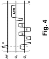
FIGURES 2-4 diagrammatically show some suitable LOBBI pulse sequences. -
FIGURE 5 diagrammatically shows a block diagram of an interleaved black/bright imaging (IBBI) sequence. -
FIGURE 6 diagrammatically shows a block diagram of an IBBI sequence employing a LOBBI black blood sequence as the black blood modules. -
FIGURE 7 shows a flow phantom. -
FIGURE 8 shows IBBI imaging experiments employing LOBBI black blood modules performed on the flow phantom ofFIGURE 7 . -
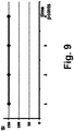
FIGURE 9 plots signal intensity (SI) of the bright blood images ofFIGURE 8 . - Disclosed herein is a black blood technique referred to herein as local excitation Black Blood Imaging (LOBBI), which is capable of achieving effective blood suppression even if the RF pulses are applied in a local region. As further disclosed herein, by using LOBBI as the black blood module in interleaved black/bright imaging (IBBI), interference of the bright blood module by the black blood module is effectively overcome. This approach achieves sufficient blood suppression in a local region, avoids signal interference with the flowing bright blood images so as to allow accurate AIF measurement, and maintains high temporal resolution for DCE MRI. The LOBBI technique does not require global radio frequency (RF) excitation, and allows for effective black blood imaging when local excitation Transmit/Receive (T/R) coil is used, providing black blood contrast for both blood originating from inside and outside of the imaging plane/slab.
- With reference to
FIGURE 1 , an illustrative magnetic resonance angiography (MRA) system is described, which employs local excitation black blood imaging (LOBBI) as the black blood module in interleaved black/bright imaging (IBBI). The MRA system includes anMR scanner 10 operated by a magnetic resonance (MR)control module 12 to retrieve from asequences storage 14 an IBBI sequence that employs a LOBBI sequence as the black blood module, and to execute the retrieved LOBBI sequence to perform black blood imaging of a subject (for example, a human subject, a veterinary subject, a clinical or pre-clinical test subject, or so forth). TheMRI scanner 10 can be any type of commercial or non-commercial MRI scanner, such as (by way of illustrative example) an Achieva™, Ingenia™, Intera™, or Panorama™ MRI scanner (available from Koninklijke Philips Electronics N.V., Eindhoven, The Netherlands). The performed sequence generates MR imaging data for a sequence of black blood and bright blood images that are suitably stored in an MRimaging data storage 16. An MRimage reconstruction module 18 applies a suitable image reconstruction algorithm to the MR imaging data generated by the LOBBI sequence module to generate one ormore MR images 20 having black blood contrast (that is, one or more black blood MRA images 20). Thereconstruction module 18 also applies a suitable image reconstruction algorithm to the MR imaging data generated by the bright blood sequence module of the IBBI sequence (which may, for example, be a time-of-flight bright blood imaging module) to generate one ormore MR images 22 having bright blood contrast (that is, one or more bright blood MRA images 22). The black andbright blood images MR images storage 24. The choice of image reconstruction algorithm depends upon the spatial encoding employed in the imaging data acquisition and may, for example, be a Fourier transform-based image reconstruction algorithm. - An image visualization/
analysis module 28 performs display and/or analysis of the black andbright blood images variation assessment module 30 processes theblack blood images 20 to assess or quantify tissue signal variations in a region proximate to the lumen, while an arterial input function (AIF)measurement sub-module 32 processes thebright blood images 22 to assess or quantify the AIF. As is known in the art, black blood images provide more accurate lumen definition compared with time-of-flight (TOF) based bright-blood MRA because TOF may be affected by blood velocity reduction proximate to blood vessel walls. On the other hand, bright blood images contain content for computing AIF. - The data processing and
control components data processing device 40, such as a suitably programmedillustrative computer 40, a network based server, or so forth, that includes or has operative access to adisplay device 42 via which thevisualization module 28 displays images and/or image analysis results. In some embodiments analog or mixed circuitry may also be included, e.g. parallel reconstruction pipeline hardware optionally used in theimage reconstruction module 18. TheMR control module 12 is optionally implemented as a separate dedicated MR control computer. Theimage visualization module 28 may be implemented as a dedicated image processing workstation with a high resolution display. - The disclosed MRA imaging techniques employing IBBI using the LOBBI sequence as the black blood module can also be embodied as a non-transitory storage medium (not shown) such as a hard disk or other magnetic storage medium, optical disk or other optical storage medium, random access memory (RAM), flash memory or other electronic storage medium, or so forth, which stores instructions that are executable by the electronic
data processing device 30 to perform the disclosed techniques. - With reference to
FIGURE 2 , an illustrative pulse diagram of a LOBBI sequence is shown. The following symbols are used inFIGURE 2 : the top plot labeled "RF pulse" diagrammatically shows applied radio frequency pulses; the bottom plot labeled "G" diagrammatically shows applied magnetic field gradients; the FSG gradient is a flow sensitization gradient for in-plane/slab flow sensitization; S is a spoiler gradient; the FSG-S gradient works as a rephrasing gradient for in-plane tissue/blood and a spoiler for blood originating from outside; ACQ is the acquisition gradients. The FSG-S gradient and the negative lobe of the ACQ gradient are shown as separate gradient pulses, but are optionally combined. The portion of the plots ofFIGURE 2 shown in braces {...} n is an acquisition module (ACQ module) that can be repeated n times where n≥1. - The pulse diagram of the LOBBI sequence shown in
FIGURE 2 suppresses blood signal originated from both in-plane and out-of-plane, and operates as follows. - For blood that originates from inside the plane, these blood particles experience both the FSG and FSG-S gradients. The phase encoding status of the spins will be stored after the tip-up 90° pulse. The combination of both gradients (bi-polar) sensitizes the moving particles and destroys the phase coherences, as in the motion-sensitized driven-equilibrium (MSDE) black blood sequence, resulting in blood suppression.
- For the static tissue (e.g., vessel wall) in the plane, the MR signal coming from these tissues will remain unsuppressed because their phase will be fully refocused by the bi-polar gradients.
- For blood originates from outside of the plane, the blood particles experience only the FSG-S gradient, which will then operate as a spoiler gradient and suppress all the blood excited by the α pulse. This will eliminate the in-flow effect which would otherwise cause blood flow artifacts.
- To avoid in-flow effect between the α pulses and the ACQ gradients, the time gap between the two is preferably made as short as possible. For example, in a typical carotid artery imaging application, the time gap between the two is suitably around 10ms. For this 10ms time gap and considering an illustrative blood velocity of 33cm/s, only the peripheral 3mm region around the coil sensitivity area will be affected by the in-flow effect. This is a significant improvement over the illustrative short MSDE sequence for multi-slice turbo field echo (TFE) black blood imaging using a local excitation coil, for which it is estimated that a 67mm region will contain artifacts.
- The LOBBI sequence can achieve black blood imaging with only local excitation, and so slice-selective RF pulses are optionally used. However, non-selective RF pulses achieve a larger black blood effect region.
- With reference to
FIGURE 3 , another suitable LOBBI pulse sequence is shown. The pulse sequence ofFIGURE 3 is a spin echo embodiment of the LOBBI sequence. In this embodiment a 180° pulse is optionally added in the LOBBI pre-pulse to correct for signal drop caused by the B0 field inhomogeneity (known as the "T2* effect"). Said another way, the 180° pulse corrects for the T2* decay which would otherwise cause undesired signal drop. In the sequence ofFIGURE 3 , the time gap between the three RF pulses should be the same. - In the LOBBI sequence shown in
FIGURE 3 , the FSG-S gradient (seeFIGURE 2 ) is optionally split into two or more gradients, namely gradients FSGA and FSGB shown inFIGURE 3 . The area of both gradients FSGA and FSGB should be the same as that of the original FSG-S gradient (seeFIGURE 2 ), and also the same as gradient FSG. The optional split of the gradient FSG-S into smaller component gradients allows for a smaller gradient (FSGB in the embodiment ofFIGURE 3 ) to be used in the ACQ module (which inFIGURE 3 is again marked off by braces {...} n and is suitably repeated n times where n≥1), which will shorten the duration between the α pulse and the ACQ gradient, consequently reducing the flow artifacts caused by in-flow. - With reference to
FIGURE 4 , a LOBBI sequence for local black blood suppression is shown. Unlike the traditional double-inversion-recovery (DIR) or motion-sensitized driven-equilibrium (MSDE) sequences, the LOBBI sequence does not rely on the global blood nulling to overcome the inflow effect. Rather, the LOBBI sequence only suppresses blood after the signal is excited by the RF pulse. Blood outside of the field of view (FOV) is left intact, preventing the following any interference with the bright blood images. - As disclosed herein, the LOBBI black blood imaging technique is suitably employed as the black blood module for interleaved black/bright imaging (IBBI). By using LOBBI as the black blood module in interleaved black/bright imaging (IBBI), interference of the bright blood module by the black blood module is effectively overcome.
- With reference to
FIGURE 5 , achieving high quality IBBI has been difficult using existing black blood imaging techniques because the black blood imaging pulses employ spatially non-selective RF pulses that tend to interfere with the bright blood imaging module, leading to inaccurate AIF quantification. For example, the typical time gap Δt between the black blood preparation module and the following bright blood module (seeFIGURE 5 ) is usually less than 200ms, which is not long enough for the blood to return to its equilibrium state (well over 1000ms at 3T). - In the IBBI dynamic contrast enhanced (DCE) MRI approach, the black blood and bright blood modules should be placed as close as possible to improve the temporal efficiency. A problem with this is that existing black blood imaging techniques rely upon spatially non-selective RF pulses for sufficient blood suppression. This is because using spatially selective pulses in existing black blood techniques would fail to suppress signal from blood flowing into the slice from outside. However, the spatially non-selective RF pulses of the black blood modules produces a residual black blood effect that tends to interfere with the following bright blood images.
- One possible solution is to utilize spatially selective RF pulses for the black blood imaging, so as to reduce signal interference between the black and bright blood images. Spatially selective RF pulses, however, will likely lead to insufficient black blood suppression due to the inflow effect (that is, due to unsuppressed blood flowing into the slice).
- The LOBBI black blood technique disclosed hererin is capable of achieving effective blood suppression even if the RF pulses are applied in a local region. Thus, using a LOBBI sequence as the black blood module in IBBI achieves sufficient blood suppression in a local region while avoiding signal interference with the flowing bright blood images so as to allow accurate AIF measurement, and maintains high temporal resolution for DCE MRI.
- With reference to
FIGURE 6 , an IBBI sequence employing LOBBI as the black blood module providing localized black blood suppression is diagrammatically shown. In the illustrative IBBI sequence ofFIGURE 6 , time-of-flight (TOF) sequences 50 (i.e., bright blood modules 50) are used to achieve bright blood contrast, while LOBBI sequences 52 (i.e., LOBBI black blood modules 52) are used to achieve black blood contrast.Multiple TOF modules 50 can be placed betweenLOBBI modules 52 to improve the temporal resolution of the AIF acquisition. Another advantage of the disclosed IBBI sequences employing LOBBIblack blood modules 52 is that coverage and location of the TOF andLOBBI sequences - With reference to
FIGURES 7 and 8 , a prototype of the LOBBI-based IBBI technique has been implemented on a flow phantom illustrated inFIGURE 7 . The flow phantom and a static phantom are used in this setup to demonstrate the interleaved black and bright blood imaging scheme (seeFIGURE 8 , where the flow signal is seen to change its brightness among images). After the application of the IBBI sequence, the signal in the flow phantom alternates between the bright and black contrasts, demonstrating effective interleaving of the black and bright blood signal. No bright blood signal variation is visually detectable in the images ofFIGURE 8 before and after the application of the black blood imaging modules. - With reference to
FIGURE 9 , the signal intensity (SI) of all bright blood images were quantitatively measured.FIGURE 9 plots the signal measured on bright blood images. No variation is found before and after the application of black blood imaging modules, demonstrating the effectiveness of LOBBI based IBBI scheme. - While the illustrative embodiments employ TOF bright blood imaging, other types of T1 weighted (for AIF measurement) techniques may be interleaved with the LOBBI black blood sequences, such as Inversion Recovery-Turbo Field Echo (IR-TFE) or T1-weighted Turbo Spin Echo (T1-TSE) sequences.
- The invention has been described with reference to the preferred embodiments. Obviously, modifications and alterations will occur to others upon reading and understanding the preceding detailed description. It is intended that the invention be construed as including all such modifications and alterations insofar as they come within the scope of the appended claims.
Claims (9)
- A method comprising- performing interleaved black/bright imaging = IBBI using a magnetic resonance scanner (10), wherein at least one interposed repetition of a bright blood module (50) is performed between successive repetitions of performing a local oscillation black blood imaging = LOBBI module (52);wherein the LOBBI module (52) of the IBBI includes:- applying a first flow sensitization gradient (FSG) for in-plane flow sensitization with a 90° radio frequency pulse preceding the first flow sensitization gradient and a -90° tip-up radio frequency pulse following the first flow sensitization gradient,- applying a spoiler gradient (S) after applying the first flow sensitization gradient and after applying the -90° tip-up radio frequency pulse,- applying a second flow sensitization gradient (FSG-S) for in-plane flow sensitization after applying the spoiler gradient wherein the second flow sensitization gradient's area is equal to the first flow sensitization gradient but of opposite polarity,- applying a slice-selective radio frequency excitation pulse before applying the second flow sensitization gradient for in-plane flow sensitization and after applying the spoiler gradient, and- performing a magnetic resonance readout after applying the second flow sensitization gradient and after applying the slice-selective radio frequency excitation wherein the readout acquires magnetic resonance imaging data having blood signal suppression in the region excited by the slice-selective radio frequency excitation pulse.
- A method comprising:- performing interleaved black/bright imaging = IBBI using a magnetic resonance scanner (10), wherein at least one interposed repetition of a bright blood module (50) is performed between successive repetitions of performing a local oscillation black blood imaging = LOBBI module (52);wherein the LOBBI module (52) of the IBBI includes the following sequence of consecutive steps:- applying a first flow sensitization gradient (FSG) for in-plane flow sensitization with a 90° radio frequency pulse preceding the first flow sensitization gradient,- applying an 180° refocusing radio frequency pulse,- applying a first portion (FSGA) of a second flow sensitization gradient for in-plane flow sensitization- applying a -90° tip-up radio frequency pulse,- applying a spoiler gradient (S),- applying a slice-selective radio frequency excitation pulse after applying the spoiler gradient,- applying a second portion (FSGB) of the second flow sensitization gradient gradient for in-plane flow sensitization,- wherein the first and second portions of the second flow sensitization gradient have a combined area equal to the area of the first flow sensitization gradient and the first and second portions of the second flow sensitization gradient and the first flow sensitization gradient are of equal polarity,- the time gap between the 90° radio frequency pulse and the 180° refocusing radio frequency is equal to the time gap between the 180° refocusing radio frequency pulse and the -90° tip-up radio frequency pulse and- performing a magnetic resonance readout after applying the second portion (FSGB) of the second flow sensitization gradient, wherein the readout acquires magnetic resonance imaging data having blood signal suppression in the region excited by the slice-selective radio frequency excitation pulse.
- The method of claim 1, wherein the bright blood module (50) of the IBBI includes a time of flight (TOF) angiography sequence.
- The method of any one of claims 1 to 3, wherein the time interval between applying the slice-selective radio frequency excitation pulse and performing the magnetic resonance readout is less than or about 10 ms.
- The method of any one of claims 1 to 4, further comprising:- reconstructing the magnetic resonance imaging data having blood signal suppression in the region excited by the slice-selective radio frequency excitation pulse to generate black blood images (20); and- reconstructing magnetic resonance imaging data generated by bright blood modules (50) of the IBBI to generate bright blood images (22).
- The method of claim 5, further comprising:
computing an arterial input function (AIF) based on the bright blood images (22). - The method of any one of claims 5 or 6 further comprising:
generating a tissue signal variation assessment from the black blood images (20). - A non-transitory storage medium storing instructions executable by an electronic data processing device (40) operating in conjunction with a magnetic resonance scanner (10) controlled by the electronic data processing device to perform a method as set forth in any one of claims 1-7.
- An apparatus comprising:- a magnetic resonance scanner (10); and- an electronic data processing device (40) programmed to perform a method as set forth in any one of claims 1-7.
Applications Claiming Priority (2)
| Application Number | Priority Date | Filing Date | Title |
|---|---|---|---|
| US201161477854P | 2011-04-21 | 2011-04-21 | |
| PCT/IB2012/051779 WO2012143824A1 (en) | 2011-04-21 | 2012-04-12 | Interleaved black and bright blood dynamic contrast enhanced (dce) mri |
Publications (2)
| Publication Number | Publication Date |
|---|---|
| EP2699929A1 EP2699929A1 (en) | 2014-02-26 |
| EP2699929B1 true EP2699929B1 (en) | 2019-12-11 |
Family
ID=46147530
Family Applications (1)
| Application Number | Title | Priority Date | Filing Date |
|---|---|---|---|
| EP12722857.5A Active EP2699929B1 (en) | 2011-04-21 | 2012-04-12 | Interleaved black and bright blood mri |
Country Status (5)
| Country | Link |
|---|---|
| US (1) | US9529065B2 (en) |
| EP (1) | EP2699929B1 (en) |
| JP (1) | JP6049694B2 (en) |
| CN (1) | CN103635825B (en) |
| WO (1) | WO2012143824A1 (en) |
Families Citing this family (6)
| Publication number | Priority date | Publication date | Assignee | Title |
|---|---|---|---|---|
| EP2932290B1 (en) | 2012-12-14 | 2020-10-14 | Koninklijke Philips N.V. | Interleaved black and bright blood imaging for interleaved dynamic contrast enhanced magnetic resonance imaging |
| CN111127586B (en) * | 2019-12-14 | 2021-10-29 | 深圳先进技术研究院 | Method and device for generating arterial input function curve |
| CN112075934B (en) * | 2020-09-09 | 2021-07-23 | 清华大学 | Magnetic Resonance Single Sequence Multiparameter Quantitative Imaging System for Identification of Carotid Plaque |
| CN112669439B (en) * | 2020-11-23 | 2024-03-19 | 西安电子科技大学 | Method for establishing an enhanced three-dimensional model of intracranial angiography based on transfer learning |
| CN112634197A (en) * | 2020-11-23 | 2021-04-09 | 西安科锐盛创新科技有限公司 | Method for establishing intracranial angiography enhanced three-dimensional model |
| CN113222987B (en) * | 2021-06-04 | 2022-10-25 | 清华大学 | Magnetic resonance imaging vascular wall enhancement intensity mapping method, device, equipment and medium |
Family Cites Families (10)
| Publication number | Priority date | Publication date | Assignee | Title |
|---|---|---|---|---|
| JPH04220236A (en) * | 1990-12-19 | 1992-08-11 | Yokogawa Medical Syst Ltd | Mr imaging method |
| JP3343392B2 (en) * | 1993-04-16 | 2002-11-11 | ジーイー横河メディカルシステム株式会社 | MR angiography device |
| JP4693227B2 (en) * | 2000-11-14 | 2011-06-01 | 株式会社東芝 | Magnetic resonance imaging apparatus and scan synchronization method of magnetic resonance imaging |
| US6552542B1 (en) | 2001-09-28 | 2003-04-22 | The Board Of Trustees Of The Leland Stanford Junior University | Oscillating dual-equilibrium steady state angiography using magnetic resonance imaging |
| US10098563B2 (en) * | 2006-11-22 | 2018-10-16 | Toshiba Medical Systems Corporation | Magnetic resonance imaging apparatus |
| JP4936864B2 (en) * | 2006-11-22 | 2012-05-23 | 株式会社東芝 | Magnetic resonance imaging system |
| JP5395332B2 (en) * | 2007-04-27 | 2014-01-22 | 株式会社東芝 | Magnetic resonance imaging system |
| US8030919B2 (en) * | 2008-04-18 | 2011-10-04 | Case Western Reserve University | Dark blood balanced steady state free precession imaging |
| JP5311333B2 (en) * | 2008-10-31 | 2013-10-09 | ジーイー・メディカル・システムズ・グローバル・テクノロジー・カンパニー・エルエルシー | Blood flow dynamic analysis apparatus and magnetic resonance imaging apparatus |
| US8378680B2 (en) * | 2010-01-28 | 2013-02-19 | Siemens Medical Solutions Usa, Inc. | System for concurrent acquisition of MR anatomical brain images and MR angiograms without contrast-injection |
-
2012
- 2012-04-12 EP EP12722857.5A patent/EP2699929B1/en active Active
- 2012-04-12 CN CN201280019366.2A patent/CN103635825B/en not_active Expired - Fee Related
- 2012-04-12 JP JP2014505751A patent/JP6049694B2/en not_active Expired - Fee Related
- 2012-04-12 US US14/112,135 patent/US9529065B2/en not_active Expired - Fee Related
- 2012-04-12 WO PCT/IB2012/051779 patent/WO2012143824A1/en not_active Ceased
Non-Patent Citations (1)
| Title |
|---|
| None * |
Also Published As
| Publication number | Publication date |
|---|---|
| JP6049694B2 (en) | 2016-12-21 |
| CN103635825B (en) | 2016-03-30 |
| EP2699929A1 (en) | 2014-02-26 |
| WO2012143824A1 (en) | 2012-10-26 |
| US20140035581A1 (en) | 2014-02-06 |
| JP2014514112A (en) | 2014-06-19 |
| CN103635825A (en) | 2014-03-12 |
| US9529065B2 (en) | 2016-12-27 |
Similar Documents
| Publication | Publication Date | Title |
|---|---|---|
| Frost et al. | Scan time reduction for readout‐segmented EPI using simultaneous multislice acceleration: Diffusion‐weighted imaging at 3 and 7 Tesla | |
| US10302729B2 (en) | Method and magnetic resonance apparatus for speed-compensated diffusion-based diffusion imaging | |
| Koktzoglou et al. | Radial fast interrupted steady‐state (FISS) magnetic resonance imaging | |
| US9529065B2 (en) | Interleaved black and bright blood dynamic contrast enhanced (DCE) MRI | |
| US20170160365A1 (en) | Mri involving the acquisition of an angiography weighted image and of a perfusion weighted image | |
| US10247798B2 (en) | Simultaneous multi-slice MRI measurement | |
| US8704518B2 (en) | System and method of high signal-to-noise ratio magnetic resonance imaging screening | |
| US7609059B2 (en) | Magnetic resonance method and apparatus with nuclear spins type-specific signal suppression | |
| Koktzoglou et al. | Nonenhanced arterial spin labeled carotid MR angiography using three‐dimensional radial balanced steady‐state free precession imaging | |
| Zhang et al. | Segmented TOF at 7 T MRI: technique and clinical applications | |
| US10012715B2 (en) | Method and apparatus for recording a magnetic resonance data set | |
| JP6529807B2 (en) | Magnetic resonance imaging apparatus, method and program | |
| Yun et al. | Submillimeter fMRI acquisition techniques for detection of laminar and columnar level brain activation | |
| JP2002224083A (en) | Magnetic resonance imaging diagnostic apparatus | |
| US9709644B2 (en) | Black blood MRI using a stimulated echo pulse sequence with flow sensitization gradients | |
| JP2020018695A (en) | Magnetic resonance imaging equipment | |
| Pracht et al. | SAR and scan‐time optimized 3D whole‐brain double inversion recovery imaging at 7T | |
| Raynaud et al. | ISME—incoherent sampling of multi‐echo data to minimize cardiac‐induced noise in brain maps of R 2* R _2^ ∗ and magnetic susceptibility | |
| EP2932290B1 (en) | Interleaved black and bright blood imaging for interleaved dynamic contrast enhanced magnetic resonance imaging | |
| US20120146638A1 (en) | System and method for reducing localized signal fluctuation | |
| Mooiweer et al. | Fast 3D isotropic imaging of the aortic vessel wall by application of 2D spatially selective excitation and a new way of inversion recovery for black blood imaging | |
| JP2005111059A (en) | Magnetic resonance imaging apparatus | |
| Wink et al. | 4D flow imaging with 2D‐selective excitation | |
| Yoneyama et al. | Hyperecho PROPELLER‐MRI: Application to rapid high‐resolution motion‐insensitive T2‐weighted black‐blood imaging of the carotid arterial vessel wall and plaque | |
| Yuan et al. | A comparison of black-blood T2 mapping sequences for carotid vessel wall imaging at 3T: An assessment of accuracy and repeatability |
Legal Events
| Date | Code | Title | Description |
|---|---|---|---|
| PUAI | Public reference made under article 153(3) epc to a published international application that has entered the european phase |
Free format text: ORIGINAL CODE: 0009012 |
|
| 17P | Request for examination filed |
Effective date: 20131121 |
|
| AK | Designated contracting states |
Kind code of ref document: A1 Designated state(s): AL AT BE BG CH CY CZ DE DK EE ES FI FR GB GR HR HU IE IS IT LI LT LU LV MC MK MT NL NO PL PT RO RS SE SI SK SM TR |
|
| DAX | Request for extension of the european patent (deleted) | ||
| 17Q | First examination report despatched |
Effective date: 20141008 |
|
| GRAP | Despatch of communication of intention to grant a patent |
Free format text: ORIGINAL CODE: EPIDOSNIGR1 |
|
| STAA | Information on the status of an ep patent application or granted ep patent |
Free format text: STATUS: GRANT OF PATENT IS INTENDED |
|
| INTG | Intention to grant announced |
Effective date: 20190705 |
|
| GRAS | Grant fee paid |
Free format text: ORIGINAL CODE: EPIDOSNIGR3 |
|
| GRAA | (expected) grant |
Free format text: ORIGINAL CODE: 0009210 |
|
| STAA | Information on the status of an ep patent application or granted ep patent |
Free format text: STATUS: THE PATENT HAS BEEN GRANTED |
|
| AK | Designated contracting states |
Kind code of ref document: B1 Designated state(s): AL AT BE BG CH CY CZ DE DK EE ES FI FR GB GR HR HU IE IS IT LI LT LU LV MC MK MT NL NO PL PT RO RS SE SI SK SM TR |
|
| REG | Reference to a national code |
Ref country code: GB Ref legal event code: FG4D |
|
| REG | Reference to a national code |
Ref country code: CH Ref legal event code: EP |
|
| REG | Reference to a national code |
Ref country code: AT Ref legal event code: REF Ref document number: 1212735 Country of ref document: AT Kind code of ref document: T Effective date: 20191215 |
|
| REG | Reference to a national code |
Ref country code: DE Ref legal event code: R096 Ref document number: 602012066375 Country of ref document: DE |
|
| REG | Reference to a national code |
Ref country code: IE Ref legal event code: FG4D |
|
| REG | Reference to a national code |
Ref country code: DE Ref legal event code: R084 Ref document number: 602012066375 Country of ref document: DE |
|
| RAP2 | Party data changed (patent owner data changed or rights of a patent transferred) |
Owner name: KONINKLIJKE PHILIPS N.V. Owner name: PHILIPS INTELLECTUAL PROPERTY & STANDARDS GMBH Owner name: THE UNIVERSITY OF WASHINGTON |
|
| REG | Reference to a national code |
Ref country code: NL Ref legal event code: MP Effective date: 20191211 |
|
| REG | Reference to a national code |
Ref country code: LT Ref legal event code: MG4D |
|
| PG25 | Lapsed in a contracting state [announced via postgrant information from national office to epo] |
Ref country code: NO Free format text: LAPSE BECAUSE OF FAILURE TO SUBMIT A TRANSLATION OF THE DESCRIPTION OR TO PAY THE FEE WITHIN THE PRESCRIBED TIME-LIMIT Effective date: 20200311 Ref country code: BG Free format text: LAPSE BECAUSE OF FAILURE TO SUBMIT A TRANSLATION OF THE DESCRIPTION OR TO PAY THE FEE WITHIN THE PRESCRIBED TIME-LIMIT Effective date: 20200311 Ref country code: GR Free format text: LAPSE BECAUSE OF FAILURE TO SUBMIT A TRANSLATION OF THE DESCRIPTION OR TO PAY THE FEE WITHIN THE PRESCRIBED TIME-LIMIT Effective date: 20200312 Ref country code: LT Free format text: LAPSE BECAUSE OF FAILURE TO SUBMIT A TRANSLATION OF THE DESCRIPTION OR TO PAY THE FEE WITHIN THE PRESCRIBED TIME-LIMIT Effective date: 20191211 Ref country code: ES Free format text: LAPSE BECAUSE OF FAILURE TO SUBMIT A TRANSLATION OF THE DESCRIPTION OR TO PAY THE FEE WITHIN THE PRESCRIBED TIME-LIMIT Effective date: 20191211 Ref country code: LV Free format text: LAPSE BECAUSE OF FAILURE TO SUBMIT A TRANSLATION OF THE DESCRIPTION OR TO PAY THE FEE WITHIN THE PRESCRIBED TIME-LIMIT Effective date: 20191211 Ref country code: SE Free format text: LAPSE BECAUSE OF FAILURE TO SUBMIT A TRANSLATION OF THE DESCRIPTION OR TO PAY THE FEE WITHIN THE PRESCRIBED TIME-LIMIT Effective date: 20191211 Ref country code: FI Free format text: LAPSE BECAUSE OF FAILURE TO SUBMIT A TRANSLATION OF THE DESCRIPTION OR TO PAY THE FEE WITHIN THE PRESCRIBED TIME-LIMIT Effective date: 20191211 |
|
| PG25 | Lapsed in a contracting state [announced via postgrant information from national office to epo] |
Ref country code: RS Free format text: LAPSE BECAUSE OF FAILURE TO SUBMIT A TRANSLATION OF THE DESCRIPTION OR TO PAY THE FEE WITHIN THE PRESCRIBED TIME-LIMIT Effective date: 20191211 Ref country code: HR Free format text: LAPSE BECAUSE OF FAILURE TO SUBMIT A TRANSLATION OF THE DESCRIPTION OR TO PAY THE FEE WITHIN THE PRESCRIBED TIME-LIMIT Effective date: 20191211 |
|
| PG25 | Lapsed in a contracting state [announced via postgrant information from national office to epo] |
Ref country code: AL Free format text: LAPSE BECAUSE OF FAILURE TO SUBMIT A TRANSLATION OF THE DESCRIPTION OR TO PAY THE FEE WITHIN THE PRESCRIBED TIME-LIMIT Effective date: 20191211 |
|
| PG25 | Lapsed in a contracting state [announced via postgrant information from national office to epo] |
Ref country code: PT Free format text: LAPSE BECAUSE OF FAILURE TO SUBMIT A TRANSLATION OF THE DESCRIPTION OR TO PAY THE FEE WITHIN THE PRESCRIBED TIME-LIMIT Effective date: 20200506 Ref country code: EE Free format text: LAPSE BECAUSE OF FAILURE TO SUBMIT A TRANSLATION OF THE DESCRIPTION OR TO PAY THE FEE WITHIN THE PRESCRIBED TIME-LIMIT Effective date: 20191211 Ref country code: RO Free format text: LAPSE BECAUSE OF FAILURE TO SUBMIT A TRANSLATION OF THE DESCRIPTION OR TO PAY THE FEE WITHIN THE PRESCRIBED TIME-LIMIT Effective date: 20191211 Ref country code: NL Free format text: LAPSE BECAUSE OF FAILURE TO SUBMIT A TRANSLATION OF THE DESCRIPTION OR TO PAY THE FEE WITHIN THE PRESCRIBED TIME-LIMIT Effective date: 20191211 Ref country code: CZ Free format text: LAPSE BECAUSE OF FAILURE TO SUBMIT A TRANSLATION OF THE DESCRIPTION OR TO PAY THE FEE WITHIN THE PRESCRIBED TIME-LIMIT Effective date: 20191211 |
|
| PG25 | Lapsed in a contracting state [announced via postgrant information from national office to epo] |
Ref country code: IS Free format text: LAPSE BECAUSE OF FAILURE TO SUBMIT A TRANSLATION OF THE DESCRIPTION OR TO PAY THE FEE WITHIN THE PRESCRIBED TIME-LIMIT Effective date: 20200411 Ref country code: SK Free format text: LAPSE BECAUSE OF FAILURE TO SUBMIT A TRANSLATION OF THE DESCRIPTION OR TO PAY THE FEE WITHIN THE PRESCRIBED TIME-LIMIT Effective date: 20191211 Ref country code: SM Free format text: LAPSE BECAUSE OF FAILURE TO SUBMIT A TRANSLATION OF THE DESCRIPTION OR TO PAY THE FEE WITHIN THE PRESCRIBED TIME-LIMIT Effective date: 20191211 |
|
| REG | Reference to a national code |
Ref country code: DE Ref legal event code: R097 Ref document number: 602012066375 Country of ref document: DE |
|
| REG | Reference to a national code |
Ref country code: AT Ref legal event code: MK05 Ref document number: 1212735 Country of ref document: AT Kind code of ref document: T Effective date: 20191211 |
|
| PLBE | No opposition filed within time limit |
Free format text: ORIGINAL CODE: 0009261 |
|
| STAA | Information on the status of an ep patent application or granted ep patent |
Free format text: STATUS: NO OPPOSITION FILED WITHIN TIME LIMIT |
|
| PG25 | Lapsed in a contracting state [announced via postgrant information from national office to epo] |
Ref country code: DK Free format text: LAPSE BECAUSE OF FAILURE TO SUBMIT A TRANSLATION OF THE DESCRIPTION OR TO PAY THE FEE WITHIN THE PRESCRIBED TIME-LIMIT Effective date: 20191211 |
|
| 26N | No opposition filed |
Effective date: 20200914 |
|
| PG25 | Lapsed in a contracting state [announced via postgrant information from national office to epo] |
Ref country code: PL Free format text: LAPSE BECAUSE OF FAILURE TO SUBMIT A TRANSLATION OF THE DESCRIPTION OR TO PAY THE FEE WITHIN THE PRESCRIBED TIME-LIMIT Effective date: 20191211 Ref country code: MC Free format text: LAPSE BECAUSE OF FAILURE TO SUBMIT A TRANSLATION OF THE DESCRIPTION OR TO PAY THE FEE WITHIN THE PRESCRIBED TIME-LIMIT Effective date: 20191211 Ref country code: SI Free format text: LAPSE BECAUSE OF FAILURE TO SUBMIT A TRANSLATION OF THE DESCRIPTION OR TO PAY THE FEE WITHIN THE PRESCRIBED TIME-LIMIT Effective date: 20191211 Ref country code: AT Free format text: LAPSE BECAUSE OF FAILURE TO SUBMIT A TRANSLATION OF THE DESCRIPTION OR TO PAY THE FEE WITHIN THE PRESCRIBED TIME-LIMIT Effective date: 20191211 |
|
| REG | Reference to a national code |
Ref country code: CH Ref legal event code: PL |
|
| REG | Reference to a national code |
Ref country code: DE Ref legal event code: R081 Ref document number: 602012066375 Country of ref document: DE Owner name: PHILIPS GMBH, DE Free format text: FORMER OWNER: PHILIPS INTELLECTUAL PROPERTY & STANDARDS GMBH, 20099 HAMBURG, DE |
|
| PG25 | Lapsed in a contracting state [announced via postgrant information from national office to epo] |
Ref country code: LI Free format text: LAPSE BECAUSE OF NON-PAYMENT OF DUE FEES Effective date: 20200430 Ref country code: FR Free format text: LAPSE BECAUSE OF NON-PAYMENT OF DUE FEES Effective date: 20200430 Ref country code: CH Free format text: LAPSE BECAUSE OF NON-PAYMENT OF DUE FEES Effective date: 20200430 Ref country code: IT Free format text: LAPSE BECAUSE OF FAILURE TO SUBMIT A TRANSLATION OF THE DESCRIPTION OR TO PAY THE FEE WITHIN THE PRESCRIBED TIME-LIMIT Effective date: 20191211 Ref country code: LU Free format text: LAPSE BECAUSE OF NON-PAYMENT OF DUE FEES Effective date: 20200412 |
|
| REG | Reference to a national code |
Ref country code: BE Ref legal event code: MM Effective date: 20200430 |
|
| PG25 | Lapsed in a contracting state [announced via postgrant information from national office to epo] |
Ref country code: BE Free format text: LAPSE BECAUSE OF NON-PAYMENT OF DUE FEES Effective date: 20200430 |
|
| GBPC | Gb: european patent ceased through non-payment of renewal fee |
Effective date: 20200412 |
|
| PG25 | Lapsed in a contracting state [announced via postgrant information from national office to epo] |
Ref country code: IE Free format text: LAPSE BECAUSE OF NON-PAYMENT OF DUE FEES Effective date: 20200412 Ref country code: GB Free format text: LAPSE BECAUSE OF NON-PAYMENT OF DUE FEES Effective date: 20200412 |
|
| PG25 | Lapsed in a contracting state [announced via postgrant information from national office to epo] |
Ref country code: TR Free format text: LAPSE BECAUSE OF FAILURE TO SUBMIT A TRANSLATION OF THE DESCRIPTION OR TO PAY THE FEE WITHIN THE PRESCRIBED TIME-LIMIT Effective date: 20191211 Ref country code: MT Free format text: LAPSE BECAUSE OF FAILURE TO SUBMIT A TRANSLATION OF THE DESCRIPTION OR TO PAY THE FEE WITHIN THE PRESCRIBED TIME-LIMIT Effective date: 20191211 Ref country code: CY Free format text: LAPSE BECAUSE OF FAILURE TO SUBMIT A TRANSLATION OF THE DESCRIPTION OR TO PAY THE FEE WITHIN THE PRESCRIBED TIME-LIMIT Effective date: 20191211 |
|
| PG25 | Lapsed in a contracting state [announced via postgrant information from national office to epo] |
Ref country code: MK Free format text: LAPSE BECAUSE OF FAILURE TO SUBMIT A TRANSLATION OF THE DESCRIPTION OR TO PAY THE FEE WITHIN THE PRESCRIBED TIME-LIMIT Effective date: 20191211 |
|
| PGFP | Annual fee paid to national office [announced via postgrant information from national office to epo] |
Ref country code: DE Payment date: 20220628 Year of fee payment: 12 |
|
| REG | Reference to a national code |
Ref country code: DE Ref legal event code: R119 Ref document number: 602012066375 Country of ref document: DE |
|
| PG25 | Lapsed in a contracting state [announced via postgrant information from national office to epo] |
Ref country code: DE Free format text: LAPSE BECAUSE OF NON-PAYMENT OF DUE FEES Effective date: 20241105 |
|
| PG25 | Lapsed in a contracting state [announced via postgrant information from national office to epo] |
Ref country code: DE Free format text: LAPSE BECAUSE OF NON-PAYMENT OF DUE FEES Effective date: 20241105 |