EP2698365B1 - Synthesis and analysis of novel compound capable of inducing differentiation of human mesenchymal stem cell into hepatocyte - Google Patents
Synthesis and analysis of novel compound capable of inducing differentiation of human mesenchymal stem cell into hepatocyte Download PDFInfo
- Publication number
- EP2698365B1 EP2698365B1 EP12771389.9A EP12771389A EP2698365B1 EP 2698365 B1 EP2698365 B1 EP 2698365B1 EP 12771389 A EP12771389 A EP 12771389A EP 2698365 B1 EP2698365 B1 EP 2698365B1
- Authority
- EP
- European Patent Office
- Prior art keywords
- cells
- mesenchymal stem
- compound
- cell sheet
- liver
- Prior art date
- Legal status (The legal status is an assumption and is not a legal conclusion. Google has not performed a legal analysis and makes no representation as to the accuracy of the status listed.)
- Active
Links
- MCZGRJSNQODZOC-HAQZEJJUSA-N C/C(/OC1=CC(C(N(C)/N=C/c2ccc(C)[o]2)=O)=CCC1)=C\C=C/C=C Chemical compound C/C(/OC1=CC(C(N(C)/N=C/c2ccc(C)[o]2)=O)=CCC1)=C\C=C/C=C MCZGRJSNQODZOC-HAQZEJJUSA-N 0.000 description 1
Images











Classifications
-
- A—HUMAN NECESSITIES
- A61—MEDICAL OR VETERINARY SCIENCE; HYGIENE
- A61K—PREPARATIONS FOR MEDICAL, DENTAL OR TOILETRY PURPOSES
- A61K35/00—Medicinal preparations containing materials or reaction products thereof with undetermined constitution
- A61K35/12—Materials from mammals; Compositions comprising non-specified tissues or cells; Compositions comprising non-embryonic stem cells; Genetically modified cells
- A61K35/37—Digestive system
- A61K35/407—Liver; Hepatocytes
-
- A—HUMAN NECESSITIES
- A61—MEDICAL OR VETERINARY SCIENCE; HYGIENE
- A61L—METHODS OR APPARATUS FOR STERILISING MATERIALS OR OBJECTS IN GENERAL; DISINFECTION, STERILISATION OR DEODORISATION OF AIR; CHEMICAL ASPECTS OF BANDAGES, DRESSINGS, ABSORBENT PADS OR SURGICAL ARTICLES; MATERIALS FOR BANDAGES, DRESSINGS, ABSORBENT PADS OR SURGICAL ARTICLES
- A61L27/00—Materials for grafts or prostheses or for coating grafts or prostheses
- A61L27/36—Materials for grafts or prostheses or for coating grafts or prostheses containing ingredients of undetermined constitution or reaction products thereof, e.g. transplant tissue, natural bone, extracellular matrix
- A61L27/38—Materials for grafts or prostheses or for coating grafts or prostheses containing ingredients of undetermined constitution or reaction products thereof, e.g. transplant tissue, natural bone, extracellular matrix containing added animal cells
- A61L27/3804—Materials for grafts or prostheses or for coating grafts or prostheses containing ingredients of undetermined constitution or reaction products thereof, e.g. transplant tissue, natural bone, extracellular matrix containing added animal cells characterised by specific cells or progenitors thereof, e.g. fibroblasts, connective tissue cells, kidney cells
-
- A—HUMAN NECESSITIES
- A61—MEDICAL OR VETERINARY SCIENCE; HYGIENE
- A61L—METHODS OR APPARATUS FOR STERILISING MATERIALS OR OBJECTS IN GENERAL; DISINFECTION, STERILISATION OR DEODORISATION OF AIR; CHEMICAL ASPECTS OF BANDAGES, DRESSINGS, ABSORBENT PADS OR SURGICAL ARTICLES; MATERIALS FOR BANDAGES, DRESSINGS, ABSORBENT PADS OR SURGICAL ARTICLES
- A61L27/00—Materials for grafts or prostheses or for coating grafts or prostheses
- A61L27/36—Materials for grafts or prostheses or for coating grafts or prostheses containing ingredients of undetermined constitution or reaction products thereof, e.g. transplant tissue, natural bone, extracellular matrix
- A61L27/38—Materials for grafts or prostheses or for coating grafts or prostheses containing ingredients of undetermined constitution or reaction products thereof, e.g. transplant tissue, natural bone, extracellular matrix containing added animal cells
- A61L27/3804—Materials for grafts or prostheses or for coating grafts or prostheses containing ingredients of undetermined constitution or reaction products thereof, e.g. transplant tissue, natural bone, extracellular matrix containing added animal cells characterised by specific cells or progenitors thereof, e.g. fibroblasts, connective tissue cells, kidney cells
- A61L27/3834—Cells able to produce different cell types, e.g. hematopoietic stem cells, mesenchymal stem cells, marrow stromal cells, embryonic stem cells
-
- A—HUMAN NECESSITIES
- A61—MEDICAL OR VETERINARY SCIENCE; HYGIENE
- A61P—SPECIFIC THERAPEUTIC ACTIVITY OF CHEMICAL COMPOUNDS OR MEDICINAL PREPARATIONS
- A61P1/00—Drugs for disorders of the alimentary tract or the digestive system
- A61P1/16—Drugs for disorders of the alimentary tract or the digestive system for liver or gallbladder disorders, e.g. hepatoprotective agents, cholagogues, litholytics
-
- A—HUMAN NECESSITIES
- A61—MEDICAL OR VETERINARY SCIENCE; HYGIENE
- A61P—SPECIFIC THERAPEUTIC ACTIVITY OF CHEMICAL COMPOUNDS OR MEDICINAL PREPARATIONS
- A61P43/00—Drugs for specific purposes, not provided for in groups A61P1/00-A61P41/00
-
- C—CHEMISTRY; METALLURGY
- C07—ORGANIC CHEMISTRY
- C07C—ACYCLIC OR CARBOCYCLIC COMPOUNDS
- C07C251/00—Compounds containing nitrogen atoms doubly-bound to a carbon skeleton
- C07C251/72—Hydrazones
- C07C251/86—Hydrazones having doubly-bound carbon atoms of hydrazone groups bound to carbon atoms of six-membered aromatic rings
-
- C—CHEMISTRY; METALLURGY
- C07—ORGANIC CHEMISTRY
- C07C—ACYCLIC OR CARBOCYCLIC COMPOUNDS
- C07C43/00—Ethers; Compounds having groups, groups or groups
- C07C43/02—Ethers
- C07C43/20—Ethers having an ether-oxygen atom bound to a carbon atom of a six-membered aromatic ring
- C07C43/225—Ethers having an ether-oxygen atom bound to a carbon atom of a six-membered aromatic ring containing halogen
-
- C—CHEMISTRY; METALLURGY
- C07—ORGANIC CHEMISTRY
- C07D—HETEROCYCLIC COMPOUNDS
- C07D307/00—Heterocyclic compounds containing five-membered rings having one oxygen atom as the only ring hetero atom
- C07D307/02—Heterocyclic compounds containing five-membered rings having one oxygen atom as the only ring hetero atom not condensed with other rings
- C07D307/34—Heterocyclic compounds containing five-membered rings having one oxygen atom as the only ring hetero atom not condensed with other rings having two or three double bonds between ring members or between ring members and non-ring members
- C07D307/38—Heterocyclic compounds containing five-membered rings having one oxygen atom as the only ring hetero atom not condensed with other rings having two or three double bonds between ring members or between ring members and non-ring members with substituted hydrocarbon radicals attached to ring carbon atoms
- C07D307/52—Radicals substituted by nitrogen atoms not forming part of a nitro radical
-
- C—CHEMISTRY; METALLURGY
- C07—ORGANIC CHEMISTRY
- C07D—HETEROCYCLIC COMPOUNDS
- C07D487/00—Heterocyclic compounds containing nitrogen atoms as the only ring hetero atoms in the condensed system, not provided for by groups C07D451/00 - C07D477/00
- C07D487/02—Heterocyclic compounds containing nitrogen atoms as the only ring hetero atoms in the condensed system, not provided for by groups C07D451/00 - C07D477/00 in which the condensed system contains two hetero rings
- C07D487/04—Ortho-condensed systems
-
- A—HUMAN NECESSITIES
- A61—MEDICAL OR VETERINARY SCIENCE; HYGIENE
- A61L—METHODS OR APPARATUS FOR STERILISING MATERIALS OR OBJECTS IN GENERAL; DISINFECTION, STERILISATION OR DEODORISATION OF AIR; CHEMICAL ASPECTS OF BANDAGES, DRESSINGS, ABSORBENT PADS OR SURGICAL ARTICLES; MATERIALS FOR BANDAGES, DRESSINGS, ABSORBENT PADS OR SURGICAL ARTICLES
- A61L2430/00—Materials or treatment for tissue regeneration
- A61L2430/28—Materials or treatment for tissue regeneration for liver reconstruction
Definitions
- Non Patent Literatures 6 to 9 five low-molecular-weight compounds capable of inhibiting a Wnt/ ⁇ -catenin signaling pathway have been identified.
- Patent Literatures 1 and 2 describe proteins that induce hepatocytes from non-liver stem cells. Since a protein preparation has been used as a differentiation inducer, there is room for further improvement regarding aspects of stability and safety.
- another aspect of the present invention provides a method for producing hepatocytes from mesenchymal stem cells, comprising a step of treating mesenchymal stem cells in vitro with a compound represented by formula (1), a salt thereof, or a solvate of them. Also, another aspect of the present invention provides a production method comprising a step of treating mesenchymal stem cells in vitro with the above differentiation inducer.
- a method for producing a cell sheet for transplantation on a liver surface comprising an induction step of treating mesenchymal stem cells with a Wnt/ ⁇ -catenin signaling pathway inhibitor to induce the mesenchymal stem cells into hepatocytes.
- the cell sheet as prepared using this production method can exert an excellent effect of suppressing liver dysfunction when transplanted on a liver surface as demonstrated in the following Examples. Therefore, this production method is an excellent production method for producing a cell sheet for transplantation on a liver surface.
- halogen means F, Cl, Br, or I.
- aryl refers to a C 6-14 monocyclic, dicyclic, or tricyclic aromatic hydrocarbon ring.
- aryl include phenyl, naphthyl (e.g., 1-naphthyl, 2-naphthyl), tetrahydronaphthalenyl, indenyl, and phenyl that is substituted with five halogens.
- aryl includes a ring group that is condensed with C 5-8 cycloalkene at its double bond position.
- hepatocytes includes cells with a size of about 20 ⁇ m, the cells constituting 70 to 80% of a liver.
- Examples of general functions of hepatocytes include: synthesis and storage of proteins; conversion of carbohydrate; synthesis of cholesterol, bile acid, and phospholipids; and detoxification, denaturing, and excretion of endogenous and exogenous substances. Also, the examples further include promotion of generation and secretion of bile.
- the hepatocytes as so differentiated may be grown to fit a desired shape by using a temperature-responsive culture dish and/or a substrate such as a synthetic polymer scaffold using a biodegradable synthetic polymer such as polylactic acid (PLA) and polyethylene glycolic acid (PGA). Then, the resulting hepatocytes can be used as a liver tissue, liver, or cell sheet for regenerative medicine.
- the resulting 3D-structured liver tissue, liver, or cell sheet for regenerative medicine can be suitably used for transplantation therapeutics, such as hepatocyte transplantation, and application for drug development in evaluation of a drug's efficacy and adverse effect.
- a condensing agent HATU (0.76 g, 2.0 mmol) and diisopropylethylamine (DIEA)(0.35 mL, 2.0 mmol) were added to a DMF (7 mL) solution containing Fmoc-L-Tyr(t-Bu)-OH (0.87 g, 1.9 mmol). Next, the mixture was stirred for 20 min. Then, compound 1 (0.54 g, 2.0 mmol) was added, and the mixture was stirred at room temperature for 16 h. After the reaction, DMF was distilled away under reduced pressure.
- Hexachlorophene is a highly chlorinated phenolic compound used for an insect repellent, microbicide, disinfectant, etc.
- synthesized was a derivative, in which a phenolic hydroxyl group of hexachlorophene was capped with a methyl group.
- MeI (1.0 mL, 16 mmol) and K 2 CO 3 (5.3 g, 36 mmol) were added to an acetone solution (10 mL) containing hexachlorophene (TOKYO CHEMICAL INDUSTRY CO., LTD.)(417 mg, 1.0 mmol), and the mixture was stirred for 14 h. Next, excessive K 2 CO 3 was removed, and the mixture was dried over Na 2 SO 4 .
- the operating scissors or the similar surgical instrument was inserted under the skin from this incision and moved toward the rostral direction to go beyond the position where the device would be implanted. Then, the operating scissors were made open, which created a space where the device could be implanted under the skin of the mouse.
- the bFGF device was inserted into this space, and the incision was closed with a surgical clip or a suture.
- Tissue sections as prepared according to the procedure in FIG. 21 were used to perform immunofluorescence staining.
- Human ⁇ 1-antitrypsin was stained according to the following procedure.
- Tissue sections were subjected to deparaffinization using xylene and hydration treatment using 100% ethanol, were washed with tap water followed by washing with 1 x PBS(-), and were then blocked in a blocking solution containing goat serum at room temperature for 20 min.
- One or two drops of a rabbit polyclonal anti-al-antitrypsin antibody (Abcam) were added to the tissue sections. The sections were kept in a moist chamber at 4°C overnight to carry out a primary antibody reaction.
- Abcam rabbit polyclonal anti-al-antitrypsin antibody
- HBSS Hanks' balanced salt solution
- FBS fetal bovine serum
- mice were fed under normal conditions until the day of sacrifice.
Landscapes
- Health & Medical Sciences (AREA)
- Chemical & Material Sciences (AREA)
- Organic Chemistry (AREA)
- Life Sciences & Earth Sciences (AREA)
- Engineering & Computer Science (AREA)
- Biomedical Technology (AREA)
- Cell Biology (AREA)
- Medicinal Chemistry (AREA)
- Public Health (AREA)
- Veterinary Medicine (AREA)
- Animal Behavior & Ethology (AREA)
- General Health & Medical Sciences (AREA)
- Chemical Kinetics & Catalysis (AREA)
- Zoology (AREA)
- Epidemiology (AREA)
- Botany (AREA)
- Transplantation (AREA)
- Oral & Maxillofacial Surgery (AREA)
- Urology & Nephrology (AREA)
- Dermatology (AREA)
- Developmental Biology & Embryology (AREA)
- Pharmacology & Pharmacy (AREA)
- Hematology (AREA)
- Bioinformatics & Cheminformatics (AREA)
- General Chemical & Material Sciences (AREA)
- Nuclear Medicine, Radiotherapy & Molecular Imaging (AREA)
- Virology (AREA)
- Nutrition Science (AREA)
- Physiology (AREA)
- Biotechnology (AREA)
- Immunology (AREA)
- Gastroenterology & Hepatology (AREA)
- Micro-Organisms Or Cultivation Processes Thereof (AREA)
- Medicines Containing Material From Animals Or Micro-Organisms (AREA)
- Organic Low-Molecular-Weight Compounds And Preparation Thereof (AREA)
- Medicinal Preparation (AREA)
- Materials For Medical Uses (AREA)
- Nitrogen Condensed Heterocyclic Rings (AREA)
- Acyclic And Carbocyclic Compounds In Medicinal Compositions (AREA)
- Furan Compounds (AREA)
Description
- The present invention relates to a novel compound, an inducer of differentiation of mesenchymal stem cells into hepatocytes, and a Wnt/β-catenin signaling pathway inhibitor, and also relates to a method for producing hepatocytes using these compounds and hepatocytes as produced by the production method, etc.
- Liver disease is said to be a disease so prevalent as to harm the nation as a whole in our country. Many patients are suffering from liver disease. In addition, about 34,000 people died of hepatocarcinoma in a year. Recently, because therapeutic intervention has advanced, a clinical outcome for treatment of hepatocarcinoma has been improved. As the number of patients with advanced cancer increases, the number of people dying of what is called liver failure caused by a decrease in liver functions due to combined hepatic cirrhosis increases. Liver transplantation is ideal for treatment of liver failure. In our country, however, it is difficult to obtain sufficient number of donors. Accordingly, stem cells should be used for development of liver regenerative therapy.
- Tissue stem cells such as bone marrow cells and umbilical cord blood cells are promising as stem cells that may differentiate into hepatocytes. Accordingly, many research institutes have been conducting R&D so as to realize regenerative medicine using hepatocyte transplantation therapy for patients with chronic liver failure and so as to develop a genuinely clinically applicable, efficient differentiation-inducing technology that can differentiate human tissue stem cells into functional hepatocytes.
- For example, the laboratory of Prof. Shiota of Tottori University Faculty of Medicine has reported differentiation into hepatocytes by inhibiting a Wnt/β-catenin signaling pathway by using RNA interference during differentiation induction from human mesenchymal stem cells into hepatocytes (
Non Patent Literatures Non Patent Literature 2 andPatent Literatures 1 and 2). - Meanwhile, a large compound library containing 4,000 or more compounds has been recently screened. Then, five low-molecular-weight compounds capable of inhibiting a Wnt/β-catenin signaling pathway have been identified (
Non Patent Literatures 6 to 9). -
- Patent Literature 1:
JP2009-535035A - Patent Literature 2:
JP2010-75631A -
- Non Patent Literature 1: Atsushi Yanagitani et al., "Retinoic Acid Receptor Dominant Negative Form Causes Steatohepatitis and Liver Tumors in Transgenic Mice", HEPATOLOGY, Vol. 40, No. 2, 2004, p. 366-375
- Non Patent Literature 2: Seoyoung Park et al., "Hexachlorophene Inhibits Wnt/β-Catenin Pathway by Promoting Siah-Mediated β-Catenin Degradation", Mol Pharmacol Vol. 70, No. 3, 960-966, 2006
- Non Patent Literature 3: Yoko Yoshida et al., "A role of Wnt/β-catenin signals in hepatic fate specification of human umbilical cord blood-derived mesenchymal stem cells", Am J Physiol Gastrointest Liver Physiol 293: G1089-G1098, 2007
- Non Patent Literature 4: Shimomura T et al., "Hepatic differentiation of human bone marrow-derived UE7T-13 cells: Effects of cytokines and CCN family gene expression", Hepatol Res., 37, 1068-79, 2007
- Non Patent Literature 5: Ishii K et al., "Hepatic differentiation of human bone marrow-derived mesenchymal stem cells by tetracycline-regulated hepatocyte
- Non Patent Literature 6: Maina Lepourcelet et al., "Small-molecule antagonists of the oncogenic Tcf/β-catenin protein complex", CANCER CELL, JANUARY 2004, VOL. 5, 91-102
- Non Patent Literature 7: Emami KH et al., "A small molecule inhibitor of beta-catenin/CREB-binding protein transcription", Proc Natl Acad Sci USA. 2004 Aug 24; 101(34): 12682-7.
- Non Patent Literature 8: Jufang Shan et al., "Identification of a Specific Inhibitor of the Dishevelled PDZ Domain", Biochemistry. 2005 Nov 29; 44 (47): 15495-503
- Non Patent Literature 9: Trosset JY et al., "Inhibition of protein-protein interactions: the discovery of druglike beta-catenin inhibitors by combining virtual and biophysical screening", Proteins. 2006 Jul 1; 64 (1): 60-7
- Unfortunately, the conventional technologies as described in the above literatures have had room for improvement regarding the following points.
-
Patent Literatures -
Non Patent Literatures Non Patent Literatures - The present invention has been completed in light of the above situations. It is an object of the present invention to provide a low-molecular-weight compound effective in inducing differentiation of mesenchymal stem cells into hepatocytes. Also, it is another object of the present invention to provide a safe method for inducing differentiation of mesenchymal stem cells into hepatocytes with excellent differentiation efficiency, the method using the foregoing low-molecular-weight compound.
- The present invention is directed to subject matter as defined in the claims.
- An aspect of the present invention provides a compound, a salt thereof, or a solvate of them, the compound being represented by formula (1):

- wherein R1, R2, and R4 are the same or different from each other and each represents H, halogen, nitro, cyano, OH, optionally substituted C1-6 alkyl, optionally substituted C2-6 alkenyl, optionally substituted C1-6 alkoxy, aryl, or heteroaryl;
- R3 represents H, optionally substituted C1-6 alkyl, or optionally substituted C2-6 alkenyl;
- m is an integer of any of 1 to 4;
- n is an integer of any of 1 to 3;
- p is an integer of any of 1 to 5; and wherein
- When this compound, the salt thereof, or the solvate of them is used, differentiation of mesenchymal stem cells into hepatocytes can be induced as demonstrated in the following Examples. In addition, this compound, the salt thereof, or the solvate of them is a low-molecular-weight organic compound, and thus has better stability and safety than a protein preparation and/or a nucleic acid preparation. This allows for a safe differentiation-inducing method with excellent efficiency of differentiation of mesenchymal stem cells into hepatocytes.
- In addition, it is disclosed an inducer of differentiation of mesenchymal stem cells into hepatocytes, comprising a compound represented by formula (1), a salt thereof, or a solvate of them.
- When this differentiation inducer is used, differentiation of mesenchymal stem cells into hepatocytes can be induced as demonstrated in the following Examples. In addition, a low-molecular-weight organic compound is used for this differentiation inducer, thereby imparting better stability and safety than a protein preparation and/or a nucleic acid preparation. This allows for a safe differentiation-inducing method with excellent efficiency of differentiation of mesenchymal stem cells into hepatocytes.
- In addition, it is disclosed a Wnt/β-catenin signaling pathway inhibitor comprising a compound represented by formula (1), a salt thereof, or a solvate of them.
- These Wnt/β-catenin signaling pathway inhibitors can inhibit a Wnt/β-catenin signaling pathway. Also, a low-molecular-weight organic compound is used for the inhibitors, thereby imparting better stability and safety than a protein preparation and/or a nucleic acid preparation.
- In addition, another aspect of the present invention provides a method for producing hepatocytes from mesenchymal stem cells, comprising a step of treating mesenchymal stem cells in vitro with a compound represented by formula (1), a salt thereof, or a solvate of them. Also, another aspect of the present invention provides a production method comprising a step of treating mesenchymal stem cells in vitro with the above differentiation inducer.
- A low-molecular-weight organic compound is used for this production method, thereby imparting better stability and safety than a protein preparation and/or a nucleic acid preparation. This makes it possible to efficiently induce differentiation of mesenchymal stem cells into hepatocytes.
- In addition, another aspect of the present disclosure provides hepatocytes differentiated from mesenchymal stem cells, comprising hepatocytes produced by treating mesenchymal stem cells with a compound represented by formula (1), a salt thereof, or a solvate of them. Also, another aspect of the present disclosure provides hepatocytes produced by treating mesenchymal stem cells with the above differentiation inducer.
- These hepatocytes are differentiated from mesenchymal stem cells by using a low-molecular-weight organic compound, so that this case has better stability and safety during production than the case of differentiation induction using a protein preparation and/or a nucleic acid preparation.
- In addition, it is disclosed a liver tissue or liver for regenerative medicine, comprising the above hepatocytes. This liver tissue or liver uses hepatocytes differentiated from mesenchymal stem cells by using a low-molecular-weight organic compound, so that this case has better stability and safety during production than the case of using stem cells for differentiation induction using a protein preparation and/or a nucleic acid preparation. Hence, the hepatocytes can be suitably used for a liver tissue or liver for regenerative medicine.
- In addition, it is disclosed a cell sheet comprising the above hepatocytes. Use of this cell sheet can suppress liver dysfunction as demonstrated in the following Examples. Also, this case has better stability and safety during production than the case of using stem cells for differentiation induction using a protein preparation and/or a nucleic acid preparation.
- In addition, it is disclosed a transplantation material comprising the above cell sheet and a support for collecting the cell sheet. Use of this transplantation material can suppress liver dysfunction as demonstrated in the following Examples. Also, this case has better stability and safety during production than the case of using stem cells for differentiation induction using a protein preparation and/or a nucleic acid preparation.
- In addition, another aspect of the present invention provides a method for producing a transplantation material, comprising a step of treating stem cells in vitro with a compound represented by formula (1), a salt thereof, or a solvate of them. Since a low-molecular-weight organic compound is used, these production methods have better stability and safety than a method using a protein preparation and/or a nucleic acid preparation.
- In addition, it is disclosed a method for producing a cell sheet for transplantation on a liver surface, comprising an induction step of treating mesenchymal stem cells with hexachlorophene, a derivative thereof, a salt of them, or a solvate of them to induce the mesenchymal stem cells into hepatocytes. Since a low-molecular-weight organic compound is used, this production method has better stability and safety than a method using a protein preparation and/or a nucleic acid preparation. Also, the cell sheet as prepared using this production method can exert an excellent effect of suppressing liver dysfunction when transplanted on a liver surface as demonstrated in the following Examples. Therefore, this production method is an excellent production method for producing a cell sheet for transplantation on a liver surface.
- In addition, it is disclosed a method for producing a cell sheet for transplantation on a liver surface, comprising an induction step of treating mesenchymal stem cells with a Wnt/ β-catenin signaling pathway inhibitor to induce the mesenchymal stem cells into hepatocytes. The cell sheet as prepared using this production method can exert an excellent effect of suppressing liver dysfunction when transplanted on a liver surface as demonstrated in the following Examples. Therefore, this production method is an excellent production method for producing a cell sheet for transplantation on a liver surface.
- According to the present invention, a particular low-molecular-weight organic compound is used, thereby imparting better stability and safety than a protein preparation and/or a nucleic acid preparation. This makes it possible to efficiently induce differentiation of mesenchymal stem cells into hepatocytes. Also, a Wnt/β-catenin signaling pathway can be inhibited.
-
- [
FIG. 1] FIG. 1 shows spectrum data of ICG-001 described in Examples. - [
FIG. 2] FIG. 2 shows spectrum data of IC-2 described in Examples. - [
FIG. 3] FIG. 3 is a schematic diagram illustrating how to establish a human mesenchymal stem cell stably expressing a β-catenin/TCF4/luciferase reporter gene. - [
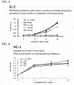
FIG. 4] FIG. 4 is a graph illustrating the results of examining the proliferation potential of bone-marrow-derived cells (UE7T-13 cells) when treated with IC-2. - [
FIG. 5] FIG. 5 is a graph illustrating the results of examining the proliferation potential of bone-marrow-derived cells (UE7T-13 cells) when treated with HC-1. - [
FIG. 6] FIG. 6 is a graph illustrating the results of examining the proliferation potential of bone-marrow-derived cells (UE7T-13 cells) when treated with PN-3-4. - [
FIG. 7] FIG. 7 is a graph illustrating the results of examining the proliferation potential of bone-marrow-derived cells (UE7T-13 cells) when treated with PN-3-13. - [
FIG. 8] FIG. 8 is a graph illustrating the results of examining the proliferation potential of bone-marrow-derived cells (UE7T-13 cells) when treated with PN-1-2. - [
FIG. 9] FIG. 9 is a graph illustrating how IC-2, etc., affects Wnt/β-catenin activity. - [
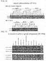
FIG. 10] FIG. 10 is electrophoresis gel images illustrating hepatocyte differentiation induction using IC-2, etc. - [
FIG. 11] FIG. 11 is electrophoresis gel images illustrating hepatocyte differentiation induction using IC-2, etc. - [
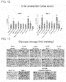
FIG. 12] FIG. 12 is graphs illustrating an ability of urea synthesis (atdays 8 and 16) of samples differentiated using IC-2, etc. - [
FIG. 13] FIG. 13 is photomicrographs (PAS staining) illustrating an ability of inducing differentiation into hepatocytes by using IC-2, etc. - [
FIG. 14] FIG. 14 is fluorescent photomicrographs illustrating hepatocyte differentiation induction (at day 8) using IC-2, etc. - [
FIG. 15] FIG. 15 is fluorescent photomicrographs illustrating hepatocyte differentiation induction (at day 16) using IC-2, etc. - [
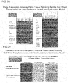
FIG. 16] FIG. 16 is images illustrating the results of examining gene expression of cytokines. - [
FIG. 17] FIG. 17 is a diagram illustrating the results of examining a neovascularization effect induced by a bFGF-sustained-release device. - [
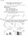
FIG. 18] FIG. 18 illustrates how to conduct an experiment for suppression of liver dysfunction when a cell sheet was transplanted under the skin. - [
FIG. 19] FIG. 19 is graphs illustrating the results of how liver functions changed when a cell sheet was transplanted under the skin. - [
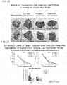
FIG. 20] FIG. 20 is photographs of a liver and pictures illustrating the results of immunohistochemistry staining when a cell sheet was transplanted on a surface of liver. - [
FIG. 21] FIG. 21 illustrates how to conduct an experiment for suppression of liver dysfunction when cell sheets were transplanted on two sites of a liver surface. - [
FIG. 22] FIG 22 is photos of a liver when cell sheets were transplanted on two sites of a liver surface. - [
FIG. 23] FIG. 23 is graphs illustrating the results of how liver functions changed when cell sheets were transplanted on two sites of a liver surface. - [
FIG. 24] FIG. 24 is graphs illustrating the results of how liver functions changed when cell sheets were transplanted on two sites of a liver surface. - [
FIG. 25] FIG. 25 is a graph illustrating the results of examining survival rates of mice when cell sheets were transplanted on two sites of a liver surface. - [
FIG. 26] FIG. 26 is images illustrating the results of examining gene expression of liver-specific secretory proteins and cytokines. - [
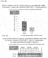
FIG. 27] FIG. 27 is pictures illustrating the results of examining levels of expression of human α1-antitrypsin when cell sheets were transplanted on two sites of a liver surface. - [
FIG. 28] FIG. 28 provides an overview of a method for sorting a mesenchymal stem cell (JP2009-060840A - [
FIG. 29] FIG. 29 is a graph illustrating the results of measuring luciferase activity. - [
FIG. 30] FIG. 30 is images illustrating the results of examining gene expression of a hepatocyte differentiation marker. - [
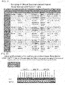
FIG. 31] FIG. 31 is a table listing information on clinical specimens. - [
FIG. 32] FIG. 32 is images illustrating the results of examining gene expression of a hepatocyte differentiation marker. - [
FIG. 33] FIG. 33 is photomicrographs (immunofluorescence staining) illustrating an ability of inducing differentiation into hepatocytes by using PN-3-13 or IC-2. - [
FIG. 34] FIG. 34 is a graph illustrating an ability (urea synthesis) of inducing differentiation into hepatocytes by using hexachlorophene, PN-3-13, or IC-2. - [
FIG. 35] FIG. 35 is photomicrographs (PAS staining) illustrating an ability of inducing differentiation into hepatocytes by using PN-3-13 or IC-2. - [
FIG. 36] FIG. 36 is graphs illustrating how liver functions changed when a "healthy" or "patient"-derived cell sheet was transplanted on a liver surface. - [
FIG. 37] FIG. 37 illustrates how to conduct an experiment for suppression of liver dysfunction when six cell sheets were transplanted on a liver surface or when cells were transplanted via a portal vein by injection into a spleen. - [
FIG. 38] FIG. 38 is a graph illustrating the results of examining survival rates of mice. - [
FIG. 39] FIG. 39 is graphs illustrating the results of how liver functions changed. - Hereinafter, embodiments of the present invention will be described in detail.
- A compound according to an embodiment of the present invention is a compound represented by formula (1), a salt thereof, or a solvate of them. This compound, the salt thereof, or the solvate of them is a low-molecular-weight organic compound, and thus has better stability and safety than a protein preparation and/or a nucleic acid preparation. This allows for a safe differentiation-inducing method with excellent efficiency of differentiation of mesenchymal stem cells into hepatocytes.
- In the formula (1), R1, R2, and R4 are the same or different from each other and each represents H, halogen, nitro, cyano, OH, optionally substituted C1-6 alkyl, optionally substituted C2-6 alkenyl, optionally substituted C1-6 alkoxy, aryl, or heteroaryl. In addition, R3 represents H, optionally substituted C1-6 alkyl, or optionally substituted C2-6 alkenyl. In addition, m is an integer of any of 1 to 4. In addition, n is an integer of any of 1 to 3. In addition, p is an integer of any of 1 to 5. In this regard, however, the term "optionally substituted" means that the respective group is unsubstituted or has 1, 2, 3, 4, or 5 substituents selected from H, halogen, nitro, cyano, OH, C1-6 alkyl, halogeno C1-6 alkyl, hydroxy C1-6 alkyl, C1-6 alkyl amino, C3-6 cycloalkyl, C2-6 alkenyl, halogeno C2-6 alkenyl, hydroxy C2-6 alkenyl, C2-6 alkenylamino, C3-6 cycloalkenyl, C2-6 alkynyl, halogeno C2-6 alkynyl, hydroxy C2-6 alkynyl, C2-6 alkynyl amino, C1-6 alkoxy, halogeno C1-6 alkoxy, hydroxy C1-6 alkoxy, C1-6 alkoxy amino, aryl, and heteroaryl.
- The above R1, R2, and R4 are the same or different from each other and may each represent H, halogen, nitro, cyano, OH, C1-6 alkyl, halogeno C1-6 alkyl, hydroxy C1-6 alkyl, C1-6 alkyl amino, C1-6 alkoxy, halogeno C1-6 alkoxy, hydroxy C1-6 alkoxy, or C1-6 alkoxy amino. The above R3 may represent H.
- As used herein, the term "halogen" means F, Cl, Br, or I.
- As used herein, unless otherwise indicated, the terms "alkyl" and "alkenyl" mean a linear or branched hydrocarbon chain.
- As used herein, the term "C1-6" refers to hydrocarbon containing 1, 2, 3, 4, 5, or 6 carbon atoms. That is, the term "C1-6 alkyl" refers to alkyl containing 1, 2, 3, 4, 5, or 6 carbon atoms. Examples of C1-6 alkyl include methyl, ethyl, n-propyl, isopropyl, n-butyl, isobutyl, sec-butyl, tert-butyl, n-pentyl, and n-hexyl.
- As used herein, examples of "alkenyl" include ethenyl, 1-propenyl, 2-propenyl, 2-methyl-1-propenyl, 1-butenyl, 2-butenyl, 3-butenyl, 3-methyl-2-butenyl, 1-pentenyl, 2-pentenyl, 3-pentenyl, 4-pentenyl, 4-methyl-3-pentenyl, 1-hexenyl, 3-hexenyl, and 5-hexenyl.
- As used herein, examples of "alkoxy" include methoxy, ethoxy, propoxy, isopropoxy, butoxy, isobutoxy, sec-butoxy, tert-butxy, pentoxy, isopentoxy, and hexoxy.
- As used herein, the term "optionally substituted" means that a compound is unsubstituted or has 1, 2, 3, 4, or 5 substituents at positions which can be substituted. Note that when a plurality of substituents are included, these substituents may be the same or different from each other. In addition, the position of each substitution may be
position - As used herein, the "halogeno C1-6 alkyl" refers to C1-6 alkyl that is substituted by one or more halogens. The number of halogens may be, for example, 1, 2, 3, 4, 5, 6, or 13. Also, the number may be within a range between any two numbers indicated above. In addition, when two or more halogens are included, the kind of each halogen may be the same or different from each other. Examples of halogeno C1-6 alkyl include, chloromethyl, dichloromethyl, trichloromethyl, fluoromethyl, difluoromethyl, trifluoromethyl, bromomethyl, dibromomethyl, tribromomethyl, chloroethyl, dichloroethyl, trichloroethyl, fluoroethyl, difluoroethyl, and trifluoroehtyl.
- As used herein, the "hydroxy C1-6 alkyl" refers to C1-6 alkyl that is substituted by one or more hydroxy groups. The number of the hydroxy groups may be, for example, 1, 2, 3, 4, 5, 6, or 13. Also, the number may be within a range between any two numbers indicated above. Examples of hydroxy C1-6 alkyl include hydroxymethyl, 1-hydroxyethyl, 2-hydroxyethyl, 2-hydroxy-n-propyl, and 2,3-dihydroxy-n-propyl.
- As used herein, the "C1-6 alkyl amino" refers to C1-6 alkyl that is substituted by one or more amino groups. The number of the amino groups may be, for example, 1, 2, 3, 4, 5, 6, or 13. Also, the number may be within a range between any two numbers indicated above. Examples of C1-6 alkyl amino include methyl amino and ethyl amino.
- As used herein, the "halogeno C1-6 alkoxy" is equivalent to halogeno C1-6 alkyl whose alkyl is replaced by alkoxy. Examples of halogeno C1-6 alkoxy include fluoromethoxy, difluoromethoxy, trifluoromethoxy, 1-fluoroethoxy, 2-fluoroethoxy 2-chloroethoxy, 2-bromoethoxy, (1,1-difluoro)ethoxy, (1,2-difluoro)ethoxy, (2,2,2-trifluoro)ethoxy, (1, 1,2,2-tetrafluoro)ethoxy, (1, 1,2,2,2-pentafluoro)ethoxy, 1-fluoro-n-propoxy, 1,1-difluoro-n-propoxy, 2,2-difluoro-n-propoxy, 3-fluoro-n-propoxy, (3,3,3-trifluoro)-n-propoxy, (2,2,3,3,3-pentafluoro)-n-propoxy, 4-fluoro-n-butoxy, (4,4,4-trifluoro)-n-butoxy, 5-fluoro-n-pentyloxy, (5,5,5-trifluoro)-n-pentyloxy, 6-fluoro-n-hexyloxy, (6,6,6-trifluoro)-n-hexyloxy, 2-fluorocyclopropoxy, and 2-fluorocyclobutoxy.
- As used herein, the "hydroxy C1-6 alkoxy" is equivalent to hydroxy C1-6 alkyl whose alkyl is replaced by alkoxy. Examples of hydroxy C1-6 alkoxy include 2-hydroxyethoxy, 2-hydroxy-n-propoxy, 3-hydroxy-n-propoxy, 2,3-dihydroxy-n-propoxy, and 2-hydroxycyclopropyl.
- As used herein, the "C1-6 alkoxy amino" is equivalent to C1-6 alkyl amino whose alkyl is replaced by alkoxy. Examples of C1-6 alkoxy amino include methoxy amino and ethoxy amino.
- As used herein, the term "aryl" refers to a C6-14 monocyclic, dicyclic, or tricyclic aromatic hydrocarbon ring. Examples of aryl include phenyl, naphthyl (e.g., 1-naphthyl, 2-naphthyl), tetrahydronaphthalenyl, indenyl, and phenyl that is substituted with five halogens. Also, aryl includes a ring group that is condensed with C5-8 cycloalkene at its double bond position.
- As used herein, the "heteroaryl" includes groups having 5 to 14 ring atoms and shared n-electrons within its rings, and having 1 to 4 heteroatoms selected from the group consisting of N, S, and O. Examples of heteroaryl include thienyl, benzothienyl, furyl, benzofuryl, dibenzofuryl, pyrrolyl, imidazolyl, pyrazolyl, pyridyl, pyrazinyl, pyrimidinyl, pyridazinyl, tetrazolyl, oxazolyl, thiazolyl, and isooxazolyl.
- As used herein, examples of the "salt" include, but are not particularly limited to, anionic salts that are formed using any acidic group (e.g., carboxyl) and cationic salts that are formed using any basic group (e.g., amino). Examples of the salts include inorganic salts, organic salts, and salts disclosed in an article (Berge, Bighley, and Monkhouse, J. Pharm. Sci., 1977, 66, 1-19). The examples further include metal salts, ammonium salts, salts with an organic base, salts with an inorganic acid, salts with an organic acid, and salts with a basic or acidic amino acid. Examples of the metal salts include alkali metal salts (e.g., sodium salts, potassium salts), alkaline earth metal salts (e.g., calcium salts, magnesium salts, barium salts), and aluminum salts. Examples of the salts with an organic base include salts with trimethylamine, triethylamine, pyridine, picoline, 2,6-lutidine, ethanolamine, diethanolamine, triethanolamine, cyclohexylamine, dicyclohexylamine, or N,N'-dibenzylethylenediamine. Examples of the salts with an inorganic acid include salts with hydrochloric acid, hydrobromic acid, nitric acid, sulfuric acid, or phosphoric acid. Examples of the salts with an organic acid include salts with formic acid, acetic acid, trifluoroacetic acid, phthalic acid, fumaric acid, oxalic acid, tartaric acid, maleic acid, citric acid, succinic acid, malic acid, methanesulfonic acid, benzenesulfonic acid, or p-toluenesulfonic acid. Examples of the salts with a basic amino acid include salts whit arginine, lysine, or ornithine. Examples of the salts with an acidic amino acid include salts with aspartic acid or glutamic acid.
- As used herein, the term "solvate" refers to a compound formed by a solute and a solvent. A dictionary (J. Honig et al., The Van Nostrand Chemist's Dictionary P650 (1953)) can be consulted regarding the solvate. If the solvent is water, the solvate formed is a hydrate. Preferably, the solvent does not interfere with a biological activity of the solute. Examples of the preferable solvent include, but are not limited to, water, ethanol, and acetic acid. The most preferred solvent is water. A compound or a salt thereof according to an embodiment of the present invention absorbs moisture when contacting air or recrystallized. They sometimes possess hygroscopic moisture or become a hydrate. As used herein, the term "isomer" includes molecules whose molecular formula is identical but whose structure is different. Examples of the isomer include enantiomers, geometric (cis/trans) isomers, and isomers (diastereomers) having one or more chiral centers that are not mirror images of each other. As used herein, the term "prodrug" includes a precursor compound in which when the above compound is administered to a subject, a chemical change occurs due to metabolic processes or various chemical reactions to give a compound, a salt thereof, or a solvate of them according to the present invention. With regard to the prodrug, an article (T. Higuchi and V. Stella, "Pro-Drugs as Novel Delivery Systems", A.C.S. Symposium Series, Volume 14) can be referred to.
- A compound according to an embodiment of the present invention is at least one compound, a salt thereof, or a solvate of them, the compound being represented by formula (3):

- An inducer of differentiation of mesenchymal stem cells into hepatocytes as disclosed herein is a differentiation inducer comprising a compound represented by formula (1), a salt thereof, or a solvate of them. A low-molecular-weight organic compound is used for these differentiation inducers, thereby imparting better stability and safety than a protein preparation and/or a nucleic acid preparation. This allows for a safe differentiation-inducing method with excellent efficiency of differentiation of mesenchymal stem cells into hepatocytes.
In addition, in view of efficiency of the differentiation, it is preferable that the structure of the compound contained in an inducer of differentiation of mesenchymal stem cells into hepatocytes according to this embodiment has a structure similar to those of formula (3). - As used herein, the term "mesenchymal stem cells" includes somatic stem cells derived from mesenchyme. These mesenchymal stem cells can differentiate into mesenchymal cells. These mesenchymal stem cells should be applicable to regenerative medicine such as restructuring of bones, blood vessels, and cardiac muscles in a near future.
- Examples of the "mesenchymal stem cells" include bone marrow mesenchymal stem cells and umbilical cord blood-derived stem cells. The mesenchymal stem cells seem to be present in all tissues containing a mesenchymal tissue. Among the mesenchymal tissues, the bone marrow mesenchymal stem cells, however, can be easily collected by bone marrow aspiration, and their culture technique has been established. Bone marrow stromal cells contain bone marrow mesenchymal stem cells, and are one type of cells supporting hematopoietic cells mainly contained in bone marrow. Meanwhile, umbilical cord blood includes blood contained in an umbilical cord that constitutes a fetal tissue connecting a fetus and its mother. It is known that this umbilical cord blood contains a large amount of the umbilical cord blood-derived stem cells (hematopoietic stem cells). Recently, mesenchymal stem cells (i.e., umbilical cord blood-derived mesenchymal stem cells), which are somatic stem cells other than the hematopoietic stem cells, have been found in umbilical cord blood. To date, the mesenchymal stem cells have been reported to be present in bone marrow. However, it has been indicated that not only bone marrow but also umbilical cord blood can provide a source for mesenchymal stem cells, and may be applicable for bone- and/or cartilage-repair using tissue engineering or clinical application to regenerative medicine.
- Here, the mesenchymal stem cells, in general, have a short life span and are not necessarily easy to culture in vitro for a long period. However, a human bone marrow-derived and a human umbilical cord blood-derived mesenchymal stem cell lines that have the hTERT gene introduced and can stably proliferate have already been established. While these cells stably proliferate, they have no chromosome abnormalities but have functional contact inhibition. Besides, they do not form tumors when transplanted into an immunosuppressed animal. Also, since they do not affect cellular differentiation, these cells are useful for research on differentiation of mesenchymal stem cells.
- In addition, as used herein, the term "hepatocytes" includes cells with a size of about 20 µm, the cells constituting 70 to 80% of a liver. Examples of general functions of hepatocytes include: synthesis and storage of proteins; conversion of carbohydrate; synthesis of cholesterol, bile acid, and phospholipids; and detoxification, denaturing, and excretion of endogenous and exogenous substances. Also, the examples further include promotion of generation and secretion of bile.
- As used herein, the term "differentiation" includes a structural and functional change of individual cells in a multicellular organism. Generally speaking, differentiation of a stem cell into a functional cell in a multicellular organism is often irreversible in many cases. Previous findings have taught that a mesenchymal stem cell has multipotency, that is, has an ability of differentiating into a wide variety of cell types while cell lineages that can be differentiated are limited. Therefore, the mesenchymal stem cell, in general, cannot be differentiated into cells derived from a different germ layer. Here, an inducer of differentiation of mesenchymal stem cells into hepatocytes according to this embodiment can induce differentiation of mesenchymal stem cells into hepatocytes that are derived from a different germ layer. This point can be said to be a big technical feature.
- Generally speaking, it is quite natural to use liver stem cells as a cell source for liver regenerative medicine. However, since the liver stem cells only appear during severe liver damage such as fulminant hepatitis, collection of the stem cells from an actual patient is not practical. Then, tissue stem cells other than liver stem cells or iPS cells could be a cell source for liver regenerative medicine. Recent reports have shown that mesenchymal stem cells can differentiate into cells with a certain degree of liver function under in vitro conditions. That is, addition of various cytokines to mesenchymal stem cells, followed by culturing for about 4 weeks, has reportedly promoted differentiation into hepatocyte-like cells. The mesenchymal stem cells are present in bone marrow and umbilical cord blood, and are relatively easy to collect. Use of bone marrow has an advantage of avoiding immune rejection because of use of autologous cells. Alternatively, umbilical cord blood is usually disposed, so there is an advantage of having less ethical issues. In view of the above, the mesenchymal stem cells are a promising cell source for liver regenerative medicine.
- As used herein, the "Wnt/β-catenin signaling pathway" includes one of pathways that regulate a cell fate decision during development of vertebrates and invertebrates. A Wnt ligand is a typical glycoprotein that binds to a secretory Frizzled receptor. Its binding triggers a signaling cascade, which results in dissociation of a multifunctional kinase GSK-3β from an APC/Axin/GSK-3β complex. Without the Wnt signal (i.e., off state), β-catenin, which is a transcriptional coactivator and an essential adaptor protein for cell-to-cell adhesion, becomes a target for degradation by the APC/Axin/GSK-3β complex. CK1 and GSK-3β act coordinately to properly phosphorylate β-catenin. Then, the β-catenin gets ubiquitinated and is degraded by a β-TrCP/SKP complex in proteasomes.
- When Wnt binds to the receptor (i.e., on state), Dishevelled (Dsh) seems to be at least partially phosphorylated to be activated. Next, GSK-3β dissociates from a degradation-inducing complex. This facilitates stabilization of the level of β-catenin expression, nuclear translocation, and recruitment of LEF/TCF to a DNA-binding factor. Then, β-catenin replaces a Griucho-HDAC corepressor and serves as a transcriptional activator.
- In addition, similar to the transcriptional repressor complex, a complex with
Prop 1, a homeodomain factor, is shown to have β-catenin that exerts its functions by conditional activation. Importantly, several human cancers have been found to contain β-catenin point mutations that lead to uncontrollable stabilization of β-catenin. In addition, it has been reported that APC and Axin also contain mutations. Thus, it has been emphasized that aberrant activation of this pathway relates to human tumors. During a course of development, the Wnt/β-catenin signaling pathway plays a role in integration of signals from various pathways such as retinoic acid, FGF, TGF-β, and BMP in different cell types and/or tissues. Further, GSK-3β, which is a component of the Wnt/β-catenin signaling pathway, is involved in glycogen metabolism and other major pathways. Thus, inhibition of GSK-3β is considered to be a reasonable measure to treat diabetes and/or neurodegenerative disease. - As used herein, the term "low-molecular-weight organic compound" includes an organic compound with a molecular weight of 2000 or less. The molecular weight is preferably 1000 or less, more preferably 900 or less, and still more preferably 800 or less. Since a high-molecular-weight compound including a nucleic acid preparation and a protein preparation is susceptible to degradation in vivo and in vitro, the compound is unstable. When the compound is used in regenerative medicine, a safety concern remains. In contrast, since the low-molecular-weight organic compound is unlikely to be degraded in vivo and in vitro, the compound is stable. Thus, when used in regenerative medicine, the compound has an advantage of safety. In addition, as the molecular weight becomes smaller, its organic synthesis becomes easier. Further, the compound has excellent feasibility such as stability and safety. Note that peptides and nucleotides are excluded in this low-molecular-weight organic compound. This is because the peptides and the nucleotides are susceptible to degradation in vivo and in vitro, thereby being unstable. Besides, when they are used in regenerative medicine, a safety concern still remains.
- Under these circumstances, the present inventors have synthesized various low-molecular-weight organic compounds, and have evaluated the compounds according to the following points. As a result, the present inventors have discovered that five low-molecular-weight organic compounds (hereinafter, sometimes referred to as "IC-2, etc.") including IC-2, HC-1, PN-3-4, PN-3-13, and PN-1-2 can induce differentiate of mesenchymal stem cells into hepatocytes as demonstrated in the below-described Examples.
- 1. Expression of Liver-specific Transcription Factors;
- 2. Expression of Liver-specific Proteins;
- 3. Glycogen Synthesis;
- 4. Urea Synthesis; and
- 5. Evaluation of Signal Strength of Wnt/β-catenin Signaling Pathway Having TCF Activity by Using Luciferase Activity Measurement.
- Examples of the above-mentioned inducer of differentiation of mesenchymal stem cells into hepatocytes according to an embodiment of the present invention include low-molecular-weight organic compounds (e.g., IC-2) that inhibit the Wnt/β-catenin signaling pathway. These low-molecular-weight compounds have better feasibility such as stability and safety than protein preparations and/or nucleic acid preparations, so that the compounds should provide a promising method to realize regenerative medicine.
- Note that in this embodiment, it is determined whether or not a low-molecular-weight organic compound, which is a candidate substance for the differentiation inducer, has characteristics of inducing differentiation of mesenchymal stem cells into hepatocytes. In order to determine the above, whether or not expression of at least one protein selected from the group consisting of albumin, C/EBPα, CYP1A1, and CYP3A4 is induced is a critical indicator. This is because hepatocytes, in general, have increased levels of expression of at least one protein (hereinafter, sometimes referred to as "albumin, etc.") selected from the group consisting of the above albumin, C/EBPα, CYP1A1, and CYP3A4, but mesenchymal stem cells have low levels of expression of the albumin, etc.
- Note that at this time, if the level of expression of albumin, etc., exceeds a predetermined threshold in post-treatment mesenchymal stem cells, it is determined that the candidate substance can induce differentiation of the mesenchymal stem cells into hepatocytes. In contrast, if the expression is lower than the predetermined threshold, it is determined that the candidate substance is not characterized by inducing differentiation of the mesenchymal stem cells into hepatocytes. This threshold, for example, may be set to 1.5 or more, 2 or more, 5 or more, or 10 or more times as large as the average of each expression level of albumin, C/EBPα, CYP1A1, and CYP3A4 in mesenchymal stem cells before treatment. Also, this threshold, for example, may be set to 2 or more, 5 or more, or 10 or more times as large as the average ± standard deviation of each expression level of albumin, C/EBPα, CYP1A1, and CYP3A4 in mesenchymal stem cells before treatment.
- Alternatively, it may be determined whether or not the expression level of any of albumin, C/EBPα, CYP1A1, and CYP3A4 in mesenchymal stem cells treated with the candidate substance has, for example, a significantly higher expression level than that in mesenchymal stem cells before treatment. With regard to a test for the significant difference, it is preferable to have a significant difference in, for example, Student's t-test, which is a parametric test, when a population is assumed to have a Gaussian distribution. Specifically, in the Student's t-test, a one-sided test should give preferably p < 0.05, more preferably p < 0.03, and still more preferably p < 0.01. Note that the Student's t-test is not limited to the one-sided test, but may be a two-sided test. Further, if the population is not assumed to have a Gaussian distribution, whether or not there is a significant difference may be determined by using a Mann-Whitney U test as a non-parametric test.
- In addition, in order to determine whether or not a low-molecular-weight organic compound, which is a candidate substance for the differentiation inducer, is characterized by inducing differentiation of mesenchymal stem cells into hepatocytes in an embodiment of the present invention, whether or not the compound increases an ability of glycogen production or urea synthesis in the mesenchymal stem cells gives another critical indicator. This is because while hepatocytes, in general, have increased the ability of glycogen production or urea synthesis, mesenchymal stem cells fail to have increased the ability of glycogen production or urea synthesis. Note that at this time, a threshold or significant difference is used for determination in a manner similar to those when the above expression level of albumin, etc., is determined.
- It is disclosed a Wnt/β-catenin signaling pathway inhibitor comprising at least one compound selected from the group consisting of compounds represented by formula (1), a salt thereof, or a solvate of them. Use of these Wnt/β-catenin signaling pathway inhibitors can inhibit a Wnt/β-catenin signaling pathway. Also, a low-molecular-weight organic compound is used for the inhibitors, thereby imparting better stability and safety than a protein preparation and/or a nucleic acid preparation.
- In addition, as a compound contained in a Wnt/ β-catenin signaling pathway inhibitor according to this embodiment has a structure more similar to those of compounds represented by formula (3), the structure is more preferable in view of efficiency of the Wnt/β-catenin signaling pathway inhibition.
- A method for producing hepatocytes according to an embodiment of the present invention is a production method comprising a step of treating mesenchymal stem cells in vitro with at least one compound selected from the group consisting of compounds represented by formula (1), a salt thereof, or a solvate of them. Also, the production method comprises the step of treating mesenchymal stem cells in vitro with the above differentiation inducer. A low-molecular-weight organic compound is used for these production methods, thereby imparting better stability and safety than a protein preparation and/or a nucleic acid preparation. This makes it possible to efficiently induce differentiation of mesenchymal stem cells into hepatocytes.
- Note that at this time, it is preferable to include a step of treating the mesenchymal stem cells with the differentiation inducer for 8 days or more as demonstrated in the below-described Examples. Progression of changes in various molecular biological intracellular events takes such long time because differentiation of mesenchymal stem cells into hepatocytes contains biological processes. In this case, since the differentiation inducer according to this embodiment is a low-molecular-weight organic compound, it has superior stability. Thus, treatment for a long period of 8 days or more is unlikely to make the compound degraded. However, it is preferable to appropriately change a medium so as to supply nutrients necessary for survival, proliferation, and changes of the mesenchymal stem cells. At such occasion, it is also preferable to add the low-molecular-weight organic compound in a freshly replaced medium, the amount of the low-molecular weight organic compound being substantially the same as that of the medium before the change.
- In addition, since the differentiation inducer according to this embodiment is a low-molecular-weight organic compound, it has superior stability. Thus, whether the treatment temperature is high or low is unlikely to affect its decomposition. It is preferable to set a temperature of a medium to a temperature suitable for survival of mammalian cells so as to maintain an environment necessary for survival, proliferation, and changes of the mesenchymal stem cells. Specifically, the temperature is set to be preferably from 20 to 45°C, more preferably from 30 to 40°C, and still more preferably from about 36 to about 38°C.
- In this case, a method for culturing mesenchymal stem cells preferably allows them to contact a low-molecular-weight organic compound in a medium suitable for survival of the mesenchymal stem cells on, for example, a temperature-responsive culture dish. Examples of a preferred medium that can be used include Dulbecco's modified Eagle's Medium (DMEM) and Mesenchymal Stem Cell Basal Medium (MSCBM).
- At that time, examples of a preferred temperature-responsive culture dish include culture dishes whose surface has been polymerized with a temperature-responsive polymer (e.g., PIPAAm) and/or methyl cellulose. This is because merely decreasing the temperature would enable cells to be collected without damage while maintaining intercellular adhesion.
- The hepatocytes as so differentiated can be suitably used for transplantation therapeutics, such as hepatocyte transplantation, and application for drug development in evaluation of a drug's efficacy and adverse effect. In addition, these hepatocytes are differentiated from mesenchymal stem cells by using a low-molecular-weight organic compound, so that this case has better stability and safety during production than the case of differentiation induction using a protein preparation and/or a nucleic acid preparation.
- Further, the hepatocytes as so differentiated may be grown to fit a desired shape by using a temperature-responsive culture dish and/or a substrate such as a synthetic polymer scaffold using a biodegradable synthetic polymer such as polylactic acid (PLA) and polyethylene glycolic acid (PGA). Then, the resulting hepatocytes can be used as a liver tissue, liver, or cell sheet for regenerative medicine. The resulting 3D-structured liver tissue, liver, or cell sheet for regenerative medicine can be suitably used for transplantation therapeutics, such as hepatocyte transplantation, and application for drug development in evaluation of a drug's efficacy and adverse effect. In addition, the liver tissue, liver, or cell sheet is differentiated from mesenchymal stem cells by using a low-molecular-weight organic compound, so that this case has better stability and safety during production than the case of differentiation induction using a protein preparation and/or a nucleic acid preparation.
- Further, the above hepatocytes, liver tissue, liver, or cell sheet may have components comprising, for example, at least one compound selected from the group consisting of compounds represented by formula (1), a salt thereof, or a solvate of them.
- In addition, the levels of expression of genes in the above hepatocytes are not particularly limited, but the levels of expression of at least one gene selected from any of transthyretin, apolipoprotein B, haptoglobin, fibrinogen α, and fibrinogen β, for example, may be lower than those of the above genes in commercially available hepatocytes. Also, the levels of expression of any of the above genes may be lower than those of hepatocytes in the liver of a patient transplanted with the above hepatocytes. Note that the term "commercially available" includes those commercially available for regenerative medicine or research purposes. In addition, the phrase "is lower" includes a state in which the level of the gene expression is significantly or substantially decreased. Also, the level may be decreased by 40, 50, 60, 70, 80, 90, or 100%. This rate may be any one of the above values or higher, or may be between any two of the above values.
- In addition, the levels of expression of genes in the above liver tissue, liver, or cell sheet are not particularly limited, but the levels of expression of at least one gene selected from any of transthyretin, apolipoprotein B, C4, RBP4, RBP1, haptoglobin, fibrinogen α, and fibrinogen β, for example, may be lower than those of the above genes in commercially available hepatocytes, liver tissues, or livers. Also, the levels of expression of any of the above genes may be lower than those of hepatocytes in the liver, a liver tissue, or the liver of a patient transplanted with the above hepatocytes.
- Moreover, it is disclosed a therapeutic agent or a material for use in treating liver dysfunction or a disease accompanied by the liver dysfunction, comprising hepatocytes, a liver tissue, a liver, or a cell sheet disclosed herein.
- It is disclosed a cell sheet comprising hepatocytes as differentiated in such a manner as described above. Use of this cell sheet can suppress liver dysfunction as demonstrated in the following Examples. Therefore, the cell sheet can be suitably used for treatment of liver dysfunction.
- In addition, this cell sheet may be used for transplantation on a liver surface. When this cell sheet is transplanted on a liver surface, the following Examples demonstrate a remarkable effect of suppressing liver dysfunction.
- Also, for transplantation, one or more layers of this cell sheet may be transplanted. In addition, with regard to the transplantation site, one or more sites may be selected. The number of sites may be, for example, 2, 3, 4, 5, or 6. The number may be the above number or more, or may be between any two of the above numbers.
- It is also disclosed a transplantation material comprising the above cell sheet and a support for collecting the cell sheet. Use of this transplantation material can suppress liver dysfunction as demonstrated in the following Examples. For example, when used, this transplantation material is placed on a site of interest. Next, the cell sheet is made to contact the site. Then, the support for collecting the cell sheet may be removed. This procedure allows the cell sheet to be stably attached to the site of interest.
- Another embodiment of the present invention provides a method for producing a transplantation material, comprising a step of treating stem in vitro with at least one compound selected from the group consisting of compounds represented by formula (1), a salt thereof, or a solvate of them. A low-molecular-weight organic compound is used for this production method, thereby imparting better stability and safety than a protein preparation and/or a nucleic acid preparation. This makes it possible to efficiently produce the transplantation material.
- This method for producing a transplantation material may include the step of: culturing mesenchymal stem cells in a medium comprising at least one compound selected from the group consisting of compounds represented by formula (1), a salt thereof, or a solvate of them to differentiate the mesenchymal stem cells into hepatocytes. Note that as used herein, the term "transplantation material" may include, for example, a cell sheet. In addition, the transplantation material may come with any support.
- It is also disclosed a method for producing a cell sheet for transplantation on a liver surface, comprising an induction step of treating mesenchymal stem cells with hexachlorophene, a derivative thereof, a salt of them, or a solvate of them to induce the mesenchymal stem cells into hepatocytes. The cell sheet as prepared using this production method can exert a remarkable effect of suppressing liver dysfunction when transplanted on a liver surface as demonstrated in the following Examples. Therefore, this production method is an excellent production method for producing a cell sheet for transplantation on a liver surface. Note that when a certain organic compound is taken as a starting material, a derivative means a compound modified in such a degree without large structural and characteristic modifications in the starting material by introduction of a functional group, oxidation, reduction, or replacement or addition of an atom, etc. For example, "Burger's Medicinal Chemistry And Drug Discovery, 5th Edition, Vol 1: Principles and Practice" can be referred to.
- In addition, it is disclosed a method for producing a cell sheet for transplantation on a liver surface, comprising an induction step of treating mesenchymal stem cells with a Wnt/β-catenin signaling pathway inhibitor to induce the mesenchymal stem cells into hepatocytes. The cell sheet as prepared using this production method can exert a remarkable effect of suppressing liver dysfunction when transplanted on a liver surface as demonstrated in the following Examples. Therefore, this production method is an excellent production method for producing a cell sheet for transplantation on a liver surface.
- As described above, the embodiments of the present invention have been illustrated. These embodiments are examples of the present invention. In addition, combinations among the above-described embodiments can also be employed.
- Hereinafter, the present invention is further illustrated by referring to Examples.
- ICG-001 is an oligopeptide having a bicyclic β-turn mimetic structure in its backbone, and reportedly functions as a potent antagonist for transcriptional activation by a β-catenin/Tcf complex (Drug Discov. ). ICG-001 was synthesized and examined according to a research article (Tetrahedron 2007, 63, 12912-12916).

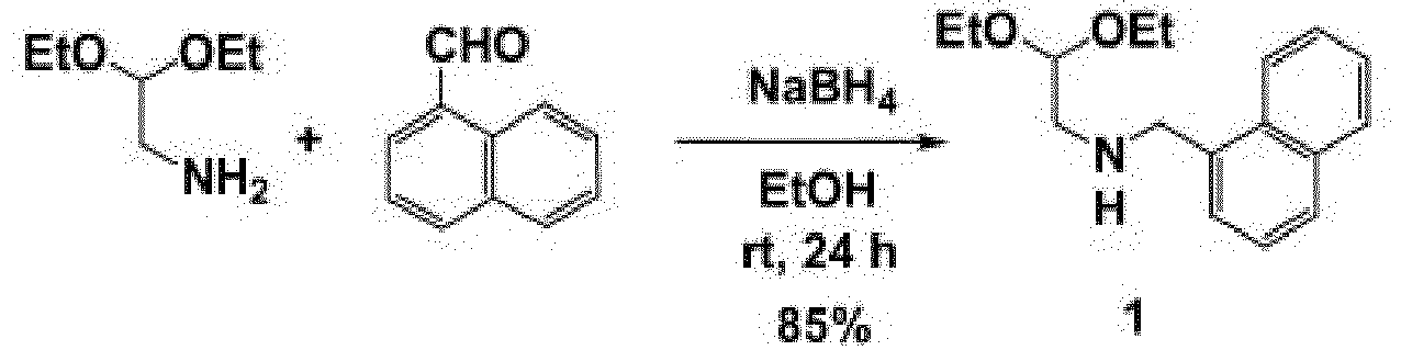
- 1-naphthaldehyde (Wako Pure Chemical Industries, Ltd.)(1.56 g, 10 mmol) and 2,2-diethoxyethanamine (TOKYO CHEMICAL INDUSTRY CO., LTD.)(1.33 g, 10 mmol) were mixed and stirred at 100°C for 20 min. After the mixture was cooled to room temperature, EtOH (20 mL) was used for dilution. NaBH4 (0.38 g, 10 mmol) was added dropwise and the mixture was stirred at room temperature for 16 h. After completion of the reaction, EtOH was distilled away under reduced pressure. Then, a product was extracted with AcOEt. The resulting product was purified by silica gel column chromatography (hexane/AcOEt = 5/1) to yield compound 1 (2.29 g, 8.5 mmol, 85%).
- A condensing agent HATU (0.76 g, 2.0 mmol) and diisopropylethylamine (DIEA)(0.35 mL, 2.0 mmol) were added to a DMF (7 mL) solution containing Fmoc-L-Tyr(t-Bu)-OH (0.87 g, 1.9 mmol). Next, the mixture was stirred for 20 min. Then, compound 1 (0.54 g, 2.0 mmol) was added, and the mixture was stirred at room temperature for 16 h. After the reaction, DMF was distilled away under reduced pressure. The resulting product was purified by column chromatography (hexane/AcOEt = 10/1) to yield compound 2 (1.33 g, 1.9 mmol, 93%). The resulting compound 2 (1.33 g, 1.9 mmol) was dissolved in CH2Cl2 (20 mL). Subsequently, diethylamine (DEA) (10 ml, excess) was added, and the mixture was stirred at room temperature for 2 h. After the termination of the reaction was verified by TLC, CH2Cl2 was distilled away under reduced pressure. The resulting product was purified by silica gel column chromatography (AcOEt) to yield compound 3 (0.92 g, 1.8 mmol, 92%).

- A condensing agent HATU (0.70 g, 1.8 mmol) and diisopropylethylamine (DIEA)(0.32 mL, 1.8 mmol) were added to a DMF (8 mL) solution containing Fmoc-β-Ala-OH (0.53 g, 1.7 mmol). Next, the mixture was stirred for 20 min. Then, compound 3 (0.92 g, 1.8 mmol) was added, and the mixture was stirred at room temperature for 14 h. After the reaction, DMF was distilled away under reduced pressure. The resulting product was purified by column chromatography (hexane/AcOEt = 1/1) to yield compound 4 (1.2 g, 1.5 mmol, 82%). The resulting compound 4 (1.2 g, 1.5 mmol) was dissolved in CH2Cl2 (20 mL). Subsequently, diethylamine (DEA) (9 mL, excess) was added, and the mixture was stirred at room temperature for 1 h. After the termination of the reaction was verified by TLC, CH2Cl2 was distilled away under reduced pressure. The resulting product was purified by silica gel column chromatography (AcOEt/EtOH = 1/1) to yield compound 5 (0.66 g, 1.2 mmol, 80%).

- A CH2Cl2 solution (8 mL) containing benzylisocyanate (0.16 g, 1.2 mmol) was added to a CH2Cl2 solution (8 mL) containing compound 5 (0.66 g, 1.2 mmol), and the mixture was stirred at room temperature for 12 h. After the termination of the reaction was verified by TLC, CH2Cl2 was distilled away under reduced pressure. The resulting product was purified by column chromatography (AcOEt/EtOH = 1/1) to yield compound 6 (0.59 g, 0.85 mmol, 73%). The resulting compound 6 (0.59 g, 0.85 mmol) was dissolved in formic acid (9 ml), and the mixture was stirred at room temperature for 20 h. Then, formic acid was distilled away under reduced pressure. The resulting product was purified by column chromatography (AcOEt) to yield
compound 7a (ICG-001) as a white solid (0.26 g, 0.48 mmol, 57%). - The resulting product was identified using MS spectrum and 1H NMR spectrum (having identical values in a literature) (
FIG. 1 ).
- When
compound 3 described in the above section (1-2) was synthesized, synthesis was carried out using "Fmoc-L-Phe(t-Bu)-OH" in place of "Fmoc-L-Tyr(t-Bu)-OH" during the synthesis process in the above section (1). By doing so, synthesized was a derivative, IC-2 ((6S,9aS)-6-phenyl-8-naphthalen-1-ylmethyl-4,7-dioxo-hexahydro-pyrazino[1,2-a] pyrimidine-1-carbocylic acid benzylamide) (total yield 29%) in which side chains of ICG-001 were modified.FIG. 2 shows spectral data.
- Hexachlorophene is a highly chlorinated phenolic compound used for an insect repellent, microbicide, disinfectant, etc. Here, synthesized was a derivative, in which a phenolic hydroxyl group of hexachlorophene was capped with a methyl group. MeI (1.0 mL, 16 mmol) and K2CO3 (5.3 g, 36 mmol) were added to an acetone solution (10 mL) containing hexachlorophene (TOKYO CHEMICAL INDUSTRY CO., LTD.)(417 mg, 1.0 mmol), and the mixture was stirred for 14 h. Next, excessive K2CO3 was removed, and the mixture was dried over Na2SO4. Then, the solvent was distilled away to yield a crude product. The resulting crude product was purified by column chromatography (hexane/AcOEt =7/1) to yield a hexachlorophene derivative of interest (a methyl ether compound) (bis(2,3,5-trichloro-6-methoxyphenyl)methane)(355 mg, 0.85 mmol, 85%). This methyl ether compound was analyzed by 1H NMR.

- Phenoxy benzhydrazide (Wako Pure Chemical Industries, Ltd.)(0.46 g, 2.0 mmol, 1.0 eq) was dissolved in EtOH. Next, 1-naphtaldehyde (0.32 g, 2.0 mmol, 1.0 eq) was added, and the mixture was stirred at room temperature for 22 h. After 22 h passed, the reaction solution was filtered and was then subjected to recrystallization with EtOH to yield a compound of interest (403 mg, 58%).
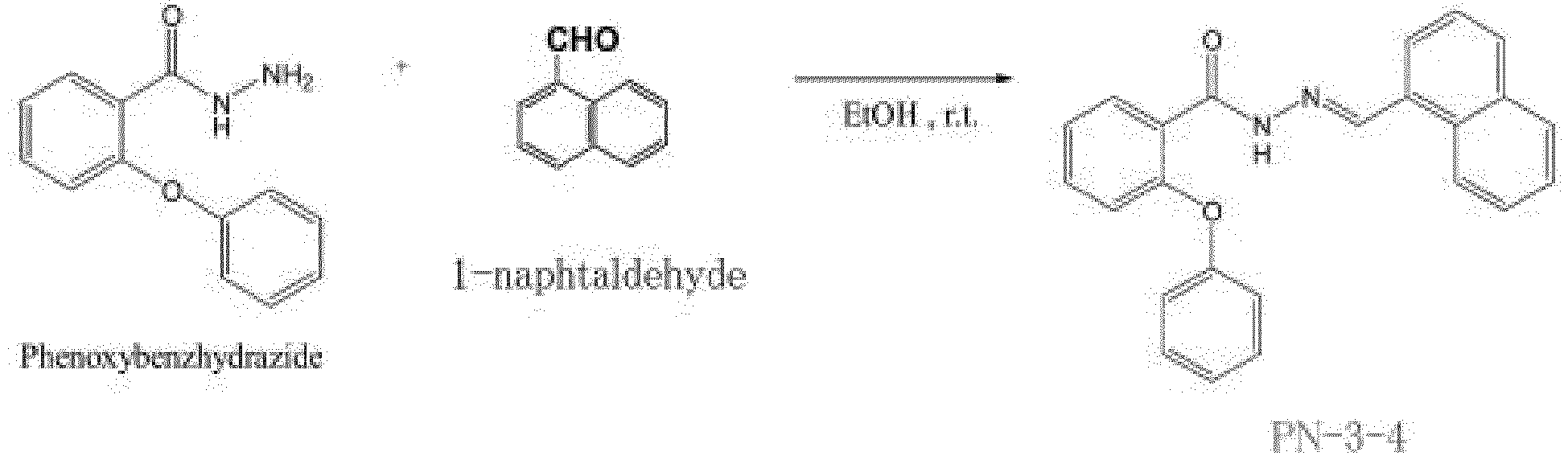
- PN-3-4 (N'-[(E)-1-naphtylmethylidene]-2-phenoxybenzohydrazide) thus obtained was analyzed as follows:
- 1H-NMR (400 MHz, CDCl3)
6.83-8.78 (m, 16H, Ar-H) 8.82 (s, 1H, -CONHN=CHC), 10.79 (s, 1H, CONHN=CH) IR(KBr) 758, 1249, 1358, 1371, 1449, 1481, 1662, 2969, 3082 cm-1; - MS (EI) Found: m/z 366. Calcd for C24H18N2O2: M+, Mol. Wt.: 366.41
Anal. Found: C, 78.37; H, 5.02; N, 7.68; O, 8.98%. Calcd for C24H18N2O2: C, 78.67; H, 4.95; N, 7.65; O, 8.73 - Synthesis was carried out using pentafluorobenzaldehyde (TOKYO CHEMICAL INDUSTRY CO., LTD.) in place of 1-naphtaldehyde in the above Example 3. Phenoxy benzhydrazide (0.24 g, 1.0 mmol, 1.0 eq) was dissolved in EtOH. Next, pentafluorobenzaldehyde (196 g, 1.0 mmol, 1.0 eq) was added, and the mixture was stirred at room temperature for 14 h. After 14 h passed, the reaction solution was filtered and was then subjected to recrystallization with EtOH to yield a compound of interest (287 mg, 71 %).

- PN-3-13 (N'-[(E)-pentafluorophenylmethylidene]-2-phenoxybenzohydrazide) thus obtained was analyzed as follows:
- 1H-NMR (400 MHz, CDCl3)
6.79-8.37(m, 9H, Ar-H) 8.53 (s, 1H, -CONHN=CH-C), 10.92 (s, 1H, CONHN=CH) IR(KBr) 750, 980, 1231, 1493, 1518, 1659, 3285 cm-1; - MS (EI) Found: m/z 406. Calcd for: C20H11F5N2O2 M Mol. Wt.: 406.31
- Synthesis was carried out using 3-phenoxy benzhydrazide (Wako Pure Chemical Industries, Ltd.) in place of 1-naphtaldehyde in the above Example 3. 3-Phenoxy benzhydrazide (0.78 g, 3.4 mmol, 1.0 eq) was dissolved in EtOH. Next, 5-methyl-furfural (0.43 g, 3.4 mmol, 1.0 eq) was added, and the mixture was stirred at room temperature for 14 h. After 14 h passed, the reaction solution was filtered and was then subjected to recrystallization with EtOH to yield a compound of interest (318 mg, 29%).

- PN-1-2 (N'-[(E)-(5-methyl-2-fulyl)methylidene]-3-phenoxybenzohydrazide) thus obtained was analyzed as follows:
- 1H-NMR (400 MHz, CDCl3)
2.35 {s, 3H, -CH=C(CH3)} 6.11{s, 1H, -CHC(CH3)} 6.69{s, 1H, -CHCHC(CH3)} 7.03-7.52 (m, 9H, Ar-H) 8.48 (s, 1H, -CONHN=CH-), 8.92 (s, 1H, CONHN=CH) IR(KBr) 746, 1018, 1233, 1287, 1300, 1489, 1580, 1651, 3048, 3194 cm-1;
MS (EI) Found: m/z 320. Calcd for C19H16N2O3: M, Mol. Wt.: 320.34
Anal. Found: C, 71.25; H, 5.04; N, 8.79; O, 14.92%. Calcd for: C19H16N2O3 C, 71.24; H, 5.03; N, 8.74; O, 14.98 -
FIG. 3 is a schematic diagram illustrating how to establish a human mesenchymal stem cell stably expressing a β-catenin/TCF4/luciferase reporter gene.FIG. 3 illustrates how to construct pTCF4-CMVpro-GL4.20 plasmid by using a luciferase expression vector. The pTCF4-CMVpro-GL4.20 plasmid contains three TCF4 sequences CCTTTGATC upstream of a CMV promoter to express luciferase. In addition, pCMVpro-GL4.20 plasmid was constructed as a control. The resulting plasmids were each linearized, introduced into UE7T-13 by electroporation, and selected using puromycin (0.25 µg/ml) supplemented in a medium. Puromycin-resistant cells were cloned and used for a luciferase assay. - Bone marrow-derived cells (UE7T-13 cells) were treated with IC-2, etc. Then, the following points were examined: (1) Proliferation Potential (Toxicity); (2) Ability of Inhibiting Wnt/β-Catenin Pathway; and (3) Ability of Differentiating into Hepatocytes.
- A human mesenchymal stem cell line, UE7T-13, was seeded on a 96-well plate (with a bottom area of 0.3 cm2) at a cell density of 9.0 x 10³ cells/cm2, and was cultured in Dulbecco's Modified Eagle's Medium (DMEM; NISSUI PHARMACEUTICAL CO., LTD.) containing 10% fetal bovine serum (FBS; JRH Biosciences, INC.), 100 U/ml penicillin, and 100 µg/ml streptomycin (Nacalai Tesque). This time point was set to
day 0. At the next day (day 1), the medium was changed to DMEM containing IC-2, etc. After that, atdays -
FIGS. 4 to 8 show the results of treating bone marrow-derived cells (UE7T-13 cells) with IC-2, etc. Treatment with IC-2, etc., somewhat inhibited the proliferation of the bone marrow-derived cells (UE7T-13 cells) in some cases. However, the proliferation potential by itself remained (except atdays day 8 with 10 µM PN-1-2). That is, IC-2, etc., had sufficiently low cell toxicity toward the bone marrow-derived cells (UE7T-13 cells), so that the compounds can be definitely used as a differentiation inducer. The results, in particular, demonstrated very low cell toxicity toward the bone marrow-derived cells (UE7T-13 cells) when IC-2 of 20 µM or less, HC-1 of 8 µM or less, PN-3-4 of 30 µM or less, PN-3-13 of 10 µM or less, or PN-1-2 of 40 µM or less was used. - The cell line having a vector stably expressing luciferase was seeded on a 24-well plate, and was cultured at 37°C. At the next day, the medium was changed to a medium containing IC-2, etc., and the cell line was further cultured at 37°C. After that, at
days - According to a reporter assay using the pTCF4-CMV-luciferase plasmid, the results demonstrated that treatment of the bone marrow-derived cells (UE7T-13 cells) with IC-2 of 15 µM or higher significantly inhibited the Wnt/β-catenin signaling pathway at 8 days after the treatment (
FIG. 9 ). In addition, the results demonstrated that treatment of the bone marrow-derived cells (UE7T-13 cells) with HC-1 of 2 µM or higher significantly inhibited the Wnt/β-catenin signaling pathway at 8 days after the treatment. Further, the results demonstrated that treatment of the bone marrow-derived cells (UE7T-13 cells) with PN-3-4 of 20 µM or higher significantly inhibited the Wnt/β-catenin at 8 days after the treatment. Furthermore, the results demonstrated that treatment of the bone marrow-derived cells (UE7T-13 cells) with PN-3-13 of 9 µM or higher significantly inhibited the Wnt/β-catenin signaling pathway at 8 days after the treatment. Moreover, the results demonstrated that treatment of the bone marrow-derived cells (UE7T-13 cells) with PN-1-2 of 3 µM or higher significantly inhibited the Wnt/β-catenin signaling pathway at 8 days after the treatment. - The cells were plated on a 6-well plate at a cell density of 9.0 x 10³ cells/cm2, and were cultured at 37°C for 24 h. After 24 h, the medium was changed to a medium containing IC-2, etc. After that, the medium was changed twice a week, and the cells were subcultured once a week to adjust a cell number at 9.0 x 10³ cells/cm2. Total RNA was collected at
days - Total RNA was extracted with a TRIzol reagent. After the extraction, in order to completely remove DNA, deoxyribonuclease was added and the mixture was incubated at 37°C for 1 h. A SuperScript First-Stand Synthesis System for RT-PCR (Invitrogen) was used for reverse transcription, and an oligo dT primer was used to convert 1 µg of RNA into cDNA. To perform a PCR, the cDNA was diluted by 5 times and 1 µl of the cDNA was used. A recombinant Taq DNA polymerase (Invitrogen) was used for the PCR. With regard to primers for human albumin, 5'-TTGGAAAAATCCCACTGCAT-3' (SEQ ID NO: 1) and 5'-CTCCAAGCTGCTCAAAAAGC-3' (SEQ ID NO: 2) were used. The PCR involved 1 cycle of 95°C for 2 min and 35 cycles of 95°C for 30 sec, 58°C for 30 sec, and 72°C for 30 sec. Human glyceraldehyde 3-phosphate dehydrogenase (GAPDH) was used as an internal control. With regard to GAPDH primers, 5'-GTCTTCTCCACCATGGAGAAGGCT-3' (SEQ ID NO: 3) and 5'-CATGCCAGTGAGCTTCCCGTTCA-3' (SEQ ID NO: 4) were used. The PCR involved 1 cycle of 95°C for 2 min and 20 cycles of 95°C for 30 sec, 60°C for 30 sec, and 72°C for 30 sec. The PCR products were subjected to electrophoresis for 30 min in a 2% agarose gel containing ethidium bromide, and their gel images were taken with a transilluminator.
-
FIGS. 10 and 11 are electrophoresis gel images illustrating differentiation induction into hepatocytes by using IC-2, etc. Treatment with any of IC-2, etc., apparently increased levels of expression of albumin, which is a representative marker for differentiation into hepatocytes, compared to those of a control. The levels of the albumin expression were particularly high when the cells were treated with 15 µM IC-2. Note that GAPDH was an internal control and the amount of RNA used was the same as that used for the test substance. Huh-7 is a human hepatoma cell line. PNU-74654 (N-[(5-methyl-2-furyl)methylideneamino]-2-phenoxy-benzamide) is a low-molecular-weight compound that reportedly inhibits a Wnt/β-catenin pathway by blocking the interaction between β-catenin and TCF. - The cells were plated on a 24-well plate, and were cultured in a medium containing IC-2, etc. Ammonium chloride (at a final concentration of 5 mM) was added to the medium, and the cells were cultured for 48, 72, and 96 h. Then, an amount of urea in the medium was determined using a QuantiChrom Urea Assay Kit (BioAssay Systems) and a fluorescence plate reader (TECAN) at a wavelength of 520 nm.
-
FIG. 12 is graphs illustrating the ability of urea synthesis (atdays 8 and 16) of samples differentiated using IC-2, etc. The ability of inducing differentiation into hepatocytes (measurement of the ability of urea synthesis) by IC-2, etc., was determined atday 8. As a result, any of the compounds apparently increased cellular urea synthesis compared with that of a negative control (0.1% DMSO). The levels increased by the same amount or more compared to those of a positive control (Huh-7). - In addition, the ability of inducing differentiation into hepatocytes (measurement of the ability of urea synthesis) by IC-2, etc., was determined at
day 16. As a result, any of the compounds apparently increased cellular urea synthesis compared with that of a negative control (0.1% DMSO). Further, the levels increased by the same amount or more compared to those of a positive control (Huh-7). Treatment with IC-2, in particular, remarkably increased the urea synthesis. The above results indicate that the cells treated with IC-2, etc., not only just express hepatocyte markers but also exert potentials as functional hepatocytes. - A cover glass (22 x 22 mm, MATSUNAMI) was sterilized with 70% EtOH and placed in a 6-well plate. Next, cells were seeded on the cover glass and cultured in a medium containing IC-2, etc. At 8, 16, and 24 days after the initiation of the culture, the cells were washed twice with PBS. Then, the cells were treated with 4% paraformaldehyde/PBS for 20 min and fixed onto the cover glass. As a negative control, 10 mg/ml of α-amylase (10 mg/ml; in 0.1 M phosphate buffer solution at pH 6.8) was added, and the cells were incubated therewith at 37°C for 1 h to digest their glycogen. Subsequently, 1% aqueous periodic acid was used for oxidation treatment for 10 min. After that, the cells were treated with a Schiff reagent for 15 min to stain glycogen. Then, the cells were washed three times with aqueous sulfurous acid and were washed three times with distilled water. After their nuclei were stained with Mayer's hematoxylin, 50% glycerol diluted with PBS was used to mount the cover glass. Finally, the cells were observed under a light microscope. The cells which had been cultured with 0.1% DMSO were used as a control.
-
FIG. 13 is photomicrographs (PAS staining) illustrating an ability of inducing differentiation into hepatocytes by using IC-2, etc. An ability of inducing differentiation into hepatocytes by using IC-2, etc., was examined (PAS staining). As a result, intracellular glycogen accumulation apparently increased compared with a negative control (0.1% DMSO), and was substantially the same level as that of a positive control (Huh-7, a hepatoma cell). - In addition to IC-2, etc., another compound for inhibiting the Wnt/β-catenin signaling pathway was also synthesized. With regard to this compound, ICG-001, and PNU-74654, the ability of inducing differentiation into hepatocytes was evaluated using PAS staining in a manner similar to the above. As a result, in all the cases of using any of the compounds, the intracellular glycogen accumulation did not increase compared to the negative control. In view of the above, the reason why the differentiation of mesenchymal stem cells into hepatocytes in this Example was successful was because IC-2, etc., has a particular structure.
- A cover glass (22 x 22 mm, MATSUNAMI) was sterilized with 70% EtOH and placed in a 6-well plate. Next, cells were seeded on the cover glass and cultured in a medium containing IC-2, etc. At 8, 16, and 14 days after the initiation of the culture, the cells were washed twice with PBS. Then, the cells were treated with 4% paraformaldehyde/PBS for 20 min and fixed onto the cover glass. After that, the cells were permeated with 0.2% Triton X-100 for 10 min. Subsequently, the cells were blocked with 3% BSA/PBS for 30 min. An anti-albumin antibody, anti-C/EBP antibody, anti-AFP antibody, or anti-CYPlAl antibody was used as a primary antibody, and the cells were incubated therewith at 4°C overnight. Next, an Alexa Fluoro 488 goat anti-mouse antibody or Alexa Fluoro 594 goat anti-rabbit antibody was used as a secondary antibody, and the cells were incubated therewith at room temperature for 1 h. DAPI was used for nuclear staining. Thereafter, 50% glycerol diluted with PBS was used to mount the cover glass. Finally, the cells were observed under a confocal laser microscope. The cells which had been cultured with 0.1% DMSO were used as a control.
-
FIG. 14 is fluorescent photomicrographs illustrating differentiation induction into hepatocytes (at day 8) by using IC-2, etc. Hepatocyte differentiation was induced by IC-2, etc. As a result, atday 8 after the treatment with IC-2, etc., levels of expression of markers such as albumin, C/EBPa, CYP1A1, and CYP3A4, which are representative markers for differentiation into hepatocytes, apparently increased compared with those of a negative control (0.1% DMSO). The levels are equivalent to those of a positive control (Huh-7).FIG. 15 is fluorescent photomicrographs illustrating differentiation induction into hepatocytes (at day 16) by using IC-2, etc. Similar results were obtained atday 16 after the treatment with IC-2, etc. Treatment with IC-2, in particular, remarkably increased the levels of expression of the markers. Note that DAPI was used as a control for each staining, and their fluorescent photomicrographs are shown. - In view of the above experimental results, the present inventors have revealed that IC-2, etc., can be used to differentiate mesenchymal stem cells into functional hepatocytes. At the same time, they have elucidated that the mesenchymal stem cells are useful as a cell source for liver regenerative medicine so as to realize liver regenerative medicine. Also, in order to induce the differentiation, the inhibition of the Wnt/β-catenin signaling pathway by IC-2, etc., was effective. These findings are important so as to develop genuinely clinically applicable liver regenerative medicine.
- Bone marrow-derived mesenchymal stem cells (UE7T-13 cells) were treated with a compound, hexachlorophene (H4625-25G; Sigma-Aldrich, Inc.) or IC-2. At that time, gene expression of each of liver-specific secretory proteins and cytokines was evaluated. First, bone marrow-derived mesenchymal stem cells (UE7T-13 cells) were seeded on a 6-well plate (with a bottom area of 9.6 cm2) at a cell density of 9.0 × 10³ cells/cm2, and were cultured under conditions at 37°C and 5% CO2 in Dulbecco's Modified Eagle's Medium (DMEM; NISSUI PHARMACEUTICAL CO., LTD.) containing 10% fetal bovine serum (FBS; JRH Biosciences, INC.), 100 U/ml penicillin, and 100 µg/ml streptomycin (Nacalai Tesque). This time point was set to
day 0. - At the next day corresponding to
day 1, the medium was changed to DMEM containing hexachlorophene at a final concentration of 0.8 µM or IC-2 at a final concentration of 15 µM. Atday 4, the medium was changed, and atday 8, the cells were reseeded and subcultured at a cell density of 9.0 x 10³ cells/cm2. After that, the medium was replaced twice during 8 days, and the cells were subcultured every 8 days. Then, the similar operation was carried out untilday 24. Atdays - Sequences of primers used in a PCR for detecting expression of genes encoding human liver-specific secretory proteins are each described as follows: primers for albumin were 5'-TTGGAAAAATCCCACTGCAT-3' (SEQ ID NO: 1) and 5'-CTCCAAGCTGCTCAAAAAGC-3' (SEQ ID NO: 2); primers for α1-antitrypsin were 5'-CAAGGAGCTTGACAGAGACACAGTTTTT-3' (SEQ ID NO: 5) and 5'-GTGTCCTTGACTTCAAAGGGTCTCT-3' (SEQ ID NO: 6); primers for ceruloplasmin were 5'-CGACTTGGGATTATGCCTCTGACC-3' (SEQ ID NO: 7) and 5'-CCCAATTCTATCTGGGCCATTTTGA-3' (SEQ ID NO: 8); primers for transthyretin were 5'-AAGGCACTTGGCATCTCCCCA-3' (SEQ ID NO: 9) and 5'-CAGGGCGGCAATGGTGTAGC-3' (SEQ ID NO: 10); primers for apolipoprotein E were 5'-GTCCTTCCCCAGGAGCCGAC-3' (SEQ ID NO: 11) and 5'-GTCTCCACCGCTTGCTCCAC-3' (SEQ ID NO: 12); primers for apolipoprotein B were 5'-GCTTCAAGCCCATCCGCACA-3' (SEQ ID NO: 13) and 5'-TGACAGGACTGGCTGCTGCT-3' (SEQ ID NO: 14); primers for complement C3 were 5'-CAGCACCATGGGACCCACCTCAG-3' (SEQ ID NO: 15) and 5'-CTCTCCAGCCGCAAGATGTTGGG-3' (SEQ ID NO: 16); primers for complement C4 were 5'-ACTTTGAGACCGAGGGGCCC-3' (SEQ ID NO: 17) and 5'-GGCACTTCCTGCACAGCCTC-3' (SEQ ID NO: 18); primers for retinol-binding protein 4 were 5'-CCCAGAAGCGCAGAAGATT-3' (SEQ ID NO: 19) and 5'-AAGGTTTCTTTCTGATCTGCCAT-3' (SEQ ID NO: 20); primers for retinol-binding protein 5 were 5'-CTTGTGGCCAACTGGCTC-3' (SEQ ID NO: 21) and 5'-GCTTCAGCAAGTTGGCGA-3' (SEQ ID NO: 22); primers for haptoglobin were 5'-TTCCAGAGGCAAGACCAACC-3' (SEQ ID NO: 23) and 5'-TGCCTGAGTCCACTGCAAA-3' (SEQ ID NO: 24); primers for α-fibrinogen were 5'-AGGATTCTCATTCGTTGACCAC-3' (SEQ ID NO: 25) and 5'-TCTGACACTCGGTTGTAGGT-3' (SEQ ID NO: 26); primers for β-fibrinogen were 5'-AAAGTTAGAATCTGATGTCTCAGCTCAA-3' (SEQ ID NO: 27) and 5'-CTTTCCTGATAATTTCCTCACATTCTTT-3' (SEQ ID NO: 28); and primers for GAPDH, an internal control, were 5'-AGCCACATCGCTCAGACAC-3' (SEQ ID NO: 29) and 5'-GCCCAATACGACCAAATCC-3' (SEQ ID NO: 30).
- The PCR involved 1 cycle of 94°C for 2 min, followed by 35 cycles of 94°C for 30 sec, a temperature corresponding to each primer for 30 sec, and 72°C for 30 sec, and 1 cycle of 72°C for 5 min. The temperature corresponding to each primer was 58°C for albumin, 68°C for complement C3, 60°C for retinol-binding
protein 4, 56°C for retino-bindingprotein 1 and haptoglobin, or 64°C for the rest. - Sequences of primers used in a PCR for detecting expression of genes encoding human cytokines are each described as follows: primers for interleukin 6 (IL6) were 5'-CCAGAGCTGTGCAGATGAG-3' (SEQ ID NO: 31) and 5'-GTCAGCAGGCTGGCATTT-3' (SEQ ID NO: 32); primers for IL-1 receptor antagonist (IL1ra) were 5'-CAGCTGGAGGCAGTTAACAT-3' (SEQ ID NO: 33)and 5'-CGCCTTCGTCAGGCATATTG-3' (SEQ ID NO: 34); primers for hepatocyte growth factor (HGF) were 5'-CCCTTCAATAGCATGTCAAGTGG-3' (SEQ ID NO: 35) and 5'-GTTCCCTTGTAGCTGCGT-3' (SEQ ID NO: 36); primers for vascular endothelial growth factor (VEGF) were 5'-TTGCCTTGCTGCTCTACCT-3' (SEQ ID NO: 37) and 5'-TCCATGAACTTCACCACTTCGT-3' (SEQ ID NO: 38); primers for transforming growth factor α (TGFα) were 5'-CTGAAGGGAAGAACCGCTTG-3' (SEQ ID NO: 39) and 5'-AGCCTTTCTTTATTGATCTGCCACA-3' (SEQ ID NO: 40); primers for transforming growth factor β (TGFβ) were 5'-CTGCGTCTGCTGAGGC-3' (SEQ ID NO: 41) and 5'-TCCACGGCTCAACCACTG-3' (SEQ ID NO: 42); primers for epidermal growth factor (EGF) were 5'-TGGGTCAAGGCAAGAGAGAGTA-3' (SEQ ID NO: 43) and 5'-GATTCCTTCCTGTTGATTTGACCA-3' (SEQ ID NO: 44); primers for basic fibroblast growth factor (bFGF) were 5'-GGGTCCGGGAGAAGAGC-3' (SEQ ID NO: 45) and 5'-GCCAGGTAACGGTTAGCAC-3' (SEQ ID NO: 46); primers for heparin-binding EGF-like growth factor (HB-EGF) were 5'-GGACCGGAAAGTCCGT-3' (SEQ ID NO: 47) and 5'-GCTCCTCCTTGTTTGGTGT-3' (SEQ ID NO: 48); primers for amphiregulin (AREG) were 5'-AACGAAAGAAACTTCGACAAGAGA-3' (SEQ ID NO: 49) and 5'-ATGATCCACTGGAAAGAGGACC-3' (SEQ ID NO: 50); primers for stem cell factor (SCF) were 5'-AGGGACAGTGGAGAGGG-3' (SEQ ID NO: 51) and 5'-GCAAGTGAGAATCCAAGTTTGTGT-3' (SEQ ID NO: 52); primers for angiogenin were 5'-TTCCATTGTCCTGCCCG-3' (SEQ ID NO: 53) and 5'-AAGTGTGTGTACCTGGAGTTATC-3' (SEQ ID NO: 54); primers for angiopoietin were 5'-ATGTGCAAATGTGCCCTCA-3' (SEQ ID NO: 55) and 5'-TCGCTTCTGACATTGCGCT-3' (SEQ ID NO: 56); primers for tumor necrosis factor (TNFα) were 5'-CCCAGGGACCTCTCTCTAATC-3' (SEQ ID NO: 57) and 5'-GGCCAGGAGGGCATTG-3' (SEQ ID NO: 58); primers for fas antigen (Fas) were 5'-GTTGGTGGACCCGCTCAGTA-3' (SEQ ID NO: 59) and 5'-AACAGACGTAAGAACCAGAGGTAG-3' (SEQ ID NO: 60); primers for tissue inhibitor of metalloproteinase 3 (TIMP3) were 5'-GGCAGCAGCGGCAATG-3' (SEQ ID NO: 61) and 5'-CCACCTTGGCCCGGATCA-3' (SEQ ID NO: 62); and primers for GAPDH, an internal control, were 5'-AGCCACATCGCTCAGACAC-3' (SEQ ID NO: 63) and 5'-GCCCAATACGACCAAATCC-3' (SEQ ID NO: 64).
- The PCR involved 1 cycle of 94°C for 2 min, followed by 35 cycles of 94°C for 30 sec, a temperature corresponding to each primer for 30 sec, and 72°C for 30 sec, and 1 cycle of 72°C for 5 min. The temperature corresponding to each primer was 60°C for heparin-binding EGF-like growth factor (HB-EGF), amphiregulin (AREG), stem cell factor (SCF), tumor necrosis factor (TNFα), and fas antigen (Fas), or 56°C for the rest. The PCR products were subjected to electrophoresis for 30 min in a 2% agarose gel containing ethidium bromide, and their electrophoresis gel images were taken with a transilluminator.
-
FIG. 16 illustrates the results of examining gene expression of liver-specific secretory proteins and cytokines. As a result, hepatocytes as obtained using treatment with hexachlorophene or IC-2 had increased levels of expression of a plurality of the liver-specific secretory proteins and cytokines. In addition, even if the differentiation was induced, the levels of expression of the liver-specific secretory proteins or cytokines that are effective in liver regeneration were found to be maintained. - A bFGF-sustained-release device was prepared in accordance with American Journal of and Nature Medicine 2007, 13(7), 880-885. The bFGF-sustained-release device was implanted under the skin in the dorsal region of an immunodeficient mouse, a NOD-SCID mouse, to induce neovascularization. First, 1 µl of somnopentyl (Kyoritsuseiyaku Corporation), a systemic anesthetic, per 1g of body weight of the NOD-SCID mouse was intraperitoneally administered to put the mouse under anesthesia. After the introduction of anesthesia, the left dorsal skin of the mouse was shaved. Next, operating scissors or a similar surgical instrument was used to cut, in a direction perpendicular to a body axis direction, about 1.5 to 2.0 cm of the skin with only subcutaneous tissue which was positioned approximately 1 cm caudal to the position where the bFGF device would be implanted. The operating scissors or the similar surgical instrument was inserted under the skin from this incision and moved toward the rostral direction to go beyond the position where the device would be implanted. Then, the operating scissors were made open, which created a space where the device could be implanted under the skin of the mouse. The bFGF device was inserted into this space, and the incision was closed with a surgical clip or a suture.
- The mice were fed under normal conditions for 10 to 12 days. After 10 to 12 days, 1 µl of somnopentyl (Kyoritsuseiyaku Corporation), a systemic anesthetic, per 1g of body weight of the NOD-SCID mouse was intraperitoneally administered to put the mouse under anesthesia in a manner similar to when the bFGF device had been implanted. The portion surrounding the site where the bFGF device had been implanted was cut with operating scissors or a similar surgical instrument, and the skin with subcutaneous tissue was excised. As a control, the right dorsal skin with subcutaneous tissue having no implanted bFGF device was likewise cut in the same NOD-SCID mouse, and was excised. The excised skin with subcutaneous tissue was fixed with 4% paraformaldehyde (Nacalai Tesque). The post-fixed tissue was embedded in paraffin, used to prepare tissue sections by using a microtome, and stained with hematoxylin and eosin. By doing so, whether or not the implantation of the bFGF device under the skin exerted an neovascularization effect was examined (
FIG. 17 ). - 7-week-old male NOD-SCID mice were divided into four groups (
FIG. 18 ).Group 1 was a sham operation group in which no cell sheet was transplanted.Group 2 was a group in which one cell sheet was transplanted under the skin after the neovascularization.Group 3 was a group in which two cell sheets were transplanted under the skin after the neovascularization.Group 4 was a group in which three cell sheets were transplanted under the skin after the neovascularization. At 12 days before the cell sheet transplantation, the bFGF device was implanted under the dorsal skin of all the four groups to induce neovascularization. - First, 1 µl of somnopentyl (Kyoritsuseiyaku Corporation), a systemic anesthetic, per 1g of body weight of a NOD-SCID mouse was intraperitoneally administered to put the mouse under anesthesia. After the introduction of anesthesia, the left dorsal skin of the mouse was shaved. Next, operating scissors or a similar surgical instrument was used to cut, in a direction perpendicular to a body axis direction, about 1.5 to 2.0 cm of the skin with only subcutaneous tissue which was positioned approximately 1 cm caudal to the position where the bFGF device would be implanted. The operating scissors or the similar surgical instrument was inserted under the skin from this incision and moved toward the rostral direction to go beyond the position where the device would be implanted. Then, the operating scissors were made open, which created a space where the device could be implanted under the skin of the mouse. The bFGF device was inserted into this space, and the incision was closed with a surgical clip or a suture.
- After the mice were fed under normal conditions for 12 days, the cell sheet was transplanted. The cell sheets were prepared according to the following procedure. At 8 days before transplantation, a human bone marrow-derived mesenchymal stem cell line (UE7T-13 cells) was seeded on a 6-cm dish (CellSeed Inc.), which is an UpCell temperature-responsive cell cultureware for collecting a cell sheet, at a cell density of 9.0 × 10³ cells/cm2, and was cultured under conditions at 37°C and 5% CO2 in Dulbecco's Modified Eagle's Medium (DMEM; NISSUI PHARMACEUTICAL CO., LTD.) containing 10% fetal bovine serum (FBS; JRH Biosciences, INC.), 100 U/ml penicillin, and 100 µg/ml streptomycin (Nacalai Tesque). This time point was set to
day 0. - At the next day corresponding to
day 1, the medium was changed to DMEM containing hexachlorophene at a final concentration of 0.8 µM. Similar today 1, the medium was changed atday 4. Atday 8 corresponding to one day before the cell sheet transplantation, the attached cells of periphery of the bottom of the dish was scraped with the tip of a disposable tip, etc., and the medium was changed with a medium at room temperature. Then, the cells were incubated under conditions at 20°C and 5% CO2 for 20 min or longer to produce a cell sheet. Until its transplantation, the cell sheet was kept under conditions at 20°C and 5% CO2. - At 12 days after the implantation of the bFGF device, the cell sheet was transplanted into
Group 2 to 4 mice. First, 1 µl of somnopentyl (Kyoritsuseiyaku Corporation), a systemic anesthetic, per 1g of body weight of the NOD-SCID mouse was intraperitoneally administered to put the mouse under anesthesia. Next, the incision opened during the implantation of the bFGF device was cut again. Then, operating scissors or a similar surgical instrument was used to cut only the subcutaneous tissue in a direction from the dorsal edge of the incision straight toward the rostral direction to open the subcutaneous tissue and to remove the bFGF device. After that, a Cell Shifter (CellSeed Inc.), which is a support for collecting a cell sheet, was placed on a cell sheet whose medium had been changed with serum-free DMEM and was then completely removed. The cell sheet, together with the support, was collected using forceps, etc. The support having thereon the cell sheet was placed on the site where the bFGF device had induced neovascularization. The operator waited about 3 to 5 min for adhesion of the cell sheet, and confirmed that the cell sheet was attached under the skin of the mouse. Thereafter, forceps, etc., were used to remove only the support. - Regarding
Group 2, one cell sheet was transplanted, and the incision was then closed with a surgical clip or a suture. RegardingGroups Group 3, after two cell sheets were transplanted, the incision was then closed with a surgical clip or a suture. RegardingGroup 4, the third cell sheet was transplanted again on the two cell sheets that had already been transplanted, and the incision was then closed with a surgical clip or a suture. RegardingGroup 1 as a control, the bFGF device was removed using the same process as ofGroups 2 to 4, and the incision was then closed with a surgical clip or a suture. - Then, the mice were fed under normal conditions again. At the next day after the operation, 0.2 µl of carbon tetrachloride per 1g of body weight of the mouse was diluted by 10 times with olive oil, and was given to all the
mice including Groups 1 to 4 through a disposable oral probe, namely a stomach tube. Whether or not the mice survived was daily checked from the day of the transplantation till 8 days after that. The body weight was measured once every other day. Atdays - At
day 8 after the cell sheet transplantation, all the mice were put under inhalation anesthesia using isoflurane (Abbott Japan). Then, operating scissors or a similar surgical instrument, and forceps were used to perform laparotomy. After that, a 27-G needle and a 1-ml syringe were used to draw all the blood from inferior vena cava. After the blood sampling, a subcutaneous tissue containing a piece of the transplanted cell sheet and the whole liver were excised. The wet weight of the excised whole liver was measured, and its image was photographed with a digital camera. Of the excised tissues, those for RNA extraction were cut into small pieces at a wet weight of 0.1 g by means of operating scissors or a similar surgical instrument. Next, 1 ml of TRIzol (Invitrogen) was added thereto. Then, a POLYTRON (KINEMATICA AG) was used for homogenization, and the samples were stored at a freezer at -30°C until they were used in experiments. Tissue pieces for protein extraction were likewise cut into small pieces at a wet weight of 0.1 g by means of operating scissors or a similar surgical instrument. The samples were put into a 15-ml tube, subsequently immersed in liquid nitrogen, instantaneously frozen, and stored in a deep freezer at -80°C until they were used in experiments. Tissue pieces for histochemical staining were fixed with 4% paraformaldehyde (Nacalai Tesque). The post-fixed tissues were embedded in paraffin, and their tissue sections were then prepared with a microtome and stained with hematoxylin and eosin. Tissue sections other than those were stored at room temperature until immunohistochemical staining was carried out. Serum of the vein blood collected from inferior vena cava was separated using the same procedure as described above, and was stored in a deep freezer at -80°C. - The levels of serum transaminases of the mice at
days - The levels of serum bilirubin of the mice at
days -
FIG. 19 shows the above results. Subcutaneous transplantation of two or three layers of the cell sheet resulted in decreased levels of the serum transaminases. In addition, transplantation of one or more layers of the cell sheet resulted in decreased level of the bilirubin value. - 7-week-old male NOD-SCID mice were divided into four groups.
Group 1 was a sham operation group in which no cell sheet was transplanted.Group 2 was a group in which one cell sheet was transplanted on the surface of the left lateral lobe of the liver.Group 3 was a group in which two cell sheets were transplanted on the surface of the left lateral lobe of the liver.Group 4 was a group in which three cell sheets were transplanted on the surface of the left lateral lobe of the liver. - The cell sheets were prepared according to the following procedure. At 8 days before the cell sheet transplantation, a human bone marrow-derived mesenchymal stem cell line (UE7T-13 cells) was seeded on a 6-cm dish (CellSeed Inc.), which is an UpCell temperature-responsive cell cultureware for collecting a cell sheet, at a cell density of 9.0 × 10³ cells/cm2, and was cultured under conditions at 37°C and 5% CO2 in Dulbecco's Modified Eagle's Medium (DMEM; NISSUI PHARMACEUTICAL CO., LTD.) containing 10% fetal bovine serum (FBS; JRH Biosciences, INC.), 100 U/ml penicillin, and 100 µg/ml streptomycin (Nacalai Tesque). This time point was set to
day 0. - At the next day corresponding to
day 1, the medium was changed to DMEM containing hexachlorophene at a final concentration of 0.8 µM. Similar today 1, the medium was changed atday 4. Atday 8 corresponding to one day before the cell sheet transplantation, the attached cells of periphery of the bottom of the dish was scraped with the tip of a disposable tip, etc., and the medium was changed with a medium at room temperature. Then, the cells were incubated under conditions at 20°C and 5% CO2 for 20 min or longer to produce a cell sheet. Until its transplantation, the cell sheet was kept under conditions at 20°C and 5% CO2. - First, on the day of transplantation, 1 µl of somnopentyl (Kyoritsuseiyaku Corporation), a systemic anesthetic, per 1g of body weight of the NOD-SCID mouse was intraperitoneally administered to put the mouse under anesthesia. After the introduction of anesthesia, the abdomen of the mouse was shaved, and operating scissors or a similar surgical instrument was used to cut the abdominal skin along the midline. Next, the peritoneum was dissected along the midline by using operating scissors or a similar surgical instrument. Then, forceps, etc., were used to hold the skin and the peritoneum to keep the surgical field open in the peritoneum. A suture was made to pass through the xiphoid process twice, and was pinched and fixed. This further allowed the surgical field surrounding the liver to be kept open. With regard to the cell sheet, after the medium was changed to serum-free DMEM in the cultureware, the medium was completely removed. Then, a Cell Shifter (CellSeed Inc.), which is a support for collecting a cell sheet, was placed on the cell sheet. This cell sheet, together with the support, was collected using forceps, etc. Then, the cell sheet having attached thereon the support was placed on the surface of the left lateral lobe of the liver. The operator waited about 3 to 5 min for adhesion of the cell sheet, and confirmed that the cell sheet was attached on the surface of the liver of the mouse. Thereafter, forceps, etc., were used to remove only the support.
- With regard to
Group 2, after the first cell sheet was transplanted, the suture that passed through the xiphoid process was removed; the forceps, etc., that fixed the peritoneum and the skin were taken out; and the peritoneum was closed with a suture. After that, the skin was closed with a surgical clip or a suture. RegardingGroups Group 3, after the two cell sheets were transplanted, the suture that passed through the xiphoid process was removed; the forceps, etc., that fixed the peritoneum and the skin were taken out; and the peritoneum was closed with a suture. Thereafter, the skin was closed with a surgical clip or a suture. RegardingGroup 4, the third cell sheet was transplanted on the two cell sheets that had already been transplanted, and the peritoneum and the skin were closed in a similar fashion. RegardingGroup 1 as a control, after the same operation as of the transplantation groups was applied to keep the surgical field open, the peritoneum and the skin were closed. - Then, the mice were fed under normal conditions until the day of sacrifice. At
day 7 after the cell sheet transplantation, the mice were put under inhalation anesthesia using isoflurane (Abbott Japan) and subjected to laparotomy in a manner similar to when the transplantation was performed. Thereafter, the whole liver was excised. The excised liver was photographed with a digital camera. Of the tissue pieces containing the transplanted cell sheet, those for RNA extraction were cut into small pieces at a wet weight of 0.1 g by means of operating scissors or a similar surgical instrument. Next, 1 ml of TRIzol (Invitrogen) was added thereto. Then, a POLYTRON (KINEMATICA AG) was used for homogenization, and the samples were stored at a freezer at -30°C until they were used in experiments. Tissue pieces for histochemical staining were fixed with 4% paraformaldehyde (Nacalai Tesque). The post-fixed tissues were embedded in paraffin, and their tissue sections were then prepared with a microtome and stained with hematoxylin and eosin. Tissue sections other than those were stored at room temperature until immunohistochemical staining was carried out. -
FIG. 20 shows the above results. Transplantation of the cell sheet on the liver surface resulted in suppression of liver dysfunction. - 9-week-old male NOD-SCID mice were divided into four groups (
FIG. 21 ).Group 1 was a sham operation group in which no cell sheet was transplanted.Group 2 was a group in which one cell sheet was transplanted on each of two sites of the surface of the left lateral lobe of the liver.Group 3 was a group in which two cell sheets were transplanted on each of two sites of the surface of the left lateral lobe of the liver.Group 4 was a group in which three cell sheets were transplanted on each of two sites of the surface of the left lateral lobe of the liver. - The cell sheets were prepared according to the following procedure. At 8 days before the cell sheet transplantation, a human bone marrow-derived mesenchymal stem cell line (UE7T-13 cells) was seeded on a 6-cm dish (CellSeed Inc.), which is an UpCell temperature-responsive cell cultureware for collecting a cell sheet, at a cell density of 9.0 × 10³ cells/cm2, and was cultured under conditions at 37°C and 5% CO2 in Dulbecco's Modified Eagle's Medium (DMEM; NISSUI PHARMACEUTICAL CO., LTD.) containing 10% fetal bovine serum (FBS; JRH Biosciences, INC.), 100 U/ml penicillin, and 100 µg/ml streptomycin (Nacalai Tesque). This time point was set to
day 0. - At the next day corresponding to
day 1, the medium was changed to DMEM containing hexachlorophene at a final concentration of 0.8 µM. Similar today 1, the medium was changed atday 4. Atday 8 corresponding to one day before the cell sheet transplantation, the attached cells of periphery of the bottom of the dish was scraped with the tip of a disposable tip, etc., and the medium was changed with a medium at room temperature. Then, the cells were incubated under conditions at 20°C and 5% CO2 for 20 min or longer to produce a cell sheet. Until its transplantation, the cell sheet was kept under conditions at 20°C and 5% CO2. - First, on the day of transplantation, 1 µl of somnopentyl (Kyoritsuseiyaku Corporation), a systemic anesthetic, per 1g of body weight of the NOD-SCID mouse was intraperitoneally administered to put the mouse under anesthesia. After the introduction of anesthesia, the abdomen of the mouse was shaved, and operating scissors or a similar surgical instrument was used to cut the abdominal skin along the midline. Next, the peritoneum was dissected along the midline by using the operating scissors or the similar surgical instrument. Then, forceps, etc., were used to hold the skin and the peritoneum to keep the surgical field open in the peritoneum. A suture was made to pass through the xiphoid process twice, and was pinched and fixed. This further allowed the surgical field surrounding the liver to be kept open. With regard to the cell sheet, after the medium was changed to serum-free DMEM in the cultureware, the medium was completely removed. Then, a Cell Shifter (CellSeed Inc.), which is a support for collecting a cell sheet, was placed on the cell sheet. This cell sheet, together with the support, was collected using forceps, etc. Then, the cell sheet having attached thereon the support was placed on each of two sites of the surface of the left lateral lobe of the liver. The operator waited about 3 to 5 min for adhesion of the cell sheet, and confirmed that the cell sheet was attached on the surface of the liver of the mouse. Thereafter, forceps, etc., were used to remove only the support.
- With regard to
Group 2, after the first cell sheet was transplanted on each of two sites, the suture that passed through the xiphoid process was removed; the forceps, etc., that fixed the peritoneum and the skin were taken out; and the peritoneum was closed with a suture. After that, the skin was closed with a surgical clip or a suture. RegardingGroups Group 3, after the two cell sheets were transplanted on each of two sites, the suture that passed through the xiphoid process was removed; the forceps, etc., that fixed the peritoneum and the skin were taken out; and the peritoneum was closed with a suture. Then, the skin was closed with a surgical clip or a suture. RegardingGroup 4, the third cell sheet was transplanted on each of two sites having the cell sheets that had already been transplanted, and the peritoneum and the skin were closed in a similar fashion. RegardingGroup 1 as a control, after the same operation as of the transplantation groups was applied to keep the surgical field open, the peritoneum and the skin were closed. - At the next day after the operation, 0.2 µl of carbon tetrachloride per 1g of body weight of the mouse was diluted by 10 times with olive oil, and was given to all the
mice including Groups 1 to 4 through a disposable oral probe, namely a stomach tube. Whether or not the mice survived was daily checked from the day of the transplantation till 8 days after that. The body weight was measured once every other day. Atdays - At
day 8 after the cell sheet transplantation, all the mice were put under inhalation anesthesia using isoflurane (Abbott Japan). Then, operating scissors or a similar surgical instrument, and forceps were used to perform laparotomy. After that, a 27-G needle and a 1-ml syringe were used to draw all the blood from inferior vena cava. After the blood sampling, the whole liver was excised. The wet weight of the excised whole liver was measured, and its image was photographed with a digital camera. Of the tissue pieces containing the transplanted cell sheet, those for RNA extraction were cut into small pieces at a wet weight of 0.1 g by means of operating scissors or a similar surgical instrument. Next, 1 ml of TRIzol (Invitrogen) was added thereto. Then, a POLYTRON (KINEMATICA AG) was used for homogenization, and the samples were stored at a freezer at -30°C until they were used in experiments. Tissue pieces for protein extraction were likewise cut into small pieces at a wet weight of 0.1 g by means of operating scissors or a similar surgical instrument. The samples were put into a 15-ml tube, subsequently immersed in liquid nitrogen, instantaneously frozen, and stored in a deep freezer at -80°C until they were used in experiments. Tissue pieces for histochemical staining were fixed with 4% paraformaldehyde (Nacalai Tesque). The post-fixed tissues were embedded in paraffin, and their tissue sections were then prepared with a microtome and stained with hematoxylin and eosin. Tissue sections other than those were stored at room temperature until immunohistochemical staining was carried out. Serum of the vein blood collected from inferior vena cava was separated using the same procedure as described above, and was stored in a deep freezer at -80°C. -
FIG. 22 shows the above results. Transplantation of the cell sheet on two sites of the liver surface resulted in suppression of liver dysfunction. This suppression was remarkable when two or three layers of the cell sheet were transplanted. - The levels of serum transaminases of the mice at
days -
FIG. 23 shows the results. Transplantation of the cell sheet on two sites of the liver surface resulted in a large decrease in levels of the serum transaminases. This decrease was remarkable when two or three layers of the cell sheet were transplanted. - The levels of serum bilirubin of the mice at
days -
FIG. 24 shows the results. Transplantation of the cell sheet on two sites of the liver surface resulted in a large decrease in levels of bilirubin. - The number of surviving mice was counted each day after the transplantation according to the procedure in
FIG. 21 . Based on the number, a survival curve was drawn using PASW statistical software according to a Kaplan-Meier method. The survival rate was tested by a logrank test. -
FIG. 25 shows the results. Transplantation of the cell sheet on two sites of the liver surface resulted in a large increase in the survival rate. - Samples were stored for RNA extraction according to the procedure in
FIG. 21 . Then, their total RNA was extracted according to the attached instruction regarding TRIzol (Invitrogen). After the extraction, deoxyribonuclease (Nippon Gene) was added, and the mixture was incubated at 37°C for 30 min to remove DNA. In a reverse transcription reaction, SuperScriptII Reverse Transcriptase (Invitrogen) and Oligo(dT)15 primer were added to 1 µg of RNA to convert it into cDNA. In a RT-PCR method, the cDNA was diluted by 5 times. Next, 1 µl of the cDNA was used as a template. Then, rTaq DNA polymerase (TOYOBO) was used to amplify the cDNA. After that, a PCR was conducted according to the procedure described in the above Example 8. The PCR products were subjected to electrophoresis for 30 min in a 2% agarose gel containing ethidium bromide, and their electrophoresis gel images were taken with a transilluminator. -
FIG. 26 illustrates the results of examining gene expression of liver-specific secretory proteins and cytokines. Among those shown inFIG. 26 , serum protein markers or cytokines having increased expression (e.g., α1-AT) seem to be effective in liver regeneration. Note that markers having no recognizable expression (e.g., Apolipoprotein B) may not be directly involved in the present liver regeneration mechanism. In addition, when the cell sheet was transplanted on two or more sites of the liver surface, in particular, serum protein markers and cytokines had increased gene expression compared with those when no cell sheet was transplanted. - Tissue sections as prepared according to the procedure in
FIG. 21 were used to perform immunofluorescence staining. Human α1-antitrypsin was stained according to the following procedure. Tissue sections were subjected to deparaffinization using xylene and hydration treatment using 100% ethanol, were washed with tap water followed by washing with 1 x PBS(-), and were then blocked in a blocking solution containing goat serum at room temperature for 20 min. One or two drops of a rabbit polyclonal anti-al-antitrypsin antibody (Abcam) were added to the tissue sections. The sections were kept in a moist chamber at 4°C overnight to carry out a primary antibody reaction. After washing with 1 x PBS(-), 100 µl of Alexa Fluor 488 goat anti-rabbit IgG (H+L) antibody (Invitrogen) that had been diluted by 200 times was added onto the tissue sections. Then, the sections were kept in a dark place at room temperature for 1 h to carry out a secondary antibody reaction. After washing with 1 x PBS(-), each section was covered. Staining images were observed under a fluorescence microscope (IX71, Olympus Corporation), and the images were obtained. -
FIG. 27 shows the above results. The transplanted hepatocyte sheet and hepatic parenchyma of the mouse were found to have expression of human α1-antitrypsin. - Professor Okano and Associate Professor Matsuzaki of Keio University have developed a method for sorting a CD90+CD271+ mesenchymal stem cell (
JP2009-060840A FIG. 28 shows an overview of the method. Based on the method, mesenchymal stem cells were sorted. - Human bone marrow mononuclear cells that had been purchased from Lonza Inc. were suspended in 1 ml of Hanks' balanced salt solution (HBSS, Gibco, Life Technologies Corp.) supplemented with a 100-fold diluted APC-labeled anti-CD90 antibody, a 407-fold diluted PE-labeled anti-CD271 antibody, and 2% fetal bovine serum (FBS, Defined, Thermo Fisher Scientific Inc.), and were stained on ice for 30 min. A flow cytometer (MoFlo XDP, Beckman Coulter Inc.) was used to selectively separate mesenchymal stem cells. The resulting cells were cultured in Dulbecco's Modified Eagle's Medium (Low Glucose, Gibco, Life Technologies Corp.) containing 20% FBS, 100 U/ml penicillin, 100 ng/ml streptomycin (Nacalai Tesque), and 20 ng/ml basic fibroblast growth factor (TRANS GENIC INC., Ltd.) on a 35-mm dish under conditions at 37°C and 5% CO2. The medium was changed every 4 days. After colonies were formed, the cells were detached using a 0.025% trypsin/1 mM EDTA solution (Nacalai Tesque), split on a 100-mm dish, and cultured under conditions at 37°C and 5% CO2. Then, the cells which were kept 70 to 80% confluent were detached using trypsin treatment. These cells in one dish were divided into four dishes for passage.
- Cells were seeded on a 96-well plate at a cell density of 5.0×10³ cells/cm2. After 24 h, the medium was changed to a medium containing 0.1% DMSO or IC-2 (at a final concentration of 40 or 45 µM). Then, the cells were cultured under conditions at 37°C and 5% CO2. At 4 days after the cells were seeded, the medium was changed to a medium containing the above compound. At 3 days before measurement, the cells were infected at MOI = 10 with a lentivirus (SABiosciences, QIAGEN N.V.) that expressed firefly luciferase under control of mCMV promoter. As an internal control, the cells were simultaneously infected with a lentivirus (SABiosciences, QIAGEN N.V.) that expressed Renilla luciferase. At 8 days after the chemical treatment with the above compound, a Dual-Luciferase Reporter Assay System (Promega Corp.) was used to measure luciferase activity by using a luminometer.
-
FIG. 29 shows the results of the luciferase assay. The results demonstrated that treatment of the bone marrow-derived mesenchymal stem cells with IC-2 significantly inhibited the Wnt/β-catenin signaling pathway at 8 days after the treatment. - Cells were seeded on a 6-well plate at a cell density of 1.8 x 10⁴ cells/cm2. This day was designated as
day 0. Atday 1 corresponding to the next day, the medium was changed to a medium containing IC-2 (at a final concentration of 25, 35, or 45 µM) or 0.1% DMSO. Atday 4, the medium was changed in a manner similar to that atday 1, and atday 8, the cells were reseeded and subcultured at a cell density of 1.8 x 10⁴ cells/cm2. After that, the medium was replaced twice during 8 days, and the cells were subcultured every 8 days. Then, the similar operation was carried out untilday 24. Atdays - In order to determine hepatocyte differentiation markers, levels of expression of albumin were measured. The primers used had the following sequences: 5'-TTGGAAAAATCCCACTGCAT-3' (SEQ ID NO: 1) and 5'-CTCCAAGCTGCTCAAAAAGC-3' (SEQ ID NO: 2).
- The PCR involved 1 cycle of 94°C for 2 min, followed by 35 cycles of 94°C for 30 sec, 58°C for 30 sec, and 72°C for 30 sec, and 1 cycle of 72°C for 5 min. The PCR products were subjected to electrophoresis for 30 min in a 2% agarose gel containing ethidium bromide, and their electrophoresis gel images were taken with a transilluminator.
-
FIG. 30 shows the results of examining gene expression of albumin. These results demonstrated that treatment with 45 µM of IC-2, which had exhibited an effect of inhibiting the Wnt/β catenin signaling pathway in the aboveFIG. 29 , induced a remarkable increase in albumin expression when compared with 0.1% DMSO. - A fresh bone marrow aspirate was collected under informed consent from a patient (a 60 years old woman) with osteoarthritis who underwent replacement arthroplasty at the Department of Orthopedic Surgery, Tottori University Hospital. This aspirate was subjected to density gradient centrifugation using ficoll (Amersham Biosciences) to separate bone marrow mononuclear cells (
FIG. 31 ). These bone marrow mononuclear cells were suspended in 1 ml of Hanks' balanced salt solution (HBSS, Gibco, Life Technologies Corp.) supplemented with a 100-fold diluted APC-labeled anti-CD90 antibody, a 407-fold diluted PE-labeled anti-CD271 antibody, and 2% fetal bovine serum (FBS, Defined, Thermo Fisher Scientific Inc.), and were stained on ice for 30 min. A flow cytometer (MoFlo XDP, Beckman Coulter Inc.) was used to selectively separate mesenchymal stem cells. The resulting cells were cultured in Dulbecco's Modified Eagle's Medium (Low Glucose, Gibco, Life Technologies Corp.) containing 20% FBS, 100 U/ml penicillin, 100 ng/ml streptomycin (Nacalai Tesque), and 20 ng/ml basic fibroblast growth factor (TRANS GENIC INC., Ltd.) on a 35-mm dish under conditions at 37°C and 5% CO2. The medium was changed every 4 days. After colonies were formed, the cells were detached using a 0.025% trypsin/ 1 mM EDTA solution (Nacalai Tesque), split on a 100-mm dish, and cultured under conditions at 37°C and 5% CO2. Then, the cells which were kept 70 to 80% confluent were detached using trypsin treatment. These cells in one dish were divided into four dishes for passage. - Cells were seeded on a 6-well plate at a cell density of 1.8 x 10⁴ cells/cm2. This day was designated as
day 0. Atday 1 corresponding to the next day, the medium was changed to a medium containing any one of hexachlorophene (at a final concentration of 4 µM). HC-2 (at a final concentration of 20 µM). PN-3-13 (at a final concentration of 20 µM). IC-2 (at a final concentration of 40 µM), and 0.1% DMSO. Atday 4, the medium was changed in a manner similar to that atday 1, and atday 8, the cells were reseeded and subcultured at a cell density of 1.8 x 10⁴ cells/cm2. After that, the medium was replaced twice during 8 days, and the cells were subcultured every 8 days. Then, the similar operation was carried out untilday 24. Atdays -
FIG. 32 shows the results of examining gene expression of hepatocyte differentiation markers. When any of hexachlorophene, HC-2, PN-3-13, and IC-2 was used, expression of albumin was induced. - A cover glass that had been sterilized with 100% ethanol was placed on a 12-well plate. Cells were seeded on the cover glass at a cell density of 1.8 x 10⁴ cells/cm2. This day was designated as
day 0. Atday 1 corresponding to the next day, the medium was changed to a medium containing HC-2 (at a final concentration of 20 µM), PN-3-13 (at a final concentration of 20 µM), IC-2 (at a final concentration of 40 µM), or 0.1% DMSO. Atday 4, the medium was changed in a manner similar to that atday 1. Atday 8, the culture medium was removed and the cells were washed twice with PBS. Then, in order to perform immunostaining of albumin, the cells were fixed for 20 min with 4% paraformaldehyde (Nacalai Tesque) containing 8% sucrose (Wako Pure Chemical Industries, Ltd.). For immunostaining of C/EBPα or CYP1A1, the cells were fixed for 20 min with 4% paraformaldehyde. After that, the cells were permeated with 0.2% Triton X-100 (Wako Pure Chemical Industries, Ltd.) for 10 min and were then blocked in 3% BSA (Nacalai Tesque) at room temperature for 30 min without shaking. - The cells were covered with each of a 1,000-fold diluted monoclonal anti-human serum albumin antibody (Sigma-Aldrich Corp.), a 125-fold diluted polyclonal anti-human CCAAT enhancer-binding protein A antibody (Santa Cruz Biotechnology, Inc.), and a 250-fold diluted polyclonal anti-CYPlAl antibody (Santa Cruz Biotechnology, Inc.) as a primary antibody, and were kept at 4°C overnight to carry out a primary antibody reaction. After the primary antibody reaction, the cells were washed with 0.1% BSA/PBS. For albumin staining, a 1,000-fold diluted Alexa Fluor 488 goat anti-mouse immunoglobulin G antibody (Molecular Probes) was used. For other staining, an Alexa Fluor 594 goat anti-rabbit immunoglobulin G antibody (Molecular Probes) was used. The cells were incubated with each antibody, and were kept in a dark condition at room temperature for 1 h to carry out a secondary antibody reaction. Their nuclei were stained with DAPI (Cell Signaling Technology Inc.). After the reaction, the cells were washed with 0.2% Tween-20 (Nacalai Tesque). Further, the cells were washed with MQ water. Then, the cover glass was picked up, placed on a slide glass, sealed, and observed under a FV1000D 1X81 microscope (Olympus Corporation). A hepatoma cell line Huh-7 was used as a positive control for immunofluorescence staining. As a negative control, used were CD90+CD271+ bone marrow-derived mesenchymal stem cells that were seeded one day before the day of the immunostaining and were not subjected to the differentiation induction.
-
FIG. 33 shows the results of immunofluorescence staining. Atday 8, treatment with PN-3-13 or IC-2 apparently increased levels of expression of any of albumin, C/EBPa, and CYP1A1, which are representative markers for differentiation into hepatocytes, compared with those of a negative control (0.1% DMSO). In addition, the levels were close to those of a positive control (Huh-7). - A urea assay was performed according to the following protocol. CD90+CD271+ bone marrow-derived mesenchymal stem cells were seeded on a 24-well plate at a cell density of 1.8 x 10⁴ cells/cm2. This day was designated as
day 0. Atday 1 corresponding to the next day, each medium was changed to a medium containing hexachlorophene (at a final concentration of 4 µM). HC-2 (at a final concentration of 20 µM), PN-3-13 (at a final concentration of 20 µM), IC-2 (at a final concentration of 40 µM), or 0.1% DMSO. Atday 4, the medium was changed in a manner similar to that atday 1, and the cells were cultured untilday 8. Atday 8, the medium was removed, and replaced with a medium containing ammonium chloride (Nacalai Tesque) at a final concentration of 5 mM. The cells were further cultured for 96 h. At 96 h after the addition of ammonium chloride, levels of urea in the medium were determined using a QuantiChrom Urea Assay Kit (BioAssay Systems) according to the attached protocol by reading the absorbance at a wavelength of 520 nm with a microplate reader. The levels of urea were calculated based on a standard sample according to the attached calculation method. As a negative control for the urea assay, used were CD90+CD271+ bone marrow-derived mesenchymal stem cells that were seeded one day before the day of the immunostaining and were not subjected to the differentiation induction. -
FIG. 34 shows the results of the urea assay. Use of any of hexachlorophene, PN-3-13, and IC-2 resulted in an apparent increase in cellular urea synthesis when compared with a negative control (0.1% DMSO). The above results indicate that the cells treated with hexachlorophene, PN-3-13, or IC-2 not only just express hepatocyte markers but also exert potentials as functional hepatocytes. - PAS staining was performed according to the following procedure. A cover glass that had been sterilized with 100% ethanol was placed on a 12-well plate. Cells were seeded on the cover glass at a cell density of 1.8 x 10⁴ cells/cm2. This day was designated as
day 0. Atday 1 corresponding to the next day, each medium was changed to a medium containing HC-2 (at a final concentration of 20 µM), PN-3-13 (at a final concentration of 20 µM), IC-2 (at a final concentration of 40 µM), or 0.1% DMSO. Atday 4, the medium was changed in a manner similar to that atday 1, and the cells were cultured untilday 8. Atday 8, the cells were washed twice with PBS and were fixed with 4% paraformaldehyde for 30 min. As a negative control, α-amylase (Nacalai Tesque) at a final concentration of 10 mg/ml was added, and the cells were incubated therewith at 37°C for 1 h to digest their glycogen. Subsequently, the cells were treated with 1% aqueous periodic acid for 5 min. Then, the cells were treated with a Schiff reagent (Nacalai Tesque) for 15 min to stain glycogen. After that, the cells were washed three times with aqueous sulfurous acid and were then washed three times with distilled water. The nuclei were stained with Mayer's Hematoxylin (MUTO PURE CHEMICALS CO., LTD.) for 1 min. Thereafter, the cover glass was picked up, placed on a slide glass, sealed, and observed under a light microscope (IX71, Olympus Corporation). A hepatoma cell line Huh-7 was used as a positive control for PAS staining. As a negative control, used were CD90+CD271+ bone marrow-derived mesenchymal stem cells that were seeded one day before the day of the immunostaining and were not subjected to the differentiation induction. -
FIG. 35 shows the results of the PAS staining. Use of any of PN-3-13 and IC-2 resulted in an apparent increase in cellular glycogen accumulation when compared with a negative control (0.1% DMSO). - 7-week-old male NOD-SCID mice were divided into three groups.
Group 1 was a sham operation group in which no cell sheet was transplanted.Group 2 was a group transplanted with a cell sheet prepared by inducing differentiation of CD90+CD271+bone marrow-derived mesenchymal stem cells (hereinafter, referred to as "healthy cells") separated and prepared from bone marrow mononuclear cells that had been purchased from Lonza Inc.Group 3 was a group transplanted with a cell sheet prepared by inducing differentiation of CD90+CD271+ bone marrow-derived mesenchymal stem cells (hereinafter, referred to as "patient cells") separated and prepared from bone marrow of a patient who underwent replacement arthroplasty at the Department of Orthopedic Surgery, Tottori University Hospital. - The cell sheets were prepared according to the following procedure. At 8 days before cell sheet transplantation, each of healthy and patient CD90+CD271+ bone marrow-derived mesenchymal stem cells was seeded on a 6-cm dish (CellSeed Inc.), an UpCell temperature-responsive cell cultureware for collecting a cell sheet, at a cell density of 1.8 x 10⁴ cells/cm2. The respective cells were cultured in DMEM (Low Glucose, Gibco, Life Technologies Corp.) containing 20% FBS, 100 U/ml penicillin, 100 ng/ml streptomycin (Nacalai Tesque), and 20 ng/ml basic fibroblast growth factor (TRANS GENIC INC., Ltd.) under conditions at 37°C and 5% CO2. This time point was set to
day 0. - At the next day corresponding to
day 1, the medium of the healthy cells was changed to a medium containing IC-2 at a final concentration of 45 µl, and the medium of the patient cells was changed to a medium containing IC-2 at a final concentration of 45 µl. Similar today 1, the medium was changed atday 4. Atday 8 corresponding to one day before the cell sheet transplantation, the attached cells of periphery of the bottom of the dish was scraped with the tip of a disposable tip, etc., and the medium was changed with a medium at room temperature. Then, the cells were incubated under conditions at 20°C and 5% CO2 for 30 min or longer to produce a cell sheet. Until its transplantation, the cell sheet was kept under conditions at 20°C and 5% CO2. - First, on the day of transplantation, 1 µl of somnopentyl (Kyoritsuseiyaku Corporation), a systemic anesthetic, per 1g of body weight of the NOD-SCID mouse was intraperitoneally administered to put the mouse under anesthesia. After the introduction of anesthesia, the abdomen of the mouse was shaved, and operating scissors or a similar surgical instrument was used to cut the abdominal skin along the midline. Next, the peritoneum was dissected along the midline by using the operating scissors or the similar surgical instrument. Then, forceps, etc., were used to hold the skin and the peritoneum to keep the surgical field open in the peritoneum. A suture was made to pass through the xiphoid process twice, and was pinched and fixed. This further allowed the surgical field surrounding the liver to be kept open.
- The cell sheet was transplanted in
Groups - Subsequently, the second cell sheet was transplanted on the first cell sheet, which had already been transplanted, in a manner similar to that of the first cell sheet. Next, the third cell sheet was transplanted on the second cell sheet, which had already been transplanted, in a manner similar to that of the first cell sheet. Meanwhile, the fourth cell sheet was transplanted on the surface of the right median lobe of the liver in a manner similar to that of the first cell sheet. Then, the fifth cell sheet was likewise transplanted on the fourth cell sheet, which had already been transplanted on the surface of the right median lobe. Furthermore, the sixth cell sheet was likewise transplanted on the fifth cell sheet, which had already been transplanted.
- The suture that passed through the xiphoid process was removed; the forceps that fixed the peritoneum and the skin were taken out; and the peritoneum was closed with a suture. After that, the skin was closed with a surgical clip or a suture. Regarding
Group 1 as a control, after the same operation as of the transplantation groups was applied to keep the surgical field open, the peritoneum and the skin were closed. - At the next day after the operation, 0.2 µl of carbon tetrachloride per 1g of body weight of the mouse was diluted by 10 times with olive oil, and was given to all the
mice including Groups 1 to 3 through a disposable oral probe, namely a stomach tube. Subsequently, whether or not the mice survived was daily checked from the day of the transplantation till 8 days after that. The body weight was measured once every other day. Atdays - At
day 8 after the cell sheet transplantation, all the mice were put under inhalation anesthesia using isoflurane (Abbott Japan). Then, operating scissors or a similar surgical instrument, and forceps were used to perform laparotomy. After that, a 27-G needle and a 1-ml syringe were used to draw all the blood from inferior vena cava. After the blood sampling, the whole liver was excised. The wet weight of the excised whole liver was measured, and its image was photographed with a digital camera. - Of the tissue pieces containing the transplanted cell sheet, those for RNA extraction were cut into small pieces at a wet weight of 0.1 g by means of operating scissors or a similar surgical instrument. Next, 1 ml of TRIzol (Invitrogen) was added thereto. Then, a POLYTRON (KINEMATICA AG) was used for homogenization, and the samples were stored at a freezer at -30°C until they were used in experiments. Tissue pieces for protein extraction were likewise cut into small pieces at a wet weight of 0.1 g by means of operating scissors or a similar surgical instrument. The samples were put into a 15-ml tube, subsequently immersed in liquid nitrogen, instantaneously frozen, and stored in a deep freezer at -80°C until they were used in experiments.
- Tissue pieces for histochemical staining were fixed with 4% paraformaldehyde (Nacalai Tesque). The post-fixed tissues were embedded in paraffin, and their tissue sections were then prepared with a microtome and stained with hematoxylin and eosin. Tissue sections other than those were stored at room temperature until immunohistochemical staining was carried out. Serum of the vein blood collected from inferior vena cava was separated using the same procedure as described above, and was stored in a deep freezer at -80°C.
- The levels of serum transaminases of the mice at
days -
FIG. 36 shows the above results. Transplantation of the cell sheet prepared from the differentiated healthy cells resulted in decreased levels of the ALT and AST values. In addition, transplantation of the cell sheet prepared from the differentiated patient cells resulted in decreased levels of the AST value. - 9-week-old male NOD-SCID mice were divided into five groups (
FIG. 37 ).Group 1 was a sham operation group in which no cell sheet was transplanted.Group 2 was a group in which six cell sheets were transplanted on the surface of the left lateral lobe of the liver.Group 3 was a group in which a phosphate-buffered saline (PBS) was administered via a portal vein by injection into the spleen.Group 4 was a group in which 1 x 10⁶ cells were transplanted via a portal vein by injection into the spleen.Group 5 was a group in which 4 x 10⁷ cells were transplanted via a portal vein by injection into the spleen. - The cell sheets were prepared according to the following procedure. At 8 days before the cell sheet transplantation, a human bone marrow-derived mesenchymal stem cell line (UE7T-13 cells) was seeded on a 6-cm dish (CellSeed Inc.), which is an UpCell temperature-responsive cell cultureware for collecting a cell sheet, at a cell density of 9.0 × 10³ cells/cm2, and was cultured under conditions at 37°C and 5% CO2 in Dulbecco's Modified Eagle's Medium (DMEM; NISSUI PHARMACEUTICAL CO., LTD.) containing 10% fetal bovine serum (FBS; JRH Biosciences, INC.), 100 U/ml penicillin, and 100 µg/ml streptomycin (Nacalai Tesque). This time point was set to
day 0. - At the next day corresponding to
day 1, the medium was changed to DMEM containing hexachlorophene at a final concentration of 0.8 µM. Similar today 1, the medium was changed atday 4. At the day of the cell sheet transplantation, the attached cells of periphery of the bottom of the dish was scraped with the tip of a disposable tip, etc., and the medium was changed with a medium at room temperature. Then, the cells were incubated under conditions at 20°C and 5% CO2 for 20 min or longer to produce a cell sheet. Until its transplantation, the cell sheet was kept under conditions at 20°C and 5% CO2. - Cells for cell transplantation via a portal vein by injection into the spleen were prepared according to the following procedure. A human bone marrow-derived mesenchymal stem cell line (UE7T-13 cells) was seeded on a 10-cm cell culture dish (TPP Techno Plastic Products AG) at a cell density of 9.0 × 10³ cells/cm2, and was cultured under conditions at 37°C and 5% CO2 in Dulbecco's Modified Eagle's Medium (DMEM; NISSUI PHARMACEUTICAL CO., LTD.) containing 10% fetal bovine serum (FBS; JRH Biosciences, INC.), 100 U/ml penicillin, and 100 µg/ml streptomycin (Nacalai Tesque). This time point was set to
day 0. - At the next day corresponding to
day 1, the medium was changed to DMEM containing hexachlorophene at a final concentration of 0.8 µM. Similar today 1, the medium was changed atday 4. The cells were detached using trypsin treatment immediately before the cell transplantation. The number ofGroup 4 cells was adjusted with 100 µl of PBS at 1 x 10⁶ cells, and the number ofGroup 5 cells was 4 x 10⁷ cells. - First, on the day of transplantation, 1 µl of somnopentyl (Kyoritsuseiyaku Corporation), a systemic anesthetic, per 1g of body weight of the NOD-SCID mouse was intraperitoneally administered to put the mouse under anesthesia. After the introduction of anesthesia, the abdomen of the
Group 2 mouse was shaved, and operating scissors or a similar surgical instrument was used to cut the abdominal skin along the midline. Next, the peritoneum was dissected along the midline by using the operating scissors or the similar surgical instrument. Then, forceps, etc., were used to hold the skin and the peritoneum to keep the surgical field open in the peritoneum. A suture was made to pass through the xiphoid process twice, and was pinched and fixed. This further allowed the surgical field surrounding the liver to be kept open. - With regard to the cell sheet, after the medium was changed to serum-free DMEM in the cultureware, the medium was completely removed. Then, a Cell Shifter (CellSeed Inc.), which is a support for collecting a cell sheet, was placed on the cell sheet. This cell sheet, together with the support, was collected using forceps, etc. Then, the cell sheet having attached thereon the support was placed on the surface of the left lateral lobe of the liver. The operator waited about 3 to 5 min for adhesion of the cell sheet, and confirmed that the cell sheet was attached on the surface of the liver of the mouse. Thereafter, forceps, etc., were used to remove only the support. Subsequently, the second cell sheet was likewise transplanted and overlaid on the first cell sheet that had already been transplanted. After their adhesion, the third cell sheet was transplanted on the two cell sheets that had already been transplanted. The suture that passed through the xiphoid process was removed; the forceps, etc., that fixed the peritoneum and the skin were taken out; and the peritoneum was closed with a suture. Finally, the skin was closed with a surgical clip or a suture.
- Regarding
Group 1 as a control for a cell sheet transplantation operation, after the same operation as of the transplantation groups was applied to keep the surgical field open, the peritoneum and the skin were closed. After anesthesia, the left abdomen ofGroups Group 4, a 1-ml syringe with a 24-G needle was filled with 1 x 10⁶ cells suspended in 100 µl of PBS, and the cells were injected into the spleen. After the needle was withdrawn from the spleen, the spleen was quickly ligated with a suture. Then, the bleeding was stopped and the spleen was returned to the original position. After that, the peritoneum and the skin were closed with sutures. With regard toGroup 5, 4 x 10⁷ cells were likewise injected into the spleen. RegardingGroup 3, which was a control for transplantation operations ofGroups Groups Groups - At the next day after the operation, 0.2 µl of carbon tetrachloride per 1g of body weight of the mouse was diluted by 10 times with olive oil, and was given to all the
mice including Groups 1 to 5 through a disposable oral probe, namely a stomach tube. Subsequently, whether or not the mice survived was daily checked from the day of the transplantation till 8 days after that. Atdays day 8 after the cell sheet transplantation, all the mice were put under inhalation anesthesia using isoflurane (Abbott Japan). Then, operating scissors or a similar surgical instrument, and forceps were used to perform laparotomy. After that, a 27-G needle and a 1-ml syringe were used to draw all the blood from inferior vena cava. Serum of the vein blood collected from inferior vena cava was separated using the same procedure as described above, and was stored in a deep freezer at -80°C. - The number of surviving mice was counted each day after the transplantation according to the above procedure. Based on the number, a survival curve was drawn using PASW statistical software according to a Kaplan-Meier method. The survival rate was tested by a logrank test.
FIG. 38 shows the results.Group 2 that was transplanted with a total of 6 cell sheets on the liver surface was found to have a significantly improved survival rate when compared withGroup 5 that was transplanted via a portal vein with 4 x 10⁷ cells, which was an equivalent number, by injection into the spleen. With regard toGroup - The levels of serum transaminases of the mice at
days days Group 2 to that ofGroup 1 and a ratio of the level ofGroup Group 3 was calculated. -
FIG. 39 shows the results. A ratio of the transferase level of each transplantation group to that of the corresponding operation control group was calculated.Group 2, a sheet transplantation group, was found to have significantly decreased levels of the transferases ALT and AST afterday 4, compared withGroups Group 2, a sheet transplantation group, was found to have significantly decreased levels of bilirubin atday 2, compared withGroups - In view of the above experimental results, the present inventors have elucidated that IC-2, etc., can inhibit the Wnt/β-catenin signaling pathway. In addition, they have revealed that IC-2, etc., can be used to induce differentiation of mesenchymal stem cells into functional hepatocytes.
- Also, they have shown that a cell sheet as prepared using IC-2, etc., can be used to suppress liver dysfunction. Further, this cell sheet has a remarkable effect of suppressing the liver dysfunction when transplanted on a liver surface. Furthermore, they have demonstrated that functional hepatocytes can be differentiated from cells prepared and separated from bone marrow of an actual patient; a cell sheet can be prepared using these functional hepatocytes; and this cell sheet can be used to suppress liver dysfunction. Moreover, they have discovered that this cell sheet has a higher therapeutic effect of suppressing liver dysfunction than a method for cell transplantation via a portal vein. These findings are important so as to develop genuinely clinically applicable liver regenerative medicine.
-
- <110> National University Corporation Tottori University
- <120> Synthesis and analysis of novel compound for Induction of Differentiation from human Mesenchymal Stem Cell to human Hepatocyte
- <130> P11489WO/EP
- <150>
2011-091599
<151> 2011-04-15 - <160> 64
- <170> PatentIn version 3.5
- <210> 1
<211> 20
<212> DNA
<213> Artificial Sequence - <220>
<223> Primer - <400> 1
ttggaaaaat cccactgcat 20 - <210> 2
<211> 20
<212> DNA
<213> Artificial Sequence - <220>
<223> Primer - <400> 2
ctccaagctg ctcaaaaagc 20 - <210> 3
<211> 24
<212> DNA
<213> Artificial Sequence - <220>
<223> Primer - <400> 3
gtcttctcca ccatggagaa ggct 24 - <210> 4
<211> 23
<212> DNA
<213> Artificial Sequence - <220>
<223> Primer - <400> 4
catgccagtg agcttcccgt tca 23 - <210> 5
<211> 28
<212> DNA
<213> Artificial Sequence - <220>
<223> Primer - <400> 5
caaggagctt gacagagaca cagttttt 28 - <210> 6
<211> 25
<212> DNA
<213> Artificial Sequence - <220>
<223> Primer - <400> 6
gtgtccttga cttcaaaggg tctct 25 - <210> 7
<211> 24
<212> DNA
<213> Artificial Sequence - <220>
<223> Primer - <400> 7
cgacttggga ttatgcctct gacc 24 - <210> 8
<211> 25
<212> DNA
<213> Artificial Sequence - <220>
<223> Primer - <400> 8
cccaattcta tctgggccat tttga 25 - <210> 9
<211> 21
<212> DNA
<213> Artificial Sequence - <220>
<223> Primer - <400> 9
aaggcacttg gcatctcccc a 21 - <210> 10
<211> 20
<212> DNA
<213> Artificial Sequence - <220>
<223> Primer - <400> 10
cagggcggca atggtgtagc 20 - <210> 11
<211> 20
<212> DNA
<213> Artificial Sequence - <220>
<223> Primer - <400> 11
gtccttcccc aggagccgac 20 - <210> 12
<211> 20
<212> DNA
<213> Artificial Sequence - <220>
<223> Primer - <400> 12
gtctccaccg cttgctccac 20 - <210> 13
<211> 20
<212> DNA
<213> Artificial Sequence - <220>
<223> Primer - <400> 13
gcttcaagcc catccgcaca 20 - <210> 14
<211> 20
<212> DNA
<213> Artificial Sequence - <220>
<223> Primer - <400> 14
tgacaggact ggctgctgct 20 - <210> 15
<211> 23
<212> DNA
<213> Artificial Sequence - <220>
<223> Primer - <400> 15
cagcaccatg ggacccacct cag 23 - <210> 16
<211> 23
<212> DNA
<213> Artificial Sequence - <220>
<223> Primer - <400> 16
ctctccagcc gcaagatgtt ggg 23 - <210> 17
<211> 20
<212> DNA
<213> Artificial Sequence - <220>
<223> Primer - <400> 17
actttgagac cgaggggccc 20 - <210> 18
<211> 20
<212> DNA
<213> Artificial Sequence - <220>
<223> Primer - <400> 18
ggcacttcct gcacagcctc 20 - <210> 19
<211> 19
<212> DNA
<213> Artificial Sequence - <220>
<223> Primer - <400> 19
cccagaagcg cagaagatt 19 - <210> 20
<211> 23
<212> DNA
<213> Artificial Sequence - <220>
<223> Primer - <400> 20
aaggtttctt tctgatctgc cat 23 - <210> 21
<211> 18
<212> DNA
<213> Artificial Sequence - <220>
<223> Primer - <400> 21
cttgtggcca actggctc 18 - <210> 22
<211> 18
<212> DNA
<213> Artificial Sequence - <220>
<223> Primer - <400> 22
gcttcagcaa gttggcga 18 - <210> 23
<211> 20
<212> DNA
<213> Artificial Sequence - <220>
<223> Primer - <400> 23
ttccagaggc aagaccaacc 20 - <210> 24
<211> 19
<212> DNA
<213> Artificial Sequence - <220>
<223> Primer - <400> 24
tgcctgagtc cactgcaaa 19 - <210> 25
<211> 22
<212> DNA
<213> Artificial Sequence - <220>
<223> Primer - <400> 25
aggattctca ttcgttgacc ac 22 - <210> 26
<211> 20
<212> DNA
<213> Artificial Sequence - <220>
<223> Primer - <400> 26
tctgacactc ggttgtaggt 20 - <210> 27
<211> 28
<212> DNA
<213> Artificial Sequence - <220>
<223> Primer - <400> 27
aaagttagaa tctgatgtct cagctcaa 28 - <210> 28
<211> 28
<212> DNA
<213> Artificial Sequence - <220>
<223> Primer - <400> 28
ctttcctgat aatttcctca cattcttt 28 - <210> 29
<211> 19
<212> DNA
<213> Artificial Sequence - <220>
<223> Primer - <400> 29
agccacatcg ctcagacac 19 - <210> 30
<211> 19
<212> DNA
<213> Artificial Sequence - <220>
<223> Primer - <400> 30
gcccaatacg accaaatcc 19 - <210> 31
<211> 19
<212> DNA
<213> Artificial Sequence - <220>
<223> Primer - <400> 31
ccagagctgt gcagatgag 19 - <210> 32
<211> 18
<212> DNA
<213> Artificial Sequence - <220>
<223> Primer - <400> 32
gtcagcaggc tggcattt 18 - <210> 33
<211> 20
<212> DNA
<213> Artificial Sequence - <220>
<223> Primer - <400> 33
cagctggagg cagttaacat 20 - <210> 34
<211> 20
<212> DNA
<213> Artificial Sequence - <220>
<223> Primer - <400> 34
cgccttcgtc aggcatattg 20 - <210> 35
<211> 23
<212> DNA
<213> Artificial Sequence - <220>
<223> Primer - <400> 35
cccttcaata gcatgtcaag tgg 23 - <210> 36
<211> 18
<212> DNA
<213> Artificial Sequence - <220>
<223> Primer - <400> 36
gttcccttgt agctgcgt 18 - <210> 37
<211> 19
<212> DNA
<213> Artificial Sequence - <220>
<223> Primer - <400> 37
ttgccttgct gctctacct 19 - <210> 38
<211> 22
<212> DNA
<213> Artificial Sequence - <220>
<223> Primer - <400> 38
tccatgaact tcaccacttc gt 22 - <210> 39
<211> 20
<212> DNA
<213> Artificial Sequence - <220>
<223> Primer - <400> 39
ctgaagggaa gaaccgcttg 20 - <210> 40
<211> 25
<212> DNA
<213> Artificial Sequence - <220>
<223> Primer - <400> 40
agcctttctt tattgatctg ccaca 25 - <210> 41
<211> 16
<212> DNA
<213> Artificial Sequence - <220>
<223> Primer - <400> 41
ctgcgtctgc tgaggc 16 - <210> 42
<211> 18
<212> DNA
<213> Artificial Sequence - <220>
<223> Primer - <400> 42
tccacggctc aaccactg 18 - <210> 43
<211> 22
<212> DNA
<213> Artificial Sequence - <220>
<223> Primer - <400> 43
tgggtcaagg caagagagag ta 22 - <210> 44
<211> 24
<212> DNA
<213> Artificial Sequence - <220>
<223> Primer - <400> 44
gattccttcc tgttgatttg acca 24 - <210> 45
<211> 17
<212> DNA
<213> Artificial Sequence - <220>
<223> Primer - <400> 45
gggtccggga gaagagc 17 - <210> 46
<211> 19
<212> DNA
<213> Artificial Sequence - <220>
<223> Primer - <400> 46
gccaggtaac ggttagcac 19 - <210> 47
<211> 16
<212> DNA
<213> Artificial Sequence - <220>
<223> Primer - <400> 47
ggaccggaaa gtccgt 16 - <210> 48
<211> 19
<212> DNA
<213> Artificial Sequence - <220>
<223> Primer - <400> 48
gctcctcctt gtttggtgt 19 - <210> 49
<211> 24
<212> DNA
<213> Artificial Sequence - <220>
<223> Primer - <400> 49
aacgaaagaa acttcgacaagaga 24 - <210> 50
<211> 22
<212> DNA
<213> Artificial Sequence - <220>
<223> Primer - <400> 50
atgatccact ggaaagagga cc 22 - <210> 51
<211> 17
<212> DNA
<213> Artificial Sequence - <220>
<223> Primer - <400> 51
agggacagtg gagaggg 17 - <210> 52
<211> 24
<212> DNA
<213> Artificial Sequence - <220>
<223> Primer - <400> 52
gcaagtgaga atccaagttt gtgt 24 - <210> 53
<211> 17
<212> DNA
<213> Artificial Sequence - <220>
<223> Primer - <400> 53
ttccattgtc ctgcccg 17 - <210> 54
<211> 23
<212> DNA
<213> Artificial Sequence - <220>
<223> Primer - <400> 54
aagtgtgtgt acctggagtt atc 23 - <210> 55
<211> 19
<212> DNA
<213> Artificial Sequence - <220>
<223> Primer - <400> 55
atgtgcaaat gtgccctca 19 - <210> 56
<211> 19
<212> DNA
<213> Artificial Sequence - <220>
<223> Primer - <400> 56
tcgcttctga cattgcgct 19 - <210> 57
<211> 21
<212> DNA
<213> Artificial Sequence - <220>
<223> Primer - <400> 57
cccagggacc tctctctaat c 21 - <210> 58
<211> 16
<212> DNA
<213> Artificial Sequence - <220>
<223> Primer - <400> 58
ggccaggagg gcattg 16 - <210> 59
<211> 20
<212> DNA
<213> Artificial Sequence - <220>
<223> Primer - <400> 59
gttggtggac ccgctcagta 20 - <210> 60
<211> 24
<212> DNA
<213> Artificial Sequence - <220>
<223> Primer - <400> 60
aacagacgta agaaccagag gtag 24 - <210> 61
<211> 16
<212> DNA
<213> Artificial Sequence - <220>
<223> Primer - <400> 61
ggcagcagcg gcaatg 16 - <210> 62
<211> 18
<212> DNA
<213> Artificial Sequence - <220>
<223> Primer - <400> 62
ccaccttggc ccggatca 18 - <210> 63
<211> 19
<212> DNA
<213> Artificial Sequence - <220>
<223> Primer - <400> 63
agccacatcg ctcagacac 19 - <210> 64
<211> 19
<212> DNA
<213> Artificial Sequence - <220>
<223> Primer - <400> 64
gcccaatacg accaaatcc 19
Claims (11)
- A compound, a salt thereof, or a solvate of them, the compound being represented by formula (1):
 wherein R1, R2, and R4 are the same or different from each other and each represents H, halogen, nitro, cyano, OH, optionally substituted C1-6 alkyl, optionally substituted C2-6 alkenyl, optionally substituted C1-6 alkoxy, aryl, or heteroaryl;R3 represents H, optionally substituted C1-6 alkyl, or optionally substituted C2-6 alkenyl;m is an integer of any of 1 to 4;n is an integer of any of 1 to 3;p is an integer of any of 1 to 5; and whereinthe term "optionally substituted" means that the respective group is unsubstituted or has 1, 2, 3, 4, or 5 substituents selected from H, halogen, nitro, cyano, OH, C1-6 alkyl, halogeno C1-6 alkyl, hydroxy C1-6 alkyl, C1-6 alkyl amino, C3-6 cycloalkyl, C2-6 alkenyl, halogeno C2-6 alkenyl, hydroxy C2-6 alkenyl, C2-6 alkenylamino, C3-6 cycloalkenyl, C2-6 alkynyl, halogeno C2-6 alkynyl, hydroxy C2-6 alkynyl, C2-6 alkynyl amino, C1-6 alkoxy, halogeno C1-6 alkoxy, hydroxy C1-6 alkoxy, C1-6 alkoxy amino, aryl, and heteroaryl.
 - The compound, the salt thereof, or the solvate of them according to Claim 1, wherein R1, R2, and R4 are the same or different from each other and each represents H, halogen, nitro, cyano, OH, C1-6 alkyl, halogeno C1-6 alkyl, hydroxy C1-6 alkyl, C1-6 alkyl amino, C1-6 alkoxy, halogeno C1-6 alkoxy, hydroxy C1-6 alkoxy, or C1-6 alkoxy amino; and
R3 represents H. - The compound, the salt thereof, or the solvate of them according to Claim 2, wherein the compound, the salt thereof, or the solvate of them is represented by formula (3):
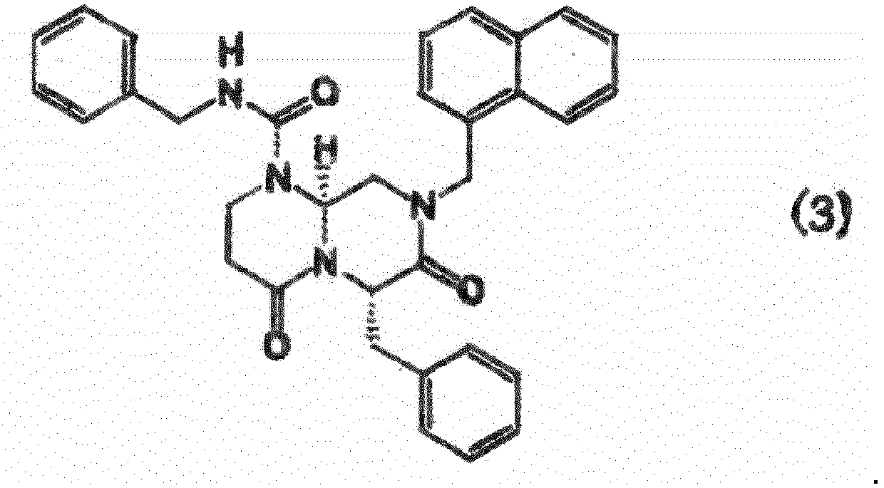
- A method for producing hepatocyte from mesenchymal stem cell, comprising a step of treating mesenchymal stem cell in vitro with the compound, the salt thereof, or the solvate of them according to any of Claims 1 to 3.
- The method for producing hepatocyte from mesenchymal stem cell according to Claim 4, comprising a step of treating mesenchymal stem cell in vitro with at least one compound, a salt thereof, or a solvate of them, the compound being represented by formula (3):

- The method for producing hepatocyte from mesenchymal stem cell according to Claim 4 or 5, wherein the mesenchymal stem cell is bone marrow-derived cell.
- A method for inhibiting a Wnt/β-catenin signaling pathway, comprising a step of treating mesenchymal stem cell in vitro with the compound, the salt thereof, or the solvate of them according to any of Claims 1 to 3.
- The method for inhibiting a Wnt/β-catenin signaling pathway according to Claim 7, comprising a step of treating mesenchymal stem cell with at least one compound, a salt thereof, or a solvate of them, the compound being represented by formula (3):

- A method for producing a transplantation material, comprising a step of:a) treating stem cell in vitro with the compound, the salt thereof, or the solvate of them according to any of Claims 1 to 3.
- The production method according to Claim 9, wherein the step a) comprises a step of culturing mesenchymal stem cell in a medium comprising the compound, the salt thereof, or the solvate of them according to any of Claims 1 to 3 to differentiate the mesenchymal stem cell into hepatocyte.
- A method for inducing differentiation of mesenchymal stem cell into hepatocyte, comprising a step of treating mesenchymal stem cell in vitro with:a) the compound, the salt thereof, or the solvate of them according to any of Claims 1 to 3.
Applications Claiming Priority (2)
| Application Number | Priority Date | Filing Date | Title |
|---|---|---|---|
| JP2011091599 | 2011-04-15 | ||
| PCT/JP2012/059021 WO2012141038A1 (en) | 2011-04-15 | 2012-04-02 | Synthesis and analysis of novel compound capable of inducing differentiation of human mesenchymal stem cell into hepatocyte |
Publications (4)
| Publication Number | Publication Date |
|---|---|
| EP2698365A1 EP2698365A1 (en) | 2014-02-19 |
| EP2698365A4 EP2698365A4 (en) | 2015-04-29 |
| EP2698365B1 true EP2698365B1 (en) | 2017-12-13 |
| EP2698365B8 EP2698365B8 (en) | 2018-02-14 |
Family
ID=47009217
Family Applications (1)
| Application Number | Title | Priority Date | Filing Date |
|---|---|---|---|
| EP12771389.9A Active EP2698365B8 (en) | 2011-04-15 | 2012-04-02 | Synthesis and analysis of novel compound capable of inducing differentiation of human mesenchymal stem cell into hepatocyte |
Country Status (5)
| Country | Link |
|---|---|
| US (1) | US9555061B2 (en) |
| EP (1) | EP2698365B8 (en) |
| JP (4) | JP6008297B2 (en) |
| ES (1) | ES2660756T3 (en) |
| WO (1) | WO2012141038A1 (en) |
Cited By (1)
| Publication number | Priority date | Publication date | Assignee | Title |
|---|---|---|---|---|
| US11274279B2 (en) | 2020-03-11 | 2022-03-15 | Bit Bio Limited | Method of generating hepatic cells |
Families Citing this family (12)
| Publication number | Priority date | Publication date | Assignee | Title |
|---|---|---|---|---|
| KR101767802B1 (en) * | 2013-03-15 | 2017-08-11 | 에프. 호프만-라 로슈 아게 | Compounds for improved stem cell differentiation into hepatocytes |
| CN106132417B (en) * | 2014-03-28 | 2020-04-14 | 国立大学法人鸟取大学 | Cancer and fibrosis inhibitory effects of low molecular weight compounds |
| CN108026104B (en) * | 2015-09-18 | 2021-06-18 | 国立大学法人鸟取大学 | Effects of cancer and fibrosis inhibition and regeneration promotion by low molecular weight compounds |
| JP2017200473A (en) * | 2016-04-27 | 2017-11-09 | 株式会社Cells Power | Activated stem cells |
| TWI774829B (en) * | 2017-09-01 | 2022-08-21 | 國立大學法人鳥取大學 | Cell layer with fibrosis inhibitory effect |
| WO2019235362A1 (en) | 2018-06-08 | 2019-12-12 | カノンキュア株式会社 | Fibrosis-inhibiting composition, cells producing same, and cell sheet comprising said cells |
| EP3851519A4 (en) * | 2018-09-28 | 2021-12-29 | Nissan Chemical Corporation | Additive for medium for promoting production of paracrine factor |
| JPWO2020122022A1 (en) * | 2018-12-10 | 2021-10-28 | 東京都 | Liver function improving agent |
| JP7452799B2 (en) * | 2018-12-26 | 2024-03-19 | 国立大学法人京都大学 | Method for producing hepatocytes |
| CN114787337A (en) | 2020-01-24 | 2022-07-22 | 国立大学法人长崎大学 | Implant sheet for attaching to organ surface |
| CN116925081A (en) * | 2022-04-11 | 2023-10-24 | 中国科学院上海药物研究所 | Cyclic peptide compound and application thereof |
| CN117165516B (en) * | 2022-05-26 | 2026-04-28 | 青岛瑞思德生物科技有限公司 | An inducer to differentiate mesenchymal stem cells into estradiol-secreting cells |
Family Cites Families (12)
| Publication number | Priority date | Publication date | Assignee | Title |
|---|---|---|---|---|
| JPH08502048A (en) * | 1992-09-11 | 1996-03-05 | ザ リージェンツ オブ ザ ユニバーシティ オブ カリフォルニア | Metazoan parasite protease inhibitors |
| EP1406889A2 (en) * | 2001-07-09 | 2004-04-14 | Pharmacia Italia S.p.A. | Interaction inhibitors of tcf-4 with beta-catenin |
| RU2383547C2 (en) * | 2003-08-28 | 2010-03-10 | Чоонгвае Фарма Корпорейшн | Modulation of beta-catenin/tcf-activated transcription |
| ES2561944T3 (en) * | 2006-01-12 | 2016-03-01 | Tokyo Women's Medical University | Procedure to maintain the function of liver tissue cells for a long time |
| WO2007127698A2 (en) | 2006-04-25 | 2007-11-08 | Board Of Supervisors Of Louisiana State Universityand Agricultural And Mechanical College | Adipose derived adult stem cells in hepatic regeneration |
| JP2009538903A (en) | 2006-05-30 | 2009-11-12 | チュンウェ ファーマ コーポレーション | Composition for inducing or suppressing stem cell differentiation |
| US8283351B2 (en) | 2007-04-02 | 2012-10-09 | Institute For Oneworld Health | Cyclic and acyclic hydrazine derivatives compositions including them and uses thereof |
| JP2009060840A (en) | 2007-09-06 | 2009-03-26 | Keio Gijuku | Method for concentrating human mesenchymal stem cell |
| JP2010075631A (en) | 2008-09-29 | 2010-04-08 | Japan Science & Technology Agency | Method for differentiating and inducing liver from undifferentiated cell |
| US20120046242A1 (en) * | 2008-12-24 | 2012-02-23 | Massachusetts Institute Of Technology | Molecular activators of the wnt/beta-catenin pathway |
| WO2010144696A1 (en) * | 2009-06-11 | 2010-12-16 | Burnham Institute For Medical Research | Directed differentiation of stem cells |
| JP5704554B2 (en) * | 2010-04-14 | 2015-04-22 | 国立大学法人鳥取大学 | Compounds that induce human stem cells to differentiate into hepatocytes |
-
2012
- 2012-04-02 US US14/111,757 patent/US9555061B2/en active Active
- 2012-04-02 ES ES12771389.9T patent/ES2660756T3/en active Active
- 2012-04-02 WO PCT/JP2012/059021 patent/WO2012141038A1/en not_active Ceased
- 2012-04-02 EP EP12771389.9A patent/EP2698365B8/en active Active
- 2012-04-02 JP JP2013509857A patent/JP6008297B2/en active Active
-
2016
- 2016-06-22 JP JP2016123647A patent/JP6201008B2/en active Active
-
2017
- 2017-06-15 JP JP2017117768A patent/JP2017205526A/en active Pending
- 2017-06-30 JP JP2017128831A patent/JP6391067B2/en active Active
Non-Patent Citations (1)
| Title |
|---|
| None * |
Cited By (2)
| Publication number | Priority date | Publication date | Assignee | Title |
|---|---|---|---|---|
| US11274279B2 (en) | 2020-03-11 | 2022-03-15 | Bit Bio Limited | Method of generating hepatic cells |
| US12410406B2 (en) | 2020-03-11 | 2025-09-09 | Bit Bio Limited | Method of generating hepatic cells |
Also Published As
| Publication number | Publication date |
|---|---|
| US9555061B2 (en) | 2017-01-31 |
| JPWO2012141038A1 (en) | 2014-07-28 |
| ES2660756T3 (en) | 2018-03-26 |
| JP6201008B2 (en) | 2017-09-20 |
| EP2698365A1 (en) | 2014-02-19 |
| JP6008297B2 (en) | 2016-10-19 |
| JP2018008948A (en) | 2018-01-18 |
| JP2016222678A (en) | 2016-12-28 |
| JP6391067B2 (en) | 2018-09-19 |
| WO2012141038A1 (en) | 2012-10-18 |
| EP2698365A4 (en) | 2015-04-29 |
| US20140112892A1 (en) | 2014-04-24 |
| EP2698365B8 (en) | 2018-02-14 |
| JP2017205526A (en) | 2017-11-24 |
Similar Documents
| Publication | Publication Date | Title |
|---|---|---|
| US9555061B2 (en) | Synthesis and analysis of compounds capable of inducing differentiation of human mesenchymal stems cells into hepatocytes | |
| JP7461674B2 (en) | Culture medium for esophageal squamous cell carcinoma epithelial cells, culture method, and use thereof | |
| Yukawa et al. | Monitoring transplanted adipose tissue-derived stem cells combined with heparin in the liver by fluorescence imaging using quantum dots | |
| Lim et al. | Trichostatin A enhances differentiation of human induced pluripotent stem cells to cardiogenic cells for cardiac tissue engineering | |
| EP3533865A1 (en) | Method for preparing human hepatocyte progenitor cells | |
| JP7228269B2 (en) | Cell mass fusion method | |
| Ouji et al. | Effects of Wnt-10b on hair shaft growth in hair follicle cultures | |
| Wabitsch et al. | Human stem cells promote liver regeneration after partial hepatectomy in BALB/C nude mice | |
| Li et al. | Circulating fibrocytes stabilize blood vessels during angiogenesis in a paracrine manner | |
| Karoubi et al. | Identification of mesenchymal stromal cells in human lung parenchyma capable of differentiating into aquaporin 5-expressing cells | |
| Tanaka et al. | Potentiality of combined hepatocellular and intrahepatic cholangiocellular carcinoma originating from a hepatic precursor cell: Immunohistochemical evidence | |
| JP5704554B2 (en) | Compounds that induce human stem cells to differentiate into hepatocytes | |
| Yuan et al. | The effect of vascular endothelial growth factor on bone marrow mesenchymal stem cell engraftment in rat fibrotic liver upon transplantation | |
| Muneer et al. | Wnt signaling pathway inhibitor promotes mesenchymal stem cells differentiation into cardiac progenitor cells in vitro and improves cardiomyopathy in vivo | |
| Lokman et al. | Chick chorioallantoic membrane assay: a 3D animal model for cancer invasion and metastasis | |
| JP7503662B2 (en) | Culture medium for laryngeal cancer epithelial cells, culture method and use thereof | |
| EP4215602A1 (en) | Method for producing cardiac muscle stem/precursor cells and method for inhibiting myocardial fibrosis | |
| JP7774913B2 (en) | Culture medium and culture method for lung cancer epithelial cells, and uses thereof | |
| Suzuki et al. | Phenotype-dependent production of des-γ-carboxy prothrombin in hepatocellular carcinoma | |
| JP7197203B2 (en) | Methods of screening for substances that affect the formation, structure or function of human blood vessels, and methods of manufacturing human blood vessels | |
| Torres-Montaner | Telomere Maintenance and Oncogenesis | |
| RU2838489C2 (en) | Medium for culturing lung cancer epithelial cells, method for culturing and use thereof | |
| Boylan | Characterization of the Pharmacokinetic Properties and In Vivo Efficacy of a Candidate Bone Marrow Niche Altering Compound | |
| RU2755212C2 (en) | Composition inhibiting meis proteins | |
| Woosley | Mechanistic Studies of TGFβ-induced Cancer Stem Cell Formation and Mammary Tumorigenesis through an ILEI/LIFR Signaling Axis |
Legal Events
| Date | Code | Title | Description |
|---|---|---|---|
| PUAI | Public reference made under article 153(3) epc to a published international application that has entered the european phase |
Free format text: ORIGINAL CODE: 0009012 |
|
| 17P | Request for examination filed |
Effective date: 20131114 |
|
| AK | Designated contracting states |
Kind code of ref document: A1 Designated state(s): AL AT BE BG CH CY CZ DE DK EE ES FI FR GB GR HR HU IE IS IT LI LT LU LV MC MK MT NL NO PL PT RO RS SE SI SK SM TR |
|
| DAX | Request for extension of the european patent (deleted) | ||
| RIC1 | Information provided on ipc code assigned before grant |
Ipc: C07C 43/225 20060101ALI20141205BHEP Ipc: C12N 5/071 20100101ALI20141205BHEP Ipc: A61L 27/00 20060101ALI20141205BHEP Ipc: C07D 307/52 20060101ALI20141205BHEP Ipc: C07C 251/86 20060101ALI20141205BHEP Ipc: C07D 487/04 20060101AFI20141205BHEP |
|
| REG | Reference to a national code |
Ref country code: DE Ref legal event code: R079 Ref document number: 602012040872 Country of ref document: DE Free format text: PREVIOUS MAIN CLASS: C07C0251860000 Ipc: C07D0487040000 |
|
| RA4 | Supplementary search report drawn up and despatched (corrected) |
Effective date: 20150330 |
|
| RIC1 | Information provided on ipc code assigned before grant |
Ipc: C07C 43/225 20060101ALI20150324BHEP Ipc: C07D 307/52 20060101ALI20150324BHEP Ipc: C12N 5/071 20100101ALI20150324BHEP Ipc: C07C 251/86 20060101ALI20150324BHEP Ipc: A61L 27/00 20060101ALI20150324BHEP Ipc: C07D 487/04 20060101AFI20150324BHEP |
|
| STAA | Information on the status of an ep patent application or granted ep patent |
Free format text: STATUS: EXAMINATION IS IN PROGRESS |
|
| 17Q | First examination report despatched |
Effective date: 20161209 |
|
| GRAP | Despatch of communication of intention to grant a patent |
Free format text: ORIGINAL CODE: EPIDOSNIGR1 |
|
| STAA | Information on the status of an ep patent application or granted ep patent |
Free format text: STATUS: GRANT OF PATENT IS INTENDED |
|
| INTG | Intention to grant announced |
Effective date: 20170627 |
|
| GRAS | Grant fee paid |
Free format text: ORIGINAL CODE: EPIDOSNIGR3 |
|
| GRAA | (expected) grant |
Free format text: ORIGINAL CODE: 0009210 |
|
| STAA | Information on the status of an ep patent application or granted ep patent |
Free format text: STATUS: THE PATENT HAS BEEN GRANTED |
|
| RAP1 | Party data changed (applicant data changed or rights of an application transferred) |
Owner name: KANONCURE, INC. Owner name: TOKYO WOMEN'S MEDICAL UNIVERSITY Owner name: NATIONAL UNIVERSITY CORPORATION TOTTORI UNIVERSITY |
|
| GRAT | Correction requested after decision to grant or after decision to maintain patent in amended form |
Free format text: ORIGINAL CODE: EPIDOSNCDEC |
|
| AK | Designated contracting states |
Kind code of ref document: B1 Designated state(s): AL AT BE BG CH CY CZ DE DK EE ES FI FR GB GR HR HU IE IS IT LI LT LU LV MC MK MT NL NO PL PT RO RS SE SI SK SM TR |
|
| REG | Reference to a national code |
Ref country code: GB Ref legal event code: FG4D |
|
| REG | Reference to a national code |
Ref country code: AT Ref legal event code: REF Ref document number: 954240 Country of ref document: AT Kind code of ref document: T Effective date: 20171215 Ref country code: CH Ref legal event code: EP |
|
| RAP2 | Party data changed (patent owner data changed or rights of a patent transferred) |
Owner name: KANONCURE, INC. Owner name: NATIONAL UNIVERSITY CORPORATION TOTTORI UNIVERSITY Owner name: TOKYO WOMEN'S MEDICAL UNIVERSITY |
|
| REG | Reference to a national code |
Ref country code: IE Ref legal event code: FG4D |
|
| REG | Reference to a national code |
Ref country code: DE Ref legal event code: R096 Ref document number: 602012040872 Country of ref document: DE |
|
| REG | Reference to a national code |
Ref country code: ES Ref legal event code: FG2A Ref document number: 2660756 Country of ref document: ES Kind code of ref document: T3 Effective date: 20180326 |
|
| REG | Reference to a national code |
Ref country code: NL Ref legal event code: MP Effective date: 20171213 |
|
| REG | Reference to a national code |
Ref country code: FR Ref legal event code: PLFP Year of fee payment: 7 |
|
| REG | Reference to a national code |
Ref country code: LT Ref legal event code: MG4D |
|
| PG25 | Lapsed in a contracting state [announced via postgrant information from national office to epo] |
Ref country code: NO Free format text: LAPSE BECAUSE OF FAILURE TO SUBMIT A TRANSLATION OF THE DESCRIPTION OR TO PAY THE FEE WITHIN THE PRESCRIBED TIME-LIMIT Effective date: 20180313 Ref country code: SE Free format text: LAPSE BECAUSE OF FAILURE TO SUBMIT A TRANSLATION OF THE DESCRIPTION OR TO PAY THE FEE WITHIN THE PRESCRIBED TIME-LIMIT Effective date: 20171213 Ref country code: FI Free format text: LAPSE BECAUSE OF FAILURE TO SUBMIT A TRANSLATION OF THE DESCRIPTION OR TO PAY THE FEE WITHIN THE PRESCRIBED TIME-LIMIT Effective date: 20171213 Ref country code: LT Free format text: LAPSE BECAUSE OF FAILURE TO SUBMIT A TRANSLATION OF THE DESCRIPTION OR TO PAY THE FEE WITHIN THE PRESCRIBED TIME-LIMIT Effective date: 20171213 |
|
| REG | Reference to a national code |
Ref country code: AT Ref legal event code: MK05 Ref document number: 954240 Country of ref document: AT Kind code of ref document: T Effective date: 20171213 |
|
| PG25 | Lapsed in a contracting state [announced via postgrant information from national office to epo] |
Ref country code: BG Free format text: LAPSE BECAUSE OF FAILURE TO SUBMIT A TRANSLATION OF THE DESCRIPTION OR TO PAY THE FEE WITHIN THE PRESCRIBED TIME-LIMIT Effective date: 20180313 Ref country code: HR Free format text: LAPSE BECAUSE OF FAILURE TO SUBMIT A TRANSLATION OF THE DESCRIPTION OR TO PAY THE FEE WITHIN THE PRESCRIBED TIME-LIMIT Effective date: 20171213 Ref country code: RS Free format text: LAPSE BECAUSE OF FAILURE TO SUBMIT A TRANSLATION OF THE DESCRIPTION OR TO PAY THE FEE WITHIN THE PRESCRIBED TIME-LIMIT Effective date: 20171213 Ref country code: LV Free format text: LAPSE BECAUSE OF FAILURE TO SUBMIT A TRANSLATION OF THE DESCRIPTION OR TO PAY THE FEE WITHIN THE PRESCRIBED TIME-LIMIT Effective date: 20171213 Ref country code: GR Free format text: LAPSE BECAUSE OF FAILURE TO SUBMIT A TRANSLATION OF THE DESCRIPTION OR TO PAY THE FEE WITHIN THE PRESCRIBED TIME-LIMIT Effective date: 20180314 |
|
| PG25 | Lapsed in a contracting state [announced via postgrant information from national office to epo] |
Ref country code: NL Free format text: LAPSE BECAUSE OF FAILURE TO SUBMIT A TRANSLATION OF THE DESCRIPTION OR TO PAY THE FEE WITHIN THE PRESCRIBED TIME-LIMIT Effective date: 20171213 |
|
| PG25 | Lapsed in a contracting state [announced via postgrant information from national office to epo] |
Ref country code: SK Free format text: LAPSE BECAUSE OF FAILURE TO SUBMIT A TRANSLATION OF THE DESCRIPTION OR TO PAY THE FEE WITHIN THE PRESCRIBED TIME-LIMIT Effective date: 20171213 Ref country code: EE Free format text: LAPSE BECAUSE OF FAILURE TO SUBMIT A TRANSLATION OF THE DESCRIPTION OR TO PAY THE FEE WITHIN THE PRESCRIBED TIME-LIMIT Effective date: 20171213 Ref country code: CY Free format text: LAPSE BECAUSE OF FAILURE TO SUBMIT A TRANSLATION OF THE DESCRIPTION OR TO PAY THE FEE WITHIN THE PRESCRIBED TIME-LIMIT Effective date: 20171213 Ref country code: CZ Free format text: LAPSE BECAUSE OF FAILURE TO SUBMIT A TRANSLATION OF THE DESCRIPTION OR TO PAY THE FEE WITHIN THE PRESCRIBED TIME-LIMIT Effective date: 20171213 |
|
| PG25 | Lapsed in a contracting state [announced via postgrant information from national office to epo] |
Ref country code: AT Free format text: LAPSE BECAUSE OF FAILURE TO SUBMIT A TRANSLATION OF THE DESCRIPTION OR TO PAY THE FEE WITHIN THE PRESCRIBED TIME-LIMIT Effective date: 20171213 Ref country code: SM Free format text: LAPSE BECAUSE OF FAILURE TO SUBMIT A TRANSLATION OF THE DESCRIPTION OR TO PAY THE FEE WITHIN THE PRESCRIBED TIME-LIMIT Effective date: 20171213 Ref country code: PL Free format text: LAPSE BECAUSE OF FAILURE TO SUBMIT A TRANSLATION OF THE DESCRIPTION OR TO PAY THE FEE WITHIN THE PRESCRIBED TIME-LIMIT Effective date: 20171213 Ref country code: RO Free format text: LAPSE BECAUSE OF FAILURE TO SUBMIT A TRANSLATION OF THE DESCRIPTION OR TO PAY THE FEE WITHIN THE PRESCRIBED TIME-LIMIT Effective date: 20171213 Ref country code: IS Free format text: LAPSE BECAUSE OF FAILURE TO SUBMIT A TRANSLATION OF THE DESCRIPTION OR TO PAY THE FEE WITHIN THE PRESCRIBED TIME-LIMIT Effective date: 20180413 |
|
| REG | Reference to a national code |
Ref country code: DE Ref legal event code: R097 Ref document number: 602012040872 Country of ref document: DE |
|
| PLBE | No opposition filed within time limit |
Free format text: ORIGINAL CODE: 0009261 |
|
| STAA | Information on the status of an ep patent application or granted ep patent |
Free format text: STATUS: NO OPPOSITION FILED WITHIN TIME LIMIT |
|
| 26N | No opposition filed |
Effective date: 20180914 |
|
| PG25 | Lapsed in a contracting state [announced via postgrant information from national office to epo] |
Ref country code: MC Free format text: LAPSE BECAUSE OF FAILURE TO SUBMIT A TRANSLATION OF THE DESCRIPTION OR TO PAY THE FEE WITHIN THE PRESCRIBED TIME-LIMIT Effective date: 20171213 Ref country code: DK Free format text: LAPSE BECAUSE OF FAILURE TO SUBMIT A TRANSLATION OF THE DESCRIPTION OR TO PAY THE FEE WITHIN THE PRESCRIBED TIME-LIMIT Effective date: 20171213 |
|
| REG | Reference to a national code |
Ref country code: CH Ref legal event code: PL |
|
| REG | Reference to a national code |
Ref country code: BE Ref legal event code: MM Effective date: 20180430 |
|
| REG | Reference to a national code |
Ref country code: IE Ref legal event code: MM4A |
|
| PG25 | Lapsed in a contracting state [announced via postgrant information from national office to epo] |
Ref country code: LU Free format text: LAPSE BECAUSE OF NON-PAYMENT OF DUE FEES Effective date: 20180402 |
|
| PG25 | Lapsed in a contracting state [announced via postgrant information from national office to epo] |
Ref country code: BE Free format text: LAPSE BECAUSE OF NON-PAYMENT OF DUE FEES Effective date: 20180430 Ref country code: SI Free format text: LAPSE BECAUSE OF FAILURE TO SUBMIT A TRANSLATION OF THE DESCRIPTION OR TO PAY THE FEE WITHIN THE PRESCRIBED TIME-LIMIT Effective date: 20171213 Ref country code: CH Free format text: LAPSE BECAUSE OF NON-PAYMENT OF DUE FEES Effective date: 20180430 Ref country code: LI Free format text: LAPSE BECAUSE OF NON-PAYMENT OF DUE FEES Effective date: 20180430 |
|
| PG25 | Lapsed in a contracting state [announced via postgrant information from national office to epo] |
Ref country code: IE Free format text: LAPSE BECAUSE OF NON-PAYMENT OF DUE FEES Effective date: 20180402 |
|
| PG25 | Lapsed in a contracting state [announced via postgrant information from national office to epo] |
Ref country code: MT Free format text: LAPSE BECAUSE OF NON-PAYMENT OF DUE FEES Effective date: 20180402 |
|
| PG25 | Lapsed in a contracting state [announced via postgrant information from national office to epo] |
Ref country code: TR Free format text: LAPSE BECAUSE OF FAILURE TO SUBMIT A TRANSLATION OF THE DESCRIPTION OR TO PAY THE FEE WITHIN THE PRESCRIBED TIME-LIMIT Effective date: 20171213 |
|
| PG25 | Lapsed in a contracting state [announced via postgrant information from national office to epo] |
Ref country code: HU Free format text: LAPSE BECAUSE OF FAILURE TO SUBMIT A TRANSLATION OF THE DESCRIPTION OR TO PAY THE FEE WITHIN THE PRESCRIBED TIME-LIMIT; INVALID AB INITIO Effective date: 20120402 Ref country code: PT Free format text: LAPSE BECAUSE OF FAILURE TO SUBMIT A TRANSLATION OF THE DESCRIPTION OR TO PAY THE FEE WITHIN THE PRESCRIBED TIME-LIMIT Effective date: 20171213 |
|
| PG25 | Lapsed in a contracting state [announced via postgrant information from national office to epo] |
Ref country code: MK Free format text: LAPSE BECAUSE OF NON-PAYMENT OF DUE FEES Effective date: 20171213 |
|
| PG25 | Lapsed in a contracting state [announced via postgrant information from national office to epo] |
Ref country code: AL Free format text: LAPSE BECAUSE OF FAILURE TO SUBMIT A TRANSLATION OF THE DESCRIPTION OR TO PAY THE FEE WITHIN THE PRESCRIBED TIME-LIMIT Effective date: 20171213 |
|
| PGFP | Annual fee paid to national office [announced via postgrant information from national office to epo] |
Ref country code: GB Payment date: 20250417 Year of fee payment: 14 Ref country code: ES Payment date: 20250519 Year of fee payment: 14 |
|
| PGFP | Annual fee paid to national office [announced via postgrant information from national office to epo] |
Ref country code: IT Payment date: 20250422 Year of fee payment: 14 |
|
| PGFP | Annual fee paid to national office [announced via postgrant information from national office to epo] |
Ref country code: FR Payment date: 20250425 Year of fee payment: 14 |
|
| PGFP | Annual fee paid to national office [announced via postgrant information from national office to epo] |
Ref country code: DE Payment date: 20250626 Year of fee payment: 14 |