EP2695949A1 - Nucleic acid based nanopores or transmembrane channels and their uses - Google Patents
Nucleic acid based nanopores or transmembrane channels and their uses Download PDFInfo
- Publication number
- EP2695949A1 EP2695949A1 EP20120180126 EP12180126A EP2695949A1 EP 2695949 A1 EP2695949 A1 EP 2695949A1 EP 20120180126 EP20120180126 EP 20120180126 EP 12180126 A EP12180126 A EP 12180126A EP 2695949 A1 EP2695949 A1 EP 2695949A1
- Authority
- EP
- European Patent Office
- Prior art keywords
- nucleic acid
- nanopore
- dna
- acid based
- channel
- Prior art date
- Legal status (The legal status is an assumption and is not a legal conclusion. Google has not performed a legal analysis and makes no representation as to the accuracy of the status listed.)
- Granted
Links
- 102000039446 nucleic acids Human genes 0.000 title claims abstract description 184
- 108020004707 nucleic acids Proteins 0.000 title claims abstract description 184
- 150000007523 nucleic acids Chemical class 0.000 title claims abstract description 181
- 239000003814 drug Substances 0.000 claims abstract description 15
- 230000008685 targeting Effects 0.000 claims abstract description 12
- 238000002372 labelling Methods 0.000 claims abstract description 9
- 238000012377 drug delivery Methods 0.000 claims abstract description 8
- 239000000203 mixture Substances 0.000 claims abstract description 8
- 108020004414 DNA Proteins 0.000 claims description 171
- 239000012528 membrane Substances 0.000 claims description 83
- 108091034117 Oligonucleotide Proteins 0.000 claims description 54
- 210000004027 cell Anatomy 0.000 claims description 54
- 230000027455 binding Effects 0.000 claims description 48
- 239000000232 Lipid Bilayer Substances 0.000 claims description 36
- 150000002632 lipids Chemical class 0.000 claims description 36
- JLCPHMBAVCMARE-UHFFFAOYSA-N [3-[[3-[[3-[[3-[[3-[[3-[[3-[[3-[[3-[[3-[[3-[[5-(2-amino-6-oxo-1H-purin-9-yl)-3-[[3-[[3-[[3-[[3-[[3-[[5-(2-amino-6-oxo-1H-purin-9-yl)-3-[[5-(2-amino-6-oxo-1H-purin-9-yl)-3-hydroxyoxolan-2-yl]methoxy-hydroxyphosphoryl]oxyoxolan-2-yl]methoxy-hydroxyphosphoryl]oxy-5-(5-methyl-2,4-dioxopyrimidin-1-yl)oxolan-2-yl]methoxy-hydroxyphosphoryl]oxy-5-(6-aminopurin-9-yl)oxolan-2-yl]methoxy-hydroxyphosphoryl]oxy-5-(6-aminopurin-9-yl)oxolan-2-yl]methoxy-hydroxyphosphoryl]oxy-5-(6-aminopurin-9-yl)oxolan-2-yl]methoxy-hydroxyphosphoryl]oxy-5-(6-aminopurin-9-yl)oxolan-2-yl]methoxy-hydroxyphosphoryl]oxyoxolan-2-yl]methoxy-hydroxyphosphoryl]oxy-5-(5-methyl-2,4-dioxopyrimidin-1-yl)oxolan-2-yl]methoxy-hydroxyphosphoryl]oxy-5-(4-amino-2-oxopyrimidin-1-yl)oxolan-2-yl]methoxy-hydroxyphosphoryl]oxy-5-(5-methyl-2,4-dioxopyrimidin-1-yl)oxolan-2-yl]methoxy-hydroxyphosphoryl]oxy-5-(5-methyl-2,4-dioxopyrimidin-1-yl)oxolan-2-yl]methoxy-hydroxyphosphoryl]oxy-5-(6-aminopurin-9-yl)oxolan-2-yl]methoxy-hydroxyphosphoryl]oxy-5-(6-aminopurin-9-yl)oxolan-2-yl]methoxy-hydroxyphosphoryl]oxy-5-(4-amino-2-oxopyrimidin-1-yl)oxolan-2-yl]methoxy-hydroxyphosphoryl]oxy-5-(4-amino-2-oxopyrimidin-1-yl)oxolan-2-yl]methoxy-hydroxyphosphoryl]oxy-5-(4-amino-2-oxopyrimidin-1-yl)oxolan-2-yl]methoxy-hydroxyphosphoryl]oxy-5-(6-aminopurin-9-yl)oxolan-2-yl]methoxy-hydroxyphosphoryl]oxy-5-(4-amino-2-oxopyrimidin-1-yl)oxolan-2-yl]methyl [5-(6-aminopurin-9-yl)-2-(hydroxymethyl)oxolan-3-yl] hydrogen phosphate Polymers Cc1cn(C2CC(OP(O)(=O)OCC3OC(CC3OP(O)(=O)OCC3OC(CC3O)n3cnc4c3nc(N)[nH]c4=O)n3cnc4c3nc(N)[nH]c4=O)C(COP(O)(=O)OC3CC(OC3COP(O)(=O)OC3CC(OC3COP(O)(=O)OC3CC(OC3COP(O)(=O)OC3CC(OC3COP(O)(=O)OC3CC(OC3COP(O)(=O)OC3CC(OC3COP(O)(=O)OC3CC(OC3COP(O)(=O)OC3CC(OC3COP(O)(=O)OC3CC(OC3COP(O)(=O)OC3CC(OC3COP(O)(=O)OC3CC(OC3COP(O)(=O)OC3CC(OC3COP(O)(=O)OC3CC(OC3COP(O)(=O)OC3CC(OC3COP(O)(=O)OC3CC(OC3COP(O)(=O)OC3CC(OC3COP(O)(=O)OC3CC(OC3CO)n3cnc4c(N)ncnc34)n3ccc(N)nc3=O)n3cnc4c(N)ncnc34)n3ccc(N)nc3=O)n3ccc(N)nc3=O)n3ccc(N)nc3=O)n3cnc4c(N)ncnc34)n3cnc4c(N)ncnc34)n3cc(C)c(=O)[nH]c3=O)n3cc(C)c(=O)[nH]c3=O)n3ccc(N)nc3=O)n3cc(C)c(=O)[nH]c3=O)n3cnc4c3nc(N)[nH]c4=O)n3cnc4c(N)ncnc34)n3cnc4c(N)ncnc34)n3cnc4c(N)ncnc34)n3cnc4c(N)ncnc34)O2)c(=O)[nH]c1=O JLCPHMBAVCMARE-UHFFFAOYSA-N 0.000 claims description 32
- HVYWMOMLDIMFJA-DPAQBDIFSA-N cholesterol Chemical compound C1C=C2C[C@@H](O)CC[C@]2(C)[C@@H]2[C@@H]1[C@@H]1CC[C@H]([C@H](C)CCCC(C)C)[C@@]1(C)CC2 HVYWMOMLDIMFJA-DPAQBDIFSA-N 0.000 claims description 28
- 125000003729 nucleotide group Chemical group 0.000 claims description 24
- 239000002773 nucleotide Substances 0.000 claims description 23
- 230000004048 modification Effects 0.000 claims description 22
- 238000012986 modification Methods 0.000 claims description 22
- 230000007246 mechanism Effects 0.000 claims description 21
- 238000005232 molecular self-assembly Methods 0.000 claims description 19
- 235000012000 cholesterol Nutrition 0.000 claims description 15
- 239000012634 fragment Substances 0.000 claims description 15
- 239000000427 antigen Substances 0.000 claims description 11
- 108091007433 antigens Proteins 0.000 claims description 11
- 102000036639 antigens Human genes 0.000 claims description 11
- 229940079593 drug Drugs 0.000 claims description 10
- 230000006870 function Effects 0.000 claims description 10
- 102000004169 proteins and genes Human genes 0.000 claims description 10
- 108090000623 proteins and genes Proteins 0.000 claims description 10
- 206010028980 Neoplasm Diseases 0.000 claims description 9
- 239000012491 analyte Substances 0.000 claims description 9
- 210000000056 organ Anatomy 0.000 claims description 8
- 210000001519 tissue Anatomy 0.000 claims description 8
- 108091023037 Aptamer Proteins 0.000 claims description 7
- 239000000872 buffer Substances 0.000 claims description 7
- 239000003446 ligand Substances 0.000 claims description 7
- 230000002265 prevention Effects 0.000 claims description 7
- 230000015572 biosynthetic process Effects 0.000 claims description 6
- 201000010099 disease Diseases 0.000 claims description 6
- 208000037265 diseases, disorders, signs and symptoms Diseases 0.000 claims description 6
- 241001524679 Escherichia virus M13 Species 0.000 claims description 5
- 201000011510 cancer Diseases 0.000 claims description 5
- 210000000170 cell membrane Anatomy 0.000 claims description 5
- 230000000295 complement effect Effects 0.000 claims description 5
- 238000003745 diagnosis Methods 0.000 claims description 5
- 238000001514 detection method Methods 0.000 claims description 4
- 230000002209 hydrophobic effect Effects 0.000 claims description 4
- 229920000642 polymer Polymers 0.000 claims description 4
- 108090000765 processed proteins & peptides Proteins 0.000 claims description 4
- 150000003839 salts Chemical class 0.000 claims description 4
- 238000000926 separation method Methods 0.000 claims description 4
- 108010083687 Ion Pumps Proteins 0.000 claims description 3
- 239000003443 antiviral agent Substances 0.000 claims description 3
- 208000019622 heart disease Diseases 0.000 claims description 3
- 208000030159 metabolic disease Diseases 0.000 claims description 3
- 238000012163 sequencing technique Methods 0.000 claims description 3
- HVCOBJNICQPDBP-UHFFFAOYSA-N 3-[3-[3,5-dihydroxy-6-methyl-4-(3,4,5-trihydroxy-6-methyloxan-2-yl)oxyoxan-2-yl]oxydecanoyloxy]decanoic acid;hydrate Chemical compound O.OC1C(OC(CC(=O)OC(CCCCCCC)CC(O)=O)CCCCCCC)OC(C)C(O)C1OC1C(O)C(O)C(O)C(C)O1 HVCOBJNICQPDBP-UHFFFAOYSA-N 0.000 claims description 2
- 102000014914 Carrier Proteins Human genes 0.000 claims description 2
- 108010078791 Carrier Proteins Proteins 0.000 claims description 2
- 208000035473 Communicable disease Diseases 0.000 claims description 2
- 102000004190 Enzymes Human genes 0.000 claims description 2
- 108090000790 Enzymes Proteins 0.000 claims description 2
- 229930186217 Glycolipid Natural products 0.000 claims description 2
- 102000003886 Glycoproteins Human genes 0.000 claims description 2
- 108090000288 Glycoproteins Proteins 0.000 claims description 2
- 206010020751 Hypersensitivity Diseases 0.000 claims description 2
- 208000008589 Obesity Diseases 0.000 claims description 2
- 230000007815 allergy Effects 0.000 claims description 2
- 230000000845 anti-microbial effect Effects 0.000 claims description 2
- 239000002246 antineoplastic agent Substances 0.000 claims description 2
- 229940041181 antineoplastic drug Drugs 0.000 claims description 2
- 230000003197 catalytic effect Effects 0.000 claims description 2
- 235000014113 dietary fatty acids Nutrition 0.000 claims description 2
- 229930195729 fatty acid Natural products 0.000 claims description 2
- 239000000194 fatty acid Substances 0.000 claims description 2
- 150000004665 fatty acids Chemical class 0.000 claims description 2
- 239000007850 fluorescent dye Substances 0.000 claims description 2
- 150000004676 glycans Chemical class 0.000 claims description 2
- 230000013595 glycosylation Effects 0.000 claims description 2
- 238000006206 glycosylation reaction Methods 0.000 claims description 2
- 239000002159 nanocrystal Substances 0.000 claims description 2
- 239000002105 nanoparticle Substances 0.000 claims description 2
- 235000020824 obesity Nutrition 0.000 claims description 2
- 230000000704 physical effect Effects 0.000 claims description 2
- 239000002096 quantum dot Substances 0.000 claims description 2
- 150000003384 small molecules Chemical class 0.000 claims description 2
- 239000003053 toxin Substances 0.000 claims description 2
- 231100000765 toxin Toxicity 0.000 claims description 2
- 108700012359 toxins Proteins 0.000 claims description 2
- UFTFJSFQGQCHQW-UHFFFAOYSA-N triformin Chemical compound O=COCC(OC=O)COC=O UFTFJSFQGQCHQW-UHFFFAOYSA-N 0.000 claims description 2
- 108091033319 polynucleotide Proteins 0.000 claims 1
- 102000040430 polynucleotide Human genes 0.000 claims 1
- 239000002157 polynucleotide Substances 0.000 claims 1
- 108091006146 Channels Proteins 0.000 description 194
- 230000005945 translocation Effects 0.000 description 42
- 238000002474 experimental method Methods 0.000 description 22
- 239000011148 porous material Substances 0.000 description 22
- 239000000243 solution Substances 0.000 description 21
- 108090000862 Ion Channels Proteins 0.000 description 16
- 102000004310 Ion Channels Human genes 0.000 description 16
- 238000010348 incorporation Methods 0.000 description 15
- 102000053602 DNA Human genes 0.000 description 13
- 238000005259 measurement Methods 0.000 description 12
- 238000001000 micrograph Methods 0.000 description 12
- 238000000034 method Methods 0.000 description 10
- 108020004682 Single-Stranded DNA Proteins 0.000 description 8
- 230000003993 interaction Effects 0.000 description 8
- 239000000126 substance Substances 0.000 description 8
- 230000001052 transient effect Effects 0.000 description 8
- 238000003917 TEM image Methods 0.000 description 7
- 230000009467 reduction Effects 0.000 description 7
- 210000004881 tumor cell Anatomy 0.000 description 7
- 101710092462 Alpha-hemolysin Proteins 0.000 description 6
- HEDRZPFGACZZDS-UHFFFAOYSA-N Chloroform Chemical compound ClC(Cl)Cl HEDRZPFGACZZDS-UHFFFAOYSA-N 0.000 description 6
- TWRXJAOTZQYOKJ-UHFFFAOYSA-L Magnesium chloride Chemical compound [Mg+2].[Cl-].[Cl-] TWRXJAOTZQYOKJ-UHFFFAOYSA-L 0.000 description 6
- 230000005684 electric field Effects 0.000 description 6
- 238000003384 imaging method Methods 0.000 description 6
- 238000003780 insertion Methods 0.000 description 6
- 230000037431 insertion Effects 0.000 description 6
- 230000008569 process Effects 0.000 description 6
- 238000004627 transmission electron microscopy Methods 0.000 description 6
- UKDDQGWMHWQMBI-UHFFFAOYSA-O 1,2-diphytanoyl-sn-glycero-3-phosphocholine Chemical compound CC(C)CCCC(C)CCCC(C)CCCC(C)CC(=O)OCC(COP(O)(=O)OCC[N+](C)(C)C)OC(=O)CC(C)CCCC(C)CCCC(C)CCCC(C)C UKDDQGWMHWQMBI-UHFFFAOYSA-O 0.000 description 5
- WTJKGGKOPKCXLL-RRHRGVEJSA-N phosphatidylcholine Chemical compound CCCCCCCCCCCCCCCC(=O)OC[C@H](COP([O-])(=O)OCC[N+](C)(C)C)OC(=O)CCCCCCCC=CCCCCCCCC WTJKGGKOPKCXLL-RRHRGVEJSA-N 0.000 description 5
- KCXVZYZYPLLWCC-UHFFFAOYSA-N EDTA Chemical compound OC(=O)CN(CC(O)=O)CCN(CC(O)=O)CC(O)=O KCXVZYZYPLLWCC-UHFFFAOYSA-N 0.000 description 4
- FAPWRFPIFSIZLT-UHFFFAOYSA-M Sodium chloride Chemical compound [Na+].[Cl-] FAPWRFPIFSIZLT-UHFFFAOYSA-M 0.000 description 4
- IQFYYKKMVGJFEH-XLPZGREQSA-N Thymidine Chemical compound O=C1NC(=O)C(C)=CN1[C@@H]1O[C@H](CO)[C@@H](O)C1 IQFYYKKMVGJFEH-XLPZGREQSA-N 0.000 description 4
- GRRMZXFOOGQMFA-UHFFFAOYSA-J YoYo-1 Chemical compound [I-].[I-].[I-].[I-].C12=CC=CC=C2C(C=C2N(C3=CC=CC=C3O2)C)=CC=[N+]1CCC[N+](C)(C)CCC[N+](C)(C)CCC[N+](C1=CC=CC=C11)=CC=C1C=C1N(C)C2=CC=CC=C2O1 GRRMZXFOOGQMFA-UHFFFAOYSA-J 0.000 description 4
- 230000008859 change Effects 0.000 description 4
- 238000009826 distribution Methods 0.000 description 4
- -1 glycol nucleic acids Chemical class 0.000 description 4
- UYTPUPDQBNUYGX-UHFFFAOYSA-N guanine Chemical compound O=C1NC(N)=NC2=C1N=CN2 UYTPUPDQBNUYGX-UHFFFAOYSA-N 0.000 description 4
- 238000000746 purification Methods 0.000 description 4
- 238000001338 self-assembly Methods 0.000 description 4
- 238000004088 simulation Methods 0.000 description 4
- FBPFZTCFMRRESA-FSIIMWSLSA-N D-Glucitol Natural products OC[C@H](O)[C@H](O)[C@@H](O)[C@H](O)CO FBPFZTCFMRRESA-FSIIMWSLSA-N 0.000 description 3
- 239000007983 Tris buffer Substances 0.000 description 3
- 238000005513 bias potential Methods 0.000 description 3
- 239000008364 bulk solution Substances 0.000 description 3
- 238000012512 characterization method Methods 0.000 description 3
- 238000013461 design Methods 0.000 description 3
- 150000002500 ions Chemical class 0.000 description 3
- 229910001629 magnesium chloride Inorganic materials 0.000 description 3
- 239000000463 material Substances 0.000 description 3
- 230000002688 persistence Effects 0.000 description 3
- 238000002360 preparation method Methods 0.000 description 3
- 230000004044 response Effects 0.000 description 3
- 239000000600 sorbitol Substances 0.000 description 3
- 239000002691 unilamellar liposome Substances 0.000 description 3
- 208000035657 Abasia Diseases 0.000 description 2
- IJGRMHOSHXDMSA-UHFFFAOYSA-N Atomic nitrogen Chemical compound N#N IJGRMHOSHXDMSA-UHFFFAOYSA-N 0.000 description 2
- 208000035143 Bacterial infection Diseases 0.000 description 2
- DWRXFEITVBNRMK-UHFFFAOYSA-N Beta-D-1-Arabinofuranosylthymine Natural products O=C1NC(=O)C(C)=CN1C1C(O)C(O)C(CO)O1 DWRXFEITVBNRMK-UHFFFAOYSA-N 0.000 description 2
- OKTJSMMVPCPJKN-UHFFFAOYSA-N Carbon Chemical compound [C] OKTJSMMVPCPJKN-UHFFFAOYSA-N 0.000 description 2
- 102000034573 Channels Human genes 0.000 description 2
- 108091093037 Peptide nucleic acid Proteins 0.000 description 2
- 208000036142 Viral infection Diseases 0.000 description 2
- 208000022362 bacterial infectious disease Diseases 0.000 description 2
- IQFYYKKMVGJFEH-UHFFFAOYSA-N beta-L-thymidine Natural products O=C1NC(=O)C(C)=CN1C1OC(CO)C(O)C1 IQFYYKKMVGJFEH-UHFFFAOYSA-N 0.000 description 2
- 230000010267 cellular communication Effects 0.000 description 2
- 230000021615 conjugation Effects 0.000 description 2
- 230000001419 dependent effect Effects 0.000 description 2
- 206010012601 diabetes mellitus Diseases 0.000 description 2
- 239000000975 dye Substances 0.000 description 2
- 238000005516 engineering process Methods 0.000 description 2
- 238000001914 filtration Methods 0.000 description 2
- 238000010353 genetic engineering Methods 0.000 description 2
- 210000000987 immune system Anatomy 0.000 description 2
- 238000000338 in vitro Methods 0.000 description 2
- 238000001727 in vivo Methods 0.000 description 2
- 229920002521 macromolecule Polymers 0.000 description 2
- 238000012423 maintenance Methods 0.000 description 2
- 230000004060 metabolic process Effects 0.000 description 2
- 238000002156 mixing Methods 0.000 description 2
- 230000036961 partial effect Effects 0.000 description 2
- 239000002245 particle Substances 0.000 description 2
- 230000035515 penetration Effects 0.000 description 2
- BASFCYQUMIYNBI-UHFFFAOYSA-N platinum Chemical compound [Pt] BASFCYQUMIYNBI-UHFFFAOYSA-N 0.000 description 2
- 102000004196 processed proteins & peptides Human genes 0.000 description 2
- 239000011780 sodium chloride Substances 0.000 description 2
- 229940104230 thymidine Drugs 0.000 description 2
- LENZDBCJOHFCAS-UHFFFAOYSA-N tris Chemical compound OCC(N)(CO)CO LENZDBCJOHFCAS-UHFFFAOYSA-N 0.000 description 2
- 230000009385 viral infection Effects 0.000 description 2
- XLYOFNOQVPJJNP-UHFFFAOYSA-N water Substances O XLYOFNOQVPJJNP-UHFFFAOYSA-N 0.000 description 2
- WTJKGGKOPKCXLL-VYOBOKEXSA-N 1-hexadecanoyl-2-(9Z-octadecenoyl)-sn-glycero-3-phosphocholine Chemical class CCCCCCCCCCCCCCCC(=O)OC[C@H](COP([O-])(=O)OCC[N+](C)(C)C)OC(=O)CCCCCCC\C=C/CCCCCCCC WTJKGGKOPKCXLL-VYOBOKEXSA-N 0.000 description 1
- 102000016938 Catalase Human genes 0.000 description 1
- 108010053835 Catalase Proteins 0.000 description 1
- 230000004568 DNA-binding Effects 0.000 description 1
- 108091028043 Nucleic acid sequence Proteins 0.000 description 1
- 241000283283 Orcinus orca Species 0.000 description 1
- 102000017033 Porins Human genes 0.000 description 1
- 108010013381 Porins Proteins 0.000 description 1
- 229910021607 Silver chloride Inorganic materials 0.000 description 1
- 238000005411 Van der Waals force Methods 0.000 description 1
- 238000005054 agglomeration Methods 0.000 description 1
- 230000002776 aggregation Effects 0.000 description 1
- 238000000137 annealing Methods 0.000 description 1
- 239000004599 antimicrobial Substances 0.000 description 1
- 238000013459 approach Methods 0.000 description 1
- 238000003491 array Methods 0.000 description 1
- 230000001580 bacterial effect Effects 0.000 description 1
- 230000003115 biocidal effect Effects 0.000 description 1
- 230000005540 biological transmission Effects 0.000 description 1
- 239000007853 buffer solution Substances 0.000 description 1
- 239000004566 building material Substances 0.000 description 1
- 229910052799 carbon Inorganic materials 0.000 description 1
- 238000005119 centrifugation Methods 0.000 description 1
- 238000006243 chemical reaction Methods 0.000 description 1
- 239000007795 chemical reaction product Substances 0.000 description 1
- 239000003153 chemical reaction reagent Substances 0.000 description 1
- 150000001841 cholesterols Chemical class 0.000 description 1
- 230000008878 coupling Effects 0.000 description 1
- 238000010168 coupling process Methods 0.000 description 1
- 238000005859 coupling reaction Methods 0.000 description 1
- 239000006059 cover glass Substances 0.000 description 1
- 239000013078 crystal Substances 0.000 description 1
- 238000007405 data analysis Methods 0.000 description 1
- 230000007547 defect Effects 0.000 description 1
- 238000011161 development Methods 0.000 description 1
- 239000000539 dimer Substances 0.000 description 1
- 230000000694 effects Effects 0.000 description 1
- 239000003792 electrolyte Substances 0.000 description 1
- 239000008151 electrolyte solution Substances 0.000 description 1
- LYCAIKOWRPUZTN-UHFFFAOYSA-N ethylene glycol Natural products OCCO LYCAIKOWRPUZTN-UHFFFAOYSA-N 0.000 description 1
- 238000001704 evaporation Methods 0.000 description 1
- 238000000799 fluorescence microscopy Methods 0.000 description 1
- 230000004907 flux Effects 0.000 description 1
- 125000000524 functional group Chemical group 0.000 description 1
- 229910021389 graphene Inorganic materials 0.000 description 1
- 239000004519 grease Substances 0.000 description 1
- 238000004128 high performance liquid chromatography Methods 0.000 description 1
- 229910052739 hydrogen Inorganic materials 0.000 description 1
- 239000001257 hydrogen Substances 0.000 description 1
- WGCNASOHLSPBMP-UHFFFAOYSA-N hydroxyacetaldehyde Natural products OCC=O WGCNASOHLSPBMP-UHFFFAOYSA-N 0.000 description 1
- 238000007654 immersion Methods 0.000 description 1
- 238000009830 intercalation Methods 0.000 description 1
- 230000003834 intracellular effect Effects 0.000 description 1
- 239000007788 liquid Substances 0.000 description 1
- 159000000003 magnesium salts Chemical class 0.000 description 1
- 238000007726 management method Methods 0.000 description 1
- 238000004519 manufacturing process Methods 0.000 description 1
- 238000013507 mapping Methods 0.000 description 1
- 238000004949 mass spectrometry Methods 0.000 description 1
- 230000001404 mediated effect Effects 0.000 description 1
- 238000002844 melting Methods 0.000 description 1
- 230000008018 melting Effects 0.000 description 1
- 238000000302 molecular modelling Methods 0.000 description 1
- 238000012900 molecular simulation Methods 0.000 description 1
- 125000004573 morpholin-4-yl group Chemical group N1(CCOCC1)* 0.000 description 1
- 229910052757 nitrogen Inorganic materials 0.000 description 1
- TVMXDCGIABBOFY-UHFFFAOYSA-N octane Chemical compound CCCCCCCC TVMXDCGIABBOFY-UHFFFAOYSA-N 0.000 description 1
- 239000003921 oil Substances 0.000 description 1
- 238000005580 one pot reaction Methods 0.000 description 1
- 238000012856 packing Methods 0.000 description 1
- 238000010422 painting Methods 0.000 description 1
- 230000035699 permeability Effects 0.000 description 1
- 229910052697 platinum Inorganic materials 0.000 description 1
- 238000012545 processing Methods 0.000 description 1
- 230000002035 prolonged effect Effects 0.000 description 1
- 108020001580 protein domains Proteins 0.000 description 1
- 238000003908 quality control method Methods 0.000 description 1
- 238000011084 recovery Methods 0.000 description 1
- 230000003252 repetitive effect Effects 0.000 description 1
- 230000000630 rising effect Effects 0.000 description 1
- 238000012216 screening Methods 0.000 description 1
- 230000035945 sensitivity Effects 0.000 description 1
- 230000035939 shock Effects 0.000 description 1
- 229910052710 silicon Inorganic materials 0.000 description 1
- 239000010703 silicon Substances 0.000 description 1
- HKZLPVFGJNLROG-UHFFFAOYSA-M silver monochloride Chemical compound [Cl-].[Ag+] HKZLPVFGJNLROG-UHFFFAOYSA-M 0.000 description 1
- 239000007790 solid phase Substances 0.000 description 1
- 238000001228 spectrum Methods 0.000 description 1
- 238000010186 staining Methods 0.000 description 1
- 238000003756 stirring Methods 0.000 description 1
- 238000003786 synthesis reaction Methods 0.000 description 1
- 229920002994 synthetic fiber Polymers 0.000 description 1
- 238000012360 testing method Methods 0.000 description 1
- 230000005919 time-dependent effect Effects 0.000 description 1
- 230000007704 transition Effects 0.000 description 1
- GPRLSGONYQIRFK-MNYXATJNSA-N triton Chemical compound [3H+] GPRLSGONYQIRFK-MNYXATJNSA-N 0.000 description 1
- SFIHWLKHBCDNCE-UHFFFAOYSA-N uranyl formate Chemical compound OC=O.OC=O.O=[U]=O SFIHWLKHBCDNCE-UHFFFAOYSA-N 0.000 description 1
Images





Classifications
-
- C—CHEMISTRY; METALLURGY
- C12—BIOCHEMISTRY; BEER; SPIRITS; WINE; VINEGAR; MICROBIOLOGY; ENZYMOLOGY; MUTATION OR GENETIC ENGINEERING
- C12Q—MEASURING OR TESTING PROCESSES INVOLVING ENZYMES, NUCLEIC ACIDS OR MICROORGANISMS; COMPOSITIONS OR TEST PAPERS THEREFOR; PROCESSES OF PREPARING SUCH COMPOSITIONS; CONDITION-RESPONSIVE CONTROL IN MICROBIOLOGICAL OR ENZYMOLOGICAL PROCESSES
- C12Q1/00—Measuring or testing processes involving enzymes, nucleic acids or microorganisms; Compositions therefor; Processes of preparing such compositions
- C12Q1/68—Measuring or testing processes involving enzymes, nucleic acids or microorganisms; Compositions therefor; Processes of preparing such compositions involving nucleic acids
-
- C—CHEMISTRY; METALLURGY
- C12—BIOCHEMISTRY; BEER; SPIRITS; WINE; VINEGAR; MICROBIOLOGY; ENZYMOLOGY; MUTATION OR GENETIC ENGINEERING
- C12Q—MEASURING OR TESTING PROCESSES INVOLVING ENZYMES, NUCLEIC ACIDS OR MICROORGANISMS; COMPOSITIONS OR TEST PAPERS THEREFOR; PROCESSES OF PREPARING SUCH COMPOSITIONS; CONDITION-RESPONSIVE CONTROL IN MICROBIOLOGICAL OR ENZYMOLOGICAL PROCESSES
- C12Q1/00—Measuring or testing processes involving enzymes, nucleic acids or microorganisms; Compositions therefor; Processes of preparing such compositions
- C12Q1/68—Measuring or testing processes involving enzymes, nucleic acids or microorganisms; Compositions therefor; Processes of preparing such compositions involving nucleic acids
- C12Q1/6813—Hybridisation assays
- C12Q1/6816—Hybridisation assays characterised by the detection means
- C12Q1/6825—Nucleic acid detection involving sensors
Definitions
- the present invention relates to nucleic acid based nanopores comprising one or more nucleic acids.
- the present invention further relates to nanopore complexes comprising at least one of said nucleic acid based nanopores, to uses of the nucleic acid based nanopores as sensor and for cell-specific targeting, cell-specific labeling and/or cell-specific drug delivery.
- the present invention further relates to nucleic acid based nanopores and nanopore complexes for use in medicine and to compositions for obtaining the nucleic acid based nanopores.
- Membrane channels provide a means for cells for the exchange of matter and information with the outside world. They are fundamental to processes such as cellular communication, maintenance of membrane potentials, or metabolism.
- the natural ⁇ -hemolysin channel protein has a characteristic mushroom-like shape and is formed by multimerization of subunits (Song et al., 1996).
- Nanoscale membrane pores have shown great potential for the use as single molecule biosensors (see e.g. Hall et al., 2010, Branton et al., 2008), whose operation principle is based on the transient blockage of ionic current by analyte molecules.
- the type of sensing task that can be accomplished with such 'nanopores' depends on their size and their chemical structure.
- Nanopore sensors based on naturally occurring membrane pores provide excellent electrical properties, but altering the geometry of biological pores and introducing chemical functions through genetic engineering or chemical conjugation is challenging.
- this object is solved by a nucleic acid based nanopore comprising one or more nucleic acids.
- this object is solved by a nanopore complex comprising at least one nucleic acid based nanopore according to the invention.
- this object is solved by the use of a nucleic acid based nanopore according to the invention as sensor.
- this object is solved by the use of a nucleic acid based nanopore or a nanopore complex according to the invention for controlling membrane potentials, transmembrane traffic or interference with membrane functions.
- this object is solved by the use of a nucleic acid based nanopore or a nanopore complex according to the invention for cell-specific targeting, cell-specific labeling and/or cell-specific drug delivery.
- this object is solved by providing the nucleic acid based nanopore or a nanopore complex according to the invention for use in medicine.
- this object is solved by providing the nucleic acid based nanopore or a nanopore complex according to the invention for the diagnosis, prevention and/or treatment of a disease.
- compositions for obtaining the nucleic acid based nanopore according to the invention comprising a scaffold nucleic acid strand and/or a plurality of staple oligonucleotide strands, adaptor oligonucleotides comprising recognition binding moiety(ies), optionally, adaptor oligonucleotides comprising recognition sensing moiety(ies), further component(s) and/or gating mechanism component(s), suitable buffers and salts.
- Numbers of nucleotides, nucleic acids, components, moieties or other numerical data may be expressed or presented herein in a range format. It is to be understood that such a range format is used merely for convenience and brevity and thus should be interpreted flexibly to include not only the numerical values explicitly recited as the limits of the range, but also to include all the individual numerical values or sub-ranges encompassed within that range as if each numerical value and sub-range is explicitly recited. As an illustration, a numerical range of "15 to 100 nucleotides " should be interpreted to include not only the explicitly recited values of 15 to 100, but also include individual values and sub-ranges within the indicated range.
- the present invention provides nucleic acid based nanopores comprising one or more nucleic acids.
- the nucleic acid based nanopores of the invention are formed or assembled by the one or more nucleic acids.
- nanopore refers to a structure formed or assembled by nucleic acid molecule(s) which has a hollow channel or hollow tunnel or hollow tube or hole, which provides a connection between two reservoirs or compartments, which are otherwise separate from each other, such as the outside and inside of a vesicle or a cell or an extracellular and an intracellular room.
- the hollow channel/tunnel/tube or hole of a nanopore has a diameter in the nm range, such as about 0.1 nm to about 1,000 nm, e.g. about 0.1 to about 100 nm.
- the nanopore of the invention is not a (single) DNA double helix or nucleic acid double helix.
- the nanopore of the invention is or becomes or is intended for becoming associated with a (cell) membrane, lipid bilayer or lipid vesicle.
- a nanopore as known in the art may, for example, be created by a pore-forming protein, such as alpha hemolysin and MspA porin, or is a hole in synthetic materials, such as silicon or graphene.
- a pore-forming protein such as alpha hemolysin and MspA porin
- synthetic materials such as silicon or graphene.
- the nanopores of the present invention are nucleic acid based nanopores, i.e. they are formed by nucleic acids and not by pore-forming proteins.
- the nucleic acid based nanopores according to the invention have a hollow channel/tunnel/tube which is formed by the nucleic acids and not by proteins or protein domains.
- nucleic acids of the nucleic acid based nanopores according to the invention preferably comprise
- a nanopore can comprise nucleic acid molecules which are DNA which is partially modified, such as comprising parts without bases (abasic) and parts with modified backbones and/or bases.
- the intended use or application of the nanopore of the invention can have influence on the choice of nucleic acid(s) or modified nucleic acid(s) to be used for obtaining the respective nanopore.
- the nucleic acid based nanopores of the invention comprise self-assembled nucleic acid(s), such as self assembled DNA molecule(s).
- the one or more nucleic acids of the nanopore of the invention comprise a plurality of helical nucleic acid domains which form the structure of and/or assemble the nanopore.
- a plurality of helical nucleic acid domains refers to several helical nucleic acid domains, which are at least two helical nucleic acid domains, preferably 2 to 100, such as 10 to 70, and can be as high as several 100 or even several 1,000 helical nucleic acid domains.
- a nanonopore comprises 54 double helices or helical nucleic acid domains.
- the nucleic acid based nanopores of the invention comprises or consists of a plurality of nucleic acids.
- nucleic acids refers to several nucleic acids, which are at least two nucleic acids, preferably at least three, and can be as high as several 100 or several 1,000 nucleic acid(s) / nucleic acid molecule(s).
- the plurality of nucleic acids that form the nanopore of the invention can be chemically ligated after formation/generation of the nanopore, such that the nanopore than comprises only one or several (such as 2, 3, 4, 5 up to e.g. 10) nucleic acid molecules.
- the nucleic acid based nanopores according to the invention preferably comprise
- the transmembrane domain of the nucleic acid based nanopores according to the invention have a hollow channel or hollow tunnel or hollow tube or hole, such as a centrally located hollow channel or hollow tunnel or hollow tube or hole.
- the transmembrane domain of the nucleic acid based nanopores according to the invention comprises helical nucleic acid domains.
- the helical nucleic acid domains of the transmembrane domain are preferably parallel helical nucleic acid domains.
- the helical nucleic acid domains of the transmembrane domain are preferably DNA double helices, such as 6 DNA double helices.
- the hollow channel formed by the transmembrane domain or of the transmembrane domain has preferably a length of about 2 to about 200 nm.
- the preferred length of the hollow channel formed by the transmembrane domain or of the transmembrane domain depends on the intended use of the nanopore.
- the hollow channel has at least or about the length or thickness of a lipid bilayer, which is about 4 nm thick.
- the hollow channel can also have a length of about 2 nm, in particular in an embodiment where a nanopore of the invention can form dimers or multimers which then span a lipid bilayer of (cell) membrane.
- the hollow channel is longer, such nanopores are preferably suitable for sequencing or filtering applications.
- the hollow channel has a length of about 2 to about 100 nm, preferably 2 to 50 nm or about 3 to 50 nm or about 4 to 50 nm, such as about 42 nm. In one embodiment, the hollow channel has a length of about 2 to about 20 nm, such as about 3 to 20 nm, about 3 to 10 nm, about 4 to 20 nm or about 4 to 10 nm.
- the hollow channel formed by the transmembrane domain or of the transmembrane domain has preferably a diameter of about 0.1 to 100 nm, preferably about 0.2 to 10 nm, such as about 2 nm.
- the preferred diameter of the hollow channel depends on the intended application of the nanopore, such as on the size of the analyte or single molecule to be detected or filtered.
- the hollow channel is modified.
- single stranded nucleic acid(s) or oligonucleotide(s) are protruding into the hollow channel or are lining the hollow channel walls.
- Said single stranded nucleic acid(s) or oligonucleotide(s) can comprise DNA, RNA, modified nucleic acids, artificial nucleic acids, nucleic acid analogues or combinations thereof, as described above.
- Such modification of the hollow channel is preferably for varying the conductivity of the nanopore.
- the cap domain of the nucleic acid based nanopores according to the invention if present, comprise helical nucleic acid domains.
- the cap domain can be barrel-shaped.
- the helical nucleic acid domains of the cap domain are preferably parallel helical nucleic acid domains.
- the helical nucleic acid domains of the cap domain are preferably DNA double helices, such as 48 DNA double helices.
- the adhesion or attachment of the nanopore according to the invention to a lipid bilayer, membrane or lipid vesicle leads to formation of a transmembrane channel which spans through said lipid bilayer, membrane or lipid vesicle.
- the cap domain preferably adheres to the outside of a cell membrane, i.e. the cis-side of the membrane or lipid bilayer/vesicle.
- the nucleic acid based nanopores of the invention furthermore comprises at least one recognition binding moiety.
- the at least one recognition binding moiety is preferably
- the recognition binding moiety/moieties are preferably attached to at least one of the nucleic acids of the nanopore of the invention, such as to the transmembrane domain and/or the cap domain.
- the nanopores of the invention can comprise one recognition binding moiety or more than one recognition binding moiety, such as two , three, four and so one, such as 100.
- a nanopore of the invention comprises 26 cholesterols.
- the nanopores of the invention can also comprise different recognition binding moieties, such as different hydrophobic moieties and/or different moieties specifically recognizing receptors or other surface membrane molecules.
- the nucleic acid based nanopores of the invention furthermore comprises at least one recognition sensing moiety.
- Said recognition sensing moiety is a moiety specifically recognizing or binding or interacting with an analyte or molecule which passes through the nanopore or hollow channel.
- This embodiment is preferably useful when the nanopores of the invention are used as single molecule sensor or for transmembrane traffic.
- the recognition sensing moiety/moieties are preferably attached to at least one of the nucleic acids of the nanopore of the invention, preferably to the transmembrane domain, more preferably within the hollow channel of the transmembrane domain.
- the nanopores of the invention can comprise one recognition sensing moiety or more than one recognition sensing moiety, such as two , three, four and so one, such as 100.
- the nanopores of the invention can also comprise different recognition sensing moieties, such as different antigens and/or aptamers, such as for recognizing different analytes/molecules to pass through the nanopore.
- the nucleic acid based nanopores of the invention furthermore comprise one or more further components.
- the one or more further components are preferably
- the one or more further components are preferably attached to at least one of the nucleic acids of the nanopore of the invention, such as to the transmembrane domain and/or the cap domain.
- the nanopores of the invention can comprise one or more further components, such as two , three, four and so one, such as 100.
- nanopores of the invention can also comprise different further components, such as labels/tags and drugs; or labels/tags and rotor domain(s).
- Rotor domains enabling the nanopore to move or rotate in a lipid bilayer or membrane can be, for example, similar to a rotor domain of the ATP synthase complex (Boyer 1997).
- the nucleic acid based nanopores of the invention furthermore comprise gating mechanism component(s) for opening and closing the nanopore.
- Such gating mechanism component(s) can be reactive to binding of ligand(s), such as antigen(s).
- gating mechanism component(s) based on aptamers that are capable of binding to antigen(s).
- the nanopore can be closed, when said aptamer is hybridized to a respective complementary nucleic acid strand, and the nanopore can be open, upon antigen binding.
- the nanopore opens due to a comformational change an aptamer undergoes upon antigen binding.
- Further gating mechanism component(s) can be reactive to pressure, temperature, voltage and/or light.
- pressure, temperature, voltage and/or light For example:
- the gating mechanism component(s) are preferably attached to at least one of the nucleic acids of the nanopore of the invention, such as to the transmembrane domain and/or the cap domain.
- the recognition binding moiety(ies), the recognition sensing moiety(ies), the further component(s) and/or the gating mechanism component(s) comprise adaptor oligonucleotide(s) with a nucleotide sequence which hybridizes to at least one of the nucleic acids of the nanopore of the invention or parts thereof.
- an adaptor oligonucleotide is covalently attached to said recognition binding moiety(ies), recognition sensing moiety(ies), further component(s) and/or gating mechanism component(s).
- the nucleic acid based nanopores of the invention comprise or consist of self-assembled nucleic acids.
- the self-assembled nucleic acids are obtained via molecular self-assembly.
- molecular self-assembly refers to the process by which nucleic acid molecules adopt a defined arrangement without guidance or management from an outside source. There are two types of self-assembly, intramolecular self-assembly and intermolecular self-assembly. The molecular self-assembly can also be called folding. The assembly of the nucleic acid molecules is directed through noncovalent interactions (e.g., hydrogen bonding, hydrophobic forces, van der Waals forces, ⁇ - ⁇ interactions, and/or electrostatic) as well as electromagnetic interactions. Molecular self-assembly is an important aspect of bottom-up approaches to nanotechnology.
- nucleic acids such as DNA
- DNA are used as a structural material rather than as a carrier of biological information, to make the structures.
- the molecular self-assembly of the nucleic acids according to the invention can be via DNA origami molecular self-assembly of scaffold nucleic acid strand(s) and a plurality of staple or helper oligonucleotide strands.
- the molecular self-assembly of the nucleic acids can be via molecular self-assembly of one or more scaffold nucleic acid strand(s).
- the molecular self-assembly of the nucleic acids can also be via molecular self-assembly of a plurality of oligonucleotide strands.
- DNA origami molecular self-assembly refers to intramolecular folding of a single-stranded DNA scaffold molecule with DNA staple or helper oligonucleotides into a specific molecular structure.
- DNA origami structures incorporate DNA as a building material to make nanoscale shapes.
- the DNA origami process involves the folding of one or more long, "scaffold" DNA strands into a particular shape using a plurality of rationally designed "staple” or “helper” DNA or oligonucleotide strands.
- the sequences of the staple or "helper” strands are designed such that they hybridize to particular portions of the scaffold strands and, in doing so, force the scaffold strands into a particular shape.
- Methods useful in the making of DNA origami structures can be found, for example, in Rothemund 2006, Douglas et al 2009-2; Dietz et al, 2009 or US 7,842,793 B2 (Rothemund ). Staple design can be facilitated using, for example, CADnano software, available at http://www.cadnano.org.
- the scaffold nucleic acid strand(s) are single stranded DNA polynucleotide strand(s).
- the scaffold strand or scaffold nucleic acid can have any sufficiently non-repetitive sequence.
- the scaffold strand is derived from a natural source, such as derived from M13 phage DNA, such as the M13mpl8-derived sequence provided in nucleotide sequence of SEQ ID NO. 1.
- the scaffold strand comprises or consists of a nucleic acid with SEQ ID NO. 1.
- the scaffold strand or scaffold nucleic acid can be artificial.
- the scaffold strand or scaffold nucleic acid(s) has/have a length of preferably several thousand nucleotides, such as about 100 to about 100.00 nucleotides, e.g. about 4,000 to about 8,000 nucleotides.
- the staple or helper oligonucleotide strands are at least partially complementary to parts of the scaffold nucleic acid strand(s).
- the number of staple or helper oligonucleotide strands are in the range of about 1 to 1,000, e.g. about 50 to about 500.
- the staple or helper oligonucleotide strands are oligonucleotides comprising or consisting of a nucleotide sequence of SEQ ID NOs. 2 to 131.
- oligonucleotides comprising or consisting of a nucleotide sequence of SEQ ID NOs. 2 to 105 are staple or helper oligonucleotides; and oligonucleotides comprising or consisting of a nucleotide sequence of SEQ ID NOs. 106 to 131 are staple or helper oligonucleoties to which adaptor oligonuleotide(s) can hybridize / are complementary to adaptor oligonucleotide(s), e.g. in that said oligonucleotides comprise single-stranded adaptor extensions to which the adaptor oligonucleotide(s) can hybridize/are complementary to.
- the staple or helper oligonucleotides have preferably a length of about 10 to 1,000 nucleotides, such as about 15 to 100 nucleotides, such as about 20 to 60.
- the adaptor oligonucleotides comprised in the recognition binding moiety(ies), the recognition sensing moiety(ies), the further component(s) and/or the gating mechanism component(s) are selected from oligonucleotides comprising a nucleotide sequence of SEQ ID NO. 132.
- the number of adaptor oligonucleotides depends on the number and/or kind of recognition binding moiety(ies), the recognition sensing moiety(ies), the further component(s) and/or the gating mechanism component(s) that are comprised in a respective embodiment of the nanopore of the invention.
- the adaptor oligonucleotides have preferably a length of about 10 to 1,000 nucleotides, such as about 15 to 100 nucleotides, such as about 15 to 60 or 20 to 60.
- a nucleic acid based nanopore according to the invention comprises the following nucleic acids:
- a M13 phage-derived 7249 bases-long single-stranded DNA was folded by 156 DNA oligonucleotides (130 staple oligonucleotides plus 26 adaptor oligonucleotides with cholesterol) into a structure consisting of 54 parallel DNA double-helices close-packed on a honeycomb lattice. See, for example Figure 1a .
- the present invention provides nanopore complexes.
- the nanopore complexes of the invention comprise at least one nucleic acid based nanopore according to the invention.
- the nanopore complexes of the invention comprise preferably two or more, such as three, four, five and more of said nucleic acid based nanopores.
- the nucleic acid based nanopores of the nanopore complex can differ in their recognition binding moieties, further component(s) and/or gating mechanisms.
- the nucleic acid based nanopores of the nanopore complex can differ in their nucleic acid sequences, number of nucleic acid molecules, number of helical domains.
- the nucleic acid based nanopores of the nanopore complex can differ in their length and diameter of hollow channel/tunnel/hole.
- nucleic acid based nanopores and nanopore complexes Uses of the nucleic acid based nanopores and nanopore complexes
- the present invention provides uses of a nucleic acid based nanopore of the invention.
- the nucleic acid based nanopore of the invention is used as sensor, preferably single molecule sensor.
- the uses of the nucleic acid based nanopore of the invention comprises
- nanopores of the invention can be employed as sensing devices to discriminate analyte molecules, for example, by studying their translocation characteristics.
- a nanopore of the invention can be used for single molecule studies of DNA hairpin unzipping and guanine quadruplex unfolding.
- Single-stranded DNA fits through the central pore of a nanopore having a 2 nm wide central pore, while larger DNA secondary structures such as hairpins or quadruplexes are not.
- the structures need to unzip or unfold, thus providing a characteristic time delay in the current blockades. See, for example, Figure 3 .
- the nanopores of the invention can be used for separating charges across a lipid bilayer or a (cell) membrane, such as in form of nanobatteries.
- the present invention provides the use a nucleic acid based nanopore of the invention or of a nanopore complex of the invention for
- Said uses comprise in vitro and in vivo uses.
- Transmembrane traffic comprises, for example, the translocation or transport of nucleic acid molecules or proteins or other molecules through a nanopore of the invention.
- Interference with membrane functions comprises, for example, the formation of holes, i.e. transmembrane channels, via adhesion or attachment of one or more nanopores or nanopore complexes of the invention to a respective membrane.
- the uses of the invention comprises voltage gating, pressure, gating, ligand-gating, or gating by other physical effects, such as light, temperature.
- the present invention provides the use of a nucleic acid based nanopore of the invention or of a nanopore complex of the invention for cell-specific targeting, cell-specific labeling and/or cell-specific drug delivery.
- a nanopore of the invention comprises a respective recognition binding moiety specific for target cells and is, thus, suitable for said cell-specific targeting.
- a nanopore of the invention comprises a respective recognition moiety specific for target cells as well as label(s)/tag(s) and is, thus, suitable for said cell-specific labeling.
- a nanopore of the invention comprises a respective recognition binding moiety specific for target cells as well as drug(s) and is, thus, suitable for said cell-specific drug delivery.
- the present invention provides a nucleic acid based nanopore of the invention or of a nanopore complex of the invention for use in medicine.
- the present invention provides a nucleic acid based nanopore of the invention or of a nanopore complex of the invention for the diagnosis, prevention and/or treatment of a disease.
- the diseases are, for example,
- a nanopore of the invention comprises a respective antibiotic drug or antiviral drug and is, thus, suitable for the prevention and/or treatment of a bacterial infection or viral infection.
- a nanopore of the invention comprises a suitable further component, such as a respective drug and/or an ion pump, and is, thus, suitable for the prevention and/or treatment of heart diseases and/or metabolic diseases, such as diabetes.
- a suitable further component such as a respective drug and/or an ion pump
- a nanopore of the invention comprises a suitable label or tag and is, thus, suitable for the diagnoses of a disease.
- a nanopore of the invention comprises a respective recognition binding moiety specific for cancer cells and is, thus, suitable for the diagnosis, prevention and/or treatment of the specific cancer.
- the diagnosis, prevention and/or treatment of a disease can comprise the cell-specific targeting of diseased cells, tissues and organs; cell-specific labelling of diseased cells, tissues and organs; and/or cell-specific drug delivery to diseased cells, tissues and organs, wherein the diseased cells, tissues and organs are preferably tumor related.
- the nanopores of the invention will then comprise suitable recognition binding moieties and further components, such as label, drugs.
- the cell-specific targeting comprises the recognition and attachment of the nanopore to specific target cells.
- the cell-specific targeting can further comprise the penetration of the cell membrane and formation of multiple holes/pores into the cell membrane of the target cells, which can result in the killing of said target cells and/or in making said target cells recognizable/susceptible for the immune system of the patient.
- the nanopores of the invention can be used for targeting of cancer or tumor cells.
- the nanopore can be designed to be specifically recognizing and targeting tumor cells by comprising suitable recognition binding moieties which are tumor specific.
- the nanopore can be utilized for the treatment of a specific tumor in that the nanopore recognizes the specific tumor cells, attached to and then penetrates the tumor cell membrane which results in the killing of said tumor cells and/or in making said tumor cells recognizable/susceptible for the immune system of the patient.
- the nanopore can furthermore comprise drug(s) and/or further components, as described herein, to be delivered to or into the tumor cells.
- the present invention provides compositions for obtaining a nucleic acid based nanopore of the present invention.
- composition of the invention comprises
- Membrane channels provide a means for cells for the exchange of matter and information with the outside world. They are fundamental to processes such as cellular communication, maintenance of membrane potentials, or metabolism.
- the inventors report on a new type of synthetic membrane channel with user-defined geometric specifications that is constructed entirely from DNA. The inventors show that these synthetic channels can be incorporated into lipid bilayer membranes and the inventors study their electrical properties by single-channel electrophysiological measurements.
- the inventors find remarkable similarities to the behaviour of biological ion channels, such as channel gating caused by molecular fluctuations within the channel.
- biological ion channels such as channel gating caused by molecular fluctuations within the channel.
- the inventors highlight one of many potential applications of the synthetic channels, namely as single-molecule sensing devices.
- Other applications such as the control of membrane potentials, transmembrane traffic, or interference with membrane functions, both in vitro as well as in vivo, are readily conceivable.
- the shape of the inventors' synthetic membrane channel is inspired by the natural ⁇ -hemolysin channel protein that has a characteristic mushroom-like shape (Song et al., 1996). In contrast to ⁇ -hemolysin, however, our channel does not form by multimerization of subunits.
- M13 phage-derived 7249 bases-long single-stranded DNA was folded by 156 DNA oligonucleotides into a structure consisting of 54 parallel DNA double-helices close-packed on a honeycomb lattice (Douglas, et al, 2009-2, Castro et al., 2011) (see e.g. Fig. 1a ).
- the channel consists of two modules/domains: a stem that penetrates and spans a lipid membrane (i.e. the transmembrane domain), and a barrel-shaped cap that adheres to the (cis)-side of the membrane (i.e. the cap domain).
- Adhesion to the lipid bilayer is mediated by 26 cholesterol moieties that are attached to the cis-facing surface of the barrel (see e.g. Fig. 1a ).
- the stem protrudes centrally from the barrel and consists of six double-helical DNA domains that form a hollow tube.
- the interior of this tube acts as a transmembrane channel with a diameter of ⁇ 2 nm and a length of ⁇ 42 nm and runs through both stem and barrel (see e.g. Fig. 1b, c ).
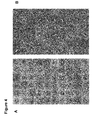
- Transmission electron microscopy (TEM) images taken from purified structures see e.g. Fig. 1d ) confirm that the intended shape is realized.
- TEM Transmission electron microscopy
- the inventors performed single-channel electrophysiological experiments (Sakman & Neher, 1995) using an integrated chip-based setup (Baaken et al., 2008).
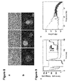
- the inventors added synthetic DNA channels at low concentrations ( ⁇ 200 pM) to the cis side of the setup and applied voltage pulses to facilitate incorporation into the membrane (see e.g. Fig. 9a ).
- synthetic DNA channels at low concentrations ( ⁇ 200 pM) to the cis side of the setup and applied voltage pulses to facilitate incorporation into the membrane (see e.g. Fig. 9a ).
- successful membrane incorporation of individual synthetic DNA channels manifested itself in a stepwise increase in transmembrane current (see e.g. Fig. 2a ) along with an increase in electrical noise (see e.g. Fig. 9b ).
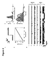
- mutant channels showed more pronounced gating than wildtype channels (see e.g. Fig. 2d , trace iv-vi), and they also significantly differed in their gating time statistics (see e.g. Fig. 2e and 12 ). Every investigated mutant channel displayed gating, while some of the wildtype channels did not show gating at all (see e.g. Fig. 2d , trace i). These results show that the transmembrane current depends on fine structural details of the synthetic DNA channel.
- Nanoscale membrane pores have shown great potential for the use as single molecule biosensors (see e.g. Hall et al., 2010, Branton et al., 2008), whose operation principle is based on the transient blockage of ionic current by analyte molecules.
- the type of sensing task that can be accomplished with such 'nanopores' depends on their size and their chemical structure.
- Nanopore sensors based on naturally occurring membrane pores provide excellent electrical properties, but altering the geometry of biological pores and introducing chemical functions through genetic engineering or chemical conjugation is challenging.
- the geometry of synthetic DNA objects Bell et al., 2012, Wei et al., 2012
- their chemical properties can be tailored for custom nanopore sensing applications.
- the inventors utilize their synthetic DNA lipid membrane channel for single molecule studies of DNA hairpin unzipping and guanine quadruplex (Burge et al., 2006) unfolding.
- Single-stranded DNA is expected to fit through the 2 nm wide central pore of the DNA channel, while larger DNA secondary structures such as hairpins or quadruplexes are not.
- the structures need to unzip or unfold as in similar experiments with ⁇ -hemolysin, thus providing a characteristic time delay in the current blockades that reveals the kinetics of structural transitions (Mathe et al., 2004).
- the inventors used a DNA hairpin with a 9 base pair long stem flanked by 50 thymidines on the 3' end and 6 thymidines on the 5' end (see e.g. Fig. 3a ).
- the hairpin molecules were initially added to the cis side of a lipid membrane containing a single synthetic DNA channel that displayed a stable current baseline without gating.
- Application of a positive voltage bias leads to capture, unzipping, and translocation of the hairpin structures resulting in transient current blockades (see e.g. Fig. 3a,b ).
- the inventors added quadruplex-forming oligonucleotides with a 60 thymidine long single-stranded tail (Q-T60) to the cis side of a membrane containing a single synthetic DNA channel (see e.g. Fig. 3d,e ).
- Q-T60 60 thymidine long single-stranded tail
- the inventors observed transient current blockades, which correspond to the capture and threading of quadruplex DNA molecules into the channel, followed by unfolding and subsequent translocation through the pore. Removal of the analyte from the cis compartment restored a stable baseline current without blockades. Subsequent addition of quadruplex DNA with a longer (dT) 125 tail (Q-T125) led to larger current blockades.
- the larger current blockade for Q-T125 as compared to Q-T60 translocations can be explained by the larger volume occupied in the channel by the longer T 125 tail as compared to the T 60 tail (see the Examples for further details).
- DNA-based channels can be employed as sensing devices to discriminate analyte molecules by studying their translocation characteristics. Moreover, it is easily conceivable that DNA-based channels can be tailored to provide highest sensitivity and optimum discrimination for a wide range of different molecular targets.
- the inventors have reported a novel DNA-based synthetic membrane channel.
- geometry and chemical details of the synthetic channel are user-defined.
- the inventors have incorporated the channel into lipid bilayers and verified ion flux through the channel.
- the inventors found quantized conductance values and current gating that depended on channel layout details.
- the inventors have demonstrated one potential use of the synthetic DNA channel as a single-molecule sensing device. Future modifications of our current ion channel design may find use for more complex sensing tasks and characterization of other types of analyte.
- the synthetic DNA channels introduced here constitute an entirely new class of biologically-active ion channels. They open up perspectives for applications as targeted antimicrobial agents or delivery means when supplemented with cell-specific recognition mechanisms.
- DNA scaffold strands of 7249 bases length derived from the genome of bacteriophage M13 were prepared recombinantly as previously described (Douglas et al., 2009-2).
- Staple oligonucleotide strands were prepared by solid-phase chemical synthesis (Eurofins MWG, Ebersberg, Germany, HPSF purification).
- DNA channel structures was accomplished in one-pot reactions by mixing 20 nM scaffold strands with 100 nM of oligonucleotide staple strands in a buffer including 5 mM TRIS, 1 mM EDTA, 20 mM MgCl 2 , 5 mM NaCl (pH 8) (Douglas et al., 2009-2, Castro et al., 2011). The mixture was subjected to thermal annealing ramps that cooled from 65°C to room temperature. Shortly before purification the origami structures were incubated with 10 ⁇ M of cholesterol-modified oligonucleotides at room temperature for one hour.
- reaction products 50 ⁇ l of the reaction products were mixed with 450 ⁇ l of buffer containing 5 mMTris, 1 mM EDTA, 5 mM MgCl 2 , 5 mM NaCl and spin filtered with a 100kDaAmicon filter (Millipore) at 2000rcf for 30 minutes. The filtration was repeated 5 additional times.
- POPC 1-palmitoyl-2-oleoyl-sn-glycero-3-phosphocholine lipids were dissolved in chloroform to a concentration of 5 mg/ml.
- a lipid film was formed by evaporating 1 ml of POPC solution in a 5 ml round bottom flask, using a rotational evaporator. The flask was kept under vacuum for at least 30 minutes to evaporate remaining chloroform. The lipid film was resuspended in 2ml of a 150mM aqueous KCl solution.
- SUVs were formed by sonicating the solution with a tip sonicator (BandelinSonoplus mini20) for ca. 5 minutes at 8W, until the solution started to become transparent. Afterwards the SUVs were purified by centrifugation at 1500 rcf for 5 minutes. The solution was shock frozen with liquid nitrogen and kept at -80°C until usage.
- Particles were adsorbed on glow-discharged formvar-supported carbon-coated Cu400 TEM grids (Science Services, Kunststoff, Germany) and stained using a 2% aqueous uranylformate solution containing 25 mMNaOH. Imaging was performed using a Philips CM100 transmission electron microscope operated at 100 kV. Images were acquired using an AMT 4x4 Megapixel CCD camera. Micrograph scale bars were calibrated using 2D catalase crystal lattice constants as length reference. Imaging was performed at 28500x magnification for the majority of images and 11500x magnification for overview images.
- Micrographs of individual particles were picked using the EMAN2 (Tang et al. 2007) boxer routine.
- Micrograph alignment and superposition was performed using the XMIPP mlf_2Dalign routine (Scheres et al., 2007).
- Mg 2+ ions strongly influence the permeability of GUV membranes.
- fluorescence imaging experiments were performed in a Mg 2+ free buffer containing 300mM KCl.
- Control TEM experiments confirmed the stability of DNA channels under these conditions.
- 4 ⁇ l of DNA channel solution were incubated with 1 ⁇ M (1 ⁇ l) YOYO-1 (Invitrogen, Life Technologies GmbH, Darmstadt, Germany) for 2 hours in 600mMKCl, 0.5xTBE. Then, 5 ⁇ l of the GUV solution were added and incubated for 1h.
- GUV-Origami solution was diluted in 5 steps to a final concentration of 300mM KCl and 600mM sorbitol in order to reduce background fluorescence.
- All other consumables were purchased form Sigma Aldrich, Kunststoff, Germany).
- a microscopic observation chamber was assembled using vacuum grease and a cover glass, coated overnight with 5% BSA solution.
- fluorescence and DIC imaging an Olympus IX81 microscope with a 60x 1.4 oil immersion objective and a Hamamatsu Orca r2 camera was used.
- MECA 16 channel microelectrode cavity arrays
- the MECA chips were interfaced using an Orbit 16 device, provided by Nanion Technologies GmbH, Kunststoff, Germany.
- Lipid solutions were prepared from 1,2-diphytanoyl-sn-glycero-3-phosphocholine (DPhPC, Avanti Polar Lipids, Alabaster, AL, USA) and dissolved in octane (Sigma Aldrich, Kunststoff, Germany) at a concentration of 10 mg/ml.
- electrolyte 1M KCl solution Merck KGaA, Darmstadt, Germany
- 10mM Tris and 1 mM EDTA Sigma Aldrich, Kunststoff, Germany
- lipid bilayers on the 16-well MECA chips 100 ⁇ l of electrolyte solution were added to the measurement chamber.
- the ground Ag/AgCl electrode resides in the cis (upper) compartment.
- 1-2 ⁇ l of lipid solution were pipetted in the vicinity of the cavities followed by painting the lipid bilayer on top of the cavities using a magnetic stir bar.
- Parallel current recordings were performed using a 16 Channel Triton+ amplifier (Tecella Inc., Costa Mesa, CA, USA) that was controlled via a custom made Labview (National Instruments Germany GmbH, Kunststoff, Germany) measurement software.
- Labview National Instruments Germany GmbH, Kunststoff, Germany
- binding to and insertion into lipid bilayer membranes is facilitated by cholesterol modifications of the DNA channels.
- the cap region of the channel structure facing the lipid membrane were modified at 26 positions arranged concentrically around the stem with single-stranded adaptor extensions (cf. schematic depiction in Fig. 1a , staple sequences SEQ ID NOs. 106-131 and caDNAno maps in 2.9).
- these extensions were hybridized to 22 nucleotide long cholesterol-modified DNA oligomers (an adaptor oligonucleotide) with sequence 5'-CCTCGCTCTGCTAATCCTGTTA-3' [SEQ ID NO. 132].
- a voltage pulse protocol was applied to facilitate the incorporation of single channels into the membrane.
- the voltage protocol shown in Fig. 9a is applied, supported by repeated mixing.
- a clear step in the current signal is observed along with an increase of 1/f noise ( Fig. 9b ).
- this sharp increase in transmembrane current occurred during the initial part of the incorporation pulse (230mV for 20 ms), suggesting a voltage-enhanced incorporation probability as known for other biological channels. Similar current steps could be observed at 100mV bias voltage, but less frequently.
- two or more subsequent steps indicated the incorporation of multiple channels into the same membrane (see Fig. 9a inset).
- the flicker noise power varies greatly from channel to channel, from negligible up to values on the order of 10 -6 .
- the conductivity of the origami structure was varied in the simulation from 10 -4 S/m (insulating) to 10 S/m.
- the mesh density ranged from 18 elements/nm 3 (channel interior & surrounding) to 10 -2 elements/nm 3 (solution compartments).
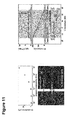
- Fig. 11 shows the results of the simulations.
- the calculated channel conductance coincides with that of a simple geometrical model for the limit of zero origami conductivity.
- For origami conductivity values of 10 -1 S/m and more (corresponding to 1% of the bulk solution conductivity) a considerable increase in channel conductance is observed.
- the channel conductance corresponds to a channel with a length equal to the lipid bilayer thickness and a diameter equal to the stem's outer diameter.
- the electrical potential along the channel's central axis is shown in Fig. 11d.
- the region of steepest slope is not at the center of the channel structure, but at the bilayer position, i.e. 26 nm from the cis side channel entrance.
- the local variation of electric field strength for nonzero leak conductivity can be expected to strongly influence translocation experiments, where charged molecules are pulled electrophoretically through the pore (see 2.7 below).
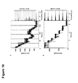
- individual DNA channels displayed distinct gating behavior in which the channel conductance switched between several (sub)-conductance levels. While for high bias voltages the channels underwent full closure as seen, e.g., in Fig. 10 , at lower voltages we typically observed current reduction amplitudes ⁇ I corresponding to fractions of a single channel conductance.
- channel 'mutants' in which one of the central helices forming the stem of the DNA channel was modified with a single-stranded heptanucleotide not incorporated into the main origami structure. This modification potentially could fluctuate, block the channel, or act as a 'handle' for the electric field and lead to partial unzipping of a DNA staple strand.
- Fig. 12a shows a scatter plot of conductance change vs. channel closure time for an unmodified DNA channel structure.
- a single population is observed with a mean conductance reduction of ⁇ G/ ⁇ G 0 of 45% and an exponentially distributed dwell time with a mean value of 10.5 ms.As seen in Fig. 12b , for modification M1 (heptanucleotide pointing into the central channel) a clear second population occurs with a considerably higher blockade amplitude of 70% and a considerably shorter time constant of 0.3 ms.
- DNA molecules hairpin and quadruplex structures
- polythymidine tails of different lengths (60 and 125 nt) were investigated for both forward (cis-trans) and backward (trans-cis) translocation directions (cf. Fig. 11e).
- the fact that longer tails lead to a larger transient current reduction is another clear indication of translocation through the central channel of our DNA structures.
- the current blockade amplitudes measured in these experiments can be used for a qualitative discussion of the electric field distribution within the DNA channel.
- a 60 nt long tail corresponds to a length of L DNA ⁇ 24 nm (assuming a base-to-base distance of 0.4 nm), from which we expect a ⁇ G / G of ⁇ 15%. While again our experimentally observed reduction is smaller ( ⁇ 5%), this value is completely consistent with the value for the 125 tail if one takes the leak conductance into account.
- Fig. 11e the lengths of the DNA molecules together with the electric fields for different values of the leak conductivity are depicted.
- backward translocation results in faster translocations with a higher current amplitude, indicating that the short tail penetrates deeper into the high field region for the backward direction.
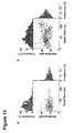
- Fig. 13a a scatter plot generated from backward translocation events (from trans to cis) of Q-T125 DNA at negative bias potential is shown.
- the scatter plot histograms yield a blockade amplitude that is 27% lower ( 11.2 pA ) and a characteristic dwell time that is 2.3 times shorter (3.5 ms) than for the forward direction.
- This observation indicates a directionality of the translocation process itself, as the integrated electrophoretic force along the DNA channel should be independent of the direction of translocation (as the T 125 tail is expected to traverse the whole central channel).
- a possible reason for this effect may be steric or binding interactions at the entrance of the asymmetrically designed origami structure.
Landscapes
- Chemical & Material Sciences (AREA)
- Organic Chemistry (AREA)
- Life Sciences & Earth Sciences (AREA)
- Zoology (AREA)
- Proteomics, Peptides & Aminoacids (AREA)
- Health & Medical Sciences (AREA)
- Engineering & Computer Science (AREA)
- Wood Science & Technology (AREA)
- Analytical Chemistry (AREA)
- Microbiology (AREA)
- Physics & Mathematics (AREA)
- Molecular Biology (AREA)
- Immunology (AREA)
- Biotechnology (AREA)
- Biophysics (AREA)
- Biochemistry (AREA)
- Bioinformatics & Cheminformatics (AREA)
- General Engineering & Computer Science (AREA)
- General Health & Medical Sciences (AREA)
- Genetics & Genomics (AREA)
- Measuring Or Testing Involving Enzymes Or Micro-Organisms (AREA)
- Apparatus Associated With Microorganisms And Enzymes (AREA)
Abstract
Description
- The present invention relates to nucleic acid based nanopores comprising one or more nucleic acids. The present invention further relates to nanopore complexes comprising at least one of said nucleic acid based nanopores, to uses of the nucleic acid based nanopores as sensor and for cell-specific targeting, cell-specific labeling and/or cell-specific drug delivery. The present invention further relates to nucleic acid based nanopores and nanopore complexes for use in medicine and to compositions for obtaining the nucleic acid based nanopores.
- Proteins and peptides that form channels through lipid bilayer membranes are an abundant class of biological macromolecules found in all three domains of life. The basic function of membrane channels is to facilitate the transport of water, ions, or other entities through otherwise impermeable lipid membranes. Membrane channels provide a means for cells for the exchange of matter and information with the outside world. They are fundamental to processes such as cellular communication, maintenance of membrane potentials, or metabolism.
- The natural α-hemolysin channel protein has a characteristic mushroom-like shape and is formed by multimerization of subunits (Song et al., 1996).
- Nanoscale membrane pores have shown great potential for the use as single molecule biosensors (see e.g. Hall et al., 2010, Branton et al., 2008), whose operation principle is based on the transient blockage of ionic current by analyte molecules. The type of sensing task that can be accomplished with such 'nanopores' depends on their size and their chemical structure. Nanopore sensors based on naturally occurring membrane pores provide excellent electrical properties, but altering the geometry of biological pores and introducing chemical functions through genetic engineering or chemical conjugation is challenging.
- There is a need in the art for improved nanopores and nanopore complexes, which can be easily adapted and tailored for different uses.
- According to the present invention this object is solved by a nucleic acid based nanopore comprising one or more nucleic acids.
- According to the present invention this object is solved by a nanopore complex comprising at least one nucleic acid based nanopore according to the invention.
- According to the present invention this object is solved by the use of a nucleic acid based nanopore according to the invention as sensor.
- According to the present invention this object is solved by the use of a nucleic acid based nanopore or a nanopore complex according to the invention for controlling membrane potentials, transmembrane traffic or interference with membrane functions.
- According to the present invention this object is solved by the use of a nucleic acid based nanopore or a nanopore complex according to the invention for cell-specific targeting, cell-specific labeling and/or cell-specific drug delivery.
- According to the present invention this object is solved by providing the nucleic acid based nanopore or a nanopore complex according to the invention for use in medicine.
- According to the present invention this object is solved by providing the nucleic acid based nanopore or a nanopore complex according to the invention for the diagnosis, prevention and/or treatment of a disease.
- According to the present invention this object is solved by a composition for obtaining the nucleic acid based nanopore according to the invention comprising a scaffold nucleic acid strand and/or a plurality of staple oligonucleotide strands, adaptor oligonucleotides comprising recognition binding moiety(ies), optionally, adaptor oligonucleotides comprising recognition sensing moiety(ies), further component(s) and/or gating mechanism component(s), suitable buffers and salts.
- Before the present invention is described in more detail below, it is to be understood that this invention is not limited to the particular methodology, protocols and reagents described herein as these may vary. It is also to be understood that the terminology used herein is for the purpose of describing particular embodiments only, and is not intended to limit the scope of the present invention which will be limited only by the appended claims. Unless defined otherwise, all technical and scientific terms used herein have the same meanings as commonly understood by one of ordinary skill in the art. For the purpose of the present invention, all references cited herein are incorporated by reference in their entireties.
- Numbers of nucleotides, nucleic acids, components, moieties or other numerical data may be expressed or presented herein in a range format. It is to be understood that such a range format is used merely for convenience and brevity and thus should be interpreted flexibly to include not only the numerical values explicitly recited as the limits of the range, but also to include all the individual numerical values or sub-ranges encompassed within that range as if each numerical value and sub-range is explicitly recited. As an illustration, a numerical range of "15 to 100 nucleotides " should be interpreted to include not only the explicitly recited values of 15 to 100, but also include individual values and sub-ranges within the indicated range. Thus, included in this numerical range are individual values such as 15, 16, 17, 18, 19, ... 97, 98, 99, 100 and sub-ranges such as from 20 to 60, from 15 to 20, from 20 to 40, from 15 to 30 etc. As an illustration, a numerical range of "about 2 to about 100 nm " should be interpreted to include not only the explicitly recited values of 2 to 100, but also include individual values and sub-ranges within the indicated range. Thus, included in this numerical range are individual values such as 2, 3, 4, 5, 6, 7, ... 98, 99, 100 and sub-ranges such as from 2 to 50 nm, 3 to 50 nm, 4 to 50 nm, such as about 42 nm, or sub-ranges such as from 2 to 20 nm, 3 to 20 nm, 3 to 10 nm, 4 to 20 nm or 4 to 10 nm, etc. This same principle applies to ranges reciting only one numerical value. Furthermore, such an interpretation should apply regardless of the breadth of the range or the characteristics being described.
- As described above, the present invention provides nucleic acid based nanopores comprising one or more nucleic acids.
- The nucleic acid based nanopores of the invention are formed or assembled by the one or more nucleic acids.
- The term "nanopore" as used herein refers to a structure formed or assembled by nucleic acid molecule(s) which has a hollow channel or hollow tunnel or hollow tube or hole, which provides a connection between two reservoirs or compartments, which are otherwise separate from each other, such as the outside and inside of a vesicle or a cell or an extracellular and an intracellular room. Preferably, the hollow channel/tunnel/tube or hole of a nanopore has a diameter in the nm range, such as about 0.1 nm to about 1,000 nm, e.g. about 0.1 to about 100 nm.
- The nanopore of the invention is not a (single) DNA double helix or nucleic acid double helix.
- Preferably, the nanopore of the invention is or becomes or is intended for becoming associated with a (cell) membrane, lipid bilayer or lipid vesicle.
- A nanopore as known in the art may, for example, be created by a pore-forming protein, such as alpha hemolysin and MspA porin, or is a hole in synthetic materials, such as silicon or graphene.
- The nanopores of the present invention are nucleic acid based nanopores, i.e. they are formed by nucleic acids and not by pore-forming proteins. The nucleic acid based nanopores according to the invention have a hollow channel/tunnel/tube which is formed by the nucleic acids and not by proteins or protein domains.
- The nucleic acids of the nucleic acid based nanopores according to the invention preferably comprise
- DNA,
- RNA,
- modified and artificial nucleic acids and nucleic acid analogues, such as
- nucleic acids comprising modified backbones
such as peptide nucleic acids (PNA), Morpholino and locked nucleic acids (LNA), glycol nucleic acids (GNA), threose nucleic acids (TNA), - nucleic acids comprising modified nucleotides or bases or base analogues or artificial nucleobases,
- nucleic acids comprising abasic and other synthetic residues,
- nucleic acids comprising modified backbones
- In embodiments of the invention, a nanopore can comprise nucleic acid molecules which are DNA which is partially modified, such as comprising parts without bases (abasic) and parts with modified backbones and/or bases.
- The intended use or application of the nanopore of the invention can have influence on the choice of nucleic acid(s) or modified nucleic acid(s) to be used for obtaining the respective nanopore.
- In a preferred embodiment, the nucleic acid based nanopores of the invention comprise self-assembled nucleic acid(s), such as self assembled DNA molecule(s).
- Preferably, the one or more nucleic acids of the nanopore of the invention comprise a plurality of helical nucleic acid domains which form the structure of and/or assemble the nanopore.
- The term "a plurality of helical nucleic acid domains" as used herein refers to several helical nucleic acid domains, which are at least two helical nucleic acid domains, preferably 2 to 100, such as 10 to 70, and can be as high as several 100 or even several 1,000 helical nucleic acid domains.
- In an example, a nanonopore comprises 54 double helices or helical nucleic acid domains.
- In one embodiment, the nucleic acid based nanopores of the invention comprises or consists of a plurality of nucleic acids.
- The term "a plurality of nucleic acids" as used herein refers to several nucleic acids, which are at least two nucleic acids, preferably at least three, and can be as high as several 100 or several 1,000 nucleic acid(s) / nucleic acid molecule(s).
- In one embodiment, the plurality of nucleic acids that form the nanopore of the invention can be chemically ligated after formation/generation of the nanopore, such that the nanopore than comprises only one or several (such as 2, 3, 4, 5 up to e.g. 10) nucleic acid molecules.
- The nucleic acid based nanopores according to the invention preferably comprise
- (i) a transmembrane domain,
- (ii) optionally, a cap domain.
- The transmembrane domain of the nucleic acid based nanopores according to the invention have a hollow channel or hollow tunnel or hollow tube or hole, such as a centrally located hollow channel or hollow tunnel or hollow tube or hole.
- The transmembrane domain of the nucleic acid based nanopores according to the invention comprises helical nucleic acid domains.
- The helical nucleic acid domains of the transmembrane domain are preferably parallel helical nucleic acid domains.
- The helical nucleic acid domains of the transmembrane domain are preferably DNA double helices, such as 6 DNA double helices.
- The hollow channel formed by the transmembrane domain or of the transmembrane domain has preferably a length of about 2 to about 200 nm.
- The preferred length of the hollow channel formed by the transmembrane domain or of the transmembrane domain depends on the intended use of the nanopore.
- In one embodiment, the hollow channel has at least or about the length or thickness of a lipid bilayer, which is about 4 nm thick.
- The hollow channel can also have a length of about 2 nm, in particular in an embodiment where a nanopore of the invention can form dimers or multimers which then span a lipid bilayer of (cell) membrane.
- In one embodiment, the hollow channel is longer, such nanopores are preferably suitable for sequencing or filtering applications.
- In one embodiment, the hollow channel has a length of about 2 to about 100 nm, preferably 2 to 50 nm or about 3 to 50 nm or about 4 to 50 nm, such as about 42 nm. In one embodiment, the hollow channel has a length of about 2 to about 20 nm, such as about 3 to 20 nm, about 3 to 10 nm, about 4 to 20 nm or about 4 to 10 nm.
- The hollow channel formed by the transmembrane domain or of the transmembrane domain has preferably a diameter of about 0.1 to 100 nm, preferably about 0.2 to 10 nm, such as about 2 nm.
- The preferred diameter of the hollow channel depends on the intended application of the nanopore, such as on the size of the analyte or single molecule to be detected or filtered.
- In one embodiment, the hollow channel is modified.
- For example, single stranded nucleic acid(s) or oligonucleotide(s) are protruding into the hollow channel or are lining the hollow channel walls.
- Said single stranded nucleic acid(s) or oligonucleotide(s) can comprise DNA, RNA, modified nucleic acids, artificial nucleic acids, nucleic acid analogues or combinations thereof, as described above.
- Such modification of the hollow channel is preferably for varying the conductivity of the nanopore.
- The cap domain of the nucleic acid based nanopores according to the invention, if present, comprise helical nucleic acid domains.
- The cap domain can be barrel-shaped.
- The helical nucleic acid domains of the cap domain are preferably parallel helical nucleic acid domains.
- The helical nucleic acid domains of the cap domain are preferably DNA double helices, such as 48 DNA double helices.
- Preferably, the adhesion or attachment of the nanopore according to the invention to a lipid bilayer, membrane or lipid vesicle (preferably via recognition binding moieties), leads to formation of a transmembrane channel which spans through said lipid bilayer, membrane or lipid vesicle.
- The cap domain preferably adheres to the outside of a cell membrane, i.e. the cis-side of the membrane or lipid bilayer/vesicle.
- In a preferred embodiment, the nucleic acid based nanopores of the invention furthermore comprises at least one recognition binding moiety.
- The at least one recognition binding moiety is preferably
- a hydrophobic moiety, preferably for binding to lipid or membrane surfaces, such as cholesterol or its derivatives, lipid(s), fatty acid(s), triglyceride(s),
- a moiety specifically recognizing receptors or other surface molecules of membranes or (target) cell membranes,
- such as
- antibody or fragments thereof,
- receptor ligand or fragments thereof, receptor fragments,
- nucleic acid(s), such as aptamers,
- antigen or epitopes.
- The recognition binding moiety/moieties are preferably attached to at least one of the nucleic acids of the nanopore of the invention, such as to the transmembrane domain and/or the cap domain.
- The nanopores of the invention can comprise one recognition binding moiety or more than one recognition binding moiety, such as two , three, four and so one, such as 100.
- In one example, a nanopore of the invention comprises 26 cholesterols.
- The nanopores of the invention can also comprise different recognition binding moieties, such as different hydrophobic moieties and/or different moieties specifically recognizing receptors or other surface membrane molecules.
- In a preferred embodiment, the nucleic acid based nanopores of the invention furthermore comprises at least one recognition sensing moiety.
- Said recognition sensing moiety is a moiety specifically recognizing or binding or interacting with an analyte or molecule which passes through the nanopore or hollow channel.
- Such as
- antibody or fragments thereof,
- receptor ligand or fragments thereof, receptor fragments,
- nucleic acid(s), such as aptamers,
- antigen or epitopes,
- This embodiment is preferably useful when the nanopores of the invention are used as single molecule sensor or for transmembrane traffic.
- The recognition sensing moiety/moieties are preferably attached to at least one of the nucleic acids of the nanopore of the invention, preferably to the transmembrane domain, more preferably within the hollow channel of the transmembrane domain.
- The nanopores of the invention can comprise one recognition sensing moiety or more than one recognition sensing moiety, such as two , three, four and so one, such as 100.
- The nanopores of the invention can also comprise different recognition sensing moieties, such as different antigens and/or aptamers, such as for recognizing different analytes/molecules to pass through the nanopore.
- In a preferred embodiment, the nucleic acid based nanopores of the invention furthermore comprise one or more further components.
- The one or more further components are preferably
- label or tag,
such as fluorescent dye, chromophore, isotope, quantum dot, ... - protein or peptide,
such as an enzyme or its binding and/or catalytic domain, antibody or fragments thereof, receptor ligand or fragments thereof, receptor fragments, antigen or epitopes, preferably to be delivered to a target cell, - drug,
such as a drug to be delivered to a target cell,
such as antimicrobial or anti-viral drugs, anti-cancer drugs, - nanoparticle or nanocrystal,
- glycosylated or glycosylation label or tag,
such as glycan, glycoprotein, glycolipid, proteoglycan, - component for a functional mechanism,
such as
a rotor domain enabling the nanopore to move or rotate in the lipid bilayer or membrane,
an ion pump,
transport proteins or domains thereof. - The one or more further components are preferably attached to at least one of the nucleic acids of the nanopore of the invention, such as to the transmembrane domain and/or the cap domain.
- The nanopores of the invention can comprise one or more further components, such as two , three, four and so one, such as 100.
- The nanopores of the invention can also comprise different further components, such as labels/tags and drugs; or labels/tags and rotor domain(s).
- Rotor domains enabling the nanopore to move or rotate in a lipid bilayer or membrane can be, for example, similar to a rotor domain of the ATP synthase complex (Boyer 1997).
- In a preferred embodiment, the nucleic acid based nanopores of the invention furthermore comprise gating mechanism component(s) for opening and closing the nanopore.
- Such gating mechanism component(s) can be reactive to binding of ligand(s), such as antigen(s).
- For example, gating mechanism component(s) based on aptamers that are capable of binding to antigen(s). The nanopore can be closed, when said aptamer is hybridized to a respective complementary nucleic acid strand, and the nanopore can be open, upon antigen binding. In another exemplary embodiment, the nanopore opens due to a comformational change an aptamer undergoes upon antigen binding.
- Further gating mechanism component(s) can be reactive to pressure, temperature, voltage and/or light.
For example: - mechano-sensitive nucleic acids, such as mechano-sensitive DNA,
- components with a temperature-dependent secondary structure
- components conjugated with temperature-dependent polymers, such as p-NIPAM,
- movable charged structures, which move in an electric field and thereby open/close the nanopore, i.e. the hollow channel,
- photo-switchable molecules, which are e.g. conjugated to nucleic acids
- The gating mechanism component(s) are preferably attached to at least one of the nucleic acids of the nanopore of the invention, such as to the transmembrane domain and/or the cap domain.
- Preferably, the recognition binding moiety(ies), the recognition sensing moiety(ies), the further component(s) and/or the gating mechanism component(s) comprise adaptor oligonucleotide(s) with a nucleotide sequence which hybridizes to at least one of the nucleic acids of the nanopore of the invention or parts thereof.
- Preferably, an adaptor oligonucleotide is covalently attached to said recognition binding moiety(ies), recognition sensing moiety(ies), further component(s) and/or gating mechanism component(s).
- In a preferred embodiment, the nucleic acid based nanopores of the invention comprise or consist of self-assembled nucleic acids.
- Preferably, the self-assembled nucleic acids are obtained via molecular self-assembly.
- The term "molecular self-assembly", as used herein, refers to the process by which nucleic acid molecules adopt a defined arrangement without guidance or management from an outside source. There are two types of self-assembly, intramolecular self-assembly and intermolecular self-assembly. The molecular self-assembly can also be called folding. The assembly of the nucleic acid molecules is directed through noncovalent interactions (e.g., hydrogen bonding, hydrophobic forces, van der Waals forces, π-π interactions, and/or electrostatic) as well as electromagnetic interactions. Molecular self-assembly is an important aspect of bottom-up approaches to nanotechnology. Using molecular self-assembly the final (desired) structure is programmed in the shape and functional groups of the molecules. The nucleic acids, such as DNA, are used as a structural material rather than as a carrier of biological information, to make the structures.
- The molecular self-assembly of the nucleic acids according to the invention can be via DNA origami molecular self-assembly of scaffold nucleic acid strand(s) and a plurality of staple or helper oligonucleotide strands.
- The molecular self-assembly of the nucleic acids can be via molecular self-assembly of one or more scaffold nucleic acid strand(s).
- The molecular self-assembly of the nucleic acids can also be via molecular self-assembly of a plurality of oligonucleotide strands.
- The term "DNA origami molecular self-assembly", as used herein, refers to intramolecular folding of a single-stranded DNA scaffold molecule with DNA staple or helper oligonucleotides into a specific molecular structure.
- DNA origami structures incorporate DNA as a building material to make nanoscale shapes. In general, the DNA origami process involves the folding of one or more long, "scaffold" DNA strands into a particular shape using a plurality of rationally designed "staple" or "helper" DNA or oligonucleotide strands. The sequences of the staple or "helper" strands are designed such that they hybridize to particular portions of the scaffold strands and, in doing so, force the scaffold strands into a particular shape.
Methods useful in the making of DNA origami structures can be found, for example, in Rothemund 2006, Douglas et al 2009-2; Dietz et al, 2009 orUS 7,842,793 B2 (Rothemund ). Staple design can be facilitated using, for example, CADnano software, available at http://www.cadnano.org. - Preferably, the scaffold nucleic acid strand(s) are single stranded DNA polynucleotide strand(s).
- The scaffold strand or scaffold nucleic acid can have any sufficiently non-repetitive sequence.
- For example, in certain embodiments, the scaffold strand is derived from a natural source, such as derived from M13 phage DNA, such as the M13mpl8-derived sequence provided in nucleotide sequence of SEQ ID NO. 1. In one embodiment, the scaffold strand comprises or consists of a nucleic acid with SEQ ID NO. 1.
- The scaffold strand or scaffold nucleic acid can be artificial.
- The scaffold strand or scaffold nucleic acid(s) has/have a length of preferably several thousand nucleotides, such as about 100 to about 100.00 nucleotides, e.g. about 4,000 to about 8,000 nucleotides.
- Preferably, the staple or helper oligonucleotide strands are at least partially complementary to parts of the scaffold nucleic acid strand(s).
- Preferably, the number of staple or helper oligonucleotide strands are in the range of about 1 to 1,000, e.g. about 50 to about 500.
- Preferably, the staple or helper oligonucleotide strands are oligonucleotides comprising or consisting of a nucleotide sequence of SEQ ID NOs. 2 to 131.
- In an embodiment, oligonucleotides comprising or consisting of a nucleotide sequence of SEQ ID NOs. 2 to 105 are staple or helper oligonucleotides; and oligonucleotides comprising or consisting of a nucleotide sequence of SEQ ID NOs. 106 to 131 are staple or helper oligonucleoties to which adaptor oligonuleotide(s) can hybridize / are complementary to adaptor oligonucleotide(s), e.g. in that said oligonucleotides comprise single-stranded adaptor extensions to which the adaptor oligonucleotide(s) can hybridize/are complementary to.
- The staple or helper oligonucleotides have preferably a length of about 10 to 1,000 nucleotides, such as about 15 to 100 nucleotides, such as about 20 to 60.
- In one embodiment, the adaptor oligonucleotides comprised in the recognition binding moiety(ies), the recognition sensing moiety(ies), the further component(s) and/or the gating mechanism component(s) are selected from oligonucleotides comprising a nucleotide sequence of SEQ ID NO. 132.
- The number of adaptor oligonucleotides depends on the number and/or kind of recognition binding moiety(ies), the recognition sensing moiety(ies), the further component(s) and/or the gating mechanism component(s) that are comprised in a respective embodiment of the nanopore of the invention.
- The adaptor oligonucleotides have preferably a length of about 10 to 1,000 nucleotides, such as about 15 to 100 nucleotides, such as about 15 to 60 or 20 to 60.
- In one embodiment, a nucleic acid based nanopore according to the invention comprises the following nucleic acids:
- one scaffold strand, such as the M13mpl8-derived sequence provided in nucleotide sequence of SEQ ID NO. 1;
- at least or up to 130 staple or helper oligonucleotide strands, such as oligonucleotides comprising or consisting of a nucleotide sequence of SEQ ID NOs. 2 to 131;
- at least or up to 26 adaptor oligonucleotide comprising recognition binding moieties, such as oligonucleotides each comprising or consisting of a nucleotide sequence of SEQ ID NOs. 132.
- In one embodiment, a M13 phage-derived 7249 bases-long single-stranded DNA was folded by 156 DNA oligonucleotides (130 staple oligonucleotides plus 26 adaptor oligonucleotides with cholesterol) into a structure consisting of 54 parallel DNA double-helices close-packed on a honeycomb lattice. See, for example
Figure 1a . - Castro et al., 2011, for example, describe close-packing of DNA double helical domains onto a honeycomb lattice or onto a square lattice.
- As described above, the present invention provides nanopore complexes.
- The nanopore complexes of the invention comprise at least one nucleic acid based nanopore according to the invention.
- The nanopore complexes of the invention comprise preferably two or more, such as three, four, five and more of said nucleic acid based nanopores.
- The nucleic acid based nanopores of the nanopore complex can differ in their recognition binding moieties, further component(s) and/or gating mechanisms.
- The nucleic acid based nanopores of the nanopore complex can differ in their nucleic acid sequences, number of nucleic acid molecules, number of helical domains.
- The nucleic acid based nanopores of the nanopore complex can differ in their length and diameter of hollow channel/tunnel/hole.
- As described above, the present invention provides uses of a nucleic acid based nanopore of the invention.
- Preferably, the nucleic acid based nanopore of the invention is used as sensor, preferably single molecule sensor.
- The uses of the nucleic acid based nanopore of the invention comprises
- nucleic acid sequencing,
- sensing nucleic acid secondary structure,
- detection of analytes, such as small molecules, toxins, proteins,
- detection of polymers (such as nanopore mass spectrometry),
- separation of components across a lipid bilayer or a (cell) membrane,
- separation of charges across a lipid bilayer or a (cell) membrane,
- molecular filters and sieves.
- The nanopores of the invention can be employed as sensing devices to discriminate analyte molecules, for example, by studying their translocation characteristics.
- For example, a nanopore of the invention can be used for single molecule studies of DNA hairpin unzipping and guanine quadruplex unfolding. Single-stranded DNA fits through the central pore of a nanopore having a 2 nm wide central pore, while larger DNA secondary structures such as hairpins or quadruplexes are not. In order to translocate through the DNA channel, the structures need to unzip or unfold, thus providing a characteristic time delay in the current blockades. See, for example,
Figure 3 . - The nanopores of the invention can be used for separating charges across a lipid bilayer or a (cell) membrane, such as in form of nanobatteries.
- As described above, the present invention provides the use a nucleic acid based nanopore of the invention or of a nanopore complex of the invention for
- controlling membrane potentials,
- transmembrane traffic
- interference with membrane functions.
- Said uses comprise in vitro and in vivo uses.
- Transmembrane traffic comprises, for example, the translocation or transport of nucleic acid molecules or proteins or other molecules through a nanopore of the invention.
- See for example,
Figure 3 and Example 2.7. - Interference with membrane functions comprises, for example, the formation of holes, i.e. transmembrane channels, via adhesion or attachment of one or more nanopores or nanopore complexes of the invention to a respective membrane.
- The uses of the invention comprises voltage gating, pressure, gating, ligand-gating, or gating by other physical effects, such as light, temperature.
- As described above, the present invention provides the use of a nucleic acid based nanopore of the invention or of a nanopore complex of the invention for cell-specific targeting, cell-specific labeling and/or cell-specific drug delivery.
- In one embodiment, a nanopore of the invention comprises a respective recognition binding moiety specific for target cells and is, thus, suitable for said cell-specific targeting.
- In one embodiment, a nanopore of the invention comprises a respective recognition moiety specific for target cells as well as label(s)/tag(s) and is, thus, suitable for said cell-specific labeling.
- In one embodiment, a nanopore of the invention comprises a respective recognition binding moiety specific for target cells as well as drug(s) and is, thus, suitable for said cell-specific drug delivery.
- As described above, the present invention provides a nucleic acid based nanopore of the invention or of a nanopore complex of the invention for use in medicine.
- As described above, the present invention provides a nucleic acid based nanopore of the invention or of a nanopore complex of the invention for the diagnosis, prevention and/or treatment of a disease.
- The diseases are, for example,
- cancer,
- infectious diseases, such as bacterial and/or viral infections,
- allergies,
- metabolic diseases, such as diabetes,
- obesity and heart diseases.
- In one embodiment, a nanopore of the invention comprises a respective antibiotic drug or antiviral drug and is, thus, suitable for the prevention and/or treatment of a bacterial infection or viral infection.
- In one embodiment, a nanopore of the invention comprises a suitable further component, such as a respective drug and/or an ion pump, and is, thus, suitable for the prevention and/or treatment of heart diseases and/or metabolic diseases, such as diabetes.
- In one embodiment, a nanopore of the invention comprises a suitable label or tag and is, thus, suitable for the diagnoses of a disease.
- In one embodiment, a nanopore of the invention (furthermore) comprises a respective recognition binding moiety specific for cancer cells and is, thus, suitable for the diagnosis, prevention and/or treatment of the specific cancer.
- The diagnosis, prevention and/or treatment of a disease can comprise the cell-specific targeting of diseased cells, tissues and organs; cell-specific labelling of diseased cells, tissues and organs; and/or cell-specific drug delivery to diseased cells, tissues and organs, wherein the diseased cells, tissues and organs are preferably tumor related. The nanopores of the invention will then comprise suitable recognition binding moieties and further components, such as label, drugs.
- The cell-specific targeting comprises the recognition and attachment of the nanopore to specific target cells. The cell-specific targeting can further comprise the penetration of the cell membrane and formation of multiple holes/pores into the cell membrane of the target cells, which can result in the killing of said target cells and/or in making said target cells recognizable/susceptible for the immune system of the patient.
- For example, the nanopores of the invention can be used for targeting of cancer or tumor cells. The nanopore can be designed to be specifically recognizing and targeting tumor cells by comprising suitable recognition binding moieties which are tumor specific. The nanopore can be utilized for the treatment of a specific tumor in that the nanopore recognizes the specific tumor cells, attached to and then penetrates the tumor cell membrane which results in the killing of said tumor cells and/or in making said tumor cells recognizable/susceptible for the immune system of the patient. The nanopore can furthermore comprise drug(s) and/or further components, as described herein, to be delivered to or into the tumor cells.
- As described above, the present invention provides compositions for obtaining a nucleic acid based nanopore of the present invention.
- A composition of the invention comprises
- a scaffold nucleic acid strand and/or a plurality of staple oligonucleotide strands as defined herein,
- adaptor oligonucleotides comprising recognition binding moiety(ies) as defined herein, optionally, adaptor oligonucleotides comprising recognition sensing moiety(ies), further component(s) and/or gating mechanism component(s) as defined herein, suitable buffers and salts, such as Tris buffer, magnesium salt.
- A multitude of experimental obstacles and difficulties were to be overcome in the design and development of the nucleic based nanopore according to the invention, which can also be seen in greater detail from the description of a preferred embodiments as well as the examples and figures.
- The architecture and structure of the nanopore itself had to be designed wherein a multitude of requirements had to be realized, such as stability of the structure, ability to attach to a lipid layer/membrane and then to penetrate said lipid layer/membrane.
- The correct molecular self-assembly of the rather large structure of a nucleic acid based nanopore had to be ensured: by screening and tuning multiple reaction conditions, such as temperature and salt concentrations, the self-assembly of more than 131 nucleic acid molecules into a multitude of neighboring DNA helices had to be adjusted.
- The recognition binding moiety had to be screened and tested, including the attachment points on the nanopore, which are e.g. important for the ability of the nanopore to successfully attach to a lipid layer/membrane.
- The insertion of the nanopore into a lipid bilayer/membrane had to be ensured by choosing the architecture/geometry of the nanopore, the recognition binding moiety, its attachment points, the dimensions of the nanopore.
- Proteins and peptides that form channels through lipid bilayer membranes are an abundant class of biological macromolecules found in all three domains of life. The basic function of membrane channels is to facilitate the transport of water, ions, or other entities through otherwise impermeable lipid membranes. Membrane channels provide a means for cells for the exchange of matter and information with the outside world. They are fundamental to processes such as cellular communication, maintenance of membrane potentials, or metabolism. Here, the inventors report on a new type of synthetic membrane channel with user-defined geometric specifications that is constructed entirely from DNA. The inventors show that these synthetic channels can be incorporated into lipid bilayer membranes and the inventors study their electrical properties by single-channel electrophysiological measurements. The inventors find remarkable similarities to the behaviour of biological ion channels, such as channel gating caused by molecular fluctuations within the channel. In single molecule translocation experiments with DNA, the inventors highlight one of many potential applications of the synthetic channels, namely as single-molecule sensing devices. Other applications such as the control of membrane potentials, transmembrane traffic, or interference with membrane functions, both in vitro as well as in vivo, are readily conceivable.
- The shape of the inventors' synthetic membrane channel is inspired by the natural α-hemolysin channel protein that has a characteristic mushroom-like shape (Song et al., 1996). In contrast to α-hemolysin, however, our channel does not form by multimerization of subunits. We constructed the channel using molecular self-assembly with scaffolded DNA origami (Rothemund 2006, Douglas, et al, 2009-2, Andersen et al., 2009, Dietz et al., 2009, Seeman et al., 2009, Han et al., 2011, Castro et al., 2011).
M13 phage-derived 7249 bases-long single-stranded DNA was folded by 156 DNA oligonucleotides into a structure consisting of 54 parallel DNA double-helices close-packed on a honeycomb lattice (Douglas, et al, 2009-2, Castro et al., 2011) (see e.g.Fig. 1a ). The channel consists of two modules/domains: a stem that penetrates and spans a lipid membrane (i.e. the transmembrane domain), and a barrel-shaped cap that adheres to the (cis)-side of the membrane (i.e. the cap domain). Adhesion to the lipid bilayer is mediated by 26 cholesterol moieties that are attached to the cis-facing surface of the barrel (see e.g.Fig. 1a ). The stem protrudes centrally from the barrel and consists of six double-helical DNA domains that form a hollow tube. The interior of this tube acts as a transmembrane channel with a diameter of ≈ 2 nm and a length of ≈ 42 nm and runs through both stem and barrel (see e.g.Fig. 1b, c ). Transmission electron microscopy (TEM) images taken from purified structures (see e.g.Fig. 1d ) confirm that the intended shape is realized. Experiments with unilamellar lipid vesicles show that the synthetic DNA channels bind to lipid bilayer membranes (see e.g. Fig. Ie-g) in the desired orientation in which the cholesterol-modified face of the barrel forms tight contact with the membrane and the stem protrudes into the lipid bilayer (see e.g. Fig. If). These observations show that the synthetic DNA channels are capable of forming membrane pores as designed. - In order to demonstrate the electrical conductivity of the resulting membrane pores, the inventors performed single-channel electrophysiological experiments (Sakman & Neher, 1995) using an integrated chip-based setup (Baaken et al., 2008). The inventors added synthetic DNA channels at low concentrations (≈ 200 pM) to the cis side of the setup and applied voltage pulses to facilitate incorporation into the membrane (see e.g.
Fig. 9a ). As with biological channels, successful membrane incorporation of individual synthetic DNA channels manifested itself in a stepwise increase in transmembrane current (see e.g.Fig. 2a ) along with an increase in electrical noise (see e.g.Fig. 9b ). Depending on the preparation method, the inventors also observed incorporation of multiple DNA channels into the same membrane (see e.g.Fig. 10 ). The synthetic DNA channels displayed an average Ohmic conductance of G = 0.87 ± 0.15 nS (see e.g.Fig. 2b,c ) per channel in a solution containing 1 M KCl and 2mM MgCl2. A simple geometrical model (Hille 2001) predicts G = 0.78nS for a channel withdiameter 2 nm and length 42 nm (see Examples for further details) which agrees favourably with what the inventors observe. - Many natural ion channels display current gating caused by switching between distinct channel conformations with different conductances (Hille 2001). The inventors found that the synthetic DNA channels also displayed gating behavior (see e.g.
Fig. 2d , trace i-iii). The inventors hypothesized that stochastic unzipping and rezipping of double-helical DNA domains in the channel may contribute to the observed current gating. To test this idea, the inventors designed two channel 'mutants' that differed from the 'wildtype' channel only by a single-stranded heptanucleotide protruding from the central transmembrane tube (see e.g.Fig. 1b , see Examples for further details). The inventors found that the mutant channels showed more pronounced gating than wildtype channels (see e.g.Fig. 2d , trace iv-vi), and they also significantly differed in their gating time statistics (see e.g.Fig. 2e and12 ). Every investigated mutant channel displayed gating, while some of the wildtype channels did not show gating at all (see e.g.Fig. 2d , trace i). These results show that the transmembrane current depends on fine structural details of the synthetic DNA channel. - In the past, nanoscale membrane pores have shown great potential for the use as single molecule biosensors (see e.g. Hall et al., 2010, Branton et al., 2008), whose operation principle is based on the transient blockage of ionic current by analyte molecules. The type of sensing task that can be accomplished with such 'nanopores' depends on their size and their chemical structure. Nanopore sensors based on naturally occurring membrane pores provide excellent electrical properties, but altering the geometry of biological pores and introducing chemical functions through genetic engineering or chemical conjugation is challenging. By contrast, the geometry of synthetic DNA objects (Bell et al., 2012, Wei et al., 2012) and their chemical properties can be tailored for custom nanopore sensing applications.
- Here, the inventors utilize their synthetic DNA lipid membrane channel for single molecule studies of DNA hairpin unzipping and guanine quadruplex (Burge et al., 2006) unfolding. Single-stranded DNA is expected to fit through the 2 nm wide central pore of the DNA channel, while larger DNA secondary structures such as hairpins or quadruplexes are not. In order to translocate through the DNA channel, the structures need to unzip or unfold as in similar experiments with α-hemolysin, thus providing a characteristic time delay in the current blockades that reveals the kinetics of structural transitions (Mathe et al., 2004).
- For one set of experiments, the inventors used a DNA hairpin with a 9 base pair long stem flanked by 50 thymidines on the 3' end and 6 thymidines on the 5' end (see e.g.
Fig. 3a ). The hairpin molecules were initially added to the cis side of a lipid membrane containing a single synthetic DNA channel that displayed a stable current baseline without gating. Application of a positive voltage bias leads to capture, unzipping, and translocation of the hairpin structures resulting in transient current blockades (see e.g.Fig. 3a,b ). Reversal of the bias after approximately -30 minutes again led to transient current blockades, this time caused by molecules that had accumulated in the trans compartment by previous translocation through the DNA channel. The blockade amplitudes for both translocation directions were ΔIcis-trans = 11.9 ± 2.7 pA and ΔItrans-cis= 20.3 ± 4.2 pA. The blockade dwell times were distributed exponentially with a characteristic lifetime of τcis-trans = 1.5 ms and τtrans-cis = 1ms, respectively (see e.g.Fig. 3c ). - In another set of experiments, the inventors added quadruplex-forming oligonucleotides with a 60 thymidine long single-stranded tail (Q-T60) to the cis side of a membrane containing a single synthetic DNA channel (see e.g.
Fig. 3d,e ). Again, the inventors observed transient current blockades, which correspond to the capture and threading of quadruplex DNA molecules into the channel, followed by unfolding and subsequent translocation through the pore. Removal of the analyte from the cis compartment restored a stable baseline current without blockades. Subsequent addition of quadruplex DNA with a longer (dT)125tail (Q-T125) led to larger current blockades. The average current blockades were ΔIQ-T60 = 5.6 ± 1.0 pA and ΔIQ-T125 = 15.3 ± 2.3 pA, respectively. The larger current blockade for Q-T125 as compared to Q-T60 translocations can be explained by the larger volume occupied in the channel by the longer T125 tail as compared to the T60 tail (see the Examples for further details). The blockade dwell times are distributed exponentially with characteristic lifetimes of τQ-T60 = 9.7 ms and τQ-T125 =8.1 ms. - Therefore, similar to biological pores our synthetic DNA channels can be employed as sensing devices to discriminate analyte molecules by studying their translocation characteristics. Moreover, it is easily conceivable that DNA-based channels can be tailored to provide highest sensitivity and optimum discrimination for a wide range of different molecular targets.
- To conclude, the inventors have reported a novel DNA-based synthetic membrane channel. In contrast to biological ion channels, geometry and chemical details of the synthetic channel are user-defined. The inventors have incorporated the channel into lipid bilayers and verified ion flux through the channel. The inventors found quantized conductance values and current gating that depended on channel layout details. The inventors have demonstrated one potential use of the synthetic DNA channel as a single-molecule sensing device. Future modifications of our current ion channel design may find use for more complex sensing tasks and characterization of other types of analyte. The synthetic DNA channels introduced here constitute an entirely new class of biologically-active ion channels. They open up perspectives for applications as targeted antimicrobial agents or delivery means when supplemented with cell-specific recognition mechanisms.
- The following examples and drawings illustrate the present invention without, however, limiting the same thereto.
-
-
Figure 1 . Synthetic DNA membrane channels.- a Schematic illustration of the channel formed by 54 double-helical DNA domains packed on a honeycomb lattice. Cylinders indicate double-helical DNA domains, rings indicate mechanical domain limits determined using CanDo (Maarel 2007). Dark grey: transmembrane stem. Light grey strands with light grey ellipsoids indicate cholesterol-modified oligonucleotides that hybridize to single-stranded DNA adaptor strands as indicated.
- b Geometric specifications of the transmembrane channel. Length L = 47 nm, tube diameter D = 6 nm, inner diameter d = 2 nm. The length of the central channel fully surrounded by DNA helices is 42 nm. The star symbol indicates the position of a 7 base strand extension acting as a 'defect' in channel 'mutants' (1=15 nm for mutant M1 and 1=14 nm for mutant M2).
- c Cross-sectional view through the channel when incorporated in a lipid bilayer.
- d Averaged negative-stain TEM images obtained from purified DNA channel structures.
- e,f TEMimages ofDNA channels adhering to small unilamellar vesicles (SUVs).
- g TEM image of DNA channels binding to an extended lipid bilayer in the upper right part of the image. DNA channels are found predominantly on lipid covered areas or sticking to SUVs.
-
Figure 2 . Electrical characterization.- a Stepwise increase in ionic current during an incorporation event at V=200 mV.
- b Histogram of channel conductances obtained from 43 incorporation events. The black line depicts a Gaussian fit.
- c Current - voltage dependence of the channel after incorporation.
- d Typical current traces obtained from 'wildtype' channels (i-iii), and 'mutants' with modification M2 (iv-vi).
- e Statistics of gating times for the wildtype (red) and the mutant (gray) channels. Continuous lines correspond to a mono-exponential fit for the wildtype channel (τ=10.5 ms) and a double-exponential fit for the mutant (τ1=1.3 ms and τ2=9.2 ms).
-
Figure 3 DNA Translocation studies.- a Addition of DNA hairpins (T5-HP-T50) to a DNA channel at V=200 mV results in the appearance of transient current blockades, indicating unzipping and translocation of hairpin molecules from cis to trans. When a sufficient amount of hairpins has accumulated on the trans side, they can also be transferred back from trans to cis by a reversal of the transmembrane voltage.
- b Representative blockade events for forward (top) and backward (bottom) translocation of DNA hairpins.
- c Scatter plot for the translocation of T5-HP-T50 DNA through a DNA channel from cis to trans (blue) and from trans to cis side (gray) at 200 mV bias potential, accompanied by the corresponding histograms. Each data point corresponds to a single translocation event. In total, 777 (forward) and 379 (backward) events were analyzed.
- d Top: Typical current trace at 200 mV bias potential after addition of 10µM of Q-T60 DNA. Middle: Current trace after rinsing with buffer solution. Bottom: Current trace after subsequent addition of 10µM of Q-T125 DNA to the same channel.
- e Representative blockade events for Q-T60 (top) and Q-T125 DNA (bottom).
- f Scatter plot of the current blockade versus dwell time for the translocation of Q-T60 (red) and Q-T125 DNA (black) through the DNA channel. Each data point corresponds to a single translocation event. In total 631 (Q-T60) and 279 (Q-T125) events were analyzed. On the right and on the top corresponding histograms are shown. The lines correspond to single exponential (top) and Gaussian fits (right).
-
Figure 4 . Negative-stain TEM micrographs of synthetic DNA channels.- A:
Side view 1, B:Side view 2, rotated by 60° around the long axis relative toside view 1. Each micrograph of A and B is sized 110nm x 110nm. C: Top view. Each micrograph of C is sized 60nm x 60nm.
Fig. 1d . - A:
-
Figure 5 . Negative-stain TEM micrographs of cholesterol-modified DNA channels attached to POPC SUVs. Each micrograph is sized 60nm x 60nm. The first micrograph in the top row contains the respective average micrograph also depicted inFig. 1e . -
Figure 6 . Binding of cholesterol modified DNA channels to POPC membranes.- a Cholesterol modified DNA channels mixed with SUVs and imaged byTEM. The lipids form white circular patches. Almost all DNA channels are attached to SUVs (collapsed on the TEM grid) or larger supported lipid membrane patches.
- b Unmodified DNA channels mixed with SUVs show no binding - DNA channels and lipid patches typically are found isolated from each other.
-
Figure 7 . Binding of cholesterol modified DNA channels to POPC membranes.- a Cholesterol modified channels with SUVs,
- b Unmodified channels with SUVs.
Fig.6 at a higher magnification (28500 x). Scale bar is 100 nm. -
Figure 8 . Binding of DNA channels to DPhPC GUVs.- a. Differential interference contrast images of GUVs in 300nMKClsolution.
- b. Epifluorescence microscope images of YOYO labeled origami pores bound to the GUV membranes. The presence of labeled DNA channels in solution leads to a strong fluorescence of the GUV membranes, suggesting that the DNA channels accumulate at the membrane surface due to binding interactions. A control experiment where only YOYO dye was used showed no observable fluorescence of the vesicle membrane. TEM images confirm that the intercalating dye YOYO does not alter the origami structure at the labeling density used here.
-
Figure 9 . Channel Incorporation- a Top: Voltage pulse used for incorporation of origami channels into the lipid membrane. Bottom: Corresponding current response before (black dots) and during incorporation (red line). Inset: Consecutive incorporation of two origami channels at 100mV bias voltage.
- b Noise power spectra before (black line) and after the incorporation of a single origami pore (red line). Current traces were recorded at 100mV at a filter frequency of 3kHz.
-
Figure 10 . Multiple insertions and stepwise closing.- a Time course of the ionic current across the lipid bilayer with multiple origami channels incorporated at 200 mV bias voltage. The DNA channel structures used here had a 7nt modification at siteM2. Vertical lines mark limits of continuous recording. The recording was suspended a this positions with no applied bias for a few ms. Right: Corresponding all-point current histogram, showing a mean peak-to-peak distance of 172±15 pA This corresponds to a mean conductance change of 0.86±0.08 nS.
- b Current response of origami channels at 200 mV with a modification present at site M1, accompanied by the corresponding all-point current histogram. The mean peak-to-peak distance corresponds to 194±14 pA (0.97±7nS).
-
Figure 11 . FEM Simulations of the DNA pore.- a Simulated conductance across a DNA channel incorporated into a lipid bilayer for varying intrinsic origami conductivity at 100mV bias voltage and 11S/m bulk solution conductivity.
- b Color map of the electrical potential distribution along a y-z plane at x=0 calculated for 0.1 S/m origami conductivity. The dashed line marks the structure boundaries.
- c Electric Field along the symmetry axis (z-axis) of the channel for varying origami conductivity.
- d Corresponding potential drops along the channel.
- e Schematic illustration of the penetration depth of the structures used in translocation experiments before unzipping (cf.
Figure 3 ).
-
Figure 12 . Scatter plots of the relative conductance change versus dwell time for the closure states of unmodified origami channel compared to modified channels.- a unmodified origami channel,
- b a channel with modification on site M1 and
- c a channel with modification on site M2
at 100mV bias voltage. Each data point corresponds to a single closing event. In total 697 events (red, a), 2532 events (blue, c) and 623 events (grey, b) were recorded.
On the right and on the top the corresponding histograms are shown. The lines correspond to single exponential (no modification or modification M2) or double exponential (with modification M1), (top) and Gaussian fits (right). - d Closure time for mutant M2 as a function of applied membrane voltage.
-
Figure 13 . Additional data for quadruplex translocations.- a Scatter Plot of the current amplitude vs. dwell time for the backward translocation of Q-T125 through the artificial ion channel at negative bias voltage (-200mV), accompanied by the corresponding histograms. 266 events were detected. The solid lines correspond to single exponential (top) and Gaussian (right) fits to the histograms.
- b Long-lived population: Scatter Plot of the current amplitude vs. dwell time for forward translocation of Q-T125 (see
Fig. 3 ), including data from the last part of the measurement. Each data point corresponds to a single translocation event. The lines correspond to single exponential (top) and Gaussian (right) fits to the histograms.
- Structures were designed using caDNAno v.02 (Douglas et al., 2009-1). DNA scaffold strands of 7249 bases length derived from the genome of bacteriophage M13 (with SEQ ID NO. 1) were prepared recombinantly as previously described (Douglas et al., 2009-2). Staple oligonucleotide strands (with SEQ ID NOs. 2 to 131) were prepared by solid-phase chemical synthesis (Eurofins MWG, Ebersberg, Germany, HPSF purification). Production of DNA channel structures was accomplished in one-pot reactions by mixing 20 nM scaffold strands with 100 nM of oligonucleotide staple strands in a buffer including 5 mM TRIS, 1 mM EDTA, 20 mM MgCl2, 5 mM NaCl (pH 8) (Douglas et al., 2009-2, Castro et al., 2011). The mixture was subjected to thermal annealing ramps that cooled from 65°C to room temperature. Shortly before purification the origami structures were incubated with 10µM of cholesterol-modified oligonucleotides at room temperature for one hour.
- 50µl of the reaction products were mixed with 450µl of buffer containing 5 mMTris, 1 mM EDTA, 5 mM MgCl2, 5 mM NaCl and spin filtered with a 100kDaAmicon filter (Millipore) at 2000rcf for 30 minutes. The filtration was repeated 5 additional times.
- POPC (1-palmitoyl-2-oleoyl-sn-glycero-3-phosphocholine) lipids were dissolved in chloroform to a concentration of 5 mg/ml. A lipid film was formed by evaporating 1 ml of POPC solution in a 5 ml round bottom flask, using a rotational evaporator. The flask was kept under vacuum for at least 30 minutes to evaporate remaining chloroform. The lipid film was resuspended in 2ml of a 150mM aqueous KCl solution. Subsequently, SUVs were formed by sonicating the solution with a tip sonicator (BandelinSonoplus mini20) for ca. 5 minutes at 8W, until the solution started to become transparent. Afterwards the SUVs were purified by centrifugation at 1500 rcf for 5 minutes. The solution was shock frozen with liquid nitrogen and kept at -80°C until usage.
- GUVs were created by electroswelling using the following procedure: 6µl (10mg/ml) DPhPC (1,2-diphytanoyl-sn-glycero-3-phosphocholine, Avanti Polar Lipids, Alabaster, AL, USA) with 10% Cholesterol were dissolved in Chloroform. The solution was dried on two parallel platinum electrodes, d=0.5mm, with a spacing of 2.5mm (center to center) for 1h under vacuum. The electrodes were then inserted into a 500µl eppendorf tube filled with 1.2 M sorbitol. The osmolarity was determined with an Osmomat 30 (Gonotec, Berlin). The osmolarity of the GUV buffer was adjusted by slightly changing the sorbitol concentration. Then a voltage of 3Vp-p at 5 Hz was applied for 3h.
- Particles were adsorbed on glow-discharged formvar-supported carbon-coated Cu400 TEM grids (Science Services, Munich, Germany) and stained using a 2% aqueous uranylformate solution containing 25 mMNaOH. Imaging was performed using a Philips CM100 transmission electron microscope operated at 100 kV. Images were acquired using an AMT 4x4 Megapixel CCD camera. Micrograph scale bars were calibrated using 2D catalase crystal lattice constants as length reference. Imaging was performed at 28500x magnification for the majority of images and 11500x magnification for overview images.
- Micrographs of individual particles were picked using the EMAN2 (Tang et al. 2007) boxer routine. Micrograph alignment and superposition was performed using the XMIPP mlf_2Dalign routine (Scheres et al., 2007).
- Mg2+ ions strongly influence the permeability of GUV membranes. In order to study interactions of DNA channels with GUVs, fluorescence imaging experiments were performed in a Mg2+ free buffer containing 300mM KCl. Control TEM experiments confirmed the stability of DNA channels under these conditions. For staining, 4µl of DNA channel solution were incubated with 1µM (1µl) YOYO-1 (Invitrogen, Life Technologies GmbH, Darmstadt, Germany) for 2 hours in 600mMKCl, 0.5xTBE. Then, 5µl of the GUV solution were added and incubated for 1h. Next the GUV-Origami solution was diluted in 5 steps to a final concentration of 300mM KCl and 600mM sorbitol in order to reduce background fluorescence. (All other consumables were purchased form Sigma Aldrich, Munich, Germany). A microscopic observation chamber was assembled using vacuum grease and a cover glass, coated overnight with 5% BSA solution. For fluorescence and DIC imaging an Olympus IX81 microscope with a 60x 1.4 oil immersion objective and a Hamamatsu Orca r2 camera was used.
- All electrical experiments were performed using 16 channel microelectrode cavity arrays (MECA) (Baaken et al., 2008). The MECA chips were interfaced using an
Orbit 16 device, provided by Nanion Technologies GmbH, Munich, Germany. Lipid solutions were prepared from 1,2-diphytanoyl-sn-glycero-3-phosphocholine (DPhPC, Avanti Polar Lipids, Alabaster, AL, USA) and dissolved in octane (Sigma Aldrich, Munich, Germany) at a concentration of 10 mg/ml. As electrolyte 1M KCl solution (Merck KGaA, Darmstadt, Germany) was used, containing 10mM Tris and 1 mM EDTA (Sigma Aldrich, Munich, Germany) at pH 8. - HPLC purified oligonucleotides for translocation studies (biomers, Ulm, Germany) were dissolved in 10 mMTris, 1mM EDTA solution at pH 8.
DNA sequences: - Hairpin: 5'- (dT)6 - ATCTACATATTTTTTTTTTTATGTAGAT - (dT)50 - 3 ' [SEQ ID NO. 133]
- Q-T60: 5'- GGTTGGTGTGGTTGG - (dT)60 -3' [SEQ ID NO. 134]
- Q-T125: 5'- GGTTGGTGTGGTTGG - (dT)125 -3' [SEQ ID NO. 135]
- To form lipid bilayers on the 16-well MECA chips, 100 µl of electrolyte solution were added to the measurement chamber. In the MECA setup, the ground Ag/AgCl electrode resides in the cis (upper) compartment. 1-2 µl of lipid solution were pipetted in the vicinity of the cavities followed by painting the lipid bilayer on top of the cavities using a magnetic stir bar. Parallel current recordings were performed using a 16 Channel Triton+ amplifier (Tecella Inc., Costa Mesa, CA, USA) that was controlled via a custom made Labview (National Instruments Germany GmbH, Munich, Germany) measurement software. For low noise experiments on the gating behaviour of the DNA structures, individual channels were switched to a single channel patch clamp amplifier (EPC9, HEKA Elektronik GmbH, Lambrecht, Germany).
- Patch clamp data were analyzed using custom routines implemented in Matlab (Math Works, Ismaning, Germany). A cut-off frequency of 3 kHz was used for the built-in Bessel filter of the Tecella amplifier, but in fact the slow frequency response of the 16channel amplifier board limited the bandwidth considerably (0.5 - 1.7 kHz) in our experiments. To determine the amplitude of measured current blockades reliably, rising edge and falling edge of all peaks were fitted with single exponentials as described in (Baaken et al., 2008). All dwell time histograms feature logarithmically spaced bin widths and were fitted with a single exponential (p(t)=exp(-t/τ)) with the time constant τ being the only fit parameter. For measurements on hairpin translocation, it was necessary to introduce an amplitude scaling factor to obtain satisfying fits.
- The quality of folding of each batch of DNA channels was assessed by transmission electron microscopy before the samples were used for electrical characterization. Typical results of this process are shown in
Figure 4A-C . - Binding to and insertion into lipid bilayer membranes is facilitated by cholesterol modifications of the DNA channels. To this end, the cap region of the channel structure facing the lipid membrane were modified at 26 positions arranged concentrically around the stem with single-stranded adaptor extensions (cf. schematic depiction in
Fig. 1a , staple sequences SEQ ID NOs. 106-131 and caDNAno maps in 2.9). After folding of the channels, these extensions were hybridized to 22 nucleotide long cholesterol-modified DNA oligomers (an adaptor oligonucleotide) with sequence
5'-CCTCGCTCTGCTAATCCTGTTA-3' [SEQ ID NO. 132]. - Depending on the position of the modification (5' or 3'), cholesterol can either be placed in close vicinity of the channel structure or within a distance of 22 bases from the cap. Both modification versions were investigated in our studies and showed electrically detectable interactions with the lipid membrane. The proximal modification version was found to lead to more stable insertions into the membrane, whereas the distal version mainly caused transient current fluctuations that were not stable upon voltage reversal.
- As shown in
Figures 5-7 and8 , we obtained electron and fluorescence microscopic evidence for binding of the channels to lipid bilayer membranes. DNA channels without cholesterol modification did not show any appreciable interaction with the membranes. We assume that upon insertion of the - negatively charged - DNA channels into the membrane, lipids rearrange around the stem of the channel to form a hydrophilic pore with lipid heads oriented toward the DNA stem. The cholesterol anchors prevent unbinding of the structure from membrane and stabilize the system. - For electrical measurements, a voltage pulse protocol was applied to facilitate the incorporation of single channels into the membrane. After the addition of origami channels to supported lipid membranes on the MECA chip, the voltage protocol shown in
Fig. 9a is applied, supported by repeated mixing. After some time, typically a clear step in the current signal is observed along with an increase of 1/f noise (Fig. 9b ). Typically this sharp increase in transmembrane current occurred during the initial part of the incorporation pulse (230mV for 20 ms), suggesting a voltage-enhanced incorporation probability as known for other biological channels.
Similar current steps could be observed at 100mV bias voltage, but less frequently. In some cases, two or more subsequent steps indicated the incorporation of multiple channels into the same membrane (seeFig. 9a inset). The flicker noise power varies greatly from channel to channel, from negligible up to values on the order of 10-6. - In experiments with elevated membrane voltages (e.g., constant V=200 mV), occasionally large conductances/currents were observed that corresponded to the presence of several channels in parallel (
Fig. 10 ). In these cases, we observed stepwise closing of single channels over time, potentially caused by clogging of the channels. Partial recovery of the channel conductance is observed after setting the bias temporarily to 0 mV (Fig. 10a ). As shown inFig. 10 , current histograms display the quantized conductance of the DNA channels. - A simple estimate for the conductance of the DNA channel is obtained by approximating it by a cylindrical resistor of equal dimensions (length L = 42 nm, diameter d = 2 nm) with additionalaccess resistances at the channel entrances. The conductance of such a cylinder is given by

resulting in a conductance of 0.78 nS for a electrolytic conductivity of κ=10.86 S/m (for 1M KCl at T=25°C, Pratt et al., 2001). This result fits our experimental findings well. As observed previously (Wei et al., 2012), we also expected a considerable lateral conductivity, allowing ionic current also to flow from the sides of the origami cap or stem. Only in the membrane region the current is expected to be strictly confined to the central channel itself. - A qualitative study of how the origami structure's intrinsic electrical conductivity impacts potential distribution and channel conductance was performed by means of a finite element simulation using Comsol Multiphysics to solve Poisson's equation. To this end the DNA channel was approximated by two cylinders with diameters 6.6 nm and 20 nm,with a cylindrical channel of diameter 2.2 nm inside (Fig. 11a). The lipid bilayer was modelled as a 4 nm thick layer with zero conductivity. The symmetry axis of the channel was defined as the z axis of the coordinate system. z=0 corresponds to the boundary of the lipid bilayer facing the stem side of the structure. The channel interior and adjacent solution compartments were simulated with KCl solution bulk conductivity (11 S/m). To screen the influence of origami leakiness, the conductivity of the origami structure was varied in the simulation from 10-4 S/m (insulating) to 10 S/m. The mesh density ranged from 18 elements/nm3 (channel interior & surrounding) to 10-2 elements/nm3 (solution compartments).
Fig. 11 shows the results of the simulations. As expected, the calculated channel conductance coincides with that of a simple geometrical model for the limit of zero origami conductivity. For origami conductivity values of 10-1 S/m and more (corresponding to 1% of the bulk solution conductivity) a considerable increase in channel conductance is observed. In the bulk solution limit, the channel conductance corresponds to a channel with a length equal to the lipid bilayer thickness and a diameter equal to the stem's outer diameter. The electrical potential along the channel's central axis is shown in Fig. 11d. Even at comparably low origami conductivity, a considerable inhomogeneity of the potential drop is apparent. Notably, the region of steepest slope is not at the center of the channel structure, but at the bilayer position, i.e. 26 nm from the cis side channel entrance. The local variation of electric field strength for nonzero leak conductivity can be expected to strongly influence translocation experiments, where charged molecules are pulled electrophoretically through the pore (see 2.7 below). - As shown in
Fig. 2 , individual DNA channels displayed distinct gating behavior in which the channel conductance switched between several (sub)-conductance levels. While for high bias voltages the channels underwent full closure as seen, e.g., inFig. 10 , at lower voltages we typically observed current reduction amplitudes ΔI corresponding to fractions of a single channel conductance. We also designed channel 'mutants', in which one of the central helices forming the stem of the DNA channel was modified with a single-stranded heptanucleotide not incorporated into the main origami structure. This modification potentially could fluctuate, block the channel, or act as a 'handle' for the electric field and lead to partial unzipping of a DNA staple strand. We studied two channel mutants - for modification M1 the heptanucleotide pointed into the central channel, for modification M2 the staple extension was oriented away from the central stem (looking into one of the "other" holes of the cap).We found that both modifications increased the gating frequency and behaviour. Moreover, we never observed a current signal without gating for the mutants, whereas many of the unmodified DNA channels showed no gating at all (such as in current trace i) inFig. 1d ). -
Fig. 12a shows a scatter plot of conductance change vs. channel closure time for an unmodified DNA channel structure. A single population is observed with a mean conductance reduction of ΔG/ΔG0 of 45% and an exponentially distributed dwell time with a mean value of 10.5 ms.As seen inFig. 12b , for modification M1 (heptanucleotide pointing into the central channel) a clear second population occurs with a considerably higher blockade amplitude of 70% and a considerably shorter time constant of 0.3 ms. - Also for modification M2, a second population occurs. As expected for this mutant, the additional staple pointing away from the central stem does not lead to a stronger blockade amplitude - however, it causes gating fluctuations at short timescales similar to that of the A mutants.
- We also studied the voltage dependence of gating fluctuations of the M2 mutant. Apparently, the closure time increases slightly with increasing voltage, indicating the coupling of the closure events to electrostatic forces. Notably, this clearly differentiates gating events from translocation events through the channel, which become faster with increasing voltage.
- In our DNA translocation experiments, DNA molecules (hairpin and quadruplex structures) with polythymidine tails of different lengths (60 and 125 nt) were investigated for both forward (cis-trans) and backward (trans-cis) translocation directions (cf. Fig. 11e). The fact that longer tails lead to a larger transient current reduction is another clear indication of translocation through the central channel of our DNA structures. Moreover, the current blockade amplitudes measured in these experiments can be used for a qualitative discussion of the electric field distribution within the DNA channel.
- In a simple picture, the presence of a DNA strand of length LDNA reduces the effective diameter of the channel from A to A-ADNA, where ADNA is the diameter of single-stranded DNA. Hence, the total conductance of the channel is given by the series of the conductances of blocked (Gb) and unblocked (Gu) part, i.e.,


- Compared with an unoccupied channel the relative current reduction (conductance reduction) is then given by:

- A 125 thymidine tail fully stretched to its contour length has a length of LDNA ≈ 50 nm, i.e., it spans the whole length of the DNA channel. Assuming an effective diameter of 1 nm for ssDNA, we would expect a current/conductance reduction of Δ G/G of ADNA /(A-ADNA) ≈ 33% (L=LDNA in the formula above), which is considerably higher than what we observe (around 10%). A 60 nt long tail corresponds to a length of LDNA ≈ 24 nm (assuming a base-to-base distance of 0.4 nm), from which we expect a ΔG /G of ≈ 15%. While again our experimentally observed reduction is smaller (∼ 5%), this value is completely consistent with the value for the 125 tail if one takes the leak conductance into account.
- In Fig. 11e, the lengths of the DNA molecules together with the electric fields for different values of the leak conductivity are depicted. For finite leak conductivity, we expect a much stronger influence on the conductance from DNA molecules with long tails - traversing the high field region defined by the lipid bilayer membrane - than for the short tails. In fact, it is expected that short-tailed DNA molecules may feel a considerably smaller electric force depending on the exact field distribution. Furthermore, we expect short-tailed molecules to be sensitive to the translocation direction. As seen in the scatter plot
Fig. 3c for DNA hairpins with a 50 nt tail, backward translocation results in faster translocations with a higher current amplitude, indicating that the short tail penetrates deeper into the high field region for the backward direction. - For translocation experiments we expect the DNA tails to be fully stretched in the channel, and therefore their lengths are simply given by the number of bases times the base-to-base-distance for ssDNA (0.4 nm - 0.5 nm). As the channel width (2 nm) is close to this length as well as to the persistence length of ssDNA, the usual theories for polymer conformation inside a narrow channel are not applicable - these refer either to the limiting case where the channel diameter is much larger than the persistence length (resulting in a scaling treatment) or much smaller than the persistence length (resulting in a mechanical deflection model) (Maarel 2007). A better molecular understanding of the conformation of DNA strands inside a DNA channel will require molecular modelling and simulation (Aksimentiev & Schulten, 2008).
- In any case, for 100 mV applied bias over a length of L=42 nm, we expect electrostatic forces to be well in the higher pN regime (also supported by the fact that we observe force-induced melting of DNA duplexes in the translocation experiments). In free solution experiments, a single-stranded random coil structure would be completely stretched out at these forces, and we assume the same to be the case in the context of our translocation studies.
- In
Fig. 13a , a scatter plot generated from backward translocation events (from trans to cis) of Q-T125 DNA at negative bias potential is shown. The scatter plot histograms yield a blockade amplitude that is 27% lower (11.2 pA) and a characteristic dwell time that is 2.3 times shorter (3.5 ms) than for the forward direction. This observation indicates a directionality of the translocation process itself, as the integrated electrophoretic force along the DNA channel should be independent of the direction of translocation (as the T125 tail is expected to traverse the whole central channel). A possible reason for this effect may be steric or binding interactions at the entrance of the asymmetrically designed origami structure. - For prolonged measurement times for the Q-T125 translocation experiment in forward direction (cf.
Fig. 3 c) ), a sudden increase in long-lived blockade events was observed. A scatter plot including all Q-T125 measurement data recorded for these events is shown inFigure 13b . In this plot, a second population is clearly visible. It features a higher blockade amplitude (20.5 pA) than the main population and a considerably longer characteristic dwell time (46 ms). A comparable population was not observed for the Q-T60 measurement or for the Q-T125 backward translocation measurement. One possible explanation for this time dependent effect is the agglomeration of DNA molecules at the channel entrance over the course of a measurement run. In fact, after addition of quadruplex DNA the event rate continuously increased, indicating that the DNA concentration at the pore entrance rises over time. - The features disclosed in the foregoing description, in the claims and/or in the accompanying drawings may, both separately and in any combination thereof, be material for realizing the invention in diverse forms thereof.
-
- Andersen, E. S. et al. Self-assembly of a nanoscale DNA box with a controllable lid. Nature 459, 73-76 (2009).
- Aksimentiev, A. & Schulten, K. Imaging a-Hemolysin with Molecular Dynamics: Ionic Conductance, Osmotic Permeability, and the Electrostatic Potential Map. Biophys J88, 3745-3761 (2008).
- Baaken, G., Sondermann, M., Schlemmer, C., Ruehe, J. & Behrends, J. C. Planar microelectrode-cavity array for high-resolution and parallel electrical recording of membrane ionic currents. Lab Chip 8, 938-944 (2008).
- Bell, N. A. W. et al. DNA Origami Nanopores. Nano Lett 12, 512-517 (2012).
- Boyer, P. D. (1997) Annu. Rev. Biochem. 66, 717-749
- Branton, D. et al. The potential and challenges of nanopore sequencing. Nat. Biotech.26, 1146-1153 (2008).
- Burge, S., Parkinson, G. N., Hazel, P., Todd, A. K. & Neidle, S. Quadruplex DNA: sequence, topology and structure. Nucleic Acids Res. (2006).
- Castro, C. E. et al. A primer to scaffolded DNA origami. Nat Meth 8, 221-229 (2011).
- Dietz, H., Douglas, S. M. & Shih, W. M. Folding DNA into Twisted and Curved Nanoscale Shapes. Science 325, 725-730 (2009).
- Douglas, S. M. et al. Rapid prototyping of 3D DNA-origami shapes with caDNAno. Nucleic Acids Res. 37, 5001-5006 (2009).
- Douglas, S. M. et al. Self-assembly of DNA into nanoscale three-dimensional shapes. Nature 459, 414-418 (2009).
- Hall, A. R. et al. Hybrid pore formation by directed insertion of [alpha]-haemolysin into solid-state nanopores. ).
- Han, D. et al. DNA origami with complex curvatures in three-dimensional space. Science 332, 342-346 (2011).
- Hille, B. Ion channels of excitable membranes. 3rd edn, (Sinauer Associates, 2001).
- Maarel, J. R. C. v. d. Introduction to Biopolymer Physics. (World Scientific, 2007).
- Mathe, J., Visram, H., Viasnoff, V., Rabin, Y. & Meller, A. Nanopore Unzipping of Individual DNA Hairpin Molecules. Biophys. J. 87, 3205-3212 (2004).
- Pratt, K., Koch, W., Wu, Y. & Berezansky, P. Molality-based primary standards of electrolytic conductivity - (IUPAC technical report). Pure Appl. Chem. 73, 1783-1793 (2001).
- Rothemund, P. W. K. Folding DNA to create nanoscale shapes and patterns. Nature 440, 297-302 (2006).
- Sakmann, B. & Neher, E. Single Channel Recording. (Plenum, 1995).
- Scheres, S. H. W. et al. Modeling experimental image formation for likelihood-based classification of electron microscopy. ).
- Seeman, N. C. Nanomaterials Based on DNA. Annu. Rev. Biochem. 79, 65-87 (2010).
- Song, L. et al. Structure of staphylococcal alpha-hemolysin, a heptameric transmembrane pore. Science 274, 1859-1866 (1996).
- Tang, G. et al. EMAN2: An extensible image processing suite for electron microscopy. J. Struct. Biol. 157, 38-46 (2007).
- Wei, R., Martin, T. G., Rant, U. & Dietz, H. DNA Origami Gatekeepers for Solid-State Nanopores. Angewandte Chemie International Edition, 51(20):4864-7 (2012). Epub 2012 Apr 4.


Claims (19)
- Nucleic acid based nanopore comprising one or more nucleic acids.
- The nucleic acid based nanopore of claim 1, formed or assembled by the one or more nucleic acids,
preferably comprising a plurality of helical nucleic acid domains which form the structure of and/or assemble the nanopore. - The nucleic acid based nanopore of claim 1 or 2, comprising(i) a transmembrane domain having a hollow channel and comprising helical nucleic acid domains(ii) optionally, a cap domain comprising helical nucleic acid domains.
- The nucleic acid based nanopore of any one of claims 1 to 2, furthermore comprising at least one recognition binding moiety, preferably attached to at least one of the nucleic acids, wherein the recognition binding moiety is preferably- a hydrophobic moiety, preferably for binding to lipid or membrane surfaces,
such as cholesterol or its derivatives, lipid(s), fatty acid(s), triglyceride(s),- a moiety specifically recognizing receptors or other surface molecules of (target) cell membranes,wherein, preferably, adhesion or attachment of the nanopore to a lipid bilayer, membrane or lipid vesicle via recognition binding moieties, leads to formation of a transmembrane channel which spans through said lipid bilayer, membrane or lipid vesicle.
such as
antibody or fragments thereof,
receptor ligand or fragments thereof, receptor fragments,
nucleic acid(s), such as aptamers,
antigen or epitopes, - The nucleic acid based nanopore of any of the preceding claims, wherein the nucleic acids comprise DNA, RNA, modified nucleic acids, artificial nucleic acids, nucleic acid analogues or combinations thereof,
and/or comprising self-assembled nucleic acids, such as self assembled DNA molecules, and/or wherein the transmembrane domain comprises parallel helical nucleic acid domains, preferably DNA double helices,
and/or wherein the cap domain is barrel-shaped, preferably comprising parallel DNA double helices. - The nucleic acid based nanopore of any of the preceding claims, wherein the hollow channel formed by the transmembrane domain has a length of about 2 to 200 nm,
and/or wherein the hollow channel formed by the transmembrane domain has a diameter of about 0.1 to 100 nm, preferably about 0.2 to 10 nm. - The nucleic acid based nanopore of any of the preceding claims, furthermore comprising at least one recognition sensing moiety, preferably attached to at least one of the nucleic acids, more preferably within the hollow channel of the transmembrane domain, wherein the recognition sensing moiety is preferably a moiety specifically recognizing or binding an analyte or molecule which passes through the nanopore or hollow channel.
- The nucleic acid based nanopore of any of the preceding claims, comprising one or more further components, such as- label or tag,
such as fluorescent dye, chromophore, isotope, quantum dot,- protein or peptide,
such as an enzyme or its binding and/or catalytic domain, antibody or fragments thereof, receptor ligand or fragments thereof, receptor fragments, antigen or epitopes, preferably to be delivered to a target cell,- drug,
such as a drug to be delivered to a target cell,
such as antimicrobial or anti-viral drugs, anti-cancer drugs,- nanoparticle or nanocrystal,- glycosylated or glycosylation label or tag,
such as glycan, glycoprotein, glycolipid, proteoglycan,- component for a functional mechanism,preferably attached to at least one of the nucleic acids.
such as
a rotor domain enabling the nanopore to move or rotate in the lipid bilayer or membrane,
an ion pump,
transport proteins or domains thereof, - The nucleic acid based nanopore of any of the preceding claims, comprising gating mechanism component(s) for opening and closing the nanopore,
such as gating mechanism component(s) that are reactive to binding of ligand(s), such as antigen(s), pressure, temperature, voltage and/or light. - The nucleic acid based nanopore of any of claims 3, 4 and 7 to 9, wherein the recognition binding moiety(ies), the recognition sensing moiety(ies), the further component(s) and/or the gating mechanism component(s) comprise adaptor oligonucleotide(s) with a nucleotide sequence which hybridizes to at least one of the nucleic acids.
- The nucleic acid based nanopore of any of the preceding claims, wherein the self-assembled nucleic acids are obtained via molecular self-assembly, such as via DNA origami molecular self-assembly of scaffold nucleic acid strand(s) and a plurality of staple oligonucleotide strands, or such as via molecular self-assembly of one or more scaffold nucleic acid strand(s) or such as via molecular self-assembly of a plurality of oligonucleotide strands.
- The nucleic acid based nanopore of claim 11, wherein the scaffold nucleic acid strand is a single stranded polynucleotide strand, which is derived from a natural source, such as derived from M13 phage DNA (such as a nucleic acid comprising the nucleotide sequence of SEQ ID NO. 1), or is artificial;
and/or wherein the staple oligonucleotide strands are at least partially complementary to parts of the scaffold nucleic acid strand,
and are preferably oligonucleotides comprising a nucleotide sequence of SEQ ID NOs. 2 to 131;
and/or wherein the adaptor oligonucleotides comprised in the recognition binding moiety(ies), the recognition sensing moiety(ies), the further component(s) and/or the gating mechanism component(s) are selected from oligonucleotides comprising a nucleotide sequence of SEQ ID NO. 132. - The nucleic acid based nanopore of any of the preceding claims, comprising a modification in the hollow channel of the transmembrane domain, such as single stranded nucleic acids or oligonucleotides, preferably for varying the conductivity of the nanopore.
- Nanopore complex comprising at least one nucleic acid based nanopore of any one of claims 1 to 13, preferably comprising two or more of said nucleic acid based nanopores, such as nucleic acid based nanopores which differ in their recognition moieties, further component(s) and/or gating mechanisms.
- Use of a nucleic acid based nanopore of any one of claims 1 to 13 or the nanopore complex of claim 14 as sensor, preferably single molecule sensor;
and/or for controlling membrane potentials, transmembrane traffic and/or interference with membrane functions;
and/or for cell-specific targeting, cell-specific labeling and/or cell-specific drug delivery. - Use of a nucleic acid based nanopore of any one of claims 1 to 13 or the nanopore complex of claim 14 and the use according to claim 15, comprising- nucleic acid sequencing,- sensing nucleic acid secondary structure,- detection of analytes, such as small molecules, toxins, proteins,- detection of polymers,- separation of components across a lipid bilayer or a (cell) membrane,- separation of charges across a lipid bilayer or a (cell) membrane,- molecular filters and sieves,preferably comprising voltage gating, pressure, gating, ligand-gating, or gating by other physical effects, such as light, temperature.
- The nucleic acid based nanopore of any one of claims 1 to 13 or the nanopore complex of claim 14 for use in medicine.
- The nucleic acid based nanopore of any one of claims 1 to 13 or the nanopore complex of claim 14 for the diagnosis, prevention and/or treatment of a disease, such as cancer, infectious diseases, allergies, metabolic diseases, obesity and heart diseases,
preferably comprising the cell-specific targeting of diseased cells, tissues and organs; cell-specific labelling of diseased cells, tissues and organs; and/or cell-specific drug delivery to diseased cells, tissues and organs, wherein, more preferably, the diseased cells, tissues and organs are tumor related. - Composition for obtaining a nucleic acid based nanopore of any one of claims 1 to 13 comprising
a scaffold nucleic acid strand and/or a plurality of staple oligonucleotide strands as defined in claim 12,
adaptor oligonucleotides comprising recognition binding moiety(ies) as defined in claim 10 and 12,
optionally, adaptor oligonucleotides comprising recognition sensing moiety(ies) as defined in claim 10 and 12,
optionally, oligonucleotides comprising further component(s) and/or gating mechanism component(s) as defined in claim 10 and 12,
suitable buffers and salts.
Priority Applications (1)
| Application Number | Priority Date | Filing Date | Title |
|---|---|---|---|
| EP12180126.0A EP2695949B1 (en) | 2012-08-10 | 2012-08-10 | Nucleic acid based nanopores or transmembrane channels and their uses |
Applications Claiming Priority (1)
| Application Number | Priority Date | Filing Date | Title |
|---|---|---|---|
| EP12180126.0A EP2695949B1 (en) | 2012-08-10 | 2012-08-10 | Nucleic acid based nanopores or transmembrane channels and their uses |
Publications (2)
| Publication Number | Publication Date |
|---|---|
| EP2695949A1 true EP2695949A1 (en) | 2014-02-12 |
| EP2695949B1 EP2695949B1 (en) | 2016-10-19 |
Family
ID=46682713
Family Applications (1)
| Application Number | Title | Priority Date | Filing Date |
|---|---|---|---|
| EP12180126.0A Not-in-force EP2695949B1 (en) | 2012-08-10 | 2012-08-10 | Nucleic acid based nanopores or transmembrane channels and their uses |
Country Status (1)
| Country | Link |
|---|---|
| EP (1) | EP2695949B1 (en) |
Cited By (14)
| Publication number | Priority date | Publication date | Assignee | Title |
|---|---|---|---|---|
| WO2017118862A1 (en) * | 2016-01-08 | 2017-07-13 | National Physical Laboratory | Anti-microbial compositions comprising nucleic acid nanostructures and methods of making and using such compositions |
| WO2018011603A1 (en) * | 2016-07-14 | 2018-01-18 | Stefan Howorka | Membrane-spanning nanopores |
| CN107760759A (en) * | 2017-11-07 | 2018-03-06 | 上海纳米技术及应用国家工程研究中心有限公司 | The method for detecting prostate cancer target methyl amimoacetic acid |
| WO2018054571A1 (en) | 2016-09-21 | 2018-03-29 | Technische Universität München | Scalable biotechnological production of dna single strand molecules of defined sequence and length |
| CN108187062A (en) * | 2017-12-11 | 2018-06-22 | 青岛科技大学 | A kind of construction method of the nano-medicament carrier based on nucleic acid |
| WO2020025909A1 (en) * | 2018-07-30 | 2020-02-06 | Oxford University Innovation Limited | Assemblies |
| CN111154848A (en) * | 2020-02-12 | 2020-05-15 | 中国科学院重庆绿色智能技术研究院 | Channel structure for DNA sequencing and preparation and sequencing methods thereof |
| CN111239392A (en) * | 2020-02-26 | 2020-06-05 | 浙江诺迦生物科技有限公司 | Novel coronavirus pneumonia (COVID-19) serological diagnosis kit |
| CN112063692A (en) * | 2020-09-18 | 2020-12-11 | 东南大学深圳研究院 | Disease molecule detection method based on nanopore and DNA origami |
| US20210301334A1 (en) * | 2018-08-02 | 2021-09-30 | Ucl Business Ltd | Membrane bound nucleic acid nanopores |
| CN114317684A (en) * | 2021-12-15 | 2022-04-12 | 南京大学 | Intracellular magnesium ion imaging method based on TNA molecules |
| US11441118B2 (en) | 2017-11-02 | 2022-09-13 | The Board Of Trustees Of The University Of Illinois | Nucleic acid and other compositions and methods for the modulation of cell membranes |
| CN116929867A (en) * | 2022-04-02 | 2023-10-24 | 中国科学院化学研究所 | Phospholipid nano-disc, phospholipid nano-disc-membrane protein complex, preparation method of phospholipid nano-disc-membrane protein complex and method for analyzing structure and function of membrane protein |
| CN117288728A (en) * | 2023-06-27 | 2023-12-26 | 上海交通大学 | Mechanically sensitive nanomachines and their preparation and applications |
Families Citing this family (1)
| Publication number | Priority date | Publication date | Assignee | Title |
|---|---|---|---|---|
| EP4355904A1 (en) * | 2021-06-18 | 2024-04-24 | UCL Business Ltd | Nucleic acid nanopore with enhanced sensing functionality |
Citations (1)
| Publication number | Priority date | Publication date | Assignee | Title |
|---|---|---|---|---|
| US7842793B2 (en) | 2005-06-14 | 2010-11-30 | The California Institute Of Technology | Methods of making nucleic acid nanostructures |
-
2012
- 2012-08-10 EP EP12180126.0A patent/EP2695949B1/en not_active Not-in-force
Patent Citations (1)
| Publication number | Priority date | Publication date | Assignee | Title |
|---|---|---|---|---|
| US7842793B2 (en) | 2005-06-14 | 2010-11-30 | The California Institute Of Technology | Methods of making nucleic acid nanostructures |
Non-Patent Citations (30)
| Title |
|---|
| AKSIMENTIEV, A.; SCHULTEN, K.: "Imaging a-Hemolysin with Molecular Dynamics: Ionic Conductance, Osmotic Permeability, and the Electrostatic Potential Map", BIOPHYS J, vol. 88, 2008, pages 3745 - 3761 |
| ANDERSEN, E. S. ET AL.: "Self-assembly of a nanoscale DNA box with a controllable lid", NATURE, vol. 459, 2009, pages 73 - 76 |
| BAAKEN, G.; SONDERMANN, M.; SCHLEMMER, C.; RUEHE, J.; BEHRENDS, J. C.: "Planar microelectrode-cavity array for high-resolution and parallel electrical recording of membrane ionic currents", LAB CHIP, vol. 8, 2008, pages 938 - 944 |
| BELL, N. A. W. ET AL.: "DNA Origami Nanopores", NANO LETT, vol. 12, 2012, pages 512 - 517 |
| BOYER, P. D., ANNU. REV. BIOCHEM., vol. 66, 1997, pages 717 - 749 |
| BRANTON, D. ET AL.: "The potential and challenges of nanopore sequencing", NAT. BIOTECH., vol. 26, 2008, pages 1146 - 1153 |
| BURGE, S.; PARKINSON, G. N.; HAZEL, P.; TODD, A. K.; NEIDLE, S.: "Quadruplex DNA: sequence, topology and structure", NUCLEIC ACIDS RES., 2006 |
| CASTRO, C. E. ET AL.: "A primer to scaffolded DNA origami", NAT METH, vol. 8, 2011, pages 221 - 229 |
| DIETZ, H.; DOUGLAS, S. M.; SHIH, W. M.: "Folding DNA into Twisted and Curved Nanoscale Shapes", SCIENCE, vol. 325, 2009, pages 725 - 730 |
| DOUGLAS, S. M. ET AL.: "Rapid prototyping of 3D DNA-origami shapes with caDNAno", NUCLEIC ACIDS RES., vol. 37, 2009, pages 5001 - 5006 |
| DOUGLAS, S. M. ET AL.: "Self-assembly of DNA into nanoscale three-dimensional shapes", NATURE, vol. 459, 2009, pages 414 - 418 |
| HALL, A. R. ET AL.: "Hybrid pore formation by directed insertion of [alpha]-haemolysin into solid-state nanopores", NAT NANO, vol. 5, 2010, pages 874 - 877 |
| HAN, D. ET AL.: "DNA origami with complex curvatures in three-dimensional space", SCIENCE, vol. 332, 2011, pages 342 - 346 |
| HILLE, B.: "Ion channels of excitable membranes", 2001, SINAUER ASSOCIATES |
| M. LANGECKER ET AL: "Synthetic Lipid Membrane Channels Formed by Designed DNA Nanostructures", SCIENCE, vol. 338, no. 6109, 16 November 2012 (2012-11-16), pages 932 - 936, XP055048403, ISSN: 0036-8075, DOI: 10.1126/science.1225624 * |
| MAAREL, J. R. C. V. D.: "Introduction to Biopolymer Physics", 2007, WORLD SCIENTIFIC |
| MARK S. KAUCHER ET AL: "A Unimolecular G-Quadruplex that Functions as a Synthetic Transmembrane Na + Transporter", JOURNAL OF THE AMERICAN CHEMICAL SOCIETY, vol. 128, no. 1, 1 January 2006 (2006-01-01), pages 38 - 39, XP055048352, ISSN: 0002-7863, DOI: 10.1021/ja056888e * |
| MATHE, J.; VISRAM, H.; VIASNOFF, V.; RABIN, Y.; MELLER, A.: "Nanopore Unzipping of Individual DNA Hairpin Molecules", BIOPHYS. J., vol. 87, 2004, pages 3205 - 3212 |
| NICHOLAS A. W. BELL ET AL: "DNA Origami Nanopores", NANO LETTERS, vol. 12, no. 1, 11 January 2012 (2012-01-11), pages 512 - 517, XP055048370, ISSN: 1530-6984, DOI: 10.1021/nl204098n * |
| PARISA AKHSHI ET AL: "Free-Energy Landscapes of Ion Movement through a G-Quadruplex DNA Channel", ANGEWANDTE CHEMIE INTERNATIONAL EDITION, vol. 51, no. 12, 19 March 2012 (2012-03-19), pages 2850 - 2854, XP055048320, ISSN: 1433-7851, DOI: 10.1002/anie.201107700 * |
| PRATT, K.; KOCH, W.; WU, Y.; BEREZANSKY, P.: "Molality-based primary standards of electrolytic conductivity - (IUPAC technical report", PURE APPL. CHEM., vol. 73, 2001, pages 1783 - 1793 |
| ROTHEMUND, P. W. K.: "Folding DNA to create nanoscale shapes and patterns", NATURE, vol. 440, 2006, pages 297 - 302 |
| RUOSHAN WEI ET AL: "DNA Origami Gatekeepers for Solid-State Nanopores", ANGEWANDTE CHEMIE INTERNATIONAL EDITION, vol. 51, no. 20, 14 May 2012 (2012-05-14), pages 4864 - 4867, XP055048404, ISSN: 1433-7851, DOI: 10.1002/anie.201200688 * |
| SAKMANN, B.; NEHER, E.: "Single Channel Recording", 1995, PLENUM |
| SCHERES, S. H. W. ET AL.: "Modeling experimental image formation for likelihood-based classification of electron microscopy.", STRUCTURE, vol. 15, 2007, pages 1167 - 1177 |
| SCOTT L. FORMAN ET AL: "Toward Artificial Ion Channels: A Lipophilic G-Quadruplex", JOURNAL OF THE AMERICAN CHEMICAL SOCIETY, vol. 122, no. 17, 1 May 2000 (2000-05-01), pages 4060 - 4067, XP055048357, ISSN: 0002-7863, DOI: 10.1021/ja9925148 * |
| SEEMAN, N. C.: "Nanomaterials Based on DNA", ANNU. REV. BIOCHEM., vol. 79, 2010, pages 65 - 87 |
| SONG, L. ET AL.: "Structure of staphylococcal alpha-hemolysin, a heptameric transmembrane pore", SCIENCE, vol. 274, 1996, pages 1859 - 1866 |
| TANG, G. ET AL.: "EMAN2: An extensible image processing suite for electron microscopy", J. STRUCT. BIOL., vol. 157, 2007, pages 38 - 46 |
| WEI, R.; MARTIN, T. G.; RANT, U.; DIETZ, H.: "DNA Origami Gatekeepers for Solid-State Nanopores", ANGEWANDTE CHEMIE INTERNATIONAL EDITION, vol. 51, no. 20, 4 April 2012 (2012-04-04), pages 4864 - 7 |
Cited By (26)
| Publication number | Priority date | Publication date | Assignee | Title |
|---|---|---|---|---|
| WO2017118862A1 (en) * | 2016-01-08 | 2017-07-13 | National Physical Laboratory | Anti-microbial compositions comprising nucleic acid nanostructures and methods of making and using such compositions |
| CN109844541B (en) * | 2016-07-14 | 2023-06-23 | Ucl商务有限公司 | Transmembrane nanopore |
| US11485995B2 (en) * | 2016-07-14 | 2022-11-01 | Ucl Business Ltd | Membrane-spanning nanopores |
| WO2018011603A1 (en) * | 2016-07-14 | 2018-01-18 | Stefan Howorka | Membrane-spanning nanopores |
| CN109844541A (en) * | 2016-07-14 | 2019-06-04 | Ucl商务股份有限公司 | Cross-film nano-pore |
| JP2019524155A (en) * | 2016-07-14 | 2019-09-05 | ユーシーエル ビジネス ピーエルシー | Transmembrane nanopore |
| JP2022172064A (en) * | 2016-07-14 | 2022-11-15 | ユーシーエル ビジネス リミテッド | transmembrane nanopore |
| WO2018054571A1 (en) | 2016-09-21 | 2018-03-29 | Technische Universität München | Scalable biotechnological production of dna single strand molecules of defined sequence and length |
| US11441118B2 (en) | 2017-11-02 | 2022-09-13 | The Board Of Trustees Of The University Of Illinois | Nucleic acid and other compositions and methods for the modulation of cell membranes |
| US12195753B2 (en) | 2017-11-02 | 2025-01-14 | The Board Of Trustees Of The University Of Illinois | Nucleic acid and other compositions and methods for the modulation of cell membranes |
| CN107760759A (en) * | 2017-11-07 | 2018-03-06 | 上海纳米技术及应用国家工程研究中心有限公司 | The method for detecting prostate cancer target methyl amimoacetic acid |
| CN108187062B (en) * | 2017-12-11 | 2020-10-02 | 青岛科技大学 | Construction method of nucleic acid-based nano-drug carrier |
| CN108187062A (en) * | 2017-12-11 | 2018-06-22 | 青岛科技大学 | A kind of construction method of the nano-medicament carrier based on nucleic acid |
| WO2020025909A1 (en) * | 2018-07-30 | 2020-02-06 | Oxford University Innovation Limited | Assemblies |
| AU2019315760B2 (en) * | 2018-08-02 | 2025-08-07 | Ucl Business Ltd | Membrane bound nucleic acid nanopores |
| US20210301334A1 (en) * | 2018-08-02 | 2021-09-30 | Ucl Business Ltd | Membrane bound nucleic acid nanopores |
| JP2021532837A (en) * | 2018-08-02 | 2021-12-02 | ユーシーエル ビジネス エルティーディーUCL Business Ltd | Membrane-bound nucleic acid nanopores |
| US12344891B2 (en) * | 2018-08-02 | 2025-07-01 | Ucl Business Ltd | Membrane bound nucleic acid nanopores |
| CN111154848A (en) * | 2020-02-12 | 2020-05-15 | 中国科学院重庆绿色智能技术研究院 | Channel structure for DNA sequencing and preparation and sequencing methods thereof |
| CN111239392A (en) * | 2020-02-26 | 2020-06-05 | 浙江诺迦生物科技有限公司 | Novel coronavirus pneumonia (COVID-19) serological diagnosis kit |
| CN112063692B (en) * | 2020-09-18 | 2024-06-04 | 东南大学深圳研究院 | Disease molecule detection method based on nanopore and DNA paper folding |
| CN112063692A (en) * | 2020-09-18 | 2020-12-11 | 东南大学深圳研究院 | Disease molecule detection method based on nanopore and DNA origami |
| CN114317684B (en) * | 2021-12-15 | 2023-12-26 | 南京大学 | A method for intracellular magnesium ion imaging based on TNA molecules |
| CN114317684A (en) * | 2021-12-15 | 2022-04-12 | 南京大学 | Intracellular magnesium ion imaging method based on TNA molecules |
| CN116929867A (en) * | 2022-04-02 | 2023-10-24 | 中国科学院化学研究所 | Phospholipid nano-disc, phospholipid nano-disc-membrane protein complex, preparation method of phospholipid nano-disc-membrane protein complex and method for analyzing structure and function of membrane protein |
| CN117288728A (en) * | 2023-06-27 | 2023-12-26 | 上海交通大学 | Mechanically sensitive nanomachines and their preparation and applications |
Also Published As
| Publication number | Publication date |
|---|---|
| EP2695949B1 (en) | 2016-10-19 |
Similar Documents
| Publication | Publication Date | Title |
|---|---|---|
| EP2695949B1 (en) | Nucleic acid based nanopores or transmembrane channels and their uses | |
| US20230357821A1 (en) | Methods for delivering an analyte to transmembrane pores | |
| US20230120851A1 (en) | Membrane-spanning nanopores | |
| US20250369951A1 (en) | Membrane bound nucleic acid nanopores | |
| KR102828319B1 (en) | Compositions, systems and methods for sequencing polynucleotides using tethers immobilized on polymerases adjacent to nanopores | |
| US9279153B2 (en) | Ultrafast sequencing of biological polymers using a labeled nanopore | |
| CN106687574B (en) | Compositions, systems and methods for detecting events using tethers anchored to or near nanoparticles | |
| KR20170064540A (en) | Target sequence detection by nanopore sensing of synthetic probes | |
| Heli et al. | An electrochemical genosensor for Leishmania major detection based on dual effect of immobilization and electrocatalysis of cobalt-zinc ferrite quantum dots | |
| EP2815235A1 (en) | Aptamer method | |
| US11994508B2 (en) | Method and system for linearization and translocation of single protein molecules through nanopores | |
| Wang et al. | Hybrid, multiplexed, functional DNA nanotechnology for bioanalysis | |
| Goikoetxea et al. | Impedimetric fingerprinting and structural analysis of isogenic E. coli biofilms using multielectrode arrays | |
| US20120225457A1 (en) | Nanoparticle-nucleic acid complex and method of linearizing target nucleic acid | |
| EP3977137B1 (en) | Detection of analytes by nanopore without using electrodes | |
| EP4341433A1 (en) | Methods for complement strand sequencing | |
| Fusco et al. | Imaging of single mRNAs in the cytoplasm of living cells | |
| EP2771485B1 (en) | Procedure for the identification of aptamers | |
| WO2009009889A1 (en) | Ultrasensitive detection of target using target-ready particles | |
| US20190093157A1 (en) | Rna nanotubes for single molecule sensing and dna/rna/protein sequencing | |
| US12117450B2 (en) | DNA-cage erasable labels for fluorescence-based pathology | |
| Li | Advanced nucleic acid detection with solid-state nanopores: from fundamental understanding to cutting-edge platforms | |
| Yang | DNA nanopore as a signal transducer for the detection of short oligonucleotides | |
| EP3880350B1 (en) | Surfactant-stabilized fluid interface and dispersion comprising one or more droplets having a surfactant stabilized fluid interface | |
| Bier et al. | Oriented immobilization of single DNA molecules as a nanostructuring tool |
Legal Events
| Date | Code | Title | Description |
|---|---|---|---|
| AK | Designated contracting states |
Kind code of ref document: A1 Designated state(s): AL AT BE BG CH CY CZ DE DK EE ES FI FR GB GR HR HU IE IS IT LI LT LU LV MC MK MT NL NO PL PT RO RS SE SI SK SM TR |
|
| AX | Request for extension of the european patent |
Extension state: BA ME |
|
| PUAI | Public reference made under article 153(3) epc to a published international application that has entered the european phase |
Free format text: ORIGINAL CODE: 0009012 |
|
| 17P | Request for examination filed |
Effective date: 20140717 |
|
| RBV | Designated contracting states (corrected) |
Designated state(s): AL AT BE BG CH CY CZ DE DK EE ES FI FR GB GR HR HU IE IS IT LI LT LU LV MC MK MT NL NO PL PT RO RS SE SI SK SM TR |
|
| 17Q | First examination report despatched |
Effective date: 20141021 |
|
| GRAP | Despatch of communication of intention to grant a patent |
Free format text: ORIGINAL CODE: EPIDOSNIGR1 |
|
| INTG | Intention to grant announced |
Effective date: 20160512 |
|
| GRAS | Grant fee paid |
Free format text: ORIGINAL CODE: EPIDOSNIGR3 |
|
| GRAA | (expected) grant |
Free format text: ORIGINAL CODE: 0009210 |
|
| AK | Designated contracting states |
Kind code of ref document: B1 Designated state(s): AL AT BE BG CH CY CZ DE DK EE ES FI FR GB GR HR HU IE IS IT LI LT LU LV MC MK MT NL NO PL PT RO RS SE SI SK SM TR |
|
| REG | Reference to a national code |
Ref country code: GB Ref legal event code: FG4D |
|
| REG | Reference to a national code |
Ref country code: CH Ref legal event code: EP |
|
| REG | Reference to a national code |
Ref country code: AT Ref legal event code: REF Ref document number: 838395 Country of ref document: AT Kind code of ref document: T Effective date: 20161115 |
|
| REG | Reference to a national code |
Ref country code: IE Ref legal event code: FG4D |
|
| REG | Reference to a national code |
Ref country code: DE Ref legal event code: R096 Ref document number: 602012024261 Country of ref document: DE |
|
| REG | Reference to a national code |
Ref country code: NL Ref legal event code: MP Effective date: 20161019 |
|
| REG | Reference to a national code |
Ref country code: LT Ref legal event code: MG4D |
|
| PG25 | Lapsed in a contracting state [announced via postgrant information from national office to epo] |
Ref country code: LV Free format text: LAPSE BECAUSE OF FAILURE TO SUBMIT A TRANSLATION OF THE DESCRIPTION OR TO PAY THE FEE WITHIN THE PRESCRIBED TIME-LIMIT Effective date: 20161019 |
|
| REG | Reference to a national code |
Ref country code: AT Ref legal event code: MK05 Ref document number: 838395 Country of ref document: AT Kind code of ref document: T Effective date: 20161019 |
|
| PG25 | Lapsed in a contracting state [announced via postgrant information from national office to epo] |
Ref country code: NO Free format text: LAPSE BECAUSE OF FAILURE TO SUBMIT A TRANSLATION OF THE DESCRIPTION OR TO PAY THE FEE WITHIN THE PRESCRIBED TIME-LIMIT Effective date: 20170119 Ref country code: SE Free format text: LAPSE BECAUSE OF FAILURE TO SUBMIT A TRANSLATION OF THE DESCRIPTION OR TO PAY THE FEE WITHIN THE PRESCRIBED TIME-LIMIT Effective date: 20161019 Ref country code: LT Free format text: LAPSE BECAUSE OF FAILURE TO SUBMIT A TRANSLATION OF THE DESCRIPTION OR TO PAY THE FEE WITHIN THE PRESCRIBED TIME-LIMIT Effective date: 20161019 Ref country code: GR Free format text: LAPSE BECAUSE OF FAILURE TO SUBMIT A TRANSLATION OF THE DESCRIPTION OR TO PAY THE FEE WITHIN THE PRESCRIBED TIME-LIMIT Effective date: 20170120 |
|
| PG25 | Lapsed in a contracting state [announced via postgrant information from national office to epo] |
Ref country code: HR Free format text: LAPSE BECAUSE OF FAILURE TO SUBMIT A TRANSLATION OF THE DESCRIPTION OR TO PAY THE FEE WITHIN THE PRESCRIBED TIME-LIMIT Effective date: 20161019 Ref country code: PT Free format text: LAPSE BECAUSE OF FAILURE TO SUBMIT A TRANSLATION OF THE DESCRIPTION OR TO PAY THE FEE WITHIN THE PRESCRIBED TIME-LIMIT Effective date: 20170220 Ref country code: FI Free format text: LAPSE BECAUSE OF FAILURE TO SUBMIT A TRANSLATION OF THE DESCRIPTION OR TO PAY THE FEE WITHIN THE PRESCRIBED TIME-LIMIT Effective date: 20161019 Ref country code: ES Free format text: LAPSE BECAUSE OF FAILURE TO SUBMIT A TRANSLATION OF THE DESCRIPTION OR TO PAY THE FEE WITHIN THE PRESCRIBED TIME-LIMIT Effective date: 20161019 Ref country code: BE Free format text: LAPSE BECAUSE OF FAILURE TO SUBMIT A TRANSLATION OF THE DESCRIPTION OR TO PAY THE FEE WITHIN THE PRESCRIBED TIME-LIMIT Effective date: 20161019 Ref country code: NL Free format text: LAPSE BECAUSE OF FAILURE TO SUBMIT A TRANSLATION OF THE DESCRIPTION OR TO PAY THE FEE WITHIN THE PRESCRIBED TIME-LIMIT Effective date: 20161019 Ref country code: IS Free format text: LAPSE BECAUSE OF FAILURE TO SUBMIT A TRANSLATION OF THE DESCRIPTION OR TO PAY THE FEE WITHIN THE PRESCRIBED TIME-LIMIT Effective date: 20170219 Ref country code: AT Free format text: LAPSE BECAUSE OF FAILURE TO SUBMIT A TRANSLATION OF THE DESCRIPTION OR TO PAY THE FEE WITHIN THE PRESCRIBED TIME-LIMIT Effective date: 20161019 Ref country code: RS Free format text: LAPSE BECAUSE OF FAILURE TO SUBMIT A TRANSLATION OF THE DESCRIPTION OR TO PAY THE FEE WITHIN THE PRESCRIBED TIME-LIMIT Effective date: 20161019 Ref country code: PL Free format text: LAPSE BECAUSE OF FAILURE TO SUBMIT A TRANSLATION OF THE DESCRIPTION OR TO PAY THE FEE WITHIN THE PRESCRIBED TIME-LIMIT Effective date: 20161019 |
|
| REG | Reference to a national code |
Ref country code: DE Ref legal event code: R097 Ref document number: 602012024261 Country of ref document: DE |
|
| PG25 | Lapsed in a contracting state [announced via postgrant information from national office to epo] |
Ref country code: CZ Free format text: LAPSE BECAUSE OF FAILURE TO SUBMIT A TRANSLATION OF THE DESCRIPTION OR TO PAY THE FEE WITHIN THE PRESCRIBED TIME-LIMIT Effective date: 20161019 Ref country code: DK Free format text: LAPSE BECAUSE OF FAILURE TO SUBMIT A TRANSLATION OF THE DESCRIPTION OR TO PAY THE FEE WITHIN THE PRESCRIBED TIME-LIMIT Effective date: 20161019 Ref country code: SK Free format text: LAPSE BECAUSE OF FAILURE TO SUBMIT A TRANSLATION OF THE DESCRIPTION OR TO PAY THE FEE WITHIN THE PRESCRIBED TIME-LIMIT Effective date: 20161019 Ref country code: RO Free format text: LAPSE BECAUSE OF FAILURE TO SUBMIT A TRANSLATION OF THE DESCRIPTION OR TO PAY THE FEE WITHIN THE PRESCRIBED TIME-LIMIT Effective date: 20161019 Ref country code: EE Free format text: LAPSE BECAUSE OF FAILURE TO SUBMIT A TRANSLATION OF THE DESCRIPTION OR TO PAY THE FEE WITHIN THE PRESCRIBED TIME-LIMIT Effective date: 20161019 |
|
| REG | Reference to a national code |
Ref country code: FR Ref legal event code: PLFP Year of fee payment: 6 |
|
| PLBE | No opposition filed within time limit |
Free format text: ORIGINAL CODE: 0009261 |
|
| STAA | Information on the status of an ep patent application or granted ep patent |
Free format text: STATUS: NO OPPOSITION FILED WITHIN TIME LIMIT |
|
| PG25 | Lapsed in a contracting state [announced via postgrant information from national office to epo] |
Ref country code: BG Free format text: LAPSE BECAUSE OF FAILURE TO SUBMIT A TRANSLATION OF THE DESCRIPTION OR TO PAY THE FEE WITHIN THE PRESCRIBED TIME-LIMIT Effective date: 20170119 Ref country code: IT Free format text: LAPSE BECAUSE OF FAILURE TO SUBMIT A TRANSLATION OF THE DESCRIPTION OR TO PAY THE FEE WITHIN THE PRESCRIBED TIME-LIMIT Effective date: 20161019 Ref country code: SM Free format text: LAPSE BECAUSE OF FAILURE TO SUBMIT A TRANSLATION OF THE DESCRIPTION OR TO PAY THE FEE WITHIN THE PRESCRIBED TIME-LIMIT Effective date: 20161019 |
|
| 26N | No opposition filed |
Effective date: 20170720 |
|
| PG25 | Lapsed in a contracting state [announced via postgrant information from national office to epo] |
Ref country code: SI Free format text: LAPSE BECAUSE OF FAILURE TO SUBMIT A TRANSLATION OF THE DESCRIPTION OR TO PAY THE FEE WITHIN THE PRESCRIBED TIME-LIMIT Effective date: 20161019 |
|
| REG | Reference to a national code |
Ref country code: CH Ref legal event code: PL |
|
| PG25 | Lapsed in a contracting state [announced via postgrant information from national office to epo] |
Ref country code: MC Free format text: LAPSE BECAUSE OF FAILURE TO SUBMIT A TRANSLATION OF THE DESCRIPTION OR TO PAY THE FEE WITHIN THE PRESCRIBED TIME-LIMIT Effective date: 20161019 |
|
| PG25 | Lapsed in a contracting state [announced via postgrant information from national office to epo] |
Ref country code: LI Free format text: LAPSE BECAUSE OF NON-PAYMENT OF DUE FEES Effective date: 20170831 Ref country code: CH Free format text: LAPSE BECAUSE OF NON-PAYMENT OF DUE FEES Effective date: 20170831 |
|
| REG | Reference to a national code |
Ref country code: IE Ref legal event code: MM4A |
|
| PG25 | Lapsed in a contracting state [announced via postgrant information from national office to epo] |
Ref country code: LU Free format text: LAPSE BECAUSE OF NON-PAYMENT OF DUE FEES Effective date: 20170810 |
|
| REG | Reference to a national code |
Ref country code: FR Ref legal event code: PLFP Year of fee payment: 7 |
|
| PG25 | Lapsed in a contracting state [announced via postgrant information from national office to epo] |
Ref country code: IE Free format text: LAPSE BECAUSE OF NON-PAYMENT OF DUE FEES Effective date: 20170810 |
|
| PG25 | Lapsed in a contracting state [announced via postgrant information from national office to epo] |
Ref country code: MT Free format text: LAPSE BECAUSE OF NON-PAYMENT OF DUE FEES Effective date: 20170810 |
|
| PGFP | Annual fee paid to national office [announced via postgrant information from national office to epo] |
Ref country code: FR Payment date: 20180712 Year of fee payment: 7 Ref country code: DE Payment date: 20180731 Year of fee payment: 7 |
|
| PGFP | Annual fee paid to national office [announced via postgrant information from national office to epo] |
Ref country code: GB Payment date: 20180808 Year of fee payment: 7 |
|
| PG25 | Lapsed in a contracting state [announced via postgrant information from national office to epo] |
Ref country code: HU Free format text: LAPSE BECAUSE OF FAILURE TO SUBMIT A TRANSLATION OF THE DESCRIPTION OR TO PAY THE FEE WITHIN THE PRESCRIBED TIME-LIMIT; INVALID AB INITIO Effective date: 20120810 |
|
| PG25 | Lapsed in a contracting state [announced via postgrant information from national office to epo] |
Ref country code: CY Free format text: LAPSE BECAUSE OF NON-PAYMENT OF DUE FEES Effective date: 20161019 |
|
| PG25 | Lapsed in a contracting state [announced via postgrant information from national office to epo] |
Ref country code: MK Free format text: LAPSE BECAUSE OF FAILURE TO SUBMIT A TRANSLATION OF THE DESCRIPTION OR TO PAY THE FEE WITHIN THE PRESCRIBED TIME-LIMIT Effective date: 20161019 |
|
| REG | Reference to a national code |
Ref country code: DE Ref legal event code: R119 Ref document number: 602012024261 Country of ref document: DE |
|
| PG25 | Lapsed in a contracting state [announced via postgrant information from national office to epo] |
Ref country code: TR Free format text: LAPSE BECAUSE OF FAILURE TO SUBMIT A TRANSLATION OF THE DESCRIPTION OR TO PAY THE FEE WITHIN THE PRESCRIBED TIME-LIMIT Effective date: 20161019 |
|
| GBPC | Gb: european patent ceased through non-payment of renewal fee |
Effective date: 20190810 |
|
| PG25 | Lapsed in a contracting state [announced via postgrant information from national office to epo] |
Ref country code: FR Free format text: LAPSE BECAUSE OF NON-PAYMENT OF DUE FEES Effective date: 20190831 Ref country code: AL Free format text: LAPSE BECAUSE OF FAILURE TO SUBMIT A TRANSLATION OF THE DESCRIPTION OR TO PAY THE FEE WITHIN THE PRESCRIBED TIME-LIMIT Effective date: 20161019 Ref country code: DE Free format text: LAPSE BECAUSE OF NON-PAYMENT OF DUE FEES Effective date: 20200303 |
|
| PG25 | Lapsed in a contracting state [announced via postgrant information from national office to epo] |
Ref country code: GB Free format text: LAPSE BECAUSE OF NON-PAYMENT OF DUE FEES Effective date: 20190810 |