EP2687217A1 - Enhanced autologous growth factor production and delivery system - Google Patents
Enhanced autologous growth factor production and delivery system Download PDFInfo
- Publication number
- EP2687217A1 EP2687217A1 EP13176767.5A EP13176767A EP2687217A1 EP 2687217 A1 EP2687217 A1 EP 2687217A1 EP 13176767 A EP13176767 A EP 13176767A EP 2687217 A1 EP2687217 A1 EP 2687217A1
- Authority
- EP
- European Patent Office
- Prior art keywords
- tissue
- cells
- autologous
- source
- container
- Prior art date
- Legal status (The legal status is an assumption and is not a legal conclusion. Google has not performed a legal analysis and makes no representation as to the accuracy of the status listed.)
- Granted
Links
Images


Classifications
-
- A—HUMAN NECESSITIES
- A61—MEDICAL OR VETERINARY SCIENCE; HYGIENE
- A61K—PREPARATIONS FOR MEDICAL, DENTAL OR TOILETRY PURPOSES
- A61K35/00—Medicinal preparations containing materials or reaction products thereof with undetermined constitution
- A61K35/12—Materials from mammals; Compositions comprising non-specified tissues or cells; Compositions comprising non-embryonic stem cells; Genetically modified cells
- A61K35/14—Blood; Artificial blood
- A61K35/16—Blood plasma; Blood serum
-
- A—HUMAN NECESSITIES
- A61—MEDICAL OR VETERINARY SCIENCE; HYGIENE
- A61K—PREPARATIONS FOR MEDICAL, DENTAL OR TOILETRY PURPOSES
- A61K35/00—Medicinal preparations containing materials or reaction products thereof with undetermined constitution
- A61K35/12—Materials from mammals; Compositions comprising non-specified tissues or cells; Compositions comprising non-embryonic stem cells; Genetically modified cells
Definitions
- the present invention relates to systems and methods for producing autograft or autologous enhanced serum containing prophylactically or therapeutically active proteins.
- IL-1Ra interleukin-1 receptor antagonist
- the present invention provides methods and systems for producing enhanced serum.
- the enhanced serum is obtained by incubating blood/fluid in a simple, sterile container (test tube) together with a source of cells or tissue, for example, an autograft, allograft or xenograft tissue such as collagen.
- a source of cells or tissue for example, an autograft, allograft or xenograft tissue such as collagen.
- the autograft, allograft or xenograft tissue is loaded in the sterile container and mixed with blood or a fluid.
- the cells from the injected blood/fluid interact/incubate with the autograft, allograft or xenograft tissue inside the tube to produce a serum that contains autologous proteins, growth factors, and cytokines.
- the construct is then centrifuged and the serum can be extracted from the tube. Incubation can be conducted at ambient conditions, or at 37°C between 1 and 24 hours.
- FIGS. 1(a) and 1(b) illustrate a containment system (with a sterile container) according to an exemplary embodiment of the present invention.
- FIG. 2(a) illustrates a graph depicting concentrations for IL-1Ra with different incubation materials (including collagen strips) using an exemplary construct.
- FIG. 2(b) illustrates a graph depicting concentrations for IL-1beta with different incubation materials (including collagen strips) using an exemplary construct.
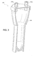
- FIG. 3 illustrates a cross-sectional perspective view of a containment device (incubation device or sterile container) according to another exemplary embodiment of the present invention.
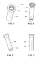
- FIG. 4 illustrates a perspective view of a containment device (incubation device or container) according to yet another exemplary embodiment of the present invention.
- FIG. 5 illustrates another perspective view of the containment device of FIG. 4 .
- FIG. 6 illustrates a perspective, partial cross-sectional view of the containment device of FIG. 4 .
- FIG. 7 illustrates a front view of the containment device of FIG. 4 .
- the present invention provides systems and methods for producing enhanced blood serum with concentrated growth factors and/or prophylactically and therapeutically active proteins.
- An autologous blood product or fluid is obtained from a donor and then incubated into a containment device (sterile container) that includes a source of cells or tissue (for example, autologous, allogeneic, or xenographic tissue, or combinations thereof, the tissue being natural, synthetic or composite products tissue).
- the cells from the injected fluid or autologous blood product interact/incubate with the source of cells or tissue inside the containment device, to produce a serum that is loaded with autologous proteins, growth factors, and cytokines.
- the serum can be extracted from the containment device and injected at a wounded tissue site.
- the autologous blood product may be a fluid and/or composition that includes blood, whole blood, autologous conditioned plasma (ACP), platelet-rich plasma (PRP), platelet-poor plasma (PPP), bone marrow aspirate (BMA), bone marrow concentrate (BMC) and stem cells, among others, and/or combinations thereof.
- ACP autologous conditioned plasma
- PRP platelet-rich plasma
- PPP platelet-poor plasma
- BMA bone marrow aspirate
- BMC bone marrow concentrate
- stem cells among others, and/or combinations thereof.
- the present invention also provides methods and systems for producing enhanced autograft or autologous blood serum containing prophylactically or therapeutically active proteins.
- the enhanced serum is obtained by incubating blood/fluid in a simple, sterile container (test tube) together with a source of cells or tissue, for example, an autograft, allograft or xenograft tissue or material, which may be in natural, synthetic or composite form (or combinations of these forms).
- the source of cells or tissue is loaded in the sterile container and mixed with blood (for example, whole blood) or a fluid such as autologous conditioned plasma (ACP), platelet-rich plasma (PRP), platelet-poor plasma (PPP), bone marrow aspirate (BMA), bone marrow concentrate (MBC) and stem cells, among others, and/or combinations thereof.
- ACP autologous conditioned plasma
- PRP platelet-rich plasma
- PPP platelet-poor plasma
- BMA bone marrow aspirate
- MSC bone marrow concentrate
- stem cells among others, and/or combinations thereof.
- the source of cells or tissue is brought together with the fluid/autologous blood product into the sterile container, incubated, and then subjected to hard spinning such as centrifugation, to obtain enhanced growth factors and prophylactically and therapeutically active proteins.
- the growth factors and prophylactically and therapeutically active proteins may be separated from the source of cells or tissue by collecting a serum rich in those elements.
- the present invention provides methods and systems for producing enhanced autograft or autologous blood serum containing prophylactically or therapeutically active proteins.
- the enhanced serum is obtained by incubating blood/fluid in a simple, sterile container (test tube) together with an autograft, allograft or xenograft tissue or material (preferably, collagen).
- the autograft, allograft or xenograft tissue (autograft, allograft or xenograft material) is loaded in the sterile container and mixed with blood (whole blood) or a fluid such as autologous conditioned plasma (ACP), platelet-rich plasma (PRP), platelet-poor plasma (PPP), bone marrow aspirate (BMA), bone marrow concentrate (MBC) and stem cells, among others, and combinations thereof.
- ACP autologous conditioned plasma
- PRP platelet-rich plasma
- PPP platelet-poor plasma
- BMA bone marrow aspirate
- MSC bone marrow concentrate
- stem cells among others, and combinations thereof.
- the invention uses the sterile container and method steps detailed in US 2006/0177515 , but instead of having the beads provided in the test tube (incubation tube), the tube has a source of cells or tissue, i.e., an autograft, allograft or xenograft tissue (preferably collagen).
- the device utilizes an incubation tube as disclosed in US 2006/0177515 , but substitutes autologous, allogeneic, or xenographic tissue, or even synthetic or composite products tissue, for the beads inside the tube.
- the fluid may be an autologous blood product and/or composition that includes blood, whole blood, autologous conditioned plasma, platelet-rich plasma, platelet-poor plasma, bone marrow aspirate, bone marrow concentrate and stem cells, among others, and combinations thereof.
- the cells from the injected fluid interact/incubate with the tissue inside the tube, to produce a serum that is loaded with autologous proteins, growth factors, and cytokines.
- the serum can be extracted from the tube and injected at a wounded tissue site. Incubation can be conducted at ambient conditions or at 37°C between 1 and 24 hours.
- the incubation may be conducted with a collagen construct.
- the collagen can be of autograftic, allograftic, xenograftic origination.
- the collagen can be provided in natural form, partially natural form, or may even be produced synthetically.
- the collagen can be obtained, purified, or derived from connective tissue or fibrous tissue (for example, tendons, ligaments, skin, cartilage, bone, intervetebral tissue, among others).
- the collagen may be any of the Types I-V collagen, preferably Type I and Type II collagen.
- the physical construct of the collagen could be, but is not limited to strips, granules, powder, cylinder(s), scaffolding, mesh, etc., or any combination thereof.
- the present invention also provides methods of producing enhanced serum to be injected at a wounded site by inter alia the steps of: (i) providing a sterile container and adding at least one of autograft, allograft or xenograft tissue (for example, collagen) in any size and/or shape; (ii) adding a fluid (for example, blood, autologous conditioned plasma, bone marrow aspirate, PRP, PPP, etc) to the autograft, allograft or xenograft tissue; (iii) incubating the fluid with the autograft, allograft or xenograft tissue for about 0-24 hours, preferably between 1 and 24 hours; (iv) subjecting the sterile container to hard spinning to separate cells and tissue from growth factors; (v) withdrawing the concentrated growth factors (could optionally filter out catabolics); and (vi) delivering the concentrated growth factors to a patient (could optionally further mix with ACP and or add a scaffold).
- the system delivers
- FIGS. 1(a) and 1(b) illustrate system 100 of the present invention comprising containment device 50 (sterile container 50) which is an exemplary test tube.
- containment device 50 sterile container 50
- the sterile container 50 of FIG. 1(a) is modified to allow containment of blood/ fluid and material, and mixing of the blood/fluid with the material, as detailed below.
- the containment device 50 is a ported system with luer connections 10a, 10b that allow for delivery of an autologous fluid 11 (blood such as whole blood, autologous conditioned plasma (ACP), platelet-rich plasma (PRP), platelet-poor plasma (PPP), bone marrow aspirate (BMA), bone marrow concentrate (MBC), stem cells or combinations thereof) and holding of a source of cells or tissue 22, for example, autologous, allogenic, or xenographic tissue 22.
- the source of cells or tissue 22 is synthetic or purified collagen 22.
- FIG. 1(b) illustrates containment device 50 with autologous blood product (fluid) 11 brought into contact with source of cells or tissue 22 (collagen 22).
- Containment device 50 may be a sterile container of any suitable size or configuration (e.g., vial, capped container, closed tube) and is preferably made of a material that can be subjected to sterilization (e.g., chemical sterilization, autoclave).
- the container 50 can be made of any suitable glass, ceramic, or plastic material (e.g., polystyrene, polypropylene, polycarbonate).
- the surface area of the inner surface 110 of the sterile container 50, or a portion thereof, can be increased by adding and/or coating the inner surface 110 of the container 50 with additional coating materials that increase the surface area, providing additional attachments for adherence by blood monocytes (not shown), which stimulates the monocytes to produce therapeutically or prophylactically active proteins such as, for example, IL-1Ra.
- the inner surface 110 of the container 50, or a portion thereof can be coated with an anti-coagulant such as heparin.
- FIGS. 1(a) and 1(b) also illustrate source of cells or tissue 22, for example, autologous, allogenic, or xenographic tissue/material 22, contained within the sterile container 50.
- the autograft, allograft or xenograft tissue 22 is loaded in the sterile container 50 and mixed with blood/fluid 11 ( FIG. 1(b) ).
- autologous, allogenic, or xenographic tissue 22 may be collagen 22 which may be synthetic or natural, and which may be provided in the form of strips, for example.
- the collagen 22 can be of autograftic, allograftic, xenograftic origination, or may even be produced synthetically.
- the collagen 22 can be obtained, purified, or derived from connective tissue or fibrous tissue (for example, tendons, ligaments, skin, cartilage, bone, intervertebral tissue, among others).
- the collagen 22 may be any of the Types I-V collagen, preferably Type I and Type II collagen.
- the physical construct of the collagen 22 could be, but is not limited to strips, granules, powder, cylinder(s), scaffolding, etc.
- autologous fluid 11 for example, blood 11
- sterile container 50 containing autologous, allogeneic, or xenographic tissue 22 (for example, collagen 22).
- autologous, allogeneic, or xenographic tissue 22 for example, collagen 22.
- the fluid can be (i) immediately removed; or (ii) incubated for up to 24 hours (between 1 and 24 hours) at ambient conditions or 37° C, and then a serum extracted; or (iii) incubated for up to 24 hours (between 1 and 24 hours) at ambient conditions or 37° C, then centrifuged, and then a serum extracted.
- the autologous, allogeneic, or xenographic material 22 is left in the container.
- the incubation in steps (ii) and (iii) may be conducted at a temperature suitable to induce production of growth factors, and/or prophylactically or therapeutically active proteins (e.g., IL-1Ra, interleukin 4, interleukin 10, and TGF beta).
- the incubation is conducted at temperatures from about 35 to 39 °C, more preferably from about 36 °C to about 38° C, most preferably at 37° C.
- the autologous fluid 11 (the cells within the fluid) interact with the material 22 to produce growth factors, cytokines and proteins (that will be provided at a wounded tissue site). These proteins "float" in the surrounding fluid and can be retrieved/retracted for delivery at an injured tissue site.
- FIGS. 2(a) and 2(b) illustrate two graphs depicting concentrations for IL-1Ra ( FIG. 2(a) ) and IL-1beta ( FIG. 2(b) ) with different incubation constructs and materials (all using an exemplary IRAP II construct).
- Each graph illustrates the levels of IL-1Ra concentration ( FIG. 2(a) ) and IL-1beta concentration ( FIG.
- IRAP 24 an IRAP II construct with 24 hours incubation
- IRAP 24A an IRAP II construct with ACDA and 24 hours incubation
- IRAP Col an IRAP II Tube with collagen and no beads and 24 hours incubation
- IRAP DBM an IRAP II Tube with DBM and no beads and 24 hours incubation
- the two graphs clearly evidence the increase in the concentrations for both IL-1Ra and IL-1beta with a construct having collagen (i.e., the collagen group under "IRAP Col”).
- the collagen group produced significantly higher amounts of monocytic-derived growth factors (GFs), and increased level of catabolic and anabolic activity (as a result of the natural injury response and monocyte exposure to collagen).
- GFs monocytic-derived growth factors
- FIG. 3 illustrates another exemplary embodiment of incubations system 200 with containment device 250 (sterile container 250) which is similar to the containment device 50 of FIGS. 1(a) and 1(b) in that it is also a ported system with luer connections/ports 210a, 210b that allow for delivery of an autologous fluid 11 and holding of autologous, allogeneic, or xenographic tissue 22, but differs in that it has a specific design, i.e., a modified test tube design.
- Incubation device 250 can be made of any suitable glass, ceramic, or plastic material (e.g., polystyrene, polypropylene, polycarbonate).
- FIGS. 4-8 illustrate another embodiment of system 300 of the present invention which includes incubation device 350 (containment device or sterile container 350) which is similar to the containment device 50 of FIGS. 1(a) and 1(b) in that it allows introduction of a fluid (for example, blood (whole blood), autologous conditioned plasma (ACP), platelet-rich plasma (PRP), platelet-poor plasma (PPP), bone marrow aspirate (BMA), bone marrow concentrate (MBC), stem cells or combinations thereof) into container 350 with at least one of autograft, allograft or xenograft tissue or material (for example, collagen) but differs in that it has a specific design, i.e., a modified tube design with only one centrally located port/lumen 310 ( FIG.
- a fluid for example, blood (whole blood), autologous conditioned plasma (ACP), platelet-rich plasma (PRP), platelet-poor plasma (PPP), bone marrow aspirate (B
- Containment device (incubation device) 350 can be made of any suitable glass, ceramic, or plastic material (e.g., polystyrene, polypropylene, polycarbonate).
- the inner surface 310 ( FIG. 6 ) of the device 350, or a portion thereof, may be also increased by adding and/or coating the inner surface 310 of the container 350 with additional coating materials that increase the surface area, providing additional attachments for adherence by blood monocytes (not shown), which stimulates the monocytes to produce therapeutically or prophylactically active proteins such as, for example, IL-1Ra.
- the inner surface 310 of the container 350, or a portion thereof can be coated with an anti-coagulant such as heparin.
- a method of producing enhanced serum to be injected at a wounded site comprises the steps of: (i) providing a sterile container 50, 250, 350 and adding at least one of autograft, allograft or xenograft tissue or material 22 (for example, collagen 22) in any size and/or shape; (ii) adding a fluid 11 (for example, an autologous blood product which may include blood, whole blood, autologous conditioned plasma, platelet-rich plasma, platelet-poor plasma, bone marrow aspirate, bone marrow concentrate and stem cells, among others, and combinations thereof) to the autograft, allograft or xenograft tissue or material 22 to form system 100, 200, 300; (iii) incubating the fluid 11 with the autograft, allograft or xenograft tissue or material 22 for about 0-24 hours, preferably between 1 to 24 hours; (iv) subjecting the sterile container 50, 250, 350 to hard spinning to separate cells and tissue from growth factors; (v)
- Preferred embodiments of the invention also provide methods of treating a patient by administering enhanced serum containing the prophylactically or therapeutically active proteins to a patient in need of treatment.
- patients suffering from osteoarthritis and tendonitis may benefit from such treatment.
- serum from a donor herd of horses can be used to produce prophylactically or therapeutically active proteins for administration to horses in need of treatment.
- the protein serum component may be divided and stored in a container (separate from the containment device 50, 250, 350).
- the container can be frozen to preserve the serum for long or short term storage and/or for transportation of the serum.
- a portion of the serum (e.g., one container or vial) of serum can be administered (e.g., via injection or perfusion) by an orthopedic surgeon into a patient, for example, at a specific site (e.g., specific joint, tendon, muscle, or other soft tissue) to treat or reduce the symptoms associated with a disease condition (e.g., osteoarthritis or tendonitis).
- a specific site e.g., specific joint, tendon, muscle, or other soft tissue
Landscapes
- Health & Medical Sciences (AREA)
- Life Sciences & Earth Sciences (AREA)
- Cell Biology (AREA)
- Hematology (AREA)
- Chemical & Material Sciences (AREA)
- Medicinal Chemistry (AREA)
- Developmental Biology & Embryology (AREA)
- Immunology (AREA)
- Virology (AREA)
- Zoology (AREA)
- Biomedical Technology (AREA)
- Biotechnology (AREA)
- Pharmacology & Pharmacy (AREA)
- Epidemiology (AREA)
- Animal Behavior & Ethology (AREA)
- General Health & Medical Sciences (AREA)
- Public Health (AREA)
- Veterinary Medicine (AREA)
- Engineering & Computer Science (AREA)
- Medicines Containing Material From Animals Or Micro-Organisms (AREA)
Description
- The present invention relates to systems and methods for producing autograft or autologous enhanced serum containing prophylactically or therapeutically active proteins.
- Methods for producing interleukin-1 receptor antagonist ("IL-1Ra") in a syringe are described in
U.S. Patent Numbers 6,759,188 ;6,623,472 ; and6,713,246 . These methods require the use of a special syringe to produce the IL-1Ra and do not provide methods for dividing the protein serum containing IL-1Ra or other therapeutically active proteins into portions for long-term storage and/or transportation. - There is a need for devices, systems and techniques that would maximize the innate healing response of the human body by delivering autologous growth factors, cytokines and proteins to a wounded tissue site, without the need for a special syringe.
- The present invention provides methods and systems for producing enhanced serum. The enhanced serum is obtained by incubating blood/fluid in a simple, sterile container (test tube) together with a source of cells or tissue, for example, an autograft, allograft or xenograft tissue such as collagen. The autograft, allograft or xenograft tissue is loaded in the sterile container and mixed with blood or a fluid. The cells from the injected blood/fluid interact/incubate with the autograft, allograft or xenograft tissue inside the tube to produce a serum that contains autologous proteins, growth factors, and cytokines. The construct is then centrifuged and the serum can be extracted from the tube. Incubation can be conducted at ambient conditions, or at 37°C between 1 and 24 hours.
- These and other features and advantages of the invention will be more apparent from the following detailed description that is provided in connection with the accompanying drawings and illustrated exemplary embodiments of the invention.
-
FIGS. 1(a) and 1(b) illustrate a containment system (with a sterile container) according to an exemplary embodiment of the present invention. -
FIG. 2(a) illustrates a graph depicting concentrations for IL-1Ra with different incubation materials (including collagen strips) using an exemplary construct. -
FIG. 2(b) illustrates a graph depicting concentrations for IL-1beta with different incubation materials (including collagen strips) using an exemplary construct. -
FIG. 3 illustrates a cross-sectional perspective view of a containment device (incubation device or sterile container) according to another exemplary embodiment of the present invention. -
FIG. 4 illustrates a perspective view of a containment device (incubation device or container) according to yet another exemplary embodiment of the present invention. -
FIG. 5 illustrates another perspective view of the containment device ofFIG. 4 . -
FIG. 6 illustrates a perspective, partial cross-sectional view of the containment device ofFIG. 4 . -
FIG. 7 illustrates a front view of the containment device ofFIG. 4 . - The present invention provides systems and methods for producing enhanced blood serum with concentrated growth factors and/or prophylactically and therapeutically active proteins. An autologous blood product or fluid is obtained from a donor and then incubated into a containment device (sterile container) that includes a source of cells or tissue (for example, autologous, allogeneic, or xenographic tissue, or combinations thereof, the tissue being natural, synthetic or composite products tissue). The cells from the injected fluid or autologous blood product interact/incubate with the source of cells or tissue inside the containment device, to produce a serum that is loaded with autologous proteins, growth factors, and cytokines. The serum can be extracted from the containment device and injected at a wounded tissue site. Incubation could be conducted at ambient conditions or at 37°C between 1 and 24 hours. The autologous blood product may be a fluid and/or composition that includes blood, whole blood, autologous conditioned plasma (ACP), platelet-rich plasma (PRP), platelet-poor plasma (PPP), bone marrow aspirate (BMA), bone marrow concentrate (BMC) and stem cells, among others, and/or combinations thereof.
- The present invention also provides methods and systems for producing enhanced autograft or autologous blood serum containing prophylactically or therapeutically active proteins. The enhanced serum is obtained by incubating blood/fluid in a simple, sterile container (test tube) together with a source of cells or tissue, for example, an autograft, allograft or xenograft tissue or material, which may be in natural, synthetic or composite form (or combinations of these forms). The source of cells or tissue is loaded in the sterile container and mixed with blood (for example, whole blood) or a fluid such as autologous conditioned plasma (ACP), platelet-rich plasma (PRP), platelet-poor plasma (PPP), bone marrow aspirate (BMA), bone marrow concentrate (MBC) and stem cells, among others, and/or combinations thereof. The source of cells or tissue is brought together with the fluid/autologous blood product into the sterile container, incubated, and then subjected to hard spinning such as centrifugation, to obtain enhanced growth factors and prophylactically and therapeutically active proteins. The growth factors and prophylactically and therapeutically active proteins may be separated from the source of cells or tissue by collecting a serum rich in those elements.
- In an exemplary embodiment, the present invention provides methods and systems for producing enhanced autograft or autologous blood serum containing prophylactically or therapeutically active proteins. The enhanced serum is obtained by incubating blood/fluid in a simple, sterile container (test tube) together with an autograft, allograft or xenograft tissue or material (preferably, collagen). The autograft, allograft or xenograft tissue (autograft, allograft or xenograft material) is loaded in the sterile container and mixed with blood (whole blood) or a fluid such as autologous conditioned plasma (ACP), platelet-rich plasma (PRP), platelet-poor plasma (PPP), bone marrow aspirate (BMA), bone marrow concentrate (MBC) and stem cells, among others, and combinations thereof.
- In an exemplary embodiment, the invention uses the sterile container and method steps detailed in
US 2006/0177515 , but instead of having the beads provided in the test tube (incubation tube), the tube has a source of cells or tissue, i.e., an autograft, allograft or xenograft tissue (preferably collagen). Thus, the device utilizes an incubation tube as disclosed inUS 2006/0177515 , but substitutes autologous, allogeneic, or xenographic tissue, or even synthetic or composite products tissue, for the beads inside the tube. - Any type of fluid/composition can be injected into the device. The fluid may be an autologous blood product and/or composition that includes blood, whole blood, autologous conditioned plasma, platelet-rich plasma, platelet-poor plasma, bone marrow aspirate, bone marrow concentrate and stem cells, among others, and combinations thereof. The cells from the injected fluid interact/incubate with the tissue inside the tube, to produce a serum that is loaded with autologous proteins, growth factors, and cytokines. The serum can be extracted from the tube and injected at a wounded tissue site. Incubation can be conducted at ambient conditions or at 37°C between 1 and 24 hours.
- In a specific embodiment only, the incubation may be conducted with a collagen construct. The collagen can be of autograftic, allograftic, xenograftic origination. The collagen can be provided in natural form, partially natural form, or may even be produced synthetically. The collagen can be obtained, purified, or derived from connective tissue or fibrous tissue (for example, tendons, ligaments, skin, cartilage, bone, intervetebral tissue, among others). The collagen may be any of the Types I-V collagen, preferably Type I and Type II collagen. The physical construct of the collagen could be, but is not limited to strips, granules, powder, cylinder(s), scaffolding, mesh, etc., or any combination thereof.
- The present invention also provides methods of producing enhanced serum to be injected at a wounded site by inter alia the steps of: (i) providing a sterile container and adding at least one of autograft, allograft or xenograft tissue (for example, collagen) in any size and/or shape; (ii) adding a fluid (for example, blood, autologous conditioned plasma, bone marrow aspirate, PRP, PPP, etc) to the autograft, allograft or xenograft tissue; (iii) incubating the fluid with the autograft, allograft or xenograft tissue for about 0-24 hours, preferably between 1 and 24 hours; (iv) subjecting the sterile container to hard spinning to separate cells and tissue from growth factors; (v) withdrawing the concentrated growth factors (could optionally filter out catabolics); and (vi) delivering the concentrated growth factors to a patient (could optionally further mix with ACP and or add a scaffold). The system delivers concentrated growth factors, proteins, and cytokines that are devoid of cells.
- Reference is now made to
FIGS. 1(a) and 1(b) which illustratesystem 100 of the present invention comprising containment device 50 (sterile container 50) which is an exemplary test tube. Thesterile container 50 ofFIG. 1(a) is modified to allow containment of blood/ fluid and material, and mixing of the blood/fluid with the material, as detailed below. Thecontainment device 50 is a ported system withluer connections tissue 22, for example, autologous, allogenic, orxenographic tissue 22. In a preferred embodiment, the source of cells ortissue 22 is synthetic or purifiedcollagen 22.FIG. 1(b) illustratescontainment device 50 with autologous blood product (fluid) 11 brought into contact with source of cells or tissue 22 (collagen 22). -
Containment device 50 may be a sterile container of any suitable size or configuration (e.g., vial, capped container, closed tube) and is preferably made of a material that can be subjected to sterilization (e.g., chemical sterilization, autoclave). For example, thecontainer 50 can be made of any suitable glass, ceramic, or plastic material (e.g., polystyrene, polypropylene, polycarbonate). - The surface area of the
inner surface 110 of thesterile container 50, or a portion thereof, can be increased by adding and/or coating theinner surface 110 of thecontainer 50 with additional coating materials that increase the surface area, providing additional attachments for adherence by blood monocytes (not shown), which stimulates the monocytes to produce therapeutically or prophylactically active proteins such as, for example, IL-1Ra. In an exemplary embodiment only, theinner surface 110 of thecontainer 50, or a portion thereof, can be coated with an anti-coagulant such as heparin. -
FIGS. 1(a) and 1(b) also illustrate source of cells ortissue 22, for example, autologous, allogenic, or xenographic tissue/material 22, contained within thesterile container 50. The autograft, allograft orxenograft tissue 22 is loaded in thesterile container 50 and mixed with blood/fluid 11 (FIG. 1(b) ). In an exemplary embodiment, autologous, allogenic, orxenographic tissue 22 may becollagen 22 which may be synthetic or natural, and which may be provided in the form of strips, for example. - As noted, the
collagen 22 can be of autograftic, allograftic, xenograftic origination, or may even be produced synthetically. Thecollagen 22 can be obtained, purified, or derived from connective tissue or fibrous tissue (for example, tendons, ligaments, skin, cartilage, bone, intervertebral tissue, among others). Thecollagen 22 may be any of the Types I-V collagen, preferably Type I and Type II collagen. The physical construct of thecollagen 22 could be, but is not limited to strips, granules, powder, cylinder(s), scaffolding, etc. - In one embodiment of the invention, autologous fluid 11 (for example, blood 11) is obtained from a patient (by withdrawing the fluid 11 with a syringe, for example) and then injected into
sterile container 50 containing autologous, allogeneic, or xenographic tissue 22 (for example, collagen 22). After injection of theautologous fluid 11 through one of theluer ports xenographic tissue 22. After mixing, the fluid can be (i) immediately removed; or (ii) incubated for up to 24 hours (between 1 and 24 hours) at ambient conditions or 37° C, and then a serum extracted; or (iii) incubated for up to 24 hours (between 1 and 24 hours) at ambient conditions or 37° C, then centrifuged, and then a serum extracted. The autologous, allogeneic, orxenographic material 22 is left in the container. - The incubation in steps (ii) and (iii) may be conducted at a temperature suitable to induce production of growth factors, and/or prophylactically or therapeutically active proteins (e.g., IL-1Ra, interleukin 4, interleukin 10, and TGF beta). In an exemplary embodiment only, the incubation is conducted at temperatures from about 35 to 39 °C, more preferably from about 36 °C to about 38° C, most preferably at 37° C.
- The autologous fluid 11 (the cells within the fluid) interact with the material 22 to produce growth factors, cytokines and proteins (that will be provided at a wounded tissue site). These proteins "float" in the surrounding fluid and can be retrieved/retracted for delivery at an injured tissue site.
- Reference is now made to
FIGS. 2(a) and 2(b) which illustrate two graphs depicting concentrations for IL-1Ra (FIG. 2(a) ) and IL-1beta (FIG. 2(b) ) with different incubation constructs and materials (all using an exemplary IRAP II construct). Each graph illustrates the levels of IL-1Ra concentration (FIG. 2(a) ) and IL-1beta concentration (FIG. 2(b) ) for four different exemplary constructs: IRAP 24 (an IRAP II construct with 24 hours incubation);IRAP 24A (an IRAP II construct with ACDA and 24 hours incubation); IRAP Col (an IRAP II Tube with collagen and no beads and 24 hours incubation); and IRAP DBM (an IRAP II Tube with DBM and no beads and 24 hours incubation). - The two graphs clearly evidence the increase in the concentrations for both IL-1Ra and IL-1beta with a construct having collagen (i.e., the collagen group under "IRAP Col"). The collagen group produced significantly higher amounts of monocytic-derived growth factors (GFs), and increased level of catabolic and anabolic activity (as a result of the natural injury response and monocyte exposure to collagen).
-
FIG. 3 illustrates another exemplary embodiment ofincubations system 200 with containment device 250 (sterile container 250) which is similar to thecontainment device 50 ofFIGS. 1(a) and 1(b) in that it is also a ported system with luer connections/ports autologous fluid 11 and holding of autologous, allogeneic, orxenographic tissue 22, but differs in that it has a specific design, i.e., a modified test tube design.Incubation device 250 can be made of any suitable glass, ceramic, or plastic material (e.g., polystyrene, polypropylene, polycarbonate). -
FIGS. 4-8 illustrate another embodiment ofsystem 300 of the present invention which includes incubation device 350 (containment device or sterile container 350) which is similar to thecontainment device 50 ofFIGS. 1(a) and 1(b) in that it allows introduction of a fluid (for example, blood (whole blood), autologous conditioned plasma (ACP), platelet-rich plasma (PRP), platelet-poor plasma (PPP), bone marrow aspirate (BMA), bone marrow concentrate (MBC), stem cells or combinations thereof) intocontainer 350 with at least one of autograft, allograft or xenograft tissue or material (for example, collagen) but differs in that it has a specific design, i.e., a modified tube design with only one centrally located port/lumen 310 (FIG. 4 ). Containment device (incubation device) 350 can be made of any suitable glass, ceramic, or plastic material (e.g., polystyrene, polypropylene, polycarbonate). The inner surface 310 (FIG. 6 ) of thedevice 350, or a portion thereof, may be also increased by adding and/or coating theinner surface 310 of thecontainer 350 with additional coating materials that increase the surface area, providing additional attachments for adherence by blood monocytes (not shown), which stimulates the monocytes to produce therapeutically or prophylactically active proteins such as, for example, IL-1Ra. In an exemplary embodiment only, theinner surface 310 of thecontainer 350, or a portion thereof, can be coated with an anti-coagulant such as heparin. - A method of producing enhanced serum to be injected at a wounded site comprises the steps of: (i) providing a
sterile container material 22 to formsystem material 22 for about 0-24 hours, preferably between 1 to 24 hours; (iv) subjecting thesterile container - Preferred embodiments of the invention also provide methods of treating a patient by administering enhanced serum containing the prophylactically or therapeutically active proteins to a patient in need of treatment. For example, patients suffering from osteoarthritis and tendonitis may benefit from such treatment. In exemplary veterinary medicine applications, serum from a donor herd of horses can be used to produce prophylactically or therapeutically active proteins for administration to horses in need of treatment.
- If necessary, the protein serum component may be divided and stored in a container (separate from the
containment device - Although the present invention has been described in connection with preferred embodiments, many modifications and variations will become apparent to those skilled in the art. While preferred embodiments of the invention have been described and illustrated above, it should be understood that these are exemplary of the invention and are not to be considered as limiting.
Claims (8)
- A method of producing blood serum with concentrated growth factors, comprising the steps of:introducing an autologous blood product into a container containing a source of cells or tissue;incubating the autologous blood product and the source of cells or tissue to induce production of growth factors at a temperature between 35 °C to 39 °C, the incubation being conducted in the container;separating the source of cells or tissue from the growth factors; andcollecting a serum containing the growth factors.
- The method of claim 1, wherein the autologous blood product is selected from the group consisting of whole blood, autologous conditioned plasma, platelet-rich plasma, platelet-poor plasma, bone marrow aspirate, bone marrow concentrate, and stem cells.
- The method of claim 1, wherein the source of cells or tissue is autologous, allogeneic, or xenographic tissue, or combinations thereof.
- The method of claim 1, wherein the source of cells or tissue is collagen.
- The method of claim 1, wherein the source of cells or tissue is natural, synthetic or composite products tissue.
- The method of claim 1, wherein the step of incubating is conducted between 1 and 24 hours.
- The method of claim 1, wherein the step of incubating is conducted at a temperature of 37 °C.
- The method of claim 1, further comprising the steps of:obtaining the autologous blood product from a donor and with a syringe which is different from the container containing the source of cells or tissue;transferring the autologous blood product into the container containing the source of cells ortissue by injecting the autologous blood product into the container through one of two ports of the container;bringing together the autologous blood product and the source of cells or tissue into the container;hard spinning he sterile container to separate cells and tissue from growth factors; andwithdrawing the growth factors.
Applications Claiming Priority (1)
| Application Number | Priority Date | Filing Date | Title |
|---|---|---|---|
| US201261672975P | 2012-07-18 | 2012-07-18 |
Publications (2)
| Publication Number | Publication Date |
|---|---|
| EP2687217A1 true EP2687217A1 (en) | 2014-01-22 |
| EP2687217B1 EP2687217B1 (en) | 2015-08-19 |
Family
ID=48793073
Family Applications (1)
| Application Number | Title | Priority Date | Filing Date |
|---|---|---|---|
| EP13176767.5A Active EP2687217B1 (en) | 2012-07-18 | 2013-07-17 | Enhanced autologous growth factor production and delivery system |
Country Status (2)
| Country | Link |
|---|---|
| US (1) | US9205110B2 (en) |
| EP (1) | EP2687217B1 (en) |
Cited By (3)
| Publication number | Priority date | Publication date | Assignee | Title |
|---|---|---|---|---|
| EP3175920A1 (en) * | 2015-12-03 | 2017-06-07 | Vetlinebio AB | Method and devices for producing serum |
| WO2017156375A1 (en) * | 2016-03-10 | 2017-09-14 | Arthrex, Inc. | Systems and methods for preparing a thrombin serum |
| WO2017156379A3 (en) * | 2016-03-10 | 2018-08-16 | Arthrex, Inc. | Systems and methods for preparing protein enhanced serums |
Families Citing this family (3)
| Publication number | Priority date | Publication date | Assignee | Title |
|---|---|---|---|---|
| US10512659B2 (en) | 2015-07-02 | 2019-12-24 | Arthrex, Inc. | Methods and devices for preparation of enriched biological fluids |
| JP7210465B2 (en) | 2016-11-18 | 2023-01-23 | パワー オブ プレートレッツ ピーティーイー リミテッド | Method for preparing growth factor-containing platelet releasate |
| EP3799896B1 (en) | 2019-10-04 | 2025-09-10 | Arthrex, Inc | Devices and methods for making therapeutic fluids |
Citations (3)
| Publication number | Priority date | Publication date | Assignee | Title |
|---|---|---|---|---|
| US6623472B1 (en) | 1997-08-16 | 2003-09-23 | Orthogen Gentechnologic. Gmbh | Method for inducing therapeutically-effective proteins |
| US6713246B1 (en) | 1999-02-01 | 2004-03-30 | Orthogen Ag | Method of producing interleukin-1 receptor antagonist in a syringe filled with blood |
| US20060177515A1 (en) | 2005-02-09 | 2006-08-10 | Reinhold Schmieding | Method of producing autogenous or allogenic blood serum and related logistics |
Family Cites Families (7)
| Publication number | Priority date | Publication date | Assignee | Title |
|---|---|---|---|---|
| US8101077B2 (en) * | 2001-05-07 | 2012-01-24 | Sivaprasad Sukavaneshvar | Device for separating platelets from fluid suspensions |
| EP1406492B1 (en) | 2001-06-06 | 2009-12-30 | Perfusion Partners & Associates, Inc. | Centrifuge tube assembly |
| DE102004051300C5 (en) | 2004-10-20 | 2013-01-24 | Fresenius Kabi Deutschland Gmbh | Cap for containers filled with medical fluids |
| DE102006005016A1 (en) | 2006-02-03 | 2007-08-16 | Orthogen Ag | Conditioned blood composition and process for its preparation |
| EP2146794B1 (en) | 2007-04-12 | 2016-10-19 | Biomet Biologics, LLC | Buoy suspension fractionation system |
| CA2740079A1 (en) * | 2008-10-08 | 2010-04-15 | Thrombodyne, Inc. | Methods of making concentrated fibrinogen- and platelet-containing compositions |
| US8460227B2 (en) | 2008-11-17 | 2013-06-11 | Arthrex, Inc. | Cytokine concentration system |
-
2013
- 2013-07-15 US US13/941,899 patent/US9205110B2/en active Active
- 2013-07-17 EP EP13176767.5A patent/EP2687217B1/en active Active
Patent Citations (4)
| Publication number | Priority date | Publication date | Assignee | Title |
|---|---|---|---|---|
| US6623472B1 (en) | 1997-08-16 | 2003-09-23 | Orthogen Gentechnologic. Gmbh | Method for inducing therapeutically-effective proteins |
| US6713246B1 (en) | 1999-02-01 | 2004-03-30 | Orthogen Ag | Method of producing interleukin-1 receptor antagonist in a syringe filled with blood |
| US6759188B2 (en) | 1999-02-01 | 2004-07-06 | Orthogen Ag | Method for producing interleukin 1 receptor antagonists, a therapeutic activity protein, from body fluids |
| US20060177515A1 (en) | 2005-02-09 | 2006-08-10 | Reinhold Schmieding | Method of producing autogenous or allogenic blood serum and related logistics |
Non-Patent Citations (1)
| Title |
|---|
| FRIZZIERO ANTONIO ET AL: "Autologous conditioned serum for the treatment of osteoarthritis and other possible applications in musculoskeletal disorders.", BRITISH MEDICAL BULLETIN 2013, vol. 105, 4 July 2012 (2012-07-04), pages 169 - 184, XP002713428, ISSN: 1471-8391 * |
Cited By (9)
| Publication number | Priority date | Publication date | Assignee | Title |
|---|---|---|---|---|
| EP3175920A1 (en) * | 2015-12-03 | 2017-06-07 | Vetlinebio AB | Method and devices for producing serum |
| WO2017093048A1 (en) * | 2015-12-03 | 2017-06-08 | Bio-Ech R&D Ab | Method and devices for producing serum |
| WO2017156375A1 (en) * | 2016-03-10 | 2017-09-14 | Arthrex, Inc. | Systems and methods for preparing a thrombin serum |
| WO2017156379A3 (en) * | 2016-03-10 | 2018-08-16 | Arthrex, Inc. | Systems and methods for preparing protein enhanced serums |
| US10596236B2 (en) | 2016-03-10 | 2020-03-24 | Arthrex, Inc. | Systems and methods for preparing a thrombin serum |
| US10960026B2 (en) | 2016-03-10 | 2021-03-30 | Arthrex, Inc. | Systems and methods for preparing protein enhanced serums |
| US11045526B2 (en) | 2016-03-10 | 2021-06-29 | Arthrex, Inc. | Systems and methods for preparing a thrombin serum |
| US11617784B2 (en) | 2016-03-10 | 2023-04-04 | Arthrex, Inc. | Systems and methods for preparing a thrombin serum |
| US12059455B2 (en) | 2016-03-10 | 2024-08-13 | Arthrex, Inc. | Systems and methods for preparing a thrombin serum |
Also Published As
| Publication number | Publication date |
|---|---|
| US20140023720A1 (en) | 2014-01-23 |
| EP2687217B1 (en) | 2015-08-19 |
| US9205110B2 (en) | 2015-12-08 |
Similar Documents
| Publication | Publication Date | Title |
|---|---|---|
| Chahla et al. | Bone marrow aspirate concentrate harvesting and processing technique | |
| Textor | Autologous biologic treatment for equine musculoskeletal injuries: platelet-rich plasma and IL-1 receptor antagonist protein | |
| Ortved | Regenerative medicine and rehabilitation for tendinous and ligamentous injuries in sport horses | |
| EP2687217B1 (en) | Enhanced autologous growth factor production and delivery system | |
| CN102573887B (en) | The treatment of osteolysis | |
| CN102573790B (en) | Implantable device for production of interleukin-1 receptor antagonist | |
| EA014435B1 (en) | Conditioned blood composition and method for its production | |
| AU2010284704B2 (en) | Apparatus and process for generating and harvesting adult stem cells and fluid associated with it from skin and omentum for medical, cosmetic, and veterinary use | |
| US20250275914A1 (en) | Preparation method and container for a topical wound healing gel | |
| Bembo et al. | Combined use of platelet rich plasma & micro-fat in sport and race horses with degenerative joint disease: preliminary clinical study in eight horses | |
| Özdemir et al. | Biological augmentation strategies in rotator cuff repair | |
| US20210154235A1 (en) | Stromal stem cell therapeutics and methods of use | |
| EP3881858A1 (en) | Heat-treated platelet-derived growth factor extract for use in a method of preventing or treating a tissue defect | |
| US20120264699A1 (en) | Method of producing autogenous or allogenic blood serum and related logistics | |
| EP3426400B1 (en) | System for preparing protein enhanced serums | |
| Villarreal-Espinosa et al. | Orthobiologics for Meniscus Injuries | |
| WO2014027359A1 (en) | A growth factor concentrate for treating tendinopathies | |
| Alsousou et al. | Therapeutic platelet-rich plasma in wound healing | |
| AU2021238506A1 (en) | Method for producing enhanced anti-inflammatory/ anti-catabolic agents from autologous physiological fluid | |
| EP4414004A1 (en) | Biological fluid concentration devices and methods of use | |
| BE1032813B1 (en) | PROCEDURE FOR OBTAINING A BLOOD DERIVATIVE AND THE BLOOD DERIVATIVE THUS OBTAINED | |
| CA2935211C (en) | Multipotent and immunocompatible stem cell concentrate | |
| Birdwhistell et al. | Biological Therapies in Canine Sports Medicine | |
| Chávez | Regenerative medicine with platelet rich plasma: a therapy for tendonitis in equines | |
| WO2014047688A1 (en) | Therapeutics using multiple injections of cells |
Legal Events
| Date | Code | Title | Description |
|---|---|---|---|
| PUAI | Public reference made under article 153(3) epc to a published international application that has entered the european phase |
Free format text: ORIGINAL CODE: 0009012 |
|
| AK | Designated contracting states |
Kind code of ref document: A1 Designated state(s): AL AT BE BG CH CY CZ DE DK EE ES FI FR GB GR HR HU IE IS IT LI LT LU LV MC MK MT NL NO PL PT RO RS SE SI SK SM TR |
|
| AX | Request for extension of the european patent |
Extension state: BA ME |
|
| 17P | Request for examination filed |
Effective date: 20140121 |
|
| RBV | Designated contracting states (corrected) |
Designated state(s): AL AT BE BG CH CY CZ DE DK EE ES FI FR GB GR HR HU IE IS IT LI LT LU LV MC MK MT NL NO PL PT RO RS SE SI SK SM TR |
|
| 17Q | First examination report despatched |
Effective date: 20140512 |
|
| GRAP | Despatch of communication of intention to grant a patent |
Free format text: ORIGINAL CODE: EPIDOSNIGR1 |
|
| INTG | Intention to grant announced |
Effective date: 20150309 |
|
| GRAS | Grant fee paid |
Free format text: ORIGINAL CODE: EPIDOSNIGR3 |
|
| GRAA | (expected) grant |
Free format text: ORIGINAL CODE: 0009210 |
|
| AK | Designated contracting states |
Kind code of ref document: B1 Designated state(s): AL AT BE BG CH CY CZ DE DK EE ES FI FR GB GR HR HU IE IS IT LI LT LU LV MC MK MT NL NO PL PT RO RS SE SI SK SM TR |
|
| REG | Reference to a national code |
Ref country code: GB Ref legal event code: FG4D |
|
| REG | Reference to a national code |
Ref country code: CH Ref legal event code: EP |
|
| REG | Reference to a national code |
Ref country code: IE Ref legal event code: FG4D |
|
| REG | Reference to a national code |
Ref country code: AT Ref legal event code: REF Ref document number: 743303 Country of ref document: AT Kind code of ref document: T Effective date: 20150915 |
|
| REG | Reference to a national code |
Ref country code: DE Ref legal event code: R096 Ref document number: 602013002706 Country of ref document: DE |
|
| REG | Reference to a national code |
Ref country code: AT Ref legal event code: MK05 Ref document number: 743303 Country of ref document: AT Kind code of ref document: T Effective date: 20150819 |
|
| REG | Reference to a national code |
Ref country code: LT Ref legal event code: MG4D |
|
| REG | Reference to a national code |
Ref country code: NL Ref legal event code: MP Effective date: 20150819 |
|
| PG25 | Lapsed in a contracting state [announced via postgrant information from national office to epo] |
Ref country code: NO Free format text: LAPSE BECAUSE OF FAILURE TO SUBMIT A TRANSLATION OF THE DESCRIPTION OR TO PAY THE FEE WITHIN THE PRESCRIBED TIME-LIMIT Effective date: 20151119 Ref country code: GR Free format text: LAPSE BECAUSE OF FAILURE TO SUBMIT A TRANSLATION OF THE DESCRIPTION OR TO PAY THE FEE WITHIN THE PRESCRIBED TIME-LIMIT Effective date: 20151120 Ref country code: LV Free format text: LAPSE BECAUSE OF FAILURE TO SUBMIT A TRANSLATION OF THE DESCRIPTION OR TO PAY THE FEE WITHIN THE PRESCRIBED TIME-LIMIT Effective date: 20150819 Ref country code: FI Free format text: LAPSE BECAUSE OF FAILURE TO SUBMIT A TRANSLATION OF THE DESCRIPTION OR TO PAY THE FEE WITHIN THE PRESCRIBED TIME-LIMIT Effective date: 20150819 Ref country code: LT Free format text: LAPSE BECAUSE OF FAILURE TO SUBMIT A TRANSLATION OF THE DESCRIPTION OR TO PAY THE FEE WITHIN THE PRESCRIBED TIME-LIMIT Effective date: 20150819 |
|
| PG25 | Lapsed in a contracting state [announced via postgrant information from national office to epo] |
Ref country code: PT Free format text: LAPSE BECAUSE OF FAILURE TO SUBMIT A TRANSLATION OF THE DESCRIPTION OR TO PAY THE FEE WITHIN THE PRESCRIBED TIME-LIMIT Effective date: 20151221 Ref country code: RS Free format text: LAPSE BECAUSE OF FAILURE TO SUBMIT A TRANSLATION OF THE DESCRIPTION OR TO PAY THE FEE WITHIN THE PRESCRIBED TIME-LIMIT Effective date: 20150819 Ref country code: PL Free format text: LAPSE BECAUSE OF FAILURE TO SUBMIT A TRANSLATION OF THE DESCRIPTION OR TO PAY THE FEE WITHIN THE PRESCRIBED TIME-LIMIT Effective date: 20150819 Ref country code: ES Free format text: LAPSE BECAUSE OF FAILURE TO SUBMIT A TRANSLATION OF THE DESCRIPTION OR TO PAY THE FEE WITHIN THE PRESCRIBED TIME-LIMIT Effective date: 20150819 Ref country code: AT Free format text: LAPSE BECAUSE OF FAILURE TO SUBMIT A TRANSLATION OF THE DESCRIPTION OR TO PAY THE FEE WITHIN THE PRESCRIBED TIME-LIMIT Effective date: 20150819 Ref country code: IS Free format text: LAPSE BECAUSE OF FAILURE TO SUBMIT A TRANSLATION OF THE DESCRIPTION OR TO PAY THE FEE WITHIN THE PRESCRIBED TIME-LIMIT Effective date: 20151219 Ref country code: SE Free format text: LAPSE BECAUSE OF FAILURE TO SUBMIT A TRANSLATION OF THE DESCRIPTION OR TO PAY THE FEE WITHIN THE PRESCRIBED TIME-LIMIT Effective date: 20150819 |
|
| PG25 | Lapsed in a contracting state [announced via postgrant information from national office to epo] |
Ref country code: NL Free format text: LAPSE BECAUSE OF FAILURE TO SUBMIT A TRANSLATION OF THE DESCRIPTION OR TO PAY THE FEE WITHIN THE PRESCRIBED TIME-LIMIT Effective date: 20150819 |
|
| PG25 | Lapsed in a contracting state [announced via postgrant information from national office to epo] |
Ref country code: CZ Free format text: LAPSE BECAUSE OF FAILURE TO SUBMIT A TRANSLATION OF THE DESCRIPTION OR TO PAY THE FEE WITHIN THE PRESCRIBED TIME-LIMIT Effective date: 20150819 Ref country code: DK Free format text: LAPSE BECAUSE OF FAILURE TO SUBMIT A TRANSLATION OF THE DESCRIPTION OR TO PAY THE FEE WITHIN THE PRESCRIBED TIME-LIMIT Effective date: 20150819 Ref country code: EE Free format text: LAPSE BECAUSE OF FAILURE TO SUBMIT A TRANSLATION OF THE DESCRIPTION OR TO PAY THE FEE WITHIN THE PRESCRIBED TIME-LIMIT Effective date: 20150819 Ref country code: IT Free format text: LAPSE BECAUSE OF FAILURE TO SUBMIT A TRANSLATION OF THE DESCRIPTION OR TO PAY THE FEE WITHIN THE PRESCRIBED TIME-LIMIT Effective date: 20150819 Ref country code: SK Free format text: LAPSE BECAUSE OF FAILURE TO SUBMIT A TRANSLATION OF THE DESCRIPTION OR TO PAY THE FEE WITHIN THE PRESCRIBED TIME-LIMIT Effective date: 20150819 |
|
| REG | Reference to a national code |
Ref country code: DE Ref legal event code: R097 Ref document number: 602013002706 Country of ref document: DE |
|
| PG25 | Lapsed in a contracting state [announced via postgrant information from national office to epo] |
Ref country code: RO Free format text: LAPSE BECAUSE OF FAILURE TO SUBMIT A TRANSLATION OF THE DESCRIPTION OR TO PAY THE FEE WITHIN THE PRESCRIBED TIME-LIMIT Effective date: 20150819 |
|
| REG | Reference to a national code |
Ref country code: FR Ref legal event code: PLFP Year of fee payment: 4 |
|
| PLBE | No opposition filed within time limit |
Free format text: ORIGINAL CODE: 0009261 |
|
| STAA | Information on the status of an ep patent application or granted ep patent |
Free format text: STATUS: NO OPPOSITION FILED WITHIN TIME LIMIT |
|
| 26N | No opposition filed |
Effective date: 20160520 |
|
| PG25 | Lapsed in a contracting state [announced via postgrant information from national office to epo] |
Ref country code: SI Free format text: LAPSE BECAUSE OF FAILURE TO SUBMIT A TRANSLATION OF THE DESCRIPTION OR TO PAY THE FEE WITHIN THE PRESCRIBED TIME-LIMIT Effective date: 20150819 |
|
| PG25 | Lapsed in a contracting state [announced via postgrant information from national office to epo] |
Ref country code: BE Free format text: LAPSE BECAUSE OF FAILURE TO SUBMIT A TRANSLATION OF THE DESCRIPTION OR TO PAY THE FEE WITHIN THE PRESCRIBED TIME-LIMIT Effective date: 20150819 |
|
| REG | Reference to a national code |
Ref country code: CH Ref legal event code: PL |
|
| PG25 | Lapsed in a contracting state [announced via postgrant information from national office to epo] |
Ref country code: MC Free format text: LAPSE BECAUSE OF FAILURE TO SUBMIT A TRANSLATION OF THE DESCRIPTION OR TO PAY THE FEE WITHIN THE PRESCRIBED TIME-LIMIT Effective date: 20150819 |
|
| PG25 | Lapsed in a contracting state [announced via postgrant information from national office to epo] |
Ref country code: LI Free format text: LAPSE BECAUSE OF NON-PAYMENT OF DUE FEES Effective date: 20160731 Ref country code: CH Free format text: LAPSE BECAUSE OF NON-PAYMENT OF DUE FEES Effective date: 20160731 |
|
| REG | Reference to a national code |
Ref country code: IE Ref legal event code: MM4A |
|
| REG | Reference to a national code |
Ref country code: FR Ref legal event code: PLFP Year of fee payment: 5 |
|
| PG25 | Lapsed in a contracting state [announced via postgrant information from national office to epo] |
Ref country code: IE Free format text: LAPSE BECAUSE OF NON-PAYMENT OF DUE FEES Effective date: 20160717 |
|
| PG25 | Lapsed in a contracting state [announced via postgrant information from national office to epo] |
Ref country code: LU Free format text: LAPSE BECAUSE OF NON-PAYMENT OF DUE FEES Effective date: 20160717 |
|
| PG25 | Lapsed in a contracting state [announced via postgrant information from national office to epo] |
Ref country code: HU Free format text: LAPSE BECAUSE OF FAILURE TO SUBMIT A TRANSLATION OF THE DESCRIPTION OR TO PAY THE FEE WITHIN THE PRESCRIBED TIME-LIMIT; INVALID AB INITIO Effective date: 20130717 Ref country code: SM Free format text: LAPSE BECAUSE OF FAILURE TO SUBMIT A TRANSLATION OF THE DESCRIPTION OR TO PAY THE FEE WITHIN THE PRESCRIBED TIME-LIMIT Effective date: 20150819 Ref country code: CY Free format text: LAPSE BECAUSE OF FAILURE TO SUBMIT A TRANSLATION OF THE DESCRIPTION OR TO PAY THE FEE WITHIN THE PRESCRIBED TIME-LIMIT Effective date: 20150819 |
|
| REG | Reference to a national code |
Ref country code: FR Ref legal event code: PLFP Year of fee payment: 6 |
|
| PG25 | Lapsed in a contracting state [announced via postgrant information from national office to epo] |
Ref country code: MK Free format text: LAPSE BECAUSE OF FAILURE TO SUBMIT A TRANSLATION OF THE DESCRIPTION OR TO PAY THE FEE WITHIN THE PRESCRIBED TIME-LIMIT Effective date: 20150819 Ref country code: TR Free format text: LAPSE BECAUSE OF FAILURE TO SUBMIT A TRANSLATION OF THE DESCRIPTION OR TO PAY THE FEE WITHIN THE PRESCRIBED TIME-LIMIT Effective date: 20150819 Ref country code: MT Free format text: LAPSE BECAUSE OF NON-PAYMENT OF DUE FEES Effective date: 20160731 Ref country code: HR Free format text: LAPSE BECAUSE OF FAILURE TO SUBMIT A TRANSLATION OF THE DESCRIPTION OR TO PAY THE FEE WITHIN THE PRESCRIBED TIME-LIMIT Effective date: 20150819 |
|
| PG25 | Lapsed in a contracting state [announced via postgrant information from national office to epo] |
Ref country code: BG Free format text: LAPSE BECAUSE OF FAILURE TO SUBMIT A TRANSLATION OF THE DESCRIPTION OR TO PAY THE FEE WITHIN THE PRESCRIBED TIME-LIMIT Effective date: 20150819 |
|
| PG25 | Lapsed in a contracting state [announced via postgrant information from national office to epo] |
Ref country code: AL Free format text: LAPSE BECAUSE OF FAILURE TO SUBMIT A TRANSLATION OF THE DESCRIPTION OR TO PAY THE FEE WITHIN THE PRESCRIBED TIME-LIMIT Effective date: 20150819 |
|
| PGFP | Annual fee paid to national office [announced via postgrant information from national office to epo] |
Ref country code: GB Payment date: 20250529 Year of fee payment: 13 |
|
| PGFP | Annual fee paid to national office [announced via postgrant information from national office to epo] |
Ref country code: FR Payment date: 20250610 Year of fee payment: 13 |
|
| PGFP | Annual fee paid to national office [announced via postgrant information from national office to epo] |
Ref country code: DE Payment date: 20250604 Year of fee payment: 13 |