EP2686348B1 - Bi- and monospecific, asymmetric antibodies and methods of generating the same - Google Patents
Bi- and monospecific, asymmetric antibodies and methods of generating the same Download PDFInfo
- Publication number
- EP2686348B1 EP2686348B1 EP12716657.7A EP12716657A EP2686348B1 EP 2686348 B1 EP2686348 B1 EP 2686348B1 EP 12716657 A EP12716657 A EP 12716657A EP 2686348 B1 EP2686348 B1 EP 2686348B1
- Authority
- EP
- European Patent Office
- Prior art keywords
- antibody
- heavy chain
- heavy
- antibodies
- chain
- Prior art date
- Legal status (The legal status is an assumption and is not a legal conclusion. Google has not performed a legal analysis and makes no representation as to the accuracy of the status listed.)
- Active
Links
Images


Classifications
-
- A—HUMAN NECESSITIES
- A61—MEDICAL OR VETERINARY SCIENCE; HYGIENE
- A61P—SPECIFIC THERAPEUTIC ACTIVITY OF CHEMICAL COMPOUNDS OR MEDICINAL PREPARATIONS
- A61P29/00—Non-central analgesic, antipyretic or antiinflammatory agents, e.g. antirheumatic agents; Non-steroidal antiinflammatory drugs [NSAID]
-
- C—CHEMISTRY; METALLURGY
- C07—ORGANIC CHEMISTRY
- C07K—PEPTIDES
- C07K16/00—Immunoglobulins [IG], e.g. monoclonal or polyclonal antibodies
- C07K16/46—Hybrid immunoglobulins
- C07K16/468—Immunoglobulins having two or more different antigen binding sites, e.g. multifunctional antibodies
-
- C—CHEMISTRY; METALLURGY
- C07—ORGANIC CHEMISTRY
- C07K—PEPTIDES
- C07K16/00—Immunoglobulins [IG], e.g. monoclonal or polyclonal antibodies
- C07K16/46—Hybrid immunoglobulins
-
- A—HUMAN NECESSITIES
- A61—MEDICAL OR VETERINARY SCIENCE; HYGIENE
- A61K—PREPARATIONS FOR MEDICAL, DENTAL OR TOILETRY PURPOSES
- A61K39/00—Medicinal preparations containing antigens or antibodies
- A61K39/395—Antibodies; Immunoglobulins; Immune serum, e.g. antilymphocytic serum
-
- A—HUMAN NECESSITIES
- A61—MEDICAL OR VETERINARY SCIENCE; HYGIENE
- A61P—SPECIFIC THERAPEUTIC ACTIVITY OF CHEMICAL COMPOUNDS OR MEDICINAL PREPARATIONS
- A61P31/00—Antiinfectives, i.e. antibiotics, antiseptics, chemotherapeutics
-
- A—HUMAN NECESSITIES
- A61—MEDICAL OR VETERINARY SCIENCE; HYGIENE
- A61P—SPECIFIC THERAPEUTIC ACTIVITY OF CHEMICAL COMPOUNDS OR MEDICINAL PREPARATIONS
- A61P35/00—Antineoplastic agents
-
- C—CHEMISTRY; METALLURGY
- C07—ORGANIC CHEMISTRY
- C07K—PEPTIDES
- C07K16/00—Immunoglobulins [IG], e.g. monoclonal or polyclonal antibodies
-
- C—CHEMISTRY; METALLURGY
- C07—ORGANIC CHEMISTRY
- C07K—PEPTIDES
- C07K16/00—Immunoglobulins [IG], e.g. monoclonal or polyclonal antibodies
- C07K16/12—Immunoglobulins [IG], e.g. monoclonal or polyclonal antibodies against material from bacteria
- C07K16/1267—Gram-positive bacteria
- C07K16/1275—Streptococcus (G)
-
- C—CHEMISTRY; METALLURGY
- C07—ORGANIC CHEMISTRY
- C07K—PEPTIDES
- C07K16/00—Immunoglobulins [IG], e.g. monoclonal or polyclonal antibodies
- C07K16/18—Immunoglobulins [IG], e.g. monoclonal or polyclonal antibodies against material from animals or humans
- C07K16/28—Immunoglobulins [IG], e.g. monoclonal or polyclonal antibodies against material from animals or humans against receptors, cell surface antigens or cell surface determinants
-
- C—CHEMISTRY; METALLURGY
- C07—ORGANIC CHEMISTRY
- C07K—PEPTIDES
- C07K16/00—Immunoglobulins [IG], e.g. monoclonal or polyclonal antibodies
- C07K16/18—Immunoglobulins [IG], e.g. monoclonal or polyclonal antibodies against material from animals or humans
- C07K16/28—Immunoglobulins [IG], e.g. monoclonal or polyclonal antibodies against material from animals or humans against receptors, cell surface antigens or cell surface determinants
- C07K16/2878—Immunoglobulins [IG], e.g. monoclonal or polyclonal antibodies against material from animals or humans against receptors, cell surface antigens or cell surface determinants against the NGF-receptor/TNF-receptor superfamily, e.g. CD27, CD30, CD40, CD95
-
- C—CHEMISTRY; METALLURGY
- C07—ORGANIC CHEMISTRY
- C07K—PEPTIDES
- C07K16/00—Immunoglobulins [IG], e.g. monoclonal or polyclonal antibodies
- C07K16/18—Immunoglobulins [IG], e.g. monoclonal or polyclonal antibodies against material from animals or humans
- C07K16/32—Immunoglobulins [IG], e.g. monoclonal or polyclonal antibodies against material from animals or humans against translation products of oncogenes
-
- A—HUMAN NECESSITIES
- A61—MEDICAL OR VETERINARY SCIENCE; HYGIENE
- A61K—PREPARATIONS FOR MEDICAL, DENTAL OR TOILETRY PURPOSES
- A61K39/00—Medicinal preparations containing antigens or antibodies
- A61K2039/505—Medicinal preparations containing antigens or antibodies comprising antibodies
- A61K2039/507—Comprising a combination of two or more separate antibodies
-
- C—CHEMISTRY; METALLURGY
- C07—ORGANIC CHEMISTRY
- C07K—PEPTIDES
- C07K2317/00—Immunoglobulins specific features
- C07K2317/30—Immunoglobulins specific features characterized by aspects of specificity or valency
- C07K2317/31—Immunoglobulins specific features characterized by aspects of specificity or valency multispecific
-
- C—CHEMISTRY; METALLURGY
- C07—ORGANIC CHEMISTRY
- C07K—PEPTIDES
- C07K2317/00—Immunoglobulins specific features
- C07K2317/50—Immunoglobulins specific features characterized by immunoglobulin fragments
- C07K2317/52—Constant or Fc region; Isotype
- C07K2317/522—CH1 domain
-
- C—CHEMISTRY; METALLURGY
- C07—ORGANIC CHEMISTRY
- C07K—PEPTIDES
- C07K2317/00—Immunoglobulins specific features
- C07K2317/50—Immunoglobulins specific features characterized by immunoglobulin fragments
- C07K2317/52—Constant or Fc region; Isotype
- C07K2317/526—CH3 domain
-
- C—CHEMISTRY; METALLURGY
- C07—ORGANIC CHEMISTRY
- C07K—PEPTIDES
- C07K2317/00—Immunoglobulins specific features
- C07K2317/50—Immunoglobulins specific features characterized by immunoglobulin fragments
- C07K2317/55—Fab or Fab'
-
- C—CHEMISTRY; METALLURGY
- C07—ORGANIC CHEMISTRY
- C07K—PEPTIDES
- C07K2317/00—Immunoglobulins specific features
- C07K2317/50—Immunoglobulins specific features characterized by immunoglobulin fragments
- C07K2317/56—Immunoglobulins specific features characterized by immunoglobulin fragments variable (Fv) region, i.e. VH and/or VL
-
- C—CHEMISTRY; METALLURGY
- C07—ORGANIC CHEMISTRY
- C07K—PEPTIDES
- C07K2317/00—Immunoglobulins specific features
- C07K2317/60—Immunoglobulins specific features characterized by non-natural combinations of immunoglobulin fragments
Definitions
- the present invention in some embodiments thereof, discloses bispecific antibodies, monospecific, asymmetric antibodies and methods of generating same.
- Bispecific antibodies are antibodies with two binding sites, each directed against a different target antigen, to which they can bind simultaneously ( Baeuerle and Reinhardt, 2009, Cancer Res 69; 4941-4944 ). This property enables the development of therapeutic strategies that are not possible with conventional monoclonal antibodies.
- the primary applications of bispecific antibodies include a) simultaneous inhibition of two targets (e.g.
- a target cell usually a tumor cell
- T or NK cells enzyme for prodrug activation
- cytokine radionuclide, virus, toxin
- c) increased specificity when strong binding mediated by simultaneous engagement of both antibody arms can only occur on cells expressing both antigens ( Fischer and Leger, 2007, Pathobiology; 74(1):3-14 ; Amann et al., 2009, Journal of Immunotherapy, Vol 32:452-464 ; Lutterbuese et al., 2010, Proc Natl Acad Sci USA, 107:12605-12610 ).
- bispecific antibodies are regarded as promising therapeutic agents, several bispecific modalities have been developed, but their utility is limited due to problems with stability and manufacturing complexity.
- Several strategies for the creation of bispecific antibodies have been proposed over the past 20 years but despite numerous attempts and various proposed antibody formats, the BsAbs suffer from lack of product homogeneity and challenging production problems ( Fischer and Leger, 2007, Pathobiology;74(1):3-14 ; Chames and Baty, 2009, MAbs, 1(6):539-47 ).
- quadromas suffered from genetic instability and yielded heterogeneous mixes of the heavy and light chains. It was found that on average an at random association of L chains with H chains was found of the two antibodies, and only a tiny fraction were the desired bispecific antibodies ( De Lau et al., 1991, J. Immunology 146, 906-914 ; Massino et al., 1997, J. Immunol. Methods 201, 57).
- each heavy chain associates with the heavy chain of the second antibody (heavy chain A associates with heavy chain B) and no homoassociation (A+A or B+B) occurs.
- the second requirement is that each light chain associates with its cognate heavy chain (light chain A with heavy chain A, and not light chain B with heavy chain A or light chain A with heavy chain B). The random association of antibody chains in quadromas could not meet those requirements.
- knob and hole variants were anticipated to heterodimerize by virtue of the knob inserting into an appropriately designed hole on the partner C H 3 domain.
- Knobs were constructed by replacing small side chains with the largest side chains, tyrosine or tryptophan. Holes of identical or similar size to the knobs were created by replacing large side chains with the smaller ones, in this case alanine or threonine. This way, two heavy chains that are knob variants can not homoassociate because of side chain clashes, and the homoassociation of two hole variants is less favored because of the absence of a stabilizing side-chain interaction. Subsequently, this group engineered a disulfide bond near the c-terminus of the CH3 domain to further stabilize the assembled bispecific antibodies ( Merchant et al., 1998, Biotechnol;16:677-681 ).
- U.S. Patent No. 7,183,076 teaches a method of generating bifunctional antibodies using the knob and hole approach.
- knobs into holes approach provided a solution only for the heteroassociation of the heavy chain and did not provide one for the correct pairing of each heavy chain with its cognate light chain. Therefore, in that study, a bispecific IgG capable of simultaneously binding to the human receptors HER3 and cMpI was prepared by coexpressing a common light chain and the corresponding remodeled heavy chains followed by protein A chromatography. The engineered heavy chains retain their ability to support antibody-dependent cell-mediated cytotoxicity as demonstrated with an anti-HER2 antibody ( Merchant et al., 1998, Biotechnol., 16:677-681 ).
- Disulfide-stabilized Fvs were first described by the group of Andreas Plückthun ( Glockshuber et al., 1990, Biochemistry, 29:1362-1367 ) and later by the group of Ira Pastan ( Brinkmann et al., 1993, PNAS, 90:7538-7542 ; Reiter et al., 1994, Protein Engng, 7:697-704 ; Reiter et al., 1994, Protein Engng, 7:697-704 ; Reiter et al., 1995, Protein Engineering, 7:697-704 ).
- the Pastan group did extensive work on dsFvs, and used molecular modeling to identify positions in conserved framework regions of antibody Fv fragments (Fvs) that are distant from CDRs, and potentially can be used to make recombinant Fv fragments in which the unstable V H and V L heterodimer is stabilized by an engineered interchain disulfide bond inserted between structurally conserved framework positions.
- a disulfide bond was introduced at one of these positions, V H 44-V L 105 or V H 111-V L 48 was shown to stabilize various Fvs that retain full binding and specificity.
- WO 2010/115553 A1 discloses a bispecific antibody avoiding mispairing of the heavy and light Fab chain with the second specificity by introducing a covalent link (linker) different to the Cys-Cys covalent link between the heavy and light Fab chain(s) of the first specificity.
- Schmiedl A. et al. discloses the dsFv technique, wherein a bivalent, bispecific Fv construct mispairing of two different pairs of VH and VL chains is prevented by engineering "abnormal" Cys residues into one pair in order to form a different disulfide bridge.
- a bispecific antibody comprising an Fc region and a Fab region, wherein:
- a method of preparing an antibody comprising:
- a pharmaceutical composition comprising as an active agent the antibody disclosed herein and a pharmaceutically acceptable carrier.
- an antibody for treating an infection or inflammatory disease or inflammatory disorder there is provided an antibody for treating an infection or inflammatory disease or inflammatory disorder.
- a method of treating an infection or an inflammatory disease or disorder in a subject in need thereof comprising administering to the subject a therapeutically effective amount of the antibody disclosed herein, thereby treating the infection or inflammatory disease or disorder.
- the antibody is an asymmetric, monospecific antibody.
- the complementation comprises a charge complementation.
- the antibody is selected from the group consisting of a chimeric antibody, a humanized antibody and a fully human antibody.
- the CH3 domain of the first heavy chain is covalently linked to the CH3 domain of the second heavy chain.
- the first antigen binding site of the antibody binds a first epitope of an antigen and the second antigen binding site of the antibody binds a second epitope of the antigen.
- the first antigen binding site of the antibody binds an epitope of a first antigen and the second antigen binding site of the antibody binds an epitope of a second antigen.
- each light chain is linked to its cognate heavy chain via a single disulfide bond.
- the antibody is an intact antibody.
- the antibody is selected from the group consisting of IgA, IgD, IgE and IgG.
- the IgG comprises IgG1, IgG2, IgG3 or IgG4.
- the first heavy chain comprises a T366W mutation; and the second heavy chain comprises T366S, L368A, Y407V mutations.
- the first heavy chain comprises an S354C mutation and the second heavy chain comprises a Y349C mutation.
- the first antigen binding site binds CD30 and the second antigen binding site binds erbB2.
- the first antigen binding site binds CD30 and the second antigen binding site binds Pseudomonas Exotoxin (PE).
- PE Pseudomonas Exotoxin
- the first antigen binding site binds CD30 and the second antigen binding site binds strepavidin
- At least one of the heavy chains is attached to an identifiable moiety.
- the antibody is selected from the group consisting of a primate antibody, a porcine antibody, a murine antibody, a bovine antibody, a goat antibody and an equine antibody.
- the host cells comprise bacterial cells.
- the host cells comprise mammalian cells.
- the expression takes place in inclusion bodies of the bacterial cells.
- each of the nucleic acid molecules are transfected into the same host cell.
- the bacterial cells comprise gram negative bacterial cells.
- the method further comprises purifying the antibody on Protein A/G/L following step (f).
- the inflammatory disorder is cancer.
- the inflammatory disease or disorder is cancer.
- the present invention in some embodiments thereof, relates to bispecific antibodies, and methods of generating same.
- BsAbs bispecific antibodies
- cytotoxic agents including effector cells, radionuclides, drugs, and toxins
- two relevant tumor targets that is, growth factor receptors or their ligands
- a major obstacle in the development of BsAb has been the difficulty of producing the materials in sufficient quality and quantity by traditional technologies such as the hybrid hybridoma and chemical conjugation methods.
- IgG-like BsAbs as therapeutic agents will depend heavily on the advances made in the design of recombinant BsAb constructs and production efficiency.
- knob and hole approach In order to ensure heterodimerization between the heavy chains of antibodies “A” and “B” to form, and to prevent homodimerization of antibody “A” to antibody “A” and antibody “B” to antibody “B”, a knob and hole approach has been suggested, as disclosed in U.S. Patent No. 7,183,076 .
- the knobs into holes approach provides a solution only for the heteroassociation of the heavy chains and does not provide one for the correct pairing of each heavy chain with its cognate light chain.
- the present invention relates to an approach of efficient assembly of bispecific antibodies in an IgG format.
- the approach involves heterodimerization of the two heavy chains by applying the knobs into holes approach, combined with facilitation of pairing of each heavy chain with only its cognate light chain.
- the present inventors suggest pairing the heavy and light chains of the same antibody using one native CH1-CL binding disulfide bond and one non-native V H - V L binding dsFv-like di-sulfide bond (as illustrated in Figure 1B ).
- This way one antibody branch would stay molecularly untouched while the other antibody branch would acquire a new disulfide covalent bond in variable region instead of the wt S-S bond.
- the mis-paired light and heavy chains would not form the S-S stabilized interface and would not produce stable IgG molecule.
- this strategy supposes the conversion of one antibody branch into dsFv-like molecule without any affinity or stability loss.
- the present inventors generated a bispecific antibody by combining an anti-CD30 (T427) and an anti-erbB2 (FRP5) antibody.
- T427 anti-CD30
- FRP5 anti-erbB2
- erbB2 antibody heavy - light chain association was facilitated by the natural disulfide bond that covalently links the C H 1 domain of the heavy chain with the C L domain of the light chain.
- the cysteine in C H 1 was mutated to alanine and the C-terminal cysteine of C L was deleted, preventing the formation of the native H-L disulfide bond.
- the present invention contemplates both the generation of a novel disulfide bridge between the heavy chain and its cognate light chain on one arm of the bifunctional antibody and so as to further enhance correct assembly, deletion of the naturally occurring disulfide bridge between the same heavy chain with its cognate light chain.
- bispecific antibody comprising an Fc region and a Fab region, wherein:
- An antibody is characterized by a centrally placed disulfide bridge that stabilizes a series of antiparallel beta strands into an immunoglobulin-like fold.
- An antibody heavy or light chain has an N-terminal (NH 2 ) variable region (V), and a C-terminal (-COOH) constant region (C).
- the heavy chain variable region is referred to as V H
- the light chain variable region is referred to as V L .
- V H and V L fragments together are referred to as "Fv”.
- the variable region is the part of the molecule that binds to the antibody's cognate antigen, while the constant region determines the antibody's effector function (e.g., complement fixation, opsonization).
- Full-length immunoglobulin or antibody "light chains” (generally about 25 kilodaltons (Kd), about 214 amino acids) are encoded by a variable region gene at the N-terminus (generally about 110 amino acids) and a constant region gene at the COOH-terminus.
- Full-length immunoglobulin or antibody "heavy chains” (generally about 50 Kd, about 446 amino acids), are similarly encoded by a variable region gene (generally encoding about 116 amino acids) and one of the constant region genes (encoding about 330 amino acids).
- An antibody light or heavy chain variable region comprises three hypervariable regions, also called complementarity determining regions or CDRs, flanked by four relatively conserved framework regions or FRs.
- the antibody is a bispecific antibody.
- bispecific antibody refers to an antibody which comprises two antigen binding sites, each binding to a different epitope of an antigen.
- the bispecific antibodies of this aspect of the present invention do not share common light chains nor common heavy chains.
- the two antigen binding sites each bind to different epitopes of an identical antigen. According to another embodiment, the two antigen binding sites each bind to different epitopes on different antigens.
- an antibody wherein the antibody is a monospecific, asymmetric antibody.
- the monospecific antibodies have the same paratope on both arms which bind an identical antigen.
- monospecific antibodies described herein are asymmetric assemblies of two non-identical heavy chains and two non-identical light chains.
- the differences between the two heavy chains and between the two light chains are in the constant domains and in framework regions of the variable domains that allow heterodimerization of the chains. Accordingly, the CDR loops of the variable domains and supporting variable domain residues that may comprise the paratope are identical in the chain pairs - see Figure 23 .
- the monospecific antibody is an IgG4.
- the affinity of each of the antigen binding sites of the antibody for its target is not substantially reduced as compared with one arm of its corresponding monoclonal antibody for the identical target.
- the affinity is not reduced more than 100 fold, more preferably is not reduced more than 50 fold, more preferably is not reduced more than 20 fold, more preferably is not reduced more than 10 fold and even more preferably is not reduced more than 5 fold.
- bispecific antibodies include those with one antigen binding site directed against a first growth factor ligand and a second antigen binding site directed against a second growth factor ligand; one antigen binding site directed against a first growth factor receptor and a second antigen binding site directed against a second growth factor receptor; one antigen binding site directed against a first cytokine and a second antigen binding site directed against a second cytokine; one antigen binding site directed against a first cytokine receptor and a second antigen binding site directed against a second cytokine receptor; one antigen binding site directed against a growth factor receptor and a second antigen binding site directed against a growth factor ligand; one antigen binding site directed against a cytokine receptor and a second antigen binding site directed against a cytokine ligand. Additional combinations of growth factors, growth factor receptors, cytokines and cytokine receptors are also contemplated.
- the bispecific antibody block two pathways of angiogenesis, one antigen binding site is directed towards a receptor or ligand associated with the first pathway and the other antigen binding site is directed towards a receptor or ligand associated with the second pathway.
- the bispecific antibody comprises one antigen binding site directed against a tumor cell antigen and the other antigen binding site directed against a cytotoxic trigger molecule such as anti-Fc ⁇ RI/anti-CD15, anti-p185 HER /Fc ⁇ RIII (CD16), anti-CD3/anti-malignant B-cell (1D10), anti-CD 3/anti-p185 HER2 , anti-CD3/anti-p97, anti-CD3/anti-renal cell carcinoma, anti-CD3/anti-OVCAR-3, anti-CD3/L-D1 (anti-colon carcinoma), anti-CD3/anti-melanocyte stimulating hormone analog, anti-EGF receptor/anti-CD3, anti-CD3/anti-CAMA1, anti-CD3/anti-CD19, anti-CD3/MoV18, anti-neural cell adhesion molecule (NCAM)/anti-CD3, anti-folate binding protein (FBP)/anti-CD3, anti-pan carcinoma associated antigen (AMOC-31)/anti-CD3.
- a cytotoxic trigger molecule
- Bispecific antibodies with one antigen binding site binding specifically to a tumor antigen and one antigen binding site binding to a toxin include for example anti-saporin/anti-Id-1, anti-CD22/anti-saporin, anti-CD7/anti-saporin, anti-CD38/anti-saporin, anti-CEA/anti-ricin A chain, anti-interferon- ⁇ (IFN- ⁇ )/anti-hybridoma idiotype, anti-CEA/anti-vinca alkaloid.
- bispecific antibodies include those for converting enzyme activated prodrugs such as anti-CD30/anti-alkaline phosphatase (which catalyzes conversion of mitomycin phosphate prodrug to mitomycin alcohol).
- bispecific antibodies include those which can be used as fibrinolytic agents such as anti-fibrin/anti-tissue plasminogen activator (tPA), anti-fibrin/anti-urokinase-type plasminogen activator (uPA).
- fibrinolytic agents such as anti-fibrin/anti-tissue plasminogen activator (tPA), anti-fibrin/anti-urokinase-type plasminogen activator (uPA).
- Additional contemplated bispecific antibodies include those for targeting immune complexes to cell surface receptors such as anti-low density lipoprotein (LDL)/anti-Fc receptor (e.g. Fc ⁇ RI, Fc ⁇ RII or Fc ⁇ RIII).
- LDL low density lipoprotein
- anti-Fc receptor e.g. Fc ⁇ RI, Fc ⁇ RII or Fc ⁇ RIII.
- bispecific antibodies include those for use in therapy of infectious diseases such as anti-CD3/anti-herpes simplex virus (HSV), anti-T-cell receptor:CD3 complex/anti-influenza, anti-Fc ⁇ R/anti-HIV.
- HIV herpes simplex virus
- Further bispecific antibodies for tumor detection in vitro or in vivo include anti-CEA/anti-EOTUBE, anti-CEA/anti-DPTA, anti-p185HER2/anti-hapten.
- Bispecific antibodies may be used as vaccine adjuvants (see Fanger et al., Critical Reviews in Immunology 12(3,4):101-124 (1992 )).
- Bispecific antibodies may be used as diagnostic tools such as anti-rabbit IgG/anti-ferritin, anti-horse radish peroxidase (HRP)/anti-hormone, anti-somatostatin/anti-substance P, anti-HRP/anti-FITC, anti-CEA/anti- ⁇ -galactosidase.
- HRP anti-horse radish peroxidase
- HRP anti-somatostatin/anti-substance P
- anti-CEA anti- ⁇ -galactosidase
- Additional contemplated bispecific antibodies include ones where the first antigen binding site binds CD30 and the second antigen binding site binds erbB2; ones where the first antigen binding site binds CD30 and the second antigen binding site binds Pseudomonas Exotoxin (PE); ones where the first antigen binding site binds CD30 and the second antigen binding site binds Streptavidin.
- PE Pseudomonas Exotoxin
- trispecific antibodies examples include anti-CD3/anti-CD4/anti-CD37, anti-CD3/anti-CD5/anti-CD37 and anti-CD3/anti-CD8/anti-CD37.
- the Fc region is an IgG Fc region.
- the Fc region of the antibodies described herein comprises two non-identical heavy chains (e.g. that differ in the sequence of the variable domains), wherein at least one of the two non-identical heavy chains comprises an amino acid modification so as to increase the probability of forming a stable heterodimer of the non-identical heavy chains and decrease the probability of forming a stable homodimer of identical heavy chains.
- At least one heavy chain is genetically modified such that an altered charge polarity across the interface is created.
- an altered charge polarity across the interface is created.
- the amino acid modifications are effected at the rim of the interface between the two heavy chains and not in structurally conserved buried residues at the hydrophobic core of the interface.
- one heavy chain is genetically modified to generate a protuberance and the second heavy chain is genetically modified to generate a sterically compensatory cavity, the protuberance protruding into the compensatory cavity.
- Proturbances are constructed by replacing small amino acid side chains from the interface of the first heavy chains with larger side chains (e.g. tyrosine, arginine, phenylalanine, isoleucine, leucine or tryptophan).
- Compensatory "cavities” of identical or similar size to the protuberances are optionally created on the interface of the second heavy chain by replacing large amino acid side chains with smaller ones (e.g. alanine, glycine, serine, valine, or threonine).
- the protuberance or cavity can be "introduced" into the interface of the first or second heavy chain by synthetic means, e.g. by recombinant techniques, in vitro peptide synthesis, those techniques for introducing non-naturally occurring amino acid residues previously described, by enzymatic or chemical coupling of peptides or some combination of these techniques.
- the protuberance, or cavity which is "introduced” is "non-naturally occurring” or “non-native", which means that it does not exist in nature or in the original polypeptide (e.g. a humanized monoclonal antibody).
- the import amino acid residue for forming the protuberance has a relatively small number of “rotamers” (e.g. about 3 6).
- a “rotamer” is an energetically favorable conformation of an amino acid side chain. The number of rotamers of the various amino acid residues are reviewed in Ponders and Richards, J. Mol. Biol. 193:775 791 (1987 ).
- the three-dimensional structure of the antibodies are obtained using techniques which are well known in the art such as X-ray crystallography or NMR. Based on the three-dimensional structure, those skilled in the art will be able to identify the interface residues.
- the preferred interface is the C H3 domain of an immunoglobulin constant domain. It is preferable to select "buried" residues to be replaced.
- the interface residues of the CH3 domains of IgG, IgA, IgD, IgE and IgM have been identified (see, for example, PCT/US96/01598 ), including those which are optimal for replacing with import residues; as were the interface residues of various IgG subtypes and "buried" residues.
- the preferred C H3 domain is derived from an IgG antibody, such as an human IgG 1 .
- the C H3 /C H3 interface of human IgG 1 involves sixteen residues on each domain located on four anti-parallel ⁇ -strands which buries 1090 ANG 2 from each surface. Mutations are preferably targeted to residues located on the two central anti-parallel ⁇ -strands. The aim is to minimize the risk that the protuberances which are created can be accommodated by protruding into surrounding solvent rather than by compensatory cavities in the partner C H3 domain. Methods of selection particular sites on the heavy chains have been disclosed in U.S. Patent No. 7,183,076 .
- the first heavy chain comprises a T366W mutation (i.e. threonine to tryptophan); and the second heavy chain comprises T366S, L368A, Y407V mutations (i.e. threonine to serine; leucine to alanine; and tyrosine to valine).
- the amino acid modifications are effected at structurally conserved buried residues at the hydrophobic core of the interface, and not in at the rim of the interface between the two heavy chains.
- amino acid replacements may be introduced into the heavy chains using techniques which are well known in the art.
- Oligonucleotide-mediated mutagenesis is a preferred method for preparing substitution variants of the DNA encoding the first or second heavy chain. This technique is well known in the art as described by Adelman et al., DNA, 2:183 (1983 ). Briefly, first or second polypeptide-coding DNA is altered by hybridizing an oligonucleotide encoding the desired mutation to a DNA template, where the template is the single-stranded form of a plasmid or bacteriophage containing the unaltered or native DNA sequence of heteromultimer. After hybridization, a DNA polymerase is used to synthesize an entire second complementary strand of the template that will thus incorporate the oligonucleotide primer, and will code for the selected alteration in the heteromultimer DNA.
- Cassette mutagenesis can be performed as described Wells et al. Gene 34:315 (1985 ) by replacing a region of the DNA of interest with a synthetic mutant fragment generated by annealing complimentary oligonucleotides.
- PCR mutagenesis is also suitable for making variants of the first or second polypeptide DNA. While the following discussion refers to DNA, it is understood that the technique also finds application with RNA. The PCR technique generally refers to the following procedure (see Erlich, Science, 252:1643 1650 (1991 ), the chapter by R. Higuchi, p. 61 70).
- the present invention incorporates a covalent link between the two heavy chains (e.g. on the CH3 domains).
- Examples of covalent links contemplated by the present invention include amide links and disulfide links.
- the present invention contemplates introduction of a free thiol which forms an intermolecular disulfide bond between the two heavy chains of the antibody.
- the free thiol may be introduced into the interface of one of the heavy chains by substituting a naturally occurring residue of the heavy chain with, for example, a cysteine at a position allowing for the formation of a disulfide bond between the heavy chains.
- free thiol-containing compound refers to a compound that can be incorporated into or reacted with an amino acid of a polypeptide interface of the invention such that the free thiol moiety of the compound is positioned to interact with a free thiol of moiety at the interface of additional polypeptide of the invention to form a disulfide bond.
- the free thiol-containing compound is cysteine.
- the first heavy chain comprises a S354C mutation (i.e. serine to cysteine); and the second heavy chain comprises a Y349C mutation (tyrosine to cysteine).
- At least one light chain of the antibodies described herein is also modified such that there is a first covalent link between a first heavy chain and a first light chain and a second covalent link between a second heavy chain and a second light chain, wherein a position of the first covalent link relative to the first heavy chain is different to a position of the second covalent link relative to the second heavy chain.
- the positioning of the first and second covalent link is selected such that pairing between a heavy chain with its cognate light chain is facilitated, whilst the specificity and stability of the antibody is not reduced by more than 20 % or preferably by more than 10 % or even more preferably by more than 5 % as compared to the individual antibodies from which it is generated.
- the covalent link between the first heavy chain to its cognate light chain is positioned between the C H1 and the C L region and the covalent link between the second heavy chain to its cognate light chain is positioned between the V H and the V L region.
- covalent links contemplated by the present invention include for example amide links, disulfide links and additional forms of covalent bonds occurring between site-specifically inserted amino acid residues, including non-natural amino acids (see Wu, X., Schultz, P.G. "Synthesis at the Interface of Chemistry and Biology.” J. Am. Chem. Soc., 131(35):12497-515, 2009 ; Hutchins BM, Kazane SA, Staflin K, Forsyth JS, Felding-Habermann B, Schultz PG, Smider VV. Site-specific coupling and sterically controlled formation of multimeric antibody fab fragments with unnatural amino acids J Mol Biol. 2011 Mar 4;406(4):595-603 . Epub 2011 Jan 13; Liu CC, Schultz PG. Adding new chemistries to the genetic code. Annu Rev Biochem. 2010;79:413-44 . Review).
- the present invention contemplates mutating at least one of the heavy chains and its cognate light chain such that at least one naturally occurring (i.e. native) disulfide bond that connects the two molecules can no longer be generated. Typically, this is effected by deleting (or substituting) the cysteines at the positions described herein above.
- the phrase "native disulfide bond” refers to the interchain disulfide bond that connects a heavy chain to its cognate light chain (typically between the constant region of the light chain and the CH1 region of the heavy chain) encoded in a naturally occurring germline antibody gene.
- Substitution of the cysteine is typically effected by replacing the amino acid with one similar in size and charge (i.e. a conservative amino acid, such as cysteine to alanine).
- the present invention contemplates that the first covalent link is a naturally occurring disulfide bond and the second covalent link is a non-naturally occurring covalent bond, (e.g. an engineered disulfide bond), wherein at least one cysteine amino acid residue has been inserted into the chain - i.e. an engineered cysteine.
- a non-naturally occurring covalent bond e.g. an engineered disulfide bond
- engineered cysteine refers to a cysteine which has been introduced into the antibody fragment sequence at a position where a cysteine does not occur in the natural germline antibody sequence.
- positions 43, 44, 45, 46, and 47 groups 43, 44, 45, 46, and 47 (group 1) and positions 103, 104, 105, and 106 (group 2) of the heavy chain variable region; and positions 42, 43, 44, 45, and 46 (group 3) and positions 98, 99, 100, and 101 (group 4) of the light chain variable region. In some cases, some of these positions may be missing, representing a gap in the alignment.
- Contemplated pairs of amino acids to be selected are,: V H 44-V L 100, V H 105-V L 43, V H 105-V L 42, V H 44-V L 101, V H 106-V L 43, V H 104-V L 43, V H 44-V L 99, V H 45-V L 98, V H 46-V L 98, V H 103-V L 43, V H 103-V L 44, V H 103-V L 45.
- selection of which amino acid to mutate may be effected according to the rules set out in U.S. Patent No. 5,747,654 .
- the sites of mutation to the cysteine residues can be identified by review of either the actual antibody or the model antibody of interest as exemplified below.
- Computer programs to create models of proteins such as antibodies are generally available and well-known to those skilled in the art (see Kabat and Wu, Sequences of Proteins of Immunological Interest," E. Kabat, et al., U.S. Government Printing Office, NIH Publication No. 91-3242 (1991 ); Loew, et al., Int. J. Quant. Chem., Quant. Biol.
- a pair of suitable amino acid residues should (1) have a C ⁇ -C ⁇ distance between the two residues less than or equal to 8 ANG, preferably less than or equal to 6.5 ANG (determined from the crystal structure of antibodies which are available such as those from the Brookhaven Protein Data Bank) and (2) be as far away from the CDR region as possible. Once they are identified, they can be substituted with cysteins.
- Modifications of the genes to encode cysteine at the target point may be readily accomplished by well-known techniques, such as oligonucleotides-directed mutagenesis (as described herein above), site-directed mutagenesis (see, Gillman and Smith, Gene, 8:81-97 (1979 ) and Roberts, S., et al, Nature, 328:731-734 (1987 )), by the method described in Kunkel, Proc. Natl. Acad. Sci. USA 82:488-492 (1985 ), by total gene synthesis ( Hughes, R.A. et al, , Methods in Enzymology, Volume 498 p. 277-309 (2011 )) or by any other means known in the art.
- oligonucleotides-directed mutagenesis as described herein above
- site-directed mutagenesis see, Gillman and Smith, Gene, 8:81-97 (1979 ) and Roberts, S., et al, Nature, 328:731-734
- Antibodies of some embodiments of the present invention may be from any mammalian origin including human, porcine, murine, bovine, goat, equine, canine, feline, ovine and the like.
- the antibody may be a heterologous antibody.
- heterologous antibody is defined in relation to a transgenic host such as a plant expressing the antibody.
- the antibody is an isolated intact antibody (i.e., substantially free of cellular material other antibodies having different antigenic specificities and/or other chemicals).
- recombinant antibody refers to intact antibodies that are prepared, expressed, created or isolated by recombinant means, such as (a) antibodies isolated from an animal (e.g., mouse) that is transgenic for immunoglobulin genes (e.g., human immunoglobulin genes) or hybridoma prepared therefrom; (b) antibodies isolated from a host cell transformed to express the antibody; (c) antibodies isolated from a recombinant antibody library; and (d) antibodies prepared, expressed, created or isolated by any other means that involve splicing of immunoglobulin gene sequences to other DNA sequences.
- immunoglobulin of the present invention may have variable and constant regions derived from human germline immunoglobulin sequences.
- such recombinant human antibodies can be subjected to in vitro mutagenesis and thus the amino acid sequences of the V H and V L regions of the recombinant antibodies comprise sequences that while derived from and related to human germline V H and V L sequences, may not naturally exist within the human antibody germline repertoire in vivo.
- human antibody refers to intact antibodies having variable regions in which both the framework and CDR regions are derived from human germline immunoglobulin sequences as described, for example, by Kabat et al. (see Kabat 1991, Sequences of proteins of immunological Interest, 5th Ed. NIH Publication No. 91-3242 ). The constant region of the human antibody is also described from human germline immunoglobulin sequences.
- the human antibodies may include amino residues not encoded by human germline immunoglobulin sequences (e.g., mutations introduced by random or site directed mutagenesis in vitro or somatic mutation in vivo).
- the term "human antibody”, as used herein is not intended to include antibodies in which CDR sequences derived from the germline of another mammalian species, such as a mouse, have been grafted onto human framework sequences.
- a "chimeric antibody” refers to an intact antibody in which the variable regions derive from a first species and the constant regions are derived from a second species.
- Chimeric immunoglobulins can be constructed by genetic engineering from immunoglobulin gene segments belonging to different species (e.g., VH and VL domains from a mouse antibody with constant domains of human origin).
- humanized immunoglobulin refers to an intact antibody in which the minimum mouse part from a non-human (e.g., murine) antibody is transplanted onto a human antibody; generally humanized antibodies are 5-10 % mouse and 90-95 % human.
- a humanized antibody has one or more amino acid residues introduced into it from a source which is non-human. These non-human amino acid residues are often referred to as import residues, which are typically taken from an import variable domain. Humanization can be essentially performed following the method of Winter and co-workers [ Jones et al., Nature, 321:522-525 (1986 ); Riechmann et al., Nature 332:323-327 (1988 ); Verhoeyen et al., Science, 239:1534-1536 (1988 )], by substituting rodent CDRs or CDR sequences for the corresponding sequences of a human antibody.
- humanized antibodies are chimeric antibodies ( U.S. Pat. No. 4,816,567 ), wherein substantially less than an intact human variable domain has been substituted by the corresponding sequence from a non-human species.
- humanized antibodies are typically human antibodies in which some CDR residues and possibly some FR residues are substituted by residues from analogous sites in rodent antibodies.
- Human antibodies can also be produced using various techniques known in the art, including phage display libraries [ Hoogenboom and Winter, J. Mol. Biol., 227:381 (1991 ); Marks et al., J. Mol. Biol., 222:581 (1991 )].
- the techniques of Cole et al. and Boerner et al. are also available for the preparation of human monoclonal antibodies ( Cole et al., Monoclonal Antibodies and Cancer Therapy, Alan R. Liss, p. 77 (1985 ) and Boerner et al., J. Immunol., 147(1):86-95 (1991 )].
- human antibodies can be made by introduction of human immunoglobulin loci into transgenic animals, e.g., mice in which the endogenous immunoglobulin genes have been partially or completely inactivated. Upon challenge, human antibody production is observed, which closely resembles that seen in humans in all respects, including gene rearrangement, assembly, and antibody repertoire. This approach is described, for example, in U.S. Pat. Nos.
- the antibodies of the present invention may be conjugated to a functional moiety such as a detectable or a therapeutic moiety.
- detectable or reporter moieties may be conjugated to the antibody of the invention. These include, but not are limited to, a radioactive isotope (such as [121] iodine), a phosphorescent chemical, a chemiluminescent chemical, a fluorescent chemical (fluorophore), an enzyme, a fluorescent polypeptide, an affinity tag, and molecules (contrast agents) detectable by Positron Emission Tomagraphy (PET) or Magnetic Resonance Imaging (MRI).
- fluorophores examples include, but are not limited to, phycoerythrin (PE), fluorescein isothiocyanate (FITC), Cy-chrome, rhodamine, green fluorescent protein (GFP), blue fluorescent protein (BFP), Texas red, PE-Cy5, and the like.
- PE phycoerythrin
- FITC fluorescein isothiocyanate
- Cy-chrome Cy-chrome
- rhodamine green fluorescent protein
- GFP green fluorescent protein
- BFP blue fluorescent protein
- Texas red PE-Cy5, and the like.
- fluorophore selection methods of linking fluorophores to various types of molecules see Richard P. Haugland, "Molecular Probes: Handbook of Fluorescent Probes and Research Chemicals 1992-1994", 5th ed., Molecular Probes, Inc. (1994 ); U.S. Pat. No. 6,037,137 to Oncoimmunin Inc.; Hermanson, "Bioconjugate Techniques", Academic Press New York,
- Fluorescence detection methods which can be used to detect the antibody when conjugated to a fluorescent detectable moiety include, for example, fluorescence activated flow cytometry (FACS), immunofluorescence confocal microscopy, fluorescence in-situ hybridization (FISH) and fluorescence resonance energy transfer (FRET).
- FACS fluorescence activated flow cytometry
- FISH fluorescence in-situ hybridization
- FRET fluorescence resonance energy transfer
- HRP horseradish peroxidase
- AP alkaline phosphatase
- detection of enzyme-conjugated antibodies can be performed using ELISA (e.g., in solution), enzyme-linked immunohistochemical assay (e.g., in a fixed tissue), enzyme-linked chemiluminescence assay (e.g., in an electrophoretically separated protein mixture) or other methods known in the art (see e.g., Khatkhatay MI. and Desai M., 1999. J Immunoassay 20:151-83 ; wisdom GB., 1994. Methods Mol Biol.
- the affinity tag (or a member of a binding pair) can be an antigen identifiable by a corresponding antibody [e.g., digoxigenin (DIG) which is identified by an anti-DIG antibody) or a molecule having a high affinity towards the tag [e.g., streptavidin and biotin].
- DIG digoxigenin
- the antibody or the molecule which binds the affinity tag can be fluorescently labeled or conjugated to enzyme as described above.
- a streptavidin or biotin molecule may be attached to the antibody of the invention via the recognition sequence of a biotin protein ligase (e.g., BirA) as described in the Examples section which follows and in Denkberg, G. et al., 2000. Eur. J. Immunol. 30:3522-3532 .
- a streptavidin molecule may be attached to an antibody fragment, such as a single chain Fv, essentially as described in Cloutier SM. et al., 2000. Molecular Immunology 37:1067-1077 ; Dubel S. et al., 1995.
- Functional moieties such as fluorophores, conjugated to streptavidin are commercially available from essentially all major suppliers of immunofluorescence flow cytometry reagents (for example, Pharmingen or Becton-Dickinson).
- biotin conjugated antibodies are bound to a streptavidin molecule to form a multivalent composition (e.g., a dimmer or tetramer form of the antibody).
- Table 1 provides non-limiting examples of identifiable moieties which can be conjugated to the antibody of the invention.
- Table 1 Identifiable Moiety Amino Acid sequence (GenBank Accession No.) Nucleic Acid sequence (GenBank Accession No.) Green Fluorescent protein AAL33912 AF435427 Alkaline phosphatase AAK73766 AY042185 Peroxidase CAA00083 A00740 Histidine tag Amino acids 264-269 of GenBank Accession No. AAK09208 Nucleotides 790-807 of GenBank Accession No. AF329457 Myc tag Amino acids 273-283 of GenBank Accession No. AAK09208 Nucleotides 817-849 of GenBank Accession No.
- the antibody may be conjugated to a therapeutic moiety.
- the therapeutic moiety can be, for example, a cytotoxic moiety, a toxic moiety, a cytokine moiety and a second antibody moiety comprising a different specificity to the antibodies of the invention.
- Non-limiting examples of therapeutic moieties which can be conjugated to the antibody of the invention are provided in Table 2, hereinbelow.
- Table 2 Therapeutic moiety Amino acid sequence (GenBank Accession No.) Nucleic acid sequence (GenBank Accession No.) Pseudomonas exotoxin ABU63124 - SEQ ID NO:42 EU090068 - SEQ ID NO:51 Diphtheria toxin AAV70486 - SEQ ID NO:43 AY820132.1 - SEQ ID NO:52 interleukin 2 CAA00227 - SEQ ID NO:44 A02159 - SEQ ID NO:53 CD3 P07766 - SEQ ID NO:45 X03884 - SEQ ID NO:54 CD16 NP_000560.5 - SEQ ID NO:46 NM_000569.6 - SEQ ID NO:55 interleukin 4 NP_000580.1 - SEQ ID NO:47 NM_000589.2 - SEQ ID NO:56
- the functional moiety may be conjugated to the V H or the V L sequence at either the N- or C-terminus or be inserted into other protein sequences in a suitable position.
- V H or V L should be linked to the N-terminus of the toxin or be inserted into domain III of PE.
- V H or V L is preferably linked to the C-terminus of the toxin.
- V H and V L sequences for application in this invention can be obtained from antibodies produced by any one of a variety of techniques known in the art.
- antibodies are provided by immunization of a non-human animal, preferably a mouse, with an immunogen comprising a desired antigen or immunogen.
- antibodies may be provided by selection of combinatorial libraries of immunoglobulins, as disclosed for instance in Ward et al (Nature 341 (1989) 544 ).
- Ward et al Natlol ast al (Nature 341 (1989) 544 ).
- any method of antibody production is envisaged according to the present teachings as long as an immunoglobulin antibody is finally expressed in the bacterial host.
- the step of immunizing a non-human mammal with an antigen may be carried out in any manner well known in the art for stimulating the production of antibodies in a mouse (see, for example, E. Harlow and D. Lane, Antibodies: A Laboratory Manual., Cold Spring Harbor Laboratory Press, Cold Spring Harbor, N.Y. (1988 )).
- the non-human animal is a mammal, such as a rodent (e.g., mouse, rat, etc.), bovine, porcine, horse, rabbit, goat, sheep, etc.
- the non-human mammal may be genetically modified or engineered to produce "human" antibodies, such as the XenomouseTM (Abgenix) or HuMAb-MouseTM (Medarex).
- the immunogen is suspended or dissolved in a buffer, optionally with an adjuvant, such as complete Freund's adjuvant.
- an adjuvant such as complete Freund's adjuvant.
- the location and frequency of immunization sufficient to stimulate the production of antibodies is also well known in the art.
- the non-human animals are injected intraperitoneally with antigen on day 1 and again about a week later. This is followed by recall injections of the antigen around day 20, optionally with adjuvant such as incomplete Freund's adjuvant.
- the recall injections are performed intravenously or intraperitoneally and may be repeated for several consecutive days. This is followed by a booster injection at day 40, either intravenously or intraperitoneally, typically without adjuvant.
- This protocol results in the production of antigen-specific antibody-producing B cells after about 40 days. Other protocols may also be utilized as long as they result in the production of B cells expressing an antibody directed to the antigen used in immunization.
- lymphocytes from a non-immunized non-human mammal are isolated, grown in vitro, and then exposed to the immunogen in cell culture. The lymphocytes are then harvested and the fusion step described below is carried out.
- the next step is the isolation of splenocytes from the immunized non-human mammal and the subsequent fusion of those splenocytes with an immortalized cell in order to form an antibody-producing hybridoma.
- the isolation of splenocytes from a non-human mammal is well-known in the art and typically involves removing the spleen from an anesthetized non-human mammal, cutting it into small pieces and squeezing the splenocytes from the splenic capsule and through a nylon mesh of a cell strainer into an appropriate buffer so as to produce a single cell suspension.
- the cells are washed, centrifuged and re-suspended in a buffer that lyses any red blood cells.
- the solution is again centrifuged and remaining lymphocytes in the pellet are finally re-suspended in fresh buffer.
- the lymphocytes are fused to an immortal cell line.
- This is typically a mouse myeloma cell line, although many other immortal cell lines useful for creating hybridomas are known in the art.
- Preferred murine myeloma lines include, but are not limited to, those derived from MOPC-21 and MPC-11 mouse tumors available from the Salk Institute Cell Distribution Center, San Diego, Calif. U.S.A., X63 Ag8653 and SP-2 cells available from the American Type Culture Collection, Rockville, Md. U.S.A.
- the fusion is effected using polyethylene glycol or the like.
- the resulting hybridomas are then grown in selective media that contains one or more substances that inhibit the growth or survival of the unfused, parental myeloma cells.
- the parental myeloma cells lack the enzyme hypoxanthine guanine phosphoribosyl transferase (HGPRT or HPRT)
- the culture medium for the hybridomas typically will include hypoxanthine, aminopterin, and thymidine (HAT medium), which substances prevent the growth of HGPRT-deficient cells.
- the hybridomas are typically grown on a feeder layer of macrophages.
- the macrophages are preferably from littermates of the non-human mammal used to isolate splenocytes and are typically primed with incomplete Freund's adjuvant or the like several days before plating the hybridomas. Fusion methods are described in ( Goding, "Monoclonal Antibodies: Principles and Practice,” pp. 59-103 (Academic Press, 1986
- Hybridomas that are confirmed to be producing a monoclonal antibody are then grown up in larger amounts in an appropriate medium, such as DMEM or RPMI-1640.
- an appropriate medium such as DMEM or RPMI-1640.
- the hybridoma cells can be grown in vivo as ascites tumors in an animal.
- the growth media containing monoclonal antibody (or the ascites fluid) is separated away from the cells and the monoclonal antibody present therein is purified. Purification is typically achieved by gel electrophoresis, dialysis, chromatography using protein A or protein G-Sepharose, or an anti-mouse Ig linked to a solid support such as agarose or Sepharose beads (all described, for example, in the Antibody Purification Handbook, Amersham Biosciences, publication No. 18-1037-46, Edition AC ).
- the bound antibody is typically eluted from protein A, protein G or protein L columns by using low pH buffers (glycine or acetate buffers of pH 3.0 or less) with immediate neutralization of antibody-containing fractions. These fractions are pooled, dialyzed, and concentrated as needed.
- low pH buffers glycine or acetate buffers of pH 3.0 or less
- DNA encoding the heavy and light chains of the antibody may be readily isolated and sequenced using conventional procedures (e.g., by using oligonucleotide probes that are capable of binding specifically to genes encoding the heavy and light chains of antibodies such as murine or human). Once isolated, the DNA can be ligated into expression vectors, which are then transfected into host cells.
- the antibodies according to the invention are typically produced by recombinant means.
- the DNA sequences encoding the immunoglobulin light chain and heavy chain polypeptides may be independently inserted into separate recombinant vectors or one single vector, which may be any vector, which may conveniently be subjected to recombinant DNA procedures, and the choice of vector will often depend on the host cell into which it is to be introduced.
- Methods for recombinant production are widely known in the state of the art and comprise protein expression in prokaryotic and eukaryotic cells with subsequent isolation of the antibody and usually purification to a pharmaceutically acceptable purity.
- nucleic acids encoding the respective modified light and heavy chains are inserted into expression vectors by standard methods.
- Expression is performed in appropriate prokaryotic or eukaryotic host cells like CHO cells, NSO cells, SP2/0 cells, HEK293 cells, COS cells, PER.C6 cells, yeast, or bacterial cells, and the antibody is recovered from the cells (supernatant or cells after lysis).
- the present invention contemplates expressing each component of the antibody in its own individual host cell, or various combinations of the antibody components in their own host cells.
- the light chains may be expressed in one host cell and the heavy chains in another host cell.
- one light chain and one heavy chain is expressed in one host cell and the second light chain and the second heavy chain is expressed in another host cell.
- both the heavy chains and both the light chains may be expressed in the same host cell.
- the host cell comprises bacterial cells.
- the antibodies are generated as inclonals as described in WO2009/107129 .
- the bacterial host may be selected capable of producing the recombinant proteins (i.e., heavy and light chains) as inclusion bodies (i.e., nuclear or cytoplasmic aggregates of stainable substances).
- recombinant proteins i.e., heavy and light chains
- inclusion bodies i.e., nuclear or cytoplasmic aggregates of stainable substances.
- the host cells e.g., first host cell and second host cell
- the host cells can be of identical species or different species.
- the host cells are selected from a Gram-negative bacterium/bacteria.
- Gram negative bacteria refers to bacteria having characteristic staining properties under the microscope, where they either do not stain or are decolorized by alcohol during Gram's method of staining.
- Gram negative bacteria generally have the following characteristics: (i) their cell wall comprises only a few layers of peptidoglycans (which is present in much higher levels in Gram positive bacteria); (ii) the cells are surrounded by an outer membrane containing lipopolysaccharide (which consists of Lipid A, core polysaccharide, and O-polysaccharide) outside the peptideglycan layer; (iii) porins exist in the outer membrane, which act like pores for particular molecules; (iv) there is a space between the layers of peptidoglycan and the secondary cell membrane called the periplasmic space; (v) the Slayer is directly attached to the outer membrane, rather than the peptidoglycan (vi) lipoproteins are attached to the polysaccharide backbone, whereas in Gram positive bacteria no lipoprotein
- Gram-negative bacteria examples include, but are not limited to, Escherichia coli Pseudomonas, erwinia and Serratia. It should be noted that the use of such Gram-negative bacteria other than E. coli such as Pseudomonas as a host cell would provide great economic value owing to both the metabolic and physiologic properties of pseudomonas. Under certain conditions, pseudomonas, for example, can be grown to higher cell culture densities than E. coli thus providing potentially greater product yields.
- bacterial expression vectors suitable for use in accordance with the present teachings include, but are not limited to, pETTM systems, the T7 systems and the pBADTM system, which are well known in the art.
- the host cells can either be co-cultured in the same medium, or cultured separately.
- Effective culture conditions include, but are not limited to, effective media, bioreactor, temperature, pH and oxygen conditions that permit recombinant protein production.
- An effective medium refers to any medium in which a bacterium is cultured to produce the recombinant protein of the present invention.
- Such a medium typically includes an aqueous solution having assimilable carbon, nitrogen and phosphate sources, and appropriate salts, minerals, metals and other nutrients, such as vitamins.
- Bacterial hosts of the present invention can be cultured in conventional fermentation bioreactors, shake flasks, test tubes, microtiter dishes, and petri plates, dependent on the desired amount. Culturing can be carried out at a temperature, pH and oxygen content appropriate for a recombinant host. Such culturing conditions are within the expertise of one of ordinary skill in the art.
- the polypeptides are recovered from the inclusion bodies.
- Methods of recovering recombinant proteins from bacterial inclusion bodies are well known in the art and typically involve cell lysis followed by solubilization in denaturant [e.g., De Bernardez-Clark and Georgiou, "Inclusion bodies and recovery of proteins from the aggregated state” Protein Refolding Chapter 1:1-20 (1991 ). See also Examples section which follows, under “Expression of Inclonals in E. coli "].
- the inclusion bodies can be separated from the bulk of cytoplasmic proteins by simple centrifugation giving an effective purification strategy. They can then be solubilized by strong denaturing agents like urea (e.g., 8 M) or guanidinium hydrochloride and sometimes with extremes of pH or temperature. The denaturant concentration, time and temperature of exposure should be standardized for each protein. Before complete solubilization, inclusion bodies can be washed with diluted solutions of denaturant and detergent to remove some of the contaminating proteins.
- strong denaturing agents like urea (e.g., 8 M) or guanidinium hydrochloride and sometimes with extremes of pH or temperature.
- the denaturant concentration, time and temperature of exposure should be standardized for each protein.
- inclusion bodies can be washed with diluted solutions of denaturant and detergent to remove some of the contaminating proteins.
- solubilized inclusion bodies can be directly subjected to further purification through chromatographic techniques under denaturing conditions or the heavy and light chains may be refolded to native conformation before purification.
- purification can be affinity-based through the identifiable or therapeutic moiety (e.g., using affinity columns which bind PE38 to purify antibodies that are fused to PE38).
- Further purification of antibodies may be performed in order to eliminate cellular components or other contaminants, e.g. other cellular nucleic acids or proteins, by standard techniques, including alkaline/SDS treatment, CsCl banding, column chromatography, agarose gel electrophoresis, and others well known in the art. See Ausubel, F., et al., ed. Current Protocols in Molecular Biology, Greene Publishing and Wiley Interscience, New York (1987 ). Different methods are well established and widespread used for protein purification, such as affinity chromatography with microbial proteins (e.g. protein A or protein G affinity chromatography), ion exchange chromatography (e.g.
- cation exchange (carboxymethyl resins), anion exchange (amino ethyl resins) and mixed-mode exchange), thiophilic adsorption (e.g. with betamercaptoethanol and other SH ligands), hydrophobic interaction or aromatic adsorption chromatography (e.g. with phenyl-sepharose, aza-arenophilic resins, or m-aminophenylboronic acid), metal chelate affinity chromatography (e.g.
- Ni(II)- and Cu(II)-affinity material size exclusion chromatography
- electrophoretical methods such as gel electrophoresis, capillary electrophoresis
- the reconstituted heavy chains and reconstituted light chains are provided at a ratio selected to maximize the formation of an intact antibody.
- a heavy to light chain molar ratio of about 1:1 to 1:3, 1:1.5 to 1:3, 1:2 to 1:3 is.
- the heavy to light chain molar ratio is about 1:1.
- the immunoglobulin may be subjected to directed in vitro glycosylation, which can be done according to the method described by Isabelle Meynial-salles and Didier Combes. In vitro glycosylation of proteins: An enzymatic approach. Journal of Biotechnology Volume 46, Issue 1, 18 April 1996, Pages 1-14 .
- One aspect of the invention is a pharmaceutical composition comprising an antibody according to the invention. Another aspect discloses the use of an antibody according to the invention for the manufacture of a pharmaceutical composition. A further aspectdiscloses a method for the manufacture of a pharmaceutical composition comprising an antibody according to the invention.
- the present invention provides a composition, e.g. a pharmaceutical composition, containing an antibody according to the present invention, formulated together with a pharmaceutical carrier.
- Antibodies and compositions comprising same may be used in diagnostic and therapeutic applications and as such may be included in therapeutic or diagnostic kits.
- compositions of the present invention may, if desired, be presented in a pack or dispenser device, such as an FDA approved kit, which may contain one or more unit dosage forms containing the active ingredient i.e., antibody.
- the pack may, for example, comprise metal or plastic foil, such as a blister pack.
- the pack or dispenser device may be accompanied by instructions for administration.
- the pack or dispenser may also be accommodated by a notice associated with the container in a form prescribed by a governmental agency regulating the manufacture, use or sale of pharmaceuticals, which notice is reflective of approval by the agency of the form of the compositions or human or veterinary administration. Such notice, for example, may be of labeling approved by the U.S. Food and Drug Administration for prescription drugs or of an approved product insert.
- Compositions comprising a preparation of the invention formulated in a compatible pharmaceutical carrier may also be prepared, placed in an appropriate container, and labeled for treatment of an indicated condition, as is further detailed above.
- One use of the antibody according to the invention is for the treatment of diseases related to inflammation and infections.
- inflammation refers to any medical condition which comprises an inflammatory response in which migration of cells (e.g. to the lymph nodes) contributes to inflammation onset or progression.
- a number of diseases and conditions, which involve an inflammatory response, can be treated using the methodology described hereinabove including both chronic inflammatory diseases and acute inflammatory diseases.
- diseases include inflammatory diseases associated with hypersensitivity.
- hypersensitivity examples include, but are not limited to, Type I hypersensitivity, Type II hypersensitivity, Type III hypersensitivity, Type IV hypersensitivity, immediate hypersensitivity, antibody mediated hypersensitivity, immune complex mediated hypersensitivity, T lymphocyte mediated hypersensitivity and DTH.
- inflammatory diseases which may be treated with the bifunctional antibodies disclosed herein are autoimmune diseases, infectious diseases, graft rejection diseases, allergic diseases and cancerous diseases.
- cancer refers to proliferative diseases including by not limited to carcinoma, lymphoma, blastoma, sarcoma, and leukemia. Particular examples of cancerous diseases but are not limited to: Myeloid leukemia such as Chronic myelogenous leukemia. Acute myelogenous leukemia with maturation. Acute promyelocytic leukemia, Acute nonlymphocytic leukemia with increased basophils, Acute monocytic leukemia.
- Myeloid leukemia such as Chronic myelogenous leukemia. Acute myelogenous leukemia with maturation. Acute promyelocytic leukemia, Acute nonlymphocytic leukemia with increased basophils, Acute monocytic leukemia.
- Acute myelomonocytic leukemia with eosinophilia Malignant lymphoma, such as Birkitt's Non-Hodgkin's; Lymphocytic leukemia, such as Acute lumphoblastic leukemia.
- Chronic lymphocytic leukemia Myeloproliferative diseases, such as Solid tumors Benign Meningioma, Mixed tumors of salivary gland, Colonic adenomas; Adenocarcinomas, such as Small cell lung cancer, Kidney, Uterus, Prostate, Bladder, Ovary, Colon, Sarcomas, Liposarcoma, myxoid, Synovial sarcoma, Rhabdomyosarcoma (alveolar), Extraskeletel myxoid chonodrosarcoma, Ewing's tumor; other include Testicular and ovarian dysgerminoma, Retinoblastoma, Wilms' tumor, Neuroblastoma, Malignant melanoma, Mesothelioma, breast, skin, prostate, and ovarian.
- Adenocarcinomas such as Small cell lung cancer, Kidney, Uterus, Prostate, Bladder, Ovary, Colon, Sarcomas, Lipos
- Treatment of diseases may be effected by administering the antibody alone, or together with a carrier as a pharmaceutical composition.
- pharmaceutical carrier includes any and all solvents, dispersion media, coatings, antibacterial and antifungal agents, isotonic and absorption delaying agents, and the like that are physiologically compatible.
- the carrier is suitable for intravenous, intramuscular, subcutaneous, parenteral, spinal or epidermal administration (e.g. by injection or infusion).
- a composition of the present invention can be administered by a variety of methods known in the art. As will be appreciated by the skilled artisan, the route and/or mode of administration will vary depending upon the desired results. To administer a compound of the invention by certain routes of administration, it may be necessary to coat the compound with, or co-administer the compound with, a material to prevent its inactivation. For example, the compound may be administered to a subject in an appropriate carrier, for example, liposomes, or a diluent.
- Pharmaceutically acceptable diluents include saline and aqueous buffer solutions.
- Pharmaceutical carriers include sterile aqueous solutions or dispersions and sterile powders for the extemporaneous preparation of sterile injectable solutions or dispersion. The use of such media and agents for pharmaceutically active substances is known in the art.
- parenteral administration and “administered parenterally” as used herein means modes of administration other than enteral and topical administration, usually by injection, and includes, without limitation, intravenous, intramuscular, intraarterial, intrathecal, intracapsular, intraorbital, intracardiac, intradermal, intraperitoneal, transtracheal, subcutaneous, subcuticular, intra-articular, subcapsular, subarachnoid, intraspinal, epidural and intrasternal injection and infusion.
- compositions may also contain adjuvants such as preservatives, wetting agents, emulsifying agents and dispersing agents. Prevention of presence of microorganisms may be ensured both by sterilization procedures, supra, and by the inclusion of various antibacterial and antifungal agents, for example, paraben, chlorobutanol, phenol, sorbic acid, and the like. It may also be desirable to include isotonic agents, such as sugars, sodium chloride, and the like into the compositions.
- adjuvants such as preservatives, wetting agents, emulsifying agents and dispersing agents. Prevention of presence of microorganisms may be ensured both by sterilization procedures, supra, and by the inclusion of various antibacterial and antifungal agents, for example, paraben, chlorobutanol, phenol, sorbic acid, and the like. It may also be desirable to include isotonic agents, such as sugars, sodium chloride, and the like into the compositions.
- prolonged absorption of the injectable pharmaceutical form may be brought about by the inclusion of agents which delay absorption such as aluminum monostearate and gelatin.
- the compounds of the present invention which may be used in a suitable hydrated form, and/or the pharmaceutical compositions of the present invention, are formulated into pharmaceutically acceptable dosage forms by conventional methods known to those of skill in the art.
- Actual dosage levels of the active ingredients in the pharmaceutical compositions of the present invention may be varied so as to obtain an amount of the active ingredient which is effective to achieve the desired therapeutic response for a particular patient, composition, and mode of administration, without being toxic to the patient.
- the selected dosage level will depend upon a variety of pharmacokinetic factors including the activity of the particular compositions of the present invention employed, the route of administration, the time of administration, the rate of excretion of the particular compound being employed, the duration of the treatment, other drugs, compounds and/or materials used in combination with the particular compositions employed, the age, sex, weight, condition, general health and prior medical history of the patient being treated, and like factors well known in the medical arts.
- composition must be sterile and fluid to the extent that the composition is deliverable by syringe.
- carrier preferably is an isotonic buffered saline solution.
- Proper fluidity can be maintained, for example, by use of coating such as lecithin, by maintenance of required particle size in the case of dispersion and by use of surfactants.
- isotonic agents for example, sugars, polyalcohols such as mannitol or sorbitol, and sodium chloride in the composition.
- bispecific antibodies of the present invention include purification of analytes; in immunohistochemistry and enzyme immunoassays; for radio imaging and radio immunotherapy and for drug delivery
- compositions, method or structure may include additional ingredients, steps and/or parts, but only if the additional ingredients, steps and/or parts do not materially alter the basic and novel characteristics of the claimed composition, method or structure.
- method refers to manners, means, techniques and procedures for accomplishing a given task including, but not limited to, those manners, means, techniques and procedures either known to, or readily developed from known manners, means, techniques and procedures by practitioners of the chemical, pharmacological, biological, biochemical and medical arts.
- treating includes abrogating, substantially inhibiting, slowing or reversing the progression of a condition, substantially ameliorating clinical or aesthetical symptoms of a condition or substantially preventing the appearance of clinical or aesthetical symptoms of a condition.
- coli CJ236 strain infected with M13KO7 helper phage and released single-stranded uracil-containing plasmid DNA was collected the next day using phenol-chloroform purification.
- the DNA samples were incubated with either primer 1 (for introduction of "knob” mutations) or the mixture of primer 2, primer 3 and primer 4 (for introduction of "hole” mutations) (Table 3, herein below) in TM buffer (0.01 M MgCl 2 , 0.05 M Tris pH 7.5).
- the DNA samples were incubated in presence of T7 polymerase and T4 ligase enzymes (supplied by 0.4 mM ATP, 0.4 mM dNTPs, 6mM DTT) and transformed into DH5 ⁇ E. coli bacteria.
- T7 polymerase and T4 ligase enzymes supplied by 0.4 mM ATP, 0.4 mM dNTPs, 6mM DTT
- the resulted constructs were named pHAK-HC-knob (carrying mutations T366W, S354C) and pHAK-HC-hole (carrying mutations T366S, L368A, Y407V, Y349C).
- the mutation-containing regions were subcloned using NsiI-NdeI restriction enzymes into pHAK-IgH-PE38 vector ( Hakim and Benhar, 2009, mAbs 1(3):1-7 ) that resulted in pHAK-HC-knob-PE38 and pHAK-HC-hole-PE38 vectors.
- the above constructs provided expression of antibody heavy chain fused to PE38 toxin.
- Table 3 Primer name Sequence 5'to 3' Notes Primer1 S ⁇ C:T ⁇ W Reverse primer for Kunkel mutagenesis that provides S354C and T366W replacements on pHAK-IgH vector.
- Primer2 Y ⁇ C Reverse primer for Kunkel mutagenesis that provides Y349C replacement on pHAK-IgH vector.
- Primer7 Reverse primer for removal of C218 amino acid codon from light chain sequence.
- Primer9 Reverse primer for PCR amplification of variable region of heavy chain on pHAK-T427-IgH vector.
- Primer1 0 Forward primer for C222A replacement in heavy chain constant region on pHAK-IgH vector.
- Primer1 1 Reverse primer for C222A replacement in heavy chain constant region on pHAK-IgH vector.
- Primer1 2 Reverse primer for amplification of heavy chain constant region of pHAK-IgH vector.
- Primer1 3 Forward primer for amplification of heavy chain variable region of pHAK-T427-IgH vector.
- the native interchain di-sulfide bond was replaced with an engineered bond at an alternative position in one IgH/IgL pair.
- the mutations that were inserted in pHAK-LC-Cys were A104C in V L , and a C218del in C-Kappa.
- the mutations that were inserted in pHAK-HC-Cys were A44C in V H and C222A in CH1.
- the construction of pHAK-LC-Cys vector included two sequential cloning steps.
- the light chain variable domain of the selected antibody was amplified with primer 5 and primer 6, digested with NdeI-BsiWI restriction enzymes and cloned to pHAK-IgL previously digested with the same enzymes.
- the resulted vector served as a template for amplification of IgL with primer 5 and primer 7, which was digested with Nde I -Eco RI enzymes and cloned to pHAK-IgL ( NdeI-EcoRI digested).
- pHAK-HC-Cys A44C
- the heavy chain variable region of selected antibody was amplified with primer 8 and primer 9, following StuI-NheI digestion and cloning into pHAK-IgH vector.
- the insertion of the C222A mutations into CH1 was carried out by amplification of two of the two PCR fragments that were generated by either 10 and 12 primers or 11 and 13 primers, followed by assembly PCR with primer 12 and primer 13.
- the assembled DNA fragment was digested with NdeI and BsrGI restriction enzymes and cloned into previously constructed pHAK-HC-Cys (A44C) vector.
- the combined pHAK-HC-Cys-knob vector (A44C, C222A, T366W, and S354C) was constructed by insertion of NdeI-SacII digested region of pHAK-HC-Cys to pHAK-HC-knob vector.
- the light or heavy variable regions of desired antibody were cloned on either pHAK-LC-based vector (using NdeI-BsiWI subcloning) or pHAK-HC-based vector (using NdeI-NheI subcloning).
- IgG production in E. coli Heavy and light chains constructs based on pHAK-IgH and pHAK-IgL, respectively, were expressed in separate E. coli BL21 (DE3) pUBS500 bacterial cultures as inclusion bodies. The inclusion bodies were purified, denatured, mixed and refolded according to the Inclonals IgG production method ( Hakim and Benhar, 2009, mAbs 1(3):1-7 ). For bispecific IgG production the complement heavy chains were added at 1:1 molar ratio. The same rule was applied for the light chains.
- Protein A purification Following the refolding process IgG and IgG-based fusion proteins were loaded on a protein A affinity column and separated from bacterial contaminants and not efficiently refolded proteins. The proteins were eluted with 0.1 mM citric acid neutralized with 1M Tris (HCl) pH 8.5 followed by dialysis against 20 mM phosphate buffer solution (PBS) pH 7.4. The protein final concentration was determined by absorbance at 280 nm.
- ELISA analysis The antigen binding by mono- and bispecific IgGs was determined as follows: the 96-well ELISA plate was coated with 5 ⁇ g/ml of pure antigen in PBS 100 ⁇ l/well for overnight at 4 °C and blocked with 3 % skim milk (in PBS) for 1 hour at 37 °C. All subsequent steps were carried out at room temperature (25°C). Protein A purified proteins were applied onto the plates in a three-fold dilution series in PBST for 1 hour incubation and washed with PBST for three times.
- the Inclonals method for production of full-length IgG in E. coli bacteria includes using pHAK-IgH and pHAK-IgL vectors for production of IgH and IgL, respectively in separate bacterial cultures.
- the variable regions of heavy and light chains define the antibody specificity while the constant region is common for each vector.
- the protein expression, purification and refolding was carried out according to the Inclonals protocol and the purified proteins were evaluated using SDS-PAGE, Western blot, size exclusion chromatography and antigen binding analysis.
- the bispecific IgGs consists of 2 different heavy chains and 2 different light chains, thus expression and refolding steps include concomitant work with 4 proteins.
- FIG. 4B demonstrates that Inclonals' produced T427 "knobs-into-holes" antibody migrated as 190 kDa band on a non-reducing polyacrylamide gel and could be separated to 3 components (IgL, IgH and IgH-PE38) under reducing conditions.
- the "knobs-into-holes" bispecific T427-FRP5 antibody was constructed.
- This IgG consisted of 4 different chains: FRP5-knob and T427-hole-PE38 heavy chains, and FRP5 wt and T427 wt light chains.
- the PE38 toxin in this construct was used as a detection signal for T427 heavy chain presence.
- the mono-specific T427 and FRP5 IgGs served as controls.
- the present inventors demonstrated the antibodies' binding ability to each one of its antigens (erbB2 for FRP5 ( Figure 8A ) and CD30 for T427 (not shown)).
- pHAK-HC-Cys-knob enabled the production of fully bispecific full length T427-FRP5 IgG ( Figure 9 , lane 4).
- Heterodimerization of heavy chains was provided by "knobs-into-holes" strategy and heavy-light pair matching was ensured by asymmetric interchain disulfide bond.
- the IgG refolding solutions provided with unpaired heavy and light chains did not generate complete IgG molecules ( Figure 10 ).
- the anti-streptavidin ( ⁇ SA) and anti-PE B11 clone ( ⁇ PE) antibodies were isolated as scFvs by affinity selecting the "Ronit1" antibody phage display library (Azriel- Rosenfeld et al, 2004, J Mol Biol 335, 177-92 ).
- the heavy and light domains were cloned into the pHAK-IgH-hole and pHAK-IgL, respectively (as mentioned above).
- the chains were produced in E. coli bacteria as inclusion bodies, purified by centrifugation and analyzed by SDS-PAGE electrophoresis ( Figure 11 ).
- the appropriate heavy and light chains were mixed, refolded and purified by Protein A affinity purification for production of mono-specific T427, ⁇ SA, ⁇ PE and bispecific T427- ⁇ SA and T427- ⁇ PE antibodies according to Inclonals protocol ( Hakim and Benhar, 2009, mAbs 1(3):1-7 ).
- the antibodies were analyzed by ELISA for binding activity to each antigen ( Figures 12 and 13 ). As shown, these bispecific antibodies were successfully produced and bound the two antigens according to the specificities of the two arms. Specificity was demonstrated by negligible binding to bovine serum albumin (BSA).
- BSA bovine serum albumin
- pDual vectors for expression of IgGs in mammalian cells The vector for production of antibody heavy and light chains in E. coli were constructed on the backbone of pMAZ vectors ( Mazor Y., et al. J Immunol Methods. 2007 Apr 10;321(1-2):41-59 .). Two bi-cistronic pMAZ vectors were constructed - pMAZ-IgH that carried the heavy chain and a Neomycin selection marker; and pMAZ-IgL that carried the light chain and a hygromycin selection marker. IgG expression was mediated by co-transfection of the two vectors, followed by double drug selection for obtaining stable transfectants.
- the pDual vector was based on pMAZ-IgH vector that was previously mutated using IgH- Apa del- Nhe I-For and IgH- BsrG I-Rev primers in order to delete the Apa I restriction site in the constant region.
- the next step was the construction of pCMV-IgL-term cassette and cloning it between the Kpn I- Eco RI restriction sites of pMAZ-IgH- Apa del vector.
- the pCMV-IgL-term cassette was built by assembly of three PCR products that included: 1) amplification of pCMV promoter using pCMV- Kpn I-For and pCMV-Rev primers that provided the replacement of BssH I by Apa I restriction site (this Apa I site will be unique in the plasmid since the Apa I site that was present in the Fc coding region was mutated); 2) amplification of the T427 light chain antibody (VL+LC) using T427L-For and T427L-Rev primers that provided the replacement of Xba I by Not I restriction site; 3) amplification of the BGH polyadenylation site using BGH-polyA-For and BGH-polyA- EcoR I-Rev primers.
- PCR products were assembled into pCMV-IgL-term cassette by overlap-extension polymerase chain reaction (assembly PCR) followed by digestion with Kpn I and EcoR I restriction enzymes and cloning into pMAZ-IgH- Apa del vector as described above.
- the resulted vector was named pDual that was further used for cloning of variable domains of different antibodies using Apa I- BsiW I restriction sites for VL (kappa light chains, for lambda light chains, a separate vector is required that carries a lambda light chain, into which V-lambda variable domain should be cloned as Apa I- Avr II restriction fragments) and BssH I- Nhe I restriction sites for VH.
- a similar pDual vector was constructed that carries the hygromycin selection marker.
- Table 4 Primer name Sequence 5' to 3' Notes IgH- Apa del-NheI-For Forward primer for removal of ApaI restriction site at IgH constant domain by silent mutation IgH- BsrG I-Rev Reverse primer for IgH- Apa del-NheI-For pCMV- Kpn I-For Forward primer for amplification of CMV promoter pCMV-Rev Reverse primer for amplification of CMV promoter and insertion of ApaI restriction site between ER secretion signal and VL antibody region.
- T427L-For Forward primer for amplification of T427 IgL and assembly with pCMV fragment T427L-Rev Reverse primer for amplification of T427 IgL that allows the replacement of XbaI by NotI restriction site.
- Transfection of HEK293 T-RExTM cells The calcium-phosphate transfection method was applied for introducing 1 ⁇ g of the pDual or pMAZ plasmids into T-REx 293 cells, seeded 3 ⁇ 10 5 cells/well on 6-well plate 24 hours before transfection. For transient transfection, the medium samples were collected 24, 48 and 72 hours post transfection. In order to obtain the stable transfectants, the cells were harvested 24 hours post transfection and seeded on DMEM supplemented with appropriate antibiotics (1.2 mg/ml G418 and 0.2 mg/ml Hygromycin). The stable clones were collected and their media were evaluated for the presence of antibody.
- appropriate antibiotics 1.2 mg/ml G418 and 0.2 mg/ml Hygromycin
- IgG production in HEK293 T-RExTM cells The previously obtained stable clones were transferred to tissue culture flasks (250 cm 3 ) in DMEM supplement with 0.9 mg/ml G418 and 0.15 mg/ml Hygromycin (75 % of the regular concentration). The next day (or when the cells reached 80% confluence) the medium was changed to 50 % DMEM (+L-Glu, PNS and bovine serum) and 50 % DCCM1 (+L-Glu, PNS, serum free) + 75% of antibiotics concentration (0.9 mg/ml G418 and 0.15 mg/ml of Hygromycin) for 24 hours. The next day the medium was changed to 100 % serum free DCCM1 (+L-Glu, PNS). The DCCM1 media from cells were collected every 2-4 days and gently changed to new serum-free media. It was possible to collect up to 4 harvests from the flask.
- Protein A purification of IgG produced in mammalian cells The collected DCCM1 medium from antibody secreting cells was centrifuged at 5500 rpm for 15 minutes and filtered using 0.45 ⁇ m filtrap. The medium was diluted 1:20 with x20 concentration phosphate buffer (400mM) to final concentration of 20 mM Na 2 HPO 4 and 20 mM NaH 2 PO 4 and the mixture was loaded onto protein A column at a flow rate of 1 ml/min. The proteins were eluted with 0.1 mM citric acid (pH 3), neutralized with 1M Tris (HCl) pH 8.5 which was followed by dialysis against 20 mM phosphate buffer solution (PBS) pH 7.4. The protein final concentration was determined by absorbance at 280 nm.
- ELISA analysis The antigen binding by mono- and bispecific IgGs was determined as follows: 96-well ELISA plates were coated with 5 ⁇ g/ml of pure antigen in PBS 100 ⁇ l/well for overnight at 4 °C and blocked with 3 % milk (in PBS) for 1 hr at 37°C. All subsequent steps were carried out at room temperature (25 °C). Protein-A purified proteins (or conditioned media) were applied onto the plates in a three-fold dilution series in PBST for 1 hour incubation and washed with PBST for three times.
- Cell ELISA analysis The A431/CD30 (expressing CD30, target antigen for T427) and SKBR3 (expressing ErbB2, target antigen for FRP5) cell lines were maintained in DMEM supplemented by 10 % fetal calf serum, 1 % L-glutamine and 1 % penicillin-streptomycin and grown at 37 °C with 5 % CO 2 .
- the cells (2 ⁇ 10 4 /well) were seeded onto 96-well tissue culture plates in 100 ⁇ l medium and grown at 37 °C for overnight. Following the overnight growth the medium was gently poured out and the cells were fixed with 3 % glutaraldehyde solution in water for 15 minutes at room temperature. The cells were washed with PBS and blocked with 5 % BSA in PBS for 2 hours at 37 °C. All subsequent steps were carried out according the regular ELISA protocol at room temperature (25 °C).
- Dot blot analysis The 100 ⁇ l samples of 72 hours post-transfection cell conditioned media were diluted in an equal volume of PBS and applied via a vacuum manifold onto a nitrocellulose membrane filter using a dot-blot apparatus (Schleicher and Schuell, USA). After blocking the membranes with 3 % (v/v) non-fat milk in PBS for 1 hour at 37°C, the membrane was washed briefly with PBS followed by incubation with goat-anti-human HRP conjugated secondary antibody for 1 hour at room temperature. After three washes with PBS the membrane was developed with the ECL reagent (Pierce, USA).
- the vector system used for the production of IgG in mammalian cells for production of bispecific antibodies was based on pMAZ vectors for production of monoclonal antibodies in mammalian cell culture ( Mazor Y et al, 2007, J. Immunol. Methods 321(1-2):41-59 ).
- Vector pMAZ-IgH was designed for human ⁇ 1 heavy chain expression and pMAZ-IgL for human ⁇ light chain expression.
- the variable domains of light and heavy chains were introduced to the appropriate vector, co-transfected into HEK293 cells and stable antibody secreting clones were identified and kept.
- the starvation of cell clones to serum resulted in secretion of the desired antibody at a total yield of up to 20 mg per liter of culture.
- the pDual vector was constructed by fusion of DNA fragments derived from the pMAZ-IgL and pMAZ-IgH vectors ( Mazor Y et al, 2007, J. Immunol. Methods 321(1-2):41-59 ) that were previously used for production of antibodies' light and heavy chains independently ( Figure 14 ), in order to build chimeric construct for production of light and heavy antibody chains using the same vector.
- the IgL and IgH were constructed in pDual vector as separate cassettes under the control of separate CMV promoters.
- Neo R In order to construct the pDual-based vectors for production of bispecific molecules, the "knob”, “hole” and “Cys” related mutations were cloned from the pHAK vectors into the pDual system. The cloning process resulted in the series of constructs listed in Table 5. The replacement of Neo R with Hygro resistance cassette in pDual-T427 vectors was carried out by subcloning of the cassette from pMAZ-IgL vector using AvrII and KpnI restriction enzymes.
- the resulted pDual-Neo R and pDual-Hygro R vectors' pair can be used for transfection in mammalian cells and selection of stable clones that produce 4 different antibody chains: two heavy and two light chains within one cell line.
- the cells were transiently transfected with either pDual-T427 wt (encoding wt IgG), pDual-T427-L(Cys)-H(wt) (encoding wt heavy chain and light chain that lacks the C-kappa cysteine and does contain the engineerd cysteine in VL, this should be a pair of chains that should not form an IgG) or with pMAZ-IgL+pMAZ-IgH (previous system for wt IgG production) pair as control.
- the secretion of bispecific IgG molecules was also demonstrated and the preference of bispecific IgG formation using "knobs-into-holes” and "alternative Cysteine” (also called “disulfide stabilization”) approaches was also estimated.
- the pDual vectors were transiently transfected to HEK293 TRex cells and the secreted antibodies were detected by Western blot analysis of conditioned media.
- the cells that were transfected with four pMAZ vectors (pMAZ-T427-IgL, pMAZ-T427-IgH, pMAZ-FRP5-IgL and pMAZ-FRP5-IgH) served as a control.
- Intact IgG can be observed in cells that express monospecific IgG (lanes 1 and 2), in cells that express two monospecific IgGs (lanes 5, two pDual vectors and lane 4 four pMAZ vectors) and in cells that express a bispecific IgG (lane 7).
- the present inventors co-transfected HEK293 T-RExTM cells with pDual-FRP5-L(wt)-H(hole) Neo R and pDual-T427-L(Cys)-H(Cys-knob) Hygro R vectors.
- the resultant Neo + Hygro resistant clones were verified for their ability: 1) to secrete antibody, 2) to bind both antigens, 3) to secret full-length IgG for further purification.
- the preliminary antibody secretion test was performed using Dot blot analysis (the test example demonstrated in Figure 17 ) and identified the low, medium and high secreting clones.
- the clones marked as medium and high secretors were examined for their antigen binding activity.
- ELISA was carried out to evaluate the binding level of each clone to erbB2 (FRP5) and CD30 (T427) recombinant antibodies ( Figure 18 ).
- the several clones that were able to bind each of the antigens continued to the third step and were purified on protein A affinity column ( Figure 19 ).
- the purified antibodies were analyzed to determine their size, purity and binding activity to either recombinant antigens or antigen-presenting cells ( Figures 20 and 21 ). As shown in Figure 18 , the binding signal in ELISA correlated to the secretion level of these clones.
- T427 IgG a mono-specific antibody
- KIH knobs into hole
- T427 IgG a mono-specific antibody (T427 IgG) was generated that comprised both the KIH mutation and the cysteine mutations in the light chains as described herein above.
Landscapes
- Health & Medical Sciences (AREA)
- Chemical & Material Sciences (AREA)
- Organic Chemistry (AREA)
- Immunology (AREA)
- Life Sciences & Earth Sciences (AREA)
- Medicinal Chemistry (AREA)
- General Health & Medical Sciences (AREA)
- Biochemistry (AREA)
- Biophysics (AREA)
- Proteomics, Peptides & Aminoacids (AREA)
- Molecular Biology (AREA)
- Genetics & Genomics (AREA)
- Animal Behavior & Ethology (AREA)
- Public Health (AREA)
- Veterinary Medicine (AREA)
- Pharmacology & Pharmacy (AREA)
- Oncology (AREA)
- Nuclear Medicine, Radiotherapy & Molecular Imaging (AREA)
- General Chemical & Material Sciences (AREA)
- Chemical Kinetics & Catalysis (AREA)
- Engineering & Computer Science (AREA)
- Pain & Pain Management (AREA)
- Rheumatology (AREA)
- Communicable Diseases (AREA)
- Bioinformatics & Cheminformatics (AREA)
- Microbiology (AREA)
- Mycology (AREA)
- Epidemiology (AREA)
- Peptides Or Proteins (AREA)
- Micro-Organisms Or Cultivation Processes Thereof (AREA)
- Medicines Containing Antibodies Or Antigens For Use As Internal Diagnostic Agents (AREA)
- Preparation Of Compounds By Using Micro-Organisms (AREA)
Description
- The present invention, in some embodiments thereof, discloses bispecific antibodies, monospecific, asymmetric antibodies and methods of generating same.
- Bispecific antibodies (BsAbs) are antibodies with two binding sites, each directed against a different target antigen, to which they can bind simultaneously (Baeuerle and Reinhardt, 2009, Cancer Res 69; 4941-4944). This property enables the development of therapeutic strategies that are not possible with conventional monoclonal antibodies. The primary applications of bispecific antibodies include a) simultaneous inhibition of two targets (e.g. receptors of soluble ligands, a receptor and a ligand or two different ligands), b) retargeting, where one binding specificity is directed against a target cell (usually a tumor cell) whereas the other binding site is used to recruit a toxic activity or moiety to the target cell (T or NK cells; enzyme for prodrug activation; cytokine, radionuclide, virus, toxin), c) increased specificity, when strong binding mediated by simultaneous engagement of both antibody arms can only occur on cells expressing both antigens (Fischer and Leger, 2007, Pathobiology; 74(1):3-14; Amann et al., 2009, Journal of Immunotherapy, Vol 32:452-464; Lutterbuese et al., 2010, Proc Natl Acad Sci USA, 107:12605-12610). Since bispecific antibodies are regarded as promising therapeutic agents, several bispecific modalities have been developed, but their utility is limited due to problems with stability and manufacturing complexity. Several strategies for the creation of bispecific antibodies have been proposed over the past 20 years but despite numerous attempts and various proposed antibody formats, the BsAbs suffer from lack of product homogeneity and challenging production problems (Fischer and Leger, 2007, Pathobiology;74(1):3-14; Chames and Baty, 2009, MAbs, 1(6):539-47).
- Initially, attempts were made to produce bispecific antibodies by fusing two hybridomas, each producing a different antibody, resulting in what was referred to as "quadromas" or hybrid hybridomas. However, quadromas suffered from genetic instability and yielded heterogeneous mixes of the heavy and light chains. It was found that on average an at random association of L chains with H chains was found of the two antibodies, and only a tiny fraction were the desired bispecific antibodies (De Lau et al., 1991, J. Immunology 146, 906-914; Massino et al., 1997, J. Immunol. Methods 201, 57). If one considers creating a bispecific antibody from two monospecific antibodies, A and B, efficient assembly of a bispecific antibody in an IgG format has two basic requirements, one is that each heavy chain associates with the heavy chain of the second antibody (heavy chain A associates with heavy chain B) and no homoassociation (A+A or B+B) occurs. The second requirement is that each light chain associates with its cognate heavy chain (light chain A with heavy chain A, and not light chain B with heavy chain A or light chain A with heavy chain B). The random association of antibody chains in quadromas could not meet those requirements.
- Efficient generation of bispecific antibodies was made possible by advances in antibody engineering. Advanced antibody engineering enabled the creation of new recombinant antibody formats like tandem single-chain variable fragment (scFv) (Robinson et al., 2008, Br J Cancer, 99(9): 1415-1425), diabodies (Hudson and Kortt, 1999, J Immunol Methods, 231(1-2):177-89), tandem diabodies (Kipriyanov, 2009, Methods Molecular Biology; 562:177-93), two-in-one antibody (Bostrom et al., 2009, Science. 323(5921):1610-4), and dual variable domain antibodies (Wu et al., 2007, Nat Biotechnol, 25(11):1290-7). These new antibody formats solved some of the manufacturing issues, providing homogeneous preparations. However, most of these scaffolds, due to their small size, suffer from poor pharmacokinetics and therefore require frequent dosing or conjugation to larger carrier molecules to improve half-life (Constantinou et al., 2009, Bioconjugate Chemistry, 20, 924-931).
- Ridgway et al., 1996 (Protein Eng. 9 (7): 617-621) provided a solution to one of the two criteria for making bispecific antibodies making it possible to re-consider IgG-based bispecific antibodies technically feasible. They described an engineering approach termed "knobs into holes" which allows only heterodimerization between the heavy chains of "antibodies A and B" to form, disallowing homodimerization. While studying the rules for heavy chain association, the authors postulated that it is primarily dependent on interfacial interactions between the
C H3 domains of the two heavy chains. When protein domains or subdomain interact, a knob is a bulky side chains that protrudes into the opposite domain where it is aligned with a small side chain that makes such invasion possible. In their approach, knob and hole variants were anticipated to heterodimerize by virtue of the knob inserting into an appropriately designed hole on thepartner C H3 domain. Knobs were constructed by replacing small side chains with the largest side chains, tyrosine or tryptophan. Holes of identical or similar size to the knobs were created by replacing large side chains with the smaller ones, in this case alanine or threonine. This way, two heavy chains that are knob variants can not homoassociate because of side chain clashes, and the homoassociation of two hole variants is less favored because of the absence of a stabilizing side-chain interaction. Subsequently, this group engineered a disulfide bond near the c-terminus of the CH3 domain to further stabilize the assembled bispecific antibodies (Merchant et al., 1998, Biotechnol;16:677-681). -
U.S. Patent No. 7,183,076 teaches a method of generating bifunctional antibodies using the knob and hole approach. - However, the knobs into holes approach provided a solution only for the heteroassociation of the heavy chain and did not provide one for the correct pairing of each heavy chain with its cognate light chain. Therefore, in that study, a bispecific IgG capable of simultaneously binding to the human receptors HER3 and cMpI was prepared by coexpressing a common light chain and the corresponding remodeled heavy chains followed by protein A chromatography. The engineered heavy chains retain their ability to support antibody-dependent cell-mediated cytotoxicity as demonstrated with an anti-HER2 antibody (Merchant et al., 1998, Biotechnol., 16:677-681).
- International application
2010/115589 teaches trivalent bispecific antibodies in which to a monospecific IgG carrying knobs into holes mutations, a VH and VL of a second specificity are fused at the C-terminus of the two CH3 domains. - Similar molecules are described in U.S. patent application
US 2010/0256340 . - Disulfide-stabilized Fvs were first described by the group of Andreas Plückthun (Glockshuber et al., 1990, Biochemistry, 29:1362-1367) and later by the group of Ira Pastan (Brinkmann et al., 1993, PNAS, 90:7538-7542; Reiter et al., 1994, Protein Engng, 7:697-704; Reiter et al., 1994, Protein Engng, 7:697-704; Reiter et al., 1995, Protein Engineering, 7:697-704). The Pastan group did extensive work on dsFvs, and used molecular modeling to identify positions in conserved framework regions of antibody Fv fragments (Fvs) that are distant from CDRs, and potentially can be used to make recombinant Fv fragments in which the unstable VH and VL heterodimer is stabilized by an engineered interchain disulfide bond inserted between structurally conserved framework positions. A disulfide bond was introduced at one of these positions, VH44-VL105 or VH111-
V L48 was shown to stabilize various Fvs that retain full binding and specificity. -
U.S. Patent Nos. 5,747,654 ,6,147,203 and6,558,672 teach disulfide-stabilized Fvs, wherein the Fvs are engineered to introduce additional disulfide bonds between the light and heavy chains. - Additional background art includes Jackman et al., Journal of Biological Chemistry Vol 285, No.27, pp. 20850-20859, July 2, 2010 and Schaefer et al., Proc Natl Acad Sci U S A. 2011 July 5; 108(27): 11187-11192.
-
WO 2010/115553 A1 discloses a bispecific antibody avoiding mispairing of the heavy and light Fab chain with the second specificity by introducing a covalent link (linker) different to the Cys-Cys covalent link between the heavy and light Fab chain(s) of the first specificity. - Gunasekaran K. et al. (Gunasekaran K. et al. 2010. J Biol Chem, 285(25):19637-19646) discloses changing the charge complementation of Fc chains to efficiently produce Fc heterodimers thus suggesting that charge complementation can critically influence Fc dimer formation.
- Schmiedl A. et al. (Schmiedl A. et al. 2000. Protein Engineering, 13(10): 725-734) discloses the dsFv technique, wherein a bivalent, bispecific Fv construct mispairing of two different pairs of VH and VL chains is prevented by engineering "abnormal" Cys residues into one pair in order to form a different disulfide bridge.
- Masanori Onda et al. (Masanori Onda et al. 2004. Cancer Research, 64:1419-1424) discloses dsFv-Immunotoxins, wherein the VH and VL chains are connected via a
VL 100 Cys-VH 44 Cys S-S bond. - The scope of the present invention is defined by the claims and any information that does not fall within the claims is provided for information only.
- According to an aspect of the present invention there is provided a bispecific antibody comprising an Fc region and a Fab region, wherein:
- (i) said Fc region comprises two non-identical heavy chains, wherein at least one of said two non-identical heavy chains comprises an amino acid modification so as to form a steric complementation between said two non-identical heavy chains, wherein said Fc region comprises a protuberance of one heavy chain of said Fc region and a sterically compensatory cavity on a second heavy chain of said Fc region, said protuberance protruding into said compensatory cavity, wherein said complementation is generated by:
- (a) substituting an amino acid at one position on a CH3 domain of said one heavy chain with another amino acid having a larger side chain volume than the original amino acid, said amino acid having a larger side chain being selected from the group consisting of tyrosine, arginine, phenylalanine, isoleucine and tryptophan so as to generate said protuberance; and
- (b) substituting an amino acid at one position on a CH3 domain of said second heavy chain with another amino acid having a smaller side chain volume than the original amino acid so as to generate said compensatory cavity, said amino acid having a smaller side chain being selected from the group consisting of alanine, glycine, valine and threonine; and
- (ii) said Fab region comprises a first covalent link between a first heavy chain and a first light chain of said Fab region and a second covalent link between a second heavy chain and a second light chain of said Fab region, wherein said first covalent link is a naturally occurring disulfide bond between a CH1 domain of said one heavy chain and a CL domain of said one light chain; and said second covalent link is a disulfide bond between a cysteine at position 44 of a VH domain of said second heavy chain and a cysteine at
position 100 of a VL domain of said second light chain, wherein the numbering of said position is according to Kabat and Wu, wherein said second heavy chain is devoid of its native disulfide bond with said second light chain, said native disulfide bond being an interchain disulfide bond that connects a heavy chain to its cognate light chain encoded in a naturally occurring germline antibody gene. - According to an aspect of some embodiments of the present invention there is provided a method of preparing an antibody, comprising:
- (a) providing a first nucleic acid molecule encoding the first heavy chain;
- (b) providing a second nucleic acid molecule encoding the second heavy chain;
- (c) providing a third nucleic acid molecule encoding the first light chain;
- (d) providing a fourth nucleic acid molecule encoding the second light chain;
- (e) culturing host cells comprising the first, second, third and fourth nucleic acid molecules under conditions that permit expression of the nucleic acid molecules; and
- (f) recovering the antibody.
- According to an aspect of some embodiments of the present invention there is provided a pharmaceutical composition comprising as an active agent the antibody disclosed herein and a pharmaceutically acceptable carrier.
- According to an aspect of some embodiments of the present invention there is provided an antibody for treating an infection or inflammatory disease or inflammatory disorder.
- According to an aspect it is disclosed a method of treating an infection or an inflammatory disease or disorder in a subject in need thereof, the method comprising administering to the subject a therapeutically effective amount of the antibody disclosed herein, thereby treating the infection or inflammatory disease or disorder.
- It is disclosed that the antibody is an asymmetric, monospecific antibody.
- It is disclosed that the complementation comprises a charge complementation.
- According to some embodiments of the invention, the antibody is selected from the group consisting of a chimeric antibody, a humanized antibody and a fully human antibody.
- According to some embodiments of the invention, the CH3 domain of the first heavy chain is covalently linked to the CH3 domain of the second heavy chain.
- According to some embodiments of the invention, the first antigen binding site of the antibody binds a first epitope of an antigen and the second antigen binding site of the antibody binds a second epitope of the antigen.
- According to some embodiments of the invention, the first antigen binding site of the antibody binds an epitope of a first antigen and the second antigen binding site of the antibody binds an epitope of a second antigen.
- According to some embodiments of the invention, each light chain is linked to its cognate heavy chain via a single disulfide bond.
- According to some embodiments of the invention, the antibody is an intact antibody.
- According to some embodiments of the invention, the antibody is selected from the group consisting of IgA, IgD, IgE and IgG.
- According to some embodiments of the invention, the IgG comprises IgG1, IgG2, IgG3 or IgG4.
- According to some embodiments of the invention, the first heavy chain comprises a T366W mutation; and the second heavy chain comprises T366S, L368A, Y407V mutations.
- According to some embodiments of the invention, the first heavy chain comprises an S354C mutation and the second heavy chain comprises a Y349C mutation.
- According to some embodiments of the invention, the first antigen binding site binds CD30 and the second antigen binding site binds erbB2.
- According to some embodiments of the invention, the first antigen binding site binds CD30 and the second antigen binding site binds Pseudomonas Exotoxin (PE).
- According to some embodiments of the invention, the first antigen binding site binds CD30 and the second antigen binding site binds strepavidin
- According to some embodiments of the invention, at least one of the heavy chains is attached to a therapeutic moiety.
- According to some embodiments of the invention, at least one of the heavy chains is attached to an identifiable moiety.
- According to some embodiments of the invention, the antibody is selected from the group consisting of a primate antibody, a porcine antibody, a murine antibody, a bovine antibody, a goat antibody and an equine antibody.
- According to some embodiments of the invention, the host cells comprise bacterial cells.
- According to some embodiments of the invention, the host cells comprise mammalian cells.
- According to some embodiments of the invention, the expression takes place in inclusion bodies of the bacterial cells.
- According to some embodiments of the invention, each of the nucleic acid molecules are transfected into different host cells.
- According to some embodiments of the invention, each of the nucleic acid molecules are transfected into the same host cell.
- According to some embodiments of the invention, the bacterial cells comprise gram negative bacterial cells.
- According to some embodiments of the invention, the method further comprises purifying the antibody on Protein A/G/L following step (f).
- According to some embodiments of the invention, the inflammatory disorder is cancer.
- According to some embodiments of the invention, the inflammatory disease or disorder is cancer.
- Unless otherwise defined, all technical and/or scientific terms used herein have the same meaning as commonly understood by one of ordinary skill in the art to which the invention pertains. In case of conflict, the patent specification, including definitions, will control. In addition, the materials, methods, and examples are illustrative only and are not intended to be necessarily limiting.
- Some embodiments of the invention are herein described, by way of example only, with reference to the accompanying drawings. With specific reference now to the drawings in detail, it is stressed that the particulars shown are by way of example and for purposes of illustrative discussion of embodiments of the invention. In this regard, the description taken with the drawings makes apparent to those skilled in the art how embodiments of the invention may be practiced.
- In the drawings:
-
FIGs. 1A-B are schematic structure of a novel strategy for production of bispecific antibodies. (A) scheme of an IgG antibody produced by the knobs into holes approach, there are two different heavy chains but a common light chain. (B) scheme of a bispecific antibody prepared according to embodiments of the present invention. There are two different heavy chains, each paired to its cognate light chain. The "knob" mutation corresponds to T366W, the "hole" mutations correspond to T366S, L368A Y407V replacements. Cysteine replacement mutations S354C and Y349C at CH3 region of "knob" or "hole" heavy chain, respectively, provide 95 % heterodimerization (Merchant et al., 1998, Biotechnol;16:677-681). -
FIGs. 2A-H are schematic representation of pHAK-IgH- and pHAK-IgL-based plasmid maps for expression of mono- and bispecific antibodies in E. coli: pHAK-IgL for expression of antibodies with human κ or □ light chain, pHAK-LC-Cys for expression of light chains containing dsFv-like intrachain disulfide bond, pHAK-IgH for expression of antibodies with human γ1 heavy chain, pHAK-HC-knob for expression of heavy chain containing S354C and T366W "knob" mutations in the constant region, pHAK-HC-hole for expression of heavy chain containing Y349C, T366S, L368A and Y407V "hole" mutation in constant region, pHAK-HC-hole-PE38 for expression of heavy chain containing "hole" mutations fused to a truncated form of Pseudomonas exotoxin A (PE38), pHAK-HC-Cys for expression of heavy chain containing dsFv-like disulfide intrachain bond, pHAK-HC-Cys-knob for expression of heavy chain containing "knob" mutations in constant region and dsFv-like intrachain disulfide bond. -
FIG. 3 is a photograph of SDS-PAGE analysis of heavy and light chains purification. The expressed proteins were collected as inclusion bodies, purified by sequential centrifugation steps and dissolved in a 6M guanidinium hydrochloride buffered solution. (1) T427 IgL, (2) T427 IgH, (3) T427-IgH-knob, (4) T427-IgH-PE38, (5) T427-IgH-hole-PE38. "Knob" mutations correspond to S354C:T366W. "Hole" mutations correspond to Y349C:T366S:L368A:Y407V. -
FIGs. 4A-B provide analysis of bispecific IgG-like proteins. (A) The schematic structures of IgG heavy and light chains and the theoretically possible IgG molecules that can be formed. Each variant can be easily detected according the significant differences in molecular weight. (B) SDS-PAGE (10% acrylamide) analysis of protein A purified products: wt T427 antibody displaying PE38 on heavy chain (1), "knobs-into-holes" version (2) of T427 antibody (S354C:T366W/Y349C:T366S:L368A:Y407V mutations), wt FRP5 antibody (3). -
FIGs. 5A-B provide SDS-PAGE (10% acrylamide) analysis of protein A purified products. (1) T427 "knob-knob" version (IgL + IgH-knob S354C:T366W mutations). (2) "Knobs-into-holes" version of T427 antibody (S354C:T366W/Y349C:T366S:L368A:Y407V mutations). (3) T427 "hole-hole" version (IgL + IgH-hole-PE38 Y349C:T366S:L368A:Y407V mutations). (4) wt T427 antibody displaying PE38 on heavy chain. (M) Marker. -
FIG. 6 shows a gel filtration analysis of IgG and IgG-like proteins. T427 IgG antibody (150 kDa) elutes theSephadex 200 gel filtration column at 11.5 min. The IgG-like T427 heterodimer (2 IgL + IgH-knob + IgH-hole-PE38), 190 kDa elutes at 10.3 min. The small fraction of knob-knob homodimer (150 kDa) elutes at 11.5 min. The hole-hole homodimer (230 kDa) probably elutes at void volume (6.5 min (not shown)). -
FIGs. 7A-C illustrate SDS-PAGE (7.5% acrylamide) and density analysis of protein A purified products. (A) SDS-PAGE analysis of T427 IgG wt (1), T427-knobhole-PE38 (2) and T427-PE38 (3) proteins. (B) Protein band density analysis of SDS-PAGE by ImageMaster 1D scanning laser densitometry. (C) The pie chart of the heterodimerization yield was measured according the pixel intensity at band position. T427-knob-hole-PE38 (2) consists of 2IgL+IgH-knob+IgH-hole-PE38. T427-PE38 (3) consists of 2IgL+2IgH-PE38. "Knob" mutations correspond to S354C:T366W. "Hole" mutations correspond to Y349C:T366S:L368A:Y407V. -
FIGs. 8A-B illustrate ELISA analysis of IgG and IgG-like proteins. The binding ability of FRP5 IgG and bispecific FRP5-T437-PE38 (PE38 fused to T427 heavy chain). (A) The ELISA plate was coated with erbB2 (antigen of FRP5 antibody) and antibodies were detected with anti-human secondary antibody. (B) The ELISA plate was coated with erbB2 (antigen of FRP5 antibody) and antibodies were detected with anti-PE secondary antibody (detection of bispecific antibodies).
The FRP5-T427-PE38 antibody consists of IgL-FRP5+IgL-T427+IgH-FRP5-knob+IgH-T427-hole-PE38 proteins. "Knobs-into-holes" mutations: S354C:T366W/Y349C:T366S:L368A:Y407V. -
FIGs. 9A-B illustrate SDS-PAGE (12% and 6% acrylamide) analyses of protein A purified IgG and IgG-like proteins. (1) FRP5 IgG wt. (2) T427 IgG wt. (3) T427 IgG-Cys (IgH-Cys44:Cys222Ala+IgL-Cys104:Cys218del). (4) Bispecific T427-FRP5 IgG (IgH-FRP5-hole+IgL-FRP5+IgH-T427-khob-Cys44:Cys222Ala+IgL-T427-Cys104:Cys218del IgG. "Knob" mutations correspond to S354C:T366W. "Hole" mutations correspond to Y349C:T366S:L368A:Y407V. -
FIG. 10 is an SDS-PAGE (10% acrylamide) analysis of protein A purified IgG and IgG-like proteins. (1) T427 IgG wt. (2) Anti-Tac IgG wt. (3) T427 IgG-Cys control A (IgH wt + IgL-Cys104:Cys218del). (4) T427 IgG-Cys control B (IgH-Cys44:Cys222Ala+IgL wt). -
FIG. 11 is an SDS-PAGE analysis of heavy and light chains of αPE (B11), T427 and αSA antibodies purified as inclusion bodies and resuspended in 6 M guanidinium hydrochloride. The samples were separated in reducing condition on 12 % acrylamide gel. -
FIGs.12A-B is ELISA analysis of αSA (anti-streptavidin) antibodies. The T427, αSA (monoclonal) and T427-αSA (bispecific) protein A purified antibodies were analyzed for their ability to bind CD30 (A). The binding was detected using goat-anti-human HRP conjugated antibodies. Coating with bovine serum albumin (BSA) served as a control (B). -
FIGs. 13A-C is an ELISA analysis of αPE (anti-Pseudomonas exotoxin 38kDa fragment) antibodies. The T427, αPE B11 clone (monoclonal) and T427-αPE (bispecific) protein A purified antibodies were analyzed for their ability to bind avitag-PE38 (A) and dsFv-PE38 (B) antigens. The binding was detected using goat-anti-human HRP conjugated antibodies. Coating with bovine serum albumin (BSA) served as a control (C). -
FIG. 14 is a schematic presentation of pDual vector system. pDual vectors are bi-cistronic, CMV promoter-based plasmids for the expression of IgGs in mammalian cells. They were constructed by combining heavy and light chain expression cassettes from the pMAZ vectors (Mazor Y, J Immunol Methods. 2007 ). -
FIGs. 15A-B illustrate analyses of secreted IgG in medium of CaCl2 transfected 293 Trex cells. (A) Western blot analysis of cell media transfected with pDual wt, pDual L(Cys)+H(wt) or pMAZ-IgL + pMAZ-IgH vectors systems. The antibodies were detected with goat-anti-human HRP conjugated secondary antibody. The antibody concentration in media was determined in comparison to the secondary dilutions of Erbitux antibody (B). -
FIG.16 illustrates a Western blot analysis of 293 Trex cells transfected with pDual mono-specific and bispecific vectors or combination of vectors. The antibodies were detected with goat-anti-human HRP conjugated antibody. -
FIG.17 illustrates exemplary results from a Dot blot analysis of antibody secreting clones. The cell media were absorbed to nitrocellulose membrane and antibodies were detected with goat-anti-human HRP conjugated antibody. The secretion level was determined relatively to other clones. The cell media of non-treated cells served as control. -
FIGs. 18A-B illustrate validation of binding activity of bispecific clones. The cell media were incubated with either erbB2 (18A) or CD30 (18B) antigens. The binding was detected with goat-anti-human HRP conjugated antibody. The marked clones demonstrated the binding ability to both antigens. -
FIGs. 19A-B illustrates an SDS-PAGE analysis of IgGs produced in HEK 293 T-REx™ mammalian cells followed by protein A purification. The proteins were separated in unreduced conditions on 10 % acrylamide gel in order to evaluate the 150 kDa IgGs (A) and in reduced conditions on 12 % acrylamide gel (B) in order to evaluate the minimal differences between T427 and FRP5 heavy chains and light chains and determine the double bands in bispecific T427-FRP5 molecules. -
FIGs. 20A-B illustrate ELISA analysis of protein A purified IgGs produced in mammalian cells. A5 is a control cell line transfected with four plasmids, two encoding the monospecific T427 antibody and two encoding the monospecific FRP5 antibody. Bispecific T427-FRP5 represents bispecific antibody secreting stable clone D3 with mono-valent binding ability to each antigen (erbB2 and CD30). The binding was detected using goat-anti-human HRP conjugated secondary antibody. -
FIGs. 21A-B are graphs illustrating ELISA analysis of protein A purified IgGs produced in mammalian cells. T427 and FRP5 represent mono-specific antibodies with bi-valent binding activity. Bispecific T427-FRP5 represents bispecific antibody-secreting stable clone D3 with mono-valent binding ability to each antigen (erbB2 and CD30). Erbitux served as a negative control. The binding was detected using goat-anti-human HRP conjugated secondary antibody -
FIG. 22 is a graph illustrating cell-ELISA analysis of binding ability of B3 clone secreting T427-FRP5 bispecific antibody, (protein A-purified from conditioned medium of the stable clone) to A431/CD30 and SKBR3 (erbB2+) cells. The binding was detected using goat-anti-human HRP conjugated secondary antibody. -
FIG. 23 is a schematic representation of the monospecific antibody of embodiments disclosed in the present document. -
FIG. 24 is ELISA analysis of T427 KIH. The binding ability of T427 IgG and T427-PE38 (PE38 fused to heavy chain) in comparison to knobs-into-holes (KIH) version of T427 antibody (2×IgL+IgH-knob+IgH-hole-PE38). - The scope of the present invention is defined by the claims and any information that does not fall within the claims is provided for information only.
- The present invention, in some embodiments thereof, relates to bispecific antibodies, and methods of generating same.
- In past years, both laboratory and early clinical studies have demonstrated that bispecific antibodies (BsAbs) may have significant potential application in cancer therapy either by targeting tumor cells with cytotoxic agents including effector cells, radionuclides, drugs, and toxins, or by simultaneously blocking two relevant tumor targets, that is, growth factor receptors or their ligands, thus neutralizing multiple receptor activation and downstream signal transduction pathways. A major obstacle in the development of BsAb has been the difficulty of producing the materials in sufficient quality and quantity by traditional technologies such as the hybrid hybridoma and chemical conjugation methods. Thus, it is believed that the development of IgG-like BsAbs as therapeutic agents will depend heavily on the advances made in the design of recombinant BsAb constructs and production efficiency.
- In order to ensure heterodimerization between the heavy chains of antibodies "A" and "B" to form, and to prevent homodimerization of antibody "A" to antibody "A" and antibody "B" to antibody "B", a knob and hole approach has been suggested, as disclosed in
U.S. Patent No. 7,183,076 . However, the knobs into holes approach provides a solution only for the heteroassociation of the heavy chains and does not provide one for the correct pairing of each heavy chain with its cognate light chain. - The present invention relates to an approach of efficient assembly of bispecific antibodies in an IgG format. The approach involves heterodimerization of the two heavy chains by applying the knobs into holes approach, combined with facilitation of pairing of each heavy chain with only its cognate light chain.
- The present inventors suggest pairing the heavy and light chains of the same antibody using one native CH1-CL binding disulfide bond and one non-native VH- VL binding dsFv-like di-sulfide bond (as illustrated in
Figure 1B ). This way one antibody branch would stay molecularly untouched while the other antibody branch would acquire a new disulfide covalent bond in variable region instead of the wt S-S bond. The mis-paired light and heavy chains would not form the S-S stabilized interface and would not produce stable IgG molecule. Thus this strategy supposes the conversion of one antibody branch into dsFv-like molecule without any affinity or stability loss. - Whilst reducing the present invention to practice, the present inventors generated a bispecific antibody by combining an anti-CD30 (T427) and an anti-erbB2 (FRP5) antibody. In the erbB2 antibody, heavy - light chain association was facilitated by the natural disulfide bond that covalently links the
C H1 domain of the heavy chain with the CL domain of the light chain. In the anti-CD30 antibody, the cysteine inC H1 was mutated to alanine and the C-terminal cysteine of CL was deleted, preventing the formation of the native H-L disulfide bond. Instead of the eliminated disulfide bond, two cysteines, one in the variable domain of the heavy chain and one in the variable domain of the light chain were introduced according the rules of disulfide-stabilized Fv fragments (dsFvs). As a result, the heavy and light chains of the anti-CD30 antibody associated covalently via a disulfide bond that forms between VH and VL through these two cysteine residues. Thus, the present invention contemplates both the generation of a novel disulfide bridge between the heavy chain and its cognate light chain on one arm of the bifunctional antibody and so as to further enhance correct assembly, deletion of the naturally occurring disulfide bridge between the same heavy chain with its cognate light chain. - As illustrated in
Figures 9A-B , using this approach, full-length bifunctional antibodies were generated in bacterial cells. When the heavy and light chains of the anti-CD30 antibody were not mutated as described above, full length bifunctional antibodies were not generated (Figure 10 ). - Further, using bispecific vectors, the present inventors showed that the generation of full-length bifunctional antibodies in mammalian cells was promoted by applying the knobs into holes approach, combined with facilitation of pairing of each heavy chain with only its cognate light chain (as illustrated in
Figures 17-22 ) - Thus, according to an aspect of the present invention there is provided a bispecific antibody comprising an Fc region and a Fab region, wherein:
- (i) said Fc region comprises two non-identical heavy chains, wherein at least one of said two non-identical heavy chains comprises an amino acid modification so as to form a steric complementation between said two non-identical heavy chains, wherein said Fc region comprises a protuberance of one heavy chain of said Fc region and a sterically compensatory cavity on a second heavy chain of said Fc region, said protuberance protruding into said compensatory cavity, wherein said complementation is generated by:
- (a) substituting an amino acid at one position on a CH3 domain of said one heavy chain with another amino acid having a larger side chain volume than the original amino acid, said amino acid having a larger side chain being selected from the group consisting of tyrosine, arginine, phenylalanine, isoleucine and tryptophan so as to generate said protuberance; and
- (b) substituting an amino acid at one position on a CH3 domain of said second heavy chain with another amino acid having a smaller side chain volume than the original amino acid so as to generate said compensatory cavity, said amino acid having a smaller side chain being selected from the group consisting of alanine, glycine, valine and threonine; and
- (ii) said Fab region comprises a first covalent link between a first heavy chain and a first light chain of said Fab region and a second covalent link between a second heavy chain and a second light chain of said Fab region, wherein said first covalent link is a naturally occurring disulfide bond between a CH1 domain of said one heavy chain and a CL domain of said one light chain; and said second covalent link is a disulfide bond between a cysteine at position 44 of a VH domain of said second heavy chain and a cysteine at
position 100 of a VL domain of said second light chain, wherein the numbering of said position is according to Kabat and Wu, wherein said second heavy chain is devoid of its native disulfide bond with said second light chain, said native disulfide bond being an interchain disulfide bond that connects a heavy chain to its cognate light chain encoded in a naturally occurring germline antibody gene. - An antibody is characterized by a centrally placed disulfide bridge that stabilizes a series of antiparallel beta strands into an immunoglobulin-like fold. An antibody heavy or light chain has an N-terminal (NH2) variable region (V), and a C-terminal (-COOH) constant region (C). The heavy chain variable region is referred to as VH, and the light chain variable region is referred to as VL. VH and VL fragments together are referred to as "Fv". The variable region is the part of the molecule that binds to the antibody's cognate antigen, while the constant region determines the antibody's effector function (e.g., complement fixation, opsonization). Full-length immunoglobulin or antibody "light chains" (generally about 25 kilodaltons (Kd), about 214 amino acids) are encoded by a variable region gene at the N-terminus (generally about 110 amino acids) and a constant region gene at the COOH-terminus. Full-length immunoglobulin or antibody "heavy chains" (generally about 50 Kd, about 446 amino acids), are similarly encoded by a variable region gene (generally encoding about 116 amino acids) and one of the constant region genes (encoding about 330 amino acids). An antibody light or heavy chain variable region comprises three hypervariable regions, also called complementarity determining regions or CDRs, flanked by four relatively conserved framework regions or FRs.
- According to this aspect of the present invention the antibody is a bispecific antibody.
- As used herein, the term "bispecific antibody" refers to an antibody which comprises two antigen binding sites, each binding to a different epitope of an antigen. The bispecific antibodies of this aspect of the present invention do not share common light chains nor common heavy chains.
- According to one embodiment, the two antigen binding sites each bind to different epitopes of an identical antigen. According to another embodiment, the two antigen binding sites each bind to different epitopes on different antigens.
- According to another embodiment it is disclosed an antibody wherein the antibody is a monospecific, asymmetric antibody.
- The monospecific antibodies have the same paratope on both arms which bind an identical antigen. However, unlike conventional monoclonal antibodies which are symmetric assemblies of two identical heavy chains and two identical light chains, monospecific antibodies described herein are asymmetric assemblies of two non-identical heavy chains and two non-identical light chains. The differences between the two heavy chains and between the two light chains are in the constant domains and in framework regions of the variable domains that allow heterodimerization of the chains. Accordingly, the CDR loops of the variable domains and supporting variable domain residues that may comprise the paratope are identical in the chain pairs - see
Figure 23 . - According to a particular embodiment, the monospecific antibody is an IgG4.
- Preferably, the affinity of each of the antigen binding sites of the antibody for its target is not substantially reduced as compared with one arm of its corresponding monoclonal antibody for the identical target. According to a specific embodiment, the affinity is not reduced more than 100 fold, more preferably is not reduced more than 50 fold, more preferably is not reduced more than 20 fold, more preferably is not reduced more than 10 fold and even more preferably is not reduced more than 5 fold.
- Examples of bispecific antibodies include those with one antigen binding site directed against a first growth factor ligand and a second antigen binding site directed against a second growth factor ligand; one antigen binding site directed against a first growth factor receptor and a second antigen binding site directed against a second growth factor receptor; one antigen binding site directed against a first cytokine and a second antigen binding site directed against a second cytokine; one antigen binding site directed against a first cytokine receptor and a second antigen binding site directed against a second cytokine receptor; one antigen binding site directed against a growth factor receptor and a second antigen binding site directed against a growth factor ligand; one antigen binding site directed against a cytokine receptor and a second antigen binding site directed against a cytokine ligand. Additional combinations of growth factors, growth factor receptors, cytokines and cytokine receptors are also contemplated.
- According to another embodiment, the bispecific antibody block two pathways of angiogenesis, one antigen binding site is directed towards a receptor or ligand associated with the first pathway and the other antigen binding site is directed towards a receptor or ligand associated with the second pathway.
- According to a specific embodiment, the bispecific antibody comprises one antigen binding site directed against a tumor cell antigen and the other antigen binding site directed against a cytotoxic trigger molecule such as anti-FcγRI/anti-CD15, anti-p185HER/FcγRIII (CD16), anti-CD3/anti-malignant B-cell (1D10), anti-CD 3/anti-p185HER2, anti-CD3/anti-p97, anti-CD3/anti-renal cell carcinoma, anti-CD3/anti-OVCAR-3, anti-CD3/L-D1 (anti-colon carcinoma), anti-CD3/anti-melanocyte stimulating hormone analog, anti-EGF receptor/anti-CD3, anti-CD3/anti-CAMA1, anti-CD3/anti-CD19, anti-CD3/MoV18, anti-neural cell adhesion molecule (NCAM)/anti-CD3, anti-folate binding protein (FBP)/anti-CD3, anti-pan carcinoma associated antigen (AMOC-31)/anti-CD3.
- Bispecific antibodies with one antigen binding site binding specifically to a tumor antigen and one antigen binding site binding to a toxin include for example anti-saporin/anti-Id-1, anti-CD22/anti-saporin, anti-CD7/anti-saporin, anti-CD38/anti-saporin, anti-CEA/anti-ricin A chain, anti-interferon-α (IFN- α)/anti-hybridoma idiotype, anti-CEA/anti-vinca alkaloid.
- Other contemplated bispecific antibodies include those for converting enzyme activated prodrugs such as anti-CD30/anti-alkaline phosphatase (which catalyzes conversion of mitomycin phosphate prodrug to mitomycin alcohol).
- Other contemplated bispecific antibodies include those which can be used as fibrinolytic agents such as anti-fibrin/anti-tissue plasminogen activator (tPA), anti-fibrin/anti-urokinase-type plasminogen activator (uPA).
- Additional contemplated bispecific antibodies include those for targeting immune complexes to cell surface receptors such as anti-low density lipoprotein (LDL)/anti-Fc receptor (e.g. FcγRI, FcγRII or FcγRIII).
- Additional contemplated bispecific antibodies include those for use in therapy of infectious diseases such as anti-CD3/anti-herpes simplex virus (HSV), anti-T-cell receptor:CD3 complex/anti-influenza, anti-FcγR/anti-HIV. Further bispecific antibodies for tumor detection in vitro or in vivo include anti-CEA/anti-EOTUBE, anti-CEA/anti-DPTA, anti-p185HER2/anti-hapten.
- Bispecific antibodies may be used as vaccine adjuvants (see Fanger et al., Critical Reviews in Immunology 12(3,4):101-124 (1992)).
- Bispecific antibodies may be used as diagnostic tools such as anti-rabbit IgG/anti-ferritin, anti-horse radish peroxidase (HRP)/anti-hormone, anti-somatostatin/anti-substance P, anti-HRP/anti-FITC, anti-CEA/anti-β-galactosidase.
- Additional contemplated bispecific antibodies include ones where the first antigen binding site binds CD30 and the second antigen binding site binds erbB2; ones where the first antigen binding site binds CD30 and the second antigen binding site binds Pseudomonas Exotoxin (PE); ones where the first antigen binding site binds CD30 and the second antigen binding site binds Streptavidin.
- Examples of trispecific antibodies include anti-CD3/anti-CD4/anti-CD37, anti-CD3/anti-CD5/anti-CD37 and anti-CD3/anti-CD8/anti-CD37.
- The Fc region of the antibodies of the present invention may be may be obtained from any antibody, such as IgG1, IgG2, IgG3, or IgG4 subtypes, IgA, IgE, IgD or IgM.
- According to one embodiment, the Fc region is an IgG Fc region.
- As mentioned, the Fc region of the antibodies described herein comprises two non-identical heavy chains (e.g. that differ in the sequence of the variable domains), wherein at least one of the two non-identical heavy chains comprises an amino acid modification so as to increase the probability of forming a stable heterodimer of the non-identical heavy chains and decrease the probability of forming a stable homodimer of identical heavy chains.
- According to one embodiment, at least one heavy chain is genetically modified such that an altered charge polarity across the interface is created. As a consequence, a stable heterodimer between electrostatically matched Fc chains is promoted, and unwanted Fc homodimer formation is suppressed due to unfavorable repulsive charge interactions.
- Determination of which amino acids to modify and to which amino acids is further explained in Gunasekaran K, Pentony M, Shen M, Garrett L, Forte C, Woodward A, Ng SB, Born T, Retter M, Manchulenko K, Sweet H, Foltz IN, Wittekind M, Yan W. Enhancing antibody Fc heterodimer formation through electrostatic steering effects: applications to bispecific molecules and monovalent IgG. J Biol Chem. 2010 Jun 18;285(25):19637-46. Epub 2010 Apr 16.
- According to one embodiment, the amino acid modifications (that affect charge complementarity) are effected at the rim of the interface between the two heavy chains and not in structurally conserved buried residues at the hydrophobic core of the interface.
- According to another embodiment, at least one heavy chain is genetically modified, to generate a heavy chain with a 3D structure which binds more efficiently to the non-identical heavy chain (i.e. a heterodimer) as opposed to an identical heavy chain (i.e. a homodimer). The generation of heterodimers is encouraged due to steric complementation and the generation of homodimers is discouraged due to steric hindrance.
- According to this embodiment, one heavy chain is genetically modified to generate a protuberance and the second heavy chain is genetically modified to generate a sterically compensatory cavity, the protuberance protruding into the compensatory cavity.
- "Proturbances" are constructed by replacing small amino acid side chains from the interface of the first heavy chains with larger side chains (e.g. tyrosine, arginine, phenylalanine, isoleucine, leucine or tryptophan). Compensatory "cavities" of identical or similar size to the protuberances are optionally created on the interface of the second heavy chain by replacing large amino acid side chains with smaller ones (e.g. alanine, glycine, serine, valine, or threonine).
- The protuberance or cavity can be "introduced" into the interface of the first or second heavy chain by synthetic means, e.g. by recombinant techniques, in vitro peptide synthesis, those techniques for introducing non-naturally occurring amino acid residues previously described, by enzymatic or chemical coupling of peptides or some combination of these techniques. According, the protuberance, or cavity which is "introduced" is "non-naturally occurring" or "non-native", which means that it does not exist in nature or in the original polypeptide (e.g. a humanized monoclonal antibody).
- Preferably the import amino acid residue for forming the protuberance has a relatively small number of "rotamers" (e.g. about 3 6). A "rotamer" is an energetically favorable conformation of an amino acid side chain. The number of rotamers of the various amino acid residues are reviewed in Ponders and Richards, J. Mol. Biol. 193:775 791 (1987).
- As a first step to selecting original residues for forming the protuberance and/or cavity, the three-dimensional structure of the antibodies are obtained using techniques which are well known in the art such as X-ray crystallography or NMR. Based on the three-dimensional structure, those skilled in the art will be able to identify the interface residues.
- The preferred interface is the CH3 domain of an immunoglobulin constant domain. It is preferable to select "buried" residues to be replaced. The interface residues of the CH3 domains of IgG, IgA, IgD, IgE and IgM have been identified (see, for example,
PCT/US96/01598 - The CH3/CH3 interface of human IgG1 involves sixteen residues on each domain located on four anti-parallel β-strands which buries 1090 ANG2 from each surface. Mutations are preferably targeted to residues located on the two central anti-parallel β-strands. The aim is to minimize the risk that the protuberances which are created can be accommodated by protruding into surrounding solvent rather than by compensatory cavities in the partner CH3 domain. Methods of selection particular sites on the heavy chains have been disclosed in
U.S. Patent No. 7,183,076 . - According to a specific embodiment, the first heavy chain comprises a T366W mutation (i.e. threonine to tryptophan); and the second heavy chain comprises T366S, L368A, Y407V mutations (i.e. threonine to serine; leucine to alanine; and tyrosine to valine).
- According to one embodiment, the amino acid modifications (that affect structural complementarity) are effected at structurally conserved buried residues at the hydrophobic core of the interface, and not in at the rim of the interface between the two heavy chains.
- The effect of replacing residues on the heavy chains can be studied using a molecular graphics modeling program such as the Insight™ program (Biosym Technologies).
- Once the preferred original/import residues are identified by molecular modeling, the amino acid replacements may be introduced into the heavy chains using techniques which are well known in the art.
- Oligonucleotide-mediated mutagenesis is a preferred method for preparing substitution variants of the DNA encoding the first or second heavy chain. This technique is well known in the art as described by Adelman et al., DNA, 2:183 (1983). Briefly, first or second polypeptide-coding DNA is altered by hybridizing an oligonucleotide encoding the desired mutation to a DNA template, where the template is the single-stranded form of a plasmid or bacteriophage containing the unaltered or native DNA sequence of heteromultimer. After hybridization, a DNA polymerase is used to synthesize an entire second complementary strand of the template that will thus incorporate the oligonucleotide primer, and will code for the selected alteration in the heteromultimer DNA.
- Cassette mutagenesis can be performed as described Wells et al. Gene 34:315 (1985) by replacing a region of the DNA of interest with a synthetic mutant fragment generated by annealing complimentary oligonucleotides. PCR mutagenesis is also suitable for making variants of the first or second polypeptide DNA. While the following discussion refers to DNA, it is understood that the technique also finds application with RNA. The PCR technique generally refers to the following procedure (see Erlich, Science, 252:1643 1650 (1991), the chapter by R. Higuchi, p. 61 70).
- Additional modifications are also contemplated to further enhance the specificity of interaction between the two heavy chains. Accordingly, the present invention incorporates a covalent link between the two heavy chains (e.g. on the CH3 domains).
- Examples of covalent links contemplated by the present invention include amide links and disulfide links.
- Thus, for example the present invention contemplates introduction of a free thiol which forms an intermolecular disulfide bond between the two heavy chains of the antibody. The free thiol may be introduced into the interface of one of the heavy chains by substituting a naturally occurring residue of the heavy chain with, for example, a cysteine at a position allowing for the formation of a disulfide bond between the heavy chains.
- The phrase "free thiol-containing compound" as used herein refers to a compound that can be incorporated into or reacted with an amino acid of a polypeptide interface of the invention such that the free thiol moiety of the compound is positioned to interact with a free thiol of moiety at the interface of additional polypeptide of the invention to form a disulfide bond. Preferably, the free thiol-containing compound is cysteine.
- According to a specific embodiment, the first heavy chain comprises a S354C mutation (i.e. serine to cysteine); and the second heavy chain comprises a Y349C mutation (tyrosine to cysteine).
- As well as having modifications in their heavy chains, at least one light chain of the antibodies described herein is also modified such that there is a first covalent link between a first heavy chain and a first light chain and a second covalent link between a second heavy chain and a second light chain, wherein a position of the first covalent link relative to the first heavy chain is different to a position of the second covalent link relative to the second heavy chain.
- The positioning of the first and second covalent link is selected such that pairing between a heavy chain with its cognate light chain is facilitated, whilst the specificity and stability of the antibody is not reduced by more than 20 % or preferably by more than 10 % or even more preferably by more than 5 % as compared to the individual antibodies from which it is generated.
- According to another embodiment, the covalent link between the first heavy chain to its cognate light chain is positioned between the CH1 and the CL region and the covalent link between the second heavy chain to its cognate light chain is positioned between the VH and the VL region.
- Examples of covalent links contemplated by the present invention include for example amide links, disulfide links and additional forms of covalent bonds occurring between site-specifically inserted amino acid residues, including non-natural amino acids (see Wu, X., Schultz, P.G. "Synthesis at the Interface of Chemistry and Biology." J. Am. Chem. Soc., 131(35):12497-515, 2009; Hutchins BM, Kazane SA, Staflin K, Forsyth JS, Felding-Habermann B, Schultz PG, Smider VV. Site-specific coupling and sterically controlled formation of multimeric antibody fab fragments with unnatural amino acids J Mol Biol. 2011 . Epub 2011 Jan 13; Liu CC, Schultz PG. Adding new chemistries to the genetic code. Annu Rev Biochem. 2010;79:413-44. Review).
- Accordingly, the present invention contemplates mutating at least one of the heavy chains and its cognate light chain such that at least one naturally occurring (i.e. native) disulfide bond that connects the two molecules can no longer be generated. Typically, this is effected by deleting (or substituting) the cysteines at the positions described herein above.
- As used herein, the phrase "native disulfide bond" refers to the interchain disulfide bond that connects a heavy chain to its cognate light chain (typically between the constant region of the light chain and the CH1 region of the heavy chain) encoded in a naturally occurring germline antibody gene.
- Substitution of the cysteine is typically effected by replacing the amino acid with one similar in size and charge (i.e. a conservative amino acid, such as cysteine to alanine).
- The present invention contemplates that the first covalent link is a naturally occurring disulfide bond and the second covalent link is a non-naturally occurring covalent bond, (e.g. an engineered disulfide bond), wherein at least one cysteine amino acid residue has been inserted into the chain - i.e. an engineered cysteine.
- The term "engineered cysteine" as used herein, refers to a cysteine which has been introduced into the antibody fragment sequence at a position where a cysteine does not occur in the natural germline antibody sequence.
- Alternatively, both the first and second covalent links may be non-naturally occurring and the cysteines (which in the non-modified antibody serve as amino acid residues to generate disulfide bonds) may be replaced by other amino acids that are not capable of serving as amino acid residues to generate covalent bonds.
- Information regarding the antibody of interest is required in order to produce proper placement of the disulfide bond. The amino acid sequences of the variable regions that are of interest are compared by alignment with those analogous sequences in the well-known publication by Kabat and Wu [Sequences of Proteins of Immunological Interest,"
E. Kabat, et al., U.S. Government Printing Office, NIH Publication No. 91-3242 (1991 )], to determine which sequences can be mutated so that cysteine is encoded for in the proper position of each heavy and light chain variable region to provide a disulfide bond in the framework regions of the desired antibody. - After the sequences are aligned, the amino acid positions in the sequence of interest that align with the following positions in the numbering system used by Kabat and Wu are identified: positions 43, 44, 45, 46, and 47 (group 1) and positions 103, 104, 105, and 106 (group 2) of the heavy chain variable region; and positions 42, 43, 44, 45, and 46 (group 3) and positions 98, 99, 100, and 101 (group 4) of the light chain variable region. In some cases, some of these positions may be missing, representing a gap in the alignment.
- Then, the nucleic acid sequences encoding the amino acids at two of these identified positions are changed such that these two amino acids are mutated to cysteine residues. Contemplated pairs of amino acids to be selected are,: VH44-
V L100, VH105-VL43, VH105-VL42, VH44-VL101, VH106-VL43, VH104-VL43, VH44-VL99, VH45-VL98, VH46-VL98, VH103-VL43, VH103-VL44, VH103-VL45. - Most preferably, substitutions of cysteine are made at the positions: VH44-
V L100; or VH105-VL43. (The notation VH44-V L100, for example, refers to a polypeptide with a VH having a cysteine at position 44 and a cysteine in VL atposition 100; the positions being in accordance with the numbering given by Kabat and Wu.) - Note that with the assignment of positions according to Kabat and Wu, the numbering of positions refers to defined conserved residues and not to actual sequentially numbered amino acid positions in a given antibody. For example, CysL100 (of Kabat and Wu) which is used to generate ds(Fv)B3 as described in the example below, actually corresponds to position 105 of B3(VL).
- According to one embodiment, selection of which amino acid to mutate may be effected according to the rules set out in
U.S. Patent No. 5,747,654 . The sites of mutation to the cysteine residues can be identified by review of either the actual antibody or the model antibody of interest as exemplified below. Computer programs to create models of proteins such as antibodies are generally available and well-known to those skilled in the art (see Kabat and Wu, Sequences of Proteins of Immunological Interest,"E. Kabat, et al., U.S. Government Printing Office, NIH Publication No. 91-3242 (1991 ); Loew, et al., Int. J. Quant. Chem., Quant. Biol. Symp., 15:55-66 (1988); Bruccoleri, et al., Nature, 335:564-568 (1988); Chothia, et al., Science, 233:755-758 (1986). Commercially available computer programs can be used to display these models on a computer monitor, to calculate the distance between atoms, and to estimate the likelihood of different amino acids interacting (see, Ferrin, et al., J. Mol. Graphics, 6:13-27 (1988)). For example, computer models can predict charged amino acid residues that are accessible and relevant in binding and then conformationally restricted organic molecules can be synthesized. See, for example, Saragovi, et al., Science, 253:792 (1991). In other cases, an experimentally determined actual structure of the antibody may be available. - According to one embodiment, a pair of suitable amino acid residues should (1) have a Cα-Cα distance between the two residues less than or equal to 8 ANG, preferably less than or equal to 6.5 ANG (determined from the crystal structure of antibodies which are available such as those from the Brookhaven Protein Data Bank) and (2) be as far away from the CDR region as possible. Once they are identified, they can be substituted with cysteins.
- Modifications of the genes to encode cysteine at the target point may be readily accomplished by well-known techniques, such as oligonucleotides-directed mutagenesis (as described herein above), site-directed mutagenesis (see, Gillman and Smith, Gene, 8:81-97 (1979) and Roberts, S., et al, Nature, 328:731-734 (1987)), by the method described in Kunkel, Proc. Natl. Acad. Sci. USA 82:488-492 (1985), by total gene synthesis (Hughes, R.A. et al, , Methods in Enzymology, Volume 498 p. 277-309 (2011)) or by any other means known in the art.
- Antibodies of some embodiments of the present invention may be from any mammalian origin including human, porcine, murine, bovine, goat, equine, canine, feline, ovine and the like. The antibody may be a heterologous antibody.
- As used herein a "heterologous antibody" is defined in relation to a transgenic host such as a plant expressing the antibody.
- According to some embodiments of the invention, the antibody is an isolated intact antibody (i.e., substantially free of cellular material other antibodies having different antigenic specificities and/or other chemicals).
- As used herein "recombinant antibody" refers to intact antibodies that are prepared, expressed, created or isolated by recombinant means, such as (a) antibodies isolated from an animal (e.g., mouse) that is transgenic for immunoglobulin genes (e.g., human immunoglobulin genes) or hybridoma prepared therefrom; (b) antibodies isolated from a host cell transformed to express the antibody; (c) antibodies isolated from a recombinant antibody library; and (d) antibodies prepared, expressed, created or isolated by any other means that involve splicing of immunoglobulin gene sequences to other DNA sequences. In certain embodiments immunoglobulin of the present invention may have variable and constant regions derived from human germline immunoglobulin sequences. In other embodiments, such recombinant human antibodies can be subjected to in vitro mutagenesis and thus the amino acid sequences of the VH and VL regions of the recombinant antibodies comprise sequences that while derived from and related to human germline VH and VL sequences, may not naturally exist within the human antibody germline repertoire in vivo.
- The following exemplary embodiments of antibodies are encompassed by the scope of the invention.
- A used herein "human antibody" refers to intact antibodies having variable regions in which both the framework and CDR regions are derived from human germline immunoglobulin sequences as described, for example, by Kabat et al. (see Kabat 1991, Sequences of proteins of immunological Interest, 5th Ed. NIH Publication No. 91-3242). The constant region of the human antibody is also described from human germline immunoglobulin sequences. The human antibodies may include amino residues not encoded by human germline immunoglobulin sequences (e.g., mutations introduced by random or site directed mutagenesis in vitro or somatic mutation in vivo). However, the term "human antibody", as used herein, is not intended to include antibodies in which CDR sequences derived from the germline of another mammalian species, such as a mouse, have been grafted onto human framework sequences.
- As used herein, a "chimeric antibody" refers to an intact antibody in which the variable regions derive from a first species and the constant regions are derived from a second species. Chimeric immunoglobulins can be constructed by genetic engineering from immunoglobulin gene segments belonging to different species (e.g., VH and VL domains from a mouse antibody with constant domains of human origin).
- As used herein "humanized immunoglobulin" refers to an intact antibody in which the minimum mouse part from a non-human (e.g., murine) antibody is transplanted onto a human antibody; generally humanized antibodies are 5-10 % mouse and 90-95 % human.
- In general, the humanized antibody will comprise substantially all of at least one, and typically two, variable domains, in which all or substantially all of the CDR regions correspond to those of a non-human immunoglobulin and all or substantially all of the FR regions are those of a human immunoglobulin consensus sequence. The humanized antibody optimally also will comprise at least a portion of an immunoglobulin constant region (Fc), typically that of a human immunoglobulin [Jones et al., Nature, 321:522-525 (1986); Riechmann et al., Nature, 332:323-329 (1988); and Presta, Curr. Op. Struct. Biol., 2:593-596 (1992)].
- Methods for humanizing non-human antibodies are well known in the art. Generally, a humanized antibody has one or more amino acid residues introduced into it from a source which is non-human. These non-human amino acid residues are often referred to as import residues, which are typically taken from an import variable domain. Humanization can be essentially performed following the method of Winter and co-workers [Jones et al., Nature, 321:522-525 (1986); Riechmann et al., Nature 332:323-327 (1988); Verhoeyen et al., Science, 239:1534-1536 (1988)], by substituting rodent CDRs or CDR sequences for the corresponding sequences of a human antibody. Accordingly, such humanized antibodies are chimeric antibodies (
U.S. Pat. No. 4,816,567 ), wherein substantially less than an intact human variable domain has been substituted by the corresponding sequence from a non-human species. In practice, humanized antibodies are typically human antibodies in which some CDR residues and possibly some FR residues are substituted by residues from analogous sites in rodent antibodies. - Human antibodies can also be produced using various techniques known in the art, including phage display libraries [Hoogenboom and Winter, J. Mol. Biol., 227:381 (1991); Marks et al., J. Mol. Biol., 222:581 (1991)]. The techniques of Cole et al. and Boerner et al. are also available for the preparation of human monoclonal antibodies (Cole et al., Monoclonal Antibodies and Cancer Therapy, Alan R. Liss, p. 77 (1985) and Boerner et al., J. Immunol., 147(1):86-95 (1991)]. Similarly, human antibodies can be made by introduction of human immunoglobulin loci into transgenic animals, e.g., mice in which the endogenous immunoglobulin genes have been partially or completely inactivated. Upon challenge, human antibody production is observed, which closely resembles that seen in humans in all respects, including gene rearrangement, assembly, and antibody repertoire. This approach is described, for example, in
U.S. Pat. Nos. 5,545,807 ;5,545,806 ;5,569,825 ;5,625,126 ;5,633,425 ;5,661,016 , and in the following scientific publications: Marks et al., Bio/); Lonberg et al., Nature 368: 856-859 (1994); Morrison, Nature 368 812-13 (1994); Fishwild et al., Nature Biotechnology 14, 845-51 (1996); Neuberger, Nature Biotechnology 14: 826 (1996); and Lonberg and Huszar, Intern. Rev. Immunol. 13, 65-93 (1995). - The antibodies of the present invention may be conjugated to a functional moiety such as a detectable or a therapeutic moiety.
- Various types of detectable or reporter moieties may be conjugated to the antibody of the invention. These include, but not are limited to, a radioactive isotope (such as [121]iodine), a phosphorescent chemical, a chemiluminescent chemical, a fluorescent chemical (fluorophore), an enzyme, a fluorescent polypeptide, an affinity tag, and molecules (contrast agents) detectable by Positron Emission Tomagraphy (PET) or Magnetic Resonance Imaging (MRI).
- Examples of suitable fluorophores include, but are not limited to, phycoerythrin (PE), fluorescein isothiocyanate (FITC), Cy-chrome, rhodamine, green fluorescent protein (GFP), blue fluorescent protein (BFP), Texas red, PE-Cy5, and the like. For additional guidance regarding fluorophore selection, methods of linking fluorophores to various types of molecules see Richard P. Haugland, "Molecular Probes: Handbook of Fluorescent Probes and Research Chemicals 1992-1994", 5th ed., Molecular Probes, Inc. (1994);
U.S. Pat. No. 6,037,137 to Oncoimmunin Inc.; Hermanson, "Bioconjugate Techniques", Academic Press New York, N.Y. (1995); Kay M. et al., 1995. Biochemistry 34:293; Stubbs et al., 1996. Biochemistry 35:937; Gakamsky D. et al., "Evaluating Receptor Stoichiometry by Fluorescence Resonance Energy Transfer," in "Receptors: A Practical Approach," 2nd ed., Stanford C. and Horton R. (eds.), Oxford University Press, UK. (2001);U.S. Pat. No. 6,350,466 to Targesome, Inc.]. Fluorescence detection methods which can be used to detect the antibody when conjugated to a fluorescent detectable moiety include, for example, fluorescence activated flow cytometry (FACS), immunofluorescence confocal microscopy, fluorescence in-situ hybridization (FISH) and fluorescence resonance energy transfer (FRET). - Numerous types of enzymes may be attached to the antibody of the invention [e.g., horseradish peroxidase (HRP), beta-galactosidase, and alkaline phosphatase (AP)] and detection of enzyme-conjugated antibodies can be performed using ELISA (e.g., in solution), enzyme-linked immunohistochemical assay (e.g., in a fixed tissue), enzyme-linked chemiluminescence assay (e.g., in an electrophoretically separated protein mixture) or other methods known in the art (see e.g., Khatkhatay MI. and Desai M., 1999. J Immunoassay 20:151-83; Wisdom GB., 1994. Methods Mol Biol. 32:433-40; Ishikawa E. et al., 1983. J Immunoassay 4:209-327; Oellerich M., 1980. J Clin Chem Clin Biochem. 18:197-208; Schuurs AH. and van Weemen BK., 1980. J Immunoassay 1:229-49).
- The affinity tag (or a member of a binding pair) can be an antigen identifiable by a corresponding antibody [e.g., digoxigenin (DIG) which is identified by an anti-DIG antibody) or a molecule having a high affinity towards the tag [e.g., streptavidin and biotin]. The antibody or the molecule which binds the affinity tag can be fluorescently labeled or conjugated to enzyme as described above.
- Various methods, widely practiced in the art, may be employed to attach a streptavidin or biotin molecule to the antibody of the invention. For example, a biotin molecule may be attached to the antibody of the invention via the recognition sequence of a biotin protein ligase (e.g., BirA) as described in the Examples section which follows and in Denkberg, G. et al., 2000. Eur. J. Immunol. 30:3522-3532. Alternatively, a streptavidin molecule may be attached to an antibody fragment, such as a single chain Fv, essentially as described in Cloutier SM. et al., 2000. Molecular Immunology 37:1067-1077; Dubel S. et al., 1995. J Immunol Methods 178:201; Huston JS. et al., 1991. Methods in Enzymology 203:46; Kipriyanov SM. et al., 1995. Hum Antibodies Hybridomas 6:93; Kipriyanov SM. et al., 1996. Protein Engineering 9:203; Pearce LA. et al., 1997. Biochem Molec Biol Intl 42:1179-1188).
- Functional moieties, such as fluorophores, conjugated to streptavidin are commercially available from essentially all major suppliers of immunofluorescence flow cytometry reagents (for example, Pharmingen or Becton-Dickinson).
- According to some embodiments of the invention, biotin conjugated antibodies are bound to a streptavidin molecule to form a multivalent composition (e.g., a dimmer or tetramer form of the antibody).
- Table 1 provides non-limiting examples of identifiable moieties which can be conjugated to the antibody of the invention.
Table 1 Identifiable Moiety Amino Acid sequence (GenBank Accession No.) Nucleic Acid sequence (GenBank Accession No.) Green Fluorescent protein AAL33912 AF435427 Alkaline phosphatase AAK73766 AY042185 Peroxidase CAA00083 A00740 Histidine tag Amino acids 264-269 of GenBank Accession No. AAK09208 Nucleotides 790-807 of GenBank Accession No. AF329457 Myc tag Amino acids 273-283 of GenBank Accession No. AAK09208 Nucleotides 817-849 of GenBank Accession No. AF329457 Biotin lygase tag LHHILDAQ K MVWNHR / orange fluorescent protein AAL33917 AF435432 Beta galactosidase ACH42114 EU626139 Streptavidin AAM49066 AF283893 - As mentioned, the antibody may be conjugated to a therapeutic moiety. The therapeutic moiety can be, for example, a cytotoxic moiety, a toxic moiety, a cytokine moiety and a second antibody moiety comprising a different specificity to the antibodies of the invention.
- Non-limiting examples of therapeutic moieties which can be conjugated to the antibody of the invention are provided in Table 2, hereinbelow.
Table 2 Therapeutic moiety Amino acid sequence (GenBank Accession No.) Nucleic acid sequence (GenBank Accession No.) Pseudomonas exotoxin ABU63124 - SEQ ID NO:42 EU090068 - SEQ ID NO:51 Diphtheria toxin AAV70486 - SEQ ID NO:43 AY820132.1 - SEQ ID NO:52 interleukin 2CAA00227 - SEQ ID NO:44 A02159 - SEQ ID NO:53 CD3 P07766 - SEQ ID NO:45 X03884 - SEQ ID NO:54 CD16 NP_000560.5 - SEQ ID NO:46 NM_000569.6 - SEQ ID NO:55 interleukin 4NP_000580.1 - SEQ ID NO:47 NM_000589.2 - SEQ ID NO:56 HLA-A2 P01892 - SEQ ID NO:48 K02883 - SEQ ID NO:57 interleukin 10P22301 - SEQ ID NO:49 M57627 - SEQ ID NO:58 Ricin toxin EEF27734 - SEQ ID NO:50 EQ975183 - SEQ ID NO:59 - The functional moiety may be conjugated to the VH or the VL sequence at either the N- or C-terminus or be inserted into other protein sequences in a suitable position. For example, for Pseudomonas exotoxin (PE) derived fusion proteins, either VH or VL should be linked to the N-terminus of the toxin or be inserted into domain III of PE. For Diphtheria toxin-derived antibodies, VH or VL is preferably linked to the C-terminus of the toxin.
- It will be appreciated that such fusions can also be effected using chemical conjugation (i.e., not by recombinant DNA technology).
- The VH and VL sequences for application in this invention can be obtained from antibodies produced by any one of a variety of techniques known in the art.
- Methods of producing polyclonal and monoclonal antibodies are well known in the art (See for example, Harlow and Lane, Antibodies: A Laboratory Manual, Cold Spring Harbor Laboratory, New York, 1988).
- Typically, antibodies are provided by immunization of a non-human animal, preferably a mouse, with an immunogen comprising a desired antigen or immunogen. Alternatively, antibodies may be provided by selection of combinatorial libraries of immunoglobulins, as disclosed for instance in Ward et al (Nature 341 (1989) 544). Thus any method of antibody production is envisaged according to the present teachings as long as an immunoglobulin antibody is finally expressed in the bacterial host.
- The step of immunizing a non-human mammal with an antigen may be carried out in any manner well known in the art for stimulating the production of antibodies in a mouse (see, for example, E. Harlow and D. Lane, Antibodies: A Laboratory Manual., Cold Spring Harbor Laboratory Press, Cold Spring Harbor, N.Y. (1988)). In a preferred embodiment, the non-human animal is a mammal, such as a rodent (e.g., mouse, rat, etc.), bovine, porcine, horse, rabbit, goat, sheep, etc. As mentioned, the non-human mammal may be genetically modified or engineered to produce "human" antibodies, such as the Xenomouse™ (Abgenix) or HuMAb-Mouse™ (Medarex). Typically, the immunogen is suspended or dissolved in a buffer, optionally with an adjuvant, such as complete Freund's adjuvant. Methods for determining the amount of immunogen, types of buffers and amounts of adjuvant are well known to those of skill in the art and are not limiting in any way on the present invention. These parameters may be different for different immunogens, but are easily elucidated.
- Similarly, the location and frequency of immunization sufficient to stimulate the production of antibodies is also well known in the art. In a typical immunization protocol, the non-human animals are injected intraperitoneally with antigen on
day 1 and again about a week later. This is followed by recall injections of the antigen aroundday 20, optionally with adjuvant such as incomplete Freund's adjuvant. The recall injections are performed intravenously or intraperitoneally and may be repeated for several consecutive days. This is followed by a booster injection at day 40, either intravenously or intraperitoneally, typically without adjuvant. This protocol results in the production of antigen-specific antibody-producing B cells after about 40 days. Other protocols may also be utilized as long as they result in the production of B cells expressing an antibody directed to the antigen used in immunization. - In an alternate embodiment, lymphocytes from a non-immunized non-human mammal are isolated, grown in vitro, and then exposed to the immunogen in cell culture. The lymphocytes are then harvested and the fusion step described below is carried out.
- For monoclonal antibodies, the next step is the isolation of splenocytes from the immunized non-human mammal and the subsequent fusion of those splenocytes with an immortalized cell in order to form an antibody-producing hybridoma. The isolation of splenocytes from a non-human mammal is well-known in the art and typically involves removing the spleen from an anesthetized non-human mammal, cutting it into small pieces and squeezing the splenocytes from the splenic capsule and through a nylon mesh of a cell strainer into an appropriate buffer so as to produce a single cell suspension. The cells are washed, centrifuged and re-suspended in a buffer that lyses any red blood cells. The solution is again centrifuged and remaining lymphocytes in the pellet are finally re-suspended in fresh buffer.
- Once isolated and present in single cell suspension, the lymphocytes are fused to an immortal cell line. This is typically a mouse myeloma cell line, although many other immortal cell lines useful for creating hybridomas are known in the art. Preferred murine myeloma lines include, but are not limited to, those derived from MOPC-21 and MPC-11 mouse tumors available from the Salk Institute Cell Distribution Center, San Diego, Calif. U.S.A., X63 Ag8653 and SP-2 cells available from the American Type Culture Collection, Rockville, Md. U.S.A. The fusion is effected using polyethylene glycol or the like. The resulting hybridomas are then grown in selective media that contains one or more substances that inhibit the growth or survival of the unfused, parental myeloma cells. For example, if the parental myeloma cells lack the enzyme hypoxanthine guanine phosphoribosyl transferase (HGPRT or HPRT), the culture medium for the hybridomas typically will include hypoxanthine, aminopterin, and thymidine (HAT medium), which substances prevent the growth of HGPRT-deficient cells.
- The hybridomas are typically grown on a feeder layer of macrophages. The macrophages are preferably from littermates of the non-human mammal used to isolate splenocytes and are typically primed with incomplete Freund's adjuvant or the like several days before plating the hybridomas. Fusion methods are described in (Goding, "Monoclonal Antibodies: Principles and Practice," pp. 59-103 (Academic Press, 1986
- The cells are allowed to grow in the selection media for sufficient time for colony formation and antibody production. This is usually between 7 and 14 days. The hybridoma colonies are then assayed for the production of antibodies that bind the immunogen/antigen. The assay is typically a colorimetric ELISA-type assay, although any assay may be employed that can be adapted to the wells that the hybridomas are grown in. Other assays include immunoprecipitation and radioimmunoassay. The wells positive for the desired antibody production are examined to determine if one or more distinct colonies are present. If more than one colony is present, the cells may be recloned and grown to ensure that only a single cell has given rise to the colony producing the desired antibody. Positive wells with a single apparent colony are typically recloned and re-assayed to insure only one monoclonal antibody is being detected and produced.
- Hybridomas that are confirmed to be producing a monoclonal antibody are then grown up in larger amounts in an appropriate medium, such as DMEM or RPMI-1640. Alternatively, the hybridoma cells can be grown in vivo as ascites tumors in an animal.
- After sufficient growth to produce the desired monoclonal antibody, the growth media containing monoclonal antibody (or the ascites fluid) is separated away from the cells and the monoclonal antibody present therein is purified. Purification is typically achieved by gel electrophoresis, dialysis, chromatography using protein A or protein G-Sepharose, or an anti-mouse Ig linked to a solid support such as agarose or Sepharose beads (all described, for example, in the Antibody Purification Handbook, Amersham Biosciences, publication No. 18-1037-46, Edition AC). The bound antibody is typically eluted from protein A, protein G or protein L columns by using low pH buffers (glycine or acetate buffers of pH 3.0 or less) with immediate neutralization of antibody-containing fractions. These fractions are pooled, dialyzed, and concentrated as needed.
- DNA encoding the heavy and light chains of the antibody may be readily isolated and sequenced using conventional procedures (e.g., by using oligonucleotide probes that are capable of binding specifically to genes encoding the heavy and light chains of antibodies such as murine or human). Once isolated, the DNA can be ligated into expression vectors, which are then transfected into host cells.
- The antibodies according to the invention are typically produced by recombinant means.
- The DNA sequences encoding the immunoglobulin light chain and heavy chain polypeptides may be independently inserted into separate recombinant vectors or one single vector, which may be any vector, which may conveniently be subjected to recombinant DNA procedures, and the choice of vector will often depend on the host cell into which it is to be introduced.
- Methods for recombinant production are widely known in the state of the art and comprise protein expression in prokaryotic and eukaryotic cells with subsequent isolation of the antibody and usually purification to a pharmaceutically acceptable purity.
- For the expression of the antibodies as aforementioned in a host cell, nucleic acids encoding the respective modified light and heavy chains are inserted into expression vectors by standard methods.
- The procedures used to ligate the DNA sequences coding for the polypeptides, the promoter (e.g., constitutive or inducible) and optionally the terminator sequence, respectively, and to insert them into suitable vectors containing the information necessary for replication, are well known to persons skilled in the art (see, for instance, Sambrook et al., Molecular Cloning: A Laboratory Manual, Cold Spring Harbor, N.Y., 1989).
- Expression is performed in appropriate prokaryotic or eukaryotic host cells like CHO cells, NSO cells, SP2/0 cells, HEK293 cells, COS cells, PER.C6 cells, yeast, or bacterial cells, and the antibody is recovered from the cells (supernatant or cells after lysis).
- The present invention contemplates expressing each component of the antibody in its own individual host cell, or various combinations of the antibody components in their own host cells. Thus for example, the light chains may be expressed in one host cell and the heavy chains in another host cell. Alternatively, one light chain and one heavy chain is expressed in one host cell and the second light chain and the second heavy chain is expressed in another host cell. Still alternatively, both the heavy chains and both the light chains may be expressed in the same host cell.
- It will be appreciated that when both the heavy chains and both the light chains are expressed in the same host cell, in vitro assembly of the chains is not necessary and only purification of the antibodies form the conditioned medium i.e. by protein A chromatography is required (See for example: Jackman J, J Biol Chem. 2010 . Epub 2010 May 5).
- When at least one of the chains is expressed in a different host cell to the other three chains, in vitro assembly of the chains is required.
- According to a specific embodiment, the host cell comprises bacterial cells.
- According to another embodiment the antibodies are generated as inclonals as described in
WO2009/107129 . - The bacterial host may be selected capable of producing the recombinant proteins (i.e., heavy and light chains) as inclusion bodies (i.e., nuclear or cytoplasmic aggregates of stainable substances).
- The host cells (e.g., first host cell and second host cell) used can be of identical species or different species.
- According to specific embodiments of the present invention the host cells are selected from a Gram-negative bacterium/bacteria.
- As used herein "Gram negative bacteria" refers to bacteria having characteristic staining properties under the microscope, where they either do not stain or are decolorized by alcohol during Gram's method of staining. Gram negative bacteria generally have the following characteristics: (i) their cell wall comprises only a few layers of peptidoglycans (which is present in much higher levels in Gram positive bacteria); (ii) the cells are surrounded by an outer membrane containing lipopolysaccharide (which consists of Lipid A, core polysaccharide, and O-polysaccharide) outside the peptideglycan layer; (iii) porins exist in the outer membrane, which act like pores for particular molecules; (iv) there is a space between the layers of peptidoglycan and the secondary cell membrane called the periplasmic space; (v) the Slayer is directly attached to the outer membrane, rather than the peptidoglycan (vi) lipoproteins are attached to the polysaccharide backbone, whereas in Gram positive bacteria no lipoproteins are present.
- Examples of Gram-negative bacteria which can be used in accordance with the present teachings include, but are not limited to, Escherichia coli Pseudomonas, erwinia and Serratia. It should be noted that the use of such Gram-negative bacteria other than E. coli such as Pseudomonas as a host cell would provide great economic value owing to both the metabolic and physiologic properties of pseudomonas. Under certain conditions, pseudomonas, for example, can be grown to higher cell culture densities than E. coli thus providing potentially greater product yields.
- Examples of bacterial expression vectors suitable for use in accordance with the present teachings include, but are not limited to, pET™ systems, the T7 systems and the pBAD™ system, which are well known in the art.
- Methods of introducing expression vectors into bacterial host cells are well known in the art and mainly depend on the host system used.
- The host cells can either be co-cultured in the same medium, or cultured separately.
- Host cells are cultured under effective conditions, which allow for the expression of high amounts of recombinant heavy and light chain. Effective culture conditions include, but are not limited to, effective media, bioreactor, temperature, pH and oxygen conditions that permit recombinant protein production. An effective medium refers to any medium in which a bacterium is cultured to produce the recombinant protein of the present invention. Such a medium typically includes an aqueous solution having assimilable carbon, nitrogen and phosphate sources, and appropriate salts, minerals, metals and other nutrients, such as vitamins. Bacterial hosts of the present invention can be cultured in conventional fermentation bioreactors, shake flasks, test tubes, microtiter dishes, and petri plates, dependent on the desired amount. Culturing can be carried out at a temperature, pH and oxygen content appropriate for a recombinant host. Such culturing conditions are within the expertise of one of ordinary skill in the art.
- Once appropriate expression levels of immunoglobulin heavy and light chains are obtained, the polypeptides are recovered from the inclusion bodies. Methods of recovering recombinant proteins from bacterial inclusion bodies are well known in the art and typically involve cell lysis followed by solubilization in denaturant [e.g., De Bernardez-Clark and Georgiou, "Inclusion bodies and recovery of proteins from the aggregated state" Protein Refolding Chapter 1:1-20 (1991). See also Examples section which follows, under "Expression of Inclonals in E. coli"].
- Briefly, the inclusion bodies can be separated from the bulk of cytoplasmic proteins by simple centrifugation giving an effective purification strategy. They can then be solubilized by strong denaturing agents like urea (e.g., 8 M) or guanidinium hydrochloride and sometimes with extremes of pH or temperature. The denaturant concentration, time and temperature of exposure should be standardized for each protein. Before complete solubilization, inclusion bodies can be washed with diluted solutions of denaturant and detergent to remove some of the contaminating proteins.
- Finally, the solubilized inclusion bodies can be directly subjected to further purification through chromatographic techniques under denaturing conditions or the heavy and light chains may be refolded to native conformation before purification.
- Thus, further purification of the reconstituted/refolded heavy and light chain polypeptides (i.e., solubilized reduced polypeptides) can be effected prior to, and alternatively or additionally, following refolding.
- Methods of antibody purification are well known in the art and are described hereinabove and in the Examples section which follows. Other methods for purification of IgG are described in "Purification of IgG and insulin on supports grafted by sialic acid developing "thiophilic-like" interactions Hamid Lakhiaria and Daniel Mullerb, Journal of Chromatography B Volume 818, .
- Alternatively or additionally, purification can be affinity-based through the identifiable or therapeutic moiety (e.g., using affinity columns which bind PE38 to purify antibodies that are fused to PE38).
- Further purification of antibodies may be performed in order to eliminate cellular components or other contaminants, e.g. other cellular nucleic acids or proteins, by standard techniques, including alkaline/SDS treatment, CsCl banding, column chromatography, agarose gel electrophoresis, and others well known in the art. See Ausubel, F., et al., ed. Current Protocols in Molecular Biology, Greene Publishing and Wiley Interscience, New York (1987). Different methods are well established and widespread used for protein purification, such as affinity chromatography with microbial proteins (e.g. protein A or protein G affinity chromatography), ion exchange chromatography (e.g. cation exchange (carboxymethyl resins), anion exchange (amino ethyl resins) and mixed-mode exchange), thiophilic adsorption (e.g. with betamercaptoethanol and other SH ligands), hydrophobic interaction or aromatic adsorption chromatography (e.g. with phenyl-sepharose, aza-arenophilic resins, or m-aminophenylboronic acid), metal chelate affinity chromatography (e.g. with Ni(II)- and Cu(II)-affinity material), size exclusion chromatography, and electrophoretical methods (such as gel electrophoresis, capillary electrophoresis) (Vijayalakshmi, M.A., Appl. Biochem. Biotech. 75 (1998) 93-102).
- To improve the refolding yield, the reconstituted heavy chains and reconstituted light chains are provided at a ratio selected to maximize the formation of an intact antibody. To this end, a heavy to light chain molar ratio of about 1:1 to 1:3, 1:1.5 to 1:3, 1:2 to 1:3 is. In an exemplary embodiment the heavy to light chain molar ratio is about 1:1.
- When desired the immunoglobulin may be subjected to directed in vitro glycosylation, which can be done according to the method described by Isabelle Meynial-salles and Didier Combes. In vitro glycosylation of proteins: An enzymatic approach. Journal of Biotechnology Volume 46, .
- One aspect of the invention is a pharmaceutical composition comprising an antibody according to the invention. Another aspect discloses the use of an antibody according to the invention for the manufacture of a pharmaceutical composition. A further aspectdiscloses a method for the manufacture of a pharmaceutical composition comprising an antibody according to the invention. In another aspect, the present invention provides a composition, e.g. a pharmaceutical composition, containing an antibody according to the present invention, formulated together with a pharmaceutical carrier.
- Antibodies and compositions (e.g., pharmaceutical composition) comprising same may be used in diagnostic and therapeutic applications and as such may be included in therapeutic or diagnostic kits.
- Thus, compositions of the present invention may, if desired, be presented in a pack or dispenser device, such as an FDA approved kit, which may contain one or more unit dosage forms containing the active ingredient i.e., antibody. The pack may, for example, comprise metal or plastic foil, such as a blister pack. The pack or dispenser device may be accompanied by instructions for administration. The pack or dispenser may also be accommodated by a notice associated with the container in a form prescribed by a governmental agency regulating the manufacture, use or sale of pharmaceuticals, which notice is reflective of approval by the agency of the form of the compositions or human or veterinary administration. Such notice, for example, may be of labeling approved by the U.S. Food and Drug Administration for prescription drugs or of an approved product insert. Compositions comprising a preparation of the invention formulated in a compatible pharmaceutical carrier may also be prepared, placed in an appropriate container, and labeled for treatment of an indicated condition, as is further detailed above.
- One use of the antibody according to the invention is for the treatment of diseases related to inflammation and infections.
- As used herein the term "inflammation" refers to any medical condition which comprises an inflammatory response in which migration of cells (e.g. to the lymph nodes) contributes to inflammation onset or progression.
- A number of diseases and conditions, which involve an inflammatory response, can be treated using the methodology described hereinabove including both chronic inflammatory diseases and acute inflammatory diseases.
- Examples of such diseases include inflammatory diseases associated with hypersensitivity.
- Examples of hypersensitivity include, but are not limited to, Type I hypersensitivity, Type II hypersensitivity, Type III hypersensitivity, Type IV hypersensitivity, immediate hypersensitivity, antibody mediated hypersensitivity, immune complex mediated hypersensitivity, T lymphocyte mediated hypersensitivity and DTH.
- Other types of inflammatory diseases which may be treated with the bifunctional antibodies disclosed herein are autoimmune diseases, infectious diseases, graft rejection diseases, allergic diseases and cancerous diseases.
- The term "cancer" as used herein refers to proliferative diseases including by not limited to carcinoma, lymphoma, blastoma, sarcoma, and leukemia. Particular examples of cancerous diseases but are not limited to: Myeloid leukemia such as Chronic myelogenous leukemia. Acute myelogenous leukemia with maturation. Acute promyelocytic leukemia, Acute nonlymphocytic leukemia with increased basophils, Acute monocytic leukemia. Acute myelomonocytic leukemia with eosinophilia; Malignant lymphoma, such as Birkitt's Non-Hodgkin's; Lymphocytic leukemia, such as Acute lumphoblastic leukemia. Chronic lymphocytic leukemia; Myeloproliferative diseases, such as Solid tumors Benign Meningioma, Mixed tumors of salivary gland, Colonic adenomas; Adenocarcinomas, such as Small cell lung cancer, Kidney, Uterus, Prostate, Bladder, Ovary, Colon, Sarcomas, Liposarcoma, myxoid, Synovial sarcoma, Rhabdomyosarcoma (alveolar), Extraskeletel myxoid chonodrosarcoma, Ewing's tumor; other include Testicular and ovarian dysgerminoma, Retinoblastoma, Wilms' tumor, Neuroblastoma, Malignant melanoma, Mesothelioma, breast, skin, prostate, and ovarian.
- Treatment of diseases may be effected by administering the antibody alone, or together with a carrier as a pharmaceutical composition.
- As used herein, "pharmaceutical carrier" includes any and all solvents, dispersion media, coatings, antibacterial and antifungal agents, isotonic and absorption delaying agents, and the like that are physiologically compatible. Preferably, the carrier is suitable for intravenous, intramuscular, subcutaneous, parenteral, spinal or epidermal administration (e.g. by injection or infusion).
- A composition of the present invention can be administered by a variety of methods known in the art. As will be appreciated by the skilled artisan, the route and/or mode of administration will vary depending upon the desired results. To administer a compound of the invention by certain routes of administration, it may be necessary to coat the compound with, or co-administer the compound with, a material to prevent its inactivation. For example, the compound may be administered to a subject in an appropriate carrier, for example, liposomes, or a diluent.
- Pharmaceutically acceptable diluents include saline and aqueous buffer solutions. Pharmaceutical carriers include sterile aqueous solutions or dispersions and sterile powders for the extemporaneous preparation of sterile injectable solutions or dispersion. The use of such media and agents for pharmaceutically active substances is known in the art.
- The phrases "parenteral administration" and "administered parenterally" as used herein means modes of administration other than enteral and topical administration, usually by injection, and includes, without limitation, intravenous, intramuscular, intraarterial, intrathecal, intracapsular, intraorbital, intracardiac, intradermal, intraperitoneal, transtracheal, subcutaneous, subcuticular, intra-articular, subcapsular, subarachnoid, intraspinal, epidural and intrasternal injection and infusion.
- These compositions may also contain adjuvants such as preservatives, wetting agents, emulsifying agents and dispersing agents. Prevention of presence of microorganisms may be ensured both by sterilization procedures, supra, and by the inclusion of various antibacterial and antifungal agents, for example, paraben, chlorobutanol, phenol, sorbic acid, and the like. It may also be desirable to include isotonic agents, such as sugars, sodium chloride, and the like into the compositions.
- In addition, prolonged absorption of the injectable pharmaceutical form may be brought about by the inclusion of agents which delay absorption such as aluminum monostearate and gelatin.
- Regardless of the route of administration selected, the compounds of the present invention, which may be used in a suitable hydrated form, and/or the pharmaceutical compositions of the present invention, are formulated into pharmaceutically acceptable dosage forms by conventional methods known to those of skill in the art.
- Actual dosage levels of the active ingredients in the pharmaceutical compositions of the present invention may be varied so as to obtain an amount of the active ingredient which is effective to achieve the desired therapeutic response for a particular patient, composition, and mode of administration, without being toxic to the patient. The selected dosage level will depend upon a variety of pharmacokinetic factors including the activity of the particular compositions of the present invention employed, the route of administration, the time of administration, the rate of excretion of the particular compound being employed, the duration of the treatment, other drugs, compounds and/or materials used in combination with the particular compositions employed, the age, sex, weight, condition, general health and prior medical history of the patient being treated, and like factors well known in the medical arts.
- The composition must be sterile and fluid to the extent that the composition is deliverable by syringe. In addition to water, the carrier preferably is an isotonic buffered saline solution.
- Proper fluidity can be maintained, for example, by use of coating such as lecithin, by maintenance of required particle size in the case of dispersion and by use of surfactants. In many cases, it is preferable to include isotonic agents, for example, sugars, polyalcohols such as mannitol or sorbitol, and sodium chloride in the composition.
- Other contemplated uses of the bispecific antibodies of the present invention include purification of analytes; in immunohistochemistry and enzyme immunoassays; for radio imaging and radio immunotherapy and for drug delivery
- Other contemplated uses are set forth in Cao Y, Suresh MR. Bispecific antibodies as novel bioconjugates. Bioconjug Chem. 1998 Nov-Dec;9(6):635-44.
- As used herein the term "about" refers to ± 10 %.
- The terms "comprises", "comprising", "includes", "including", "having" and their conjugates mean "including but not limited to".
- The term "consisting of means "including and limited to".
- The term "consisting essentially of" means that the composition, method or structure may include additional ingredients, steps and/or parts, but only if the additional ingredients, steps and/or parts do not materially alter the basic and novel characteristics of the claimed composition, method or structure.
- As used herein the term "method" refers to manners, means, techniques and procedures for accomplishing a given task including, but not limited to, those manners, means, techniques and procedures either known to, or readily developed from known manners, means, techniques and procedures by practitioners of the chemical, pharmacological, biological, biochemical and medical arts.
- As used herein, the term "treating" includes abrogating, substantially inhibiting, slowing or reversing the progression of a condition, substantially ameliorating clinical or aesthetical symptoms of a condition or substantially preventing the appearance of clinical or aesthetical symptoms of a condition.
- It is appreciated that certain features of the invention, which are, for clarity, described in the context of separate embodiments, may also be provided in combination in a single embodiment. Conversely, various features of the invention, which are, for brevity, described in the context of a single embodiment, may also be provided separately or in any suitable subcombination or as suitable in any other described embodiment of the invention. Certain features described in the context of various embodiments are not to be considered essential features of those embodiments, unless the embodiment is inoperative without those elements.
- Various embodiments and aspects of the present invention as delineated hereinabove and as claimed in the claims section below find experimental support in the following examples.
- Reference is now made to the following examples, which illustrate some embodiments of the invention in a non limiting fashion.
- Generally, the nomenclature used herein and the laboratory procedures utilized in the present invention include molecular, biochemical, microbiological and recombinant DNA techniques. Such techniques are thoroughly explained in the literature. See, for example, "Molecular Cloning: A laboratory Manual" Sambrook et al., (1989); "Current Protocols in Molecular Biology" Volumes I-III Ausubel, R. M., ed. (1994); Ausubel et al., "Current Protocols in Molecular Biology", John Wiley and Sons, Baltimore, Maryland (1989); Perbal, "A Practical Guide to Molecular Cloning", John Wiley & Sons, New York (1988); Watson et al., "Recombinant DNA", Scientific American Books, New York; Birren et al. (eds) "Genome Analysis: A Laboratory Manual Series", Vols. 1-4, Cold Spring Harbor Laboratory Press, New York (1998); methodologies as set forth in
U.S. Pat. Nos. 4,666,828 ;4,683,202 ;4,801,531 ;5,192,659 and5,272,057 ; "Cell Biology: A Laboratory Handbook", Volumes I-III Cellis, J. E., ed. (1994); "Culture of Animal Cells - A Manual of Basic Technique" by Freshney, Wiley-Liss, N. Y. (1994), Third Edition; "Current Protocols in Immunology" Volumes I-III Coligan J. E., ed. (1994); Stites et al. (eds), "Basic and Clinical Immunology" (8th Edition), Appleton & Lange, Norwalk, CT (1994); Mishell and Shiigi (eds), "Selected Methods in Cellular Immunology", W. H. Freeman and Co., New York (1980); available immunoassays are extensively described in the patent and scientific literature, see, for example,U.S. Pat. Nos. 3,791,932 ;3,839,153 ;3,850,752 ;3,850,578 ;3,853,987 ;3,867,517 ;3,879,262 ;3,901,654 ;3,935,074 ;3,984,533 ;3,996,345 ;4,034,074 ;4,098,876 ;4,879,219 ;5,011,771 and5,281,521 ; "Oligonucleotide Synthesis" Gait, M. J., ed. (1984); "Nucleic Acid Hybridization" Hames, B. D., and Higgins S. J., eds. (1985); "Transcription and Translation" Hames, B. D., and Higgins S. J., eds. (1984); "Animal Cell Culture" Freshney, R. I., ed. (1986); "Immobilized Cells and Enzymes" IRL Press, (1986); "A Practical Guide to Molecular Cloning" Perbal, B., (1984) and "Methods in Enzymology" Vol. 1-317, Academic Press; "PCR Protocols: A Guide To Methods And Applications", Academic Press, San Diego, CA (1990); Marshak et al., "Strategies for Protein Purification and Characterization - A Laboratory Course Manual" CSHL Press (1996). Other general references are provided throughout this document. The procedures therein are believed to be well known in the art and are provided for the convenience of the reader. - Construction of expression vectors for heavy and light chains: The vector for production of antibody heavy and light chains in E. coli were constructed on the backbone of pHAK vectors (Hakim and Benhar, 2009, mAbs 1(3):1-7). The heavy chains vectors were modified at the CH2-CH3 constant regions using Kunkel mutagenesis (Kunkel, 1985, PNAS, vol. 82 no. 2, pages 488-492) to contain heavy-heavy heterodimer-preferable mutations according to "knobs-into-holes" approach (Merchant et al., 1998, Biotechnol;16:677-681). To that end, DNA of the pHAK-IgH vector was prepared in E. coli CJ236 strain, infected with M13KO7 helper phage and released single-stranded uracil-containing plasmid DNA was collected the next day using phenol-chloroform purification. The DNA samples were incubated with either primer 1 (for introduction of "knob" mutations) or the mixture of
primer 2,primer 3 and primer 4 (for introduction of "hole" mutations) (Table 3, herein below) in TM buffer (0.01 M MgCl2, 0.05 M Tris pH 7.5). In the next step, the DNA samples were incubated in presence of T7 polymerase and T4 ligase enzymes (supplied by 0.4 mM ATP, 0.4 mM dNTPs, 6mM DTT) and transformed into DH5α E. coli bacteria. The resulted constructs were named pHAK-HC-knob (carrying mutations T366W, S354C) and pHAK-HC-hole (carrying mutations T366S, L368A, Y407V, Y349C). The mutation-containing regions were subcloned using NsiI-NdeI restriction enzymes into pHAK-IgH-PE38 vector (Hakim and Benhar, 2009, mAbs 1(3):1-7) that resulted in pHAK-HC-knob-PE38 and pHAK-HC-hole-PE38 vectors. The above constructs provided expression of antibody heavy chain fused to PE38 toxin.Table 3 Primer name Sequence 5'to 3' Notes Primer1 S→C:T →W 
Reverse primer for Kunkel mutagenesis that provides S354C and T366W replacements on pHAK-IgH vector. Primer2 Y→C 
Reverse primer for Kunkel mutagenesis that provides Y349C replacement on pHAK-IgH vector. Primer3 T→S:L →A 
Reverse primer for Kunkel mutagenesis that provides L368A and T366S replacements on pHAK-IgH vector. Primer4 Y→V 
Reverse primer for Kunkel mutagenesis that provides Y407 replacement on pHAK-IgH vector. Primer5 
Forward primer for PCR amplification of variable region of light chain on pHAK-T427-IgL vector Primer6 
Reverse primer for A104C replacement in variable domain of T427-IgL. Primer7 
Reverse primer for removal of C218 amino acid codon from light chain sequence. Primer8 
Forward primer for G44C replacement in variable domain of T427-IgH. 
Primer9 
Reverse primer for PCR amplification of variable region of heavy chain on pHAK-T427-IgH vector. Primer1 0
Forward primer for C222A replacement in heavy chain constant region on pHAK-IgH vector. Primer1 1
Reverse primer for C222A replacement in heavy chain constant region on pHAK-IgH vector. Primer1 2
Reverse primer for amplification of heavy chain constant region of pHAK-IgH vector. Primer1 3
Forward primer for amplification of heavy chain variable region of pHAK-T427-IgH vector. - To provide for efficient pairing of the heavy-light chains, the native interchain di-sulfide bond was replaced with an engineered bond at an alternative position in one IgH/IgL pair. The mutations that were inserted in pHAK-LC-Cys were A104C in VL, and a C218del in C-Kappa. The mutations that were inserted in pHAK-HC-Cys were A44C in VH and C222A in CH1. The construction of pHAK-LC-Cys vector included two sequential cloning steps. First, the light chain variable domain of the selected antibody was amplified with
primer 5 and primer 6, digested with NdeI-BsiWI restriction enzymes and cloned to pHAK-IgL previously digested with the same enzymes. The resulted vector served as a template for amplification of IgL withprimer 5 and primer 7, which was digested with NdeI-EcoRI enzymes and cloned to pHAK-IgL (NdeI-EcoRI digested). In order to construct pHAK-HC-Cys (A44C), the heavy chain variable region of selected antibody was amplified withprimer 8 andprimer 9, following StuI-NheI digestion and cloning into pHAK-IgH vector. The insertion of the C222A mutations into CH1 was carried out by amplification of two of the two PCR fragments that were generated by either 10 and 12 primers or 11 and 13 primers, followed by assembly PCR with primer 12 and primer 13. The assembled DNA fragment was digested with NdeI and BsrGI restriction enzymes and cloned into previously constructed pHAK-HC-Cys (A44C) vector. - The combined pHAK-HC-Cys-knob vector (A44C, C222A, T366W, and S354C) was constructed by insertion of NdeI-SacII digested region of pHAK-HC-Cys to pHAK-HC-knob vector. The light or heavy variable regions of desired antibody were cloned on either pHAK-LC-based vector (using NdeI-BsiWI subcloning) or pHAK-HC-based vector (using NdeI-NheI subcloning).
- IgG production in E. coli: Heavy and light chains constructs based on pHAK-IgH and pHAK-IgL, respectively, were expressed in separate E. coli BL21 (DE3) pUBS500 bacterial cultures as inclusion bodies. The inclusion bodies were purified, denatured, mixed and refolded according to the Inclonals IgG production method (Hakim and Benhar, 2009, mAbs 1(3):1-7). For bispecific IgG production the complement heavy chains were added at 1:1 molar ratio. The same rule was applied for the light chains.
- Protein A purification: Following the refolding process IgG and IgG-based fusion proteins were loaded on a protein A affinity column and separated from bacterial contaminants and not efficiently refolded proteins. The proteins were eluted with 0.1 mM citric acid neutralized with 1M Tris (HCl) pH 8.5 followed by dialysis against 20 mM phosphate buffer solution (PBS) pH 7.4. The protein final concentration was determined by absorbance at 280 nm.
- Gel filtration chromatography: Gel filtration analysis was carried out on Amersham Pharmacia ÄKTA FPLC System to determine the molecular mass of the purified antibodies. The protein A purified proteins were applied to a
Superdex 200 column, previously equilibrated with PBS (pH 7.4), and separated using the same buffer at a flow rate of 0.5 ml/min. The molecular weight of examined IgG-like proteins was determined by comparing its elution volume with that of standard IgG (150 kDa) and IgG-based immunotoxin IgG-PE38 (225 kDa). - SDS-PAGE analysis: Polyacrylamide gel electrophoresis of proteins was performed according to Laemmli (Laemmli, 1970, Nature 227: 680-685) 1/5 volume of 5x sample buffer was added to the protein samples followed by boiling for 5 min prior to the loading onto the gel. 7.5 %, 10 % and 12 % mini-gels were run at 120 V. For evaluation of full length IgG, the non-reduced samples (without β-mercaptoethanol) were loaded, while the reduced protein samples separated into heavy and light chains components. Gels were stained with Coomassie blue solution (0.05 % Coomassie R-250, 20 % ethanol, 10 % glacial acetic acid) for 2 hours and washed in destain solution (20 % ethanol, 10 % glacial acetic acid) until protein bands could be clearly seen. The protein band density was analyzed by ImageMaster 1D scanning laser densitometer (Pharmacia, Sweden). Gels that were stained were loaded with 20µg of protein per lane for non-purified fraction or 3-5 µg for purified proteins. Gels that were further processed by immunoblotting were loaded with 1/10 that quantity.
- Western blot analysis: Proteins resolved by SDS-PAGE were electro-transferred onto the nitrocellulose membrane according to (Towbin et al., 1992, Biotechnology. 24:145-9). The membrane was blocked for at least 1 hour with PBS containing 5 % non-fat milk powder at room temperature with slow agitation. The membrane was washed with PBS followed by incubation HRP conjugated goat-anti-human secondary antibodies (Jackson Immunoresearch Laboratories, West Grove, PA). After three washes with PBS containing 0.05 % Tween-20 (PBST) and one wash with PBS the nitrocellulose filter was developed with the SuperSignal West Pico Chemiluminescent Substrate (Thermo Scientific, USA) as described by the vendor.
- ELISA analysis: The antigen binding by mono- and bispecific IgGs was determined as follows: the 96-well ELISA plate was coated with 5 µg/ml of pure antigen in
PBS 100 µl/well for overnight at 4 °C and blocked with 3 % skim milk (in PBS) for 1 hour at 37 °C. All subsequent steps were carried out at room temperature (25°C). Protein A purified proteins were applied onto the plates in a three-fold dilution series in PBST for 1 hour incubation and washed with PBST for three times. Following the 1 hour incubation with HRP conjugated secondary antibodies (1:5000 dilution in PBST, 100 µl/well), the plates were washed in PBST and developed using chromogenic HRP substrate TMB and colour development was terminated with 1M H2SO4. The plated were read at 450 nm. - The Inclonals method for production of full-length IgG in E. coli bacteria (Hakim and Benhar, 2009, mAbs 1(3):1-7) includes using pHAK-IgH and pHAK-IgL vectors for production of IgH and IgL, respectively in separate bacterial cultures. The variable regions of heavy and light chains define the antibody specificity while the constant region is common for each vector. The protein expression, purification and refolding was carried out according to the Inclonals protocol and the purified proteins were evaluated using SDS-PAGE, Western blot, size exclusion chromatography and antigen binding analysis. As opposed to mono-specific antibody, the bispecific IgGs consists of 2 different heavy chains and 2 different light chains, thus expression and refolding steps include concomitant work with 4 proteins.
- The "knobs-into-holes" approach (Ridgway et al., 1996, Protein Eng. 9 (7): 617-621) was implemented as a solution to preferable heterodimerization of different heavy chains for bispecific IgG production in E. coli. It was previously demonstrated that introduction of 4 mutations (T366W in "knob" heavy chain and T366S, L368A, Y407V in "hole" heavy chain) and the asymmetric disulfide bond (S354C and Y349C on complement heavy chains) provided high (>95%) heterodimerization level of heavy chains in IgG produced in mammalian cells (Merchant et al., 1998, Biotechnol. 1998;16:677-681), (
Figure 1A ). The above mutations were used for construction of pHAK-HC-knob and pHAK-HC-hole vectors that were used for expression and examination of heavy chains, while the common unmodified light chain served for all IgG constructs (Figure 2A-H ). T427 (anti-CD30) and FRP5 (anti-erbB2) antibodies were used as model IgGs for method evaluation (Harwerth et al., 1992, J Biol Chem, 267; 15160-15167; Nagata et al., 2004, Cancer Cell;6:117-127). The antibody heavy and light chains were expressed as inclusion bodies, purified by centrifugation and analyzed by SDS-PAGE (Figure 3 ). The refolding of 4 antibody chains together followed by protein A purification according to Inclonals protocol enabled production of full-length IgG. - For detailed characterization of heterodimerization yield the "hole-heavy" chain was expressed as fusion protein with PE38 toxin (Kreitman et al., 1992, Blood; 80: 2344-2350) that provided additional 38 kDa to protein molecular weight. As illustrated in
Figures 4A-B , using SDS-PAGE analysis it was possible to distinguish between the homodimer of "knob" heavy chains (150 kDa), the homodimer of "hole" toxin-fused heavy chains (230 kDa) and the heterodimer of two different heavy chains (190 kDa).Figure 4B demonstrates that Inclonals' produced T427 "knobs-into-holes" antibody migrated as 190 kDa band on a non-reducing polyacrylamide gel and could be separated to 3 components (IgL, IgH and IgH-PE38) under reducing conditions. - The attempt to produce "knob-knob" and "hole-hole" versions of IgG by supplying the refolding solution with only one heavy chain type (either "knob" or "hole") resulted in assembly failure of IgG and performance of partial-sized molecules (
Figures 5A-B ). - The evaluation of bispecific inclonals "knobs-into-holes" antibodies using size-exclusion chromatography demonstrated that protein majority migrated as a 190 kDa molecules while only small protein fraction represented homodimers (
Figure 6 ). Density analysis SDS-PAGE of the Inclonals "knobs-into-holes" antibody concluded that >90 % of E. coli produced IgGs underwent heavy chains heterodimerization (Figures 7A-C ). - In order to evaluate the binding activity of bispecific molecules the "knobs-into-holes" bispecific T427-FRP5 antibody was constructed. This IgG consisted of 4 different chains: FRP5-knob and T427-hole-PE38 heavy chains, and FRP5 wt and T427 wt light chains. The PE38 toxin in this construct was used as a detection signal for T427 heavy chain presence. The mono-specific T427 and FRP5 IgGs served as controls. Using indirect ELISA the present inventors demonstrated the antibodies' binding ability to each one of its antigens (erbB2 for FRP5 (
Figure 8A ) and CD30 for T427 (not shown)). The special ELISA (Figure 8B ) analysis examines the antibody binding to FRP5 antigen while T427-PE38 chain was detected. This assay demonstrated the presence of T427-FRP5 heterodimer that was able to bind its' two antigens. - In order to introduce the disulfide bond between the two variable domains to replace the native heavy-light interchain S-S bond, the T427 antibody was used. This antibody has been extensively studied and its' cysteine positions for dsFv have been well defined (Nagata et al., 2004, Cancer Cell;6:117-127). Vectors pHAK-HC-Cys and pHAK-LC-Cys were constructed by replacement of conventional cysteine position by dsFv defined. The production of dsFv-like modified mono-specific IgG demonstrated the efficient formation of full length IgG stabilized by a single dsFv-like heavy-light interchain S-S bond (
Figure 9 , lane 3). - The construction of pHAK-HC-Cys-knob enabled the production of fully bispecific full length T427-FRP5 IgG (
Figure 9 , lane 4). Heterodimerization of heavy chains was provided by "knobs-into-holes" strategy and heavy-light pair matching was ensured by asymmetric interchain disulfide bond. Further, the IgG refolding solutions provided with unpaired heavy and light chains did not generate complete IgG molecules (Figure 10 ). - Two additional bispecific antibodies were produced, purified and evaluated, as described above in the materials and methods.
- 1. T427-αSA IgG: binding to CD30 and streptavidin (SA). The bispecific antibody consisted of 4 chains: IgL-T427-Cys (Cys104:Cys218del), IgH-T427-knob-Cys (Cys44:Cys222Ala + S354C:T366W), IgL-αSA and IgH-αSA-hole (Y349C:T366S:L368A:Y407V).
- 2. T427-αPE (B11 clone) IgG: binding to CD30 and PE38 (Pseudomonas exotoxin 38kDa fragment). The bispecific antibody consisted of 4 chains: IgL-T427-Cys (Cys104:Cys218del), IgH-T427-knob-Cys (Cys44:Cys222Ala + S354C:T366W), IgL-αPE and IgH-αPE-hole (Y349C:T366S:L368A:Y407V).
- The anti-streptavidin (αSA) and anti-PE B11 clone (αPE) antibodies were isolated as scFvs by affinity selecting the "Ronit1" antibody phage display library (Azriel-Rosenfeld et al, 2004, J Mol Biol 335, 177-92). The heavy and light domains were cloned into the pHAK-IgH-hole and pHAK-IgL, respectively (as mentioned above). The chains were produced in E. coli bacteria as inclusion bodies, purified by centrifugation and analyzed by SDS-PAGE electrophoresis (
Figure 11 ). The appropriate heavy and light chains were mixed, refolded and purified by Protein A affinity purification for production of mono-specific T427, αSA, αPE and bispecific T427-αSA and T427-αPE antibodies according to Inclonals protocol (Hakim and Benhar, 2009, mAbs 1(3):1-7). The antibodies were analyzed by ELISA for binding activity to each antigen (Figures 12 and13 ). As shown, these bispecific antibodies were successfully produced and bound the two antigens according to the specificities of the two arms. Specificity was demonstrated by negligible binding to bovine serum albumin (BSA). - Construction of pDual vectors for expression of IgGs in mammalian cells: The vector for production of antibody heavy and light chains in E. coli were constructed on the backbone of pMAZ vectors (Mazor Y., et al. J Immunol Methods. 2007 .). Two bi-cistronic pMAZ vectors were constructed - pMAZ-IgH that carried the heavy chain and a Neomycin selection marker; and pMAZ-IgL that carried the light chain and a hygromycin selection marker. IgG expression was mediated by co-transfection of the two vectors, followed by double drug selection for obtaining stable transfectants.
- The pDual vector was based on pMAZ-IgH vector that was previously mutated using IgH-Apadel-NheI-For and IgH-BsrGI-Rev primers in order to delete the ApaI restriction site in the constant region. The next step was the construction of pCMV-IgL-term cassette and cloning it between the KpnI-EcoRI restriction sites of pMAZ-IgH-Apadel vector. The pCMV-IgL-term cassette was built by assembly of three PCR products that included: 1) amplification of pCMV promoter using pCMV-KpnI-For and pCMV-Rev primers that provided the replacement of BssHI by ApaI restriction site (this ApaI site will be unique in the plasmid since the ApaI site that was present in the Fc coding region was mutated); 2) amplification of the T427 light chain antibody (VL+LC) using T427L-For and T427L-Rev primers that provided the replacement of XbaI by NotI restriction site; 3) amplification of the BGH polyadenylation site using BGH-polyA-For and BGH-polyA-EcoRI-Rev primers. The above PCR products were assembled into pCMV-IgL-term cassette by overlap-extension polymerase chain reaction (assembly PCR) followed by digestion with KpnI and EcoRI restriction enzymes and cloning into pMAZ-IgH-Apadel vector as described above. The resulted vector was named pDual that was further used for cloning of variable domains of different antibodies using ApaI-BsiWI restriction sites for VL (kappa light chains, for lambda light chains, a separate vector is required that carries a lambda light chain, into which V-lambda variable domain should be cloned as ApaI-AvrII restriction fragments) and BssHI-NheI restriction sites for VH.
- A similar pDual vector was constructed that carries the hygromycin selection marker.
- The list of primers used for generating the above described pDual vectors is summarized in Table 4, herein below.
Table 4 Primer name Sequence 5' to 3' Notes IgH-Apadel-NheI-For 
Forward primer for removal of ApaI restriction site at IgH constant domain by silent mutation IgH-BsrGI-Rev 
Reverse primer for IgH-Apadel-NheI-For pCMV-KpnI-For 
Forward primer for amplification of CMV promoter pCMV-Rev 
Reverse primer for amplification of CMV promoter and insertion of ApaI restriction site between ER secretion signal and VL antibody region. T427L-For 
Forward primer for amplification of T427 IgL and assembly with pCMV fragment. T427L-Rev 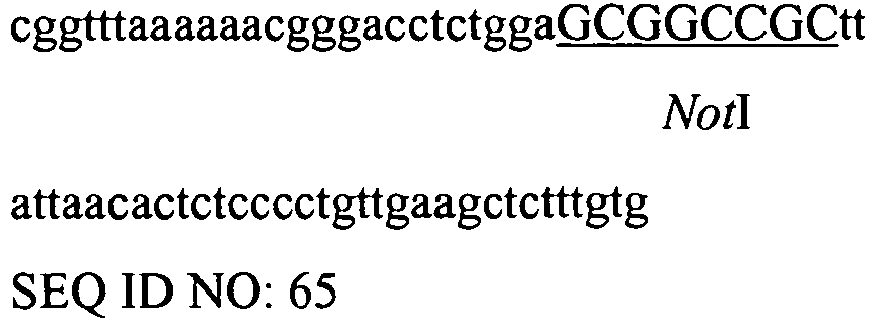
Reverse primer for amplification of T427 IgL that allows the replacement of XbaI by NotI restriction site. BGH-polyA-For 
Forward primer for amplification of polyadenylation site and assembly with T427 IgL fragment. BGH-polyA-EcoRI-Rev 
Reverse primer for amplification of polyadenylation site. - Transfection of HEK293 T-REx™ cells: The calcium-phosphate transfection method was applied for introducing 1 µg of the pDual or pMAZ plasmids into T-
REx 293 cells, seeded 3 × 10⁵ cells/well on 6-well plate 24 hours before transfection. For transient transfection, the medium samples were collected 24, 48 and 72 hours post transfection. In order to obtain the stable transfectants, the cells were harvested 24 hours post transfection and seeded on DMEM supplemented with appropriate antibiotics (1.2 mg/ml G418 and 0.2 mg/ml Hygromycin). The stable clones were collected and their media were evaluated for the presence of antibody. - IgG production in HEK293 T-REx™ cells: The previously obtained stable clones were transferred to tissue culture flasks (250 cm3) in DMEM supplement with 0.9 mg/ml G418 and 0.15 mg/ml Hygromycin (75 % of the regular concentration). The next day (or when the cells reached 80% confluence) the medium was changed to 50 % DMEM (+L-Glu, PNS and bovine serum) and 50 % DCCM1 (+L-Glu, PNS, serum free) + 75% of antibiotics concentration (0.9 mg/ml G418 and 0.15 mg/ml of Hygromycin) for 24 hours. The next day the medium was changed to 100 % serum free DCCM1 (+L-Glu, PNS). The DCCM1 media from cells were collected every 2-4 days and gently changed to new serum-free media. It was possible to collect up to 4 harvests from the flask.
- Protein A purification of IgG produced in mammalian cells: The collected DCCM1 medium from antibody secreting cells was centrifuged at 5500 rpm for 15 minutes and filtered using 0.45µm filtrap. The medium was diluted 1:20 with x20 concentration phosphate buffer (400mM) to final concentration of 20 mM Na2HPO4 and 20 mM NaH2PO4 and the mixture was loaded onto protein A column at a flow rate of 1 ml/min. The proteins were eluted with 0.1 mM citric acid (pH 3), neutralized with 1M Tris (HCl) pH 8.5 which was followed by dialysis against 20 mM phosphate buffer solution (PBS) pH 7.4. The protein final concentration was determined by absorbance at 280 nm.
- ELISA analysis: The antigen binding by mono- and bispecific IgGs was determined as follows: 96-well ELISA plates were coated with 5 µg/ml of pure antigen in
PBS 100 µl/well for overnight at 4 °C and blocked with 3 % milk (in PBS) for 1 hr at 37°C. All subsequent steps were carried out at room temperature (25 °C). Protein-A purified proteins (or conditioned media) were applied onto the plates in a three-fold dilution series in PBST for 1 hour incubation and washed with PBST for three times. Following the 1 hour incubation with HRP-conjugated secondary antibody (1:5000 dilution in PBST, 100 µl/well), the plates were washed in PBST and developed using chromogenic HRP substrate TMB and colour development was terminated with 1 M H2SO4. The plates were read at 450 nm. - Cell ELISA analysis: The A431/CD30 (expressing CD30, target antigen for T427) and SKBR3 (expressing ErbB2, target antigen for FRP5) cell lines were maintained in DMEM supplemented by 10 % fetal calf serum, 1 % L-glutamine and 1 % penicillin-streptomycin and grown at 37 °C with 5 % CO2. The cells (2×10⁴/well) were seeded onto 96-well tissue culture plates in 100 µl medium and grown at 37 °C for overnight. Following the overnight growth the medium was gently poured out and the cells were fixed with 3 % glutaraldehyde solution in water for 15 minutes at room temperature. The cells were washed with PBS and blocked with 5 % BSA in PBS for 2 hours at 37 °C. All subsequent steps were carried out according the regular ELISA protocol at room temperature (25 °C).
- Dot blot analysis: The 100 µl samples of 72 hours post-transfection cell conditioned media were diluted in an equal volume of PBS and applied via a vacuum manifold onto a nitrocellulose membrane filter using a dot-blot apparatus (Schleicher and Schuell, USA). After blocking the membranes with 3 % (v/v) non-fat milk in PBS for 1 hour at 37°C, the membrane was washed briefly with PBS followed by incubation with goat-anti-human HRP conjugated secondary antibody for 1 hour at room temperature. After three washes with PBS the membrane was developed with the ECL reagent (Pierce, USA).
- SDS-PAGE analysis: Polyacrylamide gel electrophoresis of proteins was performed according to Laemmli (Laemmli, 1970, Nature 227:680-685) 1/5 volume of 5x sample buffer was added to the protein samples followed by boiling for 5 minutes prior to loading onto the gel. 7.5 %, 10 % and 12 % mini-gels were run at 120 V. For evaluation of full length IgG, non-reduced samples (without β-mercaptoethanol) were loaded, while the reduced protein samples separated into heavy and light chains components. Gels were stained with Coomassie blue solution (0.05 % Coomassie R-250, 20 % ethanol, 10 % glacial acetic acid) for 2 hours and washed in destain solution (20 % ethanol, 10 % glacial acetic acid) until protein bands could be clearly seen. The protein band density was analyzed by ImageMaster 1D scanning laser densitometer (Pharmacia, Sweeden). Gels that were stained were loaded with 20 µg of protein per lane for non-purified fraction or 3-5µg for purified proteins. Gels that were further processed by immunoblotting were loaded with 1/10 that quantity.
- Western blot analysis: Proteins resolved by SDS-PAGE were electro-transferred onto the nitrocellulose membrane according to (Towbin et al., 1992, Biotechnology. 24:145-9). The membrane was blocked for at least 1 hour with PBS containing 5 % non-fat milk powder at room temperature with slow agitation. The membrane was washed with PBS followed by incubation HRP conjugated goat-anti-human secondary antibodies (Jackson Laboratories, West Grove, PA). After three washes with PBS containing 0.05 % Tween-20 (PBST) and one wash with PBS the nitrocellulose filter was developed with the SuperSignal West Pico Chemiluminescent Substrate (Thermo Scientific, USA) as described by the vendor.
- The vector system used for the production of IgG in mammalian cells for production of bispecific antibodies was based on pMAZ vectors for production of monoclonal antibodies in mammalian cell culture (Mazor Y et al, 2007, J. Immunol. Methods 321(1-2):41-59). Vector pMAZ-IgH was designed for human γ1 heavy chain expression and pMAZ-IgL for human κ light chain expression. The variable domains of light and heavy chains were introduced to the appropriate vector, co-transfected into HEK293 cells and stable antibody secreting clones were identified and kept. The starvation of cell clones to serum resulted in secretion of the desired antibody at a total yield of up to 20 mg per liter of culture.
- The pDual vector was constructed by fusion of DNA fragments derived from the pMAZ-IgL and pMAZ-IgH vectors (Mazor Y et al, 2007, J. Immunol. Methods 321(1-2):41-59) that were previously used for production of antibodies' light and heavy chains independently (
Figure 14 ), in order to build chimeric construct for production of light and heavy antibody chains using the same vector. The IgL and IgH were constructed in pDual vector as separate cassettes under the control of separate CMV promoters. The replacement of BssHI by ApaI restriction site in the light chain cassette simplified the following cloning of variable light and heavy domains into dual vector: ApaI-BsiWI were used for cloning of VL and BssHI-NheI were used for cloning of VH. - In order to construct the pDual-based vectors for production of bispecific molecules, the "knob", "hole" and "Cys" related mutations were cloned from the pHAK vectors into the pDual system. The cloning process resulted in the series of constructs listed in Table 5. The replacement of NeoR with Hygro resistance cassette in pDual-T427 vectors was carried out by subcloning of the cassette from pMAZ-IgL vector using AvrII and KpnI restriction enzymes. The resulted pDual-NeoR and pDual-HygroR vectors' pair can be used for transfection in mammalian cells and selection of stable clones that produce 4 different antibody chains: two heavy and two light chains within one cell line.
Table 5 Vector name Product pDual-T427 wt NeoR IgL-T427 + IgH-T427 pDual-FRP5 wt NeoR IgL-FRP5 + IgH-FRP5 pDual-T427-L(wt)-H(knob) NeoR IgL-T427 + IgH-T427(knob) pDual-FRP5-L(wt)-H(hole) NeoR IgL-FRP5 + IgH-FRP5(hole) pDual-T427-L(Cys)-H(wt) NeoR IgL-T427(Cys) + IgH-T427 pDual-T427-L(Cys)-H(knob) NeoR IgL-T427(Cys) + IgH-T427(knob) pDual-T427-L(wt)-H(Cys-knob) NeoR IgL-T427 + IgH-T427(Cys-knob) pDual-T427-L(Cys)-H(Cys-knob) NeoR IgL-T427(Cys) + IgH-T427(Cys-knob) pDual-T427 wt HygroR IgL-T427 + IgH-T427 pDual-T427-L(Cys)-H(Cys-knob) HygroR IgL-T427(Cys) + IgH-T427(Cys-knob) - The following example demonstrates the importance of the S-S bridge between the light and heavy chain of the antibody in IgG secretion system and proves that the "alternative Cysteine" theory for coupling the appropriate light and heavy chain of bispecific antibody is relevant in mammalian production system as well as it had been demonstrated in the E. coli produced bispecific "Inclonals". The HEK293 T-REx™ cell line was used for this study. The cells were transiently transfected with either pDual-T427 wt (encoding wt IgG), pDual-T427-L(Cys)-H(wt) (encoding wt heavy chain and light chain that lacks the C-kappa cysteine and does contain the engineerd cysteine in VL, this should be a pair of chains that should not form an IgG) or with pMAZ-IgL+pMAZ-IgH (previous system for wt IgG production) pair as control. The evaluation of post-transfection medium by Western blot analysis showed that no full-size antibodies were detected in media of pDual-T427-L(Cys)-H(wt) transfected cells, while the transfection of pDual wt construct produced the detectable levels of the secreted antibody (up to 1 µg/ml in comparison to Erbitux dilutions) (
Figures 15A and B ). - The secretion of bispecific IgG molecules was also demonstrated and the preference of bispecific IgG formation using "knobs-into-holes" and "alternative Cysteine" (also called "disulfide stabilization") approaches was also estimated. The pDual vectors were transiently transfected to HEK293 TRex cells and the secreted antibodies were detected by Western blot analysis of conditioned media. The cells that were transfected with four pMAZ vectors (pMAZ-T427-IgL, pMAZ-T427-IgH, pMAZ-FRP5-IgL and pMAZ-FRP5-IgH) served as a control. The analysis of experiment demonstrated: 1) the "knobs-into-holes" approach is a solution for efficient heterodimerization of different heavy chains, 2) "alternative Cys" approach provide the solution for coupling of light and heavy chains of the same antibody arm, 3) the combination of the two above approaches provides the secretion of full-length bispecific IgG antibodies in mammalian cells production systems. As shown in
Figure 16 , when "wrong" combinations of chains are used, no full size IgG can be seen in the immunoblot analysis of conditioned media. Intact IgG can be observed in cells that express monospecific IgG (lanes 1 and 2), in cells that express two monospecific IgGs (lanes 5, two pDual vectors andlane 4 four pMAZ vectors) and in cells that express a bispecific IgG (lane 7). - In order to obtain stable antibody-secreting clones, the present inventors co-transfected HEK293 T-REx™ cells with pDual-FRP5-L(wt)-H(hole) NeoR and pDual-T427-L(Cys)-H(Cys-knob) HygroR vectors. The resultant Neo + Hygro resistant clones were verified for their ability: 1) to secrete antibody, 2) to bind both antigens, 3) to secret full-length IgG for further purification. The preliminary antibody secretion test was performed using Dot blot analysis (the test example demonstrated in
Figure 17 ) and identified the low, medium and high secreting clones. The clones marked as medium and high secretors were examined for their antigen binding activity. ELISA was carried out to evaluate the binding level of each clone to erbB2 (FRP5) and CD30 (T427) recombinant antibodies (Figure 18 ). The several clones that were able to bind each of the antigens continued to the third step and were purified on protein A affinity column (Figure 19 ). The purified antibodies were analyzed to determine their size, purity and binding activity to either recombinant antigens or antigen-presenting cells (Figures 20 and21 ). As shown inFigure 18 , the binding signal in ELISA correlated to the secretion level of these clones. As shown inFigures 20 and21 , a protein A-purified bispecific IgG bound to both CD30 and ErbB2 antigens. In such an ELISA (Figure 21 ) it was expected that the monospecific IgGs (which are bi-valent) will show a more intense binding signal, each on its cognate antigen, due to avidity effect. A preliminary cell-ELISA (Figure 22 ) shows that the bispecific antibody secreted by clone D3 stains antigen-positive cells. - First, a mono-specific antibody (T427 IgG) was generated that comprised the knobs into hole (KIH) mutation. In order to evaluate the binding activity of KIH T427 IgG molecules, ELISA was carried out. The ELISA plate was coated with MBP-CD30 and incubated with T427 KIH IgG (fused to PE38). It was demonstrated that the binding ability of T427 KIH molecule was similar to the binding of unmodified T427 IgG and T427-PE38 IgG-PE38 (
Figure 24 ). - Subsequently, a mono-specific antibody (T427 IgG) was generated that comprised both the KIH mutation and the cysteine mutations in the light chains as described herein above.
- In order to produce the mono-specific T427 antibody, 4 chains were constructed: IgL-PE38, IgH-knob, IgH-Cys-hole and IgL-Cys. The presence of 38 kDa PE38 fused to VL- unmodified light chain provided the possibility to analyze the pairing of the appropriate heavy and light chains (analogous to KIH heterodimerization analysis) and the formation of the full-sized mono bi-specific molecule (Figure not shown).
- In addition, citation or identification of any reference in this application shall not be construed as an admission that such reference is available as prior art to the present invention. To the extent that section headings are used, they should not be construed as necessarily limiting.
-
- <110> Ramot at Tel-Aviv University Ltd. Benhar, Itai Vaks, Lilach
- <120> BISPECIFIC AND MONOSPECIFIC, ASYMMETRIC ANTIBODIES AND METHODS OF
GENERATING SAME - <130> 52585
- <150>
US 61/453,591
<151>2011-03-17 - <160> 75
- <170> PatentIn version 3.5
- <210> 1
<211> 75
<212> DNA
<213> Artificial sequence - <220>
<223> Single strand DNA oligonucleotide - <400> 1

- <210> 2
<211> 34
<212> DNA
<213> Artificial sequence - <220>
<223> Single strand DNA oligonucleotide - <400> 2
ggatgggggc agggtgcaca cctgtggttc tcgg 34 - <210> 3
<211> 45
<212> DNA
<213> Artificial sequence - <220>
<223> Single strand DNA oligonucleotide - <400> 3
ggatagaagc ctttgaccgc gcagctcagg ctgacctggt tcttg 45 - <210> 4
<211> 39
<212> DNA
<213> Artificial sequence - <220>
<223> Single strand DNA oligonucleotide - <400> 4
gtccacggtg agcttgctaa cgaggaagaa ggagccgtc 39 - <210> 5
<211> 23
<212> DNA
<213> Artificial sequence - <220>
<223> Single strand DNA oligonucleotide - <400> 5
atatacatat ggacattgtg ctg 23 - <210> 6
<211> 51
<212> DNA
<213> Artificial sequence - <220>
<223> Single strand DNA oligonucleotide - <400> 6
tatatacgta cgtttgattt ccagtttggt gccgcaaccg aacgtccgag g 51 - <210> 7
<211> 40
<212> DNA
<213> Artificial sequence - <220>
<223> Single strand DNA oligonucleotide - <400> 7
tatatagaat tcttactctc ccctgttgaa gctctttgtg 40 - <210> 8
<211> 34
<212> DNA
<213> Artificial sequence - <220>
<223> Single strand DNA oligonucleotide - <400> 8
aaacagaggc ctggacagtg tctggaatgg attg 34 - <210> 9
<211> 27
<212> DNA
<213> Artificial sequence - <220>
<223> Single strand DNA oligonucleotide - <400> 9
tatatagcta gcggaggaga ctgtgag 27 - <210> 10
<211> 36
<212> DNA
<213> Artificial sequence - <220>
<223> Single strand DNA oligonucleotide - <400> 10
gcccaaatct gccgacaaaa ctcacacatg cccacc 36 - <210> 11
<211> 40
<212> DNA
<213> Artificial sequence - <220>
<223> Single strand DNA oligonucleotide - <400> 11
tgtgtgagtt ttgtcggcag atttgggctc aactctcttg 40 - <210> 12
<211> 23
<212> DNA
<213> Artificial sequence - <220>
<223> Single strand DNA oligonucleotide - <400> 12
gaggagatga ccaagaacca ggt 23 - <210> 13
<211> 24
<212> DNA
<213> Artificial sequence - <220>
<223> Single strand DNA oligonucleotide - <400> 13
atatacatat gcaggtcaaa ctgc 24 - <210> 14
<211> 660
<212> DNA
<213> Artificial sequence - <220>
<223> Light chain T427 (wt IgG) coding sequence - <400> 14


- <210> 15
<211> 219
<212> PRT
<213> Artificial sequence - <220>
<223> Light chain T427 (wt IgG) - <400> 15


- <210> 16
<211> 648
<212> DNA
<213> Artificial sequence - <220>
<223> Light chain FRP5 (wt IgG) coding sequence - <400> 16

- <210> 17
<211> 215
<212> PRT
<213> Artificial sequence - <220>
<223> Light chain FRP5 (wt IgG) - <400> 17

- <210> 18
<211> 645
<212> DNA
<213> Artificial sequence - <220>
<223> Light chain anti-Tac (wt IgG) coding sequence - <400> 18

- <210> 19
<211> 214
<212> PRT
<213> Artificial sequence - <220>
<223> Light chain anti-Tac (wt IgG) - <400> 19


- <210> 20
<211> 657
<212> DNA
<213> Artificial sequence - <220>
<223> Light chain T427 A104C:Cys218del (Cys) coding sequence - <400> 20

- <210> 21
<211> 218
<212> PRT
<213> Artificial sequence - <220>
<223> Light chain T427 A104C:Cys218del (Cys) - <400> 21

- <210> 22
<211> 1353
<212> DNA
<213> Artificial sequence - <220>
<223> Heavy chain T427 (wt IgG human gamma 1) coding sequence - <400> 22

- <210> 23
<211> 450
<212> PRT
<213> Artificial sequence - <220>
<223> Heavy chain T427 (wt IgG human gamma 1) - <400> 23

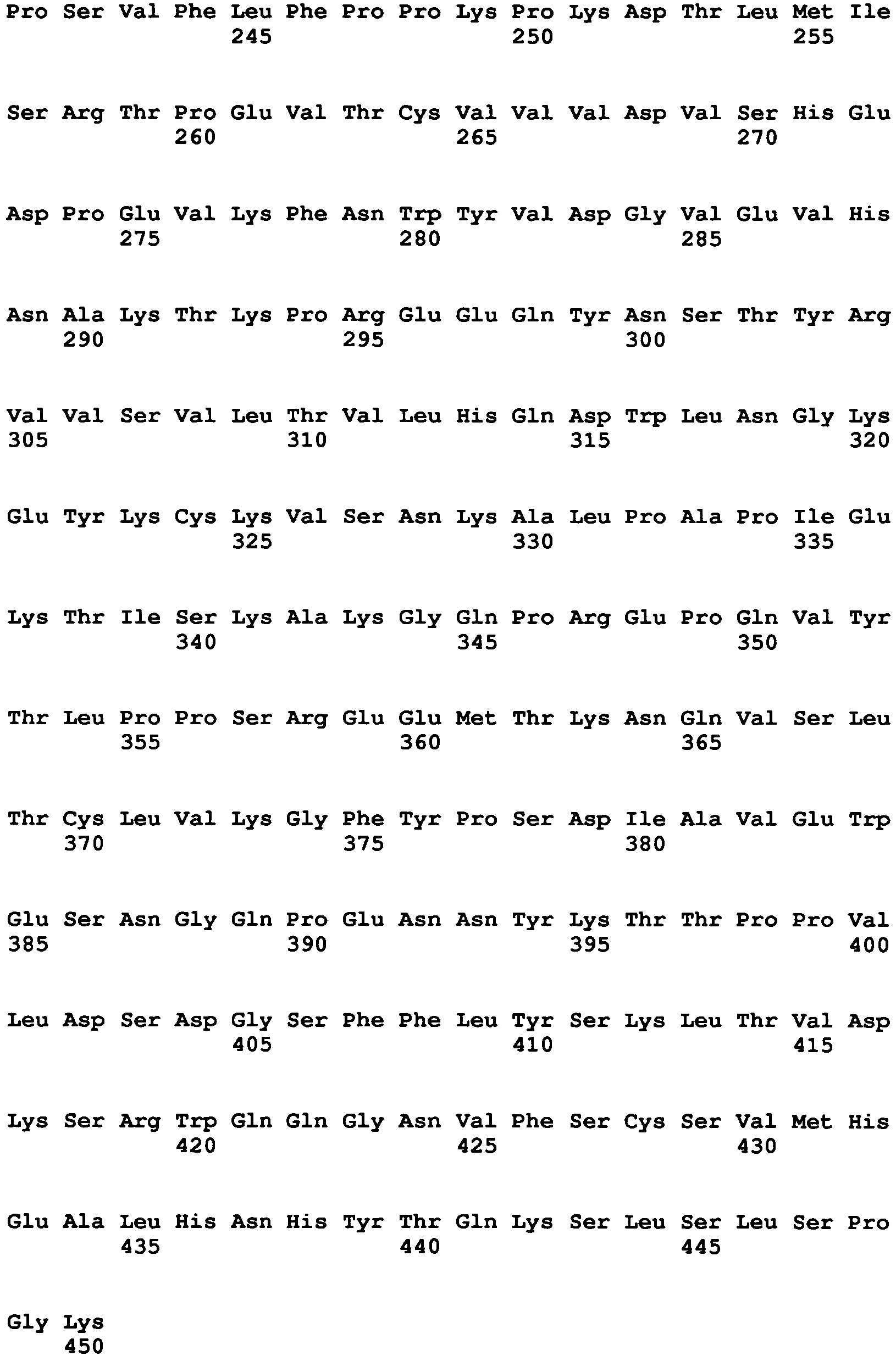
- <210> 24
<211> 1353
<212> DNA
<213> Artificial sequence - <220>
<223> Heavy chain FRP5 (wt IgG human gamma 1) coding sequence - <400> 24

- <210> 25
<211> 450
<212> PRT
<213> Artificial sequence - <220>
<223> Heavy chain FRP5 (wt IgG human gamma 1) - <400> 25


- <210> 26
<211> 1344
<212> DNA
<213> Artificial sequence - <220>
<223> Heavy chain anti-Tac (wt IgG human gamma 1) coding sequence - <400> 26


- <210> 27
<211> 447
<212> PRT
<213> Artificial sequence - <220>
<223> Heavy chain anti-Tac (wt IgG human gamma 1) - <400> 27


- <210> 28
<211> 1353
<212> DNA
<213> Artificial sequence - <220>
<223> Heavy chain T427 T366W:S354C (knob) coding sequence - <400> 28


- <210> 29
<211> 465
<212> PRT
<213> Artificial sequence - <220>
<223> Heavy chain T427 T366W:S354C (knob) coding sequence - <400> 29


- <210> 30
<211> 1352
<212> DNA
<213> Artificial sequence - <220>
<223> Heavy chain T427 T366S:L368A:Y407V:Y349C (hole) coding sequence - <400> 30
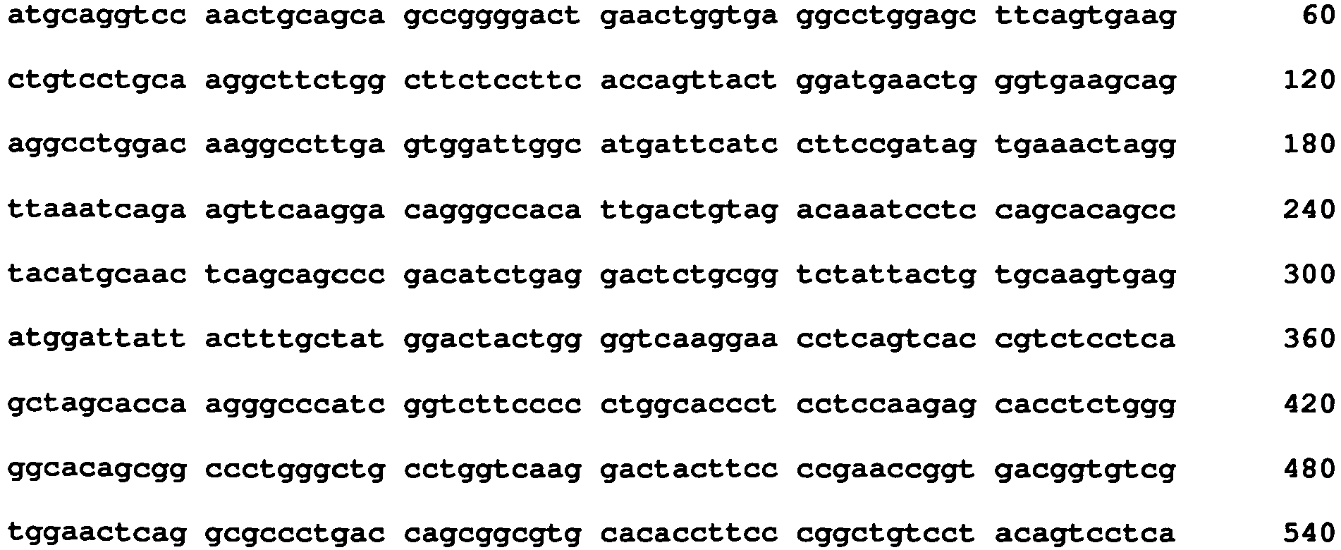

- <210> 31
<211> 450
<212> PRT
<213> Artificial sequence - <220>
<223> Heavy chain T427 T366S:L368A:Y407V:Y349C (hole) - <400> 31


- <210> 32
<211> 2406
<212> DNA
<213> Artificial sequence - <220>
<223> Heavy chain T427-PE38 coding sequence - <400> 32


- <210> 33
<211> 800
<212> PRT
<213> Artificial sequence - <220>
<223> Heavy chain T427-PE38 - <400> 33


- <210> 34
<211> 2406
<212> DNA
<213> Artificial sequence - <220>
<223> Heavy chain T427-PE38 T366S:L368A:Y407V:Y349C (hole) coding sequence - <400> 34


- <210> 35
<211> 800
<212> PRT
<213> Artificial sequence - <220>
<223> Heavy chain T427-PE38 T366S:L368A:Y407V:Y349C (hole) - <400> 35


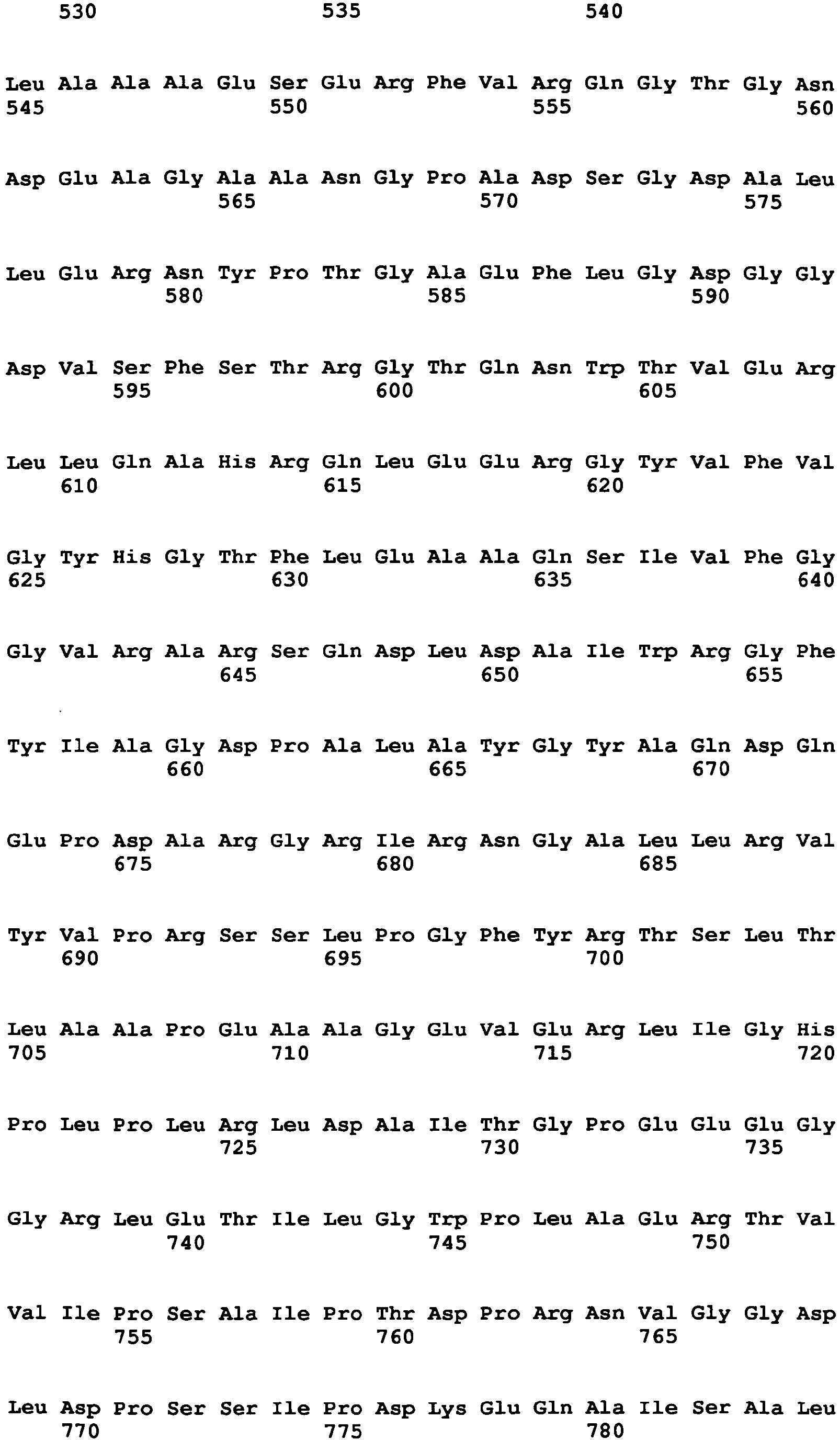

- <210> 36
<211> 1353
<212> DNA
<213> Artificial sequence - <220>
<223> Heavy chain FRP5 T366W:S354C (knob) coding sequence - <400> 36

- <210> 37
<211> 450
<212> PRT
<213> Artificial sequence - <220>
<223> Heavy chain FRP5 T366W:S354C (knob) - <400> 37
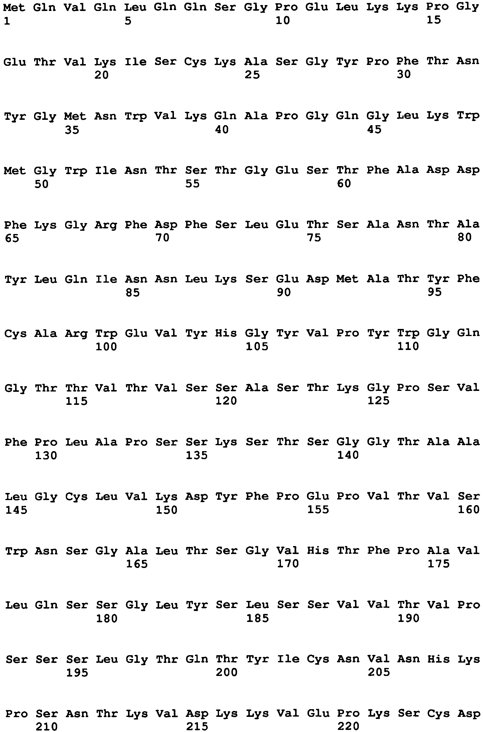

- <210> 38
<211> 1353
<212> DNA
<213> Artificial sequence - <220>
<223> Heavy chain FRP5 T366S:L368A:Y407V:Y349C (hole) coding sequence - <400> 38

- <210> 39
<211> 450
<212> PRT
<213> Artificial sequence - <220>
<223> Heavy chain FRP5 T366S:L368A:Y407V:Y349C (hole) - <400> 39


- <210> 40
<211> 1353
<212> DNA
<213> Artificial sequence - <220>
<223> Heavy chain T427 T366W:S354C (knob) and A44C:C222A (Cys) coding sequence - <400> 40

- <210> 41
<211> 450
<212> PRT
<213> Artificial sequence - <220>
<223> Heavy chain T427 T366W:S354C (knob) and A44C:C222A (Cys) - <400> 41


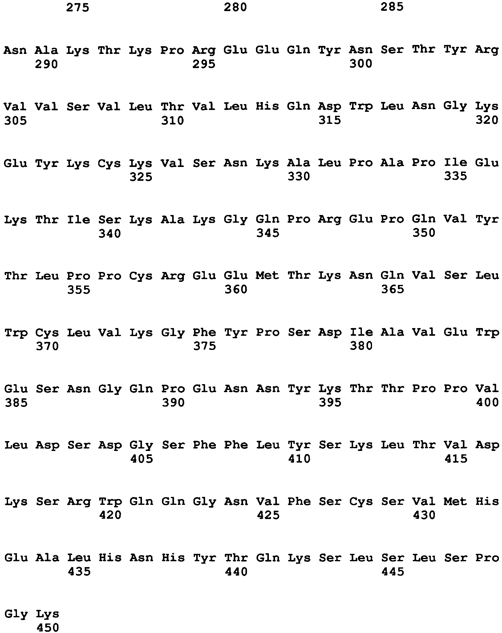
- <210> 42
<211> 216
<212> PRT
<213> Pseudomonas aeruginosa - <400> 42


- <210> 43
<211> 536
<212> PRT
<213> Corynebacterium diphtheriae - <400> 43


- <210> 44
<211> 127
<212> PRT
<213> Artificial sequence - <220>
<223>interleukin 2 - <400> 44

- <210> 45
<211> 207
<212> PRT
<213> Homo sapiens - <400> 45


- <210> 46
<211> 290
<212> PRT
<213> Homo sapiens - <400> 46
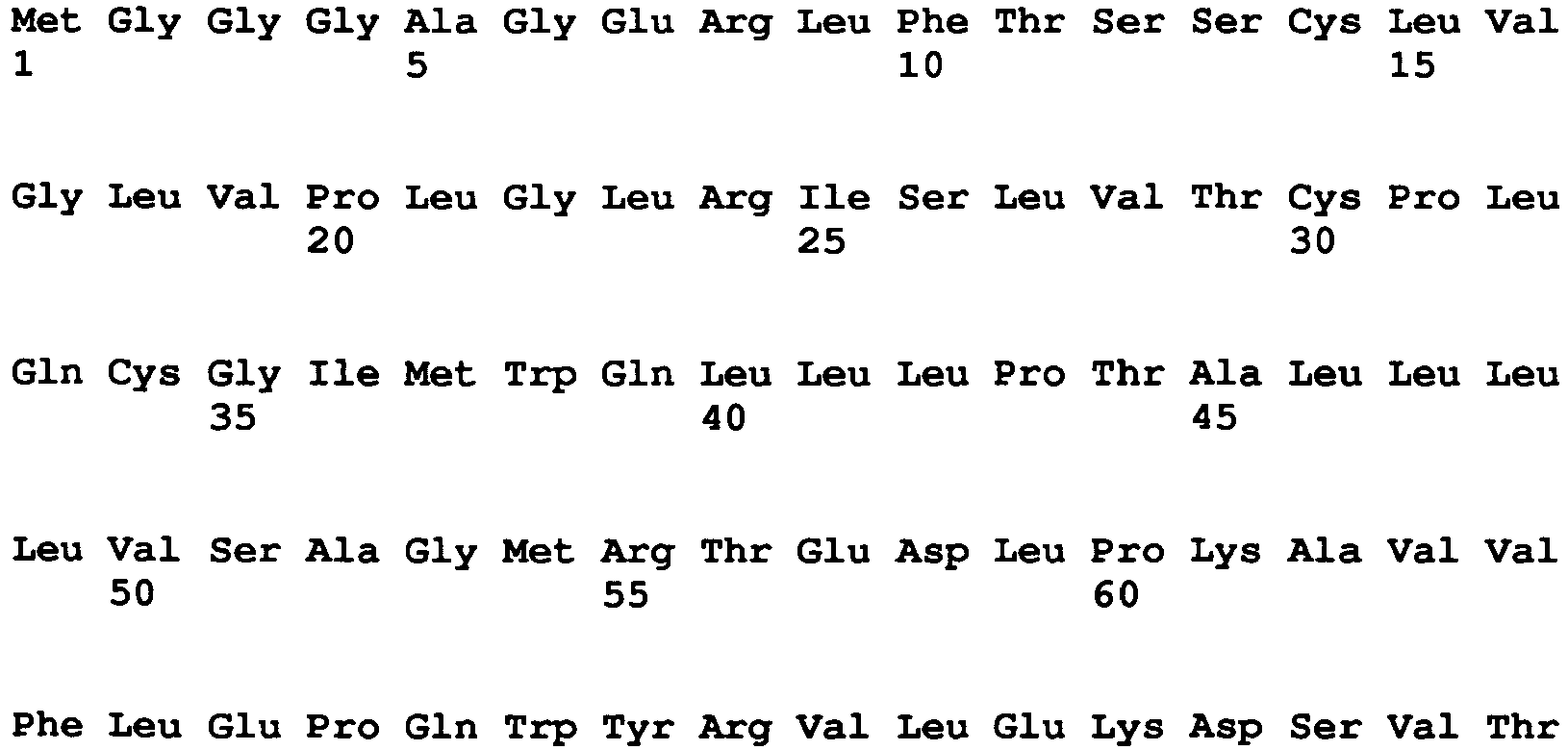

- <210> 47
<211> 153
<212> PRT
<213> Homo sapiens - <400> 47

- <210> 48
<211> 365
<212> PRT
<213> Homo sapiens - <400> 48


- <210> 49
<211> 178
<212> PRT
<213> Homo sapiens - <400> 49


- <210> 50
<211> 576
<212> PRT
<213> Ricinus communis - <400> 50


- <210> 51
<211> 651
<212> DNA
<213> Pseudomonas aeruginosa - <400> 51

- <210> 52
<211> 1611
<212> DNA
<213> Corynebacterium diphtheriae - <400> 52
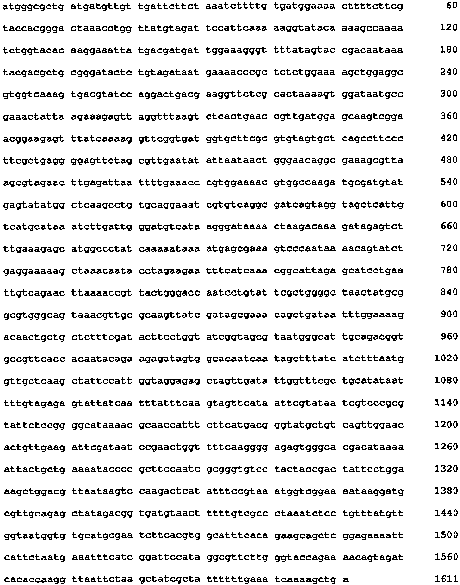
- <210> 53
<211> 698
<212> DNA
<213> Artificial sequence - <220>
<223>interleukin 2 - <220>
<221> misc_feature
<222> (4) .. (4)
<223> n is a, c, g, or t - <400> 53

- <210> 54
<211> 1311
<212> DNA
<213> Homo sapiens - <400> 54


- <210> 55
<211> 2406
<212> DNA
<213> Homo sapiens - <400> 55


- <210> 56
<211> 921
<212> DNA
<213> Homo sapiens - <400> 56


- <210> 57
<211> 4000
<212> DNA
<213> Homo sapiens - <400> 57


- <210> 58
<211> 1601
<212> DNA
<213> Homo sapiens - <400> 58


- <210> 59
<211> 12150
<212> DNA
<213> Ricinus communis - <400> 59


- <210> 60
<211> 39
<212> DNA
<213> Artificial sequence - <220>
<223> Single strand DNA oligonucleotide - <400> 60
tcctcagcta gcaccaaggg accatcggtc ttccccctg 39 - <210> 61
<211> 22
<212> DNA
<213> Artificial sequence - <220>
<223> Single strand DNA oligonucleotide - <400> 61
gcagggtgta cacctgtggt tc 22 - <210> 62
<211> 51
<212> DNA
<213> Artificial sequence - <220>
<223> Single strand DNA oligonucleotide - <400> 62
actgaacctt ggagtcaggt accacattga ttattgagta gttattaata g 51 - <210> 63
<211> 32
<212> DNA
<213> Artificial sequence - <220>
<223> Single strand DNA oligonucleotide - <400> 63
gggcccctgt ggagagaaag gcaaagtgga tg 32 - <210> 64
<211> 51
<212> DNA
<213> Artificial sequence - <220>
<223> Single strand DNA oligonucleotide - <400> 64
ctttgccttt ctctccacag gggcccactc cgacattgtg ctgacccaat c 51 - <210> 65
<211> 67
<212> DNA
<213> Artificial sequence - <220>
<223> Single strand DNA oligonucleotide - <400> 65
cggtttaaaa aacgggacct ctggagcggc cgcttattaa cactctcccc tgttgaagct 60 -
ctttgtg 67 - <210> 66
<211> 51
<212> DNA
<213> Artificial sequence - <220>
<223> Single strand DNA oligonucleotide - <400> 66
tccagaggtc ccgtttttta aaccggtttt ttaaaccgct gatcagcctc g 51 - <210> 67
<211> 44
<212> DNA
<213> Artificial sequence - <220>
<223> Single strand DNA oligonucleotide - <400> 67
tagctcgatc cgtcgagaga attcccccag catgcctgct attg 44 - <210> 68
<211> 657
<212> DNA
<213> Artificial sequence - <220>
<223> Light chain of anti-streptavidin (anti-SA) - <400> 68

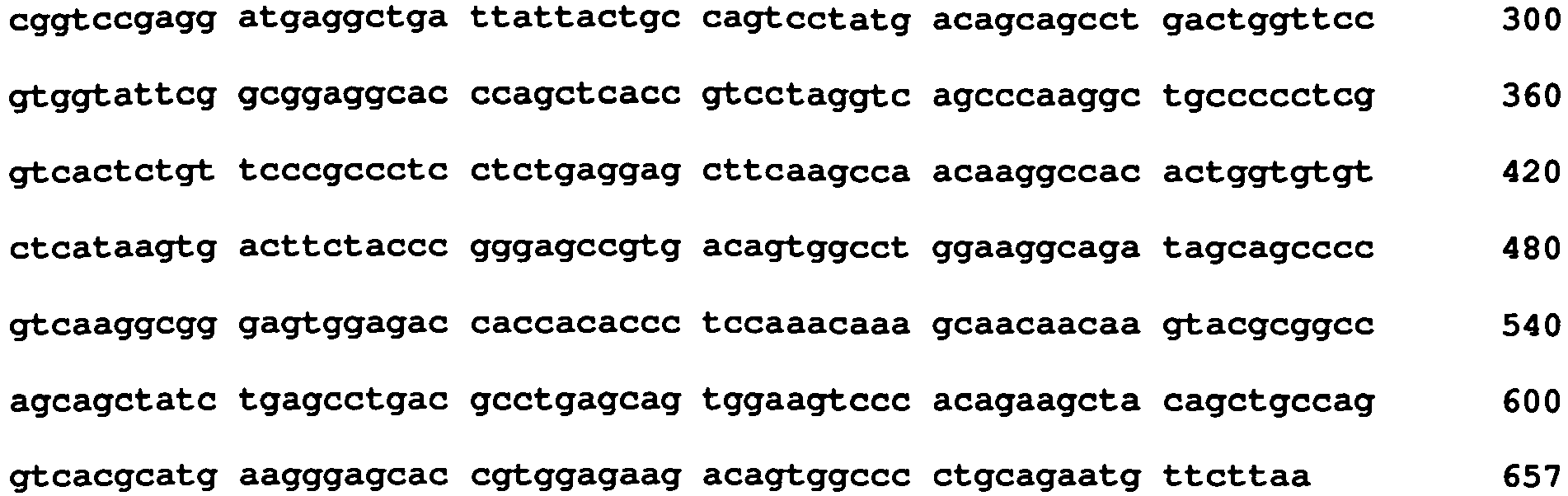
- <210> 69
<211> 218
<212> PRT
<213> Artificial sequence - <220>
<223> Light chain of anti-streptavidin (anti-SA) - <400> 69


- <210> 70
<211> 657
<212> DNA
<213> Artificial sequence - <220>
<223> Light chain of anti-PE clone B11 - <400> 70

- <210> 71
<211> 218
<212> PRT
<213> Artificial sequence - <220>
<223> Light chain of anti-PE clone B11 - <400> 71


- <210> 72
<211> 1347
<212> DNA
<213> Artificial sequence - <220>
<223> Heavy chain of anti-streptavidin (anti-SA) - <400> 72

- <210> 73
<211> 448
<212> PRT
<213> Artificial sequence - <220>
<223> Heavy chain of anti-streptavidin (anti-SA) - <400> 73



- <210> 74
<211> 1362
<212> DNA
<213> Artificial sequence - <220>
<223> Heavy chain of anti-PE clone B11 - <400> 74


- <210> 75
<211> 453
<212> PRT
<213> Artificial sequence - <220>
<223> Heavy chain of anti-PE clone B11 - <400> 75


Claims (15)
- A bispecific antibody comprising an Fc region and a Fab region, wherein:(i) said Fc region comprises two non-identical heavy chains, wherein at least one of said two non-identical heavy chains comprises an amino acid modification so as to form a steric complementation between said two non-identical heavy chains, wherein said Fc region comprises a protuberance of one heavy chain of said Fc region and a sterically compensatory cavity on a second heavy chain of said Fc region, said protuberance protruding into said compensatory cavity, wherein said complementation is generated by:(a) substituting an amino acid at one position on a CH3 domain of said one heavy chain with another amino acid having a larger side chain volume than the original amino acid, said amino acid having a larger side chain being selected from the group consisting of tyrosine, arginine, phenylalanine, isoleucine and tryptophan so as to generate said protuberance; and(b) substituting an amino acid at one position on a CH3 domain of said second heavy chain with another amino acid having a smaller side chain volume than the original amino acid so as to generate said compensatory cavity, said amino acid having a smaller side chain being selected from the group consisting of alanine, glycine, valine and threonine; and(ii) said Fab region comprises a first covalent link between a first heavy chain and a first light chain of said Fab region and a second covalent link between a second heavy chain and a second light chain of said Fab region, wherein said first covalent link is a naturally occurring disulfide bond between a CH1 domain of said one heavy chain and a CL domain of said one light chain; and said second covalent link is a disulfide bond between a cysteine at position 44 of a VH domain of said second heavy chain and a cysteine at position 100 of a VL domain of said second light chain, wherein the numbering of said position is according to Kabat and Wu, wherein said second heavy chain is devoid of its native disulfide bond with said second light chain, said native disulfide bond being an interchain disulfide bond that connects a heavy chain to its cognate light chain encoded in a naturally occurring germline antibody gene.
- The antibody of claim 1, being selected from the group consisting of a chimeric antibody, a humanized antibody and a fully human antibody.
- The antibody of claim 1, wherein said CH3 domain of said first heavy chain is covalently linked to said CH3 domain of said second heavy chain.
- The antibody of claim 1, wherein a first antigen binding site of the antibody binds a first epitope of an antigen and a second antigen binding site of the antibody binds a second epitope of said antigen.
- The antibody of claim 1, wherein a first antigen binding site of the antibody binds an epitope of a first antigen and a second antigen binding site of the antibody binds an epitope of a second antigen.
- The antibody of claim 1, wherein each light chain is linked to its cognate heavy chain via a single disulfide bond.
- The antibody of claim 1 being an intact antibody.
- The antibody of claim 1, wherein the antibody is selected from the group consisting of IgA, IgD, IgE and IgG.
- The antibody of claim 1, wherein said first heavy chain comprises a T366W mutation; and said second heavy chain comprises T366S, L368A, Y407V mutations.
- The antibody of claim 9, wherein said first heavy chain comprises an S354C mutation and said second heavy chain comprises a Y349C mutation.
- A method of preparing the antibody of claim 1, comprising:(a) providing a first nucleic acid molecule encoding said first heavy chain;(b) providing a second nucleic acid molecule encoding said second heavy chain;(c) providing a third nucleic acid molecule encoding said first light chain;(d) providing a fourth nucleic acid molecule encoding said second light chain;(e) culturing host cells comprising said first, second, third and fourth nucleic acid molecules under conditions that permit expression of the nucleic acid molecules; and(f) recovering the antibody of claim 1.
- The method of claim 11, wherein said host cells comprise bacterial cells.
- A pharmaceutical composition comprising as an active agent the antibody of claim 1 and a pharmaceutically acceptable carrier.
- The antibody of claim 1 for use in the treatment of an infection or inflammatory disease or inflammatory disorder.
- The antibody of claim 1, for use according to claim 14. wherein said inflammatory disorder is cancer.
Applications Claiming Priority (2)
| Application Number | Priority Date | Filing Date | Title |
|---|---|---|---|
| US201161453591P | 2011-03-17 | 2011-03-17 | |
| PCT/IL2012/050093 WO2012123949A1 (en) | 2011-03-17 | 2012-03-15 | Bi- and monospecific, asymmetric antibodies and methods of generating the same |
Publications (2)
| Publication Number | Publication Date |
|---|---|
| EP2686348A1 EP2686348A1 (en) | 2014-01-22 |
| EP2686348B1 true EP2686348B1 (en) | 2015-12-30 |
Family
ID=45999923
Family Applications (1)
| Application Number | Title | Priority Date | Filing Date |
|---|---|---|---|
| EP12716657.7A Active EP2686348B1 (en) | 2011-03-17 | 2012-03-15 | Bi- and monospecific, asymmetric antibodies and methods of generating the same |
Country Status (11)
| Country | Link |
|---|---|
| US (2) | US9624291B2 (en) |
| EP (1) | EP2686348B1 (en) |
| JP (1) | JP2014510084A (en) |
| KR (1) | KR20140019385A (en) |
| CN (1) | CN103429619B (en) |
| AU (1) | AU2012227883A1 (en) |
| BR (1) | BR112013023653A2 (en) |
| CA (1) | CA2827188C (en) |
| IN (1) | IN2013MN01438A (en) |
| MX (1) | MX342135B (en) |
| WO (1) | WO2012123949A1 (en) |
Families Citing this family (56)
| Publication number | Priority date | Publication date | Assignee | Title |
|---|---|---|---|---|
| US10662256B2 (en) | 2010-07-26 | 2020-05-26 | Trianni, Inc. | Transgenic mammals and methods of use thereof |
| RU2580017C2 (en) | 2010-07-26 | 2016-04-10 | Трианни, Инк. | Transgenic animals and methods of use |
| US10793829B2 (en) | 2010-07-26 | 2020-10-06 | Trianni, Inc. | Transgenic mammals and methods of use thereof |
| CA2827188C (en) | 2011-03-17 | 2020-02-11 | Ramot At Tel-Aviv University Ltd. | Bi- and monospecific, asymmetric antibodies and methods of generating the same |
| US12466897B2 (en) | 2011-10-10 | 2025-11-11 | Xencor, Inc. | Heterodimeric human IgG1 polypeptides with isoelectric point modifications |
| US9465889B2 (en) * | 2012-07-05 | 2016-10-11 | Physion Consulting, LLC | Method and system for identifying data and users of interest from patterns of user interaction with existing data |
| WO2014079000A1 (en) | 2012-11-21 | 2014-05-30 | Wuhan Yzy Biopharma Co., Ltd. | Bispecific antibody |
| US9605084B2 (en) * | 2013-03-15 | 2017-03-28 | Xencor, Inc. | Heterodimeric proteins |
| WO2014116051A1 (en) * | 2013-01-24 | 2014-07-31 | 스크립스코리아 항체연구원 | Protein combination-based fv library, and preparation method therefor |
| EP4050019A1 (en) * | 2013-02-15 | 2022-08-31 | R-Pharm International, LLC | Il-1beta inhibitor composition and use thereof |
| EP2975061A4 (en) | 2013-03-13 | 2017-03-01 | Ibentrus Inc. | Protein in which electrical interaction is introduced within hydrophobic interaction site and preparation method therefor |
| US10858417B2 (en) | 2013-03-15 | 2020-12-08 | Xencor, Inc. | Heterodimeric proteins |
| MX379030B (en) | 2013-12-17 | 2025-03-10 | Genentech Inc | Anti-cd3 antibodies and methods of use |
| DK3105250T3 (en) * | 2014-02-10 | 2020-09-28 | Igm Biosciences Inc | MULTISPECIFIC IGA BINDING MOLECULES |
| WO2015134411A1 (en) * | 2014-03-03 | 2015-09-11 | Academia Sinica | Bi-specific antibodies and uses thereof |
| US12180264B2 (en) | 2014-03-24 | 2024-12-31 | R-Pharm Overseas, Inc. | IL1-R1 derived inhibitor of IL-1β and use thereof |
| DK3126384T3 (en) | 2014-04-01 | 2021-01-18 | Adimab Llc | MULTISPECIFIC ANTIBODY ANALOGS INCLUDING A COMMON LIGHT CHAIN, AND METHODS FOR THEIR PREPARATION AND USE |
| SG10201913507SA (en) * | 2014-05-02 | 2020-02-27 | Momenta Pharmaceuticals Inc | Compositions and methods related to engineered fc constructs |
| KR20170010863A (en) | 2014-07-01 | 2017-02-01 | 화이자 인코포레이티드 | Bispecific heterodimeric diabodies and uses thereof |
| US10927185B2 (en) | 2014-11-21 | 2021-02-23 | Astellas Pharma Inc. | Bispecific antibody format |
| SG11201704283PA (en) | 2014-11-26 | 2017-06-29 | Xencor Inc | Heterodimeric antibodies that bind cd3 and tumor antigens |
| WO2016204966A1 (en) | 2015-06-16 | 2016-12-22 | Genentech, Inc. | Anti-cd3 antibodies and methods of use |
| IL257411B2 (en) * | 2015-08-24 | 2024-05-01 | Trianni Inc | Improved production of immunoglobulins |
| BR112018002570A2 (en) * | 2015-10-02 | 2018-10-16 | Hoffmann La Roche | bispecific antigen binding molecule, bispecific antibody, polynucleotides, ox40-specific binding antibody, pharmaceutical composition and method for inhibiting tumor cell growth in an individual |
| WO2017095939A1 (en) | 2015-12-03 | 2017-06-08 | Trianni, Inc. | Enhanced immunoglobulin diversity |
| AU2017213633B2 (en) | 2016-02-04 | 2023-08-03 | Trianni, Inc. | Enhanced production of immunoglobulins |
| WO2017151971A2 (en) | 2016-03-02 | 2017-09-08 | Momenta Pharmaceuticals, Inc. | METHODS RELATED TO ENGINEERED Fc CONSTRUCTS |
| CA3025310A1 (en) | 2016-05-23 | 2017-11-30 | Momenta Pharmaceuticals, Inc. | Compositions and methods related to engineered fc constructs |
| JP7301540B2 (en) * | 2016-05-26 | 2023-07-03 | チールー ピュージェット サウンド バイオセラピューティクス コーポレイション | mixture of antibodies |
| EP3481860A4 (en) * | 2016-07-07 | 2020-01-22 | Acceleron Pharma Inc. | HETEROMULTIMERS OF THE TGF-BETA SUPERFAMILY AND USES THEREOF |
| CA3203613A1 (en) | 2016-07-19 | 2018-01-25 | Ibentrus, Inc. | Bispecific proteins and methods for preparing same |
| WO2018089293A2 (en) | 2016-11-08 | 2018-05-17 | Qilu Puget Sound Biotherapeutics Corporation | Anti-pd1 and anti-ctla4 antibodies |
| CN106496331B (en) * | 2016-11-08 | 2020-03-13 | 北京智岭生物医药科技有限公司 | FSH-Fc fusion protein and preparation method and application thereof |
| KR20190074300A (en) | 2016-11-15 | 2019-06-27 | 제넨테크, 인크. | Dosage for treatment with anti-CD20 / anti-CD3 bispecific antibodies |
| KR102624254B1 (en) | 2017-01-06 | 2024-01-12 | 모멘타 파머슈티컬스 인코포레이티드 | Compositions and methods for genetically engineered Fc constructs |
| UY37758A (en) | 2017-06-12 | 2019-01-31 | Novartis Ag | METHOD OF MANUFACTURING OF BIESPECTIFIC ANTIBODIES, BISPECTIFIC ANTIBODIES AND THERAPEUTIC USE OF SUCH ANTIBODIES |
| MX2020003497A (en) | 2017-10-20 | 2020-07-22 | Hoffmann La Roche | Method for generating multispecific antibodies from monospecific antibodies. |
| AR115360A1 (en) | 2018-02-08 | 2021-01-13 | Genentech Inc | ANTIGEN BINDING MOLECULES AND METHODS OF USE |
| TWI848953B (en) | 2018-06-09 | 2024-07-21 | 德商百靈佳殷格翰國際股份有限公司 | Multi-specific binding proteins for cancer treatment |
| WO2020135804A1 (en) * | 2018-12-29 | 2020-07-02 | 上海一宸医药科技有限公司 | Heterodimeric fusion protein |
| WO2020198683A1 (en) | 2019-03-28 | 2020-10-01 | Ab Studio Inc. | Heteromultimeric proteins and methods of use thereof |
| BR112022011357A2 (en) | 2019-12-13 | 2022-08-23 | Genentech Inc | ISOLATED ANTIBODIES, ONE OR MORE ISOLATED NUCLEIC ACIDS, ONE OR MORE VECTORS, ONE OR MORE HOST CELLS, COMPOSITION, KIT, ANTIBODY USE, METHODS TO PRODUCE THE ANTIBODY AND METHOD TO TREAT OR DELAY PROGRESSION OF A LY6G6D POSITIVE CANCER |
| WO2021231976A1 (en) | 2020-05-14 | 2021-11-18 | Xencor, Inc. | Heterodimeric antibodies that bind prostate specific membrane antigen (psma) and cd3 |
| AU2021374590A1 (en) | 2020-11-04 | 2023-06-01 | Genentech, Inc. | Subcutaneous dosing of anti-cd20/anti-cd3 bispecific antibodies |
| TWI874719B (en) | 2020-11-04 | 2025-03-01 | 美商建南德克公司 | Dosing for treatment with anti-cd20/anti-cd3 bispecific antibodies |
| KR20230095113A (en) | 2020-11-04 | 2023-06-28 | 제넨테크, 인크. | Dosing for Treatment with Anti-CD20/Anti-CD3 Bispecific Antibodies and Anti-CD79B Antibody Drug Conjugates |
| IL301263A (en) | 2020-11-18 | 2023-05-01 | Novartis Ag | Bispecific antibodies for use in treatment of nlrc4-gof inflammasomapathy |
| KR20230156079A (en) | 2021-03-09 | 2023-11-13 | 젠코어 인코포레이티드 | Heterodimeric antibody binding to CD3 and CLDN6 |
| KR20240007171A (en) | 2021-05-14 | 2024-01-16 | 제넨테크, 인크. | Methods for treating CD20 positive proliferative disorders using mosunetuzumab and polatuzumab vedotin |
| CN113480645B (en) * | 2021-07-16 | 2022-11-11 | 甘肃中科博瑞生物工程有限公司 | Anti-helicobacter pylori recombinant antibody, preparation method and application |
| EP4508086A1 (en) | 2022-04-13 | 2025-02-19 | Genentech, Inc. | Pharmaceutical compositions of mosunetuzumab and methods of use |
| CN115232214A (en) * | 2022-08-16 | 2022-10-25 | 广州市拜沃思生物科技有限公司 | Bispecific antibody and preparation method and application thereof |
| CN116789836B (en) * | 2023-08-14 | 2024-01-05 | 浙江时迈药业有限公司 | Antibodies against DLL3 and uses thereof |
| WO2025098100A1 (en) * | 2023-11-07 | 2025-05-15 | Biomap (Suzhou) Intelligent Technology Limited | Modified polypeptides for heteromultimer |
| GB202319967D0 (en) | 2023-12-22 | 2024-02-07 | Epsilogen Ltd | Heteromultimeric proteins |
| WO2026037918A1 (en) | 2024-08-14 | 2026-02-19 | Epsilogen Ltd | Heteromultimeric proteins |
Family Cites Families (50)
| Publication number | Priority date | Publication date | Assignee | Title |
|---|---|---|---|---|
| NL154600B (en) | 1971-02-10 | 1977-09-15 | Organon Nv | METHOD FOR THE DETERMINATION AND DETERMINATION OF SPECIFIC BINDING PROTEINS AND THEIR CORRESPONDING BINDABLE SUBSTANCES. |
| NL154598B (en) | 1970-11-10 | 1977-09-15 | Organon Nv | PROCEDURE FOR DETERMINING AND DETERMINING LOW MOLECULAR COMPOUNDS AND PROTEINS THAT CAN SPECIFICALLY BIND THESE COMPOUNDS AND TEST PACKAGING. |
| NL154599B (en) | 1970-12-28 | 1977-09-15 | Organon Nv | PROCEDURE FOR DETERMINING AND DETERMINING SPECIFIC BINDING PROTEINS AND THEIR CORRESPONDING BINDABLE SUBSTANCES, AND TEST PACKAGING. |
| US3901654A (en) | 1971-06-21 | 1975-08-26 | Biological Developments | Receptor assays of biologically active compounds employing biologically specific receptors |
| US3853987A (en) | 1971-09-01 | 1974-12-10 | W Dreyer | Immunological reagent and radioimmuno assay |
| US3867517A (en) | 1971-12-21 | 1975-02-18 | Abbott Lab | Direct radioimmunoassay for antigens and their antibodies |
| NL171930C (en) | 1972-05-11 | 1983-06-01 | Akzo Nv | METHOD FOR DETERMINING AND DETERMINING BITES AND TEST PACKAGING. |
| US3850578A (en) | 1973-03-12 | 1974-11-26 | H Mcconnell | Process for assaying for biologically active molecules |
| US3935074A (en) | 1973-12-17 | 1976-01-27 | Syva Company | Antibody steric hindrance immunoassay with two antibodies |
| US3996345A (en) | 1974-08-12 | 1976-12-07 | Syva Company | Fluorescence quenching with immunological pairs in immunoassays |
| US4034074A (en) | 1974-09-19 | 1977-07-05 | The Board Of Trustees Of Leland Stanford Junior University | Universal reagent 2-site immunoradiometric assay using labelled anti (IgG) |
| US3984533A (en) | 1975-11-13 | 1976-10-05 | General Electric Company | Electrophoretic method of detecting antigen-antibody reaction |
| US4098876A (en) | 1976-10-26 | 1978-07-04 | Corning Glass Works | Reverse sandwich immunoassay |
| US4879219A (en) | 1980-09-19 | 1989-11-07 | General Hospital Corporation | Immunoassay utilizing monoclonal high affinity IgM antibodies |
| US4816567A (en) | 1983-04-08 | 1989-03-28 | Genentech, Inc. | Recombinant immunoglobin preparations |
| US5011771A (en) | 1984-04-12 | 1991-04-30 | The General Hospital Corporation | Multiepitopic immunometric assay |
| US4666828A (en) | 1984-08-15 | 1987-05-19 | The General Hospital Corporation | Test for Huntington's disease |
| US4683202A (en) | 1985-03-28 | 1987-07-28 | Cetus Corporation | Process for amplifying nucleic acid sequences |
| US4801531A (en) | 1985-04-17 | 1989-01-31 | Biotechnology Research Partners, Ltd. | Apo AI/CIII genomic polymorphisms predictive of atherosclerosis |
| GB8823869D0 (en) | 1988-10-12 | 1988-11-16 | Medical Res Council | Production of antibodies |
| US5272057A (en) | 1988-10-14 | 1993-12-21 | Georgetown University | Method of detecting a predisposition to cancer by the use of restriction fragment length polymorphism of the gene for human poly (ADP-ribose) polymerase |
| US5192659A (en) | 1989-08-25 | 1993-03-09 | Genetype Ag | Intron sequence analysis method for detection of adjacent and remote locus alleles as haplotypes |
| US5625126A (en) | 1990-08-29 | 1997-04-29 | Genpharm International, Inc. | Transgenic non-human animals for producing heterologous antibodies |
| US5633425A (en) | 1990-08-29 | 1997-05-27 | Genpharm International, Inc. | Transgenic non-human animals capable of producing heterologous antibodies |
| US5545806A (en) | 1990-08-29 | 1996-08-13 | Genpharm International, Inc. | Ransgenic non-human animals for producing heterologous antibodies |
| AU8507191A (en) | 1990-08-29 | 1992-03-30 | Genpharm International, Inc. | Transgenic non-human animals capable of producing heterologous antibodies |
| US5661016A (en) | 1990-08-29 | 1997-08-26 | Genpharm International Inc. | Transgenic non-human animals capable of producing heterologous antibodies of various isotypes |
| US5281521A (en) | 1992-07-20 | 1994-01-25 | The Trustees Of The University Of Pennsylvania | Modified avidin-biotin technique |
| US5747654A (en) * | 1993-06-14 | 1998-05-05 | The United States Of America As Represented By The Department Of Health And Human Services | Recombinant disulfide-stabilized polypeptide fragments having binding specificity |
| US6132764A (en) | 1994-08-05 | 2000-10-17 | Targesome, Inc. | Targeted polymerized liposome diagnostic and treatment agents |
| US5731168A (en) * | 1995-03-01 | 1998-03-24 | Genentech, Inc. | Method for making heteromultimeric polypeptides |
| US6037137A (en) | 1997-02-20 | 2000-03-14 | Oncoimmunin, Inc. | Fluorogenic peptides for the detection of protease activity |
| US20020062010A1 (en) | 1997-05-02 | 2002-05-23 | Genentech, Inc. | Method for making multispecific antibodies having heteromultimeric and common components |
| DK0979281T3 (en) | 1997-05-02 | 2005-11-21 | Genentech Inc | Process for the preparation of multispecific antibodies with heteromultimers and common components |
| WO2004106375A1 (en) * | 2003-05-30 | 2004-12-09 | Merus Biopharmaceuticals B.V. I.O. | Fab library for the preparation of anti vegf and anti rabies virus fabs |
| US7655229B2 (en) * | 2004-09-02 | 2010-02-02 | Chan Andrew C | Anti-FC-gamma RIIB receptor antibody and uses therefor |
| US20080125363A1 (en) * | 2004-12-14 | 2008-05-29 | Enzon Pharmaceuticals Inc. | Polymer-Linked Pseudomonas Exotoxin Immunotoxin |
| US7741128B2 (en) * | 2005-05-23 | 2010-06-22 | University Of Hawaii | Cooperative reporter systems, components, and methods for analyte detection |
| MX2010002683A (en) * | 2007-09-14 | 2010-03-26 | Amgen Inc | POPULATIONS OF HOMOGENEO ANTIBODIES. |
| US8242247B2 (en) * | 2007-12-21 | 2012-08-14 | Hoffmann-La Roche Inc. | Bivalent, bispecific antibodies |
| US9266967B2 (en) | 2007-12-21 | 2016-02-23 | Hoffmann-La Roche, Inc. | Bivalent, bispecific antibodies |
| US20110003337A1 (en) | 2008-02-29 | 2011-01-06 | Ramot At Tel-Aviv University Ltd. | Immunoglobulin compositions and methods of producing same |
| PE20110926A1 (en) * | 2008-09-26 | 2011-12-29 | Roche Glycart Ag | ANTI-EGFR / ANTI-IGF-1R BIESPECIFIC ANTIBODIES |
| US8268314B2 (en) * | 2008-10-08 | 2012-09-18 | Hoffmann-La Roche Inc. | Bispecific anti-VEGF/anti-ANG-2 antibodies |
| JP4694610B2 (en) | 2008-10-30 | 2011-06-08 | シャープ株式会社 | Approver determination device and facsimile apparatus having the approver determination device |
| BRPI1014089A2 (en) * | 2009-04-02 | 2016-04-19 | Roche Glycart Ag | multispecific antibodies comprising full length antibodies and single chain fab fragments |
| PT2417156E (en) | 2009-04-07 | 2015-04-29 | Roche Glycart Ag | Trivalent, bispecific antibodies |
| AU2010233993A1 (en) | 2009-04-07 | 2011-09-08 | Roche Glycart Ag | Bispecific anti-ErbB-1/anti-c-Met antibodies |
| US8796759B2 (en) | 2010-07-15 | 2014-08-05 | Taiwan Semiconductor Manufacturing Company, Ltd. | Fin-like field effect transistor (FinFET) device and method of manufacturing same |
| CA2827188C (en) | 2011-03-17 | 2020-02-11 | Ramot At Tel-Aviv University Ltd. | Bi- and monospecific, asymmetric antibodies and methods of generating the same |
-
2012
- 2012-03-15 CA CA2827188A patent/CA2827188C/en active Active
- 2012-03-15 MX MX2013010590A patent/MX342135B/en active IP Right Grant
- 2012-03-15 IN IN1438MUN2013 patent/IN2013MN01438A/en unknown
- 2012-03-15 KR KR1020137027330A patent/KR20140019385A/en not_active Ceased
- 2012-03-15 AU AU2012227883A patent/AU2012227883A1/en not_active Abandoned
- 2012-03-15 WO PCT/IL2012/050093 patent/WO2012123949A1/en not_active Ceased
- 2012-03-15 CN CN201280013666.XA patent/CN103429619B/en active Active
- 2012-03-15 EP EP12716657.7A patent/EP2686348B1/en active Active
- 2012-03-15 US US14/005,580 patent/US9624291B2/en active Active
- 2012-03-15 BR BR112013023653A patent/BR112013023653A2/en not_active IP Right Cessation
- 2012-03-15 JP JP2013558572A patent/JP2014510084A/en active Pending
-
2017
- 2017-03-02 US US15/447,244 patent/US10822428B2/en active Active
Non-Patent Citations (1)
| Title |
|---|
| MASANORI ONDA ET AL.: "In Vitro and in Vivo Cytotoxic Activities of Recombinant Immunotoxin 8H9 (Fv)-PE38 against Breast Cancer, Osteosarcoma, and Neuroblastoma", CANCER RESEARCH, 15 February 2004 (2004-02-15), pages 1419 - 1424, XP002348888 * |
Also Published As
| Publication number | Publication date |
|---|---|
| US9624291B2 (en) | 2017-04-18 |
| AU2012227883A8 (en) | 2013-11-07 |
| WO2012123949A8 (en) | 2013-03-14 |
| WO2012123949A1 (en) | 2012-09-20 |
| US20140010814A1 (en) | 2014-01-09 |
| CA2827188C (en) | 2020-02-11 |
| AU2012227883A1 (en) | 2013-10-24 |
| EP2686348A1 (en) | 2014-01-22 |
| CN103429619B (en) | 2017-07-28 |
| CN103429619A (en) | 2013-12-04 |
| CA2827188A1 (en) | 2012-09-20 |
| BR112013023653A2 (en) | 2016-12-13 |
| IN2013MN01438A (en) | 2015-06-12 |
| MX2013010590A (en) | 2014-05-27 |
| KR20140019385A (en) | 2014-02-14 |
| US20170174791A1 (en) | 2017-06-22 |
| MX342135B (en) | 2016-09-14 |
| US10822428B2 (en) | 2020-11-03 |
| JP2014510084A (en) | 2014-04-24 |
Similar Documents
| Publication | Publication Date | Title |
|---|---|---|
| US10822428B2 (en) | Bi-and monospecific, asymmetric antibodies and methods of generating the same | |
| US12258405B2 (en) | CD3 antibody and pharmaceutical use thereof | |
| RU2742691C2 (en) | Cd3-binding domain | |
| HK1247214B (en) | Pdl-1 antibody, pharmaceutical composition thereof, and uses thereof | |
| CN106397598B (en) | Expression and preparation method of multivalent and multispecific antibody and immunohybrid protein | |
| US11597763B2 (en) | Anti-Kv1.3 antibodies, and methods of production and use thereof | |
| WO2019005639A2 (en) | Multi-specific antibodies and methods of making and using thereof | |
| WO2021207242A2 (en) | Anti-mesothelin antigen-binding molecules and uses thereof | |
| CN119698294A (en) | Variant antibodies that bind to the gamma-delta T cell receptor | |
| TW202436354A (en) | Anti-cldn6 antibodies and methods of use | |
| US20230086069A1 (en) | Anti-cd19 antibodies and methods of using and making thereof | |
| IL228442A (en) | Bi-and monospecific asymmetric antibodies and methods of generating the same | |
| US20240301062A1 (en) | Anti-cd3 multispecific antibodies and methods of use | |
| US20240301061A1 (en) | Anti-cldn6 and anti-cd3 multispecific antibodies and methods of use | |
| WO2025113570A1 (en) | Fully canine antibodies and uses thereof | |
| US20240301051A1 (en) | Anti-cldn6 antibodies and methods of use | |
| US20240262915A1 (en) | Anti-tigit antibody and double antibody and their application | |
| TW202436353A (en) | Anti-cldn6 and anti-cd3 multispecific antibodies and methods of use | |
| TW202436345A (en) | Anti-cd3 multispecific antibodies and methods of use | |
| CN119698435A (en) | Anti-ILT4 antibodies and their medical uses |
Legal Events
| Date | Code | Title | Description |
|---|---|---|---|
| PUAI | Public reference made under article 153(3) epc to a published international application that has entered the european phase |
Free format text: ORIGINAL CODE: 0009012 |
|
| 17P | Request for examination filed |
Effective date: 20131011 |
|
| AK | Designated contracting states |
Kind code of ref document: A1 Designated state(s): AL AT BE BG CH CY CZ DE DK EE ES FI FR GB GR HR HU IE IS IT LI LT LU LV MC MK MT NL NO PL PT RO RS SE SI SK SM TR |
|
| DAX | Request for extension of the european patent (deleted) | ||
| 17Q | First examination report despatched |
Effective date: 20140819 |
|
| GRAP | Despatch of communication of intention to grant a patent |
Free format text: ORIGINAL CODE: EPIDOSNIGR1 |
|
| RIC1 | Information provided on ipc code assigned before grant |
Ipc: A61K 39/00 20060101ALN20150618BHEP Ipc: C07K 16/28 20060101AFI20150618BHEP Ipc: C07K 16/32 20060101ALI20150618BHEP Ipc: C07K 16/00 20060101ALI20150618BHEP |
|
| RIC1 | Information provided on ipc code assigned before grant |
Ipc: C07K 16/00 20060101ALI20150619BHEP Ipc: C07K 16/28 20060101AFI20150619BHEP Ipc: A61K 39/00 20060101ALN20150619BHEP Ipc: C07K 16/32 20060101ALI20150619BHEP |
|
| INTG | Intention to grant announced |
Effective date: 20150709 |
|
| RIC1 | Information provided on ipc code assigned before grant |
Ipc: A61K 39/00 20060101ALN20150630BHEP Ipc: C07K 16/32 20060101ALI20150630BHEP Ipc: C07K 16/28 20060101AFI20150630BHEP Ipc: C07K 16/00 20060101ALI20150630BHEP |
|
| RIN1 | Information on inventor provided before grant (corrected) |
Inventor name: VAKS, LILACH Inventor name: BENHAR, ITAI |
|
| GRAS | Grant fee paid |
Free format text: ORIGINAL CODE: EPIDOSNIGR3 |
|
| GRAA | (expected) grant |
Free format text: ORIGINAL CODE: 0009210 |
|
| AK | Designated contracting states |
Kind code of ref document: B1 Designated state(s): AL AT BE BG CH CY CZ DE DK EE ES FI FR GB GR HR HU IE IS IT LI LT LU LV MC MK MT NL NO PL PT RO RS SE SI SK SM TR |
|
| REG | Reference to a national code |
Ref country code: GB Ref legal event code: FG4D |
|
| REG | Reference to a national code |
Ref country code: CH Ref legal event code: EP |
|
| REG | Reference to a national code |
Ref country code: AT Ref legal event code: REF Ref document number: 767443 Country of ref document: AT Kind code of ref document: T Effective date: 20160115 |
|
| REG | Reference to a national code |
Ref country code: IE Ref legal event code: FG4D |
|
| REG | Reference to a national code |
Ref country code: DE Ref legal event code: R096 Ref document number: 602012013363 Country of ref document: DE |
|
| REG | Reference to a national code |
Ref country code: FR Ref legal event code: PLFP Year of fee payment: 5 |
|
| REG | Reference to a national code |
Ref country code: LT Ref legal event code: MG4D |
|
| PG25 | Lapsed in a contracting state [announced via postgrant information from national office to epo] |
Ref country code: HR Free format text: LAPSE BECAUSE OF FAILURE TO SUBMIT A TRANSLATION OF THE DESCRIPTION OR TO PAY THE FEE WITHIN THE PRESCRIBED TIME-LIMIT Effective date: 20151230 Ref country code: NO Free format text: LAPSE BECAUSE OF FAILURE TO SUBMIT A TRANSLATION OF THE DESCRIPTION OR TO PAY THE FEE WITHIN THE PRESCRIBED TIME-LIMIT Effective date: 20160330 Ref country code: LT Free format text: LAPSE BECAUSE OF FAILURE TO SUBMIT A TRANSLATION OF THE DESCRIPTION OR TO PAY THE FEE WITHIN THE PRESCRIBED TIME-LIMIT Effective date: 20151230 |
|
| REG | Reference to a national code |
Ref country code: NL Ref legal event code: MP Effective date: 20151230 |
|
| REG | Reference to a national code |
Ref country code: AT Ref legal event code: MK05 Ref document number: 767443 Country of ref document: AT Kind code of ref document: T Effective date: 20151230 |
|
| PG25 | Lapsed in a contracting state [announced via postgrant information from national office to epo] |
Ref country code: LV Free format text: LAPSE BECAUSE OF FAILURE TO SUBMIT A TRANSLATION OF THE DESCRIPTION OR TO PAY THE FEE WITHIN THE PRESCRIBED TIME-LIMIT Effective date: 20151230 Ref country code: GR Free format text: LAPSE BECAUSE OF FAILURE TO SUBMIT A TRANSLATION OF THE DESCRIPTION OR TO PAY THE FEE WITHIN THE PRESCRIBED TIME-LIMIT Effective date: 20160331 Ref country code: SE Free format text: LAPSE BECAUSE OF FAILURE TO SUBMIT A TRANSLATION OF THE DESCRIPTION OR TO PAY THE FEE WITHIN THE PRESCRIBED TIME-LIMIT Effective date: 20151230 Ref country code: RS Free format text: LAPSE BECAUSE OF FAILURE TO SUBMIT A TRANSLATION OF THE DESCRIPTION OR TO PAY THE FEE WITHIN THE PRESCRIBED TIME-LIMIT Effective date: 20151230 Ref country code: FI Free format text: LAPSE BECAUSE OF FAILURE TO SUBMIT A TRANSLATION OF THE DESCRIPTION OR TO PAY THE FEE WITHIN THE PRESCRIBED TIME-LIMIT Effective date: 20151230 |
|
| PG25 | Lapsed in a contracting state [announced via postgrant information from national office to epo] |
Ref country code: NL Free format text: LAPSE BECAUSE OF FAILURE TO SUBMIT A TRANSLATION OF THE DESCRIPTION OR TO PAY THE FEE WITHIN THE PRESCRIBED TIME-LIMIT Effective date: 20151230 |
|
| PG25 | Lapsed in a contracting state [announced via postgrant information from national office to epo] |
Ref country code: IT Free format text: LAPSE BECAUSE OF FAILURE TO SUBMIT A TRANSLATION OF THE DESCRIPTION OR TO PAY THE FEE WITHIN THE PRESCRIBED TIME-LIMIT Effective date: 20151230 Ref country code: CZ Free format text: LAPSE BECAUSE OF FAILURE TO SUBMIT A TRANSLATION OF THE DESCRIPTION OR TO PAY THE FEE WITHIN THE PRESCRIBED TIME-LIMIT Effective date: 20151230 Ref country code: ES Free format text: LAPSE BECAUSE OF FAILURE TO SUBMIT A TRANSLATION OF THE DESCRIPTION OR TO PAY THE FEE WITHIN THE PRESCRIBED TIME-LIMIT Effective date: 20151230 |
|
| PG25 | Lapsed in a contracting state [announced via postgrant information from national office to epo] |
Ref country code: IS Free format text: LAPSE BECAUSE OF FAILURE TO SUBMIT A TRANSLATION OF THE DESCRIPTION OR TO PAY THE FEE WITHIN THE PRESCRIBED TIME-LIMIT Effective date: 20160430 Ref country code: AT Free format text: LAPSE BECAUSE OF FAILURE TO SUBMIT A TRANSLATION OF THE DESCRIPTION OR TO PAY THE FEE WITHIN THE PRESCRIBED TIME-LIMIT Effective date: 20151230 Ref country code: SM Free format text: LAPSE BECAUSE OF FAILURE TO SUBMIT A TRANSLATION OF THE DESCRIPTION OR TO PAY THE FEE WITHIN THE PRESCRIBED TIME-LIMIT Effective date: 20151230 Ref country code: PT Free format text: LAPSE BECAUSE OF FAILURE TO SUBMIT A TRANSLATION OF THE DESCRIPTION OR TO PAY THE FEE WITHIN THE PRESCRIBED TIME-LIMIT Effective date: 20160502 Ref country code: BE Free format text: LAPSE BECAUSE OF NON-PAYMENT OF DUE FEES Effective date: 20160331 Ref country code: RO Free format text: LAPSE BECAUSE OF FAILURE TO SUBMIT A TRANSLATION OF THE DESCRIPTION OR TO PAY THE FEE WITHIN THE PRESCRIBED TIME-LIMIT Effective date: 20151230 Ref country code: EE Free format text: LAPSE BECAUSE OF FAILURE TO SUBMIT A TRANSLATION OF THE DESCRIPTION OR TO PAY THE FEE WITHIN THE PRESCRIBED TIME-LIMIT Effective date: 20151230 Ref country code: SK Free format text: LAPSE BECAUSE OF FAILURE TO SUBMIT A TRANSLATION OF THE DESCRIPTION OR TO PAY THE FEE WITHIN THE PRESCRIBED TIME-LIMIT Effective date: 20151230 Ref country code: PL Free format text: LAPSE BECAUSE OF FAILURE TO SUBMIT A TRANSLATION OF THE DESCRIPTION OR TO PAY THE FEE WITHIN THE PRESCRIBED TIME-LIMIT Effective date: 20151230 |
|
| REG | Reference to a national code |
Ref country code: DE Ref legal event code: R097 Ref document number: 602012013363 Country of ref document: DE |
|
| PG25 | Lapsed in a contracting state [announced via postgrant information from national office to epo] |
Ref country code: MC Free format text: LAPSE BECAUSE OF FAILURE TO SUBMIT A TRANSLATION OF THE DESCRIPTION OR TO PAY THE FEE WITHIN THE PRESCRIBED TIME-LIMIT Effective date: 20151230 Ref country code: LU Free format text: LAPSE BECAUSE OF FAILURE TO SUBMIT A TRANSLATION OF THE DESCRIPTION OR TO PAY THE FEE WITHIN THE PRESCRIBED TIME-LIMIT Effective date: 20160315 Ref country code: DK Free format text: LAPSE BECAUSE OF FAILURE TO SUBMIT A TRANSLATION OF THE DESCRIPTION OR TO PAY THE FEE WITHIN THE PRESCRIBED TIME-LIMIT Effective date: 20151230 |
|
| REG | Reference to a national code |
Ref country code: CH Ref legal event code: PL |
|
| PLBE | No opposition filed within time limit |
Free format text: ORIGINAL CODE: 0009261 |
|
| STAA | Information on the status of an ep patent application or granted ep patent |
Free format text: STATUS: NO OPPOSITION FILED WITHIN TIME LIMIT |
|
| 26N | No opposition filed |
Effective date: 20161003 |
|
| REG | Reference to a national code |
Ref country code: IE Ref legal event code: MM4A |
|
| PG25 | Lapsed in a contracting state [announced via postgrant information from national office to epo] |
Ref country code: BE Free format text: LAPSE BECAUSE OF FAILURE TO SUBMIT A TRANSLATION OF THE DESCRIPTION OR TO PAY THE FEE WITHIN THE PRESCRIBED TIME-LIMIT Effective date: 20151230 |
|
| PG25 | Lapsed in a contracting state [announced via postgrant information from national office to epo] |
Ref country code: LI Free format text: LAPSE BECAUSE OF NON-PAYMENT OF DUE FEES Effective date: 20160331 Ref country code: IE Free format text: LAPSE BECAUSE OF NON-PAYMENT OF DUE FEES Effective date: 20160315 Ref country code: CH Free format text: LAPSE BECAUSE OF NON-PAYMENT OF DUE FEES Effective date: 20160331 |
|
| PG25 | Lapsed in a contracting state [announced via postgrant information from national office to epo] |
Ref country code: SI Free format text: LAPSE BECAUSE OF FAILURE TO SUBMIT A TRANSLATION OF THE DESCRIPTION OR TO PAY THE FEE WITHIN THE PRESCRIBED TIME-LIMIT Effective date: 20151230 |
|
| REG | Reference to a national code |
Ref country code: FR Ref legal event code: PLFP Year of fee payment: 6 |
|
| PG25 | Lapsed in a contracting state [announced via postgrant information from national office to epo] |
Ref country code: MT Free format text: LAPSE BECAUSE OF FAILURE TO SUBMIT A TRANSLATION OF THE DESCRIPTION OR TO PAY THE FEE WITHIN THE PRESCRIBED TIME-LIMIT Effective date: 20151230 |
|
| REG | Reference to a national code |
Ref country code: FR Ref legal event code: PLFP Year of fee payment: 7 |
|
| PG25 | Lapsed in a contracting state [announced via postgrant information from national office to epo] |
Ref country code: CY Free format text: LAPSE BECAUSE OF FAILURE TO SUBMIT A TRANSLATION OF THE DESCRIPTION OR TO PAY THE FEE WITHIN THE PRESCRIBED TIME-LIMIT Effective date: 20151230 Ref country code: HU Free format text: LAPSE BECAUSE OF FAILURE TO SUBMIT A TRANSLATION OF THE DESCRIPTION OR TO PAY THE FEE WITHIN THE PRESCRIBED TIME-LIMIT; INVALID AB INITIO Effective date: 20120315 |
|
| PG25 | Lapsed in a contracting state [announced via postgrant information from national office to epo] |
Ref country code: MK Free format text: LAPSE BECAUSE OF FAILURE TO SUBMIT A TRANSLATION OF THE DESCRIPTION OR TO PAY THE FEE WITHIN THE PRESCRIBED TIME-LIMIT Effective date: 20151230 Ref country code: TR Free format text: LAPSE BECAUSE OF FAILURE TO SUBMIT A TRANSLATION OF THE DESCRIPTION OR TO PAY THE FEE WITHIN THE PRESCRIBED TIME-LIMIT Effective date: 20151230 Ref country code: MT Free format text: LAPSE BECAUSE OF FAILURE TO SUBMIT A TRANSLATION OF THE DESCRIPTION OR TO PAY THE FEE WITHIN THE PRESCRIBED TIME-LIMIT Effective date: 20160331 |
|
| PG25 | Lapsed in a contracting state [announced via postgrant information from national office to epo] |
Ref country code: BG Free format text: LAPSE BECAUSE OF FAILURE TO SUBMIT A TRANSLATION OF THE DESCRIPTION OR TO PAY THE FEE WITHIN THE PRESCRIBED TIME-LIMIT Effective date: 20151230 |
|
| PG25 | Lapsed in a contracting state [announced via postgrant information from national office to epo] |
Ref country code: AL Free format text: LAPSE BECAUSE OF FAILURE TO SUBMIT A TRANSLATION OF THE DESCRIPTION OR TO PAY THE FEE WITHIN THE PRESCRIBED TIME-LIMIT Effective date: 20151230 |
|
| PGFP | Annual fee paid to national office [announced via postgrant information from national office to epo] |
Ref country code: GB Payment date: 20260319 Year of fee payment: 15 |
|
| PGFP | Annual fee paid to national office [announced via postgrant information from national office to epo] |
Ref country code: DE Payment date: 20260320 Year of fee payment: 15 |
|
| PGFP | Annual fee paid to national office [announced via postgrant information from national office to epo] |
Ref country code: FR Payment date: 20260323 Year of fee payment: 15 |