EP2684021B1 - Improvements to matrix assisted laser desorption ionisation mass spectrometry imaging (maldi-msi) - Google Patents
Improvements to matrix assisted laser desorption ionisation mass spectrometry imaging (maldi-msi) Download PDFInfo
- Publication number
- EP2684021B1 EP2684021B1 EP12712146.5A EP12712146A EP2684021B1 EP 2684021 B1 EP2684021 B1 EP 2684021B1 EP 12712146 A EP12712146 A EP 12712146A EP 2684021 B1 EP2684021 B1 EP 2684021B1
- Authority
- EP
- European Patent Office
- Prior art keywords
- fingermark
- sample
- maldi
- matrix
- msi
- Prior art date
- Legal status (The legal status is an assumption and is not a legal conclusion. Google has not performed a legal analysis and makes no representation as to the accuracy of the status listed.)
- Active
Links
Images






Classifications
-
- A—HUMAN NECESSITIES
- A61—MEDICAL OR VETERINARY SCIENCE; HYGIENE
- A61B—DIAGNOSIS; SURGERY; IDENTIFICATION
- A61B5/00—Measuring for diagnostic purposes; Identification of persons
- A61B5/117—Identification of persons
- A61B5/1171—Identification of persons based on the shapes or appearances of their bodies or parts thereof
- A61B5/1172—Identification of persons based on the shapes or appearances of their bodies or parts thereof using fingerprinting
-
- G—PHYSICS
- G01—MEASURING; TESTING
- G01N—INVESTIGATING OR ANALYSING MATERIALS BY DETERMINING THEIR CHEMICAL OR PHYSICAL PROPERTIES
- G01N1/00—Sampling; Preparing specimens for investigation
- G01N1/28—Preparing specimens for investigation including physical details of (bio-)chemical methods covered elsewhere, e.g. G01N33/50, C12Q
-
- G—PHYSICS
- G01—MEASURING; TESTING
- G01N—INVESTIGATING OR ANALYSING MATERIALS BY DETERMINING THEIR CHEMICAL OR PHYSICAL PROPERTIES
- G01N1/00—Sampling; Preparing specimens for investigation
- G01N1/28—Preparing specimens for investigation including physical details of (bio-)chemical methods covered elsewhere, e.g. G01N33/50, C12Q
- G01N1/2813—Producing thin layers of samples on a substrate, e.g. smearing, spinning-on
-
- G—PHYSICS
- G01—MEASURING; TESTING
- G01N—INVESTIGATING OR ANALYSING MATERIALS BY DETERMINING THEIR CHEMICAL OR PHYSICAL PROPERTIES
- G01N33/00—Investigating or analysing materials by specific methods not covered by groups G01N1/00 - G01N31/00
- G01N33/48—Biological material, e.g. blood, urine; Haemocytometers
- G01N33/50—Chemical analysis of biological material, e.g. blood, urine; Testing involving biospecific ligand binding methods; Immunological testing
- G01N33/68—Chemical analysis of biological material, e.g. blood, urine; Testing involving biospecific ligand binding methods; Immunological testing involving proteins, peptides or amino acids
- G01N33/6803—General methods of protein analysis not limited to specific proteins or families of proteins
- G01N33/6848—Methods of protein analysis involving mass spectrometry
- G01N33/6851—Methods of protein analysis involving laser desorption ionisation mass spectrometry
-
- G—PHYSICS
- G06—COMPUTING OR CALCULATING; COUNTING
- G06V—IMAGE OR VIDEO RECOGNITION OR UNDERSTANDING
- G06V40/00—Recognition of biometric, human-related or animal-related patterns in image or video data
- G06V40/10—Human or animal bodies, e.g. vehicle occupants or pedestrians; Body parts, e.g. hands
- G06V40/12—Fingerprints or palmprints
- G06V40/13—Sensors therefor
-
- H—ELECTRICITY
- H01—ELECTRIC ELEMENTS
- H01J—ELECTRIC DISCHARGE TUBES OR DISCHARGE LAMPS
- H01J49/00—Particle spectrometers or separator tubes
- H01J49/0004—Imaging particle spectrometry
-
- H—ELECTRICITY
- H01—ELECTRIC ELEMENTS
- H01J—ELECTRIC DISCHARGE TUBES OR DISCHARGE LAMPS
- H01J49/00—Particle spectrometers or separator tubes
- H01J49/02—Details
- H01J49/04—Arrangements for introducing or extracting samples to be analysed, e.g. vacuum locks; Arrangements for external adjustment of electron- or ion-optical components
- H01J49/0409—Sample holders or containers
- H01J49/0418—Sample holders or containers for laser desorption, e.g. matrix-assisted laser desorption/ionisation [MALDI] plates or surface enhanced laser desorption/ionisation [SELDI] plates
-
- H—ELECTRICITY
- H01—ELECTRIC ELEMENTS
- H01J—ELECTRIC DISCHARGE TUBES OR DISCHARGE LAMPS
- H01J49/00—Particle spectrometers or separator tubes
- H01J49/02—Details
- H01J49/10—Ion sources; Ion guns
- H01J49/16—Ion sources; Ion guns using surface ionisation, e.g. field-, thermionic- or photo-emission
- H01J49/161—Ion sources; Ion guns using surface ionisation, e.g. field-, thermionic- or photo-emission using photoionisation, e.g. by laser
- H01J49/164—Laser desorption/ionisation, e.g. matrix-assisted laser desorption/ionisation [MALDI]
-
- G—PHYSICS
- G01—MEASURING; TESTING
- G01N—INVESTIGATING OR ANALYSING MATERIALS BY DETERMINING THEIR CHEMICAL OR PHYSICAL PROPERTIES
- G01N1/00—Sampling; Preparing specimens for investigation
- G01N2001/002—Devices for supplying or distributing samples to an analysing apparatus
- G01N2001/007—Devices specially adapted for forensic samples, e.g. tamper-proofing, sample tracking
-
- G—PHYSICS
- G01—MEASURING; TESTING
- G01N—INVESTIGATING OR ANALYSING MATERIALS BY DETERMINING THEIR CHEMICAL OR PHYSICAL PROPERTIES
- G01N1/00—Sampling; Preparing specimens for investigation
- G01N1/02—Devices for withdrawing samples
- G01N2001/028—Sampling from a surface, swabbing, vaporising
Definitions
- the present invention relates to a method of preparing a sample for matrix assisted laser desorption ionisation mass spectrometry imaging (MALDI-MSI) and in particular, although not exclusively, a method of generating an image of the sample using mass spectrometry.
- MALDI-MSI matrix assisted laser desorption ionisation mass spectrometry imaging
- Matrix deposition is a crucial aspect in MALDI-MSI. Extensive research has been undertaken over the years to improve this preparation step with the aim of minimising analyte delocalisation while maximising the available resolution and analyte extraction from the tissue section. The selection of the most appropriate matrix and solvent combination impacts on the analyte extraction (sensitivity); nonetheless it is the choice of the deposition method that has, overall, a greater impact.
- Minimisation of analyte delocalisation can be achieved not only by using appropriate sample pre-treatments but also by selecting a deposition method which either confines the matrix to a very small area (microspotting for example) or spraying the matrix either robotically or manually.
- Contactless microspotting can currently be achieved by piezo-dispensing the solution using a chemical ink-jet printer [1. Sloane, A.J.; Duff, J.L.; Wilson, N.L.; Vogel, P.S.; Hill, C.J.; Hopwood, F.G.; Smith, P.E.; Thomas, M.L.; Cole, R.A.; Packer, N.H.; Breen, E.J.; Cooley, P.
- WO 2010/007443 describes a dual analysis technique that encompasses a more conventional non-imaging MALDI-MS chemical profile analysis but which also considers the affects of matrix particle size.
- a first stage is directed to obtaining an image of a deposited fingermark using a lifting agent and applying a developing agent.
- An image of the ridge pattern of the treated fingermark is then obtained using a solid-state or an optical fingerprint reader.
- a second stage chemical profiling analysis is then undertaken using MALDI MS involving dusting of the fingerprint sample with a silica nanoparticle powder using a commercial magnetic wand.
- Another example of prior art fingerprint preparation is disclosed in US4708882 .
- Pneumatically assisted matrix spray-coating for which the first example was reported by Caprioli and collaborators [ Caprioli, R.M.; Farmer, T.B.; Gile, J. Anal Chem, 1997, 69, 4751 ] requires the optimisation of the distance between the sample and the spray nozzle, deposition speed and number of coating layers (this generally depends on matrix solvent combinations).
- the matrix can also be applied through a matrix aerosol created by vibrational vaporization that requires a number of parameters be monitored and optimised such as deposition time, intervals, matrix layer thickness, wetness and drying rate.
- This deposition system is claimed to generate a droplet size of 20 ⁇ m in diameter typically covering an area on the tissue smaller than 50 ⁇ m in diameter. This and the ability to control the relevant deposition parameters allow images with excellent spatial resolution and high ion intensity to be achieved.
- the deposition method For manual spray coating, it is important to find optimal conditions for the spray distance, number of cycles and elapsed time between consecutive spraying. Whichever the deposition method, it has an influence on the crystal formation in terms of speed (slow formation has been observed to yield higher intensity signal) and size, with the latter having a direct effect on the image resolution that will then only be limited by the laser spot diameter if oversampling is not performed.
- the deposition method can also affect the signal intensity as it can impact on the analyte extraction-matrix co-crystallisation.
- Mass Spectrom, 2010; 24: 1682-1686 Goodwin, R.J.; Scullion, P.; MacIntyre, L.; Watson, D.G.; Pitt, A.R. Anal Chem, 2010, 1751-61 ].
- latent fingermarks being fingermarks with low sample volume and invisible to the naked eye, means that they cannot be analysed via MALDI-MSI without the fingermark being in some way enhanced initially. If conventional processes are to be taken as suitable starting points, the enhancement process would need to be compatible with the MALDI-MSI analysis and the fingermark recovered and laid on a flat and thin surface (less than 200 ⁇ m - typically suitable for MALDI-MSI analysis) for instrumental analysis.
- HOSDB Home Office Scientific Development Branch
- the processing flow charts are organised at the first level by the type of surface on which the fingermark is deposited: smooth non-porous, rough non-porous, paper and cardboard, plastics, metal, raw wood, adhesive coated surfaces etc. This necessarily means that the choice of enhancement technique depends largely on the type of deposition surface rather than the chemical composition of the fingermark in question.
- the inventors provide a method of preparing a mass spectrometry imaging sample via a two-step sample preparation procedure, referred to herein as the 'dry-wet method'.
- the two-step preparation procedure of powdering and then spraying a matrix soluble solvent on to the fingermark for efficient analyte extraction and co-crystallisation, a superior procedure is achieved over the pneumatically assisted matrix spray coating in particular for fingermark analysis by MALDI-MSI.
- the present dry-wet method has proved to be reproducible and advantageous over conventional spray coating deposition, in terms of preparation time and recording of the evidence prior to MALDI-MSI analysis.
- the present method of preparing the sample allows fingermark recovery from a deposition surface whilst preserving the sample integrity and characteristics to enable an accurate image and chemical information to be obtained.
- the present method is superior to the conventional spray coating method in terms of quality of the fingermark mass spectrometric image and ion intensity.
- the present method is also, to a large extent, insensitive to the substrate material upon which the fingermark/sample is deposited.
- the dry-wet method allows images of latent fingermarks to be obtained after recovery from a variety of different surfaces including porous and non-porous surfaces such as, by way of example only, glass, metal, plastic, wood and leather.
- ungroomed fingermarks fingermarks with very low amounts of sebaceous compounds
- the present method therefore is highly sensitive and provides a versatile MALDI-MSI tool that may be integrated with standard operational procedures in the forensic analysis of latent fingermarks.
- the dry-wet method bridges the gaps between visualizing and recovering a latent fingermark at the crime scene and analysing it by MALDI-MSI as the matrix acts as a dusting (and therefore) enhancing agent.
- a method of preparing a matrix assisted laser desorption ionisation mass spectrometry imaging (MALDI-MSI) fingerprint sample as defined in claim 1, and its dependent claims.
- MALDI-MSI matrix assisted laser desorption ionisation mass spectrometry imaging
- the present method is particularly suitable for MALDI-MSI analysis of fingermarks that have been lifted from a substrate surface upon which the fingermark has been initially deposited using an adhesive backed strip, pad or tape.
- the strip, pad or tape may be crime scene investigation (CSI) tape.
- the step of spraying the dusted sample comprises spraying the sample adhered to the strip, pad or tape with the solvent.
- the method further comprises introducing the lifted sample on the strip, pad or tape into a MALDI mass spectrometer with imaging capabilities so as to obtain an image of the sample by MALDI-MSI.
- the strip, pad or tape is adhered to a MALDI target plate prior to introduction into the MALDI mass spectrometer.
- the particle size of the powered matrix is in the range 5 ⁇ m to 120 ⁇ m. More preferably, the powered matrix is in the range 10 ⁇ m to 30 ⁇ m.
- the matrix is an absorber of UV radiation and/or is fluorescent. It has been identified that a decrease in the particle size of the matrix employed leads to an increase of the mass spectrometric fingermark image quality.
- the step of spraying the dusted sample comprises spraying the solvent onto the dusted sample (and optionally the lifted sample on the adhesive backed strip, pad or tape), as a fine mist.
- the method comprises three successive spray applications of the fine mist onto the sample.
- the method comprises creating the fine mist using an automatic pneumatic sprayer device.
- the spray speed is in the range 1 to 3 ⁇ L/min and preferably 2 ⁇ L/min.
- a method as defined in claim 11, of matrix assisted laser desorption ionisation mass spectrometry imaging (MALDI-MSI) comprising preparing a fingerprint sample according to claims 1 to 10.
- MALDI-MSI matrix assisted laser desorption ionisation mass spectrometry imaging
- the method further comprises obtaining a fluorescent image of the prepared sample using a fluorescent microscope. More preferably, the method further comprises obtaining a UV image of the prepared sample using a UV light source and a still and/or video camera.
- the present MALDI-MSI preparation method comprises a relatively straight forward two-stage procedure involving a first dusting of the sample with powered matrix followed by spraying of the dusted sample with a matrix compatible solvent.
- the present process enables samples with low abundance substances, such as fingermarks, to be analysed by mass spectrometry imaging.
- the present method is also advantageous in that sample fingermarks can be lifted from the substrate upon which they were deposited allowing subsequent and remote (laboratory) analysis.
- the inventors have confirmed the enhanced sensitivity of the present method being suitable to generate very detailed and accurate mass spectrometry images for latent fingermarks having low concentrations of endogenous and exogenous constituent compounds deposited on a substrate.
- Such fingermarks are referred to as 'ungroomed' where 'groomed' fingermarks typically contain higher levels of endogenous sebaceous compounds that result from an individual wiping fingertips over their skin prior to deposition on a substrate. Accordingly, the quality and reproducible results for 'ungroomed' fingermarks confirm the suitability of the present method for actual crime scene analysis where fingermark samples would typically contain minimal material.
- the inventors demonstrate the advantages of the present method over the conventional spray coat method for the analysis of fingermark samples deposited on a variety of different substrate surfaces having different properties including, in particular, porosity and surface roughness.
- Example substrates tested include glass, metal, plastic, wood and leather. The results obtained confirm the suitability of the present method to lift fingermarks from different substrate surfaces with little or no loss of image clarity.
- Trifluoroacetic acid (TFA), ⁇ -cyano-hydroxycinnamic acid (CHCA) and ALUGRAM ®SIL G/UV 254 pre-coated aluminium sheets were purchased from Sigma Aldrich (Poole, UK). Acetone and aceonitrile (ACN) were obtained from Fisher Scientific (Loughborough, UK). MALDI target OPTI spotless inserts were purchased from Applied Biosystems (CA, USA). Double sided conductive carbon tape was obtained from TAAB (Aldermaston, UK). Klenair air spray was obtained from Kenco Ltd (Swindon, UK). Dettol alcohol wipes were purchased from Sheffield branches of Wilkinson's (Worksop, UK). Forensic lifting tape was acquired from TETRA Scene of Crime (http://www.tetrasoc.com/).
- MALDI MS Imaging analyses were conducted using a modified Applied Biosystems API Q-Star Pulsar i hybrid quadrupole time-of-flight (QTOF) instrument.
- the orthogonal MALDI source has been modified to incorporate a SPOT 20 kHz Nd:YVO4 solid-state laser (Elforlight Ltd., Daventry, UK), having a wavelength of 355 nm, a pulse duration of 1.5 ns and producing an elliptical spot size of 100 ⁇ m ⁇ 150 ⁇ m. Images were acquired using 'oMALDI Server 5.1' software supplied by MDS Sciex (Concord, Ontario, Canada).
- Ungroomed fingermarks were prepared by preliminarily cleaning hands with alcohol wipes and carrying on normal work activities for a period of 15 minutes before deposition. The marks were laid onto the ALUGRAM ® SIL G/UV 254 pre-coated aluminum sheets after scraping off the silica with acetone. Ungroomed fingermarks were also deposited on a knife (metal, non porous surface), an air tight plastic container (non porous surface) a laboratory beaker (glass, non porous surface), a wood tray (varnished wood) and a belt (leather, porous surface).
- ⁇ -CHCA was ground into a very fine powder, which was used to dust the fingermark using a zephyr brush. Approximately 40 mg to 70 mg of matrix was found suitable for dusting a fingermark of approximate size 2 cm x 1.3 cm. Excess powder was then removed using a Klenair air sprayer. The aluminium sheet with the dusted fingermark on it was then stuck onto a MALDI spotless 'OPTI-TofTM' insert using double sided conductive carbon tape and sprayed with 3 layers of a 70:30 ACN/0.5% TFA solution using the SunCollect auto-sprayer, at a speed of 2 ⁇ L/min.
- Fingermarks deposited on the different surfaces employed were dusted and lifted with crime scene investigation (CSI) tape. The tape was then stuck using double sided conductive carbon tape onto a MALDI spotless insert and then sprayed in the same fashion with 3 layers of 70:30 ACN/0.1% solution.
- CSI crime scene investigation
- ungroomed fingermarks obtained from 3 donors (a good, intermediate and poor secretor) were laid onto aluminum plates. For each donor fingermarks were divided in halves. When using the dry-wet method, one half was dusted with mechanically ground ⁇ -CHCA using a zephyr brush and excess powder was blown off prior to auto-spaying the sample with 5 layers of a 70:30 ACN/0.5% TFA solution at a speed of 5 ⁇ L/min.
- each donors' fingermark was prepared by auto- spraying with 4 layers of a 5 mg/ml ⁇ -CHCA in a 70:30 ACN/0.1% solution using the SunCollect auto-sprayer at a speed of 1 ⁇ L/min, 2 ⁇ L/min, 2 ⁇ L/min and 2 ⁇ L/min. Both methods had been previously optimised to provide the best quality image of the fingermark.
- the fingermarks were dusted using a zephyr brush with either unground ⁇ -CHCA, manually ground ⁇ -CHCA (ground into a fine powder using a pestle and mortar) or mechanically ground ⁇ -CHCA using a Herzog vibrating disc mill (Herzog, Osnabrück, Germany) and sieved through a 38 ⁇ m sieve or a PM100 Planetary Ball Mill (Retsch Ltd Castleford, UK). Excess powder was then removed using a Klenair air sprayer. Fingermarks were sprayed with 5 layers of a 70:30 solution of ACN/0.5% TFA, at a rate of 5 ⁇ L/min prior to SEM and MALDI MSI analyses.
- MALDI-TOF-MSI analyses were performed at a resolution of 150 ⁇ m x 150 ⁇ m using 'continuous raster imaging' at a laser repetition rate of 5 kHz. This differs from the classic 'stop and go fashion' of MALDI-MSI, as the laser moves continuously in rows across the sample surface allowing rapid acquisition at a high image resolution. Images of a whole fingermark were obtained in around 1 h 20 min. Data processing was performed using BioMap 3.7.5 software (Novartis, Basel, Switzerland). High mass accuracy spectra were recorded directly from fingermarks prepared by the dry-wet method.
- Spectra were initially calibrated using a standard solution of polyethylene glycol (PEG), using the PEG signal at a m/z of 525.2886 as a lock mass and centroided prior to generation of an accurate peak list.
- PEG polyethylene glycol
- the centroided m/z values were then submitted into the Lipidmap database (http://www.lipidmaps.org) using a mass tolerance of 0.001 Da in order to generate a list of probable structures.
- Digital images were obtained using a Fujifilm IS Pro CCD camera, with a 50 mm UV lens.
- the size of the image obtained was 4256 x 2848 pixels.
- UV-Vis and fluorescent images of fingermarks were obtained using: (i) a video spectral comparator (VSC4CX, Foster & Freeman, Evesham, UK) at an excitation wavelength of 365 nm, (ii) using an Olympus BX51 fluorescent microscope equipped with Cell-D software (Olympus), using U-MNU2 filter (excitation 360-370 nm, 400 nm dichromatic, emission 420 nm) and (x4) objective.
- VSC4CX video spectral comparator
- Olympus BX51 fluorescent microscope equipped with Cell-D software Olympus
- a novel two step matrix deposition method (referred to herein as the 'dry-wet' method) has been devised and applied to the analysis of latent fingermarks.
- the first step involves the application of finely ground MALDI matrix to the fingermark, in this case ⁇ -cyano 4 hydroxycinnamic acid ( ⁇ -CHCA).
- ⁇ -CHCA finely ground MALDI matrix
- Molecular sieves as indicated by Puolitaval and co-workers [ Puolitaival, S.M.; Burnum, K.E.; Cornett, D.S.; Caprioli R.M.
- the provided matrix acts as an enhancer and after blowing away excess, a photograph can be taken, as show in figure 1A .
- Figure 1A is a photograph of the fingermark prior to matrix dusting.
- a very clear ridge pattern is visualised (as illustrated in figure 1B ) where the minutiae (local features of the ridge pattern) can be very clearly observed.
- the fingermark could be classified as grade 4 (full development - whole fingermark in clear continuous ridges).
- fluorescent optical images can be obtained either as a whole as shown in figure 1C , using a simple device like a document examination instrument operated at a wavelength of 365 nm, or as small areas, as shown in figure ID, when using a fluorescent microscope operated as described in the methods.
- the magnification employed on the fluorescent microscope (40X) allowed the fingermark to be inspected with a high level of detail.
- the same fingermark may then be subjected to MALDI-MSI allowing the ridge pattern to be imaged simultaneously to potentially obtaining chemical information from the endogenous and exogenous compounds present in the fingermark as shown in figure 1E .
- the potential to acquire two different optical images prior to MALDI-MSI analysis is very important as it enables evidence to be provided quickly and according to existing and accepted forensic procedures.
- the concept of a multipurpose dusting agent has been reported by Rowell and co-workers [ Rowell, F.; Hudson, K.; Seviour, J. Analyst, 2009, 134,701-7 ]. These researchers employed a hydrophobic silica dusting agent containing carbon black which enabled both visualization and SALDI TOF MS analysis of the latent fingermarks.
- the chemical was a particularly good SALDI matrix targeting a range of drugs. Although chemical information could be obtained, SALDI TOF MS was not shown to provide a chemical image of the actual fingermark to complement the optical one. The dry-wet method allows the qualitative recovery of chemical information embedded in multiple MS images.
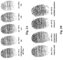
- Figure 2A illustrates the mapping of different putative amino acids including in particular: ornithyne/MH + -NH 3 (at m/z 116.2); Val (at m/z 118.2); Lys (at m/z 147.2); and His (at m/z 156.2).
- Figure 2B illustrates the mapping of a variety of different endogenous fatty acids and vitamins including in particular: Caproic acid (at m/z 117.0), dehydrated Cholesterol (at m/z 369.2), Vitamin E (at m/z 431.2) and sodiated squalene (at m/z 433.2).
- putative endogenous diacylglycerols were mapped including DG C 36 H 70 O 5 (at m/z 583.6), DG C 38 H 68 O 5 (at m/z 605.6), DG C 39 H 72 O 5 (at m/z 621.6) and DG C 43 H 74 O 5 (at m/z 671.6) as shown in figure 2C .
- the dry wet method was probed for its ability to provide high intensity signals and high quality images of fingermarks in a comparison with the conventional matrix spray coating.
- the comparison was made for three donors who were previously classified as good, average and bad secretors in order to evaluate robustness of the method.
- the classification which generally applies to all the individuals, refers to the different ability to secret sweat which makes the ridge pattern detail of fingermarks more or less clear.
- the same donor's fingermark was divided in halves with one half prepared using the dry wet method and the other half by using the spray coating method. As a representative sample, the ion species were imaged at m/z 283.2, 311.2 and 668.6.
- Ion species imaged in figure 3 were subjected to high resolution mass spectrometry profiling to ascertain their identity.
- the species at m/z 283.3, 311.3 and 668.6 were tentatively identified by High Mass Accuracy measurements as oleic acid, eicosenoic acid and amino-octadecanoic acid with mass accuracies of 1.1 ppm, 2.3 ppm and 1.1 ppm respectively.
- MALDI- MS images of this ion are also shown in figure 4c reflecting the average peak intensities observed in the mass spectra of the four replicates and the possibility to detect with constant clarity the 'delta' feature which is one of the minutiae exhibited by the particular fingermark examined.
- the dry-wet method has a crucial feature which makes it altogether superior to the spray-coating method previously employed [ Wolstenholme, R.; Bradshaw, R.; Clench, M.R.; Francese, S. Rapid Commun Mass Spectrom, 2009, 23, 3031 ].
- This existing spray coating method in fact can only be applied to: i) fingermarks previously enhanced (provided that the enhancing method was compatible with the MALDI-MSI analysis) and ii) to fingermarks that were laid flat on an adequate MALDI support.
- Wolstenholme and collaborators used only one surface (aluminium sheets) for depositing and analysing the fingermark and, therefore, the technology could not be proven, at that time, to be helpful in real crime scene investigations.
- the present dry-wet method that employs as enhancing powder an actual MALDI matrix, it is now possible to visually identity fingermarks on crime scene surfaces, lift and analyse them by MALDI-MSI.
- a range of deposition surfaces have been tested to prove feasibility including non-porous surfaces such as metal, glass, plastic, and porous surfaces such as varnished wood and leather.
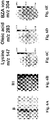
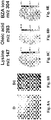
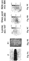
- the series of figures 5A , 6A , 7A , 8A and 9A are photographs of fingermarks on different deposition surfaces prior to dusting with the matrix and the series of figures 5B , 6B , 7B , 8B and 9B are the corresponding photographs post matrix dusting, with each surface comprising the same sample of deposited fingermark.
- the substrate surfaces tested included glass ( figure 5 series), a metal ( figure 6 series), wood ( figure 7 series), plastic ( figure 8 series) and leather ( figure 9 series).
- Figures 5A to 9E confirm that for all of the tested substrate surfaces it is possible to retrieve an image of endogenous and the selected exogenous compounds.
- the dimethylbenzylammonium ion exhibits the best image quality (grade 4), which, amongst other factors, could also be due to its relative amount compared to the two endogenous compounds selected.
- grade 4 the best image quality
- putative lysine and oleic acid overall yield useful images, which could be superimposed using appropriate software to enhance the ridge pattern clarity.
- the matrix was considered in regards to its particles size.
- Four experiments were performed in which, upon dusting with different size matrix particles, the fingermarks of the good donor were submitted to SEM and to MALDI MSI to evaluate the impact of the matrix particle size on the quality of the ridge pattern in the MALDI MS image.
- fingermark prior to submission to SEM and MALDI MSI analyses, fingermark were: spray coated (experiment A), dusted with unground matrix used directly as the manufacturer sells it and solvent sprayed (experiment B), dusted with manually ground matrix and solvent sprayed (experiment C), dusted with mechanically ground and sieved matrix ( ⁇ 38 ⁇ m) followed by solvent spraying (experiment D), dusted with mechanically ground matrix (generating 10-20 ⁇ m particle size according to the manufacturer) followed by solvent spraying (experiment E).
- Figure 10 shows the SEM micrographs of fingermarks spray coated and dusted with different particle size matrix and a MALDI MSI image of the fingermarks treated with the different matrix systems.
- matrix co-crystal formation is mainly expected on the endogenous compound rich ridges of the fingermark. This is in fact observed and matrix co-crystals assemble on "lines" which are effectively the ridges. This is better viewed in Figure 10 D1.
- a decrease in the matrix co-crystals average length is observed along with a general increase in the density of distribution of the matrix co-crystals.
- the matrix co-crystals average length is ⁇ 54 ⁇ m for experiment B ( Figure 10 B2), ⁇ 16 ⁇ m for experiment C ( figure 10 C2), ⁇ 7 ⁇ m for experiment D ( Figure 10 D2) and ⁇ 6 ⁇ m for experiment E ( figure 10 E2).
- the ridge pattern image quality of the dry-wet method treated fingermarks improves as the co-crystal average length decreases.
- the MALDI MSI images of the corresponding fingermarks show a dramatic increase of both the fingermark area coverage when considering the unground matrix dusted and the manually ground matrix dusted fingermarks ( Figure 10 B3 and Figure 10 C3), and of the signal intensity when considering the manually ground matrix dusted and the mechanically ground and sieved dusted matrix fingermarks ( figure 10 C3 and D3).
- Dusting mechanically ground matrix (10-20 ⁇ m) on the fingermark generates very similar results to dusting mechanically ground and sieved matrix in terms of image coverage and intensity ( Figure 10 D3 and E3).
- the spray coated fingermark presents mainly of amorphous species which would explain the generally lower signal intensity compared to the dry wet method as well as a poorer coverage of the ridge pattern.
- MALDI-MSI is capable of detecting both endogenous and exogenous fingermark chemical components in one analysis and simultaneously to providing an image of the fingermark ridge pattern.
- MALDI-MSI can be used for fingermarks deposited on a range of surfaces and in such a way, that recovery from the scene could be integrated easily with current crime scene investigator practice for subsequent instrumental analysis, importantly, remote from the crime scene.
- the novel two step matrix deposition method that has been developed allows evidence to be photographed and exposed to both UV light and fluorescent radiation enabling even clearer images to be obtained. This possibility allows the kind of legal evidence to be generated prior to subsequent MALDI-MSI analysis which, by providing chemical information, potentially adds intelligence to the case under investigation.
Landscapes
- Physics & Mathematics (AREA)
- Life Sciences & Earth Sciences (AREA)
- Health & Medical Sciences (AREA)
- Engineering & Computer Science (AREA)
- Chemical & Material Sciences (AREA)
- Analytical Chemistry (AREA)
- Molecular Biology (AREA)
- General Health & Medical Sciences (AREA)
- Pathology (AREA)
- General Physics & Mathematics (AREA)
- Immunology (AREA)
- Optics & Photonics (AREA)
- Biochemistry (AREA)
- Biomedical Technology (AREA)
- Spectroscopy & Molecular Physics (AREA)
- Biophysics (AREA)
- Bioinformatics & Computational Biology (AREA)
- Bioinformatics & Cheminformatics (AREA)
- Public Health (AREA)
- Animal Behavior & Ethology (AREA)
- Surgery (AREA)
- Medical Informatics (AREA)
- Heart & Thoracic Surgery (AREA)
- Veterinary Medicine (AREA)
- Hematology (AREA)
- Urology & Nephrology (AREA)
- Plasma & Fusion (AREA)
- Human Computer Interaction (AREA)
- Multimedia (AREA)
- Theoretical Computer Science (AREA)
- Proteomics, Peptides & Aminoacids (AREA)
- Microbiology (AREA)
- Cell Biology (AREA)
- Food Science & Technology (AREA)
- Medicinal Chemistry (AREA)
- Biotechnology (AREA)
- Other Investigation Or Analysis Of Materials By Electrical Means (AREA)
Description
- The present invention relates to a method of preparing a sample for matrix assisted laser desorption ionisation mass spectrometry imaging (MALDI-MSI) and in particular, although not exclusively, a method of generating an image of the sample using mass spectrometry.
- Matrix deposition is a crucial aspect in MALDI-MSI. Extensive research has been undertaken over the years to improve this preparation step with the aim of minimising analyte delocalisation while maximising the available resolution and analyte extraction from the tissue section. The selection of the most appropriate matrix and solvent combination impacts on the analyte extraction (sensitivity); nonetheless it is the choice of the deposition method that has, overall, a greater impact.
- Minimisation of analyte delocalisation can be achieved not only by using appropriate sample pre-treatments but also by selecting a deposition method which either confines the matrix to a very small area (microspotting for example) or spraying the matrix either robotically or manually. Contactless microspotting can currently be achieved by piezo-dispensing the solution using a chemical ink-jet printer [1. Sloane, A.J.; Duff, J.L.; Wilson, N.L.; Gandhi, P.S.; Hill, C.J.; Hopwood, F.G.; Smith, P.E.; Thomas, M.L.; Cole, R.A.; Packer, N.H.; Breen, E.J.; Cooley, P. W.; Wallace, D.B.; Williams, K.L.; Gooley, A.A. Mol Cell Proteomics 2002, 1, 490-499] or through acoustic ejection first introduced as a prototype by Aerni and collaborators [Aerni, H.R.; Cornett, D.S.; Caprioli, R.M.; Anal Chem, 2006, 78, 827-834] and then improved and commercialised by Labcyte as the Portrait™ 630. Here both matrix concentration and solvents used to dissolve the matrix have an important role in determining the size of the crystallized droplet and the spot to spot distance, which determine the final image resolution.
WO 2010/007443 describes a dual analysis technique that encompasses a more conventional non-imaging MALDI-MS chemical profile analysis but which also considers the affects of matrix particle size. A first stage is directed to obtaining an image of a deposited fingermark using a lifting agent and applying a developing agent. An image of the ridge pattern of the treated fingermark is then obtained using a solid-state or an optical fingerprint reader. A second stage chemical profiling analysis is then undertaken using MALDI MS involving dusting of the fingerprint sample with a silica nanoparticle powder using a commercial magnetic wand. Another example of prior art fingerprint preparation is disclosed inUS4708882 . - Pneumatically assisted matrix spray-coating, for which the first example was reported by Caprioli and collaborators [Caprioli, R.M.; Farmer, T.B.; Gile, J. Anal Chem, 1997, 69, 4751] requires the optimisation of the distance between the sample and the spray nozzle, deposition speed and number of coating layers (this generally depends on matrix solvent combinations).
- The matrix can also be applied through a matrix aerosol created by vibrational vaporization that requires a number of parameters be monitored and optimised such as deposition time, intervals, matrix layer thickness, wetness and drying rate. This deposition system is claimed to generate a droplet size of 20µm in diameter typically covering an area on the tissue smaller than 50µm in diameter. This and the ability to control the relevant deposition parameters allow images with excellent spatial resolution and high ion intensity to be achieved.
- For manual spray coating, it is important to find optimal conditions for the spray distance, number of cycles and elapsed time between consecutive spraying. Whichever the deposition method, it has an influence on the crystal formation in terms of speed (slow formation has been observed to yield higher intensity signal) and size, with the latter having a direct effect on the image resolution that will then only be limited by the laser spot diameter if oversampling is not performed. The deposition method can also affect the signal intensity as it can impact on the analyte extraction-matrix co-crystallisation. In terms of sample pre-treatment, immersion techniques involving dipping the mounted section in a bath of matrix solution prior to matrix deposition have been observed to increase the signal without delocalising the analyte [Stoeckli, M.; Staab, D.; Staufenbiel, M.; Wiederhold, K.H.; Signor L. Anal Biochem, 2002, 311, 33-39; Francese, S.; Lambardi D.; Mastrobuoni, G.; la Marca, G.; Moneti, G.; Turillazzi, S. JAm Soc Mass Spectrom, 2009, 20, 112-123]. Independently from the mode of application, the most widely used deposition methods involve the use of 'wet' matrix, i.e. dissolved in an appropriate solvent combination prior to deposition. Sugiura and collaborators used this principle in a two step procedure to deposit matrix over a tissue section [Sugiura, Y.; Shimma, S.; Setou, M. Anal Chem, 2006, 78, 8227]. The method named 'spray droplet', involved spray-coating the matrix in humid conditions and in low concentration (the seeding step) followed by microspotting matrix at a higher concentration using a chemical ink-jet printer. The control over iterative spray coating cycles and humidity made this procedure very efficient in terms of ion population (m/z range 4-20kDa) and ion intensity compared to the single step conventional deposition method.
- Although not fully embraced by the MALDI-MSI community, a number of other deposition methods have been shown to improve the ion intensity and to yield more populated mass spectra. These methods are based on the use of 'dry' matrix, which in most cases acts as a seeding agent. Aerni and co-workers were the first to report the use of ground matrix powder being brushed off a sample surface and subsequent matrix microspotting using an acoustic ejector [Aerni, H.R.; Cornett, D.S.; Caprioli, R.M.; Anal Chem, 2006, 78, 827-834]. Powder seeding prior to matrix microspotting yielded more homogenous matrix spots and smaller crystals and although it did not produce any ion signal without subsequent spotting, seeding greatly improved the total ion count (TIC) (m/z 5-8 kDa).
- In 2007 Hankin and co-workers demonstrated that matrix powder could indeed be used to generate ions and ion images by sublimation and compared this method with conventional electrospray deposition [Hankin, J.A.; Barkley, R.M.; Murphy, R.C. J Am Soc Mass Spectrom, 2007, 18, 1646 -1652]. In the new method, the dry matrix was sublimed under vacuum and required the optimisation of a range of parameters including pressure, condenser temperature, amount of matrix, heat applied to the matrix and sublimation time.
- Although the method required laborious fine-tuning of all these parameters, it yielded a homogeneous coating and higher intensity phospholipid signals and corresponding distribution maps in sections of mouse brain. In 2008 Puolitaival and co-workers published the use of a solvent-free matrix dry coating method that allowed detection and mapping of phospholipids with a 30µm to 100µm lateral resolution [Puolitaival, S.M.; Burnum, K.E.; Cornett, D.S.; Caprioli R.M. JAm Soc Mass Spectrom, 2008, 19, 882-886]. In their work, the matrix was simply finely ground using a mortar and pestle and applied by using a 20µm sieve. Although they did not directly compare the method with that of Hankin and collaborators, a comparison was made with a spray-coating method using a TLC nebuliser. Whereas the quality of the molecular images and the phospholipid localisation were very similar to those achieved with the spray-coating, the authors claimed simplicity of sample preparation and minimisation of the analyte delocalisation as advantages of their method over the classical one. This method has been successfully used later to map and quantify small molecules by Goodwin and collaborators [Goodwin, R.J.; MacIntyre, L.; Watson, D. G.; Scullion S. P.; Pitt A. R.; Rapid Commun. Mass Spectrom, 2010; 24: 1682-1686; Goodwin, R.J.; Scullion, P.; MacIntyre, L.; Watson, D.G.; Pitt, A.R. Anal Chem, 2010, 1751-61].
- The very nature of latent fingermarks, being fingermarks with low sample volume and invisible to the naked eye, means that they cannot be analysed via MALDI-MSI without the fingermark being in some way enhanced initially. If conventional processes are to be taken as suitable starting points, the enhancement process would need to be compatible with the MALDI-MSI analysis and the fingermark recovered and laid on a flat and thin surface (less than 200µm - typically suitable for MALDI-MSI analysis) for instrumental analysis.
- Currently employed enhancement methods can be categorised as optical, physical, physico-chemical or chemical, commonly used examples of each being UV light, black powder, cyanoacrylate fuming and ninhydrin, respectively. The Home Office Scientific Development Branch (HOSDB) publish the Manual of Fingerprint Development Techniques based on their research and testing. [Bowman, V.; Manual of fingerprint development techniques, 2nd ed.; Police Scientific Development Branch, Home Office, Sandridge, UK, 2004]. The manual details the techniques that have been approved for operational use and the order in which they should be applied for a specific set of conditions (primarily surface type and contact with water). The processing flow charts are organised at the first level by the type of surface on which the fingermark is deposited: smooth non-porous, rough non-porous, paper and cardboard, plastics, metal, raw wood, adhesive coated surfaces etc. This necessarily means that the choice of enhancement technique depends largely on the type of deposition surface rather than the chemical composition of the fingermark in question.
- What is required is a method of preparing a MALDI-MSI sample suitable for introduction into a MALDI mass spectrometer that is largely independent of the surface or substrate material upon which the sample is deposited. Additionally, there is a need for an improved analytical method for recovering fingermarks from a crime scene and enabling crime scene-remote (laboratory) analysis. Importantly, the sample recovery and preparation method must preserve the fingermark characteristics to enable remote analysis.
- Accordingly, the inventors provide a method of preparing a mass spectrometry imaging sample via a two-step sample preparation procedure, referred to herein as the 'dry-wet method'. Using the two-step preparation procedure of powdering and then spraying a matrix soluble solvent on to the fingermark for efficient analyte extraction and co-crystallisation, a superior procedure is achieved over the pneumatically assisted matrix spray coating in particular for fingermark analysis by MALDI-MSI. Moreover, the present dry-wet method has proved to be reproducible and advantageous over conventional spray coating deposition, in terms of preparation time and recording of the evidence prior to MALDI-MSI analysis.
- The present method of preparing the sample allows fingermark recovery from a deposition surface whilst preserving the sample integrity and characteristics to enable an accurate image and chemical information to be obtained. When trialled on fingermarks deposited on aluminium surfaces, the present method is superior to the conventional spray coating method in terms of quality of the fingermark mass spectrometric image and ion intensity. The present method is also, to a large extent, insensitive to the substrate material upon which the fingermark/sample is deposited. In particular, the dry-wet method allows images of latent fingermarks to be obtained after recovery from a variety of different surfaces including porous and non-porous surfaces such as, by way of example only, glass, metal, plastic, wood and leather.
- Additionally, the inventors have demonstrated that ungroomed fingermarks (fingermarks with very low amounts of sebaceous compounds) can be analysed to obtain both chemical information and good quality ridge pattern images. The present method therefore is highly sensitive and provides a versatile MALDI-MSI tool that may be integrated with standard operational procedures in the forensic analysis of latent fingermarks. The dry-wet method bridges the gaps between visualizing and recovering a latent fingermark at the crime scene and analysing it by MALDI-MSI as the matrix acts as a dusting (and therefore) enhancing agent.
- According to a first aspect of the present invention there is provided a method of preparing a matrix assisted laser desorption ionisation mass spectrometry imaging (MALDI-MSI) fingerprint sample as defined in claim 1, and its dependent claims.
- The present method is particularly suitable for MALDI-MSI analysis of fingermarks that have been lifted from a substrate surface upon which the fingermark has been initially deposited using an adhesive backed strip, pad or tape. In particular, the strip, pad or tape may be crime scene investigation (CSI) tape.
- Preferably, the step of spraying the dusted sample comprises spraying the sample adhered to the strip, pad or tape with the solvent. Preferably, the method further comprises introducing the lifted sample on the strip, pad or tape into a MALDI mass spectrometer with imaging capabilities so as to obtain an image of the sample by MALDI-MSI. Preferably the strip, pad or tape is adhered to a MALDI target plate prior to introduction into the MALDI mass spectrometer.
- Preferably, the particle size of the powered matrix is in the range 5µm to 120µm. More preferably, the powered matrix is in the range 10µm to 30µm. Preferably, the matrix is an absorber of UV radiation and/or is fluorescent. It has been identified that a decrease in the particle size of the matrix employed leads to an increase of the mass spectrometric fingermark image quality.
- Preferably, the step of spraying the dusted sample comprises spraying the solvent onto the dusted sample (and optionally the lifted sample on the adhesive backed strip, pad or tape), as a fine mist. Preferably, the method comprises three successive spray applications of the fine mist onto the sample. Preferably, the method comprises creating the fine mist using an automatic pneumatic sprayer device. Preferably, the spray speed is in the range 1 to 3 µL/min and preferably 2 µL/min.
- According to a second aspect of the present invention there is provided a method, as defined in claim 11, of matrix assisted laser desorption ionisation mass spectrometry imaging (MALDI-MSI) comprising preparing a fingerprint sample according to claims 1 to 10.
- According to a third aspect of the present invention there is provided in claim 12 a method of creating a plurality of images of a fingermark sample according to any one of claims 1 to 10.
- Preferably, the method further comprises obtaining a fluorescent image of the prepared sample using a fluorescent microscope. More preferably, the method further comprises obtaining a UV image of the prepared sample using a UV light source and a still and/or video camera.
- A specific implementation of the present invention will now be described, by way of example only and with reference to the accompanying figures in which:
-
figure 1A is a non-enhanced visible light photograph of a latent fingermark present at a simulated crime scene; -
figure 1B illustrates the fingermark offigure 1A dusted with a MALDI matrix (α-CHCA); -
figure 1C illustrates a UV photograph of the MALDI matrix dusted fingermark offigure 1B ; -
figure 1D is fluorescent microscopy image of the region of the fingermark as indicated infigure 1C ; -
figure 1E is a MALDI-MSI image of the fingermark offigure 1B ; -
figure 2A is a series of MALDI-MS images of endogenous putative amino acids in an ungroomed fingermark prepared and analysed according to the present two-stage process; -
figure 2B is a series of MALDI-MS images of putative endogenous fatty acids and vitamins in the same ungroomed fingermark offigure 2A prepared and analysed according to the present two-stage process; -
figure 2C is a series of MALDI-MS images of endogenous putative diacylglycerols in the same ungroomed fingermark offigure 2A prepared and analysed according to the present two-stage process; -
figure 2D is a series of MALDI-MS images of endogenous putative triacylglycerols in the same ungroomed fingermark offigure 2A prepared and analysed according to the present two-stage process; -
figure 3 illustrated the MALDI MSI analysis of fingermarks prepared with either the Dry-Wet (D-W) or the Spray Coating (S-C) matrix application method. Three types of donors have been selected and three ions were imaged at m/z 283.2, 311.2 and 668.6 both within the fingermarks' halves prepared with the dry-wet method and for the halves prepared with the spray coating matrix application method. -
figure 4 illustrates the superimposition of a α-CHCA ion signal at m/z 190 (a) from four finger marks deposited independently and prepared using the present two-stage preparation method; an exogenous signal at m/z 304 (dimethylbenzylammonium ion) also produced superimposable spectra (b); and detail of the corresponding four MALDI MS images of the ion at m/z 304 is illustrated in (c); -
figure 5A is a photograph of an ungroomed fingermark deposited on a glass beaker; -
figure 5B is a photograph of the fingermark deposited on the glass beaker offigure 5A dusted with the MALDI matrix (α-CHCA); -
figure 5C is a MALDI-MSI image of the fingermark offigure 5B having been lifted from the beaker with an adhesive backed tape and sprayed with an appropriate solvent followed by MALDI-MSI analysis to provide an image of an endogenous amino acid (putative lysine m/z 147); -
figure 5D is a MALDI-MSI image of the fingermark offigure 5B lifted, prepared and analysed in the same manner asfigure 5C to provide an image of an endogenous fatty acid (oleic acid m/z 283); -
figure 5E is a MALDI-MSI image of the fingermark offigure 5B lifted, prepared and analysed in the same manner asfigure 5C to provide an image of an exogenous compound (dimethylbenzylammonium ion m/z 304); -
figure 6A is a photograph of an ungroomed fingermark deposited on a metal knife; -
figure 6B is a photograph of the fingermark deposited on the metal knife offigure 6A dusted with the MALDI matrix (α-CHCA); -
figure 6C is a MALDI-MSI image of the fingermark offigure 6B having been lifted from the metal knife with an adhesive backed tape and sprayed with an appropriate solvent followed by MALDI-MSI analysis to provide an image of an endogenous amino acid (putative lysine m/z 147); -
figure 6D is a MALDI-MSI image of the fingermark offigure 6B lifted, prepared and analysed in the same manner asfigure 6C to provide an image of an endogenous fatty acid (oleic acid m/z 283); -
figure 6E is a MALDI-MSI image of the fingermark offigure 6B lifted, prepared and analysed in the same manner asfigure 6C to provide an image of an exogenous compound (dimethylbenzylammonium ion m/z 304); -
figure 7A is a photograph of an ungroomed fingermark deposited on wood; -
figure 7B is a photograph of the fingermark deposited on the wood offigure 7A dusted with the MALDI matrix (α-CHCA); -
figure 7C is a MALDI-MSI image of the fingermark offigure 7B having been lifted from the wood with an adhesive backed tape and sprayed with an appropriate solvent followed by MALDI-MSI analysis to provide an image of an endogenous amino acid (putative lysine m/z 147); -
figure 7D is a MALDI-MSI image of the fingermark offigure 7B lifted, prepared and analysed in the same manner asfigure 7C to provide an image of an endogenous fatty acid (oleic acid m/z 283); -
figure 7E is a MALDI-MSI image of the fingermark offigure 7B lifted, prepared and analysed in the same manner asfigure 7C to provide an image of an exogenous compound (dimethylbenzylammonium ion m/z 304); -
figure 8A is a photograph of an ungroomed fingermark deposited on plastic; -
figure 8B is a photograph of the fingermark deposited on the plastic offigure 8A dusted with the MALDI matrix (α-CHCA); -
figure 8C is a MALDI-MSI image of the fingermark offigure 8B having been lifted from the plastic with an adhesive backed tape and sprayed with an appropriate solvent followed by MALDI-MSI analysis to provide an image of an endogenous amino acid (putative lysine m/z 147); -
figure 8D is a MALDI-MSI image of the fingermark offigure 8B lifted, prepared and analysed in the same manner asfigure 8C to provide an image of an endogenous fatty acid (oleic acid m/z 283); -
figure 8E is a MALDI-MSI image of the fingermark offigure 8B lifted, prepared and analysed in the same manner asfigure 8C to provide an image of an exogenous compound (dimethylbenzylammonium ion m/z 304); -
figure 9A is a photograph of an ungroomed fingermark deposited on leather; -
figure 9B is a photograph of the fingermark deposited on the leather offigure 9A dusted with the MALDI matrix (α-CHCA); -
figure 9C is a MALDI-MSI image of the fingermark offigure 9B having been lifted from the leather with an adhesive backed tape and sprayed with an appropriate solvent followed by MALDI-MSI analysis to provide an image of an endogenous amino acid (putative lysine m/z 147); -
figure 9D is a MALDI-MSI image of the fingermark offigure 9B lifted, prepared and analysed in the same manner asfigure 9C to provide an image of an endogenous fatty acid (oleic acid m/z 283); -
figure 9E is a MALDI-MS image of the fingermark offigure 9B lifted, prepared and analysed in the same manner asfigure 9C to provide an image of an exogenous compound (dimethylbenzylammonium ion m/z 304); -
figure 10 illustrates the comparative analysis via SEM and MALDI MSI of fingermarks prepared with the spray coat and the dry-wet methods. Panels A1-D1 report a 700x magnification image of the dusted fingermarks with the spray coat (S-C) and the dry-wet (D-W) methods and different particle size (A1= S-C method, B1 = D-W method and unground matrix, C1 = D-W method and manually ground matrix, D1 = D-W method and mechanically ground and sieved matrix). Panels A2-D2 show a 3000x magnification of panels A1-D1. Panels A3-D3 show the corresponding MALDI MS images of distribution on the entire fingermark of the ion at m/z 283.2. - The present MALDI-MSI preparation method comprises a relatively straight forward two-stage procedure involving a first dusting of the sample with powered matrix followed by spraying of the dusted sample with a matrix compatible solvent. Importantly, the present process enables samples with low abundance substances, such as fingermarks, to be analysed by mass spectrometry imaging. The present method is also advantageous in that sample fingermarks can be lifted from the substrate upon which they were deposited allowing subsequent and remote (laboratory) analysis.
- Furthermore, through experimental testing, the inventors have confirmed the enhanced sensitivity of the present method being suitable to generate very detailed and accurate mass spectrometry images for latent fingermarks having low concentrations of endogenous and exogenous constituent compounds deposited on a substrate. Such fingermarks are referred to as 'ungroomed' where 'groomed' fingermarks typically contain higher levels of endogenous sebaceous compounds that result from an individual wiping fingertips over their skin prior to deposition on a substrate. Accordingly, the quality and reproducible results for 'ungroomed' fingermarks confirm the suitability of the present method for actual crime scene analysis where fingermark samples would typically contain minimal material.
- The inventors also demonstrate the advantages of the present method over the conventional spray coat method for the analysis of fingermark samples deposited on a variety of different substrate surfaces having different properties including, in particular, porosity and surface roughness. Example substrates tested include glass, metal, plastic, wood and leather. The results obtained confirm the suitability of the present method to lift fingermarks from different substrate surfaces with little or no loss of image clarity.
- Furthermore, chemical analysis of the lifted fingermark samples is also possible with the present method enabling the identification of both endogenous and exogenous compounds. This is indeed a significant contribution to current forensic analysis as it is now possible to firstly identify and then extract very low quantities of biological samples at a crime scene that may have otherwise been undetected and to obtain both an image and a chemical breakdown of the sample.
- Trifluoroacetic acid (TFA), α-cyano-hydroxycinnamic acid (CHCA) and ALUGRAM ®SIL G/UV254 pre-coated aluminium sheets were purchased from Sigma Aldrich (Poole, UK). Acetone and aceonitrile (ACN) were obtained from Fisher Scientific (Loughborough, UK). MALDI target OPTI spotless inserts were purchased from Applied Biosystems (CA, USA). Double sided conductive carbon tape was obtained from TAAB (Aldermaston, UK). Klenair air spray was obtained from Kenco Ltd (Swindon, UK). Dettol alcohol wipes were purchased from Sheffield branches of Wilkinson's (Worksop, UK). Forensic lifting tape was acquired from TETRA Scene of Crime (http://www.tetrasoc.com/).
- MALDI MS Imaging analyses were conducted using a modified Applied Biosystems API Q-Star Pulsar i hybrid quadrupole time-of-flight (QTOF) instrument. The orthogonal MALDI source has been modified to incorporate a
SPOT 20 kHz Nd:YVO4 solid-state laser (Elforlight Ltd., Daventry, UK), having a wavelength of 355 nm, a pulse duration of 1.5 ns and producing an elliptical spot size of 100 µm × 150 µm. Images were acquired using 'oMALDI Server 5.1' software supplied by MDS Sciex (Concord, Ontario, Canada). High mass accuracy MALDI MS spectra were acquired directly from fingermarks using a SYNAPT G2-HDMSTM system incorporating ion mobility (Waters, Manchester, UK) operating in resolution mode ("V"). The laser was operated at 1kHz, which equates to a laser energy of approximately 10.5µJ. Latent fingermarks were sprayed using a 'SunCollect' autospraying system obtained from SunchromGmbH (Friedrichsdorf, Germany). All Scanning Electron Microscopy (SEM) images were obtained using a FEI NOVA nanoSEM 200 (FEI, The Netherlands) fitted with a HELIX bullet/ detector system to enable electron imaging in variable pressure (VP) mode. - Ungroomed fingermarks were prepared by preliminarily cleaning hands with alcohol wipes and carrying on normal work activities for a period of 15 minutes before deposition. The marks were laid onto the ALUGRAM ® SIL G/UV254 pre-coated aluminum sheets after scraping off the silica with acetone. Ungroomed fingermarks were also deposited on a knife (metal, non porous surface), an air tight plastic container (non porous surface) a laboratory beaker (glass, non porous surface), a wood tray (varnished wood) and a belt (leather, porous surface).
- Using a pestle and mortar, α-CHCA was ground into a very fine powder, which was used to dust the fingermark using a zephyr brush. Approximately 40 mg to 70 mg of matrix was found suitable for dusting a fingermark of approximate size 2 cm x 1.3 cm. Excess powder was then removed using a Klenair air sprayer. The aluminium sheet with the dusted fingermark on it was then stuck onto a MALDI spotless 'OPTI-Tof™' insert using double sided conductive carbon tape and sprayed with 3 layers of a 70:30 ACN/0.5% TFA solution using the SunCollect auto-sprayer, at a speed of 2 µL/min. Fingermarks deposited on the different surfaces employed were dusted and lifted with crime scene investigation (CSI) tape. The tape was then stuck using double sided conductive carbon tape onto a MALDI spotless insert and then sprayed in the same fashion with 3 layers of 70:30 ACN/0.1% solution.
- When comparing the Dry-Wet method with the conventional Spray Coating matrix application method, ungroomed fingermarks obtained from 3 donors (a good, intermediate and poor secretor) were laid onto aluminum plates. For each donor fingermarks were divided in halves. When using the dry-wet method, one half was dusted with mechanically ground α-CHCA using a zephyr brush and excess powder was blown off prior to auto-spaying the sample with 5 layers of a 70:30 ACN/0.5% TFA solution at a speed of 5 µL/min. The other half of each donors' fingermark was prepared by auto- spraying with 4 layers of a 5 mg/ml α-CHCA in a 70:30 ACN/0.1% solution using the SunCollect auto-sprayer at a speed of 1 µL/min, 2 µL/min, 2 µL/min and 2 µL/min. Both methods had been previously optimised to provide the best quality image of the fingermark.
- For comparative analyses via SEM and MALDI MSI, the fingermarks were dusted using a zephyr brush with either unground α-CHCA, manually ground α-CHCA (ground into a fine powder using a pestle and mortar) or mechanically ground α-CHCA using a Herzog vibrating disc mill (Herzog, Osnabrück, Germany) and sieved through a 38 µm sieve or a PM100 Planetary Ball Mill (Retsch Ltd Castleford, UK). Excess powder was then removed using a Klenair air sprayer. Fingermarks were sprayed with 5 layers of a 70:30 solution of ACN/0.5% TFA, at a rate of 5 µL/min prior to SEM and MALDI MSI analyses.
- MALDI-TOF-MSI analyses were performed at a resolution of 150 µm x 150 µm using 'continuous raster imaging' at a laser repetition rate of 5 kHz. This differs from the classic 'stop and go fashion' of MALDI-MSI, as the laser moves continuously in rows across the sample surface allowing rapid acquisition at a high image resolution. Images of a whole fingermark were obtained in around 1
h 20 min. Data processing was performed using BioMap 3.7.5 software (Novartis, Basel, Switzerland). High mass accuracy spectra were recorded directly from fingermarks prepared by the dry-wet method. Spectra were initially calibrated using a standard solution of polyethylene glycol (PEG), using the PEG signal at a m/z of 525.2886 as a lock mass and centroided prior to generation of an accurate peak list. The centroided m/z values were then submitted into the Lipidmap database (http://www.lipidmaps.org) using a mass tolerance of 0.001 Da in order to generate a list of probable structures. - Digital images were obtained using a Fujifilm IS Pro CCD camera, with a 50 mm UV lens. The size of the image obtained was 4256 x 2848 pixels.
- UV-Vis and fluorescent images of fingermarks, prepared with the dry-wet method, were obtained using: (i) a video spectral comparator (VSC4CX, Foster & Freeman, Evesham, UK) at an excitation wavelength of 365 nm, (ii) using an Olympus BX51 fluorescent microscope equipped with Cell-D software (Olympus), using U-MNU2 filter (excitation 360-370 nm, 400 nm dichromatic, emission 420 nm) and (x4) objective.
- All SEM images were acquired using an accelerating voltage of 5KV, a spot size between 3-4 and at a working distance of 4-5 mm. Samples were examined at a magnification of x700 and x3000.
- Specifically, a novel two step matrix deposition method (referred to herein as the 'dry-wet' method) has been devised and applied to the analysis of latent fingermarks. The first step involves the application of finely ground MALDI matrix to the fingermark, in this case α-cyano 4 hydroxycinnamic acid (α-CHCA). Molecular sieves, as indicated by Puolitaval and co-workers [Puolitaival, S.M.; Burnum, K.E.; Cornett, D.S.; Caprioli R.M. J Am Soc Mass Spectrom, 2008, 19, 882-886], were initially tested but their use was later dismissed as filtering the matrix in such a way proved to be very laborious and time consuming, with no significant improvement in the results obtained as well as being impractical for vertical surfaces. Additionally, dusting fingermarks is one of the conventional methodologies used by forensic investigators to develop marks at the crime scene, therefore, if introduced, this would be a familiar procedure for CSI officers. The second step involves the spraying of the matrix dissolution solvent.
- In the first step of the method, the provided matrix acts as an enhancer and after blowing away excess, a photograph can be taken, as show in
figure 1A . This may then be sent to a laboratory for scanning and comparison using police databases.Figure 1A is a photograph of the fingermark prior to matrix dusting. After matrix application, a very clear ridge pattern is visualised (as illustrated infigure 1B ) where the minutiae (local features of the ridge pattern) can be very clearly observed. According to the HOSDB grading system, where the proportion of the developed fingermark with clear ridge detail is estimated and a score is assigned to the fingermark from 0 to 4, the fingermark could be classified as grade 4 (full development - whole fingermark in clear continuous ridges). Another interesting aspect to this protocol is due to the α-CHCA matrix having a fluorescent property. Accordingly, fluorescent optical images can be obtained either as a whole as shown infigure 1C , using a simple device like a document examination instrument operated at a wavelength of 365 nm, or as small areas, as shown in figure ID, when using a fluorescent microscope operated as described in the methods. The magnification employed on the fluorescent microscope (40X) allowed the fingermark to be inspected with a high level of detail. - The same fingermark may then be subjected to MALDI-MSI allowing the ridge pattern to be imaged simultaneously to potentially obtaining chemical information from the endogenous and exogenous compounds present in the fingermark as shown in
figure 1E . The potential to acquire two different optical images prior to MALDI-MSI analysis is very important as it enables evidence to be provided quickly and according to existing and accepted forensic procedures. The concept of a multipurpose dusting agent has been reported by Rowell and co-workers [Rowell, F.; Hudson, K.; Seviour, J. Analyst, 2009, 134,701-7]. These researchers employed a hydrophobic silica dusting agent containing carbon black which enabled both visualization and SALDI TOF MS analysis of the latent fingermarks. The chemical was a particularly good SALDI matrix targeting a range of drugs. Although chemical information could be obtained, SALDI TOF MS was not shown to provide a chemical image of the actual fingermark to complement the optical one. The dry-wet method allows the qualitative recovery of chemical information embedded in multiple MS images. - The dry-wet method devised allows images of fingermarks to be obtained showing detection of many molecular species, comparable to those previously reported by spray-coating the matrix [Wolstenholme, R.; Bradshaw, R.; Clench, M.R.; Francese, S. Rapid Commun Mass Spectrom, 2009, 23, 3031].
Figure 2A illustrates the mapping of different putative amino acids including in particular: ornithyne/MH+-NH3 (at m/z 116.2); Val (at m/z 118.2); Lys (at m/z 147.2); and His (at m/z 156.2).Figure 2B illustrates the mapping of a variety of different endogenous fatty acids and vitamins including in particular: Caproic acid (at m/z 117.0), dehydrated Cholesterol (at m/z 369.2), Vitamin E (at m/z 431.2) and sodiated squalene (at m/z 433.2). Furthermore, putative endogenous diacylglycerols were mapped including DG C36H70O5 (at m/z 583.6), DG C38H68O5 (at m/z 605.6), DG C39H72O5 (at m/z 621.6) and DG C43H74O5 (at m/z 671.6) as shown infigure 2C . Furthermore, and as shown infigure 2D a range of endogenous putative tricylglycerols were mapped from ungroomed fingermarks including in particular: triacyl-sn-glycerol (at m/z 174.0), TG C58H100O6 (at m/z 893.8) and TG C65H122O6 (at m/z 999.8). This is a particularly important advantage of the present method as it shows the versatility of MALDI-MSI compared to other currently used forensic technologies that need to be selected according to the molecular target. - When the particular distribution of a certain species is of interest (for example illicit substances) on an absolute scale or in relation to other substances, images would need to be normalised and the same contrast/brightness should be applied. Where the aim is obtaining an image that shows clear ridge pattern details for a fingermark area that is as large as possible, presence of a particular compound, rather than relative amounts, can be detected in an image by skipping the normalisation process if this reduces the number of minutiae retrievable.
- The dry wet method was probed for its ability to provide high intensity signals and high quality images of fingermarks in a comparison with the conventional matrix spray coating. The comparison was made for three donors who were previously classified as good, average and bad secretors in order to evaluate robustness of the method. The classification, which generally applies to all the individuals, refers to the different ability to secret sweat which makes the ridge pattern detail of fingermarks more or less clear. In order to keep the experimental variables at a minimum level, the same donor's fingermark was divided in halves with one half prepared using the dry wet method and the other half by using the spray coating method. As a representative sample, the ion species were imaged at m/z 283.2, 311.2 and 668.6. The images were normalised against the total ion current; this does not always produce the best quality of the ridge detail. However, normalisation of the two differentially prepared fingermarks halves enables a more reliable comparison and observation of the distribution of the selected endogenous species. As it can be observed in
figure 3 , the dry-wet method generally generates higher quality ridge detail images in terms of area/minutiae covered. As the fingermarks were rubbed together prior to deposition, an even deposition of the endogenous compounds resulting in a uniform image of distribution was expected; the spray coat method appears to generate a much more discontinuous ridge pattern and a lot lower ion signal uniformity. The ion intensity was also generally higher when fingermarks were prepared using the dry wet method with the exception of the bad donor. However, even in this case, in terms of minutiae retrievable, relatively to the species imaged, the dry wet method is slightly better placed than the spray coat method. Overall these results are in agreement with previous work published by Aerni and collaborators [Aerni, H.R.; Cornett, D.S.; Caprioli, R.M.; Anal. Chem., 2006, 78, 827-834]. In this work, the dusting technique was applied in a different way to the present work, as it was employed on biological tissues and was used as a seeding step to which matrix solution application followed either by spray coating or micro-spotting. However, in both cases, in agreement with the results shown here, a better homogeneity of matrix deposition over the tissue, as well as an increased ion signal intensity were demonstrated. - Ion species imaged in
figure 3 were subjected to high resolution mass spectrometry profiling to ascertain their identity. The species at m/z 283.3, 311.3 and 668.6 were tentatively identified by High Mass Accuracy measurements as oleic acid, eicosenoic acid and amino-octadecanoic acid with mass accuracies of 1.1 ppm, 2.3 ppm and 1.1 ppm respectively. - Referring to
figure 4 , the reproducibility of the dry-wet method was also tested by imaging four fingermarks from the same donor. Average spectra were exported from Biomap to mMass, an open source multifunctional mass spectrometry software [Strohalm, M.; Kavan, D.; Novak, P.; Volny, M.; Havlicek, V. Anal Chem, 2010, 82, 4648-4651; Strohalm, M.; Hassman, M.; Košata, B.; Kodíček, M. Rapid Commun Mass Spectrom, 2008, 22, 905-908].Figure 4a reports the four mass spectra generated for the α-CHCA matrix peak at m/z 190. Spectra were not normalised. The ion peak signals are almost superimposable showing reproducibility in the application of the matrix. The lack of a perfect superimposition is likely due to the impossibility of obtaining four identical fingermark depositions as the pressure that the finger exerts on the surface will be inevitably slightly different. Before the deposition of ungroomed fingermarks, hands and fingers were wiped with alcohol wipes known to contain an antiseptic, dimethylbenzylammonium ion, which has previously been identified by these and other authors [Bradshaw, R.; Wolstenholme, R.; Clench, M.R.; Blackledge, R.D.; Ferguson L.; Francese, S. Rapid Commun Mass Spectrom, 2011, 25, 415-422; Ferrer, I.; Furlong. E. Environ Sci Technol, 2001, 35, 2583] and observed to generate very good mass spectral images [Bradshaw, R.; Wolstenholme, R.; Clench, M.R.; Blackledge, R.D.; Ferguson L.; Francese, S. Rapid Commun Mass Spectrom, 2011, 25, 415-422]. Dimethylbenzylammonium ion mass spectra are therefore also reported as shown infigure 4b exhibiting a similar degree of ion intensity overlapping to that observed for the matrix. MALDI- MS images of this ion are also shown infigure 4c reflecting the average peak intensities observed in the mass spectra of the four replicates and the possibility to detect with constant clarity the 'delta' feature which is one of the minutiae exhibited by the particular fingermark examined. - Most importantly, the dry-wet method has a crucial feature which makes it altogether superior to the spray-coating method previously employed [Wolstenholme, R.; Bradshaw, R.; Clench, M.R.; Francese, S. Rapid Commun Mass Spectrom, 2009, 23, 3031]. This existing spray coating method in fact can only be applied to: i) fingermarks previously enhanced (provided that the enhancing method was compatible with the MALDI-MSI analysis) and ii) to fingermarks that were laid flat on an adequate MALDI support. Wolstenholme and collaborators used only one surface (aluminium sheets) for depositing and analysing the fingermark and, therefore, the technology could not be proven, at that time, to be helpful in real crime scene investigations. However, with the present dry-wet method that employs as enhancing powder an actual MALDI matrix, it is now possible to visually identity fingermarks on crime scene surfaces, lift and analyse them by MALDI-MSI.
- A range of deposition surfaces have been tested to prove feasibility including non-porous surfaces such as metal, glass, plastic, and porous surfaces such as varnished wood and leather. The series of
figures 5A ,6A ,7A ,8A and9A are photographs of fingermarks on different deposition surfaces prior to dusting with the matrix and the series offigures 5B ,6B ,7B ,8B and9B are the corresponding photographs post matrix dusting, with each surface comprising the same sample of deposited fingermark. The substrate surfaces tested included glass (figure 5 series), a metal (figure 6 series), wood (figure 7 series), plastic (figure 8 series) and leather (figure 9 series). In particular, images of an endogenous aminoacid (putative Lysine m/z 147), an endogenous fatty acid (oleic acid m/z 283) and an exogenous compound (dimethylbenzylammonium ion m/z 304) were obtained as shown infigures 5 to 9 , series C, D and E, respectively for each substrate. It is accepted that fingermarks on porous surfaces are more problematic to visualise than those on non-porous ones. As proving feasibility of the method is the objective, all the images are reported with differing contrast and brightness to enhance the ridge detail features in each case. This is how forensic investigators would treat the fingermark image for comparison as their goal is to obtain as many clear minutiae as possible.Figures 5A to 9E confirm that for all of the tested substrate surfaces it is possible to retrieve an image of endogenous and the selected exogenous compounds. In particular, the dimethylbenzylammonium ion exhibits the best image quality (grade 4), which, amongst other factors, could also be due to its relative amount compared to the two endogenous compounds selected. Although ranging in grade, putative lysine and oleic acid overall yield useful images, which could be superimposed using appropriate software to enhance the ridge pattern clarity. - In order to understand and improve the efficiency of the dry wet method, the matrix was considered in regards to its particles size. Four experiments were performed in which, upon dusting with different size matrix particles, the fingermarks of the good donor were submitted to SEM and to MALDI MSI to evaluate the impact of the matrix particle size on the quality of the ridge pattern in the MALDI MS image. In particular, prior to submission to SEM and MALDI MSI analyses, fingermark were: spray coated (experiment A), dusted with unground matrix used directly as the manufacturer sells it and solvent sprayed (experiment B), dusted with manually ground matrix and solvent sprayed (experiment C), dusted with mechanically ground and sieved matrix (< 38 µm) followed by solvent spraying (experiment D), dusted with mechanically ground matrix (generating 10-20 µm particle size according to the manufacturer) followed by solvent spraying (experiment E).
Figure 10 shows the SEM micrographs of fingermarks spray coated and dusted with different particle size matrix and a MALDI MSI image of the fingermarks treated with the different matrix systems. As the matrix excess is blown off prior to solvent spraying, matrix co-crystal formation is mainly expected on the endogenous compound rich ridges of the fingermark. This is in fact observed and matrix co-crystals assemble on "lines" which are effectively the ridges. This is better viewed inFigure 10 D1. For fingermarks treated with the dry-wet method, as a result of the different matrix powder pre-treatments, a decrease in the matrix co-crystals average length is observed along with a general increase in the density of distribution of the matrix co-crystals. In particular the matrix co-crystals average length is ∼54 µm for experiment B (Figure 10 B2), ∼16 µm for experiment C (figure 10 C2), ∼7 µm for experiment D (Figure 10 D2) and ∼6 µm for experiment E (figure 10 E2). Correspondingly, the ridge pattern image quality of the dry-wet method treated fingermarks improves as the co-crystal average length decreases. It must be noted that, although the image resolution is dictated by the laser spot diameter so that matrix co-crystals with smaller diameter than that of the laser have no impact on it, the image coverage of a species evenly distributed on the given target, improves with decreasing the size of the co-crystals [Crossman, L.; McHugh, N.A.; Hsieh, Y.; Korfmacher, W.A.; Chen, J. Rapid Commun. Mass Spectrom., 2006, 20, 284]. In agreement with the above, the MALDI MSI images of the corresponding fingermarks show a dramatic increase of both the fingermark area coverage when considering the unground matrix dusted and the manually ground matrix dusted fingermarks (Figure 10 B3 andFigure 10 C3), and of the signal intensity when considering the manually ground matrix dusted and the mechanically ground and sieved dusted matrix fingermarks (figure 10 C3 and D3). Dusting mechanically ground matrix (10-20 µm) on the fingermark generates very similar results to dusting mechanically ground and sieved matrix in terms of image coverage and intensity (Figure 10 D3 and E3). The spray coated fingermark presents mainly of amorphous species which would explain the generally lower signal intensity compared to the dry wet method as well as a poorer coverage of the ridge pattern. In these experiments the dry wet method is clearly superior to the spray coat one when the matrix was ground prior to dusting which is likely due to a higher number of crystals and better uniformity of crystallization. In the extreme case when the matrix was not ground, the quality of the image for the spray coated fingermark appears to be slightly better (grade 2) than that of the dry-wet method (grade 1) (Figure 10 A3 andFigure 10 B3). However, in relation to the image shown inFigure 10 B3, it is important to bear in mind that the ability to produce multiple images for the same fingermark allows image overlay and thus provides enhanced coverage of the ridge pattern. Also, as benchtop ball mills are readily available enabling up to 20 g of matrix to be ground to < 20 microns in less than 5 min, the use of unground matrix is conveniently avoidable. Finally, even with the bigger matrix particle size where the spray coat method may produce slightly better images than the dry wet method, because it is inapplicable in the field it is eliminated as a viable matrix deposition method for MALDI MS Imaging analyses. - The demonstrated applicability of the present dry-wet method to recover and analyse latent fingermarks by MALDI-MSI therefore complements the achievements of current forensic methodologies. The existing techniques suggested in the Home Office Scientific Development Branch (HOSDB) manual, are aimed solely at providing an image of the ridges on the fingertip, they are often not appropriate for a wide range of situations and they do not provide any additional information. Only in cases where a clear mark has been deposited will the existing techniques, if used correctly, allow a comparison. However, scene of crime marks are not ideal marks. They may be smudged, as a result of the articulation of the arm while the donor is holding an object, overlaid because of multiple contacts or any number of other actions resulting in ridges that are not sufficiently clear for comparison. In these cases it would be beneficial to have a protocol available, such as MALDI-MSI in combination with the present dry-wet deposition method that is configured to enable visualisation of the ridge pattern, but also provide chemical information which could potentially disclose donor dietary habits or drug use.
- Accordingly, via the present method of sample preparation, MALDI-MSI is capable of detecting both endogenous and exogenous fingermark chemical components in one analysis and simultaneously to providing an image of the fingermark ridge pattern. By preparing the sample appropriately, it is demonstrated that MALDI-MSI can be used for fingermarks deposited on a range of surfaces and in such a way, that recovery from the scene could be integrated easily with current crime scene investigator practice for subsequent instrumental analysis, importantly, remote from the crime scene. The novel two step matrix deposition method that has been developed allows evidence to be photographed and exposed to both UV light and fluorescent radiation enabling even clearer images to be obtained. This possibility allows the kind of legal evidence to be generated prior to subsequent MALDI-MSI analysis which, by providing chemical information, potentially adds intelligence to the case under investigation.
Claims (14)
- A method of preparing a matrix assisted laser desorption ionisation mass spectrometry imaging (MALDI-MSI) fingermark sample suitable to obtain a chemical image of a fingermark via MALDI-MSI, the method comprising:dusting a fingermark sample with a MALDI-MSI matrix powder;then removing excess matrix powder from the fingermark sample such that the matrix powder acts as an enhancer of a ridge pattern of the fingermark sample;spraying the dusted fingermark sample with a solvent in which both the matrix powder and the fingermark sample are soluble to provide co-crystallisation of the matrix powder with analytes of the fingermark sample at the ridges of the ridge pattern to minimise analyte delocalisation.
- The method as claimed in claim 1 further comprising lifting the fingermark sample from a surface upon which the fingermark sample is deposited using an adhesive backed strip, pad or tape.
- The method as claimed in claim 2 wherein the strip, pad or tape is crime scene investigation (CSI) tape.
- The method as claimed in claims 2 or 3 wherein the step of spraying the dusted fingermark sample comprises spraying the fingermark sample adhered to the strip, pad or tape with the solvent.
- The method as claimed in claim 4 further comprising introducing the lifted fingermark sample on the strip, pad or tape into a MALDI mass spectrometer with imaging capabilities.
- The method as claimed in claim 5 further comprising obtaining at least one image of the fingermark sample by MALDI-MSI.
- The method as claimed in any preceding claim wherein a particle size of the powered matrix is in the range 5µm to 120µm.
- The method as claimed in claim 7 wherein a particle size of the powered matrix is in the range 10µm to 30µm.
- The method as claimed in any preceding claim wherein the matrix is an absorber of UV radiation and/or fluorescent.
- The method as claimed in any preceding claim wherein the step of spraying the dusted fingermark sample comprises spraying the solvent onto the dusted fingermark sample as a fine mist.
- A method of matrix assisted laser desorption ionisation mass spectrometry imaging (MALDI-MSI) comprising preparing a fingermark sample according to any one of claims 1 to 10; introducing the fingermark sample into a MALDI imaging mass spectrometer;
delivering a laser beam to the fingermark sample within the mass spectrometer; and
generating the chemical image of the fingermark sample. - A method of creating a plurality of images of a fingermark comprising:preparing a fingermark sample according to any one of claims 1 to 10;introducing the prepared fingermark sample into a MALDI imaging mass spectrometer;obtaining the chemical image of the fingermark using a MALDI mass spectrometer with imaging capabilities.
- The method as claimed in claim 12 further comprising obtaining a fluorescent image of the prepared fingermark sample using a fluorescent microscope.
- The method as claimed in claims 12 or 13 further comprising obtaining a UV image of the prepared fingermark sample using a UV light source and a still and/or video camera.
Applications Claiming Priority (3)
| Application Number | Priority Date | Filing Date | Title |
|---|---|---|---|
| GBGB1104003.7A GB201104003D0 (en) | 2011-03-09 | 2011-03-09 | Improvements to matrix assisted laser deposition ionisation mass spectrometry imaging (maldi-msi) |
| GB201104365A GB2489215B8 (en) | 2011-03-09 | 2011-03-15 | Improvements to matrix assisted laser desorption ionisation mass spectrometry imaging (maldi-msi) |
| PCT/GB2012/050460 WO2012120279A1 (en) | 2011-03-09 | 2012-03-01 | Improvements to matrix assisted laser desorption ionisation mass spectrometry imaging (maldi-msi) |
Publications (2)
| Publication Number | Publication Date |
|---|---|
| EP2684021A1 EP2684021A1 (en) | 2014-01-15 |
| EP2684021B1 true EP2684021B1 (en) | 2020-07-08 |
Family
ID=43923444
Family Applications (1)
| Application Number | Title | Priority Date | Filing Date |
|---|---|---|---|
| EP12712146.5A Active EP2684021B1 (en) | 2011-03-09 | 2012-03-01 | Improvements to matrix assisted laser desorption ionisation mass spectrometry imaging (maldi-msi) |
Country Status (4)
| Country | Link |
|---|---|
| US (1) | US9261438B2 (en) |
| EP (1) | EP2684021B1 (en) |
| GB (2) | GB201104003D0 (en) |
| WO (1) | WO2012120279A1 (en) |
Families Citing this family (7)
| Publication number | Priority date | Publication date | Assignee | Title |
|---|---|---|---|---|
| US9125596B2 (en) * | 2011-09-29 | 2015-09-08 | The Regents Of The University Of California | Nanostructure-initiator mass spectrometry biometrics |
| WO2014162557A1 (en) * | 2013-04-04 | 2014-10-09 | 株式会社島津製作所 | Maldi sample preparation method and sample preparation device |
| DE102013017649B4 (en) * | 2013-10-23 | 2023-08-17 | Leibniz-Institut für Oberflächenmodifizierung e.V. | Method and measuring device for the chemical-analytical examination of an impression of a skin surface |
| US10540536B2 (en) | 2014-08-01 | 2020-01-21 | Katholieke Universiteit Leuven, K.U.Leuven R&D | System for interpretation of image patterns in terms of anatomical or curated patterns |
| CN109447013B (en) * | 2018-11-06 | 2022-02-15 | 重庆工程职业技术学院 | computer user identification device |
| US11139154B2 (en) * | 2019-02-04 | 2021-10-05 | Shimadzu Corporation | MALDI mass spectrometer and matrix observation device |
| CN116907952A (en) * | 2023-08-28 | 2023-10-20 | 香港浸会大学深圳研究院 | Preparation method of matrix-assisted laser desorption ionization mass spectrometry imaging sample |
Family Cites Families (10)
| Publication number | Priority date | Publication date | Assignee | Title |
|---|---|---|---|---|
| US3075852A (en) | 1959-08-12 | 1963-01-29 | Matthew J Bonora | Fingerprinting |
| US4143544A (en) * | 1977-06-22 | 1979-03-13 | General Electric Company | Fingerprinting crystals |
| DE3583498D1 (en) * | 1984-12-27 | 1991-08-22 | Kawasaki Steel Co | METHOD FOR DETECTING FINGERPRINTS BY MEANS OF A LASER. |
| US5099131A (en) | 1990-12-07 | 1992-03-24 | Hughes Aircraft Company | Acquisition and testing of latent fingerprints using upconversion |
| US5382793A (en) * | 1992-03-06 | 1995-01-17 | Hewlett-Packard Company | Laser desorption ionization mass monitor (LDIM) |
| US5364408A (en) * | 1992-09-04 | 1994-11-15 | Laurus Medical Corporation | Endoscopic suture system |
| US6756586B2 (en) * | 2001-10-15 | 2004-06-29 | Vanderbilt University | Methods and apparatus for analyzing biological samples by mass spectrometry |
| US6900061B2 (en) * | 2002-08-23 | 2005-05-31 | Perseptive Biosystems, Inc. | MALDI plate and process for making a MALDI plate |
| US8026328B2 (en) * | 2005-08-09 | 2011-09-27 | University Of Sunderland | Hydrophobic silica particles and methods of making same |
| GB0813208D0 (en) * | 2008-07-18 | 2008-08-27 | Roar Particles Ltd | Methods of determining properties of latent fingerprints |
-
2011
- 2011-03-09 GB GBGB1104003.7A patent/GB201104003D0/en not_active Ceased
- 2011-03-15 GB GB201104365A patent/GB2489215B8/en active Active
-
2012
- 2012-03-01 EP EP12712146.5A patent/EP2684021B1/en active Active
- 2012-03-01 WO PCT/GB2012/050460 patent/WO2012120279A1/en not_active Ceased
- 2012-03-01 US US14/003,813 patent/US9261438B2/en active Active
Non-Patent Citations (1)
| Title |
|---|
| None * |
Also Published As
| Publication number | Publication date |
|---|---|
| GB2489215B (en) | 2013-04-17 |
| GB201104003D0 (en) | 2011-04-20 |
| US9261438B2 (en) | 2016-02-16 |
| GB2489215A (en) | 2012-09-26 |
| GB2489215B8 (en) | 2013-05-01 |
| WO2012120279A1 (en) | 2012-09-13 |
| HK1169854A1 (en) | 2013-02-08 |
| EP2684021A1 (en) | 2014-01-15 |
| GB201104365D0 (en) | 2011-04-27 |
| US20140084151A1 (en) | 2014-03-27 |
Similar Documents
| Publication | Publication Date | Title |
|---|---|---|
| US9261438B2 (en) | Matrix assisted laser desorption ionisation mass spectrometry imaging (MALDI-MSI) | |
| Bouschen et al. | Matrix vapor deposition/recrystallization and dedicated spray preparation for high‐resolution scanning microprobe matrix‐assisted laser desorption/ionization imaging mass spectrometry (SMALDI‐MS) of tissue and single cells | |
| Ferguson et al. | Two-step matrix application for the enhancement and imaging of latent fingermarks | |
| EP2124192B1 (en) | Method for the analysis of tissue sections | |
| Rubakhin et al. | Imaging mass spectrometry: fundamentals and applications to drug discovery | |
| JP5094939B2 (en) | Information acquisition method | |
| Bradshaw et al. | Towards the integration of matrix assisted laser desorption ionisation mass spectrometry imaging into the current fingermark examination workflow | |
| US6995363B2 (en) | Reduction of matrix interference for MALDI mass spectrometry analysis | |
| KR20120027182A (en) | Ion generation using wetted porous material | |
| KR101752656B1 (en) | Sample plate using MALDI-TOF mass spectrometer and manufacturing method of the sample plate | |
| Paine et al. | Visualizing molecular distributions for biomaterials applications with mass spectrometry imaging: a review | |
| JP4636859B2 (en) | Information acquisition method | |
| WO2006109073A2 (en) | Inprovements in or relating to methods and apparatus for analyzing biological samples by mass spectrometry | |
| JP5317054B2 (en) | Mass spectrometry substrate, method for producing the same, and mass spectrometry | |
| Román et al. | Spatially resolved chemical analysis of cicada wings using laser-ablation electrospray ionization (LAESI) imaging mass spectrometry (IMS) | |
| JP2006153493A5 (en) | ||
| US8415615B2 (en) | Information acquisition method | |
| JP4636822B2 (en) | Information acquisition method | |
| Nygren et al. | A cell preparation method allowing subcellular localization of cholesterol and phosphocholine with imaging TOF-SIMS | |
| JP5504282B2 (en) | MALDI mass spectrometry | |
| US9355826B2 (en) | Method for imaging mass analysis using physical vapor deposition of platinum nanoparticles | |
| DE10258674A1 (en) | Manufacturing sample carrier with points for matrix-assisted laser desorption and ionization, forms MALDI matrix points by gas phase sublimation | |
| WO2010007443A2 (en) | Methods of determining properties of latent fingerprints | |
| Gross | Matrix-Assisted Laser Desorption/Ionization | |
| Malmberg et al. | Subcellular localisation of cholesterol and phosphocholine with pattern‐recognition‐imaging‐TOF‐SIMS |
Legal Events
| Date | Code | Title | Description |
|---|---|---|---|
| PUAI | Public reference made under article 153(3) epc to a published international application that has entered the european phase |
Free format text: ORIGINAL CODE: 0009012 |
|
| 17P | Request for examination filed |
Effective date: 20131007 |
|
| AK | Designated contracting states |
Kind code of ref document: A1 Designated state(s): AL AT BE BG CH CY CZ DE DK EE ES FI FR GB GR HR HU IE IS IT LI LT LU LV MC MK MT NL NO PL PT RO RS SE SI SK SM TR |
|
| DAX | Request for extension of the european patent (deleted) | ||
| STAA | Information on the status of an ep patent application or granted ep patent |
Free format text: STATUS: EXAMINATION IS IN PROGRESS |
|
| 17Q | First examination report despatched |
Effective date: 20180405 |
|
| GRAP | Despatch of communication of intention to grant a patent |
Free format text: ORIGINAL CODE: EPIDOSNIGR1 |
|
| STAA | Information on the status of an ep patent application or granted ep patent |
Free format text: STATUS: GRANT OF PATENT IS INTENDED |
|
| INTG | Intention to grant announced |
Effective date: 20200225 |
|
| GRAS | Grant fee paid |
Free format text: ORIGINAL CODE: EPIDOSNIGR3 |
|
| GRAA | (expected) grant |
Free format text: ORIGINAL CODE: 0009210 |
|
| STAA | Information on the status of an ep patent application or granted ep patent |
Free format text: STATUS: THE PATENT HAS BEEN GRANTED |
|
| RBV | Designated contracting states (corrected) |
Designated state(s): AL AT BE BG CH CY CZ DE DK EE ES FI FR GR HR HU IE IS IT LI LT LU LV MC MK MT NL NO PL PT RO RS SE SI SK SM TR |
|
| AK | Designated contracting states |
Kind code of ref document: B1 Designated state(s): AL AT BE BG CH CY CZ DE DK EE ES FI FR GR HR HU IE IS IT LI LT LU LV MC MK MT NL NO PL PT RO RS SE SI SK SM TR |
|
| REG | Reference to a national code |
Ref country code: CH Ref legal event code: EP Ref country code: AT Ref legal event code: REF Ref document number: 1288968 Country of ref document: AT Kind code of ref document: T Effective date: 20200715 |
|
| REG | Reference to a national code |
Ref country code: DE Ref legal event code: R096 Ref document number: 602012071118 Country of ref document: DE |
|
| REG | Reference to a national code |
Ref country code: IE Ref legal event code: FG4D |
|
| REG | Reference to a national code |
Ref country code: LT Ref legal event code: MG4D |
|
| REG | Reference to a national code |
Ref country code: AT Ref legal event code: MK05 Ref document number: 1288968 Country of ref document: AT Kind code of ref document: T Effective date: 20200708 |
|
| REG | Reference to a national code |
Ref country code: NL Ref legal event code: MP Effective date: 20200708 |
|
| PG25 | Lapsed in a contracting state [announced via postgrant information from national office to epo] |
Ref country code: GR Free format text: LAPSE BECAUSE OF FAILURE TO SUBMIT A TRANSLATION OF THE DESCRIPTION OR TO PAY THE FEE WITHIN THE PRESCRIBED TIME-LIMIT Effective date: 20201009 Ref country code: ES Free format text: LAPSE BECAUSE OF FAILURE TO SUBMIT A TRANSLATION OF THE DESCRIPTION OR TO PAY THE FEE WITHIN THE PRESCRIBED TIME-LIMIT Effective date: 20200708 Ref country code: BG Free format text: LAPSE BECAUSE OF FAILURE TO SUBMIT A TRANSLATION OF THE DESCRIPTION OR TO PAY THE FEE WITHIN THE PRESCRIBED TIME-LIMIT Effective date: 20201008 Ref country code: SE Free format text: LAPSE BECAUSE OF FAILURE TO SUBMIT A TRANSLATION OF THE DESCRIPTION OR TO PAY THE FEE WITHIN THE PRESCRIBED TIME-LIMIT Effective date: 20200708 Ref country code: HR Free format text: LAPSE BECAUSE OF FAILURE TO SUBMIT A TRANSLATION OF THE DESCRIPTION OR TO PAY THE FEE WITHIN THE PRESCRIBED TIME-LIMIT Effective date: 20200708 Ref country code: PT Free format text: LAPSE BECAUSE OF FAILURE TO SUBMIT A TRANSLATION OF THE DESCRIPTION OR TO PAY THE FEE WITHIN THE PRESCRIBED TIME-LIMIT Effective date: 20201109 Ref country code: LT Free format text: LAPSE BECAUSE OF FAILURE TO SUBMIT A TRANSLATION OF THE DESCRIPTION OR TO PAY THE FEE WITHIN THE PRESCRIBED TIME-LIMIT Effective date: 20200708 Ref country code: FI Free format text: LAPSE BECAUSE OF FAILURE TO SUBMIT A TRANSLATION OF THE DESCRIPTION OR TO PAY THE FEE WITHIN THE PRESCRIBED TIME-LIMIT Effective date: 20200708 Ref country code: AT Free format text: LAPSE BECAUSE OF FAILURE TO SUBMIT A TRANSLATION OF THE DESCRIPTION OR TO PAY THE FEE WITHIN THE PRESCRIBED TIME-LIMIT Effective date: 20200708 Ref country code: NO Free format text: LAPSE BECAUSE OF FAILURE TO SUBMIT A TRANSLATION OF THE DESCRIPTION OR TO PAY THE FEE WITHIN THE PRESCRIBED TIME-LIMIT Effective date: 20201008 |
|
| PG25 | Lapsed in a contracting state [announced via postgrant information from national office to epo] |
Ref country code: RS Free format text: LAPSE BECAUSE OF FAILURE TO SUBMIT A TRANSLATION OF THE DESCRIPTION OR TO PAY THE FEE WITHIN THE PRESCRIBED TIME-LIMIT Effective date: 20200708 Ref country code: LV Free format text: LAPSE BECAUSE OF FAILURE TO SUBMIT A TRANSLATION OF THE DESCRIPTION OR TO PAY THE FEE WITHIN THE PRESCRIBED TIME-LIMIT Effective date: 20200708 Ref country code: PL Free format text: LAPSE BECAUSE OF FAILURE TO SUBMIT A TRANSLATION OF THE DESCRIPTION OR TO PAY THE FEE WITHIN THE PRESCRIBED TIME-LIMIT Effective date: 20200708 Ref country code: IS Free format text: LAPSE BECAUSE OF FAILURE TO SUBMIT A TRANSLATION OF THE DESCRIPTION OR TO PAY THE FEE WITHIN THE PRESCRIBED TIME-LIMIT Effective date: 20201108 |
|
| PG25 | Lapsed in a contracting state [announced via postgrant information from national office to epo] |
Ref country code: NL Free format text: LAPSE BECAUSE OF FAILURE TO SUBMIT A TRANSLATION OF THE DESCRIPTION OR TO PAY THE FEE WITHIN THE PRESCRIBED TIME-LIMIT Effective date: 20200708 |
|
| REG | Reference to a national code |
Ref country code: DE Ref legal event code: R097 Ref document number: 602012071118 Country of ref document: DE |
|
| PG25 | Lapsed in a contracting state [announced via postgrant information from national office to epo] |
Ref country code: CZ Free format text: LAPSE BECAUSE OF FAILURE TO SUBMIT A TRANSLATION OF THE DESCRIPTION OR TO PAY THE FEE WITHIN THE PRESCRIBED TIME-LIMIT Effective date: 20200708 Ref country code: DK Free format text: LAPSE BECAUSE OF FAILURE TO SUBMIT A TRANSLATION OF THE DESCRIPTION OR TO PAY THE FEE WITHIN THE PRESCRIBED TIME-LIMIT Effective date: 20200708 Ref country code: EE Free format text: LAPSE BECAUSE OF FAILURE TO SUBMIT A TRANSLATION OF THE DESCRIPTION OR TO PAY THE FEE WITHIN THE PRESCRIBED TIME-LIMIT Effective date: 20200708 Ref country code: IT Free format text: LAPSE BECAUSE OF FAILURE TO SUBMIT A TRANSLATION OF THE DESCRIPTION OR TO PAY THE FEE WITHIN THE PRESCRIBED TIME-LIMIT Effective date: 20200708 Ref country code: SM Free format text: LAPSE BECAUSE OF FAILURE TO SUBMIT A TRANSLATION OF THE DESCRIPTION OR TO PAY THE FEE WITHIN THE PRESCRIBED TIME-LIMIT Effective date: 20200708 Ref country code: RO Free format text: LAPSE BECAUSE OF FAILURE TO SUBMIT A TRANSLATION OF THE DESCRIPTION OR TO PAY THE FEE WITHIN THE PRESCRIBED TIME-LIMIT Effective date: 20200708 |
|
| PLBE | No opposition filed within time limit |
Free format text: ORIGINAL CODE: 0009261 |
|
| STAA | Information on the status of an ep patent application or granted ep patent |
Free format text: STATUS: NO OPPOSITION FILED WITHIN TIME LIMIT |
|
| PG25 | Lapsed in a contracting state [announced via postgrant information from national office to epo] |
Ref country code: AL Free format text: LAPSE BECAUSE OF FAILURE TO SUBMIT A TRANSLATION OF THE DESCRIPTION OR TO PAY THE FEE WITHIN THE PRESCRIBED TIME-LIMIT Effective date: 20200708 |
|
| 26N | No opposition filed |
Effective date: 20210409 |
|
| PG25 | Lapsed in a contracting state [announced via postgrant information from national office to epo] |
Ref country code: SK Free format text: LAPSE BECAUSE OF FAILURE TO SUBMIT A TRANSLATION OF THE DESCRIPTION OR TO PAY THE FEE WITHIN THE PRESCRIBED TIME-LIMIT Effective date: 20200708 |
|
| PG25 | Lapsed in a contracting state [announced via postgrant information from national office to epo] |
Ref country code: SI Free format text: LAPSE BECAUSE OF FAILURE TO SUBMIT A TRANSLATION OF THE DESCRIPTION OR TO PAY THE FEE WITHIN THE PRESCRIBED TIME-LIMIT Effective date: 20200708 |
|
| PG25 | Lapsed in a contracting state [announced via postgrant information from national office to epo] |
Ref country code: MC Free format text: LAPSE BECAUSE OF FAILURE TO SUBMIT A TRANSLATION OF THE DESCRIPTION OR TO PAY THE FEE WITHIN THE PRESCRIBED TIME-LIMIT Effective date: 20200708 |
|
| REG | Reference to a national code |
Ref country code: CH Ref legal event code: PL |
|
| REG | Reference to a national code |
Ref country code: BE Ref legal event code: MM Effective date: 20210331 |
|
| PG25 | Lapsed in a contracting state [announced via postgrant information from national office to epo] |
Ref country code: LI Free format text: LAPSE BECAUSE OF NON-PAYMENT OF DUE FEES Effective date: 20210331 Ref country code: LU Free format text: LAPSE BECAUSE OF NON-PAYMENT OF DUE FEES Effective date: 20210301 Ref country code: CH Free format text: LAPSE BECAUSE OF NON-PAYMENT OF DUE FEES Effective date: 20210331 Ref country code: IE Free format text: LAPSE BECAUSE OF NON-PAYMENT OF DUE FEES Effective date: 20210301 |
|
| PG25 | Lapsed in a contracting state [announced via postgrant information from national office to epo] |
Ref country code: BE Free format text: LAPSE BECAUSE OF NON-PAYMENT OF DUE FEES Effective date: 20210331 |
|
| PG25 | Lapsed in a contracting state [announced via postgrant information from national office to epo] |
Ref country code: HU Free format text: LAPSE BECAUSE OF FAILURE TO SUBMIT A TRANSLATION OF THE DESCRIPTION OR TO PAY THE FEE WITHIN THE PRESCRIBED TIME-LIMIT; INVALID AB INITIO Effective date: 20120301 Ref country code: CY Free format text: LAPSE BECAUSE OF FAILURE TO SUBMIT A TRANSLATION OF THE DESCRIPTION OR TO PAY THE FEE WITHIN THE PRESCRIBED TIME-LIMIT Effective date: 20200708 |
|
| PG25 | Lapsed in a contracting state [announced via postgrant information from national office to epo] |
Ref country code: MK Free format text: LAPSE BECAUSE OF FAILURE TO SUBMIT A TRANSLATION OF THE DESCRIPTION OR TO PAY THE FEE WITHIN THE PRESCRIBED TIME-LIMIT Effective date: 20200708 |
|
| PG25 | Lapsed in a contracting state [announced via postgrant information from national office to epo] |
Ref country code: MT Free format text: LAPSE BECAUSE OF FAILURE TO SUBMIT A TRANSLATION OF THE DESCRIPTION OR TO PAY THE FEE WITHIN THE PRESCRIBED TIME-LIMIT Effective date: 20200708 |
|
| PG25 | Lapsed in a contracting state [announced via postgrant information from national office to epo] |
Ref country code: TR Free format text: LAPSE BECAUSE OF FAILURE TO SUBMIT A TRANSLATION OF THE DESCRIPTION OR TO PAY THE FEE WITHIN THE PRESCRIBED TIME-LIMIT Effective date: 20200708 |
|
| PGFP | Annual fee paid to national office [announced via postgrant information from national office to epo] |
Ref country code: DE Payment date: 20260320 Year of fee payment: 15 |
|
| PGFP | Annual fee paid to national office [announced via postgrant information from national office to epo] |
Ref country code: FR Payment date: 20260325 Year of fee payment: 15 |