EP2671949A1 - Micro-RNA miR-145 that regulate muscle cell proliferation and differentiation - Google Patents
Micro-RNA miR-145 that regulate muscle cell proliferation and differentiation Download PDFInfo
- Publication number
- EP2671949A1 EP2671949A1 EP13177425.9A EP13177425A EP2671949A1 EP 2671949 A1 EP2671949 A1 EP 2671949A1 EP 13177425 A EP13177425 A EP 13177425A EP 2671949 A1 EP2671949 A1 EP 2671949A1
- Authority
- EP
- European Patent Office
- Prior art keywords
- mir
- gene
- expression
- mirna
- sequence
- Prior art date
- Legal status (The legal status is an assumption and is not a legal conclusion. Google has not performed a legal analysis and makes no representation as to the accuracy of the status listed.)
- Withdrawn
Links
Images


Classifications
-
- C—CHEMISTRY; METALLURGY
- C12—BIOCHEMISTRY; BEER; SPIRITS; WINE; VINEGAR; MICROBIOLOGY; ENZYMOLOGY; MUTATION OR GENETIC ENGINEERING
- C12N—MICROORGANISMS OR ENZYMES; COMPOSITIONS THEREOF; PROPAGATING, PRESERVING, OR MAINTAINING MICROORGANISMS; MUTATION OR GENETIC ENGINEERING; CULTURE MEDIA
- C12N15/00—Mutation or genetic engineering; DNA or RNA concerning genetic engineering, vectors, e.g. plasmids, or their isolation, preparation or purification; Use of hosts therefor
- C12N15/09—Recombinant DNA-technology
- C12N15/11—DNA or RNA fragments; Modified forms thereof; Non-coding nucleic acids having a biological activity
-
- A—HUMAN NECESSITIES
- A61—MEDICAL OR VETERINARY SCIENCE; HYGIENE
- A61K—PREPARATIONS FOR MEDICAL, DENTAL OR TOILETRY PURPOSES
- A61K31/00—Medicinal preparations containing organic active ingredients
- A61K31/70—Carbohydrates; Sugars; Derivatives thereof
- A61K31/7088—Compounds having three or more nucleosides or nucleotides
-
- A—HUMAN NECESSITIES
- A61—MEDICAL OR VETERINARY SCIENCE; HYGIENE
- A61K—PREPARATIONS FOR MEDICAL, DENTAL OR TOILETRY PURPOSES
- A61K48/00—Medicinal preparations containing genetic material which is inserted into cells of the living body to treat genetic diseases; Gene therapy
-
- A—HUMAN NECESSITIES
- A61—MEDICAL OR VETERINARY SCIENCE; HYGIENE
- A61P—SPECIFIC THERAPEUTIC ACTIVITY OF CHEMICAL COMPOUNDS OR MEDICINAL PREPARATIONS
- A61P17/00—Drugs for dermatological disorders
- A61P17/02—Drugs for dermatological disorders for treating wounds, ulcers, burns, scars, keloids, or the like
-
- A—HUMAN NECESSITIES
- A61—MEDICAL OR VETERINARY SCIENCE; HYGIENE
- A61P—SPECIFIC THERAPEUTIC ACTIVITY OF CHEMICAL COMPOUNDS OR MEDICINAL PREPARATIONS
- A61P21/00—Drugs for disorders of the muscular or neuromuscular system
-
- A—HUMAN NECESSITIES
- A61—MEDICAL OR VETERINARY SCIENCE; HYGIENE
- A61P—SPECIFIC THERAPEUTIC ACTIVITY OF CHEMICAL COMPOUNDS OR MEDICINAL PREPARATIONS
- A61P21/00—Drugs for disorders of the muscular or neuromuscular system
- A61P21/04—Drugs for disorders of the muscular or neuromuscular system for myasthenia gravis
-
- A—HUMAN NECESSITIES
- A61—MEDICAL OR VETERINARY SCIENCE; HYGIENE
- A61P—SPECIFIC THERAPEUTIC ACTIVITY OF CHEMICAL COMPOUNDS OR MEDICINAL PREPARATIONS
- A61P3/00—Drugs for disorders of the metabolism
-
- A—HUMAN NECESSITIES
- A61—MEDICAL OR VETERINARY SCIENCE; HYGIENE
- A61P—SPECIFIC THERAPEUTIC ACTIVITY OF CHEMICAL COMPOUNDS OR MEDICINAL PREPARATIONS
- A61P43/00—Drugs for specific purposes, not provided for in groups A61P1/00-A61P41/00
-
- A—HUMAN NECESSITIES
- A61—MEDICAL OR VETERINARY SCIENCE; HYGIENE
- A61P—SPECIFIC THERAPEUTIC ACTIVITY OF CHEMICAL COMPOUNDS OR MEDICINAL PREPARATIONS
- A61P5/00—Drugs for disorders of the endocrine system
-
- A—HUMAN NECESSITIES
- A61—MEDICAL OR VETERINARY SCIENCE; HYGIENE
- A61P—SPECIFIC THERAPEUTIC ACTIVITY OF CHEMICAL COMPOUNDS OR MEDICINAL PREPARATIONS
- A61P9/00—Drugs for disorders of the cardiovascular system
-
- C—CHEMISTRY; METALLURGY
- C12—BIOCHEMISTRY; BEER; SPIRITS; WINE; VINEGAR; MICROBIOLOGY; ENZYMOLOGY; MUTATION OR GENETIC ENGINEERING
- C12N—MICROORGANISMS OR ENZYMES; COMPOSITIONS THEREOF; PROPAGATING, PRESERVING, OR MAINTAINING MICROORGANISMS; MUTATION OR GENETIC ENGINEERING; CULTURE MEDIA
- C12N15/00—Mutation or genetic engineering; DNA or RNA concerning genetic engineering, vectors, e.g. plasmids, or their isolation, preparation or purification; Use of hosts therefor
- C12N15/09—Recombinant DNA-technology
- C12N15/11—DNA or RNA fragments; Modified forms thereof; Non-coding nucleic acids having a biological activity
- C12N15/113—Non-coding nucleic acids modulating the expression of genes, e.g. antisense oligonucleotides; Antisense DNA or RNA; Triplex- forming oligonucleotides; Catalytic nucleic acids, e.g. ribozymes; Nucleic acids used in co-suppression or gene silencing
-
- C—CHEMISTRY; METALLURGY
- C12—BIOCHEMISTRY; BEER; SPIRITS; WINE; VINEGAR; MICROBIOLOGY; ENZYMOLOGY; MUTATION OR GENETIC ENGINEERING
- C12N—MICROORGANISMS OR ENZYMES; COMPOSITIONS THEREOF; PROPAGATING, PRESERVING, OR MAINTAINING MICROORGANISMS; MUTATION OR GENETIC ENGINEERING; CULTURE MEDIA
- C12N2310/00—Structure or type of the nucleic acid
- C12N2310/10—Type of nucleic acid
- C12N2310/11—Antisense
- C12N2310/113—Antisense targeting other non-coding nucleic acids, e.g. antagomirs
-
- C—CHEMISTRY; METALLURGY
- C12—BIOCHEMISTRY; BEER; SPIRITS; WINE; VINEGAR; MICROBIOLOGY; ENZYMOLOGY; MUTATION OR GENETIC ENGINEERING
- C12N—MICROORGANISMS OR ENZYMES; COMPOSITIONS THEREOF; PROPAGATING, PRESERVING, OR MAINTAINING MICROORGANISMS; MUTATION OR GENETIC ENGINEERING; CULTURE MEDIA
- C12N2310/00—Structure or type of the nucleic acid
- C12N2310/10—Type of nucleic acid
- C12N2310/14—Type of nucleic acid interfering N.A.
- C12N2310/141—MicroRNAs, miRNAs
-
- C—CHEMISTRY; METALLURGY
- C12—BIOCHEMISTRY; BEER; SPIRITS; WINE; VINEGAR; MICROBIOLOGY; ENZYMOLOGY; MUTATION OR GENETIC ENGINEERING
- C12N—MICROORGANISMS OR ENZYMES; COMPOSITIONS THEREOF; PROPAGATING, PRESERVING, OR MAINTAINING MICROORGANISMS; MUTATION OR GENETIC ENGINEERING; CULTURE MEDIA
- C12N2310/00—Structure or type of the nucleic acid
- C12N2310/30—Chemical structure
- C12N2310/32—Chemical structure of the sugar
- C12N2310/321—2'-O-R Modification
-
- C—CHEMISTRY; METALLURGY
- C12—BIOCHEMISTRY; BEER; SPIRITS; WINE; VINEGAR; MICROBIOLOGY; ENZYMOLOGY; MUTATION OR GENETIC ENGINEERING
- C12N—MICROORGANISMS OR ENZYMES; COMPOSITIONS THEREOF; PROPAGATING, PRESERVING, OR MAINTAINING MICROORGANISMS; MUTATION OR GENETIC ENGINEERING; CULTURE MEDIA
- C12N2310/00—Structure or type of the nucleic acid
- C12N2310/30—Chemical structure
- C12N2310/35—Nature of the modification
- C12N2310/351—Conjugate
- C12N2310/3517—Marker; Tag
-
- C—CHEMISTRY; METALLURGY
- C12—BIOCHEMISTRY; BEER; SPIRITS; WINE; VINEGAR; MICROBIOLOGY; ENZYMOLOGY; MUTATION OR GENETIC ENGINEERING
- C12N—MICROORGANISMS OR ENZYMES; COMPOSITIONS THEREOF; PROPAGATING, PRESERVING, OR MAINTAINING MICROORGANISMS; MUTATION OR GENETIC ENGINEERING; CULTURE MEDIA
- C12N2330/00—Production
- C12N2330/10—Production naturally occurring
Definitions
- the presently disclosed subject matter relates, in general, to methods and compositions for modulating gene expression in a myocyte. More particularly, the presently disclosed subject matter relates to methods of using microRNAs (miRNAs) to modulate the expression level of a gene in a myocyte, and to compositions comprising miRNAs.
- miRNAs microRNAs
- MicroRNAs are a recently discovered class of ⁇ 22-nucleotide regulatory RNAs that post-transcriptionally regulate gene expression 1,2 .
- miRNAs are a recently discovered class of ⁇ 22-nucleotide regulatory RNAs that post-transcriptionally regulate gene expression 1,2 .
- Increasing evidence has pointed to the potential role of miRNAs in a variety of biological processes 3-8 .
- a method for treating a muscle injury in a subject comprises administering to a muscle injury site in a subject an effective amount of a miRNA or a vector encoding the miRNA or an inhibitor of miRNA, wherein the miRNA is targeted to a gene in a myocyte at the muscle injury site.
- the inhibitor of miRNA is capable of hybridizing to a target miRNA and in some embodiments, the target miRNA is selected from the group consisting of miR-1, miR-133, miR-206, miR-208, miR-22, miR-26, miR-29, miR-30, miR-128, miR-143, and miR-145.
- an miRNA-133 and an inhibitor of miRNA-1 are administered in combination to the muscle injury site at a first time point and an miRNA-1 and an inhibitor of miRNA-133 are administered in combination to the muscle injury site at a second time point to thereby treat the muscle injury.
- the muscle injury results from a mechanical muscle trauma, a muscular degenerative disorder, a cardiac insult, or a combination thereof.
- the subject is a mammal.
- a method for modulating myocyte differentiation, proliferation, or both comprises contacting a myocyte with a miRNA or a vector encoding the miRNA targeted to a gene in the myocyte which can modulate myocyte differentiation, proliferation, or both.
- the modulating is inhibiting and in some embodiments, the miRNA inhibits translation of the gene.
- a method for modulating expression of a gene in a myocyte comprises contacting a myocyte with a miRNA or a vector encoding the miRNA targeted to a gene in the myocyte.
- the modulating is inhibiting and in some embodiments, the miRNA inhibits translation of the gene.
- a method for inhibiting the expression of a gene in a myocyte comprises transforming the myocyte with a vector encoding a miRNA molecule, wherein the miRNA molecule comprises a nucleotide sequence at least 70% identical to a contiguous 17-24 nucleotide subsequence of the gene, except that the miRNA will comprise a uracil in place of any thymidines that would be found in the gene.
- the miRNA inhibits translation of the gene.
- the miRNA employed comprises a nucleotide sequence selected from the group consisting of any of SEQ ID NOs: 1-11 and sequences at least 70% identical to any of SEQ ID NOs: 1-11.
- the miRNA is selected from the group consisting of miR-1, miR-133, miR-206, miR-208, miR-22, miR-26, miR-29, miR-30, miR-128, miR-143, and miR-145.
- the miRNA is targeted to a 3' untranslated region of the gene.
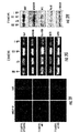
- the gene targeted by the miRNA is selected from the group consisting of a myocyte differentiation gene (e.g., a gene encoding a histone deacetylase 4 (HDAC4) polypeptide or a thyroid hormone receptor protein 240 (TRAP240)), a myocyte proliferation gene (e.g., a gene encoding a serum response factor (SRF) polypeptide) and a hormone related protein (e.g. a gene encoding thyroid hormone associated protein 1 (Thrap1).
- a myocyte differentiation gene e.g., a gene encoding a histone deacetylase 4 (HDAC4) polypeptide or a thyroid hormone receptor protein 240 (TRAP240)
- HDAC4 histone deacetylase 4
- TRIP240 thyroid hormone receptor protein 240
- SRF serum response factor
- Thrap1 thyroid hormone associated protein 1
- a vector encoding an miRNA comprises a promoter operatively linked to a nucleic acid molecule encoding the miRNA molecule; and a transcription termination sequence.
- the vector is incorporated in a kit further comprising at least one reagent for introducing the vector into a myocyte.
- the kit in some embodiments, further comprises instructions for introducing the vector into a myocyte.
- RNA sequences are presented in the form of DNA (i.e . with thymidine present instead of uracil), it is understood that these sequences are also intended to correspond to the RNA transcripts of these DNA sequences ( i.e . with each T replaced by a U).
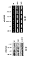
- miRNAs can modulate expression of specific genes in myocytes that affect differentiation and/or proliferation of the myocytes.
- This discovery has therapeutic applications, as disclosed herein, including treating muscle injuries having a wide variety of causes, such as for example mechanical muscle trauma, muscular degenerative disorders, and cardiac insult.
- Application of the discoveries disclosed herein further include modulating expression of one or more specific genes in myocytes utilizing miRNAs having specificity for the genes, and in turn, modulating functionality of the myocytes, such as for example differentiation and/or proliferation of the myocytes.
- Exemplary non-limiting miRNAs useful with the presently disclosed subject matter include miRNA-1, miRNA-133, miRNA-206, and miRNA-208.
- miRNA-1 miRNA-1
- miRNA-133 miRNA-133
- SRF serum response factor
- the present disclosure thus provides molecular mechanisms in which miRNAs participate in transcriptional circuits that control muscle gene expression and embryonic development.
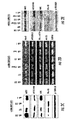
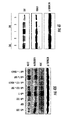
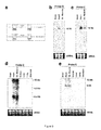
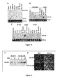
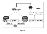
- Thrap1 expression is likely regulated by miR-208.
- the 3' UTR of Thrap1 contains two predicted miR-208 binding sites ( Figure 18 ).
- the two targets are located ⁇ 80 bp downstream of the Thrap1 stop codon and are separated from one another by only ⁇ 50 bp. Both targets are perfectly complementary with the seed region of miR-208.
- the Thrap1 gene encodes TRAP240, a 240 kd subunit of the TRAP (thyroid hormone receptor protein) complex that is ubiquitously expressed.
- TRAP is a multisubunit protein complex that is a coactivator for nuclear receptors and TRAP family members are important for proper development.
- miR-208 can regulate production of TRAP240 and promote hormone-dependent cardiomyocyte differentiation.
- This small RNA was found to target complementary sites in the 3' untranslated region (UTR) of lin-14. 49,50 .
- UTR 3' untranslated region
- Mature miRNAs are ⁇ 22 nucleotides (nt) in length that were processed from longer transcripts 51,52 .
- Primary-miRNAs pri-miRNAs
- RNA Pol II Primary-miRNAs
- RNA Pol II can be transcribed by RNA Pol II as independent transcriptional units or can originate from spliced-out introns of host genes 53 .
- the miRNA processing pathway can begin with pri-miRNA nuclear cleavage by RNAse III endonuclease Drosha, which produces a ⁇ 70-nt long intermediate precursor-miRNA (pre-miRNA) that has a stem-loop structure 54 .
- RISC RNA-induced silencing complex
- the relative thermal stabilities of the stem-arm duplex are thought to determine which strand becomes incorporated into RISC: the strand that enters RISC is often the one whose 5' end is less stable 64,65 .
- Translation inhibition is mediated by miRNA complementarity to target sequence(s) within the 3' UTR of the target mRNA by an as yet unknown mechanism 66,67 .
- imperfect complementarity results in translation suppression while perfect or near-perfect complementarity results in mRNA cleavage 68 .
- Many aspects of miRNA biogenesis, trafficking, RISC assembly, and the mechanism of RISC function await clarification, however functional studies of specific miRNAs and genetic and biochemical analyses of miRNA pathway components have shown that miRNAs are important in diverse biological processes.
- RNAs are proposed to control or fine-tune those complex genetic pathways by post-transcriptional regulation of target genes.
- One approach to determine the necessity of miRNAs in animal development has been to create mutations in Dicer, an upstream enzyme required for the processing miRNAs to their mature, active form. Vertebrates are believed to have only a single copy of Dicer, which is likely required to fully process all vertebrate miRNAs 62,63,69 .
- ablation of Dicer function resulted in lethality by embryonic day (E) 7.5 69 .
- the Dicer null mice did not express primitive streak marker T (brachyury), indicating that development was likely arrested before the body was configured during gastrulation.
- miRNAs participate in diverse biological processes.
- pancreatic islet cells overexpression of miR-375 suppressed glucose-induced insulin secretion, while inhibiting endogenous miR-375 enhanced insulin secretion 72 .
- a similar overexpression and inhibition strategy identified a role for miR-143 in adipocyte differentiation through regulating ERK5 protein expression 73 .
- a polycistronic miRNA gene coding for 5 miRNAs was linked to tumorigenesis 74 .
- Other functions for miRNAs have been proposed in hematopoiesis 75 , neuronal differentiation 76,77 , and the regulation of Hox gene expression 78,79 .
- miRNAs There are now over 300 known human miRNAs, however only a handful has any assigned biological function. Studies of specific miRNAs are required for understanding the prevalence and importance of miRNA-mediated regulation in development and pathology. The presently disclosed subject matter provides for the first time a role for miRNAs in regulating muscle differentiation and proliferation.
- Identifying the targets of specific miRNAs facilitates understanding their precise role in regulatory pathways.
- Most animal miRNAs are imperfectly complementary to their target site, which thwarts using simple homology searches to identify animal miRNA target sites.
- several computational methods have been developed that incorporate sequence conservation and characteristics of known miRNA targets as criteria to predict new animal miRNA targets 80-85 .
- some algorithms take into account that the majority of miRNAs have exhibited high complementarity between the second and eighth nucleotide within validated target sites, which is called the 'seed' region.
- Other algorithms do not since, in some cases, complementarity at the 3' end of a miRNA can compensate for weak 5' end binding.
- These algorithms also rank predictions by target sequence conservation across two or more species relative to flanking regions.
- These types of computational approaches have successfully predicted some mammalian miRNA target sites. The predictions produced for any particular miRNA almost certainly contains false positives. However, the predictions are extremely useful as hypothesis generators. Any prediction can be verified experimentally and placed into a relevant biological context
- miRNA research seeks to understand the precise molecular mechanisms behind miRNA-directed repression, to develop better tools for analyzing miRNA expression and identifying target sites, and to determine biologically relevant roles for specific miRNAs within regulatory pathways.
- the term "about”, when referring to a value or to an amount of mass, weight, time, volume, concentration, or percentage is meant to encompass variations of in some embodiments ⁇ 20% or ⁇ 10%, in some embodiments ⁇ 5%, in some embodiments ⁇ 1%, in some embodiments ⁇ 0.5%, and in some embodiments ⁇ 0.1% from the specified amount, as such variations are appropriate to practice the presently disclosed subject matter.
- all numbers expressing quantities of ingredients, reaction conditions, and so forth used in the specification and claims are to be understood as being modified in all instances by the term “about”. Accordingly, unless indicated to the contrary, the numerical parameters set forth in this specification and attached claims are approximations that can vary depending upon the desired properties sought to be obtained by the presently disclosed subject matter.
- amino acid and “amino acid residue” are used interchangeably and refer to any of the twenty naturally occurring amino acids, as well as analogs, derivatives, and congeners thereof; amino acid analogs having variant side chains; and all stereoisomers of any of the foregoing.
- amino acid is intended to embrace all molecules, whether natural or synthetic, which include both an amino functionality and an acid functionality and are capable of being included in a polymer of naturally occurring amino acids.
- amino acid can be formed upon chemical digestion (hydrolysis) of a polypeptide at its peptide linkages.
- the amino acid residues described herein are in some embodiments in the "L" isomeric form. However, residues in the "D" isomeric form can be substituted for any L-amino acid residue, as long as the desired functional property is retained by the polypeptide.
- NH 2 refers to the free amino group present at the amino terminus of a polypeptide.
- COOH refers to the free carboxy group present at the carboxy terminus of a polypeptide.
- amino acid residue sequences represented herein by formulae have a left-to-right orientation in the conventional direction of amino terminus to carboxy terminus.
- amino acid residues are broadly defined to include modified and unusual amino acids.
- a dash at the beginning or end of an amino acid residue sequence indicates a peptide bond to a further sequence of one or more amino acid residues or a covalent bond to an amino-terminal group such as NH 2 or acetyl or to a carboxy-terminal group such as COOH.
- the term "cell” is used in its usual biological sense.
- the cell is present in an organism, for example, a vertebrate subject.
- the cell can be eukaryotic (e.g., a myocyte, such as a skeletal myocyte or a cardiac myocyte) or prokaryotic (e.g. a bacterium).
- the cell can be of somatic or germ line origin, totipotent, pluripotent, or differentiated to any degree, dividing or non-dividing.
- the cell can also be derived from or can comprise a gamete or embryo, a stem cell, or a fully differentiated cell.
- the terms "host cells” and “recombinant host cells” are used interchangeably and refer to cells (for example, myocytes) into which the compositions of the presently disclosed subject matter (for example, an expression vector encoding an miRNA) can be introduced.
- the terms refer not only to the particular cell into which an expression construct is initially introduced, but also to the progeny or potential progeny of such a cell. Because certain modifications can occur in succeeding generations due to either mutation or environmental influences, such progeny might not, in fact, be identical to the parent cell, but are still included within the scope of the term as used herein.
- RNA refers to a nucleic acid that encodes an RNA, for example, nucleic acid sequences including, but not limited to, structural genes encoding a polypeptide.
- gene also refers broadly to any segment of DNA associated with a biological function.
- the term "gene” encompasses sequences including but not limited to: a coding sequence; a promoter region; a transcriptional regulatory sequence; a non-expressed DNA segment that is a specific recognition sequence for regulatory proteins; a non-expressed DNA segment that contributes to gene expression, such as for example a DNA segment that can be transcribed into a 3' untranslated region of an mRNA, which is in turn targeted and bound by exemplary miRNAs of the presently disclosed subject matter; a DNA segment designed to have desired parameters; or combinations thereof.
- a gene can be obtained by a variety of methods, including cloning from a biological sample, synthesis based on known or predicted sequence information, and recombinant derivation from one or more existing sequences.
- a gene typically comprises a coding strand and a non-coding strand.
- coding strand and “sense strand” are used interchangeably, and refer to a nucleic acid sequence that has the same sequence of nucleotides as an mRNA from which the gene product is translated.
- the coding/sense strand includes thymidine residues instead of the uridine residues found in the corresponding mRNA.
- the coding/sense strand can also include additional elements not found in the mRNA including, but not limited to promoters, enhancers, and introns.
- the terms “template strand” and “antisense strand” are used interchangeably and refer to a nucleic acid sequence that is complementary to the coding/sense strand. It should be noted, however, that for those genes that do not encode polypeptide products, for example an miRNA gene, the term "coding strand” is used to refer to the strand comprising the miRNA.
- the strand comprising the miRNA is a sense strand with respect to the miRNA precursor, but it would be antisense with respect to its target RNA (i.e ., the miRNA hybridizes to the target RNA because it comprises a sequence that is antisense to the target RNA).
- the terms “complementarity” and “complementary” refer to a nucleic acid that can form one or more hydrogen bonds with another nucleic acid sequence by either traditional Watson-Crick or other non-traditional types of interactions.
- the binding free energy for a nucleic acid molecule with its complementary sequence is sufficient to allow the relevant function of the nucleic acid to proceed, in some embodiments, ribonuclease activity.
- the degree of complementarity between the sense and antisense strands of an miRNA precursor can be the same or different from the degree of complementarity between the miRNA-containing strand of an miRNA precursor and the target nucleic acid sequence. Determination of binding free energies for nucleic acid molecules is well known in the art. See e.g., Freier et al., 1986 31 ; Turner et al., 1987 32 .
- percent complementarity As used herein, the phrase “percent complementarity”, “percent identitiy”, and “percent identical” are used interchangeably herein and refer to the percentage of contiguous residues in a nucleic acid molecule that can form hydrogen bonds (e.g., Watson-Crick base pairing) with a second nucleic acid sequence (e.g., 5, 6, 7, 8, 9, 10 out of 10 being 50%, 60%, 70%, 80%, 90%, and 100% complementary).
- the terms “100% complementary”, “fully complementary”, and “perfectly complementary” indicate that all of the contiguous residues of a nucleic acid sequence can hydrogen bond with the same number of contiguous residues in a second nucleic acid sequence.
- miRNAs are about 17-24 nt, and up to 5 mismatches (e.g., 1, 2, 3, 4, or 5 mismatches) are typically tolerated during miRNA-directed modulation of gene expression, a percent complementarity of at least about 70% between an miRNA and the RNA to which it is targeted should be sufficient for the miRNA to modulate the expression of the gene from which the target RNA was derived.
- gene expression generally refers to the cellular processes by which a biologically active polypeptide is produced from a DNA sequence and exhibits a biological activity in a cell.
- gene expression involves the processes of transcription and translation, but also involves post-transcriptional and post-translational processes that can influence a biological activity of a gene or gene product. These processes include, but are not limited to RNA synthesis, processing, and transport, as well as polypeptide synthesis, transport, and post-translational modification of polypeptides. Additionally, processes that affect protein-protein interactions within the cell can also affect gene expression as defined herein.
- the term “gene expression” refers to the processes by which a precursor miRNA is produced from the gene. Typically, this process is referred to as transcription, although unlike the transcription directed by RNA polymerase II for protein-coding genes, the transcription products of an miRNA gene are not translated to produce a protein. Nonetheless, the production of a mature miRNA from an miRNA gene is encompassed by the term “gene expression” as that term is used herein.
- isolated refers to a molecule substantially free of other nucleic acids, proteins, lipids, carbohydrates, and/or other materials with which it is normally associated, such association being either in cellular material or in a synthesis medium.
- isolated nucleic acid refers to a ribonucleic acid molecule or a deoxyribonucleic acid molecule (for example, a genomic DNA, cDNA, mRNA, miRNA, etc.) of natural or synthetic origin or some combination thereof, which (1) is not associated with the cell in which the "isolated nucleic acid” is found in nature, or (2) is operatively linked to a polynucleotide to which it is not linked in nature.
- isolated polypeptide refers to a polypeptide, in some embodiments prepared from recombinant DNA or RNA, or of synthetic origin, or some combination thereof, which (1) is not associated with proteins that it is normally found with in nature, (2) is isolated from the cell in which it normally occurs, (3) is isolated free of other proteins from the same cellular source, (4) is expressed by a cell from a different species, or (5) does not occur in nature.
- isolated when used in the context of an “isolated cell”, refers to a cell that has been removed from its natural environment, for example, as a part of an organ, tissue, or organism.
- label and “labeled” refer to the attachment of a moiety, capable of detection by spectroscopic, radiologic, or other methods, to a probe molecule.
- label or “labeled” refer to incorporation or attachment, optionally covalently or non-covalently, of a detectable marker into a molecule, such as a polypeptide.
- Various methods of labeling polypeptides are known in the art and can be used.
- labels for polypeptides include, but are not limited to, the following: radioisotopes, fluorescent labels, heavy atoms, enzymatic labels or reporter genes, chemiluminescent groups, biotinyl groups, predetermined polypeptide epitopes recognized by a secondary reporter (e.g., leucine zipper pair sequences, binding sites for antibodies, metal binding domains, epitope tags).
- labels are attached by spacer arms of various lengths to reduce potential steric hindrance.
- the term “modulate” refers to an increase, decrease, or other alteration of any, or all, chemical and biological activities or properties of a biochemical entity.
- the term “modulate” can refer to a change in the expression level of a gene or a level of an RNA molecule or equivalent RNA molecules encoding one or more proteins or protein subunits; or to an activity of one or more proteins or protein subunits that is upregulated or downregulated, such that expression, level, or activity is greater than or less than that observed in the absence of the modulator.
- the term “modulate” can mean “inhibit” or “suppress”, but the use of the word “modulate” is not limited to this definition.
- modulation refers to both upregulation (i.e ., activation or stimulation) and downregulation (i.e ., inhibition or suppression) of a response.
- modulation when used in reference to a functional property or biological activity or process (e.g., enzyme activity or receptor binding), refers to the capacity to upregulate (e.g., activate or stimulate), downregulate (e.g., inhibit or suppress), or otherwise change a quality of such property, activity, or process.
- upregulate e.g., activate or stimulate
- downregulate e.g., inhibit or suppress
- regulation can be contingent on the occurrence of a specific event, such as activation of a signal transduction pathway, and/or can be manifest only in particular cell types.
- modulator refers to a polypeptide, nucleic acid, macromolecule, complex, molecule, small molecule, compound, species, or the like (naturally occurring or non-naturally occurring), or an extract made from biological materials such as bacteria, plants, fungi, or animal cells or tissues, that can be capable of causing modulation.
- Modulators can be evaluated for potential activity as inhibitors or activators (directly or indirectly) of a functional property, biological activity or process, or a combination thereof (e.g., agonist, partial antagonist, partial agonist, inverse agonist, antagonist, anti-microbial agents, inhibitors of microbial infection or proliferation, and the like), by inclusion in assays. In such assays, many modulators can be screened at one time. The activity of a modulator can be known, unknown, or partially known.
- Modulators can be either selective or non-selective.
- selective when used in the context of a modulator (e.g. an inhibitor) refers to a measurable or otherwise biologically relevant difference in the way the modulator interacts with one molecule (e.g. a target RNA of interest) versus another similar but not identical molecule (e.g. an RNA derived from a member of the same gene family as the target RNA of interest).
- a modulator to be considered a selective modulator, the nature of its interaction with a target need not entirely exclude its interaction with other molecules related to the target (e.g. transcripts from family members other than the target itself).
- the term selective modulator is not intended to be limited to those molecules that only bind to mRNA transcripts from a gene of interest and not to those of related family members.
- the term is also intended to include modulators that can interact with transcripts from genes of interest and from related family members, but for which it is possible to design conditions under which the differential interactions with the targets versus the family members has a biologically relevant outcome.
- Such conditions can include, but are not limited to differences in the degree of sequence identity between the modulator and the family members, and the use of the modulator in a specific tissue or cell type that expresses some but not all family members.
- a modulator might be considered selective to a given target in a given tissue if it interacts with that target to cause a biologically relevant effect despite the fact that in another tissue that expresses additional family members the modulator and the target would not interact to cause a biological effect at all because the modulator would be "soaked out" of the tissue by the presence of other family members.
- the modulator When a selective modulator is identified, the modulator binds to one molecule (for example an mRNA transcript of a gene of interest) in a manner that is different (for example, stronger) from the way it binds to another molecule (for example, an mRNA transcript of a gene related to the gene of interest).
- the modulator is said to display "selective binding" or “preferential binding” to the molecule to which it binds more strongly as compared to some other possible molecule to which the modulator might bind.
- the terms “inhibit”, “suppress”, “down regulate”, and grammatical variants thereof are used interchangeably and refer to an activity whereby gene product (e.g., a polypeptide), expression of a gene, activity of a polynucleotide, such as for example an miRNA, or a level of an RNA encoding one or more gene products is reduced below that observed in the absence of an implementation of an approach of the presently disclosed subject matter.
- gene product e.g., a polypeptide
- activity of a polynucleotide such as for example an miRNA
- a level of an RNA encoding one or more gene products is reduced below that observed in the absence of an implementation of an approach of the presently disclosed subject matter.
- inhibition with an miRNA molecule results in a decrease in the steady state expression level of a target RNA. In some embodiments, inhibition with an miRNA molecule results in an expression level of a target gene that is below that level observed in the presence of an inactive or attenuated molecule that is unable to downregulate the expression level of the target. In some embodiments, inhibition of gene expression with an miRNA molecule of the presently disclosed subject matter is greater in the presence of the miRNA molecule than in its absence. In some embodiments, inhibition of gene expression is associated with an enhanced rate of degradation of the mRNA encoded by the gene (for example, by miRNA-mediated inhibition of gene expression). In some embodiments, inhibition with an miRNA molecule of the presently disclosed subject matter results in an expression level of a gene product from a target gene that is below that level observed in the absence of the miRNA.
- an miRNA such as for example an endogenous miRNA
- an miRNA inhibitor can be inhibited by an miRNA inhibitor, resulting in an increase in expression of a gene targeted by the miRNA, as compared to the level of gene expression (e.g., production of a gene product) when the miRNA is not inhibited.
- miRNA inhibitor and “inhibitor of miRNA” are used interchangeably and refer to a molecule that inhibits activity of an miRNA.
- an miRNA inhibitor is a polynucleotide that hybridizes to a particular target miRNA under specified conditions, thereby inhibiting activity of the target miRNA.
- Conditions under which the miRNA inhibitor can hybridize to the target miRNA include, for example, physiological conditions.
- the miRNA inhibitor can hybridize to the target miRNA to a greater or lesser degree based on complementarity of the miRNA inhibitor polynucleotide sequence to the target miRNA polynucleotide.
- the miRNA can be fully complementary to all or a portion of the target miRNA, or less than fully complementary, including for example, 99%, 98%, 97%, 96%, 95%, 90%, 80%, or 70% complementary to the target miRNA, depending on the particular application and need for specificity, as would be generally understood by one of skill in the art.
- the miRNA inhibitor need only share complementary with the target miRNA as is necessary to inhibit a desired amount of target miRNA activity under a particular set of conditions. Examples of miRNA inhibitors useful with the presently disclosed subject matter include, but are not limited to, modified polynucleotides such as 2'-O-methyl polynucleotides.
- mutation carries its traditional connotation and refers to a change, inherited, naturally occurring, or introduced, in a nucleic acid or polypeptide sequence, and is used in its sense as generally known to those of skill in the art.
- myocyte refers broadly to all classifications of muscle cells at all stages of development.
- myocyte encompasses both undifferentiated muscle cells, such as for example myoblasts, as well as differentiated muscle cells, such as for example terminally differentiated myotubes.
- Myocyte also encompasses muscle cells of varying histological types, including but not limited to striated muscle cells (e.g., skeletal muscle cells), smooth muscle cells (e.g., intestinal muscle cells), and cardiac muscle cells. Further, "myocyte” as used herein is not species specific.
- naturally occurring refers to the fact that an object can be found in nature.
- a polypeptide or polynucleotide sequence that is present in an organism (including bacteria) that can be isolated from a source in nature and which has not been intentionally modified by man in the laboratory is naturally occurring. It must be understood, however, that any manipulation by the hand of man can render a "naturally occurring" object an “isolated” object as that term is used herein.
- nucleic acid refers to any of deoxyribonucleic acid (DNA), ribonucleic acid (RNA), oligonucleotides, fragments generated by the polymerase chain reaction (PCR), and fragments generated by any of ligation, scission, endonuclease action, and exonuclease action.
- Nucleic acids can comprise monomers that are naturally occurring nucleotides (such as deoxyribonucleotides and ribonucleotides), or analogs of naturally occurring nucleotides (e.g ., ⁇ -enantiomeric forms of naturally occurring nucleotides), or a combination of both.
- Modified nucleotides can have modifications in sugar moieties and/or in pyrimidine or purine base moieties.
- Sugar modifications include, for example, replacement of one or more hydroxyl groups with halogens, alkyl groups, amines, and azido groups, or sugars can be functionalized as ethers or esters.
- nucleic acid monomers can be linked by phosphodiester bonds or analogs of such linkages. Analogs of phosphodiester linkages include phosphorothioate, phosphorodithioate, phosphoroselenoate, phosphorodiselenoate, phosphoroanilothioate, phosphoranilidate, phosphoramidate, and the like.
- nucleic acid also includes so-called "peptide nucleic acids", which comprise naturally occurring or modified nucleic acid bases attached to a polyamide backbone. Nucleic acids can be either single stranded or double stranded.
- operatively linked when describing the relationship between two nucleic acid regions, refers to a juxtaposition wherein the regions are in a relationship permitting them to function in their intended manner.
- a control sequence "operatively linked" to a coding sequence can be ligated in such a way that expression of the coding sequence is achieved under conditions compatible with the control sequences, such as when the appropriate molecules (e.g., inducers and polymerases) are bound to the control or regulatory sequence(s).
- the phrase "operatively linked” refers to a promoter connected to a coding sequence in such a way that the transcription of that coding sequence is controlled and regulated by that promoter.
- operatively linked can refer to a promoter region that is connected to a nucleotide sequence in such a way that the transcription of that nucleotide sequence is controlled and regulated by that promoter region.
- a nucleotide sequence is said to be under the "transcriptional control" of a promoter to which it is operatively linked.
- operatively linked can also refer to a transcription termination sequence that is connected to a nucleotide sequence in such a way that termination of transcription of that nucleotide sequence is controlled by that transcription termination sequence.
- a transcription termination sequence comprises a sequence that causes transcription by an RNA polymerase III to terminate at the third or fourth T in the terminator sequence, TTTTTTT. Therefore, the nascent small transcript typically has 3 or 4 U's at the 3' terminus.
- percent identity and percent identical in the context of two nucleic acid or protein sequences, refer to two or more sequences or subsequences that have in some embodiments at least 60%, in some embodiments at least 70%, in some embodiments at least 80%, in some embodiments at least 85%, in some embodiments at least 90%, in some embodiments at least 95%, in some embodiments at least 96%, in some embodiments at least 97%, in some embodiments at least 98%, and in some embodiments at least 99% nucleotide or amino acid residue identity, when compared and aligned for maximum correspondence, as measured using one of the following sequence comparison algorithms or by visual inspection.
- the percent identity exists in some embodiments over a region of the sequences that is at least about 10 residues in length, in some embodiments over a region that is at least about 20 residues in length, in some embodiments over a region of the sequences that is at least about 50 residues in length, in some embodiments over a region of at least about 100 residues, and in some embodiments the percent identity exists over at least about 150 residues. In some embodiments, the percent identity exists over the entire length of a given region, such as a coding region or an entire miRNA.
- sequence comparison typically one sequence acts as a reference sequence to which test sequences are compared.
- test and reference sequences are input into a computer, subsequence coordinates are designated if necessary, and sequence algorithm program parameters are designated.
- sequence comparison algorithm then calculates the percent sequence identity for the test sequence(s) relative to the reference sequence, based on the designated program parameters.
- Optimal alignment of sequences for comparison can be conducted, for example, by the local homology algorithm described in Smith & Waterman, 1981 33 , by the homology alignment algorithm described in Needleman & Wunsch, 1970 34 , by the search for similarity method described in Pearson & Lipman, 1988 35 , by computerized implementations of these algorithms (GAP, BESTFIT, FASTA, and TFASTA in the GCG ® WISCONSIN PACKAGE ® , available from Accelrys, Inc., San Diego, California, United States of America), or by visual inspection. See generally, Ausubel et al., 1989 36 .
- HSPs high scoring sequence pairs
- T some positive-valued threshold score
- These initial neighborhood word hits act as seeds for initiating searches to find longer HSPs containing them. The word hits are then extended in both directions along each sequence for as far as the cumulative alignment score can be increased.
- Cumulative scores are calculated using, for nucleotide sequences, the parameters M (reward score for a pair of matching residues; always > 0) and N (penalty score for mismatching residues; always ⁇ 0).
- M forward score for a pair of matching residues
- N penalty score for mismatching residues; always ⁇ 0.
- a scoring matrix is used to calculate the cumulative score. Extension of the word hits in each direction are halted when the cumulative alignment score falls off by the quantity X from its maximum achieved value, the cumulative score goes to zero or below due to the accumulation of one or more negative-scoring residue alignments, or the end of either sequence is reached.
- the BLAST algorithm parameters W, T, and X determine the sensitivity and speed of the alignment.
- W wordlength
- E expectation
- M number of amino acid sequences
- E amino acid sequences
- BLASTP program uses as defaults a wordlength (W) of 3, an expectation (E) of 10, and the BLOSUM62 scoring matrix.
- the BLAST algorithm In addition to calculating percent sequence identity, the BLAST algorithm also performs a statistical analysis of the similarity between two sequences. See e.g., Karlin & Altschul 1993 39 .
- One measure of similarity provided by the BLAST algorithm is the smallest sum probability (P(N)), which provides an indication of the probability by which a match between two nucleotide or amino acid sequences would occur by chance.
- P(N) the smallest sum probability
- a test nucleic acid sequence is considered similar to a reference sequence if the smallest sum probability in a comparison of the test nucleic acid sequence to the reference nucleic acid sequence is in some embodiments less than about 0.1, in some embodiments less than about 0.01, and in some embodiments less than about 0.001.
- substantially identical in the context of two nucleotide sequences, refers to two or more sequences or subsequences that have in some embodiments at least about 70% nucleotide identity, in some embodiments at least about 75% nucleotide identity, in some embodiments at least about 80% nucleotide identity, in some embodiments at least about 85% nucleotide identity, in some embodiments at least about 90% nucleotide identity, in some embodiments at least about 95% nucleotide identity, in some embodiments at least about 97% nucleotide identity, and in some embodiments at least about 99% nucleotide identity, when compared and aligned for maximum correspondence, as measured using one of the following sequence comparison algorithms or by visual inspection.
- the substantial identity exists in nucleotide sequences of at least 17 residues, in some embodiments in nucleotide sequence of at least about 18 residues, in some embodiments in nucleotide sequence of at least about 19 residues, in some embodiments in nucleotide sequence of at least about 20 residues, in some embodiments in nucleotide sequence of at least about 21 residues, in some embodiments in nucleotide sequence of at least about 22 residues, in some embodiments in nucleotide sequence of at least about 23 residues, in some embodiments in nucleotide sequence of at least about 24 residues, in some embodiments in nucleotide sequence of at least about 25 residues, in some embodiments in nucleotide sequence of at least about 26 residues, in some embodiments in nucleotide sequence of at least about 27 residues, in some embodiments in nucleotide sequence of at least about 30 residues, in some embodiments in nucleotide sequence of at least about 50 residues, in some embodiments
- polymorphic sequences can be substantially identical sequences.
- the term "polymorphic" refers to the occurrence of two or more genetically determined alternative sequences or alleles in a population. An allelic difference can be as small as one base pair. Nonetheless, one of ordinary skill in the art would recognize that the polymorphic sequences correspond to the same gene.
- nucleic acid sequences are substantially identical in that the two molecules specifically or substantially hybridize to each other under stringent conditions.
- two nucleic acid sequences being compared can be designated a "probe sequence” and a "test sequence".
- a "probe sequence” is a reference nucleic acid molecule
- a "'test sequence” is a test nucleic acid molecule, often found within a heterogeneous population of nucleic acid molecules.
- An exemplary nucleotide sequence employed for hybridization studies or assays includes probe sequences that are complementary to or mimic in some embodiments at least an about 14 to 40 nucleotide sequence of a nucleic acid molecule of the presently disclosed subject matter.
- probes comprise 14 to 20 nucleotides, or even longer where desired, such as 30, 40, 50, 60, 100, 200, 300, or 500 nucleotides or up to the full length of a given gene.
- Such fragments can be readily prepared by, for example, directly synthesizing the fragment by chemical synthesis, by application of nucleic acid amplification technology, or by introducing selected sequences into recombinant vectors for recombinant production.
- targeted to includes “hybridizing specifically to”, which refers to the binding, duplexing, or hybridizing of a molecule only to a particular nucleotide sequence under stringent conditions when that sequence is present in a complex nucleic acid mixture (e.g., total cellular DNA or RNA).
- a complex nucleic acid mixture e.g., total cellular DNA or RNA
- hybridization can be carried out in 5x SSC, 4x SSC, 3x SSC, 2x SSC, 1 x SSC, or 0.2x SSC for at least about 1 hour, 2 hours, 5 hours, 12 hours, or 24 hours ( see Sambrook & Russell, 2001, for a description of SSC buffer and other hybridization conditions).
- the temperature of the hybridization can be increased to adjust the stringency of the reaction, for example, from about 25°C (room temperature), to about 45°C, 50°C, 55°C, 60°C, or 65°C.
- the hybridization reaction can also include another agent affecting the stringency; for example, hybridization conducted in the presence of 50% formamide increases the stringency of hybridization at a defined temperature.
- the hybridization reaction can be followed by a single wash step, or two or more wash steps, which can be at the same or a different salinity and temperature.
- the temperature of the wash can be increased to adjust the stringency from about 25°C (room temperature), to about 45°C, 50°C, 55°C, 60°C, 65°C, or higher.
- the wash step can be conducted in the presence of a detergent, e.g., SDS.
- hybridization can be followed by two wash steps at 65°C each for about 20 minutes in 2x SSC, 0.1% SDS, and optionally two additional wash steps at 65°C each for about 20 minutes in 0.2x SSC, 0.1 % SDS.
- a probe nucleotide sequence hybridizes in one example to a target nucleotide sequence in 7% sodium dodecyl sulfate (SDS), 0.5M NaPO 4 , 1 mm ethylenediamine tetraacetic acid (EDTA) at 50°C followed by washing in 2X SSC, 0.1% SDS at 50°C; in some embodiments, a probe and test sequence hybridize in 7% sodium dodecyl sulfate (SDS), 0.5M NaPO 4 , 1 mm EDTA at 50°C followed by washing in 1X SSC, 0.1% SDS at 50°C; in some embodiments, a probe and test sequence hybridize in 7% sodium dodecyl sulfate (SDS), 0.5M NaPO 4 , 1 mm EDTA at
- Additional exemplary stringent hybridization conditions include overnight hybridization at 42°C in a solution comprising or consisting of 50% formamide, 10x Denhardt's (0.2% Ficoll, 0.2% polyvinylpyrrolidone, 0.2% bovine serum albumin) and 200 mg/ml of denatured carrier DNA, e.g., sheared salmon sperm DNA, followed by two wash steps at 65°C each for about 20 minutes in 2x SSC, 0.1% SDS, and two wash steps at 65°C each for about 20 minutes in 0.2x SSC, 0.1% SDS.
- denatured carrier DNA e.g., sheared salmon sperm DNA
- Hybridization can include hybridizing two nucleic acids in solution, or a nucleic acid in solution to a nucleic acid attached to a solid support, e.g., a filter.
- a prehybridization step can be conducted prior to hybridization. Prehybridization can be carried out for at least about 1 hour, 3 hours, or 10 hours in the same solution and at the same temperature as the hybridization (but without the complementary polynucleotide strand).
- hybridizing substantially to refers to complementary hybridization between a probe nucleic acid molecule and a target nucleic acid molecule and embraces minor mismatches that can be accommodated by reducing the stringency of the hybridization media to achieve the desired hybridization.
- phenotype refers to the entire physical, biochemical, and physiological makeup of a cell or an organism, e.g., having any one trait or any group of traits. As such, phenotypes result from the expression of genes within a cell or an organism, and relate to traits that are potentially observable or assayable.
- polypeptide As used herein, the terms “polypeptide”, “protein”, and “peptide”, which are used interchangeably herein, refer to a polymer of the 20 protein amino acids, or amino acid analogs, regardless of its size or function. Although “protein” is often used in reference to relatively large polypeptides, and “peptide” is often used in reference to small polypeptides, usage of these terms in the art overlaps and varies.
- the term “polypeptide” as used herein refers to peptides, polypeptides and proteins, unless otherwise noted.
- protein polypeptide
- polypeptide are used interchangeably herein when referring to a gene product.
- polypeptide encompasses proteins of all functions, including enzymes.
- exemplary polypeptides include gene products, naturally occurring proteins, homologs, orthologs, paralogs, fragments, and other equivalents, variants and analogs of the foregoing.
- polypeptide fragment when used in reference to a reference polypeptide, refers to a polypeptide in which amino acid residues are deleted as compared to the reference polypeptide itself, but where the remaining amino acid sequence is usually identical to the corresponding positions in the reference polypeptide. Such deletions can occur at the amino-terminus or carboxy-terminus of the reference polypeptide, or alternatively both. Fragments typically are at least 5, 6, 8 or 10 amino acids long, at least 14 amino acids long, at least 20, 30, 40 or 50 amino acids long, at least 75 amino acids long, or at least 100, 150, 200, 300, 500 or more amino acids long. A fragment can retain one or more of the biological activities of the reference polypeptide. Further, fragments can include a sub-fragment of a specific region, which sub-fragment retains a function of the region from which it is derived.
- the term "primer” refers to a sequence comprising in some embodiments two or more deoxyribonucleotides or ribonucleotides, in some embodiments more than three, in some embodiments more than eight, and in some embodiments at least about 20 nucleotides of an exonic or intronic region. Such oligonucleotides are in some embodiments between ten and thirty bases in length.
- purified refers to an object species that is the predominant species present (i.e ., on a molar basis it is more abundant than any other individual species in the composition).
- a “purified fraction” is a composition wherein the object species comprises at least about 50 percent (on a molar basis) of all species present.
- the solvent or matrix in which the species is dissolved or dispersed is usually not included in such determination; instead, only the species (including the one of interest) dissolved or dispersed are taken into account.
- a purified composition will have one species that comprises more than about 80 percent of all species present in the composition, more than about 85%, 90%, 95%, 99% or more of all species present.
- the object species can be purified to essential homogeneity (contaminant species cannot be detected in the composition by conventional detection methods) wherein the composition consists essentially of a single species.
- Purity of a polypeptide can be determined by a number of methods known to those of skill in the art, including for example, amino-terminal amino acid sequence analysis, gel electrophoresis, and mass-spectrometry analysis.
- a “reference sequence” is a defined sequence used as a basis for a sequence comparison.
- a reference sequence can be a subset of a larger sequence, for example, as a segment of a full-length nucleotide or amino acid sequence, or can comprise a complete sequence. Because two proteins can each (1) comprise a sequence ( i.e ., a portion of the complete protein sequence) that is similar between the two proteins, and (2) can further comprise a sequence that is divergent between the two proteins, sequence comparisons between two (or more) proteins are typically performed by comparing sequences of the two proteins over a "comparison window" (defined hereinabove) to identify and compare local regions of sequence similarity.
- regulatory sequence is a generic term used throughout the specification to refer to polynucleotide sequences, such as initiation signals, enhancers, regulators, promoters, and termination sequences, which are necessary or desirable to affect the expression of coding and non-coding sequences to which they are operatively linked.
- Exemplary regulatory sequences are described in Goeddel, 1990 45 , and include, for example, the early and late promoters of simian virus 40 (SV40), adenovirus or cytomegalovirus immediate early promoter, the CMV minimal promoter, the lac system, the trp system, the TAC or TRC system, T7 promoter whose expression is directed by T7 RNA polymerase, the major operator and promoter regions of phage lambda, the control regions for fd coat protein, the promoter for 3-phosphoglycerate kinase or other glycolytic enzymes, the promoters of acid phosphatase, e.g., Pho5, the promoters of the yeast ⁇ -mating factors, the polyhedron promoter of the baculovirus system and other sequences known to control the expression of genes of prokaryotic or eukaryotic cells or their viruses, and various combinations thereof.
- SV40 simian virus 40
- CMV minimal promoter the lac system
- regulatory sequences can differ depending upon the host organism.
- such regulatory sequences generally include promoter, ribosomal binding site, and transcription termination sequences.
- the term "regulatory sequence” is intended to include, at a minimum, components the presence of which can influence expression, and can also include additional components the presence of which is advantageous, for example, leader sequences and fusion partner sequences.
- transcription of a polynucleotide sequence is under the control of a promoter sequence (or other regulatory sequence) that controls the expression of the polynucleotide in a cell type in which expression is intended. It will also be understood that the polynucleotide can be under the control of regulatory sequences that are the same or different from those sequences which control expression of the naturally occurring form of the polynucleotide.
- a promoter sequence is selected from the group consisting of a CMV minimal promoter, muscle creatine kinase (MCK), and an ⁇ -myosin heavy chain (MHC) promoter.
- muscle creatine kinase (MCK) promoter which directs gene expression in skeletal muscle, can be used to express miRNAs, such as for example, miR-1, miR-133 or miR-206 in tissue, including skeletal muscle using currently available transgenic techniques. It is understood that the entire promoter identified for any promoter (for example, the promoters listed herein) need not be employed, and that a functional derivative thereof can be used.
- the phrase "functional derivative” refers to a nucleic acid sequence that comprises sufficient sequence to direct transcription of another operatively linked nucleic acid molecule. As such, a “functional derivative" can function as a minimal promoter, as that term is defined herein.
- Termination of transcription of a polynucleotide sequence is typically regulated by an operatively linked transcription termination sequence (for example, an RNA polymerase III termination sequence).
- transcriptional terminators are also responsible for correct mRNA polyadenylation.
- the 3' non-transcribed regulatory DNA sequence includes from in some embodiments about 50 to about 1,000, and in some embodiments about 100 to about 1,000, nucleotide base pairs and contains transcriptional and translational termination sequences.
- an RNA polymerase III termination sequence comprises the nucleotide sequence TTTTTTT.
- reporter gene refers to a nucleic acid comprising a nucleotide sequence encoding a protein that is readily detectable either by its presence or activity, including, but not limited to, luciferase, fluorescent protein (e.g., green fluorescent protein), chloramphenicol acetyl transferase, ⁇ -galactosidase, secreted placental alkaline phosphatase, ⁇ -lactamase, human growth hormone, and other secreted enzyme reporters.
- fluorescent protein e.g., green fluorescent protein
- chloramphenicol acetyl transferase e.g., chloramphenicol acetyl transferase
- ⁇ -galactosidase e.g., secreted placental alkaline phosphatase
- ⁇ -lactamase ⁇ -lactamase
- human growth hormone and other secreted enzyme reporters.
- a reporter gene encodes a polypeptide not otherwise produced by the host cell, which is detectable by analysis of the cell(s), e.g., by the direct fluorometric, radioisotopic or spectrophotometric analysis of the cell(s) and typically without the need to kill the cells for signal analysis.
- a reporter gene encodes an enzyme, which produces a change in fluorometric properties of the host cell, which is detectable by qualitative, quantitative, or semiquantitative function or transcriptional activation.
- Exemplary enzymes include esterases, ⁇ -lactamase, phosphatases, peroxidases, proteases (tissue plasminogen activator or urokinase), and other enzymes whose function can be detected by appropriate chromogenic or fluorogenic substrates known to those skilled in the art or developed in the future.
- sequencing refers to determining the ordered linear sequence of nucleic acids or amino acids of a DNA, RNA, or protein target sample, using conventional manual or automated laboratory techniques.
- the term “substantially pure” refers to that the polynucleotide or polypeptide is substantially free of the sequences and molecules with which it is associated in its natural state, and those molecules used in the isolation procedure.
- the term “substantially free” refers to that the sample is in some embodiments at least 50%, in some embodiments at least 70%, in some embodiments 80% and in some embodiments 90% free of the materials and compounds with which is it associated in nature.
- target cell refers to a cell, into which it is desired to insert a nucleic acid sequence or polypeptide, or to otherwise effect a modification from conditions known to be standard in the unmodified cell.
- a nucleic acid sequence introduced into a target cell can be of variable length. Additionally, a nucleic acid sequence can enter a target cell as a component of a plasmid or other vector or as a naked sequence.
- target gene refers to a gene that is targeted for modulation using the methods and compositions of the presently disclosed subject matter.
- a target gene therefore, comprises a nucleic acid sequence the expression level of which, either at the mRNA or polypeptide level, is downregulated by a miRNA.
- target RNA or “target mRNA” refers to the transcript of a target gene to which the miRNA is intended to bind, leading to modulation of the expression of the target gene.
- the target gene can be a gene derived from a cell, an endogenous gene, a transgene, or exogenous genes such as genes of a pathogen, for example a virus, which is present in the cell after infection thereof.
- the cell containing the target gene can be derived from or contained in any organism, for example a plant, animal, protozoan, virus, bacterium, or fungus.
- transcription refers to a cellular process involving the interaction of an RNA polymerase with a gene that directs the expression as RNA of the structural information present in the coding sequences of the gene.
- the process includes, but is not limited to, the following steps: (a) the transcription initiation; (b) transcript elongation; (c) transcript splicing; (d) transcript capping; (e) transcript termination; (f) transcript polyadenylation; (g) nuclear export of the transcript; (h) transcript editing; and (i) stabilizing the transcript.
- transcription factor refers to a cytoplasmic or nuclear protein which binds to a gene, or binds to an RNA transcript of a gene, or binds to another protein which binds to a gene or an RNA transcript or another protein which in turn binds to a gene or an RNA transcript, so as to thereby modulate expression of the gene. Such modulation can additionally be achieved by other mechanisms; the essence of a "transcription factor for a gene” pertains to a factor that alters the level of transcription of the gene in some way.
- transfection refers to the introduction of a nucleic acid, e.g., an expression vector, into a recipient cell, which in certain instances involves nucleic acid-mediated gene transfer.
- transformation refers to a process in which a cell's genotype is changed as a result of the cellular uptake of exogenous nucleic acid.
- a transformed cell can express an miRNA of the presently disclosed subject matter.
- signaling or “significant” relates to a statistical analysis of the probability that there is a non-random association between two or more entities. To determine whether or not a relationship is “significant” or has “significance”, statistical manipulations of the data can be performed to calculate a probability, expressed as a "p-value". Those p-values that fall below a user-defined cutoff point are regarded as significant. In one example, a p-value less than or equal to 0.05, in some embodiments less than 0.01, in some embodiments less than 0.005, and in some embodiments less than 0.001, are regarded as significant.
- target RNA refers to an RNA molecule (for example, an mRNA molecule encoding a gene product) that is a target for modulation.
- the target RNA is encoded by a target gene.
- target site refers to a sequence within a target RNA that is “targeted” for cleavage mediated by an miRNA construct that contains sequences within its antisense strand that are complementary to the target site.
- target cell refers to a cell that expresses a target RNA and into which an miRNA is intended to be introduced.
- a target cell is in some embodiments a myocyte.
- an miRNA is "targeted to" an RNA molecule if it has sufficient nucleotide similarity to the RNA molecule that it would be expected to modulate the expression of the RNA molecule under conditions sufficient for the miRNA and the RNA molecule to interact.
- the interaction occurs within a myocyte.
- the interaction occurs under physiological conditions.
- physiological conditions refers to in vivo conditions within a myocyte, whether that myocyte is part of a subject or a subject's tissue, or that myocyte is being grown in vitro.
- physiological conditions refers to the conditions within a myocyte under any conditions that the myocyte can be exposed to, either as part of a subject or when grown in vitro.
- the phrase "detectable level of cleavage” refers to a degree of cleavage of target RNA (and formation of cleaved product RNAs) that is sufficient to allow detection of cleavage products above the background of RNAs produced by random degradation of the target RNA. Production of miRNA-mediated cleavage products from at least 1-5% of the target RNA is sufficient to allow detection above background for most detection methods.
- miRNA refers to a nucleic acid molecule of about 17-24 nucleotides that is produced from a pri-miRNA, a pre-miRNA, or a functional equivalent. miRNAs are to be contrasted with short interfering RNAs (siRNAs), although in the context of exogenously supplied miRNAs and siRNAs, this distinction might be somewhat artificial.
- siRNAs short interfering RNAs
- an miRNA is necessarily the product of nuclease activity on a hairpin molecule such as has been described herein, and an siRNA can be generated from a fully double-stranded RNA molecule or a hairpin molecule.
- RNA refers to a molecule comprising at least one ribonucleotide residue.
- ribonucleotide is meant a nucleotide with a hydroxyl group at the 2' position of a ⁇ -D-ribofuranose moiety.
- the terms encompass double stranded RNA, single stranded RNA, RNAs with both double stranded and single stranded regions, isolated RNA such as partially purified RNA, essentially pure RNA, synthetic RNA, and recombinantly produced RNA.
- RNAs include, but are not limited to mRNA transcripts, miRNAs and miRNA precursors, and siRNAs.
- RNA is also intended to encompass altered RNA, or analog RNA, which are RNAs that differ from naturally occurring RNA by the addition, deletion, substitution, and/or alteration of one or more nucleotides. Such alterations can include addition of non-nucleotide material, such as to the end(s) of the RNA or internally, for example at one or more nucleotides of the RNA. Nucleotides in the RNA molecules of the presently disclosed subject matter can also comprise non-standard nucleotides, such as non-naturally occurring nucleotides or chemically synthesized nucleotides or deoxynucleotides. These altered RNAs can be referred to as analogs or analogs of a naturally occurring RNA.
- double stranded RNA refers to an RNA molecule at least a part of which is in Watson-Crick base pairing forming a duplex.
- the term is to be understood to encompass an RNA molecule that is either fully or only partially double stranded.
- Exemplary double stranded RNAs include, but are not limited to molecules comprising at least two distinct RNA strands that are either partially or fully duplexed by intermolecular hybridization.
- the term is intended to include a single RNA molecule that by intramolecular hybridization can form a double stranded region (for example, a hairpin).
- the phrases “intermolecular hybridization” and “intramolecular hybridization” refer to double stranded molecules for which the nucleotides involved in the duplex formation are present on different molecules or the same molecule, respectively.
- double stranded region refers to any region of a nucleic acid molecule that is in a double stranded conformation via hydrogen bonding between the nucleotides including, but not limited to hydrogen bonding between cytosine and guanosine, adenosine and thymidine, adenosine and uracil, and any other nucleic acid duplex as would be understood by one of ordinary skill in the art.
- the length of the double stranded region can vary from about 15 consecutive basepairs to several thousand basepairs.
- the double stranded region is at least 15 basepairs, in some embodiments between 15 and 300 basepairs, and in some embodiments between 15 and about 60 basepairs.
- the formation of the double stranded region results from the hybridization of complementary RNA strands (for example, a sense strand and an antisense strand), either via an intermolecular hybridization ( i.e ., involving 2 or more distinct RNA molecules) or via an intramolecular hybridization, the latter of which can occur when a single RNA molecule contains self-complementary regions that are capable of hybridizing to each other on the same RNA molecule.
- These self-complementary regions are typically separated by a short stretch of nucleotides (for example, about 5-10 nucleotides) such that the intramolecular hybridization event forms what is referred to in the art as a "hairpin” or a "stem-loop structure".
- nucleic acid molecules employed in accordance with the presently disclosed subject matter include nucleic acid molecules encoding a myocyte gene product, as well as the nucleic acid molecules that are used in accordance with the presently disclosed subject matter to modulate the expression of a myocyte gene (e.g., an miRNA nucleic acid molecule).
- nucleic acid molecules employed in accordance with the presently disclosed subject matter include, but are not limited to, the nucleic acid molecules described herein.
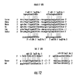
- nucleic acid molecules employed herein include, but are not limited to miR-1 (UGGAAUGUAAAGAAGUAUGUA; SEQ ID NO:1), miR-133 (UUGGUCCCCUUCAACCAGCUGU; SEQ ID NO:2), miR-206 (UGGAAUGUAAGGAAGUGUGUGG; SEQ ID NO:3), miR-208 (AUAAGACGAGCAAAAAAGCUUGU; SEQ ID NO:4), miR-22 (AAGCUGCCAGUUGAAGAACUGU; SEQ ID NO:5), miR-26 (UUCAAGUAAUyCAGGAUAGGy(U); SEQ ID NO:6), miR-29 (UAGCACCAUyUGAAAUCrGU(kUU); SEQ ID NO:7), miR-30 (ykUwmAswysshhswyUvnvvv(bC); SEQ ID NO:8), miR-128 (UCACAGUGAACCGGUCUCUUUy; SEQ ID NO:
- nucleotide codes used above and elsewhere herein are in accordance with WIPO Standard ST.25 (1998), Appendix 2, Table 1, (M.P.E.P. 2422, Table 1), herein incorporated by reference.
- the following one-letter codes represent the associated nucleotide(s) as set forth in Table 1.
- Nucleotide(s) in parenthesis e.g., (n)
- Figure 21 lists individual sequences possible for SEQ ID NOs: 5-11 based on the nucleotide permutations set forth in SEQ ID NOs: 5-11.
- An exemplary nucleotide sequence employed in the methods disclosed herein comprises sequences that are complementary to each other, the complementary regions being capable of forming a duplex of, in some embodiments, at least about 15 to 300 basepairs, and in some embodiments, at least about 15-24 basepairs.
- One strand of the duplex comprises a nucleic acid sequence of at least 15 contiguous bases having a nucleic acid sequence of a nucleic acid molecule of the presently disclosed subject matter.
- one strand of the duplex comprises a nucleic acid sequence comprising 15, 16, 17, or 18 nucleotides, or even longer where desired, such as 19, 20, 21, 22, 23, 24, 25, 26, 27, 28, 29, or 30 nucleotides, or up to the full length of any of those nucleic acid sequences described herein.
- Such fragments can be readily prepared by, for example, directly synthesizing the fragment by chemical synthesis, by application of nucleic acid amplification technology, or by introducing selected sequences into recombinant vectors for recombinant production.
- hybridizing specifically to refers to the binding, duplexing, or hybridizing of a molecule only to a particular nucleotide sequence under stringent conditions when that sequence is present in a complex nucleic acid mixture (e.g., total cellular DNA or RNA).
- sequence refers to a sequence of a nucleic acid molecule or amino acid molecule that comprises a part of a longer nucleic acid or amino acid sequence.
- An exemplary subsequence is a sequence that comprises part of a duplexed region of a pri-miRNA or a pre-miRNA ("miRNA precursors") including, but not limited to the nucleotides that become the mature miRNA after nuclease action or a single-stranded region in an miRNA precursor.
- elongated sequence refers to an addition of nucleotides (or other analogous molecules) incorporated into the nucleic acid.
- a polymerase e.g., a DNA polymerase
- the nucleotide sequence can be combined with other DNA sequences, such as promoters, promoter regions, enhancers, polyadenylation signals, intronic sequences, additional restriction enzyme sites, multiple cloning sites, and other coding segments.
- Nucleic acids of the presently disclosed subject matter can be cloned, synthesized, recombinantly altered, mutagenized, or subjected to combinations of these techniques.
- Standard recombinant DNA and molecular cloning techniques used to isolate nucleic acids are known in the art. Exemplary, non-limiting methods are described by Silhavy et al., 1984 46 ; Ausubel et al., 1989 36 ; Glover & Hames, 1995 47 ; and Sambrook & Russell, 2001 40 .
- Site-specific mutagenesis to create base pair changes, deletions, or small insertions is also known in the art as exemplified by publications (see e.g., Adelman et al., 1983 48 ; Sambrook & Russell, 2001 40 ).
- miRNA molecules or miRNA precursor molecules are expressed from transcription units inserted into nucleic acid vectors (alternatively referred to generally as "recombinant vectors" or “expression vectors”).
- a vector can be used to deliver a nucleic acid molecule encoding an miRNA into a myocyte to target a specific gene.
- the recombinant vectors can be, for example, DNA plasmids or viral vectors.
- a variety of expression vectors are known in the art. The selection of the appropriate expression vector can be made on the basis of several factors including, but not limited to the cell type wherein expression is desired.
- vector refers to a nucleic acid capable of transporting another nucleic acid to which it has been linked.
- Vectors include those capable of autonomous replication and expression of nucleic acids to which they are linked.
- Vectors capable of directing the expression of genes to which they are operatively linked are referred to herein as "expression vectors".
- expression vectors of utility in recombinant techniques are often in the form of plasmids.
- the presently disclosed subject matter is intended to include such other forms of expression vectors which serve equivalent functions and which become known in the art subsequently hereto.
- expression vector refers to a nucleotide sequence capable of directing expression of a particular nucleotide sequence in an appropriate host cell, comprising a promoter operatively linked to the nucleotide sequence of interest which is operatively linked to transcription termination sequences. It also typically comprises sequences required for proper translation of the nucleotide sequence.
- the construct comprising the nucleotide sequence of interest can be chimeric. The construct can also be one that is naturally occurring but has been obtained in a recombinant form useful for heterologous expression.
- the nucleotide sequence of interest including any additional sequences designed to effect proper expression of the nucleotide sequences, can also be referred to as an "expression cassette".
- heterologous gene refers to a sequence that originates from a source foreign to an intended host cell or, if from the same source, is modified from its original form.
- a heterologous gene in a host cell includes a gene that is endogenous to the particular host cell but has been modified, for example by mutagenesis or by isolation from native transcriptional regulatory sequences.
- the terms also include non-naturally occurring multiple copies of a naturally occurring nucleotide sequence.
- the terms can refer to a DNA segment that is foreign or heterologous to the cell, or homologous to the cell but in a position within the host cell nucleic acid wherein the element is not ordinarily found.
- promoter each refers to a nucleotide sequence within a gene that is positioned 5' to a coding sequence and functions to direct transcription of the coding sequence.
- the promoter region comprises a transcriptional start site, and can additionally include one or more transcriptional regulatory elements.
- a method of the presently disclosed subject matter employs a RNA polymerase III promoter.
- a “minimal promoter” is a nucleotide sequence that has the minimal elements required to enable basal level transcription to occur. As such, minimal promoters are not complete promoters but rather are subsequences of promoters that are capable of directing a basal level of transcription of a reporter construct in an experimental system.
- Minimal promoters include but are not limited to the cytomegalovirus (CMV) minimal promoter, the herpes simplex virus thymidine kinase (HSV-tk) minimal promoter, the simian virus 40 (SV40) minimal promoter, the human ⁇ -actin minimal promoter, the human EF2 minimal promoter, the adenovirus E1 B minimal promoter, and the heat shock protein (hsp) 70 minimal promoter.
- CMV cytomegalovirus
- HSV-tk herpes simplex virus thymidine kinase
- SV40 simian virus 40
- hsp heat shock protein
- Minimal promoters are often augmented with one or more transcriptional regulatory elements to influence the transcription of an operatively linked gene.
- minimal promoter also encompasses a functional derivative of a promoter disclosed herein, including, but not limited to an RNA polymerase III promoter (for example, an H1, 7SL, 5S, or U6 promoter), an adenovirus VA1 promoter, a Vault promoter, a telomerase RNA promoter, and a tRNA gene promoter.
- RNA polymerase III promoter for example, an H1, 7SL, 5S, or U6 promoter
- adenovirus VA1 promoter for example, a Vault promoter, a telomerase RNA promoter, and a tRNA gene promoter.
- promoters have different combinations of transcriptional regulatory elements. Whether or not a gene is expressed in a cell is dependent on a combination of the particular transcriptional regulatory elements that make up the gene's promoter and the different transcription factors that are present within the nucleus of the cell. As such, promoters are often classified as “constitutive”, “tissue-specific”, “cell-type-specific”, or “inducible”, depending on their functional activities in vivo or in vitro. For example, a constitutive promoter is one that is capable of directing transcription of a gene in a variety of cell types (in some embodiments, in all cell types) of an organism.
- Exemplary constitutive promoters include the promoters for the following genes which encode certain constitutive or "housekeeping" functions: hypoxanthine phosphoribosyl transferase (HPRT), dihydrofolate reductase (DHFR; (Scharfmann et al., 1991), adenosine deaminase, phosphoglycerate kinase (PGK), pyruvate kinase, phosphoglycerate mutase, the ⁇ -actin promoter (see e.g., Williams et al., 1993), and other constitutive promoters known to those of skill in the art.
- HPRT hypoxanthine phosphoribosyl transferase
- DHFR dihydrofolate reductase
- PGK phosphoglycerate kinase
- pyruvate kinase phosphoglycerate mutase
- ⁇ -actin promoter see e.
- tissue-specific or “cell-type-specific” promoters direct transcription in some tissues or cell types of an organism but are inactive in some or all others tissues or cell types.
- tissue-specific promoters include those promoters described in more detail hereinbelow, as well as other tissue-specific and cell-type specific promoters known to those of skill in the art.
- linked refers to a physical proximity of promoter elements such that they function together to direct transcription of an operatively linked nucleotide sequence.
- transcriptional regulatory sequence or “transcriptional regulatory element”, as used herein, each refers to a nucleotide sequence within the promoter region that enables responsiveness to a regulatory transcription factor. Responsiveness can encompass a decrease or an increase in transcriptional output and is mediated by binding of the transcription factor to the nucleotide sequence comprising the transcriptional regulatory element.
- a transcriptional regulatory sequence is a transcription termination sequence, alternatively referred to herein as a transcription termination signal.
- transcription factor generally refers to a protein that modulates gene expression by interaction with the transcriptional regulatory element and cellular components for transcription, including RNA Polymerase, Transcription Associated Factors (TAFs), chromatin-remodeling proteins, and any other relevant protein that impacts gene transcription.
- TAFs Transcription Associated Factors
- the presently disclosed subject matter provides method for modulating expression of genes in myocytes with specificity.
- the methods comprise contacting a myocyte with a miRNA or a vector encoding the miRNA targeted to a gene in the myocyte.
- Targeting one or more particular genes in myocyte allows for manipulation of myocyte function or development (e.g., differentiation) with a high level of specificity.
- the presently disclosed subject matter further provides methods for modulating myocyte function or development by contacting a myocyte with an miRNA targeted to a gene in the myocyte that can modulate myocyte function or development.
- the miRNA targeted to a particular gene is selected from the group consisting of miR-1, miR-133, miR-206, miR-208, miR-22, miR-26, miR-29, miR-30, miR-128, miR-143, and miR-145 (SEQ ID NOs: 1-11, respectively), including miRNAs having sequences at least about 70%, 80%, 85%, 90%, 91%, 92%, 93%, 94%, 95%, 96%, 97%, 98%, or 99% identical to the sequences set forth in any one of SEQ ID NOs 1-11.
- an miRNA is "targeted to" an RNA molecule if it has sufficient nucleotide similarity to the RNA molecule that it would be expected to modulate the expression of the RNA molecule under conditions sufficient for the miRNA and the RNA molecule to interact.
- the interaction occurs within a myocyte.
- the interaction occurs under physiological conditions.
- physiological conditions refers to in vivo conditions within a myocyte, whether that myocyte is part of a subject or a subject's tissue, or that myocyte is being grown in vitro.
- physiological conditions refers to the conditions within a myocyte under any conditions that the myocyte can be exposed to, either as part of an organism or when grown in vitro.
- the targeted gene is a myocyte differentiation gene or a myocyte proliferation gene and when expressed can modulate myocyte differentiation and/or proliferation, respectively.
- the targeted gene can express a gene product that inhibits myocyte differentiation and/or proliferation.
- targeted inhibition of expression of one or more of these differentiation and/or proliferation target genes by the miRNA can result in an increase in differentiation and/or proliferation of the treated myocyte.
- the myocyte differentiation gene can encode a histone deacetylase 4 (HDAC4) polypeptide or a thyroid hormone receptor protein 240 (TRAP240) and the myocyte proliferation gene can encode a serum response factor (SRF) polypeptide.
- HDAC4 histone deacetylase 4
- TRAP240 thyroid hormone receptor protein 240
- SRF serum response factor
- Expression of one or more of the myocyte differentiation or proliferation genes can be targeted for inhibition utilizing one of the miRNAs disclosed herein.
- the miRNAs miR-1 and miRNA-133 each specifically target the 3' untranslated region of HDAC4 and SRF, respectively, and inhibit expression of the gene products encoded by these genes.
- differentiation of a myocyte can be increased by contacting the myocyte with miR-1, which targets the gene encoding HDAC4, thereby substantially preventing expression of HDAC4 and increasing myocyte differentiation.
- proliferation of a myocyte can be increased by contacting the myocyte with miR-133, which targets the gene encoding SRF, thereby substantially preventing expression of SRF and increasing myocyte proliferation.
- miRNAs can be targeted to genes to modulate expression of the genes.
- genes expressing products that function to inhibit myocyte differentiation and/or proliferation can be targeted by miRNAs to inhibit expression of these genes, resulting in increased myocyte differentiation and/or proliferation.
- miRNA inhibitors can be targeted to endogenous miRNAs to facilitate in relative increase in expression of particular gene products in a manner beneficial to treatment of the muscle injury.
- combinations of miRNAs and/or miRNA inhibitors can be co-administered to the muscle injury in an approach to optimize healing of the injury. Increased myocyte differentiation and/or proliferation can be beneficial in healing damaged muscle tissue or stimulating regrowth of lost muscle tissue.
- methods for treating a muscle injury in a subject comprise administering to a muscle injury site in a subject an effective amount of a miRNA, a vector encoding the miRNA, an inhibitor of an miRNA, or combinations thereof, wherein the miRNA is targeted to a gene in a myocyte at the muscle injury site.
- Muscle development, and comparably muscle growth and/or healing, such as after injury, can occur in phases. Representative phases include undifferentiated myocyte proliferation, followed by differentiation of the myocytes into mature cells of the muscle tissue.
- repair of muscle tissue at a site of muscle injury can be facilitated by the coordinated administration to the injury site of miRNAs and/or miRNA inhibitors that enhance proliferation of undifferentiated myocytes, and the administration to the injury site of miRNAs and/or miRNA inhibitors that enhance differentiation of the proliferated myocytes into mature functioning muscle tissue.
- miR-1 and miR-133 each play distinct roles in modulating skeletal muscle proliferation and differentiation.
- miR-133 enhances myocyte proliferation by repressing SRF.
- miR-1 promotes myocyte differentiation by targeting HDAC4, a transcriptional repressor of muscle gene expression.
- miR-133 and an inhibitor of miR-1 are first co-administered to the muscle injury site at a first time point to increase myocyte proliferation at the injury site.
- miR-1 and an inhibitor of miR-133 are co-administered to the muscle injury site at a second time point increase differentiation of the proliferated myocytes.
- the temporally coordinated coadministration of multiple miRNAs and/or miRNA inhibitors can further improve recovery from muscle injury.
- the muscle injury results from a mechanical muscle trauma, a muscular degenerative disorder, a cardiac insult, or a combination thereof.
- Mechanical muscle trauma can be the result of, for example, blunt force trauma such as occurs in automobile accidents or impaling injuries, wherein the muscle tissue is sliced or torn.
- Exemplary non-limiting muscular degenerative disorders include muscular dystrophies (e.g., Duchenne muscular dystrophy (DMD)), motor neuron diseases (e.g., amyotrophic lateral sclerosis (ALS)), inflammatory myopathies (e.g., dermatomyositis (DM)), neuromuscular junction diseases (e.g., myasthenia gravis (MG)), endocrine myopathies (e.g., hyperthyroid myopathy (HYPTM)), and metabolic muscular diseases (e.g., phosphorylase deficiency (MPD)).
- Exemplary non-limiting cardiac insult muscle injuries include myocardial infarction and cardiac muscle reperfusion injury.
- the miRNA targeted to a particular gene is selected from the group consisting of miR-1, miR-133, miR-206, miR-208, miR-22, miR-26, miR-29, miR-30, miR-128, miR-143, and miR-145 (SEQ ID NOs: 1-11, respectively), including miRNAs having sequences at least 70%, 80%, 85%, 90%, 91%, 92%, 93%, 94%, 95%, 96%, 97%, 98%, or 99% identical to the sequences set forth in any one of SEQ ID NOs 1-11.
- the gene is a myocyte differentiation gene (e.g., encoding HDAC4 or TRAP240) or a myocyte proliferation gene (e.g., encoding SRF).
- a preferred subject is a vertebrate subject.
- a preferred vertebrate is warm-blooded; a preferred warm-blooded vertebrate is a mammal.
- a preferred mammal is most preferably a human.
- the term "subject" includes both human and animal subjects.
- veterinary therapeutic uses are provided in accordance with the presently disclosed subject matter.
- the presently disclosed subject matter provides for the treatment of mammals such as humans, as well as those mammals of importance due to being endangered, such as Siberian tigers; of economic importance, such as animals raised on farms for consumption by humans; and/or animals of social importance to humans, such as animals kept as pets or in zoos.
- mammals such as humans, as well as those mammals of importance due to being endangered, such as Siberian tigers; of economic importance, such as animals raised on farms for consumption by humans; and/or animals of social importance to humans, such as animals kept as pets or in zoos.
- animals include but are not limited to: carnivores such as cats and dogs; swine, including pigs, hogs, and wild boars; ruminants and/or ungulates such as cattle, oxen, sheep, giraffes, deer, goats, bison, and camels; and horses.
- domesticated fowl i.e ., poultry, such as turkeys, chickens, ducks, geese, guinea fowl, and the like, as they are also of economical importance to humans.
- livestock including, but not limited to, domesticated swine, ruminants, ungulates, horses (including race horses), poultry, and the like.
- Suitable methods for administering to a subject an miRNA or a vector encoding the miRNA include but are not limited to systemic administration, parenteral administration (including intravascular, intramuscular, intraarterial administration), oral delivery, buccal delivery, subcutaneous administration, inhalation, intratracheal installation, surgical implantation, transdermal delivery, local injection, and hyper-velocity injection/bombardment. Where applicable, continuous infusion can enhance drug accumulation at a target site.
- the particular mode of administration used in accordance with the methods of the present subject matter depends on various factors, including but not limited to the miRNA and/or vector carrier employed, the severity of the condition to be treated, and mechanisms for metabolism or removal of the active compound(s) following administration.
- an amount of the therapeutic composition e.g., a composition comprising an miRNA or a vector encoding the miRNA
- a measurable biological response e.g., an increase in myocyte differentiation and/or proliferation
- Actual dosage levels of active compounds in a therapeutic composition of the presently disclosed subject matter can be varied so as to administer an amount of the active compound(s) that is effective to achieve the desired therapeutic response for a particular subject and/or application.
- the selected dosage level will depend upon a variety of factors including the activity of the therapeutic composition, formulation, the route of administration, combination with other drugs or treatments, severity of the condition being treated, and the physical condition and prior medical history of the subject being treated.
- a minimal dose is administered, and dose is escalated in the absence of dose-limiting toxicity to a minimally effective amount. Determination and adjustment of an effective dose, as well as evaluation of when and how to make such adjustments, are within the ordinary skill in the art.
- miRNAs microRNAs
- C2C12 myoblasts because this line of cells faithfully mimics skeletal muscle differentiation in vitro as myoblasts can be induced to become terminally differentiated myotubes when serum is withdrawn from the culture medium.
- 10-12 We found that the expression of a fraction of the miRNAs examined was up-regulated in differentiated C2C12 myoblasts/myotubes ( Figure 1 a and Figure 6 ). The increase in expression of miR-1 and miR-133 in differentiated myoblasts was confirmed by Northern blot analysis ( Figure 1b and Figure 7 ).
- miR-1 and miR-133 are specifically expressed in adult cardiac and skeletal muscle tissues, but not in other tissues tested ( Figure 1c , Figure 8 ). However, little is known about the temporospatial distribution of specific miRNAs during mammalian development. We therefore examined the expression of miR-1 and miR-133 in mouse embryos and neonates. miR-1 and miR-133 are expressed at very low levels in the developing hearts and skeletal muscle of E13.5 and E16.5 embryos, ( Figure 1d and Figure 8 ). An increasing level of miR-1 and miR-133 expression was found in neonatal hearts and skeletal muscle, though it is still significantly lower than that of adults ( Figure 1e and Figure 8 ). These data are consistent with findings from zebrafish in which the majority of miRNAs are expressed relatively late during embryogenesis 16 .
- Both miR-1 and miR-133 are clustered together on mouse chromosomes 2 (separated by 9.3 kb) and 18 (separated by 2.5 kb) ( Figure 9 and ref. 14).
- miR-1 and miR-133 probes from chromosome 18 detected a single primary transcript of ⁇ 6 kb from total RNAs isolated from heart and skeletal muscle ( Figures 9b and 9c ), indicating that miR-1 and miR-133 are indeed transcribed together.
- miR-1 and miR-133 probes from chromosome 2 detected a transcript of ⁇ 10 kb from the heart and skeletal muscle
- the miR-133 probe also hybridized to two additional transcripts of ⁇ 4.5 kb and ⁇ 2.2 kb
- the miR-1 probe also detected a major transcript of ⁇ 6 kb ( Figures 9d and 9e ), suggesting the potential involvement of posttranscriptional processing.
- our data indicate that cardiac- and skeletal muscle-specific expression of miR-1 and miR-133 is dictated at the primary transcription step.
- miR-1 and miR-133 To assess the function of miR-1 and miR-133 in skeletal muscle, we first attempted to overexpress miR-1 and miR-133 in mammalian cells. We tested and validated the expression and activity of both miRNAs using Northern blot analysis as well as miR-1 and miR-133 "sensors" 17 , in which the complementary sequences for miR-1 or miR-133 were cloned downstream of a dsRed coding sequence ( Figure 11 and data not shown). We transfected C2C12 myoblasts with miR-1 or miR-133 and then either maintained cells in growth medium (GM) or transferred them to differentiation medium (DM) after transfection.
- GM growth medium
- DM differentiation medium
- miR-1 strongly enhanced myogenesis as indicated by increased expression of both the early and late myogenic markers myogenin and myosin heavy chain (MHC), respectively, as well as other myogenic markers, including MyoD, Mef2, and skeletal ⁇ -actin ( Figures 2a-2e , 2i, 2j and Table 2).
- miR-1 induced myogenesis is specific, since overexpression of a GFP control or miR-208, which is not endogenously expressed in skeletal myocytes, showed no effect ( Figures 2a-2e ). Furthermore, mutations introduced into miR-1 "seed" sequences abolished its ability to activate myogenic gene expression ( Figures 2d-2e ). In contrast, overexpression of miR-133 repressed the expression of myogenin and MHC ( Figure 2 , a-e and Table 2) and promoted myoblast proliferation ( Figures 2c-2e and Table 2). Again, the effect of miR-133 on myoblasts proliferation is specific, as controls showed no effect and mutation introduced abolished the function of miR-133 ( Figures 2a-2e , 2j).
- miR-1 and miR-133 have been found in most animal species, from Drosophila to human, suggesting they are evolutionary conserved.
- miR-1 and miR-133 have been found in most animal species, from Drosophila to human, suggesting they are evolutionary conserved.
- Introduction of miR-1 at the one cell stage leads to a dramatically shortened axis with accompanying reduction in anterior structures and an increase in body size along the dorsal-ventral axis compared to either uninjected or miGFP injected controls (n > 45, two independent experiments) ( Figure 3 ).
- HDAC4 contains two naturally occurring putative miR-1 sites at its 3' UTR, which are evolutionarily conserved among vertebrate species ( Figure 12 ). Similarly, two conserved miR-133 binding sites are found in the 3' UTR of the mammalian SRF gene ( Figure 12 ), which has been shown to play an important role in muscle proliferation and differentiation in vitro and in vivo 11,24,25
- miR-1 led to the down-regulation of endogenous HDAC4 protein in C2C12 cells in both the growth condition ( Figure 4c ) and differentiation condition ( Figure 4e ), whereas miR-133 repressed the expression of endogenous SRF proteins ( Figures 4c , 4e).
- the mRNA levels of SRF and HDAC4 were not altered by those miRNAs ( Figure 4d ), supporting the notion that miRNAs repress the function of their target genes mainly by inhibiting translation.
- HDAC4 and SRF are cognate targets for miR-1 or miR-133 in regulating skeletal muscle gene expression.
- cotransfecting expression plasmids for SRF or HDAC4 could "suppress" miRNA-mediated myogenesis. Indeed, as shown in Figure 4h , overexpression of SRF partially reversed myogenic gene repression induced by miR-133. In contrast, HDAC4 counteracted the effects of miR-1 on skeletal muscle gene expression ( Figure 4h ).
- GM Dulbecco's Modified Eagle Medium
- FBS fetal bovine serum
- DM differentiation medium
- RNA was labeled with 5'-phosphate-cytidyl-uridyl-Cy3-3' (Dharmacon, Inc., Lafayette, Colorado, U.S.A.) using RNA ligase and hybridized with 0.5 mM mixture of oligonucleotide probes for 124 microRNAs labeled with ALEXA 647 ® (Cy5) (Molecular Probes, Eugene, Oregon, U.S.A.) in disposable chambers (MJ Research, Reno, Nevada, U.S.A.; part number SLF-0601).
- Normalized log (base 2) data was hierarchically clustered by gene and is plotted as a heat map. The range of signal was from -4 fold to +4 fold. Yellow denotes high expression and blue denotes low expression, relative to the median.
- TRIZOL ® Reagent Invitrogen
- PEG was applied to remove large sized RNAs. Briefly, 30 ⁇ g of each total RNA sample were mixed 1:1 with 5X PEG solution and placed on ice 10 min. After 10 min centrifuging at maximum speed at 4°C, the supernatant was transferred to a fresh tube. RNAs were then precipitated by adding 2.5 volumes of 100% EtOH and centrifuged 30 min at maximum speed.
- Northern blot analysis for miRNAs was performed as described 13 . miR-1 and miR-133 oligonucleotide sequences used as probes are listed in Table 3. Northern blot analysis was used to detect primary transcripts of miRNAs and performed as described 26 , using 20 ⁇ g of total RNA from each sample. Genomic fragments for miR-1 and miR-133 were PCR-cloned and serve as probes.
- Genomic fragments for miR-1 and miR-133 precursors from mouse chromosomes 2 and 18 were PCR amplified using mouse genomic DNA as a template (for PCR primers, see Table 3 above).
- the PCR products were cloned into the pcDNATM(+)3.1 vector (Invitrogen) and the expression of miRNAs was determined by transfecting expression vectors into mammalian cells (COS7, 10T1/2 or C2C12) and detected by Northern blot analysis.
- C2C12 myoblast cells were cultured and myogenesis induced as described 12 .
- Transient transfection luciferase reporter assays were performed as described 12,26 .
- miRNA duplexes and 2'- O -methyl antisense oligoribonucleotides for miR-1, miR-133, miR-208 and GFP were purchased from Dharmacon (see Table 3 for sequences). They were introduced into mammalian cells using either LIPOFECTAMINETM (Invitrogen) transfection (200 nM) or electroporation using the Amaxa Biosystems (Gaithersburg, Maryland, U.S.A.) NUCLEOFECTOR ® system (5 ⁇ g).
- Western blots were performed as described previously 27 .
- the following antibodies were used: anti- myogenin; SRF; MEF2; HDAC4; and ⁇ -tubulin (Santa Cruz Biotechnology, Santa Cruz, California, U.S.A.); and phospho-histone H3 (Upstate Biotechnology, Lake Placid, New York, U.S.A.).
- the MF20 antibody which recognizes striated muscle-specific MHC, was obtained from the DSHB (University of Iowa, Iowa City, Iowa, U.S.A.).
- treated C2C12 cell in 12-well plates were fixed with 4% formaldehyde for 5 min at 37°C and changed to 0.1% NP40/PBS solution for 15 min at RT.
- Primary antibodies were incubated in 0.1% NP40-PBS with 3% BSA for 2 hr in the following concentration: anti-myogenin (1: 20 dilution), anti-phospho-histone H3 (1:100 dilution), MF20 (1:10 dilution).
- Secondary antibodies fluorescein anti-mouse/rabbit (1:100 dilution; Vector Laboratories, Burlingame, California, U.S.A.) were adding in 0.1% NP40-PBS with 3% BSA for 1 hr at 37°C.
- DAPI was added in for 5 min at RT. After several wash with PBS, cells were subjected to fluorescence microscopy observation. Ten fields that cover the whole well were picked and green fluorescence positive cells and total cells with DAPI staining were counted for each well, respectively.
- RT-PCR was performed essentially as described 27 .
- Total RNA were extracted from C2C12 cells using TRIZOL ® reagent (Invitrogen), and 2.0 ⁇ g aliquots were reverse transcribed to cDNA using random hexamers and MMLV reverse transcriptase (Invitrogen). For each case, 2.5% cDNA pool was used for amplification and PCR were performed for 24-28 cycles. Sequences for PCR primers can be found in Table 3.
- miR-208 is a cardiac-specific miRNA conserved between human, mouse, and rat ( Figure 14 ).
- Northern blot analysis revealed that miR-208 expression is developmentally regulated ( Figure 15 ).
- Northern blots were prepared from staged mouse tissues and probed with radiolabeled probe complementary to miR-208.
- miR-208 levels were dramatically higher in the adult mouse heart relative to E13.5, E16.5, and neonatal stage hearts.
- miR-208 is hosted by an intron of the cardiac muscle ⁇ -myosin heavy chain ( ⁇ - MHC) gene ( Figure 14 ).
- ⁇ - MHC One of two cardiac myosin heavy isoforms, ⁇ -MHC is weakly expressed during mouse development but later becomes the predominant isoform in the adult mouse heart.
- miR-208 and ⁇ -MHC are both cardiac-specific and transcribed from the reverse strand, which suggests that miR-208 is processed from the ⁇ -MHC intron and is expressed parallel to ⁇
- Neonatal cardiomyocytes do not lose their ability to replicate after birth; a large fraction undergoes mitotic division and proliferates in vitro and in vivo where cardiac gene expression is also activated. It is likely that miR-208 promotes cardiomyocyte differentiation since it is expressed highly in the adult heart relative to earlier development. In order to determine miR-208 function in the heart, this model system is used to study the effects of miR-208 expression and inhibition upon the cardiogenesis program.
- cardiomyocytes are one of the most widely used experimental models in cardiac research. The preparation of cardiomyocytes from small mammals is economical relative to whole animal studies, reliable, and allows for a broad spectrum of experiments. For both economical and technical reasons, cardiomyocytes are most commonly isolated from neonatal rats. We isolate rat cardiomyocytes essentially as previously described, with minor modifications 86 .
- RNA Pol II promoter sequence to direct transcription of the miRNA precursor sequence plus ⁇ 150 flanking nucleotides.
- the resulting RNA transcripts are recognized by the miRNA processing machinery and become fully functional miRNAs capable of directing translational repression.
- Northern blot analysis shows a dosage-dependent increase of mir-208 expression in isolated cardiomyocytes infected with increasing concentrations Ad-208 ( Figure 16 ). This tool can be utilized to study the effects of miR-208 overexpression on the cardiomyocyte phenotype in vitro.
- a cell counting assay is used to determine if changes in miR-208 expression affects proliferation.
- Cardiomyocytes are plated at low density and infected with Ad-208.
- Ad-GFP multiplicity of infection
- cells with Ad-GFP can also be infected at the same multiplicity of infection (MOI) as Ad-208 to control for indirect effects caused by adenoviral infection. Since both viruses express green fluorescent protein (GFP), infection efficiencies are also controlled for by epifluorescence microscopy.
- Cells are counted under brightfield illumination prior to infection and at 24, 48, 72, and 96 hours post-infection. Cells in 10 fields of vision are counted for both conditions at each time point.
- Statistical analyses are applied to cell count data.
- the unpaired Student t-test can be used to determine the probability that the mean number of cells counted per field view differs significantly between Ad-208 and Ad-GFP infection at each particular time point; whereas the paired Student t- test can determine the probability that the mean number of cells counted for each infection differs significantly between time points. If the probability that any two means are significantly different is greater than or equal to 95%, those differences are considered significant.
- the cell counting assay is complemented with studies to determine the mitotic index by using phospho-Histone H3 antibodies and to determine the percentage of cells undergoing DNA synthesis by BrdU incorporation. Fixed cells are TUNEL stained to rule out the possibility that miR-208 overexpression causes cell death. Potential outcomes of Ad-208 infection upon cardiomyocyte proliferation are decreased, increased, or no change upon cell number. Without wishing to be limited by theory, it is likely miR-208 expression will slow cardiomyocyte proliferation relative to controls since miR-208 is normally highly expressed in differentiated adult cardiomyocytes.
- a set of cardiac transcription factors including Nkx2.5, MEF2C, GATA4, myocardin, and TBX5, have been shown to be expressed in early differentiating cardiomyocytes, making them early genetic markers of cardiac differentiation.
- Cardiac muscle-specific contractile proteins such as ⁇ -MHC, ⁇ -MHC, ⁇ -CA, and MLV2V, are terminal differentiation markers of cardiomyocytes.
- Some of these cardiac genes are differentially regulated during development. For example, ⁇ - MHC is highly expressed in embryonic heart, but becomes down regulated post-neonatally, whereas ⁇ -MHC has an opposing expression pattern.
- miR-208 has a role in regulating cardiac gene expression
- the effect of ectopic miR-208 expression upon cardiac marker gene expression in neonatal rat cardiomyocytes is examined. Without wishing to be limited by theory, it is anticipated that miR-208 expression will decrease fetal gene expression and/or promote expression of adult cardiac genes.
- RT-PCR reverse transcriptase-PCR
- Ad-208 and Ad-GFP infected cardiomyocytes are fixed on glass cover slips, probed with appropriate primary and secondary antibodies, nuclear stained, and mounted to slides for analysis by microscopy.
- a reporter construct with antisense miR-208 sequence attached directly 3' to the luciferase gene (luc-miR-208-sensor) is used as a control and to test the efficacy of the system to block miR-208 function.
- the mRNA and protein levels, as well as protein localization, are studied as described for the miR-208 overexpression studies.
- miR-208 is weakly expressed in the embryonic heart and its expression increases dramatically in the adult heart. This example analyzes whether miR-208 function is more important for gene regulation in the developing heart or in the adult heart. During development, one could argue that miR-208 is likely not important since it is expressed weakly in the embryo. Against that argument, proper miR-208 dosage might be critical for regulating certain genetic pathways during development. Furthermore, the knockout mouse of its host gene ⁇ -MHC, which is weakly expressed during development relative to the adult stage, suffered embryonic lethality, though it is not known whether expression of miR-208 is affected in those animals 87 . The high expression of miR-208 detected in the adult heart might indicate that its most important function lies in later development. To sort through these types of issues, two mouse models are created to study miR-208 function: a miR-208 knockout mouse and a transgenic mouse that conditionally overexpresses miR-208.
- a mouse functionally null for miR-208 is designed and created without affecting the expression of its host gene ⁇ -MHC.
- the production of embryonic-stem cell derived miR-208 knockout mice is a three-stage process: production of the targeting vector; introduction of DNA sequences into embryonic stem cells by homologous recombination; and production of genetically modified mice derived from embryonic stem cells.
- Related miRNAs are grouped into families based on sequence homology within their seed regions. These families might redundantly regulate the expression of the same genes, potentially complicating genetic analysis of their function in vivo.
- the seed region of miR-208 does not cluster with other known miRNAs in a phylogenetic tree, making miR-208 a suitable miRNA knockout candidate.
- the miR-208 targeting construct is built using a strategy called recombineering, which uses homologous recombination between linear DNA fragments and circular plasmids 88,89 .
- the circular plasmid can contain a 6-7 kb fragment of the ⁇ -MHC gene that miR-208 lies within centrally.
- the linear DNA fragment contains two homologous arms designed to replace the 22 nt miR-208 sequence with a floxed selection cassette.
- Bacteria transformed with the circular plasmid are electroporated with the linear DNA fragment, and subsequently, resistance encoded by the linear fragment selects for recombinant colonies. The resulting construct is used to target ES cells by homologous recombination.
- a heterozygous ES cell is identified using a PCR-based screening or by Southern blot, it is used to generate chimera by blastocyst injection.
- the presently disclsoed knockout design leaves only a small footprint of exogenous DNA within the intron of ⁇ -MHC and helps ensure that transcription of ⁇ -MHC or the splicing pattern of ⁇ -MHC mRNA remains unaffected.
- the conditional transgenic strategy is diagramed in Figure 17 .
- the conditional transgenic approach is a binary system consisting of two transgenes.
- One transgene encodes miR-208 (tet208), while the other transgene encodes a transactivator (tTA) that activates the miR-208 transgene by binding a regulatory sequence within its promoter. Binding of tTA is inhibited whenever doxycycline (DOX) is present (i.e. "tet-off”), thus enabling temporal regulation of miR-208 transgene by DOX treatment.
- DOX doxycycline
- a mouse colony homozygous for the miR-208 transgene is established.
- the tet208 mice is mated to transgenic mice harboring a tTA transgene to create double transgenics for study. Assuming Mendelian genetics, 1 in 4 offspring will be double transgenic and express miR-208 wherever tTA is expressed.
- An ⁇ -MHC promoter is utilized to direct tTA expression. The ⁇ -MHC promoter has been well-characterized and is sufficient to properly direct tissue-specific expression in early development 90 .
- ⁇ -MHC promoter to express tTA will increase miR-208 dosage in the same tissues as endogenous miR-208 in the double transgenic animals, since endogenous miR-208 normally originates from an intron within the ⁇ - MHC gene.
- a transgenic mouse line that uses the mouse ⁇ -MHC promoter to direct tTA expression exists and has been used successfully 91,92 .
- the tet208 transgenic mouse line allows us to study the dosage effects of miR-208 expression in the developing embryo or adult mouse heart independently. Typically, early embryonic lethality in regular transgenic founders would severely limit the number of developmentally arrested embryos available for study and hinder phenotypic analysis. With the conditional strategy, we are able to delay miR-208 transgene expression in the double transgenic mice should earlier miR-208 overexpression prove lethal.
- the specific analyses conducted depend upon how the phenotypes are manifested. In general, using histological and biochemical approaches to characterize potential phenotypes in the developing embryo and/or adult is acceptable. Hearts are examined for gross abnormalities and sectioned for histological analysis to identify potentially more subtle developmental defects. The possibilities for defects are innumerable and can include anything from defective septal formation to thickened atria. It is equally possible that the phenotype can be a contractility defect that can be characterized by electrophysiological studies.
- Target predictions are utilized to complement observations related to the investigation of miR-208 function in vitro and in in vivo mouse models.
- Thrap1 expression is regulated by miR-208.
- the 3' UTR of Thrap1 contains two predicted miR-208 binding sites ( Figure 18 ).
- the two targets are located ⁇ 80 bp downstream of the Thrap1 stop codon and are separated from one another by only ⁇ 50 bp. Both targets are perfectly complementary with the seed region of miR-208.
- the Thrap1 gene encodes a 240 kd subunit of the TRAP (thyroid hormone receptor protein) complex that is ubiquitously expressed 93 .
- TRAP thyroid hormone receptor protein
- TRAP thyroid hormone receptor protein
- Thrap1 has not been characterized, but defects in other TRAP subunits have been shown to affect nuclear receptor signaling. Gene ablation of TRAP220 in mice impaired heart and nervous system development, while Drosophila homologues of TRAP230 and TRAP240 are required for proper eye-antennal disc development 95,96 . Mutations in a gene highly similar to Thrap1, termed Thrap2, were found in patients with the congenital heart defect Transposition of the Great Arteries 97 . Thus, TRAP family members are important for proper development. Of particular interest is Thrap1 as a target of miR-208, since the thyroid hormone is known to exert profound effects upon cardiac contractility.
- Thyroid hormone is linked to cardiac myosin isoform switching. In cardiomyocytes, thyroid hormone causes a rapid accumulation of ⁇ -MHC mRNA while simultaneously inhibiting ⁇ - MHC expression 98,99 .
- Several positive acting thyroid response elements (TREs) are located within the ⁇ - MHC promoter and a negative acting half-TRE within the ⁇ - MHC promoter has been identified 100,101 .
- ⁇ -MHC and ⁇ - MHC genes are arranged in tandem on chromosome 14 and they encode the two cardiac myosin heavy chain isoforms that convert ATP to mechanical work at different rates and their protein expression ratio affects the contractility of the cardiac sacromeres; ⁇ -MHC is "fast” whereas ⁇ -MHC is "slow.” Their expression is developmentally regulated. In mouse and rat, ⁇ -MHC is predominant in late fetal life, but shortly after birth ⁇ -MHC becomes the predominant adult cardiac isoform. The transition is likely caused by the surge of circulating thyroid hormone occurring shortly after birth 98 . In larger mammals, like rabbits and humans, ⁇ -MHC is the predominant adult cardiac isoform.
- miR-208 acts as a tissue-specific inhibitor of thyroid hormone signaling in a negative feedback loop to regulate the ratio of cardiac myosin heavy chain isoforms by targeting a component of the TRAP complex ( Figure 19 ).
- the initial screening strategy asks whether overexpression of miR-208 downregulates expression of a reporter gene bearing putative target sites in its 3' UTR.
- Results suggest that miR-208 targets the Thrap1 UTR ( Figure 18 ).
- Cardiomyocytes are infected with Ad-208 to determine whether miR-208 expression inhibits thyroid hormone signaling by monitoring transcript and protein levels of ⁇ -MHC and ⁇ -MHC. Inhibition of ⁇ -MHC expression by miR-208 would indirectly support our hypothesis that miR-208 targets a component of the thyroid hormone signaling pathway.
- miR-208 expression decreases mRNA or protein levels in hearts from the miR-208 knockout and miR-208 transgenic mouse models.
- Specific antibodies against the targeted proteins are employed.
- Human Thrap1 is commercially available. If not effective in mouse studies, mouse Thrap1-specific antibodies are developed.
- the 3' UTRs are from SP3 (Sp3 trans-acting transcription factor 3), EYA4 (eyes absent homolog 4), CSNK2A2 (casein kinase 2, alpha prime polypeptide ) , and TTN (Titin).
- SP3 protein expression is opposite to miR-208 expression; SP3 is a transcription factor that interacts with a variety of promoters containing GC-boxes 103,104,105 . SP 3 protein is readily detected in fetal mouse hearts, but is barely detectable in the adult heart. The opposing expression patterns of SP3 and miR-208 makes it formally possible for miR-208 to regulate SP3 translation.
- EYA4 is an interesting potential target of miR-208 given its established link to pathology in the human heart. Mutations have been identified in human EYA4 that cause dilated cardiomyopathy and associated heart failure 106,107 . EYA4 is a transcriptional coactivator that interacts with members of the sine-oculis family (Six1-Six6) and Dach transcription factors leading to gene activation 108,109 . The characterization of the human mutation was supported by work in zebrafish, as attenuated EYA4 levels produced morphological and hemodynamic features of heart failure 106 . Currently, no potential downstream cardiac genes of EYA4 have been identified.
- CSNK2A2 is a broadly expressed protein serine/threonine kinase that has been implicated in DNA replication, regulation of basal and inducible transcription, translation and control of metabolism 101,111 .
- CSKN2A2 was interested in CSKN2A2 for its potential in regulating a variety of genetic pathways.
- TTN is also an interesting miR-208 target candidate given its established association with cardiovascular function and pathology.
- TTN is a giant sacromeric protein expressed in both cardiac and skeletal muscle tissue and is important for sacromere assembly and force transmission 112 . Mutations in TTN have been linked to hypertrophic and dilated cardiomyopathies. Given the requirement of TTN for cardiac and skeletal muscle function, we doubt that miR-208 strongly regulates TTN expression, but it is possibly that that one sacromeric gene (i.e. ⁇ -MHC) might fine-tune the expression of another (i.e. TTN) to adjust the contractile properties of cardiomyocytes.
- ⁇ -MHC sacromeric gene
- the 3' UTR of the predicted target gene is tested by reporter assay to determine if it confers miR-208-mediated suppression.
- Candidate genes are further characterized by mutating the predicted target sites and testing whether miR-208 suppression is diminished.
- the effects of miR-208 overexpression in vitro using cardiomyocyte cells upon the candidate gene expression at the transcript and protein levels are analyzed.
- the biological relevance of verified targets are studied in vivo using our miR-208 knockout and inducible transgenic mouse models. Analysis of potential miR-208 targets in vitro and in vivo can validate target predictions and confirm their biological relevance in order to understand the genetic pathways regulated by miR-208 in the heart.
- Skeletal muscles are damaged and repaired repeatedly throughout life. Muscle regeneration maintains locomotor function during aging and delays the appearance of clinical symptoms in neuromuscular diseases, such as Duchenne muscular dystrophy. This capacity for tissue repair is conferred by a subset of stem cell-like cells called satellite cells located between the basal lamina and the sarcolemma of mature myofibers. Upon injury, satellite cells reenter the cell cycle, proliferate, and then exit the cell cycle either to renew the quiescent satellite cell pool or to differentiate into mature myofibers. Both the cell proliferation and differentiation programs are essential for myogenesis.
- the presently disclosed subject matter provides data demonstrating that miRNAs are responsive for the proliferation and differentiation of muscle cells.
- Cardiotoxin was injected into the tibialis anterior (TA) muscles of 6-week-old male C57BL/6 mice according to Yan et al. 114 The muscles were harvested 3 days after injection. Uninjected TA muscles were used as control. Total RNA was extracted from TA muscle and 5 ⁇ g was used for microarray analysis of microRNAs.
- Figures 20A and 20B show miRNA array expression data from cardiotoxin injected (Injured) or uninjected (Control) tibialis anterior (TA) muscle. Normalized log (base 2) data was hierarchically clustered by gene and is plotted as a heat map. The range of signal was from -4 fold to +4 fold. Light shading denotes high expression and dark shading denotes low expression, relative to the median.
- Figure 20A shows miRNAs that are down-regulated in injured muscle and Figure 20B shows miRNAs that are up-regulated in injured muscle.
- Skeletal muscle satellite cells are a group of small mononuclear cells positioned between the plasma membrane and the surrounding basal lamina of mature, multinuclear muscle fibers. Satellite cells have long been considered as the precursor cells of adult skeletal muscle. Recent evidence supports the notion that satellite cells are heterogeneous and have stem cell-like potential. These cells are maintained in a quiescent state, but once activated, will proliferate extensively to form a pool of myoblasts that will differentiate and regenerate or repair muscle tissue.
- Pax3 and Pax7 members of a family of paired box/homeodomain transcription factors, have been demonstrated to play important yet distinct roles in mediating the satellite cell-related skeletal muscle regeneration process. It is less clear, however, how the expression of Pax proteins are regulated during the course of satellite cell maintenance and activation. Intriguingly, the expression of Pax3 and Pax7 is down regulated in differentiating myoblasts. More importantly, ectopic overexpression of Pax3 or Pax7 in C2C12 myoblast blocks their differentiation. Those observations suggest that the status of satellite cell quiescence and self-renewal as well as myoblast proliferation and differentiation is under tight transcriptional and posttranscriptional control.
- BDNF brain-derived neurotrophic factor
- a retroviral-based overexpression system was adapted.
- the genomic sequences flanking miR-1 and miR-206 are flanked with a splice donor (SD) and a splice acceptor (SA) within a murine stem cell virus (MSCV)-derived retrovirus vector in which a green fluorescent protein (GFP) coding sequence is located downstream of miRNA-SDSA sequence.
- SD splice donor
- SA splice acceptor
- MSCV murine stem cell virus
- GFP green fluorescent protein
- miRNA sensor In order to monitor the expression and the inhibitory effect of miRNAs in satellite cells, we created a "miRNA sensor" in which the expression of a dsRed gene is under the transcriptional control of a constitutively active CMV promoter.
- the complementary sequence of the miR-1/206 was linked to the 3' of a dsRed reporter gene and inserted into a MSCV-derived retrovirus vector so that a functional miRNA will repress the translation of dsRed protein ( Figure 22 ).
- a MSCV-derived retrovirus vector so that a functional miRNA will repress the translation of dsRed protein
- Pax7 and BDNF are regulatory targets of miR-1/206 i n satellite cells
- Pax7 and BDNF genes contain highly conserved miR-1/206 target sites in their 3' UTRs ( Figures 24, 25, 26).
- both miR-1 and miR-206 potently repress these reporters.
- miRNA-mediated repression is abolished when the conserved miRNA binding sequences were mutated, indicating the specificity of the repression.
- Satellite cells are adult skeletal muscle progenitor cells responsive to postnatal growth and regeneration. Without wishing to be limited by theory, we hypothesize that miRNAs are also regulators of satellite cells. To test this hypothesis, we began isolating satellite cells from hindleg or diaphragm skeletal muscle of neonatal or adult mice. We are able to isolate satellite cells from single myofibers, which yields purest satellite cell population and provides reproducible results. These satellite cells could be maintained in an undifferentiated status when bFGF was included in the culture medium, in which the expression of Pax7 and other satellite cells markers can be detected. However, they can be induced to differentiate into myoblasts and myotubes upon growth factor bFGF removal, faithfully mimic skeletal muscle differentiation process in vitro ( Figure 28 ).
- miR-1/206 in skeletal muscle progenitor cells, the satellite cells isolated from single myofibers of adult mice are plated on 24 well tissue culture plates and miR-1/206 is introduced into the cells using a SDSA vector-based retrovirus.
- the expression and activity of ectopically expressed miRNAs is monitored in several ways: Northern blot analysis was applied to detect and quantitatively measure the expression of miR-1.
- the activity of miR-1/206 is also monitored using "sensor" reporters in which the complementary sequence for miR-1/206 is cloned 3' toward a dsRed reporter gene.
- satellite cell culture was labeled with BrdU 1 hr before harvesting. The cells were then fixed and proliferating cells measured by counting BrdU positive staining cells. As shown in Figure 27 , overexpression of miR-1/206 in satellite cells inhibits their proliferation.
- miR-1/206 enhances the differentiation of satellite cells
- the satellite cells from single myofibers were plated on collagen coated 24 well plates in DMEM plus 20% FBS and 10 ng/ml of bFGF. Cells were plated at a density around 5 X 10 3 cells/cm 2 and infected with the miR-1/206 retroviruses or control retrovirus. Once the bFGF is removed from the culture medium, satellite cells will spontaneously exit cell cycle and differentiate. As shown in Figures 28 and 29 overexpression of miR-1/206 enhances the differentiation of satellite cells and accelerates their differentiation kinetics.
Abstract


Description
- The presently disclosed subject matter claims the benefit of
U.S. Provisional Patent Application Serial No. 60/749,544, filed December 12, 2005 - This work was supported by grant R01-HL075251 from the National Institutes of Health. Thus, the U.S. government has certain rights in the presently disclosed subject matter.
- The presently disclosed subject matter relates, in general, to methods and compositions for modulating gene expression in a myocyte. More particularly, the presently disclosed subject matter relates to methods of using microRNAs (miRNAs) to modulate the expression level of a gene in a myocyte, and to compositions comprising miRNAs.
- Understanding the molecular mechanisms that regulate cellular proliferation and differentiation is a central theme of developmental biology. MicroRNAs (miRNAs) are a recently discovered class of ∼22-nucleotide regulatory RNAs that post-transcriptionally regulate gene expression 1,2. Increasing evidence has pointed to the potential role of miRNAs in a variety of biological processes 3-8.
- However, there remains a long-felt and continuing need in the art for characterization of the role or roles of miRNAs in biological processes. The presently disclosed subject matter addresses this and other needs in the art.
- This Summary lists several embodiments of the presently disclosed subject matter, and in many cases lists variations and permutations of these embodiments. This Summary is merely exemplary of the numerous and varied embodiments. Mention of one or more representative features of a given embodiment is likewise exemplary. Such an embodiment can typically exist with or without the feature(s) mentioned; likewise, those features can be applied to other embodiments of the presently disclosed subject matter, whether listed in this Summary or not. To avoid excessive repetition, this Summary does not list or suggest all possible combinations of such features.
- In one embodiment of the presently disclosed subject matter, a method for treating a muscle injury in a subject is provided. In some embodiments, the method comprises administering to a muscle injury site in a subject an effective amount of a miRNA or a vector encoding the miRNA or an inhibitor of miRNA, wherein the miRNA is targeted to a gene in a myocyte at the muscle injury site. In some embodiments, the inhibitor of miRNA is capable of hybridizing to a target miRNA and in some embodiments, the target miRNA is selected from the group consisting of miR-1, miR-133, miR-206, miR-208, miR-22, miR-26, miR-29, miR-30, miR-128, miR-143, and miR-145. In some particular embodiments, an miRNA-133 and an inhibitor of miRNA-1 are administered in combination to the muscle injury site at a first time point and an miRNA-1 and an inhibitor of miRNA-133 are administered in combination to the muscle injury site at a second time point to thereby treat the muscle injury. In some embodiments, the muscle injury results from a mechanical muscle trauma, a muscular degenerative disorder, a cardiac insult, or a combination thereof. In some embodiments, the subject is a mammal.
- In another embodiment of the presently disclosed subject matter, a method for modulating myocyte differentiation, proliferation, or both is provided. In some embodiments, the method comprises contacting a myocyte with a miRNA or a vector encoding the miRNA targeted to a gene in the myocyte which can modulate myocyte differentiation, proliferation, or both. In some embodiments, the modulating is inhibiting and in some embodiments, the miRNA inhibits translation of the gene.
- In still another embodiment of the presently disclosed subject matter, a method for modulating expression of a gene in a myocyte is provided. In some embodiments, the method comprises contacting a myocyte with a miRNA or a vector encoding the miRNA targeted to a gene in the myocyte. In some embodiments, the modulating is inhibiting and in some embodiments, the miRNA inhibits translation of the gene.
- In a further embodiment of the presently disclosed subject matter, a method for inhibiting the expression of a gene in a myocyte is provided. In some embodiments, the method comprises transforming the myocyte with a vector encoding a miRNA molecule, wherein the miRNA molecule comprises a nucleotide sequence at least 70% identical to a contiguous 17-24 nucleotide subsequence of the gene, except that the miRNA will comprise a uracil in place of any thymidines that would be found in the gene. In some embodiments, the miRNA inhibits translation of the gene.
- In some embodiments of the methods disclosed herein, the miRNA employed comprises a nucleotide sequence selected from the group consisting of any of SEQ ID NOs: 1-11 and sequences at least 70% identical to any of SEQ ID NOs: 1-11. In some embodiments, the miRNA is selected from the group consisting of miR-1, miR-133, miR-206, miR-208, miR-22, miR-26, miR-29, miR-30, miR-128, miR-143, and miR-145. Further, in some embodiments, the miRNA is targeted to a 3' untranslated region of the gene.
- Further, in some embodiments of the methods, the gene targeted by the miRNA is selected from the group consisting of a myocyte differentiation gene (e.g., a gene encoding a histone deacetylase 4 (HDAC4) polypeptide or a thyroid hormone receptor protein 240 (TRAP240)), a myocyte proliferation gene (e.g., a gene encoding a serum response factor (SRF) polypeptide) and a hormone related protein (e.g. a gene encoding thyroid hormone associated protein 1 (Thrap1).
- In another embodiment of the presently disclosed subject matter, a vector encoding an miRNA is provided. In some embodiments, the vector comprises a promoter operatively linked to a nucleic acid molecule encoding the miRNA molecule; and a transcription termination sequence. Further, in some embodiments, the vector is incorporated in a kit further comprising at least one reagent for introducing the vector into a myocyte. The kit, in some embodiments, further comprises instructions for introducing the vector into a myocyte.
- Accordingly, it is an object of the presently disclosed subject matter to provide a method for manipulating gene expression in a myocyte using an miRNA-mediated approach. This object is achieved in whole or in part by the presently disclosed subject matter.
- An object of the presently disclosed subject matter having been stated above, other objects and advantages will become apparent to those of ordinary skill in the art after a study of the following description of the presently disclosed subject matter and non-limiting examples.
-
-
Figures 1a-1e depict data for expression of miR-1 and miR-133 in cardiac and skeletal muscle during development. -
Figure 1 a shows miRNA array expression data from C2C12 myoblasts cultured in growth medium (GM) or in differentiation medium (DM) for 0, 1, 3 and 5 days, respectively. Normalized log (base 2) data was hierarchically clustered by gene and is plotted as a heat map. The range of signal was from -4 fold to +4 fold. Yellow denotes high expression and blue denotes low expression, relative to the median and only the miRNA nodes that are up-regulated in differentiation medium are shown. -
Figure 1b depicts a Northern blot analysis of the expression of miR-1 and miR-133 using total RNA isolated from C2C12 myoblasts cultured in GM or in DM for 0, 1, 3 and 5 days, respectively. tRNAs were used as a loading control. -
Figure 1c depicts a Northern blot analysis of the expression of miR-1 and miR-133 in adult mouse tissues. -
Figure 1d depicts a Northern blot analysis of the expression of miR-1 and miR-133 in embryonic day 13.5 (E13.5) and 16.5 (E16.5) mouse tissues. -
Figure 1e depicts a Northern blot analysis of the expression of miR-1 and miR-133 in neonatal mouse tissues. Same amount of total RNAs from adult heart and skeletal muscle were loaded into blots to serve as a comparison to embryonic and neonate RNA (Figures 1d and 1e ). -
Figures 2a-2j depict data showing regulation of myoblast proliferation and differentiation by miR-1 and miR-133. C2C12 myoblasts cultured in growth medium (GM) were electroporated with double-stranded miRNA duplexes for miR-1, miR-133, and GFP as a control. -
Figures 2a-2e show results of experiments wherein cells were continuously cultured in GM for 24 hr after transfection, then transferred to differentiation medium (DM) for 12 hr before immunostaining for myogenin (Figure 2a ) or 36 hr before immunostaining for MHC (Figure 2b ). C2C12 myoblasts cultured in GM were electroporated with double-stranded miRNA duplexes for miR-1, miR-133 (or their mutants as indicated), or miR-208 and GFP as controls and cultured for 24 hr before: Western blotting using indicated antibodies (Figure 2c ); cells were transferred to DM for 24 hr and RT-PCR for the indicated genes were performed (Figure 2d ); or cells were transferred to DM for 24 hr and Western blotting using the indicated antibodies (Figure 2e ). -
Figures 2f-2h show results of experiments wherein C2C12 myoblasts cultured in GM were electroporated with 2'-O-methyl antisense oligonucleotide inhibitors for miR-1, miR-133 or miR-208 and GFP as controls. Cells were cultured in GM for 24 hr after transfection then transferred into DM for: 12 hr before immunostaining for phospho-histone H3 (figure 2f ); 24 hr before performing RT-PCR for the indicated genes (Figure 2g ); or24 hr before Western blotting using indicated antibodies (Figure 2h ). -
Figures 2i and 2j show results of experiments wherein C2C12 myoblasts cultured in GM were electroporated with either the miRNA duplexes or 2'-O-methyl antisense oligonucleotide inhibitors as indicated. Cells were cultured in GM for 24 hr after transfection, then transferred into DM for 12 hr before immunostaining for myogenin (Figure 2i ) or phospho-histone H3 (figure 2j ). Positive stained cells were counted and data are presented as the expression level relative to a GFP control (100%). - Figures 3a-3k depict data showing control of cardiac and skeletal muscle development by miR-1 and miR-133 in vivo.
- Figures 3a-3h show data from Xenopus embryo experiments. Xenopus embryos derived from uninjected (Figures 3a and 3b), GFP RNA control-injected (Figures 3c and 3d), miR-1-injected (Figures 3e and 3f), or miR-133-injected (Figures 3g and 3h) embryos stained with anti-tropomyosin and shown at
stage 32 under brightfield (Figures 3a, 3c, 3e, and 3g) or fluorescence (Figures 3b, 3d, 3f, and 3h). Note the lack of staining for heart tissue (Figure 3b and 3d, H arrows) and disruption of segmented somites (Figures 3f and 3h, S arrows). - Figures 3i-3k show data from transverse sections of the Xenopus embryos. Transverse sections of Xenopus embryos corresponding to the position of the heart at
stage 32 from uninjected (Figure 3i), miR-1 injected (Figure 3j), or miR-133 injected (Figure 3k) embryos stained with anti-tropomyosin to visualize somites (S arrows) and cardiac tissue (H arrows), and anti-phospho-histone H3 (red) to visualize cells in S phase. Each set of injections was conducted at least twice independently, and the phenotype was observed in at least 90% of a minimum of 50 embryos scored by whole mount immunostaining. -
Figures 4a-4i depict data showing identification of miR-1 and miR-133 target genes in skeletal muscle. -
Figure 4a depicts data showing repression of SRF and HDAC4 3'UTRs by miR-133 and miR-1. Luciferase reporters containing either miR-133 complementary sites from mouse SRF 3' UTR (SRF-3'-UTR), miR-1 complementary sites from mouse HDAC4 3' UTR (HDAC4-3'-UTR) or the perfect antisense sequences of miR-133 (miR-133-luc) or miR-1 (miR-1-luc) were co-transfected with the indicated miRNA expression vectors or their mutants. Luciferase activity was determined 48 hr after transfection. Data represent the mean ± s.d. from at least three independent experiments in duplicate (* P < 0.05). -
Figure 4b depicts data showing results of SRF-3'-UTR, HDAC4-3'-UTR, and MCK-luc luciferase reporters transfected into C2C12 myoblasts. Cells were maintained in GM for 24 hr (GM) or transferred into DM for 1 day (DM1) or 3 days (DM3) before luciferase activity was determined. -
Figures 4c-4e depict data showing results of C2C12 myoblasts cultured in GM and electroporated with indicated double-stranded miRNA duplexes (or their mutants), or miR-208 and GFP as controls. Cells were cultured in GM for 24 hr after transfection before: Western blotting using anti-SRF and anti-HDAC4 antibodies (Figure 4c ); cells were transferred into DM for 24 hr and RT-PCR for the indicated genes performed (Figure 4d ); cells were transferred into DM for 24 hr and Western blotting using indicated antibodies. C2C12 myoblasts cultured in GM were electroporated with indicated 2'-O-methyl antisense oligonucleotide inhibitors (Figure 4e ). -
Figures 4f and 4g depict data showing results of cells cultured in GM for 24 hr after transfection, then transferred into DM for 24 hr before: RT-PCR for the indicated genes performed (Figure 4f ); and Western blotting using indicated antibodies (Figure 4g ). -
Figure 4h depicts data showing results of C2C12 myoblasts cultured in GM and electroporated with indicated double-stranded miRNA duplexes or/and expression plasmids for SRF or HDAC4, as indicated. Cells were cultured in GM for 24 hr after transfection. Western blotting performed 24 hr after transfer into DM using indicated antibodies. -
Figure 4i depicts data showing results of C2C12 myoblasts cultured in GM or DM for 0, 1, 3 or 5 days. Western blotting was performed using indicated antibodies. -
Figure 5 shows a model for miR-1 and miR-133-mediated regulation of skeletal muscle proliferation and differentiation. -
Figure 6 shows analysis data from miRNA array expression data from C2C12 myoblasts cultured in growth medium (GM) or in differentiation medium (DM) for 0, 1, 3 and 5 days, respectively. Normalized log (base 2) data was hierarchically clustered by gene and is plotted as a heat map. The range of signal was from -4 fold to +4 fold. Light shading denotes high expression and dark shading denotes low expression, relative to the median. -
Figures 7a-7d shows data of expression of miR-1, miR-133 and skeletal muscle differentiation marker genes in C2C12 cells. -
Figures 7a and 7b show Northern blot analysis of the expression of miR-1 (Figure 7a ) and miR-133 (Figure 7b ) using total RNA isolated from C2C12 myoblasts cultured in GM or in differentiation medium (DM) for 0, 1, 3 and 5 days, respectively. Both mature miRNAs and their precursors (Pre) are indicated. tRNAs were used as a loading control. -
Figure 7c shows semi-quantitative RT-PCR analysis of skeletal muscle differentiation marker genes. GAPDH was used as a control for equal loading. -
Figure 7d shows expression of skeletal muscle differentiation markers. C2C12 myoblasts were cultured in growth medium (GM) or in differentiation medium (DM) for 0, 1, 3 and 5 days, and Western blots performed with cell extracts using the indicated antibodies. β-tubulin serves as a loading control. -
Figures 8a-8f show data of expression of miR-1 and miR-133 in cardiac and skeletal muscle in adult mice and throughout development. Northern blot analysis is shown of the expression of miR-1 (Figure 8a ) and miR-133 (Figure 8d ) in adult mouse tissues. Northern blot analysis is shown of the expression of miR-1 (Figure 8b ) and miR-133 (Figure 8e ) in embryonic day 13.5 (E13.5) and 16.5 (E16.5) mouse tissues. The same amount of total RNA from adult heart and skeletal muscle was also loaded in the blot to serve as a comparison. Northern blot analysis is shown of the expression of miR-1 (Figure 8c ) and miR-133 (Figure 8f ) in neonatal mouse tissues. The same amount of total RNA from adult heart and skeletal muscle was also loaded in the blot to serve as a comparison. Both mature miRNAs and their precursors (Pre) are indicated. tRNAs were used as loading controls. -
Figures 9a-9e show data for expression of miR-1 and miR-133 primary transcripts in cardiac and skeletal muscle. -
Figure 9a is a diagram showing miR-1 and miR-133 genes clustered onmouse chromosomes Figures 9b-9e are denoted. -
Figures 9b-9e show data of Northern blot analysis of the expression of primary transcripts for miR-1 (Figures 9c and 9e ) and miR-133 (Figures 9b and 9d ) from chromosome 2 (Figures 9d and 9e ) and chromosome 18 (Figures 9b and 9c ). 20 µg of total RNA from the indicated adult mouse tissues was used. - Figures 10a-10g provide data showing miR-1 and miR-133 enhancer can direct reporter gene expression in cardiac and skeletal muscle.
-
Figure 10a shows data of Xenopus laevis transgenic for mouse miR-1 and miR-133 genomic sequence linked to dsRed illustrating somite (S, arrows) expression atstage 28. -
Figure 10b shows transgenic (Tg) Xenopus laevis carrying a miR-1 and miR-133-containing transgene at stage 46 (lower embryo) and negative control (non-transgenic, Ct, upper embryo) under bright field. -
Figure 10c is a photograph of the same embryos as shown inFigure 10b under fluorescence. -
Figure 10d is a high power magnification photomicrograph of the transgenic embryo inFigure 10b under bright field showing expression of the transgene in the heart (H, arrows) and branchial arches (BA, arrows). -
Figure 10e is a high power magnification photomicrograph of the transgenic embryo inFigure 10b under fluorescence showing expression of the transgene in the heart (H, arrows) and branchial arches (BA, arrows). -
Figure 10f is a high power magnification photomicrograph of a stage 46 transgenic embryo showing expression of the transgene in the somites (S, arrows). - Figure 10g shows genomic DNA sequences (SEQ ID No:82) of miR-1/133 enhancer from
mouse chromosome 2. A putative MEF2 site and CArG box are marked out, and mutations introduced into these sites are indicated. -
Figures 11a-11h show data demonstrating repression of a miR-133 sensor by miR-133 in C2C12 cells. C2C12 cells stably expressing the miR-133 sensor were transfected with expression vectors for GFP (control), wild -type miR-133 (miR-133), mutant miR-133 (miR-133mut) in which the "seed" sequence has been mutated, or a combination of miR-133 expression vector and 2'-O-methyl antisense oligos (miR-133 + 2'-O-methyl). Cells were transferred into differentiation medium for 12 hr and images were obtained using phasecontrast (P/C) (Figures 11a-11d ) or fluorescence to show expression of the dsRed reporter gene (Figures 11e-11h ). Cells from each condition were harvested and the expression of the dsRed reporter gene was quantified using FACS analysis (lower panels). Open area under the line denotes autofluorescein of the cell and striped area under the line indicates the ds-Red expression. -
Figure 12 shows sequences of the miR-1 and miR-133 target sites in the 3' UTR of HDAC4 and SRF genes. Upper panel: HDAC4 3' UTR sequences from conserved vertebrate species human (SEQ ID NO:24), chimp (SEQ ID NO:25), mouse (SEQ ID NO:26), rat (SEQ ID NO:27), dog (SEQ ID NO:28), and chicken (SEQ ID NO:29), and their alignment with miR-1 (SEQ ID NO:1) and miR-206 (SEQ ID NO:3). Lower panel: SRF 3' UTR sequences from human (SEQ ID NOs:30 and 31) and rat (SEQ ID NOs:32 and 33) and their alignment with miR-133. Conserved nucleotide sequences are listed in upper case. -
Figure 13 depicts a model of miRNA biogenesis. (A) pri-miRNAs are transcribed in the nucleus by RNA polymerase II and (B) are processed by Drosha to pre-miRNAs containing a stem-loop. (C) Exportin-5 recognizes the 3' overhang left by Drosha and exports pre-miRNAs into the cytoplasm, where (D) Dicer cleaves pre-miRNAs below the stem-loop to produce a ∼22 nucleotide duplex. (E) A single strand is incorporated in the RISC, which (F) recognizes complementary sequences within 3' untranslated regions of mRNAs and regulates gene expression by translational suppression or mRNA cleavage. -
Figures 14a-14c depict miR-208 genomics.Figure 14a shows that a mouse precursor miR-208 sequence (SEQ ID NO:34) is folded using mFold and with mature miR-208 (SEQ ID NO:4) sequence to the right.Figure 14b shows a sequence alignment of mouse (SEQ ID NO:35), rat (SEQ ID NO:36), and human (SEQ ID NO:37) precursor miR-208 sequences. Mature miR-208 sequence is shown in the upper right side ofFigure 14A . Asterisks denote perfect sequence conservation.Figure 14c shows miR-208 originates from a alpha-MHC intron. Mouse miR-208 is located withinintron 29 of α-MHC. Similarly, human miR-208 lies withinintron 28 of α-MHC. -
Figures 15a-15c show data demonstrating miR-208 is developmentally regulated. Total RNA from different mouse tissues was blotted and probed with a 5'-radiolabeled oligodeoxynucleotide complementary to miR-208. Equal loading of total RNA on the gel was verified by ethidium bromide staining prior to transfer. -
Figure 15a shows data demonstrating miR-208 is cardiac-specific. The upper signal is pre-miR-208 transcript, while the lower signal is the mature 22 nt form. -
Figure 15b shows data of miR-208 expression in tissues from neonatal mouse relative to adult heart and skeletal muscle. -
Figure 15c shows data of miR-208 expression in various tissues from E13.5 and E16.5 mice relative to adult heart and skeletal muscle. -
Figures 16a and 16b show data of ectopic miR-208 expression in cardiomyocytes.Figure 16a shows a Northern blot prepared from cardiomyocytes infected with Ad-GFP or Ad-208 probed using radiolabeled miR-208 antisense oligonucleotide.Figure 16b shows epifluorescent micrographs of infected cardiomyocytes atMOIs -
Figure 17 shows a diagram of a conditional transgenic system disclosed herein. Two independent lines of transgenic lines of mice are utilized: one expressing tTA-VP16 fusion protein under the control the α-MHC promoter, and a second line harboring the miR-208 transgene under the control of a CMV minimal promoter. The CMV minimal promoter has several repeats of the tetracycline operon (tetO) located directly upstream. The two lines are bred together and yield, assuming a Mendelian inheritance pattern, 1 in 4 mice that are double transgenic. If doxycycline (DOX) is administered to a double transgenic mouse, the tTA-VP16 protein is bound by DOX and transcription of miR-208 is inhibited. If DOX is absent, the tTA protein binds the tetO concatemer, which allows the VP16 domain to induce miR-208 transcription from the CMV minimal promoter. Cardiac-specific target gene expression can be turned on or off by addition or withdrawal of DOX. Adapted from James et al Am J Physiol 273: H2105-H2118, herein incorporated by reference. -
Figures 18A-18C are graphs and a sequence alignment showing data demonstrating miR-208 targets Thrap1. Luciferase reporters with an antisense miR-208 sequence (mir-208 sensor), or 3' UTRs of Hemoglobin-β (Hbb) and Thyroid hormone associated protein 1 (Thrap1) (Figure 18A ) or four copies of putative miR-208 binding sites fromThrap 1 3' UTR (Figure 18B ) were attached directly downstream of the luciferase gene and were co-transfected with increasing amounts of pCDNA3.1 miR-208 in 293T cells. miR-208 Sensor, Thrap1 and 4x Thrap1 reporters were both repressed in a dosage-dependent manner, while the negative control CSNK was not changed significantly.Figure 18C shows the mature miR-208 sequence (SEQ ID NO:4) bound to predicted miR-208 target site within the 3' UTR of human (SEQ ID NO:38) and mouse (SEQ ID NO:39) Thrap1 gene. Note the perfectly conserved target seed region within both predicted targets (the 2nd to 8th nucleotides at the 5' end of miR-208). -
Figure 19 shows a model of miR-208 regulation of cardiac myosin heavy chain isoform switching. Thyroid hormone nuclear receptors (TR) bind to Thyroid Receptor Elements (TREs) sequences within the promoters of the α-MHC and β-MHC genes. The α-MHC promoter contains a full-TRE bound by two TRs, while the β-MHC has is bound by a single TR at a half-TRE. TR monomers and dimers both can heterodimerize with the TRAP complex, a TR cofactor. Thyroid hormone (T3) binds TRs and inhibits transcription of β-MHC while inducing α-MHC expression. miR-208 is concurrently expressed with α-MHC protein and is predicted to regulate translation of Thrap1, the largest subunit of the TRAP complex. It is believed that miR-208 is a component of a negative feedback loop that regulates cardiac myosin heavy chain isoform expression by inhibiting T3 signaling. -
Figures 20A and 20B show data from miRNA array analysis of injured/regenerated skeletal muscle.Figure 20A shows data from miRNAs that are down-regulated in injured muscle.Figure 20B shows data from miRNAs that are up-regulated in injured muscle. -
Figure 21 lists exemplary sequences for SEQ ID NOs: 6-9. -
Figure 22 shows data demonstrating the expression of miR-1 in differentiating skeletal muscle satellite cells using a miRNA sensor. Satellite cells stably expressing the miR-1 sensor (dsRed::miR-1) or the mutant sensor (dsRed::miR-1-Mut) were induced to differentiate by transferring into differentiation medium, in which bFGF was removed, and images were obtained using fluorescence to show expression of the dsRed reporter gene (dsRed::miR-1) or muscle differentiation marker gene myosin heavy chain (MF20). Low expression of dsRed in the sensor expressed differentiating cells indicates the expression of miR-1 in those cells. DAPI stains the cell nuclei. -
Figures 23A and 23B show data demonstrating the establishment of miR-1/206 expression system (Figure 23A ) and miR-1/206 sensor (Figure 23B). Figure 23A shows a diagram of the expression construct for the expression of miR-1/206 and a GFP protein (Figure 23A , left panel). Northern blot analysis shows the expression of miR-1 (Figure 23a , right panel).Figure 23B demonstrates the repression of a miR-1/206 sensor by miR-1 in 293 cells. 293 cells stably expressing the miR-1/206 sensor were transfected with expression vector for miR-1/206 (SDSA::miR-1), and images were obtained using phasecontrast (293 cell) or fluorescence to show expression of the dsRed reporter gene (dsRed::miR-1) or miRNA::GFP (SDSA::miR-1) or overlay of both (Overlay). Note that the expression of dsRed sensor and miR-1 is exclusive, indicating that miR-1 specifically represses the expression of the sensor reporter. - Figures 24A and 24B depict data showing the repression of Pax7 and BDNF 3'UTRs by miR-1/206. Figure 24A is a sequence alignment of mouse Pax7 UTR (SEQ ID NO:40-41) with MiR-1 (SEQ ID NO:1) and miR-206 (SEQ ID NO:3). Figure 24B discloses graphs showing luciferase reporters containing either mouse Pax7 3' UTR (Luc-Pax7::UTR), or its mutant (Luc-Pax7::UTR-M) or BDNF 3' UTR (Luc-BDNF::UTR) or its mutant (Luc-BDNF::UTR-M) were co-transfected with the indicated miRNA expression vectors. Luciferase activity was determined 48 hr after transfection. Data represent the mean ± s.d. from at least three independent experiments in duplicate. Note that miR-1/206 strongly represses the expression of Pax7 and BDNF 3' UTR reporters.
-
Figures 25A-25C show miR-1/206 inhibits the expression of Pax7, but not Pax3, in satellite cells.Figure 25A is a Northern blot analysis of Pax7 expression, demonstrating that the transcripts levels of Pax7 mRNAs are not inhibited by 3' UTRs.Figure 25B is a Western blot analysis demonstrating that Pax7, but not Pax3, protein level is lower in miR-1/206 overexpressed satellite cells.Figure 25C shows images obtained using phase contrast (Phase/Contrast panels) or fluorescence to show expression of the Pax7 or Pax3 proteins (Pax7 and Pax3 panels) or miRNA::GFP (SDSA::miR-1/206 panels) or overlay (Overlay panels) in skeletal muscle satellite cells. Note the expression of Pax7, but not that of Pax3, is inhibited by miR-1/206. -
Figure 26 shows miR-1/206 inhibits the expression of BDNF but not GDNF in satellite cells. Images were obtained using phase contrast (Phase/Contrast panels) or fluorescence to show expression of the BDNF or GDNF proteins (BDNF and GDNF panels) or miRNA::GFP (SDSA::miR-1/206 panels) or overlay (Overlay panels) in skeletal muscle satellite cells. Note the expression of BDNF, but not GDNF, is inhibited by miR-1/206. -
Figures 27A and 27B show miR-1/206 inhibits satellite cell proliferation.Figure 27A shows satellite cell images obtained using phase contrast or fluorescence to show cell proliferation index as marked by BrdU (BrdU panels) or miRNA::GFP (SDSA::miR-1+206 panel). Fewer BrdU positive cells were observed in miR-1/206 overexpressed satellite cells.Figure 27B is a graph showing results of experiments wherein BrdU positive stained cells were counted in control and miR-1/206 overexpressed cells and data are presented as the ratio of BrdU positive cells versus total cells. -
Figures 28A and28B show that miR-1/206 enhances satellite cell differentiation.Figures 28A and28B show results of experiments wherein satellite cells stably overexpress either miR-1/206 (SDSA-miR-1+206) or GFP (Control), then transferred to differentiation medium, where bFGF was removed, for 24 hr (Figure 28A ) or 48 hr (Figure 28B ) before immunostaining for myosin heavy chain (MyHC). Note enhanced MyHC staining in miR-1/206 overexpressed cells. DAPI labels cell nuclei. -
Figure 29 is a graph showing results of experiments wherein overexpression of miR-1/206 enhances satellite cell differentiation kinetics. Satellite cells either overexpress miR-1/206 (■) or GFP (Control; ◆) were cultured in growth medium or transferred to differentiation medium, where bFGF was removed, at different time points (0, 12, 24, 36 and 48 hrs) and myosin heavy chain (MyHC) positive cells were scored. Results were presented as ratios of MyHC positive cells versus total cells. -
Figure 30 shows a model for miR-1/206 in regulation of skeletal muscle satellite cell proliferation and differentiation. -
- Table 1 is a list of one-letter nucleotide abbreviations used herein.
- Table 2 shows the effect on myogenic proliferation and differentiation by miR-1 and miR-133. C2C12 myoblasts cultured in growth medium (GM) were electroporated with double-stranded miRNA duplex or 2'-O-methyl antisense oligos for miR-1, miR-133 or GFP as a negative control. 36 hr later, GM was replaced with differentiation medium (DM) for 8, 12 and 24 hr and cells were fixed for immunohistochemistry analysis using antibodies against myogenin, phospho-histone H3 and Myosin heavy chain (MHC). Positive cells were counted out of 5000 DAPI staining cell from randomly chosen field. Assays were performed three times independently with comparable results.
- Table 3 lists the names and sequences of oligonucleotides disclosed herein.
- The Sequence Listing discloses, inter alia, the sequences of various miRNAs, specifically miR-1, miR-133, miR-206, miR-208, miR-22, miR-26, miR-29, miR-30, miR-128, miR-143, and miR-145 (SEQ ID NOs: 1-11, respectively) as well as additional polynucleotide sequences disclosed herein. In some cases RNA sequences are presented in the form of DNA (i.e. with thymidine present instead of uracil), it is understood that these sequences are also intended to correspond to the RNA transcripts of these DNA sequences (i.e. with each T replaced by a U).
- Disclosed herein is the determination that particular miRNAs can modulate expression of specific genes in myocytes that affect differentiation and/or proliferation of the myocytes. This discovery has therapeutic applications, as disclosed herein, including treating muscle injuries having a wide variety of causes, such as for example mechanical muscle trauma, muscular degenerative disorders, and cardiac insult. Application of the discoveries disclosed herein further include modulating expression of one or more specific genes in myocytes utilizing miRNAs having specificity for the genes, and in turn, modulating functionality of the myocytes, such as for example differentiation and/or proliferation of the myocytes. Exemplary non-limiting miRNAs useful with the presently disclosed subject matter include miRNA-1, miRNA-133, miRNA-206, and miRNA-208.
- For example, miRNA-1 (miR-1) and miRNA-133 (miR-133), which are clustered on the same chromosomal loci, are transcribed together in a tissue-specific manner during development. miR-1 and miR-133 each play distinct roles in modulating skeletal muscle proliferation and differentiation in cultured myoblasts in vitro and in Xenopus embryos in vivo. miR-1 promotes myogenesis by targeting histone deacetylase 4 (HDAC4), a transcriptional repressor of muscle gene expression. In contrast, miR-133 enhances myoblast proliferation by repressing serum response factor (SRF). The results reveal, for the first time, that two mature miRNAs, derived from the same miRNA polycistron and transcribed together, can perform distinct biological functions. The present disclosure thus provides molecular mechanisms in which miRNAs participate in transcriptional circuits that control muscle gene expression and embryonic development.
- As another non-limiting example, Thrap1 expression is likely regulated by miR-208. The 3' UTR of Thrap1 contains two predicted miR-208 binding sites (
Figure 18 ). The two targets are located ∼80 bp downstream of the Thrap1 stop codon and are separated from one another by only ∼50 bp. Both targets are perfectly complementary with the seed region of miR-208. The Thrap1 gene encodes TRAP240, a 240 kd subunit of the TRAP (thyroid hormone receptor protein) complex that is ubiquitously expressed. TRAP is a multisubunit protein complex that is a coactivator for nuclear receptors and TRAP family members are important for proper development. Thus, miR-208 can regulate production of TRAP240 and promote hormone-dependent cardiomyocyte differentiation. - The first miRNA described, the lin-4 gene, which controls the timing of C. elegans larval development, was discovered to unexpectedly produce a 21-nucleotide long noncoding RNA that suppressed lin-14 protein expression without noticeably affecting lin-14 mRNA levels. This small RNA was found to target complementary sites in the 3' untranslated region (UTR) of lin-14. 49,50 . Although this phenomenon was initially treated as a genetic oddity and virtually ignored, it is now appreciated that hundreds of small RNAs, now called miRNAs, similar to lin-4 exist in the genomes of divergent species and regulate translation of complementary mRNAs. While recent reports suggest roles for a few miRNAs in remarkably diverse biological processes, the majority remains largely uncharacterized.
- A general model for miRNA biogenesis is depicted in
Figure 13 . Mature miRNAs are ∼22 nucleotides (nt) in length that were processed from longer transcripts51,52. Primary-miRNAs (pri-miRNAs) can be transcribed by RNA Pol II as independent transcriptional units or can originate from spliced-out introns of host genes53. The miRNA processing pathway can begin with pri-miRNA nuclear cleavage by RNAse III endonuclease Drosha, which produces a ∼70-nt long intermediate precursor-miRNA (pre-miRNA) that has a stem-loop structure54. Exportin-5 recognizes the staggered cut left by Drosha cleavage and exports the pre-miRNA to the cytoplasm in a Ran-GTP dependent manner54-60 Once in the cytoplasm, both strands of the pre-miRNA can be cleaved by Dicer, another RNAse III enzyme, approximately two helical turns away from the base of the stem-loop61-63. The resulting ∼22mer RNA duplex is released by Dicer and a single stem-arm can be incorporated into RISC (RNA-induced silencing complex). RISC is a ribonucleoprotein complex that contains members of the Argonaute protein family and accessory factors, along with a miRNA and mRNA target. The relative thermal stabilities of the stem-arm duplex are thought to determine which strand becomes incorporated into RISC: the strand that enters RISC is often the one whose 5' end is less stable64,65. Translation inhibition is mediated by miRNA complementarity to target sequence(s) within the 3' UTR of the target mRNA by an as yet unknown mechanism66,67. Generally, imperfect complementarity results in translation suppression while perfect or near-perfect complementarity results in mRNA cleavage68. Many aspects of miRNA biogenesis, trafficking, RISC assembly, and the mechanism of RISC function await clarification, however functional studies of specific miRNAs and genetic and biochemical analyses of miRNA pathway components have shown that miRNAs are important in diverse biological processes. - The development of a multicellular organism requires spatial and temporal control of genetic pathways. miRNAs are proposed to control or fine-tune those complex genetic pathways by post-transcriptional regulation of target genes. One approach to determine the necessity of miRNAs in animal development has been to create mutations in Dicer, an upstream enzyme required for the processing miRNAs to their mature, active form. Vertebrates are believed to have only a single copy of Dicer, which is likely required to fully process all vertebrate miRNAs62,63,69. In mice, ablation of Dicer function resulted in lethality by embryonic day (E) 7.569. The Dicer null mice did not express primitive streak marker T (brachyury), indicating that development was likely arrested before the body was configured during gastrulation. Decreased limb size and increased programmed cell death resulted from the conditional loss of Dicer function specifically in the mouse limb mesoderm70. Completely blocking miRNA formation in zebrafish by making maternal-zygotic Dicer mutants revealed that loss of miRNAs did not affect axis formation or patterning of many cell types in the embryos. However, morphogenesis during gastrulation, brain formation, somitogenesis, and heart development all proved abnormal, and resulted in lethality71. Collectively, the genetic analyses of Dicer function suggest that mature miRNAs are required for proper development. Studies that remove all miRNA function are informative, however they are also blunt tools and do not provide insight into the precise functions of specific miRNAs.
- There is a growing amount of evidence that suggests miRNAs participate in diverse biological processes. In pancreatic islet cells, overexpression of miR-375 suppressed glucose-induced insulin secretion, while inhibiting endogenous miR-375 enhanced insulin secretion72. A similar overexpression and inhibition strategy identified a role for miR-143 in adipocyte differentiation through regulating ERK5 protein expression73. In another example, a polycistronic miRNA gene coding for 5 miRNAs was linked to tumorigenesis74. Other functions for miRNAs have been proposed in hematopoiesis75, neuronal differentiation76,77, and the regulation of Hox gene expression78,79.
- There are now over 300 known human miRNAs, however only a handful has any assigned biological function. Studies of specific miRNAs are required for understanding the prevalence and importance of miRNA-mediated regulation in development and pathology. The presently disclosed subject matter provides for the first time a role for miRNAs in regulating muscle differentiation and proliferation.
- Cardiogenesis requires precise control of different genetic programs, thus it is intriguing to speculate that differentially expressed cardiac-enriched miRNAs might help regulate those complex pathways. Such tissue-specific expression patterns are disclosed herein by the presently disclosed subject matter for several miRNAs. miR-1 and miR-133 are expressed in both skeletal and cardiac muscle tissue, while miR-208 is detected solely in cardiac muscle tissue. Prior to the present disclosure, the functions of these muscle-specific miRNAs were unclear.
- Identifying the targets of specific miRNAs facilitates understanding their precise role in regulatory pathways. Most animal miRNAs are imperfectly complementary to their target site, which thwarts using simple homology searches to identify animal miRNA target sites. To overcome this obstacle, several computational methods have been developed that incorporate sequence conservation and characteristics of known miRNA targets as criteria to predict new animal miRNA targets80-85. For example, some algorithms take into account that the majority of miRNAs have exhibited high complementarity between the second and eighth nucleotide within validated target sites, which is called the 'seed' region. Other algorithms do not since, in some cases, complementarity at the 3' end of a miRNA can compensate for weak 5' end binding. These algorithms also rank predictions by target sequence conservation across two or more species relative to flanking regions. These types of computational approaches have successfully predicted some mammalian miRNA target sites. The predictions produced for any particular miRNA almost certainly contains false positives. However, the predictions are extremely useful as hypothesis generators. Any prediction can be verified experimentally and placed into a relevant biological context.
- There are currently several active areas in miRNA research that seek to understand the precise molecular mechanisms behind miRNA-directed repression, to develop better tools for analyzing miRNA expression and identifying target sites, and to determine biologically relevant roles for specific miRNAs within regulatory pathways.
- Heart development and pathology are intimately linked to the regulation of complex genetic pathways, and much effort has been expended in attempt to understand these pathways. Most studies have focused on the role of transcription factors and regulatory enhancer sequences required for cardiac gene transcription. The regulation of cardiac gene expression has proved quite complex, with individual cardiac genes being controlled by multiple independent enhancers that direct very restricted expression patterns in the heart. Potentially, miRNAs have dramatically increased this complexity even further by adding another layer of regulation at the post-transcriptional level. The presently disclosed subject matter provides, in part, a new understanding of how cardiac and skeletal muscle gene expression is regulated and discloses therapeutic and research applications for the discoveries. Further, the discoveries related to miRNA control of muscle differentiation and proliferation disclosed herein serve as a model for understanding the function of miRNAs in other pathways as well.
- For convenience, certain terms employed in the specification, examples, and appended claims are collected here. While the following terms are believed to be well understood by one of ordinary skill in the art, the following definitions are set forth to facilitate explanation of the presently disclosed subject matter.
- Unless defined otherwise, all technical and scientific terms used herein have the same meaning as commonly understood to one of ordinary skill in the art to which the presently disclosed subject matter belongs. Although any methods, devices, and materials similar or equivalent to those described herein can be used in the practice or testing of the presently disclosed subject matter, representative methods, devices, and materials are now described.
- Following long-standing patent law convention, the terms "a", "an", and "the" refer to "one or more" when used in this application, including the claims. Thus, the articles "a", "an", and "the" are used herein to refer to one or to more than one (i.e., to at least one) of the grammatical object of the article. By way of example, "an element" refers to one element or more than one element.
- As used herein, the term "about", when referring to a value or to an amount of mass, weight, time, volume, concentration, or percentage is meant to encompass variations of in some embodiments ±20% or ±10%, in some embodiments ±5%, in some embodiments ±1%, in some embodiments ±0.5%, and in some embodiments ±0.1% from the specified amount, as such variations are appropriate to practice the presently disclosed subject matter. Unless otherwise indicated, all numbers expressing quantities of ingredients, reaction conditions, and so forth used in the specification and claims are to be understood as being modified in all instances by the term "about". Accordingly, unless indicated to the contrary, the numerical parameters set forth in this specification and attached claims are approximations that can vary depending upon the desired properties sought to be obtained by the presently disclosed subject matter.
- As used herein, the terms "amino acid" and "amino acid residue" are used interchangeably and refer to any of the twenty naturally occurring amino acids, as well as analogs, derivatives, and congeners thereof; amino acid analogs having variant side chains; and all stereoisomers of any of the foregoing. Thus, the term "amino acid" is intended to embrace all molecules, whether natural or synthetic, which include both an amino functionality and an acid functionality and are capable of being included in a polymer of naturally occurring amino acids.
- An amino acid can be formed upon chemical digestion (hydrolysis) of a polypeptide at its peptide linkages. The amino acid residues described herein are in some embodiments in the "L" isomeric form. However, residues in the "D" isomeric form can be substituted for any L-amino acid residue, as long as the desired functional property is retained by the polypeptide. NH2 refers to the free amino group present at the amino terminus of a polypeptide. COOH refers to the free carboxy group present at the carboxy terminus of a polypeptide. In keeping with standard polypeptide nomenclature, abbreviations for amino acid residues are shown in tabular form presented hereinabove.
- It is noted that all amino acid residue sequences represented herein by formulae have a left-to-right orientation in the conventional direction of amino terminus to carboxy terminus. In addition, the phrases "amino acid" and "amino acid residue" are broadly defined to include modified and unusual amino acids.
- Furthermore, it is noted that a dash at the beginning or end of an amino acid residue sequence indicates a peptide bond to a further sequence of one or more amino acid residues or a covalent bond to an amino-terminal group such as NH2 or acetyl or to a carboxy-terminal group such as COOH.
- As used herein, the term "cell" is used in its usual biological sense. In some embodiments, the cell is present in an organism, for example, a vertebrate subject. The cell can be eukaryotic (e.g., a myocyte, such as a skeletal myocyte or a cardiac myocyte) or prokaryotic (e.g. a bacterium). The cell can be of somatic or germ line origin, totipotent, pluripotent, or differentiated to any degree, dividing or non-dividing. The cell can also be derived from or can comprise a gamete or embryo, a stem cell, or a fully differentiated cell.
- As used herein, the terms "host cells" and "recombinant host cells" are used interchangeably and refer to cells (for example, myocytes) into which the compositions of the presently disclosed subject matter (for example, an expression vector encoding an miRNA) can be introduced. Furthermore, the terms refer not only to the particular cell into which an expression construct is initially introduced, but also to the progeny or potential progeny of such a cell. Because certain modifications can occur in succeeding generations due to either mutation or environmental influences, such progeny might not, in fact, be identical to the parent cell, but are still included within the scope of the term as used herein.
- As used herein, the term "gene" refers to a nucleic acid that encodes an RNA, for example, nucleic acid sequences including, but not limited to, structural genes encoding a polypeptide. The term "gene" also refers broadly to any segment of DNA associated with a biological function. As such, the term "gene" encompasses sequences including but not limited to: a coding sequence; a promoter region; a transcriptional regulatory sequence; a non-expressed DNA segment that is a specific recognition sequence for regulatory proteins; a non-expressed DNA segment that contributes to gene expression, such as for example a DNA segment that can be transcribed into a 3' untranslated region of an mRNA, which is in turn targeted and bound by exemplary miRNAs of the presently disclosed subject matter; a DNA segment designed to have desired parameters; or combinations thereof. A gene can be obtained by a variety of methods, including cloning from a biological sample, synthesis based on known or predicted sequence information, and recombinant derivation from one or more existing sequences.
- As is understood in the art, a gene typically comprises a coding strand and a non-coding strand. As used herein, the terms "coding strand" and "sense strand" are used interchangeably, and refer to a nucleic acid sequence that has the same sequence of nucleotides as an mRNA from which the gene product is translated. As is also understood in the art, when the coding strand and/or sense strand is used to refer to a DNA molecule, the coding/sense strand includes thymidine residues instead of the uridine residues found in the corresponding mRNA. Additionally, when used to refer to a DNA molecule, the coding/sense strand can also include additional elements not found in the mRNA including, but not limited to promoters, enhancers, and introns. Similarly, the terms "template strand" and "antisense strand" are used interchangeably and refer to a nucleic acid sequence that is complementary to the coding/sense strand. It should be noted, however, that for those genes that do not encode polypeptide products, for example an miRNA gene, the term "coding strand" is used to refer to the strand comprising the miRNA. In this usage, the strand comprising the miRNA is a sense strand with respect to the miRNA precursor, but it would be antisense with respect to its target RNA (i.e., the miRNA hybridizes to the target RNA because it comprises a sequence that is antisense to the target RNA).
- As used herein, the terms "complementarity" and "complementary" refer to a nucleic acid that can form one or more hydrogen bonds with another nucleic acid sequence by either traditional Watson-Crick or other non-traditional types of interactions. In reference to the nucleic molecules of the presently disclosed subject matter, the binding free energy for a nucleic acid molecule with its complementary sequence is sufficient to allow the relevant function of the nucleic acid to proceed, in some embodiments, ribonuclease activity. For example, the degree of complementarity between the sense and antisense strands of an miRNA precursor can be the same or different from the degree of complementarity between the miRNA-containing strand of an miRNA precursor and the target nucleic acid sequence. Determination of binding free energies for nucleic acid molecules is well known in the art. See e.g., Freier et al., 198631; Turner et al., 198732.
- As used herein, the phrase "percent complementarity", "percent identitiy", and "percent identical" are used interchangeably herein and refer to the percentage of contiguous residues in a nucleic acid molecule that can form hydrogen bonds (e.g., Watson-Crick base pairing) with a second nucleic acid sequence (e.g., 5, 6, 7, 8, 9, 10 out of 10 being 50%, 60%, 70%, 80%, 90%, and 100% complementary). The terms "100% complementary", "fully complementary", and "perfectly complementary" indicate that all of the contiguous residues of a nucleic acid sequence can hydrogen bond with the same number of contiguous residues in a second nucleic acid sequence. As miRNAs are about 17-24 nt, and up to 5 mismatches (e.g., 1, 2, 3, 4, or 5 mismatches) are typically tolerated during miRNA-directed modulation of gene expression, a percent complementarity of at least about 70% between an miRNA and the RNA to which it is targeted should be sufficient for the miRNA to modulate the expression of the gene from which the target RNA was derived.
- The term "gene expression" generally refers to the cellular processes by which a biologically active polypeptide is produced from a DNA sequence and exhibits a biological activity in a cell. As such, gene expression involves the processes of transcription and translation, but also involves post-transcriptional and post-translational processes that can influence a biological activity of a gene or gene product. These processes include, but are not limited to RNA synthesis, processing, and transport, as well as polypeptide synthesis, transport, and post-translational modification of polypeptides. Additionally, processes that affect protein-protein interactions within the cell can also affect gene expression as defined herein.
- However, in the case of genes that do not encode protein products, for example miRNA genes, the term "gene expression" refers to the processes by which a precursor miRNA is produced from the gene. Typically, this process is referred to as transcription, although unlike the transcription directed by RNA polymerase II for protein-coding genes, the transcription products of an miRNA gene are not translated to produce a protein. Nonetheless, the production of a mature miRNA from an miRNA gene is encompassed by the term "gene expression" as that term is used herein.
- As used herein, the term "isolated" refers to a molecule substantially free of other nucleic acids, proteins, lipids, carbohydrates, and/or other materials with which it is normally associated, such association being either in cellular material or in a synthesis medium. Thus, the term "isolated nucleic acid" refers to a ribonucleic acid molecule or a deoxyribonucleic acid molecule (for example, a genomic DNA, cDNA, mRNA, miRNA, etc.) of natural or synthetic origin or some combination thereof, which (1) is not associated with the cell in which the "isolated nucleic acid" is found in nature, or (2) is operatively linked to a polynucleotide to which it is not linked in nature. Similarly, the term "isolated polypeptide" refers to a polypeptide, in some embodiments prepared from recombinant DNA or RNA, or of synthetic origin, or some combination thereof, which (1) is not associated with proteins that it is normally found with in nature, (2) is isolated from the cell in which it normally occurs, (3) is isolated free of other proteins from the same cellular source, (4) is expressed by a cell from a different species, or (5) does not occur in nature.
- The term "isolated", when used in the context of an "isolated cell", refers to a cell that has been removed from its natural environment, for example, as a part of an organ, tissue, or organism.
- As used herein, the terms "label" and "labeled" refer to the attachment of a moiety, capable of detection by spectroscopic, radiologic, or other methods, to a probe molecule. Thus, the terms "label" or "labeled" refer to incorporation or attachment, optionally covalently or non-covalently, of a detectable marker into a molecule, such as a polypeptide. Various methods of labeling polypeptides are known in the art and can be used. Examples of labels for polypeptides include, but are not limited to, the following: radioisotopes, fluorescent labels, heavy atoms, enzymatic labels or reporter genes, chemiluminescent groups, biotinyl groups, predetermined polypeptide epitopes recognized by a secondary reporter (e.g., leucine zipper pair sequences, binding sites for antibodies, metal binding domains, epitope tags). In some embodiments, labels are attached by spacer arms of various lengths to reduce potential steric hindrance.
- As used herein, the term "modulate" refers to an increase, decrease, or other alteration of any, or all, chemical and biological activities or properties of a biochemical entity. For example, the term "modulate" can refer to a change in the expression level of a gene or a level of an RNA molecule or equivalent RNA molecules encoding one or more proteins or protein subunits; or to an activity of one or more proteins or protein subunits that is upregulated or downregulated, such that expression, level, or activity is greater than or less than that observed in the absence of the modulator. For example, the term "modulate" can mean "inhibit" or "suppress", but the use of the word "modulate" is not limited to this definition.
- The term "modulation" as used herein refers to both upregulation (i.e., activation or stimulation) and downregulation (i.e., inhibition or suppression) of a response. Thus, the term "modulation", when used in reference to a functional property or biological activity or process (e.g., enzyme activity or receptor binding), refers to the capacity to upregulate (e.g., activate or stimulate), downregulate (e.g., inhibit or suppress), or otherwise change a quality of such property, activity, or process. In certain instances, such regulation can be contingent on the occurrence of a specific event, such as activation of a signal transduction pathway, and/or can be manifest only in particular cell types.
- The term "modulator" refers to a polypeptide, nucleic acid, macromolecule, complex, molecule, small molecule, compound, species, or the like (naturally occurring or non-naturally occurring), or an extract made from biological materials such as bacteria, plants, fungi, or animal cells or tissues, that can be capable of causing modulation. Modulators can be evaluated for potential activity as inhibitors or activators (directly or indirectly) of a functional property, biological activity or process, or a combination thereof (e.g., agonist, partial antagonist, partial agonist, inverse agonist, antagonist, anti-microbial agents, inhibitors of microbial infection or proliferation, and the like), by inclusion in assays. In such assays, many modulators can be screened at one time. The activity of a modulator can be known, unknown, or partially known.
- Modulators can be either selective or non-selective. As used herein, the term "selective" when used in the context of a modulator (e.g. an inhibitor) refers to a measurable or otherwise biologically relevant difference in the way the modulator interacts with one molecule (e.g. a target RNA of interest) versus another similar but not identical molecule (e.g. an RNA derived from a member of the same gene family as the target RNA of interest).
- It must be understood that for a modulator to be considered a selective modulator, the nature of its interaction with a target need not entirely exclude its interaction with other molecules related to the target (e.g. transcripts from family members other than the target itself). Stated another way, the term selective modulator is not intended to be limited to those molecules that only bind to mRNA transcripts from a gene of interest and not to those of related family members. The term is also intended to include modulators that can interact with transcripts from genes of interest and from related family members, but for which it is possible to design conditions under which the differential interactions with the targets versus the family members has a biologically relevant outcome. Such conditions can include, but are not limited to differences in the degree of sequence identity between the modulator and the family members, and the use of the modulator in a specific tissue or cell type that expresses some but not all family members. Under the latter set of conditions, a modulator might be considered selective to a given target in a given tissue if it interacts with that target to cause a biologically relevant effect despite the fact that in another tissue that expresses additional family members the modulator and the target would not interact to cause a biological effect at all because the modulator would be "soaked out" of the tissue by the presence of other family members.
- When a selective modulator is identified, the modulator binds to one molecule (for example an mRNA transcript of a gene of interest) in a manner that is different (for example, stronger) from the way it binds to another molecule (for example, an mRNA transcript of a gene related to the gene of interest). As used herein, the modulator is said to display "selective binding" or "preferential binding" to the molecule to which it binds more strongly as compared to some other possible molecule to which the modulator might bind.
- As used herein, the terms "inhibit", "suppress", "down regulate", and grammatical variants thereof are used interchangeably and refer to an activity whereby gene product (e.g., a polypeptide), expression of a gene, activity of a polynucleotide, such as for example an miRNA, or a level of an RNA encoding one or more gene products is reduced below that observed in the absence of an implementation of an approach of the presently disclosed subject matter.
- In some embodiments, inhibition with an miRNA molecule results in a decrease in the steady state expression level of a target RNA. In some embodiments, inhibition with an miRNA molecule results in an expression level of a target gene that is below that level observed in the presence of an inactive or attenuated molecule that is unable to downregulate the expression level of the target. In some embodiments, inhibition of gene expression with an miRNA molecule of the presently disclosed subject matter is greater in the presence of the miRNA molecule than in its absence. In some embodiments, inhibition of gene expression is associated with an enhanced rate of degradation of the mRNA encoded by the gene (for example, by miRNA-mediated inhibition of gene expression). In some embodiments, inhibition with an miRNA molecule of the presently disclosed subject matter results in an expression level of a gene product from a target gene that is below that level observed in the absence of the miRNA.
- In some embodiments, an miRNA, such as for example an endogenous miRNA, can be inhibited by an miRNA inhibitor, resulting in an increase in expression of a gene targeted by the miRNA, as compared to the level of gene expression (e.g., production of a gene product) when the miRNA is not inhibited. As used herein, the terms "miRNA inhibitor" and "inhibitor of miRNA" are used interchangeably and refer to a molecule that inhibits activity of an miRNA.
- In some embodiments, an miRNA inhibitor is a polynucleotide that hybridizes to a particular target miRNA under specified conditions, thereby inhibiting activity of the target miRNA. Conditions under which the miRNA inhibitor can hybridize to the target miRNA include, for example, physiological conditions. The miRNA inhibitor can hybridize to the target miRNA to a greater or lesser degree based on complementarity of the miRNA inhibitor polynucleotide sequence to the target miRNA polynucleotide. In some embodiments, the miRNA can be fully complementary to all or a portion of the target miRNA, or less than fully complementary, including for example, 99%, 98%, 97%, 96%, 95%, 90%, 80%, or 70% complementary to the target miRNA, depending on the particular application and need for specificity, as would be generally understood by one of skill in the art. The miRNA inhibitor need only share complementary with the target miRNA as is necessary to inhibit a desired amount of target miRNA activity under a particular set of conditions. Examples of miRNA inhibitors useful with the presently disclosed subject matter include, but are not limited to, modified polynucleotides such as 2'-O-methyl polynucleotides. Representative, non-limiting examples are set forth in Tables 2 and 3, and include 2'-O-methyl-miR-1, 2'-O-methyl-miR-133, and 2'-O-methyl-miR-208, which can specifically inhibit the activity of miR-1, miR-133, or miR-208, respectively.
- As used herein, the term "mutation" carries its traditional connotation and refers to a change, inherited, naturally occurring, or introduced, in a nucleic acid or polypeptide sequence, and is used in its sense as generally known to those of skill in the art.
- As used herein, the term "myocyte" refers broadly to all classifications of muscle cells at all stages of development. Thus, "myocyte" encompasses both undifferentiated muscle cells, such as for example myoblasts, as well as differentiated muscle cells, such as for example terminally differentiated myotubes. "Myocyte" also encompasses muscle cells of varying histological types, including but not limited to striated muscle cells (e.g., skeletal muscle cells), smooth muscle cells (e.g., intestinal muscle cells), and cardiac muscle cells. Further, "myocyte" as used herein is not species specific.
- The term "naturally occurring", as applied to an object, refers to the fact that an object can be found in nature. For example, a polypeptide or polynucleotide sequence that is present in an organism (including bacteria) that can be isolated from a source in nature and which has not been intentionally modified by man in the laboratory is naturally occurring. It must be understood, however, that any manipulation by the hand of man can render a "naturally occurring" object an "isolated" object as that term is used herein.
- As used herein, the terms "nucleic acid", "polynucleotide", and "nucleic acid molecule" refer to any of deoxyribonucleic acid (DNA), ribonucleic acid (RNA), oligonucleotides, fragments generated by the polymerase chain reaction (PCR), and fragments generated by any of ligation, scission, endonuclease action, and exonuclease action. Nucleic acids can comprise monomers that are naturally occurring nucleotides (such as deoxyribonucleotides and ribonucleotides), or analogs of naturally occurring nucleotides (e.g., α-enantiomeric forms of naturally occurring nucleotides), or a combination of both. Modified nucleotides can have modifications in sugar moieties and/or in pyrimidine or purine base moieties. Sugar modifications include, for example, replacement of one or more hydroxyl groups with halogens, alkyl groups, amines, and azido groups, or sugars can be functionalized as ethers or esters. Moreover, the entire sugar moiety can be replaced with sterically and electronically similar structures, such as aza-sugars and carbocyclic sugar analogs. Examples of modifications in a base moiety include alkylated purines and pyrimidines, acylated purines or pyrimidines, or other well-known heterocyclic substitutes. Nucleic acid monomers can be linked by phosphodiester bonds or analogs of such linkages. Analogs of phosphodiester linkages include phosphorothioate, phosphorodithioate, phosphoroselenoate, phosphorodiselenoate, phosphoroanilothioate, phosphoranilidate, phosphoramidate, and the like. The term "nucleic acid" also includes so-called "peptide nucleic acids", which comprise naturally occurring or modified nucleic acid bases attached to a polyamide backbone. Nucleic acids can be either single stranded or double stranded.
- The term "operatively linked", when describing the relationship between two nucleic acid regions, refers to a juxtaposition wherein the regions are in a relationship permitting them to function in their intended manner. For example, a control sequence "operatively linked" to a coding sequence can be ligated in such a way that expression of the coding sequence is achieved under conditions compatible with the control sequences, such as when the appropriate molecules (e.g., inducers and polymerases) are bound to the control or regulatory sequence(s). Thus, in some embodiments, the phrase "operatively linked" refers to a promoter connected to a coding sequence in such a way that the transcription of that coding sequence is controlled and regulated by that promoter. Techniques for operatively linking a promoter to a coding sequence are known in the art; the precise orientation and location relative to a coding sequence of interest is dependent, inter alia, upon the specific nature of the promoter.
- Thus, the term "operatively linked" can refer to a promoter region that is connected to a nucleotide sequence in such a way that the transcription of that nucleotide sequence is controlled and regulated by that promoter region. Similarly, a nucleotide sequence is said to be under the "transcriptional control" of a promoter to which it is operatively linked. Techniques for operatively linking a promoter region to a nucleotide sequence are known in the art.
- The term "operatively linked" can also refer to a transcription termination sequence that is connected to a nucleotide sequence in such a way that termination of transcription of that nucleotide sequence is controlled by that transcription termination sequence. In some embodiments, a transcription termination sequence comprises a sequence that causes transcription by an RNA polymerase III to terminate at the third or fourth T in the terminator sequence, TTTTTTT. Therefore, the nascent small transcript typically has 3 or 4 U's at the 3' terminus.
- The phrases "percent identity" and "percent identical," in the context of two nucleic acid or protein sequences, refer to two or more sequences or subsequences that have in some embodiments at least 60%, in some embodiments at least 70%, in some embodiments at least 80%, in some embodiments at least 85%, in some embodiments at least 90%, in some embodiments at least 95%, in some embodiments at least 96%, in some embodiments at least 97%, in some embodiments at least 98%, and in some embodiments at least 99% nucleotide or amino acid residue identity, when compared and aligned for maximum correspondence, as measured using one of the following sequence comparison algorithms or by visual inspection. The percent identity exists in some embodiments over a region of the sequences that is at least about 10 residues in length, in some embodiments over a region that is at least about 20 residues in length, in some embodiments over a region of the sequences that is at least about 50 residues in length, in some embodiments over a region of at least about 100 residues, and in some embodiments the percent identity exists over at least about 150 residues. In some embodiments, the percent identity exists over the entire length of a given region, such as a coding region or an entire miRNA.
- For sequence comparison, typically one sequence acts as a reference sequence to which test sequences are compared. When using a sequence comparison algorithm, test and reference sequences are input into a computer, subsequence coordinates are designated if necessary, and sequence algorithm program parameters are designated. The sequence comparison algorithm then calculates the percent sequence identity for the test sequence(s) relative to the reference sequence, based on the designated program parameters.
- Optimal alignment of sequences for comparison can be conducted, for example, by the local homology algorithm described in Smith & Waterman, 198133, by the homology alignment algorithm described in Needleman & Wunsch, 197034, by the search for similarity method described in Pearson & Lipman, 198835, by computerized implementations of these algorithms (GAP, BESTFIT, FASTA, and TFASTA in the GCG® WISCONSIN PACKAGE®, available from Accelrys, Inc., San Diego, California, United States of America), or by visual inspection. See generally, Ausubel et al., 198936.
- One example of an algorithm that is suitable for determining percent sequence identity and sequence similarity is the BLAST algorithm, which is described in Altschul et al., 199037. Software for performing BLAST analyses is publicly available through the National Center for Biotechnology Information via the World Wide Web. This algorithm involves first identifying high scoring sequence pairs (HSPs) by identifying short words of length W in the query sequence, which either match or satisfy some positive-valued threshold score T when aligned with a word of the same length in a database sequence. T is referred to as the neighborhood word score threshold37. These initial neighborhood word hits act as seeds for initiating searches to find longer HSPs containing them. The word hits are then extended in both directions along each sequence for as far as the cumulative alignment score can be increased. Cumulative scores are calculated using, for nucleotide sequences, the parameters M (reward score for a pair of matching residues; always > 0) and N (penalty score for mismatching residues; always < 0). For amino acid sequences, a scoring matrix is used to calculate the cumulative score. Extension of the word hits in each direction are halted when the cumulative alignment score falls off by the quantity X from its maximum achieved value, the cumulative score goes to zero or below due to the accumulation of one or more negative-scoring residue alignments, or the end of either sequence is reached. The BLAST algorithm parameters W, T, and X determine the sensitivity and speed of the alignment. The BLASTN program (for nucleotide sequences) uses as defaults a wordlength (W) of 11, an expectation (E) of 10, a cutoff of 100, M = 5, N = -4, and a comparison of both strands. For amino acid sequences, the BLASTP program uses as defaults a wordlength (W) of 3, an expectation (E) of 10, and the BLOSUM62 scoring matrix.38
- In addition to calculating percent sequence identity, the BLAST algorithm also performs a statistical analysis of the similarity between two sequences. See e.g., Karlin & Altschul 199339. One measure of similarity provided by the BLAST algorithm is the smallest sum probability (P(N)), which provides an indication of the probability by which a match between two nucleotide or amino acid sequences would occur by chance. For example, a test nucleic acid sequence is considered similar to a reference sequence if the smallest sum probability in a comparison of the test nucleic acid sequence to the reference nucleic acid sequence is in some embodiments less than about 0.1, in some embodiments less than about 0.01, and in some embodiments less than about 0.001.
- The term "substantially identical", in the context of two nucleotide sequences, refers to two or more sequences or subsequences that have in some embodiments at least about 70% nucleotide identity, in some embodiments at least about 75% nucleotide identity, in some embodiments at least about 80% nucleotide identity, in some embodiments at least about 85% nucleotide identity, in some embodiments at least about 90% nucleotide identity, in some embodiments at least about 95% nucleotide identity, in some embodiments at least about 97% nucleotide identity, and in some embodiments at least about 99% nucleotide identity, when compared and aligned for maximum correspondence, as measured using one of the following sequence comparison algorithms or by visual inspection. In one example, the substantial identity exists in nucleotide sequences of at least 17 residues, in some embodiments in nucleotide sequence of at least about 18 residues, in some embodiments in nucleotide sequence of at least about 19 residues, in some embodiments in nucleotide sequence of at least about 20 residues, in some embodiments in nucleotide sequence of at least about 21 residues, in some embodiments in nucleotide sequence of at least about 22 residues, in some embodiments in nucleotide sequence of at least about 23 residues, in some embodiments in nucleotide sequence of at least about 24 residues, in some embodiments in nucleotide sequence of at least about 25 residues, in some embodiments in nucleotide sequence of at least about 26 residues, in some embodiments in nucleotide sequence of at least about 27 residues, in some embodiments in nucleotide sequence of at least about 30 residues, in some embodiments in nucleotide sequence of at least about 50 residues, in some embodiments in nucleotide sequence of at least about 75 residues, in some embodiments in nucleotide sequence of at least about 100 residues, in some embodiments in nucleotide sequences of at least about 150 residues, and in yet another example in nucleotide sequences comprising complete coding sequences. In some embodiments, polymorphic sequences can be substantially identical sequences. The term "polymorphic" refers to the occurrence of two or more genetically determined alternative sequences or alleles in a population. An allelic difference can be as small as one base pair. Nonetheless, one of ordinary skill in the art would recognize that the polymorphic sequences correspond to the same gene.
- Another indication that two nucleotide sequences are substantially identical is that the two molecules specifically or substantially hybridize to each other under stringent conditions. In the context of nucleic acid hybridization, two nucleic acid sequences being compared can be designated a "probe sequence" and a "test sequence". A "probe sequence" is a reference nucleic acid molecule, and a "'test sequence" is a test nucleic acid molecule, often found within a heterogeneous population of nucleic acid molecules.
- An exemplary nucleotide sequence employed for hybridization studies or assays includes probe sequences that are complementary to or mimic in some embodiments at least an about 14 to 40 nucleotide sequence of a nucleic acid molecule of the presently disclosed subject matter. In one example, probes comprise 14 to 20 nucleotides, or even longer where desired, such as 30, 40, 50, 60, 100, 200, 300, or 500 nucleotides or up to the full length of a given gene. Such fragments can be readily prepared by, for example, directly synthesizing the fragment by chemical synthesis, by application of nucleic acid amplification technology, or by introducing selected sequences into recombinant vectors for recombinant production.
- The phrase "targeted to" includes "hybridizing specifically to", which refers to the binding, duplexing, or hybridizing of a molecule only to a particular nucleotide sequence under stringent conditions when that sequence is present in a complex nucleic acid mixture (e.g., total cellular DNA or RNA).
- By way of non-limiting example, hybridization can be carried out in 5x SSC, 4x SSC, 3x SSC, 2x SSC, 1 x SSC, or 0.2x SSC for at least about 1 hour, 2 hours, 5 hours, 12 hours, or 24 hours (see Sambrook & Russell, 2001, for a description of SSC buffer and other hybridization conditions). The temperature of the hybridization can be increased to adjust the stringency of the reaction, for example, from about 25°C (room temperature), to about 45°C, 50°C, 55°C, 60°C, or 65°C. The hybridization reaction can also include another agent affecting the stringency; for example, hybridization conducted in the presence of 50% formamide increases the stringency of hybridization at a defined temperature.
- The hybridization reaction can be followed by a single wash step, or two or more wash steps, which can be at the same or a different salinity and temperature. For example, the temperature of the wash can be increased to adjust the stringency from about 25°C (room temperature), to about 45°C, 50°C, 55°C, 60°C, 65°C, or higher. The wash step can be conducted in the presence of a detergent, e.g., SDS. For example, hybridization can be followed by two wash steps at 65°C each for about 20 minutes in 2x SSC, 0.1% SDS, and optionally two additional wash steps at 65°C each for about 20 minutes in 0.2x SSC, 0.1 % SDS.
- The following are examples of hybridization and wash conditions that can be used to clone homologous nucleotide sequences that are substantially identical to reference nucleotide sequences of the presently disclosed subject matter: a probe nucleotide sequence hybridizes in one example to a target nucleotide sequence in 7% sodium dodecyl sulfate (SDS), 0.5M NaPO4, 1 mm ethylenediamine tetraacetic acid (EDTA) at 50°C followed by washing in 2X SSC, 0.1% SDS at 50°C; in some embodiments, a probe and test sequence hybridize in 7% sodium dodecyl sulfate (SDS), 0.5M NaPO4, 1 mm EDTA at 50°C followed by washing in 1X SSC, 0.1% SDS at 50°C; in some embodiments, a probe and test sequence hybridize in 7% sodium dodecyl sulfate (SDS), 0.5M NaPO4, 1 mm EDTA at 50°C followed by washing in 0.5X SSC, 0.1% SDS at 50°C; in some embodiments, a probe and test sequence hybridize in 7% sodium dodecyl sulfate (SDS), 0.5M NaPO4, 1 mm EDTA at 50°C followed by washing in 0.1X SSC, 0.1% SDS at 50°C; in yet another example, a probe and test sequence hybridize in 7% sodium dodecyl sulfate (SDS), 0.5M NaPO4, 1 mm EDTA at 50°C followed by washing in 0.1X SSC, 0.1% SDS at 65 °C.
- Additional exemplary stringent hybridization conditions include overnight hybridization at 42°C in a solution comprising or consisting of 50% formamide, 10x Denhardt's (0.2% Ficoll, 0.2% polyvinylpyrrolidone, 0.2% bovine serum albumin) and 200 mg/ml of denatured carrier DNA, e.g., sheared salmon sperm DNA, followed by two wash steps at 65°C each for about 20 minutes in 2x SSC, 0.1% SDS, and two wash steps at 65°C each for about 20 minutes in 0.2x SSC, 0.1% SDS.
- Hybridization can include hybridizing two nucleic acids in solution, or a nucleic acid in solution to a nucleic acid attached to a solid support, e.g., a filter. When one nucleic acid is on a solid support, a prehybridization step can be conducted prior to hybridization. Prehybridization can be carried out for at least about 1 hour, 3 hours, or 10 hours in the same solution and at the same temperature as the hybridization (but without the complementary polynucleotide strand).
- Thus, upon a review of the present disclosure, stringency conditions are known to those skilled in the art or can be determined by the skilled artisan without undue experimentation.36,40-44
- The phrase "hybridizing substantially to" refers to complementary hybridization between a probe nucleic acid molecule and a target nucleic acid molecule and embraces minor mismatches that can be accommodated by reducing the stringency of the hybridization media to achieve the desired hybridization.
- The term "phenotype" refers to the entire physical, biochemical, and physiological makeup of a cell or an organism, e.g., having any one trait or any group of traits. As such, phenotypes result from the expression of genes within a cell or an organism, and relate to traits that are potentially observable or assayable.
- As used herein, the terms "polypeptide", "protein", and "peptide", which are used interchangeably herein, refer to a polymer of the 20 protein amino acids, or amino acid analogs, regardless of its size or function. Although "protein" is often used in reference to relatively large polypeptides, and "peptide" is often used in reference to small polypeptides, usage of these terms in the art overlaps and varies. The term "polypeptide" as used herein refers to peptides, polypeptides and proteins, unless otherwise noted. As used herein, the terms "protein", "polypeptide", and "peptide" are used interchangeably herein when referring to a gene product. The term "polypeptide" encompasses proteins of all functions, including enzymes. Thus, exemplary polypeptides include gene products, naturally occurring proteins, homologs, orthologs, paralogs, fragments, and other equivalents, variants and analogs of the foregoing.
- The terms "polypeptide fragment" or "fragment", when used in reference to a reference polypeptide, refers to a polypeptide in which amino acid residues are deleted as compared to the reference polypeptide itself, but where the remaining amino acid sequence is usually identical to the corresponding positions in the reference polypeptide. Such deletions can occur at the amino-terminus or carboxy-terminus of the reference polypeptide, or alternatively both. Fragments typically are at least 5, 6, 8 or 10 amino acids long, at least 14 amino acids long, at least 20, 30, 40 or 50 amino acids long, at least 75 amino acids long, or at least 100, 150, 200, 300, 500 or more amino acids long. A fragment can retain one or more of the biological activities of the reference polypeptide. Further, fragments can include a sub-fragment of a specific region, which sub-fragment retains a function of the region from which it is derived.
- As used herein, the term "primer" refers to a sequence comprising in some embodiments two or more deoxyribonucleotides or ribonucleotides, in some embodiments more than three, in some embodiments more than eight, and in some embodiments at least about 20 nucleotides of an exonic or intronic region. Such oligonucleotides are in some embodiments between ten and thirty bases in length.
- The term "purified" refers to an object species that is the predominant species present (i.e., on a molar basis it is more abundant than any other individual species in the composition). A "purified fraction" is a composition wherein the object species comprises at least about 50 percent (on a molar basis) of all species present. In making the determination of the purity of a species in solution or dispersion, the solvent or matrix in which the species is dissolved or dispersed is usually not included in such determination; instead, only the species (including the one of interest) dissolved or dispersed are taken into account. Generally, a purified composition will have one species that comprises more than about 80 percent of all species present in the composition, more than about 85%, 90%, 95%, 99% or more of all species present. The object species can be purified to essential homogeneity (contaminant species cannot be detected in the composition by conventional detection methods) wherein the composition consists essentially of a single species. Purity of a polypeptide can be determined by a number of methods known to those of skill in the art, including for example, amino-terminal amino acid sequence analysis, gel electrophoresis, and mass-spectrometry analysis.
- A "reference sequence" is a defined sequence used as a basis for a sequence comparison. A reference sequence can be a subset of a larger sequence, for example, as a segment of a full-length nucleotide or amino acid sequence, or can comprise a complete sequence. Because two proteins can each (1) comprise a sequence (i.e., a portion of the complete protein sequence) that is similar between the two proteins, and (2) can further comprise a sequence that is divergent between the two proteins, sequence comparisons between two (or more) proteins are typically performed by comparing sequences of the two proteins over a "comparison window" (defined hereinabove) to identify and compare local regions of sequence similarity.
- The term "regulatory sequence" is a generic term used throughout the specification to refer to polynucleotide sequences, such as initiation signals, enhancers, regulators, promoters, and termination sequences, which are necessary or desirable to affect the expression of coding and non-coding sequences to which they are operatively linked. Exemplary regulatory sequences are described in Goeddel, 199045, and include, for example, the early and late promoters of simian virus 40 (SV40), adenovirus or cytomegalovirus immediate early promoter, the CMV minimal promoter, the lac system, the trp system, the TAC or TRC system, T7 promoter whose expression is directed by T7 RNA polymerase, the major operator and promoter regions of phage lambda, the control regions for fd coat protein, the promoter for 3-phosphoglycerate kinase or other glycolytic enzymes, the promoters of acid phosphatase, e.g., Pho5, the promoters of the yeast α-mating factors, the polyhedron promoter of the baculovirus system and other sequences known to control the expression of genes of prokaryotic or eukaryotic cells or their viruses, and various combinations thereof. The nature and use of such control sequences can differ depending upon the host organism. In prokaryotes, such regulatory sequences generally include promoter, ribosomal binding site, and transcription termination sequences. The term "regulatory sequence" is intended to include, at a minimum, components the presence of which can influence expression, and can also include additional components the presence of which is advantageous, for example, leader sequences and fusion partner sequences.
- In certain embodiments, transcription of a polynucleotide sequence is under the control of a promoter sequence (or other regulatory sequence) that controls the expression of the polynucleotide in a cell type in which expression is intended. It will also be understood that the polynucleotide can be under the control of regulatory sequences that are the same or different from those sequences which control expression of the naturally occurring form of the polynucleotide. In some embodiments, a promoter sequence is selected from the group consisting of a CMV minimal promoter, muscle creatine kinase (MCK), and an α-myosin heavy chain (MHC) promoter. For example, muscle creatine kinase (MCK) promoter, which directs gene expression in skeletal muscle, can be used to express miRNAs, such as for example, miR-1, miR-133 or miR-206 in tissue, including skeletal muscle using currently available transgenic techniques. It is understood that the entire promoter identified for any promoter (for example, the promoters listed herein) need not be employed, and that a functional derivative thereof can be used. As used herein, the phrase "functional derivative" refers to a nucleic acid sequence that comprises sufficient sequence to direct transcription of another operatively linked nucleic acid molecule. As such, a "functional derivative" can function as a minimal promoter, as that term is defined herein.
- Termination of transcription of a polynucleotide sequence is typically regulated by an operatively linked transcription termination sequence (for example, an RNA polymerase III termination sequence). In certain instances, transcriptional terminators are also responsible for correct mRNA polyadenylation. The 3' non-transcribed regulatory DNA sequence includes from in some embodiments about 50 to about 1,000, and in some embodiments about 100 to about 1,000, nucleotide base pairs and contains transcriptional and translational termination sequences. In some embodiments, an RNA polymerase III termination sequence comprises the nucleotide sequence TTTTTTT.
- The term "reporter gene" refers to a nucleic acid comprising a nucleotide sequence encoding a protein that is readily detectable either by its presence or activity, including, but not limited to, luciferase, fluorescent protein (e.g., green fluorescent protein), chloramphenicol acetyl transferase, β-galactosidase, secreted placental alkaline phosphatase, β-lactamase, human growth hormone, and other secreted enzyme reporters. Generally, a reporter gene encodes a polypeptide not otherwise produced by the host cell, which is detectable by analysis of the cell(s), e.g., by the direct fluorometric, radioisotopic or spectrophotometric analysis of the cell(s) and typically without the need to kill the cells for signal analysis. In certain instances, a reporter gene encodes an enzyme, which produces a change in fluorometric properties of the host cell, which is detectable by qualitative, quantitative, or semiquantitative function or transcriptional activation. Exemplary enzymes include esterases, β-lactamase, phosphatases, peroxidases, proteases (tissue plasminogen activator or urokinase), and other enzymes whose function can be detected by appropriate chromogenic or fluorogenic substrates known to those skilled in the art or developed in the future.
- As used herein, the term "sequencing" refers to determining the ordered linear sequence of nucleic acids or amino acids of a DNA, RNA, or protein target sample, using conventional manual or automated laboratory techniques.
- As used herein, the term "substantially pure" refers to that the polynucleotide or polypeptide is substantially free of the sequences and molecules with which it is associated in its natural state, and those molecules used in the isolation procedure. The term "substantially free" refers to that the sample is in some embodiments at least 50%, in some embodiments at least 70%, in some embodiments 80% and in some embodiments 90% free of the materials and compounds with which is it associated in nature.
- As used herein, the term "target cell" refers to a cell, into which it is desired to insert a nucleic acid sequence or polypeptide, or to otherwise effect a modification from conditions known to be standard in the unmodified cell. A nucleic acid sequence introduced into a target cell can be of variable length. Additionally, a nucleic acid sequence can enter a target cell as a component of a plasmid or other vector or as a naked sequence.
- As used herein, the term "target gene" refers to a gene that is targeted for modulation using the methods and compositions of the presently disclosed subject matter. A target gene, therefore, comprises a nucleic acid sequence the expression level of which, either at the mRNA or polypeptide level, is downregulated by a miRNA. Similarly, the terms "target RNA" or "target mRNA" refers to the transcript of a target gene to which the miRNA is intended to bind, leading to modulation of the expression of the target gene. The target gene can be a gene derived from a cell, an endogenous gene, a transgene, or exogenous genes such as genes of a pathogen, for example a virus, which is present in the cell after infection thereof. The cell containing the target gene can be derived from or contained in any organism, for example a plant, animal, protozoan, virus, bacterium, or fungus.
- As used herein, the term "transcription" refers to a cellular process involving the interaction of an RNA polymerase with a gene that directs the expression as RNA of the structural information present in the coding sequences of the gene. The process includes, but is not limited to, the following steps: (a) the transcription initiation; (b) transcript elongation; (c) transcript splicing; (d) transcript capping; (e) transcript termination; (f) transcript polyadenylation; (g) nuclear export of the transcript; (h) transcript editing; and (i) stabilizing the transcript.
- As used herein, the term "transcription factor" refers to a cytoplasmic or nuclear protein which binds to a gene, or binds to an RNA transcript of a gene, or binds to another protein which binds to a gene or an RNA transcript or another protein which in turn binds to a gene or an RNA transcript, so as to thereby modulate expression of the gene. Such modulation can additionally be achieved by other mechanisms; the essence of a "transcription factor for a gene" pertains to a factor that alters the level of transcription of the gene in some way.
- The term "transfection" refers to the introduction of a nucleic acid, e.g., an expression vector, into a recipient cell, which in certain instances involves nucleic acid-mediated gene transfer. The term "transformation" refers to a process in which a cell's genotype is changed as a result of the cellular uptake of exogenous nucleic acid. For example, a transformed cell can express an miRNA of the presently disclosed subject matter.
- As used herein, "significance" or "significant" relates to a statistical analysis of the probability that there is a non-random association between two or more entities. To determine whether or not a relationship is "significant" or has "significance", statistical manipulations of the data can be performed to calculate a probability, expressed as a "p-value". Those p-values that fall below a user-defined cutoff point are regarded as significant. In one example, a p-value less than or equal to 0.05, in some embodiments less than 0.01, in some embodiments less than 0.005, and in some embodiments less than 0.001, are regarded as significant.
- As used herein, the phrase "target RNA" refers to an RNA molecule (for example, an mRNA molecule encoding a gene product) that is a target for modulation. In some embodiments the target RNA is encoded by a target gene. Similarly, the phrase "target site" refers to a sequence within a target RNA that is "targeted" for cleavage mediated by an miRNA construct that contains sequences within its antisense strand that are complementary to the target site. Also similarly, the phrase "target cell" refers to a cell that expresses a target RNA and into which an miRNA is intended to be introduced. A target cell is in some embodiments a myocyte.
- An miRNA is "targeted to" an RNA molecule if it has sufficient nucleotide similarity to the RNA molecule that it would be expected to modulate the expression of the RNA molecule under conditions sufficient for the miRNA and the RNA molecule to interact. In some embodiments, the interaction occurs within a myocyte. In some embodiments the interaction occurs under physiological conditions. As used herein, the phrase "physiological conditions" refers to in vivo conditions within a myocyte, whether that myocyte is part of a subject or a subject's tissue, or that myocyte is being grown in vitro. Thus, as used herein, the phrase "physiological conditions" refers to the conditions within a myocyte under any conditions that the myocyte can be exposed to, either as part of a subject or when grown in vitro.
- As used herein, the phrase "detectable level of cleavage" refers to a degree of cleavage of target RNA (and formation of cleaved product RNAs) that is sufficient to allow detection of cleavage products above the background of RNAs produced by random degradation of the target RNA. Production of miRNA-mediated cleavage products from at least 1-5% of the target RNA is sufficient to allow detection above background for most detection methods.
- The terms "microRNA" and "miRNA" are used interchangeably and refer to a nucleic acid molecule of about 17-24 nucleotides that is produced from a pri-miRNA, a pre-miRNA, or a functional equivalent. miRNAs are to be contrasted with short interfering RNAs (siRNAs), although in the context of exogenously supplied miRNAs and siRNAs, this distinction might be somewhat artificial. The distinction to keep in mind is that an miRNA is necessarily the product of nuclease activity on a hairpin molecule such as has been described herein, and an siRNA can be generated from a fully double-stranded RNA molecule or a hairpin molecule. Further information related to miRNAs generally, as well as a database of known published miRNAs and searching tools for mining the database can be found at the Wellcome Trust Sanger Institute miRBase::Sequences website, herein incorporated by reference. See also The microRNA Registry, Griffiths-Jones S., NAR, 2004, 32, Database Issue, D109-D111, herein incorporated by reference.
- As used herein, the term "RNA" refers to a molecule comprising at least one ribonucleotide residue. By "ribonucleotide" is meant a nucleotide with a hydroxyl group at the 2' position of a β-D-ribofuranose moiety. The terms encompass double stranded RNA, single stranded RNA, RNAs with both double stranded and single stranded regions, isolated RNA such as partially purified RNA, essentially pure RNA, synthetic RNA, and recombinantly produced RNA. Thus, RNAs include, but are not limited to mRNA transcripts, miRNAs and miRNA precursors, and siRNAs. As used herein, the term "RNA" is also intended to encompass altered RNA, or analog RNA, which are RNAs that differ from naturally occurring RNA by the addition, deletion, substitution, and/or alteration of one or more nucleotides. Such alterations can include addition of non-nucleotide material, such as to the end(s) of the RNA or internally, for example at one or more nucleotides of the RNA. Nucleotides in the RNA molecules of the presently disclosed subject matter can also comprise non-standard nucleotides, such as non-naturally occurring nucleotides or chemically synthesized nucleotides or deoxynucleotides. These altered RNAs can be referred to as analogs or analogs of a naturally occurring RNA.
- As used herein, the phrase "double stranded RNA" refers to an RNA molecule at least a part of which is in Watson-Crick base pairing forming a duplex. As such, the term is to be understood to encompass an RNA molecule that is either fully or only partially double stranded. Exemplary double stranded RNAs include, but are not limited to molecules comprising at least two distinct RNA strands that are either partially or fully duplexed by intermolecular hybridization. Additionally, the term is intended to include a single RNA molecule that by intramolecular hybridization can form a double stranded region (for example, a hairpin). Thus, as used herein the phrases "intermolecular hybridization" and "intramolecular hybridization" refer to double stranded molecules for which the nucleotides involved in the duplex formation are present on different molecules or the same molecule, respectively.
- As used herein, the phrase "double stranded region" refers to any region of a nucleic acid molecule that is in a double stranded conformation via hydrogen bonding between the nucleotides including, but not limited to hydrogen bonding between cytosine and guanosine, adenosine and thymidine, adenosine and uracil, and any other nucleic acid duplex as would be understood by one of ordinary skill in the art. The length of the double stranded region can vary from about 15 consecutive basepairs to several thousand basepairs. In some embodiments, the double stranded region is at least 15 basepairs, in some embodiments between 15 and 300 basepairs, and in some embodiments between 15 and about 60 basepairs. As describe hereinabove, the formation of the double stranded region results from the hybridization of complementary RNA strands (for example, a sense strand and an antisense strand), either via an intermolecular hybridization (i.e., involving 2 or more distinct RNA molecules) or via an intramolecular hybridization, the latter of which can occur when a single RNA molecule contains self-complementary regions that are capable of hybridizing to each other on the same RNA molecule. These self-complementary regions are typically separated by a short stretch of nucleotides (for example, about 5-10 nucleotides) such that the intramolecular hybridization event forms what is referred to in the art as a "hairpin" or a "stem-loop structure".
- The nucleic acid molecules employed in accordance with the presently disclosed subject matter include nucleic acid molecules encoding a myocyte gene product, as well as the nucleic acid molecules that are used in accordance with the presently disclosed subject matter to modulate the expression of a myocyte gene (e.g., an miRNA nucleic acid molecule). Thus, the nucleic acid molecules employed in accordance with the presently disclosed subject matter include, but are not limited to, the nucleic acid molecules described herein. For example, the nucleic acid molecules employed herein include, but are not limited to miR-1 (UGGAAUGUAAAGAAGUAUGUA; SEQ ID NO:1), miR-133 (UUGGUCCCCUUCAACCAGCUGU; SEQ ID NO:2), miR-206 (UGGAAUGUAAGGAAGUGUGUGG; SEQ ID NO:3), miR-208 (AUAAGACGAGCAAAAAGCUUGU; SEQ ID NO:4), miR-22 (AAGCUGCCAGUUGAAGAACUGU; SEQ ID NO:5), miR-26 (UUCAAGUAAUyCAGGAUAGGy(U); SEQ ID NO:6), miR-29 (UAGCACCAUyUGAAAUCrGU(kUU); SEQ ID NO:7), miR-30 (ykUwmAswysshhswyUvnvv(bC); SEQ ID NO:8), miR-128 (UCACAGUGAACCGGUCUCUUUy; SEQ ID NO:9), miR-143 (UGAGAUGAAGCACUGUAGCUCA; SEQ ID NO:10), and miR-145 (GUCCAGUUUUCCCAGGAAUCCCUU; SEQ ID NO:11); sequences substantially identical to those described herein (for example, in some embodiments, sequences at least 70%, 80%, 85%, 90%, 91%, 92%, 93%, 94%, 95%, 96%, 97%, 98%, or 99% identical to any of SEQ ID NOs: 1-11); and subsequences and elongated sequences thereof. The presently disclosed subject matter also encompasses genes, cDNAs, chimeric genes, and vectors comprising the disclosed nucleic acid sequences.
- The one-letter nucleotide codes used above and elsewhere herein are in accordance with WIPO Standard ST.25 (1998),
Appendix 2, Table 1, (M.P.E.P. 2422, Table 1), herein incorporated by reference. In particular, the following one-letter codes represent the associated nucleotide(s) as set forth in Table 1. Nucleotide(s) in parenthesis (e.g., (n)) is intended to mean the nucleotide(s) can be present or absent. Further,Figure 21 lists individual sequences possible for SEQ ID NOs: 5-11 based on the nucleotide permutations set forth in SEQ ID NOs: 5-11.Table 1 One-Letter Nucleotide Abbreviations A adenine G guanine C cytosine T thymine U uracil k G or U/T w U/T or A m C or A s G or C h A, C, or U/T v G, A, or C b C, G, or U/T n A, G, C, or U/T y C or T/U - An exemplary nucleotide sequence employed in the methods disclosed herein comprises sequences that are complementary to each other, the complementary regions being capable of forming a duplex of, in some embodiments, at least about 15 to 300 basepairs, and in some embodiments, at least about 15-24 basepairs. One strand of the duplex comprises a nucleic acid sequence of at least 15 contiguous bases having a nucleic acid sequence of a nucleic acid molecule of the presently disclosed subject matter. In one example, one strand of the duplex comprises a nucleic acid sequence comprising 15, 16, 17, or 18 nucleotides, or even longer where desired, such as 19, 20, 21, 22, 23, 24, 25, 26, 27, 28, 29, or 30 nucleotides, or up to the full length of any of those nucleic acid sequences described herein. Such fragments can be readily prepared by, for example, directly synthesizing the fragment by chemical synthesis, by application of nucleic acid amplification technology, or by introducing selected sequences into recombinant vectors for recombinant production. The phrase "hybridizing specifically to" refers to the binding, duplexing, or hybridizing of a molecule only to a particular nucleotide sequence under stringent conditions when that sequence is present in a complex nucleic acid mixture (e.g., total cellular DNA or RNA).
- The term "subsequence" refers to a sequence of a nucleic acid molecule or amino acid molecule that comprises a part of a longer nucleic acid or amino acid sequence. An exemplary subsequence is a sequence that comprises part of a duplexed region of a pri-miRNA or a pre-miRNA ("miRNA precursors") including, but not limited to the nucleotides that become the mature miRNA after nuclease action or a single-stranded region in an miRNA precursor.
- The term "elongated sequence" refers to an addition of nucleotides (or other analogous molecules) incorporated into the nucleic acid. For example, a polymerase (e.g., a DNA polymerase) can add sequences at the 3' terminus of the nucleic acid molecule. In addition, the nucleotide sequence can be combined with other DNA sequences, such as promoters, promoter regions, enhancers, polyadenylation signals, intronic sequences, additional restriction enzyme sites, multiple cloning sites, and other coding segments.
- Nucleic acids of the presently disclosed subject matter can be cloned, synthesized, recombinantly altered, mutagenized, or subjected to combinations of these techniques. Standard recombinant DNA and molecular cloning techniques used to isolate nucleic acids are known in the art. Exemplary, non-limiting methods are described by Silhavy et al., 198446; Ausubel et al., 198936; Glover & Hames, 199547; and Sambrook & Russell, 200140. Site-specific mutagenesis to create base pair changes, deletions, or small insertions is also known in the art as exemplified by publications (see e.g., Adelman et al., 198348; Sambrook & Russell, 200140).
- In some embodiments of the presently disclosed subject matter, miRNA molecules or miRNA precursor molecules are expressed from transcription units inserted into nucleic acid vectors (alternatively referred to generally as "recombinant vectors" or "expression vectors"). A vector can be used to deliver a nucleic acid molecule encoding an miRNA into a myocyte to target a specific gene. The recombinant vectors can be, for example, DNA plasmids or viral vectors. A variety of expression vectors are known in the art. The selection of the appropriate expression vector can be made on the basis of several factors including, but not limited to the cell type wherein expression is desired.
- The term "vector" refers to a nucleic acid capable of transporting another nucleic acid to which it has been linked. Vectors include those capable of autonomous replication and expression of nucleic acids to which they are linked. Vectors capable of directing the expression of genes to which they are operatively linked are referred to herein as "expression vectors". In general, expression vectors of utility in recombinant techniques are often in the form of plasmids. However, the presently disclosed subject matter is intended to include such other forms of expression vectors which serve equivalent functions and which become known in the art subsequently hereto.
- The term "expression vector" as used herein refers to a nucleotide sequence capable of directing expression of a particular nucleotide sequence in an appropriate host cell, comprising a promoter operatively linked to the nucleotide sequence of interest which is operatively linked to transcription termination sequences. It also typically comprises sequences required for proper translation of the nucleotide sequence. The construct comprising the nucleotide sequence of interest can be chimeric. The construct can also be one that is naturally occurring but has been obtained in a recombinant form useful for heterologous expression. The nucleotide sequence of interest, including any additional sequences designed to effect proper expression of the nucleotide sequences, can also be referred to as an "expression cassette".
- The terms "heterologous gene", "heterologous DNA sequence", "heterologous nucleotide sequence", "exogenous nucleic acid molecule", or "exogenous DNA segment", as used herein, each refer to a sequence that originates from a source foreign to an intended host cell or, if from the same source, is modified from its original form. Thus, a heterologous gene in a host cell includes a gene that is endogenous to the particular host cell but has been modified, for example by mutagenesis or by isolation from native transcriptional regulatory sequences. The terms also include non-naturally occurring multiple copies of a naturally occurring nucleotide sequence. Thus, the terms can refer to a DNA segment that is foreign or heterologous to the cell, or homologous to the cell but in a position within the host cell nucleic acid wherein the element is not ordinarily found.
- The term "promoter" or "promoter region" each refers to a nucleotide sequence within a gene that is positioned 5' to a coding sequence and functions to direct transcription of the coding sequence. The promoter region comprises a transcriptional start site, and can additionally include one or more transcriptional regulatory elements. In some embodiments, a method of the presently disclosed subject matter employs a RNA polymerase III promoter.
- A "minimal promoter" is a nucleotide sequence that has the minimal elements required to enable basal level transcription to occur. As such, minimal promoters are not complete promoters but rather are subsequences of promoters that are capable of directing a basal level of transcription of a reporter construct in an experimental system. Minimal promoters include but are not limited to the cytomegalovirus (CMV) minimal promoter, the herpes simplex virus thymidine kinase (HSV-tk) minimal promoter, the simian virus 40 (SV40) minimal promoter, the human β-actin minimal promoter, the human EF2 minimal promoter, the adenovirus E1 B minimal promoter, and the heat shock protein (hsp) 70 minimal promoter. Minimal promoters are often augmented with one or more transcriptional regulatory elements to influence the transcription of an operatively linked gene. For example, cell-type-specific or tissue-specific transcriptional regulatory elements can be added to minimal promoters to create recombinant promoters that direct transcription of an operatively linked nucleotide sequence in a cell-type-specific or tissue-specific manner. As used herein, the term "minimal promoter" also encompasses a functional derivative of a promoter disclosed herein, including, but not limited to an RNA polymerase III promoter (for example, an H1, 7SL, 5S, or U6 promoter), an adenovirus VA1 promoter, a Vault promoter, a telomerase RNA promoter, and a tRNA gene promoter.
- Different promoters have different combinations of transcriptional regulatory elements. Whether or not a gene is expressed in a cell is dependent on a combination of the particular transcriptional regulatory elements that make up the gene's promoter and the different transcription factors that are present within the nucleus of the cell. As such, promoters are often classified as "constitutive", "tissue-specific", "cell-type-specific", or "inducible", depending on their functional activities in vivo or in vitro. For example, a constitutive promoter is one that is capable of directing transcription of a gene in a variety of cell types (in some embodiments, in all cell types) of an organism. Exemplary constitutive promoters include the promoters for the following genes which encode certain constitutive or "housekeeping" functions: hypoxanthine phosphoribosyl transferase (HPRT), dihydrofolate reductase (DHFR; (Scharfmann et al., 1991), adenosine deaminase, phosphoglycerate kinase (PGK), pyruvate kinase, phosphoglycerate mutase, the β-actin promoter (see e.g., Williams et al., 1993), and other constitutive promoters known to those of skill in the art. "Tissue-specific" or "cell-type-specific" promoters, on the other hand, direct transcription in some tissues or cell types of an organism but are inactive in some or all others tissues or cell types. Exemplary tissue-specific promoters include those promoters described in more detail hereinbelow, as well as other tissue-specific and cell-type specific promoters known to those of skill in the art.
- When used in the context of a promoter, the term "linked" as used herein refers to a physical proximity of promoter elements such that they function together to direct transcription of an operatively linked nucleotide sequence.
- The term "transcriptional regulatory sequence" or "transcriptional regulatory element", as used herein, each refers to a nucleotide sequence within the promoter region that enables responsiveness to a regulatory transcription factor. Responsiveness can encompass a decrease or an increase in transcriptional output and is mediated by binding of the transcription factor to the nucleotide sequence comprising the transcriptional regulatory element. In some embodiments, a transcriptional regulatory sequence is a transcription termination sequence, alternatively referred to herein as a transcription termination signal.
- The term "transcription factor" generally refers to a protein that modulates gene expression by interaction with the transcriptional regulatory element and cellular components for transcription, including RNA Polymerase, Transcription Associated Factors (TAFs), chromatin-remodeling proteins, and any other relevant protein that impacts gene transcription.
- The presently disclosed subject matter provides method for modulating expression of genes in myocytes with specificity. In some embodiments, the methods comprise contacting a myocyte with a miRNA or a vector encoding the miRNA targeted to a gene in the myocyte. Targeting one or more particular genes in myocyte allows for manipulation of myocyte function or development (e.g., differentiation) with a high level of specificity. Thus, in some embodiments, the presently disclosed subject matter further provides methods for modulating myocyte function or development by contacting a myocyte with an miRNA targeted to a gene in the myocyte that can modulate myocyte function or development.
- In some embodiments, the miRNA targeted to a particular gene is selected from the group consisting of miR-1, miR-133, miR-206, miR-208, miR-22, miR-26, miR-29, miR-30, miR-128, miR-143, and miR-145 (SEQ ID NOs: 1-11, respectively), including miRNAs having sequences at least about 70%, 80%, 85%, 90%, 91%, 92%, 93%, 94%, 95%, 96%, 97%, 98%, or 99% identical to the sequences set forth in any one of SEQ ID NOs 1-11.
- An miRNA is "targeted to" an RNA molecule if it has sufficient nucleotide similarity to the RNA molecule that it would be expected to modulate the expression of the RNA molecule under conditions sufficient for the miRNA and the RNA molecule to interact. In some embodiments, the interaction occurs within a myocyte. In some embodiments the interaction occurs under physiological conditions. As used herein, the phrase "physiological conditions" refers to in vivo conditions within a myocyte, whether that myocyte is part of a subject or a subject's tissue, or that myocyte is being grown in vitro. Thus, as used herein, the phrase "physiological conditions" refers to the conditions within a myocyte under any conditions that the myocyte can be exposed to, either as part of an organism or when grown in vitro.
- In some embodiments, the targeted gene is a myocyte differentiation gene or a myocyte proliferation gene and when expressed can modulate myocyte differentiation and/or proliferation, respectively. In some embodiments, the targeted gene can express a gene product that inhibits myocyte differentiation and/or proliferation. Thus, targeted inhibition of expression of one or more of these differentiation and/or proliferation target genes by the miRNA can result in an increase in differentiation and/or proliferation of the treated myocyte. In non-limiting exemplary embodiments of the presently disclosed subject matter, the myocyte differentiation gene can encode a histone deacetylase 4 (HDAC4) polypeptide or a thyroid hormone receptor protein 240 (TRAP240) and the myocyte proliferation gene can encode a serum response factor (SRF) polypeptide.
- Expression of one or more of the myocyte differentiation or proliferation genes can be targeted for inhibition utilizing one of the miRNAs disclosed herein. For example, the miRNAs miR-1 and miRNA-133 each specifically target the 3' untranslated region of HDAC4 and SRF, respectively, and inhibit expression of the gene products encoded by these genes. Thus, in some embodiments of the presently disclosed subject matter, differentiation of a myocyte can be increased by contacting the myocyte with miR-1, which targets the gene encoding HDAC4, thereby substantially preventing expression of HDAC4 and increasing myocyte differentiation. Likewise, in some embodiments of the presently disclosed subject matter, proliferation of a myocyte can be increased by contacting the myocyte with miR-133, which targets the gene encoding SRF, thereby substantially preventing expression of SRF and increasing myocyte proliferation.
- The presently disclosed subject matter provides in some embodiments therapeutic methods for treating a muscle injury in a subject. As disclosed herein, miRNAs can be targeted to genes to modulate expression of the genes. In particular, genes expressing products that function to inhibit myocyte differentiation and/or proliferation can be targeted by miRNAs to inhibit expression of these genes, resulting in increased myocyte differentiation and/or proliferation. Further, miRNA inhibitors can be targeted to endogenous miRNAs to facilitate in relative increase in expression of particular gene products in a manner beneficial to treatment of the muscle injury. Additionally, combinations of miRNAs and/or miRNA inhibitors can be co-administered to the muscle injury in an approach to optimize healing of the injury. Increased myocyte differentiation and/or proliferation can be beneficial in healing damaged muscle tissue or stimulating regrowth of lost muscle tissue.
- Thus, in some embodiments of the presently disclosed subject matter, methods for treating a muscle injury in a subject comprise administering to a muscle injury site in a subject an effective amount of a miRNA, a vector encoding the miRNA, an inhibitor of an miRNA, or combinations thereof, wherein the miRNA is targeted to a gene in a myocyte at the muscle injury site.
- Muscle development, and comparably muscle growth and/or healing, such as after injury, can occur in phases. Representative phases include undifferentiated myocyte proliferation, followed by differentiation of the myocytes into mature cells of the muscle tissue. Thus, repair of muscle tissue at a site of muscle injury can be facilitated by the coordinated administration to the injury site of miRNAs and/or miRNA inhibitors that enhance proliferation of undifferentiated myocytes, and the administration to the injury site of miRNAs and/or miRNA inhibitors that enhance differentiation of the proliferated myocytes into mature functioning muscle tissue.
- For example, as disclosed herein, it has been determined that miR-1 and miR-133 each play distinct roles in modulating skeletal muscle proliferation and differentiation. miR-133 enhances myocyte proliferation by repressing SRF. In contrast, miR-1 promotes myocyte differentiation by targeting HDAC4, a transcriptional repressor of muscle gene expression. Thus, in a non-limiting representative embodiment of the presently disclosed subject matter, miR-133 and an inhibitor of miR-1 (e.g., 2'-O-methyl-miR-1) are first co-administered to the muscle injury site at a first time point to increase myocyte proliferation at the injury site. Then, miR-1 and an inhibitor of miR-133 (e.g., 2'-O-methyl-miR-133) are co-administered to the muscle injury site at a second time point increase differentiation of the proliferated myocytes. The temporally coordinated coadministration of multiple miRNAs and/or miRNA inhibitors can further improve recovery from muscle injury.
- In some embodiments, the muscle injury results from a mechanical muscle trauma, a muscular degenerative disorder, a cardiac insult, or a combination thereof. Mechanical muscle trauma can be the result of, for example, blunt force trauma such as occurs in automobile accidents or impaling injuries, wherein the muscle tissue is sliced or torn. Exemplary non-limiting muscular degenerative disorders include muscular dystrophies (e.g., Duchenne muscular dystrophy (DMD)), motor neuron diseases (e.g., amyotrophic lateral sclerosis (ALS)), inflammatory myopathies (e.g., dermatomyositis (DM)), neuromuscular junction diseases (e.g., myasthenia gravis (MG)), endocrine myopathies (e.g., hyperthyroid myopathy (HYPTM)), and metabolic muscular diseases (e.g., phosphorylase deficiency (MPD)). Exemplary non-limiting cardiac insult muscle injuries include myocardial infarction and cardiac muscle reperfusion injury.
- In some embodiments, the miRNA targeted to a particular gene is selected from the group consisting of miR-1, miR-133, miR-206, miR-208, miR-22, miR-26, miR-29, miR-30, miR-128, miR-143, and miR-145 (SEQ ID NOs: 1-11, respectively), including miRNAs having sequences at least 70%, 80%, 85%, 90%, 91%, 92%, 93%, 94%, 95%, 96%, 97%, 98%, or 99% identical to the sequences set forth in any one of SEQ ID NOs 1-11. In some embodiments, the gene is a myocyte differentiation gene (e.g., encoding HDAC4 or TRAP240) or a myocyte proliferation gene (e.g., encoding SRF).
- With respect to the therapeutic methods of the presently disclosed subject matter, a preferred subject is a vertebrate subject. A preferred vertebrate is warm-blooded; a preferred warm-blooded vertebrate is a mammal. A preferred mammal is most preferably a human. As used herein, the term "subject" includes both human and animal subjects. Thus, veterinary therapeutic uses are provided in accordance with the presently disclosed subject matter.
- As such, the presently disclosed subject matter provides for the treatment of mammals such as humans, as well as those mammals of importance due to being endangered, such as Siberian tigers; of economic importance, such as animals raised on farms for consumption by humans; and/or animals of social importance to humans, such as animals kept as pets or in zoos. Examples of such animals include but are not limited to: carnivores such as cats and dogs; swine, including pigs, hogs, and wild boars; ruminants and/or ungulates such as cattle, oxen, sheep, giraffes, deer, goats, bison, and camels; and horses. Also provided is the treatment of birds, including the treatment of those kinds of birds that are endangered and/or kept in zoos, as well as fowl, and more particularly domesticated fowl, i.e., poultry, such as turkeys, chickens, ducks, geese, guinea fowl, and the like, as they are also of economical importance to humans. Thus, also provided is the treatment of livestock, including, but not limited to, domesticated swine, ruminants, ungulates, horses (including race horses), poultry, and the like.
- Suitable methods for administering to a subject an miRNA or a vector encoding the miRNA include but are not limited to systemic administration, parenteral administration (including intravascular, intramuscular, intraarterial administration), oral delivery, buccal delivery, subcutaneous administration, inhalation, intratracheal installation, surgical implantation, transdermal delivery, local injection, and hyper-velocity injection/bombardment. Where applicable, continuous infusion can enhance drug accumulation at a target site.
- The particular mode of administration used in accordance with the methods of the present subject matter depends on various factors, including but not limited to the miRNA and/or vector carrier employed, the severity of the condition to be treated, and mechanisms for metabolism or removal of the active compound(s) following administration.
- The term "effective amount" is used herein to refer to an amount of the therapeutic composition (e.g., a composition comprising an miRNA or a vector encoding the miRNA) sufficient to produce a measurable biological response (e.g., an increase in myocyte differentiation and/or proliferation). Actual dosage levels of active compounds in a therapeutic composition of the presently disclosed subject matter can be varied so as to administer an amount of the active compound(s) that is effective to achieve the desired therapeutic response for a particular subject and/or application. The selected dosage level will depend upon a variety of factors including the activity of the therapeutic composition, formulation, the route of administration, combination with other drugs or treatments, severity of the condition being treated, and the physical condition and prior medical history of the subject being treated. Preferably, a minimal dose is administered, and dose is escalated in the absence of dose-limiting toxicity to a minimally effective amount. Determination and adjustment of an effective dose, as well as evaluation of when and how to make such adjustments, are within the ordinary skill in the art.
- The following Examples have been included to illustrate modes of the presently disclosed subject matter. In light of the present disclosure and the general level of skill in the art, those of skill will appreciate that the following Examples are intended to be exemplary only and that numerous changes, modifications, and alterations can be employed without departing from the scope of the presently disclosed subject matter.
- In order to understand the potential involvement of microRNAs (miRNAs) in skeletal muscle proliferation and differentiation, we analyzed the expression of miRNAs during skeletal muscle differentiation using the established microarray analysis9. We chose to use C2C12 myoblasts because this line of cells faithfully mimics skeletal muscle differentiation in vitro as myoblasts can be induced to become terminally differentiated myotubes when serum is withdrawn from the culture medium.10-12 We found that the expression of a fraction of the miRNAs examined was up-regulated in differentiated C2C12 myoblasts/myotubes (
Figure 1 a andFigure 6 ). The increase in expression of miR-1 and miR-133 in differentiated myoblasts was confirmed by Northern blot analysis (Figure 1b andFigure 7 ). - miR-1 and miR-133 are specifically expressed in adult cardiac and skeletal muscle tissues, but not in other tissues tested (
Figure 1c ,Figure 8 ). However, little is known about the temporospatial distribution of specific miRNAs during mammalian development. We therefore examined the expression of miR-1 and miR-133 in mouse embryos and neonates. miR-1 and miR-133 are expressed at very low levels in the developing hearts and skeletal muscle of E13.5 and E16.5 embryos, (Figure 1d andFigure 8 ). An increasing level of miR-1 and miR-133 expression was found in neonatal hearts and skeletal muscle, though it is still significantly lower than that of adults (Figure 1e andFigure 8 ). These data are consistent with findings from zebrafish in which the majority of miRNAs are expressed relatively late during embryogenesis 16. - Both miR-1 and miR-133 are clustered together on mouse chromosomes 2 (separated by 9.3 kb) and 18 (separated by 2.5 kb) (
Figure 9 and ref. 14). We performed a Northern blot analysis using ∼300 bp genomic probes including the miR-1 or miR-133 sequences (Figures 9a-9e ). miR-1 and miR-133 probes fromchromosome 18 detected a single primary transcript of ∼ 6 kb from total RNAs isolated from heart and skeletal muscle (Figures 9b and 9c ), indicating that miR-1 and miR-133 are indeed transcribed together. While both miR-1 and miR-133 probes fromchromosome 2 detected a transcript of ∼ 10 kb from the heart and skeletal muscle, the miR-133 probe also hybridized to two additional transcripts of ∼ 4.5 kb and ∼ 2.2 kb, while the miR-1 probe also detected a major transcript of ∼ 6 kb (Figures 9d and 9e ), suggesting the potential involvement of posttranscriptional processing. Together, our data indicate that cardiac- and skeletal muscle-specific expression of miR-1 and miR-133 is dictated at the primary transcription step. - We reasoned that the regulatory elements which control the transcription of both
chromosome chromosome 2 and 18 (Figure 10 ). When this genomic fragment fromchromosome 2 was used to drive the expression of a dsRed reporter gene in transgenic Xenopus, we found cardiac- and skeletal-muscle specific expression of the transgene (Figure 10 ). - To assess the function of miR-1 and miR-133 in skeletal muscle, we first attempted to overexpress miR-1 and miR-133 in mammalian cells. We tested and validated the expression and activity of both miRNAs using Northern blot analysis as well as miR-1 and miR-133 "sensors"17, in which the complementary sequences for miR-1 or miR-133 were cloned downstream of a dsRed coding sequence (
Figure 11 and data not shown). We transfected C2C12 myoblasts with miR-1 or miR-133 and then either maintained cells in growth medium (GM) or transferred them to differentiation medium (DM) after transfection. miR-1 strongly enhanced myogenesis as indicated by increased expression of both the early and late myogenic markers myogenin and myosin heavy chain (MHC), respectively, as well as other myogenic markers, including MyoD, Mef2, and skeletal α-actin (Figures 2a-2e , 2i, 2j and Table 2). miR-1 induced myogenic marker gene expression in cells maintained in both the log-phase growth condition (Figure 2c ) and the differentiation condition (Figures 2 ,2d, 2e ). Accelerated myogenic differentiation induced by miR-1 is also accompanied by a decrease in cell proliferation, as marked by a significant decrease in the expression of phospho-histone H3 (Figures 2 ,2c, 2e and Table 2). Of particular note, miR-1 induced myogenesis is specific, since overexpression of a GFP control or miR-208, which is not endogenously expressed in skeletal myocytes, showed no effect (Figures 2a-2e ). Furthermore, mutations introduced into miR-1 "seed" sequences abolished its ability to activate myogenic gene expression (Figures 2d-2e ). In contrast, overexpression of miR-133 repressed the expression of myogenin and MHC (Figure 2 , a-e and Table 2) and promoted myoblast proliferation (Figures 2c-2e and Table 2). Again, the effect of miR-133 on myoblasts proliferation is specific, as controls showed no effect and mutation introduced abolished the function of miR-133 (Figures 2a-2e , 2j).Table 2 Effect on myogenic proliferation and differentiation by miR-1 and miR-133 overexpress and knock down DM (8 hr) DM (12 hr) DM (24 hr) Treatment Myogenin positive cells Relative to control Myogenin positive cells Relative to control Phospho -H3 positive cells Relative to control Myogenin positive cells Relative to control Phospho-H3 positive cells Relative to control MHC positive cells Relative to control GFP 172 100% 93 100% 135 100% 118 100% 137 100% 22 100% miR-1 206 121% 230 247.3% 93 68.9% 251 212.7% 76 55.5% 56 254.5% miR-133 89 51.7% 68 73.1% 168 124.4% 93 78.8% 201 146.7% 12 54.5% 2'-O-methyl- GFP 146 100% 145 100% 172 100% 348 100% 207 100% 22 100% 2'-O-methyl-miR-1 120 82.2% 98 67.6% 214 124.4% 299 85.9% 283 136.7% 18 81.8% 2'-O-methyl-miR-133 205 140.4% 211 145.5% 107 62.2% 498 143.1% 191 92.3% 44 200% - We performed the reciprocal experiment wherein we transfected C2C12 myoblasts with the miR-1 or miR-133 2'-O-methyl antisense inhibitory oligos (or control GFP and miR-208), which have been shown to inhibit the function of miRNAs18,19. Cells transfected with the miR-1 inhibitor showed inhibition of myogenesis and promotion of myoblast proliferation, as indicated by a decrease in myogenic markers and an increase in phospho-histone H3 (
Figures 2f-2i and Table 2). Consistent with the role of miR-133 in promoting myoblast proliferation and repressing differentiation, inhibition of miR-133 caused an opposing effect, where myogenesis was enhanced and cell proliferation repressed (Figures 2f-2j and Table 2). In contrast, control 2'-O-methyl inhibitors showed no effects (Figures 2f-2j ). We conclude that miR-1 and miR-133 have distinct roles in skeletal muscle proliferation and differentiation: miR-1 promotes myoblast differentiation, whereas miR-133 stimulates myoblast proliferation. - Both miR-1 and miR-133 have been found in most animal species, from Drosophila to human, suggesting they are evolutionary conserved. To test the effects of miR-1 and miR-133 on skeletal muscle and heart development in vivo, we identified copies of miR-1 and miR-133 in Xenopus and tested their function through mis-expression. Introduction of miR-1 at the one cell stage leads to a dramatically shortened axis with accompanying reduction in anterior structures and an increase in body size along the dorsal-ventral axis compared to either uninjected or miGFP injected controls (n > 45, two independent experiments) (
Figure 3 ). Although somites formed in miR-1 injected embryos (Figure 3 ), whole-mount antibody staining and serial sectioning reveal the tissue is highly disorganized and fails to develop into segmented structures (Figures 3e, 3f, 3j). Cardiac tissue is completely absent as judged by histology, tropomyosin staining (Figures 3f, 3j) and cardiac actin staining. In addition to these defects, there is a dramatic decrease in phospho-histone H3 staining (Figures 3i-3k), consistent with the essential role of miR-1 in regulating myocyte proliferation and differentiation. Although mis-expression of miR-133 also leads to a reduction in anterior structures and defects in somite development, in contrast to miR-1, there is only a modest reduction in anterior-posterior length and somatic defects are most severe in the more anterior or posterior aspects of the embryo where somites fail to form (Figures 3g, 3h). In addition, cardiac tissue frequently forms in miR-133 embryos, though it is highly disorganized and fails to undergo cardiac looping or chamber formation (Figures 3g, 3h, 3k). Collectively, these data suggest that the correct timing and levels of both miR-1 and miR-133 are necessary for proper skeletal muscle and heart development. - HDAC4 contains two naturally occurring putative miR-1 sites at its 3' UTR, which are evolutionarily conserved among vertebrate species (
Figure 12 ). Similarly, two conserved miR-133 binding sites are found in the 3' UTR of the mammalian SRF gene (Figure 12 ), which has been shown to play an important role in muscle proliferation and differentiation in vitro and invivo - We fused the 3' UTRs of mouse SRF and HDAC4 to a luciferase reporter gene and transfected these constructs along with transfection controls into mammalian cells. Ectopic overexpression of miR-1 strongly repressed a HDAC4 3' UTR luciferase reporter gene, whereas miR-133 inhibited the expression of a SRF 3' UTR luciferase reporter gene (
Figure 4a ). In contrast, mutations introduced into miR-1 or miR-133 "seed" sequences abolished such repression, indicating the specificity of the action (Figure 4a ). - When the above reporters were transfected into C2C12 myoblasts and luciferase activity measured before and after the induction of cell differentiation, we found that the reporter activity was dramatically repressed in differentiated cells (
Figure 4b ), indicating that increased levels of endogenous miR-1 and miR-133 inhibited the reporter gene. The effects and specificity of endogenous miR-1 and miR-133 were monitored by the miRNA "sensor" (Figure 11 ). In contrast, the luciferase activity of the MCK-luc reporter, an indicator of muscle differentiation, was increased in differentiated myocytes (Figure 4b ). Furthermore, overexpression of miR-1 led to the down-regulation of endogenous HDAC4 protein in C2C12 cells in both the growth condition (Figure 4c ) and differentiation condition (Figure 4e ), whereas miR-133 repressed the expression of endogenous SRF proteins (Figures 4c , 4e). In contrast, the mRNA levels of SRF and HDAC4 were not altered by those miRNAs (Figure 4d ), supporting the notion that miRNAs repress the function of their target genes mainly by inhibiting translation. When 2'-O-methyl-antisense oligos against miR-1 or miR-133 were applied, they relieved repression exerted on the protein levels of HDAC4 or SRF, respectively (Figure 4g ), with no effect on their mRNA levels (Figure 4f ). - To further verify that HDAC4 and SRF are cognate targets for miR-1 or miR-133 in regulating skeletal muscle gene expression, we tested whether cotransfecting expression plasmids for SRF or HDAC4 could "suppress" miRNA-mediated myogenesis. Indeed, as shown in
Figure 4h , overexpression of SRF partially reversed myogenic gene repression induced by miR-133. In contrast, HDAC4 counteracted the effects of miR-1 on skeletal muscle gene expression (Figure 4h ). - Consistent with the potential involvement of HDAC4 and SRF in miR-1 and mir-133-dependent skeletal muscle proliferation and differentiation, endogenous HDAC4 and SRF protein levels were down-regulated in differentiated C2C12 cells, with a concomitant increase in expression of myogenic differentiation markers and a decrease in expression of the mitotic index marker phospho-histone H3 (
Figure 4i andFigure 7d ). Decreased expression of SRF and HDAC4 proteins was accompanied by an increase expression of miR-1 and miR-133 (compareFigure 4i withFigure 1b ). Together, these data demonstrate that miR-1 and miR-133 specifically repress HDAC4 and SRF protein levels, respectively, which in turn, contributes to (at least in part) the regulatory effects of those miRNAs on myoblast proliferation and differentiation. - We characterized cardiac- and skeletal muscle-specific miR-1 and miR-133 and have shown their function in controlling skeletal muscle proliferation and differentiation. Of significance, we found that miR-1 and miR-133, which are clustered on the same chromosomal loci and transcribed together as a single transcript, become two independent, mature miRNAs with distinct biological functions achieved by inhibiting different target genes. This implicates the involvement of miRNAs in complex molecular mechanisms. Interestingly, while the tissue-specific expression of miR-1 and miR-133 is controlled by myoD and SRF8, SRF expression is repressed by miR-133. Therefore, these findings reveal a negative regulatory loop in which miRNAs participate in regulatory pathways to control cellular proliferation and differentiation (
Figure 5 ). - Total RNA was isolated from C2C12 cells cultured in growth medium (GM) consisting of Dulbecco's Modified Eagle Medium (DMEM) (Sigma Chemical Co., St. Louis, Missouri, U.S.A.) with 10% fetal bovine serum (FBS) (Sigma) and 1% penicillin/streptomycin (Invitrogen, Carlsbad, California, U.S.A.) or differentiation medium (DM) consisting of DMEM (Sigma) with 2% horse serum (Sigma) at different time points (
day - Total RNA was extracted from C2C12 cells, mouse embryonic or adult tissue using TRIZOL® Reagent (Invitrogen). For Northern blot analysis of miRNA, PEG was applied to remove large sized RNAs. Briefly, 30 µg of each total RNA sample were mixed 1:1 with 5X PEG solution and placed on
ice 10 min. After 10 min centrifuging at maximum speed at 4°C, the supernatant was transferred to a fresh tube. RNAs were then precipitated by adding 2.5 volumes of 100% EtOH and centrifuged 30 min at maximum speed. Northern blot analysis for miRNAs was performed as described 13. miR-1 and miR-133 oligonucleotide sequences used as probes are listed in Table 3. Northern blot analysis was used to detect primary transcripts of miRNAs and performed as described26, using 20 µg of total RNA from each sample. Genomic fragments for miR-1 and miR-133 were PCR-cloned and serve as probes.
- Genomic fragments for miR-1 and miR-133 precursors from
mouse chromosomes 2 and 18 (ch 2 and ch 18) were PCR amplified using mouse genomic DNA as a template (for PCR primers, see Table 3 above). The PCR products were cloned into the pcDNA™(+)3.1 vector (Invitrogen) and the expression of miRNAs was determined by transfecting expression vectors into mammalian cells (COS7, 10T1/2 or C2C12) and detected by Northern blot analysis. - C2C12 myoblast cells were cultured and myogenesis induced as described12. Transient transfection luciferase reporter assays were performed as described 12,26. miRNA duplexes and 2'-O-methyl antisense oligoribonucleotides for miR-1, miR-133, miR-208 and GFP were purchased from Dharmacon (see Table 3 for sequences). They were introduced into mammalian cells using either LIPOFECTAMINE™ (Invitrogen) transfection (200 nM) or electroporation using the Amaxa Biosystems (Gaithersburg, Maryland, U.S.A.) NUCLEOFECTOR® system (5 µg).
- For 3' UTR-luciferase reporter construction, the multiple cloning site of the pGL3-Control Vector (Promega, Madison, Wisconsin, U.S.A.) was removed and placed downstream of the luciferase gene. 3' UTRs for mouse HDAC4 and SRF were PCR amplified and cloned into the modified pGL3-Control Vector to result in the constructs SRF-3'UTR and HDAC4-3'UTR (see Table 3 for PCR primer sequences). Luciferase reporter assays were performed as describe26.
- Western blots were performed as described previously27. The following antibodies were used: anti- myogenin; SRF; MEF2; HDAC4; and β-tubulin (Santa Cruz Biotechnology, Santa Cruz, California, U.S.A.); and phospho-histone H3 (Upstate Biotechnology, Lake Placid, New York, U.S.A.). The MF20 antibody, which recognizes striated muscle-specific MHC, was obtained from the DSHB (University of Iowa, Iowa City, Iowa, U.S.A.).
- For immunostaining, treated C2C12 cell in 12-well plates were fixed with 4% formaldehyde for 5 min at 37°C and changed to 0.1% NP40/PBS solution for 15 min at RT. Primary antibodies were incubated in 0.1% NP40-PBS with 3% BSA for 2 hr in the following concentration: anti-myogenin (1: 20 dilution), anti-phospho-histone H3 (1:100 dilution), MF20 (1:10 dilution). Secondary antibodies fluorescein anti-mouse/rabbit (1:100 dilution; Vector Laboratories, Burlingame, California, U.S.A.) were adding in 0.1% NP40-PBS with 3% BSA for 1 hr at 37°C. DAPI was added in for 5 min at RT. After several wash with PBS, cells were subjected to fluorescence microscopy observation. Ten fields that cover the whole well were picked and green fluorescence positive cells and total cells with DAPI staining were counted for each well, respectively.
- RT-PCR was performed essentially as described27. Total RNA were extracted from C2C12 cells using TRIZOL® reagent (Invitrogen), and 2.0 µg aliquots were reverse transcribed to cDNA using random hexamers and MMLV reverse transcriptase (Invitrogen). For each case, 2.5% cDNA pool was used for amplification and PCR were performed for 24-28 cycles. Sequences for PCR primers can be found in Table 3.
- Standard methods were utilized in obtaining and culturing Xenopus laevis embryos. DNA constructs were linearized with Kpn I and transgenic embryos were generated according to the methods described by Kroll & Amaya28. Expression of the transgene was analyzed under a Leica MZFLIII microscope. Preparation and injection of Xenopus with miRNAs was carried out essentially as previously described29. However, RNA was not capped prior to injection. Whole-mount immunohistology analysis was carried out as described30.
- miR-208 is a cardiac-specific miRNA conserved between human, mouse, and rat (
Figure 14 ). Northern blot analysis revealed that miR-208 expression is developmentally regulated (Figure 15 ). Northern blots were prepared from staged mouse tissues and probed with radiolabeled probe complementary to miR-208. miR-208 levels were dramatically higher in the adult mouse heart relative to E13.5, E16.5, and neonatal stage hearts. miR-208 is hosted by an intron of the cardiac muscle α-myosin heavy chain (α-MHC) gene (Figure 14 ). One of two cardiac myosin heavy isoforms, α-MHC is weakly expressed during mouse development but later becomes the predominant isoform in the adult mouse heart. miR-208 and α-MHC are both cardiac-specific and transcribed from the reverse strand, which suggests that miR-208 is processed from the α -MHC intron and is expressed parallel to α-MHC transcription. - To investigate the in vitro function of miR-208 in cardiomyocytes, we have chosen to use neonatal rat cardiomyocytes because it is a well-characterized in vitro model for studying the morphological, biochemical, and electrophysiological characteristics of cardiac cells. Neonatal cardiomyocytes do not lose their ability to replicate after birth; a large fraction undergoes mitotic division and proliferates in vitro and in vivo where cardiac gene expression is also activated. It is likely that miR-208 promotes cardiomyocyte differentiation since it is expressed highly in the adult heart relative to earlier development. In order to determine miR-208 function in the heart, this model system is used to study the effects of miR-208 expression and inhibition upon the cardiogenesis program.
- Cultured cardiomyocytes are one of the most widely used experimental models in cardiac research. The preparation of cardiomyocytes from small mammals is economical relative to whole animal studies, reliable, and allows for a broad spectrum of experiments. For both economical and technical reasons, cardiomyocytes are most commonly isolated from neonatal rats. We isolate rat cardiomyocytes essentially as previously described, with minor modifications86.
- Functional, mature miRNAs can be ectopically expressed using a RNA Pol II promoter sequence to direct transcription of the miRNA precursor sequence plus ∼150 flanking nucleotides. The resulting RNA transcripts are recognized by the miRNA processing machinery and become fully functional miRNAs capable of directing translational repression. We have PCR amplified the miR-208 precursor sequence and flanking regions from mouse genomic DNA and inserted this fragment into an adenovirus vector to generate recombinant adenovirus that expresses miR-208 (Ad-208). Northern blot analysis shows a dosage-dependent increase of mir-208 expression in isolated cardiomyocytes infected with increasing concentrations Ad-208 (
Figure 16 ). This tool can be utilized to study the effects of miR-208 overexpression on the cardiomyocyte phenotype in vitro. - A cell counting assay is used to determine if changes in miR-208 expression affects proliferation. Cardiomyocytes are plated at low density and infected with Ad-208. Although adenovirus has been used extensively with great success in cardiomyocyte studies, cells with Ad-GFP can also be infected at the same multiplicity of infection (MOI) as Ad-208 to control for indirect effects caused by adenoviral infection. Since both viruses express green fluorescent protein (GFP), infection efficiencies are also controlled for by epifluorescence microscopy.
- Cells are counted under brightfield illumination prior to infection and at 24, 48, 72, and 96 hours post-infection. Cells in 10 fields of vision are counted for both conditions at each time point. Statistical analyses are applied to cell count data. The unpaired Student t-test can be used to determine the probability that the mean number of cells counted per field view differs significantly between Ad-208 and Ad-GFP infection at each particular time point; whereas the paired Student t-test can determine the probability that the mean number of cells counted for each infection differs significantly between time points. If the probability that any two means are significantly different is greater than or equal to 95%, those differences are considered significant.
- The cell counting assay is complemented with studies to determine the mitotic index by using phospho-Histone H3 antibodies and to determine the percentage of cells undergoing DNA synthesis by BrdU incorporation. Fixed cells are TUNEL stained to rule out the possibility that miR-208 overexpression causes cell death. Potential outcomes of Ad-208 infection upon cardiomyocyte proliferation are decreased, increased, or no change upon cell number. Without wishing to be limited by theory, it is likely miR-208 expression will slow cardiomyocyte proliferation relative to controls since miR-208 is normally highly expressed in differentiated adult cardiomyocytes.
- A set of cardiac transcription factors, including Nkx2.5, MEF2C, GATA4, myocardin, and TBX5, have been shown to be expressed in early differentiating cardiomyocytes, making them early genetic markers of cardiac differentiation. Cardiac muscle-specific contractile proteins, such as α-MHC, β-MHC, α-CA, and MLV2V, are terminal differentiation markers of cardiomyocytes. Some of these cardiac genes are differentially regulated during development. For example, β-MHC is highly expressed in embryonic heart, but becomes down regulated post-neonatally, whereas α-MHC has an opposing expression pattern. In order to determine if miR-208 has a role in regulating cardiac gene expression, the effect of ectopic miR-208 expression upon cardiac marker gene expression in neonatal rat cardiomyocytes is examined. Without wishing to be limited by theory, it is anticipated that miR-208 expression will decrease fetal gene expression and/or promote expression of adult cardiac genes.
- Using semi-quantitative reverse transcriptase-PCR (RT-PCR) methods, the relative mRNA transcript levels of various cardiac marker genes in Ad-208 vs. Ad-GFP infected neonatal rat cardiomyocytes are analyzed. cDNA libraries are made from RNA isolated from infected cardiomyocytes by conventional techniques. GAPDH, which is expressed highly in nearly all tissues, is amplified and used to normalize cDNA levels. All PCR primer sets are designed to amplify product that span one or more introns, which will produce a larger PCR amplified product if DNA contamination is present. Additionally, commercially available antibodies are used to examine protein expression levels of various cardiac markers by Western blot analysis to determine if any changes detected in protein expression coincide with changes in mRNA transcript levels.
- In addition to studying effects of miR-208 upon cardiac gene expression, any effects upon localization of various cardiac proteins, including transcription factors and structural components are determined. Observation of Ad-208 infected cardiomyocytes suggests that these cells exhibit different morphology to Ad-GFP infected cardiomyocytes (
Figure 16b ). The Ad-208 infected cardiomyocytes appeared "rounded" relative their Ad-GFP infected counterparts. - Ad-208 and Ad-GFP infected cardiomyocytes are fixed on glass cover slips, probed with appropriate primary and secondary antibodies, nuclear stained, and mounted to slides for analysis by microscopy.
- In parallel to the miR-208 overexpression studies, the effects of miR-208 inhibition using 2'-O-methyloligonucleotides antisense to miR-208 are studied. 2'-O-methyloligonucleotides act as sequence-specific and irreversible inhibitors of miRNA function in a stoichiometric fashion. This miRNA inhibition system is adapted to cardiomyocytes. Antisense miR-208 2'-O-methyloligonucleotides or, in controls, random 2'-O-methyloligonucleotides are transfected into cardiomyocytes by a cationic lipid reagent, or alternatively by electroporation. A reporter construct with antisense miR-208 sequence attached directly 3' to the luciferase gene (luc-miR-208-sensor) is used as a control and to test the efficacy of the system to block miR-208 function. The mRNA and protein levels, as well as protein localization, are studied as described for the miR-208 overexpression studies.
- miR-208 is weakly expressed in the embryonic heart and its expression increases dramatically in the adult heart. This example analyzes whether miR-208 function is more important for gene regulation in the developing heart or in the adult heart. During development, one could argue that miR-208 is likely not important since it is expressed weakly in the embryo. Against that argument, proper miR-208 dosage might be critical for regulating certain genetic pathways during development. Furthermore, the knockout mouse of its host gene α-MHC, which is weakly expressed during development relative to the adult stage, suffered embryonic lethality, though it is not known whether expression of miR-208 is affected in those animals87. The high expression of miR-208 detected in the adult heart might indicate that its most important function lies in later development. To sort through these types of issues, two mouse models are created to study miR-208 function: a miR-208 knockout mouse and a transgenic mouse that conditionally overexpresses miR-208.
- A mouse functionally null for miR-208 is designed and created without affecting the expression of its host gene α -MHC. The production of embryonic-stem cell derived miR-208 knockout mice is a three-stage process: production of the targeting vector; introduction of DNA sequences into embryonic stem cells by homologous recombination; and production of genetically modified mice derived from embryonic stem cells. Related miRNAs are grouped into families based on sequence homology within their seed regions. These families might redundantly regulate the expression of the same genes, potentially complicating genetic analysis of their function in vivo. The seed region of miR-208 does not cluster with other known miRNAs in a phylogenetic tree, making miR-208 a suitable miRNA knockout candidate.
- The miR-208 targeting construct is built using a strategy called recombineering, which uses homologous recombination between linear DNA fragments and circular plasmids88,89. The circular plasmid can contain a 6-7 kb fragment of the α -MHC gene that miR-208 lies within centrally. The linear DNA fragment contains two homologous arms designed to replace the 22 nt miR-208 sequence with a floxed selection cassette. Bacteria transformed with the circular plasmid are electroporated with the linear DNA fragment, and subsequently, resistance encoded by the linear fragment selects for recombinant colonies. The resulting construct is used to target ES cells by homologous recombination. Once a heterozygous ES cell is identified using a PCR-based screening or by Southern blot, it is used to generate chimera by blastocyst injection. The presently disclsoed knockout design leaves only a small footprint of exogenous DNA within the intron of α -MHC and helps ensure that transcription of α -MHC or the splicing pattern of α -MHC mRNA remains unaffected.
- The conditional transgenic strategy is diagramed in
Figure 17 . The conditional transgenic approach is a binary system consisting of two transgenes. One transgene encodes miR-208 (tet208), while the other transgene encodes a transactivator (tTA) that activates the miR-208 transgene by binding a regulatory sequence within its promoter. Binding of tTA is inhibited whenever doxycycline (DOX) is present (i.e. "tet-off"), thus enabling temporal regulation of miR-208 transgene by DOX treatment. - A mouse colony homozygous for the miR-208 transgene is established. The tet208 mice is mated to transgenic mice harboring a tTA transgene to create double transgenics for study. Assuming Mendelian genetics, 1 in 4 offspring will be double transgenic and express miR-208 wherever tTA is expressed. An α -MHC promoter is utilized to direct tTA expression. The α -MHC promoter has been well-characterized and is sufficient to properly direct tissue-specific expression in early development90. Using the α -MHC promoter to express tTA will increase miR-208 dosage in the same tissues as endogenous miR-208 in the double transgenic animals, since endogenous miR-208 normally originates from an intron within the α - MHC gene. A transgenic mouse line that uses the mouse α -MHC promoter to direct tTA expression exists and has been used successfully91,92. The tet208 transgenic mouse line allows us to study the dosage effects of miR-208 expression in the developing embryo or adult mouse heart independently. Typically, early embryonic lethality in regular transgenic founders would severely limit the number of developmentally arrested embryos available for study and hinder phenotypic analysis. With the conditional strategy, we are able to delay miR-208 transgene expression in the double transgenic mice should earlier miR-208 overexpression prove lethal.
- The specific analyses conducted depend upon how the phenotypes are manifested. In general, using histological and biochemical approaches to characterize potential phenotypes in the developing embryo and/or adult is acceptable. Hearts are examined for gross abnormalities and sectioned for histological analysis to identify potentially more subtle developmental defects. The possibilities for defects are innumerable and can include anything from defective septal formation to thickened atria. It is equally possible that the phenotype can be a contractility defect that can be characterized by electrophysiological studies.
- The identification of direct molecular targets of miR-208 facilitates understanding of the mechanism underlying its biological function. Target predictions are utilized to complement observations related to the investigation of miR-208 function in vitro and in in vivo mouse models.
- Without wishing to be limited by theory, it is hypothesized that Thrap1 expression is regulated by miR-208. The 3' UTR of Thrap1 contains two predicted miR-208 binding sites (
Figure 18 ). The two targets are located ∼80 bp downstream of the Thrap1 stop codon and are separated from one another by only ∼50 bp. Both targets are perfectly complementary with the seed region of miR-208. The Thrap1 gene encodes a 240 kd subunit of the TRAP (thyroid hormone receptor protein) complex that is ubiquitously expressed93. TRAP is a multisubunit protein complex that is a coactivator for nuclear receptors. TRAP was initially characterized for the thyroid hormone nuclear receptor94. Thrap1 has not been characterized, but defects in other TRAP subunits have been shown to affect nuclear receptor signaling. Gene ablation of TRAP220 in mice impaired heart and nervous system development, while Drosophila homologues of TRAP230 and TRAP240 are required for proper eye-antennal disc development95,96. Mutations in a gene highly similar to Thrap1, termed Thrap2, were found in patients with the congenital heart defect Transposition of the Great Arteries97. Thus, TRAP family members are important for proper development. Of particular interest is Thrap1 as a target of miR-208, since the thyroid hormone is known to exert profound effects upon cardiac contractility. - Thyroid hormone is linked to cardiac myosin isoform switching. In cardiomyocytes, thyroid hormone causes a rapid accumulation of α -MHC mRNA while simultaneously inhibiting β-MHC expression98,99. Several positive acting thyroid response elements (TREs) are located within the α - MHC promoter and a negative acting half-TRE within the β-MHC promoter has been identified100,101. The α-MHC and β-MHC genes are arranged in tandem on
chromosome 14 and they encode the two cardiac myosin heavy chain isoforms that convert ATP to mechanical work at different rates and their protein expression ratio affects the contractility of the cardiac sacromeres; α -MHC is "fast" whereas β-MHC is "slow." Their expression is developmentally regulated. In mouse and rat, β-MHC is predominant in late fetal life, but shortly after birth α -MHC becomes the predominant adult cardiac isoform. The transition is likely caused by the surge of circulating thyroid hormone occurring shortly after birth98. In larger mammals, like rabbits and humans, β-MHC is the predominant adult cardiac isoform. However, the promoters of the α -MHC and β-MHC genes are highly conserved between mouse and human, suggesting that they are regulated similarly. Given the origin of miR-208 from an α-MHC intron, and without wishing to be bound by theory, it is possible that miR-208 acts as a tissue-specific inhibitor of thyroid hormone signaling in a negative feedback loop to regulate the ratio of cardiac myosin heavy chain isoforms by targeting a component of the TRAP complex (Figure 19 ). - The initial screening strategy asks whether overexpression of miR-208 downregulates expression of a reporter gene bearing putative target sites in its 3' UTR. We have inserted the Thrap1 3' UTR directly behind the coding sequence of a constitutively expressed luciferase reporter gene. Results suggest that miR-208 targets the Thrap1 UTR (
Figure 18 ). To confirm this observation, we have mutated the seed regions of the two putative target sites within the Thrap1 UTR, separately and in combination. The mutated polynucleotide can be tested to determine if they can alleviate miR-208 mediated repression. - Previous studies have shown that thyroid hormone transcriptionally activates α -MHC chain expression and inhibits β-MHC expression in cardiomyocytes98,99,102. Cardiomyocytes are infected with Ad-208 to determine whether miR-208 expression inhibits thyroid hormone signaling by monitoring transcript and protein levels of α -MHC and β-MHC. Inhibition of α -MHC expression by miR-208 would indirectly support our hypothesis that miR-208 targets a component of the thyroid hormone signaling pathway.
- To further validate predicted targets, it can be determined whether miR-208 expression decreases mRNA or protein levels in hearts from the miR-208 knockout and miR-208 transgenic mouse models. Specific antibodies against the targeted proteins are employed. Human Thrap1 is commercially available. If not effective in mouse studies, mouse Thrap1-specific antibodies are developed.
- In addition to Thrap1, we have cloned the 3' UTRs of four other interesting miR-208 predicted targets directly to the luciferase gene for reporter studies. The 3' UTRs are from SP3 (Sp3 trans-acting transcription factor 3), EYA4 (eyes absent homolog 4), CSNK2A2 (
casein kinase 2, alpha prime polypeptide), and TTN (Titin). - SP3 protein expression is opposite to miR-208 expression; SP3 is a transcription factor that interacts with a variety of promoters containing GC-boxes103,104,105. SP3 protein is readily detected in fetal mouse hearts, but is barely detectable in the adult heart. The opposing expression patterns of SP3 and miR-208 makes it formally possible for miR-208 to regulate SP3 translation.
- EYA4 is an interesting potential target of miR-208 given its established link to pathology in the human heart. Mutations have been identified in human EYA4 that cause dilated cardiomyopathy and associated heart failure106,107. EYA4 is a transcriptional coactivator that interacts with members of the sine-oculis family (Six1-Six6) and Dach transcription factors leading to gene activation108,109. The characterization of the human mutation was supported by work in zebrafish, as attenuated EYA4 levels produced morphological and hemodynamic features of heart failure106. Currently, no potential downstream cardiac genes of EYA4 have been identified.
- CSNK2A2 is a broadly expressed protein serine/threonine kinase that has been implicated in DNA replication, regulation of basal and inducible transcription, translation and control of metabolism101,111. We are interested in CSKN2A2 for its potential in regulating a variety of genetic pathways.
- Similar to EYA4, TTN is also an interesting miR-208 target candidate given its established association with cardiovascular function and pathology. TTN is a giant sacromeric protein expressed in both cardiac and skeletal muscle tissue and is important for sacromere assembly and force transmission112. Mutations in TTN have been linked to hypertrophic and dilated cardiomyopathies. Given the requirement of TTN for cardiac and skeletal muscle function, we doubt that miR-208 strongly regulates TTN expression, but it is possibly that that one sacromeric gene (i.e. α -MHC) might fine-tune the expression of another (i.e. TTN) to adjust the contractile properties of cardiomyocytes.
- The 3' UTR of the predicted target gene is tested by reporter assay to determine if it confers miR-208-mediated suppression. Candidate genes are further characterized by mutating the predicted target sites and testing whether miR-208 suppression is diminished. After the initial reporter screening of candidate targets, the effects of miR-208 overexpression in vitro using cardiomyocyte cells upon the candidate gene expression at the transcript and protein levels are analyzed. The biological relevance of verified targets are studied in vivo using our miR-208 knockout and inducible transgenic mouse models. Analysis of potential miR-208 targets in vitro and in vivo can validate target predictions and confirm their biological relevance in order to understand the genetic pathways regulated by miR-208 in the heart.
- Skeletal muscles are damaged and repaired repeatedly throughout life. Muscle regeneration maintains locomotor function during aging and delays the appearance of clinical symptoms in neuromuscular diseases, such as Duchenne muscular dystrophy. This capacity for tissue repair is conferred by a subset of stem cell-like cells called satellite cells located between the basal lamina and the sarcolemma of mature myofibers. Upon injury, satellite cells reenter the cell cycle, proliferate, and then exit the cell cycle either to renew the quiescent satellite cell pool or to differentiate into mature myofibers. Both the cell proliferation and differentiation programs are essential for myogenesis.
- The presently disclosed subject matter provides data demonstrating that miRNAs are responsive for the proliferation and differentiation of muscle cells.
- Cardiotoxin was injected into the tibialis anterior (TA) muscles of 6-week-old male C57BL/6 mice according to Yan et al. 114 The muscles were harvested 3 days after injection. Uninjected TA muscles were used as control. Total RNA was extracted from TA muscle and 5 µg was used for microarray analysis of microRNAs.
-
Figures 20A and 20B show miRNA array expression data from cardiotoxin injected (Injured) or uninjected (Control) tibialis anterior (TA) muscle. Normalized log (base 2) data was hierarchically clustered by gene and is plotted as a heat map. The range of signal was from -4 fold to +4 fold. Light shading denotes high expression and dark shading denotes low expression, relative to the median.Figure 20A shows miRNAs that are down-regulated in injured muscle andFigure 20B shows miRNAs that are up-regulated in injured muscle. - Skeletal muscle satellite cells are a group of small mononuclear cells positioned between the plasma membrane and the surrounding basal lamina of mature, multinuclear muscle fibers. Satellite cells have long been considered as the precursor cells of adult skeletal muscle. Recent evidence supports the notion that satellite cells are heterogeneous and have stem cell-like potential. These cells are maintained in a quiescent state, but once activated, will proliferate extensively to form a pool of myoblasts that will differentiate and regenerate or repair muscle tissue.
- The genetic pathways and molecular mechanisms that maintain satellite cells in their inactive quiescent state under normal conditions, as well as how they become activated in response to muscle injury to facilitate muscle regeneration are beginning to be revealed. Pax3 and Pax7, members of a family of paired box/homeodomain transcription factors, have been demonstrated to play important yet distinct roles in mediating the satellite cell-related skeletal muscle regeneration process. It is less clear, however, how the expression of Pax proteins are regulated during the course of satellite cell maintenance and activation. Intriguingly, the expression of Pax3 and Pax7 is down regulated in differentiating myoblasts. More importantly, ectopic overexpression of Pax3 or Pax7 in C2C12 myoblast blocks their differentiation. Those observations suggest that the status of satellite cell quiescence and self-renewal as well as myoblast proliferation and differentiation is under tight transcriptional and posttranscriptional control.
- In addition, brain-derived neurotrophic factor (BDNF) is expressed in the skeletal muscle satellite cells and inhibits myogenic differentiation. It was found and is now disclosed herein that both Pax7 and BDNF are putative regulatory targets for miR-1/206, implicating the miR-1/206 plays a vital role in skeletal muscle satellite cell regulation.
- In order to efficiently everexpress miRNAs into satellite cells ectopically, a retroviral-based overexpression system was adapted. The genomic sequences flanking miR-1 and miR-206 (around 300-400 bp) are flanked with a splice donor (SD) and a splice acceptor (SA) within a murine stem cell virus (MSCV)-derived retrovirus vector in which a green fluorescent protein (GFP) coding sequence is located downstream of miRNA-SDSA sequence. In this way, both miR-1/206 and GFP will be expressed simultaneously and the expression of GFP will serve as an excellent indication of miR-1/206 expression (
Figure 23 ). - In order to monitor the expression and the inhibitory effect of miRNAs in satellite cells, we created a "miRNA sensor" in which the expression of a dsRed gene is under the transcriptional control of a constitutively active CMV promoter. The complementary sequence of the miR-1/206 was linked to the 3' of a dsRed reporter gene and inserted into a MSCV-derived retrovirus vector so that a functional miRNA will repress the translation of dsRed protein (
Figure 22 ). Using this system, we can precisely detect the expression and inhibitory effect of a miRNA in satellite cells, which provide us powerful tools to further study the function of miRNAs in skeletal muscle. - We found that Pax7 and BDNF genes contain highly conserved miR-1/206 target sites in their 3' UTRs (Figures 24, 25, 26). We cloned these 3' UTR sequences into a luciferase reporter and tested whether they can be repressed by the miRNAs. As shown in Figure 24, both miR-1 and miR-206 potently repress these reporters. miRNA-mediated repression is abolished when the conserved miRNA binding sequences were mutated, indicating the specificity of the repression. These data suggest that miR-1/206 can control the proliferation and differentiation of muscle cells and/or their precursors by repressing important target genes.
- Satellite cells are adult skeletal muscle progenitor cells responsive to postnatal growth and regeneration. Without wishing to be limited by theory, we hypothesize that miRNAs are also regulators of satellite cells. To test this hypothesis, we began isolating satellite cells from hindleg or diaphragm skeletal muscle of neonatal or adult mice. We are able to isolate satellite cells from single myofibers, which yields purest satellite cell population and provides reproducible results. These satellite cells could be maintained in an undifferentiated status when bFGF was included in the culture medium, in which the expression of Pax7 and other satellite cells markers can be detected. However, they can be induced to differentiate into myoblasts and myotubes upon growth factor bFGF removal, faithfully mimic skeletal muscle differentiation process in vitro (
Figure 28 ). - To define the function of miR-1/206 in skeletal muscle progenitor cells, the satellite cells isolated from single myofibers of adult mice are plated on 24 well tissue culture plates and miR-1/206 is introduced into the cells using a SDSA vector-based retrovirus. The expression and activity of ectopically expressed miRNAs is monitored in several ways: Northern blot analysis was applied to detect and quantitatively measure the expression of miR-1. The activity of miR-1/206 is also monitored using "sensor" reporters in which the complementary sequence for miR-1/206 is cloned 3' toward a dsRed reporter gene.
- For proliferation assays, satellite cell culture was labeled with
BrdU 1 hr before harvesting. The cells were then fixed and proliferating cells measured by counting BrdU positive staining cells. As shown inFigure 27 , overexpression of miR-1/206 in satellite cells inhibits their proliferation. - For differentiation kinetics analysis, the satellite cells from single myofibers were plated on collagen coated 24 well plates in DMEM plus 20% FBS and 10 ng/ml of bFGF. Cells were plated at a density around 5
X 103 cells/cm2 and infected with the miR-1/206 retroviruses or control retrovirus. Once the bFGF is removed from the culture medium, satellite cells will spontaneously exit cell cycle and differentiate. As shown inFigures 28 and29 overexpression of miR-1/206 enhances the differentiation of satellite cells and accelerates their differentiation kinetics. - In summary, the data set forth in the present Example demonstrate that miR-1 and miR-206 play an important role in controlling skeletal muscle satellite cell proliferation and differentiation process. Given the critical function of satellite cells in skeletal muscle regeneration and repair process, it is suggested, and without wishing to be limited by theory, that miR-1, miR-206 and miR-133 are important for skeletal muscle regeneration (
Figure 30 ). - The references listed below as well as all references cited in the specification are incorporated herein by reference to the extent that they supplement, explain, provide a background for or teach methodology, techniques and/or compositions employed herein.
- 1. Bartel, D.P. MicroRNAs: genomics, biogenesis, mechanism, and function. Cell 116, 281-97 (2004).
- 2. Ambros, V. The functions of animal microRNAs. Nature 431, 350-5 (2004).
- 3. Lee, R.C., Feinbaum, R.L. & Ambros, V. The C. elegans heterochronic gene lin-4 encodes small RNAs with antisense complementarity to lin-14. Cell 75, 843-54 (1993).
- 4. Wightman, B., Ha, I. & Ruvkun, G. Posttranscriptional regulation of the heterochronic gene lin-14 by lin-4 mediates temporal pattern formation in C. elegans. Cell 75, 855-62 (1993).
- 5. Chen, C.Z., Li, L., Lodish, H.F. & Bartel, D.P. MicroRNAs modulate hematopoietic lineage differentiation. Science 303, 83-6 (2004).
- 6. He, L. et al. A microRNA polycistron as a potential human oncogene. Nature 435, 828-33 (2005).
- 7. Giraldez, A.J. et al. MicroRNAs regulate brain morphogenesis in zebrafish. Science 308, 833-8 (2005).
- 8. Zhao, Y., Samal, E. & Srivastava, D. Serum response factor regulates a muscle-specific microRNA that targets Hand2 during cardiogenesis. Nature 436, 214-20 (2005).
- 9. Thomson, J.M., Parker, J., Perou, C.M. & Hammond, S.M. A custom microarray platform for analysis of microRNA gene expression. ).
- 10. Blau, H.M. et al. Plasticity of the differentiated state. ).
- 11. Soulez, M. et al. Growth and differentiation of C2 myogenic cells are dependent on serum response factor. ).
- 12. Lu, J., McKinsey, T.A., Zhang, C.L. & Olson, E.N. Regulation of skeletal myogenesis by association of the MEF2 transcription factor with class II histone deacetylases. ).
- 13. Lee, R.C. & Ambros, V. An extensive class of small RNAs in Caenorhabditis elegans. Science 294, 862-4 (2001).
- 14. Lagos-Quintana, M. et al. Identification of tissue-specific microRNAs from mouse. Curr Biol 12, 735-9 (2002).
- 15. Sempere, L.F. et al. Expression profiling of mammalian microRNAs uncovers a subset of brain-expressed microRNAs with possible roles in murine and human neuronal differentiation. ).
- 16. Wienholds, E. et al. MicroRNA expression in zebrafish embryonic development. Science 309, 310-1 (2005).
- 17. Mansfield, J.H. et al. MicroRNA-responsive 'sensor' transgenes uncover Hox-like and other developmentally regulated patterns of vertebrate microRNA expression. Nat Genet 36, 1079-83 (2004).
- 18. Hutvagner, G., Simard, M.J., Mello, C.C. & Zamore, P.D. Sequence-specific inhibition of small RNA function. ).
- 19. Meister, G., Landthaler, M., Dorsett, Y. & Tuschl, T. Sequence-specific inhibition of microRNA- and siRNA-induced RNA silencing. ).
- 20. Lewis, B.P., Shih, I.H., Jones-Rhoades, M.W., Bartel, D.P. & Burge, C.B. Prediction of mammalian microRNA targets. Cell 115, 787-98 (2003).
- 21. Kiriakidou, M. et al. A combined computational-experimental approach predicts human microRNA targets. ).
- 22. Krek, A. et al. Combinatorial microRNA target predictions. Nat Genet 37, 495-500 (2005).
- 23. McKinsey, T.A., Zhang, C.L., Lu, J. & Olson, E.N. Signal-dependent nuclear export of a histone deacetylase regulates muscle differentiation. Nature 408, 106-11 (2000).
- 24. Wang, D. et al. Regulation of cardiac growth and development by SRF and its cofactors. Cold Spring Harb Symp Quant Biol 67, 97-105 (2002).
- 25. Li, S. et al. Requirement for serum response factor for skeletal muscle growth and maturation revealed by tissue-specific gene deletion in mice. Proc Natl ).
- 26. Wang, D. et al. Activation of cardiac gene expression by myocardin, a transcriptional cofactor for serum response factor. Cell 105, 851-62 (2001).
- 27. Cao, D. et al. Modulation of smooth muscle gene expression by association of histone acetyltransferases and deacetylases with myocardin. ).
- 28. Kroll, K.L. & Amaya, E. Transgenic Xenopus embryos from sperm nuclear transplantations reveal FGF signaling requirements during gastrulation. Development 122, 3173-83 (1996).
- 29. Conlon, F.L., Sedgwick, S.G., Weston, K.M. & Smith, J.C. Inhibition of Xbra transcription activation causes defects in mesodermal patterning and reveals autoregulation of Xbra in dorsal mesoderm. Development 122, 2427-35 (1996).
- 30. Brown, D.D. et al. Tbx5 and Tbx20 act synergistically to control vertebrate heart morphogenesis. Development 132, 553-63 (2005).
- 31. Freier et al. (1986) Proc Natl Acad Sci USA 83:9373-9377.
- 32. Turner et al. (1987) Cold Spring Harb Symp Quant Biol LII:123-133.
- 33. Smith & Waterman (1981) Adv Appl Math 2:482-489.
- 34. Needleman & Wunsch (1970) J Mol Biol 48:443-453.
- 35. Pearson & Lipman (1988) Proc Natl Acad Sci U S A 85:2444-2448.
- 36. Ausubel et al., eds (1989) Current Protocols in Molecular Biology. Wiley, New York, New York, United States of America.
- 37. Altschul et al. (1990) J Mol Biol 215:403-410.
- 38. Henikoff & Henikoff (1992) Proc Natl Acad Sci U S A 89:10915-10919.
- 39. Karlin & Altschul (1993) Proc Natl Acad Sci U S A 90:5873-5877.
- 40. Sambrook & Russell (2001) Molecular Cloning: A Laboratory Manual, 3rd ed. Cold Spring Harbor Laboratory Press, Cold Spring Harbor, N.Y.
- 41. Agrawal S (ed.) Methods in Molecular Biology, .
- 42. Tijssen (1993) Laboratory Techniques in Biochemistry and Molecular Biology - Hybridization with Nucleic Acid Probes. Elsevier, New York, United States of America.
- 43. Tibanyenda et al. (1984) Eur J Biochem 139:19-27.
- 44. Ebel et al. (1992) Biochem 31:12083-12086.
- 45. Goeddel (1990) Gene Expression Technology. Methods in Enzymology, .
- 46. Silhavy (1984) Experiments with Gene Fusions. Cold Spring Harbor Laboratory, Cold Spring Harbor, New York, United States of America.
- 47. Glover & Hames (1995) DNA Cloning: A Practical Approach, 2nd ed. IRL Press at Oxford University Press, Oxford ; New York.
- 48. Adelman et al. (1983) DNA 2:183-193.
- 49. Lee, R. C., Feinbaum, R. L., and Ambros, V. (1993). The C. elegans heterochronic gene lin-4 encodes small RNAs with antisense complementarity to lin-14. Cell 75, 843-854.
- 50. Wightman, B., Ha, I., and Ruvkun, G. (1993). Posttranscriptional regulation of the heterochronic gene lin-14 by lin-4 mediates temporal pattern formation in C. elegans. Cell 75, 855-862.
- 51. Lee, Y., Kim, M., Han, J., Yeom, K. H., Lee, S., Baek, S. H., and Kim, V. N. (2004). MicroRNA genes are transcribed by RNA polymerase II. .
- 52. Bracht, J., Hunter, S., Eachus, R., Weeks, P., and Pasquinelli, A. E. (2004). Trans-splicing and polyadenylation of let-7 microRNA primary transcripts. .
- 53. Rodriguez, A., Griffiths-Jones, S., Ashurst, J. L., and Bradley, A. (2004). Identification of mammalian microRNA host genes and transcription units. .
- 54. Lee, Y., Ahn, C., Han, J., Choi, H., Kim, J., Yim, J., Lee, J., Provost, P., Radmark, O., Kim, S., and Kim, V. N. (2003). The nuclear RNase III Drosha initiates microRNA processing. Nature 425, 415-419.
- 55. Basyuk, E., Suavet, F., Doglio, A., Bordonne, R., and Bertrand, E. (2003). Human let-7 stem-loop precursors harbor features of RNase III cleavage products. .
- 56. Lund, E., Guttinger, S., Calado, A., Dahlberg, J. E., and Kutay, U. (2004). Nuclear export of microRNA precursors. Science 303, 95-98.
- 57. Yi, R., Qin, Y., Macara, I. G., and Cullen, B. R. (2003). Exportin-5 mediates the nuclear export of pre-microRNAs and short hairpin RNAs. .
- 58. Yi, R., Doehle, B. P., Qin, Y., Macara, I. G., and Cullen, B. R. (2005). Overexpression of .
- 59. Bohnsack, M. T., Czaplinski, K., and Gorlich, D. (2004). .
- 60. Gwizdek, C., Ossareh-Nazari, B., Brownawell, A. M., Evers, S., Macara, I. G., and Dargemont, C. (2004). Minihelix-containing RNAs mediate exportin-5-dependent nuclear export of the double-stranded RNA-binding protein ILF3. J Biol Chem 279, 884-891.
- 61. Grishok, A., Pasquinelli, A. E., Conte, D., Li, N., Parrish, S., Ha, I., Baillie, D. L., Fire, A., Ruvkun, G., and Mello, C. C. (2001).Genes and mechanisms related to RNA interference regulate expression of the small temporal RNAs that control C. elegans developmental timing. Cell 106, 23-34.
- 62. Hutvagner, G., McLachlan, J., Pasquinelli, A. E., Balint, E., Tuschl, T., and Zamore, P. D. (2001). A cellular function for the RNA-interference enzyme Dicer in the maturation of the let-7 small temporal RNA. .
- 63. Ketting, R. F., Fischer, S. E., Bernstein, E., Sijen, T., Hannon, G. J., and Plasterk, R. H. (2001). Dicer functions in RNA interference and in synthesis of small RNA involved in developmental timing in C. elegans. .
- 64. Schwarz, D. S., Hutvagner, G., Du, T., Xu, Z., Aronin, N., and Zamore, P. D. (2003). Asymmetry in the assembly of the RNAi enzyme complex. Cell 115, 199-208.
- 65. Khvorova, A., Reynolds, A., and Jayasena, S. D. (2003). Functional siRNAs and miRNAs exhibit strand bias. Cell 115, 209-216.
- 66. Pillai, R. S., Artus, C. G., and Filipowicz, W. (2004). Tethering of human go proteins to mRNA mimics the miRNA-mediated repression of protein synthesis. .
- 67. Doench, J. G., and Sharp, P. A. (2004). Specificity of microRNA target selection in translational repression. .
- 68. Bartel, D. P. (2004). MicroRNAs: genomics, biogenesis, mechanism, and function. Cell 116, 281-297.
- 69. Bernstein, E., Kim, S. Y., Carmell, M. A., Murchison, E. P., Alcorn, H., Li, M. Z., Mills, A. A., Elledge, S. J., Anderson, K. V., and Hannon, G. J. (2003). Dicer is essential for mouse development. Nat Genet 35, 215-217.
- 70. Harfe, B. D., McManus, M. T., Mansfield, J. H., Hornstein, E., and Tabin, C. J. (2005). The RNaseIII enzyme Dicer is required for morphogenesis but not patterning of the vertebrate limb. Proc Natl Acad Sci U S A.
- 71. Giraldez, A. J., Cinalli, R. M., Glasner, M. E., Enright, A. J., Thomson, J. M., Baskerville, S., Hammond, S. M., Bartel, D. P., and Schier, A. F. (2005). MicroRNAs regulate brain morphogenesis in zebrafish. Science 308, 833-838.
- 72. Poy, M. N., Eliasson, L., Krutzfeldt, J., Kuwajima, S., Ma, X., Macdonald, P. E., Pfeffer, S., Tuschl, T., Rajewsky, N., Rorsman, P., and Stoffel, M. (2004). A pancreatic islet-specific microRNA regulates insulin secretion. Nature 432, 226-230.
- 73. Esau, C., Kang, X., Peralta, E., Hanson, E., Marcusson, E. G., Ravichandran, L. V., Sun, Y., Koo, S., Perera, R. J., Jain, R., et al. (2004). MicroRNA-143 regulates adipocyte differentiation. J Biol Chem 279, 52361-52365.
- 74. He, L., Thomson, J. M., Hemann, M. T., Hernando-Monge, E., Mu, D., Goodson, S., Powers, S., Cordon-Cardo, C., Lowe, S. W., Hannon, G. J., and Hammond, S. M. (2005). A microRNA polycistron as a potential human oncogene. Nature 435, 828-833.
- 75. Chen, C. Z., Li, L., Lodish, H. F., and Bartel, D. P. (2004). MicroRNAs modulate hematopoietic lineage differentiation. Science 303, 83-86.
- 76. Johnston, R. J., and Hobert, O. (2003). A microRNA controlling left/right neuronal asymmetry in Caenorhabditis elegans. Nature 426, 845-849.
- 77. Chang, S., Johnston, R. J., Jr., Frokjaer-Jensen, C., Lockery, S., and Hobert, O. (2004). MicroRNAs act sequentially and asymmetrically to control chemosensory laterality in the nematode. Nature 430, 785-789.
- 78. Yekta, S., Shih, I. H., and Bartel, D. P. (2004). MicroRNA-directed cleavage of HOXB8 mRNA. Science 304, 594-596.
- 79. Mansfield, J. H., Harfe, B. D., Nissen, R., Obenauer, J., Srineel, J., Chaudhuri, A., Farzan-Kashani, R., Zuker, M., Pasquinelli, A. E., Ruvkun, G., et al. (2004). MicroRNA-responsive 'sensor' transgenes uncover Hox-like and other developmentally regulated patterns of vertebrate microRNA expression. Nat Genet 36, 1079-1083.
- 80. Zhao, Y., Samal, E., and Srivastava, D. (2005). Serum response factor regulates a muscle-specific microRNA that targets Hand2 during cardiogenesis. Nature 436, 214-220.
- 81. Lewis, B. P., Shih, I. H., Jones-Rhoades, M. W., Bartel, D. P., and Burge, C. B. (2003). Prediction of mammalian microRNA targets. Cell 115, 787-798.
- 82. John, B., Enright, A. J., Aravin, A., Tuschl, T., Sander, C., and Marks, D. S. (2004). Human MicroRNA targets. .
- 83. Kiriakidou, M., Nelson, P. T., Kouranov, A., Fitziev, P., Bouyioukos, C., Mourelatos, Z., and Hatzigeorgiou, A. (2004). A combined computational-experimental approach predicts human microRNA targets. .
- 84. Krek, A., Grun, D., Poy, M. N., Wolf, R., Rosenberg, L., Epstein, E. J., MacMenamin, P., da Piedade, I., Gunsalus, K. C., Stoffel, M., and Rajewsky, N. (2005). Combinatorial microRNA target predictions. Nat Genet 37, 495-500.
- 85. Rajewsky, N., and Socci, N. D. (2004). Computational identification of microRNA targets. Dev Biol 267, 529-535.
- 86. Nicol, R. L., Frey, N., Pearson, G., Cobb, M., Richardson, J., and Olson, E. N. (2001). Activated MEK5 induces serial assembly of sarcomeres and eccentric cardiac hypertrophy. .
- 87. Jones, W. K., Grupp, I. L., Doetschman, T., Grupp, G., Osinska, H., Hewett, T. E., Boivin, G., Gulick, J., Ng, W. A., and Robbins, J. (1996). Ablation of the murine alpha myosin heavy chain gene leads to dosage effects and functional deficits in the heart. .
- 88. Liu, P., Jenkins, N. A., and Copeland, N. G. (2003). A highly efficient recombineering-based method for generating conditional knockout mutations. Genome Res 13, 476-484.
- 89. Cotta-de-Almeida, V., Schonhoff, S., Shibata, T., Leiter, A., and Snapper, S. B. (2003). A new method for rapidly generating genetargeting vectors by engineering BACs through homologous recombination in bacteria. Genome Res 13, 2190-2194.
- 90. Subramaniam, A., Jones, W. K., Gulick, J., Wert, S., Neumann, J., and Robbins, J. (1991). Tissue-specific regulation of the alpha-myosin heavy chain gene promoter in transgenic mice. J Biol Chem 266, 24613-24620.
- 91. Sanbe, A., Gulick, J., Hanks, M. C., Liang, Q., Osinska, H., and Robbins, J. (2003). Reengineering inducible cardiac-specific transgenesis with an attenuated myosin heavy chain promoter. .
- 92. Czubryt, M. P., McAnally, J., Fishman, G. I., and Olson, E. N. (2003). Regulation of peroxisome proliferator-activated .
- 93. Ito, M., Yuan, C. X., Malik, S., Gu, W., Fondell, J. D., Yamamura, S., Fu, Z. Y., Zhang, X., Qin, J., and Roeder, R. G. (1999). Identity between TRAP and SMCC complexes indicates novel pathways for the function of nuclear receptors and diverse mammalian activators. .
- 94. Fondell, J. D., Ge, H., and Roeder, R. G. (1996). Ligand induction of a transcriptionally active thyroid hormone receptor coactivator complex. Proc Natl .
- 95. Treisman, J. (2001).Drosophila homologues of the transcriptional coactivation complex subunits TRAP240 and TRAP230 are required for identical processes in eye-antennal disc development. .
- 96. Ito, M., Yuan, C. X., Okano, H. J., Darnell, R. B., and Roeder, R. G. (2000). Involvement of the TRAP220 component of the TRAP/SMCC coactivator complex in embryonic development and thyroid hormone action. .
- 97. Muncke, N., Jung, C., Rudiger, H., Ulmer, H., Roeth, R., Hubert, A., Goldmuntz, E., Driscoll, D., Goodship, J., Schon, K., and Rappold, G. (2003). Missense mutations and gene interruption in PROSIT240, a novel TRAP240-like gene, in patients with congenital heart defect (transposition of the great arteries). Circulation 108, 2843-2850.
- 98. Lompre, A. M., Nadal-Ginard, B., and Mahdavi, V. (1984). Expression of the cardiac ventricular alpha- and beta-myosin heavy chain genes is developmentally and hormonally regulated. J Biol Chem 259, 6437-6446.
- 99. Everett, A. W., Sinha, A. M., Umeda, P. K., Jakovcic, S., Rabinowitz, M., and Zak, R. (1984). Regulation of myosin synthesis by thyroid hormone: relative change in the alpha- and beta-myosin heavy chain mRNA levels in rabbit heart. .
- 100. Darling, D. S., Carter, R. L., Yen, P. M., Welborn, J. M., Chin, W. W., and Umeda, P. K. (1993). Different dimerization activities of alpha and beta thyroid hormone receptor isoforms. J Biol Chem 268, 10221-10227.
- 101. Subramaniam, A., Gulick, J., Neumann, J., Knotts, S., and Robbins, J. (1993). Transgenic analysis of the thyroid-responsive elements in the alpha-cardiac myosin heavy chain gene promoter. J Biol Chem 268, 4331-4336.
- 102. Gustafson, T. A., Markham, B. E., Bahl, J. J., and Morkin, E. (1987). Thyroid hormone regulates expression of a transfected alpha-myosin heavy-chain fusion gene in fetal heart cells. Proc Natl Acad Sci U S A 84, 3122-3126.
- 103. Santalucia, T., Boheler, K. R., Brand, N. J., Sahye, U., Fandos, C., Vinals, F., Ferre, J., Testar, X., Palacin, M., and Zorzano, A. (1999). Factors involved in GLUT-1 glucose transporter gene transcription in cardiac muscle. J Biol Chem 274, 17626-17634.
- 104. Hagen, G., Muller, S., Beato, M., and Suske, G. (1994). Sp1-mediated transcriptional activation is repressed by Sp3. Embo J 13, 3843-3851.
- 105. Hagen, G., Muller, S., Beato, M., and Suske, G. (1992). Cloning by recognition site screening of two novel GT box binding proteins: a family of Sp1 related genes. .
- 106. Schonberger, J., Wang, L., Shin, J. T., Kim, S. D., Depreux, F. F., Zhu, H., Zon, L., Pizard, A., Kim, J. B., Macrae, C. A., et al. (2005). Mutation in the transcriptional coactivator EYA4 causes dilated cardiomyopathy and sensorineural hearing loss. Nat Genet 37, 418-422.
- 107. Wayne, S., Robertson, N. G., DeClau, F., Chen, N., Verhoeven, K., Prasad, S., Tranebjarg, L., Morton, C. C., Ryan, A. F., Van Camp, G., and Smith, R. J. (2001).Mutations in the transcriptional activator EYA4 cause late-onset deafness at the DFNA10 locus. .
- 108. Borsani, G., DeGrandi, A., Ballabio, A., Bulfone, A., Bernard, L., Banfi, S., Gattuso, C., Mariani, M., Dixon, M., Donnai, D., et al. (1999). EYA4, a novel vertebrate gene related to Drosophila eyes absent. .
- 109. Bonini, N. M., Leiserson, W. M., and Benzer, S. (1993). The eyes absent gene: genetic control of cell survival and differentiation in the developing Drosophila eye. Cell 72, 379-395.
- 110. Heanue, T. A., Reshef, R., Davis, R. J., Mardon, G., Oliver, G., Tomarev, S., Lassar, A. B., and Tabin, C. J. (1999). Synergistic regulation of vertebrate muscle development by Dach2, Eya2, and Six1, homologs of genes required for Drosophila eye formation. Genes Dev 13,3231-3243.
- 111. Xu, X., Rich, E. S., Jr., and Seldin, D. C. (1998). Murine protein kinase CK2 alpha': cDNA and genomic cloning and chromosomal mapping. Genomics 48, 79-86.
- 112. Pinna, L. A., and Meggio, F. (1997). Protein kinase CK2 ("casein kinase-2") and its implication in cell division and proliferation. Prog .
- 113. Granzier, H. L., and Labeit, S. (2004). The giant protein titin: a major player in myocardial mechanics, signaling, and disease. Circ Res 94, 284-295.
- 114. Yan et al (2003). J. Biol. Chem. 278(10), 8826-8836.
- It will be understood that various details of the presently disclosed subject matter can be changed without departing from the scope of the presently disclosed subject matter. Furthermore, the foregoing description is for the purpose of illustration only, and not for the purpose of limitation.
-
- 1. A method for treating a muscle injury in a subject, the method comprising administering to a muscle injury site in a subject an effective amount of a microRNA (miRNA), a vector encoding the miRNA, an inhibitor of miRNA, or combinations thereof, wherein the miRNA is targeted to a gene in a myocyte at the muscle injury site.
- 2. The method of
embodiment 1, wherein the muscle injury results from a mechanical muscle trauma, a muscular degenerative disorder, a cardiac insult, or a combination thereof. - 3. The method of
embodiment 1, wherein the subject is a mammal. - 4. The method of
embodiment 1, wherein the miRNA comprises a nucleotide sequence selected from the group consisting of any of SEQ ID NOs: 1-11, and sequences at least 70% identical to any of SEQ ID NOs: 1-11. - 5. The method of
embodiment 1, wherein the miRNA is selected from the group consisting of miR-1, miR-133, miR-206, miR-208, miR-22, miR-26, miR-29, miR-30, miR-128, miR-143, and miR-145. - 6. The method of
embodiment 1, wherein the miRNA is targeted to a 3' untranslated region of the gene. - 7. The method of
embodiment 1, wherein the vector encoding the miRNA comprises:- (a) a promoter operatively linked to a nucleic acid molecule encoding the miRNA molecule; and
- (b) a transcription termination sequence.
- 8. The method of
embodiment 1, wherein the inhibitor of miRNA is capable of hybridizing to a target miRNA. - 9. The method of
embodiment 8, wherein the target miRNA is selected from the group consisting of miR-1, miR-133, miR-206, miR-208, miR-22, miR-26, miR-29, miR-30, miR-128, miR-143, and miR-145. - 10. The method of
embodiment 1, wherein the gene is selected from the group consisting of a myocyte differentiation gene and a myocyte proliferation gene. - 11. The method of
embodiment 10, wherein the myocyte differentiation gene encodes a histone deacetylase 4 (HDAC4) polypeptide or a thyroid hormone receptor protein 240 (TRAP240) polypeptide. - 12. The method of
embodiment 10, wherein the myocyte proliferation gene encodes a serum response factor (SRF) polypeptide. - 13. The method of
embodiment 5, wherein an miR-133 and an inhibitor of miR-1 are administered in combination to the muscle injury site at a first time point and an miR-1 and an inhibitor of miR-133 are administered in combination to the muscle injury site at a second time point. - 14. A method for modulating myocyte differentiation, proliferation, or both, the method comprising contacting a myocyte with a microRNA (miRNA), or a vector encoding the miRNA targeted to a gene in the myocyte which can modulate myocyte differentiation, proliferation, or both.
- 15. The method of
embodiment 14, wherein the modulating is inhibiting. - 16. The method of
embodiment 15 wherein the miRNA inhibits translation of the gene. - 17. The method of
embodiment 16, wherein the miRNA is targeted to a 3' untranslated region of the gene. - 18. The method of
embodiment 14, wherein the miRNA comprises a nucleotide sequence selected from the group consisting of any of SEQ ID NOs: 1-11, and sequences at least 70% identical to any of SEQ ID NOs: 1-11. - 19. The method of
embodiment 14, wherein the miRNA is selected from the group consisting of miR-1, miR-133, miR-206, miR-208, miR-22, miR-26, miR-29, miR-30, miR-128, miR-143, and miR-145. - 20.The method of
embodiment 14, wherein the vector encoding the miRNA comprises:- (a) a promoter operatively linked to a nucleic acid molecule encoding the miRNA molecule; and
- (b) a transcription termination sequence.
- 21. The method of
embodiment 14, wherein the myocyte differentiation gene encodes a histone deacetylase 4 (HDAC4) polypeptide or a thyroid hormone receptor protein 240 (TRAP240) polypeptide. - 22. The method of
embodiment 14, wherein the myocyte proliferation gene encodes a serum response factor (SRF) polypeptide. - 23. A method for modulating expression of a gene in a myocyte, the method comprising contacting a myocyte with a microRNA (miRNA) or a vector encoding the miRNA targeted to a gene in the myocyte.
- 24.The method of
embodiment 23, wherein the modulating is inhibiting. - 25.The method of
embodiment 24, wherein the miRNA inhibits translation of the gene. - 26. The method of
embodiment 25, wherein the miRNA is targeted to a 3' untranslated region of the gene. - 27.The method of
embodiment 23, wherein the miRNA comprises a nucleotide sequence selected from the group consisting of any of SEQ ID NOs: 1-11, and sequences at least 70% identical to any of SEQ ID NOs: 1-11. - 28.The method of
embodiment 23, wherein the miRNA is selected from the group consisting of miR-1, miR-133, miR-206, miR-208, miR-22, miR-26, miR-29, miR-30, miR-128, miR-143, and miR-145. - 29.The method of
embodiment 23, wherein the vector encoding the miRNA comprises:- (a) a promoter operatively linked to a nucleic acid molecule encoding the miRNA molecule; and
- (b) a transcription termination sequence.
- 30. The method of
embodiment 23, wherein the gene is selected from the group consisting of a myocyte differentiation gene and a myocyte proliferation gene. - 31. The method of
embodiment 30, wherein the myocyte differentiation gene encodes a histone deacetylase 4 (HDAC4) polypeptide. - 32. The method of
embodiment 30, wherein the myocyte proliferation gene encodes a serum response factor (SRF) polypeptide. - 33. A method for inhibiting the expression of a gene in a myocyte, the method comprising transforming the myocyte with a vector encoding a microRNA (miRNA) molecule, wherein the miRNA molecule comprises a nucleotide sequence at least 70% identical to a contiguous 17-24 nucleotide subsequence of the gene.
- 34.The method of
embodiment 33, wherein the miRNA inhibits translation of the gene. - 35.The method of
embodiment 33, wherein the subsequence is located within a 3' untranslated region of the gene. - 36.The method of
embodiment 33, wherein the miRNA comprises a nucleotide sequence selected from the group consisting of any of SEQ ID NOs: 1-11, and sequences at least 70% identical to any of SEQ ID NOs: 1-11. - 37.The method of
embodiment 33, wherein the miRNA is selected from the group consisting of miR-1, miR-133, miR-206, miR-208, miR-22, miR-26, miR-29, miR-30, miR-128, miR-143, and miR-145. - 38.The method of
embodiment 33, wherein the vector encoding the miRNA comprises:- (a) a promoter operatively linked to a nucleic acid molecule encoding the miRNA molecule; and
- (b) a transcription termination sequence.
- 39. The method of
embodiment 33, wherein the gene is selected from the group consisting of a myocyte differentiation gene and a myocyte proliferation gene. - 40. The method of
embodiment 39, wherein the myocyte differentiation gene encodes a histone deacetylase 4 (HDAC4) polypeptide or a thyroid hormone receptor protein 240 (TRAP240) polypeptide. - 41. The method of
embodiment 39, wherein the myocyte proliferation gene encodes a serum response factor (SRF) polypeptide. - 42.A vector for expressing a microRNA (miRNA) molecule in a myocyte, the vector comprising:
- (a) a promoter operatively linked to a nucleic acid molecule encoding an miRNA molecule; and
- (b) a transcription termination sequence.
- 43.The vector of embodiment 42, wherein the expressed miRNA inhibits translation of a gene in the myocyte.
- 44. The vector of embodiment 43, wherein the miRNA is targeted to a 3' untranslated region of the gene.
- 45.The vector of embodiment 42, wherein the miRNA comprises a nucleotide sequence selected from the group consisting of any of SEQ ID NOs: 1-11, and sequences at least 70% identical to any of SEQ ID NOs: 1-11.
- 46. The vector of embodiment 42, wherein the miRNA is selected from the group consisting of miR-1, miR-133, miR-206, miR-208, miR-22, miR-26, miR-29, miR-30, miR-128, miR-143, and miR-145.
- 47. The vector of embodiment 43, wherein the gene is selected from the group consisting of a myocyte differentiation gene and a myocyte proliferation gene.
- 48. The vector of embodiment 47, wherein the myocyte differentiation gene encodes a histone deacetylase 4 (HDAC4) polypeptide or a thyroid hormone receptor protein 240 (TRAP240) polypeptide.
- 49.The vector of embodiment 47, wherein the myocyte proliferation gene encodes a serum response factor (SRF) polypeptide.
- 50. A kit comprising the vector of
embodiment 39 and at least one reagent for introducing the vector into a myocyte. - 51. The kit of embodiment 50, further comprising instructions for introducing the vector into the myocyte.
- 52. A myocyte comprising a vector of embodiment 42.
- 53. An effective amount of miR-206 or a vector encoding miR-206 for use in a method for treating muscle injury in a subject.
- 54. The effective amount of miR-206 or a vector encoding miR-206 for use according to embodiment 53, wherein the muscle injury results from a mechanical muscle trauma, a muscular degenerative disorder, a cardiac insult, or a combination thereof.
- 55. The effective amount of miR-206 or a vector encoding miR-206 for use according to embodiment 53, wherein the subject is a mammal.
- 56. The effective amount of miR-206 or a vector encoding miR-206 for use according to embodiment 53, wherein miR-206 comprises a nucleotide sequence of SEQ ID NO: 3 or a sequence at least 70% identical to SEQ ID NO:3.
- 57. The effective amount of miR-206 or a vector encoding miR-206 for use according to embodiment 53, wherein the vector encoding miR-206 comprises:
- (a) a promoter operatively linked to a nucleic acid molecule encoding miR-206; and
- (b) a transcription termination sequence.
- 58. An in vitro method for increasing myocyte differentiation comprising contacting a skeletal muscle progenitor cell with miR-206 or a vector encoding miR-206.
- 59. An in vitro method for inhibiting myocyte proliferation comprising contacting a skeletal muscle progenitor cell with miR-206 or a vector encoding miR-206.
- 60. The in vitro method according to embodiment 58 or embodiment 59, wherein miR-206 comprises a nucleotide sequence of SEQ ID NO: 3 or a sequence at least 70% identical to SEQ ID NO: 3.
- 61. The in vitro method according to embodiment 58 or embodiment 59, wherein the vector encoding miR-206 comprises:
- (a) a promoter operatively linked to a nucleic acid molecule encoding miR-206; and
- (b) a transcription termination sequence.
- 62. The in vitro method according to embodiment 58 or embodiment 59, wherein the skeletal muscle progenitor cell is a satellite cell.


Claims (17)
- An inhibitor of miR-145 for use in a method of treating a muscle injury in a subject in need thereof, wherein the inhibitor of miR-145 is a polynucleotide that hybridizes to miR-145 under physiological conditions and inhibits miR-145 activity.
- The inhibitor of miR-145 for use according to claim 1, wherein the polynucleotide comprises a sequence that is complementary to SEQ ID NO: 11.
- The inhibitor of miR-145 for use according to claim 1, wherein the polynucleotide is a 2'-O-methyl polynucleotide.
- The inhibitor of miR-145 for use according to claim 1, wherein the muscle injury results from a mechanical muscle trauma, a muscular degenerative disorder, a cardiac insult, or a combination thereof.
- The inhibitor of miR-145 for use according to claim 1, wherein the subject is a mammal.
- The inhibitor of miR-145 for use according to claim 1, wherein miR-145 comprises a nucleotide sequence of SEQ ID NO: 11 or a sequence at least 70% identical to SEQ ID NO: 11.
- A method for modulating myocyte differentiation, proliferation, or both, the method comprising contacting a myocyte in vitro with an inhibitor of miR-145, wherein the inhibitor of miR-145 is a polynucleotide that hybridizes to miR-145 under physiological conditions and inhibits miR-145 activity.
- The method according to claim 7, wherein miR-145 comprises a nucleotide sequence of SEQ ID NO: 11 or a sequence at least 70% identical to SEQ ID NO: 11.
- The method according to claim 7, wherein the modulating is inhibiting.
- The method according to claim 7, wherein the polynucleotide comprises a sequence that is complementary to SEQ ID NO: 11.
- The method according to claim 7, wherein the polynucleotide is a 2'-O-methyl polynucleotide.
- A method for modulating expression of a gene in a myocyte, comprising contacting a myocyte in vitro with an inhibitor of miR-145, wherein the inhibitor of miR-145 is a polynucleotide that hybridizes to miR-145 under physiological conditions and inhibits miR-145 activity.
- The method according to claim 12, wherein miR-145 comprises a nucleotide sequence of SEQ ID NO: 11 or a sequence at least 70% identical to SEQ ID NO: 11.
- The method according to claim 12, wherein the modulating is increasing gene expression.
- The method according to claim 12, wherein the gene is selected from the group consisting of a myocyte differentiation gene and a myocyte proliferation gene.
- The method according to claim 12, wherein the polynucleotide comprises a sequence that is complementary to SEQ ID NO: 11.
- The method according to claim 12, wherein the polynucleotide is a 2'-O-methyl polynucleotide.
Applications Claiming Priority (3)
| Application Number | Priority Date | Filing Date | Title |
|---|---|---|---|
| US74954405P | 2005-12-12 | 2005-12-12 | |
| EP06845222A EP1969125B1 (en) | 2005-12-12 | 2006-12-12 | Micrornas that regulate muscle cell proliferation and differentiation |
| EP12166173A EP2497827A1 (en) | 2005-12-12 | 2006-12-12 | Micro-RNAs that regulate muscle cell proliferation and differentiation |
Related Parent Applications (2)
| Application Number | Title | Priority Date | Filing Date |
|---|---|---|---|
| EP06845222.6 Division | 2006-12-12 | ||
| EP12166173.0 Division | 2012-04-30 |
Publications (1)
| Publication Number | Publication Date |
|---|---|
| EP2671949A1 true EP2671949A1 (en) | 2013-12-11 |
Family
ID=38163464
Family Applications (3)
| Application Number | Title | Priority Date | Filing Date |
|---|---|---|---|
| EP13177425.9A Withdrawn EP2671949A1 (en) | 2005-12-12 | 2006-12-12 | Micro-RNA miR-145 that regulate muscle cell proliferation and differentiation |
| EP12166173A Withdrawn EP2497827A1 (en) | 2005-12-12 | 2006-12-12 | Micro-RNAs that regulate muscle cell proliferation and differentiation |
| EP06845222A Active EP1969125B1 (en) | 2005-12-12 | 2006-12-12 | Micrornas that regulate muscle cell proliferation and differentiation |
Family Applications After (2)
| Application Number | Title | Priority Date | Filing Date |
|---|---|---|---|
| EP12166173A Withdrawn EP2497827A1 (en) | 2005-12-12 | 2006-12-12 | Micro-RNAs that regulate muscle cell proliferation and differentiation |
| EP06845222A Active EP1969125B1 (en) | 2005-12-12 | 2006-12-12 | Micrornas that regulate muscle cell proliferation and differentiation |
Country Status (14)
| Country | Link |
|---|---|
| US (3) | US8431542B2 (en) |
| EP (3) | EP2671949A1 (en) |
| JP (3) | JP2009519339A (en) |
| KR (2) | KR20140130562A (en) |
| CN (2) | CN104027818A (en) |
| AU (1) | AU2006326517B2 (en) |
| BR (1) | BRPI0619755A2 (en) |
| CA (2) | CA2903764A1 (en) |
| DK (1) | DK1969125T3 (en) |
| ES (1) | ES2387250T3 (en) |
| NZ (3) | NZ593688A (en) |
| PL (1) | PL1969125T3 (en) |
| PT (1) | PT1969125E (en) |
| WO (1) | WO2007070483A2 (en) |
Cited By (5)
| Publication number | Priority date | Publication date | Assignee | Title |
|---|---|---|---|---|
| US9072764B2 (en) | 2005-12-12 | 2015-07-07 | The University Of North Carolina At Chapel Hill | MicroRNAs that regulate muscle cell proliferation and differentiation |
| US9381208B2 (en) | 2006-08-08 | 2016-07-05 | Rheinische Friedrich-Wilhelms-Universität | Structure and use of 5′ phosphate oligonucleotides |
| US9399658B2 (en) | 2011-03-28 | 2016-07-26 | Rheinische Friedrich-Wilhelms-Universität Bonn | Purification of triphosphorylated oligonucleotides using capture tags |
| US9738680B2 (en) | 2008-05-21 | 2017-08-22 | Rheinische Friedrich-Wilhelms-Universität Bonn | 5′ triphosphate oligonucleotide with blunt end and uses thereof |
| US10059943B2 (en) | 2012-09-27 | 2018-08-28 | Rheinische Friedrich-Wilhelms-Universität Bonn | RIG-I ligands and methods for producing them |
Families Citing this family (58)
| Publication number | Priority date | Publication date | Assignee | Title |
|---|---|---|---|---|
| CA2603881A1 (en) | 2005-04-04 | 2006-10-12 | The Board Of Regents Of The University Of Texas System | Micro-rna's that regulate muscle cells |
| BRPI0714794A2 (en) | 2006-08-01 | 2013-05-21 | Univ Texas | identification of a micro-rna that activates beta-myosin heavy chain expression |
| GB2440782A (en) * | 2006-08-04 | 2008-02-13 | Cesare Peschle | Uses and compositions comprising miRNAs |
| CN101827942A (en) | 2007-06-29 | 2010-09-08 | 波士顿生物医药公司 | Enabling the use of long dsrna for gene targeting in mammalian and other selected animal cells |
| BRPI0814991A2 (en) | 2007-07-31 | 2015-03-31 | Univ Texas | Microbes that control myosin expression and myofiber identity. |
| AU2008283795B2 (en) * | 2007-07-31 | 2013-11-21 | Board Of Regents, The University Of Texas System | A micro-RNA family that modulates fibrosis and uses thereof |
| JP2011513238A (en) * | 2008-02-21 | 2011-04-28 | ザ ボード オブ リージェンツ オブ ザ ユニバーシティ オブ テキサス システム | MicroRNAs that regulate smooth muscle proliferation and differentiation and uses thereof |
| CA2718520C (en) * | 2008-03-17 | 2020-01-07 | The Board Of Regents Of The University Of Texas System | Identification of micro-rnas involved in neuromuscular synapse maintenance and regeneration |
| EP2303292A4 (en) * | 2008-06-09 | 2013-01-02 | New York Medical College | Compositions comprising cardiac stem cells overexpressing specific micrornas and methods of their use in repairing damaged myocardium |
| GB0811715D0 (en) * | 2008-06-26 | 2008-07-30 | Angeletti P Ist Richerche Bio | Therapeutic agents |
| UA105029C2 (en) | 2009-02-04 | 2014-04-10 | Борд Оф Ріджентс, Зе Юніверсіті Оф Техас Сістем | Dual targeting of mir-208 and mir-499 in the treatment of cardiac disorders |
| CA2761106C (en) | 2009-05-05 | 2019-01-15 | Miragen Therapeutics | Lipophilic polynucleotide conjugates |
| EP2435570A1 (en) * | 2009-05-25 | 2012-04-04 | Carola Ponzetto | Differentiation therapy for sarcomas |
| WO2011105556A1 (en) * | 2010-02-26 | 2011-09-01 | 独立行政法人国立精神・神経医療研究センター | Marker for detection of myogenic diseases, and method for detection of the diseases using same |
| CN103108953B (en) * | 2010-04-08 | 2015-04-08 | 京都府公立大学法人 | Method of detecting rhabdomyosarcoma using sample derived from body fluid |
| WO2012006577A2 (en) | 2010-07-08 | 2012-01-12 | Duke University | Direct reprogramming of cells to cardiac myocyte fate |
| KR101235256B1 (en) * | 2010-09-13 | 2013-02-21 | 서울대학교산학협력단 | Treatment of Neurodegenerative Diseases by Targeting a miRNA |
| CA2817002C (en) * | 2010-11-05 | 2019-01-15 | Miragen Therapeutics | Base modified oligonucleotides |
| US20130302293A1 (en) * | 2010-11-11 | 2013-11-14 | Keith A. Webster | Compositions, cells, kits and methods for autologous stem cell therapy |
| BR112013012319A2 (en) | 2010-12-15 | 2019-09-24 | Miragen Therapeutics | micro rna inhibitors comprising blocked nucleotides |
| BR112013029016A2 (en) * | 2011-05-09 | 2017-01-17 | Cambridge Entpr Ltd | Microwave modulation methods in the treatment of pulmonary arterial hypertension |
| JP2014520813A (en) * | 2011-07-01 | 2014-08-25 | ボード・オブ・リージエンツ,ザ・ユニバーシテイ・オブ・テキサス・システム | Compositions and methods for treating skeletal muscle myopathy |
| US20140259192A1 (en) * | 2011-07-12 | 2014-09-11 | Sanofi | Transgenic animal comprising a deletion or functional deletion of the 3'utr of an endogenous gene |
| CA2844577A1 (en) | 2011-09-13 | 2013-03-21 | Ottawa Hospital Research Institute | Microrna inhibitors |
| AU2013201303C1 (en) * | 2011-10-06 | 2016-06-23 | MiRagen Therapeutics, Inc. | Control of whole body energy homeostasis by microRNA regulation |
| MX355408B (en) | 2012-06-21 | 2018-04-18 | Miragen Therapeutics Inc | Oligonucleotide-based inhibitors comprising locked nucleic acid motif. |
| CA2902623A1 (en) | 2013-03-15 | 2014-09-25 | MiRagen Therapeutics, Inc. | Locked nucleic acid inhibitor of mir-145 and uses thereof |
| DK2970968T3 (en) | 2013-03-15 | 2018-03-05 | Miragen Therapeutics Inc | CONNECTED BICYCLIC Nucleosides |
| WO2014201254A1 (en) * | 2013-06-12 | 2014-12-18 | University Of Washington Through Its Center For Commercialization | Methods for maturing cardiomyocytes and uses thereof |
| CN103710387B (en) * | 2013-12-25 | 2015-10-14 | 天津农学院 | A kind of method promoting bovine muscle satellite cell in-vitro multiplication |
| JP6566536B2 (en) * | 2014-06-16 | 2019-08-28 | 国立大学法人山口大学 | Forward primer |
| CN104498497A (en) * | 2014-12-10 | 2015-04-08 | 山东农业大学 | miRNA related to milk goat mammary epithelial cell apoptosis and application thereof in breeding of milk goats |
| EP3246398B1 (en) * | 2015-01-14 | 2021-06-16 | Kyoto University | Method for selecting skeletal muscle progenitor cell |
| US9885042B2 (en) | 2015-01-20 | 2018-02-06 | MiRagen Therapeutics, Inc. | miR-92 inhibitors and uses thereof |
| CN104877960B (en) * | 2015-06-01 | 2017-09-19 | 东北农业大学 | MiR 2400 remarkably promotes the biological function of bovine muscle satellite cell propagation |
| CN109312341B (en) | 2016-03-07 | 2024-02-27 | 美国政府(由卫生和人类服务部的部长所代表) | Micrornas and methods of use thereof |
| CN105713972A (en) * | 2016-03-16 | 2016-06-29 | 上海中医药大学 | Application of miRNA to preparation of drug-induced heart disease biomarkers |
| CN106399473B (en) * | 2016-08-23 | 2019-12-06 | 南京大学 | miRNA marker for detecting and evaluating strength training effect or combination thereof and application thereof |
| CN110049770A (en) * | 2016-10-13 | 2019-07-23 | 国立研究开发法人国立精神·神经医疗研究中心 | Muscle differentiation inducer |
| WO2018080658A1 (en) * | 2016-10-27 | 2018-05-03 | Aalborg University | Therapeutic targeting of a microrna to treat duchenne muscular dystrophy |
| CN108324946B (en) * | 2017-01-20 | 2022-02-18 | 上海市东方医院 | Application of miRNA708 and/or 301 cluster microRNA in improving cardiac function |
| CN107096030A (en) * | 2017-03-20 | 2017-08-29 | 天津农学院 | One kind delays amyotrophic method after mouse denervation using the inhibitor of miR 143 |
| US11365412B2 (en) | 2017-04-05 | 2022-06-21 | University Of Cincinnati | Promotion of cardiomyocyte proliferation and regenerative treatment of the heart by inhibition of microRNA-128 |
| KR102177130B1 (en) | 2017-08-18 | 2020-11-10 | (주)큐라미스 | Use of miR-18b for preventing, treating or diagnosing muscular disease and neuromuscular disease |
| WO2019035690A1 (en) * | 2017-08-18 | 2019-02-21 | 서울대학교병원 | USE OF miR-18b FOR PREVENTION, TREATMENT, OR DIAGNOSIS OF MUSCLE DISEASE AND NEUROMUSCULAR DISEASE |
| WO2019113432A1 (en) * | 2017-12-08 | 2019-06-13 | University Of Washington | Methods and compositions for detecting and promoting cardiolipin remodeling and cardiomyocyte maturation and related methods of treating mitochondrial dysfunction |
| CA3093572A1 (en) * | 2018-03-14 | 2019-09-19 | Beth Israel Deaconess Medical Center | Inhibitors of micro-rna 22 |
| CN108690846A (en) * | 2018-04-17 | 2018-10-23 | 上海大学 | Inhibit the application of MiR-29b gene expressions |
| CN108823209B (en) * | 2018-07-18 | 2021-04-09 | 华中农业大学 | Skeletal muscle specific promoter and application |
| EP3843774A4 (en) | 2018-08-30 | 2022-08-31 | Tenaya Therapeutics, Inc. | Cardiac cell reprogramming with myocardin and ascl1 |
| CN111041052B (en) * | 2019-12-30 | 2021-05-28 | 嘉兴学院 | Application of mir-206 in inhibiting triglyceride generation in pig fat cells and lean pig breeding |
| AU2021230476A1 (en) * | 2020-03-02 | 2022-10-20 | Tenaya Therapeutics, Inc. | Gene vector control by cardiomyocyte-expressed microRNAs |
| MX2022012265A (en) | 2020-04-02 | 2023-01-11 | Mirecule Inc | Targeted inhibition using engineered oligonucleotides. |
| CN112266912A (en) * | 2020-10-22 | 2021-01-26 | 上海大学 | gRNA of target miR-29b, AAV8-CRISPR/Cas9 system and application thereof |
| WO2023034966A1 (en) * | 2021-09-03 | 2023-03-09 | The University Of North Carolina At Chapel Hill | Compositions and methods of using the same for treating disorders associated with thymosin βeta 4 |
| CN114480672B (en) * | 2022-02-22 | 2023-08-15 | 青岛农业大学 | Method for screening high-yield carnitine-producing black cows through miR-145 |
| CN114748497A (en) * | 2022-04-27 | 2022-07-15 | 广州市妇女儿童医疗中心 | miRNA molecule miR-206 for regulating uterine muscle contraction and application thereof |
| CN117511884A (en) * | 2023-12-06 | 2024-02-06 | 中国农业科学院兰州畜牧与兽药研究所 | Application of RTL1 in inhibiting differentiation of yak myoblasts |
Family Cites Families (10)
| Publication number | Priority date | Publication date | Assignee | Title |
|---|---|---|---|---|
| US7803782B2 (en) * | 2003-05-28 | 2010-09-28 | Roche Madison Inc. | Intravenous delivery of polynucleotides to cells in mammalian limb |
| US20050124566A1 (en) | 2001-05-18 | 2005-06-09 | Sirna Therapeutics, Inc. | RNA interference mediated inhibition of myostatin gene expression using short interfering nucleic acid (siNA) |
| EP2390330B1 (en) * | 2001-09-28 | 2018-04-25 | Max-Planck-Gesellschaft zur Förderung der Wissenschaften e.V. | MicroRNA molecules |
| US7781415B2 (en) * | 2003-02-07 | 2010-08-24 | Roche Madison Inc. | Process for delivering sirna to cardiac muscle tissue |
| EP1592791A2 (en) | 2003-02-10 | 2005-11-09 | National Institute of Advanced Industrial Science and Technology | Regulation of gene expression by dna interference |
| SE0300500L (en) * | 2003-02-26 | 2003-11-18 | Scania Cv Publ | Power transmission with a gearshift different from a main gear |
| EP2281887A1 (en) * | 2004-11-12 | 2011-02-09 | Asuragen, Inc. | Methods and compositions involving miRNA and miRNA inhibitor molecules |
| CA2603881A1 (en) | 2005-04-04 | 2006-10-12 | The Board Of Regents Of The University Of Texas System | Micro-rna's that regulate muscle cells |
| US7338670B2 (en) | 2005-04-14 | 2008-03-04 | Duke University | Use of an agent that restores tissue perfusion and oxygenation |
| NZ593688A (en) | 2005-12-12 | 2013-01-25 | Univ North Carolina | Micrornas that regulate muscle cell proliferation and differentiation |
-
2006
- 2006-12-12 NZ NZ593688A patent/NZ593688A/en not_active IP Right Cessation
- 2006-12-12 EP EP13177425.9A patent/EP2671949A1/en not_active Withdrawn
- 2006-12-12 CA CA2903764A patent/CA2903764A1/en not_active Abandoned
- 2006-12-12 CN CN201410112733.4A patent/CN104027818A/en active Pending
- 2006-12-12 EP EP12166173A patent/EP2497827A1/en not_active Withdrawn
- 2006-12-12 BR BRPI0619755-8A patent/BRPI0619755A2/en not_active IP Right Cessation
- 2006-12-12 US US12/086,109 patent/US8431542B2/en not_active Expired - Fee Related
- 2006-12-12 WO PCT/US2006/047255 patent/WO2007070483A2/en active Application Filing
- 2006-12-12 AU AU2006326517A patent/AU2006326517B2/en not_active Ceased
- 2006-12-12 CA CA002634046A patent/CA2634046A1/en not_active Abandoned
- 2006-12-12 ES ES06845222T patent/ES2387250T3/en active Active
- 2006-12-12 KR KR1020147028814A patent/KR20140130562A/en not_active Application Discontinuation
- 2006-12-12 PL PL06845222T patent/PL1969125T3/en unknown
- 2006-12-12 CN CN200680052681.XA patent/CN101448942B/en not_active Expired - Fee Related
- 2006-12-12 EP EP06845222A patent/EP1969125B1/en active Active
- 2006-12-12 KR KR1020087017004A patent/KR101485071B1/en not_active IP Right Cessation
- 2006-12-12 DK DK06845222.6T patent/DK1969125T3/en active
- 2006-12-12 JP JP2008545718A patent/JP2009519339A/en active Pending
- 2006-12-12 PT PT06845222T patent/PT1969125E/en unknown
- 2006-12-12 NZ NZ602571A patent/NZ602571A/en not_active IP Right Cessation
- 2006-12-12 NZ NZ569738A patent/NZ569738A/en not_active IP Right Cessation
-
2013
- 2013-02-15 JP JP2013027633A patent/JP5796027B2/en not_active Expired - Fee Related
- 2013-03-08 US US13/791,736 patent/US20130225658A1/en not_active Abandoned
- 2013-03-08 US US13/791,309 patent/US9072764B2/en not_active Expired - Fee Related
- 2013-12-09 JP JP2013253829A patent/JP5740460B2/en not_active Expired - Fee Related
Non-Patent Citations (116)
| Title |
|---|
| "Current Protocols in Molecular Biology", 1989, WILEY |
| "Methods in Molecular Biology", vol. 20, HUMANA PRESS |
| ADELMAN ET AL., DNA, vol. 2, 1983, pages 183 - 193 |
| ALTSCHUL ET AL., J MOL BIOL, vol. 215, 1990, pages 403 - 410 |
| AMBROS, V.: "The functions of animal microRNAs", NATURE, vol. 431, 2004, pages 350 - 5 |
| BARTEL, D. P.: "MicroRNAs: genomics, biogenesis, mechanism, and function", CELL, vol. 116, 2004, pages 281 - 297 |
| BARTEL, D.P: "MicroRNAs: genomics, biogenesis, mechanism, and function", CE11, vol. 116, 2004, pages 281 - 97 |
| BASYUK, E.; SUAVET, F.; DOGLIO, A.; BORDONNE, R.; BERTRAND, E.: "Human let-7 stem-loop precursors harbor features of RNase III cleavage products", NUCLEIC ACIDS RES, vol. 31, 2003, pages 6593 - 6597 |
| BERNSTEIN, E.; KIM, S. Y.; CARMELL, M. A.; MURCHISON, E. P.; ALCORN, H.; LI, M. Z.; MILLS, A. A.; ELLEDGE, S. J.; ANDERSON, K. V.;: "Dicer is essential for mouse development", NAT GENET, vol. 35, 2003, pages 215 - 217 |
| BLAU, H.M. ET AL.: "Plasticity of the differentiated state", SCIENCE, vol. 230, 1985, pages 758 - 66 |
| BOHNSACK, M. T.; CZAPLINSKI, K.; GORLICH, D.: "Exportin 5 is a RanGTP-dependent dsRNA-binding protein that mediates nuclear export of pre-miRNAs", RNA, vol. 10, 2004, pages 185 - 191 |
| BONINI, N. M.; LEISERSON, W. M.; BENZER, S.: "The eyes absent gene: genetic control of cell survival and differentiation in the developing Drosophila eye.", CELL, vol. 72, 1993, pages 379 - 395 |
| BORSANI, G.; DEGRANDI, A.; BALLABIO, A.; BULFONE, A.; BERNARD, L.; BANFI, S.; GATTUSO, C.; MARIANI, M.; DIXON, M.; DONNAI, D. ET A: "EYA4, a novel vertebrate gene related to Drosophila eyes absent", HUM MOL GENET, vol. 8, 1999, pages 11 - 23 |
| BRACHT, J.; HUNTER, S.; EACHUS, R.; WEEKS, P.; PASQUINELLI, A. E.: "Trans-splicing and polyadenylation of let-7 microRNA primary transcripts", RNA, vol. 10, 2004, pages 1586 - 1594 |
| BROWN, D.D. ET AL.: "Tbx5 and Tbx20 act synergistically to control vertebrate heart morphogenesis", DEVELOPMENT, vol. 132, 2005, pages 553 - 63 |
| CAO, D. ET AL.: "Modulation of smooth muscle gene expression by association of histone acetyltransferases and deacetylases with myocardin", MOL CELL BIOL, vol. 25, 2005, pages 364 - 76 |
| CHANG, S.; JOHNSTON, R. J., JR.; FROKJAER-JENSEN, C.; LOCKERY, S.; HOBERT, O.: "MicroRNAs act sequentially and asymmetrically to control chemosensory laterality in the nematode", NATURE, vol. 430, 2004, pages 785 - 789 |
| CHEN, C. Z.; LI, L.; LODISH, H. F.; BARTEL, D. P.: "MicroRNAs modulate hematopoietic lineage differentiation", SCIENCE, vol. 303, 2004, pages 83 - 86 |
| CHEN, C.Z.; LI, L.; LODISH, H.F.; BARTEL, D.P.: "MicroRNAs modulate hematopoietic lineage differentiation", SCIENCE, vol. 303, 2004, pages 83 - 6 |
| CONLON, F.L.; SEDGWICK, S.G.; WESTON, K.M.; SMITH, J.C.: "Inhibition of Xbra transcription activation causes defects in mesodermal patterning and reveals autoregulation of Xbra in dorsal mesoderm", DEVELOPMENT, vol. 122, 1996, pages 2427 - 35 |
| COTTA-DE-ALMEIDA, V.; SCHONHOFF, S.; SHIBATA, T.; LEITER, A.; SNAPPER, S. B.: "A new method for rapidly generating gene- targeting vectors by engineering BACs through homologous recombination in bacteria", GENOME RES, vol. 13, 2003, pages 2190 - 2194 |
| CZUBRYT, M. P.; MCANALLY, J.; FISHMAN, G. I.; OLSON, E. N.: "Regulation of peroxisome proliferator-activated receptor gamma coactivator 1 alpha (PGC-1 alpha) and mitochondrial function by MEF2 and HDAC5", PROC NATL ACAD SCI USA, vol. 100, 2003, pages 1711 - 1716 |
| DARLING, D. S.; CARTER, R. L.; YEN, P. M.; WELBORN, J. M.; CHIN, W. W.; UMEDA, P. K.: "Different dimerization activities of alpha and beta thyroid hormone receptor isoforms", J BIOL CHEM, vol. 268, 1993, pages 10221 - 10227 |
| DOENCH, J. G.; SHARP, P. A.: "Specificity of microRNA target selection in translational repression", GENES DEV, vol. 18, 2004, pages 504 - 511 |
| EBEL ET AL., BIOCHEM, vol. 31, 1992, pages 12083 - 12086 |
| ESAU, C.; KANG, X.; PERALTA, E.; HANSON, E.; MARCUSSON, E. G.; RAVICHANDRAN, L. V.; SUN, Y.; KOO, S.; PERERA, R. J.; JAIN, R. ET A: "MicroRNA-143 regulates adipocyte differentiation", J BIOL CHEM, vol. 279, 2004, pages 52361 - 52365 |
| EVERETT, A. W.; SINHA, A. M.; UMEDA, P. K.; JAKOVCIC, S.; RABINOWITZ, M.; ZAK, R.: "Regulation of myosin synthesis by thyroid hormone: relative change in the alpha- and beta-myosin heavy chain mRNA levels in rabbit heart.", BIOCHEMISTRY, vol. 23, 1984, pages 1596 - 1599 |
| FONDELL, J. D.; GE, H.; ROEDER, R. G.: "Ligand induction of a transcriptionally active thyroid hormone receptor coactivator complex", PROC NATL ACAD SCI USA, vol. 93, 1996, pages 8329 - 8333 |
| FREIER ET AL., PROC NATL ACAD SCI USA, vol. 83, 1986, pages 9373 - 9377 |
| GIRALDEZ, A. J.; CINALLI, R. M.; GLASNER, M. E.; ENRIGHT, A. J.; THOMSON, J. M.; BASKERVILLE, S.; HAMMOND, S. M.; BARTEL, D. P.; S: "MicroRNAs regulate brain morphogenesis in zebrafish", SCIENCE, vol. 308, 2005, pages 833 - 838 |
| GIRALDEZ, A.J. ET AL.: "MicroRNAs regulate brain morphogenesis in zebrafish", SCIENCE, vol. 308, 2005, pages 833 - 8 |
| GLOVER; HAMES: "DNA Cloninq: A Practical Approach", 1995, IRL PRESS AT OXFORD UNIVERSITY PRESS |
| GOEDDEL: "Gene Expression Technology. Methods in Enzymoiogy", vol. 185, 1990, ACADEMIC PRESS |
| GRANZIER, H. L.; LABEIT, S.: "The giant protein titin: a major player in myocardial mechanics, signaling, and disease", CIRC RES, vol. 94, 2004, pages 284 - 295 |
| GRISHOK, A.; PASQUINELLI, A. E.; CONTE, D.; LI, N.; PARRISH, S.; HA, I.; BAILLIE, D. L.; FIRE, A.; RUVKUN, G.; MELLO, C. C.: "Genes and mechanisms related to RNA interference regulate expression of the small temporal RNAs that control C. elegans developmental timing", CELL, vol. 106, 2001, pages 23 - 34 |
| GUSTAFSON, T. A.; MARKHAM, B. E.; BAHL, J. J.; MORKIN, E.: "Thyroid hormone regulates expression of a transfected alpha-myosin heavy-chain fusion gene in fetal heart cells", PROC NATL ACAD SCI USA, vol. 84, 1987, pages 3122 - 3126 |
| GWIZDEK, C.; OSSAREH-NAZARI, B.; BROWNAWELL, A. M.; EVERS, S.; MACARA, . G.; DARGEMONT, C.: "Minihelix-containing RNAs mediate exportin-5-dependent nuclear export of the double-stranded RNA-binding protein ILF3", J BIOL CHEM, vol. 279, 2004, pages 884 - 891 |
| HAGEN, G.; MULLER, S.; BEATO, M.; SUSKE, G.: "Cloning by recognition site screening of two novel GT box binding proteins: a family of Sp1 related genes", NUCLEIC ACIDS RES, vol. 20, 1992, pages 5519 - 5525 |
| HAGEN, G.; MULLER, S.; BEATO, M.; SUSKE, G: "Sp1- mediated transcriptional activation is repressed by Sp3", EMBO J, vol. 13, 1994, pages 3843 - 3851 |
| HARFE, B. D.; MCMANUS, M. T.; MANSFIELD, J. H.; HORNSTEIN, E.; TABIN, C. J.: "The RNaselll enzyme Dicer is required for morphogenesis but not patterning of the vertebrate limb", PROC NATL ACAD SCI USA., 2005 |
| HE, L. ET AL.: "A microRNA polycistron as a potential human oncogene", NATURE, vol. 435, 2005, pages 828 - 33 |
| HE, L.; THOMSON, J. M.; HEMANN, M. T.; HERNANDO-MONGE, E.; MU, D.; GOODSON, S.; POWERS, S.; CORDON-CARDO, C.; LOWE, S. W.; HANNON,: "A microRNA polycistron as a potential human oncogene", NATURE, vol. 435, 2005, pages 828 - 833 |
| HEANUE, T. A.; RESHEF, R.; DAVIS, R. J.; MARDON, G.; OLIVER, G.; TOMAREV, S.; LASSAR, A. B.; TABIN, C. J.: "Synergistic regulation of vertebrate muscle development by Dach2, Eya2, and Six1, homologs of genes required for Drosophila eye formation", GENES DEV, vol. 13, 1999, pages 3231 - 3243 |
| HENIKOFF; HENIKOFF, PROC NATL ACAD SCI USA, vol. 89, 1992, pages 10915 - 10919 |
| HUTVAGNER, G.; MCLACHLAN, J.; PASQUINELLI, A. E.; BALINT, E.; TUSCHL, T.; ZAMORE, P. D.: "A cellular function for the RNA-interference enzyme Dicer in the maturation of the let-7 small temporal RNA", SCIENCE, vol. 293, 2001, pages 834 - 838 |
| HUTVAGNER, G.; SIMARD, M.J.; MELLO, C.C.; ZAMORE, P.D.: "Sequence- specific inhibition of small RNA function", PLOS BIOL, vol. 2, 2004, pages E98 |
| IORIO MARILENA V ET AL: "MicroRNA gene expression deregulation in human breast cancer", CANCER RESEARCH, AMERICAN ASSOCIATION FOR CANCER RESEARCH, US, vol. 65, no. 16, 15 August 2005 (2005-08-15), pages 7065 - 7070, XP002603432, ISSN: 0008-5472, DOI: 10.1158/0008-5472.CAN-05-1783 * |
| ITO, M.; YUAN, C. X.; MALIK, S.; GU, W.; FONDELL, J. D.; YAMAMURA, S.; FU, Z. Y.; ZHANG, X.; QIN, J.; ROEDER, R. G.: "Identity between TRAP and SMCC complexes indicates novel pathways for the function of nuclear receptors and diverse mammalian activators", MOL CELL, vol. 3, 1999, pages 361 - 370 |
| ITO, M.; YUAN, C. X.; OKANO, H. J.; DARNELL, R. B.; ROEDER, R. G.: "Involvement of the TRAP220 component of the TRAP/SMCC coactivator complex in embryonic development and thyroid hormone action", MOL CELL, vol. 5, 2000, pages 683 - 693 |
| JOHN, B.; ENRIGHT, A. J.; ARAVIN, A.; TUSCHL, T.; SANDER, C.; MARKS, D. S.: "Human MicroRNA targets", PLOS BIOL, vol. 2, 2004, pages E363 |
| JOHNSTON, R. J.; HOBERT, O.: "A microRNA controlling left/right neuronal asymmetry in Caenorhabditis elegans", NATURE, vol. 426, 2003, pages 845 - 849 |
| JONES, W. K.; GRUPP, . L.; DOETSCHMAN, T.; GRUPP, G.; OSINSKA, H.; HEWETT, T. E.; BOIVIN, G.; GULICK, J.; NG, W. A.; ROBBINS, J.: "Ablation of the murine alpha myosin heavy chain gene leads to dosage effects and functional deficits in the heart", J CLIN INVEST, vol. 98, 1996, pages 1906 - 1917 |
| KARLIN; ALTSCHUL, PROC NATL ACAD SCI USA, vol. 90, 1993, pages 5873 - 5877 |
| KETTING, R. F.; FISCHER, S. E.; BERNSTEIN, E.; SIJEN, T.; HANNON, G. J.; PLASTERK, R. H.: "Dicer functions in RNA interference and in synthesis of small RNA involved in developmental timing in C. elegans", GENES DEV, vol. 15, 2001, pages 2654 - 2659 |
| KHVOROVA, A.; REYNOLDS, A.; JAYASENA, S. D.: "Functional siRNAs and miRNAs exhibit strand bias", CELL, vol. 115, 2003, pages 209 - 216 |
| KIRIAKIDOU, M. ET AL.: "A combined computational-experimental approach predicts human microRNA targets", GENES DEV, vol. 18, 2004, pages 1165 - 78 |
| KIRIAKIDOU, M.; NELSON, P. T.; KOURANOV, A.; FITZIEV, P.; BOUYIOUKOS, C.; MOURELATOS, Z.; HATZIGEORGIOU, A.: "A combined computational-experimental approach predicts human microRNA targets", GENES DEV, vol. 18, 2004, pages 1165 - 1178 |
| KREK, A ET AL.: "Combinatorial microRNA target predictions", NAT GENET, vol. 37, 2005, pages 495 - 500 |
| KREK, A.; GRUN, D.; POY, M. N.; WOLF, R.; ROSENBERG, L.; EPSTEIN, E. J.; MACMENAMIN, P.; DA PIEDADE, I.; GUNSALUS, K. C.; STOFFEL,: "Combinatorial microRNA target predictions", NAT GENET, vol. 37, 2005, pages 495 - 500 |
| KROLL, K.L.; AMAYA, E.: "Transgenic Xenopus embryos from sperm nuclear transplantations reveal FGF signaling requirements during gastrulation", DEVELOPMENT, vol. 122, 1996, pages 3173 - 83 |
| LAGOS-QUINTANA, M. ET AL.: "Identification of tissue-specific microRNAs from mouse", CURR BIOL, vol. 12, 2002, pages 735 - 9 |
| LEE, R. C.; FEINBAUM, R. L.; AMBROS, V.: "The C. elegans heterochronic gene lin-4 encodes small RNAs with antisense complementarity to lin-14", CELL, vol. 75, 1993, pages 843 - 854 |
| LEE, R.C.; AMBROS, V.: "An extensive class of small RNAs in Caenorhabditis elegans", SCIENCE, vol. 294, 2001, pages 862 - 4 |
| LEE, R.C.; FEINBAUM, R.L.; AMBROS, V.: "The C. elegans heterochronic gene lin-4 encodes small RNAs with antisense complementarity to lin-14", CELL, vol. 75, 1993, pages 843 - 54 |
| LEE, Y.; AHN, C.; HAN, J.; CHOI, H.; KIM, J.; YIM, J.; LEE, J.; PROVOST, P.; RADMARK, O.; KIM, S.: "The nuclear RNase III Drosha initiates microRNA processing", NATURE, vol. 425, 2003, pages 415 - 419 |
| LEE, Y.; KIM, M.; HAN, J.; YEOM, K. H.; LEE, S.; BAEK, S. H.; KIM, V. N: "MicroRNA genes are transcribed by RNA polymerase II", EMBO J, vol. 23, 2004, pages 4051 - 4060 |
| LEWIS, B. P.; SHIH, . H.; JONES-RHOADES, M. W.; BARTEL, D. P.; BURGE, C. B.: "Prediction of mammalian microRNA targets", CELL, vol. 115, 2003, pages 787 - 798 |
| LEWIS, B.P.; SHIH, I.H.; JONES-RHOADES, M.W.; BARTEL, D.P.; BURGE, C.B.: "Prediction of mammalian microRNA targets", CELL, vol. 115, 2003, pages 787 - 98 |
| LI, S. ET AL.: "Requirement for serum response factor for skeletal muscle growth and maturation revealed by tissue-specific gene deletion in mice", PROC NATL ACAD SCI USA, vol. 102, 2005, pages 1082 - 7 |
| LIU, P.; JENKINS, N. A.; COPELAND, N. G.: "A highly efficient recombineering-based method for generating conditional knockout mutations", GENOME RES, vol. 13, 2003, pages 476 - 484 |
| LOMPRE, A. M.; NADAL-GINARD, B.; MAHDAVI, V.: "Expression of the cardiac ventricular alpha- and beta-myosin heavy chain genes is developmentally and hormonally regulated", J BIOL CHEM, vol. 259, 1984, pages 6437 - 6446 |
| LU, J.; MCKINSEY, T.A.; ZHANG, C.L.; OLSON, E.N.: "Regulation of skeletal myogenesis by association of the MEF2 transcription factor with class II histone deacetylases", MOL CELL, vol. 6, 2000, pages 233 - 44 |
| LUND, E.; GUTTINGER, S.; CALADO, A.; DAHLBERG, J. E.; KUTAY, U.: "Nuclear export of microRNA precursors", SCIENCE, vol. 303, 2004, pages 95 - 98 |
| MANSFIELD, J. H.; HARFE, B. D.; NISSEN, R.; OBENAUER, J.; SRINEEL, J.; CHAUDHURI, A.; FARZAN-KASHANI, R.; ZUKER, M.; PASQUINELLI,: "MicroRNA-responsive 'sensor' transgenes uncover Hox-like and other developmentally regulated patterns of vertebrate microRNA expression", NAT GENET, vol. 36, 2004, pages 1079 - 1083 |
| MANSFIELD, J.H. ET AL.: "MicroRNA-responsive 'sensor' transgenes uncover Hox-like and other developmentally regulated patterns of vertebrate microRNA expression", NAT GENET, vol. 36, 2004, pages 1079 - 83 |
| MCKINSEY, T.A.; ZHANG, C.L.; LU, J.; OLSON, E.N.: "Signal-dependent nuclear export of a histone deacetylase regulates muscle differentiation", NATURE, vol. 408, 2000, pages 106 - 11 |
| MEISTER, G.; LANDTHALER, M.; DORSETT, Y.; TUSCHL, T.: "Sequence- specific inhibition of microRNA- and siRNA-induced RNA silencing", RNA, vol. 10, 2004, pages 544 - 50 |
| MUNCKE, N.; JUNG, C.; RUDIGER, H.; ULMER, H.; ROETH, R.; HUBERT, A.; GOLDMUNTZ, E.; DRISCOLL, D.; GOODSHIP, J.; SCHON, K.: "Missense mutations and gene interruption in PROSIT240, a novel TRAP240-like gene, in patients with congenital heart defect (transposition of the great arteries", CIRCULATION, vol. 108, 2003, pages 2843 - 2850 |
| NEEDLEMAN; WUNSCH, J MOL BIOL, vol. 48, 1970, pages 443 - 453 |
| NICOL, R. L.; FREY, N.; PEARSON, G.; COBB, M.; RICHARDSON, J.; OLSON, E. N.: "Activated MEK5 induces serial assembly of sarcomeres and eccentric cardiac hypertrophy", EMBO J, vol. 20, 2001, pages 2757 - 2767 |
| PEARSON; LIPMAN, PROC NATL ACAD SCI USA, vol. 85, 1988, pages 2444 - 2448 |
| PILLAI, R. S.; ARTUS, C. G.; FILIPOWICZ, W.: "Tethering of human go proteins to mRNA mimics the miRNA-mediated repression of protein synthesis", RNA, vol. 10, 2004, pages 1518 - 1525 |
| PINNA, L. A.; MEGGIO, F.: "Protein kinase CK2 (''casein kinase-2'') and its implication in cell division and proliferation", PROG CELL CYCLE RES, vol. 3, 1997, pages 77 - 97 |
| POY, M. N.; ELIASSON, L.; KRUTZFELDT, J.; KUWAJIMA, S.; MA, X.; MACDONALD, P. E.; PFEFFER, S.; TUSCHL, T.; RAJEWSKY, N.; RORSMAN,: "A pancreatic islet-specific microRNA regulates insulin secretion", NATURE, vol. 432, 2004, pages 226 - 230 |
| RAJEWSKY, N.; SOCCI, N. D.: "Computational identification of microRNA targets", DEV BIOL, vol. 267, 2004, pages 529 - 535 |
| RODRIGUEZ, A.; GRIFFITHS-JONES, S.; ASHURST, J. L.; BRADLEY, A.: "Identification of mammalian microRNA host genes and transcription units", GENOME RES, vol. 14, 2004, pages 1902 - 1910 |
| SAMBROOK; RUSSELL: "Molecular Cloninq: A Laboratory Manual", 2001, COLD SPRING HARBOR LABORATORY PRESS |
| SANBE, A.; GULICK, J.; HANKS, M. C.; LIANG, Q.; OSINSKA, H.; ROBBINS, J.: "Reengineering inducible cardiac-specific transgenesis with an attenuated myosin heavy chain promoter", CIRC RES, vol. 92, 2003, pages 609 - 616 |
| SANTALUCIA, T.; BOHELER, K. R.; BRAND, N. J.; SAHYE, U.; FANDOS, C.; VINALS, F.; FERRE, J.; TESTAR, X.; PALACIN, M.; ZORZANO, A.: "Factors involved in GLUT-1 glucose transporter gene transcription in cardiac muscle", J BIOL CHEM, vol. 274, 1999, pages 17626 - 17634 |
| SCHONBERGER, J.; WANG, L.; SHIN, J. T.; KIM, S. D.; DEPREUX, F. F.; ZHU, H.; ZON, L.; PIZARD, A.; KIM, J. B.; MACRAE, C. A. ET AL.: "Mutation in the transcriptional coactivator EYA4 causes dilated cardiomyopathy and sensorineural hearing loss", NAT GENET, vol. 37, 2005, pages 418 - 422 |
| SCHWARZ, D. S.; HUTVAGNER, G.; DU, T.; XU, Z.; ARONIN, N.; ZAMORE, P. D.: "Asymmetry in the assembly of the RNAi enzyme complex", CELL, vol. 115, 2003, pages 199 - 208 |
| SEMPERE, L.F. ET AL.: "Expression profiling of mammalian microRNAs uncovers a subset of brain-expressed microRNAs with possible roles in murine and human neuronal differentiation", GENOME BIOL, vol. 5, 2004, pages R13 |
| SILHAVY: "Experiments with Gene Fusions", 1984, COLD SPRING HARBOR LABORATORY |
| SMITH; WATERMAN, ADVAPPL MATH, vol. 2, 1981, pages 482 - 489 |
| SOULEZ, M. ET AL.: "Growth and differentiation of C2 myogenic cells are dependent on serum response factor", MOL CELL BIOL, vol. 16, 1996, pages 6065 - 74 |
| SUBRAMANIAM, A.; GULICK, J.; NEUMANN, J.; KNOTTS, S.; ROBBINS, J.: "Transgenic analysis of the thyroid-responsive elements in the alpha-cardiac myosin heavy chain gene promoter", J BIOL CHEM, vol. 268, 1993, pages 4331 - 4336 |
| SUBRAMANIAM, A.; JONES, W. K.; GULICK, J.; WERT, S.; NEUMANN, J.; ROBBINS, J.: "Tissue-specific regulation of the alpha-myosin heavy chain gene promoter in transgenic mice", J BIOL CHEM, vol. 266, 1991, pages 24613 - 24620 |
| THOMSON, J.M.; PARKER, J.; PEROU, C.M.; HAMMOND, S.M.: "A custom microarray platform for analysis of microRNA gene expression", NAT METHODS, vol. 1, 2004, pages 47 - 53 |
| TIBANYENDA ET AL., EUR J BIOCHEM, vol. 139, 1984, pages 19 - 27 |
| TIJSSEN: "Laboratory Techniques in Biochemistry and Molecular Biology - Hybridization with Nucleic Acid Probes", 1993, ELSEVIER |
| TREISMAN, J: "Drosophila homologues of the transcriptional coactivation complex subunits TRAP240 and TRAP230 are required for identical processes in eye-antennal disc development", DEVELOPMENT, vol. 128, 2001, pages 603 - 615 |
| TURNER ET AL., COLD SPRING HARB SYMP QUANT BIOL, vol. LI I, 1987, pages 123 - 133 |
| WANG, D. ET AL.: "Activation of cardiac gene expression by myocardin, a transcriptional cofactor for serum response factor", CELL, vol. 105, 2001, pages 851 - 62 |
| WANG, D. ET AL.: "Regulation of cardiac growth and development by SRF and its cofactors", COLD SPRING HARB SYMP QUANT BIOL, vol. 67, 2002, pages 97 - 105 |
| WAYNE, S.; ROBERTSON, N. G.; DECLAU, F.; CHEN, N.; VERHOEVEN, K.; PRASAD, S.; TRANEBJARG, L.; MORTON, C. C.; RYAN, A. F.; VAN CAMP: "Mutations in the transcriptional activator EYA4 cause late-onset deafness at the DFNA10 locus", HUM MOL GENET, vol. 10, 2001, pages 195 - 200 |
| WIENHOLDS, E. ET AL.: "MicroRNA expression in zebrafish embryonic development", SCIENCE, vol. 309, 2005, pages 310 - 1 |
| WIGHTMAN, B.; HA, .; RUVKUN, G.: "Posttranscriptional regulation of the heterochronic gene lin-14 by lin-4 mediates temporal pattern formation in C. elegans", CELL, vol. 75, 1993, pages 855 - 62 |
| WIGHTMAN, B.; HA, I.; RUVKUN, G.: "Posttranscriptional regulation of the heterochronic gene lin-14 by lin-4 mediates temporal pattern formation in C. elegans", CELL, vol. 75, 1993, pages 855 - 862 |
| XU, X.; RICH, E. S., JR.; SELDIN, D. C.: "Murine protein kinase CK2 alpha': cDNA and genomic cloning and chromosomal mapping", GENOMICS, vol. 48, 1998, pages 79 - 86 |
| YAN ET AL., J. BIOL. CHEM., vol. 278, no. 10, 2003, pages 8826 - 8836 |
| YEKTA, S.; SHIH, . H.; BARTEL, D. P.: "MicroRNA-directed cleavage of HOXB8 mRNA", SCIENCE, vol. 304, 2004, pages 594 - 596 |
| YI, R.; DOEHLE, B. P.; QIN, Y.; MACARA, . G.; CULLEN, B. R.: "Overexpression of exportin 5 enhances RNA interference mediated by short hairpin RNAs and microRNAs", RNA, vol. 11, 2005, pages 220 - 226 |
| YI, R.; QIN, Y.; MACARA, . G.; CULLEN, B. R.: "Exportin-5 mediates the nuclear export of pre-microRNAs and short hairpin RNAs", GENES DEV, vol. 17, no. 301, 2003, pages 1 - 3016 |
| ZHAO, Y.; SAMAL, E.; SRIVASTAVA, D.: "Serum response factor regulates a muscle-specific microRNA that targets Hand2 during cardiogenesis", NATURE, vol. 436, 1 July 2005 (2005-07-01), pages 214 - 20, XP002396711 * |
| ZHAO, Y.; SAMAL, E.; SRIVASTAVA, D.: "Serum response factor regulates a muscle-specific microRNA that targets Hand2 during cardiogenesis", NATURE, vol. 436, 2005, pages 214 - 20 |
| ZHAO, Y.; SAMAL, E.; SRIVASTAVA, D.: "Serum response factor regulates a muscle-specific microRNA that targets Hand2 during cardiogenesis", NATURE, vol. 436, 2005, pages 214 - 220 |
Cited By (11)
| Publication number | Priority date | Publication date | Assignee | Title |
|---|---|---|---|---|
| US9072764B2 (en) | 2005-12-12 | 2015-07-07 | The University Of North Carolina At Chapel Hill | MicroRNAs that regulate muscle cell proliferation and differentiation |
| US9381208B2 (en) | 2006-08-08 | 2016-07-05 | Rheinische Friedrich-Wilhelms-Universität | Structure and use of 5′ phosphate oligonucleotides |
| US10238682B2 (en) | 2006-08-08 | 2019-03-26 | Rheinische Friedrich-Wilhelms-Universität Bonn | Structure and use of 5′ phosphate oligonucleotides |
| US9738680B2 (en) | 2008-05-21 | 2017-08-22 | Rheinische Friedrich-Wilhelms-Universität Bonn | 5′ triphosphate oligonucleotide with blunt end and uses thereof |
| US10036021B2 (en) | 2008-05-21 | 2018-07-31 | Rheinische Friedrich-Wilhelms-Universität Bonn | 5′ triphosphate oligonucleotide with blunt end and uses thereof |
| US10196638B2 (en) | 2008-05-21 | 2019-02-05 | Rheinische Friedrich-Wilhelms-Universität Bonn | 5′ triphosphate oligonucleotide with blunt end and uses thereof |
| US9399658B2 (en) | 2011-03-28 | 2016-07-26 | Rheinische Friedrich-Wilhelms-Universität Bonn | Purification of triphosphorylated oligonucleotides using capture tags |
| US9896689B2 (en) | 2011-03-28 | 2018-02-20 | Rheinische Friedrich-Wilhelms-Universität Bonn | Purification of triphosphorylated oligonucleotides using capture tags |
| US10059943B2 (en) | 2012-09-27 | 2018-08-28 | Rheinische Friedrich-Wilhelms-Universität Bonn | RIG-I ligands and methods for producing them |
| US10072262B2 (en) | 2012-09-27 | 2018-09-11 | Rheinische Friedrich-Wilhelms-Universität Bonn | RIG-I ligands and methods for producing them |
| US11142763B2 (en) | 2012-09-27 | 2021-10-12 | Rheinische Friedrich-Wilhelms-Universität Bonn | RIG-I ligands and methods for producing them |
Also Published As
Similar Documents
| Publication | Publication Date | Title |
|---|---|---|
| US9072764B2 (en) | MicroRNAs that regulate muscle cell proliferation and differentiation | |
| CA2977624C (en) | Pharmaceutical composition for treating cancer comprising microrna as active ingredient | |
| Williams | Functional aspects of animal microRNAs | |
| Tang et al. | microRNA inhibitors: Natural and artificial sequestration of microRNA | |
| EP2208499A1 (en) | Nucleic acid capable of regulating the proliferation of cell | |
| EP2298359A1 (en) | Nucleic acid capable of controlling degranulation of mast cell | |
| AU2013202292B2 (en) | MicroRNAs that regulate muscle cell proliferation and differentiation | |
| US20100305195A1 (en) | microrna mediator of cardiomyopathy and heart failure | |
| MX2008007552A (en) | Micrornas that regulate muscle cell proliferation and differentiation | |
| Callis et al. | miRNAs and Their Emerging Role in Cardiac Hypertrophy |
Legal Events
| Date | Code | Title | Description |
|---|---|---|---|
| PUAI | Public reference made under article 153(3) epc to a published international application that has entered the european phase |
Free format text: ORIGINAL CODE: 0009012 |
|
| AC | Divisional application: reference to earlier application |
Ref document number: 2497827 Country of ref document: EP Kind code of ref document: P Ref document number: 1969125 Country of ref document: EP Kind code of ref document: P |
|
| AK | Designated contracting states |
Kind code of ref document: A1 Designated state(s): AT BE BG CH CY CZ DE DK EE ES FI FR GB GR HU IE IS IT LI LT LU LV MC NL PL PT RO SE SI SK TR |
|
| AX | Request for extension of the european patent |
Extension state: AL BA HR MK RS |
|
| RIN1 | Information on inventor provided before grant (corrected) |
Inventor name: WANG, DA-ZHI Inventor name: CHEN, JIANFU |
|
| RIN1 | Information on inventor provided before grant (corrected) |
Inventor name: CHEN, JIANFU Inventor name: WANG, DA-ZHI |
|
| 17P | Request for examination filed |
Effective date: 20140610 |
|
| RBV | Designated contracting states (corrected) |
Designated state(s): AT BE BG CH CY CZ DE DK EE ES FI FR GB GR HU IE IS IT LI LT LU LV MC NL PL PT RO SE SI SK TR |
|
| REG | Reference to a national code |
Ref country code: HK Ref legal event code: DE Ref document number: 1192280 Country of ref document: HK |
|
| RAP1 | Party data changed (applicant data changed or rights of an application transferred) |
Owner name: THE UNIVERSITY OF NORTH CAROLINA AT CHAPEL HILL |
|
| 17Q | First examination report despatched |
Effective date: 20150313 |
|
| STAA | Information on the status of an ep patent application or granted ep patent |
Free format text: STATUS: THE APPLICATION IS DEEMED TO BE WITHDRAWN |
|
| 18D | Application deemed to be withdrawn |
Effective date: 20160701 |
|
| REG | Reference to a national code |
Ref country code: HK Ref legal event code: WD Ref document number: 1192280 Country of ref document: HK |