EP2662387A1 - Antibody to signal peptide of amyloid precursor protein - Google Patents
Antibody to signal peptide of amyloid precursor protein Download PDFInfo
- Publication number
- EP2662387A1 EP2662387A1 EP12732284.0A EP12732284A EP2662387A1 EP 2662387 A1 EP2662387 A1 EP 2662387A1 EP 12732284 A EP12732284 A EP 12732284A EP 2662387 A1 EP2662387 A1 EP 2662387A1
- Authority
- EP
- European Patent Office
- Prior art keywords
- antibody
- app
- peptide
- antigen
- signal peptide
- Prior art date
- Legal status (The legal status is an assumption and is not a legal conclusion. Google has not performed a legal analysis and makes no representation as to the accuracy of the status listed.)
- Ceased
Links
Images


Classifications
-
- C—CHEMISTRY; METALLURGY
- C07—ORGANIC CHEMISTRY
- C07K—PEPTIDES
- C07K16/00—Immunoglobulins [IG], e.g. monoclonal or polyclonal antibodies
- C07K16/18—Immunoglobulins [IG], e.g. monoclonal or polyclonal antibodies against material from animals or humans
-
- C—CHEMISTRY; METALLURGY
- C12—BIOCHEMISTRY; BEER; SPIRITS; WINE; VINEGAR; MICROBIOLOGY; ENZYMOLOGY; MUTATION OR GENETIC ENGINEERING
- C12N—MICROORGANISMS OR ENZYMES; COMPOSITIONS THEREOF; PROPAGATING, PRESERVING, OR MAINTAINING MICROORGANISMS; MUTATION OR GENETIC ENGINEERING; CULTURE MEDIA
- C12N5/00—Undifferentiated human, animal or plant cells, e.g. cell lines; Tissues; Cultivation or maintenance thereof; Culture media therefor
- C12N5/10—Cells modified by introduction of foreign genetic material
- C12N5/12—Fused cells, e.g. hybridomas
Definitions
- the present invention relates to an antibody against an amyloid precursor protein signal peptide.
- the invention also relates to a monoclonal antibody against the amyloid precursor protein signal peptide and a hybridoma producing the same.
- Peptides per se are widely and generally known to have various functions, such as hormone and neurotransmitter, in vivo.
- peptides playing a plurality of physiological roles have been reported in recent years.
- a short peptide region called a signal peptide had been considered to be only responsible for translocating a newly synthesized protein to the endoplasmic reticulum.
- other physiological roles have been reported one after another in recent years ( Trends in Cell Biology, 1998, vol.8, pp.410-415 ).
- MHC major histocompatibility complex
- signal peptides have been each shown to not only be a mere signal guiding a protein to the endoplasmic reticulum but also have a new physiological/pathological significance. Thus, there is no denying that signal peptides that have not been characterized well could be involved as a factor responsible for some kind of important physiological phenomenon, for example, unexplained refractory diseases.
- Alzheimer's disease is advanced dementia having pathological characteristics such as neuronal loss, amyloid ⁇ accumulation, and neurofibrillary change in the brain. Based on these pathological characteristics, the hypothesis prevails that amyloid ⁇ is a primary cause, and methods for diagnosing/ treating Alzheimer's disease have been studied for many years. However, not only a treatment method but also a pathogenic mechanism has not yet been determined. With the foregoing current circumstances in view, the opinion is recently arising that the amyloid ⁇ theory may be wrong ( Nature, 2008, vol.456, pp.161-164 ). In addition, there is a report suggesting that a factor other than amyloid ⁇ is involved in the onset of Alzheimer's disease.
- 17 residues from the N-terminal of the amyloid precursor protein include a signal peptide represented by SEQ ID NO: 1 (Met Leu Pro Gly Leu Ala Leu Leu Leu Leu Leu Ala Ala Trp Thr Ala Arg Ala: APP-SP).
- SEQ ID NO: 1 Metal Leu Pro Gly Leu Ala Leu Leu Leu Leu Leu Ala Ala Trp Thr Ala Arg Ala: APP-SP.
- APP-SP may contribute to the onset of Alzheimer's disease, and an antibody against the amyloid precursor protein signal peptide (APP-SP) can be expected in future to be useful for use in the diagnosis, treatment, or the like of Alzheimer's disease.
- An advantage of some aspects of the invention is to provide the antibody against the amyloid precursor protein signal peptide (APP-SP).
- Fig. 1 shows the results of antibody titer measurement in Example 1.
- Fig. 2 shows the results of competitive ELISA measurement.
- Fig. 3 shows the results of ELISA measurement of an anti APP-SP monoclonal antibody.
- Fig. 4 shows the results of ELISA measurement for random APP-SP (SEQ ID NO: 2).
- Fig. 5 shows the results of measurement in a dot-blot method.
- Fig. 6 shows the results of antibody titer measurement in Reference Example.
- the antibody according to an aspect of the invention is an antibody against an amyloid precursor protein signal peptide (APP-SP), and the antibody is preferably an antibody specifically recognizing the amyloid precursor protein signal peptide.
- APP-SP amyloid precursor protein signal peptide
- amyloid precursor protein signal peptide means (a) a peptide represented by the amino acid sequence described in SEQ ID NO: 1.
- Met means free methionine or S-methylmethionine, and amino acids are represented in one-letter or three-letter code according to IUPAC nomenclature.
- the amyloid precursor protein signal peptide may be a naturally-derived isolated peptide or a peptide produced by a heretofore known method.
- the peptide having the amino acid sequence having SEQ ID NO: 1 is a peptide which can be chemically synthesized by a heretofore known method or can be synthesized by a recombinant technique using a microorganism or the like.
- the antibody recognizing APP-SP obtained in an aspect of the invention may be a polyclonal antibody or a monoclonal antibody.
- Recent research results (see, for example, Nature, 2009, vol.457, pp.981-989 ) have shown that the N-terminal region including a signal peptide portion of amyloid precursor protein promotes the degeneration of neurons.
- an antibody which binds to APP-SP receiving attention as a causative substance for Alzheimer's disease as an antigen can be expected in future to be capable of being used for the analysis, diagnosis, treatment, or the like of Alzheimer's disease.
- the antibody against APP-SP according to an aspect of the invention is an antibody obtained using an antigen including APP-SP as an antigen, and, for example, as the antigen,
- the obtained antibody recognizes APP-SP.
- An antigen-antibody reaction can be performed by using the above-referenced antigen and administering a substance being the antigen to an immunized animal as heretofore known.
- the substance being the antigen can be administered to raise an immune response in the immunized animal to produce the antibody against APP-SP according to an aspect of the invention.
- Examples of the immunized animal include, but are not limited to, guinea pigs, rats, mice, rabbits, and sheep, which are used as experimental animals.
- Examples of method for administering the antigen include, but are not limited to, administration routes such as intraperitoneal, intravenous, intramuscular, and intradermal.
- Example of administration schedule of the antigen includes, but is not limited to, administering the antigen about 2 to 10 times at 2-week administration intervals to enable the production of the antibody against APP-SP.
- a serum sample can be properly collected from the immunized animal to determine the production level of the antibody and confirm whether the immune response is sufficiently raised.
- serum can be collected from the immunized animal, followed by purifying it by a heretofore known method to provide the anti APP-SP antibody.
- Example of dose of the antigen for priming or booster immunization includes, but is not limited to, a dose of 10 to 200 ⁇ g per mouse as dose of an antigen corresponding to APP-SP.
- an adjuvant may be simultaneously administered.
- adjuvant a heretofore known adjuvant capable of raising an immune response at a high titer may be used. Examples thereof include Freund's complete adjuvant, Freund's incomplete adjuvant, and aluminum hydroxide.
- a method for production of the antibody according to an aspect of the invention can provide a polyclonal antibody, which can be subsequently purified by a heretofore known method to make the antibody highly pure.
- Antibody-producing cells can be collected from the spleen or lymph node of the immunized animal producing the antibody to produce a hybridoma and also to provide a monoclonal antibody.
- the following antigen solution can be used.
- the antigen solution is a solution containing an antigen for obtaining an antibody against APP-SP.
- the antigen is:
- PEG surfactant is not limited, as long as it is a surfactant having a polyethylene glycol (PEG) structure in the molecule.
- PEG surfactant examples include a compound in which PEG as a hydrophilic partial structure and a cholesterol skeleton, saturated or unsaturated aliphatic hydrocarbon skeleton having 8 to 20 carbons, or the like as a hydrophobic partial structure are covalently bound through ether bond, ester bond, or the like to the alcoholic hydroxyl group of the terminus of PEG.
- the polymerization degree of the repeated structure of the PEG portion is, for example, 70 or less and may be 60 or less.
- Examples of the PEG surfactant include at least one compound selected from the groups represented by the following formulas. (wherein m is an integer of 0 to 10; n is an integer of 10 to 70; and x is an integer of 30 to 60.)
- n and x means the polymerization degrees of the repeated structures.
- Examples of the PEG surfactant represented by the at least one compound selected from the groups represented by the above formulas include, in the above-referenced formula, CS-050 in which x is 50, PEG60 in which m is 1 and n is 60, PEG36 in which n is 36, PEG24 in which n is 24, and PEG12 in which n is 12.
- the mixing of the peptide and a solution containing a PEG surfactant is not limited, the peptide may be mixed in the solution containing a PEG surfactant.
- an adjuvant can be mixed with the resultant mixture to make a peptide micellar aggregate solution, followed by administering it to an immunized animal by using the peptide as an antigen to provide an anti APP-SP antibody.
- adjuvant examples include Freund's complete adjuvant, Freund's incomplete adjuvant, and aluminum hydroxide.
- the peptide micellar aggregate solution may be sized to make the particle size distribution of the peptide micellar aggregate uniform.
- the particle size can be made uniform by a method such as gel filtration chromatography or filtration by a membrane filter.
- the particle size of the peptide micellar aggregate may be 5 ⁇ m to 200 ⁇ m, or may be 5 ⁇ m to 100 ⁇ m.
- the particle size of the peptide micellar aggregate can be measured using a dynamic light scattering particle size measurement apparatus from Nikkiso Co., Ltd.
- an antigen is used in which a plurality of molecules of the peptide is covalently bound to a carrier protein.
- the binding between the peptide and the carrier protein can be formed by a heretofore known method.
- carrier protein examples include keyhole limpet hemocyanin (KLH), bovine serum albumin (BSA), ovalbumin (OVA), mouse serum albumin (MSA), and rabbit serum albumin (RSA).
- KLH keyhole limpet hemocyanin
- BSA bovine serum albumin
- OVA ovalbumin
- MSA mouse serum albumin
- RSA rabbit serum albumin
- cysteine or the like can be added to the N-terminus of the peptide to use a linker binding to the carrier protein.
- Such a linker is not limited, as long as it is a linker enabling the covalent binding between the carrier protein and the cysteine-added peptide.
- examples thereof include linkers having functional groups, such as an N-hydroxysuccinimido-activated ester (NHS ester), maleimide, and carbodiimide.
- the antibody against APP-SP in the case of obtaining an antibody against APP-SP by causing a carrier protein to bear an antigen site, the antibody against APP-SP have not been able to be obtained when a peptide having the full-length peptide of APP-SP is used such as (a) a peptide having the amino acid sequence of SEQ ID NO: 1 as an antigen, as disclosed in Examples.
- the method for administering an antigen to obtain an antibody is not limited, by measuring the antibody titer, the antigen may be administered until the desired antibody titer is obtained.
- the antibody titer can be measured with absorbance.
- the antibody titer can be measured using a blood sample properly collected from the immunized animal. A blood sample from which foreign substances are removed by centrifugation or the like is preferably used.
- the antibody titer can be measured using a heretofore known method, not limited, such as ELISA (enzyme linked immunosorbent assay).
- serum having the desired antibody titer When serum having the desired antibody titer is obtained by the booster immunization, it can be purified by a heretofore known method to provide an anti APP-SP antibody.
- the antibody purification for example, chromatography purification such as ion-exchange chromatography, gel filtration chromatography, and affinity chromatography, and salting-out or the like can be used.
- the antibody thus obtained from the blood sample is a polyclonal antibody recognizing APP-SP.
- the antigen specificity of the polyclonal antibody can be confirmed by a method such as ELISA.
- antibody-producing cells can be collected from the spleen, lymph node, or the like of the immunized animal by a heretofore known method.
- the resultant antibody-producing cells can be used for cell fusion to provide hybridoma (fused cells).
- Antibody-producing cells can be fused with myeloma cells to provide hybridoma.
- a commonly available cell line from an animal such as a mouse may be used.
- Examples of the myeloma cell include P3-X63-Ag8-U1 and SP2/0-Ag14.
- hybridoma After cell fusion, only hybridoma can be selectively proliferated by screening.
- a screening method cultivation in HAT medium or the like is prefer.
- the cultivation may be performed in the presence of feeder cells such as thymus cells to help the proliferation of hybridoma.
- the antigen specificity of the antibody produced by the resultant hybridoma can be confirmed by measuring the culture supernatant by a known method such as ELISA to select only hybridoma in which desired antibody production has been able to be confirmed. Then, the resultant hybridoma is cloned to remove contaminating undesired cells and thus hybridoma being single clone can be obtained.
- Examples of cloning method include a limiting dilution method, a soft agar method, and a fibrin gel method.
- the resultant hybridoma can be freeze preserved by a heretofore known method.
- a monoclonal antibody can be produced from the cloned hybridoma by a heretofore known method.
- a monoclonal antibody can be produced by proliferating the hybridoma in the abdominal cavity of a mouse and purifying the ascites fluid containing the monoclonal antibody.
- a monoclonal antibody can also be produced by culturing the hybridoma in a serum-free medium and purifying the culture supernatant.
- the antibody obtained in an aspect of the invention is a mouse antibody when a mouse is used as an immunized animal.
- the anitbody may be made in the form of a chimeric antibody, a humanized antibody, or a human antibody by a heretofore known method.
- a chimeric antibody, humanized antibody, or human antibody recognizing APP-SP is an antibody effective for the treatment of Alzheimer's disease.
- the human amyloid precursor proteinsignal peptide (APP-SP, SEQ ID NO: 1) was chemically synthesized using Fmoc solid phase synthesis method under commission to GL Biochem Ltd. (Shanghai).
- the chemically synthesized peptide (APP-SP) was purified by a concentration gradient method in which an ODS column was used and 0.1% trifluoroacetic acid (TFA)-containing water/acetonitrile (MeCN) was used as a mobile phase.
- TFA trifluoroacetic acid
- MeCN water/acetonitrile
- the resultant adjusted solution was mixed with an equal volume of an adjuvant, which was then sonicated to give an antigen solution as an emulsion.
- the antigen solution was intraperitoneally administered to a female BALB/c mouse according to an administration schedule as shown in Table 1. Blood collection 1 to 7 was performed from the eyeground.
- As the adjuvant Freund's complete adjuvant (from Sigma) was used at priming immunization (the 0th day) and Freund's incomplete adjuvant (from Sigma) was used during booster immunization (on or after 14th day).
- ELISA Enzyme-Linked ImmunoSorbent Assay
- APP-SP chemically synthesized in (1) and PEG60 were adjusted to 2.5 mg/mL APP-SP and 1% PEG60, and the resultant adjusted solution was filtered with a 0.45 ⁇ m filter.
- the filtrate was diluted 1:350 with pure water, which was then added in an amount of 100 ⁇ L/well to a 96-well microwell plate (from Nunc) and allowed to stand at room temperature overnight. The solution in each well was discarded; thereto was added 250 ⁇ L of 3% BSA (from Sigma, Albumin, bovine serum, Fraction V, minimum 96%, lyophilized powder), 0.1% NaN 3 , and PBS (phosphate buffered saline, pH 7.0) and the resultant was allowed to stand for 3 hours.
- BSA from Sigma, Albumin, bovine serum, Fraction V, minimum 96%, lyophilized powder
- 0.1% NaN 3 0.1% NaN 3
- PBS phosphate buffered saline, pH 7.0
- Hybridomas were prepared based on B cells of the mouse in which an increase in the antibody titer was observed in (3) Measurement of Antibody Titer under commission to Bex Co., Ltd.
- Myeloma cells P3U1 were subjected to cell fusion with B cells prepared by removing the spleen of a mouse at the 94th day after the start of immunization, at which an increase in the titer of the antibody against APP-SP was confirmed, by the action of 50% PEG1500 (from Roche). After fusion, cells were seeded in 10 sheets of 96-well plates to which feeder cells were added in advance, and hybridomas were selectively proliferated in HAT medium.
- Pristane (from Sigma) was intraperitoneally administered in a volume of 0.5 mL per mouse to 3 female BALB/c mice. After 1 week, 1 ⁇ 10 7 cells per mouse of 12A3 hybridoma cultured in advance was intraperitoneally administered. After 1 week, ascites fluid was collected and centrifuged to recover the supernatant. From the 3 mice, 9.5 mL of the supernatant was obtained. A saturated ammonium sulfate solution was added dropwise to 50% to the resultant supernatant, which was then stirred at 4°C for 1 hour.
- Cys-APP-SP SEQ ID NO: 3
- the chemical synthesis of the peptide was performed by Fmoc solid phase synthesis method.
- the chemically synthesized peptide (Cys-APP-SP) was purified by a concentration gradient method in which an ODS column was used and 0.1% TFA-containing water/MeCN was used as a mobile phase.
- a 10 mg/mL DMF solution of Cys-APP-SP and a solution containing 1% PEG60, 83 mM sodium phosphate buffer, 100 mM EDTA, and 0.9 M NaCl (pH 7.2) were mixed at a volume ratio of 1:4.
- This mixed solution and 10 mg/mL maleimide-activated KLH were mixed in equal amounts and stirred at room temperature for 4 hours.
- a desalting column was used to separate only a fraction presumed to contain a cross-linking reaction product between the carrier protein and APP-SP.
- the antigen prepared in (2) was immunized into a mouse according to an administration schedule as shown in Table 2, and blood collection was carried out.
- a serum fraction was obtained from the blood obtained in the blood collection by a centrifugation operation.
- An adjuvant was mixed in an amount equal to 0.2 mg/mL of the fraction as an antigen (resulting in a concentration of 0.1 mg/mL), and the resultant was sonicated to make an emulsion to provide an antigen solution.
- Freund's complete adjuvant from Sigma was used at priming immunization (the 0th day) and Freund's incomplete adjuvant (from Sigma) was used during booster immunization (on or after 14th day).
- the serum obtained by the blood collection method shown in Table 2 was used to carry out ELISA as follows.
- a 0.002% Tween20 solution containing 10 ⁇ g/mL APP-SP was added in an amount of 100 ⁇ L/well to a microwell plate (from Nunc) and placed in a state allowed to stand at room temperature overnight. The following day, the solution in the wells was discarded, and 250 ⁇ L of a solution containing 3% BSA, 0.1% NaN 3 , and PBS (pH 7.4) was added, followed by allowing the resultant to stand at room temperature for 2.5 hours.
- an antibody against the amyloid precursor protein signal peptide can be provided.
- the anti APP-SP antibody according to an aspect of the invention has the possibility of being capable of used for the diagnosis, treatment, or the like of Alzheimer's disease.
- an antibody against the amyloid precursor protein signal peptide can be provided.
- the anti APP-SP antibody according to an aspect of the invention has industrial applicability in that it can be used for the diagnosis, treatment, or the like of Alzheimer's disease.
- SEQ ID NO: 1 shows the amino acid sequence of the amyloid precursor protein signal peptide (APP-SP).
- SEQ ID NO: 2 shows the amino acid sequence of random APP-SP prepared by shuffling the amino acid sequence of APP-SP.
- SEQ ID NO: 3 shows the amino acid sequence of a peptide in which a Cys residue is added to the N-terminus of APP-SP.
Landscapes
- Chemical & Material Sciences (AREA)
- Health & Medical Sciences (AREA)
- Organic Chemistry (AREA)
- Genetics & Genomics (AREA)
- Life Sciences & Earth Sciences (AREA)
- Engineering & Computer Science (AREA)
- General Health & Medical Sciences (AREA)
- Biochemistry (AREA)
- Medicinal Chemistry (AREA)
- Biomedical Technology (AREA)
- Zoology (AREA)
- Proteomics, Peptides & Aminoacids (AREA)
- Bioinformatics & Cheminformatics (AREA)
- Wood Science & Technology (AREA)
- Molecular Biology (AREA)
- Biotechnology (AREA)
- Immunology (AREA)
- Biophysics (AREA)
- General Engineering & Computer Science (AREA)
- Cell Biology (AREA)
- Microbiology (AREA)
- Peptides Or Proteins (AREA)
- Preparation Of Compounds By Using Micro-Organisms (AREA)
- Micro-Organisms Or Cultivation Processes Thereof (AREA)
- Medicines Containing Antibodies Or Antigens For Use As Internal Diagnostic Agents (AREA)
Abstract
Description
- The present invention relates to an antibody against an amyloid precursor protein signal peptide. The invention also relates to a monoclonal antibody against the amyloid precursor protein signal peptide and a hybridoma producing the same.
- Peptides per se are widely and generally known to have various functions, such as hormone and neurotransmitter, in vivo. Among such peptides, peptides playing a plurality of physiological roles have been reported in recent years. Previously, a short peptide region called a signal peptide had been considered to be only responsible for translocating a newly synthesized protein to the endoplasmic reticulum. However, other physiological roles have been reported one after another in recent years (Trends in Cell Biology, 1998, vol.8, pp.410-415).
- For example, a portion of the calreticulin signal peptide has been demonstrated to be presented on the cell surface by major histocompatibility complex (MHC) (Science, 1992, vol.255, pp.1264-1266). The physiological significance of such phenomenon is considered to be probably a part of a mechanism for monitoring the expression level of the intracellular protein or the production proportion of the normal signal peptide. However, no conclusion has been reached.
- It has also been shown that a portion of the calcitonin signal peptide is presented on the surface of non-small cell lung cancer cells by MHC and cytotoxic T cells recognize the signal peptide as an epitope, which has been expected to be useful for the development of a cancer vaccine (Proceedings of the National Academy of Sciences of the United States of America, 2008, vol.29, no.105, pp.10119-10124).
- As described above, signal peptides have been each shown to not only be a mere signal guiding a protein to the endoplasmic reticulum but also have a new physiological/pathological significance. Thus, there is no denying that signal peptides that have not been characterized well could be involved as a factor responsible for some kind of important physiological phenomenon, for example, unexplained refractory diseases.
- An example of such refractory diseases includes Alzheimer's disease. Alzheimer's disease is advanced dementia having pathological characteristics such as neuronal loss, amyloid β accumulation, and neurofibrillary change in the brain. Based on these pathological characteristics, the hypothesis prevails that amyloid β is a primary cause, and methods for diagnosing/ treating Alzheimer's disease have been studied for many years. However, not only a treatment method but also a pathogenic mechanism has not yet been determined. With the foregoing current circumstances in view, the opinion is recently arising that the amyloid β theory may be wrong (Nature, 2008, vol.456, pp.161-164). In addition, there is a report suggesting that a factor other than amyloid β is involved in the onset of Alzheimer's disease.
- It is disclosed that the region from the N-terminus to the 286th residue of the amyloid precursor protein promotes the degeneration of neurons, and the authors address that it has the possibility of providing a causative substance for Alzheimer's disease other than amyloid β (Nature, 2009, vol.457, pp.981-989).
- The literature (Nature, 2009, vol.457, pp.981-989) discloses that the protein from the N-terminus to the 286th residue of the amyloid precursor protein promotes neuronal degeneration. However, it does not disclose which portion of the region is the essential portion responsible for the neuronal degeneration.
- 17 residues from the N-terminal of the amyloid precursor protein include a signal peptide represented by SEQ ID NO: 1 (Met Leu Pro Gly Leu Ala Leu Leu Leu Leu Ala Ala Trp Thr Ala Arg Ala: APP-SP). Thus, APP-SP may contribute to the onset of Alzheimer's disease, and an antibody against the amyloid precursor protein signal peptide (APP-SP) can be expected in future to be useful for use in the diagnosis, treatment, or the like of Alzheimer's disease.
- An advantage of some aspects of the invention is to provide the antibody against the amyloid precursor protein signal peptide (APP-SP).
- Specifically, some aspects of the invention is as follows:
- (1) An antibody against an amyloid precursor protein signal peptide.
- (2) The antibody according to item (1), wherein the signal peptide is a peptide having the amino acid sequence of SEQ ID NO: 1.
- (3) The antibody according to item (1) or (2), wherein the antibody is obtained using the peptide having the amino acid sequence of SEQ ID NO: 1.
- (4) The antibody according to any of items (1) to (3), wherein the antibody is a monoclonal antibody.
- (5) A hybridoma producing the monoclonal antibody according to item (4).
- (6) A hybridoma indicated by receipt number NITE ABP-1198.
-
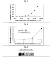
Fig. 1 shows the results of antibody titer measurement in Example 1. -
Fig. 2 shows the results of competitive ELISA measurement. -
Fig. 3 shows the results of ELISA measurement of an anti APP-SP monoclonal antibody. -
Fig. 4 shows the results of ELISA measurement for random APP-SP (SEQ ID NO: 2). -
Fig. 5 shows the results of measurement in a dot-blot method. -
Fig. 6 shows the results of antibody titer measurement in Reference Example. - Preferred aspects of the invention will be specifically described below. The invention is not intended to be limited to the following exemplary embodiments, and various modifications can be made within the scope of the gist of the invention.
- The antibody according to an aspect of the invention is an antibody against an amyloid precursor protein signal peptide (APP-SP), and the antibody is preferably an antibody specifically recognizing the amyloid precursor protein signal peptide.
- According to an aspect of the invention, the term " amyloid precursor protein signal peptide" (or sometimes refereed to as "APP-SP") means (a) a peptide represented by the amino acid sequence described in SEQ ID NO: 1. In the sequence, the description Met (M) means free methionine or S-methylmethionine, and amino acids are represented in one-letter or three-letter code according to IUPAC nomenclature.
- The amyloid precursor protein signal peptide may be a naturally-derived isolated peptide or a peptide produced by a heretofore known method. In the field of peptide synthesis, the peptide having the amino acid sequence having SEQ ID NO: 1 is a peptide which can be chemically synthesized by a heretofore known method or can be synthesized by a recombinant technique using a microorganism or the like.
- The antibody recognizing APP-SP obtained in an aspect of the invention may be a polyclonal antibody or a monoclonal antibody. Recent research results (see, for example, Nature, 2009, vol.457, pp.981-989) have shown that the N-terminal region including a signal peptide portion of amyloid precursor protein promotes the degeneration of neurons. Thus, an antibody which binds to APP-SP receiving attention as a causative substance for Alzheimer's disease as an antigen can be expected in future to be capable of being used for the analysis, diagnosis, treatment, or the like of Alzheimer's disease.
- The antibody against APP-SP according to an aspect of the invention is an antibody obtained using an antigen including APP-SP as an antigen, and, for example, as the antigen,
- (a) a peptide having the amino acid sequence of SEQ ID NO: 1, or
- (b) a peptide having an amino acid sequence in which one or more amino acids are substituted, deleted, added and/ or inserted in the amino acid sequence of SEQ ID NO: 1 may be used.
- Preferably as the peptide having the amino acid sequence of SEQ ID NO: 1 is used as an antigen, the obtained antibody recognizes APP-SP.
- An antigen-antibody reaction can be performed by using the above-referenced antigen and administering a substance being the antigen to an immunized animal as heretofore known.
- The substance being the antigen can be administered to raise an immune response in the immunized animal to produce the antibody against APP-SP according to an aspect of the invention.
- Examples of the immunized animal include, but are not limited to, guinea pigs, rats, mice, rabbits, and sheep, which are used as experimental animals.
- Examples of method for administering the antigen include, but are not limited to, administration routes such as intraperitoneal, intravenous, intramuscular, and intradermal. Example of administration schedule of the antigen includes, but is not limited to, administering the antigen about 2 to 10 times at 2-week administration intervals to enable the production of the antibody against APP-SP.
- In accordance with the administration schedule, a serum sample can be properly collected from the immunized animal to determine the production level of the antibody and confirm whether the immune response is sufficiently raised.
- After final immunization, serum can be collected from the immunized animal, followed by purifying it by a heretofore known method to provide the anti APP-SP antibody.
- Example of dose of the antigen for priming or booster immunization includes, but is not limited to, a dose of 10 to 200 µg per mouse as dose of an antigen corresponding to APP-SP.
- In administering the antigen, an adjuvant may be simultaneously administered. As such adjuvant, a heretofore known adjuvant capable of raising an immune response at a high titer may be used. Examples thereof include Freund's complete adjuvant, Freund's incomplete adjuvant, and aluminum hydroxide.
- A method for production of the antibody according to an aspect of the invention can provide a polyclonal antibody, which can be subsequently purified by a heretofore known method to make the antibody highly pure.
- Antibody-producing cells can be collected from the spleen or lymph node of the immunized animal producing the antibody to produce a hybridoma and also to provide a monoclonal antibody.
- To obtain the antibody against APP-SP according to an aspect of the invention, the following antigen solution can be used.
- The antigen solution is a solution containing an antigen for obtaining an antibody against APP-SP. When the antigen is:
- (a) a peptide having the amino acid sequence of SEQ ID NO: 1, or
- (b) a peptide having an amino acid sequence in which one or more amino acids are substituted, deleted, added and/ or inserted in the amino acid sequence of SEQ ID NO: 1, a peptide micellar aggregate solution containing the peptide and a PEG surfactant can be used as an antigen solution.
- According to an aspect of the invention, the term "PEG surfactant" is not limited, as long as it is a surfactant having a polyethylene glycol (PEG) structure in the molecule.
- Examples of the PEG surfactant include a compound in which PEG as a hydrophilic partial structure and a cholesterol skeleton, saturated or unsaturated aliphatic hydrocarbon skeleton having 8 to 20 carbons, or the like as a hydrophobic partial structure are covalently bound through ether bond, ester bond, or the like to the alcoholic hydroxyl group of the terminus of PEG.
- The polymerization degree of the repeated structure of the PEG portion (ethylene glycol structure) is, for example, 70 or less and may be 60 or less.
- Examples of the PEG surfactant include at least one compound selected from the groups represented by the following formulas.

- In the formulas, n and x means the polymerization degrees of the repeated structures.
- Examples of the PEG surfactant represented by the at least one compound selected from the groups represented by the above formulas include, in the above-referenced formula, CS-050 in which x is 50, PEG60 in which m is 1 and n is 60, PEG36 in which n is 36, PEG24 in which n is 24, and PEG12 in which n is 12.
- Although the mixing of the peptide and a solution containing a PEG surfactant is not limited, the peptide may be mixed in the solution containing a PEG surfactant.
- Then, an adjuvant can be mixed with the resultant mixture to make a peptide micellar aggregate solution, followed by administering it to an immunized animal by using the peptide as an antigen to provide an anti APP-SP antibody.
- Examples of the adjuvant include Freund's complete adjuvant, Freund's incomplete adjuvant, and aluminum hydroxide.
- In mixing the adjuvant, it is preferable to form an emulsion using an ultrasonic processor.
- The peptide micellar aggregate solution may be sized to make the particle size distribution of the peptide micellar aggregate uniform. For example, the particle size can be made uniform by a method such as gel filtration chromatography or filtration by a membrane filter.
- The particle size of the peptide micellar aggregate may be 5 µm to 200 µm, or may be 5 µm to 100 µm.
- The particle size of the peptide micellar aggregate can be measured using a dynamic light scattering particle size measurement apparatus from Nikkiso Co., Ltd.
- In an attempt to prepare an antibody against a peptide by a heretofore known method, an antigen is used in which a plurality of molecules of the peptide is covalently bound to a carrier protein.
- The binding between the peptide and the carrier protein can be formed by a heretofore known method.
- Examples of the carrier protein include keyhole limpet hemocyanin (KLH), bovine serum albumin (BSA), ovalbumin (OVA), mouse serum albumin (MSA), and rabbit serum albumin (RSA).
- When the peptide is bound to a carrier protein, cysteine or the like can be added to the N-terminus of the peptide to use a linker binding to the carrier protein.
- Such a linker is not limited, as long as it is a linker enabling the covalent binding between the carrier protein and the cysteine-added peptide. However, examples thereof include linkers having functional groups, such as an N-hydroxysuccinimido-activated ester (NHS ester), maleimide, and carbodiimide.
- According to an aspect of the invention, in the case of obtaining an antibody against APP-SP by causing a carrier protein to bear an antigen site, the antibody against APP-SP have not been able to be obtained when a peptide having the full-length peptide of APP-SP is used such as (a) a peptide having the amino acid sequence of SEQ ID NO: 1 as an antigen, as disclosed in Examples.
- Although the method for administering an antigen to obtain an antibody according to an aspect of the invention is not limited, by measuring the antibody titer, the antigen may be administered until the desired antibody titer is obtained.
- The antibody titer can be measured with absorbance. The antibody titer can be measured using a blood sample properly collected from the immunized animal. A blood sample from which foreign substances are removed by centrifugation or the like is preferably used.
- The antibody titer can be measured using a heretofore known method, not limited, such as ELISA (enzyme linked immunosorbent assay).
- When serum having the desired antibody titer is obtained by the booster immunization, it can be purified by a heretofore known method to provide an anti APP-SP antibody.
- For the antibody purification, for example, chromatography purification such as ion-exchange chromatography, gel filtration chromatography, and affinity chromatography, and salting-out or the like can be used. The antibody thus obtained from the blood sample is a polyclonal antibody recognizing APP-SP.
- The antigen specificity of the polyclonal antibody can be confirmed by a method such as ELISA.
- When the antibody recognizing APP-SP is found to be obtained, antibody-producing cells can be collected from the spleen, lymph node, or the like of the immunized animal by a heretofore known method. The resultant antibody-producing cells can be used for cell fusion to provide hybridoma (fused cells). Antibody-producing cells can be fused with myeloma cells to provide hybridoma.
- AS the myeloma cell for fusing with the antibody-producing cell, a commonly available cell line from an animal such as a mouse may be used.
- Examples of the myeloma cell include P3-X63-Ag8-U1 and SP2/0-Ag14.
- After cell fusion, only hybridoma can be selectively proliferated by screening. As a screening method,cultivation in HAT medium or the like is prefer. At this time, the cultivation may be performed in the presence of feeder cells such as thymus cells to help the proliferation of hybridoma. The antigen specificity of the antibody produced by the resultant hybridoma can be confirmed by measuring the culture supernatant by a known method such as ELISA to select only hybridoma in which desired antibody production has been able to be confirmed. Then, the resultant hybridoma is cloned to remove contaminating undesired cells and thus hybridoma being single clone can be obtained. Examples of cloning method include a limiting dilution method, a soft agar method, and a fibrin gel method. The resultant hybridoma can be freeze preserved by a heretofore known method. A monoclonal antibody can be produced from the cloned hybridoma by a heretofore known method. For example, a monoclonal antibody can be produced by proliferating the hybridoma in the abdominal cavity of a mouse and purifying the ascites fluid containing the monoclonal antibody. A monoclonal antibody can also be produced by culturing the hybridoma in a serum-free medium and purifying the culture supernatant.
- The antibody obtained in an aspect of the invention is a mouse antibody when a mouse is used as an immunized animal. The anitbody may be made in the form of a chimeric antibody, a humanized antibody, or a human antibody by a heretofore known method. Particularly, a chimeric antibody, humanized antibody, or human antibody recognizing APP-SP is an antibody effective for the treatment of Alzheimer's disease.
- The invention will be described below in further detail with reference to Examples. However, the invention is not intended to be limited to these Examples.
- Example 1: Preparation of Anti APP-SP Monoclonal Antibody
- (1) Synthesis of Peptide (APP-SP)
- The human amyloid precursor proteinsignal peptide (APP-SP, SEQ ID NO: 1) was chemically synthesized using Fmoc solid phase synthesis method under commission to GL Biochem Ltd. (Shanghai).
- The chemically synthesized peptide (APP-SP) was purified by a concentration gradient method in which an ODS column was used and 0.1% trifluoroacetic acid (TFA)-containing water/acetonitrile (MeCN) was used as a mobile phase.
- (2) Preparation of Antigen Solution and Immunization Work
- By using PEG60 (from Polypure) represented by the following formula (I) as a PEG surfactant, APP-SP chemically synthesized in (1) was adjusted so as to provide 0.8 mg/mL APP-SP and 1% PEG60 to give a mixed aqueous solution in which APP-SP was solubilized.

- The resultant adjusted solution was mixed with an equal volume of an adjuvant, which was then sonicated to give an antigen solution as an emulsion.
- The antigen solution was intraperitoneally administered to a female BALB/c mouse according to an administration schedule as shown in Table 1.
Blood collection 1 to 7 was performed from the eyeground. As the adjuvant, Freund's complete adjuvant (from Sigma) was used at priming immunization (the 0th day) and Freund's incomplete adjuvant (from Sigma) was used during booster immunization (on or after 14th day). -
[Table 1] Number of Days Work Item 0 Blood Collection 1, Antigen Administration (60 µg)14 Antigen Administration (60 µg) 21 Blood Collection 228 Antigen Administration (60 µg) 35 Blood Collection 342 Antigen Administration (60 µg) 49 Blood Collection 456 Antigen Administration (60 µg) 63 Blood Collection 570 Antigen Administration (60 µg) 77 Blood Collection 684 Antigen Administration (60 µg) 91 Blood Collection 7, Final Antigen Administration (60 µg) - (3) Measurement of Antibody Titer
- To evaluate the results of the immunization performed in (2), ELISA (Enzyme-Linked ImmunoSorbent Assay) was carried out using the serum obtained in the blood collection shown in Table 1, as follows.
- APP-SP chemically synthesized in (1) and PEG60 were adjusted to 2.5 mg/mL APP-SP and 1% PEG60, and the resultant adjusted solution was filtered with a 0.45 µm filter.
- The filtrate was diluted 1:350 with pure water, which was then added in an amount of 100 µL/well to a 96-well microwell plate (from Nunc) and allowed to stand at room temperature overnight. The solution in each well was discarded; thereto was added 250 µL of 3% BSA (from Sigma, Albumin, bovine serum, Fraction V, minimum 96%, lyophilized powder), 0.1% NaN3, and PBS (phosphate buffered saline, pH 7.0) and the resultant was allowed to stand for 3 hours. The solution in each well was discarded; thereto was added 100 µL of each of the sera obtained in
blood collection 1 to 7 diluted 1:1,000 with 1% BSA, 0.05% NaN3, and TBS (Tris buffered saline, pH 7.5); and the resultant was allowed to stand for 1 hour. Each well was washed 6 times with PBST (PBS (pH 7.4), 0.1% Tween20); thereto were then added 100 µL of HRP labeled Anti mouse IgG (H+L chain) (from MBL) diluted 1:4,000 with 1% BSA-containing PBS (pH 7.4); and the resultant was allowed to stand for 1 hour. Each well was washed 6 times with PBST; 100 µL of TMB+ (from Dako) was then added thereto; and the resultant was allowed to stand in a dark room for 30 minutes. 100 µL of 2 N sulfuric acid was added thereto, and the resultant was then measured for absorbance at 450 nm and 650 nm. The obtained results are shown inFig. 1 with the values of absorbance at 450 nm, less that at 650 nm as ordinate. - From the results of
Fig. 1 , it could be confirmed that an antibody having the property of binding to the human amyloid precursor protein signal peptide (APP-SP) was produced in a mouse body. - (4) Preparation of Hybridoma Producing Anti APP-SP Monoclonal Antibody
- Hybridomas were prepared based on B cells of the mouse in which an increase in the antibody titer was observed in (3) Measurement of Antibody Titer under commission to Bex Co., Ltd.
- Myeloma cells P3U1 were subjected to cell fusion with B cells prepared by removing the spleen of a mouse at the 94th day after the start of immunization, at which an increase in the titer of the antibody against APP-SP was confirmed, by the action of 50% PEG1500 (from Roche). After fusion, cells were seeded in 10 sheets of 96-well plates to which feeder cells were added in advance, and hybridomas were selectively proliferated in HAT medium.
- Cells in the wells in which reactivity was confirmed were subjected to expansion culture in a 24-well plate. After expansion culture, ELISA was performed as in (3), and hybridomas exhibiting positivity were selected and cloned. Cloning was carried out by repeating a limiting
dilution method 2 times, and one type of clone producing a monoclonal antibody exhibiting reactivity to APP-SP was finally obtained (hereinafter, this clone is referred to as 12A3 hybridoma). The 12A3 hybridoma was internationally deposited under the Budapest Treaty on the International Recognition of the Deposit of Microorganisms for the Purposes of Patent Procedure in National Institute of Technology and Evaluation, Patent Microorganisms Depositary (NPMD, 2-5-8 Kazusakamatri, Kisarazu-shi, Chiba, Japan), January 6, 2012, and the deposit has been received (receipt number NITE ABP-1198). - (5) Evaluation of Anti APP-SP Monoclonal Antibody Produced by 12A3 Hybridoma
- Competitive ELISA was performed to examine the reaction specificity of the anti APP-SP monoclonal antibody for APP-SP. Specifically, in the same way as in (3), for the primary antibody reaction, the supernatant of a culture in which 12A3 hybridoma was cultured was used after 1:3 dilution, and the case of containing 0.2% PEG60 in the solution (competition -) and the case of containing 100 µg/mL APP-SP and 0.2% PEG60 therein (competition +) were prepared and compared. As shown in
Fig. 2 for the results, the competition of APP-SP decreased the signal intensity by 92%; thus, the monoclonal antibody could be confirmed to have the property of binding to APP-SP. - When the subclass of the monoclonal antibody produced by 12A3 hybridoma was analyzed using a mouse monoclonal antibody subclass determination kit (from Roche), it was shown to be IgG2a.
- (6) Production of Anti APP-SP Monoclonal Antibody
- Pristane (from Sigma) was intraperitoneally administered in a volume of 0.5 mL per mouse to 3 female BALB/c mice. After 1 week, 1 × 10⁷ cells per mouse of 12A3 hybridoma cultured in advance was intraperitoneally administered. After 1 week, ascites fluid was collected and centrifuged to recover the supernatant. From the 3 mice, 9.5 mL of the supernatant was obtained. A saturated ammonium sulfate solution was added dropwise to 50% to the resultant supernatant, which was then stirred at 4°C for 1 hour.
- Then, centrifugation operation was carried out, and the supernatant was discarded. Again, 50% saturated ammonium sulfate and PBS (pH 7.4) were added, and the resultant was slightly stirred and then centrifuged, followed by discarding the supernatant. PBS (pH 7.4) was added to the resultant precipitate for resolution. In addition, buffer exchange to phosphate buffer was performed using a desalting column, PD-10 (from GE Healthcare). From the solution, only an IgG fraction was purified by affinity chromatography using a protein G column (from GE Healthcare). 0.1 M glycine (pH 2.7) was used as an eluting solution, and buffer exchange to PBS (pH 7.4) was performed using the desalting column PD-10. The measurement of absorbance at UV 280 nm revealed that 15 mg of the anti APP-SP monoclonal antibody had been obtained.
- (7) ELISA Evaluation of Purified Anti APP-SP Monoclonal Antibody
- The same ELISA as in (3) was carried out using the anti APP-SP monoclonal antibody obtained in (6). However, a solution containing the anti APP-SP monoclonal antibody obtained in (6) was used for a primary antibody. The results are shown in
Fig. 3 . - Thus, after purification, the reactivity of the antibody against APP-SP could be confirmed.
- (8) Evaluation of Specificity of Anti APP-SP Monoclonal Antibody
- To evaluate the reaction specificity of the antibody obtained in this Example, its reactivity was evaluated to random APP-SP (SEQ ID NO: 2) prepared by shuffling the amino acid sequence of the antigen APP-SP having exhibited reactivity in (7), by using the same ELISA as in (7). The results are shown in
Fig. 4 . From these results, the reaction specificity was confirmed to be high since the antibody did not exhibit reactivity to random APP-SP containing the same amino acid components as those of APP-SP but having a different arrangement sequence. - (9) Evaluation of Anti APP-SP Monoclonal Antibody in Dot Blot Method
- It was examined whether the antibody obtained in this Example could also detect APP-SP in a dot blot method. 1 µL each of 1% PEG60 aqueous solutions containing 1 mg/mL, 0.1 mg/mL, 0.01 mg/mL, and 0 mg/mL APP-SP were each added dropwise to a nitrocellulose membrane (from GE Healthcare) and dried. The resultant was immersed in a solution containing 1% skim milk, TBS (pH 7.5), and 0.1% Tween20 and shaken for 1 hour. Then, the solution was discarded; a solution containing 0.2% skim milk, TBS (pH 7.5), and 0.1% Tween20, containing 10 µg/mL of the anti APP-SP monoclonal antibody was added; and shaking was performed for 1 hour. Thereafter, the antibody solution was discarded, and washing operation was performed 3 times with the solution containing 0.2% skim milk, TBS (pH 7.5), and 0.1% Tween20 (shaking was carried out for 5 minutes per operation). Then, thereto was added an anti mouse IgG alkaline phosphatase conjugate (from Sigma) diluted 1:4,000 with the solution containing 0.2% skim milk, TBS (pH 7.5), and 0.1% Tween20, followed by shaking for 1 hour. After discarding the solution, washing operation was performed 3 times with the solution containing 0.2% skim milk, TBS (pH 7.5), and 0.1% Tween20 (shaking was carried out for 5 minutes per operation). Finally, NBT/BCIP solution (from PIERCE) was added, followed by shaking for 30 minutes. The results are shown in
Fig. 5 . From these results, the antibody was shown to be also capable of detecting APP-SP in the dot blot method. - Reference Example
- (1) Synthesis of Peptide
- A peptide in which a Cys residue was added to the N-terminus of the human amyloid precursor protein signal peptide (APP-SP, SEQ ID NO: 1) (hereinafter, sometimes referred to as Cys-APP-SP, SEQ ID NO: 3) was chemically synthesized under commission to GL Biochem Ltd. (Shanghai).
- The chemical synthesis of the peptide was performed by Fmoc solid phase synthesis method. The chemically synthesized peptide (Cys-APP-SP) was purified by a concentration gradient method in which an ODS column was used and 0.1% TFA-containing water/MeCN was used as a mobile phase.
- (2) Preparation of Antigen by Cross-Linking Reaction between Cys-APP-SP and Keyhole Limpet Hemocyanin (KLH)
- A 10 mg/mL DMF solution of Cys-APP-SP and a solution containing 1% PEG60, 83 mM sodium phosphate buffer, 100 mM EDTA, and 0.9 M NaCl (pH 7.2) were mixed at a volume ratio of 1:4. This mixed solution and 10 mg/mL maleimide-activated KLH were mixed in equal amounts and stirred at room temperature for 4 hours. A desalting column was used to separate only a fraction presumed to contain a cross-linking reaction product between the carrier protein and APP-SP.
- (3) KLH-Cys-APP-SP Administration to Mouse
- The antigen prepared in (2) was immunized into a mouse according to an administration schedule as shown in Table 2, and blood collection was carried out. A serum fraction was obtained from the blood obtained in the blood collection by a centrifugation operation.
- An adjuvant was mixed in an amount equal to 0.2 mg/mL of the fraction as an antigen (resulting in a concentration of 0.1 mg/mL), and the resultant was sonicated to make an emulsion to provide an antigen solution. As the adjuvant, Freund's complete adjuvant (from Sigma) was used at priming immunization (the 0th day) and Freund's incomplete adjuvant (from Sigma) was used during booster immunization (on or after 14th day).
-
[Table 2] Number of Days Work Item 0 Blood Collection 1, Antigen Administration (20 µg)14 Antigen Administration (10 µg) 22 Blood Collection 228 Antigen Administration (10 µg) 35 Blood Collection 342 Antigen Administration (10 µg) 49 Blood Collection 456 Antigen Administration (10 µg) 63 Blood Collection 570 Antigen Administration (10 µg) 77 Blood Collection 685 Antigen Administration (10 µg) 91 Blood Collection 7 - (4) Measurement of Antibody Titer
- To evaluate the results of the immunization performed in (3), the serum obtained by the blood collection method shown in Table 2 was used to carry out ELISA as follows.
- A 0.002% Tween20 solution containing 10 µg/mL APP-SP was added in an amount of 100 µL/well to a microwell plate (from Nunc) and placed in a state allowed to stand at room temperature overnight. The following day, the solution in the wells was discarded, and 250 µL of a solution containing 3% BSA, 0.1% NaN3, and PBS (pH 7.4) was added, followed by allowing the resultant to stand at room temperature for 2.5 hours. Subsequently, the solution in the wells was discarded, and 100 µL of a solution was added in which serum was diluted 1:100 with a solution containing 1% BSA, 0.05% NaN3, TBS (pH 7.5)(a primary antibody diluent). After standing for 1 hour, the resultant was washed 6 times with PBST (PBS (pH 7.4), 0.1% Tween20). Then, HRP-labeled Anti mouse IgG (H+L chain) (from MBL) as a secondary antibody was diluted 1:4,000 with 1% BSA-containing PBS (pH 7.4), and 100 µL of the resultant was added. After standing for 1 hour, washing was carried out 6 times with PBST. Then, 100 µL of TBS+ was added thereto, and the resultant was allowed to stand for 30 minutes. 100 µL of 2 N sulfuric acid was added to stop enzyme reaction, and absorbance at 450 nm and 650 nm was measured. The results are shown in
Fig. 6 . These results showed that an antibody against APP-SP was not obtained. - The present application is based on Japanese Patent Application No.
2001-2464 filed January 7, 2011 2011-2465 filed January 7, 2011 - According to an aspect of the invention, an antibody against the amyloid precursor protein signal peptide (APP-SP) can be provided. The anti APP-SP antibody according to an aspect of the invention has the possibility of being capable of used for the diagnosis, treatment, or the like of Alzheimer's disease.
- According to an aspect of the invention, an antibody against the amyloid precursor protein signal peptide (APP-SP) can be provided. The anti APP-SP antibody according to an aspect of the invention has industrial applicability in that it can be used for the diagnosis, treatment, or the like of Alzheimer's disease.
- SEQ ID NO: 1 shows the amino acid sequence of the amyloid precursor protein signal peptide (APP-SP).
- SEQ ID NO: 2 shows the amino acid sequence of random APP-SP prepared by shuffling the amino acid sequence of APP-SP.
- SEQ ID NO: 3 shows the amino acid sequence of a peptide in which a Cys residue is added to the N-terminus of APP-SP.


Claims (6)
- An antibody against an amyloid precursor protein signal peptide.
- The antibody according to claim 1, wherein the signal peptide is a peptide having the amino acid sequence of SEQ ID NO: 1.
- The antibody according to claim 1 or 2, wherein the antibody is obtained using the peptide having the amino acid sequence of SEQ ID NO: 1.
- The antibody according to any one of claims 1 to 3, wherein the antibody is a monoclonal antibody.
- A hybridoma producing the monoclonal antibody according to claim 4.
- A hybridoma indicated by receipt number NITE ABP-1198.
Applications Claiming Priority (3)
| Application Number | Priority Date | Filing Date | Title |
|---|---|---|---|
| JP2011002464 | 2011-01-07 | ||
| JP2011002465 | 2011-01-07 | ||
| PCT/JP2012/050210 WO2012093732A1 (en) | 2011-01-07 | 2012-01-06 | Antibody to signal peptide of amyloid precursor protein |
Publications (2)
| Publication Number | Publication Date |
|---|---|
| EP2662387A1 true EP2662387A1 (en) | 2013-11-13 |
| EP2662387A4 EP2662387A4 (en) | 2015-08-05 |
Family
ID=46457582
Family Applications (1)
| Application Number | Title | Priority Date | Filing Date |
|---|---|---|---|
| EP12732284.0A Ceased EP2662387A4 (en) | 2011-01-07 | 2012-01-06 | ANTIBODY AGAINST PEPTIDE AMYLOID PRECURSOR PROTEIN SIGNAL |
Country Status (5)
| Country | Link |
|---|---|
| US (1) | US9309307B2 (en) |
| EP (1) | EP2662387A4 (en) |
| JP (2) | JP5650249B2 (en) |
| CN (1) | CN103492415B (en) |
| WO (2) | WO2012093733A1 (en) |
Families Citing this family (7)
| Publication number | Priority date | Publication date | Assignee | Title |
|---|---|---|---|---|
| US9238796B2 (en) | 2010-06-04 | 2016-01-19 | Toagosei Co. Ltd. | Cell growth-promoting peptide and use thereof |
| JP6077858B2 (en) | 2011-01-07 | 2017-02-08 | 東亞合成株式会社 | Antigen preparation method for obtaining anti-hydrophobic peptide antibody |
| WO2013180011A1 (en) * | 2012-05-28 | 2013-12-05 | 東亞合成株式会社 | Anti-bacterial peptide and use thereof |
| JP6311935B2 (en) | 2012-10-18 | 2018-04-18 | 東亞合成株式会社 | Synthetic peptide that suppresses expression of type 2 TNF receptor and use thereof |
| TW202300517A (en) | 2021-03-12 | 2023-01-01 | 美商美國禮來大藥廠 | Anti-amyloid beta antibodies and uses thereof |
| WO2022251048A1 (en) | 2021-05-24 | 2022-12-01 | Eli Lilly And Company | Anti-amyloid beta antibodies and uses thereof |
| CN114478762B (en) * | 2022-02-15 | 2024-06-04 | 武汉天德生物科技有限公司 | Beta-amyloid polypeptide 1-42 monoclonal antibody, ELISA kit and application thereof |
Family Cites Families (17)
| Publication number | Priority date | Publication date | Assignee | Title |
|---|---|---|---|---|
| US5234814A (en) * | 1989-06-01 | 1993-08-10 | Du Pont Merck Pharmaceutical Company | Diagnostic assay for alzheimer's disease |
| US5242932A (en) * | 1991-12-17 | 1993-09-07 | The Rockefeller University | Treatment of amyloidosis associated with alzheimer disease |
| GB9207731D0 (en) * | 1992-04-07 | 1992-05-27 | Proteus Molecular Design | Improvements in or relating to vaccines |
| JPH07132033A (en) * | 1993-11-12 | 1995-05-23 | Hoechst Japan Ltd | Alzheimer's disease model transgenic animal |
| HRP990246A2 (en) * | 1998-08-07 | 2000-06-30 | Du Pont Pharm Co | Succinoylamino benzodiazepines as inhibitors of a beta protein production |
| AU2002351239A1 (en) * | 2001-12-04 | 2003-06-17 | Dana-Farber Cancer Institute, Inc. | Antibody to latent membrane proteins and uses thereof |
| WO2005052593A1 (en) * | 2003-10-29 | 2005-06-09 | The University Of Leicester | Detection |
| CA2551896A1 (en) * | 2003-12-30 | 2005-07-21 | Wyeth | Formulations of hydrophobic proteins in an immunogenic composition having improved tolerability |
| CN101421624A (en) * | 2004-04-02 | 2009-04-29 | 默克公司 | Method and binding compounds for detecting substances binding to amyloid precursor protein or amyloid beta fragment |
| ZA200904323B (en) * | 2006-12-22 | 2010-08-25 | Genentech Inc | DR6 antibodies inhibiting the binding of DR6 to APP, and uses thereof in treating neurological disorders |
| WO2009020093A1 (en) | 2007-08-09 | 2009-02-12 | Daiichi Sankyo Company, Limited | Immunoliposome inducing apoptosis into cell expressing death domain-containing receptor |
| ITMI20071975A1 (en) * | 2007-10-12 | 2009-04-13 | Fond I R C C S Istituto Neur O | PRODUCTS AND THEIR USE FOR DIAGNOSIS PREVENTION AND-OR TREATMENT OF HUMAN PATHOLOGIES E-O ANIMALS CHARACTERIZED BY THE ANOMALA DEPOSITION OF B-AMYLOID E-O SIMILAMYLOID SUBSTANCE IN HUMAN ORGANS AND TESSTUI E-O ANIMALS AND SCREENING METHOD FOR DETERMINATION |
| GB0720912D0 (en) * | 2007-10-25 | 2007-12-05 | Univ Cardiff | Monoclonal Anitbody for APP |
| JP5709013B2 (en) * | 2009-04-10 | 2015-04-30 | 東亞合成株式会社 | Neuronal differentiation-inducing peptide and use thereof |
| JP5731109B2 (en) * | 2009-07-09 | 2015-06-10 | 東亞合成株式会社 | Method for producing antibody against hydrophobic peptide |
| US8486657B2 (en) * | 2010-03-22 | 2013-07-16 | Xiaohong Cai | Anti-preproprotein and anti-preprotein antibodies as immunohistochemical markers |
| JP6077858B2 (en) * | 2011-01-07 | 2017-02-08 | 東亞合成株式会社 | Antigen preparation method for obtaining anti-hydrophobic peptide antibody |
-
2012
- 2012-01-06 WO PCT/JP2012/050211 patent/WO2012093733A1/en not_active Ceased
- 2012-01-06 JP JP2012551893A patent/JP5650249B2/en not_active Expired - Fee Related
- 2012-01-06 WO PCT/JP2012/050210 patent/WO2012093732A1/en not_active Ceased
- 2012-01-06 US US13/978,584 patent/US9309307B2/en active Active
- 2012-01-06 JP JP2012551894A patent/JP5855583B2/en not_active Expired - Fee Related
- 2012-01-06 CN CN201280004580.0A patent/CN103492415B/en not_active Expired - Fee Related
- 2012-01-06 EP EP12732284.0A patent/EP2662387A4/en not_active Ceased
Also Published As
| Publication number | Publication date |
|---|---|
| JPWO2012093733A1 (en) | 2014-06-09 |
| WO2012093733A1 (en) | 2012-07-12 |
| JP5650249B2 (en) | 2015-01-07 |
| JP5855583B2 (en) | 2016-02-09 |
| EP2662387A4 (en) | 2015-08-05 |
| WO2012093732A1 (en) | 2012-07-12 |
| CN103492415A (en) | 2014-01-01 |
| US9309307B2 (en) | 2016-04-12 |
| CN103492415B (en) | 2017-12-29 |
| US20130345408A1 (en) | 2013-12-26 |
| JPWO2012093732A1 (en) | 2014-06-09 |
Similar Documents
| Publication | Publication Date | Title |
|---|---|---|
| US9309307B2 (en) | Antibody against amyloid precursor protein signal peptide | |
| DK1625166T3 (en) | Antibodies to MASP-2 | |
| US10604586B2 (en) | Humanized monoclonal antibody and uses thereof | |
| EP4703474A1 (en) | Anti-lrrc15 antibody | |
| US12540190B2 (en) | Anti-BCMA antibody, antigen-binding fragment thereof and medical use thereof | |
| CN116041502A (en) | Monoclonal antibody for recognizing phosphorylation of Tau protein pT181 and application thereof | |
| JP2025146838A (en) | Antibodies to misfolded TDP-43 and methods of use thereof | |
| JPWO2018105560A1 (en) | Claudin 5 antibody and medicament containing the antibody | |
| EP3016977B1 (en) | Human anti-il-32 antibodies | |
| CN113227148A (en) | anti-GPC 3 antibody, antigen-binding fragment thereof, and medicinal use thereof | |
| CN109678950B (en) | spink1 antigen, antibodies capable of specifically binding spink1 and functional fragments thereof, and their applications and products | |
| CA2897914A1 (en) | Humanized anti-hmgb1 antibody or antigen-binding fragment thereof | |
| JP2024504454A (en) | Antibody-drug conjugates and their medical uses | |
| AU2014409558A8 (en) | Monoclonal anti-GPC-1 antibodies and uses thereof | |
| AU2007320657B2 (en) | Antibody recognizing C-domain of midkine | |
| AU2011327208B2 (en) | Anti single-strand type-IV collagen polypeptide antibody, and pharmaceutical, or agent for diagnosing, preventing or treating tumours, containing same | |
| CN120882741A (en) | Combinatorial epitope antibody library | |
| CN120202223A (en) | Anti-human LAIR1 antibody | |
| HK40049006B (en) | Anti-gpc3 antibody, antigen-binding fragment thereof, and medical use thereof | |
| HK40049006A (en) | Anti-gpc3 antibody, antigen-binding fragment thereof, and medical use thereof | |
| KR20220029653A (en) | Antibodies directed against APOE amino-terminal fragment of 12 KDA |
Legal Events
| Date | Code | Title | Description |
|---|---|---|---|
| PUAI | Public reference made under article 153(3) epc to a published international application that has entered the european phase |
Free format text: ORIGINAL CODE: 0009012 |
|
| 17P | Request for examination filed |
Effective date: 20130807 |
|
| AK | Designated contracting states |
Kind code of ref document: A1 Designated state(s): AL AT BE BG CH CY CZ DE DK EE ES FI FR GB GR HR HU IE IS IT LI LT LU LV MC MK MT NL NO PL PT RO RS SE SI SK SM TR |
|
| DAX | Request for extension of the european patent (deleted) | ||
| RA4 | Supplementary search report drawn up and despatched (corrected) |
Effective date: 20150703 |
|
| RIC1 | Information provided on ipc code assigned before grant |
Ipc: C12P 21/08 20060101ALI20150629BHEP Ipc: C12N 5/10 20060101ALI20150629BHEP Ipc: C07K 16/18 20060101AFI20150629BHEP |
|
| 17Q | First examination report despatched |
Effective date: 20160711 |
|
| REG | Reference to a national code |
Ref country code: DE Ref legal event code: R003 |
|
| STAA | Information on the status of an ep patent application or granted ep patent |
Free format text: STATUS: THE APPLICATION HAS BEEN REFUSED |
|
| 18R | Application refused |
Effective date: 20180129 |