EP2660593A2 - Determination of levels of immunogenic gluten peptides in human samples - Google Patents
Determination of levels of immunogenic gluten peptides in human samples Download PDFInfo
- Publication number
- EP2660593A2 EP2660593A2 EP11852781.1A EP11852781A EP2660593A2 EP 2660593 A2 EP2660593 A2 EP 2660593A2 EP 11852781 A EP11852781 A EP 11852781A EP 2660593 A2 EP2660593 A2 EP 2660593A2
- Authority
- EP
- European Patent Office
- Prior art keywords
- gluten
- peptides
- monitoring
- gliadin
- seq
- Prior art date
- Legal status (The legal status is an assumption and is not a legal conclusion. Google has not performed a legal analysis and makes no representation as to the accuracy of the status listed.)
- Granted
Links
Images


Classifications
-
- G—PHYSICS
- G01—MEASURING; TESTING
- G01N—INVESTIGATING OR ANALYSING MATERIALS BY DETERMINING THEIR CHEMICAL OR PHYSICAL PROPERTIES
- G01N33/00—Investigating or analysing materials by specific methods not covered by groups G01N1/00 - G01N31/00
- G01N33/02—Food
-
- G—PHYSICS
- G01—MEASURING; TESTING
- G01N—INVESTIGATING OR ANALYSING MATERIALS BY DETERMINING THEIR CHEMICAL OR PHYSICAL PROPERTIES
- G01N33/00—Investigating or analysing materials by specific methods not covered by groups G01N1/00 - G01N31/00
- G01N33/48—Biological material, e.g. blood, urine; Haemocytometers
- G01N33/50—Chemical analysis of biological material, e.g. blood, urine; Testing involving biospecific ligand binding methods; Immunological testing
- G01N33/68—Chemical analysis of biological material, e.g. blood, urine; Testing involving biospecific ligand binding methods; Immunological testing involving proteins, peptides or amino acids
-
- G—PHYSICS
- G01—MEASURING; TESTING
- G01N—INVESTIGATING OR ANALYSING MATERIALS BY DETERMINING THEIR CHEMICAL OR PHYSICAL PROPERTIES
- G01N33/00—Investigating or analysing materials by specific methods not covered by groups G01N1/00 - G01N31/00
- G01N33/48—Biological material, e.g. blood, urine; Haemocytometers
- G01N33/50—Chemical analysis of biological material, e.g. blood, urine; Testing involving biospecific ligand binding methods; Immunological testing
- G01N33/53—Immunoassay; Biospecific binding assay; Materials therefor
- G01N33/5308—Immunoassay; Biospecific binding assay; Materials therefor for analytes not provided for elsewhere, e.g. nucleic acids, uric acid, worms, mites
-
- G—PHYSICS
- G01—MEASURING; TESTING
- G01N—INVESTIGATING OR ANALYSING MATERIALS BY DETERMINING THEIR CHEMICAL OR PHYSICAL PROPERTIES
- G01N2333/00—Assays involving biological materials from specific organisms or of a specific nature
- G01N2333/415—Assays involving biological materials from specific organisms or of a specific nature from plants
-
- G—PHYSICS
- G01—MEASURING; TESTING
- G01N—INVESTIGATING OR ANALYSING MATERIALS BY DETERMINING THEIR CHEMICAL OR PHYSICAL PROPERTIES
- G01N2800/00—Detection or diagnosis of diseases
- G01N2800/06—Gastro-intestinal diseases
- G01N2800/065—Bowel diseases, e.g. Crohn, ulcerative colitis, IBS
Definitions
- the present invention shows a method for monitoring the ingestion of gluten by measuring protein/gluten peptides present in fecal samples with antibodies against immunogenic peptides resistant to gastrointestinal digestion.
- the presence or absence of these immunogenic peptides is controlled by immunological assays based on antibodies reactive against immunogenic gluten peptides that are resistant to proteolysis. These assays may be quantitative techniques ELISAs, or qualitative as rapid immunochromatographic assays, immunoblots, etc.
- These measures may also be applied to verify compliance with the gluten-free diet, to improve diagnosis in cases of refractory or severe symptoms of celiac disease in which a gluten-free diet is supposedly being respected, or to clinical research on the effectiveness of enzymatic therapies related with prolamin detoxification.
- Gluten is a set of storage proteins of cereals. Gluten proteins from wheat, barley, rye and probably oats, are not tolerated by genetically predisposed individuals with celiac disease (CD).
- CD celiac disease
- gluten is composed of an ethanol-soluble fraction (prolamins: ⁇ , ⁇ , ⁇ and ⁇ -gliadins) and other insoluble, glutenins (high and low molecular weight subunits) ( Wieser, 2007 Food Microbiol., 24:115-119 ; Fasano, 2009, Sci Am, 301:54-61 ). Gliadins and glutenins are also unusually rich in proline ( ⁇ 15%) and glutamine ( ⁇ 35%) residues.
- Intestinal villi are destroyed due to the immunological reaction, resulting in a reduction of the intestinal absorption which can lead to symptoms such as diarrhea, anemia, stunting, weight loss, bone disorders, neurological disorders, cancer, etc. ( Alaedini and Green, 2005, Ann Int Med, 142:289-299 ; Catassi and Fasano, 2008, Curr Opin Gastroenterol., 24:687-691 ; Tack et al., 2010, Gastroenterol Hepatol., 7:204 - 213 ).
- the proteic profile of the prolamins fractions which constitute PWG gliadin was analyzed by SDS-PAGE to observe the pattern of bands obtained after the enzymatic treatment and to confirm that samples had been digested.
- the samples were diluted in running buffer (62.5 mM Tris-HCl pH 6.8, 10% glycerol, 2% SDS, 0.001% bromophenol blue and 5% 2-mercaptoethanol) and denatured by boiling at 100° C for 5 min. This step was repeated a total of three times. Samples were run on 15-18% polyacrylamide gels (SDS-PAGE) at a constant voltage of 100 V using MiniProtein system (BioRad Laboratories). The separated proteins in the electrophoresis gel were stained using silver staining.
Landscapes
- Health & Medical Sciences (AREA)
- Life Sciences & Earth Sciences (AREA)
- Engineering & Computer Science (AREA)
- Immunology (AREA)
- Molecular Biology (AREA)
- Chemical & Material Sciences (AREA)
- Biomedical Technology (AREA)
- Urology & Nephrology (AREA)
- Hematology (AREA)
- Food Science & Technology (AREA)
- Physics & Mathematics (AREA)
- Biochemistry (AREA)
- Pathology (AREA)
- General Physics & Mathematics (AREA)
- Medicinal Chemistry (AREA)
- General Health & Medical Sciences (AREA)
- Analytical Chemistry (AREA)
- Microbiology (AREA)
- Biotechnology (AREA)
- Cell Biology (AREA)
- Tropical Medicine & Parasitology (AREA)
- Proteomics, Peptides & Aminoacids (AREA)
- Peptides Or Proteins (AREA)
- Investigating Or Analysing Biological Materials (AREA)
Abstract
Description
- The present invention, fitted in the medical-clinical sector, shows a method for monitoring the ingestion of gluten by measuring protein/gluten peptides present in fecal samples with antibodies against immunogenic peptides resistant to gastrointestinal digestion. The presence or absence of these immunogenic peptides is controlled by immunological assays based on antibodies reactive against immunogenic gluten peptides that are resistant to proteolysis. These assays may be quantitative techniques ELISAs, or qualitative as rapid immunochromatographic assays, immunoblots, etc. These measures may also be applied to verify compliance with the gluten-free diet, to improve diagnosis in cases of refractory or severe symptoms of celiac disease in which a gluten-free diet is supposedly being respected, or to clinical research on the effectiveness of enzymatic therapies related with prolamin detoxification.
- Gluten is a set of storage proteins of cereals. Gluten proteins from wheat, barley, rye and probably oats, are not tolerated by genetically predisposed individuals with celiac disease (CD). In wheat, gluten is composed of an ethanol-soluble fraction (prolamins: α, β, γ and ω-gliadins) and other insoluble, glutenins (high and low molecular weight subunits) (Wieser, 2007 Food Microbiol., 24:115-119; Fasano, 2009, Sci Am, 301:54-61). Gliadins and glutenins are also unusually rich in proline (~15%) and glutamine (~35%) residues. As a result, while most dietary proteins are digested by gastrointestinal proteases to single amino acids, dipeptides or tripeptides, gluten proteins are not completely digested (Erickson and Kim, 1990, Annu Rev Med 41:133 - 139, Gray, 1991, New York: Oxford University, pp.411-420; Ganapathy et al., 2006, Academic Press, pp.1667-1692). Therefore, some of the gluten peptides generated during gastrointestinal digestion are highly resistant to digestion by gastric and pancreatic enzymes, so that they persist in the gut. These peptides are capable of being internalized into the intestinal cells and, therefore, glutamine residues can be deaminated by tissue transglutaminase (tTG). Genetic predisposition of individuals with CD makes them intolerant to these peptides because their immune system reacts pathologically against autoantigens generated by gluten peptides/tTG-interaction (Korponay-Szabó et al., 2007, BMJ, 335:1244-1247; Bethune and Khosla, 2008, PLoS Pathogens, 4: e34; Jabri and Sollid, 2009, Nat Rev Immunol., 9:858-870). Deamidated peptides induce an immune response mediated by T cells that causes chronic inflammation of the small intestine. Intestinal villi are destroyed due to the immunological reaction, resulting in a reduction of the intestinal absorption which can lead to symptoms such as diarrhea, anemia, stunting, weight loss, bone disorders, neurological disorders, cancer, etc. (Alaedini and Green, 2005, Ann Int Med, 142:289-299; Catassi and Fasano, 2008, Curr Opin Gastroenterol., 24:687-691; Tack et al., 2010, Gastroenterol Hepatol., 7:204 - 213).
- One of the main gluten peptides described to date is the 33-mer peptide from α2-gliadin (Shan et al., 2002, Science, 297:2275-2279; Bethune et al., 2009, Chem Biol, 16 :868-881) that has been shown to be resistant to gastrointestinal digestion, substrate of the tTG mediated deamination and highly reactive with T cells isolated from celiac patients. The identification of the 33-mer peptide and other peptides, helps to demonstrate that gluten epitopes with high antigenicity are located in gliadin regions rich in proline and glutamine residues (Shan et al., 2002, Science, 297: 2275-2279; Tye-Din et al., 2010, Sci Transl Med 2:41 RA51).
- Nowadays, the only existing therapy for patients with celiac disease is a strict gluten-free diet (GFD). Non-compliance with the GFD has been associated with osteoporosis, iron deficiency anemia, depression and infertility, all of which is improved, to some extent, by adhering to the gluten-free diet. These observations give us an idea of the importance of adherence to a GFD to reduce symptoms, prevent nutritional deficiencies and improve the quality of life of these patients. However, several studies based on intestinal biopsies have suggested that dietary transgressions are relatively frequent, being between 32.6% and 55.4% in the populations studied (Ciacci et al., 2002, Digestion, 66: 178-185; Sylvester and Rashid, 2007, Can J Gastroenterol., 21:557-564). The lack of adherence to a strict gluten-free diet is the main reason for poorly controlled celiac disease in adults.
- In addition, there is a part of the celiac population that does not seem to respond positively to the GFD and suffer symptoms of persistent or recurrent malabsorption and intestinal villous atrophy. This population could be suspected of having refractory CD, a rare disease (approximately 5% -10% of patients with CD) that appears in patients without apparent positive response to the gluten-free diet (Al-Shot et al., 2007, Dig Dis 25:230-236, Freeman, 2009, Gut Liver, 3:237-246; Rubio-Tapia and Murray, 2010, Gut, 59:547-557). Although this refractory disease was described in patients with assumed total absence of gluten intake, involuntary ingestion and hypersensitivity to a small amount of gluten can also trigger the symptoms of the disease. The lack of an accurate marker for monitoring compliance with the GFD is still an unresolved issue and is particularly difficult in the case of minor dietary transgressions (Fernandez-Calle et al., 1993, Gut, 34:774-777). There is no way to demonstrate gluten intake and thereby avoid possible harmful consequences. In fact, the consequences of dietary transgressions can only be measured by observing mucosal inflammation and/or villous atrophy for which intestinal biopsies would have to be performed and, as a result, the patient would have to be anesthetised with the possible consequences that this may have.
- Control of anti-tTG has been proposed as a marker to assess the strict compliance with the GFD. However the effectiveness of this marker to control the intake of gluten is not yet clear (Tack et al., 2010, Gastroenterol Hepatol., 7:204-213). Other markers have been proposed for monitoring the diet, such as permeability test (Duerksen et al., 2005) or fecal calprotectin (Ertekin et al., 2010, J Clin Gastroenterol., 44:544-546). These methods can demonstrate the presence of inflammatory processes, so that if the values of these markers are altered it can be a result of infectious diseases, inflammatory bowel diseases or allergy processes, meaning that they do not need to be a measure of the direct intake of gluten. Therefore, there is no effective method to verify that the celiac patient is performing a GFD or to eliminate the possibility that the refractory CD symptoms are due to a hypersensitive intolerance to gluten traces associated with unintentional exposure to toxic cereals.
- Compliance with the diet assessed by interview has been suggested as a marker of CD control for its low cost, non-invasiveness, and its proven correlation with intestinal damage. However, GFD involves numerous restrictions for patients because of its social and economic implications. Additionally, a gluten-free diet is difficult to maintain due to the ubiquity of gluten in foods, educational misinformation, changes in food labeling and possible cross-contamination in food (Bethune et al., 2009, Chem Biol, 16:868-881; Selimoglu and Karabiter, 2010, J Clin Gastroenterol., 44:4-8). Moreover, certain lifestyles and some sectors of the population make difficult, to some extent, compliance with the GFD. Furthermore there is no alternative to patient interviews to know how reliable the results of these clinical trials are.
- A more direct measure of the ingestion of gluten could provide critical information about the patient: the detection of infringements of the GFD before anatomical damage, inadvertent consumption detection, the accuracy assessment of the adherence to treatment in the initial period after diagnosis when patients are less familiar with the diet, etc. providing an easy and reliable confirmation of the results obtained. Therefore, a sensitive and reliable marker to monitor and detect gluten intake could be a useful tool for the proper compliance with the GFD and probably for an accurate diagnosis of refractory CD.
- Monoclonal antibodies (moAbs) G12 and A1 obtained against the main immunogenic epitope of α-gliadin have demonstrated to be very useful in the detection of toxic peptides in food samples as well as in clinical research of gluten enzymatic detoxification (Morón et al., 2008, Am J Clin Nutr., 87:405-414; Morón et al., 2008, PLoS ONE, 3: e2294; Ehren et al., 2009, PLoS ONE, 4: e6313, Alvine Pharmaceuticals, Inc., Biomedal SL). The sensitivity and specificity of the monoclonal antibodies and their ability to recognize peptides resistant to gastrointestinal digestion could make them ideal for monitoring immunotoxic gluten peptides obtained after intestinal digestion in human samples. Recognition epitopes from moAb G12, QP(Q/E)LP(Y/F), are present in major peptides described recently in a high throughput screening performed with 2,700 peptides from prolamins of different cereals (Tye-Din and al., 2010, Sci Transl Med 2:41 RA51). Undigested peptide fragments from gluten intake that are not absorbed could be recovered from the feces, which would demonstrate gluten intake by the individual.
- In this patent, we have evaluated the feasibility of monitoring gluten, intact and digested, in the feces by the detection of epitopes associated with the 33-mer peptide, which could be used in clinical studies and dietary monitoring, as well as in the diagnosis of refractory CD.
- The gluten-free diet is the only effective treatment today for celiac disease. Therefore, compliance with GFD should be monitored to prevent cumulative direct and indirect damage, as well as to confirm the persistence of any symptom of celiac disease is not due to a transgression (intentionally or not) of the diet. However, currently there are no methods to monitor dietary compliance in patients with celiac disease.
- The aim of the present invention is the application of immunological methods for monitoring compliance with the gluten-free diet by detecting immunotoxic peptides present in feces that are resistant to gastrointestinal digestion. It is an object of the invention the application of immunological techniques based on antibodies that recognize immunogenic peptides from gluten that resist gastrointestinal digestion. Preferably, the invention employs immunological techniques using antibodies recognizing the 33-mer peptide from gliadin. A preferent way of making the detection of peptides is by rapid qualitative methods based on immunochromatographic strips or by quantitative and automated methods as ELISA techniques. The preferred method used in the invention should be able to detect gluten ingested equivalent to 50 mgs of wheat gluten per day, which is the maximum amount described for a gluten free diet, or at least 20 ppms of gluten equivalent in feces.
- It is also an object of the present patent kits or immunological analytical devices based on antibodies reactive against the 33-mer from gliadin that are suitable for the detection of gluten peptides in feces.
- These procedures and analytical kits are also an object of this patent because of its use to reveal a lack of adherence to GFD, either due to contamination of food consumed or to a voluntary/involuntary occasional intake of foods containing gluten. Furthermore, it is object of the present invention the application of the detection of these immunotoxic peptides in feces for the clinical research control on celiac disease, including enzymatic therapies related to gluten prolamins detoxification, seize of immunotoxic peptides and other alternative therapies.
- The object of the present invention is a method for detecting or monitoring the gluten ingested by detecting immunotoxic peptides in feces, characterized by the use of immunological methods using antibodies that recognize preferably epitopes related to 33-mer gliadin peptide and other peptide sequences resistant to gastrointestinal digestion.
- It is an object of the present invention the use of immunological techniques for such gluten monitoring, immunological methods such as indirect ELISA, competitive ELISA, sandwich ELISA, immunochromatographic strips, fluorescent immunomicroparticles, Western blot, biosensors based on electrochemical reactions catalyzed by enzymes attached to the antibodies, by magnetic particles coated with antibodies, by surface plasmon resonance, and other techniques in which an analyte bound to an antibody is detected.
- In a preferred embodiment of the invention, these methods are characterized by the preferent use of one or more monoclonal antibodies capable of detecting epitopes contained or similar to peptide 33-mer (SEQ ID No. 1), such as the following sequences: SEQ ID No. 2, SEQ ID No. 3, SEQ ID No. 4, SEQ ID No. 5, SEQ ID No. 6, SEQ ID No. 7, SEQ ID No. 8. In a preferred embodiment of the invention the antibodies used for immunological techniques would be G12 and A1 because of the proven relation between its reactivity and the potential immunotoxicity of a sample. The invention also contemplates the use of the antibody R5 that reacts with the epitope SEQ ID No. 9, which also can be found in gluten peptides resistant to gastrointestinal digestion, but with less specificity against immunogenic peptides resistant to proteases.
- A preferred embodiment of the invention would be the use of immunological methods based on G12 monoclonal antibody conjugated to an enzyme that allows a quantitative assay using chromogenic, luminescent or fluorogenic substrates. This procedure would use a standard of gliadin, hydrolysed gliadin, complete 33-mer peptide or a part of its sequence of at least 6 amino acids (SEQ ID No 2).
- Another object of the invention is constituted of the particular use of immunochromatographic strips based on the anti-33-mer from gliadin antibodies, G12 and A1 which allow a rapid and semiquantitative detection of proteins/peptides from gluten content in feces.
- The invention proposes a measure of the gluten ingested by the individual through the diet. This is a useful process for the control of the gluten ingested by using analytical methods in which a correlation between the quantity of ingested gluten and the estimated quantity of gluten protein/peptides in feces obtained from these methods has been shown.
- The invention proposes an analytical instrument for monitoring compliance with the GFD, as well as to discard uncontrolled intake of gluten in patients suspected of suffering from the called refractory celiac disease. Also, with this procedure, new therapeutic alternatives for enzymatic detoxification of gluten and other alternative therapies may also be controlled in the feces of celiac patients subjected to clinical trials or therapeutic prescription in the future, since the effectiveness of the therapy could be determined by measuring the presence or absence of peptides in feces after 12-48 hours from the intake of a controlled amount of gluten in conjunction with therapies to eliminate immunotoxic peptides.
- Some issues that are not still resolved in clinical practice, such as the control of the GFD or the control of involuntary exposure to gluten due to food contamination, could be resolved with simple immunological assays in feces. Medics, clinicians and analysts might consider these methods useful for clinical trial design and monitoring of their celiac patients to establish consistent conclusions on the state of the patient's disease.
- Extraction of the peptides from the feces can be carried out directly with a hydroalcoholic solution of 40 to 60%. Sometimes, due to the nature of the food ingested, the gluten extraction from the feces could be improved by adding a solution containing dispersing agents such as guanidinium chloride, arginine chloride, etc., or detergents such as polyvinylpyrrolidone, and reducing agents such as bmercaptoethanol, DTT or TCEP.
- Subsequently extracted gluten polypeptides are diluted in a buffered saline solution and are then used to make the measurement with an ELISA, either competitive or sandwich if an idea of the concentration of reactive peptides is to be obtained. The standard curve could be done with standard gliadin digested by trypsin and pepsin to simulate gastric digestion. Synthesized polypeptide reacting with the antibodies could also be used directly, preferably 33-mer from gliadin or fragments thereof with the option of making some specific modifications that retain reactivity against the antibodies. For example the peptides that may be used for antibody G12 are the following: SEQ ID No. 1, SEQ ID No. 2, SEQ ID No. 3, and SEQ ID No. 4. For A1, besides 33-mer, the following peptides could be valid to make the standard curve with a competitive ELISA: SEQ ID No. 5, SEQ ID No. 6, SEQ ID No. 7 or SEQ ID No. 8.
- For a qualitative assay in which it is seen if the value of gluten polypeptides in feces is greater or less than a certain amount, immunochromatographic strips or ELISAs assays, using antibody G12 or A1 or both, could be used. The procedure could be carried out with a kit that contained the extraction solution of the polypeptides in feces; a reference pattern with gluten polypeptides hydrolysed by pepsin and trypsin or synthesized; and the components of an ELISA with a multi-well plate and using the antibodies A1 and/or G12 to immobilize the wells and/or for the development of the assay, or immunochromatographic strips.
- To complement the description being made and in order to aid a better understanding of the characteristics of the invention, according to a preferred practical embodiment thereof, it is attached as an integral part of said description, with illustrative and non limiting character, the following figures:
-
Figure 1 . Relative affinity of moAb G12 to immunotoxic peptides derived from PWG gliadin after simulating gastrointestinal digestion. A and B. SDS-PAGE and Western blot of PWG gliadin, PWG gliadin + pepsin and PWG gliadin + pepsin + trypsin/chymotrypsin. Samples were stained with silver or transferred to a PVDF membrane with moAb G12. MW: molecular weight marker. C. Analysis by competitive ELISA G12-HRP of peptides from gliadin PWG. -
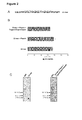
Figure 2 . Resistance of 33-mer peptide to be broken by gastrointestinal enzymes. A. Amino acid sequence of 33-mer peptide. The recognition sequence of moAb G12 into the 33-mer peptide is in bold. B. Competitive ELISA for the detection of 33-mer after treatment with pepsin, trypsin and chymotrypsin using the moAb G12-HRP. C. Western blot of 33-mer peptide after treatment with gastrointestinal enzymes. MW: molecular weight marker. Two separate assays with 3 replicates each one were carried out. -
Figure 3 . Gluten detection in feces from healthy individuals subjected to a gluten-controlled diet. A and B. Gluten peptides/proteins semiquantification in feces of healthy individuals (n = 11) by immunochromatographic strips G12. The upper line is a positive internal control indicates that the device has worked correctly, and bottom line indicates the presence of gluten. HL901-HL911: subjects who participated in the study. *Gluten traces were detected. C and D. SDS-PAGE and Western blot of gluten peptides and proteins extracted from the feces. MW: molecular weight marker. -
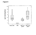
Figure 4 . Concentration of 33-mer peptide (ng/mg) in feces after a gluten-controlled diet. Competitive ELISA G12-HRP to determine the relationship between gluten proteins ingested/excreted by its content in 33-mer peptide. The concentration of 33-mer peptide was determined by a standard curve of 33-mer. Two separate tests were carried out, each with three replicates. - The present example shows that a substantial portion of the immunogenic peptides of gluten remain susceptible for fecal detection despite gastrointestinal digestion. Among the major proteins of the diet, those that constitute gluten are the only ones that contain approximately 15% of proline residues and 35% of glutamine residues. The high content of these two amino acids prevents the complete proteolysis of these proteins by gastric and pancreatic enzymes, so that peptide fragments are formed in the small intestine which are immunotoxic for celiac patients. In particular, the 33-mer peptide was found as one of the main contributors to the immunotoxicity of gluten (Shan et al., 2002, Science, 297:2275-2279). This peptide of the α-2 gliadin contains six recognition epitopes for T cells and is highly resistant to proteolysis.
- The moAb G12 is specific for the epitope of six amino acids SEQ ID No. 10, with 3 repetitions in the 33-mer peptide. Moreover, this antibody is capable of recognizing other immunoreactive peptides present in gliadin and other toxic prolamins. The purpose of this example is to know the capacity of G12 antibody to detect toxic peptides formed after gastrointestinal digestion simulation of gliadin. For standardization of the assay PWG gliadin was used, considered an international reference reagent in gluten analysis due to its high content of gliadins, good solubility, homogeneity, stability and for being constituted of 28 wheat European cultivars (Eckert et al., 2006, J Cereal Sci, 43:331-341).
- Gliadin was subjected to sequential digestion with pepsin (main protease present in the stomach), trypsin and chymotrypsin (proteases contained in the intestinal membrane). The samples were incubated at 37° C in HCl solution (pH 2) containing 0.06 mg/mL of pepsin. Samples were incubated for 60 min and inactivated by heating at 95° C for 5 min. After simulating gastric digestion with pepsin, the digestions were adjusted to pH 6.0 with sodium phosphate buffer, and incubated with pancreatic proteases: trypsin (0.375 mg/mL) and chymotrypsin (0.375 mg/mL). After the duodenal simulation at 37 °C for 30 min the samples were immediately inactivated at 95° C for 5 min.
- The proteic profile of the prolamins fractions which constitute PWG gliadin was analyzed by SDS-PAGE to observe the pattern of bands obtained after the enzymatic treatment and to confirm that samples had been digested. For the analysis by SDS-PAGE, the samples were diluted in running buffer (62.5 mM Tris-HCl pH 6.8, 10% glycerol, 2% SDS, 0.001% bromophenol blue and 5% 2-mercaptoethanol) and denatured by boiling at 100° C for 5 min. This step was repeated a total of three times. Samples were run on 15-18% polyacrylamide gels (SDS-PAGE) at a constant voltage of 100 V using MiniProtein system (BioRad Laboratories). The separated proteins in the electrophoresis gel were stained using silver staining.
- The intact PWG gliadin evaluated by 1D gel revealed intense bands of alpha, beta and gamma gliadin (MW = 33-45 kDa) and weak bands of omega gliadin (MW = 50-67 kDa) (Eckert et al., 2006 J Cereal Sci, 43:331-341). Digestion of these proteins mediated by pepsin (gastric digestion) resulted in the formation of smaller peptide fragments below 25 kDa. Sequentially, trypsin and chymotrypsin digestion generated smaller peptides (less than 15 kDa), resulting from the hydrolysis process mediated by these enzymes (
Figure 1A ). - In order to verify whether PWG gliadin peptides obtained by the process of gastrointestinal digestion were recognized by the anti-33-mer antibody, a Western blot with this antibody was carried out for the samples described above: undigested PWG gliadin, PWG gliadin after gastric digestion and PWG gliadin after intestinal digestion (prior gastric digestion). Protein extracts initially obtained were separated by SDS-PAGE and then incubated with G12 antibody onto PVDF membranes. After that, the samples were incubated in blocking buffer (TBS with 5% skim milk) overnight, after G12 antibody was added (1:5000 dilution in blocking solution). After 3 washes, membranes were incubated with secondary antibody anti-mouse IgG conjugated to phosphatase (Sigma, Sr. Louis, MO) (1:2000 dilution in blocking solution). The membrane was developed using Sigma-Fast system.
- The G12 antibody was able to recognize the different factions into PWG gliadin. After gastric digestion, peptide fragments formed remained being recognized by the G12 antibody (
Figure 1B ). Sequential treatment with pancreatic enzymes (trypsin, chymotrypsin) resulted in the presence of smaller peptides also recognized by moAb G12. - In order to determine the capacity of the G12 antibody to quantify the toxic peptides generated, the concentration of 33-mer and analogues peptides obtained after gastrointestinal simulation of PWG gliadin was determined by competitive ELISA also using the G12 antibody. The competitive ELISA is a very suitable technique to monitor gluten digestion since it is capable of detecting both intact proteins and small protein fragments: the latter could be underestimated by sandwich ELISA, because the detection of antigens requires at least two different epitopes on the peptide molecule.
- The relative amount of immunotoxic epitopes contained in the samples was quantified by competitive ELISA using moAb G12-HRP (Biomedal SL, Seville, Spain). Maxisorp microtiter plates were used for this assay (Nunc, Roskilde, Denmark), which were coated with 100 µL/well of Sigma gliadin solution (5 µg/mL) in 0.1 M PBS (Na2CO3-NaHCO3, pH 9.6), and incubated at 4 ° C overnight. The plates were washed with PBS 0.05
% Tween ® 20 and blocked with blocking solution (PBS, 5% skimmed milk) for 1 h at room temperature. Serial dilutions of the standard (gliadin or 33-mer peptide) and samples studied were made in PBS with 3% BSA (100 µL) and 100 µL of moAb G12 conjugated to HRP solution was added to each one (1:10,000 in PBS with 3% BSA). Samples were pre-incubated 1 h at room temperature with gentle agitation, and then added to the wells. After 30 minutes of incubation, samples were washed, and 100 µL/well of substrate solution was added (TMB, Sigma, St. Louis, Missouri, USA). After 30 minutes incubation at room temperature in darkness, the reaction was stopped with 1 M sulfuric acid (100 µL/well), and absorbance was measured at 450 nm (UVM340 microplate reader, Asys Hitech GMBH, Eugendorf, Austria). Gliadin/33-mer concentration was determined using the 4-parameter model. - The concentration of 33-mer both in intact PWG gliadin and subjected to gastric and intestinal digestion was determined by this method. Gastric digestion of PWG gliadin resulted in a slight increase in the levels of toxic peptide. This increase is probably due to the opening of the molecules constituting the gliadin fractions so that the epitopes of anti-33-mer present are more accessible, and thus, can be identified with greater specificity (intact PWG gliadin = 21, 6 ng 33-mer/µg vs. PWG gliadin after gastric digestion = 24.5 ng3 3-mer/µg). After the following process of intestinal digestion the moAb G12 continued to recognize the PWG gliadin peptides formed, although with less specificity (7.5 ng 33-mer/µg) (
Figure 1C ). - In contrast to these results, both in vitro and in vivo studies made with the 33-mer peptide demonstrate the great stability of this peptide to rupture by gastric, pancreatic and intestinal endoproteases. Its features make it to be suggested as the main promoter of the inflammatory response to gluten in celiac patients (
Figure 2A ) (Shan et al., 2002, Science, 297:2275-2279, 2005, J Proteome Res 4:1732 - 1741). - To verify that the 33-mer peptide remains intact after gastric proteolysis (mediated by pepsin) and sequential intestinal proteolysis (mediated primarily by trypsin and chymotrypsin) an in vitro simulation of gastrointestinal digestion of this peptide was performed. The concentrations of 33-mer peptide obtained after each of the digestion processes were determined by competitive ELISA using the anti-33-mer monoclonal antibody. The concentration of 33-mer obtained after gastric digestion did not differ significantly with respect to the non-digested peptide (194 µg/mL vs. 186 µg/mL, respectively, p = 0.4469). Equally, exposure of the 33-mer to the enzymes trypsin and chymotrypsin (intestinal digestion), did not change the levels of this peptide in comparison with untreated peptide (169 µg/mL vs. 186 µg/mL, respectively, p = 0.1024) (
Figure 2B ). These results confirm the high stability of the 33-mer to hydrolysis by enzymes involved in the digestive process. - The results obtained by ELISA were confirmed by Western blot analysis. Tricine-SDS-PAGE and Western blot were performed under standard conditions (Sousa et al., 2001, Mol Cellular Biol, 7:204-213). The immunoblotting showed bands of approximately 3.5 kDa in the sample containing unprocessed 33-mer as well as in that containing 33-mer subject to gastrointestinal digestion (theoretical 33-mer molecular weight 3.9 kDa, PIR, Protein Information Resource , Georgetown University Medical Center, USA) (
Figure 2C ). - Similarly, the ability of moAb G12 to detect hydrolysates was assessed using a system for rapid detection of gluten, immunochromatographic strips based on G12 moAb (GlutenTox stick, Biomedal S.L.). The detection limit for gliadin and hydrolyzed gliadin was 30 ng/ml (6 ppm of gluten) and 50 ng/ml (10 ppm of hydrolyzed gluten), respectively, while for the 33-mer peptide and 33-mer peptide after digestion was 0.5 ng/mL in both cases. These results suggest that the analysis method is highly sensitive for both the intact proteins/peptides and their respective hydrolysates.
- The results obtained by Western blot, competitive ELISA and immunochromatographic strips suggest that anti-33-mer G12 antibody could be used to monitor the presence of toxic gliadin peptides and other gluten prolamins during the digestive process. At least one third of peptides reactive for G12 remained resistant to gastrointestinal digestion. Therefore, a substantial portion of prolamins epitopes of ingested food that were detected with moAb G12 may be resistant to gastrointestinal digestion and their detection may be appropriate in the gastrointestinal tract.
- The present example shows how the digestion that gluten proteins suffer in vivo in healthy individuals occurs, and also to determine the ability of moAb G12 to detect these proteins/peptides excreted through the feces. An assay was carried out in which the type and quantity of gluten consumed in healthy individuals was controlled (n = 11, 7 men and 4 women, mean age 24-42 years). The inclusion criteria were the absence of diseases, gastrointestinal symptoms, medications, antibiotics in the last two months and no family history of CD. All participants were assessed for CD, showed normal serum tTG levels and HLA-DQ phenotype was not DQ-2/-8. Hemoglobin levels and blood biochemical analysis, including kidney and liver tests were within normal values. The local ethic committee from "Hospital Universitario de León" approved this study and informed consent was obtained from the subjects.
- For this study the following protocol was adopted:
- The subjects were instructed to follow a diet in which the type and amount of gluten consumed was controlled within 15 days of this study. First, the subjects consumed a strict gluten-free diet for a week. The following 4 days, 9 g of unprocessed gluten were ingested, distributed in three meals a day. In the last 4 days, the dose was increased to 30 g of gluten, similarly distributed.
- Fresh feces were collected from 11 subjects who participated in the study under different diet conditions: normal diet, GFD, GFD + 9 g of gluten and GFD + 30 g of gluten. The sampling was made before GFD and after each of the diets tested. All samples were homogenized and aliquoted within 3 hours after defecation.
- Prolamins were extracted by mixing 1 g of feces with 10 mL of ethanol 60% (v/v) on a rotary shaker for 1 h at room temperature. The suspension was centrifuged at 13,000 x g for 10 min and the supernatant was removed. The positive control, PWG gliadin, was also prepared in ethanol 60% (v/v) at 1 mg/mL.
- First, fecal samples was collected from individuals analyzed, who maintained a normal diet in which gluten was present (bread, pasta, cookies, etc.). The presence of gluten polypeptides in fecal extracts was determined semiquantitatively using immunochromatographic strips based on G12 antibody, in serial dilutions of the sample to represent a wide range from less than 6 ppm to over 500 ppm. Samples were diluted (1:10 to 1:20,000) in the dilution solution proposed by the manufacturer (it was tested for 6, 25, 50, 100, 250 and 500 ppm of gluten). Immunochromatographic strips were immersed in the different samples (300 µL) for 10 min and allowed to air dry. In this case, all individuals showed an excretion of gluten proteins/peptides in feces with values above 500 ppm (
Figure 3A and 3B ). - Once confirmed the feasibility of the method for the detection of gluten in feces, the correlation between the amount of gluten consumed and amount of gluten excreted were tested. For this, the 11 subjects followed a controlled diet of gluten. First, these individuals consumed a strict gluten-free diet for one week, then they ingested 9 g of gluten per day divided into the main meals for a period of 4 days (taking into account the filling time of the large intestine) and finally, they consumed 30 g of gluten per day, equally divided into the main meals for a period of 4 days. In order to avoid differences in the measurement due to the ingestion of different gluten products with different origin, in all cases it was administered the same type of gluten (without heat treatment). The proposed schedule took into account that in healthy people the transit time is 45 ± 16 hours (mean ± standard deviation) with a diet rich in fiber and over 70 hours on low fiber diets (Stasse-Wolthuis et al., 1979, Am J Clin Nutr., 32:1881-1888).
- Fecal samples collected during the period in which the individual followed a gluten-free diet showed, in all cases, gluten levels below the detection limit of the method (6 ppm of intact gluten, 10 ppm of hydrolyzed gluten). In contrast, when there was a 9 g intake of gluten per day it was found that the amount of gluten detected was above 250 ppm in all samples, except one which had values between 6 and 25 ppm. When individuals consumed 30 g of gluten per day the levels of gluten excreted were above 500 ppm
- (
Figure 3A and 3B ), more than 100 times greater than the detection limit of the method. Therefore, there is a correlation between the amount of gluten consumed and the amount of peptides with G12 epitopes excreted in feces. - In order to demonstrate the suitability of moAb G12 in the detection of gluten proteins/peptides excreted in feces, protein extracts obtained by treatment with 60% ethanol as well as controls, PWG gliadin and gluten ingested by subjects, were separated by SDS-PAGE. After that the proteins/peptides were stained with silver staining or transferred to a membrane and analyzed by Western blot with moAb G12 (
Figure 3C and 3D ). The results indicated that the moAb G12 reacts with samples derived from a conventional uncontrolled diet, GFD + 9 g of gluten and GFD + 30 g of gluten, as well as the positive controls, PWG gliadin and ingested gluten. However, in the sample derived from a GFD no peptides/proteins were found in feces (Figure 3D ). - This example shows how the partial digestion of reactive peptides can be determined by ELISA with the G12 anti gliadin 33mer antibody, as convenient method, due to its simplicity, sensitivity and economy. In the case of the detection of proteins/peptides from gluten, sandwich ELISA systems are designed to quantify intact proteins but may underestimate hydrolyzed gluten. Gluten passage through the gastrointestinal tract results in the hydrolysis of the majority of it: a competitive ELISA is able to quantify toxic peptides, even at the level of a few amino acids, so it would be a convenient method for quantification.
- Therefore, the aim of this study was to determine the concentration of toxic peptides present in the feces from healthy individuals by G12 competitive ELISA using as standard curve 33-mer peptide. Each experiment was carried out in triplicate on separate days. All statistical analysis was performed using SPSS software for Windows. Data were expressed as mean, maximum, minimum and percentile values 25 and 75. Differences between groups were examined using Friedman test and Wilcoxon test for comparing two related samples. A statistical probability of p <0.05 was considered significant.
- As in the previous test, the fecal samples analyzed were the samples corresponding to the periods of intake: uncontrolled diet, GFD, GFD + 9 g of gluten and GFD + 30 g of gluten. Fecal samples collected during the period in which individuals followed a gluten-free diet had levels of toxic peptide below the quantification limit of the method (5.4 pg 33-mer/mg of sample). However, when they ingested 9 g of gluten per day, immunoreactive peptides were detected in feces in all cases, being in the range between 3.49 and 9.62 ng 33-mer/mg of feces, 600 times higher than the detection limit of the method. Finally, when individuals consumed 30 g of gluten per day the levels of 33-mer obtained increased in all cases, with respect to the period of ingestion of 9 g/day (6.69 to 28.00 ng 33-mer / mg of feces, p = 0.018, with respect to GFD + 9 g) (
Figure 4 ), more than 1,000 times higher than the detection limit of the method. These results agree with those obtained previously in the gluten detection in feces by immunochromatography. The method based on the anti-33-mer antibody could estimate the amount of gluten proteins consumed by measuring reactive peptides excreted in the feces, and the ingestion of few grams of gluten per day could be detected in quantities exceeding 600 times the detection limit. Therefore, an intake of greater than 10 mg of gluten daily could be assumed as detectable by immunological assays based on moAb G12.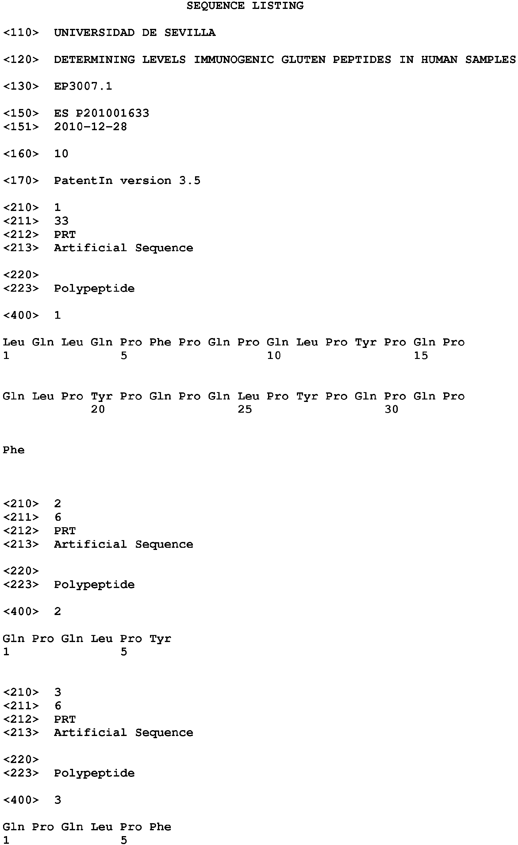


Claims (14)
- Procedure for monitoring the ingested gluten by the detection of immunotoxic peptides in feces characterized by the use of immunological methods using antibodies that recognize peptides from gluten proteins resistant to gastrointestinal digestion.
- Procedure for gluten monitoring according to claim 1 in which the immunological methods use at least one monoclonal antibody with reactivity against some of the sequences of 33-mer peptide SEQ ID No. 1.
- Procedure for monitoring the gluten in feces according to claims 1 to 2 in which the immunological methods use at least a monoclonal antibody with ability to detect epitopes contained in the 33-mer peptide SEQ ID No. 1, SEQ ID No. 2, SEQ ID No. 3, SEQ ID No. 4, SEQ ID No. 5, SEQ ID No. 6, SEQ ID No. 7, SEQ ID No. 8.
- Procedure for monitoring the gluten in feces according to claims 1 to 3 in which the immunological methods use at least one of the monoclonal antibodies G12, A1 and R5.
- Procedure for monitoring the gluten in feces according to claims 1 to 4 in which the immunological methods are an indirect ELISA, a competitive ELISA, a sandwich ELISA, immunochromatographic strips, fluorescent immunomicroparticles, magnetic immunoparticles, Western blot, electronic biosensors, resonance biosensors.
- Procedure for monitoring the presence of gluten peptides ingested according to claims 1 to 5 in which the immunological methods use the monoclonal antibody G12 conjugated to an enzyme that allows a quantitative assay using chromogenic, fluorogenic or luminescent substrates.
- Procedure for monitoring the presence of gluten immunogenic peptides ingested by an immunological assay according to any of claims 1 to 6, characterized by using as a reference standard some of the peptides from claim 3.
- Procedure for monitoring the presence of gluten immunogenic peptides ingested by an immunological assay according to any of claims 1 to 6, characterized because hydrolyzed gliadin by pepsin and trypsin is used as reference standard.
- Procedure for monitoring the presence of immunogenic peptides from gluten ingested by an immunological assay in which immunological methods use at least one of anti-gliadin antibodies, G12 and A1, for carrying out a semiquantitative assay based on immunochromatographic strips of rapid detection.
- Use of analytical procedures according to any of claims 1 to 9 for monitoring the compliance with gluten-free diet.
- Use of analytical procedures according to any of claims 1 to 9 to detect the uncontrolled intake of gluten in celiac patients following a gluten-free diet but with refractory and acute symptoms of celiac disease.
- Use of analytical procedures according to any of claims 1 to 9 for monitoring the effectiveness of therapies related to the elimination of immunogenic peptides of gluten.
- Analytical kit for detecting immunogenic gluten peptides in feces containing:- A solution for the extraction of gluten in feces.- A reference peptide standard comprising at least part or all of the 33-mer immunogenic peptide from gliadin.- An immunoassay using an antibody reactive against the peptide 33-mer from gliadin.
- Analytical kit for detecting immunogenic gluten peptides in feces according to claim 13 comprising:- An aqueous solution having in its composition any of the following components: a dispersing agent, a mild detergent, a reducing agent, a buffer, and ethanol.- A reference peptide standard obtained by hydrolysis with pepsin and trypsin of gliadin or a synthetic peptide comprising part or all of the sequence of the 33-mer from gliadin.- An immunoassay using an antibody reactive against the 33-mer peptide from gliadin like ELISA, immunochromatographic strips, immunoblots, electronic biosensors.
Priority Applications (1)
| Application Number | Priority Date | Filing Date | Title |
|---|---|---|---|
| PL11852781T PL2660593T3 (en) | 2010-12-28 | 2011-12-28 | Determination of levels of immunogenic gluten peptides in human samples |
Applications Claiming Priority (2)
| Application Number | Priority Date | Filing Date | Title |
|---|---|---|---|
| ES201001633A ES2385455B2 (en) | 2010-12-28 | 2010-12-28 | DETERMINATION OF LEVELS OF IMMUNOGENIC PEPTIDES OF GLUTEN IN HUMAN SAMPLES. |
| PCT/ES2011/000379 WO2012089868A2 (en) | 2010-12-28 | 2011-12-28 | Determination of levels of immunogenic gluten peptides in human samples |
Publications (3)
| Publication Number | Publication Date |
|---|---|
| EP2660593A2 true EP2660593A2 (en) | 2013-11-06 |
| EP2660593A4 EP2660593A4 (en) | 2014-10-08 |
| EP2660593B1 EP2660593B1 (en) | 2018-10-31 |
Family
ID=46383573
Family Applications (1)
| Application Number | Title | Priority Date | Filing Date |
|---|---|---|---|
| EP11852781.1A Active EP2660593B1 (en) | 2010-12-28 | 2011-12-28 | Determination of levels of immunogenic gluten peptides in human samples |
Country Status (8)
| Country | Link |
|---|---|
| US (1) | US9410962B2 (en) |
| EP (1) | EP2660593B1 (en) |
| CA (1) | CA2823440C (en) |
| DK (1) | DK2660593T3 (en) |
| ES (2) | ES2385455B2 (en) |
| PL (1) | PL2660593T3 (en) |
| PT (1) | PT2660593T (en) |
| WO (1) | WO2012089868A2 (en) |
Cited By (1)
| Publication number | Priority date | Publication date | Assignee | Title |
|---|---|---|---|---|
| WO2016005643A1 (en) * | 2014-07-09 | 2016-01-14 | Universidad De Sevilla | Detecting gluten peptides in human fluids |
Families Citing this family (5)
| Publication number | Priority date | Publication date | Assignee | Title |
|---|---|---|---|---|
| US10466238B2 (en) * | 2010-12-28 | 2019-11-05 | Universidad De Sevilla | Determination of levels of immunogenic gluten peptides in human samples |
| WO2013098441A1 (en) * | 2011-12-29 | 2013-07-04 | Universidad De Sevilla | Determination of immunogenic gluten peptides in human samples |
| US9738920B2 (en) * | 2015-01-16 | 2017-08-22 | General Mills, Inc. | In vitro method for estimating in vivo protein digestibility |
| BR102021012841A2 (en) * | 2021-06-28 | 2023-01-03 | Sociedade Beneficente Israelita Brasileira Hospital Albert Einstein | SYSTEM FOR MONITORING GLUTEN CONSUMPTION AND PREDICTING THE ASSOCIATION OF INDISPOSITION TO GLUTEN CONSUMPTION |
| ES2982932B2 (en) * | 2023-03-16 | 2025-04-11 | Biomedal SL | PROCEDURE FOR DETERMINING THE STATE OF INTESTINAL PERMEABILITY |
Family Cites Families (6)
| Publication number | Priority date | Publication date | Assignee | Title |
|---|---|---|---|---|
| US4208479A (en) * | 1977-07-14 | 1980-06-17 | Syva Company | Label modified immunoassays |
| US7462688B2 (en) * | 2002-05-14 | 2008-12-09 | The Board Of Trustees Of The Leland Stanford Junior University | Peptides for diagnostic and therapeutic methods for celiac sprue |
| WO2004045392A2 (en) * | 2002-11-20 | 2004-06-03 | The Board Of Trustees Of The Leland Stanford Junior University | Diagnostic method for celiac sprue |
| ES2214108B1 (en) * | 2002-08-16 | 2005-11-16 | Consejo Sup. Investig. Cientificas | IMMUNOCROMATOGRAPHIC SYSTEM AND PROCEDURE FOR THE SIMULTANEOUS IDENTIFICATION OF ANTIBODIES AGAINST AGA AND T-TG, AND ITS USE IN THE CELIAC DISEASE DIAGNOSIS. |
| US7563864B2 (en) * | 2004-04-26 | 2009-07-21 | Celiac Sprue Research Foundation | Prolyl endopeptidase mediated destruction of T cell epitopes in whole gluten |
| US7892763B2 (en) * | 2009-01-26 | 2011-02-22 | Dodds W Jean | Multi-stage nutrigenomic diagnostic food sensitivity testing in animals |
-
2010
- 2010-12-28 ES ES201001633A patent/ES2385455B2/en active Active
-
2011
- 2011-12-28 PL PL11852781T patent/PL2660593T3/en unknown
- 2011-12-28 US US13/977,247 patent/US9410962B2/en active Active
- 2011-12-28 PT PT11852781T patent/PT2660593T/en unknown
- 2011-12-28 WO PCT/ES2011/000379 patent/WO2012089868A2/en not_active Ceased
- 2011-12-28 ES ES11852781T patent/ES2715181T3/en active Active
- 2011-12-28 DK DK11852781.1T patent/DK2660593T3/en active
- 2011-12-28 EP EP11852781.1A patent/EP2660593B1/en active Active
- 2011-12-28 CA CA2823440A patent/CA2823440C/en active Active
Cited By (3)
| Publication number | Priority date | Publication date | Assignee | Title |
|---|---|---|---|---|
| WO2016005643A1 (en) * | 2014-07-09 | 2016-01-14 | Universidad De Sevilla | Detecting gluten peptides in human fluids |
| RU2690665C2 (en) * | 2014-07-09 | 2019-06-05 | Универсидад Дэ Севилья | Detection of gluten peptides in human body fluids |
| AU2015287510B2 (en) * | 2014-07-09 | 2021-03-25 | Biomedal, S. L. | Detecting gluten peptides in human fluids |
Also Published As
| Publication number | Publication date |
|---|---|
| WO2012089868A2 (en) | 2012-07-05 |
| CA2823440A1 (en) | 2012-07-05 |
| CA2823440C (en) | 2020-02-18 |
| US20140178903A1 (en) | 2014-06-26 |
| PL2660593T3 (en) | 2019-05-31 |
| EP2660593A4 (en) | 2014-10-08 |
| DK2660593T3 (en) | 2019-02-18 |
| ES2385455A1 (en) | 2012-07-25 |
| EP2660593B1 (en) | 2018-10-31 |
| WO2012089868A3 (en) | 2012-10-11 |
| US9410962B2 (en) | 2016-08-09 |
| PT2660593T (en) | 2019-02-06 |
| ES2385455B2 (en) | 2013-09-25 |
| ES2715181T3 (en) | 2019-06-03 |
Similar Documents
| Publication | Publication Date | Title |
|---|---|---|
| Comino et al. | Monitoring of gluten-free diet compliance in celiac patients by assessment of gliadin 33-mer equivalent epitopes in feces | |
| Koehler et al. | Celiac disease and gluten: multidisciplinary challenges and opportunities | |
| EP2660593B1 (en) | Determination of levels of immunogenic gluten peptides in human samples | |
| Palosuo et al. | Wheat ω-5 gliadin is a major allergen in children with immediate allergy to ingested wheat | |
| Elli et al. | Immunological effects of transglutaminase-treated gluten in coeliac disease | |
| Ballabio et al. | Molecular characterisation of 36 oat varieties and in vitro assessment of their suitability for coeliacs’ diet | |
| Molinari et al. | Fecal chymotrypsin and elastase-1 determination on one single stool collected at random: diagnostic value for exocrine pancreatic status | |
| US10466238B2 (en) | Determination of levels of immunogenic gluten peptides in human samples | |
| US20190376982A1 (en) | Detecting Gluten Peptides in Human Fluids | |
| RU2573985C2 (en) | Using prosomatostatin in diagnostics | |
| WO2013098441A1 (en) | Determination of immunogenic gluten peptides in human samples | |
| Wieser | Detection of gluten | |
| CN111537719A (en) | Application of serum Sparcl1 protein in the diagnosis of nonalcoholic steatohepatitis | |
| CN106501532B (en) | It is a kind of for detecting the kit of gastrin 17 | |
| Mohammed et al. | The Association of Gluten-Free Diet and Liver Function in Iraqi Patientswith Celiac Disease | |
| US20080038760A1 (en) | Method for Detecting Anti-Transglutaminase Antibodies | |
| US20190191748A1 (en) | Method for selecting cereal seeds suitable for consumption by coeliac patients | |
| Di Sabatino et al. | The spectrum of gluten-related disorders | |
| Ruiz-Carnicer et al. | Urinary Gluten Immunogenic Peptides as a Biomarker in Celiac Patients | |
| Stankovic et al. | Determination of anti-secalin antibodies in sera from coeliac patients by ELISA-based assay | |
| GMG | Prolyl Endopeptidase Mediated Destruction of T Cell Epitopes in Whole Gluten–Chemical and Immunological Characterization | |
| ES2392412A1 (en) | Solution for gluten extraction and methods of use. (Machine-translation by Google Translate, not legally binding) |
Legal Events
| Date | Code | Title | Description |
|---|---|---|---|
| PUAI | Public reference made under article 153(3) epc to a published international application that has entered the european phase |
Free format text: ORIGINAL CODE: 0009012 |
|
| 17P | Request for examination filed |
Effective date: 20130627 |
|
| AK | Designated contracting states |
Kind code of ref document: A2 Designated state(s): AL AT BE BG CH CY CZ DE DK EE ES FI FR GB GR HR HU IE IS IT LI LT LU LV MC MK MT NL NO PL PT RO RS SE SI SK SM TR |
|
| DAX | Request for extension of the european patent (deleted) | ||
| A4 | Supplementary search report drawn up and despatched |
Effective date: 20140904 |
|
| RIC1 | Information provided on ipc code assigned before grant |
Ipc: G01N 33/02 20060101AFI20140829BHEP Ipc: G01N 33/53 20060101ALI20140829BHEP |
|
| 17Q | First examination report despatched |
Effective date: 20151215 |
|
| STAA | Information on the status of an ep patent application or granted ep patent |
Free format text: STATUS: EXAMINATION IS IN PROGRESS |
|
| GRAP | Despatch of communication of intention to grant a patent |
Free format text: ORIGINAL CODE: EPIDOSNIGR1 |
|
| STAA | Information on the status of an ep patent application or granted ep patent |
Free format text: STATUS: GRANT OF PATENT IS INTENDED |
|
| INTG | Intention to grant announced |
Effective date: 20180601 |
|
| GRAS | Grant fee paid |
Free format text: ORIGINAL CODE: EPIDOSNIGR3 |
|
| GRAA | (expected) grant |
Free format text: ORIGINAL CODE: 0009210 |
|
| STAA | Information on the status of an ep patent application or granted ep patent |
Free format text: STATUS: THE PATENT HAS BEEN GRANTED |
|
| AK | Designated contracting states |
Kind code of ref document: B1 Designated state(s): AL AT BE BG CH CY CZ DE DK EE ES FI FR GB GR HR HU IE IS IT LI LT LU LV MC MK MT NL NO PL PT RO RS SE SI SK SM TR |
|
| REG | Reference to a national code |
Ref country code: CH Ref legal event code: EP Ref country code: GB Ref legal event code: FG4D |
|
| REG | Reference to a national code |
Ref country code: AT Ref legal event code: REF Ref document number: 1060003 Country of ref document: AT Kind code of ref document: T Effective date: 20181115 |
|
| REG | Reference to a national code |
Ref country code: DE Ref legal event code: R096 Ref document number: 602011053513 Country of ref document: DE |
|
| REG | Reference to a national code |
Ref country code: IE Ref legal event code: FG4D |
|
| REG | Reference to a national code |
Ref country code: PT Ref legal event code: SC4A Ref document number: 2660593 Country of ref document: PT Date of ref document: 20190206 Kind code of ref document: T Free format text: AVAILABILITY OF NATIONAL TRANSLATION Effective date: 20190129 |
|
| REG | Reference to a national code |
Ref country code: CH Ref legal event code: NV Representative=s name: STOLMAR AND PARTNER INTELLECTUAL PROPERTY S.A., CH |
|
| REG | Reference to a national code |
Ref country code: DK Ref legal event code: T3 Effective date: 20190212 |
|
| REG | Reference to a national code |
Ref country code: NL Ref legal event code: FP |
|
| REG | Reference to a national code |
Ref country code: LT Ref legal event code: MG4D Ref country code: SE Ref legal event code: TRGR |
|
| PG25 | Lapsed in a contracting state [announced via postgrant information from national office to epo] |
Ref country code: IS Free format text: LAPSE BECAUSE OF FAILURE TO SUBMIT A TRANSLATION OF THE DESCRIPTION OR TO PAY THE FEE WITHIN THE PRESCRIBED TIME-LIMIT Effective date: 20190228 Ref country code: LV Free format text: LAPSE BECAUSE OF FAILURE TO SUBMIT A TRANSLATION OF THE DESCRIPTION OR TO PAY THE FEE WITHIN THE PRESCRIBED TIME-LIMIT Effective date: 20181031 Ref country code: NO Free format text: LAPSE BECAUSE OF FAILURE TO SUBMIT A TRANSLATION OF THE DESCRIPTION OR TO PAY THE FEE WITHIN THE PRESCRIBED TIME-LIMIT Effective date: 20190131 Ref country code: HR Free format text: LAPSE BECAUSE OF FAILURE TO SUBMIT A TRANSLATION OF THE DESCRIPTION OR TO PAY THE FEE WITHIN THE PRESCRIBED TIME-LIMIT Effective date: 20181031 Ref country code: LT Free format text: LAPSE BECAUSE OF FAILURE TO SUBMIT A TRANSLATION OF THE DESCRIPTION OR TO PAY THE FEE WITHIN THE PRESCRIBED TIME-LIMIT Effective date: 20181031 Ref country code: BG Free format text: LAPSE BECAUSE OF FAILURE TO SUBMIT A TRANSLATION OF THE DESCRIPTION OR TO PAY THE FEE WITHIN THE PRESCRIBED TIME-LIMIT Effective date: 20190131 |
|
| PG25 | Lapsed in a contracting state [announced via postgrant information from national office to epo] |
Ref country code: RS Free format text: LAPSE BECAUSE OF FAILURE TO SUBMIT A TRANSLATION OF THE DESCRIPTION OR TO PAY THE FEE WITHIN THE PRESCRIBED TIME-LIMIT Effective date: 20181031 Ref country code: SE Free format text: LAPSE BECAUSE OF FAILURE TO SUBMIT A TRANSLATION OF THE DESCRIPTION OR TO PAY THE FEE WITHIN THE PRESCRIBED TIME-LIMIT Effective date: 20181031 Ref country code: AL Free format text: LAPSE BECAUSE OF FAILURE TO SUBMIT A TRANSLATION OF THE DESCRIPTION OR TO PAY THE FEE WITHIN THE PRESCRIBED TIME-LIMIT Effective date: 20181031 |
|
| REG | Reference to a national code |
Ref country code: ES Ref legal event code: FG2A Ref document number: 2715181 Country of ref document: ES Kind code of ref document: T3 Effective date: 20190603 |
|
| REG | Reference to a national code |
Ref country code: DE Ref legal event code: R097 Ref document number: 602011053513 Country of ref document: DE |
|
| PG25 | Lapsed in a contracting state [announced via postgrant information from national office to epo] |
Ref country code: SK Free format text: LAPSE BECAUSE OF FAILURE TO SUBMIT A TRANSLATION OF THE DESCRIPTION OR TO PAY THE FEE WITHIN THE PRESCRIBED TIME-LIMIT Effective date: 20181031 Ref country code: RO Free format text: LAPSE BECAUSE OF FAILURE TO SUBMIT A TRANSLATION OF THE DESCRIPTION OR TO PAY THE FEE WITHIN THE PRESCRIBED TIME-LIMIT Effective date: 20181031 Ref country code: SM Free format text: LAPSE BECAUSE OF FAILURE TO SUBMIT A TRANSLATION OF THE DESCRIPTION OR TO PAY THE FEE WITHIN THE PRESCRIBED TIME-LIMIT Effective date: 20181031 Ref country code: EE Free format text: LAPSE BECAUSE OF FAILURE TO SUBMIT A TRANSLATION OF THE DESCRIPTION OR TO PAY THE FEE WITHIN THE PRESCRIBED TIME-LIMIT Effective date: 20181031 Ref country code: LU Free format text: LAPSE BECAUSE OF NON-PAYMENT OF DUE FEES Effective date: 20181228 Ref country code: MC Free format text: LAPSE BECAUSE OF FAILURE TO SUBMIT A TRANSLATION OF THE DESCRIPTION OR TO PAY THE FEE WITHIN THE PRESCRIBED TIME-LIMIT Effective date: 20181031 |
|
| PLBE | No opposition filed within time limit |
Free format text: ORIGINAL CODE: 0009261 |
|
| STAA | Information on the status of an ep patent application or granted ep patent |
Free format text: STATUS: NO OPPOSITION FILED WITHIN TIME LIMIT |
|
| 26N | No opposition filed |
Effective date: 20190801 |
|
| PG25 | Lapsed in a contracting state [announced via postgrant information from national office to epo] |
Ref country code: SI Free format text: LAPSE BECAUSE OF FAILURE TO SUBMIT A TRANSLATION OF THE DESCRIPTION OR TO PAY THE FEE WITHIN THE PRESCRIBED TIME-LIMIT Effective date: 20181031 |
|
| PG25 | Lapsed in a contracting state [announced via postgrant information from national office to epo] |
Ref country code: MT Free format text: LAPSE BECAUSE OF NON-PAYMENT OF DUE FEES Effective date: 20181228 |
|
| PG25 | Lapsed in a contracting state [announced via postgrant information from national office to epo] |
Ref country code: HU Free format text: LAPSE BECAUSE OF FAILURE TO SUBMIT A TRANSLATION OF THE DESCRIPTION OR TO PAY THE FEE WITHIN THE PRESCRIBED TIME-LIMIT; INVALID AB INITIO Effective date: 20111228 Ref country code: GR Free format text: LAPSE BECAUSE OF FAILURE TO SUBMIT A TRANSLATION OF THE DESCRIPTION OR TO PAY THE FEE WITHIN THE PRESCRIBED TIME-LIMIT Effective date: 20181031 Ref country code: MK Free format text: LAPSE BECAUSE OF NON-PAYMENT OF DUE FEES Effective date: 20181031 Ref country code: CY Free format text: LAPSE BECAUSE OF FAILURE TO SUBMIT A TRANSLATION OF THE DESCRIPTION OR TO PAY THE FEE WITHIN THE PRESCRIBED TIME-LIMIT Effective date: 20181031 |
|
| PGFP | Annual fee paid to national office [announced via postgrant information from national office to epo] |
Ref country code: DK Payment date: 20211227 Year of fee payment: 11 Ref country code: FI Payment date: 20211227 Year of fee payment: 11 Ref country code: CZ Payment date: 20211213 Year of fee payment: 11 Ref country code: PT Payment date: 20211216 Year of fee payment: 11 Ref country code: SE Payment date: 20211227 Year of fee payment: 11 |
|
| PGFP | Annual fee paid to national office [announced via postgrant information from national office to epo] |
Ref country code: TR Payment date: 20211214 Year of fee payment: 11 Ref country code: BE Payment date: 20211227 Year of fee payment: 11 |
|
| PGFP | Annual fee paid to national office [announced via postgrant information from national office to epo] |
Ref country code: NL Payment date: 20230226 Year of fee payment: 12 |
|
| PGFP | Annual fee paid to national office [announced via postgrant information from national office to epo] |
Ref country code: AT Payment date: 20230221 Year of fee payment: 12 |
|
| REG | Reference to a national code |
Ref country code: DK Ref legal event code: EBP Effective date: 20221231 |
|
| PG25 | Lapsed in a contracting state [announced via postgrant information from national office to epo] |
Ref country code: PT Free format text: LAPSE BECAUSE OF NON-PAYMENT OF DUE FEES Effective date: 20230628 Ref country code: CZ Free format text: LAPSE BECAUSE OF NON-PAYMENT OF DUE FEES Effective date: 20221228 |
|
| REG | Reference to a national code |
Ref country code: SE Ref legal event code: EUG |
|
| REG | Reference to a national code |
Ref country code: BE Ref legal event code: MM Effective date: 20221231 |
|
| PG25 | Lapsed in a contracting state [announced via postgrant information from national office to epo] |
Ref country code: SE Free format text: LAPSE BECAUSE OF FAILURE TO SUBMIT A TRANSLATION OF THE DESCRIPTION OR TO PAY THE FEE WITHIN THE PRESCRIBED TIME-LIMIT Effective date: 20221229 |
|
| PG25 | Lapsed in a contracting state [announced via postgrant information from national office to epo] |
Ref country code: BE Free format text: LAPSE BECAUSE OF NON-PAYMENT OF DUE FEES Effective date: 20221231 |
|
| PG25 | Lapsed in a contracting state [announced via postgrant information from national office to epo] |
Ref country code: DK Free format text: LAPSE BECAUSE OF NON-PAYMENT OF DUE FEES Effective date: 20221231 |
|
| PG25 | Lapsed in a contracting state [announced via postgrant information from national office to epo] |
Ref country code: FI Free format text: LAPSE BECAUSE OF NON-PAYMENT OF DUE FEES Effective date: 20221228 |
|
| REG | Reference to a national code |
Ref country code: NL Ref legal event code: MM Effective date: 20240101 |
|
| REG | Reference to a national code |
Ref country code: AT Ref legal event code: MM01 Ref document number: 1060003 Country of ref document: AT Kind code of ref document: T Effective date: 20231228 |
|
| PG25 | Lapsed in a contracting state [announced via postgrant information from national office to epo] |
Ref country code: NL Free format text: LAPSE BECAUSE OF NON-PAYMENT OF DUE FEES Effective date: 20240101 |
|
| PG25 | Lapsed in a contracting state [announced via postgrant information from national office to epo] |
Ref country code: TR Free format text: LAPSE BECAUSE OF NON-PAYMENT OF DUE FEES Effective date: 20221228 Ref country code: NL Free format text: LAPSE BECAUSE OF NON-PAYMENT OF DUE FEES Effective date: 20240101 |
|
| PG25 | Lapsed in a contracting state [announced via postgrant information from national office to epo] |
Ref country code: AT Free format text: LAPSE BECAUSE OF NON-PAYMENT OF DUE FEES Effective date: 20231228 |
|
| PG25 | Lapsed in a contracting state [announced via postgrant information from national office to epo] |
Ref country code: AT Free format text: LAPSE BECAUSE OF NON-PAYMENT OF DUE FEES Effective date: 20231228 |
|
| PGFP | Annual fee paid to national office [announced via postgrant information from national office to epo] |
Ref country code: ES Payment date: 20251231 Year of fee payment: 15 |
|
| REG | Reference to a national code |
Ref country code: CH Ref legal event code: U11 Free format text: ST27 STATUS EVENT CODE: U-0-0-U10-U11 (AS PROVIDED BY THE NATIONAL OFFICE) Effective date: 20260101 |
|
| PGFP | Annual fee paid to national office [announced via postgrant information from national office to epo] |
Ref country code: GB Payment date: 20251229 Year of fee payment: 15 |
|
| PGFP | Annual fee paid to national office [announced via postgrant information from national office to epo] |
Ref country code: IT Payment date: 20251219 Year of fee payment: 15 |
|
| PGFP | Annual fee paid to national office [announced via postgrant information from national office to epo] |
Ref country code: FR Payment date: 20251226 Year of fee payment: 15 |
|
| PGFP | Annual fee paid to national office [announced via postgrant information from national office to epo] |
Ref country code: IE Payment date: 20251229 Year of fee payment: 15 |
|
| PGFP | Annual fee paid to national office [announced via postgrant information from national office to epo] |
Ref country code: PL Payment date: 20251208 Year of fee payment: 15 |
|
| PGFP | Annual fee paid to national office [announced via postgrant information from national office to epo] |
Ref country code: DE Payment date: 20251229 Year of fee payment: 15 |
|
| PGFP | Annual fee paid to national office [announced via postgrant information from national office to epo] |
Ref country code: CH Payment date: 20260101 Year of fee payment: 15 |