EP2653555A1 - P38 MAPK gamma and delta for use as biomarkers of NAFLD - Google Patents
P38 MAPK gamma and delta for use as biomarkers of NAFLD Download PDFInfo
- Publication number
- EP2653555A1 EP2653555A1 EP12164807.5A EP12164807A EP2653555A1 EP 2653555 A1 EP2653555 A1 EP 2653555A1 EP 12164807 A EP12164807 A EP 12164807A EP 2653555 A1 EP2653555 A1 EP 2653555A1
- Authority
- EP
- European Patent Office
- Prior art keywords
- liver
- p38mapk
- delta
- seq
- nafld
- Prior art date
- Legal status (The legal status is an assumption and is not a legal conclusion. Google has not performed a legal analysis and makes no representation as to the accuracy of the status listed.)
- Withdrawn
Links
- 208000008338 non-alcoholic fatty liver disease Diseases 0.000 title claims abstract description 39
- 239000000090 biomarker Substances 0.000 title claims abstract description 13
- 102000043136 MAP kinase family Human genes 0.000 title description 2
- 108091054455 MAP kinase family Proteins 0.000 title description 2
- 210000004369 blood Anatomy 0.000 claims abstract description 18
- 239000008280 blood Substances 0.000 claims abstract description 18
- 238000003745 diagnosis Methods 0.000 claims abstract description 18
- 238000000034 method Methods 0.000 claims abstract description 16
- 238000000338 in vitro Methods 0.000 claims abstract description 8
- 208000004930 Fatty Liver Diseases 0.000 claims description 24
- 239000002773 nucleotide Substances 0.000 claims description 4
- 125000003729 nucleotide group Chemical group 0.000 claims description 4
- 230000000295 complement effect Effects 0.000 claims description 2
- 108020004707 nucleic acids Proteins 0.000 claims description 2
- 102000039446 nucleic acids Human genes 0.000 claims description 2
- 150000007523 nucleic acids Chemical class 0.000 claims description 2
- 206010019708 Hepatic steatosis Diseases 0.000 abstract description 19
- 102000002574 p38 Mitogen-Activated Protein Kinases Human genes 0.000 abstract description 11
- 108010068338 p38 Mitogen-Activated Protein Kinases Proteins 0.000 abstract description 11
- 230000036765 blood level Effects 0.000 abstract description 6
- 238000004393 prognosis Methods 0.000 abstract description 6
- 238000012360 testing method Methods 0.000 abstract description 6
- 101150024075 Mapk1 gene Proteins 0.000 abstract 1
- 210000004185 liver Anatomy 0.000 description 32
- 241000699670 Mus sp. Species 0.000 description 31
- 101000628954 Homo sapiens Mitogen-activated protein kinase 12 Proteins 0.000 description 27
- 102100026932 Mitogen-activated protein kinase 12 Human genes 0.000 description 27
- 108700015928 Mitogen-activated protein kinase 13 Proteins 0.000 description 26
- 102000056248 Mitogen-activated protein kinase 13 Human genes 0.000 description 26
- 238000000692 Student's t-test Methods 0.000 description 21
- 241001465754 Metazoa Species 0.000 description 18
- 235000005911 diet Nutrition 0.000 description 17
- 230000037213 diet Effects 0.000 description 17
- WZUVPPKBWHMQCE-UHFFFAOYSA-N Haematoxylin Chemical compound C12=CC(O)=C(O)C=C2CC2(O)C1C1=CC=C(O)C(O)=C1OC2 WZUVPPKBWHMQCE-UHFFFAOYSA-N 0.000 description 16
- 238000012353 t test Methods 0.000 description 16
- 231100000240 steatosis hepatitis Toxicity 0.000 description 15
- 206010016654 Fibrosis Diseases 0.000 description 14
- 108020004999 messenger RNA Proteins 0.000 description 14
- 201000010099 disease Diseases 0.000 description 13
- 208000037265 diseases, disorders, signs and symptoms Diseases 0.000 description 13
- 230000007863 steatosis Effects 0.000 description 13
- 108010003415 Aspartate Aminotransferases Proteins 0.000 description 12
- 102000004625 Aspartate Aminotransferases Human genes 0.000 description 12
- WSFSSNUMVMOOMR-UHFFFAOYSA-N Formaldehyde Chemical compound O=C WSFSSNUMVMOOMR-UHFFFAOYSA-N 0.000 description 12
- 235000009200 high fat diet Nutrition 0.000 description 12
- 102100036475 Alanine aminotransferase 1 Human genes 0.000 description 10
- 108010082126 Alanine transaminase Proteins 0.000 description 10
- 208000008589 Obesity Diseases 0.000 description 10
- 230000004761 fibrosis Effects 0.000 description 10
- 235000020824 obesity Nutrition 0.000 description 10
- 230000000694 effects Effects 0.000 description 9
- YQGOJNYOYNNSMM-UHFFFAOYSA-N eosin Chemical compound [Na+].OC(=O)C1=CC=CC=C1C1=C2C=C(Br)C(=O)C(Br)=C2OC2=C(Br)C(O)=C(Br)C=C21 YQGOJNYOYNNSMM-UHFFFAOYSA-N 0.000 description 9
- 238000012317 liver biopsy Methods 0.000 description 9
- 230000004054 inflammatory process Effects 0.000 description 8
- NOESYZHRGYRDHS-UHFFFAOYSA-N insulin Chemical compound N1C(=O)C(NC(=O)C(CCC(N)=O)NC(=O)C(CCC(O)=O)NC(=O)C(C(C)C)NC(=O)C(NC(=O)CN)C(C)CC)CSSCC(C(NC(CO)C(=O)NC(CC(C)C)C(=O)NC(CC=2C=CC(O)=CC=2)C(=O)NC(CCC(N)=O)C(=O)NC(CC(C)C)C(=O)NC(CCC(O)=O)C(=O)NC(CC(N)=O)C(=O)NC(CC=2C=CC(O)=CC=2)C(=O)NC(CSSCC(NC(=O)C(C(C)C)NC(=O)C(CC(C)C)NC(=O)C(CC=2C=CC(O)=CC=2)NC(=O)C(CC(C)C)NC(=O)C(C)NC(=O)C(CCC(O)=O)NC(=O)C(C(C)C)NC(=O)C(CC(C)C)NC(=O)C(CC=2NC=NC=2)NC(=O)C(CO)NC(=O)CNC2=O)C(=O)NCC(=O)NC(CCC(O)=O)C(=O)NC(CCCNC(N)=N)C(=O)NCC(=O)NC(CC=3C=CC=CC=3)C(=O)NC(CC=3C=CC=CC=3)C(=O)NC(CC=3C=CC(O)=CC=3)C(=O)NC(C(C)O)C(=O)N3C(CCC3)C(=O)NC(CCCCN)C(=O)NC(C)C(O)=O)C(=O)NC(CC(N)=O)C(O)=O)=O)NC(=O)C(C(C)CC)NC(=O)C(CO)NC(=O)C(C(C)O)NC(=O)C1CSSCC2NC(=O)C(CC(C)C)NC(=O)C(NC(=O)C(CCC(N)=O)NC(=O)C(CC(N)=O)NC(=O)C(NC(=O)C(N)CC=1C=CC=CC=1)C(C)C)CC1=CN=CN1 NOESYZHRGYRDHS-UHFFFAOYSA-N 0.000 description 8
- 208000019423 liver disease Diseases 0.000 description 8
- 230000004807 localization Effects 0.000 description 8
- 238000001356 surgical procedure Methods 0.000 description 8
- 206010067125 Liver injury Diseases 0.000 description 7
- 102100033810 RAC-alpha serine/threonine-protein kinase Human genes 0.000 description 7
- 231100000234 hepatic damage Toxicity 0.000 description 7
- 230000006372 lipid accumulation Effects 0.000 description 7
- 150000002632 lipids Chemical class 0.000 description 7
- 230000008818 liver damage Effects 0.000 description 7
- 235000021590 normal diet Nutrition 0.000 description 7
- IAZDPXIOMUYVGZ-UHFFFAOYSA-N Dimethylsulphoxide Chemical compound CS(C)=O IAZDPXIOMUYVGZ-UHFFFAOYSA-N 0.000 description 6
- 206010061218 Inflammation Diseases 0.000 description 6
- 108091000080 Phosphotransferase Proteins 0.000 description 6
- 230000035508 accumulation Effects 0.000 description 6
- 238000009825 accumulation Methods 0.000 description 6
- 238000004458 analytical method Methods 0.000 description 6
- 208000019425 cirrhosis of liver Diseases 0.000 description 6
- 238000013399 early diagnosis Methods 0.000 description 6
- 238000005259 measurement Methods 0.000 description 6
- 206010053219 non-alcoholic steatohepatitis Diseases 0.000 description 6
- 102000020233 phosphotransferase Human genes 0.000 description 6
- 238000003753 real-time PCR Methods 0.000 description 6
- 210000002966 serum Anatomy 0.000 description 6
- 210000001519 tissue Anatomy 0.000 description 6
- 230000004913 activation Effects 0.000 description 5
- 238000011161 development Methods 0.000 description 5
- MVCOAUNKQVWQHZ-UHFFFAOYSA-N doramapimod Chemical compound C1=CC(C)=CC=C1N1C(NC(=O)NC=2C3=CC=CC=C3C(OCCN3CCOCC3)=CC=2)=CC(C(C)(C)C)=N1 MVCOAUNKQVWQHZ-UHFFFAOYSA-N 0.000 description 5
- 206010073071 hepatocellular carcinoma Diseases 0.000 description 5
- 231100000844 hepatocellular carcinoma Toxicity 0.000 description 5
- 108091032973 (ribonucleotides)n+m Proteins 0.000 description 4
- LFQSCWFLJHTTHZ-UHFFFAOYSA-N Ethanol Chemical compound CCO LFQSCWFLJHTTHZ-UHFFFAOYSA-N 0.000 description 4
- 102000004877 Insulin Human genes 0.000 description 4
- 108090001061 Insulin Proteins 0.000 description 4
- FFEARJCKVFRZRR-BYPYZUCNSA-N L-methionine Chemical compound CSCC[C@H](N)C(O)=O FFEARJCKVFRZRR-BYPYZUCNSA-N 0.000 description 4
- HVYWMOMLDIMFJA-DPAQBDIFSA-N cholesterol Chemical compound C1C=C2C[C@@H](O)CC[C@]2(C)[C@@H]2[C@@H]1[C@@H]1CC[C@H]([C@H](C)CCCC(C)C)[C@@]1(C)CC2 HVYWMOMLDIMFJA-DPAQBDIFSA-N 0.000 description 4
- 229960001231 choline Drugs 0.000 description 4
- OEYIOHPDSNJKLS-UHFFFAOYSA-N choline Chemical compound C[N+](C)(C)CCO OEYIOHPDSNJKLS-UHFFFAOYSA-N 0.000 description 4
- 230000007882 cirrhosis Effects 0.000 description 4
- 235000020940 control diet Nutrition 0.000 description 4
- DDRJAANPRJIHGJ-UHFFFAOYSA-N creatinine Chemical compound CN1CC(=O)NC1=N DDRJAANPRJIHGJ-UHFFFAOYSA-N 0.000 description 4
- 230000002950 deficient Effects 0.000 description 4
- 239000003814 drug Substances 0.000 description 4
- 208000006454 hepatitis Diseases 0.000 description 4
- 231100000805 hepatocellular lesion Toxicity 0.000 description 4
- 230000006872 improvement Effects 0.000 description 4
- 229940125396 insulin Drugs 0.000 description 4
- 238000011813 knockout mouse model Methods 0.000 description 4
- 229930182817 methionine Natural products 0.000 description 4
- 239000012188 paraffin wax Substances 0.000 description 4
- 150000003626 triacylglycerols Chemical class 0.000 description 4
- 208000001072 type 2 diabetes mellitus Diseases 0.000 description 4
- PEDCQBHIVMGVHV-UHFFFAOYSA-N Glycerine Chemical compound OCC(O)CO PEDCQBHIVMGVHV-UHFFFAOYSA-N 0.000 description 3
- 206010019668 Hepatic fibrosis Diseases 0.000 description 3
- 206010022489 Insulin Resistance Diseases 0.000 description 3
- 102000004889 Interleukin-6 Human genes 0.000 description 3
- 108090001005 Interleukin-6 Proteins 0.000 description 3
- OKKJLVBELUTLKV-UHFFFAOYSA-N Methanol Chemical compound OC OKKJLVBELUTLKV-UHFFFAOYSA-N 0.000 description 3
- 108010029485 Protein Isoforms Proteins 0.000 description 3
- 102000001708 Protein Isoforms Human genes 0.000 description 3
- 239000000524 Thiobarbituric Acid Reactive Substance Substances 0.000 description 3
- 102100040247 Tumor necrosis factor Human genes 0.000 description 3
- 210000004027 cell Anatomy 0.000 description 3
- 230000008859 change Effects 0.000 description 3
- 229920001436 collagen Polymers 0.000 description 3
- 230000003247 decreasing effect Effects 0.000 description 3
- 238000001514 detection method Methods 0.000 description 3
- 239000000284 extract Substances 0.000 description 3
- 230000001965 increasing effect Effects 0.000 description 3
- 230000008595 infiltration Effects 0.000 description 3
- 238000001764 infiltration Methods 0.000 description 3
- 230000002757 inflammatory effect Effects 0.000 description 3
- 230000005764 inhibitory process Effects 0.000 description 3
- 230000003859 lipid peroxidation Effects 0.000 description 3
- 208000018191 liver inflammation Diseases 0.000 description 3
- 238000012544 monitoring process Methods 0.000 description 3
- 230000026731 phosphorylation Effects 0.000 description 3
- 238000006366 phosphorylation reaction Methods 0.000 description 3
- 230000002441 reversible effect Effects 0.000 description 3
- 230000035945 sensitivity Effects 0.000 description 3
- 108010088751 Albumins Proteins 0.000 description 2
- 102000009027 Albumins Human genes 0.000 description 2
- 208000007848 Alcoholism Diseases 0.000 description 2
- 102000002260 Alkaline Phosphatase Human genes 0.000 description 2
- 108020004774 Alkaline Phosphatase Proteins 0.000 description 2
- HEDRZPFGACZZDS-UHFFFAOYSA-N Chloroform Chemical compound ClC(Cl)Cl HEDRZPFGACZZDS-UHFFFAOYSA-N 0.000 description 2
- 208000000419 Chronic Hepatitis B Diseases 0.000 description 2
- 208000006154 Chronic hepatitis C Diseases 0.000 description 2
- 102000008186 Collagen Human genes 0.000 description 2
- 108010035532 Collagen Proteins 0.000 description 2
- 101100447432 Danio rerio gapdh-2 gene Proteins 0.000 description 2
- 206010061818 Disease progression Diseases 0.000 description 2
- 101710107035 Gamma-glutamyltranspeptidase Proteins 0.000 description 2
- 101150112014 Gapdh gene Proteins 0.000 description 2
- WQZGKKKJIJFFOK-GASJEMHNSA-N Glucose Natural products OC[C@H]1OC(O)[C@H](O)[C@@H](O)[C@@H]1O WQZGKKKJIJFFOK-GASJEMHNSA-N 0.000 description 2
- 101710173228 Glutathione hydrolase proenzyme Proteins 0.000 description 2
- 102100031181 Glyceraldehyde-3-phosphate dehydrogenase Human genes 0.000 description 2
- DHCLVCXQIBBOPH-UHFFFAOYSA-N Glycerol 2-phosphate Chemical compound OCC(CO)OP(O)(O)=O DHCLVCXQIBBOPH-UHFFFAOYSA-N 0.000 description 2
- 102000015779 HDL Lipoproteins Human genes 0.000 description 2
- 108010010234 HDL Lipoproteins Proteins 0.000 description 2
- 208000005176 Hepatitis C Diseases 0.000 description 2
- 102000003814 Interleukin-10 Human genes 0.000 description 2
- 108090000174 Interleukin-10 Proteins 0.000 description 2
- 102000007330 LDL Lipoproteins Human genes 0.000 description 2
- 108010007622 LDL Lipoproteins Proteins 0.000 description 2
- 238000010222 PCR analysis Methods 0.000 description 2
- FAPWRFPIFSIZLT-UHFFFAOYSA-M Sodium chloride Chemical compound [Na+].[Cl-] FAPWRFPIFSIZLT-UHFFFAOYSA-M 0.000 description 2
- QAOWNCQODCNURD-UHFFFAOYSA-N Sulfuric acid Chemical compound OS(O)(=O)=O QAOWNCQODCNURD-UHFFFAOYSA-N 0.000 description 2
- 238000008050 Total Bilirubin Reagent Methods 0.000 description 2
- 238000009557 abdominal ultrasonography Methods 0.000 description 2
- 210000000577 adipose tissue Anatomy 0.000 description 2
- 238000007681 bariatric surgery Methods 0.000 description 2
- WQZGKKKJIJFFOK-VFUOTHLCSA-N beta-D-glucose Chemical compound OC[C@H]1O[C@@H](O)[C@H](O)[C@@H](O)[C@@H]1O WQZGKKKJIJFFOK-VFUOTHLCSA-N 0.000 description 2
- 238000004820 blood count Methods 0.000 description 2
- 238000005119 centrifugation Methods 0.000 description 2
- 239000003153 chemical reaction reagent Substances 0.000 description 2
- 238000002192 cholecystectomy Methods 0.000 description 2
- 235000012000 cholesterol Nutrition 0.000 description 2
- 230000002596 correlated effect Effects 0.000 description 2
- 229940109239 creatinine Drugs 0.000 description 2
- 230000005750 disease progression Effects 0.000 description 2
- 229940079593 drug Drugs 0.000 description 2
- 208000010706 fatty liver disease Diseases 0.000 description 2
- 102000006640 gamma-Glutamyltransferase Human genes 0.000 description 2
- 239000008103 glucose Substances 0.000 description 2
- 108020004445 glyceraldehyde-3-phosphate dehydrogenase Proteins 0.000 description 2
- 230000005802 health problem Effects 0.000 description 2
- 206010019692 hepatic necrosis Diseases 0.000 description 2
- 208000002672 hepatitis B Diseases 0.000 description 2
- 208000010710 hepatitis C virus infection Diseases 0.000 description 2
- 210000003494 hepatocyte Anatomy 0.000 description 2
- 230000010438 iron metabolism Effects 0.000 description 2
- 210000000265 leukocyte Anatomy 0.000 description 2
- 210000005228 liver tissue Anatomy 0.000 description 2
- 239000012139 lysis buffer Substances 0.000 description 2
- 239000003550 marker Substances 0.000 description 2
- 230000036542 oxidative stress Effects 0.000 description 2
- YBYRMVIVWMBXKQ-UHFFFAOYSA-N phenylmethanesulfonyl fluoride Chemical compound FS(=O)(=O)CC1=CC=CC=C1 YBYRMVIVWMBXKQ-UHFFFAOYSA-N 0.000 description 2
- DHRLEVQXOMLTIM-UHFFFAOYSA-N phosphoric acid;trioxomolybdenum Chemical compound O=[Mo](=O)=O.O=[Mo](=O)=O.O=[Mo](=O)=O.O=[Mo](=O)=O.O=[Mo](=O)=O.O=[Mo](=O)=O.O=[Mo](=O)=O.O=[Mo](=O)=O.O=[Mo](=O)=O.O=[Mo](=O)=O.O=[Mo](=O)=O.O=[Mo](=O)=O.OP(O)(O)=O DHRLEVQXOMLTIM-UHFFFAOYSA-N 0.000 description 2
- 230000000391 smoking effect Effects 0.000 description 2
- 238000007920 subcutaneous administration Methods 0.000 description 2
- MZOFCQQQCNRIBI-VMXHOPILSA-N (3s)-4-[[(2s)-1-[[(2s)-1-[[(1s)-1-carboxy-2-hydroxyethyl]amino]-4-methyl-1-oxopentan-2-yl]amino]-5-(diaminomethylideneamino)-1-oxopentan-2-yl]amino]-3-[[2-[[(2s)-2,6-diaminohexanoyl]amino]acetyl]amino]-4-oxobutanoic acid Chemical compound OC[C@@H](C(O)=O)NC(=O)[C@H](CC(C)C)NC(=O)[C@H](CCCN=C(N)N)NC(=O)[C@H](CC(O)=O)NC(=O)CNC(=O)[C@@H](N)CCCCN MZOFCQQQCNRIBI-VMXHOPILSA-N 0.000 description 1
- FWMNVWWHGCHHJJ-SKKKGAJSSA-N 4-amino-1-[(2r)-6-amino-2-[[(2r)-2-[[(2r)-2-[[(2r)-2-amino-3-phenylpropanoyl]amino]-3-phenylpropanoyl]amino]-4-methylpentanoyl]amino]hexanoyl]piperidine-4-carboxylic acid Chemical compound C([C@H](C(=O)N[C@H](CC(C)C)C(=O)N[C@H](CCCCN)C(=O)N1CCC(N)(CC1)C(O)=O)NC(=O)[C@H](N)CC=1C=CC=CC=1)C1=CC=CC=C1 FWMNVWWHGCHHJJ-SKKKGAJSSA-N 0.000 description 1
- 108010039627 Aprotinin Proteins 0.000 description 1
- 208000034048 Asymptomatic disease Diseases 0.000 description 1
- 102100021943 C-C motif chemokine 2 Human genes 0.000 description 1
- 101710155857 C-C motif chemokine 2 Proteins 0.000 description 1
- 108010074051 C-Reactive Protein Proteins 0.000 description 1
- 102100032752 C-reactive protein Human genes 0.000 description 1
- 201000009030 Carcinoma Diseases 0.000 description 1
- 102000004127 Cytokines Human genes 0.000 description 1
- 108090000695 Cytokines Proteins 0.000 description 1
- 102100023275 Dual specificity mitogen-activated protein kinase kinase 3 Human genes 0.000 description 1
- 102100023401 Dual specificity mitogen-activated protein kinase kinase 6 Human genes 0.000 description 1
- KCXVZYZYPLLWCC-UHFFFAOYSA-N EDTA Chemical compound OC(=O)CN(CC(O)=O)CCN(CC(O)=O)CC(O)=O KCXVZYZYPLLWCC-UHFFFAOYSA-N 0.000 description 1
- 206010019663 Hepatic failure Diseases 0.000 description 1
- 102000003745 Hepatocyte Growth Factor Human genes 0.000 description 1
- 108090000100 Hepatocyte Growth Factor Proteins 0.000 description 1
- 101001115394 Homo sapiens Dual specificity mitogen-activated protein kinase kinase 3 Proteins 0.000 description 1
- 101000624426 Homo sapiens Dual specificity mitogen-activated protein kinase kinase 6 Proteins 0.000 description 1
- 208000031226 Hyperlipidaemia Diseases 0.000 description 1
- 102000004890 Interleukin-8 Human genes 0.000 description 1
- 108090001007 Interleukin-8 Proteins 0.000 description 1
- 125000002842 L-seryl group Chemical group O=C([*])[C@](N([H])[H])([H])C([H])([H])O[H] 0.000 description 1
- GDBQQVLCIARPGH-UHFFFAOYSA-N Leupeptin Natural products CC(C)CC(NC(C)=O)C(=O)NC(CC(C)C)C(=O)NC(C=O)CCCN=C(N)N GDBQQVLCIARPGH-UHFFFAOYSA-N 0.000 description 1
- 101150003567 Mapk12 gene Proteins 0.000 description 1
- 208000001145 Metabolic Syndrome Diseases 0.000 description 1
- 102100039364 Metalloproteinase inhibitor 1 Human genes 0.000 description 1
- 102000056243 Mitogen-activated protein kinase 12 Human genes 0.000 description 1
- 108700015929 Mitogen-activated protein kinase 12 Proteins 0.000 description 1
- 206010028980 Neoplasm Diseases 0.000 description 1
- 108010022233 Plasminogen Activator Inhibitor 1 Proteins 0.000 description 1
- 102100039418 Plasminogen activator inhibitor 1 Human genes 0.000 description 1
- 238000001190 Q-PCR Methods 0.000 description 1
- 101150105578 SAPK3 gene Proteins 0.000 description 1
- 108010031374 Tissue Inhibitor of Metalloproteinase-1 Proteins 0.000 description 1
- 102000003929 Transaminases Human genes 0.000 description 1
- 108090000340 Transaminases Proteins 0.000 description 1
- 239000007983 Tris buffer Substances 0.000 description 1
- 229920004890 Triton X-100 Polymers 0.000 description 1
- 239000013504 Triton X-100 Substances 0.000 description 1
- 108060008682 Tumor Necrosis Factor Proteins 0.000 description 1
- 201000000690 abdominal obesity-metabolic syndrome Diseases 0.000 description 1
- 230000002159 abnormal effect Effects 0.000 description 1
- 238000010171 animal model Methods 0.000 description 1
- 229960004405 aprotinin Drugs 0.000 description 1
- 238000003556 assay Methods 0.000 description 1
- 238000003149 assay kit Methods 0.000 description 1
- 230000005773 cancer-related death Effects 0.000 description 1
- 231100000504 carcinogenesis Toxicity 0.000 description 1
- 230000005754 cellular signaling Effects 0.000 description 1
- 230000001684 chronic effect Effects 0.000 description 1
- 230000034994 death Effects 0.000 description 1
- 206010012601 diabetes mellitus Diseases 0.000 description 1
- 229940042399 direct acting antivirals protease inhibitors Drugs 0.000 description 1
- 230000009977 dual effect Effects 0.000 description 1
- 230000002526 effect on cardiovascular system Effects 0.000 description 1
- 230000006353 environmental stress Effects 0.000 description 1
- 230000006870 function Effects 0.000 description 1
- PCHJSUWPFVWCPO-UHFFFAOYSA-N gold Chemical compound [Au] PCHJSUWPFVWCPO-UHFFFAOYSA-N 0.000 description 1
- 230000007866 hepatic necrosis Effects 0.000 description 1
- 231100000283 hepatitis Toxicity 0.000 description 1
- 238000003119 immunoblot Methods 0.000 description 1
- 238000001727 in vivo Methods 0.000 description 1
- 238000011534 incubation Methods 0.000 description 1
- 230000006698 induction Effects 0.000 description 1
- 230000001939 inductive effect Effects 0.000 description 1
- ZPNFWUPYTFPOJU-LPYSRVMUSA-N iniprol Chemical compound C([C@H]1C(=O)NCC(=O)NCC(=O)N[C@H]2CSSC[C@H]3C(=O)N[C@@H](CCCCN)C(=O)N[C@@H](C)C(=O)N[C@@H](CCCNC(N)=N)C(=O)N[C@H](C(N[C@H](C(=O)N[C@@H](CCCNC(N)=N)C(=O)N[C@@H](CC=4C=CC(O)=CC=4)C(=O)N[C@@H](CC=4C=CC=CC=4)C(=O)N[C@@H](CC=4C=CC(O)=CC=4)C(=O)N[C@@H](CC(N)=O)C(=O)N[C@@H](C)C(=O)N[C@@H](CCCCN)C(=O)N[C@@H](C)C(=O)NCC(=O)N[C@@H](CC(C)C)C(=O)N[C@@H](CSSC[C@H](NC(=O)[C@H](CC(O)=O)NC(=O)[C@H](CCC(O)=O)NC(=O)[C@H](C)NC(=O)[C@H](CO)NC(=O)[C@H](CCCCN)NC(=O)[C@H](CC=4C=CC=CC=4)NC(=O)[C@H](CC(N)=O)NC(=O)[C@H](CC(N)=O)NC(=O)[C@H](CCCNC(N)=N)NC(=O)[C@H](CCCCN)NC(=O)[C@H](C)NC(=O)[C@H](CCCNC(N)=N)NC2=O)C(=O)N[C@@H](CCSC)C(=O)N[C@@H](CCCNC(N)=N)C(=O)N[C@@H]([C@@H](C)O)C(=O)N[C@@H](CSSC[C@H](NC(=O)[C@H](CC=2C=CC=CC=2)NC(=O)[C@H](CC(O)=O)NC(=O)[C@H]2N(CCC2)C(=O)[C@@H](N)CCCNC(N)=N)C(=O)N[C@@H](CC(C)C)C(=O)N[C@@H](CCC(O)=O)C(=O)N2[C@@H](CCC2)C(=O)N2[C@@H](CCC2)C(=O)N[C@@H](CC=2C=CC(O)=CC=2)C(=O)N[C@@H]([C@@H](C)O)C(=O)NCC(=O)N2[C@@H](CCC2)C(=O)N3)C(=O)NCC(=O)NCC(=O)N[C@@H](C)C(O)=O)C(=O)N[C@@H](CCC(N)=O)C(=O)N[C@H](C(=O)N[C@@H](CC=2C=CC=CC=2)C(=O)N[C@H](C(=O)N1)C(C)C)[C@@H](C)O)[C@@H](C)CC)=O)[C@@H](C)CC)C1=CC=C(O)C=C1 ZPNFWUPYTFPOJU-LPYSRVMUSA-N 0.000 description 1
- 230000006362 insulin response pathway Effects 0.000 description 1
- GDBQQVLCIARPGH-ULQDDVLXSA-N leupeptin Chemical compound CC(C)C[C@H](NC(C)=O)C(=O)N[C@@H](CC(C)C)C(=O)N[C@H](C=O)CCCN=C(N)N GDBQQVLCIARPGH-ULQDDVLXSA-N 0.000 description 1
- 108010052968 leupeptin Proteins 0.000 description 1
- 210000005229 liver cell Anatomy 0.000 description 1
- 231100000835 liver failure Toxicity 0.000 description 1
- 208000007903 liver failure Diseases 0.000 description 1
- 231100000149 liver necrosis Toxicity 0.000 description 1
- 230000007774 longterm Effects 0.000 description 1
- 238000004519 manufacturing process Methods 0.000 description 1
- 210000005087 mononuclear cell Anatomy 0.000 description 1
- 210000003205 muscle Anatomy 0.000 description 1
- 230000017074 necrotic cell death Effects 0.000 description 1
- 239000012074 organic phase Substances 0.000 description 1
- 231100000915 pathological change Toxicity 0.000 description 1
- 230000036285 pathological change Effects 0.000 description 1
- 239000000137 peptide hydrolase inhibitor Substances 0.000 description 1
- BZQFBWGGLXLEPQ-REOHCLBHSA-N phosphoserine Chemical compound OC(=O)[C@@H](N)COP(O)(O)=O BZQFBWGGLXLEPQ-REOHCLBHSA-N 0.000 description 1
- USRGIUJOYOXOQJ-GBXIJSLDSA-N phosphothreonine Chemical compound OP(=O)(O)O[C@H](C)[C@H](N)C(O)=O USRGIUJOYOXOQJ-GBXIJSLDSA-N 0.000 description 1
- 235000018102 proteins Nutrition 0.000 description 1
- 102000004169 proteins and genes Human genes 0.000 description 1
- 108090000623 proteins and genes Proteins 0.000 description 1
- 230000009467 reduction Effects 0.000 description 1
- 230000004044 response Effects 0.000 description 1
- 230000000630 rising effect Effects 0.000 description 1
- 230000037390 scarring Effects 0.000 description 1
- 238000012216 screening Methods 0.000 description 1
- 239000011780 sodium chloride Substances 0.000 description 1
- 239000007787 solid Substances 0.000 description 1
- 239000000243 solution Substances 0.000 description 1
- 238000001228 spectrum Methods 0.000 description 1
- 230000035882 stress Effects 0.000 description 1
- 239000006228 supernatant Substances 0.000 description 1
- UFTFJSFQGQCHQW-UHFFFAOYSA-N triformin Chemical compound O=COCC(OC=O)COC=O UFTFJSFQGQCHQW-UHFFFAOYSA-N 0.000 description 1
- LENZDBCJOHFCAS-UHFFFAOYSA-N tris Chemical compound OCC(N)(CO)CO LENZDBCJOHFCAS-UHFFFAOYSA-N 0.000 description 1
- IHIXIJGXTJIKRB-UHFFFAOYSA-N trisodium vanadate Chemical compound [Na+].[Na+].[Na+].[O-][V]([O-])([O-])=O IHIXIJGXTJIKRB-UHFFFAOYSA-N 0.000 description 1
- GPRLSGONYQIRFK-MNYXATJNSA-N triton Chemical compound [3H+] GPRLSGONYQIRFK-MNYXATJNSA-N 0.000 description 1
Images




Classifications
-
- C—CHEMISTRY; METALLURGY
- C12—BIOCHEMISTRY; BEER; SPIRITS; WINE; VINEGAR; MICROBIOLOGY; ENZYMOLOGY; MUTATION OR GENETIC ENGINEERING
- C12Q—MEASURING OR TESTING PROCESSES INVOLVING ENZYMES, NUCLEIC ACIDS OR MICROORGANISMS; COMPOSITIONS OR TEST PAPERS THEREFOR; PROCESSES OF PREPARING SUCH COMPOSITIONS; CONDITION-RESPONSIVE CONTROL IN MICROBIOLOGICAL OR ENZYMOLOGICAL PROCESSES
- C12Q1/00—Measuring or testing processes involving enzymes, nucleic acids or microorganisms; Compositions therefor; Processes of preparing such compositions
- C12Q1/68—Measuring or testing processes involving enzymes, nucleic acids or microorganisms; Compositions therefor; Processes of preparing such compositions involving nucleic acids
- C12Q1/6876—Nucleic acid products used in the analysis of nucleic acids, e.g. primers or probes
- C12Q1/6883—Nucleic acid products used in the analysis of nucleic acids, e.g. primers or probes for diseases caused by alterations of genetic material
-
- C—CHEMISTRY; METALLURGY
- C12—BIOCHEMISTRY; BEER; SPIRITS; WINE; VINEGAR; MICROBIOLOGY; ENZYMOLOGY; MUTATION OR GENETIC ENGINEERING
- C12Q—MEASURING OR TESTING PROCESSES INVOLVING ENZYMES, NUCLEIC ACIDS OR MICROORGANISMS; COMPOSITIONS OR TEST PAPERS THEREFOR; PROCESSES OF PREPARING SUCH COMPOSITIONS; CONDITION-RESPONSIVE CONTROL IN MICROBIOLOGICAL OR ENZYMOLOGICAL PROCESSES
- C12Q2600/00—Oligonucleotides characterized by their use
- C12Q2600/158—Expression markers
Definitions
- the present invention may be included in the field of the medicine, in general, and, more particularly, in that area of the medicine which is focused on the study of nonalcoholic fatty liver disease (NAFLD) and, more particularly, of hepatic steatosis, also known as fatty liver, characterized by the abnormal fat accumulation that occurs in the liver cells.
- NAFLD nonalcoholic fatty liver disease
- hepatic steatosis also known as fatty liver, characterized by the abnormal fat accumulation that occurs in the liver cells.
- Obesity is a major health problem associated with many other diseases like type two diabetes T2DM, metabolic syndrome and NAFLD.
- NAFLD is one of the most prevalent manifestations of obesity [1] and this chronic liver disease has become a worldwide health problem.
- NAFLD refers to a spectrum of liver diseases ranging from simple steatosis or fatty liver, to nonalcoholic steatohepatitis (NASH), and cirrhosis (with advanced scarring of the liver), all these stages have in common an accumulation of fat within hepatocytes. Most patients with NAFLD also have obesity (60-100%), T2DM (28-55%) and hyperlipidemia (27-92%) [2]. While NAFLD can be quite unobtrusive, NASH is a serious condition, with nearly a quarter of affected patients developing cirrhosis, which, in turn, increases the risk of subsequent progression to hepatocellular carcinoma (HCC). HCC is the fifth most common tumour worldwide with increasing incidence and third leading cause of cancer-related death. Among patients with NAFLD or NASH, HCC represents the main cause of death [3].
- NAFLD a asymptomatic disease that can progress from an initial trygliceride accumulation to NASH which, in a long-term, may results in fibrosis, cirrhosis and hepatocellularar carcinoma (HCC) [6, 7, 8]. For that reason, an early and accurate diagnosis is fundamental to know the degree of liver damage, and avoid the disease progression.
- liver biopsy is nowadays considered the gold standard for NAFLD detection, since the diagnostic yield of available non-invasive tests is not enough accurate [9, 10].
- liver biopsy is an invasive procedure, uncomfortable and with risks for the patient and therefore it cannot be used as a screening or routine method, or for obtaining several samples periodically to assess disease evolution.
- liver steatosis Therefore, a reliable and non-invasive test for diagnosis of liver steatosis would lead to a clear improvement in early diagnosis, ability of monitoring of the disease, as well as quality of life.
- the present invention solves the above-mentioned problem by providing solid biomarkers (p38MAPK) for NAFLD, preferably for hepatic steatosis.
- p38MAPK mitogen-activated protein kinase family are strongly activated in vivo by environmental stress factors and inflammatory cytokines. Obesity and liver steatosis act like stress signals inducing multiple responses in tissues.
- Four p38MAPK isotypes have been described to date: alpha ( ⁇ ), beta ( ⁇ ), gamma ( ⁇ ) and delta ( ⁇ ).
- the canonical activation of p38MAPK occurs via dual phosphorylation in the activation loop by MKK3 and MKK6 [12].
- the present invention provides an accurate in vitro method for prognosis and diagnosis of NAFLD, preferably of hepatic steatosis, offering the opportunity of carrying out an improved treatment at earlier stages of the disease, thus rising the likelihood of definitely treating the disease. For that reason, an early and accurate diagnosis is fundamental to know the degree of liver damage, and avoid the disease progression.
- the method of the invention is based on blood samples and consequently it may be considered as a non-invasive test which leads to a clear improvement in early diagnosis, ability of monitoring of the disease, as well as quality of life.
- Blood levels of these kinases can be therefore used as potential biomarkers of hepatic steatosis in human, without the inconvenient and risks of liver biopsy and therefore improving early diagnosis and more accurate prognosis of NAFLD and its associated complications or diseases.
- the present invention provides an accurate and non-invasive in vitro method for prognosis and diagnosis of NAFLD, preferably of hepatic steatosis, offering the opportunity of carrying out an improved treatment at earlier stages of the disease, thus raising the likelihood of definitely treating the disease.
- the method of the invention is based on blood samples and consequently it may be considered as a non-invasive test for diagnosis of hepatic steatosis which leads to a clear improvement in early diagnosis, ability of monitoring of the disease, as well as quality of life. Blood levels of these kinases could be therefore used as potential biomarkers of hepatic steatosis in human, without the inconvenient and risks of liver biopsy and therefore improving early diagnosis and more accurate prognosis of NAFLD and its complications.
- liver stetatosis induced by obesity with a decrease in expression levels of kinases p38MAPK ⁇ and p38MAPK ⁇ was observed in the present invention (p ⁇ 0.001 or p ⁇ 0.01 respectably). Therefore, analysis of these kinases can be used as biomarkers of NAFLD, preferably of liver steatosis, in human, without the drawbacks associated to the use of liver biopsy and therefore improving early diagnosis and more accurate prognosis.
- the first embodiment of the present invention refers to p38MAPK gamma and/or delta for use as biomarkers of NAFLD, preferably of hepatic steatosis.
- the second embodiment of the present invention refers to an in vitro method for the diagnosis of NAFLD, preferably of hepatic steatosis, that comprises measuring the expression level of p38MAPK gamma and/or delta in a blood sample taken from the subject under examination.
- hepatic steatosis is associated with a significant decrease in expression levels of p38MAPK gamma and p38MAPK delta.
- the method of the invention is characterized in that the expression level of p38MAPK gamma is measured using the primers of SEQ ID NO: 21 and 22 and the expression level of p38MAPK delta is measured using the primers of SEQ ID NO: 23 and 24.
- the third embodiment of the present invention refers to a nucleotide sequence characterized by the SEQ ID NO: 21, 22, 23 or 24. These sequences are used in the present invention as primers for measuring the expression level of p38 gamma and/or p38 delta in the blood samples taken from the patient/subject. Consequently these sequences may be used as primers to make a kit for diagnosis of NAFLD, preferably for diagnosis of hepatic steatosis.
- the present invention refers to a nucleotide sequence characterized by the SEQ ID NO: 21, 22, 23 or 24 or any combination thereof for use as primers for hybridizing under stringent conditions with a nucleic acid encoding for p38MAPK gamma and/or delta sequence or any partial complementary or mutated sequence thereof.
- the fourth embodiment of the present invention refers to a kit for the diagnosis of NAFLD, preferably of hepatic steatosis, characterized by comprising the primers of SEQ ID NO: 21 and 22 for measuring the expression level of p38MAPK gamma and/or the primers of SEQ ID NO: 23 and 24 for measuring the expression level of p38MAPK delta.
- p ⁇ 0.05 refers to the significance level and p values below to 0.05 make the biomarkers of the invention statistically significant.
- Example 1 Partial protection of liver damage in p38 ⁇ -/- and p38 ⁇ -/- mice.
- mice p38 ⁇ -/- and p38 ⁇ -/- were obtained from Dr. Ana Cuenda laboratory, and housed at the facilities of the Centro Nacional de Biotecnologia (CNB). 10 males (8-10 weeks old) per group were fed with a methionine and choline deficient diet (MCD, Harlan ref TD.90262) or chow diet for 4 weeks. Mice were genotyped by PCR analysis using genomic DNA. Animal studies were approved by the local ethics committee, and all procedures followed EU Directive 86/609/EEC and Recommendation 2007/526/EC regarding the protection of animals used for experimental and other scientific purposes, enacted under Spanish law 1201/2005.
- ALT and AST activity were measured using Biosystems reagents (ref 11568 and 11561) and read in a microplate spectrophotometer at 340 nm (Benchmark Plus BioRad) after 10 min.
- obesity is a disease associated to a chronic inflammatory state, characterized by increased inflammatory markers such as C reactive protein, IL-6, IL-8, IL-10, PAI-1, TNF- ⁇ , MCP-1 and hepatocyte growth factor, and, moreover, it is well established that the p38MAPK family plays a key role during the inflammatory process, thus, it was decided to study whether p38 ⁇ and p38 ⁇ could have a role in the development of liver stetaosis.
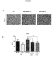
- mice were fed with a diet deficient in methionine and choline (MCD) for 4 weeks, a diet described to induce liver stetaosis that resembles human NAFLD [13]. Both knock-out mice showed a partial protection against liver steatosis compared with WT animals, since a decrease in the accumulation of lipid droplets in liver was observed ( Figure 1 ). Lower AST levels, a well-established marker of hepatic necrosis, were also detected in plasma of p38 ⁇ -/- , p38 ⁇ -/- mice. *p ⁇ 0.05,**p ⁇ 0.01, and ***p ⁇ 0.001;by t -test.
- Example 2 p38 ⁇ / ⁇ -/- mice are protected against liver steatosis.
- mice p38 ⁇ / ⁇ -/- were obtained from Dr. Ana Cuenda laboratory, and housed at the facilities of the Centro Nacional de Biotecnologia (CNB). 10 males (8-10 weeks old) per group were fed with a high fat diet (HFD, Harlan ref TD110418) or chow diet for 20 weeks. Mice were genotyped by PCR analysis using genomic DNA. For the inhibition of p38 MAPK isoforms C57BL6J male mice housed at the Centro de Investigativations Cardiovasculares (CNIC) were used.
- CNIC Centro de Investigativations Cardiovasculares
- ALT and AST activity were measured using Biosystems reagents (ref 11568 and 11561) and read in a microplate spectrophotometer at 340 nm (Benchmark Plus BioRad) after 10 min. 25 mg of liver tissue was homogenized in a chloroform and methanol solution (8:1 ratio) and incubated for 4 hours at 22°C. Sulfuric acid 1M was added to the extracts, and samples were centrifuged at 1000 r.p.m. for 10 min. Lipids in the organic phase were transferred to a new tube, quantified using Serum Triglyceride determination Kit (Sigma ref TR0100), and detected after 15 min incubation at 540 nm.
- TBARS thiobarbituric acid reactive substances
- liver fibrosis were studied by measuring the levels of TIMP-1 and collagen, two known markers of fibrosis (see Figure 5 , *p ⁇ 0.05,**p ⁇ 0.01, and ***p ⁇ 0.001;by t -test).
- mice p38 ⁇ / ⁇ -/- were obtained as explained in Example 2. Mice fed with MCD diet and treated with BIRB 796 show lower liver steatosis than the ones treated with MCD and vehicle (DMSO). Then it can be concluded that p38 ⁇ and ⁇ isoforms are involved in the liver protection of lipid steatosis (see Figure 6 ).
- Example 4 Mice lacking p38 ⁇ and p38 ⁇ are resistant to develop liver steatosis during obesity.
- Liver extracts were prepared using Triton lysis buffer (20 mM Tris at pH 7.4, 1% Triton X-100, 10% glycerol, 137 mM NaCl, 2 mM EDTA, 25 mM ⁇ -glycerophosphate, 1 mM sodium orthovanadate, 1 mM phenylmethylsulfonyl fluoride, 10 ⁇ g/mL of aprotinin and 10 ⁇ g/mL of leupeptin), and 50 ⁇ g of protein examined by immunoblot analysis probing with antibodies against to AKT, phospho-Thr 308 AKT, and phospho-Ser 473 AKT (Cell Signaling); GAPDH (Santa Cruz Biotechnologies) and SAPK3 (produced by our laboratory).
- Triton lysis buffer (20 mM Tris at pH 7.4, 1% Triton X-100, 10% glycerol, 137 mM NaCl, 2 mM EDTA, 25 mM
- the study population was comprised of obese adult patients (at least 18 years of age) with body mass index (BMI) greater than 35 who underwent elective bariatric surgery at the University Hospital of Salamanca between November 2010 and November 2011. Patients were excluded if they had prior liver disease, history of alcohol use disorders or excessive alcohol consumption (>30 g/day in men and > 20 g/day in women). Subjects were also excluded if they had chronic hepatitis C or B or if laboratory and histopathological data showed other causes of liver disease different from non-alcoholic fatty liver disease (NAFLD). Patients with body mass index lower than 25 and without liver disease who underwent laparoscopic cholecystectomy were included as a control group.
- BMI body mass index
- RNAlater (Ambion Inc, USA) and stored at -80°C until RNA was extracted.
- a sample of subcutaneous and omental adipose tissue were collected as well during surgery. Before surgery, fasting venous blood samples were collected. Serum was obtained after centrifugation and was stored at -80°C and human whole blood was collected in PAXgene Blood RNA Tubes (ref 762165) and stored at -80°C until RNA was extracted.
- Data were collected on demographic information (age, sex, and ethnicity), anthropomorphic measurements (BMI), coexisting medical conditions, medication use, smoking and alcohol history, and laboratory data including complete cell blood count, total bilirubin, aspartate aminotransferase (AST), alanine aminotransferase (ALT), alkaline phosphatase, gamma-glutamyl-transpeptidase, total cholesterol, high-density lipoprotein, low-density lipoprotein, triglycerides, creatinine, glucose, glycated haemoglobin, albumin, and iron metabolism markers. Abdominal ultrasound imaging was also obtained before surgery.
- Hepatic fibrosis was assessed by Masson stain. No histological signs of hepatic fibrosis were observed in controls. However, obvious hepatic fibrosis was detected in 92% of obese patients, with different grades of fibrosis. The liver leukocyte infiltration was also detected in all the obese patients ranging from a medium to high grade of mononuclear cell infiltration (MNCs), a change not detected in control patients (see Figure 9 , *p ⁇ 0.05,**p ⁇ 0.01, and ***p ⁇ 0.001;by t -test ).
- MNCs mononuclear cell infiltration
- Example 6 p 38 ⁇ and ⁇ are potential markers for liver steatosis.
- the study population was comprised of obese adult patients (at least 18 years of age) with body mass index (BMI) greater than 35 who underwent elective bariatric surgery at the University Hospital of Salamanca between November 2010 and November 2011. Patients were excluded if they had prior liver disease, history of alcohol use disorders or excessive alcohol consumption (>30 g/day in men and > 20 g/day in women). Subjects were also excluded if they had chronic hepatitis C or B or if laboratory and histopathological data showed other causes of liver disease different from non-alcoholic fatty liver disease (NAFLD). Patients with body mass index lower than 25 and without liver disease who underwent laparoscopic cholecystectomy were included as a control group.
- BMI body mass index
- RNAlater (Ambion Inc, USA) and stored at -80°C until RNA was extracted.
- a sample of subcutaneous and omental adipose tissue were collected as well during surgery. Before surgery, fasting venous blood samples were collected. Serum was obtained after centrifugation and was stored at -80°C and human whole blood was collected in PAXgene Blood RNA Tubes (ref 762165) and stored at -80°C until RNA was extracted.
- Data were collected on demographic information (age, sex, and ethnicity), anthropomorphic measurements (BMI), coexisting medical conditions, medication use, smoking and alcohol history, and laboratory data including complete cell blood count, total bilirubin, aspartate aminotransferase (AST), alanine aminotransferase (ALT), alkaline phosphatase, gamma-glutamyl-transpeptidase, total cholesterol, high-density lipoprotein, low-density lipoprotein, triglycerides, creatinine, glucose, glycated haemoglobin, albumin, and iron metabolism markers. Abdominal ultrasound imaging was also obtained before surgery.
- measuring blood mRNA levels of p38 ⁇ and ⁇ in relative amount against s18 for the diagnosis of NAFLD could be of potential clinical use.
- p38 ⁇ levels lower than 0,30 (threshold value) had a sensitivity rate of 83,33% and a specificity rate of 73% for the diagnosis of NALFD, with a positive predictive value (PPV) of 76,92% and a negative predictive value (NPV) of 80,0%.
- p38 ⁇ levels lower than 0,45 had a sensitivity rate of 83,33% and a specificity rate of 82,0%, with a PPV of 83,33% and a NPV of 81,82%.
- the combination of both markers improved specificity (90,9%) but slightly decreased sensitivity (75%).
- the detection of p38 ⁇ and ⁇ blood levels could detect the presence of liver steatosis and the combination of these markers with other markers of liver inflammation or liver fibrosis could potentially improve the detection of early fibrosis or monitor the development of cirrhosis (see Figure 10 *p ⁇ 0.05, **p ⁇ 0.01, and ***p ⁇ ⁇ 0.001;by t -test).
Landscapes
- Chemical & Material Sciences (AREA)
- Life Sciences & Earth Sciences (AREA)
- Proteomics, Peptides & Aminoacids (AREA)
- Health & Medical Sciences (AREA)
- Organic Chemistry (AREA)
- Wood Science & Technology (AREA)
- Analytical Chemistry (AREA)
- Zoology (AREA)
- Genetics & Genomics (AREA)
- Engineering & Computer Science (AREA)
- Pathology (AREA)
- Immunology (AREA)
- Microbiology (AREA)
- Molecular Biology (AREA)
- Biotechnology (AREA)
- Biophysics (AREA)
- Physics & Mathematics (AREA)
- Biochemistry (AREA)
- Bioinformatics & Cheminformatics (AREA)
- General Engineering & Computer Science (AREA)
- General Health & Medical Sciences (AREA)
- Measuring Or Testing Involving Enzymes Or Micro-Organisms (AREA)
Abstract
Description
- The present invention may be included in the field of the medicine, in general, and, more particularly, in that area of the medicine which is focused on the study of nonalcoholic fatty liver disease (NAFLD) and, more particularly, of hepatic steatosis, also known as fatty liver, characterized by the abnormal fat accumulation that occurs in the liver cells.
- Obesity is a major health problem associated with many other diseases like type two diabetes T2DM, metabolic syndrome and NAFLD. In fact, NAFLD is one of the most prevalent manifestations of obesity [1] and this chronic liver disease has become a worldwide health problem.
- NAFLD refers to a spectrum of liver diseases ranging from simple steatosis or fatty liver, to nonalcoholic steatohepatitis (NASH), and cirrhosis (with advanced scarring of the liver), all these stages have in common an accumulation of fat within hepatocytes. Most patients with NAFLD also have obesity (60-100%), T2DM (28-55%) and hyperlipidemia (27-92%) [2]. While NAFLD can be quite unobtrusive, NASH is a serious condition, with nearly a quarter of affected patients developing cirrhosis, which, in turn, increases the risk of subsequent progression to hepatocellular carcinoma (HCC). HCC is the fifth most common tumour worldwide with increasing incidence and third leading cause of cancer-related death. Among patients with NAFLD or NASH, HCC represents the main cause of death [3].
- It has also been estimated that around 20-30% of the general population have NAFLD [4,5], a asymptomatic disease that can progress from an initial trygliceride accumulation to NASH which, in a long-term, may results in fibrosis, cirrhosis and hepatocelullar carcinoma (HCC) [6, 7, 8]. For that reason, an early and accurate diagnosis is fundamental to know the degree of liver damage, and avoid the disease progression.
- Liver biopsy is nowadays considered the gold standard for NAFLD detection, since the diagnostic yield of available non-invasive tests is not enough accurate [9, 10]. Despite of providing important information about liver damage and staging, liver biopsy is an invasive procedure, uncomfortable and with risks for the patient and therefore it cannot be used as a screening or routine method, or for obtaining several samples periodically to assess disease evolution.
- Therefore, a reliable and non-invasive test for diagnosis of liver steatosis would lead to a clear improvement in early diagnosis, ability of monitoring of the disease, as well as quality of life.
- The present invention solves the above-mentioned problem by providing solid biomarkers (p38MAPK) for NAFLD, preferably for hepatic steatosis. p38MAPK (mitogen-activated protein kinase) families are strongly activated in vivo by environmental stress factors and inflammatory cytokines. Obesity and liver steatosis act like stress signals inducing multiple responses in tissues. Four p38MAPK isotypes have been described to date: alpha (α), beta (β), gamma (γ) and delta (δ). The canonical activation of p38MAPK occurs via dual phosphorylation in the activation loop by MKK3 and MKK6 [12].
- Consequently the present invention provides an accurate in vitro method for prognosis and diagnosis of NAFLD, preferably of hepatic steatosis, offering the opportunity of carrying out an improved treatment at earlier stages of the disease, thus rising the likelihood of definitely treating the disease. For that reason, an early and accurate diagnosis is fundamental to know the degree of liver damage, and avoid the disease progression. On the other hand, the method of the invention is based on blood samples and consequently it may be considered as a non-invasive test which leads to a clear improvement in early diagnosis, ability of monitoring of the disease, as well as quality of life. Blood levels of these kinases (p38MAPK gamma and p38MAPK delta) can be therefore used as potential biomarkers of hepatic steatosis in human, without the inconvenient and risks of liver biopsy and therefore improving early diagnosis and more accurate prognosis of NAFLD and its associated complications or diseases.
- As explained above, the present invention provides an accurate and non-invasive in vitro method for prognosis and diagnosis of NAFLD, preferably of hepatic steatosis, offering the opportunity of carrying out an improved treatment at earlier stages of the disease, thus raising the likelihood of definitely treating the disease. The method of the invention is based on blood samples and consequently it may be considered as a non-invasive test for diagnosis of hepatic steatosis which leads to a clear improvement in early diagnosis, ability of monitoring of the disease, as well as quality of life. Blood levels of these kinases could be therefore used as potential biomarkers of hepatic steatosis in human, without the inconvenient and risks of liver biopsy and therefore improving early diagnosis and more accurate prognosis of NAFLD and its complications.
- After a large study with both mice and human blood samples (see examples), a statistically significant relation between liver stetatosis induced by obesity with a decrease in expression levels of kinases p38MAPK δ and p38MAPK γ was observed in the present invention (p<0.001 or p<0.01 respectably). Therefore, analysis of these kinases can be used as biomarkers of NAFLD, preferably of liver steatosis, in human, without the drawbacks associated to the use of liver biopsy and therefore improving early diagnosis and more accurate prognosis.
- Consequently, the first embodiment of the present invention refers to p38MAPK gamma and/or delta for use as biomarkers of NAFLD, preferably of hepatic steatosis.
- The second embodiment of the present invention refers to an in vitro method for the diagnosis of NAFLD, preferably of hepatic steatosis, that comprises measuring the expression level of p38MAPK gamma and/or delta in a blood sample taken from the subject under examination. As explained above, hepatic steatosis is associated with a significant decrease in expression levels of p38MAPK gamma and p38MAPK delta. In a preferred embodiment, the method of the invention is characterized in that the expression level of p38MAPK gamma is measured using the primers of SEQ ID NO: 21 and 22 and the expression level of p38MAPK delta is measured using the primers of SEQ ID NO: 23 and 24.
- The third embodiment of the present invention refers to a nucleotide sequence characterized by the SEQ ID NO: 21, 22, 23 or 24. These sequences are used in the present invention as primers for measuring the expression level of p38 gamma and/or p38 delta in the blood samples taken from the patient/subject. Consequently these sequences may be used as primers to make a kit for diagnosis of NAFLD, preferably for diagnosis of hepatic steatosis. Thus, in a preferred embodiment the present invention refers to a nucleotide sequence characterized by the SEQ ID NO: 21, 22, 23 or 24 or any combination thereof for use as primers for hybridizing under stringent conditions with a nucleic acid encoding for p38MAPK gamma and/or delta sequence or any partial complementary or mutated sequence thereof.
- Thus, the fourth embodiment of the present invention refers to a kit for the diagnosis of NAFLD, preferably of hepatic steatosis, characterized by comprising the primers of SEQ ID NO: 21 and 22 for measuring the expression level of p38MAPK gamma and/or the primers of SEQ ID NO: 23 and 24 for measuring the expression level of p38MAPK delta.
- For the purpose of the present invention p<0.05 refers to the significance level and p values below to 0.05 make the biomarkers of the invention statistically significant.
-
-
Figure 1 . Effect of MCD (methionine choline deficiente) diet in mice lacking p38γ (p38gamma -/-) or p38δ (p38delta -/-). WT (wild type) animal or lacking in p38γ or p38δ were fed for 4 weeks with MCD or control diet (ND). A) Histological section of liver stained with hematoxilin and eosin. B) AST (Aspartate Aminotransferase) (well known marker of hepatitis necrosis) levels in plasma as indicator of liver damage. Both knock-out mice showed a partial protection against liver steatosis compared with WT animals, since a decrease in the accumulation of lipid droplets in liver is observed. *p < 0.05, **p < 0.01, and ***p < 0.001;by t-test. -
Figure 2 . Effect of MCD diet or ND diet in mice lacking p38 gamma and delta p38δ/γ-/-. WT animals or p38δ/γ-/- were fed for 4 weeks with MCD diet. A) Histological sections of liver stained with hematoxylin and eosin. B) AST and ALT (alanine aminotransferase) in the plasma as indicators of liver damage. Double knock-out mice p38δ/γ-/- are protected against liver steatosis. *p < 0.05,**p < 0.01, and ***p< 0.001; by t-test. -
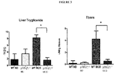
Figure 3 .p38δ/γ-/- animals are protected animals against oxidative stress. Triglycerides stored in the liver and lipid peroxidation (Tbars) in livers of WT mice and p38δ/γ-/- fed or not with MCD diet. *p < 0.05,**p < 0.01, and ***p < 0.001;by t-test. -
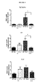
Figure 4 . Lack of p38δ and p38 γ protects animals from inflammation induced by a methionine and choline deficient diet. Liver mRNA levels of TNF, IL-6 and IL-10 in WT mice and p38δ/γ-/- fed with MCD or control diet (ND). *p < 0.05,**p < 0.01, and ***p < 0.001;by t-test. -
Figure 5 . p38δ/γ-/- animals are protected against MCD-induced fibrosis. Measurement of liver mRNA levels ofTIMPI and collagen from WT and p38δ/γ-/-mice fed MCD or control diet (ND). *p < 0.05,**p < 0.01, and ***p < 0.001;by t-test. -
Figure 6 . BIRB796 protects animals of MCD-induced steatosis. WT animals were injected with DMSO or BIRB796 daily for 3 weeks and fed MCD diet. Histological sections of liver stained with hematoxylin and eosin reveal that BIRB796 protects against development of steatosis. -
Figure 7 . p38δ/γ-/- does not develop hepatic steatosis and insulin resistance. WT or p38δ/γ-/- mice fed with normal diet (ND) or high fat diet (HFD) were fasted for 16hrs and then treated with insulin (1.5U/kg). Liver extracts were analyzed for phosphorylation ofAKT -
Figure 8 . mRNA levels of p38δ and p38γ decrease during obesity. Relative expression levels respect to GAPDH of p38δ and p38γ in blood from animals fed 20 weeks with high fat diet (HFD) or control diet (ND). *p < 0.05,**p < 0.01, and ***p < 0.001;by t-test. -
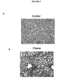
Figure 9 . Levels of hepatic steatosis in control (A) and obese patients (B). Histological sections stained with hematoxylin and eosin revealed different grades of steatosis in obese patients. Histological grading of inflammation and fibrosis was estimated from histological samples. * < 0.05,**p < 0.01, and ***p < 0.001;by t-test. -
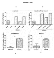
Figure 10 . mRNA levels of blood from obese patients and controls. The expression levels of p38gamma and p38delta in whole blood are significantly decreased in obese patients when compared to healthy controls. Expression level of s18 was used as control. *p < 0.05,**p < 0.01, and ***p < 0.001;by t-test. - Mice p38δ-/- and p38γ-/- were obtained from Dr. Ana Cuenda laboratory, and housed at the facilities of the Centro Nacional de Biotecnologia (CNB). 10 males (8-10 weeks old) per group were fed with a methionine and choline deficient diet (MCD, Harlan ref TD.90262) or chow diet for 4 weeks. Mice were genotyped by PCR analysis using genomic DNA. Animal studies were approved by the local ethics committee, and all procedures followed EU Directive 86/609/EEC and Recommendation 2007/526/EC regarding the protection of animals used for experimental and other scientific purposes, enacted under Spanish law 1201/2005.
- Tissue fixed in 10% formalin for 24 h, dehydrated, and embedded in paraffin was cut in sections (8 µm) and stained using hematoxylin and eosin or Masson trichromic.
- Different histological parameters were analyzed on each liver sample.
- Steatosis: Degree, type and localization.
- ● Degree: Presence of lipid accumulation in vesicles, Mild (<10%), Moderate (10-25%), Severe (26-50%) and Massive (>50%).
- ● Type: Microvesicular, Macrovesicular, and Mixed.
- ● Localization: Periportal, Centrilobular, Diffuse.
- Inflammation: Type and degree.
- Fibrosis: Absent, Perisinusoidal.
- Typical hepatocellular lesions: Presence or absence.
- The serum concentration of alanine transaminases (ALT) and aspartate aminotransferase (AST) activity was measured using Biosystems reagents (ref 11568 and 11561) and read in a microplate spectrophotometer at 340 nm (Benchmark Plus BioRad) after 10 min.
- Statistical significance to compare treatment groups was calculated by Student t-test using Prism software (GraphPad). Data were presented as mean±SEM.
- Since obesity is a disease associated to a chronic inflammatory state, characterized by increased inflammatory markers such as C reactive protein, IL-6, IL-8, IL-10, PAI-1, TNF-α, MCP-1 and hepatocyte growth factor, and, moreover, it is well established that the p38MAPK family plays a key role during the inflammatory process, thus, it was decided to study whether p38γ and p38δ could have a role in the development of liver stetaosis. With that aim, p38δ-/-, p38γ-/- and WT mice were fed with a diet deficient in methionine and choline (MCD) for 4 weeks, a diet described to induce liver stetaosis that resembles human NAFLD [13]. Both knock-out mice showed a partial protection against liver steatosis compared with WT animals, since a decrease in the accumulation of lipid droplets in liver was observed (
Figure 1 ). Lower AST levels, a well-established marker of hepatic necrosis, were also detected in plasma of p38δ-/-, p38γ-/- mice. *p < 0.05,**p < 0.01, and ***p < 0.001;by t-test. - Mice p38γ/δ -/- were obtained from Dr. Ana Cuenda laboratory, and housed at the facilities of the Centro Nacional de Biotecnologia (CNB). 10 males (8-10 weeks old) per group were fed with a high fat diet (HFD, Harlan ref TD110418) or chow diet for 20 weeks. Mice were genotyped by PCR analysis using genomic DNA. For the inhibition of p38 MAPK isoforms C57BL6J male mice housed at the Centro de Investigaciones Cardiovasculares (CNIC) were used. Animal studies were approved by the local ethics committee, and all procedures followed EU Directive 86/609/EEC and Recommendation 2007/526/EC regarding the protection of animals used for experimental and other scientific purposes, enacted under Spanish law 1201/2005. Tissue fixed in 10% formalin for 24 h, dehydrated, and embedded in paraffin was cut in sections (8 µm) and stained using hematoxylin and eosin or Masson trichromic.
- Different histological parameters were analyzed on each liver sample.
- Steatosis: Degree, type and localization.
- ● Degree: Presence of lipid accumulation in vesicles, Mild (<10%), Moderate (10-25%), Severe (26-50%) and Massive (>50%).
- ● Type: Microvesicular, Macrovesicular, and Mixed.
- ● Localization: Periportal, Centrilobular, Diffuse.
- Inflammation: Type and degree.
- Fibrosis: Absent, Perisinusoidal.
- Typical hepatocellular lesions: Presence or absence.
- The serum concentration of alanine transaminases (ALT) and aspartate aminotransferase (AST) activity was measured using Biosystems reagents (ref 11568 and 11561) and read in a microplate spectrophotometer at 340 nm (Benchmark Plus BioRad) after 10 min. 25 mg of liver tissue was homogenized in a chloroform and methanol solution (8:1 ratio) and incubated for 4 hours at 22°C. Sulfuric acid 1M was added to the extracts, and samples were centrifuged at 1000 r.p.m. for 10 min. Lipids in the organic phase were transferred to a new tube, quantified using Serum Triglyceride determination Kit (Sigma ref TR0100), and detected after 15 min incubation at 540 nm.
- Lipid peroxidation in tissue samples was determined by the thiobarbituric acid reactive substances (TBARS) assay. 25 mg of liver tissue was homogenized in lysis buffer with protease inhibitors and then sonicated. This homogenate was centrifuged and supernatant used for analysis following the manufacture instructions of TBARS assay kit (Cayman ref 10009055).
- The expression of mRNA was examined by quantitative PCR analysis using a 7900 Fast Real-Time PCR machine. Primers were produced by Sigma Aldrich (Table 1). The relative mRNA expression was normalized by measurement of the amount of Gapdh mRNA.
Table 1 Forward Reverse mmu-Il6 SEQ ID NO: 1 SEQ ID NO: 2 mmu-Tnf-α SEQ ID NO: 3 SEQ ID NO: 4 mmu-Il10 SEQ ID NO: 5 SEQ ID NO: 6 mmu-Timp1 SEQ ID NO: 7 SEQ ID NO: 8 mmu-Collagen SEQ ID NO: 9 SEQ ID NO: 10 mmu-Gapdh SEQ ID NO: 11 SEQ ID NO: 12 - Statistical significance to compare treatment groups was calculated by Student t-test using Prism software (GraphPad). Data were presented as mean±SEM.
- This example showed that p38γ/δLyz-KO mice have a greater protection against liver failure than either of the single conditional knock-out, indicating a degree of functional redundancy between p38γ and δ. Then, it was decided to study the effect of lacking the two p38 isoforms (p38γ and δ) in liver steatosis. Double knock-out mice (p38δ/γ-/-) were fed with MCD for four weeks. MCD diet induces a change in liver histology architecture, with an increase in lipids accumulation and leukocyte liver infiltration in WT mice. However, these effects were not present in p38δ/γ-/- mice (
Figure 2A ) which correlated with reduced levels of serum transaminases, which are biomarkers of liver necrosis. *p < 0.05,**p < 0.01, and ***p < 0.001;by t-test. - Moreover reduced levels of triglycerides in the liver from p38δ/γ-/- mice correlated with lower lipid peroxidation after MCD diet (
Figure 3 ), indicating a protection against the oxidative stress that accomplishes the liver steatosis. *p < < 0.05,**p < 0.01, and ***p < 0.001;by t-test - The lower levels of lipid accumulation in liver could be responsible for the reduction in liver inflammation observed in these animals after MCD diet with a decrease in the expression levels of TNF and IL6 (see
Figure 4 ,*p < 0.05,**p < 0.01, and ***p < 0.001;by t-test). To study whether all these effects correlate or not with a whole improvement of the disease, levels of liver fibrosis were studied by measuring the levels of TIMP-1 and collagen, two known markers of fibrosis (seeFigure 5 , *p < 0.05,**p < 0.01, and ***p < 0.001;by t-test).). Both markers are increased after MCD diet in the livers of WT animals, but not in the liver from p38δ/γ-/- mice. This indicates that mice defective in p38γ and δ present a great protection against liver steatosis and inflammation. - Mice p38γ/δ-/- were obtained as explained in Example 2. Mice fed with MCD diet and treated with BIRB 796 show lower liver steatosis than the ones treated with MCD and vehicle (DMSO). Then it can be concluded that p38γ and δ isoforms are involved in the liver protection of lipid steatosis (see
Figure 6 ). - Mice p38γ/δ-/- were obtained as explained in Example 2.
- Liver extracts were prepared using Triton lysis buffer (20 mM Tris at pH 7.4, 1% Triton X-100, 10% glycerol, 137 mM NaCl, 2 mM EDTA, 25 mM β-glycerophosphate, 1 mM sodium orthovanadate, 1 mM phenylmethylsulfonyl fluoride, 10 µg/mL of aprotinin and 10 µg/mL of leupeptin), and 50 µg of protein examined by immunoblot analysis probing with antibodies against to AKT, phospho-Thr308 AKT, and phospho-Ser473 AKT (Cell Signaling); GAPDH (Santa Cruz Biotechnologies) and SAPK3 (produced by our laboratory).
- Tissue fixed in 10% formalin for 24 h, dehydrated, and embedded in paraffin was cut in sections (8 µm) and stained using hematoxylin and eosin or Masson trichromic.
- Different histological parameters were analyzed on each liver sample.
- Steatosis: Degree, type and localization.
- ● Degree: Presence of lipid accumulation in vesicles, Mild (<10%), Moderate (10-25%), Severe (26-50%) and Massive (>50%).
- ● Type: Microvesicular, Macrovesicular, and Mixed.
- ● Localization: Periportal, Centrilobular, Diffuse.
- Inflammation: Type and degree.
- Fibrosis: Absent, Perisinusoidal.
- Typical hepatocellular lesions: Presence or absence.
- The expression of mRNA was examined by quantitative PCR analysis using a 7900 Fast Real-Time PCR machine. Primers were produced by Sigma Aldrich (Table 2). The relative mRNA expression was normalized by measurement of the amount of Gapdh mRNA.
Table 2 Forward Reverse mmu-p38 delta SEQ ID NO: 13 SEQ ID NO: 14 mmu-p38 gamma SEQ ID NO: 15 SEQ ID NO: 16 mmu-Gapdh SEQ ID NO: 17 SEQ ID NO: 18 - Statistical significance to compare treatment groups was calculated by Student t-test using Prism software (GraphPad). Data were presented as mean±SEM.
- There is a high prevalence of steatosis in obese individuals, it was decided to study the role of p38γ/δ in the development of steatosis in a model of obesity induced esteatosis. Mice were fed with high fat diet (HFD) (60% of the calories from fat) for 20 weeks. After this time, WT mice develop liver steatosis with an increase of lipid accumulation. However, mice lacking p38γ/δ did not accumulate lipid in the liver (
Figure 7 ). Because liver steatosis induces insulin resistance we decide to study whether the lower lipid accumulation observed in p38γ/δ -/- mice correlate with a better insulin response after high fat diet. To confirm the effect steatosis on insulin sensitivity, insulin-stimulated Ser/Thr phosphorylation and activation of AKT were tested. HFD-fed WT mice exhibited reduced insulin-stimulated AKT activation in liver(Figure 7A). In contrast, p38γ/δ-/- mice exhibited HFD-induced inhibition of insulin-stimulated AKT activation in muscle, but not in liver (seeFigure 7 ). - Since the present invention indicates that p38γ/δ have a role on liver steatosis induction, it was decided to measure blood levels of these kinases. Expression levels of these kinases decreased after feeding by HFD (
Figure 8 ), indicating that they can be used as biomarkers of liver steatosis. *p < 0.05, **p < 0.01, and ***p < 0.001;by t-test. - The study population was comprised of obese adult patients (at least 18 years of age) with body mass index (BMI) greater than 35 who underwent elective bariatric surgery at the University Hospital of Salamanca between November 2010 and November 2011. Patients were excluded if they had prior liver disease, history of alcohol use disorders or excessive alcohol consumption (>30 g/day in men and > 20 g/day in women). Subjects were also excluded if they had chronic hepatitis C or B or if laboratory and histopathological data showed other causes of liver disease different from non-alcoholic fatty liver disease (NAFLD). Patients with body mass index lower than 25 and without liver disease who underwent laparoscopic cholecystectomy were included as a control group. The study was approved by the Ethics Committee of the University Hospital of Salamanca and all subjects provided written informed consent to undergo liver biopsy under direct vision during surgery. A portion of each liver biopsy was fixed in 10% formalin and stained with hematoxylin-eosin and Masson trichrome for standard histopathological interpretation. The remaining portion was immersed in RNAlater (Ambion Inc, USA) and stored at -80°C until RNA was extracted. A sample of subcutaneous and omental adipose tissue were collected as well during surgery. Before surgery, fasting venous blood samples were collected. Serum was obtained after centrifugation and was stored at -80°C and human whole blood was collected in PAXgene Blood RNA Tubes (ref 762165) and stored at -80°C until RNA was extracted.
- Data were collected on demographic information (age, sex, and ethnicity), anthropomorphic measurements (BMI), coexisting medical conditions, medication use, smoking and alcohol history, and laboratory data including complete cell blood count, total bilirubin, aspartate aminotransferase (AST), alanine aminotransferase (ALT), alkaline phosphatase, gamma-glutamyl-transpeptidase, total cholesterol, high-density lipoprotein, low-density lipoprotein, triglycerides, creatinine, glucose, glycated haemoglobin, albumin, and iron metabolism markers. Abdominal ultrasound imaging was also obtained before surgery.
- Tissue fixed in 10% formalin for 24 h, dehydrated, and embedded in paraffin was cut in sections (8 µm) and stained using hematoxylin and eosin or Masson trichromic.
- Different histological parameters were analyzed on each liver sample.
- Steatosis: Degree, type and localization.
- ● Degree: Presence of lipid accumulation in vesicles, Mild (<10%), Moderate (10-25%), Severe (26-50%) and Massive (>50%).
- ● Type: Microvesicular, Macrovesicular, and Mixed.
- ● Localization: Periportal, Centrilobular, Diffuse.
- Inflammation: Type and degree.
- Fibrosis: Absent, Perisinusoidal.
- Typical hepatocellular lesions: Presence or absence.
- Statistical significance to compare treatment groups was calculated by Student t-test using Prism software (GraphPad). Data were presented as mean±SEM.
- It has been extensively described how a high intake of calories can induce liver damage. The first and most visible change is observed at histological level in obese patients which have a large amount of lipids droplets inside the hepatocytes that can impair the normal function of the liver.
- Hepatic fibrosis was assessed by Masson stain. No histological signs of hepatic fibrosis were observed in controls. However, obvious hepatic fibrosis was detected in 92% of obese patients, with different grades of fibrosis. The liver leukocyte infiltration was also detected in all the obese patients ranging from a medium to high grade of mononuclear cell infiltration (MNCs), a change not detected in control patients (see
Figure 9 , *p < 0.05,**p < 0.01, and ***p < 0.001;by t-test ). - The study population was comprised of obese adult patients (at least 18 years of age) with body mass index (BMI) greater than 35 who underwent elective bariatric surgery at the University Hospital of Salamanca between November 2010 and November 2011. Patients were excluded if they had prior liver disease, history of alcohol use disorders or excessive alcohol consumption (>30 g/day in men and > 20 g/day in women). Subjects were also excluded if they had chronic hepatitis C or B or if laboratory and histopathological data showed other causes of liver disease different from non-alcoholic fatty liver disease (NAFLD). Patients with body mass index lower than 25 and without liver disease who underwent laparoscopic cholecystectomy were included as a control group. The study was approved by the Ethics Committee of the University Hospital of Salamanca and all subjects provided written informed consent to undergo liver biopsy under direct vision during surgery. A portion of each liver biopsy was fixed in 10% formalin and stained with hematoxylin-eosin and Masson trichrome for standard histopathological interpretation. The remaining portion was immersed in RNAlater (Ambion Inc, USA) and stored at -80°C until RNA was extracted. A sample of subcutaneous and omental adipose tissue were collected as well during surgery. Before surgery, fasting venous blood samples were collected. Serum was obtained after centrifugation and was stored at -80°C and human whole blood was collected in PAXgene Blood RNA Tubes (ref 762165) and stored at -80°C until RNA was extracted.
- Data were collected on demographic information (age, sex, and ethnicity), anthropomorphic measurements (BMI), coexisting medical conditions, medication use, smoking and alcohol history, and laboratory data including complete cell blood count, total bilirubin, aspartate aminotransferase (AST), alanine aminotransferase (ALT), alkaline phosphatase, gamma-glutamyl-transpeptidase, total cholesterol, high-density lipoprotein, low-density lipoprotein, triglycerides, creatinine, glucose, glycated haemoglobin, albumin, and iron metabolism markers. Abdominal ultrasound imaging was also obtained before surgery.
- The expression of mRNA was examined by quantitative PCR analysis using a 7900 Fast Real-Time PCR machine. Primers were designed as shown in Table 3. The relative mRNA expression was normalized by measurement of the amount of s18 mRNA.
Table 3 Forward Reverse h-p38 beta SEQ ID NO: 19 SEQ ID NO: 20 h-p38 gamma SEQ ID NO: 21 SEQ ID NO: 22 h-p38 delta SEQ ID NO: 23 SEQ ID NO: 24 h-s18 SEQ ID NO: 25 SEQ ID NO: 26 - Statistical significance to compare treatment groups was calculated by Student t-test using Prism software (GraphPad). Data were presented as mean±SEM.
- Blood samples were collected from obese and lean or healthy patients and the expression levels of p38 gamma and delta were analyzed by Q-PCR. A reduced expression of p38γ and δ of blood levels were found among obese patients. This indicates that p38γ and p38δ are good markers for predicting liver steatosis without an invasive test.
- According to the present invention, measuring blood mRNA levels of p38γ and δ in relative amount against s18 for the diagnosis of NAFLD could be of potential clinical use. p38γ levels lower than 0,30 (threshold value) had a sensitivity rate of 83,33% and a specificity rate of 73% for the diagnosis of NALFD, with a positive predictive value (PPV) of 76,92% and a negative predictive value (NPV) of 80,0%. Besides, p38 δ levels lower than 0,45 (threshold value) had a sensitivity rate of 83,33% and a specificity rate of 82,0%, with a PPV of 83,33% and a NPV of 81,82%. The combination of both markers improved specificity (90,9%) but slightly decreased sensitivity (75%).
- Therefore, the detection of p38γ and δ blood levels could detect the presence of liver steatosis and the combination of these markers with other markers of liver inflammation or liver fibrosis could potentially improve the detection of early fibrosis or monitor the development of cirrhosis (see
Figure 10 *p < 0.05, **p < 0.01, and ***p < < 0.001;by t-test). -
- 1. Luyckx FH, Desaive C, Thiry A, Dewé W, Scheen AJ, Gielen JE, Lefèbvre PJ. Liver abnormalities in severely obese subjects: effect of drastic weight loss after gastroplasty, Int J Obes Relat Metab Disord. 1998 Mar;22(3):222-6.
- 2. Angulo P, Lindor KD. Treatment of non-alcoholic steatohepatitis, Best Pract Res Clin Gastroenterol. 2002 Oct;16(5):797-810.
- 3. Yatsuji S, Hashimoto E, Tobari M, Taniai M, Tokushige K, Shiratori K., Clinical features and outcomes of cirrhosis due to non-alcoholic steatohepatitis compared with cirrhosis caused by chronic hepatitis C. J Gastroenterol Hepatol. 2009 Feb;24(2):248-54.
- 4. Sheth, S. G., Gordon, F. D. & Chopra, S. Nonalcoholic steatohepatitis. Ann. Intern. Med. 126,137-145 (1997).
- 5. Ludwig, J., Viggiano, T. R., McGill, D. B. & Oh, B. J. Nonalcoholic steatohepatitis: Mayo Clinic experiences with a hitherto unnamed disease. Mayo Clin. Proc. 55, 434-438 (1980).
- 6. Caldwell, S. H. et al. Cryptogenic cirrhosis: clinical characterization and risk factors for underlying disease. Hepatology 29, 664-669 (1999).
- 7. Shimada, M. et al. Hepatocellular carcinoma in patients with non-alcoholic steatohepatitis. J. Hepatol.37, 154-160 (2002).
- 8. Propst, A., Propst, T., Judmaier, G. & Vogel, W. Prognosis in nonalcoholic steatohepatitis. Gastroenterology 108, 1607 (1995).
- 9. Michael M. Van Ness,Anna Mae Diehl, Ann, Is Liver Biopsy Useful in the Evaluation of Patients with Chronically Elevated Liver Enzymes? Intern Med September 15, 1989 111:473-478.
- 10. Skelly MM, James PD, Ryder SD,Findings on liver biopsy to investigate abnormal liver function tests in the absence of diagnostic serology. J Hepatol. 2001; 35:195-199.
- 11. Enslen H, Brancho DM, Davis RJ.Molecular determinants that mediate selective activation of p38 MAP kinase isoforms. EMBO J.2000 .
- 12. Remy G, Risco AM, Iñesta-Vaquera FA, González-Terán B, Sabio G, Davis RJ, Cuenda A. Differential activation of p38MAPK isoforms by MKK6 and MKK3, Cell Signal. 2010 Apr;22(4):660-7. Epub 2009 Dec 11.
- 13. Varela-Rey, M. et al. Non-alcoholic steatohepatitis and animal models: understanding the human disease. Int. J. Biochem. Cell Biol. 41, 969-976 (2009).


Claims (10)
- p38MAPK gamma and/or delta for use as biomarkers of NAFLD.
- p38MAPK gamma and/or delta, according to claim 1, for use as biomarkers of liver steatosis.
- In vitro method for the diagnosis of NAFLD that comprises measuring the expression level of p38MAPK gamma and/or delta in a blood sample taken from the subject under examination.
- In vitro method, according to claim 3, for the diagnosis of liver steatosis.
- In vitro method, according to claim 3, characterized in that the expression level of p38MAPK gamma is measured using the primers of SEQ ID NO: 21 and 22 and the expression level of p38MAPK delta is measured using the primers of SEQ ID NO: 23 and 24.
- In vitro method, according to claim 5, wherein the expression level of p38MAPK gamma and/or delta is reduced in patients suffering from NAFLD as compared with the lean individuals.
- Nucleotide sequence characterized by the SEQ ID NO: 21, 22, 23 or 24.
- Nucleotide sequence characterized by the SEQ ID NO: 21, 22, 23 or 24 or any combination thereof for use as primers for hybridizing under stringent conditions with a nucleic acid encoding for p38MAPK gamma and/or delta sequence or any partial, complementary or mutated sequence thereof.
- kit for the diagnosis of NAFLD characterized by comprising the primers of SEQ ID NO: 21 and 22 for measuring the expression level of p38MAPK gamma and/or the primers of SEQ ID NO: 23 and 24 for measuring the expression level of p38MAPK delta.
- Kit, according to claim 9, for the diagnosis of liver steatosis.
Priority Applications (1)
| Application Number | Priority Date | Filing Date | Title |
|---|---|---|---|
| EP12164807.5A EP2653555A1 (en) | 2012-04-19 | 2012-04-19 | P38 MAPK gamma and delta for use as biomarkers of NAFLD |
Applications Claiming Priority (1)
| Application Number | Priority Date | Filing Date | Title |
|---|---|---|---|
| EP12164807.5A EP2653555A1 (en) | 2012-04-19 | 2012-04-19 | P38 MAPK gamma and delta for use as biomarkers of NAFLD |
Publications (1)
| Publication Number | Publication Date |
|---|---|
| EP2653555A1 true EP2653555A1 (en) | 2013-10-23 |
Family
ID=45976192
Family Applications (1)
| Application Number | Title | Priority Date | Filing Date |
|---|---|---|---|
| EP12164807.5A Withdrawn EP2653555A1 (en) | 2012-04-19 | 2012-04-19 | P38 MAPK gamma and delta for use as biomarkers of NAFLD |
Country Status (1)
| Country | Link |
|---|---|
| EP (1) | EP2653555A1 (en) |
Cited By (2)
| Publication number | Priority date | Publication date | Assignee | Title |
|---|---|---|---|---|
| WO2016198698A3 (en) * | 2015-06-12 | 2017-03-16 | Cnic Fundación Centro Nacional De Investigaciones Cardiovasculares Carlos Iii | P38 inhibitors for the treatment and prophylaxis of liver cancer |
| CN111183360A (en) * | 2017-07-19 | 2020-05-19 | 生物辐射欧洲有限公司 | Biomarker combination for simultaneous assessment of non-alcoholic steatohepatitis and hepatic fibrosis status |
Citations (2)
| Publication number | Priority date | Publication date | Assignee | Title |
|---|---|---|---|---|
| WO2009121152A2 (en) * | 2008-04-03 | 2009-10-08 | Katholieke Universiteit Leuven | Gene signatures |
| WO2011029899A1 (en) * | 2009-09-11 | 2011-03-17 | The Chinese University Of Hong Kong | Methods for assessing liver pathologies |
-
2012
- 2012-04-19 EP EP12164807.5A patent/EP2653555A1/en not_active Withdrawn
Patent Citations (2)
| Publication number | Priority date | Publication date | Assignee | Title |
|---|---|---|---|---|
| WO2009121152A2 (en) * | 2008-04-03 | 2009-10-08 | Katholieke Universiteit Leuven | Gene signatures |
| WO2011029899A1 (en) * | 2009-09-11 | 2011-03-17 | The Chinese University Of Hong Kong | Methods for assessing liver pathologies |
Non-Patent Citations (20)
| Title |
|---|
| ANGULO P; LINDOR KD.: "Treatment of non-alcoholic steatohepatitis", BEST PRACT RES CLIN GASTROENTEROL., vol. 16, no. 5, October 2002 (2002-10-01), pages 797 - 810 |
| BELL L N ET AL: "Serum proteomics and biomarker discovery across the spectrum of nonalcoholic fatty liver disease", HEPATOLOGY, WILLIAMS AND WILKINS, BALTIMORE, MD, US, vol. 51, no. 1, 1 January 2010 (2010-01-01), pages 111 - 120, XP002605132, ISSN: 0270-9139 * |
| CALDWELL, S. H. ET AL.: "Cryptogenic cirrhosis: clinical characterization and risk factors for underlying disease", HEPATOLOGY, vol. 29, 1999, pages 664 - 669 |
| DATABASE EMBL [Online] 21 January 2000 (2000-01-21), "Sequence 3 from Patent EP0922762.", XP002680468, retrieved from EBI accession no. EMBL:A80859 Database accession no. A80859 * |
| ENSLEN H; BRANCHO DM; DAVIS RJ.: "Molecular determinants that mediate selective activation of p38 MAP kinase isoforms", EMBO J., vol. 19, no. 6, 15 March 2000 (2000-03-15), pages 1301 - 11, XP002961828, DOI: doi:10.1093/emboj/19.6.1301 |
| GRZEGORZ SUMARA ET AL: "Regulation of PKD by the MAPK p38[delta] in Insulin Secretion and Glucose Homeostasis", CELL, vol. 136, no. 2, 1 January 2009 (2009-01-01), pages 235 - 248, XP055033134, ISSN: 0092-8674, DOI: 10.1016/j.cell.2008.11.018 * |
| GUADALUPE SABIO ET AL: "Prevention of Steatosis by Hepatic JNK1", CELL METABOLISM, vol. 10, no. 6, 1 December 2009 (2009-12-01), pages 491 - 498, XP055033442, ISSN: 1550-4131, DOI: 10.1016/j.cmet.2009.09.007 * |
| HALE ET AL: "Differential expression and activation of p38 mitogen-activated protein kinase alpha, beta, gamma, and delta in inflammatory cell lineages.", THE JOURNAL OF IMMUNOLOGY, vol. 162, no. 7, 1 April 1999 (1999-04-01), pages 4246 - 4252, XP055033095, ISSN: 0022-1767 * |
| LEWIS JEFFREY R ET AL: "Nonalcoholic Fatty Liver Disease: A Review and Update", DIGESTIVE DISEASES AND SCIENCES, PLENUM PUBLISHING CO, US, vol. 55, no. 3, 1 March 2010 (2010-03-01), pages 560 - 578, XP002631893, ISSN: 0163-2116, [retrieved on 20100126], DOI: 10.1007/S10620-009-1081-0 * |
| LUDWIG, J.; VIGGIANO, T. R.; MCGILL, D. B.; OH, B. J.: "Nonalcoholic steatohepatitis: Mayo Clinic experiences with a hitherto unnamed disease", MAYO CLIN. PROC., vol. 55, 1980, pages 434 - 438, XP009009087 |
| LUYCKX FH; DESAIVE C; THIRY A; DEWE W; SCHEEN AJ; GIELEN JE; LEFEBVRE PJ.: "Liver abnormalities in severely obese subjects: effect of drastic weight loss after gastroplasty", INT J OBES RELAT METAB DISORD., vol. 22, no. 3, March 1998 (1998-03-01), pages 222 - 6 |
| MICHAEL M. VAN NESS; ANNA MAE DIEHL: "Ann, Is Liver Biopsy Useful in the Evaluation of Patients with Chronically Elevated Liver Enzymes?", INTERN MED, vol. 111, 15 September 1989 (1989-09-15), pages 473 - 478 |
| PROPST, A.; PROPST, T.; JUDMAIER, G.; VOGEL, W.: "Prognosis in nonalcoholic steatohepatitis", GASTROENTEROLOGY, vol. 108, 1995, pages 1607 |
| REMY G; RISCO AM; IÑESTA-VAQUERA FA; GONZÁLEZ-TERÁN B; SABIO G; DAVIS RJ; CUENDA A.: "Differential activation of p38MAPK isoforms by MKK6 and MKK3", CELL SIGNAL., vol. 22, no. 4, 11 December 2009 (2009-12-11), pages 660 - 7 |
| SHETH, S. G.; GORDON, F. D.; CHOPRA, S.: "Nonalcoholic steatohepatitis", ANN. INTERN. MED., vol. 126, 1997, pages 137 - 145, XP009006803 |
| SHIMADA, M. ET AL.: "Hepatocellular carcinoma in patients with non-alcoholic steatohepatitis.", J. HEPATOL., vol. 37, 2002, pages 154 - 160 |
| SKELLY MM; JAMES PD; RYDER SD: "Findings on liver biopsy to investigate abnormal liver function tests in the absence of diagnostic serology", J HEPATOL., vol. 35, 2001, pages 195 - 199, XP002432459, DOI: doi:10.1016/S0168-8278(01)00094-0 |
| VARELA-REY, M. ET AL.: "Non-alcoholic steatohepatitis and animal models: understanding the human disease", INT. J. BIOCHEM. CELL BIOL., vol. 41, 2009, pages 969 - 976, XP025940839, DOI: doi:10.1016/j.biocel.2008.10.027 |
| VILLANUEVA A ET AL: "Pivotal Role of mTOR Signaling in Hepatocellular Carcinoma", GASTROENTEROLOGY, ELSEVIER, PHILADELPHIA, PA, vol. 135, no. 6, 1 December 2008 (2008-12-01), pages 1972 - 1983.E11, XP026147661, ISSN: 0016-5085, [retrieved on 20080820], DOI: 10.1053/J.GASTRO.2008.08.008 * |
| YATSUJI S; HASHIMOTO E; TOBARI M; TANIAI M; TOKUSHIGE K; SHIRATORI K.: "Clinical features and outcomes of cirrhosis due to non-alcoholic steatohepatitis compared with cirrhosis caused by chronic hepatitis C", J GASTROENTEROL HEPATOL., vol. 24, no. 2, February 2009 (2009-02-01), pages 248 - 54 |
Cited By (2)
| Publication number | Priority date | Publication date | Assignee | Title |
|---|---|---|---|---|
| WO2016198698A3 (en) * | 2015-06-12 | 2017-03-16 | Cnic Fundación Centro Nacional De Investigaciones Cardiovasculares Carlos Iii | P38 inhibitors for the treatment and prophylaxis of liver cancer |
| CN111183360A (en) * | 2017-07-19 | 2020-05-19 | 生物辐射欧洲有限公司 | Biomarker combination for simultaneous assessment of non-alcoholic steatohepatitis and hepatic fibrosis status |
Similar Documents
| Publication | Publication Date | Title |
|---|---|---|
| Scapaticci et al. | Non-alcoholic fatty liver disease in obese youth with insulin resistance and type 2 diabetes | |
| Heida et al. | The hepatocyte IKK: NF-κB axis promotes liver steatosis by stimulating de novo lipogenesis and cholesterol synthesis | |
| Sei et al. | A hereditary form of small intestinal carcinoid associated with a germline mutation in inositol polyphosphate multikinase | |
| EP2715366B1 (en) | Biomarkers for hedgehog inhibitor therapy | |
| Bricambert et al. | Salt-inducible kinase 2 links transcriptional coactivator p300 phosphorylation to the prevention of ChREBP-dependent hepatic steatosis in mice | |
| Dongiovanni et al. | Genetic predisposition in NAFLD and NASH: impact on severity of liver disease and response to treatment | |
| Bechmann et al. | Glucokinase links Krüppel‐like factor 6 to the regulation of hepatic insulin sensitivity in nonalcoholic fatty liver disease | |
| Sheldon et al. | Gestational exercise protects adult male offspring from high-fat diet-induced hepatic steatosis | |
| Perumalsamy et al. | Chemokine like receptor-1 (CMKLR-1) receptor: a potential therapeutic target in management of chemerin induced type 2 diabetes mellitus and cancer | |
| Camilleri et al. | Neuropeptide S receptor induces neuropeptide expression and associates with intermediate phenotypes of functional gastrointestinal disorders | |
| Chistiakov et al. | The PPARγ Pro12Ala variant is associated with insulin sensitivity in Russian normoglycaemic and type 2 diabetic subjects | |
| ES2665849T3 (en) | Biomarker for the diagnosis of aging or amyotrophy | |
| Delik et al. | The effect of PNPLA3 polymorphism as gain in function mutation in the pathogenesis of non-alcoholic fatty liver disease | |
| Cheng et al. | Gene expression in rats with Barrett’s esophagus and esophageal adenocarcinoma induced by gastroduodenoesophageal reflux | |
| Li et al. | A putative mutation hotspot of the AGXT gene associated with primary hyperoxaluria type 1 in the Chinese population | |
| Kaneoka et al. | Carnitine palmitoyltransferase II deficiency due to a novel gene variant in a patient with rhabdomyolysis and ARF | |
| El-Gharbawy et al. | Beyond predicting diagnosis: Is there a role for measuring biotinidase activity in liver glycogen storage diseases? | |
| Ghazy et al. | Micro-RNAs− 122 and Pro-neurotensin biomarker for non-invasive diagnosis of Non-alcoholic fatty liver disease (A CASE CONTROL STUDY | |
| Shen et al. | Characterization of serum adiponectin and leptin in healthy perinatal dairy cows or cows with ketosis, and their effects on ketosis involved indices | |
| Voisey et al. | Body Mass Index‐Related Human Adipocyte agouti Expression Is Sex‐Specific but Not Depot‐Specific | |
| Bhatt et al. | SREBP‐2 1784 G/C genotype is associated with non‐alcoholic fatty liver disease in north Indians | |
| Helwig et al. | The minor allele of the PPARγ2 Pro12Ala polymorphism is associated with lower postprandial TAG and insulin levels in non-obese healthy men | |
| Badr et al. | Expression of FASN and PEPCK genes in adipose tissues of obese subjects with and without diabetes mellitus | |
| AL-Aflwky et al. | The Role of the Let-7 microRNA Family as Biomarker Prognostic Indicator Against Breast Cancer | |
| US8202690B2 (en) | Cancer marker and therapeutic agent for cancer |
Legal Events
| Date | Code | Title | Description |
|---|---|---|---|
| PUAI | Public reference made under article 153(3) epc to a published international application that has entered the european phase |
Free format text: ORIGINAL CODE: 0009012 |
|
| AK | Designated contracting states |
Kind code of ref document: A1 Designated state(s): AL AT BE BG CH CY CZ DE DK EE ES FI FR GB GR HR HU IE IS IT LI LT LU LV MC MK MT NL NO PL PT RO RS SE SI SK SM TR |
|
| AX | Request for extension of the european patent |
Extension state: BA ME |
|
| PUAJ | Public notification under rule 129 epc |
Free format text: ORIGINAL CODE: 0009425 |
|
| 32PN | Public notification |
Free format text: NOTING OF LOSS OF RIGHTS PURSUANT TO RULE 112(1) EPC (EPO FORM 1097 DATED 14/07/2014) |
|
| STAA | Information on the status of an ep patent application or granted ep patent |
Free format text: STATUS: THE APPLICATION IS DEEMED TO BE WITHDRAWN |
|
| 18D | Application deemed to be withdrawn |
Effective date: 20140424 |