EP2652127B1 - Vertical bone augmentation using endothelial progenitor cells - Google Patents
Vertical bone augmentation using endothelial progenitor cells Download PDFInfo
- Publication number
- EP2652127B1 EP2652127B1 EP11848447.6A EP11848447A EP2652127B1 EP 2652127 B1 EP2652127 B1 EP 2652127B1 EP 11848447 A EP11848447 A EP 11848447A EP 2652127 B1 EP2652127 B1 EP 2652127B1
- Authority
- EP
- European Patent Office
- Prior art keywords
- bone
- cells
- scaffolding
- tissue
- endothelial progenitor
- Prior art date
- Legal status (The legal status is an assumption and is not a legal conclusion. Google has not performed a legal analysis and makes no representation as to the accuracy of the status listed.)
- Not-in-force
Links
- 210000000988 bone and bone Anatomy 0.000 title claims description 151
- 210000000130 stem cell Anatomy 0.000 title claims description 27
- 230000003416 augmentation Effects 0.000 title claims description 22
- 230000003511 endothelial effect Effects 0.000 title claims description 12
- 210000002901 mesenchymal stem cell Anatomy 0.000 claims description 39
- 235000019731 tricalcium phosphate Nutrition 0.000 claims description 25
- 230000010478 bone regeneration Effects 0.000 claims description 20
- 210000001519 tissue Anatomy 0.000 claims description 20
- 230000003190 augmentative effect Effects 0.000 claims description 14
- 238000004113 cell culture Methods 0.000 claims description 11
- 230000001054 cortical effect Effects 0.000 claims description 9
- 210000005259 peripheral blood Anatomy 0.000 claims description 9
- 239000011886 peripheral blood Substances 0.000 claims description 9
- QORWJWZARLRLPR-UHFFFAOYSA-H tricalcium bis(phosphate) Chemical class [Ca+2].[Ca+2].[Ca+2].[O-]P([O-])([O-])=O.[O-]P([O-])([O-])=O QORWJWZARLRLPR-UHFFFAOYSA-H 0.000 claims description 6
- 210000004027 cell Anatomy 0.000 description 53
- 241000700159 Rattus Species 0.000 description 40
- 239000002775 capsule Substances 0.000 description 28
- 230000007547 defect Effects 0.000 description 18
- 230000011164 ossification Effects 0.000 description 15
- 238000000034 method Methods 0.000 description 12
- 230000000694 effects Effects 0.000 description 11
- 210000001185 bone marrow Anatomy 0.000 description 10
- PCHJSUWPFVWCPO-UHFFFAOYSA-N gold Chemical compound [Au] PCHJSUWPFVWCPO-UHFFFAOYSA-N 0.000 description 10
- 229910052737 gold Inorganic materials 0.000 description 10
- 239000010931 gold Substances 0.000 description 10
- LFQSCWFLJHTTHZ-UHFFFAOYSA-N Ethanol Chemical compound CCO LFQSCWFLJHTTHZ-UHFFFAOYSA-N 0.000 description 9
- 241000282414 Homo sapiens Species 0.000 description 9
- 238000004458 analytical method Methods 0.000 description 9
- 230000035876 healing Effects 0.000 description 9
- WSFSSNUMVMOOMR-UHFFFAOYSA-N Formaldehyde Chemical compound O=C WSFSSNUMVMOOMR-UHFFFAOYSA-N 0.000 description 8
- 238000002474 experimental method Methods 0.000 description 8
- 108010035532 Collagen Proteins 0.000 description 7
- 102000008186 Collagen Human genes 0.000 description 7
- 229920001436 collagen Polymers 0.000 description 7
- 210000003455 parietal bone Anatomy 0.000 description 7
- 238000001356 surgical procedure Methods 0.000 description 7
- 238000002054 transplantation Methods 0.000 description 7
- 108010073929 Vascular Endothelial Growth Factor A Proteins 0.000 description 6
- 108010019530 Vascular Endothelial Growth Factors Proteins 0.000 description 6
- 102000005789 Vascular Endothelial Growth Factors Human genes 0.000 description 6
- 210000002808 connective tissue Anatomy 0.000 description 6
- 238000002513 implantation Methods 0.000 description 6
- 238000011694 lewis rat Methods 0.000 description 6
- XLYOFNOQVPJJNP-UHFFFAOYSA-N water Substances O XLYOFNOQVPJJNP-UHFFFAOYSA-N 0.000 description 6
- 102100037362 Fibronectin Human genes 0.000 description 5
- 108010067306 Fibronectins Proteins 0.000 description 5
- 210000004369 blood Anatomy 0.000 description 5
- 239000008280 blood Substances 0.000 description 5
- 230000002188 osteogenic effect Effects 0.000 description 5
- 230000008439 repair process Effects 0.000 description 5
- 210000004872 soft tissue Anatomy 0.000 description 5
- CIWBSHSKHKDKBQ-JLAZNSOCSA-N Ascorbic acid Chemical compound OC[C@H](O)[C@H]1OC(=O)C(O)=C1O CIWBSHSKHKDKBQ-JLAZNSOCSA-N 0.000 description 4
- 101000946889 Homo sapiens Monocyte differentiation antigen CD14 Proteins 0.000 description 4
- 101000738771 Homo sapiens Receptor-type tyrosine-protein phosphatase C Proteins 0.000 description 4
- 102100035877 Monocyte differentiation antigen CD14 Human genes 0.000 description 4
- 102100037422 Receptor-type tyrosine-protein phosphatase C Human genes 0.000 description 4
- RGCKGOZRHPZPFP-UHFFFAOYSA-N alizarin Chemical compound C1=CC=C2C(=O)C3=C(O)C(O)=CC=C3C(=O)C2=C1 RGCKGOZRHPZPFP-UHFFFAOYSA-N 0.000 description 4
- 230000033115 angiogenesis Effects 0.000 description 4
- 235000019441 ethanol Nutrition 0.000 description 4
- 238000002955 isolation Methods 0.000 description 4
- 238000005259 measurement Methods 0.000 description 4
- 239000002609 medium Substances 0.000 description 4
- 239000012188 paraffin wax Substances 0.000 description 4
- 239000002245 particle Substances 0.000 description 4
- 230000008929 regeneration Effects 0.000 description 4
- 238000011069 regeneration method Methods 0.000 description 4
- 238000012360 testing method Methods 0.000 description 4
- 206010003497 Asphyxia Diseases 0.000 description 3
- -1 CD31 Proteins 0.000 description 3
- 102100032912 CD44 antigen Human genes 0.000 description 3
- 239000006144 Dulbecco’s modified Eagle's medium Substances 0.000 description 3
- 210000001956 EPC Anatomy 0.000 description 3
- 102100031573 Hematopoietic progenitor cell antigen CD34 Human genes 0.000 description 3
- 101000868273 Homo sapiens CD44 antigen Proteins 0.000 description 3
- 101000777663 Homo sapiens Hematopoietic progenitor cell antigen CD34 Proteins 0.000 description 3
- 101000800116 Homo sapiens Thy-1 membrane glycoprotein Proteins 0.000 description 3
- 241001465754 Metazoa Species 0.000 description 3
- 102100024616 Platelet endothelial cell adhesion molecule Human genes 0.000 description 3
- FAPWRFPIFSIZLT-UHFFFAOYSA-M Sodium chloride Chemical compound [Na+].[Cl-] FAPWRFPIFSIZLT-UHFFFAOYSA-M 0.000 description 3
- 102100033523 Thy-1 membrane glycoprotein Human genes 0.000 description 3
- 206010052428 Wound Diseases 0.000 description 3
- 208000027418 Wounds and injury Diseases 0.000 description 3
- 230000001464 adherent effect Effects 0.000 description 3
- 230000000202 analgesic effect Effects 0.000 description 3
- 230000002491 angiogenic effect Effects 0.000 description 3
- 239000003242 anti bacterial agent Substances 0.000 description 3
- 229940088710 antibiotic agent Drugs 0.000 description 3
- 230000004888 barrier function Effects 0.000 description 3
- 230000008901 benefit Effects 0.000 description 3
- 229940106164 cephalexin Drugs 0.000 description 3
- ZAIPMKNFIOOWCQ-UEKVPHQBSA-N cephalexin Chemical compound C1([C@@H](N)C(=O)N[C@H]2[C@@H]3N(C2=O)C(=C(CS3)C)C(O)=O)=CC=CC=C1 ZAIPMKNFIOOWCQ-UEKVPHQBSA-N 0.000 description 3
- 230000035602 clotting Effects 0.000 description 3
- 238000001816 cooling Methods 0.000 description 3
- 238000011156 evaluation Methods 0.000 description 3
- 230000009818 osteogenic differentiation Effects 0.000 description 3
- 210000003819 peripheral blood mononuclear cell Anatomy 0.000 description 3
- 230000035755 proliferation Effects 0.000 description 3
- 239000011780 sodium chloride Substances 0.000 description 3
- 230000006641 stabilisation Effects 0.000 description 3
- 238000011105 stabilization Methods 0.000 description 3
- 102100024506 Bone morphogenetic protein 2 Human genes 0.000 description 2
- 108091003079 Bovine Serum Albumin Proteins 0.000 description 2
- 102100037241 Endoglin Human genes 0.000 description 2
- 102000010834 Extracellular Matrix Proteins Human genes 0.000 description 2
- 108010037362 Extracellular Matrix Proteins Proteins 0.000 description 2
- DHCLVCXQIBBOPH-UHFFFAOYSA-N Glycerol 2-phosphate Chemical compound OCC(CO)OP(O)(O)=O DHCLVCXQIBBOPH-UHFFFAOYSA-N 0.000 description 2
- 101000762366 Homo sapiens Bone morphogenetic protein 2 Proteins 0.000 description 2
- 101000881679 Homo sapiens Endoglin Proteins 0.000 description 2
- 101000610551 Homo sapiens Prominin-1 Proteins 0.000 description 2
- YQEZLKZALYSWHR-UHFFFAOYSA-N Ketamine Chemical compound C=1C=CC=C(Cl)C=1C1(NC)CCCCC1=O YQEZLKZALYSWHR-UHFFFAOYSA-N 0.000 description 2
- 241001494479 Pecora Species 0.000 description 2
- 102100040120 Prominin-1 Human genes 0.000 description 2
- PXIPVTKHYLBLMZ-UHFFFAOYSA-N Sodium azide Chemical compound [Na+].[N-]=[N+]=[N-] PXIPVTKHYLBLMZ-UHFFFAOYSA-N 0.000 description 2
- 208000007536 Thrombosis Diseases 0.000 description 2
- 229940035676 analgesics Drugs 0.000 description 2
- 229940035674 anesthetics Drugs 0.000 description 2
- 239000000730 antalgic agent Substances 0.000 description 2
- 239000000427 antigen Substances 0.000 description 2
- 102000036639 antigens Human genes 0.000 description 2
- 108091007433 antigens Proteins 0.000 description 2
- 229960005070 ascorbic acid Drugs 0.000 description 2
- 235000010323 ascorbic acid Nutrition 0.000 description 2
- 239000011668 ascorbic acid Substances 0.000 description 2
- 230000003115 biocidal effect Effects 0.000 description 2
- 230000015572 biosynthetic process Effects 0.000 description 2
- 210000002449 bone cell Anatomy 0.000 description 2
- 210000000845 cartilage Anatomy 0.000 description 2
- 238000012512 characterization method Methods 0.000 description 2
- 239000000515 collagen sponge Substances 0.000 description 2
- 239000000470 constituent Substances 0.000 description 2
- 230000001419 dependent effect Effects 0.000 description 2
- 238000011161 development Methods 0.000 description 2
- 230000018109 developmental process Effects 0.000 description 2
- 230000004069 differentiation Effects 0.000 description 2
- 239000003814 drug Substances 0.000 description 2
- 230000009977 dual effect Effects 0.000 description 2
- 238000005516 engineering process Methods 0.000 description 2
- 230000007717 exclusion Effects 0.000 description 2
- 210000002744 extracellular matrix Anatomy 0.000 description 2
- 210000004700 fetal blood Anatomy 0.000 description 2
- 229940012952 fibrinogen Drugs 0.000 description 2
- 238000005755 formation reaction Methods 0.000 description 2
- 239000003193 general anesthetic agent Substances 0.000 description 2
- 239000008187 granular material Substances 0.000 description 2
- 239000000017 hydrogel Substances 0.000 description 2
- 239000007943 implant Substances 0.000 description 2
- 238000000338 in vitro Methods 0.000 description 2
- 238000001727 in vivo Methods 0.000 description 2
- 229910052500 inorganic mineral Inorganic materials 0.000 description 2
- 238000012423 maintenance Methods 0.000 description 2
- 239000000463 material Substances 0.000 description 2
- 239000011159 matrix material Substances 0.000 description 2
- 239000012528 membrane Substances 0.000 description 2
- 239000011707 mineral Substances 0.000 description 2
- 210000005087 mononuclear cell Anatomy 0.000 description 2
- 230000024121 nodulation Effects 0.000 description 2
- 238000010899 nucleation Methods 0.000 description 2
- 235000015097 nutrients Nutrition 0.000 description 2
- 210000000963 osteoblast Anatomy 0.000 description 2
- 230000001582 osteoblastic effect Effects 0.000 description 2
- XYJRXVWERLGGKC-UHFFFAOYSA-D pentacalcium;hydroxide;triphosphate Chemical group [OH-].[Ca+2].[Ca+2].[Ca+2].[Ca+2].[Ca+2].[O-]P([O-])([O-])=O.[O-]P([O-])([O-])=O.[O-]P([O-])([O-])=O XYJRXVWERLGGKC-UHFFFAOYSA-D 0.000 description 2
- 230000008569 process Effects 0.000 description 2
- 238000012552 review Methods 0.000 description 2
- 239000002356 single layer Substances 0.000 description 2
- 210000003625 skull Anatomy 0.000 description 2
- 239000000243 solution Substances 0.000 description 2
- 238000010186 staining Methods 0.000 description 2
- 238000007619 statistical method Methods 0.000 description 2
- 238000007920 subcutaneous administration Methods 0.000 description 2
- 210000000398 surgical flap Anatomy 0.000 description 2
- 230000002195 synergetic effect Effects 0.000 description 2
- 238000002560 therapeutic procedure Methods 0.000 description 2
- 210000002303 tibia Anatomy 0.000 description 2
- 230000002792 vascular Effects 0.000 description 2
- 230000004862 vasculogenesis Effects 0.000 description 2
- 230000000982 vasogenic effect Effects 0.000 description 2
- BJHCYTJNPVGSBZ-YXSASFKJSA-N 1-[4-[6-amino-5-[(Z)-methoxyiminomethyl]pyrimidin-4-yl]oxy-2-chlorophenyl]-3-ethylurea Chemical compound CCNC(=O)Nc1ccc(Oc2ncnc(N)c2\C=N/OC)cc1Cl BJHCYTJNPVGSBZ-YXSASFKJSA-N 0.000 description 1
- 102100022464 5'-nucleotidase Human genes 0.000 description 1
- 208000002679 Alveolar Bone Loss Diseases 0.000 description 1
- 102100023126 Cell surface glycoprotein MUC18 Human genes 0.000 description 1
- 101100481408 Danio rerio tie2 gene Proteins 0.000 description 1
- KCXVZYZYPLLWCC-UHFFFAOYSA-N EDTA Chemical compound OC(=O)CN(CC(O)=O)CCN(CC(O)=O)CC(O)=O KCXVZYZYPLLWCC-UHFFFAOYSA-N 0.000 description 1
- 102000009024 Epidermal Growth Factor Human genes 0.000 description 1
- 101800003838 Epidermal growth factor Proteins 0.000 description 1
- 108090000394 Erythropoietin Proteins 0.000 description 1
- 102000003951 Erythropoietin Human genes 0.000 description 1
- 208000008924 Femoral Fractures Diseases 0.000 description 1
- 102100024785 Fibroblast growth factor 2 Human genes 0.000 description 1
- 108090000379 Fibroblast growth factor 2 Proteins 0.000 description 1
- 238000012413 Fluorescence activated cell sorting analysis Methods 0.000 description 1
- HTTJABKRGRZYRN-UHFFFAOYSA-N Heparin Chemical compound OC1C(NC(=O)C)C(O)OC(COS(O)(=O)=O)C1OC1C(OS(O)(=O)=O)C(O)C(OC2C(C(OS(O)(=O)=O)C(OC3C(C(O)C(O)C(O3)C(O)=O)OS(O)(=O)=O)C(CO)O2)NS(O)(=O)=O)C(C(O)=O)O1 HTTJABKRGRZYRN-UHFFFAOYSA-N 0.000 description 1
- 241000282412 Homo Species 0.000 description 1
- 101000678236 Homo sapiens 5'-nucleotidase Proteins 0.000 description 1
- 101100220044 Homo sapiens CD34 gene Proteins 0.000 description 1
- 101000623903 Homo sapiens Cell surface glycoprotein MUC18 Proteins 0.000 description 1
- 101000599951 Homo sapiens Insulin-like growth factor I Proteins 0.000 description 1
- 101001001487 Homo sapiens Phosphatidylinositol-glycan biosynthesis class F protein Proteins 0.000 description 1
- 101000595923 Homo sapiens Placenta growth factor Proteins 0.000 description 1
- 206010020649 Hyperkeratosis Diseases 0.000 description 1
- 102100037852 Insulin-like growth factor I Human genes 0.000 description 1
- 241001484259 Lacuna Species 0.000 description 1
- 101100481410 Mus musculus Tek gene Proteins 0.000 description 1
- 206010028851 Necrosis Diseases 0.000 description 1
- 206010029113 Neovascularisation Diseases 0.000 description 1
- 208000025157 Oral disease Diseases 0.000 description 1
- 102000004067 Osteocalcin Human genes 0.000 description 1
- 108090000573 Osteocalcin Proteins 0.000 description 1
- 206010031243 Osteogenesis imperfecta Diseases 0.000 description 1
- 102100035194 Placenta growth factor Human genes 0.000 description 1
- RYMZZMVNJRMUDD-UHFFFAOYSA-N SJ000286063 Natural products C12C(OC(=O)C(C)(C)CC)CC(C)C=C2C=CC(C)C1CCC1CC(O)CC(=O)O1 RYMZZMVNJRMUDD-UHFFFAOYSA-N 0.000 description 1
- 108090000190 Thrombin Proteins 0.000 description 1
- 108010053099 Vascular Endothelial Growth Factor Receptor-2 Proteins 0.000 description 1
- 102100033177 Vascular endothelial growth factor receptor 2 Human genes 0.000 description 1
- 241000282485 Vulpes vulpes Species 0.000 description 1
- 108010090455 acetyl-E VEALEKK VAALESK VQALEKK VEALEHG-amide Proteins 0.000 description 1
- 230000001154 acute effect Effects 0.000 description 1
- 210000000577 adipose tissue Anatomy 0.000 description 1
- 230000000240 adjuvant effect Effects 0.000 description 1
- 238000004873 anchoring Methods 0.000 description 1
- 238000010171 animal model Methods 0.000 description 1
- 239000005557 antagonist Substances 0.000 description 1
- 230000017531 blood circulation Effects 0.000 description 1
- 230000036770 blood supply Effects 0.000 description 1
- 210000004204 blood vessel Anatomy 0.000 description 1
- 210000002798 bone marrow cell Anatomy 0.000 description 1
- 229940098773 bovine serum albumin Drugs 0.000 description 1
- 239000001506 calcium phosphate Substances 0.000 description 1
- 201000011510 cancer Diseases 0.000 description 1
- 230000015556 catabolic process Effects 0.000 description 1
- 230000021164 cell adhesion Effects 0.000 description 1
- 230000010261 cell growth Effects 0.000 description 1
- 230000012292 cell migration Effects 0.000 description 1
- 230000001413 cellular effect Effects 0.000 description 1
- 239000003795 chemical substances by application Substances 0.000 description 1
- ANTSCNMPPGJYLG-UHFFFAOYSA-N chlordiazepoxide Chemical compound O=N=1CC(NC)=NC2=CC=C(Cl)C=C2C=1C1=CC=CC=C1 ANTSCNMPPGJYLG-UHFFFAOYSA-N 0.000 description 1
- 239000011248 coating agent Substances 0.000 description 1
- 238000000576 coating method Methods 0.000 description 1
- 230000001010 compromised effect Effects 0.000 description 1
- 230000003750 conditioning effect Effects 0.000 description 1
- 210000001608 connective tissue cell Anatomy 0.000 description 1
- 210000004748 cultured cell Anatomy 0.000 description 1
- 238000012258 culturing Methods 0.000 description 1
- 238000007405 data analysis Methods 0.000 description 1
- 238000013480 data collection Methods 0.000 description 1
- 230000007423 decrease Effects 0.000 description 1
- 230000002950 deficient Effects 0.000 description 1
- 238000006731 degradation reaction Methods 0.000 description 1
- 230000008021 deposition Effects 0.000 description 1
- UREBDLICKHMUKA-CXSFZGCWSA-N dexamethasone Chemical compound C1CC2=CC(=O)C=C[C@]2(C)[C@]2(F)[C@@H]1[C@@H]1C[C@@H](C)[C@@](C(=O)CO)(O)[C@@]1(C)C[C@@H]2O UREBDLICKHMUKA-CXSFZGCWSA-N 0.000 description 1
- 229960003957 dexamethasone Drugs 0.000 description 1
- 229910003460 diamond Inorganic materials 0.000 description 1
- 239000010432 diamond Substances 0.000 description 1
- RKQKLZMMOQWTGB-HYBUGGRVSA-N diphenyl-[(1R,2S)-2-(phenylsulfanylmethyl)cyclopentyl]phosphane Chemical compound C([C@@H]1[C@@H](CCC1)P(C=1C=CC=CC=1)C=1C=CC=CC=1)SC1=CC=CC=C1 RKQKLZMMOQWTGB-HYBUGGRVSA-N 0.000 description 1
- 230000004821 effect on bone Effects 0.000 description 1
- 229940116977 epidermal growth factor Drugs 0.000 description 1
- 210000002919 epithelial cell Anatomy 0.000 description 1
- 229940105423 erythropoietin Drugs 0.000 description 1
- 229920000295 expanded polytetrafluoroethylene Polymers 0.000 description 1
- 238000000605 extraction Methods 0.000 description 1
- 239000012091 fetal bovine serum Substances 0.000 description 1
- 230000001605 fetal effect Effects 0.000 description 1
- 230000003328 fibroblastic effect Effects 0.000 description 1
- 238000011049 filling Methods 0.000 description 1
- 238000000684 flow cytometry Methods 0.000 description 1
- 239000012737 fresh medium Substances 0.000 description 1
- 230000003394 haemopoietic effect Effects 0.000 description 1
- 238000003306 harvesting Methods 0.000 description 1
- 210000003958 hematopoietic stem cell Anatomy 0.000 description 1
- 229960002897 heparin Drugs 0.000 description 1
- 229920000669 heparin Polymers 0.000 description 1
- 229910052588 hydroxylapatite Inorganic materials 0.000 description 1
- 238000003364 immunohistochemistry Methods 0.000 description 1
- 230000001771 impaired effect Effects 0.000 description 1
- 238000011534 incubation Methods 0.000 description 1
- 208000015181 infectious disease Diseases 0.000 description 1
- 230000008595 infiltration Effects 0.000 description 1
- 238000001764 infiltration Methods 0.000 description 1
- 230000002757 inflammatory effect Effects 0.000 description 1
- 238000001802 infusion Methods 0.000 description 1
- 238000002347 injection Methods 0.000 description 1
- 239000007924 injection Substances 0.000 description 1
- 208000014674 injury Diseases 0.000 description 1
- 230000032631 intramembranous ossification Effects 0.000 description 1
- 238000011835 investigation Methods 0.000 description 1
- 208000028867 ischemia Diseases 0.000 description 1
- 230000000302 ischemic effect Effects 0.000 description 1
- 210000001847 jaw Anatomy 0.000 description 1
- 238000011005 laboratory method Methods 0.000 description 1
- 239000007788 liquid Substances 0.000 description 1
- 210000004359 mandibular condyle Anatomy 0.000 description 1
- 238000004519 manufacturing process Methods 0.000 description 1
- 230000035800 maturation Effects 0.000 description 1
- 230000008384 membrane barrier Effects 0.000 description 1
- 238000010603 microCT Methods 0.000 description 1
- 238000013508 migration Methods 0.000 description 1
- 230000005012 migration Effects 0.000 description 1
- 238000012986 modification Methods 0.000 description 1
- 230000004048 modification Effects 0.000 description 1
- 208000030194 mouth disease Diseases 0.000 description 1
- 210000003205 muscle Anatomy 0.000 description 1
- 208000010125 myocardial infarction Diseases 0.000 description 1
- 230000017074 necrotic cell death Effects 0.000 description 1
- 239000013642 negative control Substances 0.000 description 1
- 230000009871 nonspecific binding Effects 0.000 description 1
- 210000002997 osteoclast Anatomy 0.000 description 1
- 230000001599 osteoclastic effect Effects 0.000 description 1
- 230000000278 osteoconductive effect Effects 0.000 description 1
- 210000004409 osteocyte Anatomy 0.000 description 1
- 230000002138 osteoinductive effect Effects 0.000 description 1
- 238000007427 paired t-test Methods 0.000 description 1
- 230000003076 paracrine Effects 0.000 description 1
- 230000001717 pathogenic effect Effects 0.000 description 1
- 230000037361 pathway Effects 0.000 description 1
- 208000028169 periodontal disease Diseases 0.000 description 1
- 230000003239 periodontal effect Effects 0.000 description 1
- 125000002467 phosphate group Chemical group [H]OP(=O)(O[H])O[*] 0.000 description 1
- 210000002826 placenta Anatomy 0.000 description 1
- 229920001223 polyethylene glycol Polymers 0.000 description 1
- OXCMYAYHXIHQOA-UHFFFAOYSA-N potassium;[2-butyl-5-chloro-3-[[4-[2-(1,2,4-triaza-3-azanidacyclopenta-1,4-dien-5-yl)phenyl]phenyl]methyl]imidazol-4-yl]methanol Chemical compound [K+].CCCCC1=NC(Cl)=C(CO)N1CC1=CC=C(C=2C(=CC=CC=2)C2=N[N-]N=N2)C=C1 OXCMYAYHXIHQOA-UHFFFAOYSA-N 0.000 description 1
- 238000002360 preparation method Methods 0.000 description 1
- 238000012545 processing Methods 0.000 description 1
- 238000004393 prognosis Methods 0.000 description 1
- 230000002062 proliferating effect Effects 0.000 description 1
- 230000001737 promoting effect Effects 0.000 description 1
- 238000011536 re-plating Methods 0.000 description 1
- 238000011084 recovery Methods 0.000 description 1
- 230000007115 recruitment Effects 0.000 description 1
- 238000011160 research Methods 0.000 description 1
- 230000000250 revascularization Effects 0.000 description 1
- 210000002966 serum Anatomy 0.000 description 1
- 238000007493 shaping process Methods 0.000 description 1
- RYMZZMVNJRMUDD-HGQWONQESA-N simvastatin Chemical compound C([C@H]1[C@@H](C)C=CC2=C[C@H](C)C[C@@H]([C@H]12)OC(=O)C(C)(C)CC)C[C@@H]1C[C@@H](O)CC(=O)O1 RYMZZMVNJRMUDD-HGQWONQESA-N 0.000 description 1
- 229960002855 simvastatin Drugs 0.000 description 1
- 230000012488 skeletal system development Effects 0.000 description 1
- 230000002269 spontaneous effect Effects 0.000 description 1
- 230000000638 stimulation Effects 0.000 description 1
- 229920002994 synthetic fiber Polymers 0.000 description 1
- 210000002435 tendon Anatomy 0.000 description 1
- 229960004072 thrombin Drugs 0.000 description 1
- 230000008733 trauma Effects 0.000 description 1
- 229910000391 tricalcium phosphate Inorganic materials 0.000 description 1
- 229940078499 tricalcium phosphate Drugs 0.000 description 1
- VBEQCZHXXJYVRD-GACYYNSASA-N uroanthelone Chemical compound C([C@@H](C(=O)N[C@H](C(=O)N[C@@H](CS)C(=O)N[C@@H](CC(N)=O)C(=O)N[C@@H](CS)C(=O)N[C@H](C(=O)N[C@@H]([C@@H](C)CC)C(=O)NCC(=O)N[C@@H](CC=1C=CC(O)=CC=1)C(=O)N[C@@H](CO)C(=O)NCC(=O)N[C@@H](CC(O)=O)C(=O)N[C@@H](CCCNC(N)=N)C(=O)N[C@@H](CS)C(=O)N[C@@H](CCC(N)=O)C(=O)N[C@@H]([C@@H](C)O)C(=O)N[C@@H](CCCNC(N)=N)C(=O)N[C@@H](CC(O)=O)C(=O)N[C@@H](CC(C)C)C(=O)N[C@@H](CCCNC(N)=N)C(=O)N[C@@H](CC=1C2=CC=CC=C2NC=1)C(=O)N[C@@H](CC=1C2=CC=CC=C2NC=1)C(=O)N[C@@H](CCC(O)=O)C(=O)N[C@@H](CC(C)C)C(=O)N[C@@H](CCCNC(N)=N)C(O)=O)C(C)C)[C@@H](C)O)NC(=O)[C@H](CO)NC(=O)[C@H](CC(O)=O)NC(=O)[C@H](CC(C)C)NC(=O)[C@H](CO)NC(=O)[C@H](CCC(O)=O)NC(=O)[C@@H](NC(=O)[C@H](CC=1NC=NC=1)NC(=O)[C@H](CCSC)NC(=O)[C@H](CS)NC(=O)[C@@H](NC(=O)CNC(=O)CNC(=O)[C@H](CC(N)=O)NC(=O)[C@H](CC(C)C)NC(=O)[C@H](CS)NC(=O)[C@H](CC=1C=CC(O)=CC=1)NC(=O)CNC(=O)[C@H](CC(O)=O)NC(=O)[C@H](CC=1C=CC(O)=CC=1)NC(=O)[C@H](CO)NC(=O)[C@H](CO)NC(=O)[C@H]1N(CCC1)C(=O)[C@H](CS)NC(=O)CNC(=O)[C@H]1N(CCC1)C(=O)[C@H](CC=1C=CC(O)=CC=1)NC(=O)[C@H](CO)NC(=O)[C@@H](N)CC(N)=O)C(C)C)[C@@H](C)CC)C1=CC=C(O)C=C1 VBEQCZHXXJYVRD-GACYYNSASA-N 0.000 description 1
- 238000005406 washing Methods 0.000 description 1
Images



Classifications
-
- A—HUMAN NECESSITIES
- A61—MEDICAL OR VETERINARY SCIENCE; HYGIENE
- A61F—FILTERS IMPLANTABLE INTO BLOOD VESSELS; PROSTHESES; DEVICES PROVIDING PATENCY TO, OR PREVENTING COLLAPSING OF, TUBULAR STRUCTURES OF THE BODY, e.g. STENTS; ORTHOPAEDIC, NURSING OR CONTRACEPTIVE DEVICES; FOMENTATION; TREATMENT OR PROTECTION OF EYES OR EARS; BANDAGES, DRESSINGS OR ABSORBENT PADS; FIRST-AID KITS
- A61F2/00—Filters implantable into blood vessels; Prostheses, i.e. artificial substitutes or replacements for parts of the body; Appliances for connecting them with the body; Devices providing patency to, or preventing collapsing of, tubular structures of the body, e.g. stents
- A61F2/02—Prostheses implantable into the body
- A61F2/28—Bones
-
- A—HUMAN NECESSITIES
- A61—MEDICAL OR VETERINARY SCIENCE; HYGIENE
- A61L—METHODS OR APPARATUS FOR STERILISING MATERIALS OR OBJECTS IN GENERAL; DISINFECTION, STERILISATION OR DEODORISATION OF AIR; CHEMICAL ASPECTS OF BANDAGES, DRESSINGS, ABSORBENT PADS OR SURGICAL ARTICLES; MATERIALS FOR BANDAGES, DRESSINGS, ABSORBENT PADS OR SURGICAL ARTICLES
- A61L27/00—Materials for grafts or prostheses or for coating grafts or prostheses
- A61L27/02—Inorganic materials
- A61L27/12—Phosphorus-containing materials, e.g. apatite
-
- A—HUMAN NECESSITIES
- A61—MEDICAL OR VETERINARY SCIENCE; HYGIENE
- A61L—METHODS OR APPARATUS FOR STERILISING MATERIALS OR OBJECTS IN GENERAL; DISINFECTION, STERILISATION OR DEODORISATION OF AIR; CHEMICAL ASPECTS OF BANDAGES, DRESSINGS, ABSORBENT PADS OR SURGICAL ARTICLES; MATERIALS FOR BANDAGES, DRESSINGS, ABSORBENT PADS OR SURGICAL ARTICLES
- A61L27/00—Materials for grafts or prostheses or for coating grafts or prostheses
- A61L27/14—Macromolecular materials
- A61L27/22—Polypeptides or derivatives thereof, e.g. degradation products
- A61L27/225—Fibrin; Fibrinogen
-
- A—HUMAN NECESSITIES
- A61—MEDICAL OR VETERINARY SCIENCE; HYGIENE
- A61L—METHODS OR APPARATUS FOR STERILISING MATERIALS OR OBJECTS IN GENERAL; DISINFECTION, STERILISATION OR DEODORISATION OF AIR; CHEMICAL ASPECTS OF BANDAGES, DRESSINGS, ABSORBENT PADS OR SURGICAL ARTICLES; MATERIALS FOR BANDAGES, DRESSINGS, ABSORBENT PADS OR SURGICAL ARTICLES
- A61L27/00—Materials for grafts or prostheses or for coating grafts or prostheses
- A61L27/14—Macromolecular materials
- A61L27/22—Polypeptides or derivatives thereof, e.g. degradation products
- A61L27/24—Collagen
-
- A—HUMAN NECESSITIES
- A61—MEDICAL OR VETERINARY SCIENCE; HYGIENE
- A61L—METHODS OR APPARATUS FOR STERILISING MATERIALS OR OBJECTS IN GENERAL; DISINFECTION, STERILISATION OR DEODORISATION OF AIR; CHEMICAL ASPECTS OF BANDAGES, DRESSINGS, ABSORBENT PADS OR SURGICAL ARTICLES; MATERIALS FOR BANDAGES, DRESSINGS, ABSORBENT PADS OR SURGICAL ARTICLES
- A61L27/00—Materials for grafts or prostheses or for coating grafts or prostheses
- A61L27/36—Materials for grafts or prostheses or for coating grafts or prostheses containing ingredients of undetermined constitution or reaction products thereof, e.g. transplant tissue, natural bone, extracellular matrix
- A61L27/38—Materials for grafts or prostheses or for coating grafts or prostheses containing ingredients of undetermined constitution or reaction products thereof, e.g. transplant tissue, natural bone, extracellular matrix containing added animal cells
- A61L27/3804—Materials for grafts or prostheses or for coating grafts or prostheses containing ingredients of undetermined constitution or reaction products thereof, e.g. transplant tissue, natural bone, extracellular matrix containing added animal cells characterised by specific cells or progenitors thereof, e.g. fibroblasts, connective tissue cells, kidney cells
- A61L27/3808—Endothelial cells
-
- A—HUMAN NECESSITIES
- A61—MEDICAL OR VETERINARY SCIENCE; HYGIENE
- A61L—METHODS OR APPARATUS FOR STERILISING MATERIALS OR OBJECTS IN GENERAL; DISINFECTION, STERILISATION OR DEODORISATION OF AIR; CHEMICAL ASPECTS OF BANDAGES, DRESSINGS, ABSORBENT PADS OR SURGICAL ARTICLES; MATERIALS FOR BANDAGES, DRESSINGS, ABSORBENT PADS OR SURGICAL ARTICLES
- A61L27/00—Materials for grafts or prostheses or for coating grafts or prostheses
- A61L27/36—Materials for grafts or prostheses or for coating grafts or prostheses containing ingredients of undetermined constitution or reaction products thereof, e.g. transplant tissue, natural bone, extracellular matrix
- A61L27/38—Materials for grafts or prostheses or for coating grafts or prostheses containing ingredients of undetermined constitution or reaction products thereof, e.g. transplant tissue, natural bone, extracellular matrix containing added animal cells
- A61L27/3804—Materials for grafts or prostheses or for coating grafts or prostheses containing ingredients of undetermined constitution or reaction products thereof, e.g. transplant tissue, natural bone, extracellular matrix containing added animal cells characterised by specific cells or progenitors thereof, e.g. fibroblasts, connective tissue cells, kidney cells
- A61L27/3821—Bone-forming cells, e.g. osteoblasts, osteocytes, osteoprogenitor cells
-
- A—HUMAN NECESSITIES
- A61—MEDICAL OR VETERINARY SCIENCE; HYGIENE
- A61L—METHODS OR APPARATUS FOR STERILISING MATERIALS OR OBJECTS IN GENERAL; DISINFECTION, STERILISATION OR DEODORISATION OF AIR; CHEMICAL ASPECTS OF BANDAGES, DRESSINGS, ABSORBENT PADS OR SURGICAL ARTICLES; MATERIALS FOR BANDAGES, DRESSINGS, ABSORBENT PADS OR SURGICAL ARTICLES
- A61L27/00—Materials for grafts or prostheses or for coating grafts or prostheses
- A61L27/36—Materials for grafts or prostheses or for coating grafts or prostheses containing ingredients of undetermined constitution or reaction products thereof, e.g. transplant tissue, natural bone, extracellular matrix
- A61L27/38—Materials for grafts or prostheses or for coating grafts or prostheses containing ingredients of undetermined constitution or reaction products thereof, e.g. transplant tissue, natural bone, extracellular matrix containing added animal cells
- A61L27/3839—Materials for grafts or prostheses or for coating grafts or prostheses containing ingredients of undetermined constitution or reaction products thereof, e.g. transplant tissue, natural bone, extracellular matrix containing added animal cells characterised by the site of application in the body
- A61L27/3843—Connective tissue
- A61L27/3847—Bones
-
- C—CHEMISTRY; METALLURGY
- C12—BIOCHEMISTRY; BEER; SPIRITS; WINE; VINEGAR; MICROBIOLOGY; ENZYMOLOGY; MUTATION OR GENETIC ENGINEERING
- C12N—MICROORGANISMS OR ENZYMES; COMPOSITIONS THEREOF; PROPAGATING, PRESERVING, OR MAINTAINING MICROORGANISMS; MUTATION OR GENETIC ENGINEERING; CULTURE MEDIA
- C12N5/00—Undifferentiated human, animal or plant cells, e.g. cell lines; Tissues; Cultivation or maintenance thereof; Culture media therefor
- C12N5/06—Animal cells or tissues; Human cells or tissues
- C12N5/0602—Vertebrate cells
- C12N5/0652—Cells of skeletal and connective tissues; Mesenchyme
- C12N5/0662—Stem cells
- C12N5/0663—Bone marrow mesenchymal stem cells (BM-MSC)
-
- C—CHEMISTRY; METALLURGY
- C12—BIOCHEMISTRY; BEER; SPIRITS; WINE; VINEGAR; MICROBIOLOGY; ENZYMOLOGY; MUTATION OR GENETIC ENGINEERING
- C12N—MICROORGANISMS OR ENZYMES; COMPOSITIONS THEREOF; PROPAGATING, PRESERVING, OR MAINTAINING MICROORGANISMS; MUTATION OR GENETIC ENGINEERING; CULTURE MEDIA
- C12N5/00—Undifferentiated human, animal or plant cells, e.g. cell lines; Tissues; Cultivation or maintenance thereof; Culture media therefor
- C12N5/06—Animal cells or tissues; Human cells or tissues
- C12N5/0602—Vertebrate cells
- C12N5/0652—Cells of skeletal and connective tissues; Mesenchyme
- C12N5/0662—Stem cells
- C12N5/0668—Mesenchymal stem cells from other natural sources
-
- C—CHEMISTRY; METALLURGY
- C12—BIOCHEMISTRY; BEER; SPIRITS; WINE; VINEGAR; MICROBIOLOGY; ENZYMOLOGY; MUTATION OR GENETIC ENGINEERING
- C12N—MICROORGANISMS OR ENZYMES; COMPOSITIONS THEREOF; PROPAGATING, PRESERVING, OR MAINTAINING MICROORGANISMS; MUTATION OR GENETIC ENGINEERING; CULTURE MEDIA
- C12N5/00—Undifferentiated human, animal or plant cells, e.g. cell lines; Tissues; Cultivation or maintenance thereof; Culture media therefor
- C12N5/06—Animal cells or tissues; Human cells or tissues
- C12N5/0602—Vertebrate cells
- C12N5/069—Vascular Endothelial cells
- C12N5/0692—Stem cells; Progenitor cells; Precursor cells
-
- C—CHEMISTRY; METALLURGY
- C12—BIOCHEMISTRY; BEER; SPIRITS; WINE; VINEGAR; MICROBIOLOGY; ENZYMOLOGY; MUTATION OR GENETIC ENGINEERING
- C12N—MICROORGANISMS OR ENZYMES; COMPOSITIONS THEREOF; PROPAGATING, PRESERVING, OR MAINTAINING MICROORGANISMS; MUTATION OR GENETIC ENGINEERING; CULTURE MEDIA
- C12N2533/00—Supports or coatings for cell culture, characterised by material
- C12N2533/50—Proteins
- C12N2533/54—Collagen; Gelatin
-
- C—CHEMISTRY; METALLURGY
- C12—BIOCHEMISTRY; BEER; SPIRITS; WINE; VINEGAR; MICROBIOLOGY; ENZYMOLOGY; MUTATION OR GENETIC ENGINEERING
- C12N—MICROORGANISMS OR ENZYMES; COMPOSITIONS THEREOF; PROPAGATING, PRESERVING, OR MAINTAINING MICROORGANISMS; MUTATION OR GENETIC ENGINEERING; CULTURE MEDIA
- C12N2533/00—Supports or coatings for cell culture, characterised by material
- C12N2533/70—Polysaccharides
- C12N2533/80—Hyaluronan
Definitions
- the present invention pertains to the arts of medicine.
- the invention relates to a bone augmentation system and to uses of endothelial progenitor cells and scaffolding.
- Alveolar bone atrophy may occur due to trauma, malignant tumors, and periodontal disease (Albandar 1990, Schwartz-Arad & Levin 2004, Irinakis 2006). Restoring the lost bone is crucial for the rehabilitation of the patient's function, phonetics and aesthetic demands.
- the biological principle of GBR also includes the creation of a space between a rigid barrier and the underlying bone which prevents epithelial and fibroblastic cells migration from the soft tissue, thus enabling the slower moving bone-forming cells to migrate from the underlying bone to populate the space (Polimeni et al.
- Both the calvaria and the jaw bones are formed through the intra-membranous bone formation pathway (Verna et al. 2002).
- the process begins with the proliferation of mesenchymal cells and deposition of extracellular matrix. This newly formed tissue guides cell migration and angiogenesis.
- the combination of matrix components with new vessels forms provisional connective tissue that is later transformed into bone along blood vessels (Lang et al. 2003).
- MSC Mesenchymal stem cells
- MSC mesenchymal tissues such as: bone, cartilage, adipose, muscle and tendon.
- the characterization of cultured MSC relies on combination of several antigens: CD105, CD146, CD90, CD73 and CD44; furthermore, the hematopoietic origin of the cells should be excluded (CD45, CD14 and CD34) (Baksh 2004, Geregory et al. 2005).
- EPC Blood-derived endothelial progenitor cells
- VEGF vasculogenic / angiogenic molecules
- PIGF vascular endothelial growth factor
- erythropoietin vasculogenic / angiogenic molecules
- EPC and MSC based therapy might enhance both processes because these cells are capable of participating in vasculogenesis, might induce mesenchymal progenitors to proliferate or differentiate into osteoblasts and possibly at the same time gain plasticity to differentiate themselves into bone forming cells (Bick et al. 2006).
- Tricalcium phosphates is a synthetic scaffold used in the clinics for the reconstruction of bone defects. These materials provide a mineral matrix phase similar to that found in bone tissue. Following application, TCP is resorbed by osteoclastic activity and replaced by newly formed bone. Additionally, TCP as a synthetic material does not pose the risk of transmitting pathogenic agents (such is the case with allographs and xenographs). It is also being resorbed more rapidly when compared with a xenograft. ( Rojbani H, Nyan M, Ohya K, Kasugai S.
- WO 00/35510 A1 discloses a bone graft and a guided bone regeneration (GBR) method which promotes bone cell ingrowth preferentially over soft tissue ingrowth in a bone repair procedure.
- the bone graft occludes soft tissue ingrowth while promoting bone formation within the bone repair site.
- Said document does not disclose a scaffolding onto which a cell culture is seeded and also does not dislcose a rigid enclosure adapted to enclose said scaffolding relative to the surface of said bone.
- Kaigler et al. discloses a graft containing bone repair cells (BRCs) - cells derived from bone marrow aspirates.
- BRCs bone repair cells
- BRCs are enriched for mesenchymal and vascular phenotypes, which are suspended and grafted into an osseous jawbone defect, created following a tooth extraction. Said document does not disclose a scaffolding onto which a cell culture is seeded.
- the present invention provides a system for vertical augmentation of osseous tissue on a surface of a bone, and also provides a scaffolding comprising tricalcium phosphates and being adapted to be placed on portions of a bone which is to be augmented, and further comprising a cell culture seeded onto said scaffolding, for use in vertically augmenting osseous tissue on a surface of a bone.
- a scaffolding comprising tricalcium phosphates and being adapted to be placed on portions of a bone which is to be augmented, and further comprising a cell culture seeded onto said scaffolding, for use in vertically augmenting osseous tissue on a surface of a bone.
- GBR Guided Bone Regeneration
- the present invention provides a system for vertical augmentation of osseous tissue on a surface of a bone according to claim 1, and a scaffolding comprising tricalcium phosphates and being adapted to be placed on portions of a bone which is to be augmented, and further comprising a cell culture seeded onto said scaffolding, for use in vertically augmenting osseous tissue on a surface of a bone according to claim 5.
- a scaffolding comprising tricalcium phosphates and being adapted to be placed on portions of a bone which is to be augmented, and further comprising a cell culture seeded onto said scaffolding, for use in vertically augmenting osseous tissue on a surface of a bone according to claim 5.
- Forming an extra cortical osseous tissue augmentation is achieved by maintaining the following constituent factors, in order to achieve predictable bone regeneration.
- the constituent factors to be maintained are:
- the formation of an extra cortical osseous tissue, using GBR and scaffolding may include the following steps. Initially the portion of the surface of the bone the augmentation of which is desired is exposed. Thereafter, optionally using a dental hand-piece, small perforations of the cortical bone are preformed, preferably under cooling conditions, e.g. while rinsed with saline water. This is in order to allow blood clot formation and cells and nutrients passage from the endosteum (Lundgren et al. 2000).
- EPC and/or MSC with EPC sells, a tissue culture thereof is seeded on the scaffolding.
- Cells are typically seeded on the scaffold 1 day before the transplantation.
- scaffolds seeded with stem/progenitor cells will be placed on the decorticated bone. The cells are extracted and cultured in accordance with the procedures exemplified infra.
- Scaffolding is then placed on the decorticated bone.
- a rigid gold enclosure is fixed to the underling bone, typically using gold screws, so as to surround the decorticated portion bone, the augmentation of which is desired.
- the surgical flap is repositioned and sutured with resorbable sutures to achieve primary wound closure.
- the rigid enclosure is removed and the calvarium+ scaffold is optionally evaluated. Afterwards, histological evaluation and immune-stainings is optionally performed on the same specimens.
- Evaluating a predicted efficacy of extra cortical vertical bone augmentation by combining GBR technique and implantation of stem/progenitor cells may lead to a prognosis to particular potential patient, and may include:
- Lewis rats were anaesthesized using Ketamin 10mg/ 100gr BW and Xylasin 0.5mg/ 100gr BW IM.
- Subcutaneous antibiotics (Cefalexin, 0.3 ml) and analgesic (Turbogezic, 0.3 ml) will be injected pre- operatively.
- U shape incision Figure 4A
- small perforations of the cortical bone were preformed under cooling conditions with saline water. This is in order to allow blood clot formation and cells and nutrients passage from the endosteum (Lundgren et al. 2000).
- Scaffold was placed on the parietal bone.
- a rigid gold capsule custom made: 5 mm height; 7 mm radius
- the surgical flap was repositioned and sutured with resorbable sutures to achieve primary wound closure.
- Rats were fed rat chow and water ad libitum. Rats were sacrificed after 2 months. The calvaria with the capsules was fixed in 10% buffered formalin for 2 days and then immersed in 70% ethyl alcohol. The gold capsules were removed and the calvarium+ scaffold will be scanned by CT. Afterwards, histological evaluation and immune-stainings were performed on the same specimens.
- the adjuvant effects of rats' MSC, EPC and/or MSC with EPC cells seeded on scaffold to on GBR bone augmentation was achieved by seeding the cells on the scaffold 1 day before the transplantation.
- the surgical procedure was as described hereinabove. Following perforation of the parietal bone, scaffolds with stem/progenitor cells were placed on the bone. A rigid gold capsule was fixed to the underlining bone tissue using gold screws.
- hEPC Human EPC obtained from peripheral blood were isolated and characterized. 27 nude rats were divided into 4 groups: GBR+hyaluronic acid-fibrinogen scaffold (GS), GBR+scaffold+hEPC(GSC), scaffold+hEPC (SC) and scaffold alone(S). Following the exposure and perforation of the parietal bone, in the S and SC groups, scaffold with or without 105 cell was laid on the bone. In the GS and GSC groups scaffold with or without 10 5 cells was laid on the bone and covered by a rigid capsule. Rats were sacrificed 8 weeks later, and specimens were prepared for histological analysis.
- the newly formed bone was quantified by measuring the maximal vertical bone height in the histological sections.
- the vertical bone height ranged from 1 to 5.13 mm.
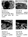
- cortical perforations were completely healed; nevertheless, bone did not regenerate vertically.
- Mean maximal vertical bone height was similar to the width of the original calvaria (1.49 ⁇ 0.26mm, 1.5 ⁇ 0.1 1 mm respectively).
- the regenerated bone was continuous with the calvaria and covered by periostum.
- the bone appeared mature with a mean vertical bone height of 2.88 ⁇ 0.61 mm.
- results were similar to the GS group (2.18 ⁇ 0.25mm). Bone height was almost double in the GBR groups; nevertheless hEPC did not enhance bone formation. Additional studies are required in order to explore the effect of other stem/progenitor cells and different scaffold on bone formation in this model.
- Bone marrow from rats' tibiae was flushed, centrifuged at 250g for 15 minutes and pelleted cells were seeded on culture dishes with DMEM (Dulbecco Modified Eagle's Medium) containing 10% FCS (Fetal Caff Serum) and 100 U/ml Pen-Strep. Cells were grown at 37°C with humidified 95% air/5% CO 2 , fed 3 times per week, split when confluent by brief trypsinization using 0.5% trypsine/0.2% EDTA (Biological Industries Ltd., Beit Haemek, Israel) and dispersed ( Figure 1 ).
- DMEM Dynabecco Modified Eagle's Medium
- FCS Fetal Caff Serum
- Pen-Strep 100 U/ml Pen-Strep.
- Cells were grown at 37°C with humidified 95% air/5% CO 2 , fed 3 times per week, split when confluent by brief trypsinization using 0.5% trypsine/0.2% EDTA
- Osteogenic differentiation of MSC was performed as follows, expanded MSC were grown in osteogenic differentiation media - DMEM/F12 (1:1) containing 10% FCS, Dexamethasone 10-7 M, ascorbic acid 5x10⁻⁵ M and ⁇ -glycerophosphate 10-2 M. Osteoblastic differentiation was identified by nodule formation that were further characterized by alizarin red.
- the cells were washed with PBS, resuspended in 0.5mL FACS buffer, and analyzed for the expression of the aforementioned rat antigens using FACScan and CellQuest software for data collection and analysis (both from Becton Dickinson and Co., Franklin Lakes, NJ). Gating was set to 1 % or less of isotype-stained cells (IgG1 or IgG2).
- TCP coating with fibronectin ⁇ -Tricalcium was performed as follows, phosphate scaffold (TCP) - Ossaplast®, (Osacure, Germany) was coated in fibronectin solution (Biological Industries Ltd., Beit Haemek, Israel) in order to enhance cell adhesion to the scaffold (26).
- fibronectin solution Biological Industries Ltd., Beit Haemek, Israel
- 0.2gr TCP granules were placed as a dense monolayer in 24 well plate and soaked in 100 microliter PBS mixed with 50 microliter fibronectin solution (5 micro gr/cm2) and incubated for 60 min.
- Rats 150 ⁇ l HAF hydrogel scaffold (supplied by Procore Biotech Ltd.) was polymerized immediately before transplantation by addition of 50 ⁇ l thrombin (0.33 U/ml). Rats were anaesthesized and received pre operative antibiotics (Cefalexin) and analgesic (Turbogezic) as described above. The parietal bone was exposed and perforated. Rats were divided into 4 groups:
- HAF and hEPC group (3) the mean vertical bone height was almost similar to the original calvaria and to the HAF groupl (.5 ⁇ 0.1 1 mm) thus, early hEPC did not improve bone regeneration.
- capsule (HAF+eariy hEPC+GBR) group (4) vertical bone formation was noticed (2.18 ⁇ 0.25mm) similar to that observed in group (3) (HAF+GBR) again early hEPC had no adjunctive effect to bone augmentation ( Figure 5E ).
- Table 1 summarizes the results of this experiment: HAF HAF+hEPC HAF+GBR GBR+HAF+hEPC Mean maximal vertical bone height ( ⁇ SE) 10.49 ⁇ 0.26 1.5 ⁇ 0.11 2.88 ⁇ 0.61 2.18 ⁇ 0.25
- HAF scaffold is not available, 3 different scaffolds that are commonly used for bone augmentation: Bio-Oss collagen, collagen sponge and TCP were evaluated.
- the maximal bone height was 2 mm in continuation with another 4mm height of islands of dense connective tissue surrounding the residual scaffolds with numerous areas of new lamellar bone ( Figure 8A clinical and 8B histological right views). Osteoclasts were observed near the TCP particles coupled with osteoblasts and new bone ( Figure 9 ).
- TCP scaffold showed the highest potential for bone regeneration. Therefore, the TCP scaffold is expected to provide the foremost clinical benefit. Since most of the newly formed tissue was immature (after 8 weeks), it is preferable to terminate the regeneration phase after 2 or 3 months.
- the Domes were fixed to the calvaria using fixation screws that were attached in it's anchoring rings.
- the flaps were repositioned and sutured, with minimal tension, using resorbable sutures.
- MNC Total rat mononuclear cells
- BM cells were positive for CD90 and CD44 and negative for CD 45. When grown in osteogenic conditions, nodule formations were seen and culture was positively stained for alizarin red.
- the augmented tissue was composed of bone, residual scaffold and connective tissue.
- the proportion of these components were different among the experimental groups.
- newly formed compact bone was continuous with the original calvaria.
- unaracteristics or mature lamellar bone were observed in most areas (e.g: reversing lines, osteocyte trapped in empty lacuna) however in some areas woven bone was noticed (as demonstrated by picro-serius red.
- the upper (distal) part of the augmented tissue contained residual scaffold surrounded by dense vascularized connective tissue.
Landscapes
- Health & Medical Sciences (AREA)
- Life Sciences & Earth Sciences (AREA)
- Engineering & Computer Science (AREA)
- Biomedical Technology (AREA)
- Chemical & Material Sciences (AREA)
- General Health & Medical Sciences (AREA)
- Zoology (AREA)
- Cell Biology (AREA)
- Transplantation (AREA)
- Animal Behavior & Ethology (AREA)
- Oral & Maxillofacial Surgery (AREA)
- Public Health (AREA)
- Veterinary Medicine (AREA)
- Wood Science & Technology (AREA)
- Organic Chemistry (AREA)
- Medicinal Chemistry (AREA)
- Epidemiology (AREA)
- Dermatology (AREA)
- Genetics & Genomics (AREA)
- Bioinformatics & Cheminformatics (AREA)
- Biotechnology (AREA)
- Developmental Biology & Embryology (AREA)
- General Engineering & Computer Science (AREA)
- Biochemistry (AREA)
- Chemical Kinetics & Catalysis (AREA)
- Botany (AREA)
- Microbiology (AREA)
- Vascular Medicine (AREA)
- Orthopedic Medicine & Surgery (AREA)
- Rheumatology (AREA)
- Urology & Nephrology (AREA)
- Inorganic Chemistry (AREA)
- Biophysics (AREA)
- Hematology (AREA)
- Immunology (AREA)
- Cardiology (AREA)
- Heart & Thoracic Surgery (AREA)
- Materials For Medical Uses (AREA)
Description
- In general, the present invention pertains to the arts of medicine. In particular, the invention relates to a bone augmentation system and to uses of endothelial progenitor cells and scaffolding.
- Alveolar bone atrophy may occur due to trauma, malignant tumors, and periodontal disease (Albandar 1990, Schwartz-Arad & Levin 2004, Irinakis 2006). Restoring the lost bone is crucial for the rehabilitation of the patient's function, phonetics and aesthetic demands.
- Nowadays, the methods available for vertical bone augmentation include: distraction osteogenesis, bone blocks (autologic/ allogenic or xenogenic) and guided bone regeneration (GBR). These techniques are surgically complicated, unpredictable and some of them are also associated with significant morbidity (Chiapasco et al. 2006, Rothamel et al. 2009, Greenstein et al. 2009, Esposito et al. 2006, Rocchietta et al. 2008). The idea of using a physical barrier for guided bone regeneration (GBR) was first described in the early 80's by Nyman et al (Nyman et al. 1982a, Nyman et al. 1982b, Nyman et al. 1987, Pitaru et al. 1987). It was demonstrated that creating a critical size intra bony defect and covering the defect with muco periostea I flap will lead to healing of the defect, mostly with soft tissue; while when a barrier to separate the bone defect from the soft tissue is used, it allows for bone regeneration into the defect (Dahalin et al. 1990, Kostopoulos et al. 1994a, Kostopoulos et al. 1994b, Dahalin et al. 1988). The biological principle of GBR also includes the creation of a space between a rigid barrier and the underlying bone which prevents epithelial and fibroblastic cells migration from the soft tissue, thus enabling the slower moving bone-forming cells to migrate from the underlying bone to populate the space (Polimeni et al. 2005, Schenk et al. 1994). Another important role of the membrane is blood clot stabilization. It was demonstrated that supra- crestal bone defects that were treated with ePTFE membrane and had root conditioning with heparin, showed compromised periodontal healing and bone regeneration (Haney et al. 1993).
- As of today, bone regeneration within the bony envelope (intra-bony) using GBR technique is a common and relatively predictable procedure (Kim et al. 2004). However, only few studies that attempted to grow bone extra corticaiiy using C3B resulted with modest success (Majzoub et al. 1999, Min et al. 2007, Lundgren et al. 1995, Wikesjo et al. 2003, Lioubavina & Kostopoulos 1999).
- Both the calvaria and the jaw bones are formed through the intra-membranous bone formation pathway (Verna et al. 2002). The process begins with the proliferation of mesenchymal cells and deposition of extracellular matrix. This newly formed tissue guides cell migration and angiogenesis. The combination of matrix components with new vessels forms provisional connective tissue that is later transformed into bone along blood vessels (Lang et al. 2003).
- Mesenchymal stem cells (MSC) contribute to the maintenance of various tissues, especially bone, in adults. They can be isolated form bone marrow, placenta, umbilical cord blood, or from adipose tissue in adults (Pittenger et al. 1999, Secco et al. 2008, Zuk et al. 2001, Yen et al. 2005). The first to describe these cells was Friedenstein in 1966. MSC present several characteristics that assist to identify them: adherence to culture plates, fibroblast-like phenotype and proliferation potential. The osteogenic potential of these cells was also recognized (Friedenstein). Since that time, in-vivo and in-vitro studies showed that MSC can be transformed into mesenchymal tissues such as: bone, cartilage, adipose, muscle and tendon. The characterization of cultured MSC relies on combination of several antigens: CD105, CD146, CD90, CD73 and CD44; furthermore, the hematopoietic origin of the cells should be excluded (CD45, CD14 and CD34) (Baksh 2004, Geregory et al. 2005).
- Several preclinical studies and a few clinical studies showed the efficacy of cultured MSC for bone reconstruction. In animal models these cells showed osteoinductive properties (i.e production of ectopic bone) and improved healing of bone defects (Petite et al. 2000, Bruder et al. 1998). In humans, few clinical reports (case reports) were published: Quarto et al. 2001 reported on callus formation 2 months after administration of autologous MSC seeded on hydroxyapatite scaffold into non- union long bone defects. The I.V. infusion of autologous MSC to children with osteogenesis imperfecta resulted in increase in their body length and bone mineral content (Horowits et al. 2002).
- Several attempts to heal mandibular defect in animals using MSC have also been recently published. These studies reported that the addition of MSC to scaffold have statistically improved bone regeneration compared with scaffold alone (Steinhardt et al. 2008, Jafarian et al. 2008).
- Although extensively studied, several difficulties concerning the use of bone marrow MSC still exist. The main problems are the invasive nature associate witn tne harvesting of MSC and the morbidity of the donor. Additionally some reports suggest age-dependent decline in the proliferation and the osteogenic differentiation of these cells (Zhou et al. 2008). Finally, the limited amount of MSC in bone marrow aspirates requires ex- vivo expansion to obtain sufficient cell number for transplantation (Banfi et al. 2000). Thus, extensive research to improve our laboratory techniques and control clinical trials is still needed in order to confirm the potential of MSC to induce new bone formation.
- Blood-derived endothelial progenitor cells (EPC) - In 1997, Asahara et al. discovered the presence of bone marrow-derived circulating endothelial progenitor cells in adult peripheral blood and human umbilical cord blood (Ashara et al. 1997, Murohara et al. 2000) that participate in post-natal neovascularization (Takahashi et al. 1999). Furthermore, these cell lines were found to participate in angiogenesis, vascular repair, blood-flow recovery after tissue ischemia and vasculoprotection. The current clinical use of EPC is limited to treating ischemic tissue after acute myocardial infarct (Isner & Losordo 1999, Kalka et al. 2000). During skeletal development or bone healing processes the recruitment of EPC by vasculogenic / angiogenic molecules (e.g. VEGF, PIGF, erythropoietin) is crucial (Ferguson et al. 1999, Giannoudis et al. 2007). Studies that explored the effect of local delivery of BMP2 & VEGF loaded on scaffold on bone regeneration suggest a synergistic effect in this dual delivery system (Patel et al. 2008, Kempen et al. 2009). Dual release of BMP2 & VEGF from scaffold implanted into calvaria-bone defect in rats enhanced bone formation after 4 weeks compared to scaffold alone or scaffold with only one GF (Patel et al. 2008).
- Successful regeneration of large bone defects might also benefit from concomitant stimulation of vascularization and osteogenesis. Hence, we suggest that EPC and MSC based therapy might enhance both processes because these cells are capable of participating in vasculogenesis, might induce mesenchymal progenitors to proliferate or differentiate into osteoblasts and possibly at the same time gain plasticity to differentiate themselves into bone forming cells (Bick et al. 2006).
- Identification of EPC is rather controversial and complicated since three different populations of EPCs were identified:
- 1. CFU - Hill -these EPCs have weak proliferative and vasculogenic activities;
- 2. Circulating angiogenic cells- these cells are obtained from adherent peripheral blood mononuclear cells (PBMNC) and do not form colonies in culture. They are positive to: CD133, CD31, CD45 and VEGFR-2.
- 3. Late EPCs - these cells are derived from adherent PBMNC and form endothelial colonies after 3 -4 week of culture. They are thought to play a major role in revascularization in adults; they are positive to CD34, CD144, VEGF-R2 but negative to CD133, CD45 and CD14 (Hirschi et al. 2008, Hur et al. 2004, Yoder et al. 2007).
- The participation of EPC in bone repair was reported by some researchers. Lee et al. and Cetrulo et al. showed the participation of EPC in distraction osteogenesis model (Lee et al. 2009, Cetrulo et al. 2005). The injection of human CD34 progenitor cells into non healing femoral fracture in nude rats enhanced bone healing compared with control group. The addition of VEGF antagonist to this model impaired not only angiogenesis but also osteogenesis and led the authors to suggest that these cells have paracrine effect on bone forming cells. (Lee 2008, Fuchs 2009).
- Transplantation of autologeus EPC into critical size gap in sheep tibiae revealed full bridging at 3 months in 6 out of 7 E PC-transplanted defects while non or minimal new bone formation was observed radiographically in 8 sham-operated defects (Rozen et al. 2009). These authors also suggested that the effect of EPC is not limited to vasculogenesis but that they are also capable to transform into bone cells. When sheep EPC were sub-cultured under osteogenic conditions they changed their morphology and formed nodular aggregates (1-2 mm diameter) following 1 -2 weeks incubation and stained positively by von Kossa (vK), alizarin red (AR) and osteocalcin immunohistochemistry, which are markers of osteoblastic differentiation (Bick et al. 2006).
- To be able to use cell-based therapy in animal and human defects a suitable scaffold is required that will not only serve as a vehicle for cellular delivery, but will also have osteoconductive properties. Tricalcium phosphates (TCP) is a synthetic scaffold used in the clinics for the reconstruction of bone defects. These materials provide a mineral matrix phase similar to that found in bone tissue. Following application, TCP is resorbed by osteoclastic activity and replaced by newly formed bone. Additionally, TCP as a synthetic material does not pose the risk of transmitting pathogenic agents (such is the case with allographs and xenographs). It is also being resorbed more rapidly when compared with a xenograft. (Rojbani H, Nyan M, Ohya K, Kasugai S. Evaluation of the osteoconductivity of a-tricalcium phosphate, β-tricalcium phosphate, and hydroxyapatite combined with or without simvastatin in rat calvarial defect. J Biomed Mater Res A. 2011 Sep 15;98(4):488-98.
- By combining TCP with GBR we achieved a mean height of 5.5±0.24 mm new hard tissue under the capsule, significantly higher than bone formed when capsules were filled with Bio-Oss collagen (p<0.001). Histological analysis revealed that TCP was partially resorbed and replaced by new bone that was continuous with the original calvaria. Residual TCP particles were surrounded by vascularized dense connective tissue.
-
WO 00/35510 A1 - Ward et al. (Oral Diseases, Vol. 16, No. 8, November 1, 2010, pages 709-716) is a review of bioengineering strategies for regeneration of craniofacial bone by applying the principles of engineering to biology and medicine. The authors suggest the evolving technology of stem cell biology for regeneration of craniofacial bone. In this context, the generation of cadaveric human mandibular condyle of bone and cartilage from adult rat bone marrow mesenchymal stem cells (MSCs) encapsulated in poly(ethylene glycol)-based hydrogel is disclosed. Said document does not relate to rehabilitation of a defective or damaged bone, but rather to pure scientific investigation of the scope of capabilities enabled by implementation of stem cell technology in genesis of bone tissues. In particular, a system for augmentation of osseous tissue on a surface of a bone is not disclosed.
- Kaigler et al. (Tissue Engineering Part A, Vol. 16, No. 9, Sept. 1, 2010, pages 2809-2820) discloses a graft containing bone repair cells (BRCs) - cells derived from bone marrow aspirates. BRCs are enriched for mesenchymal and vascular phenotypes, which are suspended and grafted into an osseous jawbone defect, created following a tooth extraction. Said document does not disclose a scaffolding onto which a cell culture is seeded.
-
- Albandar JM. Some predictors of radiographic alveolar bone height reduction over 6 years. J Periodontal Res. 1990 May;25(3):186-92.
- Araujo MG, Lindhe J. Ridge preservation with the use of Bio-Oss collagen: A 6-month study in the dog. Clin Oral Implants Res. 2009 May;20(5):433-40.
- Artzi Z, Tal H, Dayan D. Porous bovine bone mineral in healing of human extraction sockets: 2. Histochemical observations at 9 months. J Periodontol. 2001 Feb;72(2): 152-9.
- Asahara T, Murohara T, Sullivan A, Silver M, van der Zee R, Li T,Witzenbichler B, Schatteman G, Isner JM. Isolation of putative progenitor endothelial cells for angiogenesis. Science. 1997 Feb 14;275(5302):964-7.
- Asian H, Zilberman Y, Kandel L, Liebergall M, Oskouian RJ, Gazit D, Gazit Z. Osteogenic differentiation of noncultured immunoisolated bone marrow-derived CD105+ cells. Stem Cells. 2006 Jul;24(7): 1728-37. Epub 2006 Apr 6.
- Baksh D, Song L, Tuan RS. Adult mesenchymal stem cells: characterization, differentiation, and application in cell and gene therapy. J Cell Mol Med. 2004 Jul-Sep;8(3):301 -16. Review.
- Banfi A, Muraglia A, Dozin B, Mastrogiacomo M, Cancedda R, Quarto R. Proliferation kinetics and differentiation potential of ex vivo expanded human bone marrow stromal cells: Implications for their use in cell therapy. Exp Hematol. 2000 Jun;28(6):707-15.
- Bick T, Rozen N, Dreyfuss E, Soudry M, Lewinson D. Osteogenic differentiation of circulating endothelial progenitor cells. J. of Bone and Min. Res 2006; 22:S143.
- Bruder SP, Kraus KH, Goldberg VM, Kadiyala S. The effect of implants loaded with autologous mesenchymal stem cells on the healing of canine segmental bone defects. J Bone Joint Surg Am. 1998 Jul; 80(7):985-96.
- Camelo M, Nevins ML, Schenk RK, Simion M, Rasperini G, Lynch SE, Nevins M. Clinical, radiographic, and histologic evaluation of human periodontal defects treated with Bio-Oss and Bio-Gide. Int J Periodontics Restorative Dent. 1998 Aug; 18(4)321-31.
- Cetrulo CL Jr, Knox KR, Brown DJ, Ashinoff RL, Dobryansky M, Ceradini DJ, Capla JM, Chang El, Bhatt KA, McCarthy JG, Gurtner GC. Stem cells and distraction osteogenesis: endothelial progenitor cells home to the ischemic generate in activation and consolidation. Plast Reconstr Surg. 2005 Sep 15; 116(4): 1053-64.
- Chiapasco M, Zaniboni M, Boisco M .Augmentation procedures for the rehabilitation of deficient edentulous ridges with oral implants.Clin Oral Implants Res. 2006 Oct;17 Suppl 2:136-59. Review.
- Dahlin C, Linde A, Gottlow J, Nyman S. Healing of bone defects by guided tissue regeneration. Plast Reconstr Surg. 1988 May; 81 (5):672-6.
- Dahlin C, Gottlow J, Linde A, Nyman S.Healing of maxillary and mandibular bone defects using a membrane technique. An experimental study in monkeys.Scand J Plast Reconstr Surg Hand Surg. 1990; 24(1):13-9.
- Donos N, Lang NP, Karoussis IK, Bosshardt D, Tonetti M, Kostopoulos L. Effect of GBR in combination with deproteinized bovine bone mineral and/or enamel matrix proteins on the healing of critical-size defects. Clin Oral Implants Res. 2004 Feb;15(1):101-1.
- Esposito M, Grusovin MG, Coulthard P, Worthington HV. The efficacy of various bone augmentation procedures for dental implants: a Cochrane systematic review of randomized controlled clinical trials. Int J Oral Maxillofac Implants. 2006 Sep-Oct; 21(5):696-710. Review.
- Ferguson CM, Miclau T, Hu D, Alpern E, Helms JA. Common molecular pathways in skeletal morphogenesis and repair. Ann N Y Acad Sci. 1998 Oct 23; 857:33-42. Review.
- Friedenstein AJ, Piatetzky-Shapiro II, Petrakova KV. Osteogenesis in transplants of bone marrow cells. J Embryol Exp Morphol. 1966 Dec; 16(3):381 -90.
- Fuchs S, Jiang X, Schmidt H, Dohle E, Ghanaati S, Orth C, Hofmann A,Motta A, Migliaresi C, Kirkpatrick CJ. Dynamic processes involved in the pre-vascularization of silk fibroin constructs for bone regeneration using outgrowth endothelial cells. Biomaterials. 2009 Mar; 30(7): 1329-38.
- Giannoudis PV, Einhorn TA, Marsh D. Fracture healing: the diamond concept. Injury. 2007 Sep;38 Suppl 4:S3-6. Review.
- Greenstein G, Greenstein B, Cavallaro J, Tarnow D. The role of bone decortication in enhancing the results of guided bone regeneration: a literature review. J Periodontol. 2009 Feb; 80(2): 175-89. Review.
- Gregory CA, Prockop DJ, Spees JL. Non-hematopoietic bone marrow stem cells: molecular control of expansion and differentiation. Exp Cell Res. 2005 Jun 10; 306(2):330-5.
- Haney JM, Nilveus RE, McMillan PJ, Wikesjo UM. Periodontal repair in dogs: expanded polytetrafluoroethylene barrier membranes support wound stabilization and enhance bone regeneration. J Periodontol. 1993 Sep; 64(9):883-90.
- Hirschi KK, Ingram DA, Yoder MC Assessing identity, phenotype, and fate of endothelial progenitor cells. Arterioscler Thromb Vase Biol. 2008 Sep; 28(9): 1584-95.
- Horwitz EM, Gordon PL, Koo WK, Marx JC, Neel MD, McNall RY, Muul L,Hofmann T. Isolated allogeneic bone marrow-derived mesenchymal cells engraft and stimulate growth in children with osteogenesis imperfecta: Implications for cell therapy of bone. Proc Natl Acad Sci USA. 2002 Jun 25;99(13):8932-7.
- Hur J, Yoon CH, Kim HS, Choi JH, Kang HJ, Hwang KK, Oh BH, Lee MM, Park YB. Characterization of two types of endothelial progenitor cells and their different contributions to neovasculogenesis. Arterioscler Thromb Vase Biol. 2004 Feb; 24(2):288-93.
- Irinakis T. Rationale for socket preservation after extraction of a single-rooted tooth when planning for future implant placement.J Can Dent Assoc. 2006 Dec;72(10):917-22.
- Isner JM, Losordo DW. Therapeutic angiogenesis for heart failure. Nat Med.1999 May; 5(5):491 -2.
- Jafarian M, Eslaminejad MB, Khojasteh A, Mashhadi Abbas F, Dehghan MM, Hassanizadeh R, Houshmand B. Marrow-derived mesenchymal stem cells-directed bone regeneration in the dog mandible: a comparison between biphasic calcium phosphate and natural bone mineral. Oral Surg Oral Med Oral Pathol Oral Radiol Endod. 2008 May; 105(5):e14-24.
- Kalka C, Masuda H, Takahashi T, Kalka-Moll WM, Silver M, Kearney M, Li T, Isner JM, Asahara T. Transplantation of ex vivo expanded endothelial progenitor cells for therapeutic neovascularization. Proc Natl Acad Sci USA. 2000 Mar 28; 97(7):3422-7.
- Kempen DH, Lu L, Heijink A, Hefferan TE, Creemers LB, Maran A, Yaszemski MJ, Dhert WJ. Effect of local sequential VEG and L3MP-2 delivery on ectopic and orthotopic bone regeneration. Biomaterials. 2009 May; 30(14):2816-25.
- Khojasteh A, Eslaminejad MB, Nazarian H. Mesenchymal stem cells enhance bone regeneration in rat calvarial critical size defects more than platelete-rich plasma. Oral Surg Oral Med Oral Pathol Oral Radiol Endod. 2008 Sep; 106(3):356-62.
- Kim CS, Choi SH, Chai JK, Cho KS, Moon IS, Wikesjo UM, Kim CK. Periodontal repair in surgically created intrabony defects in dogs: influence of the number of bone walls on healing response. J Periodontol. 2004 Feb; 75(2):229-35.
- Kim SS, Kim BS. Comparison of osteogenic potential between apatite-coated poly lactide-co-glycolide /hydroxyapatite particulates and Bio-Oss. Dent Mater J. 2008 May; 27(3):368-75.
- Kostopoulos L, Karring T, Uraguchi R. Formation of jawbone tuberosities by guided tissue regeneration. An experimental study in the rat.Clin Oral Implants Res. 1994a Dec; 5(4):245-53.
- Kostopoulos L, Karring T. Augmentation of the rat mandible using guided tissue regeneration. Clin Oral Implants Res. 1994b Jun; 5(2):75-82.
- Lang NP, Araujo M, Karring T. Alveolar Bone Formation. In: Lindhe J, Karring T & Lang NP, Editors. Clinical periodontology and implant dentistry. Fourth edition. Blackwell Munksgaard, 2003: pp: 870-873.
- Lee DY, Cho TJ, Kim JA, Lee HR, Yoo WJ, Chung CY, Choi IH. Mobilization of endothelial progenitor cells in fracture healing and distraction osteogenesis. Bone. 2008 May; 42(5):932-41.
- Lee DY, Cho TJ, Lee HR, Park MS, Yoo WJ, Chung CY, Choi IH. Distraction osteogenesis induces endothelial progenitor cell mobilization without inflammatory response in man. Bone. 2009 Oct 22.
- Lioubavina N, Kostopoulos L, Wenzel A, Karring T. Long-term stability of jaw bone tuberosities formed by "guided tissue regeneration". Clin Oral Implants Res. 1999 Dec; 10(6):477-86.
- Lundgren D, Lundgren AK, Sennerby L, Nyman S. Augmentation of intramembraneous bone beyond the skeletal envelope using an occlusive titanium barrier. An experimental study in the rabbit.Clin Oral Implants Res. 1995 Jun; 6(2):67-72.
- Lundgren AK, Lundgren D, Hammerle CH, Nyman S, Sennerby L. Influence of decortication of the donor bone on guided bone augmentation. An experimental study in the rabbit skull bone. Clin Oral Implants Res. 2000; 11 (2):99-106.
- Machtei EE. The effect of membrane exposure on the outcome of regenerative procedures in humans: a meta-analysis. J Periodontol. 2001 Apr; 72(4):512-6.
- Majzoub Z, Berengo M, Giardino R, Aldini NN, Cordioli G. Role of intramarrow penetration in osseous repair: a pilot study in the rabbit calvaria. J Periodontol. 1999 Dec;70(12):1501 -10.
- Mellonig JT. Human histologic evaluation of a bovine-derived bone xenograft in the treatment of periodontal osseous defects. Int J Periodontics Restorative Dent. 2000 Feb; 20(1): 19-29.
- Min S, Sato S, Murai M, Okuno K, Fujisaki Y, Yamada Y, Ito K. Effects of marrow penetration on bone augmentation within a titanium cap in rabbit calvarium. J Periodontol. 2007 Oct; 78(10):1978-84.
- Murohara T, Ikeda H, Duan J, Shintani S, Sasaki K, Eguchi H, Onitsuka I, Matsui K, Imaizumi T. Transplanted cord blood-derived endothelial precursor cells augment postnatal neovascularization. J Clin Invest. 2000 Jun;105(11):1527-36.
- Nyman S, Gottlow J, Karring T, Lindhe J.The regenerative potential of the periodontal ligament. An experimental study in the monkey.J Clin Periodontol. 1982 May; 9(3):257-65.
- Nyman S, Lindhe J, Karring T, Rylander H. New attachment following surgical treatment of human periodontal disease.J Clin Periodontol. 1982 Jul; 9(4):290-6.
- Nyman S, Gottlow J, Lindhe J, Karring T, Wennstrom J. New attachment formation by guided tissue regeneration. J Periodontal Res. 1987 May; 22(3):252-4.
- Patel ZS, Young S, Tabata Y, Jansen JA, Wong ME, Mikos AG. Dual delivery of an angiogenic and an osteogenic growth factor for bone regeneration in a critical size defect model. Bone. 2008 Nov; 43(5):931 -40. Epub 2008 Jul 14.
- Petite H, Viateau V, Bensaid W, Meunier A, de Pollak C, Bourguignon M, Oudina K, Sedel L, Guillemin G. Tissue-engineered bone regeneration. Nat Biotechnol. 2000 Sep; 18(9):959-63.
- Piattelli M, Favero GA, Scarano A, Orsini G, Piattelli A. Bone reactions to anorganic bovine bone (Bio-Oss) used in sinus augmentation procedures: a histologic long-term report of 20 cases in humans. Int J Oral Maxillofac Implants. 1999 Nov-Dec;14(6):835-40.
- Pitaru S, Tal H, Soldinger M, Azar-Avidan O., Nott M. Collagen membranes prevent the apical migration of epithelium during periodontal wound healing.J Periodontal Res. 1987 Jul;22(4):331 -3.
- Pittenger MF, Mackay AM, Beck SC, Jaiswal RK, Douglas R, Mosca JD, Moorman MA, Simonetti DW, Craig S, Marshak DR. Multilineage potential of adult human mesenchymal stem cells. Science 1999 Apr 2; 284(5411):143-7.
- Polimeni G, Albandar JM, Wikesjo UM. Prognostic factors for alveolar regeneration: effect of space provision. J Clin Periodontol. 2005 Sep; 32(9):951 -4.
- Quarto R, Mastrogiacomo M, Cancedda R, Kutepov SM, Mukhachev V, Lavroukov A, Kon E, Marcacci M. Repair of large bone defects with the use of autologous bone marrow stromal cells. N Engl J Med. 2001 Feb 1; 344(5):385-6.
- Rothamel D, Schwarz F, Herten M, Ferrari D, Mischkowski RA, Sager M,Becker J. Vertical ridge augmentation using xenogenous bone blocks: a histomorphometric study in dogs. Int J Oral Maxillofac Implants. 2009 Mar-Apr; 24(2):243-50.
- Rocchietta I, Fontana F, Simion M. Clinical outcomes of vertical bone augmentation to enable dental implant placement: a systematic review. J Clin Periodontol. 2008 Sep; 35(8 Suppl):203-15. Review.
- Rozen N, Bick T, Bajayo A, Shamian B, Schrift-Tzadok M, Gabet Y, Yayon A, Bab I, Soudry M, Lewinson D. Transplanted blood-derived endothelial progenitor cells (EPC) enhance bridging of sheep tibia critical size defects. Bone. 2009 Nov;45 (5):918-24.
- Schenk RK, Buser D, Hardwick WR, Dahlin C. Healing pattern of bone regeneration in membrane-protected defects: a histologic study in the canine mandible. Int J Oral Maxillofac Implants. 1994 Jan-Feb; 9(1): 13-29.
- Schwartz-Arad D, Levin L. Post-traumatic use of dental implants to rehabilitate anterior maxillary teeth. Dent Traumatol. 2004 Dec; 20(6):344-7.
- Secco M, Zucconi E, Vieira NM, Fogaca LL, Cerqueira A, Carvalho MD, Jazedje T, Okamoto OK, Muotri AR, Zatz M. Multipotent stem cells from umbilical cord: cord is richer than blood. Stem Cells. 2008 Jan; 26(1):146-50. Epub 2007 Oct 11.
- Slotte C, Lundgren D. Augmentation of calvarial tissue using non-permeable silicone domes and bovine bone mineral. An experimental study in the rat. Clin Oral Implants Res. 1999 Dec10 (6):468-76.
- Steinhardt Y, Asian H, Regev E, Zilberman Y, Kallai I, Gazit D, Gazit Z. Maxillofacial-derived stem cells regenerate critical mandibular bone detect. Tissue eng Part A. 2008 Nov; 14(11):1763-73.
- Takahashi T, Kalka C, Masuda H, Chen D, Silver M, Kearney M, Magner M, Isner JM, Asahara T. Ischemia- and cytokine-induced mobilization of bone marrow-derived endothelial progenitor cells for neovascularization. Nat Med. 1999 Apr; 5(4):434-8.
- Valentini P, Abensur D, Densari D, Graziani JN, Hammerle C. Histological evaluation of Bio-Oss in a 2-stage sinus floor elevation and implantation procedure. A human case report. Clin Oral Implants Res. 1998 Feb;9(1):59-64. Review.
- Verna C, Dalstra M, Wikesjo UM, Trombelli L, Bosch C. Healing patterns in calvarial bone defects following guided bone regeneration in rats. A micro-CT scan analysis. J Clin Periodontol. 2002;29(9):865-70.
- Wikesjo UM, Xiropaidis AV, Thomson RC, Cook AD, Selvig KA, Hardwick WR. Periodontal repair in dogs: rhBMP-2 significantly enhances bone formation under provisions for guided tissue regeneration. J Clin Periodontol. 2003 Aug;30(8):705-14.
- Yen BL, Huang HI, Chien CC, Jui HY, Ko BS, Yao M, Shun CT, Yen ML, Lee MC, Chen YC. Isolation of multipotent cells from human term placenta. Stem Cells. 2005;23(1 ):3-9.
- Yoder MC, Mead LE, Prater D, Krier TR, Mroueh KN, Li F, Krasich R, Temm CJ, Prchal JT, Ingram DA. Redefining endothelial progenitor cells via clonal analysis and hematopoietic stem/progenitor cell principals. Blood. 2007 Mar 1 ;109(5): 1801 -9. Epub 2006 Oct 19.
- Zhou S, Greenberger JS, Epperly MW, Goff JP, Adler C, Leboff MS, Glowacki J. Age-related intrinsic changes in human bone-marrow-derived mesenchymal stem cells and their differentiation to osteoblasts. Aging Cell. 2008 Jun; 7(3):335-43.
- Zitzmann NU, Scharer P, Marinello CP, Schiipbach P, Berglundh T. Alveolar ridge augmentation with Bio-Oss: a histologic study in humans. Int J Periodontics Restorative Dent. 2001 Jun; 21 (3):288-95.
- Zuk PA, Zhu M, Mizuno H, Huang J, Futrell JW, Katz AJ, Benhaim P, Lorenz HP, Hedrick MH. Multilineage cells from human adipose tissue: implications for cell-based therapies. Tissue Eng. 2001 Apr; 7(2):21 1 -28.
- The present invention provides a system for vertical augmentation of osseous tissue on a surface of a bone, and also provides a scaffolding comprising tricalcium phosphates and being adapted to be placed on portions of a bone which is to be augmented, and further comprising a cell culture seeded onto said scaffolding, for use in vertically augmenting osseous tissue on a surface of a bone. It was discovered that the angiogenic effect of Endothelial Progenitor Cells (here in after EPC), combined with effects of space creation, clot stabilization and cell exclusion achieved by GBR, facilitates a predictable bone augmentation. Guided Bone Regeneration (GBR) techniques, known in the art, include space maintaining, blood clot stabilization and exclusion of epithelial as well as connective tissue cells. In combination with progenitor cells that participate in various stages of bone regeneration has shown a better and more predictable bone augmentation compared with GBR. The effect of bone marrow derived mesenchymal stem cells (hereinafter BM SC), peripheral blood derived endothelial progenitor cells (EPC) and the combination of both on bone augmentation using a GBR model, in rats' calvaria, has been studied.
- It should be understood, however, that the eclectically synopsized brief summary supra is not to limit the invention to the particular forms and examples, but on the contrary, is to cover all modifications, equivalents, and alternatives falling within the scope of the invention.
- Illustrative embodiments of the invention are described below. In the interest of clarity, not all features of an actual implementation are described in this specification. It will of course be appreciated that in the development of any such actual embodiment, numerous implementation-specific decisions must be made to achieve the developers' specific goals, such as compliance with technology- or business-related constraints, which may vary from one implementation to another. Moreover, it will be appreciated that the effort of such a development might be complex and time-consuming, but would nevertheless be a routine undertaking for those of ordinary skill in the art having the benefit of this disclosure. The present invention provides a system for vertical augmentation of osseous tissue on a surface of a bone according to claim 1, and a scaffolding comprising tricalcium phosphates and being adapted to be placed on portions of a bone which is to be augmented, and further comprising a cell culture seeded onto said scaffolding, for use in vertically augmenting osseous tissue on a surface of a bone according to claim 5. Preferred embodiments of the present invention are disclosed in the dependent claims.
- Forming an extra cortical osseous tissue augmentation is achieved by maintaining the following constituent factors, in order to achieve predictable bone regeneration. The constituent factors to be maintained are:
- 1 - An angiogenesis, to provide necessary blood supply and undifferentiated mesenchymal progenitor cells, which is typically achieved by (EPC);
- 2 - Space maintenance, to facilitate adequate space for bone ingrowth, and shaping of the newly formed osseous tissue, typically achieved by (GBR);
- 3 - To provide stability of wound and/or implant to induce initial blood clot formation and uneventful healing events.
- The formation of an extra cortical osseous tissue, using GBR and scaffolding, may include the following steps. Initially the portion of the surface of the bone the augmentation of which is desired is exposed. Thereafter, optionally using a dental hand-piece, small perforations of the cortical bone are preformed, preferably under cooling conditions, e.g. while rinsed with saline water. This is in order to allow blood clot formation and cells and nutrients passage from the endosteum (Lundgren et al. 2000).
- To achieve the synergistic effect induced by MSC, EPC and/or MSC with EPC sells, a tissue culture thereof is seeded on the scaffolding. Cells are typically seeded on the scaffold 1 day before the transplantation. Following the perforation of the bone, scaffolds seeded with stem/progenitor cells will be placed on the decorticated bone. The cells are extracted and cultured in accordance with the procedures exemplified infra.
- Scaffolding is then placed on the decorticated bone. Preferably a rigid gold enclosure is fixed to the underling bone, typically using gold screws, so as to surround the decorticated portion bone, the augmentation of which is desired. Optionally the surgical flap is repositioned and sutured with resorbable sutures to achieve primary wound closure.
- The rigid enclosure is removed and the calvarium+ scaffold is optionally evaluated. Afterwards, histological evaluation and immune-stainings is optionally performed on the same specimens.
- Evaluating a predicted efficacy of extra cortical vertical bone augmentation by combining GBR technique and implantation of stem/progenitor cells, may lead to a prognosis to particular potential patient, and may include:
- 1 - Isolating, culturing and characterizing stem and/or progenitor ceils, including:
- (a) MSC from bone marrow;
- (b) EPC from peripheral blood.
- 2 - Evaluating, typically supra crestal, bone formation using GBR technique in combination with stem and/or progenitor cells, including:
- (a) Test the adjunctive effect of BM MSC implantation to GBR.
- (b) Test the adjunctive effect of EPC implantation to GBR.
- (c) Test the adjunctive effect of EPC & MSC implantation to GBR.
- 3 - Follow maturation, ossification and stability of the newly formed bone.
- Lewis rats were anaesthesized using Ketamin 10mg/ 100gr BW and Xylasin 0.5mg/ 100gr BW IM. Subcutaneous antibiotics (Cefalexin, 0.3 ml) and analgesic (Turbogezic, 0.3 ml) will be injected pre- operatively. Followed by U shape incision (
Figure 4A ), a full thickness flap has exposeed the parietal bone. Using a dental hand-piece, small perforations of the cortical bone were preformed under cooling conditions with saline water. This is in order to allow blood clot formation and cells and nutrients passage from the endosteum (Lundgren et al. 2000). - Scaffold was placed on the parietal bone. A rigid gold capsule (custom made: 5 mm height; 7 mm radius) was fixed to the underling bone using gold screws (as shown in
Figure 5B ). The surgical flap was repositioned and sutured with resorbable sutures to achieve primary wound closure. - During the healing period, rats were fed rat chow and water ad libitum. Rats were sacrificed after 2 months. The calvaria with the capsules was fixed in 10% buffered formalin for 2 days and then immersed in 70% ethyl alcohol. The gold capsules were removed and the calvarium+ scaffold will be scanned by CT. Afterwards, histological evaluation and immune-stainings were performed on the same specimens.
- The adjuvant effects of rats' MSC, EPC and/or MSC with EPC cells seeded on scaffold to on GBR bone augmentation was achieved by seeding the cells on the scaffold 1 day before the transplantation. The surgical procedure was as described hereinabove. Following perforation of the parietal bone, scaffolds with stem/progenitor cells were placed on the bone. A rigid gold capsule was fixed to the underlining bone tissue using gold screws.
- Human EPC (hEPC) obtained from peripheral blood were isolated and characterized. 27 nude rats were divided into 4 groups: GBR+hyaluronic acid-fibrinogen scaffold (GS), GBR+scaffold+hEPC(GSC), scaffold+hEPC (SC) and scaffold alone(S). Following the exposure and perforation of the parietal bone, in the S and SC groups, scaffold with or without 105 cell was laid on the bone. In the GS and GSC groups scaffold with or without 105 cells was laid on the bone and covered by a rigid capsule. Rats were sacrificed 8 weeks later, and specimens were prepared for histological analysis.
- The newly formed bone was quantified by measuring the maximal vertical bone height in the histological sections. The vertical bone height ranged from 1 to 5.13 mm. In the S+SC group, cortical perforations were completely healed; nevertheless, bone did not regenerate vertically. Mean maximal vertical bone height was similar to the width of the original calvaria (1.49± 0.26mm, 1.5± 0.1 1 mm respectively). In the GS group, the regenerated bone was continuous with the calvaria and covered by periostum. The bone appeared mature with a mean vertical bone height of 2.88 ± 0.61 mm. In the GSC group, results were similar to the GS group (2.18 ± 0.25mm). Bone height was almost double in the GBR groups; nevertheless hEPC did not enhance bone formation. Additional studies are required in order to explore the effect of other stem/progenitor cells and different scaffold on bone formation in this model.
- Bone marrow from rats' tibiae was flushed, centrifuged at 250g for 15 minutes and pelleted cells were seeded on culture dishes with DMEM (Dulbecco Modified Eagle's Medium) containing 10% FCS (Fetal Caff Serum) and 100 U/ml Pen-Strep. Cells were grown at 37°C with humidified 95% air/5% CO2, fed 3 times per week, split when confluent by brief trypsinization using 0.5% trypsine/0.2% EDTA (Biological Industries Ltd., Beit Haemek, Israel) and dispersed (
Figure 1 ). - Osteogenic differentiation of MSC was performed as follows, expanded MSC were grown in osteogenic differentiation media - DMEM/F12 (1:1) containing 10% FCS, Dexamethasone 10-7 M, ascorbic acid 5x10⁻⁵ M and β-glycerophosphate 10-2 M. Osteoblastic differentiation was identified by nodule formation that were further characterized by alizarin red.
- 40 ml blood was drown from the heart of 5 Lewis rats. The Mononuclear fraction was isolated using Lymphoprep™ (Axis-Shield, Oslo, Norway), seeded on fibronectin (Sigma-Aldrich, MS, USA)-coated plates, and cultured in EBM-2 media supplemented with EGM-2MV SingleQuote (Clonetics, Cambrex Bio Science, MD, USA), containing 20% heat inactivated fetal bovine serum, vascular endothelial growth factor (VEGF), fibroblast growth factor-2, epidermal growth factor, insulin-like growth factor-1 and ascorbic acid. Cells were grown at 37°C with humidified 95% air/5% C02. After 4 days of culture, non-adherent cells were discarded by gentle washing with PBS, and fresh medium were applied. The attached cells were continuously cultured with complete EGM-2 medium. Cells were fed 3 times per week, split when -80% confluent by brief trypsinization and dispersed.
Figure 2 show peripheral blood derived Lewis rat cells, 7 days after isolation. - 50 ml blood were drawn from 25-35 years old healthy volunteers (Helsinki no.2423) and human late EPC were isolated as described for rIEPC. FACS analysis found that >95% of the cells are positive for CD31 , CD105 and CD 146, >70% for Tie-2, 30-40% for CD34, but importantly all of them are negative to CD14, ruling out their monocytic origin and supporting their angioblastic origin (
Figure 3 ). - Aliquots (5 105 cells) of lewis rat BM derived MSCs at Passages 1 to 3 were used for an analysis of cell-surface molecules. The cells were subcultured (as described earlier), resuspended in fluorescence-activated cell sorter (FACS) buffer consisting of 2% bovine serum albumin and 0.1 % sodium azide (Sigma Chemical Co., St. Louis, MO) in PBS, and stained with fluorochromeconjugated mouse anti-rat CD90, CD45, CD44 (Antibodies Direct Serotec, Kidlington, UK), monoclonal antibodies according to the manufacturers' recommendations using mouse IgG1 monoclonal isotype antibodies to detect any nonspecific binding fluorescence (negative control). The cells were washed with PBS, resuspended in 0.5mL FACS buffer, and analyzed for the expression of the aforementioned rat antigens using FACScan and CellQuest software for data collection and analysis (both from Becton Dickinson and Co., Franklin Lakes, NJ). Gating was set to 1 % or less of isotype-stained cells (IgG1 or IgG2).
- TCP coating with fibronectin β-Tricalcium was performed as follows, phosphate scaffold (TCP) - Ossaplast®, (Osacure, Germany) was coated in fibronectin solution (Biological Industries Ltd., Beit Haemek, Israel) in order to enhance cell adhesion to the scaffold (26). On the day of surgery, 0.2gr TCP granules were placed as a dense monolayer in 24 well plate and soaked in 100 microliter PBS mixed with 50 microliter fibronectin solution (5 micro gr/cm2) and incubated for 60 min.
- For establishment of a GBR in rats' calvaria, 2 Lewis rats were anaesthesized using Ketamin 10mg/ 100gr BW and Xylasin 0.5mg/ 100gr BW IM. Subcutaneous antibiotics (Cefalexin, 0.3 ml/300gr BW) and analgesic (Turbogezic, 0.3 ml/300gr BW) were injected pre-operatively. Followed by U shape incision, a full thickness flap exposed the parietal bone. Small perforations of the cortical bone were preformed under cooling conditions with saline water using a dental hand-piece and a diamond burr. A rigid 7 mm diameter, 5 mm height gold dome were fixed to the perforated bone (using fixation screws) which created and preserved a space underneath. The capsule was covered by the flap that was sutured with resorbable sutures. Rats were fed rat chow and water ad libitum. Two months later, rats were sacrificed and capsules were retrieved. Limited vertical bone, only partially filling the space under the capsule, was observed macroscopically (
Figure 4A ). Calvaria were fixed in 10% buffered formalin for 2 days and then immersed in 70% ethyl alcohol. Thereafter, bones were decalcified in Calci-Clear Rapid, (national diagnostic, Atlanta, Georgia, USA) and processed for paraffin embedding. Five pm sections were stained with H&E. Histological analysis showed mature lamellar bone (Figure 4B ). - 27 nude rats (Hsd: RH-FoxN1RNU, Harlan, IN, USA) were enrolled in this experiment. 4 days before the experiment, 50 ml blood was obtained from human volunteers (Helsinki no.2423). The mononuclear fraction was isolated using Ficoll-Paque™ Plus (GE Healthcare, US), seeded on extracellular matrix-coated plates and cultured in X-Vivo 15 medium (Biowhittaker, US) supplemented with human recombinant VL ha (ReliaTech, GE). These cells were positive for CD31 and CD14 hence defined as early EPC. 150 µl HAF hydrogel scaffold (supplied by Procore Biotech Ltd.) was polymerized immediately before transplantation by addition of 50 µl thrombin (0.33 U/ml). Rats were anaesthesized and received pre operative antibiotics (Cefalexin) and analgesic (Turbogezic) as described above. The parietal bone was exposed and perforated. Rats were divided into 4 groups:
- 1) HAF - (n=3), 200 µl polymerized HAF were laid on the underling bone and allowed to polymerize (
Figure 5A ). - 2) GBR- (n=6), a gold capsule covered 200 µl polymerized HAF and fixed to the bone using fixation screws (
Figure 5B ). - 3) HAF & early hEPC (n=6), 200 µl polymerized HAF mixed with 105 early hEPC was applied as in group 1 . 4) HAF& hEPC & GBR- (n=12), 105 early hEPC were mixed with 200 µl polymerized HAF and covered by a rigid capsule as described for the GBR group (2). Rats were kept in sterile conditions. Each nude rat was kept in a separate cage. After 8 weeks rats were euthanized by C02 asphyxation. Capsules were removed and the calvaria was fixed in 10% buffered formalin for 2 days and then immersed in 70% ethanol. A clear liquid was noticed under the capsules. Fixed bones were decalcified in Calci-Clear Rapid, (national diagnostic, Atlanta, Georgia, USA) and processed for paraffin embedding. Five pm sections were stained with H&E.
- In the HAF group (1), cortical perforations were completely healed. Measurements of the maximal vertical bone height showed that the width of the bone is equal to that of the original rats' calvaria (1.49± 0.26 mm) therefore, bone did not regenerate vertically (
Figure 5C ). In the GBR group (2), the regenerated bone appears mature and reversing lines could be noticed. Mean vertical bone height was 2.88 ± 0.61 mm (Figure 5D ). Thus GBR resulted in some bone regeneration. - In the HAF and hEPC group (3), the mean vertical bone height was almost similar to the original calvaria and to the HAF groupl (.5± 0.1 1 mm) thus, early hEPC did not improve bone regeneration. Follow the addition of capsule (HAF+eariy hEPC+GBR) group (4), vertical bone formation was noticed (2.18 ± 0.25mm) similar to that observed in group (3) (HAF+GBR) again early hEPC had no adjunctive effect to bone augmentation (
Figure 5E ).Table 1 summarizes the results of this experiment: HAF HAF+hEPC HAF+GBR GBR+HAF+hEPC Mean maximal vertical bone height (± SE) 10.49±0.26 1.5±0.11 2.88±0.61 2.18±0.25 - Since GBR groups presented almost double vertical bone height compared with scaffold alone, this proves the importance of GBR to vertical bone regeneration. Thus rigid capsules are preferably to be installed in order to create a space for bone regeneration. Moreover, early EPC did not improve bone regeneration, therefore it is preferable to test the potential of late EPC and MSC to improve bone regeneration.
- If the HAF scaffold is not available, 3 different scaffolds that are commonly used for bone augmentation: Bio-Oss collagen, collagen sponge and TCP were evaluated.
- 3 different scaffolds were explored for their effect on vertical bone formation by GBR. 9 in-bred Lewis male rats were divided into 3 experimental groups, namely:
- 1 - Bio-Oss collagen group: 4x4x4 mm Bio-Oss collagen covered by a rigid capsule.
- 2 - Collagen group: 4x4x4 mm collagen sponge covered by a rigid capsule.
- 3 - TCP group: 0.25 gr of TCP granules covered by a rigid capsule.
- Surgery, scaffold implantation, anesthetics, pre-operative analgesics and antibiotic were administrated as previously described. Rats were sacrificed after 8 weeks by C02 asphyxation. After sacrifice, capsules were removed. In the TCP group the highest protuberances were noticed on the calvaria (
Figure 8A ). All specimens were fixed in formalin for 2 days and then immersed in 70% ethyl alcohol. Fixed bones were decalcified in Calci-Clear Rapid, (national diagnostic, Atlanta, Georgia, USA) and processed for paraffin embedding. Five µm sections were stained with H&E. The histological analysis revealed residual scaffold in all experimental groups. In the collagen group, the maximal vertical bone height was approximately 1 .5 mm (Figure 6A clinical and 6B histological right view) and was covered by a thick (2-4 mm) fibrous tissue. - In the Bio-Oss collagen group the maximal vertical bone height was approximately 1 .2 mm. Particles of the residual scaffold were encapsulated with highly infiltrated loose connective tissue (
Figure 7B ) and were 4-5 mm height (Figure 7A ). - In the TCP group the maximal bone height was 2 mm in continuation with another 4mm height of islands of dense connective tissue surrounding the residual scaffolds with numerous areas of new lamellar bone (
Figure 8A clinical and 8B histological right views). Osteoclasts were observed near the TCP particles coupled with osteoblasts and new bone (Figure 9 ). - According to the results of this feasibility experiment, TCP scaffold showed the highest potential for bone regeneration. Therefore, the TCP scaffold is expected to provide the foremost clinical benefit. Since most of the newly formed tissue was immature (after 8 weeks), it is preferable to terminate the regeneration phase after 2 or 3 months.
- Three combinations of progenitor cells and scaffold were transplanted under a rigid capsule in rats' calvaria, These included:
- 1) Bone marrow derived MSC (bmMSC)
- 2) Osteogenic transformed MSC (otMSC)
- 3) Peripheral blood derived progenitor cells (PBPC)
- Male Lewis Rats (approximately 300 g) were allocated into 4 experimental groups: Group I 5x10⁵ bmMSC (n=5); Group II, 5x10⁵ otMSC (n=7); Group III, 5x10⁵ PBPC (n=5); Group IV, control (C) TCP (n=4). Surgery, scaffold implantation, anesthetics, pre-operative analgesics and antibiotic were administrated as previously described. Rats were sacrificed after 8 weeks by C02 asphyxation. Before transplantation 5x10⁵ cells (suspended in 1 micro-liter medium) were mixed with 0.2 gr fibronectin coated PTCP particles and filled rigid gold domes (7 mm radius, 5 mm height).
- The Domes were fixed to the calvaria using fixation screws that were attached in it's anchoring rings. The flaps were repositioned and sutured, with minimal tension, using resorbable sutures. Immediately post-op and during the whole experiment, each rat was kept in a separate cage and all rats were fed rat chow and water ad librium. Result: After 3 months cells -doubled the mean maximal vertical bone height: EPC 4.02±0.3 mm, MSC 4.1 ±0.5 mm, otMSC 4.1 ±0.3 mm, compared with TCP (2.3±0.2 mm, n=4). Bone area fraction (%) was significantly increased in the EPC and otMSC compared with TCP (28.9±2.7 p=0.05, 28.9±2.0 p≤0.02, 21.4±1.1, respectively). Additional experiments are required to further improve vertical bone augmentation in order to achieve clinically significant bone height.
- Histological preparations and histomorphomentric analysis Fixed bones were decalcified in Calci-Clear Rapid, (national diagnostic, Atlanta, Georgia, USA) for 2-3 days and processed for paraffin embedding. Five pm sections were stained with H&E for determination of bone morphology. Scaffold degradation and inflammatory infiltration were graded as high, medium or low. For Histomorphometric measurements, 4 H&E stained slides (20 µηι apart) from each specimen were captured by a computer camera (Olympus DP70) with a calibration scale. Three parameters were measured using image J software (image processing and analysis):
- 1) Vertical bone height: maximal height of the bone that was measured form the bottom of the calvaria to the crest of the bone.
- 2) Bone area -bone area under the dome. Since the newly formed bone could not be separate from the original calvaria, bone area includes: newly formed bone+ original calvaria.
- 3) Bone fraction- percentage of bone within the tissue under the capsule.
- Statistical analysis Sigma plot computer software was used for statistical analysis. Descriptive statistics included mean and standard error (SE) of the maximal vertical bone height and augmented tissue for each experimental group. In order to compare the bone height and augmented tissue dimensions between the groups, student un-paired t- test analysis was preformed. Results were regarded as significant at p<0.05.
- Total rat mononuclear cells (MNC) were seeded. Immediately after seeding cells appeared round, but after 3 to 5 days, attached cells appeared as elongated and spindle shaped. Following re-plating, they rapidly replicated from several cells to colonies and formed a monolayer of homogenous appearance. The cultured cell population was morphologicallypure and did not contain hematopoietic cells. Self renewal of cells was preserved for 7 passages.
- BM cells were positive for CD90 and CD44 and negative for CD 45. When grown in osteogenic conditions, nodule formations were seen and culture was positively stained for alizarin red.
- Histological slides revealed that the augmented tissue was composed of bone, residual scaffold and connective tissue. The proportion of these components were different among the experimental groups. In the lower part of the specimens newly formed compact bone was continuous with the original calvaria. unaracteristics or mature lamellar bone were observed in most areas (e.g: reversing lines, osteocyte trapped in empty lacuna) however in some areas woven bone was noticed (as demonstrated by picro-serius red. The upper (distal) part of the augmented tissue contained residual scaffold surrounded by dense vascularized connective tissue.
- Histomorphometric measurements have resulted with following values. Vertical bone height ranged from 2.1 -3.5 mm in the control group (mean 2.7±0.29 mm). Vertical bone height was approximately doubled following addition of cells (compared with control group): PBPC 4.07±0.26 mm (range 3.5-5.2 mm, p=0.01), MSC 4.1 ±0.54 mm (range 2.3-5.5 mm, p=0.07), otMSC 4.1 ±0.32 mm (range 3.1 -5.8 mm, p≤0.01).
- Bone area was highest in the otMSC group (mean 33.9±2.41 ranged from 23.7-40.7 mm2), and significantly different from control (mean 20.4±2.5 mm, range 13-24 mm2, p<0.005). Bone area in PBPC and bmMSC groups was almost similar and higher than control: mean 27.12±2.65 mm2 (bmMSC), 27.5±2.66 mm2 (PBPC). Bone area fraction was comparable in PBPC and otMSC groups (mean mean 28.9±2.7, 28.9±2.0 respectively) and higher than bmMSC and (C) (mean 23.4±2.6 ,21.4±1.1 respectively). Statistical significance were observed between PBPC and otMSC groups and (C) (p=0.05 and p≤0.02, respectively).
- All rats survived the surgical procedures and healing was un-eventful. Although the surgical sites had all healed per-primum, a focal necrosis of the skin overlying the capsule was noticed a month after the surgery. This caused spontaneous exposure of the capsules in 30% of the rats. Infection signs were not observed in the areas of the capsules' exposure and were therefore not treated. In one rat, the exposed capsule was exfoliated; this animal (from control group) was excluded from the experiment.
- Clinical macroscopic view following removal of the capsule showed new augmented hard tissue filled the space under the capsule. This augmented tissue appeared non-homogenous and was composed of particulate residual scaffold material surrounded by new host tissue.
Claims (8)
- A system for vertical augmentation of osseous tissue on a surface of a bone, said system comprising:a scaffolding comprising tricalcium phosphates, adapted to be placed on portions of said bone which is to be augmented, wherein the cortical portion of said bone comprises a plurality of minute perforations;a rigid enclosure enclosing said scaffolding relatively to the surface of said bone;a cell culture seeded onto said scaffolding, wherein said tissue culture is selected from the group consisting of: a late endothelial progenitor cell culture, a late mesenchymal stem cells culture, peripheral blood derived endothelial progenitor cells and mesenchymal stem cells.
- The system according to claim 1, wherein said scaffolding is enclosed for a period of time ranging from about 2 to 3 weeks to about 2 to 3 months.
- The system according to claim 1, wherein said tissue culture comprises mesenchymal stem cells and/or a late endothelial progenitor cell culture.
- The system according to claim 1, wherein said bone is a decorticated bone.
- A scaffolding comprising tricalcium phosphates and being adapted to be placed on portions of a bone which is to be augmented, further comprising a cell culture seeded onto said scaffolding, said cell culture being selected from the group consisting of: a late endothelial progenitor cell culture, a late mesenchymal stem cells culture, peripheral blood derived endothelial progenitor cells and mesenchymal stem cells, for use in bone regeneration by vertically augmenting osseous tissue on a surface of a bone, which comprises a plurality of minute perforations, by furnishing said surface of said bone with the scaffolding and enclosing said scaffolding onto the surface of said bone with a rigid enclosure.
- The scaffolding for use according to claim 5, further comprising pre-incubating said scaffolding with said cell culture one day prior to augmenting.
- The scaffolding for use according to claim 5, wherein said furnishing and enclosing is carried out for about 2 to 3 weeks to about 2 to 3 months.
- The scaffolding for use according to claim 5, wherein said bone is a decorticated bone.
Applications Claiming Priority (2)
| Application Number | Priority Date | Filing Date | Title |
|---|---|---|---|
| US42312010P | 2010-12-15 | 2010-12-15 | |
| PCT/IB2011/003059 WO2012080825A2 (en) | 2010-12-15 | 2011-12-15 | Vertical bone augmentation using endothelial progenitor cells |
Publications (3)
| Publication Number | Publication Date |
|---|---|
| EP2652127A2 EP2652127A2 (en) | 2013-10-23 |
| EP2652127A4 EP2652127A4 (en) | 2014-11-12 |
| EP2652127B1 true EP2652127B1 (en) | 2018-04-04 |
Family
ID=46245159
Family Applications (1)
| Application Number | Title | Priority Date | Filing Date |
|---|---|---|---|
| EP11848447.6A Not-in-force EP2652127B1 (en) | 2010-12-15 | 2011-12-15 | Vertical bone augmentation using endothelial progenitor cells |
Country Status (4)
| Country | Link |
|---|---|
| US (1) | US9168139B2 (en) |
| EP (1) | EP2652127B1 (en) |
| IL (1) | IL226893B (en) |
| WO (1) | WO2012080825A2 (en) |
Families Citing this family (2)
| Publication number | Priority date | Publication date | Assignee | Title |
|---|---|---|---|---|
| EP2895103A1 (en) * | 2012-09-17 | 2015-07-22 | Debold, Martin | Dental implant set |
| RU2692452C1 (en) * | 2018-01-22 | 2019-06-24 | Адальби Заурбиевич Хашукоев | Method of jaw bone defects replacement |
Family Cites Families (9)
| Publication number | Priority date | Publication date | Assignee | Title |
|---|---|---|---|---|
| DE69918717T2 (en) | 1998-12-14 | 2005-07-21 | Osteotech, Inc. | BONE TRANSPLANT FROM BONE PARTICLES |
| AU2003279506A1 (en) * | 2002-11-12 | 2004-06-03 | Regenex Ltd. | Expandable devices and methods for tissue expansion, regenerationand fixation |
| US20050272153A1 (en) * | 2004-01-27 | 2005-12-08 | Zou Xuenong | Bone tissue engineering by ex vivo stem cells ongrowth into three-dimensional trabecular metal |
| US7600843B2 (en) * | 2004-05-27 | 2009-10-13 | Silverbrook Research Pty Ltd | Printer controller for controlling a printhead module based on thermal sensing |
| CA2649729A1 (en) * | 2006-04-17 | 2007-10-25 | Hepacore Ltd. | Methods for bone regeneration using endothelial progenitor cell preparations |
| US20080125863A1 (en) | 2006-11-28 | 2008-05-29 | Mckay William F | Implant designs and methods of improving cartilage repair |
| US20080147197A1 (en) * | 2006-12-14 | 2008-06-19 | Mckay William F | Biodegradable osteogenic porous biomedical implant with impermeable membrane |
| CA2695946C (en) * | 2007-08-09 | 2017-06-13 | The Board Of Regents Of The University Of Texas System | Bi-layered bone-like scaffolds |
| US8470308B2 (en) * | 2009-01-03 | 2013-06-25 | Ray C. Wasielewski | Enhanced medical implant comprising disrupted tooth pulp and tooth particles |
-
2011
- 2011-12-15 EP EP11848447.6A patent/EP2652127B1/en not_active Not-in-force
- 2011-12-15 WO PCT/IB2011/003059 patent/WO2012080825A2/en not_active Ceased
-
2013
- 2013-06-12 IL IL226893A patent/IL226893B/en active IP Right Grant
- 2013-06-14 US US13/918,602 patent/US9168139B2/en active Active
Non-Patent Citations (1)
| Title |
|---|
| None * |
Also Published As
| Publication number | Publication date |
|---|---|
| WO2012080825A3 (en) | 2013-10-31 |
| WO2012080825A2 (en) | 2012-06-21 |
| EP2652127A2 (en) | 2013-10-23 |
| IL226893B (en) | 2020-09-30 |
| EP2652127A4 (en) | 2014-11-12 |
| US9168139B2 (en) | 2015-10-27 |
| US20130282139A1 (en) | 2013-10-24 |
Similar Documents
| Publication | Publication Date | Title |
|---|---|---|
| Bartold et al. | Mesenchymal stem cells and biologic factors leading to bone formation | |
| Wu et al. | Bone tissue regeneration in the oral and maxillofacial region: a review on the application of stem cells and new strategies to improve vascularization | |
| Pieri et al. | Dose-dependent effect of adipose-derived adult stem cells on vertical bone regeneration in rabbit calvarium | |
| Zhang et al. | Craniofacial tissue engineering | |
| Abdel Meguid et al. | Stem cells applications in bone and tooth repair and regeneration: New insights, tools, and hopes | |
| Alsberg et al. | Craniofacial tissue engineering | |
| Stockmann et al. | Guided bone regeneration in pig calvarial bone defects using autologous mesenchymal stem/progenitor cells–a comparison of different tissue sources | |
| Kang et al. | Comparing the osteogenic potential of canine mesenchymal stem cells derived from adipose tissues, bone marrow, umbilical cord blood, and Wharton's jelly for treating bone defects | |
| Zaky et al. | Engineering craniofacial structures: facing the challenge | |
| Khojasteh et al. | Lateral ramus cortical bone plate in alveolar cleft osteoplasty with concomitant use of buccal fat pad derived cells and autogenous bone: phase I clinical trial | |
| EP2274023B1 (en) | Bone-like prosthetic implants | |
| Menicanin et al. | Cementum and periodontal ligament regeneration | |
| S Shekkeris et al. | Clinical applications of mesenchymal stem cells in the treatment of fracture non-union and bone defects | |
| Farré-Guasch et al. | Human maxillary sinus floor elevation as a model for bone regeneration enabling the application of one-step surgical procedures | |
| TW200817019A (en) | De novo formation and regeneration of vascularized tissue from tissue progenitor cells and vascular progenitor cells | |
| Bressan et al. | Adipose-derived stem cells as a tool for dental implant osseointegration: An experimental study in the dog | |
| EP2010193A2 (en) | Methods for bone regeneration using endothelial progenitor cell preparations | |
| Li et al. | Effect of platelet-rich plasma and latissimus dorsi muscle flap on osteogenesis and vascularization of tissue-engineered bone in dogs | |
| Zigdon‐Giladi et al. | Mesenchymal stem cells combined with barrier domes enhance vertical bone formation | |
| US9168139B2 (en) | Vertical bone augmentation using endothelial progenitor cells | |
| Jain et al. | Evaluation of two socket healing procedures with and without mesenchymal stem cells: A comparative study | |
| Trubiani et al. | Dental pulp stem cells bioadhesivity: evaluation on mineral-trioxide-aggregate | |
| Zigdon-Giladi et al. | Adult stem cells in the use of jaw bone regeneration: current and prospective research. | |
| Caseiro et al. | The potential clinical application of mesenchymal stem cells from the dental pulp (DPSCs) for bone regeneration | |
| Suh et al. | Effects of co-culture of dental pulp stem cells and periodontal ligament stem cells on assembled dual disc scaffolds |
Legal Events
| Date | Code | Title | Description |
|---|---|---|---|
| PUAI | Public reference made under article 153(3) epc to a published international application that has entered the european phase |
Free format text: ORIGINAL CODE: 0009012 |
|
| 17P | Request for examination filed |
Effective date: 20130710 |
|
| AK | Designated contracting states |
Kind code of ref document: A2 Designated state(s): AL AT BE BG CH CY CZ DE DK EE ES FI FR GB GR HR HU IE IS IT LI LT LU LV MC MK MT NL NO PL PT RO RS SE SI SK SM TR |
|
| RIC1 | Information provided on ipc code assigned before grant |
Ipc: A61F 2/28 20060101AFI20131002BHEP |
|
| R17D | Deferred search report published (corrected) |
Effective date: 20131031 |
|
| RAP1 | Party data changed (applicant data changed or rights of an application transferred) |
Owner name: HEALTH CORPORATION - RAMBARN |
|
| DAX | Request for extension of the european patent (deleted) | ||
| REG | Reference to a national code |
Ref country code: DE Ref legal event code: R079 Ref document number: 602011047268 Country of ref document: DE Free format text: PREVIOUS MAIN CLASS: C12N0005074000 Ipc: A61F0002280000 |
|
| A4 | Supplementary search report drawn up and despatched |
Effective date: 20141014 |
|
| RIC1 | Information provided on ipc code assigned before grant |
Ipc: A61L 27/38 20060101ALI20141008BHEP Ipc: A61L 27/22 20060101ALI20141008BHEP Ipc: C12N 5/0775 20100101ALI20141008BHEP Ipc: A61F 2/28 20060101AFI20141008BHEP Ipc: A61L 27/12 20060101ALI20141008BHEP Ipc: A61L 27/24 20060101ALI20141008BHEP Ipc: C12N 5/071 20100101ALI20141008BHEP |
|
| 17Q | First examination report despatched |
Effective date: 20151002 |
|
| STAA | Information on the status of an ep patent application or granted ep patent |
Free format text: STATUS: EXAMINATION IS IN PROGRESS |
|
| GRAP | Despatch of communication of intention to grant a patent |
Free format text: ORIGINAL CODE: EPIDOSNIGR1 |
|
| STAA | Information on the status of an ep patent application or granted ep patent |
Free format text: STATUS: GRANT OF PATENT IS INTENDED |
|
| INTG | Intention to grant announced |
Effective date: 20171023 |
|
| GRAS | Grant fee paid |
Free format text: ORIGINAL CODE: EPIDOSNIGR3 |
|
| GRAA | (expected) grant |
Free format text: ORIGINAL CODE: 0009210 |
|
| STAA | Information on the status of an ep patent application or granted ep patent |
Free format text: STATUS: THE PATENT HAS BEEN GRANTED |
|
| AK | Designated contracting states |
Kind code of ref document: B1 Designated state(s): AL AT BE BG CH CY CZ DE DK EE ES FI FR GB GR HR HU IE IS IT LI LT LU LV MC MK MT NL NO PL PT RO RS SE SI SK SM TR |
|
| REG | Reference to a national code |
Ref country code: GB Ref legal event code: FG4D |
|
| REG | Reference to a national code |
Ref country code: CH Ref legal event code: EP |
|
| REG | Reference to a national code |
Ref country code: AT Ref legal event code: REF Ref document number: 984749 Country of ref document: AT Kind code of ref document: T Effective date: 20180415 |
|
| REG | Reference to a national code |
Ref country code: IE Ref legal event code: FG4D |
|
| REG | Reference to a national code |
Ref country code: DE Ref legal event code: R096 Ref document number: 602011047268 Country of ref document: DE |
|
| REG | Reference to a national code |
Ref country code: NL Ref legal event code: MP Effective date: 20180404 |
|
| REG | Reference to a national code |
Ref country code: LT Ref legal event code: MG4D |
|
| PG25 | Lapsed in a contracting state [announced via postgrant information from national office to epo] |
Ref country code: NL Free format text: LAPSE BECAUSE OF FAILURE TO SUBMIT A TRANSLATION OF THE DESCRIPTION OR TO PAY THE FEE WITHIN THE PRESCRIBED TIME-LIMIT Effective date: 20180404 |
|
| PG25 | Lapsed in a contracting state [announced via postgrant information from national office to epo] |
Ref country code: AL Free format text: LAPSE BECAUSE OF FAILURE TO SUBMIT A TRANSLATION OF THE DESCRIPTION OR TO PAY THE FEE WITHIN THE PRESCRIBED TIME-LIMIT Effective date: 20180404 Ref country code: ES Free format text: LAPSE BECAUSE OF FAILURE TO SUBMIT A TRANSLATION OF THE DESCRIPTION OR TO PAY THE FEE WITHIN THE PRESCRIBED TIME-LIMIT Effective date: 20180404 Ref country code: SE Free format text: LAPSE BECAUSE OF FAILURE TO SUBMIT A TRANSLATION OF THE DESCRIPTION OR TO PAY THE FEE WITHIN THE PRESCRIBED TIME-LIMIT Effective date: 20180404 Ref country code: PL Free format text: LAPSE BECAUSE OF FAILURE TO SUBMIT A TRANSLATION OF THE DESCRIPTION OR TO PAY THE FEE WITHIN THE PRESCRIBED TIME-LIMIT Effective date: 20180404 Ref country code: NO Free format text: LAPSE BECAUSE OF FAILURE TO SUBMIT A TRANSLATION OF THE DESCRIPTION OR TO PAY THE FEE WITHIN THE PRESCRIBED TIME-LIMIT Effective date: 20180704 Ref country code: BG Free format text: LAPSE BECAUSE OF FAILURE TO SUBMIT A TRANSLATION OF THE DESCRIPTION OR TO PAY THE FEE WITHIN THE PRESCRIBED TIME-LIMIT Effective date: 20180704 Ref country code: LT Free format text: LAPSE BECAUSE OF FAILURE TO SUBMIT A TRANSLATION OF THE DESCRIPTION OR TO PAY THE FEE WITHIN THE PRESCRIBED TIME-LIMIT Effective date: 20180404 Ref country code: FI Free format text: LAPSE BECAUSE OF FAILURE TO SUBMIT A TRANSLATION OF THE DESCRIPTION OR TO PAY THE FEE WITHIN THE PRESCRIBED TIME-LIMIT Effective date: 20180404 |
|
| PG25 | Lapsed in a contracting state [announced via postgrant information from national office to epo] |
Ref country code: RS Free format text: LAPSE BECAUSE OF FAILURE TO SUBMIT A TRANSLATION OF THE DESCRIPTION OR TO PAY THE FEE WITHIN THE PRESCRIBED TIME-LIMIT Effective date: 20180404 Ref country code: HR Free format text: LAPSE BECAUSE OF FAILURE TO SUBMIT A TRANSLATION OF THE DESCRIPTION OR TO PAY THE FEE WITHIN THE PRESCRIBED TIME-LIMIT Effective date: 20180404 Ref country code: LV Free format text: LAPSE BECAUSE OF FAILURE TO SUBMIT A TRANSLATION OF THE DESCRIPTION OR TO PAY THE FEE WITHIN THE PRESCRIBED TIME-LIMIT Effective date: 20180404 Ref country code: GR Free format text: LAPSE BECAUSE OF FAILURE TO SUBMIT A TRANSLATION OF THE DESCRIPTION OR TO PAY THE FEE WITHIN THE PRESCRIBED TIME-LIMIT Effective date: 20180705 |
|
| REG | Reference to a national code |
Ref country code: AT Ref legal event code: MK05 Ref document number: 984749 Country of ref document: AT Kind code of ref document: T Effective date: 20180404 |
|
| PG25 | Lapsed in a contracting state [announced via postgrant information from national office to epo] |
Ref country code: PT Free format text: LAPSE BECAUSE OF FAILURE TO SUBMIT A TRANSLATION OF THE DESCRIPTION OR TO PAY THE FEE WITHIN THE PRESCRIBED TIME-LIMIT Effective date: 20180806 |
|
| REG | Reference to a national code |
Ref country code: DE Ref legal event code: R097 Ref document number: 602011047268 Country of ref document: DE |
|
| PG25 | Lapsed in a contracting state [announced via postgrant information from national office to epo] |
Ref country code: CZ Free format text: LAPSE BECAUSE OF FAILURE TO SUBMIT A TRANSLATION OF THE DESCRIPTION OR TO PAY THE FEE WITHIN THE PRESCRIBED TIME-LIMIT Effective date: 20180404 Ref country code: RO Free format text: LAPSE BECAUSE OF FAILURE TO SUBMIT A TRANSLATION OF THE DESCRIPTION OR TO PAY THE FEE WITHIN THE PRESCRIBED TIME-LIMIT Effective date: 20180404 Ref country code: SK Free format text: LAPSE BECAUSE OF FAILURE TO SUBMIT A TRANSLATION OF THE DESCRIPTION OR TO PAY THE FEE WITHIN THE PRESCRIBED TIME-LIMIT Effective date: 20180404 Ref country code: DK Free format text: LAPSE BECAUSE OF FAILURE TO SUBMIT A TRANSLATION OF THE DESCRIPTION OR TO PAY THE FEE WITHIN THE PRESCRIBED TIME-LIMIT Effective date: 20180404 Ref country code: AT Free format text: LAPSE BECAUSE OF FAILURE TO SUBMIT A TRANSLATION OF THE DESCRIPTION OR TO PAY THE FEE WITHIN THE PRESCRIBED TIME-LIMIT Effective date: 20180404 Ref country code: EE Free format text: LAPSE BECAUSE OF FAILURE TO SUBMIT A TRANSLATION OF THE DESCRIPTION OR TO PAY THE FEE WITHIN THE PRESCRIBED TIME-LIMIT Effective date: 20180404 |
|
| PLBE | No opposition filed within time limit |
Free format text: ORIGINAL CODE: 0009261 |
|
| STAA | Information on the status of an ep patent application or granted ep patent |
Free format text: STATUS: NO OPPOSITION FILED WITHIN TIME LIMIT |
|
| PG25 | Lapsed in a contracting state [announced via postgrant information from national office to epo] |
Ref country code: IT Free format text: LAPSE BECAUSE OF FAILURE TO SUBMIT A TRANSLATION OF THE DESCRIPTION OR TO PAY THE FEE WITHIN THE PRESCRIBED TIME-LIMIT Effective date: 20180404 Ref country code: SM Free format text: LAPSE BECAUSE OF FAILURE TO SUBMIT A TRANSLATION OF THE DESCRIPTION OR TO PAY THE FEE WITHIN THE PRESCRIBED TIME-LIMIT Effective date: 20180404 |
|
| 26N | No opposition filed |
Effective date: 20190107 |
|
| PG25 | Lapsed in a contracting state [announced via postgrant information from national office to epo] |
Ref country code: SI Free format text: LAPSE BECAUSE OF FAILURE TO SUBMIT A TRANSLATION OF THE DESCRIPTION OR TO PAY THE FEE WITHIN THE PRESCRIBED TIME-LIMIT Effective date: 20180404 |
|
| REG | Reference to a national code |
Ref country code: CH Ref legal event code: PL |
|
| PG25 | Lapsed in a contracting state [announced via postgrant information from national office to epo] |
Ref country code: MC Free format text: LAPSE BECAUSE OF FAILURE TO SUBMIT A TRANSLATION OF THE DESCRIPTION OR TO PAY THE FEE WITHIN THE PRESCRIBED TIME-LIMIT Effective date: 20180404 Ref country code: LU Free format text: LAPSE BECAUSE OF NON-PAYMENT OF DUE FEES Effective date: 20181215 |
|
| REG | Reference to a national code |
Ref country code: IE Ref legal event code: MM4A |
|
| REG | Reference to a national code |
Ref country code: BE Ref legal event code: MM Effective date: 20181231 |
|
| PG25 | Lapsed in a contracting state [announced via postgrant information from national office to epo] |
Ref country code: IE Free format text: LAPSE BECAUSE OF NON-PAYMENT OF DUE FEES Effective date: 20181215 |
|
| PG25 | Lapsed in a contracting state [announced via postgrant information from national office to epo] |
Ref country code: BE Free format text: LAPSE BECAUSE OF NON-PAYMENT OF DUE FEES Effective date: 20181231 |
|
| PG25 | Lapsed in a contracting state [announced via postgrant information from national office to epo] |
Ref country code: LI Free format text: LAPSE BECAUSE OF NON-PAYMENT OF DUE FEES Effective date: 20181231 Ref country code: CH Free format text: LAPSE BECAUSE OF NON-PAYMENT OF DUE FEES Effective date: 20181231 |
|
| REG | Reference to a national code |
Ref country code: DE Ref legal event code: R082 Ref document number: 602011047268 Country of ref document: DE Representative=s name: 2K PATENT- UND RECHTSANWAELTE PARTNERSCHAFT MB, DE |
|
| PG25 | Lapsed in a contracting state [announced via postgrant information from national office to epo] |
Ref country code: MT Free format text: LAPSE BECAUSE OF NON-PAYMENT OF DUE FEES Effective date: 20181215 |
|
| PG25 | Lapsed in a contracting state [announced via postgrant information from national office to epo] |
Ref country code: TR Free format text: LAPSE BECAUSE OF FAILURE TO SUBMIT A TRANSLATION OF THE DESCRIPTION OR TO PAY THE FEE WITHIN THE PRESCRIBED TIME-LIMIT Effective date: 20180404 |
|
| PG25 | Lapsed in a contracting state [announced via postgrant information from national office to epo] |
Ref country code: HU Free format text: LAPSE BECAUSE OF FAILURE TO SUBMIT A TRANSLATION OF THE DESCRIPTION OR TO PAY THE FEE WITHIN THE PRESCRIBED TIME-LIMIT; INVALID AB INITIO Effective date: 20111215 Ref country code: CY Free format text: LAPSE BECAUSE OF FAILURE TO SUBMIT A TRANSLATION OF THE DESCRIPTION OR TO PAY THE FEE WITHIN THE PRESCRIBED TIME-LIMIT Effective date: 20180404 Ref country code: MK Free format text: LAPSE BECAUSE OF NON-PAYMENT OF DUE FEES Effective date: 20180404 |
|
| PG25 | Lapsed in a contracting state [announced via postgrant information from national office to epo] |
Ref country code: IS Free format text: LAPSE BECAUSE OF FAILURE TO SUBMIT A TRANSLATION OF THE DESCRIPTION OR TO PAY THE FEE WITHIN THE PRESCRIBED TIME-LIMIT Effective date: 20180804 |
|
| PGFP | Annual fee paid to national office [announced via postgrant information from national office to epo] |
Ref country code: FR Payment date: 20211220 Year of fee payment: 11 Ref country code: DE Payment date: 20211220 Year of fee payment: 11 |
|
| PGFP | Annual fee paid to national office [announced via postgrant information from national office to epo] |
Ref country code: GB Payment date: 20221222 Year of fee payment: 12 |
|
| REG | Reference to a national code |
Ref country code: DE Ref legal event code: R119 Ref document number: 602011047268 Country of ref document: DE |
|
| PG25 | Lapsed in a contracting state [announced via postgrant information from national office to epo] |
Ref country code: DE Free format text: LAPSE BECAUSE OF NON-PAYMENT OF DUE FEES Effective date: 20230701 |
|
| PG25 | Lapsed in a contracting state [announced via postgrant information from national office to epo] |
Ref country code: FR Free format text: LAPSE BECAUSE OF NON-PAYMENT OF DUE FEES Effective date: 20221231 |
|
| GBPC | Gb: european patent ceased through non-payment of renewal fee |
Effective date: 20231215 |
|
| PG25 | Lapsed in a contracting state [announced via postgrant information from national office to epo] |
Ref country code: GB Free format text: LAPSE BECAUSE OF NON-PAYMENT OF DUE FEES Effective date: 20231215 |
|
| PG25 | Lapsed in a contracting state [announced via postgrant information from national office to epo] |
Ref country code: GB Free format text: LAPSE BECAUSE OF NON-PAYMENT OF DUE FEES Effective date: 20231215 |