EP2647391A1 - Mri contrast agent containing composite particles - Google Patents
Mri contrast agent containing composite particles Download PDFInfo
- Publication number
- EP2647391A1 EP2647391A1 EP11845744.9A EP11845744A EP2647391A1 EP 2647391 A1 EP2647391 A1 EP 2647391A1 EP 11845744 A EP11845744 A EP 11845744A EP 2647391 A1 EP2647391 A1 EP 2647391A1
- Authority
- EP
- European Patent Office
- Prior art keywords
- composite particles
- contrast agent
- mri contrast
- microparticles
- graft chains
- Prior art date
- Legal status (The legal status is an assumption and is not a legal conclusion. Google has not performed a legal analysis and makes no representation as to the accuracy of the status listed.)
- Granted
Links
- 239000011246 composite particle Substances 0.000 title claims abstract description 106
- 239000002616 MRI contrast agent Substances 0.000 title claims abstract description 74
- 239000011859 microparticle Substances 0.000 claims abstract description 58
- 206010028980 Neoplasm Diseases 0.000 claims abstract description 36
- 229920000642 polymer Polymers 0.000 claims abstract description 33
- 238000003745 diagnosis Methods 0.000 claims abstract description 6
- 238000006116 polymerization reaction Methods 0.000 claims description 30
- 230000000977 initiatory effect Effects 0.000 claims description 15
- 238000009826 distribution Methods 0.000 claims description 10
- 238000010526 radical polymerization reaction Methods 0.000 claims description 7
- CERQOIWHTDAKMF-UHFFFAOYSA-N Methacrylic acid Chemical class CC(=C)C(O)=O CERQOIWHTDAKMF-UHFFFAOYSA-N 0.000 claims description 2
- 150000003926 acrylamides Chemical class 0.000 claims description 2
- 150000001252 acrylic acid derivatives Chemical class 0.000 claims description 2
- FQPSGWSUVKBHSU-UHFFFAOYSA-N methacrylamide Chemical class CC(=C)C(N)=O FQPSGWSUVKBHSU-UHFFFAOYSA-N 0.000 claims description 2
- 125000003011 styrenyl group Chemical class [H]\C(*)=C(/[H])C1=C([H])C([H])=C([H])C([H])=C1[H] 0.000 claims description 2
- 239000006185 dispersion Substances 0.000 abstract description 23
- 230000003307 reticuloendothelial effect Effects 0.000 abstract description 6
- WYURNTSHIVDZCO-UHFFFAOYSA-N Tetrahydrofuran Chemical compound C1CCOC1 WYURNTSHIVDZCO-UHFFFAOYSA-N 0.000 description 42
- UQSXHKLRYXJYBZ-UHFFFAOYSA-N Iron oxide Chemical compound [Fe]=O UQSXHKLRYXJYBZ-UHFFFAOYSA-N 0.000 description 38
- 239000002245 particle Substances 0.000 description 27
- 238000002595 magnetic resonance imaging Methods 0.000 description 23
- 210000001519 tissue Anatomy 0.000 description 21
- 239000000243 solution Substances 0.000 description 19
- 239000008280 blood Substances 0.000 description 18
- 210000004369 blood Anatomy 0.000 description 18
- 238000009825 accumulation Methods 0.000 description 17
- 241000699666 Mus <mouse, genus> Species 0.000 description 16
- 230000000694 effects Effects 0.000 description 16
- 210000004185 liver Anatomy 0.000 description 16
- XEEYBQQBJWHFJM-UHFFFAOYSA-N Iron Chemical compound [Fe] XEEYBQQBJWHFJM-UHFFFAOYSA-N 0.000 description 15
- 238000000034 method Methods 0.000 description 15
- 229940031182 nanoparticles iron oxide Drugs 0.000 description 15
- 210000002540 macrophage Anatomy 0.000 description 13
- XLYOFNOQVPJJNP-UHFFFAOYSA-N water Substances O XLYOFNOQVPJJNP-UHFFFAOYSA-N 0.000 description 12
- 239000007822 coupling agent Substances 0.000 description 11
- 230000005291 magnetic effect Effects 0.000 description 11
- 238000005259 measurement Methods 0.000 description 11
- 230000000052 comparative effect Effects 0.000 description 10
- 239000002872 contrast media Substances 0.000 description 10
- SZVJSHCCFOBDDC-UHFFFAOYSA-N iron(II,III) oxide Inorganic materials O=[Fe]O[Fe]O[Fe]=O SZVJSHCCFOBDDC-UHFFFAOYSA-N 0.000 description 10
- 230000014759 maintenance of location Effects 0.000 description 9
- 239000000178 monomer Substances 0.000 description 9
- 238000002360 preparation method Methods 0.000 description 9
- 210000004027 cell Anatomy 0.000 description 8
- 239000002904 solvent Substances 0.000 description 8
- 239000000126 substance Substances 0.000 description 8
- 238000006243 chemical reaction Methods 0.000 description 7
- IOLQWGVDEFWYNP-UHFFFAOYSA-N ethyl 2-bromo-2-methylpropanoate Chemical compound CCOC(=O)C(C)(C)Br IOLQWGVDEFWYNP-UHFFFAOYSA-N 0.000 description 7
- 238000003384 imaging method Methods 0.000 description 7
- 229940082629 iron antianemic preparations Drugs 0.000 description 7
- 238000004904 shortening Methods 0.000 description 7
- 238000003860 storage Methods 0.000 description 7
- 238000005481 NMR spectroscopy Methods 0.000 description 6
- FAPWRFPIFSIZLT-UHFFFAOYSA-M Sodium chloride Chemical compound [Na+].[Cl-] FAPWRFPIFSIZLT-UHFFFAOYSA-M 0.000 description 6
- 201000011510 cancer Diseases 0.000 description 6
- 238000011156 evaluation Methods 0.000 description 6
- 210000000865 mononuclear phagocyte system Anatomy 0.000 description 6
- 229910052688 Gadolinium Inorganic materials 0.000 description 5
- 206010057249 Phagocytosis Diseases 0.000 description 5
- UIWYJDYFSGRHKR-UHFFFAOYSA-N gadolinium atom Chemical compound [Gd] UIWYJDYFSGRHKR-UHFFFAOYSA-N 0.000 description 5
- 239000003446 ligand Substances 0.000 description 5
- 230000008782 phagocytosis Effects 0.000 description 5
- 238000012360 testing method Methods 0.000 description 5
- 241000699670 Mus sp. Species 0.000 description 4
- 238000005119 centrifugation Methods 0.000 description 4
- LOKCTEFSRHRXRJ-UHFFFAOYSA-I dipotassium trisodium dihydrogen phosphate hydrogen phosphate dichloride Chemical compound P(=O)(O)(O)[O-].[K+].P(=O)(O)([O-])[O-].[Na+].[Na+].[Cl-].[K+].[Cl-].[Na+] LOKCTEFSRHRXRJ-UHFFFAOYSA-I 0.000 description 4
- 239000001963 growth medium Substances 0.000 description 4
- 239000002105 nanoparticle Substances 0.000 description 4
- ZQPPMHVWECSIRJ-KTKRTIGZSA-N oleic acid Chemical compound CCCCCCCC\C=C/CCCCCCCC(O)=O ZQPPMHVWECSIRJ-KTKRTIGZSA-N 0.000 description 4
- 210000000056 organ Anatomy 0.000 description 4
- 239000012044 organic layer Substances 0.000 description 4
- 239000002953 phosphate buffered saline Substances 0.000 description 4
- 210000000952 spleen Anatomy 0.000 description 4
- 238000002411 thermogravimetry Methods 0.000 description 4
- 229910000859 α-Fe Inorganic materials 0.000 description 4
- WRIDQFICGBMAFQ-UHFFFAOYSA-N (E)-8-Octadecenoic acid Natural products CCCCCCCCCC=CCCCCCCC(O)=O WRIDQFICGBMAFQ-UHFFFAOYSA-N 0.000 description 3
- MXLVVOUBPBBRDE-UHFFFAOYSA-N 2-(4-hydroxyphenyl)ethyl 2-methylprop-2-enoate Chemical compound CC(=C)C(=O)OCCC1=CC=C(O)C=C1 MXLVVOUBPBBRDE-UHFFFAOYSA-N 0.000 description 3
- LQJBNNIYVWPHFW-UHFFFAOYSA-N 20:1omega9c fatty acid Natural products CCCCCCCCCCC=CCCCCCCCC(O)=O LQJBNNIYVWPHFW-UHFFFAOYSA-N 0.000 description 3
- QSBYPNXLFMSGKH-UHFFFAOYSA-N 9-Heptadecensaeure Natural products CCCCCCCC=CCCCCCCCC(O)=O QSBYPNXLFMSGKH-UHFFFAOYSA-N 0.000 description 3
- ROFVEXUMMXZLPA-UHFFFAOYSA-N Bipyridyl Chemical compound N1=CC=CC=C1C1=CC=CC=N1 ROFVEXUMMXZLPA-UHFFFAOYSA-N 0.000 description 3
- VMQMZMRVKUZKQL-UHFFFAOYSA-N Cu+ Chemical compound [Cu+] VMQMZMRVKUZKQL-UHFFFAOYSA-N 0.000 description 3
- YMWUJEATGCHHMB-UHFFFAOYSA-N Dichloromethane Chemical compound ClCCl YMWUJEATGCHHMB-UHFFFAOYSA-N 0.000 description 3
- WOBHKFSMXKNTIM-UHFFFAOYSA-N Hydroxyethyl methacrylate Chemical compound CC(=C)C(=O)OCCO WOBHKFSMXKNTIM-UHFFFAOYSA-N 0.000 description 3
- 239000005642 Oleic acid Substances 0.000 description 3
- ZQPPMHVWECSIRJ-UHFFFAOYSA-N Oleic acid Natural products CCCCCCCCC=CCCCCCCCC(O)=O ZQPPMHVWECSIRJ-UHFFFAOYSA-N 0.000 description 3
- 239000002202 Polyethylene glycol Substances 0.000 description 3
- ZMANZCXQSJIPKH-UHFFFAOYSA-N Triethylamine Chemical compound CCN(CC)CC ZMANZCXQSJIPKH-UHFFFAOYSA-N 0.000 description 3
- 239000007864 aqueous solution Substances 0.000 description 3
- 230000005540 biological transmission Effects 0.000 description 3
- 230000015572 biosynthetic process Effects 0.000 description 3
- 239000003795 chemical substances by application Substances 0.000 description 3
- 239000000460 chlorine Substances 0.000 description 3
- 150000001875 compounds Chemical class 0.000 description 3
- 238000011161 development Methods 0.000 description 3
- 238000002059 diagnostic imaging Methods 0.000 description 3
- 230000000857 drug effect Effects 0.000 description 3
- 238000010559 graft polymerization reaction Methods 0.000 description 3
- 238000007912 intraperitoneal administration Methods 0.000 description 3
- HOIQWTMREPWSJY-GNOQXXQHSA-K iron(3+);(z)-octadec-9-enoate Chemical compound [Fe+3].CCCCCCCC\C=C/CCCCCCCC([O-])=O.CCCCCCCC\C=C/CCCCCCCC([O-])=O.CCCCCCCC\C=C/CCCCCCCC([O-])=O HOIQWTMREPWSJY-GNOQXXQHSA-K 0.000 description 3
- QXJSBBXBKPUZAA-UHFFFAOYSA-N isooleic acid Natural products CCCCCCCC=CCCCCCCCCC(O)=O QXJSBBXBKPUZAA-UHFFFAOYSA-N 0.000 description 3
- 210000001865 kupffer cell Anatomy 0.000 description 3
- 230000003902 lesion Effects 0.000 description 3
- 239000000203 mixture Substances 0.000 description 3
- VLKZOEOYAKHREP-UHFFFAOYSA-N n-Hexane Chemical compound CCCCCC VLKZOEOYAKHREP-UHFFFAOYSA-N 0.000 description 3
- 239000013642 negative control Substances 0.000 description 3
- 229920001223 polyethylene glycol Polymers 0.000 description 3
- 230000002035 prolonged effect Effects 0.000 description 3
- 238000003786 synthesis reaction Methods 0.000 description 3
- NHBKXEKEPDILRR-UHFFFAOYSA-N 2,3-bis(butanoylsulfanyl)propyl butanoate Chemical compound CCCC(=O)OCC(SC(=O)CCC)CSC(=O)CCC NHBKXEKEPDILRR-UHFFFAOYSA-N 0.000 description 2
- CSCPPACGZOOCGX-UHFFFAOYSA-N Acetone Chemical compound CC(C)=O CSCPPACGZOOCGX-UHFFFAOYSA-N 0.000 description 2
- HEDRZPFGACZZDS-UHFFFAOYSA-N Chloroform Chemical compound ClC(Cl)Cl HEDRZPFGACZZDS-UHFFFAOYSA-N 0.000 description 2
- LFQSCWFLJHTTHZ-UHFFFAOYSA-N Ethanol Chemical compound CCO LFQSCWFLJHTTHZ-UHFFFAOYSA-N 0.000 description 2
- VEXZGXHMUGYJMC-UHFFFAOYSA-N Hydrochloric acid Chemical compound Cl VEXZGXHMUGYJMC-UHFFFAOYSA-N 0.000 description 2
- 229910021578 Iron(III) chloride Inorganic materials 0.000 description 2
- 241001465754 Metazoa Species 0.000 description 2
- 125000000217 alkyl group Chemical group 0.000 description 2
- XXROGKLTLUQVRX-UHFFFAOYSA-N allyl alcohol Chemical compound OCC=C XXROGKLTLUQVRX-UHFFFAOYSA-N 0.000 description 2
- 239000003125 aqueous solvent Substances 0.000 description 2
- GDTBXPJZTBHREO-UHFFFAOYSA-N bromine Chemical compound BrBr GDTBXPJZTBHREO-UHFFFAOYSA-N 0.000 description 2
- 238000004132 cross linking Methods 0.000 description 2
- 230000007423 decrease Effects 0.000 description 2
- 238000004821 distillation Methods 0.000 description 2
- 239000012153 distilled water Substances 0.000 description 2
- 238000005516 engineering process Methods 0.000 description 2
- 230000005294 ferromagnetic effect Effects 0.000 description 2
- 238000001641 gel filtration chromatography Methods 0.000 description 2
- -1 generally Substances 0.000 description 2
- 210000003547 hepatic macrophage Anatomy 0.000 description 2
- 238000001727 in vivo Methods 0.000 description 2
- 238000002347 injection Methods 0.000 description 2
- 239000007924 injection Substances 0.000 description 2
- 229940030980 inova Drugs 0.000 description 2
- 238000001990 intravenous administration Methods 0.000 description 2
- 208000014018 liver neoplasm Diseases 0.000 description 2
- 239000000696 magnetic material Substances 0.000 description 2
- 230000005415 magnetization Effects 0.000 description 2
- 230000035699 permeability Effects 0.000 description 2
- 238000001556 precipitation Methods 0.000 description 2
- 239000000047 product Substances 0.000 description 2
- 239000013049 sediment Substances 0.000 description 2
- 238000001179 sorption measurement Methods 0.000 description 2
- YLQBMQCUIZJEEH-UHFFFAOYSA-N tetrahydrofuran Natural products C=1C=COC=1 YLQBMQCUIZJEEH-UHFFFAOYSA-N 0.000 description 2
- 238000004627 transmission electron microscopy Methods 0.000 description 2
- 238000005292 vacuum distillation Methods 0.000 description 2
- 125000006273 (C1-C3) alkyl group Chemical group 0.000 description 1
- YOCIJWAHRAJQFT-UHFFFAOYSA-N 2-bromo-2-methylpropanoyl bromide Chemical compound CC(C)(Br)C(Br)=O YOCIJWAHRAJQFT-UHFFFAOYSA-N 0.000 description 1
- HRBDELVBHOGZRM-UHFFFAOYSA-N 3-triethoxysilylpropyl 2-bromo-2-methylpropanoate Chemical compound CCO[Si](OCC)(OCC)CCCOC(=O)C(C)(C)Br HRBDELVBHOGZRM-UHFFFAOYSA-N 0.000 description 1
- SGHSHVYXVXXJNJ-UHFFFAOYSA-N 6-triethoxysilylhexyl 2-bromo-2-methylpropanoate Chemical compound CCO[Si](OCC)(OCC)CCCCCCOC(=O)C(C)(C)Br SGHSHVYXVXXJNJ-UHFFFAOYSA-N 0.000 description 1
- RZVAJINKPMORJF-UHFFFAOYSA-N Acetaminophen Chemical compound CC(=O)NC1=CC=C(O)C=C1 RZVAJINKPMORJF-UHFFFAOYSA-N 0.000 description 1
- VHUUQVKOLVNVRT-UHFFFAOYSA-N Ammonium hydroxide Chemical compound [NH4+].[OH-] VHUUQVKOLVNVRT-UHFFFAOYSA-N 0.000 description 1
- 206010002091 Anaesthesia Diseases 0.000 description 1
- 206010005003 Bladder cancer Diseases 0.000 description 1
- 208000003174 Brain Neoplasms Diseases 0.000 description 1
- 206010006187 Breast cancer Diseases 0.000 description 1
- 208000026310 Breast neoplasm Diseases 0.000 description 1
- 208000005243 Chondrosarcoma Diseases 0.000 description 1
- 206010009944 Colon cancer Diseases 0.000 description 1
- 239000006144 Dulbecco’s modified Eagle's medium Substances 0.000 description 1
- 206010061825 Duodenal neoplasm Diseases 0.000 description 1
- 208000000461 Esophageal Neoplasms Diseases 0.000 description 1
- 229910002546 FeCo Inorganic materials 0.000 description 1
- 229910005335 FePt Inorganic materials 0.000 description 1
- KRHYYFGTRYWZRS-UHFFFAOYSA-N Fluorane Chemical compound F KRHYYFGTRYWZRS-UHFFFAOYSA-N 0.000 description 1
- 206010019695 Hepatic neoplasm Diseases 0.000 description 1
- 208000008839 Kidney Neoplasms Diseases 0.000 description 1
- 206010023825 Laryngeal cancer Diseases 0.000 description 1
- 206010058467 Lung neoplasm malignant Diseases 0.000 description 1
- 206010025323 Lymphomas Diseases 0.000 description 1
- 206010027406 Mesothelioma Diseases 0.000 description 1
- 206010029260 Neuroblastoma Diseases 0.000 description 1
- 206010030155 Oesophageal carcinoma Diseases 0.000 description 1
- 206010033128 Ovarian cancer Diseases 0.000 description 1
- 206010061535 Ovarian neoplasm Diseases 0.000 description 1
- 229910019142 PO4 Inorganic materials 0.000 description 1
- 206010061902 Pancreatic neoplasm Diseases 0.000 description 1
- 208000009565 Pharyngeal Neoplasms Diseases 0.000 description 1
- 206010034811 Pharyngeal cancer Diseases 0.000 description 1
- 206010035226 Plasma cell myeloma Diseases 0.000 description 1
- 206010060862 Prostate cancer Diseases 0.000 description 1
- 208000000236 Prostatic Neoplasms Diseases 0.000 description 1
- 206010038389 Renal cancer Diseases 0.000 description 1
- BLRPTPMANUNPDV-UHFFFAOYSA-N Silane Chemical group [SiH4] BLRPTPMANUNPDV-UHFFFAOYSA-N 0.000 description 1
- 208000000453 Skin Neoplasms Diseases 0.000 description 1
- 206010054184 Small intestine carcinoma Diseases 0.000 description 1
- UIIMBOGNXHQVGW-UHFFFAOYSA-M Sodium bicarbonate Chemical class [Na+].OC([O-])=O UIIMBOGNXHQVGW-UHFFFAOYSA-M 0.000 description 1
- BCKXLBQYZLBQEK-KVVVOXFISA-M Sodium oleate Chemical compound [Na+].CCCCCCCC\C=C/CCCCCCCC([O-])=O BCKXLBQYZLBQEK-KVVVOXFISA-M 0.000 description 1
- 208000005718 Stomach Neoplasms Diseases 0.000 description 1
- 206010042674 Swelling Diseases 0.000 description 1
- 208000024313 Testicular Neoplasms Diseases 0.000 description 1
- 206010057644 Testis cancer Diseases 0.000 description 1
- 208000007536 Thrombosis Diseases 0.000 description 1
- 208000024770 Thyroid neoplasm Diseases 0.000 description 1
- 206010062129 Tongue neoplasm Diseases 0.000 description 1
- 208000007097 Urinary Bladder Neoplasms Diseases 0.000 description 1
- 208000002495 Uterine Neoplasms Diseases 0.000 description 1
- 238000002441 X-ray diffraction Methods 0.000 description 1
- 210000000683 abdominal cavity Anatomy 0.000 description 1
- 238000002835 absorbance Methods 0.000 description 1
- 239000000654 additive Substances 0.000 description 1
- 238000012382 advanced drug delivery Methods 0.000 description 1
- 238000004220 aggregation Methods 0.000 description 1
- 230000002776 aggregation Effects 0.000 description 1
- 150000001343 alkyl silanes Chemical class 0.000 description 1
- 229910045601 alloy Inorganic materials 0.000 description 1
- 239000000956 alloy Substances 0.000 description 1
- QGZKDVFQNNGYKY-UHFFFAOYSA-N ammonia Natural products N QGZKDVFQNNGYKY-UHFFFAOYSA-N 0.000 description 1
- 230000037005 anaesthesia Effects 0.000 description 1
- 239000012300 argon atmosphere Substances 0.000 description 1
- 201000009036 biliary tract cancer Diseases 0.000 description 1
- 208000020790 biliary tract neoplasm Diseases 0.000 description 1
- 239000011230 binding agent Substances 0.000 description 1
- 229920001400 block copolymer Polymers 0.000 description 1
- 210000004204 blood vessel Anatomy 0.000 description 1
- 229910052794 bromium Inorganic materials 0.000 description 1
- 238000004364 calculation method Methods 0.000 description 1
- 239000002775 capsule Substances 0.000 description 1
- 239000003054 catalyst Substances 0.000 description 1
- 239000000919 ceramic Substances 0.000 description 1
- 230000008859 change Effects 0.000 description 1
- 239000003153 chemical reaction reagent Substances 0.000 description 1
- VDQQXEISLMTGAB-UHFFFAOYSA-N chloramine T Chemical compound [Na+].CC1=CC=C(S(=O)(=O)[N-]Cl)C=C1 VDQQXEISLMTGAB-UHFFFAOYSA-N 0.000 description 1
- 229910052801 chlorine Inorganic materials 0.000 description 1
- 125000001309 chloro group Chemical group Cl* 0.000 description 1
- 238000004587 chromatography analysis Methods 0.000 description 1
- 239000011248 coating agent Substances 0.000 description 1
- 238000000576 coating method Methods 0.000 description 1
- 239000010941 cobalt Substances 0.000 description 1
- 229910017052 cobalt Inorganic materials 0.000 description 1
- GUTLYIVDDKVIGB-UHFFFAOYSA-N cobalt atom Chemical compound [Co] GUTLYIVDDKVIGB-UHFFFAOYSA-N 0.000 description 1
- 208000029742 colonic neoplasm Diseases 0.000 description 1
- 239000006071 cream Substances 0.000 description 1
- 239000013078 crystal Substances 0.000 description 1
- 230000003247 decreasing effect Effects 0.000 description 1
- 230000007123 defense Effects 0.000 description 1
- 230000001419 dependent effect Effects 0.000 description 1
- 230000008021 deposition Effects 0.000 description 1
- 238000000151 deposition Methods 0.000 description 1
- 239000003085 diluting agent Substances 0.000 description 1
- 238000010790 dilution Methods 0.000 description 1
- 239000012895 dilution Substances 0.000 description 1
- 239000003937 drug carrier Substances 0.000 description 1
- 238000001035 drying Methods 0.000 description 1
- 201000000312 duodenum cancer Diseases 0.000 description 1
- 238000002296 dynamic light scattering Methods 0.000 description 1
- 238000000921 elemental analysis Methods 0.000 description 1
- 201000004101 esophageal cancer Diseases 0.000 description 1
- 230000001747 exhibiting effect Effects 0.000 description 1
- 238000002474 experimental method Methods 0.000 description 1
- 238000000605 extraction Methods 0.000 description 1
- 239000003889 eye drop Substances 0.000 description 1
- 239000000706 filtrate Substances 0.000 description 1
- 238000001914 filtration Methods 0.000 description 1
- 230000008014 freezing Effects 0.000 description 1
- 238000007710 freezing Methods 0.000 description 1
- LGMLJQFQKXPRGA-VPVMAENOSA-K gadopentetate dimeglumine Chemical compound [Gd+3].CNC[C@H](O)[C@@H](O)[C@H](O)[C@H](O)CO.CNC[C@H](O)[C@@H](O)[C@H](O)[C@H](O)CO.OC(=O)CN(CC([O-])=O)CCN(CC([O-])=O)CCN(CC(O)=O)CC([O-])=O LGMLJQFQKXPRGA-VPVMAENOSA-K 0.000 description 1
- 206010017758 gastric cancer Diseases 0.000 description 1
- 239000011521 glass Substances 0.000 description 1
- 229920000578 graft copolymer Polymers 0.000 description 1
- 239000008187 granular material Substances 0.000 description 1
- 125000005843 halogen group Chemical group 0.000 description 1
- 238000010438 heat treatment Methods 0.000 description 1
- 229910000040 hydrogen fluoride Inorganic materials 0.000 description 1
- 238000000338 in vitro Methods 0.000 description 1
- 230000001939 inductive effect Effects 0.000 description 1
- 230000002401 inhibitory effect Effects 0.000 description 1
- 238000007918 intramuscular administration Methods 0.000 description 1
- 238000010253 intravenous injection Methods 0.000 description 1
- 229910052742 iron Inorganic materials 0.000 description 1
- RBTARNINKXHZNM-UHFFFAOYSA-K iron trichloride Chemical compound Cl[Fe](Cl)Cl RBTARNINKXHZNM-UHFFFAOYSA-K 0.000 description 1
- DCYOBGZUOMKFPA-UHFFFAOYSA-N iron(2+);iron(3+);octadecacyanide Chemical compound [Fe+2].[Fe+2].[Fe+2].[Fe+3].[Fe+3].[Fe+3].[Fe+3].N#[C-].N#[C-].N#[C-].N#[C-].N#[C-].N#[C-].N#[C-].N#[C-].N#[C-].N#[C-].N#[C-].N#[C-].N#[C-].N#[C-].N#[C-].N#[C-].N#[C-].N#[C-] DCYOBGZUOMKFPA-UHFFFAOYSA-N 0.000 description 1
- WTFXARWRTYJXII-UHFFFAOYSA-N iron(2+);iron(3+);oxygen(2-) Chemical compound [O-2].[O-2].[O-2].[O-2].[Fe+2].[Fe+3].[Fe+3] WTFXARWRTYJXII-UHFFFAOYSA-N 0.000 description 1
- 201000010982 kidney cancer Diseases 0.000 description 1
- 238000002372 labelling Methods 0.000 description 1
- 206010023841 laryngeal neoplasm Diseases 0.000 description 1
- 208000032839 leukemia Diseases 0.000 description 1
- 201000007270 liver cancer Diseases 0.000 description 1
- 210000005228 liver tissue Anatomy 0.000 description 1
- 230000007774 longterm Effects 0.000 description 1
- 239000000314 lubricant Substances 0.000 description 1
- 201000005202 lung cancer Diseases 0.000 description 1
- 208000020816 lung neoplasm Diseases 0.000 description 1
- 210000001165 lymph node Anatomy 0.000 description 1
- 229920002521 macromolecule Polymers 0.000 description 1
- 230000005381 magnetic domain Effects 0.000 description 1
- 208000015486 malignant pancreatic neoplasm Diseases 0.000 description 1
- 238000004519 manufacturing process Methods 0.000 description 1
- 239000000463 material Substances 0.000 description 1
- 239000011259 mixed solution Substances 0.000 description 1
- 239000012046 mixed solvent Substances 0.000 description 1
- 201000000050 myeloid neoplasm Diseases 0.000 description 1
- XTAZYLNFDRKIHJ-UHFFFAOYSA-N n,n-dioctyloctan-1-amine Chemical compound CCCCCCCCN(CCCCCCCC)CCCCCCCC XTAZYLNFDRKIHJ-UHFFFAOYSA-N 0.000 description 1
- 230000001613 neoplastic effect Effects 0.000 description 1
- 239000002674 ointment Substances 0.000 description 1
- 201000008968 osteosarcoma Diseases 0.000 description 1
- 201000002528 pancreatic cancer Diseases 0.000 description 1
- 208000008443 pancreatic carcinoma Diseases 0.000 description 1
- 238000007911 parenteral administration Methods 0.000 description 1
- 244000052769 pathogen Species 0.000 description 1
- 230000001717 pathogenic effect Effects 0.000 description 1
- 230000001575 pathological effect Effects 0.000 description 1
- 239000008177 pharmaceutical agent Substances 0.000 description 1
- 239000000546 pharmaceutical excipient Substances 0.000 description 1
- 239000000825 pharmaceutical preparation Substances 0.000 description 1
- NBIIXXVUZAFLBC-UHFFFAOYSA-K phosphate Chemical compound [O-]P([O-])([O-])=O NBIIXXVUZAFLBC-UHFFFAOYSA-K 0.000 description 1
- 239000010452 phosphate Substances 0.000 description 1
- 230000004962 physiological condition Effects 0.000 description 1
- 239000003505 polymerization initiator Substances 0.000 description 1
- 239000013641 positive control Substances 0.000 description 1
- 239000000843 powder Substances 0.000 description 1
- 102000004169 proteins and genes Human genes 0.000 description 1
- 108090000623 proteins and genes Proteins 0.000 description 1
- 229960003351 prussian blue Drugs 0.000 description 1
- 239000013225 prussian blue Substances 0.000 description 1
- 239000005297 pyrex Substances 0.000 description 1
- 230000002285 radioactive effect Effects 0.000 description 1
- 238000010992 reflux Methods 0.000 description 1
- 238000012552 review Methods 0.000 description 1
- 150000003839 salts Chemical class 0.000 description 1
- 230000035945 sensitivity Effects 0.000 description 1
- 229910000077 silane Inorganic materials 0.000 description 1
- 201000000849 skin cancer Diseases 0.000 description 1
- 239000011734 sodium Substances 0.000 description 1
- 239000011780 sodium chloride Substances 0.000 description 1
- HRZFUMHJMZEROT-UHFFFAOYSA-L sodium disulfite Chemical compound [Na+].[Na+].[O-]S(=O)S([O-])(=O)=O HRZFUMHJMZEROT-UHFFFAOYSA-L 0.000 description 1
- 229940001584 sodium metabisulfite Drugs 0.000 description 1
- 235000010262 sodium metabisulphite Nutrition 0.000 description 1
- 229910052596 spinel Inorganic materials 0.000 description 1
- 239000011029 spinel Substances 0.000 description 1
- 239000003381 stabilizer Substances 0.000 description 1
- 201000011549 stomach cancer Diseases 0.000 description 1
- 239000000829 suppository Substances 0.000 description 1
- 230000008961 swelling Effects 0.000 description 1
- 239000006188 syrup Substances 0.000 description 1
- 235000020357 syrup Nutrition 0.000 description 1
- 201000003120 testicular cancer Diseases 0.000 description 1
- 238000010257 thawing Methods 0.000 description 1
- 229940071127 thioglycolate Drugs 0.000 description 1
- CWERGRDVMFNCDR-UHFFFAOYSA-M thioglycolate(1-) Chemical compound [O-]C(=O)CS CWERGRDVMFNCDR-UHFFFAOYSA-M 0.000 description 1
- 201000002510 thyroid cancer Diseases 0.000 description 1
- 201000006134 tongue cancer Diseases 0.000 description 1
- 238000012546 transfer Methods 0.000 description 1
- 238000011282 treatment Methods 0.000 description 1
- QQQSFSZALRVCSZ-UHFFFAOYSA-N triethoxysilane Chemical compound CCO[SiH](OCC)OCC QQQSFSZALRVCSZ-UHFFFAOYSA-N 0.000 description 1
- 210000004881 tumor cell Anatomy 0.000 description 1
- 238000007039 two-step reaction Methods 0.000 description 1
- 201000005112 urinary bladder cancer Diseases 0.000 description 1
- 206010046766 uterine cancer Diseases 0.000 description 1
- 238000005406 washing Methods 0.000 description 1
Images




Classifications
-
- A—HUMAN NECESSITIES
- A61—MEDICAL OR VETERINARY SCIENCE; HYGIENE
- A61K—PREPARATIONS FOR MEDICAL, DENTAL OR TOILETRY PURPOSES
- A61K49/00—Preparations for testing in vivo
- A61K49/06—Nuclear magnetic resonance [NMR] contrast preparations; Magnetic resonance imaging [MRI] contrast preparations
- A61K49/08—Nuclear magnetic resonance [NMR] contrast preparations; Magnetic resonance imaging [MRI] contrast preparations characterised by the carrier
- A61K49/10—Organic compounds
- A61K49/12—Macromolecular compounds
-
- A—HUMAN NECESSITIES
- A61—MEDICAL OR VETERINARY SCIENCE; HYGIENE
- A61K—PREPARATIONS FOR MEDICAL, DENTAL OR TOILETRY PURPOSES
- A61K49/00—Preparations for testing in vivo
- A61K49/06—Nuclear magnetic resonance [NMR] contrast preparations; Magnetic resonance imaging [MRI] contrast preparations
- A61K49/18—Nuclear magnetic resonance [NMR] contrast preparations; Magnetic resonance imaging [MRI] contrast preparations characterised by a special physical form, e.g. emulsions, microcapsules, liposomes
- A61K49/1818—Nuclear magnetic resonance [NMR] contrast preparations; Magnetic resonance imaging [MRI] contrast preparations characterised by a special physical form, e.g. emulsions, microcapsules, liposomes particles, e.g. uncoated or non-functionalised microparticles or nanoparticles
- A61K49/1821—Nuclear magnetic resonance [NMR] contrast preparations; Magnetic resonance imaging [MRI] contrast preparations characterised by a special physical form, e.g. emulsions, microcapsules, liposomes particles, e.g. uncoated or non-functionalised microparticles or nanoparticles coated or functionalised microparticles or nanoparticles
- A61K49/1824—Nuclear magnetic resonance [NMR] contrast preparations; Magnetic resonance imaging [MRI] contrast preparations characterised by a special physical form, e.g. emulsions, microcapsules, liposomes particles, e.g. uncoated or non-functionalised microparticles or nanoparticles coated or functionalised microparticles or nanoparticles coated or functionalised nanoparticles
- A61K49/1827—Nuclear magnetic resonance [NMR] contrast preparations; Magnetic resonance imaging [MRI] contrast preparations characterised by a special physical form, e.g. emulsions, microcapsules, liposomes particles, e.g. uncoated or non-functionalised microparticles or nanoparticles coated or functionalised microparticles or nanoparticles coated or functionalised nanoparticles having a (super)(para)magnetic core, being a solid MRI-active material, e.g. magnetite, or composed of a plurality of MRI-active, organic agents, e.g. Gd-chelates, or nuclei, e.g. Eu3+, encapsulated or entrapped in the core of the coated or functionalised nanoparticle
- A61K49/1851—Nuclear magnetic resonance [NMR] contrast preparations; Magnetic resonance imaging [MRI] contrast preparations characterised by a special physical form, e.g. emulsions, microcapsules, liposomes particles, e.g. uncoated or non-functionalised microparticles or nanoparticles coated or functionalised microparticles or nanoparticles coated or functionalised nanoparticles having a (super)(para)magnetic core, being a solid MRI-active material, e.g. magnetite, or composed of a plurality of MRI-active, organic agents, e.g. Gd-chelates, or nuclei, e.g. Eu3+, encapsulated or entrapped in the core of the coated or functionalised nanoparticle having a (super)(para)magnetic core coated or functionalised with an organic macromolecular compound, i.e. oligomeric, polymeric, dendrimeric organic molecule
- A61K49/1854—Nuclear magnetic resonance [NMR] contrast preparations; Magnetic resonance imaging [MRI] contrast preparations characterised by a special physical form, e.g. emulsions, microcapsules, liposomes particles, e.g. uncoated or non-functionalised microparticles or nanoparticles coated or functionalised microparticles or nanoparticles coated or functionalised nanoparticles having a (super)(para)magnetic core, being a solid MRI-active material, e.g. magnetite, or composed of a plurality of MRI-active, organic agents, e.g. Gd-chelates, or nuclei, e.g. Eu3+, encapsulated or entrapped in the core of the coated or functionalised nanoparticle having a (super)(para)magnetic core coated or functionalised with an organic macromolecular compound, i.e. oligomeric, polymeric, dendrimeric organic molecule the organic macromolecular compound being obtained by reactions only involving carbon-to-carbon unsaturated bonds, e.g. poly(meth)acrylate, polyacrylamide, polyvinylpyrrolidone, polyvinylalcohol
-
- A—HUMAN NECESSITIES
- A61—MEDICAL OR VETERINARY SCIENCE; HYGIENE
- A61K—PREPARATIONS FOR MEDICAL, DENTAL OR TOILETRY PURPOSES
- A61K49/00—Preparations for testing in vivo
- A61K49/06—Nuclear magnetic resonance [NMR] contrast preparations; Magnetic resonance imaging [MRI] contrast preparations
- A61K49/18—Nuclear magnetic resonance [NMR] contrast preparations; Magnetic resonance imaging [MRI] contrast preparations characterised by a special physical form, e.g. emulsions, microcapsules, liposomes
- A61K49/1818—Nuclear magnetic resonance [NMR] contrast preparations; Magnetic resonance imaging [MRI] contrast preparations characterised by a special physical form, e.g. emulsions, microcapsules, liposomes particles, e.g. uncoated or non-functionalised microparticles or nanoparticles
- A61K49/1821—Nuclear magnetic resonance [NMR] contrast preparations; Magnetic resonance imaging [MRI] contrast preparations characterised by a special physical form, e.g. emulsions, microcapsules, liposomes particles, e.g. uncoated or non-functionalised microparticles or nanoparticles coated or functionalised microparticles or nanoparticles
- A61K49/1824—Nuclear magnetic resonance [NMR] contrast preparations; Magnetic resonance imaging [MRI] contrast preparations characterised by a special physical form, e.g. emulsions, microcapsules, liposomes particles, e.g. uncoated or non-functionalised microparticles or nanoparticles coated or functionalised microparticles or nanoparticles coated or functionalised nanoparticles
- A61K49/1827—Nuclear magnetic resonance [NMR] contrast preparations; Magnetic resonance imaging [MRI] contrast preparations characterised by a special physical form, e.g. emulsions, microcapsules, liposomes particles, e.g. uncoated or non-functionalised microparticles or nanoparticles coated or functionalised microparticles or nanoparticles coated or functionalised nanoparticles having a (super)(para)magnetic core, being a solid MRI-active material, e.g. magnetite, or composed of a plurality of MRI-active, organic agents, e.g. Gd-chelates, or nuclei, e.g. Eu3+, encapsulated or entrapped in the core of the coated or functionalised nanoparticle
- A61K49/1851—Nuclear magnetic resonance [NMR] contrast preparations; Magnetic resonance imaging [MRI] contrast preparations characterised by a special physical form, e.g. emulsions, microcapsules, liposomes particles, e.g. uncoated or non-functionalised microparticles or nanoparticles coated or functionalised microparticles or nanoparticles coated or functionalised nanoparticles having a (super)(para)magnetic core, being a solid MRI-active material, e.g. magnetite, or composed of a plurality of MRI-active, organic agents, e.g. Gd-chelates, or nuclei, e.g. Eu3+, encapsulated or entrapped in the core of the coated or functionalised nanoparticle having a (super)(para)magnetic core coated or functionalised with an organic macromolecular compound, i.e. oligomeric, polymeric, dendrimeric organic molecule
- A61K49/1857—Nuclear magnetic resonance [NMR] contrast preparations; Magnetic resonance imaging [MRI] contrast preparations characterised by a special physical form, e.g. emulsions, microcapsules, liposomes particles, e.g. uncoated or non-functionalised microparticles or nanoparticles coated or functionalised microparticles or nanoparticles coated or functionalised nanoparticles having a (super)(para)magnetic core, being a solid MRI-active material, e.g. magnetite, or composed of a plurality of MRI-active, organic agents, e.g. Gd-chelates, or nuclei, e.g. Eu3+, encapsulated or entrapped in the core of the coated or functionalised nanoparticle having a (super)(para)magnetic core coated or functionalised with an organic macromolecular compound, i.e. oligomeric, polymeric, dendrimeric organic molecule the organic macromolecular compound being obtained otherwise than by reactions only involving carbon-to-carbon unsaturated bonds, e.g. PLGA
- A61K49/186—Nuclear magnetic resonance [NMR] contrast preparations; Magnetic resonance imaging [MRI] contrast preparations characterised by a special physical form, e.g. emulsions, microcapsules, liposomes particles, e.g. uncoated or non-functionalised microparticles or nanoparticles coated or functionalised microparticles or nanoparticles coated or functionalised nanoparticles having a (super)(para)magnetic core, being a solid MRI-active material, e.g. magnetite, or composed of a plurality of MRI-active, organic agents, e.g. Gd-chelates, or nuclei, e.g. Eu3+, encapsulated or entrapped in the core of the coated or functionalised nanoparticle having a (super)(para)magnetic core coated or functionalised with an organic macromolecular compound, i.e. oligomeric, polymeric, dendrimeric organic molecule the organic macromolecular compound being obtained otherwise than by reactions only involving carbon-to-carbon unsaturated bonds, e.g. PLGA the organic macromolecular compound being polyethyleneglycol [PEG]
Definitions
- the present invention relates to an MRI contrast agent containing composite particles.
- contrast agents are often used in order to image specific tissues more clearly.
- the contrast agents are pharmaceutical agents for use in examinations to enhance the image contrast between a normal tissue and a target tissue in diagnosis and are usually used by intravenous injection.
- MRI contrast agents generally, magnetic materials that affect nuclear magnetic resonance are used and, for example, gadolinium preparations and iron preparations are commercially available.
- the iron preparations As compared to the gadolinium preparations, the iron preparations have the advantages of being excellent in biological safety and producing excellent imaging effects at low doses.
- Patent Document 2 As an example of the methods in which the pharmacokinetics after administration to a biological body has been improved, a method of coating the surfaces of inorganic core nanoparticles with polymers through, for example, silane groups has been proposed (for example, Patent Document 2).
- Patent Document 1 WO 2006/068653
- the present inventors aim to provide an MRI contrast agent that has high dispersion stability and improved functions and that tends not to be phagocytized by macrophages.
- the present invention is an MRI contrast agent containing composite particles, wherein the composite particles are those with polymer graft chains bound to surfaces of microparticles at a very high density sufficient to allow steric repulsion to occur between the graft chains, the microparticles are inorganic microparticles that exhibit superparamagnetism, and the polymer graft chains have a number average molecular weight (Mn) of at least 30,000.
- Mn number average molecular weight
- the composite particles according to the present invention to which high molecular weight graft chains were bound at a very high density by living radical polymerization have high dispersion stability and tend not to be phagocytized by reticuloendothelial cells and therefore tend not to be accumulated in tissues such as a liver and a spleen. Furthermore, the above-mentioned composite particles are accumulated in tumors due to the enhanced permeability and retention effect (EPR effect). Moreover, they have a high relaxation level and therefore can exhibit an imaging effect (contrast) even at a lower concentration.
- EPR effect enhanced permeability and retention effect
- the present inventors found that composite particles with polymer graft chains of a predetermined molecular weight bound to the surfaces of microparticles at a very high density sufficient to allow steric repulsion to occur between the graft chains were excellent in dispersion stability under physiological conditions over a prolonged period. Based on this finding, the present inventors solved the above-mentioned problems. As described above, with the excellent dispersion stability, the MRI contrast agent of the present invention has the advantages of (a) easy handling, (b) providing a stable drug effect, and (c) improved safety.
- the MRI contrast agent of the present invention since the MRI contrast agent is prepared in the medical setting immediately before being administered to a patient, the MRI contrast agent of the present invention that is easy to handle can reduce the time between preparation and administration and therefore is advantageous particularly for medical staff. Furthermore, since contrast agents with composite particles that tend to aggregate (i.e., with low dispersion stability) are recognized as foreign bodies in the blood and are then metabolized and excreted, the drug effect is reduced and thereby a sufficiently high imaging effect is difficult to obtain. Therefore, the MRI contrast agent of the present invention with the excellent dispersion stability allows the drug effect to be stable.
- the contrast agents with composite particles that tend to aggregate may form a thrombus in the blood and therefore they have lower safety
- the MRI contrast agent of the present invention has high dispersion stability and therefore such a possibility is low, resulting in improved safety.
- the phrase “the composite particles with excellent dispersion stability” denotes the state where macroscopically apparent aggregates and sediments of the composite particles are not observed in a conventional solvent.
- the phrase “composite particles are excellent in dispersion stability over a prolonged period” denotes the state where macroscopically apparent aggregates and sediments are not observed when the composite particles are stored in a solvent under normal storage conditions for a few months.
- the normal storage conditions denote conditions including a temperature around 18 to 30°C and a relative humidity of approximately 60% or lower.
- the conventional solvent is an aqueous solvent such as a normal saline solution that is generally used for pharmaceutical preparations.
- the composite particles when the composite particles have higher graft chain molecular weights, the density of the graft chains at the chain end decreases as compared to the vicinity where the graft chains are bound to the microparticles. Therefore, it is expected that they tend to be phagocytized by reticuloendothelial cells. Contrary to this expectation, however, the composite particles contained in the MRI contrast agent of the present invention exhibit characteristics of tending not to be phagocytized by reticuloendothelial cells.
- the term "reticuloendothelial system" used in the present invention is a generic term for cells that are involved in biological defense by phagocytizing foreign bodies and typical examples thereof include macrophages.
- Such composite particles make it possible to provide a MRI contrast agent that is typified by being accumulated in a tumor tissue through a blood circulating system and furthermore, that is characterized by depending on the pathological conditions in the tumor tissue and exhibiting the accumulation degree distribution that varies in the tumor tissue.
- the exhibition of the accumulation in the tumor tissue can be confirmed by the fact that in an MRI image, as compared to the test animal before the MRI contrast agent of the present invention is administered, the contrast is enhanced in the part corresponding to the tumor tissue of the test animal after administration.
- the present invention is an MRI contrast agent containing composite particles, wherein the composite particles are those with polymer graft chains bound to surfaces of microparticles at a very high density sufficient to allow steric repulsion to occur between the graft chains, the microparticles are inorganic microparticles that exhibit superparamagnetism, and the polymer graft chains have a number average molecular weight (Mn) of at least 30,000.
- Mn number average molecular weight
- the "MRI contrast agent” is a substance (agent) that is administered in an in vivo diagnostic imaging method using an MRI (Magnetic Resonance Imaging) system that is used in clinical practice.
- MRI Magnetic Resonance Imaging
- polymer graft chains denotes polymer chains, each of which is formed with at least two monomers elongated from the surface of a microparticle by a polymerization reaction.
- a very high density denotes the density of graft chains in the case where the graft chains have thickened to such an extent that steric repulsion occurs between the graft chains.
- the graft chains each take a form of substantially fully elongated in the direction perpendicular to the surface.
- binding/bound denotes a bond that is formed by a common chemical reaction and examples thereof include a covalent bond and an ionic bond.
- the polymer graft chains have a number average molecular weight (Mn) of at least 30,000, preferably 30,000 to 320,000, more preferably 30,000 to 160,000, and most preferably 35,000 to 160,000.
- Mn number average molecular weight
- differences are found in phagocytosis of reticuloendothelial cells and retention in blood of the MRI contrast agent of the present invention can be controlled.
- the graft chains have a high number average molecular weight, the retention in blood of the MRI contrast agent improves.
- the MRI contrast agent tends not to be phagocytized by reticuloendothelial cells.
- the polymer graft chains have a number average degree of polymerization of 10 to 100,000.
- the number average degree of polymerization is preferably 50 to 500, more preferably 50 to 350, and most preferably 70 to 350.
- the number average molecular weight can be measured by, for example, GPC.
- the number average degree of polymerization can be calculated by dividing the number average molecular weight by the monomer molecular weight.
- the microparticles are inorganic microparticles that exhibit superparamagnetism.
- examples of such inorganic microparticles include iron (Fe), cobalt (Co), an alloy (for example, FePt and FeCo), and iron oxide (for example, ferrite and magnetite).
- Ferrite is preferable since it has been used successfully in commercial iron preparations for MRI contrast agents.
- the "ferrite” is a generic term for ceramics containing iron oxide as a main component thereof and is widely used as a magnetic material.
- the inorganic microparticles are more preferably ferrite having a spinel type crystal structure and further preferably magnetite (Fe 3 O 4 ).
- SPIO superparamagnetic iron oxide preparation
- USPIO an ultra-small SPIO
- MION monocrystalline iron oxide nanoparticle preparation
- the term "superparamagnetism” is a phenomenon that occurs in magnetic inorganic nanoparticles and denotes that when an external magnetic field is applied thereto, they are magnetized as a whole.
- the superparamagnetic substance has properties of nanoparticles obtained by miniaturizing a ferromagnetic substance to have a size that allows only a single magnetic domain to be contained. Unlike the ferromagnetic substance that remains magnetized (magnetic storage) even when the magnetic field is removed, the superparamagnetic substance loses magnetization when the magnetic field is removed. Therefore, preferably, the inorganic microparticles that exhibit superparamagnetism have a particle size of 50 nm or smaller. This particle size can be measured by for example, transmission electron microscopy. Whether the composite particles of the present invention exhibit superparamagnetism can be confirmed with, for example, a SQUID (Superconducting Quantum Interference Device) fluxmeter ( Macromolecules, 42(4), p1219-1228, 2009 ).
- the inorganic microparticles that exhibit superparamagnetism affect the relaxation times (both T 1 and T 2 ) of nuclei (generally water protons in clinical MRI) that emit MR signals used for forming images in the MRI measurement.
- the iron preparation containing the inorganic microparticles emphasizes the contrast inT 2 -weighted image by the effect of shortening mainly T 2 (strictly speaking, shortening T 2 * by inducing nonuniformity in the local magnetic field).
- a transverse relaxation level (R 2 ) that is expressed in the reciprocal of T 2 is used as a value indicating the T 2 shortening rate of the contrast agent.
- the R 2 of the composite particles basically depends on magnetization of the inorganic microparticles that exhibit superparamagnetism and the R 2 of existing SPIO or USPIO is generally 20 to 200 [Fe mM -1 •s -1 ] ( Advanced Drug Delivery Reviews, Vol.58, p1471-1504, 2006 ).
- a higher R 2 indicates a higher T 2 shortening effect at a lower concentration.
- the composite particles When the inorganic microparticles that exhibit superparamagnetism are magnetite (Fe 3 O 4 ) and are used as a negative contrast agent, the composite particles have a transverse relaxation level (R 2 ) of preferably at least 100 [Fe mM -1 •s -1 ] (magnetic field strength: 1.5T) and more preferably at least 200 [Fe mM -1 •s -1 ] (magnetic field strength:1.5T).
- R 2 transverse relaxation level
- the polymer graft chains have a graft density of, for example, at least 0.1 chain/nm 2 , preferably 0.1 to 1.2 chain/nm 2 , more preferably 0.1 to 0.5 chain/nm 2 , and most preferably 0.15 to 0.45 chain/nm 2 . Controlling this graft density allows the retention in blood of the composite particles in the MRI contrast agent of the present invention to be controlled.
- graft density or "density” denotes the number of graft chains bound to the surface per unit area (nm 2 ) of the surfaces of the microparticles.
- the composite particles have an average particle size of preferably 1000 nm or smaller, more preferably 10 to 500 nm, and further preferably 10 to 250 nm.
- the retention in blood thereof seldom depends on the average particle size. Therefore, when having graft chains with a predetermined molecular weight, the MRI contrast agent of the present invention is not eliminated easily from a biological body even when the particle size of the microparticles is increased and as a result, the average particle size of the composite particles is increased. Consequently, the MRI contrast agent of the present invention is excellent in sensitivity as compared to conventional agents and can produce an effect at a low dose.
- the average particle size denotes the hydrodynamic diameter of a whole composite particle including graft chains provided for a core microparticle. Furthermore, in the case of composite particles in which the particle size distribution of the average particle size is small, the MRI contrast agent has a uniform size and therefore is preferable for DDS.
- the average particle size can be measured by a dynamic light scattering-type particle size distribution measuring method.
- the polymer graft chains can be obtained, for example, by living radical polymerization of monomers using polymerization initiating groups located on the surfaces of microparticles as base points.
- the type of the monomers is preferably at least one selected from the group consisting of an acrylic acid derivative, a methacrylic acid derivative, an acrylamide derivative, a methacrylamide derivative, and a styrene derivative. However, it is not limited to them and can be selected suitably in the range recognized by those skilled in the art.
- the polymer graft chains may be bound to other polymer graft chains.
- the living radical polymerization block copolymers are grafted using cross-linking monomers as one type of monomers and then the cross-linking groups thus introduced may be reacted.
- the living radical polymerization may be, for example, random polymerization, block polymerization, or of a composition gradient type.
- the polymer graft chains each may have a specific ligand introduced into an end thereof or the vicinity of the end.
- An MRI contrast agent containing composite particles having such ligands is preferable since it can selectively deliver the ligands to a target organ.
- the specific ligand is not particularly limited as long as it is a specific ligand known in the present field.
- the polymer graft chains have preferably a molecular weight distribution (Mw/Mn) of 1 to 1.5. This is because when the molecular weight distribution is in the range of 1 to 1.5, the MRI contrast agent has homogeneous graft surfaces.
- polymer graft chains located in a region distant from microparticles may be polymers directed to a specific diseased tissue.
- the polymers directed to a specific diseased tissue include target molecules for molecular markers that are highly expressed at the time of lesion development.
- Such an MRI contrast agent can also be accumulated specifically in a lesion other than tumor. As a result, such a lesion can be detected as an MRI signal contrast.
- the MRI contrast agent of the present invention is used for tumor diagnosis of, for example, a target affected part (an organ, a tissue, a cell, a pathogen, etc.).
- the MRI contrast agent of the present invention is characterized by inhibiting adsorption of a protein working for removing it from the blood and as a result, having a high percentage thereof remaining in the blood. Therefore, the MRI contrast agent of the present invention highly accumulates in cancer tissue.
- the possible reason for this is an EPR effect (Enhanced Permeability and Retention Effect). It is known that a substance with a molecular size of tens of nanometers to 250 nm accumulates due to anatomical features of blood vessels of cancer tissue.
- the MRI contrast agent of the present invention has high retention in blood and as a result, has a high probability to pass through the cancer tissue thorough the bloodstream. Consequently, it is considered that the MRI contrast agent accumulates more in cancer.
- the MRI contrast agent of the present invention has a low transfer rate to liver and exhibits a property of accumulating in tumor. Therefore, it is possible to detect tumors in various organs by the MRI method using the MRI contrast agent of the present invention.
- the composite particles can be produced, for example, as follows. First, a compound to be used as a coupling agent containing a polymerization initiating group is fixed to the surfaces of microparticles by a chemisorption method. Then at least one type of monomers is supplied thereto to perform living radical graft polymerization. With the coupling agent containing a polymerization initiating group being fixed to the surfaces of the microparticles beforehand, graft polymerization can be progressed on the surfaces of the microparticles, with the graft density maintained constant.
- the amount of grafts can be increased in proportion to Mn (the number average molecular weight) of the graft chains, and polymerization is allowed to progress in a living manner and thereby almost all graft chains located on the surfaces of the microparticles are allowed to grow substantially evenly. That is, in the composite particles of the present invention, steric hindrance between adjacent graft chains located on the surfaces of the microparticles is reduced. Furthermore, during the living radical-graft polymerization, a polymerization initiator that is not fixed to the surfaces of the microparticles may be allowed to coexist.
- Mn the number average molecular weight
- the compound to be used as a coupling agent containing a polymerization initiating group can be selected in view of, for example, an affinity with the microparticles.
- the coupling agent containing a polymerization initiating group thereof is preferably a silane compound represented by the following formula (I).
- n denotes an integer and is preferably 3 to 10 and more preferably 4 to 8.
- R 1 is a C 1 -C 3 alkyl group and is preferably a C 1 or C 2 alkyl group.
- R 2 is a C 1 or C 2 alkyl group.
- X is a halogen atom and is preferably a bromine or chlorine atom.
- Examples of the compound represented by Formula (I) include (2-bromo-2-methy])propionyloxypropyltriethoxysilane (BPE) and (2-bromo-2-methyl)propionyloxyhexyltriethoxysilane (BHE).
- examples of the coupling agent that does not contain a polymerization initiating group thereof include an alkylsilane coupling agent.
- the graft density on the surfaces of the microparticles by adjusting the ratio between the coupling agent containing a polymerization initiating group and the coupling agent that does not contain a polymerization initiating group.
- the graft density is at least 0.1 chain/nm 2 when the ratio between the coupling agent containing a polymerization initiating group and the coupling agent that does not contain a polymerization initiating group is 1:0.
- the intended composite particles can be isolated from the reaction solution by a method generally used in the present field, for example, by one of the methods such as extraction, distillation, washing, concentration, precipitation, filtration, drying, adsorption, deposition, and chromatography or a combination thereof
- the MRI contrast agent of the present invention is used for tumor diagnosis as described above.
- the tumor include non-neoplastic swellings and true tumors.
- the true tumors include a benign tumor and a malignant tumor.
- the malignant tumor include brain tumor, tongue cancer, laryngeal cancer, pharyngeal cancer, lung cancer, breast cancer, esophageal cancer, thyroid cancer, stomach cancer, pancreatic cancer, biliary tract cancer, duodenal cancer, colon cancer, liver cancer, renal cancer, uterine cancer, ovarian cancer, testicular cancer, prostate cancer, bladder cancer, osteosarcoma, chondrosarcoma, mesothelioma, skin cancer, leukemia, neuroblastoma, myeloma, and lymphoma.
- the MRI contrast agent of the present invention can be administered either orally or parenterally.
- the MRI contrast agent of the present invention can be administered in the form of, for example, a tablet, a capsule, granules, powder, or syrup.
- the MRI contrast agent of the present invention can be administered in the form of, for example, an injection, a suppository, an eye drop, an inhalant, a transnasal agent, an ointment, or a cream.
- the injection it is administered by, for example, intravenous administration, intramuscular administration, or intraperitoneal administration.
- These preparations can be produced by conventionally well-known methods using pharmaceutically acceptable additives such as an excipient, a lubricant, a binder, a disintegrator, a stabilizer, and a diluent.
- Synthesis of BPE was performed by a two-step reaction.
- a mixed solution containing allyl alcohol (170 g), triethylamine (237 g), and dichloromethane (2 L) was cooled in ice and then 2-bromoisobutyryl bromide (450 g) was dripped therein.
- the reaction solution was stirred at 0°C for three hours and was further stirred at room temperature for ten hours.
- the reaction solution was concentrated, THF was added thereto, and thereby salts were allowed to precipitate and were then filtrated. After the filtrate was concentrated, the resultant was then diluted with chloroform (1 L).
- BPP 100 g
- triethoxysilane 170 g
- a Karstedt catalyst 600 ⁇ L
- oleic acid-coated iron oxide nanoparticles were synthesized.
- an iron oleate complex was synthesized.
- Ferric chloride (FeCl 3 6H 2 O, 10.8 g) and sodium oleate (36.5 g) were dissolved in a mixed solvent of ethanol (80 mL), water (60 mL), and hexane (140 mL). The solution thus obtained was stirred at 70°C for four hours. After completion of the reaction, an upper organic layer was recovered and the organic layer was then washed with pure water three times.
- the projected area equivalent circular diameter (average particle size) of the iron oxide nanoparticles was measured by transmission electron microscopy and as a result, was found to be 7, 10, 15 and 20 nm. It was confirmed by the X-ray diffraction method that this iron oxide was magnetite (Fe 3 O 4 ).
- the iron oxide nanoparticles obtained in (2) were dispersed in THF and thereby a dispersion (1 wt%) was prepared. Aqueous ammonia (1 wt%) was added to the dispersion, which was then stirred for a while. Thereafter, the BPE (2 wt%) obtained in (1) was added thereto, which was then stirred at room temperature for three days. While being stirred, this solution was subjected to ultrasonic irradiation at regular intervals. Thereafter, the particles were recovered by centrifugation (12,000 rpm) and were then subjected to redispersion with THF and centrifugation repeatedly. Thus, iron oxide nanoparticles with initiating groups were obtained.
- the iron oxide nanoparticles with initiating groups obtained in (3), polyethylene glycol methacrylate (PEGMA), (4-hydroxyphenyl)ethyl methacrylate (PEMA), Cu(I)Cl, dinonyl bipyridine (dN-biby), and ethyl 2-bromoisobutyrate (EBIB) were placed in a Pyrex glass tube. This was degassed by the freezing and thawing method and was then sealed under vacuum. Thereafter, it was polymerized at 70°C for a predetermined period of time and thereby composite particles with polymer graft chains bound to the surfaces of the iron oxide nanoparticles were obtained.
- the polymerization conditions are indicated in Table 1.
- the composite particles (with the microparticles being the iron oxide nanoparticles) were purified by a repetition of centrifugation and redispersion into acetone and pure water.
- the composite particles were dispersed in pure water, which was then stored at room temperature.
- the composite particles were treated with hydrogen fluoride and thereby the polymer graft chains were cut out from the iron oxide nanoparticles. Then the molecular weight and molecular weight distribution of the polymer graft chains were determined by gel filtration chromatography (GPC).
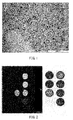
- FIG.1 shows a transmission electron microscope (TEM) photograph of composite particles (run 6).
- Example run 2 (7 nm), run 3 (10 nm), and run 6 (20 nm) that are different in particle size of the iron oxide microparticles from each other, the relaxation time was measured using NMR and MRI as follows.
- T 2 were measured by a CPMG (Carr-Purcell-Meiboom-Gill) method, with the measuring object being a proton signal of H 2 O in the presence of composite particles equivalent to 1 mM Fe (99% D 2 O/24°C).
- R 2 determined from the result were 118 [Fe mM -1 •s -1 ] (run 2), 151 [Fe mM -1 •s -1 ] (run 3), and 326 [Fe mM -1 •s -1 ] (run 6). Accordingly, the composite particles obtained above were found to exhibit a T 2 -shortening effect required as an MRI contrast agent.
- composite particles (run 6) were able to be obtained that indicated a R 2 of at least 200 [Fe mM -1 •s -1 ] and exhibited a high T 2 -shortening effect even at a lower concentration.
- the magnetic field strength was 9.4 T, but T 2 relaxation is hardly affected by the difference in magnetic field strength ( Questions & Answers in MAGNETIC RESONANCE IMAGING, Second Edition, published by Medical Science International ).
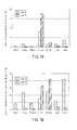
- FIG. 2 are concentrations (mM) of the composite particles, and MV indicates the existing gadolinium-based contrast agent used as the comparison subject.
- the left-hand drawing of FIG. 2 shows an MRI phantom image for measuring the relaxation level of the composite particles (run 6).
- the composite particles become dark in a concentration-dependent manner and it was confirmed that a sufficient MRI contrast image was able to be obtained even at lower concentrations as compared to the existing product.
- the magnetic field strength was 4.7 T.
- composite particles with higher graft chain molecular weights were excellent in dispersion stability and were suitable for a long period of storage. This is a surprising result indicating that composite particles with polymer graft chains bound thereto by living radical polymerization exhibit a characteristic that the dispersion stability is maintained for a long period of time even when they are simply mixed in an aqueous solvent.
- the in vitro macrophage test was carried out.
- a mouse (8 weeks old, ICR male mouse) subjected to intraperitoneal administration of 2 mL of 3% thioglycolate (manufactured by SIGMA CORPORATION) was sacrificed after three days and macrophages were recovered from the abdominal cavity using Hanks' solution (manufactured by Gibco Corporation).
- the macrophages thus recovered were adjusted to have a 1 ⁇ 10 6 cells/mL after being washed with Hanks' solution and a culture medium (DMEM, manufactured by Gibco Corporation) and then cultivated under the conditions of 37°C and 5 % CO 2 .
- DMEM culture medium
- Example runs 1 to 7, 11, and 12 as well as Comparative Example runs 8 to 10 and 13 adjusted to have 30 ⁇ g Fe/mL using the culture medium were added thereto.
- the culture medium was removed therefrom, which was then washed with PBS three times. Thereafter, the macrophages that phagocytized the composite particles were stained with Perl's Prussian Blue stain and the absorbance (590 nm) was measured ( FIG. 4 ).
- the negative control used herein was PBS and the positive control used herein was an existing iron oxide contrast agent, Resovist (Bayer Ltd.; 30 ⁇ g Fe/mL).
- Example run 5 and Comparative Example run 8 300 ⁇ L each
- concentration adjusted to 1.9 wt% using a normal saline solution was administered intravenously through the tail of a mouse (6 weeks old, ICR male mouse). They were sacrificed three days after administration and the extirpated livers were formalin-fixed. Thereafter, they were paraffin-embedded, sectioned, and then were HE-stained.
- Example run 6 the particle size of iron oxide microparticles: 20 nm, Mn: 109,000
- an MRI test was carried out.
- a tumor-bearing mouse (8 weeks old, BALB/c female mouse) under anesthesia was set in an MRI system (Unity INOVA 4.7T, Varian Technologies Japan Ltd.). After the tumor of the left femoral region was positioned and imaged (before administration) (see FIGS. 8A and 8B ), a dispersion (100 ⁇ M Fe/kg, 100 ⁇ L) of composite particles of run 6 prepared with a normal saline solution was intravenously administered through the tail and the same tumor was imaged after 24 hours (see FIG. 8C ).
- the interior of the tumor which was imaged as a white image before administration of the composite particles, was shadowed overall and particularly in a part thereof a perfect shadow effect was observed in the image 24 hours after administration (see FIG. 8C ).
- This result showed that the composite particles accumulated in the tumor and thereby exhibited the T 2 -shortening effect to reduce the signal strength. Accordingly it was confirmed that the composite particles practically function as an MRI contrast agent that enhances contrast.
- the MRI contrast agent of the present invention is also applicable for use as a nano-drug carrier, an imaging reagent, etc.
Landscapes
- Health & Medical Sciences (AREA)
- Chemical & Material Sciences (AREA)
- Nanotechnology (AREA)
- Engineering & Computer Science (AREA)
- Nuclear Medicine, Radiotherapy & Molecular Imaging (AREA)
- Life Sciences & Earth Sciences (AREA)
- Radiology & Medical Imaging (AREA)
- Epidemiology (AREA)
- Animal Behavior & Ethology (AREA)
- General Health & Medical Sciences (AREA)
- Public Health (AREA)
- Veterinary Medicine (AREA)
- Molecular Biology (AREA)
- Immunology (AREA)
- Medicinal Chemistry (AREA)
- Medicines Containing Antibodies Or Antigens For Use As Internal Diagnostic Agents (AREA)
- Medicinal Preparation (AREA)
Abstract
Description
- The present invention relates to an MRI contrast agent containing composite particles.
- In the MRI method (the magnetic resonance imaging method) that is one of the in vivo diagnostic imaging methods, contrast agents are often used in order to image specific tissues more clearly. The contrast agents are pharmaceutical agents for use in examinations to enhance the image contrast between a normal tissue and a target tissue in diagnosis and are usually used by intravenous injection. For the MRI contrast agents, generally, magnetic materials that affect nuclear magnetic resonance are used and, for example, gadolinium preparations and iron preparations are commercially available.
- As compared to the gadolinium preparations, the iron preparations have the advantages of being excellent in biological safety and producing excellent imaging effects at low doses.
- Generally, however, conventional iron preparations are taken into normal liver tissue in just a matter of minutes after intravenous administration but are not taken into tumor regions in the liver at all. Therefore, as MRI contrast agents, they can be used only for negative imaging of liver tumors. On the other hand, although iron preparations with a high stability in blood and low liver accumulation also have been clinically developed (for example, Patent Document 1), they are shown to be phagocytized mainly by macrophages and to have a high accumulation in lymph nodes and therefore are no different in terms of a tendency to be taken into the reticuloendothelial system. Thus, the conventional gadolinium preparations and iron preparations have problems in pharmacokinetics when being administered to biological bodies.
- As an example of the methods in which the pharmacokinetics after administration to a biological body has been improved, a method of coating the surfaces of inorganic core nanoparticles with polymers through, for example, silane groups has been proposed (for example, Patent Document 2).
- Patent Document 1:
WO 2006/068653 - Patent Document 2: Japanese Unexamined Patent Application Publication No.
2009-519316 - However, such conventional preparations have low dispersion stability and tend to be phagocytized by macrophages, which have been problems. Therefore, the present inventors aim to provide an MRI contrast agent that has high dispersion stability and improved functions and that tends not to be phagocytized by macrophages.
- The present invention is an MRI contrast agent containing composite particles, wherein the composite particles are those with polymer graft chains bound to surfaces of microparticles at a very high density sufficient to allow steric repulsion to occur between the graft chains, the microparticles are inorganic microparticles that exhibit superparamagnetism, and the polymer graft chains have a number average molecular weight (Mn) of at least 30,000.
- The composite particles according to the present invention to which high molecular weight graft chains were bound at a very high density by living radical polymerization have high dispersion stability and tend not to be phagocytized by reticuloendothelial cells and therefore tend not to be accumulated in tissues such as a liver and a spleen. Furthermore, the above-mentioned composite particles are accumulated in tumors due to the enhanced permeability and retention effect (EPR effect). Moreover, they have a high relaxation level and therefore can exhibit an imaging effect (contrast) even at a lower concentration. The development of such a high-performance MRI contrast agent enables high-precision diagnostic imaging and can contribute to the development of advanced medical treatments.
-
- [
FIG. 1] FIG.1 is a transmission electron microscope (TEM) photograph of composite particles (Table 1, run 6) of an MRI contrast agent. - [
FIG. 2] FIG. 2 is an MRI phantom image for measuring the relaxation level of the composite particles (Table 1, run 6) of the MRI contrast agent according to the present invention. - [
FIG. 3] FIG. 3 is a picture of 100-fold dilutions of composite particles (Table 1, runs 3 to 5 and 8) of the MRI contrast agent according to the present invention, which was taken after they were stored at room temperature for six months. - [
FIG. 4] FIG. 4 is a graph showing phagocytosis by macrophages of the composite particles (Table 1, runs 1 to 13) of the MRI contrast agent according to the present invention. - [
FIG. 5A] FIG. 5A is a histopathological picture of liver of a mouse, to which composite particles (Table 1, run 5) of the MRI contrast agent according to the present invention were administered. - [
FIG. 5B] FIG. 5B is a histopathological picture of liver of a mouse, to which composite particles (Table 1, run 8) of the MRI contrast agent according to the present invention were administered. - [
FIG. 6] FIG. 6 is a graph showing the percentages of composite particles remaining in the blood of mice, to which the composite particles (Table 1, runs 6 and 9) of the MRI contrast agent according to the present invention were administered. - [
FIG. 7A] FIG. 7A is a graph showing the rates of accumulation in tissues of mice, to which the composite particles (Table 1, runs 6 and 9) of the MRI contrast agent according to the present invention were administered. - [
FIG. 7B] FIG. 7B is a graph showing the rates of accumulation in tissues per weight of mice, to which the composite particles (Table 1, runs 6 and 9) of the MRI contrast agent according to the present invention were administered. - [
FIG. 8A] FIG. 8A is a picture showing the position of a tumor in the left femoral region of a tumor-bearing mouse, to which the composite particles (Table 1, run 6) of the MRI contrast agent according to the present invention are administered. - [
FIG. 8B] FIG. 8B is an MRI contrast image of a mouse tumor before administration of the composite particles (Table 1, run 6) of the MRI contrast agent according to the present invention. - [
FIG. 8C] FIG. 8C is an MRI contrast image of the mouse tumor taken 24 hours after administration of the composite particles (Table 1, run 6) of the MRI contrast agent according to the present invention. - The present inventors found that composite particles with polymer graft chains of a predetermined molecular weight bound to the surfaces of microparticles at a very high density sufficient to allow steric repulsion to occur between the graft chains were excellent in dispersion stability under physiological conditions over a prolonged period. Based on this finding, the present inventors solved the above-mentioned problems. As described above, with the excellent dispersion stability, the MRI contrast agent of the present invention has the advantages of (a) easy handling, (b) providing a stable drug effect, and (c) improved safety. Generally, since the MRI contrast agent is prepared in the medical setting immediately before being administered to a patient, the MRI contrast agent of the present invention that is easy to handle can reduce the time between preparation and administration and therefore is advantageous particularly for medical staff. Furthermore, since contrast agents with composite particles that tend to aggregate (i.e., with low dispersion stability) are recognized as foreign bodies in the blood and are then metabolized and excreted, the drug effect is reduced and thereby a sufficiently high imaging effect is difficult to obtain. Therefore, the MRI contrast agent of the present invention with the excellent dispersion stability allows the drug effect to be stable. Moreover, there is a possibility that the contrast agents with composite particles that tend to aggregate (i.e., with low dispersion stability) may form a thrombus in the blood and therefore they have lower safety, while the MRI contrast agent of the present invention has high dispersion stability and therefore such a possibility is low, resulting in improved safety.
- In the present specification, the phrase "the composite particles with excellent dispersion stability" denotes the state where macroscopically apparent aggregates and sediments of the composite particles are not observed in a conventional solvent. Furthermore, the phrase "composite particles are excellent in dispersion stability over a prolonged period" denotes the state where macroscopically apparent aggregates and sediments are not observed when the composite particles are stored in a solvent under normal storage conditions for a few months. In this context, the normal storage conditions denote conditions including a temperature around 18 to 30°C and a relative humidity of approximately 60% or lower. Preferably, the conventional solvent is an aqueous solvent such as a normal saline solution that is generally used for pharmaceutical preparations.
- Generally, when the composite particles have higher graft chain molecular weights, the density of the graft chains at the chain end decreases as compared to the vicinity where the graft chains are bound to the microparticles. Therefore, it is expected that they tend to be phagocytized by reticuloendothelial cells. Contrary to this expectation, however, the composite particles contained in the MRI contrast agent of the present invention exhibit characteristics of tending not to be phagocytized by reticuloendothelial cells. The term "reticuloendothelial system" used in the present invention is a generic term for cells that are involved in biological defense by phagocytizing foreign bodies and typical examples thereof include macrophages.
- Such composite particles make it possible to provide a MRI contrast agent that is typified by being accumulated in a tumor tissue through a blood circulating system and furthermore, that is characterized by depending on the pathological conditions in the tumor tissue and exhibiting the accumulation degree distribution that varies in the tumor tissue. The exhibition of the accumulation in the tumor tissue can be confirmed by the fact that in an MRI image, as compared to the test animal before the MRI contrast agent of the present invention is administered, the contrast is enhanced in the part corresponding to the tumor tissue of the test animal after administration.
- That is, as described above, the present invention is an MRI contrast agent containing composite particles, wherein the composite particles are those with polymer graft chains bound to surfaces of microparticles at a very high density sufficient to allow steric repulsion to occur between the graft chains, the microparticles are inorganic microparticles that exhibit superparamagnetism, and the polymer graft chains have a number average molecular weight (Mn) of at least 30,000.
- In the present invention, the "MRI contrast agent" is a substance (agent) that is administered in an in vivo diagnostic imaging method using an MRI (Magnetic Resonance Imaging) system that is used in clinical practice. When the substance is present in the object to be subjected to MRI measurement, how the measuring object looks in the measurement image seems to have been changed significantly as compared to the case of absence of the substance. Thus, contrast can be given to the image.
- In the present invention, the term "polymer graft chains" denotes polymer chains, each of which is formed with at least two monomers elongated from the surface of a microparticle by a polymerization reaction. Furthermore, the term "a very high density" denotes the density of graft chains in the case where the graft chains have thickened to such an extent that steric repulsion occurs between the graft chains. In this case, the graft chains each take a form of substantially fully elongated in the direction perpendicular to the surface. Furthermore, the term "bind/bound" denotes a bond that is formed by a common chemical reaction and examples thereof include a covalent bond and an ionic bond.
- In the MRI contrast agent of the present invention, the polymer graft chains have a number average molecular weight (Mn) of at least 30,000, preferably 30,000 to 320,000, more preferably 30,000 to 160,000, and most preferably 35,000 to 160,000. By controlling the number average molecular weight of the graft chains, differences are found in phagocytosis of reticuloendothelial cells and retention in blood of the MRI contrast agent of the present invention can be controlled. Specifically, when the graft chains have a high number average molecular weight, the retention in blood of the MRI contrast agent improves. Furthermore, when the graft chains have a high number average molecular weight, the MRI contrast agent tends not to be phagocytized by reticuloendothelial cells. The polymer graft chains have a number average degree of polymerization of 10 to 100,000. For example, when the monomer has a molecular weight of 500, the number average degree of polymerization is preferably 50 to 500, more preferably 50 to 350, and most preferably 70 to 350. The number average molecular weight can be measured by, for example, GPC. The number average degree of polymerization can be calculated by dividing the number average molecular weight by the monomer molecular weight.
- In the MRI contrast agent of the present invention, the microparticles are inorganic microparticles that exhibit superparamagnetism. Examples of such inorganic microparticles include iron (Fe), cobalt (Co), an alloy (for example, FePt and FeCo), and iron oxide (for example, ferrite and magnetite). Ferrite is preferable since it has been used successfully in commercial iron preparations for MRI contrast agents. The "ferrite" is a generic term for ceramics containing iron oxide as a main component thereof and is widely used as a magnetic material. The inorganic microparticles are more preferably ferrite having a spinel type crystal structure and further preferably magnetite (Fe3O4). In this context, those referred to as SPIO (a superparamagnetic iron oxide preparation), USPIO (an ultra-small SPIO), and MION (a monocrystalline iron oxide nanoparticle preparation) are included in the iron preparations for an MRI contrast agent.
- The term "superparamagnetism" is a phenomenon that occurs in magnetic inorganic nanoparticles and denotes that when an external magnetic field is applied thereto, they are magnetized as a whole. The superparamagnetic substance has properties of nanoparticles obtained by miniaturizing a ferromagnetic substance to have a size that allows only a single magnetic domain to be contained. Unlike the ferromagnetic substance that remains magnetized (magnetic storage) even when the magnetic field is removed, the superparamagnetic substance loses magnetization when the magnetic field is removed. Therefore, preferably, the inorganic microparticles that exhibit superparamagnetism have a particle size of 50 nm or smaller. This particle size can be measured by for example, transmission electron microscopy. Whether the composite particles of the present invention exhibit superparamagnetism can be confirmed with, for example, a SQUID (Superconducting Quantum Interference Device) fluxmeter (Macromolecules, 42(4), p1219-1228, 2009).
- The inorganic microparticles that exhibit superparamagnetism affect the relaxation times (both T1 and T2) of nuclei (generally water protons in clinical MRI) that emit MR signals used for forming images in the MRI measurement. The iron preparation containing the inorganic microparticles emphasizes the contrast inT2-weighted image by the effect of shortening mainly T2 (strictly speaking, shortening T2* by inducing nonuniformity in the local magnetic field).
- A transverse relaxation level (R2) that is expressed in the reciprocal of T2 is used as a value indicating the T2 shortening rate of the contrast agent. The R2 of the composite particles basically depends on magnetization of the inorganic microparticles that exhibit superparamagnetism and the R2 of existing SPIO or USPIO is generally 20 to 200 [Fe mM-1•s-1] (Advanced Drug Delivery Reviews, Vol.58, p1471-1504, 2006). A higher R2 indicates a higher T2 shortening effect at a lower concentration. When the inorganic microparticles that exhibit superparamagnetism are magnetite (Fe3O4) and are used as a negative contrast agent, the composite particles have a transverse relaxation level (R2) of preferably at least 100 [Fe mM-1•s-1] (magnetic field strength: 1.5T) and more preferably at least 200 [Fe mM-1•s-1] (magnetic field strength:1.5T).
- In the MRI contrast agent of the present invention, the polymer graft chains have a graft density of, for example, at least 0.1 chain/nm2, preferably 0.1 to 1.2 chain/nm2, more preferably 0.1 to 0.5 chain/nm2, and most preferably 0.15 to 0.45 chain/nm2. Controlling this graft density allows the retention in blood of the composite particles in the MRI contrast agent of the present invention to be controlled. In the present invention, the term "graft density" or "density" denotes the number of graft chains bound to the surface per unit area (nm2) of the surfaces of the microparticles. The graft density (s) is calculated as follows by determining the amount of the polymer graft chains (the amount of grafts, w) that are formed by being elongated from the surfaces of microparticles, by elemental analysis and using the value thus obtained, the surface area (S, nm2) of core microparticles, the number average molecular weight Mn and Avogadro's number (Av) of the graft polymers. Calculation Method : s = (w/Mn)Av/S
- In the MRI contrast agent of the present invention, the composite particles have an average particle size of preferably 1000 nm or smaller, more preferably 10 to 500 nm, and further preferably 10 to 250 nm. In the MRI contrast agent of the present invention, the retention in blood thereof seldom depends on the average particle size. Therefore, when having graft chains with a predetermined molecular weight, the MRI contrast agent of the present invention is not eliminated easily from a biological body even when the particle size of the microparticles is increased and as a result, the average particle size of the composite particles is increased. Consequently, the MRI contrast agent of the present invention is excellent in sensitivity as compared to conventional agents and can produce an effect at a low dose. The average particle size denotes the hydrodynamic diameter of a whole composite particle including graft chains provided for a core microparticle. Furthermore, in the case of composite particles in which the particle size distribution of the average particle size is small, the MRI contrast agent has a uniform size and therefore is preferable for DDS. The average particle size can be measured by a dynamic light scattering-type particle size distribution measuring method.
- In the MRI contrast agent of the present invention, the polymer graft chains can be obtained, for example, by living radical polymerization of monomers using polymerization initiating groups located on the surfaces of microparticles as base points. The type of the monomers is preferably at least one selected from the group consisting of an acrylic acid derivative, a methacrylic acid derivative, an acrylamide derivative, a methacrylamide derivative, and a styrene derivative. However, it is not limited to them and can be selected suitably in the range recognized by those skilled in the art. The polymer graft chains may be bound to other polymer graft chains. Specifically, in the living radical polymerization, block copolymers are grafted using cross-linking monomers as one type of monomers and then the cross-linking groups thus introduced may be reacted. Furthermore, the living radical polymerization may be, for example, random polymerization, block polymerization, or of a composition gradient type. The polymer graft chains each may have a specific ligand introduced into an end thereof or the vicinity of the end. An MRI contrast agent containing composite particles having such ligands is preferable since it can selectively deliver the ligands to a target organ. The specific ligand is not particularly limited as long as it is a specific ligand known in the present field.
- In the MRI contrast agent of the present invention, the polymer graft chains have preferably a molecular weight distribution (Mw/Mn) of 1 to 1.5. This is because when the molecular weight distribution is in the range of 1 to 1.5, the MRI contrast agent has homogeneous graft surfaces.
- Furthermore, in the composite particles of the MRI contrast agent of the present invention, polymer graft chains located in a region distant from microparticles may be polymers directed to a specific diseased tissue. Examples of the polymers directed to a specific diseased tissue include target molecules for molecular markers that are highly expressed at the time of lesion development. Such an MRI contrast agent can also be accumulated specifically in a lesion other than tumor. As a result, such a lesion can be detected as an MRI signal contrast.
- Preferably, the MRI contrast agent of the present invention is used for tumor diagnosis of, for example, a target affected part (an organ, a tissue, a cell, a pathogen, etc.). The MRI contrast agent of the present invention is characterized by inhibiting adsorption of a protein working for removing it from the blood and as a result, having a high percentage thereof remaining in the blood. Therefore, the MRI contrast agent of the present invention highly accumulates in cancer tissue. The possible reason for this is an EPR effect (Enhanced Permeability and Retention Effect). It is known that a substance with a molecular size of tens of nanometers to 250 nm accumulates due to anatomical features of blood vessels of cancer tissue. The MRI contrast agent of the present invention has high retention in blood and as a result, has a high probability to pass through the cancer tissue thorough the bloodstream. Consequently, it is considered that the MRI contrast agent accumulates more in cancer.
- Furthermore, the MRI contrast agent of the present invention has a low transfer rate to liver and exhibits a property of accumulating in tumor. Therefore, it is possible to detect tumors in various organs by the MRI method using the MRI contrast agent of the present invention.
- In the MRI contrast agent of the present invention, the composite particles can be produced, for example, as follows. First, a compound to be used as a coupling agent containing a polymerization initiating group is fixed to the surfaces of microparticles by a chemisorption method. Then at least one type of monomers is supplied thereto to perform living radical graft polymerization. With the coupling agent containing a polymerization initiating group being fixed to the surfaces of the microparticles beforehand, graft polymerization can be progressed on the surfaces of the microparticles, with the graft density maintained constant. That is, the amount of grafts can be increased in proportion to Mn (the number average molecular weight) of the graft chains, and polymerization is allowed to progress in a living manner and thereby almost all graft chains located on the surfaces of the microparticles are allowed to grow substantially evenly. That is, in the composite particles of the present invention, steric hindrance between adjacent graft chains located on the surfaces of the microparticles is reduced. Furthermore, during the living radical-graft polymerization, a polymerization initiator that is not fixed to the surfaces of the microparticles may be allowed to coexist.
- The compound to be used as a coupling agent containing a polymerization initiating group can be selected in view of, for example, an affinity with the microparticles. Specifically, when the microparticles are iron oxide particles, the coupling agent containing a polymerization initiating group thereof is preferably a silane compound represented by the following formula (I).
-

- In the above formula, n denotes an integer and is preferably 3 to 10 and more preferably 4 to 8. R1 is a C1-C3 alkyl group and is preferably a C1 or C2 alkyl group. R2 is a C1 or C2 alkyl group. X is a halogen atom and is preferably a bromine or chlorine atom. Examples of the compound represented by Formula (I) include (2-bromo-2-methy])propionyloxypropyltriethoxysilane (BPE) and (2-bromo-2-methyl)propionyloxyhexyltriethoxysilane (BHE).
- When the microparticles are iron oxide, examples of the coupling agent that does not contain a polymerization initiating group thereof include an alkylsilane coupling agent.
- It is possible to change the graft density on the surfaces of the microparticles by adjusting the ratio between the coupling agent containing a polymerization initiating group and the coupling agent that does not contain a polymerization initiating group. Specifically, in the case where the microparticles are iron oxide nanoparticles, the graft density is at least 0.1 chain/nm2 when the ratio between the coupling agent containing a polymerization initiating group and the coupling agent that does not contain a polymerization initiating group is 1:0.
- After completion of the polymerization, the intended composite particles can be isolated from the reaction solution by a method generally used in the present field, for example, by one of the methods such as extraction, distillation, washing, concentration, precipitation, filtration, drying, adsorption, deposition, and chromatography or a combination thereof
- Preferably the MRI contrast agent of the present invention is used for tumor diagnosis as described above. Examples of the tumor include non-neoplastic swellings and true tumors. The true tumors include a benign tumor and a malignant tumor. Examples of the malignant tumor include brain tumor, tongue cancer, laryngeal cancer, pharyngeal cancer, lung cancer, breast cancer, esophageal cancer, thyroid cancer, stomach cancer, pancreatic cancer, biliary tract cancer, duodenal cancer, colon cancer, liver cancer, renal cancer, uterine cancer, ovarian cancer, testicular cancer, prostate cancer, bladder cancer, osteosarcoma, chondrosarcoma, mesothelioma, skin cancer, leukemia, neuroblastoma, myeloma, and lymphoma.
- The MRI contrast agent of the present invention can be administered either orally or parenterally. In the case of oral administration, the MRI contrast agent of the present invention can be administered in the form of, for example, a tablet, a capsule, granules, powder, or syrup. In the case of parenteral administration, the MRI contrast agent of the present invention can be administered in the form of, for example, an injection, a suppository, an eye drop, an inhalant, a transnasal agent, an ointment, or a cream. In the case of the injection, it is administered by, for example, intravenous administration, intramuscular administration, or intraperitoneal administration. These preparations can be produced by conventionally well-known methods using pharmaceutically acceptable additives such as an excipient, a lubricant, a binder, a disintegrator, a stabilizer, and a diluent.
- Hereinafter, the present invention is described in further detail using examples. However, the range of the present invention is not limited by the following examples.
In the descriptions in the present specification, the following abbreviations are used: - THF: Tetrahydrofuran
- BPE: (2-Bromo-2-methyl)propionyloxypropyltriethoxysilane
- BPP: 1-(2-Bromo-2-methyl)propionyloxy-2-propene
- PEGMA: Polyethylene glycol methacrylate
- PEMA: (4-Hydroxyphenyl)ethyl methacrylate
- dN-biby: Cu(I)Cl, Dinonyl bipyridine
- EBIB: Ethyl 2-bromoisobutyrate
- NMR: Nuclear Magnetic Resonance
- GPC: Gel Filtration Chromatography
- TGA: Thermogravimetric Analysis
- TEM: Transmission Electron Microscope
- MRI: Magnetic Resonance Imaging
- <Production Example of Composite Particles (with microparticles being iron oxide nanoparticles)>
-

- Synthesis of BPE was performed by a two-step reaction. As a first step, a mixed solution containing allyl alcohol (170 g), triethylamine (237 g), and dichloromethane (2 L) was cooled in ice and then 2-bromoisobutyryl bromide (450 g) was dripped therein. After that, the reaction solution was stirred at 0°C for three hours and was further stirred at room temperature for ten hours. The reaction solution was concentrated, THF was added thereto, and thereby salts were allowed to precipitate and were then filtrated. After the filtrate was concentrated, the resultant was then diluted with chloroform (1 L). This was then washed with a 1N hydrochloric acid aqueous solution (2 × 1 L), a saturated sodium bicarbonate aqueous solution (2 × 1 L), and pure water (2 × 1 L) in this sequence. The organic layer was dried and was then concentrated. Thereafter, this was purified by vacuum distillation. Thus, 1-(2-bromo-2-methyl)propionyloxy-2-propene (BPP) was obtained at a yield of 90%.
- As a second step, BPP (100 g), triethoxysilane (170 g), and a Karstedt catalyst (600 µL) were sequentially placed in a flask. The mixture thereof was stirred in an argon atmosphere at room temperature for 12 hours. After the reaction, it was purified by vacuum distillation and thus BPE was synthesized at a yield of 70%.
- In accordance with the report of Hyeon et aL (Nature Materials, Vol.3, p891-895, 2004), oleic acid-coated iron oxide nanoparticles were synthesized.
First, an iron oleate complex was synthesized. Ferric chloride (FeCl36H2O, 10.8 g) and sodium oleate (36.5 g) were dissolved in a mixed solvent of ethanol (80 mL), water (60 mL), and hexane (140 mL). The solution thus obtained was stirred at 70°C for four hours. After completion of the reaction, an upper organic layer was recovered and the organic layer was then washed with pure water three times. Thereafter, the organic layer was concentrated under reduced pressure to remove the solvent using a rotary evaporator. The residue thus obtained was vacuum-dried at 70°C overnight and thus an iron oleate complex was obtained. This iron oleate complex (36 g) and oleic acid (5.7 g) were dissolved in trioctylamine (200 g). The solution thus obtained was heated to 370°C at a heating rate of 5°C/min. The solution was further heated under reflux to maintain a temperature of 370°C for 30 minutes. Thereafter, the reaction solution was cooled to room temperature and after being diluted with THF, oleic acid-coated iron oxide nanoparticles were recovered by centrifugation (12,000 rpm). The projected area equivalent circular diameter (average particle size) of the iron oxide nanoparticles was measured by transmission electron microscopy and as a result, was found to be 7, 10, 15 and 20 nm. It was confirmed by the X-ray diffraction method that this iron oxide was magnetite (Fe3O4). - The iron oxide nanoparticles obtained in (2) were dispersed in THF and thereby a dispersion (1 wt%) was prepared. Aqueous ammonia (1 wt%) was added to the dispersion, which was then stirred for a while. Thereafter, the BPE (2 wt%) obtained in (1) was added thereto, which was then stirred at room temperature for three days. While being stirred, this solution was subjected to ultrasonic irradiation at regular intervals. Thereafter, the particles were recovered by centrifugation (12,000 rpm) and were then subjected to redispersion with THF and centrifugation repeatedly. Thus, iron oxide nanoparticles with initiating groups were obtained.
-

- The iron oxide nanoparticles with initiating groups obtained in (3), polyethylene glycol methacrylate (PEGMA), (4-hydroxyphenyl)ethyl methacrylate (PEMA), Cu(I)Cl, dinonyl bipyridine (dN-biby), and ethyl 2-bromoisobutyrate (EBIB) were placed in a Pyrex glass tube. This was degassed by the freezing and thawing method and was then sealed under vacuum. Thereafter, it was polymerized at 70°C for a predetermined period of time and thereby composite particles with polymer graft chains bound to the surfaces of the iron oxide nanoparticles were obtained. The polymerization conditions are indicated in Table 1.
- The composite particles (with the microparticles being the iron oxide nanoparticles) were purified by a repetition of centrifugation and redispersion into acetone and pure water.
- The composite particles were dispersed in pure water, which was then stored at room temperature. The composite particles were treated with hydrogen fluoride and thereby the polymer graft chains were cut out from the iron oxide nanoparticles. Then the molecular weight and molecular weight distribution of the polymer graft chains were determined by gel filtration chromatography (GPC).
- From the amount of grafts determined by thermogravimetricAnalysis (TGA), the graft density was calculated. It was confirmed that all the samples had a high graft density of at least 0.15 to 0.43 chain/nm2. Furthermore, the polymerization conditions used herein and polymerization results are indicated together in Table 1, which is intended to be used as a sample list.
FIG.1 shows a transmission electron microscope (TEM) photograph of composite particles (run 6). - [Table 1]
Table 1. Polymerization Conditions and Polymerization Results run Polymerization Conditions for Polymer Graft Chains Polymer Graft Chain Properties EBIB : (PEGMA+PEMA): Cu(I)Cl : dN-bipy Monomer Amount (wt% Iron Oxide Microparticle Size (nm) Iron Oxide Microparticle Concentration (wt%) Polymerization Solvent Total Amount of Polymerization Solution (g) Polymerization Time (h) Number Average Molecular Weight (Mn) Molecular Weight Distribution (Mw/Mn) Number Average Degree of Polymerization Graft Density (chain/ nm2) Examples 1 1 : 15000 : 150 : 300 50 7 0.25 THF 20 65.5 160,000 1.43 337 0.29 2 1 : 2000 : 10 : 20 50 7 0.25 THF 20 27 66,000 1.1 139 0.29 3 1 : 2000 : 10 : 20 50 10 0.5 THF 20 24 65,000 1.1 137 0.32 4 1 : 10000 : 100 : 200 50 10 0.5 THF 20 20 101,000 1.15 213 0.38 5 1 : 10000 : 100 : 200 50 10 0.5 THF 20 72 154,000 1.2 324 0.41 6 1 : 10000 : 100 : 200 50 20 0.5 THF 20 20 109,000 1.12 229 0.4 7 1 : 10000 : 100 : 200 40 20 0.5 THF 20 19.5 100,000 1.22 211 0.43 11 1 : 2000 : 10 : 20 50 15 0.5 THF 20 5.5 36000 1.30 76 0.15 12 1 : 2000 : 10 : 20 50 15 3 THF 50 9 57000 1.40 120 0.18 Comparative Examples 8 1 : 2000 : 10 : 20 50 10 1 THF 10 16 29,000 1.13 61 0.11 9 1 : 100 : 0.5 : 1 50 20 2 THF 10 4 11,000 1.22 23 0.06 10 1 : 100 : 0.5 : 1 50 20 0.5 THF 10 4 12,000 1.18 25 0.16 13 1 : 100 : 0.5 : 1 50 20 2 THF 10 4 26000 1.21 55 0.14 EBIB: Ethyl 2-bromoisobutyrate
PEGMA: Polyethylene glycol methacrylate
PEMA: (4-Hydroxyphenyl)ethyl methacrylate
dN-biby: Dinonyl bipyridine
THF: Tetrahydrofuran - With respect to the composite particles obtained in Example run 2 (7 nm), run 3 (10 nm), and run 6 (20 nm) that are different in particle size of the iron oxide microparticles from each other, the relaxation time was measured using NMR and MRI as follows.
- Using NMR (EX-400, JEOL Ltd.), T2 were measured by a CPMG (Carr-Purcell-Meiboom-Gill) method, with the measuring object being a proton signal of H2O in the presence of composite particles equivalent to 1 mM Fe (99% D2O/24°C). R2 determined from the result were 118 [Fe mM-1•s-1] (run 2), 151 [Fe mM-1•s-1] (run 3), and 326 [Fe mM-1•s-1] (run 6). Accordingly, the composite particles obtained above were found to exhibit a T2-shortening effect required as an MRI contrast agent. Furthermore, composite particles (run 6) were able to be obtained that indicated a R2 of at least 200 [Fe mM-1•s-1] and exhibited a high T2-shortening effect even at a lower concentration. In the NMR used in this measurement, the magnetic field strength was 9.4 T, but T2 relaxation is hardly affected by the difference in magnetic field strength (Questions & Answers in MAGNETIC RESONANCE IMAGING, Second Edition, published by Medical Science International).
- Next, a phantom experiment was carried out using MRI (Unity INOVA 4.7T, Varian Technologies Japan Ltd.). The composite particles (run 6) were diluted with distilled water to a Fe concentration of 0.001 to 2.0 mM. These were placed as shown in the right-hand drawing of
FIG. 2 and T2 thereof were then measured by spin echo (measurement conditions: TR/TE = 4000/0.08 ms). The comparison subject used herein was an existing gadolinium-based contrast agent, Magnevist (MV, 1 mM Gd, Bayer Yakuhin, Ltd.) and the negative control used herein was distilled water. The numbers indicated in the circles shown in the right-hand drawing ofFIG. 2 are concentrations (mM) of the composite particles, and MV indicates the existing gadolinium-based contrast agent used as the comparison subject. The left-hand drawing ofFIG. 2 shows an MRI phantom image for measuring the relaxation level of the composite particles (run 6). As a result, with reference to the T2 measurement image, the composite particles become dark in a concentration-dependent manner and it was confirmed that a sufficient MRI contrast image was able to be obtained even at lower concentrations as compared to the existing product. Furthermore, the same results were obtained also when other composite particles were used. The magnetic field strength was 4.7 T. - In order to evaluate the dispersion stability of the composite particles obtained in
runs 1 to 13, a long-term storage test was carried out. Using PBS (phosphate buffered saline), the respective composite particles (Example runs 3, 4, and 5 as well asComparative Example run 8; 5 mL each) with the iron oxide microparticles having a particle size of 10 nm were adjusted to have a particle concentration of 0.038 wt% and were then stored at room temperature for six months. Before the start of storage, no aggregation of any composite particles was found. As a result of dynamic light scattering particle size distribution measurement using LB-550 (manufactured by HORIBA, Ltd.), the average particle sizes (hydrodynamic diameter; the median size was used in the present measurement) were smaller than 40 nm, and all of them exhibited excellent dispersion stability. When these composite particles were stored at room temperature for 6 months, precipitation occurred only in Comparative Example run 8 (FIG. 3 ). Similarly, the measurement of particle size distribution of the composite particles was carried out and as a result, Comparative Example run 8 was 296 nm,run 3 was 120 nm,run 4 was 75 nm, and run 5 was 24 nm. Thus, the longer the graft chain length was, the more the dispersibility obtained before the start of storage was maintained. From these results, it was confirmed that composite particles with higher graft chain molecular weights were excellent in dispersion stability and were suitable for a long period of storage. This is a surprising result indicating that composite particles with polymer graft chains bound thereto by living radical polymerization exhibit a characteristic that the dispersion stability is maintained for a long period of time even when they are simply mixed in an aqueous solvent. - In order to evaluate the tendency of being taken into the reticuloendothelial system of the composite particles, the in vitro macrophage test was carried out. A mouse (8 weeks old, ICR male mouse) subjected to intraperitoneal administration of 2 mL of 3% thioglycolate (manufactured by SIGMA CORPORATION) was sacrificed after three days and macrophages were recovered from the abdominal cavity using Hanks' solution (manufactured by Gibco Corporation). The macrophages thus recovered were adjusted to have a 1×10⁶ cells/mL after being washed with Hanks' solution and a culture medium (DMEM, manufactured by Gibco Corporation) and then cultivated under the conditions of 37°C and 5 % CO2. After three hours, the culture medium was removed and composite particles (Example runs 1 to 7, 11, and 12 as well as Comparative Example runs 8 to 10 and 13) adjusted to have 30 µg Fe/mL using the culture medium were added thereto. After 24 hours of cultivation, the culture medium was removed therefrom, which was then washed with PBS three times. Thereafter, the macrophages that phagocytized the composite particles were stained with Perl's Prussian Blue stain and the absorbance (590 nm) was measured (
FIG. 4 ). The negative control used herein was PBS and the positive control used herein was an existing iron oxide contrast agent, Resovist (Bayer Ltd.; 30 µg Fe/mL). As a result, when the composite particles of Example were used, there was no difference from the negative control and therefore it was confirmed that they were not phagocytized by macrophages. Generally, it is considered that when the composite particles have higher graft chain molecular weights, the density at the graft chain end decreases as compared to the vicinity where the microparticles were bound, and they tend to be recognized by the reticuloendothelial system and thus tend to be phagocytized. Therefore, it was considered that the composite particles with lower graft chain molecular weights tended not to be phagocytized by macrophages. Contrary to expectations, however, it was confirmed that as compared to the composite particles (Comparative Example) with lower graft chain molecular weights, the composite particles (the present invention) with higher graft chain molecular weights tended not to be phagocytized by macrophages. - In order to evaluate the tendency of being taken into the liver of the composite particles, the histopathological evaluation of the liver was carried out. Each solution of the composite particles (
Example run 5 andComparative Example run 8; 300 µL each) with a concentration adjusted to 1.9 wt% using a normal saline solution was administered intravenously through the tail of a mouse (6 weeks old, ICR male mouse). They were sacrificed three days after administration and the extirpated livers were formalin-fixed. Thereafter, they were paraffin-embedded, sectioned, and then were HE-stained. As a result of the histopathological evaluation of the liver, in the case of Comparative Example run 8 (the particle size of the iron oxide microparticles: 10 nm, Mn: 29,000), the liver macrophages (Kupffer cells) shown in the circles were stained a brownish color, and thereby it was indicated that the iron oxide Nanoparticles (composite particles) were phagocytized (FIG. 5B ). In the case of run 5 (the particle size of the iron oxide microparticles: 10 nm, Mn: 154,000), only normal Kupffer cells shown in the circles were found and no phagocytosis was observed (FIG. 5A ). From this result, it was confirmed that the composite particles of Example were not phagocytized by the Kupffer cells in the liver reticuloendothelial system and were not accumulated in the liver three days after administration. - Dispersions (the concentration of the composite particles: 1 wt%, the solvent = pure water, 100 µL) of the composite particles of Example run 6 (the particle size of iron oxide microparticles: 20 nm, Mn: 109,000) and Comparative Example run 9 (the particle size of iron oxide microparticles: 20 nm, Mn: 11,000) each were placed in a microcentrifuge tube, and further Na125I (5 µL, Perkinelmer NEZ033) and then a Chloramine T solution (the concentration: 0.2 mg/ml, the solvent = 0.5M phosphate buffered aqueous solution (pH 7.5, containing 0.5M NaCl, 100 µL) were added thereto. Each solution thus obtained was stirred with a vortex mixer for two minutes. Further, a sodium metabisulfite solution (the concentration: 4 mg/mL, the solvent = pure water, 100 µL) was added thereto, which was then stirred with the vortex mixer for two minutes. The mixture thus obtained was separated and purified (the eluate: normal saline solution) with a PD-10 column (GE Healthcare Japan Corporation) and thereby a distillation containing microparticles (125I-labeled composite particles) were recovered as a 125I -labeled composite particle dispersion.
- The 125I-labeled composite particle dispersions (runs 6 and 9, 100 µL each) produced in (1) each were administered intravenously through the tail of a tumor-bearing mouse (8 weeks old, BALB/c female mouse) with Colon-26 tumor cells transplanted thereinto. After the lapse of a predetermined period of time, blood was collected from a mouse eye. The radioactivity thereof was then measured with a gamma counter (ARC-301B, ALOKA CO., LTD.) and thereby the percentage of the composite particles remaining in the blood was determined (
FIG. 6 ). Furthermore, the tumor-bearing mice were sacrificed 24 hours after administration. The radioactivity of each organ extirpated therefrom was measured and thereby the biodistribution of the composite particles was evaluated (FIG. 7A ; the rate of accumulation in tissue,FIG. 7B : the rate of accumulation in tissue per weight). As a result of the percentage of the composite particles remaining in the blood, in Comparative Example run 9 (the particle size of the iron oxide microparticles: 20 nm, Mn: 11,000), the percentage decreased from immediately after administration, while in Example run 6 (the particle size of the iron oxide microparticles: 20 nm, Mn: 109,000), a high percentage (approximately 40%) of the composite particles remaining in the blood was maintained up to 24 hours after administration, which indicated a high retention in blood. In the biodistribution, with respect to the rate of accumulation in tissue measured 24 hours after administration, an accumulation of 8 to 18% in liver and an accumulation of 1 to 2% in spleen were observed in both trial products. Since the rate of accumulation in the liver reticuloendothelial system measured one hour after administration of a conventional iron oxide-based contrast agent was approximately 80% (Am J Roentgenol, 152, 167-173, 1989), it was confirmed that the rates of accumulation of the composite particles of Example in the liver and the spleen were very low. Furthermore, in the rate of accumulation in tissue per weight measured 24 hours after administration ofExample run 6, a high rate of accumulation in the tumor was proved. From these results, it was confirmed that in the composite particles with higher graft chain molecular weights, the retention in blood was prolonged and accumulation in the tumor increased due to the EPR effect, while the rates of accumulation in the liver and the spleen were low. - Using the composite particles of Example run 6 (the particle size of iron oxide microparticles: 20 nm, Mn: 109,000), an MRI test was carried out. A tumor-bearing mouse (8 weeks old, BALB/c female mouse) under anesthesia was set in an MRI system (Unity INOVA 4.7T, Varian Technologies Japan Ltd.). After the tumor of the left femoral region was positioned and imaged (before administration) (see
FIGS. 8A and 8B ), a dispersion (100 µM Fe/kg, 100 µL) of composite particles ofrun 6 prepared with a normal saline solution was intravenously administered through the tail and the same tumor was imaged after 24 hours (seeFIG. 8C ). The imaging was carried out under the conditions including: the transverse plane, 1 mm thick, TR = 300 ms, TE = 10 ms, and a flip angle of 20°. As a result, in the T2-weighted image that was imaged by the gradient echo (GE) measurement, the interior of the tumor, which was imaged as a white image before administration of the composite particles, was shadowed overall and particularly in a part thereof a perfect shadow effect was observed in theimage 24 hours after administration (seeFIG. 8C ). This result showed that the composite particles accumulated in the tumor and thereby exhibited the T2-shortening effect to reduce the signal strength. Accordingly it was confirmed that the composite particles practically function as an MRI contrast agent that enhances contrast. - The MRI contrast agent of the present invention is also applicable for use as a nano-drug carrier, an imaging reagent, etc.
Claims (6)
- An MRI contrast agent comprising composite particles,
wherein the composite particles are those with polymer graft chains bound to surfaces of microparticles at a very high density sufficient to allow steric repulsion to occur between the graft chains, the microparticles are inorganic microparticles that exhibit superparamagnetism, and the polymer graft chains have a number average molecular weight (Mn) of at least 30,000. - The MRI contrast agent according to claim 1, wherein the polymer graft chains have a graft density of at least 0.1 chain/nm2.
- The MRI contrast agent according to claim 1 or 2, wherein the polymer graft chains are obtained by living radical polymerization of at least one selected from the group consisting of an acrylic acid derivative, a methacrylic acid derivative, an acrylamide derivative, a methacrylamide derivative, and a styrene derivative, with polymerization initiating groups located on the surfaces of the microparticles being used as base points.
- The MRI contrast agent according to any one of claims 1 to 3, wherein the polymer graft chains have a molecular weight distribution of 1 to 1.5.
- The MRI contrast agent according to any one of claims 1 to 4, wherein the MRI contrast agent is for tumor diagnosis.
- Use of an MRI contrast agent according to any one of claims 1 to 4 in tumor diagnosis.
Applications Claiming Priority (2)
| Application Number | Priority Date | Filing Date | Title |
|---|---|---|---|
| JP2010269386 | 2010-12-02 | ||
| PCT/JP2011/072431 WO2012073588A1 (en) | 2010-12-02 | 2011-09-29 | Mri contrast agent containing composite particles |
Publications (3)
| Publication Number | Publication Date |
|---|---|
| EP2647391A1 true EP2647391A1 (en) | 2013-10-09 |
| EP2647391A4 EP2647391A4 (en) | 2016-07-06 |
| EP2647391B1 EP2647391B1 (en) | 2018-05-02 |
Family
ID=46171543
Family Applications (1)
| Application Number | Title | Priority Date | Filing Date |
|---|---|---|---|
| EP11845744.9A Not-in-force EP2647391B1 (en) | 2010-12-02 | 2011-09-29 | Mri contrast agent containing composite particles |
Country Status (7)
| Country | Link |
|---|---|
| US (1) | US20130251636A1 (en) |
| EP (1) | EP2647391B1 (en) |
| JP (1) | JP5961556B2 (en) |
| CN (1) | CN103237562B (en) |
| ES (1) | ES2670925T3 (en) |
| SG (1) | SG190740A1 (en) |
| WO (1) | WO2012073588A1 (en) |
Families Citing this family (4)
| Publication number | Priority date | Publication date | Assignee | Title |
|---|---|---|---|---|
| EP3115384B1 (en) * | 2014-03-05 | 2020-07-29 | JSR Corporation | Solid support, ligand-bound solid support, detection or separation method for target substance, solid support production method, and ligand-bound solid support production method |
| JP6372800B2 (en) * | 2014-09-05 | 2018-08-15 | 株式会社カネカ | Imaging agent for renal function diagnosis containing composite particles |
| CN107753960B (en) * | 2016-08-19 | 2022-04-22 | 江苏纳菲生物医药科技有限公司 | Polymer-metal oxide compound and preparation method and application thereof |
| JP7464592B2 (en) * | 2019-05-31 | 2024-04-09 | 国立大学法人京都大学 | Macrophage Imaging Agent |
Family Cites Families (4)
| Publication number | Priority date | Publication date | Assignee | Title |
|---|---|---|---|---|
| WO2005108451A1 (en) * | 2004-05-07 | 2005-11-17 | Kyoto University | Method for preparing colloidal crystal using polymer-grafted fine particles |
| JP2006328309A (en) * | 2005-05-30 | 2006-12-07 | Canon Inc | Magnetic polymer particles and method for producing the same |
| US8232344B2 (en) * | 2007-06-27 | 2012-07-31 | Canon Kabushiki Kaisha | Structure, target substance-detecting element and target substance-detecting kit |
| WO2009020220A1 (en) * | 2007-08-06 | 2009-02-12 | Canon Kabushiki Kaisha | Structural member and method of producing the structural member |
-
2011
- 2011-09-29 US US13/991,050 patent/US20130251636A1/en not_active Abandoned
- 2011-09-29 SG SG2013042213A patent/SG190740A1/en unknown
- 2011-09-29 WO PCT/JP2011/072431 patent/WO2012073588A1/en not_active Ceased
- 2011-09-29 ES ES11845744.9T patent/ES2670925T3/en active Active
- 2011-09-29 CN CN201180057960.6A patent/CN103237562B/en not_active Expired - Fee Related
- 2011-09-29 EP EP11845744.9A patent/EP2647391B1/en not_active Not-in-force
- 2011-09-29 JP JP2012546728A patent/JP5961556B2/en not_active Expired - Fee Related
Also Published As
| Publication number | Publication date |
|---|---|
| US20130251636A1 (en) | 2013-09-26 |
| JPWO2012073588A1 (en) | 2014-05-19 |
| EP2647391A4 (en) | 2016-07-06 |
| WO2012073588A1 (en) | 2012-06-07 |
| EP2647391B1 (en) | 2018-05-02 |
| SG190740A1 (en) | 2013-07-31 |
| CN103237562B (en) | 2015-01-21 |
| JP5961556B2 (en) | 2016-08-02 |
| CN103237562A (en) | 2013-08-07 |
| ES2670925T3 (en) | 2018-06-04 |
Similar Documents
| Publication | Publication Date | Title |
|---|---|---|
| Bae et al. | Bioinspired synthesis and characterization of gadolinium-labeled magnetite nanoparticles for dual contrast T 1-and T 2-weighted magnetic resonance imaging | |
| Shen et al. | Iron oxide nanoparticle based contrast agents for magnetic resonance imaging | |
| US20090226376A1 (en) | Novel Mixed Ligand Core/Shell Iron Oxide Nanoparticles for Inflammation Imaging | |
| KR20120101362A (en) | Treating water insoluble nanoparticles with hydrophilic alpha-hydroxyphosphonic acid conjugates, the so modified nanoparticles and their use as contrast agents | |
| Chen et al. | In vitro and in vivo CT imaging using bismuth sulfide modified with a highly biocompatible Pluronic F127 | |
| Paunesku et al. | Gadolinium-conjugated TiO2-DNA oligonucleotide nanoconjugates show prolonged intracellular retention period and T1-weighted contrast enhancement in magnetic resonance images | |
| Liang et al. | Ultrasmall gadolinium hydrated carbonate nanoparticle: an advanced T 1 MRI contrast agent with large longitudinal relaxivity | |
| Na et al. | T 1-weighted mr imaging of liver tumor by gadolinium-encapsulated glycol chitosan nanoparticles without non-specific toxicity in normal tissues | |
| Tate et al. | In vivo biodistribution of iron oxide nanoparticles: an overview | |
| Shah et al. | EDTA capped iron oxide nanoparticles magnetic micelles: drug delivery vehicle for treatment of chronic myeloid leukemia and T 1–T 2 dual contrast agent for magnetic resonance imaging | |
| EP2647391B1 (en) | Mri contrast agent containing composite particles | |
| Wei et al. | Biocompatible low-retention superparamagnetic iron oxide nanoclusters as contrast agents for magnetic resonance imaging of liver tumor | |
| Stanicki et al. | Impact of the chain length on the biodistribution profiles of PEGylated iron oxide nanoparticles: A multimodal imaging study | |
| EP1960002B1 (en) | Aqueous dispersions of superparamagnetic single domain particles production and use thereof for diagnosis and therapy | |
| JP7125725B2 (en) | Amphiphilic polymer nanomicelle containing poly 3,4-dihydroxy-L-phenylalanine chelate ferric ion and use thereof | |
| US20190247524A1 (en) | Polymer-metal oxide complex, preparation method therefor, and applications | |
| Wang et al. | Trifunctional polymeric nanocomposites incorporated with Fe3O4/iodine-containing rare earth complex for computed X-ray tomography, magnetic resonance, and optical imaging | |
| Yaghoobi et al. | Therapeutic effect of deferrioxamine conjugated to PEGylated gold nanoparticles and complexed with Mn (II) beside the CT scan and MRI diagnostic studies | |
| US12447216B2 (en) | Biogenic hemin-based MRI contrast agents, and compositions and methods thereof | |
| Sobhani et al. | Novel MR imaging nanoprobe for hepatocellular carcinoma detection based on manganese–zinc ferrite nanoparticles: in vitro and in vivo assessments | |
| JP5142251B2 (en) | Composite particles using gold iron oxide particles and MRI contrast agent | |
| Liu et al. | Biocompatible KMnF 3 nanoparticular contrast agent with proper plasma retention time for in vivo magnetic resonance imaging | |
| WO2019004297A1 (en) | Nanoparticle, contrast agent for magnetic resonance imaging containing same, and ligand compound | |
| Jin et al. | Polymer-coated superparamagnetic iron oxide nanoparticles: synthesis, characterization and toxicity evaluation for MRI in mice | |
| CN106620728B (en) | Zwitterion modified multifunctional Mn3O4Nanoparticle magnetic resonance imaging contrast agent and preparation and application thereof |
Legal Events
| Date | Code | Title | Description |
|---|---|---|---|
| PUAI | Public reference made under article 153(3) epc to a published international application that has entered the european phase |
Free format text: ORIGINAL CODE: 0009012 |
|
| 17P | Request for examination filed |
Effective date: 20130606 |
|
| AK | Designated contracting states |
Kind code of ref document: A1 Designated state(s): AL AT BE BG CH CY CZ DE DK EE ES FI FR GB GR HR HU IE IS IT LI LT LU LV MC MK MT NL NO PL PT RO RS SE SI SK SM TR |
|
| DAX | Request for extension of the european patent (deleted) | ||
| RAP1 | Party data changed (applicant data changed or rights of an application transferred) |
Owner name: KANEKA CORPORATION |
|
| RA4 | Supplementary search report drawn up and despatched (corrected) |
Effective date: 20160603 |
|
| RIC1 | Information provided on ipc code assigned before grant |
Ipc: A61K 49/18 20060101ALI20160527BHEP Ipc: A61K 49/00 20060101AFI20160527BHEP |
|
| GRAP | Despatch of communication of intention to grant a patent |
Free format text: ORIGINAL CODE: EPIDOSNIGR1 |
|
| INTG | Intention to grant announced |
Effective date: 20171122 |
|
| RIN1 | Information on inventor provided before grant (corrected) |
Inventor name: TABATA, YASUHIKO Inventor name: TAGO, YOSHIYUKI Inventor name: TSUJII, YOSHINOBU Inventor name: YOSHIDA, SHINICHI Inventor name: OHNO, KOHJI |
|
| GRAS | Grant fee paid |
Free format text: ORIGINAL CODE: EPIDOSNIGR3 |
|
| GRAA | (expected) grant |
Free format text: ORIGINAL CODE: 0009210 |
|
| AK | Designated contracting states |
Kind code of ref document: B1 Designated state(s): AL AT BE BG CH CY CZ DE DK EE ES FI FR GB GR HR HU IE IS IT LI LT LU LV MC MK MT NL NO PL PT RO RS SE SI SK SM TR |
|
| REG | Reference to a national code |
Ref country code: GB Ref legal event code: FG4D |
|
| RIN1 | Information on inventor provided before grant (corrected) |
Inventor name: OHNO, KOHJI Inventor name: YOSHIDA, SHINICHI Inventor name: TABATA, YASUHIKO Inventor name: TAGO, YOSHIYUKI Inventor name: TSUJII, YOSHINOBU |
|
| REG | Reference to a national code |
Ref country code: CH Ref legal event code: EP Ref country code: AT Ref legal event code: REF Ref document number: 994516 Country of ref document: AT Kind code of ref document: T Effective date: 20180515 |
|
| REG | Reference to a national code |
Ref country code: DE Ref legal event code: R096 Ref document number: 602011048127 Country of ref document: DE |
|
| REG | Reference to a national code |
Ref country code: IE Ref legal event code: FG4D |
|
| REG | Reference to a national code |
Ref country code: ES Ref legal event code: FG2A Ref document number: 2670925 Country of ref document: ES Kind code of ref document: T3 Effective date: 20180604 |
|
| REG | Reference to a national code |
Ref country code: NL Ref legal event code: MP Effective date: 20180502 |
|
| REG | Reference to a national code |
Ref country code: FR Ref legal event code: PLFP Year of fee payment: 8 |
|
| REG | Reference to a national code |
Ref country code: LT Ref legal event code: MG4D |
|
| PG25 | Lapsed in a contracting state [announced via postgrant information from national office to epo] |
Ref country code: SE Free format text: LAPSE BECAUSE OF FAILURE TO SUBMIT A TRANSLATION OF THE DESCRIPTION OR TO PAY THE FEE WITHIN THE PRESCRIBED TIME-LIMIT Effective date: 20180502 Ref country code: LT Free format text: LAPSE BECAUSE OF FAILURE TO SUBMIT A TRANSLATION OF THE DESCRIPTION OR TO PAY THE FEE WITHIN THE PRESCRIBED TIME-LIMIT Effective date: 20180502 Ref country code: BG Free format text: LAPSE BECAUSE OF FAILURE TO SUBMIT A TRANSLATION OF THE DESCRIPTION OR TO PAY THE FEE WITHIN THE PRESCRIBED TIME-LIMIT Effective date: 20180802 Ref country code: FI Free format text: LAPSE BECAUSE OF FAILURE TO SUBMIT A TRANSLATION OF THE DESCRIPTION OR TO PAY THE FEE WITHIN THE PRESCRIBED TIME-LIMIT Effective date: 20180502 Ref country code: NO Free format text: LAPSE BECAUSE OF FAILURE TO SUBMIT A TRANSLATION OF THE DESCRIPTION OR TO PAY THE FEE WITHIN THE PRESCRIBED TIME-LIMIT Effective date: 20180802 |
|
| PG25 | Lapsed in a contracting state [announced via postgrant information from national office to epo] |
Ref country code: GR Free format text: LAPSE BECAUSE OF FAILURE TO SUBMIT A TRANSLATION OF THE DESCRIPTION OR TO PAY THE FEE WITHIN THE PRESCRIBED TIME-LIMIT Effective date: 20180803 Ref country code: HR Free format text: LAPSE BECAUSE OF FAILURE TO SUBMIT A TRANSLATION OF THE DESCRIPTION OR TO PAY THE FEE WITHIN THE PRESCRIBED TIME-LIMIT Effective date: 20180502 Ref country code: RS Free format text: LAPSE BECAUSE OF FAILURE TO SUBMIT A TRANSLATION OF THE DESCRIPTION OR TO PAY THE FEE WITHIN THE PRESCRIBED TIME-LIMIT Effective date: 20180502 Ref country code: LV Free format text: LAPSE BECAUSE OF FAILURE TO SUBMIT A TRANSLATION OF THE DESCRIPTION OR TO PAY THE FEE WITHIN THE PRESCRIBED TIME-LIMIT Effective date: 20180502 Ref country code: NL Free format text: LAPSE BECAUSE OF FAILURE TO SUBMIT A TRANSLATION OF THE DESCRIPTION OR TO PAY THE FEE WITHIN THE PRESCRIBED TIME-LIMIT Effective date: 20180502 |
|
| REG | Reference to a national code |
Ref country code: AT Ref legal event code: MK05 Ref document number: 994516 Country of ref document: AT Kind code of ref document: T Effective date: 20180502 |
|
| PG25 | Lapsed in a contracting state [announced via postgrant information from national office to epo] |
Ref country code: CZ Free format text: LAPSE BECAUSE OF FAILURE TO SUBMIT A TRANSLATION OF THE DESCRIPTION OR TO PAY THE FEE WITHIN THE PRESCRIBED TIME-LIMIT Effective date: 20180502 Ref country code: RO Free format text: LAPSE BECAUSE OF FAILURE TO SUBMIT A TRANSLATION OF THE DESCRIPTION OR TO PAY THE FEE WITHIN THE PRESCRIBED TIME-LIMIT Effective date: 20180502 Ref country code: PL Free format text: LAPSE BECAUSE OF FAILURE TO SUBMIT A TRANSLATION OF THE DESCRIPTION OR TO PAY THE FEE WITHIN THE PRESCRIBED TIME-LIMIT Effective date: 20180502 Ref country code: SK Free format text: LAPSE BECAUSE OF FAILURE TO SUBMIT A TRANSLATION OF THE DESCRIPTION OR TO PAY THE FEE WITHIN THE PRESCRIBED TIME-LIMIT Effective date: 20180502 Ref country code: EE Free format text: LAPSE BECAUSE OF FAILURE TO SUBMIT A TRANSLATION OF THE DESCRIPTION OR TO PAY THE FEE WITHIN THE PRESCRIBED TIME-LIMIT Effective date: 20180502 Ref country code: DK Free format text: LAPSE BECAUSE OF FAILURE TO SUBMIT A TRANSLATION OF THE DESCRIPTION OR TO PAY THE FEE WITHIN THE PRESCRIBED TIME-LIMIT Effective date: 20180502 Ref country code: AT Free format text: LAPSE BECAUSE OF FAILURE TO SUBMIT A TRANSLATION OF THE DESCRIPTION OR TO PAY THE FEE WITHIN THE PRESCRIBED TIME-LIMIT Effective date: 20180502 |
|
| REG | Reference to a national code |
Ref country code: DE Ref legal event code: R097 Ref document number: 602011048127 Country of ref document: DE |
|
| PG25 | Lapsed in a contracting state [announced via postgrant information from national office to epo] |
Ref country code: SM Free format text: LAPSE BECAUSE OF FAILURE TO SUBMIT A TRANSLATION OF THE DESCRIPTION OR TO PAY THE FEE WITHIN THE PRESCRIBED TIME-LIMIT Effective date: 20180502 |
|
| PLBE | No opposition filed within time limit |
Free format text: ORIGINAL CODE: 0009261 |
|
| STAA | Information on the status of an ep patent application or granted ep patent |
Free format text: STATUS: NO OPPOSITION FILED WITHIN TIME LIMIT |
|
| 26N | No opposition filed |
Effective date: 20190205 |
|
| PG25 | Lapsed in a contracting state [announced via postgrant information from national office to epo] |
Ref country code: MC Free format text: LAPSE BECAUSE OF FAILURE TO SUBMIT A TRANSLATION OF THE DESCRIPTION OR TO PAY THE FEE WITHIN THE PRESCRIBED TIME-LIMIT Effective date: 20180502 |
|
| REG | Reference to a national code |
Ref country code: CH Ref legal event code: PL |
|
| PG25 | Lapsed in a contracting state [announced via postgrant information from national office to epo] |
Ref country code: SI Free format text: LAPSE BECAUSE OF FAILURE TO SUBMIT A TRANSLATION OF THE DESCRIPTION OR TO PAY THE FEE WITHIN THE PRESCRIBED TIME-LIMIT Effective date: 20180502 |
|
| REG | Reference to a national code |
Ref country code: BE Ref legal event code: MM Effective date: 20180930 |
|
| REG | Reference to a national code |
Ref country code: IE Ref legal event code: MM4A |
|
| PG25 | Lapsed in a contracting state [announced via postgrant information from national office to epo] |
Ref country code: LU Free format text: LAPSE BECAUSE OF NON-PAYMENT OF DUE FEES Effective date: 20180929 |
|
| PG25 | Lapsed in a contracting state [announced via postgrant information from national office to epo] |
Ref country code: IE Free format text: LAPSE BECAUSE OF NON-PAYMENT OF DUE FEES Effective date: 20180929 |
|
| PG25 | Lapsed in a contracting state [announced via postgrant information from national office to epo] |
Ref country code: LI Free format text: LAPSE BECAUSE OF NON-PAYMENT OF DUE FEES Effective date: 20180930 Ref country code: BE Free format text: LAPSE BECAUSE OF NON-PAYMENT OF DUE FEES Effective date: 20180930 Ref country code: CH Free format text: LAPSE BECAUSE OF NON-PAYMENT OF DUE FEES Effective date: 20180930 |
|
| PGFP | Annual fee paid to national office [announced via postgrant information from national office to epo] |
Ref country code: IT Payment date: 20190917 Year of fee payment: 9 Ref country code: FR Payment date: 20190815 Year of fee payment: 9 Ref country code: DE Payment date: 20190917 Year of fee payment: 9 |
|
| PG25 | Lapsed in a contracting state [announced via postgrant information from national office to epo] |
Ref country code: AL Free format text: LAPSE BECAUSE OF FAILURE TO SUBMIT A TRANSLATION OF THE DESCRIPTION OR TO PAY THE FEE WITHIN THE PRESCRIBED TIME-LIMIT Effective date: 20180502 |
|
| PGFP | Annual fee paid to national office [announced via postgrant information from national office to epo] |
Ref country code: GB Payment date: 20190926 Year of fee payment: 9 |
|
| PG25 | Lapsed in a contracting state [announced via postgrant information from national office to epo] |
Ref country code: MT Free format text: LAPSE BECAUSE OF NON-PAYMENT OF DUE FEES Effective date: 20180929 |
|
| PGFP | Annual fee paid to national office [announced via postgrant information from national office to epo] |
Ref country code: ES Payment date: 20191002 Year of fee payment: 9 |
|
| PG25 | Lapsed in a contracting state [announced via postgrant information from national office to epo] |
Ref country code: TR Free format text: LAPSE BECAUSE OF FAILURE TO SUBMIT A TRANSLATION OF THE DESCRIPTION OR TO PAY THE FEE WITHIN THE PRESCRIBED TIME-LIMIT Effective date: 20180502 |
|
| PG25 | Lapsed in a contracting state [announced via postgrant information from national office to epo] |
Ref country code: HU Free format text: LAPSE BECAUSE OF FAILURE TO SUBMIT A TRANSLATION OF THE DESCRIPTION OR TO PAY THE FEE WITHIN THE PRESCRIBED TIME-LIMIT; INVALID AB INITIO Effective date: 20110929 Ref country code: PT Free format text: LAPSE BECAUSE OF FAILURE TO SUBMIT A TRANSLATION OF THE DESCRIPTION OR TO PAY THE FEE WITHIN THE PRESCRIBED TIME-LIMIT Effective date: 20180502 |
|
| PG25 | Lapsed in a contracting state [announced via postgrant information from national office to epo] |
Ref country code: CY Free format text: LAPSE BECAUSE OF FAILURE TO SUBMIT A TRANSLATION OF THE DESCRIPTION OR TO PAY THE FEE WITHIN THE PRESCRIBED TIME-LIMIT Effective date: 20180502 Ref country code: MK Free format text: LAPSE BECAUSE OF NON-PAYMENT OF DUE FEES Effective date: 20180502 |
|
| PG25 | Lapsed in a contracting state [announced via postgrant information from national office to epo] |
Ref country code: IS Free format text: LAPSE BECAUSE OF FAILURE TO SUBMIT A TRANSLATION OF THE DESCRIPTION OR TO PAY THE FEE WITHIN THE PRESCRIBED TIME-LIMIT Effective date: 20180902 |
|
| REG | Reference to a national code |
Ref country code: DE Ref legal event code: R119 Ref document number: 602011048127 Country of ref document: DE |
|
| GBPC | Gb: european patent ceased through non-payment of renewal fee |
Effective date: 20200929 |
|
| PG25 | Lapsed in a contracting state [announced via postgrant information from national office to epo] |
Ref country code: FR Free format text: LAPSE BECAUSE OF NON-PAYMENT OF DUE FEES Effective date: 20200930 Ref country code: DE Free format text: LAPSE BECAUSE OF NON-PAYMENT OF DUE FEES Effective date: 20210401 |
|
| PG25 | Lapsed in a contracting state [announced via postgrant information from national office to epo] |
Ref country code: GB Free format text: LAPSE BECAUSE OF NON-PAYMENT OF DUE FEES Effective date: 20200929 |
|
| PG25 | Lapsed in a contracting state [announced via postgrant information from national office to epo] |
Ref country code: IT Free format text: LAPSE BECAUSE OF NON-PAYMENT OF DUE FEES Effective date: 20200929 |
|
| REG | Reference to a national code |
Ref country code: ES Ref legal event code: FD2A Effective date: 20220120 |
|
| PG25 | Lapsed in a contracting state [announced via postgrant information from national office to epo] |
Ref country code: ES Free format text: LAPSE BECAUSE OF NON-PAYMENT OF DUE FEES Effective date: 20200930 |