EP2644709B1 - RNase-H-based assays utilizing modified RNA monomers - Google Patents
RNase-H-based assays utilizing modified RNA monomers Download PDFInfo
- Publication number
- EP2644709B1 EP2644709B1 EP13173390.9A EP13173390A EP2644709B1 EP 2644709 B1 EP2644709 B1 EP 2644709B1 EP 13173390 A EP13173390 A EP 13173390A EP 2644709 B1 EP2644709 B1 EP 2644709B1
- Authority
- EP
- European Patent Office
- Prior art keywords
- rnase
- cleavage
- seq
- primer
- oligonucleotide
- Prior art date
- Legal status (The legal status is an assumption and is not a legal conclusion. Google has not performed a legal analysis and makes no representation as to the accuracy of the status listed.)
- Active
Links
- 0 BC(*1)**(C)(C*P(OC)(OC(C(COC)*C(B)C2)C2O)=O)C1OC Chemical compound BC(*1)**(C)(C*P(OC)(OC(C(COC)*C(B)C2)C2O)=O)C1OC 0.000 description 1
Images


Classifications
-
- C—CHEMISTRY; METALLURGY
- C12—BIOCHEMISTRY; BEER; SPIRITS; WINE; VINEGAR; MICROBIOLOGY; ENZYMOLOGY; MUTATION OR GENETIC ENGINEERING
- C12N—MICROORGANISMS OR ENZYMES; COMPOSITIONS THEREOF; PROPAGATING, PRESERVING, OR MAINTAINING MICROORGANISMS; MUTATION OR GENETIC ENGINEERING; CULTURE MEDIA
- C12N9/00—Enzymes; Proenzymes; Compositions thereof; Processes for preparing, activating, inhibiting, separating or purifying enzymes
- C12N9/96—Stabilising an enzyme by forming an adduct or a composition; Forming enzyme conjugates
-
- C—CHEMISTRY; METALLURGY
- C12—BIOCHEMISTRY; BEER; SPIRITS; WINE; VINEGAR; MICROBIOLOGY; ENZYMOLOGY; MUTATION OR GENETIC ENGINEERING
- C12Q—MEASURING OR TESTING PROCESSES INVOLVING ENZYMES, NUCLEIC ACIDS OR MICROORGANISMS; COMPOSITIONS OR TEST PAPERS THEREFOR; PROCESSES OF PREPARING SUCH COMPOSITIONS; CONDITION-RESPONSIVE CONTROL IN MICROBIOLOGICAL OR ENZYMOLOGICAL PROCESSES
- C12Q1/00—Measuring or testing processes involving enzymes, nucleic acids or microorganisms; Compositions therefor; Processes of preparing such compositions
- C12Q1/68—Measuring or testing processes involving enzymes, nucleic acids or microorganisms; Compositions therefor; Processes of preparing such compositions involving nucleic acids
- C12Q1/6844—Nucleic acid amplification reactions
- C12Q1/6853—Nucleic acid amplification reactions using modified primers or templates
Definitions
- This invention pertains to methods of cleaving a nucleic acid strand to initiate, assist, monitor or perform biological assays.
- primer-based amplification reactions such as the polymerase chain reaction (PCR)
- PCR polymerase chain reaction
- the primers Under the elevated temperatures used in a typical amplification reaction, the primers ideally hybridize only to the intended target sequence and form primer extension products to produce the complement of the target sequence.
- amplification reaction mixtures are typically assembled at room temperature, well below the temperature needed to insure primer hybridization specificity.
- the primers Under lower temperature conditions, the primers may bind non-specifically to other partially complementary nucleic acid sequences or to other primers and initiate the synthesis of undesired extension products, which can be amplified along with the target sequence.
- Amplification of non-specific primer extension products can compete with amplification of the desired target sequences and can significantly decrease the efficiency of the amplification of the desired sequence. Non-specific amplification can also give rise in certain assays to a false positive result.
- Primer dimers are double-stranded fragments whose length typically is close to the sum of the two primer lengths and are amplified when one primer is extended over another primer. The resulting duplex forms an undesired template which, because of its short length, is amplified efficiently.
- Non-specific amplification can be reduced by reducing the formation of primer extension products (e.g ., primer dimers) prior to the start of the reaction.
- primer extension products e.g ., primer dimers
- one or more critical reagents are withheld from the reaction mixture until the temperature is raised sufficiently to provide the necessary hybridization specificity. In this manner, the reaction mixture cannot support primer extension at lower temperatures.
- Manual hot-start methods in which the reaction tubes are opened after the initial high temperature incubation step and the missing reagents are added, are labor intensive and increase the risk of contamination of the reaction mixture.
- a heat sensitive material such as wax
- a heat sensitive material can be used to separate or sequester reaction components, as described in U.S. Pat. No. 5,411,876 , and Chou et al., 1992, Nucl. Acids Res. 20(7):1717-1723 .
- a high temperature pre-reaction incubation melts the heat sensitive material, thereby allowing the reagents to mix.
- One objective of the present invention can be used, for example, to address the problem of carry-over cross contamination which is a significant concern in amplification reactions, especially PCR wherein a large number of copies of the amplified product are produced.
- attempts have been made to solve this problem in a number of ways. For example, direct UV irradiation can effectively remove contaminating DNA ( Rys & Persing, 1993, J Clin Microbiol. 31(9):2356-60 and Sarkar & Sommer, 1990 Nature. 343(6253):27 ) but the irradiation of the PCR reagents must take place before addition of polymerase, primers, and template DNA.
- UNG Uracil-N-Glycosylase
- the UNG enzyme will cleave the uracil base from DNA strands of contaminating amplicons before amplification, and render all such products unable to act as a template for new DNA synthesis without affecting the sample DNA.
- the UNG enzyme is then heat-inactivated and PCR is then carried out. The requirement for dUTP and the UNG enzyme adds significantly to the cost of performing PCR.
- Another objective of the present invention is to provide PCR assays in which a hot-start reaction is achieved through a coupled reaction sequence with a thermostable RNase H.
- Ribonucleases are enzymes that catalyze the hydrolysis of RNA into smaller components. The enzymes are present internally; in bodily fluids; on the surface of skin; and on the surface of many objects, including untreated laboratory glasswear. Double-stranded RNases are present in nearly all intracellular environments and cleave RNA-containing, double-stranded constructs. Single-stranded RNases arc ubiquitous in extracellular environments, and are therefore extremely stable in order to function under a wide range of conditions.
- the RNases H are a conserved family of ribonucleases which are present in all organisms examined to date. There are two primary classes of RNase H: RNase H1 and RNase H2. Retroviral RNase H enzymes are similar to the prokaryotic RNase H1. All of these enzymes share the characteristic that they arc able to cleave the RNA component of an RNA:DNA heteroduplex.
- the human and mouse RNase H1 genes are 78% identical at the amino acid level ( Cerntelli, et al., (1998) Genomics, 53, 300-307 ). In prokaryotes, the genes are named rnha (RNase H1) and rnhb (RNase H2).
- rnhc RNase H3 ( Ohtani, et al. (1999) Biochemistry, 38, 605-618 ).
- the Type II enzymes utilize Mg ++ , Mn ++ , Co ++ (and sometimes Ni ++ ) as cofactor, while the Type I enzymes require Mg ++ and can be inhibited by Mn ++ ions.
- the reaction products are the same for both classes of enzymes: the cleaved products have a 3'-OH and 5'-phosphate (See Figure 1 ).
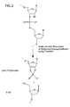
- RNase III class enzymes which cleave RNA:RNA duplexes (e.g., Dicer, Ago2, Drosha) result in similar products and contain a nuclease domain with similarity to RNasc H. Most other ribonucleascs, and in particular single stranded ribonucleases, result in a cyclic 2',3'-phosphate and 5'-OH products ( see Figure 2 ).
- E. coli RNase H1 has been extensively characterized. A large amount of work on this enzyme has been carried out, focusing on characterization of substrate requirements as it impacts antisense oligonucleotide design; this has included studies on both the E . coli RNase H1 (see Crooke, et al., (1995) Biochem J, 312 (Pt 2), 599-608 ; Lima, et al., (1997) J Biol Chem, 272, 27513-27516 ; Lima, ct al., (1997) Biochemistry, 36, 390-398 ; Lima, et al., (1997) J Biol Chem, 272, 18191-18199 ; Lima, et al., (2007) Mol Pharmacol, 71, 83-91 ; Lima, et al., (2007) Mol Pharmacol, 71, 73-82 ; Lima, et al., (2003) J Biol Chem, 278, 14906-14912 ; Lima, e
- Type I RNase H requires multiple RNA bases in the substrate for full activity.
- a DNA/RNA/DNA oligonucleotide hybridized to a DNA oligonucleotide with only 1 or 2 RNA bases is inactive.
- E . coli RNasc H1 substrates with three consecutive RNA bases show weak activity. Full activity was observed with a stretch of four RNA bases ( Hogrefe, et al., (1990) J Biol Chem, 265, 5561-5566 ).
- An RNase H1 was cloned from Thermus thermophilus in 1991 which has only 56% amino acid identity with the E . coli enzyme but which has similar catalytic properties ( Itaya, et al., (1991) Nucleic Acids Res, 19, 4443-4449 ). This enzyme was stable at 65°C but rapidly lost activity when heated to 80°C.
- the human RNase H1 gene (Type I RNase H) was cloned in 1998 ( Genomics, 53, 300-307 and Antisense Nucleic Acid Drug Dev, 8, 53-61 ). This enzyme requires a 5 base RNA stretch in DNA/RNA/DNA chimeras for cleavage to occur. Maximal activity was observed in 1 mM Mg ++ buffer at neutral pH and Mn -+ ions were inhibitory ( J Biol Chem, 274, 28270-28278 ). Cleavage was not observed when 2'-modified nucleosides (such as 2'-OMe, 2'-F, etc.) were substituted for RNA.
- 2'-modified nucleosides such as 2'-OMe, 2'-F, etc.
- Damha's studies are consistent with the known active site of the enzyme, wherein the reaction mechanism involves the 2'-OH group.
- the enzyme active site resides within a cluster of lysine residues which presumably contribute to electrostatic binding of the duplex. Interaction between the binding surface and negatively charged phosphate backbone is believed to occur along the minor grove of the RNA:DNA heteroduplex ( Nakamura, et al., (1991) Proc Natl Acad Sci U S A, 88, 11535-11539 ); changes in structure that affect the minor groove should therefore affect interactions between the substrate and the active site.
- the minor groove width is 7.5 ⁇ in a B-form DNA:DNA duplex, is 11 ⁇ in a pure A-form RNA:RNA duplex, and is 8.5 ⁇ in the hybrid A-form duplex of an RNA:DNA duplex ( Fedoroff et al., (1993) J Mol Biol, 233, 509-523 ).
- 2'-modifications protrude into the minor groove, which may account for some of the behavior of these groups in reducing or eliminating activity of modified substrates for cleavage by RNase H1.
- the human Type II RNase H was first purified and characterized by Eder and Walder in 1991 ( Eder, et al., (1991) J Biol Chem, 266, 6472-6479 ). This enzyme was initially designated human RNase H1 because it had the characteristic divalent metal ion dependence of what was then known as Class I RNases H. In the current nomenclature, it is a Type II RNase H enzyme. Unlike the Type I enzymes which are active in Mg ++ but inhibited by Mn ++ ions, the Type II enzymes are active with a wide variety of divalent cations. Optimal activity of human Type II RNase H is observed with 10 mM Mg ++ , 5 mM Co ++ , or 0.5 mM Mn ++ .
- RNase H2 the substrate specificity of the Type II RNase H (hereafter referred to as RNase H2) is different from RNase H1.
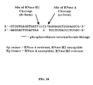
- this enzyme can cleave a single ribonucleotide embedded within a DNA sequence (in duplex form) ( Eder, et al., (1993) Biochimie, 75, 123-126 ). Interestingly, cleavage occurs on the 5' side of the RNA residue (See Figure 3 ). See a recent review by Kanaya for a summary of prokaryotic RNase H2 enzymes ( Kanaya (2001) Methods Enzymol, 341, 377-394 ).
- the E . coli RNase H2 gene has been cloned ( Itaya, M. (1990) Proc Natl Acad Sci U S A, 87, 8587-8591 ) and characterized ( Ohtani, et al., (2000) J Biochem (Tokyo), 127, 895-899 ). Like the human enzyme, the E . coli enzyme functions with Mn + ions and is actually more active with manganese than magnesium.
- RNase H2 genes have been cloned and the enzymes characterized from a variety of eukaryotic and prokaryotic sources.
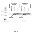
- the RNase H2 from Pyrococcus kodalraraensis (KOD1) has been cloned and studied in detail ( Haruki, et al., (1998) J Bacteriol, 180, 6207-6214 ; Mukaiyama, et al., (2004) Biochemistry, 43, 13859-13866 ).
- the RNase H2 from the related organism Pyrococcus furious has also been cloned but has not been as thoroughly characterized ( Sato, et al., (2003) Biochem Biophys Res Common, 309, 247-252 ).
- the RNase H2 from Methanococcus jannaschii was cloned and characterized by Lai ( Lai, et al., (2000) Structure, 8, 897-904 ; Lai et al., (2003) Biochemistry, 42, 785-791 ). Isothermal titration calorimetry was used to quantitatively measure metal ion binding to the enzyme. They tested binding of Mn ++ , Mg ++ Ca ++ , and Ba ++ and in all cases observed a 1:1 molar binding ratio, suggesting the presence of only a single divalent metal ion cofactor in the enzyme's active site. The association constant for Mn ++ was 10-fold higher than for Mg ++ . Peak enzyme activity was seen at 0.8 mM MnCl 2 .
- Nucleic acid hybridization assays based on cleavage of an RNA-containing probe by RNase H such as the cycling probe reaction ( Walder et al., U.S. Pat. No. 5,403,711 ) have been limited in the past by background cleavage of the oligonucleotide by contaminating single-stranded ribonucleases and by water catalyzed hydrolysis facilitated by Mg 2+ and other divalent cations.
- the effect of single-stranded ribonucleases can be mitigated to a certain degree by inhibitors such as RNasin that block single-stranded ribonucleases but do not interfere with the activity of RNase H.
- Single-stranded ribonucleases cleave 3' of an RNA residue, leaving a cyclic phosphate group at the 2' and 3' positions of the ribose (See FIG. 2 ).
- the same products are produced by spontaneous water catalyzed hydrolysis.
- the cyclic phosphate can hydrolyze further forming a 3'-monophosphate ester in the enzyme catalyzed reaction, or a mixture of the 3'- and 2'-monophosphate esters through spontaneous hydrolysis.
- the difference between the cleavage products formed by RNase H ( FIG. 1 ) and those formed by nonspecific cleavage of the probe ( FIG. 2 ) provides a basis for distinguishing between the two pathways.
- RNase H has been used as a cleaving enzyme in cycling probe assays, in PCR assays ( Han et al., U.S. Patent No. 5,763,181 ; Sagawa et al., U.S. Pat. No. 7,135,291 ; and Behlke and Walder, U.S. Pat. App. No. 20080068643 ) and in polynomial amplification reactions ( Behlke et al., U.S. Patent No. 7,112,406 ). Despite improvements offered by these assays, there remain considerable limitations. The PCR assays utilize a hot-start DNA polymerase which adds substantially to the cost.
- the current invention provides novel biological assays that employ RNase H cleavage in relation to nucleic acid amplification, detection, ligation, sequencing, and synthesis. Additionally, the invention provides new assay formats to utilize cleavage by RNase H and novel oligonucleotide substrates for such assays.
- the compounds, kits, and methods of the present invention provide a convenient and economic means of achieving highly specific primer-based amplification reactions that are substantially free of nonspecific amplification impurities such as primer dimers.
- the methods and kits of the present invention avoid the need for reversibly inactivated DNA polymerase and DNA ligase enzymes.
- One objective of the present invention is to enable hot start protocols in nucleic acid amplification and detection assays including but not limited to PCR, OLA (oligonucleotide ligation assays), LCR (ligation chain reaction), polynomial amplification and DNA sequencing, wherein the hot start component is a thermostable RNase H or other nicking enzyme that gains activity at the elevated temperatures employed in the reaction.
- Such assays employ a modified oligonucleotide of the invention that is unable to participate in the reaction until it hybridizes to a complementary nucleic acid sequence and is cleaved to generate a functional 5'- or 3'-end.
- the specificity is greatly enhanced.
- the requirement for reversibly inactivated DNA polymerases or DNA ligases is eliminated.
- the modification of the oligonucleotide inhibiting activity is preferably located at or near the 3'-end.
- the oligonucleotide inhibiting activity may be positioned near the 3' end of the oligonucleotide, e.g., up to about 10 bases from the 3' end of the oligonucleotide of the invention.
- the oligonucleotide inhibiting activity may be positioned near the 3' end, e.g., about 1-6 bases from the 3' end of the oligonucleotide of the invention. In other embodiments, the oligonucleotide inhibiting activity may be positioned near the 3' end, e.g., about 1-5 bases from the 3' end of the oligonucleotide of the invention. In other embodiments, the oligonucleotide inhibiting activity may be positioned near the 3' end, e.g., about 1-3 bases from the 3' end of the oligonucleotide of the invention.
- the precise position (i.e., number of bases) from the 3' end where the oligonucleotide inhibiting activity may be positioned will depend upon factors influencing the ability of the oligonucleotide primer of the invention to hybridize to a shortened complement of itself on the target sequence (i.e., the sequence for which hybridization is desired). Such factors include but are not limited to Tm, buffer composition, and annealing temperature employed in the reaction(s).
- the modification inhibiting activity may be located at or near either the 3'- or 5'-end of the oligonucleotide.
- modification inhibitory activity if used, is preferably placed within the domain that is 3' to the cleavable RNA base in the region that is removed by probe cleavage.
- C3 spacers may be positioned close to the RNA base in the oligonucleotide probes of the invention to improve specificity that is helpful for improving mismatch discrimination.
- any blocking group may be placed in the domain of the oligonucleotide of the invention that is removed by RNase H cleavage.
- the precise position of the blocking group in the RNasc H cleavable domain may be adjusted to alter specificity for cleavage and precise placement of the blocking group relative to the cleavable RNA bases may alter the amount of enzyme needed to achieve optimal cleavage rates.
- Yet a further objective of the present invention is to provide novel modifications of oligonucleotides to interfere with primer extension and ligation.
- Yet a further objective of the present invention is to provide modifications of oligonucleotides that prevent the oligonucleotide from serving as a template for DNA synthesis and thereby interfere with PCR.
- a further objective of the invention is to provide modified oligonucleotide sequences lacking RNA that are cleaved by RNase H.
- the oligonucleotide contains a single 2'-fluoro residue and cleavage is mediated by a Type II RNase H enzyme.
- the oligonucleotide contains two adjacent 2'-fluoro residues.
- Yet a further objective of the present invention is to provide oligonucleotides for use in the above mentioned assays that are modified so as to inhibit undesired cleavage reactions including but not limited to water and divalent metal ion catalyzed hydrolysis 3' to RNA residues, hydrolysis by single-stranded ribonucleases and atypical cleavage reactions catalyzed by Type II RNase H enzymes at positions other than the 5'-phosphate of an RNA residue (see Figure 3 ).
- the 2'-hydroxy group of an RNA residue is replaced with an alternative functional group such as fluorine or an alkoxy substituent (e.g., O-methyl).
- the phosphate group 3' to an RNA residue is replaced with a phosphorothioate or a dithioate linkage.
- the oligonucleotide is modified with nuclease resistant linkages further downstream from the 3'-phosphate group of an RNA residue or on the 5'-side of an RNA residue to prevent aberrant cleavage by RNase H2.
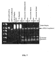
- Nuclease resistant linkages useful in such embodiments include phosphorothioates, dithioates, methylphosphonates, and abasic residues such as a C3 spacer. Incorporation of such nuclease resistant linkages into oligonucleotide primers used in PCR assays of the present invention has been found to be particularly beneficial (see Examples 25, 27 and 28).
- Yet a further objective of the invention is to provide oligonucleotides for use in the above-mentioned assays that are modified at positions flanking the cleavage site to provide enhanced discrimination of variant alleles.
- modifications include but are not limited to 2'-O-methyl RNA residues and secondary mismatch substitutions (see Example 23).
- a further objective is to provide oligonucleotides and assay formats for use in the present invention wherein cleavage of the oligonucleotide can be measured by a change in fluorescence.
- a primer cleavable by RNase H is labeled with a fluorophore and a quencher and the assay is monitored by an increase in fluorescence (see Examples 19-21).
- Yet a further objective of the invention is to provide RNase H compositions and protocols for their use in which the enzyme is thermostable and has reduced activity at lower temperatures.
- a Type II RNasc H is employed in a cycling probe reaction in which the RNA residue in the probe is replaced with a 2'-fluoro residue.
- a probe with two adjacent 2'-fluoro residues is used.
- Type II RNase H enzymes are used in novel methods for DNA sequencing.
- Type II RNase H enzymes are used in novel methods for DNA synthesis.
- the current invention provides novel nucleic acid compounds having a cleavage domain and a 3' or 5' blocking group. These compounds offer improvements to existing methods for nucleic acid amplification, detection, ligation, sequencing and synthesis. New assay formats comprising the use of these novel nucleic acid compounds are also provided.
- nucleic acid and oligonucleotide refer to polydeoxyribonucleotides (containing 2-deoxy-D-ribose), polyribonucleotides (containing D-ribose), and to any other type of polynucleotide which is an N glycoside of a purine or pyrimidine base.
- nucleic acid refers only to the primary structure of the molecule. Thus, these terms include double- and single-stranded DNA, as well as double- and single-stranded RNA.
- an oligonucleotide also can comprise nucleotide analogs in which the base, sugar or phosphate backbone is modified as well as non-purine or non-pyrimidine nucleotide analogs.
- Oligonucleotides can be prepared by any suitable method, including direct chemical synthesis by a method such as the phosphotriester method of Narang et al., 1979, Meth. Enzymol. 68:90-99 ; the phosphodiester method of Brown et al., 1979, Meth. Enzymol. 68:109-151 ; the diethylphosphoramidite method of Beaucage et al., 1981, Tetrahedron Lett. 22:1859-1862 ; and the solid support method of U.S. Pat. No. 4,458,066 .
- a review of synthesis methods of conjugates of oligonucleotides and modified nucleotides is provided in Goodchild, 1990, Bioconjugate Chemistry 1(3): 165-187 .
- primer refers to an oligonucleotide capable of acting as a point of initiation of DNA synthesis under suitable conditions. Such conditions include those in which synthesis of a primer extension product complementary to a nucleic acid strand is induced in the presence of four different nucleoside triphosphates and an agent for extension (e.g., a DNA polymerase or reverse transcriptase) in an appropriate buffer and at a suitable temperature. Primer extension can also be carried out in the absence of one or more of the nucleotide triphosphates in which case an extension product of limited length is produced.
- agent for extension e.g., a DNA polymerase or reverse transcriptase
- the term "primer” is intended to encompass the oligonucleotides used in ligation-mediated reactions, in which one oligonucleotide is "extended” by ligation to a second oligonucleotide which hybridizes at an adjacent position.
- primer extension refers to both the polymerization of individual nucleoside triphosphates using the primer as a point of initiation of DNA synthesis and to the ligation of two oligonucleotides to form an extended product.
- a primer is preferably a single-stranded DNA.
- the appropriate length of a primer depends on the intended use of the primer but typically ranges from 6 to 50 nucleotides, preferably from 15-35 nucleotides. Short primer molecules generally require cooler temperatures to form sufficiently stable hybrid complexes with the template.
- a primer need not reflect the exact sequence of the template nucleic acid, but must be sufficiently complementary to hybridize with the template. The design of suitable primers for the amplification of a given target sequence is well known in the art and described in the literature cited herein.
- Primers can incorporate additional features which allow for the detection or immobilization of the primer but do not alter the basic property of the primer, that of acting as a point of initiation of DNA synthesis.
- primers may contain an additional nucleic acid sequence at the 5' end which does not hybridize to the target nucleic acid, but which facilitates cloning or detection of the amplified product.
- the region of the primer which is sufficiently complementary to the template to hybridize is referred to herein as the hybridizing region.
- target and target nucleic acid
- hybridization refers to the formation of a duplex structure by two single-stranded nucleic acids due to complementary base pairing. Hybridization can occur between fully complementary nucleic acid strands or between "substantially complementary” nucleic acid strands that contain minor regions of mismatch. Conditions under which hybridization of fully complementary nucleic acid strands is strongly preferred are referred to as “stringent hybridization conditions” or “sequence-specific hybridization conditions”. Stable duplexes of substantially complementary sequences can be achieved under less stringent hybridization conditions; the degree of mismatch tolerated can be controlled by suitable adjustment of the hybridization conditions.
- nucleic acid technology can determine duplex stability empirically considering a number of variables including, for example, the length and base pair composition of the oligonucleotides, ionic strength, and incidence of mismatched base pairs, following the guidance provided by the art (see, e.g., Sambrook et al., 1989, Molecular Cloning-A Laboratory Manual, Cold Spring Harbor Laboratory, Cold Spring Harbor, New York ; Wetmur, 1991, Critical Review in Biochem. and Mol. Biol. 26(3/4):227-259 ; and Owczarzy et al., 2008, Biochemistry, 47: 5336-5353 .
- Amplification reaction refers to any chemical reaction, including an enzymatic reaction, which results in increased copies of a template nucleic acid sequence or results in transcription of a template nucleic acid.
- Amplification reactions include reverse transcription, the polymerase chain reaction (PCR), including Real Time PCR (see U.S. Pat. Nos. 4,683,195 and 4,683,202 ; PCR Protocols: A Guide to Methods and Applications (Innis et al., eds, 1990 )), and the ligase chain reaction (LCR) (see Barany et al., U.S. Pat. No. 5,494,810 ).
- Exemplary "amplification reactions conditions” or “amplification conditions” typically comprise either two or three step cycles. Two step cycles have a high temperature denaturation step followed by a hybridization/elongation (or ligation) step. Three step cycles comprise a denaturation step followed by a hybridization step followed by a separate elongation or ligation step.
- a "polymerase” refers to an enzyme that catalyzes the polymerization of nucleotides. Generally, the enzyme will initiate synthesis at the 3'-end of the primer annealed to a nucleic acid template sequence.
- DNA polymerase catalyzes the polymerization of deoxyribonucleotides.
- Known DNA polymerases include, for example, Pyrococcus furiosus (Pfu) DNA polymerase ( Lundberg et al., 1991, Gene, 108:1 ), E. coli DNA polymerase I ( Lecomte and Doubleday, 1983, Nucleic Acids Res.
- T7 DNA polymerase Nordstrom et al., 1981, J. Biol. Chcm. 256:3112
- Thermus thermophilus (Tth) DNA polymerase Myers and Gelfand 1991, Biochemistry 30:7661
- Bacillus stearothermophilus DNA polymerase Stenesh and McGowan, 1977, Biochim Biophys Acta 475:32
- Thermococcus litoralis (Tli) DNA polymerase (also referred to as Vent DNA polymerase, Cariello et al., 1991, Nucleic Acids Res, 19: 4193 ), Thermotoga maritima (Tma) DNA polymerase ( Diaz and Sabino, 1998 Braz J.
- a primer is "specific," for a target sequence if, when used in an amplification reaction under sufficiently stringent conditions, the primer hybridizes primarily to the target nucleic acid.
- a primer is specific for a target sequence if the primer-target duplex stability is greater than the stability of a duplex formed between the primer and any other sequence found in the sample.
- salt conditions such as salt conditions as well as base composition of the primer and the location of the mismatches, will affect the specificity of the primer, and that routine experimental confirmation of the primer specificity will be needed in many cases.
- Hybridization conditions can be chosen under which the primer can form stable duplexes only with a target sequence.
- target-specific primers under suitably stringent amplification conditions enables the selective amplification of those target sequences which contain the target primer binding sites.
- non-specific amplification refers to the amplification of nucleic acid sequences other than the target sequence which results from primers hybridizing to sequences other than the target sequence and then serving as a substrate for primer extension.
- the hybridization of a primer to a non-target sequence is referred to as “non-specific hybridization” and is apt to occur especially during the lower temperature, reduced stringency, pre-amplification conditions, or in situations where there is a variant allele in the sample having a very closely related sequence to the true target as in the case of a single nucleotide polymorphism (SNP).
- SNP single nucleotide polymorphism
- primer dimer refers to a template-independent non-specific amplification product, which is believed to result from primer extensions wherein another primer serves as a template. Although primer dimers frequently appear to be a concatamer of two primers, i.e., a dimer, concatamers of more than two primers also occur.
- primer dimer is used herein generically to encompass a template-independent non-specific amplification product.
- reaction mixture refers to a solution containing reagents necessary to carry out a given reaction.
- a “PCR reaction mixture” typically contains oligonucleotide primers, a DNA polymerase (most typically a thermostable DNA polymerase), dNTP's, and a divalent metal cation in a suitable buffer.
- reaction mixture is referred to as complete if it contains all reagents necessary to enable the reaction, and incomplete if it contains only a subset of the necessary reagents.
- reaction components are routinely stored as separate solutions, each containing a subset of the total components, for reasons of convenience, storage stability, or to allow for application-dependent adjustment of the component concentrations, and that reaction components are combined prior to the reaction to create a complete reaction mixture.
- reaction components are packaged separately for commercialization and that useful commercial kits may contain any subset of the reaction components which includes the blocked primers of the invention.
- non-activated refers to a primer or other oligonucleotide that is incapable of participating in a primer extension reaction or a ligation reaction because either DNA polymerase or DNA ligase cannot interact with the oligonucleotide for their intended purposes.
- the non-activated state occurs because the primer is blocked at or near the 3'-end so as to prevent primer extension.
- specific groups are bound at or near the 3'-end of the primer, DNA polymerase cannot bind to the primer and extension cannot occur.
- a non-activated primer is, however, capable of hybridizing to a substantially complementary nucleotide sequence.
- the term "activated,” as used herein, refers to a primer or other oligonucleotide that is capable of participating in a reaction with DNA polymerase or DNA ligase.
- a primer or other oligonucleotide becomes activated after it hybridizes to a substantially complementary nucleic acid sequence and is cleaved to generate a functional 3'- or 5'-end so that it can interact with a DNA polymerase or a DNA ligase.
- a 3'-blocking group can be removed from the primer by, for example, a cleaving enzyme such that DNA polymerase can bind to the 3' end of the primer and promote primer extension.
- cleavage domain or "cleaving domain,” as used herein, are synonymous and refer to a region located between the 5' and 3' end of a primer or other oligonucleotide that is recognized by a cleavage compound, for example a cleavage enzyme, that will cleave the primer or other oligonucleotide.
- a cleavage compound for example a cleavage enzyme
- the cleavage domain is designed such that the primer or other oligonucleotide is cleaved only when it is hybridized to a complementary nucleic acid sequence, but will not be cleaved when it is single-stranded.
- the cleavage domain or sequences flanking it may include a moiety that a) prevents or inhibits the extension or ligation of a primer or other oligonucleotide by a polymerase or a ligase, b) enhances discrimination to detect variant alleles, or c) suppresses undesired cleavage reactions.
- One or more such moieties may be included in the cleavage domain or the sequences flanking it.
- RNase H cleavage domain is a type of cleavage domain that contains one or more ribonucleic acid residue or an alternative analog which provides a substrate for an RNase H.
- An RNase H cleavage domain can be located anywhere within a primer or oligonucleotide, and is preferably located at or near the 3'-end or the 5'-end of the molecule.
- RNase H1 cleavage domain generally contains at least three residues.
- An “RNase H2 cleavage domain” may contain one RNA residue, a sequence of contiguously linked RNA residues or RNA residues separated by DNA residues or other chemical groups.
- the RNasc H2 cleavage domain is a 2'-fluoronucleoside residue.
- the RNase H2 cleavable domain is two adjacent 2'-fluoro residues.
- cleavage compound refers to any compound that can recognize a cleavage domain within a primer or other oligonucleotide, and selectively cleave the oligonucleotide based on the presence of the cleavage domain.
- the cleavage compounds utilized in the invention electively cleave the primer or other oligonucleotide comprising the cleavage domain only when it is hybridized to a substantially complementary nucleic acid sequence, but will not cleave the primer or other oligonucleotide when it is single stranded.
- the cleavage compound cleaves the primer or other oligonucleotide within or adjacent to the cleavage domain.
- adjacent means that the cleavage compound cleaves the primer or other oligonucleotide at either the 5'-end or the 3' end of the cleavage domain. Cleavage reactions preferred in the invention yield a 5'-phosphate group and a 3'-OH group.
- the cleavage compound is a "cleaving enzyme.”
- a cleaving enzyme is a protein or a ribozyme that is capable of recognizing the cleaving domain when a primer or other nucleotide is hybridized to a substantially complementary nucleic acid sequence, but that will not cleave the complementary nucleic acid sequence (i.e., it provides a single strand break in the duplex).
- the cleaving enzyme will also not cleave the primer or other oligonucleotide comprising the cleavage domain when it is single stranded.
- Examples of cleaving enzymes are RNase H enzymes and other nicking enzymes.
- nicking refers to the cleavage of only one strand of the double-stranded portion of a fully or partially double-stranded nucleic acid.
- the position where the nucleic acid is nicked is referred to as the "nicking site” (NS).
- NS nicking site
- a "nicking agent” (NA) is an agent that nicks a partially or fully double-stranded nucleic acid. It may be an enzyme or any other chemical compound or composition.
- a nicking agent may recognize a particular nucleotide sequence of a fully or partially double-stranded nucleic acid and cleave only one strand of the fully or partially double-stranded nucleic acid at a specific position (i.e., the NS) relative to the location of the recognition sequence.
- nicking agents include, but are not limited to, nicking endonucleases (e.g., N.BstNB).
- nicking endonuclease refers to an endonuclease that recognizes a nucleotide sequence of a completely or partially double-stranded nucleic acid molecule and cleaves only one strand of the nucleic acid molecule at a specific location relative to the recognition sequence. In such a case the entire sequence from the recognition site to the point of cleavage constitutes the "cleavage domain".
- blocking group refers to a chemical moiety that is bound to the primer or other oligonucleotide such that an amplification reaction does not occur. For example, primer extension and/or DNA ligation does not occur.
- the oligonucleotide is capable of participating in the assay for which it was designed (PCR, ligation, sequencing, etc).
- the "blocking group” can be any chemical moiety that inhibits recognition by a polymerase or DNA ligase.
- the blocking group may be incorporated into the cleavage domain but is generally located on either the 5'- or 3'-side of the cleavage domain.
- the blocking group can be comprised of more than one chemical moiety.
- the "blocking group” is typically removed after hybridization of the oligonucleotide to its target sequence.
- fluorescent generation probe refers either to a) an oligonucleotide having an attached fluorophore and quencher, and optionally a minor groove binder or to b) a DNA binding reagent such as SYBR ® Green dye.
- fluorescent label refers to compounds with a fluorescent emission maximum between about 350 and 900 nm.
- fluorophores can be used, including but not limited to: 5-FAM (also called 5-carboxyfluorescein; also called Spiro(isobenzofuran-1(3H), 9'-(9H)xanthene)-5-carboxylic acid,3',6'-dihydroxy-3-oxo-6-carboxyfluorescein); 5-Hexachloro-Fluorescein; ([4,7,2',4',5',7'-hexachloro-(3',6'-dipivaloyl-fluoresceinyl)-6-carboxylic acid]); 6-Hexachloro-Fluorescein; ([4,7,2',4',5',7'-hexachloro-(3',6'-dipivaloylfluoresceinyl)-5
- quencher refers to a molecule or part of a compound, which is capable of reducing the emission from a fluorescent donor when attached to or in proximity to the donor. Quenching may occur by any of several mechanisms including fluorescence resonance energy transfer, photo-induced electron transfer, paramagnetic enhancement of intersystem crossing, Dexter exchange coupling, and exciton coupling such as the formation of dark complexes. Fluorescence is "quenched” when the fluorescence emitted by the fluorophore is reduced as compared with the fluorescence in the absence of the quencher by at least 10%, for example, 15%, 20%, 30%, 40%, 50%, 60%, 70%, 80%, 90%, 95%, 98%, 99%, 99.9% or more.
- quenchers include but are not limited to DABCYL, Black Hole TM Quenchers (BHQ-1, BHQ-2, and BHQ-3), Iowa Black ® FQ and Iowa Black ® RQ. These are so-called dark quenchers. They have no native fluorescence, virtually eliminating background problems seen with other quenchers such as TAMRA which is intrinsically fluorescent.
- ligation refers to the covalent joining of two polynucleotide ends.
- ligation involves the covalent joining of a 3' end of a first polynucleotide (the acceptor) to a 5' end of a second polynucleotide (the donor). Ligation results in a phosphodiester bond being formed between the polynucleotide ends.
- ligation may be mediated by any enzyme, chemical, or process that results in a covalent joining of the polynucleotide ends.
- ligation is mediated by a ligase enzyme.
- ligase refers to an enzyme that is capable of covalently linking the 3' hydroxyl group of one polynucleotide to the 5' phosphate group of a second polynucleotide.
- ligases include E. coli DNA ligase, T4 DNA ligase, etc.
- the ligation reaction can be employed in DNA amplification methods such as the "ligase chain reaction” (LCR), also referred to as the “ligase amplification reaction” (LAR), see Barany, Proc. Natl. Acad. Sci., 88:189 (1991 ); and Wu and Wallace, Genomics 4:560 (1989 ).
- LCR ligase chain reaction
- LAR ligase amplification reaction
- oligonucleotides two adjacent oligonucleotides which uniquely hybridize to one strand of the target DNA, and a complementary set of adjacent oligonucleotides, that hybridize to the opposite strand are mixed and DNA ligase is added to the mixture.
- DNA ligase will covalently link each set of hybridized molecules.
- Novel oligonucleotides and compounds of the present invention are disclosed.
- the novel oligonucleotides of the present invention are primers for DNA replication, as for example in PCR, DNA sequencing and polynomial amplification, to name a few such applications.
- the primers have an inactive configuration wherein DNA replication (i.e., primer extension) is blocked, and an activated configuration wherein DNA replication proceeds.
- the inactive configuration of the primer is present when the primer is either single-stranded, or the primer is hybridized to the DNA sequence of interest and primer extension remains blocked by a chemical moiety that is linked at or near to the 3' end of the primer.
- the activated configuration of the primer is present when the primer is hybridized to a nucleic acid sequence of interest and subsequently acted upon by RNase H or other cleaving agent to remove the blocking group and allow for an enzyme (e.g., a DNA polymerase) to catalyze primer extension.
- RNase H or other cleaving agent to remove the blocking group and allow for an enzyme (e.g., a DNA polymerase) to catalyze primer extension.
- a number of blocking groups are known in the art that can be placed at or near the 3' end of the oligonucleotide (e.g., a primer) to prevent extension.
- a primer or other oligonucleotide may be modified at the 3'-terminal nucleotide to prevent or inhibit initiation of DNA synthesis by, for example, the addition of a 3' deoxyribonucleotide residue (e.g ., cordycepin), a 2',3'-dideoxyribonucleotide residue, non-nucleotide linkages or alkane-diol modifications ( U.S. Pat. No. 5,554,516 ).
- a 3' deoxyribonucleotide residue e.g ., cordycepin
- 2',3'-dideoxyribonucleotide residue e.g ., non-nucleotide linkages or alkane-diol modifications
- blocking groups include 3' hydroxyl substitutions (e.g., 3'-phosphate, 3'-triphosphate or 3'-phosphate diesters with alcohols such as 3- hydroxypropyl), a 2'3'-cyclic phosphate, 2' hydroxyl substitutions of a terminal RNA base (e.g ., phosphate or sterically bulky groups such as triisopropyl silyl (TIPS) or tert -butyl dimethyl silyl (TBDMS)).
- TIPS triisopropyl silyl
- TBDMS tert -butyl dimethyl silyl
- Blocking groups to inhibit primer extension can also be located upstream, that is 5', from the 3'-terminal residue.
- Sterically bulky substituents which interfere with binding by the polymerase can be incorporated onto the base, sugar or phosphate group of residues upstream from the 3'-terminus.
- substituents include bulky alkyl groups like t-butyl, triisopropyl and polyaromatic compounds including fluorophores and quenchers, and can be placed from one to about 10 residues from the 3'-terminus.
- abasic residues such as a C3 spacer may be incorporated in these locations to block primer extension. In one such embodiment two adjacent C3 spacers have been employed (see Examples 27 and 28).
- blocking moieties upstream of the 3'-terminal residue can serve two functions: 1) to inhibit primer extension, and 2) to block the primer from serving as a template for DNA synthesis when the extension product is copied by synthesis from the reverse primer. The latter is sufficient to block PCR even if primer extension can occur.
- C3 spacers placed upstream of the 3'-terminal residue can function in this manner (see Examples 26 and 27).
- a modification used as a blocking group may also be located within a region 3' to the priming sequence that is non-complementary to the target nucleic acid sequence.
- the oligonucleotide further comprises a cleavage domain located upstream of the blocking group used to inhibit primer extension.
- a cleavage domain located upstream of the blocking group used to inhibit primer extension.
- An RNase H cleavage domain is preferred.
- An RNase H2 cleavage domain comprising a single RNA residue or replacement of the RNA base with one or more alternative nucleosides is most preferred.
- RNase H2 can be used to cleave duplexes containing a single 2'-fluoro residue. Cleavage occurs on the 5' side of the 2'-fluoro residue.
- an RNase H2 cleavage domain comprising two adjacent 2'-fluoro residues is employed (see Example 6). The activity is enhanced when two consecutive 2'-fluoro modifications are present. In this embodiment cleavage occurs preferentially between the 2'-fluoro residues.
- oligonucleotides with 2'-fluoro groups are not cleaved by single-stranded ribonucleases and are resistant to water catalyzed cleavage and completely stable at high temperatures. Enhanced cleavage has also been found when a 2'-fluoro modified RNA residue is used with a 2' LNA modified RNA residue. 2'-fluoro-containing oligonucleotides have been found to be further advantageous in certain applications compared to RNA-containing oligonucleotides in offering greater discrimination with respect to mismatches between the oligonucleotide and the target sequence.
- the RNase H cleavage domain may include one or more of these modified residues alone or in combination with RNA bases. DNA bases and abasic residues such as a C3 spacer may also be included to provide greater performance.
- cleaving agent is an RNase H1 enzyme a continuouse sequence of at least three RNA residues is preferred. A continuous sequence of four RNA residues generally leads to maximal activity. If the cleaving agent is an RNase H2 enzyme a single RNA residue or 2 adjacent 2'-fluoro residues are preferred.
- One objective of incorporating modified residues within an RNase H cleavage domain is to suppress background cleavage of a primer or probe due to water catalyzed hydrolysis or cleavage by single stranded ribonucleases.
- Replacement of the 2'-hydroxyl group with a substituent that cannot attack the adjacent phosphate group of an RNA residue can accomplish this goal.
- Examples of this approach include the use of the 2'-substituted nucleosides listed above, such as 2'-fluoro and 2'-O-methyl nucleosides. This is particularly advantageous when cleavage is mediated by RNase H2 and there is a single RNA residue within the cleavage domain. As shown in Figure 3 , in this case cleavage by single stranded ribonucleases or water catalyzed hydrolysis occurs at a different position than cleavage by RNase H2.
- RNA residues modifications that can be used to suppress cleavage by single stranded ribonucleases and water catalyzed hydrolysis at RNA residues include substitution of the 5' oxygen atom of the adjacent residue (3'- to the RNA base) with an amino group, thiol group, or a methylene group (a phosphonate linkage).
- substitution of the 5' oxygen atom of the adjacent residue (3'- to the RNA base) with an amino group, thiol group, or a methylene group (a phosphonate linkage).
- one or both of the hydrogen atoms on the 5' carbon of the adjacent residue can be replaced with bulkier substituents such as methyl groups to inhibit background cleavage of a ribonucleotide residue.
- the phosphate group at the 3'-side of an RNA residue can be replaced with a phosphorothioate, phosphorodithioates or boronate linkage.
- a phosphorothioate the S stereoisomer is preferred. Combinations of these various modifications may also be employed.
- the cleavage domain may include the blocking group provided that cleavage occurs on the 5'-side of the blocking group and generates a free 3'-OH. Generally however the cleavage domain and the blocking group are separated by one to about 15 bases. After cleavage takes place the portion of the primer 3' from the cleavage site containing the blocking group dissociates from the template and a functional 3'-hydroxyl group is exposed, capable of being acted on by a polymerase enzyme. The optimal distance between the cleavage site and the blocking group will depend on the cleaving agent and the nature of the blocking group.
- a distance of 3 to about 8 bases between the cleavage site and the blocking group is preferred.
- the blocking group is sterically small, for example a phosphodiester at the 3' terminal nucleotide as in the following structure a cleavage site 5 bases from the 3'-end is generally optimal. If the blocking group is larger it is advantageous to position the cleavage site further from it.
- thermophilic RNase H2 enzyme is utilized to cleave the oligonucleotide.
- a thermophilic RNase H2 enzyme is used which is less active at room temperature than at elevated temperatures. This allows a hot-start type of reaction to be achieved in PCR and other primer extension assays using the blocked primers of the present invention without actually requiring a hot start, i.e., reversibly inactivated, DNA polymerase. Standard less expensive DNA polymerase polymerases such as Taq polymerase can be used instead of the much more expensive hot start versions of the enzyme. Moreover, for different applications alternative DNA polymerases may be preferred. Utilizing RNase H as the hot start component of the assay obviates the need to develop a new reversibly inactivated analog of each different DNA polymerase.
- Hot start properties of the enzyme may be intrinsic to the protein as in the case of Pyrococcus abysii RNase H2 (see Example 4).
- the enzyme may be reversibly inactivated by chemical modification using, for example, maleic acid anhydride analogs such as citroconic anhydride. These compounds react with amino groups of the protein and at high temperature are released restoring activity.
- antibodies against an RNase H which block the enzyme may be employed which are denatured at elevated temperatures.
- the oligonucleotide of the present invention has a cleavage domain that is recognized and cleaved by a sequence specific nicking agent, e.g ., a nicking enzyme.
- the nicking agent also can be designed to cleave an oligonucleotide (e.g., a primer) at a modified nucleic acid or grouping of modified nucleic acids.
- the oligonucleotide is designed to be recognized by a nicking agent upon hybridization with the target nucleic acid, and the nicking of the oligonucleotide/target duplex can be used to remove a blocking group and allow for oligonucleotide extension.
- the nicking site (NS) is preferably located at or near the 3'-end of the oligonucleotide, specifically, one to about 15 bases from the 3'-end of the oligonucleotide.
- Exemplary nicking agents include, without limitation, single strand nicking restriction endonucleases that recognize a specific sequence such as N.BstNBI; or repair enzymes such as Mut H, MutY (in combination with an AP endonuclease), or uracil-N-glycosylase (in combination with an AP Lyase and AP endonucleases); and the geneII protein of bacteriophage fl.
- the blocked primers of the present invention minimize non-specific reactions by requiring hybridization to the target followed by cleavage before primer extension. If a primer hybridizes incorrectly to a related sequence, cleavage of the primer is inhibited especially when there is a mismatch that lies at or near the cleavage site. This reduces the frequency of false priming at such locations and thereby increases the specificity of the reaction. It should be noted that with Pyrococcus abysii Type II RNase H and other RNase H enzymes used in the present invention some cleavage does occur even when there is a mismatch at the cleavage site.
- Reaction conditions particularly the concentration of RNase H and the time allowed for hybridization and extension in each cycle, can be optimized to maximize the difference in cleavage efficiencies between the primer hybridized to its true target and when there is a mismatch. This allows the methods of the present invention to be used very effectively to distinguish between variant alleles, including SNPs (see Examples 12-14, 22-25).
- primer-dimers a common side reaction occurring in PCR, can also be inhibited using the 3' blocked primers of the present invention. This allows for a greater degree of multiplexing in PCR (e.g., detecting multiple target sequences in the case of a DNA detection/amplification assay).
- nuclease resistant residues can be incorporated into the primer 3' to the RNA residue (see Example 22, 25 and 28).
- Such groups include but are not limited to one or more phosphorothioates, phosphorodithioates, methyl phosphonates and abasic residues such as a C3 spacer.
- substitutions both 5' and 3' to the RNA residue can also be utilized to enhance the discrimination and detection of variant alleles in the methods of the present invention.
- substitutions include but are not limited to 2'-O-methyl RNA and secondary mismatches (see Example 23).
- the nature of the blocking group which prevents primer extension is not critical. It can be placed at the 3'-terminal residue or upstream from it. Labeling groups can be incorporated within the blocking group or attached at other positions on the 3'-segment of the oligonucleotide primer which dissociates from the template after cleavage occurs. Such labeling groups include, but are not limited to, fluorophores, quenchers, biotin, haptens such as digoxigenin, proteins including enzymes and antibodies, mass tags which alter the mass of the cleavage fragment for detection by mass spectrometry, and radiolabels such as 14 C, 3 H, 35 S, 32 P and 33 P. These labeling groups can also be attached to the primer 5' to the cleavage site, in which case they will be incorporated within the extension product.
- the blocking group at or near the 3'-end of the oligonucleotide can be a fluorescent moiety.
- release of the fluorescent molecule can be used to monitor the progress of the primer extension reaction. This is facilitated if the oligonucleotide also contains a quencher moiety on the 5'-side of the cleavage site. Cleavage of the oligonucleotide during the reaction separates the fluorophore from the quencher and leads to an increase in fluorescence. If the quencher is itself a fluorophore, such as Tamra, a decrease in its fluorescence may also be observed.
- the oligonucleotide is labeled with a fluorescent molecule on the 5'-side of the cleavage domain, and the blocking group located at or near the 3'-end of the molecule is a quencher such as Iowa Black ® , Black Hole TM , or Tamra to name a few.
- a quencher such as Iowa Black ® , Black Hole TM , or Tamra to name a few.
- cleavage of the quencher from the oligonucleotide e.g. , a primer
- the primer extension product is fluorescently labeled.
- the blocked primers of the present invention are used for nucleic acid sequencing.
- the specificity of primer extension for DNA sequencing is also increased when using the oligonucleotides of the present invention.
- 2',3' dideoxynucleotide triphosphates that are fluorescently labeled and used as chain terminators and the nested fragments produced in the reaction are separated by electrophoresis, preferably capillary electrophoresis.
- an oligonucleotide primer of the present invention is labeled with a fluorescent group and the 3' dideoxynucleotide triphosphate chain terminators are unlabeled.
- the blocking group can be a quencher, in which case background fluorescence is reduced because the primer itself is not fluorescent. Only the extension products arc fluorescent.
- Another aspect of the invention includes the incorporation of alternative divalent cations such as Mn 2+ , Ni 2+ or Co 2+ , with or without Mg 2+ , into the assay buffer.
- alternative divalent cations such as Mn 2+ , Ni 2+ or Co 2+ , with or without Mg 2+
- the effectiveness of the particular assay is increased due to enhanced cleavage by RNase H2.
- 0.3-1 mM MnCl 2 with 2-4 mM MgCl 2 gave optimal performance in the assay ( see Example 3).
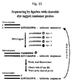
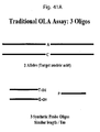
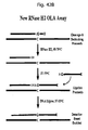
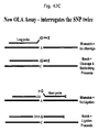
- primers, probes and other novel oligonucleotides described herein can be utilized in a number of biological assays. Although the following list is not comprehensive, the majority of the methods of the present invention fall into six general categories: (1) primer extension assays (including PCR, DNA sequencing and polynomial amplification), (2) oligonucleotide ligation assays (OLA), (3) cycling probe reactions, (4) sequencing by ligation, (5) sequencing by generation of end-labeled fragments using RNase H enzymes, and (6) synthesis by ligation.
- primer extension assays including PCR, DNA sequencing and polynomial amplification
- OOA oligonucleotide ligation assays
- cycling probe reactions (4) sequencing by ligation, (5) sequencing by generation of end-labeled fragments using RNase H enzymes, and (6) synthesis by ligation.
- primers, probes and other novel oligonucleotides described herein can be utilized in a number of primer extension assays.
- a method of amplifying a target DNA sequence of interest comprises the steps of:
- a 3'-blocked primer containing a cleavage domain When used in PCR, a 3'-blocked primer containing a cleavage domain first hybridizes to the target sequence. In this embodiment, the primer cannot extend until cleavage of the 3' blocking group occurs after hybridization to the complementary DNA sequence.
- an RNase H cleavage domain when an RNase H cleavage domain is present in the primer, an RNase H enzyme will recognize the double-stranded substrate formed by the primer and target and cleave the primer within or adjacent to the cleavage domain. The primer can then extend and amplification of the target can then occur. Because the primer needs to be recognized and cleaved by RNase H before extension, non-specific amplification is reduced.
- a "hot start" polymerase is often used to reduce primer dimers and decrease non-spccific amplification.
- Blocked primers of the present invention requiring cleavage by RNase H can confer the same advantage.
- a thermophilic RNase H enzyme with little or no activity at lower temperatures is preferred. Activation of the primers occurs only after hybridization to the target sequence and cleavage at elevated temperatures. Advantages of this approach compared to the use of a hot start reversibly inactivated DNA polymerase have been described above. Of course a hot start RNase H enzyme and a hot start DNA polymerase can be used in conjunction, if desired.
- thermostable RNase H enzymes Three types of hot start RNase H enzymes are described here (see Tables 1, 2, and 3): 1) a thermostable RNase H enzyme that has intrinsically little or no activity at reduced temperatures as in the case of Pyrococcus abysii RNase H2; 2) a thermostable RNase H reversibly inactivated by chemical modification; and 3) a thermostable RNase H reversibly inactivated by a blocking antibody.
- mutant versions of RNase H can be synthesized that can further improve the traits of RNase H that are desirable in the assays of the present invention.
- mutant strains of other enzymes that share the characteristics desirable for the present invention could be used.
- the cleavage domain within the primer is cleavable by RNase H.
- the RNase H cleavage domain consists of a single RNA residue and cleavage of the primer is mediated by a Type II RNase H enzyme, preferably by a thermophilic Type II RNase H enzyme, and even more preferably a thermophilic Type II RNase H enzyme which is less active at room temperature than at elevated temperatures.
- the RNase H2 cleavage domain consists of two adjacent 2'-fluoro nucleoside residues.
- the PCR is carried out in buffers containing alternative divalent cations, including but not limited to, Mn 2+ , Ni 2+ or Co 2+ in addition to Mg 2+ .
- alternative divalent cations including but not limited to, Mn 2+ , Ni 2+ or Co 2+ in addition to Mg 2+ .
- the novel 3'-blocked primers of the present invention comprising a cleavage domain can be utilized in a variation of hot start PCR in which a thermophilic nicking enzyme is used and the cleavage domain is a nicking site.
- cleavage enzyme that lacks hot start characteristics can be used in the present invention with traditional hot-start methods such as adding the enzyme at an elevated temperature, encasing a necessary reagent or enzyme in wax, or with a hot start reversibly inactivated DNA polymerase.
- the increased specificity of the present invention when used in amplification reactions, enables real-time PCR applications to achieve more specific results, as compared to conventional real-time PCR with standard DNA primers.
- double-stranded DNA-binding dye assays such as SYBR ® Green assays
- SYBR ® Green assays have a disadvantage in that a signal is produced once the dye binds to any double-stranded product produced by PCR (e.g., a primer dimer) and can thereby give rise to a false positive result.
- a primer of the current invention is used, non-specific amplification and primer-dimer formation is reduced, and the intensity of the signal of the double-stranded DNA-binding dye will reflect amplification only of the desired target (see Example 17).
- the reagent concentrations and reaction conditions of the assay can be varied to maximize its utility.
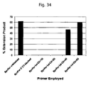
- the relative efficiency of PCR using the blocked primers of the present invention relates to the concentration of the unblocking enzyme and the dwell time at the anneal/extend reaction temperature (where unblocking proceeds). With low amounts of enzyme and short dwell times, cleavage can be incomplete and the reactions with blocked primers have lower efficiency than those with unblocked primers. As either enzyme concentration or dwell time increases, the reaction efficiency with blocked primers increases and becomes identical to unblocked primers. The use of even more enzyme or longer dwell times can decrease the specificity of the assay and lessen the ability of the system to discriminate mismatches at the cleavage site or within the surrounding sequence (see Example 4).
- the assay can be tuned for SNP assays requiring higher specificity, or for quantitation of expression levels of mRNA requiring less specificity.
- a primer pair having one blocked primer and one unblocked primer can be used.
- an enzyme can be selected that has less sequence specificity and can cleave various sequences.
- an additional mismatch flanking the cleavage site can be added to increase the ability to discriminate variant alleles.
- Modified bases such as 2'-O-methyl nucleosides can also be introduced into the primer on either side of the cleavage site to increase specificity (see Example 23).
- the reactions of the various assays described herein can be monitored using fluorescent detection, detection by mass tags, enzymatic detection, and via labeling the probe or primer with a variety of other groups including biotin, haptens, radionucleotides and antibodies to name a few.
- the progress of PCR using the modified primers of the present invention is monitored in real time using a dye intercelating assay with, for example, SYBR ® Green.
- the progress of PCR using the modified primers of the present invention is monitored using a probe labeled with a fluorophore and a quencher such as a molecular beacon or, as in the 5'nuclease assay where cleavage of the probe occurs.
- a dual labeled probe which is cleavable by RNase H2 may be employed.
- cleavage of both the hybridized primers and the probe can be mediated by the same enzyme.
- the RNase H cleavage domain within the probe may comprise only RNA residues.
- all of the combinations of residues useful in the cleavage domain of the blocked primers of the present invention can be used as the cleavage domain within the probe.
- RNasc H2 is used as the cleavage enzyme, a single RNA residue or two adjacent 2'-F residues are preferred as the cleavage domain within the probe.
- thermophilic versions of RNase H2 are preferred , especially thermophilic RNase H2 enzymes having lower activity at reduced temperatures.
- thermophilic RNase H2 enzymes have been isolated and have shown to be stable under thermocycling conditions and useful in PCR.
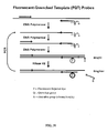
- the blocked primers of the present invention can be used in the primer-probe assay format for PCR described in U.S. Patent App. 2009/0068643 .
- the primer also contains a label domain on the 5' end of the oligonucleotide which may or may not be complementary to the target nucleic acid.
- the product generated by extension of the primer serves as a template for synthesis by the reverse primer in the next cycle of PCR. This converts the label domain into a double stranded structure.
- a fluorophore and a quencher are attached to the label domain and the reaction is monitored by an increase in fluorescence resulting from an increase in the distance between the fluorophore and quencher in the double stranded form compared to the single stranded state.
- the label domain contains a cleavage domain located between the fluorophore and quencher. Cleavage occurs only when the cleavage domain is double stranded.
- the reaction is monitored by an increase in fluorescence.
- the cleaving agent may be one that cleaves both strands, the primer and its complement, such as a restriction enzyme.
- the cleaving agent may be a nicking agent that cleaves only the primer, preferably an RNase H enzyme, and even more preferably a thermostable RNase H2 enzyme.
- RNase H enzyme a thermostable RNase H2 enzyme.
- the label domain may also contain other labeling groups including but not limited to biotin, haptens and enzymes to name a few. Alternatively the 5' fragment released by cleavage within the label domain may serve as a mass tag for detection by mass spectrometry.
- the blocked primers of the present invention can be used in the template-probe assay format for PCR described in U.S. Patent App. 2009/0068643 .
- RNase H2 cleavable blocked oligonucleotides are used to detect 5-methylcytosine residues by PCR analysis of sodium bisulfite treated nucleic acids, including but not limited to DNA and RNA.
- PCR analysis of sodium bisulfite treated nucleic acids including but not limited to DNA and RNA.
- 5-methylcytosine is highly resistant to this deamination, resulting in preservation of the 5-methylcytosine nucleotide as a cytosine, rather than conversion to a thymine.
- Numerous methods have been employed to detect 5' cytosine methylation modifications following the bisulfite conversion technique. Examples include, but are not limited to, standard mismatch-specific quantitative and non-quantitative PCR methods, as well as subcloning and sequencing of the generated sodium bisulfite reaction products.
- the template is bisulfite treated by methods that are well known to those in the art. If the starting template was RNA, a complementary cDNA strand is generated by any well known reverse transcription method. Blocked cleavable oligonucleotides that will either match or discriminate against the target template cytosines (now converted to uracils) or 5-methylcytosines are added to a PCR reaction containing the RNase H2 enzyme and the bisulfite treated template. Amplification of the mismatched (converted cytosine>uracil or unconverted 5-methylcytosine>5-methylcytosine) base containing template is highly reduced relative to the matched base template due to the mismatch discrimination of RNase H2 cleavage reaction.
- the blocked primers of the present invention can also be used in allele-specific PCR (AS-PCR).
- AS-PCR is used to detect variant alleles of a gene, especially single base mutations such as SNPs (see for example U.S. Patent No. 5,496,699 ). SNP locations in the genome, as well as sequences of mutated oncogenes, arc known in the art and PCR primers can be designed to overlap with these regions.
- Detection of single base mismatches is a critical tool in diagnosing and correlating certain diseases to a particular gene sequence or mutation.
- AS-PCR has been known in the biological arts for more than a decade ( Bottema et al., 1993, Methods Enzymol., 218, pp. 388-402 ), tools are still needed to more accurately discriminate between particular mismatches and fully complementary sequences. The present invention addresses this need.
- a primer is utilized which overlaps the variant locus.
- the primer is designed such that the 3'-terminal nucleotide is positioned over the mutation site.
- the mutation site is sometimes located over one or two bases from the 3'-end. If there is a mismatch at or near the 3'-end, primer extension and hence PCR are inhibited.
- the difference between the efficiency of amplification when there is an exact match with the primers versus an allelic variant where there is one or more mismatches can in some cases be measured by end point PCR in which case the final amplification products are analyzed by, for example, gel electrophoresis. More commonly real time PCR is used to determine the efficiency of amplification.
- a fluorescence based method of detection of the amplicon in real time such as a DNA dye binding assay or a dual labeled probe assay is most often used.
- the PCR cycle where fluorescence is first detectable above background levels (the Cp, or crossing point) provides a measure of amplification efficiency. If there is a mismatch between the primer and the target DNA, amplification efficiency is reduced and the Cp is delayed. Generally an increase in Cp of 4 to 5 cycles is sufficient for discrimination of SNPs.
- the primer contains a single RNA residue, and the mismatch can be aligned directly over the RNA residue of the primer.
- the difference in crossing point (Cp) values between a perfect match and a mismatch, correlating to a cleavage differential, is readily apparent (see Example 13).
- aligning the mismatch one base to either the 5' side or the 3' side of the RNA residue increases the difference in Cp values.
- the subsequent RNase H2 cleavage would leave the mismatch as the last base of the 3' end of the cleaved primer.
- having the mismatch directly on top of the RNA residue is more effective in most cases than locating the mismatch to the 5' side of the RNA residue.
- the primer contains multiple RNA residues or two adjacent 2'-fluoro residues and detection of the mismatch follows the same principles as with a primer containing one RNA residue; the mismatch preferably is located near or on top of the expected point of cleavage.
- a second mismatch is used to increase the sensitivity of the assay.
- the second mismatch is placed to the 3' side of the mismatch directly over the SNP site.
- the second mismatch is placed one or two bases from the mismatch directly over the SNP site (see Example 23).
- modified residues are incorporated into the primer on the 5'- or 3'-side of the base located over the mutation site.
- a 2'-O-methyl ribonucleoside is placed immediately 5' to the RNA base within the primer (see Example 22).
- the sensitivity of the assay can also be increased through incorporation of nuclease resistant analogs into the primer on the 3'-side of the base over the mutation site.
- nuclease resistant analogs include, but are not limited to, phosphorothioates, phosphorodithioates, methylphosphonates and abasic residues such as a C3 spacer.
- phosphorothioate intemucleotide linkages are incorporated at each position from the RNA base over the mutation site to the 3'-end of the primer.
- phosphorothioate linkages or phosphoroditioatc arc incorporated at all positions from the base on the 3'-side of the RNA residue to the 3'-end of the primer.
- a single phosphorothioate or phosphorodithioates is introduced on the 3'-side of the residue immediately downstream from the RNA base within the primer.
- the phosphorothioate bonds are placed between each monomer 3' to the RNA monomer directly over the SNP site, as well as between the RNA monomer and the base 3' to the RNA base (see Example 25).
- the assay sensitivity can also be improved by optimizing the placement of the 3' blocking group or groups.
- a blocking group is placed internal to the 3' end of the oligonucleotide.
- more than on blocking group is placed internal to the 3' terminus.
- an RNA monomer sits directly over the SNP site, with a DNA monomer 3' to the RNA monomer, followed by two C3 spacers, and finally followed by a 3' terminal base (see Example 28).
- the primers can be designed to detect more than one mismatch.
- the forward primer can detect a first mismatch
- the reverse primer could detect a second mismatch.
- the assay can be used to indicate whether two mismatches occur on the same gene or chromosome being analyzed. This assay would be useful in applications such as determining whether a bacterium of interest is both pathogenic and antibiotic resistant.
- RT-PCR Reverse transcriptase PCR
- the methods of the present invention can be used in coupled reverse transcription-PCR (RT-PCR).
- reverse transcription and PCR are carried out in two disctinct steps. First a cDNA copy of the sample mRNA is synthesized using either an oligo dT primer or a sequence specific primer. Random hexamers and the like can also be used to prime cDNA synthesis. The resulting cDNA is then used as the substrate for PCR employing the blocked primers and methods of the present invention.
- reverse transcription and PCR can be carried out in a single closed tube reaction.
- three primers are employed, one for reverse transcription and two for PCR.
- the primer for reverse transcription binds to the mRNA 3' to the position of the PCR amplicon.
- the reverse transcription primer can include RNA residues or modified analogs such as 2'-O-methyl RNA bases which will not form a substrate for RNase H when hybridized to the mRNA.
- an RNase H2 enzyme which has decreased activity at lower temperatures is used as the cleaving agent.
- RT-primer In the three primer RT-PCR assay it is desirable to inhibit the RT-primer from participating in the PCR reaction. This can be accomplished by utilizing an RT-primer having a lower Tm than the PCR primers so it will not hybridize under the PCR conditions.
- a non-replicable primer incorporating, for example, two adjacent C3 spacers can be used as the RT-primer (as in polynomial amplification, see U.S. Pat. No. 7,112,406 ). In this case when the cDNA is copied by extension of the forward PCR primer it will not include the binding site for the RT-primer.
- only the reverse PCR primer is blocked utilizing the compositions and methods of the present invention.
- both the forward and reverse PCR primers arc blocked.
- the reverse PCR primer is blocked in the 3 primer RT-PCR assay to prevent it from being utilized for reverse transcription.
- modified bases such as 2'-O-methyl RNA residues can be incorporated in the reverse PCR primer although any such modification must allow the primer sequence to serve as a template for DNA synthesis and be copied.
- the reverse PCR primer also serves as the RT-primer and therefore can not be blocked.
- Table 1 illustrates how variations in the blocking groups, labeling groups, cleavage site embodiments, modifications to the cleavage site or other regions of the oligonucleotide, buffer conditions and enzyme can further optimize assay formats depending on their particular application.
- assay formats and applications include PCR; real-time PCR utilizing double-stranded DNA-binding dyes such as SYBR ® Green, 5' nuclease assays (Taqman TM assays) or molecular beacons; primer-probe and template-probe assays (see U.S.
- Patent Application 2009/0068643 polynomial or linked linear amplification assays; gene construction or fragment assembly via PCR; allele-specific PCR and other methods used to detect single nucleotide polymorphisms and other variant alleles; nucleic acid sequencing assays; and strand displacement amplification.
- cleavage of the primers of the present invention can be used to enhance the specificity of the particular reaction.
- Table 1 PCR/Primer Extension/Polyamp Primer Blocking Group Labeling Group RNase H Cleavage Site Flanking sequence modifications Divalent cation DNA Polymerase RNase H Samples Use Assay Format None None RNA None Mg 2+ Hot Start RNase H1 Genomic DNA Sample Prep No additional probe 1.
- RNA residues Single RNA residue 1.
- Ab Detection of primer cleavage 2.Chemically modified Fluorophore 2. Multiple RNA residues Nuclease-resistant linkages RNase H2 Coupled amplification to reverse transcription A. Fluorescence 1. Non-thermostable B. Mass Spec 1. Phosphorothioate C. Electrophoresis 2. Thermostable 2. Dye-binding assay 2. Dithioate A. Hot Start A. Sybr Green Fluorophore/ Quencher 3. Methyl-phosphonate i. Intrinsic ii. Ab 4. Non-nucleotide spacers iii. Chemically modified B. Non-Hot Start Enzyme 1.
- RNA DNA
- RNase H2-cleavable probe Adjacent to the 3'-terminal residue Mass Tag Radiolabel 32 P, 14 C, 3 H, 35 S, etc. 1. Enhanced cleavage of 2'-F substrates 2. Further upstream
- Cycling probe reactions are another technique for detecting specific nucleic acid sequences (see U.S. Pat. No. 5,403,711 ).
- the reaction operates under isothermal conditions or with temperature cycling. Unlike PCR products accumulate in a linear fashion.
- Table 2 illustrates a non-comprehensive set of possible elements of the current invention to improve assays based on the cycling probe reaction.
- New features of the invention include 1) use of a hot start RNase H enzyme; 2) cleavage of novel sequences by RNase H enzymes (e.g., cleavage of substrates containing 2'-fluoronucleosides by Type II RNases H); and 3) introduction of modifications and secondary mismatches flanking an RNase H cleavage domain to enhance specificity and/or suppress nonspecific cleavage reactions. Such modifications and secondary mismatches are particularly useful when cleavage is mediated by a Type II RNase H and the cleavage domain is a single RNA residue or two adjacent 2'-fluoro residues.
- Biotin 2'OMe RNase H mutants having altered cleavage specificity cDNA Hapten 1.
- Deoxigenin 1. Enhanced cleavage of 2'-F substrates Antibody Secondary mismatches Mass Tag Radiolabel 32 P, 14 C, 3 H, 35 S, etc.
- the present invention can also serve to increase the specificity of DNA ligation assays.
- Donor and/or acceptor oligonucleotides of the present invention can be designed which bind adjacent to one another on a target DNA sequence and are modified to prevent ligation.
- Blocking groups on the acceptor oligonucleotide useful to inhibit ligation are the same as those used to prevent primer extension. Blocking the donor oligonucleotide can be readily accomplished by capping the 5'-OH group, for example as a phosphodiester, e.g ..:
- 5' blocking groups include 5'-O-alkyl substituents such as 5'-O-methyl or 5'-O-trityl groups, 5'-O-heteroalkyl groups such as 5'-OCH 2 CH 2 OCH 3 , 5'-O-aryl groups, and 5'-O-silyl groups such as TIPS or TBDMS.
- a 5' deoxy residue can also be used to block ligation.
- Sterically bulky groups can also be placed at or near the 5'-end of the oligonucleotide to block the ligation reaction.
- a 5'-phosphate group cannot be used to block the 5'-OH as this is the natural substrate for DNA ligase.
- the blocking groups Only after hybridization to the target DNA sequence are the blocking groups removed by, for example cleavage at an RNase H cleavable domain, to allow ligation to occur.
- cleavage is mediated by an RNase H Type II enzyme, and even more preferably a thermophilic Type II RNase H enzyme.
- thermophilic Type II RNasc H enzyme which is less active at room temperature than at elevated temperature is utilized to mediate cleavage and thereby activation of the acceptor and/or donor oligonucleotide.
- a sequence specific nicking enzyme such as a restriction enzyme, may be utilized to mediate cleavage of the donor and/or acceptor oligonucleotide.
- the cleaving reaction is first carried out at a higher temperature at which only one of the two oligonucleotides hybridizes to the target sequence. The temperature is then lowered, and the second oligonucleotide hybridizes to the target, and the ligation reaction then takes place.
- this oligonucleotide is not blocked at or near the 5'-end, but simply has a free 5'-OH.
- This oligonucleotide cannot serve as a donor in the ligation reaction; to do so requires a 5'-phosphate group.
- the 5'-end is functionally blocked. Cleavage by RNase H generates a 5'-phosphate group allowing the donor oligonucleotide to participate in the ligation reaction.
- An important advantage of the present invention is that it allows double interrogation of the mutation site, and hence greater specificity, than standard ligation assays. There is an opportunity for discrimination of a variant allele both at the cleavage step and the ligation step.
- Table 3 illustrates a non-comprehensive set of possible elements of the current invention to improve oligonucleotide ligation assays.
- Table: 3 Oligonucleotide Ligation Assay Donor Oligonucleotide Blocking Group Acceptor Oligonucleotide Blocking Group Labeling Group RNase H Cleavage Site Flanking sequence mods Divalent cation DNA Ligase RNase H Sample Use Reaction Conditions Assay Fo None (5'-phosphate) None None RNA None Mg 2+ Hot Start RNase H1 Genomic DNA Quantification of target nucleic acid sequence RNase H cleavage and DNA ligation at single temperature Stand-alo 1. Single RNA residue 1. Ab 1. Single cycle 2. Chem-ically modified 2.
- Non-Hot Start Downstream modification Upstream modification Enzyme Modified residues: Alternativ e bivalent cation +/-Mg 2+ Non-Hot Start RNase H3 and other catalysts that cleave RNA/DNA heteroduplexes Detection of variant allele RNase H cleavage at elevated temperature (reduced temperature for DNA ligation) Coupled to primer extension 1. Horse-radish peroxidase 1. Adjacent to the 5'-terminal residue 1. Adjacent to the 3'-terminal residue 1. 2 adjacent 2' F residues 1. PCR 2. Reverse transcription 2. Alkaline phosphatase 2. Further downstream 2. Further upstream 3. Polyamp Biotin 2'OMe RNase H mutants having altered cleavage specificity cDNA Hapten 1. Deoxigenin 1. Enhanced cleavage of 2'-F substrates Antibody Secondary mismatches Mass Tag Radiolabel 32 P, 14 C, 3 H, 35 S, etc.
- a method of sequencing a target DNA of interest is provided.
- the method entails
- next generation sequencing is “sequencing by synthesis", wherein genomic DNA is sheared and ligated with adapter oligonucleotides or amplified by gene-specific primers, which then are hybridized to complementary oligonucleotides that arc either coated onto a glass slide or arc placed in emulsion for PCR.
- the subsequent sequencing reaction either incorporates dye-labeled nucleotide triphosphates or is detected by chemiluminescence resulting from the reaction of pyrophosphate released in the extension reaction with ATP sulfurylase to generate ATP and then the ATP-catalyzed reaction of luciferase and its substrate luciferin to generate oxyluciferin and light.
- a second type of next generation sequencing is "sequencing by ligation", wherein four sets of oligonucleotides are used, representing each of the four bases. In each set, a fluorophore-labeled oligonucleotide of around 7 to 11 bases is employed in which one base is specified and the remaining are either universal or degenerate bases.
- an 8-base oligonucleotide containing 3 universal bases such as inosine and 4 degenerate positions
- a specified base A, T, C or G
- fluorescent label attached to either the 5'- or 3'-end of the molecule or at an internal position that does not interfere with ligation.
- Four different labels are employed, each specific to one of the four bases.
- a mixture of these four sets of oligonucleotides is allowed to hybridize to the amplified sample DNA.
- DNA ligase the oligonucleotide hybridized to the target becomes ligated to an acceptor DNA molecule. Detection of the attached label allows the determination of the corresponding base in the sample DNA at the position
- a donor oligonucleotide of about 7-11 bases contains a specified base at the 5' end of the oligonucleotide.
- the remaining bases are degenerate or universal bases, and a label specific to the specified base is incorporated on the 3' side of the specified base.
- the 3' end of the probe is irreversibly blocked to prevent the donor oligonucleotide from also acting as an acceptor. In some cases this may be accomplished by the labeling group.
- the second base from the 5' end of the oligonucleotide, i.e ., the residue next to the specified base is a degenerate mixture of the 4 RNA bases.
- any anaolog recognized by RNase H2 such as a 2'-fluoronucleoside may be substituted at this position.
- a universal base such as riboinosine or ribo-5-nitroindole, may also be incorporated at this location.
- the probe first hybridizes to the target sequence and becomes ligated to the acceptor DNA fragment as in the standard sequencing by ligation reaction. After detection of the specified base, RNase H2 is added which cleaves the probe on the 5'-side of the RNA residue leaving the specified base attached to the 3' end of the acceptor fragment. The end result is that the acceptor fragment is elongated by one base and now is in position to permit the determination of the next base within the sequence.
- the cycle is repeated over and over, in each case moving the position of hybridization of the donor oligonucleotide one base 3' down the target sequence.
- the specificity is increased compared to traditional sequencing by ligation because the specified base is always positioned at the junction of the ligation reaction.
- the donor oligonucleotide probe can optionally contain universal bases including, but not limited to, 5-nitroindole, ribo-5'-nitroindole, 2'-O-methyl-5-nitroindole, inosine, riboinosine, 2'-O-methylriboinosine and 3-nitropyrrole. This reduces the number of different oligonucleotides in each set required for the assay by a factor of four for every degenerate position on the probe substituted with a universal base.
- the method can also include a capping step between the ligation reaction and the RNase H2 cleaving step. The capping reaction can be performed by introducing a DNA polymerase and a chain terminator, thereby capping any of the acceptor fragment molecules that did not ligate with a donor oligonucleotide probe in the previous step.
- the ligation reactions and hence the sequencing readout proceeds in the 5'- to 3'-direction one base at a time.
- the donor oligonucleotide can be designed so that two bases are determined in each cycle.
- oligonucleotide As in all cases there is a 5'-phosphate (p) to permit ligation of the donor oligonucleotide to the acceptor.
- p 5'-phosphate
- Sixteen such oligonucleotide sets are required, one for each of the sixteen possible dinucleotides.
- Each of the sixteen can be labeled with a different fluorophore.
- ligation reactions can be carried out with 4 separate pools each having four such sets of oligonucleotides. In that case, only four different fluorophores are required.
- a donor oligonucleotide of the following type can be used: pA-N-R-N-N-N-I-I-X wherein p, N, R, I and X are as defined in the previous example.
- One base is determined at each cycle but at alternate positions: 1, 3, 5, etc. This may be adequate for identification of the sequence if compared to a reference database.
- the remaining bases can be determined by repeating the sequencing reaction on the same template with the original acceptor oligonucleotide shifted one base upstream or downstream.
- a donor oligonucleotide of the following type can be used: p-A-F-FN-N-N-1-I-X wherein p, N, I and X are as defined above and F is a degenerate mixture of all four 2'-fluoronucleosides.
- RNasc H2 cleavage by RNasc H2 results in the addition of two bases to the 3'-end of the acceptor (i.e., AF).
- the sequence at the 3'-end of the acceptor would be ...A-F-S-F-F-N-N-N-I-I-X where S is the specified base at position 3, and X would be a different fluorophore from the previous cycle if the specified base were not A.
- Cleavage with RNase H2 next occurs between the two 2'-fluororesidues.
- Cleavage by RNasc H2 at the isolated 2'-fluororcsiduc occurs much more slowly and can be avoided by adjusting the RNase H2 concentration and reaction time.
- a variant of the above method can be performed in which sequencing proceeds in the 3'- to 5'-direction.
- an acceptor oligonucleotide is added at each cycle as in the following structure: X-I-F-N-N-N-F-F-S-OH wherein the specified base (S) is at the 3'-end of the oligonucleotide.
- the 5'-end is blocked to prevent the oligonucleotide from acting as a donor.
- Cleavage by RNase H2 leaves the sequence pF-S at the 5'-end of the donor fragment which is prepared for the next sequencing cycle.
- a capping step can be included in the cycle before the cleavage reaction using a phosphatase to remove the 5'-phosphate of the donor oligonucleotide if ligation to the acceptor failed to occur.
- the invention provides an improvement for DNA sequencing using ribotriphosphates (or alternative analogs which provide a substrate for RNase H2, such as 2'-fluoronucleoside triphosphates) in conjunction with a fluorescently labeled primer.
- ribotriphosphates or alternative analogs which provide a substrate for RNase H2, such as 2'-fluoronucleoside triphosphates
- the triphosphate residue would be incorporated by a DNA polymerase.
- the concentration of the ribo triphosphate, or the alternative analog providing a substrate for RNase H2 is adjusted to a concentration such that on average one such base is incorporated randomly within each extension product produced by the polymerase.
- the nested family of fragments originating from the primer is generated by cleavage with RNase H2 and then separated by electrophoresis as in standard DNA sequencing methods.
- RNA residues or modified nucleosides such as 2'-fluoronucleosides may be incorporated into the extension product and the subsequent digestion with RNase H2 is limited so that on average each strand is cut only once.
- RNase H2 a different ribotriphosphate (A, C, T or G) or other RNase H2 cleavable analog.
- use of expensive fluorescently labeled dideoxy triphosphate chain terminators is obviated.
- an improved method for oligonucleotide synthesis is provided.
- a composition acting as a donor ol igonucleotide can be ligated to an acceptor fragment in order to add additional bases to the 3'-end of the acceptor fragment. It is the acceptor fragment that is the growing polynucleotide undergoing synthesis.
- the composition of the donor fragment is preferably a single-stranded oligonucleotide that forms a hairpin to provide a double-stranded region with an overhang of about 1-8 bases on the 3'-end. The base at the 5' end would be the desired base to add to the growing acceptor fragment.
- a polynucleotide containing all four bases A, C, T and G
- four different donor fragments are employed which can have the identical sequence except varying in the 5' base.
- the donor is blocked at the 3'-end so it cannot react as an acceptor.
- the blocking group placed at or near the 3'-end of the donor can be a label to allow monitoring of the reaction.
- Four different labels can be used corresponding to the four different bases at the 5'-end of the donor.
- the base adjacent to the desired base at the 5'-end is a RNA base or an alternative analog such as a 2'-fluoronucleoside which provides a substrate for RNase H2.
- the overhang at the 3'-end can be random (degenerate) bases or universal bases or a combination of both.
- the donor fragment binds to the acceptor fragment, through hybridization of the 3'-end of the acceptor to the 3'-overhang of the donor oligonucleotide.
- a DNA ligase enzyme is then used to join the two fragments.
- a Type II RNase H is used to cleave the product on the 5'-side of the RNase H2 cleavage site, transferring the 5' base of the donor to the 3'-end of the acceptor.
- a third step can be included in the cycle between the ligase and RNase H2 cleavage reactions in which molecules of the growing polynucleotide chain which may have failed to ligate are capped by reaction with a dideoxynucleotide triphosphate (or other chain terminator) catalyzed by a DNA polymerase.
- the DNA polymerase is a deoxynucleotide terminal transferase.
- the cycle is repeated, and the acceptor fragment can continue to be extended in a 5' to 3' direction.
- the acceptor can be attached to a solid support such as controlled pore glass or polystyrene
- a donor oligonucleotide can be used to add two bases to the 3'-end of the acceptor oligonucleotide at each cycle.
- the RNase H2 cleavable residue would be positioned 3' from the 5' end of the donor.
- This enzymatic synthesis method is particularly advantageous for synthesis of longer DNA molecules.
- the hairpin reagents corresponding to each base can be collected for reuse in further cycles or additional syntheses. Because the system does not use organic solvents, waste disposal is simplified.
- kits for nucleic acid amplification, detection, sequencing, ligation or synthesis that allow for use of the primers and other novel oligonucleotides of the present invention in the aforementioned methods.
- the kits include a container containing a cleavage compound, for example a nicking enzyme or an RNase H enzyme; another container containing a DNA polymerase and/or a DNA ligase and preferably there is an instruction booklet for using the kits.
- the kits include a container containing both a nicking enzyme or an RNase H enzyme combined with a DNA polymerase or DNA ligase.
- the modified oligonucleotides used in the assay can be included with the enzymes.
- kits may further comprise a buffer for the nicking agent or RNase H, a buffer for the DNA polymerase or DNA ligase, or both buffers.
- the kits may further comprise a buffer suitable for both the nicking agent or RNase H, and the DNA polymerase or DNA ligase.
- Buffers may include RNasin and other inhibitors of single stranded ribonucleases. Descriptions of various components of the present kits may be found in preceding sections related to various methods of the present invention.
- the kit may contain an instruction booklet providing information on how to use the kit of the present invention for amplifying or ligating nucleic acids in the presence of the novel primers and/or other novel oligonucleotides of the invention.
- the information includes one or more descriptions on how to use and/or store the RNase H, nicking agent, DNA polymerase, DNA ligase and oligonucleotides used in the assay as well as descriptions of buffer(s) for the nicking agent or RNase H and the DNA polymerase or DNA ligase, appropriate reaction temperature(s) and reaction time period(s), etc.
- kits for the selective amplification of a nucleic acid from a sample comprises
- the kit for selective amplification of a nucleic acid includes an oligonucleotide probe having a 3' end and a 5' end comprising an RNase H cleavable domain, a fluorophore and a quencher, wherein the cleavable domain is positioned between the fluorophore and the quencher, and wherein the probe is complementary to a portion of the nucleic acid to be amplified or its complement.
- the present invention is directed to a kit for the ligation of an acceptor oligonucleotide and a donor oligonucleotide in the presence of a target nucleic acid sequence.
- the kit comprises
- the kit may optionally include a DNA ligase enzyme.
- the donor oligonucleotide contains an RNase H cleavage domain, but lacks a blocking group at or near the 5'-end and instead has a free 5'-OH.
- This example describes the cloning of codon optimized RNase H2 enzymes from thermophilic organisms.
- candidate genes were identified from public nucleotide sequence repositories from Archaeal hyperthermophilic organisms whose genome sequences had previously been determined. While RNase H2 enzymes do share some amino acid homology and have several highly conserved residues present, the actual homology between the identified candidate genes was low and it was uncertain if these represented functional RNase H2 enzymes or were genes of unknown function or were non-functional RNase H2 genes. As shown in Table 4, five genes were selected for study, including two organisms for which the RNase H2 genes have not been characterized and three organisms to use as positive controls where the RNase H2 genes ( rnhb ) and functional proteins have been identified and are known to be functional enzymes.
- the amino acid similarity between RNase H2 enzymes (or candidate enzymes) from different Archaeal species within this set of 5 sequences ranges from 34% to 65%.
- An amino-acid identity matrix is shown in Table 6 below. Table 6.
- Amino acid identity between five Archaeal RNase H2 proteins P. kod. P. fur. M. jann. P. ab. S. solf. P. kodakarensis - 0.570 0.595 0.358 0.333 P. furiosus 0.570 - 0.654 0.410 0.362 M. jannaschii 0.595 0.654 - 0.380 0.363 P. abysii 0.358 0.410 0.380 - 0.336 S. solfataricus 0.333 0.362 0.363 0.336 -
- Codons of the native gene sequence were optimized for expression in E . coli using standard codon usage tables. The following sequences were assembled and cloned into plasmids as artificial genes made from synthetic oligonucleotides using standard methods. DNA sequence identity was verified on both strands. Sequences of the artificial DNA constructs are shown below. Lower case letters represents linker sequences, including a Bam HI site on the 5'-end and a Hind III site on the 3'-end. Upper case letters represents coding sequences and the ATG start codons are underlined.
- the five synthetic gene sequences from Example 1 were subcloned using unique Bam HI and Hind III restriction sites into the bacterial expression vector pET-27b(+) (Novagen, EMD Biosciences, La Jolla, CA). This vector places six histidine residues (which together comprise a "His-tag") at the carboxy terminus of the expressed peptide (followed by a stop codon).
- His-tag permits use of rapid, simple purification of recombinant proteins using Ni affinity chromatography, methods which are well known to those with skill in the art.
- the synthetic genes could be expressed in native form without the His-tag and purified using size exclusion chromatography, anion-exchange chromatography, or other such methods, which are also well known to a person of ordinary skill in the art.
- IPTG isopropyl- ⁇ -D-thio-galactoside
- 5 mL of IPTG induced culture was treated with Bugbuster ® Protein Extraction Reagent and Benzonase ® Nuclease (Novagen) to release soluble proteins and degrade nucleic acids according to the manufacturer's instructions.
- the recovered protein was passed over a Ni affinity column (Novagen) and eluted with buffer containing 1M imidazole according to protocols provided by the manufacturer.
- a 10 fold dilution in 500 mM NaCl, 20 mM TrisHCl, 5 mM imidazole, pH 7.9 is made and 10 mL is used per 0.5 g of pelleted bacterial paste from induced cultures.
- 5 mL of Bugbuster ® Protein Extraction Reagent (Novagen) per 100 mL of induced culture is used for cell lysis.
- 5KU rLysozyme TM Novagen
- 250U DNase I Roche Diagnostics, Indianapolis, IN
- the lysates are heated for 15 minutes at 75°C to inactive the DNase I and any other cellular nucleases present.
- the lysates are then spun at 16,000 x g for 20 minutes to sediment denatured protein following heat treatment.
- the centrifugation step alone provides a large degree of functional purification of the recombinant thermostable enzymes.
- the resulting soluble supernatant is passed over a Ni affinity column containing His ⁇ Bind ® Resin (Novagen) and eluted with an elution buffer containing 200 mM imidazole.
- the purified protein is then precipitated in the presence of 70% ammonium sulfate and resuspended in storage buffer (10mM Tris pH 8.0, 1mM EDTA, 100mM NaCl, 0.1% Triton X-100, 50% Glycerol) to concentrate and stabilize the protein for long term storage.
- the concentrated protein is dialyzed 2 x 2 hours (x250 volumes each) against the same storage buffer to remove residual salts.
- the final purified protein is stored at -20°C.
- Recombinant protein was made and purified for each of the cloned RNase H2 enzymes as outlined above. Samples from Pyrococcus kodakaraensis, Pyrococcus furiosus, Pyrococcus abyssi, and Sulfolobus solfataricus were examined using SDS 10% polyacrylamide gel electrophoresis. Proteins were visualized with Coomassie Blue staining. Results are shown in Figure 5 . If the expression and purification method functioned as predicted, these proteins should all contain a 6x Histidine tag, which can be detected using an anti-His antibody by Western blot.
- RNase H enzymes cleave RNA residues in an RNA/DNA heteroduplex. All RNase H enzymes can cleave substrates of this kind when at least 4 sequential RNA residues are present. RNase H1 enzymes rapidly lose activity as the RNA "window" of a chimeric RNA/DNA species is shortened to less than 4 residues. RNase H2 enzymes, on the other hand, are capable of cleaving an RNA/DNA heteroduplex containing only a single RNA residue. In all cases, the cleavage products contain a 3'-hydroxyl and a 5'-phosphate (see FIG. 1 ). When multiple RNA residues are present, cleavage occurs between RNA bases, cleaving an RNA-phosphate linkage.
- RNase H enzymes require the presence of a divalent metal ion cofactor.
- RNase H1 enzymes require the presence of Mg ++ ions while RNase H2 enzymes can function with any of a number of divalent cations, including but not limited to Mg ++ , Mn ++ , Co ++ and Ni ++ .
- RNA bases Cleavage of a substrate with multiple RNA bases.
- the following synthetic 30 bp substrate was used to test the activity of these enzymes for cleavage of a long RNA domain.
- the substrate is an "11-8-11" design, having 11 DNA bases, 8 RNA bases, and 11 DNA bases on one strand and a perfect match DNA complement as the other strand.
- the oligonucleotides employed are indicated below, where upper case letters represent DNA bases and lower case letters represent RNA bases.
- these single-stranded (ss) oligonucleotides form the following "11-8-11" double-stranded (ds) substrate:
- RNA/DNA heteroduplex 11-8-11
- the recombinant proteins did not degrade the single stranded RNA-containing oligonucleotide (SEQ ID No. 6), indicating that a double-stranded substrate was required. Further, a dsDNA substrate was not cleaved.
- Cleavage was not observed in the absence of a divalent cation (e.g., no activity was observed if Mg ++ was absent from the reaction buffer).
- a Mg ++ titration was performed and high enzyme activity was observed between 2-8 mM MgCl 2 .
- Optimal activity was observed between 3-6 mM MgCl 2 .
- Cleavage activity was also detected using other divalent cations including Mn ++ and Co ++ .
- MnCl 2 good activity was seen from 0.3 mM to 10 mM divalent cation concentration.
- Enzyme activity was optimal in the range of 300 nM to 1 mM.
- Substrate cleavage by RNasc H enzymes is expected to result in products with a 3'-OH and 5'-phosphate.
- the identity of the reaction products from the new recombinant RNase H2 proteins was examined by mass spectrometry. Electrospray ionization mass spectrometry (ESI-MS) has near single Dalton resolution for nucleic acid fragments of this size (accuracy of +/- 0.02%).
- the oligonucleotide 11-8-11 substrate was examined by ESI-MS both before and after digestion with the three Pyrococcus sp. RNase H enzymes. The primary masses observed are reported in Table 7 along with identification of nucleic acid species consistent with the observed masses. Table 7.
- DNA bases are indicated with upper case letters
- RNA bases are indicated with lower case letters
- phosphate "P”.
- Molecular weights are rounded to the nearest Dalton. In the absence of other notation, the nucleic acids strands end in a 5'-hydroxyl or 3'-hydroxyl.
- RNA complement strand was observed intact (non-degraded).
- the RNA-containing strands were efficiently cleaved and the observed masses of the reaction products are consistent with the following species being the primary fragments produced: 1) a species which contained undigested DNA residues and a single 3'-RNA residue with a 3'-hydroxyl groups (SEQ ID No. 9), and 2) a species with a 5'-phosphate, a single 5'-RNA residue, and undigested DNA residues (SEQ ID No. 10).
- the observed reaction products are consistent with the known cleavage properties of both RNase H1 and RNase H2 enzymes.
- RNase H2 enzymes characteristically cleave a substrate that contains a single RNA residue while RNase H1 enzymes cannot.
- the following synthetic 30 bp substrates were used to test the activity of these enzymes for cleavage at a single RNA residue.
- the substrates are a "14-1-15" design, having 14 DNA bases, 1 RNA base, and 15 DNA bases on one strand and a perfect match DNA complement as the other strand.
- Four different substrates were made from 8 component single-stranded oligonucleotides comprising each of the 4 RNA bases: C, G, A, and U.
- the oligonucleotides employed are indicated below, where upper case letters represent DNA bases and lower case letters represent RNA bases.
- SEQ ID NO: 12 5'-CGCCTCCCATCTCCTGCATCACCTCACGAG-3'
- these single-stranded (ss) oligonucleotides form the following "14-1-15 rC" double-stranded (ds) substrate:
- these single-stranded (ss) oligonucleotides form the following "14-1-15 rG" double-stranded (ds) substrate:
- these single-stranded (ss) oligonucleotides form the following "14-1-15 rA" double-stranded (ds) substrate:
- these single-stranded (ss) oligonucleotides form the following "14-1-15 rU" double-stranded (ds) substrate:
- RNA/DNA heteroduplex 14-1-15
- All 5 recombinant peptides showed the ability to cleave a single RNA base in an RNA/DNA heteroduplex (14-1-15).
- Each of the 4 RNA bases functioned as a cleavable substrate with these enzymes.
- the recombinant proteins did not degrade the single stranded RNA-containing oligonucleotides (SEQ ID Nos. 11, 14, 17, 20), indicating that a double-stranded substrate was required.
- the isolated enzymes therefore show RNase H2 activity. Titration of divalent cations was tested and results were identical to those obtained previously using the 8-11-8 substrate.
- Substrate cleavage by RNase H enzymes is expected to result in products with a 3'-OH and 5'-phosphate. Further, cleavage of a substrate containing a single ribonucleotide by RNase H2 enzymes characteristically occurs at the DNA linkage 5'-to the RNA residue.
- the identity of the reaction products using a single ribonucleotide substrate from the new recombinant RNase H2 proteins was examined by mass spectrometry.
- the oligonucleotide 14-1-15 rC substrate (SEQ ID No. 13) was examined by ESI-MS both before and after digestion with the three Pyrococcus sp. RNase H2 enzymes and the Methanocaldococcus jannaschii enzyme.
- DNA bases are indicated with upper case letters
- RNA bases are indicated with lower case letters
- phosphate "P”.
- Molecular weights are rounded to the nearest Dalton. In the absence of other notation, the nucleic acids strands end in a 5'-hydroxyl or 3'-hydroxyl.
- RNA complement strand was observed intact (non-degraded).
- the RNA-containing strands were efficiently cleaved and the observed masses of the reaction products are consistent with the following species being the primary fragments produced: 1) a species which contained undigested DNA residues with a 3'-hydroxyl (SEQ ID No. 23), and 2) a species with a 5'-phosphate, a single 5'-RNA residue, and undigested DNA residues (SEQ ID No. 24).
- the observed reaction products are consistent with the known cleavage properties of RNase H2 class enzymes.
- Activity is also present using Mn ++ or Co ++ ions; 2) Single-stranded nucleic acids are not degraded; 3) Double-stranded heteroduplex nucleic acids are substrates where one strand contains one or more RNA bases; 4) For substrates containing 2 or more consecutive RNA bases, cleavage occurs in a DNA-RNA-DNA chimera between RNA linkages; for substrates containing a single RNA base, cleavage occurs immediately 5'-to the RNA base in a DNA-RNA-DNA at a DNA linkage; and 6) Reaction products have a 3-hydroxyl and 5'-phosphate.
- Substrate SEQ ID No. 13 was employed for characterizing RNase H2 enzyme preparation for the purpose of normalizing unit concentration. The following standardized buffer was employed unless otherwise noted. "Mg Cleavage Buffer”: 4 mM MgCl 2 , 10 mM Tris pH 8.0, 50 mM NaCl, 10 ⁇ g/ml BSA (bovine serum albumin), and 300 nM oligo-dT (20mer poly-dT oligonucleotide). The BSA and oligo-dT serve to saturate non-specific binding sites on plastic tubes and improve the quantitative nature of assays performed.
- Mg Cleavage Buffer 4 mM MgCl 2 , 10 mM Tris pH 8.0, 50 mM NaCl, 10 ⁇ g/ml BSA (bovine serum albumin), and 300 nM oligo-dT (20mer poly-dT oligonucleotide).
- BSA and oligo-dT serve
- RNA strand of the substrate was radiolabeled with 32 P using 6000 Ci/mmol ⁇ - 32 P-ATP and the enzyme T4 Polynucleotide Kinase (Optikinase, US Biochemical). Trace label was added to reaction mixtures (1:50). Reactions were performed using 100 nM substrate with 100 microunits ( ⁇ U) of enzyme in Mg Cleavage Buffer. Reactions were incubated at 70°C for 20 minutes.
- Reaction products were separated using denaturing 7M urea, 15% polyacrylamide gel electrophoresis (PAGE) and visualized using a Packard Cyclone TM Storage Phosphor System (phosphorimager). The relative intensity of each band was quantified using the manufacturer's image analysis software and results plotted as a fraction of total substrate cleaved. Results are shown in Figure 9 .
- the enzyme retained full activity for over 30 minutes at 95°C. Activity was reduced to 50% after 45 minutes incubation and to 10% after 85 minutes incubation.
- the temperature dependence of the activity of the Pyrococcus abyssi RNase H2 enzyme was next characterized. The activity was studied over a 40°C temperature range from 30°C to 70°C.
- the RNA strand of the rC substrate SEQ ID No. 13 was radiolabeled as described above. Reactions were performed using 100 nM substrate with 200 microunits ( ⁇ U) of enzyme in Mg Cleavage Buffer. Reactions were incubated at 30°C, 40°C, 50°C, 60°C, or 70°C for 10 minutes. Reactions were stopped with the addition of cold EDTA containing formamide gel loading buffer.
- the enzyme is functionally inactive at room temperature. Reactions employing this enzyme can therefore be set up on ice or even at room temperature and the reactions will not proceed until temperature is elevated. If Pyrococcus abyssi RNase H2 cleavage were linked to a PCR reaction, the temperature dependent activity demonstrated herein would effectively function to provide for a "hot start" reaction format in the absence of a modified DNA polymerase.
- the natural biological substrates for RNase H1 and RNase H2 are duplex DNA sequences containing one or more RNA residues. Modified bases containing substitutions at the 2'-position other than hydroxyl (RNA) have not been observed to be substrates for these enzymes.
- RNA hydroxyl
- LNA locked nucleic acid
- 2'OMe 2' -O-methyl
- 2'F 2'-fluoro
- the single ribo-C containing substrate was employed as positive control.
- LNA bases will be designated with a "+” prefix (+N)
- 2'OMe bases will be designated with a "m” prefix (mN)
- 2'F bases will be designated with a "f" prefix (fN)
- the above 4 substrates were incubated in an 80 ⁇ l reaction volume in various buffers for 20 minutes at 70°C with the recombinant Pyrococcus abyssi RNase H2 enzyme.
- Buffers tested included 50 mM NaCl, 10 mM Tris pH 8.0 with cither 10 mM MgCl 2 , 10 mM CoCl 2 , or 10 mM MnCl 2 .
- Reactions were stopped with the addition of gel loading buffer (formamide/EDTA) and separated on a denaturing 7M urea, 15% polyacrylamide gel. Gels were stained using GelStar TM (Lonza, Rockland, ME) and visualized with UV excitation. Results are shown in Figure 12 .
- the control substrate with a single ribo-C residue was 100% cleaved.
- the substrates containing a single LNA-C or a single 2'OMe-C residue were not cleaved.
- the substrate containing a single 2'-F-C residue was cleaved to a small extent. This cleavage occurred only in the manganese containing buffer and was not seen in either cobalt or magnesium buffers.
- the above 4 substrates were incubated in an 80 ⁇ l reaction volume in a buffer containing 50 mM NaCl, 10 mM Tris pH 8.0 and 10 mM MnCl 2 for 20 minutes at 70°C with either the recombinant Pyrococcus abyssi RNase H2 enzyme or the recombinant Pyrococcus furiosus RNase H2 enzyme. Reactions were stopped with the addition of gel loading buffer (formamide/EDTA) and separated on a denaturing 7M urea, 15% polyacrylamide gel. Gels were stained using GelStar TM (Lonza, Rockland, ME) and visualized with UV excitation. Results are shown in Figure 13 .
- the control substrate with a single ribo-C residue was 100% cleaved.
- the substrates containing a single 2'-F-C or single 2'-F-U residue were cleaved to a small extent.
- the di-fluoro substrate containing adjacent 2'-F-C and 2'-F-U residues (fCfU) was cleaved nearly 100%.
- both the Pyrococcus abyssi and Pyrococcus furiosus RNase H2 enzymes cleaved the modified substrate in an identical fashion. This example demonstrates that the unexpected cleavage of the fC group was not restricted to fC but also occurred with fU.
- the identity of the cleavage products of the di-fluoro fCfU substrate was studied using mass spectrometry using the methods described in Example 3. Using traditional ribonucleotide substrates, cleavage by RNase H enzymes results in products with a 3'-OH and 5'-phosphate.
- the fCfU substrate (SEQ ID No. 29) was examined by ESI-MS both before and after digestion by the recombinant Pyrococcus abyssi RNase H2 enzyme. The primary masses observed are reported in Table 9 along with identification of nucleic acid species consistent with the observed masses. Table 9. Mass of species observed after RNase H2 digestion of SEQ ID No.
- the mass spectrometry data indicates that digestion of a di-fluoro substrate such as the fCfU duplex studied above by RNase H2 results in cleavage between the two fluoro bases. Further, the reaction products contain a 3'-hydroxyl and 5'-phosphate, similar to the products resulting from digestion of RNA containing substrates.
- Cleavage of the modified bases was not observed in the absence of a divalent cation.
- a titration was performed and enzyme activity was observed between 0.25-10 mM MnCl 2 and 0.25-1.5 mM CoCl 2 .
- Enzyme activity was optimal in the range of 0.5 mM to 1 mM for both MnCl 2 and CoCl 2 .
- 0.6 mM MnCl 2 was employed in reactions or 0.5 mM CoCl 2 .
- Reduced activity for cleavage of the modified substrate was observed using Mg buffers. Overall, optimum activity was observed using Mn buffers for cleavage of the di-fluoro (fNfN) substrates whereas Mg buffers were superior for cleavage of ribonucleotide (rN) substrates.
- the ability of the RNase H2 enzymes to cleave at single or double 2'-F bases was unexpected.
- the Pyrococcus abyssi RNase H2 enzyme was next tested for the ability to cleave a greater variety of modified substrates using the same methods described above in this example.
- the modified strand of the substrate was radiolabeled as described above. Reactions were performed using 100 nM substrate and 480-1000 mU of recombinant enzyme in Mn Cleavage Buffer (10 mM Tris pH 8.0, 50 mM NaCl, 0.6 mM MnCl 2 , 10 ⁇ g/ml BSA). Reactions were incubated at 70°C for 20 minutes.
- Pyrococcus abyssi RNase H2 can be used to cleave substrates which do not contain any RNA bases but instead contain 2'-modified bases.
- di-fluoro (fNfN) containing substrates performed best.
- Use of the modified substrates generally requires increased amounts of enzyme, however the enzyme is catalytically very potent and it presents no difficulty to employ sufficient enzyme to achieve 100% cleavage of a di-fluoro substrate.
- the 2'-modified substrate described in this example are not susceptible to cleavage by typical RNase enzymes. As such they can be employed in novel assay formats where cleavage events are mediated by RNase H2 using substrates that are completely resistant to cleavage by other RNase enzymes, particularly single stranded ribonucleases.
- Table 12 fNfN Sequence SEQ ID No. AA SEQ ID No. 46 AC SEQ ID No. 47 AG SEQ ID No. 48 AU SEQ ID No. 49 CA SEQ ID No. 50 CC SEQ ID No. 51 CG SEQ ID No. 52 CU SEQ ID No. 29 GA SEQ ID No. 53 GC SEQ ID No. 54 GG SEQ ID No. 55 GU SEQ ID No. 56 UA SEQ ID No. 57 UC SEQ ID No. 58 UG SEQ ID No. 59 UU SEQ ID No. 60
- the modified strand of each substrate was radiolabeled as described above. Reactions were performed using 100 nM substrate with 25 mU of recombinant enzyme in Mn Cleavage Buffer (10 mM Tris pH 8.0, 50 mM NaCl, 0.6 mM MnCl 2 , 10 ⁇ g/ml BSA). Reactions were incubated at 70°C for 20 minutes. Reaction products were separated using denaturing 7M urea, 15% polyacrylamide gel electrophoresis (PAGE) and visualized using a Packard Cyclone TM Storage Phosphor System (phosphorimager). The relative intensity of each band was quantified, and results plotted as a fraction of total substrate cleaved are shown in Figure 14 . The enzyme amount was titrated so that the most active substrate cleaved at 90-95% without having excess enzyme present so that accurate assessment could be made of relative cleavage efficiency for less active substrates.
- Mn Cleavage Buffer 10 mM Tris pH
- the following example shows the optimization of the placement of the cleavable domain relative to the 3' and 5' ends of a primer or probe sequence.
- the substrates all had 14 or 15 DNA bases on both the 5'- and 3'-sides flanking the cleavable domain.
- Tm hybridization temperature
- the modified strand of each substrate was radiolabeled as described above Reactions were performed using 100 nM substrate with 100 ⁇ U of recombinant enzyme in Mg Cleavage Buffer (10 mM Tris pH 8.0, 50 mM NaCl, 4 mM MgCl 2 , 10 ⁇ g/ml BSA). Reactions were incubated at 70°C for 20 minutes. Reaction products were separated using denaturing 7M urea, 15% polyacrylamide gel electrophoresis (PAGE) and visualized using a Packard Cyclone TM Storage Phosphor System (phosphorimager). The relative intensity of each band was quantified, and results plotted as a fraction of total substrate cleaved are shown in Figure 15 . Maximal cleavage occurred with 4-5 DNA bases flanking the ribonucleotide on the 3'-side.
- Mg Cleavage Buffer 10 mM Tris pH 8.0, 50 mM NaCl, 4 mM MgCl 2
- the modified strand of each substrate was radiolabeled as previously described. Reactions were performed using 100 nM substrate with 123 ⁇ U of recombinant enzyme in a mixed buffer containing both Mg and Mn cations (10 mM Tris pH 8.0, 50 mM NaCl, 0.6 mM MnCl 2 , 3 mM MgCl 2 , 10 ⁇ g/ml BSA). Reactions were incubated at 70°C for 20 minutes. Reaction products were separated using denaturing 7M urea, 15% polyacrylamide gel electrophoresis (PAGE) and visualized using a Packard Cyclone TM Storage Phosphor System (phosphorimager).
- Mg and Mn cations 10 mM Tris pH 8.0, 50 mM NaCl, 0.6 mM MnCl 2 , 3 mM MgCl 2 , 10 ⁇ g/ml BSA. Reactions were incubated at 70°C for 20 minutes. Reaction products
- the modified strand of each substrate was radiolabeled as above. Reactions were performed using 100 nM substrate with 37 mU of recombinant enzyme in a mixed buffer containing both Mg and Mn cations (10 mM Tris pH 8.0, 50 mM NaCl, 0.6 mM MnCl 2 , 3 mM MgCl 2 , 10 ⁇ g/ml BSA). Reactions were incubated at 70°C for 20 minutes. Reaction products were separated using denaturing 7M urea, 15% polyacrylamide gel electrophoresis (PAGE) and visualized using a Packard Cyclone TM Storage Phosphor System (phosphorimagcr).
- Mg and Mn cations 10 mM Tris pH 8.0, 50 mM NaCl, 0.6 mM MnCl 2 , 3 mM MgCl 2 , 10 ⁇ g/ml BSA. Reactions were incubated at 70°C for 20 minutes. Reaction
- maximal cleavage activity is seen when at least 4-5 DNA residues are positioned on the 3'-side and 10-12 DNA residues are positioned on the 5'-side of the cleavable domain.
- maximal cleavage activity is seen when at least 8-10 DNA residues are positioned on the 3'-side of the cleavable domain; from prior examples it is clear that activity is high when 14-15 DNA residues are positioned on the 5'-side of the cleavable domain.
- thermostable RNasc H2 enzyme to cleave a duplex nucleic acid at a single internal ribonucleotide or at a 2'-fluoro dinucleotide.
- Example 7 establishes parameters for designing short oligonucleotides which will be effective substrates in this cleavage reaction. These features can be combined to make cleavable primers that function in primer extension assays, such as DNA sequencing, or PCR.
- a single stranded oligonucleotide is not a substrate for the cleavage reaction, so a modified oligonucleotide primer will be functionally "inert” until it hybridizes to a target sequence.
- a cleavable domain is incorporated into an otherwise unmodified oligonucleotide, this oligonucleotide could function to prime PCR and will result in an end product wherein a sizable portion of the primer domain could be cleaved from the final PCR product, resulting in sterilization of the reaction (lacking the priming site, the product will no longer be a template for PCR using the original primer set). If the cleavable domain is incorporated into an oligonucleotide which is blocked at the 3'-end, then this primer will not be active in PCR until cleavage has occurred. Cleavage will "activate" the blocked primer.
- this format can confer a "hot start" to a PCR reaction, as no DNA synthesis can occur prior to the cleavage event.
- Example 4 showed that this cleavage event is very inefficient with Pyrococcus abysii RNase H2 until elevated temperatures are attained.
- the linkage between the cleavage reaction and primer extension confer added specificity to the assay, since both steps requireenzymatic recognition of the duplex formed when the primer hybridizes to the template.
- a schematic of this reaction is shown in Figure 18 . Note that this schema applies to both simple primer extension reactions as well as PCR. It can also be exploited in other kinds of enzymatic assays such as ligation reactions.
- the following example demonstrates the use of an RNase H2 cleavable primer for DNA sequencing.
- the most common method of DNA sequencing in use today involves sequential DNA synthesis reactions (primer extension reactions) done in the presence of dideoxy terminator nucleotides. The reaction is done in a thermal cycling format where multiple cycles of primer extension are performed and product accumulates in a linear fashion.
- DNA sequencing was done using the Big Dye TM Terminator V3.1 Cycle Sequencing Kit (Applied Biosystems, Foster City, CA). The following primers were used:
- RNA bases are indicated in lower case
- SpC3 is a spacer C3 blocking group placed at the 3'-end of the oligonucleotide.
- the blocked cleavable primer contains 17 DNA bases on the 5'-side of the ribonucleotide and 4 DNA bases on the 3'-side of the ribonucleotide (17-1-4 design) and so conforms to the optimized design rules established in Example 7.
- Sequencing reactions were set up in 20 ⁇ l volume comprising 0.75X ABI Reaction buffer, 160 nM primer, 0.5X Big Dye Terminators and 230 ng plasmid DNA template.
- 4 mM additional MgCl 2 was supplemented into the reaction, with or without 14, 1.4, or 0.14 mU of recombinant Pyrococcus abyssi RNase H2.
- the following cycle sequencing program was employed: 96°C for 30 seconds followed by 25 cycles of [96°C for 5 seconds, 50°C for 10 seconds, 55°C for 4 minutes].
- the DNA sequencing reactions were run on an Applied Biosystems model 3130x1 Genetic Analyzer. The resulting sequencing traces were examined for quality and read length. Results are summarized in Table 16 below.
- Supplementing magnesium content of the reaction buffer was necessary to obtain cleavage and primer extension reactions using the blocked primers.
- the amount of enzyme employed here is 100-fold higher than is needed to achieve 100% cleavage of a rN substrate under optimal conditions (70°C, 20 minute incubation).
- primer annealing was run at 50°C and extension reactions were run at 55°C for 10 seconds and 4 minutes, respectively. These lower temperatures are suboptimal for Pyrococcus abyssi RNase H2 (see Example 4 above). Performing the cycle sequencing reaction at higher temperatures will require less enzyme but is not necessary.
- blocked primers containing an internal cleavage site for RNase H2 can be used with primer-extension based sequencing methods, such as dideoxy (Sanger) sequencing, and are compatible with use of existing high throughput fluorescent sequencing protocols.
- Use of blocked primers and the method of the present invention can confer added specificity to the sequencing reaction, thus permitting sequencing to be performed for more cycles and on highly complex nucleic acid samples that work poorly with unmodified primers.
- Example 8 demonstrated that RNase H2 could be used to cleave a blocked primer and that this system could be linked to DNA synthesis and primer extension reactions, including DNA sequencing.
- the following example demonstrates the utility of this method in PCR.
- the first system demonstrates use in an end point PCR format and the second system demonstrates use in a quantitative real-time PCR format.
- the Syn-For and Syn-Rev primers are unmodified control primers specific for an artificial amplicon (a synthetic oligonucleotide template).
- the Syn-For primer is paired with the unmodified control Syn-Rev primer or the different modified Syn-Rev primers.
- a set of modified Syn-Rev primers were made which contain a single rU (cleavable) base followed by 2-6 DNA bases, all ending with a dideoxy-C residue (ddC).
- the ddC residue functions as a blocking group that prevents primer function.
- the ddC blocking group is removed with cleavage of the primer at the rU base by the action of RNase H2 (the unblocking step, shown in Figure 18 ).
- the synthetic template is a 103-base long oligonucleotide, shown below (SEQ ID No. 93). Primer binding sites are underlined.
- Table 17 Name Sequence SEQ ID No. Syn-For 5'-AGCTCTGCCCAAAGATTACCCTG-3' SEQ ID No. 86 Syn-Rev 5'-CTGAGCTTCATGCCTTTACTGT-3' SEQ ID No. 87 Syn-Rev-rU-2D 5' -CTGAGCTTCATGCCTTTACTGTuCC-ddC-3' SEQ ID No.
- PCR reactions were performed in 20 ⁇ l volume using 200 nM primers, 2 ng template, 200 ⁇ M of each dNTP (800 ⁇ M total), 1 unit of Immolase (a thermostable DNA polymerase, Bioline), 50 mM Tris pH 8.3, 50 mM KCl, and 3 mM MgCl 2 .
- Reactions were run either with or without 100 ⁇ U of Pyrococcus abyssi RNase H2. Reactions were started with a soak at 95°C for 5 minutes followed by 35 cycles of [95°C for 10 seconds, 60°C for 30 seconds, and 72°C for 1 second]. Reaction products were separated on a 10% non-denaturing polyacrylamide gel and visualized using GelStar staining.
- Results are shown in Figure 19 .
- Unmodified control primers produced a strong band of the correct size.
- 3'-end blocked rU primers did not produce any products in the absence of RNase H2.
- blocked primers produced a strong band of the correct size using the D4, D5, and D6 primers.
- No signal was seen using the D2 or D3 primers.
- This example demonstrates that blocked primers can be used in PCR reactions using the method of the present invention. Further, this example is consistent with results obtained using cleavage of preformed duplex substrates in Example 7, where the presence of 4-5 3'-DNA bases were found to be optimal for cleavage of rN containing primers.
- Thermal cycling parameters included an initial 5 minutes soak at 95°C and then 45 cycles were performed of [95°C for 10 seconds + 60°C for 20 seconds + 72°C for 30 seconds]. All reactions were run in triplicate and reactions employed the same unmodified For primer (SEQ ID No. 86). The Rev primer was varied between the unmodified and 2-6D modified primers (SEQ ID Nos. 87-92). Cp values, the PCR cycle number where a positive reaction is first detected, in these experiments are shown in Table 18 below. The Cps were essentially identical for control reactions done using unmodified For + Rev primers and the coupled cleavage PCR reactions performed using the D4, D5, or D6 blocked primers in the presence of RNase H2.
- the following example demonstrates use of RNase H2 cleavage using rN blocked primers (both For and Rev) in a quantitative real-time PCR assay format using an endogenous human gene target and HeLa cell cDNA as template.
- the primers shown in Table 19 specific for the human HRAS gene (NM_176795) were designed and synthesized. In this case a C3 spacer was use das the blocking group.
- primers define a 340 bp amplicon within the HRAS gene as shown below. Primer binding sites are underlined.
- Reactions were performed in 10 ⁇ l volume in 384 well format using a Roche Lightcycler ® 480 platform. Reactions comprised 1x BIO-RAD iQ TM SYBR ® Green Supermix (BIO-RAD, Hercules, CA) using the iTAQ DNA polymerase at 25U/ml, 3 mM MgCl 2 , 200 nM of each primer (for + rcv), 2 ng cDNA (made from HeLa cell total RNA), with or without 5 mU of Pyrococcus abyssi RNase H2. Thermal cycling parameters included an initial 5 minutes soak at 95°C and then 50 cycles were performed of [95°C for 10 seconds + 60°C for 20 seconds + 72°C for 30 seconds].
- the following example demonstrates use of RNase H2 cleavage using rN blocked primers in a quantitative real-time PCR assay format using another endogenous human gene target and HeLa cell DNA as the template.
- the primers specific for the human ETS2 gene (NM_005239) shown in Table 20 were designed and synthesized.
- Table 20 Name Sequence SEQ ID No. ETS2-300-For 5'-CCCTGTTTGCTGTTTTTCCTTCTC-3' SEQ ID No. 99 ETS2-463-Rev 5'-CGCCGCTGTTCCTTTTTGAAG-3' SEQ ID No. 100 ETS2-300-For-rU-D4 5'-CCCTGTTTGCTGTTTTTCCTTCTCuAAAT-SpC3-3' SEQ ID No.
- primers define a 184 bp amplicon within the ETS2 gene as shown below. Primer binding sites are underlined.
- Reactions were performed in 10 ⁇ l volume in 384 well format using a Roche Lightcycler ® 480 platform. Reactions comprised 1x BIO-RAD iQ TM SYBR ® Green Supermix (BIO-RAD, Hercules, CA) using the iTAQ DNA polymerase at 25U/ml, 3 mM MgCl 2 , 200 nM of each primer (for + rev), 2 ng cDNA (made from HeLa cell total RNA), with or without 5 mU of Pyrococcus abyssi RNase H2. Thermal cycling parameters included an initial 5 minutes soak at 95°C and then 50 cycles were performed of [95°C for 10 seconds + 60°C for 20 seconds + 72°C for 30 seconds].
- the HRAS assay performed identically using unmodified vs. blocked primers.
- the ETS2 assay showed a delay between unmodified vs. blocked primers.
- primer hybridization and cleavage kinetics play a significant role in the efficiency of the overall reaction for reactions which employ the blocked primers.
- DNA synthesis is linked to the unblocking event, and unblocking requires hybridization, binding of RNase H2, and substrate cleavage before primers become activated and are capable of priming DNA synthesis. It should be possible to increase the amount of cleaved primer produced each cycle by either increasing the amount of RNase H2 enzyme present or by increasing the anneal time of the reaction.
- DNA synthesis occurs at the anneal temperature (60°C) nearly as well as at the extension temperature (72°C) used in the above examples.
- unblocking can only take place during the duration of the anneal step (60°C) and not during the extend step (72°C) due to the Tm of the primers employed which only permit formation of a double-stranded substrate for RNase H2 during the anneal step but not at 72°C (where the primers only exist in single-stranded form).
- PCR cycle parameters were changed to a 2 step reaction with anneat/extend as a single event done at 60°C and the duration of the anneal/extend step was varied to see if changing these reaction parameters could allow the blocked ETS2 primers to perform with similar efficiency as the unmodified control primers.
- Reactions were done in 10 ⁇ l volume in 384 well format using a Roche Lightcycler ® 480 platform.
- Reactions comprised 1x BIO-RAD iQ TM SYBR ® Green Supermix (BIO-RAD, Hercules, CA) using the iTAQ DNA polymerase at 25U/ml, 3 mM MgCl 2 , 200 nM of each primer (for + rev), 2 ng cDNA (made from HeLa cell total RNA), with or without 5 mU of Pyrococcus abyssi RNase H2.
- Thermal cycling parameters included an initial 5 minutes soak at 95°C and then 45 cycles were performed of [95°C for 10 seconds + 60°C for 20-120 seconds]. All reactions were run in triplicate.
- Example 9 above demonstrated utility of RNase H2 mediated cleavage for use of rN blocked primers in end point and quantitative real time PCR assays.
- the present example demonstrates utility using fNfN blocked primers in quantitative real time PCR assays.
- this product should also be able to support PCR reactions using the same reaction format as described in Example 9, assuming that primers bearing a single 2'-F base (fN) are capable of priming DNA synthesis.
- Cleavage of a di-fluoro substrate proceeds best in the presence of manganese cations, whereas PCR reactions generally are performed in the presence of magnesium cations.
- PCR reactions using unmodified primers were tested using standard qPCR buffer containing 3 mM MgCl 2 and a modified buffer containing 3 mM MgCl 2 + 0.6 mM MnCl 2 . Reaction performance was identical and the presence of this low amount of manganese did not adversely affect the quantitative nature of the reaction.
- Reactions were done in 10 ⁇ l volume in 384 well format using a Roche Lightcycler ® 480 platform. Reactions comprised 1x BIO-RAD iQ TM SYBR ® Green Supermix (BIO-RAD, Hercules, CA) using the iTAQ DNA polymerase at 25U/ml, 3 mM MgCl 2 , 0.6 mM MnCl 2 , 200 nM of each primer (for + rev), 2 x 10 6 copies of synthetic oligonucleotide target, with or without 1.75 U of Pyrococcus abyssi RNase H2.
- BIO-RAD iQ TM SYBR ® Green Supermix BIO-RAD, Hercules, CA
- Thermal cycling parameters included an initial 5 minutes soak at 95°C and then 45 cycles were performed of [95°C for 10 seconds + 60°C for 120 seconds + 72°C for 120 seconds]. All reactions were run in triplicate. The reactions run with the control primer having a single 2'-fluoro base at the 3'-end (which mimics the cleavage product of the fNfN blocked primer) had a Cp of 20. Reactions run with the blocked fUfC primer also had a Cp of 20.
- reactions were done in 10 ⁇ l volume in 384 well format using a Roche Lightcycler ® 480 platform. Reactions comprised 1x BIO-RAD iQ TM SYBR ® Green Supermix (BIO-RAD, Hercules, CA) using the iTAQ DNA polymerase at 25U/ml, 3 mM MgCl 2 , 0.6 mM MnCl 2 , 200 nM of each primer (for + rev), 2 x 10 6 copies of synthetic oligonucleotide target. The same unmodified Syn-For primer was used in all reactions.
- Recombinant Pyrococcus abyssi RNase H2 was added from 0 to 600 mU per reaction. Thermal cycling parameters included an initial 5 minutes soak at 95°C and then 45 cycles were performed of [95°C for 10 seconds + 60°C for 120 seconds + 72°C for 120 seconds]. All reactions were run in triplicate. Cp values corresponding to the varying amounts of RNase H2 for each primer are shown in Table 24. Table 24.
- PCR has an almost unlimited potential for amplification and a PCR reaction should only be limited by consumption of reagents in the reaction mix.
- PCR reactions are typically limited to 40-45 cycles to help preserve specificity.
- the amplification power of PCR is enormous and, as cycle number exceeds 40-45, it becomes increasingly common for mispriming events to give rise to amplification of undesired products and false positive signals.
- This example demonstrates how use of cleavable blocked primers with the methods of the present invention improves reaction specificity and permits use of a greater number of PCR cycles, thereby increasing the potential sensitivity of PCR.
- PCR reactions were done in 384 well format using a Roche Lightcycler ® 480 platform. Reactions comprised 1x BIO-RAD iQ TM SYBR ® Green Supermix (BIO-RAD, Hercules, CA), 200 nM of each primer (For + Rev), and 1.3 mU of Pyrococcus abyssi RNase H2 in 10 ⁇ l volume. Template DNA was either 2 ng of human HeLa cell cDNA or 2 ng of rat spinal cord cDNA. Thermal cycling parameters included an initial 5 minutes soak at 95°C and then 60 cycles were performed of [95°C for 10 seconds + 60°C for 90 seconds]. Under these conditions, the Cp value observed for human cDNA represents a true positive event.
- Example 11 demonstrated the ability of the methods of the invention to improve specificity of a qPCR reaction in the face of background mispriming events.
- the present example demonstrates the specificity of the RNase H2 cleavage reaction with respect to single-base differences (SNPs).
- SNPs single-base differences
- the ability of the Pyrococcus abyssi RNase H2 enzyme to distinguish base mismatches in a duplex substrate containing a single rC base was tested under steady state conditions.
- the following substrates were 32 P-end labeled and incubated in "Mg Cleavage Buffer" as described in Example 4 above. Reactions comprised 100 nM substrate with 100 ⁇ U of enzyme in 20 ⁇ L volume and were incubated at 70°C for 20 minutes.
- Reaction products were separated using denaturing 7M urea, 15% polyacrylamide gel electrophoresis (PAGE) and visualized using a Packard Cyclone TM Storage Phosphor System (phosphorimager). The relative intensity of each band was quantified and results plotted as a fraction of total substrate cleaved.
- Example 12 The ability of the Pyrococcus abyssi RNase H2 enzyme to distinguish base mismatches for a rC substrate under steady state conditions was described in Example 12. The ability of this enzyme to distinguish base mismatches for all rN containing substrates under conditions of thermal cycling was examined in the present example. In these conditions, the cleavable substrate is only available for processing by the enzyme for a short period of time before temperature elevation disrupts the duplex. Mismatch discrimination was assessed in the setting of a fluorescent quantitative real-time PCR assay. We found that base mismatch discrimination was greatly improved under these kinetically limited conditions than were observed under steady-state conditions.
- Oligonuclcotides were synthesized to provide coverage for all nearest neighbor pairs and mismatches.
- Blocked rN substrate rev primers (C3 spacer blocking group at the 3'-end are shown below. DNA bases are uppercase and RNA bases are lower case. Regions of variation are indicated by bold and underlined. At total of 28 blocked primers containing a single RNA residue were synthesized.
- nucleic acids comprise PCR assays set up as indicated:
- the terminal C3 spacer group (indicated by “x”) blocks the rU containing oligonucleotide to serve as a primer.
- the duplex becomes a substrate for RNase H2 and cleavage occurs immediately 5'- to the rU residue, resulting in a functional primer as shown ( ⁇ ).
- Quantitative real time PCR reactions were performed using unmodified primer SEQ ID No. 86 and pairwise combinations of rN containing primers SEQ ID Nos. 127-154 and templates SEQ ID Nos. 155-182. Reactions were done in 384 well format using a Roche Lightcycler ® 480 platform. Reactions comprised 1x BIO-RAD iQ TM SYBR ® Green Supermix (BIO-RAD, Hercules, CA), 200 nM of each primer (for + rev), and 1.3 mU of Pyrococcus abyssi RNase H2 in 10 ⁇ l volume.
- Thermal cycling parameters included an initial 5 minutes soak at 95°C and then 45 cycles were performed of [95°C for 10 seconds + 60°C for 20 seconds + 72°C for 30 seconds]. Under these conditions, the Cp value was identical for control reactions done using For + Rev (unmodified) primers and control coupled RNase H2 cleavage-PCR reactions done using the perfect match For (unmodified) + rN Rev (blocked) primers.
- the reaction conditions employed had sufficient incubation time and RNase H2 concentration to cleave the perfect match species within the kinetic constraints of the real time thermal cycling and any deviations from this point will represent a change in reaction efficiency imparted by base mismatches present between the blocked primer and the various templates.
- ⁇ Cp is the difference of cycle threshold observed between control and mismatch reactions. Since each Cp represents a cycle in PCR (which is an exponential reaction under these conditions), a ⁇ Cp of 10 represents a real differential of 2 10 , or a 1024 fold change in sensitivity. A ⁇ Cp of 4 to 5 cycles is generally sufficient to discriminate between SNPs in allele specific PCR assays.
- Table 31 ⁇ Cp for all possible base mismatches at position -1 relative to a rA base Template A C G T A (rA) 14.2 8.6 11.8 0 C (rA) 6.9 12.6 0 6.8 G (rA) 12.8 0 12.6 8.9 T (rA) 0 5.1 1.4 8.6 Table 32: ⁇ Cp for all possible base mismatches at position +1 relative to a rA base Template A C G T (rA) A 3.1 1.0 6.12 0 (rA) C 9.3 10.2 0 8.3 (rA) G 13.2 0 2.5 5.9 (rA) T 0 5.0 7.1 4.0 Table 33: ⁇ Cp for all possible base mismatches at position -1 relative to a rC base Template A C G T A (rA) 14.2 8.6 11.8 0 C (rA) 6.9 12.6 0 6.8 G (rA) 12.8 0 12.6 8.9 T (rA) 0 5.1 1.4 8.6 Table 32:
- the relative change in reaction efficiency of cleavage of a rN substrate by Pyrococcus abyssi RNase H2 in the setting of a single base mismatch varies with the identity of the paired bases, the relative position of the mismatch to the cleavage site, and the neighboring bases.
- the mismatch charts defined in this example can be used to design optimal mismatch detection assays which maximize the expected differential ( ⁇ Cp)between mismatch and matched loci, and can be built into an algorithm to automate optimization of new assay designs.
- DNA bases are shown as uppercase. 2'-F bases are shown as fU. Mismatches are shown in bold font and are underlined.
- DNA bases are shown as uppercase. 2'-F bases are shown as fU. Mismatches arc shown in bold font and arc underlined.
- Phosphorothioate (PS) bonds are typically considered relatively nuclease resistant and are commonly used to increase the stability of oligonucleotides in nuclease containing solutions, such as serum. PS bonds form two stereoisomers, Rp and Sp, which usually show different levels of stabilization for different nucleases.
- the di-fluoro substrate was examined with a PS bond between the two modified bases.
- a mixture of both diastereomers was employed for the present study.
- a substrate containing a single rC residue was studied next, testing placement of the PS modification on either side of the RNA base (5'- or 3'-side as indicated). A mixture of both diastereomers were employed for the present study.
- the 3'-rC* substrate was studied in greater detail. Since RNase H2 cleaves this substrate on the 5'-side of the ribonucleotide while other RNases (such as RNase A, RNase 1, etc.) cleave this substrate on the 3'-side of the ribonucleotide, it may be possible to use the PS modification as a way of protecting the substrate from unwanted degradation by other nucleases while leaving it available as an RNase H2 substrate. It is well known that cleavage of RNA substrates by RNase A and other single-stranded ribonucleases is inhibited to a greater extent by the Sp phosphorothioate isomer than the Rp isomer. The relative effects of the Sp vs. Rp isomer on RNase H2 cleavage have not been known. Therefore the two stereoisomers were purified and the relative contributions of the Sp and Rp isomers on 3'-rC* substrate stability were studied.
- Reactions were performed using 100 nM substrate in 20 ⁇ l volume with 1 pg (72 attomoles) of RNase A in Mg Cleavage Buffer. Reactions were incubated at 70°C for 20 minutes. Reaction products were separated using denaturing 7M urea, 15% polyacrylamide gel electrophoresis (PAGE) and visualized using a Packard Cyclone TM Storage Phosphor System (phosphorimager). The relative intensity of each band was quantified and results plotted as a fraction of total substrate cleaved. Peak “A” was more completely degraded by RNasc A than peak "B"; peak “A” was therefore assigned identity as the Rp isomer and peak “B” was assigned as the Sp isomer.
- RNA-containing strand of the substrate was radiolabeled with 32 P using 6000 Ci/mmol ⁇ - 32 P-ATP and the enzyme T4 Polynucleotide Kinase (Optikinase, US Biochemical). Trace label was added to reaction mixtures (1:50). Reactions were performed using 100 nM substrate in 20 ⁇ l volume with 100 ⁇ U of recombinant Pyrococcus abyssi RNase H2 in Mg Cleavage Buffer. Substrates were employed in both single-stranded and duplex form. Reactions were incubated at 70°C for 20 minutes.
- Example 9 illustrates a real time PCR assay utilizing a rU-containing dual labeled probe.
- Previously we demonstrated in Example 9 the feasibility for use of rN blocked primers in qPCR using a SYBR ® Green detection format. Cleavage of blocked oligonucleotides using the method of the present invention can also be applied to the dual-labeled probe assay format.
- Use of RNase H1 to cleave a dual-labeled probe containing a 4 RNA base cleavage domain in an isothermal cycling probe assay format has been described by Harvey, J.J., et al. (Analytical Biochemistry, 333:246-255, 2004 ).
- Table 39 The following oligonucleotides shown in Table 39, were used as probes and primers in a qPCR assay with a dual-labeled fluorescence-quenched probe.
- the target was a synthetic oligonucleotide template.
- Table 39 Name Sequence SEQ ID No. Syn-For 5'-AGCTCTGCCCAAAGATTACCCTG-3' SEQ ID No. 86 Syn-Rcv 5'-CTGAGCTTCATGCCTTTACTGT-3' SEQ ID No. 87 Syn-Probc 5'-FAM-TTCTGAGGCCAACTCCACTGCCACTTA-IBFQ-3' SEQ ID No.
- Quantitative real time PCR reactions were performed using unmodified primers SEQ ID Nos. 86 and 87 and probes Seq ID Nos. 212 and 213. Reactions were done in 384 well format using a Roche Lightcycler ® 480 platform. Reactions comprised 200 nM of each primer (for + rev) and 200 n.M probe, 2 x 10 6 copies of the synthetic template, and 5 mU of Pyrococcus abyssi RNase H2 in 10 ⁇ l volume. Thermal cycling parameters included an initial 10 minutes incubation at 95°C and then 45 cycles were performed of [95°C for 10 seconds + 60°C for 30 seconds + 72°C for 1 seconds]. The buffer employed varied with the polymerase used.
- PCR is performed using a thermostable DNA polymerase having 5'-exonuclease activity the polymerase will degrade the probe. Under these conditions, a DNA probe should perform the same as a rN modified probe. This reaction constitutes a positive control. If a DNA polymerase is employed which is lacking 5'-exonuclease activity, then neither probe should be degraded. This reaction constitutes a negative control. A PCR reaction using the exo-negative polymerase with RNase H2, however, should degrade the rN containing probe but not the DNA probe, demonstrating function of the invention.
- thermostable polymerases were used: Immolase (intact 5' nuclease activity, Bioline) and Vent Exo (5'-exonuclease negative mutant, New England Biolabs). Buffers employed were the manufacturer's recommended buffers for the DNA polymerases and were not optimized for RNase H2 activity.
- the buffer comprised 16 mM (NH 4 ) 2 SO 4 , 67 mM Tris pH 8.3, and 3 mM MgCl 2 .
- Vent Exo - the buffer comprised 10 mM (NH 4 ) 2 SO 4 , 20 mM Tris pH 8.8, 10 mM KCI, and 3 mM MgSO 4 .
- both probes showed similar functional performance and gave similar Cp values, both with or without RNase H2.
- the DNA probe did not produce any detectable fluorescent signal; the rU probe failed to produce fluorescent signal in the absence of RNase H2, but in the presence of RNase H2 was cleaved and resulted in signal at the expected Cp value. Similar results can be obtained using di-fluoro containing probes. If the RNase H2 cleavage domain is placed over a mutation site such probes can be used to distinguish variant alleles.
- RNase H-cleavable probes can also be linked with the use of blocked primers of the present invention to additively increase the specificity of amplification based assay systems.
- primer-dimers or other small target independent amplicons can be a significant problem in both endpoint and real-time PCR. These products can arise even when the primers appear to be well designed. Further, it is sometimes necessary to employ primers which have sub-optimal design because of sequence constraints for selection of primers which hybridize to specific regions. For example, PCR assays for certain viruses can be subtype or serotype specific if primers are chosen in areas that are variable between strains. Conversely, PCR reactions can be designed to broadly amplify all viral strains if primers are placed in highly conserved regions of the viral genome. Thus the sequence space available to choose primers may be very limited and "poor" primers may have to be employed that have the potential to form primer dimers. Use of "hot start" PCR methods may eliminate some but not all of these problems.
- oligonucleotides as shown in Table 41, were used as primers in a PCR assay.
- the target was a cloned synthetic amplicon isolated from a plasmid.
- Table 41 Name Sequence SEQ ID No. ST280A-for 5'-GCAGAAAGCGTCTAGCCATGGCGTTA SEQ ID No. 214 ST778AA-rev 5'-GCAAGCACCCTATCAGGCAGTACCACAA SEQ ID No. 215 ST280A-for-B 5'-GCAGAAAGCGTCTAGCCATGGCGTTAgTATG-SpC3 SEQ ID No.
- PCR reactions were done in 384 well format using a Roche Lightcycler ® 480 platform. Reactions comprised 1x New England Biolabs (Beverly, MA) DyNAmo reaction mix with DyNAmo DNA polymerase, 200 nM of each primer (For + Rev), with or without 1.3 mU of Pyrococcus abyssi RNase H2 in 10 ⁇ l volume. Template DNA was either 2000 copies of the linearized HCV plasmid amplicon or no target control. Thermal cycling parameters included an initial 2 minutes soak at 95°C and then 50 cycles were performed of [95°C for 15 seconds + 60°C for 30 seconds]. Samples were separated on an 8% polyacrylamide non-denaturing gel and visualized using GelStar stain.
- Results are shown in Figure 25 .
- the unblocked standard primers produced multiple products having sizes ranging from 55 bp to 90 bp in size and no desired full length product was seen.
- use of the blocked primers did not result in any amplified product.
- the blocked primers produced a single strong amplicon of the expected size and no undesired small species were seen.
- the DyNAmo is a non hot-start DNA polymerase.
- Use of RNase H2 blocked primer of the present invention with a hot-start RNasc H2 having reduced activity at lower temperatures eliminated undesired primer-dimers from the reaction and resulted in formation of the desired amplicon whereas standard unblocked primers failed and produced only small, undesired species.
- Reactions were performed using 100 nM substrate with 100 microunits ( ⁇ U) of enzyme in Mg Cleavage Buffer with different detergents at varying concentrations.
- Detergents tested included Triton-X100, Tween-20, Tween-80, CTAB, and N-lauryol sarcosyl. Results with Pyrococcus absii RNase H2 are shown in Figure 26 . Additional experiments were done to more finely titrate CTAB detergent concentration. Optimum levels of detergent to obtain highest enzyme activity were (vol:vol): Triton-X100 0.01%, Tween-20 0.01%, and CTAB 0.0013%.
- thermophilic RNase H2 enzymes The detergents Tween-80 and N-lauryol sarcosyl did not perform as well as the other detergents tested. Thus both non-ionic (Triton, Tween) and ionic (CTAB) detergents can be employed to stabilize thermophilic RNase H2 enzymes of the present invention.
- non-ionic (Triton, Tween) and ionic (CTAB) detergents can be employed to stabilize thermophilic RNase H2 enzymes of the present invention.
- Example 9 it was demonstrated that cleavable blocked primers function in PCR and further can be employed in real-time quantitative PCR (qPCR) using SYBR green detection.
- qPCR real-time quantitative PCR
- Figure 18 illustrates the scheme for performing PCR using blocked cleavable primers.
- Figure 27 illustrates the scheme for performing PCR using fluorescence-quenched cleavable primers.
- one primer in the pair is detectably labeled with a fluorescent dye.
- a fluorescence quencher is positioned at or near the 3'-end of the primer and effectively prevents priming and DNA synthesis when the probe is intact.
- a single ribonucleotide base is positioned between the dye and the quencher. Cleavage at the ribonucleotide by RNase H2 separates the reporter and quencher, removing quenching, resulting in a detectable signal. Concomitantly, cleavage activates the primer and PCR proceeds.
- the following synthetic oligonucleotides shown in Table 42 were employed to demonstrate this reaction using a synthetic template.
- the 5'-nuclease Taqman ® assay was performed with unmodified primers and a standard fluorescence-quenched probe.
- Three variants of the synthetic fluorescence-quenched cleavable primers were compared, having 4, 5, or 6 DNA bases 3' to the RNA base. It was previously established that 4 DNA bases 3' to the RNA base was optimal using oligonucleotide substrates having a C3 spacer or ddC end group. It was possible that the presence of a bulky hydrophobic quencher group at or near the 3'-end might change the optimal number of DNA residues needed in this domain.
- Table 42 Name Sequence SEQ ID No. Syn-For 5'-AGCTCTGCCCAAAGATTACCCTG SEQ ID No. 86 Syn-Rev 5'-CTGAGCTTCATGCCTTTACTGT SEQ ID No. 87 Syn-Probe 5'-FAM-TTCTGAGGCCAACTCCACTGCCACTTA-IBFQ SEQ ID No. 219 Syn-For F/Q-4D 5'-FAM-CTGAGCTTCATGCCTTTACTGTuCCCC-IBFQ SEQ ID No. 220 Syn-For F/Q-5D 5'-FAM-CTGAGCTTCATGCCTTTACTGTuCCCCG-IBFQ SEQ ID No.
- PCR reactions were performed in 10 ⁇ l volume using 200 nM primers, 200 ⁇ M of each dNTP (800 ⁇ M total), 1 unit of iTaq (BIO-RAD), 50 mM Tris pH 8.3, 50 mM KCl, and 3 mM MgCl 2 .
- Reactions were run either with or without varying amounts of Pyrococcus abyssi RNase H2 on a Roche Lightcycler ® 480 platform with 2 x 10 6 copies of synthetic template/target oligonucleotide (SEQ ID No. 93). Reactions were started with a soak at 95°C for 5 minutes followed by 45 cycles of [95°C for 10 seconds, 60°C for 30 seconds, and 72°C for 1 second].
- the For and Rev primers (SEQ ID Nos. 86 and 87) were used with the internally placed DLP (SEQ ED No. 219).
- the For primer (SEQ ID No. 86) was used with the FQ primers (individually) (SEQ ID Nos. 220-222).
- oligonucleotide design with 5 DNA bases 3' to the RNA base in this assay format as opposed to the 3-4 DNA base design optimal when the 3'-blocking group is smaller.
- 1.3 mU of RNase H2 resulted in priming efficiency identical to unmodified primers.
- use of 1.3 mU of RNase H2 resulted in delayed amplification whereas use of 2.6 mU of RNase H2 resulted in identical results compared to unmodified primers.
- Increasing the amount of RNase H2 for the F/Q assay format is therefore preferred. Both amplification and detection of signal was RNase H2 dependent.'
- the probe is degraded and the reporter dye is released into the reaction mixture attached to a single-stranded short nucleic acid fragment.
- the fluorescent reporter dye remains attached to the PCR amplicon and is in double-stranded format. DNA can quench fluorescein emission, so this configuration might lower the final signal.
- PCR reactions were performed in 10 ⁇ l volume using 200 nM primers, 200 ⁇ M of each dNTP (800 ⁇ M total), 1 unit of iTaq (BIO-RAD), 50 mM Tris pH 8.3, 50 mM KCl, and 3 mM MgCl 2 .
- Reactions were run with 2.6 mU of Pyrococcus abyssi RNase H2 on a Roche Lightcycler ® 480 platform with 2 x 10 6 copies of synthetic template/target oligonucleotide (SEQ ID No. 225). Reactions were started with a soak at 95°C for 5 minutes followed by 45 cycles of [95°C for 10 seconds, 60°C for 30 seconds, and 72°C for 1 second].
- the For primer (SEQ ID No. 86) was used with either the FQ primer (SEQ ID No. 223) or the QF primer (SEQ ID No. 224).
- Multiplex assays are commonly employed today to streamline experiments and increase throughput. It is particularly common to combine a qPCR assay specific for an experimental gene of interest with a second qPCR assay specific for an internal reference control gene for normalization purposes.
- SYBR Green detection for qPCR is that multiplex reactions are not possible.
- the use of dye-labeled fluorescence-quenched probes or primers does permit such multiplex reactions to be run.
- Real time PCR cycling and detection equipment is available today that permits combination of 2, 3, or 4 different fluorophores into the same reaction tube. This example demonstrates the utility of fluorescence-quenched (F/Q) cleavable primers in multiplex qPCR.
- oligonucleotide reagents shown in Table 44 were synthesized to perform multiplex qPCR using either a dual-labeled probe with the 5'-nuclease assay or an F/Q cleavable primer.
- One assay was specific for the human MYC gene (NM_002476) and the second assay was specific for the human SFRS9 gene (NM_003769), a splicing factor which is a commonly used internal normalization control gene.
- Table 44 Name Sequence SEQ ID No. MYC-For 5'-TCGGATTCTCTGCTCTCCT SEQ ID No. 226 MYC-Rev 5'-CCTCATCTTCTTGTTCCTCC SEQ ID No.
- PCR reactions were performed in 10 ⁇ l volume using 200 nM primers (and probe where appropriate), 200 ⁇ M of each dNTP (800 ⁇ M total), 1 unit of iTaq (BIO-RAD), 50 mM Tris pH 8.3, 50 mM KCI, and 3 mM MgCl 2 .
- Reactions were run with 10 mU of Pyrococcus abyssi RNase H2 on a Roche Lightcycler ® 480 platform with 2 ng of cDNA made from total HeLa cell RNA. Reactions were started with a soak at 95°C for 5 minutes followed by 45 cycles of [95°C for 10 seconds, 60°C for 30 seconds, and 72°C for 1 second].
- the multiplex reactions for the 5'-nuclease assays included the MYC For and Rev primers + MYC probe (SEQ ID Nos. 226-228) and the SFRS9 For and Rev primers + SFRS9 probe (SEQ ID Nos. 231-233).
- the multiplex reactions for the FQ-cleavable primer assays included the MYC-For-FQ and MYC-Rev-B blocked primers (SEQ ID Nos. 229 and 230) and the SFRS9-For-FQ and SFRS9-Rev-B blocked primers (SEQ ID Nos. 234 and 235). All assays were also run in singleplex format for comparison.
- RNase H concentration was titrated and higher levels of enzyme were needed to maintain reaction efficiency in multiplex format.
- blocked primers in singleplex SYBR Green detection format required 1.3 mU of enzyme.
- Blocked FQ primers in singleplex format required 2.6 mU of enzyme.
- Blocked FQ primers in multiplex format required 10 mU of enzyme. It is therefore important to titrate the amount of RNase H2 enzyme employed when cleavable primers are used in different assay formats.
- allelic discrimination SNPs Another application where use of multiplex probes is common practice is allelic discrimination SNPs.
- the following assay was designed to distinguish a SNP pair for the SMAD7 gene at a site that is known to be relevant for development of colorectal carcinoma, rs4939827.
- FQ blocked primers were designed and synthesized at this site using the standard design features taught in the above examples without any further optimization to discriminate between the "C" and "T” alleles in this gene. Sequences are shown below in Table 46.
- Table 46 Name Sequence SEQ ID No. rs4939827 Rev 5'-CTCACTCTAAACCCCAGCATT SEQ ID No.
- the above primers target the following 85 bp region of the SMAD7 gene (NM_005904). Primer binding sites arc underlined and the SNP location is highlighted as bold italic.
- PCR reactions were performed in 10 ⁇ l volume using 200 nM FQ-For and unmodified Rev primers, 200 ⁇ M of each dNTP (800 ⁇ M total), 1 unit of iTaq (BIO-RAD), 50 mM Tris pH 8.3, 50 mM KCI, and 3 mM MgCl 2 . Reactions were run with 2.6 mU of Pyrococcus abyssi RNase H2 on a Roche Lightcycler ® 480 platform with 2 ng of target DNA. Target DNA was genomic DNA made from cells homozygous for the two SMAD7 alleles (Coreill 18562 and 18537). The "C" and "T" alleles (SEQ ID Nos.
- the Primer Domain is complementary to and will bind to a target nucleic acid under conditions employed in PCR. It is capable of priming DNA synthesis using the complementary target as template, such as in PCR.
- the Reporter Domain comprises a sequence which can be complementary to the target or can be unrelated to the target nucleic acid and does not hybridize to the target.
- the Reporter Domain includes a detectable element, such as a fluorescent reporter dye, and a quencher.
- the reporter dye and quencher are separated by a suitable number of nucleotides such that fluorescent signal from the reporter dye is effective suppressed by the quencher when the Reporter Domain is in single-stranded random coil conformation.
- the Primer Domain will prime DNA synthesis and the FQT synthetic oligonucleotide is thereby incorporated into a product nucleic acid, which itself can is used as template in the next cycle of PCR.
- the entire FQT probe is converted to double-stranded form, including the Reporter Domain. Formation of a rigid double-stranded duplex physically increases the distance between the fluoropohore and the quencher, decreasing the suppression of fluorescence emission (hence increasing fluorescent intensity).
- an RNase H2 cleavage domain can be placed between the fluorophore and quencher. Placement of a single ribonucleotide residue between the fluorophore and the quencher would make the FQT primer a suitable substrate for RNase H2 during PCR.
- the scheme for this reaction is shown in Figure 31 .
- the present example demonstrates use of a thermostable RNase H2 to mediate cleavage of a fluorescence-quenched primer in a primer-probe real time PCR assay.
- a qPCR assay was designed for the human Drosha gene including unmodified For and Rev primers with an internally positioned dual-labeled probe suitable for use in the 5'-nuclease assay.
- the For primer was also synthesized as an FQT forward primer using the same Primer Domain sequence as the unmodified For primer and adding a Reporter Domain on the 5'-end comprising a reporter dye (Fluorescein-dT) and a dark quencher (IBFQ) separated by 11 bases including a centrally positioned rU base (cleavage site). Sequences are shown below in Table 47. Table 47: Name Sequence SEQ ID No. Drosha-For 5'-ACCAACGACAAGACCAAGAG SEQ ID No.
- RNASEN RNASEN, NM_013235.
- Primer binding sites are underlined and the internal probe binding site for the 5'-nuclease assay is in bold font.
- 5'-Nuclease qPCR reactions were performed in 10 ⁇ l volume using 200 nM unmodified For and Rev primers with 200 nM probe, 200 ⁇ M of each dNTP (800 ⁇ M total), 1 unit of iTaq (BIO-RAD), 50 mM Tris pH 8.3, 50 mM KCl, and 3 mM MgCl 2 .
- FQT qPCR reactions were performed in 10 ⁇ l volume using 200 nM FQT-For primer and 200 nM unmodified Rev primer, 200 ⁇ M of each dNTP (800 ⁇ M total), 1 unit of iTaq (hot start thermostable DNA polymerase, BIO-RAD), 50 mM Tris pH 8.3, 50 mM KCl, and 3 mM MgCl 2 .
- Reactions were run with or without 2.6 mU of Pyrococcus abyssi RNase H2 on a Roche Lightcycler ® 480 platform. Reactions were run with or without 10 ng of cDNA made from HeLa total cellular RNA. Reactions were started with a soak at 95°C for 5 minutes followed by 45 cycles of [95°C for 10 seconds, 60°C for 30 seconds, and 72°C for 1 second].
- Results for the 5'-nuclease qPCR reaction are shown in Figure 32A . A positive signal was seen at cycle 26. Results for the FQT primer qPCR reactions are shown in Figure 32B . A positive signal was seen at cycle 27, nearly identical to the 5'-nuclease assay results. In this case, signal was dependent upon RNase H2 cleavage. Thus cleavage at an internal RNA residue by RNasc H2 can be used to generate signal from FQT primers that have a distinct fluorescence-quenched reporter domain.
- blocked cleavable primers can be used in qPCR to distinguish single base mismatches in the SYBR Green assay format in Example 13 and in the fluorescence-quenched (FQ) assay format in Example 20.
- detectable signal for the mismatch target occurred from 5 to 15 cycles after detection of the perfect match target.
- greater levels of mismatch discrimination are desired, such as detection of a rare mutant allele in the background of predominantly wild type cells.
- selective placement of 2'OMe RNA modified residue within the cleavable primer can improve mismatch discrimination.
- Example 5 above demonstrated that modified bases could be compatible with cleavage of a heteroduplex substrate by RNase H2 depending upon the type of modification employed and placement relative to the cleavage site.
- 2'OMe modification in blocked primers having a single unmodified ribonucleotide base.
- the following primers shown below in Table 48, were synthesized and used in qPCR reactions in the SYBR Green format with a synthetic oligonucleotide template.
- Blocked cleavable primers having a single rU residue were synthesized either without additional modification (SEQ ID No. 134) or with a 2'OMe base 5'- to the rU (SEQ ID No.
- PCR reactions were performed in 10 ⁇ l volume using 200 nM unmodified For primer pairwise with 200 nM of each of the different Rev primers shown above in Bio-Rad SYBR Green master mix. Reactions were run with or without 1.3 - 200 mU of Pyrococcus abyssi RNase H2 on a Roche Lightcycler ® 480 platformwith no target or 2 x 10 6 copies of the synthetic oligonucleotide template. Reactions were started with a soak at 95°C for 5 minutes followed by 45 cycles of [95°C for 10 seconds, 60°C for 20 seconds, and 72°C for 30 seconds]. Results are summarized in Table 49. Table 49.
- the unblocked primer with a 3'-terminal 2'OMc base showed a 2 cycle delay relative to the unmodified primer (SEQ ID No. 87), indicating that the terminal 2'OMe base slightly decreased priming efficiency but nevertheless was functional as a PCR primer.
- the blocked primer containing a single rU base (SEQ ID No. 134) performed as expected (see Example 13) and worked well with low concentrations of RNase H2 (data not shown). For the 2'OMe RNA containing primers a higher concentration of RNase H2 was needed.
- the primer having a 2'OMe residue 5'- to the ribonucleotide SEQ ID No.
- the standard configuration blocked RNasc H2 cleavable primer (SEQ ID No. 134) was compared with the 5'-2'OMe version of this sequence (SEQ ID No. 247).
- These two "Rev” primers were used with the unmodified "For" primer (SEQ ID No. 86) together with 3 different synthetic oligonucleotide templates (originally used in defining mismatch discrimination potential in Example 13).
- These templates provide a perfect match control (Template SEQ ID No. 162), a T/U mismatch (Template SEQ ID No. 155), or a G/U mismatch (Template SEQ ID No. 176).
- the 3 templates oligonucleotides are shown below with the cleavable blocked primer (SEQ ID No. 134) aligned beneath to illustrate the regions of match vs. mismatch.
- Synthetic template SEQ ID No. 162 (A:U match):
- PCR reactions were performed in 10 ⁇ l volume using 200 nM unmodified For primer with 200 nM of cleavable blocked Rev primer (SEQ ID No. 134) or 5'mU containing cleavable blocked Rev primer (SEQ ID No. 247) in Bio-Rad SYBR Green master mix. Reactions were run with 1.3 mU (primer SEQ ID No. 134) or 100 mU (primer SEQ ID No. 247) of Pyrococcus abyssi RNase H2. Reactions were run on a Roche Lightcycler ® 480 platform with 2 x 10 6 copies of the different synthetic oligonucleotide templates (SEQ ID Nos. 155, 162, or 176).
- nucleic acid probes that are complementary to a wild type (WT) sequence will bind to both the perfect match WT target and a mutant target bearing a single base mismatch with sufficiently similar affinity that the two sequences (WT and mutant) are not easily distinguished. While a single mismatch introduced between the probe and target sequence may not significantly disrupt binding to the wild type target (which has 1 mismatch with the probe) disrupts binding to the mutant target (which now has 2 mismatches with the probe).
- This strategy has been used to improve selectivity of hybridization based assays as well as assays dependent upon interaction with nucleic acid binding proteins.
- the present example demonstrates use of a double-mismatch strategy to improve base discrimination with use of cleavable-blocked primers of the present invention.
- the SMAD7 qPCR SNP discrimination assay presented in Example 20 was employed as a model system, except that the SYBR Green detection format was used instead of the FQ format.
- Blocked-cleavable primers were synthesized with the base mismatch in the positioned at the cleavable ribonucleotide.
- any mismatch placed 5'- to the cleavage site (RNA base) will be retained in the primer extension product and thus will be replicated during PCR.
- the new mismatch In order to maintain the presence of the double-mismatch during PCR, the new mismatch must be positioned 3'- to the cleavable RNA residue in the domain that is cleaved off and is not retained in daughter products.
- Blocked-cleavable primers were designed and synthesized at this site using standard design features to discriminate between the "C” and "T” alleles in the SMAD7 gene (SNP locus rs4939827).
- the same unmodified Rev primer was used in all assays (SEQ ID No. 236).
- the perfect match "C” allele primer is SEQ ID No. 250 and the perfect match "T” allele primer is SEQ ID No. 254.
- a series of primers were made bearing a mutation at position +2 relative to the ribonucleotide (2 bases 3'- to the RNA residue). It was anticipated that the identity of the base mismatch would alter the relative perturbation that having a mismatch at this position would introduce into the assay.
- primers target the following 85 bp region of the SMAD7 gene (NM_005904). Primer binding sites are underlined and the SNP location is highlighted as bold italic. Primers are aligned with target in Figure 33 to help illustrate the scheme of the "double mutant" approach to improve SNP discrimination.
- PCR reactions were performed in 10 ⁇ l volume using 200 nM of the unmodified Rev primer (SEQ ID No. 236) and the series of cleavable blocked For primers (SEQ ID Nos. 250-257) in Bio-Rad SYBR Green master mix. Reactions were run with 2.6 mU of Pyrococcus abyssi RNase H2 on a Roche Lightcycler ® 480 platform with 2 ng of target DNA. Target DNA was genomic DNA made from cells homozygous for the two SMAD7 alleles (Coreill 18562 and 18537). The "C" and "T” alleles (SEQ ID Nos. 239 and 240) were tested individually.
- the mismatch primers showed a minor decrease in detection efficiency with the "C” allele target (a shift of up to 2.4 cycles was observed) but mismatch discrimination at the SNP site increased significantly with a ⁇ Cp of 18.8 cycles seen for the rCA A primer (SEQ ID No. 251).
- the mismatch primers for the "T” allele showed a large decrease in detection efficiency with the "T” allele target. Shifts as large as 20 cycles were observed. Nevertheless the relative SNP discrimination was improved with a ⁇ Cp of 24.2 cycles seen for the rUA C primer (SEQ ID No. 256).
- the "T” allele creates an "AT-rich” stretch at the site of the cleavable RNA base and this sequence has low thermal stability.
- the presence of a mismatch at the +2 position must destabilize the structure in this region much more for the "T” allele than the higher stability "C” allele, which would account for the observed increase Cp for the "T” allele probes against the "T” target.
- this shift in Cp values does not limit utility of the assay. Given the inherent increased specificity of the blocked-cleavable primers (see Example 11), there should be no problem with routinely extending reactions to 60-80 or more cycles.
- the increased discrimination power of the double-mismatch format will be of sufficient value to accept the lower overall reaction efficiency.
- “AT-rich” regions like the SMAD7 "T” allele it might also be useful to position the double mismatch at the +3 position, removing its disruptive effects further from the cleavable ribonucleotide.
- a base mismatch SNP site
- a cleavage event that occurs 5'- to the RNA base will result in a primer extension product that reproduces the base variant present in the template nucleic acid.
- a cleavage event that occurs 3'- to the RNA base will result in a primer extension product that changes the product to the RNA base present in the primer, creating an error that will be replicated in subsequent PCR cycles. Cleavage on the 3'-side of the ribonucleotide is therefore an undesired event.
- a false positive signal in an allele-specific SNP discrimination reaction could arise from two sources. First, ongoing inefficient cleavage at the "normal” RNase H2 cleavage site at the 5'-side of the RNA base (see Figure 3 ) in spite of the mismatch. This reaction will result in primer extension products identical to the starting target. Second, a false positive signal in an allele-specific SNP discrimination reaction could also arise from inefficient cleavage at an "abnormal" position anywhere on the 3'-side of the RNA base. This reaction would produce primer extension products identical to the primer and which would then amplify with high efficiency using this same primer.
- rs4939827 Rev 5'-CTCACTCTAAACCCCAGCATT SEQ ID No. 236 rs4939827
- 5'-CAGCCTCATCCAAAAGAGGAAA SEQ ID No. 249 rs4939827 C-For WT 5'-CAGCCTCATCCAAAAGAGGAAAcAGGA-SpC3 SEQ ID No. 250 rs4939827 T-For WT 5'-CAGCCTCATCCAAAAGAGGAAAuAGGA-SpC3 SEQ ID No. 254
- DNA bases are shown in uppercase.
- RNA bases are shown in lowercase.
- SpC3 is a spacer C3 used as a 3'-blocking group.
- the above primers target the following 85 bp region of the SMAD7 gene (NM_005904). Synthetic oligonucleotides were synthesized for use as pure targets in the SMAD7 system and are shown below. Primer binding sites are underlined and the SNP location is highlighted as bold italic.
- PCR reactions were performed in 10 ⁇ l volume using 200 nM of the unmodified Rev primer (SEQ ID No. 236) and the "T" allele cleavable blocked For primer (SEQ ID No. 254) in Bio-Rad SYBR Green master mix. Reactions were run with 2.6 mU of Pyrococcus abyssi RNase H2 on a Roche Lightcycler ® 480 platform with 6.6 x 10 5 copies of synthetic oligonucleotide target SMAD7 "C" allele (SEQ ID No. 239) or SMAD7 "T” allele (SEQ ID No. 249).
- the "T” allele primer performed similar to pervious results showing a ⁇ Cp of 13.6 between reactions run using the match "T” allele target DNA and the mismatch "C” allele target DNA.
- PCR reactions were performed in 10 ⁇ l volume using 200 nM of the unmodified Rev primer (SEQ ID No. 236) and the "T" allele cleavable blocked For primer (SEQ ID No. 254) or the "C” allele cleavable blocked For primer (SEQ ID No. 250) in Bio-Rad SYBR Green master mix. Reactions were run with 2.6 mU of Pyrococcus abyssi RNase H2 on a Roche Lightcycler ® 480 platform. Input target DNA was a 10 8 dilution of the reaction products shown in Table 54 above.
- EXAMPLE 25 Use of phosphorothioate modified internucleotide linkages in cleavable blocked primers to improve mismatch discrimination
- Example 24 indicates that PCR performed with a mismatched primer/target combination can produce a product with sequence matching the primer instead of the target.
- the most likely scenario that would result in this kind of product starts with cleavage of the mismatched primer at a position 3'- to the ribonucleotide residue.
- Use of chemical modifications that prevent unwanted cleavage in this domain of the primer may improve performance of the cleavable-blocked primers especially in SNP discrimination.
- the following primers, as shown in below in Table 56, were synthesized with nuclease-resistant phosphorothioate (PS) modified internucleotide linkages placed at positions 3'- to the ribonucleotide as indicated.
- PS nuclease-resistant phosphorothioate
- Example 15 It was established in Example 15 that placement of a PS bond at the 3'-linkage directly at the RNA base can decrease cleavage efficiency. This modification survey therefore focused on the DNA linkages further 3'- to this site.
- the synthetic amplicon system previously used in Example 13 was employed. Table 56: Name Sequence SEQ ID No. Syn-For 5'-AGCTCTGCCCAAAGATTACCCTG SEQ ID No. 86 Syn-Rev-rU-C3 5'-CTGAGCTTCATGCCTTTACTGTuCCCC-SpC3 SEQ ID No. 134 Syn-Rev-rU-C*CCC-C3 5'-CTGAGCTTCATGCCTTTACTGTuC*CCC-SpC3 SEQ ID No.
- the standard configuration blocked RNase H2 cleavable primer (SEQ ID No. 134) was compared with PS-modified versions of this sequence (SEQ ID Nos. 257-60).
- This set of "Rev” primers were used with the unmodified "For" primer (SEQ ID No. 86) together with two different synthetic oligonucleotide templates (originally used in defining mismatch discrimination potential in Example 13). These templates provide a perfect match control (Template SEQ ID No. 162) and a T/U mismatch (Template SEQ ID No. 155).
- the two templates and oligonucleotides are shown below with the cleavable blocked primer (SEQ ID No. 134) aligned beneath to illustrate the regions of match vs. mismatch.
- Synthetic template SEQ ID No. 162 (A:U match):
- PCR reactions were performed in 10 ⁇ l volume using 200 nM of the unmodified For primer (SEQ ID No. 86) and the different cleavable blocked Rev primers shown above (SEQ ID Nos. 134, 257-260) in Bio-Rad SYBR Green master mix. Reactions were run with 1.3 mU of Pyrococcus abyssi RNase H2 on a Roche Lightcycler ® 480 platform. Input target DNA was 2 x 10 6 copies of the synthetic target sequences shown above (SEQ ID Nos. 155 and 162). Reactions were started with an incubation at 95°C for 5 minutes followed by 45 cycles of [95°C for 10 seconds, 60°C for 30 seconds, and 72°C for I second].
- nuclease resistant modifications at the linkages 3'- to the cleavable ribonucleotide can increase SNP discrimination for the RNase H2 mediated cleavable-blocked primer PCR assay.
- Rp or Sp isomer confers benefit. Improved activity might therefore be realized by isolation a chirally pure PS compound here, as was demonstrated in Example 15.
- nuclease resistant modifications may be suitable in this area, such as the non-chiral phosphorodithioate linkage, the methyl phosphonate linkage, the phosphoramidate linkage, a boranophosphate linkage, and abasic residues such as a C3 spacer to name a few.
- a blocking group was placed at the 3'-end of the primer to prevent primer extension from occurring prior to RNase H2 cleavage.
- a 3'-blocking group For certain primer designs and applications, it may not be necessary or even desirable to employ a 3'-blocking group.
- polynomial amplification that employs primers that are chemically modified in ways that block template function while retaining primer function.
- groups can be used for this purpose, including internal C3 spacers and internal 2'OMe RNA bases.
- Table 58 Name Sequence SEQ ID No. Syn-For 5'-AGCTCTGCCCAAAGATTACCCTG SEQ ID No. 86 Syn-Rcv 5'-CTGAGCTTCATGCCTTTACTGT SEQ ID No. 87 Syn-For-rA-C3 5'-AGCTCTGCCCAAAGATTACCCTGaCAGC-SpC3 SEQ ID No. 261 Syn-For-rA-iC3-D1 5'-AGCTCTGCCCAAAGATTACCCTGaCAGC(SpC3-SpC3)A SEQ ID No.
- the synthetic amplicon oligonucleotide template (SEQ ID No. 162) is shown below with the unmodified and various modified cleavable For primers shown aligned above and the unmodified Rev primer aligned below.
- DNA bases are uppercase
- RNA bases are lowercase
- "x" indicates a Spacer-C3 group.
- 5'AGCTCTGCCCAAAGATTACCCTG SEQ ID No. 86 unmodified 5'AGCTCTGCCCAAAGATTACCCTGaCAGC-x
- SEQ ID No. 262 Int D1 5'AGCTCTGCCCAAAGATTACCCTGaCAGCxxAG SEQ ID No. 263 Int D2 5'AGCTCTGCCCAAAGATTACCCTGaCAGCxxAGTG SEQ ID No. 264 Int D4
- PCR reactions were performed in 10 ⁇ l volume using 200 nM of the individual For primers (SEQ ID Nos. 86, 261-65) and the unmodified Rev primer (SEQ ID No. 87) in Bio-Rad SYBR Green master mix. Reactions were run with or without 1.3 mU of Pyrococcus abyssi RNase H2 on a Roche Lightcycler ® 480 platform. Input target DNA was 2 x 10 6 copies of the synthetic target shown above (SEQ ID No. 162). Reactions were started with an incubation at 95°C for 5 minutes followed by 60 cycles of [95°C for 10 seconds, 60°C for 30 seconds, and 72°C for 1 second]. Results of qPCR amplifications are shown in Table 59 below.
- the unblocked primers gave detectable signal at around cycle 17 in this assay system.
- Using the unblocked Rev primer with the 3 '-blocked For primer no signal was detected within the 60 cycle PCR run without RNase H2, however with RNase H2 a similar cycle detection time of around 17 was seen.
- the internally blocked For primers that had a free 3'-hydroxyl group behaved identically to the 3'-modified primer.
- primer cleavage with RNase H2 was required for function in PCR, presumably due to the loss of template function imposed by the internal C3 spacers.
- C3 spacers placed near the 3'-end may also inhibit primer extension to a certain degree. No signal was detected in the absence of RNase H2; with RNase H2, cleavage and amplification proceeded normally.
- Table 60 Name Sequence SEQ ID No. rs4939827 Rev 5' -CTCACTCTAAACCCCAGCATT SEQ ID No. 236 rs4939827 For 5'-CAGCCTCATCCAAAAGAGGAAA SEQ ID No. 249 rs4939827 C-For-C3 5'-CAGCCTCATCCAAAAGAGGAAAcAGGA-SpC3 SEQ ID No.
- DNA bases are shown in uppercase.
- RNA bases are shown in lowercase.
- SpC3 is a Spacer C3 group, positioned either internal within the primer (template block) or at the 3'-end (primer block).
- the SMAD7 amplicon sequence (SEQ ID No. 239) is shown below with the unmodified and various modified cleavable For primers shown aligned above. DNA bases are uppercase, RNA bases are lowercase, and "x" indicates a Spacer-C3 group. 5'CAGCCTCATCCAAAAGAGGAAA SEQ ID No. 249 unmodified 5'CAGCCTCATCCAAAAGAGGAAAcAGGA-x SEQ ID No. 250 3'-block 5'CAGCCTCATCCAAAAGAGGAAAcAGGAxxC SEQ ID No. 266 Int-block 5'CAGCCTCATCCAAAAGAGGAAAcAGGAxxCC SEQ ID No. 267 Int-block 5'CAGCCTCATCCAAAAGAGGAAAcAGGAxxCCAG SEQ ID No. 268 Int-block
- PCR reactions were performed in 10 ⁇ l volume using 200 nM of the individual For primers (SEQ ID Nos. 249-50, 266-69) and the unmodified Rev primer (SEQ ID No. 236) in Bio-Rad SYBR Green master mix. Reactions were run with or without 2.6 mU of Pyrococcus abyssi RNase H2 on a Roche Lightcycler ® 480 platform. Input target DNA was 2 ng of genomic DNA from a human cell line (Coreill 18562, SMAD7 "C” allele). Reactions were started with an incubation at 95°C for 5 minutes followed by 60 cycles of [95°C for 10 seconds, 60°C for 30 seconds, and 72°C for 1 second].
- the unblocked primers gave detectable signal around cycle 26 in this assay system using human genomic DNA.
- Using the unblocked Rev primer with the 3'-blocked For primer no signal was detected within the 60 cycle PCR run without RNase H2, however with RNase H2 a similar cycle detection time of around 26 was seen. All of the internally blocked For primers that had a free 3'-hydroxyl group behaved identically to the 3'-modified primer. No signal was detected in the absence of RNase H2; with RNase H2, cleavage and amplification proceeded normally with detection occurring around 26 cycles.
- cleavable primers do not need to be modified at the 3'-end to function in the cleavable primer qPCR assay.
- Primers having internal modifications that disrupt template function still require a primer cleavage event to function as primers in the assay.
- cleavage is done using RNase H2 at an internal cleavable residue, like a single RNA base, amplification efficiency is identical to that seen using unmodified primers.
- This novel version of template-blocked cleavable primers can be employed to perform PCR in complex nucleic acid samples like human genomic DNA.
- the cleavable template-blocked primers disclosed in Example 26 have an unblocked 3'-hydroxyl group that should permit the oligonucleotides to function as primers in linear primer extension reactions but the internal template-blocking groups prevent function in PCR as most of the primer cannot be replicated. Consequently, no primer binding site exists in the daughter products. Cleavage of the primer by RNase H2 removes the domain containing the template-blocking groups and restores normal primer function. The present example demonstrates that these compositions can function to prime DNA synthesis.
- Table 62 Name Sequence SEQ ID No. Syn-For 5'-AGCTCTGCCCAAAGATTACCCTG SEQ ID No. 86 Syn-For-rA-C3 5'-AGCTCTGCCCAAAGATTACCCTGaCAGC-SpC3 SEQ ID No. 261 Syn-For-rA-iC3-D1 5'-AGCTCTGCCCAAAGATTACCCTGaCAGC(SpC3-SpC3)A SEQ ID No.
- a newly synthesized 103mer oligonucleotide template was made which was complementary to the Syn-For primers above (SEQ ID No. 270), which is shown below with the unmodified and various modified cleavable For primers aligned above.
- DNA bases are uppercase.
- RNA bases are lowercase, and "x" indicates a Spacer-C3 group.
- 5'AGCTCTGCCCAAAGATTACCCTG SEQ ID No. 86 unmodified 5'AGCTCTGCCCAAAGATTACCCTGaCAGC-x SEQ ID No. 261 3'-block 5'AGCTCTGCCCAAAGATTACCCTGaCAGCxxA SEQ ID No.
- Primer extension reactions were performed in a 20 ⁇ L volume using 0.8 U iTaq polymerase (Bio-Rad), 800 ⁇ M dNTPs, 3 mM MgCl 2 , in 1x iTaq buffer (20 mM Tris pH 8.4, 50 mM KCl) and 2 nM primer and template (40 fmole of each oligonucleotide in the 20 ⁇ L reaction). Reactions were started with an incubation at 95°C for 5 minutes followed by 35 cycles of [95°C for 10 seconds, 60°C for 30 seconds, and 72°C for 1 second] on an MJ Research PTC-100 thermal cycler.
- Reactions were stopped with the addition of cold EDTA containing formamide gel loading buffer. Reaction products were separated using denaturing 7M urea, 15% polyacrylamide gel electrophoresis (PAGE) and visualized using a Packard Cyclone TM Storage Phosphor System (phosphorimager). The relative intensity of each band was quantified as above and results plotted as a fraction of total radioactive material present in the band representing the primer extension product. Results are shown in Figure 34 .
- EXAMPLE 28 Use of cleavable primers with internal template blocking groups and a 3'-hydroxyl to improve mismatch discrimination
- Example 24 demonstrated that cleavage of an RNA-containing primer on the 3'-side of the RNA base by RNase H2 is an undesired event that can contribute to late arising false positive signals in a qPCR SNP discrimination assay.
- Example 25 demonstrated that modifications which confer nuclease resistance to this domain can improve SNP discrimination.
- the novel compositions described in examples 26 and 27 place internal C3 groups on the 3'-side of the cleavable ribonucleotide which disrupts template function of the primer in a domain that is removed by RNase H2 cleavage. This example demonstrates that positioning the C3 spacer groups close to the RNA base improves performance of the cleavable primer in SNP discrimination using a format that leaves the probe "unblocked", having an unmodified 3'-hydroxyl.
- the SMAD7 amplicon sequence (SEQ ID No. 239, "C” target) is shown below with the unmodified and two modified cleavable For primers aligned above it. DNA bases are uppercase, RNA bases arc lowercase, and "x" indicates a Spacer-C3 group. The site of the rs4939827 C/T SNP is indicated with bold underline. 5'CAGCCTCATCCAAAAGAGGAAA SEQ ID No. 249 unmodified 5'CAGCCTCATCCAAAAGAGGAAAcAGGA-x SEQ ID No. 250 3'-block
- PCR reactions were performed in 10 ⁇ l volume using 200 nM of the individual For primers (SEQ ID Nos. 249-50, 266-69) and the unmodified Rev primer (SEQ ID No. 236) in Bio-Rad SYBR Green master mix. Reactions were run with or without 2.6 mU of Pyrococcus abyssi RNase H2 on a Roche Lightcycler ® 480 platform. Input target DNA was 2 ng of genomic DNA from human cell lines homozygous for the SMAD7 "C" and "T" alleles (Coreill 18562 and 18537).
- the unmodified primers are designed to be non-discriminatory and amplified both alleles with similar efficiency, producing a detectable signal at around 26-27 cycles. Both cleavable primers were dependent upon RNasc H2 for function and did not produce any detectable signal for either allele in the absence of cleaving enzyme. Using low amounts of RNase H2 (2.6 - 10 mU), the 3'-blocked cleavable primer (SEQ ID No. 250) produced detectable signal around cycle 27 for the match "C" allele and showed a delayed Cp of 38-40 cycles for the mismatch "T" allele ( ⁇ Cp of 10-13). Using higher amounts of RNase H2, specificity was lost and both alleles amplified with similar efficiency.
- the cleavable primer having two C3 spacers 3'- to the ribonucleotide required higher levels of RNase H2 for efficient cleavage/priming and showed delayed Cp values even for the perfect match "C" allele using 2.6 and 10 mU of enzyme. It is not surprising that modifications of this kind near the RNA cleavable site require higher amounts of enzyme.
- Example 22 demonstrated that placing a 2'OMe modification adjacent to the ribonucleotide required 100 mU of RNase H2 to achieve full activity. Using higher amounts of enzyme resulted in efficient cleavage and positive signal was detected at -27 cycles using 50 or 200 mU of RNase H2.
- Example 8 showed use of cleavable primers in a DNA sequencing application using the Sangcr sequencing method with DNA polymerase, and dideoxynucleotide terminators; in this case the RNase H2-cleavable oligonucleotide also functioned as a primer.
- RNase H2-cleavable oligonucleotides can also be used in ligation format assays as well.
- One such application is DNA sequencing using cleavable ligation probes.
- the current Example demonstrates use of RNase H2-cleavable ligation probes in a format suitable for use in DNA sequencing.
- ligation probes to sequentially interrogate the identity of bases in an unknown nucleic acid sequence (i.e., DNA sequencing) has been described (see Patents US 5,750,341 and US 6,306,597 and US application 2008/0003571 ).
- the basic scheme for sequencing in the 5' to 3' direction by ligation begins with a nucleic acid acceptor molecule hybridized to an unknown nucleic acid sequence.
- a series of base interrogation probes arc hybridized to this sequence which have a known fixed DNA base at the 5'-end followed by random bases or universal bases to permit stable nucleic acid hybridization of the probe to the target nucleic acid of unknown sequence.
- Hybridization and subsequent ligation reactions are dependent upon perfect or near perfect match between the ligation probe and the target; perfect match is required at the site of ligation. Ligation leads to a detectable event which permits identification of the specific base present at the ligation site.
- An RNase H2 cleavable site is contained within the ligation probe. Following ligation the probe is cleaved by RNase H2, releasing the bulk of the probe but leaving the newly identified base ligated to the acceptor nucleic acid sequence, which has now been elongated by one residue as a result of the cycle of ligation and cleavage. This series of enzymatic and chemical events is repeated through multiple cycles of ligation, base identification, and cleavage and the unknown nucleic acid sequence is thereby determined.
- the RNasc H2 cleavable ligation probes in this method contain a fixed known DNA base (or bases) at the 5'-end.
- the fixed known base(s) can be the single 5'-most base or can include 2 or 3 or more bases towards the 5'-end.
- the present Example employs a system wherein only the single DNA base at the 5'-end of the probe is fixed.
- the synthetic oligonucleotide has a 5'-phosphate to permit enzymatic ligation using a DNA ligase.
- An activated adenylated form of the probe can also be used.
- the first base at the 5'-end is fixed (known).
- the first base following the fixed known DNA residue is a residue which is cleavable by RNase H2.
- an RNA base is employed, however a 2'-F residue or other cleavable modified base (such as are described in previous Examples) can also be used.
- the remaining bases in the probe will be random bases (heterogeneous mixes of the 4 DNA bases) and/or universal bases (such as inosine, 5-nitroindole, or other such groups as are well known to those with skill in the art).
- Total length of the probe will usually be around 8-9 bases, however longer or shorter probe length is possible depending on the particular ligase enzyme employed. When using T4 DNA ligase, a length of 8 is sufficient to achieve efficient hybridization and enzymatic ligation. Longer probes can also be used.
- N the number of random bases employed.
- the ligation probe has a dye molecule at or near the 3'-end to provide a detectable signal that can be resolved following ligation.
- the 3'-modifying group also serves to block ligation at the 3'-end so that the ligation probe itself cannot serve as an acceptor nucleic acid.
- RNase H2 cleavable ligation probes of this design is shown schematically in Figure 35 .
- a universal primer or acceptor nucleic acid is hybridized to the unknown nucleic acid. Attachment of a universal adaptor sequence on the end of the unknown sequence may be required to permit hybridization of the acceptor molecule, and this strategy permits use of the same acceptor nucleic acid for all reactions.
- the acceptor nucleic acid must have a 3'-hydroxyl group available for ligation.
- the mix of ligation probes is introduced into the reaction in molar excess (>256 fold excess for the 8mer inosine containing probe design described above) and T4 DNA ligase is used to perform enzymatic ligation.
- Free probe is removed by washing and retained fluorescent signal is measured. The color of the dye retained identifies which probe (A vs. G vs. C vs. T) was attached during the ligation reaction.
- RNase H2 is then used to cleave the probe, removing the "N" bases and universal bases but leaving the known base attached to the acceptor nucleic acid. In this manner the identity of the corresponding base within the template is determined, the acceptor nucleic acid has been extended by one base, and an accessible 3'-hydroxyl is once again available for ligation, permitting cycling of the process.
- oligonucleotides shown below in Table 65 were made as a representative synthetic system to demonstrate ligation and subsequent cleavage of RNA-containing fluorescent ligation probes using the methods of the present invention.
- the ligation probes here have a fixed 9 base sequence (without any "N” bases or universal base modifications).
- CLP indicates "cleavable ligation probe”.
- ANA indicates an "acceptor nucleic acid” which provides the 3'-hydroxyl acceptor site for a ligation reaction.
- “Targ-A” is a target nucleic acid, which directs a ligation reaction involving the complementary "T” ligation probe ("CLP-T-Cy3").
- Targ-T is a target nucleic acid, which directs a ligation reaction involving the complementary "A” ligation probe (“CLP-A-FAM”).
- CLP-C-TR 5'-pCaGCTGAAG-TR SEQ ID No. 272 CLP-G-Cy5 5'-pGaGCTGAAG-Cy5 SEQ ID No. 273
- CLP-A-FAM 5'-pAaGCTGAAG-FAM SEQ ID No. 274
- DNA bases are shown in uppercase. RNA bases are shown in lowercase. "p" is 5'-phosphate.
- TR is the fluorescent dye Texas Red.
- Cy5 is the fluorescent dye Cyanine-5.
- Cy3 is the fluorescent dye Cyaninc-3.
- FAM is the fluorescent dye 6-carboxyfluorescein. The position of base variation between Targ-A and Targ-T is underlined, which is complementary to the 5'-base of the corresponding ligation probe.
- Figure 36 shows the predicted results for a ligation-cleavage reaction cycle using the synthetic oligonucleotide sequences shown above.
- "Targ-A” (SEQ ID No. 277) will direct hybridization and ligation of the "CLP-T-Cy3" probe (SEQ ID No. 275) while “Targ-T” (SEQ ID No. 278) will direct hybridization and ligation of the "CLP-A-FAM” probe (SEQ ID No. 274).
- the reactions have high specificity, the remaining two ligation probes do not have a matching target in this experiment and so should not participate in the ligation reaction.
- T target nucleic acid
- A target nucleic acid
- ANA acceptor nucleic acid
- T4 DNA Ligase buffer 50 mM Tris-HCl pH 7.5, 10 mM MgCl 2 , 10 mM dithiothreitol, 1 mM ATP
- Ligation reactions were incubated at 37 °C for 5 minutes with or without 140 units of T4 DNA Ligase. The reactions were stopped by heating at 65°C for 10 minutes. Reaction volumes were then adjusted to 200 ⁇ L with the addition of RNase H2 buffer [Tris-HCl pH 8.0 (final concentration 10 mM), NaCl (final concentration 50 mM), MgCl 2 (final concentration 4 mM)] and 20 units of RNase H2 was added to each tube. Reaction mixtures were incubated at 60°C for 30 minutes, followed by desalting over a Sephadex G25 column, and the samples were lyophilized.
- RNase H2 buffer Tris-HCl pH 8.0 (final concentration 10 mM), NaCl (final concentration 50 mM), MgCl 2 (final concentration 4 mM)
- Lanes 1 and 5 show the component oligonucleotides in the absence of enzymes to visualize migration relative to size markers (lane M).
- Lanes 2 and 3 are duplicate reactions where Targ-A (SEQ ID No. 277) was incubated with the 4 cleavage ligation probes (SEQ ID Nos. 272-75) in the presence of T4 DNA Ligase.
- An upward size shift of the acceptor nucleic acid (ANA, SEQ ID No. 276) is clearly seen which represents ligation with CLP-T-Cy3 (SEQ ID No. 275) and is identified as the ligation product.
- RNA-containing short probes can be specifically ligated to an acceptor nucleic acid in the presence of a complementary target nucleic acid.
- Ligation is sensitive to the identify of the template base matching the 5'-terminal base of the ligation probe and specific ligation of the correct complementary probe can be detected from within a heterogeneous mix of different probe sequences.
- RNase H2 can cleave the ligation probe at the 5'-side of the RNA base, releasing the bulk of the probe, resulting in an acceptor nucleic acid molecule which has been extended by one base in length.
- the extended acceptor nucleic acid contains a 3'-hydroxyl and can be used in repeated cycles of ligation/cleavage.
- Example 29 it was proposed that universal bases, such as 5'-nitroindole or inosine, could be used in cleavable ligation probes.
- the present example demonstrates use of the universal base 5-nitroindole in a model system where the probe sequence is fixed (does not contain random N-bases).
- the oligonucleotides shown below in Table 66, were synthesized based upon the synthetic probe/template system in Example 29.
- Cleavage ligation probes were designed as 8mers with a 5'-phosphate, an "A" base at the 5'-end (to direct ligation to the "T" target), a single ribonucleotide, and 2 or 3 additional fixed DNA bases.
- DNA bases are shown in uppercase.
- RNA bases are shown in lowercase.
- "p” is 5'-phosphate.
- "X” is the universal base 5-nitroindole.
- FAM is the fluorescent dye 6-carboxyfluorescein. The position of base hybridization with the 5'-end of the ligation probe is underlined on the target.
- T target nucleic acid
- ANA acceptor nucleic acid
- T4 DNA Ligase buffer 50 mM Tris-HCl pH 7.5, 10 mM MgCl 2 , 10 mM dithiothreitol, 1 mM ATP. The reactions were heated at 70°C for 5 minutes and cooled slowly to 25°C.
- T4 DNA Ligase (New England Biolabs) was added at a range of 7.5-120 units and the ligation reactions were incubated at 25 or 37°C for 5 minutes. The reactions were terminated by the addition of EDTA to a final concentration of 50 mM. Final reaction volumes were 50 ⁇ L. An equal volume of 90% formamide, 1X TBE loading buffer was added to each sample, which were then heat denatured at 70°C for 3 minutes and cooled on ice. Samples were separated on a denaturing 7M urea, 20% polyacrylamide gel. Gels were stained using GelStar TM stain and visualized with UV excitation. The gel image is shown in Figure 39 .
- the 8mer cleavable ligation probe with three 5-nitroindole universal bases (SEQ ID No. 279) worked well and showed near 100% ligation efficiency using the higher enzyme amounts (60-120 units T4 DNA Ligase).
- the 8mer cleavable ligation probe with four 5-nitroindole universal bases did not ligate to the acceptor nucleic acid using any amount of enzyme. The same results were seen at 25°C and at 37°C suggesting that this difference in reactivity does not relate to difference in Tm of the two probes. It is more likely that the differential reactivity relates to substrate preferences for the T4 DNA Ligase enzyme.
- This Example demonstrates that three 5-nitroindole bases can be positioned at the 3'-end of an 8mer ligation probe and retain good function.
- This same experiment was repeated using 9mer ligation probes.
- a probe having "six DNA + three 5-nitroindole bases” and a probe having "five DNA + four 5-nitroindole bases” were both substrates for T4 DNA Ligase but a probe with "four DNA bases + five 5-nitroindole bases” did not (data not shown), consistent with the idea that T4 DNA Ligase requires 5 fixed DNA bases towards the 5'-end of the ligation probe to function well and that 5'-nitroindole bases can be introduced after this requirement is met.
- the precise optimal probe design can vary with different ligase enzymes.
- Examples 29 and 30 demonstrated use of RNase H2-cleavable ligation probes where some or all of the probe sequence was a perfect match to the target. In sequencing a nucleic acid of unknown sequence, it is necessary to use probes that contain primarily random bases so that probe hybridization can occur for any sequence encountered.
- the present Example demonstrates use of 8mer cleavable ligation probes having a random base (Nmer) domain, a universal base (5-nitroindole) domain and only a single fixed DNA base at the 5'-end.
- the following oligonucleotides shown in Table 67 were employed: Table 67: CLP-A-FAM 4N+3x5N1 5'-pAnNNNXXX-FAM SEQ ID No.
- N represents a random mix of the DNA bases A, G, C, and T
- n represents a random mix of the RNA bases A, C, G, and U.
- X is the universal base 5-nitroindole.
- FAM is the fluorescent dye 6-carboxyfluorescein.
- Cy5 is the fluorescent dye Cyanine-5.
- Cy3 is the fluorescent dye Cyanine-3. The position of base hybridization with the 5'-end of the ligation probe is underlined on the target.
- T target nucleic acid
- SEQ ID No. 276 The "T” target nucleic acid (SEQ ID No. 278) and the “ANA” acceptor nucleic acid (SEQ ID No. 276) were mixed together at a final concentration of 0.4 ⁇ M each and the three cleavable ligation probes (SEQ ID Nos. 281-83) were individually added at a final concentration of 50 ⁇ M (125-fold excess over the target and acceptor) in T4 DNA Ligase buffer in a final reaction volume of 50 ⁇ L. Reactions were heated to 70°C for 5 minutes and cooled slowly to 25°C. T4 DNA Ligase was added (400 U) and the ligation reactions were incubated at 37 °C for 30 minutes.
- the target nucleic acid contained a "T" base at the site complementary to the point of ligation.
- This template correctly directed ligation of the "A-FAM” ligation probe (SEQ ID No. 281) but not the mismatch "T-Cy3" (SEQ ID No. 282) or "G-Cy5" (SEQ ID No. 283) ligation probes.
- Ligation specificity was directed by a single fixed DNA base at the 5'-end of the ligation probes which otherwise comprised random "N" bases or universal 5-nitroindole bases.
- the ligation probes were added to the ligation reactions at 125-fold molar excess over the target and the acceptor nucleic acids.
- the ligation probes contain a 4-base "N" domain, so the complexity of the nucleic acid mixture was 4 4 (256). Thus the reaction theoretically contained sufficient perfect matched probe to ligate with only about 50% of the input acceptor nucleic acid. It is evident from the relative fluorescent images in Figure 40 that approximate half of the acceptor was present in the longer ligation product species and half was unreacted, indicating that the reaction proceeded as expected. If mismatched sequences ligated to the acceptor with any appreciable efficiency, then the 125-fold excess of ligation probe would most likely have reacted with >50% of the acceptor nucleic acid molecules, which was not observed. Thus ligation reactions using cleavable ligation probes of this design were both efficient and specific.
- cleavable ligation probes in DNA sequencing represents just one potential format/application for this general class of assay.
- the sequencing application is unique in that the target nucleic acid is of unknown sequence.
- oligonucleotide ligation assays are employed to determine the presence or absence of a known nucleic acid sequence within a sample nucleic acid of interest.
- an OLA can be employed to detect the presence of a nucleic acid sequence specific for pathogenic organisms in the background of human DNA.
- Another example would be to determine the presence or absence of a known, defined polymorphism at a specific target nucleic acid locus (e.g., an allelic discrimination assay or SNP assay).
- one ligation probe is positioned so that the 3'-most or 5'-most base aligns with the SNP site and a second perfect-match nucleic acid is positioned adjacent so that if the probe sequence is a match for the SNP base, then a ligation event can occur. If the probe sequence is a mismatch for the SNP base, then ligation is inhibited. The ligation event results in formation of a detectable species.
- SNP allelic discrimination
- a traditional OLA employs three synthetic oligonucleotides to discriminate between two alleles ( Figure 41A ). If the SNP site comprises a "C” allele and an "A" allele, then two acceptor oligonucleotides are required, one bearing a "G” base (match for the "C” allele) and one bearing a “T” base (match for the "A” allele). The acceptor oligonucleotides have a free 3'-hydroxyl group.
- a third oligonucleotide (a donor nucleic acid) is employed that hybridizes to the target so as to place its 5'-end adjacent to the 3'-end of the ligation probe.
- the acceptor nucleic acid will have a 5'-phosphate; generally the 3'-end of the donor oligonucleotide is blocked so that it cannot participate in a ligation reaction.
- perfect match hybridization of both a acceptor and the donor probes on the target will position the two oligonucleotides in a head-to-tail fashion that enables ligation between the 3'-hydroxyl of the acceptor with the 5'-phosphate of the donor ( Figure 41B ).
- a mismatch at the SNP site disrupts this structure and inhibits ligation.
- the identity of the SNP base is interrogated once at the time of hybridization/ligation and specificity is entirely dependent upon the ability of the DNA Ligase to perform ligation on the perfect matched but not the mismatched species.
- the three oligonucleotides two ligation probes and the acceptor have a similar Tm so that they can function together with the target nucleic acid under identical conditions.
- the new RNase H2 OLA of the present invention employs four synthetic nucleic acids to discriminate between two alleles ( Figure 42A ). If the SNP site comprises a "C” allele and an "A" allele, then two cleavable acceptor ligation probes are required in this embodiment, one bearing a "G” base (match for the "C” allele) and one bearing a "T” base (match for the "A” allele).
- the cleavable acceptor ligation probes have a single RNA base positioned towards the 3'-end of the molecule that is aligned to be complementary or not (match vs. mismatch) with the base at the target SNP site.
- Additional DNA bases arc positioned 3'- to the RNA base (preferably four DNA bases, all being complementary to the target) and a blocking group is placed at the 3'-end to prevent ligation.
- the general design and function of the cleavable ligation probe is simi lar to the cleavable primers demonstrated in Example 13 in a qPCR format SNP discrimination assay.
- the cleavable ligation probes can also be designed using various chemically modified bases and abasic residues as outlined in the above Examples to improve SNP discrimination at the RNase H2 cleavage site (see Examples 22, 23, 25, and 28).
- the cleavable ligation probes will be designed to have a Tm in the range of 60-70°C (in RNase H2 cleavage buffer) to permit hybridization of the cleavable probe with target in the optimal temperature range for the enzyme.
- the donor oligonucleotides in the RNase H2 OLA format are also SNP interrogation probes.
- two donor probes are required, one bearing a "G” base (match for the "C” allele) and one bearing a "T” base (match for the "A” allele).
- Both donoror probes have a phosphate at the 5'-end to enable ligation and optionally are blocked at the 3'-end ( Figure 42A ).
- the two donor ligation probes can have a lower Tm than the RNase H2 cleavable ligation probes so that hybridization of the cleavable ligation probes and the donor ligation probes with the target nucleic acid can be differentially regulated by control of reaction temperature.
- These donor probes in the assay format do not interact with RNase H2.
- all four OLA probes arc mixed in the presence of the target nucleic acid in a buffer compatible with RNase H2 activity (see above examples). Preferably this will be done around 60-70°C.
- the RNase H2 cleavable acceptor oligonulceotide is complementary to and will hybridize to the target nucleic acid under these conditions. If the RNA base of the acceptor probe and the base at the target SNP site match, then RNase H2 cleavage can occur ( Figure 42B ). It is preferred that the donor ligation probe (the non-cleavable probe) has a lower Tm than the cleavable probe.
- the first stage of the reaction hybridization of the acceptor oligonucleotide and cleavage by RNase H2 can then be carried out at a temperature that is sufficiently above the Tm of the non-cleavable donor probes that they do not hybridize to target. Cleavage of the acceptor probe by RNase H2 removes the RNA base and uncovers the SNP site, making it available to hybridize with the non-cleavable ligation probe (the donor oligonucleotide).
- reaction temperature is lowered to permit hybridization of the non-cleavable ligation probe to the target.
- the 5'-end of the non-cleavable probe will ligate to the 3'-end of the adjacent cleaved probe ( Figure 42B ), if the base at the 5'-end of the donor probe pairs with the base at the SNP site.
- the RNase H2 OLA assay interrogates the identity of the base at the SNP site twice, once during RNase H2 mediated cleavage of the acceptor oligonucleotide probe and again at the ligation reaction ( Figure 42C ). Double interrogation of the identity of the SNP base by two different enzymatic events provides for greater specificity than can be achieved using a traditional OLA.
- fluorescence detection is performed in a bead capture assay format to perform an RNase H2 OLA allelic discrimination assay as outlined in Example 32. Sequences were designed that were compatible for use with the Luminex xMAP fluorescent microbead system with detection on a Luminex L100 detection system (Luminex, Austin, TX).
- the "OLA-C-antitag” and “OLA-T-antitag” sequences (SEQ ID Nos. 284-5) were made with a 5'-amino modifier to permit conjugation to carboxylate xMAP fluorescent beads using carbodiimide coupling chemistry.
- the "OLA-T-Tag” and “OLA-C-Tag” sequences (SEQ ID Nos. 288-9) which serve as donor oligonucleotides in the ligation reaction have a 12-base sequence towards the 5'-end which is complementary to the target sequence and positions the SNP site (C/T base) at the 5'-end. Tm for these 12-base domains is estimated to be 50-53°C (in 10 mM Mg containing buffer).
- Both sequences have a 5'-phosphate to permit ligation.
- the 3'-end of these sequences is a "tag” sequence which is complementary to the "antitag” sequence and permits capture to antitag bearing beads by hybridization.
- the "OLA-C” and “OLA-T” probes serve as the acceptor fragment and are complementary to the target and position the single ribonucleotide base (rC or rU) at the SNP site. Tm for the cleavable ligation probes is estimated to be ⁇ 75°C (in 10 mM Mg ++ containing buffer).
- Both of the oligonucleotide probes have a biotin at the 5'-end which will enable binding of a reporter dye, Streptavidin-phycoerythrin, for detection by the Luminex L100 system.
- Synthetic 98mer oligonucleotide targets corresponding to the "C" allele (G base in the target, SEQ ID No. 290) and "T" allele (A base in the target, SEQ ID No. 291) were employed in this Example.
- the sequences corresponding to SEQ ID Nos. 284-291 are shown below in Table 68. Alignment and interaction of the different probe, target, tag, and antitag sequences during the various step in this assay are shown in Figure 43 ..
- Table 68 OLA-C antitag 5' aminoC12-GATTTGTATTGATTGAGATTAAAG SEQ ID No. 284 OLA-T antitag 5' aminoC12-GATTGTAAGATTTGATAAAGTGTA SEQ ID No. 285 rs4939827 OLA C 5' Biotin-CACCATGCTCACAGCCTCATCCAAAAGAGGAAAcAGGA-x SEQ ID No. 286 rs4939827 OLA T 5' Biotin-CACCATGCTCACAGCCTCATCCAAAAGAGGAAAuAGGA-x SEQ ID No.
- Anti-tag oligonucleotides containing a 5' amino group were coupled to 1.25x10 7 xMAP Multi-Analyte COOH Microspheres (L100-C127-01 and L100-C118-01, Luminex, Austin, TX) using 3 mg/mL N-(3-Dimethylaminopropyl)-N'-ethylcarbodiimide hydrochloride (03449-IG, Sigma Aldritch),in 0.1 M MES, pH 4.5 buffer (M-8250 Sigma-Aldritch) at room temperature for 90 minutes in the dark (modified manufacturer's protocol).
- microspheres were washed once with 0.02% Tween20, and then once with 0.1% SDS. Microspheres were re-suspended in 200 ⁇ L of TE pH 7.5. The concentration of microspheres was determined by counting with a hemocytometer under a light microscope (Nikon TMS, Freyer Company, Carpentersville, IL). Successful coupling was determined by hybridizing 25-250 fmoles of complementary oligonucleotides containing a 5' biotin modification and detecting the hybrids with 2 ⁇ g/mL strcptavidin R-phycoerythrin conjugate (S866 1 mg/mL, Invitrogen, Carlsbad, CA). Mean fluorescence intensity had to increase in a concentration dependent manner. No cross hybridization was observed between the two anti-tag sequences.
- RNase H2 digestion mixtures (10 ⁇ L) were prepared containing rs4939827 OLA C and rs4939827 OLA T oligos (SEQ ID Nos. 286-7) at a final concentration of 250 nM, and either C, T or C/T mix template oligonucleotides (SEQ ID Nos. 290-91) at 125 nM in a 20 mM Tris-HCI (pH 7.6 at 25°C), 25 mM KAc, 10 mM MgAc, 10 mM DTT, I mM NAD, and 0.1% Triton X-100 buffer (Taq DNA Ligase buffer, New England Biolabs, Ipswitch, MA).
- Microspheres were incubated at 50°C for 15 minutes with 75 ⁇ L of a 2 ug/mL solution of streptavidin-R phycoerythrin (S866 I mg/mL, lnvitrogen, Carlsbad. CA). Mean fluorescence was measured on a Luminex L l00 detection system (Lumincx, Austin, TX).
- Results are shown in Figure 44 .
- Fluorescent beads bearing the "C” allele antitag sequences showed positive fluorescent signal only when the reaction was run in the present of the "C” allele target or the "C/T” mix.
- Fluorescent beads bearing the "T” allele antitag sequences showed positive fluorescent signal only when the reaction was run in the present of the "T” allele target or the "C/T” mix.
- Signal was dependent on use of RNase H2 and was not observed in the absence of target DNA.
- the RNase H2 cleavable oligonucleotide ligation assay of the invention was demonstrated to be effective at distinguishing the presence of a C/T SNP present in a target DNA in a highly specific fashion.
Description
- This invention pertains to methods of cleaving a nucleic acid strand to initiate, assist, monitor or perform biological assays.
- The specificity of primer-based amplification reactions, such as the polymerase chain reaction (PCR), largely depends on the specificity of primer hybridization with a DNA template. Under the elevated temperatures used in a typical amplification reaction, the primers ideally hybridize only to the intended target sequence and form primer extension products to produce the complement of the target sequence. However, amplification reaction mixtures are typically assembled at room temperature, well below the temperature needed to insure primer hybridization specificity. Under lower temperature conditions, the primers may bind non-specifically to other partially complementary nucleic acid sequences or to other primers and initiate the synthesis of undesired extension products, which can be amplified along with the target sequence. Amplification of non-specific primer extension products can compete with amplification of the desired target sequences and can significantly decrease the efficiency of the amplification of the desired sequence. Non-specific amplification can also give rise in certain assays to a false positive result.
- One frequently observed type of non-specific amplification product in PCR is a template independent artifact of the amplification reaction known as "primer dimers". Primer dimers are double-stranded fragments whose length typically is close to the sum of the two primer lengths and are amplified when one primer is extended over another primer. The resulting duplex forms an undesired template which, because of its short length, is amplified efficiently.
- Non-specific amplification can be reduced by reducing the formation of primer extension products (e.g., primer dimers) prior to the start of the reaction. In one method, referred to as a "hot-start" protocol, one or more critical reagents are withheld from the reaction mixture until the temperature is raised sufficiently to provide the necessary hybridization specificity. In this manner, the reaction mixture cannot support primer extension at lower temperatures. Manual hot-start methods, in which the reaction tubes are opened after the initial high temperature incubation step and the missing reagents are added, are labor intensive and increase the risk of contamination of the reaction mixture.
- Alternatively, a heat sensitive material, such as wax, can be used to separate or sequester reaction components, as described in
U.S. Pat. No. 5,411,876 , and Chou et al., 1992, Nucl. Acids Res. 20(7):1717-1723. In these methods, a high temperature pre-reaction incubation melts the heat sensitive material, thereby allowing the reagents to mix. - Another method of reducing the formation of primer extension products prior to the start of PCR relies on the heat-reversible inactivation of the DNA polymerase.
U.S. Pat. Nos. 5,773,258 and5,677,152 describe DNA polymerases reversibly inactivated by the covalent attachment of a modifier group. Incubation of the inactivated DNA polymerase at high temperature results in cleavage of the modifier-enzyme bond, thereby releasing an active form of the enzyme. Non-covalent reversible inhibition of a DNA polymerase by DNA polymerase-specific antibodies is described inU.S. Pat. Nos. 5,338,671 . - One objective of the present invention can be used, for example, to address the problem of carry-over cross contamination which is a significant concern in amplification reactions, especially PCR wherein a large number of copies of the amplified product are produced. In the prior art, attempts have been made to solve this problem in a number of ways. For example, direct UV irradiation can effectively remove contaminating DNA (Rys & Persing, 1993, J Clin Microbiol. 31(9):2356-60 and Sarkar & Sommer, 1990 Nature. 343(6253):27) but the irradiation of the PCR reagents must take place before addition of polymerase, primers, and template DNA. Furthermore, this approach may be inefficient because the large numbers of mononucleotides present in the reaction will absorb much of the UV light. An alternative, the "UNG method", incorporates dUTP into the amplified fragments to alter the composition of the product so that it is different from native, naturally occurring DNA (Longo et al. 1990, Gene, 93(1): 125-128). The enzyme Uracil-N-Glycosylase (UNG) is added together with the other components of the PCR mixture. The UNG enzyme will cleave the uracil base from DNA strands of contaminating amplicons before amplification, and render all such products unable to act as a template for new DNA synthesis without affecting the sample DNA. The UNG enzyme is then heat-inactivated and PCR is then carried out. The requirement for dUTP and the UNG enzyme adds significantly to the cost of performing PCR.
- Another objective of the present invention is to provide PCR assays in which a hot-start reaction is achieved through a coupled reaction sequence with a thermostable RNase H.
- Ribonucleases (RNases) are enzymes that catalyze the hydrolysis of RNA into smaller components. The enzymes are present internally; in bodily fluids; on the surface of skin; and on the surface of many objects, including untreated laboratory glasswear. Double-stranded RNases are present in nearly all intracellular environments and cleave RNA-containing, double-stranded constructs. Single-stranded RNases arc ubiquitous in extracellular environments, and are therefore extremely stable in order to function under a wide range of conditions.
- The RNases H are a conserved family of ribonucleases which are present in all organisms examined to date. There are two primary classes of RNase H: RNase H1 and RNase H2. Retroviral RNase H enzymes are similar to the prokaryotic RNase H1. All of these enzymes share the characteristic that they arc able to cleave the RNA component of an RNA:DNA heteroduplex. The human and mouse RNase H1 genes are 78% identical at the amino acid level (Cerntelli, et al., (1998) Genomics, 53, 300-307). In prokaryotes, the genes are named rnha (RNase H1) and rnhb (RNase H2). A third family of prokaryotic RNases has been proposed, rnhc (RNase H3) (Ohtani, et al. (1999) Biochemistry, 38, 605-618).
- Evolutionarily, "ancient" organisms (archacal species) in some cases appear to have only a single RNase H enzyme which is most closely related to the modern RNase H2 enzymes (prokaryotic) (Ohtani, et al., J Biosci Bioeng, 88, 12-19). Exceptions do exist, and the archaeal Halobacterium has an RNase H1 ortholog (Ohtani, et al., (2004) Biochem J, 381, 795-802). An RNase H1 gene has also been identified in Thermus thermophilus (Itaya, et al., (1991) Nucleic Acids Res, 19, 4443-4449). RNase H2 enzymes appear to be present in all living organisms. Although all classes of RNase H enzymes hydrolyze the RNA component of an RNA:DNA heteroduplex, the substrate and co-factor requirements are different. For example, the Type II enzymes utilize Mg++, Mn++, Co++ (and sometimes Ni++) as cofactor, while the Type I enzymes require Mg++ and can be inhibited by Mn++ ions. The reaction products are the same for both classes of enzymes: the cleaved products have a 3'-OH and 5'-phosphate (See
Figure 1 ). RNase III class enzymes which cleave RNA:RNA duplexes (e.g., Dicer, Ago2, Drosha) result in similar products and contain a nuclease domain with similarity to RNasc H. Most other ribonucleascs, and in particular single stranded ribonucleases, result in a cyclic 2',3'-phosphate and 5'-OH products (seeFigure 2 ). - E. coli RNase H1 has been extensively characterized. A large amount of work on this enzyme has been carried out, focusing on characterization of substrate requirements as it impacts antisense oligonucleotide design; this has included studies on both the E. coli RNase H1 (see Crooke, et al., (1995) Biochem J, 312 (Pt 2), 599-608; Lima, et al., (1997) J Biol Chem, 272, 27513-27516; Lima, ct al., (1997) Biochemistry, 36, 390-398; Lima, et al., (1997) J Biol Chem, 272, 18191-18199; Lima, et al., (2007) Mol Pharmacol, 71, 83-91; Lima, et al., (2007) Mol Pharmacol, 71, 73-82; Lima, et al., (2003) J Biol Chem, 278, 14906-14912; Lima, et al., (2003) J Biol Chem, 278, 49860-49867) and the Human RNase H1 (see Wu, et al., (1998) Antisense Nucleic Acid Drug Dev, 8, 53-61; Wu, et al., (1999) J Biol Chem, 274, 28270-28278; Wu, ct al., (2001) J Biol Chem, 276, 23547-23553). In tissue culture, overexpression of human RNase H1 increases potency of antisense oligos (ASOs) while knockdown of RNase H1 using either siRNAs or ASOs decreases potency of antisense oligonucleotides.
- Type I RNase H requires multiple RNA bases in the substrate for full activity. A DNA/RNA/DNA oligonucleotide (hybridized to a DNA oligonucleotide) with only 1 or 2 RNA bases is inactive. With E. coli RNasc H1 substrates with three consecutive RNA bases show weak activity. Full activity was observed with a stretch of four RNA bases (Hogrefe, et al., (1990) J Biol Chem, 265, 5561-5566). An RNase H1 was cloned from Thermus thermophilus in 1991 which has only 56% amino acid identity with the E. coli enzyme but which has similar catalytic properties (Itaya, et al., (1991) Nucleic Acids Res, 19, 4443-4449). This enzyme was stable at 65°C but rapidly lost activity when heated to 80°C.
- The human RNase H1 gene (Type I RNase H) was cloned in 1998 (Genomics, 53, 300-307 and Antisense Nucleic Acid Drug Dev, 8, 53-61). This enzyme requires a 5 base RNA stretch in DNA/RNA/DNA chimeras for cleavage to occur. Maximal activity was observed in 1 mM Mg++ buffer at neutral pH and Mn-+ ions were inhibitory (J Biol Chem, 274, 28270-28278). Cleavage was not observed when 2'-modified nucleosides (such as 2'-OMe, 2'-F, etc.) were substituted for RNA.
- Three amino acids (Asp-10, Glu-48, and Asp-70) make up the catalytic site of E. coli RNase H1 which resides in the highly conserved carboxy-terminal domain of the protein (Katayanagi, et al., (1990) Nature, 347, 306-309); this domain has been evaluated by both site directed mutagenesis and crystal structure determination. The same amino acids are involved in coordination of the divalent ion cofactor.
- Interestingly, 2'-modification of the substrate duplex alters the geometry of the helix and can adversely affect activity of RNase H1. 2'- O -(2-methoxy)ethyl (MOE) modifications flanking the RNA segment reduce cleavage rates, presumably due to alterations in the sugar conformation and helical geometry. Locked nucleic acid (LNA) bases perturb helical geometry to a greater degree and impacted enzyme activity to a greater extent (Mol Pharmacol, 71, 83-91 and Mol Pharmacol, 71, 73-82). Damha (McGill University) has studied the effects of 2'-F modified nucleosides (2'-deoxy-2'-fluoro-b-D-ribose) when present in the substrate duplex and finds that this group cannot be cleaved by RNases H1 (Yazbeck, et al., (2002) Nucleic Aids Res, 30, 3015-3025). Formulas A and B illustrate the two different mechanisms that have been proposed for RNase H1 cleavage, both of which require participation of the 2'OH group.

- Damha's studies are consistent with the known active site of the enzyme, wherein the reaction mechanism involves the 2'-OH group. The enzyme active site resides within a cluster of lysine residues which presumably contribute to electrostatic binding of the duplex. Interaction between the binding surface and negatively charged phosphate backbone is believed to occur along the minor grove of the RNA:DNA heteroduplex (Nakamura, et al., (1991) Proc Natl Acad Sci U S A, 88, 11535-11539); changes in structure that affect the minor groove should therefore affect interactions between the substrate and the active site. For example, the minor groove width is 7.5 Å in a B-form DNA:DNA duplex, is 11 Å in a pure A-form RNA:RNA duplex, and is 8.5 Å in the hybrid A-form duplex of an RNA:DNA duplex (Fedoroff et al., (1993) J Mol Biol, 233, 509-523). 2'-modifications protrude into the minor groove, which may account for some of the behavior of these groups in reducing or eliminating activity of modified substrates for cleavage by RNase H1. Even a 2'-F nucleoside, which is the most "conservative" RNA analog with respect to changing chemical structure, adversely affects activity.
- The human Type II RNase H was first purified and characterized by Eder and Walder in 1991 (Eder, et al., (1991) J Biol Chem, 266, 6472-6479). This enzyme was initially designated human RNase H1 because it had the characteristic divalent metal ion dependence of what was then known as Class I RNases H. In the current nomenclature, it is a Type II RNase H enzyme. Unlike the Type I enzymes which are active in Mg++ but inhibited by Mn++ ions, the Type II enzymes are active with a wide variety of divalent cations. Optimal activity of human Type II RNase H is observed with 10 mM Mg++, 5 mM Co++, or 0.5 mM Mn++.
- Importantly, the substrate specificity of the Type II RNase H (hereafter referred to as RNase H2) is different from RNase H1. In particular, this enzyme can cleave a single ribonucleotide embedded within a DNA sequence (in duplex form) (Eder, et al., (1993) Biochimie, 75, 123-126). Interestingly, cleavage occurs on the 5' side of the RNA residue (See
Figure 3 ). See a recent review by Kanaya for a summary of prokaryotic RNase H2 enzymes (Kanaya (2001) Methods Enzymol, 341, 377-394). - The E. coli RNase H2 gene has been cloned (Itaya, M. (1990) Proc Natl Acad Sci U S A, 87, 8587-8591) and characterized (Ohtani, et al., (2000) J Biochem (Tokyo), 127, 895-899). Like the human enzyme, the E. coli enzyme functions with Mn+ ions and is actually more active with manganese than magnesium.
- RNase H2 genes have been cloned and the enzymes characterized from a variety of eukaryotic and prokaryotic sources. The RNase H2 from Pyrococcus kodalraraensis (KOD1) has been cloned and studied in detail (Haruki, et al., (1998) J Bacteriol, 180, 6207-6214; Mukaiyama, et al., (2004) Biochemistry, 43, 13859-13866). The RNase H2 from the related organism Pyrococcus furious has also been cloned but has not been as thoroughly characterized (Sato, et al., (2003) Biochem Biophys Res Common, 309, 247-252).
- The RNase H2 from Methanococcus jannaschii was cloned and characterized by Lai (Lai, et al., (2000) Structure, 8, 897-904; Lai et al., (2003) Biochemistry, 42, 785-791). Isothermal titration calorimetry was used to quantitatively measure metal ion binding to the enzyme. They tested binding of Mn++, Mg++ Ca++, and Ba++ and in all cases observed a 1:1 molar binding ratio, suggesting the presence of only a single divalent metal ion cofactor in the enzyme's active site. The association constant for Mn++ was 10-fold higher than for Mg++. Peak enzyme activity was seen at 0.8 mM MnCl2.
- Nucleic acid hybridization assays based on cleavage of an RNA-containing probe by RNase H such as the cycling probe reaction (
Walder et al., U.S. Pat. No. 5,403,711 ) have been limited in the past by background cleavage of the oligonucleotide by contaminating single-stranded ribonucleases and by water catalyzed hydrolysis facilitated by Mg2+ and other divalent cations. The effect of single-stranded ribonucleases can be mitigated to a certain degree by inhibitors such as RNasin that block single-stranded ribonucleases but do not interfere with the activity of RNase H. - Single-stranded ribonucleases cleave 3' of an RNA residue, leaving a cyclic phosphate group at the 2' and 3' positions of the ribose (See
FIG. 2 ). The same products are produced by spontaneous water catalyzed hydrolysis. In both cases, the cyclic phosphate can hydrolyze further forming a 3'-monophosphate ester in the enzyme catalyzed reaction, or a mixture of the 3'- and 2'-monophosphate esters through spontaneous hydrolysis. The difference between the cleavage products formed by RNase H (FIG. 1 ) and those formed by nonspecific cleavage of the probe (FIG. 2 ) provides a basis for distinguishing between the two pathways. This difference is even more pronounced when comparing cleavage by RNase H2 and single-stranded ribonucleascs with substrates having only a single RNA residue. In that case, RNase H2 and single-stranded ribonucleases attack at different positions along the phosphate backbone (SeeFigure 3 ). - RNase H has been used as a cleaving enzyme in cycling probe assays, in PCR assays (
Han et al., U.S. Patent No. 5,763,181 ;Sagawa et al., U.S. Pat. No. 7,135,291 ; andBehlke and Walder, U.S. Pat. App. No. 20080068643 ) and in polynomial amplification reactions (Behlke et al., U.S. Patent No. 7,112,406 ). Despite improvements offered by these assays, there remain considerable limitations. The PCR assays utilize a hot-start DNA polymerase which adds substantially to the cost. Moreover, each time an alternative DNA polymerase is required a new hot-start version of the enzyme must be developed. In addition, the utility of these various assays has been limited by undesirable cleavage of the oligonucleotide probe or primer used in the reaction, including water and divalent metal ion catalyzed hydrolysis 3' to RNA residues, hydrolysis by single-stranded ribonucleases and atypical cleavage reactions catalyzed by Type II RNase H enzymes at positions other than the 5'-phosphate of an RNA residue. The present invention overcomes these limitations and offers further advantages and new assay formats for use of RNase H in biological assays. - The current invention provides novel biological assays that employ RNase H cleavage in relation to nucleic acid amplification, detection, ligation, sequencing, and synthesis. Additionally, the invention provides new assay formats to utilize cleavage by RNase H and novel oligonucleotide substrates for such assays. The compounds, kits, and methods of the present invention provide a convenient and economic means of achieving highly specific primer-based amplification reactions that are substantially free of nonspecific amplification impurities such as primer dimers. The methods and kits of the present invention avoid the need for reversibly inactivated DNA polymerase and DNA ligase enzymes.
- One objective of the present invention is to enable hot start protocols in nucleic acid amplification and detection assays including but not limited to PCR, OLA (oligonucleotide ligation assays), LCR (ligation chain reaction), polynomial amplification and DNA sequencing, wherein the hot start component is a thermostable RNase H or other nicking enzyme that gains activity at the elevated temperatures employed in the reaction. Such assays employ a modified oligonucleotide of the invention that is unable to participate in the reaction until it hybridizes to a complementary nucleic acid sequence and is cleaved to generate a functional 5'- or 3'-end. Compared to the corresponding assays in which standard unmodified DNA oligonucleotides are used the specificity is greatly enhanced. Moreover the requirement for reversibly inactivated DNA polymerases or DNA ligases is eliminated.
- In the case of assays involving primer extension (e.g., PCR, polynomial amplification and DNA sequencing) the modification of the oligonucleotide inhibiting activity is preferably located at or near the 3'-end. In some embodiments where the oligonucleotides are being used as primers, the oligonucleotide inhibiting activity may be positioned near the 3' end of the oligonucleotide, e.g., up to about 10 bases from the 3' end of the oligonucleotide of the invention. In other embodiments, the oligonucleotide inhibiting activity may be positioned near the 3' end, e.g., about 1-6 bases from the 3' end of the oligonucleotide of the invention. In other embodiments, the oligonucleotide inhibiting activity may be positioned near the 3' end, e.g., about 1-5 bases from the 3' end of the oligonucleotide of the invention. In other embodiments, the oligonucleotide inhibiting activity may be positioned near the 3' end, e.g., about 1-3 bases from the 3' end of the oligonucleotide of the invention. In other embodiments, the precise position (i.e., number of bases) from the 3' end where the oligonucleotide inhibiting activity may be positioned will depend upon factors influencing the ability of the oligonucleotide primer of the invention to hybridize to a shortened complement of itself on the target sequence (i.e., the sequence for which hybridization is desired). Such factors include but are not limited to Tm, buffer composition, and annealing temperature employed in the reaction(s).
- For ligation assays (e.g., OLA and LCR) the modification inhibiting activity may be located at or near either the 3'- or 5'-end of the oligonucleotide. In other embodiments, for ligation assays, modification inhibitory activity, if used, is preferably placed within the domain that is 3' to the cleavable RNA base in the region that is removed by probe cleavage. In other embodiments, for ligation assays, C3 spacers may be positioned close to the RNA base in the oligonucleotide probes of the invention to improve specificity that is helpful for improving mismatch discrimination. In other embodiments, in an OLA assay, where readout depends upon a PCR assay to amplify the product of a ligation event, any blocking group may be placed in the domain of the oligonucleotide of the invention that is removed by RNase H cleavage. In such embodiments, in an OLA assay where readout depends upon a PCR assay to amplify the product of a ligation event, the precise position of the blocking group in the RNasc H cleavable domain may be adjusted to alter specificity for cleavage and precise placement of the blocking group relative to the cleavable RNA bases may alter the amount of enzyme needed to achieve optimal cleavage rates.
- Yet a further objective of the present invention is to provide novel modifications of oligonucleotides to interfere with primer extension and ligation.
- Yet a further objective of the present invention is to provide modifications of oligonucleotides that prevent the oligonucleotide from serving as a template for DNA synthesis and thereby interfere with PCR.
- Yet a further objective of the invention is to provide modified oligonucleotide sequences lacking RNA that are cleaved by RNase H. In one such embodiment, the oligonucleotide contains a single 2'-fluoro residue and cleavage is mediated by a Type II RNase H enzyme. In a more preferred embodiment the oligonucleotide contains two adjacent 2'-fluoro residues.
- Yet a further objective of the present invention is to provide oligonucleotides for use in the above mentioned assays that are modified so as to inhibit undesired cleavage reactions including but not limited to water and divalent metal ion catalyzed hydrolysis 3' to RNA residues, hydrolysis by single-stranded ribonucleases and atypical cleavage reactions catalyzed by Type II RNase H enzymes at positions other than the 5'-phosphate of an RNA residue (see
Figure 3 ). In one such embodiment the 2'-hydroxy group of an RNA residue is replaced with an alternative functional group such as fluorine or an alkoxy substituent (e.g., O-methyl). In another such embodiment the phosphate group 3' to an RNA residue is replaced with a phosphorothioate or a dithioate linkage. In yet another embodiment the oligonucleotide is modified with nuclease resistant linkages further downstream from the 3'-phosphate group of an RNA residue or on the 5'-side of an RNA residue to prevent aberrant cleavage by RNase H2. Nuclease resistant linkages useful in such embodiments include phosphorothioates, dithioates, methylphosphonates, and abasic residues such as a C3 spacer. Incorporation of such nuclease resistant linkages into oligonucleotide primers used in PCR assays of the present invention has been found to be particularly beneficial (see Examples 25, 27 and 28). - Yet a further objective of the invention is to provide oligonucleotides for use in the above-mentioned assays that are modified at positions flanking the cleavage site to provide enhanced discrimination of variant alleles. Such modifications include but are not limited to 2'-O-methyl RNA residues and secondary mismatch substitutions (see Example 23).
- Yet a further objective is to provide oligonucleotides and assay formats for use in the present invention wherein cleavage of the oligonucleotide can be measured by a change in fluorescence. In one such embodiment a primer cleavable by RNase H is labeled with a fluorophore and a quencher and the assay is monitored by an increase in fluorescence (see Examples 19-21).
- Yet a further objective of the invention is to provide RNase H compositions and protocols for their use in which the enzyme is thermostable and has reduced activity at lower temperatures.
- In yet a further embodiments a Type II RNasc H is employed in a cycling probe reaction in which the RNA residue in the probe is replaced with a 2'-fluoro residue. In a more preferred embodiment a probe with two adjacent 2'-fluoro residues is used.
- Many of the aspects of the present invention relating to primer extension assays, ligation assays and cycling probe reactions are summarized in Tables 1, 2, and 3, respectively.
- In yet a further embodiment of the invention Type II RNase H enzymes are used in novel methods for DNA sequencing.
- In yet a further embodiment of the invention Type II RNase H enzymes are used in novel methods for DNA synthesis.
-
-
Figure 1 depicts the cleavage pattern that occurs with an RNase H enzyme on a substrate containing multiple RNA bases. -
Figure 2 depicts the cleavage pattern that occurs with a single-stranded ribonuclease enzyme or through water catalyzed hydrolysis, wherein the end-product results in a cyclic phosphate group at the 2' and 3' positions of the ribose. -
Figure 3 depicts the cleavage sites for RNase H2 and single-stranded ribonucleases on a substrate containing a single RNA base. -
Figures 4A and4B are photographs ofSDS 10% polyacrylamide gels that illustrate the induced protein produced from five Archaeal RNase H2 synthetic genes.Figure 4A shows induced protein for Pyrococcus furiosus and Pyrococus abyssi.Figure 4B shows induced protein for Methanocaldococcus jannaschii, Sulfolabus solfataricus, Pyrococcus kadadarensis. -
Figure 5 shows a Coomassie Blue stained protein gel showing pure, single bands after purification using nickel affinity chromatography of recombinant His tag RNase H2 proteins. -
Figure 6 shows a Western blot done using anti-His tag antibodies using the protein gel fromFIG. 5 . -
Figure 7 is a photograph of a gel that shows the digestion of a duplex, containing a chimeric 11 DNA - 8 RNA - 11 DNA strand and a complementary DNA strand, by recombinant RNase H2 enzymes from Pyrococcus kodakaraensis, Pyrococcus furiosus, and Pyrococcus abyssi. -
Figures 8A and8B are photographs of gels that show the digestion of a duplex, containing a chimeric 14 DNA - 1 RNA - 15 DNA strand and a complementary DNA strand, by recombinant RNase H2 enzymes from Pyrococcus abyssi, Pyrococcus furiosus, and Methanocaldococcus jannaschii (FIG. 8A ) and Pyrococcus kodakaraensis (FIG. 8B ). -
Figure 9 shows the effects of incubation at 95 °C for various times on the activity of the Pyrococcus abyssi RNase H2 enzyme. -
Figure 10 is a photograph of a gel that shows the relative amounts of cleavage of a single ribonucleotide-containing substrate by Pyrococcus abyssi RNase H2 at various incubation temperatures. -
Figure 11 is a graph showing the actual quantity of substrate cleaved in the gel fromFIG. 10 . -
Figure 12 is a photograph of a gel that shows cleavage by Pyrococcus abysii RNase H2 of various single 2' modified substrates in the presence of different divalent cations. -
Figure 13 is a photograph of a gel that shows cleavage by Pyrococcus abyssi RNase H2 of single 2'-fluoro or double 2'-fluoro (di-fluoro) modified substrates. The divalent cation present was Mn++. -
Figure 14 is a graph quantifying the relative cleavage by Pyrococcus abyssi RNase H2 of all 16 possible di-fluoro modified substrates. -
Figure 15 is a graph quantifying the relative cleavage by Pyrococcus abyssi RNasc H2 of rN substrates with a variable number of 3' DNA bases (i.e., number of DNA bases on the 3' side of the RNA residue). -
Figure 16 is a graph quantifying the relative cleavage by Pyrococcus abyssi RNase H2 of rN substrates with a variable number of 5' DNA bases (i.e., number of DNA bases on the 5' side of the RNA residue). -
Figure 17 is a graph quantifying the relative cleavage by Pyrococcus abyssi RNase H2 of di-fluoro substrates with a variable number of 3' DNA bases (i.e., number of DNA bases on the 3' side of the fUfC residues). -
Figure 18 is a reaction schematic of RNase H2 activation of blocked PCR primers. -
Figure 19 is a photograph of a gel that shows the products of an end point PCR reaction performed with a single rU-containing blocked primer. Thesuffix 2D, 3D, etc. represents the number of DNA bases between the rU residues and the 3'-end of the primer. The primer is blocked with a dideoxy C residue. -
Figures 20A-B arc PCR amplification plots for a 340 bp amplicon within the human HRAS gene, using both unmodified and blocked rN primers, without RNase H2 (20A) and with RNase H2 (20B). Cycle number is shown on the X-axis and relative fluorescence intensity is shown on the Y-axis. -
Figures 21A-B are PCR amplification plots for a 184 bp amplicon within the human ETS2 gene, using both unmodified and blocked rN primers, without RNase H2 (21A) and with RNase H2 (21B). Cycle number is shown on the X-axis and relative fluorescence intensity is shown on the Y-axis. -
Figures 22A-B are PCR amplification plots for a synthetic 103 bp amplicon, using both unmodified and 3'-fN. modified primers, without RNase H2 (22A) and with RNase H2 (22B). -
Figure 23A shows HPLC traces of a rN primer containing a single phosphorothioate intemucleoside modification. The top panel shows the original synthesis product demonstrating resolution of the two isomers. The middle panel is the purified Rp isomer and the bottom panel is the purified Sp isomer. -
Figure 24 shows the relationship between RNase H2 versus RNase A enzymatic cleavage with substrates having a single RNA base and different phosphorothioate stereoisomers. -
Figure 25 shows a photograph of a polyacrylamide gel used to separate products from PCR reactions done using standard and blocked/cleavable primers on a HCV amplicon showing that use of standard primers results in formation of undesired small primer-dimer species while use of blocked primers results in specific amplification of the desired product. The nucleic acids were imaged using fluorescent staining and the image was inverted for clarity. -
Figure 26 is a graph quantifying the relative cleavage by Pyrococcus abyssi RNase H2 of a radiolabeled rC containing substrate in buffer containing different detergents at different concentrations (expressed as % vol:vol). -
Figure 27 is a reaction schematic of RNasc H2 activation of fluorescence-quenched (F/Q) blocked PCR primers. -
Figure 28 is an amplification plot showing the fluorescence signal resulting from use of unblocked primers with a fluorescence-quenched dual-labeled probe (DLP) compared with a blocked fluorescence-quenched cleavable primer for a 103 base synthetic amplicon. Cycle number is shown on the X-axis and relative fluorescence intensity is shown on the Y-axis. -
Figure 29 is an amplification plot showing the fluorescence signal resulting from use of a F/Q configuration blocked fluorescence-quenched cleavable primer compared with a Q/F configuration blocked fluorescence-quenched cleavable primer for a 103 base synthetic amplicon. Cycle number is shown on the X-axis and relative fluorescence intensity is shown on the Y-axis. -
Figure 30 is an amplification plot showing the fluorescence signal resulting from use of F/Q configuration blocked fluorescence-quenched cleavable primers to distinguish DNA templates that differ at a single base within theSMAD 7 gene. Panel (A) shows results from the FAM channel where the FAM-labeled "C" allele probe was employed. Panel "B" shows results from the HEX channel wherein the HEX-labeled "T" allele probe was employed. Cycle number is shown on the X-axis and relative fluorescence intensity is shown on the Y-axis. -
Figure 31 is a reaction schematic of RNase H2 cleavage of fluorescence-quenched (FQT) primer used in a primer probe assay. The Primer Domain is complementary to the target nucleic acid and serves to primer DNA synthesis. The Reporter Domain is non-complementary to target and contains a RNA base positioned between a reporter dye and a quencher group. The Reporter Domain remains single-stranded until conversion to double-stranded form during PCR where this domain now serves as template. Conversion to double-stranded form converts the Reporter Domain into a substrate for RNase H2; cleavage by RNase H2 separates reporter from quencher and is a detectable event. -
Figure 32 shows amplification plots of qPCR reactions done with primers specific for the human Drosha gene using HeLa cell cDNA. A) Reactions performed using unmodified primers and a fluorescence-quenched dual-labeled probe (DLP), 5'-nuclease assay format. The reaction was performed with or without template (HeLa cDNA) as indicated. B) Reactions performed using a fluorescence-quenched FQT For primer and an unmodified Rev primer in a primer-probe assay format. Reactions were performed with or without the addition of RNase H2 as indicated. Cycle number is shown on the X-axis and relative fluorescence intensity is shown on the Y-axis. -
Figure 33 shows the sequences of cleavable-blocked primers that are either perfect match or contain a mismatch at position +2 relative to the single RNA base (2 bases 3'- to the ribonucleotide). SMAD7 target sequences at SNP site rs4939827 are aligned below the primers to indicate how this strategy results in the presence of a single mismatch when primers hybridize with one allele vs. a double mismatch when hybridize with the second allele. DNA bases are uppercase, RNA bases are lowercase, and SpC3 is a Spacer C3 modification. -
Figure 34 is a graph that shows the relative functional activity of different oligonucleotide compositions to prime DNA synthesis in a linear primer extension reaction. -
Figure 35 shows the scheme for performing cycles of DNA sequencing by ligation using RNase H2 cleavable ligation probes. -
Figure 36 shows the scheme for hybridization, ligation, and subsequent cleavage by RNase H2 of RNA-containing cleavable ligation probes of a set of specific exemplary synthetic sequences. -
Figure 37 shows a photograph of a polyacrylamide gel used to separate products from ligation reactions done using cleavable ligation probes on a synthetic template showing that the 9mer probes are efficiently ligated to the acceptor nucleic acid (ANA) and that the ligation product is efficiently cleaved by RNase H2, leaving an ANA species that is lengthened by one base. The nucleic acids were imaged using fluorescent staining and the image was inverted for clarity. -
Figure 38 shows the scheme for hybridization and ligation of RNA-containing cleavable ligation probes containing either three or four 5-nitroindole residues. -
Figure 39 shows a photograph of a polyacrylamide gel used to separate products from ligation reactions done using cleavable ligation probes on a synthetic template showing that an 8mer probe containing three 5-nitroindole (3x5NI) bases is efficiently ligated to an acceptor nucleic acid (ANA) whereas an 8mer probe containing four 5-nitroindole (4x5NI) bases is not. The nucleic acids were imaged using fluorescent staining and the image was inverted for clarity. -
Figure 40 shows a photograph of a polyacrylamide gel used to separate ligation products from reactions done using cleavable ligation probes on a synthetic template showing that an 8mer probe containing a single fixed DNA base at the 5'-end, four random bases, and 3 universal base 5-nitroindoles can specifically ligate to the target as directed by the single fixed DNA base. -
Figure 41 shows the scheme for a traditional oligonucleotide ligation assay (OLA). Panel A shows the three oligonucleotides needed to interrogate a two allele target system. Panel B shows the steps involved in making a ligation product. -
Figure 42 shows the scheme for the RNasc H2 cleavable oligonucleotide ligation assay (OLA) of the present invention. Panel A shows the four oligonucleotides needed to interrogate a two allele target system. Panel B shows the steps involved in making a ligation product using the RNase H2 method. Panel C illustrates how this method tests the identity of the base polymorphism twice. -
Figure 43 shows alignment of sequences used in the present Example during each step of the RNase H2 cleavable probe OLA using fluorescence, microbcads and aLuminex L 100 system to detect the ligation products. -
Figure 44 is a chart that shows the resulting fluorescent signal detected by a Luminex L100 system to assess identity of the reaction products generated from the RNase H2 allelic discrimination O LA shown inFigure 43 . - The current invention provides novel nucleic acid compounds having a cleavage domain and a 3' or 5' blocking group. These compounds offer improvements to existing methods for nucleic acid amplification, detection, ligation, sequencing and synthesis. New assay formats comprising the use of these novel nucleic acid compounds are also provided.
- To aid in understanding the invention, several terms arc defined below.
- The terms "nucleic acid" and "oligonucleotide," as used herein, refer to polydeoxyribonucleotides (containing 2-deoxy-D-ribose), polyribonucleotides (containing D-ribose), and to any other type of polynucleotide which is an N glycoside of a purine or pyrimidine base. There is no intended distinction in length between the terms "nucleic acid", "oligonucleotide" and "polynucleotide", and these terms will be used interchangeably. These terms refer only to the primary structure of the molecule. Thus, these terms include double- and single-stranded DNA, as well as double- and single-stranded RNA. For use in the present invention, an oligonucleotide also can comprise nucleotide analogs in which the base, sugar or phosphate backbone is modified as well as non-purine or non-pyrimidine nucleotide analogs.
- Oligonucleotides can be prepared by any suitable method, including direct chemical synthesis by a method such as the phosphotriester method of Narang et al., 1979, Meth. Enzymol. 68:90-99; the phosphodiester method of Brown et al., 1979, Meth. Enzymol. 68:109-151; the diethylphosphoramidite method of Beaucage et al., 1981, Tetrahedron Lett. 22:1859-1862; and the solid support method of
U.S. Pat. No. 4,458,066 . A review of synthesis methods of conjugates of oligonucleotides and modified nucleotides is provided in Goodchild, 1990, Bioconjugate Chemistry 1(3): 165-187. - The term "primer," as used herein, refers to an oligonucleotide capable of acting as a point of initiation of DNA synthesis under suitable conditions. Such conditions include those in which synthesis of a primer extension product complementary to a nucleic acid strand is induced in the presence of four different nucleoside triphosphates and an agent for extension (e.g., a DNA polymerase or reverse transcriptase) in an appropriate buffer and at a suitable temperature. Primer extension can also be carried out in the absence of one or more of the nucleotide triphosphates in which case an extension product of limited length is produced. As used herein, the term "primer" is intended to encompass the oligonucleotides used in ligation-mediated reactions, in which one oligonucleotide is "extended" by ligation to a second oligonucleotide which hybridizes at an adjacent position. Thus, the term "primer extension", as used herein, refers to both the polymerization of individual nucleoside triphosphates using the primer as a point of initiation of DNA synthesis and to the ligation of two oligonucleotides to form an extended product.
- A primer is preferably a single-stranded DNA. The appropriate length of a primer depends on the intended use of the primer but typically ranges from 6 to 50 nucleotides, preferably from 15-35 nucleotides. Short primer molecules generally require cooler temperatures to form sufficiently stable hybrid complexes with the template. A primer need not reflect the exact sequence of the template nucleic acid, but must be sufficiently complementary to hybridize with the template. The design of suitable primers for the amplification of a given target sequence is well known in the art and described in the literature cited herein.
- Primers can incorporate additional features which allow for the detection or immobilization of the primer but do not alter the basic property of the primer, that of acting as a point of initiation of DNA synthesis. For example, primers may contain an additional nucleic acid sequence at the 5' end which does not hybridize to the target nucleic acid, but which facilitates cloning or detection of the amplified product. The region of the primer which is sufficiently complementary to the template to hybridize is referred to herein as the hybridizing region.
- The terms "target, "target sequence", "target region", and "target nucleic acid," as used herein, are synonymous and refer to a region or sequence of a nucleic acid which is to be amplified, sequenced or detected.
- The term "hybridization," as used herein, refers to the formation of a duplex structure by two single-stranded nucleic acids due to complementary base pairing. Hybridization can occur between fully complementary nucleic acid strands or between "substantially complementary" nucleic acid strands that contain minor regions of mismatch. Conditions under which hybridization of fully complementary nucleic acid strands is strongly preferred are referred to as "stringent hybridization conditions" or "sequence-specific hybridization conditions". Stable duplexes of substantially complementary sequences can be achieved under less stringent hybridization conditions; the degree of mismatch tolerated can be controlled by suitable adjustment of the hybridization conditions. Those skilled in the art of nucleic acid technology can determine duplex stability empirically considering a number of variables including, for example, the length and base pair composition of the oligonucleotides, ionic strength, and incidence of mismatched base pairs, following the guidance provided by the art (see, e.g., Sambrook et al., 1989, Molecular Cloning-A Laboratory Manual, Cold Spring Harbor Laboratory, Cold Spring Harbor, New York; Wetmur, 1991, Critical Review in Biochem. and Mol. Biol. 26(3/4):227-259; and Owczarzy et al., 2008, Biochemistry, 47: 5336-5353.
- The term "amplification reaction" refers to any chemical reaction, including an enzymatic reaction, which results in increased copies of a template nucleic acid sequence or results in transcription of a template nucleic acid. Amplification reactions include reverse transcription, the polymerase chain reaction (PCR), including Real Time PCR (see
U.S. Pat. Nos. 4,683,195 and4,683,202 ; PCR Protocols: A Guide to Methods and Applications (Innis et al., eds, 1990)), and the ligase chain reaction (LCR) (seeBarany et al., U.S. Pat. No. 5,494,810 ). Exemplary "amplification reactions conditions" or "amplification conditions" typically comprise either two or three step cycles. Two step cycles have a high temperature denaturation step followed by a hybridization/elongation (or ligation) step. Three step cycles comprise a denaturation step followed by a hybridization step followed by a separate elongation or ligation step. - As used herein, a "polymerase" refers to an enzyme that catalyzes the polymerization of nucleotides. Generally, the enzyme will initiate synthesis at the 3'-end of the primer annealed to a nucleic acid template sequence. "DNA polymerase" catalyzes the polymerization of deoxyribonucleotides. Known DNA polymerases include, for example, Pyrococcus furiosus (Pfu) DNA polymerase (Lundberg et al., 1991, Gene, 108:1), E. coli DNA polymerase I (Lecomte and Doubleday, 1983, Nucleic Acids Res. 11:7505), T7 DNA polymerase (Nordstrom et al., 1981, J. Biol. Chcm. 256:3112), Thermus thermophilus (Tth) DNA polymerase (Myers and Gelfand 1991, Biochemistry 30:7661), Bacillus stearothermophilus DNA polymerase (Stenesh and McGowan, 1977, Biochim Biophys Acta 475:32), Thermococcus litoralis (Tli) DNA polymerase (also referred to as Vent DNA polymerase, Cariello et al., 1991, Nucleic Acids Res, 19: 4193), Thermotoga maritima (Tma) DNA polymerase (Diaz and Sabino, 1998 Braz J. Mcd. Res, 31:1239), Thermus aquaticus (Taq) DNA polymerase (Chien et al., 1976, J. Bacteoriol, 127: 1550), Pyrococcus kodakaraensis KOD DNA polymerase (Takagi et al., 1997, Appl. Environ. Microbiol. 63:4504), JDF-3 DNA polymerase (
Patent application WO 0132887 - As used herein, a primer is "specific," for a target sequence if, when used in an amplification reaction under sufficiently stringent conditions, the primer hybridizes primarily to the target nucleic acid. Typically, a primer is specific for a target sequence if the primer-target duplex stability is greater than the stability of a duplex formed between the primer and any other sequence found in the sample. One of skill in the art will recognize that various factors, such as salt conditions as well as base composition of the primer and the location of the mismatches, will affect the specificity of the primer, and that routine experimental confirmation of the primer specificity will be needed in many cases. Hybridization conditions can be chosen under which the primer can form stable duplexes only with a target sequence. Thus, the use of target-specific primers under suitably stringent amplification conditions enables the selective amplification of those target sequences which contain the target primer binding sites.
- The term "non-specific amplification," as used herein, refers to the amplification of nucleic acid sequences other than the target sequence which results from primers hybridizing to sequences other than the target sequence and then serving as a substrate for primer extension. The hybridization of a primer to a non-target sequence is referred to as "non-specific hybridization" and is apt to occur especially during the lower temperature, reduced stringency, pre-amplification conditions, or in situations where there is a variant allele in the sample having a very closely related sequence to the true target as in the case of a single nucleotide polymorphism (SNP).
- The term "primer dimer," as used herein, refers to a template-independent non-specific amplification product, which is believed to result from primer extensions wherein another primer serves as a template. Although primer dimers frequently appear to be a concatamer of two primers, i.e., a dimer, concatamers of more than two primers also occur. The term "primer dimer" is used herein generically to encompass a template-independent non-specific amplification product.
- The term "reaction mixture," as used herein, refers to a solution containing reagents necessary to carry out a given reaction. An "amplification reaction mixture", which refers to a solution containing reagents necessary to carry out an amplification reaction, typically contains oligonucleotide primers and a DNA polymerase or ligase in a suitable buffer. A "PCR reaction mixture" typically contains oligonucleotide primers, a DNA polymerase (most typically a thermostable DNA polymerase), dNTP's, and a divalent metal cation in a suitable buffer. A reaction mixture is referred to as complete if it contains all reagents necessary to enable the reaction, and incomplete if it contains only a subset of the necessary reagents. It will be understood by one of skill in the art that reaction components are routinely stored as separate solutions, each containing a subset of the total components, for reasons of convenience, storage stability, or to allow for application-dependent adjustment of the component concentrations, and that reaction components are combined prior to the reaction to create a complete reaction mixture. Furthermore, it will be understood by one of skill in the art that reaction components are packaged separately for commercialization and that useful commercial kits may contain any subset of the reaction components which includes the blocked primers of the invention.
- For the purposes of this invention, the terms "non-activated" or "inactivated," as used herein, refer to a primer or other oligonucleotide that is incapable of participating in a primer extension reaction or a ligation reaction because either DNA polymerase or DNA ligase cannot interact with the oligonucleotide for their intended purposes. In some embodiments when the oligonucleotide is a primer, the non-activated state occurs because the primer is blocked at or near the 3'-end so as to prevent primer extension. When specific groups are bound at or near the 3'-end of the primer, DNA polymerase cannot bind to the primer and extension cannot occur. A non-activated primer is, however, capable of hybridizing to a substantially complementary nucleotide sequence.
- For the purposes of this invention, the term "activated," as used herein, refers to a primer or other oligonucleotide that is capable of participating in a reaction with DNA polymerase or DNA ligase. A primer or other oligonucleotide becomes activated after it hybridizes to a substantially complementary nucleic acid sequence and is cleaved to generate a functional 3'- or 5'-end so that it can interact with a DNA polymerase or a DNA ligase. For example, when the oligonucleotide is a primer, and the primer is hybridized to a template, a 3'-blocking group can be removed from the primer by, for example, a cleaving enzyme such that DNA polymerase can bind to the 3' end of the primer and promote primer extension.
- The term "cleavage domain" or "cleaving domain," as used herein, are synonymous and refer to a region located between the 5' and 3' end of a primer or other oligonucleotide that is recognized by a cleavage compound, for example a cleavage enzyme, that will cleave the primer or other oligonucleotide. For the purposes of this invention, the cleavage domain is designed such that the primer or other oligonucleotide is cleaved only when it is hybridized to a complementary nucleic acid sequence, but will not be cleaved when it is single-stranded. The cleavage domain or sequences flanking it may include a moiety that a) prevents or inhibits the extension or ligation of a primer or other oligonucleotide by a polymerase or a ligase, b) enhances discrimination to detect variant alleles, or c) suppresses undesired cleavage reactions. One or more such moieties may be included in the cleavage domain or the sequences flanking it.
- The term "RNase H cleavage domain," as used herein, is a type of cleavage domain that contains one or more ribonucleic acid residue or an alternative analog which provides a substrate for an RNase H. An RNase H cleavage domain can be located anywhere within a primer or oligonucleotide, and is preferably located at or near the 3'-end or the 5'-end of the molecule.
- An "RNase H1 cleavage domain" generally contains at least three residues. An "RNase H2 cleavage domain" may contain one RNA residue, a sequence of contiguously linked RNA residues or RNA residues separated by DNA residues or other chemical groups. In one embodiments, the RNasc H2 cleavage domain is a 2'-fluoronucleoside residue. In a more preferred embodiment the RNase H2 cleavable domain is two adjacent 2'-fluoro residues.
- The terms "cleavage compound," or "cleaving agent" as used herein, refers to any compound that can recognize a cleavage domain within a primer or other oligonucleotide, and selectively cleave the oligonucleotide based on the presence of the cleavage domain. The cleavage compounds utilized in the invention electively cleave the primer or other oligonucleotide comprising the cleavage domain only when it is hybridized to a substantially complementary nucleic acid sequence, but will not cleave the primer or other oligonucleotide when it is single stranded. The cleavage compound cleaves the primer or other oligonucleotide within or adjacent to the cleavage domain. The term "adjacent," as used herein, means that the cleavage compound cleaves the primer or other oligonucleotide at either the 5'-end or the 3' end of the cleavage domain. Cleavage reactions preferred in the invention yield a 5'-phosphate group and a 3'-OH group.
- In a preferred embodiment, the cleavage compound is a "cleaving enzyme." A cleaving enzyme is a protein or a ribozyme that is capable of recognizing the cleaving domain when a primer or other nucleotide is hybridized to a substantially complementary nucleic acid sequence, but that will not cleave the complementary nucleic acid sequence (i.e., it provides a single strand break in the duplex). The cleaving enzyme will also not cleave the primer or other oligonucleotide comprising the cleavage domain when it is single stranded. Examples of cleaving enzymes are RNase H enzymes and other nicking enzymes.
- The term "nicking," as used herein, refers to the cleavage of only one strand of the double-stranded portion of a fully or partially double-stranded nucleic acid. The position where the nucleic acid is nicked is referred to as the "nicking site" (NS). A "nicking agent" (NA) is an agent that nicks a partially or fully double-stranded nucleic acid. It may be an enzyme or any other chemical compound or composition. In certain embodiments, a nicking agent may recognize a particular nucleotide sequence of a fully or partially double-stranded nucleic acid and cleave only one strand of the fully or partially double-stranded nucleic acid at a specific position (i.e., the NS) relative to the location of the recognition sequence. Such nicking agents (referred to as "sequence specific nicking agents") include, but are not limited to, nicking endonucleases (e.g., N.BstNB).
- A "nicking endonuclease" (NE), as used herein, thus refers to an endonuclease that recognizes a nucleotide sequence of a completely or partially double-stranded nucleic acid molecule and cleaves only one strand of the nucleic acid molecule at a specific location relative to the recognition sequence. In such a case the entire sequence from the recognition site to the point of cleavage constitutes the "cleavage domain".
- The term "blocking group," as used herein, refers to a chemical moiety that is bound to the primer or other oligonucleotide such that an amplification reaction does not occur. For example, primer extension and/or DNA ligation does not occur. Once the blocking group is removed from the primer or other oligonucleotide, the oligonucleotide is capable of participating in the assay for which it was designed (PCR, ligation, sequencing, etc). Thus, the "blocking group" can be any chemical moiety that inhibits recognition by a polymerase or DNA ligase. The blocking group may be incorporated into the cleavage domain but is generally located on either the 5'- or 3'-side of the cleavage domain. The blocking group can be comprised of more than one chemical moiety. In the present invention the "blocking group" is typically removed after hybridization of the oligonucleotide to its target sequence.
- The term "fluorescent generation probe" refers either to a) an oligonucleotide having an attached fluorophore and quencher, and optionally a minor groove binder or to b) a DNA binding reagent such as SYBR® Green dye.
- The terms "fluorescent label" or "fluorophore" refers to compounds with a fluorescent emission maximum between about 350 and 900 nm. A wide variety of fluorophores can be used, including but not limited to: 5-FAM (also called 5-carboxyfluorescein; also called Spiro(isobenzofuran-1(3H), 9'-(9H)xanthene)-5-carboxylic acid,3',6'-dihydroxy-3-oxo-6-carboxyfluorescein); 5-Hexachloro-Fluorescein; ([4,7,2',4',5',7'-hexachloro-(3',6'-dipivaloyl-fluoresceinyl)-6-carboxylic acid]); 6-Hexachloro-Fluorescein; ([4,7,2',4',5',7'-hexachloro-(3',6'-dipivaloylfluoresceinyl)-5-carboxylic acid]); 5-Tetrachloro-Fluorescein; ([4,7,2',7'-tetra-chloro-(3',6'-dipivaloylfluoresceinyl)-5-carboxylic acid]); 6-Tetrachloro-Fluorescein; ([4,7,2',7'-tetrachloro-(3',6'-dipivaloylfluoresceinyl)-6-carboxylic acid]); 5-TAMRA (5-carboxytetramethylrhodamine); Xanthylium, 9-(2,4-dicarboxyphenyl)-3,6-bis(dimethyl-amino); 6-TAMRA (6-carboxytetramethylrhodamine); 9-(2,5-dicarboxyphenyl)-3,6-bis(dimethylamino); EDANS (5-((2-aminoethyl)amino)naphthalene-1-sulfonic acid); 1,5-IAEDANS (5-((((2-iodoacetyl)amino)ethyl)amino)naphthalene-1-sulfonic acid); Cy5 (Indodicarbocyanine-5); Cy3 (Indo-dicarbocyanine-3); and BODIPY FL (2,6-dibromo-4,4-difluoro-5,7-dimethyl-4-bora-3a,4a-diaza-s-indacene-3-proprionic acid); Quasar®-670 dye (Biosearch Technologies); Cal Fluor® Orange dye (Biosearch Technologies); Rox dyes; Max dyes (Integrated DNA Technologies), as well as suitable derivatives thereon
- As used herein, the term "quencher" refers to a molecule or part of a compound, which is capable of reducing the emission from a fluorescent donor when attached to or in proximity to the donor. Quenching may occur by any of several mechanisms including fluorescence resonance energy transfer, photo-induced electron transfer, paramagnetic enhancement of intersystem crossing, Dexter exchange coupling, and exciton coupling such as the formation of dark complexes. Fluorescence is "quenched" when the fluorescence emitted by the fluorophore is reduced as compared with the fluorescence in the absence of the quencher by at least 10%, for example, 15%, 20%, 30%, 40%, 50%, 60%, 70%, 80%, 90%, 95%, 98%, 99%, 99.9% or more. A number of commercially available quenchers are known in the art, and include but are not limited to DABCYL, Black Hole™ Quenchers (BHQ-1, BHQ-2, and BHQ-3), Iowa Black® FQ and Iowa Black® RQ. These are so-called dark quenchers. They have no native fluorescence, virtually eliminating background problems seen with other quenchers such as TAMRA which is intrinsically fluorescent.
- The term "ligation" as used herein refers to the covalent joining of two polynucleotide ends. In various embodiments, ligation involves the covalent joining of a 3' end of a first polynucleotide (the acceptor) to a 5' end of a second polynucleotide (the donor). Ligation results in a phosphodiester bond being formed between the polynucleotide ends. In various embodiments, ligation may be mediated by any enzyme, chemical, or process that results in a covalent joining of the polynucleotide ends. In certain embodiments, ligation is mediated by a ligase enzyme.
- As used herein, "ligase" refers to an enzyme that is capable of covalently linking the 3' hydroxyl group of one polynucleotide to the 5' phosphate group of a second polynucleotide. Examples of ligases include E. coli DNA ligase, T4 DNA ligase, etc.
- The ligation reaction can be employed in DNA amplification methods such as the "ligase chain reaction" (LCR), also referred to as the "ligase amplification reaction" (LAR), see Barany, Proc. Natl. Acad. Sci., 88:189 (1991); and Wu and Wallace, Genomics 4:560 (1989). In LCR, four oligonucleotides, two adjacent oligonucleotides which uniquely hybridize to one strand of the target DNA, and a complementary set of adjacent oligonucleotides, that hybridize to the opposite strand are mixed and DNA ligase is added to the mixture. In the presence of the target sequence, DNA ligase will covalently link each set of hybridized molecules. Importantly, in LCR, two oligonucleotides are ligated together only when they base-pair with sequences without gaps. Repeated cycles of denaturation, hybridization and ligation amplify a short segment of DNA. A mismatch at the junction between adjacent oligonucleotides inhibits ligation. As in other oligonucleotide ligation assays this property allows LCR to be used to distinguish between variant alleles such as SNPs. LCR has also been used in combination with PCR to achieve enhanced detection of single-base changes, see
Segev, PCT Public. No. WO9001069 (1990 - In one embodiment, the novel oligonucleotides of the present invention are primers for DNA replication, as for example in PCR, DNA sequencing and polynomial amplification, to name a few such applications. In this embodiment, the primers have an inactive configuration wherein DNA replication (i.e., primer extension) is blocked, and an activated configuration wherein DNA replication proceeds. The inactive configuration of the primer is present when the primer is either single-stranded, or the primer is hybridized to the DNA sequence of interest and primer extension remains blocked by a chemical moiety that is linked at or near to the 3' end of the primer. The activated configuration of the primer is present when the primer is hybridized to a nucleic acid sequence of interest and subsequently acted upon by RNase H or other cleaving agent to remove the blocking group and allow for an enzyme (e.g., a DNA polymerase) to catalyze primer extension.
- A number of blocking groups are known in the art that can be placed at or near the 3' end of the oligonucleotide (e.g., a primer) to prevent extension. A primer or other oligonucleotide may be modified at the 3'-terminal nucleotide to prevent or inhibit initiation of DNA synthesis by, for example, the addition of a 3' deoxyribonucleotide residue (e.g., cordycepin), a 2',3'-dideoxyribonucleotide residue, non-nucleotide linkages or alkane-diol modifications (
U.S. Pat. No. 5,554,516 ). Alkane diol modifications which can be used to inhibit or block primer extension have also been described by Wilk et al., (1990, Nucleic Acids Res., 18 (8):2065), and byArnold et al., (U.S. Pat. No. 6,031,091 ). Additional examples of suitable blocking groups include 3' hydroxyl substitutions (e.g., 3'-phosphate, 3'-triphosphate or 3'-phosphate diesters with alcohols such as 3- hydroxypropyl), a 2'3'-cyclic phosphate, 2' hydroxyl substitutions of a terminal RNA base (e.g., phosphate or sterically bulky groups such as triisopropyl silyl (TIPS) or tert-butyl dimethyl silyl (TBDMS)). 2'-alkyl silyl groups such as TIPS and TBDMS substituted at the 3'-end of an oligonucleotide are described byLaikhter et al., U.S. Pat. App. Serial No. 11/686,894 . Bulky substituents can also be incorporated on the base of the 3'-terminal residue of the oligonucleotide to block primer extension. - Blocking groups to inhibit primer extension can also be located upstream, that is 5', from the 3'-terminal residue. Sterically bulky substituents which interfere with binding by the polymerase can be incorporated onto the base, sugar or phosphate group of residues upstream from the 3'-terminus. Such substituents include bulky alkyl groups like t-butyl, triisopropyl and polyaromatic compounds including fluorophores and quenchers, and can be placed from one to about 10 residues from the 3'-terminus. Alternatively abasic residues such as a C3 spacer may be incorporated in these locations to block primer extension. In one such embodiment two adjacent C3 spacers have been employed (see Examples 27 and 28).
- In the case of PCR, blocking moieties upstream of the 3'-terminal residue can serve two functions: 1) to inhibit primer extension, and 2) to block the primer from serving as a template for DNA synthesis when the extension product is copied by synthesis from the reverse primer. The latter is sufficient to block PCR even if primer extension can occur. C3 spacers placed upstream of the 3'-terminal residue can function in this manner (see Examples 26 and 27).
- A modification used as a blocking group may also be located within a region 3' to the priming sequence that is non-complementary to the target nucleic acid sequence.
- The oligonucleotide further comprises a cleavage domain located upstream of the blocking group used to inhibit primer extension. An RNase H cleavage domain is preferred. An RNase H2 cleavage domain comprising a single RNA residue or replacement of the RNA base with one or more alternative nucleosides is most preferred.
- In one embodiment, RNase H2 can be used to cleave duplexes containing a single 2'-fluoro residue. Cleavage occurs on the 5' side of the 2'-fluoro residue. In a preferred embodiment, an RNase H2 cleavage domain comprising two adjacent 2'-fluoro residues is employed (see Example 6). The activity is enhanced when two consecutive 2'-fluoro modifications are present. In this embodiment cleavage occurs preferentially between the 2'-fluoro residues. Unlike oligonucleotides containing unmodified RNA residues, oligonucleotides with 2'-fluoro groups are not cleaved by single-stranded ribonucleases and are resistant to water catalyzed cleavage and completely stable at high temperatures. Enhanced cleavage has also been found when a 2'-fluoro modified RNA residue is used with a 2' LNA modified RNA residue. 2'-fluoro-containing oligonucleotides have been found to be further advantageous in certain applications compared to RNA-containing oligonucleotides in offering greater discrimination with respect to mismatches between the oligonucleotide and the target sequence.
- Alternatives to an RNA residue that can be used in the present invention wherein cleavage is mediated by an RNase H enzyme include but are not limited to 2'-O-alkyl RNA nucleosides, preferably 2'-O-methyl RNA nucleosides, 2'-fluoronucleosides, locked nucleic acids (LNA), 2'-ENA residues (ethylene nucleic acids), 2'-alkyl nucleosides, 2'-aminonucleosides and 2'-thionucleosides. The RNase H cleavage domain may include one or more of these modified residues alone or in combination with RNA bases. DNA bases and abasic residues such as a C3 spacer may also be included to provide greater performance.
- If the cleaving agent is an RNase H1 enzyme a continuouse sequence of at least three RNA residues is preferred. A continuous sequence of four RNA residues generally leads to maximal activity. If the cleaving agent is an RNase H2 enzyme a single RNA residue or 2 adjacent 2'-fluoro residues are preferred.
- One objective of incorporating modified residues within an RNase H cleavage domain is to suppress background cleavage of a primer or probe due to water catalyzed hydrolysis or cleavage by single stranded ribonucleases. Replacement of the 2'-hydroxyl group with a substituent that cannot attack the adjacent phosphate group of an RNA residue can accomplish this goal. Examples of this approach include the use of the 2'-substituted nucleosides listed above, such as 2'-fluoro and 2'-O-methyl nucleosides. This is particularly advantageous when cleavage is mediated by RNase H2 and there is a single RNA residue within the cleavage domain. As shown in
Figure 3 , in this case cleavage by single stranded ribonucleases or water catalyzed hydrolysis occurs at a different position than cleavage by RNase H2. - Other examples of modifications that can be used to suppress cleavage by single stranded ribonucleases and water catalyzed hydrolysis at RNA residues include substitution of the 5' oxygen atom of the adjacent residue (3'- to the RNA base) with an amino group, thiol group, or a methylene group (a phosphonate linkage). Alternatively one or both of the hydrogen atoms on the 5' carbon of the adjacent residue can be replaced with bulkier substituents such as methyl groups to inhibit background cleavage of a ribonucleotide residue. In another such embodiment, the phosphate group at the 3'-side of an RNA residue can be replaced with a phosphorothioate, phosphorodithioates or boronate linkage. In the case of a phosphorothioate the S stereoisomer is preferred. Combinations of these various modifications may also be employed.
- It should be noted that background cleavage at RNA residues by single stranded ribonucleases or water catalyzed hydrolysis leads to a blocked 3'-end (see
Figure 3 ) that cannot serve as a primer for DNA synthesis. This mitigates the occurrence of false positive results even if such cleavage does occur. - The cleavage domain may include the blocking group provided that cleavage occurs on the 5'-side of the blocking group and generates a free 3'-OH. Generally however the cleavage domain and the blocking group are separated by one to about 15 bases. After cleavage takes place the portion of the primer 3' from the cleavage site containing the blocking group dissociates from the template and a functional 3'-hydroxyl group is exposed, capable of being acted on by a polymerase enzyme. The optimal distance between the cleavage site and the blocking group will depend on the cleaving agent and the nature of the blocking group. When cleavage of the oligonucleotide is mediated by RNase H2 at a single RNA residue a distance of 3 to about 8 bases between the cleavage site and the blocking group is preferred. If the blocking group is sterically small, for example a phosphodiester at the 3' terminal nucleotide as in the following structure

cleavage site 5 bases from the 3'-end is generally optimal. If the blocking group is larger it is advantageous to position the cleavage site further from it. - In a preferred embodiment, a thermophilic RNase H2 enzyme is utilized to cleave the oligonucleotide. In yet a more preferred embodiment, a thermophilic RNase H2 enzyme is used which is less active at room temperature than at elevated temperatures. This allows a hot-start type of reaction to be achieved in PCR and other primer extension assays using the blocked primers of the present invention without actually requiring a hot start, i.e., reversibly inactivated, DNA polymerase. Standard less expensive DNA polymerase polymerases such as Taq polymerase can be used instead of the much more expensive hot start versions of the enzyme. Moreover, for different applications alternative DNA polymerases may be preferred. Utilizing RNase H as the hot start component of the assay obviates the need to develop a new reversibly inactivated analog of each different DNA polymerase.
- Hot start properties of the enzyme may be intrinsic to the protein as in the case of Pyrococcus abysii RNase H2 (see Example 4). Alternatively the enzyme may be reversibly inactivated by chemical modification using, for example, maleic acid anhydride analogs such as citroconic anhydride. These compounds react with amino groups of the protein and at high temperature are released restoring activity. In yet another embodiment antibodies against an RNase H which block the enzyme may be employed which are denatured at elevated temperatures.
- In yet another embodiment, the oligonucleotide of the present invention has a cleavage domain that is recognized and cleaved by a sequence specific nicking agent, e.g., a nicking enzyme. The nicking agent also can be designed to cleave an oligonucleotide (e.g., a primer) at a modified nucleic acid or grouping of modified nucleic acids. In this embodiment, the oligonucleotide is designed to be recognized by a nicking agent upon hybridization with the target nucleic acid, and the nicking of the oligonucleotide/target duplex can be used to remove a blocking group and allow for oligonucleotide extension. The nicking site (NS) is preferably located at or near the 3'-end of the oligonucleotide, specifically, one to about 15 bases from the 3'-end of the oligonucleotide.
- Exemplary nicking agents include, without limitation, single strand nicking restriction endonucleases that recognize a specific sequence such as N.BstNBI; or repair enzymes such as Mut H, MutY (in combination with an AP endonuclease), or uracil-N-glycosylase (in combination with an AP Lyase and AP endonucleases); and the geneII protein of bacteriophage fl.
- The blocked primers of the present invention minimize non-specific reactions by requiring hybridization to the target followed by cleavage before primer extension. If a primer hybridizes incorrectly to a related sequence, cleavage of the primer is inhibited especially when there is a mismatch that lies at or near the cleavage site. This reduces the frequency of false priming at such locations and thereby increases the specificity of the reaction. It should be noted that with Pyrococcus abysii Type II RNase H and other RNase H enzymes used in the present invention some cleavage does occur even when there is a mismatch at the cleavage site. Reaction conditions, particularly the concentration of RNase H and the time allowed for hybridization and extension in each cycle, can be optimized to maximize the difference in cleavage efficiencies between the primer hybridized to its true target and when there is a mismatch. This allows the methods of the present invention to be used very effectively to distinguish between variant alleles, including SNPs (see Examples 12-14, 22-25).
- As noted above, background cleavage of the primer does not lead to false-positive priming when RNA residues are incorporated into the oligonucleotide, because the 2',3'-cyclic phosphate (or 2' or 3'-phosphate) formed at the 3' end of the cleaved primer blocks primer extension. A freely accessible 3' OH group is needed to form a substrate for DNA polymerase. The formation of primer-dimers, a common side reaction occurring in PCR, can also be inhibited using the 3' blocked primers of the present invention. This allows for a greater degree of multiplexing in PCR (e.g., detecting multiple target sequences in the case of a DNA detection/amplification assay).
- Without being bound by any theory, it has been observed that atypical cleavage can occur at a low frequency 3' to an RNA residue when there is a mismatch, presumably catalyzed by RNase H2, to generate a free 3'-OH and lead to primer extension. This can result in a decrease in the specificity of the reaction. To mitigate this effect nuclease resistant residues can be incorporated into the primer 3' to the RNA residue (see Example 22, 25 and 28). Such groups include but are not limited to one or more phosphorothioates, phosphorodithioates, methyl phosphonates and abasic residues such as a C3 spacer.
- Other substitutions both 5' and 3' to the RNA residue can also be utilized to enhance the discrimination and detection of variant alleles in the methods of the present invention. Such substitutions include but are not limited to 2'-O-methyl RNA and secondary mismatches (see Example 23).
- The nature of the blocking group which prevents primer extension is not critical. It can be placed at the 3'-terminal residue or upstream from it. Labeling groups can be incorporated within the blocking group or attached at other positions on the 3'-segment of the oligonucleotide primer which dissociates from the template after cleavage occurs. Such labeling groups include, but are not limited to, fluorophores, quenchers, biotin, haptens such as digoxigenin, proteins including enzymes and antibodies, mass tags which alter the mass of the cleavage fragment for detection by mass spectrometry, and radiolabels such as 14C, 3H, 35S, 32P and 33P. These labeling groups can also be attached to the primer 5' to the cleavage site, in which case they will be incorporated within the extension product.
- In one embodiment, the blocking group at or near the 3'-end of the oligonucleotide can be a fluorescent moiety. In this case, release of the fluorescent molecule can be used to monitor the progress of the primer extension reaction. This is facilitated if the oligonucleotide also contains a quencher moiety on the 5'-side of the cleavage site. Cleavage of the oligonucleotide during the reaction separates the fluorophore from the quencher and leads to an increase in fluorescence. If the quencher is itself a fluorophore, such as Tamra, a decrease in its fluorescence may also be observed.
- In yet a further embodiment, the oligonucleotide is labeled with a fluorescent molecule on the 5'-side of the cleavage domain, and the blocking group located at or near the 3'-end of the molecule is a quencher such as Iowa Black®, Black Hole™, or Tamra to name a few. Again, cleavage of the quencher from the oligonucleotide (e.g., a primer) leads to an increase in fluorescence which can be used to monitor the progress of the oligonucleotide extension reaction. Moreover, in this case, the primer extension product is fluorescently labeled.
- In yet a further embodiment, the blocked primers of the present invention are used for nucleic acid sequencing. As in the case of DNA amplification reactions, the specificity of primer extension for DNA sequencing is also increased when using the oligonucleotides of the present invention. In one sequencing embodiment, 2',3' dideoxynucleotide triphosphates that are fluorescently labeled and used as chain terminators and the nested fragments produced in the reaction are separated by electrophoresis, preferably capillary electrophoresis.
- In yet another embodiment, an oligonucleotide primer of the present invention is labeled with a fluorescent group and the 3' dideoxynucleotide triphosphate chain terminators are unlabeled. In this embodiment, the blocking group can be a quencher, in which case background fluorescence is reduced because the primer itself is not fluorescent. Only the extension products arc fluorescent.
- Another aspect of the invention includes the incorporation of alternative divalent cations such as Mn2+, Ni2+ or Co2+, with or without Mg2+, into the assay buffer. In certain embodiments of the invention, when such alternative divalent cations are present, the effectiveness of the particular assay is increased due to enhanced cleavage by RNase H2. In one embodiment, when two adjacent 2'-fluoronucleoside residues constitute the RNasc H2 cleavable domain, 0.3-1 mM MnCl2 with 2-4 mM MgCl2 gave optimal performance in the assay (see Example 3).
- The primers, probes and other novel oligonucleotides described herein can be utilized in a number of biological assays. Although the following list is not comprehensive, the majority of the methods of the present invention fall into six general categories: (1) primer extension assays (including PCR, DNA sequencing and polynomial amplification), (2) oligonucleotide ligation assays (OLA), (3) cycling probe reactions, (4) sequencing by ligation, (5) sequencing by generation of end-labeled fragments using RNase H enzymes, and (6) synthesis by ligation.
- The primers, probes and other novel oligonucleotides described herein can be utilized in a number of primer extension assays.
- In one embodiment of the present invention, a method of amplifying a target DNA sequence of interest is provided. The method comprises the steps of:
- (a) providing a reaction mixture comprising a primer having a cleavage domain and a blocking group linked at or near to the 3' end of the primer which prevents primer extension, a sample nucleic acid having the target DNA sequence of interest, a cleaving enzyme and a polymerase;
- (b) hybridizing the primer to the target DNA sequence to form a double-stranded substrate;
- (c) cleaving the hybridized primer with the cleaving enzyme at a point within or adjacent to the cleavage domain to remove the blocking group from the primer; and
- (d) extending the primer with the polymerase.
- When used in PCR, a 3'-blocked primer containing a cleavage domain first hybridizes to the target sequence. In this embodiment, the primer cannot extend until cleavage of the 3' blocking group occurs after hybridization to the complementary DNA sequence. For example, when an RNase H cleavage domain is present in the primer, an RNase H enzyme will recognize the double-stranded substrate formed by the primer and target and cleave the primer within or adjacent to the cleavage domain. The primer can then extend and amplification of the target can then occur. Because the primer needs to be recognized and cleaved by RNase H before extension, non-specific amplification is reduced.
- In conventional PCR, a "hot start" polymerase is often used to reduce primer dimers and decrease non-spccific amplification. Blocked primers of the present invention requiring cleavage by RNase H can confer the same advantage. A thermophilic RNase H enzyme with little or no activity at lower temperatures is preferred. Activation of the primers occurs only after hybridization to the target sequence and cleavage at elevated temperatures. Advantages of this approach compared to the use of a hot start reversibly inactivated DNA polymerase have been described above. Of course a hot start RNase H enzyme and a hot start DNA polymerase can be used in conjunction, if desired.
- Three types of hot start RNase H enzymes are described here (see Tables 1, 2, and 3): 1) a thermostable RNase H enzyme that has intrinsically little or no activity at reduced temperatures as in the case of Pyrococcus abysii RNase H2; 2) a thermostable RNase H reversibly inactivated by chemical modification; and 3) a thermostable RNase H reversibly inactivated by a blocking antibody. In addition, through means well-known in the art, such as random mutagenesis, mutant versions of RNase H can be synthesized that can further improve the traits of RNase H that are desirable in the assays of the present invention. Alternatively, mutant strains of other enzymes that share the characteristics desirable for the present invention could be used.
- In one embodiment, the cleavage domain within the primer is cleavable by RNase H. In yet a further embodiment, the RNase H cleavage domain consists of a single RNA residue and cleavage of the primer is mediated by a Type II RNase H enzyme, preferably by a thermophilic Type II RNase H enzyme, and even more preferably a thermophilic Type II RNase H enzyme which is less active at room temperature than at elevated temperatures. In yet a further embodiment, the RNase H2 cleavage domain consists of two adjacent 2'-fluoro nucleoside residues. In yet a more preferred embodiment of the present invention in which the cleavage domain consists of two adjacent 2'-fluoro nucleoside residues, the PCR is carried out in buffers containing alternative divalent cations, including but not limited to, Mn2+, Ni2+ or Co2+ in addition to Mg2+. In an additional embodiment, the novel 3'-blocked primers of the present invention comprising a cleavage domain can be utilized in a variation of hot start PCR in which a thermophilic nicking enzyme is used and the cleavage domain is a nicking site.
- Alternatively, a cleavage enzyme that lacks hot start characteristics can be used in the present invention with traditional hot-start methods such as adding the enzyme at an elevated temperature, encasing a necessary reagent or enzyme in wax, or with a hot start reversibly inactivated DNA polymerase.
- The increased specificity of the present invention, when used in amplification reactions, enables real-time PCR applications to achieve more specific results, as compared to conventional real-time PCR with standard DNA primers. For example, double-stranded DNA-binding dye assays, such as SYBR® Green assays, have a disadvantage in that a signal is produced once the dye binds to any double-stranded product produced by PCR (e.g., a primer dimer) and can thereby give rise to a false positive result. But when a primer of the current invention is used, non-specific amplification and primer-dimer formation is reduced, and the intensity of the signal of the double-stranded DNA-binding dye will reflect amplification only of the desired target (see Example 17).
- The reagent concentrations and reaction conditions of the assay can be varied to maximize its utility. The relative efficiency of PCR using the blocked primers of the present invention relates to the concentration of the unblocking enzyme and the dwell time at the anneal/extend reaction temperature (where unblocking proceeds). With low amounts of enzyme and short dwell times, cleavage can be incomplete and the reactions with blocked primers have lower efficiency than those with unblocked primers. As either enzyme concentration or dwell time increases, the reaction efficiency with blocked primers increases and becomes identical to unblocked primers. The use of even more enzyme or longer dwell times can decrease the specificity of the assay and lessen the ability of the system to discriminate mismatches at the cleavage site or within the surrounding sequence (see Example 4). This results because there is an increase in the efficiency of cleavage of the primer when it is hybridized to a mismatch sequence. Cleavage at the true target site cannot be further increased because it is already at 100% each cycle. Thus the assay can be tuned for SNP assays requiring higher specificity, or for quantitation of expression levels of mRNA requiring less specificity.
- In another embodiment, a primer pair having one blocked primer and one unblocked primer, can be used. In another embodiment, an enzyme can be selected that has less sequence specificity and can cleave various sequences. In yet another embodiment, an additional mismatch flanking the cleavage site can be added to increase the ability to discriminate variant alleles. Modified bases such as 2'-O-methyl nucleosides can also be introduced into the primer on either side of the cleavage site to increase specificity (see Example 23).
- The reactions of the various assays described herein can be monitored using fluorescent detection, detection by mass tags, enzymatic detection, and via labeling the probe or primer with a variety of other groups including biotin, haptens, radionucleotides and antibodies to name a few. In one embodiment, the progress of PCR using the modified primers of the present invention is monitored in real time using a dye intercelating assay with, for example, SYBR® Green. In yet a further embodiment, the progress of PCR using the modified primers of the present invention is monitored using a probe labeled with a fluorophore and a quencher such as a molecular beacon or, as in the 5'nuclease assay where cleavage of the probe occurs. Alternatively, a dual labeled probe which is cleavable by RNase H2 may be employed. In the latter case, cleavage of both the hybridized primers and the probe can be mediated by the same enzyme. The RNase H cleavage domain within the probe may comprise only RNA residues. In general, all of the combinations of residues useful in the cleavage domain of the blocked primers of the present invention can be used as the cleavage domain within the probe. In particular, when RNasc H2 is used as the cleavage enzyme, a single RNA residue or two adjacent 2'-F residues are preferred as the cleavage domain within the probe. Such a modified oligonucleotide probe is particularly useful in real-time PCR and can be employed with standard DNA primers or with the blocked primers of the present invention. In such real-time PCR assays, thermophilic versions of RNase H2 are preferred , especially thermophilic RNase H2 enzymes having lower activity at reduced temperatures. In the examples provided herein, a number of thermophilic RNase H2 enzymes have been isolated and have shown to be stable under thermocycling conditions and useful in PCR. When used with the blocked primers of the present invention, the need for a specific hot-start DNA polymerase can be eliminated. This results in a significant decrease in assay cost.
- In another embodiment, the blocked primers of the present invention can be used in the primer-probe assay format for PCR described in
U.S. Patent App. 2009/0068643 . In this case, the primer also contains a label domain on the 5' end of the oligonucleotide which may or may not be complementary to the target nucleic acid. The product generated by extension of the primer serves as a template for synthesis by the reverse primer in the next cycle of PCR. This converts the label domain into a double stranded structure. In one such embodiment a fluorophore and a quencher are attached to the label domain and the reaction is monitored by an increase in fluorescence resulting from an increase in the distance between the fluorophore and quencher in the double stranded form compared to the single stranded state. In yet another such embodiment the label domain contains a cleavage domain located between the fluorophore and quencher. Cleavage occurs only when the cleavage domain is double stranded. Again the reaction is monitored by an increase in fluorescence. In this instance the cleaving agent may be one that cleaves both strands, the primer and its complement, such as a restriction enzyme. Alternativley the cleaving agent may be a nicking agent that cleaves only the primer, preferably an RNase H enzyme, and even more preferably a thermostable RNase H2 enzyme. There are two cleavage domains within the primer in this assay format: one 5' of the blocking group at which cleavage occurs to activate the primer and allow extension and the second within the label domain. Cleavage at both sites can be mediated by the same cleaving agent. The label domain may also contain other labeling groups including but not limited to biotin, haptens and enzymes to name a few. Alternatively the 5' fragment released by cleavage within the label domain may serve as a mass tag for detection by mass spectrometry. - In yet another embodiment, the blocked primers of the present invention can be used in the template-probe assay format for PCR described in
U.S. Patent App. 2009/0068643 . - In another embodiment of the invention, RNase H2 cleavable blocked oligonucleotides are used to detect 5-methylcytosine residues by PCR analysis of sodium bisulfite treated nucleic acids, including but not limited to DNA and RNA. Previous work has established that treatment of nucleic acid template with bisulfite will rapidly deaminate cytosines that are not methylated on the 5' carbon of the base. This deamination reaction converts the unmethylated cytosines into uracil, resulting in a functional C->T transition mutation in the nucleic acid sequence. It is also known that 5-methylcytosine is highly resistant to this deamination, resulting in preservation of the 5-methylcytosine nucleotide as a cytosine, rather than conversion to a thymine. Numerous methods have been employed to detect 5' cytosine methylation modifications following the bisulfite conversion technique. Examples include, but are not limited to, standard mismatch-specific quantitative and non-quantitative PCR methods, as well as subcloning and sequencing of the generated sodium bisulfite reaction products.
- In the present invention, the template is bisulfite treated by methods that are well known to those in the art. If the starting template was RNA, a complementary cDNA strand is generated by any well known reverse transcription method. Blocked cleavable oligonucleotides that will either match or discriminate against the target template cytosines (now converted to uracils) or 5-methylcytosines are added to a PCR reaction containing the RNase H2 enzyme and the bisulfite treated template. Amplification of the mismatched (converted cytosine>uracil or unconverted 5-methylcytosine>5-methylcytosine) base containing template is highly reduced relative to the matched base template due to the mismatch discrimination of RNase H2 cleavage reaction. Incomplete bisulfite conversion of cytosines to uracils, a consistent concern with the sodium bisulfite conversion technique, can be detected by the designing blocked cleavable oligonucleotides that target known non-5'-methylated cytosines in the bisulfite converted template. PCR amplification of unconverted cytosines with these primers should display greater discrimination relative to standard unblocked primers. The present invention is expected to significantly increase the discrimination of the methylated and unmethylated cytosines.
- The blocked primers of the present invention can also be used in allele-specific PCR (AS-PCR). In general, AS-PCR is used to detect variant alleles of a gene, especially single base mutations such as SNPs (see for example
U.S. Patent No. 5,496,699 ). SNP locations in the genome, as well as sequences of mutated oncogenes, arc known in the art and PCR primers can be designed to overlap with these regions. - Detection of single base mismatches is a critical tool in diagnosing and correlating certain diseases to a particular gene sequence or mutation. Although AS-PCR has been known in the biological arts for more than a decade (Bottema et al., 1993, Methods Enzymol., 218, pp. 388-402), tools are still needed to more accurately discriminate between particular mismatches and fully complementary sequences. The present invention addresses this need.
- In AS-PCR a primer is utilized which overlaps the variant locus. Generally the primer is designed such that the 3'-terminal nucleotide is positioned over the mutation site. Alternatively, the mutation site is sometimes located over one or two bases from the 3'-end. If there is a mismatch at or near the 3'-end, primer extension and hence PCR are inhibited. The difference between the efficiency of amplification when there is an exact match with the primers versus an allelic variant where there is one or more mismatches can in some cases be measured by end point PCR in which case the final amplification products are analyzed by, for example, gel electrophoresis. More commonly real time PCR is used to determine the efficiency of amplification. A fluorescence based method of detection of the amplicon in real time such as a DNA dye binding assay or a dual labeled probe assay is most often used. The PCR cycle where fluorescence is first detectable above background levels (the Cp, or crossing point) provides a measure of amplification efficiency. If there is a mismatch between the primer and the target DNA, amplification efficiency is reduced and the Cp is delayed. Generally an increase in Cp of 4 to 5 cycles is sufficient for discrimination of SNPs.
- In one AS-PCR embodiment of the present invention, the primer contains a single RNA residue, and the mismatch can be aligned directly over the RNA residue of the primer. The difference in crossing point (Cp) values between a perfect match and a mismatch, correlating to a cleavage differential, is readily apparent (see Example 13). In some instances, aligning the mismatch one base to either the 5' side or the 3' side of the RNA residue increases the difference in Cp values. When the mismatch is located on the 5' side of the RNA residue, the subsequent RNase H2 cleavage would leave the mismatch as the last base of the 3' end of the cleaved primer. Surprisingly, having the mismatch directly on top of the RNA residue is more effective in most cases than locating the mismatch to the 5' side of the RNA residue.
- In another embodiment, the primer contains multiple RNA residues or two adjacent 2'-fluoro residues and detection of the mismatch follows the same principles as with a primer containing one RNA residue; the mismatch preferably is located near or on top of the expected point of cleavage.
- In another embodiment, a second mismatch is used to increase the sensitivity of the assay. In yet a further embodiment, the second mismatch is placed to the 3' side of the mismatch directly over the SNP site. In yet a further embodiment, the second mismatch is placed one or two bases from the mismatch directly over the SNP site (see Example 23).
- In yet another embodiment, modified residues are incorporated into the primer on the 5'- or 3'-side of the base located over the mutation site. In one such embodiment of the present invention a 2'-O-methyl ribonucleoside is placed immediately 5' to the RNA base within the primer (see Example 22).
- The sensitivity of the assay can also be increased through incorporation of nuclease resistant analogs into the primer on the 3'-side of the base over the mutation site. Such nuclease resistant analogs include, but are not limited to, phosphorothioates, phosphorodithioates, methylphosphonates and abasic residues such as a C3 spacer. In one such embodiment of the present invention, phosphorothioate intemucleotide linkages are incorporated at each position from the RNA base over the mutation site to the 3'-end of the primer. In yet another such embodiment phosphorothioate linkages or phosphoroditioatc arc incorporated at all positions from the base on the 3'-side of the RNA residue to the 3'-end of the primer. In yet another such embodiment a single phosphorothioate or phosphorodithioates is introduced on the 3'-side of the residue immediately downstream from the RNA base within the primer. In one embodiment, the phosphorothioate bonds are placed between each monomer 3' to the RNA monomer directly over the SNP site, as well as between the RNA monomer and the base 3' to the RNA base (see Example 25).
- The assay sensitivity can also be improved by optimizing the placement of the 3' blocking group or groups. In one embodiment, a blocking group is placed internal to the 3' end of the oligonucleotide. In a further embodiment, more than on blocking group is placed internal to the 3' terminus. In yet a further embodiment, an RNA monomer sits directly over the SNP site, with a DNA monomer 3' to the RNA monomer, followed by two C3 spacers, and finally followed by a 3' terminal base (see Example 28).
- In one embodiment of the allele-specific PCR, the primers can be designed to detect more than one mismatch. For example, the forward primer can detect a first mismatch, and the reverse primer could detect a second mismatch. In this embodiment, the assay can be used to indicate whether two mismatches occur on the same gene or chromosome being analyzed. This assay would be useful in applications such as determining whether a bacterium of interest is both pathogenic and antibiotic resistant.
- In yet another embodiment the methods of the present invention can be used in coupled reverse transcription-PCR (RT-PCR). In one such embodiment reverse transcription and PCR are carried out in two disctinct steps. First a cDNA copy of the sample mRNA is synthesized using either an oligo dT primer or a sequence specific primer. Random hexamers and the like can also be used to prime cDNA synthesis. The resulting cDNA is then used as the substrate for PCR employing the blocked primers and methods of the present invention.
- Alternatively reverse transcription and PCR can be carried out in a single closed tube reaction. In one such embodiment three primers are employed, one for reverse transcription and two for PCR. The primer for reverse transcription binds to the mRNA 3' to the position of the PCR amplicon. Although not essential, the reverse transcription primer can include RNA residues or modified analogs such as 2'-O-methyl RNA bases which will not form a substrate for RNase H when hybridized to the mRNA. Preferably an RNase H2 enzyme which has decreased activity at lower temperatures is used as the cleaving agent.
- In the three primer RT-PCR assay it is desirable to inhibit the RT-primer from participating in the PCR reaction. This can be accomplished by utilizing an RT-primer having a lower Tm than the PCR primers so it will not hybridize under the PCR conditions. Alternatively, a non-replicable primer incorporating, for example, two adjacent C3 spacers can be used as the RT-primer (as in polynomial amplification, see
U.S. Pat. No. 7,112,406 ). In this case when the cDNA is copied by extension of the forward PCR primer it will not include the binding site for the RT-primer. - In one embodiment, only the reverse PCR primer is blocked utilizing the compositions and methods of the present invention. In yet another embodiment both the forward and reverse PCR primers arc blocked. The reverse PCR primer is blocked in the 3 primer RT-PCR assay to prevent it from being utilized for reverse transcription. If desired, modified bases such as 2'-O-methyl RNA residues can be incorporated in the reverse PCR primer although any such modification must allow the primer sequence to serve as a template for DNA synthesis and be copied.
- In the two primer RT-PCR assays of the present invention, only the forward PCR is blocked. The reverse PCR primer also serves as the RT-primer and therefore can not be blocked.
- While not comprehensive, Table 1 illustrates how variations in the blocking groups, labeling groups, cleavage site embodiments, modifications to the cleavage site or other regions of the oligonucleotide, buffer conditions and enzyme can further optimize assay formats depending on their particular application. Examples of assay formats and applications include PCR; real-time PCR utilizing double-stranded DNA-binding dyes such as SYBR® Green, 5' nuclease assays (Taqman™ assays) or molecular beacons; primer-probe and template-probe assays (see
U.S. Patent Application 2009/0068643 ); polynomial or linked linear amplification assays; gene construction or fragment assembly via PCR; allele-specific PCR and other methods used to detect single nucleotide polymorphisms and other variant alleles; nucleic acid sequencing assays; and strand displacement amplification. In these various assays, cleavage of the primers of the present invention can be used to enhance the specificity of the particular reaction.Table 1: PCR/Primer Extension/Polyamp Primer Blocking Group Labeling Group RNase H Cleavage Site Flanking sequence modifications Divalent cation DNA Polymerase RNase H Samples Use Assay Format None None RNA None Mg2+ Hot Start RNase H1 Genomic DNA Sample Prep No additional probe 1. Single RNA residue 1. Ab 1. Detection of primer cleavage 2.Chemically modified Fluorophore 2. Multiple RNA residues Nuclease-resistant linkages RNase H2 Coupled amplification to reverse transcription A. Fluorescence 1. Non-thermostable B. Mass Spec 1. Phosphorothioate C. Electrophoresis 2. Thermostable 2. Dye-binding assay 2. Dithioate A. Hot Start A. Sybr Green Fluorophore/ Quencher 3. Methyl-phosphonate i. Intrinsic ii. Ab 4. Non-nucleotide spacers iii. Chemically modified B. Non-Hot Start Enzyme 1. Horseradish Mitochondrial/ chloroplast DNA Quantification of target nucleic acid sequence Modification of 3'- terminal residue 1. C3 spacerperoxidase Modified residues: Alternative divalent cation +/- mg2+ Non-Hot Start RNase H3 and other catalysts that cleave RNA/DNA heteroduplexes With an internal probe 2. Alkaline 1. 2 adjacent 2' F residues 1. Taqman® phosphatase 1. Chromosomal copy number 2. Fluorescence-quenched linear probe 2. mRNA 3. Molecular beacon 4. RNase H-cleavable probe Biotin 2'OMe cDNA Detection of variant allele Hapten 1. Deoxigenin Upstream moditication Antibody Secondary mismatches RNase H mutants having altered cleavage specificity RNA Gene/Fragment construction Tempro Assay 1. mRNA 1. RNase H2- cleavable probe 1. Adjacent to the 3'-terminal residue Mass Tag Radiolabel 32P, 14C, 3H, 35S, etc. 1. Enhanced cleavage of 2'- F substrates 2. Further upstream - Cycling probe reactions are another technique for detecting specific nucleic acid sequences (see
U.S. Pat. No. 5,403,711 ). The reaction operates under isothermal conditions or with temperature cycling. Unlike PCR products accumulate in a linear fashion. - Table 2 illustrates a non-comprehensive set of possible elements of the current invention to improve assays based on the cycling probe reaction. New features of the invention include 1) use of a hot start RNase H enzyme; 2) cleavage of novel sequences by RNase H enzymes (e.g., cleavage of substrates containing 2'-fluoronucleosides by Type II RNases H); and 3) introduction of modifications and secondary mismatches flanking an RNase H cleavage domain to enhance specificity and/or suppress nonspecific cleavage reactions. Such modifications and secondary mismatches are particularly useful when cleavage is mediated by a Type II RNase H and the cleavage domain is a single RNA residue or two adjacent 2'-fluoro residues.
Table 2: Cycling Probe Reaction Primer Extension Blocking Group Labelling Group RNase H Cleavage Site Flanking sequence mods Divalent cation RNase H Sample Use Assay Format None None RNA None Mg2+ RNase H1 Genomic DNA Quantification of target nucleic acid sequence Stand-alone 1. Single RNA residue 1. Isothermal Modification of 3'- terminal residue Fluorophore Nuclease-resist ant linkages RNase H2 2. Temperature cycling 2. Multiple RNA residues 1. Non-thermostable 1. Chromosomal copy number 1. Phos- phorothi oate 2. Thermostable 1. C3 spacer A. Hot Start 2. mRNA Fluorophore/ Quencher i. Intrinsic 2. Dithioate ii. Ab Mito-chondr ial/ chloroplast DNA 3. Methyl-phosph onate iii. Chemically modified B. Non-Hot Start Upstream modification Enzyme Modified residues: 4. Non-nucleotid e spacers Alternativ e divalent cation +/-Mg2+ RNase H3 and other catalysts that cleave RNA/DNA heteroduplexes Detection of variant allele Coupled to Amplification 1. Horseradish peroxidase 1. Adjacent to the 3'- terminal residue 1. 2 adjacent 2' F residues 1. PCR 2. Alkaline phosphatase 2. LCR 3. Polyamp 2. Further upstream Biotin 2'OMe RNase H mutants having altered cleavage specificity cDNA Hapten 1. Deoxigenin 1. Enhanced cleavage of 2'-F substrates Antibody Secondary mismatches Mass Tag Radiolabel 32P, 14C, 3H, 35S, etc. - The present invention can also serve to increase the specificity of DNA ligation assays. Donor and/or acceptor oligonucleotides of the present invention can be designed which bind adjacent to one another on a target DNA sequence and are modified to prevent ligation. Blocking groups on the acceptor oligonucleotide useful to inhibit ligation are the same as those used to prevent primer extension. Blocking the donor oligonucleotide can be readily accomplished by capping the 5'-OH group, for example as a phosphodiester, e.g..:

- Other 5' blocking groups include 5'-O-alkyl substituents such as 5'-O-methyl or 5'-O-trityl groups, 5'-O-heteroalkyl groups such as 5'-OCH2CH2OCH3, 5'-O-aryl groups, and 5'-O-silyl groups such as TIPS or TBDMS. A 5' deoxy residue can also be used to block ligation.
- Sterically bulky groups can also be placed at or near the 5'-end of the oligonucleotide to block the ligation reaction. A 5'-phosphate group cannot be used to block the 5'-OH as this is the natural substrate for DNA ligase. Only after hybridization to the target DNA sequence are the blocking groups removed by, for example cleavage at an RNase H cleavable domain, to allow ligation to occur. Preferably cleavage is mediated by an RNase H Type II enzyme, and even more preferably a thermophilic Type II RNase H enzyme. More preferably, a thermophilic Type II RNasc H enzyme which is less active at room temperature than at elevated temperature is utilized to mediate cleavage and thereby activation of the acceptor and/or donor oligonucleotide. Alternatively, a sequence specific nicking enzyme, such as a restriction enzyme, may be utilized to mediate cleavage of the donor and/or acceptor oligonucleotide.
- In a further embodiment, the cleaving reaction is first carried out at a higher temperature at which only one of the two oligonucleotides hybridizes to the target sequence. The temperature is then lowered, and the second oligonucleotide hybridizes to the target, and the ligation reaction then takes place.
- In yet a further embodiment in which there is a cleavage domain located in the donor oligonucleotide, this oligonucleotide is not blocked at or near the 5'-end, but simply has a free 5'-OH. This oligonucleotide cannot serve as a donor in the ligation reaction; to do so requires a 5'-phosphate group. Thus, the 5'-end is functionally blocked. Cleavage by RNase H generates a 5'-phosphate group allowing the donor oligonucleotide to participate in the ligation reaction.
- An important advantage of the present invention is that it allows double interrogation of the mutation site, and hence greater specificity, than standard ligation assays. There is an opportunity for discrimination of a variant allele both at the cleavage step and the ligation step.
- Table 3 illustrates a non-comprehensive set of possible elements of the current invention to improve oligonucleotide ligation assays.
Table: 3: Oligonucleotide Ligation Assay Donor Oligonucleotide Blocking Group Acceptor Oligonucleotide Blocking Group Labeling Group RNase H Cleavage Site Flanking sequence mods Divalent cation DNA Ligase RNase H Sample Use Reaction Conditions Assay Fo None (5'-phosphate) None None RNA None Mg2+ Hot Start RNase H1 Genomic DNA Quantification of target nucleic acid sequence RNase H cleavage and DNA ligation at single temperature Stand- alo 1. Single RNA residue 1. Ab 1. Single cycle 2. Chem-ically modified 2. Linear Amplification 5'-OH (Functional block) Modification of 3'- terminal residue Fluorophore 2. Multiple RNA residues Nuclease-resistant linkages RNase H2 1. Non-thermostable 1. Chromosomal copy number 3. LCR 1. C3 spacer 1. Phos- phorothioate 2. Thermostable A. Hot Start 2. mRNA 2. Dithioate i. Intrinsic Modification of 5'-residue Fluorophore/ Quencher 3. Methyl-phosphona te ii. Ab iii. Chemically modified Mito-chondri al/ chloroplast DNA 1. C3 spacer 4. Non-nucleotide spacers B. Non-Hot Start Downstream modification Upstream modification Enzyme Modified residues: Alternativ e bivalent cation +/-Mg2+ Non-Hot Start RNase H3 and other catalysts that cleave RNA/DNA heteroduplexes Detection of variant allele RNase H cleavage at elevated temperature (reduced temperature for DNA ligation) Coupled to primer extension 1. Horse- radish peroxidase 1. Adjacent to the 5'- terminal residue 1. Adjacent to the 3'- terminal residue 1. 2 adjacent 2' F residues 1. PCR 2. Reverse transcription 2. Alkaline phosphatase 2. Further downstream 2. Further upstream 3. Polyamp Biotin 2'OMe RNase H mutants having altered cleavage specificity cDNA Hapten 1. Deoxigenin 1. Enhanced cleavage of 2'-F substrates Antibody Secondary mismatches Mass Tag Radiolabel 32P, 14C, 3H, 35S, etc. - In one embodiment, a method of sequencing a target DNA of interest is provided. The method entails
- (a) providing a reaction mixture comprising a primer having a cleavage domain and a blocking group linked at or near to the 3' end of the primer which prevents primer extension, a sample nucleic acid comprising the target DNA sequence of interest, a cleaving enzyme, nucleotide triphosphate chain terminators (e.g., 3' dideoxynucleotide triphosphates) and a polymerase,
- (b) hybridizing the primer to the target nucleic acid to form a double-stranded substrate;
- (c) cleaving the hybridized primer with the cleaving enzyme at a point within or adjacent to the cleavage domain to remove the blocking group from the primer; and
- (d) extending the primer with the polymerase.
- In one embodiment, the invention is used in a "next generation" sequencing platform. One type of next generation sequencing is "sequencing by synthesis", wherein genomic DNA is sheared and ligated with adapter oligonucleotides or amplified by gene-specific primers, which then are hybridized to complementary oligonucleotides that arc either coated onto a glass slide or arc placed in emulsion for PCR. The subsequent sequencing reaction either incorporates dye-labeled nucleotide triphosphates or is detected by chemiluminescence resulting from the reaction of pyrophosphate released in the extension reaction with ATP sulfurylase to generate ATP and then the ATP-catalyzed reaction of luciferase and its substrate luciferin to generate oxyluciferin and light.
- A second type of next generation sequencing is "sequencing by ligation", wherein four sets of oligonucleotides are used, representing each of the four bases. In each set, a fluorophore-labeled oligonucleotide of around 7 to 11 bases is employed in which one base is specified and the remaining are either universal or degenerate bases. If, for example, an 8-base oligonucleotide is used containing 3 universal bases such as inosine and 4 degenerate positions, there would be 44 or 256 different oligonucleotides in each set each with a specified base (A, T, C or G) at one position and a fluorescent label attached to either the 5'- or 3'-end of the molecule or at an internal position that does not interfere with ligation. Four different labels are employed, each specific to one of the four bases. A mixture of these four sets of oligonucleotides is allowed to hybridize to the amplified sample DNA. In the presence of DNA ligase the oligonucleotide hybridized to the target becomes ligated to an acceptor DNA molecule. Detection of the attached label allows the determination of the corresponding base in the sample DNA at the position complementary to the base specified within the oligonucleotide.
- In one embodiment of the present invention, a donor oligonucleotide of about 7-11 bases contains a specified base at the 5' end of the oligonucleotide. The remaining bases are degenerate or universal bases, and a label specific to the specified base is incorporated on the 3' side of the specified base. The 3' end of the probe is irreversibly blocked to prevent the donor oligonucleotide from also acting as an acceptor. In some cases this may be accomplished by the labeling group. The second base from the 5' end of the oligonucleotide, i.e., the residue next to the specified base is a degenerate mixture of the 4 RNA bases. Alternatively, any anaolog recognized by RNase H2, such as a 2'-fluoronucleoside may be substituted at this position. A universal base such as riboinosine or ribo-5-nitroindole, may also be incorporated at this location. The probe first hybridizes to the target sequence and becomes ligated to the acceptor DNA fragment as in the standard sequencing by ligation reaction. After detection of the specified base, RNase H2 is added which cleaves the probe on the 5'-side of the RNA residue leaving the specified base attached to the 3' end of the acceptor fragment. The end result is that the acceptor fragment is elongated by one base and now is in position to permit the determination of the next base within the sequence. The cycle is repeated over and over, in each case moving the position of hybridization of the donor oligonucleotide one base 3' down the target sequence. The specificity is increased compared to traditional sequencing by ligation because the specified base is always positioned at the junction of the ligation reaction.
- The donor oligonucleotide probe can optionally contain universal bases including, but not limited to, 5-nitroindole, ribo-5'-nitroindole, 2'-O-methyl-5-nitroindole, inosine, riboinosine, 2'-O-methylriboinosine and 3-nitropyrrole. This reduces the number of different oligonucleotides in each set required for the assay by a factor of four for every degenerate position on the probe substituted with a universal base. The method can also include a capping step between the ligation reaction and the RNase H2 cleaving step. The capping reaction can be performed by introducing a DNA polymerase and a chain terminator, thereby capping any of the acceptor fragment molecules that did not ligate with a donor oligonucleotide probe in the previous step.
- In the above example the ligation reactions and hence the sequencing readout proceeds in the 5'- to 3'-direction one base at a time. Alternatively the donor oligonucleotide can be designed so that two bases are determined in each cycle. In this case the first two bases on the 5'-end of the donor oligonucleotide are specified (for example, pA-C-R-N-N-N-I-I-X, where R = a degenerate mixture of all 4 RNA bases, N = a degenerate DNA base, I = inosine, and X is a fluorophore). As in all cases there is a 5'-phosphate (p) to permit ligation of the donor oligonucleotide to the acceptor. Sixteen such oligonucleotide sets are required, one for each of the sixteen possible dinucleotides. Each of the sixteen can be labeled with a different fluorophore. Alternatively ligation reactions can be carried out with 4 separate pools each having four such sets of oligonucleotides. In that case, only four different fluorophores are required.
- In another embodiment for sequencing in the 5'- to 3'-direction a donor oligonucleotide of the following type can be used: pA-N-R-N-N-N-I-I-X wherein p, N, R, I and X are as defined in the previous example. One base is determined at each cycle but at alternate positions: 1, 3, 5, etc. This may be adequate for identification of the sequence if compared to a reference database. If desired, the remaining bases (
positions position 3, and X would be a different fluorophore from the previous cycle if the specified base were not A. Cleavage with RNase H2 next occurs between the two 2'-fluororesidues. Cleavage by RNasc H2 at the isolated 2'-fluororcsiduc occurs much more slowly and can be avoided by adjusting the RNase H2 concentration and reaction time. - A variant of the above method can be performed in which sequencing proceeds in the 3'- to 5'-direction. In this case an acceptor oligonucleotide is added at each cycle as in the following structure: X-I-F-N-N-N-F-F-S-OH wherein the specified base (S) is at the 3'-end of the oligonucleotide. The 5'-end is blocked to prevent the oligonucleotide from acting as a donor. Cleavage by RNase H2 leaves the sequence pF-S at the 5'-end of the donor fragment which is prepared for the next sequencing cycle. A capping step can be included in the cycle before the cleavage reaction using a phosphatase to remove the 5'-phosphate of the donor oligonucleotide if ligation to the acceptor failed to occur.
- In a further embodiment, the invention provides an improvement for DNA sequencing using ribotriphosphates (or alternative analogs which provide a substrate for RNase H2, such as 2'-fluoronucleoside triphosphates) in conjunction with a fluorescently labeled primer. Similar to traditional sequencing methods known in the art, the triphosphate residue would be incorporated by a DNA polymerase. The concentration of the ribo triphosphate, or the alternative analog providing a substrate for RNase H2, is adjusted to a concentration such that on average one such base is incorporated randomly within each extension product produced by the polymerase. The nested family of fragments originating from the primer is generated by cleavage with RNase H2 and then separated by electrophoresis as in standard DNA sequencing methods. Alternatively, multiple RNA residues or modified nucleosides such as 2'-fluoronucleosides may be incorporated into the extension product and the subsequent digestion with RNase H2 is limited so that on average each strand is cut only once. Four separate reactions are run, each substituting one of the bases with a different ribotriphosphate (A, C, T or G) or other RNase H2 cleavable analog. In this assay, use of expensive fluorescently labeled dideoxy triphosphate chain terminators is obviated.
- In another embodiment of the present invention, an improved method for oligonucleotide synthesis is provided. Using similar techniques as described above, a composition acting as a donor ol igonucleotide can be ligated to an acceptor fragment in order to add additional bases to the 3'-end of the acceptor fragment. It is the acceptor fragment that is the growing polynucleotide undergoing synthesis. In this case, the composition of the donor fragment is preferably a single-stranded oligonucleotide that forms a hairpin to provide a double-stranded region with an overhang of about 1-8 bases on the 3'-end. The base at the 5' end would be the desired base to add to the growing acceptor fragment. For synthesis of a polynucleotide containing all four bases (A, C, T and G), four different donor fragments are employed which can have the identical sequence except varying in the 5' base. Preferably the donor is blocked at the 3'-end so it cannot react as an acceptor. The blocking group placed at or near the 3'-end of the donor can be a label to allow monitoring of the reaction. Four different labels can be used corresponding to the four different bases at the 5'-end of the donor. The base adjacent to the desired base at the 5'-end is a RNA base or an alternative analog such as a 2'-fluoronucleoside which provides a substrate for RNase H2. The overhang at the 3'-end can be random (degenerate) bases or universal bases or a combination of both. The donor fragment binds to the acceptor fragment, through hybridization of the 3'-end of the acceptor to the 3'-overhang of the donor oligonucleotide. A DNA ligase enzyme is then used to join the two fragments. Next a Type II RNase H is used to cleave the product on the 5'-side of the RNase H2 cleavage site, transferring the 5' base of the donor to the 3'-end of the acceptor. Optionally, a third step can be included in the cycle between the ligase and RNase H2 cleavage reactions in which molecules of the growing polynucleotide chain which may have failed to ligate are capped by reaction with a dideoxynucleotide triphosphate (or other chain terminator) catalyzed by a DNA polymerase. In one embodiment the DNA polymerase is a deoxynucleotide terminal transferase. The cycle is repeated, and the acceptor fragment can continue to be extended in a 5' to 3' direction. To facilitate isolation of the growing polynucleotide at each step the acceptor can be attached to a solid support such as controlled pore glass or polystyrene
- Similar to the sequence method described above, a donor oligonucleotide can be used to add two bases to the 3'-end of the acceptor oligonucleotide at each cycle. In this case the RNase H2 cleavable residue would be positioned 3' from the 5' end of the donor. This enzymatic synthesis method is particularly advantageous for synthesis of longer DNA molecules. The hairpin reagents corresponding to each base can be collected for reuse in further cycles or additional syntheses. Because the system does not use organic solvents, waste disposal is simplified.
- The present invention also provides kits for nucleic acid amplification, detection, sequencing, ligation or synthesis that allow for use of the primers and other novel oligonucleotides of the present invention in the aforementioned methods. In some embodiments, the kits include a container containing a cleavage compound, for example a nicking enzyme or an RNase H enzyme; another container containing a DNA polymerase and/or a DNA ligase and preferably there is an instruction booklet for using the kits. In certain embodiments, the kits include a container containing both a nicking enzyme or an RNase H enzyme combined with a DNA polymerase or DNA ligase. Optionally, the modified oligonucleotides used in the assay can be included with the enzymes. The cleavage enzyme agent, DNA polymerase and/or DNA ligase and oligonucleotides used in the assay are preferably stored in a state where they exhibit long-term stability, e.g., in suitable storage buffers or in a lyophilized or freeze dried state. In addition, the kits may further comprise a buffer for the nicking agent or RNase H, a buffer for the DNA polymerase or DNA ligase, or both buffers. Alternatively, the kits may further comprise a buffer suitable for both the nicking agent or RNase H, and the DNA polymerase or DNA ligase. Buffers may include RNasin and other inhibitors of single stranded ribonucleases. Descriptions of various components of the present kits may be found in preceding sections related to various methods of the present invention.
- Optionally, the kit may contain an instruction booklet providing information on how to use the kit of the present invention for amplifying or ligating nucleic acids in the presence of the novel primers and/or other novel oligonucleotides of the invention. In certain embodiments, the information includes one or more descriptions on how to use and/or store the RNase H, nicking agent, DNA polymerase, DNA ligase and oligonucleotides used in the assay as well as descriptions of buffer(s) for the nicking agent or RNase H and the DNA polymerase or DNA ligase, appropriate reaction temperature(s) and reaction time period(s), etc.
- Accordingly, in one embodiment, a kit for the selective amplification of a nucleic acid from a sample is provided. The kit comprises
- (a) a first and a second oligonucleotide primer, each having a 3' end and 5' end, wherein each oligonucleotide is complementary to a portion of a nucleic acid to be amplified or its complement, and wherein at least one oligonucleotide comprises a RNase H cleavable domain, and a blocking group linked at or near to the 3' end of the oligonucleotide to prevent primer extension and/or to prevent the primer from being copied by DNA synthesis directed from the opposite primer;
- (b) an RNase H enzyme; and
- (c) an instruction manual for amplifying the nucleic acid.
- In a further embodiment, the kit for selective amplification of a nucleic acid includes an oligonucleotide probe having a 3' end and a 5' end comprising an RNase H cleavable domain, a fluorophore and a quencher, wherein the cleavable domain is positioned between the fluorophore and the quencher, and wherein the probe is complementary to a portion of the nucleic acid to be amplified or its complement.
- In yet another embodiment, the present invention is directed to a kit for the ligation of an acceptor oligonucleotide and a donor oligonucleotide in the presence of a target nucleic acid sequence. The kit comprises
- (a) a donor oligonucleotide and an acceptor oligonucleotide in which one or both of the oligonucleotides comprise an RNase H cleavable domain and a blocking group preventing ligation;
- (b) an RNase H enzyme; and
- (c) an instruction manual for ligating the acceptor and donor oligonucleotides in the presence of a target nucleic acid sequence.
- In a further embodiment, the kit may optionally include a DNA ligase enzyme.
- In a further ligation kit embodiment, the donor oligonucleotide contains an RNase H cleavage domain, but lacks a blocking group at or near the 5'-end and instead has a free 5'-OH.
- The present invention is further illustrated by reference to the following Examples. However, it should be noted that these Examples, like the embodiments described above, are illustrative and are not to be construed as restricting the enabled scope of the invention in any way.
- This example describes the cloning of codon optimized RNase H2 enzymes from thermophilic organisms.
- To search for functional novel RNase H2 enzymes with potentially new and useful activities, candidate genes were identified from public nucleotide sequence repositories from Archaeal hyperthermophilic organisms whose genome sequences had previously been determined. While RNase H2 enzymes do share some amino acid homology and have several highly conserved residues present, the actual homology between the identified candidate genes was low and it was uncertain if these represented functional RNase H2 enzymes or were genes of unknown function or were non-functional RNase H2 genes. As shown in Table 4, five genes were selected for study, including two organisms for which the RNase H2 genes have not been characterized and three organisms to use as positive controls where the RNase H2 genes (rnhb) and functional proteins have been identified and are known to be functional enzymes. Although two uncharacterized predicted rnhb genes were selected for this initial study, many more Archaeal species have had their genome sequences determined whose rnhb genes are uncharacterized which could similarly be studied.
Table 4. Five candidate RNase H2 (rnhb) genes from thermophilic bacteria Organism Accession # Length Comments Pyrococcus kodakaraensis AB012613 687 bp, 228 AA See References (1-3) below Pyrococcus furious AE010276 675 bp, 224 AA See Reference (4) below and UA20040038366A1 Methanocaldococcus jannaschii U67470 693 bp, 230 AA See References (5,6) below Pyrococcus abyssi AJ248284 675 bp, 224 AA uncharacterized Sulfolobus solfataricus AE006839 639 bp, 212 AA uncharacterized Bp = base pairs; AA = amino acids - The predicted physical properties of the proteins encoded by the rnhb genes listed above are shown in Table 5 (Pace, C.N. et al., (1995) Protein Sci., 4, p.2411).
Table 5. Characteristics of five RNase H2 enzymes Organism Mol. weight Molecules/µg protein # residues Trp, Tyr, Cvs ε 280 nm M-1cm-1 Pyrococcus kodakarensis 25800.5 2.3E 13 1, 7, 0 15930 Pyrococcus furiosus 25315.2 2.4 E13 2,8,0 22920 Methanocaldococcus jannaschii 26505.8 2.3 E13 1,9,3 19285 Pyrococcus abysii 25394.2 2.4 E13 3,7,0 26930 Sulfolobus solfataricus 23924.8 2.5 E13 3, 10, 0 31400 - The amino acid similarity between RNase H2 enzymes (or candidate enzymes) from different Archaeal species within this set of 5 sequences ranges from 34% to 65%. An amino-acid identity matrix is shown in Table 6 below.
Table 6. Amino acid identity between five Archaeal RNase H2 proteins P. kod. P. fur. M. jann. P. ab. S. solf. P. kodakarensis - 0.570 0.595 0.358 0.333 P. furiosus 0.570 - 0.654 0.410 0.362 M. jannaschii 0.595 0.654 - 0.380 0.363 P. abysii 0.358 0.410 0.380 - 0.336 S. solfataricus 0.333 0.362 0.363 0.336 - - Codons of the native gene sequence were optimized for expression in E. coli using standard codon usage tables. The following sequences were assembled and cloned into plasmids as artificial genes made from synthetic oligonucleotides using standard methods. DNA sequence identity was verified on both strands. Sequences of the artificial DNA constructs are shown below. Lower case letters represents linker sequences, including a Bam HI site on the 5'-end and a Hind III site on the 3'-end. Upper case letters represents coding sequences and the ATG start codons are underlined.
-

-

-

-

-

- The following example demonstrates the expression of recombinant RNase H2 peptides.
- The five synthetic gene sequences from Example 1 were subcloned using unique Bam HI and Hind III restriction sites into the bacterial expression vector pET-27b(+) (Novagen, EMD Biosciences, La Jolla, CA). This vector places six histidine residues (which together comprise a "His-tag") at the carboxy terminus of the expressed peptide (followed by a stop codon). A "His-tag" permits use of rapid, simple purification of recombinant proteins using Ni affinity chromatography, methods which are well known to those with skill in the art. Alternatively, the synthetic genes could be expressed in native form without the His-tag and purified using size exclusion chromatography, anion-exchange chromatography, or other such methods, which are also well known to a person of ordinary skill in the art.
- BL21(DE3) competent cells (Novagen) were transformed with each plasmid and induced with 0.5 mM isopropyl-β-D-thio-galactoside (IPTG) for 4.5 hours at 25 °C. For all clones, 5 mL of IPTG induced culture was treated with Bugbuster® Protein Extraction Reagent and Benzonase® Nuclease (Novagen) to release soluble proteins and degrade nucleic acids according to the manufacturer's instructions. The recovered protein was passed over a Ni affinity column (Novagen) and eluted with buffer containing 1M imidazole according to protocols provided by the manufacturer.
- Both "total" and "soluble" fractions of the bacterial lysate were examined using
SDS 10% polyacrylamide gel electrophoresis. Proteins were visualized with Coomassie Blue staining. Following IPTG induction, large amounts of recombinant proteins were produced from all 5 Archaeal RNase H2 synthetic genes. Using this method of purification, protein was recovered in the soluble fraction for 4 enzymes, Pyrococcus kodakaraensis, Pyrococcus furiosus, Methanocaldococcus jannaschii, and Pyrococcus abyssi. No soluble protein was recovered for Sulfolobus solfataricus RNase H2 using this lysis procedure. Examples of induced RNase H2 proteins are shown inFigs. 4A and4B . - Improved methods to produce and purify the recombinant proteins were developed to produce small scale amounts of the proteins for characterization as follows. To maximize the amount of soluble protein obtained for each clone, an induction temperature of 37°C is used for 6 hours. For Pyrococcus kodakaraensis, Methanocaldococcus jannaschii, and Sulfolobus solfataricus, CelLytic™ B 10x lysis reagent (Sigma-Aldrich, St. Louis, MO) is used for lysis. A 10 fold dilution in 500 mM NaCl, 20 mM TrisHCl, 5 mM imidazole, pH 7.9 is made and 10 mL is used per 0.5 g of pelleted bacterial paste from induced cultures. For Pyrococcus furiosus and Pyrococcus abyssi, 5 mL of Bugbuster® Protein Extraction Reagent (Novagen) per 100 mL of induced culture is used for cell lysis. In addition, per 100 mL induced culture for all clones, 5KU rLysozyme™ (Novagen) and 250U DNase I (Roche Diagnostics, Indianapolis, IN) is used to enhance bacterial cell lysis and decrease the viscosity of the solution. Following centrifugation to remove cell debris, the lysates are heated for 15 minutes at 75°C to inactive the DNase I and any other cellular nucleases present. The lysates are then spun at 16,000 x g for 20 minutes to sediment denatured protein following heat treatment. The centrifugation step alone provides a large degree of functional purification of the recombinant thermostable enzymes.
- The resulting soluble supernatant is passed over a Ni affinity column containing His·Bind® Resin (Novagen) and eluted with an elution buffer containing 200 mM imidazole. The purified protein is then precipitated in the presence of 70% ammonium sulfate and resuspended in storage buffer (10mM Tris pH 8.0, 1mM EDTA, 100mM NaCl, 0.1% Triton X-100, 50% Glycerol) to concentrate and stabilize the protein for long term storage. The concentrated protein is dialyzed 2 x 2 hours (x250 volumes each) against the same storage buffer to remove residual salts. The final purified protein is stored at -20°C. Using these protocols, for Pyrococcus abysii, 200 mL of IPTG induced culture yields -2 mg of soluble protein. After passing over a Ni column, ∼0.7mg of pure protein is recovered. For functional use, the concentrated enzyme stocks were diluted in storage buffer and added 1:10 in all enzymatic reactions studied. Therefore all reaction buffers contain 0.01 % Triton X-100 and 5% Glycerol.
- Recombinant protein was made and purified for each of the cloned RNase H2 enzymes as outlined above. Samples from Pyrococcus kodakaraensis, Pyrococcus furiosus, Pyrococcus abyssi, and Sulfolobus solfataricus were examined using
SDS 10% polyacrylamide gel electrophoresis. Proteins were visualized with Coomassie Blue staining. Results are shown inFigure 5 . If the expression and purification method functioned as predicted, these proteins should all contain a 6x Histidine tag, which can be detected using an anti-His antibody by Western blot. The gel shown inFigure 5 was electroblot transferred to a nylon membrane and a Western blot was performed using an anti-His antibody. Results are shown inFigure 6 . All of the recombinant proteins were recognized by the anti-His antibody, indicating that the desired recombinant protein species were produced and purified. - Large scale preparations of the recombinant proteins can be better expressed using bacterial fermentation procedures well known to those with skill in the art. Heat treatment followed by centrifugation to sediment denatured proteins will provide substantial purification and final purification can be accomplished using size exclusion or anion exchange chromatography without the need for a His-tag or use of Ni-affinity chromatography.
- The following example demonstrates RNase H2 activity for the recombinant peptides.
- RNase H enzymes cleave RNA residues in an RNA/DNA heteroduplex. All RNase H enzymes can cleave substrates of this kind when at least 4 sequential RNA residues are present. RNase H1 enzymes rapidly lose activity as the RNA "window" of a chimeric RNA/DNA species is shortened to less than 4 residues. RNase H2 enzymes, on the other hand, are capable of cleaving an RNA/DNA heteroduplex containing only a single RNA residue. In all cases, the cleavage products contain a 3'-hydroxyl and a 5'-phosphate (see
FIG. 1 ). When multiple RNA residues are present, cleavage occurs between RNA bases, cleaving an RNA-phosphate linkage. When only a single RNA residue is present, cleavage occurs only with Type II RNase H enzymes. In this case cleavage occurs on the 5'-side of the RNA base at a DNA-phosphate linkage (seeFIG. 3 ). RNase H enzymes require the presence of a divalent metal ion cofactor. Typically, RNase H1 enzymes require the presence of Mg++ ions while RNase H2 enzymes can function with any of a number of divalent cations, including but not limited to Mg++, Mn++, Co++ and Ni++. - The recombinant RNase H2 proteins described in Example 2 were tested for both types of RNase H activity and were examined for the characteristics listed above.
- Cleavage of a substrate with multiple RNA bases. The following synthetic 30 bp substrate was used to test the activity of these enzymes for cleavage of a long RNA domain. The substrate is an "11-8-11" design, having 11 DNA bases, 8 RNA bases, and 11 DNA bases on one strand and a perfect match DNA complement as the other strand. The oligonucleotides employed are indicated below, where upper case letters represent DNA bases and lower case letters represent RNA bases.
- SEQ ID NO: 6
5'-CTCGTGAGGTGaugcaggaGATGGGAGGCG-3' - SEQ ID NO: 7
5'-CGCCTCCCATCTCCTGCATCACCTCACGAG-3' - When annealed, these single-stranded (ss) oligonucleotides form the following "11-8-11" double-stranded (ds) substrate:
- SEQ ID NO : 8

- Aliquots of each of the recombinant protein products were incubated with single-stranded or double-stranded oligonucleotide substrates in an 80 µl reaction volume in
buffer 50 mM NaCl, 10 mM MgCl2, and 10 mM Tris pH 8.0 for 20 minutes at 45°C or 70°C. Reactions were stopped with the addition of gel loading buffer (formamide/EDTA) and separated on a denaturing 7M urea, 15% polyacrylamide gel. Gels were stained using GelStar™ (Lonza, Rockland, ME) and visualized with UV excitation. All 5 recombinant peptides showed the ability to cleave an 8 base RNA sequence in an RNA/DNA heteroduplex (11-8-11) substrate. Importantly, the recombinant proteins did not degrade the single stranded RNA-containing oligonucleotide (SEQ ID No. 6), indicating that a double-stranded substrate was required. Further, a dsDNA substrate was not cleaved. - Cleavage was not observed in the absence of a divalent cation (e.g., no activity was observed if Mg++ was absent from the reaction buffer). A Mg++ titration was performed and high enzyme activity was observed between 2-8 mM MgCl2. Optimal activity was observed between 3-6 mM MgCl2. Cleavage activity was also detected using other divalent cations including Mn++ and Co++. In MnCl2, good activity was seen from 0.3 mM to 10 mM divalent cation concentration. Enzyme activity was optimal in the range of 300 nM to 1 mM. For CoCl2, activity was seen in the range of 0.3 mM to 2 mM, with optimal activity in the range of 0.5 - I mM. The isolated enzymes therefore show RNase H activity, and divalent cation requirements that are characteristic of the RNase H2 class.
- Digestion of the 11-8-11 substrate by recombinant RNase H2 enzymes from Pyrococcus kodakaraensis, Pyrococcus furiosus, and Pyrococcus abyssi is shown in
Figure 7 . - Substrate cleavage by RNasc H enzymes is expected to result in products with a 3'-OH and 5'-phosphate. The identity of the reaction products from the new recombinant RNase H2 proteins was examined by mass spectrometry. Electrospray ionization mass spectrometry (ESI-MS) has near single Dalton resolution for nucleic acid fragments of this size (accuracy of +/- 0.02%). The oligonucleotide 11-8-11 substrate (SEQ ID No. 8) was examined by ESI-MS both before and after digestion with the three Pyrococcus sp. RNase H enzymes. The primary masses observed are reported in Table 7 along with identification of nucleic acid species consistent with the observed masses.
Table 7. Mass of species observed after RNases H2 digestion of SEQ ID No. 8 RNase H2 Treatment Sequence Predicted Mol Wt Observed Mol Wt None (control) 5' CTCGTGAGGTGaugcaggaGATGGGAGGCG 9547 9548 3' GAGCACTCCACTACGTCCTCTACCCTCCGC 8984 8984 Pyrococcus kodakaraensis 
3717 3719 3871 3871 8984 8984 Pyrococcus furiosus 
3717 3719 3871 3872 8984 8984 Pyrococcus abyssi 
3717 3719 3871 3872 8984 8984 - Major species identified are shown. DNA bases are indicated with upper case letters, RNA bases are indicated with lower case letters, and phosphate = "P". Molecular weights are rounded to the nearest Dalton. In the absence of other notation, the nucleic acids strands end in a 5'-hydroxyl or 3'-hydroxyl.
- In all cases, the DNA complement strand was observed intact (non-degraded). The RNA-containing strands were efficiently cleaved and the observed masses of the reaction products are consistent with the following species being the primary fragments produced: 1) a species which contained undigested DNA residues and a single 3'-RNA residue with a 3'-hydroxyl groups (SEQ ID No. 9), and 2) a species with a 5'-phosphate, a single 5'-RNA residue, and undigested DNA residues (SEQ ID No. 10). The observed reaction products are consistent with the known cleavage properties of both RNase H1 and RNase H2 enzymes.
- SEQ ID NO: 9 - 5' CTCGTGAGGTGa 3'
- SEQ ID NO: 10 - 5' P-aGATGGGAGGCG 3'
- Cleavage of a substrate with a single RNA base. RNase H2 enzymes characteristically cleave a substrate that contains a single RNA residue while RNase H1 enzymes cannot. The following synthetic 30 bp substrates were used to test the activity of these enzymes for cleavage at a single RNA residue. The substrates are a "14-1-15" design, having 14 DNA bases, 1 RNA base, and 15 DNA bases on one strand and a perfect match DNA complement as the other strand. Four different substrates were made from 8 component single-stranded oligonucleotides comprising each of the 4 RNA bases: C, G, A, and U. The oligonucleotides employed are indicated below, where upper case letters represent DNA bases and lower case letters represent RNA bases.
- SEQ ID NO: 11
5'-CTCGTGAGGTGATGcAGGACATGGGAGGCG-3' - SEQ ID NO: 12
5'-CGCCTCCCATCTCCTGCATCACCTCACGAG-3' - When annealed, these single-stranded (ss) oligonucleotides form the following "14-1-15 rC" double-stranded (ds) substrate:
- SEQ ID NO: 13

- SEQ ID NO : 14 -
5'-C'TCGTGAGGTGATGgAGGAGATGGGAGGCG-3' - SEQ ID NO : 15 - 5'-CGCCTCCCATCTCCTCCATCACCTCACGAG-3'
- When annealed, these single-stranded (ss) oligonucleotides form the following "14-1-15 rG" double-stranded (ds) substrate:
- SEQ ID NO: 16

- SEQ ID NO : 17 -
5'-CTCGTGAGGTGATGaAGGAGATGGGAGGCG-3' - SEQ ID NO : 18 - 5' -CGCCTCCCATCTCCTTCATCACCTCACGAG-3'
- When annealed, these single-stranded (ss) oligonucleotides form the following "14-1-15 rA" double-stranded (ds) substrate:
- SEQ ID NO: 19

- SEQ ID NO : 20
5'-CTCGTGAGGTGATGuAGGAGATGGGAGGCG-3' - SEQ ID NO : 21
5'-CGCCTCCCATCTCCTACATCACCTCACGAG-3' - When annealed, these single-stranded (ss) oligonucleotides form the following "14-1-15 rU" double-stranded (ds) substrate:
- SEQ ID NO: 22

- Aliquots of each of the recombinant protein products were incubated with the single-stranded and double-stranded oligonucleotide substrates indicated above in an 80 µl reaction volume in
buffer 50 mM NaCl, 10 mM MgCl2, and 10 mM Tris pH 8.0 for 20 minutes at 70 °C. Reactions were stopped with the addition of gel loading buffer (formamide/EDTA) and separated on a denaturing 7M urea, 15% polyacrylamide gel. Gels were stained using GelStar™ (Lonza, Rockland, ME) and visualized with UV excitation. All 5 recombinant peptides showed the ability to cleave a single RNA base in an RNA/DNA heteroduplex (14-1-15). Each of the 4 RNA bases functioned as a cleavable substrate with these enzymes. Importantly, the recombinant proteins did not degrade the single stranded RNA-containing oligonucleotides (SEQ ID Nos. 11, 14, 17, 20), indicating that a double-stranded substrate was required. The isolated enzymes therefore show RNase H2 activity. Titration of divalent cations was tested and results were identical to those obtained previously using the 8-11-8 substrate. - Digestion of the four 14-1-15 substrates (SEQ ID Nos. 13, 16, 19, 22) and the 11-8-11 substrate (SEQ ID No. 8) by recombinant RNase H2 enzymes from Pyrococcus abyssi, Pyrococcus furiosus, and Methanocaldococcus jannaschii is shown in
Figure 8A and from Pyrococcus kodakaraensis inFigure 8B . - Substrate cleavage by RNase H enzymes is expected to result in products with a 3'-OH and 5'-phosphate. Further, cleavage of a substrate containing a single ribonucleotide by RNase H2 enzymes characteristically occurs at the DNA linkage 5'-to the RNA residue. The identity of the reaction products using a single ribonucleotide substrate from the new recombinant RNase H2 proteins was examined by mass spectrometry. The oligonucleotide 14-1-15 rC substrate (SEQ ID No. 13) was examined by ESI-MS both before and after digestion with the three Pyrococcus sp. RNase H2 enzymes and the Methanocaldococcus jannaschii enzyme. The primary masses observed are reported in Table 8 along with identification of nucleic acid species consistent with the observed masses.
Table 8. Mass of species observed after RNase H2 digestion of SEQ ID No. 13 RNase H2 Treatment Sequence Predicted Mol Wt Observed Mol Wt None (control) 5' CTCGTGAGGTGATGcAGGAGATGGGAGGCG 9449 9450 3' GAGCACTCCACTACGTCCTCTACCCTCCGC 8984 8984 Pyrococcus kodakaraensis 
4334 4335 5132 5133 8984 8984 Pyrococcus furiosus 
4334 4335 5132 5132 8984 8984 Pyrococcus abyssi 
4334 4335 5132 5133 8984 8984 Methanocaldococcus jannaschii 
4334 4335 5132 5133 8984 8984 - Major species identified are shown. DNA bases are indicated with upper case letters, RNA bases are indicated with lower case letters, and phosphate = "P". Molecular weights are rounded to the nearest Dalton. In the absence of other notation, the nucleic acids strands end in a 5'-hydroxyl or 3'-hydroxyl.
- In all cases, the DNA complement strand was observed intact (non-degraded). The RNA-containing strands were efficiently cleaved and the observed masses of the reaction products are consistent with the following species being the primary fragments produced: 1) a species which contained undigested DNA residues with a 3'-hydroxyl (SEQ ID No. 23), and 2) a species with a 5'-phosphate, a single 5'-RNA residue, and undigested DNA residues (SEQ ID No. 24). The observed reaction products are consistent with the known cleavage properties of RNase H2 class enzymes.
- SEQ ID NO: 23
5' CTCGTGAGGTGATG 3' - SEQ ID NO: 24
5' P-cAGGAGATGGGAGGCG 3' - In summary, the cloned, codon-optimized rnhb genes predicted to encode RNase H2 enzymes from 5 Archaeal species all produced recombinant protein products which displayed enzyme activities consistent with that expected for members of the RNase H2 family. 1) The enzymes required divalent cation to function (the experiments presented here were done using Mg++). Activity is also present using Mn++ or Co++ ions; 2) Single-stranded nucleic acids are not degraded; 3) Double-stranded heteroduplex nucleic acids are substrates where one strand contains one or more RNA bases; 4) For substrates containing 2 or more consecutive RNA bases, cleavage occurs in a DNA-RNA-DNA chimera between RNA linkages; for substrates containing a single RNA base, cleavage occurs immediately 5'-to the RNA base in a DNA-RNA-DNA at a DNA linkage; and 6) Reaction products have a 3-hydroxyl and 5'-phosphate.
- For this example and all subsequent work, the amount of the enzyme employed was standardized based upon the following unit definition, where:
- 1 unit is defined as the amount of enzyme that results in the cleavage of 1 nmole of a heteroduplex substrate containing a single rC residue per minute at 70°C in a buffer containing 4 mM Mg2+ at pH 8.0.
- Substrate SEQ ID No. 13 was employed for characterizing RNase H2 enzyme preparation for the purpose of normalizing unit concentration. The following standardized buffer was employed unless otherwise noted. "Mg Cleavage Buffer": 4 mM MgCl2, 10 mM Tris pH 8.0, 50 mM NaCl, 10 µg/ml BSA (bovine serum albumin), and 300 nM oligo-dT (20mer poly-dT oligonucleotide). The BSA and oligo-dT serve to saturate non-specific binding sites on plastic tubes and improve the quantitative nature of assays performed.
- Purified recombinant Pyrococcus abyssi RNase H2 enzyme was studied for thermal stability. Aliquots of enzyme were incubated at 95 °C for various periods of time and then used to cleave the single rC containing substrate SEQ ID No. 13. The RNA strand of the substrate was radiolabeled with 32P using 6000 Ci/mmol γ-32P-ATP and the enzyme T4 Polynucleotide Kinase (Optikinase, US Biochemical). Trace label was added to reaction mixtures (1:50). Reactions were performed using 100 nM substrate with 100 microunits (µU) of enzyme in Mg Cleavage Buffer. Reactions were incubated at 70°C for 20 minutes. Reaction products were separated using denaturing 7M urea, 15% polyacrylamide gel electrophoresis (PAGE) and visualized using a Packard Cyclone™ Storage Phosphor System (phosphorimager). The relative intensity of each band was quantified using the manufacturer's image analysis software and results plotted as a fraction of total substrate cleaved. Results are shown in
Figure 9 . The enzyme retained full activity for over 30 minutes at 95°C. Activity was reduced to 50% after 45 minutes incubation and to 10% after 85 minutes incubation. - These results demonstrate that the Pyrococcus abyssi RNase H2 enzyme is sufficiently thermostable to survive prolonged incubation at 95°C and would therefore survive conditions typically employed in PCR reactions.
- The temperature dependence of the activity of the Pyrococcus abyssi RNase H2 enzyme was next characterized. The activity was studied over a 40°C temperature range from 30°C to 70°C. The RNA strand of the rC substrate SEQ ID No. 13 was radiolabeled as described above. Reactions were performed using 100 nM substrate with 200 microunits (µU) of enzyme in Mg Cleavage Buffer. Reactions were incubated at 30°C, 40°C, 50°C, 60°C, or 70°C for 10 minutes. Reactions were stopped with the addition of cold EDTA containing formamide gel loading buffer. Reaction products were then separated using denaturing 7M urea, 15% polyacrylamide gel electrophoresis (PAGE) and visualized using a Packard Cyclone™ Storage Phosphor System (phosphorimager). The resulting gel image is shown in
Figure 10 . The relative intensity of each band was quantified using the manufacturer's image analysis software and results plotted as a fraction of total substrate cleaved (seeFIG. 11 ). The enzyme shows only ~0.1% activity at 30°C and does not attain appreciable activity until about 50 to 60°C. - Therefore, for practical purposes the enzyme is functionally inactive at room temperature. Reactions employing this enzyme can therefore be set up on ice or even at room temperature and the reactions will not proceed until temperature is elevated. If Pyrococcus abyssi RNase H2 cleavage were linked to a PCR reaction, the temperature dependent activity demonstrated herein would effectively function to provide for a "hot start" reaction format in the absence of a modified DNA polymerase.
- The natural biological substrates for RNase H1 and RNase H2 are duplex DNA sequences containing one or more RNA residues. Modified bases containing substitutions at the 2'-position other than hydroxyl (RNA) have not been observed to be substrates for these enzymes. The following example demonstrates that the Pyrococcus abyssi RNase H2 enzyme has activity against modified RNA-containing substrates.
- The following 14-1-15 substrates containing modified bases were tested to determine if RNase H2 could recognize single non-RNA 2'-modified bases as sites for cleavage. The modifications are located on the 2' position of the base and include locked nucleic acid (LNA), 2' -O-methyl (2'OMe), and 2'-fluoro (2'F); the single ribo-C containing substrate was employed as positive control. Hereafter, LNA bases will be designated with a "+" prefix (+N), 2'OMe bases will be designated with a "m" prefix (mN), 2'F bases will be designated with a "f" prefix (fN), and 2'-amino bases with an "a" prefix (aN).
- SEQ ID NO: 13

- SEQ ID NO: 25

- SEQ ID NO: 26

- SEQ ID NO: 27

- The above 4 substrates were incubated in an 80 µl reaction volume in various buffers for 20 minutes at 70°C with the recombinant Pyrococcus abyssi RNase H2 enzyme. Buffers tested included 50 mM NaCl, 10 mM Tris pH 8.0 with
cither 10 mM MgCl2, 10 mM CoCl2, or 10 mM MnCl2. Reactions were stopped with the addition of gel loading buffer (formamide/EDTA) and separated on a denaturing 7M urea, 15% polyacrylamide gel. Gels were stained using GelStar™ (Lonza, Rockland, ME) and visualized with UV excitation. Results are shown inFigure 12 . The control substrate with a single ribo-C residue was 100% cleaved. The substrates containing a single LNA-C or a single 2'OMe-C residue were not cleaved. However, the substrate containing a single 2'-F-C residue was cleaved to a small extent. This cleavage occurred only in the manganese containing buffer and was not seen in either cobalt or magnesium buffers. - Cleavage at a 2'-F-C base was unexpected. Cleavage of 2'-fluoro bases was investigated further using the following substrates.
- SEQ ID NO: 13

- SEQ ID NO: 27

- SEQ ID NO: 28

- SEQ ID NO: 29

- The above 4 substrates were incubated in an 80 µl reaction volume in a buffer containing 50 mM NaCl, 10 mM Tris pH 8.0 and 10 mM MnCl2 for 20 minutes at 70°C with either the recombinant Pyrococcus abyssi RNase H2 enzyme or the recombinant Pyrococcus furiosus RNase H2 enzyme. Reactions were stopped with the addition of gel loading buffer (formamide/EDTA) and separated on a denaturing 7M urea, 15% polyacrylamide gel. Gels were stained using GelStar™ (Lonza, Rockland, ME) and visualized with UV excitation. Results are shown in
Figure 13 . The control substrate with a single ribo-C residue was 100% cleaved. The substrates containing a single 2'-F-C or single 2'-F-U residue were cleaved to a small extent. The di-fluoro substrate containing adjacent 2'-F-C and 2'-F-U residues (fCfU) was cleaved nearly 100%. Further, both the Pyrococcus abyssi and Pyrococcus furiosus RNase H2 enzymes cleaved the modified substrate in an identical fashion. This example demonstrates that the unexpected cleavage of the fC group was not restricted to fC but also occurred with fU. More importantly, a combination of 2 sequential 2'-fluoro modified bases was a far better substrate for RNasc H2.. This novel cleavage property was seen for both the P. abyssi and P. furiosus enzymes. Cleavage of such atypical substrates may be a property common to all Archaeal RNase H2 enzymes. - The identity of the cleavage products of the di-fluoro fCfU substrate was studied using mass spectrometry using the methods described in Example 3. Using traditional ribonucleotide substrates, cleavage by RNase H enzymes results in products with a 3'-OH and 5'-phosphate. The fCfU substrate (SEQ ID No. 29) was examined by ESI-MS both before and after digestion by the recombinant Pyrococcus abyssi RNase H2 enzyme. The primary masses observed are reported in Table 9 along with identification of nucleic acid species consistent with the observed masses.
Table 9. Mass of species observed after RNase H2 digestion of SEQ ID No. 29 RNase H2 Treatment Sequence Predicted Mol Wt Observed Mol Wt None (control) 
9446 9446 8993 8994 Pyrococcus abyssi 
4642 4643 4822 4823 8993 8994 Major species identified are shown. DNA bases are indicated with upper case letters, 2'-F bases are indicated as fC or fU, and phosphate = "P". Molecular weights are rounded to the nearest Dalton. In the absence of other notation, the nucleic acids strands end in a 5'-hydroxyl or 3'-hydroxyl. - The mass spectrometry data indicates that digestion of a di-fluoro substrate such as the fCfU duplex studied above by RNase H2 results in cleavage between the two fluoro bases. Further, the reaction products contain a 3'-hydroxyl and 5'-phosphate, similar to the products resulting from digestion of RNA containing substrates.
- Cleavage of the modified bases was not observed in the absence of a divalent cation. A titration was performed and enzyme activity was observed between 0.25-10 mM MnCl2 and 0.25-1.5 mM CoCl2. Enzyme activity was optimal in the range of 0.5 mM to 1 mM for both MnCl2 and CoCl2. Hereafter 0.6 mM MnCl2 was employed in reactions or 0.5 mM CoCl2. Reduced activity for cleavage of the modified substrate was observed using Mg buffers. Overall, optimum activity was observed using Mn buffers for cleavage of the di-fluoro (fNfN) substrates whereas Mg buffers were superior for cleavage of ribonucleotide (rN) substrates.
- The ability of the RNase H2 enzymes to cleave at single or double 2'-F bases was unexpected. The Pyrococcus abyssi RNase H2 enzyme was next tested for the ability to cleave a greater variety of modified substrates using the same methods described above in this example. The modified strand of the substrate was radiolabeled as described above. Reactions were performed using 100 nM substrate and 480-1000 mU of recombinant enzyme in Mn Cleavage Buffer (10 mM Tris pH 8.0, 50 mM NaCl, 0.6 mM MnCl2, 10 µg/ml BSA). Reactions were incubated at 70°C for 20 minutes. Reaction products were separated using denaturing 7M urea, 15% polyacrylamide gel electrophoresis (PAGE) and visualized using a Packard Cyclone™ Storage Phosphor System (phosphorimager). The relative intensity of each band was quantified using the manufacturer's image analysis software and results plotted as a fraction of total substrate cleaved are shown in Table 10.
Table 10: Cleavage of substrates containing 2'-modification by Pyrococcus abyssi RNase H2 using increased amounts of enzyme 2'-Mod Oligo Sequence SEQ ID No. Cleavage fN-fN 
SEQ ID No. 30 +++++ fU-LNA-C 
SEQ ID No. 31 ++++ mU-fC 
SEQ ID No. 32 +++ mU-LNA-C 
SEQ ID No. 33 ++ mU-mN 
SEQ ID No. 34 ++ Amino-U-LN A-C 
SEQ ID No. 35 + fN 
SEQ ID No. 36 + mU-Amino-C 
SEQ ID No. 37 +/- LNA-T-fC 
SEQ ID No. 38 - Amino-U 
SEQ ID No. 39 - LNA-T-LNA-C 
SEQ ID No. 40 - fU-mC 
SEQ ID No. 41 - LNA-T-mC 
SEQ ID No. 42 - Uppercase letters = DNA; fN = 2'-F bases, +N = LNA bases, mN = 2'OMe bases, aN = 2'-amino bases Use of "N" base indicates that every possible base (A, G, C, U/T) was tested with the appropriate perfect match complement. Efficiency of cleavage was rated from "+++++" (100% cleavage) to "-" (no cleavage). The mUmN substrates did not cleave equally well and the "++" rating applies to the best cleaving dinucleotide pair, mUmU. The rank order of cleavage for this substrate design was mUmU > mUmA > mUmC > mUmG. - It is clear from the above results that many different 2'-modifications can be cleaved by RNase H2 enzymes that were not heretofore appreciated. Of the 2'-modified substrates, the di-fluoro compounds (those with 2 sequential 2'-fluoro bases) were most active. Additional substrates were tested, including some with 3 or 4 sequential 2'-fluoro bases. No icrease in activity was seen when increasing the 2'-fluoro content above 2 residues.
- A similar series of experiments was performed using lower amounts of enzyme. The experiment below was conducted using an identical protocol except that 148 µU of recombinant Pyrococcus abyssi RNase H2 was employed instead of the 480 mU previously employed (3000-fold less enzyme) and the buffer contained a mixture of divalent cations (3 mM MgCl2 + 0.6 mM MnCl2). Under these conditions, a substrate containing a single ribonucleotide residue is completely cleaved whereas modified substrates are not. Results are shown in Table 11. RNase H2 is more active in cleaving substrates containing an RNA base than in cleaving the 2'-modified bases.
Table 11. Cleavage of substrates containing 2'-modification by Pyrococcus abyssi RNase H2 using small amounts of enzyme 2'-Mod Oligo Sequence SEQ ID No. Cleavage rN 
SEQ ID No. 43 +++++ fU-rC 
SEQ ID No. 44 +++++ rU-fC 
SEQ ID No. 45 ++++ fN-fN 
SEQ ID No. 30 + fN 
SEQ ID No. 36 - Uppercase letters = DNA; fN = 2'-F bases. Use of "N" base indicates that every possible base (A, G, C, U/T) was tested with the appropriate perfect match complement. Efficiency of cleavage was rated from "+++++" (100% cleavage) to "-" (no cleavage). - Thus, Pyrococcus abyssi RNase H2 can be used to cleave substrates which do not contain any RNA bases but instead contain 2'-modified bases. Of the compounds studied, di-fluoro (fNfN) containing substrates performed best. Use of the modified substrates generally requires increased amounts of enzyme, however the enzyme is catalytically very potent and it presents no difficulty to employ sufficient enzyme to achieve 100% cleavage of a di-fluoro substrate.
- The 2'-modified substrate described in this example are not susceptible to cleavage by typical RNase enzymes. As such they can be employed in novel assay formats where cleavage events are mediated by RNase H2 using substrates that are completely resistant to cleavage by other RNase enzymes, particularly single stranded ribonucleases.
- The following example demonstrates that all 16 possible 2'-fluoro dinucleotides can be cleaved by RNase H2. Distinct base preferences are observed.
- As shown in Table 12, the following 16 substrates were synthesized and tested for efficiency of cleavage using the recombinant Pyrococcus abyssi RNase H2 enzyme.
Table 12: fNfN Sequence SEQ ID No. AA 
SEQ ID No. 46 AC 
SEQ ID No. 47 AG 
SEQ ID No. 48 AU 
SEQ ID No. 49 CA 
SEQ ID No. 50 CC 
SEQ ID No. 51 CG 
SEQ ID No. 52 CU 
SEQ ID No. 29 GA 
SEQ ID No. 53 GC 
SEQ ID No. 54 GG 
SEQ ID No. 55 GU 
SEQ ID No. 56 UA 
SEQ ID No. 57 UC 
SEQ ID No. 58 UG 
SEQ ID No. 59 UU 
SEQ ID No. 60 - The modified strand of each substrate was radiolabeled as described above. Reactions were performed using 100 nM substrate with 25 mU of recombinant enzyme in Mn Cleavage Buffer (10 mM Tris pH 8.0, 50 mM NaCl, 0.6 mM MnCl2, 10 µg/ml BSA). Reactions were incubated at 70°C for 20 minutes. Reaction products were separated using denaturing 7M urea, 15% polyacrylamide gel electrophoresis (PAGE) and visualized using a Packard Cyclone™ Storage Phosphor System (phosphorimager). The relative intensity of each band was quantified, and results plotted as a fraction of total substrate cleaved are shown in
Figure 14 . The enzyme amount was titrated so that the most active substrate cleaved at 90-95% without having excess enzyme present so that accurate assessment could be made of relative cleavage efficiency for less active substrates. - All 16 dinucleotide fNfN pairs were cleaved by RNase H2, however clear substrate preferences were observed. In general, substrates having the sequence fNfU performed worse, indicating that placement of a fU base at the 3'-position of the dinuclcotide pair was unfavorable. The least active substrate was fUfU, which showed 10% cleavage under conditions that resulted in >90% cleavage of fAfC or fAfG substrates.
- Using greater amounts of enzyme, the relative differences of cleavage efficiency between substrates is minor and 100% cleavage can readily be achieved for all substrates studied here.
- The following example shows the optimization of the placement of the cleavable domain relative to the 3' and 5' ends of a primer or probe sequence. In the prior examples, the substrates all had 14 or 15 DNA bases on both the 5'- and 3'-sides flanking the cleavable domain. For use in designing cleavable probes and primers, it may at times be beneficial to make these flanking sequences as short as possible, in order to control Tm (hybridization temperature) or to improve specificity of priming reactions. It is therefore important to define the minimum length of duplex needed to obtain efficient enzymatic cleavage.
- In this experiment, the synthetic substrate duplexes shown in Table 13 were made having a single rC cleavable base, a fixed domain of 25 DNA bases 5'-flanking the ribonucleotide and a variable number of bases on the 3'-side.
Table 13: 3'-End Sequence (rC) SEQ ID No. 3'-D1 
SEQ ID No. 61 3'-D2 
SEQ ID No. 62 3'-D3 
SEQ ID No. 63 3'-D5 
SEQ ID No. 64 3'-D6 
SEQ ID No. 65 - The modified strand of each substrate was radiolabeled as described above Reactions were performed using 100 nM substrate with 100 µU of recombinant enzyme in Mg Cleavage Buffer (10 mM Tris pH 8.0, 50 mM NaCl, 4 mM MgCl2, 10 µg/ml BSA). Reactions were incubated at 70°C for 20 minutes. Reaction products were separated using denaturing 7M urea, 15% polyacrylamide gel electrophoresis (PAGE) and visualized using a Packard Cyclone™ Storage Phosphor System (phosphorimager). The relative intensity of each band was quantified, and results plotted as a fraction of total substrate cleaved are shown in
Figure 15 . Maximal cleavage occurred with 4-5 DNA bases flanking the ribonucleotide on the 3'-side. - In the next experiment, the synthetic substrate duplexes shown in Table 14 were made having a single rU cleavable base with a fixed domain of 25 base-pairs flanking the ribonucleotide on the 3'side and 2-14 base-pairs on the 5'-side. A minimum of 5 unpaired bases (dangling ends) were left on the unmodified complement to simulate hybridization to a long nucleic acid sample.
Table 14: 5'-End Sequence (rU) SEQ ID No. 5'-D1 
SEQ ID No. 66 5'-D2 
SEQ ID No. 67 5'-D3 
SEQ ID No. 68 5'-D4 
SEQ ID No. 69 5'-D5 
SEQ ID No. 70 5'-D6 
SEQ ID No. 71 5'-D8 
SEQ ID No. 72 5'-D10 
SEQ ID No. 73 5'-D12 
SEQ ID No. 74 5'-D14 
SEQ ID No. 75 - The modified strand of each substrate was radiolabeled as previously described. Reactions were performed using 100 nM substrate with 123 µU of recombinant enzyme in a mixed buffer containing both Mg and Mn cations (10 mM Tris pH 8.0, 50 mM NaCl, 0.6 mM MnCl2, 3 mM MgCl2, 10 µg/ml BSA). Reactions were incubated at 70°C for 20 minutes. Reaction products were separated using denaturing 7M urea, 15% polyacrylamide gel electrophoresis (PAGE) and visualized using a Packard Cyclone™ Storage Phosphor System (phosphorimager). The relative intensity of each band was quantified and the results plotted as a fraction of total substrate cleaved in
Figure 16 . Little cleavage was seen with the short substrates. Activity increased with length of the 5'-DNA domain until maximum cleavage was obtained at around 10-12 bases of duplex flanking the rU base on the 5'-side. - Similar experiments were done to determine the optimal length of the 3'-DNA domain needed for cleavage of di-fluoro (fNfN) substrates. The duplexes shown in Table 15 were synthesized and tested to functionally define the length of DNA bases needed at the 3'-end of a fUfC di-fluoro substrate. A fixed domain of 22 base pairs was positioned at the 5'-end and the 3'-domain was varied from 2-14 bases.
Table 15: 3'-End Sequence (fUfC) SEQ ID No. 3'-D2 
SEQ ID No. 76 3'-D4 
SEQ ID No. 77 3'-D5 
SEQ ID No. 78 3'-D6 
SEQ ID No. 79 3'-D8 
SEQ ID No. 80 3'-D10 
SEQ ID No. 81 3'-D12 
SEQ ID No. 82 3'-D14 
SEQ ID No. 83 - The modified strand of each substrate was radiolabeled as above. Reactions were performed using 100 nM substrate with 37 mU of recombinant enzyme in a mixed buffer containing both Mg and Mn cations (10 mM Tris pH 8.0, 50 mM NaCl, 0.6 mM MnCl2, 3 mM MgCl2, 10 µg/ml BSA). Reactions were incubated at 70°C for 20 minutes. Reaction products were separated using denaturing 7M urea, 15% polyacrylamide gel electrophoresis (PAGE) and visualized using a Packard Cyclone™ Storage Phosphor System (phosphorimagcr). The relative intensity of each band was quantified and the results plotted as a fraction of total substrate cleaved in
Figure 17 . No cleavage was seen with the substrate having 2 DNA bases on the 3'-side of the cleavable domain. Cleavage was seen with 4 DNA bases and steadily increased until maximal cleavage was obtained when 8-10 DNA bases were present on the 3'-side of the fUfC cleavage domain. Interestingly, the optimal length of DNA bases on the 3'-side of the cleavage domain is longer for the di-fluoro substrates (8-10 bases) compared with the single ribonucleotide substrates (4-5 bases). - In summary, for ribonucleotide containing substrates, maximal cleavage activity is seen when at least 4-5 DNA residues are positioned on the 3'-side and 10-12 DNA residues are positioned on the 5'-side of the cleavable domain. For di-fluoro substrates, maximal cleavage activity is seen when at least 8-10 DNA residues are positioned on the 3'-side of the cleavable domain; from prior examples it is clear that activity is high when 14-15 DNA residues are positioned on the 5'-side of the cleavable domain.
- The examples above characterized the ability of a thermostable RNasc H2 enzyme to cleave a duplex nucleic acid at a single internal ribonucleotide or at a 2'-fluoro dinucleotide. Example 7 establishes parameters for designing short oligonucleotides which will be effective substrates in this cleavage reaction. These features can be combined to make cleavable primers that function in primer extension assays, such as DNA sequencing, or PCR. A single stranded oligonucleotide is not a substrate for the cleavage reaction, so a modified oligonucleotide primer will be functionally "inert" until it hybridizes to a target sequence. If a cleavable domain is incorporated into an otherwise unmodified oligonucleotide, this oligonucleotide could function to prime PCR and will result in an end product wherein a sizable portion of the primer domain could be cleaved from the final PCR product, resulting in sterilization of the reaction (lacking the priming site, the product will no longer be a template for PCR using the original primer set). If the cleavable domain is incorporated into an oligonucleotide which is blocked at the 3'-end, then this primer will not be active in PCR until cleavage has occurred. Cleavage will "activate" the blocked primer. As such, this format can confer a "hot start" to a PCR reaction, as no DNA synthesis can occur prior to the cleavage event. Example 4 showed that this cleavage event is very inefficient with Pyrococcus abysii RNase H2 until elevated temperatures are attained. Additionally, the linkage between the cleavage reaction and primer extension confer added specificity to the assay, since both steps requireenzymatic recognition of the duplex formed when the primer hybridizes to the template. A schematic of this reaction is shown in
Figure 18 . Note that this schema applies to both simple primer extension reactions as well as PCR. It can also be exploited in other kinds of enzymatic assays such as ligation reactions. - The following example demonstrates the use of an RNase H2 cleavable primer for DNA sequencing. The most common method of DNA sequencing in use today involves sequential DNA synthesis reactions (primer extension reactions) done in the presence of dideoxy terminator nucleotides. The reaction is done in a thermal cycling format where multiple cycles of primer extension are performed and product accumulates in a linear fashion.
- DNA sequencing was done using the Big Dye™ Terminator V3.1 Cycle Sequencing Kit (Applied Biosystems, Foster City, CA). The following primers were used:
- M13(-27)
- SEQ ID No. 84
5'-CAGGAAACAGCTATGAC-3'
- SEQ ID No. 84
- M13(-27)-rC
- SEQ ID No. 85
5'-CAGGAAACAGCTATGACcATGA-SpC3-3'
- SEQ ID No. 85
- As before, DNA bases are indicated in upper case, RNA bases are indicated in lower case, and SpC3 is a spacer C3 blocking group placed at the 3'-end of the oligonucleotide. The blocked cleavable primer contains 17 DNA bases on the 5'-side of the ribonucleotide and 4 DNA bases on the 3'-side of the ribonucleotide (17-1-4 design) and so conforms to the optimized design rules established in Example 7.
- Sequencing reactions were set up in 20 µl volume comprising 0.75X ABI Reaction buffer, 160 nM primer, 0.5X Big Dye Terminators and 230 ng plasmid DNA template. Optionally, 4 mM additional MgCl2 was supplemented into the reaction, with or without 14, 1.4, or 0.14 mU of recombinant Pyrococcus abyssi RNase H2. The following cycle sequencing program was employed: 96°C for 30 seconds followed by 25 cycles of [96°C for 5 seconds, 50°C for 10 seconds, 55°C for 4 minutes]. The DNA sequencing reactions were run on an Applied Biosystems model 3130x1 Genetic Analyzer. The resulting sequencing traces were examined for quality and read length. Results are summarized in Table 16 below.
Table 16. Results of cycle sequencing using a rC blocked cleavable primer Primer RNase H2 Read length in ABI Buffer Read length in ABI Buffer + 4 mM MgCl2 M13(-27) SEQ ID No. 84 0 >800 ~500 0.14 mU >800 >800 1.4 mU >800 >800 14 mU >800 >800 M13(-27)-rC SEQ ID No. 85 0 0 0 0.14 mU 0 0 1.4 mU 0 -300 14 mU -300 >800 - Control reactions using an unmodified primer resulted in high quality DNA sequence traces with usable read lengths slightly exceeding 800 bases. The addition of RNase H2 enzyme to these reactions did not compromise reaction quality. The manufacturer (Applied Biosystems) does not disclose the cation content of the buffer provided in the sequencing kits, so actual reaction conditions are not certain. Supplementation of the reactions with an additional 4 mM MgCl2 had no effect. The rC blocked cleavable primer did not support DNA sequencing without the addition of RNase H2. With the addition of RNase H2, high quality sequencing reactions were obtained using 14 mU of enzyme in the 20 µl reaction. Use of lower amounts enzyme resulted in lower quality reactions or no functional reaction at all. Supplementing magnesium content of the reaction buffer was necessary to obtain cleavage and primer extension reactions using the blocked primers. The amount of enzyme employed here is 100-fold higher than is needed to achieve 100% cleavage of a rN substrate under optimal conditions (70°C, 20 minute incubation). In the cycle sequencing reactions performed herein, primer annealing was run at 50°C and extension reactions were run at 55°C for 10 seconds and 4 minutes, respectively. These lower temperatures are suboptimal for Pyrococcus abyssi RNase H2 (see Example 4 above). Performing the cycle sequencing reaction at higher temperatures will require less enzyme but is not necessary.
- This example demonstrates that blocked primers containing an internal cleavage site for RNase H2 can be used with primer-extension based sequencing methods, such as dideoxy (Sanger) sequencing, and are compatible with use of existing high throughput fluorescent sequencing protocols. Use of blocked primers and the method of the present invention can confer added specificity to the sequencing reaction, thus permitting sequencing to be performed for more cycles and on highly complex nucleic acid samples that work poorly with unmodified primers.
- Example 8 demonstrated that RNase H2 could be used to cleave a blocked primer and that this system could be linked to DNA synthesis and primer extension reactions, including DNA sequencing. The following example demonstrates the utility of this method in PCR. The first system demonstrates use in an end point PCR format and the second system demonstrates use in a quantitative real-time PCR format.
- The primers shown in Table 17, were made for use in a synthetic end-point PCR assay. The Syn-For and Syn-Rev primers are unmodified control primers specific for an artificial amplicon (a synthetic oligonucleotide template). The Syn-For primer is paired with the unmodified control Syn-Rev primer or the different modified Syn-Rev primers. A set of modified Syn-Rev primers were made which contain a single rU (cleavable) base followed by 2-6 DNA bases, all ending with a dideoxy-C residue (ddC). The ddC residue functions as a blocking group that prevents primer function. The ddC blocking group is removed with cleavage of the primer at the rU base by the action of RNase H2 (the unblocking step, shown in
Figure 18 ). The synthetic template is a 103-base long oligonucleotide, shown below (SEQ ID No. 93). Primer binding sites are underlined.Table 17: Name Sequence SEQ ID No. Syn-For 5'-AGCTCTGCCCAAAGATTACCCTG-3' SEQ ID No. 86 Syn-Rev 5'-CTGAGCTTCATGCCTTTACTGT-3' SEQ ID No. 87 Syn-Rev-rU-2D 5' -CTGAGCTTCATGCCTTTACTGTuCC-ddC-3' SEQ ID No. 88 Syn-Rev-rU-3D 5'-CTGAGCTTCATGCCTTTACTGTuCCC-ddC-3' SEQ ID No. 89 Syn-Rev-rU-4D 5'-CTGAGCTTCATGCCTTTACTGTuCCCC-ddC-3' SEQ ID No. 90 Syn-Rev-rU-5 D 5'-CTGAGCTTCATGCCTTTACTGTuCCCCG-ddC-3' SEQ ID No. 91 Syn-Rev-rU-6D 5'-CTGAGCTTCATGCCTTTACTGTuCCCCGA-ddC-3' SEQ ID No. 92 DNA bases are shown in uppercase. RNA bases are shown in lowercase. ddC indicates a dideoxv-C residue which functions as a blocking group. - SEQ ID No. 93

- PCR reactions were performed in 20 µl volume using 200 nM primers, 2 ng template, 200 µM of each dNTP (800 µM total), 1 unit of Immolase (a thermostable DNA polymerase, Bioline), 50 mM Tris pH 8.3, 50 mM KCl, and 3 mM MgCl2. Reactions were run either with or without 100 µU of Pyrococcus abyssi RNase H2. Reactions were started with a soak at 95°C for 5 minutes followed by 35 cycles of [95°C for 10 seconds, 60°C for 30 seconds, and 72°C for 1 second]. Reaction products were separated on a 10% non-denaturing polyacrylamide gel and visualized using GelStar staining. Results are shown in
Figure 19 . Unmodified control primers produced a strong band of the correct size. 3'-end blocked rU primers did not produce any products in the absence of RNase H2. In the presence of RNase H2, blocked primers produced a strong band of the correct size using the D4, D5, and D6 primers. No signal was seen using the D2 or D3 primers. This example demonstrates that blocked primers can be used in PCR reactions using the method of the present invention. Further, this example is consistent with results obtained using cleavage of preformed duplex substrates in Example 7, where the presence of 4-5 3'-DNA bases were found to be optimal for cleavage of rN containing primers. - The same synthetic PCR amplicon assay system described above was next tested in a quantitative real-time PCR assay using SYBR® Green detection. Reactions were done in 384 well format using a Roche Lightcycler® 480 platform. Reactions comprised 1x BIO-RAD iQ™ SYBR® Green Supermix (BIO-RAD, Hercules, CA), 200 nM of each primer (for + rev), 2x10⁶ copies of synthetic template oligonucleotide (SEQ ID No. 93), and 5 mU of Pyrococcus abyssi RNase H2 in 10 µl volume. Thermal cycling parameters included an initial 5 minutes soak at 95°C and then 45 cycles were performed of [95°C for 10 seconds + 60°C for 20 seconds + 72°C for 30 seconds]. All reactions were run in triplicate and reactions employed the same unmodified For primer (SEQ ID No. 86). The Rev primer was varied between the unmodified and 2-6D modified primers (SEQ ID Nos. 87-92). Cp values, the PCR cycle number where a positive reaction is first detected, in these experiments are shown in Table 18 below. The Cps were essentially identical for control reactions done using unmodified For + Rev primers and the coupled cleavage PCR reactions performed using the D4, D5, or D6 blocked primers in the presence of RNase H2. In the absence of RNase H2, no positive signal was detected using the blocked primers. As was seen in the end point assay, performance was reduced for the primers having shorter 3'-DNA domains (D2 or D3).
Table 18. Cp values for SYBR® Green qPCR reactions using cleavable blocked primers in a synthetic amplicon system with RNase H2 present Reverse Primer SEQ ID No. Cp Value Syn-Rev (Control) SEQ ID No. 87 17.7 Syn-Rev-rU-2D SEQ ID No. 88 23.4 Syn-Rev-rU-3D SEQ ID No. 89 23.0 Syn-Rev-rU-4D SEQ ID No. 90 16.8 Syn-Rev-rU-5D SEQ ID No. 91 16.6 Syn-Rev-rU-6D SEQ ID No. 92 16.9 All reactions used the same unmodified For primer, SEQ ID No. 86 - The following example demonstrates use of RNase H2 cleavage using rN blocked primers (both For and Rev) in a quantitative real-time PCR assay format using an endogenous human gene target and HeLa cell cDNA as template. The primers shown in Table 19 specific for the human HRAS gene (NM_176795) were designed and synthesized. In this case a C3 spacer was use das the blocking group.
Table 19: Name Sequence SEQ ID No. HRAS-618-For 5'-ACCTCGGCCAAGACCC-3' SEQ ID No. 94 HRAS-916-Rev 5'-CCTTCCTTCCTTCCTTGCTTCC-3' SEQ ID No. 95 HRAS-618-For-rG-D4 5'-ACCTCGGCCAAGACCCgGCAG-SpC3-3' SEQ ID No. 96 HRAS-916-Rev-rG-D4 5'-CCTTCCTTCCTTCCTTGCTTCCgTCCT-SpC3-3' SEQ ID No. 97 Uppercase represents DNA bases, lowercase represents RNA bases. SpC3 is a spacer C3 placed as a blocking group on the 3'-end. - These primers define a 340 bp amplicon within the HRAS gene as shown below. Primer binding sites are underlined.
- SEQ ID No. 98

- Reactions were performed in 10 µl volume in 384 well format using a Roche Lightcycler® 480 platform. Reactions comprised 1x BIO-RAD iQ™ SYBR® Green Supermix (BIO-RAD, Hercules, CA) using the iTAQ DNA polymerase at 25U/ml, 3 mM MgCl2, 200 nM of each primer (for + rcv), 2 ng cDNA (made from HeLa cell total RNA), with or without 5 mU of Pyrococcus abyssi RNase H2. Thermal cycling parameters included an initial 5 minutes soak at 95°C and then 50 cycles were performed of [95°C for 10 seconds + 60°C for 20 seconds + 72°C for 30 seconds]. All reactions were run in triplicate. Using unmodified primers, the crossing point (Cp) occurred at cycle 27. In the absence of RNase H2, reactions done with blocked primers did not support PCR and no fluorescence signal was detected during the 50 cycle reaction. In the presence of RNase H2, reactions done with blocked primers produced detectable signal at cycle 27.4, essentially identical to the control unblocked primers. Real time PCR fluorescence plots are shown in
Figure 20 . - The following example demonstrates use of RNase H2 cleavage using rN blocked primers in a quantitative real-time PCR assay format using another endogenous human gene target and HeLa cell DNA as the template. The primers specific for the human ETS2 gene (NM_005239) shown in Table 20 were designed and synthesized.
Table 20: Name Sequence SEQ ID No. ETS2-300-For 5'-CCCTGTTTGCTGTTTTTCCTTCTC-3' SEQ ID No. 99 ETS2-463-Rev 5'-CGCCGCTGTTCCTTTTTGAAG-3' SEQ ID No. 100 ETS2-300-For-rU-D4 5'-CCCTGTTTGCTGTTTTTCCTTCTCuAAAT-SpC3-3' SEQ ID No. 101 ETS2-463-Rev-rC-D4 5'-CGCCGCTGTTCCTTTTTGAAGcCACT-SpC3-3' SEQ ID No. 102 Uppercase represents DNA bases, lowercase represents RNA bases. SpC3 is a spacer C3 placed as a blocking group on the 3'-end - These primers define a 184 bp amplicon within the ETS2 gene as shown below. Primer binding sites are underlined.
- SEQ ID No. 103

- Reactions were performed in 10 µl volume in 384 well format using a Roche Lightcycler® 480 platform. Reactions comprised 1x BIO-RAD iQ™ SYBR® Green Supermix (BIO-RAD, Hercules, CA) using the iTAQ DNA polymerase at 25U/ml, 3 mM MgCl2, 200 nM of each primer (for + rev), 2 ng cDNA (made from HeLa cell total RNA), with or without 5 mU of Pyrococcus abyssi RNase H2. Thermal cycling parameters included an initial 5 minutes soak at 95°C and then 50 cycles were performed of [95°C for 10 seconds + 60°C for 20 seconds + 72°C for 30 seconds]. All reactions were run in triplicate. Using unblocked primers, the Cp occurred at cycle 25.7. In the absence of RNase H2, reactions done with blocked primers did not support PCR and no fluorescence signal was detected out to 50 cycles. In the presence of RNase H2, reactions done with blocked primers produced detectable signal at cycle 31.7, a delay of 6 cycles from the unmodified control primers. Reactions done using one blocked primer (unmodified For + blocked Rev or blocked For + unmodified Rcv) showed intermediate Cp values. Real time PCR fluorescence plots are shown in
Figure 21 . - Using the present reaction conditions, the HRAS assay performed identically using unmodified vs. blocked primers. However, the ETS2 assay showed a delay between unmodified vs. blocked primers. In the setting of a PCR reaction where rapid thermal cycling occurs, primer hybridization and cleavage kinetics play a significant role in the efficiency of the overall reaction for reactions which employ the blocked primers. DNA synthesis is linked to the unblocking event, and unblocking requires hybridization, binding of RNase H2, and substrate cleavage before primers become activated and are capable of priming DNA synthesis. It should be possible to increase the amount of cleaved primer produced each cycle by either increasing the amount of RNase H2 enzyme present or by increasing the anneal time of the reaction. DNA synthesis occurs at the anneal temperature (60°C) nearly as well as at the extension temperature (72°C) used in the above examples. However, unblocking can only take place during the duration of the anneal step (60°C) and not during the extend step (72°C) due to the Tm of the primers employed which only permit formation of a double-stranded substrate for RNase H2 during the anneal step but not at 72°C (where the primers only exist in single-stranded form).
- PCR cycle parameters were changed to a 2 step reaction with anneat/extend as a single event done at 60°C and the duration of the anneal/extend step was varied to see if changing these reaction parameters could allow the blocked ETS2 primers to perform with similar efficiency as the unmodified control primers. Reactions were done in 10 µl volume in 384 well format using a Roche Lightcycler® 480 platform. Reactions comprised 1x BIO-RAD iQ™ SYBR® Green Supermix (BIO-RAD, Hercules, CA) using the iTAQ DNA polymerase at 25U/ml, 3 mM MgCl2, 200 nM of each primer (for + rev), 2 ng cDNA (made from HeLa cell total RNA), with or without 5 mU of Pyrococcus abyssi RNase H2. Thermal cycling parameters included an initial 5 minutes soak at 95°C and then 45 cycles were performed of [95°C for 10 seconds + 60°C for 20-120 seconds]. All reactions were run in triplicate. The differences between the Cp values obtained for the blocked primers and the unmodified control primers (ΔCp) are summarized in Table 21 below.
Table 21. ΔCp values for SYBR® Green qPCR ETS2 reactions comparing unmodified and cleavable blocked primers Combined time at 60°C (anneal/extend) ΔCp Value 20 seconds 6.1 60 seconds 1.2 90 seconds 0.6 120 seconds 0.4 - Minor adjustment of the cycling parameters and increasing the duration of the 60°C anneal step from 20 seconds to 1-2 minutes led to uniform performance between the blocked-cleavable primers and the control unmodified primers. Similar experiments were performed keeping the cycling parameters fixed and increasing enzyme. As predicted, it was possible to improve performance of the blocked primers using higher amounts of enzyme. Doubling the amount of enzyme employed to 10 mU RNase H resulted in minimal difference between control unblocked and blocked cleaveable primers when using a 30 second anneal step at 60°C.
- The above example demonstrates that blocked primers containing a single ribonucleotide residue of the optimized design taught in Example 7 can be used with RNase H2 in quantitative real-time PCR assays.
- Example 9 above demonstrated utility of RNase H2 mediated cleavage for use of rN blocked primers in end point and quantitative real time PCR assays. The present example demonstrates utility using fNfN blocked primers in quantitative real time PCR assays.
- Since cleavage of the di-fluoro substrate by RNase H2 results in a species having a 3'-OH end, this product should also be able to support PCR reactions using the same reaction format as described in Example 9, assuming that primers bearing a single 2'-F base (fN) are capable of priming DNA synthesis. Cleavage of a di-fluoro substrate proceeds best in the presence of manganese cations, whereas PCR reactions generally are performed in the presence of magnesium cations. PCR reactions using unmodified primers were tested using standard qPCR buffer containing 3 mM MgCl2 and a modified buffer containing 3 mM MgCl2 + 0.6 mM MnCl2. Reaction performance was identical and the presence of this low amount of manganese did not adversely affect the quantitative nature of the reaction.
- The ability of a terminal 3'-fN primer to function in PCR was investigated using the synthetic PCR amplicon system described in example 9. The following primers shown in Table 22 were tested:
Table 22: Name Sequence SEQ ID No. Syn-For 5'-AGCTCTGCCCAAAGATTACCCTG-3' SEQ ID No. 86 Syn-Rev 5'-CTGAGCTTCATGCCTTTACTGT-3' SEQ ID No. 87 Syn-Rev-fU 5'-CTGAGCTTCATGCCTTTACTGT(fU)-3' SEQ ID No. 104 DNA bases are shown in uppercase. 2'-fluoro bases are indicated as fN. - SEQ ID No. 93

- Reactions were done in 10 µl volume in 384 well format using a Roche Lightcycler® 480 platform. Reactions comprised 1x BIO-RAD iQ™ SYBR® Green Supermix (BIO-RAD, Hercules, CA) using the iTAQ DNA polymerase at 25U/ml, 3 mM MgCl2, 0.6 mM MnCl2, 200 nM of each primer (for + rev), 2 x 10⁶ copies of synthetic oligonucleotide target, with or without 1.75 U of Pyrococcus abyssi RNase H2. Thermal cycling parameters included an initial 5 minutes soak at 95°C and then 30 cycles were performed of [95°C for 10 seconds + 60°C for 120 seconds + 72°C for 120 seconds]. All reactions were run in triplicate. Results are shown in
Figure 22 . In the absence of RNase H2, the primer having a 2'-F base at the 3'-end supported PCR with identical efficiency compared with the unmodified primer. However, in the presence of RNase H2, the 2'-F modified primer showed a 3.5 Cp delay compared with the unmodified primer. This results not from the inhibition of DNA synthesis by RNasc H2, but from a low level of cleavage of the primer from the amplification product by RNase H2. Following DNA synthesis, incorporation of a fN-containing primer into the newly formed DNA product creates a potential substrate for RNase H2 (see example 5 above). Cleavage at the 2'-F base will remove the priming site from this strand of the amplicon, effectively sterilizing this product so that any products made from it will be incapable of further priming events. It is this reaction sequence which occurs in polynomial amplification. Cleavage of substrates containing a single 2'-F residue is relatively inefficient, however, so only a modest decrease in PCR reaction efficiency is seen. Extending incubation at 72 °C following PCR should result in total cleavage of the primer from the amplification product, completely blocking the ability of further amplification to occur and thereby sterilizing the product. This should be useful in cross-contamination control of PCR reactions. - Given that the cleavage of a single 2'-F residue is inefficient, use of lower amounts of enzyme, or eliminating the 72 °C elongation step permits cleavage of a difluoro blocked primer by RNase H2 without significantly cleaving the primer extension reaction product containing a single 2' fluoro residue. Alternatively, it should be possible to block this cleavage event by selective placement of a phosphorothioate modification between the terminal 2'-F residue and the adjacent DNA base.
- The ability of a di-fluoro blocked primer to support qPCR was demonstrated using the primers shown in Table 23, in the synthetic oligonucleotide amplicon system, described in Example 9 above.
Table 23: Name Sequence SEQ ID No. Syn-For 5'-AGCTCTGCCCAAAGATTACCCTG-3' SEQ ID No. 86 Syn-Rev-fU 5'-CTGAGCTTCATGCCTTTACTGT(fU)-3' SEQ ID No. 104 Syn-Rev-fUfC- D 10
SEQ ID No. 105 DNA bases are shown in uppercase. 2'-F bases are indicated as fN. SpC3 indicates a spacer C3 group employed to block the 3'-end - SEQ ID No. 93

- Reactions were done in 10 µl volume in 384 well format using a Roche Lightcycler® 480 platform. Reactions comprised 1x BIO-RAD iQ™ SYBR® Green Supermix (BIO-RAD, Hercules, CA) using the iTAQ DNA polymerase at 25U/ml, 3 mM MgCl2, 0.6 mM MnCl2, 200 nM of each primer (for + rev), 2 x 10⁶ copies of synthetic oligonucleotide target, with or without 1.75 U of Pyrococcus abyssi RNase H2. Thermal cycling parameters included an initial 5 minutes soak at 95°C and then 45 cycles were performed of [95°C for 10 seconds + 60°C for 120 seconds + 72°C for 120 seconds]. All reactions were run in triplicate. The reactions run with the control primer having a single 2'-fluoro base at the 3'-end (which mimics the cleavage product of the fNfN blocked primer) had a Cp of 20. Reactions run with the blocked fUfC primer also had a Cp of 20.
- The amount of RNases H2 enzyme needed in the di-fluoro primer cleavage assay was next studied in more detail. Reactions were done in 10 µl volume in 384 well format using a Roche Lightcycler® 480 platform. Reactions comprised 1x BIO-RAD iQ™ SYBR® Green Supermix (BIO-RAD, Hercules, CA) using the iTAQ DNA polymerase at 25U/ml, 3 mM MgCl2, 0.6 mM MnCl2, 200 nM of each primer (for + rev), 2 x 10⁶ copies of synthetic oligonucleotide target. The same unmodified Syn-For primer was used in all reactions. Recombinant Pyrococcus abyssi RNase H2 was added from 0 to 600 mU per reaction. Thermal cycling parameters included an initial 5 minutes soak at 95°C and then 45 cycles were performed of [95°C for 10 seconds + 60°C for 120 seconds + 72°C for 120 seconds]. All reactions were run in triplicate. Cp values corresponding to the varying amounts of RNase H2 for each primer are shown in Table 24.
Table 24. Optimization of the amount of RNase H2 for qPCR reactions using a fUfC blocked primer Amount of RNase H2 used per reaction Primer 600 mU 400 mU 200 mU 100 mU 0 mU Syn-Rev 17.9 17.7 17.2 17.1 17.0 Syn-Rev-fU 25.6 23.2 19.9 18.5 17.0 Syn-Rev-fU fC-D10 24.6 22.9 21.3 21.9 ND ND = not detected. - The optimal amount of RNase H2 is 200 mU (Cp = 21.3 shown in bold and underlined). At higher concentrations of RNase H2 PCR reaction is less efficient, and to a similar degree, with both the 3' fluoroU primer and the blocked difluoro primer. Presumably this is due to a low level of cleavage at the fU set within the PCR product as discussed above.
- Generally about 200 mU of Pyrococcus abysii RNase H2 per 10 µl is the optimal enzyme concentration for a coupled RNase H2-PCR with blocked primers wherein the RNase H2 cleavage domain is two consecutive 2'-fluoronucleosides. An increase in Cp compared to standard unmodified DNA primers of between 2 and 6 cycles is typically observed. This small difference has no effect on assay performance because results arc always compared to a standard curve of Cp vs. target copy number generated with the same primers as used to test unknown samples.
- In conclusion, this example has demonstrated that blocked fNfN primers can support qPCR reactions using RNase H2 cleavage with the methods of the present invention and defines optimal amounts of RNase H2 and cycling conditions to employ.
- In theory, PCR has an almost unlimited potential for amplification and a PCR reaction should only be limited by consumption of reagents in the reaction mix. In actual practice, PCR reactions are typically limited to 40-45 cycles to help preserve specificity. The amplification power of PCR is enormous and, as cycle number exceeds 40-45, it becomes increasingly common for mispriming events to give rise to amplification of undesired products and false positive signals. This example demonstrates how use of cleavable blocked primers with the methods of the present invention improves reaction specificity and permits use of a greater number of PCR cycles, thereby increasing the potential sensitivity of PCR.
- In this example, we studied PCR reactions specific for 3 human genes and compared the specificity of each set of primer pairs in amplification using human and rat cDNA as the template. Traditional unmodified oligonucleotides were compared with the new cleavable blocked primers of the present invention. The following primers, as shown in Table 25, were employed. DNA bases are shown in upper case, RNA bases in lower case, and the 3'-blocking group employed was a C3 spacer (SpC3). The gene targets studied were human ETS2, NM_005239 (rat homolog NM_001107107), human HRAS, NM_176795 (rat homolog NM_001061671), and human ACACA, NM_198834 (rat homology NM_022193).
Table 25: Gene Primer SEQ ID No. Sequence ETS2 hETS2-For SEQ ID No. 106 CCCTGTTTGCTGTTTTTCCTTCTC hETS2-For-rU SEQ ID No. 107 CCCTGTTTGCTGTTTTTCCTTCTCuAAAT-SpC3 hETS2-Rev SEQ ID No. 108 CGCCGCTGTTCCTTTTTGAAG hETS2-Rev-rC SEQ ID No. 109 CGCCGCTGTTCCTTTTTGAAGcCACT-SpC3 HRAS hHRAS-For SEQ ID No. 110 ACCTCGGCCAAGACCC hHRAS-For-rG SEQ ID No. 111 ACCTCGGCCAAGACCCgGCAG-SpC3 hHRAS-Rev SEQ ID No. 112 CCTTCCTTCCTTCCTTGCTTCC hHRAS-Rev-rG SEQ ID No. 113 CCTTCCTTCCTTCCTTGCTTCCgTCCT-SpC3 ACACA hACACA-For SEQ ID No. 114 GCATTTCTTCCATCTCCCCCTC hACACA-For-rU SEQ ID No. 115 GCATTTCTTCCATCTCCCCCTCuGCCT-SpC3 hACACA-Rev SEQ ID No. 116 TCCGATTCTTGCTCCACTGTTG hACACA-Rev-rG SEQ ID No. 117 TCCGATTCTTGCTCCACTGTTGgCTGA-SpC3 - PCR reactions were done in 384 well format using a Roche Lightcycler® 480 platform. Reactions comprised 1x BIO-RAD iQ™ SYBR® Green Supermix (BIO-RAD, Hercules, CA), 200 nM of each primer (For + Rev), and 1.3 mU of Pyrococcus abyssi RNase H2 in 10 µl volume. Template DNA was either 2 ng of human HeLa cell cDNA or 2 ng of rat spinal cord cDNA. Thermal cycling parameters included an initial 5 minutes soak at 95°C and then 60 cycles were performed of [95°C for 10 seconds + 60°C for 90 seconds]. Under these conditions, the Cp value observed for human cDNA represents a true positive event. If any signal was detected using rat cDNA, it was recorded as a false positive event. For these 3 genes, the human and rat sequences arc divergent at the primer binding sites. Therefore detection of a PCR product in rat DNA using human gene specific primers is an undesired, false positive result that originates from mispriming. Results are shown in Table 26 below.
Table 26: False detection of products in rat cDNA using human gene specific primers in a 60 cycle qRT-PCR reaction Primers (For/Rev) Observed Cp Human cDNA Observed Cp Rat cDNA ΔCp ETS2 23.6 56.4 32.8 ETS2-blocked 24.9 ND > assay HRAS 25.2 35.5 10.3 HRAS-blocked 26.1 ND > assay ACACA 26.2 52.3 26.1 ACACA-blocked 26.3 ND > assay ND = not detected - Using unmodified primers, detection of the human targets in human cDNA was successful and Cp's of 23-26 were observed. For all 3 PCR assays, the human gene-specific primers also detected products in rat cDNA when cycling was continued, and Cp's of 35-56 were observed. These represent undesired false positive signals which limit the ability of the PCR assays to detect low levels of true positive signal.
- Using modified primers, detection of the desired product in human cDNA was successful and Cp's were all within 1 of the values obtained for unmodified primers. However, no false positive signals were seen using rat cDNA with the modified primers, even at 60 cycles. Use of the RNase H2 blocked-cleavable primers resulted in improved specificity, permitting use of longer, more sensitive PCR reactions (in this case up to 60 cycles) without detection of false priming events. This allows for a much greater ability to detect variant alleles in the presence of a larger excess of the wild type sequence.
- Example 11 demonstrated the ability of the methods of the invention to improve specificity of a qPCR reaction in the face of background mispriming events. The present example demonstrates the specificity of the RNase H2 cleavage reaction with respect to single-base differences (SNPs). The ability of the Pyrococcus abyssi RNase H2 enzyme to distinguish base mismatches in a duplex substrate containing a single rC base was tested under steady state conditions. The following substrates were 32P-end labeled and incubated in "Mg Cleavage Buffer" as described in Example 4 above. Reactions comprised 100 nM substrate with 100 µU of enzyme in 20 µL volume and were incubated at 70°C for 20 minutes. Reaction products were separated using denaturing 7M urea, 15% polyacrylamide gel electrophoresis (PAGE) and visualized using a Packard Cyclone™ Storage Phosphor System (phosphorimager). The relative intensity of each band was quantified and results plotted as a fraction of total substrate cleaved.
- Ten duplexes were studied, including the perfect match (rC:G, SEQ ID No. 13 as well as each possible base mismatch at the rC base (3 duplexes, SEQ ID Nos. 118-120), at position +1 relative to the rC (3 duplexes, SEQ ID Nos. 121-123), and at position -1 relative to the rC (3 duplexes, SEQ ID Nos. 124-126). Results were normalized for perfect match = 100% and are shown in Table 27 below.
Table 27: C leavageof rC substrates with and without mismatches under steady state conditions Duplex Identity Substrate Sequence Cleavage SEQ ID No. 13 
100% SEQ ID No. 118 
46% SEQ ID No. 119 
35% SEQ ID No. 120 
23% SEQ ID No. 121 
19% SEQ ID No. 122 
65% Table 27: Cleavage of rC substrates with and without mismatches under steady state conditions Duplex Identity Substrate Sequence Cleavage SEQ ID No. 123 
22% SEQ ID No. 124 
61% SEQ ID No. 125 
91% SEQ ID No. 126 
46% DNA bases are shown as uppercase. RNA bases are shown as lowercase. Mismatches are shown in bold font and are underlined. - Pyrococcus RNase H2 was able to discriminate between single base mismatches under these conditions. The precise degree of discrimination varied with which bases were paired in the mismatch. Interestingly, mismatches at position -1 (one base 5' to the rC base) showed relatively good mismatch discrimination while mismatches at position +1 (one base 3' to the rC base) were in general less effective. Although the selectivity appears relatively modest, it becomes greatly amplified with repeated cycles of PCR.
- The ability of the Pyrococcus abyssi RNase H2 enzyme to distinguish base mismatches for a rC substrate under steady state conditions was described in Example 12. The ability of this enzyme to distinguish base mismatches for all rN containing substrates under conditions of thermal cycling was examined in the present example. In these conditions, the cleavable substrate is only available for processing by the enzyme for a short period of time before temperature elevation disrupts the duplex. Mismatch discrimination was assessed in the setting of a fluorescent quantitative real-time PCR assay. We found that base mismatch discrimination was greatly improved under these kinetically limited conditions than were observed under steady-state conditions.
- The following nucleic acids were employed in this example. Oligonuclcotides were synthesized to provide coverage for all nearest neighbor pairs and mismatches.
- SEQ ID No. 86
5' AGCTCTGCCCAAAGATTACCCTG 3' - Blocked rN substrate rev primers (C3 spacer blocking group at the 3'-end are shown below. DNA bases are uppercase and RNA bases are lower case. Regions of variation are indicated by bold and underlined. At total of 28 blocked primers containing a single RNA residue were synthesized.
- SEQ ID No. 127
5' CTGAGCTTCATGCCTTTACTGTaCCCC-SpC3 3' - SEQ ID No. 128
5' CTGAGCTTCATGCCTTTACTG A aCCCC-SpC.3 3' - SEQ ID No. 129
5' CTGAGCTTCATGCCTTTACTG C aCCCC-SpC3 3' - SEQ ID No. 130
5' CTGAGCTTCATGCCTTTACTG G aCCCC-SpC3 3' - SEQ ID No. 131
5' CTGAGCTTCATGCCTTTACTGTa T CCC-SpC3 3' - SEQ ID No. 132
5' CTGAGCTTCATGCCTTTACTGTa G CCC-SpC3 3' - SEQ ID No. 133
5' CTGAGCTTCATGCCTTTACTGTa A CCC-SpC3 3' - SEQ ID No. 134
5' CTGAGCTTCATGCCTTTACTGTuCCCC-SpC3 3' - SEQ ID No. 135
5' CTGAGCTTCATGCCTTTACTG A uCCCC-SpC3 3' - SEQ ID No. 136
5' CTGAGCTTCATGCCTTTACTG C uCCCC-SpC3 3' - SEQ ID No. 137
5' CTGAGCTTCATGCCTTTACTG G uCCCC-SpC3 3' - SEQ ID No. 138
5' CTGAGCTTCATGCCTTTACTGTu T CCC-SpC3 3' - SEQ ID No. 139
5' CTGAGCTTCATGCCTTTACTGTu G CCC-SpC3 3' - SEQ ID No. 140
5' CTGAGCTTCATGCCTTTACTGTu A CCC-SpC3 3' - SEQ ID No. 141
5' CTGAGCTTCATGCCTTTACTGTcCCCC-SpC3 3' - SEQ ID No. 142
5' CTGAGCTTCATGCCTTTACTG A cCCCC-SpC3 3' - SEQ ID No. 143
5' CTGAGCTTCATGCCTTTACTG C cCCCC-SpC3 3' - SEQ ID No. 144
5' CTGAGCTTCATGCCTTTACTG G cCCCC-SpC3 3' - SEQ ID No. 145
5' CTGAGCTTCATGCCTTTACTGTc T CCC-SpC3 3' - SEQ ID No. 146
5' CTGAGCTTCATGCCTTTACTGTc G CCC-SpC3 3' - SEQ ID No. 147
5' CTGAGCTTCATGCCTTTACTGTc A CCC-SpC3 3' - SEQ ID No. 148
5' CTGAGCTTCATGCCTTTACTGTgCCCC-SpC3 3' - SEQ ID No. 149
5' CTGAGCTTCATGCCTTTACTG A gCCCC-SpC3 3' - SEQ ID No. 150
5' CTGAGCTTCATGCCTTTACTG C gCCCC-SpC3 3' - SEQ ID No. 151
5' CTGAGCTTCATGCCTTTACTG G gCCCC-SpC3 3' - SEQ ID No. 152
5' CTGAGCTTCATGCCTTTACTGTg T CCC-SpC3 3' - SEQ ID No. 153
5' CTGAGCTTCATGCCTTTACTGTg G CCC-SpC3 3' - SEQ ID No. 154
5' CTGAGCTTCATGCCTTTACTGTg A CCC-SpC3 3' - The unblocked control Rev prime (mimicing reaction product of blocked primers after cleavage by RNase H2) employed was:
- SEQ ID No. 87
5' CTGAGCTTCATGCCTTTACTG 3' - The following perfect-matched and mismatched synthetic templates were employed. The locations of varying bases are indicated in bold font with underline. Unique templates were made for each possible base variation at the ribonucleotide or one base 5' or one base 3' of the ribonucleotide. In total, 28 templates were synthesized and tested.
- SEQ ID No. 155

- SEQ ID No. 156

- SEQ ID No. 157

- SEQ ID No. 158

- SEQ ID No. 159

- SEQ ID No. 160

- SEQ ID No. 161

- SEQ ID No. 162

- SEQ ID No. 163

- SEQ ID No. 164

- SEQ ID No. 165

- SEQ ID No. 166

- SEQ ID No. 167

- SEQ ID No. 168

- SEQ ID No. 169

- SEQ ID No. 170

- SEQ ID No. 171

- SEQ ID No. 172

- SEQ ID No. 173

- SEQ ID No. 174

- SEQ ID No. 175

- SEQ ID No. 176

- SEQ ID No. 177

- SEQ ID No. 178

- SEQ ID No. 179

- SEQ ID No. 180

- SEQ ID No. 181

- SEQ ID No. 182

- Together, these nucleic acids comprise PCR assays set up as indicated:

- The terminal C3 spacer group (indicated by "x") blocks the rU containing oligonucleotide to serve as a primer. When hybridized to the template, the duplex becomes a substrate for RNase H2 and cleavage occurs immediately 5'- to the rU residue, resulting in a functional primer as shown (←).
- Quantitative real time PCR reactions were performed using unmodified primer SEQ ID No. 86 and pairwise combinations of rN containing primers SEQ ID Nos. 127-154 and templates SEQ ID Nos. 155-182. Reactions were done in 384 well format using a Roche Lightcycler® 480 platform. Reactions comprised 1x BIO-RAD iQ™ SYBR® Green Supermix (BIO-RAD, Hercules, CA), 200 nM of each primer (for + rev), and 1.3 mU of Pyrococcus abyssi RNase H2 in 10 µl volume. Thermal cycling parameters included an initial 5 minutes soak at 95°C and then 45 cycles were performed of [95°C for 10 seconds + 60°C for 20 seconds + 72°C for 30 seconds]. Under these conditions, the Cp value was identical for control reactions done using For + Rev (unmodified) primers and control coupled RNase H2 cleavage-PCR reactions done using the perfect match For (unmodified) + rN Rev (blocked) primers. Thus the reaction conditions employed had sufficient incubation time and RNase H2 concentration to cleave the perfect match species within the kinetic constraints of the real time thermal cycling and any deviations from this point will represent a change in reaction efficiency imparted by base mismatches present between the blocked primer and the various templates.
- Pairwise combinations of primers and templates were run as described above and results are summarized below showing ΔCp, which is the difference of cycle threshold observed between control and mismatch reactions. Since each Cp represents a cycle in PCR (which is an exponential reaction under these conditions), a ΔCp of 10 represents a real differential of 210, or a 1024 fold change in sensitivity. A ΔCp of 4 to 5 cycles is generally sufficient to discriminate between SNPs in allele specific PCR assays.
- Results for tests done varying bases at the central position over the rN base are shown below in Table 28:

Table 28. ΔCp for all possible base mismatches at the rN position Template A C C T rA 14.9 9.4 13.6 0 rC 7.4 9.2 0 6.6 rG 13.9 0 12.7 14.5 rU 0 12.2 10.9 5.3 - Very large differences in reactive efficiency are seen in RNase H2 cleavage of a rN substrate under thermal cycling conditions, ranging from a difference of around 40-fold (ΔCp 5.3) to over a 30,000 fold difference (ΔCp 14.9). None of the assays showed a ΔCp less than 5 cycles. Thus the RNase H2 rN cleavage reaction shows far greater specificity in the setting of a kinetic assay (qPCR) than under steady state conditions and much greater selectivity than allele specific PCR with standard DNA primers. Added specificity may be conferred by the design of the primers as described in the detailed description of the invention and demonstrated in the examples below.
- Results for tests done varying bases at the -1 position relative to the rN base arc shown below in Table 29:

Table 29: ΔCp for all possible base mismatches at position -1 relative to a rU base Template A C G T A(rU) 16.1 8.7 12.6 0 C(rU) 7.6 3.9 0 12.0 G(rU) 13.8 0 12.4 5.9 T(rU) 0 5.2 2.4 6.2 - Results for tests done varying bases at the +1 position relative to the rN base are shown below in Table 30:

Table 30. ΔCp for, all possible base mismatches at position +1 relative to a rU base Template A C G T (rU)A 11.4 2.5 12.2 0 (rU)C 6.4 10.4 0 9.0 (rU)G 13.8 0 4.5 3.0 (rU) T 0 11.1 11.9 2.9 - Pairwise combinations were similarly tested for all sequence variants listed above for the -1 and +1 positions relative to the rN base, including the rA, rC, and rG probes. Results are shown in Tables 31-36 below.
Table 31: ΔCp for all possible base mismatches at position -1 relative to a rA base Template A C G T A(rA) 14.2 8.6 11.8 0 C(rA) 6.9 12.6 0 6.8 G(rA) 12.8 0 12.6 8.9 T(rA) 0 5.1 1.4 8.6 Table 32: ΔCp for all possible base mismatches at position +1 relative to a rA base Template A C G T (rA)A 3.1 1.0 6.12 0 (rA)C 9.3 10.2 0 8.3 (rA)G 13.2 0 2.5 5.9 (rA)T 0 5.0 7.1 4.0 Table 33: ΔCp for all possible base mismatches at position -1 relative to a rC base Template A C G T A(rC) 13.0 8.2 10.5 0 C(rC) 5.0 3.3 0 3.5 G(rC) 8.3 0 7.0 0.8 T(rC) 0 5.4 2.1 4.6 Table 34: ΔCp for all possible base mismatches at position +1 relative to a rC base Template A C G T (rC)A 5.6 1.8 10.2 0 (rC)C 8.8 9.6 0 8.6 (rC)G 9.8 0 3.2 0.3 (rC)T 0 2.1 0.2 0.0 Table 35: ΔCp for all possible base mismatches at position -1 relative to a rG base Template A C G T A(rG) 12.4 4.8 10.4 0 C(rG) 4.5 11.1 0 2.5 G(rG) 10.3 0 10.1 3.8 T(rG) 0 3.5 2.2 5.3 Table 36: ΔCp for all possible base mismatches at position +1 relative to a rG base Template A C G T (rG)A 6.2 3.0 11.4 0 (rG)C 9.5 7.3 0 4.7 (rG)G 13.1 0 6.0 3.2 (rG)T 0 4.5 11.5 0.3 - The relative change in reaction efficiency of cleavage of a rN substrate by Pyrococcus abyssi RNase H2 in the setting of a single base mismatch varies with the identity of the paired bases, the relative position of the mismatch to the cleavage site, and the neighboring bases. The mismatch charts defined in this example can be used to design optimal mismatch detection assays which maximize the expected differential (ΔCp)between mismatch and matched loci, and can be built into an algorithm to automate optimization of new assay designs.
- The ability of the Pyrococcus abyssi RNase H2 enzyme to distinguish base mismatches in a duplex substrate containing a fUfU dinucleotide pair was tested under steady state conditions. The following substrates were 32P-end labeled and incubated in "Mn Cleavage Buffer" as described in Examples 5 and 6 above. Reactions comprised 100 nM substrate with I U of enzyme in 20 µL volume and were incubated at 70°C for 20 minutes. Reaction products were separated using denaturing 7M urca, 15% polyacrylamide gel electrophoresis (PAGE) and visualized using a Packard Cyclone™ Storage Phosphor System (phosphorimager). The relative intensity of each band was quantified and results plotted as a fraction of total substrate cleaved.
- Fourteen duplexes shown in Table 37, were studied, including the perfect match (SEQ ID No. 60), mismatches within the 2'-fluoro dinucleotide pair (SEQ ID Nos. 183-189), and mismatches adjacent to the 2'-fluoro dinucleotide pair (SEQ ID Nos. 190-195). Results were normalized for a perfect match = 100%.
Table 37: Cleavage of fUfU substrates with and without mismatches under steady state conditions Duplex Identity Substrate Sequence Cleavage SEQ ID No. 60 
100% SEQ ID No. 183 
5% SEQ ID No. 184 
14% SEQ ID No. 185 
1% SEQ ID No. 186 
2% SEQ ID No. 187 
0% SEQ ID No. 188 
0% SEQ ID No. 189 
0% SEQ ID No. 190 
8% SEQ ID No. 191 
4% SEQ ID No. 192 
4% SEQ ID No. 193 
2% SEQ ID No. 194 
8% SEQ ID No. 195 
2% DNA bases are shown as uppercase. 2'-F bases are shown as fU. Mismatches are shown in bold font and are underlined. - Pyrococcus RNase H2 was able to discriminate very efficiently between single base mismatches under these conditions. The precise degree of discrimination varied with which bases were paired in the mismatch. Interestingly, mismatches at both positions -1 and +1 (relative to the fUfU domain) were effective. Specificity for cleavage using the fUfU substrate was significantly higher under steady state assay conditions than was the rC substrate (Example 12 above).
- The study above employed the fUfU dinucleotide pair, which was previously shown in Example 6 to be the least efficient di-fluoro substrate for cleavage of the 16 possible dinucleotide pairs. This may impact the mismatch results. Similar experiments were conducted using the same complement strands, substituting a fUfC di-fluoro substrate strand. RNase H2 was reduced to 20 mU due to the increased activity of cleavage seen for fUfC compared to fUfU substrates. Results arc shown in Table 38 below.
Table 38. Cleavage of fUfC substrates with and without mismatches under steady state conditions Duplex Identity Substrate Sequence Cleavage SEQ ID No. 58 
100% SEQ ID No. 196 
0% SEQ ID No. 197 
7% SEQ ID No. 198 
0% SEQ ID No. 199 
1% SEQ ID No. 200 
0% SEQ ID No. 201 
2% SEQ ID No. 202 
0% SEQ ID No. 203 
0% SEQ ID No. 204 
4% SEQ ID No. 205 
0% SEQ ID No. 206 
2% SEQ ID No. 207 
4% SEQ ID No. 208 
2% DNA bases are shown as uppercase. 2'-F bases are shown as fU. Mismatches arc shown in bold font and arc underlined. - Again, Pyrococcus abyssi RNase H2 was able to discriminate very efficiently between single base mismatches. The precise degree of discrimination varied with which bases were paired in the mismatch. As before, mismatches at both positions -1 and +1 (relative to the fUfC domain) were effective. Specificity for cleavage using the fUfC substrate was significantly higher under steady state assay conditions than was the rC substrate (Example 12 above) and also showed slightly greater specificity than the fUfU substrate. Under kinetic assay conditions during thermal cycling, mismatch assays using di-fluoro substrates may show even greater selectivity.
- The effect of incorporation of a phosphorothioate internucleoside linkage was tested for several different substrates. Phosphorothioate (PS) bonds are typically considered relatively nuclease resistant and are commonly used to increase the stability of oligonucleotides in nuclease containing solutions, such as serum. PS bonds form two stereoisomers, Rp and Sp, which usually show different levels of stabilization for different nucleases.
- The di-fluoro substrate was examined with a PS bond between the two modified bases. A mixture of both diastereomers was employed for the present study.
- SEQ ID No. 58

- SEQ ID No. 209(note - gaps in sequence arc for alignment purposes)

- The above substrates were incubated for 1 hour at 70°C in "Mn Cleavage Buffer" using 160 pmoles of substrate in 120 µl volume (1.3 µM) and 4 units of the recombinant Pyrococcus RNase H2 enzyme. Reactions were stopped with the addition of gel loading buffer (formamide/EDTA) and separated on a denaturing 7M urea, 15% polyacrylamide gel. Gels were stained using GelStar™ (Lonza, Rockland, ME) and visualized with UV excitation. The unmodified substrate was 100% cleaved under these conditions; however the PS-modified substrate was essentially uncleaved. The phosphorothioate modification can effectively block cleavage of a di-fluoro substrate.
- A substrate containing a single rC residue was studied next, testing placement of the PS modification on either side of the RNA base (5'- or 3'-side as indicated). A mixture of both diastereomers were employed for the present study.
- SEQ ID No. 13

- SEQ ID No. 210

- SEQ ID No. 211

- The above substrates were incubated for 1 hour at 70°C in "Mg Cleavage Buffer" using 160 pmoles of substrate in 120 µl volume (1.3 µM) and 4 units of the recombinant Pyrococcus RNase H2 enzyme. Reactions were stopped with the addition of gel loading buffer (formamide/EDTA) and separated on a denaturing 7M urea, 15% polyacrylamide gel. Gels were stained using GelStar™ (Lonza, Rockland, ME) and visualized with UV excitation. The unmodified substrate was 100% cleaved under these conditions. Both the 5'-*C and 3'-rC* PS-modified substrates were approximately 50% cleaved under these conditions. These results are most consistent with one stereoisomer, Rp or Sp, being more resistant to cleavage than the other isomer.
- The 3'-rC* substrate was studied in greater detail. Since RNase H2 cleaves this substrate on the 5'-side of the ribonucleotide while other RNases (such as RNase A,
RNase 1, etc.) cleave this substrate on the 3'-side of the ribonucleotide, it may be possible to use the PS modification as a way of protecting the substrate from unwanted degradation by other nucleases while leaving it available as an RNase H2 substrate. It is well known that cleavage of RNA substrates by RNase A and other single-stranded ribonucleases is inhibited to a greater extent by the Sp phosphorothioate isomer than the Rp isomer. The relative effects of the Sp vs. Rp isomer on RNase H2 cleavage have not been known. Therefore the two stereoisomers were purified and the relative contributions of the Sp and Rp isomers on 3'-rC* substrate stability were studied. - It is well known that phosphorothioate isomers can be separated by HPLC techniques and that this separation is readily done if only a single PS bond exists in an oligonucleotide. HPLC was therefore employed to purify the two PS isomers of the 3'-rC* substrate, SEQ ID No. 211. A mass of 7 nmoles of the single-stranded 3'-rC* containing oligonucleotide was employed. Characterization showed that the test material had a molecular weight of 9464 Daltons (calculated 9465) by ESI-MS with a molar purity of 95% by capillary electrophoresis. This material was injected into a 4.6 mm x 50 mm Xbridge™ C18 column (Waters) with 2.5 micron particle size. Starting mobile phase (Buffer A) was 100 mM TEAA pH 7.0 with 5% acetonitrile and which was mixed with pure acetonitrile (Buffer B) at 35°C. The HPLC method employed clearly resolved two peaks in the sample which were collected and re-run to demonstrate purity. HPLC traces of the mixed isomer sample and purified specimens are shown in
Figure 23 . Both the "A" and "B" peaks had an identical mass of 9464 Daltons by ESI-MS. From the original sample, 1.3 nmoles of peak "A" and 3.6 nmoles of peak "B" were recovered. - It was not possible based upon mass or HPLC data to identify which peak was the Rp and which peak was the Sp isomer. Relative resistance to degradation by RNase A was employed to assign isomer identity to the purification fractions. The Sp isomer is known to confer relatively greater resistance to RNase A degradation than the Rp isomer. Purified products were studied in the single-stranded form. The substrate was radiolabeled with 32P using 6000 Ci/mmol γ-32P-ATP and the enzyme T4 Polynucleotide Kinase (Optikinase, US Biochemical). Trace label was added to reaction mixtures (1:50). Reactions were performed using 100 nM substrate in 20 µl volume with 1 pg (72 attomoles) of RNase A in Mg Cleavage Buffer. Reactions were incubated at 70°C for 20 minutes. Reaction products were separated using denaturing 7M urea, 15% polyacrylamide gel electrophoresis (PAGE) and visualized using a Packard Cyclone™ Storage Phosphor System (phosphorimager). The relative intensity of each band was quantified and results plotted as a fraction of total substrate cleaved. Peak "A" was more completely degraded by RNasc A than peak "B"; peak "A" was therefore assigned identity as the Rp isomer and peak "B" was assigned as the Sp isomer.
- The relative susceptibility of each stereoisomer to RNase H2 cleavage was studied. The RNA-containing strand of the substrate was radiolabeled with 32P using 6000 Ci/mmol γ-32P-ATP and the enzyme T4 Polynucleotide Kinase (Optikinase, US Biochemical). Trace label was added to reaction mixtures (1:50). Reactions were performed using 100 nM substrate in 20 µl volume with 100 µU of recombinant Pyrococcus abyssi RNase H2 in Mg Cleavage Buffer. Substrates were employed in both single-stranded and duplex form. Reactions were incubated at 70°C for 20 minutes. Reaction products were separated using denaturing 7M urea, 15% polyacrylamide gel electrophoresis (PAGE) and visualized using a Packard Cyclone™ Storage Phosphor System (phosphorimager). The relative intensity of each band was quantified and results plotted as a fraction of total substrate cleaved. As expected, single-stranded substrates were not cleaved by the RNase H2 enzyme. The control unmodified rC duplex (SEQ ID No. 13) was 100% cleaved under the conditions employed. The Sp isomer 3'-rC* duplex substrate (peak "B") was cleaved ~30% whereas the Rp isomer (peak "A") was cleaved < 10% under these conditions. Therefore the relative susceptibility to cleavage of racemically pure phosphorothioate modified substrates at this position (3'-to the ribonucleotide) is exactly opposite for RNase H2 vs. RNase A. The Sp isomer is more readily cleaved by RNase H2 while the Rp isomer is more readily cleaved by RNase A. Therefore single ribonucleotide containing substrates having a racemically pure Sp isomer phosphorothioate modification on the 3'-side of the ribonucleotide could be employed to protect this bond from unwanted degradation by single-stranded nucleases (such as RNase A) while still being a functional substrate for cleavage by RNase H2. The relationship between enzyme cleavage and phosphorothioate stereoisomer is summarized in
Figure 24 . - The following example illustrates a real time PCR assay utilizing a rU-containing dual labeled probe. Previously, we demonstrated in Example 9 the feasibility for use of rN blocked primers in qPCR using a SYBR® Green detection format. Cleavage of blocked oligonucleotides using the method of the present invention can also be applied to the dual-labeled probe assay format. Use of RNase H1 to cleave a dual-labeled probe containing a 4 RNA base cleavage domain in an isothermal cycling probe assay format has been described by Harvey, J.J., et al. (Analytical Biochemistry, 333:246-255, 2004). Another dual-labeled probe assay using RNase H has been described, wherein a molecular beacon containing a single ribonucleotide residue was employed to detect polymorphisms in an end-point PCR format using RNase H2 (Hou, J., et al., Oligonucleotides, 17:433-443, 2007). In the present example we will demonstrate use of single ribonucleotide containing dual-labeled probes in a qPCR assay format that relies upon RNase H2 cleavage of the probe.
- The following oligonucleotides shown in Table 39, were used as probes and primers in a qPCR assay with a dual-labeled fluorescence-quenched probe. The target was a synthetic oligonucleotide template.
Table 39: Name Sequence SEQ ID No. Syn-For 5'-AGCTCTGCCCAAAGATTACCCTG-3' SEQ ID No. 86 Syn-Rcv 5'-CTGAGCTTCATGCCTTTACTGT-3' SEQ ID No. 87 Syn-Probc 5'-FAM-TTCTGAGGCCAACTCCACTGCCACTTA-IBFQ-3' SEQ ID No. 212 Syn-Probe-rU 5'-FAM-TTCTGAGGCCAACuCCACTGCCACTTA-IBFQ-3' SEQ ID No. 213 DNA bases are shown in uppercase. RNA bases are shown in lowercase. FAM is 6-carboxyfluorescein and IBFQ is a dark quencher (Integrated DNA Technologies). - Synthetic template (primer and probe binding sites are underlined).
- SEQ ID No. 93

- Quantitative real time PCR reactions were performed using unmodified primers SEQ ID Nos. 86 and 87 and probes Seq ID Nos. 212 and 213. Reactions were done in 384 well format using a Roche Lightcycler® 480 platform. Reactions comprised 200 nM of each primer (for + rev) and 200 n.M probe, 2 x 10⁶ copies of the synthetic template, and 5 mU of Pyrococcus abyssi RNase H2 in 10 µl volume. Thermal cycling parameters included an initial 10 minutes incubation at 95°C and then 45 cycles were performed of [95°C for 10 seconds + 60°C for 30 seconds + 72°C for 1 seconds]. The buffer employed varied with the polymerase used.
- If PCR is performed using a thermostable DNA polymerase having 5'-exonuclease activity the polymerase will degrade the probe. Under these conditions, a DNA probe should perform the same as a rN modified probe. This reaction constitutes a positive control. If a DNA polymerase is employed which is lacking 5'-exonuclease activity, then neither probe should be degraded. This reaction constitutes a negative control. A PCR reaction using the exo-negative polymerase with RNase H2, however, should degrade the rN containing probe but not the DNA probe, demonstrating function of the invention. For the present study, the following two thermostable polymerases were used: Immolase (intact 5' nuclease activity, Bioline) and Vent Exo (5'-exonuclease negative mutant, New England Biolabs). Buffers employed were the manufacturer's recommended buffers for the DNA polymerases and were not optimized for RNase H2 activity. For Immolase, the buffer comprised 16 mM (NH4)2SO4, 67 mM Tris pH 8.3, and 3 mM MgCl2. For Vent Exo-, the buffer comprised 10 mM (NH4)2SO4, 20 mM Tris pH 8.8, 10 mM KCI, and 3 mM MgSO4.
- qPCR reactions were run as described and results are shown below in Table 40.
Table 40. Cp values of qPCR reactions comparing DNA or rU Dual-Labeled Probes Probe Polymerase Minus RNase H2 Plus RNase H2 Syn-Probe Immolase Exo+ 21.1 21.0 Vent Exo- ND ND Syn-Probe-rU Immolase Exo+ 21.0 20.7 Vent Exo- ND 21.1 ND = not detectable - Using the exonuclease positive polymerase, both probes showed similar functional performance and gave similar Cp values, both with or without RNase H2. Using the exonuclease deficient mutant polymerase, however, the DNA probe did not produce any detectable fluorescent signal; the rU probe failed to produce fluorescent signal in the absence of RNase H2, but in the presence of RNase H2 was cleaved and resulted in signal at the expected Cp value. Similar results can be obtained using di-fluoro containing probes. If the RNase H2 cleavage domain is placed over a mutation site such probes can be used to distinguish variant alleles.
- RNase H-cleavable probes can also be linked with the use of blocked primers of the present invention to additively increase the specificity of amplification based assay systems.
- Formation of primer-dimers or other small target independent amplicons can be a significant problem in both endpoint and real-time PCR. These products can arise even when the primers appear to be well designed. Further, it is sometimes necessary to employ primers which have sub-optimal design because of sequence constraints for selection of primers which hybridize to specific regions. For example, PCR assays for certain viruses can be subtype or serotype specific if primers are chosen in areas that are variable between strains. Conversely, PCR reactions can be designed to broadly amplify all viral strains if primers are placed in highly conserved regions of the viral genome. Thus the sequence space available to choose primers may be very limited and "poor" primers may have to be employed that have the potential to form primer dimers. Use of "hot start" PCR methods may eliminate some but not all of these problems.
- The following example derives from one such case cited in
US Patent 06001611 where primcr-dimcrs were found to be a significant problem during development of a PCR-based nucleic acid detection assay for the Hepatitis C virus (HCV) using sites in conserved domains that permit detection of a wide range of viral serotypes. We demonstrate herein that use of cleavable blocked primers can prevent unwanted primer dimer formation, specifically in the absence of a "hot-start" DNA polymerase. - The following oligonucleotides, as shown in Table 41, were used as primers in a PCR assay. The target was a cloned synthetic amplicon isolated from a plasmid.
Table 41: Name Sequence SEQ ID No. ST280A-for 5'-GCAGAAAGCGTCTAGCCATGGCGTTA SEQ ID No. 214 ST778AA-rev 5'-GCAAGCACCCTATCAGGCAGTACCACAA SEQ ID No. 215 ST280A-for-B 5'-GCAGAAAGCGTCTAGCCATGGCGTTAgTATG-SpC3 SEQ ID No. 216 ST778AA-rev-B 5'-GCAAGCACCCTATCAGGCAGTACCACAAgGCCT-SpC3 SEQ ID No. 217 DNA bases are shown in uppercase. RNA bases are shown in lowercase. SpC3 is a C3 spacer. The "B" designation indicates a blocked, cleavable primer. - SEQ ID No. 218
Hepatitis C virus subtype 1b amplicon (242 bp):
- PCR reactions were done in 384 well format using a Roche Lightcycler® 480 platform. Reactions comprised 1x New England Biolabs (Beverly, MA) DyNAmo reaction mix with DyNAmo DNA polymerase, 200 nM of each primer (For + Rev), with or without 1.3 mU of Pyrococcus abyssi RNase H2 in 10 µl volume. Template DNA was either 2000 copies of the linearized HCV plasmid amplicon or no target control. Thermal cycling parameters included an initial 2 minutes soak at 95°C and then 50 cycles were performed of [95°C for 15 seconds + 60°C for 30 seconds]. Samples were separated on an 8% polyacrylamide non-denaturing gel and visualized using GelStar stain. Results are shown in
Figure 25 . The unblocked standard primers produced multiple products having sizes ranging from 55 bp to 90 bp in size and no desired full length product was seen. In the absence of RNase H2, use of the blocked primers did not result in any amplified product. With RNase H2, the blocked primers produced a single strong amplicon of the expected size and no undesired small species were seen. - The DyNAmo is a non hot-start DNA polymerase. Use of RNase H2 blocked primer of the present invention with a hot-start RNasc H2 having reduced activity at lower temperatures eliminated undesired primer-dimers from the reaction and resulted in formation of the desired amplicon whereas standard unblocked primers failed and produced only small, undesired species.
- The presence of detergent was found to be beneficial to cleavage by the Pyrococcus abyssi RNase H2 enzyme. Different detergents were tested at different concentrations to optimize the reaction conditions.
- Aliquots of each of the recombinant RNase H2 enzymes were incubated with the single-stranded and double-stranded oligonucleotide substrates indicated above in an 80 µl reaction volume in
buffer 50 mM NaCl, 10 mM MgCl2, and 10 mM Tris pH 8.0 for 20 minutes at 70°C. Reactions were stopped with the addition of gel loading buffer (formamide/EDTA) and separated on a denaturing 7M urea, 15% polyacrylamide gel. The RNA strand of the substrate SEQ ID No. 13 was radiolabeled with 32P. Reactions were performed using 100 nM substrate with 100 microunits (µU) of enzyme in Mg Cleavage Buffer with different detergents at varying concentrations. Detergents tested included Triton-X100, Tween-20, Tween-80, CTAB, and N-lauryol sarcosyl. Results with Pyrococcus absii RNase H2 are shown inFigure 26 . Additional experiments were done to more finely titrate CTAB detergent concentration. Optimum levels of detergent to obtain highest enzyme activity were (vol:vol): Triton-X100 0.01%, Tween-20 0.01%, and CTAB 0.0013%. The detergents Tween-80 and N-lauryol sarcosyl did not perform as well as the other detergents tested. Thus both non-ionic (Triton, Tween) and ionic (CTAB) detergents can be employed to stabilize thermophilic RNase H2 enzymes of the present invention. - In Example 9 above, it was demonstrated that cleavable blocked primers function in PCR and further can be employed in real-time quantitative PCR (qPCR) using SYBR green detection. In this example we demonstrate use of fluorescence-quenched cleavage primers where the primer itself generates detectable signal during the course of the PCR reaction.
-
Figure 18 illustrates the scheme for performing PCR using blocked cleavable primers.Figure 27 illustrates the scheme for performing PCR using fluorescence-quenched cleavable primers. In this case one primer in the pair is detectably labeled with a fluorescent dye. A fluorescence quencher is positioned at or near the 3'-end of the primer and effectively prevents priming and DNA synthesis when the probe is intact. A single ribonucleotide base is positioned between the dye and the quencher. Cleavage at the ribonucleotide by RNase H2 separates the reporter and quencher, removing quenching, resulting in a detectable signal. Concomitantly, cleavage activates the primer and PCR proceeds. - The following synthetic oligonucleotides shown in Table 42, were employed to demonstrate this reaction using a synthetic template. As a control the 5'-nuclease Taqman® assay was performed with unmodified primers and a standard fluorescence-quenched probe. Three variants of the synthetic fluorescence-quenched cleavable primers were compared, having 4, 5, or 6 DNA bases 3' to the RNA base. It was previously established that 4 DNA bases 3' to the RNA base was optimal using oligonucleotide substrates having a C3 spacer or ddC end group. It was possible that the presence of a bulky hydrophobic quencher group at or near the 3'-end might change the optimal number of DNA residues needed in this domain.
Table 42: Name Sequence SEQ ID No. Syn-For 5'-AGCTCTGCCCAAAGATTACCCTG SEQ ID No. 86 Syn-Rev 5'-CTGAGCTTCATGCCTTTACTGT SEQ ID No. 87 Syn-Probe 5'-FAM-TTCTGAGGCCAACTCCACTGCCACTTA-IBFQ SEQ ID No. 219 Syn-For F/Q-4D 5'-FAM-CTGAGCTTCATGCCTTTACTGTuCCCC-IBFQ SEQ ID No. 220 Syn-For F/Q-5D 5'-FAM-CTGAGCTTCATGCCTTTACTGTuCCCCG-IBFQ SEQ ID No. 221 Syn-For F/Q-6D 5'-FAM-CTGAGCTTCATGCCTTTACTGTuCCCCGA-IBFQ SEQ ID No. 222 DNA bases are shown in uppercase. RNA bases are shown in lowercase. FAM is 6-carboxyfluorescein. IBFQ is Iowa Black FQ, a dark quencher. - SEQ ID No. 93

- PCR reactions were performed in 10 µl volume using 200 nM primers, 200 µM of each dNTP (800 µM total), 1 unit of iTaq (BIO-RAD), 50 mM Tris pH 8.3, 50 mM KCl, and 3 mM MgCl2. Reactions were run either with or without varying amounts of Pyrococcus abyssi RNase H2 on a Roche Lightcycler® 480 platform with 2 x 10⁶ copies of synthetic template/target oligonucleotide (SEQ ID No. 93). Reactions were started with a soak at 95°C for 5 minutes followed by 45 cycles of [95°C for 10 seconds, 60°C for 30 seconds, and 72°C for 1 second]. The For and Rev primers (SEQ ID Nos. 86 and 87) were used with the internally placed DLP (SEQ ED No. 219). Alternatively, the For primer (SEQ ID No. 86) was used with the FQ primers (individually) (SEQ ID Nos. 220-222).
- Use of the F/Q cleavable primers resulted in detectable fluorescence signal in real time during PCR similar to that obtained using the traditional dual-labeled probe (DLP) (SEQ ID No. 219) in the 5'-nuclease assay format. Primer SEQ ID No. 220, with 4 DNA residues 3' to the RNA base, showed delayed amplification relative to the unmodified primers. Primers SEQ ID No. 221 and 222, with 5 and 6 DNA bases 3' to the RNA base, were more efficient and performed equally well. It is therefore preferable to use an oligonucleotide design with 5 DNA bases 3' to the RNA base in this assay format as opposed to the 3-4 DNA base design optimal when the 3'-blocking group is smaller. In previous Examples using a SYBR Green assay format, 1.3 mU of RNase H2 resulted in priming efficiency identical to unmodified primers. In the present F/Q assay format, use of 1.3 mU of RNase H2 resulted in delayed amplification whereas use of 2.6 mU of RNase H2 resulted in identical results compared to unmodified primers. Increasing the amount of RNase H2 for the F/Q assay format is therefore preferred. Both amplification and detection of signal was RNase H2 dependent.'
- Examples of amplification plots for qPCR reactions run using the 5'-nuclease assay DLP (SEQ ID No. 219) and the F/Q cleavable 5D primer (SEQ ID No. 221) are shown in
Figure 28 . It is evident that amplification efficiency is similar between both methods as the Cp values where fluorescence is first detected is identical (20.0). Interestingly, the ΔRf (the magnitude of fluorescence signal detected) peaked at slightly higher levels using the DLP than the FQ primer. One possible explanation for the difference in maximal fluorescence signal release is that the fluorescent dye on the FQ primer remained partially quenched at the end of the reaction. In the 5'-nuclease assay, the probe is degraded and the reporter dye is released into the reaction mixture attached to a single-stranded short nucleic acid fragment. In the FQ primer assay format the fluorescent reporter dye remains attached to the PCR amplicon and is in double-stranded format. DNA can quench fluorescein emission, so this configuration might lower the final signal. - We therefore tested if changing dye/quencher configuration on the primer would alter the fluorescence signal, comparing F/Q vs. Q/F versions of the same primer. In the synthetic amplicon assay used above, the preferred 5-DNA probe has a "G" residue present at the 3'-end. G residues tend to quenche FAM, whereas other bases have little effect on FAM fluorescence. The amplicon was therefore modified to change this base. The sequences in Table 43, were synthesized and tested in a fluorescent real-time PCR assay format.
Table 43: Name Sequence SEQ ID No. Syn-For 5'-AGCTCTGCCCAAAGATTACCCTG SEQ ID No. 86 Syn-For(C) F/Q-5D 5'-FAM-CTGAGCTTCATGCCTTTACTGTuCCCCC-IBFQ SEQ ID No. 223 Syn-For(C) Q/F-5D 5'-IBFQ-CTGAGCTTCATGCCTTTACTGTuCCCCC-FAM SEQ ID No. 224 DNA bases are shown in uppercase. RNA bases are shown in lowercase. FAM is 6-carboxyfluorescein. IBFQ is Iowa Black FQ, a dark quencher. - SEQ ID No. 225

- PCR reactions were performed in 10 µl volume using 200 nM primers, 200 µM of each dNTP (800 µM total), 1 unit of iTaq (BIO-RAD), 50 mM Tris pH 8.3, 50 mM KCl, and 3 mM MgCl2. Reactions were run with 2.6 mU of Pyrococcus abyssi RNase H2 on a Roche Lightcycler® 480 platform with 2 x 10⁶ copies of synthetic template/target oligonucleotide (SEQ ID No. 225). Reactions were started with a soak at 95°C for 5 minutes followed by 45 cycles of [95°C for 10 seconds, 60°C for 30 seconds, and 72°C for 1 second]. The For primer (SEQ ID No. 86) was used with either the FQ primer (SEQ ID No. 223) or the QF primer (SEQ ID No. 224).
- Use of the F/Q and Q/F cleavable primers resulted in an identical Cp, indicating that both primers performed with equal efficiency in the reaction. As predicted, the Q/F primer showed increased ΔRf relative to the F/Q primer. Both versions of the primer work equally well in the assay.
- Multiplex assays are commonly employed today to streamline experiments and increase throughput. It is particularly common to combine a qPCR assay specific for an experimental gene of interest with a second qPCR assay specific for an internal reference control gene for normalization purposes. One weakness of SYBR Green detection for qPCR is that multiplex reactions are not possible. The use of dye-labeled fluorescence-quenched probes or primers does permit such multiplex reactions to be run. Real time PCR cycling and detection equipment is available today that permits combination of 2, 3, or 4 different fluorophores into the same reaction tube. This example demonstrates the utility of fluorescence-quenched (F/Q) cleavable primers in multiplex qPCR.
- The following oligonucleotide reagents shown in Table 44, were synthesized to perform multiplex qPCR using either a dual-labeled probe with the 5'-nuclease assay or an F/Q cleavable primer. One assay was specific for the human MYC gene (NM_002476) and the second assay was specific for the human SFRS9 gene (NM_003769), a splicing factor which is a commonly used internal normalization control gene.
Table 44: Name Sequence SEQ ID No. MYC-For 5'-TCGGATTCTCTGCTCTCCT SEQ ID No. 226 MYC-Rev 5'-CCTCATCTTCTTGTTCCTCC SEQ ID No. 227 MYC-Probe 5'-FAM-CCACCACCAGCAGCGACTCTGA-IBFQ SEQ ID No. 228 MYC-For-FQ 5'-FAM-TCGGATTCTCTGCTCTCCTcGACGG-IBFQ SEQ ID No. 229 MYC-Rev-B 5'-CCTCATCTTCTTGTTCCTCCuCAGA-SpC3 SEQ ID No. 230 SFRS9-For 5'-TGTGCAGAAGGATGGAGT SEQ ID No. 231 SFRS9-Rev 5'-CTGGTGCTTCTCTCAGGATA SEQ ID No. 232 SFRS9-Probe 5'-MAX-TGGAATATGCCCTGCGTAAACTGGA-IBFQ SEQ ID No. 233 SFRS9-For-FQ 5'-MAX-TGTGCAGAAGGATGGAGTgGGGAT-IBFQ SEQ ID No. 234 SFRS9-Rev-B 5'-CTGGTGCTTCTCTCAGGATAaACTC-SpC3 SEQ ID No. 235 DNA bases are shown in uppercase. RNA bases are shown in lowercase. FAM is 6-carboxyfluorescein. IBFQ is Iowa Black FQ, a dark quencher. MAX is a red reporter dye. SpC3 is a C3 spacer. - PCR reactions were performed in 10 µl volume using 200 nM primers (and probe where appropriate), 200 µM of each dNTP (800 µM total), 1 unit of iTaq (BIO-RAD), 50 mM Tris pH 8.3, 50 mM KCI, and 3 mM MgCl2. Reactions were run with 10 mU of Pyrococcus abyssi RNase H2 on a Roche Lightcycler® 480 platform with 2 ng of cDNA made from total HeLa cell RNA. Reactions were started with a soak at 95°C for 5 minutes followed by 45 cycles of [95°C for 10 seconds, 60°C for 30 seconds, and 72°C for 1 second].
- The multiplex reactions for the 5'-nuclease assays included the MYC For and Rev primers + MYC probe (SEQ ID Nos. 226-228) and the SFRS9 For and Rev primers + SFRS9 probe (SEQ ID Nos. 231-233). The multiplex reactions for the FQ-cleavable primer assays included the MYC-For-FQ and MYC-Rev-B blocked primers (SEQ ID Nos. 229 and 230) and the SFRS9-For-FQ and SFRS9-Rev-B blocked primers (SEQ ID Nos. 234 and 235). All assays were also run in singleplex format for comparison. The FAM primers and probes were detected in the fluorescein dye channel while the MAX primers and probes were detected in the HEX dye channel. Both the multiplexed DLP 5'-nuclease assays and the multiplexed FQ-cleavable primer assays worked well and resulted in very similar data, which is summarized in Table 45 below.
Table 45. Multiplex qPCR reactions for MYC and SFRS9 Reaction Cp Value FAM Channel Cp Value HEX Channel MYC FAM DLP 25.7 -- SFRS9 MAX DLP -- 24.8 MYC FAM DLP + SFRS9 MAX DLP 24.6 23.9 MYC FAM FQ-Primer 27.2 -- SFRS9 MAX FQ Primer -- 28.0 MYC FAM FQ-Primer + SFRS9 MAX FQ Primer 27.9 26.1 - RNase H concentration was titrated and higher levels of enzyme were needed to maintain reaction efficiency in multiplex format. For example, blocked primers in singleplex SYBR Green detection format required 1.3 mU of enzyme. Blocked FQ primers in singleplex format required 2.6 mU of enzyme. Blocked FQ primers in multiplex format required 10 mU of enzyme. It is therefore important to titrate the amount of RNase H2 enzyme employed when cleavable primers are used in different assay formats.
- Another application where use of multiplex probes is common practice is allelic discrimination SNPs. The following assay was designed to distinguish a SNP pair for the SMAD7 gene at a site that is known to be relevant for development of colorectal carcinoma, rs4939827. FQ blocked primers were designed and synthesized at this site using the standard design features taught in the above examples without any further optimization to discriminate between the "C" and "T" alleles in this gene. Sequences are shown below in Table 46.
Table 46: Name Sequence SEQ ID No. rs4939827 Rev 5'-CTCACTCTAAACCCCAGCATT SEQ ID No. 236 rs4939827 C-FAM-FQ-For 5'-FAM-CAGCCTCATCCAAAAGAGGAAAcAGGA-IBFQ SEQ ID No.237 rs4939827 T-HEX-FQ-For 5'-HEX-CAGCCTCATCCAAAAGAGGAAAuAGGA-IBFQ SEQ ID No.238 DNA bases are shown in uppercase. RNA bases are shown in lowercase. FAM is 6-carboxyfluorescein. IBFQ is Iowa Black FQ, a dark quencher. MAX is a red reporter dye. - The above primers target the following 85 bp region of the SMAD7 gene (NM_005904). Primer binding sites arc underlined and the SNP location is highlighted as bold italic.
-

-

- PCR reactions were performed in 10 µl volume using 200 nM FQ-For and unmodified Rev primers, 200 µM of each dNTP (800 µM total), 1 unit of iTaq (BIO-RAD), 50 mM Tris pH 8.3, 50 mM KCI, and 3 mM MgCl2. Reactions were run with 2.6 mU of Pyrococcus abyssi RNase H2 on a Roche Lightcycler® 480 platform with 2 ng of target DNA. Target DNA was genomic DNA made from cells homozygous for the two SMAD7 alleles (Coreill 18562 and 18537). The "C" and "T" alleles (SEQ ID Nos. 239 and 240) were tested individually (homozygote) and together (heterozygote). Reactions were started with a soak at 95°C for 5 minutes followed by 45 cycles of [95°C for 10 seconds, 60°C for 30 seconds, and 72°C for 1 second]. Data acquisition was set for multiplex mode detecting the FAM and HEX channels.
- Results are shown in
Figure 30 . It is clear that the FAM-labeled "C" probe detected the presence of the "C" target DNA but not the "T" target DNA and that the HEX "T" probe detected the presence of the "T" target DNA but not the "C" target DNA. Thus FQ cleavable primers can be used in multiplex formats to distinguish SNPs. - We previously described a method of detecting nucleic acid samples using fluorescence quenched primers comprising two distinct but linked elements, a Reporter Domain positioned towards the 5'-end and a Primer Domain, positioned at the 3'-end of the nucleic acid molecule (US patent application
US 2009/0068643 ). The Primer Domain is complementary to and will bind to a target nucleic acid under conditions employed in PCR. It is capable of priming DNA synthesis using the complementary target as template, such as in PCR. The Reporter Domain comprises a sequence which can be complementary to the target or can be unrelated to the target nucleic acid and does not hybridize to the target. Furthermore the Reporter Domain includes a detectable element, such as a fluorescent reporter dye, and a quencher. The reporter dye and quencher are separated by a suitable number of nucleotides such that fluorescent signal from the reporter dye is effective suppressed by the quencher when the Reporter Domain is in single-stranded random coil conformation. During PCR, the Primer Domain will prime DNA synthesis and the FQT synthetic oligonucleotide is thereby incorporated into a product nucleic acid, which itself can is used as template in the next cycle of PCR. Upon primer extension during the next cycle of PCR, the entire FQT probe is converted to double-stranded form, including the Reporter Domain. Formation of a rigid double-stranded duplex physically increases the distance between the fluoropohore and the quencher, decreasing the suppression of fluorescence emission (hence increasing fluorescent intensity). Thus conversion of the FQT primer to double-stranded form during PCR constitutes a detectable event. Further increases in fluorescent signal can be achieved by cleavage of the Reporter Domain at a site between the reporter dye and the quencher, such that the reporter dye and the quencher become physically separated and are no longer covalently linked on the same nucleic acid molecule. This cleavage event is dependent upon formation of double-stranded nucleic acid sequence so that cleavage cannot occur if the FQT primer is in its original single-stranded state. Suitable methods to separate reporter and quencher include, for example, use of a restriction endonuclease to cleave at a specific sequence in dsDNA. Alternatively, an RNase H2 cleavage domain can be placed between the fluorophore and quencher. Placement of a single ribonucleotide residue between the fluorophore and the quencher would make the FQT primer a suitable substrate for RNase H2 during PCR. The scheme for this reaction is shown inFigure 31 . The present example demonstrates use of a thermostable RNase H2 to mediate cleavage of a fluorescence-quenched primer in a primer-probe real time PCR assay. - A qPCR assay was designed for the human Drosha gene including unmodified For and Rev primers with an internally positioned dual-labeled probe suitable for use in the 5'-nuclease assay. The For primer was also synthesized as an FQT forward primer using the same Primer Domain sequence as the unmodified For primer and adding a Reporter Domain on the 5'-end comprising a reporter dye (Fluorescein-dT) and a dark quencher (IBFQ) separated by 11 bases including a centrally positioned rU base (cleavage site). Sequences are shown below in Table 47.
Table 47: Name Sequence SEQ ID No. Drosha-For 5'-ACCAACGACAAGACCAAGAG SEQ ID No. 241 Drosha-Rev 5'-TCGTGGAAAGAAGCAGACA SEQ ID No. 242 Drosha-probe 5'-FAM-ACCAAGACCTTGGCGGACCTTT-IBFQ SEQ ID No. 243 Drosha-For-FQT 5'-IBFQ-TTTCCuGGTTT(F1-dT)ACCAACGACAAGACCAAGAG SEQ ID No. 244 DNA bases are shown in uppercase. RNA bases are shown in lowercase. Fl-dT in an internal Fluorescein-dT modified base. IBFQ is Iowa Black FQ, a dark quencher. The portion of the FQT probe that is complementary to the Drosha target is underlined (i.e., the Primer Domain). - The above primers target the following 141 bp region of the human Drosha gene (RNASEN, NM_013235.). Primer binding sites are underlined and the internal probe binding site for the 5'-nuclease assay is in bold font.
-

- 5'-Nuclease qPCR reactions were performed in 10 µl volume using 200 nM unmodified For and Rev primers with 200 nM probe, 200 µM of each dNTP (800 µM total), 1 unit of iTaq (BIO-RAD), 50 mM Tris pH 8.3, 50 mM KCl, and 3 mM MgCl2. FQT qPCR reactions were performed in 10 µl volume using 200 nM FQT-For primer and 200 nM unmodified Rev primer, 200 µM of each dNTP (800 µM total), 1 unit of iTaq (hot start thermostable DNA polymerase, BIO-RAD), 50 mM Tris pH 8.3, 50 mM KCl, and 3 mM MgCl2. Reactions were run with or without 2.6 mU of Pyrococcus abyssi RNase H2 on a Roche Lightcycler® 480 platform. Reactions were run with or without 10 ng of cDNA made from HeLa total cellular RNA. Reactions were started with a soak at 95°C for 5 minutes followed by 45 cycles of [95°C for 10 seconds, 60°C for 30 seconds, and 72°C for 1 second].
- Results for the 5'-nuclease qPCR reaction are shown in
Figure 32A . A positive signal was seen atcycle 26. Results for the FQT primer qPCR reactions are shown inFigure 32B . A positive signal was seen at cycle 27, nearly identical to the 5'-nuclease assay results. In this case, signal was dependent upon RNase H2 cleavage. Thus cleavage at an internal RNA residue by RNasc H2 can be used to generate signal from FQT primers that have a distinct fluorescence-quenched reporter domain. - We demonstrated that blocked cleavable primers can be used in qPCR to distinguish single base mismatches in the SYBR Green assay format in Example 13 and in the fluorescence-quenched (FQ) assay format in Example 20. Depending upon the precise base mismatch and the sequence context, detectable signal for the mismatch target occurred from 5 to 15 cycles after detection of the perfect match target. There may be circumstances where greater levels of mismatch discrimination are desired, such as detection of a rare mutant allele in the background of predominantly wild type cells. We demonstrate in this example that selective placement of 2'OMe RNA modified residue within the cleavable primer can improve mismatch discrimination.
- Example 5 above demonstrated that modified bases could be compatible with cleavage of a heteroduplex substrate by RNase H2 depending upon the type of modification employed and placement relative to the cleavage site. Here we demonstrate in greater detail use of the 2'OMe modification in blocked primers having a single unmodified ribonucleotide base. The following primers, shown below in Table 48, were synthesized and used in qPCR reactions in the SYBR Green format with a synthetic oligonucleotide template. Blocked cleavable primers having a single rU residue were synthesized either without additional modification (SEQ ID No. 134) or with a 2'OMe base 5'- to the rU (SEQ ID No. 247) or with a 2'OMe base 3'- to the rU (SEQ ID No. 248). If the 2'OMe residue is positioned 5'- to the ribonucleotide, then it will remain in the final primer which results from cleavage by RNase H2. Therefore a Syn-Rev-mU primer was made specific for the synthetic template bearing a 3'-2'OMe U residue at the 3'-end to mimic this reaction product (SEQ ID No. 246).
Table 48: Name Sequence SEQ ID No. Syn-For 5'-AGCTCTGCCCAAAGATTACCCTG SEQ ID No. 86 Syn-Rev 5'-CTGAGCTTCATGCCTTTACTGT SEQ ID No. 87 Syn-Rev-mU 5'-CTGAGCTTCATGCCTTTACTG(mU) SEQ ID No. 246 Syn-Rev-rU-C3 5'-CTGAGCTTCATGCCTTTACTGTuCCCC-SpC3 SEQ ID No. 134 Syn-Rev-mUrU-C3 5'-CTGAGCTTCATGCCTTTACTG(mU)uCCCC-SpC3 SEQ ID No. 247 Syn-Rev-rUmC-C3 5'-CTGAGCTTCATGCCTTTACTGTu(mC)CCC-SpC3 SEQ ID No. 248 DNA bases are shown in uppercase. RNA bases are shown in lowercase. 2'OMe RNA bases are indicated as (mN). - The following synthetic oligonucleotide was used as template. Primer binding sites are underlined.
-

- PCR reactions were performed in 10 µl volume using 200 nM unmodified For primer pairwise with 200 nM of each of the different Rev primers shown above in Bio-Rad SYBR Green master mix. Reactions were run with or without 1.3 - 200 mU of Pyrococcus abyssi RNase H2 on a Roche Lightcycler® 480 platformwith no target or 2 x 10⁶ copies of the synthetic oligonucleotide template. Reactions were started with a soak at 95°C for 5 minutes followed by 45 cycles of [95°C for 10 seconds, 60°C for 20 seconds, and 72°C for 30 seconds]. Results are summarized in Table 49.
Table 49. Cp values of qPCR reactions comparing blocked primers with or without a 2'OMe base flanking a cleavable ribonucleotide. RNase H2 Syn-Rev SEQ ID No. 87 Syn-Rev-mU SEQ ID No. 246 Syn-Rev-rU-C3 SEQ ID No. 134 Syn-Rev-mUrU-C3 SEQ ID No. 247 Syn-Rev-rUmC-C3 SEQ ID No. 248 None 17.8 19.8 >40 >40 >40 50 mU 17.8 19.8 17.2 21.6 >40 100 mU 17.8 19.6 17.2 19.7 >40 150 mU 17.8 19.9 17.2 19.5 >40 200 mU 17.8 19.8 17.2 19.1 >40 - The unblocked primer with a 3'-terminal 2'OMc base (SEQ ID No. 246) showed a 2 cycle delay relative to the unmodified primer (SEQ ID No. 87), indicating that the terminal 2'OMe base slightly decreased priming efficiency but nevertheless was functional as a PCR primer. The blocked primer containing a single rU base (SEQ ID No. 134) performed as expected (see Example 13) and worked well with low concentrations of RNase H2 (data not shown). For the 2'OMe RNA containing primers a higher concentration of RNase H2 was needed. The primer having a 2'OMe residue 5'- to the ribonucleotide (SEQ ID No. 247) showed good activity at 50 mU RNase H2 and performed identically to the unblocked 2'OMe control primer (SEQ ID No. 246) when 100 mU or higher RNasc H2 was employed. The primer having a 2'OMe residue 3'-to the ribonucleotide (SEQ ID No. 248) did not function at any level of RNase H2 tested. The primer having a 2'OMe residue 5'- to the ribonucleotide (SEQ ID No. 247) was next tested in a mismatch discrimination qPCR assay.
- The standard configuration blocked RNasc H2 cleavable primer (SEQ ID No. 134) was compared with the 5'-2'OMe version of this sequence (SEQ ID No. 247). These two "Rev" primers were used with the unmodified "For" primer (SEQ ID No. 86) together with 3 different synthetic oligonucleotide templates (originally used in defining mismatch discrimination potential in Example 13). These templates provide a perfect match control (Template SEQ ID No. 162), a T/U mismatch (Template SEQ ID No. 155), or a G/U mismatch (Template SEQ ID No. 176). The 3 templates oligonucleotides are shown below with the cleavable blocked primer (SEQ ID No. 134) aligned beneath to illustrate the regions of match vs. mismatch.
-

-

-

- PCR reactions were performed in 10 µl volume using 200 nM unmodified For primer with 200 nM of cleavable blocked Rev primer (SEQ ID No. 134) or 5'mU containing cleavable blocked Rev primer (SEQ ID No. 247) in Bio-Rad SYBR Green master mix. Reactions were run with 1.3 mU (primer SEQ ID No. 134) or 100 mU (primer SEQ ID No. 247) of Pyrococcus abyssi RNase H2. Reactions were run on a Roche Lightcycler® 480 platform with 2 x 10⁶ copies of the different synthetic oligonucleotide templates (SEQ ID Nos. 155, 162, or 176). Reactions were started with a soak at 95°C for 5 minutes followed by 45 cycles of [95°C for 10 seconds, 60°C for 20 seconds, and 72°C for 30 seconds]. Results are summarized in Table 50 and are shown as ΔCp (ΔCp = Cp mismatch - Cp match).
Table50. Cp values of qPCR reactions comparing mismatch discrimination of blocked primers with or without a 2'OMe base on the 5'-side of an RNA residue. 1.3 mU RNase H2 SEQ ID No. 134 rU primer 100 mU RNase H2 SEQ ID No. 247 mUrU primer Match (A:U) 0 0 Mismatch (T:U) 5.3 12.7 Mismatch (G:U) 10.9 14.4 (ΔCp = Cp mismatch - Cp match) - In both cases tested, addition of a 2'OMe residue directly 5' to the cleavable ribonucleotide significantly improved mismatch discrimination. The T/U mismatch improved from a ΔCp of 5.3 to 12.7 and the G/U mismatch improved from a ΔCp of 10.9 to 14.4. This new primer design required use of 100 mU of RNase H2 compared with 1.3 mU (in a 10 ul assay), however the enzyme is inexpensive and the boost in reaction specificity was considerable. We conclude that the use of chemically modified residues in select positions within the cleavable primer can significantly improve the mismatch discrimination capability of the assay.
- Some nucleic acid probes that are complementary to a wild type (WT) sequence will bind to both the perfect match WT target and a mutant target bearing a single base mismatch with sufficiently similar affinity that the two sequences (WT and mutant) are not easily distinguished. While a single mismatch introduced between the probe and target sequence may not significantly disrupt binding to the wild type target (which has 1 mismatch with the probe) disrupts binding to the mutant target (which now has 2 mismatches with the probe). This strategy has been used to improve selectivity of hybridization based assays as well as assays dependent upon interaction with nucleic acid binding proteins. The present example demonstrates use of a double-mismatch strategy to improve base discrimination with use of cleavable-blocked primers of the present invention.
- For the present study, the SMAD7 qPCR SNP discrimination assay presented in Example 20 was employed as a model system, except that the SYBR Green detection format was used instead of the FQ format. Blocked-cleavable primers were synthesized with the base mismatch in the positioned at the cleavable ribonucleotide. Using the present probe design, any mismatch placed 5'- to the cleavage site (RNA base) will be retained in the primer extension product and thus will be replicated during PCR. In order to maintain the presence of the double-mismatch during PCR, the new mismatch must be positioned 3'- to the cleavable RNA residue in the domain that is cleaved off and is not retained in daughter products. It is desirable that the intentionally added second mismatch not disrupt function of the primer with a perfect match target. It was demonstrated in Example 13 that mismatches present in the "+1 position" (i.e., immediately 3'- to the RNA base) can have a significant impact upon cleavage and functional primer efficiency. The double mismatch was therefore placed at the "+2 position" 3'- to the RNA base with the expectation that this configuration would not be disruptive as a single mismatch but would be disruptive as a double mismatch.
- Blocked-cleavable primers were designed and synthesized at this site using standard design features to discriminate between the "C" and "T" alleles in the SMAD7 gene (SNP locus rs4939827). The same unmodified Rev primer was used in all assays (SEQ ID No. 236). The perfect match "C" allele primer is SEQ ID No. 250 and the perfect match "T" allele primer is SEQ ID No. 254. Next, a series of primers were made bearing a mutation at position +2 relative to the ribonucleotide (2 bases 3'- to the RNA residue). It was anticipated that the identity of the base mismatch would alter the relative perturbation that having a mismatch at this position would introduce into the assay. Therefore, perfect match (wild type) and all 3 possible base mismatches were synthesized and studied (SEQ ID Nos. 251-253 and 255-257). Sequences are shown below in Table 51.
Table 51: Name Sequence SEQ ID No. rs4939827 Rev 5'-CTCACTCTAAACCCCAGCATT SEQ ID No. 236 rs4939827 For 5' -CAGCCTCATCCAAAAGAGGAAA SEQ ID No. 249 rs4939827 C-For WT 5'-CAGCCTCATCCAAAAGAGGAAAcAGGA-SpC3 SEQ ID No. 250 rs4939827 C-For CA A 5'-CAGCCTCATCCAAAAGAGGAAAcA A GA-SpC3 SEQ ID No. 251 rs4939827 C-For CA C 5'-CAGCCTCATCCAAAAGAGGAAAcA C GA-SpC3 SEQ ID No. 252 rs4939827 C-For CA T 5'-CAGCCTCATCCAAAAGAGGAAAcA T GA-SpC3 SEQ ID No. 253 rs4939827 T-For WT 5'-CAGCCTCATCCAAAAGAGGAAAuAGGA-SpC3 SEQ ID No. 254 rs4939827 T-For UA A 5'-CAGCCTCATCCAAAAGAGGAAAuA A GA-SpC3 SEQ ID No. 255 rs4939827 T-For UA C 5'-CAGCCTCATCCAAAAGAGGAAAuA C GA-SpC3 SEQ ID No. 256 rs4939827 T-For UA T 5'-CAGCCTCATCCAAA.AGAGGAAAuA T GA-SpC3 SEQ ID No. 257 DNA bases are shown in uppercase. RNA bases are shown in lowercase. SpC3 is a spacer C3 used as a 3'-blocking group. Mutations introduced to create double-mismatches are indicated with bold underline. - The above primers target the following 85 bp region of the SMAD7 gene (NM_005904). Primer binding sites are underlined and the SNP location is highlighted as bold italic. Primers are aligned with target in
Figure 33 to help illustrate the scheme of the "double mutant" approach to improve SNP discrimination. -

-

- PCR reactions were performed in 10 µl volume using 200 nM of the unmodified Rev primer (SEQ ID No. 236) and the series of cleavable blocked For primers (SEQ ID Nos. 250-257) in Bio-Rad SYBR Green master mix. Reactions were run with 2.6 mU of Pyrococcus abyssi RNase H2 on a Roche Lightcycler® 480 platform with 2 ng of target DNA. Target DNA was genomic DNA made from cells homozygous for the two SMAD7 alleles (Coreill 18562 and 18537). The "C" and "T" alleles (SEQ ID Nos. 239 and 240) were tested individually. Reactions were started with a soak at 95°C for 5 minutes followed by 80 cycles of [95°C for 10 seconds, 60°C for 30 seconds, and 72°C for 1 second]. Results are shown in Table 52 below.
Table 52. Cp and ΔCp values of qPCR reactions comparing mismatch discrimination of blocked primers with or without a second mutation at position +2 relative to the ribonucleotide. Cp "C" Allele Cp "T" Allele ΔCp SEQ ID No. 249 Unblocked control 27.5 26.5 ---- SEQ ID No. 250 rCAG (WT) 29.2 39.9 10.7 SEQ ID No. 251 rCA A 29.0 47.8 18.8 SEQ ID No. 252 rCA C 31.6 45.4 13.8 SEQ ID No. 253 rCA T 30.2 42.8 12.6 SEQ ID No. 254 rUAG (WT) 42.6 29.2 13.4 SEQ ID No. 255 rUA A 49.3 40.1 9.2 SEQ ID No. 256 rUA C 74.1 49.9 24.2 SEQ ID No. 257 rUA T 62.5 45.3 17.2 (ΔCp = Cp mismatch - Cp match) - For the "C" allele, the standard design perfectly matched probe (SEQ ID No. 250) showed amplification efficiency similar to unmodified control primers and the mismatch discrimination was 10.7 cycles (ΔCp = 10.7) against the "T" target. The mismatch primers showed a minor decrease in detection efficiency with the "C" allele target (a shift of up to 2.4 cycles was observed) but mismatch discrimination at the SNP site increased significantly with a ΔCp of 18.8 cycles seen for the rCA A primer (SEQ ID No. 251).
- For the "T" allele, the standard design perfect match probe (SEQ ID No. 254) also showed amplification efficiency similar to unmodified control primers and the mismatch discrimination was 13.4 cycles (ΔCp = 13.4) against the "C" target. However, unlike the "C" allele, the mismatch primers for the "T" allele showed a large decrease in detection efficiency with the "T" allele target. Shifts as large as 20 cycles were observed. Nevertheless the relative SNP discrimination was improved with a ΔCp of 24.2 cycles seen for the rUA C primer (SEQ ID No. 256). For this region of the SMAD7 gene, the "T" allele creates an "AT-rich" stretch at the site of the cleavable RNA base and this sequence has low thermal stability. The presence of a mismatch at the +2 position must destabilize the structure in this region much more for the "T" allele than the higher stability "C" allele, which would account for the observed increase Cp for the "T" allele probes against the "T" target. However, this shift in Cp values does not limit utility of the assay. Given the inherent increased specificity of the blocked-cleavable primers (see Example 11), there should be no problem with routinely extending reactions to 60-80 or more cycles. In certain settings, the increased discrimination power of the double-mismatch format will be of sufficient value to accept the lower overall reaction efficiency. In "AT-rich" regions like the SMAD7 "T" allele, it might also be useful to position the double mismatch at the +3 position, removing its disruptive effects further from the cleavable ribonucleotide.
- For use in PCR or any primer extension application, if a base mismatch (SNP site) is positioned directly at the ribonucleotide residue in blocked-cleavable primers, then a cleavage event that occurs 5'- to the RNA base will result in a primer extension product that reproduces the base variant present in the template nucleic acid. A cleavage event that occurs 3'- to the RNA base will result in a primer extension product that changes the product to the RNA base present in the primer, creating an error that will be replicated in subsequent PCR cycles. Cleavage on the 3'-side of the ribonucleotide is therefore an undesired event. Given the enormous amplification power of PCR, even a small amount of 3'-cleavage could lead to the accumulation of a sizeable amount of products containing a sequence error. For example, cleavage at a rate of 0.1 % would lead to 1 out of 1000 molecules having the "wrong" base at the site of the RNA residue which would then be detectable as "perfect match" in subsequent PCR cycles. This would equate to a 10 cycle shift (ΔCp = 10) in a qPCR reaction. Using the design parameters taught in Example 13, cycle shifts for SNP discrimination varied from 5-15. Thus a small amount of undesired and unsuspected 3'-cleavage could easily account for the delayed false-positive signals seen in Example 13 during SNP interrogation.
- A false positive signal in an allele-specific SNP discrimination reaction could arise from two sources. First, ongoing inefficient cleavage at the "normal" RNase H2 cleavage site at the 5'-side of the RNA base (see
Figure 3 ) in spite of the mismatch. This reaction will result in primer extension products identical to the starting target. Second, a false positive signal in an allele-specific SNP discrimination reaction could also arise from inefficient cleavage at an "abnormal" position anywhere on the 3'-side of the RNA base. This reaction would produce primer extension products identical to the primer and which would then amplify with high efficiency using this same primer. If the first scenario were correct, then the products from a reaction performed using allele "A" primer with allele "B" target should produce mostly allele "B" products, which would continue to amplify inefficiently with allele "A" primers. If the second scenario were correct, then the products from a reaction performed using allele "A" primer with allele "B" target should produce mostly allele "A" products, which would amplify efficiently with allele "A" primers. - To distinguish between these possibilities, a re-amplification experiment was performed wherein a first round of PCR amplification was performed using a SMAD7 "T" allele primer with SMAD7 "T" allele target DNA or with SMAD7 "C" allele target DNA. The reaction products were diluted 108 fold and re-amplification was performed using the "T" vs. "C" allele primers to determine if the identity of the SNP base present in the reaction products changed during the first round of amplification. The SMAD7 rs4939827 allele system was employed using the following primers and target DNAs, which are shown below in Table 53.
Table 53: Name Sequence SEQ ID No. rs4939827 Rev 5'-CTCACTCTAAACCCCAGCATT SEQ ID No. 236 rs4939827 For 5'-CAGCCTCATCCAAAAGAGGAAA SEQ ID No. 249 rs4939827 C-For WT 5'-CAGCCTCATCCAAAAGAGGAAAcAGGA-SpC3 SEQ ID No. 250 rs4939827 T-For WT 5'-CAGCCTCATCCAAAAGAGGAAAuAGGA-SpC3 SEQ ID No. 254 DNA bases are shown in uppercase. RNA bases are shown in lowercase. SpC3 is a spacer C3 used as a 3'-blocking group. - The above primers target the following 85 bp region of the SMAD7 gene (NM_005904). Synthetic oligonucleotides were synthesized for use as pure targets in the SMAD7 system and are shown below. Primer binding sites are underlined and the SNP location is highlighted as bold italic.
-

-

- PCR reactions were performed in 10 µl volume using 200 nM of the unmodified Rev primer (SEQ ID No. 236) and the "T" allele cleavable blocked For primer (SEQ ID No. 254) in Bio-Rad SYBR Green master mix. Reactions were run with 2.6 mU of Pyrococcus abyssi RNase H2 on a Roche Lightcycler® 480 platform with 6.6 x 10⁵ copies of synthetic oligonucleotide target SMAD7 "C" allele (SEQ ID No. 239) or SMAD7 "T" allele (SEQ ID No. 249). Reactions were started with a soak at 95°C for 5 minutes followed by 80 cycles of [95°C for 10 seconds, 60°C for 30 seconds, and 72°C for 1 second]. Results of qPCR amplification done at this SNP site are shown in Table 54 below.
Table 54. Cp and ΔCp values of qPCR reactions showing mismatch discrimination of cleavable-blocked primers at a SMAD7 C/T allele. Cp for: "T" Target SEQ ID No. 240 Cp for: "C" Target SEQ ID No. 239 ΔCp Primer rs4939827 T-For WT SEQ ID No. 254 32.5 18.9 13.6 - The "T" allele primer performed similar to pervious results showing a ΔCp of 13.6 between reactions run using the match "T" allele target DNA and the mismatch "C" allele target DNA.
- This experiment was repeated using a 108 dilution of the reaction products from the above PCR amplifications as target DNA. If cleavage at the mismatch site occurred at the expected position 5'- to the ribonucleotide, then the reaction products should remain "true" and "T" allele product would be made from input "T" allele template and "C" allele product would be made for input "C" allele template. However, if any appreciable cleavage occurred 3'- to the ribonucleotide, then the reaction products should be converted to the sequence of the primer at the SNP site. In this case, a "T" allele product would be made from a "C" allele target.
- PCR reactions were performed in 10 µl volume using 200 nM of the unmodified Rev primer (SEQ ID No. 236) and the "T" allele cleavable blocked For primer (SEQ ID No. 254) or the "C" allele cleavable blocked For primer (SEQ ID No. 250) in Bio-Rad SYBR Green master mix. Reactions were run with 2.6 mU of Pyrococcus abyssi RNase H2 on a Roche Lightcycler® 480 platform. Input target DNA was a 108 dilution of the reaction products shown in Table 54 above. Reactions were started with a soak at 95°C for 5 minutes followed by 45 cycles of [95°C for 10 seconds, 60°C for 30 seconds, and 72°C for 1 second]. Results of qPCR amplifications done at this SNP site are shown in Table 55 below.
Table 55. Cp and ΔCp values of qPCR reactions showing mismatch discrimination of cleavable-blocked primers at a SMAD7 C/T allele. Cp for: "T" Target amplified by "T" primer Cp for: "C" Target amplified by "T" primer ΔCp Primer rs4939827 T-For WT SEQ ID No. 254 27.7 29.2 1.5 Primer rs4939827 C-For WT SEQ ID No. 250 38.6 38.5 0.1 - The reactions products previously made (Table 54) using the "T" allele primer with both the "T" allele target and the "C" allele target now show nearly identical amplification efficiency using the "T" allele primer whereas previously a ΔCp of 13.6 was observed between the two different starting target DNAs. This is most consistent with the product nucleic acids having similar sequence, i.e., both are now predominantly "T" allele. Consistent with this hypothesis, both of these samples now show similar delayed Cp using the "C" allele primer. Thus it appears that the product from the "T" allele primer amplification using the "C" allele target was largely converted to "T" allele, consistent with that product originating with a primer cleavage event occurring 3'- to the ribonucleotide base. The reaction products from the original amplification using the "T" allele primer (Table 54) were subcloned and DNA sequence determined. All clones identified had the "T" allele present, whether the starting template was the "T" allele or the "C" allele, adding further support to this conclusion.
- The results form Example 24 indicates that PCR performed with a mismatched primer/target combination can produce a product with sequence matching the primer instead of the target. The most likely scenario that would result in this kind of product starts with cleavage of the mismatched primer at a position 3'- to the ribonucleotide residue. Use of chemical modifications that prevent unwanted cleavage in this domain of the primer may improve performance of the cleavable-blocked primers especially in SNP discrimination. The following primers, as shown in below in Table 56, were synthesized with nuclease-resistant phosphorothioate (PS) modified internucleotide linkages placed at positions 3'- to the ribonucleotide as indicated. It was established in Example 15 that placement of a PS bond at the 3'-linkage directly at the RNA base can decrease cleavage efficiency. This modification survey therefore focused on the DNA linkages further 3'- to this site. The synthetic amplicon system previously used in Example 13 was employed.
Table 56: Name Sequence SEQ ID No. Syn-For 5'-AGCTCTGCCCAAAGATTACCCTG SEQ ID No. 86 Syn-Rev-rU-C3 5'-CTGAGCTTCATGCCTTTACTGTuCCCC-SpC3 SEQ ID No. 134 Syn-Rev-rU-C*CCC-C3 5'-CTGAGCTTCATGCCTTTACTGTuC*CCC-SpC3 SEQ ID No. 257 Syn-Rev-rU-CC*CC-C3 5'-CTGAGCTTCATGCCTTTACTGTuCC*CC-SpC3 SEQ ID No. 258 Syn-Rev-rU-CCC*C-C3 5'-CTGAGCTTCATGCCTTTACTGTuCCC*C-SpC3 SEQ ID No. 259 Syn-Rev-rU-C*C*C*C-C3 5'-CTGAGCTTCATGCCTTTACTGTuC*C*C*C-SpC3 SEQ ID No. 260 DNA bases are shown in uppercase. RNA bases are shown in lowercase.
"*" indicates a phosphorothioate (PS) modified internucleotide linkage. - The standard configuration blocked RNase H2 cleavable primer (SEQ ID No. 134) was compared with PS-modified versions of this sequence (SEQ ID Nos. 257-60). This set of "Rev" primers were used with the unmodified "For" primer (SEQ ID No. 86) together with two different synthetic oligonucleotide templates (originally used in defining mismatch discrimination potential in Example 13). These templates provide a perfect match control (Template SEQ ID No. 162) and a T/U mismatch (Template SEQ ID No. 155). The two templates and oligonucleotides are shown below with the cleavable blocked primer (SEQ ID No. 134) aligned beneath to illustrate the regions of match vs. mismatch.
-

-

- PCR reactions were performed in 10 µl volume using 200 nM of the unmodified For primer (SEQ ID No. 86) and the different cleavable blocked Rev primers shown above (SEQ ID Nos. 134, 257-260) in Bio-Rad SYBR Green master mix. Reactions were run with 1.3 mU of Pyrococcus abyssi RNase H2 on a Roche Lightcycler® 480 platform. Input target DNA was 2 x 10⁶ copies of the synthetic target sequences shown above (SEQ ID Nos. 155 and 162). Reactions were started with an incubation at 95°C for 5 minutes followed by 45 cycles of [95°C for 10 seconds, 60°C for 30 seconds, and 72°C for I second]. Results of qPCR amplifications done at this SNP site are shown in Table 57 below.
Table 57. Cp values of qPCR reactions comparing mismatch discrimination of blocked primers with or without PS linkages 3'- to the cleavable ribonucleotide. Match Target (A:U) SEQ ID No. 162 Mismatch Target (T:U) SEQ ID No. 155 ΔCp SEQ ID No. 134 CCCC Primer 18.5 26.2 7.7 SEQ ID No. 257 C*CCC Primer 19.5 31.9 12.4 SEQ ID No. 258 CC*CC Primer 18.2 26.7 8.5 SEQ ID No. 259 CCC*C Primer 18.4 26.3 7.9 SEQ ID No. 260 C*C*C*C Primer 18.5 29.1 10.6 (ΔCp = Cp mismatch - Cp match - Placement of a PS modified linkage at the 3' "+1" position (rUC*CCC) led to almost a 5 cycle improvement in SNP discrimination in this assay (SEQ ID No. 260 vs. 134), demonstrating that increasing nuclease stability in the domain 3'- to the ribonucleotide can significantly improve assay performance. Modification o the linkages further 3' from the ribonucleotide had minimal impact. Modification of all of the linkages in this area (rUC*C*C*C, SEQ ID No. 260) also showed benefit, improving relative SNP discrimination by 3 cycles, but unexpectedly showed less benefit than using just a single modification at the 3'+ 1 linkage. This may relate to the lowered binding affinity Tm that also results from the PS modification.
- Thus, adding nuclease resistant modifications at the linkages 3'- to the cleavable ribonucleotide can increase SNP discrimination for the RNase H2 mediated cleavable-blocked primer PCR assay. Typically, only one of the two stereoisomers at a PS linkage (the Rp or Sp isomer) confers benefit. Improved activity might therefore be realized by isolation a chirally pure PS compound here, as was demonstrated in Example 15. Other nuclease resistant modifications may be suitable in this area, such as the non-chiral phosphorodithioate linkage, the methyl phosphonate linkage, the phosphoramidate linkage, a boranophosphate linkage, and abasic residues such as a C3 spacer to name a few.
- In the above Examples, a blocking group was placed at the 3'-end of the primer to prevent primer extension from occurring prior to RNase H2 cleavage. For certain primer designs and applications, it may not be necessary or even desirable to employ a 3'-blocking group. We have previously described a method of nucleic acid amplification termed polynomial amplification that employs primers that are chemically modified in ways that block template function while retaining primer function. A variety of groups can be used for this purpose, including internal C3 spacers and internal 2'OMe RNA bases. Using nested primers, high specificity is achieved and amplification power is dependent upon the number of nested primers employed, with amplification occurring according to a polynomial expansion instead of the exponential amplification seen in PCR (see
US Patent 7,112,406 and pendingUS Patent applications 2005/0255486 and2008/0038724 ). Combining elements of the polynomial amplification primers with an RNase H2 cleavable domain of the present invention results in a novel primer design that has an unblocked 3'-hydroxyl and is capable of supporting primer extension yet cannot support PCR. Upon cleavage, the template blocking groups are removed and primer function for use in PCR is restored. The present example demonstrates use of cleavable template-blocked primers having a 3'-hydroxyl in qPCR. - The following primers, as shown below in Table 58, were synthesized for use with the artificial synthetic amplicon used in previous Examples.
Table 58: Name Sequence SEQ ID No. Syn-For 5'-AGCTCTGCCCAAAGATTACCCTG SEQ ID No. 86 Syn-Rcv 5'-CTGAGCTTCATGCCTTTACTGT SEQ ID No. 87 Syn-For-rA-C3 5'-AGCTCTGCCCAAAGATTACCCTGaCAGC-SpC3 SEQ ID No. 261 Syn-For-rA-iC3-D1 5'-AGCTCTGCCCAAAGATTACCCTGaCAGC(SpC3-SpC3)A SEQ ID No. 262 Syn-For-rA-iC3-D2 5'-AGCTCTGCCCAAAGATTACCCTGaCAGC(SpC3-SpC3) AG SEQ ID No. 263 Syn-For-rA-iC3-D4 5'-AGCTCTGCCCAAAGATTACCCTGaCAGC(SpC3-SpC3)AGTG SEQ ID No. 264 Syn-For-rA-iC3-D5 5'-AGCTCTGCCCAAAGATTACCCTGaCAGC(SpC3-SpC3)AGTGG SEQ ID No. 265 DNA bases are shown in uppercase. RNA bases are shown in lowercase.
SpC3 is a Spacer C3 group, positioned either internal within the primer or at the 3'-end. - The synthetic amplicon oligonucleotide template (SEQ ID No. 162) is shown below with the unmodified and various modified cleavable For primers shown aligned above and the unmodified Rev primer aligned below. DNA bases are uppercase, RNA bases are lowercase, and "x" indicates a Spacer-C3 group.
5'AGCTCTGCCCAAAGATTACCCTG SEQ ID No. 86 unmodified 5'AGCTCTGCCCAAAGATTACCCTGaCAGC-x SEQ ID No. 261 3'-block 5'AGCTCTGCCCAAAGATTACCCTGaCAGCxxA SEQ ID No. 262 Int D1 5'AGCTCTGCCCAAAGATTACCCTGaCAGCxxAG SEQ ID No. 263 Int D2 5'AGCTCTGCCCAAAGATTACCCTGaCAGCxxAGTG SEQ ID No. 264 Int D4 

- PCR reactions were performed in 10 µl volume using 200 nM of the individual For primers (SEQ ID Nos. 86, 261-65) and the unmodified Rev primer (SEQ ID No. 87) in Bio-Rad SYBR Green master mix. Reactions were run with or without 1.3 mU of Pyrococcus abyssi RNase H2 on a Roche Lightcycler® 480 platform. Input target DNA was 2 x 10⁶ copies of the synthetic target shown above (SEQ ID No. 162). Reactions were started with an incubation at 95°C for 5 minutes followed by 60 cycles of [95°C for 10 seconds, 60°C for 30 seconds, and 72°C for 1 second]. Results of qPCR amplifications are shown in Table 59 below.
Table 59. Cp values of qPCR reactions comparing performance of cleavable primers having a 3'-blocking group vs. cleavable primers having internal template blocking groups. Without RNase H2 + 1.3 mU RNase H2 Unblocked For SEQ ID No. 86 17.0 17.2 Unblocked Rev SEQ ID No. 87 3'-blocked For SEQ ID No. 261 >60 17.1 Unblocked Rev SEQ ID No. 87 Int-blocked For SEQ ID No. 262 (D1) >60 17.1 Unblocked Rev SEQ ID No. 87 Int-blocked For SEQ ID No. 263 (D2) >60 17.1 Unblocked Rev SEQ ID No. 87 Int-blocked For SEQ ID No. 264 (D4) >60 17.1 Unblocked Rev SEQ ID No. 87 Int-blocked For SEQ ID No. 265 (D5) >60 17.9 Unblocked Rev SEQ ID No. 87 - The unblocked primers gave detectable signal at around cycle 17 in this assay system. Using the unblocked Rev primer with the 3 '-blocked For primer, no signal was detected within the 60 cycle PCR run without RNase H2, however with RNase H2 a similar cycle detection time of around 17 was seen. The internally blocked For primers that had a free 3'-hydroxyl group behaved identically to the 3'-modified primer. In spite of the unblocked 3'-hydroxyl, primer cleavage with RNase H2 was required for function in PCR, presumably due to the loss of template function imposed by the internal C3 spacers. C3 spacers placed near the 3'-end may also inhibit primer extension to a certain degree. No signal was detected in the absence of RNase H2; with RNase H2, cleavage and amplification proceeded normally.
- This example demonstrates that cleavable primers do not need to be modified at the 3'-terminal residue to function in a cleavable-primer PCR assay and that primers having internal modifications that disrupt template function can perform equally well. Given the significance of this finding to primer design, a similar experiment was performed using an endogenous human gene target using human genomic DNA to ensure that these results could be generalized.
- The following primers, as shown below in Table 60, were synthesized based upon the human SMAD7 gene used in previous Examples, using only the "C" allele.
Table 60: Name Sequence SEQ ID No. rs4939827 Rev 5' -CTCACTCTAAACCCCAGCATT SEQ ID No. 236 rs4939827 For 5'-CAGCCTCATCCAAAAGAGGAAA SEQ ID No. 249 rs4939827 C-For-C3 5'-CAGCCTCATCCAAAAGAGGAAAcAGGA-SpC3 SEQ ID No. 250 rs4939827 C-For-iC3-D1 5'-CAGCCTCATCCAAAAGAGGAAAcAGGA(SpC3-SpC3)C SEQ ID No. 266 rs4939827 C-For-iC3-D2 5'-CAGCCTCATCCAAAAGAGGAAAcAGGA(SpC3-SpC3)CC SEQ ID No. 267 rs4939827 C-For-iC3-D4 5'-CAGCCTCATCCAAAAGAGGAAAcAGGA(SpC3-SpC3)CCAG SEQ ID No. 268 rs4939827 C-For-iC3-D5 5'-CAGCCTCATCCAAAAGAGGAAAcAGGA(SpC3-SpC3)CCAGA SEQ ID No. 269 DNA bases are shown in uppercase. RNA bases are shown in lowercase.
SpC3 is a Spacer C3 group, positioned either internal within the primer (template block) or at the 3'-end (primer block). - The SMAD7 amplicon sequence (SEQ ID No. 239) is shown below with the unmodified and various modified cleavable For primers shown aligned above. DNA bases are uppercase, RNA bases are lowercase, and "x" indicates a Spacer-C3 group.
5'CAGCCTCATCCAAAAGAGGAAA SEQ ID No. 249 unmodified 5'CAGCCTCATCCAAAAGAGGAAAcAGGA-x SEQ ID No. 250 3'-block 5'CAGCCTCATCCAAAAGAGGAAAcAGGAxxC SEQ ID No. 266 Int-block 5'CAGCCTCATCCAAAAGAGGAAAcAGGAxxCC SEQ ID No. 267 Int-block 5'CAGCCTCATCCAAAAGAGGAAAcAGGAxxCCAG SEQ ID No. 268 Int-block 
- PCR reactions were performed in 10 µl volume using 200 nM of the individual For primers (SEQ ID Nos. 249-50, 266-69) and the unmodified Rev primer (SEQ ID No. 236) in Bio-Rad SYBR Green master mix. Reactions were run with or without 2.6 mU of Pyrococcus abyssi RNase H2 on a Roche Lightcycler® 480 platform. Input target DNA was 2 ng of genomic DNA from a human cell line (Coreill 18562, SMAD7 "C" allele). Reactions were started with an incubation at 95°C for 5 minutes followed by 60 cycles of [95°C for 10 seconds, 60°C for 30 seconds, and 72°C for 1 second]. Results of qPCR amplifications are shown in Table 61 below.
Table 61. Cp values of qPCR reactions comparing performance of cleavable primers having a 3'-blocking group vs. cleavable primers having internal template blocking groups. Without RNase H2 + 2.6 mU RNase H2 Unblocked For SEQ ID No. 249 25.8 25.5 Unblocked Rev SEQ ID No. 236 3'-blocked For SEQ ID No. 250 >60 26.3 Unblocked Rev SEQ ID No. 236 Int-blocked For SEQ ID No. 266 (D1) >60 26.3 Unblocked Rev SEQ ID No. 236 Int-blocked For SEQ ID No. 267 (D2) >60 26.2 Unblocked Rev SEQ ID No. 236 Int-blocked For SEQ ID No. 268 (D4) >60 26.2 Unblocked Rev SEQ ID No. 236 Int-blocked For SEQ ID No. 269 (D5) >60 26.7 Unblocked Rev SEQ ID No. 236 - The unblocked primers gave detectable signal around
cycle 26 in this assay system using human genomic DNA. Using the unblocked Rev primer with the 3'-blocked For primer, no signal was detected within the 60 cycle PCR run without RNase H2, however with RNase H2 a similar cycle detection time of around 26 was seen. All of the internally blocked For primers that had a free 3'-hydroxyl group behaved identically to the 3'-modified primer. No signal was detected in the absence of RNase H2; with RNase H2, cleavage and amplification proceeded normally with detection occurring around 26 cycles. - This example further demonstrates that cleavable primers do not need to be modified at the 3'-end to function in the cleavable primer qPCR assay. Primers having internal modifications that disrupt template function still require a primer cleavage event to function as primers in the assay. When cleavage is done using RNase H2 at an internal cleavable residue, like a single RNA base, amplification efficiency is identical to that seen using unmodified primers. This novel version of template-blocked cleavable primers can be employed to perform PCR in complex nucleic acid samples like human genomic DNA.
- The cleavable template-blocked primers disclosed in Example 26 have an unblocked 3'-hydroxyl group that should permit the oligonucleotides to function as primers in linear primer extension reactions but the internal template-blocking groups prevent function in PCR as most of the primer cannot be replicated. Consequently, no primer binding site exists in the daughter products. Cleavage of the primer by RNase H2 removes the domain containing the template-blocking groups and restores normal primer function. The present example demonstrates that these compositions can function to prime DNA synthesis.
- The following primers shown below in Table 62 were employed to perform linear primer extension reactions using the artificial synthetic amplicon system used in previous Examples.
Table 62: Name Sequence SEQ ID No. Syn-For 5'-AGCTCTGCCCAAAGATTACCCTG SEQ ID No. 86 Syn-For-rA-C3 5'-AGCTCTGCCCAAAGATTACCCTGaCAGC-SpC3 SEQ ID No. 261 Syn-For-rA-iC3-D1 5'-AGCTCTGCCCAAAGATTACCCTGaCAGC(SpC3-SpC3)A SEQ ID No. 262 Syn-For-rA-iC3-D2 5'-AGCTCTGCCCAAAGATTACCCTGaCAGC(SpC3-SpC3)AG SEQ ID No. 263 Syn-For-rA-iC3-D4 5'-AGCTCTGCCCAAAGATTACCCTGaCAGC(SpC3-SpC3)AGTG SEQ ID No. 264 Syn-For-rA-iC3-D5 5'-AGCTCTGCCCAAAGATTACCCTGaCAGC(SpC3-SpC3)AGTGG SEQ ID No. 265 DNA bases are shown in uppercase. RNA bases are shown in lowercase.
SpC3 is a Spacer C3 group, positioned either internal within the primer or at the 3'-end. - A newly synthesized 103mer oligonucleotide template was made which was complementary to the Syn-For primers above (SEQ ID No. 270), which is shown below with the unmodified and various modified cleavable For primers aligned above. DNA bases are uppercase. RNA bases are lowercase, and "x" indicates a Spacer-C3 group.
5'AGCTCTGCCCAAAGATTACCCTG SEQ ID No. 86 unmodified 5'AGCTCTGCCCAAAGATTACCCTGaCAGC-x SEQ ID No. 261 3'-block 5'AGCTCTGCCCAAAGATTACCCTGaCAGCxxA SEQ ID No. 262 Int D1 5'AGCTCTGCCCAAAGATTACCCTGaCAGCxxAG SEQ ID No. 263 Int D2 5'AGCTCTGCCCAAAGATTACCCTGaCAGCxxAGTG SEQ ID No. 264 Int D4 
- The six For primers shown above were radiolabeled with 32P as described above. Primer extension reactions were performed in a 20 µL volume using 0.8 U iTaq polymerase (Bio-Rad), 800 µM dNTPs, 3 mM MgCl2, in 1x iTaq buffer (20 mM Tris pH 8.4, 50 mM KCl) and 2 nM primer and template (40 fmole of each oligonucleotide in the 20 µL reaction). Reactions were started with an incubation at 95°C for 5 minutes followed by 35 cycles of [95°C for 10 seconds, 60°C for 30 seconds, and 72°C for 1 second] on an MJ Research PTC-100 thermal cycler. Reactions were stopped with the addition of cold EDTA containing formamide gel loading buffer. Reaction products were separated using denaturing 7M urea, 15% polyacrylamide gel electrophoresis (PAGE) and visualized using a Packard Cyclone™ Storage Phosphor System (phosphorimager). The relative intensity of each band was quantified as above and results plotted as a fraction of total radioactive material present in the band representing the primer extension product. Results are shown in
Figure 34 . - Under these reaction conditions, 61 % of the control unblocked primer (SEQ ID No. 86) was converted into a higher molecular weight primer extension product. As expected, the 3'-end blocked cleavable primer (SEQ ID No. 261) did not show any primer extension product. Similarly, the D1 and D2 cleavable primers with internal C3 groups and a 3'-hydroxyl (SEQ ID Nos. 262-3) also did not support primer extension. The cleavable primers having a slightly longer terminal DNA domains (the D4 and D5 sequences, SEQ ID Nos. 264-5) did support primer extension with the D4 showing 47% conversion and the D5 showing 60% conversion of the primer into an extension product, a reaction efficiency identical to the unmodified control primer. Thus when internal C3 spacers are placed very near the 3'-end both priming and template function are disrupted. When placed more than 4 residues from the 3'-end only template function is blocked.
- Example 24 demonstrated that cleavage of an RNA-containing primer on the 3'-side of the RNA base by RNase H2 is an undesired event that can contribute to late arising false positive signals in a qPCR SNP discrimination assay. Example 25 demonstrated that modifications which confer nuclease resistance to this domain can improve SNP discrimination. The novel compositions described in examples 26 and 27 place internal C3 groups on the 3'-side of the cleavable ribonucleotide which disrupts template function of the primer in a domain that is removed by RNase H2 cleavage. This example demonstrates that positioning the C3 spacer groups close to the RNA base improves performance of the cleavable primer in SNP discrimination using a format that leaves the probe "unblocked", having an unmodified 3'-hydroxyl.
- The following primers, as shown below in Table 63, were synthesized for the human SMAD7 gene similar to previous Examples. Primers were made specific for the "C" allele and were tested on both "C" allele and "T" allele genomic DNA targets.
Table 63: Name Sequence SEQ ID No. rs4939827 Rev 5'-CTCACTCTAAACCCCAGCATT SEQ ID No. 236 rs4939827 For 5'-CAGCCTCATCCAAAAGAGGAAA SEQ ID No. 249 rs4939827 C-For-C3 5'-CAGCCTCATCCAAAAGAGGAAAcAGGA-SpC3 SEQ ID No. 250 rs4939827 C-For-A(C3C3)A 5'-CAGCCTCATCCAAAAGAGGAAAcA(SpC3-SpC3)A SEQ ID No. 271 DNA bases are shown in uppercase. RNA bases are shown in lowercase.
SpC3 is a Spacer C3 group, positioned either internal within the primer or at the 3'-end. - The SMAD7 amplicon sequence (SEQ ID No. 239, "C" target) is shown below with the unmodified and two modified cleavable For primers aligned above it. DNA bases are uppercase, RNA bases arc lowercase, and "x" indicates a Spacer-C3 group. The site of the rs4939827 C/T SNP is indicated with bold underline.
5'CAGCCTCATCCAAAAGAGGAAA SEQ ID No. 249 unmodified 5'CAGCCTCATCCAAAAGAGGAAAcAGGA-x SEQ ID No. 250 3'-block 
- The same primers are aligned with the mismatch SMAD7 amplicon sequence (SEQ ID No. 240, "T" target).
5'CAGCCTCATCCAAAAGAGGAAA SEQ ID No. 249 unmodified 5'CAGCCTCATCCAAAAGAGGAAAcAGGA-x SEQ ID No. 250 3'-block 
- PCR reactions were performed in 10 µl volume using 200 nM of the individual For primers (SEQ ID Nos. 249-50, 266-69) and the unmodified Rev primer (SEQ ID No. 236) in Bio-Rad SYBR Green master mix. Reactions were run with or without 2.6 mU of Pyrococcus abyssi RNase H2 on a Roche Lightcycler® 480 platform. Input target DNA was 2 ng of genomic DNA from human cell lines homozygous for the SMAD7 "C" and "T" alleles (Coreill 18562 and 18537). Reactions were started with an incubation at 95°C for 5 minutes followed by 75 cycles of [95°C for 10 seconds, 60°C for 30 seconds, and 72°C for 1 second]. Results of qPCR amplifications are shown in Table 64 below.
Table 64. Cp values of qPCR reactions comparing performance of cleavable primers having a 3'-blocking group vs. cleavable primers having internal template blocking groups in a SNP discrimination assay. Unmodified Control SEQ ID No. 249 3'-C3 Blocked SEQ ID No. 250 Int-C3, 3'-unblocked SEO ID No. 271 "C" Allele "T" Allele ΔCp "C" Allele "T" Allele ΔCp "C" Allele "T" Allele ΔCp No RNaseH 27.5 25.9 - >75 >75 - >75 >75 - 2.6 mU RNaseH 27.3 26.1 - 27.8 37.8 10.0 41.8 68.4 26.6 10 mU RNaseH 27.1 25.8 - 27.0 40.0 13.0 29.9 53.7 23.8 50 mU RNaseH 27.1 26.0 - 27.0 28.5 1.5 27.6 53.3 25.7 200 mU RNaseH 27.1 25.8 - 27.0 26.1 - 27.5 41.4 13.9 (ΔCp = Cp mismatch "T" - Cp match "C") - The unmodified primers are designed to be non-discriminatory and amplified both alleles with similar efficiency, producing a detectable signal at around 26-27 cycles. Both cleavable primers were dependent upon RNasc H2 for function and did not produce any detectable signal for either allele in the absence of cleaving enzyme. Using low amounts of RNase H2 (2.6 - 10 mU), the 3'-blocked cleavable primer (SEQ ID No. 250) produced detectable signal around cycle 27 for the match "C" allele and showed a delayed Cp of 38-40 cycles for the mismatch "T" allele (ΔCp of 10-13). Using higher amounts of RNase H2, specificity was lost and both alleles amplified with similar efficiency. The cleavable primer having two C3 spacers 3'- to the ribonucleotide (SEQ ID No. 271) required higher levels of RNase H2 for efficient cleavage/priming and showed delayed Cp values even for the perfect match "C" allele using 2.6 and 10 mU of enzyme. It is not surprising that modifications of this kind near the RNA cleavable site require higher amounts of enzyme. Example 22 demonstrated that placing a 2'OMe modification adjacent to the ribonucleotide required 100 mU of RNase H2 to achieve full activity. Using higher amounts of enzyme resulted in efficient cleavage and positive signal was detected at -27 cycles using 50 or 200 mU of RNase H2. Importantly, SNP discrimination was markedly improved using this primer design, with the ΔCp for the "T" allele being around 25 cycles using RNase H2 in the concentration range of 2.6-50 mU. Mismatch discrimination decreased when using 200 mU of the enzyme; however, SNP discrimination was still almost at a 14 cycle ΔCp. Optimal enzyme concentration was 50 mU, at which point priming efficiency was similar to unmodified primers and SNP discrimination showed a 25.7 cycle ΔCp.
- Therefore the present cleavable primer design with two internal C3 spacer groups near the ribonucleotide and an unblocked 3'-hydroxyl, "RDxxD", showed significantly improved mismatch discrimination over the original primer design, "RDDDD-x" (where R = RNA base, D = DNA base, and x = C3 spacer). Related designs, such as "RDDxxD" or "RDxxDD", may show similarly improved function and small optimizations in design may be beneficial depending upon the precise sequence context of the SNP of interest. Utilizing chemical modifying groups like the C3 spacer that disrupt template function but leave the 3'-hydroxyl unmodified can enhance the specificity of cleavage at the ribonucleotide by RNase H2 and improve SNP discrimination.
- The previous Examples described the use of RNasc H2 cleavable oligonucleotide compositions for applications as primers where the cleavable oligonucleotide primes a DNA synthesis reaction. Applications disclosed in the above Examples include both end-point and real time PCR in several different detection formats. Example 8 showed use of cleavable primers in a DNA sequencing application using the Sangcr sequencing method with DNA polymerase, and dideoxynucleotide terminators; in this case the RNase H2-cleavable oligonucleotide also functioned as a primer. RNase H2-cleavable oligonucleotides can also be used in ligation format assays as well. One such application is DNA sequencing using cleavable ligation probes. The current Example demonstrates use of RNase H2-cleavable ligation probes in a format suitable for use in DNA sequencing.
- The use of ligation probes to sequentially interrogate the identity of bases in an unknown nucleic acid sequence (i.e., DNA sequencing) has been described (see Patents
US 5,750,341 andUS 6,306,597 andUS application 2008/0003571 ). The basic scheme for sequencing in the 5' to 3' direction by ligation begins with a nucleic acid acceptor molecule hybridized to an unknown nucleic acid sequence. A series of base interrogation probes arc hybridized to this sequence which have a known fixed DNA base at the 5'-end followed by random bases or universal bases to permit stable nucleic acid hybridization of the probe to the target nucleic acid of unknown sequence. Hybridization and subsequent ligation reactions are dependent upon perfect or near perfect match between the ligation probe and the target; perfect match is required at the site of ligation. Ligation leads to a detectable event which permits identification of the specific base present at the ligation site. An RNase H2 cleavable site is contained within the ligation probe. Following ligation the probe is cleaved by RNase H2, releasing the bulk of the probe but leaving the newly identified base ligated to the acceptor nucleic acid sequence, which has now been elongated by one residue as a result of the cycle of ligation and cleavage. This series of enzymatic and chemical events is repeated through multiple cycles of ligation, base identification, and cleavage and the unknown nucleic acid sequence is thereby determined. - While the patent references cited above teach methods for sequencing by ligation, the methods suggested therein to achieve cleavage and release of the ligation probe permitting multiple cycles of ligation/detection are inefficient and difficult to perform. RNase H2-cleavable oligonucleotides using the methods of the present invention offer an improvement over pre-existing method and permit construction of less costly, easier to use cleavable ligation probes for DNA sequencing. One scheme for DNA sequencing using RNase H2 cleavable ligation probes is shown in
Figure 35 . - The RNasc H2 cleavable ligation probes in this method contain a fixed known DNA base (or bases) at the 5'-end. The fixed known base(s) can be the single 5'-most base or can include 2 or 3 or more bases towards the 5'-end. The present Example employs a system wherein only the single DNA base at the 5'-end of the probe is fixed. The synthetic oligonucleotide has a 5'-phosphate to permit enzymatic ligation using a DNA ligase. An activated adenylated form of the probe can also be used. As mentioned, the first base at the 5'-end is fixed (known). Thus four independent probes are needed to perform DNA sequencing, an "A" probe, a "C" probe, a "T" probe, and a "G" probe. Obviously more probes will be needed if the number of fixed bases are greater than one (for example, 16 ligation probes will be needed if the first 2 bases are used as fixed known sequence, one for each possible dinucleotide pair). The first base following the fixed known DNA residue (in this case, the second base from the 5'-end) is a residue which is cleavable by RNase H2. In the present Example, an RNA base is employed, however a 2'-F residue or other cleavable modified base (such as are described in previous Examples) can also be used. The remaining bases in the probe will be random bases (heterogeneous mixes of the 4 DNA bases) and/or universal bases (such as inosine, 5-nitroindole, or other such groups as are well known to those with skill in the art). Total length of the probe will usually be around 8-9 bases, however longer or shorter probe length is possible depending on the particular ligase enzyme employed. When using T4 DNA ligase, a length of 8 is sufficient to achieve efficient hybridization and enzymatic ligation. Longer probes can also be used.
- Complexity of the probe population increases according to 4N, where N = the number of random bases employed. For example, the probe "pTnNNNNNN" has a fixed "T" base at the 5'-end, a single "n" RNA base, and 6 "N" DNA bases, totaling 7 random residues (p = phosphate, n = RNA, N = DNA). This presents a complexity of 47 molecules (16,384) in the population. The complexity of the probe can be decreased by substituting universal base groups for random N bases. This is particularly effective towards the 3'-end. For example, using 3 inosine residues would convert the above probe to "pTnNNNIII" (as before, with I = inosine). This probe has a complexity of 44 molecules (256). It will require a significantly lower mass input of ligation probe to achieve 100% ligation with a probe having a complexity of 256 than one having a complexity of 16,384. Use of one or more universal bases is generally preferred. Finally, the ligation probe has a dye molecule at or near the 3'-end to provide a detectable signal that can be resolved following ligation. The 3'-modifying group also serves to block ligation at the 3'-end so that the ligation probe itself cannot serve as an acceptor nucleic acid.
- Use of RNase H2 cleavable ligation probes of this design in DNA sequencing is shown schematically in
Figure 35 . A universal primer or acceptor nucleic acid is hybridized to the unknown nucleic acid. Attachment of a universal adaptor sequence on the end of the unknown sequence may be required to permit hybridization of the acceptor molecule, and this strategy permits use of the same acceptor nucleic acid for all reactions. The acceptor nucleic acid must have a 3'-hydroxyl group available for ligation. The mix of ligation probes is introduced into the reaction in molar excess (>256 fold excess for the 8mer inosine containing probe design described above) and T4 DNA ligase is used to perform enzymatic ligation. Free probe is removed by washing and retained fluorescent signal is measured. The color of the dye retained identifies which probe (A vs. G vs. C vs. T) was attached during the ligation reaction. RNase H2 is then used to cleave the probe, removing the "N" bases and universal bases but leaving the known base attached to the acceptor nucleic acid. In this manner the identity of the corresponding base within the template is determined, the acceptor nucleic acid has been extended by one base, and an accessible 3'-hydroxyl is once again available for ligation, permitting cycling of the process. - The following oligonucleotides shown below in Table 65, were made as a representative synthetic system to demonstrate ligation and subsequent cleavage of RNA-containing fluorescent ligation probes using the methods of the present invention. The ligation probes here have a fixed 9 base sequence (without any "N" bases or universal base modifications). The designation "CLP" indicates "cleavable ligation probe". The designation "ANA" indicates an "acceptor nucleic acid" which provides the 3'-hydroxyl acceptor site for a ligation reaction. "Targ-A" is a target nucleic acid, which directs a ligation reaction involving the complementary "T" ligation probe ("CLP-T-Cy3"). "Targ-T" is a target nucleic acid, which directs a ligation reaction involving the complementary "A" ligation probe ("CLP-A-FAM").
Table 65: CLP-C-TR 5'-pCaGCTGAAG-TR SEQ ID No. 272 CLP-G-Cy5 5'-pGaGCTGAAG-Cy5 SEQ ID No. 273 CLP-A-FAM 5'-pAaGCTGAAG-FAM SEQ ID No. 274 CLP-T-Cy3 5'-pTaGCTGAAG-Cy3 SEQ ID No. 275 ANA 5'-CCCTGTTTGCTGTTTTTCCTTCTC SEQ ID No. 276 Targ-A 5'-AGTGTTTGCTCTTCAGCTAGAGAAGGAAAACAGCAAACAGGG SEQ ID No. 277 Targ-T 5'-AGTGTTTGCTCTTCAGCTTGAGAAGGAAAAACAGCAAACAGGG SEQ ID No. 278 DNA bases are shown in uppercase. RNA bases are shown in lowercase. "p" is 5'-phosphate. TR is the fluorescent dye Texas Red. Cy5 is the fluorescent dye Cyanine-5. Cy3 is the fluorescent dye Cyaninc-3. FAM is the fluorescent dye 6-carboxyfluorescein. The position of base variation between Targ-A and Targ-T is underlined, which is complementary to the 5'-base of the corresponding ligation probe. -
Figure 36 shows the predicted results for a ligation-cleavage reaction cycle using the synthetic oligonucleotide sequences shown above. "Targ-A" (SEQ ID No. 277) will direct hybridization and ligation of the "CLP-T-Cy3" probe (SEQ ID No. 275) while "Targ-T" (SEQ ID No. 278) will direct hybridization and ligation of the "CLP-A-FAM" probe (SEQ ID No. 274). Assuming that the reactions have high specificity, the remaining two ligation probes do not have a matching target in this experiment and so should not participate in the ligation reaction. Following ligation, the newly formed fusion of the "ANA" + "CLP" product will become a substrate for RNase H2. Cleavage by RNase H2 will result in release of the 3'-end of the ligation probe (including the RNA base and the fluorescent reporter dye), leaving the "ANA" molecule longer by one base. - The "T" target nucleic acid (SEQ ID No. 278) or the "A" target nucleic acid (SEQ ID No. 277) and the "ANA" acceptor nucleic acid (SEQ ID No. 276) were mixed at 1.75 µM and all 4 ligation probes (SEQ ID Nos. 272-75) were added to a concentration of 3.5 µM (each) in T4 DNA Ligase buffer (50 mM Tris-HCl pH 7.5, 10 mM MgCl2, 10 mM dithiothreitol, 1 mM ATP) in a volume of 80 µL, heated to 70°C for 3 minutes and cooled slowly to 25°C. Ligation reactions were incubated at 37 °C for 5 minutes with or without 140 units of T4 DNA Ligase. The reactions were stopped by heating at 65°C for 10 minutes. Reaction volumes were then adjusted to 200 µL with the addition of RNase H2 buffer [Tris-HCl pH 8.0 (
final concentration 10 mM), NaCl (final concentration 50 mM), MgCl2 (final concentration 4 mM)] and 20 units of RNase H2 was added to each tube. Reaction mixtures were incubated at 60°C for 30 minutes, followed by desalting over a Sephadex G25 column, and the samples were lyophilized. Samples were rehydrated in 70 µL of water and 10 µL aliquots were analyzed on a 20% acrylamide, 7M urea, denaturing gel, followed by visualization using GelStar stain (#50535 GelStar Nucleic Acid Gel Stain, Lonza). The remainder of the reactions was saved at -20°C for future testing, including mass spectrometry or other methods as needed. - The gel image is shown in
Figure 37 .Lanes Lanes Figure 37 ) and was further verified by mass spectrometry. Similarly,lanes lanes lanes - This example has therefore demonstrated that short RNA-containing short probes can be specifically ligated to an acceptor nucleic acid in the presence of a complementary target nucleic acid. Ligation is sensitive to the identify of the template base matching the 5'-terminal base of the ligation probe and specific ligation of the correct complementary probe can be detected from within a heterogeneous mix of different probe sequences. Finally, RNase H2 can cleave the ligation probe at the 5'-side of the RNA base, releasing the bulk of the probe, resulting in an acceptor nucleic acid molecule which has been extended by one base in length. The extended acceptor nucleic acid contains a 3'-hydroxyl and can be used in repeated cycles of ligation/cleavage.
- In Example 29 above it was proposed that universal bases, such as 5'-nitroindole or inosine, could be used in cleavable ligation probes. The present example demonstrates use of the universal base 5-nitroindole in a model system where the probe sequence is fixed (does not contain random N-bases). The oligonucleotides, shown below in Table 66, were synthesized based upon the synthetic probe/template system in Example 29. Cleavage ligation probes were designed as 8mers with a 5'-phosphate, an "A" base at the 5'-end (to direct ligation to the "T" target), a single ribonucleotide, and 2 or 3 additional fixed DNA bases. Three or four 5-nitroindole bases were positioned towards the 3'-end. A FAM fluorescent dye was attached at the 3'-end. The same acceptor nucleic acid (ANA) and T-target nucleic acid were employed as in Example 29. A reaction scheme showing alignment of oligonucleotide components for this example is shown in
Figure 38 .Table 66: CLP-A-FAM -3x5NI 5'-pAaGCTXXX-FAM SEQ ID No. 279 CLP-A-FAM -4x5NI 5'-pAaGCXXXX-FAM SEQ ID No. 280 ANA 5'-CCCTGTTTGCTGTTTTTCCTTCTC SEQ ID No. 276 Targ-T 5'-AGTGTTTGCTCTTCAGCTTGAGAAGGAAAAACAGCAAACAGGG SEQ ID No. 278 DNA bases are shown in uppercase. RNA bases are shown in lowercase. "p" is 5'-phosphate. "X" is the universal base 5-nitroindole. FAM is the fluorescent dye 6-carboxyfluorescein. The position of base hybridization with the 5'-end of the ligation probe is underlined on the target. - The "T" target nucleic acid (SEQ ID No. 278) and the "ANA" acceptor nucleic acid (SEQ ID No. 276) were mixed at 2 µM with the 3X or 4X 5'-nitroindole containing CLPs (cleavable ligation probes, SEQ ID Nos. 279-80) in T4 DNA Ligase buffer (50 mM Tris-HCl pH 7.5, 10 mM MgCl2, 10 mM dithiothreitol, 1 mM ATP). The reactions were heated at 70°C for 5 minutes and cooled slowly to 25°C. T4 DNA Ligase (New England Biolabs) was added at a range of 7.5-120 units and the ligation reactions were incubated at 25 or 37°C for 5 minutes. The reactions were terminated by the addition of EDTA to a final concentration of 50 mM. Final reaction volumes were 50 µL. An equal volume of 90% formamide, 1X TBE loading buffer was added to each sample, which were then heat denatured at 70°C for 3 minutes and cooled on ice. Samples were separated on a denaturing 7M urea, 20% polyacrylamide gel. Gels were stained using GelStar™ stain and visualized with UV excitation. The gel image is shown in
Figure 39 . - The 8mer cleavable ligation probe with three 5-nitroindole universal bases (SEQ ID No. 279) worked well and showed near 100% ligation efficiency using the higher enzyme amounts (60-120 units T4 DNA Ligase). In contrast, the 8mer cleavable ligation probe with four 5-nitroindole universal bases (SEQ ID No. 280) did not ligate to the acceptor nucleic acid using any amount of enzyme. The same results were seen at 25°C and at 37°C suggesting that this difference in reactivity does not relate to difference in Tm of the two probes. It is more likely that the differential reactivity relates to substrate preferences for the T4 DNA Ligase enzyme. This Example demonstrates that three 5-nitroindole bases can be positioned at the 3'-end of an 8mer ligation probe and retain good function. This same experiment was repeated using 9mer ligation probes. In this case, a probe having "six DNA + three 5-nitroindole bases" and a probe having "five DNA + four 5-nitroindole bases" were both substrates for T4 DNA Ligase but a probe with "four DNA bases + five 5-nitroindole bases" did not (data not shown), consistent with the idea that T4 DNA Ligase requires 5 fixed DNA bases towards the 5'-end of the ligation probe to function well and that 5'-nitroindole bases can be introduced after this requirement is met. The precise optimal probe design can vary with different ligase enzymes.
- These findings arc significant as it permits synthesis of lower complexity pools of ligation probes.
- Examples 29 and 30 demonstrated use of RNase H2-cleavable ligation probes where some or all of the probe sequence was a perfect match to the target. In sequencing a nucleic acid of unknown sequence, it is necessary to use probes that contain primarily random bases so that probe hybridization can occur for any sequence encountered. The present Example demonstrates use of 8mer cleavable ligation probes having a random base (Nmer) domain, a universal base (5-nitroindole) domain and only a single fixed DNA base at the 5'-end. The following oligonucleotides shown in Table 67 were employed:
Table 67: CLP-A-FAM 4N+3x5N1 5'-pAnNNNXXX-FAM SEQ ID No. 281 CLP-T-Cy3 4N+3x5N1 5'-pTnNNNXXX-Cy3 SEQ ID No. 282 CLP-G-Cy5 4N+3x5NI 5'-pGnNNNXXX-Cy5 SEQ ID No. 283 ANA 5'-CCCTGTTTGCTGTTTTTCCTTCTC SEQ ID No. 276 Targ-T 5'-AGTGTTTGCTCTTCAGCTTGAGAAGGAAAAACAGCAAACAGGG SEQ ID No. 278 DNA bases are shown in uppercase. RNA bases are shown in lowercase. "p" is 5'-phosphate. "N" represents a random mix of the DNA bases A, G, C, and T, "n" represents a random mix of the RNA bases A, C, G, and U. "X" is the universal base 5-nitroindole. FAM is the fluorescent dye 6-carboxyfluorescein. Cy5 is the fluorescent dye Cyanine-5. Cy3 is the fluorescent dye Cyanine-3. The position of base hybridization with the 5'-end of the ligation probe is underlined on the target. - The "T" target nucleic acid (SEQ ID No. 278) and the "ANA" acceptor nucleic acid (SEQ ID No. 276) were mixed together at a final concentration of 0.4 µM each and the three cleavable ligation probes (SEQ ID Nos. 281-83) were individually added at a final concentration of 50 µM (125-fold excess over the target and acceptor) in T4 DNA Ligase buffer in a final reaction volume of 50 µL. Reactions were heated to 70°C for 5 minutes and cooled slowly to 25°C. T4 DNA Ligase was added (400 U) and the ligation reactions were incubated at 37 °C for 30 minutes. The reactions were stopped by heating at 65°C for 10 minutes followed by desalting over a Sephadex G25 column, after which the samples were lyophilized and rehydrated in 10 µL of water mixed with 10 µL of 90% formamide, 1X TBE loading buffer. Samples were heat denatured at 70°C for 3 minutes and cooled on ice. Reaction products were separated on a 20% acrylamide 7M urea denaturing gel, followed by visualization using GelStar stain with UV transillumination (50535 GclStar Nucleic Acid Gel Stain, Lonza). Results are shown in
Figure 40 . - The target nucleic acid contained a "T" base at the site complementary to the point of ligation. This template correctly directed ligation of the "A-FAM" ligation probe (SEQ ID No. 281) but not the mismatch "T-Cy3" (SEQ ID No. 282) or "G-Cy5" (SEQ ID No. 283) ligation probes. Ligation specificity was directed by a single fixed DNA base at the 5'-end of the ligation probes which otherwise comprised random "N" bases or universal 5-nitroindole bases. The ligation probes were added to the ligation reactions at 125-fold molar excess over the target and the acceptor nucleic acids. The ligation probes contain a 4-base "N" domain, so the complexity of the nucleic acid mixture was 44 (256). Thus the reaction theoretically contained sufficient perfect matched probe to ligate with only about 50% of the input acceptor nucleic acid. It is evident from the relative fluorescent images in
Figure 40 that approximate half of the acceptor was present in the longer ligation product species and half was unreacted, indicating that the reaction proceeded as expected. If mismatched sequences ligated to the acceptor with any appreciable efficiency, then the 125-fold excess of ligation probe would most likely have reacted with >50% of the acceptor nucleic acid molecules, which was not observed. Thus ligation reactions using cleavable ligation probes of this design were both efficient and specific. - Use of cleavable ligation probes in DNA sequencing represents just one potential format/application for this general class of assay. The sequencing application is unique in that the target nucleic acid is of unknown sequence. More typically, oligonucleotide ligation assays are employed to determine the presence or absence of a known nucleic acid sequence within a sample nucleic acid of interest. For example, an OLA can be employed to detect the presence of a nucleic acid sequence specific for pathogenic organisms in the background of human DNA. Another example would be to determine the presence or absence of a known, defined polymorphism at a specific target nucleic acid locus (e.g., an allelic discrimination assay or SNP assay). In all of these applications, one ligation probe is positioned so that the 3'-most or 5'-most base aligns with the SNP site and a second perfect-match nucleic acid is positioned adjacent so that if the probe sequence is a match for the SNP base, then a ligation event can occur. If the probe sequence is a mismatch for the SNP base, then ligation is inhibited. The ligation event results in formation of a detectable species.
- An allelic discrimination (SNP) assay is shown in this Example to demonstrate utility of the novel RNase H2 cleavable ligation oligonucleotide probes of the present invention. Sequence designs shown herein place the SNP site towards the 3'-end of the acceptor ligation probe.
- A traditional OLA employs three synthetic oligonucleotides to discriminate between two alleles (
Figure 41A ). If the SNP site comprises a "C" allele and an "A" allele, then two acceptor oligonucleotides are required, one bearing a "G" base (match for the "C" allele) and one bearing a "T" base (match for the "A" allele). The acceptor oligonucleotides have a free 3'-hydroxyl group. A third oligonucleotide (a donor nucleic acid) is employed that hybridizes to the target so as to place its 5'-end adjacent to the 3'-end of the ligation probe. The acceptor nucleic acid will have a 5'-phosphate; generally the 3'-end of the donor oligonucleotide is blocked so that it cannot participate in a ligation reaction. In this way, perfect match hybridization of both a acceptor and the donor probes on the target will position the two oligonucleotides in a head-to-tail fashion that enables ligation between the 3'-hydroxyl of the acceptor with the 5'-phosphate of the donor (Figure 41B ). In contrast, a mismatch at the SNP site disrupts this structure and inhibits ligation. In the traditional OLA, the identity of the SNP base is interrogated once at the time of hybridization/ligation and specificity is entirely dependent upon the ability of the DNA Ligase to perform ligation on the perfect matched but not the mismatched species. Typically the three oligonucleotides (two ligation probes and the acceptor) have a similar Tm so that they can function together with the target nucleic acid under identical conditions. - The new RNase H2 OLA of the present invention employs four synthetic nucleic acids to discriminate between two alleles (
Figure 42A ). If the SNP site comprises a "C" allele and an "A" allele, then two cleavable acceptor ligation probes are required in this embodiment, one bearing a "G" base (match for the "C" allele) and one bearing a "T" base (match for the "A" allele). The cleavable acceptor ligation probes have a single RNA base positioned towards the 3'-end of the molecule that is aligned to be complementary or not (match vs. mismatch) with the base at the target SNP site. Additional DNA bases arc positioned 3'- to the RNA base (preferably four DNA bases, all being complementary to the target) and a blocking group is placed at the 3'-end to prevent ligation. The general design and function of the cleavable ligation probe is simi lar to the cleavable primers demonstrated in Example 13 in a qPCR format SNP discrimination assay. The cleavable ligation probes can also be designed using various chemically modified bases and abasic residues as outlined in the above Examples to improve SNP discrimination at the RNase H2 cleavage site (see Examples 22, 23, 25, and 28). Preferably the cleavable ligation probes will be designed to have a Tm in the range of 60-70°C (in RNase H2 cleavage buffer) to permit hybridization of the cleavable probe with target in the optimal temperature range for the enzyme. - Unlike the traditional OLA format, the donor oligonucleotides in the RNase H2 OLA format are also SNP interrogation probes. Thus two donor probes are required, one bearing a "G" base (match for the "C" allele) and one bearing a "T" base (match for the "A" allele). Both donoror probes have a phosphate at the 5'-end to enable ligation and optionally are blocked at the 3'-end (
Figure 42A ). The two donor ligation probes can have a lower Tm than the RNase H2 cleavable ligation probes so that hybridization of the cleavable ligation probes and the donor ligation probes with the target nucleic acid can be differentially regulated by control of reaction temperature. These donor probes in the assay format do not interact with RNase H2. - To perform an RNase H2 OLA, all four OLA probes arc mixed in the presence of the target nucleic acid in a buffer compatible with RNase H2 activity (see above examples). Preferably this will be done around 60-70°C. The RNase H2 cleavable acceptor oligonulceotide is complementary to and will hybridize to the target nucleic acid under these conditions. If the RNA base of the acceptor probe and the base at the target SNP site match, then RNase H2 cleavage can occur (
Figure 42B ). It is preferred that the donor ligation probe (the non-cleavable probe) has a lower Tm than the cleavable probe. The first stage of the reaction (hybridization of the acceptor oligonucleotide and cleavage by RNase H2) can then be carried out at a temperature that is sufficiently above the Tm of the non-cleavable donor probes that they do not hybridize to target. Cleavage of the acceptor probe by RNase H2 removes the RNA base and uncovers the SNP site, making it available to hybridize with the non-cleavable ligation probe (the donor oligonucleotide). - Once the RNase H2 cleavage phase of the OLA is complete, reaction temperature is lowered to permit hybridization of the non-cleavable ligation probe to the target. In the presence of DNA Ligase, the 5'-end of the non-cleavable probe will ligate to the 3'-end of the adjacent cleaved probe (
Figure 42B ), if the base at the 5'-end of the donor probe pairs with the base at the SNP site. Thus the RNase H2 OLA assay interrogates the identity of the base at the SNP site twice, once during RNase H2 mediated cleavage of the acceptor oligonucleotide probe and again at the ligation reaction (Figure 42C ). Double interrogation of the identity of the SNP base by two different enzymatic events provides for greater specificity than can be achieved using a traditional OLA. - A variety of methods exist that enable detection of OLA products. In the present Example, fluorescence detection is performed in a bead capture assay format to perform an RNase H2 OLA allelic discrimination assay as outlined in Example 32. Sequences were designed that were compatible for use with the Luminex xMAP fluorescent microbead system with detection on a Luminex L100 detection system (Luminex, Austin, TX).
- The "OLA-C-antitag" and "OLA-T-antitag" sequences (SEQ ID Nos. 284-5) were made with a 5'-amino modifier to permit conjugation to carboxylate xMAP fluorescent beads using carbodiimide coupling chemistry. The "OLA-T-Tag" and "OLA-C-Tag" sequences (SEQ ID Nos. 288-9) which serve as donor oligonucleotides in the ligation reaction have a 12-base sequence towards the 5'-end which is complementary to the target sequence and positions the SNP site (C/T base) at the 5'-end. Tm for these 12-base domains is estimated to be 50-53°C (in 10 mM Mg containing buffer). Both sequences have a 5'-phosphate to permit ligation. The 3'-end of these sequences is a "tag" sequence which is complementary to the "antitag" sequence and permits capture to antitag bearing beads by hybridization. The "OLA-C" and "OLA-T" probes (SEQ ID Nos. 286-7) serve as the acceptor fragment and are complementary to the target and position the single ribonucleotide base (rC or rU) at the SNP site. Tm for the cleavable ligation probes is estimated to be ~75°C (in 10 mM Mg++ containing buffer). Both of the oligonucleotide probes have a biotin at the 5'-end which will enable binding of a reporter dye, Streptavidin-phycoerythrin, for detection by the Luminex L100 system. Synthetic 98mer oligonucleotide targets corresponding to the "C" allele (G base in the target, SEQ ID No. 290) and "T" allele (A base in the target, SEQ ID No. 291) were employed in this Example. The sequences corresponding to SEQ ID Nos. 284-291 are shown below in Table 68. Alignment and interaction of the different probe, target, tag, and antitag sequences during the various step in this assay are shown in
Figure 43 ..Table 68: OLA-C antitag 5' aminoC12-GATTTGTATTGATTGAGATTAAAG SEQ ID No. 284 OLA-T antitag 5' aminoC12-GATTGTAAGATTTGATAAAGTGTA SEQ ID No. 285 rs4939827 OLA C 5' Biotin-CACCATGCTCACAGCCTCATCCAAAAGAGGAAAcAGGA-x SEQ ID No. 286 rs4939827 OLA T 5' Biotin-CACCATGCTCACAGCCTCATCCAAAAGAGGAAAuAGGA-x SEQ ID No. 287 rs4939827 OLA C Tag 5' pCAGGACCCCAGACTTTAATCTCAATCAATACAAATC-x SEQ ID No. 288 rs4939827 OLA T Tag 5' pTAGGACCCCAGATACACTTTATCAAATCTTACAATC-x SEQ ID No. 289 Targ-C 
SEQ ID No. 290 Targ-T 
SEQ ID No. 291 DNA bases are uppercase. RNA bases are lowercase. Biotin is a Biotin-TEG group. X represents a C3 spacer. For the OLA C/T Tag oligonucleotides, the portion of the sequence which is the "tag" and binds the "antitag" sequence is underline. The site of the SNP with the target sequence under interrogation is underlined and in bold. - Coupling of antitag oligos to xMAP microspheres. Anti-tag oligonucleotides containing a 5' amino group were coupled to 1.25x10⁷ xMAP Multi-Analyte COOH Microspheres (L100-C127-01 and L100-C118-01, Luminex, Austin, TX) using 3 mg/mL N-(3-Dimethylaminopropyl)-N'-ethylcarbodiimide hydrochloride (03449-IG, Sigma Aldritch),in 0.1 M MES, pH 4.5 buffer (M-8250 Sigma-Aldritch) at room temperature for 90 minutes in the dark (modified manufacturer's protocol). After coupling, the microspheres were washed once with 0.02% Tween20, and then once with 0.1% SDS. Microspheres were re-suspended in 200 µL of TE pH 7.5. The concentration of microspheres was determined by counting with a hemocytometer under a light microscope (Nikon TMS, Freyer Company, Carpentersville, IL). Successful coupling was determined by hybridizing 25-250 fmoles of complementary oligonucleotides containing a 5' biotin modification and detecting the hybrids with 2 µg/mL strcptavidin R-phycoerythrin conjugate (
S866 1 mg/mL, Invitrogen, Carlsbad, CA). Mean fluorescence intensity had to increase in a concentration dependent manner. No cross hybridization was observed between the two anti-tag sequences. - OLA Assay. RNase H2 digestion mixtures (10 µL) were prepared containing rs4939827 OLA C and rs4939827 OLA T oligos (SEQ ID Nos. 286-7) at a final concentration of 250 nM, and either C, T or C/T mix template oligonucleotides (SEQ ID Nos. 290-91) at 125 nM in a 20 mM Tris-HCI (pH 7.6 at 25°C), 25 mM KAc, 10 mM MgAc, 10 mM DTT, I mM NAD, and 0.1% Triton X-100 buffer (Taq DNA Ligase buffer, New England Biolabs, Ipswitch, MA). Samples were incubated for 30 minutes at 65°C with or without 5 mU of Pyrococcus abyssi RNase H2. For each RNase H2 digestion reaction, the volume was increased to 25 p.L by adding 2.5 pmolcs of rs4939827 OLA 12C Tag and 2.5 pmoles rs4939827 OLA 12T Tag oligonucleotides (SEQ ID Nos. 288-9) (100 nM final concentration for each oligo), with or without 40 U of Taq DNA Ligase (New England Biolabs, Ipswitch, MA), maintaining a final buffer composition of 20 mM Tris-HCI (pH 7.6 at 25°C), 25 mM KAc, 10 mM MgAc, 10 mM DTT, I mM NAD, and 0.1% Triton X-100. The ligation reactions were incubated at 45°C for 30 minutes.
- Capture of ligation product on fluorescent beads and detection of signal. 10 µL of each ligation mixture was combined with 15 µL of H2O, and 25 ill of the xMAP bead mixture (Bead sets 127 and 138) at a dertsity of 100 beads of each type/uL. The samples were heated to 70°C for 90 seconds followed by 50°C for 30 minutes. The samples were transferred to a Millipore Multiscreen filtration plate (MABVN1250, Millipore, Bedford, MA), and washed two times with 100 µL of 50°C 0.2 M Nad, 0.1 M Tris pH 8.0, 0.08% Triton X-100 buffer. Microspheres were incubated at 50°C for 15 minutes with 75 µL of a 2 ug/mL solution of streptavidin-R phycoerythrin (S866 I mg/mL, lnvitrogen, Carlsbad. CA). Mean fluorescence was measured on a Luminex L l00 detection system (Lumincx, Austin, TX).
- Results are shown in
Figure 44 . Fluorescent beads bearing the "C" allele antitag sequences showed positive fluorescent signal only when the reaction was run in the present of the "C" allele target or the "C/T" mix. Fluorescent beads bearing the "T" allele antitag sequences showed positive fluorescent signal only when the reaction was run in the present of the "T" allele target or the "C/T" mix. Signal was dependent on use of RNase H2 and was not observed in the absence of target DNA. Thus the RNase H2 cleavable oligonucleotide ligation assay of the invention was demonstrated to be effective at distinguishing the presence of a C/T SNP present in a target DNA in a highly specific fashion. - The present disclosure pertains exemplarily to the following embodiments:
- 1. A method of amplifying a target DNA sequence, said method comprising the steps of:
- a) providing a reaction mixture comprising (i) an oligonucleotide primer having a cleavage domain positioned 5' of a blocking group, said blocking group linked at or near the end of the 3'-end of the oligonucleotide primer wherein said blocking group prevents primer extension, (ii) a sample nucleic acid that may or may not have the target sequence, (iii) a cleaving enzyme and (iv) a polymerase wherein said cleaving enzyme is a hot start cleaving enzyme which is thermostable and has reduced activity at lower temperatures;
- b) hybridizing the primer to the target DNA sequence to form a double-stranded substrate;
- c) cleaving the hybridized primer with said cleaving enzyme at a point within or adjacent to the cleavage domain to remove the blocking group from the primer; and
- d) extending the primer with the polymerase.
- 2.-39.: 2. The method of
embodiment 1, wherein the hot start cleaving enzyme is an RNase H enzyme. 3. The method ofembodiment 2, wherein said RNase H enzyme is an RNase H2 enzyme. 4. The method ofembodiment 3, wherein said RNase H2 enzyme inhe_rently has lower activity at reduced temperature, is reversibly inactivated by chemical modification or by a blocking antibody. 5. The method of embodiment l, wherein said cleaving enzyme is a sequence-specific double stranded endonuclease. 6. The method ofembodiment 5, wherein said sequence-specific double stranded endonuclease is a restriction enzyme. 7. The method ofembodiment 1, wherein the blocking group is attached to the 3'-terminal nucleotide of the primer. 8. The method ofembodiment 1, wherein the blocking group is attached 5' of the 3'-terminal residue. 9. The method ofembodiment 8, wherein the blocking group includes one or more abasic residues or modified nucleosides. 10. The method ofembodiment 9, wherein the abasic residue is a C3 spacer. 11. The method ofembodiment 9, wherein the modified nucleoside is a 2'-O-methyl ribose residue. I 2. The method ofembodiment 1, wherein the blocking group includes a label permitting detection of the amplification reaction. 13. The method ofembodiment 12, wherein the label is a fluorophore, a quencher, biotin, a or a hapten. 14. The method ofembodiment 12, wherein the label is a mass tag for detection of the amplification reaction by mass spectrometry. 15. The method ofembodiment 2, wherein the cleavage domain is a continuous sequence of 3 or more RNA residues, 16. The method of embodiment 1 S, wherein said cleavage domain further comprises one or more of the following moieties: a DNA residue, an abasic residue, a modified nucleoside, or a modified phosphate intemucleotide linkage. 17. The method ofembodiment 3, wherein the cleavage domain is a single RNA residue or two adjacent RNA residues. 18. The method ofembodiment 3, wherein the cleavage domain lacks an RNA residue. 19. The method ofembodiment 18, wherein the cleavage domain comprises one or more 2'-modified nucleosides. 20. The method of embodiment 19, wherein said 2'-modified nucleoside is a single 2'-fluoronucleoside. 21. The method of embodiment 19, wherein the cleavage domain is two adjacent 2'-fluoronucleoside residues. 22. The method ofembodiment 18, wherein the cleavage reaction is carried out in the presence of one or more of the following divalent cations: manganese, cobalt, nickel or zinc. 23. The method ofembodiment 22, wherein magnesium is also present in the reaction mixture. 24. The method of embodiment 17, wherein a sequence within or flanking the cleavage domain contains one or more intemucleoside linkages resistant to nuclease cleavage. 25. The method ofembodiment 24, wherein said nuclease resistant linkage is phosphorothioate, phosphorodithioate, methylphosphonate or an abasic residue. 26. The method ofembodiment 24, wherein said nuclease resistant linkage is on the 3' side of the cleavage domain. 27. The method ofembodiment 1, further comprising a second primer in reverse orientation from the first primer to support PCR. 28. The method of embodiment 27, wherein the second primer is an unmodified DNA primer. 29. The method of embodiment 27, wherein the second primer comprises a cleavage domain positioned 5' of a blocking group, said blocking group linked at or near the end of the 3'-end of the oligonucleotide primer wherein said blocking group prevents primer extension. 30. The method of embodiment 27, wherein the PCR assay is used to discriminate between variant alleles. 31. The method ofembodiment 30, wherein a secondary mutation site is incorporated within or flanking the cleavage domain to enhance detection of the variant allele. 32. The method ofembodiment 30, wherein a modified nucleoside is incorporated within or flanking the cleavage domain to enhance detection of the variant allele. 33. The method ofembodiment 32, wherein said modified nucleoside is a 2'-O-methyl ribose residue. 34. The method ofembodiment 30, wherein a nuclease resistant linkage is incorporated on the 3'-side of the cleavage domain. 35. The method of embodiment 27, wherein the PCR assay is used to quantitate the abundance of the target nucleic sequence in the sample. 36. The method of embodiment 27, wherein the PCR assay is a primer-probe PCR assay. 37. The method ofembodiment 36, wherein the primer having a 5' label domain includes a cleavage domain. 38. The method ofembodiment 37, wherein the cleavage domain is an RNase H cleavage domain. 39. The method ofembodiment 38, wherein the RNase H cleavage domain is an RNase H2 cleavage domain. - 40. A method of amplifying a target DNA sequence, said method comprising the steps of:
- a) providing a reaction mixture comprising (i) an oligonucleotide primer having a cleavage domain positioned 5' of a blocking group, said blocking group linked upstream 5' of the 3'-terminal residue of the oligonucleotide primer wherein said blocking group prevents primer extension, (ii) a sample nucleic acid that may or may not have the target sequence, (iii) a cleaving enzyme and (iv) a polymerase;
- b) hybridizing the primer to the target DNA sequence to form a double-stranded substrate;
- c) cleaving the hybridized primer with said cleaving enzyme at a point within or adjacent to the cleavage domain to remove the blocking group from the primer; and
- d) extending the primer with the polymerase.
- 41. A primer for DNA replication having a 5' end and a 3' end, said primer comprising:
- a) a nonactivated configuration when the primer is unhybridized to a DNA sequence of interest, the nonactivated configuration further comprising an RNase H cleavage domain comprising one or more 2'-modified nucleoside residues, and a blocking group linked at or near to the 3' end of the primer; b) an activated configuration when the primer is hybridized to the DNA sequence of interest, wherein the activated configuration is the free of the 3' blocking group and is capable of supporting DNA replication.
- 42. The composition of embodiment 41, wherein said 2'-modifed nucleoside residue is a 2'-fluoronuclesoside.
- 43. A primer for DNA replication having a 5' end and a 3' end, said primer comprising:
- a) a nonactivated configuration when the primer is unhybridized to a DNA sequence of interest, the nonactivated configuration further comprising an RNase H cleavage domain, and a blocking group linked at or near to the 3' end of the primer which incorporates a label; b) an activated configuration when the primer is hybridized to the DNA sequence of interest, wherein the activated configuration is the free of the 3' blocking group and is capable of supporting DNA replication.
- 44. A primer for DNA replication having a 5' end and a 3' end, said primer comprising:
- a) a nonactivated configuration when the primer is unhybridized to a DNA sequence of interest, the nonactivated configuration further comprising an RNase H cleavage domain, and a blocking group located 5' to the 3'-terminal nucleotide residue of the primer; b) an activated configuration when the primer is hybridized to the DNA sequence of interest, wherein the activated configuration is the free of the 3' blocking group and is capable of supporting DNA replication.
- 45.- 47.: 45. A method of detection of a target nucleic acid sequence within a sample using a cycling probe reaction, wherein cleavage of the probe is mediated by a thermophilic RNAse H2 enzyme and a cleavage domain lacks an RNA residue. 46. The method of
embodiment 45, wherein the cleavage domain comprises one or more 2'-modified nucleoside residues. 47. The method ofembodiment 46, wherein the cleavage domain is comprised of one or more 2'-nucleoside residues. - 48. A method of detection of a target nucleic acid sequence within a sample using a cycling probe reaction, wherein cleavage of the probe is mediated by a thermophilic hot start RNAse H2 enzyme having reduced activity at lower temperatures.
- 49. A method of detection of a target nucleic acid sequence within a sample using an oligonucleotide ligation reaction, wherein said method comprises the steps of:
- a) contacting the sample with an acceptor oligonucleotide comprising a cleavage domain and a blocking group at or near the 3'-end wherein said blocking group inhibits ligation;
- b) hybridizing the acceptor oligonucleotide to the target DNA sequence to form a double-stranded substrate;
- c) cleaving said hybridized acceptor of the oligonucleotide with a cleaving enzyme at a point within or adjacent to the cleavage domain to remove the blocking group; and
- d) ligating the acceptor oligonucleotide to a donor oligonucleotide with a ligase.
- 50.-52.: 50. The method of embodiment 49, wherein said cleavage domain is an RNase H2 cleavage domain. 51. The method of embodiment 49, wherein the oligonucleotide ligation assay is used to distinguish allelic variants. 52. The method of embodiment 49, wherein said allelic variants are single nucleotide polymorphisms.
- 53. A method of detection of a target nucleic acid sequence within a sample using an oligonucleotide ligation reaction, wherein said method comprises the steps of:
- a) contacting the sample with an donor oligonucleotide comprising a cleavage domain and a blocking group at or near the 5'-end wherein said blocking group inhibits ligation;
- b) hybridizing the donor oligonucleotide to the target DNA sequence to form a double-stranded substrate;
- c) cleaving said hybridized donor oligonucleotide with a cleaving enzyme at a point within or adjacent to the cleavage domain to remove the blocking group; and
- d) ligating the donor oligonucleotide to an acceptor oligonucleotide with a ligase.
- 54. The method of embodiment 53, wherein said cleavage domain is an RNase H2 cleavage domain.
- 55. A method of sequencing a target nucleic acid comprising the steps of-.
- a) providing a reaction mixture comprising the target nucleic acid, an acceptor oligonucleotide, a donor oligonucleotide set, the donor oligonucleotide set comprising four oligonucleotide groups corresponding to guanine, cytosine, adenine and thymine nucleotides, wherein each oligonucleotide group comprises donor oligonucleotides that are about 7 to 11 bases long, wherein a first domain on the 5' end of the donor oligonucleotide comprises one or two bases and correspond to the nucleotide or nucleotides of the given set, a second domain 3' to the first domain cleavable by an RNase H2 enzyme, a third domain 3' to the second domain comprising degenerate or universal bases, and a fourth domain comprising a label and a blocking group at or near the 3'-end, which can be the label, of the donor oligonucleotide preventing it from serving as an acceptor in a ligation reaction;
- b) hybridizing the acceptor oligonucleotide to the target nucleic acid sequence;
- c) hybridizing the donor oligonucleotides to the target nucleic acid sequence;
- d) introducing a ligase enzyme to ligate the donor oligonucleotide and the acceptor oligonucleotide;
- e) detection of the label on the donor oligonucleotide ligated to the acceptor;
- f) introducing an RNase H2 enzyme to cleave the donor oligonucleotide 5' of the RNA or modified residue and leaving the first domain of the donor molecule attached to the acceptor oligonucleotide; and
- g) repeating steps c-f.
- 56. A method of synthesizing a nucleic acid, comprising the steps of:
- a) providing a reaction mixture comprising an acceptor oligonucleotide, a donor oligonucleotide, said donor oligonucleotide having a 5'-phosphate and comprising a specific base at the 5'-end, an adjacent RNA residue, a hairpin structure with a 3'-overhang of 1 to 8 residues, said overhang comprising degenerate or universal bases, optionally a blocking group at or near the 3'-end and a label;
- b) hybridizing the acceptor oligonucleotide to the 3' -overhang of the donor oligonucleotide;
- c) introducing a ligase enzyme to ligate the donor oligonucleotide and the acceptor oligonucleotides;
- d) introducing an RNase H2 enzyme to cleave the donor oligonucleotide 5' of the RNA residue and leaving the first base on the 5' end of the donor molecule attached to the acceptor oligonucleotide; and e) repeating steps b-d.
- 57. A method for determining a DNA sequence, said method comprising the steps of:
- a) providing a reaction mixture comprising an oligonucleotide primer having a cleavage domain positioned 5' of a blocking groupsaid blocking group linked at or near the end of the 3'-end of the oligonucleotide primer wherein said blocking group prevents primer extension, a sample nucleic acid, a cleaving enzyme, a polymerase, labeled dideoxynucleotide triphosphates and unlabeled deoxynucleotide triphosphates;
- b) hybridizing the primer to the DNA sequence to form a double-stranded substrate;
- c) cleaving the hybridized primer with said cleaving enzyme at a point within or adjacent to the cleavage domain to remove the blocking group from the primer; and
- d) extending the primer with the polymerase; and
- e) separating the 5'-nested fragments fo-rmed in the extension reaction.
- 58. A method for determining a DNA sequence, said method comprising the steps of:
- a) providing a reaction mixture comprising a fluorescently labeled oligonucleotide primer, a sample nucleic acid, a polymerases, ribonucleotide triphosphates, and deoxribonucletide triphosphates:
- b) hybridizing the primer to the sample DNA sequence to form a double-stranded substrate;
- c) extending the primer with the polymerase;
- d) cleaving the double stranded product of the extension reaction with an RNase H2 enzyme; and e) separating the 5'-nested labeled fragments formed in the extension reaction.
- 59. A primer for DNA replication having a 5' end and a 3' end, said primer comprising:
- a) a nonactivated configuration when the primer is unhybridized to a DNA sequence of interest, the nonactivated configuration further comprising a thermostable RNAse H cleavage domain and blocking group linked at or near to the 3' end of the primer; b) an activated configuration when the primer is hybridized to the DNA sequence of interest, wherein the activated configuration is the free of the 3' blocking group and is capable of supporting DNA replication.
- 60. A single stranded oligonucleotide having a 3' end and a 5' end, the single stranded oligonucleotide comprising a ribonucleic acid domain comprising at least one RNA residue, and a blocking group linked at or near to the 3' end of the oligonucleotide.
- 61.-79.: 61. The single stranded oligonucleotide of
embodiment 60, wherein the ribonucleic acid moiety consists of 1 to 3 ribonucleic acid bases, or modified derivatives thereof. 62. The single stranded oligonucleotide ofembodiment 60, wherein the ribonucleic acid domain is located at least 3 bases from the 3' end of the compound. 63. The single stranded oligonucleotide ofembodiment 60, wherein the ribonucleic acid domain is located at least 5 bases from the 3' end of the compound. 64. The single stranded oligonucleotide ofembodiment 60, wherein the ribonucleic acid domain is located at the 3' end of the compound. 65. The single stranded oligonucleotide ofembodiment 60, wherein the ribonucleic acid domain comprises 1 or 2 RNA bases. 66. The single stranded oligonucleotide ofembodiment 60, wherein the ribonucleic acid domain comprises one RNA base. 67. The single stranded oligonucleotide ofembodiment 60, wherein at least one RNA residue contains one or more modifications. 68. The single stranded oligonucleotide of embodiment 67, wherein the modification confers resistance to cleavage by single-stranded ribonucleases and water catalyzed hydrolysis. 69. The single stranded oligonucleotide of embodiment 67, wherein the modification is at the 2' position of the RNA residue. 70. The single stranded oligonucleotide of embodiment 69, wherein the modification is selected from the group consisting of 2' fluoro, 2' alkyl, 2' methyl, 2' amino, 2' LNA, 2' ENA, 2' thio, 2'-O-alkyl. 2'-O-methyl, 5' thio, 5' amine, 5' alkyl, 5' methylene, 5' ethylene, 3'-phosphate and 3'-phosphate diester. 71. The single stranded oligonucleotide of embodiment 67, wherein the modifications are at the phosphate group on the 3' side of the RNA base. 72. The single stranded oligonucleotide of embodiment 67, wherein the modifications are at the 5' position on the adjacent residue on the 3' side of the RNA base. 73. The single stranded oligonucleotide of embodiment 67, wherein the modifications are selected from the group consisting of phosphorothioate, phosphorodithioates and boronate. 74. The single stranded oligonucleotide of embodiment 73, wherein the modifications are selected from the group consisting of 5' thio, 5' amine and 5' alkyl, 5' methylene and 5' ethylene. 75. The single stranded oligonucleotide ofembodiment 60, wherein the blocking group is selected from the group consisting of a dideoxy nucleotide, 3' hydroxyl modified DNA residue , 2' hydroxyl substitutions or any substitution off of the base group of a monomer at or near the 3' end capable of preventing extension or ligation. - 76. The single stranded oligonucleotide of
embodiment 75, wherein the 3' hydroxyl modified DNA residues are selected from the group consisting of dideoxy, 3'-phosphate and 3'-phosphate diester. 77. The single stranded oligonucleotide of embodiment 76, wherein the 3' phosphate diester is 3'-hydroxypropyl diester. 78. The single stranded oligonucleotide ofembodiment 75, wherein the 2' hydroxyl substitutions are selected from the group consisting of triisopropyl silyl and tert-butyl dimethyl silyl. 79. The single stranded oligonucleotide of embodiment 67, wherein the one or more RNA bases or modified residues serves as a substrate for RNase H2 are located at least 8 bases away from the 3' end and/or the 5' end of the primer. - 80. An oligonucleotide primer for DNA replication having a 5' end and a 3' end comprising:
- a) a 5' sequence tag that is not complementary to the target nucleic acid;
- b) a first cleavage domain located within or 3' to the 5' sequence tag and which is cleavable when hybridized to a complementary base or sequence;
- c) a target hybridizing sequence located 3' to the 5' sequence tag and the first cleaving domain, wherein the target hybridizing sequence is at least partially complementary to a target nucleic acid;
- d) a blocking group located 3' to the hybridizing sequence, the blocking group preventing extension of the primer;
- e) a second cleavage domain located between the target hybridizing sequence and the blocking group.
- 81. The primer of
embodiment 80, wherein the second cleavage domain will be cleaved at a higher rate when the second cleaving domain is complementary to the target nucleic acid. - 82. A double stra.nded nucleic acid compound comprising:
- a) a first single stranded oligonucleotide having a 3' end and a 5' end, a ribonucleic acid domain comprising at least one RNA residue, and a blocking group linked at or near the 3' end of the oligonucleotide; and b) a second single stranded oligonucleotide having a 3' end and a 5' end, a region substantially complimentary to the first single stranded oligonucleotide, and a target DNA sequence.
- 83. A single stranded oligonucleotide probe having a 3' end and a 5' end an RNase H cleavage domain comprising one or more modified nucleotide residues, a fluorophore, and a quencher, wherein the RNase H cleavage domain is positioned between the fluorophore and the quencher.
- 84. The single stranded oligonucleotide probe of embodiment 83, wherein the modified nucleotide residues are selected from the group consisting of 2' fluoro, 2' alkyl, 2' methyl, 2' amino, 2' LNA, 2' ENA, 2' thio, 2'-O-alkyl, 2'-O-methyl, 5' thio, 5' amine, 5' alkyl, 5' methylene, 5' ethylene, 3'-phosphate and 3'-phosphate diester.
- 85. A method for replicating a target nucleic acid sequence comprising the steps of :
- a) providing a reaction mixture comprising at least one oligonucleotide primer having a blocking group at or near the 3' end, and an RNase H2 cleavage domain 5' to the blocking group, a target nucleic acid, an RNaseH2 enzyme and a polymerase;
- b) hybridizing the at least one primer to the target nucleic acid to form a double stranded substrate;
- c) removing the blocking group with the RNaseH2 enzyme; and
- d) extending the primer oligonucleotide with the polymerase.
- 86.-93.: 86. The method of
embodiment 85, wherein the reaction mixture contains at least two oligonucleotide primers having a blocking group at or near the 3' end, and an RNase H cleavage domain 5' to the blocking group. 87. The method ofembodiment 85; wherein the RNaseH2 enzyme has a 10-fold increase in activity at 75 °C compared to an activity level at 25 °C. 88. The method ofembodiment 85, wherein the blocking group is a capped 3' nucleic acid residue. 89. The method ofembodim.ent 85, wherein the blocking group is between the RNase H2 cleaving domain and the 3' end. 90. The method of embodiment 89, wherein the blocking group is a C3 spacer. 91. The method ofembodiment 85, wherein one or more C3 spacers are located between the RNase H2 cleaving domain and the 3' end, and wherein at least one of the C3 spacers act as the blocking group. 92. The method ofembodiment 85, wherein the RNase H2 cleavage domain comprises at least one modified oligonucleotide. 93. The method ofembodiment 85, wherein the RNase H2 cleavage domain comprises two adjacent 2'-fluoro nucleotides. - 94. A method for amplifying a target nucleic acid sequence within a nucleic acid polymer comprising the steps of :
- a) contacting the target nucleic acid sequence with a reaction mixture comprising:
- (i) a primer set containing a plurality of forward primers and a plurality of reverse primers, wherein at least one of the pluralities of forward or reverse primers comprises individual primers having a blocking group at or near the 3' end, and an RNase H2 cleavage domain 5' to the blocking group, (ii) an RNaseH2 enzyme, (iii) dNPTs and (iv) a nucleic acid polymerase enzyme, in which,
- b) each of the reverse primers is capable of hybridizing to the nucleic acid polymer and forming a first primer extension product using the nucleic acid polymer as a template, the first primer extension product comprising the reverse primer sequence and stmcture and at least a portion of the target nucleic acid sequence or its complement; and
- c) each of the forward primers is capable of hybridizing to the first primer extension product and forming a second primer extension product using the first primer extension product as a template;
- d) the plurality of primers containing the blocking group at or near the 3' end, and an RNase H2 cleavage domain 5' to the blocking group will not extend until the hybridized to a complementary sequence and the cleavage domain cleaved by the RNaseH2 enzyme;
- e) subjecting the reaction mixture to conditions such that (i) the reverse primer hybridizes to the nucleic acid polymer and forms the first primer extension product, and (ii) the forward primer hybridizes to the first primer extension product to form the second primer extension product;
- f) separating the first and second primer extension products from the nucleic acid polymer templates and treating the resulting mixture with the primer set under conditions such that additional first and second primer extension products are produced; and
- g) repeating step (f) to provide a reaction mixture wherein the at least a portion of the nucleotide sequence of interest or its complement is amplified.
- a) contacting the target nucleic acid sequence with a reaction mixture comprising:
- 95.-111.: 95. The method of embodiment 94 wherein the RNase H2 cleavage domain is located at least 3 bases from the 3' end of the compound. 96. The method of embodiment 94, wherein the RNase H2 cleavage domain is located at least 5 bases from the 3' end of the compound. 97. The method of embodiment 94, wherein the RNase H2 cleavage domain is located at the 3' end of the compound. 98. The method of embodiment 94, wherein the RNase H2 cleavage domain comprises one RNA base. 99. The method of embodiment 94, wherein the RNase H2 cleavage domain is 8 bases away from 3' end of the primer and has at least one RNA residue or at least one modified residue serving as a substrate for RNase H2. 100. The method of embodiment 94, wherein the RNase H2 enzyme has a 10-fold increase in activity at 75 °C compared to an activity level at 25 °C. 101. The method of embodiment 94, wherein the blocking group is a capped 3' nucleic acid residue. 102. The method of embodiment 94, wherein the blocking group is between the RNase H2 cleaving domain and the 3' end. 103. The method of embodiment 102, wherein the blocking group is a C3 spacer. 104. The method of embodiment 94, wherein one or more C3 spacers are located between the RNase H2 cleaving domain and the 3' end, and wherein at least one of the C3 spacers act as the blocking group. 105. The method of embodiment 94, wherein the RNase H2 cleavage domain comprises at least one modified oligonucleotide, ion. The method of embodiment 94, wherein the RNase H2 cleavage domain comprises two adjacent 2'-fluoro nucleotides. 107. The method of embodiment 94, wherein individual primers in both pluralities of primers contain a blocking group at or near the 3' end, and an RNase H cleavage domain 5' to the blocking group. 108. The method of embodiment 94, wherein amplification of the target nucleic acid is monitored by fluorescence, wherein the amount of fluorescence generated is directly proportional to the amount of target nucleic acid amplification. 109. The method of embodiment 94, wherein the target nucleic acid is amplified polynoinially. 110. The method of embodiment 94, wherein the first primer extension product contains the cleavage domain of the reverse primer and extension of the second primer extension product terminates at the cleavage domain of the first primer extension product such that the second primer extension product does not contain a sufficiently complementary sequence to hybridize with the reverse primer and form a first primer extension product. 111. The method of embodiment 94, wherein the second primer extension product contains the cleavage domain of the forward primer and extension of the first primer extension product terminates at the cleavage domain of the second primer extension product such that the first primer extension product does not contain a sufficiently complementary sequence to hybridize with the forward primer and form a second primer extension product.
- 112. A method for quantifying the amplification of a target nucleic acid sequence within a nucleic acid polymer comprising the steps of :
- a) contacting the target nucleic acid sequence with a reaction mixture comprising:
- (i) a primer set containing a plurality of forward primers and a plurality of reverse primers, (ii) an oligonucleotide probe having a 3' end and a 5' end comprising an RNase H cleavage domain comprising one or more modified nucleotide residues, a fluorophore, and a quencher, wherein the RNase H cleavage domain is positioned between the fluorophore and the quencher, (iii) an RNase H enzyme, (iv) dNPTs and (v) a nucleic acid polymerase enzyme, in which,
- b) each of the reverse primers is capable of hybridizing to the nucleic acid polymer and forming a first primer extension product using the nucleic acid polymer as a template, the first primer extension product comprising the reverse primer sequence and structure and at least a portion of the target nucleic acid sequence or its complement; and
- c) each of the forward primers is capable of hybridizing to the first primer extension product and fonning a second primer extension product using the first primer extension product as a template;
- d) subjecting the reaction mixture to conditions such that (i) the reverse primer hybridizes to the nucleic acid polymer and forms the first primer extension product, (ii) the forward primer hybridizes to the first primer extension product to form the second primer extension product and, (iii) the oligonucleotide probe binds either the first or second primer extension product, and the probe is cleaved at its cleavage domain by the RNase H enzyme after hybridization to the first or second primer extension product;
- e) measuring the fluorescence generated by cleavage of the probe;
- f) separating the first and second primer extension products from the nucleic acid polymer templates and treating the resulting mixture with the primer set under conditions such that additional first and second primer extension products are produced; and
- g) repeating steps (f) and (g) to provide a reaction mixture wherein the at least a portion of the nucleotide sequence of interest or its complement is amplified and the amplification is quantified by measuring the fluorescence generated by cleavage of the probe.
- a) contacting the target nucleic acid sequence with a reaction mixture comprising:
- 113.-119.: 113. The method of embodiment 112, wherein the RNase H2 cleavage domain is located at least 3 bases from the 3' end of the compound. 114. The method of embodiment 112, wherein the RNase H2 cleavage domain is located at least 5 bases from the 3' end of the compound. 115. The method of embodiment 112, wherein the RNase H2 cleavage domain is located at the 3' end of the compound. 116. The method of embodiment 112, wherein the RNase H2 cleavage domain comprises one RNA base. 117. The method of embodiment 112, wherein the RNase H2 cleavage domain is 8 bases away from 3' end of the primer and has at least one RNA residue or at least one modified residue serving as a substrate for RNase H2. 118. The method of embodiment 112, wherein the RNase H2 enzyme is thermostable. 119. The method of embodiment 112, wherein the oligonucleotide probe is cleaved by RNase H2.
- 120. A method for detecting a single base mutation in a target nucleic acid sample, the method comprising the steps of:
- a) mixing a sample containing the target nucleic acid, a primer extension reaction mixture, and an RNase H2 enzyme;
- b) incubating the reaction mixture with at least one primer, the primer having a 5' end and a 3' end, said primer comprising (i) a region at least partially complementary to the target nucleic acid and is capable of supporting DNA replication, (ii) a cleavage domain that is capable of being cleaved by the RNase H2 enzyme when the primer hybridizes to the target nucleic acid and the cleavage domain is aligned to a potential base mutation site and complementary to the potential base mutation site when the target nucleic acid is a wild type allele (iii) a blocking group linked at or near to the 3' end of the primer; and
- c) identifying resulting products based upon their mass.
- 121.-123: 121. The method of
embodiment 120, wherein the RNase H2 enzyme is thermostable. 122. The method ofembodiment 120, wherein the cleavage domain contains at least one ribonucloetide base. 123. The method ofembodiment 120, wherein two blocking groups are present at the 3' end of the primer. - 124. A method for detecting a single base mutation in a target nucleic acid sample, the method comprising the steps of:
- a) mixing a sample containing the target nucleic acid, a primer extension reaction mixture, and an RNase H2 enzyme;
- b) incubating the reaction mixture with at least one primer, the primer having a 5' end and a 3' end, said primer comprising (i) a 5' sequence domain that is on the 5' end of the region, said 5' sequence domain being noncomplementary to the target nucleic acid sample and contains a secondary cleaving domain capable of being cleaved by RNase H2 when hybridized to a complementary base or sequence (ii) a region at least partially complementary to the target nucleic acid and is capable of supporting DNA replication, (iii) a cleaving domain that is capable of being cleaved by the RNase H2 enzyme when the primer hybridizes to the target nucleic acid and the cleaving domain is aligned and complementary to a potential base mutation site (iii) a blocking group linked at or near to the 3' end of the primer; and
- c) identifying resulting products based upon their mass.
- 125.-132: 125. The method of embodiment 124 wherein the blocking group is located internally from the 3'-terminal nucleotide. 126. The method of
embodiment 125 wherein the blocking group is a C3 spacer. 127. The method ofembodiment 125 further comprising at least a second blocking group at or near the 3'-end. 128. The method of embodiment 124, wherein at least one RNA base in the cleaving domain contains one or more modified nucleotide residues selected from a group consisting of 2' fluoro, 2' alkyl, 2' methyl, 2' amino, 2' LNA, 2' ENA, 2' thio, 2'-O-alkyl, 2'-O-methyl, 5' thio, 5' amine, 5' alkyl, 5' methylene, 5' ethylene, 3'-phosphate and 3'-phosphate diester. 129. The method of embodiment 124, wherein the reaction mixture contains on or more divalent cation ofMgC l2, MnClz, CoCl2 Or NiCl2. 130. The method of embodiment 124, wherein the reaction mixture contains MgCl2 and MnCl2. 131. The method of embodiment 124, wherein the reaction mixture contains 3 mM MgCl2 and 600 µM MnCl2. 132. The method of embodiment 124. wherein the RNase H2 enzyme is from the group comprising Pyrococcus abyssi, Sulfolobus solfataricus. Methanocaldococcus jannaschii, Pyrococcus kodakaraensis and Pyrococcus furiosus. - 133. An isolated thermostable ribonuclease H enzyme comprising SEQ ID NO: 1, SEQ ID NO: 2, SEQ ID NO: 3, SEQ ID NO: 4 or SEQ ID NO: 5.
- 134. A method of sequencing a target nucleic acid comprising the steps of:
- a) providing a reaction mixture comprising the target nucleic acid, and acceptor oligonucleotide, a donor oligonucleotide set, the donor oligonucleotide set comprising four oligonucleotide groups corresponding to guanine, cytosine, adenine and thymine nucleotides, wherein each oligonucleotide group comprises donor oligonucleotides that are about 7 to 1 I bases long containing a hairpin, wherein a first base domain on the 5' end of the donor oligonucleotide is one to two bases and correspond to the nucleotide or nucleotides of the given set, a second base domain on the 5' end of the donor oligonucleotide is at least one RNA residue or a modified residue that is a substrate for RNase H2, and remaining bases are degenerate or universal, and a label that is on the 3' side of the RNA or modified residue, said label corresponding to the first base domain, and a blocking group at the 3' end of the acceptor oligonucleotide;
- b) hybridizing the target nucleic acid to the acceptor oligonucleotide and the donor oligonucleotide that is complementary to the target nucleic acid;
- c) introducing a ligase enzyme to ligate the donor oligonucleotide and the acceptor oligonucleotide;
- d) introducing an RNase H2 enzyme to cleave the donor oligonucleotide 5' of the RNA or modified residue and leaving the first base domain on the 5' end of the donor molecule attached to the acceptor oligonucleotide; and e) repeating steps b-d.
- 135. A method of synthesizing a target nucleic acid, comprising the steps of:
- a) providing a reaction mixture comprising an acceptor oligonucleotide, a donor oligonucleotide set, the donor oligonucleotide set comprising four oligonucleotide groups corresponding to guanine, cytosine, adenine and thymine nucleotides, wherein each oligonucleotide group comprises donor oligonucleotides containing a hairpin and an overhang on the 5' end about 1 to 5 bases long, wherein the first base on the 5' end of the donor oligonucleotide corresponds to the nucleotide of the given set, the second base on the 5' end of the donor oligonucleotide is an RNA residue or a modified residue that is a substrate for RNase I-12, remaining bases that are random or universal, and optionally a label that is on the 3' side of the RNA or modified residue, said label corresponding to the first base, and a blocking group at the 3' end of the acceptor oligonucleotide;
- b) hybridizing the acceptor oligonucleotide and the donor oligonucleotide that is complementary to the acceptor oligonucleotide;
- c) introducing a ligase enzyme to ligate the donor oligonucleotide and the acceptor oligonucleotide;
- d) introducing an RNase H2 enzyme to cleave the donor oligonucleotide 5' of the RNA or modified residue and leaving the first base on the 5' end of the donor molecule attached to the acceptor oligonucleotide; and
- e) repeating steps b-d.
- 136. The method of embodiment 135, wherein a dideoxytriphosphate and a polymerase are added after the ligase enzyme and before the RNase H2 enzyme. 137. The method of embodiment 136, wherein the polymerase is a terminal transferase.
- 138. A method for sequencing a target nucleic acid comprising:
- a) mixing the target nucleic acid and a sequencing reaction mixture;
- b) and incubating the reaction mixture with at least one primer of embodiment I under suitable conditions.
- 139. A method for detecting a single base mutation comprising the steps of:
- a) mixing a sample containing a nucleic acid and a PCR reaction mixture;
- b) and incubating the reaction mixture with at least one primer of
embodiment 1 under suitable conditions.
- 140. A method for ligating target nucleic acid segments comprising the steps of:
- a) adding an effective amount of at least one of the primers of
embodiment 1 to a ligation mixture comprising the target nucleic acid segments, a suitable buffer and a suitable ligase, and - b) incubating said mixture under suitable ligation conditions.
- a) adding an effective amount of at least one of the primers of
-
- <110> Integrated DNA Technologies, Inc.
- <120> RNAse H-Based Assays Utilizing Modified RNA Monomers
- <130> EP73017IIIHV163pau
- <140> not yet assigned
<141> 2009-04-30 - <150>
09 739 895.2
<151> 2009-04-30 - <150>
PCT/US2009/042454
<151> 2009-04-30 - <150>
61/049,204
<151> 2008-04-30 - <160> 331
- <170> PatentIn version 3.5
- <210> 1
<211> 697
<212> DNA
<213> Artificial Sequence - <220>
<223> Synthetic - <400> 1

- <210> 2
<211> 685
<212> DNA
<213> Artificial Sequence - <220>
<223> Synthetic - <400> 2

- <210> 3
<211> 702
<212> DNA
<213> Artificial Sequence - <220>
<223> Synthetic - <400> 3

- <210> 4
<211> 685
<212> DNA
<213> Artificial Sequence - <220>
<223> Synthetic - <400> 4

- <210> 5
<211> 649
'<212> DNA
<213> Artificial Sequence - <220>
<223> synthetic - <400> 5


- <210> 6
<211> 30
<212> DNA
<213> Artificial Sequence - <220>
<223> Synthetic - <400> 6
ctcgtgaggt gaugcaggag atgggaggcg 30 - <210> 7
<211> 30
<212> DNA
<213> Artificial Sequence - <220>
<223> Synthetic - <400> 7
cgcctcccat ctcctgcatc acctcacgag 30 - <210> 8
<211> 60
<212> DNA
<213> Artificial Sequence - <220>
<223> Synthetic - <400> 8
ctcgtgaggt gaugcaggag atgggaggcg gagcactcca ctacgtcctc taccctccgc 60 - <210> 9
<211> 12
<212> DNA
<213> Artificial Sequence - <220>
<223> Synthetic - <400> 9
ctcgtgaggt ga 12 - <210> 10
<211> 12
<212> DNA
<213> Artificial Sequence - <220>
<223> Synthetic oligonucleotide - <220>
<221> modified_base
<222> (1)..(1)
<223> 5'-phosphate a - <400> 10
agatgggagg cg 12 - <210> 11
<211> 30
<212> DNA
<213> Artificial Sequence - <220>
<223> Synthetic oligonucleotide - <400> 11
ctcgtgaggt gatgcaggag atgggaggcg 30 - <210> 12
<211> 30
<212> DNA
<213> Artificial Sequence - <220>
<223> Synthetic oligonucleotide - <400> 12
cgcctcccat ctcctgcatc acctcacgag 30 - <210> 13
<211> 60
<212> DNA
<213> Artificial Sequence - <220>
<223> Synthetic oligonucleotide - <400> 13'
ctcgtgaggt gatgcaggag atgggaggcg gagcactcca ctacgtcctc taccctccgc 60 - <210> 14
<211> 30
<212> DNA
<213> Artificial Sequence - <220>
<223> Synthetic oligonucleotide - <400> 14
ctcgtgaggt gatggaggag atgggaggcg 30 - <210> 15
<211> 30
<212> DNA
<213> Artificial Sequence - <220>
<223> Synthetic oligonucleotide - <400> 15
cgcctcccat ctcctccatc acctcacgag 30 - <210> 16
<211> 60
<212> DNA
<213> Artificial Sequence - <220>
<223> Synthetic oligonucleotide - <400> 16
ctcgtgaggt gatggaggag atgggaggcg gagcactcca ctacttcctc taccctccgc 60 - <210> 17
<211> 30
<212> DNA
<213> Artificial Sequence - <220>
<223> Synthetic oligonucleotide - <400> 17
ctcgtgaggt gatgaaggag atgggaggcg 30 - <210> 18
<211> 30
<212> DNA
<213> Artificial Sequence - <220>
<223> Synthetic oligonucleotide - <400> 18
cgcctcccat ctccttcatc acctcacgag 30 - <210> 19
<211> 60
<212> DNA
<213> Artificial Sequence - <220>
<223> Synthetic oligonucleotide - <400> 19
ctcgtgaggt gatgaaggag atgggaggcg gagcactcca ctacttcctc taccctccgc 60 - <210> 20
<211> 30
<212> DNA
<213> Artificial Sequences - <220>
<223> Synthetic oligonucleotide - <400> 20
ctcgtgaggt gatguaggag atgggaggcg 30 - <210> 21
<211> 30
<212> DNA
<213> Artificial Sequence - <220>
<223> Synthetic oligonucleotide - <400> 21
cgcctcccat ctcctacatc acctcacgag 30 - <210> 22
<211> 60
<212> DNA
<213> Artificial Sequence - <220>
<223> Synthetic oligonucleotide - <400> 22
ctcgtgaggt gatguaggag atgggaggcg gagcactcca ctacatcctc taccctccgc 60 - <210> 23
<211> 14
<212> DNA
<213> Artificial Sequence - <220>
<223> Synthetic oligonucleotide - <400> 23
ctcgtgaggt gatg 14 - <210> 24
<211> 16
<212> DNA
<213> Artificial Sequence - <220>
<223> Synthetic oligonucleotide - <220>
<221> modified_base
<222> (1)..(1)
<223> 5'-phosphate c - <400> 24
caggagatgg gaggcg 16 - <210> 25
<211> 60
<212> DNA
<213> Artificial Sequence - <220>
<223> Synthetic oligonucleotide - <220>
<221> modified_base
<222> (15)..(15)
<223> Locked nucleic acid - <400> 25
ctcgtgaggt gatgcaggag atgggaggcg gagcactcca ctacgtcctc taccctccgc 60 - <210> 26
<211> 60
<212> DNA
<213> Artificial Sequence - <220>
<223> Synthetic oligonucleotide - <220>
<221> modified_base
<222> (15)..(15)
<223> 2'-O-methyl-c - <400> 26
ctcgtgaggt gatgcaggag atgggaggcg gagcactcca ctacgtcctc taccctccgc 60 - <210> 27
<211> 60
<212> DNA
<213> Artificial Sequence - <220>
<223> Synthetic oligonucleotide - <400> 27
ctcgtgaggt gatgcaggag atgggaggcg gatcactcca ctacgtggtc taccctccgc 60 - <210> 28
<211> 60
<212> DNA
<213> Artificial Sequence - <220>
<223> Synthetic oligonucleotide - <220>
<221> modified_base
<222> (15)..(15)
<223> 2'-fluoro-u - <400> 28
ctcgtgaggt gatguaggag atgggaggcg gagcactcca ctacatcctc taccctccgc 60 - <210> 29
<211> 60
<212> DNA
<213> Artificial Sequence - <220>
<223> Synthetic oligonucleotide - <220>
<221> modified_base
<222> (15)..(15)
<223> 2'-fluoro-c - <220>
<221> modified_base
<222> (16)..(16)
<223> 2'-fluoro-u - <400> 29
ctcgtgaggt gatgcuggag atgggaggcg gagcactcca ctacgacctc taccctccgc 60 - <210> 30
<211> 60
<212> DNA
<213> Artificial Sequence - <220>
<223> Synthetic oligonucleotide - <220>
<221> modified_base
<222> (19)..(15) .
<223> 2'-fluoro-a, c', g or t - <220>
<221> misc_feature
<222> (19) .(15)
<223> n is a, c, g, t or u - <220>
<221> modified_base
<222> (34)..(35)
<223> a, c, g, t/u - <220>
<221> misc_feature
<222> (44) .(45)
<223> n is a, c, g, t or u - <400> 30
ctcgtgaggt gatnnaggag atgggaggcg gagcactcca ctanntcctc taccctccgc 60 - <210> 31
<211> 60
<212> DNA .
<213> Artificial Sequence - <220>
<223> Synthetic oligonucleotide - <220>
<221> modified_base
<222> (14)..(14)
<223> 2'-fluoro-u - <220>
<221> modified_base
<222> (15)..(15)
<223> Locked nucleic acid - <400> 31
ctcgtgaggt gatucaggag atgggaggcg gagcactcca ctaagtcctc taccctccgc 60 - <210> 32
<211> 60
<212> DNA
<213> Artificial Sequence - <220>
<223> Synthetic oligonucleotide - <220>
<221> modified_base
<222> (14) .. (14)
<223> 2'-0-methyl-u - <220>
<221> modified_base
<222> (15) .. (15)
<223> 2'-fluoro-c - <400> 32
ctcgtgaggt gatucaggag atgggaggcg gagcactcca ctaagtcctc taccctccgc 60 - <210> 33
<211> 60
<212> DNA
<213> Artificial Sequence - <220>
<223> Synthetic oligonucleotide - <220>
<221> modified_base
<222> (14) .. (14)
<223> 2'-0-methyl-u - <220>
<221> modified_base
<222> (15) .. (15)
<223> Locked nucleic acid - <400> 33
ctcgtgaggt gatucaggag atgggaggcg gagcactcca ctaagtcctc taccctccgc 60 - <210> 34
<211> 60
<212> DNA
<213> Artificial Sequence - <220>
<223> Synthetic oligonucleotide - <220>
<221> modified_base
<222> (14)..(14)
<223> 2'-0-methyl-u - <220>
<221> modified_base
<222> (15)..(15)
<223> 2'-0-methyl-a, c, g or t - <220>
<221> misc_feature
<222> (15)..(15)
<223> n is a, c, g, t or u - <220>
<221> modified_base
<222> (45)..(45)
<223> a, c, g., t/u - <220>
<221> misc_feature
<222> (45)..(45)
<223> n is a, c, g, t or u - <400> 34
ctcgtgaggt gatunaggag atgggaggcg gagcactcca ctaantcctc taccctccgc 60 - <210> 35
<211> 60
<212> DNA
<213> Artificial Sequence - <220>
<223> Synthetic oligonucleotide - <220>
<221> modified_base
<222> (14)..(14)
<223> 2'-amino-u - <220>
<221> modified_base
<222> (15)..(15)
<223> Locked nucleic acid - <400> 35
ctcgtgaggt gatucaggag atgggaggcg gagcactcca ctaagtcctc taccctccgc 60 - <210> 36
<211> 60
<212> DNA
<213> Artificial Sequence - <220>
<223> Synthetic oligonucleotide - <220>
<221> modified_base
<222> (15)..(15)
<223> 2'-fluoro-a, c, g or t - <220>
<221> misc_feature
<222> (15)..(15)
<223> n is a, c, g, or t - <220>
<221> modified_base
<222> (45)..(45)
<223> a, c, g or t/u - <220>
<221> misc_feature
<222> (45)..(45)
<223> n is a, c, g, or t - <400> 36
ctcgtgaggt gatgnaggag atgggaggcg gagcactcca ctacntcctc taccctccgc 60 - <210> 37
<211> 60
<212> DNA
<213> Artificial Sequence - <220>
<223> Synthetic oligonucleotide - <220>
<221> modified_base
<222> (14)..(14)
<223> 2'-O-methyl-u - <220>
<221> modified_base
<222> (15)..(15)
<223> 2'-amino-c - <400> 37
ctcgtgaggt gatucaggag atgggaggcg gagcactcca ctaagtcctc taccctccgc 60 - <210> 38
<211> 60
<212> DNA
<213> Artificial Sequence - <220>
<223> Synthetic oligonucleotide - <220>
<221> modified_base
<222> (14)..(14)
<223> Locked nucleic acid - <220>
<221> modified_base
<222> (15)..(15)
<223> 2'-fluoro-c - <400> 38
ctcgtgaggt gattcaggag atgggaggcg gagcactcca ctaagtcctc taccctccgc 60 - <210> 39
<211> 60
<212> DNA
<213> Artificial Sequence - <220>
<223> Synthetic oligonucleotide - <220>
<221> modified_base
<222> (15)..(15)
<223> 2'-amino-u - <400> 39
ctcgtgaggt gatguaggag atgggaggcg gagcactcca ctacatcctc taccctccgc 60 - <210> 40
<211> 60
<212> DNA
<213> Artificial Sequence - <220>
<223> Synthetic oligonucleotide - <220>
<221> modified_base
<222> (14)..(14)
<223> Locked nucleic acid - <220>
<221> modified_base
<222> (15)..(15)
<223> Locked nucleic acid - <400> 40
ctcgtgaggt gattcaggag atgggaggcg gagcactcca ctaagtcctc taccctccgc 60 - <210> 41
<211> 60
<212> DNA
<213> Artificial Sequence - <220>
<223> Synthetic oligonucleotide - <220>
<221> modified_base
<222> (14)..(14)
<223> 2'-fluoro-u - <220>
<221> modified_base
<222> (15)..(15)
<223> 2'-0-methyl-c - <400> 41
ctcgtgaggt gatucaggag atgggaggcg gagcactcca ctaagtcctc taccctccgc 60 - <210> 42
<211> 60
<212> DNA
<213> Artificial Sequence - <220>
<223> Synthetic oligonucleotide - <220>
<221> modified_base
<222> (14)..(14)
<223> Locked nucleic acid - <220>
<221> modified_base
<222> (15)..(15)
<223> 2'-0-methyl-c - <400> 42
ctcgtgaggt gattcaggag atgggaggcg gagcactcca ctaagtcctc taccctccgc 60 - <210> 43
<211> 60
<212> DNA
<213> Artificial Sequence - <220>
<223> Synthetic oligonucleotide - <220>
<221> modified_base
<222> (15)..(15)
<223> a, c, g or u/t - <220>
<221> misc_feature
<222> (15)..(15)
<223> n is a, c, g, or t - <220>
<221> modified_base
<222> (45)..(45)
<223> a, c, g or u/t - <220>
<221> misc_feature
<222> (45)..(45)
<223> n is a, c, g, or t - <400> 43
ctcgtgaggt gatgnaggag atgggaggcg gagcactcca ctacntcctc taccctccgc 60 - <210> 44
<211> 60
<212> DNA
<213> Artificial Sequence - <220>
<223> Synthetic oligonucleotide - <220>
<221> modified_base
<222> (14)..(14)
<223> 2'-fluoro-u - <400> 44
ctcgtgaggt gatucaggag atgggaggcg gagcactcca ctaagtcctc taccctccgc 60 - <210> 45
<211> 60
<212> DNA
<213> Artificial Sequence - <220>
<223> Synthetic oligonucleotide - <220>
<221> modified_base
<222> (15)..(15)
<223> 2'-fluoro-c - <400> 45
ctcgtgaggt gatucaggag atgggaggcg gagcactcca ctaagtcctc taccctccgc 60 - <210> 46
<211> 60
<212> DNA
<213> Artificial Sequence - <220>
<223> Synthetic oligonucleotide - <220>
<221> modified_base
<222> (15)..(15)
<223> 2'-fluoro-a - <220>
<221> modified_base
<222> (16)..(16)
<223> 2'-fluoro-a - <400> 46
ctcgtgaggt gatgaaggag atgggaggcg gagcactcca ctacttcctc taccctccgc 60 - <210> 47
<211> 60
<212> DNA
<213> Artificial Sequence - <220>
<223> Synthetic oligonucleotide - <220>
<221> modified_base
<222> (15)..(15)
<223> 2'-fluoro-a - <220>
<221> modified_base
<222> (16)..(16)
<223> 2'-fluoro-c - <400> 47
ctcgtgaggt gatgacggag atgggaggcg gagcactcca ctactgcctc taccctccgc 60 - <210> 48
<211> 60
<212> DNA
<213> Artificial Sequence - <220>
<223> Synthetic oligonucleotide - <220>
<221> modified_base
<222> (15)..(15)
<223> 2'-fluoro-a - <220>
<221> modified_base
<222> (16)..(16)
<223> 2'-fluoro-g - <400> 48
ctcgtgaggt gatgagggag atgggaggcg gagcactcca ctactccctc taccctccgc 60 - <210> 49
<211> 60
<212> DNA
<213> Artificial Sequence - <220>
<223> Synthetic oligonucleotide - <220>
<221> modified_base
<222> (15)..(15)
<223> 2'-fluoro-a - <220>
<221> modified_base
<222> (16)..(16)
<223> 2'-fluoro-u - <400> 49
ctcgtgaggt gatgauggag atgggaggcg gagcactcca ctactacctc taccctccgc 60 - <210> 50
<211> 60
<212> DNA
<213> Artificial Sequence - <220>
<223> Synthetic oligonucleotide - <220>
<221> modified_base
<222> (15)..(15)
<223> 2'-fluoro-c - <220>
<221> modified_base
<222> (16)..(16)
<223> 2'-fluoro-a - <400> 50
ctcgtgaggt gatgcaggag atgggaggcg gagcactcca ctacgtcctc taccctccgc 60 - <210> 51
<211> 60
<212> DNA
<213> Artificial Sequence - <220>
<223> Synthetic oligonucleotide - <220>
<221> modified_base
<222> (15)..(16)
<223> 2'-fluoro-c - <400> 51
ctcgtgaggt gatgccggag atgggaggcg gagcactcca ctacggcctc taccctccgc 60 - <210> 52
<211> 60
<212> DNA
<213> Artificial Sequence - <220>
<223> Synthetic oligonucleotide - <220>
<221> modified_base
<222> (15)..(15)
<223> 2'-fluoro-c - <220>
<221> modified_base
<222> (16)..(16)
<223> 2'-fluoro-g - <400> 52
ctcgtgaggt gatgcgggag atgggaggcg gagcactcca ctacgccctc taccctccgc 60 - <210> 53
<211> 60
<212> DNA
<213> Artificial Sequence - <220>
<223> Synthetic oligonucleotide - <220>
<221> modified_base
<222> (15)..(15)
<223> 2'-fluoro-g - <220>
<221> modified_base
<222> (16)..(16)
<223> 2'-fluoro-a - <400> 53
ctcgtgaggt gatggaggag atgggaggcg gagcactcca ctacctcctc taccctccgc 60 - <210> 54
<211> 60
<212> DNA
<213> Artificial Sequence - <220>
<223> Synthetic oligonucleotide - <220>
<221> modified_base
<222> (15)..(15)
<223> 2'-fluoro-g - <220>
<221> modified_base
<222> (16)..(16)
<223> 2'-fluoro-c - <400> 54
ctcgtgaggt gatggcggag atgggaggcg gagcactcca ctaccgcctc taccctccgc 60 - <210> 55
<211> 60
<212> DNA
<213> Artificial Sequence - <220>
<223> Synthetic oligonucleotide - <220>
<221> modified_base
<222> (5)..(16)
<223> 2'-fluoro-g - <400> 55
ctcgtgaggt gatgggggag atgggaggcg gagcactcca ctaccccctc taccctccgc 60 - <210> 56
<211> 60
<212> DNA
<213> Artificial Sequence - <220>
<223> Synthetic oligonucleotide - <220>
<221> modified_base
<222> (15)..(15)
<223> 2'-fluoro-g - <220>
<221> modified_base
<222> (16)..(16)
<223> 2'-fluoro-u - <400> 56
ctcgtgaggt gatgguggag atgggaggcg gagcactcca ctaccacctc taccctccgc 60 - <210> 57
<211> 60
<212> DNA
<213> Artificial Sequence - <220>
<223> Synthetic oligonucleotide - <220>
<221> modified_base
<222> (15)..(15)
<223> 2'-fluoro-u - <220>
<221> modified_base
<222> (16)..(16)
<223> 2'-fluoro-a - <400> 57
ctcgtgaggt gatguaggag atgggaggcg gagcactcca ctacatcctc taccctccgc 60 - <210> 58
<211> 60
<212> DNA
<213> Artificial Sequence - <220>
<223> Synthetic oligonucleotide - <220>
<221> modified_base
<222> (15)..(15)
<223> 2'-fluoro-u - <220>
<221> modified_base
<222> (16)..(16)
<223> 2'-fluoro-c - <400> 58
ctcgtgaggt gatgucggag atgggaggcg gagcactcca ctacagcctc taccctccgc 60 - <210> 59
<211> 60
<212> DNA
<213> Artificial Sequence - <220>
<223> Synthetic oligonucleotide - <220>
<221> modified_base
<222> (15)..(15)
<223> 2'-fluoro-u - <220>
<221> modified_base
<222> (16)..(16)
<223> 2'-fluoro-c - <400> 59
ctcgtgaggt gatgugggag atgggaggcg gagcactcca ctacaccctc taccctccgc 60 - <210> 60
<211> 60
<212> DNA
<213> Artificial Sequence - <220>
<223> Synthetic oligonucleotide - <220>
<221> modified_base
<222> (15)..(16)
<223> 2'-fluoro-u - <400> 60
ctcgtgaggt gatguuggag atgggaggcg gagcactcca ctacaacctc taccctccgc 60 - <210> 61
<211> 54
<212> DNA
<213> Artificial Sequence - <220>
<223> Synthetic oligonucleotide - <400> 61
ctgagcttca tgcctttact gtcctctgac tcgaagtacg gaaatgacag gaca 54 - <210> 62
<211> 56
<212> DNA
<213> Artificial Sequence - <220>
<223> Synthetic oligonucleotide - <400> 62
ctgagcttca tgcctttact gtcctctcga ctcgaagtac ggaaatgaca ggacag 56 - <210> 63
<211> 58
<212> DNA
<213> Artificial Sequence - <220>
<223> Synthetic oligonucleotide - <400> 63
ctgagcttca tgcctttact gtcctctccg actcgaagta cggaaatgac aggacagg 58 - <210> 64
<211> 62
<212> DNA
<213> Artificial Sequence - <220>
<223> Synthetic oligonucleotide - <400> 64

- <210> 65
<211> 64
<212> DNA
<213> Artificial Sequence - <220>
<223> Synthetic oligonucleotide - <400> 65

- <210> 66
<211> 77
<212> DNA
<213> Artificial Sequence - <220>
<223> Synthetic oligonucleotide - <400> 66


- <210> 67
<211> 78
<212> DNA
<213> Artificial Sequence - <220>
<223> Synthetic oligonucleotide - <400> 67

- <210> 68
<211> 79
<212> DNA
<213> Artificial Sequence - <220>
<223> Synthetic oligonucleotide - <400> 68

- <210> 69
<211> 80
<212> DNA
<213> Artificial Sequence - <220>
<223> Synthetic oligonucleotide - <400> 69

- <210> 70
<211> 81
<212> DNA
<213> Artificial Sequence - <220>
<223> Synthetic oligonucleotide - <400> 70

- <210> 71
<211> 82
<212> DNA
<213> Artificial Sequence - <220>
<223> Synthetic oligonucleotide - <400> 71

- <210> 72
<211> 84
<212> DNA
<213> Artificial Sequence - <220>
<223> Synthetic oligonucleotide - <400> 72

- <210> 73
<211> 86
<212> DNA
<213> Artificial Sequence - <220>
<223> Synthetic oligonucleotide - <400> 73

- <210> 74
<211> 88
<212> DNA
<213> Artificial Sequence - <220>
<223> Synthetic oligonucleotide - <400> 74

- <210> 75
<211> 90
<212> DNA
<213> Artificial Sequence - <220>
<223> Synthetic oligonucleotide - <400> 75


- <210> 76
<211> 65
<212> DNA
<213> Artificial Sequence - <220>
<223> Synthetic oligonucleotide - <220>
<221> modified_base
<222> (23)..(23)
<223> 2'-fluoro-u - <220>
<221> modified_base
<222> (24)..(24)
<223> 2'-fluoro-c - <220>
<221> modified_base
<222> (26)..(26)
<223> c-SpacerC3 - <400> 76

- <210> 77
<211> 67
<212> DNA
<213> Artificial Sequence - <220>
<223> Synthetic oligonucleotide - <220>
<221> modified_base
<222> (23)..(23)
<223> 2'-fluoro-u - <220>
<221> modified_base
<222> (24)..(24)
<223> 2'-fluoro-c - <220>
<221> modified_base
<222> (28)..(28)
<223> g-SpacerC3 - <400> 77

- <210> 78
<211> 68
<212> DNA
<213> Artificial Sequence - <220>
<223> Synthetic oligonucleotide - <220>
<221> modified_base
<222> (23).. (23)
<223> 2'-fluoro-u - <220>
<221> modified_base
<222> (24)..(24)
<223> 2'-fluoro-c - <220>
<221> modified_base
<222> (28)..(28)
<223> a-SpacerC3 - <400> 78

- <210> 79
<211> 69
<212> DNA
<213> Artificial Sequence - <220>
<223> Synthetic oligonucleotide - <220>
<221> modified_base
<222> (23)..(23)
<223> 2'-fluoro-u - <220>
<221> modified_base
<222> (24)..(24)
<223> 2'-fluoro-c - <220>
<221> modified_base
<222> (30)..(30)
<223> c-SpacerC3 - <400> 79

- <210> 80
<211> 71
<212> DNA
<213> Artificial Sequence - <220>
<223> Synthetic oligonucleotide - <220>
<221> modified_base
<222> (23)..(23)
<223> 2'-fluoro-u - <220>
<221> modified_base
<222> (24)..(24)
<223> 2'-fluoro-c - <220>
<221> modified_base
<222> (32)..(32)
<223> c-SpacerC3 - <400> 80

- <210> 81
<211> 73
<212> DNA
<213> Artificial Sequence - <220>
<223> Synthetic oligonucleotide - <220>
<221> modified_base
<222> (23)..(23)
<223> 2'-fluoro-u - <220>
<221> modified_base
<222> (24)..(24)
<223> 2'-fluoro-c - <220>
<221> modified_base
<222> (34)..(34)
<223> c-SpacerC3 - <400> 81

- <210> 82
<211> 75
<212> DNA
<213> Artificial Sequence - <220>
<223> Synthetic oligonucleotide - <220>
<221> modified_base
<222> (23)..(23)
<223> 2'-fluoro-u - <220>
<221> modified_base
<222> (24)..(24)
<223> 2'-fluoro-c - <220>
<221> modified_base
<222> (36)..(36)
<223> g-SpacerC3 - <400> 82

- <210> 83
<211> 77
<212> DNA
<213> Artificial Sequence - <220>
<223> Synthetic oligonucleotide - <220>
<221> modified_base
<222> (23)..(23)
<223> 2'-fluoro-u - <220>
<221> modified_base
<222> (24)..(24)
<223> 2'-fiuoro-c - <220>
<221> modified_base
<222> (38)..(38)
<223> t-SpacerC3 - <400> 83

- <210> 84
<211> 17
<212> DNA
<213> Artificial Sequence - <220>
<223> Synthetic primer - <400> 84
caggaaacag ctatgac 17 - <210> 85
<211> 22
<212> DNA
<213> Artificial Sequence - <220>
<223> Synthetic primer - <220>
<221> modified_base
<222> (22)..(22)
<223> a-SpacerC3 - <400> 85
caggaaacag ctatgaccatga 22 - <210> 86
<211> 23
<212> DNA
<213> Artificial Sequence - <220>
<223> Synthetic primer - <400> 86
agctctgccc aaagattacc ctg 23 - <210> 87
<211> 22
<212> DNA
<213> Artificial Sequence - <220>
<223> Synthetic primer - <400> 87
ctgagcttca tgcctttactgt 22 - <210> 88
<211> 26
<212> DNA
<213> Artificial Sequence - <220>
<223> Synthetic primer - <220>
<221> modified_base
<222> (26)..(26)
<223> dideoxy-c - <400> 88
ctgagcttca tgcctttact gtuccc 26 - <210> 89
<211> 27
<212> DNA
<213> Artificial Sequence - <220>
<223> Synthetic primer - <220>
<221> modified_base
<222> (27)..(27)
<223> dideoxy-c - <400> 89
ctgagcttca tgcctttact gtucccc 27 - <210> 90
<211> 28
<212> DNA
<213> Artificial Sequence - <220>
<223> Synthetic primer - <220>
<221> modified_base
<222> (28)..(28)
<223> dideoxy-c - <400> 90
ctgagcttca tgcctttact gtuccccc 28 - <210> 91
<211> 29
<212> DNA
<213> Artificial Sequence - <220>
<223> Synthetic primer - <220>
<221> modified_base
<222> (29)..(29)
<223> dideoxy-c - <400> 91
ctgagcttca tgcctttact gtuccccgc 29 - <210> 92
<211> 30
<212> DNA
<213> Artificial Sequence - <220>
<223> Synthetic primer - <220>
<221> modified_base
<222> (30)..(30)
<223> dideoxy-c - <400> 92
ctgagcttca tgcctttact gtuccccgac 30 - <210> 93
<211> 103
<212> DNA
<213> Artificial Sequence - <220>
<223> Synthetic polynucleotide - <400> 93

- <210> 94
<211> 16
<212> DNA
<213> Artificial Sequence - <220>
<223> Synthetic primer - <400> 94
acctcggcca agaccc 16 - <210> 95
<211> 22
<212> DNA
<213> Artificial Sequence - <220>
<223> Synthetic primer - <400> 95
ccttccttcc ttccttgcttcc 22 - <210> 96
<211> 21
<212> DNA
<213> Artificial Sequence - <220>
<223> Synthetic primer - <220>
<221> modified_base
<222> (21)..(21)
<223> g-SpacerC3 - <400> 96
acctcggcca agacccggca g 21 - <210> 97
<211> 27
<212> DNA
<213> Artificial Sequence - <220>
<223> Synthetic primer - <220>
<221> modified_base
<222> (27)..(27)
<223> t-SpacerC3 - <400> 97
ccttccttcc ttccttgctt ccgtcct 27 - <210> 98
<211> 320
<212> DNA
<213> Artificial Sequence - <220>
<223> Synthetic polynucleotide - <400> 98

- <210> 99
<211> 24
<212> DNA
<213> Artificial Sequence - <220>
<223> Synthetic primer - <400> 99
ccctgtttgc tgtttttcct tctc 24 - <210> 100
<211> 21
<212> DNA
<213> Artificial Sequence - <220>
<223> Synthetic primer - <400> 100
cgccgctgtt cctttttgaa g 21 - <210> 101
<211> 29
<212> DNA
<213> Artificial Sequence - <220>
<223> Synthetic primer - <400> 101
ccctgtttgc tgtttttcct tctcuaaat 29 - <210> 102
<211> 26
<212> DNA
<213> Artificial Sequence - <220>
<223> Synthetic primer - <220>
<221> modified_base
<222> (26)..(26)
<223> t-SpacerC3 - <400> 102
cgccgctgtt cctttttgaa gccact 26 - <210> 103
<211> 180
<212> DNA
<213> Artificial Sequence - <220>
<223> Synthetic polynucleotide - <400> 103

- <210> 104
<211> 23
<212> DNA
<213> Artificial Sequence - <220>
<223> Synthetic primer - <220>
<221> modified_base
<222> (23)..(23)
<223> 2'-fluoro-u - <400> 104
ctgagcttca tgcctttact gtu 23 - <210> 105
<211> 34
<212> DNA
<213> Artificial Sequence - <220>
<223> Synthetic primer - <220>
<221> modified_base
<222> (23)..(23)
<223> 2'-fluoro-u - <220>
<221> modified_base
<222> (24)..(24)
<223> 2'-fluoro-c - <220>
<221> modified_base
<222> (34)..(34)
<223> a-SpacerC3 - <400> 105
ctgagcttca tgcctttact gtuccccgac acac 34 - <210> 106
<211> 24
<212> DNA
<213> Artificial Sequence - <220>
<223> Synthetic primer - <400> 106
ccctgtttgc tgtttttcct tctc 24 - <210> 107
<211> 29
<212> DNA
<213> Artificial Sequence - <220>
<223> Synthetic primer - <220>
<221> modified_base
<222> (29)..29)
<223> t-SpacerC3 - <400> 107
ccctgtttgc tgtttttcct tctcuaaat 29 - <210> 108
<211> 21
<212> DNA
<213> Artificial Sequence - <220>
<223> Synthetic primer - <400> 108
cgccgctgtt cctttttgaa g 21 - <210> 109
<211> 26
<212> DNA
<213> Artificial Sequence - <220>
<223> Synthetic primer - <220>
<221> modified_base
<222> (26)..(26)
<223> t-SpacerC3 - <400> 109
cgccgctgtt cctttttgaa gccact 26 - <210> 110
<211> 16
<212> DNA
<213> Artificial Sequence - <220>
<223> Synthetic primer - <400> 110
acctcggcca agaccc 16 - <210> 111
<211> 21
<212> DNA
<213> Artificial Sequence - <220>
<223> Synthetic primer - <220>
<221> modified_base
<222> (21)..(21)
<223> g-SpacerC3 - <400> 111
acctcggcca agacccggca g 21 - <210> 112
<211> 22
<212> DNA
<213> Artificial Sequence - <220>
<223> Synthetic primer - <400> 112
ccttccttcc ttccttgcttcc 22 - <210> 113
<211> 27
<212> DNA
<213> Artificial Sequence - <220>
<223> Synthetic primer - <220>
<221> modified_base
<222> (27)..(27)
<223> t-SpacerC3 - <400> 113
ccttccttcc ttccttgctt ccgtcct 27 - <210> 114
<211> 22
<212> DNA
<213> Artificial Sequence - <220>
<223> Synthetic primer - <400> 114
gcatttcttc catctccccctc 22 - <210> 115
<211> 27
<212> DNA
<213> Artificial Sequence - <220>
<223> Synthetic primer - <220>
<221> modified_base
<222> (27)..27)
<223> t-SpacerC3 - <400> 115
gcatttcttc catctccccc tcugcct 27 - <210> 116
<211> 22
<212> DNA
<213> Artificial Sequence - <220>
<223> Synthetic primer - <400> 116
tccgattctt gctccactgt tg 22 - <210> 117
<211> 27
<212> DNA
<213> Artificial Sequence - <220>
<223> Synthetic primer - <220>
<221> modified_base
<222> (27)..(27)
<223> a-SpacerC3 - <400> 117
tccgattctt gctccactgt tggctga 27 - <210> 118
<211> 60
<212> DNA
<213> Artificial Sequence - <220>
<223> Synthetic oligonucleotide - <400> 118
ctcgtgaggt gatgcaggag atgggaggcg gagcactcca ctacatcctc taccctccgc 60 - <210> 119
<211> 60
<212> DNA
<213> Artificial Sequence - <220>
<223> Synthetic oligonucleotide - <400> 119
ctcgtgaggt gatgcaggag atgggaggcg gagcactcca ctacttcctc taccctccgc 60 - <210> 120
<211> 60
<212> DNA
<213> Artificial Sequence - <220>
<223> Synthetic oligonucleotide - <400> 120
ctcgtgaggt gatgcaggag atgggaggcg gagcactcca ctacctcctc taccctccgc 60 - <210> 121
<211> 60
<212> DNA
<213> Artificial Sequence - <220>
<223> Synthetic oligonucleotide - <400> 121
ctcgtgaggt gatgcaggag atgggaggcg gagcactcca ctaagtcctc taccctccgc 60 - <210> 122
<211> 60
<212> DNA
<213> Artificial Sequence - <220>
<223> Synthetic oligonucleotide - <400> 122
ctcgtgaggt gatgcaggag atgggaggcg gagcactcca ctatgtcctc taccctccgc 60 - <210> 123
<211> 60
<212> DNA
<213> Artificial Sequence - <220>
<223> Synthetic oligonucleotide - <400> 123
ctcgtgaggt gatgcaggag atgggaggcg gagcactcca ctaggtcctc taccctccgc 60 - <210> 124
<211> 60
<212> DNA
<213> Artificial Sequence - <220>
<223> Synthetic oligonucleotide - <400> 124
ctcgtgaggt gatgcaggag atgggaggcg gagcactcca ctacgacctc taccctccgc 60 - <210> 125
<211> 60
<212> DNA
<213> Artificial Sequence - <220>
<223> Synthetic oligonucleotide - <400> 125
ctcgtgaggt gatgcaggag atgggaggcg gagcactcca ctacgccctc taccctccgc 60 - <210> 126
<211> 60
<212> DNA
<213> Artificial Sequence - <220>
<223> Synthetic oligonucleotide - <400> 126
ctcgtgaggt gatgcaggag atgggaggcg gagcactcca ctacggcctc taccctccgc 60 - <210> 127
<211> 27
<212> DNA
<213> Artificial Sequence - <220>
<223> Synthetic primer - <220>
<221> modified_base
<222> (27)..(27)
<223> c-SpacerC3 - <400> 127
ctgagcttca tgcctttact gtacccc 27 - <210> 128
<211> 27
<212> DNA
<213> Artificial Sequence - <220>
<223> Synthetic primer - <220>
<221> modified_base
<222> (27)..(27)
<223> c-SpacerC3 - <400> 128
ctgagcttca tgcctttact gaacccc 27 - <210> 129
<211> 27
<212> DNA
<213> Artificial Sequence - <220>
<223> Synthetic primer - <220>
<221> modified_base
<222> (27). (27)
<223> c-SpacerC3 - <400> 129
ctgagcttca tgcctttact gcacccc 27 - <210> 130
<211> 27
<212> DNA
<213> Artificial Sequence - <220>
<223> Synthetic primer - <220>
<221> modified_base
<222> (27)..(27)
<223> c-SpacerC3 - <400> 130
ctgagcttca tgcctttact ggacccc 27 - <210> 131
<211> 27
<212> DNA
<213> Artificial Sequence - <220>
<223> Synthetic primer - <220>
<221> modified_base
<222> (27)..(27)
<223> c-SpacerC3 - <400> 131
ctgagcttca tgcctttact gtatccc 27 - <210> 132
<211> 27
<212> DNA
<213> Artificial Sequence - <220>
<223> Synthetic primer - <220>
<221> modified_base
<222> (27)..(27)
<223> c-SpacerC3 - <400> 132
ctgagcttca tgcctttact gtagccc 27 - <210> 133
<211> 27
<212> DNA
<213> Artificial Sequence - <220>
<223> Synthetic primer - <400> 133
ctgagcttca tgcctttact gtaaccc 27 - <210> 134
<211> 27
<212> DNA
<213> Artificial Sequence - <220>
<223> Synthetic primer - <220>
<221> modified_base
<222> (27)..(27)
<223> c-SpacerC3 - <400> 134
ctgagcttca tgcctttact gtucccc 27 - <210> 135
<211> 27
<212> DNA
<213> Artificial Sequence - <220>
<223> Synthetic primer - <220>
<221> modified_base
<222> (27)..(27)
<223> c-SpacerC3 - <400> 135
ctgagcttca tgcctttact gaucccc 27 - <210> 136
<211> 27
<212> DNA
<213> Artificial Sequence - <220>
<223> Synthetic primer - <220>
<221> modified_base
<222> (27)..(27)
<223> c-SpacerC3 - <400> 136
ctgagcttca tgcctttact gcucccc 27 - <210> 137
<211> 27
<212> DNA
<213> Artificial Sequence - <220>
<223> Synthetic primer - <220>
<221> modified_base
<222> (27).. (27)
<223> c-SpacerC3 - <400> 137
ctgagcttca tgcctttact ggucccc 27 - <210> 138
<211> 27
<212> DNA
<213> Artificial Sequence - <220>
<223> Synthetic primer - <220>
<221> modified_base
<222> (27)..(27)
<223> c-SpacerC3 - <400> 138
ctgagcttca tgcctttact gtutccc 27 - <210> 139
<211> 27
<212> DNA
<213> Artificial Sequence - <220>
<223> Synthetic primer - <220>
<221> modified_base
<222> (27)..(27)
<223> c-SpacerC3 - <400> 139
ctgagcttca tgcctttact gtugccc 27 - <210> 140
<211> 27
<212> DNA
<213> Artificial Sequence - <220>
<223> Synthetic primer - <220>
<221> modified_base
<222> (27)..(27)
<223> c-SpacerC3 - <400> 140
ctgagcttca tgcctttact gtuaccc 27 - <210> 141
<211> 27
<212> DNA
<213> Artificial Sequence - <220>
<223> Synthetic primer - <220>
<221> modified_base
<222> (27)..(27)
<223> c-SpacerC3 - <400> 141
ctgagcttca tgcctttact gtccccc 27 - <210> 142
<211> 27
<212> DNA
<213> Artificial Sequence - <220>
<223> Synthetic primer - <220>
<221> modified_base
<222> (27)..(27)
<223> c-SpacerC3 - <400> 142
ctgagcttca tgcctttact gaccccc 27 - <210> 143
<211> 27
<212> DNA
<213> Artificial Sequence - <220>
<223> Synthetic primer - <220>
<221> modified_base
<222> (27)..(27)
<223> c-SpacerC3 - <400> 143
ctgagcttca tgcctttact gcccccc 27 - <210> 144
<211> 27
<212> DNA ,
<213> Artificial Sequence - <220>
<223> Synthetic primer - <220>
<221> modified_base
<222> (27)..(27)
<223> c-SpacerC3 - <400> 144
ctgagcttca tgcctttact ggccccc 27 - <210> 145
<211> 27
<212> DNA
<213> Artificial Sequence - <220>
<223> Synthetic primer - <220>
<221> modified_base
<222> (27)..(27)
<223> c-SpacerC3 - <400> 145
ctgagcttca tgcctttact gtctccc 27 - <210> 146
<211> 27
<212> DNA
<213> Artificial Sequence - <220>
<223> Synthetic primer - <220>
<221> modified_base
<222> (27)..(27)
<223> c-SpacerC3 - <400> 146
ctgagcttca tgcctttact gtcgccc 27 - <210> 147
<211> 27
<212> DNA
<213> Artificial Sequence - <220>
<223> Synthetic primer - <220>
<221> modified_base
<222> (27)..(27)
<223> c-SpacerC3 - <400> 147
ctgagcttca tgcctttact gtcaccc 27 - <210> 148
<211> 27
<212> DNA
<213> Artificial Sequence - <220>
<223> Synthetic primer - <220>
<221> modified_base
<222> (27)..(27)
<223> c-SpacerC3 - <400> 148
ctgagcttca tgcctttact gtgcccc 27 - <210> 149
<211> 27
<212> DNA
<213> Artificial Sequence - <220>
<223> Synthetic primer - <220>
<221> modified_base
<222> (27)..(27)
<223> c-SpacerC3 - <400> 149
ctgagcttca tgcctttact gagcccc 27 - <210> 150
<211> 27
<212> DNA
<213> Artificial Sequence - <220>
<223> Synthetic primer - <220>
<221> modified_base
<222> (27)..(27)
<223> c-SpacerC3 - <400> 150
ctgagcttca tgcctttact gcgcccc 27 - <210> 151
<211> 27
<212> DNA
<213> Artificial Sequence - <220>
<223> Synthetic primer - <220>
<221> modified_base
<222> (27)..(27)
<223> c-SpacerC3 - <400> 151
ctgagcttca tgcctttact gggcccc 27 - <210> 152
<211> 27
<212> DNA
<213> Artificial Sequence - <220>
<223> Synthetic primer - <220>
<221> modified_base
<222> (27)..(27)
<223> c-SpacerC3 - <400> 152
ctgagcttca tgcctttact gtgtccc 27 - <210> 153
<211> 27
<212> DNA
<213> Artificial Sequence - <220>
<223> c-SpacerC3 - <220>
<221> modified_base
<222> (27)..(27)
<223> c-SpacerC3 - <400> 153
ctgagcttca tgcctttact gtggccc 27 - <210> 154
<211> 27
<212> DNA
<213> Artificial Sequence - <220>
<223> Synthetic primer - <220>
<221> modified_base
<222> (27)..(27)
<223> c-SpacerC3 - <400> 154
ctgagcttca tgcctttact gtgaccc 27 - <210> 155
<211> 103
<212> DNA
<213> Artificial Sequence - <220>
<223> Synthetic polynucleotide - <400> 155

- <210> 156
<211> 103
<212> DNA
<213> Artificial Sequence - <220>
<223> Synthetic polynucleotide - <400> 156

- <210> 157
<211> 103
<212> DNA
<213> Artificial Sequence - <220>
<223> Synthetic polynucleotide - <400> 157

- <210> 158
<211> 103
<212> DNA
<213> Artificial Sequence - <220>
<223> Synthetic polynucleotide - <400> 158

- <210> 159
<211> 103
<212> DNA
<213> Artificial Sequence - <220>
<223> Synthetic polynucleotide - <400> 159

- <210> 160
<211> 103
<212> DNA
<213> Artificial Sequence - <220>
<223> Synthetic polynucleotide - <400> 160

- <210> 161
<211> 103
<212> DNA
<213> Artificial Sequence - <220>
<223> Synthetic polynucleotide - <400> 161

- <210> 162
<211> 103
<212> DNA
<213> Artificial Sequence - <220>
<223> Synthetic polynucleotide - <400> 162


- <210> 163
<211> 103
<212> DNA
<213> Artificial Sequence - <220>
<223> Synthetic polynucleotide - <400> 163

- <210> 164
<211> 103
<212> DNA
<213> Artificial Sequence - <220>
<223> Synthetic polynucleotide - <400> 164

- <210> 165
<211> 103
<212> DNA
<213> Artificial Sequence - <220>
<223> Synthetic polynucleotide - <400> 165

- <210> 166
<211> 103
<212> DNA
<213> Artificial Sequence - <220>
<223> Synthetic polynucleotide - <400> 166

- <210> 167
<211> 103
<212> DNA
<213> Artificial Sequence - <220>
<223> Synthetic polynucleotide - <400> 167

- <210> 168
<211> 103
<212> DNA
<213> Artificial Sequence - <220>
<223> Synthetic polynucleotide - <400> 168

- <210> 169
<211> 103
<212> DNA
<213> Artificial Sequence - <220>
<223> Synthetic polynucleotide - <400> 169

- <210> 170
<211> 103
<212> DNA
<213> Artificial Sequence - <220>
<223> Synthetic polynucleotide - <400> 170

- <210> 171
<211> 103
<212> DNA
<213> Artificial Sequence - <220>
<223> Synthetic polynucleotide - <400> 171

- <210> 172
<211> 103
<212> DNA
<213> Artificial Sequence - <220>
<223> Synthetic polynucleotide - <400> 172

- <210> 173
<211> 103
<212> DNA
<213> Artificial Sequence - <220>
<223> Synthetic polynucleotide - <400> 173

- <210> 174
<211> 103
<212> DNA
<213> Artificial Sequence - <220>
<223> Synthetic polynucleotide - <400> 174

- <210> 175
<211> 103
<212> DNA
<213> Artificial Sequence - <220>
<223> Synthetic polynucleotide - <400> 175

- <210> 176
<211> 103
<212> DNA
<213> Artificial Sequence - <220>
<223> Synthetic polynucleotide - <400> 176

- <210> 177
<211> 103
<212> DNA
<213> Artificial Sequence - <220>
<223> Synthetic polynucleotide - <400> 177

- <210> 178
<211> 103
<212> DNA
<213> Artificial Sequence - <220>
<223> Synthetic polynucleotide - <400> 178

- <210> 179
<211> 103
<212> DNA
<213> Artificial Sequence - <220>
<223> Synthetic
polynucleotide - <400> 179

- <210> 180
<211> 103
<212> DNA
<213> Artificial Sequence - <220>
<223> Synthetic polynucleotide - <400> 180

- <210> 181
<211> 103
<212> DNA
<213> Artificial Sequence - <220>
<223> Synthetic polynucleotide - <400> 181

- <210> 182
<211> 103
<212> DNA
<213> Artificial Sequence - <220>
<223> Synthetic polynucleotide - <400> 182

- <210> 183
<211> 60
<212> DNA
<213> Artificial Sequence - <220>
<223> Synthetic oligonucleotide - <220>
<221> modified_base
<222> (14)..(15)
<223> 2'-O-methyl-u - <400> 183
ctcgtgaggt gatuuaggag atgggaggcc gcctcccatc tccttaatca cctcacggcg 60 - <210> 184
<211> 60
<212> DNA
<213> Artificial Sequence - <220>
<223> Synthetic
oligonucleotide - <400> 184
cgcctcccat ctcctcaatc acctcacgag gagcactcca ctaactcctc taccctccgc 60 - <210> 185
<211> 60
<212> DNA
<213> Artificial Sequence - <220>
<223> Synthetic oligonucleotide - <400> 185
cgcctcccat ctcctgaatc acctcacgag gagcactcca ctaattcctc taccctccgc 60 - <210> 186
<211> 60
<212> DNA
<213> Artificial Sequence - <220>
<223> Synthetic oligonucleotide - <400> 186
cgcctcccat ctccttcatc acctcacgag gagcactcca ctacttcctc taccctccgc 60 - <210> 187
<211> 60
<212> DNA
<213> Artificial Sequence - <220>
<223> Synthetic oligonucleotide - <400> 187
cgcctcccat ctcctggatc acctcacgag gagcactcca ctaggtcctc taccctccgc 60 - <210> 188
<211> 60
<212> DNA
<213> Artificial Sequence - <220>
<223> Synthetic oligonucleotide - <400> 188
cgcctcccat ctcctccatc acctcacgag gagcactcca ctacctcctc taccctccgc 60 - <210> 189
<211> 60
<212> DNA
<213> Artificial Sequence - <220>
<223> Synthetic oligonucleotide - <400> 189
cgcctcccat ctcctttatc acctcacgag gagcactcca ctatttcctc taccctccgc 60 - <210> 190
<211> 60
<212> DNA
<213> Artificial Sequence - <220>
<223> Synthetic
oligonucleotide - <400> 190
cgcctcccat ctcctaactc acctcacgag gagcactcca ctcaatcctc taccctccgc 60 - <210> 191
<211> 60
<212> DNA
<213> Artificial Sequence - <220>
<223> Synthetic oligonucleotide - <400> 191
cgcctcccat ctcctaagtc acctcacgag gagcactcca ctsaatcctc taccctccgc 60 - <210> 192
<211> 60
<212> DNA
<213> Artificial Sequence - <220>
<223> Synthetic oligonucleotide - <400> 192
cgcctcccat ctcctaattc acctcacgag gagcactcca cttaatcctc taccctccgc 60 - <210> 193
<211> 60
<212> DNA
<213> Artificial Sequence - <220>
<223> Synthetic oligonucleotide - <400> 193
cgcctcccat ctccgaaatc acctcacgag gagcactcca ctaaagcctc taccctccgc 60 - <210> 194
<211> 60
<212> DNA
<213> Artificial Sequence - <220>
<223> Synthetic oligonucleotide - <400> 194
ctcgtgaggt gatuuaggag atgggaggcg gagcactcca ctaaaccctc taccctccgc 60 - <210> 195
<211> 60
<212> DNA
<213> Artificial Sequence - <220>
<223> Synthetic oligonucleotide - <400> 195
cgcctcccat ctccaaaatc acctcacgag gagcactcca ctaaaacctc taccctccgc 60 - <210> 196
<211> 60
<212> DNA
<213> Artificial Sequence - <220>
<223> Synthetic oligonucleotide - <400> 196
cgcctcccat ctcctgtatc acctcacgag gagcactcca ctatgtcctc taccctccgc 60 - <210> 197
<211> 60
<212> DNA
<213> Artificial Sequence - <220>
<223> Synthetic oligonucleotide - <400> 197
cgcctcccat ctcctgcatc acctcacgag gagcactcca ctacgtcctc taccctccgc 60 - <210> 198
<211> 60
<212> DNA
<213> Artificial Sequence - <220>
<223> Synthetic oligonucleotide - <400> 198
cgcctcccat ctcctggatc acctcacgag gagcactcca ctaggtcctc taccctccgc 60 - <210> 199
<211> 60
<212> DNA
<213> Artificial Sequence - <220>
<223> Synthetic oligonucleotide - <400> 199
cgcctcccat ctccttaatc acctcacgag gagcactcca ctaattcctc taccctccgc 60 - <210> 200
<211> 60
<212> DNA
<213> Artificial Sequence - <220>
<223> Synthetic oligonucleotide - <400> 200
cgcctcccat ctcctcaatc acctcacgag gagcactcca ctaactcctc taccctccgc 60 - <210> 201
<211> 60
<212> DNA
<213> Artificial Sequence - <220>
<223> Synthetic oligonucleotide - <400> 201
cgcctcccat ctcctaaatc acctcacgag gagcactcca ctaaatcctc taccctccgc 60 - <210> 202
<211> 60
<212> DNA
<213> Artificial Sequence - <220>
<223> Synthetic oligonucleotide - <400> 202
cgcctcccat ctcctctatc acctcacgag gagcactcca ctatctcctc taccctccgc 60 - <210> 203
<211> 60
<212> DNA
<213> Artificial Sequence - <220>
<223> Synthetic oligonucleotide - <400> 203
cgcctcccat ctcctgattc acctcacgag gagcactcca cttagtcctc taccctccgc 60 - <210> 204
<211> 60
<212> DNA
<213> Artificial Sequence - <220>
<223> Synthetic oligonucleotide - <400> 204
cgcctcccat ctcctgactc acctcacgag gagcactcca ctcagtcctc taccctccgc 60 - <210> 205
<211> 60
<212> DNA
<213> Artificial Sequence - <220>
<223> Synthetic oligonucleotide - <400> 205
cgcctcccat ctcctgagtc acctcacgag gagcactcca ctsagtcctc taccctccgc 60 - <210> 206
<211> 60
<212> DNA
<213> Artificial Sequence - <220>
<223> Synthetic oligonucleotide - <400> 206
cgcctcccat ctccagaatc acctcacgag gagcactcca ctaagccctc taccctccgc 60 - <210> 207
<211> 60
<212> DNA
<213> Artificial Sequence - <220>
<223> Synthetic oligonucleotide - <400> 207
cgcctcccat ctcccgaatc acctcacgag gagcactcca ctaagccctc taccctccgc 60 - <210> 208
<211> 60
<212> DNA
<213> Artificial Sequence - <220>
<223> Synthetic oligonucleotide - <400> 208
cgcctcccat ctccggaatc acctcacgag gagcactcca ctaaggcctc taccctccgc 60 - <210> 209
<211> 60
<212> DNA
<213> Artificial Sequence - <220>
<223> Synthetic oligonucleotide - <220>
<221> modified_base
<222> (14)..(14)
<223> 2'-fluoro-u - <220>
<221> modified_base
<222> (14)..(15)
<223> Phosphothioate linkage - <220>
<221> modified_base
<222> (15)..(15)
<223> 2'-fluoro-c - <400> 209
ctcgtgaggt gatucaggag atgggaggcg gagcactcca ctatgtcctc taccctccgc 60 - <210> 210
<211> 60
<212> DNA
<213> Artificial Sequence - <220>
<223> Synthetic oligonucleotide - <220>
<221> modified_base
<222> (14)..(15)
<223> 5'-Phosphorothioate bond - <400> 210
ctcgtgaggt gattcaggag atgggaggcg gagcactcca ctaagtcctc taccctccgc 60 - <210> 211
<211> 60
<212> DNA
<213> Artificial Sequence - <220>
<223> Synthetic oligonucleotide - <220>
<221> misc_feature
<222> (15)..(16)
<223> 3' -Phosphorothioate bond - <400> 211
ctcgtgaggt gattcaggag atgggaggcg gagcactcca ctaagtcctc taccctccgc 60 - <210> 212
<211> 27
<212> DNA
<213> Artificial Sequence - <220>
<223> Synthetic probe - <220>
<221> modified_base
<222> (1)..(1)
<223> 6-carboxyfluorescein t - <220>
<221> modified_base
<222> (27)..(27)
<223> a-Iowa Black FQ dark quencher - <400> 212
ttctgaggcc aactccactg ccactta 27 - <210> 213
<211> 27
<212> DNA
<213> Artificial Sequence - <220>
<223> Synthetic probe - <220>
<221> modified_base
<222> (1)..(1)
<223> 6-carboxyfluorescein t - <220>
<221> modified_base
<222> (27)..(27)
<223> a-Iowa Black FQ dark quencher - <400> 213
ttctgaggcc aacuccactg ccactta 27 - <210> 214
<211> 26
<212> DNA
<213> Artificial Sequence - <220>
<223> Synthetic primer - <400> 214
gcagaaagcg tctagccatg gcgtta 26 - <210> 215
<211> 28
<212> DNA
<213> Artificial Sequence - <220>
<223> Synthetic primer - <400> 215
gcaagcaccc tatcaggcag taccacaa 28 - <210> 216
<211> 31
<212> DNA
<213> Artificial Sequence - <220>
<223> Synthetic primer - <220>
<221> modified_base
<222> (31)..(31)
<223> g-SpacerC3 - <400> 216
gcagaaagcg tctagccatg gcgttagtatg 31 - <210> 217
<211> 33
<212> DNA
<213> Artificial Sequence - <220>
<223> Synthetic primer - <220>
<221> modified_base
<222> (33)..(33)
<223> t-SpacerC3 - <400> 217
gcaagcaccc tatcaggcag taccacaagg cct 33 - <210> 218
<211> 242
<212> DNA
<213> Artificial Sequence - <220>
<223> Synthetic polynucleotide - <400> 218

- <210> 219
<211> 27
<212> DNA
<213> Artificial Sequence - <220>
<223> Synthetic oligonucleotide - <220>
<221> modified_base
<222> (1)..(1)
<223> 6-carboxyfluorescein t , - <220>
<221> modified_base
<222> (27)..(27)
<223> a-Iowa Black FQ dark quencher - <400> 219
ttctgaggcc aactccactg ccactta 27 - <210> 220
<211> 27
<212> DNA
<213> Artificial Sequence - <220>
<223> Synthetic oligonucleotide - <400> 220
ctgagcttca tgcctttact gtucccc 27 - <210> 221
<211> 28
<212> DNA
<213> Artificial Sequence - <220>
<223> Synthetic oligonucleotide - <220>
<221> modified_base
<222> (1)..(1)
<223> 6-carboxyfluorescein c - <220>
<221> modified_base
<222> (28)..(28)
<223> g-Iowa Black FQ dark quencher - <400> 221
ctgagcttca tgcctttact gtuccccg 28 - <210> 222
<211> 29
<212> DNA
<213> Artificial Sequence - <220>
<223> Synthetic oligonucleotide - <220>
<221> modified_base
<222> (1)..(1)
<223> 6-carboxyfluorescein c - <220>
<221> modified_base
<222> (29)..(29)
<223> a-Iowa Black FQ dark quencher - <400> 222
ctgagcttca tgcctttact gtuccccga 29 - <210> 223
<211> 28
<212> DNA
<213> Artificial Sequence - <220>
<223> Synthetic oligonucleotide - <220>
<221> modified_base
<222> (1)..(1)
<223> 6-carboxyfluorescein c - <220>
<221> modified_base
<222> (28)..(28)
<223> c-Iowa Black FQ dark quencher - <400> 223
ctgagcttca tgcctttact gtuccccc 28 - <210> 224
<211> 28
<212> DNA
<213> Artificial Sequence - <220>
<223> Synthetic oligonucleotide - <220>
<221> modified_base
<222> (1)..(1)
<223> c-Iowa Black FQ dark quencher - <220>
<221> modified_base
<222> (28)..(28)
<223> 6-carboxyfluorescein c - <400> 224
ctgagcttca tgcctttact gtuccccc 28 - <210> 225
<211> 103
<212> DNA
<213> Artificial Sequence - <220>
<223> Synthetic polynucleotide - <400> 225

- <210> 226
<211> 19
<212> DNA
<213> Artificial Sequence - <220>
<223> Synthetic primer - <400> 226
tcggattctc tgctctcct 19 - <210> 227
<211> 20
<212> DNA
<213> Artificial Sequence - <220>
<223> Synthetic primer - <400> 227
cctcatcttc ttgttcctcc 20 - <210> 228
<211> 22
<212> DNA
<213> Artificial Sequence - <220>
<223> Synthetic probe - <220>
<221> modified_base
<222> (1)..(1)
<223> 6-carboxyfluorescein c - <220>
<221> modified_base
<222> (22)..(22)
<223> a-Iowa Black FQ dark quencher - <400> 228
ccaccaccag cagcgactctga 22 - <210> 229
<211> 25
<212> DNA
<213> Artificial Sequence - <220>
<223> Synthetic primer - <220>
<221> modified_base
<222> (1)..(1)
<223> 6-carboxyfluorescein t - <220>
<221> modified_base
<222> (25)..(25)
<223> g-Iowa Black FQ dark quencher - <400> 229
tcggattctc tgctctcctc gacgg 25 - <210> 230
<211> 25
<212> DNA
<213> Artificial Sequence - <220>
<223> Synthetic primer - <220>
<221> modified_base
<222> (25)..(25)
<223> a-SpacerC3 - <400> 230
cctcatcttc ttgttcctcc ucaga 25 - <210> 231
<211> 18
<212> DNA
<213> Artificial Sequence - <220>
<223> Synthetic primer - <400> 231
tgtgcagaag gatggagt 18 - <210> 232
<211> 20
<212> DNA
<213> Artificial Sequence - <220>
<223> Synthetic primer - <400> 232
ctggtgcttc tctcaggata 20 - <210> 233
<211> 25
<212> DNA
<213> Artificial Sequence - <220>
<223> Synthetic probe - <220>
<221> modified_base
<222> (1)..(1)
<223> Red reporter fluorescent dye-t - <220>
<221> modified_base
<222> (25)..(25)
<223> a-Iowa Black FQ dark quencher - <400> 233
tggaatatgc cctgcgtaaa ctgga 25 - <210> 234
<211> 24
<212> DNA
<213> Artificial Sequence - <220>
<223> Synthetic primer - <220>
<221> modified_base
<222> (1)..(1)
<223> Red reporter fluorescent dye-t - <220>
<221> modified_base
<222> (24)..(24)
<223> t-Iowa Black FQ dark quencher - <400> 234
tgtgcagaag gatggagtgg ggat 24 - <210> 235
<211> 25
<212> DNA
<213> Artificial Sequence - <220>
<223> Synthetic primer - <220>
<221> modified_base
<222> (25)..(25)
<223> c-SpacerC3 - <400> 235
ctggtgcttc tctcaggata aactc 25 - <210> 236
<211> 21
<212> DNA
<213> Artificial Sequence - <220>
<223> Synthetic primer - <400> 236
ctcactctaa accccagcat t 21 - <210> 237
<211> 27
<212> DNA
<213> Artificial Sequence - <220>
<223> Synthetic primer - <220>
<221> modified_base
<222> (1)..(1)
<223> 6-carboxyfluorescein c - <220>
<221> modified_base
<222> (27)..(27)
<223> a-Iowa Black FQ dark quencher - <400> 237
cagcctcatc caaaagagga aacagga 27 - <210> 238
<211> 27
<212> DNA
<213> Artificial Sequence - <220>
<223> Synthetic primer - <220>
<221> modified_base
<222> (1)..(1)
<223> Hexachloro-flourescein c - <220>
<221> modified_base
<222> (27)..(27)
<223> a-Iowa Black FQ dark quencher - <400> 238
cagcctcatc caaaagagga aauagga 27 - <210> 239
<211> 85
<212> DNA
<213> Homo sapiens - <400> 239

- <210> 240
<211> 85
<212> DNA
<213> Homo sapiens - <400> 240

- <210> 241
<211> 20
<212> DNA
<213> Artificial Sequence - <220>
<223> Synthetic primer - <400> 241
accaacgaca agaccaagag 20 - <210> 242
<211> 19
<212> DNA
<213> Artificial Sequence - <220>
<223> Synthetic primer - <400> 242
tcgtggaaag aagcagaca 19 - <210> 243
<211> 22
<212> DNA
<213> Artificial Sequence - <220>
<223> Synthetic probe - <220>
<221> modified_base
<222> (1)..(1)
<223> 6-carboxyfluorescein a - <220>
<221> modified_base
<222> (22)..(22)
<223> t-Iowa Black FQ dark quencher - <400> 243
accaagacct tggcggacct tt 22 - <210> 244
<211> 32
<212> DNA
<213> Artificial Sequence - <220>
<223> Synthetic primer - <220>
<221> modified_base
<222> (1)..(1)
<223> Iowa Black FQ dark quencher-t - <220>
<221> modified_base
<222> (12)..(12)
<223> Fluorescein-dT - <400> 244
tttccuggtt ttaccaacga caagaccaagag 32 - <210> 245
<211> 141
<212> DNA
<213> Homo sapiens - <400> 245

- <210> 246
<211> 22
<212> DNA
<213> Artificial Sequence - <220>
<223> Synthetic primer - <220>
<221y modified_base
<222> (22)..(22)
<223> 2'-O-methyl-u - <400> 246
ctgagcttca tgcctttact gu 22 - <210> 247
<211> 27
<212> DNA
<213> Artificial Sequence - <220>
<223> Synthetic primer - <220>
<221> modified_base
<222> (22)..(22)
<223> 2'-O-methyl-u - <220>
<221> modified_base
<222> (27)..(27)
<223> c-SpacerC3 - <400> 247
ctgagcttca tgcctttact guucccc 27 - <210> 248
<211> 27
<212> DNA
<213> Artificial Sequence - <220>
<223> Synthetic primer - <220>
<221> modified_base
<222> (24)..(24)
<223> 2'-O-methyl-c - <220>
<221> modified_base
<222> (27)..(27)
<223> c-SpacerC3 - <400> 248
ctgagcttca tgcctttact gtucccc 27 - <210> 249
<211> 22
<212> DNA
<213> Artificial Sequence - <220>
<223> Synthetic primer - <400> 249
cagcctcatc caaaagaggaaa 22 - <210> 250
<211> 27
<212> DNA
<213> Artificial Sequence - <220>
<223> Synthetic primer - <220>
<221> modified_base
<222> (27)..(27)
<223> a-SpacerC3 - <400> 250
cagcctcatc caaaagagga aacagga 27 - <210> 251
<211> 27
<212> DNA
<213> Artificial Sequence - <220>
<223> Synthetic primer - <220>
<221> modified_base
<222> (27)..(27)
<223> a-SpacerC3 - <400> 251
cagcctcatc caaaagagga aacaaga 27 - <210> 252
<211> 27
<212> DNA
<213> Artificial Sequence - <220>
<223> Synthetic primer - <220>
<221> modified_base
<222> (27)..(27)
<223> a-SpacerC3 - <400> 252
cagcctcatc caaaagagga aacacga 27 - <210> 253
<211> 27
<212> DNA
<213> Artificial Sequence - <220>
<223> Synthetic primer - <220>
<221> modified_base
<222> (27)..(27)
<223> a-SpacerC3 - <400> 253
cagcctcatc caaaagagga aacatga 27 - <210> 254
<211> 27
<212> DNA
<213> Artificial Sequence - <220>
<223> Synthetic primer - <220>
<221> modified_base
<222> (27)..(27)
<223> a-SpacerC3 - <400> 254
cagcctcatc caaaagagga aauagga 27 - <210> 255
<211> 27
<212> DNA
<213> Artificial Sequence - <220>
<223> Synthetic primer - <220>
<221> modified_base
<222> (27)..(27)
<223> a-SpacerC3 - <400> 255
cagcctcatc caaaagagga aauaaga 27 - <210> 256
<211> 27
<212> DNA
<213> Artificial Sequence - <220>
<223> Synthetic primer - <220>
<221> modified_base
<222> (27)..(27)
<223> a-SpacerC3 - <400> 256
cagcctcatc caaaagagga aauacga 27 - <210> 257
<211> 27
<212> DNA
<213> Artificial Sequence - <220>
<223> Synthetic primer - <220>
<221> modified_base
<222> (27)..(27)
<223> a-SpacerC3 - <400> 257
cagcctcatc caaaagagga aauatga 27 - <210> 258
<211> 27
<212> DNA
<213> Artificial Sequence - <220>
<223> Synthetic primer - <220>
<221> modified_base
<222> (26)..(26)
<223> Phosphotioate linkage - <220>
<221> modified_base
<222> (27)..(27)
<223> c-SpacerC3 - <400> 258
ctgagcttca tgcctttact gtucccc 27 - <210> 259
<211> 27
<212> DNA
<213> Artificial Sequence - <220>
<223> Synthetic primer - <220>
<221> modified_base
<222> (26)..(27)
<223> Phosphotioate linkage - <220>
<221> modified_base
<222> (27)..(27)
<223> c-SpacerC3 - <400> 259
ctgagcttca tgcctttact gtucccc 27 - <210> 260
<211> 27
<212> DNA
<213> Artificial Sequence - <220>
<223> Synthetic primer - <220>
<221> modified_base
<222> (24)..(25)
<223> Phosphotioate linkage - <220>
<221> modified_base
<222> (25)..(26)
<223> Phosphotioate linkage - <220>
<221> modified_base
<222> (26)..(27)
<223> Phosphotioate linkage - <220>
<221> modified_base
<222> (27)..(27)
<223> c-SpacerC3 - <400> 260
ctgagcttca tgcctttact gtucccc 27 - <210> 261
<211> 28
<212> DNA
<213> Artificial Sequence - <220>
<223> Synthetic primer - <220>
<221> modified_base
<222> (28)..(28)
<223> c-SpacerC3 - <400> 261
agctctgccc aaagattacc ctgacagc 28 - <210> 262
<211> 28
<212> DNA
<213> Artificial Sequence - <220>
<223> Synthetic primer - <220>
<221> modified_base
<222> (28)..(28)
<223> c-SpacerC3-SpacerC3 - <400> 262
agctctgccc aaagattacc ctgacagc 28 - <210> 263
<211> 28
<212> DNA
<213> Artificial Sequence - <220>
<223> Synthetic primer - <220>
<221> modified_base
<222> (28)..(28)
<223> c-SpacerC3-SpacerC3 - <400> 263
agctctgccc aaagattacc ctgacagc 28 - <210> 264
<211> 28
<212> DNA
<213> Artificial Sequence - <220>
<223> Synthetic primer - <220>
<221> modified_base
<222> (28)..(28)
<223> c-SpacerC3-SpacerC3 - <400> 264
agctctgccc aaagattacc ctgacagc 28 - <210> 265
<211> 28
<212> DNA
<213> Artificial Sequence - <220>
<223> Synthetic primer - <220>
<221> modified_base
<222> (28)..(28)
<223> c-SpacerC3-SpacerC3 - <400> 265
agctctgccc aaagattacc ctgacagc 28 - <210> 266
<211> 27
<212> DNA
<213> Artificial Sequence - <220>
<223> Synthetic primer - <220>
<221> modified_base
<222> (27)..(27)
<223> a-SpacerC3-SpacerC3 - <400> 266
cagcctcatc caaaagagga aacagga 27 - <210> 267
<211> 27
<212> DNA
<213> Artificial Sequence - <220>
<223> a-SpacerC3-SpacerC3 - <220>
<221> modified_base
<222> (27)..(27)
<223> a-SpacerC3-SpacerC3 - <400> 267
cagcctcatc caaaagagga aacagga 27 - <210> 268
<211> 27
<212> DNA
<213> Artificial Sequence - <220>
<223> Synthetic primer - <220>
<221> modified_base
<222> (27)..(27)
<223> a-SpacerC3-SpacerC3 - <400> 268
cagcctcatc caaaagagga aacagga 27 - <210> 269
<211> 27
<212> DNA
<213> Artificial Sequence - <220>
<223> Synthetic primer - <400> 269
cagcctcatc caaaagagga aacagga 27 - <210> 270
<211> 103
<212> DNA
<213> Artificial Sequence - <220>
<223> Synthetic polynucleotide - <400> 270

- <210> 271
<211> 24
<212> DNA
<213> Artificial Sequence - <220>
<223> Synthetic primer - <220>
<221> modified_base
<222> (24)..(24)
<223> a-SpacerC3-SpacerC3 - <400> 271
cagcctcatc caaaagagga aaca 24 - <210> 272
<211> 9
<212> DNA
<213> Artificial Sequence - <220>
<223> Synthetic oligonucleotide - <220>
<221> modified_base
<222> (1)..(1)
<223> 5' phosphate-c - <220>
<221> modified_base
<222> (9)..(9)
<223> g-Texas red fluorescent dye - <400> 272
cagctgaag 9 - <210> 273
<211> 9
<212> DNA
<213> Artificial Sequence - <220>
<223> Synthetic oligonucleotide - <220>
<221> modified_base
<222> (1)..(1)
<223> 5' phosphate-g - <220>
<221> modified_base
<222> (9)..(9)
<223> g-Cyanine-5 fluorescent dye - <400> 273
gagctgaag 9 - <210> 274
<211> 9
<212> DNA
<213> Artificial Sequence - <220>
<223> Synthetic oligonucleotide - <220>
<221> modified-base
<222> (1)..(1)
<223> 5' phosphate-a - <220>
<221> modified_base
<222> (9)..(9)
<223> g-6-carboxyfluorescein - <400> 274
aagctgaag 9 - <210> 275
<211> 9
<212> DNA
<213> Artificial Sequence - <220>
<223> Synthetic oligonucleotide - <220>
<221> modified_base
<222> (1)..(1)
<223> 5' phosphate-t - <220>
<221> modified_base
<222> (9)..(9)
<223> g-Cyanine-3 fluorescent dye - <400> 275
tagctgaag 9 - <210> 276
<211> 24
<212> DNA
<213> Artificial Sequence - <220>
<223> Synthetic oligonucleotide - <400> 276
ccctgtttgc tgtttttcct tctc 24 - <210> 277
<211> 43
<212> DNA
<213> Artificial Sequence - <220>
<223> Synthetic oligonucleotide - <400> 277
agtgtttgct cttcagctag agaaggaaaa acagcaaaca ggg 43 - <210> 278
<211> 43
<212> DNA
<213> Artificial Sequence - <220>
<223> Synthetic oligonucleotide - <400> 278
agtgtttgct cttcagcttg agaaggaaaa acagcaaaca ggg 43 - <210> 279
<211> 8
<212> DNA
<213> Artificial Sequence - <220>
<223> Synthetic oligonucleotide - <220>
<221> modified_base
<222> (1)..(1)
<223> 5' phosphate-a - <220>
<221> modified_base
<222> (6)..(7)
<223> 5-nitroindole - <220>
<221> misc_feature
<222> (6)..(8)
<223> n is a, c, g, or t - <220>
<221> modified_base
<222> (8)..(8)
<223> 5-nitroindole-6-carboxyfluorescein - <400> 279
aagctnnn 8 - <210> 280
<211> 8
<212> DNA
<213> Artificial Sequence - <220>
<223> Synthetic oligonucleotide - <220>
<221> modified_base
<222> (1)..(1)
<223> 5' phosphate-a - <220>
<221> modified_base
<222> (5)..(7)
<223> 5-nitroindole - <220>
<221> misc_feature
<222> (5)..(8)
<223> n is a, c, g, or t - <220>
<221> modified_base
<222> (8)..(8)
<223> 5-nitroindole-6-carboxyfluorescein - <400> 280
aagcnnnn 8 - <210> 281
<211> 8
<212> DNA
<213> Artificial Sequence - <220>
<223> Synthetic oligonucleotide - <220>
<221> modified_base
<222> (1)..(1)
<223> 5' phosphate-a - <220>
<221> modified_base
<222> (2)..(2)
<223> a, c, g or u - <220>
<221> misc_feature
<222> (2)..(8)
<223> n is a, c, g, or t - <220>
<221> modified_base
<222> (3)..(5)
<223> a, c, g or t - <220>
<221> modified_base
<222> (6)..(7)
<223> 5-nitroindole - <220>
<221> modified_base
<222> (8)..(8)
<223> 5-nitroindole-6-carboxyfluorescein - <400> 281
annnnnnn 8 - <210> 282
<211> 8
<212> DNA
<213> Artificial Sequence - <220>
<223> Synthetic oligonucleotide - <220>
<221> modified_base
<222> (1)..(1)
<223> 5' phosphate-t - <220>
<221> modified_base
<222> (2)..(2)
<223> a, c, g or u - <220>
<221> misc_feature
<222> (2)..(8)
<223> n is a, c, g, or t - <220>
<221> modified_base
<222> (3)..(5)
<223> a, c, g or t - <220>
<221> modified_base
<222> (6)..(7)
<223> 5-nitroindole - <220>
<221> modified_base
<222> (8)..(8)
<223> 5-nitroindole-Cyanine-3 fluorescent dye - <400> 282
tnnnnnnn 8 - <210> 283
<211> 8
<212> DNA
<213> Artificial Sequence - <220>
<223> Synthetic oligonucleotide - <220>
<221> modified_base
<222> (1)..(1)
<223> 5' phosphate-g - <220>
<221> modified_base
<222> (2)..(2)
<223> a, c, g or u - <220>
<221> misc_feature
<222> (2)..(8)
<223> n is a, c, g, or t - <220>
<221> modified_base
<222> (3)..(5)
<223> a, c, g or t - <220>
<221> modified_base
<222> (6)..(7)
<223> 5-nitroindole - <220>
<221> modified_base
<222> (8)..(8)
<223> 5-nitroindole-Cyanine-5 fluorescent dye - <400> 283
gnnnnnnn 8 - <210> 284
<211> 24
<212> DNA
<213> Artificial Sequence - <220>
<223> Synthetic oligonucleotide - <220>
<221> modified_base
<222> (1)..(1)
<223> aminoCl2-g - <400> 284
gatttgtatt gattgagatt aaag 24 - <210> 285
<211> 24
<212> DNA
<213> Artificial Sequence - <220>
<223> Synthetic oligonucleotide - <220>
<221> modified_base
<222> (1)..(1)
<223> aminoCl2-g - <400> 285
gattgtaaga tttgataaag tgta 24 - <210> 286
<211> 38
<212> DNA
<213> Artificial Sequence - <220>
<223> Synthetic oligonucleotide - <220>
<221> modified_base
<222> (1)..(1)
<223> Biotin-TEG-c - <220>
<221> modified_base
<222> (38)..(38)
<223> a-SpacerC3 - <400> 286
caccatgctc acagcctcat ccaaaagagg aaacagga 38 - <210> 287
<211> 38
<212> DNA
<213> Artificial Sequence - <220>
<223> Synthetic oligonucleotide - <220>
<221> modified_base
<222> (1)..(1)
<223> Biotin-TEG-c - <220>
<221> modified_base
<222> (38)..(38)
<223> a-SpacerC3 - <400> 287
caccatgctc acagcctcat ccaaaagagg aaauagga 38 - <210> 288
<211> 36
<212> DNA
<213> Artificial Sequence - <220>
<223> Synthetic oligonucleotide - <220>
<221> modified_base
<222> (1)..(1)
<223> 5' phosphate-c - <220>
<221> modified_base
<222> (36)..(36)
<223> c-SpacerC3 - <400> 288
caggacccca gactttaatc tcaatcaata caaatc 36 - <210> 289
<211> 36
<212> DNA
<213> Artificial Sequence - <220>
<223> Synthetic oligonucleotide - <220>
<221> modified_base
<222> (1)..(1)
<223> 5' phosphate-t - <220>
<221> modified_base
<222> (36)..(36)
<223> c-SpacerC3 - <400> 289
taggacccca gatacacttt atcaaatctt acaatc 36 - <210> 290
<211> 98
<212> DNA
<213> Artificial Sequence - <220>
<223> Synthetic oligonucleotide - <400> 290

- <210> 291
<211> 98
<212> DNA
<213> Artificial Sequence - <220>
<223> Synthetic oligonucleotide - <400> 291

- <210> 292
<211> 6
<212> PRT
<213> Artificial Sequence - <220>
<223> Synthetic
6xHis tag - <400> 292

- <210> 293
<211> 30
<212> DNA
<213> Artificial Sequence - <220>
<223> Synthetic oligonucleotide - <400> 293
cgcctcccat ctcctgcatc acctcacgag 30 - <210> 294
<211> 30
<212> DNA
<213> Artificial Sequence - <220>
<223> Synthetic oligonucleotide - <400> 294
cgcctcccat ctcctacatc acctcacgag 30 - <210> 295
<211> 30
<212> DNA
<213> Artificial Sequence - <220>
<223> Synthetic oligonucleotide - <400> 295
cgcctcccat ctccagcatc acctcacgag 30 - <210> 296
<211> 15
<212> DNA
<213> Artificial Sequence - <220>
<223> Synthetic oligonucleotide - <220>
<221> modified_base
<222> (15)..(15)
<223> 2'-fluoro-c - <400> 296
ctcgtgaggt gatgc 15 - <210> 297
<211> 15
<212> DNA
<213> Artificial Sequence - <220>
<223> Synthetic oligonucleotide - <220>
<221> modified_base
<222> (1)..(1)
<223> 5' phosphate-2'fluoro u - <400> 297
uggagatggg aggcg 15 - <210> 298
<211> 30
<212> DNA
<213> Artificial Sequence - <220>
<223> Synthetic oligonucleotide - <220>
<221> modified_base
<222> (16)..(17)
<223> a, c, g or t - <220>
<221> misc_feature
<222> (16)..(17)
<223> n is a, c, g, or t - <400> 298
cgcctcccat ctcctnnatc acctcacgag 30 - <210> 299
<211> 30
<212> DNA
<213> Artificial Sequence - <220>
<223> Synthetic oligonucleotide - <400> 299
cgcctcccat ctcctgaatc acctcacgag 30 - <210> 300
<211> 30
<212> DNA
<213> Artificial Sequence - <220>
<223> Synthetic oligonucleotide - <220>
<221> modified_base
<222> (16)..(16)
<223> a, c, g or t - <220>
<221> misc_feature
<222> (16)..(16)
<223> n is a, c, g, or t - <400> 300
cgcctcccat ctcctnaatc acctcacgag 30 - <210> 301
<211> 30
<212> DNA
<213> Artificial Sequence - <220>
<223> Synthetic oligonucleotide - <220>
<221> modified_base
<222> (16)..(16)
<223> a, c, g or t - <220>
<221> misc_feature
<222> (16)..(16)
<223> n is a, c, g, or t - <400> 301
cgcctcccat ctcctncatc acctcacgag 30 - <210> 302
<211> 27
<212> DNA
<213> Artificial Sequence - <220>
<223> Synthetic oligonucleotide - <400> 302
acaggacagt aaaggcatga agctcag 27 - <210> 303
<211> 28
<212> DNA
<213> Artificial Sequence - <220>
<223> Synthetic oligonucleotide - <400> 303
gacaggacag taaaggcatg aagctcag 28 - <210> 304
<211> 29
<212> DNA
<213> Artificial Sequence - <220>
<223> Synthetic oligonucleotide - <400> 304
ggacaggaca gtaaaggcat gaagctcag 29 - <210> 305
<211> 31
<212> DNA
<213> Artificial Sequence - <220>
<223> Synthetic oligonucleotide - <400> 305
aaggacagga cagtaaaggc atgaagctcag 31 - <210> 306
<211> 50
<212> DNA
<213> Artificial Sequence - <220>
<223> Synthetic oligonucleotide - <400> 306
atgcaggaca gtaaaggcat gaagctcagg aggaagacag gtaagatgca 50 - <210> 307
<211> 50
<212> DNA
<213> Artificial Sequence - <220>
<223> Synthetic oligonucleotide - <400> 307
atgcaggaca gtaaaggcat gaagctcagg aggaagacag gtaagatgca 50 - <210> 308
<211> 39
<212> DNA
<213> Artificial Sequence - <220>
<223> Synthetic oligonucleotide - <400> 308
gagctgtgtg tcggggaaca gtaaaggcat gaagctcag 39 - <210> 309
<211> 21
<212> DNA
<213> Artificial Sequence - <220>
<223> Synthetic primer - <400> 309
ctgagcttca tgcctttact g 21 - <210> 310
<211> 105
<212> DNA
<213> Artificial Sequence - <220>
<223> Synthetic polynucleotide - <400> 310

- <210> 311
<211> 27
<212> DNA
<213> Artificial Sequence - <220>
<223> Synthetic primer - <220>
<221> modified_base
<222> (23)..(23)
<223> a, c, g or u - <220>
<221> misc_feature
<222> (23)..(23)
<223> n is a, c, g, or t - <220>
<221> modified_base
<222> (27)..(27)
<223> c-SpacerC3 - <400> 311
ctgagcttca tgcctttact gtncccc 27 - <210> 312
<211> 27
<212> DNA
<213> Artificial Sequence - <220>
<223> Synthetic oligonucleotide - <220>
<221> modified_base
<222> (5)..(5)
<223> a, c, g or t - <220>
<221> misc_feature
<222> (5)..(5)
<223> n is a, c, g, or t - <400> 312
ggggnacagt aaaggcatga agctcag 27 - <210> 313
<211> 27
<212> DNA
<213> Artificial Sequence - <220>
<223> Synthetic primer - <220>
<221> modified_base
<222> (23)..(23)
<223> a, c, g or u - <220>
<221> misc_feature
<222> (23)..(23)
<223> n is a, c, g, or t - <220>
<221> modified_base
<222> (27)..(27)
<223> c-SpacerC3 - <400> 313
ctgagcttca tgcctttact gtncccc 27 - <210> 314
<211> 27
<212> DNA
<213> Artificial Sequence - <220>
<223> Synthetic
oligonucleotide - <220>
<221> modified_base
<222> (6)..(6)
<223> a, c, g or t - <220>
<221> misc_feature
<222> (6)..(6)
<223> n is a, c, g, or t - <400> 314
ggggancagt aaaggcatga agctcag 27 - <210> 315
<211> 27
<212> DNA
<213> Artificial Sequence - <220>
<223> Synthetic primer - <220>
<221> modified_base
<222> (23)..(23)
<223> a, c, g or t - <220>
<221> modified_base
<222> (27)..(27)
<223> C-SpacerC3 - <400> 315
ctgagcttca tgcctttact gtccccc 27 - <210> 316
<211> 27
<212> DNA
<213> Artificial Sequence - <220>
<223> Synthetic oligonucleotide - <220>
<221> modified_base
<222> (4)..(4)
<223> a, c, g or t - <220>
<221> misc_feature
<222> (4)..(4)
<223> n is a, c, g, or t - <400> 316
gggnaacagt aaaggcatga agctcag 27 - <210> 317
<211> 31
<212> DNA
<213> Artificial Sequence - <220>
<223> Synthetic oligonucleotide - <400> 317
cgcctcccat ctcctgacat cacctcacgag 31 - <210> 318
<211> 30
<212> DNA
<213> Artificial Sequence - <220>
<223> Synthetic oligonucleotide - <400> 318
cgcctcccat ctcctgaatc acctcacgag 30 - <210> 319
<211> 36
<212> DNA
<213> Artificial Sequence - <220>
<223> Synthetic oligonucleotide - <220>
<221> modified_base
<222> (1)..(1)
<223> 5' phosphate-t - <400> 319
taggacccca gatacacttt atcaaatctt acaatc 36 - <210> 320
<211> 27
<212> DNA
<213> Artificial Sequence - <220>
<223> Synthetic primer - <220>
<221> modified_base
<222> (24)..(25)
<223> Phosphotioate linkage - <220>
<221> modified_base
<222> (27)..(27)
<223> Phosphotioate linkage - <400> 320
ctgagcttca tgcctttact gtucccc 27 - <210> 321
<211> 30
<212> DNA
<213> Artificial Sequence - <220>
<223> Synthetic oligonucleotide - <220>
<221> modified_base
<222> (15)..(16)
<223> Phosphotioate linkage - <400> 321
ctcgtgaggt gattcaggag atgggaggcg 30 - <210> 322
<211> 33
<212> DNA
<213> Artificial Sequence - <220>
<223> Synthetic oligonucleotide - <400> 322
ctggggtcct rtttcctctt ttggatgagg ctg 33 - <210> 323
<211> 11
<212> DNA
<213> Artificial Sequence - <220>
<223> Synthetic oliogonucleotide - <400> 323
actggacctg a 11 - <210> 324
<211> 33
<212> DNA
<213> Artificial Sequence - <220>
<223> Synthetic oligonucleotide - <220>
<221> modified_base
<222> (33)..(33)
<223> g-Cyanine-3 fluorescent dye - <400> 324
ccctgtttgc tgtttttcct tctctagctg aag 33 - <210> 325
<211> 25
<212> DNA
<213> Artificial Sequence - <220>
<223> Synthetic oligonucleotide - <400> 325
ccctgtttgc tgtttttcct tctct 25 - <210> 326
<211> 32
<212> DNA
<213> Artificial Sequence - <220>
<223> Synthetic oligonucleotide - <220>
<221> modified_base
<222> (30)..(31)
<223> 5-nitroindole - <220>
<221> misc_feature
<222> (30)..(32)
<223> n is a, c, g, or t - <220>
<221> modified_base
<222> (32)..(32)
<223> 5-nitroindole-6-carboxyfluorescein - <400> 326
ccctgtttgc tgtttttcct tctcaagctn nn 32 - <210> 327
<211> 32
<212> DNA
<213> Artificial Sequence - <220>
<223> Synthetic oligonucleotide - <220>
<221> modified_base
<222> (29)..(31)
<223> 5-nitroindole - <220>
<221> misc_feature
<222> (29) .. (32)
<223> n is a, c, g, or t - <220>
<221> modified_base
<222> (32)..(32)
<223> 5-nitroindole-6-carboxyfluorescein - <400> 327
ccctgtttgc tgtttttcct tctcaagcnn nn 32 - <210> 328
<211> 66
<212> DNA
<213> Artificial Sequence - <220>
<223> Synthetic oligonucleotide - <400> 328

- <210> 329
<211> 33
<212> DNA
<213> Artificial Sequence - <220>
<223> Synthetic oligonucleotide - <220>
<221> modified_base
<222> (1)..(1)
<223> Biotin-TEG-c - <400> 329
caccatgctc acagcctcat ccaaaagagg aaa 33 - <210> 330
<211> 69
<212> DNA
<213> Artificial Sequence - <220>
<223> Synthetic oligonucleotide - <220>
<221> modified_base
<222> (1)..(1)
<223> Biotin-TEG-c - <400> 330

- <210> 331
<211> 24
<212> DNA
<213> Artificial Sequence - <220>
<223> Synthetic oligonucleotide - <400> 331
gattgtaaga tttgataaag tgta 24
Claims (15)
- A method of detection of a target nucleic acid sequence within a sample using an oligonucleotide ligation reaction, wherein the method comprises the steps of:(a) contacting the sample with an acceptor oligonucleotide and a donor oligonucleotide, wherein the acceptor oligonucleotide or donor oligonucleotide comprises a cleavage domain;
wherein if the acceptor oligonucleotide comprises a cleavage domain then there is a blocking group at or near the 3'-end of the acceptor oligonucleotide wherein the blocking group inhibits the ligation reaction; and
wherein if the donor oligonucleotide comprises a cleavage domain then the donor oligonucleotide either has a 5' hydroxyl group, or a blocking group at or near the 5'-end of the donor oligonucleotide wherein the blocking group inhibits the ligation reaction;(b) hybridizing the acceptor and donor oligonucleotides to the target DNA sequence to form a double-stranded substrate;(c) cleaving the hybridized acceptor or donor oligonucleotide with a thermostable RNase H2 enzyme that gains activity at the elevated temperatures employed in the reaction and has reduced activity at lower temperatures, and;(d) ligating the acceptor oligonucleotide to the donor oligonucleotide with a DNA ligase, preferably a thermostable DNA ligase. - The method of claim 1, wherein the oligonucleotide ligation assay is used to distinguish allelic variants, including single nucleotide polymorphisms.
- The method of claim 2, wherein a secondary mutation site, an abasic residue or a modified nucleoside is incorporated within or flanking the cleavage domain to enhance detection of the variant allele, preferably the modified nucleoside is a 2'-O-methyl ribose residue.
- The method of claim 1, wherein the acceptor oligonucleotide comprises a cleavage domain, and the blocking group which inhibits the ligation reaction is attached 5' of the 3' terminal residue of the acceptor oligonucleotide.
- The method of claim 4, wherein the blocking group includes one or more abasic residues, preferably the abasic residue is a C3 spacer.
- The method of claim 1, wherein a label permitting detection of the target nucleic acid sequence is attached to the acceptor oligonucleotide 3' of the cleavage site, or to the donor oligonucleotide 5' to the cleavage site, preferably the label is a fluorophore.
- The method of claim 1, wherein the RNase H2 enzyme is Pyrococcus abyssi RNase H2 encoded by any one of SEQ ID NOs: 4.
- The method of claim 1, wherein the RNase H2 enzyme is reversibly inactivated either by chemical modification or by a blocking antibody.
- The method of claim 1, wherein the cleavage domain present in the acceptor or the donor oligonucleotide comprises a single RNA residue.
- The method of claim 1, wherein the cleavage domain present in the acceptor or the donor oligonucleotide lacks an RNA residue, preferably the cleavage domain comprises one or more 2'-modified nucleosides.
- The method of claim 10, wherein(a) said 2'-modified nucleoside is a single 2'-fluoronucleoside; or(b) the cleavage domain is two adjacent 2'-fluoronucleoside residues.
- The method of claim 10, wherein the cleavage reaction is carried out in the presence of one or more of the following divalent cations: manganese, cobalt, nickel, or zinc, optionally magnesium is also present in the reaction mixture.
- The method of claim 9, wherein a sequence flanking the RNA residue contains one or more internucleoside linkages resistant to nuclease cleavage, preferably the nuclease resistant linkage is a phosphorothioate or an abasic residue.
- The method of claim 1 further comprising a second oligonucleotide acceptor-donor pair to support a ligase chain reaction.
- The method of claim 1, wherein a polymerase chain reaction (PCR) is used to detect the acceptor/donor ligation reaction product.
Applications Claiming Priority (2)
| Application Number | Priority Date | Filing Date | Title |
|---|---|---|---|
| US4920408P | 2008-04-30 | 2008-04-30 | |
| EP09739895.2A EP2279263B1 (en) | 2008-04-30 | 2009-04-30 | Rnase-h-based assays utilizing modified rna monomers |
Related Parent Applications (2)
| Application Number | Title | Priority Date | Filing Date |
|---|---|---|---|
| EP09739895.2A Division EP2279263B1 (en) | 2008-04-30 | 2009-04-30 | Rnase-h-based assays utilizing modified rna monomers |
| EP09739895.2 Division | 2009-04-30 |
Publications (2)
| Publication Number | Publication Date |
|---|---|
| EP2644709A1 EP2644709A1 (en) | 2013-10-02 |
| EP2644709B1 true EP2644709B1 (en) | 2014-12-17 |
Family
ID=41203821
Family Applications (6)
| Application Number | Title | Priority Date | Filing Date |
|---|---|---|---|
| EP13173389.1A Not-in-force EP2644708B1 (en) | 2008-04-30 | 2009-04-30 | RNASE-H-based assays utilizing modified RNA monomers |
| EP13173388.3A Active EP2644707B1 (en) | 2008-04-30 | 2009-04-30 | RNase-H-based assays utilizing modified RNA monomers |
| EP09739895.2A Active EP2279263B1 (en) | 2008-04-30 | 2009-04-30 | Rnase-h-based assays utilizing modified rna monomers |
| EP16198184.0A Active EP3150727B1 (en) | 2008-04-30 | 2009-04-30 | Rnase-h-based assays utilizing modified rna monomers |
| EP13173390.9A Active EP2644709B1 (en) | 2008-04-30 | 2009-04-30 | RNase-H-based assays utilizing modified RNA monomers |
| EP13173391.7A Active EP2644710B1 (en) | 2008-04-30 | 2009-04-30 | Rnase-h-based assays utilizing modified rna monomers |
Family Applications Before (4)
| Application Number | Title | Priority Date | Filing Date |
|---|---|---|---|
| EP13173389.1A Not-in-force EP2644708B1 (en) | 2008-04-30 | 2009-04-30 | RNASE-H-based assays utilizing modified RNA monomers |
| EP13173388.3A Active EP2644707B1 (en) | 2008-04-30 | 2009-04-30 | RNase-H-based assays utilizing modified RNA monomers |
| EP09739895.2A Active EP2279263B1 (en) | 2008-04-30 | 2009-04-30 | Rnase-h-based assays utilizing modified rna monomers |
| EP16198184.0A Active EP3150727B1 (en) | 2008-04-30 | 2009-04-30 | Rnase-h-based assays utilizing modified rna monomers |
Family Applications After (1)
| Application Number | Title | Priority Date | Filing Date |
|---|---|---|---|
| EP13173391.7A Active EP2644710B1 (en) | 2008-04-30 | 2009-04-30 | Rnase-h-based assays utilizing modified rna monomers |
Country Status (7)
| Country | Link |
|---|---|
| US (4) | US20090325169A1 (en) |
| EP (6) | EP2644708B1 (en) |
| JP (6) | JP5539325B2 (en) |
| AU (1) | AU2009242546B2 (en) |
| CA (1) | CA2722541C (en) |
| DK (2) | DK2644707T3 (en) |
| WO (1) | WO2009135093A2 (en) |
Families Citing this family (70)
| Publication number | Priority date | Publication date | Assignee | Title |
|---|---|---|---|---|
| DK2644707T3 (en) | 2008-04-30 | 2015-06-29 | Integrated Dna Tech Inc | RNase-H-based assays using modified RNA monomers. |
| US10227641B2 (en) | 2008-04-30 | 2019-03-12 | Integrated Dna Technologies, Inc. | RNase H-based assays utilizing modified RNA monomers |
| JP5805064B2 (en) | 2009-03-27 | 2015-11-04 | ライフ テクノロジーズ コーポレーション | Methods, compositions, and kits for detecting allelic variants |
| JP2013507910A (en) * | 2009-10-13 | 2013-03-07 | シンテッツァ モレキュラー ディテクション イスラエル リミテッド | Methods and compositions for amplifying target sequences from nucleic acid samples |
| WO2011060014A1 (en) | 2009-11-13 | 2011-05-19 | Integrated Dna Technologies, Inc. | Small rna detection assays |
| JP2011188855A (en) * | 2010-03-11 | 2011-09-29 | Samsung Techwin Co Ltd | Method for real-time detection of salmonella using cleavable chimeric probe |
| US8618253B2 (en) * | 2010-05-25 | 2013-12-31 | Samsung Techwin Co., Ltd. | Modified RNAse H and detection of nucleic acid amplification |
| JP5851496B2 (en) * | 2010-06-14 | 2016-02-03 | ナショナル ユニヴァーシティー オブ シンガポール | Modified stem-loop oligonucleotide-mediated reverse transcription and quantitative PCR with limited base spacing |
| US20120052482A1 (en) * | 2010-08-27 | 2012-03-01 | Samsung Techwin Co., Ltd. | Kit for detecting hepatitis c virus and method of detecting hepatitis c virus using the same |
| US20120052492A1 (en) * | 2010-08-30 | 2012-03-01 | Samsung Techwin Co., Ltd. | Oligonucleotides for detecting listeria spp. and use thereof |
| US20120052501A1 (en) * | 2010-08-30 | 2012-03-01 | Samsung Techwin Co., Ltd. | Kit for detecting htlv strains and use thereof |
| US20120052495A1 (en) * | 2010-08-30 | 2012-03-01 | Samsung Techwin Co., Ltd. | Oligonucleotides for detecting listeria monocytogenes and use thereof |
| US20120052500A1 (en) * | 2010-08-30 | 2012-03-01 | Samsung Techwin Co., Ltd. | Kit for detecting chlamydia trachomatis strains and method for detecting chlamydia trachomatis strains using the same |
| US20120052503A1 (en) * | 2010-08-30 | 2012-03-01 | Samsung Techwin Co., Ltd. | Kit for detecting neisseria gonorrhoeae strains and method for detecting neisseria gonorrhoeae strains using the same |
| US20120088246A1 (en) * | 2010-10-07 | 2012-04-12 | Samsung Techwin Co., Ltd. | Real time pcr detection of single nucleotide polymorphisms |
| US9074251B2 (en) | 2011-02-10 | 2015-07-07 | Illumina, Inc. | Linking sequence reads using paired code tags |
| WO2012097353A1 (en) * | 2011-01-14 | 2012-07-19 | Life Technologies Corporation | Methods, compositions, and kits for detecting rare cells |
| US10457936B2 (en) | 2011-02-02 | 2019-10-29 | University Of Washington Through Its Center For Commercialization | Massively parallel contiguity mapping |
| AU2012236896A1 (en) | 2011-03-25 | 2013-05-16 | Integrated Dna Technologies, Inc. | RNase H-based assays utilizing modified RNA monomers |
| CN107217051A (en) * | 2011-06-15 | 2017-09-29 | Nse产品公司 | Recognize heat limit markers thing and heat limitation analogies |
| US9603949B2 (en) | 2011-09-01 | 2017-03-28 | University Of Iowa Research Foundation | Oligonucleotide-based probes for detection of bacterial nucleases |
| WO2013123238A1 (en) | 2012-02-14 | 2013-08-22 | Great Basin Scientific | Methods of isothermal amplification using blocked primers |
| US9096897B2 (en) | 2012-04-09 | 2015-08-04 | Envirologix Inc. | Compositions and methods for quantifying a nucleic acid sequence in a sample comprising a primer oligonucleotide with a 3′-terminal region comprising a 2′-modified nucleotide |
| US20130310269A1 (en) * | 2012-05-04 | 2013-11-21 | Austin So | Hot-start digital pcr |
| US9932634B2 (en) | 2012-05-24 | 2018-04-03 | University Of Utah Research Foundation | Methods for fast nucleic acid amplification |
| EP4071253A1 (en) * | 2012-07-26 | 2022-10-12 | Illumina, Inc. | Compositions and methods for the amplification of nucleic acids |
| US9683230B2 (en) | 2013-01-09 | 2017-06-20 | Illumina Cambridge Limited | Sample preparation on a solid support |
| AU2014205110A1 (en) | 2013-01-13 | 2015-08-27 | Unitaq Bio | Methods and compositions for PCR using blocked and universal primers |
| US10086093B2 (en) | 2013-02-28 | 2018-10-02 | The General Hospital Corporation | miRNA profiling compositions and methods of use |
| US10557133B2 (en) | 2013-03-13 | 2020-02-11 | Illumina, Inc. | Methods and compositions for nucleic acid sequencing |
| JP2016510601A (en) * | 2013-03-15 | 2016-04-11 | インテグレイテツド・デイー・エヌ・エイ・テクノロジーズ・インコーポレイテツド | RNase H assay using modified RNA monomers |
| US9347095B2 (en) | 2013-03-15 | 2016-05-24 | Bio-Rad Laboratories, Inc. | Digital assays for mutation detection |
| CA2906076A1 (en) | 2013-03-15 | 2014-09-18 | Abvitro, Inc. | Single cell bar-coding for antibody discovery |
| WO2015073931A1 (en) * | 2013-11-14 | 2015-05-21 | Integrated Dna Technologies, Inc. | Dna polymerase mutants having enhanced template discrimination activity |
| US10385388B2 (en) | 2013-12-06 | 2019-08-20 | Swift Biosciences, Inc. | Cleavable competitor polynucleotides |
| AU2014364926B2 (en) | 2013-12-20 | 2017-12-14 | Illumina, Inc. | Preserving genomic connectivity information in fragmented genomic DNA samples |
| PL3102704T3 (en) * | 2014-02-07 | 2021-01-11 | University Of Iowa Research Foundation | Oligonucleotide-based probes and methods for detection of microbes |
| GB201403216D0 (en) * | 2014-02-24 | 2014-04-09 | Cambridge Epigenetix Ltd | Nucleic acid sample preparation |
| EP3134551B1 (en) | 2014-04-22 | 2020-06-03 | Envirologix Inc. | Compositions and methods for enhancing and/or predicting dna amplification |
| WO2016025452A1 (en) | 2014-08-11 | 2016-02-18 | Luminex Corporation | Probes for improved melt discrimination and multiplexing in nucleic acids assays |
| WO2016025878A1 (en) * | 2014-08-14 | 2016-02-18 | Abbott Molecular Inc. | Multifunctional oligonucleotides |
| EP3950944A1 (en) | 2014-09-15 | 2022-02-09 | AbVitro LLC | High-throughput nucleotide library sequencing |
| SG11201703139VA (en) | 2014-10-17 | 2017-07-28 | Illumina Cambridge Ltd | Contiguity preserving transposition |
| WO2016081585A1 (en) * | 2014-11-20 | 2016-05-26 | Ampliwise Inc. | Compositions and methods for nucleic acid amplification |
| WO2016144810A1 (en) * | 2015-03-06 | 2016-09-15 | Sigma-Aldrich Co. Llc | Nucleic acid amplification and library preparation |
| CA2984712A1 (en) * | 2015-08-24 | 2017-03-02 | Qiagen Gmbh | Method for generating a rna-sequencing library |
| EP3353325B1 (en) | 2015-09-24 | 2024-03-20 | AbVitro LLC | Single amplicon activated exclusion pcr |
| CA2999888A1 (en) | 2015-09-24 | 2017-03-30 | Abvitro Llc | Affinity-oligonucleotide conjugates and uses thereof |
| US10928392B2 (en) | 2015-09-25 | 2021-02-23 | Abvitro Llc | High throughput process for T cell receptor target identification of natively-paired T cell receptor sequences |
| DK3337615T3 (en) | 2015-11-05 | 2022-05-09 | Univ Utah Res Found | EXTREME PCR VIA REVERSE TRANSCRIPTION |
| US20210395799A1 (en) * | 2015-11-25 | 2021-12-23 | Integrated Dna Technologies, Inc. | Methods for variant detection |
| CN116064748A (en) * | 2015-11-25 | 2023-05-05 | 综合基因技术公司 | Method for variant detection |
| WO2017136387A1 (en) * | 2016-02-01 | 2017-08-10 | Integrated Dna Technologies, Inc. | Cleavable primers for isothermal amplification |
| CN107083427B (en) * | 2016-04-01 | 2021-07-30 | 广州市基准医疗有限责任公司 | DNA ligase mediated DNA amplification technology |
| EP3469078B1 (en) * | 2016-06-10 | 2024-03-27 | Life Technologies Corporation | Methods and compositions for nucleic acid amplification |
| JP6929354B2 (en) | 2016-09-24 | 2021-09-01 | アブビトロ, エルエルシー | Affinity-oligonucleotide conjugates and their use |
| WO2018190894A1 (en) | 2017-04-13 | 2018-10-18 | Integrated Dna Technologies, Inc. | Methods for variant detection |
| WO2018218222A1 (en) | 2017-05-26 | 2018-11-29 | Goldfless Stephen Jacob | High-throughput polynucleotide library sequencing and transcriptome analysis |
| US10081829B1 (en) | 2017-06-13 | 2018-09-25 | Genetics Research, Llc | Detection of targeted sequence regions |
| US10527608B2 (en) | 2017-06-13 | 2020-01-07 | Genetics Research, Llc | Methods for rare event detection |
| WO2018231985A1 (en) * | 2017-06-13 | 2018-12-20 | Genetics Research, Llc, D/B/A Zs Genetics, Inc. | Polynucleic acid molecule enrichment methodologies |
| EP3679370A1 (en) | 2017-09-07 | 2020-07-15 | Juno Therapeutics, Inc. | Methods of identifying cellular attributes related to outcomes associated with cell therapy |
| WO2019226734A1 (en) * | 2018-05-22 | 2019-11-28 | Biochain Institute, Inc. | Target enrichment and sequencing of modified nucleic acid for human cancer detection |
| GB201812192D0 (en) | 2018-07-26 | 2018-09-12 | Ttp Plc | Variable temperature reactor, heater and control circuit for the same |
| CN109913538A (en) * | 2018-09-06 | 2019-06-21 | 湖南融健基因生物科技有限公司 | A kind of fluorescence quantitative kit of quick detection SNP site |
| CN113897420A (en) * | 2020-06-22 | 2022-01-07 | 上海思路迪生物医学科技有限公司 | Primer composition, kit and detection method for large fragment rearrangement detection based on amplicon sequencing |
| EP4169931A1 (en) * | 2020-06-22 | 2023-04-26 | Nuhigh Biotechnologies Co., Ltd | Nucleic acid ligand and use thereof |
| CN112063689A (en) * | 2020-08-21 | 2020-12-11 | 上海思路迪生物医学科技有限公司 | Efficient RNA reverse transcription method and RNA and DNA synchronous library building method |
| CN113981069B (en) * | 2021-11-10 | 2023-08-15 | 郑州华沃生物科技有限公司 | Primer and kit for detecting ADRB1 gene G1165C polymorphism, and detection method and application thereof |
| CN115948388A (en) * | 2022-12-30 | 2023-04-11 | 纳昂达(南京)生物科技有限公司 | Specific capture primer, targeted capture probe composition, targeted capture library construction method and application |
Family Cites Families (85)
| Publication number | Priority date | Publication date | Assignee | Title |
|---|---|---|---|---|
| US4458066A (en) | 1980-02-29 | 1984-07-03 | University Patents, Inc. | Process for preparing polynucleotides |
| US4683195A (en) | 1986-01-30 | 1987-07-28 | Cetus Corporation | Process for amplifying, detecting, and/or-cloning nucleic acid sequences |
| US4683202A (en) | 1985-03-28 | 1987-07-28 | Cetus Corporation | Process for amplifying nucleic acid sequences |
| CA1340522C (en) | 1987-03-10 | 1999-05-04 | Heinz Dobeli | Fusion proteins containing neighbouring histidines for improved purification |
| US6031091A (en) | 1987-09-21 | 2000-02-29 | Gen-Probe Incorporated | Non-nucleotide linking reagents for nucleotide probes |
| US5403711A (en) | 1987-11-30 | 1995-04-04 | University Of Iowa Research Foundation | Nucleic acid hybridization and amplification method for detection of specific sequences in which a complementary labeled nucleic acid probe is cleaved |
| IE61148B1 (en) | 1988-03-10 | 1994-10-05 | Ici Plc | Method of detecting nucleotide sequences |
| US5002867A (en) | 1988-04-25 | 1991-03-26 | Macevicz Stephen C | Nucleic acid sequence determination by multiple mixed oligonucleotide probes |
| ATE138106T1 (en) | 1988-07-20 | 1996-06-15 | David Segev | METHOD FOR AMPLIFICATION AND DETECTION OF NUCLEIC ACID SEQUENCES |
| US5137806A (en) | 1989-12-11 | 1992-08-11 | Board Of Regents, The University Of Texas System | Methods and compositions for the detection of sequences in selected DNA molecules |
| US5516663A (en) * | 1990-01-26 | 1996-05-14 | Abbott Laboratories | Ligase chain reaction with endonuclease IV correction and contamination control |
| JP3087907B2 (en) | 1990-02-16 | 2000-09-18 | エフ.ホフマン ― ラ ロシュ アーゲー | Improvement of specificity and simplicity of polymerase chain reaction |
| US5494810A (en) | 1990-05-03 | 1996-02-27 | Cornell Research Foundation, Inc. | Thermostable ligase-mediated DNA amplifications system for the detection of genetic disease |
| US5210015A (en) | 1990-08-06 | 1993-05-11 | Hoffman-La Roche Inc. | Homogeneous assay system using the nuclease activity of a nucleic acid polymerase |
| JPH0515439A (en) | 1991-07-08 | 1993-01-26 | Matsushita Electric Ind Co Ltd | Control circuit for jar type rice cooker |
| EP0655090B1 (en) | 1992-04-27 | 2000-12-27 | The Trustees Of Dartmouth College | Detection of gene sequences in biological fluids |
| CA2135073C (en) | 1992-05-06 | 2002-11-19 | Daniel L. Kacian | Nucleic acid sequence amplification method, composition and kit |
| US5338671A (en) * | 1992-10-07 | 1994-08-16 | Eastman Kodak Company | DNA amplification with thermostable DNA polymerase and polymerase inhibiting antibody |
| ES2165402T3 (en) * | 1994-07-16 | 2002-03-16 | Roche Diagnostics Gmbh | PROCEDURE FOR SENSITIVE DETECTION OF NUCLEIC ACIDS. |
| FR2724934B1 (en) | 1994-09-26 | 1997-01-24 | Bio Merieux | CHIMERIC OLIGONUCLEOTIDE AND ITS USE IN OBTAINING NUCLEIC ACID TRANSCRIPTS |
| WO1996015271A1 (en) * | 1994-11-16 | 1996-05-23 | Abbott Laboratories | Multiplex ligations-dependent amplification |
| DE69535882D1 (en) | 1994-12-30 | 2008-12-18 | Univ Georgetown | FLUORESCENT TEST FOR DETECTING THE CLEAVAGE OF NUCLEIC ACID |
| US20030165908A1 (en) | 1994-12-30 | 2003-09-04 | Georgetown University | Fluorometric assay for detecting nucleic acid cleavage |
| US5750341A (en) | 1995-04-17 | 1998-05-12 | Lynx Therapeutics, Inc. | DNA sequencing by parallel oligonucleotide extensions |
| US5773258A (en) * | 1995-08-25 | 1998-06-30 | Roche Molecular Systems, Inc. | Nucleic acid amplification using a reversibly inactivated thermostable enzyme |
| US5898031A (en) | 1996-06-06 | 1999-04-27 | Isis Pharmaceuticals, Inc. | Oligoribonucleotides for cleaving RNA |
| US6130038A (en) | 1996-07-16 | 2000-10-10 | Gen-Probe Incorporated | Method for amplifying target nucleic acids using modified primers |
| US5853990A (en) * | 1996-07-26 | 1998-12-29 | Edward E. Winger | Real time homogeneous nucleotide assay |
| DE69827060T2 (en) | 1997-03-20 | 2005-03-24 | F. Hoffmann-La Roche Ag | Modified primer |
| WO1998046793A1 (en) | 1997-04-16 | 1998-10-22 | Trevigen, Inc. | Detection and mapping of point mutations using partial digestion |
| US5846726A (en) | 1997-05-13 | 1998-12-08 | Becton, Dickinson And Company | Detection of nucleic acids by fluorescence quenching |
| WO1999002728A1 (en) | 1997-07-11 | 1999-01-21 | Brax Group Limited | Characterising nucleic acids |
| JPH1132772A (en) * | 1997-07-24 | 1999-02-09 | Mitsubishi Chem Corp | Heat-stable ribonuclease h and dna coding for the enzyme |
| GB9725237D0 (en) | 1997-11-29 | 1998-01-28 | Secr Defence | Amplification system |
| EP1036165A4 (en) | 1997-12-04 | 2003-05-14 | Isis Pharmaceuticals Inc | Human rnase h and compositions and uses thereof |
| WO2005021776A2 (en) * | 2003-08-21 | 2005-03-10 | Isis Pharmaceuticals, Inc. | METHODS OF USING MAMMALIAN RNase H AND COMPOSITIONS THEREOF |
| US6248877B1 (en) | 1998-04-07 | 2001-06-19 | Biolink Partners | Solid phase synthesis of organic compounds via phosphitylating reagents |
| EP1159453B1 (en) * | 1999-03-10 | 2008-05-28 | ASM Scientific, Inc. | A method for direct nucleic acid sequencing |
| CA2384838C (en) | 1999-09-13 | 2006-07-18 | Nugen Technologies, Inc. | Methods and compositions for linear isothermal amplification of polynucleotide sequences |
| US6274353B1 (en) * | 1999-09-22 | 2001-08-14 | Genecopoeia, Inc. | Method and compositions for improved polynucleotide synthesis |
| EP1226255B1 (en) | 1999-10-29 | 2006-03-29 | Stratagene California | Compositions and methods utilizing dna polymerases |
| EP1925669B1 (en) | 2000-02-17 | 2010-12-08 | Qiagen GmbH | Thermostable chimeric nucleic acid polymerases and uses thereof |
| JP4989004B2 (en) | 2000-02-23 | 2012-08-01 | シティ・オブ・ホープ | Pyrophosphate degradation-activated polymerization (PAP): adaptation to allyl-specific amplification and nucleic acid sequencing |
| US7033763B2 (en) | 2000-02-23 | 2006-04-25 | City Of Hope | Pyrophosphorolysis activated polymerization (PAP) |
| US6596490B2 (en) | 2000-07-14 | 2003-07-22 | Applied Gene Technologies, Inc. | Nucleic acid hairpin probes and uses thereof |
| AU2001286211A1 (en) | 2000-09-14 | 2002-03-26 | Takara Bio Inc | Thermotolerant ribonuclease h |
| WO2002048402A2 (en) | 2000-12-13 | 2002-06-20 | Nugen Technologies, Inc. | Methods and compositions for generation of multiple copies of nucleic acid sequences and methods of detection thereof |
| JP3681729B2 (en) * | 2001-02-15 | 2005-08-10 | タカラバイオ株式会社 | Detection method of base substitution |
| US6596489B2 (en) | 2001-03-30 | 2003-07-22 | Applied Gene Technologies | Methods and compositions for analyzing nucleotide sequence mismatches using RNase H |
| EP1275735A1 (en) | 2001-07-11 | 2003-01-15 | Roche Diagnostics GmbH | Composition and method for hot start nucleic acid amplification |
| AU2002325538B2 (en) | 2001-08-20 | 2007-03-22 | Takara Bio Inc. | Nucleic acid amplification methods |
| CA2409775C (en) * | 2001-12-03 | 2010-07-13 | F. Hoffmann-La Roche Ag | Reversibly modified thermostable enzymes for dna synthesis and amplification in vitro |
| US7629152B2 (en) | 2002-03-01 | 2009-12-08 | Integrated Dna Technologies, Inc. | Methods for amplifying polymeric nucleic acids |
| US7112406B2 (en) * | 2002-03-01 | 2006-09-26 | Integrated Dna Technologies, Inc. | Polynomial amplification of nucleic acids |
| EP3002289B1 (en) * | 2002-08-23 | 2018-02-28 | Illumina Cambridge Limited | Modified nucleotides for polynucleotide sequencing |
| ATE432980T1 (en) * | 2002-08-30 | 2009-06-15 | Takara Bio Inc | TEMPERATURE STABLE RIBONUCLEASE H |
| ATE411400T1 (en) | 2002-11-21 | 2008-10-15 | Epict Technologies | METHOD FOR USING RIBOPRIMERS FOR STRAND DISPLACEMENT REPLICATION OF TARGET SEQUENCES |
| US20040175737A1 (en) * | 2002-12-23 | 2004-09-09 | Wyeth | Assay for RNase H activity |
| JP2005006587A (en) * | 2003-06-20 | 2005-01-13 | Takara Bio Inc | Method for amplifying and/or detecting target nucleic acid |
| AU2004203649B2 (en) | 2003-08-12 | 2006-01-12 | F. Hoffmann-La Roche Ag | Thermostable Taq polymerase fragment |
| WO2005049849A2 (en) | 2003-11-14 | 2005-06-02 | Integrated Dna Technologies, Inc. | Fluorescence quenching azo dyes, their methods of preparation and use |
| KR101041106B1 (en) | 2003-11-25 | 2011-06-13 | 한면기 | Novel realtime detection of nucleic acids and proteins |
| US7344834B2 (en) | 2003-12-08 | 2008-03-18 | Cytyc Corporation | Method for DNA amplification using DNA blocking probes |
| JP4249186B2 (en) * | 2003-12-10 | 2009-04-02 | タカラバイオ株式会社 | Nucleic acid amplification method |
| WO2005123913A2 (en) * | 2004-06-09 | 2005-12-29 | Nustar Laboratory | Reversibly modified thermostable enzyme compositions and methods of making and using the same |
| CA2588865A1 (en) | 2004-11-23 | 2006-06-01 | Xiao Bing Wang | Detection of nucleic acid variation by cleavage-amplification method |
| WO2006070666A1 (en) * | 2004-12-28 | 2006-07-06 | Takara Bio Inc. | Method of simultaneously detecting gene polymorphisms |
| EP2208796B1 (en) * | 2005-01-06 | 2014-06-18 | Applied Biosystems, LLC | Polypeptides having nucleic acid binding activity and compositions and methods for nucleic acid amplification |
| US7700287B2 (en) * | 2005-01-28 | 2010-04-20 | Life Technologies Corporation | Compositions and methods for terminating a sequencing reaction at a specific location in a target DNA template |
| EP1844162B1 (en) | 2005-02-01 | 2014-10-15 | Applied Biosystems, LLC | Method for identifying a sequence in a polynucleotide |
| US20060211024A1 (en) | 2005-03-10 | 2006-09-21 | Gwc Technologies Incorporated | Methods for analysis of a nucleic acid sample |
| US20090068643A1 (en) | 2005-11-23 | 2009-03-12 | Integrated Dna Technologies, Inc. | Dual Function Primers for Amplifying DNA and Methods of Use |
| ATE551349T1 (en) * | 2005-11-30 | 2012-04-15 | Epict Technologies Corp | METHOD USING REVERSIBLY BLOCKED LABELING OLIGONUCLEOTIDES |
| WO2007103558A2 (en) | 2006-03-09 | 2007-09-13 | The Regents Of The University Of California | Hybrid energy transfer for nucleic acid detection |
| WO2007106907A2 (en) | 2006-03-15 | 2007-09-20 | Integrated Dna Technologies, Inc. | Nucleic acid monomers with 2'-chemical moieties |
| DE602007013223D1 (en) | 2006-06-01 | 2011-04-28 | Trilink Biotechnologies San Diego | CHEMICALLY MODIFIED OLIGONUCLEOTIDE PRIMERS FOR NUCLEIC ACID AMPLIFICATION |
| GB0611444D0 (en) * | 2006-06-09 | 2006-07-19 | Medical Res Council Technology | Rnase H2 complex and genes therefor |
| CA2655018A1 (en) * | 2006-06-16 | 2007-12-21 | Pacific Biosciences Of California, Inc. | Controlled initiation of primer extension |
| JP2008072427A (en) | 2006-09-14 | 2008-03-27 | Konica Minolta Business Technologies Inc | Image forming device, client/server type information processing system, and information processing method |
| EP1921156A1 (en) | 2006-11-10 | 2008-05-14 | bioMerieux B.V. | Improved multiplex nucleic acid amplification using blocked primers |
| DK2644707T3 (en) | 2008-04-30 | 2015-06-29 | Integrated Dna Tech Inc | RNase-H-based assays using modified RNA monomers. |
| WO2011060014A1 (en) | 2009-11-13 | 2011-05-19 | Integrated Dna Technologies, Inc. | Small rna detection assays |
| US8618253B2 (en) | 2010-05-25 | 2013-12-31 | Samsung Techwin Co., Ltd. | Modified RNAse H and detection of nucleic acid amplification |
| US20120052501A1 (en) | 2010-08-30 | 2012-03-01 | Samsung Techwin Co., Ltd. | Kit for detecting htlv strains and use thereof |
| US20120052495A1 (en) | 2010-08-30 | 2012-03-01 | Samsung Techwin Co., Ltd. | Oligonucleotides for detecting listeria monocytogenes and use thereof |
-
2009
- 2009-04-30 DK DK13173388.3T patent/DK2644707T3/en active
- 2009-04-30 JP JP2011507669A patent/JP5539325B2/en active Active
- 2009-04-30 US US12/433,896 patent/US20090325169A1/en not_active Abandoned
- 2009-04-30 EP EP13173389.1A patent/EP2644708B1/en not_active Not-in-force
- 2009-04-30 EP EP13173388.3A patent/EP2644707B1/en active Active
- 2009-04-30 EP EP09739895.2A patent/EP2279263B1/en active Active
- 2009-04-30 CA CA2722541A patent/CA2722541C/en active Active
- 2009-04-30 WO PCT/US2009/042454 patent/WO2009135093A2/en active Application Filing
- 2009-04-30 DK DK09739895.2T patent/DK2279263T3/en active
- 2009-04-30 AU AU2009242546A patent/AU2009242546B2/en active Active
- 2009-04-30 EP EP16198184.0A patent/EP3150727B1/en active Active
- 2009-04-30 EP EP13173390.9A patent/EP2644709B1/en active Active
- 2009-04-30 EP EP13173391.7A patent/EP2644710B1/en active Active
-
2013
- 2013-08-05 US US13/959,637 patent/US9644198B2/en active Active
- 2013-09-06 JP JP2013185104A patent/JP2014014371A/en active Pending
-
2015
- 2015-06-09 JP JP2015116458A patent/JP2015221036A/en not_active Ceased
-
2016
- 2016-12-20 JP JP2016246236A patent/JP6276835B2/en not_active Expired - Fee Related
- 2016-12-20 JP JP2016246235A patent/JP6453296B2/en active Active
-
2017
- 2017-04-21 US US15/493,781 patent/US20170355978A1/en not_active Abandoned
-
2018
- 2018-03-05 JP JP2018038239A patent/JP6510695B2/en active Active
- 2018-07-13 US US16/034,775 patent/US20190002865A1/en not_active Abandoned
Also Published As
Similar Documents
| Publication | Publication Date | Title |
|---|---|---|
| EP2644709B1 (en) | RNase-H-based assays utilizing modified RNA monomers | |
| US8911948B2 (en) | RNase H-based assays utilizing modified RNA monomers | |
| US9434988B2 (en) | RNase H-based assays utilizing modified RNA monomers | |
| US10227641B2 (en) | RNase H-based assays utilizing modified RNA monomers | |
| CA2906365A1 (en) | Rnase h-based assays utilizing modified rna monomers | |
| CA2949315A1 (en) | Rnase h-based assays utilizing modified rna monomers | |
| AU2013203629A1 (en) | RNase-H-Based Assays Utilizing Modified RNA Monomers |
Legal Events
| Date | Code | Title | Description |
|---|---|---|---|
| 17P | Request for examination filed |
Effective date: 20130624 |
|
| AC | Divisional application: reference to earlier application |
Ref document number: 2279263 Country of ref document: EP Kind code of ref document: P |
|
| AK | Designated contracting states |
Kind code of ref document: A1 Designated state(s): AT BE BG CH CY CZ DE DK EE ES FI FR GB GR HR HU IE IS IT LI LT LU LV MC MK MT NL NO PL PT RO SE SI SK TR |
|
| PUAI | Public reference made under article 153(3) epc to a published international application that has entered the european phase |
Free format text: ORIGINAL CODE: 0009012 |
|
| RIC1 | Information provided on ipc code assigned before grant |
Ipc: C12Q 1/68 20060101AFI20140423BHEP |
|
| GRAP | Despatch of communication of intention to grant a patent |
Free format text: ORIGINAL CODE: EPIDOSNIGR1 |
|
| INTG | Intention to grant announced |
Effective date: 20140702 |
|
| GRAS | Grant fee paid |
Free format text: ORIGINAL CODE: EPIDOSNIGR3 |
|
| GRAA | (expected) grant |
Free format text: ORIGINAL CODE: 0009210 |
|
| AC | Divisional application: reference to earlier application |
Ref document number: 2279263 Country of ref document: EP Kind code of ref document: P |
|
| AK | Designated contracting states |
Kind code of ref document: B1 Designated state(s): AT BE BG CH CY CZ DE DK EE ES FI FR GB GR HR HU IE IS IT LI LT LU LV MC MK MT NL NO PL PT RO SE SI SK TR |
|
| REG | Reference to a national code |
Ref country code: GB Ref legal event code: FG4D |
|
| REG | Reference to a national code |
Ref country code: CH Ref legal event code: EP |
|
| REG | Reference to a national code |
Ref country code: IE Ref legal event code: FG4D |
|
| REG | Reference to a national code |
Ref country code: AT Ref legal event code: REF Ref document number: 701987 Country of ref document: AT Kind code of ref document: T Effective date: 20150115 |
|
| REG | Reference to a national code |
Ref country code: DE Ref legal event code: R096 Ref document number: 602009028489 Country of ref document: DE Effective date: 20150129 |
|
| PG25 | Lapsed in a contracting state [announced via postgrant information from national office to epo] |
Ref country code: NO Free format text: LAPSE BECAUSE OF FAILURE TO SUBMIT A TRANSLATION OF THE DESCRIPTION OR TO PAY THE FEE WITHIN THE PRESCRIBED TIME-LIMIT Effective date: 20150317 Ref country code: FI Free format text: LAPSE BECAUSE OF FAILURE TO SUBMIT A TRANSLATION OF THE DESCRIPTION OR TO PAY THE FEE WITHIN THE PRESCRIBED TIME-LIMIT Effective date: 20141217 Ref country code: LT Free format text: LAPSE BECAUSE OF FAILURE TO SUBMIT A TRANSLATION OF THE DESCRIPTION OR TO PAY THE FEE WITHIN THE PRESCRIBED TIME-LIMIT Effective date: 20141217 |
|
| REG | Reference to a national code |
Ref country code: LT Ref legal event code: MG4D |
|
| PG25 | Lapsed in a contracting state [announced via postgrant information from national office to epo] |
Ref country code: SE Free format text: LAPSE BECAUSE OF FAILURE TO SUBMIT A TRANSLATION OF THE DESCRIPTION OR TO PAY THE FEE WITHIN THE PRESCRIBED TIME-LIMIT Effective date: 20141217 Ref country code: GR Free format text: LAPSE BECAUSE OF FAILURE TO SUBMIT A TRANSLATION OF THE DESCRIPTION OR TO PAY THE FEE WITHIN THE PRESCRIBED TIME-LIMIT Effective date: 20150318 Ref country code: HR Free format text: LAPSE BECAUSE OF FAILURE TO SUBMIT A TRANSLATION OF THE DESCRIPTION OR TO PAY THE FEE WITHIN THE PRESCRIBED TIME-LIMIT Effective date: 20141217 Ref country code: LV Free format text: LAPSE BECAUSE OF FAILURE TO SUBMIT A TRANSLATION OF THE DESCRIPTION OR TO PAY THE FEE WITHIN THE PRESCRIBED TIME-LIMIT Effective date: 20141217 |
|
| REG | Reference to a national code |
Ref country code: AT Ref legal event code: MK05 Ref document number: 701987 Country of ref document: AT Kind code of ref document: T Effective date: 20141217 |
|
| PG25 | Lapsed in a contracting state [announced via postgrant information from national office to epo] |
Ref country code: NL Free format text: LAPSE BECAUSE OF FAILURE TO SUBMIT A TRANSLATION OF THE DESCRIPTION OR TO PAY THE FEE WITHIN THE PRESCRIBED TIME-LIMIT Effective date: 20141217 |
|
| PG25 | Lapsed in a contracting state [announced via postgrant information from national office to epo] |
Ref country code: SK Free format text: LAPSE BECAUSE OF FAILURE TO SUBMIT A TRANSLATION OF THE DESCRIPTION OR TO PAY THE FEE WITHIN THE PRESCRIBED TIME-LIMIT Effective date: 20141217 Ref country code: ES Free format text: LAPSE BECAUSE OF FAILURE TO SUBMIT A TRANSLATION OF THE DESCRIPTION OR TO PAY THE FEE WITHIN THE PRESCRIBED TIME-LIMIT Effective date: 20141217 Ref country code: RO Free format text: LAPSE BECAUSE OF FAILURE TO SUBMIT A TRANSLATION OF THE DESCRIPTION OR TO PAY THE FEE WITHIN THE PRESCRIBED TIME-LIMIT Effective date: 20141217 Ref country code: CZ Free format text: LAPSE BECAUSE OF FAILURE TO SUBMIT A TRANSLATION OF THE DESCRIPTION OR TO PAY THE FEE WITHIN THE PRESCRIBED TIME-LIMIT Effective date: 20141217 Ref country code: EE Free format text: LAPSE BECAUSE OF FAILURE TO SUBMIT A TRANSLATION OF THE DESCRIPTION OR TO PAY THE FEE WITHIN THE PRESCRIBED TIME-LIMIT Effective date: 20141217 |
|
| PG25 | Lapsed in a contracting state [announced via postgrant information from national office to epo] |
Ref country code: AT Free format text: LAPSE BECAUSE OF FAILURE TO SUBMIT A TRANSLATION OF THE DESCRIPTION OR TO PAY THE FEE WITHIN THE PRESCRIBED TIME-LIMIT Effective date: 20141217 Ref country code: PL Free format text: LAPSE BECAUSE OF FAILURE TO SUBMIT A TRANSLATION OF THE DESCRIPTION OR TO PAY THE FEE WITHIN THE PRESCRIBED TIME-LIMIT Effective date: 20141217 Ref country code: IS Free format text: LAPSE BECAUSE OF FAILURE TO SUBMIT A TRANSLATION OF THE DESCRIPTION OR TO PAY THE FEE WITHIN THE PRESCRIBED TIME-LIMIT Effective date: 20150417 |
|
| REG | Reference to a national code |
Ref country code: DE Ref legal event code: R097 Ref document number: 602009028489 Country of ref document: DE |
|
| PLBE | No opposition filed within time limit |
Free format text: ORIGINAL CODE: 0009261 |
|
| STAA | Information on the status of an ep patent application or granted ep patent |
Free format text: STATUS: NO OPPOSITION FILED WITHIN TIME LIMIT |
|
| PG25 | Lapsed in a contracting state [announced via postgrant information from national office to epo] |
Ref country code: DK Free format text: LAPSE BECAUSE OF FAILURE TO SUBMIT A TRANSLATION OF THE DESCRIPTION OR TO PAY THE FEE WITHIN THE PRESCRIBED TIME-LIMIT Effective date: 20141217 |
|
| REG | Reference to a national code |
Ref country code: DE Ref legal event code: R119 Ref document number: 602009028489 Country of ref document: DE |
|
| 26N | No opposition filed |
Effective date: 20150918 |
|
| PG25 | Lapsed in a contracting state [announced via postgrant information from national office to epo] |
Ref country code: LU Free format text: LAPSE BECAUSE OF FAILURE TO SUBMIT A TRANSLATION OF THE DESCRIPTION OR TO PAY THE FEE WITHIN THE PRESCRIBED TIME-LIMIT Effective date: 20150430 Ref country code: MC Free format text: LAPSE BECAUSE OF FAILURE TO SUBMIT A TRANSLATION OF THE DESCRIPTION OR TO PAY THE FEE WITHIN THE PRESCRIBED TIME-LIMIT Effective date: 20141217 |
|
| REG | Reference to a national code |
Ref country code: CH Ref legal event code: PL |
|
| PG25 | Lapsed in a contracting state [announced via postgrant information from national office to epo] |
Ref country code: IT Free format text: LAPSE BECAUSE OF FAILURE TO SUBMIT A TRANSLATION OF THE DESCRIPTION OR TO PAY THE FEE WITHIN THE PRESCRIBED TIME-LIMIT Effective date: 20141217 |
|
| REG | Reference to a national code |
Ref country code: IE Ref legal event code: MM4A |
|
| PG25 | Lapsed in a contracting state [announced via postgrant information from national office to epo] |
Ref country code: CH Free format text: LAPSE BECAUSE OF NON-PAYMENT OF DUE FEES Effective date: 20150430 Ref country code: LI Free format text: LAPSE BECAUSE OF NON-PAYMENT OF DUE FEES Effective date: 20150430 Ref country code: DE Free format text: LAPSE BECAUSE OF NON-PAYMENT OF DUE FEES Effective date: 20151103 |
|
| REG | Reference to a national code |
Ref country code: FR Ref legal event code: ST Effective date: 20151231 |
|
| PG25 | Lapsed in a contracting state [announced via postgrant information from national office to epo] |
Ref country code: SI Free format text: LAPSE BECAUSE OF FAILURE TO SUBMIT A TRANSLATION OF THE DESCRIPTION OR TO PAY THE FEE WITHIN THE PRESCRIBED TIME-LIMIT Effective date: 20141217 Ref country code: FR Free format text: LAPSE BECAUSE OF NON-PAYMENT OF DUE FEES Effective date: 20150430 |
|
| PG25 | Lapsed in a contracting state [announced via postgrant information from national office to epo] |
Ref country code: IE Free format text: LAPSE BECAUSE OF NON-PAYMENT OF DUE FEES Effective date: 20150430 |
|
| PG25 | Lapsed in a contracting state [announced via postgrant information from national office to epo] |
Ref country code: BE Free format text: LAPSE BECAUSE OF FAILURE TO SUBMIT A TRANSLATION OF THE DESCRIPTION OR TO PAY THE FEE WITHIN THE PRESCRIBED TIME-LIMIT Effective date: 20141217 |
|
| PG25 | Lapsed in a contracting state [announced via postgrant information from national office to epo] |
Ref country code: MT Free format text: LAPSE BECAUSE OF FAILURE TO SUBMIT A TRANSLATION OF THE DESCRIPTION OR TO PAY THE FEE WITHIN THE PRESCRIBED TIME-LIMIT Effective date: 20141217 |
|
| PG25 | Lapsed in a contracting state [announced via postgrant information from national office to epo] |
Ref country code: BG Free format text: LAPSE BECAUSE OF FAILURE TO SUBMIT A TRANSLATION OF THE DESCRIPTION OR TO PAY THE FEE WITHIN THE PRESCRIBED TIME-LIMIT Effective date: 20141217 Ref country code: HU Free format text: LAPSE BECAUSE OF FAILURE TO SUBMIT A TRANSLATION OF THE DESCRIPTION OR TO PAY THE FEE WITHIN THE PRESCRIBED TIME-LIMIT; INVALID AB INITIO Effective date: 20090430 |
|
| PG25 | Lapsed in a contracting state [announced via postgrant information from national office to epo] |
Ref country code: CY Free format text: LAPSE BECAUSE OF FAILURE TO SUBMIT A TRANSLATION OF THE DESCRIPTION OR TO PAY THE FEE WITHIN THE PRESCRIBED TIME-LIMIT Effective date: 20141217 |
|
| PG25 | Lapsed in a contracting state [announced via postgrant information from national office to epo] |
Ref country code: PT Free format text: LAPSE BECAUSE OF FAILURE TO SUBMIT A TRANSLATION OF THE DESCRIPTION OR TO PAY THE FEE WITHIN THE PRESCRIBED TIME-LIMIT Effective date: 20150417 |
|
| PG25 | Lapsed in a contracting state [announced via postgrant information from national office to epo] |
Ref country code: TR Free format text: LAPSE BECAUSE OF FAILURE TO SUBMIT A TRANSLATION OF THE DESCRIPTION OR TO PAY THE FEE WITHIN THE PRESCRIBED TIME-LIMIT Effective date: 20141217 |
|
| PG25 | Lapsed in a contracting state [announced via postgrant information from national office to epo] |
Ref country code: MK Free format text: LAPSE BECAUSE OF FAILURE TO SUBMIT A TRANSLATION OF THE DESCRIPTION OR TO PAY THE FEE WITHIN THE PRESCRIBED TIME-LIMIT Effective date: 20141217 |
|
| PGFP | Annual fee paid to national office [announced via postgrant information from national office to epo] |
Ref country code: GB Payment date: 20230309 Year of fee payment: 15 |
|
| P01 | Opt-out of the competence of the unified patent court (upc) registered |
Effective date: 20230523 |
|
| P02 | Opt-out of the competence of the unified patent court (upc) corrected |
Effective date: 20230527 |