EP2626432B1 - Method for the diagnosis of myeloid neoplasias and solid tumors and diagnostic kits - Google Patents
Method for the diagnosis of myeloid neoplasias and solid tumors and diagnostic kits Download PDFInfo
- Publication number
- EP2626432B1 EP2626432B1 EP12154405.0A EP12154405A EP2626432B1 EP 2626432 B1 EP2626432 B1 EP 2626432B1 EP 12154405 A EP12154405 A EP 12154405A EP 2626432 B1 EP2626432 B1 EP 2626432B1
- Authority
- EP
- European Patent Office
- Prior art keywords
- seq
- mutation
- gene
- nucleic acid
- sample
- Prior art date
- Legal status (The legal status is an assumption and is not a legal conclusion. Google has not performed a legal analysis and makes no representation as to the accuracy of the status listed.)
- Not-in-force
Links
Images


Classifications
-
- C—CHEMISTRY; METALLURGY
- C12—BIOCHEMISTRY; BEER; SPIRITS; WINE; VINEGAR; MICROBIOLOGY; ENZYMOLOGY; MUTATION OR GENETIC ENGINEERING
- C12Q—MEASURING OR TESTING PROCESSES INVOLVING ENZYMES, NUCLEIC ACIDS OR MICROORGANISMS; COMPOSITIONS OR TEST PAPERS THEREFOR; PROCESSES OF PREPARING SUCH COMPOSITIONS; CONDITION-RESPONSIVE CONTROL IN MICROBIOLOGICAL OR ENZYMOLOGICAL PROCESSES
- C12Q1/00—Measuring or testing processes involving enzymes, nucleic acids or microorganisms; Compositions therefor; Processes of preparing such compositions
- C12Q1/68—Measuring or testing processes involving enzymes, nucleic acids or microorganisms; Compositions therefor; Processes of preparing such compositions involving nucleic acids
- C12Q1/6876—Nucleic acid products used in the analysis of nucleic acids, e.g. primers or probes
- C12Q1/6883—Nucleic acid products used in the analysis of nucleic acids, e.g. primers or probes for diseases caused by alterations of genetic material
- C12Q1/6886—Nucleic acid products used in the analysis of nucleic acids, e.g. primers or probes for diseases caused by alterations of genetic material for cancer
-
- C—CHEMISTRY; METALLURGY
- C12—BIOCHEMISTRY; BEER; SPIRITS; WINE; VINEGAR; MICROBIOLOGY; ENZYMOLOGY; MUTATION OR GENETIC ENGINEERING
- C12Q—MEASURING OR TESTING PROCESSES INVOLVING ENZYMES, NUCLEIC ACIDS OR MICROORGANISMS; COMPOSITIONS OR TEST PAPERS THEREFOR; PROCESSES OF PREPARING SUCH COMPOSITIONS; CONDITION-RESPONSIVE CONTROL IN MICROBIOLOGICAL OR ENZYMOLOGICAL PROCESSES
- C12Q2600/00—Oligonucleotides characterized by their use
- C12Q2600/156—Polymorphic or mutational markers
Definitions
- MNs myeloproliferative neoplasms
- MDS myelodysplastic syndromes
- AML acute myelogenous leukemia
- CML chronic myelogenous leukemia
- MPNs Myeloproliferative neoplasms
- PV polycythemia vera
- ET essential thrombocythemia
- MF myelofibrosis
- MPNs myeloproliferative neoplasms
- WO 2008/060090 A1 (22 May 2008 ) describes the detection of JAK2 V617F mutants, said mutants being closely associated with myeloid hematological neoplasms.
- NF-E2 Nuclear factor erythroid-2
- b-zip basic region-leucine zipper
- NF-E2 Nuclear factor erythroid-2
- PV polycythemia vera
- NF-E2 1A and NF-E2 1 F are located on different exons (exon 1A and exon 1 F, respectively)
- exon 1A and exon 1 F respectively
- the exons 1A and 1F are non-coding.
- the translation of NF-E2 starts in exon 2 and is continued in exon 3.
- the amino acid sequence of the wild-type transcription factor NF-E2 is shown in Figure 1 and SEQ ID NO:1.
- the nucleic acid sequence coding for the wild-type NF-E2 transcription factor is shown in SEQ ID NO:2.
- the sequence of the complementary strand is not shown but can be deduced according to the general known rules of base pairing.
- the degree of expression of the NF-E2 gene alone is not the only factor responsible for the promotion of erythroid maturation. It has been found that mutations occur within the transcription factor NF-E2 which have a strong influence on the maturation of progenitor cells leading ultimately to erythrocytes. The mutations as well as variants of NF-E2 gene could be detected in MPN patients and this fact can be utilized for diagnosis.
- the physician frequently observes patients in the clinic suffering from a myeloid neoplasia whereby it is difficult to distinguish one disease from another.
- Such a differential diagnosis is, however, very often of utmost importance since depending on the diagnosis the physician selects the suitable therapy. It is therefore an object to provide an in vitro or ex vivo diagnostic method for distinguishing different types of myeloid neoplasias emcompassing myeloproliferative neoplasms, myelodysplastic syndromes, acute myelogenous leukemia, chronic myelogenous leukemia from reactive states, such as secondary erythrocytosis, secondary thrombocythemia and secondary leukocytosis.
- the diagnostic method can also be applied in patients suffering from a solid tumor.
- the diagnostic method of the present invention is an ex vivo or in vitro method which means that the method can be performed on a sample comprising a suitable nucleic acid.
- a sample can be obtained from blood, but the samples can alternatively also be obtained from other body fluids like bone marrow or from a biopsy of a solid tumor.
- the method allows the determination whether a mutation or a variation of the NF-E2 gene is detectable. The nature of the mutation or of the variant identified allows diagnosis of the disease the patient is suffering from.
- a mutation according to the present invention means that the nucleic acid sequence as shown in SEQ ID NO:2 is changed which leads consequently to changes in the amino acid sequence of the mutated gene. Such a mutation may lead to a change of the amino acid sequence or even to a frame shift in the coding sequence which results in the creation of a stop codon having the consequence that the translational product of the mutated gene results in a shorter form of the NF-E2 gene product.
- variants of the NF-E2 gene product is understood in the sense that the NF-E2 gene product differs from the wild-type amino acid sequence as shown in SEQ ID NO:1.
- the method is performed by using the well-known PCR technology. Since the nucleic acid sequence coding for the wild-type NF-E2 gene is known (SEQ ID NO:2) the person skilled in the art can easily select suitable short sequences as primers for amplification of nucleic acids.
- the primer sequences consist of 10-30, preferably 12-28, more preferred 15-25 and especially preferred 18-22 consecutive nucleotides of the SEQ ID NO:2 or the complement thereof.
- the forward primer is for example selected from SEQ ID NO:2 and the reverse primer is selected from the complementary strand to SEQ ID NO:2 which is known due to the general rules of base pairing.
- forward and reverse primer cover a stretch of about 600 to 50, preferably 300 to 70, nucleotides, whereby the location of forward primer and reverse primer is selected in such a way that a short stretch of nucleic acid sequence is between the two primers.
- one of the primers is selected from an area between nucleotide 600 and 900, more preferred between nucleotide 700 and 800 of SEQ ID NO:2.
- the other primer required for PCR is selected from the strand complementary to this region.
- the primers are usually completely complementary to the corresponding stretch shown in SEQ ID NO:2 or the complement thereof. It may, however, be considered to include one or two mispairings in order to improve the diagnostic efficiency of the PCR reaction.
- hybridization and amplification of the nucleic acid strand takes place usually by using a heat-resistant polymerase. After sufficient cycles of polymerization a DNA fragment may be obtained which can be sequenced.
- the diagnostic method can be performed by using a quantitative PCR or a so-called real time PCR (RT-PCR).
- RT-PCR real time PCR
- the real time PCR is used in order to determine nucleic acids quantitatively.
- Table 1 Patient number* Diagnosis variant cDNA NM_006163.1 variant protein JAK2V617F genetics F- 209 PV c.782-785delAGAG p.E261AfsX3 positive acquired F-2836 postET-PMF c.622insG pE221GfsX7 n.d. MPD-241 postPV-PMF c. 732_733insG p.L245VfsX5 positive F-202 PV c. 782-785delAGAG p.E261AfsX3 positive acquired U-409 PV c.
- Table 1 shows NF-E2 Mutations detected in MPN patients. The location of the detected mutation is shown with regard to the cDNA contained in SEQ ID NO:2. Moreover, a correlation between diagnosis of the disease and mutation can be seen.
- the frameshifts introduce premature stop codons in the open reading frame, leading to truncations in the NF-E2 protein ( Fig. 8A ).
- One 12 bp deletion, c.889-900del causes an in-frame deletion of 4 amino acids, which includes the N-terminal leucine, of the leucine zipper heterodimerization domain.
- a secondary erythrocytosis, a secondary thrombocytosis or a reactive leukocytosis can be excluded.
- the method of the present invention can also be used for monitoring the disease and the effectiveness of the therapeutic treatment.
- the patient is treated according to the best suitable therapy and it can be monitored by the method of the present invention whether the therapy shows the desired therapeutic success.
- Table 2 Clinical Characteristics of the MPN MDS, sAML and CMML Patients Studied.
- Diagnosis Polycythemia vera (PV), Essential Thrombocythemia (ET), Primary Myelofibrosis (PMF); Gender: male (m), female (f); Age at Diagnosis: mean (range); Duration of Disease: mean, (range); White Blood Cell count (WBC), mean (range), Hematocrit (Hct), mean (range); Platelets (Plt), mean (range).# CBC at the time the blood sample was obtained.
- PV Polycythemia vera
- ET Essential Thrombocythemia
- PMF Primary Myelofibrosis
- Gender male (m), female (f); Age at Diagnosis: mean (range); Duration of Disease: mean, (range); White Blood Cell count (WBC), mean (range), Hematocrit (Hct), mean (range); Platelets (Plt), mean (range).# CBC at the time the blood sample was obtained.
- Novel NF-E2 sequence variants were also detected.
- Table 3 Seven different sequence variants not corresponding to known NF-E2 SNPs (Table 3) were observed. Table 3. Age at Diagnosis of MPN Patients with and without NF-E2 mutations and sequence variations.
- NF-E2 NF-E2 requires interaction with a small Maf protein, here MafG, in order to bind DNA ( Fig. 8B , compare lane 2 and lane 4). Specificity of the NF-E2/MafG heterodimer binding to its cognate DNA sequence was verified by competition and super-shift experiments ( Fig. 8B , lanes 5-8).
- the two truncated NF-E2 mutants (p.L245VfsX5, here called 248aa, and p.E261AfsX3, here called 236 aa) were unable to bind DNA even in the presence of MafG ( Fig. 8B , compare lanes 9, 10 and 12).
- the NF-E2 mutant carrying the 4 aa deletion here called del297-300, was likewise unable to bind DNA ( Fig. 8B , compare lanes 9 and 13).
- the NF-E2 R365P variant retained DNA binding activity ( Fig. 8B , compare lanes 9, and 11). Protein expression of all mutants was verified by Western Blotting ( Fig. 8C ).
- NF-E2 mutants retained transactivation potential in reporter gene assays.
- Two different reporter gene constructs were used, one containing a known NF-E2 binding site from the beta-globin promoter (Igarashi et al.), and a second containing a NF-E2 binding site in the beta1-tubulin promoter. Both reporter constructs yielded similar results ( Figs. 9A and B ); all three NF-E2 mutants tested were no longer able to evoke reporter gene activity above the background levels observed with MafG alone.
- CB3 cells were used to test the mutants under physiological conditions. CB3 cells carry a homozygous viral insertion in the NF-E2 locus and therefore express neither NF-E2 nor its target beta-globin. Upon re-introduction of NF-E2, beta-globin is robustly expressed.
- NF-E2 mutants and variants display a gain of function.
- CB3 cells stably transfected with wt NF-E2 were infected with an empty pLeGO-iT virus, or viruses expressing the two truncation mutants, 248 aa and 263 aa, the 4aa deletion, del 297-300, and assayed for beta-globin expression ( Fig. 10A and B ).
- Purified peripheral blood CD34 + cells from healthy donors were transduced either with empty lentivirus or with lentiviruses expressing wt NF-E2, the del297-300, the 248aa or the 263aa mutation.
- the change of the markers from CD34 + to CD36 / glycophorin A reflects the change to erythroid progenitor cells.
- Transduced cells were purified by FACS, cultured and monitored daily for erythroid maturation by staining for CD36 and glycophrin A (GpA) ( Fig. 11A ).
- the proportion of erythroid cells, doubly positive for CD36 and GpA, is decreased in NF-E2 wt expressing cells compared to empty vector transduced controls ( Fig.
- MPN patients carrying NF-E2 mutations were also tested for presence of the JAK2 V617F mutation. All seven patients assayed carried both a NF-E2 mutation and the JAK2 V617F mutation (Table 1). Analysis of individual hematopoietic colonies grown in methylcellulose revealed that in two patients, 202 and 209, the NF-E2 mutation was acquired subsequent to the JAK2 V617F mutation ( Fig. 13 ), as some colonies carrying only the JAK2 V617F mutation but not the NF-E2 mutation were found and all colonies positive for the NF-E2 mutation also carried JAK2 V617F .
- these assays revealed a proliferative advantage of cells carrying the NF-E2 mutation over cells carrying only JAK2 V617F either in the context of a heterozygous (patient 202) or a homozygous JAK2 V617F mutation (patient 209; Fig. 13 ).
- cells carrying both the NF-E2 mutation and the JAK2 V617F mutation represent the vast majority (90 and 81%, respectively), of the colonies analyzed, demonstrating that the JAK2 V617F -positive cell that acquired the NF-E2 mutation outcompeted cells carrying the JAK2 V617F mutation alone.
- the vast majority of both myeloid and erythroid colonies (>89%; n y) carry a homozygous JAK2 V617F mutation.
- the remainder of the colonies are wt for NF-E2, demonstrating that both mutations are acquired and again suggesting that both NF-E2 mutations confer a growth advantage, as they outcompete the NF-E2 wt cells.
- NF-E2 mutants cause polycythemia and thrombocytosis in a murine BMT model.
- a murine bone marrow transplant model was used.
- Donor bone marrow was transduced with lentiviruses encoding NF-E2 wt or the 248aa, the 263aa or the del297-300 mutants and transplanted into recipient mice ( Fig. 11C ).
- Donor engraftment exceeded 95% in all mice. Twelve weeks after transplantation, peripheral blood was analyzed.
- mice transplanted with wt NF-E2 which, similar to NF-E2 transgenic mice, display normal hematocrit and normal RBCs, mice transplanted with two of the three NF-E2 mutants displayed a significant elevation in hematocrit and in RBC number and this trend was evident in the third mutant as well ( Fig 11D , top panels).
- NF-E2 wt transplanted mice like NF-E2 tg mice, display normal platelet values at 3 months
- mice transplanted with all three mutants displayed a statistically significant elevation in platelet number ( Fig. 11D , bottom panel).
- sequence listing comprises the relevant sequences whereby the numbers designate the following sequences: SEQ ID NO:1 amino acid sequence of wild-type NF-E2 SEQ ID NO:2 DNA sequence coding for wild-type NF-E2 SEQ ID NO:3 cDNA sequence coding for the mutated protein F-209 of Fig. 2 SEQ ID NO:4 amino acid sequence coding for the mutated protein F-209 of Fig. 2 SEQ ID NO:5 cDNA sequence coding for the mutated protein MPD-RC-241 of Fig. 3 SEQ ID NO:6 amino acid sequence coding for the mutated protein MPD-RC-241 of Fig. 3 SEQ ID NO:7 cDNA sequence coding for the mutated protein F-2836 of Fig.
- Exons 2 and 3 of the NF-E2 locus were amplified by PCR and subjected to direct sequencing according to standard protocols. Primer sequences and reaction conditions were as follows:
- Exon 2 Forward: CGGTCTCTCCTTCCCTCAGGGGA SEQ ID NO:18 Reverse: TCTTCTCCGACACACGGCCTCC SEQ ID NO:19 Exon 3A Forward: TGCGACTTTCAGAAGAATCCAGCTTG SEQ ID NO:20 Reverse: TGGAGTGGGCCAAGGAGTTGGG SEQ ID NO:21 Exon 3B Forward: TCGGCGGCGCAGCGAATATG SEQ ID NO:22 Reverse: GTAGCTGTTGCCTGATTCATCCCG SEQ ID NO:23 Exon 3C Forward: GAGGTCATGCGCCAACAGCTGAC SEQ ID NO:24 Reverse: TGCTCAGCCTCAAGCCCAAATTTT SEQ ID NO:25
- PCR-Mastermix 1x-Mix ⁇ l 18,25 dd H 2 O 2,5 Puffer 0,75 MgCl 2 0,25 dNTPs 0,25 mM Fa. Roche 0,5 Primer F 10 pmol/ ⁇ l Fa. MWG 0,5 Primer R 0,25 Platinum Taq Fa. Invitrogen 23 ⁇ l PCR Mastermix and 1 ⁇ l DNA was amplified under the following conditions:
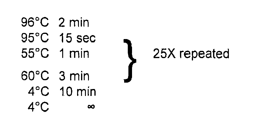
- the CSR-conditions were as follows:
- nuclear extracts were prepared as described by Schreiber et al. [Nucleic Acid Res. 17 (1989), pp 6419 ]. In brief, 1 - 2 ⁇ g of nuclear extracts were added to a binding reaction containing 2 ⁇ g poly dl-dC, 2 ⁇ l 10 x binding buffer (10mM HEPES, Ph: 7.9, 5mM MgCl 2 , 30mM KCI,1mM EDTA, 1mM dithiothreitol, 12% glycerol), and 0.5ng of 32 P-labeled oligonucelotide.
- oligonucleotide containing the NF-E2 consensus binding site at bp - 160 of the human porphobilinogen deainase gene promoter [ Mignotte et al., PNAS 86 (1989), pp 6548-52 ] was synthesized by Eurofins MWG Operon (Ebersberg, Germany) and the NF- ⁇ B consensus oligonucleotide was purchased from Promega (Mannheim, Germany). When indicated, either a 100-fold excess of non-radiaoactive oligonucleotide, a NF-E2 antibody (HPA001914, Sigma-Aldrich, St.
- 293 cells transduced with expression vectors encoding NF-E2wt or mutants, were lysed in SC buffer (50mM Tris/HCl pH 8.0; 170mM NaCl; 0.1% (v/v) NP40; 50mM NaF; 2mM Na3VO4; 0.2mM DTT; 20% (v/v) glycerol; 1 ⁇ g/ml BSA) supplemented with 1x Complete ® (Roche, Mannheim, Germany). Cell lysates were subjected to SDS-PAGE-gel-electrophoresis and Western Blotting. The primary antibody against NF-E2 was used as described in Goerttler et al.
- the blots were stripped and reprobed against ⁇ -Actin (4967, Cell Signaling Technologies, Danvers, MA) or GAPDH (G8795, Sigma, Germany) to control for equal loading.
- the immunocomplexes were detected using chemiluminescence Western Blotting Reagents (GE Healthcare, Freiburg, Germany).
- Plasmids NF-E2 reporter constructs and lentiviral vectors
- the pRBGP2-Luciferase (pRBGP2-Luc) reporter plasmid (described by Igarashi et al.), which contains three copies of the NF-E2 binding site from the chicken beta-globin enhancer in a TATA-luciferase reporter vector, was a kind gift of Masayuki Yamamoto (Tsukuba University, Tsukuba, Japan).
- Insertion and deletion mutations detected in MPN patients were inserted into the NF-E2 cDNA by site directed mutagenesis using the GeneTailor site-directed mutagenesis system (Invitrogen).
- Wt and mutant NF-E2 cDNA was cloned into pLEGO-iG, pLEGO-iT2 and pRc/CMV vectors by restriction enzyme digestion.
- the MafG-pCMV6-XL4 expression vector was purchased from Origene.
- 293 cells were transiently transfected with 0.2 ⁇ g of either the pRBGP2-Luc or the beta1-tubulin- Luciferase reporter gene construct, together with 1.36 ⁇ g of either pRc/CMV-NF-E2 wt or a pRc/CMV-NF-E2 mutant expression vector or the pRc/CMV empty control vector as well as 0.34 ⁇ g of a MafG-pCMV6-XL4 expression vector (Origene).
- 0.1 ⁇ g of a TK- Renilla plasmid (Promega, Mannheim, Germany) were co-transfected as an internal control.
- Luciferase activity was determined using the Dual Luciferase Reporter Assay System (Promega). Luciferase activity was normalized to the Renilla internal control to compensate for variations in transfection efficiency.
- CB3 cells a kind gift of Dr. V. Blank, were transduced with either empty pLeGO-iG or pLeGo-iG containing wtNF-E2 or mutants and variants thereof as described by Roelz et al. [Exp. Hematol. 38 (2010), pp 792-7 ] using MOIs between 1 and 8. GFP expressing cells were sorted to obtain pure populations.
- CB3-NF-E2wt cells were obtained by transducing CB3 cells with pLeGO-iG-wt-NF-E2, and subsequently cloning single transduced cells by limiting dilution. A stable clone was selected and used for co-expression of NF-E2 mutants and variants. CB3-NF-E2wt cells were transduced with either pLeGO-iT2 containing mutant and variant NF-E2 cDNAs or the empty control vector using MOIs between 0.5 and 9. Tomato expressing cells were sorted to obtain pure populations.
- cells were prestimulated for 12 hours in serum-free medium (StemSpan SFEM, 09650, Stem Cell Technologies, Vancouver, BC, Canada) containing 100 ng/ml rhSCF (300-07, PeproTech, Rocky Hill, NJ), 100 ng/ml rhFLT-3 ligand (300-19, PeproTech) as well as 20 ng/ml each rhIL6 (200-06, PeproTech) and rhTPO (300-18, PeproTech).
- serum-free medium Steml rhSCF (300-07, PeproTech, Rocky Hill, NJ)
- 100 ng/ml rhFLT-3 ligand 300-19, PeproTech
- 20 ng/ml each rhIL6 (200-06, PeproTech
- rhTPO 300-18, PeproTech
- Erythroid differentiation medium consisted of StemSpan SFEM (09600, StemCell Technologies, Vancouver, BC, Canada) supplemented with 50 ng/ml rhSCF (300-07, PeproTech, Rocky Hill, NJ), 1 IU/ml EPO (Erypo FS 4000, Ortho Biotech, Bridgewater, NJ), 50 IU/ml rhIL-3 (200-03, PeproTech), 40 ng/ml Human Low Density Lipoprotein (4004,
- CD34+ cells were stained with a phycoerythrin (PE)-conjugated anti- CD235a/GpA antibody [clone GA-R2 (HIR2)] as well as an allophycocyanin (APC)-conjugated anti-CD36 antibody (clone CB38) both from BD Biosciences, Franklin Lakes, NJ, USA and evaluated on a FACS Calibur (BD Biosciences). Data were analysed using the FlowJo software (FlowJo, Ashland, OR, USA).
- CFU-E Erythroid colony-forming units
- BFU-E erythroid burst-forming units
- Quantitative RT-PCR measurements were performed using an Assay on Demand (Mm01611268_g1 Hbb-b1, Applied Biosystems) for analysis of murine beta-globin as well as the following primer and probe sequences for murine ß-2-Microglobulin: FP: 5' TCT TTC TGG CCT GGA GGC TAT C 3' (SEQ ID NO:15) RP: 5' TGC TGG ATG ACG TGA GTA AAC C 3' (SEQ ID NO:16) TaqMan Probe: 6FAM-AGC GTA CTC CAA AGA T-MGBNFQ (SEQ ID NO:17)
- Reverse transcription of total CB3 cell RNA was performed using the TaqMan Reverse Transcription Kit (Applied Biosystems).
- Q-PCR assays were performed in duplicate in an ABI PRISM 7000 Cycler and analyzed using the ABI PRISM 7000 software.
- Beta globin expression was determined relative to expression of the ß-2-Microglobulin house-keeping gene using the ⁇ Ct method.
Landscapes
- Chemical & Material Sciences (AREA)
- Life Sciences & Earth Sciences (AREA)
- Health & Medical Sciences (AREA)
- Organic Chemistry (AREA)
- Proteomics, Peptides & Aminoacids (AREA)
- Engineering & Computer Science (AREA)
- Immunology (AREA)
- Pathology (AREA)
- Analytical Chemistry (AREA)
- Zoology (AREA)
- Genetics & Genomics (AREA)
- Wood Science & Technology (AREA)
- Physics & Mathematics (AREA)
- Biotechnology (AREA)
- Microbiology (AREA)
- Molecular Biology (AREA)
- Hospice & Palliative Care (AREA)
- Biophysics (AREA)
- Oncology (AREA)
- Biochemistry (AREA)
- Bioinformatics & Cheminformatics (AREA)
- General Engineering & Computer Science (AREA)
- General Health & Medical Sciences (AREA)
- Measuring Or Testing Involving Enzymes Or Micro-Organisms (AREA)
Description
- The term "myeloid neoplasias" (MNs) encompasses several classes of disorders, among them myeloproliferative neoplasms (MPNs), myelodysplastic syndromes (MDS), acute myelogenous leukemia (AML), chronic myelogenous leukemia (CML), as well as related disorders.
- Myeloproliferative neoplasms (MPNs), such as polycythemia vera (PV), essential thrombocythemia (ET) and myelofibrosis (MF) are clonal hematopoietic disorders typified by an overproduction of terminally differentiated cells in part as a result of hypersensitivity of marrow progenitor cells to hematopoietic growth factors.
- The molecular etiology of myeloproliferative neoplasms (MPNs) remains incompletely understood, despite recent advances incurred through the discovery of a point mutation in the JAK2 kinase (JAK2V617F) in a large proportion of patients. Several lines of evidence support the hypothesis that additional aberrations, either preceding or following acquisition of the JAK2V617F mutation, contribute to the pathophysiology of these disorders.
-
WO 2008/060090 A1 (22 May 2008 ) describes the detection of JAK2 V617F mutants, said mutants being closely associated with myeloid hematological neoplasms. - The transcription factor Nuclear factor erythroid-2 (NF-E2) is crucial for regulating erythroid-specific gene expression. Cloning of the NF-E2 p45 protein has revealed that it contains a basic region-leucine zipper (b-zip) domain which associates with proteins of the Maf family to form functional NF-E2 [Igarashi et al., Nature (1994), 367, 568-572].
- Nuclear factor erythroid-2 (NF-E2), a hematopoietic transcription factor, is overexpressed in a large majority of patients with polycythemia vera (PV) (Goerttler et al. [British Journal of Hematology 129 (2005) 138-150]. NF-E2 is essential for platelet formation as knock-out mice die perinatally of hemorrhage due to thrombocytopenia. In addition, loss of NF-E2 also affects the erythroid lineage since surviving adult mice display mild anemia with compensatory reticulocytosis.
- Wang et al. [Blood (2010), pp 254-266] have shown that the transcription factor NF-E2 is overexpressed not only in the majority of patients with polycythemia vera but also in patients with essential thrombocytemia (ET) and primary myelofibrosis (PMF) independent of the presence or absence of the JAK2 mutation.
- Toki et al teach that the NF-E2 mRNA is transcribed from two alternative promotors called NF-E2 1A and NF-E2 1 F which are located on different exons (exon 1A and exon 1 F, respectively) Toki et al, 2000, Exp. Hematol. 28: 1113 - 1119). The exons 1A and 1F are non-coding. The translation of NF-E2 starts in
exon 2 and is continued inexon 3. - The amino acid sequence of the wild-type transcription factor NF-E2 is shown in
Figure 1 and SEQ ID NO:1. The nucleic acid sequence coding for the wild-type NF-E2 transcription factor is shown in SEQ ID NO:2. The sequence of the complementary strand is not shown but can be deduced according to the general known rules of base pairing. - In the course of the present invention it has surprisingly been found that the degree of expression of the NF-E2 gene alone is not the only factor responsible for the promotion of erythroid maturation. It has been found that mutations occur within the transcription factor NF-E2 which have a strong influence on the maturation of progenitor cells leading ultimately to erythrocytes. The mutations as well as variants of NF-E2 gene could be detected in MPN patients and this fact can be utilized for diagnosis.
- The physician frequently observes patients in the clinic suffering from a myeloid neoplasia whereby it is difficult to distinguish one disease from another. Such a differential diagnosis is, however, very often of utmost importance since depending on the diagnosis the physician selects the suitable therapy. It is therefore an object to provide an in vitro or ex vivo diagnostic method for distinguishing different types of myeloid neoplasias emcompassing myeloproliferative neoplasms, myelodysplastic syndromes, acute myelogenous leukemia, chronic myelogenous leukemia from reactive states, such as secondary erythrocytosis, secondary thrombocythemia and secondary leukocytosis. The diagnostic method can also be applied in patients suffering from a solid tumor.
- It is therefore an object of the present invention to provide an ex vivo or in vitro method to determine the presence or absence of a mutation or a variation which lead to changes in the amino acid sequence or to a frameshift in the NF-E2 gene in a sample from a patient suffering from a myeloid neoplasia or a solid tumor, whereby said method comprises:
- a) obtaining a nucleic acid sample from the patient, whereby the sample is obtained from blood, bone marrow, or from a biopsy of a solid tumor,
- b) detecting the presence or absence of a mutation in the nucleic acid sample comprising the NF-E2 gene or a fragment thereof or of a variation of the NF-E2 gene, characterized in that the presence of the mutation or of the variation is an indication of a myeloid neoplasm, in particular, a myeloproliferative neoplasm, a myelodysplastic syndrome, a chronic myeloid leukemia or an acute myeloid leukemia, or a solid tumor, whereby the mutation is determined by comparing the nucleic acid sequence obtained from the sample with SEQ ID NO:2 or the complement thereof, or whereby a variation of the NF-E2 gene is a NF-E2 gene product which differs from the amino acid sequence as shown in SEQ ID NO:1.
- The diagnostic method of the present invention is an ex vivo or in vitro method which means that the method can be performed on a sample comprising a suitable nucleic acid. Such a sample can be obtained from blood, but the samples can alternatively also be obtained from other body fluids like bone marrow or from a biopsy of a solid tumor. The method allows the determination whether a mutation or a variation of the NF-E2 gene is detectable. The nature of the mutation or of the variant identified allows diagnosis of the disease the patient is suffering from.
- A mutation according to the present invention means that the nucleic acid sequence as shown in SEQ ID NO:2 is changed which leads consequently to changes in the amino acid sequence of the mutated gene. Such a mutation may lead to a change of the amino acid sequence or even to a frame shift in the coding sequence which results in the creation of a stop codon having the consequence that the translational product of the mutated gene results in a shorter form of the NF-E2 gene product.
- The term "variation" of the NF-E2 gene product is understood in the sense that the NF-E2 gene product differs from the wild-type amino acid sequence as shown in SEQ ID NO:1.
- In a preferred embodiment of the present invention the method is performed by using the well-known PCR technology. Since the nucleic acid sequence coding for the wild-type NF-E2 gene is known (SEQ ID NO:2) the person skilled in the art can easily select suitable short sequences as primers for amplification of nucleic acids. The primer sequences consist of 10-30, preferably 12-28, more preferred 15-25 and especially preferred 18-22 consecutive nucleotides of the SEQ ID NO:2 or the complement thereof.
- The forward primer is for example selected from SEQ ID NO:2 and the reverse primer is selected from the complementary strand to SEQ ID NO:2 which is known due to the general rules of base pairing. Usually forward and reverse primer cover a stretch of about 600 to 50, preferably 300 to 70, nucleotides, whereby the location of forward primer and reverse primer is selected in such a way that a short stretch of nucleic acid sequence is between the two primers.
- In a preferred embodiment one of the primers is selected from an area between nucleotide 600 and 900, more preferred between nucleotide 700 and 800 of SEQ ID NO:2. The other primer required for PCR is selected from the strand complementary to this region.
- The primers are usually completely complementary to the corresponding stretch shown in SEQ ID NO:2 or the complement thereof. It may, however, be considered to include one or two mispairings in order to improve the diagnostic efficiency of the PCR reaction.
- After the primers have hybridized to the nucleic acid contained within the sample, hybridization and amplification of the nucleic acid strand takes place usually by using a heat-resistant polymerase. After sufficient cycles of polymerization a DNA fragment may be obtained which can be sequenced.
- In another embodiment of the present invention the diagnostic method can be performed by using a quantitative PCR or a so-called real time PCR (RT-PCR). The real time PCR is used in order to determine nucleic acids quantitatively. By using primers which occur at stretches where mutations or variations of the nucleic acid sequence can frequently be expected it is possible to conclude from the reduced amount of the nucleic acid produced by PCR compared with the wild-type NF-E2 sequence whether a mutation is present or not.
- In the course of the present invention the sequences of NF-E2 of different patients have been checked, whereby recurrent truncating mutations in NF-E2 were identified.
- Seven different frameshift mutations, insertions and deletions, were detected in the NF-E2 coding sequence in 8 patients with myeloproliferative neoplasms (MPNs), 3 with polycythemia vera (PV) and 5 with myelofibrosis, either primary myelofibrosis (PMF) or secondary, post-MPN myelofibrosis (sMF). Two mutations, c.782-785delAGAG and c.622_623insG, were found in two different patients each, while one patient harbored two separate mutations.
Table 1 Patient number* Diagnosis variant cDNA NM_006163.1 variant protein JAK2V617F genetics F- 209 PV c.782-785delAGAG p.E261AfsX3 positive acquired F-2836 postET-PMF c.622insG pE221GfsX7 n.d. MPD-241 postPV-PMF c. 732_733insG p.L245VfsX5 positive F-202 PV c. 782-785delAGAG p.E261AfsX3 positive acquired U-409 PV c. 236delC p.P79LfsX32 positive acquired c.234delT p.P79LfsX32 U-532 PMF c889_900del p.E297-R300del positive acquired U-980 Post-ET-PMF c.780_781insA p.E261RfsX44 positive acquired U-442 post-PV-PMF c.622insG p.E221GfsX7 positive - Table 1 shows NF-E2 Mutations detected in MPN patients. The location of the detected mutation is shown with regard to the cDNA contained in SEQ ID NO:2. Moreover, a correlation between diagnosis of the disease and mutation can be seen.
- The frameshifts introduce premature stop codons in the open reading frame, leading to truncations in the NF-E2 protein (
Fig. 8A ). One 12 bp deletion, c.889-900del, causes an in-frame deletion of 4 amino acids, which includes the N-terminal leucine, of the leucine zipper heterodimerization domain. - Insertion and deletion mutations in NF-E2 were detected exclusively in PV and PMF patients (3 of 144 patients; 2.1 % and 5 of 192 patients; 2.7% respectively, Table 2), not in healthy controls. This allows a diagnosis of a myeloproliferative neoplasm (MPN) and therefore enables the clinically important discrimination between the diagnosis of an acquired myeloprolifertive disorder, distinguishing it from a physiological reactive process. More details and clinical characteristics of the patients can be seen from Table 2.
- It has been found that two specific mutations having the abbreviation c.622insG and p.E221GfsX7 which are shown in SEQ ID NO:8 could also be detected in a patient suffering from myelodysplastic syndrome (MDS) who later transformed to acute myeloic leukemia (AML). This patient had the identification number UPN:N-022. Therefore, the method disclosed herein can also be used to identify and diagnose patients with myelodysplastic syndromes, with acute myeloid leukemia as well as with solid tumors, which are frequently molecularly related to hematologic diseases. For example, the mutations IDH1 and IDH2 could be detected with patients suffering from AML (acute myeloid leukemia) as well as in patients suffering from glioblastomas.
- By detecting mutations in the NF-E2 gene, reactive processes can be excluded which can often be extremely difficult to distinguish from neoplastic processes without molecular markers such as the NF-E2 mutations.
- In general it can be said that when an NF-E2 mutation can be detected according to the method disclosed herein, a secondary erythrocytosis, a secondary thrombocytosis or a reactive leukocytosis can be excluded.
- The method of the present invention can also be used for monitoring the disease and the effectiveness of the therapeutic treatment. The patient is treated according to the best suitable therapy and it can be monitored by the method of the present invention whether the therapy shows the desired therapeutic success.
Table 2. Clinical Characteristics of the MPN MDS, sAML and CMML Patients Studied. Abbreviations used and data displayed: Diagnosis: Polycythemia vera (PV), Essential Thrombocythemia (ET), Primary Myelofibrosis (PMF); Gender: male (m), female (f); Age at Diagnosis: mean (range); Duration of Disease: mean, (range); White Blood Cell count (WBC), mean (range), Hematocrit (Hct), mean (range); Platelets (Plt), mean (range).# CBC at the time the blood sample was obtained. Diagnosis Number of patients Gender m / f Age at diagnosis in years mean (range) Duration of disease in years mean (range) Hemoglobin (at sample) WBC (at sample) Platelets (at sample) ET 120 55/65 49.7 (17.3 - 82.3) 4.9 (0.0 - 21.5) 14.1 (8.6 - 33.9) 8.1 (2.5 - 31.4) 671 (149 - 2393) PV 144 71/73 55.5 (16.7 - 78.4) 5.3 (0.0 - 29.3) 14.6 (7.9 - 20.3) 15.2 (1.8 - 209.0) 513 (40 - 2055) PMF 192 102/90 56.9 (21.3 - 86.4) 3.5 (0.0 - 20.5) 10.7 (1.2 - 17.2) 15.1 (0.8 - 102.1) 292 (99 - 593) MPN-U 49 MDS 57 sAML (post-MPN) 39 21/18 57.0 (42.2 - 76.0) 7.0 (0.0 - 20.9) 9.7 (6.9 - 15.5) 39.2 (1.5 - 182.4) 83 (11 - 362) CMML 67 - Constitutive DNA, obtained from buccal swabs, was available from 4 patients and in all cases, we were able to demonstrate that the mutations were acquired (Table1).
- Novel NF-E2 sequence variants (SNPs) were also detected.
- In addition to the insertion/deletion mutations, seven different sequence variants not corresponding to known NF-E2 SNPs (Table 3) were observed.
Table 3. Age at Diagnosis of MPN Patients with and without NF-E2 mutations and sequence variations. Diagnosis Genotype Number of Patients Age at Diagnosis Significance PV wt 141 55 NF- E2 mutations 3 55 NF-E2 variants (all) 6 51 NF-E2 R365P only 2 39 *p<0.05 PMF wt 187 57 NF- E2 mutations 5 63 NF-E2 variants (all) 4 51 NF-E2 R365P only 4 51 PV + PMF wt 328 56 NF- E2 mutations 8 60 NF-E2 variants (all) 10 52.2 NF-E2 R365P only 6 46.2 *p<0.05 ET wt 118 49 NF- E2 mutations 0 NF-E2 variants (all) 2 57 NF-E2 R365P only 0 - NF-E2 sequence variations were observed in 1.7% of ET (2 out of 120 patients), 4.2% of PV (6 out of 144) and 2.1 % of PMF patients (4 out of 192 patients). Again, the majority of the variants were present exclusively among MPN patients, PV (n = 144), PMF (n=192) and ET (n=120), but not in healthy controls. Of the seven SNPs, the most frequently observed was a G to C transversion at bp 1094, leading to an argenine to proline substitution at amino acid 365 of the NF-E2 protein (R365P). This change was observed in 2 PV patients (out of 144 = 1.4%) and 4 PMF patients (out of 192 = 2.2%). Buccal swab DNA samples from three patients showed that this variant was constitutively present in these individuals.
- Interestingly, this SNP is only found in 0.3% of the general population (n = 4550; NCBI SNP Database, http://www.ncbi.nlm.nih.gov/projects/SNP/snp_ref.cgi?rs=141251053), and is therefore statistically highly significantly enriched in MPN patients, especially in patients with PMF (p<0.0001, Chi-Square Test).
- A loss of DNA binding and transactivation potential has been observed in NF-E2 mutants.
- Because the truncated NF-E2 proteins contain neither the DNA binding domain nor the leucine zipper required for dimerization to small Maf proteins (compare with
Figure 8A ), it was investigated whether truncated NF-E2 mutants retain DNA binding activity in an electrophoretic mobility shift assay (EMSA). NF-E2 requires interaction with a small Maf protein, here MafG, in order to bind DNA (Fig. 8B , comparelane 2 and lane 4). Specificity of the NF-E2/MafG heterodimer binding to its cognate DNA sequence was verified by competition and super-shift experiments (Fig. 8B , lanes 5-8). In contrast to wt NF-E2, the two truncated NF-E2 mutants (p.L245VfsX5, here called 248aa, and p.E261AfsX3, here called 236 aa) were unable to bind DNA even in the presence of MafG (Fig. 8B , comparelanes Fig. 8B , comparelanes 9 and 13). In contrast, the NF-E2 R365P variant retained DNA binding activity (Fig. 8B , comparelanes 9, and 11). Protein expression of all mutants was verified by Western Blotting (Fig. 8C ). - Subsequently, it was investigated whether the NF-E2 mutants retained transactivation potential in reporter gene assays. Two different reporter gene constructs were used, one containing a known NF-E2 binding site from the beta-globin promoter (Igarashi et al.), and a second containing a NF-E2 binding site in the beta1-tubulin promoter. Both reporter constructs yielded similar results (
Figs. 9A and B ); all three NF-E2 mutants tested were no longer able to evoke reporter gene activity above the background levels observed with MafG alone. - Because transcription of a plasmid DNA does not reflect physiological conditions within the chromatin bound DNA, CB3 cells were used to test the mutants under physiological conditions. CB3 cells carry a homozygous viral insertion in the NF-E2 locus and therefore express neither NF-E2 nor its target beta-globin. Upon re-introduction of NF-E2, beta-globin is robustly expressed. We introduced wt NF-E2 or two truncation mutations, 248aa and 263 aa, as well as the 4aa deletion, del 297-300, into CB3 cells and measured beta-globin mRNA expression by qRT-PCR. While transduction of wt NF-E2 caused a 100-fold increase in beta-globin mRNA expression, all three NF-E2 mutants were unable to activate beta-globin expression (
Fig. 9C ). - NF-E2 mutants and variants display a gain of function.
- Because all the NF-E2 mutations were observed in a heterozygous state, hence unfold their effect in the presence of wt NF-E2, the effect of co-expression of wt NF-E2 and NF-E2 mutants in CB3 cells was investigated. CB3 cells stably transfected with wt NF-E2 were infected with an empty pLeGO-iT virus, or viruses expressing the two truncation mutants, 248 aa and 263 aa, the 4aa deletion, del 297-300, and assayed for beta-globin expression (
Fig. 10A and B ). While transduction with empty pLeGO-iT had no effect on beta-globin mRNA expression in NF-E2 wt expressing CB3 cells (Fig. 10B ), transduction with the three mutants, which on their own retain no transactivation activity (Fig. 9C ), enhance wt NF-E2 activity between 3 and 8-fold, with the del297-300 mutant displaying the strongest effect (Fig. 10B ). Thus, while unable to transactivate transcription on their own, the NF-E2 mutants have gained a neomorphic function which significantly enhances wt NF-E2 activity. - Because of the observed enhancement of beta-globin expression the effect of NF-E2 mutants on erythroid maturation of healthy CD34+ hematopoietic stem cells was examined. It has previously been shown that overexpression of wt NF-E2 delays erythroid maturation in a liquid culture system promoting erythroid maturation.
- Purified peripheral blood CD34+ cells from healthy donors were transduced either with empty lentivirus or with lentiviruses expressing wt NF-E2, the del297-300, the 248aa or the 263aa mutation. The change of the markers from CD34+ to CD36 / glycophorin A reflects the change to erythroid progenitor cells. Transduced cells were purified by FACS, cultured and monitored daily for erythroid maturation by staining for CD36 and glycophrin A (GpA) (
Fig. 11A ). The proportion of erythroid cells, doubly positive for CD36 and GpA, is decreased in NF-E2 wt expressing cells compared to empty vector transduced controls (Fig. 11B , top). Compared to NF-E2 wt transduced cells, the three mutants promote faster erythroid maturation (Fig. 11B , right panels). This observation would be compatible either with a loss of NF-E2wt function, or with a gain of function. Importantly, all three mutants accelerate erythroid maturation compared to empty virus transduced cells, witnessed by the earlier appearance of larger proportions of CD36/GpA double positive erythroid cells (Fig. 11B , left panels). These data provide strong evidence for a gain-of-function by the NF-E2 mutants, one that promotes erythroid maturation. - It has been found that NF-E2 mutations confer a proliferative advantage in the context of the JAK2V617F mutation
- MPN patients carrying NF-E2 mutations were also tested for presence of the JAK2V617F mutation. All seven patients assayed carried both a NF-E2 mutation and the JAK2V617F mutation (Table 1). Analysis of individual hematopoietic colonies grown in methylcellulose revealed that in two patients, 202 and 209, the NF-E2 mutation was acquired subsequent to the JAK2V617F mutation (
Fig. 13 ), as some colonies carrying only the JAK2V617F mutation but not the NF-E2 mutation were found and all colonies positive for the NF-E2 mutation also carried JAK2V617F. Interestingly, these assays revealed a proliferative advantage of cells carrying the NF-E2 mutation over cells carrying only JAK2V617F either in the context of a heterozygous (patient 202) or a homozygous JAK2V617F mutation (patient 209;Fig. 13 ). In both patients, cells carrying both the NF-E2 mutation and the JAK2V617F mutation represent the vast majority (90 and 81%, respectively), of the colonies analyzed, demonstrating that the JAK2V617F-positive cell that acquired the NF-E2 mutation outcompeted cells carrying the JAK2V617F mutation alone. - In
patient 532, it cannot be determined for sure whether the NF-E2 or the JAK2V617F mutation was incurred first. However, again, the presence of both mutations clearly provided a proliferative advantage over either of the mutations alone, as all colonies carry both mutations. In addition, in the context of a NF-E2 mutation, as in wt NF-E2 patients, homozygous JAK2V617F cells outgrow heterozygous JAK2V617F cells. - A second patient, 409, has acquired two separate and mutually exclusive NF-E2 mutations, c.236delC and c.234delT (
Fig. 13D and Table 1). The vast majority of both myeloid and erythroid colonies (>89%; n = y) carry a homozygous JAK2V617F mutation. However, 65% of the erythroid and 50% of the myeloid colonies carry the c.236delC while 12% of the erythroid and 28% of the myeloid colonies carry a c.234delT mutation (Fig. 13D ). The remainder of the colonies are wt for NF-E2, demonstrating that both mutations are acquired and again suggesting that both NF-E2 mutations confer a growth advantage, as they outcompete the NF-E2 wt cells. - It could be shown that NF-E2 mutants cause polycythemia and thrombocytosis in a murine BMT model.
- In order to test whether the NF-E2 mutant promote erythroid maturation in vivo, a murine bone marrow transplant model was used. Donor bone marrow was transduced with lentiviruses encoding NF-E2 wt or the 248aa, the 263aa or the del297-300 mutants and transplanted into recipient mice (
Fig. 11C ). Donor engraftment exceeded 95% in all mice. Twelve weeks after transplantation, peripheral blood was analyzed. Compared to mice transplanted with wt NF-E2, which, similar to NF-E2 transgenic mice, display normal hematocrit and normal RBCs, mice transplanted with two of the three NF-E2 mutants displayed a significant elevation in hematocrit and in RBC number and this trend was evident in the third mutant as well (Fig 11D , top panels). In addition, while NF-E2 wt transplanted mice, like NF-E2 tg mice, display normal platelet values at 3 months, mice transplanted with all three mutants displayed a statistically significant elevation in platelet number (Fig. 11D , bottom panel). These data demonstrate that presence of the NF-E2 mutants is sufficient to cause erythrocytosis and thrombocytosis in a murine model. - Important aspects of the present invention have been summarized in the Figures and can also be deduced from the sequence protocol.
-
Figure 1 shows the sequence of the wild-type NF-E2 protein which is available under the accession number NP_006154. The nucleic acid sequence coding for the NF-E2 protein is shown in SEQ ID NO:2. -
Figure 2 shows the nucleic acid and the amino acid sequence derived therefrom of variant designated as F-209. At the N-terminus a mutation inexon 3 is shown. Due to a deletion a stop codon occurs and the protein ends with the amino acid sequence "... PLTAS".
The cDNA coding for the mutated protein ofFigure 2 is provided as SEQ ID NO:3 and the protein sequence of the sequence shown inFigure 2 is provided in SEQ ID NO:4. -
Figure 3 shows another mutation inexon 3. The clone is designated as MPD-RC-241. In the nucleic acid sequence a "G" is inserted which results in a changed amino acid sequence at the N-terminus. The mutated sequence ends with the amino acids "-VNVAGR". The cDNA sequence is shown as SEQ ID NO:5 and the protein sequence of the mutated protein is shown as SEQ ID NO:6. -
Figure 4 shows another mutation in exon 3 (F-2836). The insertion of a "G" into the nucleic acid sequence results in the N-terminus of the mutated protein "-RGGGRESG". The cDNA sequence is shown as SEQ ID NO:7 and the protein sequence is shown as SEQ ID NO:8. -
Figure 5 shows another mutation inexon 3. The clone is designated as U-532. The cDNA is shown in SEQ ID NO:9 and the amino acid sequence is shown in SEQ ID NO:10. -
Figure 6 shows another mutation inexon 3 caused by a deletion of a "C". The amino acid sequence at the N-terminus is changed and the protein is shorter due to occurrence of a stop codon. The cDNA of clone U-409 is shown in SEQ ID NO:11 and the amino acid sequence is shown in SEQ ID NO:12. -
Figure 7 shows the sequence of a variant with the designation U-980. A mutation caused by an insertion of "A" results in a changed amino acid sequence at the N-terminus and the occurrence of a stop codon. The cDNA sequence is shown in SEQ ID NO:13. The amino acid sequence is provided as SEQ ID NO:14. -
Figure 8 : NF-E2 mutations in MPN patients cause truncations and loss of DNA binding.- (A) Schematic representation of the NF-E2 protein (top) and the truncations resulting from mutations detected in MPN patients (bottom).
- (B) Electrophoretic mobility shift assay (EMSA) of wt NF-E2 and NF-E2 mutants. Nuclear extracts of 293 cells transduced with expression vectors encoding either wt NF-E2 (lane 2), MafG (lane 3) or both (lanes 4-9) or the indicated NF-E2 mutants together with MafG (lanes 10-13) were incubated with a 32-P-labeled oligonucleotide containing a NF-E2 binding site (REF). In
lanes - (C) Protein expression in the cell extracts used for EMSA in (B). Cell lysates were subjected to SDS-PAGE and interrogated for NF-E2 (top) and beta-actin (bottom) expression by Western Blot.
-
Figure 9 . Transactivating Activity of mutant NF-E2 proteins. Plasmids encoding (A) a beta-globin promoter-luciferase construct (Igarashi et al.) or (B) a reporter construct encoding 5.2 kb of the beta1-tubulin promoter coupled to the luciferase reporter gene were co-transfected into 293 cells either with expression vectors for MafG (white bars), wt NF-E2 (black bars) or various NF-E2 mutants (grey bars) as indicated. Luciferase activity was measured 16 hrs post-transfection and normalized for transfection efficiency by determination of Renilla luciferase activity from a co-transfected vector. Activity of MafG alone was set at 1 and fold activity relative to this control is depicted. Bar graphs represent the mean ± SD of four independent experiments, each performed in duplicate,* p<0.05, **p<0.01, *** p < 0.001 by One Way ANOVA with Bonferroni's post-hoc multiple comparison test. (C) CB3 cells, which lack endogenous NF-E2, were transfected with wt NF-E2 (black bars) or various NF-E2 mutants (grey bars) as indicated. 72 hours after transfection, RNA was harvested and assayed for beta-globin mRNA expression and beta-2-microglobulin housekeeping gene expression by qRT-PCR. Results are reported as relative expression levels setting beta-globin expression in untransfected CB3 cells at 1. Data were analyzed for statistical significance by One Way ANOVA with Bonferroni's post-hoc multiple comparison test. **p<0.01; ***p< 0.001 -
Figure 10 . Effect of mutant NF-E2 proteins on NF-E2 wt activity. (A) Experimental design. CB3 cells were transduced with pLeGO-iG-NF-E2, sorted for GFP expression and a single clone (CB3-NF-E2wt), which displays wt NF-E2 expression, selected. Subsequently, CB3-NF-E2wt cells were transduced with either an empty pLeGo-iT vector, or with pLeGO-iT vectors encoding the indicated NF-E2 mutants. Double positive cells were FACS sorted and (B) assayed for beta-globin expression and beta-2-microglobulin housekeeping gene expression by qRT-PCR. Results are reported as relative expression levels setting beta-globin expression in empty pLeGO-iT transduced CB3-NF-E2 wt cells at 1. Data were analyzed for statistical significance by One Way ANOVA with Bonferroni's post-hoc multiple comparison test.. ***p< 0.0001. as well as NF-E2 and GAPDH protein expression (C) Protein expression in the transduced CB3 cells assayed for beta-globin expression in (B). Cell lysates were subjected to SDS-PAGE and interrogated for NF-E2 (top) and GAPDH (bottom) expression by Western Blot. -
Figure 11 . Effect of NF-E2 mutations on erythroid maturation.- (A) Experimental design. CD34+ cells were transduced with empty pLeGo-iG, with pLeGO-iG-NF-E2 wt or with pLeGO-iG carrying the indicated NF-E2 mutations. Cells were sorted for GFP expression to obtain pure populations of transduced cells and maintained in medium promoting erythroid differentiation. At the indicated time points, erythroid maturation was assessed by FACS analysis using CD36 and glycophrin A (GpA).
- (B) Erythroid Maturation. The percentage of CD36+ /GpA+ double positive erythroid cells is shown over time. Mean and standard error of n=4 independent experiments are depicted. (Left) Comparison of cells transduced with NF-E2 mutants to cells transduced with empty virus. (Right) comparison of cells transduced with NF-E2 mutants to cells transduced with NF-E2 wt. *p<0.05, **p<0.01.
-
Figure 12 . Effect of NF-E2 on erythroid maturation. The effect of the mutations compared with wild-type NF-E2 on the erythroid maturation depending on time is shown inFigure 12 . -
Figure 13 : Mutational analysis of hematopoietic colonies. Mononuclear cells of the MPN patients indicated were seeded in methylcellulose and individual colonies harvested after 14 days. Colonies were assays for presence of the JAK2V617F and the NF-E2 mutations by PCR amplification and sequencing. Genotypoes of the individual colonies, each represented by a single dot, are depicted. Individual hematopoietic colonies grown in methylcellulose of four different patients having theabbreviation UPN Figure 13 the abbreviation "wt" means wild type, "het" means heterozygous and "hom" means homozygous. - The sequence listing comprises the relevant sequences whereby the numbers designate the following sequences:
SEQ ID NO:1 amino acid sequence of wild-type NF-E2 SEQ ID NO:2 DNA sequence coding for wild-type NF-E2 SEQ ID NO:3 cDNA sequence coding for the mutated protein F-209 of Fig. 2 SEQ ID NO:4 amino acid sequence coding for the mutated protein F-209 of Fig. 2 SEQ ID NO:5 cDNA sequence coding for the mutated protein MPD-RC-241 of Fig. 3 SEQ ID NO:6 amino acid sequence coding for the mutated protein MPD-RC-241 of Fig. 3 SEQ ID NO:7 cDNA sequence coding for the mutated protein F-2836 of Fig. 4 SEQ ID NO:8 amino acid sequence coding for the mutated protein F-2836 of Fig. 4 SEQ ID NO:9 cDNA sequence coding for the mutated protein U-532 of Fig. 5 SEQ ID NO:10 amino acid sequence coding for the mutated protein U-532 of Fig. 5 SEQ ID NO:11 cDNA sequence coding for the mutated protein U-409 of Fig. 6 SEQ ID NO:12 amino acid sequence coding for the mutated protein U-409 of Fig. 6 SEQ ID NO:13 cDNA sequence coding for the variant U-980 of Fig. 7 SEO ID NO:14 amino acid sequence coding for the variant U-980 of Fig. 7 SEQ ID NO:15 primer sequence SEQ ID NO:16 primer sequence SEQ ID NO:17 probe sequence SEO ID NO:18 primer sequence SEQ ID NO:19 primer sequence SEQ ID NO:20 primer sequence SEQ ID NO:21 primer sequence SEQ ID NO:22 primer sequence SEQ ID NO:23: primer sequence SEQ ID NO:24: primer sequence SEQ ID NO:25 primer sequence - Peripheral blood samples were obtained from PV (n = 144) ET (n = 120) and PMF patients (n= 192), fulfilling the WHO criteria for diagnosis. Additional samples were obtained from the Tissue Bank of the Myeloproliferative Disease Research Consortium (MPD-RC), which uses the same diagnostic criteria. Patient characteristics are summarized in Table 2. Buffy coats of healthy volunteer blood donors were obtained from the University Hospital Freiburg Center for Blood Transfusion Nijmegen. Isolated granulocytes were obtained by dextran sedimentation and Ficoll gradient centrifugation as described by Temerinac et al. [Blood 95 (2000), pp 2569-2576] and used for DNA preparation and subsequent NF-E2 sequence analysis.
- Buccal swabs were used to obtain DNA for germline analysis. The study protocol was approved by the local ethics committees (University Hospital Freiburg, University Hospital Ulm, as well as the Member Institutions of the MPD-RC) and informed consent was obtained from all patients. Each patient was assigned a unique patient number (UPN), which was used thereafter for the protection of privacy. All patients were tested for the presence of the JAK2V617F mutation by qRT-PCR or LNA PCR as described by Steimle et al. [Ann.Hemathol. 86 (2007), pp 239-244]. The results of the experiments can be seen from the figures.
- Exons 2 and 3 of the NF-E2 locus were amplified by PCR and subjected to direct sequencing according to standard protocols. Primer sequences and reaction conditions were as follows:
-
Exon 2: Forward: CGGTCTCTCCTTCCCTCAGGGGA SEQ ID NO:18 Reverse: TCTTCTCCGACACACGGCCTCC SEQ ID NO:19 Exon 3A Forward: TGCGACTTTCAGAAGAATCCAGCTTG SEQ ID NO:20 Reverse: TGGAGTGGGCCAAGGAGTTGGG SEQ ID NO:21 Exon 3B Forward: TCGGCGGCGCAGCGAATATG SEQ ID NO:22 Reverse: GTAGCTGTTGCCTGATTCATCCCG SEQ ID NO:23 Exon 3C Forward: GAGGTCATGCGCCAACAGCTGAC SEQ ID NO:24 Reverse: TGCTCAGCCTCAAGCCCAAATTTT SEQ ID NO:25 - For each amplification the following reaction mixture was prepared:
PCR-Mastermix: 1x-Mix µl 18,25 dd H2O 2,5 Puffer 0,75 MgCl 20,25 dNTPs 0,25 mM Fa. Roche 0,5 Primer F 10 pmol/µl Fa. MWG 0,5 Primer R 0,25 Platinum Taq Fa. Invitrogen 
- For RT-PCT the following CSR-Mastermix was used:
1x- Mix µl 10 dd H2O. 2 BigDye 1.1 Fa. Applied Biosystems 1 sequencing buffer Fa. Applied Biosystems 1 Primer F oder R 10 pmol/µl 14µl + 1 µl purified PCR-product - The CSR-conditions were as follows:
- 293 nuclear extracts were prepared as described by Schreiber et al. [Nucleic Acid Res. 17 (1989), pp 6419]. In brief, 1 - 2 µg of nuclear extracts were added to a binding reaction containing 2µg poly dl-dC, 2µl 10 x binding buffer (10mM HEPES, Ph: 7.9, 5mM MgCl2, 30mM KCI,1mM EDTA, 1mM dithiothreitol, 12% glycerol), and 0.5ng of 32P-labeled oligonucelotide. The oligonucleotide containing the NF-E2 consensus binding site at bp - 160 of the human porphobilinogen deainase gene promoter [Mignotte et al., PNAS 86 (1989), pp 6548-52] was synthesized by Eurofins MWG Operon (Ebersberg, Germany) and the NF-κB consensus oligonucleotide was purchased from Promega (Mannheim, Germany). When indicated, either a 100-fold excess of non-radiaoactive oligonucleotide, a NF-E2 antibody (HPA001914, Sigma-Aldrich, St. Louis, MO) or a NF-κB p65 antibody (SC-372, Santa Cruz Biotechnologies, Santa Cruz, CA) were added. The reaction was incubated at RT for 15 min. For supershift assays, extracts and antibodies were preincubated for 10 minutes at RT prior to addition of the radioactive nucleotide. The results of the experiment are summarized in
Figure 8 . - 293 cells, transduced with expression vectors encoding NF-E2wt or mutants, were lysed in SC buffer (50mM Tris/HCl pH 8.0; 170mM NaCl; 0.1% (v/v) NP40; 50mM NaF; 2mM Na3VO4; 0.2mM DTT; 20% (v/v) glycerol; 1µg/ml BSA) supplemented with 1x Complete® (Roche, Mannheim, Germany). Cell lysates were subjected to SDS-PAGE-gel-electrophoresis and Western Blotting. The primary antibody against NF-E2 was used as described in Goerttler et al. The blots were stripped and reprobed against β-Actin (4967, Cell Signaling Technologies, Danvers, MA) or GAPDH (G8795, Sigma, Germany) to control for equal loading. The immunocomplexes were detected using chemiluminescence Western Blotting Reagents (GE Healthcare, Freiburg, Germany).
- A 5.2 kb region, spanning bp - 1 to bp - 5221 of the beta1 tubulin gene, was amplified by PCR from human genomic DNA and inserted into the pGL3basic luciferase reporter vector (Promega, Mannheim, Germany) resulting in the ß1-tubulin -5.2 kb-luc construct.
- The pRBGP2-Luciferase (pRBGP2-Luc) reporter plasmid (described by Igarashi et al.), which contains three copies of the NF-E2 binding site from the chicken beta-globin enhancer in a TATA-luciferase reporter vector, was a kind gift of Masayuki Yamamoto (Tsukuba University, Tsukuba, Japan).
- Insertion and deletion mutations detected in MPN patients were inserted into the NF-E2 cDNA by site directed mutagenesis using the GeneTailor site-directed mutagenesis system (Invitrogen). Wt and mutant NF-E2 cDNA was cloned into pLEGO-iG, pLEGO-iT2 and pRc/CMV vectors by restriction enzyme digestion. The MafG-pCMV6-XL4 expression vector was purchased from Origene.
- 293 cells were transiently transfected with 0.2 µg of either the pRBGP2-Luc or the beta1-tubulin-Luciferase reporter gene construct, together with 1.36 µg of either pRc/CMV-NF-E2 wt or a pRc/CMV-NF-E2 mutant expression vector or the pRc/CMV empty control vector as well as 0.34 µg of a MafG-pCMV6-XL4 expression vector (Origene). In addition 0.1 µg of a TK-Renilla plasmid (Promega, Mannheim, Germany) were co-transfected as an internal control.
- Cells were harvested 16 hours post transfection, and luciferase activity was determined using the Dual Luciferase Reporter Assay System (Promega). Luciferase activity was normalized to the Renilla internal control to compensate for variations in transfection efficiency.
- CB3 cells, a kind gift of Dr. V. Blank, were transduced with either empty pLeGO-iG or pLeGo-iG containing wtNF-E2 or mutants and variants thereof as described by Roelz et al. [Exp. Hematol. 38 (2010), pp 792-7] using MOIs between 1 and 8. GFP expressing cells were sorted to obtain pure populations.
- CB3-NF-E2wt cells were obtained by transducing CB3 cells with pLeGO-iG-wt-NF-E2, and subsequently cloning single transduced cells by limiting dilution. A stable clone was selected and used for co-expression of NF-E2 mutants and variants. CB3-NF-E2wt cells were transduced with either pLeGO-iT2 containing mutant and variant NF-E2 cDNAs or the empty control vector using MOIs between 0.5 and 9. Tomato expressing cells were sorted to obtain pure populations.
- Lentiviral transduction of peripheral blood CD34+ cells from healthy donors using pLEGO-iG expressing wt or mutant NF-E2 was performed as described by Roelz et al. [Exp. Hematol. 38 (2010), pp 792-7]. Briefly, cells were prestimulated for 12 hours in serum-free medium (StemSpan SFEM, 09650, Stem Cell Technologies, Vancouver, BC, Canada) containing 100 ng/ml rhSCF (300-07, PeproTech, Rocky Hill, NJ), 100 ng/ml rhFLT-3 ligand (300-19, PeproTech) as well as 20 ng/ml each rhIL6 (200-06, PeproTech) and rhTPO (300-18, PeproTech). Subsequently, CD34+ cells were subjected to two cycles of lentiviral infection over a 48 hour period using MOIs of 10 without the addition of a transduction facilitator. GFP expressing cells were FACS sorted to obtain pure populations.
- Erythroid differentiation medium consisted of StemSpan SFEM (09600, StemCell Technologies, Vancouver, BC, Canada) supplemented with 50 ng/ml rhSCF (300-07, PeproTech, Rocky Hill, NJ), 1 IU/ml EPO (Erypo FS 4000, Ortho Biotech, Bridgewater, NJ), 50 IU/ml rhIL-3 (200-03, PeproTech), 40 ng/ml Human Low Density Lipoprotein (4004,
- Harbor Bio-Products, Norwood, MA), 100 IU/ml Penicillin, 100 mg/ml Streptomycin (DE17-602E, Cambrex, North Brunswick, NJ) and 2 µl/ml Primocin (VZA-1021, Amaxa Biosystems, Cologne, Germany). Cells were maintained at a concentration of 2 x 10⁵/ml.
- CD34+ cells were stained with a phycoerythrin (PE)-conjugated anti- CD235a/GpA antibody [clone GA-R2 (HIR2)] as well as an allophycocyanin (APC)-conjugated anti-CD36 antibody (clone CB38) both from BD Biosciences, Franklin Lakes, NJ, USA and evaluated on a FACS Calibur (BD Biosciences). Data were analysed using the FlowJo software (FlowJo, Ashland, OR, USA).
- The results of the transduction experiments are shown in
Figure 11 andFigure 12 . - Cells were seeded in methylcellulose media (1000 cells/ml) containing SCF, IL-3, granulocyte-macrophage colony-stimulating factor (GM-CSF) and EPO (H4434; Stem Cell Technologies) and incubated for 14 d at 37°C, 5% CO2. Erythroid colony-forming units (CFU-E) were scored on
day 8 and erythroid burst-forming units (BFU-E) as well as granulocytic/macrophage colony-forming units CFU-GM were scored on day 14. Colonies were counted by independent observers, blinded to the experimental conditions. - Paired or unpaired Student t-tests (two sided or one sided) and the Mann-Whitney Rank sum test were used to determine whether a significant (p< 0.05) difference existed between two groups. When comparing more than two groups, a One Way ANOVA was used. These analyses were performed using the SigmaPlot 11.0 (Systat, Erkrath, Germany) or the GraphPad Prism 5.0 software (www.graphpad.com).
- Quantitative RT-PCR measurements were performed using an Assay on Demand (Mm01611268_g1 Hbb-b1, Applied Biosystems) for analysis of murine beta-globin as well as the following primer and probe sequences for murine ß-2-Microglobulin:
FP: 5' TCT TTC TGG CCT GGA GGC TAT C 3' (SEQ ID NO:15) RP: 5' TGC TGG ATG ACG TGA GTA AAC C 3' (SEQ ID NO:16) TaqMan Probe: 6FAM-AGC GTA CTC CAA AGA T-MGBNFQ (SEQ ID NO:17) - Reverse transcription of total CB3 cell RNA was performed using the TaqMan Reverse Transcription Kit (Applied Biosystems). Q-PCR assays were performed in duplicate in an ABI PRISM 7000 Cycler and analyzed using the ABI PRISM 7000 software. Beta globin expression was determined relative to expression of the ß-2-Microglobulin house-keeping gene using the ΔΔCt method.
-
- <110> Unversitätsklinikum Freiburg
- <120> Method for the Diagnosis of Myeloid Neoplasias and Solid Tumors and Diagnostic Kits
- <130> ZEE20111220
- <160> 25
- <170> PatentIn version 3.5
- <210> 1
<211> 373
<212> PRT
<213> Homo sapiens - <400> 1


- <210> 2
<211> 1676
<212> DNA
<213> Homo sapiens - <400> 2


- <210> 3
<211> 1654
<212> DNA
<213> Homo sapiens - <400> 3


- <210> 4
<211> 262
<212> PRT
<213> Homo sapiens - <400> 4


- <210> 5
<211> 1659
<212> DNA
<213> Homo sapiens - <400> 5


- <210> 6
<211> 248
<212> PRT
<213> Homo sapiens - <400> 6


- <210> 7
<211> 1659
<212> DNA
<213> Homo sapiens - <400> 7

- <210> 8
<211> 226
<212> PRT
<213> Homo sapiens - <400> 8

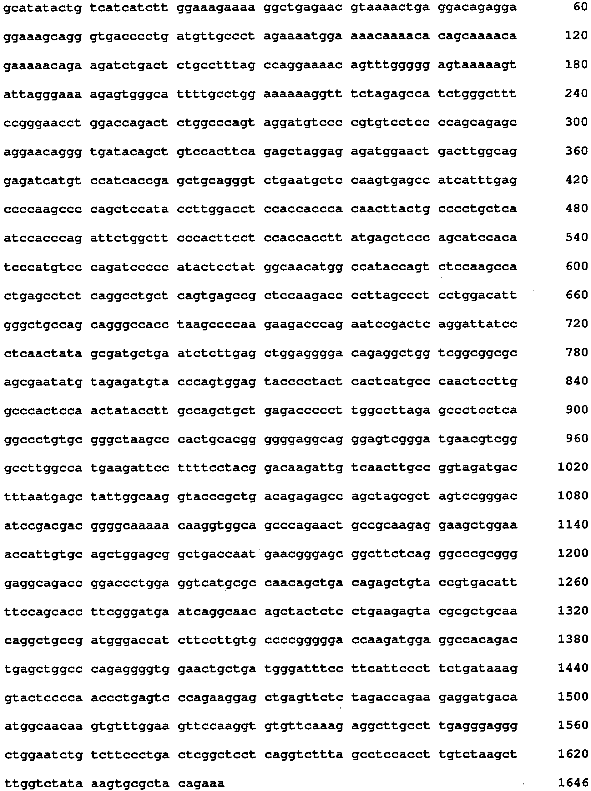
- <210> 9
<211> 1646
<212> DNA
<213> Homo sapiens - <400> 9
- <210> 10
<211> 369
<212> PRT
<213> Homo sapiens - <400> 10


- <210> 11
<211> 1657
<212> DNA
<213> Homo sapiens - <400> 11


- <210> 12
<211> 109
<212> PRT
<213> Homo sapiens - <400> 12

- <210> 13
<211> 1659
<212> DNA
<213> Homo sapiens - <400> 13

- <210> 14
<211> 303
<212> PRT
<213> Homo sapiens - <400> 14


- <210> 15
<211> 22
<212> DNA
<213> artificial sequence - <220>
<223> primer - <400> 15
tctttctggc ctggaggcta tc 22 - <210> 16
<211> 22
<212> DNA
<213> artificial sequence - <220>
<223> primer - <400> 16
tgctggatga cgtgagtaaa cc 22 - <210> 17
<211> 16
<212> DNA
<213> artificial sequence - <220>
<223> probe - <400> 17
agcgtactcc aaagat 16 - <210> 18
<211> 23
<212> DNA
<213> artificial sequence - <220>
<223> primer - <400> 18
cggtctctcc ttccctcagg gga 23 - <210> 19
<211> 22
<212> DNA
<213> artificial sequence - <220>
<223> primer - <400> 19
tcttctccga cacacggcct cc 22 - <210> 20
<211> 26
<212> DNA
<213> artificial sequence - <220>
<223> primer - <400> 20
tgcgactttc agaagaatcc agcttg 26 - <210> 21
<211> 22
<212> DNA
<213> Artificial Sequence - <220>
<223> primer - <400> 21
tggagtgggc caaggagttg gg 22 - <210> 22
<211> 20
<212> DNA
<213> Artificial Sequence - <220>
<223> primer - <400> 22
tcggcggcgc agcgaatatg 20 - <210> 23
<211> 24
<212> DNA
<213> Artificial Sequence - <220>
<223> primer - <400> 23
gtagctgttg cctgattcat cccg 24 - <210> 24
<211> 23
<212> DNA
<213> Artificial Sequence - <220>
<223> primer - <400> 24
gaggtcatgc gccaacagct gac 23 - <210> 25
<211> 24
<212> DNA
<213> Artificial Sequence - <220>
<223> primer - <400> 25
tgctcagcct caagcccaaa tttt 24
Claims (11)
- Ex vivo or in vitro method to determine the presence or absence of a mutation or a variation which lead to changes in the amino acid sequence or to a frameshift in the NF-E2 gene in a sample from a patient suffering from a myeloid neoplasia or a solid tumor, whereby said method comprises:a) obtaining a nucleic acid sample from the patient, whereby the sample is obtained from blood, bone marrow, or from a biopsy of a solid tumor,b) detecting the presence or absence of a mutation in the nucleic acid sample comprising the NF-E2 gene or a fragment thereof or of a variation of the NF-E2 gene, characterized in that the presence of the mutation or of the variation is an indication of a myeloid neoplasm, in particular, a myeloproliferative neoplasm, a myelodysplastic syndrome, a chronic myeloid leukemia or an acute myeloid leukemia, or a solid tumor, whereby the mutation is determined by comparing the nucleic acid sequence obtained from the sample with SEQ ID NO:2 or the complement thereof, or whereby a variation of the NF-E2 gene is a NF-E2 gene product which differs from the amino acid sequence as shown in SEQ ID NO:1.
- Method according to claim 1, characterized in that it comprises a hybridization step with at least one probe or primer comprising 10 to 30 consecutive nucleotides of SEQ ID NO:2 or the complement thereof.
- Method according to claim 1 or 2, characterized in that it comprises an amplification step by PCR reaction with at least one pair of primers comprising 10 to 30 consecutive nucleotides of SEQ ID NO:2 or the complement thereof.
- Method according to claim 3, characterized in that it comprises at least one amplification step by means of said primers followed by sequencing the amplified nucleic acid and comparing the sequence of the amplified nucleic acid with the corresponding stretch of SEQ ID NO:2 in order to detect the mutation in the NF-E2 gene or to determine a sequence variation comprising one or more primers or probes for the specific detection of the presence or absence of a mutation or variation in the NF-E2 gene.
- Method according to claim 1, characterized in that it is performed on a nucleic acid of a sample and comprises a real time PCR reaction.
- Method according to claim 1, characterized in that the myeloid neoplasia is selected from the group consisting of myeloproliferative neoplasms, myelodysplastic syndromes, acute myelogenous leukemia and chronic myelogenous leukemia.
- Method according to claim 6, characterized in that the myeloid neoplasia is a myeloproliferative disorder.
- Method according to any of claims 1-7, characterized in that in the PCR reaction at least any one of the sequences consisting of SEQ ID NOS:18, 19, 20, 21, 22, 23, 24 and/or 25 is used.
- Method according to any of the preceding claims, characterized in that the method is performed in regular time intervals in order to monitor the therapeutic success.
- Kit for performing an in vitro method for determining the presence or absence of a mutation or a variation which leads to changes in the amino acid sequence or to a frameshift in the NF-E2 gene in a sample from a patient suffering from a myeloid neoplasia or a solid tumor, whereby a nucleic acid sample obtained from the patient is amplified with at least one primer selected from SEQ ID NOs: 18, 19, 20, 21, 22, 23, 24 and/or 25 and the presence or absence of the mutation or variation being an indication of a myeloid neoplasm, in particular a myeloproliferative neoplasm, a myelodysplastic syndrome, a chronic myeloid leukemia or an acute myeloid leukemia, or a solid tumor is determined by comparing the nucleic acid sequence obtained from the sample with SEQ ID NO:2 or the complement thereof, said kit comprising at least one primer selected from SEQ ID NOs: 18, 19, 20, 21, 22, 23, 24 and/or 25.
- Kit according to claim 10 also containing at least one element chosen from among a heat resistant polymerase for PCR amplification, one or more solutions for amplification and/or the hybridization step.
Priority Applications (6)
| Application Number | Priority Date | Filing Date | Title |
|---|---|---|---|
| EP12154405.0A EP2626432B1 (en) | 2012-02-08 | 2012-02-08 | Method for the diagnosis of myeloid neoplasias and solid tumors and diagnostic kits |
| DK12154405T DK2626432T3 (en) | 2012-02-08 | 2012-02-08 | A method for the diagnosis of myeloid neoplasias and solid tumors, and diagnostic kits |
| PCT/EP2013/052361 WO2013117609A1 (en) | 2012-02-08 | 2013-02-07 | Method for the diagnosis of myeloid neoplasias and solid tumors and diagnostic kits |
| CA2862550A CA2862550A1 (en) | 2012-02-08 | 2013-02-07 | Method for the diagnosis of myeloid neoplasias and solid tumors and diagnostic kits |
| US14/377,468 US9695480B2 (en) | 2012-02-08 | 2013-02-07 | Method for the diagnosis of myeloid neoplasias and solid tumors |
| HK14101085.8A HK1187962B (en) | 2014-02-05 | Method for the diagnosis of myeloid neoplasias and solid tumors and diagnostic kits |
Applications Claiming Priority (1)
| Application Number | Priority Date | Filing Date | Title |
|---|---|---|---|
| EP12154405.0A EP2626432B1 (en) | 2012-02-08 | 2012-02-08 | Method for the diagnosis of myeloid neoplasias and solid tumors and diagnostic kits |
Publications (2)
| Publication Number | Publication Date |
|---|---|
| EP2626432A1 EP2626432A1 (en) | 2013-08-14 |
| EP2626432B1 true EP2626432B1 (en) | 2014-11-12 |
Family
ID=47666144
Family Applications (1)
| Application Number | Title | Priority Date | Filing Date |
|---|---|---|---|
| EP12154405.0A Not-in-force EP2626432B1 (en) | 2012-02-08 | 2012-02-08 | Method for the diagnosis of myeloid neoplasias and solid tumors and diagnostic kits |
Country Status (5)
| Country | Link |
|---|---|
| US (1) | US9695480B2 (en) |
| EP (1) | EP2626432B1 (en) |
| CA (1) | CA2862550A1 (en) |
| DK (1) | DK2626432T3 (en) |
| WO (1) | WO2013117609A1 (en) |
Families Citing this family (6)
| Publication number | Priority date | Publication date | Assignee | Title |
|---|---|---|---|---|
| US10173687B2 (en) | 2015-03-16 | 2019-01-08 | Wellen Sham | Method for recognizing vehicle driver and determining whether driver can start vehicle |
| US10703211B2 (en) | 2015-03-16 | 2020-07-07 | Thunder Power New Energy Vehicle Development Company Limited | Battery pack, battery charging station, and charging method |
| US9954260B2 (en) | 2015-03-16 | 2018-04-24 | Thunder Power New Energy Vehicle Development Company Limited | Battery system with heat exchange device |
| US9550406B2 (en) | 2015-03-16 | 2017-01-24 | Thunder Power Hong Kong Ltd. | Thermal dissipation system of an electric vehicle |
| US9499067B2 (en) | 2015-03-16 | 2016-11-22 | Thunder Power Hong Kong Ltd. | Power management in electric vehicles |
| AT518514B1 (en) * | 2016-03-29 | 2021-06-15 | Unique Product & Design Co Ltd | Carts, particularly golf carts |
Citations (1)
| Publication number | Priority date | Publication date | Assignee | Title |
|---|---|---|---|---|
| US20110104680A1 (en) * | 2009-10-06 | 2011-05-05 | The Regents Of The University Of Michigan | Recurrent gene fusions in lung cancer |
Family Cites Families (5)
| Publication number | Priority date | Publication date | Assignee | Title |
|---|---|---|---|---|
| US20020197611A1 (en) * | 2001-06-21 | 2002-12-26 | Chagovetz Alexander Michael | Method for real-time detection and quantification of nucleic acid sequences using fluorescent primers |
| US20050118625A1 (en) * | 2003-10-02 | 2005-06-02 | Mounts William M. | Nucleic acid arrays for detecting gene expression associated with human osteoarthritis and human proteases |
| US20050250134A1 (en) * | 2004-03-02 | 2005-11-10 | Dawei Sheng | Fluorescent energy transfer labeled nucleic acid substrates and methods of use thereof |
| KR100789731B1 (en) * | 2006-11-15 | 2008-01-03 | 전남대학교산학협력단 | Quantitative Detection, primers, and kits of BA2 V617F Mutants Using Pyro Sequencing |
| JP5666134B2 (en) * | 2007-01-10 | 2015-02-12 | ヘモシアー・リミテッド・ライアビリティ・カンパニーHemoShear, LLC | Use of an in vitro hemodynamic endothelial / smooth muscle cell co-culture model to identify new therapeutic targets for vascular disease |
-
2012
- 2012-02-08 DK DK12154405T patent/DK2626432T3/en active
- 2012-02-08 EP EP12154405.0A patent/EP2626432B1/en not_active Not-in-force
-
2013
- 2013-02-07 CA CA2862550A patent/CA2862550A1/en not_active Abandoned
- 2013-02-07 WO PCT/EP2013/052361 patent/WO2013117609A1/en not_active Ceased
- 2013-02-07 US US14/377,468 patent/US9695480B2/en not_active Expired - Fee Related
Patent Citations (1)
| Publication number | Priority date | Publication date | Assignee | Title |
|---|---|---|---|---|
| US20110104680A1 (en) * | 2009-10-06 | 2011-05-05 | The Regents Of The University Of Michigan | Recurrent gene fusions in lung cancer |
Also Published As
| Publication number | Publication date |
|---|---|
| HK1187962A1 (en) | 2014-04-17 |
| DK2626432T3 (en) | 2015-02-23 |
| US20150017642A1 (en) | 2015-01-15 |
| WO2013117609A1 (en) | 2013-08-15 |
| EP2626432A1 (en) | 2013-08-14 |
| US9695480B2 (en) | 2017-07-04 |
| CA2862550A1 (en) | 2013-08-15 |
Similar Documents
| Publication | Publication Date | Title |
|---|---|---|
| Jutzi et al. | MPN patients harbor recurrent truncating mutations in transcription factor NF-E2 | |
| Martin et al. | Dimerization of MLL fusion proteins immortalizes hematopoietic cells | |
| Jutzi et al. | Altered NFE2 activity predisposes to leukemic transformation and myelosarcoma with AML-specific aberrations | |
| US9695480B2 (en) | Method for the diagnosis of myeloid neoplasias and solid tumors | |
| US20200255912A1 (en) | TET2 as a Diagnostic and Pronostic Marker in Hematopoietic Neoplasms | |
| Métais et al. | The MDS1–EVI1 gene complex as a retrovirus integration site: impact on behavior of hematopoietic cells and implications for gene therapy | |
| EP1469072B1 (en) | Means and methods for diagnosing and treating affective disorders | |
| Bluteau et al. | Down-regulation of the RUNX1-target gene NR4A3 contributes to hematopoiesis deregulation in familial platelet disorder/acute myelogenous leukemia | |
| US20110117562A1 (en) | Diagnosis and Treatment of Chronic Lymphocytic Leukemia (CLL) | |
| US20150299336A1 (en) | Therapeutic and diagnostic target gene in acute myeloid leukemia | |
| Rodriguez-Mora et al. | The mutation of Transportin 3 gene that causes limb girdle muscular dystrophy 1F induces protection against HIV-1 infection | |
| Mitchell et al. | Dysregulated expression of COOH-terminally truncated Stat5 and loss of IL2-inducible Stat5-dependent gene expression in Sezary Syndrome | |
| WO2018083189A1 (en) | Biomarkers for determining responsiveness to lsd1 inhibitors | |
| US20200239961A1 (en) | Prognostic marker for myeloproliferative neoplasms | |
| ES2684548T3 (en) | Fusion protein comprising AXL and composition for treating cancer comprising the same | |
| US20030171273A1 (en) | Novel transcription factor, BP1 | |
| Ma et al. | Abnormal splicing of SHP-1 protein tyrosine phosphatase in human T cells: Implications for lymphomagenesis | |
| JP2009529869A (en) | Cancer detection and treatment | |
| HK1187962B (en) | Method for the diagnosis of myeloid neoplasias and solid tumors and diagnostic kits | |
| ES2618310T3 (en) | Mutations in the LNK gene in patients with myeloproliferative neoplasia and other hematolymphoid malignancies | |
| US7807362B2 (en) | Diagnosis of attention deficit hyperactivity disorder | |
| US20120282258A1 (en) | Crlf2 in precursor b-cell acute lymphoblastic leukemia | |
| WO2010087702A1 (en) | TET2 gene as a marker for diagnosing a myelodysuplastic syndrome (MDS) or an acute myeloid leukemia (AML) and determining the prognosis in a subject | |
| Aliano et al. | HMGA2 overexpression in polycythemia vera with t (12; 21)(q14; q22) | |
| Vieira et al. | Three‐way translocation (X; 20; 16)(p11; q13; q23) in essential thrombocythemia implicates NFATC2 in dysregulation of CSF2 expression and megakaryocyte proliferation |
Legal Events
| Date | Code | Title | Description |
|---|---|---|---|
| PUAI | Public reference made under article 153(3) epc to a published international application that has entered the european phase |
Free format text: ORIGINAL CODE: 0009012 |
|
| AK | Designated contracting states |
Kind code of ref document: A1 Designated state(s): AL AT BE BG CH CY CZ DE DK EE ES FI FR GB GR HR HU IE IS IT LI LT LU LV MC MK MT NL NO PL PT RO RS SE SI SK SM TR |
|
| AX | Request for extension of the european patent |
Extension state: BA ME |
|
| 17P | Request for examination filed |
Effective date: 20140130 |
|
| RBV | Designated contracting states (corrected) |
Designated state(s): AL AT BE BG CH CY CZ DE DK EE ES FI FR GB GR HR HU IE IS IT LI LT LU LV MC MK MT NL NO PL PT RO RS SE SI SK SM TR |
|
| REG | Reference to a national code |
Ref country code: HK Ref legal event code: DE Ref document number: 1187962 Country of ref document: HK |
|
| GRAP | Despatch of communication of intention to grant a patent |
Free format text: ORIGINAL CODE: EPIDOSNIGR1 |
|
| INTG | Intention to grant announced |
Effective date: 20140521 |
|
| GRAS | Grant fee paid |
Free format text: ORIGINAL CODE: EPIDOSNIGR3 |
|
| GRAA | (expected) grant |
Free format text: ORIGINAL CODE: 0009210 |
|
| AK | Designated contracting states |
Kind code of ref document: B1 Designated state(s): AL AT BE BG CH CY CZ DE DK EE ES FI FR GB GR HR HU IE IS IT LI LT LU LV MC MK MT NL NO PL PT RO RS SE SI SK SM TR |
|
| REG | Reference to a national code |
Ref country code: GB Ref legal event code: FG4D |
|
| REG | Reference to a national code |
Ref country code: CH Ref legal event code: EP |
|
| REG | Reference to a national code |
Ref country code: AT Ref legal event code: REF Ref document number: 695811 Country of ref document: AT Kind code of ref document: T Effective date: 20141115 |
|
| REG | Reference to a national code |
Ref country code: IE Ref legal event code: FG4D |
|
| REG | Reference to a national code |
Ref country code: DE Ref legal event code: R096 Ref document number: 602012003709 Country of ref document: DE Effective date: 20141224 |
|
| REG | Reference to a national code |
Ref country code: DK Ref legal event code: T3 Effective date: 20150216 |
|
| REG | Reference to a national code |
Ref country code: NL Ref legal event code: T3 |
|
| REG | Reference to a national code |
Ref country code: SE Ref legal event code: TRGR |
|
| PG25 | Lapsed in a contracting state [announced via postgrant information from national office to epo] |
Ref country code: ES Free format text: LAPSE BECAUSE OF FAILURE TO SUBMIT A TRANSLATION OF THE DESCRIPTION OR TO PAY THE FEE WITHIN THE PRESCRIBED TIME-LIMIT Effective date: 20141112 Ref country code: LT Free format text: LAPSE BECAUSE OF FAILURE TO SUBMIT A TRANSLATION OF THE DESCRIPTION OR TO PAY THE FEE WITHIN THE PRESCRIBED TIME-LIMIT Effective date: 20141112 Ref country code: IS Free format text: LAPSE BECAUSE OF FAILURE TO SUBMIT A TRANSLATION OF THE DESCRIPTION OR TO PAY THE FEE WITHIN THE PRESCRIBED TIME-LIMIT Effective date: 20150312 Ref country code: FI Free format text: LAPSE BECAUSE OF FAILURE TO SUBMIT A TRANSLATION OF THE DESCRIPTION OR TO PAY THE FEE WITHIN THE PRESCRIBED TIME-LIMIT Effective date: 20141112 Ref country code: PT Free format text: LAPSE BECAUSE OF FAILURE TO SUBMIT A TRANSLATION OF THE DESCRIPTION OR TO PAY THE FEE WITHIN THE PRESCRIBED TIME-LIMIT Effective date: 20150312 Ref country code: NO Free format text: LAPSE BECAUSE OF FAILURE TO SUBMIT A TRANSLATION OF THE DESCRIPTION OR TO PAY THE FEE WITHIN THE PRESCRIBED TIME-LIMIT Effective date: 20150212 |
|
| PG25 | Lapsed in a contracting state [announced via postgrant information from national office to epo] |
Ref country code: RS Free format text: LAPSE BECAUSE OF FAILURE TO SUBMIT A TRANSLATION OF THE DESCRIPTION OR TO PAY THE FEE WITHIN THE PRESCRIBED TIME-LIMIT Effective date: 20141112 Ref country code: LV Free format text: LAPSE BECAUSE OF FAILURE TO SUBMIT A TRANSLATION OF THE DESCRIPTION OR TO PAY THE FEE WITHIN THE PRESCRIBED TIME-LIMIT Effective date: 20141112 Ref country code: CY Free format text: LAPSE BECAUSE OF FAILURE TO SUBMIT A TRANSLATION OF THE DESCRIPTION OR TO PAY THE FEE WITHIN THE PRESCRIBED TIME-LIMIT Effective date: 20141112 Ref country code: PL Free format text: LAPSE BECAUSE OF FAILURE TO SUBMIT A TRANSLATION OF THE DESCRIPTION OR TO PAY THE FEE WITHIN THE PRESCRIBED TIME-LIMIT Effective date: 20141112 Ref country code: HR Free format text: LAPSE BECAUSE OF FAILURE TO SUBMIT A TRANSLATION OF THE DESCRIPTION OR TO PAY THE FEE WITHIN THE PRESCRIBED TIME-LIMIT Effective date: 20141112 Ref country code: GR Free format text: LAPSE BECAUSE OF FAILURE TO SUBMIT A TRANSLATION OF THE DESCRIPTION OR TO PAY THE FEE WITHIN THE PRESCRIBED TIME-LIMIT Effective date: 20150213 |
|
| PG25 | Lapsed in a contracting state [announced via postgrant information from national office to epo] |
Ref country code: SK Free format text: LAPSE BECAUSE OF FAILURE TO SUBMIT A TRANSLATION OF THE DESCRIPTION OR TO PAY THE FEE WITHIN THE PRESCRIBED TIME-LIMIT Effective date: 20141112 Ref country code: CZ Free format text: LAPSE BECAUSE OF FAILURE TO SUBMIT A TRANSLATION OF THE DESCRIPTION OR TO PAY THE FEE WITHIN THE PRESCRIBED TIME-LIMIT Effective date: 20141112 Ref country code: EE Free format text: LAPSE BECAUSE OF FAILURE TO SUBMIT A TRANSLATION OF THE DESCRIPTION OR TO PAY THE FEE WITHIN THE PRESCRIBED TIME-LIMIT Effective date: 20141112 Ref country code: RO Free format text: LAPSE BECAUSE OF FAILURE TO SUBMIT A TRANSLATION OF THE DESCRIPTION OR TO PAY THE FEE WITHIN THE PRESCRIBED TIME-LIMIT Effective date: 20141112 |
|
| REG | Reference to a national code |
Ref country code: DE Ref legal event code: R097 Ref document number: 602012003709 Country of ref document: DE |
|
| PLBE | No opposition filed within time limit |
Free format text: ORIGINAL CODE: 0009261 |
|
| REG | Reference to a national code |
Ref country code: HK Ref legal event code: GR Ref document number: 1187962 Country of ref document: HK |
|
| STAA | Information on the status of an ep patent application or granted ep patent |
Free format text: STATUS: NO OPPOSITION FILED WITHIN TIME LIMIT |
|
| PG25 | Lapsed in a contracting state [announced via postgrant information from national office to epo] |
Ref country code: LU Free format text: LAPSE BECAUSE OF FAILURE TO SUBMIT A TRANSLATION OF THE DESCRIPTION OR TO PAY THE FEE WITHIN THE PRESCRIBED TIME-LIMIT Effective date: 20150208 |
|
| 26N | No opposition filed |
Effective date: 20150813 |
|
| PG25 | Lapsed in a contracting state [announced via postgrant information from national office to epo] |
Ref country code: MC Free format text: LAPSE BECAUSE OF FAILURE TO SUBMIT A TRANSLATION OF THE DESCRIPTION OR TO PAY THE FEE WITHIN THE PRESCRIBED TIME-LIMIT Effective date: 20141112 |
|
| REG | Reference to a national code |
Ref country code: IE Ref legal event code: MM4A |
|
| PG25 | Lapsed in a contracting state [announced via postgrant information from national office to epo] |
Ref country code: IT Free format text: LAPSE BECAUSE OF FAILURE TO SUBMIT A TRANSLATION OF THE DESCRIPTION OR TO PAY THE FEE WITHIN THE PRESCRIBED TIME-LIMIT Effective date: 20141112 |
|
| PG25 | Lapsed in a contracting state [announced via postgrant information from national office to epo] |
Ref country code: IE Free format text: LAPSE BECAUSE OF NON-PAYMENT OF DUE FEES Effective date: 20150208 |
|
| REG | Reference to a national code |
Ref country code: FR Ref legal event code: PLFP Year of fee payment: 5 |
|
| PG25 | Lapsed in a contracting state [announced via postgrant information from national office to epo] |
Ref country code: SI Free format text: LAPSE BECAUSE OF FAILURE TO SUBMIT A TRANSLATION OF THE DESCRIPTION OR TO PAY THE FEE WITHIN THE PRESCRIBED TIME-LIMIT Effective date: 20141112 |
|
| REG | Reference to a national code |
Ref country code: AT Ref legal event code: UEP Ref document number: 695811 Country of ref document: AT Kind code of ref document: T Effective date: 20141112 |
|
| PG25 | Lapsed in a contracting state [announced via postgrant information from national office to epo] |
Ref country code: MT Free format text: LAPSE BECAUSE OF FAILURE TO SUBMIT A TRANSLATION OF THE DESCRIPTION OR TO PAY THE FEE WITHIN THE PRESCRIBED TIME-LIMIT Effective date: 20141112 |
|
| REG | Reference to a national code |
Ref country code: FR Ref legal event code: PLFP Year of fee payment: 6 |
|
| PG25 | Lapsed in a contracting state [announced via postgrant information from national office to epo] |
Ref country code: BG Free format text: LAPSE BECAUSE OF FAILURE TO SUBMIT A TRANSLATION OF THE DESCRIPTION OR TO PAY THE FEE WITHIN THE PRESCRIBED TIME-LIMIT Effective date: 20141112 Ref country code: HU Free format text: LAPSE BECAUSE OF FAILURE TO SUBMIT A TRANSLATION OF THE DESCRIPTION OR TO PAY THE FEE WITHIN THE PRESCRIBED TIME-LIMIT; INVALID AB INITIO Effective date: 20120208 Ref country code: SM Free format text: LAPSE BECAUSE OF FAILURE TO SUBMIT A TRANSLATION OF THE DESCRIPTION OR TO PAY THE FEE WITHIN THE PRESCRIBED TIME-LIMIT Effective date: 20141112 |
|
| PG25 | Lapsed in a contracting state [announced via postgrant information from national office to epo] |
Ref country code: TR Free format text: LAPSE BECAUSE OF FAILURE TO SUBMIT A TRANSLATION OF THE DESCRIPTION OR TO PAY THE FEE WITHIN THE PRESCRIBED TIME-LIMIT Effective date: 20141112 |
|
| REG | Reference to a national code |
Ref country code: FR Ref legal event code: PLFP Year of fee payment: 7 |
|
| PGFP | Annual fee paid to national office [announced via postgrant information from national office to epo] |
Ref country code: DK Payment date: 20180221 Year of fee payment: 7 |
|
| PGFP | Annual fee paid to national office [announced via postgrant information from national office to epo] |
Ref country code: SE Payment date: 20180222 Year of fee payment: 7 |
|
| PG25 | Lapsed in a contracting state [announced via postgrant information from national office to epo] |
Ref country code: MK Free format text: LAPSE BECAUSE OF FAILURE TO SUBMIT A TRANSLATION OF THE DESCRIPTION OR TO PAY THE FEE WITHIN THE PRESCRIBED TIME-LIMIT Effective date: 20141112 |
|
| PG25 | Lapsed in a contracting state [announced via postgrant information from national office to epo] |
Ref country code: AL Free format text: LAPSE BECAUSE OF FAILURE TO SUBMIT A TRANSLATION OF THE DESCRIPTION OR TO PAY THE FEE WITHIN THE PRESCRIBED TIME-LIMIT Effective date: 20141112 |
|
| PGFP | Annual fee paid to national office [announced via postgrant information from national office to epo] |
Ref country code: NL Payment date: 20190219 Year of fee payment: 8 |
|
| PGFP | Annual fee paid to national office [announced via postgrant information from national office to epo] |
Ref country code: CH Payment date: 20190221 Year of fee payment: 8 Ref country code: GB Payment date: 20190221 Year of fee payment: 8 Ref country code: DE Payment date: 20190219 Year of fee payment: 8 |
|
| PGFP | Annual fee paid to national office [announced via postgrant information from national office to epo] |
Ref country code: FR Payment date: 20190221 Year of fee payment: 8 Ref country code: BE Payment date: 20190219 Year of fee payment: 8 Ref country code: AT Payment date: 20190215 Year of fee payment: 8 |
|
| REG | Reference to a national code |
Ref country code: DK Ref legal event code: EBP Effective date: 20190228 |
|
| REG | Reference to a national code |
Ref country code: SE Ref legal event code: EUG |
|
| PG25 | Lapsed in a contracting state [announced via postgrant information from national office to epo] |
Ref country code: SE Free format text: LAPSE BECAUSE OF NON-PAYMENT OF DUE FEES Effective date: 20190209 |
|
| PG25 | Lapsed in a contracting state [announced via postgrant information from national office to epo] |
Ref country code: DK Free format text: LAPSE BECAUSE OF NON-PAYMENT OF DUE FEES Effective date: 20190228 |
|
| REG | Reference to a national code |
Ref country code: DE Ref legal event code: R119 Ref document number: 602012003709 Country of ref document: DE |
|
| REG | Reference to a national code |
Ref country code: CH Ref legal event code: PL |
|
| REG | Reference to a national code |
Ref country code: NL Ref legal event code: MM Effective date: 20200301 |
|
| REG | Reference to a national code |
Ref country code: AT Ref legal event code: MM01 Ref document number: 695811 Country of ref document: AT Kind code of ref document: T Effective date: 20200208 |
|
| GBPC | Gb: european patent ceased through non-payment of renewal fee |
Effective date: 20200208 |
|
| REG | Reference to a national code |
Ref country code: BE Ref legal event code: MM Effective date: 20200229 |
|
| PG25 | Lapsed in a contracting state [announced via postgrant information from national office to epo] |
Ref country code: CH Free format text: LAPSE BECAUSE OF NON-PAYMENT OF DUE FEES Effective date: 20200229 Ref country code: LI Free format text: LAPSE BECAUSE OF NON-PAYMENT OF DUE FEES Effective date: 20200229 Ref country code: AT Free format text: LAPSE BECAUSE OF NON-PAYMENT OF DUE FEES Effective date: 20200208 |
|
| PG25 | Lapsed in a contracting state [announced via postgrant information from national office to epo] |
Ref country code: NL Free format text: LAPSE BECAUSE OF NON-PAYMENT OF DUE FEES Effective date: 20200301 |
|
| PG25 | Lapsed in a contracting state [announced via postgrant information from national office to epo] |
Ref country code: FR Free format text: LAPSE BECAUSE OF NON-PAYMENT OF DUE FEES Effective date: 20200229 Ref country code: GB Free format text: LAPSE BECAUSE OF NON-PAYMENT OF DUE FEES Effective date: 20200208 Ref country code: DE Free format text: LAPSE BECAUSE OF NON-PAYMENT OF DUE FEES Effective date: 20200901 |
|
| PG25 | Lapsed in a contracting state [announced via postgrant information from national office to epo] |
Ref country code: BE Free format text: LAPSE BECAUSE OF NON-PAYMENT OF DUE FEES Effective date: 20200229 |