EP2623978A1 - CD8+ T-cell subsets as markers for prediction of delayed fracture healing - Google Patents
CD8+ T-cell subsets as markers for prediction of delayed fracture healing Download PDFInfo
- Publication number
- EP2623978A1 EP2623978A1 EP12153850.8A EP12153850A EP2623978A1 EP 2623978 A1 EP2623978 A1 EP 2623978A1 EP 12153850 A EP12153850 A EP 12153850A EP 2623978 A1 EP2623978 A1 EP 2623978A1
- Authority
- EP
- European Patent Office
- Prior art keywords
- cells
- frequency
- delayed
- healing
- fracture healing
- Prior art date
- Legal status (The legal status is an assumption and is not a legal conclusion. Google has not performed a legal analysis and makes no representation as to the accuracy of the status listed.)
- Withdrawn
Links
Images



Classifications
-
- G—PHYSICS
- G01—MEASURING; TESTING
- G01N—INVESTIGATING OR ANALYSING MATERIALS BY DETERMINING THEIR CHEMICAL OR PHYSICAL PROPERTIES
- G01N33/00—Investigating or analysing materials by specific methods not covered by groups G01N1/00 - G01N31/00
- G01N33/48—Biological material, e.g. blood, urine; Haemocytometers
- G01N33/50—Chemical analysis of biological material, e.g. blood, urine; Testing involving biospecific ligand binding methods; Immunological testing
- G01N33/53—Immunoassay; Biospecific binding assay; Materials therefor
- G01N33/569—Immunoassay; Biospecific binding assay; Materials therefor for microorganisms, e.g. protozoa, bacteria, viruses
- G01N33/56966—Animal cells
-
- A—HUMAN NECESSITIES
- A61—MEDICAL OR VETERINARY SCIENCE; HYGIENE
- A61K—PREPARATIONS FOR MEDICAL, DENTAL OR TOILETRY PURPOSES
- A61K31/00—Medicinal preparations containing organic active ingredients
-
- A—HUMAN NECESSITIES
- A61—MEDICAL OR VETERINARY SCIENCE; HYGIENE
- A61K—PREPARATIONS FOR MEDICAL, DENTAL OR TOILETRY PURPOSES
- A61K39/00—Medicinal preparations containing antigens or antibodies
- A61K39/395—Antibodies; Immunoglobulins; Immune serum, e.g. antilymphocytic serum
-
- A—HUMAN NECESSITIES
- A61—MEDICAL OR VETERINARY SCIENCE; HYGIENE
- A61P—SPECIFIC THERAPEUTIC ACTIVITY OF CHEMICAL COMPOUNDS OR MEDICINAL PREPARATIONS
- A61P19/00—Drugs for skeletal disorders
-
- C—CHEMISTRY; METALLURGY
- C07—ORGANIC CHEMISTRY
- C07K—PEPTIDES
- C07K16/00—Immunoglobulins [IG], e.g. monoclonal or polyclonal antibodies
- C07K16/18—Immunoglobulins [IG], e.g. monoclonal or polyclonal antibodies against material from animals or humans
- C07K16/24—Immunoglobulins [IG], e.g. monoclonal or polyclonal antibodies against material from animals or humans against cytokines, lymphokines or interferons
- C07K16/241—Tumor Necrosis Factors
-
- C—CHEMISTRY; METALLURGY
- C07—ORGANIC CHEMISTRY
- C07K—PEPTIDES
- C07K16/00—Immunoglobulins [IG], e.g. monoclonal or polyclonal antibodies
- C07K16/18—Immunoglobulins [IG], e.g. monoclonal or polyclonal antibodies against material from animals or humans
- C07K16/24—Immunoglobulins [IG], e.g. monoclonal or polyclonal antibodies against material from animals or humans against cytokines, lymphokines or interferons
- C07K16/249—Interferons
-
- G—PHYSICS
- G01—MEASURING; TESTING
- G01N—INVESTIGATING OR ANALYSING MATERIALS BY DETERMINING THEIR CHEMICAL OR PHYSICAL PROPERTIES
- G01N33/00—Investigating or analysing materials by specific methods not covered by groups G01N1/00 - G01N31/00
- G01N33/48—Biological material, e.g. blood, urine; Haemocytometers
- G01N33/50—Chemical analysis of biological material, e.g. blood, urine; Testing involving biospecific ligand binding methods; Immunological testing
- G01N33/53—Immunoassay; Biospecific binding assay; Materials therefor
- G01N33/569—Immunoassay; Biospecific binding assay; Materials therefor for microorganisms, e.g. protozoa, bacteria, viruses
- G01N33/56966—Animal cells
- G01N33/56972—White blood cells
-
- C—CHEMISTRY; METALLURGY
- C07—ORGANIC CHEMISTRY
- C07K—PEPTIDES
- C07K2317/00—Immunoglobulins specific features
- C07K2317/70—Immunoglobulins specific features characterized by effect upon binding to a cell or to an antigen
-
- C—CHEMISTRY; METALLURGY
- C07—ORGANIC CHEMISTRY
- C07K—PEPTIDES
- C07K2317/00—Immunoglobulins specific features
- C07K2317/70—Immunoglobulins specific features characterized by effect upon binding to a cell or to an antigen
- C07K2317/76—Antagonist effect on antigen, e.g. neutralization or inhibition of binding
-
- G—PHYSICS
- G01—MEASURING; TESTING
- G01N—INVESTIGATING OR ANALYSING MATERIALS BY DETERMINING THEIR CHEMICAL OR PHYSICAL PROPERTIES
- G01N2333/00—Assays involving biological materials from specific organisms or of a specific nature
- G01N2333/435—Assays involving biological materials from specific organisms or of a specific nature from animals; from humans
- G01N2333/705—Assays involving receptors, cell surface antigens or cell surface determinants
- G01N2333/70503—Immunoglobulin superfamily, e.g. VCAMs, PECAM, LFA-3
- G01N2333/70517—CD8
-
- G—PHYSICS
- G01—MEASURING; TESTING
- G01N—INVESTIGATING OR ANALYSING MATERIALS BY DETERMINING THEIR CHEMICAL OR PHYSICAL PROPERTIES
- G01N2333/00—Assays involving biological materials from specific organisms or of a specific nature
- G01N2333/435—Assays involving biological materials from specific organisms or of a specific nature from animals; from humans
- G01N2333/705—Assays involving receptors, cell surface antigens or cell surface determinants
- G01N2333/70596—Molecules with a "CD"-designation not provided for elsewhere in G01N2333/705
-
- G—PHYSICS
- G01—MEASURING; TESTING
- G01N—INVESTIGATING OR ANALYSING MATERIALS BY DETERMINING THEIR CHEMICAL OR PHYSICAL PROPERTIES
- G01N2800/00—Detection or diagnosis of diseases
- G01N2800/10—Musculoskeletal or connective tissue disorders
-
- G—PHYSICS
- G01—MEASURING; TESTING
- G01N—INVESTIGATING OR ANALYSING MATERIALS BY DETERMINING THEIR CHEMICAL OR PHYSICAL PROPERTIES
- G01N2800/00—Detection or diagnosis of diseases
- G01N2800/52—Predicting or monitoring the response to treatment, e.g. for selection of therapy based on assay results in personalised medicine; Prognosis
Definitions
- the present invention relates to a method, a system and a kit for prediction of delayed bone fracture healing.
- Fracture healing is a physiological process with sequential, overlapping stages and results in a restoration of bone tissue. Under certain risk factors, however, such as severe fractures, old age, steroid therapy or diabetes, this process can be delayed or even incomplete (non-union healing) with poor long-term outcome and a high socio-economic impact. Delayed or incomplete healing can be observed in approximately 5-10% of patients following a fracture of the long bones.
- mice deficient for adaptive immunity surprisingly expressed improved bone healing ( Colburn et al. Arthritis Rheum.; 60(6):1694-703 ; Schmidt-Bleek et al. Cell Tissue Res ; DOI 10.1007/s00441-011-1205-7).
- a significantly higher cytotoxic T cell percentage within hematoma was found in sheep with delayed/impaired healing, due to a treatment with a mechanically critical external fixator having a high rotational instability, compared with rigidly fixated animals.
- Osteocalcin and bone alkaline phosphatase have been investigated as predictive markers for delayed fracture healing. Only osteocalcin, however, showed minor differences between patients with normal and delayed fracture healing, and not before forty-two days after the fracture occurred. Furthermore, transforming growth factor beta 1 (TGF-beta 1) has been discussed as another potential biomarker. Examinations showed that after 4 weeks post fracture TGF-beta 1 levels of patients with delayed bone fracture healing were lower than the levels of patients showing normal healing. However, these parameters have only a limited validity for the prediction of bone healing due to a high intra-inter patients variability, a short half life of the cytokines, and a late predictive time point during the fracture healing.
- TGF-beta 1 transforming growth factor beta 1
- the objective of the instant invention is to provide means and methods for the prediction of delayed bone fracture.
- the present invention was made during the course of an investigation assessing the inflammatory reaction and immune cell composition in peripheral blood of proximal tibial fracture patients during the healing process at typical time points of clinical relevance. Relevant differences in the immune cell composition in the peripheral blood during the fracture healing process related to a delayed healing were identified.
- CD3+ or the major subset distribution (CD3+4+ and CD3+8+) did not show differences between the groups (normal vs. delayed healing).
- delayed healing was strongly associated with a significantly enhanced frequency of terminally differentiated CD8+ effector T cells expressing the phenotype CD8+11a++28- and/or CD8+11a++57+ and/or CD8+11a++CD28-57+, which corresponds to CD8+ TEMRA cells (CD57+8+ delayed healing vs. normal: 1.6-1.8 fold, CD28-8+ delayed healing vs. normal: 1.5-1.6 fold; CD11++8+ delayed healing vs.
- CD8+ TEMRA cells also express the marker phenotype CCR7-CD45RA+CD45RO-. This difference was stable over follow-up time of 18 weeks reflecting rather the individual immune experience than the post-trauma reaction to the fracture.
- CD8+ TEMRA cells are characterized by their tissue homing properties and strong bystander responsiveness. They can be also triggered independently of their T-cell receptor (TCR) in an antigen-independent matter by cytokines, such as IL-6, IL-8, IL-12, IL-18, IL-23, or TNF ⁇ . Those cytokines are delivered by cells of the innate immune system triggered in the fracture hematoma as a result of interaction between toll-like receptor molecules (TLR) and damage-associated molecular patterns (DAMPs). Furthermore, macrophages and dendritic cells can trigger the release of inflammatory cytokines (e.g.
- IFN-gamma by these CD8+TEMRA cells, which support overwhelming inflammation and fibrosis as well as inhibition of osteogenesis. Additionally, these cells are up-regulated in chronic immune activation states, such as in infectious diseases, like HIV, tuberculosis or cytomegalovirus.
- an ex vivo method for prognosis of delayed bone fracture healing comprising determining the frequency of a subpopulation of CD8+ T cells selected from a first group comprised of CD8+CD57+ cells, CD8+CD28- cells and/or CD8+CD28-CD57+ cells, in a sample obtained from a subject.
- an ex vivo method for prognosis of delayed bone fracture healing comprising determining the frequency of a subpopulation of CD8+ T cells selected from a first group comprised of CD8+CD11a+CD57+ cells, CD8+ CD11a+CD28- cells and/or CD8+ CD11a+CD28-CD57+ cells, in a sample obtained from a subject.
- the sample is a blood sample, in particular obtained from peripheral blood, or a biopsy sample obtained from a region in the vicinity of a bone fracture, in particular from the inflammatory peri-fracture hematoma.
- the method of the invention may also be used for predicting the outcome after bone fracture or for classifying a sample from a subject, wherein the sample is assigned to a probability of an outcome after bone fracture.
- Frequency in the sense of the invention refers to the number of cells defined by certain marker molecules presented on the surface of these cells in relation to the number of cells of an entire definable population.
- a frequency of 5% for the CD8+CD4+ subpopulation of the CD3+ cells means that 5% of all CD3+ cells belong to the CD8+CD4+ subpopulation.
- cells may be characterized herein by showing the cluster of differentiation positivity/negativity in abbreviated form: CD4+8+ is synonymous with CD4+CD8+.
- CD4+8+ is synonymous with CD4+CD8+.
- the frequency of a given subpopulation is determined in relation to the total number of the parent population (as indicated in each table CD3+ or CD8+ cells, respectively) in the sample.
- any cell population is designated "positive” with respect to a certain marker molecule herein, this designation shall mean that said cell population can be stained by a common fluorescent-dye-labelled antibody against the marker molecule and will give a fluorescence signal of at least one, two or three log higher intensity compared to unlabelled cells or cells labelled with the same antibody but commonly known as not expressing said marker molecule.
- any cell population that is designated "negative” with respect to certain marker molecule cannot be stained by a fluorescent-dye-labelled antibody as described above against the marker molecule.
- Cells designated "double positive” or "++" with respect to a certain marker molecule means cells exhibiting a high expression of this certain marker molecule which can be separated as a distinct subpopulation by electronic gating.
- Fig. 8 shows a histogram of CD8/CD11 positive and double positive cells: of all CD11a positive cells encircled in the two example histograms, those on the right of the vertical bar constitute the "++" population.
- the method according to the above aspect of the invention comprises determining the frequency of subpopulation selected from a second group comprised of CD8+CD11a++, CD8+CD11a++CD28-, CD8+CD11a++CD57+, and CD8+CD11a++CD28-CD57+ T cells.
- the method according to the above aspect and embodiments of the invention further comprises determining the frequency of a CD8+/CD4+ subpopulation of CD3+ cells.
- the predictive or diagnostic value of the method of the invention may be augmented by determining the frequency of additional subpopulations as described above.
- the frequencies of the additional subpopulations may be determined consecutively or simultaneously. A simultaneous determination is preferred.
- the method further comprises determining the level of IL-6 in the peripheral blood.
- the predictive value of the method according to the above aspect and embodiments may further be enhanced by determination of IL-6 as an immunological marker of systemic inflammation.
- the level may be expressed as a concentration and measured in units such as pg/ml or mol/l.
- Such method according to the preceding aspect and embodiments of the invention may be performed by contacting a sample with a first ligand specifically reactive to a marker molecule selected from a marker group comprised of CD4 (Uniprot ID P01730), CD11a (Uniprot ID P20701), CD28 (Uniprot ID P10747), CD57 and IL-6 (Uniprot ID P05231), and determining the frequency of a cell presenting the marker molecule marked with the first ligand or determining the frequency of the first ligand bound to the marker molecule.
- a marker group comprised of CD4 (Uniprot ID P01730), CD11a (Uniprot ID P20701), CD28 (Uniprot ID P10747), CD57 and IL-6 (Uniprot ID P05231)
- Such first ligand may be chosen from the group comprised of an antibody, an antibody fragment, an antibody-like-molecule, an oligopeptide of 6 to 30 amino acids, and a nucleic acid aptamer molecule of 10 to 75 nucleotides in length, said ligand being capable to bind to a member of the marker group described in the preceding paragraph with a dissociation constant of 10 -8 mol/l or smaller.
- An antibody fragment may be the Fab domain of an antibody (the antigen binding region of an antibody) or a single chain antibody (scFv), a fusion protein consisting of the variable regions of light and heavy chains of an antibody connected by a peptide linker.
- An antibody fragment or an antibody-like molecule may be manufactured by suitable methods such as recombinant protein expression.
- a first ligand may also be developed by evolutive methods such as phage display, ribosome display or SELEX, wherein polypeptides or oligonucleotides are selected according to their binding affinity to a target of interest. Additionally, higher affinity inhibitors may be identified by reiterative rounds of evolution and selection of the amino acid sequence or nucleotide sequence.
- An oligopeptide of 6 to 30 amino acids as referred to above may be a peptide derived from the part of a ligand which is recognized by a member of the marker group described above.
- ligands are for example CD80 (Uniprot ID P33681) or CD86 (Uniprot ID P42081), which are ligands of CD28, or CD54 (Uniprot ID P05362), which is a CD11a ligand.
- the first ligand is an antibody reactive to CD4, CD11a, CD28, CD57 and further comprises a fluorescent moiety for optical detection, wherein a marker molecule according to the preceding paragraphs is bound to such antibody, and cells presenting that marker molecule can be counted by a fluorescence based flow cytometric method such as fluorescence activated cell sorting.
- the first ligand may an antibody reactive to IL-6 and may comprise an enzymatic activity, wherein this enzymatic activity is the catalysis of a reaction that can be spectroscopically observed.
- This first ligand may also be specifically bound by a second ligand, wherein the second ligand comprises an enzymatic activity or a fluorescent moiety.
- a plurality of different marker molecules may be determined by use of a plurality of different first ligands, wherein each ligand specifically binds to a particular marker molecule.
- each first ligand of the plurality comprises a certain enzymatic activity or fluorescent moiety as described above that can be spectroscopically distinguished form the enzymatic activity or fluorescent moiety of each other first ligand of the plurality.
- each first ligand is bound be a specific second ligand having a certain enzymatic activity or fluorescent moiety that can be spectroscopically distinguished for the enzymatic activity or fluorescent moiety of each other second ligand.
- the first and second ligand or pluralities thereof are antibodies and used in an Enzyme-linked immunosorbent assay.
- the frequency of a subpopulation according to the preceding aspect of the invention are determined by counting cells that are marked with a fluorescent antibody directed to a marker molecule selected from group comprised of CD4,CD8, CD11a, CD28 and CD57 in a flow cytometric assay.
- the method further comprises determining the Calori-Score of the subject according to the above aspect of the invention.
- the Calori-Score in the sense of the invention is a measure for nonunion, the permanent failure of healing following fracture, and may be determined by a method that assesses relevant fracture healing factors such as quality of bone, bone alignment, invasiveness of primary intervention or clinical infection status.
- a subject showing a Calori-Score that is at least 5 % higher than a standard is assigned to a group having an elevated probability for delayed fracture healing.
- a detailed description of this method can be found in Calori et al., Injury, 39, Supp 2, S59-63, 2008 .
- the method further comprises comparing the determined frequency, level or Calori-Score to a standard.
- a standard in the sense of the invention means a sample of a subject showing a normal or not delayed bone fracture healing.
- the standard may be a subject showing a normal bone fracture healing.
- fracture healing of a subject is considered normal if none of the following criteria are true:
- a sample exhibiting a twofold higher frequency of CD8+/CD4+ cells compared to a standard determined (retrospectively) for number of patients showing normal fracture healing is assigned to a group having an elevated probability for delayed fracture healing.
- a sample showing a frequency of T cells being CD11a++, CD28- or CD57+ that is at least 10 % higher compared to a standard determined (retrospectively) for number of patients showing normal fracture healing is assigned to a group having an elevated probability for delayed fracture healing.
- a sample showing a frequency of at least 30 % for cells being CD28- or CD57+ of the CD8+ T cells is assigned to a group having an elevated probability for delayed fracture healing
- a sample showing a frequency of at least 65 % for cells being CD11a++ of the CD8+ T cells is assigned to a group having an elevated probability for delayed fracture healing
- a sample showing a frequency of at least 5 % for cells being CD4+ of the CD8+ T cells is assigned to a group having an elevated probability for delayed fracture healing.
- a system for diagnosis of delayed bone fracture healing comprising
- Such device may be a flow cytometer, comprising a flow cell for transporting and aligning cells, a light source such as a laser and a detector suitable for measuring light or other biophysical parameters such as impedance.
- a flow cell for transporting and aligning cells
- a light source such as a laser
- a detector suitable for measuring light or other biophysical parameters such as impedance.
- Such device may be used for determining the frequency of subpopulations of CD8+ cells according to the above aspects and embodiments of the invention.
- such device may a spectrophotometer or a plate reader, comprising a compartment holding the sample such as a cuvette or a microtiter plate, a light source and a UV/Vis detector suitable for measurement of absorbance or fluorescence, such as a diode array.
- a spectrophotometer or a plate reader comprising a compartment holding the sample such as a cuvette or a microtiter plate, a light source and a UV/Vis detector suitable for measurement of absorbance or fluorescence, such as a diode array.
- a spectrophotometer or a plate reader comprising a compartment holding the sample such as a cuvette or a microtiter plate, a light source and a UV/Vis detector suitable for measurement of absorbance or fluorescence, such as a diode array.
- such device may be used for determining the level of IL-6 according to the above aspects or embodiments of the invention.
- Such programmed microprocessor may be integrated in the device described in the preceding paragraphs or may be part of a control unit or a computer for operating the device.
- the device described above is equipped and designated to determine the frequency of the subpopulation of CD8+ T cells according to the above aspects and embodiments of the invention.
- kits of parts for diagnosis of delayed bone fracture healing comprising an anti-CD8-antibody, an anti-CD4-antibody, an anti-CD11a-antibody and an anti-CD28-antibody, wherein the above described antibodies are suitable for fluorescence based flow cytometry.
- the kit further comprises an anti-CD57-antibody.
- the antibodies according to the above aspect or embodiment of the invention are monoclonal antibodies of murine origin and comprise a fluorescent moiety for optical detection in the flow cytometry such as APC (allophycocyanin), FITC (fluorescein isothiocyanate) or PE (phycoerythrin).
- APC allophycocyanin
- FITC fluorescein isothiocyanate
- PE phycoerythrin
- Examples for such antibodies include, without being restricted to, PE-Cy7 conjugated mouse anti-human CD4 IgG 1 , a murine monoclonal antibody conjugated with a PE-Cy7(cyanine dye) tandem fluorophor, APC-Cy7-labelled mouse anti-human CD8 IgG 1 , a murine monoclonal antibody conjugated with the tandem fluorophor APC-Cy7, FITC-labelled mouse anti-human CD57 IgM, a murine monoclonal antibody, APC-H7 mouse anti-human CD28 IgG 1 , a murine monoclonal antibody conjugated with the tandem fluorophor APC-H7 being an analogue of APC-Cy7 and having the same spectroscopic properties, and FITC-labelled mouse anti-human CD11a (Anti-LFA-1 ⁇ , leukocyte function associated antigen-1, alpha polypeptide) IgG 2a , a murine monoclonal antibody.
- first week three-five days post operative
- erythrocytes, haemoglobin, hematocrit, thrombocytes, creatinine, sodium, potassium, uric acid, ostase, CRP were measured in plasma and serum samples according to the laboratory standard operating procedures (SOPs).
- SOPs laboratory standard operating procedures
- the serum sample for osteocalcin assessment was immediately centrifuged (3500rpm/15 minutes), stored in pre-freezed aliquots and sent to the laboratory within three hours.
- Cytokines (TNF ⁇ , IL-6, total IL-8, IL-10) were measured in plasma samples by using a semiautomatic system.
- the antibodies and the respective reagents for intracellular cytokine staining were purchased from BD Pharmingen.
- T-cell related cluster of differentiation were examined to evaluate the adaptive immunity of the host defense including CD3, CD4, CD8, CD11a+, CD57+ and CD28+.
- Cell sorting was performed by using BD FACSAria II flow cytometer and the purity of the obtained fractions was determined on the BD LSRII flow cytometer.
- functional data were performed to assess the painless full weight bearing after 12 weeks post operative.
- Table 1 Patient characteristics Gait Analysis Pat. Nr. Age Weight (kg) Height (cm) BMI Smoking Healing Status 1 47 60 159 23,733 yes delayed 2 62 90 183 26,874 no normal 3 49 86 175 28,082 yes normal 4 57 53 153 22,641 no normal 5 39 80 168 28,345 no normal 6 40 70 184 20,676 yes delayed 7 59 62 158 24,836 no delayed 8 23 75 192 20,345 yes normal 9 58 97 168 34,368 yes normal 10 24 60 159 23,733 yes normal 11 46 80 176 25,826 yes normal 12 45 92 188 26,03 no delayed 13 46 86 174 28,405 yes normal 14 62 66 162 25,149 no delayed 16 25 90 185 26,297 no normal 17 49 81 172 27,38 yes delayed 18 64 70 180 21,605
- a 3-D-motion analysis was conducted using 12 infrared cameras at a sampling rate of 120 Hz and retro-reflective markers which were attached to the skin on the first and fifth metatarsal head and the heel. Ground reaction forces were collected by two force platforms at 960 Hz.
- the kinematic data allowed computation of gait parameters such as walking speed, step and stride length, cadence, stride height and the duration of swing and stance phase. Furthermore, the ground reaction forces were taken to compute the peak horizontal force and the stance duration for the affected and the contralateral limb.
- the patients also performed a sit to-stand test from a chair with each foot placed on a separate force platform to determine differences in the peak force between the extremities as well as the duration of the sit-to-stand test, reflected by the ground reaction force.
- ROC-curve receiver operation characteristics
- Each box plot shows median, lower and upper quartile.
- the Calori-Score is calculated by the following factors, wherein the factors are summed up and multiplied by two.
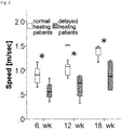
- Gait analyses were performed after 6, 12 and 18 weeks post operative to assess the functional status of the patients ( Fig. 2 ).
- the delayed healing patients meet the walking speed levels of the 6 th postoperative week in the normal healing group.
- Example 3 Frequency of CD8+/CD4+ cells in the CD3+ T cell population
- Example 4 Frequency of CD11a++ cells in the CD8+ T-cells population
- CD11a++ T-cells in the delayed healing group was consequently higher compared to normal healing patients, but reached the significance level only at the 12. postoperative week ( Fig. 4 ).
- normal healing patients had a CD11a++ percentage of the CD8+ T-cell levels well within the normal range
- delayed healing patients showed borderline levels over the complete study time. No differences were found in each group over time. Single cell values for each group and time point are shown in table 4.
- Example 5 Frequency of CD57+ cell in the CD8+-T-cell population
- Example 6 Frequency of CD28- cells in the CD8+-T cell population
- Delayed or even incomplete (non-union healing) healing of the long bones affects approximately 5-10% of the patients and is associated with poor long-term outcome with a high socio-economic impact.
- 41% of the enrolled patients showed a delayed healing as reflected by fracture gap or callus formation and reduced physical function over 18 weeks after the primary surgery intervention.
- the walking speed in the 18 th postoperative week of the delayed healing patients were comparable with the levels of the 6 th postoperative week in the normal healing group.
- CD8+CD57+ expressing lymphocytes are expanded in late stage chronic (viral) infections by dysregulation of the normal apoptotic pathway.
- Clinical interventions are assumed to play a major role in the patients healing course. In the study present here, no differences in the number of interventions, surgery methods or bone quality between the two groups were found.
- CD57+ as % of CD8+ TC week 1 CD57+ as % of CD8+ TC week 2
- CD57+ as % of CD8+ TC week 6 CD57+ as % of CD8+ TC week 12
- CD57+ as % of CD8+ TC week 18 CD57+ as % of CD8+ TC month 15 1 56 48 2 52 44 32 40 42 3 29 27 27 37 34 31 4 20 27 38 31 35 39 29 5 30 30 29 41
- Table 6 all times given post operation; TC: T-cells
Landscapes
- Health & Medical Sciences (AREA)
- Life Sciences & Earth Sciences (AREA)
- Chemical & Material Sciences (AREA)
- Immunology (AREA)
- Engineering & Computer Science (AREA)
- Medicinal Chemistry (AREA)
- Cell Biology (AREA)
- Hematology (AREA)
- Molecular Biology (AREA)
- General Health & Medical Sciences (AREA)
- Urology & Nephrology (AREA)
- Biomedical Technology (AREA)
- Biochemistry (AREA)
- Microbiology (AREA)
- Organic Chemistry (AREA)
- Biotechnology (AREA)
- Zoology (AREA)
- Tropical Medicine & Parasitology (AREA)
- Virology (AREA)
- General Physics & Mathematics (AREA)
- Food Science & Technology (AREA)
- Physics & Mathematics (AREA)
- Analytical Chemistry (AREA)
- Pathology (AREA)
- Public Health (AREA)
- Animal Behavior & Ethology (AREA)
- Veterinary Medicine (AREA)
- Pharmacology & Pharmacy (AREA)
- Epidemiology (AREA)
- Bioinformatics & Cheminformatics (AREA)
- Biophysics (AREA)
- Genetics & Genomics (AREA)
- Proteomics, Peptides & Aminoacids (AREA)
- Mycology (AREA)
- Nuclear Medicine, Radiotherapy & Molecular Imaging (AREA)
- Chemical Kinetics & Catalysis (AREA)
- General Chemical & Material Sciences (AREA)
- Physical Education & Sports Medicine (AREA)
- Medicines Containing Antibodies Or Antigens For Use As Internal Diagnostic Agents (AREA)
- Measuring Or Testing Involving Enzymes Or Micro-Organisms (AREA)
Abstract
The present invention relates to a method for diagnosis of delayed bone fracture healing, comprising determining the frequency of a subpopulation of CD8+ cells selected from a first group comprised of CD8+CD57+, CD8+CD28- and CD8+CD28-/CD57+, in a sample obtained from a subject. The present invention further relates to a system and a kit of parts for prediction of delayed bone fracture healing.
Description
- The present invention relates to a method, a system and a kit for prediction of delayed bone fracture healing.
- Fracture healing is a physiological process with sequential, overlapping stages and results in a restoration of bone tissue. Under certain risk factors, however, such as severe fractures, old age, steroid therapy or diabetes, this process can be delayed or even incomplete (non-union healing) with poor long-term outcome and a high socio-economic impact. Delayed or incomplete healing can be observed in approximately 5-10% of patients following a fracture of the long bones.
- Only limited knowledge is available about the mechanisms behind poor healing. There is growing evidence suggesting a key role of inflammation and T-cell response within the bone repair processes following injury, wherein the T-cell response affects processes such as chemotaxis, recruitment of further inflammatory and mesenchymal cells resulting in stimulating angiogenesis, and finally, enhancement of extracellular matrix synthesis (Schmidt-Bleek et al., J Orthop Res.; 27(9):1147-51; Kolar et al., Tissue Eng Part B Rev.; 16(4):427-34; Toben et al. J Bone Miner Res., Jan;26(1):113-24).
- Recent data show that mice deficient for adaptive immunity surprisingly expressed improved bone healing (Colburn et al. Arthritis Rheum.; 60(6):1694-703; Schmidt-Bleek et al. Cell Tissue Res; DOI 10.1007/s00441-011-1205-7). Furthermore, a significantly higher cytotoxic T cell percentage within hematoma was found in sheep with delayed/impaired healing, due to a treatment with a mechanically critical external fixator having a high rotational instability, compared with rigidly fixated animals.
- Osteocalcin and bone alkaline phosphatase have been investigated as predictive markers for delayed fracture healing. Only osteocalcin, however, showed minor differences between patients with normal and delayed fracture healing, and not before forty-two days after the fracture occurred. Furthermore, transforming growth factor beta 1 (TGF-beta 1) has been discussed as another potential biomarker. Examinations showed that after 4 weeks post fracture TGF-
beta 1 levels of patients with delayed bone fracture healing were lower than the levels of patients showing normal healing. However, these parameters have only a limited validity for the prediction of bone healing due to a high intra-inter patients variability, a short half life of the cytokines, and a late predictive time point during the fracture healing. - There is an unmet need for biomarkers predicting the outcome after bone fracture to administer supporting therapies as early as possible, such as growth factors like BMP (bone morphogenetic protein), which are costly and not free of adverse effects.
- The objective of the instant invention is to provide means and methods for the prediction of delayed bone fracture.
- The present invention was made during the course of an investigation assessing the inflammatory reaction and immune cell composition in peripheral blood of proximal tibial fracture patients during the healing process at typical time points of clinical relevance. Relevant differences in the immune cell composition in the peripheral blood during the fracture healing process related to a delayed healing were identified.
- Total T-cell counts (CD3+) or the major subset distribution (CD3+4+ and CD3+8+) did not show differences between the groups (normal vs. delayed healing). However, it was surprisingly found that delayed healing was strongly associated with a significantly enhanced frequency of terminally differentiated CD8+ effector T cells expressing the phenotype CD8+11a++28- and/or CD8+11a++57+ and/or CD8+11a++CD28-57+, which corresponds to CD8+ TEMRA cells (CD57+8+ delayed healing vs. normal: 1.6-1.8 fold, CD28-8+ delayed healing vs. normal: 1.5-1.6 fold; CD11++8+ delayed healing vs. normal: 1.2-1.3 fold) at different time points post injury/operation (<1wk to >18 wk). CD8+ TEMRA cells also express the marker phenotype CCR7-CD45RA+CD45RO-. This difference was stable over follow-up time of 18 weeks reflecting rather the individual immune experience than the post-trauma reaction to the fracture.
- CD8+ TEMRA cells are characterized by their tissue homing properties and strong bystander responsiveness. They can be also triggered independently of their T-cell receptor (TCR) in an antigen-independent matter by cytokines, such as IL-6, IL-8, IL-12, IL-18, IL-23, or TNFα. Those cytokines are delivered by cells of the innate immune system triggered in the fracture hematoma as a result of interaction between toll-like receptor molecules (TLR) and damage-associated molecular patterns (DAMPs). Furthermore, macrophages and dendritic cells can trigger the release of inflammatory cytokines (e.g. IFN-gamma) by these CD8+TEMRA cells, which support overwhelming inflammation and fibrosis as well as inhibition of osteogenesis. Additionally, these cells are up-regulated in chronic immune activation states, such as in infectious diseases, like HIV, tuberculosis or cytomegalovirus.
- Another surprising finding was that the frequency of another small subset of CD3+ T cells in the peripheral blood, the so-called double positive CD4+8+ T cells, is also enhanced in delayed healing patients.
- According to a first aspect of the invention, an ex vivo method for prognosis of delayed bone fracture healing is provided, comprising determining the frequency of a subpopulation of CD8+ T cells selected from a first group comprised of CD8+CD57+ cells, CD8+CD28- cells and/or CD8+CD28-CD57+ cells, in a sample obtained from a subject.
- According to an alternative of this first aspect of the invention, an ex vivo method for prognosis of delayed bone fracture healing is provided, comprising determining the frequency of a subpopulation of CD8+ T cells selected from a first group comprised of CD8+CD11a+CD57+ cells, CD8+ CD11a+CD28- cells and/or CD8+ CD11a+CD28-CD57+ cells, in a sample obtained from a subject.
- According to one embodiment, the sample is a blood sample, in particular obtained from peripheral blood, or a biopsy sample obtained from a region in the vicinity of a bone fracture, in particular from the inflammatory peri-fracture hematoma.
- The method of the invention may also be used for predicting the outcome after bone fracture or for classifying a sample from a subject, wherein the sample is assigned to a probability of an outcome after bone fracture.
- Frequency in the sense of the invention refers to the number of cells defined by certain marker molecules presented on the surface of these cells in relation to the number of cells of an entire definable population. For example, a frequency of 5% for the CD8+CD4+ subpopulation of the CD3+ cells means that 5% of all CD3+ cells belong to the CD8+CD4+ subpopulation.
- In some instances, cells may be characterized herein by showing the cluster of differentiation positivity/negativity in abbreviated form: CD4+8+ is synonymous with CD4+CD8+. Generally, for the methods disclosed herein, the frequency of a given subpopulation is determined in relation to the total number of the parent population (as indicated in each table CD3+ or CD8+ cells, respectively) in the sample.
- If any cell population is designated "positive" with respect to a certain marker molecule herein, this designation shall mean that said cell population can be stained by a common fluorescent-dye-labelled antibody against the marker molecule and will give a fluorescence signal of at least one, two or three log higher intensity compared to unlabelled cells or cells labelled with the same antibody but commonly known as not expressing said marker molecule. Vice versa, any cell population that is designated "negative" with respect to certain marker molecule cannot be stained by a fluorescent-dye-labelled antibody as described above against the marker molecule. Cells designated "double positive" or "++" with respect to a certain marker molecule means cells exhibiting a high expression of this certain marker molecule which can be separated as a distinct subpopulation by electronic gating. "++" cells give a fluorescence signal significantly stronger than that given by cells at the lower end of the single positive "+" gate. "++" events can typically be distinguished as a distinct cluster. Cells double positive "++" for a given marker are part of the positive "+" population for that marker.
Fig. 8 shows a histogram of CD8/CD11 positive and double positive cells: of all CD11a positive cells encircled in the two example histograms, those on the right of the vertical bar constitute the "++" population. - According to one embodiment, the method according to the above aspect of the invention comprises determining the frequency of subpopulation selected from a second group comprised of CD8+CD11a++, CD8+CD11a++CD28-, CD8+CD11a++CD57+, and CD8+CD11a++CD28-CD57+ T cells.
- According to another embodiment, the method according to the above aspect and embodiments of the invention further comprises determining the frequency of a CD8+/CD4+ subpopulation of CD3+ cells.
- The predictive or diagnostic value of the method of the invention may be augmented by determining the frequency of additional subpopulations as described above. The frequencies of the additional subpopulations may be determined consecutively or simultaneously. A simultaneous determination is preferred.
- According to another embodiment, the method further comprises determining the level of IL-6 in the peripheral blood. Thus, the predictive value of the method according to the above aspect and embodiments may further be enhanced by determination of IL-6 as an immunological marker of systemic inflammation. According to the above embodiment, the level may be expressed as a concentration and measured in units such as pg/ml or mol/l. Such method according to the preceding aspect and embodiments of the invention may be performed by contacting a sample with a first ligand specifically reactive to a marker molecule selected from a marker group comprised of CD4 (Uniprot ID P01730), CD11a (Uniprot ID P20701), CD28 (Uniprot ID P10747), CD57 and IL-6 (Uniprot ID P05231), and determining the frequency of a cell presenting the marker molecule marked with the first ligand or determining the frequency of the first ligand bound to the marker molecule.
- Such first ligand may be chosen from the group comprised of an antibody, an antibody fragment, an antibody-like-molecule, an oligopeptide of 6 to 30 amino acids, and a nucleic acid aptamer molecule of 10 to 75 nucleotides in length, said ligand being capable to bind to a member of the marker group described in the preceding paragraph with a dissociation constant of 10-8 mol/l or smaller.
- An antibody fragment may be the Fab domain of an antibody (the antigen binding region of an antibody) or a single chain antibody (scFv), a fusion protein consisting of the variable regions of light and heavy chains of an antibody connected by a peptide linker. An antibody fragment or an antibody-like molecule may be manufactured by suitable methods such as recombinant protein expression.
- A first ligand may also be developed by evolutive methods such as phage display, ribosome display or SELEX, wherein polypeptides or oligonucleotides are selected according to their binding affinity to a target of interest. Additionally, higher affinity inhibitors may be identified by reiterative rounds of evolution and selection of the amino acid sequence or nucleotide sequence.
- An oligopeptide of 6 to 30 amino acids as referred to above may be a peptide derived from the part of a ligand which is recognized by a member of the marker group described above. Such ligands are for example CD80 (Uniprot ID P33681) or CD86 (Uniprot ID P42081), which are ligands of CD28, or CD54 (Uniprot ID P05362), which is a CD11a ligand.
- Preferably, the first ligand is an antibody reactive to CD4, CD11a, CD28, CD57 and further comprises a fluorescent moiety for optical detection, wherein a marker molecule according to the preceding paragraphs is bound to such antibody, and cells presenting that marker molecule can be counted by a fluorescence based flow cytometric method such as fluorescence activated cell sorting.
- Alternatively, the first ligand may an antibody reactive to IL-6 and may comprise an enzymatic activity, wherein this enzymatic activity is the catalysis of a reaction that can be spectroscopically observed. This first ligand may also be specifically bound by a second ligand, wherein the second ligand comprises an enzymatic activity or a fluorescent moiety.
- A plurality of different marker molecules may be determined by use of a plurality of different first ligands, wherein each ligand specifically binds to a particular marker molecule.
- According to one embodiment, each first ligand of the plurality comprises a certain enzymatic activity or fluorescent moiety as described above that can be spectroscopically distinguished form the enzymatic activity or fluorescent moiety of each other first ligand of the plurality.
- According to another embodiment, each first ligand is bound be a specific second ligand having a certain enzymatic activity or fluorescent moiety that can be spectroscopically distinguished for the enzymatic activity or fluorescent moiety of each other second ligand.
- According to another embodiment, the first and second ligand or pluralities thereof are antibodies and used in an Enzyme-linked immunosorbent assay.
- According to a preferred embodiment, the frequency of a subpopulation according to the preceding aspect of the invention are determined by counting cells that are marked with a fluorescent antibody directed to a marker molecule selected from group comprised of CD4,CD8, CD11a, CD28 and CD57 in a flow cytometric assay.
- According to another embodiment, the method further comprises determining the Calori-Score of the subject according to the above aspect of the invention.
- The Calori-Score in the sense of the invention is a measure for nonunion, the permanent failure of healing following fracture, and may be determined by a method that assesses relevant fracture healing factors such as quality of bone, bone alignment, invasiveness of primary intervention or clinical infection status. A subject showing a Calori-Score that is at least 5 % higher than a standard is assigned to a group having an elevated probability for delayed fracture healing. A detailed description of this method can be found in Calori et al., Injury, 39, .
- According to another embodiment, the method further comprises comparing the determined frequency, level or Calori-Score to a standard.
- A standard in the sense of the invention means a sample of a subject showing a normal or not delayed bone fracture healing. Alternatively, the standard may be a subject showing a normal bone fracture healing. In particular, fracture healing of a subject is considered normal if none of the following criteria are true:
- i) an incomplete fracture healing after 12 post operative weeks based on the callus formation,
- ii) an incomplete fracture healing after 12 post operative weeks with a fracture gap of larger than 1 mm,
- iii) the presence of a resorption zone or incomplete callus formation,
- iv) an incomplete bridging, which means one to three cortices are bridged,
- v) no bridging, which means no cortex is bridged.
- According to a preferred embodiment, a sample exhibiting a twofold higher frequency of CD8+/CD4+ cells compared to a standard determined (retrospectively) for number of patients showing normal fracture healing, is assigned to a group having an elevated probability for delayed fracture healing.
- According to another preferred embodiment, a sample showing a frequency of T cells being CD11a++, CD28- or CD57+ that is at least 10 % higher compared to a standard determined (retrospectively) for number of patients showing normal fracture healing, is assigned to a group having an elevated probability for delayed fracture healing.
- According to another preferred embodiment, a sample showing a frequency of at least 30 % for cells being CD28- or CD57+ of the CD8+ T cells is assigned to a group having an elevated probability for delayed fracture healing, a sample showing a frequency of at least 65 % for cells being CD11a++ of the CD8+ T cells is assigned to a group having an elevated probability for delayed fracture healing, and a sample showing a frequency of at least 5 % for cells being CD4+ of the CD8+ T cells is assigned to a group having an elevated probability for delayed fracture healing.
- According to another aspect of the invention, a system for diagnosis of delayed bone fracture healing is provided, comprising
- a device for determination the frequency of a cell population or IL-6 in a sample from a subject, and
- a programmed microprocessor,
- Such device may be a flow cytometer, comprising a flow cell for transporting and aligning cells, a light source such as a laser and a detector suitable for measuring light or other biophysical parameters such as impedance. Such device may be used for determining the frequency of subpopulations of CD8+ cells according to the above aspects and embodiments of the invention.
- Alternatively, such device may a spectrophotometer or a plate reader, comprising a compartment holding the sample such as a cuvette or a microtiter plate, a light source and a UV/Vis detector suitable for measurement of absorbance or fluorescence, such as a diode array. In particular, such device may be used for determining the level of IL-6 according to the above aspects or embodiments of the invention.
- Such programmed microprocessor may be integrated in the device described in the preceding paragraphs or may be part of a control unit or a computer for operating the device.
- According to a preferred embodiment, the device described above is equipped and designated to determine the frequency of the subpopulation of CD8+ T cells according to the above aspects and embodiments of the invention.
- According to another aspect of the invention, a kit of parts for diagnosis of delayed bone fracture healing is provided, comprising an anti-CD8-antibody, an anti-CD4-antibody, an anti-CD11a-antibody and an anti-CD28-antibody, wherein the above described antibodies are suitable for fluorescence based flow cytometry.
- According to one embodiment, the kit further comprises an anti-CD57-antibody.
- Preferably, the antibodies according to the above aspect or embodiment of the invention are monoclonal antibodies of murine origin and comprise a fluorescent moiety for optical detection in the flow cytometry such as APC (allophycocyanin), FITC (fluorescein isothiocyanate) or PE (phycoerythrin).
- Examples for such antibodies include, without being restricted to, PE-Cy7 conjugated mouse anti-human CD4 IgG1, a murine monoclonal antibody conjugated with a PE-Cy7(cyanine dye) tandem fluorophor, APC-Cy7-labelled mouse anti-human CD8 IgG1, a murine monoclonal antibody conjugated with the tandem fluorophor APC-Cy7, FITC-labelled mouse anti-human CD57 IgM, a murine monoclonal antibody, APC-H7 mouse anti-human CD28 IgG1, a murine monoclonal antibody conjugated with the tandem fluorophor APC-H7 being an analogue of APC-Cy7 and having the same spectroscopic properties, and FITC-labelled mouse anti-human CD11a (Anti-LFA-1α, leukocyte function associated antigen-1, alpha polypeptide) IgG2a, a murine monoclonal antibody.
-
-
Fig. 1 shows the Calori-Score of normal and delayed healing patients( left mean values, right median, lower and upper quartiles). -
Fig. 2 shows the mean walking speed of normal and delayed healing patients after 6, 12 and 18 weeks post operation (median, lower and upper quartile). -
Fig. 3 shows the frequency of CD8+CD4+ cells in the CD3+ T-cell populations of normal and delayed healing patients (upper plot median, lower and upper quartiles, lower plot mean values). -
Fig. 4 shows the frequency of CD11a++ cells in the CD8+ T-cell population of normal and delayed healing patients (upper plot median, lower and upper quartiles, lower plot mean values). -
Fig. 5 shows the frequency of CD57+ cells in the CD8+ T-cell population of normal and delayed healing patients (upper plot median, lower and upper quartiles, lower plot mean values). -
Fig. 6 shows the frequency of CD28- cells in the CD8+ T-cell population of normal and delayed healing patients (upper plot median, lower and upper quartiles, lower plot mean values). -
Fig. 7 shows the cytokine expression of in blood samples of normal and delayed healing patients (upper plot median, lower and upper quartiles, lower plot mean values). -
Fig. 8 shows an example for determination of CD11a++ cells. - Between July 2008 and August 2010 17 patients were enrolled with an isolated, closed proximal tibia fracture in this study (aged 23 to 64 years, 9 male and 8 female).
- Due to the assessment of biomechanical function and immunological parameters, patients with any chronic diseases (e.g. osteoporosis, diabetes, rheumatoid arthritis, chronic heart failure, renal failure), especially a human immunodeficiency virus infection or hepatitis infection, were excluded from the study. Furthermore, patients with several fractures or with the attendance of the hip joint, shoulder joint or ankle joint were also excluded from the study.
- The study was performed in compliance with the International Conference on Harmonisation Guidelines for Good Clinical Practice and the Declaration of Helsinki. All patients gave written informed consent and the study was approved by the Ethics Committee of the Charité
- Universitätsmedizin Berlin (Nr. EA1/006/08).
- To assess the early stages in fracture healing and in harmonisation with established clinical examination points during fracture healing, the patients were investigated three-five days post operative (hereafter named "first week"), after two weeks, four weeks, six weeks, twelve weeks and eighteen weeks. At every testing point all patients underwent the following methods.
- On each test day, blood samples were taken in the morning between 9:00 am and 12:00 am after 15 minutes rest in a supine position. All blood samples were immediately moved into a dark, air conditioned room and sent to the laboratory within two hours. Additionally, plasma and serum samples were collected in aliquots and frozen at -80°C.
- Full blood count and standard clinical variables (erythrocytes, haemoglobin, hematocrit, thrombocytes, creatinine, sodium, potassium, uric acid, ostase, CRP) were measured in plasma and serum samples according to the laboratory standard operating procedures (SOPs). The serum sample for osteocalcin assessment was immediately centrifuged (3500rpm/15 minutes), stored in pre-freezed aliquots and sent to the laboratory within three hours.
- Cytokines (TNFα, IL-6, total IL-8, IL-10) were measured in plasma samples by using a semiautomatic system. The antibodies and the respective reagents for intracellular cytokine staining were purchased from BD Pharmingen.
- T-cell related cluster of differentiation were examined to evaluate the adaptive immunity of the host defense including CD3, CD4, CD8, CD11a+, CD57+ and CD28+. Cell sorting was performed by using BD FACSAria II flow cytometer and the purity of the obtained fractions was determined on the BD LSRII flow cytometer.
- Every patient underwent several x-ray analyses to assess the stability of the implant and fracture gap during the study time. The examination of the x-rays was performed by three independent, blinded physicians from different directions (orthopaedics, radiologist), to ensure the correct healing outcome and the classification of the patients. Furthermore, functional data (gait analysis) were performed to assess the painless full weight bearing after 12 weeks post operative.To fulfill the definition of a delayed healing process, the patients have to meet one or more of the following criteria. As established in literature and used in clinics, there are time dependent and radiological criteria for a delayed healing process.
- An incomplete fracture healing after 12 post operative weeks based on the callus formation.
-
- 1) An incomplete fracture healing after 12 post operative weeks with a fracture gap of > 1 mm.
- 2) The presence of a resorption zone or incomplete callus formation.
- 3) An incomplete bridging, which means one to three cortices bridged.
- 4) No bridging, which means no cortex is bridged.
- To quantify the patient's individual post operative situation, the non-union scoring system by Calori was used.
- Furthermore, patients' demographic data, ASA classification, main diagnosis, type of operation and implant, concomitant diseases and microbiological data were derived from charts and collected in a database. All patient characteristics are shown in table 1.
Table 1: Patient characteristics Gait Analysis Pat. Nr. Age Weight (kg) Height (cm) BMI Smoking Healing Status 1 47 60 159 23,733 yes delayed 2 62 90 183 26,874 no normal 3 49 86 175 28,082 yes normal 4 57 53 153 22,641 no normal 5 39 80 168 28,345 no normal 6 40 70 184 20,676 yes delayed 7 59 62 158 24,836 no delayed 8 23 75 192 20,345 yes normal 9 58 97 168 34,368 yes normal 10 24 60 159 23,733 yes normal 11 46 80 176 25,826 yes normal 12 45 92 188 26,03 no delayed 13 46 86 174 28,405 yes normal 14 62 66 162 25,149 no delayed 16 25 90 185 26,297 no normal 17 49 81 172 27,38 yes delayed 18 64 70 180 21,605 no delayed - A 3-D-motion analysis was conducted using 12 infrared cameras at a sampling rate of 120 Hz and retro-reflective markers which were attached to the skin on the first and fifth metatarsal head and the heel. Ground reaction forces were collected by two force platforms at 960 Hz.
- On each test day the patients performed a walk along a 10 m walkway with self-selected speed. The kinematic data allowed computation of gait parameters such as walking speed, step and stride length, cadence, stride height and the duration of swing and stance phase. Furthermore, the ground reaction forces were taken to compute the peak horizontal force and the stance duration for the affected and the contralateral limb.
- The patients also performed a sit to-stand test from a chair with each foot placed on a separate force platform to determine differences in the peak force between the extremities as well as the duration of the sit-to-stand test, reflected by the ground reaction force.
- All data are given as mean ± standard deviation. The chi-square test was applied to assess the dependency of age and gender between the groups and the Kolmogorov-Smirnov test to determine those values that were not normally distributed. The Mann-Whitney-U test was used for comparison between groups. When repeated measured ANOVA was significant, the Wilcoxon signed rank test was applied to assess any change of variables within each group between the repeated assessments.
- A receiver operation characteristics (ROC-curve) analysis was performed for all differently expressed genes between the two patient groups. For the estimation of positive and negative predictive values, the sensitivity and specificity for each gene derived from the ROC-curve analysis was applied.
- All statistical analyses were performed using StatView 4.5 (Abacus Concepts Inc., Berkeley, USA). Statistical significance was indicated at p < 0.05.
- Each box plot shows median, lower and upper quartile.
- Normal and delayed healing patients were similar in age, gender, height and weight. There were no differences in ASA classification, bone quality, bone defect gap, operation technique, number of interventions and smoking status between the groups.
- The Calori-Score is calculated by the following factors, wherein the factors are summed up and multiplied by two.
- a) Bone factors:
- bone quality good (0 Pt), moderate (e. g. mild osteoporotic, 1 Pt), bad (e. g. bone loss, 2 Pt), very bad (e. g. necrotic, avascular 3 Pt.)
- primary fracture closed (0 Pt), open 1 degree (1 Pt), open 2-3 degrees A grade (3 Pt), open 3 degrees B-C grade (5 Pt)
- additional none (1 Pt), < 2 (2 Pt), < 4 (3 Pt), > 4 (4 Pt) intervention
- OP-Invasiveness minimal invasive (e. g. screws, 0 Pt), internal intramedular (e. g. nail, 1 Pt), internal extramedular (2 Pt), osteosynthesis (e.g. bone grafting, 3 Pt)
- OP-suitablity adequate stability (0 Pt), inadequate stability (1 Pt)
- Weber & Cech hypertroph (1 Pt), oligotroph (3 Pt), atroph (5 Pt) bone- anatomical (0 Pt), non-anatomical (1 Pt) orientation
- fracture gap 0.5-1 cm (2 Pt), 1-3 cm (3Pt), > 3 cm (5 Pt)
- b) Soft tissue
- status intact (0 Pt), recent treatment without events (2 Pt), recent treatment of a tissue defect (e. g. multiple incisions, compartment syndrom, 3 Pt), recent complex treatment (4 Pt), bad vascularisation (venous insufficiency, bad capillary filling, 5 Pt), multiple skin lesion or defects (e. g. ulcers, occurrence of
implant plates 6 Pt)
- status intact (0 Pt), recent treatment without events (2 Pt), recent treatment of a tissue defect (e. g. multiple incisions, compartment syndrom, 3 Pt), recent complex treatment (4 Pt), bad vascularisation (venous insufficiency, bad capillary filling, 5 Pt), multiple skin lesion or defects (e. g. ulcers, occurrence of
- c) Patient
-
ASA grade 1 degree or 2 degrees (0 Pt), 3 degrees or 4 degrees (1 Pt) - diabetes no (0 Pt), yes - well adjusted = HbA1c < 10 (1 Pt), ja - bad adjusted HbA1c > 10 (2Pt)
- blood tests inflammation WCC > 12 or ESR > 20 or CRP > 20 (1 Pt)
- infektions none (0 Pt), recent infection or suspected infection (1 Pt), septic (4 Pt)
- medication steriods (1 Pt), son-steroids (1 Pt)
- smoker no (0 Pt), yes (5 Pt).
-
- Delayed healing patients had a higher soft tissue defect with a more complex treatment, a higher clinical infection status and received more steroidal or non steroidal anti inflammatory drugs (table 2).
Table 2 Parameter Delayed Healing Patients Normal Healing Patients ASA class one or two 100% (7/7) 100% (10/10) Weber & Cech (hypertrophic) 28.5% (2/7) 20% (2/10) Weber & Cech (oligotrophic) 42.9% (3/7) 80% (8/10) Weber & Cech (atrophic) 28.5% (2/7) 0% (0/10) Bone defect gap (0.5 - 1.0 cm) 42.9% (3/7) 70% (7/10) Bone defect gap (3.0 cm) 57.1% (4/7) 30% (3/10) Surgery method (Percutaneous) 0% (0/7) 10% (1/10) Surgery method (Pin) 0% (0/7) 20% (2/10) Surgery method (ORIF) 100% (7/7) 70% (7/10) Number of interventions (< 2) 71.4% (5/7) 90% (9/10) Number of interventions (<4) 28.6% (2/7) 10% (1/10) Quality of Bone (good) 85.7% (6/7) 80% (8/10) Quality of Bone (moderate) 14.3% (1/7) 20% (2/10) Soft tissue defect (uneventfull surgery) 0% (0/7) 90% (9/10) Soft tissue defect (small treatment) 28.6% (2/7) 10% (1/10) Soft tissue defect (complex treatment) 57.1% (4/7) 0% (0/10) Soft tissue defect (poor vascularity) 14.3% (1/7) 0% (0/10) Clinical infection status (clean) 28.6% (2/7) 90% (9/10) Clinical infection status (previously infected) 71.4% (5/7) 10% (1/10) Steroid/NSAID Therapy 85.7% (6/7) 0% (0/10) Smoking status 28.6% (2/7) 30% (3/10) -
Fig. 1 shows the "Calori score", which reflects the risk for post operative healing complications, was significantly higher in the delayed healing patients compared with the normal healing patients (score 39.1 ± 5.5 vs. 30.8 ± 6.0; p = 0.01). - Gait analyses were performed after 6, 12 and 18 weeks post operative to assess the functional status of the patients (
Fig. 2 ). Thereby, delayed healing patients showed a significant lower walking speed at all time points (0.57 ± 0.2 vs. 0.9 ± 0.2 m/s; p=0.05 / 0.67 ± 0.3 vs. 1.1 ± 0.2 m/s; p=0.04 / 0.89 ± 0.3 vs. 1.37 ± 0.2 m/s; p= 0.05), confirming the radiological classification of the two different groups. Furthermore, it clearly shows the prolonged healing time in the delayed healing group. Remarkably, only inweek 18 postoperatively, the delayed healing patients meet the walking speed levels of the 6th postoperative week in the normal healing group. - Significant differences for the percentage of CD8+/CD4+ in the CD3+T-cells were found between the two groups (
Fig. 3 ). Delayed healing patients showed a persistent higher CD8+/CD4+ percentage in the CD3+ T-cells over all time points compared to the normal healing patients. In addition, no changes in the percentage of CD8+/CD4+ of the CD3+T-cells were observed in both groups over time. Single cell values for each group and time point are shown in table 3. - The expression of CD11a++ T-cells in the delayed healing group was consequently higher compared to normal healing patients, but reached the significance level only at the 12. postoperative week (
Fig. 4 ). However, whereas normal healing patients had a CD11a++ percentage of the CD8+ T-cell levels well within the normal range, delayed healing patients showed borderline levels over the complete study time. No differences were found in each group over time. Single cell values for each group and time point are shown in table 4. - Significant differences were observed for the expression of CD57+ in the CD8+ T-cells between delayed healing and normal healing patients (
Fig.5 ). Remarkably, delayed healing patients showed 55%-73% higher CD57+ percentages compared normal healing patients or the normal values of healthy people. Furthermore, these differences were stable over the complete study time of 18 weeks. Beginning with a slight difference at the first post operative week, these differences became significant during the second week of hospitalization. During the study time, no changes of CD8+CD57+ were found within each group, reflecting rather the individual immune experience than the reaction to the fracture. Single cell values for each group and time point are shown in table 5. - Contrary sequences are shown by the CD8+CD28- T-cells (
Fig. 6 ). Here, delayed healing patients showed persistent higher CD8+CD28- T-cell levels of 29%-34%, compared to normal healing patients or values of healthy people, respectively. In addition, normal healing patients were well within the reference range, whereas delayed healing patients showed CD8+CD28- levels consequently above this reference range. Similar to the results of the CD8+CD57+ T-cells, the differences in CD8+CD28- T-cell percentages became significant during the second week of hospitalization and these differences were also stable over the complete study time. Furthermore, no significant differences were observed within each group during the study. Single cell values for each group and time point are shown in table 6. - Significant higher IL-6 levels were found in the first (p = 0.05) and second (p = 0.03) post operative week for the delayed healing patients compared with normal healing patients (
Fig. 7 ). Furthermore, IL-6 levels decreased significantly between the first and second post operative week in both groups (delayed patients p = 0.03; normal patients p = 0.003). However, the IL-6 levels of delayed healing patients after the second postoperative week were still higher, compared to the IL-6 levels of normal healing patients at the first post operative week. Single cell values for each group and time point are shown in table 7. - There were no significant differences in IL-8 levels between the groups, but IL-8 levels decreased significantly from the first to second post operative week in both groups (delayed patients 281.4 ± 162.7 vs. 195.4 ± 77.3 pg/mL; p = 0.04 and normal healing patients 219.3 ± 66.8 vs. 182.9 ± 55.9 pg/mL; p = 0.01).
- For the TNFα levels, no differences between and within both groups over the study time were observed.
- Delayed or even incomplete (non-union healing) healing of the long bones affects approximately 5-10% of the patients and is associated with poor long-term outcome with a high socio-economic impact. In the present invention 41% of the enrolled patients showed a delayed healing as reflected by fracture gap or callus formation and reduced physical function over 18 weeks after the primary surgery intervention. Remarkably, the walking speed in the 18th postoperative week of the delayed healing patients, were comparable with the levels of the 6th postoperative week in the normal healing group.
- Most importantly, delayed healing was strongly associated with an amplified and prolonged inflammatory reaction with a significantly enhanced frequency (>2 fold) of terminally differentiated CD8+ effector T-cells expressing the phenotype CD3+8+11a++28-57+ (CD8+ TEMRA). This difference was stable over time reflecting rather the individual immune experience than the reaction to the fracture.
- CD8+CD57+ expressing lymphocytes are expanded in late stage chronic (viral) infections by dysregulation of the normal apoptotic pathway. Clinical interventions are assumed to play a major role in the patients healing course. In the study present here, no differences in the number of interventions, surgery methods or bone quality between the two groups were found.
- Additionally, severe soft tissue defects induced by the injury are commonly known in tibial fractures and may give an additional indication that the patient is high risk one and may benefit from having this risk assessed by the method of the present invention.
- Independently from the small patient count the data demonstrates, that the individual immune profile (frequency of CD8+TEMRA) is a reliable biomarker for predicting outcome of bone fracture healing allowing early interventions
Table 3 (all times given post operation; TC: T-cells) Pat. Nr. CD4+CD8 + TC as % CD3+ TC week1 CD4+CD8 + TC as % CD3+ TC week 2 CD4+CD8+ TC as % CD3+ TC week 4 CD4+CD8+ TC as % CD3+ TC week 6 CD4+CD8+ TC as % CD3+ TC week 12 CD4+CD8+ TC as % CD3+ TC week 18 CD4+CD8+ TC as % CD3+ TC month 15 1 3,01 2,32 2 0,87 0,9 0,57 0,85 0,69 3,62 3 2,08 2,55 1,65 1,76 1,67 2,78 4 0,52 0,84 0,93 1,14 0,84 1,35 1,22 5 0,76 0,53 0,52 1,15 1,36 1,11 6 1,96 2,59 1,98 2,12 2,9 7 1,88 2,46 3,26 2,4 2,44 4,27 8 0,45 0,84 1,07 0,47 0,62 0,64 9 0,55 0,48 0,44 0,37 0,84 0,55 10 0,92 1,33 2,13 1,01 1,42 0,86 1,25 11 4,11 4,16 4,07 4,38 3,83 4,33 12 1,55 1,66 1,84 1,32 1,35 1,85 13 2,72 3,16 3,47 2,61 2,97 3,17 14 11,6 13,27 9,47 7,65 8,28 10,55 11,67 16 0,71 1 0,6 0,86 0,83 0,94 0,56 17 1,62 2,79 2,24 1,84 1,89 1,78 18 28,96 26,12 27,19 23,77 18,94 19,56 21,26 Table 4 (all times given post operation; TC: T-cells) Pat. Nr. CD11a+ as % of CD8+ TC week 1CD11a+ as % of CD8+ TC week CD11a+ as % of CD8+ TC week 4CD11a+ as % of CD8+ TC week 6CD11a+ as % of CD8+ TC week 12CD11a+ as % of CD8+ TC week 18CD11a+ as % of CD8+ TC month 151 66 60 2 60 69 64 67 76 3 71 80 77 64 86 79 4 78 81 84 87 87 91 88 5 63 64 63 69 71 61 6 20 25 30 25 27 7 84 85 88 85 91 86 8 29 33 38 30 28 28 9 43 62 75 48 57 63 10 49 40 38 45 36 46 38 11 67 66 67 69 65 67 12 66 68 64 72 62 62 13 48 46 46 42 44 47 14 78 84 79 70 75 79 79 16 58 49 52 47 49 45 52 17 74 76 71 78 77 78 18 92 87 90 90 88 88 86 Table 5 (all times given post operation; TC: T-cells) Pat. Nr. CD57+ as % of CD8+ TC week 1CD57+ as % of CD8+ TC week 2CD57+ as % of CD8+ TC week 4CD57+ as % of CD8+ TC week 6CD57+ as % of CD8+ TC week 12CD57+ as % of CD8+ TC week 18CD57+ as % of CD8+ TC month 151 56 48 2 52 44 32 40 42 3 29 27 27 37 34 31 4 20 27 38 31 35 39 29 5 30 30 29 41 30 30 6 10 12 15 14 13 7 24 28 33 35 32 33 8 7 8 6 4 5 4 9 12 13 18 18 17 16 10 5 6 6 5 6 6 2 11 23 22 21 16 16 22 12 39 38 38 46 32 36 13 36 23 20 22 23 27 14 52 51 48 40 44 50 43 16 39 28 32 29 32 31 29 17 36 40 36 37 36 37 18 74 72 73 68 59 59 45 Table 6 (all times given post operation; TC: T-cells) Pat. Nr. CD28- in % of CD8+ TC week 1CD28- in % of CD8+ TC week 2CD28- in % of CD8+ TC week 4CD28- in % of CD8+ TC week 6CD28- in % of CD8+ TC week 12CD28- in % of CD8+ TC week 18CD28- in % of CD8+ TC month 151 59 49 2 60 54 40 49 56 3 30 26 29 36 31 32 4 24 34 55 41 46 49 40 5 34 34 32 34 33 35 6 11 17 15 16 15 7 33 31 35 37 35 40 8 10 11 12 10 9 10 9 11 14 14 18 16 16 10 16 16 16 12 19 14 11 11 17 20 12 12 16 17 12 39 43 41 46 32 40 13 29 25 20 20 26 28 14 59 56 50 46 51 56 56 16 40 29 32 30 32 35 40 17 39 40 36 37 38 37 18 78 75 77 71 62 63 63 Table 7 (all times given post operation) Pat. Nr. IL-6 in pg/ml week 1 IL-6 in pg/ml week 2 IL-6 in pg/ml week 4 IL-6 in pg/ml week 6 IL-6 in pg/ml week 12 IL-6 in pg/ml week 18 IL-6 in pg/ml month 15 1 31,2 7,6 2 4,8 3,9 2 2 2 3,6 3 3,2 2 3,6 2,4 2 2,6 4 5,4 2 2 2,2 2 2 2 5 7,3 2 2 2 2 2 6 10,9 2,1 2,3 3,8 2 7 6,3 6 6 7,6 6,8 4 8 9,3 2 2 2 2 2 9 9,8 6,3 2,7 4,4 3,5 6,5 10 3,8 3 2 2 2 2 2 11 3,8 2 2 2 2 2 12 27,5 23,2 3,3 2 4 2 13 2,7 2 2 2 2 2 14 2 2 2 2 2 2 2 16 8,8 2,8 4,2 2 2 2 2 17 9,7 7,5 4,5 3,6 2,7 2 18 35,7 4,6 2,5 3,2 2,7 2,7 5,7
Claims (14)
- A method for prognosis/prediction of delayed bone fracture healing, wherein said method comprises determining the frequency of a subpopulation of CD8+ cells selected from a first group comprised of CD8+CD57+, CD8+CD28- and CD8+CD28-CD57+, in a sample obtained from a subject.
- A method according to claim 1, wherein said method further comprises determining the frequency of a subpopulation of CD8+ cells selected from a second group comprised of CD8+CD11a++, CD8+CD11a++CD28-, CD8+CD11a++CD57+ and CD8+CD11a++CD28-CD57+ in said sample.
- A method according to any of the preceding claims, wherein the sample is a blood sample.
- A method according to any of the preceding claims, further comprising determining the frequency of a CD8+CD4+ subpopulation of CD3+ cells in said sample.
- A method according to one of claims 1 to 4, further comprising determining the level of IL-6 in a sample of peripheral blood.
- A method according to one of claims 1 to 4, further comprising determining of the Calori-Score of said subject.
- A method according to claim 1 to 6, comprising comparing said frequency of a subpopulation of CD8+ cells, said level of IL-6 or said Calori-Score to a standard.
- A method according to one of claims 1 to 7, wherein a sample exhibiting a twofold higher frequency of CD8+CD4+ cells compared to a standard value determined for a large population of patients with normal fracture healing, is assigned to a group having an elevated probability for delayed fracture healing.
- A method according to one of claims 1 to 7, wherein a sample exhibiting a frequency of cells being CD11a++, CD28- or CD57+ that is at least 10 % higher compared to a standard value determined for a large population of patients with normal fracture healing, is assigned to a group having an elevated probability for delayed fracture healing.
- A method according to one of claims 1 to 7, wherein- a sample showing a frequency of at least 30 % for cells being CD28- or CD57+ of the CD8+ cells is assigned to a group having an elevated probability for delayed fracture healing,- a sample showing a frequency of at least 65 % for cells being CD11a++ of the CD8+ cells is assigned to a group having an elevated probability for delayed fracture healing, and- a sample showing a frequency of at least 5 % for cells being CD4+ of the CD3+ cells is assigned to a group having an elevated probability for delayed fracture healing.
- A system for diagnosis of delayed bone fracture healing, comprising- a device for determination the frequency of a cell population or the level of IL-6 in a sample obtained from a subject, and- a programmed microprocessor,
wherein
said programmed microprocessor is equipped and designated to run a method according one of the above claims. - A system according to claim 11, wherein said device is equipped and designated to determine the frequency of said subpopulation according to any of the above claims.
- A kit of parts for diagnosis of delayed bone fracture healing, comprising an anti-CD8-antibody, an anti-CD4-antibody, and an anti-CD11a-antibody, wherein said antibodies are suitable for fluorescence based flow cytometry.
- A kit according to claim 13, further comprising an anti-CD57 antibody and/or an anti-CD28-antibody.
Priority Applications (17)
| Application Number | Priority Date | Filing Date | Title |
|---|---|---|---|
| EP12153850.8A EP2623978A1 (en) | 2012-02-03 | 2012-02-03 | CD8+ T-cell subsets as markers for prediction of delayed fracture healing |
| EP17182589.6A EP3254697B1 (en) | 2012-02-03 | 2013-02-04 | Means for treating delayed fracture healing |
| PCT/EP2013/052181 WO2013113941A1 (en) | 2012-02-03 | 2013-02-04 | Cd8+t-cell subsets as markers for prediction of delayed fracture healing |
| KR1020147024213A KR20140119174A (en) | 2012-02-03 | 2013-02-04 | Cd8+ t-cell subsets as markers for prediction of delayed fracture healing |
| US14/376,398 US20150010924A1 (en) | 2012-02-03 | 2013-02-04 | Cd8+t-cell subsets as markers for prediction of delayed fracture healing |
| ES17182589T ES2760977T3 (en) | 2012-02-03 | 2013-02-04 | Means for treating late fracture regeneration |
| NZ628251A NZ628251A (en) | 2012-02-03 | 2013-02-04 | Cd8+t-cell subsets as markers for prediction of delayed fracture healing |
| EP13702481.6A EP2810065B1 (en) | 2012-02-03 | 2013-02-04 | Cd8+t-cell subsets as markers for prediction of delayed fracture healing |
| CA2860727A CA2860727A1 (en) | 2012-02-03 | 2013-02-04 | Cd8+ t-cell subsets as markers for prediction of delayed fracture healing |
| AU2013214129A AU2013214129A1 (en) | 2012-02-03 | 2013-02-04 | CD8+T-cell subsets as markers for prediction of delayed fracture healing |
| HK15100926.2A HK1200534A1 (en) | 2012-02-03 | 2013-02-04 | Cd8+t-cell subsets as markers for prediction of delayed fracture healing |
| CN201380007559.0A CN104081205B (en) | 2012-02-03 | 2013-02-04 | It is used for predicting the CD8+T cell subsets of fracture delayed union as label |
| JP2014555249A JP6205371B2 (en) | 2012-02-03 | 2013-02-04 | CD8 + T cell subsets as markers for predicting delayed fracture healing |
| JP2017169352A JP2018035157A (en) | 2012-02-03 | 2017-09-04 | CD8 + T cell subsets as markers for predicting delayed fracture healing |
| JP2019207023A JP2020073485A (en) | 2012-02-03 | 2019-11-15 | CD8 + T cell subset as a marker for predicting delayed fracture healing |
| US16/711,480 US11639935B2 (en) | 2012-02-03 | 2019-12-12 | CD8+T-cell subsets as markers for prediction of delayed fracture healing |
| US17/937,449 US20230033136A1 (en) | 2012-02-03 | 2022-10-02 | Cd8+t-cell subsets as markers for prediction of delayed fracture healing |
Applications Claiming Priority (1)
| Application Number | Priority Date | Filing Date | Title |
|---|---|---|---|
| EP12153850.8A EP2623978A1 (en) | 2012-02-03 | 2012-02-03 | CD8+ T-cell subsets as markers for prediction of delayed fracture healing |
Publications (1)
| Publication Number | Publication Date |
|---|---|
| EP2623978A1 true EP2623978A1 (en) | 2013-08-07 |
Family
ID=47633100
Family Applications (3)
| Application Number | Title | Priority Date | Filing Date |
|---|---|---|---|
| EP12153850.8A Withdrawn EP2623978A1 (en) | 2012-02-03 | 2012-02-03 | CD8+ T-cell subsets as markers for prediction of delayed fracture healing |
| EP17182589.6A Active EP3254697B1 (en) | 2012-02-03 | 2013-02-04 | Means for treating delayed fracture healing |
| EP13702481.6A Active EP2810065B1 (en) | 2012-02-03 | 2013-02-04 | Cd8+t-cell subsets as markers for prediction of delayed fracture healing |
Family Applications After (2)
| Application Number | Title | Priority Date | Filing Date |
|---|---|---|---|
| EP17182589.6A Active EP3254697B1 (en) | 2012-02-03 | 2013-02-04 | Means for treating delayed fracture healing |
| EP13702481.6A Active EP2810065B1 (en) | 2012-02-03 | 2013-02-04 | Cd8+t-cell subsets as markers for prediction of delayed fracture healing |
Country Status (11)
| Country | Link |
|---|---|
| US (3) | US20150010924A1 (en) |
| EP (3) | EP2623978A1 (en) |
| JP (3) | JP6205371B2 (en) |
| KR (1) | KR20140119174A (en) |
| CN (1) | CN104081205B (en) |
| AU (1) | AU2013214129A1 (en) |
| CA (1) | CA2860727A1 (en) |
| ES (1) | ES2760977T3 (en) |
| HK (1) | HK1200534A1 (en) |
| NZ (1) | NZ628251A (en) |
| WO (1) | WO2013113941A1 (en) |
Cited By (2)
| Publication number | Priority date | Publication date | Assignee | Title |
|---|---|---|---|---|
| WO2018219959A1 (en) * | 2017-05-29 | 2018-12-06 | Charité Universitätsmedizin Berlin | Cd8 t cell subsets as a biomarker for predicting non-fusion after spinal fusion surgery |
| EP3499236A1 (en) * | 2017-12-12 | 2019-06-19 | Charité - Universitätsmedizin Berlin | Cd8 t cell subsets as a biomarker for predicting non-fusion after spinal fusion surgery |
Families Citing this family (7)
| Publication number | Priority date | Publication date | Assignee | Title |
|---|---|---|---|---|
| GB201418896D0 (en) * | 2014-10-23 | 2014-12-10 | Isis Innovation | Biomarker and therapy intervention for malignancy risk patients |
| US11164082B2 (en) * | 2017-02-28 | 2021-11-02 | Anixa Diagnostics Corporation | Methods for using artificial neural network analysis on flow cytometry data for cancer diagnosis |
| DE102017125150B4 (en) * | 2017-10-26 | 2019-10-10 | Epiontis Gmbh | Endosialin (CD248) as an epigenetic marker for the identification of immune cells, in particular naïver CD8 + T cells |
| EP4139677A1 (en) * | 2020-04-20 | 2023-03-01 | Longeveron Inc. | Potency assay |
| WO2023110942A1 (en) | 2021-12-14 | 2023-06-22 | Charité-Universitätsmedizin Berlin | Prevention of impaired fracture healing |
| TW202504919A (en) | 2023-05-30 | 2025-02-01 | 美商派拉岡醫療公司 | Α4β7 integrin antibody compositions and methods of use |
| CN121889424A (en) | 2023-08-14 | 2026-04-17 | 派拉冈医疗公司 | α4β7 integrin-binding protein and its usage |
Citations (2)
| Publication number | Priority date | Publication date | Assignee | Title |
|---|---|---|---|---|
| WO2003067210A2 (en) * | 2001-07-10 | 2003-08-14 | The Board Of Trustees Of The Leland Stanford Junior University | Methods and compositions for detecting the activation state of the multiple proteins in single cells |
| WO2006100033A1 (en) * | 2005-03-21 | 2006-09-28 | Mpb Meltec Patent- Und Beteiligungsgesellschaft Mbh | Method for identifying cell-specific protein combination patterns |
Family Cites Families (15)
| Publication number | Priority date | Publication date | Assignee | Title |
|---|---|---|---|---|
| US6180403B1 (en) | 1999-10-28 | 2001-01-30 | Isis Pharmaceuticals Inc. | Antisense inhibition of tumor necrosis factor alpha converting enzyme (TACE) expression |
| JPH11183481A (en) * | 1997-12-25 | 1999-07-09 | Teijin Ltd | Early diagnosis of fracture healing |
| AU2001288094A1 (en) | 2000-09-21 | 2002-04-02 | Center For Advanced Science And Technology Incubation, Ltd. | Method of regulating osteoclast formation |
| US20030129665A1 (en) * | 2001-08-30 | 2003-07-10 | Selvan Gowri Pyapali | Methods for qualitative and quantitative analysis of cells and related optical bio-disc systems |
| EP1572189A1 (en) * | 2002-11-15 | 2005-09-14 | Warner-Lambert Company LLC | Method of lowering crp and reducing systemic inflammation |
| KR101042448B1 (en) * | 2002-11-26 | 2011-06-16 | 안트로제네시스 코포레이션 | Cell Therapy, Cell Therapy Unit, and Therapeutic Method Using The Same |
| GB0314461D0 (en) * | 2003-06-20 | 2003-07-23 | Isis Innovation | Suppression of transplant rejection |
| EP1796722A1 (en) * | 2004-10-07 | 2007-06-20 | Universität Zürich | Type i interferon blocking agents for prevention and treatment of psoriasis |
| AU2006230419A1 (en) * | 2005-03-31 | 2006-10-05 | Targeted Genetics Corporation | Methods for lowering the level of tumor necrosis factor (TNF) in TNF-associated disorders |
| WO2006127585A2 (en) * | 2005-05-20 | 2006-11-30 | Virxsys Corporation | Transduction of primary cells |
| JP2007089788A (en) * | 2005-09-28 | 2007-04-12 | Asahi Kasei Corp | CD49d strong positive cell removal material |
| CN101331231B (en) * | 2005-10-14 | 2012-11-21 | 玛瑞纳生物技术有限公司 | Compounds and methods for peptide ribonucleic acid condensate particles for RNA therapeutics |
| MX2010010503A (en) * | 2008-03-24 | 2010-11-09 | Abbott Biotech Ltd | Methods and compositions for treating bone loss. |
| FR2953023B1 (en) * | 2009-11-23 | 2011-12-09 | Commissariat Energie Atomique | USE OF AN ISOFORM OF HLA-G AS A MARKER OF OSTEOGENESIS |
| US20120014947A1 (en) * | 2010-07-16 | 2012-01-19 | The University Of Chicago | Methods and compositions to reduce liver damage associated with conditions or therapies that affect the immune system |
-
2012
- 2012-02-03 EP EP12153850.8A patent/EP2623978A1/en not_active Withdrawn
-
2013
- 2013-02-04 NZ NZ628251A patent/NZ628251A/en not_active IP Right Cessation
- 2013-02-04 CA CA2860727A patent/CA2860727A1/en not_active Abandoned
- 2013-02-04 US US14/376,398 patent/US20150010924A1/en not_active Abandoned
- 2013-02-04 EP EP17182589.6A patent/EP3254697B1/en active Active
- 2013-02-04 HK HK15100926.2A patent/HK1200534A1/en unknown
- 2013-02-04 CN CN201380007559.0A patent/CN104081205B/en active Active
- 2013-02-04 KR KR1020147024213A patent/KR20140119174A/en not_active Withdrawn
- 2013-02-04 JP JP2014555249A patent/JP6205371B2/en active Active
- 2013-02-04 EP EP13702481.6A patent/EP2810065B1/en active Active
- 2013-02-04 ES ES17182589T patent/ES2760977T3/en active Active
- 2013-02-04 AU AU2013214129A patent/AU2013214129A1/en not_active Abandoned
- 2013-02-04 WO PCT/EP2013/052181 patent/WO2013113941A1/en not_active Ceased
-
2017
- 2017-09-04 JP JP2017169352A patent/JP2018035157A/en active Pending
-
2019
- 2019-11-15 JP JP2019207023A patent/JP2020073485A/en active Pending
- 2019-12-12 US US16/711,480 patent/US11639935B2/en active Active
-
2022
- 2022-10-02 US US17/937,449 patent/US20230033136A1/en active Pending
Patent Citations (2)
| Publication number | Priority date | Publication date | Assignee | Title |
|---|---|---|---|---|
| WO2003067210A2 (en) * | 2001-07-10 | 2003-08-14 | The Board Of Trustees Of The Leland Stanford Junior University | Methods and compositions for detecting the activation state of the multiple proteins in single cells |
| WO2006100033A1 (en) * | 2005-03-21 | 2006-09-28 | Mpb Meltec Patent- Und Beteiligungsgesellschaft Mbh | Method for identifying cell-specific protein combination patterns |
Non-Patent Citations (4)
| Title |
|---|
| ANDREWS R G ET AL: "Differential engraftment of genetically modified CD34<+> and CD34<-> hematopoietic cell subsets in lethally irradiated baboons", EXPERIMENTAL HEMATOLOGY, vol. 28, no. 5, 1 May 2000 (2000-05-01), ELSEVIER INC, US, pages 508 - 518, XP027103273, ISSN: 0301-472X, [retrieved on 20000501] * |
| COULIBALY MARLON O ET AL: "Recent advances in the use of serological bone formation markers to monitor callus development and fracture healing", CRITICAL REVIEWS IN EUKARYOTIC GENE EXPRESSION, vol. 20, no. 2, 2010, pages 1 - 26, XP002674623, Retrieved from the Internet <URL:http://www.ncbi.nlm.nih.gov/pmc/articles/PMC3070362/pdf/nihms281052.pdf> [retrieved on 20120424] * |
| SCHMIDT-BLEEK KATHARINA ET AL: "Inflammatory phase of bone healing initiates the regenerative healing cascade.", CELL AND TISSUE RESEARCH, vol. 347, no. 3, March 2012 (2012-03-01), pages 567 - 573, XP002674622, ISSN: 1432-0878 * |
| VOLK, HANS DIETER ET AL: "Multiparameteranalytic von Immunzellen", 16 March 2012 (2012-03-16), XP002674621, Retrieved from the Internet <URL:http://zmdb.de/data/zmdb/Vortr%C3%A4ge/Volk__Innovationsforum_2012.pdf> [retrieved on 20120420] * |
Cited By (4)
| Publication number | Priority date | Publication date | Assignee | Title |
|---|---|---|---|---|
| WO2018219959A1 (en) * | 2017-05-29 | 2018-12-06 | Charité Universitätsmedizin Berlin | Cd8 t cell subsets as a biomarker for predicting non-fusion after spinal fusion surgery |
| JP2020521976A (en) * | 2017-05-29 | 2020-07-27 | シャリテ−ウニベルジテーツメディツィン ベルリン | CD8 T cell subsets as biomarkers for predicting non-fusion after spinal fusion surgery |
| US11802873B2 (en) | 2017-05-29 | 2023-10-31 | Charité Universitätsmedizin Berlin | CD8 T cell subsets as a biomarker for predicting non-fusion after spinal fusion surgery |
| EP3499236A1 (en) * | 2017-12-12 | 2019-06-19 | Charité - Universitätsmedizin Berlin | Cd8 t cell subsets as a biomarker for predicting non-fusion after spinal fusion surgery |
Also Published As
| Publication number | Publication date |
|---|---|
| CN104081205A (en) | 2014-10-01 |
| JP2018035157A (en) | 2018-03-08 |
| EP2810065A1 (en) | 2014-12-10 |
| JP2015507197A (en) | 2015-03-05 |
| AU2013214129A1 (en) | 2014-08-28 |
| US20150010924A1 (en) | 2015-01-08 |
| EP3254697B1 (en) | 2019-11-06 |
| HK1200534A1 (en) | 2015-08-07 |
| US11639935B2 (en) | 2023-05-02 |
| EP3254697A1 (en) | 2017-12-13 |
| US20200116721A1 (en) | 2020-04-16 |
| CN104081205B (en) | 2017-04-05 |
| WO2013113941A1 (en) | 2013-08-08 |
| NZ628251A (en) | 2015-12-24 |
| JP2020073485A (en) | 2020-05-14 |
| EP2810065B1 (en) | 2017-08-02 |
| CA2860727A1 (en) | 2013-08-08 |
| ES2760977T3 (en) | 2020-05-18 |
| KR20140119174A (en) | 2014-10-08 |
| US20230033136A1 (en) | 2023-02-02 |
| JP6205371B2 (en) | 2017-09-27 |
Similar Documents
| Publication | Publication Date | Title |
|---|---|---|
| US20230033136A1 (en) | Cd8+t-cell subsets as markers for prediction of delayed fracture healing | |
| Loyer et al. | Impairment of neutrophil functions and homeostasis in COVID-19 patients: association with disease severity | |
| Røge et al. | Immunomodulatory effects of clozapine and their clinical implications: what have we learned so far? | |
| Jung et al. | Tear cytokines as biomarkers for chronic graft-versus-host disease | |
| Pearle et al. | Elevated high-sensitivity C-reactive protein levels are associated with local inflammatory findings in patients with osteoarthritis | |
| Velez et al. | Precision medicine: personalized proteomics for the diagnosis and treatment of idiopathic inflammatory disease | |
| Yoshida et al. | Knee alignment correction by high tibial osteotomy reduces symptoms and synovial inflammation in knee osteoarthritis accompanied by macrophage phenotypic change from M1 to M2 | |
| Justo-Junior et al. | Monocytes of patients with unstable angina express high levels of chemokine and pattern-recognition receptors | |
| Zhu et al. | Anti-retinal antibodies in patients with macular telangiectasia type 2 | |
| EP1721162B1 (en) | Method of assessing rheumatoid arthritis by measuring anti-ccp and serum amyloid a | |
| EP3803417A1 (en) | Pro-adm for prognosis of trauma-related complications in polytrauma patients | |
| US11802873B2 (en) | CD8 T cell subsets as a biomarker for predicting non-fusion after spinal fusion surgery | |
| US7709215B2 (en) | Method for diagnosing and treating acute joint injury | |
| Bresnihan et al. | Synovial tissue analysis in clinical trials. | |
| JP2024541909A (en) | Tumor microenvironment types in breast cancer | |
| Sayan et al. | The expanding role of biomarkers in diagnosing infection in total joint arthroplasty: a review of current literature | |
| Scala et al. | Stroke and endocarditis: Reversing the point of view. A retrospective, cohort study | |
| Shyaa et al. | Evaluation of serum resistin levels in Iraqi rheumatoid arthritis patients under biological treatment and their correlated clinical disease activity index | |
| EP3499236A1 (en) | Cd8 t cell subsets as a biomarker for predicting non-fusion after spinal fusion surgery | |
| Toral et al. | Cytotoxic Lymphocyte Signaling Pathways Identified in Exudative Age-Related Macular Degeneration after anti-VEGF Treatment | |
| AU2024318180A1 (en) | Methods for reducing the likelihood of and treating graft-versus-host disease | |
| Huang et al. | Correlation of CD4+ and CD8+ T lymphocytes and Th17 with acute coronary syndrome | |
| US20260056215A1 (en) | Pro-ADM for Prognosis of Trauma-Related Complications in Polytrauma | |
| Cordiali-Fei et al. | Clinical Study Immunologic Biomarkers for Clinical and Therapeutic Management of Psoriasis | |
| Trentini et al. | Research Article Interplay between Matrix Metalloproteinase-9, Matrix Metalloproteinase-2, and Interleukins in Multiple Sclerosis Patients |
Legal Events
| Date | Code | Title | Description |
|---|---|---|---|
| PUAI | Public reference made under article 153(3) epc to a published international application that has entered the european phase |
Free format text: ORIGINAL CODE: 0009012 |
|
| AK | Designated contracting states |
Kind code of ref document: A1 Designated state(s): AL AT BE BG CH CY CZ DE DK EE ES FI FR GB GR HR HU IE IS IT LI LT LU LV MC MK MT NL NO PL PT RO RS SE SI SK SM TR |
|
| AX | Request for extension of the european patent |
Extension state: BA ME |
|
| STAA | Information on the status of an ep patent application or granted ep patent |
Free format text: STATUS: THE APPLICATION IS DEEMED TO BE WITHDRAWN |
|
| 18D | Application deemed to be withdrawn |
Effective date: 20140208 |