EP2623612A1 - Method for variant differentiation utilizing a pyrosequencing dispensation order - Google Patents
Method for variant differentiation utilizing a pyrosequencing dispensation order Download PDFInfo
- Publication number
- EP2623612A1 EP2623612A1 EP12153477.0A EP12153477A EP2623612A1 EP 2623612 A1 EP2623612 A1 EP 2623612A1 EP 12153477 A EP12153477 A EP 12153477A EP 2623612 A1 EP2623612 A1 EP 2623612A1
- Authority
- EP
- European Patent Office
- Prior art keywords
- pyrogram
- variant
- sample
- nucleotide
- variants
- Prior art date
- Legal status (The legal status is an assumption and is not a legal conclusion. Google has not performed a legal analysis and makes no representation as to the accuracy of the status listed.)
- Withdrawn
Links
- 238000000034 method Methods 0.000 title claims abstract description 38
- 238000012175 pyrosequencing Methods 0.000 title claims abstract description 35
- 230000004069 differentiation Effects 0.000 title description 6
- 230000035772 mutation Effects 0.000 claims abstract description 91
- 239000002773 nucleotide Substances 0.000 claims abstract description 64
- 125000003729 nucleotide group Chemical group 0.000 claims abstract description 64
- 108091033319 polynucleotide Proteins 0.000 claims abstract description 29
- 102000040430 polynucleotide Human genes 0.000 claims abstract description 29
- 239000002157 polynucleotide Substances 0.000 claims abstract description 29
- 238000004458 analytical method Methods 0.000 claims abstract description 12
- 206010028980 Neoplasm Diseases 0.000 claims description 21
- 201000011510 cancer Diseases 0.000 claims description 14
- 108090000623 proteins and genes Proteins 0.000 claims description 11
- 208000037265 diseases, disorders, signs and symptoms Diseases 0.000 claims description 9
- 201000010099 disease Diseases 0.000 claims description 7
- 201000001441 melanoma Diseases 0.000 claims description 7
- 108700026244 Open Reading Frames Proteins 0.000 claims description 6
- 208000031261 Acute myeloid leukaemia Diseases 0.000 claims description 4
- 201000009030 Carcinoma Diseases 0.000 claims description 4
- 206010027476 Metastases Diseases 0.000 claims description 4
- 210000001672 ovary Anatomy 0.000 claims description 4
- 208000001333 Colorectal Neoplasms Diseases 0.000 claims description 3
- 208000035250 cutaneous malignant susceptibility to 1 melanoma Diseases 0.000 claims description 3
- 230000009401 metastasis Effects 0.000 claims description 3
- 238000002560 therapeutic procedure Methods 0.000 claims description 3
- 210000001685 thyroid gland Anatomy 0.000 claims description 3
- 208000010507 Adenocarcinoma of Lung Diseases 0.000 claims description 2
- 206010003571 Astrocytoma Diseases 0.000 claims description 2
- 206010006187 Breast cancer Diseases 0.000 claims description 2
- 208000026310 Breast neoplasm Diseases 0.000 claims description 2
- 208000017897 Carcinoma of esophagus Diseases 0.000 claims description 2
- 206010009944 Colon cancer Diseases 0.000 claims description 2
- 206010052360 Colorectal adenocarcinoma Diseases 0.000 claims description 2
- 208000005431 Endometrioid Carcinoma Diseases 0.000 claims description 2
- 206010053717 Fibrous histiocytoma Diseases 0.000 claims description 2
- 208000032612 Glial tumor Diseases 0.000 claims description 2
- 206010018338 Glioma Diseases 0.000 claims description 2
- 208000034578 Multiple myelomas Diseases 0.000 claims description 2
- 208000033776 Myeloid Acute Leukemia Diseases 0.000 claims description 2
- 206010030155 Oesophageal carcinoma Diseases 0.000 claims description 2
- 206010033128 Ovarian cancer Diseases 0.000 claims description 2
- 208000008900 Pancreatic Ductal Carcinoma Diseases 0.000 claims description 2
- 206010033701 Papillary thyroid cancer Diseases 0.000 claims description 2
- 208000007913 Pituitary Neoplasms Diseases 0.000 claims description 2
- 201000005746 Pituitary adenoma Diseases 0.000 claims description 2
- 206010061538 Pituitary tumour benign Diseases 0.000 claims description 2
- 206010035226 Plasma cell myeloma Diseases 0.000 claims description 2
- 206010039491 Sarcoma Diseases 0.000 claims description 2
- 208000009956 adenocarcinoma Diseases 0.000 claims description 2
- 208000001119 benign fibrous histiocytoma Diseases 0.000 claims description 2
- 210000003445 biliary tract Anatomy 0.000 claims description 2
- 201000008275 breast carcinoma Diseases 0.000 claims description 2
- 208000006990 cholangiocarcinoma Diseases 0.000 claims description 2
- 201000010989 colorectal carcinoma Diseases 0.000 claims description 2
- 201000003914 endometrial carcinoma Diseases 0.000 claims description 2
- 208000028730 endometrioid adenocarcinoma Diseases 0.000 claims description 2
- 201000005619 esophageal carcinoma Diseases 0.000 claims description 2
- 201000006585 gastric adenocarcinoma Diseases 0.000 claims description 2
- 201000011243 gastrointestinal stromal tumor Diseases 0.000 claims description 2
- 206010073071 hepatocellular carcinoma Diseases 0.000 claims description 2
- 231100000844 hepatocellular carcinoma Toxicity 0.000 claims description 2
- 201000003785 large intestine adenocarcinoma Diseases 0.000 claims description 2
- 206010024627 liposarcoma Diseases 0.000 claims description 2
- 201000005249 lung adenocarcinoma Diseases 0.000 claims description 2
- 201000010879 mucinous adenocarcinoma Diseases 0.000 claims description 2
- 201000002575 ocular melanoma Diseases 0.000 claims description 2
- 201000008129 pancreatic ductal adenocarcinoma Diseases 0.000 claims description 2
- 208000021310 pituitary gland adenoma Diseases 0.000 claims description 2
- 201000005825 prostate adenocarcinoma Diseases 0.000 claims description 2
- 208000004548 serous cystadenocarcinoma Diseases 0.000 claims description 2
- 206010073373 small intestine adenocarcinoma Diseases 0.000 claims description 2
- 206010042863 synovial sarcoma Diseases 0.000 claims description 2
- 208000030045 thyroid gland papillary carcinoma Diseases 0.000 claims description 2
- 208000010576 undifferentiated carcinoma Diseases 0.000 claims description 2
- 102200055464 rs113488022 Human genes 0.000 description 53
- 239000000523 sample Substances 0.000 description 38
- 101000984753 Homo sapiens Serine/threonine-protein kinase B-raf Proteins 0.000 description 28
- 102100027103 Serine/threonine-protein kinase B-raf Human genes 0.000 description 28
- 238000012163 sequencing technique Methods 0.000 description 24
- 108020004414 DNA Proteins 0.000 description 11
- 238000001514 detection method Methods 0.000 description 10
- 102220197820 rs121913227 Human genes 0.000 description 10
- 102220010951 rs397507484 Human genes 0.000 description 10
- 108020004705 Codon Proteins 0.000 description 9
- 210000004027 cell Anatomy 0.000 description 7
- 108091028043 Nucleic acid sequence Proteins 0.000 description 6
- 238000013459 approach Methods 0.000 description 6
- 102220197988 rs397507484 Human genes 0.000 description 6
- 101100381977 Homo sapiens BRAF gene Proteins 0.000 description 5
- 230000003321 amplification Effects 0.000 description 5
- 238000006243 chemical reaction Methods 0.000 description 5
- 238000003199 nucleic acid amplification method Methods 0.000 description 5
- 102220197819 rs121913227 Human genes 0.000 description 5
- 238000003556 assay Methods 0.000 description 4
- 239000011541 reaction mixture Substances 0.000 description 4
- 102220198067 rs121913226 Human genes 0.000 description 4
- 238000007480 sanger sequencing Methods 0.000 description 4
- 210000001519 tissue Anatomy 0.000 description 4
- 238000001712 DNA sequencing Methods 0.000 description 3
- 230000008901 benefit Effects 0.000 description 3
- 238000012217 deletion Methods 0.000 description 3
- 230000037430 deletion Effects 0.000 description 3
- 238000010348 incorporation Methods 0.000 description 3
- 238000003780 insertion Methods 0.000 description 3
- 230000037452 priming Effects 0.000 description 3
- 102220054859 rs727502902 Human genes 0.000 description 3
- 230000035945 sensitivity Effects 0.000 description 3
- 206010069754 Acquired gene mutation Diseases 0.000 description 2
- WSFSSNUMVMOOMR-UHFFFAOYSA-N Formaldehyde Chemical compound O=C WSFSSNUMVMOOMR-UHFFFAOYSA-N 0.000 description 2
- 208000028782 Hereditary disease Diseases 0.000 description 2
- 208000026350 Inborn Genetic disease Diseases 0.000 description 2
- 102000043136 MAP kinase family Human genes 0.000 description 2
- 108091054455 MAP kinase family Proteins 0.000 description 2
- 208000024556 Mendelian disease Diseases 0.000 description 2
- 102000004232 Mitogen-Activated Protein Kinase Kinases Human genes 0.000 description 2
- 108090000744 Mitogen-Activated Protein Kinase Kinases Proteins 0.000 description 2
- 238000001574 biopsy Methods 0.000 description 2
- 230000015572 biosynthetic process Effects 0.000 description 2
- 230000008859 change Effects 0.000 description 2
- 238000004891 communication Methods 0.000 description 2
- 208000035475 disorder Diseases 0.000 description 2
- 238000005516 engineering process Methods 0.000 description 2
- 239000012634 fragment Substances 0.000 description 2
- PCHJSUWPFVWCPO-UHFFFAOYSA-N gold Chemical compound [Au] PCHJSUWPFVWCPO-UHFFFAOYSA-N 0.000 description 2
- 230000037431 insertion Effects 0.000 description 2
- 150000007523 nucleic acids Chemical group 0.000 description 2
- 238000007894 restriction fragment length polymorphism technique Methods 0.000 description 2
- 102220010950 rs113488022 Human genes 0.000 description 2
- 102220198068 rs113488022 Human genes 0.000 description 2
- 102200055466 rs121913364 Human genes 0.000 description 2
- 102200056290 rs121913364 Human genes 0.000 description 2
- 102220198066 rs121913365 Human genes 0.000 description 2
- 102220010949 rs121913375 Human genes 0.000 description 2
- 102200055469 rs121913377 Human genes 0.000 description 2
- 102220014069 rs121913378 Human genes 0.000 description 2
- 102220014070 rs121913378 Human genes 0.000 description 2
- 102220014071 rs397516897 Human genes 0.000 description 2
- 230000037439 somatic mutation Effects 0.000 description 2
- 238000006467 substitution reaction Methods 0.000 description 2
- 238000003786 synthesis reaction Methods 0.000 description 2
- 210000004881 tumor cell Anatomy 0.000 description 2
- FWMNVWWHGCHHJJ-SKKKGAJSSA-N 4-amino-1-[(2r)-6-amino-2-[[(2r)-2-[[(2r)-2-[[(2r)-2-amino-3-phenylpropanoyl]amino]-3-phenylpropanoyl]amino]-4-methylpentanoyl]amino]hexanoyl]piperidine-4-carboxylic acid Chemical compound C([C@H](C(=O)N[C@H](CC(C)C)C(=O)N[C@H](CCCCN)C(=O)N1CCC(N)(CC1)C(O)=O)NC(=O)[C@H](N)CC=1C=CC=CC=1)C1=CC=CC=C1 FWMNVWWHGCHHJJ-SKKKGAJSSA-N 0.000 description 1
- 229920000936 Agarose Polymers 0.000 description 1
- 108700028369 Alleles Proteins 0.000 description 1
- 102000053602 DNA Human genes 0.000 description 1
- 238000007400 DNA extraction Methods 0.000 description 1
- CKLJMWTZIZZHCS-REOHCLBHSA-N L-aspartic acid Chemical compound OC(=O)[C@@H](N)CC(O)=O CKLJMWTZIZZHCS-REOHCLBHSA-N 0.000 description 1
- WHUUTDBJXJRKMK-VKHMYHEASA-N L-glutamic acid Chemical compound OC(=O)[C@@H](N)CCC(O)=O WHUUTDBJXJRKMK-VKHMYHEASA-N 0.000 description 1
- KZSNJWFQEVHDMF-BYPYZUCNSA-N L-valine Chemical compound CC(C)[C@H](N)C(O)=O KZSNJWFQEVHDMF-BYPYZUCNSA-N 0.000 description 1
- 108020005187 Oligonucleotide Probes Proteins 0.000 description 1
- 108091000080 Phosphotransferase Proteins 0.000 description 1
- 108010091528 Proto-Oncogene Proteins B-raf Proteins 0.000 description 1
- 102000018471 Proto-Oncogene Proteins B-raf Human genes 0.000 description 1
- 108091081021 Sense strand Proteins 0.000 description 1
- 108010006785 Taq Polymerase Proteins 0.000 description 1
- KZSNJWFQEVHDMF-UHFFFAOYSA-N Valine Natural products CC(C)C(N)C(O)=O KZSNJWFQEVHDMF-UHFFFAOYSA-N 0.000 description 1
- 230000001594 aberrant effect Effects 0.000 description 1
- 208000037919 acquired disease Diseases 0.000 description 1
- 238000007792 addition Methods 0.000 description 1
- 238000007844 allele-specific PCR Methods 0.000 description 1
- 150000001413 amino acids Chemical group 0.000 description 1
- 235000003704 aspartic acid Nutrition 0.000 description 1
- OQFSQFPPLPISGP-UHFFFAOYSA-N beta-carboxyaspartic acid Natural products OC(=O)C(N)C(C(O)=O)C(O)=O OQFSQFPPLPISGP-UHFFFAOYSA-N 0.000 description 1
- 108010064866 biozym Proteins 0.000 description 1
- 239000003153 chemical reaction reagent Substances 0.000 description 1
- 230000002860 competitive effect Effects 0.000 description 1
- 230000000295 complement effect Effects 0.000 description 1
- 239000002299 complementary DNA Substances 0.000 description 1
- 230000006552 constitutive activation Effects 0.000 description 1
- 238000011109 contamination Methods 0.000 description 1
- 208000030381 cutaneous melanoma Diseases 0.000 description 1
- 238000011161 development Methods 0.000 description 1
- 239000006185 dispersion Substances 0.000 description 1
- 239000003814 drug Substances 0.000 description 1
- 238000001962 electrophoresis Methods 0.000 description 1
- YQGOJNYOYNNSMM-UHFFFAOYSA-N eosin Chemical compound [Na+].OC(=O)C1=CC=CC=C1C1=C2C=C(Br)C(=O)C(Br)=C2OC2=C(Br)C(O)=C(Br)C=C21 YQGOJNYOYNNSMM-UHFFFAOYSA-N 0.000 description 1
- 238000011156 evaluation Methods 0.000 description 1
- 229930195712 glutamate Natural products 0.000 description 1
- 238000009396 hybridization Methods 0.000 description 1
- 230000003463 hyperproliferative effect Effects 0.000 description 1
- 238000012744 immunostaining Methods 0.000 description 1
- 238000000338 in vitro Methods 0.000 description 1
- 238000011534 incubation Methods 0.000 description 1
- 238000011835 investigation Methods 0.000 description 1
- 238000013332 literature search Methods 0.000 description 1
- 108020004999 messenger RNA Proteins 0.000 description 1
- 230000003278 mimic effect Effects 0.000 description 1
- 239000013642 negative control Substances 0.000 description 1
- 239000002751 oligonucleotide probe Substances 0.000 description 1
- 230000002611 ovarian Effects 0.000 description 1
- 239000012188 paraffin wax Substances 0.000 description 1
- 230000037361 pathway Effects 0.000 description 1
- 230000026731 phosphorylation Effects 0.000 description 1
- 238000006366 phosphorylation reaction Methods 0.000 description 1
- 102000020233 phosphotransferase Human genes 0.000 description 1
- 229920001184 polypeptide Polymers 0.000 description 1
- 238000002203 pretreatment Methods 0.000 description 1
- 102000004196 processed proteins & peptides Human genes 0.000 description 1
- 108090000765 processed proteins & peptides Proteins 0.000 description 1
- 238000004393 prognosis Methods 0.000 description 1
- 235000018102 proteins Nutrition 0.000 description 1
- 102000004169 proteins and genes Human genes 0.000 description 1
- WQGWDDDVZFFDIG-UHFFFAOYSA-N pyrogallol Chemical compound OC1=CC=CC(O)=C1O WQGWDDDVZFFDIG-UHFFFAOYSA-N 0.000 description 1
- 238000003908 quality control method Methods 0.000 description 1
- 239000002096 quantum dot Substances 0.000 description 1
- 238000003753 real-time PCR Methods 0.000 description 1
- 230000009467 reduction Effects 0.000 description 1
- 238000012552 review Methods 0.000 description 1
- 230000001953 sensory effect Effects 0.000 description 1
- 238000007086 side reaction Methods 0.000 description 1
- 230000011664 signaling Effects 0.000 description 1
- 201000003708 skin melanoma Diseases 0.000 description 1
- 238000011895 specific detection Methods 0.000 description 1
- 239000000758 substrate Substances 0.000 description 1
- 238000012360 testing method Methods 0.000 description 1
- 239000004474 valine Substances 0.000 description 1
- 125000002987 valine group Chemical group [H]N([H])C([H])(C(*)=O)C([H])(C([H])([H])[H])C([H])([H])[H] 0.000 description 1
Images



Classifications
-
- C—CHEMISTRY; METALLURGY
- C12—BIOCHEMISTRY; BEER; SPIRITS; WINE; VINEGAR; MICROBIOLOGY; ENZYMOLOGY; MUTATION OR GENETIC ENGINEERING
- C12Q—MEASURING OR TESTING PROCESSES INVOLVING ENZYMES, NUCLEIC ACIDS OR MICROORGANISMS; COMPOSITIONS OR TEST PAPERS THEREFOR; PROCESSES OF PREPARING SUCH COMPOSITIONS; CONDITION-RESPONSIVE CONTROL IN MICROBIOLOGICAL OR ENZYMOLOGICAL PROCESSES
- C12Q1/00—Measuring or testing processes involving enzymes, nucleic acids or microorganisms; Compositions therefor; Processes of preparing such compositions
- C12Q1/68—Measuring or testing processes involving enzymes, nucleic acids or microorganisms; Compositions therefor; Processes of preparing such compositions involving nucleic acids
- C12Q1/6869—Methods for sequencing
Definitions
- the present invention relates to a method for differentiating between at least two sequence variants of a polynucleotide by pyrosequencing, wherein at least one sequence variant comprises at least one mutation, said method comprising a) generating a dispensation order based on a nucleotide sequence encompassing the nucleotides affected by said variants, said dispensation order comprising dispensations corresponding to each nucleotide occurring in at least one of the variants, said nucleotide dispensations ordered in a sequence i) generating on average a pyrogram peak for at least every second nucleotide dispensation for each variant and ii) generating the maximal possible number of sequence pyrogram peaks for each variant; b) generating according to the dispensation order of a) a first pyrogram comprising expected intensity values for a first variant and at least a second pyrogram comprising expected intensity values for at least one further variant; c) comparing a sample pyrogram comprising measured intensity values
- Mutations in certain genes are associated with disease, either with hereditary disease or with acquired disease like, e.g. cancer.
- identification of mutations present in a subject or in a subset of cells from a subject, e.g. cancer cells is of outmost diagnostic value, since such knowledge allows prognosis to be adjusted and suitable therapy measures to be determined.
- a mutation in a cancer cell confers a selective growth advantage to the cancer cell and thus the presence of such a mutation provides a target for the development of targeted molecular therapeutics.
- the human braf gene is one example of such a gene, the mutation of which may confer a selective advantage to the cancer cells. Mutations of the braf gene are found in the majority of cutaneous malignant melanomas and in subsets of papillary thyroid, serous ovarian, and colorectal carcinomas. More than 80% of the braf mutations in cancer are T->A substitutions, leading to a substitution of a glutamate for a valine residue (usually called the "V600E mutation") in the polypeptide chain of the BRAF protein.
- the added charge appears to mimic a phosphorylation and leads to a constitutive activation of the BRAF protein and thereby to an increased signaling in the mitogen activated protein kinase (MAPK), the mitogen activated protein kinase kinase (MEK), and extracellular signal-related kinase (ERK) pathways.
- MAPK mitogen activated protein kinase
- MEK mitogen activated protein kinase kinase
- ERK extracellular signal-related kinase
- the present invention relates to a method for differentiating between at least two sequence variants of a polynucleotide by pyrosequencing, wherein at least one sequence variant comprises at least one mutation, said method comprising:
- the method of the present invention preferably, is an in vitro method. Moreover, it may comprise steps in addition to those explicitly mentioned above. For example, further steps may relate to sample pre-treatments and sample amplification or evaluation of the results obtained by the method. Additionally, internal controls, such as DNA quality controls or performance controls may be used.
- the method may be carried out manually or assisted by automation. Preferably, steps (a) to (d) may in total or in part be assisted by automation, e.g. by suitable robotic and sensory equipment for the pyrosequencing analysis in step (c).
- the term "differentiating”, as used herein, means to distinguish between sequence variants. Preferably, at least two, at least five, at least ten, at least 15, at least 20, at least 21, at least 22, at least 23, at least 24, at least 25, at least 26, at least 27,or at least 28 variants are differentiated. More preferably, at least 29, at least 30, at least 31, or at least 32 sequence variants are differentiated by the method of the present invention.
- variants relate to a polynucleotide comprising a specific nucleic acid sequence, i.e. a specific sequence of consecutive nucleotides.
- said variants are naturally occurring variants, e.g. mutations detectable in clinical samples.
- the wild-type sequence of a polynucleotide i.e. the sequence of the allele with the highest frequency of occurrence in a given population, is also to be regarded as a variant.
- At least one variant comprises at least one mutation relative to the other variant.
- the at least one mutation is associated with disease.
- Said disease preferably, is a hereditary disease. More preferably, the disease an hyperproliferative disorder wherein the cells affected by the disorder comprise a mutation of at least one gene. Even more preferably, the disease is cancer.
- the cancer is acute myeloid leukemia (AML), acute myeloid leukemia therapy related (AMLTR), astrocytoma, biliary tract carcinoma, breast carcinoma, cholangiocarcinoma, colorectal carcinoma, endometrioid carcinoma, esophageal carcinoma, fibrous histiocytoma-pleomorhic sarcoma, gastrointestinal stromal tumor (GIST), glioma, hepatocellular carcinoma, large intestine adenocarcinoma, small intestine adenocarcinoma, liposarcoma, lung adenocarcinoma, melanoma, malignant melanoma, melanoma metastasis, ocular melanoma, ovarian carcinoma, ovary mucinous carcinoma, ovary serous carcinoma, pancreatic ductal carcinoma, pituitary adenoma, plasma cell myeloma, prostate adenocarcino
- the variants are variants of the human braf gene (genomic sequence: Genbank Acc. No: NG_007873.1 GI:188595650 SEQ ID NO: 3, mRNA / cDNA sequence: Genbank Acc. No: NM_004333.4 GI:187608632, SEQ ID NO: 2, the BRAF open reading frame starts at position 62 of this sequence). More preferably, the variants are variants comprised in exon 15 of the braf gene. Even more preferably, the variants comprise at least one mutation located at a nucleotide position corresponding to position 1794 to 1804 of the wild-type BRAF open reading frame. Most preferably, the variants are variants selected from Table 1.
- Table 1 variants of the human braf gene change (protein level) variant (DNA level) relevant sequence SEQ ID NO wt Wt TACAGTGAAATCT 4 T599I;V600E C1796T;T1799A TATAGAG 5 T599_V600>IAL CAG1796_1798>TAGCTT TATAGCTTTG 6 TVKSR599_603>1 CAGTGAAATCTCGA1796 1809>TC TATCTGGAG 7 T599_V600insV T1794_1795insGTT TGTTACAG 8 (A598_T599insV) T599_V600insT (1) C1796_1797insTAC TACTACAG 9 T599T;V600E A1797T;T1799A TACTGAG 10 T599_V600insT (2) A1797_1798insACA TACAG 11 T599_V600insTT A1797insTACTACG TACTACTACG 12 VKS600_602>
- the term "pyrosequencing” is understood by the skilled person.
- the term relates to a sequencing method in which a sequencing primer is annealed to a single-stranded polynucleotide and wherein single nucleotide dispersions are applied to a reaction mixture, such that extension can only take place if the next nucleotide in sequence is the nucleotide present in the reaction mixture.
- Incorporation of a nucleotide into the growing polynucleotide strand is then translated into a measurable signal.
- the signal is proportional to the amount of nucleotide incorporated.
- the term "dispensation order" relates to a sequence of nucleotide dispensations, i.e. additions of a specific nucleotide to the sequencing reaction mixture, applied in a pyrosequencing reaction.
- the dispensation order of the present invention is not the typical dispensation order used in pyrosequencing, comprising a repetition of all four nucleotides in succession, e.g. in the order (AGTC) n .
- the dispensation order of the present invention is highly optimized to permit differentiation of a large number of variants, as described herein above, while using a small number of elongation cycles.
- the dispensation order comprises dispensations such as to encompass the nucleotides allowing the identification of variants, i.e. encompassing the nucleotides wherein one variant is mutated as compared to another. More preferably, the dispensation order encompasses the nucleotides frequently mutated in polynucleotides comprising exon 15 of the human braf gene obtained from tumor samples. Most preferably, the dispensation order encompasses the nucleotide positions 1794 to 1804 of the BRAF open reading frame. It is understood by the skilled artisan that a dispensation order is not a nucleic acid sequence.
- the term "generating a dispensation order”, as used herein, relates to selecting a dispensation order according to the directions given in this specification.
- the dispensation order is generated with the aid of a computer, comprising the directions of the present invention implemented as algorithms.
- the nucleotide sequence encompassing the nucleotides affected by the variants to be differentiated is the basis for generating the dispensation order.
- the dispensation order preferably comprises dispensations corresponding to each nucleotide occurring in at least one of the variants.
- the dispensation order when applied to a sample comprising at least one variant, on average leads to the generation of a pyrogram peak for at least every second dispensation, i. e. generates on average a pyrogram peak for at least every second nucleotide dispensation for each variant.
- the ratio of pyrogram peaks obtained to dispensations applied, calculated over the whole dispensation order is at least 0.5 for each variant. More preferably, said ratio is at least 0.55, at least 0.6, at least 0.65, at least 0.7, or at least 0.75. It is to be understood that the dispensation order may generate pyrogram peaks for two or more dispensations in succession also.
- the area of the pyrogram peak in pyrosequencing is proportional to the amount of nucleotide incorporated in the sequencing reaction, meaning that a pyrogram peak will have n-fold area (and, thus, approximately n-fold height) when a sequence is reached that comprises n identical nucleotides in succession, as compared to a sequence comprising said nucleotide only once.
- the dispensation order permits differentiation of two variants present in the same sample, as described e.g. in the examples herein below.
- the template may comprise the wild-type variant derived from surrounding, unaffected tissue, and at the same time a mutant variant, derived from tumor cells.
- the dispensation order permits differentiation of more than two variants present in the same sample.
- the dispensation order also comprises simultaneous dispensations of two or more nucleotides for positions irrelevant to the identification of any of the variants.
- the dispensation order of the present invention preferably, can only be applied with regard to variants known to the skilled person.
- the dispensation order may be applicable in the sense that said variant can be identified using said dispensation order, or it may not be applicable, in the sense that said variant can not be differentiated from other variants using said dispensation order.
- the dispensation order when applied to a sample comprising at least one variant, leads to the generation of a maximum possible number of pyrogram peaks for each variant, i.e. generates the maximal possible number of sequence pyrogram peaks for each variant.
- a dispensation order GATAT would be preferred over GTATA, since the second dispensation order would not generate a pyrogram peak for the last T in variant 1.
- the dispensation order comprises at least one additional dispensation of a nucleotide known not to be present at the specific position in any of the variants; this dispensation serves as a control, since any positive signal obtained for said dispensation is indicative of a contamination or a failure of the detection system.
- the dispensation order generated is G 1 A 2 C 3 G 4 A 5 T 6 A 7 C 8 T 9 G 10 A 11 T 12 C 13 T 14 A 15 G 16 , applicable to the 28 variants of the braf gene as described in the examples below, or, most preferably, the dispensation order generated is G 1 T 2 A 3 C 4 A 5 C 6 G 7 A 8 T 9 A 10 C 11 T 12 G 13 A 14 T 15 C 16 T 17 A 18 G 19 , applicable to the 32 variants of the braf gene as described in the examples below. It is clear for the skilled artisan that a specific dispensation order will only result in the expected pyrogram if a pyrosequencing primer priming at the same position is used for the generation of all pyrograms.
- the preferred dispensation orders are preferably used in conjunction with the sequencing primers related to herein in the examples. It is also clear for the skilled person that the priming position of the pyrosequencing primer may be chosen at a different position, provided that the position is the same for all pyrograms, and that the quality of the sample pyrogram(s) will decrease with increasing distance of the priming site from the site(s) of the expected mutation(s).
- pyrogram is understood by the skilled person. It relates to a graphical representation of the light intensity in the reaction container of a pyrosequencing apparatus over time while a defined sequence of substrate nucleotides according to the dispensation order is applied to the sequencing container. Examples can be found in the Examples herein below.
- the relative light intensities to be expected over the course of a dispensation order can be calculated, e.g. with the help of a computer.
- prediction is also possible by assigning an expected relative light intensity of 0 for a dispensation where no reaction will occur, of 1 for a dispensation where one nucleotide will be incorporated, of 2 for a dispensation where two consecutive, identical nucleotides will be incorporated, and so forth.
- the first and second, and preferably further, pyrograms of the present invention are reference pyrograms, i.e. they comprise expected peak heights to be compared to sample pyrograms.
- the reference pyrograms are obtained by pyrosequencing a known variant sequence applying the dispensation order of the present invention.
- at least one reference pyrogram is generated for each variant. More preferably, the pyrograms thus generated are compared and at least one property comprised in one pyrogram but not in the other(s), i.e. a unique property, is identified.
- a unique property comprises at least one property selected from the group consisting of (i) the presence or absence of a pyrogram peak for the first variant;(ii) the presence or absence of a pyrogram peak for at least one further variant; (iii) the expected intensity value for a pyrogram peak for the first variant; (iv) the expected intensity value pyrogram peak for at least one further variant; and (v) the ratios of expected intensity values of (iii) and (iv) or (iv) and (iii).
- differentiation between variants relies on the identification of the unique property or unique properties of the variant in the sample pyrogram. It is to be understood that a unique property, as used herein, preferably, is not necessarily a property occurring exclusively in one variant. However, the unique property, alone or in combination with other unique properties, shall allow the unambiguous identification of a variant.
- sample pyrogram as used herein, relates to a pyrogram obtained performing a pyrosequencing reaction with the dispensation order using the sequence variant of a polynucleotide to be differentiated as a template. Said sample pyrogram, thus, is a true pyrogram generated during sequencing.
- "Comparing a sample pyrogram” with other pyrograms relates to observing if the measured light intensities correspond to the predicted light intensities of said other pyrograms.
- peak area, or peak height are used as surrogate measures of light intensities. It is understood by the skilled person that relative peak areas or peak heights are preferably to be compared, not absolute values.
- a sample polynucleotide corresponding to a variant relates to a sample polynucleotide having the same sequence as the said variant, i.e. comprising the same mutation(s).
- a sample polynucleotide corresponds to a variant if the pyrogram obtained for said variant is essentially identical to the pyrogram predicted (or obtained) for said variant.
- at least one unique property is identified for each variant and the fact that a sample polynucleotide corresponds to a variant is established by identifying said unique property in the sample pyrogram. More preferably, at least one unique property is comprised in a unique dispensation order pattern.
- a "unique dispensation order pattern” is a specific pattern within a dispensation order, wherein a certain order of peaks or of unique properties allows the differentiation of a variant or the assignment of a variant to a group. Within such a group, the specific variant preferably is identified by a unique property as described herein above.
- the unique dispensation order patterns are A 10 C 11 T 12 in combination with A 18 G 19 within the dispensation pattern G 1 T 2 A 3 C 4 A 5 C 6 G 7 A 8 T 9 A 10 C 11 T 12 G 13 A 14 T 15 C 16 T 17 A 18 G 19 , or A 7 C 8 T 9 in combination with A 15 G 16 within the dispensation pattern G 1 A 2 C 3 G 4 A 5 T 6 A 7 C 8 T 9 G 10 A 11 T 12 C 13 T 14 A 15 G 16 .
- FFPE formalin fixed paraffin embedded
- haemoltoxylin- and eosin-stained slides were reviewed by an experienced pathologist to ensure sufficient viable tumor content (60-90% tumor cells).
- Total genomic DNA was extracted from seven 10 ⁇ m thick unstained sections of FFPE tissue blocks according to manufacturer's instructions using an automated DNA Extractor (Qiasymphony, Qiagen). The total DNA was eluted in 50 ⁇ l and immediately stored at -20°C for later usage. The eluted DNA was quantified using Qubit dsDNA BR Assay (Invitrogen).
- the region of human braf spanning mutation sites within exon 15 was amplified using forward primer U-BRAF F 5'-ATGCTTGCTCTGATAGGAATG-3' (SEQ ID NO: 36) and biotinylated reverse primer BRAF-Pyro-R Biotin-TEG-5'-AGCATCTCAGGGCCAAAAAT-3' (Eurofins MWG Operon, SEQ ID NO: 37, Fig. 2 ).
- Each PCR reaction mixture was prepared with 10-20 ng genomic DNA, 10 pmol each primer, 2.5 mM dNTPs and 5 units Biotherm Taq polymerase (Genecraft) in a total volume of 50 ⁇ l.
- Amplification of BRAF fragment was performed in PCR cycler Flexcycler (Analytik Jena) as follows: 95°C for 5 minutes, 45 cycles of 95°C for 30 seconds, 57°C for 30 seconds and 72°C for 45 seconds followed by final extension at 72°C for 5 minutes. Specific amplification of the 229-bp fragment was verified by visualizing 5 ⁇ l PCR product on a 1.5% agarose TBE gel using SubCell electrophoresis unit (Bio-RAD), followed by 30-minutes incubation in 1 X SybrGreen TBE solution (Biozym).
- Direct sequencing is widely accepted as a gold standard routinely used to detect BRAF mutations in biopsy specimens.
- RFLP restriction fragment length polymorphism analysis
- SSCP single strand conformation polymorphism analysis
- Alternative methods like cobas® BRAF 4800 (Roche) or BRAF RGQ PCR (Qiagen) claim to detect mutations down to a 1% level, but being the quantitative PCR-based, both detection methods have limited precision and present difficulties in reliably detecting low-copy-number templates due to nonspecific amplification and competitive side reactions.
- STA Shifted Termination Assay
- Amplification Refractory Mutations System allele-specific PCR (ARMS AS-PCR) give comparably sensitive results, detecting down to 5% mutation level in a wild type background, they are designed for detection of few hotspot BRAF mutations only.
- pyrosequencing a real-time sequencing by synthesis, has a high throughput and capable to detect minor sequencing variants with the greater diagnostic sensitivity than direct sequencing, distinguishing up to 10% mutant DNA from background noise. 3 It shows high accuracy and precision of pyrosequencing in quantitative identification of BRAF mutations in melanoma cell lines and formalin-fixed paraffin-embedded (FFPE) tumors. 4
- T1799A mutation was definitely determined by this dispensation order, the variant tandem mutations resulted in aberrant sequencing pyrograms including high peak height deviation at different dispensations, uncertain surrounding reference sequence patterns or high sum deviation in variable position. Because the knowledge of specific variant in each case could explain the altered pyrogram tracing created by a change in order and/or quantity of incorporation of each nucleotide, we embedded internal recognition patterns A 7 C 8 T 9 and A 15 G 16 into dispensation order U-BRAF-V600 G 1 A 2 C 3 G 4 A 5 T 6 [A 7 C 8 T 9 ]G 10 A 11 T 12 C 13 T 14 [A 15 G 16 ] facilitating the identification of tandem mutations V600E (TG1799_1800AA), V600K and V600E;K601I.
- variant mutations beyond V600E affects the pyrogram sequence pattern by re-ordering of nucleotide incorporation in mutant DNA sequence resulting in unique pyrogram for each BRAF mutation (Table 3, Fig. 1 ).
- two recognition patterns of dispensation order serve to distinguish between individual mutations ( Fig. 3 ).
- the ratio A 8 :T 12 (in means of peak heights) distinguishes between tandem mutation V600E (5:1) and V600K (3:1) respectively ( Fig. 3 , Table 4).
- mutation p.K601del is defined as a deletion of AAA triplet at position 1801 to 1803 (c.1801_1803delAAA), 11 this mutation is indeed created by deletion of triplet TGA at 1799-1801 (c.1799_1801delTGA) resulting in complex mutation V600_K601delinsE.
- Mutation c.1794_1795insGTT 7 is represented both as p.A598_T599insV and as p.T599_V600insV.
- 11 Advisably, to distinguish tandem mutations from other type of BRAF mutants, it might be necessary to annotate these mutations into a separate group of complex mutations in the COSMIC database.
Landscapes
- Life Sciences & Earth Sciences (AREA)
- Chemical & Material Sciences (AREA)
- Proteomics, Peptides & Aminoacids (AREA)
- Organic Chemistry (AREA)
- Zoology (AREA)
- Wood Science & Technology (AREA)
- Health & Medical Sciences (AREA)
- Engineering & Computer Science (AREA)
- Microbiology (AREA)
- Immunology (AREA)
- Biotechnology (AREA)
- Molecular Biology (AREA)
- Biophysics (AREA)
- Analytical Chemistry (AREA)
- Physics & Mathematics (AREA)
- Biochemistry (AREA)
- Bioinformatics & Cheminformatics (AREA)
- General Engineering & Computer Science (AREA)
- General Health & Medical Sciences (AREA)
- Genetics & Genomics (AREA)
- Measuring Or Testing Involving Enzymes Or Micro-Organisms (AREA)
Abstract
The present invention relates to a method for differentiating between at least two sequence variants of a polynucleotide by pyrosequencing, wherein at least one sequence variant comprises at least one mutation, said method comprising a) generating a dispensation order based on a nucleotide sequence encompassing the nucleotides affected by said variants, said dispensation order comprising dispensations corresponding to each nucleotide occurring in at least one of the variants, said nucleotide dispensations ordered in a sequence i) generating on average a pyrogram peak for at least every second nucleotide dispensation for each variant and ii) generating the maximal possible number of sequence pyrogram peaks for each variant; b) generating according to the dispensation order of a) a first pyrogram comprising expected intensity values for a first variant and at least a second pyrogram comprising expected intensity values for at least one further variant; c) comparing a sample pyrogram comprising measured intensity values, said sample pyrogram being derived from pyrosequencing analysis of a sample polynucleotide according to the nucleotide dispensation order of a), with the said first and at least the said second pyrogram generated in step b); and d) differentiating between said at least two sequence variants, wherein the sample polynucleotide corresponds to the first variant if the sample pyrogram is essentially identical with the first pyrogram and wherein the sample polynucleotide corresponds to a further variant if the sample pyrogram is essentially identical with a further pyrogram.
Description
- The present invention relates to a method for differentiating between at least two sequence variants of a polynucleotide by pyrosequencing, wherein at least one sequence variant comprises at least one mutation, said method comprising a) generating a dispensation order based on a nucleotide sequence encompassing the nucleotides affected by said variants, said dispensation order comprising dispensations corresponding to each nucleotide occurring in at least one of the variants, said nucleotide dispensations ordered in a sequence i) generating on average a pyrogram peak for at least every second nucleotide dispensation for each variant and ii) generating the maximal possible number of sequence pyrogram peaks for each variant; b) generating according to the dispensation order of a) a first pyrogram comprising expected intensity values for a first variant and at least a second pyrogram comprising expected intensity values for at least one further variant; c) comparing a sample pyrogram comprising measured intensity values, said sample pyrogram being derived from pyrosequencing analysis of a sample polynucleotide according to the nucleotide dispensation order of a), with the said first and at least the said second pyrogram generated in step b); and d) differentiating between said at least two sequence variants, wherein the sample polynucleotide corresponds to the first variant if the sample pyrogram is essentially identical with the first pyrogram and wherein the sample polynucleotide corresponds to a further variant if the sample pyrogram is essentially identical with a further pyrogram.
- Mutations in certain genes are associated with disease, either with hereditary disease or with acquired disease like, e.g. cancer. Over recent years it has been realized that identification of mutations present in a subject or in a subset of cells from a subject, e.g. cancer cells, is of outmost diagnostic value, since such knowledge allows prognosis to be adjusted and suitable therapy measures to be determined. Often, a mutation in a cancer cell confers a selective growth advantage to the cancer cell and thus the presence of such a mutation provides a target for the development of targeted molecular therapeutics.
- The human braf gene is one example of such a gene, the mutation of which may confer a selective advantage to the cancer cells. Mutations of the braf gene are found in the majority of cutaneous malignant melanomas and in subsets of papillary thyroid, serous ovarian, and colorectal carcinomas. More than 80% of the braf mutations in cancer are T->A substitutions, leading to a substitution of a glutamate for a valine residue (usually called the "V600E mutation") in the polypeptide chain of the BRAF protein. The added charge appears to mimic a phosphorylation and leads to a constitutive activation of the BRAF protein and thereby to an increased signaling in the mitogen activated protein kinase (MAPK), the mitogen activated protein kinase kinase (MEK), and extracellular signal-related kinase (ERK) pathways.
- Specific detection of mutations has been accomplished by a variety of methods, like immunostaining with mutation-specific antibodies, hybridization to mutation-specific oligonucleotide probes, or sequencing. The sequencing methods typically rely on one of the Sanger-methods of sequencing, i.e. chain-termination methods. However, pyrosequencing, i.e. real-time sequencing by synthesis, has also been used, advantages being higher throughput capacity higher diagnostic sensitivity (i.e. the ability to distinguish down to 10% mutant DNA from background noise). The downside of pyrosequencing, a relatively short read length of approximately 100 bp, is hardly of significance in mutation identification, since relevant mutations often are clustered in hot-spots of less than 50 nucleotides in length.
- In the case of the human braf gene, direct sequencing is widely accepted as the gold standard for mutation detection in biopsies. However, as described above, sequencing can be difficult if a strong background of non-mutated tissue is present in the sample. The current pyrosequencing protocols available for braf mutations are adapted to reliably identify the most frequent mutation as described above, but they are unreliable in the identification of other methods, especially at unfavorable signal / background ratios. E.g., Spittle et al. (1) propose a protocol for the identification of the V600E mutation; they also state that the pyrograms obtained for other mutants can be explained once the sequence is known, but they recommend dideoxy cycle sequencing to confirm the specific variant mutation for cases in which the pyrogram shows a pattern that is not consistent with either wild-type sequence or BRAF V600E.
- There is, thus, a need in the art to have a method for optimized identification of a mutated sequence by pyrosequencing. The technical problem underlying the present invention, therefore, can be seen as the provision of means and methods for satisfying the aforementioned needs. The technical problem is solved by the embodiments characterized in the claims and herein below.
- Therefore, the present invention relates to a method for differentiating between at least two sequence variants of a polynucleotide by pyrosequencing, wherein at least one sequence variant comprises at least one mutation, said method comprising:
- a) generating a dispensation order based on a nucleotide sequence encompassing the nucleotides affected by said variants, said dispensation order comprising dispensations corresponding to each nucleotide occurring in at least one of the variants, said nucleotide dispensations ordered in a sequence
- i) generating on average a pyrogram peak for at least every second nucleotide dispensation for each variant and
- ii) generating the maximal possible number of sequence pyrogram peaks for each variant;
- b) generating according to the dispensation order of a) a first pyrogram comprising expected intensity values for a first variant and at least a second pyrogram comprising expected intensity values for at least one further variant;
- c) comparing a sample pyrogram comprising measured intensity values, said sample pyrogram being derived from pyrosequencing analysis of a sample polynucleotide according to the nucleotide dispensation order of a), with the said first and at least the said second pyrogram generated in step b); and
- d) differentiating between said at least two sequence variants, wherein the sample polynucleotide corresponds to the first variant if the sample pyrogram is essentially identical with the first pyrogram and wherein the sample polynucleotide corresponds to a further variant if the sample pyrogram is essentially identical with a further pyrogram.
- The method of the present invention, preferably, is an in vitro method. Moreover, it may comprise steps in addition to those explicitly mentioned above. For example, further steps may relate to sample pre-treatments and sample amplification or evaluation of the results obtained by the method. Additionally, internal controls, such as DNA quality controls or performance controls may be used. The method may be carried out manually or assisted by automation. Preferably, steps (a) to (d) may in total or in part be assisted by automation, e.g. by suitable robotic and sensory equipment for the pyrosequencing analysis in step (c).
- The term "differentiating", as used herein, means to distinguish between sequence variants. Preferably, at least two, at least five, at least ten, at least 15, at least 20, at least 21, at least 22, at least 23, at least 24, at least 25, at least 26, at least 27,or at least 28 variants are differentiated. More preferably, at least 29, at least 30, at least 31, or at least 32 sequence variants are differentiated by the method of the present invention.
- The terms "variant", "sequence variant" or "sequence variant of a polynucleotide" relate to a polynucleotide comprising a specific nucleic acid sequence, i.e. a specific sequence of consecutive nucleotides. Preferably, said variants are naturally occurring variants, e.g. mutations detectable in clinical samples. It is to be understood that, in the context of the present invention, the wild-type sequence of a polynucleotide, i.e. the sequence of the allele with the highest frequency of occurrence in a given population, is also to be regarded as a variant. In order to have two variants that can be differentiated, it is clear for the skilled person, that at least one variant comprises at least one mutation relative to the other variant. Preferably, the at least one mutation is associated with disease. Said disease, preferably, is a hereditary disease. More preferably, the disease an hyperproliferative disorder wherein the cells affected by the disorder comprise a mutation of at least one gene. Even more preferably, the disease is cancer. Most preferably, the cancer is acute myeloid leukemia (AML), acute myeloid leukemia therapy related (AMLTR), astrocytoma, biliary tract carcinoma, breast carcinoma, cholangiocarcinoma, colorectal carcinoma, endometrioid carcinoma, esophageal carcinoma, fibrous histiocytoma-pleomorhic sarcoma, gastrointestinal stromal tumor (GIST), glioma, hepatocellular carcinoma, large intestine adenocarcinoma, small intestine adenocarcinoma, liposarcoma, lung adenocarcinoma, melanoma, malignant melanoma, melanoma metastasis, ocular melanoma, ovarian carcinoma, ovary mucinous carcinoma, ovary serous carcinoma, pancreatic ductal carcinoma, pituitary adenoma, plasma cell myeloma, prostate adenocarcinoma, stomach adenocarcinoma, synovial sarcoma, thyroid anaplastic carcinoma, or thyroid papillary carcinoma.
- Preferably, the variants are variants of the human braf gene (genomic sequence: Genbank Acc. No: NG_007873.1 GI:188595650 SEQ ID NO: 3, mRNA / cDNA sequence: Genbank Acc. No: NM_004333.4 GI:187608632, SEQ ID NO: 2, the BRAF open reading frame starts at position 62 of this sequence). More preferably, the variants are variants comprised in
exon 15 of the braf gene. Even more preferably, the variants comprise at least one mutation located at a nucleotide position corresponding to position 1794 to 1804 of the wild-type BRAF open reading frame. Most preferably, the variants are variants selected from Table 1.Table 1: variants of the human braf gene change (protein level) variant (DNA level) relevant sequence SEQ ID NO wt Wt TACAGTGAAATCT 4 T599I;V600E C1796T;T1799A TATAGAG 5 T599_V600>IAL CAG1796_1798>TAGCTT TATAGCTTTG 6 TVKSR599_603>1 CAGTGAAATCTCGA1796 1809>TC TATCTGGAG 7 T599_V600insV T1794_1795insGTT TGTTACAG 8 (A598_T599insV) T599_V600insT (1) C1796_1797insTAC TACTACAG 9 T599T;V600E A1797T;T1799A TACTGAG 10 T599_V600insT (2) A1797_1798insACA TACAACAG 11 T599_V600insTT A1797insTACTACG TACTACTACG 12 VKS600_602>DT TGAAAT1799_1804>ATA TACAGATACTCG 13 V600E (2) TG1799_1800AA TACAGAAAAATCTCG 14 V600K GT1798_1799AA TACAAAGAAATCTCG 15 V600R (1) GT1798_1799AG TACAAGGAAATCTCG 16 VK600_601>E TGA 17991801 del TACAGAATCTCTG 17 V600E;K601I TG1799_1800AA; A1802T TACAGAAATATCT 18 V600D TG1799_1800AT TACAGATAAATCT 19 V600G T1799G TACAGGGAAATCT 20 V600E (1) T1799A TACAGAGAAATCT 21 V600M G1798A TACAATGAAATCT 22 V600L G1978T TACATTGAAATCT 23 V600R (2) AGT1797_1799GAG TACGAGGAAATCT 24 V600A T 1799C TACAGCGAAATCT 25 V600K_S602S GT1798_1799AA;T1806G TACAAAGAAATCGCG 26 K601E A1801G TACAGTGGAATCT 27 K601N A1803T TACAGTGAATTCT 28 K601R A1802G TACAGTGAG 29 K601K A1802G TACAGTGAAG 30 V600E;K601R T1799A;A1802G TACAGAGAG 31 VKSRWS600_605>DV TGAAATCTCGATGGAG1799_1814>ATGT TACAGATGTTGGG 32 T599_V600insDFGLAT G17981799insATTTTGGTCTAGCTACAG TACAGATTTTGGTCTAG 33 K601Q A1801C TACAGTGCAA 34 VKSRWS600_605>D TGAAATCTCGATGGAG1799_1814>A TACAGATGGGTCCCA 35 All sequences start with the T at position 1794 of the BRAF open reading frame - The term "pyrosequencing" is understood by the skilled person. The term relates to a sequencing method in which a sequencing primer is annealed to a single-stranded polynucleotide and wherein single nucleotide dispersions are applied to a reaction mixture, such that extension can only take place if the next nucleotide in sequence is the nucleotide present in the reaction mixture. Incorporation of a nucleotide into the growing polynucleotide strand is then translated into a measurable signal. Preferably, the signal is proportional to the amount of nucleotide incorporated.
- The term "dispensation order" relates to a sequence of nucleotide dispensations, i.e. additions of a specific nucleotide to the sequencing reaction mixture, applied in a pyrosequencing reaction. The dispensation order of the present invention is not the typical dispensation order used in pyrosequencing, comprising a repetition of all four nucleotides in succession, e.g. in the order (AGTC)n. In contrast, the dispensation order of the present invention is highly optimized to permit differentiation of a large number of variants, as described herein above, while using a small number of elongation cycles. Preferably, the dispensation order comprises dispensations such as to encompass the nucleotides allowing the identification of variants, i.e. encompassing the nucleotides wherein one variant is mutated as compared to another. More preferably, the dispensation order encompasses the nucleotides frequently mutated in
polynucleotides comprising exon 15 of the human braf gene obtained from tumor samples. Most preferably, the dispensation order encompasses the nucleotide positions 1794 to 1804 of the BRAF open reading frame. It is understood by the skilled artisan that a dispensation order is not a nucleic acid sequence. - The term "generating a dispensation order", as used herein, relates to selecting a dispensation order according to the directions given in this specification. Preferably, the dispensation order is generated with the aid of a computer, comprising the directions of the present invention implemented as algorithms. It is understood by the skilled person that the nucleotide sequence encompassing the nucleotides affected by the variants to be differentiated is the basis for generating the dispensation order. Furthermore, the dispensation order preferably comprises dispensations corresponding to each nucleotide occurring in at least one of the variants.
- Preferably, the dispensation order, when applied to a sample comprising at least one variant, on average leads to the generation of a pyrogram peak for at least every second dispensation, i. e. generates on average a pyrogram peak for at least every second nucleotide dispensation for each variant. This means that the ratio of pyrogram peaks obtained to dispensations applied, calculated over the whole dispensation order, is at least 0.5 for each variant. More preferably, said ratio is at least 0.55, at least 0.6, at least 0.65, at least 0.7, or at least 0.75. It is to be understood that the dispensation order may generate pyrogram peaks for two or more dispensations in succession also. The skilled person knows that the area of the pyrogram peak in pyrosequencing is proportional to the amount of nucleotide incorporated in the sequencing reaction, meaning that a pyrogram peak will have n-fold area (and, thus, approximately n-fold height) when a sequence is reached that comprises n identical nucleotides in succession, as compared to a sequence comprising said nucleotide only once.
- Preferably, the dispensation order permits differentiation of two variants present in the same sample, as described e.g. in the examples herein below. E.g. when a tumor sample is used, the template may comprise the wild-type variant derived from surrounding, unaffected tissue, and at the same time a mutant variant, derived from tumor cells. More preferably, the dispensation order permits differentiation of more than two variants present in the same sample. Preferably, the dispensation order also comprises simultaneous dispensations of two or more nucleotides for positions irrelevant to the identification of any of the variants.
- It is to be understood that the dispensation order of the present invention, preferably, can only be applied with regard to variants known to the skilled person. For new, hitherto unknown variants, the dispensation order may be applicable in the sense that said variant can be identified using said dispensation order, or it may not be applicable, in the sense that said variant can not be differentiated from other variants using said dispensation order.
- Preferably, the dispensation order, when applied to a sample comprising at least one variant, leads to the generation of a maximum possible number of pyrogram peaks for each variant, i.e. generates the maximal possible number of sequence pyrogram peaks for each variant. E.g., where
variant 1 comprises a sequence GATAT andvariant 2 comprises a sequence GTTAT, a dispensation order GATAT would be preferred over GTATA, since the second dispensation order would not generate a pyrogram peak for the last T invariant 1. Preferably, the dispensation order comprises at least one additional dispensation of a nucleotide known not to be present at the specific position in any of the variants; this dispensation serves as a control, since any positive signal obtained for said dispensation is indicative of a contamination or a failure of the detection system. - More preferably, the dispensation order generated is G1A2C3G4A5T6A7C8T9G10A11T12C13T14A15G16, applicable to the 28 variants of the braf gene as described in the examples below, or, most preferably, the dispensation order generated is G1T2A3C4A5C6G7A8T9A10C11T12G13A14T15C16T17A18G19, applicable to the 32 variants of the braf gene as described in the examples below. It is clear for the skilled artisan that a specific dispensation order will only result in the expected pyrogram if a pyrosequencing primer priming at the same position is used for the generation of all pyrograms. Thus, the preferred dispensation orders are preferably used in conjunction with the sequencing primers related to herein in the examples. It is also clear for the skilled person that the priming position of the pyrosequencing primer may be chosen at a different position, provided that the position is the same for all pyrograms, and that the quality of the sample pyrogram(s) will decrease with increasing distance of the priming site from the site(s) of the expected mutation(s).
- The term "pyrogram" is understood by the skilled person. It relates to a graphical representation of the light intensity in the reaction container of a pyrosequencing apparatus over time while a defined sequence of substrate nucleotides according to the dispensation order is applied to the sequencing container. Examples can be found in the Examples herein below. Preferably, in cases where the dispensation order and the sequence comprised as a sample are known, the relative light intensities to be expected over the course of a dispensation order can be calculated, e.g. with the help of a computer. However, preferably, prediction is also possible by assigning an expected relative light intensity of 0 for a dispensation where no reaction will occur, of 1 for a dispensation where one nucleotide will be incorporated, of 2 for a dispensation where two consecutive, identical nucleotides will be incorporated, and so forth.
- The first and second, and preferably further, pyrograms of the present invention are reference pyrograms, i.e. they comprise expected peak heights to be compared to sample pyrograms. Preferably, the reference pyrograms are obtained by pyrosequencing a known variant sequence applying the dispensation order of the present invention. Preferably, at least one reference pyrogram is generated for each variant. More preferably, the pyrograms thus generated are compared and at least one property comprised in one pyrogram but not in the other(s), i.e. a unique property, is identified. Preferably, a unique property comprises at least one property selected from the group consisting of (i) the presence or absence of a pyrogram peak for the first variant;(ii) the presence or absence of a pyrogram peak for at least one further variant; (iii) the expected intensity value for a pyrogram peak for the first variant; (iv) the expected intensity value pyrogram peak for at least one further variant; and (v) the ratios of expected intensity values of (iii) and (iv) or (iv) and (iii). Preferably, differentiation between variants relies on the identification of the unique property or unique properties of the variant in the sample pyrogram. It is to be understood that a unique property, as used herein, preferably, is not necessarily a property occurring exclusively in one variant. However, the unique property, alone or in combination with other unique properties, shall allow the unambiguous identification of a variant.
- The term "sample pyrogram" as used herein, relates to a pyrogram obtained performing a pyrosequencing reaction with the dispensation order using the sequence variant of a polynucleotide to be differentiated as a template. Said sample pyrogram, thus, is a true pyrogram generated during sequencing. "Comparing a sample pyrogram" with other pyrograms relates to observing if the measured light intensities correspond to the predicted light intensities of said other pyrograms. Preferably, peak area, or peak height, are used as surrogate measures of light intensities. It is understood by the skilled person that relative peak areas or peak heights are preferably to be compared, not absolute values.
- "A sample polynucleotide corresponding to a variant", as used herein, relates to a sample polynucleotide having the same sequence as the said variant, i.e. comprising the same mutation(s). A sample polynucleotide corresponds to a variant if the pyrogram obtained for said variant is essentially identical to the pyrogram predicted (or obtained) for said variant. Preferably, at least one unique property is identified for each variant and the fact that a sample polynucleotide corresponds to a variant is established by identifying said unique property in the sample pyrogram. More preferably, at least one unique property is comprised in a unique dispensation order pattern.
- As used herein, a "unique dispensation order pattern" is a specific pattern within a dispensation order, wherein a certain order of peaks or of unique properties allows the differentiation of a variant or the assignment of a variant to a group. Within such a group, the specific variant preferably is identified by a unique property as described herein above. Preferably, the unique dispensation order patterns are A10C11T12 in combination with A18G19 within the dispensation pattern G1T2A3C4A5C6G7A8T9A10C11T12G13A14T15C16T17A18G19, or A7C8T9 in combination with A15G16 within the dispensation pattern G1A2C3G4A5T6A7C8T9G10A11T12C13T14A15G16.
-
-
Fig. 1 : BRAF mutation analysis by (A) Sanger sequencing and (B) pyrosequencing. wt - wild type; mt - mutant; recognition patterns are indicated in black boxes; (+) indication of positive peaks within recognition patterns. -
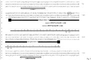
Fig. 2 : Locations of primers withinbraf exon 15.
The G residue at position 1800 of the BRAF open reading frame corresponds to the G residue at position 132 of this figure. The corresponding amino acid sequence of the BRAF protein is depicted below the corresponding DNA sequence. -
Fig. 3 : Predicted pyrogram peaks for braf mutations according to the dispensation pattern * dispensation order's nucleotides, which are involved into mt:wt ratio, are bolded; ** recognition patterns' nucleotides are indicated in black boxes; I intensity of correspondent dispensation order's nucleotide - A total of formalin fixed paraffin embedded (FFPE) tissue samples of cutaneous metastasis from 28 patients were enrolled into this study. Diagnoses were established and controlled in each tumor sample according to histopathological standards by two experienced dermopathologists independently. All patients signed informed consent at initial clinical investigation. The study was approved by local ethic committees.
- For the analysis of tumor samples, haemoltoxylin- and eosin-stained slides were reviewed by an experienced pathologist to ensure sufficient viable tumor content (60-90% tumor cells). Total genomic DNA was extracted from seven 10 µm thick unstained sections of FFPE tissue blocks according to manufacturer's instructions using an automated DNA Extractor (Qiasymphony, Qiagen). The total DNA was eluted in 50 µl and immediately stored at -20°C for later usage. The eluted DNA was quantified using Qubit dsDNA BR Assay (Invitrogen).
- For pyrosequencing assay, the region of human braf spanning mutation sites within
exon 15 was amplified using forward primer U-BRAF F 5'-ATGCTTGCTCTGATAGGAATG-3' (SEQ ID NO: 36) and biotinylated reverse primer BRAF-Pyro-R Biotin-TEG-5'-AGCATCTCAGGGCCAAAAAT-3' (Eurofins MWG Operon, SEQ ID NO: 37,Fig. 2 ). Each PCR reaction mixture was prepared with 10-20 ng genomic DNA, 10 pmol each primer, 2.5 mM dNTPs and 5 units Biotherm Taq polymerase (Genecraft) in a total volume of 50 µl. Amplification of BRAF fragment was performed in PCR cycler Flexcycler (Analytik Jena) as follows: 95°C for 5 minutes, 45 cycles of 95°C for 30 seconds, 57°C for 30 seconds and 72°C for 45 seconds followed by final extension at 72°C for 5 minutes. Specific amplification of the 229-bp fragment was verified by visualizing 5 µl PCR product on a 1.5% agarose TBE gel using SubCell electrophoresis unit (Bio-RAD), followed by 30-minutes incubation in 1X SybrGreen TBE solution (Biozym). - Pyrosequencing procedure was performed identifying variant mutations either at codons V600 to S602 (5'-AGTGAAATCT-3', SEQ NO: 38) or at codons T599 to S602 (5'-TCAGTGAAATCT-3', SEQ ID NO 39) with sequencing primer U-BRAF-600 5'-GGTGATTTTGGTCTAGCTAC-3' (SEQ NO: 40) or with U-BRAF-599 5'-AATAGGTGATTTTGGTCTAGC-3' (Eurofins MWG Operon, SEQ NO: 41,
Fig. 2 ), respectively. 20 µl PCR product were used in each dispensation order protocols of Pyromark Q24 according to manufacturer's instructions (Qiagen). Sequence pyrograms were manually analyzed using Pyromark Q24 software version 2.0.6 (Qiagen). Raw pyrosequencing data were normalized against negative A15/C16/A18/G19, followed by automatic analysis using simple operators of MS Excel 2003 (personal communication). - Sanger sequencing was performed bidirectionally with 1 µl PCR product amplified for pyrosequencing as described above, using BRAF-15F 5'-TGCTTGCTCTGATAGGAAAATG-3' (SEQ NO: 42) and BRAF-15R 5'-AGCATCTCAGGGCCAAAAAT-3' (Eurofins MWG, SEQ NO: 43) with Big Dye Terminator V1.1 cycle sequencing reagents under following PCR conditions: 25 cycles at 95°C for 20 seconds, 55°C for 15 seconds, and 60°C for 1 min (Life Technologies). DNA sequences were finally determined on 3500 Gene Analyzer (Life Technologies) and each sample was visually analyzed on the presence of mutation within
Exon 15. - Here we present a universal approach U-BRAF that enables the detection of BRAF mutation status within
exon 15 by a single pyrosequencing-based assay. We performed analysis of 75 samples, direct sequencing of which resulted in 46 (61.3 %) of mutants consisting of 28/46 (60.9%) samples with V600E mutation, 2/46 (4.3%) samples with alternative tandem mutation V600E (TG>AA), 9/46 (19.6%) samples with V600K tandem mutation, 2/46 (4.3%) samples with V600E;K601I tandem mutation, and 5/46 (10.9%) samples with complex in-frame deletion-insertion (delins) VKS600_602>DT (Fig. 1 , Table 2). - Direct sequencing is widely accepted as a gold standard routinely used to detect BRAF mutations in biopsy specimens. Although restriction fragment length polymorphism analysis (RFLP) and single strand conformation polymorphism analysis (SSCP) detection methods are simpler and easy to apply, they are still less sensitive and less specific than direct DNA sequencing.1 Alternative methods like cobas® BRAF 4800 (Roche) or BRAF RGQ PCR (Qiagen) claim to detect mutations down to a 1% level, but being the quantitative PCR-based, both detection methods have limited precision and present difficulties in reliably detecting low-copy-number templates due to nonspecific amplification and competitive side reactions.2 Despite the alternative approaches based on Shifted Termination Assay (STA) and Amplification Refractory Mutations System allele-specific PCR (ARMS AS-PCR) give comparably sensitive results, detecting down to 5% mutation level in a wild type background, they are designed for detection of few hotspot BRAF mutations only.
- In contrast, pyrosequencing, a real-time sequencing by synthesis, has a high throughput and capable to detect minor sequencing variants with the greater diagnostic sensitivity than direct sequencing, distinguishing up to 10% mutant DNA from background noise.3 It shows high accuracy and precision of pyrosequencing in quantitative identification of BRAF mutations in melanoma cell lines and formalin-fixed paraffin-embedded (FFPE) tumors.4
- Commercially available pyrosequencing approach for BRAF status detection (therascreen® BRAF Pyro® Kit) is designed to analyze complementary strand of DNA starting directly at codon V600. Unfortunately, due to mismatching of sequencing primer, any sample with mutations downstream from codon V600 could be identified as a false wild type. Moreover, V600K or V600R mutants may be interpreted as a false V600E mutation at mutant-to-wild-type ratio equal to 25% or less (Qiagen, personal communication). In general, for all known BRAF status detection approaches in case of variant mutations beyond V600E/K/D/R/A, the direct DNA sequencing is additionally required.
- To overcome these limitations, we designed our approach analyzing the sense strand of braf gene (exon 15) towards mutations, deletions and/or insertions affecting V600 and/or codons downstream. We started the pyrosequencing analysis with the conventional dispensation order G1A2C3G4[A5T6]G7A8T9 created by Pyromark Q24 software Version 2.0.6 (Qiagen) flanking the T1799A hotspot mutation at
codon 600 and ending with the first nucleotide ofcodon 601. Negative nucleotide dispensations G1 and C3 were included by software as internal controls for possible nucleotide misincorporations. Although T1799A mutation was definitely determined by this dispensation order, the variant tandem mutations resulted in aberrant sequencing pyrograms including high peak height deviation at different dispensations, uncertain surrounding reference sequence patterns or high sum deviation in variable position. Because the knowledge of specific variant in each case could explain the altered pyrogram tracing created by a change in order and/or quantity of incorporation of each nucleotide, we embedded internal recognition patterns A7C8T9 and A15G16 into dispensation order U-BRAF-V600 G1A2C3G4A5T6[A7C8T9]G10A11T12C13T14[A15G16] facilitating the identification of tandem mutations V600E (TG1799_1800AA), V600K and V600E;K601I. The presence of variant mutations beyond V600E affects the pyrogram sequence pattern by re-ordering of nucleotide incorporation in mutant DNA sequence resulting in unique pyrogram for each BRAF mutation (Table 3,Fig. 1 ). In this case, two recognition patterns of dispensation order serve to distinguish between individual mutations (Fig. 3 ). Furthermore, the ratio A8:T12 (in means of peak heights) distinguishes between tandem mutation V600E (5:1) and V600K (3:1) respectively (Fig. 3 , Table 4). - Importantly, we found novel complex in-frame mutation p.VKS600_602delinsDT (c.TGAAAT1799_1804delinsATA, Table 3,
Fig. 3 ). In concordance with previous study, we suppose that this mutation could activate BRAF kinase by charging aspartic acid in place of valine,5 although interpretation of the clinical significance of this variant can be challenging. We also identified tandem mutation V600E;K601I, which was incorrectly defined as V600E;K600M mutation by Indsto et al.6 - Comparing with direct sequencing data, five additional samples were identified as BRAF mutants increasing the percentage of mutants to 70.7% convincing in better sensitivity of pyrosequencing (Table 1).
- We next asked whether our approach could be suitable for detection of other BRAF mutations within
exon 15. To test this idea, we performed the literature search for published BRAF mutations in different human tumors using Pubmed (http://www.ncbi.nlm.nih.gov/pubmed). To cover 31 BRAF mutations affecting codons from T599 to S605, we designed novel dispensation order U-BRAFV600, G1T2A3C4A5C6G7A8T9A10C11T12G13A14T15C16T17A18G19 (Fig. 3 ). G1 and C4 nucleotides were included as negative controls. Dispensations T2A3C4 and T17 were embedded for detection of BRAF mutations affecting either codon T5997-10 or within complex in-frame insertion T599_V600insDFLAGT,5 respectively. - Comparing our review of articles with Catalogue of Somatic Mutations in Cancer (COSMIC) database11 we revealed a number of entries either representing one mutation as two independent entries or a complex mutation is divided into two independent cases. K601I, S602S and K601del mutations, which are described as individual mutations by COSMIC database, are indeed the parts of complex mutations V600E;K601I, V600E;S602S and V600E;K601del respectively. Although the mutation p.K601del is defined as a deletion of AAA triplet at position 1801 to 1803 (c.1801_1803delAAA),11 this mutation is indeed created by deletion of triplet TGA at 1799-1801 (c.1799_1801delTGA) resulting in complex mutation V600_K601delinsE.5 Mutation c.1794_1795insGTT7 is represented both as p.A598_T599insV and as p.T599_V600insV.11 Advisably, to distinguish tandem mutations from other type of BRAF mutants, it might be necessary to annotate these mutations into a separate group of complex mutations in the COSMIC database.
Due to the absence of correspondent nucleotide sequences in the original publication, the unique mutations K601E_W604 and T599T_V600R published by Edlundh-Rose et al.12 not included into U-BRAFV600 method. Additionally, unpublished DNA sequencing data by Sadow et al. made impossible to prove the misspelled "VKWRV600-604E" mutation,13 which is annotated as V600_W604del in the COSMIC database.11Table 2: BRAF mutations within exon 15 in cutaneous melanoma metastases Case1 Sample1 Age/Sex2 Sanger sequencing Pyrosequencin g mt/wt ratio3 (range in %) cobas 1 A,B 53/f V600E V600E 36;38 2 A,B 47/f V600E V600E 30;33 3 A,B,C 40/m V600E V600E 17-38 4 A,B 79/m V600E V600E 54;55 5 A 55/m wt wt - 6 A,B,C 69/m wt wt - 0 7 A 80/m V600E V600E 37 8 A,B,C,D,E 53/f V600E V600E 18-38 9 A,B 87/f wt wt - 10 A,B,C 80/m V600E V600E 59-60 11 A 82/f V600E V600E 16 B,C wt wt - 0 12 A 83/m wt wt - 13 A,B,C,D 56/m wt wt - 14 A,B,C,D,E 57/m VKS600_602> DT VKS600_602> DT 23-42 15 A, 74/f wt wt 8 0 B wt V600E 16 - 16 A,B 65/m V600E V600E 33;34 17 A,B 52/m V600K V600K 12;19 -; 18 A 30/m V600E V600E 33 19 A 75/f wt wt - 0 20 A,B,D 66/m V600E V600E 22-41 C,E wt V600E 16;11 0 21 A,B 73/m V600E V600E 31-33 0 (TG>AA) (TG>AA) 22 A,B 37/m V600K V600K 43;49 23 A 71/f wt wt - 24 A,B 52/m wt wt - 25 A 54/f V600E V600E 14 26 A,B,C,D,E,F ,G,H 66/m wt wt - 27 A,B,C,D,E 54/m V600K V600K 43-61 + 28 A,B 78/f V600E V600E 32;33 29 A,B 44/m V600E;K601I V600E;K601I 29;59 0 1different samples of the same tumor are specified by 1, 2 etc., different tumors of the same patient specified by A, B etc.; 2age in years, f = female, m = male; 3wt - wild type, mt - mutant. Table 3: Recognition patterns for 31 BRAF mutations by U-BRAF-V600; Mutation Dispensation Order Patterns Unique properties of each mutation within one group mutant to wild type ratio* Ref Identification COSMIC v57 A10 C11 T12 A18 G19 I V600E - - - - - A8=Amt; T9= Twt A8 mt : T9 wt 16 COSM476 V600L - - - - - G7=Gwt; absence of A8; T9=[2Tmt+ Twt] [I10-I4] G7 wt 15 COSM33808 V600M - - - - - G7=Gwt; absence of A8; T9= [Twt+ Tmt] [I10 - I4]mt : G7wt 14 COSM1130 V600R (AGT>GAG) - - - - - A5=Awt; G13=[Gwt+ 2Gmt] A8 mt : T9 wt 16 COSM1127 K601E - - - - - absence of A8; G13=[Gwt+2Gmt]; A14=[13Awt+2Amt] [I10 - I4]mt : [214 -I10] 17 COSM478 K601N - - - - - absence of A8; T9=[Twt+ Tmt]; 14=[3Awt+2Amt] [I12 - I6]mt : [216 -I12]wt 18 COSM1132 II V600E;K601I + - + - - A8 : A10 = 3 : 1 T12 mt T15 wt 6 COSM475; COSM26491 V600D + - + - - A8 : A10 = 1 : 3 T12 mt: T15 wt 16 COSM477 V600G + - + - - G7=Gwt+3Gmt; absence of A8; A10=3Amt; G13=Gwt T12 mt: T15 wt 15 COSM6137 III V600E (TG>AA) - + + - + A8:T12=5:1 T12 mt :T15 wt 6 COSM475 V600K - + + - + A5=[Awt+3Amt]; (A8 : T12)=3: 1 T12 mt: T15 wt 16 COSM473 V600R - + + - + A5=[Awt+2Amt] G7=[Gwt+2GmtL (A8 : T12)=3: 1 T12 mt: T15 wt 16 COSM474 VK600_601>E - + + - + A8: T12= 2:1 T12 mt: T15 wt 5 COSM1133 Mutation Dispensation Order Patterns Unique properties of each mutation within one group mutant to wild type ratio* Ref SEQ_ID COSMIC v57** A10 C11 T12 A18 G19 III TVKSR599_603>1 - + + - + absence of mutant C4, A5, G7; absence of A8 G13=[Gwt+2Gmt] C11 mt : C4 wt 8 COSM30605 IV T599T;V600E - - - - + absence of mutant A5, G7; absence of A8 G19 mt : G7 wt 19 COSM24963 COSM476 K601R - - - - + absence of A8; A14=[3Awt+Amt] G19 mt : T15 wt 15 COSM13625 K601K - - - - + absence of A8; A14=[3Awt+2Amt] G19 mt : T15 wt 25 COSM28507 V600E;K601R - - - - + A8=Amt; T9=Twt A8 mt :T9 wt 14 COSM475 COSM13625 IV VKSRWS600_605>DV - - - - + G19= 3Gmt; absence of mutant A14; T15=[Twt+2Tmt] A8 mt : T17 wt 20 COSM33764 V K601Q - - - + - absence of A8; T9=[Twt+ Tmt]; A18=2Amt [½ I18] mt:T15 wt 21 COSM1066665 VKSRWS600_605>D - - - + - G13=[Gwt+ 3Gmt] C16=[Cwt+3Cmt] A18 mt : T17 wt 22 COSM1129 Mutation Dispensation Order Patterns Unique properties of each mutation within one group mutant to wild type ratio* Ref SEQ ID COSMIC v57** A10 C11 T12 A18 G19 VI V600K;S602S - + - - + A8=3Amt T17 mt: C11 wt 19 COSM473; COSM21611 T599_V600insT (2) - + - - + absence of A8 C11 mt: C16 wt 23 COSM36245 VII T599_V600msT (1) + + - - + absence of mutant G7; T9=[Twt+Tmt] C11 mt : C16 wt 24 COSM30730 T599_V600msV + + - - + absence of mutant A3, C4; T9=[Twt+2Tmt] A10 mt : A5 wt 7 COSM26625 VIII VKS600_602>DT + + + - + A8=Amt; absence of mutant A14 T12 mt T15 wt - unpublished T599_V600insTT + + + - + absence of A8; absence of mutant A5, G7 T12 mt : T15 wt 16 COSM26459 IX T5991;V600E + - - - + A14=3Awt+Amt; T17=Twt A10 mt: A5 wt 9 COSM472; COSM476 TV599_600>IAL + - - - + absence of mutant A14;T17=[Twt+3Tmt] A10 mt : A5 wt 10 COSM33780 T599_V600insDFLAGT - - - + + unique; T9=[Twt + 4Tmt] A18 mt:[⅓I11]wt 5 COSM26504 V600A - + - - - unique; absence of A8 C11 mt : T9 wt 14 COSM18443 * I - intensity value of correspondent nucleotide dispensation; A-peak reduction factor 0.9 should be taken into consideration;
**COSMIC - Catalogue of Somatic Mutations in Cancer, Version 57, Sanger Institute


-
- 1. Chung, K. W. et al. Clin. Endocrinol. (Oxf) 65, 660-666 (2006).
- 2. Heyries, K. A. et al. Nat. ).
- 3. Jo, Y. S. et al. Clin. Endocrinol. (Oxf) 70, 139-144 (2009).
- 4. Spittle, C. et al. J. Mol. Diagn. 9, 464-471 (2007).
- 5. Hou, P., Liu, D., Xing, ).
- 6. Indsto, J. O. et al. J. Cutan. Pathol. 34, 448-455 (2007).
- 7. Carta, C. et al. Clin. Endocrinol. (Oxf) 64, 105-109 (2006).
- 8. Lupi, C. et al. J. Clin. Endocrinol. Metab 92, 4085-4090 (2007).
- 9. Gill, M., Renwick, N., Silvers, D. N., Celebi, J. T. J. Invest Dermatol. 122, 1325-1326 (2004).
- 10. Chiosea, S. et al. Endocr. Pathol. 20, 122-126 (2009).
- 11. Forbes, S. A. et al. Curr. Protoc. Hum. Genet. ).
- 12. Edlundh-Rose, E. et al. Melanoma Res. 16, 471-478 (2006).
- 13. Sadow, P. M., Heinrich, M. C., Corless, C. L., Fletcher, J. A., Nose, V. Endocr. Pathol. 21, 73-79 (2010).
- 14. Lin, J. et al. Br. J. Cancer 104, 464-468 (2011).
- 15. Boulalas, I., Zaravinos, A., Delakas, D., Spandidos, D. A. Int. ).
- 16. Omholt, K., Platz, A., Kanter, L., Ringborg, U., Hansson, J. Clin. Cancer Res. 9, 6483-6488 (2003).
- 17. Trovisco, V. et al. Hum. Pathol. 36, 694-697 (2005).
- 18. Helmke, B. M. et al. Gastroenterology 127, 1815-1820 (2004).
- 19. Deichmann, M. et al. BMC. ).
- 20. Barzon, L. et al. Eur. J. Endocrinol. 159, 77-80 (2008).
- 21. Sarkozy, A. et al. Hum. Mutat. 30, 695-702 (2009).
- 22. Cruz, F., III et al. Cancer Res. 63, 5761-5766 (2003).
- 23. Schindler, G. et al. Acta Neuropathol. 121, 397-405 (2011).
- 24. Eisenhardt, A. E. et al. Int. J. Cancer (2010).
- 25. Deichmann, M. et al. Int. J. Oncol. 29, 139-145 (2006).



















Claims (15)
- A method for differentiating between at least two sequence variants of a polynucleotide by pyrosequencing, wherein at least one sequence variant comprises at least one mutation, said method comprising:a) generating a dispensation order based on a nucleotide sequence encompassing the nucleotides affected by said variants, said dispensation order comprising dispensations corresponding to each nucleotide occurring in at least one of the variants, said nucleotide dispensations ordered in a sequencei) generating on average a pyrogram peak for at least every second nucleotide dispensation for each variant andii) generating the maximal possible number of sequence pyrogram peaks for each variant;b) generating according to the dispensation order of a) a first pyrogram comprising expected intensity values for a first variant and at least a second pyrogram comprising expected intensity values for at least one further variant;c) comparing a sample pyrogram comprising measured intensity values, said sample pyrogram being derived from pyrosequencing analysis of a sample polynucleotide according to the nucleotide dispensation order of a), with the said first and at least the said second pyrogram generated in step b); andd) differentiating between said at least two sequence variants, wherein the sample polynucleotide corresponds to the first variant if the sample pyrogram is essentially identical with the first pyrogram and wherein the sample polynucleotide corresponds to a further variant if the sample pyrogram is essentially identical with a further pyrogram.
- The method of claim 1, wherein step b) further comprises comparing said first and at least one further pyrogram and by this means identifying at least one unique property for a first variant compared to at least one second variant based on said first and at least one second pyrogram, wherein in step c) the sample pyrogram comprises for at least each nucleotide of the at least one unique property a measured intensity value, and wherein in step d) the sample polynucleotide corresponds to the first variant if the sample pyrogram comprises the at least one unique property corresponding to the first variant and wherein the sample polynucleotide corresponds to a further variant if the sample pyrogram comprises the at least one unique property corresponding to a further variant.
- The method of claim 2, wherein at least one of the at least one unique properties is comprised in a unique dispensation order pattern.
- The method of claim 2 or 3, wherein said at least one unique property comprises at least one property selected from the group consisting of:(i) the presence or absence of a pyrogram peak for the first variant;(ii) the presence or absence of a pyrogram peak for at least one further variant;(iii) the expected intensity value for a pyrogram peak for the first variant;(iv) the expected intensity value pyrogram peak for at least one further variant; and(v) the ratios of expected intensity values of (iii) and (iv) or (iv) and (iii).
- The method of any one of claims 1 to 4, wherein steps a) to d) of said method are carried out computer-implemented.
- The method of any one of claims 1 to 5, wherein said at least two variants comprise at least one mutation in exon 15 of the braf gene.
- The method of claim 6, wherein the at least one mutation is located at a nucleotide position corresponding to nucleotide positions 1794 to 1804 of the wild-type braf open reading frame.
- The method of any one of claims 1 to 7, wherein said mutation is a mutation associated with a disease.
- The method of claim 8, wherein said disease is cancer.
- The method of claim 9, wherein said cancer is acute myeloid leukemia (AML), acute myeloid leukemia therapy related (AMLTR), astrocytoma, biliary tract carcinoma, breast carcinoma, cholangiocarcinoma, colorectal carcinoma, endometrioid carcinoma, esophageal carcinoma, fibrous histiocytoma-pleomorhic sarcoma, gastrointestinal stromal tumor (GIST), glioma, hepatocellular carcinoma, large intestine adenocarcinoma, small intestine adenocarcinoma, liposarcoma, lung adenocarcinoma, melanoma, malignant melanoma, melanoma metastasis, ocular melanoma, ovarian carcinoma, ovary mucinous carcinoma, ovary serous carcinoma, pancreatic ductal carcinoma, pituitary adenoma, plasma cell myeloma, prostate adenocarcinoma, stomach adenocarcinoma, synovial sarcoma, thyroid anaplastic carcinoma, or thyroid papillary carcinoma.
- The method of any one of claims 1 to 9, wherein said dispensation order is G1T2A3C4A4C6G7A8T9A10C11T12G13A14T15C16T17A18G19 or G1A2C3G4A5T6A7C8T9G10A11T12C13T14A15G16.
- The method of claim 3, wherein said unique dispensation order patterns are A10C11T12 in combination with A18G19 or A7C8T9 in combination with A15G16.
- The method of any one of claims 1 to 12, wherein said variants are variants selected from the list provided in Table 1.
- The method of claim 13, wherein said at least one unique property is a unique property as shown in Table 1.
- The method of any one of claims 1 to 14, wherein at least two variants present in the same sample are differentiated from at least one further variant.
Priority Applications (2)
| Application Number | Priority Date | Filing Date | Title |
|---|---|---|---|
| EP12153477.0A EP2623612A1 (en) | 2012-02-01 | 2012-02-01 | Method for variant differentiation utilizing a pyrosequencing dispensation order |
| PCT/EP2013/052051 WO2013113885A1 (en) | 2012-02-01 | 2013-02-01 | Method for variant differentiation utilizing a pyrosequencing dispensation order |
Applications Claiming Priority (1)
| Application Number | Priority Date | Filing Date | Title |
|---|---|---|---|
| EP12153477.0A EP2623612A1 (en) | 2012-02-01 | 2012-02-01 | Method for variant differentiation utilizing a pyrosequencing dispensation order |
Publications (1)
| Publication Number | Publication Date |
|---|---|
| EP2623612A1 true EP2623612A1 (en) | 2013-08-07 |
Family
ID=47633078
Family Applications (1)
| Application Number | Title | Priority Date | Filing Date |
|---|---|---|---|
| EP12153477.0A Withdrawn EP2623612A1 (en) | 2012-02-01 | 2012-02-01 | Method for variant differentiation utilizing a pyrosequencing dispensation order |
Country Status (2)
| Country | Link |
|---|---|
| EP (1) | EP2623612A1 (en) |
| WO (1) | WO2013113885A1 (en) |
-
2012
- 2012-02-01 EP EP12153477.0A patent/EP2623612A1/en not_active Withdrawn
-
2013
- 2013-02-01 WO PCT/EP2013/052051 patent/WO2013113885A1/en not_active Ceased
Non-Patent Citations (6)
| Title |
|---|
| CYNTHIA SPITTLE ET AL: "Application of a BRAF Pyrosequencing Assay for Mutation Detection and Copy Number Analysis in Malignant Melanoma", JOURNAL OF MOLECULAR DIAGNOSTICS, AMERICAN SOCIETY FOR INVESTIGATIVE PATHOLOGY, BETHESDA, MD, US, vol. 9, no. 4, 1 September 2007 (2007-09-01), pages 464 - 471, XP008151917, ISSN: 1525-1578, DOI: 10.2353/JMOLDX.2007.060191 * |
| JO YOUNG SUK ET AL: "Diagnostic value of pyrosequencing for the BRAF V600E mutation in ultrasound-guided fine-needle aspiration biopsy samples of thyroid incidentalomas.", CLINICAL ENDOCRINOLOGY JAN 2009 LNKD- PUBMED:18462259, vol. 70, no. 1, January 2009 (2009-01-01), pages 139 - 144, XP002676452, ISSN: 1365-2265 * |
| PACKHAM DEBORAH ET AL: "Implementation of Novel Pyrosequencing Assays to Screen for Common Mutations of BRAF and KRAS in a Cohort of Sporadic Colorectal Cancers", DIAGNOSTIC MOLECULAR PATHOLOGY, vol. 18, no. 2, June 2009 (2009-06-01), pages 62 - 71, XP009159584, ISSN: 1052-9551 * |
| PATEL PRITESH ET AL: "Multiplex pyrosequencing for DNA variation analysis.", METHODS IN MOLECULAR BIOLOGY (CLIFTON, N.J.) 2007 LNKD- PUBMED:17185759, vol. 373, 2007, pages 75 - 88, XP009159534, ISSN: 1064-3745 * |
| SIVERTSSON A ET AL: "Pyrosequencing as an alternative to single-strand conformation polymorphism analysis for detection of N-ras mutations in human melanoma metastases", CLINICAL CHEMISTRY, AMERICAN ASSOCIATION FOR CLINICAL CHEMISTRY, WASHINGTON, DC, vol. 48, no. 12, 1 December 2002 (2002-12-01), pages 2164 - 2170, XP002363100, ISSN: 0009-9147 * |
| XI LIQIANG ET AL: "Both variant and IGHV4-34-expressing hairy cell leukemia lack the BRAF V600E mutation", BLOOD, vol. 119, no. 14, 30 December 2011 (2011-12-30), pages 3330 - 3332, XP002676451 * |
Also Published As
| Publication number | Publication date |
|---|---|
| WO2013113885A1 (en) | 2013-08-08 |
Similar Documents
| Publication | Publication Date | Title |
|---|---|---|
| US11981966B2 (en) | Methods for screening solid tumors for mutations | |
| US9228239B2 (en) | High resolution melting analysis as a prescreening tool | |
| Han et al. | Detection of EGFR mutation status in lung adenocarcinoma specimens with different proportions of tumor cells using two methods of differential sensitivity | |
| KR101727885B1 (en) | Genetic polymorphic markers for determining type of white skin and use thereof | |
| Herreros-Villanueva et al. | KRAS mutations: analytical considerations | |
| Stigt et al. | Pyrosequencing analysis of EGFR and KRAS mutations in EUS and EBUS-derived cytologic samples of adenocarcinomas of the lung | |
| CN107447013B (en) | Method for detecting mutation sites of codons 12 and 13 of Kras gene and kit thereof | |
| EP3954784A1 (en) | Composition for diagnosis or prognosis prediction of glioma, and method for providing information related thereto | |
| JP2025157489A (en) | Kits and methods for detecting cancer-associated mutations | |
| Packham et al. | Implementation of novel pyrosequencing assays to screen for common mutations of BRAF and KRAS in a cohort of sporadic colorectal cancers | |
| CN106148551A (en) | Multiple myeloma prognosis-related gene mutation detection kit and detection method | |
| JP6158189B2 (en) | Mutation analysis of JAK2 | |
| Hunt | Understanding the genotype of follicular thyroid tumors | |
| CN111647650B (en) | Primer, primer probe composition and kit for detecting V600E mutation of human B-raf gene | |
| Li et al. | A novel liquidchip platform for simultaneous detection of 70 alleles of DNA somatic mutations on EGFR, KRAS, BRAF and PIK3CA from formalin-fixed and paraffin-embedded slides containing tumor tissue. | |
| Park et al. | Evaluation of the three customized MSI panels to improve the detection of microsatellite instability in gastric cancer | |
| WO2013113885A1 (en) | Method for variant differentiation utilizing a pyrosequencing dispensation order | |
| Buxhofer-Ausch et al. | Duplex reverse‐hybridization assay for the simultaneous detection of KRAS/BRAF mutations in FFPE‐extracted genomic DNA from colorectal cancer specimens | |
| Zavodna et al. | Genetic analysis of KRAS mutation status in metastatic colorectal cancer patients | |
| Choi et al. | PNA-mediated real-time PCR clamping for detection of EGFR mutations | |
| Walsh et al. | Molecular pathology in lung cancer: a guide to the techniques used in clinical practice | |
| Shutkever et al. | Testing for genomic biomarkers in non-small-cell lung cancer | |
| Slavkovský et al. | Development and extensive analytical validation of deep amplicon sequencing for detecting KRAS and NRAS mutations in metastatic colorectal cancer samples | |
| Kitamura et al. | DNA Methylation in Ovarian and Endometrial Cancers: Predictive and Mechanistic Roles in PARP Inhibitor and ICI Response | |
| Velu et al. | Genetic Profiling in Solid Tumors: Identification of Driver Mutations, Oncogenic Fusions, and Molecular Biomarkers |
Legal Events
| Date | Code | Title | Description |
|---|---|---|---|
| PUAI | Public reference made under article 153(3) epc to a published international application that has entered the european phase |
Free format text: ORIGINAL CODE: 0009012 |
|
| AK | Designated contracting states |
Kind code of ref document: A1 Designated state(s): AL AT BE BG CH CY CZ DE DK EE ES FI FR GB GR HR HU IE IS IT LI LT LU LV MC MK MT NL NO PL PT RO RS SE SI SK SM TR |
|
| AX | Request for extension of the european patent |
Extension state: BA ME |
|
| STAA | Information on the status of an ep patent application or granted ep patent |
Free format text: STATUS: THE APPLICATION IS DEEMED TO BE WITHDRAWN |
|
| 18D | Application deemed to be withdrawn |
Effective date: 20140208 |