EP2623517A1 - Urine and serum biomarkers associated with diabetic nephropathy - Google Patents
Urine and serum biomarkers associated with diabetic nephropathy Download PDFInfo
- Publication number
- EP2623517A1 EP2623517A1 EP13162626.9A EP13162626A EP2623517A1 EP 2623517 A1 EP2623517 A1 EP 2623517A1 EP 13162626 A EP13162626 A EP 13162626A EP 2623517 A1 EP2623517 A1 EP 2623517A1
- Authority
- EP
- European Patent Office
- Prior art keywords
- seq
- biomarker
- subject
- level
- group
- Prior art date
- Legal status (The legal status is an assumption and is not a legal conclusion. Google has not performed a legal analysis and makes no representation as to the accuracy of the status listed.)
- Granted
Links
Images

Classifications
-
- G—PHYSICS
- G01—MEASURING; TESTING
- G01N—INVESTIGATING OR ANALYSING MATERIALS BY DETERMINING THEIR CHEMICAL OR PHYSICAL PROPERTIES
- G01N33/00—Investigating or analysing materials by specific methods not covered by groups G01N1/00 - G01N31/00
- G01N33/48—Biological material, e.g. blood, urine; Haemocytometers
- G01N33/50—Chemical analysis of biological material, e.g. blood, urine; Testing involving biospecific ligand binding methods; Immunological testing
- G01N33/68—Chemical analysis of biological material, e.g. blood, urine; Testing involving biospecific ligand binding methods; Immunological testing involving proteins, peptides or amino acids
- G01N33/6893—Chemical analysis of biological material, e.g. blood, urine; Testing involving biospecific ligand binding methods; Immunological testing involving proteins, peptides or amino acids related to diseases not provided for elsewhere
-
- G—PHYSICS
- G01—MEASURING; TESTING
- G01N—INVESTIGATING OR ANALYSING MATERIALS BY DETERMINING THEIR CHEMICAL OR PHYSICAL PROPERTIES
- G01N2333/00—Assays involving biological materials from specific organisms or of a specific nature
- G01N2333/435—Assays involving biological materials from specific organisms or of a specific nature from animals; from humans
- G01N2333/46—Assays involving biological materials from specific organisms or of a specific nature from animals; from humans from vertebrates
- G01N2333/47—Assays involving proteins of known structure or function as defined in the subgroups
- G01N2333/4701—Details
- G01N2333/4728—Details alpha-Glycoproteins
-
- G—PHYSICS
- G01—MEASURING; TESTING
- G01N—INVESTIGATING OR ANALYSING MATERIALS BY DETERMINING THEIR CHEMICAL OR PHYSICAL PROPERTIES
- G01N2333/00—Assays involving biological materials from specific organisms or of a specific nature
- G01N2333/435—Assays involving biological materials from specific organisms or of a specific nature from animals; from humans
- G01N2333/52—Assays involving cytokines
-
- G—PHYSICS
- G01—MEASURING; TESTING
- G01N—INVESTIGATING OR ANALYSING MATERIALS BY DETERMINING THEIR CHEMICAL OR PHYSICAL PROPERTIES
- G01N2333/00—Assays involving biological materials from specific organisms or of a specific nature
- G01N2333/435—Assays involving biological materials from specific organisms or of a specific nature from animals; from humans
- G01N2333/705—Assays involving receptors, cell surface antigens or cell surface determinants
- G01N2333/71—Assays involving receptors, cell surface antigens or cell surface determinants for growth factors; for growth regulators
-
- G—PHYSICS
- G01—MEASURING; TESTING
- G01N—INVESTIGATING OR ANALYSING MATERIALS BY DETERMINING THEIR CHEMICAL OR PHYSICAL PROPERTIES
- G01N2333/00—Assays involving biological materials from specific organisms or of a specific nature
- G01N2333/81—Protease inhibitors
- G01N2333/8107—Endopeptidase (E.C. 3.4.21-99) inhibitors
- G01N2333/811—Serine protease (E.C. 3.4.21) inhibitors
- G01N2333/8121—Serpins
- G01N2333/8125—Alpha-1-antitrypsin
-
- G—PHYSICS
- G01—MEASURING; TESTING
- G01N—INVESTIGATING OR ANALYSING MATERIALS BY DETERMINING THEIR CHEMICAL OR PHYSICAL PROPERTIES
- G01N2800/00—Detection or diagnosis of diseases
- G01N2800/34—Genitourinary disorders
- G01N2800/347—Renal failures; Glomerular diseases; Tubulointerstitial diseases, e.g. nephritic syndrome, glomerulonephritis; Renovascular diseases, e.g. renal artery occlusion, nephropathy
-
- G—PHYSICS
- G01—MEASURING; TESTING
- G01N—INVESTIGATING OR ANALYSING MATERIALS BY DETERMINING THEIR CHEMICAL OR PHYSICAL PROPERTIES
- G01N2800/00—Detection or diagnosis of diseases
- G01N2800/52—Predicting or monitoring the response to treatment, e.g. for selection of therapy based on assay results in personalised medicine; Prognosis
-
- G—PHYSICS
- G01—MEASURING; TESTING
- G01N—INVESTIGATING OR ANALYSING MATERIALS BY DETERMINING THEIR CHEMICAL OR PHYSICAL PROPERTIES
- G01N2800/00—Detection or diagnosis of diseases
- G01N2800/56—Staging of a disease; Further complications associated with the disease
Definitions
- Diabetic nephropathy is a progressive kidney disease associated with longstanding diabetes mellitus. It causes abnormal fluid filtration and increased urinary albumin excretion, eventually leading to kidney failure.
- DN displays no symptoms in its early course. As such, it is difficult to detect the incipiency of this disease. In fact, present diagnosis ofDN depends on development of microalbuminuria, which occurs when kidney damage is already in place. The lack of an early diagnostic test prevents effective treatment of early stage DN.
- the present invention is based on unexpected discoveries that a number of urine and serum proteins and their fragments, either alone or in combination, are differentially presented in DN patients as compared to DN-free subjects. These protein molecules are therefore useful markers for diagnosing early stage DN.
- one aspect of this invention features a method of diagnosing DN in a subject.
- This method includes at least two steps: (i) determining in a subject suspected of having DN a level of a biomarker, and (ii) assessing whether the subject has DN based on the level of the biomarker. An increase in the level of the biomarker, as compared to that in a DN-free subject, indicates that the subject has DN.
- the biomarker used in this diagnostic method comprises (i) a first biomarker in a urine sample, which is a fragment of alpha-1 antitrypsin comprising at least one selected from the group consisting of:
- the biomarker used in this diagnostic method may further comprise (ii) a second biomarker in the urine sample, which is a fragment of alpha-2-HS-glycoprotein comprising at least one selected from the group consisting of:
- the biomarker used in this diagnostic method may further comprise (iii) a third biomarker in the urine sample, which is GQEHFAHLLILRDTKTYMLAFDVNDEKNWGLS (SEQ ID NO:7).
- the biomarker used in this diagnostic method may further comprise (iv) a fragment of osteopontin in the serum sample, which comprises at least one selected from the group consisting of:
- the diagnostic method described above can further include, after the assessing step, a step of correlating the biomarker level with the DN status (i.e., whether it is at early or late stage).
- a step of correlating the biomarker level with the DN status i.e., whether it is at early or late stage.
- the biomarker is protein molecules (ii) or (iv)
- an increase in its level relative to that in a DN-free subject is indicative of late stage DN.
- its level indicates the DN status when compared with pre-determined reference biomarker levels representing early and late stage DN.
- the present invention features a method for assessing efficacy of a DN treatment in a subject (e.g., a human patient or a laboratory animal).
- This method includes determining in the subject pre-treatment and post-treatment levels of protein molecules (i), (ii), (iii), or (iv), and assessing efficacy of the treatment based on a change in the level of the biomarker after the treatment. If the post-treatment level of the biomarker remains the same or decreases as compared to the pre-treatment level of the biomarker, it indicates that the treatment is effective.
- this invention features a method for determining a DN stage, including at least four steps: (i) obtaining a urine sample and optionally, a serum sample from a subject suspected of having diabetic nephropathy, (ii) determining in the sample(s) a level of one of the biomarkers listed in the preceding paragraph, (iii) calculating a disease score based on the level of the biomarker, and (iv) assessing the subject's diabetic nephropathy stage based on the disease score as compared to pre-determined cutoff values indicating different diabetic nephropathy stages.
- the calculating step can be performed by ridge regression analysis, factor analysis, discriminant function analysis, and logistic regression analysis.
- the biomarker used in the just-described DN staging method is composed of at least two of the following five protein molecules: protein molecules (i)-(iv) listed above and protein molecule (v) that is urine osteopontin or its fragment described above.
- the biomarker is composed of all of the five protein molecules.
- it is composed of at least two of protein molecules (i)-(iii) and (v).
- the biomarker is composed of at least two of the five protein molecules listed above and additionally, one or more clinical factors, e.g., age, gender, HbA1c, albumin/creatinine ratio (ACR), and glomerular filtration rate (GFR).
- one or more clinical factors e.g., age, gender, HbA1c, albumin/creatinine ratio (ACR), and glomerular filtration rate (GFR).
- the present invention provides a method for monitoring DN progress based on the level of any of the above-mentioned biomarkers.
- This method includes obtaining two urine samples and optionally, two serum samples, within a time span of 2 weeks to 12 months (e.g., 2-24 weeks or 3-12 months) from a subject suspected of having DN, determining in the samples a level of one of the biomarkers, calculating disease scores based on the biomarker levels, and assessing DN progress in the subject based on the disease scores.
- the disease score for the later-obtained samples being greater than that for the earlier-obtained samples is indicative ofDN exacerbation.
- the biomarkers mentioned above can also be used to assess efficacy of a DN treatment.
- the treatment is effective if the post-treatment level of one of the biomarkers remains unchanged or decreases as compared to the pre-treatment level of the same biomarker.
- the present invention further provides a kit for use in any of the methods described above.
- This kit includes two, three, or four antibodies with different antigen specificities. Each of these antibodies is capable of binding to one of (i) alpha-1 antitrypsin, (ii) alpha-2-HS-glycoprotein, (iii) alpha-1 acid glycoprotein, and (iv) osteopontin.
- this kit contains only antibodies specific to antigens to be detected (e.g., biomarkers associated with DN) for practice one of the methods disclosed herein. Namely, it consists essentially of such antibodies.
- an isolated antibody refers to an antibody substantially free from naturally associated molecules. More specifically, a preparation containing the antibody is deemed as "an isolated antibody” when the naturally associated molecules in the preparation constitute at most 20% by dry weight. Purity can be measured by any appropriate method, e.g., column chromatography, polyacrylamide gel electrophoresis, and HPLC.
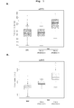
- Fig. 1 is a diagram showing boxplots for urine alpha-2-HS-glycoprotein (uDN2; see panel A), urine alpha-1 antitrypsin (uDN5; see panel B), urine alpha-1 acid glycoprotein (uGR3; see panel C), and serum osteopontin (sDNO; see panel D) in various groups ofDN patients.
- the upper and lower limits of the boxes mark the 25% and 75% values with the medians as the lines across the boxes.
- the upper whisker marks the largest value below the upper fence, which is the 75% value plus 1.5 interquartile range and the lower whisker marks the smallest value above the lower fence, which is the 25% value minus 1.5 interquartile range.
- DN is a kidney disorder associated with diabetes. It has five progression phases:
- biomarkers associated with DN are composed of one or more of the following four proteins and their fragments, either in urine or in serum: (a) alpha-1-antitrypsin (GenBank accession no. AAB59495; 10-Jan-2010); (b) alpha-2-HS-glycoprotein (GenBank accession no. NP_001613; 10-Jan-2010); (c) alpha-1 acid glycoprotein (GenBank accession no. EAW87416; 10-Jan-2010); and (d) Osteopontin, which includes two isoforms known as secreted phosphoprotein la (GenBank accession no. NP_001035147; 17-Jan-2010) and secreted phosphoprotein 1b (GenBank accession no. NP_000573; 10-Jan-2010,).
- fragments of these four proteins have a minimum length of ten amino acids and preferably, a maximum length of 190 to 410 amino acids.
- fragments of proteins (a), (b), (c), and (d) can contain up to 408, 357,191, and 290 amino acid residues, respectively.
- biomarkers composed of one or more of the above mentioned proteins/fragments, and one or more clinical factors are also associated with DN in different stages.
- one aspect of the present invention relates to a DN diagnostic method using any of the biomarkers described above.
- a urine sample and, when necessary, a serum sample is collected from a subject suspected of having DN and the urine and serum levels of one or more of the four proteins listed above or their fragments can be determined via routine methods, e.g., mass spectrometry and immune analysis. If applicable, the clinical factors are determined by route methods.
- a biomarker When a biomarker contains a single protein molecule, its level in a subject can be compared with a reference point to determine whether that subject has DN.
- the reference point representing the level of the same biomarker in a DN-free subject, can be determined based on the representative levels of the biomarker in groups of DN patients and DN-free subjects. For example, it can be the middle point between the mean levels of these two groups. A biomarker level higher than the reference point is indicative of DN.
- a biomarker contains at least two protein molecules and optionally, at least one clinical factor
- the levels of the protein molecules and the value(s) of the clinical factor(s) can be subjected to a suitable analysis to generate a disease score (e.g., represented by a numeric number) that characterizes the level of the biomarkers.
- a suitable analysis include, but are not limited to, discriminate function analysis, logistic regression analysis, ridge regression analysis, principal component analysis, factor analysis, and generalized linear model.
- the disease score is then compared with a reference point representing the level of the same biomarker in DN-free subjects.
- the reference point can be determined by conventional methods.
- it can be a score obtained by analyzing the mean levels of the protein molecules and when necessary, the mean value(s) of the clinical factor(s) in DN-free subjects with the same analysis.
- the disease score being higher than the reference point is indicative of DN presence.
- Another aspect of this invention relates to a method for determining a DN stage based on any of the biomarkers described above.
- a biomarker level of a DN patient preferably represented by a disease score
- the cutoff values can be determined by analyzing the representative levels of the same biomarker in different-staged DN patients via the same analysis.
- Any of the models thus generated can be assessed for its diagnosis value by a receiver-operating characteristic (ROC) analysis to create a ROC curve.
- ROC receiver-operating characteristic
- An optimal multivariable model provides a large Area under Curve (AUC) in the ROC analysis. See the models described in Examples 1-3 below.
- this invention relates to a method of monitoring nephropathy progress in a subject based on any of the biomarkers described above. More specifically, two urine samples and/or serum samples from a subject can be obtained within a suitable time span (e.g., 2 weeks to 12 months) and examined to determine the levels of one of the biomarkers. Disease scores are then determined as described above. If the disease score representing the biomarker level in the later obtained sample(s) is lower than that in the earlier-obtained sample(s), it indicates DN exacerbation in the subject.
- a suitable time span e.g., 2 weeks to 12 months
- the monitoring method can be applied to a human subject suffering from or at risk for DN.
- the level of the biomarker can be examined once every 6 to 12 months to monitor DN progress.
- the biomarker level be examined once every 3 to 6 months.
- a laboratory animal used herein refers to a vertebrate animal commonly used in animal testing, e.g., mouse, rat, rabbit, cat, dog, pig, and non-human primate.
- a laboratory animal is examined to determine the biomarker level once every 2 to 24 weeks.
- biomarkers can also be used to assess efficacy of a DN treatment in a subject in need (i.e., a human DN patient or a laboratory animal bearing DN).
- disease scores representing levels of one of the biomarkers described above are determined before, during, and after the treatment. If the disease scores remain the same or decline over the course of the treatment, it indicates that the treatment is effective.
- kits useful in practicing any of the above-described methods contains two, three, or four antibodies with different antigen specificities. Each of these antibodies is capable of binding to one of (i) alpha-1 antitrypsin, (i) alpha-2-HS-glycoprotein, (iii) alpha-1 acid glycoprotein, or (iv) osteopontin.
- the antibodies specific to proteins (i), (ii), (iii), and (iv) can bind to their fragments KGKWERPFEVKDTEEEDF (SEQ ID NO:3), MIEQNTKSPLFMGKVVNPTQK (SEQ ID NO:4), EDPQGDAAQKTDTSHHDQDHPTFNKITPNLAE (SEQ ID NO:5), and EDPQGDAAQKTDTSHHDQDHPTFNKITPNLAEFA (SEQ ID NO:6),; VVSLGSPSGEVSHPRKT (SEQ ID NO:1) and MGVVSLGSPSGEVSHPRKT (SEQ ID NO:2); GQEHFAHLLILRDTKTYMLADVNDEKNWGLS (SEQ ID NO:7); and YPDAVATWLNPDPSQKQNLLAPQNAVSSEETNDFKQETLPSK (SEQ ID NO:8), and KYPDAVATWLNPDPSQKQNLLAPQTLPSK (SEQ ID NO:9), respectively, i
- the kit described above can include two different antibodies (i.e., a coating antibody and a detecting antibody) that bind to the same antigen.
- the detecting antibody is conjugated with a molecule which emits a detectable signal either on its own or via binding to another agent.
- antibody used herein refers to a whole immunoglobulin or a fragment thereof, such as Fab or F(ab') 2 that retains antigen-binding activity. It can be naturally occurring or genetically engineered (e.g., single-chain antibody, chimeric antibody, or humanized antibody).
- the antibodies included in the kit of this invention can be obtained from commercial vendors. Alternatively, they can be prepared by conventional methods. See, for example, Harlow and Lane, (1988) Antibodies: A Laboratory Manual, Cold Spring Harbor Laboratory, New York .
- the marker optionally coupled to a carrier protein (e.g., KLH)
- KLH carrier protein
- Antibodies produced in the animal can then be purified by affinity chromatography.
- Commonly employed host animals include rabbits, mice, guinea pigs, and rats.
- adjuvants that can be used to increase the immunological response depend on the host species and include Freund's adjuvant (complete and incomplete), mineral gels such as aluminum hydroxide, CpG, surface-active substances such as lysolecithin, pluronic polyols, polyanions, peptides, oil emulsions, keyhole limpet hemocyanin, and dinitrophenol.
- Useful human adjuvants include BCG (bacille Calmette-Guerin) and Corynebacterium parvum.
- Polyclonal antibodies i.e., heterogeneous populations of antibody molecules, are present in the sera of the immunized animal.
- Monoclonal antibodies i.e., homogeneous populations of antibody molecules, can be prepared using standard hybridoma technology (see, for example, Kohler et al. (1975) Nature 256, 495 ; Kohler et al. (1976) Eur. J. Immunol. 6, 511 ; Kohler et al. (1976) Eur J Immunol 6, 292 ; and Hammerling et al. (1981) Monoclonal Antibodies and T Cell Hybridomas, Elsevier, N.Y .).
- monoclonal antibodies can be obtained by any technique that provides for the production of antibody molecules by continuous cell lines in culture such as described in Kohler et al. (1975) Nature 256, 495 and U.S.
- Patent No. 4,376,110 the human B-cell hybridoma technique ( Kosbor et al. (1983) Immunol Today 4, 72 ; Cole et al. (1983) Proc. Natl. Acad. Sci. USA 80, 2026 , and the EBV-hybridoma technique ( Cole et al. (1983) Monoclonal Antibodies and Cancer Therapy, Alan R. Liss, Inc., pp. 77-96 ).
- Such antibodies can be of any immunoglobulin class including IgG, IgM, IgE, IgA, IgD, and any subclass thereof.
- the hybridoma producing the monoclonal antibodies of the invention may be cultivated in vitro or in vivo. The ability to produce high titers of monoclonal antibodies in vivo makes it a particularly useful method of production.
- antibody fragments can be generated by known techniques.
- fragments include, but are not limited to, F(ab') 2 fragments that can be produced by pepsin digestion of an antibody molecule, and Fab fragments that can be generated by reducing the disulfide bridges of F(ab')2 fragments.
- Example 1 Diagnosing DN based on urine alpha-2-HS-glycoprotein, urine alpha-1 antitrypsin, urine alpha-1 acid glycoprotein, or serum osteopontin
- DM subjects diabetic mellitus patients
- DN patients DN subjects
- Protein molecules including alpha-2-HS-glycoprotein (DN2), alpha-1-anthtrypsin (DN5), osteopontin (DNO), and alpha-1 acid glycoprotein (GR3), were examined to determine their concentrations in both the urine and serum samples by ELISA. Briefly, urine samples were mixed with protease inhibitors and diluted at 1:100 with a dilution buffer and the serum samples were diluted at 1:10. The diluted samples were placed in ELISA plates in triplicates. The levels of DNO, DN2, DN5 and GR3 concentrations were measured via the standard sandwich ELISA method.
- uDN2, uGR3, and uDN5 three urine protein molecules, i.e., uDN2, uGR3, and uDN5, and one serum protein molecule, i.e., sDNO, were found to be associated with DN. See Fig. 1 , panels A-D and Table 2 above. More specifically, the levels of uDN2, uDN5, uGR3, and sDNO were found to be elevated in DN subjects as compared with DMs (free ofDN), indicating that they are reliable markers for DN. Further, the levels of uDN5 and uGR3 in DN subjects exhibiting macroalbumiuria (ACR > 300 mg/g) were higher than those in DN subjects exhibiting microalbumiuria (ACR 30 mg/g to 300 mg/g). Macroalbumiuria is an indicator of late stage DN and microalbumiuria indicates early stage DN.
- ACR > 300 mg/g macroalbumiuria
- ACR 30 mg/g to 300 mg/g microalbumiuri
- Example 2 Staging DN based on a combination of uDN2, uDN5, uGR3, uDNO, and sDNO Two-protein model
- uDN5 and uGR3 Shown below is an exemplary two-protein model, i.e., uDN5 and uGR3, including equations for calculating disease scores based on the combined levels of these two protein molecules. Also shown below are tables (i.e., Tables 4- 9) listing cutoff values, sensitivities, specificities, positive predictive values (PPV) and negative predictive values (NPV), and area under the ROC curve (AUROC) for this two-protein model.
- PPV positive predictive values
- NPV negative predictive values
- AUROC area under the ROC curve
- uDN2, uDN5 and uGR3 Shown below is an exemplary three-protein model, i.e., uDN2, uDN5 and uGR3, including equations for calculating disease scores based on the combined levels of these three protein molecules. Also shown below are tables (i.e., Tables 10- 17) listing cutoff values, sensitivities, specificities, PPV, NPV, and AUROC for this three-protein model.
- uDN2 Shown below is an exemplary four-protein model, i.e., uDN2, uDN5, uGR3, and sDNO, including equations for calculating disease scores based on the combined levels of these four protein molecules. Also shown below are tables (i.e., Tables 18-25) listing cutoff values, sensitivities, specificities, PPVs, NPVs, and AUROC for this four-protein model.
- uDN2, uDN5, uGR3, uDNO, and sDNO in DM subjects and DN subjects were subjected to discriminant function analysis, logistic regression analysis, factor analysis, and ridge regression analysis. The results indicate that the combination of these five proteins or their fragments can be used as a reliable marker for determining DN stages.
- Example 3 Staging DN based on a combination of uDN2, uDN5, uGR3, and age
Landscapes
- Life Sciences & Earth Sciences (AREA)
- Health & Medical Sciences (AREA)
- Engineering & Computer Science (AREA)
- Molecular Biology (AREA)
- Chemical & Material Sciences (AREA)
- Biomedical Technology (AREA)
- Urology & Nephrology (AREA)
- Hematology (AREA)
- Immunology (AREA)
- Biotechnology (AREA)
- Analytical Chemistry (AREA)
- Cell Biology (AREA)
- Proteomics, Peptides & Aminoacids (AREA)
- Food Science & Technology (AREA)
- Medicinal Chemistry (AREA)
- Physics & Mathematics (AREA)
- Microbiology (AREA)
- Biochemistry (AREA)
- General Health & Medical Sciences (AREA)
- General Physics & Mathematics (AREA)
- Pathology (AREA)
- Investigating Or Analysing Biological Materials (AREA)
- Peptides Or Proteins (AREA)
Abstract

Description
- Diabetic nephropathy (DN) is a progressive kidney disease associated with longstanding diabetes mellitus. It causes abnormal fluid filtration and increased urinary albumin excretion, eventually leading to kidney failure.
- DN displays no symptoms in its early course. As such, it is difficult to detect the incipiency of this disease. In fact, present diagnosis ofDN depends on development of microalbuminuria, which occurs when kidney damage is already in place. The lack of an early diagnostic test prevents effective treatment of early stage DN.
- It is of great importance to identify reliable biomarkers useful in diagnosing early stage DN.
- The present invention is based on unexpected discoveries that a number of urine and serum proteins and their fragments, either alone or in combination, are differentially presented in DN patients as compared to DN-free subjects. These protein molecules are therefore useful markers for diagnosing early stage DN.
- Accordingly, one aspect of this invention features a method of diagnosing DN in a subject. This method includes at least two steps: (i) determining in a subject suspected of having DN a level of a biomarker, and (ii) assessing whether the subject has DN based on the level of the biomarker. An increase in the level of the biomarker, as compared to that in a DN-free subject, indicates that the subject has DN.
- The biomarker used in this diagnostic method comprises (i) a first biomarker in a urine sample, which is a fragment of alpha-1 antitrypsin comprising at least one selected from the group consisting of:
- KGKWERPFEVKDTEEEDF (SEQ ID NO:3),
- MIEQNTKSPLFMGKVVNPTQK (SEQ ID NO:4),
- EDPQGDAAQKTDTSHHDQDHPTFNKITPNLAE (SEQ ID NO:5), and
- EDPQGDAAQKTDTSHHDQDHPTFNKITPNLAEFA (SEQ ID NO:6).
- The biomarker used in this diagnostic method may further comprise (ii) a second biomarker in the urine sample, which is a fragment of alpha-2-HS-glycoprotein comprising at least one selected from the group consisting of:
- VVSLGSPSGEVSHPRKT (SEQ ID NO:1) and
- MGVVSLGSPSGEVSHPRKT (SEQ ID NO:2).
- The biomarker used in this diagnostic method may further comprise (iii) a third biomarker in the urine sample, which is GQEHFAHLLILRDTKTYMLAFDVNDEKNWGLS (SEQ ID NO:7).
- The biomarker used in this diagnostic method may further comprise (iv) a fragment of osteopontin in the serum sample, which comprises at least one selected from the group consisting of:
- YPDAVATWLNPDPSQKQNLLAPQNAVSSEETNDFKQETLPSK (SEQ ID NO:8) and
- KYPDAVATWLNPDPSQKQNLLAPQTLPSK (SEQ ID NO:9).
- The diagnostic method described above can further include, after the assessing step, a step of correlating the biomarker level with the DN status (i.e., whether it is at early or late stage). When the biomarker is protein molecules (ii) or (iv), an increase in its level relative to that in a DN-free subject is indicative of late stage DN. For a biomarker that is protein molecules (i) or (iii), its level indicates the DN status when compared with pre-determined reference biomarker levels representing early and late stage DN.
- In another aspect, the present invention features a method for assessing efficacy of a DN treatment in a subject (e.g., a human patient or a laboratory animal). This method includes determining in the subject pre-treatment and post-treatment levels of protein molecules (i), (ii), (iii), or (iv), and assessing efficacy of the treatment based on a change in the level of the biomarker after the treatment. If the post-treatment level of the biomarker remains the same or decreases as compared to the pre-treatment level of the biomarker, it indicates that the treatment is effective.
- In yet another aspect, this invention features a method for determining a DN stage, including at least four steps: (i) obtaining a urine sample and optionally, a serum sample from a subject suspected of having diabetic nephropathy, (ii) determining in the sample(s) a level of one of the biomarkers listed in the preceding paragraph, (iii) calculating a disease score based on the level of the biomarker, and (iv) assessing the subject's diabetic nephropathy stage based on the disease score as compared to pre-determined cutoff values indicating different diabetic nephropathy stages. In this method, the calculating step can be performed by ridge regression analysis, factor analysis, discriminant function analysis, and logistic regression analysis.
- The biomarker used in the just-described DN staging method is composed of at least two of the following five protein molecules: protein molecules (i)-(iv) listed above and protein molecule (v) that is urine osteopontin or its fragment described above. In one example, the biomarker is composed of all of the five protein molecules. In another example, it is composed of at least two of protein molecules (i)-(iii) and (v).
- Alternatively, the biomarker is composed of at least two of the five protein molecules listed above and additionally, one or more clinical factors, e.g., age, gender, HbA1c, albumin/creatinine ratio (ACR), and glomerular filtration rate (GFR).
- In still another aspect, the present invention provides a method for monitoring DN progress based on the level of any of the above-mentioned biomarkers. This method includes obtaining two urine samples and optionally, two serum samples, within a time span of 2 weeks to 12 months (e.g., 2-24 weeks or 3-12 months) from a subject suspected of having DN, determining in the samples a level of one of the biomarkers, calculating disease scores based on the biomarker levels, and assessing DN progress in the subject based on the disease scores. The disease score for the later-obtained samples being greater than that for the earlier-obtained samples is indicative ofDN exacerbation.
- The biomarkers mentioned above can also be used to assess efficacy of a DN treatment. The treatment is effective if the post-treatment level of one of the biomarkers remains unchanged or decreases as compared to the pre-treatment level of the same biomarker.
- The present invention further provides a kit for use in any of the methods described above. This kit includes two, three, or four antibodies with different antigen specificities. Each of these antibodies is capable of binding to one of (i) alpha-1 antitrypsin, (ii) alpha-2-HS-glycoprotein, (iii) alpha-1 acid glycoprotein, and (iv) osteopontin. In one example, this kit contains only antibodies specific to antigens to be detected (e.g., biomarkers associated with DN) for practice one of the methods disclosed herein. Namely, it consists essentially of such antibodies.
- Also within the scope of this invention is an isolated antibody specifically binding one of the following peptide:
- VVSLGSPSGEVSHPRKT (SEQ ID NO:1),
- MGVVSLGSPSGEVSHPRKT (SEQ ID NO:2),
- KGKWERPFEVKDTEEEDF (SEQ ID NO:3),
- MIEQNTKSPLFMGKVVNPTQK (SEQ ID NO:4),
- EDPQGDAAQKTDTSHHDQDHPTFNKITPNLAE (SEQ ID NO:5),
- EDPQGDAAQKTDTSHHDQDHPTFNKITPNLAEFA (SEQ ID NO:6),
- GQEHFAHLLILRDTKTYMLADVNDEKNWGLS (SEQ ID NO:7),
- YPDAVATWLNPDPSQKQ NLLAPQNAVSSEETNDFKQETLPSK (SEQ ID NO:8), and
- KYPDAVATWLNPDPSQKQNLLAPQTLPSK (SEQ ID NO:9).
- The terms "an isolated antibody" used herein refers to an antibody substantially free from naturally associated molecules. More specifically, a preparation containing the antibody is deemed as "an isolated antibody" when the naturally associated molecules in the preparation constitute at most 20% by dry weight. Purity can be measured by any appropriate method, e.g., column chromatography, polyacrylamide gel electrophoresis, and HPLC.
- Any of the antibodies described above can be used in manufacturing a kit useful in practicing any of the methods of this invention.
- The details of one or more embodiments of the invention are set forth in the description below. Other features or advantages of the present invention will be apparent from the following drawings and detailed description of several embodiments, and also from the appended claims.
- The drawing is first described.
-
Fig. 1 is a diagram showing boxplots for urine alpha-2-HS-glycoprotein (uDN2; see panel A), urine alpha-1 antitrypsin (uDN5; see panel B), urine alpha-1 acid glycoprotein (uGR3; see panel C), and serum osteopontin (sDNO; see panel D) in various groups ofDN patients. The upper and lower limits of the boxes mark the 25% and 75% values with the medians as the lines across the boxes. The upper whisker marks the largest value below the upper fence, which is the 75% value plus 1.5 interquartile range and the lower whisker marks the smallest value above the lower fence, which is the 25% value minus 1.5 interquartile range. - DN is a kidney disorder associated with diabetes. It has five progression phases:
- Stage 1: characterized by diabetic mellitus with normal GFR and normal albuminuria (ACR < 30 mg/g);
- Stage 2 : characterized by glomerular hyperfiltration (greater than 120 mL/minute/1.73m2) and renal enlargement accompanying with normal GFR and normal albuminuria(ACR < 30 mg/g);
- Stage 3: characterized by microalbuminuria;
- Stage 4: characterized by overt albuminuria and a progressive decline in GFR; and
- Stage 5: characterized by a GFR of less than 15 mL/minute/1.73m2. Commonly, stages 1-3 are deemed as early stage and stages 4 and 5 are deemed as late stage.
- We have identified a number of biomarkers associated with DN, especially DN in different stages. These biomarkers are composed of one or more of the following four proteins and their fragments, either in urine or in serum: (a) alpha-1-antitrypsin (GenBank accession no. AAB59495; 10-Jan-2010); (b) alpha-2-HS-glycoprotein (GenBank accession no. NP_001613; 10-Jan-2010); (c) alpha-1 acid glycoprotein (GenBank accession no. EAW87416; 10-Jan-2010); and (d) Osteopontin, which includes two isoforms known as secreted phosphoprotein la (GenBank accession no. NP_001035147; 17-Jan-2010) and secreted phosphoprotein 1b (GenBank accession no. NP_000573; 10-Jan-2010,).
- The fragments of these four proteins have a minimum length of ten amino acids and preferably, a maximum length of 190 to 410 amino acids. For example, fragments of proteins (a), (b), (c), and (d) can contain up to 408, 357,191, and 290 amino acid residues, respectively.
- We have also found that biomarkers composed of one or more of the above mentioned proteins/fragments, and one or more clinical factors (e.g., age, gender, HbA1c, ACR, and GFR) are also associated with DN in different stages.
- Accordingly, one aspect of the present invention relates to a DN diagnostic method using any of the biomarkers described above. To practice this method, a urine sample and, when necessary, a serum sample, is collected from a subject suspected of having DN and the urine and serum levels of one or more of the four proteins listed above or their fragments can be determined via routine methods, e.g., mass spectrometry and immune analysis. If applicable, the clinical factors are determined by route methods.
- When a biomarker contains a single protein molecule, its level in a subject can be compared with a reference point to determine whether that subject has DN. The reference point, representing the level of the same biomarker in a DN-free subject, can be determined based on the representative levels of the biomarker in groups of DN patients and DN-free subjects. For example, it can be the middle point between the mean levels of these two groups. A biomarker level higher than the reference point is indicative of DN.
- When a biomarker contains at least two protein molecules and optionally, at least one clinical factor, the levels of the protein molecules and the value(s) of the clinical factor(s) can be subjected to a suitable analysis to generate a disease score (e.g., represented by a numeric number) that characterizes the level of the biomarkers. Examples of the analysis include, but are not limited to, discriminate function analysis, logistic regression analysis, ridge regression analysis, principal component analysis, factor analysis, and generalized linear model. The disease score is then compared with a reference point representing the level of the same biomarker in DN-free subjects. The reference point can be determined by conventional methods. For example, it can be a score obtained by analyzing the mean levels of the protein molecules and when necessary, the mean value(s) of the clinical factor(s) in DN-free subjects with the same analysis. The disease score being higher than the reference point is indicative of DN presence.
- Another aspect of this invention relates to a method for determining a DN stage based on any of the biomarkers described above. To practice this method, a biomarker level of a DN patient, preferably represented by a disease score, is compared with a set of pre-determined cutoff values that distinguish different DN stages to determine the subject's DN stage. The cutoff values can be determined by analyzing the representative levels of the same biomarker in different-staged DN patients via the same analysis.
- Described below is an exemplary procedure for determining the aforementioned cutoff values based on a biomarker associated with DN in different stages:
- (1) assigning DN patients to different groups according to their disease conditions (e.g., DN stages and risk factors);
- (2) determining in each patient group the levels/values of the protein molecules and clinical factors constituting the biomarker;
- (4) subjecting the protein levels and clinical factor values to a suitable analysis to establish a model (e.g., formula) for calculating a disease score, and
- (6) determining a cutoff value for each disease stage based on a disease score (e.g., mean value) representing each patient group, as well as other relevant factors, such as sensitivity, specificity, positive predictive value (PPV) and negative predictive value (NPV).
- Any of the models thus generated can be assessed for its diagnosis value by a receiver-operating characteristic (ROC) analysis to create a ROC curve. An optimal multivariable model provides a large Area under Curve (AUC) in the ROC analysis. See the models described in Examples 1-3 below.
- In still another aspect, this invention relates to a method of monitoring nephropathy progress in a subject based on any of the biomarkers described above. More specifically, two urine samples and/or serum samples from a subject can be obtained within a suitable time span (e.g., 2 weeks to 12 months) and examined to determine the levels of one of the biomarkers. Disease scores are then determined as described above. If the disease score representing the biomarker level in the later obtained sample(s) is lower than that in the earlier-obtained sample(s), it indicates DN exacerbation in the subject.
- The monitoring method can be applied to a human subject suffering from or at risk for DN. When the human subject is at risk for or in early stage DN, the level of the biomarker can be examined once every 6 to 12 months to monitor DN progress. When the human subject is already in late stage DN, it is preferred that the biomarker level be examined once every 3 to 6 months.
- The monitoring method described above is also applicable to laboratory animals, following routine procedures, to study DN. The term "a laboratory animal" used herein refers to a vertebrate animal commonly used in animal testing, e.g., mouse, rat, rabbit, cat, dog, pig, and non-human primate. Preferably, a laboratory animal is examined to determine the biomarker level once every 2 to 24 weeks.
- Any of the biomarkers can also be used to assess efficacy of a DN treatment in a subject in need (i.e., a human DN patient or a laboratory animal bearing DN). In this method, disease scores representing levels of one of the biomarkers described above are determined before, during, and after the treatment. If the disease scores remain the same or decline over the course of the treatment, it indicates that the treatment is effective.
- Also disclosed herein is a kit useful in practicing any of the above-described methods. This kit contains two, three, or four antibodies with different antigen specificities. Each of these antibodies is capable of binding to one of (i) alpha-1 antitrypsin, (i) alpha-2-HS-glycoprotein, (iii) alpha-1 acid glycoprotein, or (iv) osteopontin. The antibodies specific to proteins (i), (ii), (iii), and (iv) can bind to their fragments
KGKWERPFEVKDTEEEDF (SEQ ID NO:3),
MIEQNTKSPLFMGKVVNPTQK (SEQ ID NO:4),
EDPQGDAAQKTDTSHHDQDHPTFNKITPNLAE (SEQ ID NO:5), and
EDPQGDAAQKTDTSHHDQDHPTFNKITPNLAEFA (SEQ ID NO:6),;
VVSLGSPSGEVSHPRKT (SEQ ID NO:1) and
MGVVSLGSPSGEVSHPRKT (SEQ ID NO:2);
GQEHFAHLLILRDTKTYMLADVNDEKNWGLS (SEQ ID NO:7); and
YPDAVATWLNPDPSQKQNLLAPQNAVSSEETNDFKQETLPSK (SEQ ID NO:8), and
KYPDAVATWLNPDPSQKQNLLAPQTLPSK (SEQ ID NO:9), respectively, i.e., specific to any antibody epitopes contained in these fragments. In one example, this kit contains only antibodies specific to antigens to be detected (e.g., protein molecules associated with DN) for practice one of the methods disclosed herein. Namely, the kit consists essentially of such antibodies. - The kit described above can include two different antibodies (i.e., a coating antibody and a detecting antibody) that bind to the same antigen. Typically, the detecting antibody is conjugated with a molecule which emits a detectable signal either on its own or via binding to another agent. The term "antibody" used herein refers to a whole immunoglobulin or a fragment thereof, such as Fab or F(ab')2 that retains antigen-binding activity. It can be naturally occurring or genetically engineered (e.g., single-chain antibody, chimeric antibody, or humanized antibody).
- The antibodies included in the kit of this invention can be obtained from commercial vendors. Alternatively, they can be prepared by conventional methods. See, for example, Harlow and Lane, (1988) Antibodies: A Laboratory Manual, Cold Spring Harbor Laboratory, New York. To produce antibodies against a particular biomarker as listed above, the marker, optionally coupled to a carrier protein (e.g., KLH), can be mixed with an adjuvant, and injected into a host animal. Antibodies produced in the animal can then be purified by affinity chromatography. Commonly employed host animals include rabbits, mice, guinea pigs, and rats. Various adjuvants that can be used to increase the immunological response depend on the host species and include Freund's adjuvant (complete and incomplete), mineral gels such as aluminum hydroxide, CpG, surface-active substances such as lysolecithin, pluronic polyols, polyanions, peptides, oil emulsions, keyhole limpet hemocyanin, and dinitrophenol. Useful human adjuvants include BCG (bacille Calmette-Guerin) and Corynebacterium parvum. Polyclonal antibodies, i.e., heterogeneous populations of antibody molecules, are present in the sera of the immunized animal.
- Monoclonal antibodies, i.e., homogeneous populations of antibody molecules, can be prepared using standard hybridoma technology (see, for example, Kohler et al. (1975) Nature 256, 495; Kohler et al. (1976) Eur. J. Immunol. 6, 511; Kohler et al. (1976) ; and Hammerling et al. (1981) Monoclonal Antibodies and T Cell Hybridomas, Elsevier, N.Y.). In particular, monoclonal antibodies can be obtained by any technique that provides for the production of antibody molecules by continuous cell lines in culture such as described in Kohler et al. (1975) Nature 256, 495 and
U.S. Patent No. 4,376,110 ; the human B-cell hybridoma technique (Kosbor et al. (1983) Immunol Today 4, 72; Cole et al. (1983) Proc. Natl. Acad. Sci. USA 80, 2026, and the EBV-hybridoma technique (Cole et al. (1983) Monoclonal Antibodies and Cancer Therapy, Alan R. Liss, Inc., pp. 77-96). Such antibodies can be of any immunoglobulin class including IgG, IgM, IgE, IgA, IgD, and any subclass thereof. The hybridoma producing the monoclonal antibodies of the invention may be cultivated in vitro or in vivo. The ability to produce high titers of monoclonal antibodies in vivo makes it a particularly useful method of production. - Moreover, antibody fragments can be generated by known techniques. For example, such fragments include, but are not limited to, F(ab')2 fragments that can be produced by pepsin digestion of an antibody molecule, and Fab fragments that can be generated by reducing the disulfide bridges of F(ab')2 fragments.
- Without further elaboration, it is believed that one skilled in the art can, based on the above description, utilize the present invention to its fullest extent. The following specific embodiments are, therefore, to be construed as merely illustrative, and not limitative of the remainder of the disclosure in any way whatsoever.
- 83 diabetic mellitus patients (designated "DM subjects"), and 82 DN patients (designated "DN subjects") were recruited at the Tri-General Military Hospital in Taipei, Taiwan, following the standards set forth by the American Diabetic Association and also described below:
- DM: suffering from diabetic mellitus but free ofDN (see the standards described below);
- DN: suffering from diabetic mellitus and secreting urinary protein at a level greater than 1 g per day, having DN as proven by biopsy, or having uremia.
- All of the subjects were assigned into a training group and a testing group at a ratio of 7:3.
- First-morning-void urinary samples and serum samples were collected from each of the subjects mentioned above. Peptides contained in the urine samples were examined by urinary matrix-assisted laser desorption/ ionization time-of-flight mass spectrometry (MALDI-TOF-MS) and by isobaric tags for relative and absolute quantification (iTRAQ).
- Protein molecules, including alpha-2-HS-glycoprotein (DN2), alpha-1-anthtrypsin (DN5), osteopontin (DNO), and alpha-1 acid glycoprotein (GR3), were examined to determine their concentrations in both the urine and serum samples by ELISA. Briefly, urine samples were mixed with protease inhibitors and diluted at 1:100 with a dilution buffer and the serum samples were diluted at 1:10. The diluted samples were placed in ELISA plates in triplicates. The levels of DNO, DN2, DN5 and GR3 concentrations were measured via the standard sandwich ELISA method.
- A 5-parameter standard curve was used for concentration calculation. Only standards and samples with %CV of less than 15 were included, those not meeting criteria were repeated. The protein levels in the urine samples were normalized against the creatinine levels in the same urine samples, which were measured with the Quantichrom Creatinine Assay (BioAssay Systems, (Hayward) California, USA).
- The data indicating the urine and serum protein concentrations of each examined protein was statistically analyzed and performed as represented by auROC from 0.44-0.87 in their independent ability to distinguish DN subjects from DM subjects.
For each subject, correlation between values was determined by Spearman or Pearson analysis depending on results of test for normality. Group mean or median comparisons were made with the Student T-test or the Nonparametric Mann-Whitney Test as appropriate. Statistical significance was obtained when p<0.05. Statistics were presented either as mean ± standard error of mean (SEM) or as median with [25%, 75%]. - Tables 1 and 2 below show the characteristics of patients in the training group and testing group and those in DM, and DN groups:
Table 1. Characteristics of Patients in Training and T esting Groups Training (n=118) Testing (n=47) P value Age, mean(SD) 59.94 (9.37) 60.28 (9.48) 0.8362 Female, n (%) 83 (70) 27 (57) 0.16 MDRD_S_GFR, mean(SD) 86.56 (33.11) 83.05 (43.96) 0.5785 ACR(ug/mg), mean(SD) 737.82 (1465.47) 1084.18 (2030.98) 0.2239 Urine TP/Cr(mg/mg), mean(SD) 1.01 (2.01) 1 (1.78) 0.9963 Serum Creatinine (mg/dL), mean(SD) 1.02 (0.87) 1.34 (1.44) 0.0903 HbAlc (%), mean(SD) 8.49 (1.5) 8.29 (2.19) 0.5356 Markers (creatinine-adjusted), mean(SD) uDNO(ng/mg) 1452.71 (1416.7) 1488.77 (1222.2) 0.8687 sDNO(ng/ml) 40.65 (34.52) 38.35 (34.13) 0.6926 uDN2(ng/mg) 4225.77 (9279.63) 5999.64 (10305.95) 0.2983 uDN5(ng/mg) 15951.12 (94956.78) 45479.82 (199827.84) 0.3228 uGR3 (ng/mg) 32823.47 (62290.96) 42709.23 (103787.54) 0.5333 Table 2. Characteristics of Patients in DM and DN Groups Training (n=118) Testing (n=47) DM (n=61) DN (n=57) P value DM(n=22) DN(n=25) P value Age, mean(SD) 57.11 (8.05) 62.96 (9.8) 0.0006 59.09 (8.82) 61.32 ( 10.09) 0.4230 Female, n (%) 43 (70) 40 (70) 1.00 12 (55) 15 (60) 0.93 MDRD_S_GFR, mean(SD) 111.21 ( 15.75) 60.18 ( 25.59) <.0001 115.6 ( 33.66) 54.41 ( 29.79) <.0001 ACR(ug/mg), mean(SD) 11.35 (6.81) 1515.26 ( 1815.72) <.0001 9.63 (5.61) 2029.78 ( 2432.31) 0.0004 Urine TP/Cr(mg/mg), mean(SD) 0.17 (0.51) 1.9 (2.56) <.0001 0.17 (0.32) 1.7 (2.18) 0.0019 Serum Creatinine (mg/dL), mean(SD) 0.66 (0.12) 1.42 (1.12) <.0001 0.67 (0.15) 1.92 (1.79) 0.0019 HbAlc (%), mean(SD) 8.34 (1.48) 8.7 (1.53) 0.2311 8.37 (1.61) 8.22 (2.66) 0.8238 Markers (creatinine-adjusted), mean (SD) uDNO(ng/mg) 1422.18 ( 1105.46) 1366.77 ( 1347.92) 0.8083 1769.54 ( 1260.15) 1516.44 ( 1945.7) 0.5953 sDNO(ng/ml) 29.03 ( 19.32) 46.17 ( 37.32) 0.0026 26.2 (11.53 ) 64.52 ( 50.47) 0.0010 uDN2(ng/mg) 1968.47 ( 4218.58) 8084.87 ( 13101.68) 0.0013 968.79 ( 1144.47) 7348.69 ( 10865.95) 0.0074 uDNS (ng/mg) 390.24 ( 1327.63) 40036.86 ( 147186.58) 0.0467 336.21 ( 568.08) 71802.69 ( 264863.27) 0.1899 uGR3 (ng/mg) 3576.06 ( 13562.8) 67470.92 ( 105208.28) <.0001 2447.77 ( 2742.38) 71693.1 ( 82996.86) 0.0003 - Statistically significant differences in GFR, ACR, protein, and serum creatinine levels were observed in the DN subjects versus in the DM subjects. There was no difference in gender distribution among the groups.
- Via urine proteomic analysis, the peptides listed in Table 3 below were found to be differentially presented in urine samples from the DM subjects and DN subjects:
Table 3. Differentially Presented Urine/Serum Peptides and Proteins in Which They are Located Peptide Sequences Corresponding Proteins VVSLGSPSGEVSHPRKT (SEQ ID NO:1) Alpha-2-HS glycoprotein (DN2) MGVVSLGSPSGEVSHPRKT (SEQ ID NO:2) KGKWERPFEVKDTEEEDF (SEQ ID NO:3) Alpha-1-antitrypsin (DN5) MIEQNTKSPLFMGKWNPTQK (SEQ ID NO:4) EDPQGDAAQKTDTSHHDQDHPTFNKITPNLAE (SEQ ID NO:5) EDPQGDAAQKTDTSHHDQDHPTFNKITPNLAEFA (SEQ ID NO:6) YPDAVATWLNPDPSQKQ NLLAPQNAVSSEETNDFKQETLPSK (SEQ ID NO:8) Osteopontin (DNO) GQEHFAHLLILRDTKTYMLAFDVNDEKNWGLS (SEQ ID NO:7) Alpha-1 acid glycoprotein (GR3) - Via ELISA analysis, three urine protein molecules, i.e., uDN2, uGR3, and uDN5, and one serum protein molecule, i.e., sDNO, were found to be associated with DN. See
Fig. 1 , panels A-D and Table 2 above. More specifically, the levels of uDN2, uDN5, uGR3, and sDNO were found to be elevated in DN subjects as compared with DMs (free ofDN), indicating that they are reliable markers for DN. Further, the levels of uDN5 and uGR3 in DN subjects exhibiting macroalbumiuria (ACR > 300 mg/g) were higher than those in DN subjects exhibiting microalbumiuria (ACR 30 mg/g to 300 mg/g). Macroalbumiuria is an indicator of late stage DN and microalbumiuria indicates early stage DN. - The combined levels of two of uDN2, uDN5, uGR3, uDNO, and sDNO in DM subjects, and DN subjects were subjected to discriminant function analysis, logistic regression analysis, and ridge regression analysis. The results from this study indicate that any combination of two of the five proteins or their fragments can be used as reliable markers for determining DN stages.
- Shown below is an exemplary two-protein model, i.e., uDN5 and uGR3, including equations for calculating disease scores based on the combined levels of these two protein molecules. Also shown below are tables (i.e., Tables 4- 9) listing cutoff values, sensitivities, specificities, positive predictive values (PPV) and negative predictive values (NPV), and area under the ROC curve (AUROC) for this two-protein model.
-

Table 4. Cutoff Values Representing DN Early and Late Stages Indicated by Urine Albumin Levels Training set (n=118) Testing set (n=47) DM vs. DN DM , Micro albuminuria vs. Macro albuminuria DM vs. DN DM , Micro albuminuria vs. Macro albuminuria Cut-off 11.227 11.691 11.227 11.691 Sensitivity (%) 93 93 96 100 Specificity (%) 90 90 77 83 PPV (%) 90 83 83 78 NPV (%) 93 96 94 100 AUROC 0.95 0.96 0.98 0.96 Table 5. Cutoff Values Representing DN Stages 1-5 Training set (n=118) Testing set (n=47) DN- Stage 1 vs. 2-5 1-2 vs.3-5 1-3 vs.4-5 1-4 vs. 5 1 vs. 2-5 1-2 vs.3-5 1-3 vs.4-5 1-4 vs. 5 Cut-off 11.066 11.227 11.691 14.017 11.066 11.227 11.691 14.017 Sensitivity (%) 75 93 93 75 84 96 100 100 Specificity (%) 89 90 90 90 75 77 83 80 PPV (%) 92 90 83 21 87 83 78 18 NPV (%) 69 93 96 99 71 94 100 100 AUROC 0.86 0.95 0.96 0.95 0.9 0.98 0.96 0.91 - Disease Score = exp (Logit_value) / (1 + exp (Logit_value)), in which

Table 6. Cutoff Values Representing DN Early and Late Stages Indicated by Urine Albumin Levels Training set (n=118) Testing set (n=47) DM vs. DN DM , Micro albuminuria vs. Macro albuminuria DM vs. DN DM , Micro albuminuria vs. Macro albuminuria Cut-off 0.445 0.676 0.445 0.676 Sensitivity (%) 93 93 100 100 Specificity (%) 90 90 82 83 PPV (%) 90 83 86 78 NPV (%) 93 96 100 100 AUROC 0.95 0.96 0.98 0.97 Table 7. Cutoff Values Representing DN Stages 1-5 Training set (n=118) Testing set (n=47) DN- Stage 1 vs. 2-5 1-2 vs.3-5 1-3 vs.4-5 1-4 vs. 5 1 vs. 2-5 1-2 vs.3-5 1-3 vs.4-5 1-4 vs. 5 Cut-off 0.383 0.445 0.676 0.996 0.383 0.445 0.676 0.996 Sensitivity (%) 75 93 93 75 84 100 100 50 Specificity (%) 89 90 90 90 75 82 83 80 PPV (%) 92 90 83 21 87 86 78 10 NPV (%) 69 93 96 99 71 100 100 97 AUROC 0.86 0.95 0.96 0.95 0.9 0.98 0.97 0.88 - Ridge regression analysis:

Table 8. Cutoff Values Representing DN Early and Late Stages Indicated by Urine Albumin Levels Training set (n=118) Testing set (n=47) DM vs. DN DM , Micro albuminuria vs. Macro albuminuria DM vs. DN DM , Micro albuminuria vs. Macro albuminuria Cut-off 2.254 2.606 2.254 2.606 Sensitivity (%) 93 93 100 94 Specificity (%) 90 90 77 79 PPV (%) 90 83 83 74 NPV (%) 93 96 100 96 AUROC 0.94 0.96 0.98 0.96 Table 9. Cutoff Values Representing DN Stages 1-5 Training set (n=118) Testing set (n=47) DN- Stage 1 vs. 2-5 1-2 vs.3-5 1-3 vs.4-5 1-4 vs. 5 1 vs. 2-5 1-2 vs.3-5 1-3 vs.4-5 1-4 vs. 5 Cut-off 2.185 2.254 2.606 4.016 2.185 2.254 2.606 4.016 Sensitivity (%) 75 93 93 75 84 100 94 100 Specificity (%) 89 90 90 90 75 77 79 84 PPV (%) 92 90 83 21 87 83 74 22 NPV (%) 69 93 96 99 71 100 96 100 AUROC 0.86 0.94 0.96 0.95 0.89 0.98 0.96 0.91 - The combined levels of three of uDN2, uDN5, uGR3, uDNO, and sDNO in DM subjects and DN subjects were subjected to discriminant function analysis, logistic regression analysis, factor analysis, and ridge regression analysis. The results indicate that any three-protein combination can be used as a reliable marker for DN staging.
- Shown below is an exemplary three-protein model, i.e., uDN2, uDN5 and uGR3, including equations for calculating disease scores based on the combined levels of these three protein molecules. Also shown below are tables (i.e., Tables 10- 17) listing cutoff values, sensitivities, specificities, PPV, NPV, and AUROC for this three-protein model.
-

Table 10. Cutoff Values Representing DN Early and Late Stages Indicated by Urine Albumin Levels Training set (n=118) Testing set (n=47) DM vs. DN DM , Micro albuminuria vs. Macro albuminuria DM vs. DN DM , Micro albuminuria vs. Macro albuminuria Cut-off 11.190 11.663 11.190 11.663 Sensitivity (%) 93 93 96 100 Specificity (%) 90 90 77 83 PPV (%) 90 83 83 78 NPV (%) 93 96 94 100 AUROC 0.95 0.96 0.98 0.96 Table 11. Cutoff Values Representing DN Stages 1-5 Training set (n=118) Testing set (n=47) DN Stage 1 vs. 2-5 1-2 vs.3-5 1-3 vs.4-5 1-4 vs. 5 1 vs. 2-5 1-2 vs.3-5 1-3 vs.4-5 1-4 vs. 5 Cut-off 11.064 11.190 11.663 13.986 11.064 11.190 11.663 13.986 Sensitivity (%) 75 93 93 75 84 96 100 100 Specificity (%) 89 90 90 90 75 77 83 82 PPV (%) 92 90 83 21 87 83 78 20 NPV (%) 69 93 96 99 71 94 100 100 AUROC 0.87 0.95 0.96 0.95 0.9 0.98 0.96 0.91 -

Table 12. Cutoff Values Representing DN Early and Late Stages Indicated by Urine Albumin Levels Training set (n=118) Testing set (n=47) DM vs. DN DM , Micro albuminuria vs. Macro albuminuria DM vs. DN DM , Micro albuminuria vs. Macro albuminuria Cut-off 26.356 28.057 26.356 28.057 Sensitivity (%) 84 93 88 100 Specificity (%) 90 90 91 86 PPV (%) 89 83 92 82 NPV (%) 86 96 87 100 AUROC 0.93 0.95 0.99 0.97 Table 13. Cutoff Values Representing DN Stages 1-5 Training set (n=118) Testing set (n=47) DN Stage 1 vs. 2-5 1-2 vs.3-5 1-3 vs.4-5 1-4 vs. 5 1 vs. 2-5 1-2 vs.3-5 1-3 vs.4-5 1-4 vs. 5 Cut-off 25.669 26.356 28.057 36.464 25.669 26.356 28.057 36.464 Sensitivity (%) 68 84 93 75 84 88 100 50 Specificity (%) 89 90 90 90 88 91 86 84 PPV (%) 91 89 83 21 93 92 82 12 NPV (%) 63 86 96 99 74 87 100 97 AUROC 0.83 0.93 0.95 0.95 0.91 0.99 0.97 0.86 - Logistic regression analysis:

in which
Table 14. Cutoff Values Representing DN Early and Late Stages Indicated by Urine Albumin Levels Training set (n=118) Testing set (n=47) DM vs. DN DM , Micro albuminuria vs. Macro albuminuria DM vs. DN DM , Micro albuminuria vs. Macro albuminuria Cut-off 0.462 0.798 0.462 0.798 Sensitivity (%) 91 88 96 94 Specificity (%) 90 90 82 83 PPV (%) 90 82 86 77 NPV (%) 92 93 95 96 AUROC 0.95 0.96 0.97 0.95 Table 15. Cutoff Values Representing DN Stages 1-5 Training set (n=118) Testing set (n=47) DN Stage 1 vs. 2-5 1-2 vs.3-5 1-3 vs.4-5 1-4 vs. 5 1 vs. 2-5 1-2 vs.3-5 1-3 vs.4-5 1-4 vs. 5 Cut-off 0.361 0.462 0.798 0.997 0.361 0.462 0.798 0.997 Sensitivity (%) 75 91 88 75 90 96 94 100 Specificity (%) 89 90 90 90 75 82 83 82 PPV (%) 92 90 82 21 88 86 77 20 NPV (%) 69 92 93 99 80 95 96 100 AUROC 0.88 0.95 0.96 0.95 0.89 0.97 0.95 0.93 - Ridge regression analysis:

Table 16. Cutoff Values Representing DN Early and Late Stages Indicated by Urine Albumin Levels Training set (n=118) Testing set (n=47) DM vs. DN DM , Micro albuminuria vs. Macro albuminuria DM vs. DN DM , Micro albuminuria vs. Macro albuminuria Cut-off 2.122 2.831 2.122 2.831 Sensitivity (%) 95 85 100 94 Specificity (%) 90 90 68 86 PPV (%) 90 81 78 81 NPV (%) 95 92 100 96 AUROC 0.95 0.95 0.97 0.95 Table 17. Cutoff Values Representing DN Stages 1-5 Training set (n=118) Testing set (n=47) DN Stage 1 vs. 2-5 1-2 vs.3-5 1-3 vs.4-5 1-4 vs. 5 1 vs. 2-5 1-2 vs.3-5 1-3 vs.4-5 1-4 vs. 5 Cut-off 2.083 2.122 2.831 3.943 2.083 2.122 2.831 3.943 Sensitivity (%) 78 95 85 75 87 100 94 100 Specificity (%) 89 90 90 90 69 68 86 82 PPV (%) 92 90 81 21 84 78 81 20 NPV (%) 71 95 92 99 73 100 96 100 AUROC 0.88 0.95 0.95 0.95 0.89 0.97 0.95 0.93 - The combined levels of four of uDN2, uDN5, uGR3, uDNO, and sDNO in DM subjects and DN subjects were subjected to discriminant function analysis, logistic regression analysis, factor analysis, and ridge regression analysis. The results indicate that any combination of four of the five proteins or their fragments can be used as a reliable marker for determining DN stages.
- Shown below is an exemplary four-protein model, i.e., uDN2, uDN5, uGR3, and sDNO, including equations for calculating disease scores based on the combined levels of these four protein molecules. Also shown below are tables (i.e., Tables 18-25) listing cutoff values, sensitivities, specificities, PPVs, NPVs, and AUROC for this four-protein model.
-

Table 18. Cutoff Values Representing DN Early and Late Stages Indicated by Urine Albumin Levels Training set (n=118) Testing set (n=47) DM vs. DN DM , Micro albuminuria vs. Macro albuminuria DM vs. DN DM , Micro albuminuria vs. Macro albuminuria Cut-off 12.945 13.520 12.945 13.520 Sensitivity (%) 88 95 96 100 Specificity (%) 90 90 82 86 PPV (%) 89 83 86 82 NPV (%) 89 97 95 100 AUROC 0.94 0.96 0.97 0.97 Table 19. Cutoff Values Representing DN Stages 1-5 Training set (n=118) Testing set (n=47) DN- Stages 1 vs. 2-5 1-2 vs.3-5 1-3 vs.4-5 1-4 vs. 5 1 vs. 2-5 1-2 vs.3-5 1-3 vs.4-5 1-4 vs. 5 Cut-off 12.887 12.945 13.520 15.560 12.887 12.945 13.520 15.560 Sensitivity (%) 73 88 95 100 81 96 100 100 Specificity (%) 89 90 90 90 81 82 86 82 PPV (%) 91 89 83 27 89 86 82 20 NPV (%) 67 89 97 100 68 95 100 100 AUROC 0.87 0.94 0.96 0.97 0.93 0.97 0.97 0.89 - Factor analysis:

Table 20. Cutoff Values Representing DN Early and Late Stages Indicated by Urine Albumin Levels Training set (n=118) Testing set (n=47) DM vs. DN DM , Micro albuminuria vs. Macro albuminuria DM vs. DN DM , Micro albuminuria vs. Macro albuminuria Cut-off 28.459 30.095 28.459 30.095 Sensitivity (%) 82 93 92 100 Specificity (%) 90 90 91 83 PPV (%) 89 83 92 78 NPV (%) 85 96 91 100 AUROC 0.93 0.96 0.99 0.98 Table 21. Cutoff Values Representing DN Stages 1-5 Training set (n=118) Testing set (n=47) DN- Stage 1 vs. 2-5 1-2 vs.3-5 1-3 vs.4-5 1-4 vs. 5 1 vs. 2-5 1-2 vs.3-5 1-3 vs.4-5 1-4 vs. 5 Cut-off 28.347 28.459 30.095 38.624 28.347 28.459 30.095 38.624 Sensitivity (%) 67 82 93 75 81 92 100 50 Specificity (%) 89 90 90 90 94 91 83 84 PPV (%) 91 89 83 21 96 92 78 12 NPV (%) 62 85 96 99 71 91 100 97 AUROC 0.84 0.93 0.96 0.95 0.92 0.99 0.98 0.86 - Disease Score = exp (Logit_value) / (1 + exp (Logit_value) ), in which

Table 22. Cutoff Values Representing DN Early and Late Stages Indicated by Urine Albumin Levels Training set (n=118) Testing set (n=47) DM vs. DN DM , Micro albuminuria vs. Macro albuminuria DM vs. DN DM , Micro albuminuria vs. Macro albuminuria Cut-off 0.423 0.804 0.423 0.804 Sensitivity (%) 91 88 96 100 Specificity (%) 90 90 77 86 PPV (%) 90 82 83 82 NPV (%) 92 93 94 100 AUROC 0.96 0.96 0.97 0.96 Table 23. Cutoff Values Representing DN States 1-5 Training set (n=118) Testing set (n=47) DN- Stages 1 vs. 2-5 1-2 vs.3-5 1-3 vs.4-5 1-4 vs. 5 1 vs. 2-5 1-2 vs.3-5 1-3 vs.4-5 1-4 vs. 5 Cut-off 0.341 0.423 0.804 0.998 0.341 0.423 0.804 0.998 Sensitivity (%) 75 91 88 75 90 96 100 100 Specificity (%) 89 90 90 90 75 77 86 82 PPV (%) 92 90 82 21 88 83 82 20 NPV (%) 69 92 93 99 80 94 100 100 AUROC 0.89 0.96 0.96 0.96 0.91 0.97 0.96 0.9 - Ridge regression analysis:

Table 24. Cutoff Values Representing DN Early and Late Stages Indicated by Urine Albumin Levels Training set (n=118) Testing set (n=47) DM vs. DN DM , Micro albuminuria vs. Macro albuminuria DM vs. DN DM, Micro albuminuria vs. Macro albuminuria Cut-off 2.261 2.854 2.261 2.854 Sensitivity (%) 91 85 96 94 Specificity (%) 90 90 77 90 PPV (%) 90 81 83 85 NPV (%) 92 92 94 96 AUROC 0.95 0.95 0.97 0.95 Table 25. Cutoff Values Representing DN Stages 1-5 Training set (n=118) Testing set (n=47) DN- Stage 1 vs. 2-5 1-2 vs.3-5 1-3 vs.4-5 1-4 vs. 5 1 vs. 2-5 1-2 vs.3-5 1-3 vs.4-5 1-4 vs. 5 Cut-off 2.079 2.261 2.854 3.950 2.079 2.261 2.854 3.950 Sensitivity (%) 77 91 85 75 87 96 94 100 Specificity (%) 89 90 90 90 69 77 90 82 PPV (%) 92 90 81 21 84 83 85 20 NPV (%) 70 92 92 99 73 94 96 100 AUROC 0.89 0.95 0.95 0.95 0.89 0.97 0.95 0.93 - The combined levels of uDN2, uDN5, uGR3, uDNO, and sDNO in DM subjects and DN subjects were subjected to discriminant function analysis, logistic regression analysis, factor analysis, and ridge regression analysis. The results indicate that the combination of these five proteins or their fragments can be used as a reliable marker for determining DN stages.
- Shown below are equations for calculating disease scores based on the combined levels of these five protein molecules, as well as tables (i.e., Tables 26-33) listing cutoff values, sensitivities, specificities, NPVs, PPVs, and AUROC for this five-protein model.
-

Table 26. Cutoff Values Representing DN Early and Late Stages Indicated by Urine Albumin Levels Training set (n=118) Testing set (n=47) DM vs. DN DM , Micro albuminuria vs. Macro albuminuria DM vs. DN DM , Micro albuminuria vs. Macro albuminuria Cut-off 11.818 12.164 11.818 12.164 Sensitivity (%) 86 98 96 100 Specificity (%) 90 90 86 86 PPV (%) 89 83 89 82 NPV (%) 87 99 95 100 AUROC 0.94 0.97 0.98 0.98 Table 27. Cutoff Values Representing DN States 1-5 Training set (n=118) Testing set (n=47) DN Stages 1 vs. 2-5 1-2 vs.3-5 1-3 vs.4-5 1-4 vs. 5 1 vs. 2-5 1-2 vs.3-5 1-3 vs.4-5 1-4 vs. 5 Cut-off 11.766 11.818 12.164 14.432 11.766 11.818 12.164 14.432 Sensitivity (%) 73 86 98 100 81 96 100 100 Specificity (%) 89 90 90 90 88 86 86 82 PPV (%) 91 89 83 27 93 89 82 20 NPV (%) 67 87 99 100 70 95 100 100 AUROC 0.86 0.94 0.97 0.98 0.94 0.98 0.98 0.91 - Factor analysis:

Table 28. Cutoff Values Representing DN Early and Late Stages Indicated by Urine Albumin Levels Training set (n=118) Testing set (n=47) DM vs. DN DM , Micro albuminuria vs. Macro albuminuria DM vs. DN DM , Micro albuminuria vs. Macro albuminuria Cut-off 29.475 30.541 29.475 30.541 Sensitivity (%) 81 93 88 100 Specificity (%) 90 90 91 83 PPV (%) 88 83 92 78 NPV (%) 83 96 87 100 AUROC 0.93 0.96 0.99 0.98 Table 29. Cutoff Values Representing DN Stages 1-5 Training set (n=118) Testing set (n=47) DN Stages 1 vs. 2-5 1-2 vs.3-5 1-3 vs.4-5 1-4 vs. 5 1 vs. 2-5 1-2 vs.3-5 1-3 vs.4-5 1-4 vs. 5 Cut-off 28.740 29.475 30.541 39.042 28.740 29.475 30.541 39.042 Sensitivity (%) 67 81 93 75 81 88 100 50 Specificity (%) 89 90 90 90 94 91 83 84 PPV (%) 91 88 83 21 96 92 78 12 NPV (%) 62 83 96 99 71 87 100 97 AUROC 0.84 0.93 0.96 0.95 0.92 0.99 0.98 0.86 - Logistic regression analysis:

in which
Table 30. Cutoff Values Representing DN Early and Late Stages Indicated by Urine Albumin Levels Training set (n=118) Testing set (n=47) DM vs. DN DM , Micro albuminuria vs. Macro albuminuria DM vs. DN DM , Micro albuminuria vs. Macro albuminuria Cut-off 0.436 0.780 0.436 0.780 Sensitivity (%) 91 93 96 100 Specificity (%) 90 90 77 86 PPV (%) 90 83 83 82 NPV (%) 92 96 94 100 AUROC 0.96 0.96 0.97 0.96 Table 31. Cutoff Values Representing DN States 1-5 Training set (n=118) Testing set (n=47) DN Stages 1 vs. 2-5 1-2 vs.3-5 1-3 vs.4-5 1-4 vs. 5 1 vs. 2-5 1-2 vs.3-5 1-3 vs.4-5 1-4 vs. 5 Cut-off 0.329 0.436 0.780 0.997 0.329 0.436 0.780 0.997 Sensitivity (%) 75 91 93 100 90 96 100 100 Specificity (%) 89 90 90 90 75 77 86 80 PPV (%) 92 90 83 27 88 83 82 18 NPV (%) 69 92 96 100 80 94 100 100 AUROC 0.89 0.96 0.96 0.96 0.91 0.97 0.96 0.91 - Ridge regression analysis:


Table 32. Cutoff Values Representing DN Early and Late Stages Indicated by Urine Albumin Levels Training set (n=118) Testing set (n=47) DM vs. DN DM , Micro albuminuria vs. Macro albuminuria DM vs. DN DM , Micro albuminuria vs. Macro albuminuria Cut-off 2.244 2.729 2.244 2.729 Sensitivity (%) 91 88 96 100 Specificity (%) 90 90 82 90 PPV (%) 90 82 86 86 NPV (%) 92 93 95 100 AUROC 0.95 0.95 0.98 0.97 Table 33. Cutoff Values Representing DN Stages 1-5 Training set (n=118) Testing set (n=47) DN Stages 1 vs. 2-5 1-2 vs.3-5 1-3 vs.4-5 1-4 vs. 5 1 vs. 2-5 1-2 vs.3-5 1-3 vs.4-5 1-4 vs. 5 Cut-off 2.043 2.244 2.729 3.913 2.043 2.244 2.729 3.913 Sensitivity (%) 77 91 88 100 87 96 100 100 Specificity (%) 89 90 90 90 69 82 90 80 PPV (%) 92 90 82 27 84 86 86 18 NPV (%) 70 92 93 100 73 95 100 100 AUROC 0.89 0.95 0.95 0.96 0.9 0.98 0.97 0.93 - Shown below are equations for calculating disease scores determined by discriminant function analysis, factor analysis, logistic regression analysis, and ridge regression analysis, based on the level of a biomarker composed of three protein molecules, i.e., uDN2, uDN5, and uGR3, and one clinical factor, i.e., age. Also shown below are tables (i.e., Tables 34-41) listing cutoff values, sensitivities, specificities, PPVs, NPVs, and AUROC for this model.
- Discriminant function analysis:

Table 34. Cutoff Values Representing DN Early and Late Stages Indicated by Urine Albumin Levels Training set (n=118) Testing set (n=47) DM vs. DN DM , Micro albuminuria vs. Macro albuminuria DM vs. DN DM , Micro albuminuria vs. Macro albuminuria Cut-off 11.515 12.088 11.515 12.088 Sensitivity (%) 93 93 100 100 Specificity (%) 90 90 77 79 PPV (%) 90 83 83 75 NPV (%) 93 96 100 100 AUROC 0.95 0.96 0.98 0.97 Table 35. Cutoff Values Representing DN Stages 1-5 Training set (n=118) Testing set (n=47) DN- Stage 1 vs. 2-5 1-2 vs.3-5 1-3 vs.4-5 1-4 vs. 5 1 vs. 2-5 1-2 vs.3-5 1-3 vs.4-5 1-4 vs. 5 Cut-off 11.353 11.515 12.088 14.343 11.353 11.515 12.088 14.343 Sensitivity (%) 75 93 93 75 84 100 100 100 Specificity (%) 89 90 90 90 75 77 79 80 PPV (%) 92 90 83 21 87 83 75 18 NPV (%) 69 93 96 99 71 100 100 100 AUROC 0.87 0.95 0.96 0.95 0.9 0.98 0.97 0.9 - Factor analysis:

Table 36. Cutoff Values Representing DN Early and Late Stages Indicated by Urine Albumin Levels Training set (n=118) Testing set (n=47) DM vs. DN DM , Micro albuminuria vs. Macro albuminuria DM vs. DN DM , Micro albuminuria vs. Macro albuminuria Cut-off 38.341 40.075 38.341 40.075 Sensitivity (%) 82 85 96 100 Specificity (%) 90 90 86 83 PPV (%) 89 81 89 78 NPV (%) 85 92 95 100 AUROC 0.93 0.94 0.99 0.98 Table 37. Cutoff Values Representing DN Stages 1-5 Training set (n=118) Testing set (n=47) DN- Stages 1 vs. 2-5 1-2 vs.3-5 1-3 vs.4-5 1-4 vs. 5 1 vs. 2-5 1-2 vs.3-51-3 vs.4-5 1-4 vs. 5 Number of patients (%) 73 (62) 57 (48) 41 (35) 4 (3) 31 (66) 25 (53) 18 (38) 2 (4) Cut-off 38.341 38.341 40.075 48.538 38.341 38.341 40.075 48.538 Sensitivity (%) 66 82 85 50 81 96 100 50 Specificity (%) 89 90 90 90 88 86 83 89 PPV (%) 91 89 81 15 93 89 78 17 NPV (%) 62 85 92 98 70 95 100 98 AUROC 0.82 0.93 0.94 0.91 0.9 0.99 0.98 0.77 - Disease Score = exp (Logit_value) / (1 + exp (Logit_value) ), in which

Table 38. Cutoff Values Representing DN Early and Late Stages Indicated by Urine Albumin Levels Training set (n=118) Testing set (n=47) DM vs. DN DM , Micro albuminuria vs. Macro albuminuria DM vs. DN DM , Micro albuminuria vs. Macro albuminuria Cut-off 0.321 0.889 0.321 0.889 Sensitivity (%) 93 80 100 94 Specificity (%) 90 90 77 83 PPV (%) 90 80 83 77 NPV (%) 93 90 100 96 AUROC 0.96 0.95 0.97 0.95 Table 39. Cutoff Values Representing DN Stages 1-5 Training set (n=118) Testing set (n=47) DN- Stages 1 vs. 2-5 1-2 vs.3-5 1-3 vs.4-5 1-4 vs. 5 1 vs. 2-5 1-2 vs.3-5 1-3 vs.4-5 1-4 vs. 5 Cut-off 0.301 0.321 0.889 0.997 0.301 0.321 0.889 0.997 Sensitivity (%) 75 93 80 75 87 100 94 100 Specificity (%) 89 90 90 90 75 77 83 89 PPV (%) 92 90 80 21 87 83 77 29 NPV (%) 69 93 90 99 75 100 96 100 AUROC 0.89 0.96 0.95 0.92 0.88 0.97 0.95 0.91 - Ridge regression analysis:

Table 40. Cutoff Values Representing DN Early and Late Stages Indicated by Urine Albumin Levels Training set (n=118) Testing set (n=47) DM vs. DN DM , Micro albuminuria vs. Macro albuminuria DM vs. DN DM , Micro albuminuria vs. Macro albuminuria Cut-off 2.139 2.880 2.139 2.880 Sensitivity (%) 93 85 100 89 Specificity (%) 90 90 73 83 PPV (%) 90 81 81 76 NPV (%) 93 92 100 92 AUROC 0.96 0.95 0.98 0.96 Table 41. Cutoff Values Representing DN Stages 1-5 Training set (n=118) Testing set (n=47) DN- Stage 1 vs. 2-5 1-2 vs.3-5 1-3 vs.4-5 1-4 vs. 5 1 vs. 2-5 1-2 vs.3-5 1-3 vs.4-5 1-4 vs. 5 Cut-off 2.128 2.139 2.880 4.051 2.128 2.139 2.880 4.051 Sensitivity (%) 75 93 85 75 84 100 89 100 Specificity (%) 89 90 90 90 69 73 83 89 PPV (%) 92 90 81 21 84 81 76 29 NPV (%) 69 93 92 99 69 100 92 100 AUROC 0.89 0.96 0.95 0.92 0.89 0.98 0.96 0.92 - All of the features disclosed in this specification may be combined in any combination. Each feature disclosed in this specification may be replaced by an alternative feature serving the same, equivalent, or similar purpose. Thus, unless expressly stated otherwise, each feature disclosed is only an example of a generic series of equivalent or similar features.


Claims (32)
- A method of diagnosing diabetic nephropathy in a subject, comprising:determining a level of a biomarker in a urine sample from a subject suspected of having diabetic nephropathy, wherein the biomarker is a fragment of alpha-1 antitrypsin comprising at least one selected from the group consisting of:KGKWERPFEVKDTEEEDF (SEQ ID NO:3),MIEQNTKSPLFMGKVVNPTQK (SEQ ID NO:4),EDPQGDAAQKTDTSHHDQDHPTFNKITPNLAE (SEQ ID NO:5), andEDPQGDAAQKTDTSHHDQDHPTFNKITPNLAEFA (SEQ ID NO:6) ten, andassessing whether the subject has diabetic nephropathy based on the level of the biomarker;
wherein an increase in the level of the biomarker, as compared to that in a diabetic nephropathy-free subject, indicates that the subject has diabetic nephropathy. - The method of claim 1, further comprising determining a level of a second biomarker in the urine sample from the subject, wherein the second biomarker is a fragment of alpha-2-HS-glycoprotein comprising at least one selected from the group consisting of:VVSLGSPSGEVSHPRKT (SEQ ID NO:1) andMGVVSLGSPSGEVSHPRKT (SEQ ID NO:2).
- The method of claim 1 or 2, further comprising determining a level of a third biomarker in the urine sample from the subject, wherein the third biomarker is GQEHFAHLLILRDTKTYMLAFDVNDEKNWGLS (SEQ ID NO:7).
- The method of claims 1 to 3, further comprising determining a level of a fragment of osteopontin in the serum sample from the subject, wherein the fragment of osteopontin comprising at least one selected from the group consisting of:YPDAVATWLNPDPSQKQNLLAPQNAVSSEETNDFKQETLPSK (SEQ ID NO:8) andKYPDAVATWLNPDPSQKQNLLAPQTLPSK (SEQ ID NO:9).
- The method of claims 1 to 4, further comprising, after the assessing step, correlating the level of the biomarker with diabetic nephropathy status, wherein an increase in the biomarker level, relative to that of a diabetic nephropathy-free subject, indicates that the subject is in late stage diabetic nephropathy.
- A method for assessing efficacy of a diabetic nephropathy treatment in a subject, comprising:determining a pre-treatment level of a biomarker in a urine sample from the subject,
wherein the biomarker is a fragment of alpha-1 antitrypsin comprising at least one selected from the group consisting of:KGKWERPFEVKDTEEEDF (SEQ ID NO:3),MIEQNTKSPLFMGKVVNPTQK (SEQ ID NO:4),EDPQGDAAQKTDTSHHDQDHPTFNKITPNLAE (SEQ ID NO:5), andEDPQGDAAQKTDTSHHDQDHPTFNKITPNLAEFA (SEQ ID NO:6);determining a post-treatment level of the biomarker in a urine sample from the subject; andassessing efficacy of the treatment based on a change in the level of the biomarker after the treatment,wherein the post-treatment level of the biomarker being the same or lower than the pre-treatment level of the biomarker indicates effectiveness of the treatment. - The method of claim 6, further comprising determining a level of a second biomarker in the urine sample from the subject, wherein the second biomarker is a fragment of alpha-2-HS-glycoprotein comprising at least one selected from the group consisting of:VVSLGSPSGEVSHPRKT (SEQ ID NO:1) andMGVVSLGSPSGEVSHPRKT (SEQ ID NO:2).
- The method of claim 6 or 7, further comprising determining a level of a third biomarker in the urine sample from the subject, wherein the third biomarker is GQEHFAHLLILRDTKTYMLAFDVNDEKNWGLS (SEQ ID NO:7).
- The method of claims 6 to 8, further comprising determining a level of a fragment of osteopontin in the serum sample from the subject, wherein the fragment of osteopontin comprising at least one selected from the group consisting of:YPDAVATWLNPDPSQKQNLLAPQNAVSSEETNDFKQETLPSK (SEQ ID NO:8) andKYPDAVATWLNPDPSQKQNLLAPQTLPSK (SEQ ID NO:9).
- The method of claims 6 to 9, wherein the subject is a human patient.
- The method of claims 6 to 9, wherein the subject is a laboratory animal.
- A method for determining a diabetic nephropathy stage in a subject, comprising:determining a level of a biomarker in a urine sample from a subject suspected of having diabetic nephropathyand, optionally, one or more clinical factors, wherein the biomarker is a fragment of alpha-1 antitrypsin comprising at least one selected from the group consisting of:KGKWERPFEVKDTEEEDF (SEQ ID NO:3),MIEQNTKSPLFMGKVVNPTQK (SEQ ID NO:4),EDPQGDAAQKTDTSHHDQDHPTFNKITPNLAE (SEQ ID NO:5), andEDPQGDAAQKTDTSHHDQDHPTFNKITPNLAEFA (SEQ ID NO:6),and the clinical factors are selected from the group consisting of age, gender, HbA1c, albumin/creatinine ratio, and glomerular filtration rate;calculating a disease score based on the level of the biomarker; andassessing the subject's diabetic nephropathy stage based on the disease score as compared to pre-determined cutoff values indicating different diabetic nephropathy stages.
- The method of claim 12, further comprising determining a level of a second biomarker in the urine sample from the subject, wherein the second biomarker is a fragment of alpha-2-HS-glycoprotein comprising at least one selected from the group consisting of:VVSLGSPSGEVSHPRKT (SEQ ID NO:1) andMGVVSLGSPSGEVSHPRKT (SEQ ID NO:2).
- The method of claim 12 or 13, further comprising determining a level of a third biomarker in the urine sample from the subject, wherein the third biomarker is GQEHFAHLLILRDTKTYMLAFDVNDEKNWGLS (SEQ ID NO:7).
- The method of claims 12 to 14, further comprising determining a level of a fragment of osteopontin in the serum sample from the subject, wherein the fragment of osteopontin comprising at least one selected from the group consisting of:YPDAVATWLNPDPSQKQNLLAPQNAVSSEETNDFKQETLPSK (SEQ ID NO:8) andKYPDAVATWLNPDPSQKQNLLAPQTLPSK (SEQ ID NO:9).
- The method of claims 12 to 15, wherein the disease score is calculated by an analysis selected from the group consisting of ridge regression analysis, factor analysis, discriminant function analysis, and logistic regression analysis.
- A method for monitoring diabetic nephropathy progress in a subject, comprising:obtaining a first urine sample from a subject suspected of having diabetic nephropathy;obtaining a second urine sample from the subject 2 weeks to 12 months later;determining in the first and second samples a level of a biomarker, wherein the biomarker is a fragment of alpha-1 antitrypsin comprising at least one selected from the group consisting of:KGKWERPFEVKDTEEEDF (SEQ ID NO:3),MIEQNTKSPLFMGKVVNPTQK (SEQ ID NO:4),EDPQGDAAQKTDTSHHDQDHPTFNKITPNLAE (SEQ ID NO:5), andEDPQGDAAQKTDTSHHDQDHPTFNKITPNLAEFA (SEQ ID NO:6)and the clinical factors are selected from the group consisting of age, gender, HbA1c, albumin/creatinine ratio, and glomerular filtration rate;calculating a first disease score and a second disease score based on the levels of the biomarker in the first and second samples, respectively; andassessing disease progress in the subject,wherein the second disease score being greater than the first disease score is indicative of diabetic nephropathy exacerbation.
- The method of claim 17, further comprising determining a level of a second biomarker in the urine sample from the subject, wherein the second biomarker is a fragment of alpha-2-HS-glycoprotein comprising at least one selected from the group consisting of:VVSLGSPSGEVSHPRKT (SEQ ID NO:1) andMGVVSLGSPSGEVSHPRKT (SEQ ID NO:2).
- The method of claim 17 or 18, further comprising determining a level of a third biomarker in the urine sample from the subject, wherein the third biomarker is GQEHFAHLLILRDTKTYMLAFDVNDEKNWGLS (SEQ ID NO:7).
- The method of claims 17 to 19, further comprising determining a level of a fragment of osteopontin in the serum sample from the subject, wherein the fragment of osteopontin comprising at least one selected from the group consisting of:YPDAVATWLNPDPSQKQNLLAPQNAVSSEETNDFKQETLPSK (SEQ ID NO:8) andKYPDAVATWLNPDPSQKQNLLAPQTLPSK (SEQ ID NO:9).
- The method of claims 17 to 20, wherein the disease score is calculated by an analysis selected from the group consisting of ridge regression analysis, factor analysis, discriminant function analysis, and logistic regression analysis.
- A method for assessing efficacy of a diabetic nephropathy treatment in a subject, comprising:obtaining a first urine sample from the subject before the treatment,obtaining a second urine sample and from the subject after the treatment,determining in the samples levels of a biomarker, wherein the biomarker is a fragment of alpha-1 antitrypsin comprising at least one selected from the group consisting of:KGKWERPFEVKDTEEEDF (SEQ ID NO:3),MIEQNTKSPLFMGKVVNPTQK (SEQ ID NO:4),EDPQGDAAQKTDTSHHDQDHPTFNKITPNLAE (SEQ ID NO:5), andEDPQGDAAQKTDTSHHDQDHPTFNKITPNLAEFA (SEQ ID NO:6),and the clinical factors are selected from the group consisting of age, gender, HbA1c, albumin/creatinine ratio, and glomerular filtration rate;calculating a first disease score and a second disease score based on the levels of biomarkers in the first and second samples, respectively; andassessing efficacy of the treatment in the subject,
wherein the second disease score being equal to or lower than the first disease score indicates effectiveness of the treatment. - The method of claim 22, further comprising determining a level of a second biomarker in the urine sample from the subject, wherein the second biomarker is a fragment of alpha-2-HS-glycoprotein comprising at least one selected from the group consisting of:VVSLGSPSGEVSHPRKT (SEQ ID NO:1) andMGVVSLGSPSGEVSHPRKT (SEQ ID NO:2).
- The method of claim 22 or 23, further comprising determining a level of a third biomarker in the urine sample from the subject, wherein the third biomarker is GQEHFAHLLILRDTKTYMLAFDVNDEKNWGLS (SEQ ID NO:7).
- The method of claims 22 to 24, further comprising determining a level of a fragment of osteopontin in the serum sample from the subject, wherein the fragment of osteopontin comprising at least one selected from the group consisting of:YPDAVATWLNPDPSQKQNLLAPQNAVSSEETNDFKQETLPSK (SEQ ID NO:8) andKYPDAVATWLNPDPSQKQNLLAPQTLPSK (SEQ ID NO:9).
- The method of claims 22 to 25, wherein the disease score is calculated by an analysis selected from the group consisting of ridge regression analysis, factor analysis, discriminant function analysis, and logistic regression analysis.
- An isolated antibody specifically binding to a peptide selected from the group consisting of:KGKWERPFEVKDTEEEDF (SEQ ID NO:3),MIEQNTKSPLFMGKVVNPTQK (SEQ ID NO:4),EDPQGDAAQKTDTSHHDQDHPTFNKITPNLAE (SEQ ID NO:5), andEDPQGDAAQKTDTSHHDQDHPTFNKITPNLAEFA (SEQ ID NO:6).
- A kit for diagnosing diabetic nephropathy, comprising an antibody which is capable of binding to a fragment of alpha-1 antitrypsin comprising at least one selected from the group consisting of:KGKWERPFEVKDTEEEDF (SEQ ID NO:3),MIEQNTKSPLFMGKVVNPTQK (SEQ ID NO:4),EDPQGDAAQKTDTSHHDQDHPTFNKITPNLAE (SEQ ID NO:5), andEDPQGDAAQKTDTSHHDQDHPTFNKITPNLAEFA (SEQ ID NO:6).
- The kit of claim 28, further comprising an antibody, which is capable of binding to a fragment of alpha-2-HS-glycoprotein comprising at least one selected from the group consisting of:VVSLGSPSGEVSHPRKT (SEQ ID NO:1) andMGVVSLGSPSGEVSHPRKT (SEQ ID NO:2).
- The kit of claim 28 or 29, further comprising an antibody, which is capable of binding to GQEHFAHLLILRDTKTYMLAFDVNDEKNWGLS (SEQ ID NO:7).
- The kit of claims 28 to 30, further comprising an antibody, which is capable of binding to a fragment of osteopontin comprising at least one selected from the group consisting of:YPDAVATWLNPDPSQKQNLLAPQNAVSSEETNDFKQETLPSK (SEQ ID NO:8) andKYPDAVATWLNPDPSQKQNLLAPQTLPSK (SEQ ID NO:9).
- The kit of claims 28 to 31, wherein the antibodies are whole immunoglobulin molecules.
Priority Applications (2)
| Application Number | Priority Date | Filing Date | Title |
|---|---|---|---|
| PL13162626T PL2623517T3 (en) | 2009-01-28 | 2010-01-27 | Urine and serum biomarkers associated with diabetic nephropathy |
| CY20151100990T CY1117159T1 (en) | 2009-01-28 | 2015-11-05 | URINE AND TREATMENT INDICATORS RELATED TO DIABETIC Nephropathy |
Applications Claiming Priority (2)
| Application Number | Priority Date | Filing Date | Title |
|---|---|---|---|
| US14777809P | 2009-01-28 | 2009-01-28 | |
| EP10735454.0A EP2391654B1 (en) | 2009-01-28 | 2010-01-27 | Urine and serum biomarkers associated with diabetic nephropathy |
Related Parent Applications (3)
| Application Number | Title | Priority Date | Filing Date |
|---|---|---|---|
| EP10735454.0A Division-Into EP2391654B1 (en) | 2009-01-28 | 2010-01-27 | Urine and serum biomarkers associated with diabetic nephropathy |
| EP10735454.0A Division EP2391654B1 (en) | 2009-01-28 | 2010-01-27 | Urine and serum biomarkers associated with diabetic nephropathy |
| EP10735454.0 Division | 2010-01-27 |
Publications (3)
| Publication Number | Publication Date |
|---|---|
| EP2623517A1 true EP2623517A1 (en) | 2013-08-07 |
| EP2623517B1 EP2623517B1 (en) | 2015-09-16 |
| EP2623517B8 EP2623517B8 (en) | 2015-12-30 |
Family
ID=42395076
Family Applications (2)
| Application Number | Title | Priority Date | Filing Date |
|---|---|---|---|
| EP10735454.0A Active EP2391654B1 (en) | 2009-01-28 | 2010-01-27 | Urine and serum biomarkers associated with diabetic nephropathy |
| EP13162626.9A Active EP2623517B8 (en) | 2009-01-28 | 2010-01-27 | Urine and serum biomarkers associated with diabetic nephropathy |
Family Applications Before (1)
| Application Number | Title | Priority Date | Filing Date |
|---|---|---|---|
| EP10735454.0A Active EP2391654B1 (en) | 2009-01-28 | 2010-01-27 | Urine and serum biomarkers associated with diabetic nephropathy |
Country Status (14)
| Country | Link |
|---|---|
| US (4) | US8465980B2 (en) |
| EP (2) | EP2391654B1 (en) |
| JP (1) | JP5689426B2 (en) |
| CN (1) | CN102300877B (en) |
| BR (1) | BRPI1007443B8 (en) |
| CA (1) | CA2748937C (en) |
| DK (2) | DK2623517T3 (en) |
| ES (2) | ES2552467T3 (en) |
| HU (2) | HUE025795T2 (en) |
| MX (1) | MX2011007811A (en) |
| PL (2) | PL2391654T3 (en) |
| PT (2) | PT2623517E (en) |
| TW (1) | TWI449910B (en) |
| WO (1) | WO2010085879A1 (en) |
Families Citing this family (14)
| Publication number | Priority date | Publication date | Assignee | Title |
|---|---|---|---|---|
| CN102300875B (en) * | 2009-01-28 | 2014-07-16 | 财团法人工业技术研究院 | Kidney disease-associated biomarkers |
| SG188527A1 (en) * | 2010-09-21 | 2013-04-30 | Proteomics Internat Pty Ltd | Biomarkers associated with pre-diabetes, diabetes and diabetes related conditions |
| JP5714285B2 (en) * | 2010-09-29 | 2015-05-07 | 株式会社プロトセラ | Pregnancy hypertension marker and diagnosis using it |
| EP2592421B1 (en) | 2011-11-14 | 2017-08-30 | Universitätsklinikum Jena | Diagnosis of sepsis and systemic inflammatory response syndrome |
| JP2013178239A (en) * | 2012-01-31 | 2013-09-09 | Ajinomoto Co Inc | Evaluation method of early-stage nephropathy, early-stage nephropathy evaluation device, early-stage nephropathy evaluation method, early-stage nephropathy evaluation program, early-stage nephropathy evaluation system, and information communication terminal device |
| US20150004157A1 (en) * | 2012-02-13 | 2015-01-01 | New York University | Osteopontin isoform a inhibitors and methods of use |
| GB201203938D0 (en) * | 2012-03-06 | 2012-04-18 | Binding Site Group The Ltd | Assay system |
| CA2818328C (en) | 2012-06-11 | 2017-09-05 | Preventage Healthcare Llc | Methods for improving diabetes management |
| AU2014343709B2 (en) * | 2013-11-04 | 2020-11-19 | The Regents Of The University Of Michigan | Biomarkers and methods for progression prediction for chronic kidney disease |
| CN103804484B (en) * | 2014-01-24 | 2016-03-16 | 南京必优康生物技术有限公司 | A kind of net erythrocyte growth factor and its preparation method and application |
| CN105572355B (en) * | 2015-03-13 | 2018-04-13 | 中国医学科学院肿瘤医院 | Detect the biomarker of the cancer of the esophagus |
| EP3935581A4 (en) | 2019-03-04 | 2022-11-30 | Iocurrents, Inc. | Data compression and communication using machine learning |
| CN114487431A (en) * | 2020-11-12 | 2022-05-13 | 首都医科大学附属北京世纪坛医院 | Application of urine alpha-2-HS-glycoprotein and polypeptide fragment thereof in gestational diabetes |
| CN113687083B (en) * | 2021-08-20 | 2023-11-28 | 天津中医药大学 | A method and system for early prediction of diabetic nephropathy based on deep learning |
Citations (5)
| Publication number | Priority date | Publication date | Assignee | Title |
|---|---|---|---|---|
| WO2003019193A1 (en) * | 2001-08-30 | 2003-03-06 | Ciphergen Biosystems, Inc. | Method of diagnosing nephrotic syndrome |
| US20060286602A1 (en) * | 2004-05-10 | 2006-12-21 | Harald Mischak | Method and markers for the diagnosis of renal diseases |
| EP1760088A1 (en) * | 2005-09-05 | 2007-03-07 | Immatics Biotechnologies GmbH | Tumor-associated peptides binding promiscuously to human leukocyte antigen (HLA) class II molecules |
| WO2007051069A2 (en) * | 2005-10-27 | 2007-05-03 | Yale University | Urinary proteomic biomarker patterns in preeclampsia |
| WO2010031822A1 (en) * | 2008-09-17 | 2010-03-25 | Mosaiques Diagnostics And Therapeutics Ag | Kidney cell carcinoma |
Family Cites Families (15)
| Publication number | Priority date | Publication date | Assignee | Title |
|---|---|---|---|---|
| AU9510498A (en) | 1997-09-26 | 1999-04-12 | University Of Washington | Methods and compositions for diagnosing renal pathologies |
| JP2002090365A (en) * | 2000-02-08 | 2002-03-27 | Internatl Reagents Corp | Examination of renal disorder |
| EP1150123B1 (en) * | 2000-04-28 | 2006-07-12 | Bayer Aktiengesellschaft | Diagnosis of liver fibrosis with serum marker algorithms |
| EP1294756A2 (en) * | 2000-06-13 | 2003-03-26 | Children's Medical Center Corporation | Biosynthetic oncolytic molecules and uses therefor |
| ES2353273T3 (en) * | 2000-12-26 | 2011-02-28 | Institut National De La Sante Et De La Recherche Medicale (Inserm) | ANTI-CD28 ANTIBODIES. |
| US20040171081A1 (en) * | 2001-11-23 | 2004-09-02 | Abraham Mittelman | Antigens |
| WO2004082617A2 (en) * | 2003-03-14 | 2004-09-30 | Ppd Biomarker Discovery Sciences, Llc | Biological markers for diagnosing rheumatoid arthritis |
| WO2005113831A2 (en) * | 2004-05-19 | 2005-12-01 | Ppd Biomarker Discovery Sciences, Llc | Biomarkers for multiple sclerosis and methods of use thereof |
| WO2006044666A2 (en) * | 2004-10-15 | 2006-04-27 | Northeastern University | Detection of disease associated proteolysis by direct mass spectrometry analysis of low molecular weight peptides |
| WO2007056587A2 (en) * | 2005-11-09 | 2007-05-18 | The General Hospital Corporation | Predicting diabetic nephropathy (dn) |
| WO2007110230A2 (en) * | 2006-03-27 | 2007-10-04 | Institut Pasteur | Secreted proteins as early markers and drug targets for autoimmunity, tumorigenesis and infections |
| JP5244103B2 (en) * | 2006-08-09 | 2013-07-24 | ホームステッド クリニカル コーポレイション | Organ-specific protein and method of use thereof |
| WO2008092214A1 (en) | 2007-02-02 | 2008-08-07 | Minomic International Limited | Biomarkers for diabetes |
| EP2479570A3 (en) | 2007-03-26 | 2012-09-26 | Novartis AG | Predictive renal safety biomarkers and biomarker signatures to monitor kidney function |
| CN102300875B (en) * | 2009-01-28 | 2014-07-16 | 财团法人工业技术研究院 | Kidney disease-associated biomarkers |
-
2010
- 2010-01-27 WO PCT/CA2010/000097 patent/WO2010085879A1/en not_active Ceased
- 2010-01-27 ES ES10735454.0T patent/ES2552467T3/en active Active
- 2010-01-27 PT PT131626269T patent/PT2623517E/en unknown
- 2010-01-27 PT PT107354540T patent/PT2391654E/en unknown
- 2010-01-27 HU HUE10735454A patent/HUE025795T2/en unknown
- 2010-01-27 DK DK13162626.9T patent/DK2623517T3/en active
- 2010-01-27 PL PL10735454T patent/PL2391654T3/en unknown
- 2010-01-27 CN CN201080005751.2A patent/CN102300877B/en active Active
- 2010-01-27 EP EP10735454.0A patent/EP2391654B1/en active Active
- 2010-01-27 ES ES13162626.9T patent/ES2552557T3/en active Active
- 2010-01-27 BR BRPI1007443A patent/BRPI1007443B8/en active IP Right Grant
- 2010-01-27 US US12/694,575 patent/US8465980B2/en active Active
- 2010-01-27 EP EP13162626.9A patent/EP2623517B8/en active Active
- 2010-01-27 PL PL13162626T patent/PL2623517T3/en unknown
- 2010-01-27 CA CA2748937A patent/CA2748937C/en active Active
- 2010-01-27 JP JP2011546555A patent/JP5689426B2/en active Active
- 2010-01-27 DK DK10735454.0T patent/DK2391654T3/en active
- 2010-01-27 HU HUE13162626A patent/HUE025796T2/en unknown
- 2010-01-27 MX MX2011007811A patent/MX2011007811A/en active IP Right Grant
- 2010-01-28 TW TW099102367A patent/TWI449910B/en active
- 2010-12-15 US US12/969,039 patent/US8476077B2/en active Active
- 2010-12-20 US US12/972,936 patent/US20110086371A1/en not_active Abandoned
-
2013
- 2013-05-24 US US13/901,819 patent/US10101340B2/en active Active
Patent Citations (5)
| Publication number | Priority date | Publication date | Assignee | Title |
|---|---|---|---|---|
| WO2003019193A1 (en) * | 2001-08-30 | 2003-03-06 | Ciphergen Biosystems, Inc. | Method of diagnosing nephrotic syndrome |
| US20060286602A1 (en) * | 2004-05-10 | 2006-12-21 | Harald Mischak | Method and markers for the diagnosis of renal diseases |
| EP1760088A1 (en) * | 2005-09-05 | 2007-03-07 | Immatics Biotechnologies GmbH | Tumor-associated peptides binding promiscuously to human leukocyte antigen (HLA) class II molecules |
| WO2007051069A2 (en) * | 2005-10-27 | 2007-05-03 | Yale University | Urinary proteomic biomarker patterns in preeclampsia |
| WO2010031822A1 (en) * | 2008-09-17 | 2010-03-25 | Mosaiques Diagnostics And Therapeutics Ag | Kidney cell carcinoma |
Non-Patent Citations (4)
| Title |
|---|
| HIROSHI YAMAGUCHI ET AL: "Progression of Diabetic Nephropathy Enhances the Plasma Osteopontin Level in Type 2 Diabetic Patients", ENDOCRINE JOURNAL, vol. 51, no. 5, 1 January 2004 (2004-01-01), pages 499 - 504, XP055068414, ISSN: 0918-8959, DOI: 10.1507/endocrj.51.499 * |
| JAIN S ET AL: "PROTEOMIC ANALYSIS OF URINARY PROTEIN MARKERS FOR ACCURATE PREDICTION OF DIABETIC KIDNEY DISORDER", JOURNAL OF ASSOCIATION OF PHYSICIANS OF INDIA, ASSOCIATION OF PHYSICIANS OF INDIA, BOMBAY, IN, vol. 53, 1 June 2005 (2005-06-01), pages 513 - 520, XP008068535, ISSN: 0004-5772 * |
| RAO PATURI V ET AL: "Proteomic identification of urinary biomarkers of diabetic nephropathy", DIABETES CARE, AMERICAN DIABETES ASSOCIATION, ALEXANDRIA, VA, US, vol. 30, no. 3, 1 March 2007 (2007-03-01), pages 629 - 637, XP002492989, ISSN: 0149-5992, DOI: 10.2337/DC06-2056 * |
| VARGHESE SANJU A ET AL: "Urine biomarkers predict the cause of glomerular disease", JOURNAL OF THE AMERICAN SOCIETY OF NEPHROLOGY, WILLIAMS AND WILKINS, BALTIMORE, MD, US, vol. 18, no. 3, 1 March 2007 (2007-03-01), pages 913 - 922, XP002492993, ISSN: 1046-6673, DOI: 10.1681/ASN.2006070767 * |
Also Published As
Similar Documents
| Publication | Publication Date | Title |
|---|---|---|
| US10101340B2 (en) | Method of detecting a urine protein fragment and a serum protein fragment | |
| US9983216B2 (en) | Biomarkers useful in liver fibrosis diagnosis | |
| US9442120B2 (en) | Biomarkers associated with nephropathy | |
| Kawamura et al. | Plasma and serum prorenin concentrations in diabetes, hypertension, and renal disease | |
| WO2025153621A1 (en) | Urinary biomarker of kidney function | |
| CN114578064A (en) | Application of reagent for detecting CAF22 in preparation of composition for evaluating renal tubular injury |
Legal Events
| Date | Code | Title | Description |
|---|---|---|---|
| PUAI | Public reference made under article 153(3) epc to a published international application that has entered the european phase |
Free format text: ORIGINAL CODE: 0009012 |
|
| 17P | Request for examination filed |
Effective date: 20130405 |
|
| AC | Divisional application: reference to earlier application |
Ref document number: 2391654 Country of ref document: EP Kind code of ref document: P |
|
| AK | Designated contracting states |
Kind code of ref document: A1 Designated state(s): AT BE BG CH CY CZ DE DK EE ES FI FR GB GR HR HU IE IS IT LI LT LU LV MC MK MT NL NO PL PT RO SE SI SK SM TR |
|
| 17Q | First examination report despatched |
Effective date: 20140128 |
|
| GRAP | Despatch of communication of intention to grant a patent |
Free format text: ORIGINAL CODE: EPIDOSNIGR1 |
|
| INTG | Intention to grant announced |
Effective date: 20150422 |
|
| GRAS | Grant fee paid |
Free format text: ORIGINAL CODE: EPIDOSNIGR3 |
|
| GRAA | (expected) grant |
Free format text: ORIGINAL CODE: 0009210 |
|
| AC | Divisional application: reference to earlier application |
Ref document number: 2391654 Country of ref document: EP Kind code of ref document: P |
|
| AK | Designated contracting states |
Kind code of ref document: B1 Designated state(s): AT BE BG CH CY CZ DE DK EE ES FI FR GB GR HR HU IE IS IT LI LT LU LV MC MK MT NL NO PL PT RO SE SI SK SM TR |
|
| REG | Reference to a national code |
Ref country code: GB Ref legal event code: FG4D |
|
| REG | Reference to a national code |
Ref country code: CH Ref legal event code: EP |
|
| REG | Reference to a national code |
Ref country code: IE Ref legal event code: FG4D |
|
| REG | Reference to a national code |
Ref country code: AT Ref legal event code: REF Ref document number: 749755 Country of ref document: AT Kind code of ref document: T Effective date: 20151015 |
|
| REG | Reference to a national code |
Ref country code: DE Ref legal event code: R096 Ref document number: 602010027653 Country of ref document: DE |
|
| REG | Reference to a national code |
Ref country code: RO Ref legal event code: EPE |
|
| REG | Reference to a national code |
Ref country code: PT Ref legal event code: SC4A Free format text: AVAILABILITY OF NATIONAL TRANSLATION Effective date: 20151104 |
|
| REG | Reference to a national code |
Ref country code: DK Ref legal event code: T3 Effective date: 20151116 |
|
| REG | Reference to a national code |
Ref country code: ES Ref legal event code: FG2A Ref document number: 2552557 Country of ref document: ES Kind code of ref document: T3 Effective date: 20151130 |
|
| REG | Reference to a national code |
Ref country code: SE Ref legal event code: TRGR |
|
| RIN2 | Information on inventor provided after grant (corrected) |
Inventor name: LI, HUNG-YI Inventor name: HSU, TSAI-WEI Inventor name: LIN, YUH-FENG Inventor name: CHEN, GIIEN-SHUEN Inventor name: LI, YEN-PENG Inventor name: TSENG, TZU-LING Inventor name: CHENG, PING-FU Inventor name: CHEN, YI-TING Inventor name: YEH, MARY YA-PING Inventor name: LIN, WEI-YA |
|
| REG | Reference to a national code |
Ref country code: NL Ref legal event code: FP |
|
| REG | Reference to a national code |
Ref country code: FR Ref legal event code: PLFP Year of fee payment: 7 |
|
| PG25 | Lapsed in a contracting state [announced via postgrant information from national office to epo] |
Ref country code: NO Free format text: LAPSE BECAUSE OF FAILURE TO SUBMIT A TRANSLATION OF THE DESCRIPTION OR TO PAY THE FEE WITHIN THE PRESCRIBED TIME-LIMIT Effective date: 20151216 Ref country code: LT Free format text: LAPSE BECAUSE OF FAILURE TO SUBMIT A TRANSLATION OF THE DESCRIPTION OR TO PAY THE FEE WITHIN THE PRESCRIBED TIME-LIMIT Effective date: 20150916 Ref country code: LV Free format text: LAPSE BECAUSE OF FAILURE TO SUBMIT A TRANSLATION OF THE DESCRIPTION OR TO PAY THE FEE WITHIN THE PRESCRIBED TIME-LIMIT Effective date: 20150916 |
|
| REG | Reference to a national code |
Ref country code: CH Ref legal event code: PCOW Free format text: NEW ADDRESS: NO. 195 SEC. 4 CHUNG HSING ROAD CHUTUNG, HSINCHU, 31040 TAIWAN (TW) |
|
| REG | Reference to a national code |
Ref country code: LT Ref legal event code: MG4D |
|
| PG25 | Lapsed in a contracting state [announced via postgrant information from national office to epo] |
Ref country code: HR Free format text: LAPSE BECAUSE OF FAILURE TO SUBMIT A TRANSLATION OF THE DESCRIPTION OR TO PAY THE FEE WITHIN THE PRESCRIBED TIME-LIMIT Effective date: 20150916 |
|
| PG25 | Lapsed in a contracting state [announced via postgrant information from national office to epo] |
Ref country code: IS Free format text: LAPSE BECAUSE OF FAILURE TO SUBMIT A TRANSLATION OF THE DESCRIPTION OR TO PAY THE FEE WITHIN THE PRESCRIBED TIME-LIMIT Effective date: 20160116 Ref country code: EE Free format text: LAPSE BECAUSE OF FAILURE TO SUBMIT A TRANSLATION OF THE DESCRIPTION OR TO PAY THE FEE WITHIN THE PRESCRIBED TIME-LIMIT Effective date: 20150916 Ref country code: SK Free format text: LAPSE BECAUSE OF FAILURE TO SUBMIT A TRANSLATION OF THE DESCRIPTION OR TO PAY THE FEE WITHIN THE PRESCRIBED TIME-LIMIT Effective date: 20150916 |
|
| REG | Reference to a national code |
Ref country code: HU Ref legal event code: AG4A Ref document number: E025796 Country of ref document: HU |
|
| REG | Reference to a national code |
Ref country code: DE Ref legal event code: R083 Ref document number: 602010027653 Country of ref document: DE |
|
| REG | Reference to a national code |
Ref country code: GR Ref legal event code: EP Ref document number: 20150402481 Country of ref document: GR Effective date: 20160201 Ref country code: DE Ref legal event code: R097 Ref document number: 602010027653 Country of ref document: DE |
|
| PLBE | No opposition filed within time limit |
Free format text: ORIGINAL CODE: 0009261 |
|
| STAA | Information on the status of an ep patent application or granted ep patent |
Free format text: STATUS: NO OPPOSITION FILED WITHIN TIME LIMIT |
|
| 26N | No opposition filed |
Effective date: 20160617 |
|
| PG25 | Lapsed in a contracting state [announced via postgrant information from national office to epo] |
Ref country code: LU Free format text: LAPSE BECAUSE OF FAILURE TO SUBMIT A TRANSLATION OF THE DESCRIPTION OR TO PAY THE FEE WITHIN THE PRESCRIBED TIME-LIMIT Effective date: 20160127 |
|
| PG25 | Lapsed in a contracting state [announced via postgrant information from national office to epo] |
Ref country code: MC Free format text: LAPSE BECAUSE OF FAILURE TO SUBMIT A TRANSLATION OF THE DESCRIPTION OR TO PAY THE FEE WITHIN THE PRESCRIBED TIME-LIMIT Effective date: 20150916 |
|
| PG25 | Lapsed in a contracting state [announced via postgrant information from national office to epo] |
Ref country code: SI Free format text: LAPSE BECAUSE OF FAILURE TO SUBMIT A TRANSLATION OF THE DESCRIPTION OR TO PAY THE FEE WITHIN THE PRESCRIBED TIME-LIMIT Effective date: 20150916 |
|
| REG | Reference to a national code |
Ref country code: FR Ref legal event code: PLFP Year of fee payment: 8 |
|
| PG25 | Lapsed in a contracting state [announced via postgrant information from national office to epo] |
Ref country code: MT Free format text: LAPSE BECAUSE OF FAILURE TO SUBMIT A TRANSLATION OF THE DESCRIPTION OR TO PAY THE FEE WITHIN THE PRESCRIBED TIME-LIMIT Effective date: 20150916 |
|
| REG | Reference to a national code |
Ref country code: FR Ref legal event code: PLFP Year of fee payment: 9 |
|
| REG | Reference to a national code |
Ref country code: AT Ref legal event code: UEP Ref document number: 749755 Country of ref document: AT Kind code of ref document: T Effective date: 20150916 |
|
| PG25 | Lapsed in a contracting state [announced via postgrant information from national office to epo] |
Ref country code: SM Free format text: LAPSE BECAUSE OF FAILURE TO SUBMIT A TRANSLATION OF THE DESCRIPTION OR TO PAY THE FEE WITHIN THE PRESCRIBED TIME-LIMIT Effective date: 20150916 |
|
| PG25 | Lapsed in a contracting state [announced via postgrant information from national office to epo] |
Ref country code: MT Free format text: LAPSE BECAUSE OF FAILURE TO SUBMIT A TRANSLATION OF THE DESCRIPTION OR TO PAY THE FEE WITHIN THE PRESCRIBED TIME-LIMIT Effective date: 20160131 Ref country code: MK Free format text: LAPSE BECAUSE OF FAILURE TO SUBMIT A TRANSLATION OF THE DESCRIPTION OR TO PAY THE FEE WITHIN THE PRESCRIBED TIME-LIMIT Effective date: 20150916 |
|
| PG25 | Lapsed in a contracting state [announced via postgrant information from national office to epo] |
Ref country code: BG Free format text: LAPSE BECAUSE OF FAILURE TO SUBMIT A TRANSLATION OF THE DESCRIPTION OR TO PAY THE FEE WITHIN THE PRESCRIBED TIME-LIMIT Effective date: 20150916 |
|
| REG | Reference to a national code |
Ref country code: CH Ref legal event code: NV Representative=s name: GEVERS SA, CH Ref country code: CH Ref legal event code: PUE Owner name: BIO PREVENTIVE MEDICINE CORPORATION, TW Free format text: FORMER OWNER: INDUSTRIAL TECHNOLOGY RESEARCH INSTITUTE, TW |
|
| REG | Reference to a national code |
Ref country code: ES Ref legal event code: PC2A Owner name: BIO PREVENTIVE MEDICINE CORPORATION Effective date: 20190510 |
|
| REG | Reference to a national code |
Ref country code: HU Ref legal event code: FH1C Free format text: FORMER REPRESENTATIVE(S): DR. GAL MELINDA, SBGK SZABADALMI UEGYVIVOEI IRODA, HU Representative=s name: SBGK SZABADALMI UEGYVIVOEI IRODA, HU Ref country code: HU Ref legal event code: GB9C Owner name: BIO PREVENTIVE MEDICINE CORPORATION, TW Free format text: FORMER OWNER(S): INDUSTRIAL TECHNOLOGY RESEARCH INSTITUTE, TW |
|
| REG | Reference to a national code |
Ref country code: BE Ref legal event code: FP Effective date: 20151126 Ref country code: BE Ref legal event code: PD Owner name: BIO PREVENTIVE MEDICINE CORPORATION; TW Free format text: DETAILS ASSIGNMENT: CHANGE OF OWNER(S), CESSION; FORMER OWNER NAME: INDUSTRIAL TECHNOLOGY RESEARCH INSTITUTE Effective date: 20190423 |
|
| REG | Reference to a national code |
Ref country code: CH Ref legal event code: NV Representative=s name: BOVARD SA NEUCHATEL CONSEILS EN PROPRIETE INTE, CH |
|
| REG | Reference to a national code |
Ref country code: AT Ref legal event code: PC Ref document number: 749755 Country of ref document: AT Kind code of ref document: T Owner name: BIO PREVENTIVE MEDICINE CORPORATION, TW Effective date: 20190521 |
|
| REG | Reference to a national code |
Ref country code: GB Ref legal event code: 732E Free format text: REGISTERED BETWEEN 20190620 AND 20190626 |
|
| REG | Reference to a national code |
Ref country code: DE Ref legal event code: R082 Ref document number: 602010027653 Country of ref document: DE Representative=s name: FUCHS PATENTANWAELTE PARTNERSCHAFT MBB, DE Ref country code: DE Ref legal event code: R081 Ref document number: 602010027653 Country of ref document: DE Owner name: BIO PREVENTIVE MEDICINE CORPORATION, ZHUBEI CI, TW Free format text: FORMER OWNER: INDUSTRIAL TECHNOLOGY RESEARCH INSTITUTE, CHUTUNG, HSINCHU, TW |
|
| REG | Reference to a national code |
Ref country code: NL Ref legal event code: PD Owner name: BIO PREVENTIVE MEDICINE CORPORATION; TW Free format text: DETAILS ASSIGNMENT: CHANGE OF OWNER(S), ASSIGNMENT; FORMER OWNER NAME: INDUSTRIAL TECHNOLOGY RESEARCH INSTITUTE Effective date: 20190829 |
|
| REG | Reference to a national code |
Ref country code: CH Ref legal event code: U11 Free format text: ST27 STATUS EVENT CODE: U-0-0-U10-U11 (AS PROVIDED BY THE NATIONAL OFFICE) Effective date: 20260201 |
|
| REG | Reference to a national code |
Ref country code: CH Ref legal event code: R18 Free format text: ST27 STATUS EVENT CODE: U-0-0-R10-R18 (AS PROVIDED BY THE NATIONAL OFFICE) Effective date: 20260209 |
|
| PGFP | Annual fee paid to national office [announced via postgrant information from national office to epo] |
Ref country code: NL Payment date: 20260126 Year of fee payment: 17 |
|
| PGFP | Annual fee paid to national office [announced via postgrant information from national office to epo] |
Ref country code: HU Payment date: 20260109 Year of fee payment: 17 |
|
| REG | Reference to a national code |
Ref country code: CH Ref legal event code: R18 Free format text: ST27 STATUS EVENT CODE: U-0-0-R10-R18 (AS PROVIDED BY THE NATIONAL OFFICE) Effective date: 20260320 |
|
| PGFP | Annual fee paid to national office [announced via postgrant information from national office to epo] |
Ref country code: SE Payment date: 20260127 Year of fee payment: 17 |
|
| PGFP | Annual fee paid to national office [announced via postgrant information from national office to epo] |
Ref country code: GB Payment date: 20260127 Year of fee payment: 17 |
|
| PGFP | Annual fee paid to national office [announced via postgrant information from national office to epo] |
Ref country code: ES Payment date: 20260202 Year of fee payment: 17 |
|
| PGFP | Annual fee paid to national office [announced via postgrant information from national office to epo] |
Ref country code: DK Payment date: 20260126 Year of fee payment: 17 Ref country code: IE Payment date: 20260127 Year of fee payment: 17 Ref country code: DE Payment date: 20260128 Year of fee payment: 17 |
|
| PGFP | Annual fee paid to national office [announced via postgrant information from national office to epo] |
Ref country code: AT Payment date: 20260128 Year of fee payment: 17 |
|
| PGFP | Annual fee paid to national office [announced via postgrant information from national office to epo] |
Ref country code: BE Payment date: 20260127 Year of fee payment: 17 Ref country code: RO Payment date: 20260113 Year of fee payment: 17 Ref country code: FI Payment date: 20260126 Year of fee payment: 17 Ref country code: IT Payment date: 20260121 Year of fee payment: 17 |
|
| PGFP | Annual fee paid to national office [announced via postgrant information from national office to epo] |
Ref country code: FR Payment date: 20260126 Year of fee payment: 17 |
|
| PGFP | Annual fee paid to national office [announced via postgrant information from national office to epo] |
Ref country code: TR Payment date: 20260116 Year of fee payment: 17 |
|
| PGFP | Annual fee paid to national office [announced via postgrant information from national office to epo] |
Ref country code: PT Payment date: 20260112 Year of fee payment: 17 Ref country code: CH Payment date: 20260201 Year of fee payment: 17 Ref country code: CZ Payment date: 20260108 Year of fee payment: 17 |
|
| PGFP | Annual fee paid to national office [announced via postgrant information from national office to epo] |
Ref country code: GR Payment date: 20260127 Year of fee payment: 17 Ref country code: PL Payment date: 20260102 Year of fee payment: 17 Ref country code: CY Payment date: 20260109 Year of fee payment: 17 |