EP2619330B1 - Method for identifying compounds to increase the expression or activity of aspartyl aminopeptidase - Google Patents
Method for identifying compounds to increase the expression or activity of aspartyl aminopeptidase Download PDFInfo
- Publication number
- EP2619330B1 EP2619330B1 EP11827554.4A EP11827554A EP2619330B1 EP 2619330 B1 EP2619330 B1 EP 2619330B1 EP 11827554 A EP11827554 A EP 11827554A EP 2619330 B1 EP2619330 B1 EP 2619330B1
- Authority
- EP
- European Patent Office
- Prior art keywords
- dap
- cells
- ang
- test compound
- expression
- Prior art date
- Legal status (The legal status is an assumption and is not a legal conclusion. Google has not performed a legal analysis and makes no representation as to the accuracy of the status listed.)
- Not-in-force
Links
- 150000001875 compounds Chemical class 0.000 title claims description 105
- 238000000034 method Methods 0.000 title claims description 80
- 230000014509 gene expression Effects 0.000 title claims description 74
- 230000000694 effects Effects 0.000 title claims description 49
- 108010058940 Glutamyl Aminopeptidase Proteins 0.000 title claims description 7
- 102000006485 Glutamyl Aminopeptidase Human genes 0.000 title claims 2
- 102100033261 Aspartyl aminopeptidase Human genes 0.000 claims description 221
- 210000004027 cell Anatomy 0.000 claims description 101
- 210000004153 islets of langerhan Anatomy 0.000 claims description 83
- 108090000623 proteins and genes Proteins 0.000 claims description 74
- 238000012360 testing method Methods 0.000 claims description 60
- 102000004169 proteins and genes Human genes 0.000 claims description 38
- 230000001965 increasing effect Effects 0.000 claims description 25
- CZGUSIXMZVURDU-JZXHSEFVSA-N Ile(5)-angiotensin II Chemical compound C([C@@H](C(=O)N[C@@H]([C@@H](C)CC)C(=O)N[C@@H](CC=1NC=NC=1)C(=O)N1[C@@H](CCC1)C(=O)N[C@@H](CC=1C=CC=CC=1)C([O-])=O)NC(=O)[C@@H](NC(=O)[C@H](CCCNC(N)=[NH2+])NC(=O)[C@@H]([NH3+])CC([O-])=O)C(C)C)C1=CC=C(O)C=C1 CZGUSIXMZVURDU-JZXHSEFVSA-N 0.000 claims description 20
- 108020004999 messenger RNA Proteins 0.000 claims description 19
- 101800000733 Angiotensin-2 Proteins 0.000 claims description 18
- 229950006323 angiotensin ii Drugs 0.000 claims description 18
- 239000013592 cell lysate Substances 0.000 claims description 15
- 230000003247 decreasing effect Effects 0.000 claims description 10
- 102000040430 polynucleotide Human genes 0.000 claims description 10
- 108091033319 polynucleotide Proteins 0.000 claims description 10
- 239000002157 polynucleotide Substances 0.000 claims description 10
- 238000012258 culturing Methods 0.000 claims description 7
- 239000013603 viral vector Substances 0.000 claims description 6
- 239000007864 aqueous solution Substances 0.000 claims description 4
- 238000000338 in vitro Methods 0.000 claims description 3
- 210000002571 pancreatic alpha cell Anatomy 0.000 claims description 2
- 102000005862 Angiotensin II Human genes 0.000 claims 10
- 210000004748 cultured cell Anatomy 0.000 claims 2
- 238000013459 approach Methods 0.000 description 28
- 101000927708 Homo sapiens Aspartyl aminopeptidase Proteins 0.000 description 25
- 241000699666 Mus <mouse, genus> Species 0.000 description 20
- 241000282414 Homo sapiens Species 0.000 description 18
- 101150083591 dnpep gene Proteins 0.000 description 17
- 239000003112 inhibitor Substances 0.000 description 17
- 102100029576 Regulated endocrine-specific protein 18 Human genes 0.000 description 16
- 229960004666 glucagon Drugs 0.000 description 16
- 108010006277 regulated endocrine secretory protein 18 Proteins 0.000 description 16
- 210000004556 brain Anatomy 0.000 description 15
- NOESYZHRGYRDHS-UHFFFAOYSA-N insulin Chemical compound N1C(=O)C(NC(=O)C(CCC(N)=O)NC(=O)C(CCC(O)=O)NC(=O)C(C(C)C)NC(=O)C(NC(=O)CN)C(C)CC)CSSCC(C(NC(CO)C(=O)NC(CC(C)C)C(=O)NC(CC=2C=CC(O)=CC=2)C(=O)NC(CCC(N)=O)C(=O)NC(CC(C)C)C(=O)NC(CCC(O)=O)C(=O)NC(CC(N)=O)C(=O)NC(CC=2C=CC(O)=CC=2)C(=O)NC(CSSCC(NC(=O)C(C(C)C)NC(=O)C(CC(C)C)NC(=O)C(CC=2C=CC(O)=CC=2)NC(=O)C(CC(C)C)NC(=O)C(C)NC(=O)C(CCC(O)=O)NC(=O)C(C(C)C)NC(=O)C(CC(C)C)NC(=O)C(CC=2NC=NC=2)NC(=O)C(CO)NC(=O)CNC2=O)C(=O)NCC(=O)NC(CCC(O)=O)C(=O)NC(CCCNC(N)=N)C(=O)NCC(=O)NC(CC=3C=CC=CC=3)C(=O)NC(CC=3C=CC=CC=3)C(=O)NC(CC=3C=CC(O)=CC=3)C(=O)NC(C(C)O)C(=O)N3C(CCC3)C(=O)NC(CCCCN)C(=O)NC(C)C(O)=O)C(=O)NC(CC(N)=O)C(O)=O)=O)NC(=O)C(C(C)CC)NC(=O)C(CO)NC(=O)C(C(C)O)NC(=O)C1CSSCC2NC(=O)C(CC(C)C)NC(=O)C(NC(=O)C(CCC(N)=O)NC(=O)C(CC(N)=O)NC(=O)C(NC(=O)C(N)CC=1C=CC=CC=1)C(C)C)CC1=CN=CN1 NOESYZHRGYRDHS-UHFFFAOYSA-N 0.000 description 15
- 102000051325 Glucagon Human genes 0.000 description 13
- 108060003199 Glucagon Proteins 0.000 description 13
- 150000001413 amino acids Chemical group 0.000 description 13
- MASNOZXLGMXCHN-ZLPAWPGGSA-N glucagon Chemical compound C([C@@H](C(=O)N[C@H](C(=O)N[C@@H](CCC(N)=O)C(=O)N[C@@H](CC=1C2=CC=CC=C2NC=1)C(=O)N[C@@H](CC(C)C)C(=O)N[C@@H](CCSC)C(=O)N[C@@H](CC(N)=O)C(=O)N[C@@H]([C@@H](C)O)C(O)=O)C(C)C)NC(=O)[C@H](CC(O)=O)NC(=O)[C@H](CCC(N)=O)NC(=O)[C@H](C)NC(=O)[C@H](CCCNC(N)=N)NC(=O)[C@H](CCCNC(N)=N)NC(=O)[C@H](CO)NC(=O)[C@H](CC(O)=O)NC(=O)[C@H](CC(C)C)NC(=O)[C@H](CC=1C=CC(O)=CC=1)NC(=O)[C@H](CCCCN)NC(=O)[C@H](CO)NC(=O)[C@H](CC=1C=CC(O)=CC=1)NC(=O)[C@H](CC(O)=O)NC(=O)[C@H](CO)NC(=O)[C@@H](NC(=O)[C@H](CC=1C=CC=CC=1)NC(=O)[C@@H](NC(=O)CNC(=O)[C@H](CCC(N)=O)NC(=O)[C@H](CO)NC(=O)[C@@H](N)CC=1NC=NC=1)[C@@H](C)O)[C@@H](C)O)C1=CC=CC=C1 MASNOZXLGMXCHN-ZLPAWPGGSA-N 0.000 description 13
- 108020004414 DNA Proteins 0.000 description 12
- 210000001519 tissue Anatomy 0.000 description 12
- 206010067584 Type 1 diabetes mellitus Diseases 0.000 description 11
- 108020004459 Small interfering RNA Proteins 0.000 description 10
- 238000007796 conventional method Methods 0.000 description 10
- 208000037265 diseases, disorders, signs and symptoms Diseases 0.000 description 10
- 239000002245 particle Substances 0.000 description 10
- 239000000523 sample Substances 0.000 description 10
- 239000004055 small Interfering RNA Substances 0.000 description 10
- 238000004458 analytical method Methods 0.000 description 9
- 238000003556 assay Methods 0.000 description 9
- 230000002255 enzymatic effect Effects 0.000 description 9
- PCHJSUWPFVWCPO-UHFFFAOYSA-N gold Chemical compound [Au] PCHJSUWPFVWCPO-UHFFFAOYSA-N 0.000 description 9
- 230000001105 regulatory effect Effects 0.000 description 9
- 210000004739 secretory vesicle Anatomy 0.000 description 9
- 102400000345 Angiotensin-2 Human genes 0.000 description 8
- 101150040985 Ptprn gene Proteins 0.000 description 8
- 230000007423 decrease Effects 0.000 description 8
- 229940125396 insulin Drugs 0.000 description 8
- 108090000765 processed proteins & peptides Proteins 0.000 description 8
- 238000012216 screening Methods 0.000 description 8
- 102000004190 Enzymes Human genes 0.000 description 7
- 108090000790 Enzymes Proteins 0.000 description 7
- 101001081479 Homo sapiens Islet amyloid polypeptide Proteins 0.000 description 7
- 102000004877 Insulin Human genes 0.000 description 7
- 108090001061 Insulin Proteins 0.000 description 7
- 239000002299 complementary DNA Substances 0.000 description 7
- 102000052166 human DAP Human genes 0.000 description 7
- 239000006166 lysate Substances 0.000 description 7
- 210000000496 pancreas Anatomy 0.000 description 7
- 239000008194 pharmaceutical composition Substances 0.000 description 7
- 102000004196 processed proteins & peptides Human genes 0.000 description 7
- 241000894007 species Species 0.000 description 7
- 210000000278 spinal cord Anatomy 0.000 description 7
- 239000000126 substance Substances 0.000 description 7
- 208000001072 type 2 diabetes mellitus Diseases 0.000 description 7
- 238000001262 western blot Methods 0.000 description 7
- 241000252212 Danio rerio Species 0.000 description 6
- LFQSCWFLJHTTHZ-UHFFFAOYSA-N Ethanol Chemical compound CCO LFQSCWFLJHTTHZ-UHFFFAOYSA-N 0.000 description 6
- 241000124008 Mammalia Species 0.000 description 6
- 101150010490 Resp18 gene Proteins 0.000 description 6
- CZMRCDWAGMRECN-UGDNZRGBSA-N Sucrose Chemical compound O[C@H]1[C@H](O)[C@@H](CO)O[C@@]1(CO)O[C@@H]1[C@H](O)[C@@H](O)[C@H](O)[C@@H](CO)O1 CZMRCDWAGMRECN-UGDNZRGBSA-N 0.000 description 6
- 229930006000 Sucrose Natural products 0.000 description 6
- 108091023040 Transcription factor Proteins 0.000 description 6
- 102000040945 Transcription factor Human genes 0.000 description 6
- 238000006243 chemical reaction Methods 0.000 description 6
- LOKCTEFSRHRXRJ-UHFFFAOYSA-I dipotassium trisodium dihydrogen phosphate hydrogen phosphate dichloride Chemical compound P(=O)(O)(O)[O-].[K+].P(=O)(O)([O-])[O-].[Na+].[Na+].[Cl-].[K+].[Cl-].[Na+] LOKCTEFSRHRXRJ-UHFFFAOYSA-I 0.000 description 6
- 238000007901 in situ hybridization Methods 0.000 description 6
- 230000003834 intracellular effect Effects 0.000 description 6
- 210000004940 nucleus Anatomy 0.000 description 6
- 239000002953 phosphate buffered saline Substances 0.000 description 6
- 229920001184 polypeptide Polymers 0.000 description 6
- 239000005720 sucrose Substances 0.000 description 6
- 239000013598 vector Substances 0.000 description 6
- 102100030988 Angiotensin-converting enzyme Human genes 0.000 description 5
- 241000283707 Capra Species 0.000 description 5
- 238000002965 ELISA Methods 0.000 description 5
- 206010022489 Insulin Resistance Diseases 0.000 description 5
- 241000699660 Mus musculus Species 0.000 description 5
- 108090000882 Peptidyl-Dipeptidase A Proteins 0.000 description 5
- 210000005013 brain tissue Anatomy 0.000 description 5
- 201000010099 disease Diseases 0.000 description 5
- 208000035475 disorder Diseases 0.000 description 5
- 230000004064 dysfunction Effects 0.000 description 5
- 238000002474 experimental method Methods 0.000 description 5
- 230000006870 function Effects 0.000 description 5
- 229940088597 hormone Drugs 0.000 description 5
- 239000005556 hormone Substances 0.000 description 5
- 238000001727 in vivo Methods 0.000 description 5
- 239000000047 product Substances 0.000 description 5
- 230000036454 renin-angiotensin system Effects 0.000 description 5
- 239000000758 substrate Substances 0.000 description 5
- 208000024891 symptom Diseases 0.000 description 5
- 238000013518 transcription Methods 0.000 description 5
- 230000035897 transcription Effects 0.000 description 5
- 238000005406 washing Methods 0.000 description 5
- UUUHXMGGBIUAPW-UHFFFAOYSA-N 1-[1-[2-[[5-amino-2-[[1-[5-(diaminomethylideneamino)-2-[[1-[3-(1h-indol-3-yl)-2-[(5-oxopyrrolidine-2-carbonyl)amino]propanoyl]pyrrolidine-2-carbonyl]amino]pentanoyl]pyrrolidine-2-carbonyl]amino]-5-oxopentanoyl]amino]-3-methylpentanoyl]pyrrolidine-2-carbon Chemical compound C1CCC(C(=O)N2C(CCC2)C(O)=O)N1C(=O)C(C(C)CC)NC(=O)C(CCC(N)=O)NC(=O)C1CCCN1C(=O)C(CCCN=C(N)N)NC(=O)C1CCCN1C(=O)C(CC=1C2=CC=CC=C2NC=1)NC(=O)C1CCC(=O)N1 UUUHXMGGBIUAPW-UHFFFAOYSA-N 0.000 description 4
- 108090000915 Aminopeptidases Proteins 0.000 description 4
- 102000004400 Aminopeptidases Human genes 0.000 description 4
- 241000283690 Bos taurus Species 0.000 description 4
- 108091003079 Bovine Serum Albumin Proteins 0.000 description 4
- 238000008157 ELISA kit Methods 0.000 description 4
- WQZGKKKJIJFFOK-GASJEMHNSA-N Glucose Natural products OC[C@H]1OC(O)[C@H](O)[C@@H](O)[C@@H]1O WQZGKKKJIJFFOK-GASJEMHNSA-N 0.000 description 4
- 241001465754 Metazoa Species 0.000 description 4
- 241000283973 Oryctolagus cuniculus Species 0.000 description 4
- 241000700605 Viruses Species 0.000 description 4
- 230000003321 amplification Effects 0.000 description 4
- 230000033228 biological regulation Effects 0.000 description 4
- 230000009089 cytolysis Effects 0.000 description 4
- 206010012601 diabetes mellitus Diseases 0.000 description 4
- 239000012894 fetal calf serum Substances 0.000 description 4
- 238000009396 hybridization Methods 0.000 description 4
- 210000003734 kidney Anatomy 0.000 description 4
- 238000004519 manufacturing process Methods 0.000 description 4
- 210000004379 membrane Anatomy 0.000 description 4
- 239000012528 membrane Substances 0.000 description 4
- 239000000203 mixture Substances 0.000 description 4
- 238000003199 nucleic acid amplification method Methods 0.000 description 4
- 102000039446 nucleic acids Human genes 0.000 description 4
- 108020004707 nucleic acids Proteins 0.000 description 4
- 150000007523 nucleic acids Chemical class 0.000 description 4
- 239000002773 nucleotide Substances 0.000 description 4
- 125000003729 nucleotide group Chemical group 0.000 description 4
- 230000008569 process Effects 0.000 description 4
- 150000003839 salts Chemical class 0.000 description 4
- 239000000243 solution Substances 0.000 description 4
- 210000003568 synaptosome Anatomy 0.000 description 4
- 230000001225 therapeutic effect Effects 0.000 description 4
- 230000003612 virological effect Effects 0.000 description 4
- 102000004881 Angiotensinogen Human genes 0.000 description 3
- 108090001067 Angiotensinogen Proteins 0.000 description 3
- 241000282472 Canis lupus familiaris Species 0.000 description 3
- 241000255601 Drosophila melanogaster Species 0.000 description 3
- 239000006144 Dulbecco’s modified Eagle's medium Substances 0.000 description 3
- 241000287828 Gallus gallus Species 0.000 description 3
- PEDCQBHIVMGVHV-UHFFFAOYSA-N Glycerine Chemical compound OCC(O)CO PEDCQBHIVMGVHV-UHFFFAOYSA-N 0.000 description 3
- 241000699670 Mus sp. Species 0.000 description 3
- 241000282577 Pan troglodytes Species 0.000 description 3
- 108090000783 Renin Proteins 0.000 description 3
- 102100028255 Renin Human genes 0.000 description 3
- 241000269457 Xenopus tropicalis Species 0.000 description 3
- 230000008827 biological function Effects 0.000 description 3
- 230000036772 blood pressure Effects 0.000 description 3
- 238000004113 cell culture Methods 0.000 description 3
- 239000013068 control sample Substances 0.000 description 3
- 230000001086 cytosolic effect Effects 0.000 description 3
- 238000012217 deletion Methods 0.000 description 3
- 230000037430 deletion Effects 0.000 description 3
- 239000003248 enzyme activator Substances 0.000 description 3
- 239000008103 glucose Substances 0.000 description 3
- ZDXPYRJPNDTMRX-UHFFFAOYSA-N glutamine Natural products OC(=O)C(N)CCC(N)=O ZDXPYRJPNDTMRX-UHFFFAOYSA-N 0.000 description 3
- 230000005764 inhibitory process Effects 0.000 description 3
- 230000003993 interaction Effects 0.000 description 3
- 239000013038 irreversible inhibitor Substances 0.000 description 3
- 210000003470 mitochondria Anatomy 0.000 description 3
- 230000000955 neuroendocrine Effects 0.000 description 3
- 210000002569 neuron Anatomy 0.000 description 3
- 230000002093 peripheral effect Effects 0.000 description 3
- 230000002265 prevention Effects 0.000 description 3
- 238000003753 real-time PCR Methods 0.000 description 3
- 230000028327 secretion Effects 0.000 description 3
- 229960000553 somatostatin Drugs 0.000 description 3
- 238000010561 standard procedure Methods 0.000 description 3
- 238000006467 substitution reaction Methods 0.000 description 3
- 239000000725 suspension Substances 0.000 description 3
- 238000001890 transfection Methods 0.000 description 3
- XLYOFNOQVPJJNP-UHFFFAOYSA-N water Substances O XLYOFNOQVPJJNP-UHFFFAOYSA-N 0.000 description 3
- 241000251468 Actinopterygii Species 0.000 description 2
- QMMRCKSBBNJCMR-KMZPNFOHSA-N Angiotensin III Chemical compound C([C@@H](C(=O)N[C@@H]([C@@H](C)CC)C(=O)N[C@@H](CC=1NC=NC=1)C(=O)N1[C@@H](CCC1)C(=O)N[C@@H](CC=1C=CC=CC=1)C(O)=O)NC(=O)[C@@H](NC(=O)[C@@H](N)CCCN=C(N)N)C(C)C)C1=CC=C(O)C=C1 QMMRCKSBBNJCMR-KMZPNFOHSA-N 0.000 description 2
- 102400000344 Angiotensin-1 Human genes 0.000 description 2
- 101800000734 Angiotensin-1 Proteins 0.000 description 2
- 102400000348 Angiotensin-3 Human genes 0.000 description 2
- 101800000738 Angiotensin-3 Proteins 0.000 description 2
- 102000015427 Angiotensins Human genes 0.000 description 2
- 108010064733 Angiotensins Proteins 0.000 description 2
- 241000256182 Anopheles gambiae Species 0.000 description 2
- 210000002237 B-cell of pancreatic islet Anatomy 0.000 description 2
- 241000282693 Cercopithecidae Species 0.000 description 2
- 102000008169 Co-Repressor Proteins Human genes 0.000 description 2
- 108010060434 Co-Repressor Proteins Proteins 0.000 description 2
- 102000004163 DNA-directed RNA polymerases Human genes 0.000 description 2
- 108090000626 DNA-directed RNA polymerases Proteins 0.000 description 2
- 208000035859 Drug effect increased Diseases 0.000 description 2
- 108010010803 Gelatin Proteins 0.000 description 2
- 241000238631 Hexapoda Species 0.000 description 2
- 241000282412 Homo Species 0.000 description 2
- 101000591210 Homo sapiens Receptor-type tyrosine-protein phosphatase-like N Proteins 0.000 description 2
- 241000282560 Macaca mulatta Species 0.000 description 2
- 108091030071 RNAI Proteins 0.000 description 2
- 241000700157 Rattus norvegicus Species 0.000 description 2
- 240000004808 Saccharomyces cerevisiae Species 0.000 description 2
- FAPWRFPIFSIZLT-UHFFFAOYSA-M Sodium chloride Chemical compound [Na+].[Cl-] FAPWRFPIFSIZLT-UHFFFAOYSA-M 0.000 description 2
- 102000005157 Somatostatin Human genes 0.000 description 2
- 108010056088 Somatostatin Proteins 0.000 description 2
- 230000002159 abnormal effect Effects 0.000 description 2
- 238000000540 analysis of variance Methods 0.000 description 2
- ORWYRWWVDCYOMK-HBZPZAIKSA-N angiotensin I Chemical compound C([C@@H](C(=O)N[C@@H]([C@@H](C)CC)C(=O)N[C@@H](CC=1NC=NC=1)C(=O)N1[C@@H](CCC1)C(=O)N[C@@H](CC=1C=CC=CC=1)C(=O)N[C@@H](CC=1NC=NC=1)C(=O)N[C@@H](CC(C)C)C(O)=O)NC(=O)[C@@H](NC(=O)[C@H](CCCN=C(N)N)NC(=O)[C@@H](N)CC(O)=O)C(C)C)C1=CC=C(O)C=C1 ORWYRWWVDCYOMK-HBZPZAIKSA-N 0.000 description 2
- 210000000227 basophil cell of anterior lobe of hypophysis Anatomy 0.000 description 2
- 238000007622 bioinformatic analysis Methods 0.000 description 2
- 230000000903 blocking effect Effects 0.000 description 2
- 239000000872 buffer Substances 0.000 description 2
- 238000005119 centrifugation Methods 0.000 description 2
- 210000001638 cerebellum Anatomy 0.000 description 2
- 210000003710 cerebral cortex Anatomy 0.000 description 2
- 239000003153 chemical reaction reagent Substances 0.000 description 2
- 238000004587 chromatography analysis Methods 0.000 description 2
- 210000000349 chromosome Anatomy 0.000 description 2
- 230000003081 coactivator Effects 0.000 description 2
- 230000001276 controlling effect Effects 0.000 description 2
- 238000010790 dilution Methods 0.000 description 2
- 239000012895 dilution Substances 0.000 description 2
- 239000003937 drug carrier Substances 0.000 description 2
- 238000000635 electron micrograph Methods 0.000 description 2
- 238000010218 electron microscopic analysis Methods 0.000 description 2
- 238000004520 electroporation Methods 0.000 description 2
- 239000003623 enhancer Substances 0.000 description 2
- 230000007613 environmental effect Effects 0.000 description 2
- 238000010195 expression analysis Methods 0.000 description 2
- 239000000284 extract Substances 0.000 description 2
- 239000008273 gelatin Substances 0.000 description 2
- 229920000159 gelatin Polymers 0.000 description 2
- 235000019322 gelatine Nutrition 0.000 description 2
- 235000011852 gelatine desserts Nutrition 0.000 description 2
- 230000009368 gene silencing by RNA Effects 0.000 description 2
- 210000004884 grey matter Anatomy 0.000 description 2
- 239000001963 growth medium Substances 0.000 description 2
- 238000003306 harvesting Methods 0.000 description 2
- 238000004128 high performance liquid chromatography Methods 0.000 description 2
- 230000000971 hippocampal effect Effects 0.000 description 2
- 210000003917 human chromosome Anatomy 0.000 description 2
- XJFQCYIFOWHHFN-PLKCGDGVSA-N hypertensinogen Chemical compound C([C@@H](C(=O)N[C@@H]([C@@H](C)CC)C(=O)N[C@@H](CC=1NC=NC=1)C(=O)N1[C@@H](CCC1)C(=O)N[C@@H](CC=1C=CC=CC=1)C(=O)N[C@@H](CC=1NC=NC=1)C(=O)N[C@@H](CC(C)C)C(=O)N[C@@H](CC(C)C)C(=O)N[C@@H](C(C)C)C(=O)N[C@@H](CC=1C=CC(O)=CC=1)C(=O)N[C@@H](CO)C(O)=O)NC(=O)[C@@H](NC(=O)[C@H](CCCNC(N)=N)NC(=O)[C@@H](N)CC(O)=O)C(C)C)C1=CC=C(O)C=C1 XJFQCYIFOWHHFN-PLKCGDGVSA-N 0.000 description 2
- 238000003125 immunofluorescent labeling Methods 0.000 description 2
- 238000011534 incubation Methods 0.000 description 2
- 238000011835 investigation Methods 0.000 description 2
- 230000004807 localization Effects 0.000 description 2
- 230000002934 lysing effect Effects 0.000 description 2
- 230000002132 lysosomal effect Effects 0.000 description 2
- 210000003712 lysosome Anatomy 0.000 description 2
- 230000001868 lysosomic effect Effects 0.000 description 2
- 238000005259 measurement Methods 0.000 description 2
- 210000001259 mesencephalon Anatomy 0.000 description 2
- 238000000386 microscopy Methods 0.000 description 2
- 210000001589 microsome Anatomy 0.000 description 2
- 230000001537 neural effect Effects 0.000 description 2
- 210000000956 olfactory bulb Anatomy 0.000 description 2
- 239000008188 pellet Substances 0.000 description 2
- 239000006187 pill Substances 0.000 description 2
- 238000003752 polymerase chain reaction Methods 0.000 description 2
- 210000002637 putamen Anatomy 0.000 description 2
- 230000010076 replication Effects 0.000 description 2
- 238000011160 research Methods 0.000 description 2
- 230000002441 reversible effect Effects 0.000 description 2
- 230000003248 secreting effect Effects 0.000 description 2
- 230000011664 signaling Effects 0.000 description 2
- NHXLMOGPVYXJNR-ATOGVRKGSA-N somatostatin Chemical compound C([C@H]1C(=O)N[C@H](C(N[C@@H](CO)C(=O)N[C@@H](CSSC[C@@H](C(=O)N[C@@H](CCCCN)C(=O)N[C@@H](CC(N)=O)C(=O)N[C@@H](CC=2C=CC=CC=2)C(=O)N[C@@H](CC=2C=CC=CC=2)C(=O)N[C@@H](CC=2C3=CC=CC=C3NC=2)C(=O)N[C@@H](CCCCN)C(=O)N[C@H](C(=O)N1)[C@@H](C)O)NC(=O)CNC(=O)[C@H](C)N)C(O)=O)=O)[C@H](O)C)C1=CC=CC=C1 NHXLMOGPVYXJNR-ATOGVRKGSA-N 0.000 description 2
- 210000002325 somatostatin-secreting cell Anatomy 0.000 description 2
- UCSJYZPVAKXKNQ-HZYVHMACSA-N streptomycin Chemical compound CN[C@H]1[C@H](O)[C@@H](O)[C@H](CO)O[C@H]1O[C@@H]1[C@](C=O)(O)[C@H](C)O[C@H]1O[C@@H]1[C@@H](NC(N)=N)[C@H](O)[C@@H](NC(N)=N)[C@H](O)[C@H]1O UCSJYZPVAKXKNQ-HZYVHMACSA-N 0.000 description 2
- 230000004960 subcellular localization Effects 0.000 description 2
- 210000004281 subthalamic nucleus Anatomy 0.000 description 2
- 210000001103 thalamus Anatomy 0.000 description 2
- LNAZSHAWQACDHT-XIYTZBAFSA-N (2r,3r,4s,5r,6s)-4,5-dimethoxy-2-(methoxymethyl)-3-[(2s,3r,4s,5r,6r)-3,4,5-trimethoxy-6-(methoxymethyl)oxan-2-yl]oxy-6-[(2r,3r,4s,5r,6r)-4,5,6-trimethoxy-2-(methoxymethyl)oxan-3-yl]oxyoxane Chemical compound CO[C@@H]1[C@@H](OC)[C@H](OC)[C@@H](COC)O[C@H]1O[C@H]1[C@H](OC)[C@@H](OC)[C@H](O[C@H]2[C@@H]([C@@H](OC)[C@H](OC)O[C@@H]2COC)OC)O[C@@H]1COC LNAZSHAWQACDHT-XIYTZBAFSA-N 0.000 description 1
- JKMHFZQWWAIEOD-UHFFFAOYSA-N 2-[4-(2-hydroxyethyl)piperazin-1-yl]ethanesulfonic acid Chemical compound OCC[NH+]1CCN(CCS([O-])(=O)=O)CC1 JKMHFZQWWAIEOD-UHFFFAOYSA-N 0.000 description 1
- QKNYBSVHEMOAJP-UHFFFAOYSA-N 2-amino-2-(hydroxymethyl)propane-1,3-diol;hydron;chloride Chemical compound Cl.OCC(N)(CO)CO QKNYBSVHEMOAJP-UHFFFAOYSA-N 0.000 description 1
- JBIJLHTVPXGSAM-UHFFFAOYSA-N 2-naphthylamine Chemical compound C1=CC=CC2=CC(N)=CC=C21 JBIJLHTVPXGSAM-UHFFFAOYSA-N 0.000 description 1
- 101150059573 AGTR1 gene Proteins 0.000 description 1
- PQSUYGKTWSAVDQ-ZVIOFETBSA-N Aldosterone Chemical compound C([C@@]1([C@@H](C(=O)CO)CC[C@H]1[C@@H]1CC2)C=O)[C@H](O)[C@@H]1[C@]1(C)C2=CC(=O)CC1 PQSUYGKTWSAVDQ-ZVIOFETBSA-N 0.000 description 1
- PQSUYGKTWSAVDQ-UHFFFAOYSA-N Aldosterone Natural products C1CC2C3CCC(C(=O)CO)C3(C=O)CC(O)C2C2(C)C1=CC(=O)CC2 PQSUYGKTWSAVDQ-UHFFFAOYSA-N 0.000 description 1
- GUBGYTABKSRVRQ-XLOQQCSPSA-N Alpha-Lactose Chemical compound O[C@@H]1[C@@H](O)[C@@H](O)[C@@H](CO)O[C@H]1O[C@@H]1[C@@H](CO)O[C@H](O)[C@H](O)[C@H]1O GUBGYTABKSRVRQ-XLOQQCSPSA-N 0.000 description 1
- 108050000824 Angiotensin II receptor Proteins 0.000 description 1
- 102000008873 Angiotensin II receptor Human genes 0.000 description 1
- 241000416162 Astragalus gummifer Species 0.000 description 1
- 238000000035 BCA protein assay Methods 0.000 description 1
- 241000244203 Caenorhabditis elegans Species 0.000 description 1
- 101100340271 Caenorhabditis elegans ida-1 gene Proteins 0.000 description 1
- CURLTUGMZLYLDI-UHFFFAOYSA-N Carbon dioxide Chemical compound O=C=O CURLTUGMZLYLDI-UHFFFAOYSA-N 0.000 description 1
- 102000008186 Collagen Human genes 0.000 description 1
- 108010035532 Collagen Proteins 0.000 description 1
- FBPFZTCFMRRESA-FSIIMWSLSA-N D-Glucitol Natural products OC[C@H](O)[C@H](O)[C@@H](O)[C@H](O)CO FBPFZTCFMRRESA-FSIIMWSLSA-N 0.000 description 1
- FBPFZTCFMRRESA-KVTDHHQDSA-N D-Mannitol Chemical compound OC[C@@H](O)[C@@H](O)[C@H](O)[C@H](O)CO FBPFZTCFMRRESA-KVTDHHQDSA-N 0.000 description 1
- FBPFZTCFMRRESA-JGWLITMVSA-N D-glucitol Chemical compound OC[C@H](O)[C@@H](O)[C@H](O)[C@H](O)CO FBPFZTCFMRRESA-JGWLITMVSA-N 0.000 description 1
- 230000004544 DNA amplification Effects 0.000 description 1
- 102000015969 Dapper Human genes 0.000 description 1
- 108050004244 Dapper Proteins 0.000 description 1
- 241000255925 Diptera Species 0.000 description 1
- 241000907524 Drosophila C virus Species 0.000 description 1
- 241000196324 Embryophyta Species 0.000 description 1
- 108700039691 Genetic Promoter Regions Proteins 0.000 description 1
- 239000007995 HEPES buffer Substances 0.000 description 1
- 208000009889 Herpes Simplex Diseases 0.000 description 1
- 101100499764 Homo sapiens DNPEP gene Proteins 0.000 description 1
- 102000004157 Hydrolases Human genes 0.000 description 1
- 108090000604 Hydrolases Proteins 0.000 description 1
- 102000008394 Immunoglobulin Fragments Human genes 0.000 description 1
- 108010021625 Immunoglobulin Fragments Proteins 0.000 description 1
- GUBGYTABKSRVRQ-QKKXKWKRSA-N Lactose Natural products OC[C@H]1O[C@@H](O[C@H]2[C@H](O)[C@@H](O)C(O)O[C@@H]2CO)[C@H](O)[C@@H](O)[C@H]1O GUBGYTABKSRVRQ-QKKXKWKRSA-N 0.000 description 1
- 229910021380 Manganese Chloride Inorganic materials 0.000 description 1
- GLFNIEUTAYBVOC-UHFFFAOYSA-L Manganese chloride Chemical compound Cl[Mn]Cl GLFNIEUTAYBVOC-UHFFFAOYSA-L 0.000 description 1
- 229930195725 Mannitol Natural products 0.000 description 1
- 101100085226 Mus musculus Ptprn gene Proteins 0.000 description 1
- 101100194360 Mus musculus Resp18 gene Proteins 0.000 description 1
- 241000244206 Nematoda Species 0.000 description 1
- 238000000636 Northern blotting Methods 0.000 description 1
- 240000007594 Oryza sativa Species 0.000 description 1
- 235000007164 Oryza sativa Nutrition 0.000 description 1
- 229930182555 Penicillin Natural products 0.000 description 1
- JGSARLDLIJGVTE-MBNYWOFBSA-N Penicillin G Chemical compound N([C@H]1[C@H]2SC([C@@H](N2C1=O)C(O)=O)(C)C)C(=O)CC1=CC=CC=C1 JGSARLDLIJGVTE-MBNYWOFBSA-N 0.000 description 1
- 108010067902 Peptide Library Proteins 0.000 description 1
- 229920001213 Polysorbate 20 Polymers 0.000 description 1
- 229940124158 Protease/peptidase inhibitor Drugs 0.000 description 1
- 238000010240 RT-PCR analysis Methods 0.000 description 1
- 241000700159 Rattus Species 0.000 description 1
- 102100034091 Receptor-type tyrosine-protein phosphatase-like N Human genes 0.000 description 1
- 238000012300 Sequence Analysis Methods 0.000 description 1
- 244000061456 Solanum tuberosum Species 0.000 description 1
- 235000002595 Solanum tuberosum Nutrition 0.000 description 1
- 229920002472 Starch Polymers 0.000 description 1
- 241000255588 Tephritidae Species 0.000 description 1
- 244000247617 Teramnus labialis var. labialis Species 0.000 description 1
- 229920001615 Tragacanth Polymers 0.000 description 1
- 241000209140 Triticum Species 0.000 description 1
- 235000021307 Triticum Nutrition 0.000 description 1
- 238000010162 Tukey test Methods 0.000 description 1
- 108010062481 Type 1 Angiotensin Receptor Proteins 0.000 description 1
- 102000010913 Type 1 Angiotensin Receptor Human genes 0.000 description 1
- 240000008042 Zea mays Species 0.000 description 1
- 235000005824 Zea mays ssp. parviglumis Nutrition 0.000 description 1
- 235000002017 Zea mays subsp mays Nutrition 0.000 description 1
- 238000009825 accumulation Methods 0.000 description 1
- DPXJVFZANSGRMM-UHFFFAOYSA-N acetic acid;2,3,4,5,6-pentahydroxyhexanal;sodium Chemical compound [Na].CC(O)=O.OCC(O)C(O)C(O)C(O)C=O DPXJVFZANSGRMM-UHFFFAOYSA-N 0.000 description 1
- 230000004913 activation Effects 0.000 description 1
- 239000012190 activator Substances 0.000 description 1
- 230000006978 adaptation Effects 0.000 description 1
- 230000002411 adverse Effects 0.000 description 1
- 239000000443 aerosol Substances 0.000 description 1
- 238000001042 affinity chromatography Methods 0.000 description 1
- 229960002478 aldosterone Drugs 0.000 description 1
- 230000004075 alteration Effects 0.000 description 1
- 238000010171 animal model Methods 0.000 description 1
- 230000006907 apoptotic process Effects 0.000 description 1
- CKLJMWTZIZZHCS-REOHCLBHSA-N aspartic acid group Chemical group N[C@@H](CC(=O)O)C(=O)O CKLJMWTZIZZHCS-REOHCLBHSA-N 0.000 description 1
- 230000003305 autocrine Effects 0.000 description 1
- 230000004071 biological effect Effects 0.000 description 1
- 230000015572 biosynthetic process Effects 0.000 description 1
- 210000004369 blood Anatomy 0.000 description 1
- 239000008280 blood Substances 0.000 description 1
- 210000001124 body fluid Anatomy 0.000 description 1
- 239000010839 body fluid Substances 0.000 description 1
- 239000007975 buffered saline Substances 0.000 description 1
- 239000002775 capsule Substances 0.000 description 1
- 150000001720 carbohydrates Chemical class 0.000 description 1
- 235000014633 carbohydrates Nutrition 0.000 description 1
- 235000011089 carbon dioxide Nutrition 0.000 description 1
- 239000001768 carboxy methyl cellulose Substances 0.000 description 1
- 239000000969 carrier Substances 0.000 description 1
- 230000015556 catabolic process Effects 0.000 description 1
- 230000004663 cell proliferation Effects 0.000 description 1
- 230000001413 cellular effect Effects 0.000 description 1
- 230000030570 cellular localization Effects 0.000 description 1
- 239000001913 cellulose Substances 0.000 description 1
- 229920002678 cellulose Polymers 0.000 description 1
- 239000003795 chemical substances by application Substances 0.000 description 1
- 238000003200 chromosome mapping Methods 0.000 description 1
- 230000008045 co-localization Effects 0.000 description 1
- 239000005515 coenzyme Substances 0.000 description 1
- 229920001436 collagen Polymers 0.000 description 1
- 238000004440 column chromatography Methods 0.000 description 1
- 230000006957 competitive inhibition Effects 0.000 description 1
- 230000000295 complement effect Effects 0.000 description 1
- 108091036078 conserved sequence Proteins 0.000 description 1
- 238000010276 construction Methods 0.000 description 1
- 235000005822 corn Nutrition 0.000 description 1
- 210000000805 cytoplasm Anatomy 0.000 description 1
- 210000000172 cytosol Anatomy 0.000 description 1
- 101150105228 dap gene Proteins 0.000 description 1
- 230000002950 deficient Effects 0.000 description 1
- 238000006731 degradation reaction Methods 0.000 description 1
- 230000003111 delayed effect Effects 0.000 description 1
- 238000013461 design Methods 0.000 description 1
- 238000011161 development Methods 0.000 description 1
- 239000008121 dextrose Substances 0.000 description 1
- 239000003085 diluting agent Substances 0.000 description 1
- 239000002552 dosage form Substances 0.000 description 1
- 238000012137 double-staining Methods 0.000 description 1
- 239000008298 dragée Substances 0.000 description 1
- 229940000406 drug candidate Drugs 0.000 description 1
- 238000001493 electron microscopy Methods 0.000 description 1
- 210000003890 endocrine cell Anatomy 0.000 description 1
- 239000002702 enteric coating Substances 0.000 description 1
- 238000009505 enteric coating Methods 0.000 description 1
- 238000006911 enzymatic reaction Methods 0.000 description 1
- 239000002532 enzyme inhibitor Substances 0.000 description 1
- 229940125532 enzyme inhibitor Drugs 0.000 description 1
- 230000008713 feedback mechanism Effects 0.000 description 1
- 238000000855 fermentation Methods 0.000 description 1
- 230000004151 fermentation Effects 0.000 description 1
- 239000000945 filler Substances 0.000 description 1
- 230000037406 food intake Effects 0.000 description 1
- -1 for example Chemical class 0.000 description 1
- 238000005194 fractionation Methods 0.000 description 1
- 239000012634 fragment Substances 0.000 description 1
- 230000008014 freezing Effects 0.000 description 1
- 238000007710 freezing Methods 0.000 description 1
- 125000000524 functional group Chemical group 0.000 description 1
- 239000000499 gel Substances 0.000 description 1
- 238000005227 gel permeation chromatography Methods 0.000 description 1
- 230000030279 gene silencing Effects 0.000 description 1
- 238000001415 gene therapy Methods 0.000 description 1
- 230000002068 genetic effect Effects 0.000 description 1
- 230000004153 glucose metabolism Effects 0.000 description 1
- 210000001320 hippocampus Anatomy 0.000 description 1
- 238000000265 homogenisation Methods 0.000 description 1
- 229910052739 hydrogen Inorganic materials 0.000 description 1
- 239000001257 hydrogen Substances 0.000 description 1
- 230000007062 hydrolysis Effects 0.000 description 1
- 238000006460 hydrolysis reaction Methods 0.000 description 1
- 230000002209 hydrophobic effect Effects 0.000 description 1
- 238000004191 hydrophobic interaction chromatography Methods 0.000 description 1
- 239000001866 hydroxypropyl methyl cellulose Substances 0.000 description 1
- 235000010979 hydroxypropyl methyl cellulose Nutrition 0.000 description 1
- 229920003088 hydroxypropyl methyl cellulose Polymers 0.000 description 1
- UFVKGYZPFZQRLF-UHFFFAOYSA-N hydroxypropyl methyl cellulose Chemical compound OC1C(O)C(OC)OC(CO)C1OC1C(O)C(O)C(OC2C(C(O)C(OC3C(C(O)C(O)C(CO)O3)O)C(CO)O2)O)C(CO)O1 UFVKGYZPFZQRLF-UHFFFAOYSA-N 0.000 description 1
- 238000010166 immunofluorescence Methods 0.000 description 1
- 238000011065 in-situ storage Methods 0.000 description 1
- 230000002779 inactivation Effects 0.000 description 1
- 230000001939 inductive effect Effects 0.000 description 1
- 230000001524 infective effect Effects 0.000 description 1
- 238000001802 infusion Methods 0.000 description 1
- 239000007972 injectable composition Substances 0.000 description 1
- 150000002484 inorganic compounds Chemical class 0.000 description 1
- 229910010272 inorganic material Inorganic materials 0.000 description 1
- 230000002452 interceptive effect Effects 0.000 description 1
- 210000001153 interneuron Anatomy 0.000 description 1
- 238000010255 intramuscular injection Methods 0.000 description 1
- 239000007927 intramuscular injection Substances 0.000 description 1
- 238000007912 intraperitoneal administration Methods 0.000 description 1
- 238000007913 intrathecal administration Methods 0.000 description 1
- 238000010253 intravenous injection Methods 0.000 description 1
- 238000004255 ion exchange chromatography Methods 0.000 description 1
- 230000002427 irreversible effect Effects 0.000 description 1
- 239000008101 lactose Substances 0.000 description 1
- 231100000518 lethal Toxicity 0.000 description 1
- 230000001665 lethal effect Effects 0.000 description 1
- 230000000670 limiting effect Effects 0.000 description 1
- 150000002632 lipids Chemical class 0.000 description 1
- 239000007788 liquid Substances 0.000 description 1
- 229920002521 macromolecule Polymers 0.000 description 1
- 238000012423 maintenance Methods 0.000 description 1
- 239000011565 manganese chloride Substances 0.000 description 1
- 239000000594 mannitol Substances 0.000 description 1
- 235000010355 mannitol Nutrition 0.000 description 1
- 239000003550 marker Substances 0.000 description 1
- 238000004949 mass spectrometry Methods 0.000 description 1
- 230000007246 mechanism Effects 0.000 description 1
- 238000005374 membrane filtration Methods 0.000 description 1
- 230000004060 metabolic process Effects 0.000 description 1
- 150000001457 metallic cations Chemical class 0.000 description 1
- 229920000609 methyl cellulose Polymers 0.000 description 1
- 239000001923 methylcellulose Substances 0.000 description 1
- 235000010981 methylcellulose Nutrition 0.000 description 1
- 238000001000 micrograph Methods 0.000 description 1
- 238000007431 microscopic evaluation Methods 0.000 description 1
- 230000006961 mixed inhibition Effects 0.000 description 1
- 238000012986 modification Methods 0.000 description 1
- 230000004048 modification Effects 0.000 description 1
- 238000010369 molecular cloning Methods 0.000 description 1
- 210000002161 motor neuron Anatomy 0.000 description 1
- 229940126619 mouse monoclonal antibody Drugs 0.000 description 1
- 230000035772 mutation Effects 0.000 description 1
- 229930014626 natural product Natural products 0.000 description 1
- 230000006959 non-competitive inhibition Effects 0.000 description 1
- 231100000252 nontoxic Toxicity 0.000 description 1
- 230000003000 nontoxic effect Effects 0.000 description 1
- 150000002894 organic compounds Chemical class 0.000 description 1
- 230000002018 overexpression Effects 0.000 description 1
- 230000009996 pancreatic endocrine effect Effects 0.000 description 1
- 210000004923 pancreatic tissue Anatomy 0.000 description 1
- 230000003076 paracrine Effects 0.000 description 1
- 239000012188 paraffin wax Substances 0.000 description 1
- 230000001575 pathological effect Effects 0.000 description 1
- 230000007170 pathology Effects 0.000 description 1
- 230000037361 pathway Effects 0.000 description 1
- 229940049954 penicillin Drugs 0.000 description 1
- 239000000137 peptide hydrolase inhibitor Substances 0.000 description 1
- 239000000546 pharmaceutical excipient Substances 0.000 description 1
- 230000004962 physiological condition Effects 0.000 description 1
- 239000000419 plant extract Substances 0.000 description 1
- 229920000729 poly(L-lysine) polymer Polymers 0.000 description 1
- 238000002264 polyacrylamide gel electrophoresis Methods 0.000 description 1
- 239000000256 polyoxyethylene sorbitan monolaurate Substances 0.000 description 1
- 235000010486 polyoxyethylene sorbitan monolaurate Nutrition 0.000 description 1
- 230000001242 postsynaptic effect Effects 0.000 description 1
- 230000001323 posttranslational effect Effects 0.000 description 1
- 238000001556 precipitation Methods 0.000 description 1
- 239000002243 precursor Substances 0.000 description 1
- 210000000063 presynaptic terminal Anatomy 0.000 description 1
- 125000002924 primary amino group Chemical group [H]N([H])* 0.000 description 1
- 238000012545 processing Methods 0.000 description 1
- 230000000069 prophylactic effect Effects 0.000 description 1
- 230000002797 proteolythic effect Effects 0.000 description 1
- 238000004445 quantitative analysis Methods 0.000 description 1
- 230000007115 recruitment Effects 0.000 description 1
- 230000009467 reduction Effects 0.000 description 1
- 230000002829 reductive effect Effects 0.000 description 1
- 230000029865 regulation of blood pressure Effects 0.000 description 1
- 210000005084 renal tissue Anatomy 0.000 description 1
- 230000004044 response Effects 0.000 description 1
- 230000000452 restraining effect Effects 0.000 description 1
- 230000001177 retroviral effect Effects 0.000 description 1
- 238000003757 reverse transcription PCR Methods 0.000 description 1
- 235000009566 rice Nutrition 0.000 description 1
- 230000035945 sensitivity Effects 0.000 description 1
- 210000002966 serum Anatomy 0.000 description 1
- 239000002002 slurry Substances 0.000 description 1
- 150000003384 small molecules Chemical class 0.000 description 1
- 235000019812 sodium carboxymethyl cellulose Nutrition 0.000 description 1
- 239000011780 sodium chloride Substances 0.000 description 1
- 238000005063 solubilization Methods 0.000 description 1
- 230000007928 solubilization Effects 0.000 description 1
- 239000002904 solvent Substances 0.000 description 1
- 238000000527 sonication Methods 0.000 description 1
- 239000000600 sorbitol Substances 0.000 description 1
- 238000001228 spectrum Methods 0.000 description 1
- 230000006641 stabilisation Effects 0.000 description 1
- 238000011105 stabilization Methods 0.000 description 1
- 238000010186 staining Methods 0.000 description 1
- 239000008107 starch Substances 0.000 description 1
- 235000019698 starch Nutrition 0.000 description 1
- 238000007619 statistical method Methods 0.000 description 1
- 239000008223 sterile water Substances 0.000 description 1
- 229960005322 streptomycin Drugs 0.000 description 1
- 238000010254 subcutaneous injection Methods 0.000 description 1
- 239000007929 subcutaneous injection Substances 0.000 description 1
- 235000000346 sugar Nutrition 0.000 description 1
- 150000008163 sugars Chemical class 0.000 description 1
- 239000006228 supernatant Substances 0.000 description 1
- 238000013268 sustained release Methods 0.000 description 1
- 239000012730 sustained-release form Substances 0.000 description 1
- 238000003786 synthesis reaction Methods 0.000 description 1
- 239000006188 syrup Substances 0.000 description 1
- 235000020357 syrup Nutrition 0.000 description 1
- 230000009885 systemic effect Effects 0.000 description 1
- 239000003826 tablet Substances 0.000 description 1
- 210000001550 testis Anatomy 0.000 description 1
- 238000010257 thawing Methods 0.000 description 1
- 231100001274 therapeutic index Toxicity 0.000 description 1
- 230000000699 topical effect Effects 0.000 description 1
- 231100000331 toxic Toxicity 0.000 description 1
- 230000002588 toxic effect Effects 0.000 description 1
- 231100000419 toxicity Toxicity 0.000 description 1
- 230000001988 toxicity Effects 0.000 description 1
- 239000000196 tragacanth Substances 0.000 description 1
- 235000010487 tragacanth Nutrition 0.000 description 1
- 229940116362 tragacanth Drugs 0.000 description 1
- 238000003146 transient transfection Methods 0.000 description 1
- 238000013519 translation Methods 0.000 description 1
- 230000006967 uncompetitive inhibition Effects 0.000 description 1
- 210000003934 vacuole Anatomy 0.000 description 1
- 210000002845 virion Anatomy 0.000 description 1
- 230000003820 β-cell dysfunction Effects 0.000 description 1
Images


Classifications
-
- A—HUMAN NECESSITIES
- A61—MEDICAL OR VETERINARY SCIENCE; HYGIENE
- A61K—PREPARATIONS FOR MEDICAL, DENTAL OR TOILETRY PURPOSES
- A61K31/00—Medicinal preparations containing organic active ingredients
- A61K31/70—Carbohydrates; Sugars; Derivatives thereof
- A61K31/7088—Compounds having three or more nucleosides or nucleotides
-
- G—PHYSICS
- G01—MEASURING; TESTING
- G01N—INVESTIGATING OR ANALYSING MATERIALS BY DETERMINING THEIR CHEMICAL OR PHYSICAL PROPERTIES
- G01N33/00—Investigating or analysing materials by specific methods not covered by groups G01N1/00 - G01N31/00
- G01N33/48—Biological material, e.g. blood, urine; Haemocytometers
- G01N33/50—Chemical analysis of biological material, e.g. blood, urine; Testing involving biospecific ligand binding methods; Immunological testing
- G01N33/5005—Chemical analysis of biological material, e.g. blood, urine; Testing involving biospecific ligand binding methods; Immunological testing involving human or animal cells
- G01N33/5008—Chemical analysis of biological material, e.g. blood, urine; Testing involving biospecific ligand binding methods; Immunological testing involving human or animal cells for testing or evaluating the effect of chemical or biological compounds, e.g. drugs, cosmetics
- G01N33/5044—Chemical analysis of biological material, e.g. blood, urine; Testing involving biospecific ligand binding methods; Immunological testing involving human or animal cells for testing or evaluating the effect of chemical or biological compounds, e.g. drugs, cosmetics involving specific cell types
- G01N33/507—Pancreatic cells
-
- G—PHYSICS
- G01—MEASURING; TESTING
- G01N—INVESTIGATING OR ANALYSING MATERIALS BY DETERMINING THEIR CHEMICAL OR PHYSICAL PROPERTIES
- G01N33/00—Investigating or analysing materials by specific methods not covered by groups G01N1/00 - G01N31/00
- G01N33/48—Biological material, e.g. blood, urine; Haemocytometers
- G01N33/50—Chemical analysis of biological material, e.g. blood, urine; Testing involving biospecific ligand binding methods; Immunological testing
- G01N33/53—Immunoassay; Biospecific binding assay; Materials therefor
- G01N33/573—Immunoassay; Biospecific binding assay; Materials therefor for enzymes or isoenzymes
-
- G—PHYSICS
- G01—MEASURING; TESTING
- G01N—INVESTIGATING OR ANALYSING MATERIALS BY DETERMINING THEIR CHEMICAL OR PHYSICAL PROPERTIES
- G01N2333/00—Assays involving biological materials from specific organisms or of a specific nature
- G01N2333/90—Enzymes; Proenzymes
- G01N2333/914—Hydrolases (3)
- G01N2333/948—Hydrolases (3) acting on peptide bonds (3.4)
-
- G—PHYSICS
- G01—MEASURING; TESTING
- G01N—INVESTIGATING OR ANALYSING MATERIALS BY DETERMINING THEIR CHEMICAL OR PHYSICAL PROPERTIES
- G01N2410/00—Assays, e.g. immunoassays or enzyme assays, involving peptides of less than 20 animo acids
- G01N2410/02—Angiotensins; Related peptides
Definitions
- Type 1 diabetes often referred to as juvenile diabetes, is associated with islet cell dysfunction which results in loss of insulin production. Understanding how hormones such as insulin and glucagon are produced and regulated for secretion in islets of the pancreas is important for the prevention and treatment of type 1 diabetes.
- PTPRN is a secretory granule membrane protein in islet cells and a major autoantigen in type 1 diabetes. See M.S. Lan et al., Proc. Natl. Acad. Sci. U. S. A., 93, 6367-6370 (1996 ); M. Solimena et al., EMBO J., 15, 2102-2114 (1996 ).
- PTPRN (SEQ ID NO.1) is composed of a cytoplasmic region (a.a. 600 ⁇ 979; SEQ ID NO. 2) and a luminal region (a.a. 1 ⁇ 576; SEQ ID NO. 3), which has significant homology at a.a. 1-200 (SEQ ID NO. 4) with RESP18 (SEQ ID NO.
- mice showed reduced insulin secretion from isolated islets and glucose intolerance.
- PTPRN and RESP18 were also associated with secretory granules and granule-enriched fractions in the brain and endocrine cells.
- DNPEP another neighbor gene of PTPRN, encodes aspartyl aminopeptidase (DAP, EC 3.4.11.21), a mammalian homolog of the M18 family in yeast, which was believed to be a cytosolic protein with high enzymatic activity in neuroendocrine tissues.
- DAP was believed to be a cytosolic enzyme associated with lysosome in mammals ( S. Wilk et al., J. Biol. Chem., 273, 15961-70 (1998 ); S. Wilk et al., Arch. Biochem. Biophys., 407, 176-183 (2002 )) or vacuoles in yeast ( ND Rawlings et al., Biochem J., 290 (Pt 1), 205-218 (1993 ); Metz et al., Biochim. Biophys. Acta, 429, 933-949 (1976 )).
- DAP Mammalian DAP was reported to be preferentially expressed in neuron and neuroendocrine tissues and high activity of DAP was found in testis, kidney ( S. Wilk et al., J. Biol. Chem., 273, 15961-70 (1998 ), and the myelinic and synaptosomal fraction of brain tissues ( G. Larrinaga et al., Neurosci. Lett., 383, 136-140 (2005 )). DAP was reported to convert angiotensin I to angiotensin 2-10 ( MK Sim, Blood Press., 3, 260-264 (1994 )) and also to degrade angiotensin II to angiotensin III ( S. Wilk, J. Biol.
- PTPRN was recently found in kidney as well. Knockout of the Ptprn gene in mice resulted in a marked reduction of renin-angiotensin levels in plasma ( SM Kim et al., Am. J. Physiol. Renal Physiol., 296, F382-389 (2009 )), which suggests that DAP and PTPRN play a similar role in regulating blood pressure in kidney.
- the RESP18 protein was also found in the kidney tissue by Western blots ( MR Schiller et al., J. Biol. Chem., 270, 26129-26138 (1995 )). However, whether DAP is colocalized with PTPRN and/or RESP18 in the same cells of kidney needs further investigation.
- the major renin-angiotensin system (RAS) components including angiotensinogen, Angiotensin I (Ang I), Angiotensin II (Ang II), Angiotensin III (Ang III), Renin, and Angiotensin-Converting Enzyme (ACE), were found in islets and islet MIN6 cells.
- RAS renin-angiotensin system
- Ang I Angiotensin I
- Ang II Angiotensin II
- Ang III Angiotensin III
- Renin Renin
- ACE Angiotensin-Converting Enzyme
- Angiotensinogen is only expressed in alpha cells and not in other islet cells. Regoli et al., J. Endocrinol., 179, 81-89 (2003 ).
- Ang II known for its blood pressure maintenance and body fluid balance functions, has been implicated in the regulation of the local islet RAS. Beyond the role in the regulation of blood pressure, Ang II can also regulate insulin signaling, thus leading to insulin resistance and diabetes.
- the methods provided herein are based on the discovery, as described in more detail herein, that aspartyl aminopeptidase (DAP) is expressed in mammalian pancreatic islet cells and that increased DAP expression results in decreased Angiotensin II (Ang II) levels in the islet cells. Conversely, it was also found that inhibition of DAP expression resulted in increased Ang II levels. It has not been previously reported that DAP was expressed in mammalian pancreatic islet cells or that DAP expression levels in the islet cells impacted intracellular or extracellular Ang II levels. The methods of the present invention are defined in the claims.
- a screening method for identifying a compound effective to increase DAP activity in mammalian pancreatic islet cells, the method comprising: contacting purified DAP and Ang II with a test compound; measuring the level of Ang II; and determining if the level of Ang II is lower when treated with the test compound than an otherwise identical control containing DAP and Ang II that has not been contacted with the test compound.
- DAP and Ang II are contacted with the test compound for an amount of time effective for DAP to degrade at least about 10 percent of the Ang II present so that differences in activity between the test sample and the control sample can be detected.
- a screening method for identifying a compound that restrains, blocks, or suppresses a DAP inhibitor and thereby increases DAP activity in mammalian pancreatic islet cells upon inactivation of the inhibitor.
- the method comprises contacting an islet cell lysate with a test compound and purified DAP and Ang II; measuring the level of Ang II; and determining if the level of Ang II is lower when treated with the test compound than an otherwise identical control containing the cell lysate, DAP, and Ang II that has not been contacted with the test compound.
- a screening method for identifying a compound effective to increase DAP expression in mammalian pancreatic islet cells, the method comprising: culturing mammalian pancreatic islet cells capable of producing DAP in the presence of a test compound; measuring the level of DNPEP gene expression; determining if the level of DNPEP gene expression is higher when treated with the test compound than an otherwise identical control not cultured with the test compound.
- the level of gene expression can be determined by measuring the amount of DAP and/or the amount of mRNA that encodes DAP.
- the method may further comprise measuring the level of Ang II and determining if the level of Ang II is lower when treated with the test compound.
- Treatment or prevention of type 1 diabetes could comprise administering to the subject a therapeutically effective amount of a test compound identified according to the methods herein. It is also presently believed that reducing the Ang II content of the pancreatic islet cells may be effective at controlling insulin resistance and, hence, type II diabetes.
- a method for increasing DAP expression and decreasing Ang II levels comprising delivering one or more copies of a polynucleotide encoding DAP to pancreatic islet cells.
- the one or more copies of a polynucleotide encoding DAP can be delivered via a viral vector.
- the methods provided herein are based on the discovery, as described in more detail herein, that aspartyl aminopeptidase (DAP) is expressed in mammalian pancreatic islet cells and that increased DAP expression results in decreased Ang II levels in the islet cells. Conversely, it was also found that inhibition of DAP expression resulted in increased Ang II levels. It was not previously reported that DAP was expressed in mammalian pancreatic islet cells or that DAP expression levels in the islet cells impacted intracellular and extracellular Ang II levels.
- DAP aspartyl aminopeptidase
- DNPEP is an evolutionarily conserved gene found from C . elegans to human. It was discovered that DNPEP was physically linked with PTPRN in genomes in both human and C . elegans, suggesting a strong syntenic conservation of the block over evolutionary spectrum of bilaterian animals. conserveed genes within the same syntenic block may keep similar tissue specific expression patterns in neuroendocrine tissues including islets and neuronal tissues in the brain and spinal cord and hence may also be involved in similar biological functions.
- DAP and Protein Tyrosine Phosphatase Receptor type N are colocalized in the secretory granules of pancreatic islet cells. It was also found that DAP was colocalized with glucagon in hormone secretory granules in pancreatic islet cells. In situ hybridization analysis showed very similar expression patterns of Dnpep, Resp18 and Ptprn in both the brain and spinal cord.
- GEBs genomic regulatory blocks
- DNPEP Since the 5'-terminal flanking region of DNPEP showed considerable similarity to that of PTPRN and RESP18, it appeared that the expression of these genes may be regulated by common transcription factor(s) for their specific expression in islet cells. It was also found that DAP was expressed in mouse and human islet alpha cells and was subcellularly associated with secretory granules and lysosomal-like compartments. DAP enzymatic activity assay demonstrated that the highest activities of DAP was detected in synaptosomal- and lysosomal-enriched cellular fractions of brain tissues. These findings support the notion that genes located in the same syntenic block may also share similar expression patterns and even related biological functions.
- upregulated DAP expression decreases the intracellular and extracellular Ang II levels by deleting the N-terminal aspartic (Asp) of both Ang I (1-14) (Asp-Arg-Val-Tyr-Ile-His-Pro-Phe-His-Leu-Leu-Val-Tyr-Ser; SEQ ID NO. 6) and Ang II (1-8) (Asp-Arg-Val-Tyr-Ile-His-Pro-Phe; SEQ ID NO. 7), which provides Ang I* (des-Asp-Ang I) and Ang III (des-Asp-Ang II), respectively.
- Ang I appears to have no biologically activity and exists solely as a precursor to Ang II.
- Ang II can regulate insulin signaling, thus leading to insulin resistance and diabetes through a complicated process as shown in FIG. 1 .
- ACE angiotensin I converting enzyme
- Ang II is secreted and binds to the type-1 angiotensin receptor (AT1) on beta cells, which activates downstream JAK2-STAT pathway.
- AT1 angiotensin receptor
- DAP can be targeted by potential small molecules to prevent or control type 1 diabetes.
- the discovery of the relationship between the expression of DAP, the gene product of DNPEP, and Ang II levels in mammalian pancreatic islet cells can be utilized to identify compounds capable of modulating DAP levels in the islet cells.
- a method is provided for identifying a compound that increases the expression of DAP, the gene product of DNPEP, in mammalian pancreatic islet cells.
- a method is provided for identifying a compound that increases DAP activity in mammalian pancreatic islet cells.
- the increased DAP expression or activity results in decreased angiotensin II (Ang II) levels in the mammalian pancreatic islet cells, thereby preventing Ang II induced pancreas islet cell dysfunction and preventing or reversing diabetes.
- Ang II angiotensin II
- a method for treating or preventing type 1 diabetes by administering a pharmaceutical composition comprising an effective amount of the identified compound which is effective to increase the expression or activity of DAP in mammalian pancreatic islet cells and thereby decreasing Ang II levels.
- test compounds include, for example, peptides, proteins, antibodies, non-peptide compounds, synthetic compounds, fermentation products, cell extracts, plant extracts, animal tissue extracts, plasma and the like. As such, these compounds comprise organic or inorganic compounds, derived synthetically or from natural sources.
- libraries of compounds can be used for high-throughput purposes. Suitable libraries include, for example, antibody fragment libraries, lipid libraries, synthetic compound libraries, natural compound libraries, and peptide libraries. Other libraries may be used, if desired.
- the compounds also include pharmaceutically acceptable salts thereof.
- contact means bringing at least two moieties together, including both in vitro and in vivo systems.
- condition means the presentation of symptoms (i.e., illness) or the manifestation of abnormal clinical indicators (e.g., biochemical indicators), resulting from islet cell dysfunction, including type I diabetes.
- abnormal clinical indicators e.g., biochemical indicators
- disease refers to a genetic or environmental risk of or propensity for developing such symptoms or abnormal clinical indicators.
- an effective amount or “therapeutically effective amount” means that amount of a compound that will elicit the biological or medical response of a subject that is being sought by a medical doctor or other clinician.
- the term “effective amount” is intended to mean the amount of a compound or agent that will bring about a biologically meaningful decrease in the levels of Ang II in the subject's islet cells.
- expression comprises both endogenous expression and overexpression by transfection.
- substantially pure polypeptide or “substantially purified” as used herein in reference to a given polypeptide means that the polypeptide is substantially free from other biological macromolecules.
- the substantially pure polypeptide is at least about 70 percent, in another aspect at least about 75 percent, in another aspect at least 80 percent, in another aspect at least about 85 percent, in another aspect at least about 90 percent, and in yet another aspect at least about 95 percent pure by dry weight. Purity can be measured by any appropriate standard method known in the art, such as, but not limited to, column chromatography, polyacrylamide gel electrophoresis, or HPLC analysis.
- polypeptides used herein can be purified using known techniques, including, for example, salt precipitation, differential solubilization, chromatography (e.g., high performance liquid chromatography, ion exchange chromatography, gel permeation chromatography, hydrophobic interaction chromatography, affinity chromatography, and immunoaffinity chromatography), centrifugation, and combinations thereof.
- chromatography e.g., high performance liquid chromatography, ion exchange chromatography, gel permeation chromatography, hydrophobic interaction chromatography, affinity chromatography, and immunoaffinity chromatography
- centrifugation e.g., centrifugation, and combinations thereof.
- subject includes mammals and specifically includes humans.
- treating means an intervention performed with the intention of preventing the development or altering the pathology of, and thereby alleviating a disorder, disease or condition, including one or more symptoms of such disorder or condition. Accordingly, “treating” refers to both therapeutic treatment and prophylactic or preventative measures.
- treatment refers to the act of treating a disorder, symptom, disease or condition, as the term “treating” is defined above. Those in need of treatment include those already with the disorder as well as those in which the disorder is to be prevented.
- Angiotensin II or "Ang II” as used herein includes a protein having the amino acid sequence of SEQ ID NO: 7, or a polypeptide in which one or more amino acids is modified by deletion, addition, and/or substitution by another amino acid, wherein said protein is functionally equivalent to the polypeptide having the amino acid sequence of SEQ ID NO: 7.
- the nucleotide sequence of the DAP cDNA and the amino acid sequence of the protein encoded by the cDNA are shown in SEQ ID NOs: 8 and 9, respectively.
- the term "aspartyl aminopeptidase” or "DAP” as used herein includes a protein having the amino acid sequence of SEQ ID NO: 9, or a protein in which one or more amino acids is modified by deletion, addition, and/ or substitution by another amino acid, wherein said protein is functionally equivalent to the protein having the amino acid sequence of SEQ ID NO: 9.
- the protein has at least 80 percent identity to the amino sequence of SEQ ID NO: 9, in another aspect at least 85 percent identity, in another aspect at least 90 percent identity, and in yet another aspect at least 95 percent identity. It is known in the art that a protein may have an amino acid sequence which is modified by deletion, addition, and/or substitution by other amino acids yet still retain its biological activity.
- the hybridization technique ( Sambrook et al., Molecular Cloning 2nd ed. 9.47-9.58, Cold Spring Harbor Lab. press, 1989 ) is well known to those of skill in the art as an alternative to preparing a protein functionally equivalent to a certain protein. More specifically, one skilled in the art can utilize the general procedure to obtain a protein functionally equivalent to human DAP by isolating DNA having a high homology with the whole or part of the cDNA (SEQ ID NO: 8) encoding human DAP. Accordingly, the term "DAP" as used herein includes such proteins that are encoded by DNA that hybridizes with cDNA encoding human DAP or part thereof and that are functionally equivalent to a human DAP. For instance, homologues of human DAP in other mammals (such as those of monkey, mouse, rabbit, and bovine) are included.
- Stringent hybridization conditions for isolating DNA encoding functionally equivalent proteins of human DAP can be suitably selected by one skilled in the art, and for example, low-stringent conditions can be given.
- Low-stringent conditions are, for example, 42°C, 2 ⁇ SSC, and 0.1% SDS, and in another aspect, 50°C, 2 ⁇ SSC, and 0.1% SDS.
- highly stringent conditions are more preferable and include, for example, 65°C, 2 ⁇ SSC, and 0.1% SDS. Under these conditions, the higher the temperature, the higher the homology of the obtained DNA.
- several factors other than temperature such as salt concentration, can influence the stringency of hybridization and one skilled in the art can suitably select the factors to accomplish a similar stringency.
- a gene amplification method for example, the polymerase chain reaction (PCR) method, can be utilized to isolate the object DNA using primers synthesized based on the sequence information of the DNA encoding human DAP (SEQ ID NO: 8).
- PCR polymerase chain reaction
- a given primer need not hybridize with 100 percent complementarity in order to effectively prime the synthesis of a complementary nucleic acid strand in an amplification reaction.
- a primer may hybridize over one or more segments such that intervening or adjacent segments are not involved in the hybridization event (e.g., a loop structure or a hairpin structure).
- the primers are at least 13 nucleotides in length.
- the primers are less than 36 nucleotides in length.
- the function of a given primer may be substituted by a combination of two or more primers that hybridize adjacent each other or that are linked by a nucleic acid loop structure or linker which allows a polymerase to extend the two or more primers in an amplification reaction.
- a compound which is effective to increase the activity of DAP is effective to increase the activity of DAP, either directly or indirectly, in mammalian pancreatic islet cells.
- increased activity is meant that the amount of Ang II degraded by DAP per unit of time is higher in the presence of the compound while the amount of DAP, temperature, pH, and other environmental variables are held constant.
- activity is increased by at least about 10 percent, in another aspect at least about 20 percent, in another aspect at least about 30 percent, in another aspect at least about 40 percent, in another aspect at least about 50 percent, in another aspect at least about 60 percent, and in yet another aspect at least about 75 percent.
- a compound may be effective to increase DAP activity by several mechanisms as discussed below.
- the identified compound may be a cofactor that is necessary for enzymatic activity of DAP.
- Cofactors may include any non-protein compound, such as, for example, organic molecules, such as coenzymes and prosthetic groups, or inorganic molecules, such as metallic cations.
- at least one cofactor may be needed for the enzymatic activity of DAP. While the cofactor may be found in the pancreatic islet cells, the cofactor may be present in an insufficient quantity such that the amount present in the cells limits DAP activity. For example, the cofactor may be present in limited quantity in diseased cells. Therefore, providing increased amounts of cofactor to the islet cells is expected to be effective to increase DAP activity.
- the identified compound may be an enzyme activator that is effective to increase DAP activity.
- An activator is a substance other than a substrate or cofactor that is effective to increase the rate of the enzymatic reaction.
- the identified compound may be effective to increase DAP activity indirectly by interfering with an inhibitor of DAP activity.
- the compound may interfere by restraining, blocking, or suppressing the DAP inhibitor.
- An enzyme inhibitor is a molecule that decreases the enzyme's activity after binding to the enzyme. The inhibitor's interaction with the enzyme can be reversible or irreversible.
- the inhibitor may be involved in a negative feedback loop where accumulation of a downstream product inhibits future production by the system. Such negative feedback mechanisms are known to be involved in the regulation of many types of hormones.
- the inhibitor may bind to DAP in its active site or may bind to another portion of DAP, such as a regulatory site, to reduce DAP's activity or affinity for substrate.
- the inhibitor may reversibly bind to DAP by non-covalent interactions, such as hydrogen bonding, ionic bonding, or hydrophobic interaction. Reversible inhibitors may act by competitive inhibition, uncompetitive inhibition, non-competitive inhibition, or mixed inhibition.
- the inhibitor may be an irreversible inhibitor having a reactive functional group that covalently modifies DAP by reacting with amino acids of DAP to form covalent adducts. Because the irreversible inhibitor binds to the enzyme, the covalent adduct has a greater mass than unmodified DAP.
- the presence of an irreversible inhibitor can be identified by mass spectrometry or by using DAP-specific antibody in Western blot analysis.
- a compound can be differentiated as being effective to increase DAP activity as opposed to being effective to increase DAP expression by several techniques.
- the assay includes a cell free assay system that is not capable of gene expression.
- a method for identifying a compound effective to increase DAP activity in mammalian pancreatic islet cells, the method comprising: contacting DAP and Ang II with a test compound; measuring the level of Ang II; and determining if the level of Ang II is lower when treated with the test compound than an otherwise identical control containing DAP and Ang II that has not been contacted with the test compound.
- the contacting step is carried out at conditions similar to physiological conditions in pancreatic islet cells.
- the contacting step is carried out in an aqueous solution at a temperature of about 32° to about 40°C, in another aspect about 35°C to about 39°C, and in another aspect at a temperature of about 37°C and at a pH of about 7.2 to about 7.6, in another aspect at about 7.4.
- the aqueous solution is phosphate buffered saline (PBS).
- PBS phosphate buffered saline
- the DAP and/ or Ang II used is in a substantially purified form.
- DAP and Ang II are contacted with the test compound for an amount of time effective for DAP to degrade at least about 10 percent of the Ang II present so that differences in activity between the test sample and the control sample can be detected.
- the DAP and Ang II is contacted with the test compound for at least about 30 minutes, in another aspect at least about 45 minutes, and in another aspect at least about 60 minutes, or any other amount of time effective for a difference in activity to be observed between a control and test sample.
- an islet cell lysate can be prepared and contacted with purified DAP and Ang II to determine if there is a compound present in the cell lysate that acts to decrease or inhibit DAP activity compared to an otherwise identical control that does not include cell lysate.
- Suitable islet cells include cells capable of producing DAP and Ang II.
- MIN6 cells which is a cell line derived from in vivo immortalized insulin-secreting pancreatic beta cells, can be used.
- the cell lysate can be prepared after culturing islet cells to suitable confluence, such as about 80 percent confluence, washing the cells with PBS, and lysing the cells with a freeze/ thaw process, such as, for example, by freezing at -20°C, -80°C, or in dry ice/ethanol bath and thawing in a room temperature or 37°C water bath. Generally, more than one freeze/thaw cycles may be necessary for efficient lysis of the cells. Other lysis techniques may be used, if desired, including homogenization, sonication, or chemical lysis, but the lysis technique selected should not adversely impact the enzymatic activity of DAP. The recovered lysate should be incapable of protein expression. Samples are then prepared containing purified DAP and Ang II which are then contacted with and without the lysate to determine if the lysate contains an inhibitor of DAP activity.
- suitable confluence such as about 80 percent confluence
- washing the cells with PBS and
- the sample contacted with the lysate will contain higher levels of Ang II after a period of incubation than the sample not containing the lysate.
- the period of incubation may be at least about 30 minutes, in another aspect at least about 45 minutes, and in another aspect at least about 60 minutes, or any other amount of time effective for a difference in activity to be observed between a control and test sample.
- a screening method for identifying a compound that interferes with the DAP inhibitor and thereby increases DAP activity in the mammalian pancreatic islet cells.
- the method comprises contacting an islet cell lysate with a test compound and DAP and Ang II; measuring the level of Ang II; and determining if the level of Ang II is lower when treated with the test compound than an otherwise identical control (i.e., containing the cell lysate, DAP, and Ang II) that has not been contacted with the test compound.
- an otherwise identical control i.e., containing the cell lysate, DAP, and Ang II
- the DAP and/ or Ang II used is in a substantially purified form.
- the cell lysate is treated to separate protein from smaller molecules to further characterize the inhibitor.
- a variety of techniques can be used. For example, a density gradient fractionalization system, which separates by size, may be used to identify and/ or isolate cell lysate components of interest. Membrane filtration may also be used, if desired, with a membrane of suitable molecular weight cutoff.
- Protein levels can be measured by conventional levels.
- the islet cells can be grown in DMEM, supplemented with 10 percent fetal calf serum (FCS), 10 mM glucose, and 2 mmol/l glutamine, and are grown to suitable confluence, such as at least about 80 percent confluence, prior to harvesting.
- FCS fetal calf serum
- the cells are generally grown at a temperature of about 35°C to about 39°C, in another aspect about 37°C.
- the cell lysate can be prepared after culturing islet cells to about 80 percent confluence, washing the cells, such as with PBS, and lysing the cells with a freeze/thaw process as previously described.
- the levels of DAP and Ang II levels can then be detected by conventional techniques, such as Western blot analysis, ELISA, or a method based thereon using an antibody that recognizes DAP and/or Ang II. Other conventional methods of detecting protein levels may be used, if desired.
- Ang II levels can be detected by conventional techniques.
- Ang II levels can be measured using the AssayMax Ang II ELISA Kit (Cayman Chemical, Ann Arbor, Michigan).
- AssayMax Ang II ELISA Kit Kit (Cayman Chemical, Ann Arbor, Michigan).
- standards or samples can be incubated in a 96-well plate with biotinylated anti-Ang II antibody for 2 hours. After the washings, Streptavidin-Peroxidase Conjugate is added for 30 minutes. The final reaction is developed with the substrate chromogen and read at 450 nm by an ELISA reader.
- Ang II levels in the samples can be calculated from an Ang II standard curve run with each assay.
- a test compound that increases the activity of DAP by at least about 20 percent, in another aspect at least about 30 percent, in another aspect at least about 30 percent, in another aspect at least about 40 percent, in another aspect at least about 50 percent, in another aspect at least about 60 percent, and in yet another aspect at least about 75 percent can be selected as a compound that increases DAP activity and decreases Ang II levels in mammalian pancreatic islet cells.
- pancreatic islet cells By another approach, methods are provided for identifying compounds which are effective to increase the expression of DAP in mammalian pancreatic islet cells.
- increased expression is meant that the rate of gene transcription is increased or mRNA stability is increased such that the amount of DAP or the amount of mRNA that encodes DAP produced is measurably higher when pancreatic islet cells are treated with the test compound than without the test compound.
- expression is increased by at least about 10 percent, in another aspect at least about 20 percent, in another aspect at least about 30 percent, in another aspect at least about 40 percent, and in yet another aspect at least about 50 percent.
- the compound may effect increased expression of DAP by acting as a transcription factor.
- the compound may promote recruitment or stabilization of RNA polymerase to the DNPEP gene.
- the compound may act as a transcription factor by recruiting a coactivator to the DNPEP gene.
- a transcription factor generally includes one or more DNA-domains for attachment to portions of the DNA adjacent the gene to be regulated. The transcription factor may bind to either the enhancer or promoter region of the DNPEP gene.
- the compound may act as a coactivator (a substance that works with one or more transcription factors) to increase the rate of transcription from the DNPEP gene.
- the compound may effect increased expression of DAP by binding to a repressor or corepressor of gene transcription from the DNPEP gene.
- Repressors generally act by attaching to the operator of the gene and prevent RNA polymerase from attaching to the gene and transcribing the gene to produce mRNA.
- a corepressor acts by binding to the repressor so that the repressor binds more tightly to the operator.
- the compound may act to increase DAP expression by binding to the operator portion of the DNPEP gene, thereby preventing binding of a repressor.
- the compound may bind to the repressor, thereby preventing binding of the repressor to the gene. Providing such compounds to islet cells is expected to result in increased DAP expression.
- a screening method for identifying a compound effective to increase DAP expression in mammalian pancreatic islet cells, the method comprising: culturing mammalian islet cells capable of producing DAP in the presence of a test compound; measuring the level of DNPEP gene expression; and determining if the level of DNPEP gene expression is higher when treated with the test compound than an otherwise identical control not cultured with the test compound.
- a screening method for identifying a compound effective to increase DAP expression in mammalian pancreatic islet cells, the method comprising: culturing mammalian islet cells capable of producing DAP in the presence of a test compound; measuring the level of DNPEP gene expression; and determining if the level of DNPEP gene expression is higher when treated with the test compound than an otherwise identical control not cultured with the test compound.
- the level of gene expression can be determined by measuring the amount of DAP and/or the amount of mRNA that encodes DAP.
- the method may further comprise measuring the level of Ang II and determining if the level of Ang II is lower when treated with the test compound.
- the mammalian islet cells are contacted with the test compound for an amount of time sufficient to detect increased transcription from DNPEP.
- the mammalian islet cells are contacted with the test compound for an amount of time sufficient to detect increased transcription from DNPEP and for any DAP produced to degrade at least about 10 percent of the Ang II present so that differences in expression levels between the test sample and the control sample can be detected.
- the cells are contacted with the test compound for at least about 30minutes and in another aspect at least about 60 minutes.
- suitable islet cells include, for example, MIN6 cells, which is a cell line derived from in vivo immortalized insulin-secreting pancreatic beta cells.
- Protein levels can be measured by conventional methods.
- the islet cells can be grown in DMEM, supplemented with 10% FCS, 10 mM glucose, and 2 mmol/l glutamine, and are grown to and are grown to suitable confluence, such as at least about 80 percent confluence, prior to harvesting.
- the cells are generally grown at a temperature of about 35°C to about 39°C, in another aspect about 37°C..
- the cells can then be washed, such as with PBS, and lysed, such as by freeze/thaw as described above.
- the levels of DAP and Ang II levels in the lysate can then be detected by conventional techniques, such as Western blot analysis, ELISA, or a method based thereon using an antibody that recognizes DAP. Other conventional methods of detecting protein levels may be used, if desired.
- Ang II levels can be detected by conventional techniques.
- Ang II levels can be measured using the AssayMax Ang II ELISA Kit (Cayman Chemical, Ann Arbor, Michigan).
- AssayMax Ang II ELISA Kit Cayman Chemical, Ann Arbor, Michigan.
- standards or samples can be incubated in a 96-well plate with biotinylated anti-Ang II antibody for 2 hours. After the washings, Streptavidin-Peroxidase Conjugate is added for 30 minutes. The final reaction is developed with the substrate chromogen and read at 450 nm by an ELISA reader.
- Ang II levels in the samples can be calculated from an Ang II standard curve run with each assay.
- the amount of mRNA can be measured according to conventional methods.
- the mRNA is isolated from the islet cells. For example, Northern blot analysis using as the probe a nucleic acid capable of binding to the mRNA, or Reverse Transcription PCR using as the primer a nucleic acid capable of hybridizing to the mRNA a portion thereof. Other conventional methods of detecting mRNA levels may also be used, if desired.
- small interfering RNA can be used to confirm that the test compound is effective for increasing DAP expression, rather than increasing DAP activity.
- siRNA small interfering RNA
- the siRNA can be transfected into the cells using an appropriate vector, such as pCMV-SPORT6 (Open Biosystems, Huntsville, AL) by electroporation (BioRad Laboratories, Berkeley, CA).
- pCMV-SPORT6 Open Biosystems, Huntsville, AL
- electroporation BioRad Laboratories, Berkeley, CA.
- a test compound that increases the expression level of DAP by at least about 20 percent, in another aspect at least about 30 percent, in another aspect at least about 30 percent, in another aspect at least about 40 percent, in another aspect at least about 50 percent, in another aspect at least about 60 percent, and in another aspect at least about 75 percent can be selected as a compound that promotes the expression of DAP or the expression of the DNPEP gene for the protein and decreases Ang II levels in mammalian pancreatic islet cells.
- one or more additional copies of the DNPEP gene can be delivered to mammalian pancreatic islet cells for increasing in vivo expression of DAP and thereby decreasing levels of Ang II in the cells.
- a variety of viral-based delivery systems including adenoviral, retroviral, adeno-associated viral, lentiviral, and herpes simplex viral systems, can be used to introduce one or more additional copies of the DNPEP gene in target cells.
- a polynucleotide encoding DAP is incorporated into a viral vector. It is possible to use such a construct to perform gene therapy for diseases that arise from mutations in the DNPEP gene. Conventional techniques can be used to insert a polynucleotide encoding DAP (e.g., cDNA of SEQ ID NO. 8) into the viral vector.
- the polynucleotide comprises a sequence encoding a protein a having at least about 80 percent homology, in another aspect at least about 85 percent, in another aspect at least about 90 percent, and in yet another aspect at least about 95 percent homology to SEQ ID NO. 9.
- the polynucleotide encoding DAP may be linked to one or more regulatory regions, including promoters, enhancers, suppressors, and the like. Promoters that may be used include both constitutive and regulated (e.g., inducible) promoters. By one approach, a cell specific promoter is used. For example, a promoter may be used so that the gene is only expressed in pancreatic alpha cells. For example, a glucagon-activated promoter can be used. A suitable glucagon-activated promoter is provided at SEQ ID NO. 10.
- the viral vectors used herein lack at least one region necessary for replication of the virus in the target cell and are replication defective.
- the vector may then be administered to the subject through ex vivo or in vivo methods.
- the levels of DAP in the target cells can be increased.
- the increased DAP levels increase the degradation of Ang II, thereby reducing Ang II levels in the cells and preventing Ang II induced pancreas islet cell dysfunction.
- compositions which include effective amounts of the test compounds or pharmaceutically acceptable salt thereof identified herein are also provided.
- the compositions may be useful for treating or preventing type 1 diabetes.
- compositions for oral administration can be formulated using pharmaceutically acceptable carriers well known in the art in dosages suitable for oral administration.
- Appropriate carriers can be selected to formulate the compositions in the form of, for example, tablets, pills, dragees, capsules, liquids, gels, syrups, slurries, suspensions, and the like, for ingestion by the subject.
- Excipients can also be included, such as carbohydrate or protein fillers, such as sugars, including lactose, sucrose, mannitol, or sorbitol; starch from corn, wheat, rice, potato, or other plants; cellulose, such as methylcellulose, hydroxypropylmethyl-cellulose, or sodium carboxymethyl-cellulose; gums including arabic and tragacanth; and proteins such as gelatin and collagen.
- carbohydrate or protein fillers such as sugars, including lactose, sucrose, mannitol, or sorbitol
- starch from corn, wheat, rice, potato, or other plants
- cellulose such as methylcellulose, hydroxypropylmethyl-cellulose, or sodium carboxymethyl-cellulose
- gums including arabic and tragacanth
- proteins such as gelatin and collagen.
- the pharmaceutical composition may be prepared in delayed release, sustained release, or other format.
- the composition may also include an enteric coating, if desired.
- Injectable formulations can also be prepared, such as, for example, in the form of a solution or suspension in a non-toxic, parenterally-acceptable solvent or diluent.
- exemplary pharmaceutically-acceptable carriers include saline, buffered saline, isotonic saline, Ringer's solution, dextrose, water, sterile water, glycerol, ethanol, and combinations thereof.
- the disclosure also provides a method of decreasing Ang II levels in a subject having Ang II induced pancreas islet cell dysfunction, the method comprising administering to the subject a therapeutically effective amount of a test compound identified according to the methods described herein, where the test compound is effective to increase the expression or activity of DAP in mammalian pancreatic islet cells.
- the subject may have or be susceptible to type 1 diabetes.
- a method is provided for the treatment or prevention of type 1 diabetes comprising administering to the subject a therapeutically effective amount of a test compound identified according to the methods herein.
- Ang II can cause insulin resistance
- reducing the Ang II content of the pancreatic islet cells may be effective to at least partially and possibly significantly reducing insulin resistance and thereby controlling type II diabetes.
- therapeutically effective amount or dose means the amount of compound which ameliorates the symptoms or condition.
- Therapeutic efficacy and toxicity of such compounds can be determined by standard pharmaceutical procedures in cell cultures or experimental animals, e.g., ED50 (the dose therapeutically effective in 50% of the population) and LD50 (the dose lethal to 50% of the population).
- the dose ratio of toxic to therapeutic effects is the therapeutic index, and it can be expressed as the ratio, LD50/ED50.
- Pharmaceutical compositions that exhibit large therapeutic indices are preferred. Data obtained from cell culture assays and animal studies can be used in formulating a range of dosage for human use.
- the dosage may vary depending upon the dosage form employed, sensitivity of the patient, and the route of administration.
- the dosage suitable for a given subject can be readily determined by one of skill in the art. Generally, dosage and administration can be adjusted to provide sufficient levels of the test compound identified herein or to maintain the desired effect.
- compositions provided herein may be administered to a subject by a variety of methods.
- the compositions may be applied directly to target tissues, such as the pancreas.
- local administration to the desired tissue may be done by catheter, infusion pump or stent. Additional routes of delivery include, for example, intravenous injection, intramuscular injection, subcutaneous injection, aerosol inhalation, oral (tablet or pill form), topical, systemic, ocular, intraperitoneal and/or intrathecal delivery.
- recombinant viruses are formulated and administered in the form of doses of an appropriate number of plaque forming units (pfu).
- pfu corresponds to the infective power of a suspension of virions and is determined by infecting an appropriate cell culture and measuring the number of plaques formed.
- the techniques for determining the pfu titre of a viral solution are well documented in the prior art and the desired dose of recombinant virus can be readily determined by one of ordinary skill in the art.
- DNPEP-containing BAC clone The entire sequence and predicted genes of human DNPEP-containing BAC clone (GenBank acc. no. AC053503) were analyzed by BLAST searching the National Center for Biotechnology Information databases with BLAST algorithms.
- the loci of conservation of syntenic genes shared between C. elegans and humans were determined via database searching (NCBI, Ensembl, Xenbase G-Browse, Ensembl Metazoa, and Wormbase) the genome assemblages of 12 species including seven mammals (human, Homo sapiens ; chimpanzee, Pan troglodytes ; monkey, Macaca mulatta ; cow, Bos taurus ; dog, Canis familiaris ; rat, Rattus norvegicus ; mouse, Mus musculus ;), one bird (chicken, Gallus gallus ), one amphibian (western clawed frog, Xenopus tropicalis ), one fish (zebra fish, Danio rerio ), two insects (fruit fly, Drosophila melanogaster ; mosquitoes, Anopheles gambiae ), and one worm (nematode, Caenorhabditis elegans ).
- DNPEP is tandemly arranged with PTPRN in a conserved syntenic region.
- PTPRN was clustered with RESP18 across mammalian species, matched the sequence at the first 200 amino acids at the luminal region, as well as expressed in the same subcellular location in islet cells of the pancreas, prompted the hypothesis that the conserved syntenic region of PTPRN may have additional genes sharing properties including a similar expression pattern.
- the 163-kb PTPRN-containing BAC clone (RP11-747C8) on human chromosome 2q35 was analyzed. Orthologs of PTPRN, RESP18 and DNPEP were found clustered in human genome were also present in the same order located on mouse chromosome 1.
- Such a syntenic segment ( PTPRN-RESP18-DNPEP in order) was found in all seven of the mammalian species examined, including Homo sapiens (chromosome 2q35), Pan troglodytes (Chr. 2b), Macaca mulatta (Chr. 12), Bos taurus (Chr. 2), Canis lupus familiaris (Chr. 37), Rattus norvegicus (Chr. 9q33), and Mus musculus (Chr. 1).
- Paraffin embedded slides of mouse pancreas tissue were prepared and immunofluorescent staining was performed by standard methods. Microscopic analysis was completed with a Zeiss Axiophot microscope (Columbus, OH, USA) for immunofluorescence (Leica Microsystems, Wetzlar, Germany). DAP-specific rabbit polyclonal antibody was a gift from Dr. S. Wilk. Antibody to RESP18 was kindly provided by Dr. M. Schiller. Antibody to PTPRN was obtained from Santa Cruz Biotechnology (Santa Cruz, CA, USA). Anti-insulin, anti-glucagon, and anti-somatostatin mouse monoclonal antibodies were purchased from Sigma (St. Louis, MO, USA).
- the Allen Brain Atlas (ABA), a publicly accessible online gene expression digital library widely cited in numerous papers, supports gene-specific expression analysis in mouse brain by providing an automated high throughput in situ hybridization internet platform (see, e.g., Lein et al., Nature, 445, 168-176 (2007 )). In situ hybridization data for expression of each gene in adults were extracted from the ABA. All ABA image-based, in situ hybridization data are spatially registered to the common anatomic framework of the Allen Reference Atlas (ARA) ( see Dong et al., The Allen Reference Atlas:A Digital Color Brain Atlas of the C57BL/6J Male Mouse (Wiley, Hoboken, NJ, 2008), pp. 1-376 ) with a standard coordinate system and hierarchical ontology.
- ARA Allen Reference Atlas
- the Anatomic Gene Expression Atlas in principle enables users to view a comprehensive list of genes expressed in any given three-dimensional voxel of the mouse brain atlas by using the Gene Finder application ( Ng et al., Nat. Neurosci., 12, 356-362 (2009 )).
- FIG. 4 illustrates that the mRNAs of the three genes were similarly distributed in the uppermost cervical segment of the spinal cord of adult mice, mainly located in neuron-rich regions of gray matter.
- Example 4 Immunoelectron microscopy: DAP was found in secretory granules.
- the primary antibodies were detected with 5 nm diameter colloidal gold particles conjugated to goat-anti-rabbit IgG and 10 nm diameter colloidal gold particles conjugated to goat-anti-mouse IgG respectively (Ted Pella, Redding, CA, USA).
- Electron micrographs showed that DAP was associated with lysosomal-like structures ( FIG. 5A ) as well as secretory granules in islet alpha cells ( FIG. 5B ).
- Quantitative analysis of DAP particles per square micrometer in 20 electron microscopic images revealed that there were 84 ⁇ 13 particles around lysosome and 32 ⁇ 9 in secretory granules comparing to 9 ⁇ 3 particles in the cytoplasm (mean ⁇ S.D.; P ⁇ 0.01).
- DAP enzymatic activities in fractionated mouse brain tissues was measured. Since DAP was found in secretory granules in islet cells, it was assumed that DAP could be colocalized with vesicles in neuronal cells of brain. Synaptosomes that contain secretory vesicles and mitochondria of presynaptic terminal and postsynaptic membranes were prepared from brain tissues by classical subcellular fractionation techniques. Lysosome-, microsome-, and nuclei-enriched fractions were also prepared for enzymatic activity analysis. Synaptosome that contains vesicles was enriched as previously described. See, e.g., Bai et al., Subcell. Biochem., 43, 77-98 (2007 ).
- Membrane-bound DAP activity was measured as previously described in the buffer of 50 Tris-HCl (pH 7.4), 1 mM MnCl 2 and 0.125 mM aspartyl-beta-NA. See, e.g., W. Wang et al., Mol. Biol. Evol., 24, 784-791 (2007 ). Relative fluorescence was converted into pmoles of product using a standard curve, constructed with increasing concentrations of beta-naphthylamine. The results were recorded as units of aminopeptidase per mg protein of analyzed tissue. One unit of aminopeptidase activity was the amount of enzyme that hydrolyzed 1 pmol of aminoacyl-beta-naphthylamide per minute. Protein concentrations were measured by Micro BCA Protein Assay (Pierce). Data was evaluated using one-way analysis of the variance (ANOVA test), followed by the DMS test for comparisons between more than two groups.
- DAP proteolytic activities are responsible for deleting the N-terminal aspartic residues (Asp) of both Ang I (1-14) (Asp-Arg-Val-Tyr-Ile-His-Pro-Phe-His-Leu-Leu-Val-Tyr-Ser) and Ang II (1-8) (Asp-Arg-Val-Tyr-Ile-His-Pro-Phe), which lead to the production of Ang I* (des-Asp-Ang I) and Ang III (des-Asp-Ang II), respectively. Since the angiotensin-renin system (ARS) is abundantly expressed in local islet a cells and MIN6 cells, whether DAP expression modulates the Ang II levels in islet cells was investigated.
- ARS angiotensin-renin system
- the DAP gene was either knocked down with DAP RNAi construct or overexpressed by transient transfection of DAP-pCMV in MIN6 cells.
- the full-length DAP construct or the control vector was transfected into MIN6 cells. After culturing for 48 hours, MIN6 cells and the culture media were harvested for measurement of DAP and Ang II levels. Higher expression level of DAP (2.1-fold ⁇ 0.6) in DAP-pCMV-transfected MIN6 cells compared to that in the vector-transfected cells was detected by Western blots. Ang II levels in MIN6 cell lysates and the cultured media were measured after the transfection and compared to the control transfected cells. Ang II-specific ELISA kit (Cayman Chemical) was used for the measurements.
- intracellular Ang II levels were 28 percent lower than that in vector-treated cells (22 ⁇ 4 pg/mg vs. 34 ⁇ 3 pg/mg protein respectively in mean ⁇ SEM, p ⁇ 0.01) and the extracellular Ang II levels were 35 percent lower than that in control cells (22 ⁇ 3 pg/mg vs. 34 ⁇ 4 pg/mg protein respectively, p ⁇ 0.01).
- the data represented as percentages of controls are from at least three independent experiments. The decrease of both intracellular and extracellular Ang II levels in DAP transfected MIN6 cells, as shown in FIG. 7 , suggests an enhanced hydrolysis of Ang II.
- Dnpep siRNA and control siRNA were purchased (Qiagen, Germantown, MD).
- the target sequences were nucleotides 328-348 (SEQ ID NO. 11: AGAGGCCATCCAGGCCACATT) of mouse Dnpep cDNA (GenBank NM_001110831; SEQ ID NO. 12).
- MIN6 cells were maintained in DMEM, supplemented with 10% FCS, 10 mM glucose, 2 mmol/l glutamine, 100 U/ml penicillin, and 0.1 mg/ml streptomycin.
- the cells were transfected with 2 ⁇ g of Dnpep siRNA of SEQ ID NO. 13, non-silencing control siRNA, or a full-length of Dnpep cDNA (SEQ ID NO. 12) in pCMV-SPORT6 (Open Biosystems, Huntsville, AL) by electroporation (BioRad Laboratories, Berkeley, CA). A total of 1 ⁇ 10 5 of the cells was seeded into 6-well culture plates coated with poly-L-lysine and cultured for 24 h. The transfection efficiency was approximately 50 percent according to a co-transfected GFP marker.
- RT-PCR Real-time PCR
- primers Assay ID Details: Mm00497488_m1*
- 7900HT Fast Real-Time PCR System Applied Biosystems, Foster City, CA.
- Western blots were performed using the ECL Reagents (GE, Piscataway, NJ).
- the endogenous DAP mRNA level was substantially knocked down by 78 percent as shown by RT-PCR analysis, and the DAP protein level decreased 70 percent compared to that in control cells by Western blots. Consequently, the intracellular Ang II levels increased up to 74 percent compared to the control cells (50 ⁇ 6 pg/mg vs. 28.7 ⁇ 4 pg/mg protein respectively, p ⁇ 0.01) and extracellular Ang II levels increased 57 percent compared to the control cells (52 ⁇ 5.3 pg/mg vs. 33.1 ⁇ 4.2 pg/mg protein respectively, p ⁇ 0.01) ( FIG. 7 ).
- the data represented as percentages of controls are from at least three independent experiments. The results suggest that DAP is a major hydrolase for Ang II conversion in islet cells.
- Ang II levels were measured using the AssayMax Ang II ELISA Kit (Cayman Chemical, Ann Arbor, Michigan). Briefly, standards or samples were incubated in a 96-well plate with biotinylated anti-Ang II antibody for 2 h. After the washings, Streptavidin-Peroxidase Conjugate was added for 30 min. The final reaction was developed with the substrate chromogen and read at 450 nm by an ELISA reader. Ang II levels in the samples were calculated from an Ang II standard curve run with each assay. Values (mean ⁇ SEM) are from at least three independent experiments. ANOVA with Tukey's post hoc test was used for statistical analysis.
Landscapes
- Health & Medical Sciences (AREA)
- Life Sciences & Earth Sciences (AREA)
- Engineering & Computer Science (AREA)
- Immunology (AREA)
- Biomedical Technology (AREA)
- Molecular Biology (AREA)
- Chemical & Material Sciences (AREA)
- Cell Biology (AREA)
- Hematology (AREA)
- Urology & Nephrology (AREA)
- Medicinal Chemistry (AREA)
- General Health & Medical Sciences (AREA)
- Physics & Mathematics (AREA)
- Food Science & Technology (AREA)
- Microbiology (AREA)
- Analytical Chemistry (AREA)
- Biochemistry (AREA)
- Biotechnology (AREA)
- General Physics & Mathematics (AREA)
- Pathology (AREA)
- Epidemiology (AREA)
- Pharmacology & Pharmacy (AREA)
- Animal Behavior & Ethology (AREA)
- Public Health (AREA)
- Veterinary Medicine (AREA)
- Bioinformatics & Cheminformatics (AREA)
- Toxicology (AREA)
- Tropical Medicine & Parasitology (AREA)
- Medicines That Contain Protein Lipid Enzymes And Other Medicines (AREA)
- Measuring Or Testing Involving Enzymes Or Micro-Organisms (AREA)
Description
- Type 1 diabetes, often referred to as juvenile diabetes, is associated with islet cell dysfunction which results in loss of insulin production. Understanding how hormones such as insulin and glucagon are produced and regulated for secretion in islets of the pancreas is important for the prevention and treatment of type 1 diabetes.
- Bioinformatic analysis and subsequent localization studies of clustered genes related to islet function can provide clues for further studies of their roles in glucose metabolism. Sequence analysis and chromosome mapping revealed that the Protein Tyrosine Phosphatase Receptor type N gene (PTPRN; gene ID number 5798) was clustered with Regulated Endocrine-Specific Protein 18 (RESP18) at human chromosome 2q35. G. Zhang et al., J. Endocrinol., 195, 313-321 (2007). Previous phylogenetic analysis showed that PTPRN and RESP18 formed a conserved syntenic block in mammalian but not non-mammalian species. G. Zhang et al., J. Endocrinol., 195, 313-321 (2007).
- PTPRN is a secretory granule membrane protein in islet cells and a major autoantigen in type 1 diabetes. See M.S. Lan et al., Proc. Natl. Acad. Sci. U. S. A., 93, 6367-6370 (1996); M. Solimena et al., EMBO J., 15, 2102-2114 (1996). PTPRN (SEQ ID NO.1) is composed of a cytoplasmic region (a.a. 600∼979; SEQ ID NO. 2) and a luminal region (a.a. 1∼576; SEQ ID NO. 3), which has significant homology at a.a. 1-200 (SEQ ID NO. 4) with RESP18 (SEQ ID NO. 5). G. Zhang et al., J. Endocrinol., 195, 313-321 (2007). Both RESP18 and PTPRN were expressed in secretory granules of islet cells and transcriptionally upregulated upon high glucose stimulation or hyperglycemia. G. Zhang et al., J. Endocrinol., 195, 313-321 (2007). PTPRN is evolutionarily conserved from C. elegans to human (T. Cai et al., Diabetologia, 44, 81-88. (2001)) and mutation of the PTPRN orthologous gene in C. elegans has also impaired insulin-like pathway (T. Cai et al., J. Neurosci., 24, 3115-3124 (2004)). Ptprn-deficient mice showed reduced insulin secretion from isolated islets and glucose intolerance. K. Saeki et al., Diabetes, 51, 1842-1850 (2002).
- PTPRN and RESP18 were also associated with secretory granules and granule-enriched fractions in the brain and endocrine cells. M. Solimena et al., EMBO J., 15, 2102-2114 (1996); DN Darlington et al., J. Histochem. Cytochem., 45, 1265-1277 (1997). DNPEP, another neighbor gene of PTPRN, encodes aspartyl aminopeptidase (DAP, EC 3.4.11.21), a mammalian homolog of the M18 family in yeast, which was believed to be a cytosolic protein with high enzymatic activity in neuroendocrine tissues. According to earlier research, DAP was believed to be a cytosolic enzyme associated with lysosome in mammals (S. Wilk et al., J. Biol. Chem., 273, 15961-70 (1998); S. Wilk et al., Arch. Biochem. Biophys., 407, 176-183 (2002)) or vacuoles in yeast (ND Rawlings et al., Biochem J., 290 (Pt 1), 205-218 (1993); Metz et al., Biochim. Biophys. Acta, 429, 933-949 (1976)). Mammalian DAP was reported to be preferentially expressed in neuron and neuroendocrine tissues and high activity of DAP was found in testis, kidney (S. Wilk et al., J. Biol. Chem., 273, 15961-70 (1998), and the myelinic and synaptosomal fraction of brain tissues (G. Larrinaga et al., Neurosci. Lett., 383, 136-140 (2005)). DAP was reported to convert angiotensin I to angiotensin 2-10 (MK Sim, Blood Press., 3, 260-264 (1994)) and also to degrade angiotensin II to angiotensin III (S. Wilk, J. Biol. Chem., 273, 15961-15970 (1998)), suggesting that DAP played a major role in blood pressure (I. Banegas, J. Renin Angiotensin Aldosterone Syst., 7, 129-134 (2006)).
- PTPRN was recently found in kidney as well. Knockout of the Ptprn gene in mice resulted in a marked reduction of renin-angiotensin levels in plasma (SM Kim et al., Am. J. Physiol. Renal Physiol., 296, F382-389 (2009)), which suggests that DAP and PTPRN play a similar role in regulating blood pressure in kidney. The RESP18 protein was also found in the kidney tissue by Western blots (MR Schiller et al., J. Biol. Chem., 270, 26129-26138 (1995)). However, whether DAP is colocalized with PTPRN and/or RESP18 in the same cells of kidney needs further investigation.
- The major renin-angiotensin system (RAS) components, including angiotensinogen, Angiotensin I (Ang I), Angiotensin II (Ang II), Angiotensin III (Ang III), Renin, and Angiotensin-Converting Enzyme (ACE), were found in islets and islet MIN6 cells. Ramracheya et al., Diabetologia, 49, 321-331 (2006); Leung et al., Adv. Exp. Med. Biol., 654, 339-361 (2010). The local RAS plays paracrine and autocrine roles in the regulation of various functions, such as cell proliferation, apoptosis, and hormone secretion, in islets. Angiotensinogen is only expressed in alpha cells and not in other islet cells. Regoli et al., J. Endocrinol., 179, 81-89 (2003). Ang II, known for its blood pressure maintenance and body fluid balance functions, has been implicated in the regulation of the local islet RAS. Beyond the role in the regulation of blood pressure, Ang II can also regulate insulin signaling, thus leading to insulin resistance and diabetes.
- The methods provided herein are based on the discovery, as described in more detail herein, that aspartyl aminopeptidase (DAP) is expressed in mammalian pancreatic islet cells and that increased DAP expression results in decreased Angiotensin II (Ang II) levels in the islet cells. Conversely, it was also found that inhibition of DAP expression resulted in increased Ang II levels. It has not been previously reported that DAP was expressed in mammalian pancreatic islet cells or that DAP expression levels in the islet cells impacted intracellular or extracellular Ang II levels. The methods of the present invention are defined in the claims.
- By one approach, a screening method is provided for identifying a compound effective to increase DAP activity in mammalian pancreatic islet cells, the method comprising: contacting purified DAP and Ang II with a test compound; measuring the level of Ang II; and determining if the level of Ang II is lower when treated with the test compound than an otherwise identical control containing DAP and Ang II that has not been contacted with the test compound. In one aspect, DAP and Ang II are contacted with the test compound for an amount of time effective for DAP to degrade at least about 10 percent of the Ang II present so that differences in activity between the test sample and the control sample can be detected. By this approach, compounds that are effective cofactors or enzyme activators can be identified.
- In another aspect, a screening method is provided for identifying a compound that restrains, blocks, or suppresses a DAP inhibitor and thereby increases DAP activity in mammalian pancreatic islet cells upon inactivation of the inhibitor. The method comprises contacting an islet cell lysate with a test compound and purified DAP and Ang II; measuring the level of Ang II; and determining if the level of Ang II is lower when treated with the test compound than an otherwise identical control containing the cell lysate, DAP, and Ang II that has not been contacted with the test compound.
- Once it has been determined that a DAP inhibitor is present in the lysate, a screening method is provided for identifying a compound effective to increase DAP expression in mammalian pancreatic islet cells, the method comprising: culturing mammalian pancreatic islet cells capable of producing DAP in the presence of a test compound; measuring the level of DNPEP gene expression; determining if the level of DNPEP gene expression is higher when treated with the test compound than an otherwise identical control not cultured with the test compound. The level of gene expression can be determined by measuring the amount of DAP and/or the amount of mRNA that encodes DAP. By another approach, the method may further comprise measuring the level of Ang II and determining if the level of Ang II is lower when treated with the test compound.
- Treatment or prevention of type 1 diabetes could comprise
administering to the subject a therapeutically effective amount of a test compound identified according to the methods herein. It is also presently believed that reducing the Ang II content of the pancreatic islet cells may be effective at controlling insulin resistance and, hence, type II diabetes. - In yet another aspect, a method is provided for increasing DAP expression and decreasing Ang II levels, the method comprising delivering one or more copies of a polynucleotide encoding DAP to pancreatic islet cells. By one approach, the one or more copies of a polynucleotide encoding DAP can be delivered via a viral vector.
- The foregoing features, as well as other features, will become apparent with reference to the description and Figures below.
-
FIG. 1 is a schematic showing DAP and the islet renin-angiotensin system (RAS) metabolism in islet cells. The major RAS components, including angiotensinogen, Ang I, Ang II, Ang III, Renin, and ACE, were found in islets and MIN6 cells. ("+" indicates activation or increase; "-" indicates inhibition or decrease.) -
FIGS. 2A-E are microscope images taken with a Zeiss Axiophot microscope, which illustrates how PTPRN (FIG. 2A ), RESP18 (FIG. 2B ); DAP (FIG. 2C ), and glucagon (FIG. 2D ) are expressed in islet cells. All images are presented in 20× amplification. PTPRN (FIG. 2A ) and are expressed in pancreatic islet cells with stronger signal in peripheral alpha cells. DAP (FIG. 2C ) has a similar expression pattern to that of glucagon-positive cells (FIG. 2D ), and is completely overlapped with the glucagon signal (FIG. 2E ), as determined by double stainings with antibodies to DAP and glucagon separately. -
FIG. 3 is representative images of in situ hybridization in the adult mouse brain (sagittal). Scale bars are indicated in microns. mRNAs of Dnpep, Resp18 and Ptprn are detected with a similar pattern in various brain regions, including midbrain (mb), medulla (my), pons (p), thalamus (th), subthalamic nucleus (sn), hippocampal region (hip), main olfactory bulb (mob), cerebral cortex (ctx), caudate putamen (cp), and cerebellum (ctx) as judged by staining in a sagittal section of mouse brain from the Allen Brain Atlas. -
FIG. 4 are representative in situ expression images of Dnpep, Resp18 and Ptprn mRNAs in cervical spinal cord of adult mice. Scale bars are indicated in microns. All of the three genes are mainly expressed in gray matter that is shaped like a butterfly which contains interneurons and motor neurons. -
FIGS. 5A-C are electron micrographs showing subcellular distribution of DAP in islet alpha cells. Scale bars are 100 nm. The anti-DAP rabbit antibody was detected with goat anti-rabbit IgG antibody conjugated to colloidal gold. (A) DAP particles are abundantly located around lysosomal-like structure (10 nm colloidal gold) and are also detected in the luminal region of secretory granules (B, 5 nm colloidal gold) as indicated by arrows. DAP particles were not detected in mitochondria or nuclei. (C) DAP (5 nm colloidal gold particles, black arrows) is colocalized with glucagon (detected by 10 nm colloidal gold particles conjugated to goat anti-mouse antibody, white arrows) in DCVs. -
FIG. 6 is a bar graph illustrating subcellular distribution of DAP enzymatic activities in mouse brain. Values were expressed as units of aminopeptidase per mg protein from three independent experiments (Mean ± S.E.M). -
FIG. 7 is a bar graph illustrating the effects of DAP expression on angiotensin II (Ang II) levels in islet MIN6 cells. - The methods provided herein are based on the discovery, as described in more detail herein, that aspartyl aminopeptidase (DAP) is expressed in mammalian pancreatic islet cells and that increased DAP expression results in decreased Ang II levels in the islet cells. Conversely, it was also found that inhibition of DAP expression resulted in increased Ang II levels. It was not previously reported that DAP was expressed in mammalian pancreatic islet cells or that DAP expression levels in the islet cells impacted intracellular and extracellular Ang II levels.
- Many homologous genes are clustered in genomes and evolutionarily conserved in syntenic segments cross-species. DNPEP is an evolutionarily conserved gene found from C. elegans to human. It was discovered that DNPEP was physically linked with PTPRN in genomes in both human and C. elegans, suggesting a strong syntenic conservation of the block over evolutionary spectrum of bilaterian animals. Conserved genes within the same syntenic block may keep similar tissue specific expression patterns in neuroendocrine tissues including islets and neuronal tissues in the brain and spinal cord and hence may also be involved in similar biological functions.
- Through genomic localization, tissue specific expression, subcellular localization and enzyme activity studies presented herein, it was demonstrated that DAP and Protein Tyrosine Phosphatase Receptor type N (PTPRN) are colocalized in the secretory granules of pancreatic islet cells. It was also found that DAP was colocalized with glucagon in hormone secretory granules in pancreatic islet cells. In situ hybridization analysis showed very similar expression patterns of Dnpep, Resp18 and Ptprn in both the brain and spinal cord. Since genes expressed in the same tissue and cellular localization may have similar regulatory elements in their highly conserved noncoding elements (HCNEs), identification of such genomic regulatory blocks (GRBs) with specific expression patterns in pancreatic endocrine cells may provide clues to promote further investigation in corresponding biological functions of these genes. These findings suggest that genes from this syntenic block may also function coordinately. This colocalization signifies that DAP may play an important role in posttranslational processing and secretion of hormones.
- Since the 5'-terminal flanking region of DNPEP showed considerable similarity to that of PTPRN and RESP18, it appeared that the expression of these genes may be regulated by common transcription factor(s) for their specific expression in islet cells. It was also found that DAP was expressed in mouse and human islet alpha cells and was subcellularly associated with secretory granules and lysosomal-like compartments. DAP enzymatic activity assay demonstrated that the highest activities of DAP was detected in synaptosomal- and lysosomal-enriched cellular fractions of brain tissues. These findings support the notion that genes located in the same syntenic block may also share similar expression patterns and even related biological functions.
- As described further in the Examples herein, it was found that upregulated DAP expression decreases the intracellular and extracellular Ang II levels by deleting the N-terminal aspartic (Asp) of both Ang I (1-14) (Asp-Arg-Val-Tyr-Ile-His-Pro-Phe-His-Leu-Leu-Val-Tyr-Ser; SEQ ID NO. 6) and Ang II (1-8) (Asp-Arg-Val-Tyr-Ile-His-Pro-Phe; SEQ ID NO. 7), which provides Ang I* (des-Asp-Ang I) and Ang III (des-Asp-Ang II), respectively. Ang I appears to have no biologically activity and exists solely as a precursor to Ang II. Ang II can regulate insulin signaling, thus leading to insulin resistance and diabetes through a complicated process as shown in
FIG. 1 . After being processed by renin and angiotensin I converting enzyme (ACE), Ang II is secreted and binds to the type-1 angiotensin receptor (AT1) on beta cells, which activates downstream JAK2-STAT pathway. In pathological conditions, over-activation of Ang II and AT1 receptor results in islet beta cell dysfunction and diabetes. Accordingly, DAP can be targeted by potential small molecules to prevent or control type 1 diabetes. - The discovery of the relationship between the expression of DAP, the gene product of DNPEP, and Ang II levels in mammalian pancreatic islet cells can be utilized to identify compounds capable of modulating DAP levels in the islet cells. In one aspect, a method is provided for identifying a compound that increases the expression of DAP, the gene product of DNPEP, in mammalian pancreatic islet cells. In another aspect, a method is provided for identifying a compound that increases DAP activity in mammalian pancreatic islet cells. In both aspects, the increased DAP expression or activity results in decreased angiotensin II (Ang II) levels in the mammalian pancreatic islet cells, thereby preventing Ang II induced pancreas islet cell dysfunction and preventing or reversing diabetes.
- In another aspect, a method can then be provided for treating or preventing type 1 diabetes by administering a pharmaceutical composition comprising an effective amount of the identified compound which is effective to increase the expression or activity of DAP in mammalian pancreatic islet cells and thereby decreasing Ang II levels.
- The term "compound" is used in the context of a "test compound" or a "drug candidate compound" described in connection with the assays described herein. Test compounds include, for example, peptides, proteins, antibodies, non-peptide compounds, synthetic compounds, fermentation products, cell extracts, plant extracts, animal tissue extracts, plasma and the like. As such, these compounds comprise organic or inorganic compounds, derived synthetically or from natural sources. In one aspect, libraries of compounds can be used for high-throughput purposes. Suitable libraries include, for example, antibody fragment libraries, lipid libraries, synthetic compound libraries, natural compound libraries, and peptide libraries. Other libraries may be used, if desired. The compounds also include pharmaceutically acceptable salts thereof.
- The term "contact" or "contacting" means bringing at least two moieties together, including both in vitro and in vivo systems.
- The term "condition" or "disease" means the presentation of symptoms (i.e., illness) or the manifestation of abnormal clinical indicators (e.g., biochemical indicators), resulting from islet cell dysfunction, including type I diabetes. Alternatively, the term "disease" refers to a genetic or environmental risk of or propensity for developing such symptoms or abnormal clinical indicators.
- The term "effective amount" or "therapeutically effective amount" means that amount of a compound that will elicit the biological or medical response of a subject that is being sought by a medical doctor or other clinician. In one aspect, particularly with regard to treating type 1 diabetes, the term "effective amount" is intended to mean the amount of a compound or agent that will bring about a biologically meaningful decrease in the levels of Ang II in the subject's islet cells.
- The term "expression" comprises both endogenous expression and overexpression by transfection.
- The terms "substantially pure" or "substantially purified" as used herein in reference to a given polypeptide means that the polypeptide is substantially free from other biological macromolecules. For example, the substantially pure polypeptide is at least about 70 percent, in another aspect at least about 75 percent, in another aspect at least 80 percent, in another aspect at least about 85 percent, in another aspect at least about 90 percent, and in yet another aspect at least about 95 percent pure by dry weight. Purity can be measured by any appropriate standard method known in the art, such as, but not limited to, column chromatography, polyacrylamide gel electrophoresis, or HPLC analysis. For use in these experiments, the polypeptides used herein, such as DAP and/or Ang II, can be purified using known techniques, including, for example, salt precipitation, differential solubilization, chromatography (e.g., high performance liquid chromatography, ion exchange chromatography, gel permeation chromatography, hydrophobic interaction chromatography, affinity chromatography, and immunoaffinity chromatography), centrifugation, and combinations thereof.
- The term "subject" includes mammals and specifically includes humans.
- The term "treating" means an intervention performed with the intention of preventing the development or altering the pathology of, and thereby alleviating a disorder, disease or condition, including one or more symptoms of such disorder or condition. Accordingly, "treating" refers to both therapeutic treatment and prophylactic or preventative measures. The related term "treatment," as used herein, refers to the act of treating a disorder, symptom, disease or condition, as the term "treating" is defined above. Those in need of treatment include those already with the disorder as well as those in which the disorder is to be prevented.
- The term "Angiotensin II" or "Ang II" as used herein includes a protein having the amino acid sequence of SEQ ID NO: 7, or a polypeptide in which one or more amino acids is modified by deletion, addition, and/or substitution by another amino acid, wherein said protein is functionally equivalent to the polypeptide having the amino acid sequence of SEQ ID NO: 7.
- The nucleotide sequence of the DAP cDNA and the amino acid sequence of the protein encoded by the cDNA are shown in SEQ ID NOs: 8 and 9, respectively. The term "aspartyl aminopeptidase" or "DAP" as used herein includes a protein having the amino acid sequence of SEQ ID NO: 9, or a protein in which one or more amino acids is modified by deletion, addition, and/ or substitution by another amino acid, wherein said protein is functionally equivalent to the protein having the amino acid sequence of SEQ ID NO: 9. In one aspect, the protein has at least 80 percent identity to the amino sequence of SEQ ID NO: 9, in another aspect at least 85 percent identity, in another aspect at least 90 percent identity, and in yet another aspect at least 95 percent identity. It is known in the art that a protein may have an amino acid sequence which is modified by deletion, addition, and/or substitution by other amino acids yet still retain its biological activity.
- The hybridization technique (Sambrook et al., Molecular Cloning 2nd ed. 9.47-9.58, Cold Spring Harbor Lab. press, 1989) is well known to those of skill in the art as an alternative to preparing a protein functionally equivalent to a certain protein. More specifically, one skilled in the art can utilize the general procedure to obtain a protein functionally equivalent to human DAP by isolating DNA having a high homology with the whole or part of the cDNA (SEQ ID NO: 8) encoding human DAP. Accordingly, the term "DAP" as used herein includes such proteins that are encoded by DNA that hybridizes with cDNA encoding human DAP or part thereof and that are functionally equivalent to a human DAP. For instance, homologues of human DAP in other mammals (such as those of monkey, mouse, rabbit, and bovine) are included.
- Stringent hybridization conditions for isolating DNA encoding functionally equivalent proteins of human DAP can be suitably selected by one skilled in the art, and for example, low-stringent conditions can be given. Low-stringent conditions are, for example, 42°C, 2×SSC, and 0.1% SDS, and in another aspect, 50°C, 2×SSC, and 0.1% SDS. In some aspects, highly stringent conditions are more preferable and include, for example, 65°C, 2×SSC, and 0.1% SDS. Under these conditions, the higher the temperature, the higher the homology of the obtained DNA. However, several factors other than temperature, such as salt concentration, can influence the stringency of hybridization and one skilled in the art can suitably select the factors to accomplish a similar stringency.
- Alternatively, a gene amplification method, for example, the polymerase chain reaction (PCR) method, can be utilized to isolate the object DNA using primers synthesized based on the sequence information of the DNA encoding human DAP (SEQ ID NO: 8). One of ordinary skill in the art of design of amplification primers will recognize that a given primer need not hybridize with 100 percent complementarity in order to effectively prime the synthesis of a complementary nucleic acid strand in an amplification reaction. Moreover, a primer may hybridize over one or more segments such that intervening or adjacent segments are not involved in the hybridization event (e.g., a loop structure or a hairpin structure). In one aspect, the primers are at least 13 nucleotides in length. In another aspect, the primers are less than 36 nucleotides in length. In some aspects, the function of a given primer may be substituted by a combination of two or more primers that hybridize adjacent each other or that are linked by a nucleic acid loop structure or linker which allows a polymerase to extend the two or more primers in an amplification reaction.
- By one approach, methods are provided for identifying a compound which is effective to increase the activity of DAP, either directly or indirectly, in mammalian pancreatic islet cells. By "increased activity" is meant that the amount of Ang II degraded by DAP per unit of time is higher in the presence of the compound while the amount of DAP, temperature, pH, and other environmental variables are held constant. In one aspect, activity is increased by at least about 10 percent, in another aspect at least about 20 percent, in another aspect at least about 30 percent, in another aspect at least about 40 percent, in another aspect at least about 50 percent, in another aspect at least about 60 percent, and in yet another aspect at least about 75 percent. A compound may be effective to increase DAP activity by several mechanisms as discussed below.
- In one aspect, the identified compound may be a cofactor that is necessary for enzymatic activity of DAP. Cofactors may include any non-protein compound, such as, for example, organic molecules, such as coenzymes and prosthetic groups, or inorganic molecules, such as metallic cations. In some aspects, at least one cofactor may be needed for the enzymatic activity of DAP. While the cofactor may be found in the pancreatic islet cells, the cofactor may be present in an insufficient quantity such that the amount present in the cells limits DAP activity. For example, the cofactor may be present in limited quantity in diseased cells. Therefore, providing increased amounts of cofactor to the islet cells is expected to be effective to increase DAP activity.
- In another aspect, the identified compound may be an enzyme activator that is effective to increase DAP activity. An activator is a substance other than a substrate or cofactor that is effective to increase the rate of the enzymatic reaction.
- In another aspect, the identified compound may be effective to increase DAP activity indirectly by interfering with an inhibitor of DAP activity. For example, the compound may interfere by restraining, blocking, or suppressing the DAP inhibitor. An enzyme inhibitor is a molecule that decreases the enzyme's activity after binding to the enzyme. The inhibitor's interaction with the enzyme can be reversible or irreversible. There may be one or more inhibitors of DAP in the islet cells which are involved in the regulation of DAP activity in the cells. For example, the inhibitor may be involved in a negative feedback loop where accumulation of a downstream product inhibits future production by the system. Such negative feedback mechanisms are known to be involved in the regulation of many types of hormones. In some cases, the inhibitor may bind to DAP in its active site or may bind to another portion of DAP, such as a regulatory site, to reduce DAP's activity or affinity for substrate. In some aspects, the inhibitor may reversibly bind to DAP by non-covalent interactions, such as hydrogen bonding, ionic bonding, or hydrophobic interaction. Reversible inhibitors may act by competitive inhibition, uncompetitive inhibition, non-competitive inhibition, or mixed inhibition.
- In other aspects, the inhibitor may be an irreversible inhibitor having a reactive functional group that covalently modifies DAP by reacting with amino acids of DAP to form covalent adducts. Because the irreversible inhibitor binds to the enzyme, the covalent adduct has a greater mass than unmodified DAP. The presence of an irreversible inhibitor can be identified by mass spectrometry or by using DAP-specific antibody in Western blot analysis.
- A compound can be differentiated as being effective to increase DAP activity as opposed to being effective to increase DAP expression by several techniques.
- In one aspect, the assay includes a cell free assay system that is not capable of gene expression. By one approach, a method is provided for identifying a compound effective to increase DAP activity in mammalian pancreatic islet cells, the method comprising: contacting DAP and Ang II with a test compound; measuring the level of Ang II; and determining if the level of Ang II is lower when treated with the test compound than an otherwise identical control containing DAP and Ang II that has not been contacted with the test compound. By one approach, the contacting step is carried out at conditions similar to physiological conditions in pancreatic islet cells. In one aspect, the contacting step is carried out in an aqueous solution at a temperature of about 32° to about 40°C, in another aspect about 35°C to about 39°C, and in another aspect at a temperature of about 37°C and at a pH of about 7.2 to about 7.6, in another aspect at about 7.4. By one approach, the aqueous solution is phosphate buffered saline (PBS). At least in some approaches, the DAP and/ or Ang II used is in a substantially purified form.
- In one aspect, DAP and Ang II are contacted with the test compound for an amount of time effective for DAP to degrade at least about 10 percent of the Ang II present so that differences in activity between the test sample and the control sample can be detected. For example, the DAP and Ang II is contacted with the test compound for at least about 30 minutes, in another aspect at least about 45 minutes, and in another aspect at least about 60 minutes, or any other amount of time effective for a difference in activity to be observed between a control and test sample. By this approach, compounds that are effective cofactors or enzyme activators can be identified.
- In another aspect, an islet cell lysate can be prepared and contacted with purified DAP and Ang II to determine if there is a compound present in the cell lysate that acts to decrease or inhibit DAP activity compared to an otherwise identical control that does not include cell lysate. Suitable islet cells include cells capable of producing DAP and Ang II. For example, MIN6 cells, which is a cell line derived from in vivo immortalized insulin-secreting pancreatic beta cells, can be used. The cell lysate can be prepared after culturing islet cells to suitable confluence, such as about 80 percent confluence, washing the cells with PBS, and lysing the cells with a freeze/ thaw process, such as, for example, by freezing at -20°C, -80°C, or in dry ice/ethanol bath and thawing in a room temperature or 37°C water bath. Generally, more than one freeze/thaw cycles may be necessary for efficient lysis of the cells. Other lysis techniques may be used, if desired, including homogenization, sonication, or chemical lysis, but the lysis technique selected should not adversely impact the enzymatic activity of DAP. The recovered lysate should be incapable of protein expression. Samples are then prepared containing purified DAP and Ang II which are then contacted with and without the lysate to determine if the lysate contains an inhibitor of DAP activity.
- If an inhibitor is present, the sample contacted with the lysate will contain higher levels of Ang II after a period of incubation than the sample not containing the lysate. In one aspect, the period of incubation may be at least about 30 minutes, in another aspect at least about 45 minutes, and in another aspect at least about 60 minutes, or any other amount of time effective for a difference in activity to be observed between a control and test sample.
- If it is found that there is an inhibitor of DAP activity in the cell lysate, a screening method is provided for identifying a compound that interferes with the DAP inhibitor and thereby increases DAP activity in the mammalian pancreatic islet cells. The method comprises contacting an islet cell lysate with a test compound and DAP and Ang II; measuring the level of Ang II; and determining if the level of Ang II is lower when treated with the test compound than an otherwise identical control (i.e., containing the cell lysate, DAP, and Ang II) that has not been contacted with the test compound. At least in some approaches, the DAP and/ or Ang II used is in a substantially purified form.
- At least in some approaches, the cell lysate is treated to separate protein from smaller molecules to further characterize the inhibitor. A variety of techniques can be used. For example, a density gradient fractionalization system, which separates by size, may be used to identify and/ or isolate cell lysate components of interest. Membrane filtration may also be used, if desired, with a membrane of suitable molecular weight cutoff.
- Protein levels can be measured by conventional levels. By one exemplary approach, the islet cells can be grown in DMEM, supplemented with 10 percent fetal calf serum (FCS), 10 mM glucose, and 2 mmol/l glutamine, and are grown to suitable confluence, such as at least about 80 percent confluence, prior to harvesting. The cells are generally grown at a temperature of about 35°C to about 39°C, in another aspect about 37°C. The cell lysate can be prepared after culturing islet cells to about 80 percent confluence, washing the cells, such as with PBS, and lysing the cells with a freeze/thaw process as previously described. The levels of DAP and Ang II levels can then be detected by conventional techniques, such as Western blot analysis, ELISA, or a method based thereon using an antibody that recognizes DAP and/or Ang II. Other conventional methods of detecting protein levels may be used, if desired.
- Ang II levels can be detected by conventional techniques. By one approach, Ang II levels can be measured using the AssayMax Ang II ELISA Kit (Cayman Chemical, Ann Arbor, Michigan). By this approach, standards or samples can be incubated in a 96-well plate with biotinylated anti-Ang II antibody for 2 hours. After the washings, Streptavidin-Peroxidase Conjugate is added for 30 minutes. The final reaction is developed with the substrate chromogen and read at 450 nm by an ELISA reader. Ang II levels in the samples can be calculated from an Ang II standard curve run with each assay.
- At least in some approaches, a test compound that increases the activity of DAP by at least about 20 percent, in another aspect at least about 30 percent, in another aspect at least about 30 percent, in another aspect at least about 40 percent, in another aspect at least about 50 percent, in another aspect at least about 60 percent, and in yet another aspect at least about 75 percent, can be selected as a compound that increases DAP activity and decreases Ang II levels in mammalian pancreatic islet cells.
- By another approach, methods are provided for identifying compounds which are effective to increase the expression of DAP in mammalian pancreatic islet cells. By "increased expression" is meant that the rate of gene transcription is increased or mRNA stability is increased such that the amount of DAP or the amount of mRNA that encodes DAP produced is measurably higher when pancreatic islet cells are treated with the test compound than without the test compound. In one aspect, expression is increased by at least about 10 percent, in another aspect at least about 20 percent, in another aspect at least about 30 percent, in another aspect at least about 40 percent, and in yet another aspect at least about 50 percent.
- In one aspect, the compound may effect increased expression of DAP by acting as a transcription factor. For example, the compound may promote recruitment or stabilization of RNA polymerase to the DNPEP gene. In another aspect, the compound may act as a transcription factor by recruiting a coactivator to the DNPEP gene. A transcription factor generally includes one or more DNA-domains for attachment to portions of the DNA adjacent the gene to be regulated. The transcription factor may bind to either the enhancer or promoter region of the DNPEP gene. In yet another aspect, the compound may act as a coactivator (a substance that works with one or more transcription factors) to increase the rate of transcription from the DNPEP gene.
- In another aspect, the compound may effect increased expression of DAP by binding to a repressor or corepressor of gene transcription from the DNPEP gene. Repressors generally act by attaching to the operator of the gene and prevent RNA polymerase from attaching to the gene and transcribing the gene to produce mRNA. A corepressor acts by binding to the repressor so that the repressor binds more tightly to the operator. In one aspect, the compound may act to increase DAP expression by binding to the operator portion of the DNPEP gene, thereby preventing binding of a repressor. In another aspect, the compound may bind to the repressor, thereby preventing binding of the repressor to the gene. Providing such compounds to islet cells is expected to result in increased DAP expression.
- In one aspect, a screening method is provided for identifying a compound effective to increase DAP expression in mammalian pancreatic islet cells, the method comprising: culturing mammalian islet cells capable of producing DAP in the presence of a test compound; measuring the level of DNPEP gene expression; and determining if the level of DNPEP gene expression is higher when treated with the test compound than an otherwise identical control not cultured with the test compound.
- In another aspect, a screening method is provided for identifying a compound effective to increase DAP expression in mammalian pancreatic islet cells, the method comprising: culturing mammalian islet cells capable of producing DAP in the presence of a test compound; measuring the level of DNPEP gene expression; and determining if the level of DNPEP gene expression is higher when treated with the test compound than an otherwise identical control not cultured with the test compound. The level of gene expression can be determined by measuring the amount of DAP and/or the amount of mRNA that encodes DAP. By another approach, the method may further comprise measuring the level of Ang II and determining if the level of Ang II is lower when treated with the test compound.
- In one aspect, the mammalian islet cells are contacted with the test compound for an amount of time sufficient to detect increased transcription from DNPEP. In another aspect, the mammalian islet cells are contacted with the test compound for an amount of time sufficient to detect increased transcription from DNPEP and for any DAP produced to degrade at least about 10 percent of the Ang II present so that differences in expression levels between the test sample and the control sample can be detected. For example, the cells are contacted with the test compound for at least about 30minutes and in another aspect at least about 60 minutes.
- Any mammalian islet cells capable of producing DAP and Ang II can be used in the methods described herein. For example, suitable islet cells include, for example, MIN6 cells, which is a cell line derived from in vivo immortalized insulin-secreting pancreatic beta cells.
- Protein levels can be measured by conventional methods. By one approach, the islet cells can be grown in DMEM, supplemented with 10% FCS, 10 mM glucose, and 2 mmol/l glutamine, and are grown to and are grown to suitable confluence, such as at least about 80 percent confluence, prior to harvesting. The cells are generally grown at a temperature of about 35°C to about 39°C, in another aspect about 37°C.. The cells can then be washed, such as with PBS, and lysed, such as by freeze/thaw as described above. The levels of DAP and Ang II levels in the lysate can then be detected by conventional techniques, such as Western blot analysis, ELISA, or a method based thereon using an antibody that recognizes DAP. Other conventional methods of detecting protein levels may be used, if desired. By one approach, Ang II levels can be detected by conventional techniques.
- By one approach, Ang II levels can be measured using the AssayMax Ang II ELISA Kit (Cayman Chemical, Ann Arbor, Michigan). By this approach, standards or samples can be incubated in a 96-well plate with biotinylated anti-Ang II antibody for 2 hours. After the washings, Streptavidin-Peroxidase Conjugate is added for 30 minutes. The final reaction is developed with the substrate chromogen and read at 450 nm by an ELISA reader. Ang II levels in the samples can be calculated from an Ang II standard curve run with each assay.
- The amount of mRNA can be measured according to conventional methods. The mRNA is isolated from the islet cells. For example, Northern blot analysis using as the probe a nucleic acid capable of binding to the mRNA, or Reverse Transcription PCR using as the primer a nucleic acid capable of hybridizing to the mRNA a portion thereof. Other conventional methods of detecting mRNA levels may also be used, if desired.
- By one approach, small interfering RNA (siRNA) can be used to confirm that the test compound is effective for increasing DAP expression, rather than increasing DAP activity. By this approach, siRNA is provided that is sufficiently homologous to a portion of mRNA encoding DAP such that the siRNA interferes with the translation of the DAP mRNA. The siRNA can be transfected into the cells using an appropriate vector, such as pCMV-SPORT6 (Open Biosystems, Huntsville, AL) by electroporation (BioRad Laboratories, Berkeley, CA). By use of the siRNA, the endogenous DAP mRNA level and DAP protein level should be substantially reduced while Ang II levels should increase.
- At least in some approaches, a test compound that increases the expression level of DAP by at least about 20 percent, in another aspect at least about 30 percent, in another aspect at least about 30 percent, in another aspect at least about 40 percent, in another aspect at least about 50 percent, in another aspect at least about 60 percent, and in another aspect at least about 75 percent, can be selected as a compound that promotes the expression of DAP or the expression of the DNPEP gene for the protein and decreases Ang II levels in mammalian pancreatic islet cells.
- By another approach, one or more additional copies of the DNPEP gene can be delivered to mammalian pancreatic islet cells for increasing in vivo expression of DAP and thereby decreasing levels of Ang II in the cells. A variety of viral-based delivery systems, including adenoviral, retroviral, adeno-associated viral, lentiviral, and herpes simplex viral systems, can be used to introduce one or more additional copies of the DNPEP gene in target cells.
- In one aspect, a polynucleotide encoding DAP is incorporated into a viral vector. It is possible to use such a construct to perform gene therapy for diseases that arise from mutations in the DNPEP gene. Conventional techniques can be used to insert a polynucleotide encoding DAP (e.g., cDNA of SEQ ID NO. 8) into the viral vector. In one aspect, the polynucleotide comprises a sequence encoding a protein a having at least about 80 percent homology, in another aspect at least about 85 percent, in another aspect at least about 90 percent, and in yet another aspect at least about 95 percent homology to SEQ ID NO. 9.
- In some aspects, in the construction of the vector, the polynucleotide encoding DAP may be linked to one or more regulatory regions, including promoters, enhancers, suppressors, and the like. Promoters that may be used include both constitutive and regulated (e.g., inducible) promoters. By one approach, a cell specific promoter is used. For example, a promoter may be used so that the gene is only expressed in pancreatic alpha cells. For example, a glucagon-activated promoter can be used. A suitable glucagon-activated promoter is provided at SEQ ID NO. 10.
- In some aspects, the viral vectors used herein lack at least one region necessary for replication of the virus in the target cell and are replication defective.
- The vector may then be administered to the subject through ex vivo or in vivo methods. By this approach, the levels of DAP in the target cells can be increased. The increased DAP levels increase the degradation of Ang II, thereby reducing Ang II levels in the cells and preventing Ang II induced pancreas islet cell dysfunction.
- Pharmaceutical compositions which include effective amounts of the test compounds or pharmaceutically acceptable salt thereof identified herein are also provided. In some aspects, the compositions may be useful for treating or preventing type 1 diabetes.
- Pharmaceutical compositions for oral administration can be formulated using pharmaceutically acceptable carriers well known in the art in dosages suitable for oral administration. Appropriate carriers can be selected to formulate the compositions in the form of, for example, tablets, pills, dragees, capsules, liquids, gels, syrups, slurries, suspensions, and the like, for ingestion by the subject. Excipients can also be included, such as carbohydrate or protein fillers, such as sugars, including lactose, sucrose, mannitol, or sorbitol; starch from corn, wheat, rice, potato, or other plants; cellulose, such as methylcellulose, hydroxypropylmethyl-cellulose, or sodium carboxymethyl-cellulose; gums including arabic and tragacanth; and proteins such as gelatin and collagen. If desired, the pharmaceutical composition may be prepared in delayed release, sustained release, or other format. The composition may also include an enteric coating, if desired.
- Injectable formulations can also be prepared, such as, for example, in the form of a solution or suspension in a non-toxic, parenterally-acceptable solvent or diluent. Exemplary pharmaceutically-acceptable carriers include saline, buffered saline, isotonic saline, Ringer's solution, dextrose, water, sterile water, glycerol, ethanol, and combinations thereof.
- The disclosure also provides a method of decreasing Ang II levels in a subject having Ang II induced pancreas islet cell dysfunction, the method comprising administering to the subject a therapeutically effective amount of a test compound identified according to the methods described herein, where the test compound is effective to increase the expression or activity of DAP in mammalian pancreatic islet cells. In one aspect, the subject may have or be susceptible to type 1 diabetes. In another aspect, a method is provided for the treatment or prevention of type 1 diabetes comprising administering to the subject a therapeutically effective amount of a test compound identified according to the methods herein.
- As it is known that Ang II can cause insulin resistance, it is also presently believed that reducing the Ang II content of the pancreatic islet cells may be effective to at least partially and possibly significantly reducing insulin resistance and thereby controlling type II diabetes.
- As used herein, therapeutically effective amount or dose means the amount of compound which ameliorates the symptoms or condition. Therapeutic efficacy and toxicity of such compounds can be determined by standard pharmaceutical procedures in cell cultures or experimental animals, e.g., ED50 (the dose therapeutically effective in 50% of the population) and LD50 (the dose lethal to 50% of the population). The dose ratio of toxic to therapeutic effects is the therapeutic index, and it can be expressed as the ratio, LD50/ED50. Pharmaceutical compositions that exhibit large therapeutic indices are preferred. Data obtained from cell culture assays and animal studies can be used in formulating a range of dosage for human use. The dosage may vary depending upon the dosage form employed, sensitivity of the patient, and the route of administration. The dosage suitable for a given subject can be readily determined by one of skill in the art. Generally, dosage and administration can be adjusted to provide sufficient levels of the test compound identified herein or to maintain the desired effect.
- The pharmaceutical compositions provided herein may be administered to a subject by a variety of methods. In one aspect, the compositions may be applied directly to target tissues, such as the pancreas. In one aspect, local administration to the desired tissue may be done by catheter, infusion pump or stent. Additional routes of delivery include, for example, intravenous injection, intramuscular injection, subcutaneous injection, aerosol inhalation, oral (tablet or pill form), topical, systemic, ocular, intraperitoneal and/or intrathecal delivery.
- In the case of delivery of recombinant viruses as discussed above, recombinant viruses are formulated and administered in the form of doses of an appropriate number of plaque forming units (pfu). The term pfu corresponds to the infective power of a suspension of virions and is determined by infecting an appropriate cell culture and measuring the number of plaques formed. The techniques for determining the pfu titre of a viral solution are well documented in the prior art and the desired dose of recombinant virus can be readily determined by one of ordinary skill in the art.
- The following Examples are provided to illustrate certain embodiments but should not be construed as limiting the scope of the disclosure.
- The entire sequence and predicted genes of human DNPEP-containing BAC clone (GenBank acc. no. AC053503) were analyzed by BLAST searching the National Center for Biotechnology Information databases with BLAST algorithms. The adjacent genes of DNPEP including PTPRN and RESP18 were investigated to determine whether the orthologues could be mapped to the vicinity of DNPEP locus in the genome of other species. Domains or motifs were predicted with SMART (a Simple Modular Architecture Research Tool, which allows the identification and annotation of genetically mobile domains and the analysis of domain architectures; see http://smart.embl-heidelberg.de/).
- The loci of conservation of syntenic genes shared between C. elegans and humans were determined via database searching (NCBI, Ensembl, Xenbase G-Browse, Ensembl Metazoa, and Wormbase) the genome assemblages of 12 species including seven mammals (human, Homo sapiens; chimpanzee, Pan troglodytes; monkey, Macaca mulatta; cow, Bos taurus; dog, Canis familiaris; rat, Rattus norvegicus; mouse, Mus musculus;), one bird (chicken, Gallus gallus), one amphibian (western clawed frog, Xenopus tropicalis), one fish (zebra fish, Danio rerio), two insects (fruit fly, Drosophila melanogaster; mosquitoes, Anopheles gambiae), and one worm (nematode, Caenorhabditis elegans).
- DNPEP is tandemly arranged with PTPRN in a conserved syntenic region. The facts that PTPRN was clustered with RESP18 across mammalian species, matched the sequence at the first 200 amino acids at the luminal region, as well as expressed in the same subcellular location in islet cells of the pancreas, prompted the hypothesis that the conserved syntenic region of PTPRN may have additional genes sharing properties including a similar expression pattern. Thus, the 163-kb PTPRN-containing BAC clone (RP11-747C8) on human chromosome 2q35 was analyzed. Orthologs of PTPRN, RESP18 and DNPEP were found clustered in human genome were also present in the same order located on mouse chromosome 1. Such a syntenic segment (PTPRN-RESP18-DNPEP in order) was found in all seven of the mammalian species examined, including Homo sapiens (chromosome 2q35), Pan troglodytes (Chr. 2b), Macaca mulatta (Chr. 12), Bos taurus (Chr. 2), Canis lupus familiaris (Chr. 37), Rattus norvegicus (Chr. 9q33), and Mus musculus (Chr. 1).
- To determine if the syntenic block was evolutionarily conserved in non-mammal animals, genomes in more species including Danio rerio, Drosophila melanogaster, and C. elegans were searched and analyzed. The ortholog of RESP18 was not found in any non-mammal species examined. The ortholog of DNPEP was not found in the insect species of either Drosophila melanogaster or Anopheles gambiae. However, ortholog genes of PTPRN and DNPEP were found in all other four examined species including Gallus gallus (Chr. 7), Xenopus tropicalis (Chr. location, unknown), Danio rerio (Chr. 6) and C. elegans (Chr. III), suggesting that PTPRN-DNPEP is a several million years old syntenic segment. It also found that the cluster was duplicated in Danio rerio (Chr. 9 and Chr. 6). However, the Dnpep gene on Chr. 9 of Danio rerio was missing. In C. elegans, there are 17 other genes present in the region (less than 100 kb) between Ptprn (also known as ida-1) and Dnpep (e.g., F01F1.9). No identical expression patterns were identified in the 17 genes according to the Worm Database. Moreover, none of these 17 genes were syntenically mapped to the PTPRN-DNPEP-containing regions in other species.
- Paraffin embedded slides of mouse pancreas tissue were prepared and immunofluorescent staining was performed by standard methods. Microscopic analysis was completed with a Zeiss Axiophot microscope (Columbus, OH, USA) for immunofluorescence (Leica Microsystems, Wetzlar, Germany). DAP-specific rabbit polyclonal antibody was a gift from Dr. S. Wilk. Antibody to RESP18 was kindly provided by Dr. M. Schiller. Antibody to PTPRN was obtained from Santa Cruz Biotechnology (Santa Cruz, CA, USA). Anti-insulin, anti-glucagon, and anti-somatostatin mouse monoclonal antibodies were purchased from Sigma (St. Louis, MO, USA).
- As shown in
FIGS. 2A and 2B , PTPRN and RESP18 were both expressed in islet cells with a stronger signal in the peripheral region. As shown inFIG. 2C , results showed that DAP was also localized in peripheral cells of islets. This specific pattern suggested that DAP was either expressed in glucagon-positive alpha cells or somatostatin-positive delta cells. Further analysis confirmed that DAP fluorescent signal was completely colocalized with glucagon as detected by anti-glucagon monoclonal antibody, as shown inFIGS. 2D and 2E , but not colocalized with either insulin beta cells or somatostatin delta cells. - The Allen Brain Atlas (ABA), a publicly accessible online gene expression digital library widely cited in numerous papers, supports gene-specific expression analysis in mouse brain by providing an automated high throughput in situ hybridization internet platform (see, e.g., Lein et al., Nature, 445, 168-176 (2007)). In situ hybridization data for expression of each gene in adults were extracted from the ABA. All ABA image-based, in situ hybridization data are spatially registered to the common anatomic framework of the Allen Reference Atlas (ARA) (see Dong et al., The Allen Reference Atlas:A Digital Color Brain Atlas of the C57BL/6J Male Mouse (Wiley, Hoboken, NJ, 2008), pp. 1-376) with a standard coordinate system and hierarchical ontology. The Anatomic Gene Expression Atlas (AGEA) in principle enables users to view a comprehensive list of genes expressed in any given three-dimensional voxel of the mouse brain atlas by using the Gene Finder application (Ng et al., Nat. Neurosci., 12, 356-362 (2009)).
- Expression levels in different brain regions were downloaded as xml files and processed with Microsoft Excel 2007. Sagittal planes of the spinal cord were taken at cervical spinal level (C1) for further analysis of DNPEP, RESP18, and PTPRN expression data. To examine whether DNPEP expression in neuronal tissues is similar to PTPRN and RESP18, the area where messenger RNAs that encode DAP, RESP18 and PTPRN proteins are expressed in adult (56 days) mouse brain tissues, using the ABA data sets was analyzed. The probes used for in situ hybridization were 781-bp of mouse DNPEP (nt 1058-1819, GenBank acc. no., NM_001110831), 591-bp of mouse RESP18 (nt 60-650, GenBank acc. no., NM_009049), and 713-bp of mouse PTPRN (nt 1947-2659, GenBank acc. no., NM_008985), respectively. The identities/ similarities among these probes were low (<40-45%) with no significance. However, the expression patterns of Dnpep, Resp18 and Ptprn were very similar, present in various regions of brain as shown in
FIG. 3 . Detailed analysis showed that higher expression levels of Dnpep, Resp18 and Ptprn were in the midbrain, pons, medulla, thalamus, main olfactory bulb, and hippocampus regions. Lower expression levels of these genes were found in the subthalamic nucleus, hippocampal region, cerebral cortex, caudate putamen, and cerebellum as indicated respectively inFIG. 3 . - Dnpep, Resp18 and Ptprn were expressed with similar patterns in all twenty segments of the spinal cord from the cervical to lumber spinal cord.
FIG. 4 illustrates that the mRNAs of the three genes were similarly distributed in the uppermost cervical segment of the spinal cord of adult mice, mainly located in neuron-rich regions of gray matter. - To further examine subcellular localization of DAP in islet cells, immunoelectron microscopy analysis with anti-DAP antibody was conducted. Mouse pancreatic islets were isolated, fixed, and embedded by standard methods. See, e.g., Zhang et al., J. Endocrinol., 195, 313-321 (2007). Immunogold Electron Microscopy was performed as follows. Briefly, the sections were exposed to blocking solution containing 2% BSA, 2% normal goat serum, and 0.1% fish gelatin in PBS containing 0.05% Tween-20 (PBST) for 60 min and incubated with anti-DAP polyclonal rabbit antibody (1/10 dilution) and anti-glucagon monoclonal mouse antibody (1/50 dilution). The primary antibodies were detected with 5 nm diameter colloidal gold particles conjugated to goat-anti-rabbit IgG and 10 nm diameter colloidal gold particles conjugated to goat-anti-mouse IgG respectively (Ted Pella, Redding, CA, USA).
- Electron micrographs showed that DAP was associated with lysosomal-like structures (
FIG. 5A ) as well as secretory granules in islet alpha cells (FIG. 5B ). Quantitative analysis of DAP particles per square micrometer in 20 electron microscopic images revealed that there were 84 ± 13 particles around lysosome and 32 ± 9 in secretory granules comparing to 9 ± 3 particles in the cytoplasm (mean ± S.D.; P< 0.01). By double immunolabeling with rabbit antibody to DAP (5 nm colloidal gold particles conjugated to goat anti-rabbit antibody) and mouse antibody to glucagon (10 nm of colloidal gold conjugated with goat anti-mouse antibody), we found that DAP was colocalized with glucagon in islet alpha cells (FIG. 5C ). - To examine whether the DAP aminopeptidase activity in cells was reconciled with the subcellular distribution shown in the electron microscopic analysis, DAP enzymatic activities in fractionated mouse brain tissues was measured. Since DAP was found in secretory granules in islet cells, it was assumed that DAP could be colocalized with vesicles in neuronal cells of brain. Synaptosomes that contain secretory vesicles and mitochondria of presynaptic terminal and postsynaptic membranes were prepared from brain tissues by classical subcellular fractionation techniques. Lysosome-, microsome-, and nuclei-enriched fractions were also prepared for enzymatic activity analysis. Synaptosome that contains vesicles was enriched as previously described. See, e.g., Bai et al., Subcell. Biochem., 43, 77-98 (2007).
- Briefly, whole mouse brain was homogenized in buffer (0.32 M sucrose, 20 mM HEPES, pH 7.4, with protease inhibitor cocktail, Sigma) and centrifuged at 1000 g for 10 minutes to pellet the membrane fragments and nuclei. The supernatant was then centrifuged at 17,000 g for 15 minutes to obtain the pellet-containing synaptosomes contaminated with mitochondria and microsomes. This crude synaptosome fraction was further purified by using a discontinuous sucrose density gradient consisting of a 0.8 M sucrose layer on the top and a 1.2 M sucrose layer on the bottom. The pure synaptosomal fraction can be obtained from the interface of 0.8 M and 1.2 M sucrose after 90 minutes centrifugation at 54,000 g. The lysosome- or nuclei-containing fraction was then enriched with other appropriate reagents (Instructions 89839 and 89841 respectively, Pierce, Rockford, IL, USA).
- Membrane-bound DAP activity was measured as previously described in the buffer of 50 Tris-HCl (pH 7.4), 1 mM MnCl2 and 0.125 mM aspartyl-beta-NA. See, e.g., W. Wang et al., Mol. Biol. Evol., 24, 784-791 (2007). Relative fluorescence was converted into pmoles of product using a standard curve, constructed with increasing concentrations of beta-naphthylamine. The results were recorded as units of aminopeptidase per mg protein of analyzed tissue. One unit of aminopeptidase activity was the amount of enzyme that hydrolyzed 1 pmol of aminoacyl-beta-naphthylamide per minute. Protein concentrations were measured by Micro BCA Protein Assay (Pierce). Data was evaluated using one-way analysis of the variance (ANOVA test), followed by the DMS test for comparisons between more than two groups.
- Data showed that the highest activities of DAP (∼400 units) were detected in the synaptosomal and the lysosomal fractions as shown in
FIG. 6 . Although the electron microscopic analysis did not show the presence of DAP in ER and Golgi or nuclei, lower activities were also found in the fractions of microsome (∼142 units) and nuclei (∼57 units) as shown inFIG. 6 . It cannot be excluded, however, that the lower activities were attributed by small amount heterogenous fractions of cytosol proteins that contained the DAP protein. - DAP proteolytic activities are responsible for deleting the N-terminal aspartic residues (Asp) of both Ang I (1-14) (Asp-Arg-Val-Tyr-Ile-His-Pro-Phe-His-Leu-Leu-Val-Tyr-Ser) and Ang II (1-8) (Asp-Arg-Val-Tyr-Ile-His-Pro-Phe), which lead to the production of Ang I* (des-Asp-Ang I) and Ang III (des-Asp-Ang II), respectively. Since the angiotensin-renin system (ARS) is abundantly expressed in local islet a cells and MIN6 cells, whether DAP expression modulates the Ang II levels in islet cells was investigated.
- The DAP gene was either knocked down with DAP RNAi construct or overexpressed by transient transfection of DAP-pCMV in MIN6 cells.
- The full-length DAP construct or the control vector was transfected into MIN6 cells. After culturing for 48 hours, MIN6 cells and the culture media were harvested for measurement of DAP and Ang II levels. Higher expression level of DAP (2.1-fold ± 0.6) in DAP-pCMV-transfected MIN6 cells compared to that in the vector-transfected cells was detected by Western blots. Ang II levels in MIN6 cell lysates and the cultured media were measured after the transfection and compared to the control transfected cells. Ang II-specific ELISA kit (Cayman Chemical) was used for the measurements.
- In the DAP-transfected cells, intracellular Ang II levels were 28 percent lower than that in vector-treated cells (22 ± 4 pg/mg vs. 34 ± 3 pg/mg protein respectively in mean ± SEM, p < 0.01) and the extracellular Ang II levels were 35 percent lower than that in control cells (22 ± 3 pg/mg vs. 34 ± 4 pg/mg protein respectively, p < 0.01). The data represented as percentages of controls are from at least three independent experiments. The decrease of both intracellular and extracellular Ang II levels in DAP transfected MIN6 cells, as shown in
FIG. 7 , suggests an enhanced hydrolysis of Ang II. - It was also investigated whether endogenous DAP levels affect the Ang II levels. Dnpep siRNA and control siRNA were purchased (Qiagen, Germantown, MD). The target sequences were nucleotides 328-348 (SEQ ID NO. 11: AGAGGCCATCCAGGCCACATT) of mouse Dnpep cDNA (GenBank NM_001110831; SEQ ID NO. 12). MIN6 cells were maintained in DMEM, supplemented with 10% FCS, 10 mM glucose, 2 mmol/l glutamine, 100 U/ml penicillin, and 0.1 mg/ml streptomycin.
- The cells were transfected with 2 µg of Dnpep siRNA of SEQ ID NO. 13, non-silencing control siRNA, or a full-length of Dnpep cDNA (SEQ ID NO. 12) in pCMV-SPORT6 (Open Biosystems, Huntsville, AL) by electroporation (BioRad Laboratories, Berkeley, CA). A total of 1×10⁵ of the cells was seeded into 6-well culture plates coated with poly-L-lysine and cultured for 24 h. The transfection efficiency was approximately 50 percent according to a co-transfected GFP marker.
- After 48 hours culture, cells and culture media were collected. Real-time PCR (RT-PCR) was conducted with the primers (Assay ID Details: Mm00497488_m1*) and the 7900HT Fast Real-Time PCR System (Applied Biosystems, Foster City, CA). Western blots were performed using the ECL Reagents (GE, Piscataway, NJ).
- In the DAP RNAi-transfected MIN6 cells, the endogenous DAP mRNA level was substantially knocked down by 78 percent as shown by RT-PCR analysis, and the DAP protein level decreased 70 percent compared to that in control cells by Western blots. Consequently, the intracellular Ang II levels increased up to 74 percent compared to the control cells (50 ± 6 pg/mg vs. 28.7 ± 4 pg/mg protein respectively, p < 0.01) and extracellular Ang II levels increased 57 percent compared to the control cells (52 ± 5.3 pg/mg vs. 33.1 ± 4.2 pg/mg protein respectively, p < 0.01) (
FIG. 7 ). The data represented as percentages of controls are from at least three independent experiments. The results suggest that DAP is a major hydrolase for Ang II conversion in islet cells. - Ang II levels were measured using the AssayMax Ang II ELISA Kit (Cayman Chemical, Ann Arbor, Michigan). Briefly, standards or samples were incubated in a 96-well plate with biotinylated anti-Ang II antibody for 2 h. After the washings, Streptavidin-Peroxidase Conjugate was added for 30 min. The final reaction was developed with the substrate chromogen and read at 450 nm by an ELISA reader. Ang II levels in the samples were calculated from an Ang II standard curve run with each assay. Values (mean ±SEM) are from at least three independent experiments. ANOVA with Tukey's post hoc test was used for statistical analysis.
- While the invention has been particularly described with specific reference to particular processes and embodiments, it will be appreciated that various alterations, modifications, and adaptations may be based on the present disclosure, and are intended to be within the scope of the invention as defined by the following claims.
-
- <110> Chen, Yuanxiu
- <120> Methods for Identifying Compounds Effective to Increase the Expression or Activity of Aspartyl Aminopeptidase in Mammalian Pancreatic Islet Cells, and Methods for Decreasing Angiotensin II Levels in Mammalian Pancreatic Islet Cells
- <130> 8823-98230
- <160> 13
- <170> PatentIn version 3.5
- <210> 1
<211> 979
<212> PRT
<213> Homo sapiens - <400> 1


- <210> 2
<211> 380
<212> PRT
<213> Homo sapiens - <400> 2


- <210> 3
<211> 576
<212> PRT
<213> Homo sapiens - <400> 3


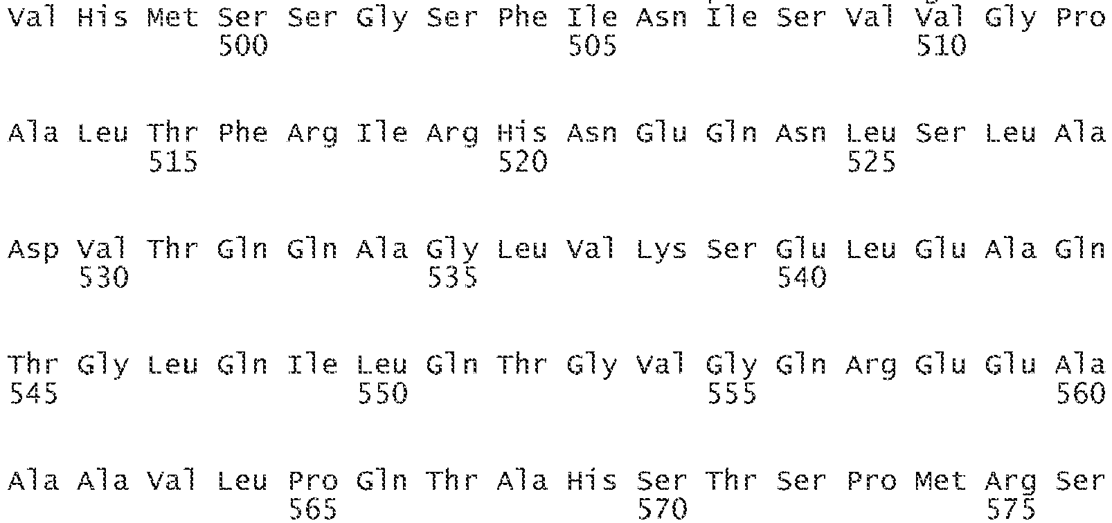
- <210> 4
<211> 200
<212> PRT
<213> Homo sapiens - <400> 4


- <210> 5
<211> 173
<212> PRT
<213> Homo sapiens - <400> 5


- <210> 6
<211> 14
<212> PRT
<213> Homo sapiens - <400> 6

- <210> 7
<211> 8
<212> PRT
<213> Homo sapiens - <400> 7

- <210> 8
<211> 2438
<212> DNA
<213> Homo sapiens - <400> 8


- <210> 9
<211> 485
<212> PRT
<213> Homo sapiens - <400> 9



- <210> 10
<211> 602
<212> DNA
<213> Homo sapiens - <400> 10

- <210> 11
<211> 21
<212> DNA
<213> Mus musculus - <400> 11
agaggccatc caggccacat t 21 - <210> 12
<211> 1682
<212> DNA
<213> Mus musculus - <400> 12


- <210> 13
<211> 21
<212> DNA
<213> Mus musculus - <400> 13
aatgtggcct ggatggcctc t 21
Claims (15)
- An in-vitro method for identifying a compound effective to increase aspartyl aminopeptidase (DAP) activity in mammalian pancreatic islet cells, the method comprising :- contacting an islet cell lysate with DAP, angiotensin II and a test compound;- measuring the level of angiotensin II; and- determining if the level of angiotensin II is lower when treated with the test compound than an otherwise identical control method containing islet cell lysate, DAP and angiotensin II that has not been contacted with the test compound.
- The method according to claim 1, wherein the contacting step is carried out at a temperature of 35° to 39 °C in an aqueous solution, said aqueous solution preferably having a pH of 7.2 to 7.6.
- The method according to claim 1, wherein the method further comprises measuring activity of DAP and determining if the activity of DAP is higher when treated with the test compound than an otherwise identical control containing DAP and angiotensin II that has not been contacted with the test compound.
- The method according to claim 1, wherein the DAP and angiotensin II are contacted with the test compound for an amount of time effective for the DAP to degrade at least about 10 percent of the angiotensin II.
- The method according to claim 1, wherein the DAP and angiotensin II are each in substantially purified form.
- A method for identifying a compound effective to increase DAP expression in mammalian pancreatic islet cells, the method comprising:- culturing mammalian islet cells capable of producing DAP in the presence of a test compound;- measuring the level of expression from the gene encoding DAP; and- determining if the level of expression is higher when treated with the test compound than an otherwise identical control method with cells not cultured with the test compound.
- The method according to claim 6, wherein the method further comprises measuring an amount of angiotensin II and determining if the level of angiotensin II is lower when treated with the test compound than an otherwise identical control not cultured with the test compound.
- The method according to claim 6, wherein measuring the level of expression comprises measuring the amount of DAP in the cultured cells.
- The method according to claim 6, wherein measuring the level of expression comprises measuring the amount of mRNA encoding DAP in the cultured cells.
- An in-vitro method for increasing DAP expression and decreasing Ang II levels, the method comprising delivering one or more copies of a polynucleotide encoding DAP to pancreatic islet cells.
- The method according to claim 10, wherein the one or more copies of a polynucleotide encoding DAP are delivered to the subject's pancreatic islet cells via a viral vector.
- The method according to claim 11, wherein the viral vector includes a promoter activated in pancreatic alpha cells.
- The method according to claim 12, wherein the promoter is a glucagon- activated promoter.
- The method according to claim 10, wherein the polynucleotide encoding DAP comprises a sequence encoding a protein a having at least 80 percent identity to SEQ ID NO. 9.
- The method according to claim 10, wherein the polynucleotide encoding DAP comprises a sequence encoding a protein a having at least 85 percent identity to SEQ ID NO. 9.
Applications Claiming Priority (2)
| Application Number | Priority Date | Filing Date | Title |
|---|---|---|---|
| US38532210P | 2010-09-22 | 2010-09-22 | |
| PCT/US2011/052799 WO2012040491A1 (en) | 2010-09-22 | 2011-09-22 | Method for identifying compounds to increase the expression or activity of aspartyl aminopeptidase |
Publications (3)
| Publication Number | Publication Date |
|---|---|
| EP2619330A1 EP2619330A1 (en) | 2013-07-31 |
| EP2619330A4 EP2619330A4 (en) | 2014-03-19 |
| EP2619330B1 true EP2619330B1 (en) | 2018-05-09 |
Family
ID=45818299
Family Applications (1)
| Application Number | Title | Priority Date | Filing Date |
|---|---|---|---|
| EP11827554.4A Not-in-force EP2619330B1 (en) | 2010-09-22 | 2011-09-22 | Method for identifying compounds to increase the expression or activity of aspartyl aminopeptidase |
Country Status (3)
| Country | Link |
|---|---|
| US (1) | US20120071544A1 (en) |
| EP (1) | EP2619330B1 (en) |
| WO (1) | WO2012040491A1 (en) |
Families Citing this family (2)
| Publication number | Priority date | Publication date | Assignee | Title |
|---|---|---|---|---|
| US10071172B2 (en) * | 2014-04-23 | 2018-09-11 | University of Pittsburgh—of the Commonwealth System of Higher Education | Endogenous neogenesis of beta cells |
| EP4460562A4 (en) * | 2022-01-04 | 2026-02-18 | Univ Pittsburgh Commonwealth Sys Higher Education | GLUCAGON PROMOTER FOR DIABETES GENE THERAPY |
Family Cites Families (1)
| Publication number | Priority date | Publication date | Assignee | Title |
|---|---|---|---|---|
| WO2005103290A1 (en) * | 2004-04-20 | 2005-11-03 | Bayer Healthcare Ag | Diagnostics and therapeutics for diseases associated with aspartyl aminopeptidase (dnpep) |
-
2011
- 2011-09-22 EP EP11827554.4A patent/EP2619330B1/en not_active Not-in-force
- 2011-09-22 US US13/240,837 patent/US20120071544A1/en not_active Abandoned
- 2011-09-22 WO PCT/US2011/052799 patent/WO2012040491A1/en not_active Ceased
Non-Patent Citations (1)
| Title |
|---|
| None * |
Also Published As
| Publication number | Publication date |
|---|---|
| EP2619330A1 (en) | 2013-07-31 |
| US20120071544A1 (en) | 2012-03-22 |
| EP2619330A4 (en) | 2014-03-19 |
| WO2012040491A1 (en) | 2012-03-29 |
Similar Documents
| Publication | Publication Date | Title |
|---|---|---|
| Missiaen et al. | Calcium in the Golgi apparatus | |
| US7879544B2 (en) | Methods of identifying adipocyte specific genes, the genes identified, and their uses | |
| Cheung et al. | Xanthine oxidoreductase is a regulator of adipogenesis and PPARγ activity | |
| Zhang et al. | Identification of novel adipokines through proteomic profiling of small extracellular vesicles derived from adipose tissue | |
| CN101932609A (en) | Compositions and methods for modulating cell membrane resealing | |
| US20100105625A1 (en) | Product and Methods for Diagnosis and Therapy for Cardiac and Skeletal Muscle Disorders | |
| Song et al. | Human munc13 is a diacylglycerol receptor that induces apoptosis and may contribute to renal cell injury in hyperglycemia | |
| Nagamatsu et al. | α-Soluble N-ethylmaleimide-sensitive factor attachment protein is expressed in pancreatic β cells and functions in insulin but not γ-aminobutyric acid secretion | |
| US20050031605A1 (en) | Compositions and methods of treating diabetes | |
| EP3108257B1 (en) | Methods and uses of mitofusins | |
| CN103966309A (en) | Early prediction/diagnosis of AD (Alzheimer disease) by determining TRPC6mRNA (transient receptor potential canonical 6 messenger ribonucleic acid) level in peripheral blood cells | |
| CA2496891A1 (en) | Novel actin-related cytoskeletal protein lacs | |
| CN110237257B (en) | Application of Ube3a ubiquitinated PP2A activator PTPA in treatment of Angel syndrome and autism | |
| US20120071544A1 (en) | Methods For Identifying Compounds Effective To Increase The Expression Or Activity Of Aspartyl Aminopeptidase In Mammalian Pancreatic Islet Cells, And Methods For Decreasing Angiotensin II Levels In Mammalian Pancreatic Islet Cells | |
| JP2010518821A (en) | Secreted protein Ccdc80 that controls adipocyte differentiation | |
| US20050085436A1 (en) | Method to treat conditions associated with insulin signalling dysregulation | |
| KR20070092767A (en) | Detection method of synobiol activity regulating action | |
| Salvatore et al. | Identification of sirm, a novel insulin-regulated SH3 binding protein that associates with Grb-2 and FYN | |
| EP1403282A1 (en) | Protein complexes of the Tumor necrosis factor-alpha (TNF-alpha) signalling pathway | |
| JPWO2004019983A1 (en) | Degradation method, degradation inhibition method and degradation inhibitor of transcription factors of genes related to sugar metabolism | |
| EP4035679A1 (en) | Diabetes therapy targeting abnormal stem cells | |
| EP1627051A2 (en) | Regulation of acheron expression | |
| US20060264610A1 (en) | Protein complexes of the tip60 transcriptional activator protein | |
| Zhang et al. | A conserved hormonal signalling–H2A. Z axis rapidly reorganizes 3D chromatin interactions in adipocyte thermogenesis | |
| WO2025210093A1 (en) | Y chrosomal genes for the treatment or diagnosis of cardiovascular disease |
Legal Events
| Date | Code | Title | Description |
|---|---|---|---|
| PUAI | Public reference made under article 153(3) epc to a published international application that has entered the european phase |
Free format text: ORIGINAL CODE: 0009012 |
|
| 17P | Request for examination filed |
Effective date: 20130419 |
|
| AK | Designated contracting states |
Kind code of ref document: A1 Designated state(s): AL AT BE BG CH CY CZ DE DK EE ES FI FR GB GR HR HU IE IS IT LI LT LU LV MC MK MT NL NO PL PT RO RS SE SI SK SM TR |
|
| DAX | Request for extension of the european patent (deleted) | ||
| A4 | Supplementary search report drawn up and despatched |
Effective date: 20140213 |
|
| RIC1 | Information provided on ipc code assigned before grant |
Ipc: C12Q 1/68 20060101AFI20140207BHEP Ipc: G01N 33/50 20060101ALI20140207BHEP Ipc: C12Q 1/37 20060101ALI20140207BHEP Ipc: C12N 9/48 20060101ALI20140207BHEP |
|
| 17Q | First examination report despatched |
Effective date: 20151203 |
|
| GRAP | Despatch of communication of intention to grant a patent |
Free format text: ORIGINAL CODE: EPIDOSNIGR1 |
|
| INTG | Intention to grant announced |
Effective date: 20171214 |
|
| GRAS | Grant fee paid |
Free format text: ORIGINAL CODE: EPIDOSNIGR3 |
|
| GRAA | (expected) grant |
Free format text: ORIGINAL CODE: 0009210 |
|
| AK | Designated contracting states |
Kind code of ref document: B1 Designated state(s): AL AT BE BG CH CY CZ DE DK EE ES FI FR GB GR HR HU IE IS IT LI LT LU LV MC MK MT NL NO PL PT RO RS SE SI SK SM TR |
|
| REG | Reference to a national code |
Ref country code: GB Ref legal event code: FG4D |
|
| REG | Reference to a national code |
Ref country code: CH Ref legal event code: EP Ref country code: AT Ref legal event code: REF Ref document number: 997612 Country of ref document: AT Kind code of ref document: T Effective date: 20180515 |
|
| REG | Reference to a national code |
Ref country code: IE Ref legal event code: FG4D |
|
| REG | Reference to a national code |
Ref country code: DE Ref legal event code: R096 Ref document number: 602011048285 Country of ref document: DE |
|
| REG | Reference to a national code |
Ref country code: NL Ref legal event code: MP Effective date: 20180509 |
|
| REG | Reference to a national code |
Ref country code: LT Ref legal event code: MG4D |
|
| PG25 | Lapsed in a contracting state [announced via postgrant information from national office to epo] |
Ref country code: ES Free format text: LAPSE BECAUSE OF FAILURE TO SUBMIT A TRANSLATION OF THE DESCRIPTION OR TO PAY THE FEE WITHIN THE PRESCRIBED TIME-LIMIT Effective date: 20180509 Ref country code: SE Free format text: LAPSE BECAUSE OF FAILURE TO SUBMIT A TRANSLATION OF THE DESCRIPTION OR TO PAY THE FEE WITHIN THE PRESCRIBED TIME-LIMIT Effective date: 20180509 Ref country code: NO Free format text: LAPSE BECAUSE OF FAILURE TO SUBMIT A TRANSLATION OF THE DESCRIPTION OR TO PAY THE FEE WITHIN THE PRESCRIBED TIME-LIMIT Effective date: 20180809 Ref country code: FI Free format text: LAPSE BECAUSE OF FAILURE TO SUBMIT A TRANSLATION OF THE DESCRIPTION OR TO PAY THE FEE WITHIN THE PRESCRIBED TIME-LIMIT Effective date: 20180509 Ref country code: BG Free format text: LAPSE BECAUSE OF FAILURE TO SUBMIT A TRANSLATION OF THE DESCRIPTION OR TO PAY THE FEE WITHIN THE PRESCRIBED TIME-LIMIT Effective date: 20180809 Ref country code: LT Free format text: LAPSE BECAUSE OF FAILURE TO SUBMIT A TRANSLATION OF THE DESCRIPTION OR TO PAY THE FEE WITHIN THE PRESCRIBED TIME-LIMIT Effective date: 20180509 |
|
| PG25 | Lapsed in a contracting state [announced via postgrant information from national office to epo] |
Ref country code: HR Free format text: LAPSE BECAUSE OF FAILURE TO SUBMIT A TRANSLATION OF THE DESCRIPTION OR TO PAY THE FEE WITHIN THE PRESCRIBED TIME-LIMIT Effective date: 20180509 Ref country code: LV Free format text: LAPSE BECAUSE OF FAILURE TO SUBMIT A TRANSLATION OF THE DESCRIPTION OR TO PAY THE FEE WITHIN THE PRESCRIBED TIME-LIMIT Effective date: 20180509 Ref country code: NL Free format text: LAPSE BECAUSE OF FAILURE TO SUBMIT A TRANSLATION OF THE DESCRIPTION OR TO PAY THE FEE WITHIN THE PRESCRIBED TIME-LIMIT Effective date: 20180509 Ref country code: RS Free format text: LAPSE BECAUSE OF FAILURE TO SUBMIT A TRANSLATION OF THE DESCRIPTION OR TO PAY THE FEE WITHIN THE PRESCRIBED TIME-LIMIT Effective date: 20180509 Ref country code: GR Free format text: LAPSE BECAUSE OF FAILURE TO SUBMIT A TRANSLATION OF THE DESCRIPTION OR TO PAY THE FEE WITHIN THE PRESCRIBED TIME-LIMIT Effective date: 20180810 |
|
| REG | Reference to a national code |
Ref country code: AT Ref legal event code: MK05 Ref document number: 997612 Country of ref document: AT Kind code of ref document: T Effective date: 20180509 |
|
| PG25 | Lapsed in a contracting state [announced via postgrant information from national office to epo] |
Ref country code: CZ Free format text: LAPSE BECAUSE OF FAILURE TO SUBMIT A TRANSLATION OF THE DESCRIPTION OR TO PAY THE FEE WITHIN THE PRESCRIBED TIME-LIMIT Effective date: 20180509 Ref country code: RO Free format text: LAPSE BECAUSE OF FAILURE TO SUBMIT A TRANSLATION OF THE DESCRIPTION OR TO PAY THE FEE WITHIN THE PRESCRIBED TIME-LIMIT Effective date: 20180509 Ref country code: DK Free format text: LAPSE BECAUSE OF FAILURE TO SUBMIT A TRANSLATION OF THE DESCRIPTION OR TO PAY THE FEE WITHIN THE PRESCRIBED TIME-LIMIT Effective date: 20180509 Ref country code: PL Free format text: LAPSE BECAUSE OF FAILURE TO SUBMIT A TRANSLATION OF THE DESCRIPTION OR TO PAY THE FEE WITHIN THE PRESCRIBED TIME-LIMIT Effective date: 20180509 Ref country code: EE Free format text: LAPSE BECAUSE OF FAILURE TO SUBMIT A TRANSLATION OF THE DESCRIPTION OR TO PAY THE FEE WITHIN THE PRESCRIBED TIME-LIMIT Effective date: 20180509 Ref country code: AT Free format text: LAPSE BECAUSE OF FAILURE TO SUBMIT A TRANSLATION OF THE DESCRIPTION OR TO PAY THE FEE WITHIN THE PRESCRIBED TIME-LIMIT Effective date: 20180509 Ref country code: SK Free format text: LAPSE BECAUSE OF FAILURE TO SUBMIT A TRANSLATION OF THE DESCRIPTION OR TO PAY THE FEE WITHIN THE PRESCRIBED TIME-LIMIT Effective date: 20180509 |
|
| REG | Reference to a national code |
Ref country code: DE Ref legal event code: R097 Ref document number: 602011048285 Country of ref document: DE |
|
| PG25 | Lapsed in a contracting state [announced via postgrant information from national office to epo] |
Ref country code: IT Free format text: LAPSE BECAUSE OF FAILURE TO SUBMIT A TRANSLATION OF THE DESCRIPTION OR TO PAY THE FEE WITHIN THE PRESCRIBED TIME-LIMIT Effective date: 20180509 Ref country code: SM Free format text: LAPSE BECAUSE OF FAILURE TO SUBMIT A TRANSLATION OF THE DESCRIPTION OR TO PAY THE FEE WITHIN THE PRESCRIBED TIME-LIMIT Effective date: 20180509 |
|
| PLBE | No opposition filed within time limit |
Free format text: ORIGINAL CODE: 0009261 |
|
| STAA | Information on the status of an ep patent application or granted ep patent |
Free format text: STATUS: NO OPPOSITION FILED WITHIN TIME LIMIT |
|
| REG | Reference to a national code |
Ref country code: DE Ref legal event code: R119 Ref document number: 602011048285 Country of ref document: DE |
|
| 26N | No opposition filed |
Effective date: 20190212 |
|
| PG25 | Lapsed in a contracting state [announced via postgrant information from national office to epo] |
Ref country code: MC Free format text: LAPSE BECAUSE OF FAILURE TO SUBMIT A TRANSLATION OF THE DESCRIPTION OR TO PAY THE FEE WITHIN THE PRESCRIBED TIME-LIMIT Effective date: 20180509 |
|
| REG | Reference to a national code |
Ref country code: CH Ref legal event code: PL |
|
| GBPC | Gb: european patent ceased through non-payment of renewal fee |
Effective date: 20180922 |
|
| PG25 | Lapsed in a contracting state [announced via postgrant information from national office to epo] |
Ref country code: SI Free format text: LAPSE BECAUSE OF FAILURE TO SUBMIT A TRANSLATION OF THE DESCRIPTION OR TO PAY THE FEE WITHIN THE PRESCRIBED TIME-LIMIT Effective date: 20180509 |
|
| REG | Reference to a national code |
Ref country code: BE Ref legal event code: MM Effective date: 20180930 |
|
| REG | Reference to a national code |
Ref country code: IE Ref legal event code: MM4A |
|
| PG25 | Lapsed in a contracting state [announced via postgrant information from national office to epo] |
Ref country code: LU Free format text: LAPSE BECAUSE OF NON-PAYMENT OF DUE FEES Effective date: 20180922 |
|
| PG25 | Lapsed in a contracting state [announced via postgrant information from national office to epo] |
Ref country code: IE Free format text: LAPSE BECAUSE OF NON-PAYMENT OF DUE FEES Effective date: 20180922 Ref country code: DE Free format text: LAPSE BECAUSE OF NON-PAYMENT OF DUE FEES Effective date: 20190402 |
|
| PG25 | Lapsed in a contracting state [announced via postgrant information from national office to epo] |
Ref country code: CH Free format text: LAPSE BECAUSE OF NON-PAYMENT OF DUE FEES Effective date: 20180930 Ref country code: LI Free format text: LAPSE BECAUSE OF NON-PAYMENT OF DUE FEES Effective date: 20180930 Ref country code: FR Free format text: LAPSE BECAUSE OF NON-PAYMENT OF DUE FEES Effective date: 20180930 Ref country code: BE Free format text: LAPSE BECAUSE OF NON-PAYMENT OF DUE FEES Effective date: 20180930 |
|
| PG25 | Lapsed in a contracting state [announced via postgrant information from national office to epo] |
Ref country code: GB Free format text: LAPSE BECAUSE OF NON-PAYMENT OF DUE FEES Effective date: 20180922 |
|
| PG25 | Lapsed in a contracting state [announced via postgrant information from national office to epo] |
Ref country code: AL Free format text: LAPSE BECAUSE OF FAILURE TO SUBMIT A TRANSLATION OF THE DESCRIPTION OR TO PAY THE FEE WITHIN THE PRESCRIBED TIME-LIMIT Effective date: 20180509 |
|
| PG25 | Lapsed in a contracting state [announced via postgrant information from national office to epo] |
Ref country code: MT Free format text: LAPSE BECAUSE OF NON-PAYMENT OF DUE FEES Effective date: 20180922 |
|
| PG25 | Lapsed in a contracting state [announced via postgrant information from national office to epo] |
Ref country code: TR Free format text: LAPSE BECAUSE OF FAILURE TO SUBMIT A TRANSLATION OF THE DESCRIPTION OR TO PAY THE FEE WITHIN THE PRESCRIBED TIME-LIMIT Effective date: 20180509 |
|
| PG25 | Lapsed in a contracting state [announced via postgrant information from national office to epo] |
Ref country code: PT Free format text: LAPSE BECAUSE OF FAILURE TO SUBMIT A TRANSLATION OF THE DESCRIPTION OR TO PAY THE FEE WITHIN THE PRESCRIBED TIME-LIMIT Effective date: 20180509 Ref country code: HU Free format text: LAPSE BECAUSE OF FAILURE TO SUBMIT A TRANSLATION OF THE DESCRIPTION OR TO PAY THE FEE WITHIN THE PRESCRIBED TIME-LIMIT; INVALID AB INITIO Effective date: 20110922 |
|
| PG25 | Lapsed in a contracting state [announced via postgrant information from national office to epo] |
Ref country code: MK Free format text: LAPSE BECAUSE OF NON-PAYMENT OF DUE FEES Effective date: 20180509 Ref country code: CY Free format text: LAPSE BECAUSE OF FAILURE TO SUBMIT A TRANSLATION OF THE DESCRIPTION OR TO PAY THE FEE WITHIN THE PRESCRIBED TIME-LIMIT Effective date: 20180509 |
|
| PG25 | Lapsed in a contracting state [announced via postgrant information from national office to epo] |
Ref country code: IS Free format text: LAPSE BECAUSE OF FAILURE TO SUBMIT A TRANSLATION OF THE DESCRIPTION OR TO PAY THE FEE WITHIN THE PRESCRIBED TIME-LIMIT Effective date: 20180909 |