EP2619306B1 - Surface display of polypeptides containing a metal porphyrin or a flavin - Google Patents
Surface display of polypeptides containing a metal porphyrin or a flavin Download PDFInfo
- Publication number
- EP2619306B1 EP2619306B1 EP20110758244 EP11758244A EP2619306B1 EP 2619306 B1 EP2619306 B1 EP 2619306B1 EP 20110758244 EP20110758244 EP 20110758244 EP 11758244 A EP11758244 A EP 11758244A EP 2619306 B1 EP2619306 B1 EP 2619306B1
- Authority
- EP
- European Patent Office
- Prior art keywords
- host cell
- polypeptide
- prosthetic group
- coli
- cell
- Prior art date
- Legal status (The legal status is an assumption and is not a legal conclusion. Google has not performed a legal analysis and makes no representation as to the accuracy of the status listed.)
- Not-in-force
Links
- 108090000765 processed proteins & peptides Proteins 0.000 title claims description 115
- 102000004196 processed proteins & peptides Human genes 0.000 title claims description 113
- 229920001184 polypeptide Polymers 0.000 title claims description 110
- 150000004032 porphyrins Chemical class 0.000 title claims description 54
- 229910052751 metal Inorganic materials 0.000 title claims description 41
- 239000002184 metal Substances 0.000 title claims description 41
- AUNGANRZJHBGPY-SCRDCRAPSA-N Riboflavin Chemical compound OC[C@@H](O)[C@@H](O)[C@@H](O)CN1C=2C=C(C)C(C)=CC=2N=C2C1=NC(=O)NC2=O AUNGANRZJHBGPY-SCRDCRAPSA-N 0.000 title claims description 18
- 210000004027 cell Anatomy 0.000 claims description 169
- 239000012528 membrane Substances 0.000 claims description 82
- 241000588724 Escherichia coli Species 0.000 claims description 63
- 108090000623 proteins and genes Proteins 0.000 claims description 56
- 150000007523 nucleic acids Chemical class 0.000 claims description 55
- 102000004169 proteins and genes Human genes 0.000 claims description 53
- 102000004190 Enzymes Human genes 0.000 claims description 50
- 108090000790 Enzymes Proteins 0.000 claims description 50
- 238000000034 method Methods 0.000 claims description 46
- 108010073429 Type V Secretion Systems Proteins 0.000 claims description 39
- 108020004707 nucleic acids Proteins 0.000 claims description 38
- 102000039446 nucleic acids Human genes 0.000 claims description 38
- 230000004927 fusion Effects 0.000 claims description 32
- 230000014509 gene expression Effects 0.000 claims description 31
- 108091005804 Peptidases Proteins 0.000 claims description 23
- 238000002360 preparation method Methods 0.000 claims description 23
- 108010078791 Carrier Proteins Proteins 0.000 claims description 20
- 239000004365 Protease Substances 0.000 claims description 17
- 102000002004 Cytochrome P-450 Enzyme System Human genes 0.000 claims description 16
- 108010015742 Cytochrome P-450 Enzyme System Proteins 0.000 claims description 16
- 101150053185 P450 gene Proteins 0.000 claims description 16
- 239000000758 substrate Substances 0.000 claims description 16
- 241000894006 Bacteria Species 0.000 claims description 15
- 102000008109 Mixed Function Oxygenases Human genes 0.000 claims description 13
- 108010074633 Mixed Function Oxygenases Proteins 0.000 claims description 13
- 108010076504 Protein Sorting Signals Proteins 0.000 claims description 13
- 102000018832 Cytochromes Human genes 0.000 claims description 11
- 108010052832 Cytochromes Proteins 0.000 claims description 11
- 230000007246 mechanism Effects 0.000 claims description 11
- 238000003556 assay Methods 0.000 claims description 10
- 230000015572 biosynthetic process Effects 0.000 claims description 10
- 238000003786 synthesis reaction Methods 0.000 claims description 10
- 239000003814 drug Substances 0.000 claims description 8
- 229940079593 drug Drugs 0.000 claims description 8
- 102000004316 Oxidoreductases Human genes 0.000 claims description 7
- 108090000854 Oxidoreductases Proteins 0.000 claims description 7
- 239000000126 substance Substances 0.000 claims description 7
- -1 BrkA Proteins 0.000 claims description 6
- 102000014914 Carrier Proteins Human genes 0.000 claims description 6
- 230000001419 dependent effect Effects 0.000 claims description 6
- 108010002231 IgA-specific serine endopeptidase Proteins 0.000 claims description 5
- FVTCRASFADXXNN-SCRDCRAPSA-N flavin mononucleotide Chemical compound OP(=O)(O)OC[C@@H](O)[C@@H](O)[C@@H](O)CN1C=2C=C(C)C(C)=CC=2N=C2C1=NC(=O)NC2=O FVTCRASFADXXNN-SCRDCRAPSA-N 0.000 claims description 4
- 238000012216 screening Methods 0.000 claims description 4
- 108010057573 Flavoproteins Proteins 0.000 claims description 3
- 102000003983 Flavoproteins Human genes 0.000 claims description 3
- 108091007187 Reductases Proteins 0.000 claims description 3
- 238000012258 culturing Methods 0.000 claims description 3
- 230000000593 degrading effect Effects 0.000 claims description 3
- 101710160107 Outer membrane protein A Proteins 0.000 claims description 2
- 101710160102 Outer membrane protein B Proteins 0.000 claims description 2
- 241001591005 Siga Species 0.000 claims description 2
- 229940088597 hormone Drugs 0.000 claims description 2
- 239000005556 hormone Substances 0.000 claims description 2
- 239000000543 intermediate Substances 0.000 claims description 2
- 101150077696 lip-1 gene Proteins 0.000 claims description 2
- YAFQFNOUYXZVPZ-UHFFFAOYSA-N liproxstatin-1 Chemical compound ClC1=CC=CC(CNC=2C3(CCNCC3)NC3=CC=CC=C3N=2)=C1 YAFQFNOUYXZVPZ-UHFFFAOYSA-N 0.000 claims description 2
- 238000012544 monitoring process Methods 0.000 claims description 2
- 108010021711 pertactin Proteins 0.000 claims description 2
- 231100000614 poison Toxicity 0.000 claims description 2
- 239000007858 starting material Substances 0.000 claims description 2
- 239000003440 toxic substance Substances 0.000 claims description 2
- 231100000419 toxicity Toxicity 0.000 claims description 2
- 230000001988 toxicity Effects 0.000 claims description 2
- 102100037486 Reverse transcriptase/ribonuclease H Human genes 0.000 claims 2
- 102100027153 Ankyrin repeat and sterile alpha motif domain-containing protein 1B Human genes 0.000 claims 1
- 101710149294 Ankyrin repeat and sterile alpha motif domain-containing protein 1B Proteins 0.000 claims 1
- 229940088598 enzyme Drugs 0.000 description 45
- 235000018102 proteins Nutrition 0.000 description 45
- 238000006243 chemical reaction Methods 0.000 description 28
- 150000001413 amino acids Chemical class 0.000 description 26
- 238000009638 autodisplay Methods 0.000 description 26
- BPHPUYQFMNQIOC-NXRLNHOXSA-N isopropyl beta-D-thiogalactopyranoside Chemical compound CC(C)S[C@@H]1O[C@H](CO)[C@H](O)[C@H](O)[C@H]1O BPHPUYQFMNQIOC-NXRLNHOXSA-N 0.000 description 23
- 102000035195 Peptidases Human genes 0.000 description 21
- 239000002245 particle Substances 0.000 description 18
- 239000000047 product Substances 0.000 description 17
- 210000001322 periplasm Anatomy 0.000 description 16
- 230000032258 transport Effects 0.000 description 16
- 108010081668 Cytochrome P-450 CYP3A Proteins 0.000 description 15
- 230000000694 effects Effects 0.000 description 15
- 235000019419 proteases Nutrition 0.000 description 15
- 102100039205 Cytochrome P450 3A4 Human genes 0.000 description 13
- 125000005647 linker group Chemical group 0.000 description 13
- 108020004414 DNA Proteins 0.000 description 12
- 101000874355 Escherichia coli (strain K12) Outer membrane protein assembly factor BamA Proteins 0.000 description 12
- 239000013612 plasmid Substances 0.000 description 12
- 150000003431 steroids Chemical class 0.000 description 12
- ZESRJSPZRDMNHY-YFWFAHHUSA-N 11-deoxycorticosterone Chemical compound O=C1CC[C@]2(C)[C@H]3CC[C@](C)([C@H](CC4)C(=O)CO)[C@@H]4[C@@H]3CCC2=C1 ZESRJSPZRDMNHY-YFWFAHHUSA-N 0.000 description 10
- 241001198387 Escherichia coli BL21(DE3) Species 0.000 description 10
- ZESRJSPZRDMNHY-UHFFFAOYSA-N de-oxy corticosterone Natural products O=C1CCC2(C)C3CCC(C)(C(CC4)C(=O)CO)C4C3CCC2=C1 ZESRJSPZRDMNHY-UHFFFAOYSA-N 0.000 description 10
- 230000037361 pathway Effects 0.000 description 9
- 108010046335 Ferredoxin-NADP Reductase Proteins 0.000 description 8
- ACFIXJIJDZMPPO-NNYOXOHSSA-N NADPH Chemical compound C1=CCC(C(=O)N)=CN1[C@H]1[C@H](O)[C@H](O)[C@@H](COP(O)(=O)OP(O)(=O)OC[C@@H]2[C@H]([C@@H](OP(O)(O)=O)[C@@H](O2)N2C3=NC=NC(N)=C3N=C2)O)O1 ACFIXJIJDZMPPO-NNYOXOHSSA-N 0.000 description 8
- 102100036777 NADPH:adrenodoxin oxidoreductase, mitochondrial Human genes 0.000 description 8
- 210000000170 cell membrane Anatomy 0.000 description 8
- 229930027945 nicotinamide-adenine dinucleotide Natural products 0.000 description 8
- 108090000631 Trypsin Proteins 0.000 description 7
- 102000004142 Trypsin Human genes 0.000 description 7
- 239000012634 fragment Substances 0.000 description 7
- 238000005805 hydroxylation reaction Methods 0.000 description 7
- 239000012588 trypsin Substances 0.000 description 7
- WHBHBVVOGNECLV-OBQKJFGGSA-N 11-deoxycortisol Chemical compound O=C1CC[C@]2(C)[C@H]3CC[C@](C)([C@@](CC4)(O)C(=O)CO)[C@@H]4[C@@H]3CCC2=C1 WHBHBVVOGNECLV-OBQKJFGGSA-N 0.000 description 6
- 108090000187 Adrenodoxin Proteins 0.000 description 6
- 102000003804 Adrenodoxin Human genes 0.000 description 6
- 101150051438 CYP gene Proteins 0.000 description 6
- 239000004793 Polystyrene Substances 0.000 description 6
- 235000001014 amino acid Nutrition 0.000 description 6
- 229940119740 deoxycorticosterone Drugs 0.000 description 6
- BCGWQEUPMDMJNV-UHFFFAOYSA-N imipramine Chemical compound C1CC2=CC=CC=C2N(CCCN(C)C)C2=CC=CC=C21 BCGWQEUPMDMJNV-UHFFFAOYSA-N 0.000 description 6
- 229960004801 imipramine Drugs 0.000 description 6
- 238000004519 manufacturing process Methods 0.000 description 6
- 229920000052 poly(p-xylylene) Polymers 0.000 description 6
- 229920000642 polymer Polymers 0.000 description 6
- 229920002223 polystyrene Polymers 0.000 description 6
- 235000019833 protease Nutrition 0.000 description 6
- BTXXTMOWISPQSJ-UHFFFAOYSA-N 4,4,4-trifluorobutan-2-one Chemical compound CC(=O)CC(F)(F)F BTXXTMOWISPQSJ-UHFFFAOYSA-N 0.000 description 5
- BQACOLQNOUYJCE-FYZZASKESA-N Abietic acid Natural products CC(C)C1=CC2=CC[C@]3(C)[C@](C)(CCC[C@@]3(C)C(=O)O)[C@H]2CC1 BQACOLQNOUYJCE-FYZZASKESA-N 0.000 description 5
- RSWGJHLUYNHPMX-UHFFFAOYSA-N Abietic-Saeure Natural products C12CCC(C(C)C)=CC2=CCC2C1(C)CCCC2(C)C(O)=O RSWGJHLUYNHPMX-UHFFFAOYSA-N 0.000 description 5
- 241000588832 Bordetella pertussis Species 0.000 description 5
- 101100165129 Neisseria meningitidis serogroup B (strain MC58) bamA gene Proteins 0.000 description 5
- 108091028043 Nucleic acid sequence Proteins 0.000 description 5
- 230000001580 bacterial effect Effects 0.000 description 5
- 238000001514 detection method Methods 0.000 description 5
- 239000002158 endotoxin Substances 0.000 description 5
- 150000002211 flavins Chemical class 0.000 description 5
- PCHJSUWPFVWCPO-UHFFFAOYSA-N gold Chemical compound [Au] PCHJSUWPFVWCPO-UHFFFAOYSA-N 0.000 description 5
- 229910052737 gold Inorganic materials 0.000 description 5
- 239000010931 gold Substances 0.000 description 5
- 230000033444 hydroxylation Effects 0.000 description 5
- 229920006008 lipopolysaccharide Polymers 0.000 description 5
- 230000028327 secretion Effects 0.000 description 5
- HCYAFALTSJYZDH-UHFFFAOYSA-N Desimpramine Chemical compound C1CC2=CC=CC=C2N(CCCNC)C2=CC=CC=C21 HCYAFALTSJYZDH-UHFFFAOYSA-N 0.000 description 4
- XEEYBQQBJWHFJM-UHFFFAOYSA-N Iron Chemical compound [Fe] XEEYBQQBJWHFJM-UHFFFAOYSA-N 0.000 description 4
- 241000588650 Neisseria meningitidis Species 0.000 description 4
- 241000607715 Serratia marcescens Species 0.000 description 4
- GUGOEEXESWIERI-UHFFFAOYSA-N Terfenadine Chemical compound C1=CC(C(C)(C)C)=CC=C1C(O)CCCN1CCC(C(O)(C=2C=CC=CC=2)C=2C=CC=CC=2)CC1 GUGOEEXESWIERI-UHFFFAOYSA-N 0.000 description 4
- 238000010521 absorption reaction Methods 0.000 description 4
- 210000004899 c-terminal region Anatomy 0.000 description 4
- 229960003914 desipramine Drugs 0.000 description 4
- 238000002474 experimental method Methods 0.000 description 4
- 230000006870 function Effects 0.000 description 4
- 230000002068 genetic effect Effects 0.000 description 4
- 238000004128 high performance liquid chromatography Methods 0.000 description 4
- 230000005661 hydrophobic surface Effects 0.000 description 4
- 230000005764 inhibitory process Effects 0.000 description 4
- 230000014759 maintenance of location Effects 0.000 description 4
- UMFJAHHVKNCGLG-UHFFFAOYSA-N n-Nitrosodimethylamine Chemical compound CN(C)N=O UMFJAHHVKNCGLG-UHFFFAOYSA-N 0.000 description 4
- 238000002415 sodium dodecyl sulfate polyacrylamide gel electrophoresis Methods 0.000 description 4
- 230000005945 translocation Effects 0.000 description 4
- 239000013598 vector Substances 0.000 description 4
- XLYOFNOQVPJJNP-UHFFFAOYSA-N water Substances O XLYOFNOQVPJJNP-UHFFFAOYSA-N 0.000 description 4
- UHOVQNZJYSORNB-UHFFFAOYSA-N Benzene Chemical compound C1=CC=CC=C1 UHOVQNZJYSORNB-UHFFFAOYSA-N 0.000 description 3
- 241000283690 Bos taurus Species 0.000 description 3
- 108091006146 Channels Proteins 0.000 description 3
- 108010001202 Cytochrome P-450 CYP2E1 Proteins 0.000 description 3
- 102100024332 Cytochrome P450 11B1, mitochondrial Human genes 0.000 description 3
- MYMOFIZGZYHOMD-UHFFFAOYSA-N Dioxygen Chemical compound O=O MYMOFIZGZYHOMD-UHFFFAOYSA-N 0.000 description 3
- 101710116435 Outer membrane protein Proteins 0.000 description 3
- 241001138501 Salmonella enterica Species 0.000 description 3
- 241000607762 Shigella flexneri Species 0.000 description 3
- 108010049356 Steroid 11-beta-Hydroxylase Proteins 0.000 description 3
- 125000004429 atom Chemical group 0.000 description 3
- 239000011942 biocatalyst Substances 0.000 description 3
- 230000003197 catalytic effect Effects 0.000 description 3
- 238000006900 dealkylation reaction Methods 0.000 description 3
- 230000007717 exclusion Effects 0.000 description 3
- 102000037865 fusion proteins Human genes 0.000 description 3
- 108020001507 fusion proteins Proteins 0.000 description 3
- 210000005256 gram-negative cell Anatomy 0.000 description 3
- 230000002209 hydrophobic effect Effects 0.000 description 3
- 230000001965 increasing effect Effects 0.000 description 3
- 230000006698 induction Effects 0.000 description 3
- 239000007788 liquid Substances 0.000 description 3
- 239000003550 marker Substances 0.000 description 3
- 230000003228 microsomal effect Effects 0.000 description 3
- 230000002438 mitochondrial effect Effects 0.000 description 3
- 230000003647 oxidation Effects 0.000 description 3
- 238000007254 oxidation reaction Methods 0.000 description 3
- 230000008569 process Effects 0.000 description 3
- 230000009467 reduction Effects 0.000 description 3
- 238000010186 staining Methods 0.000 description 3
- 229960000351 terfenadine Drugs 0.000 description 3
- 238000012360 testing method Methods 0.000 description 3
- 231100000331 toxic Toxicity 0.000 description 3
- 230000002588 toxic effect Effects 0.000 description 3
- KWGRBVOPPLSCSI-WPRPVWTQSA-N (-)-ephedrine Chemical compound CN[C@@H](C)[C@H](O)C1=CC=CC=C1 KWGRBVOPPLSCSI-WPRPVWTQSA-N 0.000 description 2
- DYNISIGUMYFVJW-AZNNDXHDSA-N 12beta-hydroxyabietic acid Natural products CC(C)C([C@@H](C[C@@H]12)O)=CC1=CC[C@@H]1[C@]2(C)CCC[C@@]1(C)C(O)=O DYNISIGUMYFVJW-AZNNDXHDSA-N 0.000 description 2
- OOLUVSIJOMLOCB-UHFFFAOYSA-N 1633-22-3 Chemical compound C1CC(C=C2)=CC=C2CCC2=CC=C1C=C2 OOLUVSIJOMLOCB-UHFFFAOYSA-N 0.000 description 2
- 241000194107 Bacillus megaterium Species 0.000 description 2
- 208000005623 Carcinogenesis Diseases 0.000 description 2
- 102000034573 Channels Human genes 0.000 description 2
- 241001647372 Chlamydia pneumoniae Species 0.000 description 2
- HEDRZPFGACZZDS-UHFFFAOYSA-N Chloroform Chemical compound ClC(Cl)Cl HEDRZPFGACZZDS-UHFFFAOYSA-N 0.000 description 2
- 108010084976 Cholesterol Side-Chain Cleavage Enzyme Proteins 0.000 description 2
- 102100027516 Cholesterol side-chain cleavage enzyme, mitochondrial Human genes 0.000 description 2
- 108090000371 Esterases Proteins 0.000 description 2
- WSFSSNUMVMOOMR-UHFFFAOYSA-N Formaldehyde Chemical compound O=C WSFSSNUMVMOOMR-UHFFFAOYSA-N 0.000 description 2
- 241000606768 Haemophilus influenzae Species 0.000 description 2
- 102000004157 Hydrolases Human genes 0.000 description 2
- 108090000604 Hydrolases Proteins 0.000 description 2
- 241001293418 Mannheimia haemolytica Species 0.000 description 2
- WBNQDOYYEUMPFS-UHFFFAOYSA-N N-nitrosodiethylamine Chemical compound CCN(CC)N=O WBNQDOYYEUMPFS-UHFFFAOYSA-N 0.000 description 2
- 108010007843 NADH oxidase Proteins 0.000 description 2
- PXHVJJICTQNCMI-UHFFFAOYSA-N Nickel Chemical compound [Ni] PXHVJJICTQNCMI-UHFFFAOYSA-N 0.000 description 2
- RJKFOVLPORLFTN-LEKSSAKUSA-N Progesterone Chemical compound C1CC2=CC(=O)CC[C@]2(C)[C@@H]2[C@@H]1[C@@H]1CC[C@H](C(=O)C)[C@@]1(C)CC2 RJKFOVLPORLFTN-LEKSSAKUSA-N 0.000 description 2
- 241000589540 Pseudomonas fluorescens Species 0.000 description 2
- 102000007056 Recombinant Fusion Proteins Human genes 0.000 description 2
- 108010008281 Recombinant Fusion Proteins Proteins 0.000 description 2
- 241000606695 Rickettsia rickettsii Species 0.000 description 2
- FAPWRFPIFSIZLT-UHFFFAOYSA-M Sodium chloride Chemical compound [Na+].[Cl-] FAPWRFPIFSIZLT-UHFFFAOYSA-M 0.000 description 2
- PPBRXRYQALVLMV-UHFFFAOYSA-N Styrene Chemical compound C=CC1=CC=CC=C1 PPBRXRYQALVLMV-UHFFFAOYSA-N 0.000 description 2
- MUMGGOZAMZWBJJ-DYKIIFRCSA-N Testostosterone Chemical compound O=C1CC[C@]2(C)[C@H]3CC[C@](C)([C@H](CC4)O)[C@@H]4[C@@H]3CCC2=C1 MUMGGOZAMZWBJJ-DYKIIFRCSA-N 0.000 description 2
- 239000002115 aflatoxin B1 Substances 0.000 description 2
- OQIQSTLJSLGHID-WNWIJWBNSA-N aflatoxin B1 Chemical compound C=1([C@@H]2C=CO[C@@H]2OC=1C=C(C1=2)OC)C=2OC(=O)C2=C1CCC2=O OQIQSTLJSLGHID-WNWIJWBNSA-N 0.000 description 2
- 229930020125 aflatoxin-B1 Natural products 0.000 description 2
- 230000001430 anti-depressive effect Effects 0.000 description 2
- 239000000935 antidepressant agent Substances 0.000 description 2
- 229940005513 antidepressants Drugs 0.000 description 2
- 238000013459 approach Methods 0.000 description 2
- 230000003833 cell viability Effects 0.000 description 2
- 210000002421 cell wall Anatomy 0.000 description 2
- 239000003153 chemical reaction reagent Substances 0.000 description 2
- 239000011248 coating agent Substances 0.000 description 2
- 238000000576 coating method Methods 0.000 description 2
- 150000001875 compounds Chemical class 0.000 description 2
- 239000010949 copper Substances 0.000 description 2
- 230000020335 dealkylation Effects 0.000 description 2
- 230000003247 decreasing effect Effects 0.000 description 2
- 238000005695 dehalogenation reaction Methods 0.000 description 2
- 238000010520 demethylation reaction Methods 0.000 description 2
- 238000011161 development Methods 0.000 description 2
- 230000029087 digestion Effects 0.000 description 2
- 229910001882 dioxygen Inorganic materials 0.000 description 2
- 230000008406 drug-drug interaction Effects 0.000 description 2
- 230000002255 enzymatic effect Effects 0.000 description 2
- 238000006735 epoxidation reaction Methods 0.000 description 2
- 210000001723 extracellular space Anatomy 0.000 description 2
- 229960003592 fexofenadine Drugs 0.000 description 2
- RWTNPBWLLIMQHL-UHFFFAOYSA-N fexofenadine Chemical compound C1=CC(C(C)(C(O)=O)C)=CC=C1C(O)CCCN1CCC(C(O)(C=2C=CC=CC=2)C=2C=CC=CC=2)CC1 RWTNPBWLLIMQHL-UHFFFAOYSA-N 0.000 description 2
- 150000003278 haem Chemical class 0.000 description 2
- 238000013537 high throughput screening Methods 0.000 description 2
- 229930195733 hydrocarbon Natural products 0.000 description 2
- 150000002430 hydrocarbons Chemical class 0.000 description 2
- JYGXADMDTFJGBT-VWUMJDOOSA-N hydrocortisone Chemical compound O=C1CC[C@]2(C)[C@H]3[C@@H](O)C[C@](C)([C@@](CC4)(O)C(=O)CO)[C@@H]4[C@@H]3CCC2=C1 JYGXADMDTFJGBT-VWUMJDOOSA-N 0.000 description 2
- 230000003993 interaction Effects 0.000 description 2
- 229910052742 iron Inorganic materials 0.000 description 2
- 239000004816 latex Substances 0.000 description 2
- 229920000126 latex Polymers 0.000 description 2
- 210000004185 liver Anatomy 0.000 description 2
- 238000005259 measurement Methods 0.000 description 2
- 239000002207 metabolite Substances 0.000 description 2
- 230000004048 modification Effects 0.000 description 2
- 238000012986 modification Methods 0.000 description 2
- 239000000178 monomer Substances 0.000 description 2
- 229920005615 natural polymer Polymers 0.000 description 2
- 239000013642 negative control Substances 0.000 description 2
- 229940099990 ogen Drugs 0.000 description 2
- 230000036470 plasma concentration Effects 0.000 description 2
- 239000011148 porous material Substances 0.000 description 2
- 239000002243 precursor Substances 0.000 description 2
- 231100000586 procarcinogen Toxicity 0.000 description 2
- 238000003259 recombinant expression Methods 0.000 description 2
- 230000027756 respiratory electron transport chain Effects 0.000 description 2
- FSYKKLYZXJSNPZ-UHFFFAOYSA-N sarcosine Chemical compound C[NH2+]CC([O-])=O FSYKKLYZXJSNPZ-UHFFFAOYSA-N 0.000 description 2
- 230000002269 spontaneous effect Effects 0.000 description 2
- 235000000346 sugar Nutrition 0.000 description 2
- VZGDMQKNWNREIO-UHFFFAOYSA-N tetrachloromethane Chemical compound ClC(Cl)(Cl)Cl VZGDMQKNWNREIO-UHFFFAOYSA-N 0.000 description 2
- 230000001225 therapeutic effect Effects 0.000 description 2
- 230000009466 transformation Effects 0.000 description 2
- DGVVWUTYPXICAM-UHFFFAOYSA-N β‐Mercaptoethanol Chemical compound OCCS DGVVWUTYPXICAM-UHFFFAOYSA-N 0.000 description 2
- DNXHEGUUPJUMQT-UHFFFAOYSA-N (+)-estrone Natural products OC1=CC=C2C3CCC(C)(C(CC4)=O)C4C3CCC2=C1 DNXHEGUUPJUMQT-UHFFFAOYSA-N 0.000 description 1
- VHJLVAABSRFDPM-UHFFFAOYSA-N 1,4-dithiothreitol Chemical compound SCC(O)C(O)CS VHJLVAABSRFDPM-UHFFFAOYSA-N 0.000 description 1
- DYNISIGUMYFVJW-UHFFFAOYSA-N 12alpha-Hydroxy-abietinsaeure Natural products C12CC(O)C(C(C)C)=CC2=CCC2C1(C)CCCC2(C)C(O)=O DYNISIGUMYFVJW-UHFFFAOYSA-N 0.000 description 1
- XGUHPTGEXRHMQQ-BGJMDTOESA-N 19-hydroxyandrost-4-ene-3,17-dione Chemical compound O=C1CC[C@]2(CO)[C@H]3CC[C@](C)(C(CC4)=O)[C@@H]4[C@@H]3CCC2=C1 XGUHPTGEXRHMQQ-BGJMDTOESA-N 0.000 description 1
- JKMHFZQWWAIEOD-UHFFFAOYSA-N 2-[4-(2-hydroxyethyl)piperazin-1-yl]ethanesulfonic acid Chemical compound OCC[NH+]1CCN(CCS([O-])(=O)=O)CC1 JKMHFZQWWAIEOD-UHFFFAOYSA-N 0.000 description 1
- XRCFMDPVHKVRDJ-BGJMDTOESA-N 3,17-dioxoandrost-4-en-19-al Chemical compound O=C1CC[C@]2(C=O)[C@H]3CC[C@](C)(C(CC4)=O)[C@@H]4[C@@H]3CCC2=C1 XRCFMDPVHKVRDJ-BGJMDTOESA-N 0.000 description 1
- 241000606749 Aggregatibacter actinomycetemcomitans Species 0.000 description 1
- PQSUYGKTWSAVDQ-ZVIOFETBSA-N Aldosterone Chemical compound C([C@@]1([C@@H](C(=O)CO)CC[C@H]1[C@@H]1CC2)C=O)[C@H](O)[C@@H]1[C@]1(C)C2=CC(=O)CC1 PQSUYGKTWSAVDQ-ZVIOFETBSA-N 0.000 description 1
- PQSUYGKTWSAVDQ-UHFFFAOYSA-N Aldosterone Natural products C1CC2C3CCC(C(=O)CO)C3(C=O)CC(O)C2C2(C)C1=CC(=O)CC2 PQSUYGKTWSAVDQ-UHFFFAOYSA-N 0.000 description 1
- 102000006410 Apoproteins Human genes 0.000 description 1
- 108010083590 Apoproteins Proteins 0.000 description 1
- 102000004452 Arginase Human genes 0.000 description 1
- 108700024123 Arginases Proteins 0.000 description 1
- 108010078554 Aromatase Proteins 0.000 description 1
- 102100029361 Aromatase Human genes 0.000 description 1
- 108010011485 Aspartame Proteins 0.000 description 1
- 229910001369 Brass Inorganic materials 0.000 description 1
- 101150013062 CHRNA1 gene Proteins 0.000 description 1
- OKTJSMMVPCPJKN-UHFFFAOYSA-N Carbon Chemical compound [C] OKTJSMMVPCPJKN-UHFFFAOYSA-N 0.000 description 1
- 241000606153 Chlamydia trachomatis Species 0.000 description 1
- 102000009016 Cholera Toxin Human genes 0.000 description 1
- 108010049048 Cholera Toxin Proteins 0.000 description 1
- 108091026890 Coding region Proteins 0.000 description 1
- RYGMFSIKBFXOCR-UHFFFAOYSA-N Copper Chemical compound [Cu] RYGMFSIKBFXOCR-UHFFFAOYSA-N 0.000 description 1
- OMFXVFTZEKFJBZ-UHFFFAOYSA-N Corticosterone Natural products O=C1CCC2(C)C3C(O)CC(C)(C(CC4)C(=O)CO)C4C3CCC2=C1 OMFXVFTZEKFJBZ-UHFFFAOYSA-N 0.000 description 1
- 108010074922 Cytochrome P-450 CYP1A2 Proteins 0.000 description 1
- 102000004328 Cytochrome P-450 CYP3A Human genes 0.000 description 1
- 102100026533 Cytochrome P450 1A2 Human genes 0.000 description 1
- 102100024889 Cytochrome P450 2E1 Human genes 0.000 description 1
- 108050008072 Cytochrome c oxidase subunit IV Proteins 0.000 description 1
- 102000000634 Cytochrome c oxidase subunit IV Human genes 0.000 description 1
- 102000007605 Cytochromes b5 Human genes 0.000 description 1
- 108010007167 Cytochromes b5 Proteins 0.000 description 1
- 206010012735 Diarrhoea Diseases 0.000 description 1
- 101100378121 Drosophila melanogaster nAChRalpha1 gene Proteins 0.000 description 1
- 208000030453 Drug-Related Side Effects and Adverse reaction Diseases 0.000 description 1
- KCXVZYZYPLLWCC-UHFFFAOYSA-N EDTA Chemical compound OC(=O)CN(CC(O)=O)CCN(CC(O)=O)CC(O)=O KCXVZYZYPLLWCC-UHFFFAOYSA-N 0.000 description 1
- 241001646716 Escherichia coli K-12 Species 0.000 description 1
- DNXHEGUUPJUMQT-CBZIJGRNSA-N Estrone Chemical compound OC1=CC=C2[C@H]3CC[C@](C)(C(CC4)=O)[C@@H]4[C@@H]3CCC2=C1 DNXHEGUUPJUMQT-CBZIJGRNSA-N 0.000 description 1
- 108010014173 Factor X Proteins 0.000 description 1
- VTLYFUHAOXGGBS-UHFFFAOYSA-N Fe3+ Chemical compound [Fe+3] VTLYFUHAOXGGBS-UHFFFAOYSA-N 0.000 description 1
- 108010074122 Ferredoxins Proteins 0.000 description 1
- 238000012413 Fluorescence activated cell sorting analysis Methods 0.000 description 1
- WQZGKKKJIJFFOK-GASJEMHNSA-N Glucose Natural products OC[C@H]1OC(O)[C@H](O)[C@@H](O)[C@@H]1O WQZGKKKJIJFFOK-GASJEMHNSA-N 0.000 description 1
- 102000006587 Glutathione peroxidase Human genes 0.000 description 1
- 108700016172 Glutathione peroxidases Proteins 0.000 description 1
- 241000282412 Homo Species 0.000 description 1
- 108010081409 Iron-Sulfur Proteins Proteins 0.000 description 1
- 102000005298 Iron-Sulfur Proteins Human genes 0.000 description 1
- KJHKTHWMRKYKJE-SUGCFTRWSA-N Kaletra Chemical compound N1([C@@H](C(C)C)C(=O)N[C@H](C[C@H](O)[C@H](CC=2C=CC=CC=2)NC(=O)COC=2C(=CC=CC=2C)C)CC=2C=CC=CC=2)CCCNC1=O KJHKTHWMRKYKJE-SUGCFTRWSA-N 0.000 description 1
- 102000004895 Lipoproteins Human genes 0.000 description 1
- 108090001030 Lipoproteins Proteins 0.000 description 1
- 241001465754 Metazoa Species 0.000 description 1
- 241000588655 Moraxella catarrhalis Species 0.000 description 1
- 108010085220 Multiprotein Complexes Proteins 0.000 description 1
- 102000007474 Multiprotein Complexes Human genes 0.000 description 1
- 102000016943 Muramidase Human genes 0.000 description 1
- 108010014251 Muramidase Proteins 0.000 description 1
- 108010062010 N-Acetylmuramoyl-L-alanine Amidase Proteins 0.000 description 1
- 150000001204 N-oxides Chemical class 0.000 description 1
- 108010086428 NADH Dehydrogenase Proteins 0.000 description 1
- 241000588652 Neisseria gonorrhoeae Species 0.000 description 1
- 206010028980 Neoplasm Diseases 0.000 description 1
- 108010020943 Nitrogenase Proteins 0.000 description 1
- 108010079246 OMPA outer membrane proteins Proteins 0.000 description 1
- 206010030113 Oedema Diseases 0.000 description 1
- 241000315040 Omura Species 0.000 description 1
- 108010067902 Peptide Library Proteins 0.000 description 1
- 241001148064 Photorhabdus luminescens Species 0.000 description 1
- 239000004743 Polypropylene Substances 0.000 description 1
- 229920001213 Polysorbate 20 Polymers 0.000 description 1
- 108010041388 Ribonucleotide Reductases Proteins 0.000 description 1
- 102000000505 Ribonucleotide Reductases Human genes 0.000 description 1
- NCDNCNXCDXHOMX-UHFFFAOYSA-N Ritonavir Natural products C=1C=CC=CC=1CC(NC(=O)OCC=1SC=NC=1)C(O)CC(CC=1C=CC=CC=1)NC(=O)C(C(C)C)NC(=O)N(C)CC1=CSC(C(C)C)=N1 NCDNCNXCDXHOMX-UHFFFAOYSA-N 0.000 description 1
- 241000607142 Salmonella Species 0.000 description 1
- 108010077895 Sarcosine Proteins 0.000 description 1
- VYPSYNLAJGMNEJ-UHFFFAOYSA-N Silicium dioxide Chemical compound O=[Si]=O VYPSYNLAJGMNEJ-UHFFFAOYSA-N 0.000 description 1
- BQCADISMDOOEFD-UHFFFAOYSA-N Silver Chemical compound [Ag] BQCADISMDOOEFD-UHFFFAOYSA-N 0.000 description 1
- 108010030741 Steroid Hydroxylases Proteins 0.000 description 1
- 102000005938 Steroid Hydroxylases Human genes 0.000 description 1
- 241000282887 Suidae Species 0.000 description 1
- NINIDFKCEFEMDL-UHFFFAOYSA-N Sulfur Chemical compound [S] NINIDFKCEFEMDL-UHFFFAOYSA-N 0.000 description 1
- 239000005864 Sulphur Substances 0.000 description 1
- 108090000190 Thrombin Proteins 0.000 description 1
- RTAQQCXQSZGOHL-UHFFFAOYSA-N Titanium Chemical compound [Ti] RTAQQCXQSZGOHL-UHFFFAOYSA-N 0.000 description 1
- 239000013504 Triton X-100 Substances 0.000 description 1
- 229920004890 Triton X-100 Polymers 0.000 description 1
- 230000002378 acidificating effect Effects 0.000 description 1
- 230000003213 activating effect Effects 0.000 description 1
- 230000004913 activation Effects 0.000 description 1
- 239000012190 activator Substances 0.000 description 1
- 229960002478 aldosterone Drugs 0.000 description 1
- 150000001335 aliphatic alkanes Chemical class 0.000 description 1
- 125000000217 alkyl group Chemical group 0.000 description 1
- 239000000956 alloy Substances 0.000 description 1
- 229910045601 alloy Inorganic materials 0.000 description 1
- 239000004411 aluminium Substances 0.000 description 1
- 229910052782 aluminium Inorganic materials 0.000 description 1
- XAGFODPZIPBFFR-UHFFFAOYSA-N aluminium Chemical compound [Al] XAGFODPZIPBFFR-UHFFFAOYSA-N 0.000 description 1
- LSQZJLSUYDQPKJ-NJBDSQKTSA-N amoxicillin Chemical compound C1([C@@H](N)C(=O)N[C@H]2[C@H]3SC([C@@H](N3C2=O)C(O)=O)(C)C)=CC=C(O)C=C1 LSQZJLSUYDQPKJ-NJBDSQKTSA-N 0.000 description 1
- 229960003022 amoxicillin Drugs 0.000 description 1
- 230000003321 amplification Effects 0.000 description 1
- 238000004458 analytical method Methods 0.000 description 1
- 238000004873 anchoring Methods 0.000 description 1
- 239000003242 anti bacterial agent Substances 0.000 description 1
- 230000001773 anti-convulsant effect Effects 0.000 description 1
- 230000001387 anti-histamine Effects 0.000 description 1
- 230000000798 anti-retroviral effect Effects 0.000 description 1
- 229940088710 antibiotic agent Drugs 0.000 description 1
- 239000001961 anticonvulsive agent Substances 0.000 description 1
- 229960003965 antiepileptics Drugs 0.000 description 1
- 239000000739 antihistaminic agent Substances 0.000 description 1
- 238000011225 antiretroviral therapy Methods 0.000 description 1
- 150000004982 aromatic amines Chemical class 0.000 description 1
- 150000001491 aromatic compounds Chemical class 0.000 description 1
- 238000007262 aromatic hydroxylation reaction Methods 0.000 description 1
- 238000005899 aromatization reaction Methods 0.000 description 1
- 239000000823 artificial membrane Substances 0.000 description 1
- IAOZJIPTCAWIRG-QWRGUYRKSA-N aspartame Chemical compound OC(=O)C[C@H](N)C(=O)N[C@H](C(=O)OC)CC1=CC=CC=C1 IAOZJIPTCAWIRG-QWRGUYRKSA-N 0.000 description 1
- 239000000605 aspartame Substances 0.000 description 1
- 229960003438 aspartame Drugs 0.000 description 1
- 235000010357 aspartame Nutrition 0.000 description 1
- QVGXLLKOCUKJST-UHFFFAOYSA-N atomic oxygen Chemical compound [O] QVGXLLKOCUKJST-UHFFFAOYSA-N 0.000 description 1
- 239000011324 bead Substances 0.000 description 1
- 230000008901 benefit Effects 0.000 description 1
- 239000003613 bile acid Substances 0.000 description 1
- 230000002210 biocatalytic effect Effects 0.000 description 1
- 230000004071 biological effect Effects 0.000 description 1
- 230000033228 biological regulation Effects 0.000 description 1
- 230000036983 biotransformation Effects 0.000 description 1
- 239000010951 brass Substances 0.000 description 1
- 239000006227 byproduct Substances 0.000 description 1
- 201000011510 cancer Diseases 0.000 description 1
- 230000036952 cancer formation Effects 0.000 description 1
- 230000000711 cancerogenic effect Effects 0.000 description 1
- 229940041514 candida albicans extract Drugs 0.000 description 1
- 229960000623 carbamazepine Drugs 0.000 description 1
- FFGPTBGBLSHEPO-UHFFFAOYSA-N carbamazepine Chemical compound C1=CC2=CC=CC=C2N(C(=O)N)C2=CC=CC=C21 FFGPTBGBLSHEPO-UHFFFAOYSA-N 0.000 description 1
- 229910052799 carbon Inorganic materials 0.000 description 1
- 125000004432 carbon atom Chemical group C* 0.000 description 1
- 150000001735 carboxylic acids Chemical group 0.000 description 1
- 231100000357 carcinogen Toxicity 0.000 description 1
- 231100000504 carcinogenesis Toxicity 0.000 description 1
- 239000003183 carcinogenic agent Substances 0.000 description 1
- 239000000969 carrier Substances 0.000 description 1
- 230000015556 catabolic process Effects 0.000 description 1
- 238000006555 catalytic reaction Methods 0.000 description 1
- 150000001768 cations Chemical class 0.000 description 1
- 230000001413 cellular effect Effects 0.000 description 1
- 239000001913 cellulose Substances 0.000 description 1
- 229920002678 cellulose Polymers 0.000 description 1
- 238000005119 centrifugation Methods 0.000 description 1
- 239000003638 chemical reducing agent Substances 0.000 description 1
- TZFWDZFKRBELIQ-UHFFFAOYSA-N chlorzoxazone Chemical compound ClC1=CC=C2OC(O)=NC2=C1 TZFWDZFKRBELIQ-UHFFFAOYSA-N 0.000 description 1
- 229960003633 chlorzoxazone Drugs 0.000 description 1
- 238000003776 cleavage reaction Methods 0.000 description 1
- 238000010367 cloning Methods 0.000 description 1
- 239000010941 cobalt Substances 0.000 description 1
- 229910017052 cobalt Inorganic materials 0.000 description 1
- GUTLYIVDDKVIGB-UHFFFAOYSA-N cobalt atom Chemical compound [Co] GUTLYIVDDKVIGB-UHFFFAOYSA-N 0.000 description 1
- 230000000295 complement effect Effects 0.000 description 1
- 238000010276 construction Methods 0.000 description 1
- 229910052802 copper Inorganic materials 0.000 description 1
- 239000003246 corticosteroid Substances 0.000 description 1
- 229960001334 corticosteroids Drugs 0.000 description 1
- OMFXVFTZEKFJBZ-HJTSIMOOSA-N corticosterone Chemical compound O=C1CC[C@]2(C)[C@H]3[C@@H](O)C[C@](C)([C@H](CC4)C(=O)CO)[C@@H]4[C@@H]3CCC2=C1 OMFXVFTZEKFJBZ-HJTSIMOOSA-N 0.000 description 1
- 239000013078 crystal Substances 0.000 description 1
- XUJNEKJLAYXESH-UHFFFAOYSA-N cysteine Natural products SCC(N)C(O)=O XUJNEKJLAYXESH-UHFFFAOYSA-N 0.000 description 1
- 235000018417 cysteine Nutrition 0.000 description 1
- 210000000805 cytoplasm Anatomy 0.000 description 1
- 230000001086 cytosolic effect Effects 0.000 description 1
- KWGRBVOPPLSCSI-UHFFFAOYSA-N d-ephedrine Natural products CNC(C)C(O)C1=CC=CC=C1 KWGRBVOPPLSCSI-UHFFFAOYSA-N 0.000 description 1
- 230000009615 deamination Effects 0.000 description 1
- 238000006481 deamination reaction Methods 0.000 description 1
- 230000002950 deficient Effects 0.000 description 1
- 238000006731 degradation reaction Methods 0.000 description 1
- 238000013461 design Methods 0.000 description 1
- 239000003599 detergent Substances 0.000 description 1
- 235000014113 dietary fatty acids Nutrition 0.000 description 1
- 239000000539 dimer Substances 0.000 description 1
- 208000037265 diseases, disorders, signs and symptoms Diseases 0.000 description 1
- 238000009826 distribution Methods 0.000 description 1
- VHJLVAABSRFDPM-ZXZARUISSA-N dithioerythritol Chemical compound SC[C@H](O)[C@H](O)CS VHJLVAABSRFDPM-ZXZARUISSA-N 0.000 description 1
- 238000007876 drug discovery Methods 0.000 description 1
- 239000000975 dye Substances 0.000 description 1
- 150000002066 eicosanoids Chemical class 0.000 description 1
- 230000008030 elimination Effects 0.000 description 1
- 238000003379 elimination reaction Methods 0.000 description 1
- 239000002532 enzyme inhibitor Substances 0.000 description 1
- 229960002179 ephedrine Drugs 0.000 description 1
- 229960003399 estrone Drugs 0.000 description 1
- 239000013613 expression plasmid Substances 0.000 description 1
- 239000000284 extract Substances 0.000 description 1
- 239000000194 fatty acid Substances 0.000 description 1
- 229930195729 fatty acid Natural products 0.000 description 1
- 150000004665 fatty acids Chemical class 0.000 description 1
- 239000012847 fine chemical Substances 0.000 description 1
- 238000000684 flow cytometry Methods 0.000 description 1
- 238000000799 fluorescence microscopy Methods 0.000 description 1
- 238000001943 fluorescence-activated cell sorting Methods 0.000 description 1
- 235000013305 food Nutrition 0.000 description 1
- 125000000524 functional group Chemical group 0.000 description 1
- 239000011521 glass Substances 0.000 description 1
- 239000008103 glucose Substances 0.000 description 1
- 229910001385 heavy metal Inorganic materials 0.000 description 1
- 108010037896 heparin-binding hemagglutinin Proteins 0.000 description 1
- 230000002440 hepatic effect Effects 0.000 description 1
- 239000004009 herbicide Substances 0.000 description 1
- 125000005842 heteroatom Chemical group 0.000 description 1
- 239000000833 heterodimer Substances 0.000 description 1
- 239000008214 highly purified water Substances 0.000 description 1
- 239000000710 homodimer Substances 0.000 description 1
- 229960000890 hydrocortisone Drugs 0.000 description 1
- 125000002887 hydroxy group Chemical group [H]O* 0.000 description 1
- 230000002779 inactivation Effects 0.000 description 1
- 238000010348 incorporation Methods 0.000 description 1
- 238000011534 incubation Methods 0.000 description 1
- 230000001939 inductive effect Effects 0.000 description 1
- 239000003112 inhibitor Substances 0.000 description 1
- 230000002401 inhibitory effect Effects 0.000 description 1
- 239000002917 insecticide Substances 0.000 description 1
- 230000010354 integration Effects 0.000 description 1
- 231100000566 intoxication Toxicity 0.000 description 1
- 230000035987 intoxication Effects 0.000 description 1
- JEIPFZHSYJVQDO-UHFFFAOYSA-N iron(III) oxide Inorganic materials O=[Fe]O[Fe]=O JEIPFZHSYJVQDO-UHFFFAOYSA-N 0.000 description 1
- BKWBIMSGEOYWCJ-UHFFFAOYSA-L iron;iron(2+);sulfanide Chemical compound [SH-].[SH-].[Fe].[Fe+2] BKWBIMSGEOYWCJ-UHFFFAOYSA-L 0.000 description 1
- 238000002955 isolation Methods 0.000 description 1
- 210000001853 liver microsome Anatomy 0.000 description 1
- 229960004525 lopinavir Drugs 0.000 description 1
- 229960000274 lysozyme Drugs 0.000 description 1
- 239000004325 lysozyme Substances 0.000 description 1
- 235000010335 lysozyme Nutrition 0.000 description 1
- 239000011572 manganese Substances 0.000 description 1
- WPBNNNQJVZRUHP-UHFFFAOYSA-L manganese(2+);methyl n-[[2-(methoxycarbonylcarbamothioylamino)phenyl]carbamothioyl]carbamate;n-[2-(sulfidocarbothioylamino)ethyl]carbamodithioate Chemical compound [Mn+2].[S-]C(=S)NCCNC([S-])=S.COC(=O)NC(=S)NC1=CC=CC=C1NC(=S)NC(=O)OC WPBNNNQJVZRUHP-UHFFFAOYSA-L 0.000 description 1
- 239000000463 material Substances 0.000 description 1
- 230000004060 metabolic process Effects 0.000 description 1
- 229910021645 metal ion Inorganic materials 0.000 description 1
- 125000001570 methylene group Chemical group [H]C([H])([*:1])[*:2] 0.000 description 1
- 239000013580 millipore water Substances 0.000 description 1
- 239000000203 mixture Substances 0.000 description 1
- 230000004899 motility Effects 0.000 description 1
- 239000002547 new drug Substances 0.000 description 1
- 229910052759 nickel Inorganic materials 0.000 description 1
- 238000003199 nucleic acid amplification method Methods 0.000 description 1
- 239000002773 nucleotide Substances 0.000 description 1
- 125000003729 nucleotide group Chemical group 0.000 description 1
- 238000010915 one-step procedure Methods 0.000 description 1
- 239000003960 organic solvent Substances 0.000 description 1
- 229910052760 oxygen Inorganic materials 0.000 description 1
- 239000001301 oxygen Substances 0.000 description 1
- 238000006213 oxygenation reaction Methods 0.000 description 1
- LSQZJLSUYDQPKJ-UHFFFAOYSA-N p-Hydroxyampicillin Natural products O=C1N2C(C(O)=O)C(C)(C)SC2C1NC(=O)C(N)C1=CC=C(O)C=C1 LSQZJLSUYDQPKJ-UHFFFAOYSA-N 0.000 description 1
- 238000005502 peroxidation Methods 0.000 description 1
- 239000012450 pharmaceutical intermediate Substances 0.000 description 1
- 230000000144 pharmacologic effect Effects 0.000 description 1
- 150000003904 phospholipids Chemical class 0.000 description 1
- 239000003504 photosensitizing agent Substances 0.000 description 1
- 239000000049 pigment Substances 0.000 description 1
- 239000013600 plasmid vector Substances 0.000 description 1
- 229920001896 polybutyrate Polymers 0.000 description 1
- 229920000515 polycarbonate Polymers 0.000 description 1
- 239000004417 polycarbonate Substances 0.000 description 1
- 239000000256 polyoxyethylene sorbitan monolaurate Substances 0.000 description 1
- 235000010486 polyoxyethylene sorbitan monolaurate Nutrition 0.000 description 1
- 229920001155 polypropylene Polymers 0.000 description 1
- 230000002379 porinlike Effects 0.000 description 1
- 239000013641 positive control Substances 0.000 description 1
- 229940124606 potential therapeutic agent Drugs 0.000 description 1
- 125000002924 primary amino group Chemical group [H]N([H])* 0.000 description 1
- 229940002612 prodrug Drugs 0.000 description 1
- 239000000651 prodrug Substances 0.000 description 1
- 229960003387 progesterone Drugs 0.000 description 1
- 239000000186 progesterone Substances 0.000 description 1
- 108020001580 protein domains Proteins 0.000 description 1
- 108010001816 pyranose oxidase Proteins 0.000 description 1
- 230000002285 radioactive effect Effects 0.000 description 1
- 238000002708 random mutagenesis Methods 0.000 description 1
- 239000011541 reaction mixture Substances 0.000 description 1
- 239000003642 reactive oxygen metabolite Substances 0.000 description 1
- 238000006722 reduction reaction Methods 0.000 description 1
- 238000011160 research Methods 0.000 description 1
- 238000012552 review Methods 0.000 description 1
- NCDNCNXCDXHOMX-XGKFQTDJSA-N ritonavir Chemical compound N([C@@H](C(C)C)C(=O)N[C@H](C[C@H](O)[C@H](CC=1C=CC=CC=1)NC(=O)OCC=1SC=NC=1)CC=1C=CC=CC=1)C(=O)N(C)CC1=CSC(C(C)C)=N1 NCDNCNXCDXHOMX-XGKFQTDJSA-N 0.000 description 1
- 229960000311 ritonavir Drugs 0.000 description 1
- 239000000523 sample Substances 0.000 description 1
- 229940043230 sarcosine Drugs 0.000 description 1
- 230000007017 scission Effects 0.000 description 1
- 230000009962 secretion pathway Effects 0.000 description 1
- 229910052709 silver Inorganic materials 0.000 description 1
- 239000004332 silver Substances 0.000 description 1
- 238000002741 site-directed mutagenesis Methods 0.000 description 1
- 239000011780 sodium chloride Substances 0.000 description 1
- 230000003381 solubilizing effect Effects 0.000 description 1
- 241000894007 species Species 0.000 description 1
- 230000003595 spectral effect Effects 0.000 description 1
- 238000010561 standard procedure Methods 0.000 description 1
- 150000005846 sugar alcohols Polymers 0.000 description 1
- 150000008163 sugars Chemical class 0.000 description 1
- 230000002194 synthesizing effect Effects 0.000 description 1
- 150000003505 terpenes Chemical class 0.000 description 1
- 235000007586 terpenes Nutrition 0.000 description 1
- 229960003604 testosterone Drugs 0.000 description 1
- 238000011287 therapeutic dose Methods 0.000 description 1
- 150000007944 thiolates Chemical class 0.000 description 1
- 229960004072 thrombin Drugs 0.000 description 1
- 239000010936 titanium Substances 0.000 description 1
- 229910052719 titanium Inorganic materials 0.000 description 1
- 238000004448 titration Methods 0.000 description 1
- 101150071242 tolC gene Proteins 0.000 description 1
- 231100000167 toxic agent Toxicity 0.000 description 1
- 239000003053 toxin Substances 0.000 description 1
- 231100000765 toxin Toxicity 0.000 description 1
- 238000012546 transfer Methods 0.000 description 1
- 238000000844 transformation Methods 0.000 description 1
- 238000009281 ultraviolet germicidal irradiation Methods 0.000 description 1
- 239000011782 vitamin Substances 0.000 description 1
- 229940088594 vitamin Drugs 0.000 description 1
- 235000013343 vitamin Nutrition 0.000 description 1
- 229930003231 vitamin Natural products 0.000 description 1
- 239000002676 xenobiotic agent Substances 0.000 description 1
- 239000012138 yeast extract Substances 0.000 description 1
Images






Classifications
-
- C—CHEMISTRY; METALLURGY
- C12—BIOCHEMISTRY; BEER; SPIRITS; WINE; VINEGAR; MICROBIOLOGY; ENZYMOLOGY; MUTATION OR GENETIC ENGINEERING
- C12N—MICROORGANISMS OR ENZYMES; COMPOSITIONS THEREOF; PROPAGATING, PRESERVING, OR MAINTAINING MICROORGANISMS; MUTATION OR GENETIC ENGINEERING; CULTURE MEDIA
- C12N9/00—Enzymes; Proenzymes; Compositions thereof; Processes for preparing, activating, inhibiting, separating or purifying enzymes
- C12N9/0004—Oxidoreductases (1.)
- C12N9/0071—Oxidoreductases (1.) acting on paired donors with incorporation of molecular oxygen (1.14)
- C12N9/0073—Oxidoreductases (1.) acting on paired donors with incorporation of molecular oxygen (1.14) with NADH or NADPH as one donor, and incorporation of one atom of oxygen 1.14.13
-
- C—CHEMISTRY; METALLURGY
- C12—BIOCHEMISTRY; BEER; SPIRITS; WINE; VINEGAR; MICROBIOLOGY; ENZYMOLOGY; MUTATION OR GENETIC ENGINEERING
- C12N—MICROORGANISMS OR ENZYMES; COMPOSITIONS THEREOF; PROPAGATING, PRESERVING, OR MAINTAINING MICROORGANISMS; MUTATION OR GENETIC ENGINEERING; CULTURE MEDIA
- C12N9/00—Enzymes; Proenzymes; Compositions thereof; Processes for preparing, activating, inhibiting, separating or purifying enzymes
- C12N9/0004—Oxidoreductases (1.)
- C12N9/0071—Oxidoreductases (1.) acting on paired donors with incorporation of molecular oxygen (1.14)
-
- C—CHEMISTRY; METALLURGY
- C12—BIOCHEMISTRY; BEER; SPIRITS; WINE; VINEGAR; MICROBIOLOGY; ENZYMOLOGY; MUTATION OR GENETIC ENGINEERING
- C12N—MICROORGANISMS OR ENZYMES; COMPOSITIONS THEREOF; PROPAGATING, PRESERVING, OR MAINTAINING MICROORGANISMS; MUTATION OR GENETIC ENGINEERING; CULTURE MEDIA
- C12N15/00—Mutation or genetic engineering; DNA or RNA concerning genetic engineering, vectors, e.g. plasmids, or their isolation, preparation or purification; Use of hosts therefor
- C12N15/09—Recombinant DNA-technology
- C12N15/10—Processes for the isolation, preparation or purification of DNA or RNA
- C12N15/1034—Isolating an individual clone by screening libraries
- C12N15/1037—Screening libraries presented on the surface of microorganisms, e.g. phage display, E. coli display
-
- C—CHEMISTRY; METALLURGY
- C12—BIOCHEMISTRY; BEER; SPIRITS; WINE; VINEGAR; MICROBIOLOGY; ENZYMOLOGY; MUTATION OR GENETIC ENGINEERING
- C12N—MICROORGANISMS OR ENZYMES; COMPOSITIONS THEREOF; PROPAGATING, PRESERVING, OR MAINTAINING MICROORGANISMS; MUTATION OR GENETIC ENGINEERING; CULTURE MEDIA
- C12N15/00—Mutation or genetic engineering; DNA or RNA concerning genetic engineering, vectors, e.g. plasmids, or their isolation, preparation or purification; Use of hosts therefor
- C12N15/09—Recombinant DNA-technology
- C12N15/11—DNA or RNA fragments; Modified forms thereof; Non-coding nucleic acids having a biological activity
- C12N15/62—DNA sequences coding for fusion proteins
- C12N15/625—DNA sequences coding for fusion proteins containing a sequence coding for a signal sequence
-
- C—CHEMISTRY; METALLURGY
- C12—BIOCHEMISTRY; BEER; SPIRITS; WINE; VINEGAR; MICROBIOLOGY; ENZYMOLOGY; MUTATION OR GENETIC ENGINEERING
- C12N—MICROORGANISMS OR ENZYMES; COMPOSITIONS THEREOF; PROPAGATING, PRESERVING, OR MAINTAINING MICROORGANISMS; MUTATION OR GENETIC ENGINEERING; CULTURE MEDIA
- C12N9/00—Enzymes; Proenzymes; Compositions thereof; Processes for preparing, activating, inhibiting, separating or purifying enzymes
- C12N9/0004—Oxidoreductases (1.)
- C12N9/0071—Oxidoreductases (1.) acting on paired donors with incorporation of molecular oxygen (1.14)
- C12N9/0077—Oxidoreductases (1.) acting on paired donors with incorporation of molecular oxygen (1.14) with a reduced iron-sulfur protein as one donor (1.14.15)
-
- C—CHEMISTRY; METALLURGY
- C12—BIOCHEMISTRY; BEER; SPIRITS; WINE; VINEGAR; MICROBIOLOGY; ENZYMOLOGY; MUTATION OR GENETIC ENGINEERING
- C12N—MICROORGANISMS OR ENZYMES; COMPOSITIONS THEREOF; PROPAGATING, PRESERVING, OR MAINTAINING MICROORGANISMS; MUTATION OR GENETIC ENGINEERING; CULTURE MEDIA
- C12N9/00—Enzymes; Proenzymes; Compositions thereof; Processes for preparing, activating, inhibiting, separating or purifying enzymes
- C12N9/0004—Oxidoreductases (1.)
- C12N9/0071—Oxidoreductases (1.) acting on paired donors with incorporation of molecular oxygen (1.14)
- C12N9/0083—Miscellaneous (1.14.99)
-
- C—CHEMISTRY; METALLURGY
- C07—ORGANIC CHEMISTRY
- C07K—PEPTIDES
- C07K2319/00—Fusion polypeptide
- C07K2319/01—Fusion polypeptide containing a localisation/targetting motif
- C07K2319/02—Fusion polypeptide containing a localisation/targetting motif containing a signal sequence
-
- C—CHEMISTRY; METALLURGY
- C07—ORGANIC CHEMISTRY
- C07K—PEPTIDES
- C07K2319/00—Fusion polypeptide
- C07K2319/01—Fusion polypeptide containing a localisation/targetting motif
- C07K2319/03—Fusion polypeptide containing a localisation/targetting motif containing a transmembrane segment
-
- C—CHEMISTRY; METALLURGY
- C07—ORGANIC CHEMISTRY
- C07K—PEPTIDES
- C07K2319/00—Fusion polypeptide
- C07K2319/50—Fusion polypeptide containing protease site
Definitions
- the present invention relates to a method for functionally displaying a recombinant polypeptide containing a prosthetic group on the surface of a host cell, wherein the prosthetic group is selected from metal porphyrins and flavins.
- Cytochromes P450 enzymes have been discovered about 50 years ago and are ubiquitously distributed enzymes, which possess high complexity and display a broad field of catalytic activities. They are hemoproteins, which means, they contain a porphyrin ring system.
- the P450 enzyme family is involved in the biotransformation of drugs, the bioconversion of xenobiotics, the metabolism of chemical carcinogens, the biosynthesis of physiologically important compounds such as steroids, fatty acids, eicosanoids, fat-soluble vitamins, bile acids, the conversion of alkanes, terpenes, and aromatic compounds as well as the degradation of herbicides and insecticides [5].
- cytochromes P450 such as carbon hydroxylation, heteroatom oxygenation, dealkylation, epoxidation, aromatic hydroxylation, reduction, and dehalogenation.
- the autotransporter pathway represents a solution of impressing simplicity. It is possible to transport a protein, regardless whether it is recombinant or the natural passenger, to the actual outer membrane, as long as its coding region lies between a typical signal peptide and a C-terminal domain called ⁇ -barrel.
- the autodisplay system has been developed by the use of the natural E . coli autotransporter protein AIDA-I (the adhesin involved in diffuse adherence) in an E . coli host background [9]. Autodisplay has been used for the surface display of random peptide libraries that were successfully screened for the identification of new enzyme inhibitors, and the display of functional enzymes like esterases, oxidoreductases and electron transfer proteins [10].
- bovine adrenodoxine which serves as an electron donor for mitochondrial p450s
- two major observation were made [11,12].
- Cytochromes P450 are external monooxygenases.
- Monooxygenases (mixed function oxidases) catalyse the incorporation of a single atom of molecular oxygen into a substrate with the concomitant reduction of the other atom to water.
- Monooxygenases are divided into two classes: internal and external. Internal monooxygenases extract two reducing equivalents from the substrate to reduce one atom of dioxygen to water, whereas external monooxygenases utilize an external reductant. While initially the microsomal drug and xenobiotic-metabolising enzymes were referred to as mixed function oxidases, in more recent years the term monooxygenase became the widely accepted one.
- Cytochromes P450 got their name from their character as hemoproteins as well as their unusual spectral properties displaying a typical absorption maximum of the reduced CO-bound complex at 450 nm: cytochrome stands for a hemoprotein, P for pigment and 450 reflects the absorption peak of the CO complex at 450 nm. The ability of reduced P450 to produce an absorption peak at 450 nm upon CO binding is still used for the estimation of the P450 content (Omura and Sato, 1964). The red shift of about 30 nm as observed in cytochromes P450 means that the distribution of electron density at the heme is significantly perturbed as compared to other cytochromes.
- the Soret band (named after its discoverer) describes the absorption band of hemoproteins at about 380-420 nm.
- Cytochrome P450 systems mainly catalyse the following reaction: RH + O 2 +NAD(P)H + H + ⁇ ROH + H 2 O + NAD(P) +
- CYP106A2 is a bacterial steroid hydroxylase from Bacillus megaterium ATCC 13368. Since it is soluble and easy to express it has application for biotechnological purposes. Recently, it was possible to design a whole cell bioconversion system for steroids using a mixed system composed of the bovine mitochondrial electron transfer system AdR and Adx and the bacterial enzyme CYP106A2. This mixed P450 monooxygenase system was expressed in E . coli cells. Those successful experiments opened the door to facilitate the application of molecular evolution approaches in order to select mutants of the cytochrome with higher stability, activity, and changed regio-and stereo-specificity suitable to produce hydroxylated steroid derivatives using a biological transformation process [6].
- CYP106A2 catalyzes as main reaction route the 15 ⁇ -hydroxylation of several steroids, e.g. 11-deoxycorticosterone, testosterone, progesterone, and corticosterone ( Fig. 1a ) [7].
- steroids e.g. 11-deoxycorticosterone, testosterone, progesterone, and corticosterone ( Fig. 1a ) [7].
- substrates which are membrane permeable like corticosteroids. A successful expression via autodisplay could broaden the variety of substrates and therefore make the P450 monooxygenase system more valuable for future research.
- CYP3A4 is the quantitative most important CYP- enzyme and involved in the oxidation of the largest range of substrates of all CYPs. In humans it is predominantly found in the liver and often allows prodrugs to be activated and absorbed. Inhibition or induction of CYP3A4 is a major problem in the daily clinical routine, since it often causes drug-drug interactions or side effects. Induction can lead to the fast inactivation of the applied drug and in consequence to plasma levels so low, that they do not have the desired therapeutic effect anymore.
- a commonly used CYP3A4 inductor is the anticonvulsant Carbamazepin. Inhibition instead can cause major intoxications due to plasma levels far beyond the therapeutic dose.
- Ritonavir is given in a subtherapeutic dose to inhibit the enzyme and booster the effect of further antiretroviral drugs such as Lopinavir.
- a well-documented example is that of terfenadine, a nonsedating antihistamine ( Fig. 1 b) .
- the oxidation of terfenadine is catalyzed very rapidly by CYP3A4 to its major metabolite fexofenadine which is responsible for the pharmacological activity.
- the E . coli outer membrane channel-tunnel protein TolC is involved in the exclusion of harmful substances such as antibiotics, dyes, organic solvents, and detergents.
- the crystal structure of the TolC protein recently has been determined.
- the TolC protein is composed of a transmembrane domain and a periplasmic domain and forms a homotrimer.
- the periplasmic barrel structure of TolC is connected to drug efflux pump proteins such as AcrB and AcrE, which are located on the inner membrane.
- Clamp proteins such as AcrA and ArcF link TolC and pump proteins in the periplasmic space. Pump proteins seem to transport toxic cytoplasmic or periplasmic substances into the extracellular space across the outer membrane via the TolC channel [26].
- Porphyrins can act as photosensitizers. If porphyrins accumulate, they can be toxic, as the cells can become sensitive to near-UV irradiation. TolC is involved in porphyrin transport across the cell membrane and provides a mechanism to eliminate superfluous or/and toxic porphyrins. The TolC outer membrane channel-tunnel protein can function together with inner membrane efflux pump proteins. Therefore, an inner membrane pump(s) or exporter(s) is assumed to be involved in porphyrin exclusion in combination with TolC. Porphyrin(ogen) exclusion is considered as a two-step process. In this process, porphyrin(ogen)s are transported to the periplasm by a TolC-independent mechanism and then are transported across the outer membrane by the TolC-dependent efflux system [26].
- the ⁇ -barrel and the linker region of AIDA-I were combined in frame with the signal peptide of the cholera toxin ⁇ -subunit (CTB) and a strong constitutive promoter (P TK ) within a medium copy number plasmid backbone [15].
- CTB cholera toxin ⁇ -subunit
- P TK a strong constitutive promoter
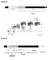
- FIG. 4b A schematic description of the structure of a typical artificial autotransporter protein used for autodisplay is given in Fig. 4b .
- the terminal step in autodisplay requires the translocation of the passenger through a size-limited pore formed by the ⁇ -barrel. This means that the passenger is not allowed to acquire a stable three dimensional conformation during transport to maintain a transport compatible state [18,19]. In case of stable folding, transport is blocked in the periplasm [19].
- passenger proteins with high biotechnological impact contain disulfide bridges and these bonds are normally formed in the periplasm of E . coli, a DsbA-negative mutant strain of E .
- the autodisplay system consists of vectors encoding various artificial autotransporter genes using the ⁇ -barrel from AIDA-I and different parts of its linkerregion.
- different modifications of the linker regions, various signal peptides under the control of inducible or constitutive promoters, mutant strains of E . coli supporting the transport and the surface display by the autotransporter pathway and detection methods are now available, that allow to follow surface translocation, preferentially independent of the protein domain used as a passenger.
- autodisplay is restricted to Gram-negative bacteria i.e. E . coli or Salmonella as host organisms.
- the autodisplay system has interesting activa.
- More than 100.000 active enzyme molecules can be displayed per single cell of E . coli without loss in cell integrity.
- dimers or multimers can be formed spontaneously at the cell surface by subunits expressed from monomeric genes, which is a unique feature of this surface display system and due to the free motility of the anchoring motif, the ⁇ -barrel within the outer membrane.
- EP 02718168 describes that anorganic prosthetic groups (e.g. 2Fe-2S) can be incorporated by a single step/one vial procedure without affecting cell viability, another feature that has not been described for any other surface display system so far.
- EP 02718168 describes autodisplay of adrenodoxin on an E . coli cell.
- Adrenodoxin belongs to the [2Fe-2S] ferredoxins, a family of small acidic iron-sulfur proteins. When displayed on the surface, the adrenodoxin is present in a non-functional form, because no prosthetic group is present.
- a functional adrenodoxin attached to the cell surface can be obtained by contacting the adrenodoxin molecule with an exogenous [2Fe-2S] cluster serving as a prosthetic group.
- the problem of the present invention is the provision of surface displayed enzymes comprising metal porphyrin-containing or flavin containing prosthetic groups. It was surprisingly found that by recombinant expression of these enzymes by surface display on a Gram-negative bacterium, a functional enzyme comprising the metal porphyrin-containing or flavin containing prosthetic group could be identified on the cell surface without introducing an exogenous prosthetic group, as described for enzymes containing [2Fe-2S] clusters in WO 02/070645 .
- polypeptides comprising prosthetic groups containing a metal porphyrin or a flavin can translocate to the cell surface in a conformation capable of retaining the prosthetic group when crossing the outer membrane, for example by mediation of the omp85 pathway.
- the prosthetic group may also be transported to the cell surface independently from the surface-displayed enzyme. Mechanisms are known for elimination of superfluous or toxic compounds, including compounds suitable as prosthetic groups (e.g., porphyrins), from the cell.
- compounds suitable as prosthetic groups e.g., porphyrins
- a first aspect of the present invention is a method for displaying a recombinant polypeptide containing a prosthetic group on the surface of a host cell, wherein the prosthetic group comprises a metal porphyrin or a flavin, said method comprising the steps:
- the recombinant polypeptide to be displayed may also be termed "passenger”, “passenger polypeptide” or “passenger protein”.
- Step (a) of the methods of the present invention refers to the provision of a host cell.
- the host cell used in the method of the present invention is preferably a bacterium, more preferably a Gram-negative bacterium, particularly an enterobacterium such as E . coli.
- a host cell particularly a host bacterium is provided which is transformed with a nucleic acid fusion operatively linked with an expression control sequence, i.e. a promoter, and optionally further sequences required for gene expression in the respective host cell.
- an expression control sequence i.e. a promoter
- the promoter or/and the expression control sequence may be homologous or heterologous to the host cell.
- the nucleic acid fusion is located on a recombinant vector, e.g. a plasmid vector.
- the host cell may be transformed with at least one nucleic acid fusion, for instance two, three, four, five or even more nucleic acid fusions.
- nucleic acid fusions preferably encode different recombinant polypeptides as described herein. If a host cell transformed with several nucleic acid fusions is used, these nucleic acid fusions may be located on a single vector or on a plurality of vectors.
- At least one host cell as described herein for instance two, three, four, five, six or even more host cells as described herein may be provided in the methods of the present invention.
- Each of these host cells is transformed with one nucleic acid fusion or at least one nucleic acid fusion, as described herein.
- the nucleic acid fusions transformed in the at least one host cell encode different recombinant polypeptides as described herein.
- the different recombinant polypeptides which may be provided in one or at least one host cell may form a functional unit, for instance the subunits of a functional unit, such as the subunits of an enzyme or the subunits or/and components of an enzyme complex.
- the nucleic acid fusion comprises (i) a portion encoding a signal peptide, preferably a portion coding for a Gram-negative signal peptide allowing for transport into the periplasm through the inner cell membrane.
- the signal peptide may be a signal peptide homologous to the host cell.
- the signal peptide may also be a signal peptide heterologous to the host cell.
- nucleic acid fusion comprises (ii) a portion encoding the recombinant polypeptide to be displayed.
- the nucleic acid fusion optionally comprises a portion encoding a protease recognition site, which may be a recognition site for an intrinsic protease, i.e. a protease naturally occurring in the host cell, or an externally added protease.
- the externally added protease may be an IgA protease (cf. EP-A-0 254 090 ), thrombin or factor X.
- the intrinsic protease may be e.g. selected from OmpT, OmpK or protease X.
- the nucleic acid fusion comprises (iii) a portion encoding a transmembrane linker which is required for the presentation of the passenger polypeptide (ii) on the outer surface of the outer membrane of the host cell.
- a transmembrane linker domain may be used which is homologous with regard to the autotransporter, i.e. the transmembrane linker domain is encoded by a nucleic acid portion directly 5' to the autotransporter domain.
- a transmembrane linker domain may be used which is heterologous with regard to the autotransporter.
- the length of the transmembrane linker is preferably 30-160 amino acids.
- the nucleic acid fusion comprises (iv) a transporter domain of an autotransporter.
- autodisplay may be the recombinant surface display of proteins or polypeptides by means of an autotransporter in any Gram-negative bacterium.
- the transporter domain of the autotransporter according to the invention can be any transporter domain of an autotransporter and is preferably capable of forming a ⁇ -barrel structure.
- a detailed description of the ⁇ -barrel structure and preferred examples of ⁇ -barrel autotransporters are disclosed in WO97/35022 incorporated herein by reference. Henderson et al. (2004) describes autotransporter proteins which comprise suitable autotransporter domains (for summary, see Table 1 of Henderson et al., 2004).
- the transporter domain of the autotransporter may be selected from Ssp (P09489, S. marcescens ), Ssp-h1 (BAA33455, S. marcescens ), Ssp-h2 (BAA11383, S. marcescens ), PspA (BAA36466, P. fluorescens ), PspB (BAA36467, P. fluorescens ), Ssa1 (AAA80490, P. haemolytica ), SphB1 (CAC44081, B. pertussis ), AspA/NalP (AAN71715, N.
- meningitidis meningitidis ), VacA (Q48247, H. pylori ), AIDA-I (Q03155, E . coli ), IcsA (AAA26547, S. flexneri ), MisL (AAD16954, S. enterica ), TibA (AAD41751, E . coli ), Ag43 (P39180, E . coli ), ShdA (AAD25110, S. enterica ), AutA (CAB89117, N. meningitidis ), Tsh (I54632, E . coli ), SepA (CAC05786, S. flexneri), EspC (AAC44731, E .
- the transporter domain of the autotransporter is the E . coli AIDA-I protein or a variant thereof, such as e.g. described by Niewert U., Frey A., Voss T., Le Bouguen C., Baljer G., Franke S., Schmidt MA.
- the AIDA Autotransporter System is Associated with F18 and Stx2e in Escherichia coli Isolates from Pigs Diagnosed with Edema Disease and Postweaning Diarrhea. Clin. Diagn. Lab. Immunol. 2001 Jan, 8(1):143-149 ;9.
- Variants of the above indicated autotransporter sequences can e.g. be obtained by altering the amino acid sequence in the loop structures of the ⁇ -barrel not participating in the transmembrane portions.
- the nucleic acid portions coding for the surface loops can be deleted completely.
- conserved amino exchanges i.e. the exchange of an hydrophilic by another hydrophilic amino acid or/and the exchange of a hydrophobic by another hydrophobic amino acid may take place.
- a variant has a sequence identity of at least 70%, at least 90%, at least 95% or at least 98% on the amino acid level to the respective native sequence of the autotransporter domain, in particular in the range of the ⁇ -sheets.
- Step (b) of the methods of the present invention refers to culturing the host cell under conditions wherein the nucleic acid fusion is expressed and the expression product comprising the recombinant polypeptide is displayed on the surface of the host cell.
- the person skilled in the art knows suitable culture conditions.
- the method according to the invention allows for an efficient expression of passenger proteins on the surface of host cells, particularly E .
- coli or other Gram-negative bacterial cells up to 100 000 or more molecules per cell by using a liquid medium of the following composition: 5 g/l to 20 g/l, preferably about 10 g/l trypton, 2 g/l to 10 g/l, preferably about 5 g/l yeast extract, 5 g/l to 20 g/l, in particular about 10 g/l NaCl and the remaning part water.
- the medium should possibly contain as little as possible divalent cations, thus preferably Aqua bidest or highly purified water, e.g. Millipore water is used.
- the liquid medium may contain in addition preferably EDTA in a concentration of 2 ⁇ M to 20 ⁇ M, in particular 10 ⁇ M.
- the liquid medium can further contain additional C-sources, preferably glucose, e.g. in an amount of up to 10 g/l, in order to favour secretion i.e. transfer of the passenger to the surrounding medium.
- additional C-sources preferably glucose, e.g. in an amount of up to 10 g/l, in order to favour secretion i.e. transfer of the passenger to the surrounding medium.
- additional C-source preferably glucose, e.g. in an amount of up to 10 g/l, in order to favour secretion i.e. transfer of the passenger to the surrounding medium.
- Preferred culture conditions for Gram-negative cells, such as E . coli, are described in the Examples.
- the polypeptide synthesized in the cytoplasma can be translocated from the cytoplasm into the periplasmic space by crossing the inner membrane. This can be effected by the signal peptide.
- a functional polypeptide comprising a metal porphyrin or a flavin on the surface of a Gram-negative cell by autodisplay can be explained as follows.
- the prosthetic group comprising a metal porphyrin or a flavin is introduced into the polypeptide of the present invention in the periplasmic space.
- the recombinant polypeptide of the present invention is translocated from the periplasmic space onto the cell surface in a conformation capable of retaining the prosthetic group when crossing the outer membrane, for example via the omp85 pathway.
- the prosthetic group present in the periplasmic space, may be transported independently from the recombinant polypeptide across the outer membrane.
- a suitable transporter is the outer membrane channel-tunnel protein TolC, in particular for the transportation of metal porphyrins. Both mechanisms may account for transportation of at least a part of prosthetic group transported to the cell surface.
- the prosthetic group can be transported to the cell surface by any suitable transport protein, which may be recombinantly expressed in the host cell. This transport can be independent from the autotransporter.
- the prosthetic group being a metal porphyrin can preferably be transported to the cell surface by a TolC-dependent mechanism.
- the prosthetic group being a metal porphyrin can also be transported to the cell surface by TolC or/and another suitable transport protein.
- the prosthetic group being a metal porphyrin can preferably be transported across the outer membrane surface by TolC.
- any TolC polypeptide may be employed.
- an E. coli TolC may be employed.
- the TolC polypeptide is homologous to the host cell.
- an E. coli TolC may be employed in an E. coli host cell.
- the TolC polypeptide may be a recombinant TolC.
- the TolC polypeptide may be recombinantly expressed in the host cell.
- TolC may be over-expressed in the host cell. If, for example, the host cell has only low expression of TolC and thus only low capability of porphyrin transport to the cell surface, TolC may be over-expressed.
- the TolC polypeptide may comprise a sequence selected from
- the TolC polypeptide as used herein, may be encoded by a sequence selected from
- nucleic acid sequences of (a), (b) and (d) include sequences within the scope of the degeneracy of the genetic code.
- the TolC polypeptide may be a HasF polypeptide, for example from Serratia marcescens.
- the passenger polypeptide is transported together with the prosthetic group to the cell surface, the passenger may acquire the prosthetic group within the cell.
- the prosthetic group endogenously produced in the cell may be introduced into the polypeptide of the present invention within the periplasmic space.
- step (b) may involve the omp85 pathway.
- Step (b) may comprise transportation of the polypeptide of the present invention via the omp85 pathway.
- Step (b) may comprise translocation of the polypeptide of the present invention from the periplasmic space onto the cell surface by the omp85 pathway, in particular in a conformation capable of retaining the prosthetic group when crossing the outer membrane.
- any Omp85 or Omp85 homologue may be employed.
- the Omp85 homologue YaeT from E . coli may be employed.
- the Omp85, in particular YaeT, may comprise a sequence selected from
- nucleic acid encoding an Omp85 may comprise a nucleic acid sequence selected from
- nucleic acid sequences of (a) and (b) include sequences within the scope of the degeneracy of the genetic code.
- the components (i) (ii), the optional portion encoding a protease recognition site, (iii) and (iv) in the nucleic acid fusion of the present invention are preferably oriented from 5' to 3'.
- the amino acid sequences encoded by nucleic acid sequences (i) and (ii), an optional protease recognition site as described herein are preferably arranged N terminal to C terminal.
- the method of the present invention may comprise preparing a membrane preparation from the cell obtained in step (b).
- the membrane preparation may comprise membrane particles.
- the membrane particles may be membrane vesicles.
- Preferred membrane particles are outer membrane particles.
- the method of the present invention may comprise preparing outer membrane particles of cells displaying a recombinant polypeptide on the surface, e.g. of Gram-negative bacterial cells.
- suitable conditions e.g. Hantke, 1981, Schultheiss et al., 2002. Typical conditions for preparing membrane particles are employed in the examples of the present invention.
- Outer membrane particles from a host cell as described herein may be performed by a method comprising the steps:
- the diameter of the membrane particles may be in the range of 1 nm to 1000 nm, in the range of 50 nm to 500 nm, in the range of 75 to 200 nm, or in the range of 90 to 120 nm. At least 80%, at least 90%, at least 95 %, or at least 98% of the membrane particles may have a diameter in a range selected from the ranges described herein.
- the recombinant passenger remains attached to the surface of the outer membrane by the ⁇ -barrel, which is serving as an anchor within the outer membrane. Due to the controlled integration of the ⁇ -barrel within the outer membrane, the C terminal part of the ⁇ -barrel is directed to the inner side of the outer membrane, whereas the N-terminal part of the linker, to which the recombinant passenger protein is covalently bound, is directed to the outer surface of the outer membrane, i.e. the environment.
- the recombinant passenger protein has an oriented location after transport, namely it is directed to the cellular surface.
- the recombinant passenger protein has the identical orientation as the lipopolysaccharide (LPS) layer which may be present in the outer membrane.
- LPS lipopolysaccharide
- Membrane particles of the present invention prepared from the host cell of the present invention comprise the recombinant peptide at the surface directed to the environment.
- the outer membrane of Gram-negative bacteria in particular E . coli, is asymmetric.
- the outer membrane may comprise an inner layer comprising phospholipids and an outer layer comprising LPS.
- LPS is hydrophilic and may contain several negative charges.
- the recombinant protein or a domain thereof, which are integrated in the outer membrane by autodisplay, will be directed to the surface distal to the carrier as well.
- the core part of the membrane particles may stabilize the interaction of the outer membrane layer obtained by applying outer membrane particles to the carrier by hydrophobic interactions and may contain lipoproteins or peptidoglycans.
- a preferred prosthetic group is a metal porphyrin, as described herein.
- the prosthetic group being the metal porphyrin may comprise a heavy metal such as cobalt, nickel, manganese, copper and iron.
- the metal porphyrin of the present invention in particular comprises a heme group.
- Another preferred prosthetic group is a flavin, as described herein.
- the prosthetic group being the flavin may be selected from FAD and FMN.
- polypeptide of the present invention comprising a prosthetic group preferable is an enzyme.
- the polypeptide comprising the metal porphyrin may be an enzyme.
- the polypeptide comprising the metal porphyrin may be selected from P450 enzymes (such as P450 reductases) and cytochromes (such as cytochrome b5).
- the polypeptide comprising the metal porphyrin may be selected from hemoproteins.
- the polypeptide comprising the metal porphyrin may be selected from monooxygenases.
- the polypeptide comprising the metal porphyrin may be selected from CYP11B1, CYP11A1, CYP106A2 and CYP3A4.
- the polypeptide comprising the metal porphyrin may preferably be selected from CYP106A2 and CYP3A4.
- polypeptide comprising the metal porphyrin comprises a sequence selected from
- the portion (ii) of the nucleic acid fusion of the present invention may encode a polypeptide which, when functional, comprises the metal porphyrin, as described herein.
- the portion (ii) of the nucleic acid fusion of the present invention may comprise a nucleic acid sequence selected from
- nucleic acid or polypeptide comprising sequences having at least 70%, at least 80%, at least 90%, at least 95%, or at least 98% identity to a given sequence includes fragments of the given nucleic acid or polypeptide.
- the flavin containing polypeptide of the present invention may be selected from flavoproteins, in particular FAD or FMN containing proteins. Preferred are FAD containing proteins.
- the flavin containing polypeptide may be selected from enzymes such as oxidoreductases, NADH oxidases, dehydrogenases, and oxidases, especially sugar oxidases, such as pyranose oxidase.
- the NADH oxidase is in particular an FAD containing enzyme.
- the polypeptide of the present invention to be displayed on the surface of the cell may be a multimeric polypeptide.
- the multimeric recombinant polypeptide may be a homodimer, i.e. a polypeptide consisting of two identical subunits or a homomultimer, i.e. a polypeptide consisting of three or more identical subunits.
- the multimeric recombinant polypeptide may be a heterodimer, i.e. a polypeptide consisting of two different subunits or a heteromultimer consisting of three or more subunits wherein at least two of these subunits are different.
- the multimeric polypeptide is comprised of a plurality of subunits which form a "single" multimeric polypeptide or a complex of a plurality of functionally associated polypeptides which may in turn be monomeric and/or multimeric polypeptides.
- at least one subunit of the multimeric recombinant protein may contain at least one prosthetic group as described herein.
- the nucleic acid fusion may encode a plurality of polypeptide subunits as a polypeptide fusion which when presented on the cell surface forms a functional multimeric polypeptide.
- Homodimers or homomultimers may be formed by a spontaneous association of several identical polypeptide subunits displayed on the host cell membrane.
- Heterodimers or heteromultimers may be formed by a spontaneous association of several different polypeptide subunits displayed on the host cell membrane.
- a multimeric recombinant polypeptide may be formed by an association of at least one polypeptide subunit displayed on the host cell membrane, as described herein, and at least one soluble polypeptide subunit added to the host cell membrane.
- the added subunit may be identical to the displayed subunit or be different therefrom.
- the host cell may be any host cell as described herein, in particular a host cell displaying a recombinant polypeptide on the surface thereof, wherein the recombinant polypeptide contains a prosthetic group comprising a metal porphyrin or a flavin, and wherein the recombinant polypeptide comprises
- the displayed polypeptide is in particular a functional polypeptide.
- the portions (I) to (III) of the recombinant polypeptide displayed by the host cell of the present invention are encoded in particular by the components (ii), (iii), and (iv) of the nucleic fusion, as described herein.
- the optional portion comprising a protease recognition site is encoded in particular by the optional portion encoding a protease recognition site of the nucleic acid fusion described herein.
- a membrane preparation comprising a recombinant polypeptide.
- the membrane preparation of the present invention may comprise membrane particles, as described herein.
- the membrane preparation may be obtained from a host cell as described herein.
- the recombinant polypeptide of the may be any recombinant polypeptide as described herein.
- Yet another aspect of the present invention is the use of a membrane preparation comprising a recombinant polypeptide in the manufacture of a carrier comprising a recombinant polypeptide.
- the membrane preparation of the present invention may be employed for coating a carrier.
- the carrier may comprise a membrane preparation of the present invention, as described herein.
- the carrier may comprise a hydrophobic surface.
- the hydrophobic surface may have a contact angle of more than 90°.
- a increasing surface angle of more than 30° indicates a gradually increasing hydrophobicity of a surface.
- a hydrophobic surface may have a contact angle of at least 40°.
- the surface preferably has a hydrophobicity described by a contact angle of at least 40°, at least 50°, at least 60°, at least 65°, at least 70°.
- Contact angles are preferably determined by the sessile drop method.
- the sessile drop method is a standard method for determining contact angles. Measurements may be performed with a contact angle goniometer.
- Preferred contact angles of the hydrophobic surface are in a range of 40° to 100°, 50° to 90°, or 60° to 80°.
- the surface of the carrier may be a metal surface.
- a suitable metal surface has a contact angle e.g. in the range of 50° to 80°.
- a suitable metal may be selected from gold, silver, titanium, aluminium and alloys such as brass.
- a preferred surface is a gold surface.
- the gold surface may be employed as it is.
- An untreated gold surface has a hydrophobicity suitable for the carrier as described herein. A treatment of the gold surface with thiolated hydrocarbons or hydrocarbons with functional groups such as carboxylic acids or hydroxyl groups is not required.
- the carrier comprises a polymer, for instance a surface usually employed in disposable materials for use in biochemical or/and medical science.
- the polymer may be an artificial polymer.
- artificial polymers include a polymer selected from polystyrenes, polypropylenes, and polycarbonates.
- the polystyrene may be produced from [2,2]paracyclophane monomers.
- Polystyrene surfaces may be treated with oxygene plasma introducing OH or/and methylene groups in order to modify the hydrophobicity. Examples of such modified surfaces include Maxi-sorp, Medi-sorp, Multi-sorp, and Poly-sorp surfaces.
- Another suitable polystyrene surface is Parylene N produced from [2,2]paracyclophane monomers.
- Parylene A Poly(monoamino-p-xylene)
- surfaces comprising a polymer having a hydrophobicity described by a contact angle of at least 50°. Suitable surfaces are selected from polystyrene, Parylene A, Parylene N, Maxi-sorp, Medi-sorp, Multi-sorp, and Poly-sorp. Preferred surfaces are selected from polystyrene, Parylene A, Parylene N, Maxi-sorp, Medi-sorp, and Poly-sorp.
- the surface may comprise a natural polymer. Suitable natural polymers include polybutyrate and cellulose and derivatives thereof. A further surface is provided by latex particles, in particular latex beads.
- C18-modified particles in particular C18-modified monolithic silica particles.
- C18 refers to an alkyl group comprising 18 carbon atoms.
- C18-modified particles are known in the art.
- Yet another suitable surface is a glass surface.
- the surface may be modified is order to adjust the hydrophobicity. Modification may be performed by chemical treatment (i.e. by oxygen plasma), physical treatment (e.g. by laser irradiation or/and radioactive irradiation), or by mechanical treatment.
- chemical treatment i.e. by oxygen plasma
- physical treatment e.g. by laser irradiation or/and radioactive irradiation
- mechanical treatment e.g. by mechanical treatment.
- the method according to the invention and the host cells according to the invention can be used for a variety of different applications, e.g. as whole cell biofactories or membrane preparation biofactories for chemical synthesis procedures, e.g. for the synthesis of organic substances selected from enzyme substrates, drugs, hormones, starting materials and intermediates for syntheses procedures and chiral substances (cf. Roberts, Chemistry and Biology 6 (1999), R269-R272 ).
- Typical CYP106A2 substrates are described in Fig. 12 .
- the method according to the invention and the host cells according to the invention, as described herein, can be used in the chemical synthesis, for example in enzymatically catalyzed enantioselective or/and regioselective steps.
- CYP106A2 displayed on the surface of a cell, as described herein can be used for the conversion of steroids, for the conversion of abietic acid, or for the preparation of desipramine from imipramine, as exemplified by Examples 1 and 3.
- the cell or the membrane preparation of the invention may be used for a directed evolution procedure, e.g. for the development of new biocatalysts for the application in organic syntheses.
- suitable screening methods in particular high throughput screening (HTS) methods for the conversion of a certain substrate.
- libraries of variants of a polypeptide containing a prosthetic group selected from metal porphyrins and flavins, as described herein are examined in view of the role of defined amino acids during certain functions, in particular catalytic functions.
- these particular embodiments concern the production of variants of proteins and/or enzymes and the production of libraries with variants of proteins and/or enyzmes, respectively, which carry a prosthetic group, as described herein, or multimers etc. and which are screened in view of a certain characteristic, i.e. one or optionally several variants fulfilling this desired characteristic perfectly are selected.
- a certain characteristic i.e. one or optionally several variants fulfilling this desired characteristic perfectly are selected.
- the cell is selected, too, and carries the nucleic acid coding the variant.
- both the amino acid sequence and the structural information of the variant can be determined via the nucleic acid sequence.
- the characteristics in question are particularly enzyme inhibiting, catalytical, toxin degrading, synthesizing, therapeutical etc. characteristics.
- the host cell or the membrane preparation may be used as an assay system for a screening procedure, e.g. for identifying modulators (activators or inhibitors) of displayed polypeptides, containing a prosthetic group selected from metal porphyrins and flavins, as described herein, which may be used as potential therapeutic agents.
- the screening procedure may also be used to identify variants of displayed polypeptides having predetermined desired characteristics.
- libraries of modulators and/or libraries of displayed polypeptides may be used.
- the host cells or membrane preparations derived therefrom may be used as a system for toxicity monitoring and/or degrading toxic substances in the environment, in the laboratory or in biological, e.g. human, animal, or non-biological systems.
- An essential advantage of applying the host cells and membranes according to the invention is enabling correct folding and biological activity of proteins or protein complexes, e.g. of the polypeptide containing a prosthetic group selected from metal porphyrins and flavins, as described herein, which require a membrane environment.
- a reconstitution as previously considered to be necessary is no longer required.
- the production steps of a functional biocatalytic system are simplified and an increased stability of the system per se is obtained.
- the passenger polypeptides are peptides or proteins selected from the group of drug metabolizing enzymes, such as CYP1A2 involved in the activation of aromatic amine carcinogenes, heterocyclic arylamine promutagenes derived from food pyrolysates and aflatoxin B1 ( Gallagher EP, Wienkers LC, Stapleton PL, Kunze KL, Eaton DL., Role of human microsomal and human complementary DNA-expressed cytochromes P4501A2 and P4503A4 in the bioactivation of aflatoxin B1. Cancer Res.
- Further preferred passenger peptides are peptides from the group of steroid biosynthesis enyzmes, such as CYP11B1 involved in the formation of cortisol and aldosterone ( Bernhardt R., Cytochrome P450: structure, function and generation of reactive oxygen species. Rev. Physiol. Biochem. Pharmacol.
- CYP19 involved in the conversion of adrostenedione to 19-hydroxyandrostenedione, 19-oxo-androstenedione and estrone
- CYP19 involved in the conversion of adrostenedione to 19-hydroxyandrostenedione, 19-oxo-androstenedione and estrone
- Further preferred metal ion containing enzymes are Cu-containing enzymes, such as cytochrome-oxidase, Mn-containing enzymes, such as arginase and ribonucleotide reductase, Mo-containing enzymes, such as dinitrogenase and Se-containing enzymes, such as glutathione peroxidase.
- the P450 enzymes are hepatic P450 enzymes, particularly P450 3A4, 2D6, 2C9 and 2C19.
- the host cells and/or preparations according to the invention are preferably used sequentially for testing the enzyme inhibition of P450 enzymes. For example, with the help of the host cell and/or membrane preparation according to the invention it can be found out in an early step of drug discovery, the so-called lead identification, whether the new drug lead structure to be tested could possibly have side-effects or lead to the so-called drug-drug interaction.
- mainly two P450 enzymes shall be expressed by autodisplay, CYP106A2 and CYP3A4.
- Fig.1a and Fig. 2 The gene of CYP106A2 was amplified by PCR and inserted into a plasmid encoding the domains needed for autodisplay. Successful expression was shown in an SDS- PAGE ( Fig. 5 ) To find out whether the CYP106A2 domain of the fusion protein was indeed exposed at the cell surface, trypsin was added to whole cells of E. coli BI21(DE3) pET-CYP13 after incubation with 1 mM IPTG for one hour. Trypsin is too large of a molecule to enter the cell envelope of E .
- CYP106A4 was indeed expressed on the surface of the cells in a functional form activity tests. For that purpose cells were cultivated and protein expression induced using 1 mM Isopropyl-beta-D-thiogalactopyranosid (IPTG). In a conversion assay it was tested whether the cells displaying CYP106A2 on the surface have the ability to efficiently convert 11-deoxycorticosterone (DOC) into 15beta-DOC.
- DOC 11-deoxycorticosterone
- DOC 11-deoxycorticosterone
- Adx adrenodoxin
- AdR adrenodoxin reductase
- This example refers to the role of TolC present in the outer membrane of E . coli cells displaying CYP106A2 on the surface. TolC is involved in porphyrin transport across the cell membrane [26].
- the strain BL21 (DE3) employed in Example 1 expresses TolC on the outer membrane. Therefore, this strain has the capability to transport porphyrins, including P450, to the outer membrane surface.
- JW5503-1 (DE3) is a TolC defective mutant, with a reduced capability of transporting porphyrins (including P450) onto the outer membrane surface.
- JW5503-1 was obtained from the Keio collection distributed by Coli Genetic Stock Center at Yale University.
- Figure 15 indicates that CYP106A2 is expressed by pET-CYP13 to the same extent on the surface on TolC negative strain JW5503-1 (DE3) and on TolC postive BL21 (DE3) E . coli cells.
- Figure 16 shows HPLC chromatograms of CYP106A2 conversion of 11-deoxycorticosterone to 15 ⁇ -deoxycorticosterone.
- Figure 16C and B it can be seen that the amount of 15 ⁇ -deoxycorticosterone decreased when CYP106A2 was expressed in the E . coli strain lacking the TolC channel protein.
- TolC can provide porphyrins, in particular P450, on the cell surface, so that the porphyrins can be introduced into a recombinant surface-displayed polypeptide of the present invention, such as CYP106A.
- Figure 16 indicates that there is still a CYP106A activity in the absence of TolC.
- Other transporters different from TolC may be present in the outer membrane so that porphyrins, in particular P450, can be provided on the cell surface so that the prosthetic group can be introduced into a recombinant surface-displayed polypeptide of the present invention, such as CYP106A.
- Figure 18 shows the kinetics of NADPH consumption of cells by E . coli BL21(DE3) pETCYP13 expressing CYP106A2 cells and control BL21(DE3) cells in the presence of imipramine, as indicated above.
- the by-product formalin (cf. Figure 17 ) has been photometrically identified to be produced by the cells displaying CYP106A2, but not in the control cells (data not shown).
- the difference of CYP106A2 expressing cells and control cells in NADPH consumption indicates conversion of imipramine into desipramine.
Landscapes
- Life Sciences & Earth Sciences (AREA)
- Health & Medical Sciences (AREA)
- Chemical & Material Sciences (AREA)
- Genetics & Genomics (AREA)
- Engineering & Computer Science (AREA)
- Organic Chemistry (AREA)
- Bioinformatics & Cheminformatics (AREA)
- Wood Science & Technology (AREA)
- Zoology (AREA)
- Biomedical Technology (AREA)
- Biotechnology (AREA)
- General Engineering & Computer Science (AREA)
- Molecular Biology (AREA)
- Microbiology (AREA)
- Biochemistry (AREA)
- General Health & Medical Sciences (AREA)
- Medicinal Chemistry (AREA)
- Physics & Mathematics (AREA)
- Biophysics (AREA)
- Plant Pathology (AREA)
- Virology (AREA)
- Crystallography & Structural Chemistry (AREA)
- Micro-Organisms Or Cultivation Processes Thereof (AREA)
Description
- The present invention relates to a method for functionally displaying a recombinant polypeptide containing a prosthetic group on the surface of a host cell, wherein the prosthetic group is selected from metal porphyrins and flavins.
- Over the past 30 years, it has become clear that enzymes hold great potential for industry. They are most remarkable biomolecules because of their extraordinary specificity and catalytic power [1]. The specificity and (enantio- and regio-)selectivity of certain enzymatic transformations makes them attractive for the production of fine chemicals and pharmaceutical intermediates. To date, more than 500 products are manufactured by enzymes. Well-known examples are ephedrine, aspartame and amoxicillin [2,3,4].
- Cytochromes P450 enzymes have been discovered about 50 years ago and are ubiquitously distributed enzymes, which possess high complexity and display a broad field of catalytic activities. They are hemoproteins, which means, they contain a porphyrin ring system. The P450 enzyme family is involved in the biotransformation of drugs, the bioconversion of xenobiotics, the metabolism of chemical carcinogens, the biosynthesis of physiologically important compounds such as steroids, fatty acids, eicosanoids, fat-soluble vitamins, bile acids, the conversion of alkanes, terpenes, and aromatic compounds as well as the degradation of herbicides and insecticides [5]. Furthermore, there is a broad versatility of reactions catalysed by cytochromes P450, such as carbon hydroxylation, heteroatom oxygenation, dealkylation, epoxidation, aromatic hydroxylation, reduction, and dehalogenation.
- Despite their very interesting features for industrial applications, the use of P450 enzymes for wide biotechnology needs is still limited, due to their difficulty in handling. With the exception of a few bacterial P450s, the vast majority needs a certain membrane contact or environment to fold into an active form. Within those membrane associated P450s, two classes can be identified: a mitochondrial and a microsomal. At the moment there are two different ways to use these enzymes for synthetic purposes. They are either purified after recombinant expression and reconstituted with an artificial membrane system, or they are expressed and used in a whole cell context. Both ways have their limitations. Reconstituted membrane vesicles with P450 enzymes are laborious to produce and they are absolutely not suited for industrial applications. Using whole cells with intrinsic P450s limits the set of substrates to be converted to those which are able to cross membranes [6].
- Among other systems for the secretion of proteins in Gram-negative bacteria, the autotransporter pathway represents a solution of impressing simplicity. It is possible to transport a protein, regardless whether it is recombinant or the natural passenger, to the actual outer membrane, as long as its coding region lies between a typical signal peptide and a C-terminal domain called β-barrel. Based on these findings the autodisplay system has been developed by the use of the natural E. coli autotransporter protein AIDA-I (the adhesin involved in diffuse adherence) in an E. coli host background [9]. Autodisplay has been used for the surface display of random peptide libraries that were successfully screened for the identification of new enzyme inhibitors, and the display of functional enzymes like esterases, oxidoreductases and electron transfer proteins [10].
- During the autodisplay of bovine adrenodoxine, which serves as an electron donor for mitochondrial p450s, two major observation were made [11,12]. First, it could be shown, that it is possible to incorporate an inorganic, prosthetic group into an apoprotein expressed by autodisplay at the cell surface by a simple titration step to yield a functional electron donor without loss of cell viability or cell integrity. And second, after external addition of the purified P450s CYP11B1 and CYP11A1, a functional whole cell biocatalyst was obtained for efficient synthesis of different steroids. Therefore the aim of the present invention is, to investigate, whether it is possible to autodisplay a P450 enzyme in a functional form on the surface of E. coli. This could provide a new expression platform for the highly interesting group of P450 enzymes with the perspective of being applicable in industrial processes.
- Cytochromes P450 are external monooxygenases. Monooxygenases (mixed function oxidases) catalyse the incorporation of a single atom of molecular oxygen into a substrate with the concomitant reduction of the other atom to water. Monooxygenases are divided into two classes: internal and external. Internal monooxygenases extract two reducing equivalents from the substrate to reduce one atom of dioxygen to water, whereas external monooxygenases utilize an external reductant. While initially the microsomal drug and xenobiotic-metabolising enzymes were referred to as mixed function oxidases, in more recent years the term monooxygenase became the widely accepted one.
- Cytochromes P450 got their name from their character as hemoproteins as well as their unusual spectral properties displaying a typical absorption maximum of the reduced CO-bound complex at 450 nm: cytochrome stands for a hemoprotein, P for pigment and 450 reflects the absorption peak of the CO complex at 450 nm. The ability of reduced P450 to produce an absorption peak at 450 nm upon CO binding is still used for the estimation of the P450 content (Omura and Sato, 1964). The red shift of about 30 nm as observed in cytochromes P450 means that the distribution of electron density at the heme is significantly perturbed as compared to other cytochromes. It has been documented that the cause of this is the thiolate sulphur, which by means of a direct bond to the iron causes this effect. The Soret band (named after its discoverer) describes the absorption band of hemoproteins at about 380-420 nm.
- Cytochrome P450 systems mainly catalyse the following reaction:
RH + O2 +NAD(P)H + H+ → ROH + H2O + NAD(P)+
- They are involved in reactions as diverse as e.g. hydroxylation, N-, O- and S-dealkylation, sulphoxidation, epoxidation, deamination, desulphuration, dehalogenation, peroxidation, and N-oxide reduction. This diversity of catalysed reactions and, of course, the high amount of acceptable substrates is attractive for biotechnological application in particular when it can be transferred to industrial needs.
- CYP106A2 is a bacterial steroid hydroxylase from Bacillus megaterium ATCC 13368. Since it is soluble and easy to express it has application for biotechnological purposes. Recently, it was possible to design a whole cell bioconversion system for steroids using a mixed system composed of the bovine mitochondrial electron transfer system AdR and Adx and the bacterial enzyme CYP106A2. This mixed P450 monooxygenase system was expressed in E. coli cells. Those successful experiments opened the door to facilitate the application of molecular evolution approaches in order to select mutants of the cytochrome with higher stability, activity, and changed regio-and stereo-specificity suitable to produce hydroxylated steroid derivatives using a biological transformation process [6]. CYP106A2 catalyzes as main reaction route the 15β-hydroxylation of several steroids, e.g. 11-deoxycorticosterone, testosterone, progesterone, and corticosterone (
Fig. 1a ) [7]. One disadvantage of this system is the limitation for substrates which are membrane permeable like corticosteroids. A successful expression via autodisplay could broaden the variety of substrates and therefore make the P450 monooxygenase system more valuable for future research. - CYP3A4 is the quantitative most important CYP- enzyme and involved in the oxidation of the largest range of substrates of all CYPs. In humans it is predominantly found in the liver and often allows prodrugs to be activated and absorbed. Inhibition or induction of CYP3A4 is a major problem in the daily clinical routine, since it often causes drug-drug interactions or side effects. Induction can lead to the fast inactivation of the applied drug and in consequence to plasma levels so low, that they do not have the desired therapeutic effect anymore. A commonly used CYP3A4 inductor is the anticonvulsant Carbamazepin. Inhibition instead can cause major intoxications due to plasma levels far beyond the therapeutic dose. On the other hand the capability of inhibition is used in the antiretroviral therapy to lower side effects and make it more bearable for patients. Ritonavir is given in a subtherapeutic dose to inhibit the enzyme and booster the effect of further antiretroviral drugs such as Lopinavir. A well-documented example is that of terfenadine, a nonsedating antihistamine (
Fig. 1 b) . The oxidation of terfenadine is catalyzed very rapidly by CYP3A4 to its major metabolite fexofenadine which is responsible for the pharmacological activity. [8] - The E. coli outer membrane channel-tunnel protein TolC is involved in the exclusion of harmful substances such as antibiotics, dyes, organic solvents, and detergents. The crystal structure of the TolC protein recently has been determined. The TolC protein is composed of a transmembrane domain and a periplasmic domain and forms a homotrimer. The periplasmic barrel structure of TolC is connected to drug efflux pump proteins such as AcrB and AcrE, which are located on the inner membrane. Clamp proteins such as AcrA and ArcF link TolC and pump proteins in the periplasmic space. Pump proteins seem to transport toxic cytoplasmic or periplasmic substances into the extracellular space across the outer membrane via the TolC channel [26].
- Porphyrins can act as photosensitizers. If porphyrins accumulate, they can be toxic, as the cells can become sensitive to near-UV irradiation. TolC is involved in porphyrin transport across the cell membrane and provides a mechanism to eliminate superfluous or/and toxic porphyrins. The TolC outer membrane channel-tunnel protein can function together with inner membrane efflux pump proteins. Therefore, an inner membrane pump(s) or exporter(s) is assumed to be involved in porphyrin exclusion in combination with TolC. Porphyrin(ogen) exclusion is considered as a two-step process. In this process, porphyrin(ogen)s are transported to the periplasm by a TolC-independent mechanism and then are transported across the outer membrane by the TolC-dependent efflux system [26].
- Autodisplay is based on the secretion mechanism of the autotransporter family or proteins [13]. A concept for this secretion mechanism was proposed concurrently with the first autotranspoter protein, IgA1protease from Neisseria gonorrhoeae (
Fig. 4a ) [14]. With the aid of a typical signal peptide, the precursor is transported across the inner membrane. Arrived in the periplasm, the C terminal part of the precursor forms a porin-like structure, a so-called β-barrel, within the outer membrane and through this pore the N terminally attached passenger (the actual protease) is translocated to the cell surface. To obtain full surface exposure of the passenger, a linker peptide is required in between the β-barrel and the passenger. - For the development of the autodisplay system the β-barrel and the linker region of AIDA-I were combined in frame with the signal peptide of the cholera toxin β-subunit (CTB) and a strong constitutive promoter (PTK) within a medium copy number plasmid backbone [15]. Into the linker regions used for autodisplay, protease cleavage sites for the sequence specific release of the passenger protein, as well as epitopes for detection by monoclonal antibodies were inserted. An antibody independent detection method, which requires only the addition of a single cysteine in the linker region, was developed for autodisplay and was named "Cystope tagging" [16,17]. A schematic description of the structure of a typical artificial autotransporter protein used for autodisplay is given in
Fig. 4b . As mentioned above, the terminal step in autodisplay requires the translocation of the passenger through a size-limited pore formed by the β-barrel. This means that the passenger is not allowed to acquire a stable three dimensional conformation during transport to maintain a transport compatible state [18,19]. In case of stable folding, transport is blocked in the periplasm [19]. As a wide variety of passenger proteins with high biotechnological impact contain disulfide bridges and these bonds are normally formed in the periplasm of E. coli, a DsbA-negative mutant strain of E. coli (JK321) was constructed and shown to facilitate the autodisplay of such types of proteins as well [19]. In summary, the autodisplay system consists of vectors encoding various artificial autotransporter genes using the β-barrel from AIDA-I and different parts of its linkerregion. Dependent on the application, different modifications of the linker regions, various signal peptides under the control of inducible or constitutive promoters, mutant strains of E. coli supporting the transport and the surface display by the autotransporter pathway and detection methods are now available, that allow to follow surface translocation, preferentially independent of the protein domain used as a passenger. It is obvious, that autodisplay is restricted to Gram-negative bacteria i.e. E. coli or Salmonella as host organisms. Beyond this limitation, the autodisplay system has interesting activa. First, more than 100.000 active enzyme molecules can be displayed per single cell of E. coli without loss in cell integrity. Second, dimers or multimers can be formed spontaneously at the cell surface by subunits expressed from monomeric genes, which is a unique feature of this surface display system and due to the free motility of the anchoring motif, the β-barrel within the outer membrane. Third,EP 02718168 -
EP 02718168 EP 02718168 - The problem of the present invention is the provision of surface displayed enzymes comprising metal porphyrin-containing or flavin containing prosthetic groups. It was surprisingly found that by recombinant expression of these enzymes by surface display on a Gram-negative bacterium, a functional enzyme comprising the metal porphyrin-containing or flavin containing prosthetic group could be identified on the cell surface without introducing an exogenous prosthetic group, as described for enzymes containing [2Fe-2S] clusters in
WO 02/070645 - Thus, a first aspect of the present invention is a method for displaying a recombinant polypeptide containing a prosthetic group on the surface of a host cell, wherein the prosthetic group comprises a metal porphyrin or a flavin, said method comprising the steps:
- (a) providing a host cell transformed with a nucleic acid fusion operatively linked with an expression control sequence said nucleic acid fusion comprising:
- (i) a portion encoding a signal peptide,
- (ii) a portion encoding the recombinant polypeptide to be displayed,
- (iii) a portion encoding a transmembrane linker, and
- (iv) a portion encoding the transporter domain of an autotransporter,
and
- (b) culturing the host cell under conditions wherein the nucleic acid fusion is expressed and the expression product comprising the recombinant polypeptide containing the prosthetic group is displayed on the surface of the host cell,
- The recombinant polypeptide to be displayed may also be termed "passenger", "passenger polypeptide" or "passenger protein".
- Step (a) of the methods of the present invention refers to the provision of a host cell. The host cell used in the method of the present invention is preferably a bacterium, more preferably a Gram-negative bacterium, particularly an enterobacterium such as E. coli.
- According to the present invention, a host cell, particularly a host bacterium is provided which is transformed with a nucleic acid fusion operatively linked with an expression control sequence, i.e. a promoter, and optionally further sequences required for gene expression in the respective host cell. The skilled person knows suitable promoters and expression control sequences. The promoter or/and the expression control sequence may be homologous or heterologous to the host cell. Preferably, the nucleic acid fusion is located on a recombinant vector, e.g. a plasmid vector. The host cell may be transformed with at least one nucleic acid fusion, for instance two, three, four, five or even more nucleic acid fusions. If two or more nucleic acid fusions are transformed into a host cell, the nucleic acid fusions preferably encode different recombinant polypeptides as described herein. If a host cell transformed with several nucleic acid fusions is used, these nucleic acid fusions may be located on a single vector or on a plurality of vectors.
- At least one host cell as described herein, for instance two, three, four, five, six or even more host cells as described herein may be provided in the methods of the present invention. Each of these host cells is transformed with one nucleic acid fusion or at least one nucleic acid fusion, as described herein. Preferably, the nucleic acid fusions transformed in the at least one host cell encode different recombinant polypeptides as described herein.
- The different recombinant polypeptides which may be provided in one or at least one host cell may form a functional unit, for instance the subunits of a functional unit, such as the subunits of an enzyme or the subunits or/and components of an enzyme complex.
- The nucleic acid fusion comprises (i) a portion encoding a signal peptide, preferably a portion coding for a Gram-negative signal peptide allowing for transport into the periplasm through the inner cell membrane. The signal peptide may be a signal peptide homologous to the host cell. The signal peptide may also be a signal peptide heterologous to the host cell.
- Further, the nucleic acid fusion comprises (ii) a portion encoding the recombinant polypeptide to be displayed.
- Further, the nucleic acid fusion optionally comprises a portion encoding a protease recognition site, which may be a recognition site for an intrinsic protease, i.e. a protease naturally occurring in the host cell, or an externally added protease. For example, the externally added protease may be an IgA protease (cf.
EP-A-0 254 090 ), thrombin or factor X. The intrinsic protease may be e.g. selected from OmpT, OmpK or protease X. - Furthermore, the nucleic acid fusion comprises (iii) a portion encoding a transmembrane linker which is required for the presentation of the passenger polypeptide (ii) on the outer surface of the outer membrane of the host cell. A transmembrane linker domain may be used which is homologous with regard to the autotransporter, i.e. the transmembrane linker domain is encoded by a nucleic acid portion directly 5' to the autotransporter domain. Also a transmembrane linker domain may be used which is heterologous with regard to the autotransporter. The length of the transmembrane linker is preferably 30-160 amino acids.
- Further, the nucleic acid fusion comprises (iv) a transporter domain of an autotransporter. In the context of the present invention, autodisplay may be the recombinant surface display of proteins or polypeptides by means of an autotransporter in any Gram-negative bacterium. The transporter domain of the autotransporter according to the invention can be any transporter domain of an autotransporter and is preferably capable of forming a β-barrel structure. A detailed description of the β-barrel structure and preferred examples of β-barrel autotransporters are disclosed in
WO97/35022 - Variants of the above indicated autotransporter sequences can e.g. be obtained by altering the amino acid sequence in the loop structures of the β-barrel not participating in the transmembrane portions. Optionally, the nucleic acid portions coding for the surface loops can be deleted completely. Also within the amphipathic β-sheet conserved amino exchanges, i.e. the exchange of an hydrophilic by another hydrophilic amino acid or/and the exchange of a hydrophobic by another hydrophobic amino acid may take place. Preferably, a variant has a sequence identity of at least 70%, at least 90%, at least 95% or at least 98% on the amino acid level to the respective native sequence of the autotransporter domain, in particular in the range of the β-sheets.
- Step (b) of the methods of the present invention refers to culturing the host cell under conditions wherein the nucleic acid fusion is expressed and the expression product comprising the recombinant polypeptide is displayed on the surface of the host cell. The person skilled in the art knows suitable culture conditions. The method according to the invention allows for an efficient expression of passenger proteins on the surface of host cells, particularly E. coli or other Gram-negative bacterial cells up to 100 000 or more molecules per cell by using a liquid medium of the following composition: 5 g/l to 20 g/l, preferably about 10 g/l trypton, 2 g/l to 10 g/l, preferably about 5 g/l yeast extract, 5 g/l to 20 g/l, in particular about 10 g/l NaCl and the remaning part water. The medium should possibly contain as little as possible divalent cations, thus preferably Aqua bidest or highly purified water, e.g. Millipore water is used. The liquid medium may contain in addition preferably EDTA in a concentration of 2 µM to 20 µM, in particular 10 µM. Moreover, it contains preferably reducing reagents, such as 2-mercapto ethanol or dithiotreitol or dithioerythritol in a preferred concentration of 2 mM to 20 mM. The reducing reagents favour a non-folded structure of the polypeptide during transport. The liquid medium can further contain additional C-sources, preferably glucose, e.g. in an amount of up to 10 g/l, in order to favour secretion i.e. transfer of the passenger to the surrounding medium. For surface display preferably no additional C-source is added. Preferred culture conditions for Gram-negative cells, such as E. coli, are described in the Examples.
- If the host cell is a Gram-negative bacterium, the polypeptide synthesized in the cytoplasma can be translocated from the cytoplasm into the periplasmic space by crossing the inner membrane. This can be effected by the signal peptide.
- While not wishing to be bound by theory, display of a functional polypeptide comprising a metal porphyrin or a flavin on the surface of a Gram-negative cell by autodisplay can be explained as follows. In a first step the prosthetic group comprising a metal porphyrin or a flavin is introduced into the polypeptide of the present invention in the periplasmic space. In a second step, the recombinant polypeptide of the present invention is translocated from the periplasmic space onto the cell surface in a conformation capable of retaining the prosthetic group when crossing the outer membrane, for example via the omp85 pathway. By this procedure, a functional polypeptide attached to the cell surface can be obtained. In a different mechanism, the prosthetic group, present in the periplasmic space, may be transported independently from the recombinant polypeptide across the outer membrane. A suitable transporter is the outer membrane channel-tunnel protein TolC, in particular for the transportation of metal porphyrins. Both mechanisms may account for transportation of at least a part of prosthetic group transported to the cell surface.
- In the present invention, the prosthetic group can be transported to the cell surface by any suitable transport protein, which may be recombinantly expressed in the host cell. This transport can be independent from the autotransporter. The prosthetic group being a metal porphyrin can preferably be transported to the cell surface by a TolC-dependent mechanism. The prosthetic group being a metal porphyrin can also be transported to the cell surface by TolC or/and another suitable transport protein. In Gram negative cells, the prosthetic group being a metal porphyrin can preferably be transported across the outer membrane surface by TolC.
- In the present invention, any TolC polypeptide may be employed. For example, an E. coli TolC may be employed.
- In particular, the TolC polypeptide is homologous to the host cell. For example, an E. coli TolC may be employed in an E. coli host cell.
- The TolC polypeptide may be a recombinant TolC. For example, the TolC polypeptide may be recombinantly expressed in the host cell. TolC may be over-expressed in the host cell. If, for example, the host cell has only low expression of TolC and thus only low capability of porphyrin transport to the cell surface, TolC may be over-expressed.
- The TolC polypeptide, as used herein, may comprise a sequence selected from
- (a) SEQ ID NO:8, and
- (b) sequences having at least 70%, at least 80%, at least 90%, at least 95%, or at least 98% identity to the sequence of (a).
- The TolC polypeptide, as used herein, may be encoded by a sequence selected from
- (a) nucleic acid sequences encoding the amino acid sequences of SEQ ID NO:8,
- (b) nucleic acid sequences encoding amino acid sequences having at least 70%, at least 80%, at least 90%, at least 95%, or at least 98% identity to the amino acid sequence of SEQ ID NO:8,
- (c) SEQ ID NO:7, and
- (d) sequences having at least 70%, at least 80%, at least 90%, at least 95%, or at least 98% identity to the sequence of (c).
- In particular, nucleic acid sequences of (a), (b) and (d) include sequences within the scope of the degeneracy of the genetic code.
- The TolC polypeptide, as defined herein, may be a HasF polypeptide, for example from Serratia marcescens.
- If the passenger polypeptide is transported together with the prosthetic group to the cell surface, the passenger may acquire the prosthetic group within the cell. In the method of the present invention, in step (b), the prosthetic group endogenously produced in the cell may be introduced into the polypeptide of the present invention within the periplasmic space.
- In the method of the present invention, step (b) may involve the omp85 pathway. Step (b) may comprise transportation of the polypeptide of the present invention via the omp85 pathway. Step (b) may comprise translocation of the polypeptide of the present invention from the periplasmic space onto the cell surface by the omp85 pathway, in particular in a conformation capable of retaining the prosthetic group when crossing the outer membrane.
- In the present invention, any Omp85 or Omp85 homologue may be employed. "Omp85", as used herein, includes homologues of Omp85. For example, the Omp85 homologue YaeT from E. coli may be employed.
The Omp85, in particular YaeT, may comprise a sequence selected from - (a) SEQ ID NO:3 and SEQ ID NO:4, and
- (b) sequences having at least 70%, at least 80%, at least 90%, at least 95%, or at least 98% identity to the sequence of (a).
- Also employed may be a nucleic acid encoding an Omp85. The nucleic acid encoding Omp85, in particular YaeT, may comprise a nucleic acid sequence selected from
- (a) nucleic acid sequences encoding the amino acid sequence of SEQ ID NO:3 and SEQ ID NO:4, and
- (b) nucleic acid sequences encoding amino acid sequences having at least 70%, at least 80%, at least 90%, at least 95%, or at least 98% identity to the amino acid sequence of SEQ ID NO:3 or/and SEQ ID NO:4,
- In particular, nucleic acid sequences of (a) and (b) include sequences within the scope of the degeneracy of the genetic code.
- The components (i) (ii), the optional portion encoding a protease recognition site, (iii) and (iv) in the nucleic acid fusion of the present invention are preferably oriented from 5' to 3'. In the expression product obtained in step (b), the amino acid sequences encoded by nucleic acid sequences (i) and (ii), an optional protease recognition site as described herein, the amino acid sequences encoded by nucleic acid sequences (iii) and (iv) are preferably arranged N terminal to C terminal.
- The method of the present invention may comprise preparing a membrane preparation from the cell obtained in step (b). The membrane preparation may comprise membrane particles. The membrane particles may be membrane vesicles. Preferred membrane particles are outer membrane particles. In particular the method of the present invention may comprise preparing outer membrane particles of cells displaying a recombinant polypeptide on the surface, e.g. of Gram-negative bacterial cells. The person skilled in the art knows suitable conditions (e.g. Hantke, 1981, Schultheiss et al., 2002). Typical conditions for preparing membrane particles are employed in the examples of the present invention. Outer membrane particles from a host cell as described herein may be performed by a method comprising the steps:
- (a) treating the host cell with a hydrolase (such as lysozyme) and optionally with a DNAse. This enzymatic treatment may be performed at room temperature. The hydrolase hydrolyses the cell wall within the periplasmic space. The cell wall comprises peptidoglycans to be hydrolyzed.
- (b) optionally solubilizing the preparation of (a) with a tenside, such as Triton X-100, or/and with sarcosine, followed by optional centrifugation of cell debris. The thus obtained preparation of outer membrane particles may be centrifuged, washed and resuspended.
- The diameter of the membrane particles may be in the range of 1 nm to 1000 nm, in the range of 50 nm to 500 nm, in the range of 75 to 200 nm, or in the range of 90 to 120 nm. At least 80%, at least 90%, at least 95 %, or at least 98% of the membrane particles may have a diameter in a range selected from the ranges described herein.
- In a host cell being a Gram-negative bacterium, such as E. coli, after translocation, the recombinant passenger remains attached to the surface of the outer membrane by the β-barrel, which is serving as an anchor within the outer membrane. Due to the controlled integration of the β-barrel within the outer membrane, the C terminal part of the β-barrel is directed to the inner side of the outer membrane, whereas the N-terminal part of the linker, to which the recombinant passenger protein is covalently bound, is directed to the outer surface of the outer membrane, i.e. the environment. The recombinant passenger protein has an oriented location after transport, namely it is directed to the cellular surface. The recombinant passenger protein has the identical orientation as the lipopolysaccharide (LPS) layer which may be present in the outer membrane.
- Membrane particles of the present invention prepared from the host cell of the present invention comprise the recombinant peptide at the surface directed to the environment. In contrast to the inner membrane which is a unit membrane, the outer membrane of Gram-negative bacteria, in particular E. coli, is asymmetric. The outer membrane may comprise an inner layer comprising phospholipids and an outer layer comprising LPS. LPS is hydrophilic and may contain several negative charges. By using outer membrane particles with anchored passenger proteins by a β-barrel for the coating of carriers, the outer side of the outer membrane, in particular the LPS side will be directed to the surface distal to the carrier. As a consequence the recombinant protein or a domain thereof, which are integrated in the outer membrane by autodisplay, will be directed to the surface distal to the carrier as well. The core part of the membrane particles may stabilize the interaction of the outer membrane layer obtained by applying outer membrane particles to the carrier by hydrophobic interactions and may contain lipoproteins or peptidoglycans.
- A preferred prosthetic group is a metal porphyrin, as described herein.
- The prosthetic group being the metal porphyrin may comprise a heavy metal such as cobalt, nickel, manganese, copper and iron. The metal porphyrin of the present invention in particular comprises a heme group.
- Another preferred prosthetic group is a flavin, as described herein.
- The prosthetic group being the flavin may be selected from FAD and FMN.
- The polypeptide of the present invention comprising a prosthetic group preferable is an enzyme.
- The polypeptide comprising the metal porphyrin may be an enzyme. The polypeptide comprising the metal porphyrin may be selected from P450 enzymes (such as P450 reductases) and cytochromes (such as cytochrome b5). The polypeptide comprising the metal porphyrin may be selected from hemoproteins. In particular, the polypeptide comprising the metal porphyrin may be selected from monooxygenases. The polypeptide comprising the metal porphyrin may be selected from CYP11B1, CYP11A1, CYP106A2 and CYP3A4. The polypeptide comprising the metal porphyrin may preferably be selected from CYP106A2 and CYP3A4.
- In a preferred embodiment, the polypeptide comprising the metal porphyrin comprises a sequence selected from
- (a) SEQ ID NO:2 and SEQ ID NO:6, and
- (b) sequences having at least 70%, at least 80%, at least 90%, at least 95%, or at least 98% identity to the sequence of (a).
- The portion (ii) of the nucleic acid fusion of the present invention may encode a polypeptide which, when functional, comprises the metal porphyrin, as described herein. The portion (ii) of the nucleic acid fusion of the present invention may comprise a nucleic acid sequence selected from
- (a) nucleic acid sequences encoding the amino acid sequences of SEQ ID NO:2 and SEQ ID NO:6,
- (b) nucleic acid sequences encoding amino acid sequences having at least 70%, at least 80%, at least 90%, at least 95%, or at least 98% identity to the amino acid sequence of SEQ ID NO:2 or/and SEQ ID NO:6,
- (c) SEQ ID NO:1 and SEQ ID NO:5, and
- (d) sequences having at least 70%, at least 80%, at least 90%, at least 95%, or at least 98% identity to the sequence of (c).
- The skilled person knows suitable methods to determine the degree of identity of nucleic acid sequences and amino acid sequences. Known algorithms, such as BLAST (for nucleic acids) or PBLAST (for amino acid sequences) may be used. A nucleic acid or polypeptide comprising sequences having at least 70%, at least 80%, at least 90%, at least 95%, or at least 98% identity to a given sequence includes fragments of the given nucleic acid or polypeptide.
- The flavin containing polypeptide of the present invention may be selected from flavoproteins, in particular FAD or FMN containing proteins. Preferred are FAD containing proteins. The flavin containing polypeptide may be selected from enzymes such as oxidoreductases, NADH oxidases, dehydrogenases, and oxidases, especially sugar oxidases, such as pyranose oxidase. The NADH oxidase is in particular an FAD containing enzyme.
- The polypeptide of the present invention to be displayed on the surface of the cell may be a multimeric polypeptide. The multimeric recombinant polypeptide may be a homodimer, i.e. a polypeptide consisting of two identical subunits or a homomultimer, i.e. a polypeptide consisting of three or more identical subunits. On the other hand, the multimeric recombinant polypeptide may be a heterodimer, i.e. a polypeptide consisting of two different subunits or a heteromultimer consisting of three or more subunits wherein at least two of these subunits are different. For example, the multimeric polypeptide is comprised of a plurality of subunits which form a "single" multimeric polypeptide or a complex of a plurality of functionally associated polypeptides which may in turn be monomeric and/or multimeric polypeptides. It should be noted that at least one subunit of the multimeric recombinant protein may contain at least one prosthetic group as described herein. Further, is should be noted that the nucleic acid fusion may encode a plurality of polypeptide subunits as a polypeptide fusion which when presented on the cell surface forms a functional multimeric polypeptide.
- Homodimers or homomultimers may be formed by a spontaneous association of several identical polypeptide subunits displayed on the host cell membrane. Heterodimers or heteromultimers may be formed by a spontaneous association of several different polypeptide subunits displayed on the host cell membrane.
- On the other hand, a multimeric recombinant polypeptide may be formed by an association of at least one polypeptide subunit displayed on the host cell membrane, as described herein, and at least one soluble polypeptide subunit added to the host cell membrane. The added subunit may be identical to the displayed subunit or be different therefrom.
- Yet another aspect of the present invention is a host cell displaying the recombinant polypeptide on the surface. The host cell may be any host cell as described herein, in particular a host cell displaying a recombinant polypeptide on the surface thereof, wherein the recombinant polypeptide contains a prosthetic group comprising a metal porphyrin or a flavin, and wherein the recombinant polypeptide comprises
- (I) a portion comprising the recombinant polypeptide to be displayed,
- (II) a portion comprising a transmembrane linker, and
- (III) a portion comprising the transporter domain of an autotransporter,
- The displayed polypeptide is in particular a functional polypeptide.
- The portions (I) to (III) of the recombinant polypeptide displayed by the host cell of the present invention are encoded in particular by the components (ii), (iii), and (iv) of the nucleic fusion, as described herein. The optional portion comprising a protease recognition site is encoded in particular by the optional portion encoding a protease recognition site of the nucleic acid fusion described herein.
- Yet another aspect of the present invention is a membrane preparation comprising a recombinant polypeptide. The membrane preparation of the present invention may comprise membrane particles, as described herein.The membrane preparation may be obtained from a host cell as described herein. The recombinant polypeptide of the may be any recombinant polypeptide as described herein.
- Yet another aspect of the present invention is the use of a membrane preparation comprising a recombinant polypeptide in the manufacture of a carrier comprising a recombinant polypeptide.
- The membrane preparation of the present invention may be employed for coating a carrier. The carrier may comprise a membrane preparation of the present invention, as described herein.
- The carrier may comprise a hydrophobic surface. The hydrophobic surface may have a contact angle of more than 90°. A increasing surface angle of more than 30° indicates a gradually increasing hydrophobicity of a surface. In the present context, a hydrophobic surface may have a contact angle of at least 40°. The surface preferably has a hydrophobicity described by a contact angle of at least 40°, at least 50°, at least 60°, at least 65°, at least 70°. Contact angles are preferably determined by the sessile drop method. The sessile drop method is a standard method for determining contact angles. Measurements may be performed with a contact angle goniometer. Preferred contact angles of the hydrophobic surface are in a range of 40° to 100°, 50° to 90°, or 60° to 80°.
The surface of the carrier may be a metal surface. A suitable metal surface has a contact angle e.g. in the range of 50° to 80°. A suitable metal may be selected from gold, silver, titanium, aluminium and alloys such as brass. A preferred surface is a gold surface. The gold surface may be employed as it is. An untreated gold surface has a hydrophobicity suitable for the carrier as described herein. A treatment of the gold surface with thiolated hydrocarbons or hydrocarbons with functional groups such as carboxylic acids or hydroxyl groups is not required. - Another preferred surface of the carrier comprises a polymer, for instance a surface usually employed in disposable materials for use in biochemical or/and medical science. The polymer may be an artificial polymer. Examples of artificial polymers include a polymer selected from polystyrenes, polypropylenes, and polycarbonates. The polystyrene may be produced from [2,2]paracyclophane monomers. Polystyrene surfaces may be treated with oxygene plasma introducing OH or/and methylene groups in order to modify the hydrophobicity. Examples of such modified surfaces include Maxi-sorp, Medi-sorp, Multi-sorp, and Poly-sorp surfaces. Another suitable polystyrene surface is Parylene N produced from [2,2]paracyclophane monomers. Yet another suitable surface is Parylene A [Poly(monoamino-p-xylene)]. Especially suitable are surfaces comprising a polymer having a hydrophobicity described by a contact angle of at least 50°. Suitable surfaces are selected from polystyrene, Parylene A, Parylene N, Maxi-sorp, Medi-sorp, Multi-sorp, and Poly-sorp. Preferred surfaces are selected from polystyrene, Parylene A, Parylene N, Maxi-sorp, Medi-sorp, and Poly-sorp.
- The surface may comprise a natural polymer. Suitable natural polymers include polybutyrate and cellulose and derivatives thereof. A further surface is provided by latex particles, in particular latex beads.
- Yet another surface is provided by C18-modified particles, in particular C18-modified monolithic silica particles. C18 refers to an alkyl group comprising 18 carbon atoms. C18-modified particles are known in the art.
- Yet another suitable surface is a glass surface.
- The surface may be modified is order to adjust the hydrophobicity. Modification may be performed by chemical treatment (i.e. by oxygen plasma), physical treatment (e.g. by laser irradiation or/and radioactive irradiation), or by mechanical treatment.
- The method according to the invention and the host cells according to the invention can be used for a variety of different applications, e.g. as whole cell biofactories or membrane preparation biofactories for chemical synthesis procedures, e.g. for the synthesis of organic substances selected from enzyme substrates, drugs, hormones, starting materials and intermediates for syntheses procedures and chiral substances (cf. Roberts, Chemistry and Biology 6 (1999), R269-R272). Typical CYP106A2 substrates are described in
Fig. 12 . - In particular, the method according to the invention and the host cells according to the invention, as described herein, can be used in the chemical synthesis, for example in enzymatically catalyzed enantioselective or/and regioselective steps. For example, CYP106A2 displayed on the surface of a cell, as described herein, can be used for the conversion of steroids, for the conversion of abietic acid, or for the preparation of desipramine from imipramine, as exemplified by Examples 1 and 3.
- Furthermore, the cell or the membrane preparation of the invention may be used for a directed evolution procedure, e.g. for the development of new biocatalysts for the application in organic syntheses.
- This is achieved in a particular embodiment by varying the amino acid sequence of the polypeptide containing a prosthetic group selected from metal porphyrins and flavins, as described herein, via site-specific or random mutagenesis and by testing variant carrying cells or membrane preparations or libraries containing variant carrying cells or membrane preparations thereof using a certain chemical reaction with the help of suitable screening methods, in particular high throughput screening (HTS) methods for the conversion of a certain substrate.
- In yet another preferred embodiments libraries of variants of a polypeptide containing a prosthetic group selected from metal porphyrins and flavins, as described herein, are examined in view of the role of defined amino acids during certain functions, in particular catalytic functions.
- In general, these particular embodiments concern the production of variants of proteins and/or enzymes and the production of libraries with variants of proteins and/or enyzmes, respectively, which carry a prosthetic group, as described herein, or multimers etc. and which are screened in view of a certain characteristic, i.e. one or optionally several variants fulfilling this desired characteristic perfectly are selected. By selecting the variant the cell is selected, too, and carries the nucleic acid coding the variant. Thus, at the same time both the amino acid sequence and the structural information of the variant can be determined via the nucleic acid sequence. The characteristics in question are particularly enzyme inhibiting, catalytical, toxin degrading, synthesizing, therapeutical etc. characteristics.
- Moreover, the host cell or the membrane preparation may be used as an assay system for a screening procedure, e.g. for identifying modulators (activators or inhibitors) of displayed polypeptides, containing a prosthetic group selected from metal porphyrins and flavins, as described herein, which may be used as potential therapeutic agents. The screening procedure may also be used to identify variants of displayed polypeptides having predetermined desired characteristics. For this purpose, libraries of modulators and/or libraries of displayed polypeptides may be used. Further, the host cells or membrane preparations derived therefrom may be used as a system for toxicity monitoring and/or degrading toxic substances in the environment, in the laboratory or in biological, e.g. human, animal, or non-biological systems.
- An essential advantage of applying the host cells and membranes according to the invention is enabling correct folding and biological activity of proteins or protein complexes, e.g. of the polypeptide containing a prosthetic group selected from metal porphyrins and flavins, as described herein, which require a membrane environment. Thus, a reconstitution as previously considered to be necessary is no longer required. Thereby the production steps of a functional biocatalytic system are simplified and an increased stability of the system per se is obtained.
- Further preferred examples for the recombinant polypeptide to be displayed, i.e. the passenger polypeptides are peptides or proteins selected from the group of drug metabolizing enzymes, such as CYP1A2 involved in the activation of aromatic amine carcinogenes, heterocyclic arylamine promutagenes derived from food pyrolysates and aflatoxin B1 (Gallagher EP, Wienkers LC, Stapleton PL, Kunze KL, Eaton DL., Role of human microsomal and human complementary DNA-expressed cytochromes P4501A2 and P4503A4 in the bioactivation of aflatoxin B1. Cancer Res. 1994, ) or CYP2E1 capable of activating the procarcinogenes N-nitrosodimethylamine and N-nitrosodiethylamin and metabolizes the procarcinogenes benzene, styrene, carbon tetrachloride, chloroform (Yoo JS, Ishazaki H, Yang CS., Roles of cytochrome P450IIE1 in the dealkylation and denitrosation of N-nitrosodimethylamine and N-nitrosodiethylamine in rat liver microsomes. Carcinogenesis. 1990 Dec; 11(12):2239-43; Peter R, Bocker R, Beaune PH, Iwaskai M, Guengerich FP, Yang CS., Hydroxylation of chlorzoxazone as a specific probe for human liver cytochrome P-450IIE1. Chem. Res. Toxicol. 1990 Nov-Dec;3(6):566-73). Further preferred passenger peptides are peptides from the group of steroid biosynthesis enyzmes, such as CYP11B1 involved in the formation of cortisol and aldosterone (Bernhardt R., Cytochrome P450: structure, function and generation of reactive oxygen species. Rev. Physiol. Biochem. Pharmacol. 1996;127:137-221) or CYP19 involved in the conversion of adrostenedione to 19-hydroxyandrostenedione, 19-oxo-androstenedione and estrone (Ryan KJ., Biological aromatization of steroids. J. Biol. Chem. 1959;134:268). Further preferred metal ion containing enzymes are Cu-containing enzymes, such as cytochrome-oxidase, Mn-containing enzymes, such as arginase and ribonucleotide reductase, Mo-containing enzymes, such as dinitrogenase and Se-containing enzymes, such as glutathione peroxidase.
- Preferably the P450 enzymes are hepatic P450 enzymes, particularly P450 3A4, 2D6, 2C9 and 2C19. The host cells and/or preparations according to the invention are preferably used sequentially for testing the enzyme inhibition of P450 enzymes. For example, with the help of the host cell and/or membrane preparation according to the invention it can be found out in an early step of drug discovery, the so-called lead identification, whether the new drug lead structure to be tested could possibly have side-effects or lead to the so-called drug-drug interaction.
- Further, the present invention shall be further illustrated by the following figures and examples:
- Figure 1:
- A: Schematic drawing of the hydroxylation reaction in the 15β-position of the steroid 11-deoxycorticosterone (DOC) catalyzed by CYP106A2. Redox equivalents are transferred from NADPH via the proteins AdR and Adx to the steroid converting enzyme CYP106A2. B: Oxidation of terfenadine by CYP3A4 to fexofenadine.
- Figure 2:
- Chromatogram of the CYP106A2 activity assay. Conversion of 11-deoxycorticosterone into 15beta-deoxycorticosterone using the pure enzyme. (Reichstein's Compound S, RSS, internal standard)
- Figure 3:
- Sequence of CYP106A2. Nucleic acid sequence (SEQ ID NO:1) and derived amino acid sequence (SEQ ID NO:2) of the CYP106A2 insert in plasmid pET-CYP13.
- Figure 4:
- A: Secretion mechanism of the autotransporter proteins in Gram-negative bacteria. B: Structure of a typical artificial autotransporter protein used in autodisplay (SEQ ID NO:9 and SEQ ID NO:10). C: Structure of the CYP106A2 fusion protein. Illustration of the fusion proteins necessary for the expression of CYP106A2. Important restriction sites for cloning are underlined (SEQ IDs NO:11-14).
- Figure 5:
- Expression of CYP106A2. SDS-PAGE (10 %) and Coomassie staining of outer membrane preparations obtained form E. coli BL21(DE3) pET-CYP13. 1: marker proteins, 2: control, BL21(DE3) without plasmid, 3: BL21 (DE3) pET-CYP13-IPTG, 4: BL21 (DE3) pET-CYP13 + 1 mM IPTG.
- Figure 6:
- a: Surface display of CYP106A2. Whole cell trypsin digestion and subsequent SDS-PAGE (10 %) and Coomassie staining of outer membrane preparations obtained from E. coli BL21 (DE3) pET-CYP13. 1: marker proteins, 2: control, BL21 (DE3) without plasmid, 3: BL21 (DE3) pET-CYP13 + IPTG - trypsin, 4: BL21 (DE3) pET-CYP13 + 1 IPTG + trypsin. b: Proof of successful surface display of CYP106A2 by indirect immune fluorescence A: E. coli BL21 (DE3) pETCYP13, abs.: 490nm, em.: 520nm, B: E. coli BL21 (DE3) pETCYP13, transmitted light, C: E. coli BL21 (DE3), abs.: 490nm, em.: 520nm, D: E. coli BL21 (DE3), transmitted light. All samples were prepared with two antibodies: a primary polyclonal anti- CYP106A2 antibody and a secondary FITC- labelled detection antibody. Only the cells containing the expression plasmid showed a positive reaction (see A). c: Proof of successful surface display of CYP106A2 by flow cytometry. All samples were prepared with two antibodies: a primary polyclonal anti- CYP106A2 antibody and a secondary FITC- labelled detection antibody. The mean fluorescence (mF) of the labelled cells was determined by FACS analysis; BL21 (DE3) (negative control), mF = 27; cells displaying CYP106A2 on the surface (BL21(DE3) pETCYP13), mF = 268.
- Figure 7:
- Chromatogram of the activity assay with whole cells displaying CYP106A2. Conversion of 11-deoxycorticosterone into 15betadeoxycorticosterone by BL21(DE3) pETCYP13. For that purpose cells were cultivated and half of them induced with 1 mM IPTG. Formation of the product only occurs after induced protein expression. (Reichstein's Compound S, RSS, internal standard)
- - - - BL21 (DE3) pET CYP 13 without addition of IPTG;
- BL21(DE3) pET CYP 13 with addition of 1 mM IPTG - Figure 8:
- Chromatograms of the CYP106A2 activity assay using BL21(DE3) pETCYP13 cells without addition of Adx (additional negative control). Cells were cultivated and protein expression was induced with 1 mM IPTG. To proof that all conversions took only place on the surface of E. coli and not inside the cell by other electron supplying systems, substrate conversions were done without the addition of Adx, since it is too large of a molecule to enter the cell envelope. The chromatograms of this conversion assay shows, as expected, no product peak. (Reichstein's Compound S, RSS, internal standard).
- BL21(DE3) pET CYP 13 with addition of 1mM IPTG - Figure 9:
- Schematic drawing of the hydroxylation of abietic acid catalysed by CYP106A2. Redox equivalents are transferred from NADPH via the proteins AdR and Adx to CYP106A2.
- Figure 10:
- Chromatogram of the CYP106A2 activity assay using purified enzyme. Conversion of the non membrane transferrable educt abietic acid into the two products 12-alpha und 12-beta-Hydroxy-abietic acid by the purified enzyme CYP106A2. (Reichstein's Compound S, RSS, internal standard).
- Figure 11:
- Chromatogram of the activity assay using BL21(DE3) cells displaying CYP106A2. Conversion of the non membrane transferrable educt abietic acid into the two products 12-alpha and 12-beta-hydroxy-abietic acid by BL21(DE3) pETCYP13. For that purpose cells were cultivated and half of them induced with 1 mM IPTG. Formation of the product occurs in particular after induced protein expression. (Reichstein's Compound S, RSS, internal standard).
- - - - BL21(DE3) pET CYP 13 without addition of IPTG;
- BL21 (DE3) pET CYP 13 with addition of 1 mM IPTG - Figure 12:
- Table of known CYP106A2 substrates from Bacillus megaterium ATCC 13368.
- Figure 13:
- Sequence of YaeT (Outer membrane protein assembly factor, Omp85 homologue) in E. coli strains BI21 (SEQ ID NO:3) and K-12 (SEQ ID NO:4).
- Figure 14:
- Sequence of CYP3A4. Nucleic acid sequence (SEQ ID NO:5) and derived amino acid sequence (SEQ ID NO:6) of the CYP3A4 insert in plasmid pSC001 used for autodisplay of CYP3A4.
- Figure 15:
- Surface display of CYP106A2 in TolC negative cells. Whole cell proteinase k digestion and subsequent SDS-PAGE (12.5 %) and Coomassie staining of outer membrane preparations obtained from E. coli BL21(DE3) pET-CYP13 and JW 5503 (DE3) pETCYP13. 1: marker proteins, 2: control, BL21(DE3) without plasmid, 3: BL21 (DE3) pET-CYP13 - IPTG - proteinase k.4: BL21 (DE3) pET-CYP13 + IPTG - proteinase k, 5: BL21 (DE3) pET-CYP13 + 1 IPTG + proteinase k, 6: control, JW 5503 (DE3) without plasmid, 7: JW 5503 (DE3) pET-CYP13 - IPTG-proteinase k 8: JW 5503 (DE3) pET-CYP13 + IPTG - proteinase k, 9: JW 5503 (DE3) pET-CYP13 + 1 IPTG + proteinase k.
- Figure 16:
- HPLC chromatograms showing CYP106A2 conversion of 11-deoxycorticosterone to 15β-deoxycorticosterone A: positive control (purified CYP106A2 enzyme). B: TolC positive cells (BL21 (DE3) pETCYP13) induced with IPTG. C: TolC negative cells (JW 5503 (DE3) pETCYP13) induced with IPTG. D: Overlay of the 3 graphs. The major product, 15p-deoxycorticosterone, was seen at a retention time of 2 min. The amount of this product decreased when CYP106A2 was expressed in the E. coli strain lacking the TolC channel protein. The peak at the retention time of 4 min is the internal standard Reichstein's compound S.
- Figure 17:
- Schematic drawing of the N-demethylation of the antidepressant imipramine into desipramin. The reaction is catalyzed by CYP106A2 displayed on the surface of E. coli cells. Redox equivalents are transferred from NADPH via the proteins AdR and Adx to the converting enzyme.
- Figure 18:
- NADPH consumption of cells by E. coli BL21(DE3) pETCYP13. Data points are the average of triplicate experiments. The bars represent the standard deviation. Squared symbols represent BL21 (DE3) cells and round symbols represent BL21 (DE3) pETCYP13 cells induced with 1 mM IPTG.
- Figure 19:
- Sequence of E. coli TolC. SEQ ID NO:7 describes a nucleotide sequence encoding TolC. SEQ ID NO:8 describes a TolC amino acid sequence.
- In this example, mainly two P450 enzymes shall be expressed by autodisplay, CYP106A2 and CYP3A4.
- To establish an efficient HPLC analytic method experiments using the purified enzyme were conducted and retention times of the educt, product and internal standard determined. (
Fig.1a andFig. 2 ) The gene of CYP106A2 was amplified by PCR and inserted into a plasmid encoding the domains needed for autodisplay. Successful expression was shown in an SDS- PAGE (Fig. 5 ) To find out whether the CYP106A2 domain of the fusion protein was indeed exposed at the cell surface, trypsin was added to whole cells of E. coli BI21(DE3) pET-CYP13 after incubation with 1 mM IPTG for one hour. Trypsin is too large of a molecule to enter the cell envelope of E. coli. This means, if the CYP106A2 is degraded by trypsin, when added to whole cells, it must be accessible at the cell surface. Because OmpA, which has a N terminal extension in the periplasm, was not digested at all, it could be excluded, that trypsin had entered the periplasm due to cell leakyness. (Fig. 6a ) Two additional methods to proof successful surface display came into operation as well; fluorescence microscopy (Fig. 6b ) and FACS (Fig. 6c ). - To measure if CYP106A4 was indeed expressed on the surface of the cells in a functional form activity tests were conducted. For that purpose cells were cultivated and protein expression induced using 1 mM Isopropyl-beta-D-thiogalactopyranosid (IPTG). In a conversion assay it was tested whether the cells displaying CYP106A2 on the surface have the ability to efficiently convert 11-deoxycorticosterone (DOC) into 15beta-DOC. To enhance the activity of the displayed enzyme adrenodoxin (Adx) and adrenodoxin reductase (AdR) from bovine adrenals, supplying this enzyme with the reducing equivalents necessary for steroid hydroxylation activity, were added. The use of whole E.coli cells only resulted in a product peak if protein expression was induced with 1 mM IPTG (
Fig. 7 ). To proof that all conversions took only place on the surface of E. coli and not inside the cell by other electron supplying systems, substrate conversions were done without the addition of Adx, since it is too large of a molecule to enter the cell envelope. The chromatogram of this conversion assay shows, as expected, no product peak (Fig. 8 ). - Further experiments using a non-membrane transferrable substrate, abietic acid, were conducted. (
Figs. 9-12 ) and it was shown that a product occurred at the expected retention time if protein expression was induced with 1 mM IPTG. - A similar approach starting from amplification of the gene by PCR to successful HPLC activity measurements was performed for the human enzyme CYP3A4.
- We succeeded to display the P450 enzymes CYP106A2 and CYP3A on the surface of E. coli in a functional form by using the autodisplay system. Functional expression was achieved by a one step procedure after induction of protein expression. Without wishing to by bound by theory, the prosthetic group was incorporated during transport in the periplasm, and the folded protein was translocated to the cell surface by the aid of the Omp85 pathway.
- This example refers to the role of TolC present in the outer membrane of E. coli cells displaying CYP106A2 on the surface. TolC is involved in porphyrin transport across the cell membrane [26].
- Functional expression was determined by the CYP106A2-dependent conversion of 11-deoxycorticosterone to 15β-deoxycorticosterone, employing the experimental condition as described in Example 1.
- The strain BL21 (DE3) employed in Example 1 expresses TolC on the outer membrane. Therefore, this strain has the capability to transport porphyrins, including P450, to the outer membrane surface.
- The strain JW5503-1 (DE3) is a TolC defective mutant, with a reduced capability of transporting porphyrins (including P450) onto the outer membrane surface. JW5503-1 was obtained from the Keio collection distributed by Coli Genetic Stock Center at Yale University.
-
Figure 15 indicates that CYP106A2 is expressed by pET-CYP13 to the same extent on the surface on TolC negative strain JW5503-1 (DE3) and on TolC postive BL21 (DE3) E. coli cells. -
Figure 16 shows HPLC chromatograms of CYP106A2 conversion of 11-deoxycorticosterone to 15β-deoxycorticosterone. By comparison ofFigure 16C and B , it can be seen that the amount of 15β-deoxycorticosterone decreased when CYP106A2 was expressed in the E. coli strain lacking the TolC channel protein. - It is concluded that TolC can provide porphyrins, in particular P450, on the cell surface, so that the porphyrins can be introduced into a recombinant surface-displayed polypeptide of the present invention, such as CYP106A.
-
Figure 16 indicates that there is still a CYP106A activity in the absence of TolC. Thus other transporters different from TolC may be present in the outer membrane so that porphyrins, in particular P450, can be provided on the cell surface so that the prosthetic group can be introduced into a recombinant surface-displayed polypeptide of the present invention, such as CYP106A. - The reaction as illustrated by
Figure 17 is catalyzed by CYP106A2 displayed on the surface of E. coli cells. The cells were prepared as described in Example 1. Redox equivalents are transferred from NADPH via the proteins AdR and Adx to the converting enzyme. - The reaction mixture contained in a total volume of 0.2 mL Hepes buffer (50 mM, 0.05 % Tween20, pH 7.4), imipramine (2.5 mM), Adx (5 mM), AdR (0.5 mM), NADPH (200 mM) and cells of E. coli BL21(DE3) or BL21(DE3) pETCYP13 corresponding to an OD578= 2.5.
-
Figure 18 shows the kinetics of NADPH consumption of cells by E. coli BL21(DE3) pETCYP13 expressing CYP106A2 cells and control BL21(DE3) cells in the presence of imipramine, as indicated above. The by-product formalin (cf.Figure 17 ) has been photometrically identified to be produced by the cells displaying CYP106A2, but not in the control cells (data not shown). Thus, the difference of CYP106A2 expressing cells and control cells in NADPH consumption indicates conversion of imipramine into desipramine. -
- 1. Kaur J, Sharma R (2006) Crit Rev Biotechnol 26, 165-199.
- 2. Schoemaker HE, Mink D, Wubbolts MG (2003) Science 299, 1694-1697.
- 3. Rubin-Pitel SB, Zhao H (2006) Comb Chem .
- 4. Bornscheuer UT (2005) Adv .
- 5. Bernhardt R (2006) J Biotechnol 124, 128-145.
- 6. Li Y, Drummond DA, Sawayama AM, Snow CD, Bloom JD, Arnold FH (2007) .
- 7. Hannemann F, Virus C, Bernhardt R (2006) J Biotechnol 124, 72-81.
- 8. Guengerich FP (2008) Chem Res Toxicol 21, 70-83.
- 9. Jose J, Meyer TF (2007) Microbiol Mol Biol 71, 600-619.
- 10. Jose J (2006) Appl Microbiol Biotechnol 69, 607-614.
- 11. Jose J, Bernhardt R, Hannemann F (2001) .
- 12. Jose J, Bernhardt R, Hannemann F (2002) J Biotechnol 95, 257-268.
- 13. Jose J, Jähnig F, Meyer TF (1995) Mol Microbiol 18, 378-380.
- 14. Pohlner J, Halter R, Beyreuther K, Meyer TF (1987) Nature 325, 458-462.
- 15. Maurer J, Jose J, Meyer TF (1997) J Bacteriol 179, 794-804.
- 16. Jose J, Handel S (2003) .
- 17. Jose J, von Schwichow S (2004) Anal Biochem 331, 267-274.
- 18. Klauser T, Pohlner J, Meyer TF (1990) .
- 19. Jose J, Krämer J, Klauser T, Pohlner J, Meyer TF (1996) Gene 178, 107-110.
- 20. Jose J, Zangen D (2005) Biochem Biophys Res Commun 333, 1218-1226.
- 21. Jose J, von Schwichow S (2004) .
- 22. Hannemann F, Bernhardt R, Jose J (2007) J Enz Inhib Med Chem 22, 570-576.
- 23. Hantke, K., 1981. Regulation of ferric iron transport in Escherichia coli K12: isolation of a constitutive mutant. Mol. Gen. Genet. 182, 288-292.
- 24. Henderson I et al., 2004. Type V protein secretion pathway: the autotransporter story. Microbiology and Molecular Biology Reviews, 68(4), 692-744
- 25. Schultheiss, E., Paar, C., Schwab, H., Jose, J., 2002. Functional esterase surface display by the autotransporter pathway in Escherichia coli. J. Mol. Catal., B Enzym. 18, 89-97.
- 26. Ryoko Tatsumi, Masaaki Wachi (2008), TolC-Dependent Exclusion of Porphyrins in Escherichia coli. J Bacteriol. 190(18):6228-6233.Published online 2008 July 18.
-
- <110> Autodisplay Biotech GmbH
- <120> p450 surface display
- <130> 49058PWO
- <150>
EP10179060.8
<151> 2010-09-23 - <160> 14
- <170> BiSSAP 1.0
- <210> 1
<211> 1242
<212> DNA
<213> Artificial Sequence - <220>
<221> source
<222> 1..1242
<223> /mol_type="DNA" /note="CYP106A2 insert in plasmid pET-CYP13" /organism="Artificial Sequence" - <400> 1


- <210> 2
<211> 414
<212> PRT
<213> Artificial Sequence - <220>
<221> SOURCE
<222> 1..414
<223> /mol_type="protein" /note="CYP106A2 insert in plasmid pET-CYP13" /organism="Artificial Sequence" - <400> 2


- <210> 3
<211> 810
<212> PRT
<213> Escherichia coli - <220>
<221> SOURCE
<222> 1..810
<223> /mol_type="protein" /note=" Outer membrane protein assembly factor, Omp85 homologue" /organism="Escherichia coli" - <400> 3


- <210> 4
<211> 810
<212> PRT
<213> Escherichia coli - <220>
<221> SOURCE
<222> 1..810
<223> /mol_type="protein" /note="Outer membrane protein assembly factor, Omp85 homologue" /organism="Escherichia coli" - <400> 4


- <210> 5
<211> 1491
<212> DNA
<213> Artificial Sequence - <220>
<221> source
<222> 1..1491
<223> /mol_type="DNA" /note="CYP3A4 in plasmid pSC001" /organism="Artificial Sequence" - <400> 5


- <210> 6
<211> 497
<212> PRT
<213> Artificial Sequence - <220>
<221> SOURCE
<222> 1..497
<223> /mo1_type="protein" /note="Amino acid sequence of CYP3A4 in plasmid pSC001" /organism="Artificial Sequence" - <400> 6


- <210> 7
<211> 1482
<212> DNA
<213> Escherichia coli - <220>
<221> source
<222> 1..1482
<223> /mol_type="DNA" - <400> 7
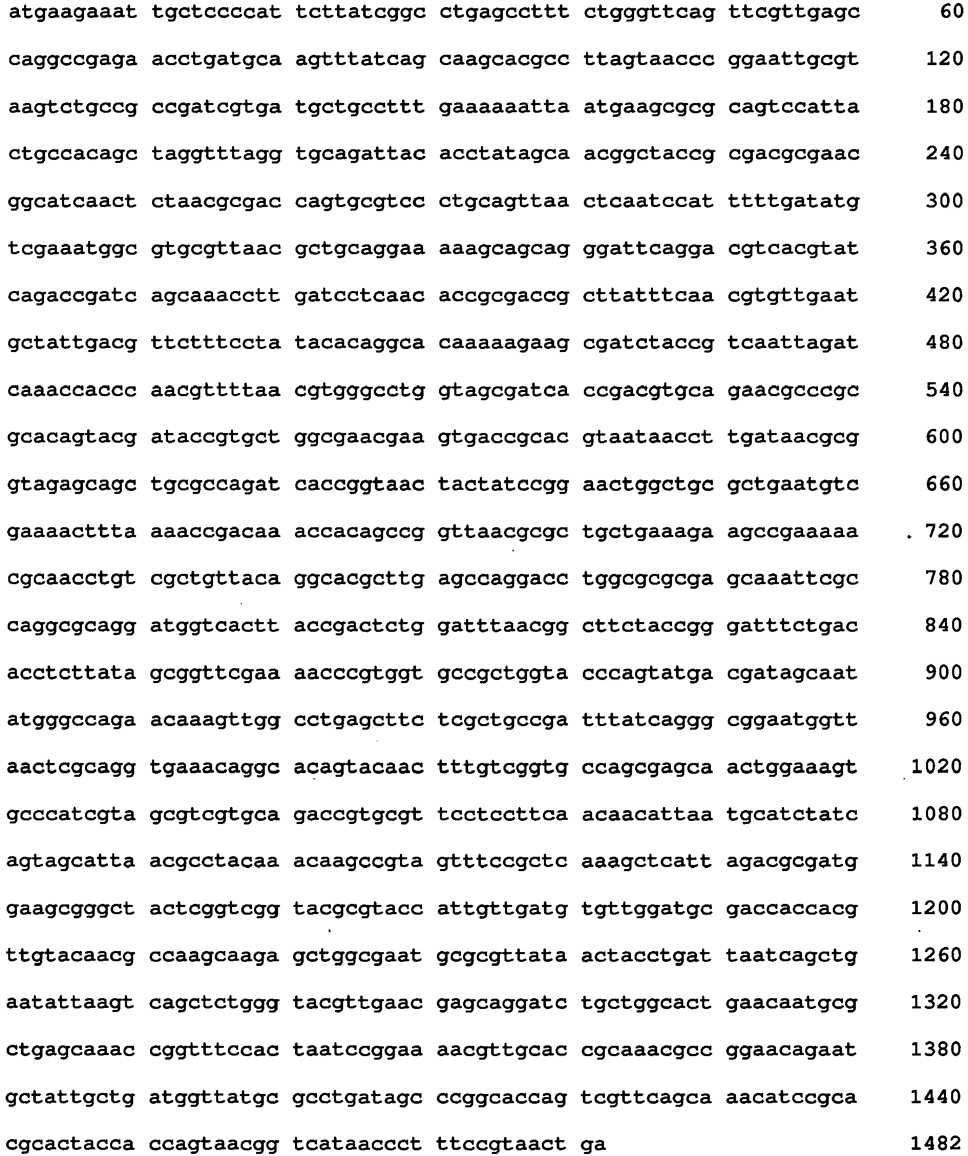
- <210> 8
<211> 493
<212> PRT
<213> Escherichia coli - <220>
<221> SOURCE
<222> 1..493
<223> /mol_type="protein" /note="Amino acid sequence of the tolC gene of E.coli" - <400> 8

- <210> 9
<211> 93
<212> DNA
<213> Artificial Sequence - <220>
<221> source
<222> 1..93
<223> /mol_type="DNA" /note="Fragment of a typical artificial autotransporter" /organism="Artificial Sequence" - <400> 9

- <210> 10
<211> 31
<212> PRT
<213> Artificial Sequence - <220>
<221> SOURCE
<222> 1..31
<223> /mol_type="protein" /note="Fragment of a typical artificial autotransporter" /organism="Artificial Sequence" - <400> 10

- <210> 11
<211> 24
<212> DNA
<213> Artificial Sequence - <220>
<221> source
<222> 1..24
<223> /mol_type="DNA" /note="Fragment with important restriction site " /organism="Artificial Sequence" - <400> 11
actgatttgc tcgagatgga agaa 24 - <210> 12
<211> 8
<212> PRT
<213> Artificial Sequence - <220>
<221> SOURCE
<222> 1..8
<223> /mol_type="protein" /note="Fragment with important restriction site" /organism="Artificial Sequence" - <400> 12

- <210> 13
<211> 24
<212> DNA
<213> Artificial Sequence - <220>
<221> source
<222> 1..24
<223> /mol_type="DNA" /note="Fragment with important restriction site" /organism="Artificial Sequence" - <400> 13
agccgcatgg gtacccttaa tcct 24 - <210> 14
<211> 8
<212> PRT
<213> Artificial Sequence - <220>
<221> SOURCE
<222> 1..8
<223> /mol_type="protein" /note="Fragment with important restriction site" /organism="Artificial Sequence" - <400> 14

Claims (17)
- A method for displaying a recombinant polypeptide containing a prosthetic group on the surface of a host cell, wherein the prosthetic group comprises a metal porphyrin or a flavin, said method comprising the steps:(a) providing a host cell transformed with a nucleic acid fusion operatively linked with an expression control sequence, said nucleic acid fusion comprising:(i) a portion encoding a signal peptide,(ii) a portion encoding the recombinant polypeptide to be displayed,(iii) a portion encoding a transmembrane linker, and(iv) a portion encoding the transporter domain of an autotransporter,
and(b) culturing the host cell under conditions wherein the nucleic acid fusion is expressed and the expression product comprising the recombinant polypeptide containing the prosthetic group is displayed on the surface of the host cell,wherein the prosthetic group is transported to the cell surface independently from the expression product comprising the recombinant polypeptide. - The method according to claim 1, wherein the nucleic acid fusion further comprises a portion encoding a protease recognition site.
- The method according to claim 1 or 2 wherein the polypeptide comprising the metal porphyrin is selected from hemoproteins, P450 enzymes, P450 reductases, cytochromes, and monooxygenases.
- The method according to any one of the preceding claims, wherein the prosthetic group being a metal porphyrin is transported to the cell surface by a TolC-dependent mechanism.
- The method according to claim 1 or 2 wherein the polypeptide is selected from flavoproteins and preferably comprises a flavin selected from FAD and FMN.
- The method according to any one of the preceding claims wherein the host cell is a bacterium, particularly a Gram-negative bacterium, more particularly an enterobacterium, e.g. E. coli.
- The method according to any one of the preceding claims wherein the transporter domain of the autotransporter is selected from Ssp, Ssp-h1, Ssp-h2, PspA, PspB, Ssa1, SphB1, AspA/NalP, VacA, AIDA-I, IcsA, MisL, TibA, Ag43, ShdA, AutA, Tsh, SepA, EspC, EspP, Pet, Pic, SigA, Sat, Vat, EpeA, EatA, Espl, EaaA, EaaC, Pertactin, BrkA, Tef, Vag8, PmpD, Pmp20, Pmp21, IgA1 protease, App, Hap, rOmpA, rOmpB, ApeE, EstA, Lip-1, McaP, BabA, SabA, AlpA, Aae, NanB, and variants having at least 90% sequence identity thereto, and wherein the transporter domain of the autotransporter preferably is the E. coli AIDA-1 protein or a variant thereof having at least 90% sequence identity to the E. coli AIDA-I protein.
- The method according to any one of the preceding claims comprising (c) preparing a membrane preparation from the cell obtained in step (b).
- Host cell displaying a recombinant polypeptide on the surface thereof wherein the recombinant polypeptide contains a prosthetic group comprising a metal porphyrin or a flavin, and wherein an expression product comprising the recombinant polypeptide comprises(I) a portion comprising the recombinant polypeptide to be displayed,(II) a portion comprising a transmembrane linker, and(III) a portion comprising the transporter domain of an autotransporter, wherein the host cell recombinantly expresses a transport protein capable of transporting the prosthetic group to the host cell surface, and wherein the prosthetic group is transported to the host cell surface by the transport protein independently from the expression product comprising the recombinant polypeptide.
- The host cell of claim 9, wherein the expression product further comprises a portion comprising a protease recognition site.
- The host cell of claim 9 or 10 wherein the recombinant polypeptide is displayed by the transporter domain of an autotransporter.
- The host cell according to any one of the claims 9 to 11, wherein the polypeptide comprising the metal porphyrin is selected from hemoproteins, P450 enzymes, P450 reductases, cytochromes, and monooxygenases.
- The host cell according to any one of the claims 9 to 12, wherein the prosthetic group being a metal porphyrin is transported to the cell surface by a TolC-dependent mechanism.
- The host cell according to any one of the claims 9 to 11, wherein the polypeptide is selected from flavoproteins and preferably comprises a flavin selected from FAD and FMN.
- The host cell according to any one of the claims 9 to 14 wherein the host cell is a bacterium, particularly a Gram-negative bacterium, more particularly an enterobacterium, e.g. E. coli.
- Use of a cell of any one of the claims 9 to 15 for a chemical synthesis procedure, preferably for the synthesis of organic substances selected from enzyme substrates, drugs, hormones, starting materials and intermediates for synthesis procedures and chiral substances.
- Use of a cell of any one of the claims 9 to 15(a) for a directed evolution procedure,(b) as an assay system for a screening procedure, e.g. for identifying modulators of metal porphyrin containing enzymes,(c) as a system for toxicity monitoring, or/and(d) as a system for degrading toxic substances.
Priority Applications (1)
| Application Number | Priority Date | Filing Date | Title |
|---|---|---|---|
| EP20110758244 EP2619306B1 (en) | 2010-09-23 | 2011-09-22 | Surface display of polypeptides containing a metal porphyrin or a flavin |
Applications Claiming Priority (3)
| Application Number | Priority Date | Filing Date | Title |
|---|---|---|---|
| EP10179060 | 2010-09-23 | ||
| PCT/EP2011/066517 WO2012038508A1 (en) | 2010-09-23 | 2011-09-22 | Surface display of polypeptides containing a metal porphyrin or a flavin |
| EP20110758244 EP2619306B1 (en) | 2010-09-23 | 2011-09-22 | Surface display of polypeptides containing a metal porphyrin or a flavin |
Publications (2)
| Publication Number | Publication Date |
|---|---|
| EP2619306A1 EP2619306A1 (en) | 2013-07-31 |
| EP2619306B1 true EP2619306B1 (en) | 2015-05-20 |
Family
ID=44654148
Family Applications (1)
| Application Number | Title | Priority Date | Filing Date |
|---|---|---|---|
| EP20110758244 Not-in-force EP2619306B1 (en) | 2010-09-23 | 2011-09-22 | Surface display of polypeptides containing a metal porphyrin or a flavin |
Country Status (3)
| Country | Link |
|---|---|
| US (1) | US20140011706A1 (en) |
| EP (1) | EP2619306B1 (en) |
| WO (1) | WO2012038508A1 (en) |
Families Citing this family (5)
| Publication number | Priority date | Publication date | Assignee | Title |
|---|---|---|---|---|
| CA2811699C (en) * | 2010-09-28 | 2019-07-02 | Abera Bioscience Ab | Fusion protein for secretory protein expression |
| CN108359676A (en) * | 2013-03-13 | 2018-08-03 | 自动显示生物技术有限公司 | Improved surface display of the functional protein in gramnegative bacterium in extensive range |
| RU2526213C1 (en) * | 2013-04-12 | 2014-08-20 | Федеральное государственное бюджетное учреждение науки Институт биоорганической химии им. академиков М.М. Шемякина и Ю.А. Овчинникова Российский академии наук (ИБХ РАН) | RECOMBINANT PLASMID DNA pEst877 DETERMINING EXPRESSION OF POLYPEPTIDE WITH ACTIVITY OF ESTERASE Psychrobacter cryohalolentis K5T ON CELL SURFACE Escherichia coli, AND BACTERIA STRAIN Escherichia coli BL21 (DE3) pLysS/pEst877-PRODUCER OF POLYPEPTIDE WITH ACTIVITY OF ESTERASE PSYCHROBACTER CRYOHALOLENTIS K5T ON CELL SURFACE |
| CN104745574B (en) * | 2013-12-31 | 2017-06-06 | 中国科学院生物物理研究所 | DNA molecular and its application for presenting destination protein in bacterium surface |
| WO2016101045A1 (en) * | 2014-12-24 | 2016-06-30 | The University Of Queensland | Improved oxidoreductases for biocatalysis |
Family Cites Families (5)
| Publication number | Priority date | Publication date | Assignee | Title |
|---|---|---|---|---|
| DE3622221A1 (en) | 1986-07-02 | 1988-01-14 | Max Planck Gesellschaft | METHOD FOR THE GENE-TECHNOLOGICAL EXTRACTION OF PROTEINS USING GRAM-NEGATIVE HOST CELLS |
| EP0886678B1 (en) | 1996-03-15 | 2005-06-29 | Max-Planck-Gesellschaft zur Förderung der Wissenschaften e.V. Berlin | Export systems for recombinant proteins |
| WO2002070645A2 (en) * | 2001-03-02 | 2002-09-12 | MAX-PLANCK-Gesellschaft zur Förderung der Wissenschaften e.V. | Functional surface display of polypeptides |
| DE10314413B4 (en) * | 2003-03-28 | 2015-08-06 | ZYRUS Beteiligungsgesellschaft mbH & Co.Patente I KG | A method for detecting the expression of recombinant proteins on a cell surface and for the immobilization and purification of cells and proteins |
| US7452572B1 (en) * | 2004-03-11 | 2008-11-18 | The North Carolina State University | Procedure for preparing redox-active polymers on surfaces |
-
2011
- 2011-09-22 WO PCT/EP2011/066517 patent/WO2012038508A1/en not_active Ceased
- 2011-09-22 EP EP20110758244 patent/EP2619306B1/en not_active Not-in-force
- 2011-09-22 US US13/825,837 patent/US20140011706A1/en not_active Abandoned
Also Published As
| Publication number | Publication date |
|---|---|
| US20140011706A1 (en) | 2014-01-09 |
| EP2619306A1 (en) | 2013-07-31 |
| WO2012038508A1 (en) | 2012-03-29 |
Similar Documents
| Publication | Publication Date | Title |
|---|---|---|
| EP1373571B1 (en) | Functional surface display of polypeptides | |
| Jose et al. | Cellular surface display of dimeric Adx and whole cell P450-mediated steroid synthesis on E. coli | |
| Zhang et al. | Mechanistic insights into interactions between bacterial class I P450 enzymes and redox partners | |
| Koch et al. | In vitro studies with purified components reveal signal recognition particle (SRP) and SecA/SecB as constituents of two independent protein-targeting pathways of Escherichia coli | |
| Liu et al. | Cyclopiazonic acid biosynthesis in Aspergillus sp.: characterization of a reductase-like R* domain in cyclopiazonate synthetase that forms and releases cyclo-acetoacetyl-L-tryptophan | |
| Grove et al. | Escherichia coli K‐12 genes essential for the synthesis of c‐type cytochromes and a third nitrate reductase located in the periplasm | |
| Sato et al. | Disulfide bond formation network in the three biological kingdoms, bacteria, fungi and mammals | |
| VAN VOORST et al. | Role of lipids in the translocation of proteins across membranes | |
| Bieker‐Brady et al. | Suppressor analysis suggests a multistep, cyclic mechanism for protein secretion in Escherichia coli. | |
| EP1487966B1 (en) | Secretion of proteins with multiple disulfide bonds in bacteria and uses thereof | |
| Guccione et al. | Reduction of fumarate, mesaconate and crotonate by Mfr, a novel oxygen‐regulated periplasmic reductase in Campylobacter jejuni | |
| EP2970953B1 (en) | Improved surface display of functional proteins in a broad range of gram negative bacteria | |
| US20140011706A1 (en) | Surface display of polypeptides containing a metal porphyrin or a flavin | |
| Ichetovkin et al. | Substrate recognition by the leucyl/phenylalanyl-tRNA-protein transferase: conservation within the enzyme family and localization to the trypsin-resistant domain | |
| Arfman et al. | Purification and characterization of an activator protein for methanol dehydrogenase from thermotolerant Bacillus spp | |
| Behrendt et al. | The TatBC complex of the Tat protein translocase in Escherichia coli and its transition to the substrate-bound TatABC complex | |
| ES2426640T3 (en) | Transformed strain derived from a strain deficient in multi-drug efflux protein and bioconversion method using the same | |
| Sligar et al. | [3] Mutagenesis of cytochromes P450cam and b5 | |
| Myllykallio et al. | Cytochrome c (y) of Rhodobacter capsulatus is attached to the cytoplasmic membrane by an uncleaved signal sequence-like anchor | |
| WO2007127735A2 (en) | Prokaryotic host cells for expressing proteins rich in disulfide bonds | |
| Zamalutdinova et al. | Optimization of Human STARD3 Cholesterol Transporter Expression for Enhancing Bovine P450scc-Mediated Steroid Biotransformation in E. coli | |
| Spandolf | Enabling the P450 complement of Beauveria bassiana and Rhodococcus jostii for biocatalysis | |
| WO2001048225A2 (en) | Production of heterologous proteins | |
| Jóźwik | Structural studies of bacterial steroid-hydroxylating cytochrome P450 monooxygenases: Insights into the molecular basis of substrate specificity, selectivity and product formation | |
| Dorenbos | Thiol-disulfide oxidoreductases in Bacillus subtilis |
Legal Events
| Date | Code | Title | Description |
|---|---|---|---|
| PUAI | Public reference made under article 153(3) epc to a published international application that has entered the european phase |
Free format text: ORIGINAL CODE: 0009012 |
|
| 17P | Request for examination filed |
Effective date: 20130418 |
|
| AK | Designated contracting states |
Kind code of ref document: A1 Designated state(s): AL AT BE BG CH CY CZ DE DK EE ES FI FR GB GR HR HU IE IS IT LI LT LU LV MC MK MT NL NO PL PT RO RS SE SI SK SM TR |
|
| DAX | Request for extension of the european patent (deleted) | ||
| 17Q | First examination report despatched |
Effective date: 20140630 |
|
| GRAP | Despatch of communication of intention to grant a patent |
Free format text: ORIGINAL CODE: EPIDOSNIGR1 |
|
| INTG | Intention to grant announced |
Effective date: 20141202 |
|
| GRAS | Grant fee paid |
Free format text: ORIGINAL CODE: EPIDOSNIGR3 |
|
| GRAA | (expected) grant |
Free format text: ORIGINAL CODE: 0009210 |
|
| AK | Designated contracting states |
Kind code of ref document: B1 Designated state(s): AL AT BE BG CH CY CZ DE DK EE ES FI FR GB GR HR HU IE IS IT LI LT LU LV MC MK MT NL NO PL PT RO RS SE SI SK SM TR |
|
| REG | Reference to a national code |
Ref country code: GB Ref legal event code: FG4D |
|
| REG | Reference to a national code |
Ref country code: CH Ref legal event code: EP Ref country code: CH Ref legal event code: NV Representative=s name: E. BLUM AND CO. AG PATENT- UND MARKENANWAELTE , CH |
|
| REG | Reference to a national code |
Ref country code: AT Ref legal event code: REF Ref document number: 727775 Country of ref document: AT Kind code of ref document: T Effective date: 20150615 |
|
| REG | Reference to a national code |
Ref country code: IE Ref legal event code: FG4D |
|
| REG | Reference to a national code |
Ref country code: DE Ref legal event code: R096 Ref document number: 602011016632 Country of ref document: DE |
|
| REG | Reference to a national code |
Ref country code: AT Ref legal event code: MK05 Ref document number: 727775 Country of ref document: AT Kind code of ref document: T Effective date: 20150520 |
|
| REG | Reference to a national code |
Ref country code: LT Ref legal event code: MG4D |
|
| REG | Reference to a national code |
Ref country code: NL Ref legal event code: MP Effective date: 20150520 |
|
| PG25 | Lapsed in a contracting state [announced via postgrant information from national office to epo] |
Ref country code: ES Free format text: LAPSE BECAUSE OF FAILURE TO SUBMIT A TRANSLATION OF THE DESCRIPTION OR TO PAY THE FEE WITHIN THE PRESCRIBED TIME-LIMIT Effective date: 20150520 Ref country code: FI Free format text: LAPSE BECAUSE OF FAILURE TO SUBMIT A TRANSLATION OF THE DESCRIPTION OR TO PAY THE FEE WITHIN THE PRESCRIBED TIME-LIMIT Effective date: 20150520 Ref country code: HR Free format text: LAPSE BECAUSE OF FAILURE TO SUBMIT A TRANSLATION OF THE DESCRIPTION OR TO PAY THE FEE WITHIN THE PRESCRIBED TIME-LIMIT Effective date: 20150520 Ref country code: LT Free format text: LAPSE BECAUSE OF FAILURE TO SUBMIT A TRANSLATION OF THE DESCRIPTION OR TO PAY THE FEE WITHIN THE PRESCRIBED TIME-LIMIT Effective date: 20150520 Ref country code: NO Free format text: LAPSE BECAUSE OF FAILURE TO SUBMIT A TRANSLATION OF THE DESCRIPTION OR TO PAY THE FEE WITHIN THE PRESCRIBED TIME-LIMIT Effective date: 20150820 Ref country code: PT Free format text: LAPSE BECAUSE OF FAILURE TO SUBMIT A TRANSLATION OF THE DESCRIPTION OR TO PAY THE FEE WITHIN THE PRESCRIBED TIME-LIMIT Effective date: 20150921 |
|
| PG25 | Lapsed in a contracting state [announced via postgrant information from national office to epo] |
Ref country code: BG Free format text: LAPSE BECAUSE OF FAILURE TO SUBMIT A TRANSLATION OF THE DESCRIPTION OR TO PAY THE FEE WITHIN THE PRESCRIBED TIME-LIMIT Effective date: 20150820 Ref country code: RS Free format text: LAPSE BECAUSE OF FAILURE TO SUBMIT A TRANSLATION OF THE DESCRIPTION OR TO PAY THE FEE WITHIN THE PRESCRIBED TIME-LIMIT Effective date: 20150520 Ref country code: IS Free format text: LAPSE BECAUSE OF FAILURE TO SUBMIT A TRANSLATION OF THE DESCRIPTION OR TO PAY THE FEE WITHIN THE PRESCRIBED TIME-LIMIT Effective date: 20150920 Ref country code: LV Free format text: LAPSE BECAUSE OF FAILURE TO SUBMIT A TRANSLATION OF THE DESCRIPTION OR TO PAY THE FEE WITHIN THE PRESCRIBED TIME-LIMIT Effective date: 20150520 Ref country code: AT Free format text: LAPSE BECAUSE OF FAILURE TO SUBMIT A TRANSLATION OF THE DESCRIPTION OR TO PAY THE FEE WITHIN THE PRESCRIBED TIME-LIMIT Effective date: 20150520 Ref country code: GR Free format text: LAPSE BECAUSE OF FAILURE TO SUBMIT A TRANSLATION OF THE DESCRIPTION OR TO PAY THE FEE WITHIN THE PRESCRIBED TIME-LIMIT Effective date: 20150821 |
|
| PG25 | Lapsed in a contracting state [announced via postgrant information from national office to epo] |
Ref country code: EE Free format text: LAPSE BECAUSE OF FAILURE TO SUBMIT A TRANSLATION OF THE DESCRIPTION OR TO PAY THE FEE WITHIN THE PRESCRIBED TIME-LIMIT Effective date: 20150520 Ref country code: DK Free format text: LAPSE BECAUSE OF FAILURE TO SUBMIT A TRANSLATION OF THE DESCRIPTION OR TO PAY THE FEE WITHIN THE PRESCRIBED TIME-LIMIT Effective date: 20150520 |
|
| REG | Reference to a national code |
Ref country code: DE Ref legal event code: R097 Ref document number: 602011016632 Country of ref document: DE |
|
| PG25 | Lapsed in a contracting state [announced via postgrant information from national office to epo] |
Ref country code: PL Free format text: LAPSE BECAUSE OF FAILURE TO SUBMIT A TRANSLATION OF THE DESCRIPTION OR TO PAY THE FEE WITHIN THE PRESCRIBED TIME-LIMIT Effective date: 20150520 Ref country code: RO Free format text: LAPSE BECAUSE OF NON-PAYMENT OF DUE FEES Effective date: 20150520 Ref country code: CZ Free format text: LAPSE BECAUSE OF FAILURE TO SUBMIT A TRANSLATION OF THE DESCRIPTION OR TO PAY THE FEE WITHIN THE PRESCRIBED TIME-LIMIT Effective date: 20150520 Ref country code: SK Free format text: LAPSE BECAUSE OF FAILURE TO SUBMIT A TRANSLATION OF THE DESCRIPTION OR TO PAY THE FEE WITHIN THE PRESCRIBED TIME-LIMIT Effective date: 20150520 |
|
| PLBE | No opposition filed within time limit |
Free format text: ORIGINAL CODE: 0009261 |
|
| STAA | Information on the status of an ep patent application or granted ep patent |
Free format text: STATUS: NO OPPOSITION FILED WITHIN TIME LIMIT |
|
| REG | Reference to a national code |
Ref country code: FR Ref legal event code: PLFP Year of fee payment: 5 |
|
| 26N | No opposition filed |
Effective date: 20160223 |
|
| PG25 | Lapsed in a contracting state [announced via postgrant information from national office to epo] |
Ref country code: IT Free format text: LAPSE BECAUSE OF FAILURE TO SUBMIT A TRANSLATION OF THE DESCRIPTION OR TO PAY THE FEE WITHIN THE PRESCRIBED TIME-LIMIT Effective date: 20150520 Ref country code: MC Free format text: LAPSE BECAUSE OF FAILURE TO SUBMIT A TRANSLATION OF THE DESCRIPTION OR TO PAY THE FEE WITHIN THE PRESCRIBED TIME-LIMIT Effective date: 20150520 |
|
| PG25 | Lapsed in a contracting state [announced via postgrant information from national office to epo] |
Ref country code: SI Free format text: LAPSE BECAUSE OF FAILURE TO SUBMIT A TRANSLATION OF THE DESCRIPTION OR TO PAY THE FEE WITHIN THE PRESCRIBED TIME-LIMIT Effective date: 20150520 |
|
| PG25 | Lapsed in a contracting state [announced via postgrant information from national office to epo] |
Ref country code: BE Free format text: LAPSE BECAUSE OF FAILURE TO SUBMIT A TRANSLATION OF THE DESCRIPTION OR TO PAY THE FEE WITHIN THE PRESCRIBED TIME-LIMIT Effective date: 20150520 |
|
| REG | Reference to a national code |
Ref country code: FR Ref legal event code: PLFP Year of fee payment: 6 |
|
| PG25 | Lapsed in a contracting state [announced via postgrant information from national office to epo] |
Ref country code: MT Free format text: LAPSE BECAUSE OF FAILURE TO SUBMIT A TRANSLATION OF THE DESCRIPTION OR TO PAY THE FEE WITHIN THE PRESCRIBED TIME-LIMIT Effective date: 20150520 |
|
| PG25 | Lapsed in a contracting state [announced via postgrant information from national office to epo] |
Ref country code: SM Free format text: LAPSE BECAUSE OF FAILURE TO SUBMIT A TRANSLATION OF THE DESCRIPTION OR TO PAY THE FEE WITHIN THE PRESCRIBED TIME-LIMIT Effective date: 20150520 Ref country code: HU Free format text: LAPSE BECAUSE OF FAILURE TO SUBMIT A TRANSLATION OF THE DESCRIPTION OR TO PAY THE FEE WITHIN THE PRESCRIBED TIME-LIMIT; INVALID AB INITIO Effective date: 20110922 |
|
| PGFP | Annual fee paid to national office [announced via postgrant information from national office to epo] |
Ref country code: LU Payment date: 20170227 Year of fee payment: 6 Ref country code: IE Payment date: 20170227 Year of fee payment: 6 |
|
| PG25 | Lapsed in a contracting state [announced via postgrant information from national office to epo] |
Ref country code: CY Free format text: LAPSE BECAUSE OF FAILURE TO SUBMIT A TRANSLATION OF THE DESCRIPTION OR TO PAY THE FEE WITHIN THE PRESCRIBED TIME-LIMIT Effective date: 20150520 Ref country code: NL Free format text: LAPSE BECAUSE OF FAILURE TO SUBMIT A TRANSLATION OF THE DESCRIPTION OR TO PAY THE FEE WITHIN THE PRESCRIBED TIME-LIMIT Effective date: 20150520 Ref country code: SE Free format text: LAPSE BECAUSE OF FAILURE TO SUBMIT A TRANSLATION OF THE DESCRIPTION OR TO PAY THE FEE WITHIN THE PRESCRIBED TIME-LIMIT Effective date: 20150520 |
|
| REG | Reference to a national code |
Ref country code: FR Ref legal event code: PLFP Year of fee payment: 7 |
|
| REG | Reference to a national code |
Ref country code: IE Ref legal event code: MM4A |
|
| PG25 | Lapsed in a contracting state [announced via postgrant information from national office to epo] |
Ref country code: MK Free format text: LAPSE BECAUSE OF FAILURE TO SUBMIT A TRANSLATION OF THE DESCRIPTION OR TO PAY THE FEE WITHIN THE PRESCRIBED TIME-LIMIT Effective date: 20150520 Ref country code: TR Free format text: LAPSE BECAUSE OF FAILURE TO SUBMIT A TRANSLATION OF THE DESCRIPTION OR TO PAY THE FEE WITHIN THE PRESCRIBED TIME-LIMIT Effective date: 20150520 Ref country code: LU Free format text: LAPSE BECAUSE OF NON-PAYMENT OF DUE FEES Effective date: 20170922 |
|
| PG25 | Lapsed in a contracting state [announced via postgrant information from national office to epo] |
Ref country code: IE Free format text: LAPSE BECAUSE OF NON-PAYMENT OF DUE FEES Effective date: 20170922 |
|
| REG | Reference to a national code |
Ref country code: FR Ref legal event code: PLFP Year of fee payment: 8 |
|
| PG25 | Lapsed in a contracting state [announced via postgrant information from national office to epo] |
Ref country code: AL Free format text: LAPSE BECAUSE OF FAILURE TO SUBMIT A TRANSLATION OF THE DESCRIPTION OR TO PAY THE FEE WITHIN THE PRESCRIBED TIME-LIMIT Effective date: 20150520 |
|
| PGFP | Annual fee paid to national office [announced via postgrant information from national office to epo] |
Ref country code: DE Payment date: 20190830 Year of fee payment: 9 Ref country code: FR Payment date: 20190927 Year of fee payment: 9 |
|
| PGFP | Annual fee paid to national office [announced via postgrant information from national office to epo] |
Ref country code: GB Payment date: 20190920 Year of fee payment: 9 |
|
| PGFP | Annual fee paid to national office [announced via postgrant information from national office to epo] |
Ref country code: CH Payment date: 20190919 Year of fee payment: 9 |
|
| REG | Reference to a national code |
Ref country code: DE Ref legal event code: R119 Ref document number: 602011016632 Country of ref document: DE |
|
| REG | Reference to a national code |
Ref country code: CH Ref legal event code: PL |
|
| GBPC | Gb: european patent ceased through non-payment of renewal fee |
Effective date: 20200922 |
|
| PG25 | Lapsed in a contracting state [announced via postgrant information from national office to epo] |
Ref country code: DE Free format text: LAPSE BECAUSE OF NON-PAYMENT OF DUE FEES Effective date: 20210401 Ref country code: FR Free format text: LAPSE BECAUSE OF NON-PAYMENT OF DUE FEES Effective date: 20200930 |
|
| PG25 | Lapsed in a contracting state [announced via postgrant information from national office to epo] |
Ref country code: CH Free format text: LAPSE BECAUSE OF NON-PAYMENT OF DUE FEES Effective date: 20200930 Ref country code: GB Free format text: LAPSE BECAUSE OF NON-PAYMENT OF DUE FEES Effective date: 20200922 Ref country code: LI Free format text: LAPSE BECAUSE OF NON-PAYMENT OF DUE FEES Effective date: 20200930 |