EP2612919A1 - Sulfated polysaccharide capable of binding to growth factor and use thereof - Google Patents
Sulfated polysaccharide capable of binding to growth factor and use thereof Download PDFInfo
- Publication number
- EP2612919A1 EP2612919A1 EP11821878.3A EP11821878A EP2612919A1 EP 2612919 A1 EP2612919 A1 EP 2612919A1 EP 11821878 A EP11821878 A EP 11821878A EP 2612919 A1 EP2612919 A1 EP 2612919A1
- Authority
- EP
- European Patent Office
- Prior art keywords
- differentiation
- cells
- sulfated polysaccharide
- days
- medium
- Prior art date
- Legal status (The legal status is an assumption and is not a legal conclusion. Google has not performed a legal analysis and makes no representation as to the accuracy of the status listed.)
- Withdrawn
Links
- 150000004676 glycans Chemical class 0.000 title claims abstract description 91
- 229920001282 polysaccharide Polymers 0.000 title claims abstract description 89
- 239000005017 polysaccharide Substances 0.000 title claims abstract description 89
- 239000003102 growth factor Substances 0.000 title claims abstract description 28
- 230000027455 binding Effects 0.000 title abstract description 18
- 230000024245 cell differentiation Effects 0.000 claims abstract description 64
- 230000004069 differentiation Effects 0.000 claims abstract description 59
- 239000000203 mixture Substances 0.000 claims abstract description 38
- 238000000034 method Methods 0.000 claims abstract description 34
- 230000035755 proliferation Effects 0.000 claims abstract description 21
- 239000004480 active ingredient Substances 0.000 claims abstract description 15
- 210000004027 cell Anatomy 0.000 claims description 84
- 230000004663 cell proliferation Effects 0.000 claims description 59
- 210000004409 osteocyte Anatomy 0.000 claims description 17
- 102100024785 Fibroblast growth factor 2 Human genes 0.000 claims description 11
- 108090000379 Fibroblast growth factor 2 Proteins 0.000 claims description 11
- 230000001737 promoting effect Effects 0.000 claims description 8
- 108010049931 Bone Morphogenetic Protein 2 Proteins 0.000 claims description 6
- 102100024506 Bone morphogenetic protein 2 Human genes 0.000 claims description 6
- 210000001612 chondrocyte Anatomy 0.000 claims description 5
- 239000000126 substance Substances 0.000 abstract description 25
- 230000002708 enhancing effect Effects 0.000 abstract description 3
- 239000002609 medium Substances 0.000 description 79
- 239000000243 solution Substances 0.000 description 39
- 210000002901 mesenchymal stem cell Anatomy 0.000 description 36
- 238000012258 culturing Methods 0.000 description 32
- 239000012228 culture supernatant Substances 0.000 description 26
- 235000013305 food Nutrition 0.000 description 23
- 230000000052 comparative effect Effects 0.000 description 22
- 108091003079 Bovine Serum Albumin Proteins 0.000 description 20
- 239000012091 fetal bovine serum Substances 0.000 description 19
- 210000000988 bone and bone Anatomy 0.000 description 18
- 102000002260 Alkaline Phosphatase Human genes 0.000 description 17
- 108020004774 Alkaline Phosphatase Proteins 0.000 description 17
- 235000013361 beverage Nutrition 0.000 description 17
- 230000000694 effects Effects 0.000 description 17
- CIWBSHSKHKDKBQ-JLAZNSOCSA-N Ascorbic acid Chemical compound OC[C@H](O)[C@H]1OC(=O)C(O)=C1O CIWBSHSKHKDKBQ-JLAZNSOCSA-N 0.000 description 16
- 210000004663 osteoprogenitor cell Anatomy 0.000 description 16
- 238000005259 measurement Methods 0.000 description 15
- 210000001519 tissue Anatomy 0.000 description 15
- 239000006144 Dulbecco’s modified Eagle's medium Substances 0.000 description 14
- DHCLVCXQIBBOPH-UHFFFAOYSA-N Glycerol 2-phosphate Chemical compound OCC(CO)OP(O)(O)=O DHCLVCXQIBBOPH-UHFFFAOYSA-N 0.000 description 14
- 239000006228 supernatant Substances 0.000 description 13
- OYPRJOBELJOOCE-UHFFFAOYSA-N Calcium Chemical compound [Ca] OYPRJOBELJOOCE-UHFFFAOYSA-N 0.000 description 12
- 239000011575 calcium Substances 0.000 description 12
- 229910052791 calcium Inorganic materials 0.000 description 12
- 239000008363 phosphate buffer Substances 0.000 description 12
- 239000003153 chemical reaction reagent Substances 0.000 description 11
- 238000002835 absorbance Methods 0.000 description 10
- 239000007758 minimum essential medium Substances 0.000 description 10
- 229960003957 dexamethasone Drugs 0.000 description 9
- UREBDLICKHMUKA-CXSFZGCWSA-N dexamethasone Chemical compound C1CC2=CC(=O)C=C[C@]2(C)[C@]2(F)[C@@H]1[C@@H]1C[C@@H](C)[C@@](C(=O)CO)(O)[C@@]1(C)C[C@@H]2O UREBDLICKHMUKA-CXSFZGCWSA-N 0.000 description 9
- FAPWRFPIFSIZLT-UHFFFAOYSA-M Sodium chloride Chemical compound [Na+].[Cl-] FAPWRFPIFSIZLT-UHFFFAOYSA-M 0.000 description 8
- 235000010323 ascorbic acid Nutrition 0.000 description 8
- 229960005070 ascorbic acid Drugs 0.000 description 8
- 239000011668 ascorbic acid Substances 0.000 description 8
- 210000000845 cartilage Anatomy 0.000 description 8
- WQZGKKKJIJFFOK-GASJEMHNSA-N Glucose Natural products OC[C@H]1OC(O)[C@H](O)[C@@H](O)[C@@H]1O WQZGKKKJIJFFOK-GASJEMHNSA-N 0.000 description 7
- 241001465754 Metazoa Species 0.000 description 7
- JRMSLDWZFJZLAS-UHFFFAOYSA-M [7-(dimethylamino)-1,9-dimethylphenothiazin-3-ylidene]-dimethylazanium;chloride Chemical compound [Cl-].CC1=CC(N(C)C)=CC2=[S+]C3=CC(N(C)C)=CC(C)=C3N=C21 JRMSLDWZFJZLAS-UHFFFAOYSA-M 0.000 description 7
- 238000003556 assay Methods 0.000 description 7
- 235000009508 confectionery Nutrition 0.000 description 7
- 239000008103 glucose Substances 0.000 description 7
- 238000000338 in vitro Methods 0.000 description 7
- 210000000056 organ Anatomy 0.000 description 7
- 238000002360 preparation method Methods 0.000 description 7
- 239000000047 product Substances 0.000 description 7
- WEVYAHXRMPXWCK-UHFFFAOYSA-N Acetonitrile Chemical compound CC#N WEVYAHXRMPXWCK-UHFFFAOYSA-N 0.000 description 6
- 210000001185 bone marrow Anatomy 0.000 description 6
- 238000011088 calibration curve Methods 0.000 description 6
- 229940004830 chondroitin sulfate a sodium Drugs 0.000 description 6
- 239000003814 drug Substances 0.000 description 6
- 230000035876 healing Effects 0.000 description 6
- 210000000130 stem cell Anatomy 0.000 description 6
- 241000700159 Rattus Species 0.000 description 5
- DBMJMQXJHONAFJ-UHFFFAOYSA-M Sodium laurylsulphate Chemical compound [Na+].CCCCCCCCCCCCOS([O-])(=O)=O DBMJMQXJHONAFJ-UHFFFAOYSA-M 0.000 description 5
- 230000002159 abnormal effect Effects 0.000 description 5
- 238000001727 in vivo Methods 0.000 description 5
- 210000002569 neuron Anatomy 0.000 description 5
- 230000001172 regenerating effect Effects 0.000 description 5
- IAZDPXIOMUYVGZ-UHFFFAOYSA-N Dimethylsulphoxide Chemical compound CS(C)=O IAZDPXIOMUYVGZ-UHFFFAOYSA-N 0.000 description 4
- 210000001789 adipocyte Anatomy 0.000 description 4
- VZTDIZULWFCMLS-UHFFFAOYSA-N ammonium formate Chemical compound [NH4+].[O-]C=O VZTDIZULWFCMLS-UHFFFAOYSA-N 0.000 description 4
- 238000001514 detection method Methods 0.000 description 4
- 150000002016 disaccharides Chemical class 0.000 description 4
- 201000010099 disease Diseases 0.000 description 4
- 208000037265 diseases, disorders, signs and symptoms Diseases 0.000 description 4
- 238000004128 high performance liquid chromatography Methods 0.000 description 4
- NOESYZHRGYRDHS-UHFFFAOYSA-N insulin Chemical compound N1C(=O)C(NC(=O)C(CCC(N)=O)NC(=O)C(CCC(O)=O)NC(=O)C(C(C)C)NC(=O)C(NC(=O)CN)C(C)CC)CSSCC(C(NC(CO)C(=O)NC(CC(C)C)C(=O)NC(CC=2C=CC(O)=CC=2)C(=O)NC(CCC(N)=O)C(=O)NC(CC(C)C)C(=O)NC(CCC(O)=O)C(=O)NC(CC(N)=O)C(=O)NC(CC=2C=CC(O)=CC=2)C(=O)NC(CSSCC(NC(=O)C(C(C)C)NC(=O)C(CC(C)C)NC(=O)C(CC=2C=CC(O)=CC=2)NC(=O)C(CC(C)C)NC(=O)C(C)NC(=O)C(CCC(O)=O)NC(=O)C(C(C)C)NC(=O)C(CC(C)C)NC(=O)C(CC=2NC=NC=2)NC(=O)C(CO)NC(=O)CNC2=O)C(=O)NCC(=O)NC(CCC(O)=O)C(=O)NC(CCCNC(N)=N)C(=O)NCC(=O)NC(CC=3C=CC=CC=3)C(=O)NC(CC=3C=CC=CC=3)C(=O)NC(CC=3C=CC(O)=CC=3)C(=O)NC(C(C)O)C(=O)N3C(CCC3)C(=O)NC(CCCCN)C(=O)NC(C)C(O)=O)C(=O)NC(CC(N)=O)C(O)=O)=O)NC(=O)C(C(C)CC)NC(=O)C(CO)NC(=O)C(C(C)O)NC(=O)C1CSSCC2NC(=O)C(CC(C)C)NC(=O)C(NC(=O)C(CCC(N)=O)NC(=O)C(CC(N)=O)NC(=O)C(NC(=O)C(N)CC=1C=CC=CC=1)C(C)C)CC1=CN=CN1 NOESYZHRGYRDHS-UHFFFAOYSA-N 0.000 description 4
- 239000008194 pharmaceutical composition Substances 0.000 description 4
- 108090000765 processed proteins & peptides Proteins 0.000 description 4
- 239000011780 sodium chloride Substances 0.000 description 4
- 239000001509 sodium citrate Substances 0.000 description 4
- DAEPDZWVDSPTHF-UHFFFAOYSA-M sodium pyruvate Chemical compound [Na+].CC(=O)C([O-])=O DAEPDZWVDSPTHF-UHFFFAOYSA-M 0.000 description 4
- 159000000000 sodium salts Chemical class 0.000 description 4
- HRXKRNGNAMMEHJ-UHFFFAOYSA-K trisodium citrate Chemical compound [Na+].[Na+].[Na+].[O-]C(=O)CC(O)(CC([O-])=O)C([O-])=O HRXKRNGNAMMEHJ-UHFFFAOYSA-K 0.000 description 4
- 229940038773 trisodium citrate Drugs 0.000 description 4
- 238000008940 Alkaline Phosphatase assay kit Methods 0.000 description 3
- 238000003650 Calcium Assay Kit Methods 0.000 description 3
- 229920002683 Glycosaminoglycan Polymers 0.000 description 3
- 241000282412 Homo Species 0.000 description 3
- 229920002684 Sepharose Polymers 0.000 description 3
- 230000037396 body weight Effects 0.000 description 3
- 239000000872 buffer Substances 0.000 description 3
- 238000012790 confirmation Methods 0.000 description 3
- 230000008021 deposition Effects 0.000 description 3
- -1 elixir Substances 0.000 description 3
- 238000002474 experimental method Methods 0.000 description 3
- 235000013376 functional food Nutrition 0.000 description 3
- 235000013402 health food Nutrition 0.000 description 3
- 230000006698 induction Effects 0.000 description 3
- 238000000746 purification Methods 0.000 description 3
- 238000000926 separation method Methods 0.000 description 3
- 239000012679 serum free medium Substances 0.000 description 3
- 235000014347 soups Nutrition 0.000 description 3
- 230000008685 targeting Effects 0.000 description 3
- 238000012360 testing method Methods 0.000 description 3
- 238000002054 transplantation Methods 0.000 description 3
- YBJHBAHKTGYVGT-ZKWXMUAHSA-N (+)-Biotin Chemical compound N1C(=O)N[C@@H]2[C@H](CCCCC(=O)O)SC[C@@H]21 YBJHBAHKTGYVGT-ZKWXMUAHSA-N 0.000 description 2
- BDKLKNJTMLIAFE-UHFFFAOYSA-N 2-(3-fluorophenyl)-1,3-oxazole-4-carbaldehyde Chemical compound FC1=CC=CC(C=2OC=C(C=O)N=2)=C1 BDKLKNJTMLIAFE-UHFFFAOYSA-N 0.000 description 2
- 241000251468 Actinopterygii Species 0.000 description 2
- 241000272525 Anas platyrhynchos Species 0.000 description 2
- 241000272517 Anseriformes Species 0.000 description 2
- 229920001342 Bakelite® Polymers 0.000 description 2
- 241000283690 Bos taurus Species 0.000 description 2
- 241000282472 Canis lupus familiaris Species 0.000 description 2
- 241000283707 Capra Species 0.000 description 2
- 241000282693 Cercopithecidae Species 0.000 description 2
- 241000699800 Cricetinae Species 0.000 description 2
- 239000006145 Eagle's minimal essential medium Substances 0.000 description 2
- 241000283086 Equidae Species 0.000 description 2
- OHCQJHSOBUTRHG-KGGHGJDLSA-N FORSKOLIN Chemical compound O=C([C@@]12O)C[C@](C)(C=C)O[C@]1(C)[C@@H](OC(=O)C)[C@@H](O)[C@@H]1[C@]2(C)[C@@H](O)CCC1(C)C OHCQJHSOBUTRHG-KGGHGJDLSA-N 0.000 description 2
- 241000282326 Felis catus Species 0.000 description 2
- 241000287828 Gallus gallus Species 0.000 description 2
- 229920002971 Heparan sulfate Polymers 0.000 description 2
- 108010022901 Heparin Lyase Proteins 0.000 description 2
- 101000652133 Homo sapiens STE20-like serine/threonine-protein kinase Proteins 0.000 description 2
- 101000601460 Homo sapiens Serine/threonine-protein kinase Nek4 Proteins 0.000 description 2
- 102000004877 Insulin Human genes 0.000 description 2
- 108090001061 Insulin Proteins 0.000 description 2
- ONIBWKKTOPOVIA-BYPYZUCNSA-N L-Proline Chemical compound OC(=O)[C@@H]1CCCN1 ONIBWKKTOPOVIA-BYPYZUCNSA-N 0.000 description 2
- 241000699666 Mus <mouse, genus> Species 0.000 description 2
- 241000699670 Mus sp. Species 0.000 description 2
- 241000283973 Oryctolagus cuniculus Species 0.000 description 2
- 241001494479 Pecora Species 0.000 description 2
- WCUXLLCKKVVCTQ-UHFFFAOYSA-M Potassium chloride Chemical compound [Cl-].[K+] WCUXLLCKKVVCTQ-UHFFFAOYSA-M 0.000 description 2
- RJKFOVLPORLFTN-LEKSSAKUSA-N Progesterone Chemical compound C1CC2=CC(=O)CC[C@]2(C)[C@@H]2[C@@H]1[C@@H]1CC[C@H](C(=O)C)[C@@]1(C)CC2 RJKFOVLPORLFTN-LEKSSAKUSA-N 0.000 description 2
- 102100030571 STE20-like serine/threonine-protein kinase Human genes 0.000 description 2
- 241000271567 Struthioniformes Species 0.000 description 2
- 241000282887 Suidae Species 0.000 description 2
- 102000004338 Transferrin Human genes 0.000 description 2
- 108090000901 Transferrin Proteins 0.000 description 2
- 230000009471 action Effects 0.000 description 2
- 239000007864 aqueous solution Substances 0.000 description 2
- 230000010478 bone regeneration Effects 0.000 description 2
- 239000002775 capsule Substances 0.000 description 2
- 239000003795 chemical substances by application Substances 0.000 description 2
- 235000013330 chicken meat Nutrition 0.000 description 2
- 238000004587 chromatography analysis Methods 0.000 description 2
- 239000012531 culture fluid Substances 0.000 description 2
- 230000029087 digestion Effects 0.000 description 2
- 235000021186 dishes Nutrition 0.000 description 2
- 238000010828 elution Methods 0.000 description 2
- 210000001671 embryonic stem cell Anatomy 0.000 description 2
- 238000011156 evaluation Methods 0.000 description 2
- 230000005284 excitation Effects 0.000 description 2
- 235000020510 functional beverage Nutrition 0.000 description 2
- 239000000499 gel Substances 0.000 description 2
- 239000008187 granular material Substances 0.000 description 2
- 210000003958 hematopoietic stem cell Anatomy 0.000 description 2
- 108010083213 heparitinsulfate lyase Proteins 0.000 description 2
- 230000002440 hepatic effect Effects 0.000 description 2
- 210000003897 hepatic stem cell Anatomy 0.000 description 2
- 210000003494 hepatocyte Anatomy 0.000 description 2
- JYGXADMDTFJGBT-VWUMJDOOSA-N hydrocortisone Chemical compound O=C1CC[C@]2(C)[C@H]3[C@@H](O)C[C@](C)([C@@](CC4)(O)C(=O)CO)[C@@H]4[C@@H]3CCC2=C1 JYGXADMDTFJGBT-VWUMJDOOSA-N 0.000 description 2
- 230000001939 inductive effect Effects 0.000 description 2
- 229940125396 insulin Drugs 0.000 description 2
- 235000015110 jellies Nutrition 0.000 description 2
- 239000008274 jelly Substances 0.000 description 2
- 239000007788 liquid Substances 0.000 description 2
- 238000004519 manufacturing process Methods 0.000 description 2
- 239000012528 membrane Substances 0.000 description 2
- 210000004165 myocardium Anatomy 0.000 description 2
- 210000001087 myotubule Anatomy 0.000 description 2
- 210000001178 neural stem cell Anatomy 0.000 description 2
- 239000003921 oil Substances 0.000 description 2
- 235000019198 oils Nutrition 0.000 description 2
- 210000000963 osteoblast Anatomy 0.000 description 2
- 210000004738 parenchymal cell Anatomy 0.000 description 2
- 239000006072 paste Substances 0.000 description 2
- 239000008188 pellet Substances 0.000 description 2
- 150000004804 polysaccharides Polymers 0.000 description 2
- YCYVPVNGUXMWMP-UHFFFAOYSA-M potassium;acetate;hydrate Chemical compound O.[K+].CC([O-])=O YCYVPVNGUXMWMP-UHFFFAOYSA-M 0.000 description 2
- 239000000843 powder Substances 0.000 description 2
- 230000008569 process Effects 0.000 description 2
- 229960002429 proline Drugs 0.000 description 2
- 108090000623 proteins and genes Proteins 0.000 description 2
- KIDHWZJUCRJVML-UHFFFAOYSA-N putrescine Chemical compound NCCCCN KIDHWZJUCRJVML-UHFFFAOYSA-N 0.000 description 2
- 235000015067 sauces Nutrition 0.000 description 2
- 210000002966 serum Anatomy 0.000 description 2
- 210000002027 skeletal muscle Anatomy 0.000 description 2
- 235000017281 sodium acetate Nutrition 0.000 description 2
- 229940087562 sodium acetate trihydrate Drugs 0.000 description 2
- 229940054269 sodium pyruvate Drugs 0.000 description 2
- 239000002904 solvent Substances 0.000 description 2
- 238000010186 staining Methods 0.000 description 2
- 238000011476 stem cell transplantation Methods 0.000 description 2
- 239000003826 tablet Substances 0.000 description 2
- 210000002303 tibia Anatomy 0.000 description 2
- 239000012581 transferrin Substances 0.000 description 2
- 238000000108 ultra-filtration Methods 0.000 description 2
- 210000000689 upper leg Anatomy 0.000 description 2
- 238000005406 washing Methods 0.000 description 2
- APIXJSLKIYYUKG-UHFFFAOYSA-N 3 Isobutyl 1 methylxanthine Chemical compound O=C1N(C)C(=O)N(CC(C)C)C2=C1N=CN2 APIXJSLKIYYUKG-UHFFFAOYSA-N 0.000 description 1
- NMUSYJAQQFHJEW-UHFFFAOYSA-N 5-Azacytidine Natural products O=C1N=C(N)N=CN1C1C(O)C(O)C(CO)O1 NMUSYJAQQFHJEW-UHFFFAOYSA-N 0.000 description 1
- NMUSYJAQQFHJEW-KVTDHHQDSA-N 5-azacytidine Chemical compound O=C1N=C(N)N=CN1[C@H]1[C@H](O)[C@H](O)[C@@H](CO)O1 NMUSYJAQQFHJEW-KVTDHHQDSA-N 0.000 description 1
- 108010088751 Albumins Proteins 0.000 description 1
- 102000009027 Albumins Human genes 0.000 description 1
- QGZKDVFQNNGYKY-UHFFFAOYSA-O Ammonium Chemical compound [NH4+] QGZKDVFQNNGYKY-UHFFFAOYSA-O 0.000 description 1
- 108090001008 Avidin Proteins 0.000 description 1
- 108010049951 Bone Morphogenetic Protein 3 Proteins 0.000 description 1
- 108010049955 Bone Morphogenetic Protein 4 Proteins 0.000 description 1
- 108010049870 Bone Morphogenetic Protein 7 Proteins 0.000 description 1
- 102100024504 Bone morphogenetic protein 3 Human genes 0.000 description 1
- 102100024505 Bone morphogenetic protein 4 Human genes 0.000 description 1
- 102100022544 Bone morphogenetic protein 7 Human genes 0.000 description 1
- 101001011741 Bos taurus Insulin Proteins 0.000 description 1
- 239000004255 Butylated hydroxyanisole Substances 0.000 description 1
- 210000003771 C cell Anatomy 0.000 description 1
- 241000238366 Cephalopoda Species 0.000 description 1
- 108020004414 DNA Proteins 0.000 description 1
- XPDXVDYUQZHFPV-UHFFFAOYSA-N Dansyl Chloride Chemical compound C1=CC=C2C(N(C)C)=CC=CC2=C1S(Cl)(=O)=O XPDXVDYUQZHFPV-UHFFFAOYSA-N 0.000 description 1
- SUZLHDUTVMZSEV-UHFFFAOYSA-N Deoxycoleonol Natural products C12C(=O)CC(C)(C=C)OC2(C)C(OC(=O)C)C(O)C2C1(C)C(O)CCC2(C)C SUZLHDUTVMZSEV-UHFFFAOYSA-N 0.000 description 1
- 101000904460 Encephalitozoon cuniculi (strain GB-M1) Probable glycerol-3-phosphate dehydrogenase Proteins 0.000 description 1
- 102000004190 Enzymes Human genes 0.000 description 1
- 108090000790 Enzymes Proteins 0.000 description 1
- 102000010834 Extracellular Matrix Proteins Human genes 0.000 description 1
- 108010037362 Extracellular Matrix Proteins Proteins 0.000 description 1
- 108090000386 Fibroblast Growth Factor 1 Proteins 0.000 description 1
- 102000003971 Fibroblast Growth Factor 1 Human genes 0.000 description 1
- 102100028072 Fibroblast growth factor 4 Human genes 0.000 description 1
- 239000004606 Fillers/Extenders Substances 0.000 description 1
- 101001060274 Homo sapiens Fibroblast growth factor 4 Proteins 0.000 description 1
- 101000628647 Homo sapiens Serine/threonine-protein kinase 24 Proteins 0.000 description 1
- 101000880439 Homo sapiens Serine/threonine-protein kinase 3 Proteins 0.000 description 1
- MIJPAVRNWPDMOR-ZAFYKAAXSA-N L-ascorbic acid 2-phosphate Chemical compound OC[C@H](O)[C@H]1OC(=O)C(OP(O)(O)=O)=C1O MIJPAVRNWPDMOR-ZAFYKAAXSA-N 0.000 description 1
- KIENGQUGHPTFGC-JLAZNSOCSA-N L-ascorbic acid 6-phosphate Chemical compound OP(=O)(O)OC[C@H](O)[C@H]1OC(=O)C(O)=C1O KIENGQUGHPTFGC-JLAZNSOCSA-N 0.000 description 1
- 229930182821 L-proline Natural products 0.000 description 1
- OYHQOLUKZRVURQ-HZJYTTRNSA-N Linoleic acid Chemical compound CCCCC\C=C/C\C=C/CCCCCCCC(O)=O OYHQOLUKZRVURQ-HZJYTTRNSA-N 0.000 description 1
- 108060001084 Luciferase Proteins 0.000 description 1
- 239000005089 Luciferase Substances 0.000 description 1
- 244000294411 Mirabilis expansa Species 0.000 description 1
- 235000015429 Mirabilis expansa Nutrition 0.000 description 1
- 102000016943 Muramidase Human genes 0.000 description 1
- 108010014251 Muramidase Proteins 0.000 description 1
- 108010062010 N-Acetylmuramoyl-L-alanine Amidase Proteins 0.000 description 1
- 102000004140 Oncostatin M Human genes 0.000 description 1
- 108090000630 Oncostatin M Proteins 0.000 description 1
- 240000007594 Oryza sativa Species 0.000 description 1
- 235000007164 Oryza sativa Nutrition 0.000 description 1
- 108090000526 Papain Proteins 0.000 description 1
- 244000046052 Phaseolus vulgaris Species 0.000 description 1
- 235000010627 Phaseolus vulgaris Nutrition 0.000 description 1
- 241000276498 Pollachius virens Species 0.000 description 1
- ONIBWKKTOPOVIA-UHFFFAOYSA-N Proline Natural products OC(=O)C1CCCN1 ONIBWKKTOPOVIA-UHFFFAOYSA-N 0.000 description 1
- 239000004365 Protease Substances 0.000 description 1
- 239000005700 Putrescine Substances 0.000 description 1
- 108020004511 Recombinant DNA Proteins 0.000 description 1
- 102100026764 Serine/threonine-protein kinase 24 Human genes 0.000 description 1
- 244000269722 Thea sinensis Species 0.000 description 1
- 230000010632 Transcription Factor Activity Effects 0.000 description 1
- 108091023040 Transcription factor Proteins 0.000 description 1
- 102000046299 Transforming Growth Factor beta1 Human genes 0.000 description 1
- 101800002279 Transforming growth factor beta-1 Proteins 0.000 description 1
- 102000056172 Transforming growth factor beta-3 Human genes 0.000 description 1
- 108090000097 Transforming growth factor beta-3 Proteins 0.000 description 1
- 102000004142 Trypsin Human genes 0.000 description 1
- 108090000631 Trypsin Proteins 0.000 description 1
- 239000000654 additive Substances 0.000 description 1
- 239000000443 aerosol Substances 0.000 description 1
- 238000001042 affinity chromatography Methods 0.000 description 1
- 235000013334 alcoholic beverage Nutrition 0.000 description 1
- 229940035676 analgesics Drugs 0.000 description 1
- 238000010171 animal model Methods 0.000 description 1
- 239000000730 antalgic agent Substances 0.000 description 1
- 239000000427 antigen Substances 0.000 description 1
- 102000036639 antigens Human genes 0.000 description 1
- 108091007433 antigens Proteins 0.000 description 1
- 238000013459 approach Methods 0.000 description 1
- 229960002756 azacitidine Drugs 0.000 description 1
- 239000002585 base Substances 0.000 description 1
- 239000011230 binding agent Substances 0.000 description 1
- 230000015572 biosynthetic process Effects 0.000 description 1
- 229960002685 biotin Drugs 0.000 description 1
- 235000020958 biotin Nutrition 0.000 description 1
- 239000011616 biotin Substances 0.000 description 1
- IXIBAKNTJSCKJM-BUBXBXGNSA-N bovine insulin Chemical compound C([C@@H](C(=O)N[C@@H](CC(C)C)C(=O)N[C@H]1CSSC[C@H]2C(=O)N[C@@H](C)C(=O)N[C@@H](CO)C(=O)N[C@H](C(=O)N[C@H](C(N[C@@H](CO)C(=O)N[C@@H](CC(C)C)C(=O)N[C@@H](CC=3C=CC(O)=CC=3)C(=O)N[C@@H](CCC(N)=O)C(=O)N[C@@H](CC(C)C)C(=O)N[C@@H](CCC(O)=O)C(=O)N[C@@H](CC(N)=O)C(=O)N[C@@H](CC=3C=CC(O)=CC=3)C(=O)N[C@@H](CSSC[C@H](NC(=O)[C@H](C(C)C)NC(=O)[C@H](CC(C)C)NC(=O)[C@H](CC=3C=CC(O)=CC=3)NC(=O)[C@H](CC(C)C)NC(=O)[C@H](C)NC(=O)[C@H](CCC(O)=O)NC(=O)[C@H](C(C)C)NC(=O)[C@H](CC(C)C)NC(=O)[C@H](CC=3NC=NC=3)NC(=O)[C@H](CO)NC(=O)CNC1=O)C(=O)NCC(=O)N[C@@H](CCC(O)=O)C(=O)N[C@@H](CCCNC(N)=N)C(=O)NCC(=O)N[C@@H](CC=1C=CC=CC=1)C(=O)N[C@@H](CC=1C=CC=CC=1)C(=O)N[C@@H](CC=1C=CC(O)=CC=1)C(=O)N[C@@H]([C@@H](C)O)C(=O)N1[C@@H](CCC1)C(=O)N[C@@H](CCCCN)C(=O)N[C@@H](C)C(O)=O)C(=O)N[C@@H](CC(N)=O)C(O)=O)=O)CSSC[C@@H](C(N2)=O)NC(=O)[C@H](CCC(N)=O)NC(=O)[C@H](CCC(O)=O)NC(=O)[C@H](C(C)C)NC(=O)[C@@H](NC(=O)CN)[C@@H](C)CC)C(C)C)NC(=O)[C@H](CCC(N)=O)NC(=O)[C@H](CC(N)=O)NC(=O)[C@@H](NC(=O)[C@@H](N)CC=1C=CC=CC=1)C(C)C)C1=CN=CN1 IXIBAKNTJSCKJM-BUBXBXGNSA-N 0.000 description 1
- 229940098773 bovine serum albumin Drugs 0.000 description 1
- 235000008429 bread Nutrition 0.000 description 1
- 235000019282 butylated hydroxyanisole Nutrition 0.000 description 1
- CZBZUDVBLSSABA-UHFFFAOYSA-N butylated hydroxyanisole Chemical compound COC1=CC=C(O)C(C(C)(C)C)=C1.COC1=CC=C(O)C=C1C(C)(C)C CZBZUDVBLSSABA-UHFFFAOYSA-N 0.000 description 1
- 229940043253 butylated hydroxyanisole Drugs 0.000 description 1
- 244000309466 calf Species 0.000 description 1
- 150000001720 carbohydrates Chemical class 0.000 description 1
- 235000014633 carbohydrates Nutrition 0.000 description 1
- 210000004413 cardiac myocyte Anatomy 0.000 description 1
- 230000003848 cartilage regeneration Effects 0.000 description 1
- 238000004113 cell culture Methods 0.000 description 1
- 235000013339 cereals Nutrition 0.000 description 1
- 239000011248 coating agent Substances 0.000 description 1
- 239000005515 coenzyme Substances 0.000 description 1
- OHCQJHSOBUTRHG-UHFFFAOYSA-N colforsin Natural products OC12C(=O)CC(C)(C=C)OC1(C)C(OC(=O)C)C(O)C1C2(C)C(O)CCC1(C)C OHCQJHSOBUTRHG-UHFFFAOYSA-N 0.000 description 1
- 239000003086 colorant Substances 0.000 description 1
- 238000004440 column chromatography Methods 0.000 description 1
- 235000021438 curry Nutrition 0.000 description 1
- 230000007547 defect Effects 0.000 description 1
- 229940039227 diagnostic agent Drugs 0.000 description 1
- 239000000032 diagnostic agent Substances 0.000 description 1
- 238000000502 dialysis Methods 0.000 description 1
- 235000015872 dietary supplement Nutrition 0.000 description 1
- 239000003085 diluting agent Substances 0.000 description 1
- 230000006806 disease prevention Effects 0.000 description 1
- BVTBRVFYZUCAKH-UHFFFAOYSA-L disodium selenite Chemical compound [Na+].[Na+].[O-][Se]([O-])=O BVTBRVFYZUCAKH-UHFFFAOYSA-L 0.000 description 1
- 229940079593 drug Drugs 0.000 description 1
- 239000003937 drug carrier Substances 0.000 description 1
- 239000008157 edible vegetable oil Substances 0.000 description 1
- 239000003995 emulsifying agent Substances 0.000 description 1
- 239000000839 emulsion Substances 0.000 description 1
- 238000005516 engineering process Methods 0.000 description 1
- 229940088598 enzyme Drugs 0.000 description 1
- 239000013604 expression vector Substances 0.000 description 1
- 210000002744 extracellular matrix Anatomy 0.000 description 1
- 235000019197 fats Nutrition 0.000 description 1
- 230000002349 favourable effect Effects 0.000 description 1
- 235000019985 fermented beverage Nutrition 0.000 description 1
- 235000021107 fermented food Nutrition 0.000 description 1
- 239000000796 flavoring agent Substances 0.000 description 1
- GNBHRKFJIUUOQI-UHFFFAOYSA-N fluorescein Chemical compound O1C(=O)C2=CC=CC=C2C21C1=CC=C(O)C=C1OC1=CC(O)=CC=C21 GNBHRKFJIUUOQI-UHFFFAOYSA-N 0.000 description 1
- 239000007850 fluorescent dye Substances 0.000 description 1
- 235000013373 food additive Nutrition 0.000 description 1
- 239000002778 food additive Substances 0.000 description 1
- 235000013355 food flavoring agent Nutrition 0.000 description 1
- 230000037406 food intake Effects 0.000 description 1
- 235000003599 food sweetener Nutrition 0.000 description 1
- 239000012634 fragment Substances 0.000 description 1
- 239000003205 fragrance Substances 0.000 description 1
- 238000001641 gel filtration chromatography Methods 0.000 description 1
- 238000001415 gene therapy Methods 0.000 description 1
- 230000036541 health Effects 0.000 description 1
- 230000003862 health status Effects 0.000 description 1
- 210000003677 hemocyte Anatomy 0.000 description 1
- 210000004408 hybridoma Anatomy 0.000 description 1
- 229960000890 hydrocortisone Drugs 0.000 description 1
- 230000003053 immunization Effects 0.000 description 1
- 230000016784 immunoglobulin production Effects 0.000 description 1
- CGIGDMFJXJATDK-UHFFFAOYSA-N indomethacin Chemical compound CC1=C(CC(O)=O)C2=CC(OC)=CC=C2N1C(=O)C1=CC=C(Cl)C=C1 CGIGDMFJXJATDK-UHFFFAOYSA-N 0.000 description 1
- 229960000905 indomethacin Drugs 0.000 description 1
- 239000007924 injection Substances 0.000 description 1
- 238000002347 injection Methods 0.000 description 1
- 235000014109 instant soup Nutrition 0.000 description 1
- 238000004255 ion exchange chromatography Methods 0.000 description 1
- 239000000865 liniment Substances 0.000 description 1
- 229940040145 liniment Drugs 0.000 description 1
- OYHQOLUKZRVURQ-IXWMQOLASA-N linoleic acid Natural products CCCCC\C=C/C\C=C\CCCCCCCC(O)=O OYHQOLUKZRVURQ-IXWMQOLASA-N 0.000 description 1
- 235000020778 linoleic acid Nutrition 0.000 description 1
- 235000021056 liquid food Nutrition 0.000 description 1
- 244000144972 livestock Species 0.000 description 1
- 239000006210 lotion Substances 0.000 description 1
- 239000007937 lozenge Substances 0.000 description 1
- 239000000314 lubricant Substances 0.000 description 1
- 230000002934 lysing effect Effects 0.000 description 1
- 229960000274 lysozyme Drugs 0.000 description 1
- 235000010335 lysozyme Nutrition 0.000 description 1
- 239000004325 lysozyme Substances 0.000 description 1
- 235000013310 margarine Nutrition 0.000 description 1
- 239000003264 margarine Substances 0.000 description 1
- 239000008268 mayonnaise Substances 0.000 description 1
- 235000010746 mayonnaise Nutrition 0.000 description 1
- 230000007246 mechanism Effects 0.000 description 1
- 230000004060 metabolic process Effects 0.000 description 1
- 235000020124 milk-based beverage Nutrition 0.000 description 1
- 235000013536 miso Nutrition 0.000 description 1
- 210000003205 muscle Anatomy 0.000 description 1
- 210000000107 myocyte Anatomy 0.000 description 1
- 210000005036 nerve Anatomy 0.000 description 1
- 235000012149 noodles Nutrition 0.000 description 1
- 239000002674 ointment Substances 0.000 description 1
- 229940055729 papain Drugs 0.000 description 1
- 235000019834 papain Nutrition 0.000 description 1
- 102000013415 peroxidase activity proteins Human genes 0.000 description 1
- 108040007629 peroxidase activity proteins Proteins 0.000 description 1
- 239000000546 pharmaceutical excipient Substances 0.000 description 1
- 239000002504 physiological saline solution Substances 0.000 description 1
- 235000021110 pickles Nutrition 0.000 description 1
- 239000000049 pigment Substances 0.000 description 1
- 239000006187 pill Substances 0.000 description 1
- 239000011505 plaster Substances 0.000 description 1
- 239000001103 potassium chloride Substances 0.000 description 1
- 235000011164 potassium chloride Nutrition 0.000 description 1
- 244000144977 poultry Species 0.000 description 1
- 235000013594 poultry meat Nutrition 0.000 description 1
- 239000003755 preservative agent Substances 0.000 description 1
- 230000002265 prevention Effects 0.000 description 1
- 229960003387 progesterone Drugs 0.000 description 1
- 239000000186 progesterone Substances 0.000 description 1
- 230000002062 proliferating effect Effects 0.000 description 1
- 102000004169 proteins and genes Human genes 0.000 description 1
- 230000010349 pulsation Effects 0.000 description 1
- 239000000941 radioactive substance Substances 0.000 description 1
- 230000008929 regeneration Effects 0.000 description 1
- 238000011069 regeneration method Methods 0.000 description 1
- 230000008439 repair process Effects 0.000 description 1
- 238000011160 research Methods 0.000 description 1
- 238000012827 research and development Methods 0.000 description 1
- 238000004366 reverse phase liquid chromatography Methods 0.000 description 1
- PYWVYCXTNDRMGF-UHFFFAOYSA-N rhodamine B Chemical compound [Cl-].C=12C=CC(=[N+](CC)CC)C=C2OC2=CC(N(CC)CC)=CC=C2C=1C1=CC=CC=C1C(O)=O PYWVYCXTNDRMGF-UHFFFAOYSA-N 0.000 description 1
- 235000009566 rice Nutrition 0.000 description 1
- 235000014438 salad dressings Nutrition 0.000 description 1
- 238000005185 salting out Methods 0.000 description 1
- 235000013580 sausages Nutrition 0.000 description 1
- 239000011781 sodium selenite Substances 0.000 description 1
- 229960001471 sodium selenite Drugs 0.000 description 1
- 235000015921 sodium selenite Nutrition 0.000 description 1
- 235000014214 soft drink Nutrition 0.000 description 1
- 239000007787 solid Substances 0.000 description 1
- 241000894007 species Species 0.000 description 1
- 230000002269 spontaneous effect Effects 0.000 description 1
- 239000003381 stabilizer Substances 0.000 description 1
- 230000000087 stabilizing effect Effects 0.000 description 1
- 239000008223 sterile water Substances 0.000 description 1
- QAOWNCQODCNURD-UHFFFAOYSA-L sulfate group Chemical group S(=O)(=O)([O-])[O-] QAOWNCQODCNURD-UHFFFAOYSA-L 0.000 description 1
- 239000000829 suppository Substances 0.000 description 1
- 239000004094 surface-active agent Substances 0.000 description 1
- 239000000375 suspending agent Substances 0.000 description 1
- 239000000725 suspension Substances 0.000 description 1
- 239000003765 sweetening agent Substances 0.000 description 1
- 208000024891 symptom Diseases 0.000 description 1
- 238000003786 synthesis reaction Methods 0.000 description 1
- 230000002194 synthesizing effect Effects 0.000 description 1
- 239000006188 syrup Substances 0.000 description 1
- 235000020357 syrup Nutrition 0.000 description 1
- 235000013616 tea Nutrition 0.000 description 1
- 210000002435 tendon Anatomy 0.000 description 1
- 239000002562 thickening agent Substances 0.000 description 1
- 230000017423 tissue regeneration Effects 0.000 description 1
- GXPHKUHSUJUWKP-UHFFFAOYSA-N troglitazone Chemical compound C1CC=2C(C)=C(O)C(C)=C(C)C=2OC1(C)COC(C=C1)=CC=C1CC1SC(=O)NC1=O GXPHKUHSUJUWKP-UHFFFAOYSA-N 0.000 description 1
- 229960001641 troglitazone Drugs 0.000 description 1
- GXPHKUHSUJUWKP-NTKDMRAZSA-N troglitazone Natural products C([C@@]1(OC=2C(C)=C(C(=C(C)C=2CC1)O)C)C)OC(C=C1)=CC=C1C[C@H]1SC(=O)NC1=O GXPHKUHSUJUWKP-NTKDMRAZSA-N 0.000 description 1
- 239000012588 trypsin Substances 0.000 description 1
- 235000015112 vegetable and seed oil Nutrition 0.000 description 1
- 239000008158 vegetable oil Substances 0.000 description 1
- 235000013311 vegetables Nutrition 0.000 description 1
- 239000003981 vehicle Substances 0.000 description 1
- 239000013603 viral vector Substances 0.000 description 1
- XLYOFNOQVPJJNP-UHFFFAOYSA-N water Chemical compound O XLYOFNOQVPJJNP-UHFFFAOYSA-N 0.000 description 1
- 235000013618 yogurt Nutrition 0.000 description 1
Images




Classifications
-
- A—HUMAN NECESSITIES
- A61—MEDICAL OR VETERINARY SCIENCE; HYGIENE
- A61K—PREPARATIONS FOR MEDICAL, DENTAL OR TOILETRY PURPOSES
- A61K31/00—Medicinal preparations containing organic active ingredients
- A61K31/70—Carbohydrates; Sugars; Derivatives thereof
- A61K31/715—Polysaccharides, i.e. having more than five saccharide radicals attached to each other by glycosidic linkages; Derivatives thereof, e.g. ethers, esters
- A61K31/737—Sulfated polysaccharides, e.g. chondroitin sulfate, dermatan sulfate
-
- C—CHEMISTRY; METALLURGY
- C08—ORGANIC MACROMOLECULAR COMPOUNDS; THEIR PREPARATION OR CHEMICAL WORKING-UP; COMPOSITIONS BASED THEREON
- C08B—POLYSACCHARIDES; DERIVATIVES THEREOF
- C08B37/00—Preparation of polysaccharides not provided for in groups C08B1/00 - C08B35/00; Derivatives thereof
-
- A—HUMAN NECESSITIES
- A61—MEDICAL OR VETERINARY SCIENCE; HYGIENE
- A61P—SPECIFIC THERAPEUTIC ACTIVITY OF CHEMICAL COMPOUNDS OR MEDICINAL PREPARATIONS
- A61P19/00—Drugs for skeletal disorders
-
- A—HUMAN NECESSITIES
- A61—MEDICAL OR VETERINARY SCIENCE; HYGIENE
- A61P—SPECIFIC THERAPEUTIC ACTIVITY OF CHEMICAL COMPOUNDS OR MEDICINAL PREPARATIONS
- A61P43/00—Drugs for specific purposes, not provided for in groups A61P1/00-A61P41/00
-
- C—CHEMISTRY; METALLURGY
- C08—ORGANIC MACROMOLECULAR COMPOUNDS; THEIR PREPARATION OR CHEMICAL WORKING-UP; COMPOSITIONS BASED THEREON
- C08B—POLYSACCHARIDES; DERIVATIVES THEREOF
- C08B37/00—Preparation of polysaccharides not provided for in groups C08B1/00 - C08B35/00; Derivatives thereof
- C08B37/0003—General processes for their isolation or fractionation, e.g. purification or extraction from biomass
-
- C—CHEMISTRY; METALLURGY
- C08—ORGANIC MACROMOLECULAR COMPOUNDS; THEIR PREPARATION OR CHEMICAL WORKING-UP; COMPOSITIONS BASED THEREON
- C08B—POLYSACCHARIDES; DERIVATIVES THEREOF
- C08B37/00—Preparation of polysaccharides not provided for in groups C08B1/00 - C08B35/00; Derivatives thereof
- C08B37/006—Heteroglycans, i.e. polysaccharides having more than one sugar residue in the main chain in either alternating or less regular sequence; Gellans; Succinoglycans; Arabinogalactans; Tragacanth or gum tragacanth or traganth from Astragalus; Gum Karaya from Sterculia urens; Gum Ghatti from Anogeissus latifolia; Derivatives thereof
-
- C—CHEMISTRY; METALLURGY
- C12—BIOCHEMISTRY; BEER; SPIRITS; WINE; VINEGAR; MICROBIOLOGY; ENZYMOLOGY; MUTATION OR GENETIC ENGINEERING
- C12P—FERMENTATION OR ENZYME-USING PROCESSES TO SYNTHESISE A DESIRED CHEMICAL COMPOUND OR COMPOSITION OR TO SEPARATE OPTICAL ISOMERS FROM A RACEMIC MIXTURE
- C12P19/00—Preparation of compounds containing saccharide radicals
- C12P19/04—Polysaccharides, i.e. compounds containing more than five saccharide radicals attached to each other by glycosidic bonds
-
- G—PHYSICS
- G01—MEASURING; TESTING
- G01N—INVESTIGATING OR ANALYSING MATERIALS BY DETERMINING THEIR CHEMICAL OR PHYSICAL PROPERTIES
- G01N33/00—Investigating or analysing materials by specific methods not covered by groups G01N1/00 - G01N31/00
- G01N33/48—Biological material, e.g. blood, urine; Haemocytometers
- G01N33/50—Chemical analysis of biological material, e.g. blood, urine; Testing involving biospecific ligand binding methods; Immunological testing
- G01N33/74—Chemical analysis of biological material, e.g. blood, urine; Testing involving biospecific ligand binding methods; Immunological testing involving hormones or other non-cytokine intercellular protein regulatory factors such as growth factors, including receptors to hormones and growth factors
-
- G—PHYSICS
- G01—MEASURING; TESTING
- G01N—INVESTIGATING OR ANALYSING MATERIALS BY DETERMINING THEIR CHEMICAL OR PHYSICAL PROPERTIES
- G01N2400/00—Assays, e.g. immunoassays or enzyme assays, involving carbohydrates
- G01N2400/10—Polysaccharides, i.e. having more than five saccharide radicals attached to each other by glycosidic linkages; Derivatives thereof, e.g. ethers, esters
- G01N2400/38—Heteroglycans, i.e. polysaccharides having more than one sugar residue in the main chain in either alternating or less regular sequence, e.g. gluco- or galactomannans, Konjac gum, Locust bean gum or Guar gum
- G01N2400/40—Glycosaminoglycans, i.e. GAG or mucopolysaccharides, e.g. chondroitin sulfate, dermatan sulfate, hyaluronic acid, heparin, heparan sulfate, and related sulfated polysaccharides
-
- G—PHYSICS
- G01—MEASURING; TESTING
- G01N—INVESTIGATING OR ANALYSING MATERIALS BY DETERMINING THEIR CHEMICAL OR PHYSICAL PROPERTIES
- G01N2800/00—Detection or diagnosis of diseases
- G01N2800/70—Mechanisms involved in disease identification
- G01N2800/7023—(Hyper)proliferation
Definitions
- the present invention relates to a sulfated polysaccharide capable of binding to growth factor that is secreted from cells during the course of cell proliferation and differentiation, a method for evaluating cell proliferation and differentiation that targets the sulfated polysaccharide, a composition for detecting cell proliferation and differentiation having a molecule that binds to the sulfated polysaccharide as an active ingredient thereof, and a composition for promoting cell proliferation and differentiation having the sulfated polysaccharide as an active ingredient thereof.
- the present application claims priority on the basis of Japanese Patent Application No. 2010-196583 , which was filed in Japan on September 2, 2010, and Japanese Patent Application No. 2011-071661 , which was filed in Japan on March 29, 2011, the contents of which are incorporated herein by reference.
- Stem cell transplantation is typically carried out in the field of regenerative medicine as a treatment method that has been researched and developed and partially applied practically.
- embryonic stem cells, mesenchymal stem cells harvested from bone marrow or fat tissue and the like, hematopoietic stem cells and various types of tissue stem cells (such as hepatic stem cells or neural stem cells) are used as stem cells for cell transplantation, mesenchymal stem cells can be acquired and cultured comparatively easily. Consequently, research on cell transplantation has progressed the greatest in the field of biological tissue regeneration using mesenchymal stem cells.
- tissues targeted for regenerative medicine using mesenchymal stem cells include bone, cartilage, fat, muscle, tendon and nerves.
- bone regeneration for example, a method is used that consists of disseminating and proliferating mesenchymal stem cells that have been cultured and proliferated in vitro in a three-dimensional scaffold, followed by transplantation and treatment, and there are cases in which this treatment has been applied (see, for example, Patent Document 1).
- a polysaccharide proliferation factor must be continued to be added to the culture fluid in order to culture mesenchymal stem cells in vitro.
- approaches that have been developed to accomplish this include a method consisting of synthesizing proliferation factor in cells and continuously supplying the proliferation factor autocrinically, and a method that enables favorable bone and cartilage regeneration by continuously expressing transcription factor activity using a viral vector incorporating a transcription factor gene (see, for example, Patent Document 2).
- Patent Document 2 since the body inherently possesses its own natural healing power, treatment is actually provided using that natural healing power in the case of simple fractures and the like.
- a substance present in the body is sought that is able to promote cell proliferation and differentiation and enhance the natural healing power inherently possessed by the body.
- an object of the present invention is to provide a biological substance that is capable of promoting cell proliferation and differentiation.
- Another object of the present invention is to provide a method for evaluating cell proliferation and differentiation that uses the biological substance as a target, a composition for detecting cell proliferation and differentiation that has a molecule that binds to the biological substance as an active ingredient thereof, and a composition for promoting cell proliferation and differentiation that has the biological substance as an active ingredient thereof.
- the inventors of the present invention found that a portion of a sulfated polysaccharide secreted from cells binds to growth factor during the course of cell proliferation and differentiation, and that the amount of the sulfated polysaccharide secreted from cells that exhibits this binding activity demonstrates a strong correlation with the degree of progression of cell proliferation and differentiation. Moreover, the inventors of the present invention found that cell proliferation and differentiation can be evaluated and cell proliferation and differentiation can be promoted by using this sulfated polysaccharide that binds to growth factor, thereby leading to completion of the present invention.
- the present invention provides the inventions indicated below.
- a method for evaluating cell proliferation and differentiation comprising detecting the sulfated polysaccharide described in any of (1) to (3).
- a composition for detecting cell proliferation and differentiation having as an active ingredient thereof a molecule that binds to the sulfated polysaccharide described in any of (1) to (3).
- composition for promoting cell proliferation and differentiation having as an active ingredient thereof the sulfated polysaccharide described in any of (1) to (3).
- the use of this sulfated polysaccharide makes it possible to promote cell proliferation and differentiation that uses the body's natural healing power in vivo, as well as promote cell proliferation and differentiation in vitro.
- the degree of progression of cell proliferation and differentiation in vivo and the degree of progression of cell proliferation and differentiation in vitro can be evaluated by targeting the sulfated polysaccharide that binds to growth factor discovered by the inventors of the present invention.
- the present invention provides a sulfated polysaccharide secreted from cells during the course of cell proliferation and differentiation that binds to growth factor. Moreover, the present invention provides a method for evaluating cell proliferation and differentiation comprising detection of that sulfated polysaccharide.
- Examples of the "course of cell proliferation and differentiation” in the present invention include, but are not limited to, the course of proliferation and differentiation of osteocytes, chondrocytes, myocytes, adipocytes, hepatocytes, neurons and hemocytes.
- Culture systems that cause various types of stem cells to differentiate into cells of various tissues can be used for culture systems for proliferation and differentiation of these cells.
- stem cells include, but are not limited to, embryonic stem cells, mesenchymal stem cells harvested from bone marrow or fat tissue and the like, hematopoietic stem cells, and various types of tissue stem cells (such as hepatic stem cells or neural stem cells).
- examples of biological sources of cells for inducing proliferation and differentiation include mice, rats, hamsters, humans, monkeys, pigs, cows, horses, sheep, goats, chickens, wild ducks, ostriches, domestic ducks, dogs, cats and rabbits.
- a method commonly known among persons with ordinary skill in the art can be used to proliferate and differentiate cells.
- An example of a method used to proliferate and differentiate osteocytes consists of disseminating cells able to differentiate into osteocytes, and culturing for 2 to 4 days in an ordinary medium such as medium to which 10% to 15% of fetal bovine serum has been added to a minimum essential medium such as alpha-Modified Eagle Minimum Essential Medium ( ⁇ -MEM) or Dulbecco's Modified Eagle Minimum Essential Medium (DMEM).
- ⁇ -MEM alpha-Modified Eagle Minimum Essential Medium
- DMEM Dulbecco's Modified Eagle Minimum Essential Medium
- examples of cells able to differentiate into osteocytes include osteoblasts, osteoprogenitor cells, mesenchymal cells (including mesenchymal stem cells) and cell lines thereof (such as MC3T3-E1 or CH310T1/2).
- the medium is replaced with differentiation medium such as a medium obtained by adding ⁇ -glycerophosphate, ascorbic acid (or a derivative thereof in the form of ascorbyl-2-phosphate) and dexamethasone to the aforementioned ordinary medium followed by culturing for 21 days to 28 days.
- differentiation medium for example, conditions consisting of culturing in gaseous 5% CO 2 at 37°C can be applied while replacing the medium every 3 to 4 days.
- One or more types of factors known to induce differentiation of mesenchymal stem cells to osteoblasts (such as BMP-2, BMP-3, BMP-4, BMP-7, bFGF or TGF- ⁇ 1) can also be added to the differentiation medium.
- Differentiation to osteocytes can be detected with, for example, alkaline phosphatase activity.
- this differentiation can also be detected by targeting calcium deposition.
- a commercially available kit such as an alkaline phosphatase assay kit (LabAssayTM ALP, Wako Pure Chemical Industries, Ltd.) or a calcium assay kit (Wako Calcium C-Test, Wako Pure Chemical Industries, Ltd.), can be used to detect these parameters.
- a method described in the literature can be used to proliferate and differentiate chondrocytes.
- the cells are dispersed in differentiation medium obtained by adding a reagent (consisting of 10 ⁇ g/mL TGF- ⁇ 3, 100 nM dexamethasone, 50 ⁇ g/mL ascorbic acid, 100 ⁇ g/mL sodium pyruvate, 40 ⁇ g/mL proline, 6.25 ⁇ g/mL bovine insulin, 6.25 ⁇ g/mL transferrin, 5.33 ⁇ g/mL linoleic acid and 1.25 mg/mL bovine serum albumin) to Dulbecco's modified Eagle medium (DMEM), followed by producing a cell pellet by centrifuging the cells
- DMEM Dulbecco's modified Eagle medium
- a method described in the literature can be used to proliferate and differentiate cardiac myocytes.
- human mesenchymal cells are differentiated into cardiac muscle by culturing for 24 hours or more in differentiation medium obtained by adding 3 ⁇ M 5-azacytidine to Iscove's modified DMEM containing 20% fetal bovine serum. Differentiation into cardiac muscle can be confirmed by microscopically observing spontaneous pulsation of the cells.
- a method described in the literature can be used to proliferate and differentiate skeletal myocytes.
- human mesenchymal stem cells are differentiated into skeletal muscle by culturing for 6 weeks in differentiation medium obtained by adding a reagent (consisting of 5% horse serum, 0.1 ⁇ M dexamethasone and 50 ⁇ M hydrocortisone) to DMEM containing 15% fetal bovine serum.
- a reagent consisting of 5% horse serum, 0.1 ⁇ M dexamethasone and 50 ⁇ M hydrocortisone
- Differentiation into skeletal muscle can be detected by fluorescent staining of MyoD1 characteristic to skeletal myocytes.
- rat mesenchymal stem cells are differentiated into adipocytes by culturing for 6 days in a differentiation medium obtained by adding a reagent (consisting of 450 ⁇ M 3-isobutyl-1-methylxanthine, 50 ⁇ g/mL insulin, 60 ⁇ M indometacin and 10 ⁇ M troglitazone) to ⁇ -MEM containing 10% calf serum.
- a reagent consisting of 450 ⁇ M 3-isobutyl-1-methylxanthine, 50 ⁇ g/mL insulin, 60 ⁇ M indometacin and 10 ⁇ M troglitazone
- Differentiation into adipocytes can be detected by staining with Oil Red or measuring GPDH enzyme activity.
- human mesenchymal stem cells are differentiated into hepatic parenchymal cells by culturing for 3 weeks in a differentiation medium obtained by adding a reagent (consisting of 300 ng/mL FGF1, 25 ng/mL FGF4 and 150 ng/mL HGF) to DMEM containing 10% fetal bovine serum, and further culturing for 2 weeks in a medium obtained by adding a reagent (consisting of 30 ng/mL oncostatin M and 2 ⁇ 10 -5 M dexamethasone) to the differentiation medium.
- a reagent consististing of 300 ng/mL FGF1, 25 ng/mL FGF4 and 150 ng/mL HGF
- DMEM containing 10% fetal bovine serum
- a reagent consististing of 30 ng/mL oncostatin M and 2 ⁇ 10 -5 M dexamethasone
- a method described in the literature can be used to proliferate and differentiate nerve cells.
- human mesenchymal stem cells are differentiated into nerve cells by culturing for 24 hours in a medium obtained by adding a reagent (consisting of 25 mM potassium chloride, 2% dimethylsulfoxide (DMSO), 100 ⁇ M butylated hydroxyanisole, 5 ⁇ g/mL insulin, 100 ⁇ g/mL transferrin, 20 mM progesterone, 100 ⁇ M putrescine, 30 nM sodium selenite, 20 ng/mL bFGF and 10 mM Forskolin) to DMEM containing 20% fetal bovine serum.
- a reagent consisting of 25 mM potassium chloride, 2% dimethylsulfoxide (DMSO), 100 ⁇ M butylated hydroxyanisole, 5 ⁇ g/mL insulin, 100 ⁇ g/mL transferrin, 20 mM progesterone, 100 ⁇ M putrescine, 30
- a "sulfated polysaccharide” refers to a polysaccharide having a sulfate group in a molecule thereof.
- the sulfated polysaccharide in the present invention is secreted during the course of cell proliferation and differentiation and is able to bind to growth factor.
- it is a sulfated polysaccharide that contains NS and diS2 structures as shown in FIG. 6 at a higher ratio than control values and contains a diS1 structure as shown in FIG. 6 at a lower ratio than a control value.
- Values of the ratios of NS, diS2 and diS1 structures in a sulfated polysaccharide secreted by cells not undergoing cell proliferation and differentiation can be used for the control values.
- growth factor refers to an endogenous protein that promotes proliferation and differentiation of specific cells.
- growth factors bound by the sulfated polysaccharide of the present invention include, but are not limited to, BMP-2 and bFGF.
- the sulfated polysaccharide that binds to growth factor can be prepared by contacting a cell culture supernatant with a carrier (such as heparin-Sepharose) having a growth factor or fragment thereof bound thereon, and then recovering the sulfated polysaccharide that binds to the growth factor on the carrier.
- a carrier such as heparin-Sepharose
- Detection of sulfated polysaccharide can be carried out with, for example, a 1,9-dimethylmethylene blue (DMB) assay. Detection of sulfated polysaccharide can also be carried out using a molecule that specifically binds to the sulfated polysaccharide (such as an antibody that binds to the sulfated polysaccharide or a peptide of that binding site, or a growth factor that binds to the sulfated polysaccharide or a peptide of that binding site).
- DMB 1,9-dimethylmethylene blue
- Antibody can be acquired by immunizing an immune animal with antigen (sulfated polysaccharide that binds to growth factor) and purifying from the resulting antiserum by conventional means (such as salting out, centrifugal separation, dialysis or column chromatography) in the case of polyclonal antibody.
- antigen sulfated polysaccharide that binds to growth factor
- monoclonal antibody can be produced according to a hybridoma method (described in, for example, Kohler & Milstein, Nature, 256:495(1975 )) or recombinant DNA method (described in, for example, P.J. Delves, Antibody Production: Essential Techniques, 1997, Wiley ; P. Shepherd and C.
- Radioactive substances fluorescent pigments, chemoluminescent substances, enzymes or coenzymes can be used for the label, and specific examples thereof include radioisotopes, fluorescein, rhodamine, dansyl chloride, luciferase, peroxidase, alkaline phosphatase, lysozyme and biotin/avidin.
- the antibody when formulating the antibody in the form of a composition such as a diagnostic agent, the antibody can be obtained in an arbitrary preparation form by employing arbitrary means in accordance with the objective thereof.
- a purified antibody can be prepared by measuring the antibody titer and adding a suitable carrier (to be subsequently described).
- a growth factor or a peptide of that binding site can be prepared by incorporating DNA encoding the same into a suitable expression vector, recovering a transformant obtained by introducing this into suitable host cells, and obtaining an extract of the transformant, followed by purifying by applying to ion exchange, reverse phase or gel filtration chromatography, or to affinity chromatography in which a sulfated polysaccharide or antibody bound to the growth factor is immobilized on a column, or by combining a plurality of these purification means.
- the growth factor or peptide of that binding site may also be labeled as necessary in the same manner as in the case of the aforementioned antibody.
- the present invention also provides a composition for detecting cell proliferation and differentiation that has as an active ingredient thereof a molecule that binds to this type of sulfated polysaccharide.
- the process of cell proliferation and differentiation can be evaluated as proceeding in the case the sulfated polysaccharide is detected in an amount that is greater than a control value.
- the value of the secreted amount of the sulfated polysaccharide in cells in which cell proliferation and differentiation is not proceeding (such as the value of the secreted amount of the sulfated polysaccharide in the case of culturing cells able to differentiate into osteocytes in ordinary medium that does not induce differentiation in the case of detecting proliferation and differentiation of osteocytes) can be used as a control value.
- the process of cell proliferation and differentiation can be evaluated as proceeding in the case the NS and diS2 structures shown in FIG. 6 are detected at a higher ratio than a control value, or in the case the diS1 structure shown in FIG. 6 is detected at a lower ratio than a control value, in the secreted sulfated polysaccharide.
- values of the ratios of NS, diS2 and diS1 structures in a sulfated polysaccharide secreted by cells not undergoing cell proliferation and differentiation can be used for the control values.
- This composition of the present invention can be used as an experimental reagent for evaluating the degree of cell proliferation and differentiation in an experiment using culture cells, or for evaluating the degree of cell proliferation and differentiation in an animal experiment.
- the composition of the present invention can be used as a diagnostic drug for evaluating the degree of cell proliferation and differentiation for the purpose of accurately diagnosing the progression of treatment with respect to regenerative medicine in a human body, for example.
- the present invention also provides a composition for promoting osteocyte proliferation and differentiation that has as an active ingredient thereof the sulfated polysaccharide that binds to growth factor.
- This composition of the present invention can be in the form of a reagent used in pharmaceutical compositions, foods and beverages (including animal feed) or for research and development purposes (such as in vitro and in vivo experiments or the production of artificial tissues and organs such as artificial bone).
- composition of the present invention can be preferably used as a pharmaceutical composition administered to prevent or treat diseases caused by damage to cells, tissues or organs or by abnormal cell proliferation and differentiation, or as a food or beverage consumed on a regular basis, in order to have an action that promotes cell proliferation and differentiation by stabilizing or enhancing growth factor activity.
- composition of the present invention can be formulated according to commonly known pharmaceutical techniques.
- the composition of the present invention can be used orally or parenterally in the form of a capsule, tablet, pill, liquid, powder, granules, grains, film-coated preparation, pellet, lozenge, sublingual preparation, chewable preparation, buccal preparation, paste, syrup, suspension, elixir, emulsion, liniment, ointment, plaster, poultice, transcutaneously absorbed preparation, lotion, inhalant, aerosol, injection or suppository.
- the composition of the present invention can be suitably combined with a pharmaceutically acceptable carrier or carrier permitted to be used in a food or beverage, specific examples of which include sterile water or physiological saline, vegetable oils, solvents, bases, emulsifiers, suspending agents, surfactants, stabilizers, flavoring agents, fragrances, excipients, vehicles, preservatives, binders, diluents, tonicity agents, analgesics, extenders, disintegration agents, buffers, coating agents, lubricants, colorants, sweeteners, thickeners, correctives, solubilizing agents, and other additives.
- composition of the present invention may be used in combination with a known pharmaceutical composition used for the treatment and prevention of diseases caused by damage to cells, tissues or organs or by abnormal cell proliferation and differentiation.
- the food or beverage can be a health food, functional food, food for specified health uses, nutritional supplement, medical food, food additives or animal feed.
- a food or beverage of the present invention can be ingested as a composition as previously described, or can be ingested in the form of various foods and beverages.
- foods and beverages include products containing oil such as edible oils, salad dressing, mayonnaise or margarine; liquid foods such as a soup, milk drinks, soft drinks, tea drinks, alcoholic beverages, drinks, jellied beverages or functional beverages; carbohydrate-containing foods such as rice, noodles or bread: processed animal foods such as ham or sausage; processed marine products such as pressed fish, dried fish or salted squid; processed vegetable foods such as pickles; semi-solid foods such as jelly or yogurt; fermented foods such as fermented beverages; various types of confections such as Western confections, Japanese confections, candy, gum, gummi drops, chilled confections or frozen confections; boil-in-the-bag foods such as curry sauce, sweet bean sauce or Chinese style soup; and instant foods and microwaved foods such as instant soup or instant miso soup.
- oil such as edible oils, salad dressing, mayonnaise or margarine
- liquid foods such as a soup, milk drinks, soft drinks, tea drinks, alcoholic beverages, drinks, jellied beverages or functional beverages
- compositions of the present invention can be used for animals, including humans, it can be used in animals other than humans such as various species of livestock, poultry, pets or experimental animals. Specific examples include, but are not limited to, pigs, cows, horses, sheep, goats, chickens, wild ducks, ostriches, domestic ducks, dogs, cats, rabbits, hamsters, mice, rats and monkeys.
- a food or beverage of the present invention can be produced using known production technology employed in the relevant field.
- One type or two or more types of active ingredients may be added to the food or beverage in order to promote cell proliferation and differentiation.
- the food or beverage may also be in the form of a functional food or beverage by combining with other components or other functional foods that demonstrate a function other than the promotion of cell proliferation and differentiation.
- the dosage or ingested amount thereof is suitably selected corresponding to the age, body weight, symptoms or health status of a subject or the type of composition (such as a pharmaceutical, food or beverage).
- the dosage or ingested amount of the composition of the present invention per administration is typically 0.01 mg/kg of body weight to 100 mg/kg of body weight.
- the present invention also provides a method for promoting cell proliferation and differentiation in a subject by administration of the composition of the present invention to the subject or ingestion of the composition of the present invention by the subject.
- the present invention also provides a method for preventing or treating a disease caused by damage to cells, tissues or organs or caused by abnormal cell proliferation and differentiation in a subject by administering the composition of the present invention to the subject.
- a product of the composition of the present invention may be provided with an indication to the effect that the product is used to promote cell proliferation and differentiation.
- “providing an indication on a product or instructions” refers to the providing of an indication on the body, container or package of a product, or providing an indication in instructions, inserts, advertisements or other printed matter that discloses information about the product.
- information can be included regarding the mechanism by which cell proliferation and differentiation are promoted as a result of administering or ingesting the composition of the present invention.
- information can be included regarding the use thereof for the prevention or treatment of diseases caused by damage to cells, tissues or organs or by abnormal cell proliferation and differentiation.
- MC3T3-E1 mouse-derived osteoprogenitor cells were respectively disseminated in three 10 cm culture dishes (Corning Inc.) containing medium obtained by adding 10% fetal bovine serum to ⁇ -MEM to 3.0 ⁇ 10 5 cells per culture dish followed by culturing for 2 days at 37°C in a 5% CO 2 incubator. 50 ⁇ g/mL ascorbic acid (Nacalai Tesque Inc.), 10 mM ⁇ -glycerophosphate (Sigma Corp.) 10 -8 M dexamethasone (Nacalai Tesque Inc.) and 100 ng/ml rhBMP-2 were added to the medium to initiate induction of differentiation. The medium was replaced every 3 to 4 days, and the cells and supernatant were sampled after culturing for 1 week at 37°C in a 5% CO 2 incubator.
- MC3T3-E1 mouse-derived osteoprogenitor cells were respectively disseminated in three 10 cm culture dishes (Corning Inc.) containing medium obtained by adding 10% fetal bovine serum to ⁇ -MEM to 3.0 ⁇ 10 5 cells per culture dish followed by culturing for 2 days at 37°C in a 5% CO 2 incubator. The same medium was replaced every 3 to 4 days, and the cells and supernatant were sampled after culturing for 1 week at 37°C in a 5% CO 2 incubator.
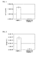
- the sampled cells were lysed in a solution (containing 3 M sodium chloride (Wako Pure Chemical Industries, Ltd.), 0.3 M trisodium citrate (Nacalai Tesque Inc.) and 200 mg/L sodium dodecyl sulfate (Nacalai Tesque Inc.) followed by measurement of the level of ALP activity using an alkaline phosphatase assay kit (LabAssayTM ALP, Wako Pure Chemical Industries, Ltd.). The value obtained by measuring absorbance at 405 nm with a microplate reader (VersaMax, Molecular Devices Corp.) was divided by the number of cells to determine the level of ALP activity per cell ( FIG. 1 ).
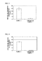
- the sampled cells were lysed in a solution (containing 3 M sodium chloride (Wako Pure Chemical Industries, Ltd.), 0.3 M trisodium citrate (Nacalai Tesque Inc.) and 200 mg/L sodium dodecyl sulfate (Nacalai Tesque Inc.) followed adding an equal amount of 1 M aqueous HCL solution (Nacalai Tesque Inc.) and allowing to stand undisturbed for 4 hours at 4°C, and measuring the amount calcium deposited using a calcium assay kit (Wako Calcium C-Test, Wako Pure Chemical Industries, Ltd.). The value obtained by measuring absorbance at 570 nm with a microplate reader (VersaMax, Molecular Devices Corp.) was divided by the number of cells to determine the amount of calcium deposited per cell ( FIG. 2 ).
- Sulfated polysaccharide present in the sampled cell supernatants were measured using a 1,9-dimethylmethylene blue (DMB) assay.
- the amount of sulfated polysaccharide present in 1 mL of culture supernatant was determined from a calibration curve obtained from values obtained by measuring absorbance at 525 nm with a microplate reader (VersaMax, Molecular Devices Corp.) using a solution obtained by dissolving chondroitin sulfate A sodium salt in medium containing 10% fetal bovine serum in ⁇ -MEM ( FIG. 3 ).
- heparin-Sepharose 5 ⁇ L aliquots of heparin-Sepharose (GE Healthcare Corp.) were completely dispersed and washed twice with 50 ⁇ L of 10 mM phosphate buffer followed by treating for 10 minutes by respectively adding 100 ⁇ L of a solution obtained by dissolving bFGF in 10 mM phosphate buffer and 100 ⁇ g/mL of 10 mM phosphate buffer. After removing the supernatant and washing twice with 200 ⁇ L of 10 mM phosphate buffer, the sampled culture supernatant was added followed by treating for 30 minutes and recovering the supernatant. The amount of sulfated polysaccharide present in recovered supernatant was measured using a 1, 9-dimethylmethylene blue (DMB) assay.
- DMB 1, 9-dimethylmethylene blue
- the amount of sulfated polysaccharide present in 1 mL of culture supernatant capable of binding to bFGF was determined by determining the amount of sulfated polysaccharide remaining in the supernatant from a calibration curve obtained from values obtained by measuring absorbance at 525 nm with a microplate reader (VersaMax, Molecular Devices Corp.) using a solution obtained by dissolving chondroitin sulfate A sodium in 10 mM phosphate buffer, and subtracting from the amount of sulfated polysaccharide present in the culture supernatant ( FIG. 4 ).
- Measurement was carried out in the same manner as measurement of sulfated polysaccharide capable of binding to bFGF with the exception of using a solution obtained by dissolving rhBMP-2 in 10 mM phosphate buffer to 100 ⁇ g/mL instead of a solution obtained by dissolving bFGF in 10 mM phosphate buffer to 100 ⁇ g/mL ( FIG. 5 ).
- MilliQ was respectively added to 100 ⁇ L of medium obtained by adding 10% fetal bovine serum to 100 ⁇ L of the culture supernatant described in Example 1 and ⁇ -MEM; or to 250 ⁇ L of medium obtained by adding 10% fetal bovine serum to 250 ⁇ L of the culture supernatant described in Comparative Example 1 and ⁇ -MEM; and brought to a total volume of 500 ⁇ L.
- the resulting solutions were subjected to centrifugal separation for 30 minutes at 14000G and 25°C using 0.5 mL of the Amicon UltraTM 10K ultrafiltration membrane (Millipore Corp.). 480 ⁇ L of MilliQ were again added followed by again centrifuging for 40 minutes at 14000G and 25°C.
- Sugar chains were recovered by purifying 20 ⁇ L aliquots of these solutions and a 20 ⁇ L aliquot of a 100 ⁇ M mixed aqueous solution of six types of disaccharides contained in an unsaturated heparan/heparin-disaccharide kit (Seikagaku Corp.) using the BlotGlycoTM sugar chain purification kit (Sumitomo Bakelite Co., Ltd.). 5 ⁇ L aliquots of the recovered sugar chain solutions were analyzed by high-performance liquid chromatography ((LaChrom Elite, Hitachi High-Technologies Co., Ltd.).
- a normal phase chromatography column (TSK-GEL, Amide-80, Tosoh Corp.) was used for the column, and 50 mM aqueous ammonium formate solution (pH 4.4) and acetonitrile were used for the mobile phase.
- the mobile phase was given a concentration gradient so that the 50 mM aqueous ammonium formate solution (pH 4.4) changed from 20% to 41.7% over the course of 80 minutes, and sugar chain solutions were detected by fluorescence (excitation wavelength: 330 nm, fluorescence wavelength: 420 nm) by applying at a flow rate of 0.4 mL/min and at a column temperature of 30°C.
- the area of the medium was subtracted from the area of the culture supernatant for the peak corresponding to the elution time of a reference disaccharide peak from the resulting HPLC charts.
- Each of the peak areas calculated in this manner was then divided by the total area of the six peaks and multiplied by 100 to calculate peak area (%) followed by a comparison of those values.
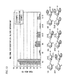
- diS1 was formed in large amounts in ordinary medium, while NS and diS2 were formed in large amounts in differentiation medium ( FIG. 6 ).
- Bone marrow aspirate collected from rat femur and tibia was disseminated in a culture flask, and after culturing at 37°C for 2 days in a 5% CO 2 incubator, the culture fluid was replaced and suspended cells were removed.
- the cells that adhered were further sub-cultured for 1 to 2 days at 37°C, and bone marrow-derived mesenchymal stem cells (MSC) obtained by culturing at 37°C until 70% to 90% confluent were suspended at 2.0 ⁇ 10 6 cells/mL in medium obtained by adding 1% fetal bovine serum to DMEM (high glucose), followed by the addition of 10 ng/mL rhTGF- ⁇ 3, 20 mM L-ascorbyl phosphate (Sigma Corp.), 100 nM dexamethasone (Nacalai Tesque Inc.), 40 ⁇ g/mL L-proline (Nacalai Tesque Inc.), 2mM of sodium pyruvate (Nacalai Tesque Inc.), 1 vol% ITS+ Premix (BD Biosciences Corp.) and 100 ng/mL rhBMP-2, and adding 0.5 mL aliquots to ten 15 mL centrifuge tubes to initiate induction of cartilage differentiation.
- the cells were sampled after 14 days and 21 days, while the culture supernatant was sampled after 4 days, 7 days, 11 days, 14 days, 18 days and 21 days.
- Bone marrow aspirate collected from rat femur and tibia was disseminated in a culture flask, and after culturing at 37°C for 2 days in a 5% CO 2 incubator, the medium was replaced and suspended cells were removed. The cells were further sub-cultured for 1 to 2 days at 37°C, and mesenchymal stem cells (MSC) obtained by culturing at 37°C until 70% to 90% confluent were disseminated at 5.0 ⁇ 10 4 cells/mL in a 6-well plate (Corning Inc.) containing medium obtained by adding 15% fetal bovine serum to ⁇ -MEM, followed by culturing for 2 days at 37°C in a 5% CO 2 incubator.
- MSC mesenchymal stem cells
- Bone differentiation was initiated by adding to medium containing 50 ⁇ g/mL ascorbic acid (Nacalai Tesque Inc.), 10 mM ⁇ -glycerophosphate (Sigma Corp.) and 10 -8 M dexamethasone (Nacalai Tesque Inc.). After culturing at 37°C in a 5% CO 2 incubator and replacing the medium once every 3 to 4 days, the cells were sampled after 10 days and 21 days, while the culture supernatant was sampled after 3 days, 7 days, 10 days, 14 days, 18 days and 21 days.
- Example 3 The procedure was carried out in the same manner as Example 3 with the exception of inducing bone differentiation by adding to the medium 50 ⁇ g/mL ascorbic acid (Nacalai Tesque Inc.), 10 mM ⁇ -glycerophosphate (Sigma Corp.), 10 -8 M dexamethasone (Nacalai Tesque Inc.) and 100 ng/mL rhBMP-2.
- hMSC Human mesenchymal stem cells
- PT-2501 Human mesenchymal stem cells
- STK2 mesenchymal stem cell serum-free medium
- STK3 mesenchymal stem cell bone differentiation serum-free medium
- hMSC Immortalized human mesenchymal stem cells
- Bone differentiation was initiated by adding to the medium 50 ⁇ g/mL ascorbic acid (Nacalai Tesque Inc.), 10 mM ⁇ -glycerophosphate (Sigma Corp.) and 10 -8 M dexamethasone (Nacalai Tesque Inc.). After culturing at 37°C in a 5% CO 2 incubator and replacing the medium once every 3 to 4 days, the cells were sampled after 10 days and 21 days, while the supernatant was sampled after 4 days, 8 days, 11 days, 14 days, 17 days and 21 days.
- ascorbic acid Nacalai Tesque Inc.
- 10 mM ⁇ -glycerophosphate Sigma Corp.
- 10 -8 M dexamethasone Nacalai Tesque Inc.
- Bone marrow-derived mesenchymal stem cells obtained in the same manner as Example 2 were suspended at 2.0 ⁇ 10 6 cells/mL in medium obtained by adding 1% fetal bovine serum to DMEM (high glucose), and 0.5 mL aliquots were added to ten 15 mL centrifuge tubes (BD Biosciences Corp.) followed by culturing at 37°C in a 5% CO 2 incubator. The medium was replaced once every 3 to 4 days, the cells were sampled after 14 days and 21 days, and the culture supernatant was sampled after 4 days, 7 days, 11 days, 14 days, 18 days and 21 days.
- MSC Bone marrow-derived mesenchymal stem cells
- MSC Mesenchymal stem cells obtained in the same manner as Example 3 were disseminated at 5.0 ⁇ 10 4 cells/mL in a 6-well plate (Corning Inc.) containing medium obtained by adding 15% fetal bovine serum to ⁇ -MEM, followed by culturing for 2 days at 37°C in a 5% CO 2 incubator. 50 ⁇ g/mL ascorbic acid (Nacalai Tesque Inc.) and 10 mM ⁇ -glycerophosphate (Sigma Corp.) were then added to the medium, and culturing was continued at 37°C in a 5% CO 2 incubator. The medium was replaced once every 3 to 4 days, the cells were sampled after 10 days and 21 days, and the culture supernatant was sampled after 3 days, 7 days, 10 days, 14 days, 18 days and 21 days.
- hMSC Human mesenchymal stem cells
- Immortalized human mesenchymal stem cells (hMSC) (huBM01-E6E7-hTERT clone 51, acquired from the Toguchida Laboratory) were disseminated at 5. 0 ⁇ 10 4 cells/mL into a 6-well plate (Corning Inc.) containing medium obtained by adding 10% fetal bovine serum to DMEM (low glucose), followed by incubating for 2 days at 37°C in a 5% CO 2 incubator. 50 ⁇ g/mL ascorbic acid (Nacalai Tesque Inc.) and 10 mM ⁇ -glycerophosphate (Sigma Corp.) were then added to the medium, and culturing was continued at 37°C in a 5% CO 2 incubator. The medium was replaced once every 3 to 4 days, the cells were sampled after 14 days and 21 days, and the culture supernatant was sampled after 4 days, 8 days, 11 days, 14 days, 17 days and 21 days.
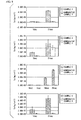
- the sampled cells were lysed in a solution (containing 3 M sodium chloride (Wako Pure Chemical Industries, Ltd.), 0.3 M trisodium citrate (Nacalai Tesque Inc.) and 200 mg/L sodium dodecyl sulfate (Nacalai Tesque Inc.) followed by measurement of the level of ALP activity using an alkaline phosphatase assay kit (LabAssayTM ALP, Wako Pure Chemical Industries, Ltd.). The value obtained by measuring absorbance at 405 nm with a microplate reader (VersaMax, Molecular Devices Corp.) was divided by the number of cells to determine the level of ALP activity per cell ( FIG. 7 ).
- the sampled cells were lysed in a solution (containing 3 M sodium chloride (Wako Pure Chemical Industries, Ltd.), 0.3 M trisodium citrate (Nacalai Tesque Inc.) and 200 mg/L sodium dodecyl sulfate (Nacalai Tesque Inc.) followed adding an equal amount of 1 M aqueous HCL solution (Nacalai Tesque Inc.) and allowing to stand undisturbed for 4 hours at 4°C, and measuring the amount of calcium deposited using a calcium assay kit (Wako Calcium C-Test, Wako Pure Chemical Industries, Ltd.). The value obtained by measuring absorbance at 600 nm with a microplate reader (VersaMax, Molecular Devices Corp.) was divided by the number of cells to determine the amount of calcium deposited per cell ( FIG. 8 ).
- Sulfated polysaccharide present in the sampled culture supernatants was measured using a 1,9-dimethylmethylene blue (DMB) assay.
- the amount of sulfated polysaccharide secreted per day present in 1 mL of culture supernatant was determined from a calibration curve obtained from values obtained by measuring absorbance at 525 nm with a microplate reader (VersaMax, Molecular Devices Corp.) using a solution obtained by dissolving chondroitin sulfate A sodium salt in the media used in each of the examples and comparative examples ( FIG. 9 ) .
- sampled cells were lysed by treating for 3 hours at 60°C in a solution (containing 300 ⁇ g/mL of papain (Wako Pure Chemical Industries, Ltd.), 2 mM DTT (Nacalai Tesque Inc.) and 200 mg/L sodium dodecyl sulfate (Nacalai Tesque Inc.) followed by measuring using a 1,9-dimethylmethylene blue (DMB) assay.
- papain Waako Pure Chemical Industries, Ltd.
- 2 mM DTT Nacalai Tesque Inc.
- sodium dodecyl sulfate Nacalai Tesque Inc.
- Sulfated polysaccharide present in the sampled culture supernatants was measured using a 1,9-dimethylmethylene blue (DMB) assay.
- the amount of sulfated polysaccharide secreted per day present in 1 mL of culture supernatant was determined from a calibration curve obtained from values obtained by measuring absorbance at 525 nm with a microplate reader (VersaMax, Molecular Devices Corp.) using a solution obtained by dissolving chondroitin sulfate A sodium in a medium obtained by adding 1% fetal bovine serum to DMEM (high glucose) ( FIG. 11 ).
- the amount of sulfated polysaccharide present in the recovered supernatant was measured using a 1,9-dimethylmethylene blue (DMB) assay.
- the amount of sulfated polysaccharide present in 1 mL of culture supernatant capable of binding to rhBMP-2 was determined by determining the amount of sulfated polysaccharide remaining in the supernatant from a calibration curve obtained from values obtained by measuring absorbance at 525 nm with a microplate reader (VersaMax, Molecular Devices Corp.) using a solution obtained by dissolving chondroitin sulfate A sodium salt in 10 mM phosphate buffer, and subtracting from the amount of sulfated polysaccharide present in the culture supernatant ( FIG. 12 ).
- MilliQ was respectively added to 100 ⁇ L of medium obtained by adding 1% fetal bovine serum to 100 ⁇ L of the culture supernatant described in Example 2, and 100 ⁇ L of DMEM (high glucose); and bringing to a total volume of 500 ⁇ L, or to 500 ⁇ L of medium obtained by adding 1% fetal bovine serum to 500 ⁇ L of the culture supernatant described in Comparative Example 2, and 500 ⁇ L of DMEM (high glucose), followed by subjecting to centrifugal separation for 30 minutes at 14000G and 25°C using 0.5 mL of the Amicon UltraTM 10K ultrafiltration membrane (Millipore Corp.).
- Sugar chains were recovered by purifying 20 ⁇ L aliquots of these solutions and a 20 ⁇ L aliquot of a 100 ⁇ M mixed aqueous solution of six types of disaccharides contained in an unsaturated heparan/heparin-disaccharide kit (Seikagaku Corp.) using the BlotGlycoTM sugar chain purification kit (Sumitomo Bakelite Co., Ltd.). 5 ⁇ L aliquots of the recovered sugar chain solutions were analyzed by high-performance liquid chromatography ((LaChrom Elite, Hitachi High-Technologies Co., Ltd.).
- a normal phase chromatography column (TSK-GEL, Amide-80, Tosoh Corp.) was used for the column, and 50 mM aqueous ammonium formate solution (pH 4.4) and acetonitrile were used for the mobile phase.
- the mobile phase was given a concentration gradient so that the 50 mM aqueous ammonium formate solution (pH 4.4) changed from 20% to 41.7% over the course of 80 minutes, and sugar chain solutions were detected by fluorescence (excitation wavelength: 330 nm, fluorescence wavelength: 420 nm) by applying at a flow rate of 0.4 mL/min and at a column temperature of 30°C.
- the area of the medium was subtracted from the area of the culture supernatant for the peak corresponding to the elution time of a reference disaccharide peak from the resulting HPLC charts, and each of the peak areas was then divided by the total area of the six peaks and multiplied by 100 to calculate peak area (%) followed by a comparison of those values ( FIG. 13 ).
- the composition of the present invention having this sulfated polysaccharide of the present invention as an active ingredient thereof can not only be used as a reagent for promoting cell proliferation and differentiation, but can also be used as a pharmaceutical for preventing or treating damaged cells, tissues or organs and diseases caused by abnormal cell proliferation and differentiation, and as a food or beverage such as a health food or beverage for improving cell proliferation and differentiation.
- the degree of cell proliferation and differentiation in vivo or in vitro can be evaluated by targeting the sulfated polysaccharide that binds to growth factor discovered by the inventors of the present invention.
- a composition having this sulfated polysaccharide as an active ingredient thereof can be used as a diagnostic reagent for detecting cell proliferation and differentiation. The detection of this sulfated polysaccharide in a patient undergoing treatment for the purpose of regenerative medicine makes it possible to evaluate the effects of that treatment, thereby enabling the present invention to be used in the field of regenerative medicine as well.
Landscapes
- Health & Medical Sciences (AREA)
- Life Sciences & Earth Sciences (AREA)
- Chemical & Material Sciences (AREA)
- Engineering & Computer Science (AREA)
- General Health & Medical Sciences (AREA)
- Organic Chemistry (AREA)
- Molecular Biology (AREA)
- Medicinal Chemistry (AREA)
- Chemical Kinetics & Catalysis (AREA)
- Biochemistry (AREA)
- Polymers & Plastics (AREA)
- Materials Engineering (AREA)
- Wood Science & Technology (AREA)
- Biomedical Technology (AREA)
- Veterinary Medicine (AREA)
- Animal Behavior & Ethology (AREA)
- Pharmacology & Pharmacy (AREA)
- Urology & Nephrology (AREA)
- General Chemical & Material Sciences (AREA)
- Immunology (AREA)
- Biotechnology (AREA)
- Microbiology (AREA)
- Bioinformatics & Cheminformatics (AREA)
- Zoology (AREA)
- Hematology (AREA)
- Public Health (AREA)
- Cell Biology (AREA)
- Epidemiology (AREA)
- Sustainable Development (AREA)
- Genetics & Genomics (AREA)
- General Engineering & Computer Science (AREA)
- Endocrinology (AREA)
- Nuclear Medicine, Radiotherapy & Molecular Imaging (AREA)
- Food Science & Technology (AREA)
- Physics & Mathematics (AREA)
- Analytical Chemistry (AREA)
- General Physics & Mathematics (AREA)
- Pathology (AREA)
- Dermatology (AREA)
- Physical Education & Sports Medicine (AREA)
Abstract
The present invention provides: a biological substance which can enhance the proliferation and differentiation of cells; a method for evaluating the proliferation and differentiation of cells, which targets the biological substance; a composition for detecting the proliferation and differentiation of cells, which contains a molecule capable of binding to the biological substance as an active ingredient; and a composition for enhancing the proliferation and differentiation of cells, which contains the biological substance as an active ingredient. According to the present invention, it is found that a part of a sulfated polysaccharide secreted from cells binds to a growth factor during the course of the proliferation and differentiation of the cells, that the amount of the sulfated polysaccharide capable of the above-mentioned binding and secreted from the cells correlates strongly with the progression of the proliferation and differentiation of the cells, and that the proliferation and differentiation of cells can be evaluated and the proliferation and differentiation of cells can be enhanced utilizing the sulfated polysaccharide.
Description
- The present invention relates to a sulfated polysaccharide capable of binding to growth factor that is secreted from cells during the course of cell proliferation and differentiation, a method for evaluating cell proliferation and differentiation that targets the sulfated polysaccharide, a composition for detecting cell proliferation and differentiation having a molecule that binds to the sulfated polysaccharide as an active ingredient thereof, and a composition for promoting cell proliferation and differentiation having the sulfated polysaccharide as an active ingredient thereof.
The present application claims priority on the basis of Japanese Patent Application No.2010-196583 2011-071661 - Stem cell transplantation is typically carried out in the field of regenerative medicine as a treatment method that has been researched and developed and partially applied practically. Although embryonic stem cells, mesenchymal stem cells harvested from bone marrow or fat tissue and the like, hematopoietic stem cells and various types of tissue stem cells (such as hepatic stem cells or neural stem cells) are used as stem cells for cell transplantation, mesenchymal stem cells can be acquired and cultured comparatively easily. Consequently, research on cell transplantation has progressed the greatest in the field of biological tissue regeneration using mesenchymal stem cells.
- Examples of tissues targeted for regenerative medicine using mesenchymal stem cells include bone, cartilage, fat, muscle, tendon and nerves. In the case of bone regeneration, for example, a method is used that consists of disseminating and proliferating mesenchymal stem cells that have been cultured and proliferated in vitro in a three-dimensional scaffold, followed by transplantation and treatment, and there are cases in which this treatment has been applied (see, for example, Patent Document 1).
- Normally, a polysaccharide proliferation factor must be continued to be added to the culture fluid in order to culture mesenchymal stem cells in vitro. Examples of approaches that have been developed to accomplish this include a method consisting of synthesizing proliferation factor in cells and continuously supplying the proliferation factor autocrinically, and a method that enables favorable bone and cartilage regeneration by continuously expressing transcription factor activity using a viral vector incorporating a transcription factor gene (see, for example, Patent Document 2).
However, since the body inherently possesses its own natural healing power, treatment is actually provided using that natural healing power in the case of simple fractures and the like. On the other hand, although there are cases, such as in the case of large bone defects, for which the body is unable to adequately demonstrate its inherent natural healing power, by enhancing that natural healing power by administering substances that promote cell proliferation and differentiation, for example, it is possible to realize regeneration and repair of body tissue without having to undergo stem cell transplantation or gene therapy. In addition, in the case a substance having such action is a substance that is present in the body, it is also thought to be possible to evaluate the status of cell proliferation and differentiation by using that substance as a target. - On the basis of such viewpoints, a substance present in the body is sought that is able to promote cell proliferation and differentiation and enhance the natural healing power inherently possessed by the body.
-
- Patent Document 1: Japanese Unexamined Patent Application, First Publication No.
2008-29317 - Patent Document 2: International Publication No.
WO 2005/035014 - With the foregoing in view, an object of the present invention is to provide a biological substance that is capable of promoting cell proliferation and differentiation. Another object of the present invention is to provide a method for evaluating cell proliferation and differentiation that uses the biological substance as a target, a composition for detecting cell proliferation and differentiation that has a molecule that binds to the biological substance as an active ingredient thereof, and a composition for promoting cell proliferation and differentiation that has the biological substance as an active ingredient thereof.
- As a result of conductive extensive studies to achieve the aforementioned objects, the inventors of the present invention found that a portion of a sulfated polysaccharide secreted from cells binds to growth factor during the course of cell proliferation and differentiation, and that the amount of the sulfated polysaccharide secreted from cells that exhibits this binding activity demonstrates a strong correlation with the degree of progression of cell proliferation and differentiation. Moreover, the inventors of the present invention found that cell proliferation and differentiation can be evaluated and cell proliferation and differentiation can be promoted by using this sulfated polysaccharide that binds to growth factor, thereby leading to completion of the present invention.
- More specifically, the present invention provides the inventions indicated below.
- (1) A sulfated polysaccharide secreted from cells during the course of cell proliferation and differentiation that binds to growth factor.
- (2) The sulfated polysaccharide described in (1), wherein the course of cell proliferation and differentiation is the course of proliferation and differentiation of osteocytes or chondrocytes.
- (3) The sulfated polysaccharide described in (1) or (2), wherein the growth factor is a molecule selected from the group consisting of BMP-2 and bFGF.
- (4) A method for evaluating cell proliferation and differentiation, comprising detecting the sulfated polysaccharide described in any of (1) to (3).
- (5) A composition for detecting cell proliferation and differentiation having as an active ingredient thereof a molecule that binds to the sulfated polysaccharide described in any of (1) to (3).
- (6) A composition for promoting cell proliferation and differentiation having as an active ingredient thereof the sulfated polysaccharide described in any of (1) to (3).
- Since the sulfated polysaccharide discovered by the inventors of the present invention that binds to growth factor is able to stabilize or enhance the activity of growth factor, the use of this sulfated polysaccharide makes it possible to promote cell proliferation and differentiation that uses the body's natural healing power in vivo, as well as promote cell proliferation and differentiation in vitro. In addition, the degree of progression of cell proliferation and differentiation in vivo and the degree of progression of cell proliferation and differentiation in vitro can be evaluated by targeting the sulfated polysaccharide that binds to growth factor discovered by the inventors of the present invention.
-
-
FIG. 1 is a graph indicating the results of measuring the level of alkaline phosphatase (ALP) activity of osteoprogenitor cells cultured in bone differentiation medium (Example 1) and osteoprogenitor cells cultured in ordinary medium (Comparative Example 1). -
FIG. 2 is a graph indicating the results of measuring the amount of calcium deposited by osteoprogenitor cells cultured in bone differentiation medium (Example 1) and osteoprogenitor cells cultured in ordinary medium (Comparative Example 1). -
FIG. 3 is a graph indicating the results of measuring the amount of sulfated polysaccharide secreted from osteoprogenitor cells cultured in bone differentiation medium (Example 1) and osteoprogenitor cells cultured in ordinary medium (Comparative Example 1). -
FIG. 4 is a graph indicating the results of measuring the amount of sulfated polysaccharide capable of binding to bFGF secreted from osteoprogenitor cells cultured in bone differentiation medium (Example 1) and osteoprogenitor cells cultured in ordinary medium (Comparative Example 1). -
FIG. 5 is a graph indicating the results of measuring the amount of sulfated polysaccharide capable of binding to BMP-2 secreted from osteoprogenitor cells cultured in bone differentiation medium (Example 1) and osteoprogenitor cells cultured in ordinary medium (Comparative Example 1). -
FIG. 6 is a graph indicating the results of analyzing the structures of sulfated polysaccharides secreted from osteoprogenitor cells cultured in bone differentiation medium (Example 1) and osteoprogenitor cells cultured in ordinary medium (Comparative Example 1). -
FIG. 7 depicts graphs indicating the results of measuring the levels of alkaline phosphatase (ALP) activity of mesenchymal stem cells cultured in bone differentiation medium (Examples 3 to 6) and mesenchymal stem cells cultured in ordinary medium (Comparative Examples). -
FIG. 8 depicts graphs indicating the results of measuring the amounts of calcium deposited by mesenchymal stem cells cultured in bone differentiation medium (Examples 3 to 6) and mesenchymal stem cells cultured in ordinary medium (Comparative Examples). -
FIG. 9 depicts graphs indicating the results of measuring the amounts of sulfated polysaccharide excreted from mesenchymal stem cells cultured in bone differentiation medium (Examples 3 to 6) and mesenchymal stem cells cultured in ordinary medium (Comparative Examples). -
FIG. 10 is a graph indicating the results of measuring the amount of sulfated GAG produced in a cell extract during cartilage differentiation (Example 2). -
FIG. 11 is a graph indicating the results of measuring the amount of sulfated polysaccharide in culture supernatant during cartilage differentiation (Example 2). -
FIG. 12 is a graph indicating the results of measuring the amount of sulfated polysaccharide capable of binding to BMP-2 in culture supernatant during cartilage differentiation (Example 2). -
FIG. 13 is a graph indicating the results of analyzing the structure of sulfated polysaccharides secreted from mesenchymal stem cells cultured in cartilage differentiation medium (Example 2) and chondrocytes cultured in ordinary medium (Comparative Example 2). - The present invention provides a sulfated polysaccharide secreted from cells during the course of cell proliferation and differentiation that binds to growth factor. Moreover, the present invention provides a method for evaluating cell proliferation and differentiation comprising detection of that sulfated polysaccharide.
- Examples of the "course of cell proliferation and differentiation" in the present invention include, but are not limited to, the course of proliferation and differentiation of osteocytes, chondrocytes, myocytes, adipocytes, hepatocytes, neurons and hemocytes. Culture systems that cause various types of stem cells to differentiate into cells of various tissues can be used for culture systems for proliferation and differentiation of these cells. Examples of stem cells include, but are not limited to, embryonic stem cells, mesenchymal stem cells harvested from bone marrow or fat tissue and the like, hematopoietic stem cells, and various types of tissue stem cells (such as hepatic stem cells or neural stem cells). In addition, examples of biological sources of cells for inducing proliferation and differentiation include mice, rats, hamsters, humans, monkeys, pigs, cows, horses, sheep, goats, chickens, wild ducks, ostriches, domestic ducks, dogs, cats and rabbits.
- A method commonly known among persons with ordinary skill in the art can be used to proliferate and differentiate cells. An example of a method used to proliferate and differentiate osteocytes consists of disseminating cells able to differentiate into osteocytes, and culturing for 2 to 4 days in an ordinary medium such as medium to which 10% to 15% of fetal bovine serum has been added to a minimum essential medium such as alpha-Modified Eagle Minimum Essential Medium (α-MEM) or Dulbecco's Modified Eagle Minimum Essential Medium (DMEM). Here, examples of cells able to differentiate into osteocytes include osteoblasts, osteoprogenitor cells, mesenchymal cells (including mesenchymal stem cells) and cell lines thereof (such as MC3T3-E1 or CH310T1/2). Subsequently, the medium is replaced with differentiation medium such as a medium obtained by adding β-glycerophosphate, ascorbic acid (or a derivative thereof in the form of ascorbyl-2-phosphate) and dexamethasone to the aforementioned ordinary medium followed by culturing for 21 days to 28 days. During culturing in the differentiation medium, for example, conditions consisting of culturing in gaseous 5% CO2 at 37°C can be applied while replacing the medium every 3 to 4 days. One or more types of factors known to induce differentiation of mesenchymal stem cells to osteoblasts (such as BMP-2, BMP-3, BMP-4, BMP-7, bFGF or TGF-β1) can also be added to the differentiation medium.
- Differentiation to osteocytes can be detected with, for example, alkaline phosphatase activity. In addition, this differentiation can also be detected by targeting calcium deposition. A commercially available kit, such as an alkaline phosphatase assay kit (LabAssay™ ALP, Wako Pure Chemical Industries, Ltd.) or a calcium assay kit (Wako Calcium C-Test, Wako Pure Chemical Industries, Ltd.), can be used to detect these parameters.
- A method described in the literature (such as Alastair M. Mackay et al., Tissue Engineering Vol. 4 (1998), pp. 415-428) can be used to proliferate and differentiate chondrocytes. In this method, after separating cultured human mesenchymal stem cells with trypsin and dispersing into individual cells in a centrifuge tube, the cells are dispersed in differentiation medium obtained by adding a reagent (consisting of 10 µg/mL TGF-β3, 100 nM dexamethasone, 50 µg/mL ascorbic acid, 100 µg/mL sodium pyruvate, 40 µg/mL proline, 6.25 µg/mL bovine insulin, 6.25 µg/mL transferrin, 5.33 µg/mL linoleic acid and 1.25 mg/mL bovine serum albumin) to Dulbecco's modified Eagle medium (DMEM), followed by producing a cell pellet by centrifuging the cells, and then culturing for 14 days as is to cause the cells to differentiate into cartilage. Confirmation of differentiation into cartilage can be made by staining each type of extracellular matrix with antibody.
- A method described in the literature (such as Keiichi Fukuda, Artificial Organs Vol. 25 (2001), pp. 187-193) can be used to proliferate and differentiate cardiac myocytes. In this method, human mesenchymal cells are differentiated into cardiac muscle by culturing for 24 hours or more in differentiation medium obtained by adding 3 µM 5-azacytidine to Iscove's modified DMEM containing 20% fetal bovine serum. Differentiation into cardiac muscle can be confirmed by microscopically observing spontaneous pulsation of the cells.
- A method described in the literature (such as Eun Ji Gang et al., Stem Cells 22 (2004), pp. 617-624) can be used to proliferate and differentiate skeletal myocytes. In this method, human mesenchymal stem cells are differentiated into skeletal muscle by culturing for 6 weeks in differentiation medium obtained by adding a reagent (consisting of 5% horse serum, 0.1 µM dexamethasone and 50 µM hydrocortisone) to DMEM containing 15% fetal bovine serum. Differentiation into skeletal muscle can be detected by fluorescent staining of MyoD1 characteristic to skeletal myocytes.
- A method described in the literature (such as Kentaro Nagane et al., Tissue Engineering: Part A Vol. 16 (2010), pp. 21-31) can be used to proliferate and differentiate adipocytes. In this method, rat mesenchymal stem cells are differentiated into adipocytes by culturing for 6 days in a differentiation medium obtained by adding a reagent (consisting of 450 µM 3-isobutyl-1-methylxanthine, 50 µg/mL insulin, 60 µM indometacin and 10 µM troglitazone) to α-MEM containing 10% calf serum. Differentiation into adipocytes can be detected by staining with Oil Red or measuring GPDH enzyme activity.
- A method described in the literature (such as Agnieszka Banas et al., Hepatology July (2007), pp. 219-228) can be used to proliferate and differentiate hepatocytes. In this method, human mesenchymal stem cells are differentiated into hepatic parenchymal cells by culturing for 3 weeks in a differentiation medium obtained by adding a reagent (consisting of 300 ng/mL FGF1, 25 ng/mL FGF4 and 150 ng/mL HGF) to DMEM containing 10% fetal bovine serum, and further culturing for 2 weeks in a medium obtained by adding a reagent (consisting of 30 ng/mL oncostatin M and 2 × 10⁻⁵ M dexamethasone) to the differentiation medium. Differentiation into hepatic parenchymal cells can be detected by measuring albumin synthesis ability or ammonium metabolism of the cells.
- A method described in the literature (such as Ju Ah Jeong, et al., NeuroReport Vol. 15 (2004), pp. 1731-1734) can be used to proliferate and differentiate nerve cells. In this method, human mesenchymal stem cells are differentiated into nerve cells by culturing for 24 hours in a medium obtained by adding a reagent (consisting of 25 mM potassium chloride, 2% dimethylsulfoxide (DMSO), 100 µM butylated hydroxyanisole, 5µg/mL insulin, 100 µg/mL transferrin, 20 mM progesterone, 100 µM putrescine, 30 nM sodium selenite, 20 ng/mL bFGF and 10 mM Forskolin) to DMEM containing 20% fetal bovine serum. Differentiation into nerve cells can be detected by measuring the expression of a group of genes characteristic to nerve cells.
- In addition, in the present invention, a "sulfated polysaccharide" refers to a polysaccharide having a sulfate group in a molecule thereof. There are no particular restrictions on the sulfated polysaccharide in the present invention provided it is secreted during the course of cell proliferation and differentiation and is able to bind to growth factor. Preferably, it is a sulfated polysaccharide that contains NS and diS2 structures as shown in
FIG. 6 at a higher ratio than control values and contains a diS1 structure as shown inFIG. 6 at a lower ratio than a control value. Values of the ratios of NS, diS2 and diS1 structures in a sulfated polysaccharide secreted by cells not undergoing cell proliferation and differentiation (such as values of the ratios of NS, diS2 and diS1 structures in the sulfated polysaccharide in the case of culturing cells able to differentiate into osteocytes in an ordinary medium that does not induce differentiation in the case of detecting proliferation and differentiation of osteocytes) can be used for the control values. - In addition, in the present invention, "growth factor" refers to an endogenous protein that promotes proliferation and differentiation of specific cells. Examples of "growth factors" bound by the sulfated polysaccharide of the present invention include, but are not limited to, BMP-2 and bFGF.
- The sulfated polysaccharide that binds to growth factor can be prepared by contacting a cell culture supernatant with a carrier (such as heparin-Sepharose) having a growth factor or fragment thereof bound thereon, and then recovering the sulfated polysaccharide that binds to the growth factor on the carrier.
- Detection of sulfated polysaccharide can be carried out with, for example, a 1,9-dimethylmethylene blue (DMB) assay. Detection of sulfated polysaccharide can also be carried out using a molecule that specifically binds to the sulfated polysaccharide (such as an antibody that binds to the sulfated polysaccharide or a peptide of that binding site, or a growth factor that binds to the sulfated polysaccharide or a peptide of that binding site).
- Antibody can be acquired by immunizing an immune animal with antigen (sulfated polysaccharide that binds to growth factor) and purifying from the resulting antiserum by conventional means (such as salting out, centrifugal separation, dialysis or column chromatography) in the case of polyclonal antibody. In addition, monoclonal antibody can be produced according to a hybridoma method (described in, for example, Kohler & Milstein, Nature, 256:495(1975)) or recombinant DNA method (described in, for example, P.J. Delves, Antibody Production: Essential Techniques, 1997, Wiley; P. Shepherd and C. Dean, Monoclonal Antibodies, 2000 Oxford University Press; and Vandamme A.M. et al., Eur. J. Biochem. 192:767-775(1990)). These antibodies may be labeled as necessary. Radioactive substances, fluorescent pigments, chemoluminescent substances, enzymes or coenzymes can be used for the label, and specific examples thereof include radioisotopes, fluorescein, rhodamine, dansyl chloride, luciferase, peroxidase, alkaline phosphatase, lysozyme and biotin/avidin. When formulating the antibody in the form of a composition such as a diagnostic agent, the antibody can be obtained in an arbitrary preparation form by employing arbitrary means in accordance with the objective thereof. For example, a purified antibody can be prepared by measuring the antibody titer and adding a suitable carrier (to be subsequently described).
- A growth factor or a peptide of that binding site can be prepared by incorporating DNA encoding the same into a suitable expression vector, recovering a transformant obtained by introducing this into suitable host cells, and obtaining an extract of the transformant, followed by purifying by applying to ion exchange, reverse phase or gel filtration chromatography, or to affinity chromatography in which a sulfated polysaccharide or antibody bound to the growth factor is immobilized on a column, or by combining a plurality of these purification means. The growth factor or peptide of that binding site may also be labeled as necessary in the same manner as in the case of the aforementioned antibody.
- The present invention also provides a composition for detecting cell proliferation and differentiation that has as an active ingredient thereof a molecule that binds to this type of sulfated polysaccharide.
- As a result of having detected a sulfated polysaccharide using the composition of the present invention, the process of cell proliferation and differentiation can be evaluated as proceeding in the case the sulfated polysaccharide is detected in an amount that is greater than a control value. For example, the value of the secreted amount of the sulfated polysaccharide in cells in which cell proliferation and differentiation is not proceeding (such as the value of the secreted amount of the sulfated polysaccharide in the case of culturing cells able to differentiate into osteocytes in ordinary medium that does not induce differentiation in the case of detecting proliferation and differentiation of osteocytes) can be used as a control value. In addition, the process of cell proliferation and differentiation can be evaluated as proceeding in the case the NS and diS2 structures shown in
FIG. 6 are detected at a higher ratio than a control value, or in the case the diS1 structure shown inFIG. 6 is detected at a lower ratio than a control value, in the secreted sulfated polysaccharide. For example, values of the ratios of NS, diS2 and diS1 structures in a sulfated polysaccharide secreted by cells not undergoing cell proliferation and differentiation (such as values of the ratios of NS, diS2 and diS1 structures in the sulfated polysaccharide in the case of culturing cells able to differentiate into osteocytes in an ordinary medium that does not induce differentiation in the case of detecting proliferation and differentiation of osteocytes) can be used for the control values. This composition of the present invention can be used as an experimental reagent for evaluating the degree of cell proliferation and differentiation in an experiment using culture cells, or for evaluating the degree of cell proliferation and differentiation in an animal experiment. In addition, the composition of the present invention can be used as a diagnostic drug for evaluating the degree of cell proliferation and differentiation for the purpose of accurately diagnosing the progression of treatment with respect to regenerative medicine in a human body, for example. - The present invention also provides a composition for promoting osteocyte proliferation and differentiation that has as an active ingredient thereof the sulfated polysaccharide that binds to growth factor. This composition of the present invention can be in the form of a reagent used in pharmaceutical compositions, foods and beverages (including animal feed) or for research and development purposes (such as in vitro and in vivo experiments or the production of artificial tissues and organs such as artificial bone).
- The composition of the present invention can be preferably used as a pharmaceutical composition administered to prevent or treat diseases caused by damage to cells, tissues or organs or by abnormal cell proliferation and differentiation, or as a food or beverage consumed on a regular basis, in order to have an action that promotes cell proliferation and differentiation by stabilizing or enhancing growth factor activity.
- The composition of the present invention can be formulated according to commonly known pharmaceutical techniques. For example, the composition of the present invention can be used orally or parenterally in the form of a capsule, tablet, pill, liquid, powder, granules, grains, film-coated preparation, pellet, lozenge, sublingual preparation, chewable preparation, buccal preparation, paste, syrup, suspension, elixir, emulsion, liniment, ointment, plaster, poultice, transcutaneously absorbed preparation, lotion, inhalant, aerosol, injection or suppository.
- In a preparation of the composition of the present invention, the composition of the present invention can be suitably combined with a pharmaceutically acceptable carrier or carrier permitted to be used in a food or beverage, specific examples of which include sterile water or physiological saline, vegetable oils, solvents, bases, emulsifiers, suspending agents, surfactants, stabilizers, flavoring agents, fragrances, excipients, vehicles, preservatives, binders, diluents, tonicity agents, analgesics, extenders, disintegration agents, buffers, coating agents, lubricants, colorants, sweeteners, thickeners, correctives, solubilizing agents, and other additives.
- In the case of using the composition of the present invention as a pharmaceutical composition, it may be used in combination with a known pharmaceutical composition used for the treatment and prevention of diseases caused by damage to cells, tissues or organs or by abnormal cell proliferation and differentiation.
- In the case of using the composition of the present invention as a food or beverage, the food or beverage can be a health food, functional food, food for specified health uses, nutritional supplement, medical food, food additives or animal feed. A food or beverage of the present invention can be ingested as a composition as previously described, or can be ingested in the form of various foods and beverages. Specific examples of foods and beverages include products containing oil such as edible oils, salad dressing, mayonnaise or margarine; liquid foods such as a soup, milk drinks, soft drinks, tea drinks, alcoholic beverages, drinks, jellied beverages or functional beverages; carbohydrate-containing foods such as rice, noodles or bread: processed animal foods such as ham or sausage; processed marine products such as pressed fish, dried fish or salted squid; processed vegetable foods such as pickles; semi-solid foods such as jelly or yogurt; fermented foods such as fermented beverages; various types of confections such as Western confections, Japanese confections, candy, gum, gummi drops, chilled confections or frozen confections; boil-in-the-bag foods such as curry sauce, sweet bean sauce or Chinese style soup; and instant foods and microwaved foods such as instant soup or instant miso soup. Moreover, additional examples include health foods and beverages prepared in the form of powders, granules, tablets, capsules, liquids, pastes or jelly. Although the composition of the present invention can be used for animals, including humans, it can be used in animals other than humans such as various species of livestock, poultry, pets or experimental animals. Specific examples include, but are not limited to, pigs, cows, horses, sheep, goats, chickens, wild ducks, ostriches, domestic ducks, dogs, cats, rabbits, hamsters, mice, rats and monkeys.
- A food or beverage of the present invention can be produced using known production technology employed in the relevant field. One type or two or more types of active ingredients may be added to the food or beverage in order to promote cell proliferation and differentiation. In addition, the food or beverage may also be in the form of a functional food or beverage by combining with other components or other functional foods that demonstrate a function other than the promotion of cell proliferation and differentiation.
- In the case of administering or ingesting the composition of the present invention, the dosage or ingested amount thereof is suitably selected corresponding to the age, body weight, symptoms or health status of a subject or the type of composition (such as a pharmaceutical, food or beverage). For example, the dosage or ingested amount of the composition of the present invention per administration is typically 0.01 mg/kg of body weight to 100 mg/kg of body weight. In this manner, the present invention also provides a method for promoting cell proliferation and differentiation in a subject by administration of the composition of the present invention to the subject or ingestion of the composition of the present invention by the subject. In addition, the present invention also provides a method for preventing or treating a disease caused by damage to cells, tissues or organs or caused by abnormal cell proliferation and differentiation in a subject by administering the composition of the present invention to the subject.
- A product of the composition of the present invention (pharmaceutical, food, beverage or reagent) or the instructions thereof may be provided with an indication to the effect that the product is used to promote cell proliferation and differentiation. Here, "providing an indication on a product or instructions" refers to the providing of an indication on the body, container or package of a product, or providing an indication in instructions, inserts, advertisements or other printed matter that discloses information about the product. In an indication to the effect that the composition of the present invention is used to promote cell proliferation of differentiation, information can be included regarding the mechanism by which cell proliferation and differentiation are promoted as a result of administering or ingesting the composition of the present invention. In addition, in an indication to the effect that the composition of the present invention is used to promote cell proliferation and differentiation, information can be included regarding the use thereof for the prevention or treatment of diseases caused by damage to cells, tissues or organs or by abnormal cell proliferation and differentiation.
- Although the following provides a more detailed explanation of the present invention based on examples and comparative examples, the present invention is not limited to the following examples.
- MC3T3-E1 mouse-derived osteoprogenitor cells were respectively disseminated in three 10 cm culture dishes (Corning Inc.) containing medium obtained by adding 10% fetal bovine serum to α-MEM to 3.0 × 10⁵ cells per culture dish followed by culturing for 2 days at 37°C in a 5% CO2 incubator. 50 µg/mL ascorbic acid (Nacalai Tesque Inc.), 10 mM β-glycerophosphate (Sigma Corp.) 10-8 M dexamethasone (Nacalai Tesque Inc.) and 100 ng/ml rhBMP-2 were added to the medium to initiate induction of differentiation. The medium was replaced every 3 to 4 days, and the cells and supernatant were sampled after culturing for 1 week at 37°C in a 5% CO2 incubator.
- MC3T3-E1 mouse-derived osteoprogenitor cells were respectively disseminated in three 10 cm culture dishes (Corning Inc.) containing medium obtained by adding 10% fetal bovine serum to α-MEM to 3.0 × 10⁵ cells per culture dish followed by culturing for 2 days at 37°C in a 5% CO2 incubator. The same medium was replaced every 3 to 4 days, and the cells and supernatant were sampled after culturing for 1 week at 37°C in a 5% CO2 incubator.
- The sampled cells were lysed in a solution (containing 3 M sodium chloride (Wako Pure Chemical Industries, Ltd.), 0.3 M trisodium citrate (Nacalai Tesque Inc.) and 200 mg/L sodium dodecyl sulfate (Nacalai Tesque Inc.) followed by measurement of the level of ALP activity using an alkaline phosphatase assay kit (LabAssay™ ALP, Wako Pure Chemical Industries, Ltd.). The value obtained by measuring absorbance at 405 nm with a microplate reader (VersaMax, Molecular Devices Corp.) was divided by the number of cells to determine the level of ALP activity per cell (
FIG. 1 ). - The sampled cells were lysed in a solution (containing 3 M sodium chloride (Wako Pure Chemical Industries, Ltd.), 0.3 M trisodium citrate (Nacalai Tesque Inc.) and 200 mg/L sodium dodecyl sulfate (Nacalai Tesque Inc.) followed adding an equal amount of 1 M aqueous HCL solution (Nacalai Tesque Inc.) and allowing to stand undisturbed for 4 hours at 4°C, and measuring the amount calcium deposited using a calcium assay kit (Wako Calcium C-Test, Wako Pure Chemical Industries, Ltd.). The value obtained by measuring absorbance at 570 nm with a microplate reader (VersaMax, Molecular Devices Corp.) was divided by the number of cells to determine the amount of calcium deposited per cell (
FIG. 2 ). - Sulfated polysaccharide present in the sampled cell supernatants were measured using a 1,9-dimethylmethylene blue (DMB) assay. The amount of sulfated polysaccharide present in 1 mL of culture supernatant was determined from a calibration curve obtained from values obtained by measuring absorbance at 525 nm with a microplate reader (VersaMax, Molecular Devices Corp.) using a solution obtained by dissolving chondroitin sulfate A sodium salt in medium containing 10% fetal bovine serum in α-MEM (
FIG. 3 ). - 5 µL aliquots of heparin-Sepharose (GE Healthcare Corp.) were completely dispersed and washed twice with 50 µL of 10 mM phosphate buffer followed by treating for 10 minutes by respectively adding 100 µL of a solution obtained by dissolving bFGF in 10 mM phosphate buffer and 100 µg/mL of 10 mM phosphate buffer. After removing the supernatant and washing twice with 200 µL of 10 mM phosphate buffer, the sampled culture supernatant was added followed by treating for 30 minutes and recovering the supernatant. The amount of sulfated polysaccharide present in recovered supernatant was measured using a 1, 9-dimethylmethylene blue (DMB) assay. The amount of sulfated polysaccharide present in 1 mL of culture supernatant capable of binding to bFGF was determined by determining the amount of sulfated polysaccharide remaining in the supernatant from a calibration curve obtained from values obtained by measuring absorbance at 525 nm with a microplate reader (VersaMax, Molecular Devices Corp.) using a solution obtained by dissolving chondroitin sulfate A sodium in 10 mM phosphate buffer, and subtracting from the amount of sulfated polysaccharide present in the culture supernatant (
FIG. 4 ). - Measurement was carried out in the same manner as measurement of sulfated polysaccharide capable of binding to bFGF with the exception of using a solution obtained by dissolving rhBMP-2 in 10 mM phosphate buffer to 100 µg/mL instead of a solution obtained by dissolving bFGF in 10 mM phosphate buffer to 100 µg/mL (
FIG. 5 ). - MilliQ was respectively added to 100 µL of medium obtained by adding 10% fetal bovine serum to 100 µL of the culture supernatant described in Example 1 and α-MEM; or to 250 µL of medium obtained by adding 10% fetal bovine serum to 250 µL of the culture supernatant described in Comparative Example 1 and α-MEM; and brought to a total volume of 500 µL. The resulting solutions were subjected to centrifugal separation for 30 minutes at 14000G and 25°C using 0.5 mL of the Amicon Ultra™ 10K ultrafiltration membrane (Millipore Corp.). 480 µL of MilliQ were again added followed by again centrifuging for 40 minutes at 14000G and 25°C. Subsequently, 10 µL aliquots were sampled from 25 µL aliquots of the recovered solutions, followed by the addition of 6 µL of digestion buffer (containing 10 mM sodium acetate trihydrate (Nacalai Tesque Inc.) and 10 mM potassium acetate hydrate (Nacalai Tesque Inc.), pH = 7.0), 5 µL of a solution obtained by dissolving heparinase (Seikagaku Corp.) in 0.1% aqueous BSA (Equitech-Bio Inc.) solution to a concentration of 0.5 U/mL, and 5 µL of a solution obtained by dissolving heparitinase (Seikagaku Corp.) in 0.1% aqueous BSA (Equitech-Bio Inc.) solution to a concentration of 0.5 U/mL, further adding MilliQ to a total volume of 30 µL and incubating for 1 hour at 37°C. Sugar chains were recovered by purifying 20 µL aliquots of these solutions and a 20 µL aliquot of a 100 µM mixed aqueous solution of six types of disaccharides contained in an unsaturated heparan/heparin-disaccharide kit (Seikagaku Corp.) using the BlotGlyco™ sugar chain purification kit (Sumitomo Bakelite Co., Ltd.). 5 µL aliquots of the recovered sugar chain solutions were analyzed by high-performance liquid chromatography ((LaChrom Elite, Hitachi High-Technologies Co., Ltd.). A normal phase chromatography column (TSK-GEL, Amide-80, Tosoh Corp.) was used for the column, and 50 mM aqueous ammonium formate solution (pH 4.4) and acetonitrile were used for the mobile phase. The mobile phase was given a concentration gradient so that the 50 mM aqueous ammonium formate solution (pH 4.4) changed from 20% to 41.7% over the course of 80 minutes, and sugar chain solutions were detected by fluorescence (excitation wavelength: 330 nm, fluorescence wavelength: 420 nm) by applying at a flow rate of 0.4 mL/min and at a column temperature of 30°C. The area of the medium was subtracted from the area of the culture supernatant for the peak corresponding to the elution time of a reference disaccharide peak from the resulting HPLC charts. Each of the peak areas calculated in this manner was then divided by the total area of the six peaks and multiplied by 100 to calculate peak area (%) followed by a comparison of those values.
- As a result, diS1 was formed in large amounts in ordinary medium, while NS and diS2 were formed in large amounts in differentiation medium (
FIG. 6 ). - Bone marrow aspirate collected from rat femur and tibia was disseminated in a culture flask, and after culturing at 37°C for 2 days in a 5% CO2 incubator, the culture fluid was replaced and suspended cells were removed. The cells that adhered were further sub-cultured for 1 to 2 days at 37°C, and bone marrow-derived mesenchymal stem cells (MSC) obtained by culturing at 37°C until 70% to 90% confluent were suspended at 2.0 × 10⁶ cells/mL in medium obtained by adding 1% fetal bovine serum to DMEM (high glucose), followed by the addition of 10 ng/mL rhTGF-β3, 20 mM L-ascorbyl phosphate (Sigma Corp.), 100 nM dexamethasone (Nacalai Tesque Inc.), 40 µg/mL L-proline (Nacalai Tesque Inc.), 2mM of sodium pyruvate (Nacalai Tesque Inc.), 1 vol% ITS+ Premix (BD Biosciences Corp.) and 100 ng/mL rhBMP-2, and adding 0.5 mL aliquots to ten 15 mL centrifuge tubes to initiate induction of cartilage differentiation. After culturing at 37°C in a 5% CO2 incubator and replacing the medium once every 3 to 4 days, the cells were sampled after 14 days and 21 days, while the culture supernatant was sampled after 4 days, 7 days, 11 days, 14 days, 18 days and 21 days.
- Bone marrow aspirate collected from rat femur and tibia was disseminated in a culture flask, and after culturing at 37°C for 2 days in a 5% CO2 incubator, the medium was replaced and suspended cells were removed. The cells were further sub-cultured for 1 to 2 days at 37°C, and mesenchymal stem cells (MSC) obtained by culturing at 37°C until 70% to 90% confluent were disseminated at 5.0 × 10⁴ cells/mL in a 6-well plate (Corning Inc.) containing medium obtained by adding 15% fetal bovine serum to α-MEM, followed by culturing for 2 days at 37°C in a 5% CO2 incubator. Bone differentiation was initiated by adding to medium containing 50 µg/mL ascorbic acid (Nacalai Tesque Inc.), 10 mM β-glycerophosphate (Sigma Corp.) and 10-8 M dexamethasone (Nacalai Tesque Inc.). After culturing at 37°C in a 5% CO2 incubator and replacing the medium once every 3 to 4 days, the cells were sampled after 10 days and 21 days, while the culture supernatant was sampled after 3 days, 7 days, 10 days, 14 days, 18 days and 21 days.
- The procedure was carried out in the same manner as Example 3 with the exception of inducing bone differentiation by adding to the medium 50 µg/mL ascorbic acid (Nacalai Tesque Inc.), 10 mM β-glycerophosphate (Sigma Corp.), 10-8 M dexamethasone (Nacalai Tesque Inc.) and 100 ng/mL rhBMP-2.
- Human mesenchymal stem cells (hMSC) (PT-2501, Lonza Inc.) were disseminated at 5.0 × 10⁴ cells/mL into 3 wells of a 6-well plate (Sumiron Co., Ltd.) containing mesenchymal stem cell serum-free medium (STK2, Two Cells Inc., Ltd.) and cultured for 3 days at 37°C in a 5% CO2 incubator. The medium was switched to mesenchymal stem cell bone differentiation serum-free medium (STK3, Two Cells Inc., Ltd.) to initiate induction of bone differentiation. After culturing at 37°C in a 5% CO2 incubator and replacing the medium once every 3 to 4 days, the cells and supernatant were sampled after 4 days, 7 days, 14 days and 21 days.
- Immortalized human mesenchymal stem cells (hMSC) (huBM01-E6E7-hTERT clone 51, acquired from the Toguchida Laboratory) were disseminated at 5. 0 × 104 cells/mL into a 6-well plate (Corning Inc.) containing medium obtained by adding 10% fetal bovine serum to DMEM (low glucose), followed by incubating for 2 days at 37°C in a 5% CO2 incubator. Bone differentiation was initiated by adding to the medium 50 µg/mL ascorbic acid (Nacalai Tesque Inc.), 10 mM β-glycerophosphate (Sigma Corp.) and 10-8 M dexamethasone (Nacalai Tesque Inc.). After culturing at 37°C in a 5% CO2 incubator and replacing the medium once every 3 to 4 days, the cells were sampled after 10 days and 21 days, while the supernatant was sampled after 4 days, 8 days, 11 days, 14 days, 17 days and 21 days.
- Bone marrow-derived mesenchymal stem cells (MSC) obtained in the same manner as Example 2 were suspended at 2.0 × 10⁶ cells/mL in medium obtained by adding 1% fetal bovine serum to DMEM (high glucose), and 0.5 mL aliquots were added to ten 15 mL centrifuge tubes (BD Biosciences Corp.) followed by culturing at 37°C in a 5% CO2 incubator. The medium was replaced once every 3 to 4 days, the cells were sampled after 14 days and 21 days, and the culture supernatant was sampled after 4 days, 7 days, 11 days, 14 days, 18 days and 21 days.
- Mesenchymal stem cells (MSC) obtained in the same manner as Example 3 were disseminated at 5.0 × 10⁴ cells/mL in a 6-well plate (Corning Inc.) containing medium obtained by adding 15% fetal bovine serum to α-MEM, followed by culturing for 2 days at 37°C in a 5% CO2 incubator. 50 µg/mL ascorbic acid (Nacalai Tesque Inc.) and 10 mM β-glycerophosphate (Sigma Corp.) were then added to the medium, and culturing was continued at 37°C in a 5% CO2 incubator. The medium was replaced once every 3 to 4 days, the cells were sampled after 10 days and 21 days, and the culture supernatant was sampled after 3 days, 7 days, 10 days, 14 days, 18 days and 21 days.
- The procedure was carried out in the same manner as Comparative Example 3 with the exception of adding rhBMP-2 to the medium at 100 ng/mL.
- Human mesenchymal stem cells (hMSC) (PT-2501, Lonza Inc.) were disseminated at 5.0 × 10⁴ cells/mL into a 6-well plate (Sumiron Co., Ltd.) containing mesenchymal stem cell serum-free medium (STK2, Two Cells Inc., Ltd.) and cultured for 3 days at 37°C in a 5% CO2 incubator. After continuing to culture at 37°C in a 5% CO2 incubator using the same medium, the medium was replaced once every 3 to 4 days, and the cells and culture supernatant were sampled after 4 days, after 7 days, after 14 days and after 21 days.
- Immortalized human mesenchymal stem cells (hMSC) (huBM01-E6E7-hTERT clone 51, acquired from the Toguchida Laboratory) were disseminated at 5. 0 × 104 cells/mL into a 6-well plate (Corning Inc.) containing medium obtained by adding 10% fetal bovine serum to DMEM (low glucose), followed by incubating for 2 days at 37°C in a 5% CO2 incubator. 50 µg/mL ascorbic acid (Nacalai Tesque Inc.) and 10 mM β-glycerophosphate (Sigma Corp.) were then added to the medium, and culturing was continued at 37°C in a 5% CO2 incubator. The medium was replaced once every 3 to 4 days, the cells were sampled after 14 days and 21 days, and the culture supernatant was sampled after 4 days, 8 days, 11 days, 14 days, 17 days and 21 days.
- The sampled cells were lysed in a solution (containing 3 M sodium chloride (Wako Pure Chemical Industries, Ltd.), 0.3 M trisodium citrate (Nacalai Tesque Inc.) and 200 mg/L sodium dodecyl sulfate (Nacalai Tesque Inc.) followed by measurement of the level of ALP activity using an alkaline phosphatase assay kit (LabAssay™ ALP, Wako Pure Chemical Industries, Ltd.). The value obtained by measuring absorbance at 405 nm with a microplate reader (VersaMax, Molecular Devices Corp.) was divided by the number of cells to determine the level of ALP activity per cell (
FIG. 7 ). - The sampled cells were lysed in a solution (containing 3 M sodium chloride (Wako Pure Chemical Industries, Ltd.), 0.3 M trisodium citrate (Nacalai Tesque Inc.) and 200 mg/L sodium dodecyl sulfate (Nacalai Tesque Inc.) followed adding an equal amount of 1 M aqueous HCL solution (Nacalai Tesque Inc.) and allowing to stand undisturbed for 4 hours at 4°C, and measuring the amount of calcium deposited using a calcium assay kit (Wako Calcium C-Test, Wako Pure Chemical Industries, Ltd.). The value obtained by measuring absorbance at 600 nm with a microplate reader (VersaMax, Molecular Devices Corp.) was divided by the number of cells to determine the amount of calcium deposited per cell (
FIG. 8 ). - Sulfated polysaccharide present in the sampled culture supernatants was measured using a 1,9-dimethylmethylene blue (DMB) assay. The amount of sulfated polysaccharide secreted per day present in 1 mL of culture supernatant was determined from a calibration curve obtained from values obtained by measuring absorbance at 525 nm with a microplate reader (VersaMax, Molecular Devices Corp.) using a solution obtained by dissolving chondroitin sulfate A sodium salt in the media used in each of the examples and comparative examples (
FIG. 9 ) . - The sampled cells were lysed by treating for 3 hours at 60°C in a solution (containing 300 µg/mL of papain (Wako Pure Chemical Industries, Ltd.), 2 mM DTT (Nacalai Tesque Inc.) and 200 mg/L sodium dodecyl sulfate (Nacalai Tesque Inc.) followed by measuring using a 1,9-dimethylmethylene blue (DMB) assay. The value obtained from a calibration curve obtained from values measured at an absorbance at 525 nm obtained by measuring with a microplate reader (VersaMax, Molecular Devices Corp.) using the solution obtained by dissolving chondroitin sulfate A sodium salt in the aforementioned solution used when lysing the cells was divided by the number of cells to determined the amount of sulfated polysaccharide per cell (
FIG. 10 ). - Sulfated polysaccharide present in the sampled culture supernatants was measured using a 1,9-dimethylmethylene blue (DMB) assay. The amount of sulfated polysaccharide secreted per day present in 1 mL of culture supernatant was determined from a calibration curve obtained from values obtained by measuring absorbance at 525 nm with a microplate reader (VersaMax, Molecular Devices Corp.) using a solution obtained by dissolving chondroitin sulfate A sodium in a medium obtained by adding 1% fetal bovine serum to DMEM (high glucose) (
FIG. 11 ). - 5 µL aliquots of heparin-Sepharose (GE Healthcare Corp.) were completely dispersed and washed twice with 50 µL of 10 mM phosphate buffer followed by treating for 10 minutes by respectively adding 100 µL of a solution obtained by dissolving rhBMP-2 in 10 mM phosphate buffer to 100 µg/mL, and 100 µL of 10 mM phosphate buffer. After removing the supernatant and washing twice with 200 µL of 10 mM phosphate buffer, the sampled culture supernatant (after 14 days and 21 days for Example 2 and after 14 days for Comparative Example 2) was added followed by treating for 30 minutes and recovering the supernatant. The amount of sulfated polysaccharide present in the recovered supernatant was measured using a 1,9-dimethylmethylene blue (DMB) assay. The amount of sulfated polysaccharide present in 1 mL of culture supernatant capable of binding to rhBMP-2 was determined by determining the amount of sulfated polysaccharide remaining in the supernatant from a calibration curve obtained from values obtained by measuring absorbance at 525 nm with a microplate reader (VersaMax, Molecular Devices Corp.) using a solution obtained by dissolving chondroitin sulfate A sodium salt in 10 mM phosphate buffer, and subtracting from the amount of sulfated polysaccharide present in the culture supernatant (
FIG. 12 ). - MilliQ was respectively added to 100 µL of medium obtained by adding 1% fetal bovine serum to 100 µL of the culture supernatant described in Example 2, and 100 µL of DMEM (high glucose); and bringing to a total volume of 500 µL, or to 500 µL of medium obtained by adding 1% fetal bovine serum to 500 µL of the culture supernatant described in Comparative Example 2, and 500 µL of DMEM (high glucose), followed by subjecting to centrifugal separation for 30 minutes at 14000G and 25°C using 0.5 mL of the Amicon Ultra™ 10K ultrafiltration membrane (Millipore Corp.). 480 µL of MilliQ were again added followed by again centrifuging for 40 minutes at 14000G and 25°C, and 10 µL aliquots were sampled from 25 µL aliquots of the recovered solutions, followed by the addition of 6 µL of digestion buffer (containing 10 mM sodium acetate trihydrate (Nacalai Tesque Inc.) and 10 mM potassium acetate hydrate (Nacalai Tesque Inc.), pH = 7.0), 5 µL of a solution obtained by dissolving heparinase (Seikagaku Corp.) in 0.1% aqueous BSA (Equitech-Bio Inc.) solution to a concentration of 0.5 U/mL, and 5 µL of a solution obtained by dissolving heparitinase (Seikagaku Corp.) in 0.1% aqueous BSA (Equitech-Bio Inc.) solution to a concentration of 0.5 U/mL, further adding MilliQ to a total volume of 30 µL and incubating for 1 hour at 37°C. Sugar chains were recovered by purifying 20 µL aliquots of these solutions and a 20 µL aliquot of a 100 µM mixed aqueous solution of six types of disaccharides contained in an unsaturated heparan/heparin-disaccharide kit (Seikagaku Corp.) using the BlotGlyco™ sugar chain purification kit (Sumitomo Bakelite Co., Ltd.). 5 µL aliquots of the recovered sugar chain solutions were analyzed by high-performance liquid chromatography ((LaChrom Elite, Hitachi High-Technologies Co., Ltd.). A normal phase chromatography column (TSK-GEL, Amide-80, Tosoh Corp.) was used for the column, and 50 mM aqueous ammonium formate solution (pH 4.4) and acetonitrile were used for the mobile phase. The mobile phase was given a concentration gradient so that the 50 mM aqueous ammonium formate solution (pH 4.4) changed from 20% to 41.7% over the course of 80 minutes, and sugar chain solutions were detected by fluorescence (excitation wavelength: 330 nm, fluorescence wavelength: 420 nm) by applying at a flow rate of 0.4 mL/min and at a column temperature of 30°C. The area of the medium was subtracted from the area of the culture supernatant for the peak corresponding to the elution time of a reference disaccharide peak from the resulting HPLC charts, and each of the peak areas was then divided by the total area of the six peaks and multiplied by 100 to calculate peak area (%) followed by a comparison of those values (
FIG. 13 ). - Use of the sulfated polysaccharide that binds with growth factor discovered by the inventors of the present invention makes it possible to promote differentiation of osteocytes both in vivo and in vitro. Thus, the composition of the present invention having this sulfated polysaccharide of the present invention as an active ingredient thereof can not only be used as a reagent for promoting cell proliferation and differentiation, but can also be used as a pharmaceutical for preventing or treating damaged cells, tissues or organs and diseases caused by abnormal cell proliferation and differentiation, and as a food or beverage such as a health food or beverage for improving cell proliferation and differentiation.
- In addition, the degree of cell proliferation and differentiation in vivo or in vitro can be evaluated by targeting the sulfated polysaccharide that binds to growth factor discovered by the inventors of the present invention. A composition having this sulfated polysaccharide as an active ingredient thereof can be used as a diagnostic reagent for detecting cell proliferation and differentiation. The detection of this sulfated polysaccharide in a patient undergoing treatment for the purpose of regenerative medicine makes it possible to evaluate the effects of that treatment, thereby enabling the present invention to be used in the field of regenerative medicine as well.
Claims (6)
- A sulfated polysaccharide secreted from cells during the course of cell proliferation and differentiation that binds to growth factor.
- The sulfated polysaccharide according to claim 1, wherein the course of cell proliferation and differentiation is the course of proliferation and differentiation of osteocytes or chondrocytes.
- The sulfated polysaccharide according to claim 1 or 2, wherein the growth factor is a molecule selected from the group consisting of BMP-2 and bFGF.
- A method for evaluating cell proliferation and differentiation, comprising detecting the sulfated polysaccharide according to any of claims 1 to 3.
- A composition for detecting cell proliferation and differentiation having as an active ingredient thereof a molecule that binds to the sulfated polysaccharide according to any of claims 1 to 3.
- A composition for promoting cell proliferation and differentiation having as an active ingredient thereof the sulfated polysaccharide according to any of claims 1 to 3.
Applications Claiming Priority (3)
| Application Number | Priority Date | Filing Date | Title |
|---|---|---|---|
| JP2010196583 | 2010-09-02 | ||
| JP2011071661 | 2011-03-29 | ||
| PCT/JP2011/069796 WO2012029863A1 (en) | 2010-09-02 | 2011-08-31 | Sulfated polysaccharide capable of binding to growth factor and use thereof |
Publications (1)
| Publication Number | Publication Date |
|---|---|
| EP2612919A1 true EP2612919A1 (en) | 2013-07-10 |
Family
ID=45772940
Family Applications (1)
| Application Number | Title | Priority Date | Filing Date |
|---|---|---|---|
| EP11821878.3A Withdrawn EP2612919A1 (en) | 2010-09-02 | 2011-08-31 | Sulfated polysaccharide capable of binding to growth factor and use thereof |
Country Status (4)
| Country | Link |
|---|---|
| US (1) | US20130157303A1 (en) |
| EP (1) | EP2612919A1 (en) |
| JP (1) | JPWO2012029863A1 (en) |
| WO (1) | WO2012029863A1 (en) |
Families Citing this family (5)
| Publication number | Priority date | Publication date | Assignee | Title |
|---|---|---|---|---|
| JP2013201940A (en) * | 2012-03-28 | 2013-10-07 | Sumitomo Bakelite Co Ltd | Medium for inducing bone differentiation |
| JP2017226601A (en) * | 2014-11-04 | 2017-12-28 | 国立大学法人 東京医科歯科大学 | Bone formation factor stabilizing agent, bone formation factor activator, method for stabilizing bone formation factor, and activation method for bone formation factor |
| TW201937167A (en) * | 2018-03-01 | 2019-09-16 | 中央研究院 | Devices and methods for detecting cancerous cells |
| AU2022214780A1 (en) * | 2021-01-26 | 2023-09-07 | Immunis, Inc. | Molecular superstructure and methods of use thereof |
| CN113754790B (en) * | 2021-10-15 | 2022-03-18 | 山东元裕生物科技有限公司 | Biological agent for promoting cell growth |
Family Cites Families (3)
| Publication number | Priority date | Publication date | Assignee | Title |
|---|---|---|---|---|
| GB9206291D0 (en) * | 1992-03-23 | 1992-05-06 | Cancer Res Campaign Tech | Oligosaccharides having growth factor binding affinity |
| WO2005035014A1 (en) | 2003-10-15 | 2005-04-21 | Japan Science And Technology Agency | Implant for regenerating bone or cartilage with the use of transcriptional factor |
| JP5099309B2 (en) | 2005-08-24 | 2012-12-19 | オリンパステルモバイオマテリアル株式会社 | Cultured bone production equipment |
-
2011
- 2011-08-31 EP EP11821878.3A patent/EP2612919A1/en not_active Withdrawn
- 2011-08-31 WO PCT/JP2011/069796 patent/WO2012029863A1/en not_active Ceased
- 2011-08-31 US US13/819,973 patent/US20130157303A1/en not_active Abandoned
- 2011-08-31 JP JP2012531920A patent/JPWO2012029863A1/en not_active Withdrawn
Non-Patent Citations (1)
| Title |
|---|
| See references of WO2012029863A1 * |
Also Published As
| Publication number | Publication date |
|---|---|
| JPWO2012029863A1 (en) | 2013-10-31 |
| WO2012029863A1 (en) | 2012-03-08 |
| US20130157303A1 (en) | 2013-06-20 |
Similar Documents
| Publication | Publication Date | Title |
|---|---|---|
| JP6129418B2 (en) | A composition for preventing and treating liver fibrosis or cirrhosis containing mesenchymal stem cells derived from human embryonic stem cells as an active ingredient | |
| KR101494279B1 (en) | Lactobacillus plantarum KY1032 having inhibitory activity against adipocyte-specific gene expression and adipocyte differentiation, and product containing thereof as an effective factor | |
| Wang et al. | Shockwave stimulates oxygen radical‐mediated osteogenesis of the mesenchymal cells from human umbilical cord blood | |
| Rønning et al. | The combination of glycosaminoglycans and fibrous proteins improves cell proliferation and early differentiation of bovine primary skeletal muscle cells | |
| AU2016321448B2 (en) | Cell expansion methods and therapeutic compositions | |
| Lee et al. | Suppression of tumor growth by a new glycosaminoglycan isolated from the African giant snail Achatina fulica | |
| US20130157303A1 (en) | Sulfated polysaccharide capable of binding to growth factor and use thereof | |
| US20190048054A1 (en) | Mesenchymal Stem Cells Expressing Biomarkers that Predict the Effectiveness of Mesenchymal Stem Cells for Treating Diseases and Disorders | |
| JP2020531560A (en) | A composition for cartilage regeneration containing HAPLN1 as an active ingredient | |
| Bulger et al. | Role of PTEN in TNFα induced insulin resistance | |
| JPWO2013136871A1 (en) | Persulfated chondroitin composition | |
| Lee et al. | Effects of valproic acid on morphology, proliferation, and differentiation of mesenchymal stem cells derived from human gingival tissue | |
| KR20230106541A (en) | Composition comprising milk exosomes for inducing browning of white adipose tissue | |
| JP6444399B2 (en) | Sulfated polysaccharides for use in the treatment of cancer | |
| JP2006316022A (en) | Γ-polyglutamic acid (γ-PGA, H form) and γ-polyglutamate for use as dietary supplements in dietary products | |
| KR102187681B1 (en) | Pharmaceutical Composition for Preventing or Treating of Fibrosis Comprising Hyaluronic acid Nanoparticles | |
| EP4292601A1 (en) | Composition for treating sarcopenia or osteoporosis through mechanism promoting formation of muscle fibers or inhibiting osteoclastogenesis, comprising cyclo-l-phenylalanyl-l-proline dipeptides | |
| Jiang et al. | Parathyroid hormone enhances the therapeutic effect of mesenchymal stem cells on temporomandibular joint osteoarthritis in rats | |
| WO2021182534A1 (en) | Healthy beverage containing water-soluble nucleoprotein | |
| TWI832070B (en) | Use of stem cell preparation for treating arthritis and method for preparing stem cell preparation | |
| US20250161362A1 (en) | Combined preparation for its use in the improvement of the muscle health | |
| JP5930507B2 (en) | Hyaluronic acid production promoter | |
| JP2009057285A (en) | Angiogenesis inhibitors and uses thereof | |
| JP2013180990A (en) | Collagen synthesis promoter | |
| KR101808227B1 (en) | Composition for improving engraftment comprising Hyaluronic Acid-6-aminocaproic acid conjugate |
Legal Events
| Date | Code | Title | Description |
|---|---|---|---|
| PUAI | Public reference made under article 153(3) epc to a published international application that has entered the european phase |
Free format text: ORIGINAL CODE: 0009012 |
|
| 17P | Request for examination filed |
Effective date: 20130305 |
|
| AK | Designated contracting states |
Kind code of ref document: A1 Designated state(s): AL AT BE BG CH CY CZ DE DK EE ES FI FR GB GR HR HU IE IS IT LI LT LU LV MC MK MT NL NO PL PT RO RS SE SI SK SM TR |
|
| DAX | Request for extension of the european patent (deleted) | ||
| STAA | Information on the status of an ep patent application or granted ep patent |
Free format text: STATUS: THE APPLICATION HAS BEEN WITHDRAWN |
|
| 18W | Application withdrawn |
Effective date: 20150420 |