EP2611462B1 - Antibody binding to alpha-2 integrin and methods and uses involving the same - Google Patents
Antibody binding to alpha-2 integrin and methods and uses involving the same Download PDFInfo
- Publication number
- EP2611462B1 EP2611462B1 EP11757216.4A EP11757216A EP2611462B1 EP 2611462 B1 EP2611462 B1 EP 2611462B1 EP 11757216 A EP11757216 A EP 11757216A EP 2611462 B1 EP2611462 B1 EP 2611462B1
- Authority
- EP
- European Patent Office
- Prior art keywords
- seq
- integrin
- antibody
- disease
- domain
- Prior art date
- Legal status (The legal status is an assumption and is not a legal conclusion. Google has not performed a legal analysis and makes no representation as to the accuracy of the status listed.)
- Active
Links
Images








Classifications
-
- C—CHEMISTRY; METALLURGY
- C07—ORGANIC CHEMISTRY
- C07K—PEPTIDES
- C07K16/00—Immunoglobulins [IG], e.g. monoclonal or polyclonal antibodies
- C07K16/18—Immunoglobulins [IG], e.g. monoclonal or polyclonal antibodies against material from animals or humans
- C07K16/28—Immunoglobulins [IG], e.g. monoclonal or polyclonal antibodies against material from animals or humans against receptors, cell surface antigens or cell surface determinants
-
- C—CHEMISTRY; METALLURGY
- C07—ORGANIC CHEMISTRY
- C07K—PEPTIDES
- C07K16/00—Immunoglobulins [IG], e.g. monoclonal or polyclonal antibodies
- C07K16/18—Immunoglobulins [IG], e.g. monoclonal or polyclonal antibodies against material from animals or humans
- C07K16/28—Immunoglobulins [IG], e.g. monoclonal or polyclonal antibodies against material from animals or humans against receptors, cell surface antigens or cell surface determinants
- C07K16/2839—Immunoglobulins [IG], e.g. monoclonal or polyclonal antibodies against material from animals or humans against receptors, cell surface antigens or cell surface determinants against the integrin superfamily
- C07K16/2842—Immunoglobulins [IG], e.g. monoclonal or polyclonal antibodies against material from animals or humans against receptors, cell surface antigens or cell surface determinants against the integrin superfamily against integrin beta1-subunit-containing molecules, e.g. CD29, CD49
-
- A—HUMAN NECESSITIES
- A61—MEDICAL OR VETERINARY SCIENCE; HYGIENE
- A61P—SPECIFIC THERAPEUTIC ACTIVITY OF CHEMICAL COMPOUNDS OR MEDICINAL PREPARATIONS
- A61P1/00—Drugs for disorders of the alimentary tract or the digestive system
- A61P1/04—Drugs for disorders of the alimentary tract or the digestive system for ulcers, gastritis or reflux esophagitis, e.g. antacids, inhibitors of acid secretion, mucosal protectants
-
- A—HUMAN NECESSITIES
- A61—MEDICAL OR VETERINARY SCIENCE; HYGIENE
- A61K—PREPARATIONS FOR MEDICAL, DENTAL OR TOILETRY PURPOSES
- A61K39/00—Medicinal preparations containing antigens or antibodies
- A61K39/395—Antibodies; Immunoglobulins; Immune serum, e.g. antilymphocytic serum
-
- A—HUMAN NECESSITIES
- A61—MEDICAL OR VETERINARY SCIENCE; HYGIENE
- A61P—SPECIFIC THERAPEUTIC ACTIVITY OF CHEMICAL COMPOUNDS OR MEDICINAL PREPARATIONS
- A61P17/00—Drugs for dermatological disorders
-
- A—HUMAN NECESSITIES
- A61—MEDICAL OR VETERINARY SCIENCE; HYGIENE
- A61P—SPECIFIC THERAPEUTIC ACTIVITY OF CHEMICAL COMPOUNDS OR MEDICINAL PREPARATIONS
- A61P17/00—Drugs for dermatological disorders
- A61P17/06—Antipsoriatics
-
- A—HUMAN NECESSITIES
- A61—MEDICAL OR VETERINARY SCIENCE; HYGIENE
- A61P—SPECIFIC THERAPEUTIC ACTIVITY OF CHEMICAL COMPOUNDS OR MEDICINAL PREPARATIONS
- A61P19/00—Drugs for skeletal disorders
- A61P19/02—Drugs for skeletal disorders for joint disorders, e.g. arthritis, arthrosis
-
- A—HUMAN NECESSITIES
- A61—MEDICAL OR VETERINARY SCIENCE; HYGIENE
- A61P—SPECIFIC THERAPEUTIC ACTIVITY OF CHEMICAL COMPOUNDS OR MEDICINAL PREPARATIONS
- A61P25/00—Drugs for disorders of the nervous system
-
- A—HUMAN NECESSITIES
- A61—MEDICAL OR VETERINARY SCIENCE; HYGIENE
- A61P—SPECIFIC THERAPEUTIC ACTIVITY OF CHEMICAL COMPOUNDS OR MEDICINAL PREPARATIONS
- A61P27/00—Drugs for disorders of the senses
- A61P27/02—Ophthalmic agents
-
- A—HUMAN NECESSITIES
- A61—MEDICAL OR VETERINARY SCIENCE; HYGIENE
- A61P—SPECIFIC THERAPEUTIC ACTIVITY OF CHEMICAL COMPOUNDS OR MEDICINAL PREPARATIONS
- A61P29/00—Non-central analgesic, antipyretic or antiinflammatory agents, e.g. antirheumatic agents; Non-steroidal antiinflammatory drugs [NSAID]
-
- A—HUMAN NECESSITIES
- A61—MEDICAL OR VETERINARY SCIENCE; HYGIENE
- A61P—SPECIFIC THERAPEUTIC ACTIVITY OF CHEMICAL COMPOUNDS OR MEDICINAL PREPARATIONS
- A61P3/00—Drugs for disorders of the metabolism
- A61P3/08—Drugs for disorders of the metabolism for glucose homeostasis
-
- A—HUMAN NECESSITIES
- A61—MEDICAL OR VETERINARY SCIENCE; HYGIENE
- A61P—SPECIFIC THERAPEUTIC ACTIVITY OF CHEMICAL COMPOUNDS OR MEDICINAL PREPARATIONS
- A61P3/00—Drugs for disorders of the metabolism
- A61P3/08—Drugs for disorders of the metabolism for glucose homeostasis
- A61P3/10—Drugs for disorders of the metabolism for glucose homeostasis for hyperglycaemia, e.g. antidiabetics
-
- A—HUMAN NECESSITIES
- A61—MEDICAL OR VETERINARY SCIENCE; HYGIENE
- A61P—SPECIFIC THERAPEUTIC ACTIVITY OF CHEMICAL COMPOUNDS OR MEDICINAL PREPARATIONS
- A61P31/00—Antiinfectives, i.e. antibiotics, antiseptics, chemotherapeutics
-
- A—HUMAN NECESSITIES
- A61—MEDICAL OR VETERINARY SCIENCE; HYGIENE
- A61P—SPECIFIC THERAPEUTIC ACTIVITY OF CHEMICAL COMPOUNDS OR MEDICINAL PREPARATIONS
- A61P31/00—Antiinfectives, i.e. antibiotics, antiseptics, chemotherapeutics
- A61P31/04—Antibacterial agents
-
- A—HUMAN NECESSITIES
- A61—MEDICAL OR VETERINARY SCIENCE; HYGIENE
- A61P—SPECIFIC THERAPEUTIC ACTIVITY OF CHEMICAL COMPOUNDS OR MEDICINAL PREPARATIONS
- A61P35/00—Antineoplastic agents
-
- A—HUMAN NECESSITIES
- A61—MEDICAL OR VETERINARY SCIENCE; HYGIENE
- A61P—SPECIFIC THERAPEUTIC ACTIVITY OF CHEMICAL COMPOUNDS OR MEDICINAL PREPARATIONS
- A61P35/00—Antineoplastic agents
- A61P35/02—Antineoplastic agents specific for leukemia
-
- A—HUMAN NECESSITIES
- A61—MEDICAL OR VETERINARY SCIENCE; HYGIENE
- A61P—SPECIFIC THERAPEUTIC ACTIVITY OF CHEMICAL COMPOUNDS OR MEDICINAL PREPARATIONS
- A61P35/00—Antineoplastic agents
- A61P35/04—Antineoplastic agents specific for metastasis
-
- A—HUMAN NECESSITIES
- A61—MEDICAL OR VETERINARY SCIENCE; HYGIENE
- A61P—SPECIFIC THERAPEUTIC ACTIVITY OF CHEMICAL COMPOUNDS OR MEDICINAL PREPARATIONS
- A61P37/00—Drugs for immunological or allergic disorders
-
- A—HUMAN NECESSITIES
- A61—MEDICAL OR VETERINARY SCIENCE; HYGIENE
- A61P—SPECIFIC THERAPEUTIC ACTIVITY OF CHEMICAL COMPOUNDS OR MEDICINAL PREPARATIONS
- A61P37/00—Drugs for immunological or allergic disorders
- A61P37/02—Immunomodulators
-
- A—HUMAN NECESSITIES
- A61—MEDICAL OR VETERINARY SCIENCE; HYGIENE
- A61P—SPECIFIC THERAPEUTIC ACTIVITY OF CHEMICAL COMPOUNDS OR MEDICINAL PREPARATIONS
- A61P37/00—Drugs for immunological or allergic disorders
- A61P37/02—Immunomodulators
- A61P37/06—Immunosuppressants, e.g. drugs for graft rejection
-
- A—HUMAN NECESSITIES
- A61—MEDICAL OR VETERINARY SCIENCE; HYGIENE
- A61P—SPECIFIC THERAPEUTIC ACTIVITY OF CHEMICAL COMPOUNDS OR MEDICINAL PREPARATIONS
- A61P43/00—Drugs for specific purposes, not provided for in groups A61P1/00-A61P41/00
-
- A—HUMAN NECESSITIES
- A61—MEDICAL OR VETERINARY SCIENCE; HYGIENE
- A61P—SPECIFIC THERAPEUTIC ACTIVITY OF CHEMICAL COMPOUNDS OR MEDICINAL PREPARATIONS
- A61P7/00—Drugs for disorders of the blood or the extracellular fluid
- A61P7/02—Antithrombotic agents; Anticoagulants; Platelet aggregation inhibitors
-
- A—HUMAN NECESSITIES
- A61—MEDICAL OR VETERINARY SCIENCE; HYGIENE
- A61P—SPECIFIC THERAPEUTIC ACTIVITY OF CHEMICAL COMPOUNDS OR MEDICINAL PREPARATIONS
- A61P9/00—Drugs for disorders of the cardiovascular system
-
- A—HUMAN NECESSITIES
- A61—MEDICAL OR VETERINARY SCIENCE; HYGIENE
- A61P—SPECIFIC THERAPEUTIC ACTIVITY OF CHEMICAL COMPOUNDS OR MEDICINAL PREPARATIONS
- A61P9/00—Drugs for disorders of the cardiovascular system
- A61P9/10—Drugs for disorders of the cardiovascular system for treating ischaemic or atherosclerotic diseases, e.g. antianginal drugs, coronary vasodilators, drugs for myocardial infarction, retinopathy, cerebrovascula insufficiency, renal arteriosclerosis
-
- C—CHEMISTRY; METALLURGY
- C07—ORGANIC CHEMISTRY
- C07K—PEPTIDES
- C07K16/00—Immunoglobulins [IG], e.g. monoclonal or polyclonal antibodies
- C07K16/18—Immunoglobulins [IG], e.g. monoclonal or polyclonal antibodies against material from animals or humans
- C07K16/28—Immunoglobulins [IG], e.g. monoclonal or polyclonal antibodies against material from animals or humans against receptors, cell surface antigens or cell surface determinants
- C07K16/2839—Immunoglobulins [IG], e.g. monoclonal or polyclonal antibodies against material from animals or humans against receptors, cell surface antigens or cell surface determinants against the integrin superfamily
-
- G—PHYSICS
- G01—MEASURING; TESTING
- G01N—INVESTIGATING OR ANALYSING MATERIALS BY DETERMINING THEIR CHEMICAL OR PHYSICAL PROPERTIES
- G01N33/00—Investigating or analysing materials by specific methods not covered by groups G01N1/00 - G01N31/00
- G01N33/48—Biological material, e.g. blood, urine; Haemocytometers
- G01N33/50—Chemical analysis of biological material, e.g. blood, urine; Testing involving biospecific ligand binding methods; Immunological testing
- G01N33/68—Chemical analysis of biological material, e.g. blood, urine; Testing involving biospecific ligand binding methods; Immunological testing involving proteins, peptides or amino acids
-
- G—PHYSICS
- G01—MEASURING; TESTING
- G01N—INVESTIGATING OR ANALYSING MATERIALS BY DETERMINING THEIR CHEMICAL OR PHYSICAL PROPERTIES
- G01N33/00—Investigating or analysing materials by specific methods not covered by groups G01N1/00 - G01N31/00
- G01N33/48—Biological material, e.g. blood, urine; Haemocytometers
- G01N33/50—Chemical analysis of biological material, e.g. blood, urine; Testing involving biospecific ligand binding methods; Immunological testing
- G01N33/68—Chemical analysis of biological material, e.g. blood, urine; Testing involving biospecific ligand binding methods; Immunological testing involving proteins, peptides or amino acids
- G01N33/6854—Immunoglobulins
- G01N33/6857—Antibody fragments
-
- G—PHYSICS
- G01—MEASURING; TESTING
- G01N—INVESTIGATING OR ANALYSING MATERIALS BY DETERMINING THEIR CHEMICAL OR PHYSICAL PROPERTIES
- G01N33/00—Investigating or analysing materials by specific methods not covered by groups G01N1/00 - G01N31/00
- G01N33/48—Biological material, e.g. blood, urine; Haemocytometers
- G01N33/50—Chemical analysis of biological material, e.g. blood, urine; Testing involving biospecific ligand binding methods; Immunological testing
- G01N33/68—Chemical analysis of biological material, e.g. blood, urine; Testing involving biospecific ligand binding methods; Immunological testing involving proteins, peptides or amino acids
- G01N33/6872—Intracellular protein regulatory factors and their receptors, e.g. including ion channels
-
- C—CHEMISTRY; METALLURGY
- C07—ORGANIC CHEMISTRY
- C07K—PEPTIDES
- C07K2317/00—Immunoglobulins specific features
- C07K2317/20—Immunoglobulins specific features characterized by taxonomic origin
- C07K2317/24—Immunoglobulins specific features characterized by taxonomic origin containing regions, domains or residues from different species, e.g. chimeric, humanized or veneered
-
- C—CHEMISTRY; METALLURGY
- C07—ORGANIC CHEMISTRY
- C07K—PEPTIDES
- C07K2317/00—Immunoglobulins specific features
- C07K2317/30—Immunoglobulins specific features characterized by aspects of specificity or valency
- C07K2317/33—Crossreactivity, e.g. for species or epitope, or lack of said crossreactivity
-
- C—CHEMISTRY; METALLURGY
- C07—ORGANIC CHEMISTRY
- C07K—PEPTIDES
- C07K2317/00—Immunoglobulins specific features
- C07K2317/50—Immunoglobulins specific features characterized by immunoglobulin fragments
- C07K2317/55—Fab or Fab'
-
- C—CHEMISTRY; METALLURGY
- C07—ORGANIC CHEMISTRY
- C07K—PEPTIDES
- C07K2317/00—Immunoglobulins specific features
- C07K2317/50—Immunoglobulins specific features characterized by immunoglobulin fragments
- C07K2317/56—Immunoglobulins specific features characterized by immunoglobulin fragments variable (Fv) region, i.e. VH and/or VL
-
- C—CHEMISTRY; METALLURGY
- C07—ORGANIC CHEMISTRY
- C07K—PEPTIDES
- C07K2317/00—Immunoglobulins specific features
- C07K2317/50—Immunoglobulins specific features characterized by immunoglobulin fragments
- C07K2317/56—Immunoglobulins specific features characterized by immunoglobulin fragments variable (Fv) region, i.e. VH and/or VL
- C07K2317/565—Complementarity determining region [CDR]
-
- C—CHEMISTRY; METALLURGY
- C07—ORGANIC CHEMISTRY
- C07K—PEPTIDES
- C07K2317/00—Immunoglobulins specific features
- C07K2317/50—Immunoglobulins specific features characterized by immunoglobulin fragments
- C07K2317/56—Immunoglobulins specific features characterized by immunoglobulin fragments variable (Fv) region, i.e. VH and/or VL
- C07K2317/567—Framework region [FR]
-
- C—CHEMISTRY; METALLURGY
- C07—ORGANIC CHEMISTRY
- C07K—PEPTIDES
- C07K2317/00—Immunoglobulins specific features
- C07K2317/70—Immunoglobulins specific features characterized by effect upon binding to a cell or to an antigen
- C07K2317/76—Antagonist effect on antigen, e.g. neutralization or inhibition of binding
-
- C—CHEMISTRY; METALLURGY
- C07—ORGANIC CHEMISTRY
- C07K—PEPTIDES
- C07K2317/00—Immunoglobulins specific features
- C07K2317/90—Immunoglobulins specific features characterized by (pharmaco)kinetic aspects or by stability of the immunoglobulin
- C07K2317/92—Affinity (KD), association rate (Ka), dissociation rate (Kd) or EC50 value
-
- C—CHEMISTRY; METALLURGY
- C07—ORGANIC CHEMISTRY
- C07K—PEPTIDES
- C07K2317/00—Immunoglobulins specific features
- C07K2317/90—Immunoglobulins specific features characterized by (pharmaco)kinetic aspects or by stability of the immunoglobulin
- C07K2317/94—Stability, e.g. half-life, pH, temperature or enzyme-resistance
-
- G—PHYSICS
- G01—MEASURING; TESTING
- G01N—INVESTIGATING OR ANALYSING MATERIALS BY DETERMINING THEIR CHEMICAL OR PHYSICAL PROPERTIES
- G01N2333/00—Assays involving biological materials from specific organisms or of a specific nature
- G01N2333/435—Assays involving biological materials from specific organisms or of a specific nature from animals; from humans
- G01N2333/705—Assays involving receptors, cell surface antigens or cell surface determinants
- G01N2333/70546—Integrin superfamily, e.g. VLAs, leuCAM, GPIIb/GPIIIa, LPAM
-
- G—PHYSICS
- G01—MEASURING; TESTING
- G01N—INVESTIGATING OR ANALYSING MATERIALS BY DETERMINING THEIR CHEMICAL OR PHYSICAL PROPERTIES
- G01N2333/00—Assays involving biological materials from specific organisms or of a specific nature
- G01N2333/435—Assays involving biological materials from specific organisms or of a specific nature from animals; from humans
- G01N2333/705—Assays involving receptors, cell surface antigens or cell surface determinants
- G01N2333/70546—Integrin superfamily, e.g. VLAs, leuCAM, GPIIb/GPIIIa, LPAM
- G01N2333/7055—Integrin beta1-subunit-containing molecules, e.g. CD29, CD49
Definitions
- the present invention relates to an isolated monoclonal antibody or antigen binding fragment thereof binding to ⁇ 2 integrin for use in the treatment, prophylaxis or diagnosis, to one or more nucleic acid(s) coding for the isolated monoclonal antibody or antigen binding fragment thereof, a recombinant cell producing the isolated monoclonal antibody or antigen binding fragment thereof, a method for producing the isolated monoclonal antibody or antigen binding fragment thereof, a pharmaceutical composition comprising the isolated monoclonal antibody or antigen binding fragment thereof, a method for detecting ⁇ 2 integrin and a screening method.
- Integrins are transmembrane proteins that mediate interactions between adhesion molecules on adjacent cells and/or the extracellular matrix (ECM). Integrins play diverse roles in several biological processes including cell migration during development and wound healing, cell differentiation and apoptosis. Their activities can also regulate the metastatic and invasive potential of tumor cells. They exist as heterodimers consisting of ⁇ and ⁇ subunits. Some ⁇ and ⁇ subunits exhibit specificity for one another and may be designated as a VLA (very late antigen) member. Heterodimers often preferentially bind certain cell adhesion molecules, or constituents of the ECM. Although they have no catalytic activity, integrins can be part of multimolecular signaling complexes known as focal adhesions.
- VLA very late antigen
- integrins Upon binding to ligands, integrins transduce intracellular signals to the cytoskeleton that modify cellular activity in response to these cellular adhesion events, referred to as outside-in signaling. Such signaling can also activate other integrin subtypes expressed on the same cell, referred to as inside-out signaling. Inside-out signaling further occurs via regulatory signals that originate within cell cytoplasm such as a disruption of the clasp between an ⁇ and ⁇ subunit, which are then transmitted to the external ligand-binding domain of the receptor. Integrins can play important roles in the cell adhesion events that control development, organ morphogenesis, physiology and pathology as well as normal tissue homeostasis, and immune and thrombotic responses, and in addition, they serve as environmental sensors for the cell.
- ⁇ 2 ⁇ 1 integrin is expressed on several different cell types, including endothelial and epithelial cells, fibroblasts, lymphocytes, and platelets.
- the ligand specificity of ⁇ 2 ⁇ 1 varies with cell type. While it serves as a collagen receptor on platelets and fibroblasts, it can serve as both a collagen and as a laminin receptor on endothelial and epithelial cells.
- ⁇ 2 ⁇ 1 integrin is a molecule composed of an ⁇ 2 integrin subunit of the family of ⁇ integrins, and a ⁇ 1 integrin subunit from the family of P integrins.
- the sequences of ⁇ 2 and ⁇ 1 integrin are known in the art and are published, e.g. in Takada and Hemler J. Cell Biol. 109(1):397-407, 1989 and Argraves, W.S, J. Cell. Biol. Sep 105(3): 1183-90 (1987 ).
- Example sequences are denoted in Figure 9 and further sequences can be retrieved from the National Centre for Biotechnology Information (NCBI) data base, e.g.
- NCBI National Centre for Biotechnology Information
- splice variants, isoforms are known in the art, as well as sequences of non-human origin (such as rodent - mouse, rat, etc - simian or other) and represent possible alternative embodiments as long as they exhibit at least one of the known functions of ⁇ 2 or ⁇ 1 integrin.
- the ⁇ 2 subunit is a member of a subset of integrin ⁇ subunits that contain an approximately 200 amino acid domain located near the amino terminus often referred to as the I (or inserted) domain.
- I domains including the ⁇ 2 and integrin subunit I domain, contain an additional cation binding site, the metal ion-dependent adhesion site (MIDAS) motif.
- MIDAS metal ion-dependent adhesion site
- the structural characterisation of the ⁇ 2 integrin I domain is published, e.g. in Dickeson et. al., J. Biol. Chemistry, 272, 7661-7668 (1997 ). I domains are important determinants in ligand binding.
- the amino acid sequence of a human ⁇ 2 integrin I domain can be gained from Figure 9 , as marked in the ⁇ 2 integrin sequence (SEQ ID 20).
- the ⁇ 2 ⁇ 1 integrin (very late antigen 2; VLA-2) is expressed on a variety of cell types including platelets, vascular endothelial cells, epithelial cells, activated monocytes/macrophages, fibroblasts, leukocytes, lymphocytes, activated neutrophils and mast cells.
- the natural ligands for ⁇ 2 ⁇ 1 include collagen and laminin, both of which are found in extracellular matrix.
- the ⁇ 2 ⁇ 1 integrin has been implicated in several biological and pathological processes including collagen-induced platelet aggregation, cell migration on collagen, cell-dependent reorganization of collagen fibers as well as collagen-dependent cellular responses that result in increases in cytokine expression and proliferation, aspects of T-cell, mast cell, and neutrophil function, aspects of delayed type hypersensitivity contact hypersensitivity and collagen-induced arthritis, mammary gland ductal morphogenesis, epidermal wound healing, and processes associated with VEGF-induced angiogenesis.
- Platelets normally circulate in the blood in an inactive resting state, however, they are primed to respond rapidly at sites of injury to a wide variety of agonists. Upon stimulation, they undergo shape changes and become highly reactive with plasma proteins, such as fibrinogen and von Willebrand factor (vWf), other platelets, and the endothelial lining of the vessel wall. These interactions all cooperate to facilitate the rapid formation of a hemostatic fibrin platelet plug ( Cramer, 2002 in Hemostasis and Thrombosis, 4th editi on).
- vWf von Willebrand factor
- platelet receptors Upon binding ligand, platelet receptors transduce outside-in signal pathways which in turn, trigger inside-out signaling that results in activation of secondary receptors such as the platelet fibrinogen receptor, ⁇ IIb ⁇ 3 integrin, leading to platelet aggregation. Even minor activation of platelets can result in platelet thrombotic responses, thrombocytopenia and bleeding complications.
- secondary receptors such as the platelet fibrinogen receptor, ⁇ IIb ⁇ 3 integrin
- ⁇ 2 integrin is the only collagen-binding integrin expressed on platelets and has been implicated to play some role in platelet adhesion to collagen and hemostasis ( Santoro et al., Thromb. Haemost. 74:813-821 (1995 ); Vanhoorelbeke et al., Curr Drug Targets Cardiovasc. Haematol. Disord. 3(2): 125-40 (2003 ); Sarratt et al., Blood 106(4): 1268-1277 (2005 )). Therefore, the inactivation of alpha 2 integrin function would be desirable in order to negatively interfere with platelet agreggation. One such kind of inhibition would e.g. be an allosteric inhibition that locks the integrin in the inactive state.
- Integrin/ligand interactions can facilitate leukocyte extravasations into inflamed tissues ( Jackson et al., J. Med. Chem. 40:3359-3368 (1997 ); Gadek et al., Science 295(5557):1086-9 (2002 ), Sircar et al., Bioorg. Med. Chem. 10:2051-2066 (2002 )), and play a role in downstream events following the initial extravasations of leukocytes from the circulation into tissues in response to inflammatory stimuli, including migration, recruitment and activation of pro-inflammatory cells at the site of inflammation ( Eble J. A., Curr Pharm Des. 11(7):867-880 (2005 )).
- Blocking of ⁇ 2 integrin has been reported to show impact on delayed hypersensitivity responses and efficacy in a murine model of rheumatoid arthritis and a model of inflammatory bowel disease ( Kriegelstein et al., J. Clin. Invest. 110(12): 1773-82 (2002 ); de Fougerolles et al., J. Clin. Invest. 105:721-720 (2000 ) and attenuate endothelial cell proliferation and migration in vitro ( Senger et al., Am. J. Pathol. 160(1):195-204 (2002 ), suggesting that the blocking of ⁇ 2 integrin might prevent/inhibit abnormal or higher than normal angiogenesis, as observed in various cancers.
- ⁇ 2-integrin inhibition was shown to be an effective anti-metastatic ( van der Bji et al, Hepatology 47(2): 532-543 (2008 )).
- Lineage commitment of colorectal cancer cells could also be shifted away from malignant phenotype ( Kirkland et al J Biol Chem 283(41): 27612-27619 (2008 )).
- ⁇ 2 integrin was shown to mediate the malignant phenotype in pancreatic cancer ( Grzesiak and Bouvet, Br J Cancer 94: 1311-1319 (2006 ) validating this target for a therapeutic approach in this type of aggressive cancer.
- ⁇ 2 ⁇ 1 integrin is interacting with glycosphingolipids in the progression of prostate cancer suggesting that blockade of this interaction will be of therapeutic use for this type of cancer ( van Slambrouck et al., Int J Onco 35: 693-699 (2009 ).
- EAE experimental autoimmune encephalitis
- MS multiple sclerosis
- ⁇ 2 integrin seems to play an important role as treatment with an anti- ⁇ 2 antibody, given immediately after the onset of the disease, suppressed clinical signs and inflammation of the CNS ( Tsunoda et al Brain Pathol 17:45-55 (2007 ).
- This therapeutically beneficial action of the anti- ⁇ 2 antibody is most likely due to the inhibition of the interaction of ⁇ 2 ⁇ 1 integrin with C1q complement protein.
- This interaction is a first step in mast-cell-degranulation and mast-cell activation, which is involved in autoimmune and inflammatory diseases, like MS, systemic lupus erythematosus, glomerolonephritis ( McCall-Culbreath et al Blood 111(3562-3570) 2008 ).
- WO 2010/052556 , WO 2007/056858 , Spotify et L. (Thrombosis and Haemostasis 94(6): 1318-1326 (2005 )) and Schoolhoff et al. (Blood 104: 390-396 (2004 )) disclose antibodies which bind to the I-domain of ⁇ 2 integrin. Also therapeutic and diagnostic uses are disclosed for the antibodies.
- WO 2010/052556 and WO 2007/056858 relate to antibodies based on antibody TMC2206. These antibodies are capable of inhibiting platelet adhesion. However, they bind to a different epitope than the antibody of the present invention.
- the antibodies of Spotify et al. and Schoolmeister et al. also bind to a different epitope on ⁇ 2 integrin. These antibodies do not inhibit platelet adhesion.
- ⁇ 2 integrin is an interesting medical target.
- integrins are difficult targets for the development of specific inhibitors, and in view of the many different possible therapeutic indications, there is a need for alternative inhibitors binding to ⁇ 2 integrin, especially inhibitors of alpha 2 integrin exhibiting somewhat different properties when compared with existing ⁇ 2 integrin inhibitors, which can be used in the treatment of ⁇ 2 integrin-associated disorders.
- the present invention relates to a ⁇ 2 integrin antibodies or antigen binding fragments for use in the treatment, prophylaxis or diagnosis, to one or more nucleic acid(s) coding for the ⁇ 2 integrin antibodies or antigen binding fragments, a recombinant cell producing the ⁇ 2 integrin antibodies or antigen binding fragments, a method for producing the ⁇ 2 integrin antibodies or antigen binding fragments, a pharmaceutical composition comprising the ⁇ 2 integrin antibodies or antigen binding fragments for use as a medicament, a method for detecting ⁇ 2 integrin and a screening method.
- the present invention relates to an isolated monoclonal antibody or antigen binding fragment thereof, wherein said antibody or fragment specifically binds to the I-domain of a human ⁇ 2-integrin, said antibody comprising a heavy chain variable region (VH) domain and a light chain variable region (VL) domain, wherein said antibody or fragment competes with a reference antibody for binding to the epitope of the reference antibody, said reference antibody comprising a light chain being the variable domain and the constant domain according to SEQ ID NO: 9 and a heavy chain being the variable domain and the constant domain according to SEQ ID NO: 10 or being the variable domain and the constant domain according to SEQ ID NO: 11.
- a monoclonal antibody against ⁇ 2 integrin has been generated and tested for its characteristics. It provides for the advantageous characteristics as described in the examples.
- the anti- ⁇ 2 integrin antibody and monovalent fragments or derivatives thereof have been characterized by a set of experimental data including binding constants, cross-reactivity, domain mapping and in vitro functional data.
- the monoclonal antibody binds to the 1-domain of ⁇ 2-integrin with nM affinities, wherein the binding obviously occurs at an epitope within the I domain that is different from the epitope bound by a comparator antibody of the state of the art that also targets the alpha 2 integrin I domain.
- All engineered molecules of the antibody according to present invention (IgG4 mAb, Fab) show comparable on- and off-rates in Biacore experiments. They display cross-reactivity to primate ⁇ 2 ⁇ 1 integrin, whereas no cross-reactivity has been detected against mouse, rat, dog, guinea pig, pig or rabbit ⁇ 2 ⁇ 1 integrin as tested with platelets from the relevant species.
- the tested molecules inhibit the interaction of recombinant ⁇ 2 integrin with collagen in vitro with low nM IC 50 values.
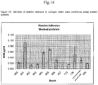
- the anti- ⁇ 2 ⁇ 1 integrin mAB or Fab fragments are able to inhibit platelet adhesion to collagen both in isolated human platelets and human platelet-rich plasma under static conditions. They are also able to inhibit the thrombus formation under flow on a collagen coated surface. The ability to block collagen binding and thus preventing platelet adhesion to collagen is one of the earliest steps in thrombus formation.
- the present invention provides monovalent antibodies, antibody fragments or derivatives and their uses to manufacture research, diagnostic and therapeutic agents for the treatment of ⁇ 2-integrin related disorders as listed below; specific examples include thrombosis, other vascular diseases, cancer and pathological consequences of neo-angiogenesis, auto-inflammatory diseases such as multiple sclerosis.
- variable domains As known to the skilled person, binding characteristics of antibodies are mediated by the variable domains.
- a variable domain from the heavy chain and a co-acting variable domain from the light chain are usually present in antibodies and arranged in order to allow for the co-action.
- the variable domain is also referred to as the FV region.
- variable loops three each on the light (VL) and heavy (VH) chain, are responsible for binding to the antigen. These loops are referred to as the Complementarity Determining Regions (CDRs), LCDR1, LCDR2 and LCDR3 for VL and HCDR1, HCDR2 and HCDR3 for VH.
- CDRs Complementarity Determining Regions
- variable domain from the heavy chain and a co-acting variable domain from the light chain are known in the art. Therefore, it was important to identify one or more suitable variable domains from the heavy chain and one or more co-acting variable domains from the light chain.
- sequence alignment the CDRs of the heavy and light chains have been identified for the ⁇ 2 integrin antibody specified above.
- present invention relates to an isolated monoclonal antibody or antigen binding fragment thereof, wherein said antibody or fragment specifically binds to the 1-domain of a human ⁇ 2-integrin, said antibody comprising a heavy chain variable region (VH) domain and a light chain variable region (VL) domain, wherein said antibody or fragment competes with a reference antibody for binding to the epitope of the reference antibody, said reference antibody comprising a light chain being the variable domain and the constant domain according to SEQ ID NO: 9 and a heavy chain being the variable domain and the constant domain according to SEQ ID NO: 10 or being the variable domain and the constant domain according to SEQ ID NO: 11.
- the present invention relates to the isolated monoclonal antibody or antigen binding fragment thereof of the present invention, wherein said antibody or fragment cross-reacts with a non-human primate ⁇ 2-integrin but does not cross-react with a non-primate ⁇ 2-integrin.
- the present invention relates to an isolated monoclonal antibody or antigen binding fragment thereof of the present invention, comprising one or more of the following components a to f:
- the present invention relates to one or more nucleic acid(s) coding for the isolated monoclonal antibody or antigen binding fragment thereof, wherein said nucleic acid(s) is/are preferably located in a vector.
- the present invention relates to a cell heterologously expressing one of the nucleic acids of present invention.
- the present invention relates to a method for producing an isolated monoclonal antibody or antigen binding fragment thereof of the present invention comprising culturing the cell according to the present invention under conditions permitting expression of the isolated monoclonal antibody or antigen binding fragment thereof and optionally recovering the isolated monoclonal antibody or antigen binding fragment thereof from the host cell.
- the present invention relates to a pharmaceutical composition
- a pharmaceutical composition comprising at least one isolated monoclonal antibody or antigen binding fragment thereof of the present invention and/or at least one nucleic acid of present invention for use as a medicament.
- present invention relates to an isolated monoclonal antibody or antigen binding fragment thereof of the present invention or the pharmaceutical composition of the present invention for use in treating or preventing an ⁇ 2-integrin-related disease or disorder selected from the group consisting of thrombosis, a vascular disease, cancer, including neo-angiogenesis and metastasis, inflammation, inflammatory disease, autoimmune disease and a disease characterized by abnormal or increase angiogenesis, inflammatory bowel disease, Crohn's disease, ulcerative colitis, reactions to transplant, optical neuritis, spinal cord trauma, rheumatoid arthritis, systemic lupus erythematosus (SLE), multiple sclerosis, Reynaud's syndrome, experimental autoimmune encephalomyelitis, Sjorgen's syndrome, scleroderma, cardiovascular disease, psoriasis, and infections that induce an inflammatory response, preferably for use in treating or preventing a vascular disease and/or thrombosis, particularly in the treatment
- the present invention relates to an article of manufacture comprising
- the present invention relates to a diagnostic kit for the diagnosis of an ⁇ 2-integrin related disorder or disease comprising an isolated monoclonal antibody or antigen binding fragment thereof according to the present invention and a suitable packaging, and possibly suitable instructions for using said antibody or fragment in the detection of ⁇ 2-integrin, wherein said disorder or disease is selected from the group consisting of thrombosis, a vascular disease, cancer, including neo-angiogenesis and metastasis, inflammation, inflammatory disease, autoimmune disease and a disease characterized by abnormal or increase angiogenesis, inflammatory bowel disease, Crohn's disease, ulcerative colitis, reactions to transplant, optical neuritis, spinal cord trauma, rheumatoid arthritis, systemic lupus erythematosus (SLE), multiple sclerosis, Reynaud's syndrome, experimental autoimmune encephalomyelitis, Sjorgen's syndrome, scleroderma, cardiovascular disease, psoriasis, and infections that induce thrombosis,
- the present invention relates to a method of diagnosis of an ⁇ 2-integrin-related disorder or disease using one or more isolated monoclonal antibody or antigen binding fragment thereof according to the present invention, wherein said disorder or disease is selected from the group consisting of thrombosis, a vascular disease, cancer, including neo-angiogenesis and metastasis, inflammation, inflammatory disease, autoimmune disease and a disease characterized by abnormal or increased angiogenesis, inflammatory bowel disease, Crohn's disease, ulcerative colitis, reactions to transplant, optical neuritis, spinal cord trauma, rheumatoid arthritis, systemic lupus erythematosus (SLE), multiple sclerosis, Reynaud's syndrome, experimental autoimmune encephalomyelitis, Sjorgen's syndrome, scleroderma, cardiovascular disease, psoriasis, and infections that induce an inflammatory response, and wherein said method comprises
- the present invention relates to an isolated monoclonal antibody or antigen binding fragment thereof, wherein said antibody or fragment specifically binds to the I-domain of a human ⁇ 2-integrin, said antibody or fragment comprising a heavy chain variable region (VH) domain and a light chain variable region (VL) domain, wherein said antibody or fragment cross-reacts with a non-human primate ⁇ 2-integrin but does not cross-react with a non-primate ⁇ 2-integrin.
- VH heavy chain variable region
- VL light chain variable region
- said antibody or fragment specifically binds to the 1-domain of the human ⁇ 2-integrin with nM binding affinity. In another embodiment, said antibody or fragment inhibits the interaction of the human ⁇ 2-integrin with collagen in vitro, thereby inhibiting the activation of platelets due to adhesion of said platelets to said collagen.
- said heavy chain variable region domain comprises the heavy chain HCDR3 of SEQ ID NO:5.
- said heavy chain variable region domain comprises the heavy chain CDRs of SEQ ID NO:3 (HCDR1), SEQ ID NO:4 (HCDR2), and SEQ ID NO:5 (HCDR3), or functionally active variants thereof.
- the functionally active variant of HCDR2 comprises the mutation Asp ⁇ Glu at amino acid position 6.
- the light chain variable region domain comprises the light chain LCDR3 of SEQ ID NO:8.
- the light chain variable region domain comprises the light chain CDRs of SEQ ID NO:6 (LCDR1), SEQ ID NO:7 (LCDR2), and SEQ ID NO:8 (LCDR3), or functionally active variants thereof.
- the functionally active variant of LCDR1 comprises the mutation Asn ⁇ Gln at amino acid position 11.
- the heavy chain variable region (VH) domain has at least 90%, 95%, 97% or 99% sequence identity to the VH sequence of SEQ ID NO: 2.
- said heavy chain variable region (VH) domain comprises the sequence of SEQ ID NO:2 or a functionally active thereof.
- the light chain variable region (VL) domain has at least 90%, 95%, 97% or 99% sequence identity to the VL sequence of SEQ ID NO: 1.
- said light chain variable region (VL) domain comprises the sequence of SEQ ID NO: 1 or a functionally active thereof.
- the heavy chain variable region (VH) domain comprises one or more amino acid substitutions at positions selected from the group consisting of H5, H7, H11, H12, H17, H20, H38, H40, H43, H55, H61, H65, H66, H67, H76, H81, H82, H87, H91, H93, H112, H113 and H116.
- the one or more amino acid subsitutions are selected from the group consisting 5His ⁇ Val, 7Pro ⁇ Ser, 11Leu ⁇ Val, 12Val ⁇ Lys, 17Pro ⁇ Ser, 20Leu ⁇ Val, 38Lys ⁇ Arg, 40Arg ⁇ Ala, 43Arg ⁇ Gln, 55Asp ⁇ Glu, 61Asn ⁇ Ala, 65Lys ⁇ Gln, 66Asp ⁇ Gly, 67Lys ⁇ Arg, 76Ser ⁇ Thr, 81Ile ⁇ Met, 82Gln ⁇ Glu, 87Thr ⁇ Arg, 91Ser ⁇ Thr, 93Val ⁇ Lys, 112Thr ⁇ Leu, 113Leu ⁇ Val and 116Ser ⁇ Val.
- the light chain variable region (VL) domain comprises one or more amino acid substitutions at positions selected from the group consisting of L9, L12, L15, L22, L34, L46, L47, L80, L83, L85, L87, and L89.
- the one or more amino acid subsitutions are selected from the group consisting of 9Ala ⁇ Ser, 12Ala ⁇ Ser, 15Leu ⁇ Val, 15Leu ⁇ Pro, 22Ser ⁇ Thr, 34Asn ⁇ Gln, 46Gln ⁇ Lys, 47Ala ⁇ Pro, 80Asp ⁇ Asn, 83Glu ⁇ Gln, 85Asp ⁇ Glu, 87Ala ⁇ Thr and 89Thr ⁇ Asn.
- the heavy chain variable region (VH) domain has at least 90%, 95%, 97% or 99% sequence identity to a VH sequence selected from the group consisting of SEQ ID NO: 38 (HC1), SEQ ID NO:39 (HC2), SEQ ID NO:40 (HC3), SEQ ID NO:41 (HC4), SEQ ID NO:42 (HC5), SEQ ID NO:43 (HC6), and SEQ ID NO:44 (HC7).
- the heavy chain variable region (VH) domain comprises a VH sequence selected from the group consisting of SEQ ID NO: 38 (HC1), SEQ ID NO:39 (HC2), SEQ ID N0:40 (HC3), SEQ ID NO:41 (HC4), SEQ ID NO:42 (HC5), SEQ ID NO:43 (HC6), and SEQ ID NO:44 (HC7).
- the light chain variable region (VL) domain has at least 90%, 95%, 97% or 99% sequence identity to a VL sequence selected from the group consisting of SEQ ID NO: 33 (LC1), SEQ ID NO:34 (LC2), SEQ ID NO:35 (LC3), SEQ ID NO:36 (LC4), and SEQ ID NO:37 (LC5).
- the light chain variable region (VL) domain comprises a VL sequence selected from the group consisting of SEQ ID NO: 33 (LC1), SEQ ID NO:34 (LC2), SEQ ID NO:35 (LC3), SEQ ID NO:36 (LC4), and SEQ ID NO:37 (LC5).
- the antibody or binding portion is a chimeric antibody or humanized antibody.
- the antigen binding portion is selected from the group consisting of a Fab, a Fab', a F(ab')2, a Fv, a disulfide linked Fv, a scFv, and a (scFv) 2 .
- the antibody or binding portion is selected from the group consisting of a multispecific antibody, a dual specific antibody, a isotype antibody, a dual variable domain antibody and a bispecific antibody.
- the antiboyd or binding portion comprises a heavy chain immunoglobulin constant domain selected from the group consisting of: a human IgM constant domain, a human IgG1 constant domain, a human IgG2 constant domain, a human IgG3 constant domain, domain, a human IgG4 constant domain, a human IgE constant domain, and a human IgA constant domain.
- the antibody or binding portion comprises a human IgG4 constant domain.
- the invention provides a nucleic acid encoding the amino acid sequence of the antibody or antigen binding portion of the invention.
- the invention provides a recombinant expression vector comprising the nucleic acid.
- the invention provdes a host cell comprising the recombinant expression vector.
- the invention provdes a method of producing the antibody or antigen binding fragment comprising culturing the host cell under conditions such that an antibody is produced by the host cell.
- the invention provides a pharmaceutical composition comprising the antibody, or antigen binding portion and one or more pharmaceutically acceptable carriers.
- the invention provides a method of treating, preventing or diagnosing an ⁇ 2-integrin-related disorder or disease, the method comprising administering to a subject in need of thereof the pharmaceutical composition.
- the ⁇ 2 integrin-related disease or disorder is selected from the group consisting of thrombosis, a vascular disease, cancer, including neo-angiogenesis and metastasis, inflammation, inflammatory disease, autoimmune disease and a disease characterized by abnormal or increase angiogenesis, inflammatory bowel disease, Crohn's disease, ulcerative colitis, reactions to transplant, optical neuritis, spinal cord trauma, rheumatoid arthritis, systemic lupus erythematosus (SLE), ,multiple sclerosis, Reynaud's syndrome, experimental autoimmune encephalomyelitis, Sjorgen's syndrome, scleroderma, cardiovascular disease, psoriasis, and infections that induce an inflammatory response.
- the ⁇ 2 integrin-related disease or disorder is selected from the group consisting of acute coronary syndrome, percutaneous coronary intervention, ischemic stroke, carotid artery stenosis or peripheral arterial occlusive disease.
- the terms used herein are defined as described in " A multilingual glossary of biotechnological terms: (IUPAC Recommendations)", Leuenberger, H.G.W, Nagel, B. and Kölbl, H. eds. (1995), Helvetica Chimica Acta, CH-4010 Basel, Switzerl and).
- alpha 2 integrin refers to alpha 2 integrin as known in the art, preferably human alpha 2 integrin and especially human alpha 2 integrin having the nucleic acid sequence shown in SEQ ID NO: 21 and the amino acid sequence of SEQ ID NO: 20, or a biologically active fragment thereof.
- I domain refers to the part of alpha 2 integrin as underlined and bold-typed in SEQ ID NO:20.
- telomere binding means that the peptide or peptide complex, e.g. an antibody or antigen-binding fragment thereof forms a complex with an antigen that is relatively stable under physiologic conditions.
- Specific binding can be characterized by an equilibrium dissociation constant of at least about 1x10 -6 M or less (e.g., a smaller K D denotes a tighter binding).
- Methods for determining whether two molecules specifically bind are well known in the art and include, for example, equilibrium dialysis, surface plasmon resonance, and the like.
- An isolated antibody that specifically binds alpha 2 integrin may, however, exhibit cross-reactivity to other antigens such as alpha 2 integrin molecules from other species.
- the ⁇ 2 integrin-specific antibodies of the invention bind to bind to both human and non-human primate ⁇ 2 integrin with an affinity that is at least two-fold regater than its affinity for a non-specific antigen (e.g., a non-primate ⁇ 2 integrin).
- a non-specific antigen e.g., a non-primate ⁇ 2 integrin
- multi-specific antibodies e.g., bispecifics
- alpha 2 integrin and one or more additional antigens are nonetheless considered antibodies that "specifically bind" alpha 2 integrin, as used herein.
- K D is intended to refer to the equilibrium dissociation constant of a particular peptide/peptide-complex - target molecule or antibody-antigen interaction.
- the equilibrium dissociation constant is typically measured in "mol/L” (abbreviated as "M").
- slow off rate a peptide/peptide complex or antibody that dissociates from alpha 2 integrin with a rate constant of 1 x 10 -3 s -1 or less, preferably 1 x 10 -4 s -1 or less, as determined by surface plasmon resonance, e.g., BIACORETM.
- high affinity antibody refers to those mAbs having a binding affinity to human alpha 2 integrin of at least 10 -10 M; preferably 10 -11 M; even more preferably 10 -12 M, as measured by surface plasmon resonance, e.g., BIACORETM or solution-affinity ELISA.
- surface plasmon resonance refers to an optical phenomenon that allows for the analysis of real-time biospecific interactions by detection of alterations in protein concentrations within a biosensor matrix, for example using the BIACORETM system (Pharmacia Biosensor AB, Uppsala, Sweden and Piscataway, N.J.).
- an “epitope”, also known as antigenic determinant, is the region of an antigen that is recognized by the immune system, specifically by antibodies, B cells, or T cells.
- an “epitope” is the part of an antigen capable of binding to an antibody or antigen-binding fragment thereof as described herein.
- binding preferably relates to a "specific binding", as defined herein.
- Epitopes usually consist of chemically active surface groupings of molecules such as amino acids, sugar side chains, phosphoryl groups, or sulfonyl groups and may have specific three-dimensional structural characteristics and/or specific charge characteristics. Conformational and non-conformational epitopes can be distinguished in that the binding to the former but not the latter is lost in the presence of denaturing solvents.
- a “paratope” is the part of an antibody that specifically binds to the epitope.
- antibody is intended to refer to immunoglobulin molecules comprised of four polypeptide chains, two heavy (H) chains and two light (L) chains inter-connected by disulfide bonds.
- antibody also includes all recombinant forms of antibodies, in particular of the antibodies described herein, e.g. antibodies expressed in prokaryotes, unglycosylated antibodies, and any antigen-binding antibody fragments and derivatives as described below.
- Each heavy chain is comprised of a heavy chain variable region ("HCVR” or "VH”) and a heavy chain constant region (comprised of domains CHI, CH2 and CH3).
- Each light chain is comprised of a light chain variable region ("LCVR or "VL”) and a light chain constant region (CL).
- VL light chain variable region
- CL light chain constant region
- the VH and VL regions can be further subdivided into regions of hypervariability, termed complementarity determining regions (CDR), interspersed with regions that are more conserved, termed framework regions (FR).
- CDR complementarity determining regions
- FR framework regions
- Each VH and VL is composed of three CDRs and four FRs, arranged from amino-terminus to carboxy-terminus in the following order: FR1, CDR1, FR2, CDR2, FR3, CDR3, FR4.
- the variable regions of the heavy and light chains contain a binding domain that interacts with an antigen.
- the constant regions of the antibodies may mediate the binding of the immunoglobulin to host tissues or factors, including various cells of the immune system (e.g., effector cells) and the first component (C1q) of the classical
- alpha2 antibody, a2 antibody, ⁇ 2 antibody, alpha2 integrin antibody, a2 integrin antibody, ⁇ 2 integrin antibody are used synonymously and refer preferably to an inhibitory, i.e. anti- (alpha2 antibody, a2 antibody, ⁇ 2 antibody, alpha2 integrin antibody, a2 integrin antibody, ⁇ 2 integrin antibody).
- CDR residues not contacting antigen can be identified based on previous studies (for example residues H60-H65 in CDRH2 are often not required), from regions of Kabat CDRs lying outside Chothia CDRs, by molecular modeling and/or empirically. If a CDR or residue(s) thereof is omitted, it is usually substituted with an amino acid occupying the corresponding position in another human antibody sequence or a consensus of such sequences. Positions for substitution within CDRs and amino acids to substitute can also be selected empirically. Empirical substitutions can be conservative or non-conservative substitutions.
- antigen-binding fragment of an antibody (or simply “binding portion”), as used herein, refers to one or more fragments of an antibody that retain the ability to specifically bind to alpha 2 integrin. It has been shown that the antigen-binding function of an antibody can be performed by fragments of a full-length antibody.
- binding fragments encompassed within the term "antigen-binding fragment" of an antibody include (i) Fab fragments, monovalent fragments consisting of the VL, VH, CL and CH domains; (ii) F(ab') 2 fragments, bivalent fragments comprising two Fab fragments linked by a disulfide bridge at the hinge region; (iii) Fd fragments consisting of the VH and CH domains; (iv) Fv fragments consisting of the VL and VH domains of a single arm of an antibody, (v) dAb fragments ( Ward et al., (1989) Nature 341: 544-546 ), which consist of a VH domain; (vi) isolated complementarity determining regions (CDR), and (vii) combinations of two or more isolated CDRs which may optionally be joined by a synthetic linker.
- Fab fragments monovalent fragments consisting of the VL, VH, CL and CH domains
- F(ab') 2 fragments bivalent fragments compris
- the two domains of the Fv fragment, VL and VH are coded for by separate genes, they can be joined, using recombinant methods, by a synthetic linker that enables them to be made as a single protein chain in which the VL and VH regions pair to form monovalent molecules (known as single chain Fv (scFv); see e.g., Bird et al. (1988) Science 242: 423-426 ; and Huston et al. (1988) Proc. Natl. Acad. Sci. USA 85: 5879-5883 ).
- single chain Fv single chain Fv
- Such single chain antibodies are also intended to be encompassed within the term "antigen-binding fragment" of an antibody.
- a further example is a binding-domain immunoglobulin fusion protein comprising (i) a binding domain polypeptide that is fused to an immunoglobulin hinge region polypeptide, (ii) an immunoglobulin heavy chain CH2 constant region fused to the hinge region, and (iii) an immunoglobulin heavy chain CH3 constant region fused to the CH2 constant region.
- the binding domain polypeptide can be a heavy chain variable region or a light chain variable region.
- the binding-domain immunoglobulin fusion proteins are further disclosed in US 2003/0118592 and US 2003/0133939 . These antibody fragments are obtained using conventional techniques known to those with skill in the art, and the fragments are screened for utility in the same manner as are intact antibodies.
- antigen-binding fragments are so-called microantibodies, which are derived from single CDRs.
- Heap et al. describe a 17 amino acid residue microantibody derived from the heavy chain CDR3 of an antibody directed against the gp120 envelope glycoprotein of HIV-1 ( Heap CJ et al. (2005) J. Gen. Virol. 86:1791-1800 ).
- Other examples include small antibody mimetics comprising two or more CDR regions that are fused to each other, preferably by cognate framework regions.
- Such a small antibody mimetic comprising VH CDR1 and VL CDR3 linked by the cognate VH FR2 has been described by Qiu et al. ( Qiu X-Q, et al. (2007) Nature biotechnology 25(8):921-929 ).
- antibody or antigen-binding fragment thereof refers to immunoglobulin molecules and immunologically active portions of immunoglobulin molecules, i.e. molecules that contain an antigen-binding site that immunospecifically binds an antigen.
- Antibodies and antigen-binding fragments thereof usable in the invention may be from any animal origin including birds and mammals.
- the antibodies or fragments are from human, chimpanzee, rodent (e.g. mouse, rat, guinea pig, or rabbit), chicken, turkey, pig, sheep, goat, camel, cow, horse, donkey, cat, or dog origin. It is particularly preferred that the antibodies are of human or murine origin.
- Antibodies of the invention also include chimeric molecules in which an antibody constant region derived from one species, preferably human, is combined with the antigen binding site derived from another species, e.g. mouse.
- antibodies of the invention include humanized molecules in which the antigen binding sites of an antibody derived from a non-human species (e.g. from mouse) are combined with constant and framework regions of human origin.
- antibodies of the invention can be obtained directly from hybridomas which express the antibody, or can be cloned and recombinantly expressed in a host cell (e.g., a CHO cell, or a lymphocytic cell).
- a host cell e.g., a CHO cell, or a lymphocytic cell.
- host cells are microorganisms, such as E. coli, and fungi, such as yeast.
- they can be produced recombinantly in a transgenic non-human animal or plant.
- chimeric antibody refers to those antibodies wherein one portion of each of the amino acid sequences of heavy and light chains is homologous to corresponding sequences in antibodies derived from a particular species or belonging to a particular class, while the remaining segment of the chain is homologous to corresponding sequences in another species or class.
- the variable region of both light and heavy chains mimics the variable regions of antibodies derived from one species of mammals, while the constant portions are homologous to sequences of antibodies derived from another.
- One clear advantage to such chimeric forms is that the variable region can conveniently be derived from presently known sources using readily available B-cells or hybridomas from non-human host organisms in combination with constant regions derived from, for example, human cell preparations.
- variable region has the advantage of ease of preparation and the specificity is not affected by the source, the constant region being human is less likely to elicit an immune response from a human subject when the antibodies are injected than would the constant region from a non-human source.
- definition is not limited to this particular example.
- humanized antibody refers to a molecule having an antigen binding site that is substantially derived from an immunoglobulin from a non-human species, wherein the remaining immunoglobulin structure of the molecule is based upon the structure and/or sequence of a human immunoglobulin.
- the antigen binding site may either comprise complete variable domains fused onto constant domains or only the complementarity determining regions (CDR) grafted onto appropriate framework regions in the variable domains.
- Antigen-binding sites may be wild-type or modified by one or more amino acid substitutions, e.g. modified to resemble human immunoglobulins more closely.
- Some forms of humanized antibodies preserve all CDR sequences (for example a humanized mouse antibody which contains all six CDRs from the mouse antibody). Other forms have one or more CDRs which are altered with respect to the original antibody.
- human antibody is intended to include antibodies having variable and constant regions derived from human germline immunoglobulin sequences.
- the human mAbs of the invention may include amino acid residues not encoded by human germline immunoglobulin sequences (e.g., mutations introduced by random or site-specific mutagenesis in vitro or by somatic mutation in vivo ), for example in the CDRs and in particular CDR3.
- human antibody as used herein, is not intended to include mAbs in which CDR sequences derived from the germline of another mammalian species (e.g., mouse), have been grafted onto human FR sequences.
- Human antibodies of the invention include antibodies isolated from human immunoglobulin libraries or from animals transgenic for one or more human immunoglobulin and that do not express endogenous immunoglobulins, as described for example in U.S. Patent No. 5,939,598 by Kucherlapati and Jakobovits.
- the term "monoclonal antibody” as used herein refers to a preparation of antibody molecules of single molecular composition.
- a monoclonal antibody displays a single binding specificity and affinity for a particular epitope.
- the monoclonal antibodies are produced by a hybridoma which includes a B cell obtained from a non-human animal, e.g. mouse, fused to an immortalized cell.
- recombinant antibody includes all antibodies that are prepared, expressed, created or isolated by recombinant means, such as (a) antibodies isolated from an animal (e.g., a mouse) that is transgenic or transchromosomal with respect to the immunoglobulin genes or a hybridoma prepared therefrom, (b) antibodies isolated from a host cell transformed to express the antibody, e.g. from a transfectoma, (c) antibodies isolated from a recombinant, combinatorial antibody library, and (d) antibodies prepared, expressed, created or isolated by any other means that involve splicing of immunoglobulin gene sequences to other DNA sequences.
- transfectoma includes recombinant eukaryotic host cells expressing an antibody, such as CHO cells, NS/0 cells, HEK293 cells, HEK293T cells, plant cells, or fungi, including yeast cells.
- a heterologous antibody is defined in relation to a transgenic organism producing such an antibody. This term refers to an antibody having an amino acid sequence or an encoding nucleic acid sequence corresponding to that found in an organism not consisting of the transgenic organism, and being generally derived from a species other than the transgenic organism.
- heterohybrid antibody refers to an antibody having light and heavy chains of different organismal origins.
- an antibody having a human heavy chain associated with a murine light chain is a heterohybrid antibody.
- nanobodies also known as single domain antibodies
- anti-idiotypic (anti-Id) antibodies including, e.g., anti-Id antibodies to antibodies of the invention
- epitope-binding fragments of any of the above include, e.g., anti-Id antibodies to antibodies of the invention, and epitope-binding fragments of any of the above.
- the antibodies described herein are preferably isolated.
- An "isolated antibody”, as used herein, is intended to refer to an antibody that is substantially free of other mAbs having different antigenic specificities (e.g., an isolated antibody that specifically binds alpha 2 integrin is substantially free of mAbs that specifically bind antigens other than alpha integrin).
- An isolated antibody that specifically binds alpha 2 integrin may, however, have cross-reactivity to other antigens, such as alpha 2 integrin molecules from other species.
- biological function or function of alpha 2 integrin are used synonymously and refer to any function of alpha 2 integrin such as, but not limited to: Binding to and forming a complex with betal integrin, binding to any of the known ligands such as binding to collagen, laminin, collagen-induced platelet aggregation, induction of thrombotic responses, thrombocytopenia, cell migration on collagen, cell-dependent reorganization of collagen fibers, collagen-dependent cellular resonses resulting in increases in cytokine expression and proliferation, alpha2 integrin or collagen-dependent aspects of T-cell, mast cell or neutrophil function, alpha 2 integrin or collagen-dependent aspects of delayed type hypersensitivity, alpha 2 integrin or collagen-dependent aspects of contact hypersensitivity, collagen-induced arthritis, mammary gland ductal morphogenesis, epidermal wound healing, and processes associated with VEGF-induced angiogenesis.
- an "alpha 2 integrin antagonist” denotes a compound that inhibits at least one biological activity of alpha 2 integrin, preferably an activity of alpha 2 integrin present on blood platelets, vascular endothelial cells, epithelial cells, activated monocytes/macrophages, fibroblasts, leukocytes, lymphocytes, activated neutrophils and/or mast cells especially when used in stoichiometric amounts.
- Preferred alpha 2 antagonists of the present invention are neutralizing antibodies.
- a “neutralizing antibody”, as used herein is intended to refer to an antibody whose binding to alpha 2 integrin results in inhibition of at least one biological activity of alpha 2 integrin, preferably inhibition of the platelet activating activity of alpha 2 integrin.
- This inhibition of the biological activity of alpha 2 integrin can be assessed by measuring one or more indicators of alpha 2 integrin biological activity by one or more of several standard in vitro or in vivo assays known in the art. Examples of such assays are described for example in the examples of present invention.
- alpha 2 integrin Since alpha 2 integrin has functions such as listed above, the activity of alpha 2 integrin has an effect on several diseases such as those associated with increased platelet activity. Accordingly, alpha 2 integrin antagonists, such as inhibitory peptide or peptide complexes targeting alpha 2 integrin or neutralizing anti-alpha 2 integrin antibodies or antigen-binding fragments thereof, are useful to reduce or inhibit the effects of alpha 2 integrin, such as platelet activity.
- alpha 2 integrin antagonists such as inhibitory peptide or peptide complexes targeting alpha 2 integrin or neutralizing anti-alpha 2 integrin antibodies or antigen-binding fragments thereof, are useful to reduce or inhibit the effects of alpha 2 integrin, such as platelet activity.
- alpha 2 integrin antagonists are useful for ameliorating, improving, inhibiting or preventing several such diseases, including without limitation thrombosis, a vascular disease, cancer, including neo-angiogenesis and metastasis, inflammation, inflammatory disease, autoimmune disease and a disease characterized by abnormal or increase angiogenesis, inflammatory bowel disease, Crohn's disease, ulcerative colitis, reactions to transplant, optical neuritis, spinal cord trauma, rheumatoid arthritis, systemic lupus erythematosus (SLE), multiple sclerosis, Reynaud's syndrome, experimental autoimmune encephalomyelitis, Sjorgen's syndrome, scleroderma, cardiovascular disease, psoriasis, and infections that induce an inflammatory response.
- diseases including without limitation thrombosis, a vascular disease, cancer, including neo-angiogenesis and metastasis, inflammation, inflammatory disease, autoimmune disease and a disease characterized by abnormal or increase angiogenesis,
- the anti-alpha 2 integrin antibodies or antigen-binding fragments thereof described herein may be conjugated to a therapeutic moiety ("immunoconjugate”), such as a cytotoxin, a chemotherapeutic drug, an immunosuppressant or a radioisotope.
- a therapeutic moiety such as a cytotoxin, a chemotherapeutic drug, an immunosuppressant or a radioisotope.
- a “conservative amino acid substitution” is one in which an amino acid residue is substituted by another amino acid residue having a side chain (R group) with similar chemical properties (e.g., charge or hydrophobicity).
- R group side chain
- similar chemical properties e.g., charge or hydrophobicity
- a conservative amino acid substitution will not substantially change the functional properties of a protein.
- the percent or degree of similarity may be adjusted upwards to correct for the conservative nature of the substitution. Means for making this adjustment are well known to those of skill in the art. See, e.g., Pearson (1994) Methods MoI. Biol. 24: 307- 331 .
- groups of amino acids that have side chains with similar chemical properties include
- Preferred conservative amino acids substitution groups are: valine-leucine-isoleucine, phenylalanine-tyrosine, lysine-arginine, alanine-valine, glutamate-aspartate, and asparagine-glutamine.
- a conservative replacement is any change having a positive value in the PAM250 log-likelihood matrix disclosed in Gonnet et al. (1992) Science 256: 1443-45 .
- a "moderately conservative" replacement is any change having a nonnegative value in the PAM250 log-likelihood matrix. Given the known genetic code, and recombinant and synthetic DNA techniques, the skilled scientist can readily construct DNAs encoding conservative amino acid variants.
- non-conservative substitutions or “non-conservative amino acid exchanges” are defined as exchanges of an amino acid by another amino acid listed in a different group of the seven standard amino acid groups 1) to 7) shown above.
- nucleic acid or fragment thereof indicates that, when optimally aligned with appropriate nucleotide insertions or deletions with another nucleic acid (or its complementary strand), there is nucleotide sequence identity in at least about 90%, and more preferably at least about 95%, 96%, 97%, 98% or 99% of the nucleotide bases, as measured by any well-known algorithm of sequence identity, such as FASTA, BLAST or GAP, as discussed below.
- the term "substantial similarity" or “substantially similar” means that two peptide sequences, when optimally aligned, such as by the programs GAP or BESTFIT using default gap weights, share at least 90% sequence identity, even more preferably at least 95%, 98% or 99% sequence identity. Preferably, residue positions which are not identical differ by conservative amino acid substitutions.
- Sequence similarity for polypeptides is typically measured using sequence analysis software. Protein analysis software matches similar sequences using measures of similarity assigned to various substitutions, deletions and other modifications, including conservative amino acid substitutions.
- GCG software contains programs such as GAP and BESTFIT which can be used with default parameters to determine sequence homology or sequence identity between closely related polypeptides, such as homologous polypeptides from different species of organisms or between a wild type protein and a mutein thereof. See, e.g., GCG Version 6.1. Polypeptide sequences also can be compared using FASTA with default or recommended parameters; a program in GCG Version 6.1.
- FASTA e.g., FASTA2 and FASTA3
- FASTA2 and FASTA3 provides alignments and percent sequence identity of the regions of the best overlap between the query and search sequences (Pearson (2000) supra ).
- Another preferred algorithm when comparing a sequence of the invention to a database containing a large number of sequences from different organisms is the computer program BLAST, especially BLASTP or TBLASTN, using default parameters. See, e.g., Altschul et al. (1990) J. Mol. Biol. 215: 403 410 and ( 1997) Nucleic Acids Res. 25:3389 402 .
- treat means accomplishing one or more of the following: (a) reducing the severity and/or duration of the disorder; (b) limiting or preventing development of symptoms characteristic of the disorder(s) being treated; (c) inhibiting worsening of symptoms characteristic of the disorder(s) being treated; (d) limiting or preventing recurrence of the disorder(s) in patients that have previously had the disorder(s); and (e) limiting or preventing recurrence of symptoms in patients that were previously symptomatic for the disorder(s).
- prevent means preventing that a disorder occurs in subject.
- the expressions "is for administration” and “is to be administered” have the same meaning as “is prepared to be administered”.
- the statement that an active compound "is for administration” has to be understood in that said active compound has been formulated and made up into doses so that said active compound is in a state capable of exerting its therapeutic activity.
- terapéuticaally effective amount or “therapeutic amount” are intended to mean that amount of a drug or pharmaceutical agent that will elicit the biological or medical response of a tissue, a system, animal or human that is being sought by a researcher, veterinarian, medical doctor or other clinician.
- prophylactically effective amount is intended to mean that amount of a pharmaceutical drug that will prevent or reduce the risk of occurrence of the biological or medical event that is sought to be prevented in a tissue, a system, animal or human by a researcher, veterinarian, medical doctor or other clinician.
- the dosage a patient receives can be selected so as to achieve the amount of peptide or peptide complex to exhibit sufficient inhibition of alpha2 integrin function in order to allow for the prophylactic or curative therapy (prevention, improvement or healing) of an ⁇ 2 integrin-related disease or disorder, preferably selected from the group consisting of thrombosis, a vascular disease, cancer, including neo-angiogenesis and metastasis, inflammation, inflammatory disease, autoimmune disease and a disease characterized by abnormal or increase angiogenesis, inflammatory bowel disease, Crohn's disease, ulcerative colitis, reactions to transplant, optical neuritis, spinal cord trauma, rheumatoid arthritis, systemic lupus erythematosus (SLE), multiple sclerosis, Reynaud's syndrome, experimental autoimmune encephalomyelitis, Sjorgen's syndrome, scleroderma, cardiovascular disease, psoriasis, and infections that induce an inflammatory response.
- a "patient” means any mammal or bird who may benefit from a treatment with the antibodies and antigen-biding fragments thereof described herein.
- a “patient” is selected from the group consisting of laboratory animals (e.g. mouse or rat), domestic animals (including e.g. guinea pig, rabbit, chicken, turkey, pig, sheep, goat, camel, cow, horse, donkey, cat, or dog), or primates including chimpanzees and human beings. It is particularly preferred that the "patient” is a human being.
- “Pharmaceutically acceptable” means approved by a regulatory agency of the Federal or a state government or listed in the U.S. Pharmacopeia (United States Pharmacopeia-33/National Formulary-28 Reissue, published by the United States Pharmacopeial Convention, Inc., Rockville Md., publication date: April 2010) or other generally recognized pharmacopeia for use in animals, and more particularly in humans.
- Specific populations treatable by the therapeutic methods of the invention include subjects indicated for alpha 2 integrin-activating mutations (gain of function mutations, "GOF”), subjects with ⁇ 2 integrin-related disease or disorder, preferably selected from the group consisting of thrombosis, a vascular disease, cancer, including neo-angiogenesis and metastasis, inflammation, inflammatory disease, autoimmune disease and a disease characterized by abnormal or increase angiogenesis, inflammatory bowel disease, Crohn's disease, ulcerative colitis, reactions to transplant, optical neuritis, spinal cord trauma, rheumatoid arthritis, systemic lupus erythematosus (SLE), multiple sclerosis, Reynaud's syndrome, experimental autoimmune encephalomyelitis, Sjorgen's syndrome, scleroderma, cardiovascular disease, psoriasis, and infections that induce an inflammatory response.
- GAF gain of function mutations
- a first aspect of the present invention relates to an isolated monoclonal antibody or antigen binding fragment thereof, wherein said antibody or fragment specifically binds to the I-domain of a human ⁇ 2-integrin, said antibody comprising a heavy chain variable region (VH) domain and a light chain variable region (VL) domain, wherein said antibody or fragment competes with a reference antibody for binding to the epitope of the reference antibody, said reference antibody comprising a light chain being the variable domain and the constant domain according to SEQ ID NO: 9 and a heavy chain being the variable domain and the constant domain according to SEQ ID NO: 10 or being the variable domain and the constant domain according to SEQ ID NO: 11.
- a second aspect of the present invention relates to the isolated monoclonal antibody or antigen binding fragment thereof of the present invention, wherein said antibody or fragment cross-reacts with a non-human primate ⁇ 2-integrin but does not cross-react with a non-primate ⁇ 2-integrin.
- present invention relates to an isolated monoclonal antibody or antigen binding fragment thereof of the present invention, comprisingone or more of the following components a to f:
- peptide and peptide complexes thereof are meant to be an “isolated monoclonal antibody or antigen binding fragment thereof", as referred to above. Consequently, any characterizations of “peptide and peptide complexes thereof” refer to characterizations of the "isolated monoclonal antibody or antigen binding fragment thereof".
- sequences of SEQ ID NO:6 to 8 are CDRs of light chains and that of SEQ ID NO:3 to 5 are the CDRs of heavy chains of the analysed antibody (as determined by sequence analysis).
- the peptide or peptide complex comprises one of above the light chain CDRs or a functionally active variant thereof and/or one of the heavy chain CDRs or a functionally active variant thereof. Examples include a peptide or peptide complex comprising one or two or three of the above HCDRS and/or one or two or three of the above LCDRs in any of the conceivable combinations.
- One embodiment of present invention is a peptide or peptide complex comprising 3 LCDRs and 3HCDRs, wherein at least one of them is one of the above CDRs a to f.
- LCDR and LDR are used synonymously.
- HCDR and HDR are used synonymously.
- CDRs are arranged in a suitable way, the arrangement allows for specific binding to ⁇ 2 integrin.
- the suitable arrangement of CDRs to allow for binding of an antigen is known in the art.
- a variety of different antibody formats or formats of binding parameters have been developed or identified so far. Any of these or any other suitable arrangement may be used for the polypeptide or polypeptide complex of the present invention, as long as the format or arrangement allows for specific binding to ⁇ 2 integrin.
- the CDR sequences may be arranged in one (poly)peptide-chain or in a polypeptide or peptide complex. If they are arranged within one (poly)peptide-chain, the sequences may be connected by one or more linker sequences, preferably a peptide linker, e.g. as a fusion protein. According to one embodiment, they may be embedded into a natural or artificial antibody scaffold or framework, as known in the art. For natural antibodies, the CDRs are supported within the variable domains by conserved framework regions. The framework can be modified in order to obtain artificial antibodies, such as Fabs, single chain antibodies etc. which are described below in more detail.
- CDRs are arranged in a peptide complex, two or more (poly)peptides are bound to each other by non-covalent bonding including hydrogen bonds, ionic bonds, Van der Waals forces, and hydrophobic interactions.
- a peptide is an organic compound made of 2 or more ⁇ -amino acids arranged in a linear chain.
- the amino acids are joined together by peptide bonds between the carboxyl and amino groups of adjacent amino acid residues.
- the genetic code specifies 20 standard amino acids.
- the residues in a protein may be chemically modified by posttranslational modification, which alter the physical and chemical properties, folding, stability, activity, and ultimately, the function of the proteins.
- the peptides according to the different aspects of present invention may be modified or unmodified as long as they are able to bind ⁇ 2 integrin.
- polypeptide refers to a molecule comprising about 20, about 25, about 30 or more amino acids coupled to each other by peptide bonds in a linear mode to form a polypeptide chain. Shorter molecules of this kind comprising at least 2 amino acids are generally referred to as peptides.
- protein usually refers to molecules comprising one or more polypeptide chains. In the context of present invention, the terms peptide, polypeptide and protein are used synonymously.
- peptide or “polypeptide”according to the different aspects of present invention refers to peptides or polypeptides as defined above
- peptide complex refers to molecule complexes comprising one or more peptides and/or polypeptides as defined above (e.g., the antibodies, antigen binding fragments and other binding molecules of the invention).
- Peptide and peptide complexes thereof as defined herein selectively recognize and specifically bind to a ⁇ 2 integrin antigen.
- the term "specific binding to ⁇ 2 integrin” refers to the ability of the peptide or peptide complex according to the invention to bind specifically to ⁇ 2 integrin or to the ⁇ 2 integrin I domain or to ⁇ 2 integrin in the complex with any other polypeptide such as in the heterodimeric complex with another integrin subunit, e.g. the ⁇ 2 ⁇ 1 integrin complex.
- the peptide or peptide complex which is defined as the isolated monoclonal antibody or antigen binding fragment thereof herein specifically binds to the I-domain of a human ⁇ 2-integrin, said antibody comprising a heavy chain variable region (VH) domain and a light chain variable region (VL) domain, wherein said antibody or fragment competes with a reference antibody for binding to the epitope of the reference antibody, said reference antibody comprising a light chain being the variable domain and the constant domain according to SEQ ID NO: 9 and a heavy chain being the variable domain and the constant domain according to SEQ ID NO: 10 or being the variable domain and the constant domain according to SEQ ID NO: 11.
- selective or “specific” herein when used to describe the binding characteristics of the peptide or peptide complex according to the invention, refers to the fact that the disclosed peptides or peptide complexes do not show significant binding to other than ⁇ 2 integrin, except in those specific instances where the peptide/complex is supplemented to confer an additional, distinct specificity to the ⁇ 2 integrin-specific binding portion (as, for example, in bispecific or bifunctional molecules where the molecule is designed to bind or effect two functions, at least one of which is to specifically bind ⁇ 2 integrin).
- ⁇ 2 integrin-specific peptides or complexes thereof bind to human ⁇ 2 integrin with a K D of at least 1.2 x 10 -6 . In specific embodiments, ⁇ 2 integrin-specific peptides or complexes thereof bind to human ⁇ 2 integrin with a K D of 5 x 10 -7 or more, of 2 x 10 -7 or more, or of 1 x 10 -7 or more. In additional embodiments, ⁇ 2 integrin-specific peptides or complexes thereof bind to human ⁇ 2 integrin with a K D of 1 x 10 -8 or more.
- ⁇ 2 integrin-specific peptides or complexes thereof bind to human ⁇ 2 integrin with a K D of 5 x 10 -9 or more or of 1 x 10 -9 or more. In further embodiments, ⁇ 2 integrin-specific peptides or complexes thereof bind to human ⁇ 2 integrin with a K D of 2 x 10 -10 or more. In specific embodiments, ⁇ 2 integrin-specific peptides or complexes thereof do not bind other proteins at the above K D s.
- the ⁇ 2 integrin-specific peptides or complexes thereof binding to an ⁇ 2 integrin (e.g., human and/or non-human primate ⁇ 2 integrin) with an affinity that is at least two-fold greater than its affinity for a non-specific antigen.
- an ⁇ 2 integrin e.g., human and/or non-human primate ⁇ 2 integrin
- K D relates to the dissociation constant obtained from the ratio of k d (the dissociation rate of a particular binding molecule-target protein interaction; also referred to as k off ) to k a (the association rate of the particular binding molecule-target protein interaction; also referred to as k on ), or k d /k a which is expressed as a molar concentration (M).
- K D values can be determined using methods well established in the art. A preferred method for determining the K D of a binding molecule is described in Example 1 D.
- ⁇ 2 integrin-specific peptides or complexes thereof have been shown to dose-dependently inhibit ⁇ 2 integrin/ligand interaction (see Fig. 2 and Examples). Accordingly, ⁇ 2 integrin-specific peptides or complexes thereof may be characterized by their ability to counteract binding of collagen to ⁇ 2 integrin. The extent of inhibition by any ⁇ 2 integrin-specific peptide or complex thereof may be measured quantitatively in statistical comparison to a control, or via any alternative method available in the art. In specific embodiments, the inhibition is about 10 % inhibition or more. In other embodiments, the inhibition is 20 % or more, 30 % or more, 40 % or more 50 % or more, 60 % or more 70 % or more, 80 % or more, 90 % or more, or 95 % or more.
- the peptide or peptide complex may also comprise a functionally active variant of the above sequences.
- a functionally active variant of the peptides or peptide complexes of the invention is characterized by having a biological activity similar to that displayed by the complete peptide, including the ability to bind to ⁇ 2 integrin, and optionally to inhibit ⁇ 2 integrin.
- the variant is functionally active in the context of the present invention, if the activity (e.g.
- binding activity, optionally expressed as K D ) of the variant amounts to 10 % or more, 25 % or more, 50 % or more, 70 % or more, 80 % or more, 90 % or more, 95% or more, or 99 % or more of the activity of the peptide/complex without sequence alteration. Suitable methods for determining binding activity to ⁇ 2 integrin are given in the Examples.
- a functionally active variant may be obtained by a limited number of amino acid substitutions, deletions and/or insertions.
- SEQ ID NOs 1 and 2 can be gained from Figure No.5:
- SEQ ID NO: 1 is the amino acid sequence of the ⁇ 2 integrin antibody-variable light chain.
- SEQ ID NO:2 is the amino acid sequence of the variable heavy chain, respectively.
- SEQ ID NOs 9, 10 and 11 can be gained from Figure 7 :
- SEQ ID NO:9 is the amino acid sequence of the chimeric light chain of the antibody produced as an IgG4 format (CDRs underlined)
- SEQ ID NO:10 is the amino acid sequence of the chimeric heavy chain of the antibody produced as an IgG4 format (CDRs underlined)
- SEQ ID NO 11 is the amino acid sequence of the chimeric heavy chain in Fab format with a 6xhis tag.
- the constant regions were derived from human sequence backbones (see Examples).
- the invention also relates to any of the antibody constructs or fragments, peptide or polypeptide complexes without the his tag.
- variable domains of the HC and LC are coupled to respective constant regions and to form chimeric HC or LC constructs.
- specific embodiments are a chimeric ⁇ 2 integrin antibody LC variable region fused to the constant region of IGKC protein (such as e.g. in SEQ ID NO:9), a chimeric ⁇ 2 integrin antibody HC variable region fused to the constant region of IGHG4 (such as e.g. in SEQ ID NO: 10) or a chimeric ⁇ 2 integrin antibody HC variable region furse to the constant region CH1 domain of IGHG1 (such as e.g. in SEQ ID NO:11).
- components a) to c) (LC CDRs) and d) to f) (HC CDRs) were obtained by sequencing variable domain of a light chain (VL) and variable domain of a heavy chain (VH), respectively, of the monoclonal antibody produced and tested. Accordingly, they may be comprised in the same. It may be any naturally occurring VL or VH framework or an artificial VL or VH framework.
- one or more of the CDRs (LCDR1, LCDR2, LCDR3, HCDR1, HCDR2 and HCDR3) are arranged in the framework of the prevailing variable domain, i.e.
- variable domains as identified by any suitable method described above (cf. SEQ ID NOs: 1 and 2) alone, together or in any combination thereof, may be removed from the shown neighborhood and transferred into the framework of another (second) variable domain, thereby substituting the CDRs of the second variable domain.
- SEQ ID NOs: 1 and 2 alone, together or in any combination thereof, may be removed from the shown neighborhood and transferred into the framework of another (second) variable domain, thereby substituting the CDRs of the second variable domain.
- a variety of variable domains or antibody sequences is known in the art and may be used for this purpose.
- variable domains, into which CDRs of interest are inserted may be obtained from any germ-line or rearranged human variable domain.
- Variable domains may also be synthetically produced.
- variable heavy domain may be paired with a variable light domain to provide an antigen binding site.
- independent regions e.g., a variable heavy domain alone may be used to bind antigen.
- the peptides or peptide-complexes of present invention can be glycosylated.
- the glycosylation of proteins and its physiological affect is known in the art.
- the oligosaccharide component can significantly (in the positive or negative) affect properties relevant to the efficacy of a therapeutic glycoprotein, including physical stability, resistance to protease attack, interactions with the immune system, pharmacokinetics, and specific biological activity.
- mammalian host cells are commonly used in the art ( Cumming et al., 1991, Glycobiology 1: 115-130 ; Jenkins et al., 1996, Nature Biotechn. 14: 975-981 ).
- Examples include Chinese hamster ovary (CHO) cells, baby hamster kidney (BHK) cells, NSO-and SP2/0-mouse myeloma cells.
- CHO Chinese hamster ovary
- BHK baby hamster kidney
- SP2/0-mouse myeloma cells The production of glycosylated proteins from transgenic animals has also been published (Jenkins et al., 1996, supra).
- engineered recombinant host cells heterologously expressing / overexpressing glycosyl transferase genes are known in the art ( Bailey, 1991, Science 252: 1668-1675 ).
- WO 9954342 (A1 ) discloses methods for the generation of glycosylated proteins using host cells expressing a range of a glycoprotein-modifying glycosyl transferase activity which increases complex N-linked oligosaccharides carrying bisecting GIcNAc reported to have improved function.
- the peptide or peptide complex can be coupled to one or more molecules that are not identical with the peptide or peptide complex according to present invention (additional moieties), the whole complex being a "conjugate".
- additional moieties comprise, e.g. one or more further biomolecules, as peptides or peptide complexes, nucleic acids (e.g. oligonucleotides, or RNA molecules, such as an RNAi) or organic (small) molecules, radioactive moieties.
- additional moieties can have their own function, e.g. cytotoxicity, therapeutic activity, immunosuppressive activity, etc. or they can be beneficial for the whole conjugate for other reason (e.g.
- Present invention encompasses peptides or peptide complexes conjugated to one or more additional moieties.
- this conjugate is an immunoconjugate.
- immunoconjugates are known in the art (see e.g. WO05/103081 ), e.g. one or more chemotherapeutic substances, prodrugs, cytotoxins, radioisotopes or radioactive nucleotides, immunosuppressive moieties, therapeutic oligonucleotides, inhibitory RNA (RNAi).
- the peptide or peptide complex is an antibody.
- Naturally occurring antibodies are globular plasma proteins ( ⁇ 150 kDa) that are also known as immunoglobulins which share a basic structure. As they have sugar chains added to amino acid residues, they are glycoproteins.
- the basic functional unit of each antibody is an immunoglobulin (Ig) monomer (containing only one Ig unit); secreted antibodies can also be dimeric with two Ig units as with IgA, tetrameric with four Ig units like teleost fish IgM, or pentameric with five Ig units, like mammalian IgM.
- Ig immunoglobulin
- suitable formats include the format of naturally occurring antibodies including antibody isotypes known as IgA, IgD, IgE, IgG and IgM.
- the Ig monomer is a "Y"-shaped molecule that consists of four polypeptide chains; two identical heavy chains and two identical light chains connected by disulfide bonds between cysteine residues. Each heavy chain is about 440 amino acids long; each light chain is about 220 amino acids long. Heavy and light chains each contain intrachain disulfide bonds which stabilize their folding. Each chain is composed of structural domains called Ig domains. These domains contain about 70-110 amino acids and are classified into different categories (for example, variable or V, and constant or C) according to their size and function. They have a characteristic immunoglobulin fold in which two ⁇ sheets create a "sandwich" shape, held together by interactions between conserved cysteines and other charged amino acids.
- Ig heavy chain There are five types of mammalian Ig heavy chain denoted by ⁇ , ⁇ , ⁇ , ⁇ , and ⁇ .
- the type of heavy chain present defines the isotype of antibody; these chains are found in IgA, IgD, IgE, IgG, and IgM antibodies, respectively.
- Distinct heavy chains differ in size and composition; ⁇ and ⁇ contain approximately 450 amino acids and ⁇ approximately 500 amino acids, while ⁇ . and ⁇ have approximately 550 amino acids.
- Each heavy chain has two regions, the constant region (C H ) and the variable region (V H ).
- the constant region is essentially identical in all antibodies of the same isotype, but differs in antibodies of different isotypes.
- Heavy chains ⁇ , ⁇ and ⁇ have a constant region composed of three tandem Ig domains, and a hinge region for added flexibility; heavy chains ⁇ and ⁇ have a constant region composed of four immunoglobulin domains.
- the variable region of the heavy chain differs in antibodies produced by different B cells, but is the same for all antibodies produced by a single B cell or B cell clone.
- the variable region of each heavy chain is approximately 110 amino acids long and is composed of a single Ig domain.
- a light chain has two successive domains: one constant domain (CL) and one variable domain (VL).
- CL constant domain
- VL variable domain
- the approximate length of a light chain is 211 to 217 amino acids.
- Each antibody contains two light chains that are always identical; only one type of light chain, ⁇ or ⁇ , is present per antibody in mammals.
- Other types of light chains, such as the ⁇ chain are found in lower vertebrates like Chondrichthyes and Teleostei.
- variable (V) regions are responsible for binding to the antigen, i.e. for its antigen specificity.
- VL variable light
- VH variable heavy chain
- CDRs Complementarity Determining Regions
- antibody means any polypeptide which has structural similarity to a naturally occurring antibody and is capable of specifically binding to ⁇ 2 integrins, wherein the binding specificity is determined by the CDRs of in SEQ ID NOs: 3 to 8.
- antibody is intended to relate to an immunoglobulin-derived structure with specific binding to ⁇ 2 integrin including, but not limited to, a full length or whole antibody, an antigen binding fragment (a fragment derived, physically or conceptually, from an antibody structure), a derivative of any of the foregoing, a chimeric molecule, a fusion of any of the foregoing with another polypeptide, or any alternative structure/composition which selectively binds to ⁇ 2 integrin and optionally inhibits the function of ⁇ 2 integrin.
- the antibody may be any polypeptide which comprises at least one antigen binding fragment.
- Antigen binding fragments consist of at least the variable domain of the heavy chain and the variable domain of the light chain, arranged in a manner that both domains together are able to bind to the specific antigen.
- Fully length or “complete” antibodies refer to proteins that comprise two heavy (H) and two light (L) chains inter-connected by disulfide bonds which comprise: (1) in terms of the heavy chains, a variable region and a heavy chain constant region which comprises three domains, CH1, CH2 and CH3; and (2) in terms of the light chains, a light chain variable region and a light chain constant region which comprises one domain, CL.
- complete antibody any antibody is meant that has a typical overall domain structure of a naturally occurring antibody (i.e. comprising a heavy chain of three or four constant domains and a light chain of one constant domain as well as the respective variable domains), even though each domain may comprise further modifications, such as mutations, deletions, or insertions, which do not change the overall domain structure.
- an "antibody fragment” also contains at least one antigen binding fragment as defined above, and exhibits essentially the same function and specificity as the complete antibody of which the fragment is derived from.
- Limited proteolytic digestion with papain cleaves the Ig prototype into three fragments. Two identical amino terminal fragments, each containing one entire L chain and about half an H chain, are the antigen binding fragments (Fab).
- the Fc contains carbohydrates, complement-binding, and FcR-binding sites.
- F(ab')2 is divalent for antigen binding.
- the disulfide bond of F(ab')2 may be cleaved in order to obtain Fab'.
- the variable regions of the heavy and light chains can be fused together to form a single chain variable fragment (scFv).
- variable domains are the smallest fragments with an intact antigen-binding domain consisting of one VL and one VH.
- Such fragments, with only the binding domains can be generated by enzymatic approaches or expression of the relevant gene fragments, e.g. in bacterial and eukaryotic cells. Different approaches can be used, e.g. either the Fv fragment alone or 'Fab'-fragments comprising one of the upper arms of the "Y" that includes the Fv plus the first constant domains.
- fragments are usually stabilized by introducing a polypeptide link between the two chains which results in the production of a single chain Fv (scFv).
- scFv single chain Fv
- dsFv disulfide-linked Fv
- the binding domains of fragments can be combined with any constant domain in order to produce full length antibodies or can be fused with other proteins and polypeptides.
- a recombinant antibody fragment is the single-chain Fv (scFv) fragment.
- scFv single-chain Fv
- the VH and VL domains are joined with a hydrophilic and flexible peptide linker, which improves expression and folding efficiency.
- linkers of about 15 amino acids are used, of which the (Gly 4 Ser) 3 linker has been used most frequently.
- scFv molecules might be easily proteolytically degraded, depending on the linker used.
- Dissociation of scFvs results in monomeric scFvs, which can be complexed into dimers (diabodies or (scFv) 2 ), trimers (triabodies) or larger aggregates such as T and Abs and Flexibodies.
- Antibodies with two binding domains can be created either through the binding of two scFv with a simple polypeptide link (scFv) 2 or through the dimerisation of two monomers (diabodies).
- the simplest designs are diabodies that have two functional antigen-binding domains that can be either the same, similar (bivalent diabodies) or have specificity for distinct antigens (bispecific diabodies).
- bispecific antibodies allow for example the recruitment of novel effector functions (such as cytotoxic T cells) to the target cells, which make them very useful for applications in medicine.
- variable domains of heavy chains and four variable domains of light chains have been developed.
- examples of these include tetravalent bispecific antibodies (TandAbs and Flexibodies, Affimed Therapeutics AG, Heidelberg. Germany).
- TandAbs and Flexibodies In contrast to a bispecific diabody, a bispecific T and Ab is a homodimer consisting of only one polypeptide.
- Flexibodies are a combination of scFv with a diabody multimer motif resulting in a multivalent molecule with a high degree of flexibility for joining two molecules which are quite distant from each other on the cell surface. If more than two functional antigen-binding domains are present and if they have specificity for distinct antigens, the antibody is multispecific.
- bispecific antibodies may be produced using conventional technologies, specific methods of which include production chemically, or from hybrid hybridomas) and other technologies including, but not limited to, the BiTE TM technology (molecules possessing antigen binding regions of different specificity with a peptide linker) and knobs-into-holes engineering.
- the antibody may be a Fab, a Fab', a F(ab')2, a Fv, a disulfide-linked Fv, a scFv, a (scFv) 2 , a bispecific antibody, a multispecific antibody, a diabody, a triabody, a tetrabody or a minibody.
- the antibody is a monoclonal antibody.
- the antibody is a chimeric antibody or a humanised antibody.
- Monoclonal antibodies are monospecific antibodies that are identical because they are produced by one type of immune cell that are all clones of a single parent cell.
- a chimeric antibody is an antibody in which at least one region of an immunoglobulin of one species is fused to another region of an immunoglobulin of another species by genetic engineering in order to reduce its immunogenecity. For example murine V L and V H regions may be fused to the remaining part of a human immunoglobulin.
- a particular type of chimeric antibodies is a humanised antibody.
- Humanised antibodies are produced by merging the DNA that encodes the CDRs of a non-human antibody with human antibody-producing DNA (or vice versa). The resulting DNA construct can then be used to express and produce antibodies that are usually not as immunogenic as the non-human parenteral antibody or as a chimeric antibody, since merely the CDRs are non-human.
- human or humanized antibodies or fragments thereof can be used.
- the peptide or peptide complex may comprise a heavy chain immunoglobulin constant domain selected from the group consisting of: a human IgM constant domain, a human IgG1 constant domain, a human IgG2 constant domain, a human IgG3 constant domain, domain, a human IgG4 constant domain, a human IgE constant domain, and a human IgA constant domain.
- the anti- ⁇ 2-Integrin antibody has been humanized using a method previously described in WO2009/032661 , but any suitable humanization method known in the art can be used.
- the CDR may also be a functionally active variant of any of the CDRs specified in the claims.
- the functionally active variant is a functionally active fragment consisting of 90 % or more of an amino acid sequence of any of SEQ ID NOS: 3 to 8.
- the functionally active variant is a functionally active variant having 70 % or more, preferably 80 % or more, more preferably 90 % or 95% or more sequence identity to an amino acid sequence of any of SEQ ID NOS: 3 to 8, particularly wherein the functionally active variant is derived from the amino acid sequence of any of SEQ ID NOS: 3 to 8 by means of one or more conservative amino acid substitution (see below).
- the peptide or peptide complex comprises the amino acid sequence of
- the peptide or peptide complex consists of the amino acid sequence of
- the peptide or peptide complex consists of the amino acid sequence of
- the functionally active variant may be a fragment characterized by being derived from any of the sequences of SEQ ID NO: 1 or 2 or 9 or 10 or 11 by one or more deletions.
- the deletion(s) may be C-terminally, N-terminally and/or internally.
- the fragment may e.g. be obtained by 10 or less deletions, such as 1, 2, 3, 4, 5, 6, 7, 8, 9 or 10, or by 5 or less, such as 1, 2, 3, 4 or 5, or by 3 or less, such as 1, 2 or 3, or by 2 or less, such as 1 or 2, or by 1 deletion(s).
- the functionally active fragment of the invention is characterized by having a biological activity similar to that displayed by the complete protein, including the ability to bind to ⁇ 2 integrin and/or ⁇ 2 ⁇ 1 integrin and optionally to inhibit ⁇ 2 and/or ⁇ 2 ⁇ 1 integrin.
- the fragment of an antigen is functionally active in the context of the present invention, if the activity of the fragment amounts to 10 % or more, preferably 25 % or more, more preferably 50 % or more, more preferably 70 % or more, more preferably 80 % or more, more preferably 90 % or more, more preferably 95 % or more, most preferably or 99 % or more of the activity of the amino acid sequence without sequence alteration. Suitable methods for determining binding activity to ⁇ 2 ⁇ 1 integrin are given in the Examples, particularly Example 1 D.
- the variant may be characterized by being derived from any of the sequences of SEQ ID NO: 1 or 2 or 9 or 10 or 11 by one or more amino acid modifications including deletions, additions and/or substitutions.
- the modification(s) may be C-terminally, N-terminally and/or internally.
- the fragment may be obtained by 10 or less deletions, such as 1, 2, 3, 4, 5, 6, 7, 8, 9 or 10, or by 5 or less, such as 1, 2, 3, 4 or 5, or by 3 or less, such as 1, 2 or 3, or by 2 or less, such as 1 or 2, or by 1 deletion(s).
- the functionally active variant of the invention is characterized by having a biological activity similar to that displayed by the complete protein, including the ability to bind to ⁇ 2 integrin and/or ⁇ 2 ⁇ 1integrin and optionally to inhibit ⁇ 2 and/or ⁇ 2 ⁇ 1 integrin.
- the variant is functionally active in the context of the present invention, if the activity of the variant amounts to 10 % or more, preferably 25 % or more, more preferably 50 % or more, even more preferably 70 % or more, still more preferably 80 % or more, especially 90 % or more, particularly 95 % or more, most preferably 99 % or more of the activity of the amino acid sequence without sequence alteration.
- the additional amino acids of (ix, x or xi) may be C-terminally, N-terminally and/or internally located. According to one embodiment, there are 50 or less additions, or 40 or less or 30 or less or 20 or less additions or 10 or less additions such as 1, 2, 3, 4, 5, 6, 7, 8, 9 or 10, or 5 or less additions, such as 1, 2, 3, 4 or 5, or 3 or less additions, such as 1, 2 or 3, or 2 or less, such as 1 or 2, or only 1 addition(s).
- the additional amino acid residue(s) may be any amino acid, which may be either an L-and/or a D-amino acid, naturally occurring and otherwise.
- the amino acid is any naturally occurring amino acid such as alanine, cysteine, aspartic acid, glutamic acid, phenylalanine, glycine, histidine, isoleucine, lysine, leucine, methionine, asparagine, proline, glutamine, arginine, serine, threonine, valine, tryptophan or tyrosine.
- the amino acid may also be a modified or unusual amino acid.
- examples of those are 2-aminoadipic acid, 3-aminoadipic acid, ⁇ -alanine, 2-aminobutyric acid, 4-aminobutyric acid, 6-aminocaproic acid, 2-aminoheptanoic acid, 2-aminoisobutyric acid, 3-aminoisobutyric acid, 2-aminopimelic acid, 2,4-diaminobutyric acid, desmosine, 2,2'-diaminopimelic acid, 2,3-diaminopropionic acid, N-ethylglycinem N-ethylasparagine, hydroxylysine, allo-hydroxylysine, 3-hydroxyproloine, 4-hydroxyproloine, isodesmosine, allo-isoleucine, N-methylglycine, N-methylisoleucine, 6-N-Methyllysine, N-methylvaline, norvaline, norleu
- amino acid may be subject to modifications such as posttranslational modifications.
- modifications include acetylation, amidation, blocking, formylation, ⁇ -carboxyglutamic acid hydroxylation, glycosilation, methylation, phosphorylation and sulfatation. If more than one additional or heterologous amino acid residue is present in the peptide, the amino acid residues may be the same or different from one another.
- the percentage of sequence identity can be determined e.g. by sequence alignment. Methods of alignment of sequences for comparison are well known in the art. Various programs and alignment algorithms have been described e.g. in Smith and Waterman, Adv. Appl. Math. 2: 482, 1981 or Pearson and Lipman, Proc. Natl. Acad. Sci.US. A. 85: 2444, 1988 .
- NCBI Basic Local Alignment Search Tool (BLAST) ( Altschul et al., J. Mol. Biol. 215: 403-410, 1990 ) is available from several sources, including the National Center for Biotechnology Information (NCBI, Bethesda, MD) and on the Internet, for use in connection with the sequence analysis programs blastp, blastn, blastx, tblastn and tblastx. Variants of any of the sequences of SEQ ID NOS: 1 to 8 are typically characterized using the NCBI Blast 2.0, gapped blastp set to default parameters.
- the Blast 2 sequences function is employed using the default BLOSUM62 matrix set to default parameters, (gap existence cost of 11, and a per residue gap cost of 1).
- the alignment is performed using the Blast 2 sequences function, employing the PAM30 matrix set t default parameters (open gap 9, extension gap 1 penalties).
- the functionally active variant as defined above, is derived from the amino acid sequence of any of the SEQ ID NOS: 1 or 2 or 9 or 10 or 11 of any of said sequences by one or more conservative amino acid substitution.
- Conservative amino acid substitutions are substitutions that replace an amino acid residue with one imparting similar or better (for the intended purpose) functional and/or chemical characteristics. For example, conservative amino acid substitutions are often ones in which the amino acid residue is replaced with an amino acid residue having a similar side chain. Families of amino acid residues having similar side chains have been defined in the art.
- amino acids with basic side chains e.g., lysine, arginine, histidine
- acidic side chains e.g., aspartic acid, glutamic acid
- uncharged polar side chains e.g., glycine, asparagine, glutamine, serine, threonine, tyrosine, cysteine, tryptophan
- nonpolar side chains e.g., alanine, valine, leucine, isoleucine, proline, phenylalanine, methionine
- ⁇ -branched side chains e.g., threonine, valine, isoleucine
- aromatic side chains e.g., tyrosine, phenylalanine, tryptophan, histidine
- Such modifications are not designed to significantly reduce or alter the binding or functional inhibition characteristics of the polypeptide (complex), albeit they may improve such properties.
- the purpose for making a substitution is not significant and can include, but is by no means limited to, replacing a residue with one better able to maintain or enhance the structure of the molecule, the charge or hydrophobicity of the molecule, or the size of the molecule. For instance, one may desire simply to substitute a less desired residue with one of the same polarity or charge.
- modifications can be introduced by standard techniques known in the art, such as site-directed mutagenesis and PCR-mediated mutagenesis. One specific means by which those of skill in the art accomplish conservative amino acid substitutions is alanine scanning mutagenesis.
- the altered polypeptides are then tested for retained or better functioning using functional assays available in the art or described in the Examples.
- the number of conservative substitutions in any of the sequences of SEQ ID NO: 1 or 2 or 9 or 10 or 20 is 20 or less such as, 20, 19, 18, 17, 16, 15, 14, 13, 12 or 11, preferably 10 or less, such as 10, 9, 8, 7 or 6, especially 5 or less, such as 5, 4, 3 particularly 2 or 1.
- the peptide or peptide complex comprises one or more functionally active variants
- the positions and mutations have been introduced based on the consideration described in the Examples in the context of Tables 4, 5 and 6. There may be only one mutation, or a combination of mutations, particularly any of the combinations given in Tables 4, 5 and 6.
- the peptide or peptide complex may comprise one or more of the mutations of one of the variant light chains as listed above together with one or more of the variant heavy chains as listed above, e.g.
- markers include without limitation a tag (e.g. 6 His (or HexaHis) tag, 7 His, 8 His, GlyGlyGlyGlySer, (GlyGlyGlyGlySer) 2 Strep tag, HA tag, c-myc tag or glutathione S-transferase (GST) tag), fluorescence marker (e.g. FITC, fluorescein, rhodamine, Cy dyes or Alexa), enzyme label (e.g.
- a tag e.g. 6 His (or HexaHis) tag, 7 His, 8 His, GlyGlyGlyGlySer, (GlyGlyGlyGlySer) 2 Strep tag, HA tag, c-myc tag or glutathione S-transferase (GST) tag
- fluorescence marker e.g. FITC, fluorescein, rhodamine, Cy dyes or Alexa
- enzyme label e.g.
- the polypeptide (complex) may be add to a support, particularly a solid support such as an array, bead (e.g. glass or magnetic), a fiber, a film etc.
- a support particularly a solid support such as an array, bead (e.g. glass or magnetic), a fiber, a film etc.
- the peptide or peptide complex exhibits one or more of the following characteristics A-E (i.e. A or B or C or D or E or A and B or A and C or A and D or A and E or B and C or B and D or B and E or C and D or C and E or A and B and C or A and B and D or A and B and E or A and C and D or A and D and E or B and C and D or B and C and E or B and D and E or A and B and C and D or A and B and C and E or B and C and E or B and D and E or A and B and C and D or A and B and C and E or B and D and E or A and B and C and D or A and B and C and E or A and C and D and E or B and C and D and E or A and B and C and E or A and C and D and E or B and C and D and E or A and B and C and E or A and C and D and E or B and C and D and E or A and B and
- present invention relates to one or more nucleic acid(s) coding for the isolated monoclonal antibody or antigen binding fragment thereof, wherein said nucleic acid(s) is/are preferably located in a vector.
- Nucleic acid molecules of the present invention may be in the form of RNA, such as mRNA or cRNA, or in the form of DNA, including, for instance, cDNA and genomic DNA e.g. obtained by cloning or produced by chemical synthetic techniques or by a combination thereof.
- the DNA may be triple-stranded, double- stranded or single-stranded.
- Single-stranded DNA may be the coding strand, also known as the sense strand, or it may be the non-coding strand, also referred to as the anti-sense strand.
- Nucleic acid molecule as used herein also refers to, among other, single- and double- stranded DNA, DNA that is a mixture of single- and double-stranded RNA, and RNA that is a mixture of single- and double-stranded regions, hybrid molecules comprising DNA and RNA that may be single-stranded or, more typically, double-stranded, or triple-stranded, or a mixture of single- and double-stranded regions.
- nucleic acid molecule as used herein refers to triple-stranded regions comprising RNA or DNA or both RNA and DNA.
- nucleic acid may contain one or more modified bases.
- Such nucleic acids may also contain modifications e.g. in the ribose-phosphate backbone to increase stability and half life of such molecules in physiological environments.
- DNAs or RNAs with backbones modified for stability or for other reasons are "nucleic acid molecule" as that feature is intended herein.
- DNAs or RNAs comprising unusual bases, such as inosine, or modified bases, such as tritylated bases, to name just two examples are nucleic acid molecule within the context of the present invention. It will be appreciated that a great variety of modifications have been made to DNA and RNA that serve many useful purposes known to those of skill in the art.
- nucleic acid molecule as it is employed herein embraces such chemically, enzymatically or metabolically modified forms of nucleic acid molecule, as well as the chemical forms of DNA and RNA characteristic of viruses and cells, including simple and complex cells, inter alia.
- nucleotide substitutions can be made which do not affect the polypeptide encoded by the nucleic acid, and thus any nucleic acid molecule which encodes an antigen or fragment or functional active variant thereof as defined above is encompassed by the present invention.
- any of the nucleic acid molecules encoding one or more polypeptides of the invention including fragments or functionally active variants thereof can be functionally linked, using standard techniques such as standard cloning techniques, to any desired regulatory sequence, leader sequence, heterologous marker sequence or a heterologous coding sequence to create a fusion protein.
- the nucleic acid of the invention may be originally formed in vitro or in a cell in culture, in general, by the manipulation of nucleic acids by endonucleases and/or exonucleases and/or polymerases and/or ligases and/or recombinases or other methods known to the skilled practitioner to produce the nucleic acids.
- the nucleic acid(s) is/are located in a vector.
- a vector may additionally include nucleic acid sequences that permit it to replicate in the host cell, such as an origin of replication, one or more therapeutic genes and/or selectable marker genes and other genetic elements known in the art such as regulatory elements directing transcription, translation and/or secretion of the encoded protein.
- the vector may be used to transduce, transform or infect a cell, thereby causing the cell to express nucleic acids and/or proteins other than those native to the cell.
- the vector optionally includes materials to aid in achieving entry of the nucleic acid into the cell, such as a viral particle, liposome, protein coating or the like.
- Suitable expression vectors are known in the art for protein expression, by standard molecular biology techniques. Such vectors are selected from among conventional vector types including insects, e.g., baculovirus expression, or yeast, fungal, bacterial or viral expression systems. Other appropriate expression vectors, of which numerous types are known in the art, can also be used for this purpose. Methods for obtaining such expression vectors are well-known (see, e.g. Sambrook et al, Molecular Cloning. A Laboratory Manual, 2d edition, Cold Spring Harbor Laboratory, New York (1989 )). In one embodiment, the vector is a viral vector. Viral vectors include, but are not limited to, retroviral and adenoviral vectors.
- Suitable host cells or cell lines for transfection by this method include bacterial cells.
- E. coli the various strains of E. coli are well-known as host cells in the field of biotechnology.
- Various strains of B. subtilis, Pseudomonas, Streptomyces, and other bacilli and the like may also be employed in this method.
- Many strains of yeast cells known to those skilled in the art are also available as host cells for expression of the peptides of the present invention.
- Other fungal cells or insect cells such as Spodoptera frugipedera (Sf9) cells may also be employed as expression systems.
- mammalian cells such as human 293 cells, Chinese hamster ovary cells (CHO), the monkey COS-1 cell line or murine 3T3 cells derived from Swiss, BALB/c or NIH mice may be used.
- CHO Chinese hamster ovary cells
- 3T3 cells derived from Swiss, BALB/c or NIH mice
- Still other suitable host cells, as well as methods for transfection, culture, amplification, screening, production, and purification are known in the art.
- a hybridoma cell line can be used, the hybridoma cell line expressing desirable monoclonal antibodies generated by well-known conventional techniques.
- the hybridoma cell is able to produce an antibody specifically binding to ⁇ 2 integrin, particularly to ⁇ 2 ⁇ 1 integrin.
- the hybridoma cell can be generated by fusing a normal-activated, antibody-producing B cell with a myeloma cell.
- the hybrodoma cell may be produced as follows: B-cells are removed from the spleen of an animal that has been challenged with the relevant antigen. These B-cells are then fused with myeloma tumor cells that can grow indefinitely in culture.
- hybridomas being cancer cells
- Supplemental media containing Interleukin-6 are usually essential for this step. Selection occurs via culturing the newly fused primary hybridoma cells in selective-media, specifically media containing 1 x concentration HAT for roughly 10-14 days. After using HAT it is often desirable to use HT containing media. Cloning occurs after identification of positive primary hybridoma cells.
- a peptide or peptide complex of the invention may be produced by expressing a nucleic acid of the invention in a suitable host cell. Accordingly, in another aspect, the present invention relates to a method for producing a peptide or peptide complex according to the invention comprising culturing the host cell comprising the nucleic acid(s) of the invention under conditions permitting expression of the antibody and optionally recovering the peptide or peptide complex from the host cell.
- host cells can be transfected, e.g. by conventional means such as electroporation with at least one expression vector containing a nucleic acid of the invention under the control of a transcriptional regulatory sequence.
- the transfected or transformed host cell is then cultured under conditions that allow expression of the protein.
- the expressed protein is recovered, isolated, and optionally purified from the cell (or from the culture medium, if expressed extracellularly) by appropriate means known to one of skill in the art.
- the proteins are isolated in soluble form following cell lysis, or extracted using known techniques, e.g. in guanidine chloride.
- the polypeptide(s) of the invention are produced as a fusion protein. Such fusion proteins are those described above.
- the molecules comprising the polypeptides of this invention may be further purified using any of a variety of conventional methods including, but not limited to: liquid chromatography such as normal or reversed phase, using HPLC, FPLC and the like; affinity chromatography (such as with inorganic ligands or monoclonal antibodies); size exclusion chromatography; immobilized metal chelate chromatography; gel electrophoresis; and the like.
- liquid chromatography such as normal or reversed phase, using HPLC, FPLC and the like
- affinity chromatography such as with inorganic ligands or monoclonal antibodies
- size exclusion chromatography size exclusion chromatography
- immobilized metal chelate chromatography immobilized metal chelate chromatography
- gel electrophoresis and the like.
- purification provides the antigen in a form substantially free from other proteinaceous and non-proteinaceous materials of the microorganism.
- Suitable host cells are e.g. eukaryotic cells or cell lines derived from multicellular organisms (such as defined above, e.g. CHO cells or BHK cells), eukaryotic single cell organisms such as yeast (e.g. s. pombe or s. cerevisiae ) or procaryotic cells such as e. coli.
- multicellular organisms such as defined above, e.g. CHO cells or BHK cells
- yeast e.g. s. pombe or s. cerevisiae
- procaryotic cells such as e. coli.
- a big variety of suitable host cells is known in the art.
- One embodiment of the different aspects of present invention relates to a recombinant cell producing the peptide or peptide complex, wherein the peptide or peptide complex is heterologously expressed by said cell/host cell.
- Heterologous expression of a peptide or protein means that the recombinant cell is derived from a cell that does not naturally express the peptide or protein or peptide complex and which has been modified (e.g. transfected or transformed) to express it; e.g.
- the recombinant cell may be derived from any cell, cell line or host cells as defined above, including eukaryotic as well as procaryotic cells.
- a fifth aspect of present invention relates to a cell heterologously expressing one of the nucleic acids of present invention.
- present invention relates to a method for producing an isolated monoclonal antibody or antigen binding fragment thereof of the present invention comprising culturing the cell according to the present invention under conditions permitting expression of the isolated monoclonal antibody or antigen binding fragment thereof and optionally recovering the isolated monoclonal antibody or antigen binding fragment thereof from the host cell.
- present invention relates to a pharmaceutical composition
- a pharmaceutical composition comprising at least one isolated monoclonal antibody or antigen binding fragment thereof of the present invention and/or at least one nucleic acid of present invention for use as a medicament.
- the present invention relates to an isolated monoclonal antibody or antigen binding fragment thereof of the present invention or the pharmaceutical composition of the present invention for use in treating or preventing an ⁇ 2-integrin-related disease or disorder selected from the group consisting of thrombosis, a vascular disease, cancer, including neo-angiogenesis and metastasis, inflammation, inflammatory disease, autoimmune disease and a disease characterized by abnormal or increase angiogenesis, inflammatory bowel disease, Crohn's disease, ulcerative colitis, reactions to transplant, optical neuritis, spinal cord trauma, rheumatoid arthritis, systemic lupus erythematosus (SLE), multiple sclerosis, Reynaud's syndrome, experimental autoimmune encephalomyelitis, Sjorgen's syndrome, scleroderma, cardiovascular disease, psoriasis, and infections that induce an inflammatory response, preferably for use in treating or preventing a vascular disease and/or thrombosis, particularly in the group consisting of
- the (pharmaceutical) composition of the present invention may further encompass pharmaceutically acceptable carriers and/or excipients.
- the pharmaceutically acceptable carriers and/or excipients useful in this invention are conventional and may include buffers, stabilizers, diluents, preservatives, and solubilizers.
- Remington's Pharmaceutical Sciences, by E. W. Martin, Mack Publishing Co., Easton, PA, 15th Edition (1975 ) describes compositions and formulations suitable for pharmaceutical delivery of the polypeptides/nucleic acids disclosed herein.
- the content of the active ingredient (polypeptide or nucleic acid) in the pharmaceutical composition is not limited as far as it is useful for treating or preventing, but preferably contains 0.0000001-10% by weight per total composition.
- parenteral formulations usually comprise injectable fluids that include pharmaceutically and physiologically acceptable fluids such as water, physiological saline, balanced salt solutions, aqueous dextrose, glycerol or the like as a vehicle.
- pharmaceutically and physiologically acceptable fluids such as water, physiological saline, balanced salt solutions, aqueous dextrose, glycerol or the like as a vehicle.
- physiologically acceptable fluids such as water, physiological saline, balanced salt solutions, aqueous dextrose, glycerol or the like
- solid compositions e. g. powder, pill, tablet, or capsule forms
- conventional non-toxic solid carriers can include, for example, pharmaceutical grades of mannitol, lactose, starch, or magnesium stearate.
- compositions to be administered can contain minor amounts of non-toxic auxiliary substances, such as wetting or emulsifying agents, preservatives, and pH buffering agents and the like, for example sodium acetate or sorbitan monolaurate.
- non-toxic auxiliary substances such as wetting or emulsifying agents, preservatives, and pH buffering agents and the like, for example sodium acetate or sorbitan monolaurate.
- an appropriate amount of a pharmaceutically acceptable salt is used in the carrier to render the formulation isotonic.
- the carrier include but are not limited to saline, Ringer's solution and dextrose solution.
- acceptable excipients, carriers, or stabilisers are preferably non-toxic at the dosages and concentrations employed, including buffers such as citrate, phosphate, and other organic acids; salt-forming counter-ions, e.g. sodium and potassium; low molecular weight (> 10 amino acid residues) polypeptides; proteins, e.g. serum albumin, or gelatine; hydrophilic polymers, e.g.
- polyvinylpyrrolidone amino acids such as histidine, glutamine, lysine, asparagine, arginine, or glycine
- carbohydrates including glucose, mannose, or dextrins
- monosaccharides e.g. sucrose, mannitol, trehalose or sorbitol
- chelating agents e.g. EDTA
- non-ionic surfactants e.g. Tween, Pluronics or polyethylene glycol
- antioxidants including methionine, ascorbic acid and tocopherol
- preservatives e.g.
- octadecyldimethylbenzyl ammonium chloride hexamethonium chloride; benzalkonium chloride, benzethonium chloride; phenol, butyl or benzyl alcohol; alkyl parabens, e.g. methyl or propyl paraben; catechol; resorcinol; cyclohexanol; 3-pentanol; and m-cresol).
- the pharmaceutical composition encompasses at least one peptide, peptide complex or nucleic acid of the invention; however, it may also contain a cocktail (i.e., a simple mixture) containing one or more different peptides and/or peptide complexes and/or nucleic acids of the invention.
- a cocktail i.e., a simple mixture
- the peptide(s) or peptide complex(es) of the present invention may also be used in the form of a pharmaceutically acceptable salt.
- Suitable acids and bases which are capable of forming salts with the peptides of the present invention are well known to those of skill in the art, and include inorganic and organic acids and bases.
- the pharmaceutical composition may be used for treating or preventing an ⁇ 2 integrin-related disease or disorder.
- an ⁇ 2 integrin-related disease or disorder can be understood as any unwanted condition of the body that involves, is caused, contributed to or affected by one or more of ⁇ 2-integrin functions or activities. Examples include signaling pathways or processes involving ⁇ 2 integrin mediating aberrant cellular reactions such as collagen-mediated increased or aberrant cellular proliferation or cytokine secretion, resulting e.g. in neo-angiogenesis, inflammatory conditions or wound healing disorders.
- Specific examples comprise (but are not limited to): Thrombosis, vascular disease, cancer, including neo-angiogenesis and metastasis, pancreatic cancer, colon cancer, e.g. metastatic spreading of colon cancer to other organs (e.g.
- lung and liver and melanoma
- inflammation inflammatory disease
- autoimmune disease and a disease characterized by abnormal or increase angiogenesis, inflammatory bowel disease, Crohn's disease, ulcerative colitis, reactions to transplant, optical neuritis, spinal cord trauma, rheumatoid arthritis, systemic lupus erythematosus (SLE), multiple sclerosis, Reynaud's syndrome, Sjorgen's syndrome, scleroderma, cardiovascular disease, psoriasis, atherosclerosis, and infections that induce an inflammatory response.
- SLE systemic lupus erythematosus
- the pharmaceutical composition may be used for treating or preventing a vascular disease and/or thrombosis, particularly in the treatment of certain clinical indications, as for example acute coronary syndrome, percutaneous coronary intervention, ischemic stroke, carotid artery stenosis or peripheral arterial occlusive disease.
- the treatment or prevention can affect any animal (non-human or human, especially mammals such as humans, farm animals or pet animals) in need of treatment (i.e. in order to lessen or abolish the diseased state or disorder or in order to prevent or delay the onset of the diseased state or disorder in inviduals that do not yet display the diseased state or disorder).
- ⁇ 2 ⁇ 1 integrin is an interesting target in the treatment or prevention of thrombosis.
- In vivo studies with ⁇ 2 ⁇ 1 knock-out mice showed decreased thrombus formation and increased time to occlusion in arterial thrombosis models as well as prolonged tail bleeding times.
- patients showed mild to severe bleeding disorder and defective collagen response of platelets. The polymorphism leads to increased expression of ⁇ 2 ⁇ 1, resulting in an independent risk factor for non fatal myocardial infarction in individuals ⁇ age 62, increased risk of stroke in patients ⁇ age 50, and increased risk for development of diabetic retinopathy in type II diabetics.
- ⁇ 2 integrin is involved in angiogenesis, tumor progression/metastasis. Accordingly, cancer is a further interesting therapeutic field. Inhibition of ⁇ 2 integrin has been shown to antagonizes stromal tumor invasion in vitro and Integrin-ECM/ ⁇ 2 integrin-mediated type I collagen adhesion in particular is involved in the promotion of the malignant phenotype in pancreatic cancer in vitro.
- anti- ⁇ 2 antagonistic mAbs prevent operation-induced augmentation of liver metastases in a rat model inhibit differentiation of multipotent human colorectal cancer cells and suppress the growth and vascularization of human squamous cell carcinoma xenografts.
- Integrin-ECM and ⁇ 2 ⁇ 1-integrin mediated type I collagen adhesion are involved in the promotion of the malignant phenotype in pancreatic cancer in vitro.
- Studies in animal models using inhibitors of ⁇ 2 ⁇ 1 integrin function such as mAbs are warranted and should be evaluated for therapeutic efficacy in the treatment of pancreatic cancer.
- the present invention relates to a method of diagnosis of an ⁇ 2-integrin-related disorder or disease using one or more isolated monoclonal antibody or antigen binding fragment thereof according to the present invention, wherein said disorder or disease is selected from the group consisting of thrombosis, a vascular disease, cancer, including neo-angiogenesis and metastasis, inflammation, inflammatory disease, autoimmune disease and a disease characterized by abnormal or increased angiogenesis, inflammatory bowel disease, Crohn's disease, ulcerative colitis, reactions to transplant, optical neuritis, spinal cord trauma, rheumatoid arthritis, systemic lupus erythematosus (SLE), multiple sclerosis, Reynaud's syndrome, experimental autoimmune encephalomyelitis, Sjorgen's syndrome, scleroderma,
- the peptide (complex) of the present invention may also be used for diagnostic assays. As detailed above altered expression of ⁇ 2 integrin and/or mutations thereof may be associated with particular diseases. Accordingly, the peptide (complex) may be used to determine binding to ⁇ 2 integrin. If binding (quantitatively or qualitatively) relative to a control or reference is changed, this may be indicative of a disease.
- a test sample obtained from a subject can be contacted with the peptide (complex) of the invention that specifically binds ⁇ 2 integrin.
- the peptide (complex) can be fixed to a solid support prior to contacting the antibody with a test sample to facilitate washing and subsequent isolation of the complex.
- solid supports include glass or plastic in the form of, for example, a microtiter plate, a glass microscope slide or cover slip, a stick, a bead, or a microbead.
- the mixture is washed and the peptide (complex) / ⁇ 2 integrin complexes formed can be detected.
- This can be accomplished by incubating the washed mixture with a detection reagent.
- This detection reagent may be by use of a detectable label.
- a variety of labels and detection methods are known to the skilled person. In terms of the detectable label, any detectable label known in the art can be used.
- the detectable label can be a radioactive label (such as, e.g., 3 H, 125 I, 35 S, 14 C, 32 P, and 33 P), an enzymatic label (such as, for example, horseradish peroxidase, alkaline phosphatase, glucose 6-phosphate dehydrogenase, and the like), a chemiluminescent label (such as, for example, acridinium esters, acridinium thioesters, acridinium sulfonamides, phenanthridinium esters, luminal, isoluminol and the like), a fluorescence label (such as, for example, fluorescein (for example, 5-fluorescein, 6-carboxyfluorescein, 3'6-carboxyfluorescein, 5(6)-carboxyfluorescein, 6-hexachloro-fluorescein, 6-tetrachlorofluorescein, fluorescein isothiocyanate,
- incubation and/or washing steps may be required after each combination of reagents. Incubation steps can vary from about 5 seconds to several hours, preferably from about 5 minutes to about 24 hours. However, the incubation time will depend upon the assay format, biomarker (antigen), volume of solution, concentrations and the like. Usually the assays will be carried out at ambient temperature, although they can be conducted over a range of temperatures, such as 10° C. to 40° C.
- the peptide (complex) can be provided in a kit, such as a packaged combination of reagents in predetermined amounts with instructions, including for performing a diagnostic assay.
- a kit such as a packaged combination of reagents in predetermined amounts with instructions, including for performing a diagnostic assay.
- the kit will include substrates and cofactors required by the enzyme (e.g., a substrate precursor which provides the detectable chromophore or fluorophore).
- substrates and cofactors required by the enzyme e.g., a substrate precursor which provides the detectable chromophore or fluorophore.
- Other additives may be included in the kit such as stabilizers, buffers (e.g., a block buffer or lysis buffer) and the like.
- the relative amounts of the various reagents provided in I he kit may be varied widely, for example, to provide for concentrations in solution of the reagents which substantially optimize the sensitivity of the assay.
- the reagents may be provided as dry powders, usually lyophilized, including excipients, for example, which on dissolution will provide a reagent solution having the appropriate concentration.
- the reference may be a sample from a healthy subject or determined at a group of healthy subjects: Alternatively, it may be a known reference value.
- the person skilled in the art knows statistical procedures to assess whether two values are significantly different from each other such as Student's t-test or chi-square tests. Furthermore, the skilled person knows how to select a suitable control.
- sample from a subject and “test sample” relates to all biological fluids, excretions and tissues isolated from any given subject, particularly a human.
- samples include, but are not limited to, blood, blood serum, blood plasma, nipple aspirate, urine, semen, seminal fluid, seminal plasma, prostatic fluid, excreta, tears, saliva, sweat, biopsy, ascites, cerebrospinal fluid, milk, lymph, bronchial and other lavage samples, or tissue extract samples.
- blood samples are preferred test samples for use in the context of the present invention.
- the present invention relates to an article of manufacture comprising
- the present invention relates to a diagnostic kit for the diagnosis of an ⁇ 2-integrin related disorder or disease comprising an isolated monoclonal antibody or antigen binding fragment thereof according to the present invention and a suitable packaging, and possibly suitable instructions for using said antibody or fragment in the detection of ⁇ 2-integrin, wherein said disorder or disease is selected from the group consisting of thrombosis, a vascular disease, cancer, including neo-angiogenesis and metastasis, inflammation, inflammatory disease, autoimmune disease and a disease characterized by abnormal or increase angiogenesis, inflammatory bowel disease, Crohn's disease, ulcerative colitis, reactions to transplant, optical neuritis, spinal cord trauma, rheumatoid arthritis, systemic lupus erythematosus (SLE), multiple sclerosis, Reynaud's syndrome, experimental autoimmune encephalomyelitis, Sjorgen's syndrome, scleroderma, cardiovascular disease, psoriasis, and infections that induce an thrombosis,
- a diagnostic kit is an article of manufacture that comprises at least the components as defined in the ninth aspect and optionally one or more further components (e.g. buffers and other reagents necessary or suitable for carrying out the detection of alpha 2 integrin in the sample or further means for detecting alpha 2 integrin or other markers of a given disease, or negative/positive standards, one or more secondary antibodies (suitably labelled) for detecting and/or visualising and/or quantifying the alpha 2 integrin-(peptide/peptide complex) complex suitably contained within one or more suitable containers) that are preferably combined to a spatially assembled unit and that is intended for use in the diagnosis of an ⁇ 2-integrin related disorder or disease
- the kit further comprises a data carrier comprising instructions for a method according to the seventh or eleventh aspect of present invention and any one its embodiments.
- a reference value is a standard value for a certain parameter (e.g. expression level of a certain indicator/biomarker molecule) that is typical for a certain status (e.g. a disease status or health status)
- a reference individual is an individual that has been selected for comparison and has a certain health state or disease
- a reference sample can e.g. be a sample from a reference individual or an artificial sample with a characteristic level of a certain indicator or biomarker typical for a disease state or health state.
- reference sample refers to a sample which is analysed in a substantially identical manner as the sample of interest and whose information is compared to that of the sample of interest. A reference sample thereby provides a standard allowing for the evaluation of the information obtained from the sample of interest.
- a reference sample may be derived from a healthy or normal tissue, organ or individual, thereby providing a standard of a healthy status of a tissue, organ or individual. Differences between the status of the normal reference sample and the status of the sample of interest may be indicative of the risk of disease development or the presence or further progression of such disease or disorder.
- a reference sample may be derived from an abnormal or diseased tissue, organ or individual thereby providing a standard of a diseased status of a tissue, organ or individual. Differences between the status of the abnormal reference sample and the status of the sample of interest may be indicative of a lowered risk of disease development or the absence or bettering of such disease or disorder.
- a reference sample may also be derived from the same tissue, organ, or individual as the sample of interest but has been taken at an earlier time point. Differences between the status of the earlier taken reference sample and the status of the sample of interest may be indicative of the progression of the disease, i.e. a bettering or worsening of the disease over time.
- a reference sample was taken at an earlier or later time point in case a period of time has lapsed between taking of the reference sample and taking of the sample of interest. Such period of time may represent years (e.g. 1, 2, 3, 4, 5, 10, 15, 20, 25, 30, 35, 40, 45, 50, 60, 70, 80, 90, 100 years), months (1, 2, 3, 4, 5, 6, 7, 8, 9, 10, 11, 12 months), weeks (e.g.
- days e.g. 1, 2, 3, 4, 5, 6, 7, 8 weeks
- days e.g. 1, 2, 3, 4, 5, 10, 15, 20, 25, 30, 35, 40, 45, 50, 60, 70, 80, 90, 100, 200, 300, 400, 500 days
- hours 1, 2, 3, 4, 5, 6, 7, 8, 9, 10, 11, 12 hours
- minutes e.g. 1, 2, 3, 4, 5, 10, 15, 20, 25, 30, 35, 40, 45, 50, 60 minutes
- seconds e.g. 1, 2, 3, 4, 5, 10, 15, 20, 25, 30, 35, 40, 45, 50, 60 seconds.
- the reference sample representative for a status or stage of pain may be from a control subject known to suffer from the disorder or disease that is to be diagnosed, i.e. an alpha-2 integrin related disorder or disease, e.g. such as herein defined.
- the control subject may be a mammal such as a human, rodent (e.g. rat, hamster, or mouse) or monkey, or may be another animal than a mammal such as an avian.
- both the sample or value and the reference sample or value are from subjects of the same species (e.g. human), more preferably of the same gender (e.g. female or male) and/or of a similar age or phase of life (e.g. infant, young child, juvenile, adult, or elderly).
- subjects of the same species e.g. human
- more preferably of the same gender e.g. female or male
- a similar age or phase of life e.g. infant, young child, juvenile, adult, or elderly.
- the reference or reference sample in the different aspects and embodiments of present invention is preferably derived from a healthy individual, a diseased individual, or from the same individual as the sample of interest.
- the reference (e.g. reference value) or reference sample was taken from the same individual as the sample of interest, the reference (e.g. reference value) or reference sample was preferably taken at an earlier or later time point then the sample of interest.
- the time period which has lapsed between taking of the the reference (e.g. reference value) or reference sample and taking of the reference (e.g. reference value) or sample or value of interest preferably represents years (e.g.
- the reference sample is a reference sample with a level of alpha 2 integrin representative for a healthy individual or representative for the presence or absence of an alpha 2 integrin related disorder or disease or representative for an increased or decreased risk of developing an alpha 2 integrin related disorder or disease.
- an elevated level of alpha 2 integrin in the reference sample or value, or in the sample or value of interest in comparison to said reference value or reference sample indicates (a) the presence of an alpha 2 integrin related disorder or disease and/or (b) an increased risk to develop an alpha 2 integrin related disorder or disease and/or (c) the progression of an alpha 2 integrin related disorder or disease in the individual.
- a similar level of alpha 2 integrin indicates (a) the presence of an alpha 2 integrin related disorder or disease and/or (b) an increased risk to develop an alpha 2 integrin related disorder or disease and/or (c) the progression of an alpha 2 integrin related disorder or disease in the individual.
- an elevated level of alpha 2 integrin in the individual/value/sample of interest indicates (a) the presence of an alpha 2 integrin related disorder or disease and/or (b) an increased risk to develop an alpha 2 integrin related disorder or disease and/or (c) the progression of an alpha 2 integrin related disorder or disease in the individual.
- a lowered level of alpha 2 integrin in the sample of interest indicates (a) an alteration of the alpha 2 integrin related disorder or disease or an improvement or absence of the alpha 2 integrin related disorder or disease and/or (b) a decreased risk to develop an alpha 2 integrin related disorder or disease and/or (c) a declined progression of the alpha 2 integrin related disorder or disease.
- a similar level of alpha 2 integrin in the sample of interest indicates (a) a similar risk to develop an alpha 2 integrin related disorder or disease and/or (b) a stagnation in the progression of an alpha 2 integrin related disorder or disease, and/or (c) a persistence of the alpha 2 integrin related disorder or disease in the individual.
- the reference (value) or reference sample is derived from a healthy individual or from an individual with a decreased risk of developing an alpha 2 integrin related disorder or disease or comprises a level of alpha 2 integrin representative of a healthy individual or of a status of disease-absence or of a decreased risk of developing an alpha 2 integrin related disorder or disease
- an elevated level of alpha 2 integrin indicates (a) the presence of an alpha 2 integrin related disorder or disease and/or (b) an increased risk to develop an alpha 2 integrin related disorder or disease and/or (c) the progression of an alpha 2 integrin related disorder or disease in the individual.
- the reference (value) or reference sample is derived from a diseased individual or from an individual with an increased risk of developing an alpha 2 integrin related disorder or disease or comprises a level or amount of alpha 2 integrin representative for a diseased individual or for a status of disease-presence or for an increased risk of developing an alpha 2 integrin related disorder or disease, wherein a similar level of alpha 2 integrin indicates (a) the presence of an alpha 2 integrin related disorder or disease and/or (b) an increased risk to develop an alpha 2 integrin related disorder or disease and/or (c) the progression of an alpha 2 integrin related disorder or disease in the individual.
- an elevated level of alpha 2 integrin in the sample of interest indicates (a) the presence of an alpha 2 integrin related disorder or disease and/or (b) an increased risk to develop an alpha 2 integrin related disorder or disease and/or (c) the progression of an alpha 2 integrin related disorder or disease in the individual.
- a lowered level of alpha 2 integrin in the sample of interest indicates (a) an alteration of the alpha 2 integrin related disorder or disease or an improvement or absence of an alpha 2 integrin related disorder or disease and/or (b) a decreased risk to develop an alpha 2 integrin related disorder or disease and/or (c) a declined progression of the alpha 2 integrin related disorder or disease.
- a similar level of alpha 2 integrin in the sample of interest indicates (a) a similar risk to develop an alpha 2 integrin related disorder or disease and/or (b) a stagnation in the progression of an alpha 2 integrin related disorder or disease, and/or (c) a persistence of the alpha 2 integrin related disorder or disease in the individual.
- the peptide or peptide complex consists of (is) an isolated monoclonal antibody or antigen binding fragment thereof.
- the characteristics of the isolated monoclonal antibody or antigen binding fragment thereof and the uses thereof are characterized in the claims. Specific embodiments of the isolated monoclonal antibody or antigen binding fragment thereof are disclosed in the following.
- An embodiment relates to the isolated monoclonal antibody or antigen binding fragment thereof according to the invention.
- An embodiment relates to the isolated monoclonal antibody or antigen binding fragment thereof according to the third aspect or according to the above embodiment;
- An embodiment relates to the isolated monoclonal antibody or antigen binding fragment thereof according to the invention, wherein said antibody of fragment inhibits the interaction of the human a2-integrin with collagen in vitro, thereby inhibiting the activation of platelets due to adhesion of said platelets to said collagen.
- An embodiment relates to the isolated monoclonal antibody or antigen binding fragment thereof according to the invention, (i) said heavy chain variable region domain comprising the heavy chain HCDR3 of SEQ ID NO:5, and preferably comprising the heavy chain CDRs of SEQ ID NO:3 (HCDR1), SEQ ID NO:4 (HCDR2), and SEQ ID NO:5 (HCDR3), or functionally active variants thereof, wherein said variants have at least 10% of the a2-integrin binding activity of the corresponding peptides without sequence alteration, and wherein the functionally active variant of HCDR2 preferably comprises the mutation Asp->Glu at amino acid position 6; and/or (ii) said light chain variable region domain comprising the light chain LCDR3 of SEQ ID NO:8, and preferably comprising the light chain CDRs of SEQ ID NO:6 (LCDR1), SEQ ID NO:7 (LCDR2), and SEQ ID NO:8 (LCDR3), or functionally active variants thereof, wherein said variants have at least 10% of the
- VH heavy chain variable region
- said heavy chain variable region (VH) domain comprises one or more amino acid substitutions at positions selected from the group consisting of H5, H7, H11, H12, H17, H20, H38, H40, H43, H55, H61, H65, H66, H67, H76, H81, H82, H87, H91, H93, H112, H1 13 and H1 16 and/or said light chain variable region (VL) domain comprises one or more amino acid substitutions at positions selected from the group consisting of L9, L12, L15, L22, L34, L46, L47, L80, L83, L85, L87, and L89.
- An embodiment relates to the isolated monoclonal antibody or antigen binding fragment thereof according to the invention, said heavy chain variable region (VH) domain having at least 90%, 95%, 97% or 99% sequence identity to a VH sequence selected from the group consisting of SEQ ID NO: 2, SEQ ID NO: 38 (HC1), SEQ ID NO:39 (HC2), SEQ ID NO:40 (HC3), SEQ ID NO:41 (HC4), SEQ ID NO:42 (HC5), SEQ ID NO:43 (HC6), and SEQ ID NO:44 (HC7) and/or said light chain variable region (VL) domain having at least 90%, 95%, 97% or 99% sequence identity to a VL sequence selected from the group consisting of SEQ ID NO: 1, SEQ ID NO: 33 (LCI), SEQ ID NO:34 (LC2), SEQ ID NO:35 (LC3), SEQ ID NO:36 (LC4), and SEQ ID NO:37 (LC5).
- VH variable region
- FIGURES are a diagrammatic representation of FIGURES.
- Example 1 Generation and selection of functional anti- ⁇ 2 integrin mAb and Fab
- Isotyping of purified mAb from hybridoma was performed by using standard commercial isotyping kit from Serotec (Mouse Monoclonal Antibody Isotyping Test Kit; ref. MMT1) revealed a mCk, mIgG2a isotype.
- Cells were subcultured every 2 to 3 days for cell amplification. For production, cells were inoculated at 1.8 x 10 5 C/mL in Iscove's Modified Dulbecco's medium (Sigma I3390) supplemented with 10 % FBS, 1 X ITS, 1 X sodium pyruvate, 150 ⁇ g/mL of oxaloacetic acid, 2 mM of glutamine and 100 U/ml penicillin/streptomycin into six T500 flasks (200 mL) for 10 days.
- Iscove's Modified Dulbecco's medium Sigma I3390
- the anti- ⁇ 2 integrin mAb was directly captured from supernatant on Protein G affinity chromatography (Hitrap Protein G, GE Healthcare) and eluted by 0.1 M acetic acid.
- the cDNA encoding the variable domains of the monoclonal antibody were obtained as follows: mRNA was extracted from hybridoma cells with the Oligotex kit from Qiagen. The corresponding cDNA was amplified by RT-PCR by the RACE method utilizing the Gene Racer kit (Invitrogen), the transcriptase SuperScript III at 55 °C (Invitrogen) and primers described on Table 1 (RACEMOG2a or CKFOR). The cDNA fragments were amplified by PCR with the polymerase Phusion at 55 °C (Finnzymes) and primers also described in Table 1. Table 1.
- VH variable regions of heavy
- VL variable regions of light chains
- Protein sequences were translated from plasmid coding sequences and the masses of the heavy (HC) and light (LC) chains were calculated (Table 2). The values obtained were in perfect agreement with mass spectrometry data obtained from preparation of mAb purified from culture of the corresponding hybridoma, see Table 2.
- Nucleic acid and amino acid sequences of HC and LC are reported in the sequence listing as follows: SEQ ID NOs 46 and 48 correspond to the HC of the ⁇ 2-integrin mAb purified from hybridoma supernatant and SEQ ID NOs. 45 and 47 correspond to the LC of ⁇ 2-integrin mAb purified from hybridoma supernatant.
- CDR1 corresponds to SEQ ID NO.3
- CDR2 corresponds to SEQ ID NO.4
- CDR3 corresponds to SEQ ID NO.5.
- CDR1 corresponds to SEQ ID NO.6
- CDR2 corresponds to SEQ ID NO.7
- CDR3 corresponds to SEQ ID NO.8.
- variable heavy and light chain of the anti- ⁇ 2-integrin mAb was generated by PCR, using the AccuPrimePfx SuperMix (Invitrogen; Cat. No.: 12344-040) and the anti- ⁇ 2-integrin mAb heavy and light chain cDNA respectively (for cDNA generation see above).
- 5 cycles were run with the primers ⁇ 2mAB-VH FOR and REV (heavy chain) or primers ⁇ 2mAB-VL FOR and REV (light chain) primers (95°C, 15sec ; 62°C, 30sec ; 68°C, 1min).
- PCR products of the 3 rd PCR were purified using the PCR purification kit (Qiagen, Cat.No.28104) as described in the kit protocol).
- PCR products were cloned into the pCR2.1-TOPO using the Invitrogen TOPO TA cloning kit (Cat # 450001) as described in the vendor's manual and sequenced using M13 forward and M13 reverse primers included in the cloning kit.
- sequences of the murine ⁇ 2 antibody variable light and heavy chain can be gained from Figure NO: 5 with SEQ ID NO:1 referring to the amino acid sequence and SEQ ID NO: 12 referring to the coding sequence of the variable light chain domain and with SEQ ID NO:2 referring to the amino acid sequence and SEQ ID NO: 13 referring to the coding sequence of the variable heavy chain domain.
- variable light domain (according to SEQ ID NO:1) was fused to the constant light chain (IGKC, Swiss-Prot: Q502W4), by digesting the VL with NheI/BsiWI and IGKC BsiWI/HindIII giving rise to the ⁇ 2 antibody VL-IGKC light chain chimera according to SEQ ID NOs:9 and 14. This fusion was ligated into the NheI/HindIII sites of the episomal expression vector pXL (Durocher et al. (2002), Nucl. Acids Res.
- variable heavy domain (according to SEQ ID NO:2) was fused to a mutated variant of the human constant heavy chain (IGHG4, Swiss-Prot P01861, S108P, L115E) giving rise to the ⁇ 2 integrin VH-IGHG4 constant heavy chain chimera according to SEQ IDs NO: 10/15 or in order to create a Fab, fused to a 6x His tagged CH1 domain from the human constant IGHG1 (Swiss-Prot: Q569F4) giving rise to the ⁇ 2 integrin VH-IGHG1 constant heavy chain Fab chimera according to SEQ ID NOs:11/16.
- the VH was digested NheI/ApaLI and fused to the ApaI/HindIII digested IGHG4 or His tagged CH1 domain respectively.
- This fusion was ligated into the NheI/HindIII sites of the episomal expression vector pXL, respectively creating for the mammalian expression plasmid of the chimeric ⁇ 2 antibody heavy chain-IgG4 "pFF0036_pXLc-AscII-IGHG4" as deposited with the DSMZ under accession DSM 23946, or for the mammalian expression plasmid of the chimeric ⁇ 2 antibody heavy chain-Fab "pFF0035_pXLc-AscII-CH1-Hi" as deposited with the DSMZ under accession No. DSM 23945.
- DSM 23945 Plasmid for eucaryotic expression of the chimeric anti ⁇ 2 antibody heavy chain Fab fragment
- DSM 23946 Plasmid for the expression of the chimeric anti ⁇ 2 antibody IgG4 heavy chain
- DSM 23944 Plasmid for the expression of the chimeric anti ⁇ 2 antibody IGKC light chain
- Plasmids used for transfection were prepared from E.coli using the Qiagen EndoFree Plasmid Mega Kit.
- HEK 293-FS cells growing in Freestyle Medium were transfected with indicated LC and HC plasmids using Fugene (Roche) transfection reagent. After 7 days the cells were removed by centrifugation and the supernatant and passed over a 0.22 ⁇ m filter to remove particles.
- IgG4 Protein was purified by affinity chromatography on Protein A (HiTrap Protein A HP Columns, GE Life Sciences). After elution from the column with 100 mM acetate buffer with 100 mM NaCl pH 3.5, the monoclonal antibodies were desalted using HiPrep 26/10 Desalting Columns, formulated in PBS at a concentration of 1 mg/mL and 0.22 ⁇ m filtered.
- Fab proteins were purified by IMAC on HiTrap IMAC HP Columns (GE Life Sciences). After elution from the column with a linear gradient (Elution buffer: 20 mM sodium phosphate, 0.5 M NaCl, 50 - 500 mM imidazole, pH 7.4), the protein containing fractions were pooled and desalted using HiPrep 26/10 Desalting Columns, formulated in PBS at a concentration of 1 mg/mL and 0.22 ⁇ m filtered.
- Protein concentration was determined by measurement of absorbance at 280 nm. Each batch was analyzed using a Protein 200 Plus LabChip kit on the Agilent 2100 bioanalyzer under reducing and non-reducing conditions to determine the purity and the molecular weight of each subunit and of the monomer.
- Binding kinetics were measured over a concentration range between 0.4 to 28 nM I-domain in HBS-P buffer supplemented with 4 mM MgCl2 (10 mM HEPES pH 7.4, 150 mM NaCl, 0.005 % Surfactant P20) at a flow rate of 30 ⁇ l/min. Chip surfaces were regenerated with 10 mM glycine pH 2.2. Kinetic parameters were analysed and calculated in the BIAevaluation program package (version 4.1) using a flow cell without immobilised anti-integrin antibody or Fab fragment as reference.
- the blocking mAb and Fab displayed affinities in the nanomolar range to human ⁇ 2 ⁇ 1-domain (Table 3).
- a cell based assay with HUVEC cells was performed.
- Cells were coated onto high binding plates (Meso Scale Discovery (MSD), L15XB-3) in PBS (10.000 cells/well) and incubated for 2hrs at room temperature. Then the plates were emptied, washed twice with PBS and blocked with blocking solution (MSD, R93BA-4) for 90 min. After emptying and washig the plates again as described above, serial dilutions of anti ⁇ 2-mAb were added and incubated with the cells for 1h at room temperature.
- the anti ⁇ 2 integrin mAb was assessed for its ability to specifically interact with platelets from macaca and man by means of FACS experiments using blood samples or human platelets.
- the mAb was incubated with samples of human blood, macaca blood or human platelets and with goat-anti-mouse-IGg Phycoerithrin (PE) coupled secondary mAbs (Beckman Coulter #731856).
- the samples were treated with Lysing Solution (BD #349202) and the platelets spun down, resuspended and analysed by FACS.
- the anti ⁇ 2 integrin mAb showed similar reactivity with blood samples of macaca fascicularis (97.3% positives, Fig 4b ) as with human whole blood sample (>98% positives, Fig. 4d ), whereas no reactivity has been detected against mouse, rat, dog, guinea pig, pig or rabbit ⁇ 2 ⁇ 1 integrin as tested with whole blood from those species (data not shown).
- the 3D homology models of the VL and VH sequences of the anti- ⁇ 2 integrin mAB antibody were built using the antibody modeller application in MOE 2008.
- Several PDB templates were identified to build the LC and HC frameworks and CDR loops. All templates had an identity above 83% vs. the VL and VH anti- ⁇ 2 integrin mAB sequences, except the best template vs. the H3 loop (56% identity).
- the resulting LC and HC models were subsequently energy minimized using the standard procedure implemented in MOE.
- a molecular dynamic (MD) calculation of the minimized 3D homology model of the murine VL/VH was subsequently performed, with constraints on the protein backbone and at 500K temperature, for 1.1 nanoseconds in Generalized Born implicit solvent.
- the 49 human germlines models were built by systematically combining the 7 most common human germline light chains (vk1, vk2, vk3, vk4, vlambda1, vlambda2, vlambda3) and 7 most common human germline heavy chains (vh1a, vh1b, vh2, vh3, vh4, vh5, vh6).
- the vk1-vh1b human germline antibody showed a 62% 4D similarity of its flexible amino-acids compared to the flexible amino-acids of the anti- a2 integrin mAB; the vk1-vh1b germline antibody was therefore used to humanize the anti- ⁇ 2 integrin mAB antibody focusing on the flexible amino-acids.
- the 2 sequences were aligned based on the optimal 3D superposition of the ⁇ carbons of the 2 corresponding homology models.
- amino-acids of the light and heavy chains with low frequency of occurrence vs. their respective canonical sequences, excluding the CDRs, are originally proposed to be mutated into the most frequently found amino-acids ( ⁇ Gth > 0.5 kcal/mol; [E. Monsellier, H. Bedouelle.
- the humanization starts by identifying the closest human germlines to, respectively, the anti- ⁇ 2 integrin mAB light and heavy chains. This is done by performing a BLAST search vs. all the human germlines which were systematically enumerated (all possible combinations of the V and J domains for the kappa and lambda chains; V, D and J domains for the heavy chains). The BLAST searches were performed using an in-house intranet application.
- IGLKV79_IGLKJ2 corresponds to SEQ ID NO: 49.
- IGHV11_IGHD33_IGHJ8 corresponds to SEQ ID NO. 50.
- the humanizing mutations are obtained by performing a pairwise comparison of the 2 aligned sequences, excluding the CDR (Kabat numbering) and Vernier zone residues.
- the resulting humanised sequences were blasted for sequence similarity against the IEDB database (http://www.immuneepitope.org/home.do; version June 2009 ) to ensure that none of the sequences contain any known B- or T-cell epitope.
- the LC1 version displays 4 mutations which derive from the direct comparison between the non-CDR most flexible amino-acids of the anti- a 2 integrin mAB light chain and the VK1 human germline light chain.
- the LC2 version includes one additional mutation to remove a potentially deamidation site in the CDRs region (N34Q).
- the LC3 version includes humanizing and stabilizing mutations predicted to optimally stabilize the anti- a2 integrin mAB light chain.
- the LC4 version includes one additional mutation to remove the potentially (N34Q) deamidation site.
- the LC5 version displays 6 mutations which derive from the grafting method.
- the HC1 version displays 3 mutations, which derive from the direct comparison between the non-CDR most flexible amino-acids of the anti- a 2 integrin mAB heavy chain and the VH1b human germline.
- the HC2 version includes another additional mutation to remove a potentially problematic succinimide Iso-Asp formation site in the CDRs region (D55E).
- the HC3 version includes humanizing and stabilizing mutations predicted to optimally stabilize the anti- a2 integrin mAB heavy chain.
- the HC4 version includes an additional mutation to address a potential aggregation issue.
- the HC5 version includes HC3 mutations and an additional mutation to remove a potentially problematic succinimide Iso-Asp formation site in the CDRs region (D55E).
- the HC6 version includes an additional mutation to address the potential aggregation issue.
- the HC7 version displays 20 mutations which derive from the grafting method.
- Humanized variable sequences were generated by gene synthesis and cloned into the corresponding heavy and light chain expression vectores as described in example 1 C.
- HC1 humanizing mutations highlighted
- HC2 humanizing mutations are highlighted, potentially problematic motifs [CDR DS site]
- HC3 humanizing and stabilizing mutations highlighted
- HC4 humanizing and stabilizing mutations are highlighted, anti-aggregation mutation
- HC5 humanizing and stabilizing mutations are highlighted, potential problematic motifs [CDR DS site]
- HC6 humanizing and stabilizing mutations are highlighted, potential problematic motifs [CDR DS site], anti-aggregation mutation
- HC7 grafted mutations are highlighted
- HC Vk1_Vh1b human germline HC2_anti_ ⁇ 2 QVQLHQPGAE LVKPGAPVKL SCKASGYTFT SYWMNWVKQR PGRGLEWIGR Vh1b QVQLVQSGAE VKKPGASVKV SCKASGYTFT SYYMHWVRQA PGQGLEWMGW HC2_anti_ ⁇ 2 IDPSDSETHY NQKFKDKATL TVDKSSSTAY IQLSSLTSED SAVYYCAKVG Vh1b INPNSGGTNY AQKFQGRVTM TRDKSSSTAY MELSSLRSED TAVYYCARWG HC2_anti_ ⁇ 2 RGY------F DYWGQGTTLT VSS Vh1b YDYDVFYYAM DYWGQGTLVT VSS (SEQ ID NO:52)
- variable heavy and light chains of each anti-integrin ⁇ 2 mAb variant (5 different light chains: VL1-VL5 and 7 different heavy chains: VH1-VH7) were generated by gene synthesis including a 5'UTR-Sequence (5'-GTGCACAGC-3') with ApaLI and a 3'UTR (5'-GCTTCCACCAAGGGCCC-3') with ApaI (heavy chain) or BsiWI (light chain).
- variable heavy domains were ligated into the ApaLI/ApaI sites of a modified pXL expression vector which contains a mutated variant of the human constant heavy chain (IGHG4, Swiss-Prot P01861, S108P, L115E) giving rise to an anti-integrin ⁇ 2 VH-IGHG4 constant heavy chain mAb.
- IGHG4 human constant heavy chain
- variable light domains were ligated into the ApaLI/BsiWI sites of a modified pXL expression vector which contains the human constant light chain (IGKC, Swiss-Prot: Q502W4) giving rise to an anti-integrin ⁇ 2 VL-IGKC constant light chain mAb.
- IGKC human constant light chain
- Q502W4 human constant light chain
- TMC2206 humanized hIgG4 anti-alpha 2 integrin antibody known in the art
- Table 7 List of humanization variants of anti- ⁇ 2 -integrin mAb LC/HC combinatior Humanization variant LC1/HC1 Mutations adressing humanization only LC2/HC2 Mutations adressing humanization only and LC/HC potentially problematic sites (NS; DS) LC3/HC3 Mutations adressing humanization and stabilization LC3/HC4 Mutations adressing humanization and stabilization and anti-aggregation LC4/HC5 Mutations adressing humanization and stabilization and LC/HC potentially problematic sites (NS; DS) LC4/HC6 Mutations addresssing humanization and stabilization and anti-aggregation and LC/HC potentially problematic sites (NS; DS) LC5/HC7 Mutations adressing humanization by grafting TMC2206 Comparator according to SEQ ID NO:53
- the mAbs were analyzed using mass spectrometry.
- the sample was trapped for 20 minutes and desalted with 20 ⁇ l/min on a monolithic trap column with 2% Acetonitrile / 0.1% TFA (v/v) prior to elution with a gradient ranging from 15 % Eluent A (H2O / 0.05 %TFA) to 50 % Eluent B (Acetonitrile / 0.05% TFA).
- the sample was separated operating in nanoflow (300 nl/min) on an monolithic column (PS-DVB; 100 ⁇ m I.D. x 5 cm) with a temperature of 37°C.
- Introduction of the sample was carried out using electrospray needles from new objective with an outer diameter of 365 ⁇ m, inner diameter of 75 ⁇ m and an end tip diameter of 15 ⁇ m plus sheath gas.
- After acquisition the spectra were summed over the corresponding time range and deconvoluted using the protein reconstruction tool delivered with BioAnalyst from Applied Biosystems/MDS Sciex.
- integrin ⁇ 2 -I-domain: ⁇ 2 -I-domain GST aa 140-339 in TBS/5mM Mn 2+ , 50 ⁇ l/well;
- 96-well plate Cornning Costar, 3690
- 25 ⁇ l/well of blocking solution 5 % BSA (crude) (A7906), 1xTBS
- 200 ⁇ l/well of blocking solution were added and left for 3h at room temperature.
- ⁇ 2 integrin mAB showed no effect on ⁇ 1 ⁇ 1/collagen interaction (solid phase assay), ⁇ 5 ⁇ 1/fibronectin interaction (solid phase assay), aIIbb3 (GPIIbIIIa) activation (FACS-assay), P-selectin expression on hu plt (FACS-assay), human platelet aggregation in whole blood alone or after stimulation with ADP, TRAP, collagen, LDH-, TNF ⁇ -, or IL1 ⁇ -release from hu PBL (alone or in combination with LPS).
- Example 5 Inhibition of platelet adhesion to collagen by anti- a2 integrin mAB-Fab under flow and under static conditions
- integrin ⁇ 2 ⁇ 1 integrin or the 1-domain of integrin ⁇ 2 ⁇ 1 integrin was coated to 96-well plates (Corning Costar 3690) in TBS buffer over night at 4°C. After washing off excessive protein, the plates were blocked with BSA solution (5% Sigma A7906) and washed again. Serial dilutions of ⁇ 2 integrin Mab were added to the plates as well as biotinylated collagen (rat tail, Sigma C8897). This was performed in the presence or absence of 4% HSA. After an incubation of 2h at room temperature the plates were washed again.
- Extravidin Peroxidase solution (Sigma E2886) was added and the plates are incubated for 20 min in the dark. Measurement was performed in an Elisa reader (SpectraMax190 Molecular Devices) at 405nM. Percentage of inhibition and IC50s are calculated versus known standards.
- anti- ⁇ 2 integrin mAB was analysed for its ability to inhibit platelet adhesion to collagen under flow.
- Glass capillaries were coated with collagen over night at 4°C. After washing and blocking with BSA, they were installed in a flow device.
- Freshly drawn anticoagulated human blood from volonteers was labelled with DiOC6(3) and incubated for 10 min with serial dilutions of anti- ⁇ 2 integrin mAB at 37°C.
- the samples were flown through the capillaries at a shear rate of 3000s-1 mimicking arterial flow. After rinsing the capillaries, 10 pictures were taken representing the surface of the capillary which was in contact with the flowing blood. Using an imaging software, surface coverage was determined and percentage of inhibition and IC50s were calculated versus known standards.
- thrombocyte adhesion assay thrombocytes were enriched as follows: Hirudin (20 ⁇ g/ml; Refludan (Pharmion)) and blood was centrifuged at 150g for 20min to produce anticoagulated human blood. platelet rich plasma (PRP) was collected and again centrifuged and collected as above. Platelet poor plasma was obtained from the remaining blood by centrifugation at 1940 g for 10 min (2 times). PPP was added to the diluted cells (2mM Mg) and concentration of cells was adjusted to 2 x 10 5 / ⁇ l. Cells were left for 0.5 hrs and diluted to 5x10 4 / ⁇ l.
- Plates were prepared as follows: Plates (Perkin Elmer, IsoPlate, 1450-571) were incubated with 100 ⁇ l / well collagen Type I 10 ⁇ g/ml (Type I from rat tail C8897 Sigma Stock 200 ⁇ g/ml in 0,01M in acetic acid) at RT for 1 hr. Then, they were washed 3 times with 200 ⁇ l / well TBS (50mM Tris-HCl pH 7.4, 120mM NaCl, 2,7mM KCl, 0.05mM CaCl 2 , 2mM MgCl 2 x 6 H 2 O, 0.1 % BSA.
- TBS 50mM Tris-HCl pH 7.4, 120mM NaCl, 2,7mM KCl, 0.05mM CaCl 2 , 2mM MgCl 2 x 6 H 2 O, 0.1 % BSA.
- anti- ⁇ 2 integrin mAB dose dependently inhibits platelet adhesion under shear stress, with a nanomolar IC50.
- Example 6 Aggregation behaviour of anti- ⁇ 2 -integrin mAbs as determined by size exclusion chromatography
- Table 10 Aggregation percentage of anti- ⁇ 2 -integrin mAbs determined by size exclusion chromatography.
- LC/HC combination Aggregation [%] Peakheight [mAU] LC1/HC1 ⁇ 0.5 74.4 LC2/HC2 ⁇ 0.5 47.8 LC3/HC3 ⁇ 0.5 93.7 LC3/HC4 2.3 67.2 LC4/HC5 1.8 67.9 LC4/HC6 ⁇ 0.5 29.1 LC5/HC7 ⁇ 0.5 46.5 TMC2206 11.8 20.7
- Binding kinetics were measured over a concentration range between 0.8 to 25 nM I domain in HBS-P buffer supplemented with 4 mM MgCl 2 (10 mM HEPES pH 7.4, 150 mM NaCl, 0.005 % surfactant P20) at a flow rate of 30 ⁇ l/min. Chip surfaces were regenerated with 10 mM glycine pH 2.5. Kinetic parameters were analysed and calculated in the BIAevaluation program package (version 4.1) using a flow cell with the immobilised anti-human Fc specific antibody as reference. A 1:1 binding model with mass transfer was applied for a global fit of the data for curves corresponding to analyte concentrations from 0.8-25 nM of antibody.
- the variant mutation by grafting is close to the non-humanized mAb.
- epitope characterisation was performed using surface plasmon resonance technology on a Biacore 3000 (GE Healthcare).
- the Fab fragment corresponding to the non-humanized anti-alpha 2 mAb was immobilized on a CM5 chip by amine reactive coupling at 500 RU.
- the integrin I domain was captured by the Fab fragment at 10 ⁇ l/min and after a short dissociation period, the antibody TMC2206 was allowed to bind at 30 ⁇ l/min to the ⁇ 2 I domain. Regeneration was performed with 10mM Glycine buffer pH 2.0.
- the non-humanized Fab binds to the integrin I domain which is pre-bound by the comparator mAb TMC2206. Simultaneous binding of the non-humanized Fab and the comparator mAb to the integrin ⁇ 2 I domain indicates that the epitope of both the Fab and the comparator mAb are not identical. This means that the anti alpha 2 antibody of present invention and the comparator antibody bind different epitopes within alpha 2 integrin.
- Example 9 platelet binding assays under static conditions using collagen-coated plates and washed platelets or platelet-rich plasma
- Table 12 Inhibition of binding of washed platelets to collagen LC/HC combination IC50 ⁇ g/ml LC1/HC1 0.021 LC2/HC2 0.092 LC3/HC3 0.012 LC3/HC4 0.016 LC4/HC5 0.057 LC4/HC6 0.068 LC5/HC7 0.031 TMC2206 (comparator) 0.023
- the humanized forms of the anti- ⁇ 2 -integrin antibody block adhesion of freshly isolated human platelets in the presence or absence of blood plasma in a concentration dependent manner.
- Four of the variants show a similar inhibitory activity in the bioassay as the non-humanized mAb:
- the three variants addresssing the problematic sites (NS; DS) LC2/HC2, LC4/HC5 and LC4/HC6 and showing lower platelet inhibition in the above platelet binding experiments were identical with the variants exhibiting weaker ⁇ 2 I domain binding activity than not-humanized anti-alpha 2 integrin antibody in the above Biacore experiments of example 7 (see table 11).
- the results of the platelet binding assays are well in accordance with the affinity data from the Biacore evaluations.
- results with respect to thermal stability are summarized in table 14.
- the antibodies show comparable, equal or better thermal stability as the comparator.
- Thermostability measurements are performed using a PCR thermocycler (My-IQ -two in a temperature range between 10 and 90 °C with 1 °C/min.
- Two microgram of antibody diluted in PBS buffer was supplemented with 40XSYPRO Orange (Invitrogen).
- Table 14 thermal stability of the different variants LC/HC combination Melt. Temp °C (1) Melt.
Landscapes
- Health & Medical Sciences (AREA)
- Life Sciences & Earth Sciences (AREA)
- Chemical & Material Sciences (AREA)
- Medicinal Chemistry (AREA)
- Engineering & Computer Science (AREA)
- General Health & Medical Sciences (AREA)
- Organic Chemistry (AREA)
- Immunology (AREA)
- Veterinary Medicine (AREA)
- Public Health (AREA)
- Animal Behavior & Ethology (AREA)
- Pharmacology & Pharmacy (AREA)
- Nuclear Medicine, Radiotherapy & Molecular Imaging (AREA)
- General Chemical & Material Sciences (AREA)
- Chemical Kinetics & Catalysis (AREA)
- Bioinformatics & Cheminformatics (AREA)
- Molecular Biology (AREA)
- Hematology (AREA)
- Biomedical Technology (AREA)
- Urology & Nephrology (AREA)
- Biochemistry (AREA)
- Proteomics, Peptides & Aminoacids (AREA)
- Diabetes (AREA)
- Cell Biology (AREA)
- Microbiology (AREA)
- Genetics & Genomics (AREA)
- Biophysics (AREA)
- Biotechnology (AREA)
- Pathology (AREA)
- General Physics & Mathematics (AREA)
- Analytical Chemistry (AREA)
- Physics & Mathematics (AREA)
- Food Science & Technology (AREA)
- Oncology (AREA)
- Endocrinology (AREA)
- Communicable Diseases (AREA)
- Dermatology (AREA)
- Heart & Thoracic Surgery (AREA)
- Obesity (AREA)
- Rheumatology (AREA)
Description
- The present invention relates to an isolated monoclonal antibody or antigen binding fragment thereof binding to α2 integrin for use in the treatment, prophylaxis or diagnosis, to one or more nucleic acid(s) coding for the isolated monoclonal antibody or antigen binding fragment thereof, a recombinant cell producing the isolated monoclonal antibody or antigen binding fragment thereof, a method for producing the isolated monoclonal antibody or antigen binding fragment thereof, a pharmaceutical composition comprising the isolated monoclonal antibody or antigen binding fragment thereof, a method for detecting α2 integrin and a screening method.
- Integrins are transmembrane proteins that mediate interactions between adhesion molecules on adjacent cells and/or the extracellular matrix (ECM). Integrins play diverse roles in several biological processes including cell migration during development and wound healing, cell differentiation and apoptosis. Their activities can also regulate the metastatic and invasive potential of tumor cells. They exist as heterodimers consisting of α and β subunits. Some α and β subunits exhibit specificity for one another and may be designated as a VLA (very late antigen) member. Heterodimers often preferentially bind certain cell adhesion molecules, or constituents of the ECM. Although they have no catalytic activity, integrins can be part of multimolecular signaling complexes known as focal adhesions.
- Upon binding to ligands, integrins transduce intracellular signals to the cytoskeleton that modify cellular activity in response to these cellular adhesion events, referred to as outside-in signaling. Such signaling can also activate other integrin subtypes expressed on the same cell, referred to as inside-out signaling. Inside-out signaling further occurs via regulatory signals that originate within cell cytoplasm such as a disruption of the clasp between an α and β subunit, which are then transmitted to the external ligand-binding domain of the receptor. Integrins can play important roles in the cell adhesion events that control development, organ morphogenesis, physiology and pathology as well as normal tissue homeostasis, and immune and thrombotic responses, and in addition, they serve as environmental sensors for the cell.
- One of the integrin heterodimers is α2β1 integrin. The α2β1 integrin is expressed on several different cell types, including endothelial and epithelial cells, fibroblasts, lymphocytes, and platelets. The ligand specificity of α2β1 varies with cell type. While it serves as a collagen receptor on platelets and fibroblasts, it can serve as both a collagen and as a laminin receptor on endothelial and epithelial cells.
- α2β1 integrin is a molecule composed of an α2 integrin subunit of the family of α integrins, and a β1 integrin subunit from the family of P integrins. The sequences of α2 and β1 integrin are known in the art and are published, e.g. in Takada and Hemler J. Cell Biol. 109(1):397-407, 1989 and Argraves, W.S, J. Cell. Biol. Sep 105(3): 1183-90 (1987). Example sequences are denoted in
Figure 9 and further sequences can be retrieved from the National Centre for Biotechnology Information (NCBI) data base, e.g. under NCBI accession Numbers NP 002194 NM_002203, NM_002211, NP_002202 (β 1integrin isoform 1A) for homosapiens α 2 andβ 1 integrin, see also below. - Alternative splice variants, isoforms are known in the art, as well as sequences of non-human origin (such as rodent - mouse, rat, etc - simian or other) and represent possible alternative embodiments as long as they exhibit at least one of the known functions of α2 or β1 integrin.
- The α2 subunit is a member of a subset of integrin α subunits that contain an approximately 200 amino acid domain located near the amino terminus often referred to as the I (or inserted) domain. Many I domains, including the α2 and integrin subunit I domain, contain an additional cation binding site, the metal ion-dependent adhesion site (MIDAS) motif. The structural characterisation of the α2 integrin I domain is published, e.g. in Dickeson et. al., J. Biol. Chemistry, 272, 7661-7668 (1997). I domains are important determinants in ligand binding. The amino acid sequence of a human α2 integrin I domain can be gained from
Figure 9 , as marked in the α2 integrin sequence (SEQ ID 20). - The α2β1 integrin (very
late antigen 2; VLA-2) is expressed on a variety of cell types including platelets, vascular endothelial cells, epithelial cells, activated monocytes/macrophages, fibroblasts, leukocytes, lymphocytes, activated neutrophils and mast cells. The natural ligands for α2β1 include collagen and laminin, both of which are found in extracellular matrix. The α2β1 integrin has been implicated in several biological and pathological processes including collagen-induced platelet aggregation, cell migration on collagen, cell-dependent reorganization of collagen fibers as well as collagen-dependent cellular responses that result in increases in cytokine expression and proliferation, aspects of T-cell, mast cell, and neutrophil function, aspects of delayed type hypersensitivity contact hypersensitivity and collagen-induced arthritis, mammary gland ductal morphogenesis, epidermal wound healing, and processes associated with VEGF-induced angiogenesis. - Platelets normally circulate in the blood in an inactive resting state, however, they are primed to respond rapidly at sites of injury to a wide variety of agonists. Upon stimulation, they undergo shape changes and become highly reactive with plasma proteins, such as fibrinogen and von Willebrand factor (vWf), other platelets, and the endothelial lining of the vessel wall. These interactions all cooperate to facilitate the rapid formation of a hemostatic fibrin platelet plug (Cramer, 2002 in Hemostasis and Thrombosis, 4th edition). Upon binding ligand, platelet receptors transduce outside-in signal pathways which in turn, trigger inside-out signaling that results in activation of secondary receptors such as the platelet fibrinogen receptor, αIIbβ3 integrin, leading to platelet aggregation. Even minor activation of platelets can result in platelet thrombotic responses, thrombocytopenia and bleeding complications.
- α2 integrin is the only collagen-binding integrin expressed on platelets and has been implicated to play some role in platelet adhesion to collagen and hemostasis (Santoro et al., Thromb. Haemost. 74:813-821 (1995); Vanhoorelbeke et al., Curr Drug Targets Cardiovasc. Haematol. Disord. 3(2): 125-40 (2003); Sarratt et al., Blood 106(4): 1268-1277 (2005)). Therefore, the inactivation of
alpha 2 integrin function would be desirable in order to negatively interfere with platelet agreggation. One such kind of inhibition would e.g. be an allosteric inhibition that locks the integrin in the inactive state. - Integrin/ligand interactions can facilitate leukocyte extravasations into inflamed tissues (Jackson et al., J. Med. Chem. 40:3359-3368 (1997); Gadek et al., Science 295(5557):1086-9 (2002), Sircar et al., Bioorg. Med. Chem. 10:2051-2066 (2002)), and play a role in downstream events following the initial extravasations of leukocytes from the circulation into tissues in response to inflammatory stimuli, including migration, recruitment and activation of pro-inflammatory cells at the site of inflammation (Eble J. A., Curr Pharm Des. 11(7):867-880 (2005)).
- Blocking of α2 integrin has been reported to show impact on delayed hypersensitivity responses and efficacy in a murine model of rheumatoid arthritis and a model of inflammatory bowel disease (Kriegelstein et al., J. Clin. Invest. 110(12): 1773-82 (2002); de Fougerolles et al., J. Clin. Invest. 105:721-720 (2000) and attenuate endothelial cell proliferation and migration in vitro (Senger et al., Am. J. Pathol. 160(1):195-204 (2002), suggesting that the blocking of α2 integrin might prevent/inhibit abnormal or higher than normal angiogenesis, as observed in various cancers. Furthermore, in a rat colorectal cancer surgery model α2-integrin inhibition was shown to be an effective anti-metastatic (van der Bji et al, Hepatology 47(2): 532-543 (2008)). Lineage commitment of colorectal cancer cells could also be shifted away from malignant phenotype (Kirkland et al J Biol Chem 283(41): 27612-27619 (2008)). As
α 2 integrin was shown to mediate the malignant phenotype in pancreatic cancer (Grzesiak and Bouvet, Br J Cancer 94: 1311-1319 (2006) validating this target for a therapeutic approach in this type of aggressive cancer. Moreover, α2β1 integrin is interacting with glycosphingolipids in the progression of prostate cancer suggesting that blockade of this interaction will be of therapeutic use for this type of cancer (van Slambrouck et al., Int J Onco 35: 693-699 (2009). In experimental autoimmune encephalitis (EAE), a murine model of multiple sclerosis (MS),α 2 integrin seems to play an important role as treatment with an anti-α2 antibody, given immediately after the onset of the disease, suppressed clinical signs and inflammation of the CNS (Tsunoda et al Brain Pathol 17:45-55 (2007). The mechanism of this therapeutically beneficial action of the anti-α2 antibody is most likely due to the inhibition of the interaction of α2β1 integrin with C1q complement protein. This interaction is a first step in mast-cell-degranulation and mast-cell activation, which is involved in autoimmune and inflammatory diseases, like MS, systemic lupus erythematosus, glomerolonephritis (McCall-Culbreath et al Blood 111(3562-3570) 2008). -
WO 2010/052556 ,WO 2007/056858 , Darren et L. (Thrombosis and Haemostasis 94(6): 1318-1326 (2005)) and Schoolmeister et al. (Blood 104: 390-396 (2004)) disclose antibodies which bind to the I-domain of α2 integrin. Also therapeutic and diagnostic uses are disclosed for the antibodies.WO 2010/052556 andWO 2007/056858 relate to antibodies based on antibody TMC2206. These antibodies are capable of inhibiting platelet adhesion. However, they bind to a different epitope than the antibody of the present invention. The antibodies of Darren et al. and Schoolmeister et al. also bind to a different epitope on α2 integrin. These antibodies do not inhibit platelet adhesion. - Thus, α2 integrin is an interesting medical target. As integrins are difficult targets for the development of specific inhibitors, and in view of the many different possible therapeutic indications, there is a need for alternative inhibitors binding to α2 integrin, especially inhibitors of
alpha 2 integrin exhibiting somewhat different properties when compared with existing α2 integrin inhibitors, which can be used in the treatment of α2 integrin-associated disorders. - The present invention relates to a α2 integrin antibodies or antigen binding fragments for use in the treatment, prophylaxis or diagnosis, to one or more nucleic acid(s) coding for the α2 integrin antibodies or antigen binding fragments, a recombinant cell producing the α2 integrin antibodies or antigen binding fragments, a method for producing the α2 integrin antibodies or antigen binding fragments, a pharmaceutical composition comprising the α2 integrin antibodies or antigen binding fragments for use as a medicament, a method for detecting α2 integrin and a screening method.
- Specifically, the present invention relates to an isolated monoclonal antibody or antigen binding fragment thereof, wherein said antibody or fragment specifically binds to the I-domain of a human α2-integrin, said antibody comprising a heavy chain variable region (VH) domain and a light chain variable region (VL) domain, wherein said antibody or fragment competes with a reference antibody for binding to the epitope of the reference antibody, said reference antibody comprising a light chain being the variable domain and the constant domain according to SEQ ID NO: 9 and a heavy chain being the variable domain and the constant domain according to SEQ ID NO: 10 or being the variable domain and the constant domain according to SEQ ID NO: 11.
- To this end, a monoclonal antibody against α2 integrin has been generated and tested for its characteristics. It provides for the advantageous characteristics as described in the examples. Particularly, the anti-α2 integrin antibody and monovalent fragments or derivatives thereof have been characterized by a set of experimental data including binding constants, cross-reactivity, domain mapping and in vitro functional data.
- It has been found that the monoclonal antibody (mAb) binds to the 1-domain of α2-integrin with nM affinities, wherein the binding obviously occurs at an epitope within the I domain that is different from the epitope bound by a comparator antibody of the state of the art that also targets the
alpha 2 integrin I domain. All engineered molecules of the antibody according to present invention (IgG4 mAb, Fab) show comparable on- and off-rates in Biacore experiments. They display cross-reactivity to primate α2β1 integrin, whereas no cross-reactivity has been detected against mouse, rat, dog, guinea pig, pig or rabbit α2β1 integrin as tested with platelets from the relevant species. - The tested molecules inhibit the interaction of recombinant α2 integrin with collagen in vitro with low nM IC50 values. In addition to the inhibition of collagen, the anti-α2β1 integrin mAB or Fab fragments are able to inhibit platelet adhesion to collagen both in isolated human platelets and human platelet-rich plasma under static conditions. They are also able to inhibit the thrombus formation under flow on a collagen coated surface. The ability to block collagen binding and thus preventing platelet adhesion to collagen is one of the earliest steps in thrombus formation.
- Finally, the mAb or Fabs did not cause platelet activation as no increase in GPIIbIIIa activation or P-selectin surface expression observed in ~30 donors for the mAb. Accordingly, the present invention provides monovalent antibodies, antibody fragments or derivatives and their uses to manufacture research, diagnostic and therapeutic agents for the treatment of α2-integrin related disorders as listed below; specific examples include thrombosis, other vascular diseases, cancer and pathological consequences of neo-angiogenesis, auto-inflammatory diseases such as multiple sclerosis.
- As known to the skilled person, binding characteristics of antibodies are mediated by the variable domains. For binding to an antigen, a variable domain from the heavy chain and a co-acting variable domain from the light chain are usually present in antibodies and arranged in order to allow for the co-action. The variable domain is also referred to as the FV region. More specifically, variable loops, three each on the light (VL) and heavy (VH) chain, are responsible for binding to the antigen. These loops are referred to as the Complementarity Determining Regions (CDRs), LCDR1, LCDR2 and LCDR3 for VL and HCDR1, HCDR2 and HCDR3 for VH. A variety of different arrangements of variable domain from the heavy chain and a co-acting variable domain from the light chain are known in the art. Therefore, it was important to identify one or more suitable variable domains from the heavy chain and one or more co-acting variable domains from the light chain. By sequence alignment, the CDRs of the heavy and light chains have been identified for the α2 integrin antibody specified above.
- In a first aspect, present invention relates to an isolated monoclonal antibody or antigen binding fragment thereof, wherein said antibody or fragment specifically binds to the 1-domain of a human α2-integrin, said antibody comprising a heavy chain variable region (VH) domain and a light chain variable region (VL) domain, wherein said antibody or fragment competes with a reference antibody for binding to the epitope of the reference antibody, said reference antibody comprising a light chain being the variable domain and the constant domain according to SEQ ID NO: 9 and a heavy chain being the variable domain and the constant domain according to SEQ ID NO: 10 or being the variable domain and the constant domain according to SEQ ID NO: 11.
- In a second aspect, the present invention relates to the isolated monoclonal antibody or antigen binding fragment thereof of the present invention, wherein said antibody or fragment cross-reacts with a non-human primate α 2-integrin but does not cross-react with a non-primate α2-integrin.
- In a third aspect, the present invention relates to an isolated monoclonal antibody or antigen binding fragment thereof of the present invention, comprising one or more of the following components a to f:
- (a) LCDR1, wherein LDR1 is RASESVESYGNSFIY (SEQ ID NO:6) or a functionally active variant thereof,
- (b) LCDR2, wherein LDR2 is LASNLAS (SEQ ID NO:7) or a functionally active variant thereof,
- (c) LCDR3, wherein LDR3 is QQNNEDPYT (SEQ ID NO:8) or a functional active variant thereof,
- (d) HCDR1, wherein HDR1 is (GYTFTSYWMN, SEQ ID NO:3) or a functionally active variant thereof,
- (e) HCDR2, wherein HDR2 is RIDPSDSETHYNQKFK (SEQ ID NO:4) or a functionally active variant thereof, and
- (f) HCDR3, wherein HDR3 is VGRGYFDY (SEQ ID NO:5) or a functional active variant thereof,
- In a fourth aspect, the present invention relates to one or more nucleic acid(s) coding for the isolated monoclonal antibody or antigen binding fragment thereof, wherein said nucleic acid(s) is/are preferably located in a vector.
- In a fifth aspect, the present invention relates to a cell heterologously expressing one of the nucleic acids of present invention.
- In a sixth aspect, the present invention relates to a method for producing an isolated monoclonal antibody or antigen binding fragment thereof of the present invention comprising culturing the cell according to the present invention under conditions permitting expression of the isolated monoclonal antibody or antigen binding fragment thereof and optionally recovering the isolated monoclonal antibody or antigen binding fragment thereof from the host cell.
- In a seventh aspect, the present invention relates to a pharmaceutical composition comprising at least one isolated monoclonal antibody or antigen binding fragment thereof of the present invention and/or at least one nucleic acid of present invention for use as a medicament.
- In an eighth aspect, present invention relates to an isolated monoclonal antibody or antigen binding fragment thereof of the present invention or the pharmaceutical composition of the present invention for use in treating or preventing an α2-integrin-related disease or disorder selected from the group consisting of thrombosis, a vascular disease, cancer, including neo-angiogenesis and metastasis, inflammation, inflammatory disease, autoimmune disease and a disease characterized by abnormal or increase angiogenesis, inflammatory bowel disease, Crohn's disease, ulcerative colitis, reactions to transplant, optical neuritis, spinal cord trauma, rheumatoid arthritis, systemic lupus erythematosus (SLE), multiple sclerosis, Reynaud's syndrome, experimental autoimmune encephalomyelitis, Sjorgen's syndrome, scleroderma, cardiovascular disease, psoriasis, and infections that induce an inflammatory response, preferably for use in treating or preventing a vascular disease and/or thrombosis, particularly in the treatment of acute coronary syndrome, percutaneous coronary Intervention, ischemic stroke, carotid artery stenosis or peripheral arterial occlusive disease.
- In a ninth aspect, the present invention relates to an article of manufacture comprising
- a) a packaging material,
- b) an isolated monoclonal antibody or antigen binding fragment thereof of the present invention or a pharmaceutically acceptable salt thereof or comprising the pharmaceutical composition of the present invention,
- In a tenth aspect, the present invention relates to a diagnostic kit for the diagnosis of an α2-integrin related disorder or disease comprising an isolated monoclonal antibody or antigen binding fragment thereof according to the present invention and a suitable packaging, and possibly suitable instructions for using said antibody or fragment in the detection of α2-integrin, wherein said disorder or disease is selected from the group consisting of thrombosis, a vascular disease, cancer, including neo-angiogenesis and metastasis, inflammation, inflammatory disease, autoimmune disease and a disease characterized by abnormal or increase angiogenesis, inflammatory bowel disease, Crohn's disease, ulcerative colitis, reactions to transplant, optical neuritis, spinal cord trauma, rheumatoid arthritis, systemic lupus erythematosus (SLE), multiple sclerosis, Reynaud's syndrome, experimental autoimmune encephalomyelitis, Sjorgen's syndrome, scleroderma, cardiovascular disease, psoriasis, and infections that induce an inflammatory response.
- In an eleventh aspect, the present invention relates to a method of diagnosis of an α2-integrin-related disorder or disease using one or more isolated monoclonal antibody or antigen binding fragment thereof according to the present invention, wherein said disorder or disease is selected from the group consisting of thrombosis, a vascular disease, cancer, including neo-angiogenesis and metastasis, inflammation, inflammatory disease, autoimmune disease and a disease characterized by abnormal or increased angiogenesis, inflammatory bowel disease, Crohn's disease, ulcerative colitis, reactions to transplant, optical neuritis, spinal cord trauma, rheumatoid arthritis, systemic lupus erythematosus (SLE), multiple sclerosis, Reynaud's syndrome, experimental autoimmune encephalomyelitis, Sjorgen's syndrome, scleroderma, cardiovascular disease, psoriasis, and infections that induce an inflammatory response, and wherein said method comprises
- a) contacting a taken sample of an individual with the isolated monoclonal antibody or antigen binding fragment thereof of the present invention; and
- b) detecting binding of a2-integrin to the isolated monoclonal antibody or antigen binding fragment thereof; and
- c) comparing the binding of step b) with the binding of a2-integrin to the isolated monoclonal antibody or antigen binding fragment thereof in one or more reference samples,
- In certain embodiments, the present invention relates to an isolated monoclonal antibody or antigen binding fragment thereof, wherein said antibody or fragment specifically binds to the I-domain of a human α2-integrin, said antibody or fragment comprising a heavy chain variable region (VH) domain and a light chain variable region (VL) domain, wherein said antibody or fragment cross-reacts with a non-human primate α2-integrin but does not cross-react with a non-primate α2-integrin.
- In one embodiment, said antibody or fragment specifically binds to the 1-domain of the human α2-integrin with nM binding affinity. In another embodiment, said antibody or fragment inhibits the interaction of the human α2-integrin with collagen in vitro, thereby inhibiting the activation of platelets due to adhesion of said platelets to said collagen.
- In one embodiment, said heavy chain variable region domain comprises the heavy chain HCDR3 of SEQ ID NO:5. In another embodiment, said heavy chain variable region domain comprises the heavy chain CDRs of SEQ ID NO:3 (HCDR1), SEQ ID NO:4 (HCDR2), and SEQ ID NO:5 (HCDR3), or functionally active variants thereof. In one embodiment, the functionally active variant of HCDR2 comprises the mutation Asp→Glu at
amino acid position 6. - In one embodiment, the light chain variable region domain comprises the light chain LCDR3 of SEQ ID NO:8. In another embodiment, the light chain variable region domain comprises the light chain CDRs of SEQ ID NO:6 (LCDR1), SEQ ID NO:7 (LCDR2), and SEQ ID NO:8 (LCDR3), or functionally active variants thereof. In one embodiment, the functionally active variant of LCDR1 comprises the mutation Asn→Gln at
amino acid position 11. - In one embodiment, the heavy chain variable region (VH) domain has at least 90%, 95%, 97% or 99% sequence identity to the VH sequence of SEQ ID NO: 2. In another embodiment, said heavy chain variable region (VH) domain comprises the sequence of SEQ ID NO:2 or a functionally active thereof.
- In one embodiment, the light chain variable region (VL) domain has at least 90%, 95%, 97% or 99% sequence identity to the VL sequence of SEQ ID NO: 1. In another embodiment, said light chain variable region (VL) domain comprises the sequence of SEQ ID NO: 1 or a functionally active thereof.
- In one embodiment, the heavy chain variable region (VH) domain comprises one or more amino acid substitutions at positions selected from the group consisting of H5, H7, H11, H12, H17, H20, H38, H40, H43, H55, H61, H65, H66, H67, H76, H81, H82, H87, H91, H93, H112, H113 and H116. In one embodiment, the one or more amino acid subsitutions are selected from the group consisting 5His→Val, 7Pro→Ser, 11Leu→Val, 12Val→Lys, 17Pro→Ser, 20Leu→Val, 38Lys→Arg, 40Arg→Ala, 43Arg→Gln, 55Asp→Glu, 61Asn→Ala, 65Lys→Gln, 66Asp→Gly, 67Lys→Arg, 76Ser→Thr, 81Ile→Met, 82Gln→Glu, 87Thr→Arg, 91Ser→Thr, 93Val→Lys, 112Thr→Leu, 113Leu→Val and 116Ser→Val.
- In one embodiment, the light chain variable region (VL) domain comprises one or more amino acid substitutions at positions selected from the group consisting of L9, L12, L15, L22, L34, L46, L47, L80, L83, L85, L87, and L89. In one embodiment, the one or more amino acid subsitutions are selected from the group consisting of 9Ala→Ser, 12Ala→Ser, 15Leu→Val, 15Leu→Pro, 22Ser→Thr, 34Asn→Gln, 46Gln→Lys, 47Ala→Pro, 80Asp→Asn, 83Glu→Gln, 85Asp→Glu, 87Ala→Thr and 89Thr→Asn.
- In one embodiment, the heavy chain variable region (VH) domain has at least 90%, 95%, 97% or 99% sequence identity to a VH sequence selected from the group consisting of SEQ ID NO: 38 (HC1), SEQ ID NO:39 (HC2), SEQ ID NO:40 (HC3), SEQ ID NO:41 (HC4), SEQ ID NO:42 (HC5), SEQ ID NO:43 (HC6), and SEQ ID NO:44 (HC7). In another embodiment, the heavy chain variable region (VH) domain comprises a VH sequence selected from the group consisting of SEQ ID NO: 38 (HC1), SEQ ID NO:39 (HC2), SEQ ID N0:40 (HC3), SEQ ID NO:41 (HC4), SEQ ID NO:42 (HC5), SEQ ID NO:43 (HC6), and SEQ ID NO:44 (HC7).
In one embodiment, the light chain variable region (VL) domain has at least 90%, 95%, 97% or 99% sequence identity to a VL sequence selected from the group consisting of SEQ ID NO: 33 (LC1), SEQ ID NO:34 (LC2), SEQ ID NO:35 (LC3), SEQ ID NO:36 (LC4), and SEQ ID NO:37 (LC5). In another embodiment, the light chain variable region (VL) domain comprises a VL sequence selected from the group consisting of SEQ ID NO: 33 (LC1), SEQ ID NO:34 (LC2), SEQ ID NO:35 (LC3), SEQ ID NO:36 (LC4), and SEQ ID NO:37 (LC5). - In one embodiment, the antibody or binding portion is a chimeric antibody or humanized antibody. In another embodiment, the antigen binding portion is selected from the group consisting of a Fab, a Fab', a F(ab')2, a Fv, a disulfide linked Fv, a scFv, and a (scFv)2. In another embodiment, the antibody or binding portion is selected from the group consisting of a multispecific antibody, a dual specific antibody, a isotype antibody, a dual variable domain antibody and a bispecific antibody. In another embodiment, the antiboyd or binding portion comprises a heavy chain immunoglobulin constant domain selected from the group consisting of: a human IgM constant domain, a human IgG1 constant domain, a human IgG2 constant domain, a human IgG3 constant domain, domain, a human IgG4 constant domain, a human IgE constant domain, and a human IgA constant domain. In one embodiment, the antibody or binding portion comprises a human IgG4 constant domain.
- In another aspect, the invention provides a nucleic acid encoding the amino acid sequence of the antibody or antigen binding portion of the invention. In another aspect, the invention provides a recombinant expression vector comprising the nucleic acid. In another aspect, the invention provdes a host cell comprising the recombinant expression vector. In another aspect, the invention provdes a method of producing the antibody or antigen binding fragment comprising culturing the host cell under conditions such that an antibody is produced by the host cell.
- In another aspect, the invention provides a pharmaceutical composition comprising the antibody, or antigen binding portion and one or more pharmaceutically acceptable carriers. In another aspect, the invention provides a method of treating, preventing or diagnosing an α2-integrin-related disorder or disease, the method comprising administering to a subject in need of thereof the pharmaceutical composition. In one embodiment, the α2 integrin-related disease or disorder is selected from the group consisting of thrombosis, a vascular disease, cancer, including neo-angiogenesis and metastasis, inflammation, inflammatory disease, autoimmune disease and a disease characterized by abnormal or increase angiogenesis, inflammatory bowel disease, Crohn's disease, ulcerative colitis, reactions to transplant, optical neuritis, spinal cord trauma, rheumatoid arthritis, systemic lupus erythematosus (SLE), ,multiple sclerosis, Reynaud's syndrome, experimental autoimmune encephalomyelitis, Sjorgen's syndrome, scleroderma, cardiovascular disease, psoriasis, and infections that induce an inflammatory response. In another embodiment, the α2 integrin-related disease or disorder is selected from the group consisting of acute coronary syndrome, percutaneous coronary intervention, ischemic stroke, carotid artery stenosis or peripheral arterial occlusive disease.
- Before the present invention is described in detail below, it is to be understood that this invention is not limited to the particular methodology, protocols and reagents described herein as these may vary. It is also to be understood that the terminology used herein is for the purpose of describing particular embodiments only, and is not intended to limit the scope of the present invention which will be limited only by the appended claims. Unless defined otherwise, all technical and scientific terms used herein have the same meanings as commonly understood by one of ordinary skill in the art to which this invention belongs.
- Preferably, the terms used herein are defined as described in "A multilingual glossary of biotechnological terms: (IUPAC Recommendations)", Leuenberger, H.G.W, Nagel, B. and Kölbl, H. eds. (1995), Helvetica Chimica Acta, CH-4010 Basel, Switzerland).
- Throughout this specification and the claims which follow, unless the context requires otherwise, the word "comprise", and variations such as "comprises" and "comprising", will be understood to imply the inclusion of a stated integer or step or group of integers or steps but not the exclusion of any other integer or step or group of integer or step.
- Several documents (for example: patents, patent applications, scientific publications, manufacturer's specifications, instructions, GenBank Accession Number sequence submissions etc.) are cited throughout the text of this specification. Nothing herein is to be construed as an admission that the invention is not entitled to antedate such disclosure by virtue of prior invention. In the event of a conflict between the definitions or teachings of such incorporated references and definitions or teachings recited in the present specification, the text of the present specification takes precedence.
- Sequences: All sequences referred to herein are disclosed in the attached sequence listing that, with its whole content and disclosure, is a part of this specification.
- The term "about" when used in connection with a numerical value is meant to encompass numerical values within a range having a lower limit that is 5% smaller than the indicated numerical value and having an upper limit that is 5% larger than the indicated numerical value.
- The term "
alpha 2 integrin" or "a2 integrin" as used herein, refers toalpha 2 integrin as known in the art, preferablyhuman alpha 2 integrin and especiallyhuman alpha 2 integrin having the nucleic acid sequence shown in SEQ ID NO: 21 and the amino acid sequence of SEQ ID NO: 20, or a biologically active fragment thereof. The term "I domain" refers to the part ofalpha 2 integrin as underlined and bold-typed in SEQ ID NO:20. - The terms "specifically binds", "specific binding" or the like, mean that the peptide or peptide complex, e.g. an antibody or antigen-binding fragment thereof forms a complex with an antigen that is relatively stable under physiologic conditions. Specific binding can be characterized by an equilibrium dissociation constant of at least about 1x10⁻⁶ M or less (e.g., a smaller KD denotes a tighter binding). Methods for determining whether two molecules specifically bind are well known in the art and include, for example, equilibrium dialysis, surface plasmon resonance, and the like. An isolated antibody that specifically binds
alpha 2 integrin may, however, exhibit cross-reactivity to other antigens such asalpha 2 integrin molecules from other species. For example, in certain embodiments, the α2 integrin-specific antibodies of the invention bind to bind to both human and non-human primate α2 integrin with an affinity that is at least two-fold regater than its affinity for a non-specific antigen (e.g., a non-primate α2 integrin). Moreover, multi-specific antibodies (e.g., bispecifics) that bind toalpha 2 integrin and one or more additional antigens are nonetheless considered antibodies that "specifically bind"alpha 2 integrin, as used herein. - The term "KD", as used herein, is intended to refer to the equilibrium dissociation constant of a particular peptide/peptide-complex - target molecule or antibody-antigen interaction. The equilibrium dissociation constant is typically measured in "mol/L" (abbreviated as "M").
- By the term "slow off rate", "Koff" or "kd" is meant a peptide/peptide complex or antibody that dissociates from
alpha 2 integrin with a rate constant of 1 x 10⁻³ s-1 or less, preferably 1 x 10⁻⁴ s-1 or less, as determined by surface plasmon resonance, e.g., BIACORE™. - The term "high affinity" antibody refers to those mAbs having a binding affinity to
human alpha 2 integrin of at least 10-10 M; preferably 10-11 M; even more preferably 10-12 M, as measured by surface plasmon resonance, e.g., BIACORE™ or solution-affinity ELISA. - The term "surface plasmon resonance", as used herein, refers to an optical phenomenon that allows for the analysis of real-time biospecific interactions by detection of alterations in protein concentrations within a biosensor matrix, for example using the BIACORE™ system (Pharmacia Biosensor AB, Uppsala, Sweden and Piscataway, N.J.).
- An "epitope", also known as antigenic determinant, is the region of an antigen that is recognized by the immune system, specifically by antibodies, B cells, or T cells. As used herein, an "epitope" is the part of an antigen capable of binding to an antibody or antigen-binding fragment thereof as described herein. In this context, the term "binding" preferably relates to a "specific binding", as defined herein. Epitopes usually consist of chemically active surface groupings of molecules such as amino acids, sugar side chains, phosphoryl groups, or sulfonyl groups and may have specific three-dimensional structural characteristics and/or specific charge characteristics. Conformational and non-conformational epitopes can be distinguished in that the binding to the former but not the latter is lost in the presence of denaturing solvents.
- A "paratope" is the part of an antibody that specifically binds to the epitope.
- The term "antibody", as used herein, is intended to refer to immunoglobulin molecules comprised of four polypeptide chains, two heavy (H) chains and two light (L) chains inter-connected by disulfide bonds. The term "antibody" also includes all recombinant forms of antibodies, in particular of the antibodies described herein, e.g. antibodies expressed in prokaryotes, unglycosylated antibodies, and any antigen-binding antibody fragments and derivatives as described below. Each heavy chain is comprised of a heavy chain variable region ("HCVR" or "VH") and a heavy chain constant region (comprised of domains CHI, CH2 and CH3). Each light chain is comprised of a light chain variable region ("LCVR or "VL") and a light chain constant region (CL). The VH and VL regions can be further subdivided into regions of hypervariability, termed complementarity determining regions (CDR), interspersed with regions that are more conserved, termed framework regions (FR). Each VH and VL is composed of three CDRs and four FRs, arranged from amino-terminus to carboxy-terminus in the following order: FR1, CDR1, FR2, CDR2, FR3, CDR3, FR4. The variable regions of the heavy and light chains contain a binding domain that interacts with an antigen. The constant regions of the antibodies may mediate the binding of the immunoglobulin to host tissues or factors, including various cells of the immune system (e.g., effector cells) and the first component (C1q) of the classical complement system.
- With respect to the present invention, the terms alpha2 antibody, a2 antibody, α2 antibody, alpha2 integrin antibody, a2 integrin antibody, α2 integrin antibody are used synonymously and refer preferably to an inhibitory, i.e. anti- (alpha2 antibody, a2 antibody, α2 antibody, alpha2 integrin antibody, a2 integrin antibody, α2 integrin antibody).
- Substitution of one or more CDR residues or omission of one or more CDRs is also possible. Antibodies have been described in the scientific literature in which one or two CDRs can be dispensed with for binding. Padlan et al. (1995 FASEB J. 9:133-139) analyzed the contact regions between antibodies and their antigens, based on published crystal structures, and concluded that only about one fifth to one third of CDR residues actually contact the antigen. Padlan also found many antibodies in which one or two CDRs had no amino acids in contact with an antigen (see also, Vajdos et al. 2002 J Mol Biol 320:415-428).
- CDR residues not contacting antigen can be identified based on previous studies (for example residues H60-H65 in CDRH2 are often not required), from regions of Kabat CDRs lying outside Chothia CDRs, by molecular modeling and/or empirically. If a CDR or residue(s) thereof is omitted, it is usually substituted with an amino acid occupying the corresponding position in another human antibody sequence or a consensus of such sequences. Positions for substitution within CDRs and amino acids to substitute can also be selected empirically. Empirical substitutions can be conservative or non-conservative substitutions.
- The term "antigen-binding fragment" of an antibody (or simply "binding portion"), as used herein, refers to one or more fragments of an antibody that retain the ability to specifically bind to
alpha 2 integrin. It has been shown that the antigen-binding function of an antibody can be performed by fragments of a full-length antibody. Examples of binding fragments encompassed within the term "antigen-binding fragment" of an antibody include (i) Fab fragments, monovalent fragments consisting of the VL, VH, CL and CH domains; (ii) F(ab')2 fragments, bivalent fragments comprising two Fab fragments linked by a disulfide bridge at the hinge region; (iii) Fd fragments consisting of the VH and CH domains; (iv) Fv fragments consisting of the VL and VH domains of a single arm of an antibody, (v) dAb fragments (Ward et al., (1989) Nature 341: 544-546), which consist of a VH domain; (vi) isolated complementarity determining regions (CDR), and (vii) combinations of two or more isolated CDRs which may optionally be joined by a synthetic linker. Furthermore, although the two domains of the Fv fragment, VL and VH, are coded for by separate genes, they can be joined, using recombinant methods, by a synthetic linker that enables them to be made as a single protein chain in which the VL and VH regions pair to form monovalent molecules (known as single chain Fv (scFv); see e.g., Bird et al. (1988) Science 242: 423-426; and Huston et al. (1988) Proc. Natl. Acad. Sci. USA 85: 5879-5883). Such single chain antibodies are also intended to be encompassed within the term "antigen-binding fragment" of an antibody. A further example is a binding-domain immunoglobulin fusion protein comprising (i) a binding domain polypeptide that is fused to an immunoglobulin hinge region polypeptide, (ii) an immunoglobulin heavy chain CH2 constant region fused to the hinge region, and (iii) an immunoglobulin heavy chain CH3 constant region fused to the CH2 constant region. The binding domain polypeptide can be a heavy chain variable region or a light chain variable region. The binding-domain immunoglobulin fusion proteins are further disclosed inUS 2003/0118592 andUS 2003/0133939 . These antibody fragments are obtained using conventional techniques known to those with skill in the art, and the fragments are screened for utility in the same manner as are intact antibodies. Further examples of "antigen-binding fragments" are so-called microantibodies, which are derived from single CDRs. For example, Heap et al. describe a 17 amino acid residue microantibody derived from the heavy chain CDR3 of an antibody directed against the gp120 envelope glycoprotein of HIV-1 (Heap CJ et al. (2005) J. Gen. Virol. 86:1791-1800). Other examples include small antibody mimetics comprising two or more CDR regions that are fused to each other, preferably by cognate framework regions. Such a small antibody mimetic comprising VH CDR1 and VL CDR3 linked by the cognate VH FR2 has been described by Qiu et al. (Qiu X-Q, et al. (2007) Nature biotechnology 25(8):921-929). - Thus, the term "antibody or antigen-binding fragment thereof", as used herein, refers to immunoglobulin molecules and immunologically active portions of immunoglobulin molecules, i.e. molecules that contain an antigen-binding site that immunospecifically binds an antigen.
- Antibodies and antigen-binding fragments thereof usable in the invention may be from any animal origin including birds and mammals. Preferably, the antibodies or fragments are from human, chimpanzee, rodent (e.g. mouse, rat, guinea pig, or rabbit), chicken, turkey, pig, sheep, goat, camel, cow, horse, donkey, cat, or dog origin. It is particularly preferred that the antibodies are of human or murine origin. Antibodies of the invention also include chimeric molecules in which an antibody constant region derived from one species, preferably human, is combined with the antigen binding site derived from another species, e.g. mouse. Moreover antibodies of the invention include humanized molecules in which the antigen binding sites of an antibody derived from a non-human species (e.g. from mouse) are combined with constant and framework regions of human origin.
- As exemplified herein, antibodies of the invention can be obtained directly from hybridomas which express the antibody, or can be cloned and recombinantly expressed in a host cell (e.g., a CHO cell, or a lymphocytic cell). Further examples of host cells are microorganisms, such as E. coli, and fungi, such as yeast. Alternatively, they can be produced recombinantly in a transgenic non-human animal or plant.
- The term "chimeric antibody" refers to those antibodies wherein one portion of each of the amino acid sequences of heavy and light chains is homologous to corresponding sequences in antibodies derived from a particular species or belonging to a particular class, while the remaining segment of the chain is homologous to corresponding sequences in another species or class. Typically the variable region of both light and heavy chains mimics the variable regions of antibodies derived from one species of mammals, while the constant portions are homologous to sequences of antibodies derived from another. One clear advantage to such chimeric forms is that the variable region can conveniently be derived from presently known sources using readily available B-cells or hybridomas from non-human host organisms in combination with constant regions derived from, for example, human cell preparations. While the variable region has the advantage of ease of preparation and the specificity is not affected by the source, the constant region being human is less likely to elicit an immune response from a human subject when the antibodies are injected than would the constant region from a non-human source. However, the definition is not limited to this particular example.
- The term "humanized antibody" refers to a molecule having an antigen binding site that is substantially derived from an immunoglobulin from a non-human species, wherein the remaining immunoglobulin structure of the molecule is based upon the structure and/or sequence of a human immunoglobulin. The antigen binding site may either comprise complete variable domains fused onto constant domains or only the complementarity determining regions (CDR) grafted onto appropriate framework regions in the variable domains. Antigen-binding sites may be wild-type or modified by one or more amino acid substitutions, e.g. modified to resemble human immunoglobulins more closely. Some forms of humanized antibodies preserve all CDR sequences (for example a humanized mouse antibody which contains all six CDRs from the mouse antibody). Other forms have one or more CDRs which are altered with respect to the original antibody.
- Different methods for humanizing antibodies are known to the skilled person, as reviewed by Almagro and Fransson, (Almagro JC and Fransson J (2008) Frontiers in Bioscience 13:1619-1633). Almagro and Fransson distinguish between rational approaches and empirical approaches. Rational approaches are characterized by generating few variants of the engineered antibody and assessing their binding or any other property of interest. If the designed variants do not produce the expected results, a new cycle of design and binding assessment is initiated. Rational approaches include CDR grafting, Resurfacing, Superhumanization, and Human String Content Optimization. In contrast, empirical approaches are based on the generation of large libraries of humanized variants and selection of the best clones using enrichment technologies or high-throughput screening. Accordingly, empirical approaches are dependent on a reliable selection and/or screening system that is able to search through a vast space of antibody variants. In vitro display technologies, such as phage display and ribosome display fulfill these requirements and are well-known to the skilled person. Empirical approaches include FR libraries, Guided selection, Framework-shuffling, and Humaneering.
- The term "human antibody", as used herein, is intended to include antibodies having variable and constant regions derived from human germline immunoglobulin sequences. The human mAbs of the invention may include amino acid residues not encoded by human germline immunoglobulin sequences (e.g., mutations introduced by random or site-specific mutagenesis in vitro or by somatic mutation in vivo), for example in the CDRs and in particular CDR3. However, the term "human antibody", as used herein, is not intended to include mAbs in which CDR sequences derived from the germline of another mammalian species (e.g., mouse), have been grafted onto human FR sequences. Human antibodies of the invention include antibodies isolated from human immunoglobulin libraries or from animals transgenic for one or more human immunoglobulin and that do not express endogenous immunoglobulins, as described for example in
U.S. Patent No. 5,939,598 by Kucherlapati and Jakobovits. - The term "monoclonal antibody" as used herein refers to a preparation of antibody molecules of single molecular composition. A monoclonal antibody displays a single binding specificity and affinity for a particular epitope. In one embodiment, the monoclonal antibodies are produced by a hybridoma which includes a B cell obtained from a non-human animal, e.g. mouse, fused to an immortalized cell.
- The term "recombinant antibody", as used herein, includes all antibodies that are prepared, expressed, created or isolated by recombinant means, such as (a) antibodies isolated from an animal (e.g., a mouse) that is transgenic or transchromosomal with respect to the immunoglobulin genes or a hybridoma prepared therefrom, (b) antibodies isolated from a host cell transformed to express the antibody, e.g. from a transfectoma, (c) antibodies isolated from a recombinant, combinatorial antibody library, and (d) antibodies prepared, expressed, created or isolated by any other means that involve splicing of immunoglobulin gene sequences to other DNA sequences.
- The term "transfectoma", as used herein, includes recombinant eukaryotic host cells expressing an antibody, such as CHO cells, NS/0 cells, HEK293 cells, HEK293T cells, plant cells, or fungi, including yeast cells.
- As used herein, a "heterologous antibody" is defined in relation to a transgenic organism producing such an antibody. This term refers to an antibody having an amino acid sequence or an encoding nucleic acid sequence corresponding to that found in an organism not consisting of the transgenic organism, and being generally derived from a species other than the transgenic organism.
- As used herein, a "heterohybrid antibody" refers to an antibody having light and heavy chains of different organismal origins. For example, an antibody having a human heavy chain associated with a murine light chain is a heterohybrid antibody.
- Thus, "antibodies and antigen-binding fragments thereof" suitable for use in the present invention include, but are not limited to, polyclonal, monoclonal, monovalent, bispecific, heteroconjugate, multispecific, recombinant, heterologous, heterohybrid, chimeric, humanized (in particular CDR-grafted), deimmunized, or human antibodies, Fab fragments, Fab' fragments, F(ab')2 fragments, fragments produced by a Fab expression library, Fd, Fv, disulfide-linked Fvs (dsFv), single chain antibodies (e.g. scFv), diabodies or tetrabodies (Holliger P. et al. (1993) Proc. Natl. Acad. Sci. U.S.A. 90(14), 6444-6448), nanobodies (also known as single domain antibodies), anti-idiotypic (anti-Id) antibodies (including, e.g., anti-Id antibodies to antibodies of the invention), and epitope-binding fragments of any of the above.
- The antibodies described herein are preferably isolated. An "isolated antibody", as used herein, is intended to refer to an antibody that is substantially free of other mAbs having different antigenic specificities (e.g., an isolated antibody that specifically binds
alpha 2 integrin is substantially free of mAbs that specifically bind antigens other than alpha integrin). An isolated antibody that specifically bindsalpha 2 integrin may, however, have cross-reactivity to other antigens, such asalpha 2 integrin molecules from other species. - The terms "biological function or function of
alpha 2 integrin" as used herein, are used synonymously and refer to any function ofalpha 2 integrin such as, but not limited to: Binding to and forming a complex with betal integrin, binding to any of the known ligands such as binding to collagen, laminin, collagen-induced platelet aggregation, induction of thrombotic responses, thrombocytopenia, cell migration on collagen, cell-dependent reorganization of collagen fibers, collagen-dependent cellular resonses resulting in increases in cytokine expression and proliferation, alpha2 integrin or collagen-dependent aspects of T-cell, mast cell or neutrophil function,alpha 2 integrin or collagen-dependent aspects of delayed type hypersensitivity,alpha 2 integrin or collagen-dependent aspects of contact hypersensitivity, collagen-induced arthritis, mammary gland ductal morphogenesis, epidermal wound healing, and processes associated with VEGF-induced angiogenesis. - As used herein, an "
alpha 2 integrin antagonist" denotes a compound that inhibits at least one biological activity ofalpha 2 integrin, preferably an activity ofalpha 2 integrin present on blood platelets, vascular endothelial cells, epithelial cells, activated monocytes/macrophages, fibroblasts, leukocytes, lymphocytes, activated neutrophils and/or mast cells especially when used in stoichiometric amounts.Preferred alpha 2 antagonists of the present invention are neutralizing antibodies. - A "neutralizing antibody", as used herein (or an "antibody that neutralizes
alpha 2 integrin activity"), is intended to refer to an antibody whose binding toalpha 2 integrin results in inhibition of at least one biological activity ofalpha 2 integrin, preferably inhibition of the platelet activating activity ofalpha 2 integrin. This inhibition of the biological activity ofalpha 2 integrin can be assessed by measuring one or more indicators ofalpha 2 integrin biological activity by one or more of several standard in vitro or in vivo assays known in the art. Examples of such assays are described for example in the examples of present invention. - Since
alpha 2 integrin has functions such as listed above, the activity ofalpha 2 integrin has an effect on several diseases such as those associated with increased platelet activity. Accordingly,alpha 2 integrin antagonists, such as inhibitory peptide or peptidecomplexes targeting alpha 2 integrin or neutralizinganti-alpha 2 integrin antibodies or antigen-binding fragments thereof, are useful to reduce or inhibit the effects ofalpha 2 integrin, such as platelet activity. Consequently,alpha 2 integrin antagonists are useful for ameliorating, improving, inhibiting or preventing several such diseases, including without limitation thrombosis, a vascular disease, cancer, including neo-angiogenesis and metastasis, inflammation, inflammatory disease, autoimmune disease and a disease characterized by abnormal or increase angiogenesis, inflammatory bowel disease, Crohn's disease, ulcerative colitis, reactions to transplant, optical neuritis, spinal cord trauma, rheumatoid arthritis, systemic lupus erythematosus (SLE), multiple sclerosis, Reynaud's syndrome, experimental autoimmune encephalomyelitis, Sjorgen's syndrome, scleroderma, cardiovascular disease, psoriasis, and infections that induce an inflammatory response. - In specific embodiments, the
anti-alpha 2 integrin antibodies or antigen-binding fragments thereof described herein may be conjugated to a therapeutic moiety ("immunoconjugate"), such as a cytotoxin, a chemotherapeutic drug, an immunosuppressant or a radioisotope. - A "conservative amino acid substitution" is one in which an amino acid residue is substituted by another amino acid residue having a side chain (R group) with similar chemical properties (e.g., charge or hydrophobicity). In general, a conservative amino acid substitution will not substantially change the functional properties of a protein. In cases where two or more amino acid sequences differ from each other by conservative substitutions, the percent or degree of similarity may be adjusted upwards to correct for the conservative nature of the substitution. Means for making this adjustment are well known to those of skill in the art. See, e.g., Pearson (1994) Methods MoI. Biol. 24: 307- 331. Examples of groups of amino acids that have side chains with similar chemical properties include
- 1) aliphatic side chains: glycine, alanine, valine, leucine and isoleucine;
- 2) aliphatic- hydroxyl side chains: serine and threonine;
- 3) amide-containing side chains: asparagine and glutamine;
- 4) aromatic side chains: phenylalanine, tyrosine, and tryptophan;
- 5) basic side chains: lysine, arginine, and histidine;
- 6) acidic side chains: aspartate and glutamate, and
- 7) sulfur-containing side chains: cysteine and methionine.
- Preferred conservative amino acids substitution groups are: valine-leucine-isoleucine, phenylalanine-tyrosine, lysine-arginine, alanine-valine, glutamate-aspartate, and asparagine-glutamine. Alternatively, a conservative replacement is any change having a positive value in the PAM250 log-likelihood matrix disclosed in Gonnet et al. (1992) Science 256: 1443-45. A "moderately conservative" replacement is any change having a nonnegative value in the PAM250 log-likelihood matrix. Given the known genetic code, and recombinant and synthetic DNA techniques, the skilled scientist can readily construct DNAs encoding conservative amino acid variants.
- As used herein, "non-conservative substitutions" or "non-conservative amino acid exchanges" are defined as exchanges of an amino acid by another amino acid listed in a different group of the seven standard amino acid groups 1) to 7) shown above.
- The term "substantial identity" or "substantially identical," when referring to a nucleic acid or fragment thereof, indicates that, when optimally aligned with appropriate nucleotide insertions or deletions with another nucleic acid (or its complementary strand), there is nucleotide sequence identity in at least about 90%, and more preferably at least about 95%, 96%, 97%, 98% or 99% of the nucleotide bases, as measured by any well-known algorithm of sequence identity, such as FASTA, BLAST or GAP, as discussed below.
- As applied to polypeptides, the term "substantial similarity" or "substantially similar" means that two peptide sequences, when optimally aligned, such as by the programs GAP or BESTFIT using default gap weights, share at least 90% sequence identity, even more preferably at least 95%, 98% or 99% sequence identity. Preferably, residue positions which are not identical differ by conservative amino acid substitutions.
- Sequence similarity for polypeptides is typically measured using sequence analysis software. Protein analysis software matches similar sequences using measures of similarity assigned to various substitutions, deletions and other modifications, including conservative amino acid substitutions. For instance, GCG software contains programs such as GAP and BESTFIT which can be used with default parameters to determine sequence homology or sequence identity between closely related polypeptides, such as homologous polypeptides from different species of organisms or between a wild type protein and a mutein thereof. See, e.g., GCG Version 6.1. Polypeptide sequences also can be compared using FASTA with default or recommended parameters; a program in GCG Version 6.1. FASTA (e.g., FASTA2 and FASTA3) provides alignments and percent sequence identity of the regions of the best overlap between the query and search sequences (Pearson (2000) supra). Another preferred algorithm when comparing a sequence of the invention to a database containing a large number of sequences from different organisms is the computer program BLAST, especially BLASTP or TBLASTN, using default parameters. See, e.g., Altschul et al. (1990) J. Mol. Biol. 215: 403 410 and (1997) Nucleic Acids Res. 25:3389 402.
- When percentages of sequence identity are referred to in the present application, these percentages are calculated in relation to the full length of the longer sequence, if not specifically indicated otherwise. This calculation in relation to the full length of the longer sequence applies both to nucleic acid sequences and to polypeptide sequences.
- As used herein, "treat", "treating" or "treatment" of a disease or disorder means accomplishing one or more of the following: (a) reducing the severity and/or duration of the disorder; (b) limiting or preventing development of symptoms characteristic of the disorder(s) being treated; (c) inhibiting worsening of symptoms characteristic of the disorder(s) being treated; (d) limiting or preventing recurrence of the disorder(s) in patients that have previously had the disorder(s); and (e) limiting or preventing recurrence of symptoms in patients that were previously symptomatic for the disorder(s).
- As used herein, "prevent", "preventing", "prevention", or "prophylaxis" of a disease or disorder means preventing that a disorder occurs in subject.
- As used herein, the expressions "is for administration" and "is to be administered" have the same meaning as "is prepared to be administered". In other words, the statement that an active compound "is for administration" has to be understood in that said active compound has been formulated and made up into doses so that said active compound is in a state capable of exerting its therapeutic activity.
- The terms "therapeutically effective amount" or "therapeutic amount" are intended to mean that amount of a drug or pharmaceutical agent that will elicit the biological or medical response of a tissue, a system, animal or human that is being sought by a researcher, veterinarian, medical doctor or other clinician. The term "prophylactically effective amount" is intended to mean that amount of a pharmaceutical drug that will prevent or reduce the risk of occurrence of the biological or medical event that is sought to be prevented in a tissue, a system, animal or human by a researcher, veterinarian, medical doctor or other clinician. Particularly, the dosage a patient receives can be selected so as to achieve the amount of peptide or peptide complex to exhibit sufficient inhibition of alpha2 integrin function in order to allow for the prophylactic or curative therapy (prevention, improvement or healing) of an α2 integrin-related disease or disorder, preferably selected from the group consisting of thrombosis, a vascular disease, cancer, including neo-angiogenesis and metastasis, inflammation, inflammatory disease, autoimmune disease and a disease characterized by abnormal or increase angiogenesis, inflammatory bowel disease, Crohn's disease, ulcerative colitis, reactions to transplant, optical neuritis, spinal cord trauma, rheumatoid arthritis, systemic lupus erythematosus (SLE), multiple sclerosis, Reynaud's syndrome, experimental autoimmune encephalomyelitis, Sjorgen's syndrome, scleroderma, cardiovascular disease, psoriasis, and infections that induce an inflammatory response.
- As used herein, a "patient" means any mammal or bird who may benefit from a treatment with the antibodies and antigen-biding fragments thereof described herein. Preferably, a "patient" is selected from the group consisting of laboratory animals (e.g. mouse or rat), domestic animals (including e.g. guinea pig, rabbit, chicken, turkey, pig, sheep, goat, camel, cow, horse, donkey, cat, or dog), or primates including chimpanzees and human beings. It is particularly preferred that the "patient" is a human being.
- "Pharmaceutically acceptable" means approved by a regulatory agency of the Federal or a state government or listed in the U.S. Pharmacopeia (United States Pharmacopeia-33/National Formulary-28 Reissue, published by the United States Pharmacopeial Convention, Inc., Rockville Md., publication date: April 2010) or other generally recognized pharmacopeia for use in animals, and more particularly in humans.
- Specific populations treatable by the therapeutic methods of the invention include subjects indicated for
alpha 2 integrin-activating mutations (gain of function mutations, "GOF"), subjects with α2 integrin-related disease or disorder, preferably selected from the group consisting of thrombosis, a vascular disease, cancer, including neo-angiogenesis and metastasis, inflammation, inflammatory disease, autoimmune disease and a disease characterized by abnormal or increase angiogenesis, inflammatory bowel disease, Crohn's disease, ulcerative colitis, reactions to transplant, optical neuritis, spinal cord trauma, rheumatoid arthritis, systemic lupus erythematosus (SLE), multiple sclerosis, Reynaud's syndrome, experimental autoimmune encephalomyelitis, Sjorgen's syndrome, scleroderma, cardiovascular disease, psoriasis, and infections that induce an inflammatory response. - The present invention will now be further described. In the following passages different aspects of the invention are defined in more detail. Each aspect so defined may be combined with any other aspect or aspects unless clearly indicated to the contrary. In particular, any feature indicated as being preferred or advantageous may be combined with any other feature or features indicated as being preferred or advantageous, unless clearly indicated to the contrary.
- Accordingly, a first aspect of the present invention relates to an isolated monoclonal antibody or antigen binding fragment thereof, wherein said antibody or fragment specifically binds to the I-domain of a human α2-integrin, said antibody comprising a heavy chain variable region (VH) domain and a light chain variable region (VL) domain, wherein said antibody or fragment competes with a reference antibody for binding to the epitope of the reference antibody, said reference antibody comprising a light chain being the variable domain and the constant domain according to SEQ ID NO: 9 and a heavy chain being the variable domain and the constant domain according to SEQ ID NO: 10 or being the variable domain and the constant domain according to SEQ ID NO: 11.
- A second aspect of the present invention relates to the isolated monoclonal antibody or antigen binding fragment thereof of the present invention, wherein said antibody or fragment cross-reacts with a non-human primate α 2-integrin but does not cross-react with a non-primate α2-integrin.
- In a third aspect, present invention relates to an isolated monoclonal antibody or antigen binding fragment thereof of the present invention, comprisingone or more of the following components a to f:
- LCDR1, wherein LCDR1 is RASESVESYGNSFIY (SEQ ID NO:6) or a functionally active variant thereof,
- LCDR2, wherein LCDR2 is LASNLAS (SEQ ID NO:7) or a functionally active variant thereof,
- LCDR3, wherein LCDR3 is QQNNEDPYT (SEQ ID NO:8) or a functional active variant thereof,
- HCDR1, wherein HCDR1 is GYTFTSYWMN (SEQ ID NO:3) or a functionally active variant thereof,
- HCDR2, wherein HCDR2 is RIDPSDSETHYNQKFK (SEQ ID NO:4) or a functionally active variant thereof, and
- HCDR3, wherein HCDR3 is VGRGYFDY (SEQ ID NO:5) or a functional active variant thereof, and wherein the one or more of the components a) to f) are arranged to allow for binding of the antibody or fragment to α2-integrin, and wherein said variants have at least 10% of the α2-integrin binding activity of the corresponding peptides without sequence alteration.
- In the following, reference is made to "peptide and peptide complexes thereof". However, it is understood that, within the context of the present invention, "peptide and peptide complexes thereof" are meant to be an "isolated monoclonal antibody or antigen binding fragment thereof", as referred to above. Consequently, any characterizations of "peptide and peptide complexes thereof" refer to characterizations of the "isolated monoclonal antibody or antigen binding fragment thereof".
- The sequences of SEQ ID NO:6 to 8 are CDRs of light chains and that of SEQ ID NO:3 to 5 are the CDRs of heavy chains of the analysed antibody (as determined by sequence analysis). In accordance with the present invention, the peptide or peptide complex, comprises one of above the light chain CDRs or a functionally active variant thereof and/or one of the heavy chain CDRs or a functionally active variant thereof. Examples include a peptide or peptide complex comprising one or two or three of the above HCDRS and/or one or two or three of the above LCDRs in any of the conceivable combinations. One embodiment of present invention is a peptide or peptide complex comprising 3 LCDRs and 3HCDRs, wherein at least one of them is one of the above CDRs a to f.
- In the context of present invention, the terms LCDR and LDR are used synonymously. The same applies for the terms HCDR and HDR.
- If the above CDRs are arranged in a suitable way, the arrangement allows for specific binding to α2 integrin. The suitable arrangement of CDRs to allow for binding of an antigen is known in the art. A variety of different antibody formats or formats of binding parameters have been developed or identified so far. Any of these or any other suitable arrangement may be used for the polypeptide or polypeptide complex of the present invention, as long as the format or arrangement allows for specific binding to α2 integrin.
- The CDR sequences, as defined by the above SEQ ID NOs or variants thereof, may be arranged in one (poly)peptide-chain or in a polypeptide or peptide complex. If they are arranged within one (poly)peptide-chain, the sequences may be connected by one or more linker sequences, preferably a peptide linker, e.g. as a fusion protein. According to one embodiment, they may be embedded into a natural or artificial antibody scaffold or framework, as known in the art. For natural antibodies, the CDRs are supported within the variable domains by conserved framework regions. The framework can be modified in order to obtain artificial antibodies, such as Fabs, single chain antibodies etc. which are described below in more detail.
- If CDRs are arranged in a peptide complex, two or more (poly)peptides are bound to each other by non-covalent bonding including hydrogen bonds, ionic bonds, Van der Waals forces, and hydrophobic interactions.
- A peptide is an organic compound made of 2 or more α-amino acids arranged in a linear chain. The amino acids are joined together by peptide bonds between the carboxyl and amino groups of adjacent amino acid residues. In general, the genetic code specifies 20 standard amino acids. After or even during synthesis, the residues in a protein may be chemically modified by posttranslational modification, which alter the physical and chemical properties, folding, stability, activity, and ultimately, the function of the proteins. The peptides according to the different aspects of present invention may be modified or unmodified as long as they are able to bind α2 integrin.
- In the art, the term "polypeptide" refers to a molecule comprising about 20, about 25, about 30 or more amino acids coupled to each other by peptide bonds in a linear mode to form a polypeptide chain. Shorter molecules of this kind comprising at least 2 amino acids are generally referred to as peptides. The term "protein" usually refers to molecules comprising one or more polypeptide chains. In the context of present invention, the terms peptide, polypeptide and protein are used synonymously.
- In the context of present invention, the term "peptide" or "polypeptide"according to the different aspects of present invention refers to peptides or polypeptides as defined above, and the term "peptide complex" refers to molecule complexes comprising one or more peptides and/or polypeptides as defined above (e.g., the antibodies, antigen binding fragments and other binding molecules of the invention).
- Peptide and peptide complexes thereof as defined herein selectively recognize and specifically bind to a α2 integrin antigen. In the context of present invention, the term "specific binding to α2 integrin" refers to the ability of the peptide or peptide complex according to the invention to bind specifically to α2 integrin or to the α2 integrin I domain or to α2 integrin in the complex with any other polypeptide such as in the heterodimeric complex with another integrin subunit, e.g. the α2β1 integrin complex. Specifically, the peptide or peptide complex which is defined as the isolated monoclonal antibody or antigen binding fragment thereof herein specifically binds to the I-domain of a human α2-integrin, said antibody comprising a heavy chain variable region (VH) domain and a light chain variable region (VL) domain, wherein said antibody or fragment competes with a reference antibody for binding to the epitope of the reference antibody, said reference antibody comprising a light chain being the variable domain and the constant domain according to SEQ ID NO: 9 and a heavy chain being the variable domain and the constant domain according to SEQ ID NO: 10 or being the variable domain and the constant domain according to SEQ ID NO: 11.
- The use of the terms "selective" or "specific" herein, when used to describe the binding characteristics of the peptide or peptide complex according to the invention, refers to the fact that the disclosed peptides or peptide complexes do not show significant binding to other than α2 integrin, except in those specific instances where the peptide/complex is supplemented to confer an additional, distinct specificity to the α2 integrin-specific binding portion (as, for example, in bispecific or bifunctional molecules where the molecule is designed to bind or effect two functions, at least one of which is to specifically bind α2 integrin). In specific embodiments, α2 integrin-specific peptides or complexes thereof bind to human α2 integrin with a KD of at least 1.2 x 10⁻⁶. In specific embodiments, α2 integrin-specific peptides or complexes thereof bind to human α2 integrin with a KD of 5 x 10⁻⁷ or more, of 2 x 10⁻⁷ or more, or of 1 x 10⁻⁷ or more. In additional embodiments, α2 integrin-specific peptides or complexes thereof bind to human α2 integrin with a KD of 1 x 10⁻⁸ or more. In other embodiments, α2 integrin-specific peptides or complexes thereof bind to human α2 integrin with a KD of 5 x 10⁻⁹ or more or of 1 x 10⁻⁹ or more. In further embodiments, α2 integrin-specific peptides or complexes thereof bind to human α2 integrin with a KD of 2 x 10⁻¹⁰ or more. In specific embodiments, α2 integrin-specific peptides or complexes thereof do not bind other proteins at the above KDs. In other embodiments, the α2 integrin-specific peptides or complexes thereof binding to an α2 integrin (e.g., human and/or non-human primate α2 integrin) with an affinity that is at least two-fold greater than its affinity for a non-specific antigen.
- KD relates to the dissociation constant obtained from the ratio of kd (the dissociation rate of a particular binding molecule-target protein interaction; also referred to as koff) to ka (the association rate of the particular binding molecule-target protein interaction; also referred to as kon), or kd/ka which is expressed as a molar concentration (M). KD values can be determined using methods well established in the art. A preferred method for determining the KD of a binding molecule is described in Example 1 D.
- α2 integrin-specific peptides or complexes thereof have been shown to dose-dependently inhibit α2 integrin/ligand interaction (see
Fig. 2 and Examples). Accordingly, α2 integrin-specific peptides or complexes thereof may be characterized by their ability to counteract binding of collagen to α2 integrin. The extent of inhibition by any α2 integrin-specific peptide or complex thereof may be measured quantitatively in statistical comparison to a control, or via any alternative method available in the art. In specific embodiments, the inhibition is about 10 % inhibition or more. In other embodiments, the inhibition is 20 % or more, 30 % or more, 40 % or more 50 % or more, 60 % or more 70 % or more, 80 % or more, 90 % or more, or 95 % or more. - The peptide or peptide complex may also comprise a functionally active variant of the above sequences. A functionally active variant of the peptides or peptide complexes of the invention is characterized by having a biological activity similar to that displayed by the complete peptide, including the ability to bind to α2 integrin, and optionally to inhibit α2 integrin. The variant is functionally active in the context of the present invention, if the activity (e.g. binding activity, optionally expressed as KD) of the variant amounts to 10 % or more, 25 % or more, 50 % or more, 70 % or more, 80 % or more, 90 % or more, 95% or more, or 99 % or more of the activity of the peptide/complex without sequence alteration. Suitable methods for determining binding activity to α2 integrin are given in the Examples. A functionally active variant may be obtained by a limited number of amino acid substitutions, deletions and/or insertions.
- In preferred embodiments of the present invention the peptide or peptide complex of the invention is further characterized by one or more of the following features:
- (i) One, two or three components a) to c) are comprised in a variable domain of a light chain (VL)
- (ii) One, two or three components d) to f) are comprised in a variable domain of a heavy chain (VH)
- (iii) The peptide or peptide complex is an antibody, as already stated above
- (iv) The peptide or peptide complex is Fab, a Fab', a F(ab')2, a Fv, a disulfide-linked Fv, a scFv, a (scFv)2, a bispecific antibody, a multispecific antibody, a diabody, a triabody, a tetrabody or a minibody, a monoclonal antibody, a chimeric antibody or a humanized antibody
- (v) The peptide or peptide complex comprises a heavy chain immunoglobulin constant domain selected from the group consisting of: a human IgM constant domain, a human IgG1 constant domain, a human IgG2 constant domain, a human IgG3 constant domain, domain, a human IgG4 constant domain, a human IgE constant domain, and a human IgA constant domain
- (vi) The functionally active variant is a functionally active fragment consisting of 60 % or more, 70 % or more, 80 % or more, 90 % or more, 95 % or more, or 99 % or more of an amino acid sequence of any of SEQ ID NOS: 3 to 8;
- (vii) The functionally active variant is a functionally active variant having 60 % or more, 70 % or more, 80 % or more, 90 % or more, 95 % or more, or 99 % or more sequence identity to an amino acid sequence of any of SEQ ID NOS: 3 to 8, particularly wherein the functionally active variant is derived from the amino acid sequence of any of SEQ ID NOS: 3 to 8 by one or more conservative amino acid substitutions
- (viii) The peptide or peptide complex comprises the amino acid sequence of
- SEQ ID NO: 1, or a functionally active variant thereof, and/or
- SEQ ID NO: 2, or a functionally active variant thereof, and/or
- SEQ ID NO:9, or a functionally active variant thereof, and/or
- SEQ ID NO: 10, or a functionally active variant thereof, and/or
- SEQ ID NO:11, or a functionally active variant thereof, and/or
- (ix) The peptide or peptide complex consists of the amino acid sequence of
- SEQ ID NO: 9, or a functionally active variant thereof, and
- SEQ ID NO: 10, or a functionally active variant thereof, and
- optionally 50 or less additional amino acid residue(s), 1 to 40, 1 to 30, 1 to 25, 1 to 15, 1 to 10, or 5, 4, 3, 2, or 1 additional amino acids residue(s)
- (x) The peptide or peptide complex consists of the amino acid sequence of
- SEQ ID NO: 9, or a functionally active variant thereof, and
- SEQ ID NO: 11, or a functionally active variant thereof, and
- optionally 50 or less additional amino acid residue(s), 1 to 40, 1 to 30, 1 to 25, 1 to 15, 1 to 10, or 5, 4, 3, 2, or 1 additional amino acids residue(s).
-
SEQ ID NOs -
SEQ ID NOs Figure 7 : SEQ ID NO:9 is the amino acid sequence of the chimeric light chain of the antibody produced as an IgG4 format (CDRs underlined), SEQ ID NO:10 is the amino acid sequence of the chimeric heavy chain of the antibody produced as an IgG4 format (CDRs underlined),andSEQ ID NO 11 is the amino acid sequence of the chimeric heavy chain in Fab format with a 6xhis tag. The constant regions were derived from human sequence backbones (see Examples). The invention also relates to any of the antibody constructs or fragments, peptide or polypeptide complexes without the his tag. - According to one embodiment, the variable domains of the HC and LC are coupled to respective constant regions and to form chimeric HC or LC constructs. Specific embodiments are a chimeric α2 integrin antibody LC variable region fused to the constant region of IGKC protein (such as e.g. in SEQ ID NO:9), a chimeric α2 integrin antibody HC variable region fused to the constant region of IGHG4 (such as e.g. in SEQ ID NO: 10) or a chimeric α2 integrin antibody HC variable region furse to the constant region CH1 domain of IGHG1 (such as e.g. in SEQ ID NO:11).
- As detailed above, components a) to c) (LC CDRs) and d) to f) (HC CDRs) were obtained by sequencing variable domain of a light chain (VL) and variable domain of a heavy chain (VH), respectively, of the monoclonal antibody produced and tested. Accordingly, they may be comprised in the same. It may be any naturally occurring VL or VH framework or an artificial VL or VH framework. In one embodiment of the present invention, one or more of the CDRs (LCDR1, LCDR2, LCDR3, HCDR1, HCDR2 and HCDR3) are arranged in the framework of the prevailing variable domain, i.e. LCDR1, LCDR2 and LCDR3 in the framework of VL and HCDR1, HCDR2 and HCDR3 in the framework of VH. This means that the CDRs, as identified by any suitable method described above (cf. SEQ ID NOs: 1 and 2) alone, together or in any combination thereof, may be removed from the shown neighborhood and transferred into the framework of another (second) variable domain, thereby substituting the CDRs of the second variable domain. A variety of variable domains or antibody sequences is known in the art and may be used for this purpose. For example, variable domains, into which CDRs of interest are inserted, may be obtained from any germ-line or rearranged human variable domain. Variable domains may also be synthetically produced. The CDR regions can be introduced into the respective variable domains using recombinant DNA technology. One means by which this can be achieved is described in Marks et al., 1992, Bio/Technology 10:779-783. A variable heavy domain may be paired with a variable light domain to provide an antigen binding site. In addition, independent regions (e.g., a variable heavy domain alone) may be used to bind antigen.
- Combinations of the above described heavy or light chain chimeras with artificially generated light or heavy chains generated by CDR grafting as described in the previous paragraph are also conceivable as long as they show α2 integrin binding specificity.
- The peptides or peptide-complexes of present invention can be glycosylated. The glycosylation of proteins and its physiological affect is known in the art. The oligosaccharide component can significantly (in the positive or negative) affect properties relevant to the efficacy of a therapeutic glycoprotein, including physical stability, resistance to protease attack, interactions with the immune system, pharmacokinetics, and specific biological activity. For the expression of glycosylated proteins, mammalian host cells are commonly used in the art (Cumming et al., 1991, Glycobiology 1: 115-130; Jenkins et al., 1996, Nature Biotechn. 14: 975-981). Examples include Chinese hamster ovary (CHO) cells, baby hamster kidney (BHK) cells, NSO-and SP2/0-mouse myeloma cells. The production of glycosylated proteins from transgenic animals has also been published (Jenkins et al., 1996, supra). Moreover, engineered recombinant host cells heterologously expressing / overexpressing glycosyl transferase genes are known in the art (Bailey, 1991, Science 252: 1668-1675).
WO 9954342 (A1 - According to one embodiment of the present invention, the peptide or peptide complex can be coupled to one or more molecules that are not identical with the peptide or peptide complex according to present invention (additional moieties), the whole complex being a "conjugate". Examples of additional moieties comprise, e.g. one or more further biomolecules, as peptides or peptide complexes, nucleic acids (e.g. oligonucleotides, or RNA molecules, such as an RNAi) or organic (small) molecules, radioactive moieties. These additional moieties can have their own function, e.g. cytotoxicity, therapeutic activity, immunosuppressive activity, etc. or they can be beneficial for the whole conjugate for other reason (e.g. improved or decreased stability of the conjugate etc.) Present invention encompasses peptides or peptide complexes conjugated to one or more additional moieties. In the case of the present invention, where the peptide or peptide complex is an antibody or fragment thereof, this conjugate is an immunoconjugate. Examples of immunoconjugates are known in the art (see e.g.
WO05/103081 - According to the invention, the peptide or peptide complex is an antibody. Naturally occurring antibodies are globular plasma proteins (~150 kDa) that are also known as immunoglobulins which share a basic structure. As they have sugar chains added to amino acid residues, they are glycoproteins. The basic functional unit of each antibody is an immunoglobulin (Ig) monomer (containing only one Ig unit); secreted antibodies can also be dimeric with two Ig units as with IgA, tetrameric with four Ig units like teleost fish IgM, or pentameric with five Ig units, like mammalian IgM. In the present invention, examples of suitable formats include the format of naturally occurring antibodies including antibody isotypes known as IgA, IgD, IgE, IgG and IgM.
- The Ig monomer is a "Y"-shaped molecule that consists of four polypeptide chains; two identical heavy chains and two identical light chains connected by disulfide bonds between cysteine residues. Each heavy chain is about 440 amino acids long; each light chain is about 220 amino acids long. Heavy and light chains each contain intrachain disulfide bonds which stabilize their folding. Each chain is composed of structural domains called Ig domains. These domains contain about 70-110 amino acids and are classified into different categories (for example, variable or V, and constant or C) according to their size and function. They have a characteristic immunoglobulin fold in which two β sheets create a "sandwich" shape, held together by interactions between conserved cysteines and other charged amino acids.
- There are five types of mammalian Ig heavy chain denoted by α, δ, ε, γ, and µ. The type of heavy chain present defines the isotype of antibody; these chains are found in IgA, IgD, IgE, IgG, and IgM antibodies, respectively.
- Distinct heavy chains differ in size and composition; α and γ contain approximately 450 amino acids and δ approximately 500 amino acids, while µ. and ε have approximately 550 amino acids. Each heavy chain has two regions, the constant region (CH) and the variable region (VH). In one species, the constant region is essentially identical in all antibodies of the same isotype, but differs in antibodies of different isotypes. Heavy chains γ, α and δ have a constant region composed of three tandem Ig domains, and a hinge region for added flexibility; heavy chains µ and ε have a constant region composed of four immunoglobulin domains. The variable region of the heavy chain differs in antibodies produced by different B cells, but is the same for all antibodies produced by a single B cell or B cell clone. The variable region of each heavy chain is approximately 110 amino acids long and is composed of a single Ig domain.
- In mammals, there are two types of immunoglobulin light chain denoted by λ and κ. A light chain has two successive domains: one constant domain (CL) and one variable domain (VL). The approximate length of a light chain is 211 to 217 amino acids. Each antibody contains two light chains that are always identical; only one type of light chain, κ or λ, is present per antibody in mammals. Other types of light chains, such as the τ chain, are found in lower vertebrates like Chondrichthyes and Teleostei.
- In addition to naturally occurring antibodies, artificial antibody formats including antibody fragments have been developed. Some of them are described in the following. However, any other antibody format comprising or consisting of the above polypeptide(s) and allowing for specific binding to α2 integrins is encompassed by the present invention as well.
- Although the general structure of all antibodies is very similar, the unique property of a given antibody is determined by the variable (V) regions, as detailed above. More specifically, variable loops, three each the light (VL) and three on the heavy (VH) chain, are responsible for binding to the antigen, i.e. for its antigen specificity. These loops are referred to as the Complementarity Determining Regions (CDRs). Because CDRs from both VH and VL domains contribute to the antigen-binding site, it is the combination of the heavy and the light chains, and not either alone, that determines the final antigen specificity.
- Accordingly, the term "antibody", as used herein, means any polypeptide which has structural similarity to a naturally occurring antibody and is capable of specifically binding to α2 integrins, wherein the binding specificity is determined by the CDRs of in SEQ ID NOs: 3 to 8. Hence, "antibody" is intended to relate to an immunoglobulin-derived structure with specific binding to α2 integrin including, but not limited to, a full length or whole antibody, an antigen binding fragment (a fragment derived, physically or conceptually, from an antibody structure), a derivative of any of the foregoing, a chimeric molecule, a fusion of any of the foregoing with another polypeptide, or any alternative structure/composition which selectively binds to α2 integrin and optionally inhibits the function of α2 integrin. The antibody may be any polypeptide which comprises at least one antigen binding fragment. Antigen binding fragments consist of at least the variable domain of the heavy chain and the variable domain of the light chain, arranged in a manner that both domains together are able to bind to the specific antigen.
- "Full length" or "complete" antibodies refer to proteins that comprise two heavy (H) and two light (L) chains inter-connected by disulfide bonds which comprise: (1) in terms of the heavy chains, a variable region and a heavy chain constant region which comprises three domains, CH1, CH2 and CH3; and (2) in terms of the light chains, a light chain variable region and a light chain constant region which comprises one domain, CL. With regard to the term "complete antibody", any antibody is meant that has a typical overall domain structure of a naturally occurring antibody (i.e. comprising a heavy chain of three or four constant domains and a light chain of one constant domain as well as the respective variable domains), even though each domain may comprise further modifications, such as mutations, deletions, or insertions, which do not change the overall domain structure.
- An "antibody fragment" also contains at least one antigen binding fragment as defined above, and exhibits essentially the same function and specificity as the complete antibody of which the fragment is derived from. Limited proteolytic digestion with papain cleaves the Ig prototype into three fragments. Two identical amino terminal fragments, each containing one entire L chain and about half an H chain, are the antigen binding fragments (Fab). The third fragment, similar in size but containing the carboxyl terminal half of both heavy chains with their interchain disulfide bond, is the crystalizable fragment (Fc). The Fc contains carbohydrates, complement-binding, and FcR-binding sites. Limited pepsin digestion yields a single F(ab')2 fragment containing both Fab pieces and the hinge region, including the H-H interchain disulfide bond. F(ab')2 is divalent for antigen binding. The disulfide bond of F(ab')2 may be cleaved in order to obtain Fab'. Moreover, the variable regions of the heavy and light chains can be fused together to form a single chain variable fragment (scFv).
- As the first generation of full sized antibodies presented some problems, many of the second generation antibodies have comprised only fragments of the antibody. Variable domains (Fvs) are the smallest fragments with an intact antigen-binding domain consisting of one VL and one VH. Such fragments, with only the binding domains, can be generated by enzymatic approaches or expression of the relevant gene fragments, e.g. in bacterial and eukaryotic cells. Different approaches can be used, e.g. either the Fv fragment alone or 'Fab'-fragments comprising one of the upper arms of the "Y" that includes the Fv plus the first constant domains. These fragments are usually stabilized by introducing a polypeptide link between the two chains which results in the production of a single chain Fv (scFv). Alternatively, disulfide-linked Fv (dsFv) fragments may be used. The binding domains of fragments can be combined with any constant domain in order to produce full length antibodies or can be fused with other proteins and polypeptides.
- A recombinant antibody fragment is the single-chain Fv (scFv) fragment. In general, it has a high affinity for its antigen and can be expressed in a variety of hosts. These and other properties make scFv fragments not only applicable in medicine, but also of potential for biotechnological applications. As detailed above, in the scFv fragment the VH and VL domains are joined with a hydrophilic and flexible peptide linker, which improves expression and folding efficiency. Usually linkers of about 15 amino acids are used, of which the (Gly4Ser)3 linker has been used most frequently. scFv molecules might be easily proteolytically degraded, depending on the linker used. With the development of genetic engineering techniques these limitations could be practically overcome by research focused on improvement of function and stability. An example is the generation of disulfide-stabilized (or disulfide-linked) Fv fragments where the VH-VL dimer is stabilized by an interchain disulfide bond. Cysteines are introduced at the interface between the VL and VH domains, forming a disulfide bridge, which holds the two domains together.
- Dissociation of scFvs results in monomeric scFvs, which can be complexed into dimers (diabodies or (scFv)2), trimers (triabodies) or larger aggregates such as T and Abs and Flexibodies.
- Antibodies with two binding domains can be created either through the binding of two scFv with a simple polypeptide link (scFv)2 or through the dimerisation of two monomers (diabodies). The simplest designs are diabodies that have two functional antigen-binding domains that can be either the same, similar (bivalent diabodies) or have specificity for distinct antigens (bispecific diabodies). These bispecific antibodies allow for example the recruitment of novel effector functions (such as cytotoxic T cells) to the target cells, which make them very useful for applications in medicine.
- Recently, antibody formats comprising four variable domains of heavy chains and four variable domains of light chains have been developed. Examples of these include tetravalent bispecific antibodies (TandAbs and Flexibodies, Affimed Therapeutics AG, Heidelberg. Germany). In contrast to a bispecific diabody, a bispecific T and Ab is a homodimer consisting of only one polypeptide. Flexibodies are a combination of scFv with a diabody multimer motif resulting in a multivalent molecule with a high degree of flexibility for joining two molecules which are quite distant from each other on the cell surface. If more than two functional antigen-binding domains are present and if they have specificity for distinct antigens, the antibody is multispecific.
- Certain antibody molecules including, but not limited to, Fv, scFv, diabody molecules or domain antibodies (Domantis) may be stabilized by incorporating disulfide bridges to line the VH and VL domains. Bispecific antibodies may be produced using conventional technologies, specific methods of which include production chemically, or from hybrid hybridomas) and other technologies including, but not limited to, the BiTE™ technology (molecules possessing antigen binding regions of different specificity with a peptide linker) and knobs-into-holes engineering.
- Preferably, the antibody may be a Fab, a Fab', a F(ab')2, a Fv, a disulfide-linked Fv, a scFv, a (scFv)2, a bispecific antibody, a multispecific antibody, a diabody, a triabody, a tetrabody or a minibody.
- In the present invention, the antibody is a monoclonal antibody. In an embodiment, the antibody is a chimeric antibody or a humanised antibody. Monoclonal antibodies are monospecific antibodies that are identical because they are produced by one type of immune cell that are all clones of a single parent cell. A chimeric antibody is an antibody in which at least one region of an immunoglobulin of one species is fused to another region of an immunoglobulin of another species by genetic engineering in order to reduce its immunogenecity. For example murine VL and VH regions may be fused to the remaining part of a human immunoglobulin. A particular type of chimeric antibodies is a humanised antibody. Humanised antibodies are produced by merging the DNA that encodes the CDRs of a non-human antibody with human antibody-producing DNA (or vice versa). The resulting DNA construct can then be used to express and produce antibodies that are usually not as immunogenic as the non-human parenteral antibody or as a chimeric antibody, since merely the CDRs are non-human.
- According to one embodiment of the different aspects of present inventions, human or humanized antibodies or fragments thereof can be used. Accordingly, the peptide or peptide complex may comprise a heavy chain immunoglobulin constant domain selected from the group consisting of: a human IgM constant domain, a human IgG1 constant domain, a human IgG2 constant domain, a human IgG3 constant domain, domain, a human IgG4 constant domain, a human IgE constant domain, and a human IgA constant domain. In the context of the invention, the anti-α 2-Integrin antibody has been humanized using a method previously described in
WO2009/032661 , but any suitable humanization method known in the art can be used. - As detailed above, the CDR may also be a functionally active variant of any of the CDRs specified in the claims. In one embodiment the functionally active variant is a functionally active fragment consisting of 90 % or more of an amino acid sequence of any of SEQ ID NOS: 3 to 8. Alternatively, the functionally active variant is a functionally active variant having 70 % or more, preferably 80 % or more, more preferably 90 % or 95% or more sequence identity to an amino acid sequence of any of SEQ ID NOS: 3 to 8, particularly wherein the functionally active variant is derived from the amino acid sequence of any of SEQ ID NOS: 3 to 8 by means of one or more conservative amino acid substitution (see below).
- In one embodiment of the different aspects of present invention, the peptide or peptide complex comprises the amino acid sequence of
- SEQ ID NO: 1, or a functionally active variant thereof, and/or
- SEQ ID NO: 2, or a functionally active variant thereof and/or
- SEQ ID NO: 9, or a functionally active variant thereof, and/or
- SEQ ID NO: 10, or a functionally active variant thereof, and/or
- SEQ ID NO: 11, or a functionally active variant thereof.
- Alternatively, the peptide or peptide complex consists of the amino acid sequence of
- SEQ ID NO: 9, or a functionally active variant thereof, and
- SEQ ID NO: 10, or a functionally active variant thereof, and
- optionally 50 additional amino acid residue(s), or 1 to 40, 1 to 30, 1 to 25, 1 to 15, 1 to 10, 1 or 2, 3, 4 or 5 additional amino acids residue(s).
- Alternatively, the peptide or peptide complex consists of the amino acid sequence of
- SEQ ID NO: 9, or a functionally active variant thereof, and
- SEQ ID NO: 11, or a functionally active variant thereof, and
- optionally 50 additional amino acid residue(s), or 1 to 40, 1 to 30, 1 to 25, 1 to 15, 1 to 10, 1 or 2, 3, 4 or 5 additional amino acids residue(s).
- The functionally active variant may be a fragment characterized by being derived from any of the sequences of SEQ ID NO: 1 or 2 or 9 or 10 or 11 by one or more deletions. The deletion(s) may be C-terminally, N-terminally and/or internally. The fragment may e.g. be obtained by 10 or less deletions, such as 1, 2, 3, 4, 5, 6, 7, 8, 9 or 10, or by 5 or less, such as 1, 2, 3, 4 or 5, or by 3 or less, such as 1, 2 or 3, or by 2 or less, such as 1 or 2, or by 1 deletion(s). The functionally active fragment of the invention is characterized by having a biological activity similar to that displayed by the complete protein, including the ability to bind to α2 integrin and/or α2β1 integrin and optionally to inhibit α2 and/or α2β1 integrin. The fragment of an antigen is functionally active in the context of the present invention, if the activity of the fragment amounts to 10 % or more, preferably 25 % or more, more preferably 50 % or more, more preferably 70 % or more, more preferably 80 % or more, more preferably 90 % or more, more preferably 95 % or more, most preferably or 99 % or more of the activity of the amino acid sequence without sequence alteration. Suitable methods for determining binding activity to α2β1 integrin are given in the Examples, particularly Example 1 D.
- The variant may be characterized by being derived from any of the sequences of SEQ ID NO: 1 or 2 or 9 or 10 or 11 by one or more amino acid modifications including deletions, additions and/or substitutions. The modification(s) may be C-terminally, N-terminally and/or internally. The fragment may be obtained by 10 or less deletions, such as 1, 2, 3, 4, 5, 6, 7, 8, 9 or 10, or by 5 or less, such as 1, 2, 3, 4 or 5, or by 3 or less, such as 1, 2 or 3, or by 2 or less, such as 1 or 2, or by 1 deletion(s). The functionally active variant of the invention is characterized by having a biological activity similar to that displayed by the complete protein, including the ability to bind to α2 integrin and/or α2β1integrin and optionally to inhibit α2 and/or α2β1 integrin. The variant is functionally active in the context of the present invention, if the activity of the variant amounts to 10 % or more, preferably 25 % or more, more preferably 50 % or more, even more preferably 70 % or more, still more preferably 80 % or more, especially 90 % or more, particularly 95 % or more, most preferably 99 % or more of the activity of the amino acid sequence without sequence alteration.
- The additional amino acids of (ix, x or xi) may be C-terminally, N-terminally and/or internally located. According to one embodiment, there are 50 or less additions, or 40 or less or 30 or less or 20 or less additions or 10 or less additions such as 1, 2, 3, 4, 5, 6, 7, 8, 9 or 10, or 5 or less additions, such as 1, 2, 3, 4 or 5, or 3 or less additions, such as 1, 2 or 3, or 2 or less, such as 1 or 2, or only 1 addition(s).
- The additional amino acid residue(s) may be any amino acid, which may be either an L-and/or a D-amino acid, naturally occurring and otherwise. Preferably, the amino acid is any naturally occurring amino acid such as alanine, cysteine, aspartic acid, glutamic acid, phenylalanine, glycine, histidine, isoleucine, lysine, leucine, methionine, asparagine, proline, glutamine, arginine, serine, threonine, valine, tryptophan or tyrosine.
- The amino acid may also be a modified or unusual amino acid. Examples of those are 2-aminoadipic acid, 3-aminoadipic acid, β-alanine, 2-aminobutyric acid, 4-aminobutyric acid, 6-aminocaproic acid, 2-aminoheptanoic acid, 2-aminoisobutyric acid, 3-aminoisobutyric acid, 2-aminopimelic acid, 2,4-diaminobutyric acid, desmosine, 2,2'-diaminopimelic acid, 2,3-diaminopropionic acid, N-ethylglycinem N-ethylasparagine, hydroxylysine, allo-hydroxylysine, 3-hydroxyproloine, 4-hydroxyproloine, isodesmosine, allo-isoleucine, N-methylglycine, N-methylisoleucine, 6-N-Methyllysine, N-methylvaline, norvaline, norleucine or ornithine. Additionally, the amino acid may be subject to modifications such as posttranslational modifications. Examples of modifications include acetylation, amidation, blocking, formylation, γ-carboxyglutamic acid hydroxylation, glycosilation, methylation, phosphorylation and sulfatation. If more than one additional or heterologous amino acid residue is present in the peptide, the amino acid residues may be the same or different from one another.
- The percentage of sequence identity can be determined e.g. by sequence alignment. Methods of alignment of sequences for comparison are well known in the art. Various programs and alignment algorithms have been described e.g. in Smith and Waterman, Adv. Appl. Math. 2: 482, 1981 or Pearson and Lipman, Proc. Natl. Acad. Sci.US. A. 85: 2444, 1988.
- The NCBI Basic Local Alignment Search Tool (BLAST) (Altschul et al., J. Mol. Biol. 215: 403-410, 1990) is available from several sources, including the National Center for Biotechnology Information (NCBI, Bethesda, MD) and on the Internet, for use in connection with the sequence analysis programs blastp, blastn, blastx, tblastn and tblastx. Variants of any of the sequences of SEQ ID NOS: 1 to 8 are typically characterized using the NCBI Blast 2.0, gapped blastp set to default parameters. For comparisons of amino acid sequences of at least 30 amino acids, the
Blast 2 sequences function is employed using the default BLOSUM62 matrix set to default parameters, (gap existence cost of 11, and a per residue gap cost of 1). When aligning short peptides (fewer than around 30 amino acids), the alignment is performed using theBlast 2 sequences function, employing the PAM30 matrix set t default parameters (open gap 9,extension gap 1 penalties). Methods for determining sequence identity over such short windows such as 15 amino acids or less are described at the website that is maintained by the National Center for Biotechnology Information in Bethesda, Maryland. - In another embodiment of the different aspects of present invention, the functionally active variant, as defined above, is derived from the amino acid sequence of any of the SEQ ID NOS: 1 or 2 or 9 or 10 or 11 of any of said sequences by one or more conservative amino acid substitution.
- Conservative amino acid substitutions, as one of ordinary skill in the art will appreciate, are substitutions that replace an amino acid residue with one imparting similar or better (for the intended purpose) functional and/or chemical characteristics. For example, conservative amino acid substitutions are often ones in which the amino acid residue is replaced with an amino acid residue having a similar side chain. Families of amino acid residues having similar side chains have been defined in the art. These families include amino acids with basic side chains (e.g., lysine, arginine, histidine), acidic side chains (e.g., aspartic acid, glutamic acid), uncharged polar side chains (e.g., glycine, asparagine, glutamine, serine, threonine, tyrosine, cysteine, tryptophan), nonpolar side chains (e.g., alanine, valine, leucine, isoleucine, proline, phenylalanine, methionine), β-branched side chains (e.g., threonine, valine, isoleucine) and aromatic side chains (e.g., tyrosine, phenylalanine, tryptophan, histidine). Such modifications are not designed to significantly reduce or alter the binding or functional inhibition characteristics of the polypeptide (complex), albeit they may improve such properties. The purpose for making a substitution is not significant and can include, but is by no means limited to, replacing a residue with one better able to maintain or enhance the structure of the molecule, the charge or hydrophobicity of the molecule, or the size of the molecule. For instance, one may desire simply to substitute a less desired residue with one of the same polarity or charge. Such modifications can be introduced by standard techniques known in the art, such as site-directed mutagenesis and PCR-mediated mutagenesis. One specific means by which those of skill in the art accomplish conservative amino acid substitutions is alanine scanning mutagenesis. The altered polypeptides are then tested for retained or better functioning using functional assays available in the art or described in the Examples. In a more preferred embodiment of the present invention the number of conservative substitutions in any of the sequences of SEQ ID NO: 1 or 2 or 9 or 10 or 20 is 20 or less such as, 20, 19, 18, 17, 16, 15, 14, 13, 12 or 11, preferably 10 or less, such as 10, 9, 8, 7 or 6, especially 5 or less, such as 5, 4, 3 particularly 2 or 1.
- In yet another embodiment of the different aspects of the present invention, the peptide or peptide complex comprises one or more functionally active variants,
- wherein the functionally active variant of LDR1 comprises the mutation at
amino acid position 11, particularly 11Asn→Gln; - wherein the functionally active variant of HDR2 comprises the mutation at
amino acid position 6, particularly 6Asp→Glu; - wherein the functionally active variant of SEQ ID NO: 1 comprises one or more mutations at amino acid positions 9, 12, 15, 22, 34, 46, 47, 80, 83, 85, 87 and/or 89, preferably selected from the group consisting of 9Ala→Ser, 12Ala→Ser, (15Leu→Val, 15Leu→Pro, 22Ser→Thr, 34Asn→Gln, 46Gln→Lys, 47Ala→Pro, 80Asp→Asn, 83Glu→Gln, 85Asp→Glu, 87Ala→Thr and 89Thr→Asn, or wherein the functionally active variant of SEQ ID NO:1 comprises the following mutations (LC1), i.e. 9Ala→Ser or 15Leu→Val or 46Gln→Lys or 83Glu→Gln or 9Ala→Ser and 15Leu→Val or 9Ala→Ser and 46Gln→Lys or 9Ala→Ser and 83Glu→Gln or 15Leu→Val and 46Gln→Lys or 15Leu→Val and 83Glu→Gln or 46Gln→Lys and 83Glu→Gln or 9Ala→Ser and 15Leu→Val and 46Gln→Lys or 9Ala→Ser and 15Leu→Val and 83Glu→Gln or 9Ala→Ser and 46Gln→Lys and 83Glu→Gln or 15Leu→Val and 46Gln→Lys and 83Glu→Gln or LC1 of table 5: 9Ala→Ser and 15Leu→Val and 46Gln→Lys and 83Glu→Gln, or wherein the functionally active variant of SEQ ID NO:1 comprises the following mutations (LC2), i.e. 9Ala→Ser or 15Leu→Val or 34Asn→Gln or 46Gln→Lys or 83Glu→Gln or 9Ala→Ser and 15Leu→Val or 9Ala→Ser and 34Asn→Gln or 9Ala→Ser and 46Gln→Lys or 9Ala→Ser and 83Glu→Gln or 15Leu→Val and 34Asn→Gln or 15Leu→Val and 46Gln→Lys or 15Leu→Val and 83Glu→Gln or 34Asn→Gln and 46Gln→Lys or 34Asn→Gln and 83Glu→Gln or 9Ala→Ser and 15Leu→Val and 34Asn→Gln or 9Ala→Ser and 15Leu→Val and 46Gln→Lys or 9Ala→Ser and 15Leu→Val and 83Glu→Gln or 9Ala→Ser and 34Asn→Gln and 46Gln→Lys or 9Ala→Ser and 34Asn→Gln and 83Glu→Gln or 9Ala→Ser and 46Gln→Lys and 83Glu→Gln or 15Leu→Val and 34Asn→Gln and 46Gln→Lys or 15Leu→Val and 34Asn→Gln and 83Glu→Gln or 15Leu→Val and 46Gln→Lys and 83Glu→Gln or 34Asn→Gln and 46Gln→Lys and 83Glu→Gln or 9Ala→Ser and 15Leu→Val and 34Asn→Gln and 46Gln→Lys or 9Ala→Ser and 15Leu→Val and 34Asn→Gln and 83Glu→Gln or 9Ala→Ser and 15Leu→Val and 46Gln→Lys and 83Glu→Gln or 9Ala→Ser and 34Asn→Gln and 46Gln→Lys and 83Glu→Gln or 15Leu→Val and 34Asn→Gln and 46Gln→Lys and 83Glu→Gln or LC2 of table 5: 9Ala→Ser and 15Leu→Val and 34Asn→Gln and 46Gln→Lys and 83Glu→Gln, or wherein the functionally active variant of SEQ ID NO:1 comprises the following mutations (LC3), i.e. 9Ala→Ser or 12Ala→Ser or 15Leu→Val or 83Glu→Gln or 85Asp→Glu or 9Ala→Ser and 12Ala→Ser or 9Ala→Ser and 15Leu→Val or 9Ala→Ser and 83Glu→Gln or 9Ala→Ser and 85Asp→Glu or 12Ala→Ser and 15Leu→Val or 12Ala→Ser and 83Glu→Gln or 12Ala→Ser and 85Asp→Glu or 15Leu→Val and 83Glu→Gln or 15Leu→Val and 85Asp→Glu or 83Glu→Gln and 85Asp→Glu or 9Ala→Ser and 12Ala→Ser and 15Leu→Val or 9Ala→Ser and 12A1a→Ser and 83Glu→Gln or 9Ala→Ser and 12Ala→Ser and 85Asp→Glu or 9Ala→Ser and 15Leu→Val and 83Glu→Gln or 9Ala→Ser and 15Leu→Val and 85Asp→Glu or 9Ala→Ser and and 83Glu→Gln and 85Asp→Glu or 12Ala→Ser and 15Leu→Val and 83Glu→Gln or 12Ala→Ser and 15Leu→Val and 85Asp→Glu or 12Ala→Ser and 83Glu→Gln and 85Asp→Glu or 15Leu→Val and 83Glu→Gln and 85Asp→Glu or 9Ala→Ser and 12Ala→Ser and 15Leu→Val and 83Glu→Gln or 9Ala→Ser and 12Ala→Ser and 15Leu→Val and 85Asp→Glu or 9Ala→Ser and 12Ala→Ser and 83Glu→Gln and 85Asp→Glu or 9Ala→Ser and 15Leu→Val and 83Glu→Gln and 85Asp→Glu or 12Ala→Ser and 15Leu→Val and 83Glu→Gln and 85Asp→Glu or (LC3) according to table 5: 9Ala→Ser and 12Ala→Ser and 15Leu→Val and 83Glu→Gln and 85Asp→Glu, or wherein the functionally active variant of SEQ ID NO:1 comprises the following mutations (LC4), i.e. 9Ala→Ser or 12Ala→Ser or 15Leu→Val or 34Asn→Gln or 83Glu→Gln or 85Asp→Glu or 9Ala→Ser and 12Ala→Ser or 9Ala→Ser and 15Leu→Val or 9Ala→Ser and 34Asn→Gln or 9Ala→Ser and 83Glu→Gln or 9Ala→Ser and 85Asp→Glu or 12Ala→Ser and 15Leu→Val or 12Ala→Ser and 34Asn→Gln or 12Ala→Ser and 83Glu→Gln or 12Ala→Ser and 85Asp→Glu or 15Leu→Val and 34Asn→Gln or 15Leu→Val and 83Glu→Gln or 15Leu→Val and 85Asp→Glu or 34Asn→Gln and 83Glu→Gln or 34Asn→Gln and 85Asp→Glu or 83Glu→Gln and 85Asp→Glu or 9Ala→Ser and 12Ala→Ser and 15Leu→Val or 9Ala→Ser and 12Ala→Ser and 34Asn→Gln or 9Ala→Ser and 12Ala→Ser and 83Glu→Gln or 9Ala→Ser and 12Ala→Ser and 85Asp→Glu or 9Ala→Ser and 15Leu→Val and 34Asn→Gln or 9Ala→Ser and 15Leu→Val and 83Glu→Gln or 9Ala→Ser and 15Leu→Val and 85Asp→Glu or 9Ala→Ser and 34Asn→Gln and 83Glu→Gln or 9Ala→Ser and 34Asn→Gln and 85Asp→Glu or 9Ala→Ser and 83Glu→Gln and 85Asp→Glu or 12Ala→Ser and 15Leu→Val and 34Asn→Gln or 12Ala→Ser and 15Leu→Val and 83Glu→Gln or 12Ala→Ser and 15Leu→Val and 85Asp→Glu or 12Ala→Ser and 34Asn→Gln and 83Glu→Gln or 12Ala→Ser and 34Asn→Gln and 85Asp→Glu or 12Ala→Ser and 83Glu→Gln and 85Asp→Glu or 15Leu→Val and 34Asn→Gln and 83Glu→Gln or 15Leu→Val and 34Asn→Gln and 85Asp→Glu or 15Leu→Val and 83Glu→Gln and 85Asp→Glu or 34Asn→Gln and 83Glu→Gln and 85Asp→Glu or 9Ala→Ser and 12Ala→Ser and 15Leu→Val and 34Asn→Gln or 9Ala→Ser and 12Ala→Ser and 15Leu→Val and 83Glu→Gln or 9Ala→Ser and 12Ala→Ser and 15Leu→Val and 85Asp→Glu or 9Ala→Ser and 12Ala→Ser and 34Asn→Gln and 83Glu→Gln or 9Ala→Ser and 12Ala→Ser and 34Asn→Gln and 85Asp→Glu or 9Ala→Ser and 12Ala→Ser and 83Glu→Gln and 85Asp→Glu or 9Ala→Ser and 15Leu→Val and 34Asn→Gln and 83Glu→Gln or 9Ala→Ser and 15Leu→Val and 34Asn→Gln and 85Asp→Glu or 9Ala→Ser and 15Leu→Val and 83Glu→Gln and 85Asp→Glu or 9Ala→Ser and 34Asn→Gln and 83Glu→Gln and 85Asp→Glu or 12Ala→Ser and 15Leu→Val and 34Asn→Gln and 83Glu→Gln or 12Ala→Ser and 15Leu→Val and 34Asn→Gln and 85Asp→Glu or 12Ala→Ser and 15Leu→Val and 83Glu→Gln and 85Asp→Glu or 12Ala→Ser and 34Asn→Gln and 83Glu→Gln and 85Asp→Glu or 9Ala→Ser and 12Ala→Ser and 15Leu→Val and 34Asn→Gln and 83Glu→Gln or 9Ala→Ser and 12Ala→Ser and 15Leu→Val and 34Asn→Gln and 85Asp→Glu or 9Ala→Ser and 12Ala→Ser and 34Asn→Gln and 83Glu→Gln and 85Asp→Glu or 9Ala→Ser and 15Leu→Val and 34Asn→Gln and 83Glu→Gln and 85Asp→Glu or 12Ala→Ser and 15Leu→Val and 34Asn→Gln and 83Glu→Gln and 85Asp→Glu or (LC4) according to table 5: 9Ala→Ser and 12Ala→Ser and 15Leu→Val and 34Asn→Gln and 83Glu→Gln and 85Asp→Glu, or wherein the functionally active variant of SEQ ID NO:1 comprises the following mutations (LC5), i.e. 15Leu→Pro, 22Ser→Thr, 47Ala→Pro, 80Asp→Asn, 87Ala→Thr, 89Thr→Asn or 15Leu→Pro and 22Ser→Thr or 15Leu→Pro and 47Ala→Pro or 15Leu→Pro and 80Asp→Asn or 15Leu→Pro and 87Ala→Thr or 15Leu→Pro and 89Thr→Asn or 22Ser→Thr and 47Ala→Pro or 22Ser→Thr and 80Asp→Asn or 22Ser→Thr and 87Ala→Thr or 22Ser→Thr and 89Thr→Asn or 47Ala→Pro and 80Asp→Asn or 47Ala→Pro and 87Ala→Thr or 47Ala→Pro and 89Thr→Asn or 80Asp→Asn and 87Ala→Thr or 80Asp→Asn and 89Thr→Asn or 87Ala→Thr and 89Thr→Asn or 15Leu→Pro and 22Ser→Thr and 47Ala→Pro or 15Leu→Pro and 22Ser→Thr and 80Asp→Asn or 15Leu→Pro and 22Ser→Thr and 87Ala→Thr or 15 Leu→Pro and 22Ser→Thr and 89Thr→Asn or 15Leu→Pro and 47Ala→Pro and 80Asp→Asn or 15Leu→Pro and 47Ala→Pro and 87Ala→Thr or 15Leu→Pro and 47Ala→Pro and 89Thr→Asn or 15Leu→Pro and 80Asp→Asn and 87Ala→Thr or 15Leu→Pro and 80Asp→Asn and 89Thr→Asn or 15Leu→Pro and 87Ala→Thr and 89Thr→Asn or 22Ser→Thr and 47Ala→Pro and 80Asp→Asn or 22Ser→Thr and 47Ala→Pro and 87Ala→Thr or 22Ser→Thr and 47Ala→Pro and 89Thr→Asn or 22Ser→Thr and 80Asp→Asn and 87Ala→Thr or 22Ser→Thr and 80Asp→Asn and 89Thr→Asn or 22Ser→Thr and 87Ala→Thr and 89Thr→Asn or 47Ala→Pro and 80Asp→Asn and 87Ala→Thr or 47Ala→Pro and 80Asp→Asn and 89Thr→Asn or 47Ala→Pro and 87Ala→Thr and 89Thr→Asn or 80Asp→Asn and 87Ala→Thr and 89Thr→Asn or 15Leu→Pro and 22Ser→Thr and 47Ala→Pro and 80Asp→Asn or 15Leu→Pro and 22Ser→Thr and 47Ala→Pro and 87Ala→Thr or 15Leu→Pro and 22Ser→Thr and 47Ala→Pro and 89Thr→Asn or 15Leu→Pro and 22Ser→Thr and 80Asp→Asn and 87Ala→Thr or 15Leu→Pro and 22Ser→Thr and 80Asp→Asn and 89Thr→Asn or 15Leu→Pro and 22Ser→Thr and 87Ala→Thr and 89Thr→Asn or 15Leu→Pro and 47Ala→Pro and 80Asp→Asn and 87Ala→Thr or 15Leu→Pro and 47Ala→Pro and 80Asp→Asn and 89Thr→Asn or 15Leu→Pro and 47Ala→Pro and 87Ala→Thr and 89Thr→Asn or 15Leu→Pro and 80Asp→Asn and 87Ala→Thr and 89Thr→Asn or 22Ser→Thr and 47Ala→Pro and 80Asp→Asn and 87Ala→Thr or 22Ser→Thr and 47Ala→Pro and 80Asp→Asn and 89Thr→Asn or 22Ser→Thr and 47Ala→Pro and 87Ala→Thr and 89Thr→Asn or 22Ser→Thr and 80Asp→Asn and 87Ala→Thr and 89Thr→Asn or 47Ala→Pro and 80Asp→Asn and 87Ala→Thr and 89Thr→Asn or 15Leu→Pro and 22Ser→Thr and 47Ala→Pro and 80Asp→Asn and 87Ala→Thr or 15Leu→Pro and 22Ser→Thr and 47Ala→Pro and 80Asp→Asn and 89Thr→Asn or 15Leu→Pro and 22Ser→Thr and 47Ala→Pro and 87Ala→Thr and 89Thr→Asn or 15Leu→Pro and 22Ser→Thr and 80Asp→Asn and 87Ala→Thr and 89Thr→Asn or 15Leu→Pro and 47Ala→Pro and 80Asp→Asn and 87Ala→Thr and 89Thr→Asn or 22Ser→Thr and 47Ala→Pro and 80Asp→Asn and 87Ala→Thr and 89Thr→Asn or (LC5) according to table 5: 15Leu→Pro and 22Ser→Thr and 47Ala→Pro and 80Asp→Asn and 87Ala→Thr and 89Thr→Asn and/or wherein the functionally active variant of SEQ ID NO: 2 comprises one or more mutations at amino acids positions 5, 7, 11, 12, 17, 20, 38, 40, 43, 55, 61, 65, 66, 67, 76, 81, 82, 87, 91, 93, 112, 113 and/or 116, particularly selected from the group consisting of 5His→Val, 7Pro→Ser, 11Leu→Val, 12Val→Lys, 17Pro→Ser, 20Leu→Val, 38Lys→Arg, 40Arg→Ala, 43Arg→Gln, 55Asp→Glu, 61Asn→Ala, 65Lys→Gln, 66Asp→Gly, 67Lys→Arg, 76Ser→Thr, 81Ile→Met, 82Gln→Glu, 87Thr→Arg, 91Ser→Thr, 93Val→Lys, 112Thr→Leu, 113Leu→Val and 116Ser→Val or wherein the functionally active variant of SEQ ID NO:2 comprises the following mutations (HC1), i.e. 43Arg→Gln or 67Lys→Arg or 116Ser→Val or 43Arg→Gln and 67Lys→Arg or 43Arg→Gln and 116Ser→Val or 67Lys→Arg and 116Ser→Val or (HCl) according to table 6: 43Arg→Gln and 67Lys→Arg and 116Ser→Val, or wherein the functionally active variant of SEQ ID NO:2 comprises the following mutations (HC2), i.e. 43Arg→Gln or 55Asp→Glu or 67Lys→Arg or 116Ser→Val or 43Arg→Gln and 55Asp→Glu or 43Arg→Gln and 67Lys→Arg or 43Arg→Gln and 116Ser→Val or 55Asp→Glu and 67Lys→Arg or 55Asp→Glu and 116Ser→Val or 67Lys→Arg and 116Ser→Val or 43Arg→Gln and 55Asp→Glu and 67Lys→Arg or 43Arg→Gln and 55Asp→Glu and 116Ser→Val or 43Arg→Gln and 67Lys→Arg and 116Ser→Val or 55Asp→Glu and 67Lys→Arg and 116Ser→Val or (HC2) according to table 6: 43Arg→Gln and 55Asp→Glu and 67Lys→Arg and 116Ser→Val, or wherein the functionally active variant of SEQ ID NO:2 comprises the following mutations (HC3), i.e. 17Pro→Ser or 116Ser→Val or (HC3) according to table 6: 17Pro→Ser and 116Ser→Val, or wherein the functionally active variant of SEQ ID NO:2 comprises the following mutations (HC4), i.e.: 17Pro→Ser or 93Val→Lys or 116Ser→Val or 17Pro→Ser and 93Val→Lys or 17Pro→Ser and 116Ser→Val or 93Val→Lys and 116Ser→Val or (HC4) according to table 6: 17Pro→Ser and 93Val→Lys and 116Ser→Val, or wherein the functionally active variant of SEQ ID NO:2 comprises the following mutations (HC5), i.e.: 17Pro→Ser or 55Asp→Glu or 116Ser→Val or 17Pro→Ser and 55Asp→Glu or 17Pro→Ser and 116Ser→Val or 55Asp→Glu and 116Ser→Val or (HC5) according to table 6: 17Pro→Ser and 55Asp→Glu and 116Ser→Val, or wherein the functionally active variant of SEQ ID NO:2 comprises the following mutations (HC6), i.e.: 12Val→Lys or 55Asp→Glu or 93Val→Lys or 116Ser→Val or 12Val→Lys and 55Asp→Glu or 12Val→Lys and 93Val→Lys or 12Val→Lys and 116Ser→Val or 55Asp→Glu and 93Val→Lys or 55Asp→Glu and 116Ser→Val or 93Val→Lys and 116Ser→Val or 12Val→Lys and 55Asp→Glu and 93Val→Lys or 12Val→Lys and 55Asp→Glu and 116Ser→Val or 12Val→Lys and 93Val→Lys and 116Ser→Val or 55Asp→Glu and 93Val→Lys and 116Ser→Val or (HC6) according to table 6: 12Val→Lys and 55Asp→Glu and 93Val→Lys and 116Ser→Val, or wherein the functionally active variant of SEQ ID NO:2 comprises 1, 2, 3, 4, 5, 6, 7, 8, 9, 10, 11, 12, 13, 14, 15, 16, 17, 18, 19 or all of the following mutations (HC6) 5His→Val, 7Pro→Ser, 11Leu→Val, 12Val→Lys, 17Pro→Ser, 20Leu→Val, 38Lys→Arg, 40Arg→Ala, 43Arg→Gln, 61Asn→Ala, 65Lys→Gln, 66Asp→Gly, 67Lys→Arg, 76Ser→Thr, 81Ile→Met, 82Gln→Glu, 87Thr→Arg, 91Ser→Thr, 112Thr→Leu, 113Leu→Val.
- The positions and mutations have been introduced based on the consideration described in the Examples in the context of Tables 4, 5 and 6. There may be only one mutation, or a combination of mutations, particularly any of the combinations given in Tables 4, 5 and 6. Moreover, the peptide or peptide complex may comprise one or more of the mutations of one of the variant light chains as listed above together with one or more of the variant heavy chains as listed above, e.g. comprise or consist of one of the following combinations of mutations/functional variants: LC1 and HC1, LC1 and HC2, LC1 and HC3, LC1 and HC4, LC1 and and HC5, LC1 and HC6, LC1 and HC7, LC2 and HC1, LC2 and HC2, LC2 and HC3, LC2 and HC4, LC2 and HC5, LC2 and HC6, LC2 and HC7, LC3 and HC1, LC3 and HC2, LC3 and HC3, LC3 and HC4, LC3 and HC5, LC3 and HC6, LC3 and HC7, LC4 and HC1, LC4 and HC2, LC4 and HC3, LC4 and HC4, LC4 and HC5, LC4 and HC6, LC4 and HC7, LC5 and HC1, LC5 and HC2, LC5 and HC3, LC5 and HC4, LC5 and and HC5, LC5 and HC6, LC5 and HC7.
- Additionally, it may be desirable, to add a marker e.g. for detection or purification of the peptide or peptide complex of the invention. Suitable markers include without limitation a tag (e.g. 6 His (or HexaHis) tag, 7 His, 8 His, GlyGlyGlyGlySer, (GlyGlyGlyGlySer)2 Strep tag, HA tag, c-myc tag or glutathione S-transferase (GST) tag), fluorescence marker (e.g. FITC, fluorescein, rhodamine, Cy dyes or Alexa), enzyme label (e.g. penicillinase, horseradish peroxidase and alkaline phosphatase), a radiolabel (e.g. 3H, 32P, 35S, 125I or 14C). Additionally, the polypeptide (complex) may be add to a support, particularly a solid support such as an array, bead (e.g. glass or magnetic), a fiber, a film etc. The skilled person will be able to adapt the binding molecule comprising the polypeptide or polypeptide complex of the present invention and a further component to the intended use by choosing a suitable further component.
- According to another embodiment of present invention, the peptide or peptide complex exhibits one or more of the following characteristics A-E (i.e. A or B or C or D or E or A and B or A and C or A and D or A and E or B and C or B and D or B and E or C and D or C and E or A and B and C or A and B and D or A and B and D or A and B and E or A and C and D or A and C and E or A and D and E or B and C and D or B and C and E or B and D and E or A and B and C and D or A and B and C and E or A and C and D and E or B and C and D and E or A and B and C and E:
- A) kinetic binding constants (as determined by surface plasmon resonance, e.g by Biacore) according to the data provided in table 11.
- B) a molecular mass for the light chain as follows: 23.73+/-0.05 kDa or 23.73 kDa (LC1) or of 23.74 +/-0.05 kDa or 23.7 kDa (LC2) or 23.75 +/-0.05 kDa or 23.8 kDa (LC3) or of 23.77 +/-0.05 kDa or of 23.77 kDa (LC4) or of 23.79 +/-0.05 kDa or 23.79 kDa (LC5) of 50.31 +/-0.05 kDA and/or a molecular mass for the heavy chain as follows: 50.31 kDa (HC1) or of 50.33 +/-0.05 kDA or of 50.33 kDa (HC2) or of 50.30 +/- 0.05 kDa or of 50.30 kDa (HC3) or of 50.33 +/-0.05 kDa or of 50.33 kDa (HC4) or of 50.32 +/- 0.05 kDa or of 50.32 kDa (HC5) or of 50.35 +/-0.05 kDa or of 50.35 kDa (HC6) or of 50.19 +/- 0.05 kDa or of 50.19 kDa (HC7),
- C) inhibition of binding of washed human platelets to collagen with an IC50 µg/ml value of <0,1, <0,09, <0,08, <0,07, <0,06, <0,05, <0,04, <0,03, <0,02 or < 0,01 as determined under static conditions,
- D) inhibition of binding of human platelets from platelet-rich plasma to collagen with an IC50 µg/ml of <0,3, <0,2, <0,1, <0,15, <0,14 or <0,13 as determined under static conditions,
- E) an aggregation percentage as determined by size exclusion chromatography of <10, <9, <8, <7, <6, <5, <4, <3, <2,5, <2, <1,5, <1 or <0,5%.
- In a fourth aspect, present invention relates to one or more nucleic acid(s) coding for the isolated monoclonal antibody or antigen binding fragment thereof, wherein said nucleic acid(s) is/are preferably located in a vector.
- Nucleic acid molecules of the present invention may be in the form of RNA, such as mRNA or cRNA, or in the form of DNA, including, for instance, cDNA and genomic DNA e.g. obtained by cloning or produced by chemical synthetic techniques or by a combination thereof. The DNA may be triple-stranded, double- stranded or single-stranded. Single-stranded DNA may be the coding strand, also known as the sense strand, or it may be the non-coding strand, also referred to as the anti-sense strand. Nucleic acid molecule as used herein also refers to, among other, single- and double- stranded DNA, DNA that is a mixture of single- and double-stranded RNA, and RNA that is a mixture of single- and double-stranded regions, hybrid molecules comprising DNA and RNA that may be single-stranded or, more typically, double-stranded, or triple-stranded, or a mixture of single- and double-stranded regions. In addition, nucleic acid molecule as used herein refers to triple-stranded regions comprising RNA or DNA or both RNA and DNA.
- Additionally, the nucleic acid may contain one or more modified bases. Such nucleic acids may also contain modifications e.g. in the ribose-phosphate backbone to increase stability and half life of such molecules in physiological environments. Thus, DNAs or RNAs with backbones modified for stability or for other reasons are "nucleic acid molecule" as that feature is intended herein. Moreover, DNAs or RNAs comprising unusual bases, such as inosine, or modified bases, such as tritylated bases, to name just two examples, are nucleic acid molecule within the context of the present invention. It will be appreciated that a great variety of modifications have been made to DNA and RNA that serve many useful purposes known to those of skill in the art. The term nucleic acid molecule as it is employed herein embraces such chemically, enzymatically or metabolically modified forms of nucleic acid molecule, as well as the chemical forms of DNA and RNA characteristic of viruses and cells, including simple and complex cells, inter alia. For example, nucleotide substitutions can be made which do not affect the polypeptide encoded by the nucleic acid, and thus any nucleic acid molecule which encodes an antigen or fragment or functional active variant thereof as defined above is encompassed by the present invention.
- Furthermore, any of the nucleic acid molecules encoding one or more polypeptides of the invention including fragments or functionally active variants thereof can be functionally linked, using standard techniques such as standard cloning techniques, to any desired regulatory sequence, leader sequence, heterologous marker sequence or a heterologous coding sequence to create a fusion protein.
- The nucleic acid of the invention may be originally formed in vitro or in a cell in culture, in general, by the manipulation of nucleic acids by endonucleases and/or exonucleases and/or polymerases and/or ligases and/or recombinases or other methods known to the skilled practitioner to produce the nucleic acids.
- In another embodiment of the different aspects of the present invention, the nucleic acid(s) is/are located in a vector. A vector may additionally include nucleic acid sequences that permit it to replicate in the host cell, such as an origin of replication, one or more therapeutic genes and/or selectable marker genes and other genetic elements known in the art such as regulatory elements directing transcription, translation and/or secretion of the encoded protein. The vector may be used to transduce, transform or infect a cell, thereby causing the cell to express nucleic acids and/or proteins other than those native to the cell. The vector optionally includes materials to aid in achieving entry of the nucleic acid into the cell, such as a viral particle, liposome, protein coating or the like. Numerous types of appropriate expression vectors are known in the art for protein expression, by standard molecular biology techniques. Such vectors are selected from among conventional vector types including insects, e.g., baculovirus expression, or yeast, fungal, bacterial or viral expression systems. Other appropriate expression vectors, of which numerous types are known in the art, can also be used for this purpose. Methods for obtaining such expression vectors are well-known (see, e.g. Sambrook et al, Molecular Cloning. A Laboratory Manual, 2d edition, Cold Spring Harbor Laboratory, New York (1989)). In one embodiment, the vector is a viral vector. Viral vectors include, but are not limited to, retroviral and adenoviral vectors.
- Suitable host cells or cell lines for transfection by this method include bacterial cells. For example, the various strains of E. coli are well-known as host cells in the field of biotechnology. Various strains of B. subtilis, Pseudomonas, Streptomyces, and other bacilli and the like may also be employed in this method. Many strains of yeast cells known to those skilled in the art are also available as host cells for expression of the peptides of the present invention. Other fungal cells or insect cells such as Spodoptera frugipedera (Sf9) cells may also be employed as expression systems. Alternatively, mammalian cells, such as human 293 cells, Chinese hamster ovary cells (CHO), the monkey COS-1 cell line or murine 3T3 cells derived from Swiss, BALB/c or NIH mice may be used. Still other suitable host cells, as well as methods for transfection, culture, amplification, screening, production, and purification are known in the art.
- In one embodiment of the different aspects of present invention, a hybridoma cell line can be used, the hybridoma cell line expressing desirable monoclonal antibodies generated by well-known conventional techniques. In the context of the present invention the hybridoma cell is able to produce an antibody specifically binding to α2 integrin, particularly to α2β1 integrin. The hybridoma cell can be generated by fusing a normal-activated, antibody-producing B cell with a myeloma cell. In particular, the hybrodoma cell may be produced as follows: B-cells are removed from the spleen of an animal that has been challenged with the relevant antigen. These B-cells are then fused with myeloma tumor cells that can grow indefinitely in culture. This fusion is performed by making the cell membranes more permeable. The fused hybrid cells (called hybridomas), being cancer cells, will multiply rapidly and indefinitely and will produce large amounts of the desired antibodies. They have to be selected and subsequently cloned by limiting dilution. Supplemental media containing Interleukin-6 (such as briclone) are usually essential for this step. Selection occurs via culturing the newly fused primary hybridoma cells in selective-media, specifically media containing 1 x concentration HAT for roughly 10-14 days. After using HAT it is often desirable to use HT containing media. Cloning occurs after identification of positive primary hybridoma cells.
- A peptide or peptide complex of the invention may be produced by expressing a nucleic acid of the invention in a suitable host cell. Accordingly, in another aspect, the present invention relates to a method for producing a peptide or peptide complex according to the invention comprising culturing the host cell comprising the nucleic acid(s) of the invention under conditions permitting expression of the antibody and optionally recovering the peptide or peptide complex from the host cell.
- For this, host cells can be transfected, e.g. by conventional means such as electroporation with at least one expression vector containing a nucleic acid of the invention under the control of a transcriptional regulatory sequence. The transfected or transformed host cell is then cultured under conditions that allow expression of the protein. The expressed protein is recovered, isolated, and optionally purified from the cell (or from the culture medium, if expressed extracellularly) by appropriate means known to one of skill in the art. For example, the proteins are isolated in soluble form following cell lysis, or extracted using known techniques, e.g. in guanidine chloride. If desired, the polypeptide(s) of the invention are produced as a fusion protein. Such fusion proteins are those described above. Alternatively, for example, it may be desirable to produce fusion proteins to enhance expression of the protein in a selected host cell or to improve purification. The molecules comprising the polypeptides of this invention may be further purified using any of a variety of conventional methods including, but not limited to: liquid chromatography such as normal or reversed phase, using HPLC, FPLC and the like; affinity chromatography (such as with inorganic ligands or monoclonal antibodies); size exclusion chromatography; immobilized metal chelate chromatography; gel electrophoresis; and the like. One of skill in the art may select the most appropriate isolation and purification techniques without departing from the scope of this invention. Such purification provides the antigen in a form substantially free from other proteinaceous and non-proteinaceous materials of the microorganism.
- Suitable host cells are e.g. eukaryotic cells or cell lines derived from multicellular organisms (such as defined above, e.g. CHO cells or BHK cells), eukaryotic single cell organisms such as yeast (e.g. s. pombe or s. cerevisiae ) or procaryotic cells such as e. coli. A big variety of suitable host cells is known in the art.
- One embodiment of the different aspects of present invention relates to a recombinant cell producing the peptide or peptide complex, wherein the peptide or peptide complex is heterologously expressed by said cell/host cell. Heterologous expression of a peptide or protein (here: peptide or peptide complex) means that the recombinant cell is derived from a cell that does not naturally express the peptide or protein or peptide complex and which has been modified (e.g. transfected or transformed) to express it; e.g. carrying a nucleic acid (such as an artificial nucleic acid construct (a vector) carrying an insert coding for the peptide or peptide complex) allowing for the expression of said peptide or peptide complex, such as an antibody or fragment thereof, by said cell. The recombinant cell may be derived from any cell, cell line or host cells as defined above, including eukaryotic as well as procaryotic cells.
- Accordingly, a fifth aspect of present invention relates to a cell heterologously expressing one of the nucleic acids of present invention.
- In a sixth aspect, present invention relates to a method for producing an isolated monoclonal antibody or antigen binding fragment thereof of the present invention comprising culturing the cell according to the present invention under conditions permitting expression of the isolated monoclonal antibody or antigen binding fragment thereof and optionally recovering the isolated monoclonal antibody or antigen binding fragment thereof from the host cell.
- In a seventh aspect, present invention relates to a pharmaceutical composition comprising at least one isolated monoclonal antibody or antigen binding fragment thereof of the present invention and/or at least one nucleic acid of present invention for use as a medicament.
- In an eighth aspect, the present invention relates to an isolated monoclonal antibody or antigen binding fragment thereof of the present invention or the pharmaceutical composition of the present invention for use in treating or preventing an α2-integrin-related disease or disorder selected from the group consisting of thrombosis, a vascular disease, cancer, including neo-angiogenesis and metastasis, inflammation, inflammatory disease, autoimmune disease and a disease characterized by abnormal or increase angiogenesis, inflammatory bowel disease, Crohn's disease, ulcerative colitis, reactions to transplant, optical neuritis, spinal cord trauma, rheumatoid arthritis, systemic lupus erythematosus (SLE), multiple sclerosis, Reynaud's syndrome, experimental autoimmune encephalomyelitis, Sjorgen's syndrome, scleroderma, cardiovascular disease, psoriasis, and infections that induce an inflammatory response, preferably for use in treating or preventing a vascular disease and/or thrombosis, particularly in the treatment of acute coronary syndrome, percutaneous coronary Intervention, ischemic stroke, carotid artery stenosis or peripheral arterial occlusive disease.
- The (pharmaceutical) composition of the present invention may further encompass pharmaceutically acceptable carriers and/or excipients. The pharmaceutically acceptable carriers and/or excipients useful in this invention are conventional and may include buffers, stabilizers, diluents, preservatives, and solubilizers. Remington's Pharmaceutical Sciences, by E. W. Martin, Mack Publishing Co., Easton, PA, 15th Edition (1975), describes compositions and formulations suitable for pharmaceutical delivery of the polypeptides/nucleic acids disclosed herein. The content of the active ingredient (polypeptide or nucleic acid) in the pharmaceutical composition is not limited as far as it is useful for treating or preventing, but preferably contains 0.0000001-10% by weight per total composition.
- In general, the nature of the carrier or excipients will depend on the particular mode of administration being employed. For instance, parenteral formulations usually comprise injectable fluids that include pharmaceutically and physiologically acceptable fluids such as water, physiological saline, balanced salt solutions, aqueous dextrose, glycerol or the like as a vehicle. For solid compositions (e. g. powder, pill, tablet, or capsule forms), conventional non-toxic solid carriers can include, for example, pharmaceutical grades of mannitol, lactose, starch, or magnesium stearate. In addition to biologically neutral carriers, pharmaceutical compositions to be administered can contain minor amounts of non-toxic auxiliary substances, such as wetting or emulsifying agents, preservatives, and pH buffering agents and the like, for example sodium acetate or sorbitan monolaurate.
- Generally, an appropriate amount of a pharmaceutically acceptable salt is used in the carrier to render the formulation isotonic. Examples of the carrier include but are not limited to saline, Ringer's solution and dextrose solution. Preferably, acceptable excipients, carriers, or stabilisers are preferably non-toxic at the dosages and concentrations employed, including buffers such as citrate, phosphate, and other organic acids; salt-forming counter-ions, e.g. sodium and potassium; low molecular weight (> 10 amino acid residues) polypeptides; proteins, e.g. serum albumin, or gelatine; hydrophilic polymers, e.g. polyvinylpyrrolidone; amino acids such as histidine, glutamine, lysine, asparagine, arginine, or glycine; carbohydrates including glucose, mannose, or dextrins; monosaccharides; disaccharides; other sugars, e.g. sucrose, mannitol, trehalose or sorbitol; chelating agents, e.g. EDTA; non-ionic surfactants, e.g. Tween, Pluronics or polyethylene glycol; antioxidants including methionine, ascorbic acid and tocopherol; and/or preservatives, e.g. octadecyldimethylbenzyl ammonium chloride; hexamethonium chloride; benzalkonium chloride, benzethonium chloride; phenol, butyl or benzyl alcohol; alkyl parabens, e.g. methyl or propyl paraben; catechol; resorcinol; cyclohexanol; 3-pentanol; and m-cresol).
- The pharmaceutical composition encompasses at least one peptide, peptide complex or nucleic acid of the invention; however, it may also contain a cocktail (i.e., a simple mixture) containing one or more different peptides and/or peptide complexes and/or nucleic acids of the invention.
- The peptide(s) or peptide complex(es) of the present invention may also be used in the form of a pharmaceutically acceptable salt. Suitable acids and bases which are capable of forming salts with the peptides of the present invention are well known to those of skill in the art, and include inorganic and organic acids and bases.
- Preferably, the pharmaceutical composition may be used for treating or preventing an α2 integrin-related disease or disorder. In the context of present invention, an α2 integrin-related disease or disorder can be understood as any unwanted condition of the body that involves, is caused, contributed to or affected by one or more of α2-integrin functions or activities. Examples include signaling pathways or processes involving α2 integrin mediating aberrant cellular reactions such as collagen-mediated increased or aberrant cellular proliferation or cytokine secretion, resulting e.g. in neo-angiogenesis, inflammatory conditions or wound healing disorders. Specific examples comprise (but are not limited to): Thrombosis, vascular disease, cancer, including neo-angiogenesis and metastasis, pancreatic cancer, colon cancer, e.g. metastatic spreading of colon cancer to other organs (e.g. lung and liver) and melanoma, inflammation, inflammatory disease, autoimmune disease and a disease characterized by abnormal or increase angiogenesis, inflammatory bowel disease, Crohn's disease, ulcerative colitis, reactions to transplant, optical neuritis, spinal cord trauma, rheumatoid arthritis, systemic lupus erythematosus (SLE), multiple sclerosis, Reynaud's syndrome, Sjorgen's syndrome, scleroderma, cardiovascular disease, psoriasis, atherosclerosis, and infections that induce an inflammatory response.
- The pharmaceutical composition may be used for treating or preventing a vascular disease and/or thrombosis, particularly in the treatment of certain clinical indications, as for example acute coronary syndrome, percutaneous coronary intervention, ischemic stroke, carotid artery stenosis or peripheral arterial occlusive disease.
- In the context of present invention, the treatment or prevention can affect any animal (non-human or human, especially mammals such as humans, farm animals or pet animals) in need of treatment (i.e. in order to lessen or abolish the diseased state or disorder or in order to prevent or delay the onset of the diseased state or disorder in inviduals that do not yet display the diseased state or disorder).
- α2β1 integrin is an interesting target in the treatment or prevention of thrombosis. In vivo studies with α2β1 knock-out mice showed decreased thrombus formation and increased time to occlusion in arterial thrombosis models as well as prolonged tail bleeding times. In clinical studies relating to α2 integrin deficiency and polymorphisms, patients showed mild to severe bleeding disorder and defective collagen response of platelets. The polymorphism leads to increased expression of α2β1, resulting in an independent risk factor for non fatal myocardial infarction in individuals < age 62, increased risk of stroke in patients <
age 50, and increased risk for development of diabetic retinopathy in type II diabetics. Furthermore, platelets and α2 integrin are involved in angiogenesis, tumor progression/metastasis. Accordingly, cancer is a further interesting therapeutic field. Inhibition of α2 integrin has been shown to antagonizes stromal tumor invasion in vitro and Integrin-ECM/α2 integrin-mediated type I collagen adhesion in particular is involved in the promotion of the malignant phenotype in pancreatic cancer in vitro. In vivo, anti-α2 antagonistic mAbs prevent operation-induced augmentation of liver metastases in a rat model inhibit differentiation of multipotent human colorectal cancer cells and suppress the growth and vascularization of human squamous cell carcinoma xenografts. - For colorectal cancer it has been shown that removal of primary colorectal carcinoma may paradoxically increase the risk of metastases development, because accumulating evidence suggests that surgical trauma can stimulate tumor growth. Manipulation of the primary tumour during surgery results in tumor cell detachment which overcomes the need of complex cellular changes. In addition, operative trauma induces exposure of subendothelial ECM and thereby facilitates binding through commonly expressed integrins, promoting tumor cell adherence. In an animal model, blocking α2 integrin on tumor cells completely abrogated operation-induced adhesion and completely reverted the enhanced outgrowth of liver metastases after abdominal surgery.
- For pancreatic cancer, current therapy is often unsufficient, because it extends life by only 4 months. Integrin-ECM and α2β1-integrin mediated type I collagen adhesion in particular are involved in the promotion of the malignant phenotype in pancreatic cancer in vitro. Studies in animal models using inhibitors of α2β1 integrin function such as mAbs are warranted and should be evaluated for therapeutic efficacy in the treatment of pancreatic cancer.
- Based on these findings, a functional blocking of α2 and/or α2β1 integrin may provide an interesting therapeutic opportunity, in particular for colorectal and pancreatic cancer.
In another aspect, the present invention relates to a method of diagnosis of an α2-integrin-related disorder or disease using one or more isolated monoclonal antibody or antigen binding fragment thereof according to the present invention, wherein said disorder or disease is selected from the group consisting of thrombosis, a vascular disease, cancer, including neo-angiogenesis and metastasis, inflammation, inflammatory disease, autoimmune disease and a disease characterized by abnormal or increased angiogenesis, inflammatory bowel disease, Crohn's disease, ulcerative colitis, reactions to transplant, optical neuritis, spinal cord trauma, rheumatoid arthritis, systemic lupus erythematosus (SLE), multiple sclerosis, Reynaud's syndrome, experimental autoimmune encephalomyelitis, Sjorgen's syndrome, scleroderma, cardiovascular disease, psoriasis, and infections that induce an inflammatory response, and wherein said method comprises - a) contacting a taken sample of an individual with the isolated monoclonal antibody or antigen binding fragment thereof of the present invention; and
- b) detecting binding of a2-integrin to the isolated monoclonal antibody or antigen binding fragment thereof; and
- c) comparing the binding of step b) with the binding of a2-integrin to the isolated monoclonal antibody or antigen binding fragment thereof in one or more reference samples,
- The peptide (complex) of the present invention may also be used for diagnostic assays. As detailed above altered expression of α2 integrin and/or mutations thereof may be associated with particular diseases. Accordingly, the peptide (complex) may be used to determine binding to α2 integrin. If binding (quantitatively or qualitatively) relative to a control or reference is changed, this may be indicative of a disease.
- Generally, a test sample obtained from a subject can be contacted with the peptide (complex) of the invention that specifically binds α2 integrin. Optionally, the peptide (complex) can be fixed to a solid support prior to contacting the antibody with a test sample to facilitate washing and subsequent isolation of the complex. Examples of solid supports include glass or plastic in the form of, for example, a microtiter plate, a glass microscope slide or cover slip, a stick, a bead, or a microbead.
- After incubating the sample with antibodies, the mixture is washed and the peptide (complex) /α2 integrin complexes formed can be detected. This can be accomplished by incubating the washed mixture with a detection reagent. This detection reagent may be by use of a detectable label. A variety of labels and detection methods are known to the skilled person. In terms of the detectable label, any detectable label known in the art can be used. For example, the detectable label can be a radioactive label (such as, e.g., 3H, 125I, 35S, 14C, 32P, and 33P), an enzymatic label (such as, for example, horseradish peroxidase, alkaline phosphatase, glucose 6-phosphate dehydrogenase, and the like), a chemiluminescent label (such as, for example, acridinium esters, acridinium thioesters, acridinium sulfonamides, phenanthridinium esters, luminal, isoluminol and the like), a fluorescence label (such as, for example, fluorescein (for example, 5-fluorescein, 6-carboxyfluorescein, 3'6-carboxyfluorescein, 5(6)-carboxyfluorescein, 6-hexachloro-fluorescein, 6-tetrachlorofluorescein, fluorescein isothiocyanate, and the like)), rhodamine, phycobiliproteins, R-phycoerythrin, quantum dots (for example, zinc sulfide-capped cadmium selenide), a thermometric label, a tag (as defined above) or an immuno-polymerase chain reaction label.
- Throughout the assays, incubation and/or washing steps may be required after each combination of reagents. Incubation steps can vary from about 5 seconds to several hours, preferably from about 5 minutes to about 24 hours. However, the incubation time will depend upon the assay format, biomarker (antigen), volume of solution, concentrations and the like. Usually the assays will be carried out at ambient temperature, although they can be conducted over a range of temperatures, such as 10° C. to 40° C.
- As a matter of convenience, the peptide (complex) can be provided in a kit, such as a packaged combination of reagents in predetermined amounts with instructions, including for performing a diagnostic assay. Where the peptide (complex) is labeled with an enzyme, the kit will include substrates and cofactors required by the enzyme (e.g., a substrate precursor which provides the detectable chromophore or fluorophore). Other additives may be included in the kit such as stabilizers, buffers (e.g., a block buffer or lysis buffer) and the like. The relative amounts of the various reagents provided in I he kit may be varied widely, for example, to provide for concentrations in solution of the reagents which substantially optimize the sensitivity of the assay. The reagents may be provided as dry powders, usually lyophilized, including excipients, for example, which on dissolution will provide a reagent solution having the appropriate concentration.
- The reference may be a sample from a healthy subject or determined at a group of healthy subjects: Alternatively, it may be a known reference value. The person skilled in the art knows statistical procedures to assess whether two values are significantly different from each other such as Student's t-test or chi-square tests. Furthermore, the skilled person knows how to select a suitable control.
- The terms "sample from a subject" and "test sample" relates to all biological fluids, excretions and tissues isolated from any given subject, particularly a human. In the context of the present invention such samples include, but are not limited to, blood, blood serum, blood plasma, nipple aspirate, urine, semen, seminal fluid, seminal plasma, prostatic fluid, excreta, tears, saliva, sweat, biopsy, ascites, cerebrospinal fluid, milk, lymph, bronchial and other lavage samples, or tissue extract samples. Typically, blood samples are preferred test samples for use in the context of the present invention.
- In an other aspect, the present invention relates to an article of manufacture comprising
- a) a packaging material (e.g. one or more containers for the peptide or peptide complex and the label or package insert),
- b) an isolated monoclonal antibody or antigen binding fragment thereof of the invention or a pharmaceutically acceptable salt thereof or comprising the pharmaceutical composition of the invention,
- In a further aspect, the present invention relates to a diagnostic kit for the diagnosis of an α2-integrin related disorder or disease comprising an isolated monoclonal antibody or antigen binding fragment thereof according to the present invention and a suitable packaging, and possibly suitable instructions for using said antibody or fragment in the detection of α2-integrin, wherein said disorder or disease is selected from the group consisting of thrombosis, a vascular disease, cancer, including neo-angiogenesis and metastasis, inflammation, inflammatory disease, autoimmune disease and a disease characterized by abnormal or increase angiogenesis, inflammatory bowel disease, Crohn's disease, ulcerative colitis, reactions to transplant, optical neuritis, spinal cord trauma, rheumatoid arthritis, systemic lupus erythematosus (SLE), multiple sclerosis, Reynaud's syndrome, experimental autoimmune encephalomyelitis, Sjorgen's syndrome, scleroderma, cardiovascular disease, psoriasis, and infections that induce an inflammatory response.
- A diagnostic kit according to the above aspect of the present invention is an article of manufacture that comprises at least the components as defined in the ninth aspect and optionally one or more further components (e.g. buffers and other reagents necessary or suitable for carrying out the detection of
alpha 2 integrin in the sample or further means for detectingalpha 2 integrin or other markers of a given disease, or negative/positive standards, one or more secondary antibodies (suitably labelled) for detecting and/or visualising and/or quantifying thealpha 2 integrin-(peptide/peptide complex) complex suitably contained within one or more suitable containers) that are preferably combined to a spatially assembled unit and that is intended for use in the diagnosis of an α2-integrin related disorder or disease - According to one embodiment of the above aspect, the kit further comprises a data carrier comprising instructions for a method according to the seventh or eleventh aspect of present invention and any one its embodiments.
- The term "reference", especially in the context of "reference individual", "reference sample" or "reference value" in the context of present invention refers to a comparison or standard that is characteristic or representative for a certain (health) status, disease etc. Thus, a reference value, is a standard value for a certain parameter (e.g. expression level of a certain indicator/biomarker molecule) that is typical for a certain status (e.g. a disease status or health status), a reference individual is an individual that has been selected for comparison and has a certain health state or disease, a reference sample can e.g. be a sample from a reference individual or an artificial sample with a characteristic level of a certain indicator or biomarker typical for a disease state or health state.
- The term "reference sample" as used herein, refers to a sample which is analysed in a substantially identical manner as the sample of interest and whose information is compared to that of the sample of interest. A reference sample thereby provides a standard allowing for the evaluation of the information obtained from the sample of interest.
- A reference sample may be derived from a healthy or normal tissue, organ or individual, thereby providing a standard of a healthy status of a tissue, organ or individual. Differences between the status of the normal reference sample and the status of the sample of interest may be indicative of the risk of disease development or the presence or further progression of such disease or disorder.
- A reference sample may be derived from an abnormal or diseased tissue, organ or individual thereby providing a standard of a diseased status of a tissue, organ or individual. Differences between the status of the abnormal reference sample and the status of the sample of interest may be indicative of a lowered risk of disease development or the absence or bettering of such disease or disorder.
- A reference sample may also be derived from the same tissue, organ, or individual as the sample of interest but has been taken at an earlier time point. Differences between the status of the earlier taken reference sample and the status of the sample of interest may be indicative of the progression of the disease, i.e. a bettering or worsening of the disease over time. A reference sample was taken at an earlier or later time point in case a period of time has lapsed between taking of the reference sample and taking of the sample of interest. Such period of time may represent years (e.g. 1, 2, 3, 4, 5, 10, 15, 20, 25, 30, 35, 40, 45, 50, 60, 70, 80, 90, 100 years), months (1, 2, 3, 4, 5, 6, 7, 8, 9, 10, 11, 12 months), weeks (e.g. 1, 2, 3, 4, 5, 6, 7, 8 weeks), days (e.g. 1, 2, 3, 4, 5, 10, 15, 20, 25, 30, 35, 40, 45, 50, 60, 70, 80, 90, 100, 200, 300, 400, 500 days), hours (1, 2, 3, 4, 5, 6, 7, 8, 9, 10, 11, 12 hours), minutes (e.g. 1, 2, 3, 4, 5, 10, 15, 20, 25, 30, 35, 40, 45, 50, 60 minutes), or seconds (e.g. 1, 2, 3, 4, 5, 10, 15, 20, 25, 30, 35, 40, 45, 50, 60 seconds).
- The reference sample representative for a status or stage of pain may be from a control subject known to suffer from the disorder or disease that is to be diagnosed, i.e. an alpha-2 integrin related disorder or disease, e.g. such as herein defined. The control subject may be a mammal such as a human, rodent (e.g. rat, hamster, or mouse) or monkey, or may be another animal than a mammal such as an avian.
- Preferably, both the sample or value and the reference sample or value are from subjects of the same species (e.g. human), more preferably of the same gender (e.g. female or male) and/or of a similar age or phase of life (e.g. infant, young child, juvenile, adult, or elderly).
- The reference or reference sample in the different aspects and embodiments of present invention is preferably derived from a healthy individual, a diseased individual, or from the same individual as the sample of interest. Where the reference (e.g. reference value) or reference sample was taken from the same individual as the sample of interest, the reference (e.g. reference value) or reference sample was preferably taken at an earlier or later time point then the sample of interest. The time period which has lapsed between taking of the the reference (e.g. reference value) or reference sample and taking of the reference (e.g. reference value) or sample or value of interest preferably represents years (e.g. 1, 2, 3, 4, 5, 10, 15, 20, 25, 30, 35, 40, 45, 50, 60, 70, 80, 90, 100 years), months (1, 2, 3, 4, 5, 6, 7, 8, 9, 10, 11, 12 months), weeks (e.g. 1, 2, 3, 4, 5, 6, 7, 8 weeks), days (e.g. 1, 2, 3, 4, 5, 10, 15, 20, 25, 30, 35, 40, 45, 50, 60, 70, 80, 90, 100, 200, 300, 400, 500 days), hours (1, 2, 3, 4, 5, 6, 7, 8, 9, 10, 11, 12 hours), minutes (e.g. 1, 2, 3, 4, 5, 10, 15, 20, 25, 30, 35, 40, 45, 50, 60 minutes), or seconds (e.g. 1, 2, 3, 4, 5, 10, 15, 20, 25, 30, 35, 40, 45, 50, 60 seconds). Alternatively or additionally, the reference sample is a reference sample with a level of
alpha 2 integrin representative for a healthy individual or representative for the presence or absence of analpha 2 integrin related disorder or disease or representative for an increased or decreased risk of developing analpha 2 integrin related disorder or disease. - In embodiments, wherein the reference or reference sample is derived from a healthy individual or an individual with a decreased risk of developing an
alpha 2 integrin related disorder or disease or with a level ofalpha 2 integrin representative of the absence of analpha 2 integrin related disorder or disease, an elevated level ofalpha 2 integrin in the reference sample or value, or in the sample or value of interest in comparison to said reference value or reference sample indicates (a) the presence of analpha 2 integrin related disorder or disease and/or (b) an increased risk to develop analpha 2 integrin related disorder or disease and/or (c) the progression of analpha 2 integrin related disorder or disease in the individual. In embodiments, wherein the reference is derived from a diseased individual or an individual with an increased risk of developing an analpha 2 integrin related disorder or disease or a value representative of the presence of analpha 2 integrin related disorder or disease, a similar level ofalpha 2 integrin indicates (a) the presence of analpha 2 integrin related disorder or disease and/or (b) an increased risk to develop analpha 2 integrin related disorder or disease and/or (c) the progression of analpha 2 integrin related disorder or disease in the individual. - In embodiments, wherein the reference (value) or reference sample is (from) the same individual as the individual of interest at an earlier time point, an elevated level of
alpha 2 integrin in the individual/value/sample of interest indicates (a) the presence of analpha 2 integrin related disorder or disease and/or (b) an increased risk to develop analpha 2 integrin related disorder or disease and/or (c) the progression of analpha 2 integrin related disorder or disease in the individual. In embodiments, wherein the reference (value) or reference sample is (from) the same individual as the individual/sample of interest at an earlier time point, a lowered level ofalpha 2 integrin in the sample of interest indicates (a) an alteration of thealpha 2 integrin related disorder or disease or an improvement or absence of thealpha 2 integrin related disorder or disease and/or (b) a decreased risk to develop analpha 2 integrin related disorder or disease and/or (c) a declined progression of thealpha 2 integrin related disorder or disease. - In embodiments, wherein the reference (value) or reference sample is (from) the same individual as the sample/value of interest at an earlier time point, a similar level of
alpha 2 integrin in the sample of interest indicates (a) a similar risk to develop analpha 2 integrin related disorder or disease and/or (b) a stagnation in the progression of analpha 2 integrin related disorder or disease, and/or (c) a persistence of thealpha 2 integrin related disorder or disease in the individual. - In embodiments, wherein the reference (value) or reference sample is derived from a healthy individual or from an individual with a decreased risk of developing an
alpha 2 integrin related disorder or disease or comprises a level ofalpha 2 integrin representative of a healthy individual or of a status of disease-absence or of a decreased risk of developing analpha 2 integrin related disorder or disease, wherein an elevated level ofalpha 2 integrin indicates (a) the presence of analpha 2 integrin related disorder or disease and/or (b) an increased risk to develop analpha 2 integrin related disorder or disease and/or (c) the progression of analpha 2 integrin related disorder or disease in the individual. - In embodiments, wherein the reference (value) or reference sample is derived from a diseased individual or from an individual with an increased risk of developing an
alpha 2 integrin related disorder or disease or comprises a level or amount ofalpha 2 integrin representative for a diseased individual or for a status of disease-presence or for an increased risk of developing analpha 2 integrin related disorder or disease, wherein a similar level ofalpha 2 integrin indicates (a) the presence of analpha 2 integrin related disorder or disease and/or (b) an increased risk to develop analpha 2 integrin related disorder or disease and/or (c) the progression of analpha 2 integrin related disorder or disease in the individual. - In embodiments, wherein the reference (value) or sample is derived from the same individual as sample of interest and was taken at an earlier time point, an elevated level of
alpha 2 integrin in the sample of interest indicates (a) the presence of analpha 2 integrin related disorder or disease and/or (b) an increased risk to develop analpha 2 integrin related disorder or disease and/or (c) the progression of analpha 2 integrin related disorder or disease in the individual. - In embodiments, wherein the reference (value) or reference sample is derived from the same individual as sample of interest and was taken at an earlier time point, a lowered level of
alpha 2 integrin in the sample of interest indicates (a) an alteration of thealpha 2 integrin related disorder or disease or an improvement or absence of analpha 2 integrin related disorder or disease and/or (b) a decreased risk to develop analpha 2 integrin related disorder or disease and/or (c) a declined progression of thealpha 2 integrin related disorder or disease. In embodiments, wherein the reference sample is derived from the same individual as sample of interest and was taken at an earlier time point, a similar level ofalpha 2 integrin in the sample of interest indicates (a) a similar risk to develop analpha 2 integrin related disorder or disease and/or (b) a stagnation in the progression of analpha 2 integrin related disorder or disease, and/or (c) a persistence of thealpha 2 integrin related disorder or disease in the individual. - According to the present invention, the peptide or peptide complex consists of (is) an isolated monoclonal antibody or antigen binding fragment thereof. The characteristics of the isolated monoclonal antibody or antigen binding fragment thereof and the uses thereof are characterized in the claims. Specific embodiments of the isolated monoclonal antibody or antigen binding fragment thereof are disclosed in the following.
- An embodiment relates to the isolated monoclonal antibody or antigen binding fragment thereof according to the invention,
- (i) wherein components a) to c) are comprised in a variable domain of a light chain (VL); and/or
- (ii) wherein components d) to f) are comprised in a variable domain of a heavy chain (VH); and/or
- (iii) wherein the antibody of fragment is a chimeric antibody, a humanized antibody, a Fab, a Fab', a F(ab')2, a Fv, a disulfide linked Fv, a scFv, a (scFv)2, a single domain antibody, a diabody, a multispecific antibody, a dual specific antibody, an isotype antibody, a dual variable domain antibody and a bispecific antibody; and/or
- (iv) wherein the antibody or fragment comprises a heavy chain immunoglobulin constant domain selected from the group consisting of: a human IgM constant domain, a human IgG1 constant domain, a human IgG2 constant domain, a human IgG3 constant domain, domain, a human IgG4 constant domain, a human IgE constant domain, and a human IgA constant domain; and/or
- (v) wherein the functionally active variant is a functionally active fragment consisting of at least 90 % sequence identity to any of the amino acid sequences of SEQ ID NOS: 3 to 8; and/or
- (vi) wherein the functionally active variant is a functionally active variant having at least 70 %, preferably at least 80 %, more preferably at least 90 % sequence identity to an amino acid sequence of any of SEQ ID NOS: 3 to 8, particularly wherein the functionally active variant is derived from the amino acid sequences of any of SEQ ID NOS: 3 to 8 with one or more conservative amino acid substitution; and/or
- (vii) comprising the amino acid sequence of
- SEQ ID NO: 1, or a functionally active variant thereof, and/or
- SEQ ID NO: 2, or a functionally active variant thereof, and/or
- SEQ ID NO: 9, or a functionally active variant thereof, and/or
- SEQ ID NO: 10, or a functionally active variant thereof, and/or
- SEQ ID NO: 11, or a functionally active variant thereof, or
- (viii) consisting of the amino acid sequence of
- SEQ ID NO: 1, or a functionally active variant thereof, and
- SEQ ID NO: 2, or a functionally active variant thereof, and
- optionally 50 additional amino acid residue(s), preferably 1 to 40, more preferably 1 to 30, even more preferably at most 1 to 25, still more preferably at most 1 to 10, most preferably 1, 2, 3, 4 or 5 additional amino acids residue(s) or
- (ix) consisting of the amino acid sequence of
- SEQ ID NO: 9, or a functionally active variant thereof, and
- SEQ ID NO: 10, or a functionally active variant thereof, and
- optionally 50 additional amino acid residue(s), preferably 1 to 40, more preferably 1 to 30, even more preferably at most 1 to 25, still more preferably at most 1 to 10, most preferably 1, 2, 3,4 or 5 additional amino acids residue(s) or
- (x) consisting of the amino acid sequence of
- SEQ ID NO: 9, or a functionally active variant thereof, and
- SEQ ID NO: 11, or a functionally active variant thereof, and
- optionally 50 additional amino acid residue(s), preferably 1 to 40, more preferably 1 to 30, even more preferably at most 1 to 25, still more preferably at most 1 to 10, most preferably 1, 2, 3, 4 or 5 additional amino acids residue(s);
- An embodiment relates to the isolated monoclonal antibody or antigen binding fragment thereof according to the third aspect or according to the above embodiment;
- wherein the functionally active variant of LCDR1 comprises the mutation at
amino acid position 11, particularly HAsn->Gln; and/or - wherein the functionally active variant of HCDR2 comprises the mutation at
amino acid position 6, particularly 6Asp->Glu; and/or - wherein the functionally active variant of SEQ ID NO: 1 comprises one or more mutations at
amino acid positions - wherein the functionally active variant of SEQ ID NO: 2 comprises one or more mutations at amino acids positions 5, 7, 11, 12, 17, 20, 38, 40, 43, 55, 61, 65, 66, 67, 76, 81, 82, 87, 91, 93, 112, 113 and/or 116, particularly selected from the group consisting of 5His->Val, 7Pro->Ser, HLeu->Val, 12Val->Lys, 17Pro->Ser, 20Leu->Val, 38Lys->Arg, 40Arg->Ala, 43Arg->Gln, 55Asp->Glu, 61Asn->Ala, 65Lys->Gln, 66Asp->Gly, 67Lys->Arg, 76Ser->Thr, 81Ile->Met, 82Gln->Glu, 87Thr->Arg, 91 Ser->Thr, 93Val->Lys, 112Thr->Leu, 113Leu->Val and 116Ser->Val;
- An embodiment relates to the isolated monoclonal antibody or antigen binding fragment thereof according to the invention, wherein said antibody of fragment inhibits the interaction of the human a2-integrin with collagen in vitro, thereby inhibiting the activation of platelets due to adhesion of said platelets to said collagen.
- An embodiment relates to the isolated monoclonal antibody or antigen binding fragment thereof according to the invention, (i) said heavy chain variable region domain comprising the heavy chain HCDR3 of SEQ ID NO:5, and preferably comprising the heavy chain CDRs of SEQ ID NO:3 (HCDR1), SEQ ID NO:4 (HCDR2), and SEQ ID NO:5 (HCDR3), or functionally active variants thereof, wherein said variants have at least 10% of the a2-integrin binding activity of the corresponding peptides without sequence alteration, and wherein the functionally active variant of HCDR2 preferably comprises the mutation Asp->Glu at
amino acid position 6; and/or (ii) said light chain variable region domain comprising the light chain LCDR3 of SEQ ID NO:8, and preferably comprising the light chain CDRs of SEQ ID NO:6 (LCDR1), SEQ ID NO:7 (LCDR2), and SEQ ID NO:8 (LCDR3), or functionally active variants thereof, wherein said variants have at least 10% of the a2-integrin binding activity of the corresponding peptides without sequence alteration, and wherein the functionally active variant of LCDR1 preferably comprises the mutation Asn->Gln atamino acid position 11. - An embodiment relates to the isolated monoclonal antibody or antigen binding fragment thereof according to the invention, wherein said heavy chain variable region (VH) domain comprises one or more amino acid substitutions at positions selected from the group consisting of H5, H7, H11, H12, H17, H20, H38, H40, H43, H55, H61, H65, H66, H67, H76, H81, H82, H87, H91, H93, H112,
H1 13 andH1 16 and/or said light chain variable region (VL) domain comprises one or more amino acid substitutions at positions selected from the group consisting of L9, L12, L15, L22, L34, L46, L47, L80, L83, L85, L87, and L89. - An embodiment relates to the isolated monoclonal antibody or antigen binding fragment thereof according to the invention, said heavy chain variable region (VH) domain having at least 90%, 95%, 97% or 99% sequence identity to a VH sequence selected from the group consisting of SEQ ID NO: 2, SEQ ID NO: 38 (HC1), SEQ ID NO:39 (HC2), SEQ ID NO:40 (HC3), SEQ ID NO:41 (HC4), SEQ ID NO:42 (HC5), SEQ ID NO:43 (HC6), and SEQ ID NO:44 (HC7) and/or said light chain variable region (VL) domain having at least 90%, 95%, 97% or 99% sequence identity to a VL sequence selected from the group consisting of SEQ ID NO: 1, SEQ ID NO: 33 (LCI), SEQ ID NO:34 (LC2), SEQ ID NO:35 (LC3), SEQ ID NO:36 (LC4), and SEQ ID NO:37 (LC5).
- The following discloses an isolated monoclonal antibody or antigen-binding fragment thereof:
- 1. Isolated monoclonal antibody or antigen binding fragment thereof, wherein said antibody or fragment specifically binds to the I-domain of a human α2-integrin, said antibody or fragment comprising a heavy chain variable region (VH) domain and a light chain variable region (VL) domain, wherein said antibody or fragment cross-reacts with a non-human primate α2-integrin but does not cross-react with a non-primate α2-integrin.
- 2. Isolated monoclonal antibody or antigen binding fragment thereof, wherein said antibody or fragment specifically binds to the I-domain of a human α2-integrin, said antibody comprising a heavy chain variable region (VH) domain and a light chain variable region (VL) domain, wherein said antibody or fragment competes with a reference antibody for binding to the epitope of the reference antibody, said reference antibody comprising a light chain encoded by the plasmid as deposited with the DSMZ under accession No. DSM 23944 and a heavy chain encoded by either (i) the plasmid as deposited with the DSMZ under accession DSM 23946 or (ii) the plasmid as deposited with the DSMZ under accession No. DSM 23945.
- 3. The antibody, or antigen binding portion thereof, of
embodiment - 4. The antibody, or antigen binding portion thereof, of any one of the previous embodiments, wherein said antibody or fragment inhibits the interaction of the human α2-integrin with collagen in vitro, thereby inhibiting the activation of platelets due to adhesion of said platelets to said collagen.
- 5. The antibody, or antigen binding portion thereof, of any one of the previous embodiments, said heavy chain variable region domain comprising the heavy chain HCDR3 of SEQ ID NO:5.
- 6. The antibody, or antigen binding portion thereof, of any one of the previous embodiments, said heavy chain variable region domain comprising the heavy chain CDRs of SEQ ID NO:3 (HCDR1), SEQ ID NO:4 (HCDR2), and SEQ ID NO:5 (HCDR3), or functionally active variants thereof.
- 7. The antibody, or antigen binding portion thereof, of
embodiment 6, wherein the functionally active variant of HCDR2 comprises the mutation Asp→Glu atamino acid position 6. - 8. The antibody, or antigen binding portion thereof, of any one of the previous embodiments, said light chain variable region domain comprising the light chain LCDR3 of SEQ ID NO:8.
- 9. The antibody, or antigen binding portion thereof, of any one of the previous embodiments, said light chain variable region domain comprising the light chain CDRs of SEQ ID NO:6 (LCDR1), SEQ ID NO:7 (LCDR2), and SEQ ID NO:8 (LCDR3), or functionally active variants thereof.
- 10. The antibody, or antigen binding portion thereof, of
embodiment 9, wherein the functionally active variant of LCDR1 comprises the mutation Asn→Gln atamino acid position 11. - 11. The antibody, or antigen binding portion thereof, of any one of the previous embodiments, said heavy chain variable region (VH) domain having at least 90%, 95%, 97% or 99% sequence identity to the VH sequence of SEQ ID NO: 2.
- 12. The antibody, or antigen binding portion thereof, of
embodiment 11, wherein said heavy chain variable region (VH) domain comprises the sequence of SEQ ID NO:2 or a functionally active thereof. - 13. The antibody, or antigen binding portion thereof, of any one of the previous embodiments, said light chain variable region (VL) domain having at least 90%, 95%, 97% or 99% sequence identity to the VL sequence of SEQ ID NO: 1.
- 14. The antibody, or antigen binding portion thereof, of
embodiment 13, wherein said light chain variable region (VL) domain comprises the sequence of SEQ ID NO:1 or a functionally active thereof. - 15. The antibody, or antigen binding portion thereof, of any one of the previous embodiments, wherein said heavy chain variable region (VH) domain comprises one or more amino acid substitutions at positions selected from the group consisting of H5, H7, H11, H12, H17, H20, H38, H40, H43, H55, H61, H65, H66, H67, H76, H81, H82, H87, H91, H93, H112, H113 and H116.
- 16.The antibody, or antigen binding portion thereof, of
embodiment 15, wherein the one or more amino acid subsitutions are selected from the group consisting 5His→Val, 7Pro→Ser, 11Leu→Val, 12Val→Lys, 17Pro→Ser, 20Leu→Val, 38Lys→Arg, 40Arg→Ala, 43Arg→Gln, 55Asp→Glu, 61Asn→Ala, 65Lys→Gln, 66Asp→Gly, 67Lys→Arg, 76Ser→Thr, 81Ile→Met, 82Gln→Glu, 87Thr→Arg, 91Ser→Thr, 93Val→Lys, 112Thr→Leu, 113Leu→Val and 116Ser→Val. - 17. The antibody, or antigen binding portion thereof, of any one of the previous embodiments, wherein said light chain variable region (VL) domain comprises one or more amino acid substitutions at positions selected from the group consisting of L9, L12, L15, L22, L34, L46, L47, L80, L83, L85, L87, and L89.
- 18. The antibody, or antigen binding portion thereof, of
embodiment 17, wherein the one or more amino acid subsitutions are selected from the group consisting of 9Ala→Ser, 12Ala→Ser, 15Leu→Val, 15Leu→Pro, 22Ser→Thr, 34Asn→Gln, 46Gln→Lys, 47Ala→Pro, 80Asp→Asn, 83Glu→Gln, 85Asp→Glu, 87A1a→Thr and 89Thr→Asn. - 19. The antibody, or antigen binding portion thereof, of any one of the previous embodiments, said heavy chain variable region (VH) domain having at least 90%, 95%, 97% or 99% sequence identity to a VH sequence selected from the group consisting of SEQ ID NO: 38 (HC1), SEQ ID NO:39 (HC2), SEQ ID NO:40 (HC3), SEQ ID NO:41 (HC4), SEQ ID NO:42 (HC5), SEQ ID NO:43 (HC6), and SEQ ID NO:44 (HC7).
- 20. The antibody, or antigen binding portion thereof, of
embodiment 19 said heavy chain variable region (VH) domain comprising a VH sequence selected from the group consisting of SEQ ID NO: 38 (HC1), SEQ ID NO:39 (HC2), SEQ ID NO:40 (HC3), SEQ ID NO:41 (HC4), SEQ ID NO:42 (HC5), SEQ ID NO:43 (HC6), and SEQ ID NO:44 (HC7). - 21. The antibody, or antigen binding portion thereof, of any one of the previous embodiments, said light chain variable region (VL) domain having at least 90%, 95%, 97% or 99% sequence identity to a VL sequence selected from the group consisting of SEQ ID NO: 33 (LC1), SEQ ID NO:34 (LC2), SEQ ID NO:35 (LC3), SEQ ID NO:36 (LC4), and SEQ ID NO:37 (LC5).
- 22. The antibody, or antigen binding portion thereof, of
embodiment 21, said light chain variable region (VL) domain comprising a VL sequence selected from the group consisting of SEQ ID NO: 33 (LC1), SEQ ID NO:34 (LC2), SEQ ID NO:35 (LC3), SEQ ID NO:36 (LC4), and SEQ ID NO:37 (LC5). - 23. The antibody, or antigen binding portion thereof, of any one of the previous embodiments, wherein said antibody or binding portion is a chimeric antibody or humanized antibody.
- 24. The antibody, or antigen binding portion thereof, of any one of the previous embodiments, wherein the antigen binding portion is selected from the group consisting of a Fab, a Fab', a F(ab')2, a Fv, a disulfide linked Fv, a scFv, and a (scFv)2.
- 25. The antibody, or antigen binding portion thereof, of any one of the previous embodiments, which is selected from the group consisting of a multispecific antibody, a dual specific antibody, a isotype antibody, a dual variable domain antibody and a bispecific antibody.
- 26. The antibody, or antigen binding portion thereof, of any one of the previous embodiments, comprising a heavy chain immunoglobulin constant domain selected from the group consisting of: a human IgM constant domain, a human IgG1 constant domain, a human IgG2 constant domain, a human IgG3 constant domain, domain, a human IgG4 constant domain, a human IgE constant domain, and a human IgA constant domain.
- 27. The antibody, or antigen binding portion thereof, of any one of the previous embodiments, comprising a human IgG4 constant domain.
- 28.An isolated nucleic acid encoding the amino acid sequence of the antibody, or antigen binding portion thereof, of any one of the preceding embodiments.
- 29. A recombinant expression vector comprising the nucleic acid of embodiment 28.
- 30. A host cell comprising the recombinant expression vector of embodiment 29.
- 31. A method of producing the antibody or antigen binding fragment of any one of embodiments 1-26, comprising culturing the host cell of
embodiment 30 under conditions such that an antibody is produced by the host cell. - 32. A pharmaceutical composition comprising the antibody, or antigen binding portion thereof, of any one of embodiments 1-27 and one or more pharmaceutically acceptable carriers.
- 33. A method of treating, preventing or diagnosing an α2- integrin -related disorder or disease, the method comprising administering to a subject in need of thereof the pharmaceutical composition of embodiment 32.
- 34. The method of embodiment 33, wherein the α2 integrin-related disease or disorder is selected from the group consisting of thrombosis, a vascular disease, cancer, including neo-angiogenesis and metastasis, inflammation, inflammatory disease, autoimmune disease and a disease characterized by abnormal or increase angiogenesis, inflammatory bowel disease, Crohn's disease, ulcerative colitis, reactions to transplant, optical neuritis, spinal cord trauma, rheumatoid arthritis, systemic lupus erythematosus (SLE), multiple sclerosis, Reynaud's syndrome, experimental autoimmune encephalomyelitis, Sjorgen's syndrome, scleroderma, cardiovascular disease, psoriasis, and infections that induce an inflammatory response.
- 35. The method of embodiment 33, wherein the α2 integrin-related disease or disorder is selected from the group consisting of acute coronary syndrome, percutaneous coronary intervention, ischemic stroke, carotid artery stenosis or peripheral arterial occlusive disease.
- 36. A method of diagnosing a disease associated with altered α2 integrin, the method comprising
- a)contacting a sample containing an α2 integrin with the antibody or antigen binding fragment of any one of embodiments 1-27;
- b) detecting binding of α2 integrin to the antibody or antigen binding fragment; and
- c) comparing the binding of step b) with a reference, wherein a altered α2 integrin binding in the sample relative to the reference is indicative of the disease.
- 37. An article of manufacture comprising
- a) a packaging material,
- b) the antibody or antigen binding fragment of any one of embodiments 1-27,
- c) a label or a package insert, the insert contained within said packaging material, indicating that said antibody or antigen binding fragment is effective for treatment or diagnosis of an α2 integrin-related disease disorder.
- The invention is not limited to the particular methodology, protocols, and reagents described herein because they may vary. Further, the terminology used herein is for the purpose of describing particular embodiments only and is not intended to limit the scope of the present invention. As used herein and in the appended claims, the singular forms "a", "an", and "the" include plural reference unless the context clearly dictates otherwise. Similarly, the words "comprise", "contain" and "encompass" are to be interpreted inclusively rather than exclusively.
- Unless defined otherwise, all technical and scientific terms and any acronyms used herein have the same meanings as commonly understood by one of ordinary skill in the art in the field of the invention. Although any methods and materials similar or equivalent to those described herein can be used in the practice of the present invention, the preferred methods, and materials are described herein.
- The invention is further illustrated by the following example, although it will be understood that the examples are included merely for purposes of illustration and are not intended to limit the scope of the invention unless otherwise specifically indicated.
-
-
Figure 1 A and B show binding of anti-α2 integrin mAB purified from hybridoma supernatant on HUVEC MesoScale Technology. -
Figure 2 shows the effect of anti-α2 integrin mAb purified from hybridoma supernatant on HUVEC angiogenesis. Anti-α2 integrin mAb was able to inhibit FGF2-induced angiogenesis in a dose-dependent manner. -
Figure 3 shows inhibition of platelet adhesion to collagen under flow by anti-α2 integrin mAB-Fab. Anti-coagulated human blood is incubated for 10 min with DiOC6(3) dye and serial dilutions of anti-α2 integrin Fab at 37°C. Then the blood is flown through collagen-coated capillaries at a shear rate of 3000s-1. From 10 pictures as representative examples of the covered area the surface coverage is calculated. The values show the percentage of inhibition of said surface coverage as a dose-dependent effect of anti-α2 integrin Fab -
Figure 4 : Shows interspecies cross reactivity studies performed by FACS analyses using α2 mAb from hybridoma supernatant and blood samples from macaca (Fig 4a and b ) and human (Fig 4c and d), Figs 4a and 4c represent negative controls only using the secondary antibody without use of primary antibody. -
Figure 5: Figure 5a ) shows the amino acid sequence (SEQ ID NO:1) and coding sequence (SEQ ID NO:12) of the variable light chain of the anti-α2 integrin monoclonal mouse antibody produced by hybridoma.Figure 5b ) shows the amino acid sequence (SEQ ID NO:2) and coding sequence (SEQ ID NO: 13) of the variable heavy chain of the anti-α2 integrin monoclonal mouse antibody. In the amino acid sequences, the CDRs are marked bold and underligned. -
Figure 6 : Shows the amino acid sequences of the different CDRs of the anti-α2 integrin monoclonal mouse antibody, whereinFig. 6a shows the heavy chain CDRs andFig. 6b shows the light chain CDRs with HCDR1 being SEQ ID NO:3, HCDR2 being SEQ ID NO:4, HCDR3 being SEQ ID NO:5, LCDR1 being SEQ ID NO:6, LCDR2 being SEQ ID NO:7, LCDR3 being SEQ ID NO:8. -
Figure 7 Shows the sequences of the chimeric constructs generated by coupling of the above murine variable light chain region (SEQ ID NO: 1) or variable heavy chain regions (SEQ ID NO: 2) to (parts of) a human constant region as detailed in the Examples.Fig.7a shows the amino acid (SEQ ID NO: 9) and coding (SEQ ID NO: 14) sequences of the chimeric light chain, -
Fig.7b shows the amino acid (SEQ ID NO: 10) and coding (SEQ ID NO: 15) sequences of the chimeric heavy chain,Fig.7c shows the amino acid (SEQ ID NO: 11) and coding (SEQ ID NO: 16) sequences of the chimeric heavy chain Fab fragment. In the amino acid sequences, the CDRs have been underlined, the sequence representing the α2 variable domains have been typed bold and the His tag is written in italics. -
Figure 8 Shows the amino acid sequences of different human constant regions used for generation of the chimeric constructs: SEQ ID NO: 17 is the amino acid sequence of human IGKC protein, light chain constant region according to Swiss-Prot accession number Q502W4 as used for the generation of the light chain chimera according to SEQ ID NO:9, SEQ ID NO:18 is the amino acid sequence of human mutated IGHG4, heavy chain constant region according to Swiss-prot accession number P01861.1 as used for construction of the heavy chain chimera according to SEQ ID NO:10 (the mutated amino acids are typed bold), SEQ ID NO:19 is the amino acid sequence of Human IGHG1 protein, heavy chain constant region according to Swiss-Prot accession number Q569F4 as used for the generation of the heavy chain Fab fragment chimera according to SEQ ID NO:11. -
Figure 9 Shows the Amino acid and coding sequences of human α2 and β1 integrin with SEQ ID NO: 20 being the amino acid sequence of α2 integrin precursor protein according to NP_002194.2. The I-Domain, which was used for experiments and recombinantly expressed in e.coli, is underlined and bold-typed. SEQ ID NO: 21 is the coding sequence of α2 integrin according to NCBI accession number: NM_002203.3, SEQ ID NO: 22 is the amino acid sequence of β1 integrin isoform 1A precursor protein according to NCBI accession number: NP_002202.2 and SEQ ID NO:23 is the coding sequence of β1 integrin isoform 1A according to NCBI accession number: NM_002211.3. -
Figure 10 shows the amino acid and coding sequences of the original murine anti-α2 integrin antibody from mouse hybridoma and verified by MS: SEQ ID NO: 45 (Figure 10a ) is the nucleotide sequence of cDNA encoding the LC of the anti-α2 integrin mAB, SEQ ID NO: 46 (Figure 10b ) is the nucleotide sequence of cDNA encoding HC anti-α2 integrin mAB, SEQ ID NO: 47 (Figure 10c ) is the amino acid sequence of the LC of anti-α2 integrin mAB as secreted from hybridoma, SEQ ID NO:48 (Figure 10d ) is the amino acid sequence of the LC of anti-α2 integrin mAB as secreted from hybridoma. SEQ ID NO: 53 (Figure 10e ) is the amino acid sequence of the LC of the comparator mAb TMC2206, SEQ ID NO: 54 (Figure 10f ) is the amino acid sequence of the HC of the comparator mAb TMC2206. -
Figure 11 shows dissociation constants of the different alpha2 integrin antibodies as determined by Biacore. The results exhibit a in many cases better or at least equal disscociation constant as the mAb TMC2206 -
Figure 12 shows binding of comparator mAb TMC2206 to integrin α2 I domain pre-bound by non-humanized Fab measured using Biacore (time in (s) seconds (x-axis) versus response difference in (RU) response units (y-axis)). As can be gained fromfigure 12 , TMC2206 binds to the integrin I domain pre-bound by non-humanized Fab. -
Figure 13 shows binding of non-humanized Fab to integrin α2 I domain pre-bound by comparator mAb TMC2206 (time in (s) seconds (x-axis) versus response difference in (RU) response units (y-axis)). As can be gained fromfigure 13 , non-humanized Fab binds to the integrin α2 I domain pre-bound by comparator mAb TMC2206. -
Figure 14 shows the inhibiton of platelet adhesion to collagen under static conditions using washed platelets.Batch 660 corresponds to LC1/HC1,batch 661 corresponds to LC2/HC2,batch 662 corresponds to LC3/HC3,batch 663 corresponds to LC3/HC4,batch 664 corresponds to LC4/HC5,batch 665 corresponds to LC4/HC6,batch 666 corresponds to LC5/HC7, andbatch 667 is the comparator. The results can also be derived from table 12.Batch number - One cryovial containing 2x10⁶ cells of the α2 integrin mAB cell bank was thawed rapidly at 37°C. Cells were transferred into T-25 cm2 flask in 5 mL of fresh media consisting of Dulbecco's Modified Eagle Medium (Gibco 31053-028) supplemented with 10 % FBS, 1X ITS (Gibco 41 400-045), 1 X sodium pyruvate (
Gibco 11 360-039), 150 µg/mL of oxaloacetic acid, 2 mM of glutamine (Gibco 25030-024) and 100 U/ml penicillin/streptomycin (Gibco 15070-063) in a 37°C incubator under a humidified atmosphere of 5 % CO2 in air on an orbital shaker platform rotating at 110 rpm. - Isotyping of purified mAb from hybridoma was performed by using standard commercial isotyping kit from Serotec (Mouse Monoclonal Antibody Isotyping Test Kit; ref. MMT1) revealed a mCk, mIgG2a isotype.
- Cells were subcultured every 2 to 3 days for cell amplification. For production, cells were inoculated at 1.8 x 10⁵ C/mL in Iscove's Modified Dulbecco's medium (Sigma I3390) supplemented with 10 % FBS, 1 X ITS, 1 X sodium pyruvate, 150 µg/mL of oxaloacetic acid, 2 mM of glutamine and 100 U/ml penicillin/streptomycin into six T500 flasks (200 mL) for 10 days.
- For purification, the anti-α2 integrin mAb was directly captured from supernatant on Protein G affinity chromatography (Hitrap Protein G, GE Healthcare) and eluted by 0.1 M acetic acid.
- After polishing the protein by SEC using a Superdex 200 (GE Healthcare) and ultrafiltration the protein was used in indicated experiments.
- The cDNA encoding the variable domains of the monoclonal antibody were obtained as follows: mRNA was extracted from hybridoma cells with the Oligotex kit from Qiagen. The corresponding cDNA was amplified by RT-PCR by the RACE method utilizing the Gene Racer kit (Invitrogen), the transcriptase SuperScript III at 55 °C (Invitrogen) and primers described on Table 1 (RACEMOG2a or CKFOR). The cDNA fragments were amplified by PCR with the polymerase Phusion at 55 °C (Finnzymes) and primers also described in Table 1.
Table 1. Primers used for RT-PCR and PCR Primer Sequence 5' to 3' 5'-GeneRacer Primer CGACTGGAGCACGAGGACACTGA (SEQ ID NO: 24) RACEMOG2a: 3'- Primer internal to murine hinge AGGACAGGGCTTGATTGTGGG (SEQ ID NO: 25) CKFOR: 3'- Primer internal to murin Ck murine CTCATTCCTGTTGAAGCTCTTGAC (SEQ ID NO: 26) - The amplified fragments encoding the variable regions of heavy (VH) and light (VL) chains were cloned into pCR4-Topo plasmids from Invitrogen which were amplified in E. coli. Cloned cDNA was then sequenced on both strands.
- Protein sequences were translated from plasmid coding sequences and the masses of the heavy (HC) and light (LC) chains were calculated (Table 2). The values obtained were in perfect agreement with mass spectrometry data obtained from preparation of mAb purified from culture of the corresponding hybridoma, see Table 2. Nucleic acid and amino acid sequences of HC and LC are reported in the sequence listing as follows:
SEQ ID NOs Table 2: Mass spectrometry analysis of α2-integrin mAb from hybridoma Chain Mass (Da) by LC/MS Mass (Da) in silico value α2 INTEGRIN mAB LC 23899 23896 HC 50728 (G0F) 50725 (G0F) - The sequences for the CDR regions were deduced from the protein sequence using the KABAT nomenclature.
- For the HC, CDR1 corresponds to SEQ ID NO.3, CDR2 corresponds to SEQ ID NO.4, CDR3 corresponds to SEQ ID NO.5.
- For the LC, CDR1 corresponds to SEQ ID NO.6, CDR2 corresponds to SEQ ID NO.7, CDR3 corresponds to SEQ ID NO.8.
- The variable heavy and light chain of the anti-α2-integrin mAb was generated by PCR, using the AccuPrimePfx SuperMix (Invitrogen; Cat. No.: 12344-040) and the anti-α2-integrin mAb heavy and light chain cDNA respectively (for cDNA generation see above). In a 25µl PCR reaction, 5 cycles were run with the primers α2mAB-VH FOR and REV (heavy chain) or primers α2mAB-VL FOR and REV (light chain) primers (95°C, 15sec ; 62°C, 30sec ; 68°C, 1min). To introduce the leader sequence, 0.5µl of each of the first PCR sample were used as template for a second PCR with Leader FOR1-54 and α2mAB -VL (or -VH) REV primers using the same PCR conditions as for the first PCR. Finally, 0.5µl of the second PCR were used as template for a third PCR performing 25 cycles with Leader FOR1-23 and
α 2 integrin mAB-VL (or -VH) REV primers using the same PCR conditions as for the first reaction. The PCR products of the 3rd PCR were purified using the PCR purification kit (Qiagen, Cat.No.28104) as described in the kit protocol). PCR products were cloned into the pCR2.1-TOPO using the Invitrogen TOPO TA cloning kit (Cat # 450001) as described in the vendor's manual and sequenced using M13 forward and M13 reverse primers included in the cloning kit. - The sequences of the murine α2 antibody variable light and heavy chain can be gained from Figure NO: 5 with SEQ ID NO:1 referring to the amino acid sequence and SEQ ID NO: 12 referring to the coding sequence of the variable light chain domain and with SEQ ID NO:2 referring to the amino acid sequence and SEQ ID NO: 13 referring to the coding sequence of the variable heavy chain domain.
- The variable light domain (according to SEQ ID NO:1) was fused to the constant light chain (IGKC, Swiss-Prot: Q502W4), by digesting the VL with NheI/BsiWI and IGKC BsiWI/HindIII giving rise to the α2 antibody VL-IGKC light chain chimera according to SEQ ID NOs:9 and 14. This fusion was ligated into the NheI/HindIII sites of the episomal expression vector pXL (Durocher et al. (2002), Nucl. Acids Res. 30(2)), E9, creating the mammalian expression plasmid of the chimeric α2 antibody light chain "pFF0033_pXLc-AscII-IGKC "as deposited with the DSMZ under accession No. DSM 23944.
- The variable heavy domain (according to SEQ ID NO:2) was fused to a mutated variant of the human constant heavy chain (IGHG4, Swiss-Prot P01861, S108P, L115E) giving rise to the α2 integrin VH-IGHG4 constant heavy chain chimera according to SEQ IDs NO: 10/15 or in order to create a Fab, fused to a 6x His tagged CH1 domain from the human constant IGHG1 (Swiss-Prot: Q569F4) giving rise to the α2 integrin VH-IGHG1 constant heavy chain Fab chimera according to SEQ ID NOs:11/16. To this end, the VH was digested NheI/ApaLI and fused to the ApaI/HindIII digested IGHG4 or His tagged CH1 domain respectively. This fusion was ligated into the NheI/HindIII sites of the episomal expression vector pXL, respectively creating for the mammalian expression plasmid of the chimeric α2 antibody heavy chain-IgG4 "pFF0036_pXLc-AscII-IGHG4" as deposited with the DSMZ under accession DSM 23946, or for the mammalian expression plasmid of the chimeric α2 antibody heavy chain-Fab "pFF0035_pXLc-AscII-CH1-Hi" as deposited with the DSMZ under accession No. DSM 23945.
- The different plasmids have been deposited with the Deutsche Sammlung von Mikroorganismen und Zellkulturen GmbH (DSMZ), Braunschweig under the following accession numbers: DSM 23945 (plasmid for eucaryotic expression of the chimeric anti α2 antibody heavy chain Fab fragment), DSM 23946 (plasmid for the expression of the chimeric anti α2 antibody IgG4 heavy chain) and DSM 23944 (plasmid for the expression of the chimeric anti α2 antibody IGKC light chain).
-
- SEQ ID NO: α2mAB-VL FOR:
- CTGGTGGCCACCGCCACCGGCGTGCACAGCAACATTGTGCTGACCCAATCTC 27 α2mAB -VL REV:
- ACCGTACGTTTTATTTCCAGCTTGGTCCCC 28 α2mAB mAB-VH FOR:
- CTGGTGGCCACCGCCACCGGCGTGCACAGCCAGGTCCAACTGCATCAGCCTG29 α2mAB mAB-VH REV:
-
TAGGGCCCTTGGTGCTGGCTGAGGAGACTGTGAGAGTGG 30 Leader for 1-54: - GCTAGCACCATGGGCTGGTCCTGCATCATCCTGTTTCTGGTGGCCACCGCCACC 31 Leader for 1-23:
- CAAGCTAGCACCATGGGCTGGTCCTG 32
- The expression plasmids encoding the heavy and light chain of the antibody were propagated in E.coli DH5a. Plasmids used for transfection were prepared from E.coli using the Qiagen EndoFree Plasmid Mega Kit.
- HEK 293-FS cells growing in Freestyle Medium (Invitrogen) were transfected with indicated LC and HC plasmids using Fugene (Roche) transfection reagent. After 7 days the cells were removed by centrifugation and the supernatant and passed over a 0.22µm filter to remove particles.
- IgG4 Protein was purified by affinity chromatography on Protein A (HiTrap Protein A HP Columns, GE Life Sciences). After elution from the column with 100 mM acetate buffer with 100 mM NaCl pH 3.5, the monoclonal antibodies were desalted using HiPrep 26/10 Desalting Columns, formulated in PBS at a concentration of 1 mg/mL and 0.22 µm filtered.
- Fab proteins were purified by IMAC on HiTrap IMAC HP Columns (GE Life Sciences). After elution from the column with a linear gradient (Elution buffer: 20 mM sodium phosphate, 0.5 M NaCl, 50 - 500 mM imidazole, pH 7.4), the protein containing fractions were pooled and desalted using HiPrep 26/10 Desalting Columns, formulated in PBS at a concentration of 1 mg/mL and 0.22 µm filtered.
- Protein concentration was determined by measurement of absorbance at 280 nm. Each batch was analyzed using a
Protein 200 Plus LabChip kit on the Agilent 2100 bioanalyzer under reducing and non-reducing conditions to determine the purity and the molecular weight of each subunit and of the monomer. - Surface plasmon resonance technology on a Biacore 3000 (GE Healthcare) was used for detailed kinetic characterisation of the purified antibody and the corresponding Fab fragment. A direct binding assay was used with the anti-integrin antibody or the Fab fragment as the ligand and the integrin α2β1 I-domain as analyte. Typically, 600 RU of antibody or Fab fragment were immobilised on a research grade CM5 chip by amine reactive coupling, resulting in an Rmax of 80 and 140 RU for the I domain bound to the antibody and the Fab fragment, respectively. Binding kinetics were measured over a concentration range between 0.4 to 28 nM I-domain in HBS-P buffer supplemented with 4 mM MgCl2 (10 mM HEPES pH 7.4, 150 mM NaCl, 0.005 % Surfactant P20) at a flow rate of 30 µl/min. Chip surfaces were regenerated with 10 mM glycine pH 2.2. Kinetic parameters were analysed and calculated in the BIAevaluation program package (version 4.1) using a flow cell without immobilised anti-integrin antibody or Fab fragment as reference. A 1:1 binding model with mass transfer was applied for a global fit of the data for curves corresponding to analyte concentrations from 0.4-28 nM of antibody or Fab fragment.
Table 3: The binding kinetics of anti-α2-integrin mAb and Fab fragment against the integrin I-domain. Ligand ka (1/Ms) E+05 kd (1/s) E-04 KD (M) E-10 Antibody 8.6 11.7 13.5 Fab frament 9.9 8.3 8.4 - The blocking mAb and Fab displayed affinities in the nanomolar range to human α2β1-domain (Table 3).
- To further assess the binding properties of the anti-a2 integrin mAb, a cell based assay with HUVEC cells (promocell C12200, lot 6062203) was performed. Cells were coated onto high binding plates (Meso Scale Discovery (MSD), L15XB-3) in PBS (10.000 cells/well) and incubated for 2hrs at room temperature. Then the plates were emptied, washed twice with PBS and blocked with blocking solution (MSD, R93BA-4) for 90 min. After emptying and washig the plates again as described above, serial dilutions of anti α2-mAb were added and incubated with the cells for 1h at room temperature. Following another washing step as above Read buffer T without surfactant (Meso scale Discovery, R92TD-2) was added. Electrochemilumenescence was read in a suitable device (Meso scale Discovery, Sector imager). Scatchard plot analysis was used to determine KD of the tested mAbs (see
Fig.1 ). - The anti α2 integrin mAb was assessed for its ability to specifically interact with platelets from macaca and man by means of FACS experiments using blood samples or human platelets. The mAb was incubated with samples of human blood, macaca blood or human platelets and with goat-anti-mouse-IGg Phycoerithrin (PE) coupled secondary mAbs (Beckman Coulter #731856). The samples were treated with Lysing Solution (BD #349202) and the platelets spun down, resuspended and analysed by FACS.
- The anti α2 integrin mAb showed similar reactivity with blood samples of macaca fascicularis (97.3% positives,
Fig 4b ) as with human whole blood sample (>98% positives,Fig. 4d ), whereas no reactivity has been detected against mouse, rat, dog, guinea pig, pig or rabbit α2β1 integrin as tested with whole blood from those species (data not shown). Thus, according to the FACS analyses, there appears to be interspecies crossreactivity of the antibody with primate α2β1 integrin on platelets from macaca blood, whereas no cross-reactivity has been detected against mouse, rat, dog, guinea pig, pig or rabbit α2β1 integrin as tested with whole blood from those species. - The 3D homology models of the VL and VH sequences of the anti-α2 integrin mAB antibody were built using the antibody modeller application in MOE 2008. Several PDB templates were identified to build the LC and HC frameworks and CDR loops. All templates had an identity above 83% vs. the VL and VH anti-α2 integrin mAB sequences, except the best template vs. the H3 loop (56% identity). The resulting LC and HC models were subsequently energy minimized using the standard procedure implemented in MOE. A molecular dynamic (MD) calculation of the minimized 3D homology model of the murine VL/VH was subsequently performed, with constraints on the protein backbone and at 500K temperature, for 1.1 nanoseconds in Generalized Born implicit solvent. 10 diverse conformations were extracted from this first MD run every 100 ps for the last 1 ns. These 10 diverse conformations were then each submitted to a MD, with no constraints on the protein backbone and at 300K temperature, for 2.3 nanoseconds in Generalized Born implicit solvent. For each of the 10 MD runs, the last 2,000 snapshots, one every picoseconds, from the MD trajectory were then used to calculate, for each anti-α2 integrin mAB amino-acid, its root mean square deviations (rmsd) compared to a reference medoïd position. By comparing the average rmsd on the 10 separate MD runs of a given amino-acid to the overall average rmsd of all anti- a 2 integrin mAB murine amino-acids, one decides if the amino-acid is flexible enough, as seen during the MD, to be considered as likely to interact with T-cell receptors and responsible for activation of the immune response. 64 amino-acids are finally identified as flexible in the anti-α2 integrin mAB antibody, of which 34 are not located in the CDRs or their immediate vicinity (5Å). Amino-acids located in the "Vernier" zone are also not considered (J. Mol. Biol. 1992, 224, 487-499).
- The motion of the most 34 flexible anti- α2 integrin mAB amino-acids (excluding the CDR+5Å region), during the 20 ns (10x2ns), were then compared to the motion of the corresponding flexible amino-acids of 49 human germlines homology models, for each of which were run the 10x2ns MD simulations. The 49 human germlines models were built by systematically combining the 7 most common human germline light chains (vk1, vk2, vk3, vk4, vlambda1, vlambda2, vlambda3) and 7 most common human germline heavy chains (vh1a, vh1b, vh2, vh3, vh4, vh5, vh6). The vk1-vh1b human germline antibody showed a 62% 4D similarity of its flexible amino-acids compared to the flexible amino-acids of the anti- a2 integrin mAB; the vk1-vh1b germline antibody was therefore used to humanize the anti- α2 integrin mAB antibody focusing on the flexible amino-acids. For the pairwise amino-acid association between anti- α2 integrin mAB and vk1-vh1b amino-acids, the 2 sequences were aligned based on the optimal 3D superposition of the α carbons of the 2 corresponding homology models.
- The amino-acids of the light and heavy chains with low frequency of occurrence vs. their respective canonical sequences, excluding the CDRs, are originally proposed to be mutated into the most frequently found amino-acids (ΔΔGth > 0.5 kcal/mol; [E. Monsellier, H. Bedouelle.
- Improving the stability of an antibody variable fragment by a combination of knowledge-based approaches: validation and mechanisms. J. Mol. Biol. 2006, 362,580-593]). A first list of consensus mutations for the LC and for the HC has been restricted to the amino-acids found in the closest human germline (i.e vk1-vh1b), i.e. to 4 potential mutations in the LC and 3 in the HC. None of these mutations are located in the CDRs, its immeditate vicinity (+5 Angstroms) or in the "Vernier" zone (J. Mol. Biol. 1992, 224, 487-499). Other criteria are taken into account to consider these consensus mutations for potentially stabilizing the anti-alpha2 integrin antibody. These criteria are a favourable change of hydropathy at the surface or a molecular mechanics based predicted stabilisation of the mutant.
- The humanization starts by identifying the closest human germlines to, respectively, the anti-α2 integrin mAB light and heavy chains. This is done by performing a BLAST search vs. all the human germlines which were systematically enumerated (all possible combinations of the V and J domains for the kappa and lambda chains; V, D and J domains for the heavy chains). The BLAST searches were performed using an in-house intranet application.
- The following closest human germlines were identified with respectively 77% and 68% identity to the anti-α2 integrin light and heavy chains:
α2_lc NIVLTQSPAS LAVSLGQRAT ISCRASESVE SYGNSFIYWY QQKPGQAPKL LIYLASNLAS IGLKV79_IGLKJ2 DIVLTQSPAS LAVSPGQRAT ITCRASESVS FLGINLIHWY QQKPGQPPKL LIYQASNKDT α2_lc GVPARFSGSG SRTDFTLTID PVEADDAATY YCQQNNEDPY TFGGGTKLEI K IGLKV79_IGLKJ2 GVPARFSGSG SGTDFTLTIN PVEANDTANY YCLQSKNFPY TFGQGTKLEI K α2_hc QVQLHQPGAE LVKPGAPVKL SCKASGYTFT SYWMNWVKQR PGRGLEWIGR IDPSDSETHY IGHV11_IGHD33_IGHJ8 QVQLVQSGAE VKKPGASVKV SCKASGYTFT SYYMHWVRQA PGQGLEWMGI INPSGGSTSY α2_hc NQKFKDKATL TVDKSSSTAY IQLSSLTSED SAVYYCAKVG RGYFDYWGQG TTLTVSS IGHV11_IGHD33_IGHJ8 AQKFQGRVTM TRDTSTSTVY MELSSLRSED TAVYYCARL- TGYFDYWGQG TLVTVSS - IGLKV79_IGLKJ2 corresponds to SEQ ID NO: 49. IGHV11_IGHD33_IGHJ8 corresponds to SEQ ID NO. 50.
- The humanizing mutations are obtained by performing a pairwise comparison of the 2 aligned sequences, excluding the CDR (Kabat numbering) and Vernier zone residues.
- The following motifs of sequences were considered: Asp-Pro (acide labile bond), Asn-X-Ser/Thr (glycosylation, X=any amino-acid but Pro), Asp-Gly/Ser/Thr (succinimide/iso-asp formation in flexible areas), Asn-Gly/His/Ser/Ala/Cys (exposed deamidation sites), Met (oxidation in exposed area). The resulting humanised sequences were blasted for sequence similarity against the IEDB database (http://www.immuneepitope.org/home.do; version June 2009 ) to ensure that none of the sequences contain any known B- or T-cell epitope.
- 1. Original sequences of the anti-a2bl integrin variable domains
- a. Light Chain (CDRs+5Å are highlighted, 1 NS potential problematic motif in the CDRs region underlined)

- b. Heavy Chain (CDRs+5Å are highlighted, 1 problematic site in the CDRs region [DS succinimide and iso-Asp formation site] underlined)

- a. Light Chain (CDRs+5Å are highlighted, 1 NS potential problematic motif in the CDRs region underlined)
- Five versions for the light chain (light chain variants LC1, LC2, LC3, LC4, LC5) and seven versions for the heavy chain were designed (heavy chain variants HC1, HC2, HC3, HC4, HC5, H6, H7). The LC1 version displays 4 mutations which derive from the direct comparison between the non-CDR most flexible amino-acids of the anti- a 2 integrin mAB light chain and the VK1 human germline light chain. The LC2 version includes one additional mutation to remove a potentially deamidation site in the CDRs region (N34Q). The LC3 version includes humanizing and stabilizing mutations predicted to optimally stabilize the anti- a2 integrin mAB light chain. The LC4 version includes one additional mutation to remove the potentially (N34Q) deamidation site. The LC5 version displays 6 mutations which derive from the grafting method.
- The
HC1 version displays 3 mutations, which derive from the direct comparison between the non-CDR most flexible amino-acids of the anti- a 2 integrin mAB heavy chain and the VH1b human germline. The HC2 version includes another additional mutation to remove a potentially problematic succinimide Iso-Asp formation site in the CDRs region (D55E). The HC3 version includes humanizing and stabilizing mutations predicted to optimally stabilize the anti- a2 integrin mAB heavy chain. The HC4 version includes an additional mutation to address a potential aggregation issue. The HC5 version includes HC3 mutations and an additional mutation to remove a potentially problematic succinimide Iso-Asp formation site in the CDRs region (D55E). The HC6 version includes an additional mutation to address the potential aggregation issue. The HC7 version displays 20 mutations which derive from the grafting method. - In total seven combinations have been prepared:
- o LC1/HC1 (mutations addressing humanization only)
- o LC2/HC2 (mutations addressing humanization and LC/HC potentially problematic site [NS and DS])
- o LC3/HC3 (mutations addressing humanization and stabilization)
- o LC3/HC4 (mutations addressing humanization and stabilization and anti-aggregation)
- o LC4/HC5 (mutations addressing humanization and stabilisation and LC potentially problematic site [NS] and HC potentially problematic site [DS])
- o LC4/HC6 (mutations addressing humanization, stabilisation, anti-aggregation and LC potentially problematic site [NS] and HC potentially problematic site [DS])
- o LC5/HC7 (mutations addressing humanization by grafting)
- Humanized variable sequences were generated by gene synthesis and cloned into the corresponding heavy and light chain expression vectores as described in example 1 C.
- Five versions light chain variants were cloned (LC1, LC2, LC3, LC4, LC5). Mutations introduced through the engineering of the variable chains are highlighted or underlined.
- LC1 ( humanizing mutations bold underlined):

- LC2 (humanizing mutations are highlighted, mutation for CDR NS site typed bold underlined):

- LC3 (humanizing and stabilizing mutations highlighted):

- LC4 (humanizing and stabilizing mutations are highlighted, mutation for CDR NS site bold underlined):

- LC5 (grafted mutations are highlighted):

- Below is the alignment of the LC anti-α2β1 integrin vs. the VK1-Vh1b human germline:
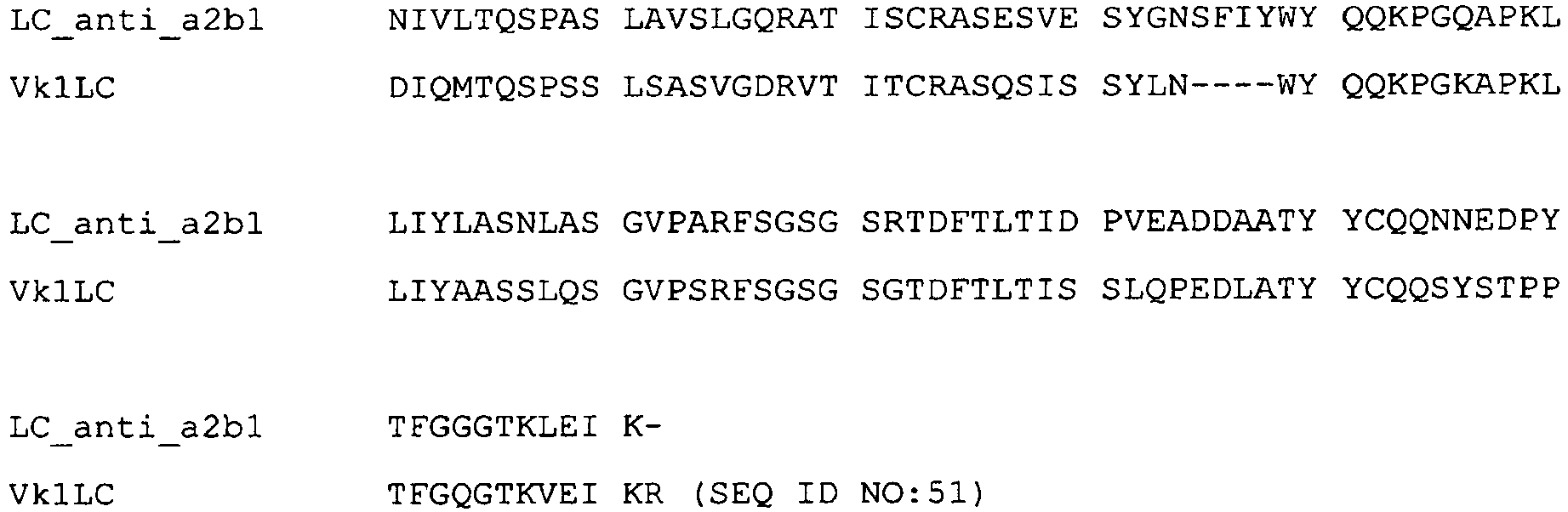
- Seven versions of heavy chain variants (HC1, HC2, HC3, HC4, HC5, HC6, HC7) were cloned. Mutations introduced through the engineering of the variable chains are highlighted.
HC1 (humanizing mutations highlighted):
HC2 (humanizing mutations are highlighted, potentially problematic motifs [CDR DS site]):
HC3 (humanizing and stabilizing mutations highlighted):
HC4 (humanizing and stabilizing mutations are highlighted, anti-aggregation mutation):
HC5 (humanizing and stabilizing mutations are highlighted, potential problematic motifs [CDR DS site]):
HC6 (humanizing and stabilizing mutations are highlighted, potential problematic motifs [CDR DS site], anti-aggregation mutation):
HC7 (grafted mutations are highlighted):
Below is the alignment of the HC anti-α21 integrin mAb vs. the HC Vk1_Vh1b human germline:HC2_anti_α2 QVQLHQPGAE LVKPGAPVKL SCKASGYTFT SYWMNWVKQR PGRGLEWIGR Vh1b QVQLVQSGAE VKKPGASVKV SCKASGYTFT SYYMHWVRQA PGQGLEWMGW HC2_anti_α2 IDPSDSETHY NQKFKDKATL TVDKSSSTAY IQLSSLTSED SAVYYCAKVG Vh1b INPNSGGTNY AQKFQGRVTM TRDKSSSTAY MELSSLRSED TAVYYCARWG HC2_anti_α2 RGY------F DYWGQGTTLT VSS Vh1b YDYDVFYYAM DYWGQGTLVT VSS (SEQ ID NO:52) - The variable heavy and light chains of each anti-integrin α2 mAb variant (5 different light chains: VL1-VL5 and 7 different heavy chains: VH1-VH7) were generated by gene synthesis including a 5'UTR-Sequence (5'-GTGCACAGC-3') with ApaLI and a 3'UTR (5'-GCTTCCACCAAGGGCCC-3') with ApaI (heavy chain) or BsiWI (light chain). The variable heavy domains were ligated into the ApaLI/ApaI sites of a modified pXL expression vector which contains a mutated variant of the human constant heavy chain (IGHG4, Swiss-Prot P01861, S108P, L115E) giving rise to an anti-integrin α2 VH-IGHG4 constant heavy chain mAb.
- The variable light domains were ligated into the ApaLI/BsiWI sites of a modified pXL expression vector which contains the human constant light chain (IGKC, Swiss-Prot: Q502W4) giving rise to an anti-integrin α2 VL-IGKC constant light chain mAb. The complete process of gene synthesis, cloning and DNA production was realized by a commercial vendor (Geneart AG).
- For comparison, a humanized
hIgG4 anti-alpha 2 integrin antibody known in the art (TMC2206) was used ("the comparator"). The comparator light- and heavy chain amino acid sequences are listed herein as SEQ IDs NO: 53 and 54 inFigure 10e .Table 7: List of humanization variants of anti- α2-integrin mAb LC/HC combinatior Humanization variant LC1/HC1 Mutations adressing humanization only LC2/HC2 Mutations adressing humanization only and LC/HC potentially problematic sites (NS; DS) LC3/HC3 Mutations adressing humanization and stabilization LC3/HC4 Mutations adressing humanization and stabilization and anti-aggregation LC4/HC5 Mutations adressing humanization and stabilization and LC/HC potentially problematic sites (NS; DS) LC4/HC6 Mutations adressing humanization and stabilization and anti-aggregation and LC/HC potentially problematic sites (NS; DS) LC5/HC7 Mutations adressing humanization by grafting TMC2206 Comparator according to SEQ ID NO:53 - In order to verify the sequences, the mAbs were analyzed using mass spectrometry. For intact mass measurements the sample was trapped for 20 minutes and desalted with 20 µl/min on a monolithic trap column with 2% Acetonitrile / 0.1% TFA (v/v) prior to elution with a gradient ranging from 15 % Eluent A (H2O / 0.05 %TFA) to 50 % Eluent B (Acetonitrile / 0.05% TFA).
- The sample was separated operating in nanoflow (300 nl/min) on an monolithic column (PS-DVB; 100µm I.D. x 5 cm) with a temperature of 37°C. Introduction of the sample was carried out using electrospray needles from new objective with an outer diameter of 365µm, inner diameter of 75 µm and an end tip diameter of 15 µm plus sheath gas. After acquisition the spectra were summed over the corresponding time range and deconvoluted using the protein reconstruction tool delivered with BioAnalyst from Applied Biosystems/MDS Sciex.
- Protein sequences were translated from plasmid coding sequences and the masses of the HC and LC were calculated (Table 8).
Table 8: Mass spectrometric analysis of the purified humanized anti-α2-integrin mAbs. LC/HC combination Light chain Heavy chain Expected Da Measured Da ppm Expected Da (G0F) Measured Da ppm LC1/HC1 23727.38 23724.56 119 50314.47 50311.80 53 LC2/HC2 23741.41 23738.26 130 50328.5 50327.52 19 LC3/HC3 23757.36 23753.89 146 50304.47 50301.96 50 LC3/HC4 23757.36 23754.17 134 50333.52 50331.29 44 LC4/HC5 23771.39 23769.87 64 50318.5 50317.68 16 LC4/HC6 23771.39 23768.13 137 50347.54 50350.26 54 LC5/HC7 23792.41 23789.01 142 50187.3 50184.84 49 TMC2206 23378.01 23374.51 150 50237.54 50233.48 81 - The observed values were in good agreement with the calculated masses and verify the cloned constructs.
- For the Solid Phase Assay, integrin (α2-I-domain: α2-I-domain GST aa 140-339 in TBS/5mM Mn2+, 50µl/well;) was immobilized on 96-well plate (Corning Costar, 3690), at room temperature overnight. Then, 25µl/well of blocking solution (5 % BSA (crude) (A7906), 1xTBS) were added and discarded. 200µl/well of blocking solution were added and left for 3h at room temperature. After a washing step (3 times with 200µl/well binding buffer: 1xTBS and 0,1% BSA (A7638) and 2mM Mn2+; TBS: 150mM NaCl, 25mM Tris (Fluka 93371) pH7.4), samples were incubated at RT for 3h stationary with 50 µl of:
- a) biotinylated collagen only - control (10µl binding buffer and 40µl biot. collagen)
- b) 10µl/well compound, 40µl/well biotinylated collagen
- c) Blank: 50µl/well binding buffer
- After a washing step (3 times with 200µl/well binding buffer), samples were incubated with 50µl/well ExtrAvidin Peroxidase (Peroxidase conjugate, Sigma E2886; 1:500 in binding buffer) for 30 min at RT and again washed 4 times with 200µl/well binding buffer. After addition of 50 µl/well peroxidase substrate (ABTS solution; 2,2'-Azino-bis 3-Ethylbenzthiazoline-6-sulfonic acid), Sigma A-1888; 275µl (11mg ABTS dissolved in 0,5ml dH2O; and 5,5ml 0.1M Sodium-acetate (Sigma S-3272) / 0.05M NaH2PO4 (Riedel de Haen 04270) pH5.0; and 55µl H2O2 Sigma H-1009 (10µl (= 30%) and 1045µl dH2O) for 10 - 30 min at RT, stationary (untill green staining is obtained) and addition of 50 µl/well 2 % SDS, absorbance was read out at 405 nm (SpectraMax 190). % Inhibition is calculated as 100-((mean value compounds* 100)/mean value collagen positive control) after blank subtraction.

- In summary,
α 2 integrin mAB showed no effect on α1β1/collagen interaction (solid phase assay), α5β1/fibronectin interaction (solid phase assay), aIIbb3 (GPIIbIIIa) activation (FACS-assay), P-selectin expression on hu plt (FACS-assay), human platelet aggregation in whole blood alone or after stimulation with ADP, TRAP, collagen, LDH-, TNFα-, or IL1β-release from hu PBL (alone or in combination with LPS). - To assess the activity of the integrin anti-a2 mab in angiogenesis an in vitro assay with HUVEC cells was performed. Matrigel (BD Biosciences, #354230) was mixed with collagen type I (BD Biosciences #35429) (
matrigel 1/3.25,PBS 5x 1/5, collagen I 1mg/ml, qsp water) and incubated for 1h at 37°C, 5% CO2. The adherent HUVEC cells were carefully detached from culture flasks with Acutase solution, centirfuged and resuspended in culture medium (EBM,FCS 2%, EGF bullet kit) at 1.2 105 cells/ml. 100µl of the cell suspension were added to the wells with the matrix in the presence or absence of serial dilutions of anti-a2 mab and FGF2 (Peptrotech, 10ng/ml) and incubated for 18h at 37°C, 5% CO2. For detection of tubule formation, cresyl violet solution was added and incubated for 30 min at 37°C. The tubule formation was determined by measuring the sum of the tubule length per well. Calculations were performed versus negative control (without FGF2) and positive controls (with FGF2 abut without anti-a2 mab) using Image Pro and software (MediaCybernetics), measuring 6 replicats per condition. The according results are shown inFig. 2 . Anti-alpha2-Integrin mAB was able to inhibit FGF2-induced angiogenesis in a dose dependent manner. - For protein-protein interaction studies either recombinantly expressed integrin α2β1 integrin or the 1-domain of integrin α2β1 integrin was coated to 96-well plates (Corning Costar 3690) in TBS buffer over night at 4°C. After washing off excessive protein, the plates were blocked with BSA solution (5% Sigma A7906) and washed again. Serial dilutions of α2 integrin Mab were added to the plates as well as biotinylated collagen (rat tail, Sigma C8897). This was performed in the presence or absence of 4% HSA. After an incubation of 2h at room temperature the plates were washed again. Extravidin Peroxidase solution (Sigma E2886) was added and the plates are incubated for 20 min in the dark. Measurement was performed in an Elisa reader (SpectraMax190 Molecular Devices) at 405nM. Percentage of inhibition and IC50s are calculated versus known standards.
- For platelet binding studies to collagen, plates (Isoplate, Perkin Elmer, F1450 571) were coated with collagen (Sigma C8897) in TBS for 1 h at room temperature. The wells were washed with TBS repeatedly before serial dilutions of anti-α2 integrin MAb were added. Freshly prepared human platelet rich plasma or isolated human platelets, which were anticoagulated with hirudin, PGE1 and ReoPro and labelled with CalceinAM (C-3099 Molecular Probes were added and incubated for 90 min at room temperature protected from light. After washing, the plates were measured in an M5 reader (Molecular Devices) at 492 nM EX, 535 nM EM. Percentage of inhibition and IC50s are calculated versus known standards.
- In experiments under shear, anti-α2 integrin mAB was analysed for its ability to inhibit platelet adhesion to collagen under flow. Glass capillaries were coated with collagen over night at 4°C. After washing and blocking with BSA, they were installed in a flow device. Freshly drawn anticoagulated human blood from volonteers was labelled with DiOC6(3) and incubated for 10 min with serial dilutions of anti-α2 integrin mAB at 37°C. The samples were flown through the capillaries at a shear rate of 3000s-1 mimicking arterial flow. After rinsing the capillaries, 10 pictures were taken representing the surface of the capillary which was in contact with the flowing blood. Using an imaging software, surface coverage was determined and percentage of inhibition and IC50s were calculated versus known standards.
- For the thrombocyte adhesion assay, thrombocytes were enriched as follows: Hirudin (20µg/ml; Refludan (Pharmion)) and blood was centrifuged at 150g for 20min to produce anticoagulated human blood. platelet rich plasma (PRP) was collected and again centrifuged and collected as above. Platelet poor plasma was obtained from the remaining blood by centrifugation at 1940 g for 10 min (2 times). PPP was added to the diluted cells (2mM Mg) and concentration of cells was adjusted to 2 x 10⁵/µl. Cells were left for 0.5 hrs and diluted to 5x10⁴ / µl. Thereafter cells were contacted with 3µg/ml ReoPro (2.5µg/ml; Centocor B.V., Leiden, NL) (10 min, RT), 6mM MnCl2x4H2O (5mM) was added (incubation for 10 min).
- Plates were prepared as follows: Plates (Perkin Elmer, IsoPlate, 1450-571) were incubated with 100µl / well collagen Type I 10µg/ml (Type I from rat tail C8897 Sigma Stock 200µg/ml in 0,01M in acetic acid) at RT for 1 hr. Then, they were washed 3 times with 200µl / well TBS (50mM Tris-HCl pH 7.4, 120mM NaCl, 2,7mM KCl, 0.05mM CaCl2, 2mM MgCl2 x 6 H2O, 0.1 % BSA. Thereafter, 10µl/well compound and ReoPro- and Mn-treated thrombocytes (5x10⁴ cells/µl, 50µl/well) were added. Cells were incubated for 1.5 hrs (darkness) and washed 3 times with 200µl/well TBS. 2.5µM Calcein AM (50µl/well, C-3099, Molecular Probes, MW 994.87, 30 min, RT) was added, followed by a washing step. The read out step was carried out using a SpectraMax M5:
Fluoreszenz EX 492 EM 535 Cutoff: 530 Automatic in the absence of cells. % Inhibition is calculated as 100-((mean value compounds* 100)/mean value control) after blank subtraction. - As can be gained from
figure 3 , anti-α2 integrin mAB dose dependently inhibits platelet adhesion under shear stress, with a nanomolar IC50. - All humanized variants and the comparator were tested for the aggregation percentage. Size exclusion chromatography was performed on an ÄKTA explorer 10 (GE Healthcare) using a TSKgel G3000SWXL column (7,8mm ID x 30,0 cm L, TosohBioscience) with a TSKgel SWXL guard column (TosohBioscience). 30µl of sample at 0.4-1mg/ml were injected and the chromatography was performed at 1ml/min using 100 mM Na2SO4, 100 mM Na2HPO4, 0.05% NaN3 pH 6.7 as running buffer and a detection wavelength of 280nm. The column was calibrated using gel filtration molecular weight markers (Sigma Aldrich). Data evaluation was done using Unicorn software v5.11 (GE Healthcare).
Table 10: Aggregation percentage of anti-α2-integrin mAbs determined by size exclusion chromatography. LC/HC combination Aggregation [%] Peakheight [mAU] LC1/HC1 < 0.5 74.4 LC2/HC2 < 0.5 47.8 LC3/HC3 < 0.5 93.7 LC3/HC4 2.3 67.2 LC4/HC5 1.8 67.9 LC4/HC6 < 0.5 29.1 LC5/HC7 < 0.5 46.5 TMC2206 11.8 20.7 - As can be gained from table 10, all tested variants of the alpha-2 integrin mAb have a low percentage of aggregates. When compared with the aggregation behaviour of the comparator, all tested alpha-2 integrin antibodies exhibited lower aggregation percentage values than the comparator.
- Surface plasmon resonance technology on a Biacore 3000 (GE Healthcare) was used for a detailed kinetic characterisation of the purified humanized antibodies. A capture assay was used with the anti-integrin antibody captured by an anti-human Fc specific antibody (MAB1302, Millipore) and the integrin α2 I domain was used as analyte. Typically, 120 RU of anti-integrin antibody were captured on a research grade CM5 by the immobilised anti-human Fc specific antibody, resulting in an Rmax of 30 RU for the I domain bound to the antibody. Binding kinetics were measured over a concentration range between 0.8 to 25 nM I domain in HBS-P buffer supplemented with 4 mM MgCl2 (10 mM HEPES pH 7.4, 150 mM NaCl, 0.005 % surfactant P20) at a flow rate of 30 µl/min. Chip surfaces were regenerated with 10 mM glycine pH 2.5. Kinetic parameters were analysed and calculated in the BIAevaluation program package (version 4.1) using a flow cell with the immobilised anti-human Fc specific antibody as reference. A 1:1 binding model with mass transfer was applied for a global fit of the data for curves corresponding to analyte concentrations from 0.8-25 nM of antibody.
- Table11). Three variants have a similar KD in Biacore as the non-humanized mAb:
- combination LC1/HC1 (Mutations adressing humanization only)
- combination LC3/HC3 (Mutations adressing humanization and stabilization)
- combination LC3/HC4 (Mutations adressing humanization and stabilization and anti-aggregation).
- The variant mutation by grafting is close to the non-humanized mAb.
- In order to verify the epitope of the
non-humanized anti-alpha 2 mAb with the comparator mAb, epitope characterisation was performed using surface plasmon resonance technology on a Biacore 3000 (GE Healthcare). The Fab fragment corresponding to thenon-humanized anti-alpha 2 mAb was immobilized on a CM5 chip by amine reactive coupling at 500 RU. The integrin I domain was captured by the Fab fragment at 10µl/min and after a short dissociation period, the antibody TMC2206 was allowed to bind at 30µl/min to the α2 I domain. Regeneration was performed with 10mM Glycine buffer pH 2.0. In a second experiment the comparator mAb TMC2206 was captured on a surface of anti-human Fc specific antibody (MAB1302 Millipore). Then the integrin I domain was bound followed by the non-humanized Fab. The results can be gained fromfigures 13 and14 . The results clearly show that the comparator antibody, TMC2206 binds to the integrin I domain prebound by non-humanized Fab. - Thus, the non-humanized Fab binds to the integrin I domain which is pre-bound by the comparator mAb TMC2206. Simultaneous binding of the non-humanized Fab and the comparator mAb to the integrin α2 I domain indicates that the epitope of both the Fab and the comparator mAb are not identical. This means that the
anti alpha 2 antibody of present invention and the comparator antibody bind different epitopes withinalpha 2 integrin. - As α2β1 integrin is expressed on blood platelets, playing an important role in their adhesion to collagen, an in vitro assay system for platelet binding studies using these cells was used. For platelet binding studies, plates (Isoplate, Perkin Elmer, F1450 571) were coated with collagen (Sigma C8897) in TBS for 1 h at room temperature. The wells were washed with TBS repeatedly before serial dilutions of anti-α2 integrin mAb were added. Freshly prepared human platelet rich plasma or freshly isolated human platelets, which were anticoagulated with hirudin, PGE1 and ReoPro and labelled with CalceinAM (C-3099 Molecular Probes were added and incubated for 90 min at room temperature protected from light. After washing, the plates were measured in an M5 reader (Molecular Devices) at 492 nM Exitation, 535 nM Emission. Percentage of inhibition and IC50s are calculated versus titration curves prepared using small molecule inhibitors of alpha-2-Integrin or the non-humanized alpha-2 mAB . The results can be gained from Table 12.
Table 12: Inhibition of binding of washed platelets to collagen LC/HC combination IC50 µg/ml LC1/HC1 0.021 LC2/HC2 0.092 LC3/HC3 0.012 LC3/HC4 0.016 LC4/HC5 0.057 LC4/HC6 0.068 LC5/HC7 0.031 TMC2206 (comparator) 0.023 - As can be gained from the results shown in table 12, platelet inhibition displayed by the different
anti alpha 2 antibody variants under static conditions using washed platelets is comparable to that of the comparator antibody and for some variants (LC3/HC3 or LC3/HC4) even significantly or slightly (LC1/HC1) stronger.Table 13: Inhibition of binding of platelets to collagen in platelet rich plasma LC/HC combination MW IC50 µg/ml LC1/HC1 0.277 LC2/HC2 3.963 LC3/HC3 0.132 LC3/HC4 0.193 LC4/HC5 3.251 LC4/HC6 4.113 LC5/HC7 0.224 TMC2206 0.110 - As can be gained from the results shown in table 13, platelet inhibition displayed by the different
anti alpha 2 antibody variants under static conditions using platelet-rich plasma, variants LC1/HC1, LC3/HC3, LC3/HC4 and LC5/HC7 is comparable to that of the comparator antibody. - As can be concluded from the static platelet binding assays, the humanized forms of the anti-α2-integrin antibody block adhesion of freshly isolated human platelets in the presence or absence of blood plasma in a concentration dependent manner. Four of the variants show a similar inhibitory activity in the bioassay as the non-humanized mAb:
- combination LC1/HC1 (Mutations adressing humanization only)
- combination LC3/HC3 (Mutations adressing humanization and stabilization)
- combination LC3/HC4 (Mutations adressing humanization and stabilization and anti-aggregation)
- combination LC5/HC7 (Mutations adressing humanization by grafting)
- The three variants adressing the problematic sites (NS; DS) LC2/HC2, LC4/HC5 and LC4/HC6 and showing lower platelet inhibition in the above platelet binding experiments were identical with the variants exhibiting weaker α2 I domain binding activity than not-humanized
anti-alpha 2 integrin antibody in the above Biacore experiments of example 7 (see table 11). Thus, the results of the platelet binding assays are well in accordance with the affinity data from the Biacore evaluations. - Results with respect to thermal stability are summarized in table 14. The antibodies show comparable, equal or better thermal stability as the comparator. Thermostability measurements are performed using a PCR thermocycler (My-IQ -two in a temperature range between 10 and 90 °C with 1 °C/min. Two microgram of antibody diluted in PBS buffer was supplemented with 40XSYPRO Orange (Invitrogen).
Table 14: thermal stability of the different variants LC/HC combination Melt. Temp °C (1) Melt. Temp °C (2) LC1/HC1 64 - LC2/HC2 63 - LC3/ HC3 64 68 LC3/HC4 66 - LC4/HC5 62 67 LC4/HC6 66 - LC5/HC7 65 72 TMC2206 (comparator) 65 71 -
- <110> SANOFI
- <120> Peptide or peptide complex binding to
alpha 2 integrin and methods and uses involving the same - <130>
DE2010/156 - <140>
EP10305929.1
<141> 2010-08-31 - <160> 54
- <170> PatentIn version 3.5
- <210> 1
<211> 111
<212> PRT
<213> Artificial Sequence - <220>
<223> Light chain variable domain of anti-alpha2-integrin mAb - <400> 1

- <210> 2
<211> 117
<212> PRT
<213> Artificial sequence - <220>
<223> Heavy chain variable domain of anti-alpha2-integrin mAb - <400> 2


- <210> 3
<211> 10
<212> PRT
<213> Artificial Sequence - <220>
<223> CDR1 of the heavy chain variable domain - <400> 3

- <210> 4
<211> 16
<212> PRT
<213> Artificial Sequence - <220>
<223> CDR2 of the heavy chain variable domain - <400> 4

- <210> 5
<211> 8
<212> PRT
<213> Artificial Sequence - <220>
<223> CDR3 of the heavy chain variable domain - <400> 5

- <210> 6
<211> 15
<212> PRT
<213> Artificial Sequence - <220>
<223> CDR1 of the light chain variable domain - <400> 6

- <210> 7
<211> 7
<212> PRT
<213> Artificial Sequence - <220>
<223> CDR2 of the light chain variable domain - <400> 7

- <210> 8
<211> 9
<212> PRT
<213> Artificial Sequence - <220>
<223> CDR3 of the light chain variable domain - <400> 8

- <210> 9
<211> 218
<212> PRT
<213> Artificial Sequence - <220>
<223> Chimeric (anti-alpha2-VL-IGKC-CL) light chain - <400> 9


- <210> 10
<211> 443
<212> PRT
<213> Artificial sequence - <220>
<223> Chimeric (anti-alpha2-VH-IGHG4-CH1) mAb - <400> 10
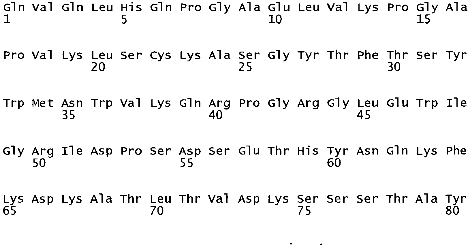


- <210> 11
<211> 231
<212> PRT
<213> Artificial sequence - <220>
<223> Chimeric (anti-alpha2-VH-IGHG1-CH1) heavy chain Fab fragment - <400> 11

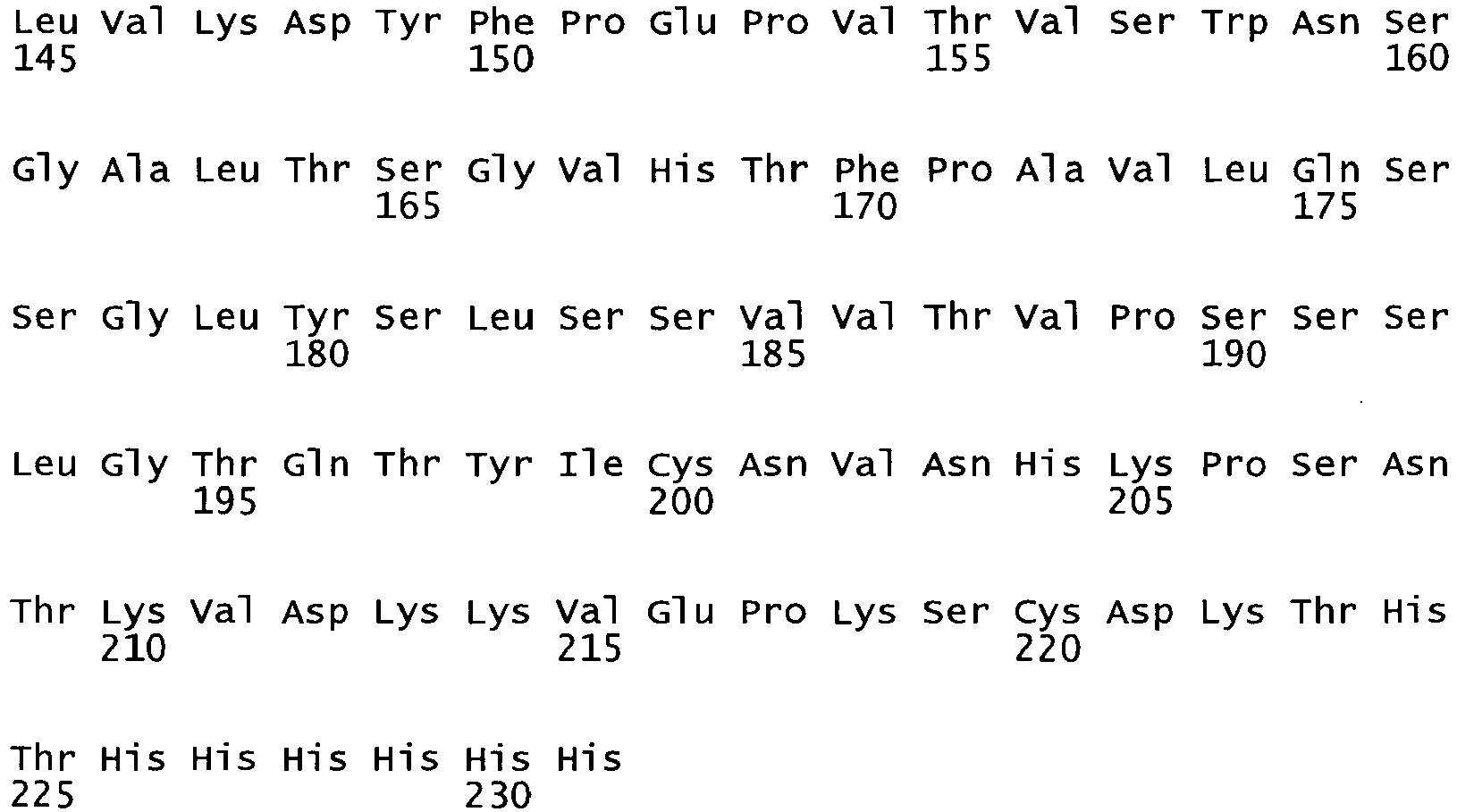
- <210> 12
<211> 333
<212> DNA
<213> Artificial Sequence - <220>
<223> Light chain variable domain of anti-alpha2-integrin mAb" - <400> 12

- <210> 13
<211> 351
<212> DNA
<213> Artificial Sequence - <220>
<223> Heavy chain variable domain of anti-alpha2-integrin mAb - <400> 13

- <210> 14
<211> 654
<212> DNA
<213> Artificial sequence - <220>
<223> chimeric (anti-alpha2-VL-IGKC-CL) light chain - <400> 14

- <210> 15
<211> 1329
<212> DNA
<213> Artificial Sequence - <220>
<223> Chimeric (anti-alpha2-VH-IGHG4-CH1) mAb - <400> 15


- <210> 16
<211> 693
<212> DNA
<213> Artificial sequence - <220>
<223> Chimeric (anti-alpha2-VH-IGHG1-CH1) heavy chain Fab fragment - <400> 16
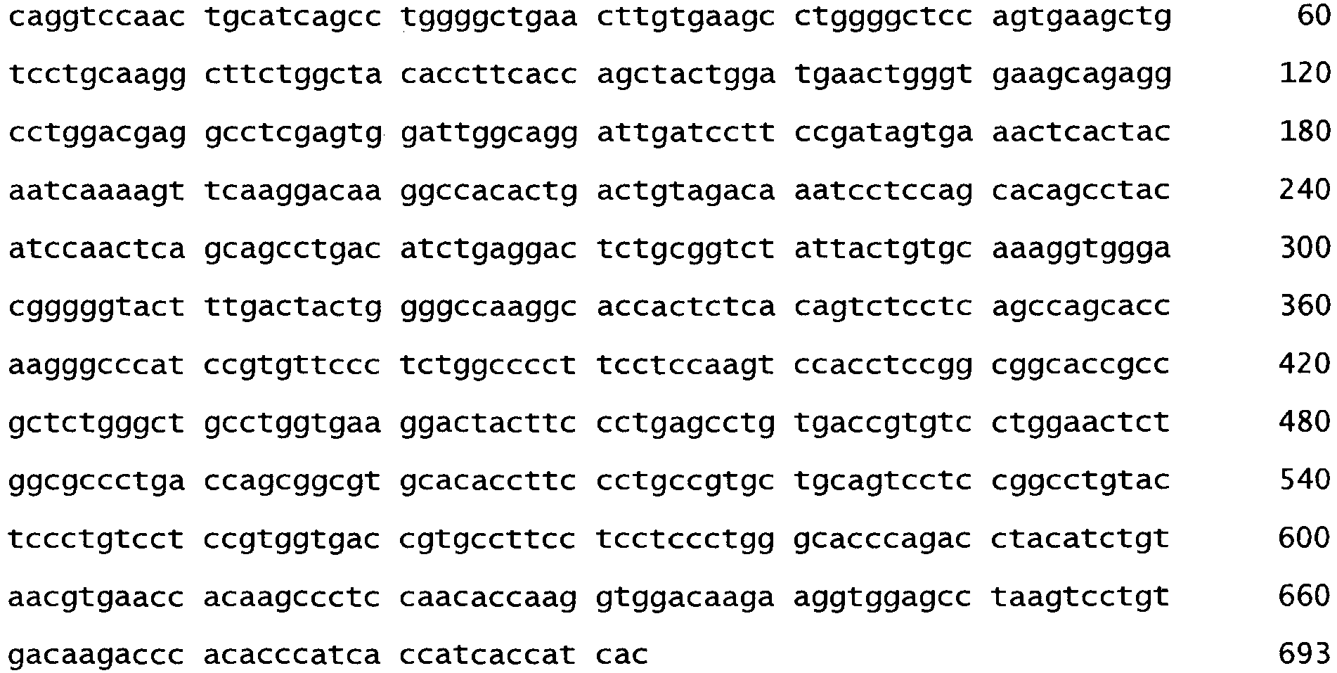
- <210> 17
<211> 107
<212> PRT
<213> Artificial Sequence - <220>
<223> Human IGKC protein (Swiss-Prot: Q502W4) - <400> 17


- <210> 18
<211> 327
<212> PRT
<213> Artificial Sequence - <220>
<223> Human IGHG4 protein (Swiss-Prot: P01861.1 (S108P, L115E)) - <400> 18


- <210> 19
<211> 330
<212> PRT
<213> Artificial sequence - <220>
<223> Human IGHG1 protein (Swiss-Prot: Q569F4) - <400> 19


- <210> 20
<211> 1181
<212> PRT
<213> Homo sapiens - <400> 20


- <210> 21
<211> 3546
<212> DNA
<213> Homo sapiens - <400> 21


- <210> 22
<211> 798
<212> PRT
<213> Homo sapiens - <400> 22


- <210> 23
<211> 3879
<212> DNA
<213> Homo sapiens - <400> 23

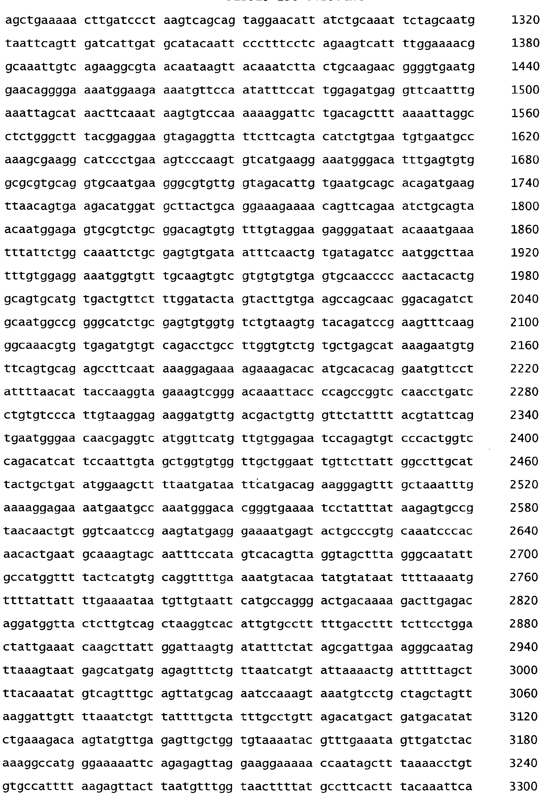

- <210> 24
<211> 23
<212> DNA
<213> Artificial Sequence - <220>
<223> 5'-GeneRacer primer for amplifying variable domains of anti-alpha2-integrin mAb - <400> 24
cgactggagc acgaggacac tga 23 - <210> 25
<211> 21
<212> DNA
<213> Artificial Sequence - <220>
<223> RACEMOG2a: 3'-primer internal to murine hinge region for amplifying variable domains of anti-alpha2-integrin mAb - <400> 25
aggacagggc ttgattgtggg 21 - <210> 26
<211> 24
<212> DNA
<213> Artificial Sequence - <220>
<223> CKFOR: 3'-primer internal to murine ck region for amplifying variable domains of anti-alpha2-integrin mAb - <400> 26
ctcattcctg ttgaagctct tgac 24 - <210> 27
<211> 52
<212> DNA
<213> Artificial Sequence - <220>
<223> alpha2mAB-VL FOR: primer for amplifying the light chain of anti-alpha2-integrin mAb - <400> 27
ctggtggcca ccgccaccgg cgtgcacagc aacattgtgc tgacccaatc tc 52 - <210> 28
<211> 30
<212> DNA
<213> Artificial Sequence - <220>
<223> alpha2mAB-VL REV: primer for amplifying the light chain of anti-alpha2-integrin mAb - <400> 28
accgtacgtt ttatttccag cttggtcccc 30 - <210> 29
<211> 52
<212> DNA
<213> Artificial Sequence - <220>
<223> alpha2mAB mAB-VH FOR: primer for amplifying the heavy chain of anti-alpha2-integrin mAb - <400> 29
ctggtggcca ccgccaccgg cgtgcacagc caggtccaac tgcatcagcc tg 52 - <210> 30
<211> 39
<212> DNA
<213> Artificial Sequence - <220>
<223> alpha2mAB mAB-VH REV: primer for amplifying the heavy chain of anti-alpha2-integrin mAb - <400> 30
tagggccctt ggtgctggct gaggagactg tgagagtgg 39 - <210> 31
<211> 54
<212> DNA
<213> Artificial Sequence - <220>
<223> Leader FOR1-54: primer for introducing leader sequence into the variable chains of anti-alpha2-integrin mAb - <400> 31
gctagcacca tgggctggtc ctgcatcatc ctgtttctgg tggccaccgc cacc 54 - <210> 32
<211> 26
<212> DNA
<213> Artificial Sequence - <220>
<223> Leader FOR1-23: primer for introducing leader sequence into the variable chains of anti-alpha2-integrin mAb - <400> 32
caagctagca ccatgggctg gtcctg 26 - <210> 33
<211> 111
<212> PRT
<213> Artificial Sequence - <220>
<223> LC1: Light chain variable domain of anti-alpha2-integrin mAb with humanizing mutations - <400> 33

- <210> 34
<211> 111
<212> PRT
<213> Artificial sequence - <220>
<223> LC2: Light chain variable domain of anti-alpha2-integrin mAb with humanizing mutations - <400> 34


- <210> 35
<211> 111
<212> PRT
<213> Artificial Sequence - <220>
<223> LC3: Light chain variable domain of anti-alpha2-integrin mAb with humanizing and stabilizing mutations - <400> 35

- <210> 36
<211> 111
<212> PRT
<213> Artificial Sequence - <220>
<223> LC4: Light chain variable domain of anti-alpha2-integrin mAb with humanizing and stabilizing mutations - <400> 36


- <210> 37
<211> 111
<212> PRT
<213> Artificial Sequence - <220>
<223> LC5: Light chain variable domain of anti-alpha2-integrin mAb with grafted mutations - <400> 37

- <210> 38
<211> 117
<212> PRT
<213> Artificial Sequence - <220>
<223> HC1: Heavy chain variable domain of anti-alpha2-integrin mAb with humanizing mutations - <400> 38

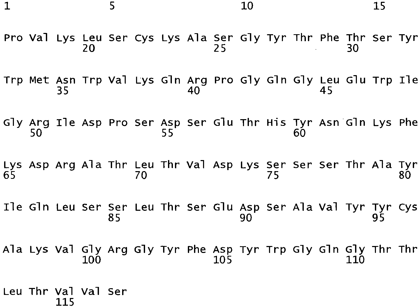
- <210> 39
<211> 117
<212> PRT
<213> Artificial Sequence - <220>
<223> HC2: Heavy chain variable domain of anti-alpha2-integrin mAb with humanizing mutations - <400> 39


- <210> 40
<211> 117
<212> PRT
<213> Artificial Sequence - <220>
<223> HC3: Heavy chain variable domain of anti-alpha2-integrin mAb with humanizing and stabilizing mutations - <400> 40

- <210> 41
<211> 117
<212> PRT
<213> Artificial Sequence - <220>
<223> HC4: Heavy chain variable domain of anti-alpha2-integrin mAb with humanizing and stabilizing mutations - <400> 41


- <210> 42
<211> 117
<212> PRT
<213> Artificial Sequence - <220>
<223> HC5: Heavy chain variable domain of anti-alpha2-integrin mAb with humanizing and stabilizing mutations - <400> 42

- <210> 43
<211> 117
<212> PRT
<213> Artificial Sequence - <220>
<223> HC6: Heavy chain variable domain of anti-alpha2-integrin mAb with humanizing and stabilizing mutations - <400> 43

- <210> 44
<211> 117
<212> PRT
<213> Artificial Sequence - <220>
<223> HC7: Heavy chain variable domain of anti-alpha2-integrin mAb with grafted mutations - <400> 44


- <210> 45
<211> 717
<212> DNA
<213> Mus musculus - <400> 45

- <210> 46
<211> 1401
<212> DNA
<213> Mus musculus - <400> 46


- <210> 47
<211> 218
<212> PRT
<213> Mus musculus - <400> 47


- <210> 48
<211> 447
<212> PRT
<213> Mus musculus - <400> 48


- <210> 49
<211> 111
<212> PRT
<213> Artificial Sequence - <220>
<223> IGLKV79_IGLKJ2 - <400> 49

- <210> 50
<211> 116
<212> PRT
<213> Artificial Sequence - <220>
<223> IGHV11_IGHD33_IGHJ8 - <400> 50


- <210> 51
<211> 108
<212> PRT
<213> Artificial Sequence - <220>
<223> Vk1LC - <400> 51

- <210> 52
<211> 123
<212> PRT
<213> Artificial Sequence - <220>
<223> Vh1b - <400> 52


- <210> 53
<211> 213
<212> PRT
<213> Artificial Sequence - <220>
<223> light chain variable domain ofanti-integrin 2 mAb - <400> 53


- <210> 54
<211> 445
<212> PRT
<213> Artificial Sequence - <220>
<223> heavy chain variable domain ofanti-integrin 2 mAb - <400> 54


| (LC1) Humanization | LC2 humanization and NS site in CDRs | LC3 humanization and stabilization | LC4 humanization and NS site in CDRs and stabilization | LC5 (grafting) | |
| (HC1) Humanization | X | ||||
| (HC2) Humanization and DS in CDRs | X | ||||
| (HC3) Humanization and stabilization | X | ||||
| (HC4) Humanization and stabilization and "anti-aggregation" | X | ||||
| (HC5) Humanization and DS in CDRs and stabilization | X | ||||
| (HC6) Humanization and stabilization and "anti-aggregation"and DS in CDRs | X | ||||
| HC7 (grafting) | X |
| Light Chain (Sequential numbering) | (LC1) Humanization | (LC2) humanization and NS in CDRs | (LC3) humanization and stabilization | (LC4) humanization and NS in CDRs and stabilization | (LC5) grafting |
| ALA9 | SER | SER | SER | SER | |
| ALA12 | SER | SER | |||
| LEU15 | VAL | VAL | VAL | VAL | PRO |
| SER22 | THR | ||||
| ASN34 | GLN | GLN | |||
| GLN46 | LYS | LYS | |||
| ALA47 | PRO | ||||
| ASP80 | ASN | ||||
| GLU83 | GLN | GLN | GLN | GLN | |
| ASP85 | GLU | GLU | |||
| ALA87 | THR | ||||
| THR89 | ASN |
| Heavy Chain (Sequential numbering) | (HC1) Humanization | (HC2) Humanization and DS in CDRs | (HC3) Humanization and stabilization | (HC4) Humanization and stabilization and " anti-aggregation" | (HC5) Humanization and DS in CDRs and stabilization | (HC6) Humanization and stabilization and " anti-aggregation" and DS in CDRs | (HC7) grafting |
| HIS5 | VAL | ||||||
| PRO7 | SER | ||||||
| LEU11 | VAL | ||||||
| VAL12 | LYS | ||||||
| PRO17 | SER | SER | SER | SER | SER | ||
| LEU20 | VAL | ||||||
| LYS38 | ARG | ||||||
| ARG40 | ALA | ||||||
| ARG43 | GLN | GLN | GLN | ||||
| ASP55 | GLU | GLU | GLU | ||||
| ASN61 | ALA | ||||||
| LYS65 | GLN | ||||||
| ASP66 | GLY | ||||||
| LYS67 | ARG | ARG | ARG | ||||
| SER76 | THR | ||||||
| ILE81 | MET | ||||||
| GLN82 | GLU | ||||||
| THR87 | ARG | ||||||
| SER91 | THR | ||||||
| VAL93 | LYS | LYS | |||||
| THR112 | LEU | ||||||
| LEU113 | VAL | ||||||
| SER116 | VAL | VAL | VAL | VAL | VAL | VAL | |
| 3 mutations | 4 mutations | 2 mutations | 3 mutations | 3 mutations | 4 mutations | 20 mutations |
Claims (17)
- Isolated monoclonal antibody or antigen binding fragment thereof, wherein said antibody or fragment specifically binds to the 1-domain of a human α2-integrin, said antibody comprising a heavy chain variable region (VH) domain and a light chain variable region (VL) domain, wherein said antibody or fragment competes with a reference antibody for binding to the epitope of the reference antibody, said reference antibody comprising a light chain being the variable domain and the constant domain according to SEQ ID NO: 9 and a heavy chain being the variable domain and the constant domain according to SEQ ID NO: 10 or being the variable domain and the constant domain according to SEQ ID NO: 11.
- The isolated monoclonal antibody or antigen binding fragment thereof of claim 1, wherein said antibody or fragment cross-reacts with a non-human primate α2-integrin but does not cross-react with a non-primate α2-integrin.
- The isolated monoclonal antibody or antigen binding fragment thereof of claim 1 or 2, comprising one or more of the following components a to f:a) LCDR1, wherein LCDR1 is RASESVESYGNSFIY (SEQ ID NO:6) or a functionally active variant thereof,b) LCDR2, wherein LCDR2 is LASNLAS (SEQ ID NO:7) or a functionally active variant thereof,c) LCDR3, wherein LCDR3 is QQNNEDPYT (SEQ ID NO:8) or a functional active variant thereof,d) HCDR1, wherein HCDR1 is (GYTFTSYWMN, SEQ ID NO:3) or a functionally active variant thereof,e) HCDR2, wherein HCDR2 is RIDP SD SETH YNQKFK (SEQ ID NO:4) or a functionally active variant thereof, andf) HCDR3, wherein HCDR3 is VGRGYFDY (SEQ ID NO:5) or a functional active variant thereof,and wherein the one or more of the components a) to f) are arranged to allow for binding of the antibody or fragment to α2-integrin, and wherein said variants have at least 10% of the α2-integrin binding activity of the corresponding peptides without sequence alteration.
- The isolated monoclonal antibody or antigen binding fragment thereof according to any one of claims 1-3,(i) wherein components a) to c) are comprised in a variable domain of a light chain (VL); and/or(ii) wherein components d) to f) are comprised in a variable domain of a heavy chain (VH); and/or(iii) wherein the antibody or fragment is a chimeric antibody, a humanized antibody, a Fab, a Fab', a F(ab')2, a Fv, a disulfide linked Fv, a scFv, a (scFv)2, a single domain antibody, a diabody, a multispecific antibody, a dual specific antibody, an isotype antibody, a dual variable domain antibody and a bispecific antibody; and/or(iv) wherein the antibody or fragment comprises a heavy chain immunoglobulin constant domain selected from the group consisting of: a human IgM constant domain, a human IgG1 constant domain, a human IgG2 constant domain, a human IgG3 constant domain, domain, a human IgG4 constant domain, a human IgE constant domain, and a human IgA constant domain; and/or(v) wherein the functionally active variant is a functionally active fragment consisting of at least 90 % sequence identity to any of the amino acid sequences of SEQ ID NOS: 3 to 8; and/or(vi) wherein the functionally active variant is a functionally active variant having at least 70 %, preferably at least 80 %, more preferably at least 90 % sequence identity to an amino acid sequence of any of SEQ ID NOS: 3 to 8, particularly wherein the functionally active variant is derived from the amino acid sequences of any of SEQ ID NOS: 3 to 8 with one or more conservative amino acid substitution; and/or(vii) comprising the amino acid sequence of- SEQ ID NO: 1, or a functionally active variant thereof, and/or- SEQ ID NO: 2, or a functionally active variant thereof, and/or- SEQ ID NO: 9, or a functionally active variant thereof, and/or- SEQ ID NO: 10, or a functionally active variant thereof, and/or- SEQ ID NO: 11, or a functionally active variant thereof, or(viii) consisting of the amino acid sequence of- SEQ ID NO: 1, or a functionally active variant thereof, and- SEQ ID NO: 2, or a functionally active variant thereof, and- optionally 50 additional amino acid residue(s), preferably 1 to 40, more preferably 1 to 30, even more preferably at most 1 to 25, still more preferably at most 1 to 10, most preferably 1, 2, 3, 4 or 5 additional amino acids residue(s) or(ix) consisting of the amino acid sequence of- SEQ ID NO: 9, or a functionally active variant thereof, and- SEQ ID NO: 10, or a functionally active variant thereof, and- optionally 50 additional amino acid residue(s), preferably 1 to 40, more preferably 1 to 30, even more preferably at most 1 to 25, still more preferably at most 1 to 10, most preferably 1, 2, 3, 4 or 5 additional amino acids residue(s) or(x) consisting of the amino acid sequence ofwherein said variants have at least 10% of the α2-integrin binding activity of the corresponding peptides without sequence alteration.- SEQ ID NO: 9, or a functionally active variant thereof, and- SEQ ID NO: 11, or a functionally active variant thereof, and- optionally 50 additional amino acid residue(s), preferably 1 to 40, more preferably 1 to 30, even more preferably at most 1 to 25, still more preferably at most 1 to 10, most preferably 1, 2, 3, 4 or 5 additional amino acids residue(s);
- The isolated monoclonal antibody or antigen binding fragment thereof according claim 3 or 4;- wherein the functionally active variant of LCDR1 comprises the mutation at amino acid position 11, particularly HAsn->Gln; and/or- wherein the functionally active variant of HCDR2 comprises the mutation at amino acid position 6, particularly 6Asp->Glu; and/or- wherein the functionally active variant of SEQ ID NO: 1 comprises one or more mutations at amino acid positions 9, 12, 15, 22, 34, 46, 47, 80, 83, 85, 87 and/or 89, particularly selected from the group consisting of 9Ala->Ser, 12Ala->Ser, 15Leu->Val, 15Leu->Pro, 22Ser->Thr, 34Asn->Gln, 46Gln->Lys, 47Ala->Pro, 80Asp->Asn, 83Glu->Gln, 85Asp->Glu, 87Ala->Thr and 89Thr->Asn; and/or- wherein the functionally active variant of SEQ ID NO: 2 comprises one or more mutations at amino acids positions 5, 7, 11, 12, 17, 20, 38, 40, 43, 55, 61, 65, 66, 67, 76, 81, 82, 87, 91, 93, 112, 113 and/or 116, particularly selected from the group consisting of 5His->Val, 7Pro->Ser, HLeu->Val, 12Val->Lys, 17Pro->Ser, 20Leu->Val, 38Lys->Arg, 40Arg->Ala, 43Arg->Gln, 55Asp->Glu, 61Asn->Ala, 65Lys->Gln, 66Asp->Gly, 67Lys->Arg, 76Ser->Thr, 81Ile->Met, 82Gln->Glu, 87Thr->Arg, 91 Ser->Thr, 93Val->Lys, 112Thr->Leu, 113Leu->Val and 116Ser->Val;wherein said variants have at least 10% of the α2-integrin binding activity of the corresponding peptides without sequence alteration.
- The isolated monoclonal antibody or antigen binding fragment thereof according to any one of claims 1-5, wherein said antibody or fragment inhibits the interaction of the human α2-integrin with collagen in vitro, thereby inhibiting the activation of platelets due to adhesion of said platelets to said collagen.
- The isolated monoclonal antibody or antigen binding fragment thereof according to any one of the previous claims, (i) said heavy chain variable region domain comprising the heavy chain HCDR3 of SEQ ID NO:5, and preferably comprising the heavy chain CDRs of SEQ ID NO:3 (HCDR1), SEQ ID NO:4 (HCDR2), and SEQ ID NO:5 (HCDR3), or functionally active variants thereof, wherein said variants have at least 10% of the α2-integrin binding activity of the corresponding peptides without sequence alteration, and wherein the functionally active variant of HCDR2 preferably comprises the mutation Asp->Glu at amino acid position 6; and/or (ii) said light chain variable region domain comprising the light chain LCDR3 of SEQ ID NO:8, and preferably comprising the light chain CDRs of SEQ ID NO:6 (LCDR1), SEQ ID NO:7 (LCDR2), and SEQ ID NO:8 (LCDR3), or functionally active variants thereof, wherein said variants have at least 10% of the α2-integrin binding activity of the corresponding peptides without sequence alteration, and wherein the functionally active variant of LCDR1 preferably comprises the mutation Asn->Gln at amino acid position 11.
- The isolated monoclonal antibody or antigen binding fragment thereof according to any one of the previous claims, wherein said heavy chain variable region (VH) domain comprises one or more amino acid substitutions at positions selected from the group consisting of H5, H7, H11, H12, H17, H20, H38, H40, H43, H55, H61, H65, H66, H67, H76, H81, H82, H87, H91, H93, H112, H113 and H116 and/or said light chain variable region (VL) domain comprises one or more amino acid substitutions at positions selected from the group consisting of L9, L12, L15, L22, L34, L46, L47, L80, L83, L85, L87, and L89.
- The isolated monoclonal antibody or antigen binding fragment thereof according to any one of the previous claims, said heavy chain variable region (VH) domain having at least 90%, 95%, 97% or 99% sequence identity to a VH sequence selected from the group consisting of SEQ ID NO: 2, SEQ ID NO: 38 (HC1), SEQ ID NO:39 (HC2), SEQ ID NO:40 (HC3), SEQ ID NO:41 (HC4), SEQ ID NO:42 (HC5), SEQ ID NO:43 (HC6), and SEQ ID NO:44 (HC7) and/or said light chain variable region (VL) domain having at least 90%, 95%, 97% or 99% sequence identity to a VL sequence selected from the group consisting of SEQ ID NO: 1, SEQ ID NO: 33 (LCI), SEQ ID NO:34 (LC2), SEQ ID NO:35 (LC3), SEQ ID NO:36 (LC4), and SEQ ID NO:37 (LC5).
- One or more nucleic acid(s) coding for the isolated monoclonal antibody or antigen binding fragment thereof according to any one of claims 1 to 9, wherein said nucleic acid(s) is/are preferably located in a vector.
- A cell heterologously expressing one of the nucleic acids according to claim 9.
- A method for producing an isolated monoclonal antibody or antigen binding fragment thereof according to any one of claims 1 to 9 comprising culturing the cell according to claim 11 under conditions permitting expression of the isolated monoclonal antibody or antigen binding fragment thereof and optionally recovering the isolated monoclonal antibody or antigen binding fragment thereof from the host cell.
- A pharmaceutical composition comprising at least one isolated monoclonal antibody or antigen binding fragment thereof according to any one of claims 1 to 9 and/or at least one nucleic acid according to claim 10 for use as a medicament.
- The isolated monoclonal antibody or antigen binding fragment thereof according to any one of claims 1-9 or the pharmaceutical composition of claim 13 for use in treating or preventing an α2-integrin-related disease or disorder selected from the group consisting of thrombosis, a vascular disease, cancer, including neo-angiogenesis and metastasis, inflammation, inflammatory disease, autoimmune disease and a disease characterized by abnormal or increase angiogenesis, inflammatory bowel disease, Crohn's disease, ulcerative colitis, reactions to transplant, optical neuritis, spinal cord trauma, rheumatoid arthritis, systemic lupus erythematosus (SLE), multiple sclerosis, Reynaud's syndrome, experimental autoimmune encephalomyelitis, Sjorgen's syndrome, scleroderma, cardiovascular disease, psoriasis, and infections that induce an inflammatory response, preferably for use in treating or preventing a vascular disease and/or thrombosis, particularly in the treatment of acute coronary syndrome, percutaneous coronary Intervention, ischemic stroke, carotid artery stenosis or peripheral arterial occlusive disease.
- Article of manufacture comprisinga) a packaging material,b) an isolated monoclonal antibody or antigen binding fragment thereof according to any one of claims 1-9 or 16 or a pharmaceutically acceptable salt thereof or comprising the pharmaceutical composition of claim 13 or 14,c) a label or a package insert, the insert contained within said packaging material, indicating that said isolated monoclonal antibody or antigen binding fragment thereof is effective for treatment of a disease or disorder, especially an α2-integrin-related disease disorder selected from the group consisting of thrombosis, a vascular disease, cancer, including neo-angiogenesis and metastasis, inflammation, inflammatory disease, autoimmune disease and a disease characterized by abnormal or increase angiogenesis, inflammatory bowel disease, Crohn's disease, ulcerative colitis, reactions to transplant, optical neuritis, spinal cord trauma, rheumatoid arthritis, systemic lupus erythematosus (SLE), multiple sclerosis, Reynaud's syndrome, experimental autoimmune encephalomyelitis, Sjorgen's syndrome, scleroderma, cardiovascular disease, psoriasis, and infections that induce an inflammatory response..
- Diagnostic kit for the diagnosis of an α2-integrin related disorder or disease comprising an isolated monoclonal antibody or antigen binding fragment thereof according to any one of claims 1-9 and a suitable packaging, and possibly suitable instructions for using said antibody or fragment in the detection of α2-integrin, wherein said disorder or disease is selected from the group consisting of thrombosis, a vascular disease, cancer, including neo-angiogenesis and metastasis, inflammation, inflammatory disease, autoimmune disease and a disease characterized by abnormal or increase angiogenesis, inflammatory bowel disease, Crohn's disease, ulcerative colitis, reactions to transplant, optical neuritis, spinal cord trauma, rheumatoid arthritis, systemic lupus erythematosus (SLE), multiple sclerosis, Reynaud's syndrome, experimental autoimmune encephalomyelitis, Sjorgen's syndrome, scleroderma, cardiovascular disease, psoriasis, and infections that induce an inflammatory response.
- Method of diagnosis of an α2-integrin-related disorder or disease using one or more isolated monoclonal antibody or antigen binding fragment thereof according to any one of claims 1-9, wherein said disorder or disease is selected from the group consisting of thrombosis, a vascular disease, cancer, including neo-angiogenesis and metastasis, inflammation, inflammatory disease, autoimmune disease and a disease characterized by abnormal or increased angiogenesis, inflammatory bowel disease, Crohn's disease, ulcerative colitis, reactions to transplant, optical neuritis, spinal cord trauma, rheumatoid arthritis, systemic lupus erythematosus (SLE), multiple sclerosis, Reynaud's syndrome, experimental autoimmune encephalomyelitis, Sjorgen's syndrome, scleroderma, cardiovascular disease, psoriasis, and infections that induce an inflammatory response, and wherein said method comprisesa) contacting a taken sample of an individual with the isolated monoclonal antibody or antigen binding fragment thereof of any one of claims 1 to 9; andb) detecting binding of α2-integrin to the isolated monoclonal antibody or antigen binding fragment thereof; andc) comparing the binding of step b) with the binding of α2-integrin to the isolated monoclonal antibody or antigen binding fragment thereof in one or more reference samples, wherein an altered binding in the taken sample relative to the binding detected in the one or more reference samples is indicative of the disease.
Priority Applications (1)
| Application Number | Priority Date | Filing Date | Title |
|---|---|---|---|
| EP11757216.4A EP2611462B1 (en) | 2010-08-31 | 2011-08-30 | Antibody binding to alpha-2 integrin and methods and uses involving the same |
Applications Claiming Priority (3)
| Application Number | Priority Date | Filing Date | Title |
|---|---|---|---|
| EP10305929 | 2010-08-31 | ||
| EP11757216.4A EP2611462B1 (en) | 2010-08-31 | 2011-08-30 | Antibody binding to alpha-2 integrin and methods and uses involving the same |
| PCT/EP2011/064926 WO2012028622A2 (en) | 2010-08-31 | 2011-08-30 | Peptide or peptide complex binding to 2 integrin and methods and uses involving the same |
Publications (2)
| Publication Number | Publication Date |
|---|---|
| EP2611462A2 EP2611462A2 (en) | 2013-07-10 |
| EP2611462B1 true EP2611462B1 (en) | 2017-01-18 |
Family
ID=43618709
Family Applications (1)
| Application Number | Title | Priority Date | Filing Date |
|---|---|---|---|
| EP11757216.4A Active EP2611462B1 (en) | 2010-08-31 | 2011-08-30 | Antibody binding to alpha-2 integrin and methods and uses involving the same |
Country Status (22)
| Country | Link |
|---|---|
| US (2) | US9234039B2 (en) |
| EP (1) | EP2611462B1 (en) |
| JP (2) | JP6250395B2 (en) |
| KR (1) | KR101853604B1 (en) |
| CN (1) | CN103298488B (en) |
| AR (1) | AR082805A1 (en) |
| AU (1) | AU2011298369B2 (en) |
| BR (1) | BR112013004124A2 (en) |
| CA (1) | CA2809493A1 (en) |
| CL (1) | CL2013000538A1 (en) |
| CO (1) | CO6680702A2 (en) |
| IL (1) | IL224950A (en) |
| MA (1) | MA34475B1 (en) |
| MX (1) | MX342171B (en) |
| MY (1) | MY161268A (en) |
| NZ (2) | NZ629595A (en) |
| PH (1) | PH12013500336A1 (en) |
| RU (1) | RU2588668C2 (en) |
| SG (1) | SG187881A1 (en) |
| TW (1) | TWI560199B (en) |
| UY (1) | UY33578A (en) |
| WO (1) | WO2012028622A2 (en) |
Families Citing this family (25)
| Publication number | Priority date | Publication date | Assignee | Title |
|---|---|---|---|---|
| WO2014066744A2 (en) | 2012-10-25 | 2014-05-01 | True North Therapeutics, Inc. | Anti-complement c1s antibodies and uses thereof |
| PL2914291T3 (en) | 2012-11-02 | 2022-10-03 | Bioverativ Usa Inc. | Anti-complement c1s antibodies and uses thereof |
| PL3280440T3 (en) | 2015-04-06 | 2023-06-19 | Bioverativ Usa Inc. | HUMANIZED ANTI-C1S ANTIBODIES AND THEIR USE |
| JP6951825B2 (en) | 2015-09-04 | 2021-10-20 | ザ スクリプス リサーチ インスティテュート | Insulin immunoglobulin fusion protein |
| EP4652926A3 (en) | 2016-10-12 | 2026-03-18 | Bioverativ USA Inc. | Anti-c1s antibodies and methods of use thereof |
| DK3579848T3 (en) | 2017-02-08 | 2025-01-20 | Dragonfly Therapeutics Inc | MULTISPECIFIC BINDING PROTEINS FOR ACTIVATING NATURAL KILLER CELLS AND THERAPEUTIC APPLICATIONS THEREOF FOR THE TREATMENT OF CANCER |
| KR20240060739A (en) | 2017-02-20 | 2024-05-08 | 드래곤플라이 쎄라퓨틱스, 인크. | Proteins binding her2, nkg2d and cd16 |
| US10093731B2 (en) | 2017-02-24 | 2018-10-09 | Kindred Biosciences, Inc. | Anti-IL31 antibodies for veterinary use |
| SG11202007482WA (en) | 2018-02-08 | 2020-09-29 | Dragonfly Therapeutics Inc | Antibody variable domains targeting the nkg2d receptor |
| SG11202007579TA (en) | 2018-02-08 | 2020-09-29 | Dragonfly Therapeutics Inc | Combination therapy of cancer involving multi-specific binding proteins that activate natural killer cells |
| JP7353576B2 (en) | 2018-02-20 | 2023-10-02 | ドラゴンフライ セラピューティクス, インコーポレイテッド | Multispecific binding proteins that bind to CD33, NKG2D, and CD16, and methods of use thereof |
| EP3833385A4 (en) | 2018-08-08 | 2021-12-22 | Dragonfly Therapeutics, Inc. | NKG2D, CD16 AND TUMOR-ASSOCIATED ANTIGEN BINDING PROTEINS |
| EA202091888A1 (en) | 2018-08-08 | 2020-10-23 | Драгонфлай Терапьютикс, Инк. | VARIABLE ANTIBODY DOMAINS TARGETED ON THE NKG2D RECEPTOR |
| EP3833392A4 (en) | 2018-08-08 | 2022-05-18 | Dragonfly Therapeutics, Inc. | MULTI-SPECIFIC BINDING PROTEINS BINDING TO BCMA, NKG2D AND CD16, AND METHODS OF USE |
| CN115836088A (en) | 2020-05-06 | 2023-03-21 | 蜻蜓疗法股份有限公司 | Proteins that bind NKG2D, CD16, and CLEC12A |
| HUE072262T2 (en) * | 2020-06-26 | 2025-11-28 | Bioarctic Ab | Alpha-synuclein protofibril-binding antibodies |
| CN112300282A (en) * | 2020-11-03 | 2021-02-02 | 南京北恒生物科技有限公司 | Humanized antibodies targeting CD7 and uses thereof |
| WO2022187539A1 (en) | 2021-03-03 | 2022-09-09 | Dragonfly Therapeutics, Inc. | Methods of treating cancer using multi-specific binding proteins that bind nkg2d, cd16 and a tumor-associated antigen |
| CN112891523B (en) * | 2021-03-25 | 2023-07-04 | 遵义医科大学 | Preparation and identification method of taenia suis Ts14-3-3.3DNA vaccine |
| US12037361B2 (en) | 2021-08-31 | 2024-07-16 | The Hong Kong Polytechnic University | Methods for treating multidrug resistant breast cancer |
| JP2025511270A (en) * | 2022-03-30 | 2025-04-15 | アデプト セラピューティクス インコーポレイテッド | Anti-adenosine receptor (A2RA) antibodies and their uses |
| JP2025524306A (en) * | 2022-06-27 | 2025-07-29 | フォーシーン テクノロジー(ベイジン)カンパニー リミテッド | Antibody targeting ITGA2 and antibody-drug conjugate containing same |
| CN115203053B (en) * | 2022-07-28 | 2025-05-23 | 西安电子科技大学 | Code clone detection method based on frequent sequence mining |
| WO2025245271A1 (en) * | 2024-05-21 | 2025-11-27 | Dragonfly Therapeutics, Inc. | Anti-nkg2d antibodies and methods of use thereof |
| WO2026011103A1 (en) * | 2024-07-03 | 2026-01-08 | Yale University | Tcr-vβeta specific therapeutic molecules and uses thereof |
Family Cites Families (31)
| Publication number | Priority date | Publication date | Assignee | Title |
|---|---|---|---|---|
| ES2284161T3 (en) | 1990-01-12 | 2007-11-01 | Amgen Fremont Inc. | GENERATION OF XENOGENIC ANTIBODIES. |
| US6136310A (en) * | 1991-07-25 | 2000-10-24 | Idec Pharmaceuticals Corporation | Recombinant anti-CD4 antibodies for human therapy |
| US6291196B1 (en) * | 1992-01-31 | 2001-09-18 | Research Corporation Technologies, Inc. | Melanoma and prostate cancer specific antibodies for immunodetection and immunotherapy |
| AU3657899A (en) | 1998-04-20 | 1999-11-08 | James E. Bailey | Glycosylation engineering of antibodies for improving antibody-dependent cellular cytotoxicity |
| KR100940380B1 (en) | 1999-01-15 | 2010-02-02 | 제넨테크, 인크. | Polypeptide Variants with Altered Effector Function |
| US6737056B1 (en) | 1999-01-15 | 2004-05-18 | Genentech, Inc. | Polypeptide variants with altered effector function |
| US7754208B2 (en) | 2001-01-17 | 2010-07-13 | Trubion Pharmaceuticals, Inc. | Binding domain-immunoglobulin fusion proteins |
| US20030133939A1 (en) | 2001-01-17 | 2003-07-17 | Genecraft, Inc. | Binding domain-immunoglobulin fusion proteins |
| US20040002587A1 (en) | 2002-02-20 | 2004-01-01 | Watkins Jeffry D. | Fc region variants |
| US20050281828A1 (en) | 2003-03-04 | 2005-12-22 | Bowdish Katherine S | Method of treating autoimmune disease by inducing antigen presentation by tolerance inducing antigen presenting cells |
| US20070148170A1 (en) | 2005-10-03 | 2007-06-28 | Desjarlais John R | Fc Variants With Optimized Fc Receptor Binding Properties |
| WO2005103081A2 (en) | 2004-04-20 | 2005-11-03 | Genmab A/S | Human monoclonal antibodies against cd20 |
| WO2006105338A2 (en) | 2005-03-31 | 2006-10-05 | Xencor, Inc. | Fc VARIANTS WITH OPTIMIZED PROPERTIES |
| JP4860703B2 (en) | 2005-10-06 | 2012-01-25 | ゼンコー・インコーポレイテッド | Optimized anti-CD30 antibody |
| ATE536373T1 (en) | 2005-10-21 | 2011-12-15 | Genzyme Corp | ANTIBODIES-BASED THERAPEUTIC PRODUCTS WITH INCREASED ADCC ACTIVITY |
| EP3023497A1 (en) | 2005-11-18 | 2016-05-25 | Glenmark Pharmaceuticals S.A. | Anti-alpha2 integrin antibodies and their uses |
| AR059851A1 (en) * | 2006-03-16 | 2008-04-30 | Genentech Inc | ANTIBODIES OF EGFL7 AND METHODS OF USE |
| CN101443359A (en) * | 2006-03-16 | 2009-05-27 | 健泰科生物技术公司 | Antibodies to EGFL7 and methods for their use |
| WO2008140603A2 (en) | 2006-12-08 | 2008-11-20 | Macrogenics, Inc. | METHODS FOR THE TREATMENT OF DISEASE USING IMMUNOGLOBULINS HAVING FC REGIONS WITH ALTERED AFFINITIES FOR FCγR ACTIVATING AND FCγR INHIBITING |
| CN101679966B (en) | 2007-01-24 | 2014-03-12 | 协和发酵麒麟株式会社 | Genetically recombinant antibody compositions with enhanced effector activity |
| GB0702888D0 (en) | 2007-02-14 | 2007-03-28 | Glaxo Group Ltd | Novel Antibodies |
| JP2010518140A (en) | 2007-02-14 | 2010-05-27 | グラクソ グループ リミテッド | Novel antibody against IGF-1R |
| WO2008114011A2 (en) | 2007-03-19 | 2008-09-25 | Medimmune Limited | Fc polypeptide variants obtained by ribosome display methodology |
| PE20090993A1 (en) | 2007-08-29 | 2009-07-08 | Sanofi Aventis | ANTI-CXCR5 ANTIBODIES HUMANIZED AND DERIVED THEREOF |
| WO2009079242A2 (en) | 2007-12-05 | 2009-06-25 | Massachusetts Institute Of Technology | Aglycosylated immunoglobulin mutants |
| NZ592436A (en) | 2008-11-06 | 2012-10-26 | Glenmark Pharmaceuticals Sa | Treatment with anti-alpha2 integrin antibodies |
| BRPI1006998A2 (en) | 2009-01-23 | 2015-08-25 | Biogen Idec Inc | Stabilized fc polypeptides with reduced effector function and methods of use |
| JP2012518404A (en) | 2009-02-23 | 2012-08-16 | グレンマーク・ファーマシューティカルズ・エスエー | Humanized antibodies that bind to CD19 and uses thereof |
| EP2399937B1 (en) | 2009-02-23 | 2016-04-27 | Gene Techno Science Co., Ltd. | Anti-human alpha9 integrin antibody and use thereof |
| CA2766405A1 (en) | 2009-06-22 | 2011-01-13 | Medimmune, Llc | Engineered fc regions for site-specific conjugation |
| WO2011091078A2 (en) | 2010-01-19 | 2011-07-28 | Xencor, Inc. | Antibody fc variants with enhanced complement activity |
-
2011
- 2011-08-29 UY UY0001033578A patent/UY33578A/en not_active Application Discontinuation
- 2011-08-29 TW TW100131015A patent/TWI560199B/en not_active IP Right Cessation
- 2011-08-30 WO PCT/EP2011/064926 patent/WO2012028622A2/en not_active Ceased
- 2011-08-30 PH PH1/2013/500336A patent/PH12013500336A1/en unknown
- 2011-08-30 US US13/819,620 patent/US9234039B2/en active Active
- 2011-08-30 CN CN201180052982.3A patent/CN103298488B/en not_active Expired - Fee Related
- 2011-08-30 JP JP2013526447A patent/JP6250395B2/en not_active Expired - Fee Related
- 2011-08-30 MX MX2013002313A patent/MX342171B/en active IP Right Grant
- 2011-08-30 EP EP11757216.4A patent/EP2611462B1/en active Active
- 2011-08-30 MA MA35670A patent/MA34475B1/en unknown
- 2011-08-30 RU RU2013114368/10A patent/RU2588668C2/en not_active IP Right Cessation
- 2011-08-30 MY MYPI2013000527A patent/MY161268A/en unknown
- 2011-08-30 KR KR1020137007987A patent/KR101853604B1/en not_active Expired - Fee Related
- 2011-08-30 NZ NZ629595A patent/NZ629595A/en not_active IP Right Cessation
- 2011-08-30 CA CA2809493A patent/CA2809493A1/en not_active Abandoned
- 2011-08-30 BR BR112013004124A patent/BR112013004124A2/en not_active IP Right Cessation
- 2011-08-30 SG SG2013011853A patent/SG187881A1/en unknown
- 2011-08-30 AU AU2011298369A patent/AU2011298369B2/en not_active Ceased
- 2011-08-30 AR ARP110103160A patent/AR082805A1/en unknown
- 2011-08-30 NZ NZ607720A patent/NZ607720A/en not_active IP Right Cessation
-
2013
- 2013-02-25 CL CL2013000538A patent/CL2013000538A1/en unknown
- 2013-02-27 IL IL224950A patent/IL224950A/en active IP Right Grant
- 2013-03-08 CO CO13046963A patent/CO6680702A2/en unknown
-
2015
- 2015-11-25 US US14/951,566 patent/US20160176970A1/en not_active Abandoned
-
2016
- 2016-09-21 JP JP2016183922A patent/JP2017043623A/en active Pending
Also Published As
Similar Documents
| Publication | Publication Date | Title |
|---|---|---|
| US9234039B2 (en) | Peptide or peptide complex binding to ALPHA2 integrin and methods and uses involving the same | |
| JP6719490B2 (en) | Anti-IL-36R antibody | |
| EP3789038B1 (en) | Humanized and chimeric monoclonal antibodies to cd47 | |
| US8236315B2 (en) | Humanized antibodies specific for von Willebrand factor | |
| AU2008232409B2 (en) | Binding proteins, including antibodies, antibody derivatives and antibody fragments, that specifically bind CD154 and uses thereof | |
| CA2629715C (en) | Anti-alpha2 integrin antibodies and their uses | |
| CA3208743A1 (en) | Pharmaceutical composition containing anti-tslp antibody | |
| WO2019126536A1 (en) | Humanized anti-cd200 antibodies and uses thereof | |
| TW202328208A (en) | Anti-TROP-2 antibodies or antigen-binding fragments thereof, methods of producing the same and uses thereof | |
| KR20140068948A (en) | New antibodies against phosphorylcholine | |
| KR20230017207A (en) | Anti-CD103 antibody | |
| HK1181670A (en) | Antibody binding to alpha-2 integrin and methods and uses involving the same | |
| HK1181670B (en) | Antibody binding to alpha-2 integrin and methods and uses involving the same | |
| JP2025533157A (en) | Bivalent CD47-binding proteins | |
| HK40042380B (en) | Humanized and chimeric monoclonal antibodies to cd47 | |
| HK40042380A (en) | Humanized and chimeric monoclonal antibodies to cd47 | |
| BR112018000650B1 (en) | Anti-TRF antibodies and their use in the treatment of proliferative and inflammatory disorders. |
Legal Events
| Date | Code | Title | Description |
|---|---|---|---|
| PUAI | Public reference made under article 153(3) epc to a published international application that has entered the european phase |
Free format text: ORIGINAL CODE: 0009012 |
|
| 17P | Request for examination filed |
Effective date: 20130402 |
|
| AK | Designated contracting states |
Kind code of ref document: A2 Designated state(s): AL AT BE BG CH CY CZ DE DK EE ES FI FR GB GR HR HU IE IS IT LI LT LU LV MC MK MT NL NO PL PT RO RS SE SI SK SM TR |
|
| AX | Request for extension of the european patent |
Extension state: BA ME |
|
| REG | Reference to a national code |
Ref country code: HK Ref legal event code: DE Ref document number: 1181670 Country of ref document: HK |
|
| 17Q | First examination report despatched |
Effective date: 20140108 |
|
| GRAP | Despatch of communication of intention to grant a patent |
Free format text: ORIGINAL CODE: EPIDOSNIGR1 |
|
| INTG | Intention to grant announced |
Effective date: 20141023 |
|
| GRAS | Grant fee paid |
Free format text: ORIGINAL CODE: EPIDOSNIGR3 |
|
| GRAA | (expected) grant |
Free format text: ORIGINAL CODE: 0009210 |
|
| AK | Designated contracting states |
Kind code of ref document: B1 Designated state(s): AL AT BE BG CH CY CZ DE DK EE ES FI FR GB GR HR HU IE IS IT LI LT LU LV MC MK MT NL NO PL PT RO RS SE SI SK SM TR |
|
| AX | Request for extension of the european patent |
Extension state: BA ME |
|
| REG | Reference to a national code |
Ref country code: GB Ref legal event code: FG4D |
|
| REG | Reference to a national code |
Ref country code: CH Ref legal event code: EP |
|
| REG | Reference to a national code |
Ref country code: AT Ref legal event code: REF Ref document number: 862507 Country of ref document: AT Kind code of ref document: T Effective date: 20170215 |
|
| REG | Reference to a national code |
Ref country code: IE Ref legal event code: FG4D |
|
| REG | Reference to a national code |
Ref country code: DE Ref legal event code: R096 Ref document number: 602011034493 Country of ref document: DE |
|
| REG | Reference to a national code |
Ref country code: NL Ref legal event code: MP Effective date: 20170118 |
|
| REG | Reference to a national code |
Ref country code: LT Ref legal event code: MG4D |
|
| REG | Reference to a national code |
Ref country code: AT Ref legal event code: MK05 Ref document number: 862507 Country of ref document: AT Kind code of ref document: T Effective date: 20170118 |
|
| PG25 | Lapsed in a contracting state [announced via postgrant information from national office to epo] |
Ref country code: NL Free format text: LAPSE BECAUSE OF FAILURE TO SUBMIT A TRANSLATION OF THE DESCRIPTION OR TO PAY THE FEE WITHIN THE PRESCRIBED TIME-LIMIT Effective date: 20170118 |
|
| REG | Reference to a national code |
Ref country code: FR Ref legal event code: PLFP Year of fee payment: 7 |
|
| PG25 | Lapsed in a contracting state [announced via postgrant information from national office to epo] |
Ref country code: HR Free format text: LAPSE BECAUSE OF FAILURE TO SUBMIT A TRANSLATION OF THE DESCRIPTION OR TO PAY THE FEE WITHIN THE PRESCRIBED TIME-LIMIT Effective date: 20170118 Ref country code: LT Free format text: LAPSE BECAUSE OF FAILURE TO SUBMIT A TRANSLATION OF THE DESCRIPTION OR TO PAY THE FEE WITHIN THE PRESCRIBED TIME-LIMIT Effective date: 20170118 Ref country code: IS Free format text: LAPSE BECAUSE OF FAILURE TO SUBMIT A TRANSLATION OF THE DESCRIPTION OR TO PAY THE FEE WITHIN THE PRESCRIBED TIME-LIMIT Effective date: 20170518 Ref country code: NO Free format text: LAPSE BECAUSE OF FAILURE TO SUBMIT A TRANSLATION OF THE DESCRIPTION OR TO PAY THE FEE WITHIN THE PRESCRIBED TIME-LIMIT Effective date: 20170418 Ref country code: FI Free format text: LAPSE BECAUSE OF FAILURE TO SUBMIT A TRANSLATION OF THE DESCRIPTION OR TO PAY THE FEE WITHIN THE PRESCRIBED TIME-LIMIT Effective date: 20170118 Ref country code: GR Free format text: LAPSE BECAUSE OF FAILURE TO SUBMIT A TRANSLATION OF THE DESCRIPTION OR TO PAY THE FEE WITHIN THE PRESCRIBED TIME-LIMIT Effective date: 20170419 |
|
| PG25 | Lapsed in a contracting state [announced via postgrant information from national office to epo] |
Ref country code: LV Free format text: LAPSE BECAUSE OF FAILURE TO SUBMIT A TRANSLATION OF THE DESCRIPTION OR TO PAY THE FEE WITHIN THE PRESCRIBED TIME-LIMIT Effective date: 20170118 Ref country code: PT Free format text: LAPSE BECAUSE OF FAILURE TO SUBMIT A TRANSLATION OF THE DESCRIPTION OR TO PAY THE FEE WITHIN THE PRESCRIBED TIME-LIMIT Effective date: 20170518 Ref country code: PL Free format text: LAPSE BECAUSE OF FAILURE TO SUBMIT A TRANSLATION OF THE DESCRIPTION OR TO PAY THE FEE WITHIN THE PRESCRIBED TIME-LIMIT Effective date: 20170118 Ref country code: ES Free format text: LAPSE BECAUSE OF FAILURE TO SUBMIT A TRANSLATION OF THE DESCRIPTION OR TO PAY THE FEE WITHIN THE PRESCRIBED TIME-LIMIT Effective date: 20170118 Ref country code: SE Free format text: LAPSE BECAUSE OF FAILURE TO SUBMIT A TRANSLATION OF THE DESCRIPTION OR TO PAY THE FEE WITHIN THE PRESCRIBED TIME-LIMIT Effective date: 20170118 Ref country code: RS Free format text: LAPSE BECAUSE OF FAILURE TO SUBMIT A TRANSLATION OF THE DESCRIPTION OR TO PAY THE FEE WITHIN THE PRESCRIBED TIME-LIMIT Effective date: 20170118 Ref country code: BG Free format text: LAPSE BECAUSE OF FAILURE TO SUBMIT A TRANSLATION OF THE DESCRIPTION OR TO PAY THE FEE WITHIN THE PRESCRIBED TIME-LIMIT Effective date: 20170418 Ref country code: AT Free format text: LAPSE BECAUSE OF FAILURE TO SUBMIT A TRANSLATION OF THE DESCRIPTION OR TO PAY THE FEE WITHIN THE PRESCRIBED TIME-LIMIT Effective date: 20170118 |
|
| REG | Reference to a national code |
Ref country code: DE Ref legal event code: R097 Ref document number: 602011034493 Country of ref document: DE |
|
| PG25 | Lapsed in a contracting state [announced via postgrant information from national office to epo] |
Ref country code: CZ Free format text: LAPSE BECAUSE OF FAILURE TO SUBMIT A TRANSLATION OF THE DESCRIPTION OR TO PAY THE FEE WITHIN THE PRESCRIBED TIME-LIMIT Effective date: 20170118 Ref country code: RO Free format text: LAPSE BECAUSE OF FAILURE TO SUBMIT A TRANSLATION OF THE DESCRIPTION OR TO PAY THE FEE WITHIN THE PRESCRIBED TIME-LIMIT Effective date: 20170118 Ref country code: EE Free format text: LAPSE BECAUSE OF FAILURE TO SUBMIT A TRANSLATION OF THE DESCRIPTION OR TO PAY THE FEE WITHIN THE PRESCRIBED TIME-LIMIT Effective date: 20170118 Ref country code: SK Free format text: LAPSE BECAUSE OF FAILURE TO SUBMIT A TRANSLATION OF THE DESCRIPTION OR TO PAY THE FEE WITHIN THE PRESCRIBED TIME-LIMIT Effective date: 20170118 Ref country code: IT Free format text: LAPSE BECAUSE OF FAILURE TO SUBMIT A TRANSLATION OF THE DESCRIPTION OR TO PAY THE FEE WITHIN THE PRESCRIBED TIME-LIMIT Effective date: 20170118 |
|
| PGFP | Annual fee paid to national office [announced via postgrant information from national office to epo] |
Ref country code: GB Payment date: 20170830 Year of fee payment: 7 Ref country code: DE Payment date: 20170822 Year of fee payment: 7 Ref country code: FR Payment date: 20170714 Year of fee payment: 7 |
|
| PLBE | No opposition filed within time limit |
Free format text: ORIGINAL CODE: 0009261 |
|
| STAA | Information on the status of an ep patent application or granted ep patent |
Free format text: STATUS: NO OPPOSITION FILED WITHIN TIME LIMIT |
|
| PG25 | Lapsed in a contracting state [announced via postgrant information from national office to epo] |
Ref country code: SM Free format text: LAPSE BECAUSE OF FAILURE TO SUBMIT A TRANSLATION OF THE DESCRIPTION OR TO PAY THE FEE WITHIN THE PRESCRIBED TIME-LIMIT Effective date: 20170118 Ref country code: DK Free format text: LAPSE BECAUSE OF FAILURE TO SUBMIT A TRANSLATION OF THE DESCRIPTION OR TO PAY THE FEE WITHIN THE PRESCRIBED TIME-LIMIT Effective date: 20170118 |
|
| 26N | No opposition filed |
Effective date: 20171019 |
|
| PG25 | Lapsed in a contracting state [announced via postgrant information from national office to epo] |
Ref country code: SI Free format text: LAPSE BECAUSE OF FAILURE TO SUBMIT A TRANSLATION OF THE DESCRIPTION OR TO PAY THE FEE WITHIN THE PRESCRIBED TIME-LIMIT Effective date: 20170118 |
|
| REG | Reference to a national code |
Ref country code: HK Ref legal event code: GR Ref document number: 1181670 Country of ref document: HK |
|
| REG | Reference to a national code |
Ref country code: CH Ref legal event code: PL |
|
| PG25 | Lapsed in a contracting state [announced via postgrant information from national office to epo] |
Ref country code: MC Free format text: LAPSE BECAUSE OF FAILURE TO SUBMIT A TRANSLATION OF THE DESCRIPTION OR TO PAY THE FEE WITHIN THE PRESCRIBED TIME-LIMIT Effective date: 20170118 |
|
| PG25 | Lapsed in a contracting state [announced via postgrant information from national office to epo] |
Ref country code: LI Free format text: LAPSE BECAUSE OF NON-PAYMENT OF DUE FEES Effective date: 20170831 Ref country code: CH Free format text: LAPSE BECAUSE OF NON-PAYMENT OF DUE FEES Effective date: 20170831 |
|
| REG | Reference to a national code |
Ref country code: IE Ref legal event code: MM4A |
|
| REG | Reference to a national code |
Ref country code: BE Ref legal event code: MM Effective date: 20170831 |
|
| PG25 | Lapsed in a contracting state [announced via postgrant information from national office to epo] |
Ref country code: LU Free format text: LAPSE BECAUSE OF NON-PAYMENT OF DUE FEES Effective date: 20170830 |
|
| PG25 | Lapsed in a contracting state [announced via postgrant information from national office to epo] |
Ref country code: IE Free format text: LAPSE BECAUSE OF NON-PAYMENT OF DUE FEES Effective date: 20170830 |
|
| PG25 | Lapsed in a contracting state [announced via postgrant information from national office to epo] |
Ref country code: BE Free format text: LAPSE BECAUSE OF NON-PAYMENT OF DUE FEES Effective date: 20170831 |
|
| PG25 | Lapsed in a contracting state [announced via postgrant information from national office to epo] |
Ref country code: MT Free format text: LAPSE BECAUSE OF NON-PAYMENT OF DUE FEES Effective date: 20170830 |
|
| REG | Reference to a national code |
Ref country code: DE Ref legal event code: R119 Ref document number: 602011034493 Country of ref document: DE |
|
| GBPC | Gb: european patent ceased through non-payment of renewal fee |
Effective date: 20180830 |
|
| PG25 | Lapsed in a contracting state [announced via postgrant information from national office to epo] |
Ref country code: HU Free format text: LAPSE BECAUSE OF FAILURE TO SUBMIT A TRANSLATION OF THE DESCRIPTION OR TO PAY THE FEE WITHIN THE PRESCRIBED TIME-LIMIT; INVALID AB INITIO Effective date: 20110830 |
|
| PG25 | Lapsed in a contracting state [announced via postgrant information from national office to epo] |
Ref country code: DE Free format text: LAPSE BECAUSE OF NON-PAYMENT OF DUE FEES Effective date: 20190301 |
|
| PG25 | Lapsed in a contracting state [announced via postgrant information from national office to epo] |
Ref country code: FR Free format text: LAPSE BECAUSE OF NON-PAYMENT OF DUE FEES Effective date: 20180831 |
|
| PG25 | Lapsed in a contracting state [announced via postgrant information from national office to epo] |
Ref country code: GB Free format text: LAPSE BECAUSE OF NON-PAYMENT OF DUE FEES Effective date: 20180830 Ref country code: CY Free format text: LAPSE BECAUSE OF NON-PAYMENT OF DUE FEES Effective date: 20170118 |
|
| PG25 | Lapsed in a contracting state [announced via postgrant information from national office to epo] |
Ref country code: MK Free format text: LAPSE BECAUSE OF FAILURE TO SUBMIT A TRANSLATION OF THE DESCRIPTION OR TO PAY THE FEE WITHIN THE PRESCRIBED TIME-LIMIT Effective date: 20170118 |
|
| PG25 | Lapsed in a contracting state [announced via postgrant information from national office to epo] |
Ref country code: TR Free format text: LAPSE BECAUSE OF FAILURE TO SUBMIT A TRANSLATION OF THE DESCRIPTION OR TO PAY THE FEE WITHIN THE PRESCRIBED TIME-LIMIT Effective date: 20170118 |
|
| PG25 | Lapsed in a contracting state [announced via postgrant information from national office to epo] |
Ref country code: AL Free format text: LAPSE BECAUSE OF FAILURE TO SUBMIT A TRANSLATION OF THE DESCRIPTION OR TO PAY THE FEE WITHIN THE PRESCRIBED TIME-LIMIT Effective date: 20170118 |