EP2606129B1 - Crystal structure of glutaminyl cyclase - Google Patents
Crystal structure of glutaminyl cyclase Download PDFInfo
- Publication number
- EP2606129B1 EP2606129B1 EP11745800.0A EP11745800A EP2606129B1 EP 2606129 B1 EP2606129 B1 EP 2606129B1 EP 11745800 A EP11745800 A EP 11745800A EP 2606129 B1 EP2606129 B1 EP 2606129B1
- Authority
- EP
- European Patent Office
- Prior art keywords
- glutaminyl cyclase
- crystal
- human
- murine
- inhibitor
- Prior art date
- Legal status (The legal status is an assumption and is not a legal conclusion. Google has not performed a legal analysis and makes no representation as to the accuracy of the status listed.)
- Not-in-force
Links
- 108010081484 glutaminyl-peptide cyclotransferase Proteins 0.000 title claims description 194
- 102000003642 glutaminyl-peptide cyclotransferase Human genes 0.000 title claims description 194
- 239000013078 crystal Substances 0.000 title claims description 156
- 241001529936 Murinae Species 0.000 claims description 99
- 239000003112 inhibitor Substances 0.000 claims description 97
- 101000585315 Homo sapiens Glutaminyl-peptide cyclotransferase Proteins 0.000 claims description 72
- 238000000034 method Methods 0.000 claims description 66
- 150000001875 compounds Chemical class 0.000 claims description 45
- FAPWRFPIFSIZLT-UHFFFAOYSA-M Sodium chloride Chemical compound [Na+].[Cl-] FAPWRFPIFSIZLT-UHFFFAOYSA-M 0.000 claims description 44
- 150000005829 chemical entities Chemical class 0.000 claims description 44
- 239000000872 buffer Substances 0.000 claims description 39
- 229940123320 Cyclase inhibitor Drugs 0.000 claims description 31
- 239000000203 mixture Substances 0.000 claims description 26
- 239000000243 solution Substances 0.000 claims description 25
- 239000011780 sodium chloride Substances 0.000 claims description 22
- 238000009792 diffusion process Methods 0.000 claims description 20
- 239000003446 ligand Substances 0.000 claims description 20
- SVTBMSDMJJWYQN-UHFFFAOYSA-N 2-methylpentane-2,4-diol Chemical compound CC(O)CC(C)(C)O SVTBMSDMJJWYQN-UHFFFAOYSA-N 0.000 claims description 18
- OWMVSZAMULFTJU-UHFFFAOYSA-N bis-tris Chemical compound OCCN(CCO)C(CO)(CO)CO OWMVSZAMULFTJU-UHFFFAOYSA-N 0.000 claims description 18
- 230000000694 effects Effects 0.000 claims description 15
- 238000004458 analytical method Methods 0.000 claims description 14
- 125000000539 amino acid group Chemical group 0.000 claims description 13
- BFNBIHQBYMNNAN-UHFFFAOYSA-N ammonium sulfate Chemical compound N.N.OS(O)(=O)=O BFNBIHQBYMNNAN-UHFFFAOYSA-N 0.000 claims description 13
- 229910052921 ammonium sulfate Inorganic materials 0.000 claims description 13
- 235000011130 ammonium sulphate Nutrition 0.000 claims description 13
- 238000013461 design Methods 0.000 claims description 13
- 238000005094 computer simulation Methods 0.000 claims description 12
- 239000011549 crystallization solution Substances 0.000 claims description 12
- 238000002156 mixing Methods 0.000 claims description 12
- 239000004303 calcium sorbate Substances 0.000 claims description 10
- VMHLLURERBWHNL-UHFFFAOYSA-M Sodium acetate Chemical compound [Na+].CC([O-])=O VMHLLURERBWHNL-UHFFFAOYSA-M 0.000 claims description 8
- 239000001166 ammonium sulphate Substances 0.000 claims description 8
- 230000003197 catalytic effect Effects 0.000 claims description 8
- 239000004302 potassium sorbate Substances 0.000 claims description 8
- 239000001632 sodium acetate Substances 0.000 claims description 8
- 235000017281 sodium acetate Nutrition 0.000 claims description 8
- 239000000556 agonist Substances 0.000 claims description 5
- 239000005557 antagonist Substances 0.000 claims description 5
- 238000012544 monitoring process Methods 0.000 claims description 2
- 108090000623 proteins and genes Proteins 0.000 description 64
- 102000004169 proteins and genes Human genes 0.000 description 64
- 235000018102 proteins Nutrition 0.000 description 62
- PEDCQBHIVMGVHV-UHFFFAOYSA-N Glycerine Chemical compound OCC(O)CO PEDCQBHIVMGVHV-UHFFFAOYSA-N 0.000 description 36
- 102000004190 Enzymes Human genes 0.000 description 21
- 108090000790 Enzymes Proteins 0.000 description 21
- OKKJLVBELUTLKV-UHFFFAOYSA-N Methanol Chemical compound OC OKKJLVBELUTLKV-UHFFFAOYSA-N 0.000 description 21
- RAXXELZNTBOGNW-UHFFFAOYSA-N 1H-imidazole Chemical compound C1=CNC=N1 RAXXELZNTBOGNW-UHFFFAOYSA-N 0.000 description 20
- 210000004027 cell Anatomy 0.000 description 20
- 238000002425 crystallisation Methods 0.000 description 17
- IJGRMHOSHXDMSA-UHFFFAOYSA-N Atomic nitrogen Chemical compound N#N IJGRMHOSHXDMSA-UHFFFAOYSA-N 0.000 description 16
- HEDRZPFGACZZDS-UHFFFAOYSA-N Chloroform Chemical compound ClC(Cl)Cl HEDRZPFGACZZDS-UHFFFAOYSA-N 0.000 description 16
- LFQSCWFLJHTTHZ-UHFFFAOYSA-N Ethanol Chemical compound CCO LFQSCWFLJHTTHZ-UHFFFAOYSA-N 0.000 description 15
- 239000008363 phosphate buffer Substances 0.000 description 12
- 238000012545 processing Methods 0.000 description 12
- 230000008025 crystallization Effects 0.000 description 11
- PTFCDOFLOPIGGS-UHFFFAOYSA-N Zinc dication Chemical compound [Zn+2] PTFCDOFLOPIGGS-UHFFFAOYSA-N 0.000 description 10
- 229910052739 hydrogen Inorganic materials 0.000 description 10
- XLYOFNOQVPJJNP-UHFFFAOYSA-N water Substances O XLYOFNOQVPJJNP-UHFFFAOYSA-N 0.000 description 10
- ZMXDDKWLCZADIW-UHFFFAOYSA-N N,N-Dimethylformamide Chemical compound CN(C)C=O ZMXDDKWLCZADIW-UHFFFAOYSA-N 0.000 description 9
- 235000001014 amino acid Nutrition 0.000 description 9
- 150000001413 amino acids Chemical class 0.000 description 9
- 238000000855 fermentation Methods 0.000 description 9
- 230000004151 fermentation Effects 0.000 description 9
- 239000012634 fragment Substances 0.000 description 9
- 230000003993 interaction Effects 0.000 description 9
- 238000007792 addition Methods 0.000 description 8
- 238000013480 data collection Methods 0.000 description 8
- 238000013500 data storage Methods 0.000 description 8
- 239000002609 medium Substances 0.000 description 8
- 238000000746 purification Methods 0.000 description 8
- VYPSYNLAJGMNEJ-UHFFFAOYSA-N Silicium dioxide Chemical compound O=[Si]=O VYPSYNLAJGMNEJ-UHFFFAOYSA-N 0.000 description 7
- 125000004429 atom Chemical group 0.000 description 7
- 230000015572 biosynthetic process Effects 0.000 description 7
- 238000002474 experimental method Methods 0.000 description 7
- 238000012900 molecular simulation Methods 0.000 description 7
- 229910052757 nitrogen Inorganic materials 0.000 description 7
- 238000002360 preparation method Methods 0.000 description 7
- 239000002904 solvent Substances 0.000 description 7
- 238000012360 testing method Methods 0.000 description 7
- ODHCTXKNWHHXJC-VKHMYHEASA-N 5-oxo-L-proline Chemical compound OC(=O)[C@@H]1CCC(=O)N1 ODHCTXKNWHHXJC-VKHMYHEASA-N 0.000 description 6
- 241000196324 Embryophyta Species 0.000 description 6
- LYCAIKOWRPUZTN-UHFFFAOYSA-N Ethylene glycol Chemical compound OCCO LYCAIKOWRPUZTN-UHFFFAOYSA-N 0.000 description 6
- 101000585320 Mus musculus Glutaminyl-peptide cyclotransferase Proteins 0.000 description 6
- 240000004808 Saccharomyces cerevisiae Species 0.000 description 6
- HEMHJVSKTPXQMS-UHFFFAOYSA-M Sodium hydroxide Chemical compound [OH-].[Na+] HEMHJVSKTPXQMS-UHFFFAOYSA-M 0.000 description 6
- HEDRZPFGACZZDS-MICDWDOJSA-N Trichloro(2H)methane Chemical compound [2H]C(Cl)(Cl)Cl HEDRZPFGACZZDS-MICDWDOJSA-N 0.000 description 6
- 238000002441 X-ray diffraction Methods 0.000 description 6
- 230000000692 anti-sense effect Effects 0.000 description 6
- 239000007853 buffer solution Substances 0.000 description 6
- 238000003818 flash chromatography Methods 0.000 description 6
- 239000012452 mother liquor Substances 0.000 description 6
- 229920001223 polyethylene glycol Polymers 0.000 description 6
- 239000000047 product Substances 0.000 description 6
- 239000012460 protein solution Substances 0.000 description 6
- 238000010992 reflux Methods 0.000 description 6
- 239000000741 silica gel Substances 0.000 description 6
- 229910002027 silica gel Inorganic materials 0.000 description 6
- 238000002415 sodium dodecyl sulfate polyacrylamide gel electrophoresis Methods 0.000 description 6
- 238000005160 1H NMR spectroscopy Methods 0.000 description 5
- IOBRLGLOSOOVNX-UHFFFAOYSA-N 2-[3-(4-methylimidazol-1-yl)propyl]isoindole-1,3-dione Chemical compound C1=NC(C)=CN1CCCN1C(=O)C2=CC=CC=C2C1=O IOBRLGLOSOOVNX-UHFFFAOYSA-N 0.000 description 5
- WTEMQTDZARLHMW-UHFFFAOYSA-N 2-[3-(5-methylimidazol-1-yl)propyl]isoindole-1,3-dione Chemical compound CC1=CN=CN1CCCN1C(=O)C2=CC=CC=C2C1=O WTEMQTDZARLHMW-UHFFFAOYSA-N 0.000 description 5
- QGZKDVFQNNGYKY-UHFFFAOYSA-N Ammonia Chemical compound N QGZKDVFQNNGYKY-UHFFFAOYSA-N 0.000 description 5
- 238000001142 circular dichroism spectrum Methods 0.000 description 5
- 238000009510 drug design Methods 0.000 description 5
- 230000006870 function Effects 0.000 description 5
- 230000002401 inhibitory effect Effects 0.000 description 5
- 238000002955 isolation Methods 0.000 description 5
- 239000007788 liquid Substances 0.000 description 5
- 102000004196 processed proteins & peptides Human genes 0.000 description 5
- 108090000765 processed proteins & peptides Proteins 0.000 description 5
- 238000012216 screening Methods 0.000 description 5
- 239000000758 substrate Substances 0.000 description 5
- BJQQCZZHUZGXKN-UHFFFAOYSA-N 3-(4-methylimidazol-1-yl)propan-1-amine Chemical compound CC1=CN(CCCN)C=N1 BJQQCZZHUZGXKN-UHFFFAOYSA-N 0.000 description 4
- XLSZMDLNRCVEIJ-UHFFFAOYSA-N 4-methylimidazole Chemical compound CC1=CNC=N1 XLSZMDLNRCVEIJ-UHFFFAOYSA-N 0.000 description 4
- 102100025698 Cytosolic carboxypeptidase 4 Human genes 0.000 description 4
- 101000932590 Homo sapiens Cytosolic carboxypeptidase 4 Proteins 0.000 description 4
- VEXZGXHMUGYJMC-UHFFFAOYSA-N Hydrochloric acid Chemical compound Cl VEXZGXHMUGYJMC-UHFFFAOYSA-N 0.000 description 4
- 108090000604 Hydrolases Proteins 0.000 description 4
- 102000004157 Hydrolases Human genes 0.000 description 4
- 241000235058 Komagataella pastoris Species 0.000 description 4
- 101001033003 Mus musculus Granzyme F Proteins 0.000 description 4
- 238000013459 approach Methods 0.000 description 4
- 229910052799 carbon Inorganic materials 0.000 description 4
- 238000006243 chemical reaction Methods 0.000 description 4
- 239000002299 complementary DNA Substances 0.000 description 4
- 239000002577 cryoprotective agent Substances 0.000 description 4
- 238000012217 deletion Methods 0.000 description 4
- 230000037430 deletion Effects 0.000 description 4
- 238000004128 high performance liquid chromatography Methods 0.000 description 4
- 238000005259 measurement Methods 0.000 description 4
- 229920001184 polypeptide Polymers 0.000 description 4
- 150000003839 salts Chemical class 0.000 description 4
- 239000007787 solid Substances 0.000 description 4
- 238000001228 spectrum Methods 0.000 description 4
- 238000006467 substitution reaction Methods 0.000 description 4
- 230000009466 transformation Effects 0.000 description 4
- 238000011179 visual inspection Methods 0.000 description 4
- 229940006486 zinc cation Drugs 0.000 description 4
- QKNYBSVHEMOAJP-UHFFFAOYSA-N 2-amino-2-(hydroxymethyl)propane-1,3-diol;hydron;chloride Chemical compound Cl.OCC(N)(CO)CO QKNYBSVHEMOAJP-UHFFFAOYSA-N 0.000 description 3
- GDROHFZOCLUMKN-UHFFFAOYSA-N 4-methyl-1-tritylimidazole Chemical compound C1=NC(C)=CN1C(C=1C=CC=CC=1)(C=1C=CC=CC=1)C1=CC=CC=C1 GDROHFZOCLUMKN-UHFFFAOYSA-N 0.000 description 3
- -1 5-oxo-prolyl Chemical group 0.000 description 3
- WEVYAHXRMPXWCK-UHFFFAOYSA-N Acetonitrile Chemical compound CC#N WEVYAHXRMPXWCK-UHFFFAOYSA-N 0.000 description 3
- 240000006432 Carica papaya Species 0.000 description 3
- YMWUJEATGCHHMB-UHFFFAOYSA-N Dichloromethane Chemical compound ClCCl YMWUJEATGCHHMB-UHFFFAOYSA-N 0.000 description 3
- 241000588724 Escherichia coli Species 0.000 description 3
- 241001465754 Metazoa Species 0.000 description 3
- 229920001030 Polyethylene Glycol 4000 Polymers 0.000 description 3
- ODHCTXKNWHHXJC-GSVOUGTGSA-N Pyroglutamic acid Natural products OC(=O)[C@H]1CCC(=O)N1 ODHCTXKNWHHXJC-GSVOUGTGSA-N 0.000 description 3
- ZMANZCXQSJIPKH-UHFFFAOYSA-N Triethylamine Chemical compound CCN(CC)CC ZMANZCXQSJIPKH-UHFFFAOYSA-N 0.000 description 3
- ODHCTXKNWHHXJC-UHFFFAOYSA-N acide pyroglutamique Natural products OC(=O)C1CCC(=O)N1 ODHCTXKNWHHXJC-UHFFFAOYSA-N 0.000 description 3
- 239000007864 aqueous solution Substances 0.000 description 3
- 238000010367 cloning Methods 0.000 description 3
- 238000002050 diffraction method Methods 0.000 description 3
- 230000009881 electrostatic interaction Effects 0.000 description 3
- RDYMFSUJUZBWLH-UHFFFAOYSA-N endosulfan Chemical compound C12COS(=O)OCC2C2(Cl)C(Cl)=C(Cl)C1(Cl)C2(Cl)Cl RDYMFSUJUZBWLH-UHFFFAOYSA-N 0.000 description 3
- 230000005284 excitation Effects 0.000 description 3
- 238000002189 fluorescence spectrum Methods 0.000 description 3
- 125000000404 glutamine group Chemical group N[C@@H](CCC(N)=O)C(=O)* 0.000 description 3
- 230000013595 glycosylation Effects 0.000 description 3
- 238000006206 glycosylation reaction Methods 0.000 description 3
- 125000002883 imidazolyl group Chemical group 0.000 description 3
- 238000003402 intramolecular cyclocondensation reaction Methods 0.000 description 3
- 229920002521 macromolecule Polymers 0.000 description 3
- 238000013508 migration Methods 0.000 description 3
- 230000005012 migration Effects 0.000 description 3
- 230000001817 pituitary effect Effects 0.000 description 3
- 239000002244 precipitate Substances 0.000 description 3
- 230000008569 process Effects 0.000 description 3
- 125000001436 propyl group Chemical group [H]C([*])([H])C([H])([H])C([H])([H])[H] 0.000 description 3
- 230000005855 radiation Effects 0.000 description 3
- 229920006395 saturated elastomer Polymers 0.000 description 3
- 230000000087 stabilizing effect Effects 0.000 description 3
- 239000011232 storage material Substances 0.000 description 3
- 239000000126 substance Substances 0.000 description 3
- 239000006228 supernatant Substances 0.000 description 3
- 125000000430 tryptophan group Chemical group [H]N([H])C(C(=O)O*)C([H])([H])C1=C([H])N([H])C2=C([H])C([H])=C([H])C([H])=C12 0.000 description 3
- OPJWSTRBFRANRM-UHFFFAOYSA-N 1-cyano-2-[3-(4-methylimidazol-1-yl)propyl]-3-(3,4,5-trimethoxyphenyl)guanidine Chemical compound COC1=C(OC)C(OC)=CC(NC(NCCCN2C=C(C)N=C2)=NC#N)=C1 OPJWSTRBFRANRM-UHFFFAOYSA-N 0.000 description 2
- VKJCJJYNVIYVQR-UHFFFAOYSA-N 2-(3-bromopropyl)isoindole-1,3-dione Chemical compound C1=CC=C2C(=O)N(CCCBr)C(=O)C2=C1 VKJCJJYNVIYVQR-UHFFFAOYSA-N 0.000 description 2
- ORGYIMPJXDYEJQ-UHFFFAOYSA-N 3-(5-methylimidazol-1-yl)propan-1-amine Chemical compound CC1=CN=CN1CCCN ORGYIMPJXDYEJQ-UHFFFAOYSA-N 0.000 description 2
- FWMNVWWHGCHHJJ-SKKKGAJSSA-N 4-amino-1-[(2r)-6-amino-2-[[(2r)-2-[[(2r)-2-[[(2r)-2-amino-3-phenylpropanoyl]amino]-3-phenylpropanoyl]amino]-4-methylpentanoyl]amino]hexanoyl]piperidine-4-carboxylic acid Chemical compound C([C@H](C(=O)N[C@H](CC(C)C)C(=O)N[C@H](CCCCN)C(=O)N1CCC(N)(CC1)C(O)=O)NC(=O)[C@H](N)CC=1C=CC=CC=1)C1=CC=CC=C1 FWMNVWWHGCHHJJ-SKKKGAJSSA-N 0.000 description 2
- 239000002028 Biomass Substances 0.000 description 2
- 239000012619 Butyl Sepharose® Substances 0.000 description 2
- OKTJSMMVPCPJKN-UHFFFAOYSA-N Carbon Chemical compound [C] OKTJSMMVPCPJKN-UHFFFAOYSA-N 0.000 description 2
- BWGNESOTFCXPMA-UHFFFAOYSA-N Dihydrogen disulfide Chemical compound SS BWGNESOTFCXPMA-UHFFFAOYSA-N 0.000 description 2
- IAZDPXIOMUYVGZ-WFGJKAKNSA-N Dimethyl sulfoxide Chemical compound [2H]C([2H])([2H])S(=O)C([2H])([2H])[2H] IAZDPXIOMUYVGZ-WFGJKAKNSA-N 0.000 description 2
- ZDXPYRJPNDTMRX-VKHMYHEASA-N L-glutamine Chemical compound OC(=O)[C@@H](N)CCC(N)=O ZDXPYRJPNDTMRX-VKHMYHEASA-N 0.000 description 2
- QIVBCDIJIAJPQS-VIFPVBQESA-N L-tryptophane Chemical compound C1=CC=C2C(C[C@H](N)C(O)=O)=CNC2=C1 QIVBCDIJIAJPQS-VIFPVBQESA-N 0.000 description 2
- 241000124008 Mammalia Species 0.000 description 2
- 238000007476 Maximum Likelihood Methods 0.000 description 2
- 238000005481 NMR spectroscopy Methods 0.000 description 2
- 239000007832 Na2SO4 Substances 0.000 description 2
- 239000004677 Nylon Substances 0.000 description 2
- PMZURENOXWZQFD-UHFFFAOYSA-L Sodium Sulfate Chemical compound [Na+].[Na+].[O-]S([O-])(=O)=O PMZURENOXWZQFD-UHFFFAOYSA-L 0.000 description 2
- QIVBCDIJIAJPQS-UHFFFAOYSA-N Tryptophan Natural products C1=CC=C2C(CC(N)C(O)=O)=CNC2=C1 QIVBCDIJIAJPQS-UHFFFAOYSA-N 0.000 description 2
- 108010084455 Zeocin Proteins 0.000 description 2
- JDZJVWAHZYIHFA-UHFFFAOYSA-N [Br].C1(=CC=CC=C1)O Chemical compound [Br].C1(=CC=CC=C1)O JDZJVWAHZYIHFA-UHFFFAOYSA-N 0.000 description 2
- 229910021529 ammonia Inorganic materials 0.000 description 2
- 238000005119 centrifugation Methods 0.000 description 2
- 238000012512 characterization method Methods 0.000 description 2
- 238000004590 computer program Methods 0.000 description 2
- 238000011033 desalting Methods 0.000 description 2
- 238000001514 detection method Methods 0.000 description 2
- 238000010586 diagram Methods 0.000 description 2
- 229910001873 dinitrogen Inorganic materials 0.000 description 2
- 238000004520 electroporation Methods 0.000 description 2
- 238000002330 electrospray ionisation mass spectrometry Methods 0.000 description 2
- 239000000284 extract Substances 0.000 description 2
- 239000000706 filtrate Substances 0.000 description 2
- 238000007710 freezing Methods 0.000 description 2
- 230000008014 freezing Effects 0.000 description 2
- 125000002791 glucosyl group Chemical class C1([C@H](O)[C@@H](O)[C@H](O)[C@H](O1)CO)* 0.000 description 2
- HNDVDQJCIGZPNO-UHFFFAOYSA-N histidine Natural products OC(=O)C(N)CC1=CN=CN1 HNDVDQJCIGZPNO-UHFFFAOYSA-N 0.000 description 2
- IKDUDTNKRLTJSI-UHFFFAOYSA-N hydrazine hydrate Chemical compound O.NN IKDUDTNKRLTJSI-UHFFFAOYSA-N 0.000 description 2
- 230000004807 localization Effects 0.000 description 2
- 230000001404 mediated effect Effects 0.000 description 2
- 229910052751 metal Inorganic materials 0.000 description 2
- 239000002184 metal Substances 0.000 description 2
- MYWUZJCMWCOHBA-VIFPVBQESA-N methamphetamine Chemical compound CN[C@@H](C)CC1=CC=CC=C1 MYWUZJCMWCOHBA-VIFPVBQESA-N 0.000 description 2
- 238000003032 molecular docking Methods 0.000 description 2
- 229920001778 nylon Polymers 0.000 description 2
- 239000003921 oil Substances 0.000 description 2
- 239000012044 organic layer Substances 0.000 description 2
- CWCMIVBLVUHDHK-ZSNHEYEWSA-N phleomycin D1 Chemical compound N([C@H](C(=O)N[C@H](C)[C@@H](O)[C@H](C)C(=O)N[C@@H]([C@H](O)C)C(=O)NCCC=1SC[C@@H](N=1)C=1SC=C(N=1)C(=O)NCCCCNC(N)=N)[C@@H](O[C@H]1[C@H]([C@@H](O)[C@H](O)[C@H](CO)O1)O[C@@H]1[C@H]([C@@H](OC(N)=O)[C@H](O)[C@@H](CO)O1)O)C=1N=CNC=1)C(=O)C1=NC([C@H](CC(N)=O)NC[C@H](N)C(N)=O)=NC(N)=C1C CWCMIVBLVUHDHK-ZSNHEYEWSA-N 0.000 description 2
- 230000035790 physiological processes and functions Effects 0.000 description 2
- 239000013612 plasmid Substances 0.000 description 2
- ZLIBICFPKPWGIZ-UHFFFAOYSA-N pyrimethanil Chemical compound CC1=CC(C)=NC(NC=2C=CC=CC=2)=N1 ZLIBICFPKPWGIZ-UHFFFAOYSA-N 0.000 description 2
- 238000012163 sequencing technique Methods 0.000 description 2
- 238000002741 site-directed mutagenesis Methods 0.000 description 2
- 229910052938 sodium sulfate Inorganic materials 0.000 description 2
- 238000003786 synthesis reaction Methods 0.000 description 2
- JBWKIWSBJXDJDT-UHFFFAOYSA-N triphenylmethyl chloride Chemical compound C=1C=CC=CC=1C(C=1C=CC=CC=1)(Cl)C1=CC=CC=C1 JBWKIWSBJXDJDT-UHFFFAOYSA-N 0.000 description 2
- 238000005406 washing Methods 0.000 description 2
- 230000003936 working memory Effects 0.000 description 2
- FZQXMGLQANXZRP-UHFFFAOYSA-N 1-(3,4-dimethoxyphenyl)-3-(3-imidazol-1-ylpropyl)thiourea Chemical compound C1=C(OC)C(OC)=CC=C1NC(=S)NCCCN1C=NC=C1 FZQXMGLQANXZRP-UHFFFAOYSA-N 0.000 description 1
- DRFWJOLELPHDTA-UHFFFAOYSA-N 1-cyano-2-[3-(4-methylimidazol-1-yl)propyl]-3-(2,4,6-trimethylphenyl)guanidine Chemical compound C1=NC(C)=CN1CCCNC(=NC#N)NC1=C(C)C=C(C)C=C1C DRFWJOLELPHDTA-UHFFFAOYSA-N 0.000 description 1
- JDIIGWSSTNUWGK-UHFFFAOYSA-N 1h-imidazol-3-ium;chloride Chemical compound [Cl-].[NH2+]1C=CN=C1 JDIIGWSSTNUWGK-UHFFFAOYSA-N 0.000 description 1
- ZBMRKNMTMPPMMK-UHFFFAOYSA-N 2-amino-4-[hydroxy(methyl)phosphoryl]butanoic acid;azane Chemical compound [NH4+].CP(O)(=O)CCC(N)C([O-])=O ZBMRKNMTMPPMMK-UHFFFAOYSA-N 0.000 description 1
- AXAVXPMQTGXXJZ-UHFFFAOYSA-N 2-aminoacetic acid;2-amino-2-(hydroxymethyl)propane-1,3-diol Chemical compound NCC(O)=O.OCC(N)(CO)CO AXAVXPMQTGXXJZ-UHFFFAOYSA-N 0.000 description 1
- SNBCLPGEMZEWLU-QXFUBDJGSA-N 2-chloro-n-[[(2r,3s,5r)-3-hydroxy-5-(5-methyl-2,4-dioxopyrimidin-1-yl)oxolan-2-yl]methyl]acetamide Chemical compound O=C1NC(=O)C(C)=CN1[C@@H]1O[C@H](CNC(=O)CCl)[C@@H](O)C1 SNBCLPGEMZEWLU-QXFUBDJGSA-N 0.000 description 1
- KKYMYPLVBCVDPL-UHFFFAOYSA-N 2-isothiocyanato-1,3,5-trimethylbenzene Chemical compound CC1=CC(C)=C(N=C=S)C(C)=C1 KKYMYPLVBCVDPL-UHFFFAOYSA-N 0.000 description 1
- LHPZZVZPOZPDDB-UHFFFAOYSA-N 4-isothiocyanato-1,2-dimethoxybenzene Chemical compound COC1=CC=C(N=C=S)C=C1OC LHPZZVZPOZPDDB-UHFFFAOYSA-N 0.000 description 1
- AFWKAIYTSPWWCA-UHFFFAOYSA-N 5-isothiocyanato-1,2,3-trimethoxybenzene Chemical compound COC1=CC(N=C=S)=CC(OC)=C1OC AFWKAIYTSPWWCA-UHFFFAOYSA-N 0.000 description 1
- 101150006240 AOX2 gene Proteins 0.000 description 1
- 102100036826 Aldehyde oxidase Human genes 0.000 description 1
- 208000024827 Alzheimer disease Diseases 0.000 description 1
- 102000004400 Aminopeptidases Human genes 0.000 description 1
- 108090000915 Aminopeptidases Proteins 0.000 description 1
- VHUUQVKOLVNVRT-UHFFFAOYSA-N Ammonium hydroxide Chemical compound [NH4+].[OH-] VHUUQVKOLVNVRT-UHFFFAOYSA-N 0.000 description 1
- 241000894006 Bacteria Species 0.000 description 1
- 241000283690 Bos taurus Species 0.000 description 1
- DLJGDMLDNKGEND-UHFFFAOYSA-N Cc1cnc[n]1CCCN/C(/Nc1c(C)cc(C)cc1C)=N\C#N Chemical compound Cc1cnc[n]1CCCN/C(/Nc1c(C)cc(C)cc1C)=N\C#N DLJGDMLDNKGEND-UHFFFAOYSA-N 0.000 description 1
- 102000016928 DNA-directed DNA polymerase Human genes 0.000 description 1
- 108010014303 DNA-directed DNA polymerase Proteins 0.000 description 1
- 102100029865 Glutaminyl-peptide cyclotransferase-like protein Human genes 0.000 description 1
- 102000005744 Glycoside Hydrolases Human genes 0.000 description 1
- 108010031186 Glycoside Hydrolases Proteins 0.000 description 1
- NMJREATYWWNIKX-UHFFFAOYSA-N GnRH Chemical compound C1CCC(C(=O)NCC(N)=O)N1C(=O)C(CC(C)C)NC(=O)C(CC=1C2=CC=CC=C2NC=1)NC(=O)CNC(=O)C(NC(=O)C(CO)NC(=O)C(CC=1C2=CC=CC=C2NC=1)NC(=O)C(CC=1NC=NC=1)NC(=O)C1NC(=O)CC1)CC1=CC=C(O)C=C1 NMJREATYWWNIKX-UHFFFAOYSA-N 0.000 description 1
- 101000928314 Homo sapiens Aldehyde oxidase Proteins 0.000 description 1
- 101000585397 Homo sapiens Glutaminyl-peptide cyclotransferase-like protein Proteins 0.000 description 1
- UFHFLCQGNIYNRP-UHFFFAOYSA-N Hydrogen Chemical compound [H][H] UFHFLCQGNIYNRP-UHFFFAOYSA-N 0.000 description 1
- 235000000177 Indigofera tinctoria Nutrition 0.000 description 1
- WHUUTDBJXJRKMK-VKHMYHEASA-N L-glutamic acid Chemical group OC(=O)[C@@H](N)CCC(O)=O WHUUTDBJXJRKMK-VKHMYHEASA-N 0.000 description 1
- 229930182816 L-glutamine Natural products 0.000 description 1
- PKVZBNCYEICAQP-UHFFFAOYSA-N Mecamylamine hydrochloride Chemical compound Cl.C1CC2C(C)(C)C(NC)(C)C1C2 PKVZBNCYEICAQP-UHFFFAOYSA-N 0.000 description 1
- 241000244489 Navia Species 0.000 description 1
- 108091005461 Nucleic proteins Proteins 0.000 description 1
- 108700026244 Open Reading Frames Proteins 0.000 description 1
- 241000235648 Pichia Species 0.000 description 1
- 238000011530 RNeasy Mini Kit Methods 0.000 description 1
- 101100163901 Rattus norvegicus Asic2 gene Proteins 0.000 description 1
- 102000040739 Secretory proteins Human genes 0.000 description 1
- 108091058545 Secretory proteins Proteins 0.000 description 1
- 229920005654 Sephadex Polymers 0.000 description 1
- 239000012507 Sephadex™ Substances 0.000 description 1
- 229920002684 Sepharose Polymers 0.000 description 1
- 238000012300 Sequence Analysis Methods 0.000 description 1
- XUIMIQQOPSSXEZ-UHFFFAOYSA-N Silicon Chemical compound [Si] XUIMIQQOPSSXEZ-UHFFFAOYSA-N 0.000 description 1
- KEAYESYHFKHZAL-UHFFFAOYSA-N Sodium Chemical compound [Na] KEAYESYHFKHZAL-UHFFFAOYSA-N 0.000 description 1
- 101000857870 Squalus acanthias Gonadoliberin Proteins 0.000 description 1
- DTQVDTLACAAQTR-UHFFFAOYSA-N Trifluoroacetic acid Chemical compound OC(=O)C(F)(F)F DTQVDTLACAAQTR-UHFFFAOYSA-N 0.000 description 1
- HCHKCACWOHOZIP-UHFFFAOYSA-N Zinc Chemical group [Zn] HCHKCACWOHOZIP-UHFFFAOYSA-N 0.000 description 1
- 238000010521 absorption reaction Methods 0.000 description 1
- 150000001408 amides Chemical group 0.000 description 1
- 230000003321 amplification Effects 0.000 description 1
- 230000001580 bacterial effect Effects 0.000 description 1
- 230000006399 behavior Effects 0.000 description 1
- 210000004556 brain Anatomy 0.000 description 1
- 125000004432 carbon atom Chemical group C* 0.000 description 1
- 238000006555 catalytic reaction Methods 0.000 description 1
- 230000008859 change Effects 0.000 description 1
- 239000003638 chemical reducing agent Substances 0.000 description 1
- 239000013599 cloning vector Substances 0.000 description 1
- 230000008045 co-localization Effects 0.000 description 1
- 230000000295 complement effect Effects 0.000 description 1
- 235000009508 confectionery Nutrition 0.000 description 1
- 238000001816 cooling Methods 0.000 description 1
- 238000002447 crystallographic data Methods 0.000 description 1
- 125000000151 cysteine group Chemical group N[C@@H](CS)C(=O)* 0.000 description 1
- 230000003247 decreasing effect Effects 0.000 description 1
- 230000007123 defense Effects 0.000 description 1
- 239000008367 deionised water Substances 0.000 description 1
- 238000004925 denaturation Methods 0.000 description 1
- 230000036425 denaturation Effects 0.000 description 1
- 238000000502 dialysis Methods 0.000 description 1
- VILAVOFMIJHSJA-UHFFFAOYSA-N dicarbon monoxide Chemical compound [C]=C=O VILAVOFMIJHSJA-UHFFFAOYSA-N 0.000 description 1
- 230000029087 digestion Effects 0.000 description 1
- 238000010790 dilution Methods 0.000 description 1
- 239000012895 dilution Substances 0.000 description 1
- 229940079593 drug Drugs 0.000 description 1
- 239000003814 drug Substances 0.000 description 1
- 229940000406 drug candidate Drugs 0.000 description 1
- 238000009509 drug development Methods 0.000 description 1
- 238000007876 drug discovery Methods 0.000 description 1
- 230000002255 enzymatic effect Effects 0.000 description 1
- 239000002532 enzyme inhibitor Substances 0.000 description 1
- 239000006167 equilibration buffer Substances 0.000 description 1
- 238000011156 evaluation Methods 0.000 description 1
- 230000001747 exhibiting effect Effects 0.000 description 1
- 239000013613 expression plasmid Substances 0.000 description 1
- 239000013604 expression vector Substances 0.000 description 1
- 230000002349 favourable effect Effects 0.000 description 1
- 239000000499 gel Substances 0.000 description 1
- 229930195712 glutamate Chemical group 0.000 description 1
- WHUUTDBJXJRKMK-VKHMYHEASA-L glutamate group Chemical group N[C@@H](CCC(=O)[O-])C(=O)[O-] WHUUTDBJXJRKMK-VKHMYHEASA-L 0.000 description 1
- 229960000789 guanidine hydrochloride Drugs 0.000 description 1
- PJJJBBJSCAKJQF-UHFFFAOYSA-N guanidinium chloride Chemical compound [Cl-].NC(N)=[NH2+] PJJJBBJSCAKJQF-UHFFFAOYSA-N 0.000 description 1
- 230000002209 hydrophobic effect Effects 0.000 description 1
- 238000000338 in vitro Methods 0.000 description 1
- 229940097275 indigo Drugs 0.000 description 1
- COHYTHOBJLSHDF-UHFFFAOYSA-N indigo powder Natural products N1C2=CC=CC=C2C(=O)C1=C1C(=O)C2=CC=CC=C2N1 COHYTHOBJLSHDF-UHFFFAOYSA-N 0.000 description 1
- 230000005764 inhibitory process Effects 0.000 description 1
- 238000003780 insertion Methods 0.000 description 1
- 230000037431 insertion Effects 0.000 description 1
- 206010022498 insulinoma Diseases 0.000 description 1
- 239000004816 latex Substances 0.000 description 1
- 229920000126 latex Polymers 0.000 description 1
- 230000000670 limiting effect Effects 0.000 description 1
- 239000012160 loading buffer Substances 0.000 description 1
- 238000004020 luminiscence type Methods 0.000 description 1
- 244000000010 microbial pathogen Species 0.000 description 1
- 239000002480 mineral oil Substances 0.000 description 1
- 235000010446 mineral oil Nutrition 0.000 description 1
- 230000004048 modification Effects 0.000 description 1
- 238000012986 modification Methods 0.000 description 1
- 238000000329 molecular dynamics simulation Methods 0.000 description 1
- 238000000324 molecular mechanic Methods 0.000 description 1
- 238000000302 molecular modelling Methods 0.000 description 1
- 150000004682 monohydrates Chemical class 0.000 description 1
- 230000000869 mutational effect Effects 0.000 description 1
- 238000003199 nucleic acid amplification method Methods 0.000 description 1
- 102000039446 nucleic acids Human genes 0.000 description 1
- 108020004707 nucleic acids Proteins 0.000 description 1
- 150000007523 nucleic acids Chemical class 0.000 description 1
- 239000002773 nucleotide Substances 0.000 description 1
- 125000003729 nucleotide group Chemical group 0.000 description 1
- 238000005457 optimization Methods 0.000 description 1
- 239000003960 organic solvent Substances 0.000 description 1
- 208000021255 pancreatic insulinoma Diseases 0.000 description 1
- 239000000813 peptide hormone Substances 0.000 description 1
- XKJCHHZQLQNZHY-UHFFFAOYSA-N phthalimide Chemical compound C1=CC=C2C(=O)NC(=O)C2=C1 XKJCHHZQLQNZHY-UHFFFAOYSA-N 0.000 description 1
- 230000004481 post-translational protein modification Effects 0.000 description 1
- 239000002243 precursor Substances 0.000 description 1
- 230000000644 propagated effect Effects 0.000 description 1
- 229940121649 protein inhibitor Drugs 0.000 description 1
- 239000012268 protein inhibitor Substances 0.000 description 1
- 238000001742 protein purification Methods 0.000 description 1
- 239000010453 quartz Substances 0.000 description 1
- 230000009467 reduction Effects 0.000 description 1
- 230000002829 reductive effect Effects 0.000 description 1
- 238000011160 research Methods 0.000 description 1
- 230000004044 response Effects 0.000 description 1
- 238000012552 review Methods 0.000 description 1
- 238000007363 ring formation reaction Methods 0.000 description 1
- 230000035945 sensitivity Effects 0.000 description 1
- 238000000822 sequential centrifugation Methods 0.000 description 1
- 229910052710 silicon Inorganic materials 0.000 description 1
- 239000010703 silicon Substances 0.000 description 1
- 238000002922 simulated annealing Methods 0.000 description 1
- 238000001542 size-exclusion chromatography Methods 0.000 description 1
- 150000003384 small molecules Chemical class 0.000 description 1
- 239000012312 sodium hydride Substances 0.000 description 1
- 229910000104 sodium hydride Inorganic materials 0.000 description 1
- 230000003019 stabilising effect Effects 0.000 description 1
- 238000010186 staining Methods 0.000 description 1
- 238000003756 stirring Methods 0.000 description 1
- 238000002910 structure generation Methods 0.000 description 1
- 238000013519 translation Methods 0.000 description 1
- 239000013598 vector Substances 0.000 description 1
- 238000002424 x-ray crystallography Methods 0.000 description 1
- 238000012982 x-ray structure analysis Methods 0.000 description 1
- 210000005253 yeast cell Anatomy 0.000 description 1
- 229910052725 zinc Inorganic materials 0.000 description 1
- 239000011701 zinc Substances 0.000 description 1
- DGVVWUTYPXICAM-UHFFFAOYSA-N β‐Mercaptoethanol Chemical compound OCCS DGVVWUTYPXICAM-UHFFFAOYSA-N 0.000 description 1
Images



































































Classifications
-
- C—CHEMISTRY; METALLURGY
- C12—BIOCHEMISTRY; BEER; SPIRITS; WINE; VINEGAR; MICROBIOLOGY; ENZYMOLOGY; MUTATION OR GENETIC ENGINEERING
- C12N—MICROORGANISMS OR ENZYMES; COMPOSITIONS THEREOF; PROPAGATING, PRESERVING, OR MAINTAINING MICROORGANISMS; MUTATION OR GENETIC ENGINEERING; CULTURE MEDIA
- C12N9/00—Enzymes; Proenzymes; Compositions thereof; Processes for preparing, activating, inhibiting, separating or purifying enzymes
- C12N9/10—Transferases (2.)
- C12N9/1025—Acyltransferases (2.3)
- C12N9/104—Aminoacyltransferases (2.3.2)
-
- C—CHEMISTRY; METALLURGY
- C12—BIOCHEMISTRY; BEER; SPIRITS; WINE; VINEGAR; MICROBIOLOGY; ENZYMOLOGY; MUTATION OR GENETIC ENGINEERING
- C12Y—ENZYMES
- C12Y203/00—Acyltransferases (2.3)
- C12Y203/02—Aminoacyltransferases (2.3.2)
- C12Y203/02005—Glutaminyl-peptide cyclotransferase (2.3.2.5)
-
- C—CHEMISTRY; METALLURGY
- C07—ORGANIC CHEMISTRY
- C07K—PEPTIDES
- C07K2299/00—Coordinates from 3D structures of peptides, e.g. proteins or enzymes
-
- G—PHYSICS
- G16—INFORMATION AND COMMUNICATION TECHNOLOGY [ICT] SPECIALLY ADAPTED FOR SPECIFIC APPLICATION FIELDS
- G16C—COMPUTATIONAL CHEMISTRY; CHEMOINFORMATICS; COMPUTATIONAL MATERIALS SCIENCE
- G16C20/00—Chemoinformatics, i.e. ICT specially adapted for the handling of physicochemical or structural data of chemical particles, elements, compounds or mixtures
- G16C20/50—Molecular design, e.g. of drugs
Definitions
- the crystal diffracts x-rays for determination of atomic co-ordinates of the crystal to a resolution of between 2.08 A and 11.98 ⁇ .
- Inhibitor A a glutaminyl cyclase inhibitor referred to herein as Inhibitor A which has the structure as follows:
- the crystal diffracts x-rays for determination of atomic co-ordinates of the crystal to a resolution of between 1.8 ⁇ and 19.81 ⁇ .
- This embodiment of the second aspect of the invention relates to the resolution of the crystal structure of murine glutaminyl cyclase complexed with a glutaminyl cyclase inhibitor referred to herein as Inhibitor A.
- the crystals within the scope of the invention include both apo crystals and co-crystals.
- the apo crystals generally comprise substantially pure glutaminyl cyclase.
- the co-crystals generally comprise substantially pure glutaminyl cyclase with a binding ligand, such as a glutaminyl cyclase inhibitor, bound to glutaminyl cyclase.
- having substantially the same three-dimensional structure is meant as having a set of atomic structure coordinates from an apo- or co-crystal that have a root mean square deviation of less than or equal to about 2 ⁇ when superimposed with the atomic structure coordinates of native glutaminyl cyclase from which the mutant is derived when at least about 50% to about 100% of the alpha carbon atoms of native glutaminyl cyclase are included in the superposition.
- glutaminyl cyclase crystals, apo or co-crystals are grown by vapor diffusion, such as hanging drop vapor diffusion.
- a method of preparing the co-crystal of human glutaminyl cyclase bound to a binding ligand, such as a glutaminyl cyclase inhibitor which comprises the steps of:
- the three-dimensional crystal structure of the present invention may be used to identify glutaminyl cyclase binding sites, be used as a molecular replacement model to solve the structure of unknown crystallized proteins, to design mutants having desirable binding properties, and ultimately, to design, characterize, identify entities capable of binding and inhibiting glutaminyl cyclase and other structurally similar proteins as well as other uses that would be recognized by one of ordinary skill in the art.
- entities may be chemical entities or proteins.
- the step of performing computer modeling analysis to identify said inhibitor compound comprises designing the compound from a known glutaminyl cyclase inhibitor.
- CAVEAT is available from the University of California, Berkeley, Calif.; 3D Database systems such as ISIS (MDL Information Systems, San Leandro, Calif.). This area is reviewed in Martin, “3D. Database Searching in Drug Design", J. Med. Chem., 35, pp. 2.145-2154 (1992 ); HOOK ( Eisen et al., “HOOK: A Program for Finding Novel Molecular Architectures that Satisfy the Chemical and Steric Requirements of a Macromolecule Binding Site", Proteins: Struct., Funct., Genet., 19, pp. 199-221 (1994 ). HOOK is available from Molecular Simulations, San Diego, Calif.
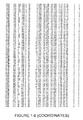
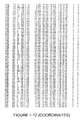
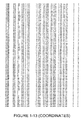
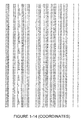
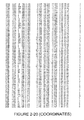
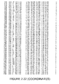
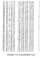
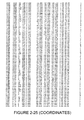
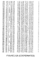
- the structure coordinates set forth in Figures 1 to 4 can also be used to aid in obtaining structural information about another crystallized molecule or molecular complex. This may be achieved by any of a number of well-known techniques, including molecular replacement.
- Inhibitor B was synthesized from 5-isothiocyanato-1,2,3-trimethoxybenzene (0.162 g, 0.72 mmol) and 3-(4-methyl-1H-imidazol-1-yl)propan-1-amine (0.10 g, 0.72 mmol, which may be prepared as described in D7).
- the conformation of both enzymes is stable up to a GdmCl concentration of 2M. At a concentration of 2.5 M unfolding can be detected indicated by a reduction of fluorescence emission. In comparison to hQC WT the unfolding is increased for the C139A/C164A mutant. While there is still a folded fraction at 4 M GdmCl for hQC WT the C139A/C164A mutant is completely unfolded.
- Crystals were grown using the hanging drop vapor diffusion technique at room temperature (21 °C) in Easyxtal 24-well plates plates (Qiagen).
- the mother liquor buffer consisted of 0.1 M imidazol pH 8, 30% (v/v) 2-methyl 2,4-pentanediol and 11% (w/v) 4000PEG.
- Protein solution concentrated up to a 10 mg/ml was recombinant human glutaminyl cyclase in the presence of 25mM Bis-Tris pH6.8 / 100mM NaCl buffer.
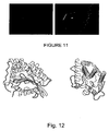
- a mixture of 1 ⁇ l mother liquor buffer and 1 ⁇ l protein solution was set. Crystals appeared usually ten to fifteen days after the experiment was initiated and they displayed a macroscopic plate shape ( Figure 11 ).
Landscapes
- Chemical & Material Sciences (AREA)
- Life Sciences & Earth Sciences (AREA)
- Organic Chemistry (AREA)
- Health & Medical Sciences (AREA)
- Genetics & Genomics (AREA)
- Engineering & Computer Science (AREA)
- Bioinformatics & Cheminformatics (AREA)
- Zoology (AREA)
- Wood Science & Technology (AREA)
- General Health & Medical Sciences (AREA)
- Biochemistry (AREA)
- General Engineering & Computer Science (AREA)
- Microbiology (AREA)
- Biotechnology (AREA)
- Biomedical Technology (AREA)
- Molecular Biology (AREA)
- Medicinal Chemistry (AREA)
- Enzymes And Modification Thereof (AREA)
- Peptides Or Proteins (AREA)
- Measuring Or Testing Involving Enzymes Or Micro-Organisms (AREA)
- Pharmaceuticals Containing Other Organic And Inorganic Compounds (AREA)
- Preparation Of Compounds By Using Micro-Organisms (AREA)
Description
- The invention relates to the novel crystal structure of glutaminyl cyclase (QC, EC 2.3.2.5). QC catalyzes the intramolecular cyclization of N-terminal glutamine residues into pyroglutamic acid (5-oxo-prolyl, pGlu*) under liberation of ammonia and the intramolecular cyclization of N-terminal glutamate residues into pyroglutamic acid under liberation of water.
- Glutaminyl cyclase (QC, EC 2.3.2.5) catalyzes the intramolecular cyclization of N-terminal glutamine residues into pyroglutamic acid (pGlu*) liberating ammonia. A QC was first isolated by Messer from the latex of the tropical plant Carica papaya in 1963 (Messer, M. 1963 Nature 4874, 1299). 24 years later, a corresponding enzymatic activity was discovered in animal pituitary (Busby, W. H. J. et al. 1987 J Biol Chem 262, 8532-8536; Fischer, W. H. and Spiess, J. 1987 Proc Natl Acad Sci U S A 84, 3628-3632). For the mammalian QC, the conversion of Gln into pGlu by QC could be shown for the precursors of TRH and GnRH (Busby, W. H. J. et al. 1987 J Biol Chem 262, 8532-8536; Fischer, W. H. and Spiess, J. 1987 Proc Natl Acad Sci U S A 84, 3628-3632). In addition, initial localization experiments of QC revealed a co-localization with its putative products of catalysis in bovine pituitary, further improving the suggested function in peptide hormone synthesis (Bockers, T. M. et al. 1995 J Neuroendocrinol 7, 445-453). In contrast, the physiological function of the plant QC is less clear. In the case of the enzyme from C. papaya, a role in the plant defense against pathogenic microorganisms was suggested (EI Moussaoui, A. et a/.2001 Cell Mol Life Sci 58, 556-570). Putative QCs from other plants were identified by sequence comparisons recently (Dahl, S. W. et al.2000 Protein Expr Purif 20, 27-36). The physiological function of these enzymes, however, is still ambiguous.
- The QCs known from plants and animals show a strict specificity for L-Glutamine in the N-terminal position of the substrates and their kinetic behavior was found to obey the Michaelis-Menten equation (Pohl, T. et al. 1991 Proc Natl Acad Sci U S A 88, 10059-10063; Consalvo, A. P. et al. 1988 Anal Biochem 175, 131-138; Gololobov, M. Y. et al. 1996 Biol Chem Hoppe Seyler 377, 395-398). A comparison of the primary structures of the QCs from C. papaya and that of the highly conserved QC from mammals, however, did not reveal any sequence homology (Dahl, S. W. et al. 2000 Protein Expr Purif 20, 27-36). Whereas the plant QCs appear to belong to a new enzyme family (Dahl, S. W. et al. 2000 Protein Expr Purif 20, 27-36), the mammalian QCs were found to have a pronounced sequence homology to bacterial aminopeptidases (Bateman, R. C. et al. 2001 Biochemistry 40, 11246-11250), leading to the conclusion that the QCs from plants and animals have different evolutionary origins.
- Recently, it was shown that recombinant human QC as well as QC-activity from brain extracts catalyze both, the N-terminal glutaminyl as well as glutamate cyclization. Most striking is the finding, that cyclase-catalyzed Glu1-conversion is favored around pH 6.0 while Gln1-conversion to pGlu-derivatives occurs with a pH-optimum of around 8.0. Since the formation of pGlu-Aβ-related peptides can be suppressed by inhibition of recombinant human QC and QC-activity from pig pituitary extracts, the enzyme QC is a target in drug development for treatment of Alzheimer's disease.
-
US 7,572,614 (Wang et a/ ) and Huang et al (2005) PNAS 102(37), 13117-13122 both describe one example of the crystal structure of soluble glutaminyl cyclase. The crystal structure disclosed in Wang et al and Huang et al was generated using a protein expressed in E. coli, which results in a lack of glycosylation. It is well known that all mammalian QC contain at least one glycosylation site (Pohl, T. et al. (1991) Proc Natl Acad Sci U S A 88, 10059-10063; Song, I. et al. (1994) J Mol Endocrinol 13, 77-86), which is glycosylated in the QC crystallized according to the invention by virtue of being expressed in eukaryotic hosts, which can be observed in the crystal structures presented herein. In addition, all mammalian QCs contain two conserved cysteine residues close to the active site, which form a disulfide bond. In the crystal structure of Wang et al and Huang et al, the disulfide bond is lacking. The expression of mammalian secretory proteins in bacteria frequently results in the absence of disulfide formation (Hannig, G. and Makrides, S. C. (1998) Trends Biotechnol 16, 54-60). The disulfide bond is clearly present in both the human and murine QC crystal structures presented herein. Notably, the mutational analyses provided in the examples described herein suggest an important stabilizing function of the disulfide bond upon the overall structure. Furthermore, in the structure of Wang et al and Huang et al, a segment of residues (L205-H206-W207) close to the active site appears in two different conformations. Due to the orientations, the binding mode of substrates is affected and reliable mechanistic conclusions could not be drawn (Huang et al., 2005). The residue W207 is conserved in mammalian QCs (W208 in murine QC). In the human and murine QC crystal structures presented herein, the orientation is identical, although the adjacent residues L205 and H206 are not conserved. Therefore, the structural orientation of residues appears non-native in the crystal structure of Wang et al and Huang et al. - The expression of the murine and human QC in an eukaryotic host, as described in the present invention, allows the crystallization and structural refinement of a native mammalian QC and, importantly, unambiguous determination of the binding modes of inhibitors, as exemplified by the structural resolution of murine QC with three different inhibitory compounds (listed in Table 1 as Inhibitor A, Inhibitor B and Inhibitor C), which have similar inhibitory potency between human and murine QC.
- In contrast to the structures described in previous publications by Wang et al and Huang et al., the invention shows that the post-translational modifications disulfide formation and glycosylation lead to a single structural arrangement of the residues L205-H206-W207 in human and murine QC. The residues have a direct effect on the binding mode of active-site directed compounds. The multiple orientations of W207 in previous studies, led to variances in the binding modes of active-site-directed compounds.
Table 1: Ki-values [µM] of selected inhibitors for murine (mQC) and human QC (hQC) Inhibitor mQC hQC Inhibitor A 0.173 0.0542 Inhibitor B n. d. 0.0613 Inhibitor C 0.0513 0.106 - In contrast, unambigous binding modes of compounds and orientations of W207 were observed with three different inhibitors and even in a structure without inhibitor bound. Thus, the methods as described in the present inventions provide substantial advances for structure- driven drug design of QC inhibitors.
- The conclusion is particularly strengthened by the structural assessment of QCs from two different mammals.
- According to a first aspect of the invention there is provided a crystal comprising human glutaminyl cyclase having a characterised space group of C121 and unit cell dimensions of +/- 5% of a = 82.6 Å, b = 63.9 Å, c = 77.5 Å, α = 90.0º, β = 105.7° and γ = 90.0º.
- According to a second aspect of the invention there is provided a crystal comprising murine glutaminyl cyclase having a characterised space group of P212121 and having unit cell dimensions of +/- 5% of a = 42.7 Å, b = 84.0 Å and c = 96.5 Å.
- According to a third aspect of the invention there is provided a method of preparing the crystal of human glutaminyl cyclase as described herein, which comprises the steps of:
- (a) providing a solution of human glutaminyl cyclase, optionally in the presence of a known glutaminyl cyclase inhibitor, in a suitable buffer such as 25mM Bis-Tris pH6.8 / 100mM NaCl buffer;
- (b) mixing the solution with a crystallization solution comprising 30% (v/v) 2-
methyl 2,4-pentanediol and 11% (w/v) 4000PEG; and - (c) incubating the mixture under conditions to promote hanging drop vapor diffusion for a time sufficient to produce the crystal of human glutaminyl cyclase.
- According to a fourth aspect of the invention there is provided a method of preparing the crystal of murine glutaminyl cyclase as described herein, which comprises the steps of:
- (a) providing a solution of murine glutaminyl cyclase, optionally in the presence of a known glutaminyl cyclase inhibitor, in a suitable buffer such as 15mM Bis-Tris pH6.8 / 100mM NaCl buffer;
- (b) mixing the solution with a crystallization solution comprising 0.1 M sodium acetate pH 5.3, 0.2M ammonium sulphate and 12% (w/v) 2000 MME-PEG; and
- (c) incubating the mixture under conditions to promote hanging drop vapor diffusion for a time sufficient to produce the crystal of murine glutaminyl cyclase.
- According to a fifth aspect of the invention there is provided a method of identifying an inhibitor of human glutaminyl cyclase which comprises the following steps:
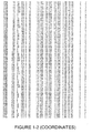
- (a) generating a 3-dimensional model of human glutaminyl cyclase using the structural coordinates described in
Figure 1 ; - (b) analysing the binding pocket provided by residues E202, D159 and H330 of SEQ ID NO: 1 according to the coordinates of
Figure 1 ; - (c) performing computer modeling analysis to identify an inhibitor compound which may associate with the binding pocket of human glutaminyl cyclase.
- According to a sixth aspect of the invention there is provided a method of identifying an inhibitor of murine glutaminyl cyclase which comprises the following steps:
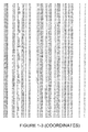
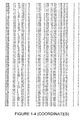
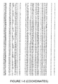
- (a) generating a 3-dimensional model of murine glutaminyl cyclase using the structural coordinates described in any of
Figures 2 to 4 ; - (b) analysing the binding pocket provided by residues D160, E203 and H331 of SEQ ID NO: 13 according to the coordinates of
Figures 2 to 4 ; - (c) performing computer modeling analysis to identify an inhibitor compound which may associate with the binding pocket of murine glutaminyl cyclase.
-
-
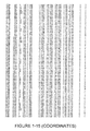
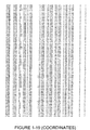
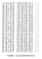
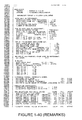
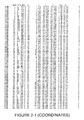
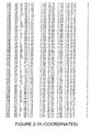
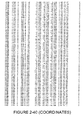
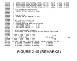
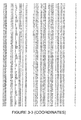
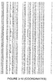
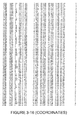
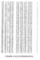
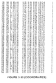
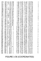
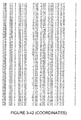
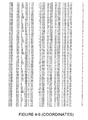
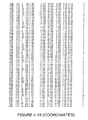
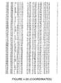
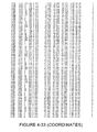
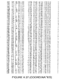
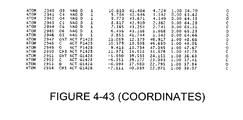
Figure 1 describes the X-ray coordinates of human QC crystallized with 0.1M imidazol pH 8, wherein "RES" and "#" correspond to the amino acid residue and number of the amino acid in SEQ ID NO: 1. -
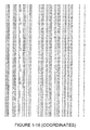
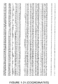
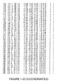
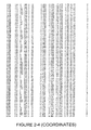
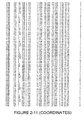
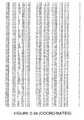
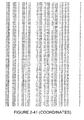
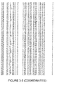
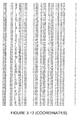
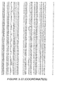
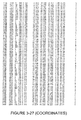
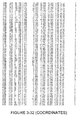
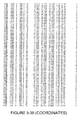
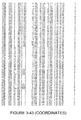
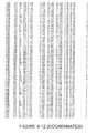
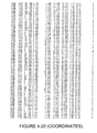
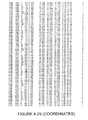
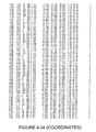
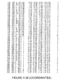
Figure 2 describes the X-ray coordinates of murine QC crystallized with QC inhibitor Inhibitor A, wherein "RES" and "#" correspond to the amino acid residue and number of the amino acid in SEQ ID NO: 13. -
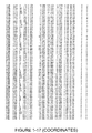
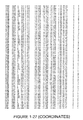
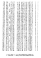
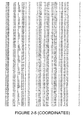
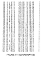
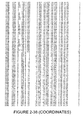
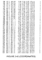
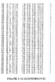
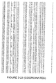
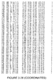
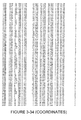
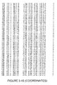
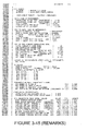
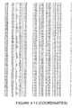
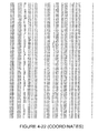
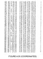
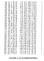
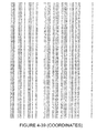
Figure 3 describes the X-ray coordinates of murine QC crystallized with QC inhibitor Inhibitor B, wherein "RES" and "#" correspond to the amino acid residue and number of the amino acid in SEQ ID NO: 13. -
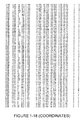
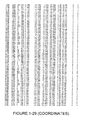
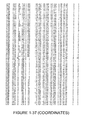
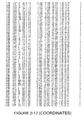
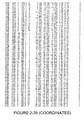
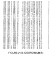
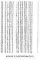
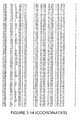
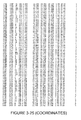
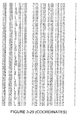
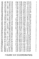
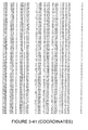
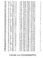
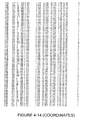
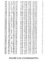
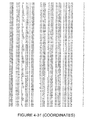
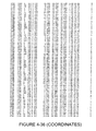
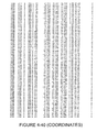
Figure 4 describes the X-ray coordinates of murine QC crystallized with QC inhibitor Inhibitor C, wherein "RES" and "#" correspond to the amino acid residue and number of the amino acid in SEQ ID NO: 13. -
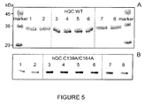
Figure 5 describes the results of SDS-PAGE analysis of hQC WT (A) and the C139A/C164A mutant (B). -
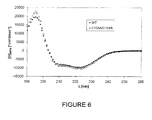
Figure 6 describes the CD spectra of the far UV region of hQC WT and the C139A/C164A mutant at 20 °C. -
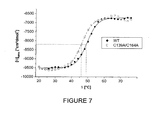
Figure 7 describes temperature dependence of the mean residue ellipticity (θMRW) of hQC WT and the C139A/C164A mutant at a wavelength of 227nm. -
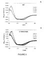
Figure 8 describes CD spectra of the far UV region of hQC WT (A) and the C139A/C164A mutant (B) at increasing temperature. -
Figure 9 describes fluorescence emission spectra in a wavelength range between 320 and 400 nm of hCQ WT (A) / C139A/C164A (B) mutant at different GdmCl concentrations. -
Figure 10 describes dependence of the relative fluorescence from the GdmCl concentration of hQC WT and the C139A/C164A mutant at a wavelength of 337 nm. -
Figure 11 demonstrates photographic representations of the crystals of human glutaminyl cyclase grown in buffer consisting of 0.1M imidazol pH methyl 2,4-pentanediol and 11% (w/v) 4000PEG. -
Figure 12 describes a cartoon representation of the obtained three dimensional structure of the human glutaminyl cyclase described by the X-ray coordinates shown inFigure 1 . The structure is shown in two orthogonal views. They are shown in β-sheet structures, in α helices and in the random coiled loops. -
Figure 13 demonstrates an overlay of the electron density at 1.5 sigma level with the modeled residues building the active site. With a stick representation are displayed the active site related residues D159, E202 and H330 and an imidazole molecule coordinating the catalytic zinc cation which is shown with a ball representation. -
Figure 14 demonstrates a photographic representation of examples of the crystals of the mouse glutaminyl cyclase grown in buffer consisting of 0.1 M sodium acetate pH 5.3 0.2Mammonium sulphate 12% (w/v) 2000 MME-PEG and 1 mM Inhibitor A. -
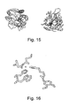
Figure 15 describes a cartoon representation of the obtained three dimensional structure of the mouse glutaminyl cyclase described by the X-ray coordinates shown inFigures 2-4 shown in two different views with a orthogonal orientation. The β-sheet, α-helices and the random coiled structures are shown. -
Figure 16 demonstrates an overlay of the electron density at 1.5 sigma level with the modeled residues building the active centre. With a stick representation are displayed the active site related residues D160, E203 and H331, and the inhibitor molecule Inhibitor A coordinating the catalytic zinc cation is shown with a ball representation. -
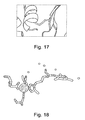
Figure 17 demonstrates an overlay of the electron density at 1.5 sigma level with the modeled residues building the disulfide bridge. With a stick representation are displayed the residues involved in the disulfide bridge, C140 and C165, formation and in cartoon representation are displayed the secondary structures surrounding that disulfide bridge. -
Figure 18 demonstrates an overlay of the electron density at 1.5 sigma level with the modeled residues building the active centre. With a stick representation are displayed the active site related residues D160, E203 and H331 and the inhibitor molecule. Inhibitor C coordinating the catalytic zinc cation is shown with a sphere representation and in ball representation are shown water molecules. -
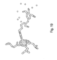
Figure 19 demonstrates an overlay of the electron density at 1.5 sigma level with the modeled residues building the active centre. With a stick representation are displayed the active site related residues D160, E203 and H331 and the inhibitor molecule. Inhibitor B coordinating the catalytic zinc cation is shown with a sphere representation and in ball representation are shown water molecules. - The present invention relates to crystals of human and murine glutaminyl cyclase, where the crystals are of sufficient quality and size to allow for the determination of the three-dimensional X-ray diffraction structure of glutaminyl cyclase to a resolution of about 1.8 angstrom and to about 2.1 angstroms in C121 and P212121 space groups, respectively. The invention also relates to methods for preparing and crystallizing human and murine glutaminyl cyclase. The crystals of human and murine glutaminyl cyclase, as well as information derived from their crystal structures, can be used to analyze and modify glutaminyl cyclase as well as to identify compounds that interact with glutaminyl cyclase.
- Thus, according to a first aspect of the invention, there is provided a crystal comprising human glutaminyl cyclase having a characterised space group of C121 and unit cell dimensions of +/- 5% of a = 82.6 Å, b = 63.9 Å, c = 77.5 Å, α = 90.0°, β = 105.7º and γ = 90.0º.
- In one embodiment of the first aspect of the invention, the crystal has unit cell dimensions of a = 82.6 Å, b = 63.9 Å and c = 77.5 Å.
- In one embodiment of the first aspect of the invention, the crystal has unit cell dimensions of α = 90.0º, β = 105.7º and γ = 90.0º.
- In one embodiment of the first aspect of the invention, the crystal diffracts x-rays for determination of atomic co-ordinates of the crystal to a resolution of between 2.08 A and 11.98 Å.
- According to a second aspect of the invention there is provided a crystal comprising murine glutaminyl cyclase having a characterised space group of P212121 and having unit cell dimensions of +/- 5% of a = 42.7 Å, b = 84.0 Å, c = 96.5 Å, α = 90.0º, β = 90.0º and γ = 90.0°.
- In one embodiment of the second aspect of the invention, the crystal has unit cell dimensions of a = 42.7 Å, b = 83.0 Å, c = 95.7 Å, α = 90.0º, β = 90.0º and γ = 90.0º. This embodiment of the second aspect of the invention relates to the crystal structure of murine glutaminyl cyclase complexed with a glutaminyl cyclase inhibitor referred to herein as Inhibitor A which has the structure as follows:

- Inhibitor A inhibits both human and mouse QC in vitro and moreover, Inhibitor A is soluble in water and therefore useful for crystallization.
- In an alternative embodiment of the second aspect of the invention, the crystal has unit cell dimensions of a = 42.7 Å, b = 84.6 Å, c = 96.5 Å, α = 90.0º, β = 90.0º and γ = 90.0°. This embodiment of the second aspect of the invention relates to the crystal structure of murine glutaminyl cyclase complexed with a glutaminyl cyclase inhibitor referred to herein as Inhibitor B which has the structure as follows:

- In an alternative embodiment of the second aspect of the invention, the crystal has unit cell dimensions of a = 42.7 Å, b = 84.6 Å, c = 97.2 Å, α = 90.0º, β = 90.0º and γ = 90.0º. This embodiment of the second aspect of the invention relates to the crystal structure of murine glutaminyl cyclase complexed with a glutaminyl cyclase inhibitor referred to herein as Inhibitor C which has the structure as follows:

- In one embodiment of the second aspect of the invention, the crystal has unit cell dimensions of α = 90.0º, β = 90.0º and γ = 90.0º.
- In one embodiment of the second aspect of the invention, the crystal diffracts x-rays for determination of atomic co-ordinates of the crystal to a resolution of between 1.9 Å and 32.0 Å.
- In one embodiment of the second aspect of the invention, the crystal diffracts x-rays for determination of atomic co-ordinates of the crystal to a resolution of between 1.8 Å and 19.81 Å. This embodiment of the second aspect of the invention relates to the resolution of the crystal structure of murine glutaminyl cyclase complexed with a glutaminyl cyclase inhibitor referred to herein as Inhibitor A.
- In an alternative embodiment of the second aspect of the invention, the crystal diffracts x-rays for determination of atomic co-ordinates of the crystal to a resolution of between 1.98 Å and 32.0 Å. This embodiment of the second aspect of the invention relates to the resolution of the crystal structure of murine glutaminyl cyclase complexed with a glutaminyl cyclase inhibitor referred to herein as Inhibitor B.
- In an alternative embodiment of the second aspect of the invention, the crystal diffracts x-rays for determination of atomic co-ordinates of the crystal to a resolution of between 1.9 Å and 30.7 Å. This embodiment of the second aspect of the invention relates to the resolution of the crystal structure of murine glutaminyl cyclase complexed with a glutaminyl cyclase inhibitor referred to herein as Inhibitor C.
- It will be appreciated that the crystals within the scope of the invention include both apo crystals and co-crystals. The apo crystals generally comprise substantially pure glutaminyl cyclase. The co-crystals generally comprise substantially pure glutaminyl cyclase with a binding ligand, such as a glutaminyl cyclase inhibitor, bound to glutaminyl cyclase.
- In one embodiment of the first aspect of the invention, the human glutaminyl cyclase consists of amino acid residues A35 to L361 of SEQ ID NO:1.
- In one embodiment of the second aspect of the invention, the murine glutaminyl cyclase consists of amino acid residues A36 to L362 of SEQ ID NO:13.
- It will be appreciated that the crystals comprising glutaminyl cyclase are not limited to those obtainable from naturally occurring or native glutaminyl cyclase. The crystals include mutants that have one or more amino acid insertions, deletions, or substitutions in native glutaminyl cyclase. Therefore, mutants of native glutaminyl cyclase are obtained by replacing at least one (such as up to 10, e.g. up to 25) amino acid residue in a native glutaminyl cyclase with a different amino acid residue, or by adding or deleting amino acid residues within the native protein or at the N- or C-terminus of the native protein, and have substantially the same three-dimensional structure as native glutaminyl cyclase from which the mutant is derived.
- By having substantially the same three-dimensional structure is meant as having a set of atomic structure coordinates from an apo- or co-crystal that have a root mean square deviation of less than or equal to about 2 Å when superimposed with the atomic structure coordinates of native glutaminyl cyclase from which the mutant is derived when at least about 50% to about 100% of the alpha carbon atoms of native glutaminyl cyclase are included in the superposition.
- In some instances, it may be particularly advantageous or convenient to substitute, delete, and/or add amino acid residues to native glutaminyl cyclase in order to provide convenient cloning sites in the cDNA encoding the protein, to aid in protein purification, and the like. Such substitutions, deletions, and/or additions, which do not substantially alter the three dimensional structure of native glutaminyl cyclase will be apparent to those skilled in the art.
- It should be noted that the mutant polypeptides contemplated herein need not exhibit glutaminyl cyclase activity. Indeed, amino acid substitutions, additions, or deletions that interfere with the activity of glutaminyl cyclase but which do not significantly alter the three-dimensional structure of glutaminyl cyclase are also included. Such polypeptide crystals, or the atomic structure coordinates obtained therefrom, can be used to identify compounds that bind to native glutaminyl cyclase and which may affect the activity of the native glutaminyl cyclase.
- The derivative crystals of the invention generally comprise glutaminyl cyclase crystals in non-covalent/covalent association with one or more metal atoms. The polypeptide may correspond to native or mutated glutaminyl cyclase. One such example of a suitable metal atom is zinc.
- The co-crystals of glutaminyl cyclase generally comprise crystals comprising glutaminyl cyclase in association with one or more compounds bound to glutaminyl cyclase. The association may be covalent or non-covalent. In one embodiment, the compounds bound to glutaminyl cyclase comprise glutaminyl cyclase inhibitors. Examples of such glutaminyl cyclase inhibitors include those described in
WO 2004/098625 ,WO 2004/098591 ,WO 2005/039548 ,WO 2005/075436 ,WO 2008/055945 ,WO 2008/055947 ,WO 2008/055950 andWO 2008/065141 . - The native and mutated glutaminyl cyclase described herein may be isolated from natural sources or produced by methods well known to those skilled in the art of molecular biology. Detailed experimental for the preparation of human and murine glutaminyl cyclase is described in Examples 1 and 2 herein, respectively.
- The apo, derivative and co-crystals of glutaminyl cyclase can be obtained by techniques well-known in the art of protein crystallography, including batch, liquid bridge, dialysis, vapor diffusion, such as hanging drop vapor diffusion, and the like (See for example, McPherson, 1982, Preparation and Analysis of Protein Crystals, John Wiley, NY; McPherson, 1990, Eur. J. Biochem. 189:1-23; Webber, 1991, Adv. Protein Chem. 41:1-36; Crystallization of Nucleic Acids and Proteins, Edited by Ducruix and Giege, Oxford University Press; Protein Crystallization Techniques, Strategies, and Tips, Edited by Bergfors, International University Line, 1999).
- In one embodiment, glutaminyl cyclase crystals, apo or co-crystals are grown by vapor diffusion, such as hanging drop vapor diffusion.
- According to a third aspect of the invention, there is provided a method of preparing the crystal of human glutaminyl cyclase as described herein, which comprises the steps of:
- (a) providing a solution of human glutaminyl cyclase, optionally in the presence of a known glutaminyl cyclase inhibitor, in a suitable buffer such as 25mM Bis-Tris pH6.8 / 100mM NaCl buffer;
- (b) mixing the solution with a crystallization solution comprising 30% (v/v) 2-
methyl 2,4-pentanediol and 11% (w/v) 4000PEG; and - (c) incubating the mixture under conditions to promote hanging drop vapor diffusion for a time sufficient to produce the crystal of human glutaminyl cyclase.
- According to a further aspect of the invention, there is provided a method of preparing the co-crystal of human glutaminyl cyclase bound to a binding ligand, such as a glutaminyl cyclase inhibitor, which comprises the steps of:
- (a) providing a solution of human glutaminyl cyclase in the presence of a binding ligand, such as a glutaminyl cyclase inhibitor, in a suitable buffer such as 25mM Bis-Tris pH6.8 / 100mM NaCl buffer;
- (b) mixing the solution with a crystallization solution comprising 30% (v/v) 2-
methyl 2,4-pentanediol and 11% (w/v) 4000PEG; and - (c) incubating the mixture under conditions to promote hanging drop vapor diffusion for a time sufficient to produce the co-crystal of human glutaminyl cyclase bound to a binding ligand, such as a glutaminyl cyclase inhibitor.
- Crystallization of human glutaminyl cyclase to produce co-crystals can be carried out as described below and in Example 1. As described, purified human glutaminyl cyclase is concentrated to 10 mg/mL in the presence of a suitable buffer, such as 25mM Bis-Tris pH6.8 / 100mM NaCl buffer and the ligand imidazol is added to a final concentration of 0.1 M. Macroscopic plate crystal forms are grown by hanging drop vapor diffusion at 21 °C by mixing an equal volume of protein solution with a crystallization solution comprising 30% (v/v) 2-
methyl 2,4-pentanediol and 11% (w/v) 4000PEG. Crystals typically appeared 10-15 days after the experiment was initiated and examples of such crystals obtained following this protocol are shown inFigure 11 . It will be appreciated that to produce apo-crystals, the ligand is omitted in the above protocol. - The human glutaminyl cyclase crystals may be frozen prior to data collection. The crystals can be cryo-protected with for example, either (a) 20-30% saturated glucose present in the crystallization setup, (b) ethanol added to 15-20%, (c) ethylene glycol added to 10-20% and PEG10,000 brought up to 25%, or (d) glycerol added to 15%. In one embodiment of the third aspect of the invention, the crystals are cryo-protected with the addition of glycerol added to 15% (v/v). The crystals can be either briefly immersed in the cryo-protectant or soaked in the cryo-protectant for periods as long as a day. Freezing can be accomplished by immersing the crystal in a bath of liquid nitrogen or by placing the crystal in a stream of nitrogen gas at-180 °C.
- As described in Example 1, the crystal structure of human glutaminyl cyclase in complex with imidazole was obtained. A summary of the crystal's attributes for human glutaminyl cyclase bound to imidazole are listed in Table 3 and the three dimensional structure coordinates for space groups C121 for human glutaminyl cyclase bound to imidazole are shown in
Figure 1 . - References herein to "coordinates" include references to Cartesian coordinates derived from the mathematical equations related to the patterns obtained on diffraction of a monochromatic beam of X-ray by the atoms of a protein or protein complex in crystal form. The diffraction data are used to calculate an electron density map of the repeating units of the crystal, The electron density maps are then used to establish the positions of the individual atoms of the molecule or molecular complex.
- Ribbon and overlay diagrams of human glutaminyl cyclase bound to imidazole based upon the coordinates for space groups C121 are shown in
Figures 12 and13 , respectively. In particular, it was found that the protein comprised a globular α/β hydrolase fold. A central β-sheet was formed with six β-strands all in parallel fashion but not the second. This β-sheet was observed to be surrounded by α-helices in a sandwich manner with two helices in one side and six more α-helices in the opposite face. The protein's structure was completed with a rather large amount of random coiled loops which are belived to build the active site of the enzyme. - References herein to "active site" include references to a specific region (or atom) in a molecular entity that is capable of entering into a stabilising interaction with another molecular entity. In certain embodiments, the term also refers to the reactive parts of a macromolecule that directly participate in its specific combination with another molecule. In an alternative embodiment, a binding site may be comprised or defined by the three dimensional arrangement of one or more amino acid residues within a folded polypeptide. References to "binding pocket" shall be interpreted in an analogous manner to "active site" and it will be appreciated that these terms may be used interchangeably.
- This active site of human glutaminyl cyclase was found to accommodate a zinc ion which is coordinated by three protein residues, E202, D159 and H330 . Thus, in one embodiment of the first aspect of the invention, the crystal comprises a binding pocket provided by residues E202, D159 and H330 of SEQ ID NO: 1 according to the coordinates of
Figure 1 . - Moreover, the protein shows the presence of a disulfide bridge between residues C139 and C164. Thus, in one embodiment of the first aspect of the invention, the crystal comprises a disulfide bridge between residues C139 and C164 of SEQ ID NO: 1. As discussed hereinbefore, the presence of such a disulfide bridge has not previously been reported during crystallisation studies with QC (Wang et al, Huang et al).
- Additionally four cis-peptide bonds are present between the pair of residues : D159-S160, H228-P229, G301-V302 and S323-P324. Thus, in one embodiment of the first aspect of the invention, the crystal comprises one or more cis-peptide bonds between any of the following pairs of residues: D159-S160, H228-P229, G301-V302and S323-P324.
- Finally two segments of the polylpeptide chain are not visible in the electron density. The gaps include residues between K182 and D190 and between F146 and N150.
- According to a fourth aspect of the invention there is provided a method of preparing the crystal of murine glutaminyl cyclase as described herein, which comprises the steps of:
- (a) providing a solution of murine glutaminyl cyclase, optionally in the presence of a known glutaminyl cyclase inhibitor, in a suitable buffer such as 15mM Bis-Tris pH6.8 / 100mM NaCl buffer;
- (b) mixing the solution with a crystallization solution comprising 0.1 M sodium acetate pH 5.3, 0.2M ammonium sulphate and 12% (w/v) 2000 MME-PEG; and
- (c) incubating the mixture under conditions to promote hanging drop vapor diffusion for a time sufficient to produce the crystal of murine glutaminyl cyclase.
- According to a further aspect of the invention, there is provided a method of preparing the co-crystal of human glutaminyl cyclase bound to a binding ligand, such as a glutaminyl cyclase inhibitor, which comprises the steps of:
- (a) providing a solution of murine glutaminyl cyclase in the presence of a known glutaminyl cyclase inhibitor, in a suitable buffer such as 15mM Bis-Tris pH6.8 / 100mM NaCl buffer;
- (b) mixing the solution with a crystallization solution comprising 0.1 M sodium acetate pH 5.3, 0.2M ammonium sulphate and 12% (w/v) 2000 MME-PEG; and
- (c) incubating the mixture under conditions to promote hanging drop vapor diffusion for a time sufficient to produce the co-crystal of murine glutaminyl cyclase bound to a binding ligand, such as a glutaminyl cyclase inhibitor.
- Crystallization of murine glutaminyl cyclase to produce co-crystals can be carried out as described below and in Example 2. As described, purified murine glutaminyl cyclase is concentrated to 10 mg/mL in the presence of a suitable buffer, such as 15mM Bis-Tris pH 6.8 / 100mM NaCl buffer and the ligand, Inhibitor A, Inhibitor B or Inhibitor C, is added to a final concentration of 1 mM. Macroscopic rod/needle crystal forms are grown by hanging drop vapor diffusion at 21 °C by mixing an equal volume of protein solution with a crystallization solution comprising 0.1 M sodium acetate pH 5.3, 0.2M ammonium sulphate and 12% (w/v) 2000 MME-PEG. Crystals typically appeared within 1 week after the experiment was initiated and examples of such crystals obtained following this protocol are shown in
Figure 14 . It will be appreciated that to produce apo-crystals, the ligand is omitted in the above protocol. - The murine glutaminyl cyclase crystals may be frozen prior to data collection. The crystals can be cryo-protected with for example, either (a) 20-30% saturated glucose present in the crystallization setup, (b) ethanol added to 15-20%, (c) ethylene glycol added to 10-20% and PEG10,000 brought up to 25%, or (d) glycerol added to 20%. In one embodiment of the fourth aspect of the invention, the crystals are cryo-protected with the addition of glycerol added to 20% (v/v). The crystals can be either briefly immersed in the cryo-protectant or soaked in the cryo-protectant for periods as long as a day. Freezing can be accomplished by immersing the crystal in a bath of liquid nitrogen or by placing the crystal in a stream of nitrogen gas at -180 °C.
- As described in Example 2, the crystal structure of murine glutaminyl cyclase in complex with glutaminyl cyclase inhibitors Inhibitor A, Inhibitor B and Inhibitor C was obtained. A summary of the crystal's attributes for murine glutaminyl cyclase bound to glutaminyl cyclase inhibitors Inhibitor A, Inhibitor B and Inhibitor C are listed in Table 4. The three dimensional structure coordinates for space groups P212121 for murine glutaminyl cyclase bound to glutaminyl cyclase inhibitor Inhibitor A is shown in
Figure 2 . The three dimensional structure coordinates for space groups P212121 for murine glutaminyl cyclase bound to glutaminyl cyclase inhibitor Inhibitor B is shown inFigure 3 . The three dimensional structure coordinates for space groups P212121 for murine glutaminyl cyclase bound to glutaminyl cyclase inhibitor Inhibitor C is shown inFigure 4 . Ribbon and overlay diagrams of murine glutaminyl cyclase bound to glutaminyl cyclase inhibitors Inhibitor A, Inhibitor B and Inhibitor C based upon the coordinates for space groups P212121 are shown inFigures 15 to 19 . In particular, it was found that the protein comprised a globular α/β hydrolase fold. A central β-sheet was formed with six β-strands all in parallel fashion but not the second. This β-sheet was observed to be surrounded by α-helices in a sandwich manner with two helices in one side and six more α-helices in the opposite face. The protein's structure was completed with a rather large amount of random coiled loops which appeared to build the active site of the enzyme. This active site accommodates a zinc ion which is coordinated by three protein residues D160, E203 and H331 and an inhibitor molecule. Thus, in one embodiment according to the second aspect of the invention, the crystal comprises a binding pocket provided by residues D160, E203 and H331 of SEQ ID NO: 13 according to the coordinates ofFigures 2 to 4 . - Furthermore, the protein demonstrated the presence of a disulfide bridge between residues C140 and C165, Thus, in one embodiment according to the second aspect of the invention, the crystal comprises a disulfide bridge between residues C140 and C165 of SEQ ID NO: 13. As discussed hereinbefore, the presence of such a disulfide bridge has not previously been reported during crystallisation studies with QC (Wang et al, Huang et al).
- The present invention is also directed to machine-readable data storage media having data storage material encoded with machine-readable data that comprises structure coordinates for glutaminyl cyclase. The present invention is also directed to a machine readable data storage media having data storage material encoded with machine readable data, which, when read by an appropriate machine, can display a three dimensional representation of a structure of glutaminyl cyclase.
- All or a portion of glutaminyl cyclase coordinate data shown in
Figures 1 to 4 , when used in conjunction with a computer programmed with software to translate those coordinates into the three-dimensional structure of glutaminyl cyclase may be used for a variety of purposes, especially for purposes relating to drug discovery. Software for generating three-dimensional graphical representations are known and commercially available. The ready use of the coordinate data requires that it be stored in a computer-readable format. Thus, in accordance with the present invention, data capable of being displayed as the three-dimensional structure of glutaminyl cyclase and/or portions thereof and/or their structurally similar variants may be stored in a machine-readable storage medium, which is capable of displaying a graphical three-dimensional representation of the structure. - Another embodiment of this invention provides a machine-readable data storage medium, comprising a data storage material encoded with machine readable data which, when used by a machine programmed with instructions for using said data, displays a graphical three-dimensional representation comprising glutaminyl cyclase or variant thereof.
- Optionally, a computer system is provided in combination with the machine-readable data storage medium provided herein. In one embodiment, the computer system comprises a working memory for storing instructions for processing the machine-readable data; a processing unit coupled to the working memory and to the machine-readable data storage medium, for processing the machine-readable data into the three-dimensional representation; and an output hardware coupled to the processing unit, for receiving the three-dimensional representation.
- The three-dimensional crystal structure of the present invention may be used to identify glutaminyl cyclase binding sites, be used as a molecular replacement model to solve the structure of unknown crystallized proteins, to design mutants having desirable binding properties, and ultimately, to design, characterize, identify entities capable of binding and inhibiting glutaminyl cyclase and other structurally similar proteins as well as other uses that would be recognized by one of ordinary skill in the art. Such entities may be chemical entities or proteins. The term "chemical entity", as used herein, refers to chemical compounds, complexes of at least two chemical compounds, and fragments of such compounds.
- The glutaminyl cyclase structural coordinates provided herein are useful for screening and identifying drugs that inhibit glutaminyl cyclase and other structurally similar proteins. For example, the structure encoded by the data may be computationally evaluated for its ability to associate with putative substrates or ligands. Such compounds that associate with glutaminyl cyclase may inhibit glutaminyl cyclase activity, and are potential drug candidates. Additionally or alternatively, the structure encoded by the data may be displayed in a graphical three-dimensional representation on a computer screen. This allows visual inspection of the structure, as well as visual inspection of the structure's association with the compounds.
- Thus, according to a fifth aspect of the invention there is provided a method of identifying an inhibitor of human glutaminyl cyclase which comprises the following steps:
- (a) generating a 3-dimensional model of human glutaminyl cyclase using the structural coordinates described in
Figure 1 ; - (b) analysing the binding pocket provided by residues E202, D159 and H330 of SEQ ID NO: 1 according to the coordinates of
Figure 1 ; - (c) performing computer modeling analysis to identify an inhibitor compound which may associate with the binding pocket of human glutaminyl cyclase.
- Furthermore, according to a sixth aspect of the invention there is provided a method of identifying an inhibitor of murine glutaminyl cyclase which comprises the following steps:
- (a) generating a 3-dimensional model of murine glutaminyl cyclase using the structural coordinates described in any of
Figures 2 to 4 ; - (b) analysing the binding pocket provided by residues D160, E203 and H331 of SEQ ID NO: 13 according to the coordinates of
Figures 2 to 4 ; - (c) performing computer modeling analysis to identify an inhibitor compound which may associate with the binding pocket of murine glutaminyl cyclase.
- A method is also provided for evaluating the potential of an entity to associate with glutaminyl cyclase or variant thereof by using all or a portion of the structure coordinates provided in
Figures 1 to 4 or functional equivalents thereof. A method is also provided for evaluating the potential of an entity to associate with glutaminyl cyclase or variant thereof by using structure coordinates similar to all or a portion of the structure coordinates provided inFigures 1 to 4 or functional equivalents thereof. - In one embodiment of the fifth or sixth aspect of the invention, the step of performing computer modeling analysis to identify said inhibitor compound comprises identifying said compound from a library of compounds.
- In one embodiment of the fifth or sixth aspect of the invention, the step of performing computer modeling, analysis to identify said inhibitor compound comprises identifying said compound in a database.
- In one embodiment of the fifth or sixth aspect of the invention, the step of performing computer modeling analysis to identify said inhibitor compound comprises designing the compound from a known glutaminyl cyclase inhibitor.
- With the structures provided herein, the present invention permits the use of molecular design techniques to identify, select or design potential inhibitors of glutaminyl cyclase based on the structure of glutaminyl cyclase. Such a predictive model is valuable in light of the high costs associated with the preparation and testing of the many diverse compounds that may possibly bind to glutaminyl cyclase.
- According to the invention, a potential glutaminyl cyclase inhibitor may be evaluated for its ability to bind glutaminyl cyclase prior to its actual synthesis and testing. If a proposed entity is predicted to have insufficient interaction or association with the binding pocket, preparation and testing of the entity can be obviated. However, if the computer modeling indicates a strong interaction, the entity may then be obtained and tested for its ability to bind.
- A potential inhibitor of glutaminyl cyclase may be computationally evaluated using a series of steps in which chemical entities or fragments are screened and selected for their ability to associate with glutaminyl cyclase.
- One skilled in the art may use one of several methods to screen entities (whether chemical or protein) for their ability to associate with glutaminyl cyclase. This process may begin by visual inspection of, for example, glutaminyl cyclase on a computer screen based on the glutaminyl cyclase structure coordinates in
Figures 1 to 4 or other coordinates which define a similar shape generated from the machine-readable storage medium. Selected fragments or chemical entities may then be positioned in a variety of orientations, or docked, within that binding pocket as defined above. Docking may be accomplished using software such as Quanta and Sybyl, followed by energy minimization and molecular dynamics with standard molecular mechanics force fields, such as CHARMM and AMBER. - Specialized computer programs may also assist in the process of selecting entities. These include: GRID (Goodford, "A Computational Procedure for Determining Energetically Favorable Binding Sites on Biologically Important Macromolecules", J. Med. Chem., 28, pp. 849-857 (1985)). GRID is available from Oxford University, Oxford, UK; MCSS (Miranker et al., "Functionality. Maps of Binding Sites: A Multiple Copy Simultaneous Search Method." Proteins: Structure, Function and Genetics, 11, pp. 29-34 (1991)). MCSS is available from Molecular Simulations, San Diego, Calif.; AUTODOCK (Goodsell et al., "Automated Docking of Substrates to Proteins by Simulated Annealing", Proteins: Structure, Function, and Genetics, 8, pp. 195-202 (1990)). AUTODOCK is available from Scripps Research Institute, La Jolla, Calif.; & DOCK (Kuntz et al., "A Geometric Approach to Macromolecule-Ligand Interactions", J. Mol. Biol., 161, pp. 269-288 (1982)). DOCK is available from University of California, San Francisco, Calif.
- Once suitable entities have been selected, they can be designed or assembled. Assembly may be preceded by visual inspection of the relationship of the fragments to each other on the three-dimensional image displayed on a computer screen in relation to the structure coordinates of glutaminyl cyclase. This may then be followed by manual model building using software such as MOE, QUANTA or Sybyl (Tripos Associates, St. Louis, Mo.).
- Useful programs to aid one of skill in the art in connecting the individual chemical entities or fragments include: CAVEAT (Bartlett et al., "CAVEAT: A Program to Facilitate the Structure-Derived Design of Biologically Active Molecules", in "Molecular Recognition in Chemical and Biological Problems", Special Pub., Royal Chem. Soc., 78, pp. 182-196 (1989); Lauri and Bartlett, "CAVEAT: a Program to Facilitate the Design of Organic Molecules", J. Comput. Aided Mol. Des., 8, pp. 51-66 (1994)). CAVEAT is available from the University of California, Berkeley, Calif.; 3D Database systems such as ISIS (MDL Information Systems, San Leandro, Calif.). This area is reviewed in Martin, "3D. Database Searching in Drug Design", J. Med. Chem., 35, pp. 2.145-2154 (1992); HOOK (Eisen et al., "HOOK: A Program for Finding Novel Molecular Architectures that Satisfy the Chemical and Steric Requirements of a Macromolecule Binding Site", Proteins: Struct., Funct., Genet., 19, pp. 199-221 (1994). HOOK is available from Molecular Simulations, San Diego, Calif.
- Instead of proceeding to build an inhibitor of glutaminyl cyclase in a step-wise fashion one fragment or entity at a time as described above, inhibitory or other glutaminyl cyclase binding compounds may be designed as a whole or "de novo" using either an empty binding site or optionally including some portion(s) of a known inhibitor(s). There are many de novo ligand design methods including: LUDI (Bohm, "The Computer Program LUDI: A New Method for the De Novo Design of Enzyme Inhibitors", J. Comp. Aid. Molec. Design, 6, pp. 61-78 (1992)). LUDI is available from Molecular Simulations Incorporated, San Diego, Calif.; LEGEND (Nishibata et al., Tetrahedron, 47, p. 8985 (1991)). LEGEND is available from Molecular Simulations Incorporated, San Diego, Calif.; LEAPFROG (available from Tripos Associates, St. Louis, Mo.); & SPROUT (Gillet et al., "SPROUT: A Program for Structure Generation)", J. Comput. Aided Mol. Design, 7, pp. 127-153 (1993)). SPROUT is available from the University of Leeds, UK.
- Other molecular modeling techniques may also be employed in accordance with this invention (See, for example, Cohen et al., "Molecular Modeling Software and Methods for Medicinal Chemistry, J. Med. Chem., 33, pp. 883-894 (1990); See also, Navia and Murcko, "The Use of Structural Information in Drug Design", Current Opinions in Structural Biology, 2, pp. 202-210 (1992); Balbes et al., "A Perspective of Modern Methods in Computer-Aided Drug Design", in Reviews in Computational Chemistry, Vol. 5, Lipkowitz and Boyd, Eds., VCH, New York, pp. 337-380 (1994); See also, Guida, "Software For Structure-Based Drug Design", Curr. Opin. Struct. Biology, 4, pp. 777-781 (1994)).
- Once an entity has been designed or selected, for example, by the above methods, the efficiency with which that entity may bind to glutaminyl cyclase may be tested and optimized by computational evaluation. For example, an effective glutaminyl cyclase inhibitor preferably demonstrates a relatively small difference in energy between its bound and free states (i.e., a small deformation energy of binding). Thus, the most efficient glutaminyl cyclase inhibitors should preferably be designed with deformation energy of binding of not greater than about 10 kcal/mole, more preferably, not greater than 7 kcal/mole. Glutaminyl cyclase inhibitors may interact with the protein in more than one of multiple conformations that are similar in overall binding energy. In those cases, the deformation energy of binding is taken to be the difference between the energy of the free entity and the average energy of the conformations observed when the inhibitor binds to the protein.
- An entity designed or selected as binding to glutaminyl cyclase may be further computationally optimized so that in its bound state it would preferably lack repulsive electrostatic interaction with the target enzyme and with the surrounding water molecules. Such non-complementary electrostatic interactions include repulsive charge-charge, dipole-dipole and charge-dipole interactions.
- Specific computer software is available in the art to evaluate compound deformation energy and electrostatic interactions. Examples of programs designed for such uses include:
Gaussian 94, revision C (Frisch, Gaussian, Inc., Pittsburgh, Pa. 1995 - Another approach provided by the invention, is the computational screening of small molecule databases for chemical entities or compounds that can bind in whole, or in part, to glutaminyl cyclase. In this screening, the quality of fit of such entities to the binding site may be judged either by shape complementarities or by estimated interaction energy (Meng et al., J. Comp. Chem., 13, 505-524 (1992)).
- The structure coordinates set forth in
Figures 1 to 4 can also be used to aid in obtaining structural information about another crystallized molecule or molecular complex. This may be achieved by any of a number of well-known techniques, including molecular replacement. - For example, a method is also provided for utilizing molecular replacement to obtain structural information about a protein whose structure is unknown comprising the steps of: generating an X-ray diffraction pattern of a crystal of the protein whose structure is unknown; generating a three-dimensional electron density map of the protein whose structure is unknown from the X-ray diffraction pattern by using at least a portion of the structure coordinates set forth in
Figures 1 to 4 as a molecular replacement model. - By using molecular replacement, all or part of the structure coordinates of glutaminyl cyclase provided by the invention (and set forth in
Figures 1 to 4 ) can be used to determine the structure of another crystallized molecule or molecular complex more quickly and efficiently than attempting an ab initio structure determination. One particular use includes use with other structurally similar proteins. Molecular replacement provides an accurate estimation of the phases for an unknown structure. Phases are a factor in equations used to solve crystal structures that cannot be determined directly. Obtaining accurate values for the phases, by methods other than molecular replacement, is a time-consuming process that involves iterative cycles of approximations and refinements and greatly hinders the solution of crystal structures. However, when the crystal structure of a protein containing at least a homologous portion has been solved, the phases from the known structure provide a satisfactory estimate of the phases for the unknown structure. - Thus, this method involves generating a preliminary model of a molecule or molecular complex whose structure coordinates are unknown, by orienting and positioning the relevant portion of glutaminyl cyclase according to
Figures 1 to 4 within the unit cell of the crystal of the unknown molecule or molecular complex so as best to account for the observed X-ray diffraction pattern of the crystal of the molecule or molecular complex whose structure is unknown. Phases can then be calculated from this model and combined with the observed X-ray diffraction pattern amplitudes to generate an electron density map of the structure whose coordinates are unknown. This, in turn, can be subjected to any well-known model building and structure refinement techniques to provide a final, accurate structure of the unknown crystallized molecule or molecular complex (Lattman, "Use of the Rotation and Translation Functions", in Meth. Enzymol., 115, pp. 55-77 (1985); Rossmann, ed., "The Molecular Replacement Method", lnt. Sci. Rev. Ser., No. 13, Gordon & Breach, New York (1972)). - The structure of any portion of any crystallized molecule or molecular complex that is sufficiently homologous to any portion of glutaminyl cyclase can be resolved by this method.
- In one embodiment, the method of molecular replacement is utilized to obtain structural information about the present invention and any other glutaminyl cyclase-like molecule.
- The structure coordinates of glutaminyl cyclase as provided by the invention are useful in solving the structure of glutaminyl cyclase variants that have amino acid substitutions, additions and/or deletions (referred to collectively as "glutaminyl cyclase mutants", as compared to naturally occurring glutaminyl cyclase). These glutaminyl cyclase mutants may optionally be crystallized in co-complex with a ligand, such as an inhibitor or substrate analogue. The crystal structures of a series of such complexes may then be solved by molecular replacement and compared with that of glutaminyl cyclase. Potential sites for modification within the various binding sites of the enzyme may thus be identified. This information provides an additional tool for determining the most efficient binding interactions such as, for example, increased hydrophobic interactions, between glutaminyl cyclase and a ligand. It is noted that the ligand may be the protein's natural ligand or may be a potential agonist or antagonist of a protein.
- According to a further aspect of the invention there is provided a method of designing, selecting and/or optimising a chemical entity that binds to all or part of the binding pocket or human or murine glutaminyl cyclase according to any one of
Figures 1 to 4 comprising the steps of: - (a) providing the structural coordinates of said binding pocket or human or murine glutaminyl cyclase according to any one of
Figures 1 to 4 on a computer comprising the means for generating 3-dimensional structural information from said structural coordinates according to any one ofFigures 1 to 4 ; and - (b) designing selecting and/or optimising said chemical entity by performing a fitting operation between said chemical entity and said 3-dimensional structural information of all or part of said binding pocket or human or murine glutaminyl cyclase.
- According to a further aspect of the invention there is provided a method for evaluating the ability of a chemical entity to associate with all or part of the binding pocket or human or murine glutaminyl cyclase according to any one of
Figures 1 to 4 comprising the steps of: - (a) providing the structural coordinates of said binding pocket or human or murine glutaminyl cyclase according to any one of
Figures 1 to 4 on a computer comprising the means for generating 3-dimensional structural information from said structural coordinates according to any one ofFigures 1 to 4 ; - (b) employing computational means to perform a fitting operation between the chemical entity and all or part of the binding pocket or human or murine glutaminyl cyclase; and
- (c) analyzing the results of said fitting operation to quantitate the association between the chemical entity and all or part of the binding pocket or human or murine glutaminyl cyclase.
- In one embodiment, the method further comprises generating a 3-dimensional graphical representation of all or part of the binding pocket or human or murine glutaminyl cyclase prior to step (b).
- According to a further aspect of the invention there is provided a method for identifying an agonist or antagonist of the human or murine glutaminyl cyclase according to any one of
Figures 1 to 4 , comprising the steps of: - (a) using a 3-dimensional structure of the binding pocket or human or murine glutaminyl cyclase, which has been generated from the structural coordinates according to any one of
Figures 1 to 4 , to design or select a chemical entity; - (b) contacting the chemical entity with the human or murine glutaminyl cyclase;
- (c) monitoring the catalytic activity of the human or murine glutaminyl cyclase;
and - (d) classifying the chemical entity as an agonist or antagonist based on the effect of the chemical entity on the catalytic activity of human or murine glutaminyl cyclase.
- According to a further aspect of the invention there is provided a method of designing a compound or complex that associates with all or part of the binding pocket or human or murine glutaminyl cyclase according to any one of
Figures 1 to 4 , comprising the steps of: - (a) providing the structural coordinates of said binding pocket or human or murine glutaminyl cyclase on a computer comprising the means for generating 3-dimensional structural information from said structural coordinates according to any one of
Figures 1 to 4 ; and - (b) using the computer to perform a fitting operation to associate a first chemical entity with all or part of the binding pocket;
- (c) performing a fitting operation to associate at least a second chemical entity with all or part of the binding pocket;
- (d) quantifying the association between the first and second chemical entity and all or part of the binding pocket;
- (e) optionally repeating steps (b) to (d) with another first and second chemical entity, selecting a first and a second chemical entity based on said quantified association of all of said first and second chemical entity;
- (f) optionally, visually inspecting the relationship of the first and second chemical entity to each other in relation to the binding pocket on a computer screen using the 3-dimensional graphical representation of the binding pocket and said first and second chemical entity; and
- (g) assembling the first and second chemical entity into a compound or complex that associates with all or part of said binding pocket by model building.
- All of the complexes referred to above may be studied using well-known X-ray diffraction techniques and may be refined versus 1.5-3.5 Å resolution X-ray data to an R value of about 0.22 or less using computer software, such as X-PLOR (Brunger et al., X-PLOR, Version 3.1, A system for X-ray crystallography and NMR, Yale University, (1992)), CNS (Brunger et al., Crystallography & NMR System (CNS), A new software suite for macromolecular structure determination, Acta Cryst. D54: 905-921(1998)), TNT (Tronrud et al., An efficient general-Purpose least-squares refinement program for macromolecular structures, Acta Cryst. A43, 489-501 (1987)), Buster (Bricogne, The Bayesian Statistical Viewpoint on Structure Determination: Basic Concepts and Examples", in Methods in Enzymology, 276A, 361-423. Carter & Sweet, eds. (1997)) and Refmac (Murshudov et al., Refinement of macromolecular structures by the maximum-likelihood method, Acta Cryst D53 :240-255 (1997)) (See, e.g., Blundell & Johnson, supra; Meth. Enzymol., Vol. 114 & 115, Wyckoff et al., eds., Academic Press (1985)). This information may thus be used to optimize known glutaminyl cyclase inhibitors, and more importantly, to design new glutaminyl cyclase inhibitors.
- The structure coordinates described herein may also be used to derive the dihedral angles, phi and psi, that define the conformation of the amino acids in the protein backbone. As will be understood by those skilled in the art, the phi angle refers to the rotation around the bond between the alpha-carbon and the nitrogen, and the psi angle refers to the rotation around the bond between the carbonyl carbon and the alpha-carbon. The subscript "n" identifies the amino acid whose conformation is being described (for a general reference, See Blundell and Johnson, Protein Crystallography, Academic Press, London, 1976).
- The invention is further illustrated by the following non-limiting examples:
- 4.0 mmol of 3,4-dimethoxyphenyl isothiocyanate and 4.0 mmol of 3-(1H-imidazol-1-yl)alkyl-1-amine were dissolved in 10 mL of absolute ethanol. After stirring for 2 h under reflux, the solvent was evaporated and the resulting solid was recrystallized from ethanol.
Yield: 0.66 g (51.3 %); mp: 160.0 - 161.0°C 1H NMR δ 1.8 - 2.0 (m, 2H), 3.4 - 3.5 (m, 2H), 3.75 (s, 6H), 3.9 - 4.0 (m, 2H), 6.7 - 6.8 (m, 1 H), 6.9 (br m, 2H), 6.95 (s, 1H), 7.15 (s, 1 H), 7.55 (br s, 1 H), 7.6 (s, 1 H), 9.3 (s, 1 H); MS m/z 321.2 (M+H), 253.3 (M-C3H3N2•) - 4-Methyl-1 H-imidazole (36.53 mmol, 1 eq) of was dissolved in 120 mL of dimethylformamide, triethylamine (73.06 mmol, 2eq.) and chlorotriphenylmethane (40.1 mmol, 1.1 eq) where added. The mixture was stirred for 3.5 h. The precipitate filtered off and was washed by means of ice-cooled dimethylformamide (2x50 mL) and water (2x50 mL). After removal of the solvent the remaining product was dried over P4O10.
- Yield: 10.65 g (98.2%). The product was used without further purification.
- 4-Methyl-1-trityl-1H-imidazole (i.e 32.85 mmol, 1eq., which may be prepared as described in D1) was suspended in acetonitrile (10 mL) and 2-(3-bromopropyl)isoindoline-1,3-dione (32.85 mmol, 1 eq.) was added. The mixture was kept under reflux over night. The organic solvent was removed.
- Purification was done by flash-chromatography using silica gel and a CHCl3/MeOH-gradient. Yield: 10.65g (63.44%).
- 1-Trityl-3-[3-(1,3-dioxo-1,3-dihydro-2H-isoindol-2-yl)propyl]-1,4-dimethyl-1H-imidazol-3-ium bromide (i.e. 7.86 mmol, which may be prepared as described in D2) was dissolved in a stirred solution containing methanol (20 mL) and trifluoracetic acid (4 mL). The mixture was kept under reflux over night. The solvent was then removed by means of reduced pressure and the remaining oil was purified by flash-chromatography using silica gel and a CHCl3/MeOH-gradient.
- Yield: 2.05g (97.0%).
- 2-(3-(5-Methyl-1H-imidazol-1-yl)propyl)isoindoline-1,3-dione (i.e. 8.92 mmol, 1eq., which may be prepared as described in D3) and hydzine monohydrate (17.84 mmol, 2eq.) were dissolved in dry EtOH (50 mL). The mixture was kept under reflux over night, then mixture was concentrated down to a volume of 25 mL. Hydrochloric acid (conc., 55 mL) was then added and the mixture was heated to 50 °C and kept at this temperature for 30 min. The formed precipitate was then filtered. The filtrate was cooled to 0 °C and solid NaOH was added until a final pH-value of 10-12 was reached. The aqueous solution was extracted by means of CHCl3 (3x50 mL). The combined organic layers were dried over Na2SO4 filtered and the solvent was removed. The product was purified by means of flash-chromatography using silica gel and a CHCl3/MeOH-gradient.
- Yield: 0.74 g (60%), viscous oil
- Yield over all steps: 36.3 %
- 1H-NMR (CDCl3, 499,78 MHz): δ 1.79-1.847 (m, 2H); 2.179 (s, 3H); 2.694-2.721 (m, 2H); 3.891-3.920 (m, 2H); 6.731 (s, H); 7.240 (s, solv.); 7.380 (s; H); ESI-MS m/z: 140.3 (M+H)+, 279.4 (2M+H)+; HPLC (λ = 214 nm) rt: dead time (100%)
- 4-Methyl-1H-imidazole (36.53mmol, 1 eq) and sodium hydride (60% in mineral oil, 36.53 mmol, 1.0 eq.) were dissolved in 80 mL of dimethylformamide. The mixture was stirred at room temperature for 2 h until the formation of hydrogen gas deceased. 2-(3-Bromopropyl)isoindoline-1,3-dione (34.70 mmol, 0.95 eq.) was added and the mixture was stirred at 90 °C overnight. The solvent was removed and the remaining residue was purified by means of flash-chromatography using silica gel and a CHCl3/MeOH-gradient.
- Yield: 6.1 g (62.0 %) of a mixture of 2-(3-(4-methyl-1H-imidazol-1-yl)propyl)isoindoline-1,3-dione and 2-(3-(5-methyl-1H-imidazol-1-yl)propyl)isoindoline-1,3-dione
- A mixture consisting of 2-(3-(4-methyl-1H-imidazol-1-yl)propyl)isoindoline-1,3-dione and 2-(3-(5-methyl-1H-imidazol-1-yl)propyl)isoindoline-1,3-dione (22.65 mmol, 1 eq., which may be prepared as described in D5) and trityl chloride (13.6 mmol, 0.6 eq.) were dissolved in 40 mL of dichloromethane and kept at a temperature of 0 °C for 10 min and 1.5 h at room temperature. The solvent was removed and the remaining solid was purified by means of flash-chromatography using silica gel and a CHCl3/MeOH-gradient.
- Yield: 0.92 g (15.1 %)
- 2-(3-(4-Methyl-1H-imidazol-1-yl)propyl)isoindoline-1,3-dione (3.42 mmol, 1 eq., which may be prepared as described in D6) and hydrazine monohydrate (6.84 mmol, 2 eq.) were dissolved in 20 mL of ethanol and the mixture was stirred for 12 h under reflux. The mixture was kept under reflux over night, then the mixture was concentrated down to a volume of 25 mL. Hydrochloric acid (conc., 55 mL) was then added and the mixture was heated to 50 °C and kept at this temperature for 30 min. The formed precipitate was then filtered. The filtrate was cooled to 0 °C and solid NaOH was added until a final pH-value of 10-12 was reached. The aqueous solution was extracted by means of CHCl3 (3x50 mL). The combined organic layers were dried over Na2SO4, filtered and the solvent was removed. The product was purified by means of flash-chromatography using silica gel and a CHCl3/MeOH-gradient containing aqueous ammonia (2% v/v).
- Yield: 0.31 g (65.1 %).
- 1H-NMR (CDCl3, 499,78 MHz): δ 1.819-1.874 (m, 2H); 2.188 (s, 3H); 2.699-2.712 (m, 2H); 3.910-3.948 (m, 2H); 6.594 (s, H); 7.240 (s, solv.); 7.328 (s; H); ESI-MS m/z: 140.3 (M+H)+, 279.4 (2M+H)+; HPLC (λ = 214 nm) rt: dead time (100%)
- Inhibitor B was synthesized from 5-isothiocyanato-1,2,3-trimethoxybenzene (0.162 g, 0.72 mmol) and 3-(4-methyl-1H-imidazol-1-yl)propan-1-amine (0.10 g, 0.72 mmol, which may be prepared as described in D7).
- Yield: 0.110 g (41.0%). 1H NMR (CDCl3): δ 1.95-2.01 (m, 2H); 2.14 (s, 3H); 3.22-3.27 (m, 2H); 3.82 (s, 9 H); 3.86-3.88 (m, 2H); 5.03 (br s, H); 6.41 (s, 2H); 6.56 (s, H); 7.28 (s, H); 7.46 (s, H). MS m/z 373.3 (M+H)+; HPLC (λ = 214 nm, [C]): rt 8.64 min (100%)
- Inhibitor C was synthesized from 2-isothiocyanato-1,3,5-trimethylbenzene (0.106 g, 0.60 mmol) and 3-(4-methyl-1H-imidazol-1-yl)propan-1-amine (0.084 g, 0.60 mmol, which may be prepared as described in D7).
- Yield: 0.088 g (45.2%).1H NMR (DMSO-d6): δ 1.80-1.84 (m, 2H); 2.04 (s, 3H); 2.09 (s, 6H); 2.23 (s, 3H); 3.04-3.06 (m, 2H); 3.80-3.84 (m, 2H); 6.59 (br s, H); 6.80 (s, H); 6.91 (s, 2H); 7.42 (s, H); 8.42 (s, H). MS m/z 325.2 (M+H)+; HPLC (λ = 214 nm, [C]): rt 10.05 min (98.6%)
- The hQC cDNA was inserted into the yeast expression vector pPICZαB (Invitrogen, Germany) via the PstI and NotI restriction sites using the primers:
- 5'-ATATATCTGCAGCG CAT CAC CAT CAC CAT CAC GAG GAG AAG AAT TAC CAC C-3 (sense) (P1) (SEQ ID NO: 2); and
- 5'-ATATATGCGGCCGC TTA CAA ATG AAG ATA TTC C-3' (antisense) (P2) (SEQ ID NO: 3).
- The disulfide bond-lacking mutant C139A/C164A was obtained via a two-step PCR-mediated site-directed mutagenesis according to standard PCR techniques using the primer pairs:
- C139A (sense) 5'-CAT TTG GTC CTC GCC GCC CAC TAT GAC TCC AAG-3' (SEQ ID NO: 4);
- C139A (antisense) 5'-CTT GGA GTC ATA GTG GGC GGC GAG GAC CAA ATG-3' (SEQ ID NO: 5);
- C164A (sense) 5'-GAT TCA GCC GTG CCA GCT GCA ATG ATG TTG GAA C-3' (SEQ ID NO: 6); and
- C164A (antisense) 5'-G TTC CAA CAT CAT TGC AGC TGG CAC GGC TGA ATC-3' (SEQ ID NO: 7)
- followed by digestion of the parent DNA using DpnI (quik-change II site-directed mutagenesis kit, Stratagene, USA).
- Plasmid DNA was amplified in the Escherichia coli strain DH5α and purified according to the recommendations of the manufacturer (Qiagen, Germany). 20-30 µg DNA were linearised with PmeI, precipitated, and dissolved in deionised water. 1-5 µg DNA was used for transformation of competent yeast cells (P. pastoris strain X33 [AOX1, AOX2]) by electroporation according to the manufacturer's instructions (Bio-Rad, Germany). Yeast was grown, transformed, and analysed according to the manufacturer's instructions (Invitrogen, Germany). Briefly, selection was carried out on YPDS plates containing 150 µg/ml zeocin. To test yeast clones upon expression, recombinants were grown for 24 h in 10 ml conical tubes containing 2 ml of BMGY. Afterwards, the cells were centrifuged and resuspended in 2 ml of BMMY containing 0.5% methanol. This concentration was maintained by addition of methanol every 24 h. QC activity was determined in the supernatant after 72 h. The expression of the C139A/C164A mutant was confirmed by sequence analysis of the hQC gene inserted into the yeast genome applying the primer 5'-CTG GAG TGA CAA ATC TGG C -3' (SEQ ID NO: 8). Therefore genomic DNA was prepared according to standard molecular biological techniques followed by the amplification of the target gene via PCR using primers (P1; SEQ ID NO: 2) and (P2; SEQ ID NO: 3). Clones displaying highest QC activity were chosen for fermentation and further experiments.
- Fermentation of hQC and the C139A/C164A mutant was carried out in a 5 L reactor (Biostad B; Braun Biotech, Germany), essentially as described elsewhere (Schilling, S., Hoffmann, T., Rosche, F., Manhart, S., Wasternack, C., and Demuth, H. U. [2002] ). Briefly, fermentation was initiated in basal salt medium supplemented with trace salts at pH 5.5. Biomass was accumulated in a batch and a fed batch phase with glycerol as the sole carbon source for about 28 h. Expression of target protein was induced by methanol feeding according to a three-step profile recommended by Invitrogen ("Pichia fermentation process guidelines"). The fermentation process was stopped after 68 h. Afterwards, cells were separated from the medium by centrifugation at 6000xg and 4 °C for 20 min.
- Histidine was added to a final concentration of 1 mM and the supernatant was applied in upward flow direction with a flow of 12 ml/min onto an expanded bed absorption column (Streamline column 25 [2.5 x 22 cm - settled] with Streamline Chelating Sepharose, GE Healthcare, Sweden) saturated with Ni2+ ions and equilibrated with 50 mM phosphate buffer, pH 6.8, 300 mM sodium chloride. Bound enzyme was washed with 1.5 L 50 mM phosphate buffer, pH 6.8, 300 mM sodium chloride. Enzyme was eluted in downward flow direction at a flow rate of 8 ml/min using 50 mM phosphate buffer, pH 6.8, 300 mM sodium chloride containing 100 mM histidine. QC-containing fractions were pooled and ammonium sulfate was added to a final concentration of 700 mM. After centrifugation at 100000xg (4°C) for 1 h the resulting solution was applied onto a Butyl Sepharose fast flow column (1.6 x 13 cm, GE Healthcare, Sweden) at a flow rate of 2 ml/min. Bound enzyme was washed for 3 column volumes applying 50 mM phosphate buffer, pH 6.8, 700 mM ammonium sulfate and eluted in reversed flow direction with 5 mM phosphate buffer, pH 6.8. Fractions containing QC-activity were pooled and dialysed over night at 4 °C against a 100 fold excess v/v of 30 mM Bis-Tris, pH 6.8. The solution was centrifuged at 100000xg (4°C) for 1 h and applied onto a Uno Q6 column (12 x 53 mm, Bio-Rad, Germany) at a flow rate of 4 ml/min. Bound enzyme was washed for 3 column volumes applying 30 mM Bis-Tris, pH 6.8. Enzyme was eluted with 30 mM Bis-Tris, pH 6.8 containing 100 mM sodium chloride. QC-containing fractions were pooled and purity was analysed by SDS-PAGE. The purified enzyme was stored at -20 °C after addition of 50% glycerol or without glycerol at -80 °C.
- For detection of a disulfide bond of hQC WT (wild type) and the C139A/C164A mutant SDS-PAGE was applied. Therefore enzyme was deglycosylated using Endoglycosidase Hf (1000 units per 0.2 mg QC for 1 h at room temperature; NEB, Germany) and mixed with loading buffer:
Table 2: Buffer conditions for reducing and non-reducing SDS-PAGE Non-reducing conditions Reducing conditions 45 mM Tris-HCl, pH 6.8 45 mM Tris-HCl, pH 6.8 10 % glycerol 10 % glycerol 0.01 % bromine phenol blue 0.01 % bromine phenol blue 5 % β-mercaptoethanol - Samples were boiled for 5 min and a 15 % Tris-Glycine SDS gel (6 cm x 8 cm) was run with 5µg protein per lane at 200 V for 2 h. Protein was visualised by Coomassie staining (
Figure 5 ). - While migration shifts for the hQC WT between reducing (shaded grey lanes, disulfide bond is broken) and non-reducing (white lanes, disulfide bridge) conditions; due to diffusion of the reducing agent the band pattern of
lanes - hQC and the C139A/C164A mutant were desalted by size exclusion chromatography using a Sephadex G-25 fast desalting column (1.0 x 10 cm; GE Healthcare, Sweden), which was equilibrated in 10 mM phosphate buffer, pH 6.8. Protein concentration was adjusted to 40 µM (WT) and 37 µM (C139A/C164A).
- CD-spectra were acquired with a Jasco J-715 spectrapolarimeter (Jasco, Germany) using quartz cuvettes of 0.1 cm pathlength and an external thermostat (Julabo F25, Julabo, Germany). For wavelength scans (general settings:
sensitivity 100 mdeg, data pitch 1 nm, scanning mode continuous,scanning speed 50 nm/min, response time 1 s, band with 1 nm) the mean of 10 scans between 190 and 260 nm (far UV / amide region) was calculated and corrected by subtraction of the buffer spectrum (Figure 6 ). The percentage of secondary structure elements at 20 °C was calculated using the Jasco secondary structure estimation program based on the method of Yang (Yang, J. T., Wu, C. S., and Martinez, H. M. [1986] ). - Both enzymes show a typical CD spectrum for proteins with a high α-helical content characterized by minima at wavelength of 208 nm and 222 nm. The tertiary structure was calculated to WT: 54% α-helix, 25.8% β-sheet, 18.5% random; C139A/C164A: 55.6% α-helix, 25.8% β-sheet, 20.2% random. These values are very similar, leading to the conclusion that the loss of the disulfide bond does not evoke changes in tertiary structure.
- To study the conformational stability, temperature was increased from 20 to 80 °C with a velocity of 30°C/h and a delay of 60 s. Wavelength scans were done every 10 K whereas the ellipticity at a wavelength of 227 nm was measured every 2 K (
Figure 7 ). At this wavelength, a major change of the spectrum could be observed. A sigmoid fit revealed a temperature of 49 °C where half of the WT enzyme is unfolded. For the C139A/C164A variant, this value is decreased to 46 °C. A conformation stabilizing effect can be attributed to the disulfide bond of hQC. - Because of the wide-meshed recording, this difference can not be detected in the wavelength scans (
Figure 8 ). Refolding does not occur upon cooling. - Fluorescence emission spectra were recorded in the wavelength range between 320 and 400 nm (major emission caused by tryptophan residues) at 22 °C using the luminescence spectrometer LS 50 B (Perkin-Elmer, USA). The excitation wavelength was 295 nm (only excitation of tryptophan residues) at a scanning speed of 100 nm/min using a slit width of 3 nm for excitation and 6 nm for emission. To study conformational stability hQC WT and the C139A/C164A mutant were diluted with 50 mM phosphate buffer, pH 6.8 containing diverse concentrations of guanidine hydrochloride (GdmCl) to a concentration of 0.24 µM (WT) respectively 0.31 µM (C139A/C164A mutant). Spectra were acquired and the spectrum of the corresponding buffer was subtracted (
Figure 9 ). - Usually, upon denaturation the emission maximum of proteins exhibits a tryptophan-mediated shift from a shorter wavelength to about 350 nm which corresponds to the fluorescence maximum of tryptophan in aqueous solutions (Schmid, F. X. [1989] Protein Structure: A Practical Approach [Creighton, T. E:, Ed.] pp 251 - 285, IRL Press, oxford). The native enzymes (hQC WT and C139A/C164A mutant) exhibit a fluorescence emission maximum at 344 nm which shifts upon unfolding to a wavelength of 354 nm, indicating a more hydrophilic environment of the tryptophan residues compared to the folded state.
- The conformation of both enzymes is stable up to a GdmCl concentration of 2M. At a concentration of 2.5 M unfolding can be detected indicated by a reduction of fluorescence emission. In comparison to hQC WT the unfolding is increased for the C139A/C164A mutant. While there is still a folded fraction at 4 M GdmCl for hQC WT the C139A/C164A mutant is completely unfolded.
- At an- emission wavelength of 337 nm major changes in relative fluorescence could be observed. The GdmCl concentrations where half of the protein is unfolded could be determined as 3.2 M for hQC WT and 2.9 M for the C139A/C164A mutant (
Figure 10 ). This confirms the conformation stabilizing effect of the disulfide bond. - Crystallisation was conducted by applying the hanging drop vapour diffusion method. First screening for crystallisation conditions was carried out with several commercial available basic screening kits at 13 °C (
protein concentration 5 mg/ml, 0.5 mM Inhibitor A) and 21 °C (protein concentration 7.5 mg/ml, 0.5 mM Inhibitor A). The most promising crystals grew in buffer C5 of theJBScreen Classic 7 kit by Jena Bioscience (JBS, Germany) [30 % (w/v) 2-methyl-2,4-pentanediol; 10 % (w/v) PEG 4000; 100 mM imidazole-HCl, pH 8] at a temperature of 21 °C and a protein concentration of 7.5 mg/ml. For further optimization the concentration of PEG 4000 was varied revealing a concentration of 11 % (w/v) PEG 4000 as ideal. By X-ray structure analysis a resolution of 2.1 Å was achieved. - Crystals were grown using the hanging drop vapor diffusion technique at room temperature (21 °C) in Easyxtal 24-well plates plates (Qiagen). The mother liquor buffer consisted of 0.1
M imidazol pH methyl 2,4-pentanediol and 11% (w/v) 4000PEG. Protein solution concentrated up to a 10 mg/ml was recombinant human glutaminyl cyclase in the presence of 25mM Bis-Tris pH6.8 / 100mM NaCl buffer. A mixture of 1µl mother liquor buffer and 1µl protein solution was set. Crystals appeared usually ten to fifteen days after the experiment was initiated and they displayed a macroscopic plate shape (Figure 11 ). - Before x-ray measurements, crystals were rapidly soaked into a new cryo-buffer solution where mother liquor buffer solution was saturated with 15% (v/v) glycerol. Immediately crystals were collected from the cryo-buffer solution using a pin nylon loop and they were mounted onto the goniometer and flash frozen at -180°C under a nitrogen stream.
- Data collection from a single crystal was undertaken by means of Cu Kα radiation (λ = 1.5418 A) by using a rotating-anode source (RA Micro 007; Rigaku/MSC, Tokyo, Japan) and CCD detector device (
CCD Saturn 944+, Rigaku) with Varimax™ Optics (Rigaku) and an AFC-11 goniometer. - Finally, the crystal was demounted and stored in liquid nitrogen for an additional measurement in the beam line BL14.1 of the synchrotron in Berlin (Bessy).
- Data processing was carried out using the programs included in the crystallographic suite CCP4. The image reflection intensities were indexed and integrated using the program Mosflm. Afterwards the integrated intensities were scaled and merged using the program Scala. Moreover, initial phases were obtained with a molecular replacement approach using Phaser and taking the crystal structure with PDBid 2AFO as searching model. Thus, initial electron densities were obtained. Subsequent manual building of the missing fragments and maximum-likelihood refinement cycles were performed by using the programs COOT and REFMAC5 as well as from the CCP4 suite.
- Table 3 summarizes the statistics of the data set obtained in Berlin synchrotron Bessy with a crystal of the human glutaminyl cyclase and their corresponding statistics for the data processing and model building. Programs used were for data processing MOSFLM and SCALA, for molecular replacement PHASER, for refinement REFMAC5 and for Model Building: Coot (all of them included in the CCP4 suite). Numbers between brackets belong to the outer shell resolution limit.
Table 3: Summary of Statistics for Human Glutaminyl Cyclase Crystal Data collection Data set collected at BESSY Space group C121 Cell dimensions? a, b, c (Å) 82.6 63.9 77.5 α, β, γ(°) 90.0 105.7 90.0 Resolution (Å) 11.98 -2.08 Rmerge 8.0(51.3) I/Sigmal 8.6(2.0) Completeness (%) 94.4 Redundancy 2.9 Refinement Resolution (Å) 11.98 - 2.08 No. reflections (work/test) 22217/1073 R work /R free 20.5/26.3 No. atoms Protein 2556 Water 167 B-factors (Mean value) 29.64 R.m.s deviations Bond lengths (Å) 0.022 Bond angles (°) 1.915 - Overall three dimensional structure. The model built from the obtained experimental electron density includes the human glutaminyl cyclase residues from A35 to L361, a zinc ion and an imidazole molecule. The protein shows a globular α/β hydrolase fold (
Figure 12 ). A central β-sheet is formed with six β-strands all in parallel fashion but not the second. This β-sheet is surrounded by α-helices in a sandwich manner with two helices in one side and six more α-helices in the opposite face. The protein's structure is completed with a rather large amount of random coiled loops which are building the active site of the enzyme. This active site is accommodating a zinc ion which is coordinated by three protein residues, E202, D159 and H330 and an imidazol molecule (Figure 13 ). Moreover the protein shows the presence of a disulfide bridge between residues C139 and C164. Additionally four cis-peptide bonds are present between the pair of residues : D159-S160, H228-P229, G301-V302 and S323-P324. Finally two segments of the polylpeptide chain are not visible in the electron density. The gaps include residues between K182 and D190 and between F146 and N150. - The primers for isolation of the open reading frame of mQC were designed using PubMed nucleotide entry AK017598, encoding the putative mQC. The primer sequences were as follows: sense 5'-ATATGCATGCATGGCAGGCAGCGAAGACAAGC-3' (SEQ ID NO: 9); antisense 5'-ATATAAGCTTTTACAAGTGAAGATATTCCAACACAAAGAC-3' (SEQ ID NO: 10). Total RNA was isolated from murine insulinoma cell line β-
TC 3 cells using the RNeasy Mini Kit (Qiagen, Germany) and reversely transcribed by SuperScriptll (Invitrogen, Germany). Subsequently, mQC cDNA was amplified on a 1:12.5 dilution of generated product in a 50 µl reaction with Herculase Enhanced DNA-Polymerase (Stratagene, USA), inserted into the PCR Script CAM Cloning vector (Stratagene, USA) and verified by sequencing. The cDNA fragment encoding the mature mQC was amplified using the primers 5'-ATACTCGAGAAAAGAGCCTGGACGCAGGAGAAG-3' (XhoI, sense) (SEQ ID NO: 11) and 5'-ATATCTAGATTACAAGTGAAGATATTCCAAC-3' (XbaI, antisense) (SEQ ID NO: 12). The digested fragment was ligated into the vector pPICZαB, propagated in E. coli and ve rified by sequencing of the sense and antisense strand. Finally, the expression plasmid was linearized using PmeI, precipitated, and stored at -20°C. - 1-2 µg of plasmid DNA were used for transformation of competent P. pastoris cells by electroporation according to the manufacturer's instructions (BioRad, Germany). Selection was conducted on plates containing 100 µg/ml Zeocin. In order to test the recombinant yeast clones upon mQC expression, recombinants were grown for 24 h in 10 ml conical tubes containing 2 ml BMGY. Subsequently, the yeast was centrifuged and resuspended in 2 ml BMMY containing 0.5 % methanol. This concentration was maintained by addition of methanol every 24 h for about 72 h. Subsequently, QC activity in the supernatant was determined. Clones that displayed the highest activity were chosen for further experiments and fermentation.
- The large-scale expression of mQC was performed in a 5 L reactor (Biostad B; B. Braun Biotech, Germany). Fermentation was carried out in basal salt medium supplemented with trace salts at pH 5.5. Initally, biomass was accumulated in batch phase with glycerol as sole carbon source for about 28 h. The expression of mQC was initiated by methanol feeding according to a three-step profile recommended by Invitrogen for an entire fermentation time of approximately 65 h.
- Subsequently, cells and turbidity were removed by two sequential centrifugation steps at 6000 xg and 38000 xg for 20 min and 3.5 h. For purification, the fermentation broth was diluted with cold water to a conductivity of about 4 mS/cm and applied in reversed flow direction (12 ml/min) onto a Streamline SP XL column (2.5 x 100 cm), equilibrated with 0.05 M phosphate buffer, pH 6.4. After a washing step in reversed flow direction with equilibration buffer for 2 column volumes, proteins were eluted at a flow rate of 8 ml/min using 0.15 M Tris-HCl buffer, pH 7.6, containing 1.5 M NaCl in forward direction. The QC containing fractions were pooled, and ammonium sulfate was added to a final concentration of 1 M. The resulting solution was centrifuged at 75000 xg for 20 min and applied onto a butyl-Sepharose FF column (1.6 x 13 cm) at a flow rate of 4 ml/min, equilibrated with 0.05 M phosphate buffer pH 6.8, containing 1 M ammonium sulfate. Bound mQC was washed with 0.05 M phosphate buffer, pH 6.8, containing 1 M ammonium sulfate for 5 column volumes and eluted in reversed flow direction with 0.05 M phosphate buffer, pH 6.8. The fractions containing mQC were pooled and diluted with water to a conductivity lower than 10 mS/cm and applied onto an Uno S column (BioRad, Germnany), equilibrated with 0.05 M phosphate buffer,
pH 6. After a washing step using equlibration buffer containing 0.09 M NaCl, mQC was eluted using the same buffer containing 1.5 M NaCl. The fractions exhibiting QC activity were pooled and applied to a HiPrep desalting column (2.6 x 10 cm), which was equilibrated with 0.015 M BisTris, pH 6.8, containing 0.1 M NaCl. After purification the mQC was concentrated to 10 mg/ml and stored at -80 °C. - Crystals were grown using the hanging drop vapor diffusion technique at room temperature (21 °C) in Easyxtal 24-well plates plates (Qiagen). The mother liquor buffer consisted of 0.1 M sodium acetate pH 5.3, 0.2M ammonium sulphate and 12% (w/v) 2000 MME-PEG. The protein solution of recombinant mouse glutaminyl cyclase (mQC) was concentrated up to 10 mg/ml in the presence of 15mM Bis-Tris pH6.8 / 100mM NaCl buffer. A mixture of 1µl mother liquor buffer and 1µl protein solution was set. Crystals appeared usually in the course of one week after experiment was initiated and they displayed a macroscopic rod/needle shape (
Figure 14 ). - Before x-ray measurements, crystals were rapidly soaked into a new cryo-buffer solution where mother liquor buffer solution was saturated with 20% (v/v) glycerol. Immediately crystals were collected from the cryo-buffer solution using a pin nylon loop and they were mounted onto the goniometer and flash frozen at -180°C under a nitrogen stream.
- Data collection from a single crystal was undertaken by means of Cu Kα radiation (λ = 1.5418 A) by using a rotating-anode source (RA Micro 007; Rigaku/MSC, Tokyo, Japan) and CCD detector device (
CCD Saturn 944+, Rigaku) with VarimaxTM Optics (Rigaku) and an AFC-11 goniometer. - Finally the crystal was demounted and stored in liquid nitrogen for an additional measurement in the beam line Bessy-MX BL14.1 of the synchrotron in Berlin (Bessy) with a wave length radiation (λ = 0.91841 A)
- Data processing was carried out as described for Example 1.
- Overall three dimensional structure. Three different protein-inhibitor complexes were obtained. The models built from the obtained experimental electron density include the mouse glutaminyl cyclase residues from A36 to L362, a zinc ion and an inhibitory molecule. The protein shows a globular α/β hydrolase fold (
Figure 15 ). A central β-sheet is formed with six β-strands all in parallel fashion but not the second. This β-sheet is surrounded by α-helices in a sandwich manner with two helices in one side and six more α-helices in the opposite face. The protein's structure is completed with a rather large amount of random coiled loops which are building the active site of the enzyme (Figures 16 ,18 and19 ). This active site is accommodating a zinc ion which is coordinated by three protein residues D160, E203 and H331 and an inhibitor molecule. Three different compounds were crystallized in different crystallization set ups, Inhibitor A, Inhibitor C and Inhibitor B, respectivelyFigures 16 ,18 and19 . These three different molecules displayed the same localization and overall interaction patterns. Moreover the protein shows the presence of a disulfide bridge between residues C140 and C165 (Figure 17 ). - Table 4 summarizes the statistics of the data set obtained with the crystals of mouse glutaminyl cyclase and their corresponding data processing and model building. The programs used were for data processing MOSFLM and SCALA, for molecular replacement PHASER for refinement REFMAC5 and for Model Building Coot. All programs belong to the CCP4 suite. Numbers between brackets belong to the outer shell resolution limit. On the table header are indicated the co-crystallized inhibitor for their respective summary of statistics of each data set.
Table 4: Summary of Statistics for Murine Glutaminyl Cyclase Crystals Data collection Inhibitor A Inhibitor B Inhibitor C Data ser collected at BESSY BESSY BESSY Space group P212121 P212121 P212121 Cell dimensions? a, b, c (Å) 42.7 83.0 95.7 42.7 84.6 96.5 42.7 84.6 97.2 α, β, γ (°) 90.0 90.0 90.0 90.0 90.0 90.0 90.0 90.0 90.0 Resolution (A) 19.81-1.80 32.0-1.98 30.7-1.9 Rmerge 14.3 (73.1) 7.3 (59.0) I/Sigmal 14.3 (2.4) 17.5 (2.4) Completeness (%) 100.0 100.0 100.0 Redundancy 4.0 (3.4) 3.1 (1.6) Refinement Resolution (Å) 19.81-1.80 32.0-1.98 30.7-1.9 No. reflections (work/test) 29995/1580 23586/1242 20392/1074 R work / R free 18.98/25.44 18.0/23.8 17.6/22.8 No. atoms Protein 2622 2622 2622 Water 340 318 285 B-factors (Mean value) 21.00 24.99 24.99 R.m.s deviations Bond lengths (Å) 0.015 0.018 0.015 Bond angles (°) 1.637 2.175 1.747 -
- <110> PROBIODRUG AS
- <120> Novel Crystal Structure
- <130> PBD00081/WO
- <160> 13
- <170> PatentIn version 3.5
- <210> 1
<211> 361
<212> PRT
<213> Human - <400> 1


- <210> 2
<211> 51
<212> DNA
<213> Human - <400> 2
atatatctgc agcgcatcac catcaccatc acgaggagaa gaattaccacc 51 - <210> 3
<211> 33
<212> DNA
<213> Human - <400> 3
atatatgcgg ccgcttacaa atgaagatat tcc 33 - <210> 4
<211> 33
<212> DNA
<213> Human - <400> 4
catttggtcc tcgccgccca ctatgactcc aag 33 - <210> 5
<211> 33
<212> DNA
<213> Human - <400> 5
cttggagtca tagtgggcgg cgaggaccaa atg 33 - <210> 6
<211> 34
<212> DNA
<213> Human - <400> 6
gattcagccg tgccagctgc aatgatgttg gaac 34 - <210> 7
<211> 34
<212> DNA
<213> Human - <400> 7
gttccaacat cattgcagct ggcacggctg aatc 34 - <210> 8
<211> 19
<212> DNA
<213> Human - <400> 8
ctggagtgac aaatctggc 19 - <210> 9
<211> 32
<212> DNA
<213> Murine - <400> 9
atatgcatgc atggcaggca gcgaagacaa gc 32 - <210> 10
<211> 40
<212> DNA
<213> Murine - <400> 10
atataagctt ttacaagtga agatattcca acacaaagac 40 - <210> 11
<211> 33
<212> DNA
<213> Murine - <400> 11
atactcgaga aaagagcctg gacgcaggag aag 33 - <210> 12
<211> 31
<212> DNA
<213> Murine - <400> 12
atatctagat tacaagtgaa gatattccaac 31 - <210> 13
<211> 362
<212> PRT
<213> Murine - <400> 13


Claims (26)
- A crystal comprising human glutaminyl cyclase having a characterised space group of C121 and unit cell dimensions of +/- 5% of a = 82.6 Å, b = 63.9 Å, c = 77.5 Å, α = 90.0º, β = 105.7º and γ = 90.0º.
- A crystal as defined in claim 1, which diffracts x-rays for determination of atomic co-ordinates of the crystal to a resolution of between 2.08 Å and 11.98 Å.
- A crystal as defined in claim 1 or claim 2, wherein the human glutaminyl cyclase consists of amino acid residues A35 to L361 of SEQ ID NO:1.
- A crystal as defined in claim 3, which comprises a binding pocket provided by residues E202, D159 and H330 of SEQ ID NO: 1 according to the coordinates of Figure 1.
- A crystal as defined in claim 3 or claim 4, which comprises a disulfide bridge between residues C139 and C164 of SEQ ID NO: 1.
- A crystal as defined in any of claims 3 to 5, which comprises one or more cis-peptide bonds between one or more of the following residue pairs of SEQ ID NO: 1: D159-S160, H228-P229, G301-V302 and S323-P324.
- A method of identifying an inhibitor of human glutaminyl cyclase which comprises the following steps:(a) generating a 3-dimensional model of human glutaminyl cyclase using the structural coordinates described in Figure 1;(b) analysing the binding pocket provided by residues E202, D159 and H330 of SEQ ID NO: 1 according to the coordinates of Figure 1;(c) performing computer modeling analysis to identify an inhibitor compound which may associate with the binding pocket of human glutaminyl cyclase.;
- A method as defined in claim 7, wherein the step of performing computer modeling analysis to identify said inhibitor compound comprises identifying said compound from a library of compounds;
or identifying said compound in a database;
or
designing the compound from a known human glutaminyl cyclase inhibitor. - A method of preparing the crystal of human glutaminyl cyclase as described in any of claims 1 to 6, which comprises the steps of:(a) providing a solution of human glutaminyl cyclase, optionally in the presence of a known glutaminyl cyclase inhibitor, in a suitable buffer such as 25mM Bis-Tris pH6.8 / 100mM NaCl buffer;(b) mixing the solution with a crystallization solution comprising 30% (v/v) 2-methyl 2,4-pentanediol and 11% (w/v) 4000PEG; and(c) incubating the mixture under conditions to promote hanging drop vapor diffusion for a time sufficient to produce the crystal of human glutaminyl cyclase.
- A method of preparing the co-crystal as defined in claim 9, which comprises the steps of:(a) providing a solution of human glutaminyl cyclase in the presence of a binding ligand, such as a glutaminyl cyclase inhibitor, in a suitable buffer such as 25mM Bis-Tris pH6.8 / 100mM NaCl buffer;(b) mixing the solution with a crystallization solution comprising 30% (v/v) 2-methyl 2,4-pentanediol and 11% (w/v) 4000PEG; and(c) incubating the mixture under conditions to promote hanging drop vapor diffusion for a time sufficient to produce the co-crystal of human glutaminyl cyclase bound to a binding ligand, such as a glutaminyl cyclase inhibitor.
- A crystal comprising murine glutaminyl cyclase having a characterised space group of P212121 and having unit cell dimensions of +/- 5% of a = 42.7 Å, b = 84.0 Å, c = 96.5 Å, α = 90.0º, β = 90.0º and γ = 90.0º.
- A crystal as defined in claim 11, having unit cell dimensions of a = 42.7 Å, b = 83.0 Å, c = 95.7 Å, α = 90.0º, β = 90.0º and γ = 90.0º.
- A crystal as defined in claim 11, having unit cell dimensions of a = 42.7 Å, b = 84.6 Å, c = 96.5 Å, α = 90.0º, β = 90.0º and γ = 90.0º.
- A crystal as defined in claim 11, having unit cell dimensions of a = 42.7 Å, b = 84.6 Å, c = 97.2 Å, α = 90.0º, β = 90.0º and γ = 90.0º.
- A crystal as defined in any of claims 11 to 14, which diffracts x-rays for determination of atomic co-ordinates of the crystal to a resolution of between 1.9 Å and 32.0 Å.
- A crystal as defined in any of claims 11 to 15, wherein the murine glutaminyl cyclase consists of amino acid residues A36 to L362 of SEQ ID NO:13.
- A crystal as defined in any of claims 11 to 16, which comprises a binding pocket provided by residues D160, E203 and H331 of SEQ ID NO: 13 according to the coordinates of Figures 2 to 4.
- A crystal as defined in claim 16 or claim 17, which comprises a disulfide bridge between residues C140 and C165 of SEQ ID NO: 13.
- A method of identifying an inhibitor of murine glutaminyl cyclase which comprises the following steps:(a) generating a 3-dimensional model of murine glutaminyl cyclase using the structural coordinates described in any of Figures 2 to 4;(b) analysing the binding pocket provided by residues D160, E203 and H331 of SEQ ID NO: 13 according to the coordinates of Figures 2 to 4;(c) performing computer modeling analysis to identify an inhibitor compound which may associate with the binding pocket of murine glutaminyl cyclase.;
- A method as defined in claim 19, wherein the step of performing computer modeling analysis to identify said inhibitor compound comprises identifying said compound from a library of compounds;
or
identifying said compound in a database;
or designing the compound from a known murine glutaminyl cyclase inhibitor. - A method of preparing the crystal of murine glutaminyl cyclase as described in any of claims 11 to 18, which comprises the steps of:(a) providing a solution of murine glutaminyl cyclase, optionally in the presence of a known glutaminyl cyclase inhibitor, in a suitable buffer such as 15mM Bis-Tris pH6.8 / 100mM NaCl buffer;(b) mixing the solution with a crystallization solution comprising 0.1 M sodium acetate pH 5.3, 0.2M ammonium sulphate and 12% (w/v) 2000 MME-PEG; and(c) incubating the mixture under conditions to promote hanging drop vapor diffusion for a time sufficient to produce the crystal of murine glutaminyl cyclase.
- A method of preparing a co-crystal of murine glutaminyl cyclase, which comprises the steps of:(a) providing a solution of murine glutaminyl cyclase in the presence of a known glutaminyl cyclase inhibitor, in a suitable buffer such as 15mM Bis-Tris pH6.8 / 100mM NaCl buffer;(b) mixing the solution with a crystallization solution comprising 0.1 M sodium acetate pH 5.3, 0.2M ammonium sulphate and 12% (w/v) 2000 MME-PEG; and(c) incubating the mixture under conditions to promote hanging drop vapor diffusion for a time sufficient to produce the co-crystal of murine glutaminyl cyclase bound to a binding ligand, such as a glutaminyl cyclase inhibitor.
- A method of designing, selecting and/or optimising a chemical entity that binds to all or part of the binding pocket or human or murine glutaminyl cyclase according to any one of Figures 1 to 4 comprising the steps of:(a) providing the structural coordinates of said binding pocket or human or murine glutaminyl cyclase according to any one of Figures 1 to 4 on a computer comprising the means for generating 3-dimensional structural information from said structural coordinates according to any one of Figures 1 to 4; and(b) designing selecting and/or optimising said chemical entity by performing a fitting operation between said chemical entity and said 3-dimensional structural information of all or part of said binding pocket or human or murine glutaminyl cyclase.
- A method for evaluating the ability of a chemical entity to associate with all or part of the binding pocket or human or murine glutaminyl cyclase according to any one of Figures 1 to 4 comprising the steps of:(a) providing the structural coordinates of said binding pocket or human or murine glutaminyl cyclase according to any one of Figures 1 to 4 on a computer comprising the means for generating 3-dimensional structural information from said structural coordinates according to any one of Figures 1 to 4;(b) employing computational means to perform a fitting operation between the chemical entity and all or part of the binding pocket or human or murine glutaminyl cyclase; and(c) analyzing the results of said fitting operation to quantitate the association between the chemical entity and all or part of the binding pocket or human or murine glutaminyl cyclase.
- A method for identifying an agonist or antagonist of the human or murine glutaminyl cyclase according to any one of Figures 1 to 4, comprising the steps of:(a) using a 3-dimensional structure of the binding pocket or human or murine glutaminyl cyclase, which has been generated from the structural coordinates according to any one of Figures 1 to 4, to design or select a chemical entity;(b) contacting the chemical entity with the human or murine glutaminyl cyclase;(c) monitoring the catalytic activity of the human or murine glutaminyl cyclase;
and(d) classifying the chemical entity as an agonist or antagonist based on the effect of the chemical entity on the catalytic activity of human or murine glutaminyl cyclase. - A method of designing a compound or complex that associates with all or part of the binding pocket or human or murine glutaminyl cyclase according to any one of Figures 1 to 4, comprising the steps of:(a) providing the structural coordinates of said binding pocket or human or murine glutaminyl cyclase according to any one of Figures 1 to 4 on a computer comprising the means for generating 3-dimensional structural information from said structural coordinates according to any one of Figures 1 to 4; and(b) using the computer to perform a fitting operation to associate a first chemical entity with all or part of the binding pocket;(c) performing a fitting operation to associate at least a second chemical entity with all or part of the binding pocket;(d) quantifying the association between the first and second chemical entity and all or part of the binding pocket;(e) optionally repeating steps (b) to (d) with another first and second chemical entity, selecting a first and a second chemical entity based on said quantified association of all of said first and second chemical entity;(f) optionally, visually inspecting the relationship of the first and second chemical entity to each other in relation to the binding pocket on a computer screen using the 3-dimensional graphical representation of the binding pocket and said first and second chemical entity; and(g) assembling the first and second chemical entity into a compound or complex that associates with all or part of said binding pocket by model building.
Applications Claiming Priority (2)
| Application Number | Priority Date | Filing Date | Title |
|---|---|---|---|
| US37510310P | 2010-08-19 | 2010-08-19 | |
| PCT/EP2011/064289 WO2012022804A1 (en) | 2010-08-19 | 2011-08-19 | Crystal structure of glutaminyl cyclase |
Publications (2)
| Publication Number | Publication Date |
|---|---|
| EP2606129A1 EP2606129A1 (en) | 2013-06-26 |
| EP2606129B1 true EP2606129B1 (en) | 2015-03-25 |
Family
ID=44630510
Family Applications (1)
| Application Number | Title | Priority Date | Filing Date |
|---|---|---|---|
| EP11745800.0A Not-in-force EP2606129B1 (en) | 2010-08-19 | 2011-08-19 | Crystal structure of glutaminyl cyclase |
Country Status (6)
| Country | Link |
|---|---|
| US (2) | US8409837B2 (en) |
| EP (1) | EP2606129B1 (en) |
| JP (1) | JP5952816B2 (en) |
| DK (1) | DK2606129T3 (en) |
| ES (1) | ES2540467T3 (en) |
| WO (1) | WO2012022804A1 (en) |
Families Citing this family (4)
| Publication number | Priority date | Publication date | Assignee | Title |
|---|---|---|---|---|
| US9388126B2 (en) | 2012-07-19 | 2016-07-12 | Drexel University | Sigma receptor ligands and methods of modulating cellular protein homeostasis using same |
| WO2019089902A1 (en) | 2017-11-01 | 2019-05-09 | Drexel University | Compounds, compositions, and methods for treating diseases |
| WO2025224169A1 (en) | 2024-04-23 | 2025-10-30 | Vivoryon Therapeutics N.V. | Dose regimens for the glutaminyl cyclase inhibitor varoglutamstat |
| WO2025224179A1 (en) | 2024-04-23 | 2025-10-30 | Vivoryon Therapeutics N.V. | Glutaminyl cyclase inhbitors for use in the treatment of kidney disease |
Family Cites Families (17)
| Publication number | Priority date | Publication date | Assignee | Title |
|---|---|---|---|---|
| WO2001071346A2 (en) * | 2000-03-21 | 2001-09-27 | Consensus Pharmaceuticals, Inc. | Binding compounds for cc chemokine receptor 5 and methods for identifying them |
| KR20100031769A (en) * | 2000-12-28 | 2010-03-24 | 알투스 파마슈티컬스 인코포레이티드 | Crystals of whole antibodies and fragments thereof and methods for making and using them |
| JP4806628B2 (en) | 2003-05-05 | 2011-11-02 | プロビオドルグ エージー | Glutaminyl cyclase inhibitor |
| MXPA05011861A (en) | 2003-05-05 | 2006-02-17 | Probiodrug Ag | Medical use of inhibitors of glutaminyl and glutamate cyclases. |
| CA2542419C (en) | 2003-10-15 | 2013-06-04 | Probiodrug Ag | Use of glutamyl cyclase inhibitors in the treatment of familial british dementia and familial danish dementia |
| KR101099206B1 (en) | 2004-02-05 | 2011-12-27 | 프로비오드룩 아게 | Novel Inhibitors of Glutaminyl Cyclase |
| US7572614B2 (en) * | 2006-02-24 | 2009-08-11 | Academia Sinica | Crystal structure of soluble glutaminyl cyclase |
| JP2010504088A (en) | 2006-09-21 | 2010-02-12 | プロビオドルグ エージー | A novel gene related to glutaminyl cyclase |
| EP2091945B1 (en) | 2006-11-09 | 2014-01-15 | Probiodrug AG | Novel inhibitors of glutaminyl cyclase |
| US7741354B2 (en) | 2006-11-09 | 2010-06-22 | Probiodrug Ag | Inhibitors of glutaminyl cyclase |
| US8278345B2 (en) | 2006-11-09 | 2012-10-02 | Probiodrug Ag | Inhibitors of glutaminyl cyclase |
| JP5523107B2 (en) | 2006-11-30 | 2014-06-18 | プロビオドルグ エージー | Novel inhibitors of glutaminyl cyclase |
| WO2008087197A1 (en) | 2007-01-19 | 2008-07-24 | Probiodrug Ag | In vivo screening models for treatment of alzheimer's disease and other qpct-related disorders |
| WO2009020654A1 (en) * | 2007-08-08 | 2009-02-12 | Abbott Laboratories | Compositions and methods for crystallizing antibodies |
| US20090202557A1 (en) * | 2008-01-30 | 2009-08-13 | Argiriadi Maria A | Compositions and methods for crystallizing antibody fragments |
| EP2274327B1 (en) * | 2008-04-27 | 2019-03-27 | H. Lundbeck A/S | Crystal structure of human sortilin and uses thereof for identifying ligands to sortilin |
| EP2344158B1 (en) | 2008-09-04 | 2017-12-27 | Probiodrug AG | Use of isoqc inhibitors |
-
2011
- 2011-08-19 US US13/213,507 patent/US8409837B2/en not_active Expired - Fee Related
- 2011-08-19 DK DK11745800.0T patent/DK2606129T3/en active
- 2011-08-19 JP JP2013524458A patent/JP5952816B2/en not_active Expired - Fee Related
- 2011-08-19 EP EP11745800.0A patent/EP2606129B1/en not_active Not-in-force
- 2011-08-19 ES ES11745800.0T patent/ES2540467T3/en active Active
- 2011-08-19 WO PCT/EP2011/064289 patent/WO2012022804A1/en not_active Ceased
-
2013
- 2013-02-26 US US13/777,871 patent/US8859250B2/en not_active Expired - Fee Related
Also Published As
| Publication number | Publication date |
|---|---|
| JP2013541325A (en) | 2013-11-14 |
| EP2606129A1 (en) | 2013-06-26 |
| US20120045815A1 (en) | 2012-02-23 |
| US20130217090A1 (en) | 2013-08-22 |
| US8859250B2 (en) | 2014-10-14 |
| JP5952816B2 (en) | 2016-07-13 |
| DK2606129T3 (en) | 2015-06-29 |
| ES2540467T3 (en) | 2015-07-09 |
| WO2012022804A1 (en) | 2012-02-23 |
| US8409837B2 (en) | 2013-04-02 |
Similar Documents
| Publication | Publication Date | Title |
|---|---|---|
| EP1549318B1 (en) | Crystal structure of aurora-2 protein and binding pockets thereof | |
| KR101672554B1 (en) | Soluble fragments of influenza virus PB2 protein capable of binding RNA-CAP | |
| US20090062286A1 (en) | Crystal Structure of SMYD3 Protein | |
| US6387641B1 (en) | Crystallized P38 complexes | |
| US8859250B2 (en) | Crystal structure of glutaminyl cyclase | |
| US20070020253A1 (en) | Spleen tyrosine kinase catalytic domain:crystal structure and binding pockets thereof | |
| EP2635675B1 (en) | Crystal structure of isoglutaminyl cyclase | |
| CN105273066A (en) | Fragments of the PA subunit of RNA dependent RNA polymerase from pandemic influenza virus A 2009 H1N1, and their use | |
| US20090317850A1 (en) | Crystal Structure of Human 70KD Ribosomal Protein S6 Kinase 1 Kinase Domain | |
| US7507552B1 (en) | Crystallization of histone deacetylase 2 | |
| US7076372B1 (en) | Crystal structure of MvaS | |
| US7252930B1 (en) | Crystallization of MvaS (HMG-CoA Synthase) | |
| US7319016B1 (en) | Crystallization of cathepsin S | |
| US20030014192A1 (en) | Crystal structure | |
| US7241604B1 (en) | Crystallization of 11-Beta-Hydroxysteroid Dehydrogenase type 1 | |
| JP2008509696A (en) | Crystal structure of UMP kinase and use thereof | |
| EP1200565A2 (en) | Crystallization and structure determination of staphylococcus aureus thymidylate kinase | |
| EP1476840A2 (en) | Crystal structures of jnk-inhibitor complexes and binding pockets thereof |
Legal Events
| Date | Code | Title | Description |
|---|---|---|---|
| PUAI | Public reference made under article 153(3) epc to a published international application that has entered the european phase |
Free format text: ORIGINAL CODE: 0009012 |
|
| 17P | Request for examination filed |
Effective date: 20130311 |
|
| AK | Designated contracting states |
Kind code of ref document: A1 Designated state(s): AL AT BE BG CH CY CZ DE DK EE ES FI FR GB GR HR HU IE IS IT LI LT LU LV MC MK MT NL NO PL PT RO RS SE SI SK SM TR |
|
| DAX | Request for extension of the european patent (deleted) | ||
| 17Q | First examination report despatched |
Effective date: 20140507 |
|
| GRAP | Despatch of communication of intention to grant a patent |
Free format text: ORIGINAL CODE: EPIDOSNIGR1 |
|
| INTG | Intention to grant announced |
Effective date: 20141017 |
|
| GRAS | Grant fee paid |
Free format text: ORIGINAL CODE: EPIDOSNIGR3 |
|
| GRAA | (expected) grant |
Free format text: ORIGINAL CODE: 0009210 |
|
| AK | Designated contracting states |
Kind code of ref document: B1 Designated state(s): AL AT BE BG CH CY CZ DE DK EE ES FI FR GB GR HR HU IE IS IT LI LT LU LV MC MK MT NL NO PL PT RO RS SE SI SK SM TR |
|
| REG | Reference to a national code |
Ref country code: GB Ref legal event code: FG4D |
|
| REG | Reference to a national code |
Ref country code: CH Ref legal event code: EP |
|
| REG | Reference to a national code |
Ref country code: IE Ref legal event code: FG4D |
|
| REG | Reference to a national code |
Ref country code: DE Ref legal event code: R096 Ref document number: 602011015051 Country of ref document: DE Effective date: 20150507 |
|
| REG | Reference to a national code |
Ref country code: AT Ref legal event code: REF Ref document number: 717900 Country of ref document: AT Kind code of ref document: T Effective date: 20150515 |
|
| REG | Reference to a national code |
Ref country code: DK Ref legal event code: T3 Effective date: 20150623 |
|
| REG | Reference to a national code |
Ref country code: CH Ref legal event code: NV Representative=s name: BRAUNPAT BRAUN EDER AG, CH |
|
| REG | Reference to a national code |
Ref country code: SE Ref legal event code: TRGR |
|
| REG | Reference to a national code |
Ref country code: NL Ref legal event code: T3 |
|
| REG | Reference to a national code |
Ref country code: ES Ref legal event code: FG2A Ref document number: 2540467 Country of ref document: ES Kind code of ref document: T3 Effective date: 20150709 |
|
| PG25 | Lapsed in a contracting state [announced via postgrant information from national office to epo] |
Ref country code: LT Free format text: LAPSE BECAUSE OF FAILURE TO SUBMIT A TRANSLATION OF THE DESCRIPTION OR TO PAY THE FEE WITHIN THE PRESCRIBED TIME-LIMIT Effective date: 20150325 Ref country code: HR Free format text: LAPSE BECAUSE OF FAILURE TO SUBMIT A TRANSLATION OF THE DESCRIPTION OR TO PAY THE FEE WITHIN THE PRESCRIBED TIME-LIMIT Effective date: 20150325 |
|
| REG | Reference to a national code |
Ref country code: FR Ref legal event code: PLFP Year of fee payment: 5 |
|
| REG | Reference to a national code |
Ref country code: LT Ref legal event code: MG4D |
|
| PG25 | Lapsed in a contracting state [announced via postgrant information from national office to epo] |
Ref country code: RS Free format text: LAPSE BECAUSE OF FAILURE TO SUBMIT A TRANSLATION OF THE DESCRIPTION OR TO PAY THE FEE WITHIN THE PRESCRIBED TIME-LIMIT Effective date: 20150325 Ref country code: LV Free format text: LAPSE BECAUSE OF FAILURE TO SUBMIT A TRANSLATION OF THE DESCRIPTION OR TO PAY THE FEE WITHIN THE PRESCRIBED TIME-LIMIT Effective date: 20150325 Ref country code: GR Free format text: LAPSE BECAUSE OF FAILURE TO SUBMIT A TRANSLATION OF THE DESCRIPTION OR TO PAY THE FEE WITHIN THE PRESCRIBED TIME-LIMIT Effective date: 20150626 |
|
| PGFP | Annual fee paid to national office [announced via postgrant information from national office to epo] |
Ref country code: LU Payment date: 20150825 Year of fee payment: 5 |
|
| PG25 | Lapsed in a contracting state [announced via postgrant information from national office to epo] |
Ref country code: PT Free format text: LAPSE BECAUSE OF FAILURE TO SUBMIT A TRANSLATION OF THE DESCRIPTION OR TO PAY THE FEE WITHIN THE PRESCRIBED TIME-LIMIT Effective date: 20150727 Ref country code: EE Free format text: LAPSE BECAUSE OF FAILURE TO SUBMIT A TRANSLATION OF THE DESCRIPTION OR TO PAY THE FEE WITHIN THE PRESCRIBED TIME-LIMIT Effective date: 20150325 Ref country code: SK Free format text: LAPSE BECAUSE OF FAILURE TO SUBMIT A TRANSLATION OF THE DESCRIPTION OR TO PAY THE FEE WITHIN THE PRESCRIBED TIME-LIMIT Effective date: 20150325 Ref country code: RO Free format text: LAPSE BECAUSE OF FAILURE TO SUBMIT A TRANSLATION OF THE DESCRIPTION OR TO PAY THE FEE WITHIN THE PRESCRIBED TIME-LIMIT Effective date: 20150325 Ref country code: CZ Free format text: LAPSE BECAUSE OF FAILURE TO SUBMIT A TRANSLATION OF THE DESCRIPTION OR TO PAY THE FEE WITHIN THE PRESCRIBED TIME-LIMIT Effective date: 20150325 |
|
| PGFP | Annual fee paid to national office [announced via postgrant information from national office to epo] |
Ref country code: FI Payment date: 20150820 Year of fee payment: 5 Ref country code: MC Payment date: 20150821 Year of fee payment: 5 |
|
| PG25 | Lapsed in a contracting state [announced via postgrant information from national office to epo] |
Ref country code: PL Free format text: LAPSE BECAUSE OF FAILURE TO SUBMIT A TRANSLATION OF THE DESCRIPTION OR TO PAY THE FEE WITHIN THE PRESCRIBED TIME-LIMIT Effective date: 20150325 Ref country code: IS Free format text: LAPSE BECAUSE OF FAILURE TO SUBMIT A TRANSLATION OF THE DESCRIPTION OR TO PAY THE FEE WITHIN THE PRESCRIBED TIME-LIMIT Effective date: 20150725 |
|
| PGFP | Annual fee paid to national office [announced via postgrant information from national office to epo] |
Ref country code: BE Payment date: 20150820 Year of fee payment: 5 Ref country code: SE Payment date: 20150824 Year of fee payment: 5 |
|
| REG | Reference to a national code |
Ref country code: DE Ref legal event code: R097 Ref document number: 602011015051 Country of ref document: DE |
|
| PLBE | No opposition filed within time limit |
Free format text: ORIGINAL CODE: 0009261 |
|
| STAA | Information on the status of an ep patent application or granted ep patent |
Free format text: STATUS: NO OPPOSITION FILED WITHIN TIME LIMIT |
|
| 26N | No opposition filed |
Effective date: 20160105 |
|
| PG25 | Lapsed in a contracting state [announced via postgrant information from national office to epo] |
Ref country code: SI Free format text: LAPSE BECAUSE OF FAILURE TO SUBMIT A TRANSLATION OF THE DESCRIPTION OR TO PAY THE FEE WITHIN THE PRESCRIBED TIME-LIMIT Effective date: 20150325 |
|
| REG | Reference to a national code |
Ref country code: FR Ref legal event code: PLFP Year of fee payment: 6 |
|
| PG25 | Lapsed in a contracting state [announced via postgrant information from national office to epo] |
Ref country code: BE Free format text: LAPSE BECAUSE OF NON-PAYMENT OF DUE FEES Effective date: 20160831 |
|
| REG | Reference to a national code |
Ref country code: SE Ref legal event code: EUG |
|
| PG25 | Lapsed in a contracting state [announced via postgrant information from national office to epo] |
Ref country code: MC Free format text: LAPSE BECAUSE OF NON-PAYMENT OF DUE FEES Effective date: 20160831 Ref country code: MT Free format text: LAPSE BECAUSE OF FAILURE TO SUBMIT A TRANSLATION OF THE DESCRIPTION OR TO PAY THE FEE WITHIN THE PRESCRIBED TIME-LIMIT Effective date: 20150325 |
|
| PG25 | Lapsed in a contracting state [announced via postgrant information from national office to epo] |
Ref country code: SE Free format text: LAPSE BECAUSE OF NON-PAYMENT OF DUE FEES Effective date: 20160820 Ref country code: FI Free format text: LAPSE BECAUSE OF NON-PAYMENT OF DUE FEES Effective date: 20160819 |
|
| REG | Reference to a national code |
Ref country code: AT Ref legal event code: UEP Ref document number: 717900 Country of ref document: AT Kind code of ref document: T Effective date: 20150325 |
|
| PG25 | Lapsed in a contracting state [announced via postgrant information from national office to epo] |
Ref country code: SM Free format text: LAPSE BECAUSE OF FAILURE TO SUBMIT A TRANSLATION OF THE DESCRIPTION OR TO PAY THE FEE WITHIN THE PRESCRIBED TIME-LIMIT Effective date: 20150325 Ref country code: HU Free format text: LAPSE BECAUSE OF FAILURE TO SUBMIT A TRANSLATION OF THE DESCRIPTION OR TO PAY THE FEE WITHIN THE PRESCRIBED TIME-LIMIT; INVALID AB INITIO Effective date: 20110819 Ref country code: BG Free format text: LAPSE BECAUSE OF FAILURE TO SUBMIT A TRANSLATION OF THE DESCRIPTION OR TO PAY THE FEE WITHIN THE PRESCRIBED TIME-LIMIT Effective date: 20150325 Ref country code: NO Free format text: LAPSE BECAUSE OF FAILURE TO SUBMIT A TRANSLATION OF THE DESCRIPTION OR TO PAY THE FEE WITHIN THE PRESCRIBED TIME-LIMIT Effective date: 20150625 |
|
| PG25 | Lapsed in a contracting state [announced via postgrant information from national office to epo] |
Ref country code: CY Free format text: LAPSE BECAUSE OF FAILURE TO SUBMIT A TRANSLATION OF THE DESCRIPTION OR TO PAY THE FEE WITHIN THE PRESCRIBED TIME-LIMIT Effective date: 20150325 |
|
| REG | Reference to a national code |
Ref country code: FR Ref legal event code: PLFP Year of fee payment: 7 |
|
| PG25 | Lapsed in a contracting state [announced via postgrant information from national office to epo] |
Ref country code: LU Free format text: LAPSE BECAUSE OF NON-PAYMENT OF DUE FEES Effective date: 20160819 |
|
| PGFP | Annual fee paid to national office [announced via postgrant information from national office to epo] |
Ref country code: NL Payment date: 20170823 Year of fee payment: 7 |
|
| REG | Reference to a national code |
Ref country code: AT Ref legal event code: MM01 Ref document number: 717900 Country of ref document: AT Kind code of ref document: T Effective date: 20160819 |
|
| PG25 | Lapsed in a contracting state [announced via postgrant information from national office to epo] |
Ref country code: AT Free format text: LAPSE BECAUSE OF NON-PAYMENT OF DUE FEES Effective date: 20160819 |
|
| PGFP | Annual fee paid to national office [announced via postgrant information from national office to epo] |
Ref country code: CH Payment date: 20170830 Year of fee payment: 7 Ref country code: IT Payment date: 20170824 Year of fee payment: 7 Ref country code: GB Payment date: 20170824 Year of fee payment: 7 Ref country code: DE Payment date: 20170823 Year of fee payment: 7 Ref country code: ES Payment date: 20170901 Year of fee payment: 7 Ref country code: FR Payment date: 20170823 Year of fee payment: 7 |
|
| PGFP | Annual fee paid to national office [announced via postgrant information from national office to epo] |
Ref country code: DK Payment date: 20170824 Year of fee payment: 7 Ref country code: IE Payment date: 20170822 Year of fee payment: 7 |
|
| REG | Reference to a national code |
Ref country code: CH Ref legal event code: PCAR Free format text: NEW ADDRESS: HOLEESTRASSE 87, 4054 BASEL (CH) |
|
| PG25 | Lapsed in a contracting state [announced via postgrant information from national office to epo] |
Ref country code: MK Free format text: LAPSE BECAUSE OF FAILURE TO SUBMIT A TRANSLATION OF THE DESCRIPTION OR TO PAY THE FEE WITHIN THE PRESCRIBED TIME-LIMIT Effective date: 20150325 Ref country code: TR Free format text: LAPSE BECAUSE OF FAILURE TO SUBMIT A TRANSLATION OF THE DESCRIPTION OR TO PAY THE FEE WITHIN THE PRESCRIBED TIME-LIMIT Effective date: 20150325 |
|
| PG25 | Lapsed in a contracting state [announced via postgrant information from national office to epo] |
Ref country code: AL Free format text: LAPSE BECAUSE OF FAILURE TO SUBMIT A TRANSLATION OF THE DESCRIPTION OR TO PAY THE FEE WITHIN THE PRESCRIBED TIME-LIMIT Effective date: 20150325 |
|
| REG | Reference to a national code |
Ref country code: DE Ref legal event code: R119 Ref document number: 602011015051 Country of ref document: DE |
|
| REG | Reference to a national code |
Ref country code: DK Ref legal event code: EBP Effective date: 20180831 |
|
| REG | Reference to a national code |
Ref country code: CH Ref legal event code: PL |
|
| REG | Reference to a national code |
Ref country code: NL Ref legal event code: MM Effective date: 20180901 |
|
| GBPC | Gb: european patent ceased through non-payment of renewal fee |
Effective date: 20180819 |
|
| PG25 | Lapsed in a contracting state [announced via postgrant information from national office to epo] |
Ref country code: LI Free format text: LAPSE BECAUSE OF NON-PAYMENT OF DUE FEES Effective date: 20180831 Ref country code: CH Free format text: LAPSE BECAUSE OF NON-PAYMENT OF DUE FEES Effective date: 20180831 |
|
| REG | Reference to a national code |
Ref country code: IE Ref legal event code: MM4A |
|
| PG25 | Lapsed in a contracting state [announced via postgrant information from national office to epo] |
Ref country code: NL Free format text: LAPSE BECAUSE OF NON-PAYMENT OF DUE FEES Effective date: 20180901 |
|
| PG25 | Lapsed in a contracting state [announced via postgrant information from national office to epo] |
Ref country code: DE Free format text: LAPSE BECAUSE OF NON-PAYMENT OF DUE FEES Effective date: 20190301 Ref country code: IT Free format text: LAPSE BECAUSE OF NON-PAYMENT OF DUE FEES Effective date: 20180819 Ref country code: IE Free format text: LAPSE BECAUSE OF NON-PAYMENT OF DUE FEES Effective date: 20180819 Ref country code: DK Free format text: LAPSE BECAUSE OF NON-PAYMENT OF DUE FEES Effective date: 20180831 |
|
| PG25 | Lapsed in a contracting state [announced via postgrant information from national office to epo] |
Ref country code: FR Free format text: LAPSE BECAUSE OF NON-PAYMENT OF DUE FEES Effective date: 20180831 |
|
| REG | Reference to a national code |
Ref country code: ES Ref legal event code: FD2A Effective date: 20190918 |
|
| PG25 | Lapsed in a contracting state [announced via postgrant information from national office to epo] |
Ref country code: GB Free format text: LAPSE BECAUSE OF NON-PAYMENT OF DUE FEES Effective date: 20180819 Ref country code: ES Free format text: LAPSE BECAUSE OF NON-PAYMENT OF DUE FEES Effective date: 20180820 |