EP2597617A2 - Method and apparatus for displaying ultrasound image - Google Patents
Method and apparatus for displaying ultrasound image Download PDFInfo
- Publication number
- EP2597617A2 EP2597617A2 EP12161003.4A EP12161003A EP2597617A2 EP 2597617 A2 EP2597617 A2 EP 2597617A2 EP 12161003 A EP12161003 A EP 12161003A EP 2597617 A2 EP2597617 A2 EP 2597617A2
- Authority
- EP
- European Patent Office
- Prior art keywords
- ultrasound image
- ultrasound
- bookmark
- selection information
- examination
- Prior art date
- Legal status (The legal status is an assumption and is not a legal conclusion. Google has not performed a legal analysis and makes no representation as to the accuracy of the status listed.)
- Granted
Links
Images

Classifications
-
- G—PHYSICS
- G16—INFORMATION AND COMMUNICATION TECHNOLOGY [ICT] SPECIALLY ADAPTED FOR SPECIFIC APPLICATION FIELDS
- G16H—HEALTHCARE INFORMATICS, i.e. INFORMATION AND COMMUNICATION TECHNOLOGY [ICT] SPECIALLY ADAPTED FOR THE HANDLING OR PROCESSING OF MEDICAL OR HEALTHCARE DATA
- G16H30/00—ICT specially adapted for the handling or processing of medical images
- G16H30/20—ICT specially adapted for the handling or processing of medical images for handling medical images, e.g. DICOM, HL7 or PACS
-
- A—HUMAN NECESSITIES
- A61—MEDICAL OR VETERINARY SCIENCE; HYGIENE
- A61B—DIAGNOSIS; SURGERY; IDENTIFICATION
- A61B8/00—Diagnosis using ultrasonic, sonic or infrasonic waves
-
- A—HUMAN NECESSITIES
- A61—MEDICAL OR VETERINARY SCIENCE; HYGIENE
- A61B—DIAGNOSIS; SURGERY; IDENTIFICATION
- A61B8/00—Diagnosis using ultrasonic, sonic or infrasonic waves
- A61B8/13—Tomography
-
- A—HUMAN NECESSITIES
- A61—MEDICAL OR VETERINARY SCIENCE; HYGIENE
- A61B—DIAGNOSIS; SURGERY; IDENTIFICATION
- A61B8/00—Diagnosis using ultrasonic, sonic or infrasonic waves
- A61B8/46—Ultrasonic, sonic or infrasonic diagnostic devices with special arrangements for interfacing with the operator or the patient
- A61B8/461—Displaying means of special interest
- A61B8/463—Displaying means of special interest characterised by displaying multiple images or images and diagnostic data on one display
-
- A—HUMAN NECESSITIES
- A61—MEDICAL OR VETERINARY SCIENCE; HYGIENE
- A61B—DIAGNOSIS; SURGERY; IDENTIFICATION
- A61B8/00—Diagnosis using ultrasonic, sonic or infrasonic waves
- A61B8/46—Ultrasonic, sonic or infrasonic diagnostic devices with special arrangements for interfacing with the operator or the patient
- A61B8/461—Displaying means of special interest
- A61B8/465—Displaying means of special interest adapted to display user selection data, e.g. icons or menus
-
- A—HUMAN NECESSITIES
- A61—MEDICAL OR VETERINARY SCIENCE; HYGIENE
- A61B—DIAGNOSIS; SURGERY; IDENTIFICATION
- A61B8/00—Diagnosis using ultrasonic, sonic or infrasonic waves
- A61B8/52—Devices using data or image processing specially adapted for diagnosis using ultrasonic, sonic or infrasonic waves
- A61B8/5292—Devices using data or image processing specially adapted for diagnosis using ultrasonic, sonic or infrasonic waves using additional data, e.g. patient information, image labeling, acquisition parameters
-
- G—PHYSICS
- G06—COMPUTING OR CALCULATING; COUNTING
- G06F—ELECTRIC DIGITAL DATA PROCESSING
- G06F3/00—Input arrangements for transferring data to be processed into a form capable of being handled by the computer; Output arrangements for transferring data from processing unit to output unit, e.g. interface arrangements
- G06F3/14—Digital output to display device ; Cooperation and interconnection of the display device with other functional units
-
- G—PHYSICS
- G06—COMPUTING OR CALCULATING; COUNTING
- G06T—IMAGE DATA PROCESSING OR GENERATION, IN GENERAL
- G06T11/00—Two-dimensional [2D] image generation
- G06T11/20—Drawing from basic elements
- G06T11/26—Drawing of charts or graphs
Definitions
- the present invention relates to a method and apparatus for displaying an ultrasound image to provide examination lists including bookmarks and an ultrasound image corresponding to an examination item selected by a user.
- Ultrasound diagnosis apparatuses transmit an ultrasound signal toward a predetermined part of the body from a body surface of an object and obtain a tomogram of soft-tissues or an image of blood flow by using information regarding the ultrasound signal reflected by tissues of the body.
- Ultrasound diagnosis apparatuses have various advantages, including a compact size, low cost, and real-time display. Also, ultrasound diagnosis apparatuses have excellent stability because there is no fear of X-ray exposure, and thus, the ultrasound diagnosis apparatuses are widely used together with various other diagnosis apparatuses, such as computerized tomography scanners, magnetic resonance imaging (MRI) apparatuses, nuclear medicine diagnosis apparatuses, or the like.
- MRI magnetic resonance imaging
- ultrasound diagnosis apparatuses do not provide a function of searching for an ultrasound image. That is, when a user searches for an ultrasound image of a previously diagnosed patient, the user needs to search for the ultrasound image by checking diagnosis dates and thumbnail images one by one. Thus, it takes much time to search for the ultrasound image desired by the user.
- current ultrasound diagnosis apparatuses do not search for a plurality of ultrasound images having common characteristics simultaneously, and do not provide users with comparison data of the plurality of ultrasound images. That is, if a patient undergoes various examinations during a predetermined period of time like determining growth progress of an embryo or a prognosis of a lesion, current ultrasound diagnosis apparatuses need to inconveniently search for and compare and analyze images having a specific lesion or clinical significance from all stored image lists.
- the present invention provides a method and apparatus for displaying an ultrasound image, which provide a user with a function of bookmarking an ultrasound image so that the user can easily search for the bookmarked ultrasound image.
- a method of displaying an ultrasound image including: acquiring patient identification information; providing an examination list including a bookmark used to identify at least one ultrasound image based on the acquired patient identification information; receiving selection information regarding the at least one ultrasound image included in the examination list; and displaying the ultrasound image selected based on the selection information.
- the providing of the examination list may include: providing the examination list including the bookmark in a bar chart.
- the method may include: analyzing an amount of ultrasound images of at least one examination item included in the examination list; setting a length of a bar corresponding to the at least one examination item based on the analyzed amount of ultrasound images; and providing the examination list including the bookmark in the bar chart according to the set length of the bar.
- the method may include receiving bookmark selection information to select at least one bookmark included in the examination list; and selecting at least one bookmark based on the bookmark selection information and displaying the ultrasound image corresponding to the selected at least one bookmark.
- the method may include: receiving a plurality of pieces of ultrasound image selection information to select a plurality of ultrasound images included in the examination list; and selecting the plurality of ultrasound images based on the plurality of pieces of ultrasound image selection information and displaying measurement values corresponding to the selected plurality of ultrasound images in a graph chart.
- the method may include: determining whether a same measurement item is included in the selected plurality of ultrasound images; if it is determined that the same measurement item is included in the selected plurality of ultrasound images, generating comparison data between the measurement values corresponding to the selected plurality of ultrasound images; and displaying the generated comparison data in the graph chart.
- the method may include: receiving ultrasound image diagnosis department selection information to select at least one ultrasound image diagnosis department; and selecting the at least one ultrasound image diagnosis department based on the ultrasound image diagnosis department selection information, and providing the examination list corresponding to the selected at least one ultrasound image diagnosis department.
- an apparatus for displaying an ultrasound image including: a receiving unit for acquiring patient identification information, and receiving selection information regarding at least one ultrasound image included in an examination list; a display unit for providing the examination list including a bookmark used to identify the at least one ultrasound image based on the acquired patient identification information, and displaying the ultrasound image selected based on the selection information; and a control unit for controlling the receiving unit and the display unit.
- a computer readable recording medium storing a program for executing a method of displaying an ultrasound image.
- the terms, such as 'including' or 'having,' etc. are intended to indicate the existence of the features, numbers, steps, actions, components, parts, or combinations thereof disclosed in the specification, and are not intended to preclude the possibility that one or more other features, numbers, steps, actions, components, parts, or combinations thereof may exist or may be added.
- the terms, such as 'unit' or 'module', etc. should be understood as a unit that processes at least one function or operation and that may be embodied in a hardware manner, a software manner, or a combination of the hardware manner and the software manner.
- the term 'ultrasound image' means an image of a target, which is captured using ultrasound waves.
- the 'target' may be understood to be part of a human body.
- the target may be an organ of a human body, such as the liver, the heart, the womb, a brain, the breast, or the abdomen, or a fetus.
- the ultrasound image may be embodied in any of various forms.
- the ultrasound image may include at least one selected from the group consisting of a brightness (B) mode image, a color (C) mode image, and a Doppler (D) mode image.
- the ultrasound image may be a two-dimensional (2D) or three-dimensional (3D) image.
- the term 'user' may be a medical professional, e.g., a doctor, a nurse, a clinical pathologist, or a medical imaging professional, but is not limited thereto.
- the term "and/or" includes any and all combinations of one or more of the associated listed items. Expressions such as “at least one of,” when preceding a list of elements, modify the entire list of elements and do not modify the individual elements of the list.
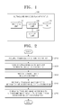
- FIG. 1 is a block diagram of an ultrasound image display apparatus 100 according to an embodiment of the present invention.
- the ultrasound image display apparatus 100 of the present embodiment is a device for acquiring an ultrasound image, storing the ultrasound image, and displaying the ultrasound image to a user.
- the ultrasound image display apparatus 100 may acquire an ultrasound image acquired by itself or receive an ultrasound image from the outside.
- the ultrasound image display apparatus 100 may be implemented in various ways.
- the ultrasound image display apparatus 100 described in the present specification may include an ultrasound diagnosis apparatus, a general purpose computer system, a laptop, a personal digital assistant (PDA), a tablet PC, and the like.
- PDA personal digital assistant
- the ultrasound image display apparatus 100 may provide the user with a bookmarking function.
- Bookmarking is an indication used to identify at least one ultrasound image. For example, if there is a region assumed to be a disease or an image having a measured clinical significance during a review of an ultrasound image, the user may bookmark the ultrasound image.
- the ultrasound image display apparatus 100 may include a receiving unit 110, a display unit 120, a storage unit 130, and a control unit 140.
- the elements of FIG. 1 are not indispensable elements.
- the ultrasound image display apparatus 100 may include a greater number of elements or a lesser number of elements.
- the receiving unit 110 may acquire patient identification information.
- the patient identification information is information used to identify a patient, such as an ID of the patient, an ID number, a name, a birth date, and the like. According to an embodiment of the present invention, if the user selects specific patient identification information, the receiving unit i110 acquires the patient identification information selected by the user.
- the receiving unit 110 may receive ultrasound image selection information included in examination lists.
- the receiving unit 110 may receive a plurality of pieces of ultrasound image selection information included in the examination lists. That is, the user may select a specific ultrasound image from the examination lists corresponding to the patient identification information, and select two or more ultrasound images.
- the receiving unit 110 may receive bookmark selection information.
- the bookmark selection information according to the present embodiment is a user's selection information regarding a bookmark indicated in an ultrasound image.
- the receiving unit 110 may receive a plurality of pieces of bookmark selection information.
- the user may select a specific bookmark from a displayed screen, and review a corresponding ultrasound image, thereby reducing a user's ultrasound image search time.
- the receiving unit 110 may receive ultrasound image diagnosis department selection information.
- the ultrasound image diagnosis department selection information is selection information regarding a diagnosis department that diagnoses an ultrasound image.
- an ultrasound image diagnosis department may include an OB (Obstetrics) department, a GYN (Gynecology) department, a PD (Pediatrics) department, a CS (Chestsurgery) department, a RD (Radiology) department, a NS (Neurosurgery) department, and the like.
- the display unit 120 may output information processed by the ultrasound image display apparatus 100.
- the display unit 120 may provide examination lists including bookmarks corresponding to the acquired patient identification information.
- the examination lists may include thumbnails of time-sequentially arranged ultrasound images.
- the display unit 120 may provide the examination lists in a bar chart.
- the examination lists according to the present embodiment may include a plurality of examination items EXAM.
- the examination items EXAM are past examination details of a specific patient divided by a predetermined unit.
- ultrasound images 1, 2, and 3 captured on May 1, 2011 may be configured as one examination item (for example, EXAM 1), and ultrasound images a, b, c, d, and e captured on July 1, 2011 may be configured as another examination item (for example, EXAM 2).
- the display unit 120 may display the examination lists by differentiating lengths of bars based on an amount of ultrasound images of the plurality of examination items EXAM.
- the display unit 120 may display a bookmarked ultrasound image based on the bookmark selection information.
- the display unit 120 may also display measurement values included in a plurality of ultrasound images in a graph chart.
- the graph chart may include a bar graph, a curve line graph, a circle graph, a radial graph, a histogram, and the like but the present invention is not limited thereto.
- the display unit 120 may provide an examination list including a bookmark corresponding to the selected ultrasound image diagnosis department.
- the display unit 120 may be used as an input device other than an output device.
- the display unit 120 may include at least one of a liquid crystal display (LCD), a thin film transistor-liquid crystal display (TFT-LCD), an organic light-emitting diode (OLED), a flexible display, and a 3D display.
- the ultrasound image display apparatus 100 may include two or more display units 120 according to how the ultrasound image display apparatus 100 is implemented.
- the storage unit 130 may store patient information, an ultrasound image, diagnosis data, etc.
- the patient information of the present embodiment may include information regarding patient personal identification information (an ID, an ID number, a name, a birth date, and the like), a medical history, a singularity, and the like.
- the diagnosis data may include measured result information, content analyzed by the user, a user's opinion on abnormality, a singularity, and the like regarding the ultrasound image.
- the storage unit 130 may be embedded in the ultrasound image display apparatus 100 or may be an external memory.
- the storage unit 130 also may be a storage server implemented on the Web.
- the storage unit 130 of the present embodiment may classify and store ultrasound image data, according to patients and diagnosis dates.
- the storage unit 130 may store ultrasound images in a unit of the examination items EXAM.
- the examination item EXAM of the present embodiment may include a plurality of ultrasound images.
- the storage unit 130 may store ultrasound images per diagnosis departments.
- the storage unit 130 may also store bookmark information indicated by the user.
- the control unit 140 may generally control the receiving unit 110, the display unit 120, and the storage unit 130.
- the control unit 140 may analyze the amount of ultrasound images of the plurality of examination items included in the examination lists, and set lengths of bars corresponding to the plurality of examination items based on the analyzed amount of ultrasound images.
- the control unit 140 may determine whether the same measurement item is included in a plurality of ultrasound images selected by the user, and, if the same measured item is included therein, may generate comparison data between measurement values included in the plurality of ultrasound images selected by the user
- a method of displaying an ultrasound image to a user by using each element of the ultrasound image display apparatus 100 will now be in detail described with reference to FIGS. 2 through 5 .
- FIG. 2 is a flowchart illustrating a method of displaying an ultrasound image, according to an embodiment of the present invention.
- the ultrasound image display apparatus 100 may acquire patient identification information (operation S210). For example, if a user wants to review an ultrasound image of a patient A and inputs an ID of the patient A, the ultrasound image display apparatus 100 acquires patient identification information of the patient A.
- the ultrasound image display apparatus 100 may acquire the patient identification information.
- the ultrasound image display apparatus 100 may receive ultrasound image diagnosis department selection information (operation S220). That is, the user may select the patient identification information on the work list and a clinic department in which an ultrasound image is analyzed. For example, if the user wants to review an ultrasound image regarding the patient A analyzed in an OB/GYN department, the user may input the patient identification information of the patient A, and select OB/GYN from among clinic departments.
- the ultrasound image display apparatus 100 may provide an examination list including a bookmark corresponding to the acquired patient identification information (operation S230).
- the examination list may be provided in a thumbnail format.
- the examination list may be provided in a bar chart. This will be described in detail with reference to FIG. 3 .
- the ultrasound image display apparatus 100 may display examination lists in an order of diagnosis times.
- the ultrasound image display apparatus 100 may also display examination lists including a plurality of examination items EXAM in an ascending order of an amount of ultrasound images or in an ascending order of bookmarks.
- the ultrasound image display apparatus 100 may provide an examination list including a bookmark corresponding to a selected diagnosis department. That is, if ultrasound images of patients are diagnosed in a plurality of clinic departments through a comprehensive medical check, the ultrasound image display apparatus 100 may display an ultrasound image diagnosed in a selected diagnosis department.
- the ultrasound image display apparatus 100 may receive ultrasound image selection information included in the examination lists (operation S240).
- the ultrasound image display apparatus 100 may receive one ultrasound image selection information or a plurality of pieces of ultrasound image selection information.
- the user may select an ultrasound image A included in the examination lists or the ultrasound image A and an ultrasound image B.
- the user may select a plurality of ultrasound images in the same examination item EXAM or extract a plurality of ultrasound images in different examination items EXAM.
- the ultrasound image display apparatus 100 may receive bookmark selection information.
- the user may select one bookmark or a plurality of bookmarks.
- the ultrasound image display apparatus 100 may display ultrasound images according to the ultrasound image selection information or the bookmark selection information (operation S250). For example, according to an embodiment of the present invention, if the user selects an ultrasound image included in an examination list, the ultrasound image display apparatus 100 may display the selected ultrasound image.
- the ultrasound image display apparatus 100 may display a bookmarked ultrasound image according to the bookmark selection information.
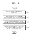
- FIG. 3 is a flowchart illustrating a method of providing an examination list in a bar chart, according to an embodiment of the present invention.
- the ultrasound image display apparatus 100 may acquire patient identification information (operation S310). In this case, the ultrasound image display apparatus 100 may analyze an amount of ultrasound images of a plurality of examination items included in the examination list corresponding to the acquired patient identification information (operation S320).
- the ultrasound image display apparatus 100 may set lengths of bars corresponding to the plurality of examination items based on the analyzed amount of ultrasound images (operation S330). For example, if three ultrasound images are included in an examination item 1 EXAM 1, and nine ultrasound images are included in an examination item 2 EXAM 2, lengths of bars corresponding to the examination item 2 EXAM 2 may be set three times longer than those corresponding to the examination item 1 EXAM 1.
- the ultrasound image display apparatus 100 may provide the examination list in the bar chart including a bookmark according to the set lengths of the bars.
- a user may intuitively check the amount of ultrasound images included in each examination item EXAM.
- FIG. 4 is a flowchart illustrating a method of displaying comparison data between measurement values included in a plurality of ultrasound images in a graph chart, according to an embodiment of the present invention.
- the ultrasound image display apparatus 100 may acquire patient identification information (operation S410).
- the ultrasound image display apparatus 100 may provide an examination list including a bookmark corresponding to the acquired patient identification information (operation S420).
- the ultrasound image display apparatus 100 may receive a plurality of pieces of ultrasound image selection information included in the examination list (operation S430). According to an embodiment of the present invention, the ultrasound image display apparatus 100 may receive a plurality of pieces of bookmark selection information.
- the ultrasound image display apparatus 100 may determine whether the same measurement item is included in selected plurality of ultrasound images (operation S440).
- Measurement items according to the present embodiment may include at least one of a BPD (bi-parietal diameter), an OFD (occipitofrontal diameter), and an AC (abdominal circumference), but the present invention is not limited thereto.
- the ultrasound image display apparatus 100 may generate the comparison data between the measurement values included in the selected ultrasound images (operation S450). For example, if the BPD item is included in ultrasound images A, B, and C selected by a user, the ultrasound image display apparatus 100 may compare BPD values included in the ultrasound images A, B, and C.
- the ultrasound image display apparatus 100 may display the generated comparison data in the graph chart (operation S460).
- it is useful in comparing a plurality of examination items EXAM like a growth progress of an embryo or a prognosis of a lesion.
- the graph chart according to the present embodiment may be applied in various ways as occasion demands.
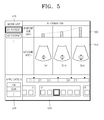
- FIG. 5 is a diagram of an example of a screen displaying an ultrasound image, according to an embodiment of the present invention.
- patient identification information may be displayed on a work list 510.
- a user may select identification information (for example, an ID) of a patient of which an ultrasound image is to be checked from the work list 510.
- the user may also select a diagnosis department 520 if the user is to search for an ultrasound image diagnosed in a predetermined diagnosis department. For example, if the user wishes to see a growth progress of an embryo, the user may select an OB department.
- the ultrasound image display apparatus 100 may display a corresponding examination list 530.
- the ultrasound image display apparatus 100 may display the examination list 530 including bookmark information (for example, ⁇ ).
- the ultrasound image display apparatus 100 may adjust lengths of bars in the examination list 530 based on an amount of ultrasound images of a plurality of examination items EXAM. For example, as shown in FIG. 5 , since an examination item (EXAM ) includes six ultrasound images, which is the most, the ultrasound image display apparatus 100 may adjust a bar corresponding to the examination item (EXAM ) to be the longest.
- the ultrasound image display apparatus 100 may display ultrasound images 540 based on a user's selection. If the same measurement item is included in a selected plurality of ultrasound images, the ultrasound image display apparatus 100 may provide comparison data of measurement values in a graph chart 550.
- the ultrasound image display apparatus 100 may display BPD values included in ultrasound images 1, 2, and 3 in a bar graph chart. In this case, the user may visually see a growth progress of an embryo.
- Embodiments of the present invention include a computer-readable recording medium including program commands for executing operations implemented through various computers.
- the computer-readable recording medium can store program commands, data files, data structures, or combinations thereof.
- the program commands recorded in the computer-readable recording medium may be specially designed and configured for the present invention or be known to those of ordinary skill in the field of computer software.
- Examples of a computer-readable recording medium include magnetic media such as hard disks, floppy disks, and magnetic tapes, optical media such as CD-ROMs and DVDs, magneto-optical media such as floptical disks, or hardware devices such as ROMs, RAMs, and flash memories, which are specially configured to store and execute program commands.
- Examples of the program commands include a machine language code created by a compiler and a high-level language code executable by a computer using an interpreter and the like.
- the computers may include the control unit 140 of the ultrasound image display apparatus 100.
Landscapes
- Health & Medical Sciences (AREA)
- Life Sciences & Earth Sciences (AREA)
- Engineering & Computer Science (AREA)
- Physics & Mathematics (AREA)
- General Health & Medical Sciences (AREA)
- Medical Informatics (AREA)
- Nuclear Medicine, Radiotherapy & Molecular Imaging (AREA)
- Public Health (AREA)
- Radiology & Medical Imaging (AREA)
- Molecular Biology (AREA)
- Surgery (AREA)
- Biophysics (AREA)
- Biomedical Technology (AREA)
- Heart & Thoracic Surgery (AREA)
- Veterinary Medicine (AREA)
- Animal Behavior & Ethology (AREA)
- Pathology (AREA)
- Theoretical Computer Science (AREA)
- General Physics & Mathematics (AREA)
- Human Computer Interaction (AREA)
- Epidemiology (AREA)
- Primary Health Care (AREA)
- Computer Vision & Pattern Recognition (AREA)
- General Engineering & Computer Science (AREA)
- Ultra Sonic Daignosis Equipment (AREA)
Abstract
Description
- This application claims the benefit of Korean Patent Application No.
10-2011-0122400, filed on November 22, 2011 - The present invention relates to a method and apparatus for displaying an ultrasound image to provide examination lists including bookmarks and an ultrasound image corresponding to an examination item selected by a user.
- Ultrasound diagnosis apparatuses transmit an ultrasound signal toward a predetermined part of the body from a body surface of an object and obtain a tomogram of soft-tissues or an image of blood flow by using information regarding the ultrasound signal reflected by tissues of the body.
- Ultrasound diagnosis apparatuses have various advantages, including a compact size, low cost, and real-time display. Also, ultrasound diagnosis apparatuses have excellent stability because there is no fear of X-ray exposure, and thus, the ultrasound diagnosis apparatuses are widely used together with various other diagnosis apparatuses, such as computerized tomography scanners, magnetic resonance imaging (MRI) apparatuses, nuclear medicine diagnosis apparatuses, or the like.
- In general, ultrasound diagnosis apparatuses do not provide a function of searching for an ultrasound image. That is, when a user searches for an ultrasound image of a previously diagnosed patient, the user needs to search for the ultrasound image by checking diagnosis dates and thumbnail images one by one. Thus, it takes much time to search for the ultrasound image desired by the user.
- Furthermore, current ultrasound diagnosis apparatuses do not search for a plurality of ultrasound images having common characteristics simultaneously, and do not provide users with comparison data of the plurality of ultrasound images. That is, if a patient undergoes various examinations during a predetermined period of time like determining growth progress of an embryo or a prognosis of a lesion, current ultrasound diagnosis apparatuses need to inconveniently search for and compare and analyze images having a specific lesion or clinical significance from all stored image lists.
- The present invention provides a method and apparatus for displaying an ultrasound image, which provide a user with a function of bookmarking an ultrasound image so that the user can easily search for the bookmarked ultrasound image.
- According to an aspect of the present invention, there is provided a method of displaying an ultrasound image, the method including: acquiring patient identification information; providing an examination list including a bookmark used to identify at least one ultrasound image based on the acquired patient identification information; receiving selection information regarding the at least one ultrasound image included in the examination list; and displaying the ultrasound image selected based on the selection information.
- The providing of the examination list may include: providing the examination list including the bookmark in a bar chart. In this case, the method may include: analyzing an amount of ultrasound images of at least one examination item included in the examination list; setting a length of a bar corresponding to the at least one examination item based on the analyzed amount of ultrasound images; and providing the examination list including the bookmark in the bar chart according to the set length of the bar.
- The method may include receiving bookmark selection information to select at least one bookmark included in the examination list; and selecting at least one bookmark based on the bookmark selection information and displaying the ultrasound image corresponding to the selected at least one bookmark.
- The method may include: receiving a plurality of pieces of ultrasound image selection information to select a plurality of ultrasound images included in the examination list; and selecting the plurality of ultrasound images based on the plurality of pieces of ultrasound image selection information and displaying measurement values corresponding to the selected plurality of ultrasound images in a graph chart. In this regard, the method may include: determining whether a same measurement item is included in the selected plurality of ultrasound images; if it is determined that the same measurement item is included in the selected plurality of ultrasound images, generating comparison data between the measurement values corresponding to the selected plurality of ultrasound images; and displaying the generated comparison data in the graph chart.
- The method may include: receiving ultrasound image diagnosis department selection information to select at least one ultrasound image diagnosis department; and selecting the at least one ultrasound image diagnosis department based on the ultrasound image diagnosis department selection information, and providing the examination list corresponding to the selected at least one ultrasound image diagnosis department.
- According to another aspect of the present invention, there is provided an apparatus for displaying an ultrasound image, the apparatus including: a receiving unit for acquiring patient identification information, and receiving selection information regarding at least one ultrasound image included in an examination list; a display unit for providing the examination list including a bookmark used to identify the at least one ultrasound image based on the acquired patient identification information, and displaying the ultrasound image selected based on the selection information; and a control unit for controlling the receiving unit and the display unit.
- According to another aspect of the present invention, there is provided a computer readable recording medium storing a program for executing a method of displaying an ultrasound image.
- The above and other features and advantages of the present invention will become more apparent by describing in detail exemplary embodiments thereof with reference to the attached drawings in which:
-
FIG. 1 is a block diagram of an ultrasound image display apparatus according to an embodiment of the present invention; -
FIG. 2 is a flowchart illustrating a method of displaying an ultrasound image, according to an embodiment of the present invention; -
FIG. 3 is a flowchart illustrating a method of providing an examination list in a bar chart, according to an embodiment of the present invention; -
FIG. 4 is a flowchart illustrating a method of displaying comparison data between measurement values included in a plurality of ultrasound images in a graph chart, according to an embodiment of the present invention; and -
FIG. 5 is a diagram of an example of a screen displaying an ultrasound image, according to an embodiment of the present invention. - First, the terms used in the present disclosure will be briefly described below before exemplary embodiments of the present invention are described in greater detail.
- Most of the terms used herein are general terms that have been widely used in the technical art to which the present invention pertains. However, some of the terms used herein may be created reflecting intentions of technicians in this art, precedents, or new technologies. Also, some of the terms used herein may be arbitrarily chosen by the present applicant. In this case, these terms are defined in detail below. Accordingly, the specific terms used herein should be understood based on the unique meanings thereof and the whole context of the present invention.
- In the present specification, it should be understood that the terms, such as 'including' or 'having,' etc., are intended to indicate the existence of the features, numbers, steps, actions, components, parts, or combinations thereof disclosed in the specification, and are not intended to preclude the possibility that one or more other features, numbers, steps, actions, components, parts, or combinations thereof may exist or may be added. Also, the terms, such as 'unit' or 'module', etc., should be understood as a unit that processes at least one function or operation and that may be embodied in a hardware manner, a software manner, or a combination of the hardware manner and the software manner.
- Throughout the specification, the term 'ultrasound image' means an image of a target, which is captured using ultrasound waves. Here, the 'target' may be understood to be part of a human body. For example, the target may be an organ of a human body, such as the liver, the heart, the womb, a brain, the breast, or the abdomen, or a fetus.
- The ultrasound image may be embodied in any of various forms. For example, the ultrasound image may include at least one selected from the group consisting of a brightness (B) mode image, a color (C) mode image, and a Doppler (D) mode image. According to an embodiment of the present invention, the ultrasound image may be a two-dimensional (2D) or three-dimensional (3D) image.
- Throughout the specification, the term 'user' may be a medical professional, e.g., a doctor, a nurse, a clinical pathologist, or a medical imaging professional, but is not limited thereto.
- The present invention will now be described more fully with reference to the accompanying drawings, in which exemplary embodiments of the invention are shown. This invention may, however, be embodied in many different forms and should not be construed as being limited to the embodiments set forth herein. Rather, these embodiments are provided so that this disclosure will be thorough and complete and will fully convey the concept of the invention to those of ordinary skill in the art. For clarity, in the drawings, constitutional elements that are not related to embodiments of the present invention are not illustrated. The same reference numerals represent the same elements throughout the drawings.
- As used herein, the term "and/or" includes any and all combinations of one or more of the associated listed items. Expressions such as "at least one of," when preceding a list of elements, modify the entire list of elements and do not modify the individual elements of the list.
-
FIG. 1 is a block diagram of an ultrasoundimage display apparatus 100 according to an embodiment of the present invention. - Referring to
FIG. 1 , the ultrasoundimage display apparatus 100 of the present embodiment is a device for acquiring an ultrasound image, storing the ultrasound image, and displaying the ultrasound image to a user. The ultrasoundimage display apparatus 100 may acquire an ultrasound image acquired by itself or receive an ultrasound image from the outside. - The ultrasound
image display apparatus 100 according to the present embodiment may be implemented in various ways. For example, the ultrasoundimage display apparatus 100 described in the present specification may include an ultrasound diagnosis apparatus, a general purpose computer system, a laptop, a personal digital assistant (PDA), a tablet PC, and the like. - Meanwhile, the ultrasound
image display apparatus 100 according to the present embodiment may provide the user with a bookmarking function. Bookmarking is an indication used to identify at least one ultrasound image. For example, if there is a region assumed to be a disease or an image having a measured clinical significance during a review of an ultrasound image, the user may bookmark the ultrasound image. - According to an embodiment of the present invention, as shown in
FIG. 1 , the ultrasoundimage display apparatus 100 may include areceiving unit 110, adisplay unit 120, astorage unit 130, and acontrol unit 140. However, the elements ofFIG. 1 are not indispensable elements. The ultrasoundimage display apparatus 100 may include a greater number of elements or a lesser number of elements. - The receiving
unit 110 may acquire patient identification information. The patient identification information according to the present embodiment is information used to identify a patient, such as an ID of the patient, an ID number, a name, a birth date, and the like. According to an embodiment of the present invention, if the user selects specific patient identification information, the receiving unit i110 acquires the patient identification information selected by the user. - The receiving
unit 110 may receive ultrasound image selection information included in examination lists. In this case, the receivingunit 110 may receive a plurality of pieces of ultrasound image selection information included in the examination lists. That is, the user may select a specific ultrasound image from the examination lists corresponding to the patient identification information, and select two or more ultrasound images. - According to an embodiment of the present invention, the receiving
unit 110 may receive bookmark selection information. The bookmark selection information according to the present embodiment is a user's selection information regarding a bookmark indicated in an ultrasound image. According to an embodiment of the present invention, the receivingunit 110 may receive a plurality of pieces of bookmark selection information. - Therefore, according to an embodiment of the present invention, the user may select a specific bookmark from a displayed screen, and review a corresponding ultrasound image, thereby reducing a user's ultrasound image search time.
- Meanwhile, the receiving
unit 110 may receive ultrasound image diagnosis department selection information. The ultrasound image diagnosis department selection information is selection information regarding a diagnosis department that diagnoses an ultrasound image. For example, an ultrasound image diagnosis department may include an OB (Obstetrics) department, a GYN (Gynecology) department, a PD (Pediatrics) department, a CS (Chestsurgery) department, a RD (Radiology) department, a NS (Neurosurgery) department, and the like. - The
display unit 120 may output information processed by the ultrasoundimage display apparatus 100. Thedisplay unit 120 may provide examination lists including bookmarks corresponding to the acquired patient identification information. In this regard, according to an embodiment of the present invention, the examination lists may include thumbnails of time-sequentially arranged ultrasound images. - According to another embodiment, the
display unit 120 may provide the examination lists in a bar chart. The examination lists according to the present embodiment may include a plurality of examination items EXAM. The examination items EXAM are past examination details of a specific patient divided by a predetermined unit. - For example,
ultrasound images - According to an embodiment of the present invention, the
display unit 120 may display the examination lists by differentiating lengths of bars based on an amount of ultrasound images of the plurality of examination items EXAM. - Meanwhile, according to an embodiment of the present invention, the
display unit 120 may display a bookmarked ultrasound image based on the bookmark selection information. Thedisplay unit 120 may also display measurement values included in a plurality of ultrasound images in a graph chart. The graph chart may include a bar graph, a curve line graph, a circle graph, a radial graph, a histogram, and the like but the present invention is not limited thereto. - According to an embodiment of the present invention, if the user selects the ultrasound image diagnosis department, the
display unit 120 may provide an examination list including a bookmark corresponding to the selected ultrasound image diagnosis department. - Meanwhile, if the
display unit 120 and a touch pad form a mutual layer structure and thus are configured as a touch screen, thedisplay unit 120 may be used as an input device other than an output device. Thedisplay unit 120 may include at least one of a liquid crystal display (LCD), a thin film transistor-liquid crystal display (TFT-LCD), an organic light-emitting diode (OLED), a flexible display, and a 3D display. The ultrasoundimage display apparatus 100 may include two ormore display units 120 according to how the ultrasoundimage display apparatus 100 is implemented. - The
storage unit 130 may store patient information, an ultrasound image, diagnosis data, etc. The patient information of the present embodiment may include information regarding patient personal identification information (an ID, an ID number, a name, a birth date, and the like), a medical history, a singularity, and the like. The diagnosis data may include measured result information, content analyzed by the user, a user's opinion on abnormality, a singularity, and the like regarding the ultrasound image. - According to an embodiment of the present invention, the
storage unit 130 may be embedded in the ultrasoundimage display apparatus 100 or may be an external memory. Thestorage unit 130 also may be a storage server implemented on the Web. - The
storage unit 130 of the present embodiment may classify and store ultrasound image data, according to patients and diagnosis dates. For example, thestorage unit 130 may store ultrasound images in a unit of the examination items EXAM. Thus, the examination item EXAM of the present embodiment may include a plurality of ultrasound images. - Meanwhile, according to another embodiment of the present invention, the
storage unit 130 may store ultrasound images per diagnosis departments. Thestorage unit 130 may also store bookmark information indicated by the user. - The
control unit 140 may generally control the receivingunit 110, thedisplay unit 120, and thestorage unit 130. For example, thecontrol unit 140 may analyze the amount of ultrasound images of the plurality of examination items included in the examination lists, and set lengths of bars corresponding to the plurality of examination items based on the analyzed amount of ultrasound images. - The
control unit 140 may determine whether the same measurement item is included in a plurality of ultrasound images selected by the user, and, if the same measured item is included therein, may generate comparison data between measurement values included in the plurality of ultrasound images selected by the user - A method of displaying an ultrasound image to a user by using each element of the ultrasound
image display apparatus 100 will now be in detail described with reference toFIGS. 2 through 5 . -
FIG. 2 is a flowchart illustrating a method of displaying an ultrasound image, according to an embodiment of the present invention. - Referring to
FIG. 2 , according to the present embodiment, the ultrasoundimage display apparatus 100 may acquire patient identification information (operation S210). For example, if a user wants to review an ultrasound image of a patient A and inputs an ID of the patient A, the ultrasoundimage display apparatus 100 acquires patient identification information of the patient A. - Meanwhile, according to an embodiment of the present invention, when the user selects a patient ID from a work list, the ultrasound
image display apparatus 100 may acquire the patient identification information. - According to an embodiment of the present invention, the ultrasound
image display apparatus 100 may receive ultrasound image diagnosis department selection information (operation S220). That is, the user may select the patient identification information on the work list and a clinic department in which an ultrasound image is analyzed. For example, if the user wants to review an ultrasound image regarding the patient A analyzed in an OB/GYN department, the user may input the patient identification information of the patient A, and select OB/GYN from among clinic departments. - The ultrasound
image display apparatus 100 may provide an examination list including a bookmark corresponding to the acquired patient identification information (operation S230). According to an embodiment of the present invention, the examination list may be provided in a thumbnail format. According to another embodiment of the present invention, the examination list may be provided in a bar chart. This will be described in detail with reference toFIG. 3 . - According to an embodiment of the present invention, the ultrasound
image display apparatus 100 may display examination lists in an order of diagnosis times. The ultrasoundimage display apparatus 100 may also display examination lists including a plurality of examination items EXAM in an ascending order of an amount of ultrasound images or in an ascending order of bookmarks. - Meanwhile, if the ultrasound image diagnosis department selection information is received, the ultrasound
image display apparatus 100 may provide an examination list including a bookmark corresponding to a selected diagnosis department. That is, if ultrasound images of patients are diagnosed in a plurality of clinic departments through a comprehensive medical check, the ultrasoundimage display apparatus 100 may display an ultrasound image diagnosed in a selected diagnosis department. - According to an embodiment of the present invention, the ultrasound
image display apparatus 100 may receive ultrasound image selection information included in the examination lists (operation S240). In this case, the ultrasoundimage display apparatus 100 may receive one ultrasound image selection information or a plurality of pieces of ultrasound image selection information. For example, the user may select an ultrasound image A included in the examination lists or the ultrasound image A and an ultrasound image B. - In this regard, the user may select a plurality of ultrasound images in the same examination item EXAM or extract a plurality of ultrasound images in different examination items EXAM.
- According to an embodiment of the present invention, the ultrasound
image display apparatus 100 may receive bookmark selection information. In this case, the user may select one bookmark or a plurality of bookmarks. - According an embodiment of the present invention, the ultrasound
image display apparatus 100 may display ultrasound images according to the ultrasound image selection information or the bookmark selection information (operation S250). For example, according to an embodiment of the present invention, if the user selects an ultrasound image included in an examination list, the ultrasoundimage display apparatus 100 may display the selected ultrasound image. - According to another embodiment of the present invention, if the user selects a bookmark, the ultrasound
image display apparatus 100 may display a bookmarked ultrasound image according to the bookmark selection information. -
FIG. 3 is a flowchart illustrating a method of providing an examination list in a bar chart, according to an embodiment of the present invention. - Referring to
FIG. 3 , according to the present embodiment, the ultrasoundimage display apparatus 100 may acquire patient identification information (operation S310). In this case, the ultrasoundimage display apparatus 100 may analyze an amount of ultrasound images of a plurality of examination items included in the examination list corresponding to the acquired patient identification information (operation S320). - The ultrasound
image display apparatus 100 may set lengths of bars corresponding to the plurality of examination items based on the analyzed amount of ultrasound images (operation S330). For example, if three ultrasound images are included in anexamination item 1EXAM 1, and nine ultrasound images are included in anexamination item 2EXAM 2, lengths of bars corresponding to theexamination item 2EXAM 2 may be set three times longer than those corresponding to theexamination item 1EXAM 1. - The ultrasound
image display apparatus 100 may provide the examination list in the bar chart including a bookmark according to the set lengths of the bars. Thus, according to an embodiment of the present invention, a user may intuitively check the amount of ultrasound images included in each examination item EXAM. -
FIG. 4 is a flowchart illustrating a method of displaying comparison data between measurement values included in a plurality of ultrasound images in a graph chart, according to an embodiment of the present invention. - Referring to
FIG. 4 , according to the present invention, the ultrasoundimage display apparatus 100 may acquire patient identification information (operation S410). The ultrasoundimage display apparatus 100 may provide an examination list including a bookmark corresponding to the acquired patient identification information (operation S420). - In this case, the ultrasound
image display apparatus 100 may receive a plurality of pieces of ultrasound image selection information included in the examination list (operation S430). According to an embodiment of the present invention, the ultrasoundimage display apparatus 100 may receive a plurality of pieces of bookmark selection information. - The ultrasound
image display apparatus 100 according to an embodiment of the present invention may determine whether the same measurement item is included in selected plurality of ultrasound images (operation S440). Measurement items according to the present embodiment may include at least one of a BPD (bi-parietal diameter), an OFD (occipitofrontal diameter), and an AC (abdominal circumference), but the present invention is not limited thereto. - If the same measurement item is included in a selected plurality of ultrasound images, the ultrasound
image display apparatus 100 may generate the comparison data between the measurement values included in the selected ultrasound images (operation S450). For example, if the BPD item is included in ultrasound images A, B, and C selected by a user, the ultrasoundimage display apparatus 100 may compare BPD values included in the ultrasound images A, B, and C. - The ultrasound
image display apparatus 100 may display the generated comparison data in the graph chart (operation S460). Thus, according to an embodiment of the present invention, it is useful in comparing a plurality of examination items EXAM like a growth progress of an embryo or a prognosis of a lesion. - Meanwhile, the graph chart according to the present embodiment may be applied in various ways as occasion demands.
-
FIG. 5 is a diagram of an example of a screen displaying an ultrasound image, according to an embodiment of the present invention. Referring toFIG. 5 , patient identification information may be displayed on awork list 510. A user may select identification information (for example, an ID) of a patient of which an ultrasound image is to be checked from thework list 510. - The user may also select a
diagnosis department 520 if the user is to search for an ultrasound image diagnosed in a predetermined diagnosis department. For example, if the user wishes to see a growth progress of an embryo, the user may select an OB department. - If the user inputs the patient identification information and/or diagnosis department selection information, the ultrasound
image display apparatus 100 may display acorresponding examination list 530. In this case, the ultrasoundimage display apparatus 100 may display theexamination list 530 including bookmark information (for example, ▼). - Meanwhile, the ultrasound
image display apparatus 100 may adjust lengths of bars in theexamination list 530 based on an amount of ultrasound images of a plurality of examination items EXAM. For example, as shown inFIG. 5 , since an examination item (EXAM ) includes six ultrasound images, which is the most, the ultrasoundimage display apparatus 100 may adjust a bar corresponding to the examination item (EXAM ) to be the longest. - If the user selects an ultrasound image or a bookmark, the ultrasound
image display apparatus 100 may displayultrasound images 540 based on a user's selection. If the same measurement item is included in a selected plurality of ultrasound images, the ultrasoundimage display apparatus 100 may provide comparison data of measurement values in agraph chart 550. - For example, as shown in
FIG. 5 , the ultrasoundimage display apparatus 100 may display BPD values included inultrasound images - Embodiments of the present invention include a computer-readable recording medium including program commands for executing operations implemented through various computers. The computer-readable recording medium can store program commands, data files, data structures, or combinations thereof. The program commands recorded in the computer-readable recording medium may be specially designed and configured for the present invention or be known to those of ordinary skill in the field of computer software. Examples of a computer-readable recording medium include magnetic media such as hard disks, floppy disks, and magnetic tapes, optical media such as CD-ROMs and DVDs, magneto-optical media such as floptical disks, or hardware devices such as ROMs, RAMs, and flash memories, which are specially configured to store and execute program commands. Examples of the program commands include a machine language code created by a compiler and a high-level language code executable by a computer using an interpreter and the like. The computers may include the
control unit 140 of the ultrasoundimage display apparatus 100. - While the present invention has been particularly shown and described with reference to exemplary embodiments thereof, it will be understood by those of ordinary skill in the art that various changes in form and details may be made therein without departing from the spirit and scope of the present invention as defined by the following claims.
Claims (15)
- A method of displaying an ultrasound image, the method comprising:acquiring patient identification information;providing an examination list including a bookmark used to identify at least one ultrasound image based on the acquired patient identification information;receiving selection information regarding the at least one ultrasound image included in the examination list; anddisplaying the ultrasound image selected based on the selection information.
- The method of claim 1, wherein the providing of the examination list comprises: providing the examination list including the bookmark in a bar chart.
- The method of claim 2, wherein the providing of the examination list including the bookmark in the bar chart comprises:analyzing an amount of ultrasound images of at least one examination item included in the examination list;setting a length of a bar corresponding to the at least one examination item based on the analyzed amount of ultrasound images; andproviding the examination list including the bookmark in the bar chart according to the set length of the bar.
- The method of claim 1, wherein the receiving of the selection information comprises: receiving bookmark selection information to select at least one bookmark included in the examination list,
wherein the displaying of the ultrasound image comprises: selecting at least one bookmark based on the bookmark selection information and displaying the ultrasound image corresponding to the selected at least one bookmark. - The method of claim 1, wherein the receiving of the selection information comprises: receiving a plurality of pieces of ultrasound image selection information to select a plurality of ultrasound images included in the examination list,
wherein the displaying of the ultrasound image comprises: selecting the plurality of ultrasound images based on the plurality of pieces of ultrasound image selection information and displaying measurement values corresponding to the selected plurality of ultrasound images in a graph chart. - The method of claim 5, wherein the displaying of the measurement values corresponding to the selected plurality of ultrasound images in the graph chart comprises:determining whether a same measurement item is included in the selected plurality of ultrasound images;if it is determined that the same measurement item is included in the selected plurality of ultrasound images, generating comparison data between the measurement values corresponding to the selected plurality of ultrasound images; anddisplaying the generated comparison data in the graph chart.
- The method of claim 1, wherein the providing of the examination list including the bookmark further comprises:receiving ultrasound image diagnosis department selection information to select at least one ultrasound image diagnosis department; andselecting the at least one ultrasound image diagnosis department based on the ultrasound image diagnosis department selection information, and providing the examination list corresponding to the selected at least one ultrasound image diagnosis department.
- An apparatus for displaying an ultrasound image, the apparatus comprising:a receiving unit for acquiring patient identification information, and receiving selection information regarding at least one ultrasound image included in an examination list;a display unit for providing the examination list including a bookmark used to identify the at least one ultrasound image based on the acquired patient identification information, and displaying the ultrasound image selected based on the selection information; anda control unit for controlling the receiving unit and the display unit.
- The apparatus of claim 1, wherein the display unit provides the examination list including the bookmark in a bar chart.
- The apparatus of claim 9, wherein the control unit analyzes an amount of ultrasound images of at least one examination item included in the examination list, and sets a length of a bar corresponding to the at least one examination item based on the analyzed amount of ultrasound images,
wherein the display unit provides the examination list including the bookmark in the bar chart according to the set length of the bar. - The apparatus of claim 8, wherein the receiving unit receives bookmark selection information to select at least one bookmark included in the examination list,
wherein the display unit selects at least one bookmark based on the bookmark selection information and displays an ultrasound image corresponding to the selected at least one bookmark. - The apparatus of claim 8, wherein the receiving unit receives a plurality of pieces of ultrasound image selection information to select a plurality of ultrasound images included in the examination list,
wherein the display unit selects the plurality of ultrasound images based on the plurality of pieces of ultrasound image selection information and displays measurement values corresponding to the selected plurality of ultrasound images in a graph chart. - The apparatus of claim 12, wherein the control unit determines whether a same measurement item is included in the selected plurality of ultrasound images, and, if it is determined that the same measurement item is included in the selected plurality of ultrasound images, generates comparison data between the measurement values corresponding to the selected plurality of ultrasound images; and
wherein the display unit displays the generated comparison data in the graph chart. - The apparatus of claim 8, wherein the receiving unit receives ultrasound image diagnosis department selection information to select at least one ultrasound image diagnosis department,
wherein the display unit selects the at least one ultrasound image diagnosis department based on the ultrasound image diagnosis department selection information, and provides the examination list corresponding to the selected at least one ultrasound image diagnosis department. - A computer readable recording medium storing a program for executing a method of displaying an ultrasound image of claim 1.
Applications Claiming Priority (1)
| Application Number | Priority Date | Filing Date | Title |
|---|---|---|---|
| KR1020110122400A KR101323329B1 (en) | 2011-11-22 | 2011-11-22 | Method and apparatus for displaying ulrtasound image |
Publications (3)
| Publication Number | Publication Date |
|---|---|
| EP2597617A2 true EP2597617A2 (en) | 2013-05-29 |
| EP2597617A3 EP2597617A3 (en) | 2014-02-19 |
| EP2597617B1 EP2597617B1 (en) | 2022-12-21 |
Family
ID=45999602
Family Applications (1)
| Application Number | Title | Priority Date | Filing Date |
|---|---|---|---|
| EP12161003.4A Active EP2597617B1 (en) | 2011-11-22 | 2012-03-23 | Method and apparatus for displaying ultrasound image |
Country Status (3)
| Country | Link |
|---|---|
| US (1) | US9498187B2 (en) |
| EP (1) | EP2597617B1 (en) |
| KR (1) | KR101323329B1 (en) |
Families Citing this family (12)
| Publication number | Priority date | Publication date | Assignee | Title |
|---|---|---|---|---|
| US10667790B2 (en) | 2012-03-26 | 2020-06-02 | Teratech Corporation | Tablet ultrasound system |
| US9877699B2 (en) | 2012-03-26 | 2018-01-30 | Teratech Corporation | Tablet ultrasound system |
| CN104055532A (en) * | 2013-03-19 | 2014-09-24 | 深圳迈瑞生物医疗电子股份有限公司 | Monitoring equipment with ultrasonic scanning monitoring function, ultrasonic device and corresponding method |
| JP6178125B2 (en) * | 2013-06-13 | 2017-08-09 | 株式会社日立製作所 | Ultrasonic diagnostic equipment |
| US10025479B2 (en) * | 2013-09-25 | 2018-07-17 | Terarecon, Inc. | Advanced medical image processing wizard |
| WO2015124388A1 (en) * | 2014-02-19 | 2015-08-27 | Koninklijke Philips N.V. | Motion adaptive visualization in medical 4d imaging |
| KR102255831B1 (en) | 2014-03-26 | 2021-05-25 | 삼성전자주식회사 | Method and ultrasound apparatus for recognizing an ultrasound image |
| JP6608144B2 (en) * | 2015-02-03 | 2019-11-20 | キヤノンメディカルシステムズ株式会社 | Medical image diagnostic apparatus and medical information display apparatus |
| JP2019534110A (en) | 2016-11-16 | 2019-11-28 | テラテク・コーポレーシヨン | Portable ultrasound system |
| US11948345B2 (en) | 2018-04-09 | 2024-04-02 | Koninklijke Philips N.V. | Ultrasound system with artificial neural network for retrieval of imaging parameter settings for recurring patient |
| KR102917172B1 (en) * | 2022-04-19 | 2026-01-23 | 주식회사 온택트헬스 | Method and device for guiding echocardiography |
| KR102819588B1 (en) * | 2022-08-01 | 2025-06-13 | 주식회사 온택트헬스 | Method and device for providing information of doppler ultrasound images |
Family Cites Families (25)
| Publication number | Priority date | Publication date | Assignee | Title |
|---|---|---|---|---|
| AU5366499A (en) | 1998-08-19 | 2000-03-14 | Ontario Cancer Institute, The | The use of high frequency ultrasound imaging to detect and monitor the process of apoptosis in living tissues, (ex-vivo) tissues and cell-culture |
| US6458081B1 (en) * | 1999-04-23 | 2002-10-01 | Kabushiki Kaisha Toshiba | Ultrasonic diagnostic apparatus |
| US6859288B1 (en) * | 1999-04-28 | 2005-02-22 | General Electric Company | Method and apparatus for requesting and displaying worklist data from remotely located device |
| US6370413B1 (en) * | 1999-11-02 | 2002-04-09 | Siemens Medical Solutions Usa, Inc. | Ultrasound imaging system and method to archive and review 3-D ultrasound data |
| US7615008B2 (en) * | 2000-11-24 | 2009-11-10 | U-Systems, Inc. | Processing and displaying breast ultrasound information |
| JP4071710B2 (en) | 2001-06-21 | 2008-04-02 | 株式会社東芝 | Electronic report creation support apparatus, method and program |
| US20040066398A1 (en) * | 2002-10-03 | 2004-04-08 | Koninklijke Philips Electronics N.V | System and method for removing, trimming and bookmarking images of an ultrasound image sequence |
| US7044912B2 (en) * | 2003-08-28 | 2006-05-16 | Siemens Medical Solutions Usa Inc. | Diagnostic medical ultrasound system having method and apparatus for storing and retrieving 3D and 4D data sets |
| US7457672B2 (en) * | 2003-09-10 | 2008-11-25 | General Electric Company | Method and apparatus for exporting ultrasound data |
| US7727151B2 (en) * | 2003-11-28 | 2010-06-01 | U-Systems Inc. | Navigation among multiple breast ultrasound volumes |
| JP2005301567A (en) * | 2004-04-09 | 2005-10-27 | Toshiba Corp | Medical image diagnostic display device |
| JP4465535B2 (en) * | 2004-06-09 | 2010-05-19 | 株式会社日立メディコ | Elastic image display method and ultrasonic diagnostic apparatus |
| JP4585286B2 (en) * | 2004-11-17 | 2010-11-24 | アロカ株式会社 | Ultrasonic diagnostic equipment |
| US20060247527A1 (en) * | 2005-04-28 | 2006-11-02 | Kabushiki Kaisha Toshiba | Ultrasonic diagnostic apparatus, measuring result display apparatus and method of displaying measuring result |
| US20070127793A1 (en) | 2005-11-23 | 2007-06-07 | Beckett Bob L | Real-time interactive data analysis management tool |
| JP4912807B2 (en) * | 2006-09-22 | 2012-04-11 | 株式会社東芝 | Ultrasound diagnostic imaging equipment |
| JP4934143B2 (en) * | 2006-10-10 | 2012-05-16 | 株式会社日立メディコ | Medical image diagnostic apparatus and medical image measurement method |
| JP2009106505A (en) * | 2007-10-30 | 2009-05-21 | Toshiba Corp | Inspection image browsing system and ultrasonic diagnostic apparatus incorporating the system |
| KR101071015B1 (en) * | 2007-12-27 | 2011-10-06 | 삼성메디슨 주식회사 | Ultrasound diagnostic device and method for displaying images |
| US8538103B2 (en) * | 2009-02-10 | 2013-09-17 | Hitachi Medical Corporation | Medical image processing device, medical image processing method, medical image diagnostic apparatus, operation method of medical image diagnostic apparatus, and medical image display method |
| EP2415401A4 (en) * | 2009-03-31 | 2017-04-19 | Hitachi, Ltd. | Medical image diagnosis device and volume calculating method |
| JP5399192B2 (en) * | 2009-09-30 | 2014-01-29 | 富士フイルム株式会社 | Ultrasonic diagnostic apparatus and method for operating ultrasonic diagnostic apparatus |
| EP2600777A1 (en) | 2010-08-05 | 2013-06-12 | The Trustees of Columbia University in the City of New York | Systems, methods, and devices for ultrasonic assessment of cancer and response to therapy |
| US20120116218A1 (en) * | 2010-11-10 | 2012-05-10 | Jennifer Martin | Method and system for displaying ultrasound data |
| KR101520613B1 (en) * | 2012-02-06 | 2015-05-15 | 삼성메디슨 주식회사 | Method and apparatus for providing ulrtasound image data |
-
2011
- 2011-11-22 KR KR1020110122400A patent/KR101323329B1/en active Active
-
2012
- 2012-03-23 EP EP12161003.4A patent/EP2597617B1/en active Active
- 2012-11-14 US US13/676,862 patent/US9498187B2/en active Active
Non-Patent Citations (1)
| Title |
|---|
| None |
Also Published As
| Publication number | Publication date |
|---|---|
| US20130131512A1 (en) | 2013-05-23 |
| US9498187B2 (en) | 2016-11-22 |
| KR101323329B1 (en) | 2013-10-29 |
| KR20130056676A (en) | 2013-05-30 |
| EP2597617A3 (en) | 2014-02-19 |
| EP2597617B1 (en) | 2022-12-21 |
Similar Documents
| Publication | Publication Date | Title |
|---|---|---|
| US9498187B2 (en) | Method and apparatus for displaying ultrasound image | |
| US10614335B2 (en) | Matching of findings between imaging data sets | |
| US20130202169A1 (en) | Method and apparatus for providing ultrasound image data | |
| US20160253467A1 (en) | Diagnosis support apparatus and method, and non-transitory computer readable medium | |
| JP5930407B2 (en) | Medical data display control device, method and program | |
| JP2017513590A (en) | Method and system for visualization of patient history | |
| US9292655B2 (en) | Method and computing system for providing an interface between an imaging system and a reporting system | |
| US9798857B2 (en) | Method and apparatus for providing an integrated display of clinical data | |
| US10395762B1 (en) | Customized presentation of data | |
| US9095315B2 (en) | Method and apparatus integrating clinical data with the review of medical images | |
| US20110145274A1 (en) | Exemplary Data Guidance in a Multi-Modality Data Viewer | |
| US20130322710A1 (en) | Systems and methods for computer aided detection using pixel intensity values | |
| JP5631914B2 (en) | Database search apparatus, method, and program | |
| JP4744926B2 (en) | Medical image display device and medical image display method | |
| JP2021182403A (en) | Converter, method for conversion, and program | |
| US11094062B2 (en) | Auto comparison layout based on image similarity | |
| US20080167902A1 (en) | Systems and methods for patient and study data export from a pacs database | |
| JP2008003783A (en) | Medical image management system | |
| US20200118659A1 (en) | Method and apparatus for displaying values of current and previous studies simultaneously | |
| JP6665815B2 (en) | Inspection reservation management system | |
| JP5689922B2 (en) | Medical image management system and image display device | |
| JP5001450B2 (en) | Ultrasonic diagnostic equipment | |
| JP5238187B2 (en) | Medical image diagnostic apparatus and incidental information recording method | |
| JP5514335B2 (en) | Medical image diagnostic apparatus and image display method | |
| JP2010069042A (en) | Program, portable storage medium, and information processor |
Legal Events
| Date | Code | Title | Description |
|---|---|---|---|
| PUAI | Public reference made under article 153(3) epc to a published international application that has entered the european phase |
Free format text: ORIGINAL CODE: 0009012 |
|
| AK | Designated contracting states |
Kind code of ref document: A2 Designated state(s): AL AT BE BG CH CY CZ DE DK EE ES FI FR GB GR HR HU IE IS IT LI LT LU LV MC MK MT NL NO PL PT RO RS SE SI SK SM TR |
|
| AX | Request for extension of the european patent |
Extension state: BA ME |
|
| PUAL | Search report despatched |
Free format text: ORIGINAL CODE: 0009013 |
|
| AK | Designated contracting states |
Kind code of ref document: A3 Designated state(s): AL AT BE BG CH CY CZ DE DK EE ES FI FR GB GR HR HU IE IS IT LI LT LU LV MC MK MT NL NO PL PT RO RS SE SI SK SM TR |
|
| AX | Request for extension of the european patent |
Extension state: BA ME |
|
| RIC1 | Information provided on ipc code assigned before grant |
Ipc: A61B 8/00 20060101AFI20140116BHEP Ipc: A61B 8/08 20060101ALI20140116BHEP Ipc: G06T 11/20 20060101ALI20140116BHEP Ipc: G06F 19/00 20110101ALI20140116BHEP Ipc: A61B 8/13 20060101ALI20140116BHEP |
|
| 17P | Request for examination filed |
Effective date: 20140819 |
|
| RBV | Designated contracting states (corrected) |
Designated state(s): AL AT BE BG CH CY CZ DE DK EE ES FI FR GB GR HR HU IE IS IT LI LT LU LV MC MK MT NL NO PL PT RO RS SE SI SK SM TR |
|
| STAA | Information on the status of an ep patent application or granted ep patent |
Free format text: STATUS: EXAMINATION IS IN PROGRESS |
|
| 17Q | First examination report despatched |
Effective date: 20181114 |
|
| REG | Reference to a national code |
Ref country code: DE Ref legal event code: R079 Ref document number: 602012079128 Country of ref document: DE Free format text: PREVIOUS MAIN CLASS: G06T0011200000 Ipc: A61B0008000000 |
|
| GRAP | Despatch of communication of intention to grant a patent |
Free format text: ORIGINAL CODE: EPIDOSNIGR1 |
|
| STAA | Information on the status of an ep patent application or granted ep patent |
Free format text: STATUS: GRANT OF PATENT IS INTENDED |
|
| RIC1 | Information provided on ipc code assigned before grant |
Ipc: G16H 30/20 20180101ALI20220626BHEP Ipc: A61B 8/13 20060101ALI20220626BHEP Ipc: A61B 8/08 20060101ALI20220626BHEP Ipc: G06T 11/20 20060101ALI20220626BHEP Ipc: A61B 8/00 20060101AFI20220626BHEP |
|
| INTG | Intention to grant announced |
Effective date: 20220713 |
|
| GRAS | Grant fee paid |
Free format text: ORIGINAL CODE: EPIDOSNIGR3 |
|
| GRAA | (expected) grant |
Free format text: ORIGINAL CODE: 0009210 |
|
| STAA | Information on the status of an ep patent application or granted ep patent |
Free format text: STATUS: THE PATENT HAS BEEN GRANTED |
|
| AK | Designated contracting states |
Kind code of ref document: B1 Designated state(s): AL AT BE BG CH CY CZ DE DK EE ES FI FR GB GR HR HU IE IS IT LI LT LU LV MC MK MT NL NO PL PT RO RS SE SI SK SM TR |
|
| REG | Reference to a national code |
Ref country code: GB Ref legal event code: FG4D |
|
| REG | Reference to a national code |
Ref country code: DE Ref legal event code: R096 Ref document number: 602012079128 Country of ref document: DE |
|
| REG | Reference to a national code |
Ref country code: CH Ref legal event code: EP |
|
| REG | Reference to a national code |
Ref country code: AT Ref legal event code: REF Ref document number: 1538555 Country of ref document: AT Kind code of ref document: T Effective date: 20230115 |
|
| REG | Reference to a national code |
Ref country code: IE Ref legal event code: FG4D |
|
| REG | Reference to a national code |
Ref country code: LT Ref legal event code: MG9D |
|
| REG | Reference to a national code |
Ref country code: NL Ref legal event code: MP Effective date: 20221221 |
|
| PG25 | Lapsed in a contracting state [announced via postgrant information from national office to epo] |
Ref country code: SE Free format text: LAPSE BECAUSE OF FAILURE TO SUBMIT A TRANSLATION OF THE DESCRIPTION OR TO PAY THE FEE WITHIN THE PRESCRIBED TIME-LIMIT Effective date: 20221221 Ref country code: NO Free format text: LAPSE BECAUSE OF FAILURE TO SUBMIT A TRANSLATION OF THE DESCRIPTION OR TO PAY THE FEE WITHIN THE PRESCRIBED TIME-LIMIT Effective date: 20230321 Ref country code: LT Free format text: LAPSE BECAUSE OF FAILURE TO SUBMIT A TRANSLATION OF THE DESCRIPTION OR TO PAY THE FEE WITHIN THE PRESCRIBED TIME-LIMIT Effective date: 20221221 Ref country code: FI Free format text: LAPSE BECAUSE OF FAILURE TO SUBMIT A TRANSLATION OF THE DESCRIPTION OR TO PAY THE FEE WITHIN THE PRESCRIBED TIME-LIMIT Effective date: 20221221 |
|
| REG | Reference to a national code |
Ref country code: AT Ref legal event code: MK05 Ref document number: 1538555 Country of ref document: AT Kind code of ref document: T Effective date: 20221221 |
|
| PG25 | Lapsed in a contracting state [announced via postgrant information from national office to epo] |
Ref country code: RS Free format text: LAPSE BECAUSE OF FAILURE TO SUBMIT A TRANSLATION OF THE DESCRIPTION OR TO PAY THE FEE WITHIN THE PRESCRIBED TIME-LIMIT Effective date: 20221221 Ref country code: LV Free format text: LAPSE BECAUSE OF FAILURE TO SUBMIT A TRANSLATION OF THE DESCRIPTION OR TO PAY THE FEE WITHIN THE PRESCRIBED TIME-LIMIT Effective date: 20221221 Ref country code: HR Free format text: LAPSE BECAUSE OF FAILURE TO SUBMIT A TRANSLATION OF THE DESCRIPTION OR TO PAY THE FEE WITHIN THE PRESCRIBED TIME-LIMIT Effective date: 20221221 Ref country code: GR Free format text: LAPSE BECAUSE OF FAILURE TO SUBMIT A TRANSLATION OF THE DESCRIPTION OR TO PAY THE FEE WITHIN THE PRESCRIBED TIME-LIMIT Effective date: 20230322 |
|
| PG25 | Lapsed in a contracting state [announced via postgrant information from national office to epo] |
Ref country code: NL Free format text: LAPSE BECAUSE OF FAILURE TO SUBMIT A TRANSLATION OF THE DESCRIPTION OR TO PAY THE FEE WITHIN THE PRESCRIBED TIME-LIMIT Effective date: 20221221 |
|
| PG25 | Lapsed in a contracting state [announced via postgrant information from national office to epo] |
Ref country code: SM Free format text: LAPSE BECAUSE OF FAILURE TO SUBMIT A TRANSLATION OF THE DESCRIPTION OR TO PAY THE FEE WITHIN THE PRESCRIBED TIME-LIMIT Effective date: 20221221 Ref country code: RO Free format text: LAPSE BECAUSE OF FAILURE TO SUBMIT A TRANSLATION OF THE DESCRIPTION OR TO PAY THE FEE WITHIN THE PRESCRIBED TIME-LIMIT Effective date: 20221221 Ref country code: PT Free format text: LAPSE BECAUSE OF FAILURE TO SUBMIT A TRANSLATION OF THE DESCRIPTION OR TO PAY THE FEE WITHIN THE PRESCRIBED TIME-LIMIT Effective date: 20230421 Ref country code: ES Free format text: LAPSE BECAUSE OF FAILURE TO SUBMIT A TRANSLATION OF THE DESCRIPTION OR TO PAY THE FEE WITHIN THE PRESCRIBED TIME-LIMIT Effective date: 20221221 Ref country code: EE Free format text: LAPSE BECAUSE OF FAILURE TO SUBMIT A TRANSLATION OF THE DESCRIPTION OR TO PAY THE FEE WITHIN THE PRESCRIBED TIME-LIMIT Effective date: 20221221 Ref country code: CZ Free format text: LAPSE BECAUSE OF FAILURE TO SUBMIT A TRANSLATION OF THE DESCRIPTION OR TO PAY THE FEE WITHIN THE PRESCRIBED TIME-LIMIT Effective date: 20221221 Ref country code: AT Free format text: LAPSE BECAUSE OF FAILURE TO SUBMIT A TRANSLATION OF THE DESCRIPTION OR TO PAY THE FEE WITHIN THE PRESCRIBED TIME-LIMIT Effective date: 20221221 |
|
| PG25 | Lapsed in a contracting state [announced via postgrant information from national office to epo] |
Ref country code: SK Free format text: LAPSE BECAUSE OF FAILURE TO SUBMIT A TRANSLATION OF THE DESCRIPTION OR TO PAY THE FEE WITHIN THE PRESCRIBED TIME-LIMIT Effective date: 20221221 Ref country code: PL Free format text: LAPSE BECAUSE OF FAILURE TO SUBMIT A TRANSLATION OF THE DESCRIPTION OR TO PAY THE FEE WITHIN THE PRESCRIBED TIME-LIMIT Effective date: 20221221 Ref country code: IS Free format text: LAPSE BECAUSE OF FAILURE TO SUBMIT A TRANSLATION OF THE DESCRIPTION OR TO PAY THE FEE WITHIN THE PRESCRIBED TIME-LIMIT Effective date: 20230421 Ref country code: AL Free format text: LAPSE BECAUSE OF FAILURE TO SUBMIT A TRANSLATION OF THE DESCRIPTION OR TO PAY THE FEE WITHIN THE PRESCRIBED TIME-LIMIT Effective date: 20221221 |
|
| REG | Reference to a national code |
Ref country code: DE Ref legal event code: R097 Ref document number: 602012079128 Country of ref document: DE |
|
| PLBE | No opposition filed within time limit |
Free format text: ORIGINAL CODE: 0009261 |
|
| STAA | Information on the status of an ep patent application or granted ep patent |
Free format text: STATUS: NO OPPOSITION FILED WITHIN TIME LIMIT |
|
| PG25 | Lapsed in a contracting state [announced via postgrant information from national office to epo] |
Ref country code: MC Free format text: LAPSE BECAUSE OF FAILURE TO SUBMIT A TRANSLATION OF THE DESCRIPTION OR TO PAY THE FEE WITHIN THE PRESCRIBED TIME-LIMIT Effective date: 20221221 Ref country code: DK Free format text: LAPSE BECAUSE OF FAILURE TO SUBMIT A TRANSLATION OF THE DESCRIPTION OR TO PAY THE FEE WITHIN THE PRESCRIBED TIME-LIMIT Effective date: 20221221 |
|
| REG | Reference to a national code |
Ref country code: CH Ref legal event code: PL |
|
| 26N | No opposition filed |
Effective date: 20230922 |
|
| GBPC | Gb: european patent ceased through non-payment of renewal fee |
Effective date: 20230323 |
|
| REG | Reference to a national code |
Ref country code: BE Ref legal event code: MM Effective date: 20230331 |
|
| PG25 | Lapsed in a contracting state [announced via postgrant information from national office to epo] |
Ref country code: LU Free format text: LAPSE BECAUSE OF NON-PAYMENT OF DUE FEES Effective date: 20230323 |
|
| REG | Reference to a national code |
Ref country code: IE Ref legal event code: MM4A |
|
| PG25 | Lapsed in a contracting state [announced via postgrant information from national office to epo] |
Ref country code: GB Free format text: LAPSE BECAUSE OF NON-PAYMENT OF DUE FEES Effective date: 20230323 |
|
| PG25 | Lapsed in a contracting state [announced via postgrant information from national office to epo] |
Ref country code: SI Free format text: LAPSE BECAUSE OF FAILURE TO SUBMIT A TRANSLATION OF THE DESCRIPTION OR TO PAY THE FEE WITHIN THE PRESCRIBED TIME-LIMIT Effective date: 20221221 Ref country code: LI Free format text: LAPSE BECAUSE OF NON-PAYMENT OF DUE FEES Effective date: 20230331 Ref country code: IE Free format text: LAPSE BECAUSE OF NON-PAYMENT OF DUE FEES Effective date: 20230323 Ref country code: GB Free format text: LAPSE BECAUSE OF NON-PAYMENT OF DUE FEES Effective date: 20230323 Ref country code: CH Free format text: LAPSE BECAUSE OF NON-PAYMENT OF DUE FEES Effective date: 20230331 |
|
| PG25 | Lapsed in a contracting state [announced via postgrant information from national office to epo] |
Ref country code: BE Free format text: LAPSE BECAUSE OF NON-PAYMENT OF DUE FEES Effective date: 20230331 |
|
| PG25 | Lapsed in a contracting state [announced via postgrant information from national office to epo] |
Ref country code: BG Free format text: LAPSE BECAUSE OF FAILURE TO SUBMIT A TRANSLATION OF THE DESCRIPTION OR TO PAY THE FEE WITHIN THE PRESCRIBED TIME-LIMIT Effective date: 20221221 |
|
| PG25 | Lapsed in a contracting state [announced via postgrant information from national office to epo] |
Ref country code: BG Free format text: LAPSE BECAUSE OF FAILURE TO SUBMIT A TRANSLATION OF THE DESCRIPTION OR TO PAY THE FEE WITHIN THE PRESCRIBED TIME-LIMIT Effective date: 20221221 |
|
| PG25 | Lapsed in a contracting state [announced via postgrant information from national office to epo] |
Ref country code: CY Free format text: LAPSE BECAUSE OF FAILURE TO SUBMIT A TRANSLATION OF THE DESCRIPTION OR TO PAY THE FEE WITHIN THE PRESCRIBED TIME-LIMIT; INVALID AB INITIO Effective date: 20120323 |
|
| PG25 | Lapsed in a contracting state [announced via postgrant information from national office to epo] |
Ref country code: HU Free format text: LAPSE BECAUSE OF FAILURE TO SUBMIT A TRANSLATION OF THE DESCRIPTION OR TO PAY THE FEE WITHIN THE PRESCRIBED TIME-LIMIT; INVALID AB INITIO Effective date: 20120323 |
|
| PG25 | Lapsed in a contracting state [announced via postgrant information from national office to epo] |
Ref country code: TR Free format text: LAPSE BECAUSE OF FAILURE TO SUBMIT A TRANSLATION OF THE DESCRIPTION OR TO PAY THE FEE WITHIN THE PRESCRIBED TIME-LIMIT Effective date: 20221221 |
|
| PGFP | Annual fee paid to national office [announced via postgrant information from national office to epo] |
Ref country code: DE Payment date: 20260320 Year of fee payment: 15 |
|
| PGFP | Annual fee paid to national office [announced via postgrant information from national office to epo] |
Ref country code: FR Payment date: 20260324 Year of fee payment: 15 |
|
| PGFP | Annual fee paid to national office [announced via postgrant information from national office to epo] |
Ref country code: IT Payment date: 20260331 Year of fee payment: 15 |