EP2597148A1 - Oxygraph chamber and insertable micro-culture device and uses thereof - Google Patents
Oxygraph chamber and insertable micro-culture device and uses thereof Download PDFInfo
- Publication number
- EP2597148A1 EP2597148A1 EP11191000.6A EP11191000A EP2597148A1 EP 2597148 A1 EP2597148 A1 EP 2597148A1 EP 11191000 A EP11191000 A EP 11191000A EP 2597148 A1 EP2597148 A1 EP 2597148A1
- Authority
- EP
- European Patent Office
- Prior art keywords
- oxygraph
- cells
- micro
- membrane
- culture device
- Prior art date
- Legal status (The legal status is an assumption and is not a legal conclusion. Google has not performed a legal analysis and makes no representation as to the accuracy of the status listed.)
- Withdrawn
Links
- 239000012528 membrane Substances 0.000 claims abstract description 77
- 239000004205 dimethyl polysiloxane Substances 0.000 claims description 45
- 229920000435 poly(dimethylsiloxane) Polymers 0.000 claims description 45
- 238000006243 chemical reaction Methods 0.000 claims description 29
- 230000036284 oxygen consumption Effects 0.000 claims description 28
- 238000005259 measurement Methods 0.000 claims description 20
- 238000011156 evaluation Methods 0.000 claims description 18
- 238000000034 method Methods 0.000 claims description 17
- 239000007789 gas Substances 0.000 claims description 13
- 238000011534 incubation Methods 0.000 claims description 12
- -1 polydimethylsiloxane Polymers 0.000 claims description 11
- 239000004810 polytetrafluoroethylene Substances 0.000 claims description 7
- 229920001343 polytetrafluoroethylene Polymers 0.000 claims description 7
- 238000003556 assay Methods 0.000 claims description 6
- 238000012258 culturing Methods 0.000 claims description 6
- 229920003229 poly(methyl methacrylate) Polymers 0.000 claims description 6
- 239000004926 polymethyl methacrylate Substances 0.000 claims description 6
- 230000000093 cytochemical effect Effects 0.000 claims description 5
- 239000000725 suspension Substances 0.000 claims description 5
- 150000001875 compounds Chemical class 0.000 claims description 4
- 230000000877 morphologic effect Effects 0.000 claims description 4
- 239000004417 polycarbonate Substances 0.000 claims description 4
- 229920000515 polycarbonate Polymers 0.000 claims description 4
- 230000003698 anagen phase Effects 0.000 claims description 2
- 238000004320 controlled atmosphere Methods 0.000 claims description 2
- NJPPVKZQTLUDBO-UHFFFAOYSA-N novaluron Chemical compound C1=C(Cl)C(OC(F)(F)C(OC(F)(F)F)F)=CC=C1NC(=O)NC(=O)C1=C(F)C=CC=C1F NJPPVKZQTLUDBO-UHFFFAOYSA-N 0.000 claims description 2
- 239000013049 sediment Substances 0.000 claims description 2
- 210000004027 cell Anatomy 0.000 description 85
- QVGXLLKOCUKJST-UHFFFAOYSA-N atomic oxygen Chemical compound [O] QVGXLLKOCUKJST-UHFFFAOYSA-N 0.000 description 43
- 239000001301 oxygen Substances 0.000 description 43
- 229910052760 oxygen Inorganic materials 0.000 description 43
- CURLTUGMZLYLDI-UHFFFAOYSA-N Carbon dioxide Chemical compound O=C=O CURLTUGMZLYLDI-UHFFFAOYSA-N 0.000 description 15
- 238000004113 cell culture Methods 0.000 description 13
- 238000012360 testing method Methods 0.000 description 13
- 240000004808 Saccharomyces cerevisiae Species 0.000 description 12
- 229910002092 carbon dioxide Inorganic materials 0.000 description 12
- 238000003501 co-culture Methods 0.000 description 12
- 235000014680 Saccharomyces cerevisiae Nutrition 0.000 description 11
- 239000000523 sample Substances 0.000 description 9
- LFQSCWFLJHTTHZ-UHFFFAOYSA-N Ethanol Chemical compound CCO LFQSCWFLJHTTHZ-UHFFFAOYSA-N 0.000 description 8
- 238000011109 contamination Methods 0.000 description 8
- 238000000855 fermentation Methods 0.000 description 8
- 230000003287 optical effect Effects 0.000 description 8
- 239000000243 solution Substances 0.000 description 8
- 239000000126 substance Substances 0.000 description 8
- 238000009792 diffusion process Methods 0.000 description 7
- 230000004151 fermentation Effects 0.000 description 7
- 239000010410 layer Substances 0.000 description 7
- XLYOFNOQVPJJNP-UHFFFAOYSA-N water Chemical compound O XLYOFNOQVPJJNP-UHFFFAOYSA-N 0.000 description 7
- 239000001963 growth medium Substances 0.000 description 6
- 239000000203 mixture Substances 0.000 description 6
- 238000009343 monoculture Methods 0.000 description 6
- 230000029058 respiratory gaseous exchange Effects 0.000 description 6
- 230000035882 stress Effects 0.000 description 6
- 239000000758 substrate Substances 0.000 description 6
- 210000001519 tissue Anatomy 0.000 description 6
- 239000003795 chemical substances by application Substances 0.000 description 5
- 239000011521 glass Substances 0.000 description 5
- 230000001464 adherent effect Effects 0.000 description 4
- 238000004458 analytical method Methods 0.000 description 4
- 239000012298 atmosphere Substances 0.000 description 4
- 230000001413 cellular effect Effects 0.000 description 4
- 238000011161 development Methods 0.000 description 4
- 230000018109 developmental process Effects 0.000 description 4
- 239000012530 fluid Substances 0.000 description 4
- 239000007788 liquid Substances 0.000 description 4
- 230000005012 migration Effects 0.000 description 4
- 238000013508 migration Methods 0.000 description 4
- 230000010412 perfusion Effects 0.000 description 4
- 230000008569 process Effects 0.000 description 4
- 238000012216 screening Methods 0.000 description 4
- 108091003079 Bovine Serum Albumin Proteins 0.000 description 3
- 229940098773 bovine serum albumin Drugs 0.000 description 3
- 230000012292 cell migration Effects 0.000 description 3
- 208000037265 diseases, disorders, signs and symptoms Diseases 0.000 description 3
- 235000019441 ethanol Nutrition 0.000 description 3
- 230000006870 function Effects 0.000 description 3
- 239000000463 material Substances 0.000 description 3
- 239000002609 medium Substances 0.000 description 3
- 229920002120 photoresistant polymer Polymers 0.000 description 3
- 229920000642 polymer Polymers 0.000 description 3
- 239000011148 porous material Substances 0.000 description 3
- 230000004044 response Effects 0.000 description 3
- JVBXVOWTABLYPX-UHFFFAOYSA-L sodium dithionite Chemical compound [Na+].[Na+].[O-]S(=O)S([O-])=O JVBXVOWTABLYPX-UHFFFAOYSA-L 0.000 description 3
- 238000004528 spin coating Methods 0.000 description 3
- 238000004381 surface treatment Methods 0.000 description 3
- 238000011282 treatment Methods 0.000 description 3
- 235000014101 wine Nutrition 0.000 description 3
- FWBHETKCLVMNFS-UHFFFAOYSA-N 4',6-Diamino-2-phenylindol Chemical compound C1=CC(C(=N)N)=CC=C1C1=CC2=CC=C(C(N)=N)C=C2N1 FWBHETKCLVMNFS-UHFFFAOYSA-N 0.000 description 2
- 239000006144 Dulbecco’s modified Eagle's medium Substances 0.000 description 2
- WCUXLLCKKVVCTQ-UHFFFAOYSA-M Potassium chloride Chemical compound [Cl-].[K+] WCUXLLCKKVVCTQ-UHFFFAOYSA-M 0.000 description 2
- 229910000831 Steel Inorganic materials 0.000 description 2
- 230000032683 aging Effects 0.000 description 2
- 238000010256 biochemical assay Methods 0.000 description 2
- 230000005540 biological transmission Effects 0.000 description 2
- 230000010261 cell growth Effects 0.000 description 2
- 238000012512 characterization method Methods 0.000 description 2
- 239000000356 contaminant Substances 0.000 description 2
- 201000010099 disease Diseases 0.000 description 2
- 239000012153 distilled water Substances 0.000 description 2
- 238000009826 distribution Methods 0.000 description 2
- 238000007876 drug discovery Methods 0.000 description 2
- 230000000694 effects Effects 0.000 description 2
- 238000000799 fluorescence microscopy Methods 0.000 description 2
- 230000012010 growth Effects 0.000 description 2
- 238000003384 imaging method Methods 0.000 description 2
- 238000000338 in vitro Methods 0.000 description 2
- 238000004519 manufacturing process Methods 0.000 description 2
- 239000011159 matrix material Substances 0.000 description 2
- 210000003470 mitochondria Anatomy 0.000 description 2
- 230000002438 mitochondrial effect Effects 0.000 description 2
- 238000000465 moulding Methods 0.000 description 2
- 230000008518 non respiratory effect Effects 0.000 description 2
- 230000037361 pathway Effects 0.000 description 2
- 230000035699 permeability Effects 0.000 description 2
- 238000011458 pharmacological treatment Methods 0.000 description 2
- 239000004033 plastic Substances 0.000 description 2
- 229920003023 plastic Polymers 0.000 description 2
- BASFCYQUMIYNBI-UHFFFAOYSA-N platinum Chemical compound [Pt] BASFCYQUMIYNBI-UHFFFAOYSA-N 0.000 description 2
- 239000003361 porogen Substances 0.000 description 2
- 108090000623 proteins and genes Proteins 0.000 description 2
- 238000011160 research Methods 0.000 description 2
- 230000003938 response to stress Effects 0.000 description 2
- 238000001115 scanning electrochemical microscopy Methods 0.000 description 2
- 238000000926 separation method Methods 0.000 description 2
- 239000002356 single layer Substances 0.000 description 2
- 239000002904 solvent Substances 0.000 description 2
- 239000010959 steel Substances 0.000 description 2
- 210000000130 stem cell Anatomy 0.000 description 2
- 230000017423 tissue regeneration Effects 0.000 description 2
- 238000012546 transfer Methods 0.000 description 2
- 210000005253 yeast cell Anatomy 0.000 description 2
- RUVJFMSQTCEAAB-UHFFFAOYSA-M 2-[3-[5,6-dichloro-1,3-bis[[4-(chloromethyl)phenyl]methyl]benzimidazol-2-ylidene]prop-1-enyl]-3-methyl-1,3-benzoxazol-3-ium;chloride Chemical compound [Cl-].O1C2=CC=CC=C2[N+](C)=C1C=CC=C(N(C1=CC(Cl)=C(Cl)C=C11)CC=2C=CC(CCl)=CC=2)N1CC1=CC=C(CCl)C=C1 RUVJFMSQTCEAAB-UHFFFAOYSA-M 0.000 description 1
- 239000002028 Biomass Substances 0.000 description 1
- 208000031229 Cardiomyopathies Diseases 0.000 description 1
- 102000010834 Extracellular Matrix Proteins Human genes 0.000 description 1
- 108010037362 Extracellular Matrix Proteins Proteins 0.000 description 1
- 108020005196 Mitochondrial DNA Proteins 0.000 description 1
- 206010028980 Neoplasm Diseases 0.000 description 1
- 208000008589 Obesity Diseases 0.000 description 1
- 241000235070 Saccharomyces Species 0.000 description 1
- 108010071390 Serum Albumin Proteins 0.000 description 1
- XUIMIQQOPSSXEZ-UHFFFAOYSA-N Silicon Chemical compound [Si] XUIMIQQOPSSXEZ-UHFFFAOYSA-N 0.000 description 1
- 102000004243 Tubulin Human genes 0.000 description 1
- 108090000704 Tubulin Proteins 0.000 description 1
- 230000002159 abnormal effect Effects 0.000 description 1
- 230000004913 activation Effects 0.000 description 1
- 230000002411 adverse Effects 0.000 description 1
- 238000013019 agitation Methods 0.000 description 1
- 230000001476 alcoholic effect Effects 0.000 description 1
- 238000013459 approach Methods 0.000 description 1
- 230000000712 assembly Effects 0.000 description 1
- 238000000429 assembly Methods 0.000 description 1
- 230000009286 beneficial effect Effects 0.000 description 1
- 230000008901 benefit Effects 0.000 description 1
- 208000036815 beta tubulin Diseases 0.000 description 1
- 230000003851 biochemical process Effects 0.000 description 1
- 230000002715 bioenergetic effect Effects 0.000 description 1
- 230000004071 biological effect Effects 0.000 description 1
- 230000033228 biological regulation Effects 0.000 description 1
- 230000008499 blood brain barrier function Effects 0.000 description 1
- 210000001218 blood-brain barrier Anatomy 0.000 description 1
- 201000011510 cancer Diseases 0.000 description 1
- 239000001569 carbon dioxide Substances 0.000 description 1
- 230000022131 cell cycle Effects 0.000 description 1
- 230000030833 cell death Effects 0.000 description 1
- 230000024245 cell differentiation Effects 0.000 description 1
- 239000013043 chemical agent Substances 0.000 description 1
- 238000001311 chemical methods and process Methods 0.000 description 1
- 239000003153 chemical reaction reagent Substances 0.000 description 1
- 238000010835 comparative analysis Methods 0.000 description 1
- 239000000470 constituent Substances 0.000 description 1
- 210000004748 cultured cell Anatomy 0.000 description 1
- 210000004292 cytoskeleton Anatomy 0.000 description 1
- 230000001419 dependent effect Effects 0.000 description 1
- 238000000151 deposition Methods 0.000 description 1
- 238000013461 design Methods 0.000 description 1
- 206010012601 diabetes mellitus Diseases 0.000 description 1
- 238000007599 discharging Methods 0.000 description 1
- 239000012154 double-distilled water Substances 0.000 description 1
- 239000003814 drug Substances 0.000 description 1
- 238000012377 drug delivery Methods 0.000 description 1
- 230000004064 dysfunction Effects 0.000 description 1
- 229920001971 elastomer Polymers 0.000 description 1
- 239000000806 elastomer Substances 0.000 description 1
- 239000003792 electrolyte Substances 0.000 description 1
- 238000005530 etching Methods 0.000 description 1
- 239000010408 film Substances 0.000 description 1
- MHMNJMPURVTYEJ-UHFFFAOYSA-N fluorescein-5-isothiocyanate Chemical compound O1C(=O)C2=CC(N=C=S)=CC=C2C21C1=CC=C(O)C=C1OC1=CC(O)=CC=C21 MHMNJMPURVTYEJ-UHFFFAOYSA-N 0.000 description 1
- 239000007850 fluorescent dye Substances 0.000 description 1
- 238000009472 formulation Methods 0.000 description 1
- 235000011389 fruit/vegetable juice Nutrition 0.000 description 1
- 238000007306 functionalization reaction Methods 0.000 description 1
- 238000001476 gene delivery Methods 0.000 description 1
- PCHJSUWPFVWCPO-UHFFFAOYSA-N gold Chemical compound [Au] PCHJSUWPFVWCPO-UHFFFAOYSA-N 0.000 description 1
- 229910052737 gold Inorganic materials 0.000 description 1
- 239000010931 gold Substances 0.000 description 1
- 210000003494 hepatocyte Anatomy 0.000 description 1
- 230000002706 hydrostatic effect Effects 0.000 description 1
- 230000000984 immunochemical effect Effects 0.000 description 1
- 230000001939 inductive effect Effects 0.000 description 1
- 230000005764 inhibitory process Effects 0.000 description 1
- 238000002347 injection Methods 0.000 description 1
- 239000007924 injection Substances 0.000 description 1
- 230000010354 integration Effects 0.000 description 1
- 238000009403 interspecific hybridization Methods 0.000 description 1
- 230000006799 invasive growth in response to glucose limitation Effects 0.000 description 1
- 201000007270 liver cancer Diseases 0.000 description 1
- 208000014018 liver neoplasm Diseases 0.000 description 1
- 238000004020 luminiscence type Methods 0.000 description 1
- 229920002521 macromolecule Polymers 0.000 description 1
- 230000002503 metabolic effect Effects 0.000 description 1
- 230000037353 metabolic pathway Effects 0.000 description 1
- 230000004060 metabolic process Effects 0.000 description 1
- 230000000813 microbial effect Effects 0.000 description 1
- 244000005700 microbiome Species 0.000 description 1
- 238000010232 migration assay Methods 0.000 description 1
- 208000012268 mitochondrial disease Diseases 0.000 description 1
- 230000004898 mitochondrial function Effects 0.000 description 1
- 238000012544 monitoring process Methods 0.000 description 1
- 230000004770 neurodegeneration Effects 0.000 description 1
- 235000020824 obesity Nutrition 0.000 description 1
- 238000012634 optical imaging Methods 0.000 description 1
- 238000000399 optical microscopy Methods 0.000 description 1
- 210000003463 organelle Anatomy 0.000 description 1
- 230000003204 osmotic effect Effects 0.000 description 1
- 230000036542 oxidative stress Effects 0.000 description 1
- 230000000144 pharmacologic effect Effects 0.000 description 1
- 238000000206 photolithography Methods 0.000 description 1
- 229910052697 platinum Inorganic materials 0.000 description 1
- 229920000729 poly(L-lysine) polymer Polymers 0.000 description 1
- 229920005597 polymer membrane Polymers 0.000 description 1
- 238000006116 polymerization reaction Methods 0.000 description 1
- 235000011164 potassium chloride Nutrition 0.000 description 1
- 239000001103 potassium chloride Substances 0.000 description 1
- 238000002360 preparation method Methods 0.000 description 1
- 230000002265 prevention Effects 0.000 description 1
- 102000004169 proteins and genes Human genes 0.000 description 1
- 230000008707 rearrangement Effects 0.000 description 1
- 230000009467 reduction Effects 0.000 description 1
- 230000001172 regenerating effect Effects 0.000 description 1
- 230000000241 respiratory effect Effects 0.000 description 1
- 239000012488 sample solution Substances 0.000 description 1
- 229920006395 saturated elastomer Polymers 0.000 description 1
- 238000001878 scanning electron micrograph Methods 0.000 description 1
- 238000007789 sealing Methods 0.000 description 1
- 210000002966 serum Anatomy 0.000 description 1
- 230000019491 signal transduction Effects 0.000 description 1
- 230000011664 signaling Effects 0.000 description 1
- 229910052710 silicon Inorganic materials 0.000 description 1
- 239000010703 silicon Substances 0.000 description 1
- 229920005573 silicon-containing polymer Polymers 0.000 description 1
- 229910052709 silver Inorganic materials 0.000 description 1
- 239000004332 silver Substances 0.000 description 1
- 238000004088 simulation Methods 0.000 description 1
- 239000007787 solid Substances 0.000 description 1
- 210000001082 somatic cell Anatomy 0.000 description 1
- 230000000392 somatic effect Effects 0.000 description 1
- 241000894007 species Species 0.000 description 1
- 238000010186 staining Methods 0.000 description 1
- 239000010935 stainless steel Substances 0.000 description 1
- 229910001220 stainless steel Inorganic materials 0.000 description 1
- 238000010561 standard procedure Methods 0.000 description 1
- 230000003068 static effect Effects 0.000 description 1
- 238000003756 stirring Methods 0.000 description 1
- 235000000346 sugar Nutrition 0.000 description 1
- 150000008163 sugars Chemical class 0.000 description 1
- 229940126585 therapeutic drug Drugs 0.000 description 1
- 239000010409 thin film Substances 0.000 description 1
- 231100000167 toxic agent Toxicity 0.000 description 1
- 239000003440 toxic substance Substances 0.000 description 1
- 238000005406 washing Methods 0.000 description 1
- 230000004580 weight loss Effects 0.000 description 1
Images






Classifications
-
- C—CHEMISTRY; METALLURGY
- C12—BIOCHEMISTRY; BEER; SPIRITS; WINE; VINEGAR; MICROBIOLOGY; ENZYMOLOGY; MUTATION OR GENETIC ENGINEERING
- C12M—APPARATUS FOR ENZYMOLOGY OR MICROBIOLOGY; APPARATUS FOR CULTURING MICROORGANISMS FOR PRODUCING BIOMASS, FOR GROWING CELLS OR FOR OBTAINING FERMENTATION OR METABOLIC PRODUCTS, i.e. BIOREACTORS OR FERMENTERS
- C12M41/00—Means for regulation, monitoring, measurement or control, e.g. flow regulation
- C12M41/46—Means for regulation, monitoring, measurement or control, e.g. flow regulation of cellular or enzymatic activity or functionality, e.g. cell viability
-
- C—CHEMISTRY; METALLURGY
- C12—BIOCHEMISTRY; BEER; SPIRITS; WINE; VINEGAR; MICROBIOLOGY; ENZYMOLOGY; MUTATION OR GENETIC ENGINEERING
- C12M—APPARATUS FOR ENZYMOLOGY OR MICROBIOLOGY; APPARATUS FOR CULTURING MICROORGANISMS FOR PRODUCING BIOMASS, FOR GROWING CELLS OR FOR OBTAINING FERMENTATION OR METABOLIC PRODUCTS, i.e. BIOREACTORS OR FERMENTERS
- C12M23/00—Constructional details, e.g. recesses, hinges
- C12M23/02—Form or structure of the vessel
- C12M23/06—Tubular
-
- C—CHEMISTRY; METALLURGY
- C12—BIOCHEMISTRY; BEER; SPIRITS; WINE; VINEGAR; MICROBIOLOGY; ENZYMOLOGY; MUTATION OR GENETIC ENGINEERING
- C12M—APPARATUS FOR ENZYMOLOGY OR MICROBIOLOGY; APPARATUS FOR CULTURING MICROORGANISMS FOR PRODUCING BIOMASS, FOR GROWING CELLS OR FOR OBTAINING FERMENTATION OR METABOLIC PRODUCTS, i.e. BIOREACTORS OR FERMENTERS
- C12M25/00—Means for supporting, enclosing or fixing the microorganisms, e.g. immunocoatings
- C12M25/02—Membranes; Filters
Definitions
- the present invention relates to a device for culturing cells in adhesion; specifically, the device is optimized for the integration into a bench oxygraph reaction chamber and for the measurement of oxygen variations in the mono or co-culture cells layer.
- Mitochondria work in supplying cellular energy and are involved in many processes, such as several metabolic pathways, signaling, cellular differentiation, cell death, control of cell cycle and cell growth.
- Abnormal mitochondrial function occurs in a wide variety of diseases, such as diabetes and obesity, aging, cancer, cardiomyopathy, and neurodegeneration. Measurement of mitochondrial bioenergetics would provide valuable insight into several disorders and possibly lead to identification of targets for drug discovery. Generally these analysis are performed by means of oxygraph apparatus.
- the conventional "reaction chamber oxygraph” instrumentation has a basic structure wherein a container (reaction chamber) made of plastic, stainless steel or glass has an hermetically closable upper end, and an open lower end interfaced with a cathode (made of platinum or gold) and an anode (made of silver or the like), covered by a film of aqueous potassium chloride solution, paper layer and a gas permeable membrane (made of polytetrafluoroethylene (PTFE), silicone polymer or the like), and sealed by means of a plastic O-ring.
- This model of oxygraph instrument is functional for measurement of the oxygen consumption of organelles/cells/tissues suspensions only [1] [2] [3] [4] [5] [6] .
- the devices of prior art can be classified into two group: external micromanipulator based and planar integrated micro-electrode.
- External micromanipulator based electrode has been used to make measurements of the pO2 gradient directly into the incubator. This open-air method is useful for studies on the metabolic properties of mono layers because the cells do not risk being damaged [15] , although this method may be less handy than systems described next.
- the second group of oxygen micro-sensors is represented by planar Clark micro-electrodes, basically a polydimethylsiloxane (PDMS) reservoir with an oxygen permeable membrane is interfaced with micro-electrodes at bottom.
- PDMS polydimethylsiloxane
- PDMS is the material of choice for biodevices micro fabrication mainly due to its UV-Vis transparency, well-characterized molding parameters, good solvent and chemical inertia and biocompatibility [30] ther materials with similar properties (except for high optical transmission in the visible wavelengths only) such as poly(methyl methacrylate) (PMMA) or polycarbonate (PC) can be used [31, 32].
- PMMA poly(methyl methacrylate)
- PC polycarbonate
- WO200695480 discloses a cell culture chamber comprising a support, a semipermeable membrane, an upper compartment and a lower compartment both provided in the inside of the support and partitioned from each other by the semipermeable membrane, a culture medium feed flow path for feeding a culture medium into the lower compartment, a culture medium discharge flow path for discharging a culture medium in the lower compartment, and a perfusion flow path for perfusing a fluid through the upper compartment.
- US20070212773 discloses an electrical signal measuring device that can measure electrical characteristics of cells in culture in real time, including a support body, a compartment in the support body, a semipermeable membrane, and electrodes, in which the compartment is divided by the semipermeable membrane into an upper compartment and a lower compartment; an upper electrode is provided in the upper compartment, and a lower electrode that faces the upper electrode and whose facing surface is in contact with the semipermeable membrane is provided in the lower compartment; and an upper perfusion channel and a lower perfusion channel are provided in the support body to separately perfuse fluid in the upper compartment and the lower compartment is disclosed.
- Said device is not used as an oxygraph chamber micro-adapter.
- US2008009027 discloses a cell culture apparatus for culturing cells, which provides enhanced oxygen delivery and supply to cells without the need for stirring or sparging. Oxygen diffusion occurs on both sides of the culture vessel, top and bottom.
- a gaspermeable membrane that includes perfluorocarbons in its composition allows for the rapid, enhanced and uniform transfer of oxygen between the environment of cells or tissues contained in the cell culture container apparatus and the atmosphere of the incubator in which the cell culture apparatus is incubated. It discloses a single chamber apparatus and it is not used as an oxygraph chamber micro-adapter.
- a complex biochemical process is represented by fermentation, in particular wine fermentation is usually conducted by yeasts belonging to the species Saccharomyces cerevisiae.
- Industrial S. cerevisiae strains are highly specialized organisms, which have evolved to utilize to their full potential in the different environments.
- yeast cells are exposed to oxidative stress (derived mainly through the aerobic metabolism of yeasts), hyperosmotic stress, ionic stress, etc.
- oxidative stress derived mainly through the aerobic metabolism of yeasts
- hyperosmotic stress derived mainly through the aerobic metabolism of yeasts
- ionic stress ionic stress
- yeast cells are subjected to a number of adverse conditions, the most important being osmotic and ethanol stresses.
- the regulation of the stress response includes sensor systems and signal transduction pathways which result in the activation of the so-called stress response genes.
- A. Querol and colleagues [22] have reviewed, in this frame, about the adaptative genome rearrangements and interspecific hybridization between different species of Saccharomyces, screening that are conducted also in microvinification conditions.
- Microvinification is a technique often used for experimental or scientific research but also for the screening of industrial strain and culture media.
- S.A. et al. [23] communicated the use of sub-millilitre culture volumes in microtiter plate (MTP) for self-induced anaerobic fermentation by means of Breathe Easy gas permeable membrane (Diversified Biotech, Boston, MA, USA) for sealing a commercial MTP, in particular they showed how to measure different parameter in fermentation performance, among this: measurement of MTP weight at the beginning and after the process in which yeast interact with sugars in the juice to create ethyl alcohol, and carbon dioxide (CO2 passes through the membrane, and so a loss in weight is registered).
- MTP microtiter plate
- CO2 carbon dioxide
- the present invention relates to a semi-permeable micro-chamber system with the specific function of cell culture vessel, insertable in a reaction chamber oxygraph. It can be used for an accurate evaluation of dissolved oxygen concentration, at different time points, in order to quantify the oxygen consumption rate (OCR) of solutions in micro volumes, microbial cells in liquid or solid medium, and mainly for OCR of adherent cells cultures.
- OCR oxygen consumption rate
- the oxygraph chamber insertable micro-culture device (MCD) of the invention allows performing OCR tests of volumes of investigated solution or cells suspensions and of cells cultured in adhesion in the same apparatus, avoiding cells trypsinization.
- the culture membrane is removable and easy to lay down on a glass slide for auxiliary analysis.
- MCD allows OCR tests on adhering cells, in the cited apparatus reaction chamber, without detaching them.
- MCD makes possible more assays on the same cells (i.e. before and following a pharmacological treatment at different time-points) also after more days, with very low risk of contamination or stress.
- the invention can be beneficial as new disposable platform in microvinification condition assessment, for the screening of industrial yeast strains and culture media.
- Specific arguments are represented by oxygen consumption evaluation in fermentation's aerobic phase (for cell growth) and in non-respiratory oxygen consumption pathways in anaerobically-grown Saccharomyces cerevisiae cells.
- the present invention also relates to a method for measuring oxygen consumption rate in an oxygraph, comprising the steps of: culturing cells on a semi-permeable nanoporous membrane of a micro culture device (MCD) by the steps of inserting suspension cells in an upper or in a lower microchamber through suitable inlet and incubating the MCD at controlled atmosphere, i.e.
- MCD micro culture device
- the invention further refers to a method for analysing cells after measuring oxygen consumption rate, comprising the steps of: removing MCD from an oxygraph reaction chamber; performing cytochemical assay; disassembling MCD and recuperating the intermediate membrane; and performing morphological evaluation by microscope.
- MCD permits oxygen consumption measurement on very small volumes and on adherent cells by means of reaction chamber oxygraph; so application of MCD to conventional instrumentation, permits to test both micro samples of solution/suspension and cells in adhering status. Additional advantages are here highlighted:
- the present invention therefore relates to a ready-to-use culture/co-culture microsystem, chemically modified for oxygen permeability and appositely machined to be housed within a conventional oxygraph reaction chamber. It is composed by a cylindrical chamber made of polydimethylsiloxane (PDMS) or polymethyl methacrylate (PMMA) or polycarbonate (PC) divided into two compartments, by a thin nanoporous membrane made of polydimethylsiloxane (PDMS) or polycarbonate (PC) or polytetrafluoroethylene (PTFE), where cells can be easily cultured.
- PDMS polydimethylsiloxane
- PMMA polymethyl methacrylate
- PC polycarbonate
- PTFE polytetrafluoroethylene
- the cylindrical vessel has an inlet (or a nanoporous PDMS membrane for particular contamination sensitive cell culture) at the top and an inlet or a nanoporous PDMS membrane at the bottom to allow the passage of the gases during cell culture in CO2 incubator and the passage of oxygen to the Clark electrode disc, once MCD is inserted in the oxygraph reaction chamber to run a measurement.
- the MCD is suitable for long time adhesion cultures and biochemical or biomolecular tests on cells. Moreover, once performing a single or several assays, on the same sample, it can be disassembled to separate and to use the membrane cell culture disc for further optical analysis of adhering cells on a microscope slide, also in fluorescence microscopy, due to PDMS optical properties (PDMS is optically transparent from 235 nm to the near-infrared).
- the model described will allow to develop MCD application in tissue regeneration studies, for bio-scaffold efficiency evaluation, also in terms of cell migration from a side to the opposite one, in function of scaffold surface chemical functionalization, expressly studied to promote or inhibit cell migration in function of a specific tissue to regenerate.
- the micro-culture device (MCD) 5 is totally fabricated in polydimethylsiloxane (PDMS).
- PDMS polydimethylsiloxane
- a PDMS/curing agent mixture (1:10 w/w) is casted in appositely fabricated steel molds and cured at 70°C for 90 min.
- the micro-culture device (MCD) 5 is composed by three elements: oxygen-permeable nanoporous membrane 1 for cell culture, an upper microchamber 2 and a lower reservoir-type microchamber 3. The system will be described herein in details.
- the semipermeable nanoporous membrane 1 is then subjected at a plasma oxygen treatment by a plasma hasher (5 min, 20% O 2 , 100W, 40 KHz) and incubated with 0,01% w/v poly-L-Lysine or extra cellular matrix protein for 30 min; this treatment facilitates the next cells adhesion on the polymeric membrane.
- Polymerized microchambers are realized as hollow cylinders of 8 mm height (or less, or more, if required), 9 mm outer diameter, 6 mm inner diameter (cylinder side walls of 1,5 mm wide); the upper microchamber 2 presents, on the top, a central inlet 2.1 coaxially aligned with oxygraph plunger precision bore 6 of 1 mm inner diameter, and it is intended for solutions injection by means of a syringe, at some stage during evaluation; alternatively, it has a semi-permeable membrane bonded at the upper end.
- the lower microchamber 3 in the same way, has at the lower end a central inlet 3.1 positioned at the bottom and coaxially aligned with oxygraph piston bore, like the upper one (see figure 1 ), or a semi-permeable membrane 3.2 (see figure 1 ).
- the inlets conformed as telescopic sub micro-cylinders are obtained by a capillary placed on the cast and upholded, over the time of PDMS mixture pouring and polymerization, by means of an expressly fabricated mini column holder.
- the upper microchamber 2 and the lower microchamber 3 are subjected to a surface chemical process (plasma hasher (5 min, 20% O 2 , 100W, 40 KHz) and 1h incubation with 0,01% w/v bovine serum albumin (BSA)) in order to avoid random gas exchange, and purposely address oxygen toward electrode disc.
- plasma hasher 5 min, 20% O 2 , 100W, 40 KHz
- BSA bovine serum albumin
- the three elements are assembled by conformal contact with a PDMS thin film between the microchambers and the membrane, and cured at 70°C for 15-30 min.
- the whole system is autoclavable or easily sterilized by ethanol 70% and sterile double distilled water washing steps.
- the invented system during cells incubation, is placed and closed in a standard sterile well plate, to minimize microorganisms contamination; for this purpose, in order to well close the tray and the lid of the commercial multiwell plate, lower MCD can be realized.
- the MCD 5 has been applied to oxygraph chamber 4 of an oxygraph 7, to measure the OCR of two independent cells lines, seeded and allowed to growth in mono or co-culture, on the intermediate membrane in MCD, and the given results (see figures) are consistent to those obtained by standard tests performed on cells in agitation in the oxygraph reaction chamber barely.
- MCD lets to perform OCR tests of adhering cells without detaching them, allowing different subsequent evaluations of the same sample, i.e. following a pharmacological test, after one or more days of incubation, with modest risk of contamination or stress.
- biochemical assay i.e. MTT test
- cyto- and immunochemical evaluation can be performed within the invented device.
- MCD can be easily disassembled in the constituent modules 1, 2, 3 and, after this, the microchambers 2, 3 can be discarded and the intermediate membrane with the assayed cells still adhering on, can be settled on a microscope slide for optical imaging in transmission and fluorescence, as showed in figures.
- MCD composed as:
- a PDMS made bracket horse is useful, but not mandatory, as open pedestal insert for gas free exchange during cell incubation in CO 2 incubator; finally, within these configurations, the bracket horse insert should optimise gas diffusion through cell culture layer (as aforementioned in WO200806104 , [29]).
- entry of CO 2 from the incubator atmosphere into a cell culture plate, involves a loosely fitting lid over the plate in excluding particulate contaminants from entering the plate chamber(s), but allows gas exchange between the incubator atmosphere and the atmosphere within the plates.
- a cap is provided with a gas permeable membrane or filter thereby allowing for gas exchange, and not particulate contaminants entry.
- Possible alternative forms include an optimised intermediate porous membrane and inception of flat microporous bio-scaffold as intermediate insert, in the place of the PDMS porous membrane.
- an optimised porous membrane fabricated by photolithographic techniques with the aim to control pores geometry and distribution.
- Photolithography is a method used in microfabrication to transfer a pattern onto a surface, exploiting chemical and physical processes. This is possible by depositing a photoresist (photosensitive polymer) on the substrate and exposing it to UV light, inducing chemical changes that allows to remove the photoresist by a developer solvent. Subsequently, the layer of the substrate in the areas that are not protected by photoresist is removed by etching process, utilizing chemical agents attaining the final mold. Specifically, to realize a porous membrane, a mold with embossed micropillars is required, to apply and cure the polymer on. The polymer membrane removed from the mold results in a regular geometric conformation and distribution of the desired pores.
- This option is related also to the application of MCD for high density trans-membrane co-culture (i.e. in vitro blood brain barrier co-culture), or for cells separation/migration devices, in which a regular porosity is necessary to allow a free gas exchange also in the presence of high cell density onto the membrane surface, in case of application for high density trans-membrane co-culture; to warrant a precise porosity, specific for different cell lines (with different cells diameter), in case of application for cells separation/migration devices.
- MCD for high density trans-membrane co-culture
- cells separation/migration devices in which a regular porosity is necessary to allow a free gas exchange also in the presence of high cell density onto the membrane surface, in case of application for high density trans-membrane co-culture; to warrant a precise porosity, specific for different cell lines (with different cells diameter), in case of application for cells separation/migration devices.
- a second possible alternative form provides for inception of flat microporous bio-scaffold as intermediate insert, in the place of the PDMS porous membrane, intended as a novel application in tissue engineering scaffold characterization. Specifically intended for oxygen diffusion dynamic evaluation into the cultured matrix, and, hence for assessing matrix efficiency as bio-scaffold for tissue regeneration, besides, this application refers to cells migration test of chemically functionalised bio-scaffold.
- the present invention is intended for in vitro applications, mainly in the field of biology (physiological and disease models), regenerative medicine (stem cells and tissue engineering), bio- and nanobiotechnology, pharmacology (drug discovery) and tossicology.
- MCD makes possible to analyse micro volumes of samples and cells in adhesion, into the oxygraph reaction chamber. This ability can be exploited for biochemical assays on cells in mono- and/or co-culture (also at different time points) and subsequent imaging of the same sample.
Landscapes
- Health & Medical Sciences (AREA)
- Life Sciences & Earth Sciences (AREA)
- Chemical & Material Sciences (AREA)
- Wood Science & Technology (AREA)
- Organic Chemistry (AREA)
- Engineering & Computer Science (AREA)
- Bioinformatics & Cheminformatics (AREA)
- Zoology (AREA)
- Biomedical Technology (AREA)
- Sustainable Development (AREA)
- Microbiology (AREA)
- Biotechnology (AREA)
- Biochemistry (AREA)
- General Engineering & Computer Science (AREA)
- General Health & Medical Sciences (AREA)
- Genetics & Genomics (AREA)
- Clinical Laboratory Science (AREA)
- Immunology (AREA)
- Cell Biology (AREA)
- Analytical Chemistry (AREA)
- Apparatus Associated With Microorganisms And Enzymes (AREA)
Abstract
Description
- The present invention relates to a device for culturing cells in adhesion; specifically, the device is optimized for the integration into a bench oxygraph reaction chamber and for the measurement of oxygen variations in the mono or co-culture cells layer.
- Mitochondria work in supplying cellular energy and are involved in many processes, such as several metabolic pathways, signaling, cellular differentiation, cell death, control of cell cycle and cell growth. Abnormal mitochondrial function occurs in a wide variety of diseases, such as diabetes and obesity, aging, cancer, cardiomyopathy, and neurodegeneration. Measurement of mitochondrial bioenergetics would provide valuable insight into several disorders and possibly lead to identification of targets for drug discovery. Generally these analysis are performed by means of oxygraph apparatus.
- The conventional "reaction chamber oxygraph" instrumentation has a basic structure wherein a container (reaction chamber) made of plastic, stainless steel or glass has an hermetically closable upper end, and an open lower end interfaced with a cathode (made of platinum or gold) and an anode (made of silver or the like), covered by a film of aqueous potassium chloride solution, paper layer and a gas permeable membrane (made of polytetrafluoroethylene (PTFE), silicone polymer or the like), and sealed by means of a plastic O-ring. This model of oxygraph instrument is functional for measurement of the oxygen consumption of organelles/cells/tissues suspensions only [1] [2] [3] [4] [5] [6].
- In order to evaluate oxygen consumption of adherent cellular cultures, different methodology have been exploited. In this respect representative references to oxygen consumption measurement include several methodology approaches, specifically: optical probe, scanning electrochemical microscopy, electrochemical micro-sensor. Actually, in [7] [8] [9] [10] [11] [12] [13], the authors described optical sensor platforms based on the changes in fluorescent dyes luminescence as a response to variations in oxygen concentration. Scanning electrochemical microscopy has been applied in a silicon chip with PDMS multichannels [14]. Electrochemical micro-sensors are usually based on Clark's micro-electrodes, where the oxygen undergoes cathodic reduction.
- The devices of prior art can be classified into two group: external micromanipulator based and planar integrated micro-electrode. External micromanipulator based electrode has been used to make measurements of the pO2 gradient directly into the incubator. This open-air method is useful for studies on the metabolic properties of mono layers because the cells do not risk being damaged [15], although this method may be less handy than systems described next. The second group of oxygen micro-sensors is represented by planar Clark micro-electrodes, basically a polydimethylsiloxane (PDMS) reservoir with an oxygen permeable membrane is interfaced with micro-electrodes at bottom. In particular Wu and collaborators used a PDMS conical container bound to a PDMS gas permeable membrane [16] [17] [18]. Similarly Park et al, employed this system, although their device was composed of three layers: a glass substrate with micro-electrodes, a chamber for the inner electrolyte divided into two compartments communicating by microchannels and a reservoir to store sample solution [19].
- In respect of reaction chamber, Ostrovidov et al. have made a PDMS microbioreactor to improve primary cells cultures, essentially structured as a thin PDMS porous membrane inserted between two microstructured PDMS layers provided of inlet and outlet holes, intended for a perfusion circuit device [20]. In respect of PDMS chemical surface treatment, for oxygen impermeabilization, the results obtained from Shiku and collaborators [21] have been considered as groundwork for development of the present oxygraph chamber insertable micro-culture device (MCD).
- PDMS is the material of choice for biodevices micro fabrication mainly due to its UV-Vis transparency, well-characterized molding parameters, good solvent and chemical inertia and biocompatibility [30] ther materials with similar properties (except for high optical transmission in the visible wavelengths only) such as poly(methyl methacrylate) (PMMA) or polycarbonate (PC) can be used [31, 32].
-
WO200695480 -
US20070212773 discloses an electrical signal measuring device that can measure electrical characteristics of cells in culture in real time, including a support body, a compartment in the support body, a semipermeable membrane, and electrodes, in which the compartment is divided by the semipermeable membrane into an upper compartment and a lower compartment; an upper electrode is provided in the upper compartment, and a lower electrode that faces the upper electrode and whose facing surface is in contact with the semipermeable membrane is provided in the lower compartment; and an upper perfusion channel and a lower perfusion channel are provided in the support body to separately perfuse fluid in the upper compartment and the lower compartment is disclosed. Said device is not used as an oxygraph chamber micro-adapter. -
US2008009027 discloses a cell culture apparatus for culturing cells, which provides enhanced oxygen delivery and supply to cells without the need for stirring or sparging. Oxygen diffusion occurs on both sides of the culture vessel, top and bottom. A gaspermeable membrane that includes perfluorocarbons in its composition allows for the rapid, enhanced and uniform transfer of oxygen between the environment of cells or tissues contained in the cell culture container apparatus and the atmosphere of the incubator in which the cell culture apparatus is incubated. It discloses a single chamber apparatus and it is not used as an oxygraph chamber micro-adapter. - None of the cited documents discloses devices presenting an inlet at the top or a semi-permeable, namely permeable to O2, CO2 and N2, membrane bonded at the upper end and/or an inlet at the bottom or a semi-permeable membrane bonded at the lower end.
- A complex biochemical process is represented by fermentation, in particular wine fermentation is usually conducted by yeasts belonging to the species Saccharomyces cerevisiae. Industrial S. cerevisiae strains are highly specialized organisms, which have evolved to utilize to their full potential in the different environments.
- During aerobic fed-batch growth for biomass production, yeast cells are exposed to oxidative stress (derived mainly through the aerobic metabolism of yeasts), hyperosmotic stress, ionic stress, etc. During the alcoholic fermentation and the industrial preparation, yeast cells are subjected to a number of adverse conditions, the most important being osmotic and ethanol stresses. The regulation of the stress response includes sensor systems and signal transduction pathways which result in the activation of the so-called stress response genes. A. Querol and colleagues [22] have reviewed, in this frame, about the adaptative genome rearrangements and interspecific hybridization between different species of Saccharomyces, screening that are conducted also in microvinification conditions.
- Microvinification is a technique often used for experimental or scientific research but also for the screening of industrial strain and culture media. Just last year Schmidt, S.A. et al. [23] communicated the use of sub-millilitre culture volumes in microtiter plate (MTP) for self-induced anaerobic fermentation by means of Breathe Easy gas permeable membrane (Diversified Biotech, Boston, MA, USA) for sealing a commercial MTP, in particular they showed how to measure different parameter in fermentation performance, among this: measurement of MTP weight at the beginning and after the process in which yeast interact with sugars in the juice to create ethyl alcohol, and carbon dioxide (CO2 passes through the membrane, and so a loss in weight is registered).
- On the other hand, different research groups exploited oxygraph technique (Oroboros Innsbruck, Austria, reaction chamber Oxygraph) for evaluation of oxygen consumption by Saccharomyces cerevisiae under enological conditions and non-respiratory oxygen consumption pathways in anaerobically-grown S. cerevisiae [24; 25] as well as the influence on oxygen consumption capacity during simulation of wine aging and the effect of different yeast strains and their culture conditions on the prevention of wine model solution browning by yeast lees [26, 27].
- The present invention relates to a semi-permeable micro-chamber system with the specific function of cell culture vessel, insertable in a reaction chamber oxygraph. It can be used for an accurate evaluation of dissolved oxygen concentration, at different time points, in order to quantify the oxygen consumption rate (OCR) of solutions in micro volumes, microbial cells in liquid or solid medium, and mainly for OCR of adherent cells cultures.
- Actually, performing cellular OCR measurement by means of a reaction chamber oxygraph apparatus requires volumes of sample ≥ 200 µl and if working with anchorage dependent cells, to detach cells from the culturing substrate and re-suspend them into the apparatus' reaction chamber.
- The oxygraph chamber insertable micro-culture device (MCD) of the invention allows performing OCR tests of volumes of investigated solution or cells suspensions and of cells cultured in adhesion in the same apparatus, avoiding cells trypsinization.
- Moreover it permits evaluation of the respiratory efficiency on the same cells (i.e. following a pharmacological treatment), also after many days, with minimal risk of contamination or stress.
- Furthermore, the culture membrane is removable and easy to lay down on a glass slide for auxiliary analysis.
- To perform cellular OCR measurement, by means of a conventional oxygraph apparatus, it is necessary to detach adhering cultured cells from the adhesion substrate (plates, flask). As an alternative, the device of the invention, MCD, allows OCR tests on adhering cells, in the cited apparatus reaction chamber, without detaching them. Hence MCD makes possible more assays on the same cells (i.e. before and following a pharmacological treatment at different time-points) also after more days, with very low risk of contamination or stress.
- Besides, the invention can be beneficial as new disposable platform in microvinification condition assessment, for the screening of industrial yeast strains and culture media. Specific arguments are represented by oxygen consumption evaluation in fermentation's aerobic phase (for cell growth) and in non-respiratory oxygen consumption pathways in anaerobically-grown Saccharomyces cerevisiae cells.
- The present invention also relates to a method for measuring oxygen consumption rate in an oxygraph, comprising the steps of: culturing cells on a semi-permeable nanoporous membrane of a micro culture device (MCD) by the steps of inserting suspension cells in an upper or in a lower microchamber through suitable inlet and incubating the MCD at controlled atmosphere, i.e. 37°C, 5% CO2, wherein cells sediment and adhere on the surface of the semi-permeable nanoporous membrane; measuring oxygen consumption rate by the steps of inserting the MCD in the oxygraph reaction chamber keeping it in contact with a Clark electrode disc and hermetically closing the reaction chamber; optionally adding a compound during said step of measuring by the steps of opening a micro cap of a oxygraph plunger and injecting a compound into MCD through the inlet and closing the micro cap; and running oxygen consumption rate measurement, being performable at different growing phases of cells.
- The invention further refers to a method for analysing cells after measuring oxygen consumption rate, comprising the steps of: removing MCD from an oxygraph reaction chamber; performing cytochemical assay; disassembling MCD and recuperating the intermediate membrane; and performing morphological evaluation by microscope.
- MCD permits oxygen consumption measurement on very small volumes and on adherent cells by means of reaction chamber oxygraph; so application of MCD to conventional instrumentation, permits to test both micro samples of solution/suspension and cells in adhering status. Additional advantages are here highlighted:
- small number of cells are required;
- low volume of sample and hence low amount of reagents are required;
- the system is disposable and avoids oxygraph chamber contamination;
- MCD allows running many measurements (at different time points) on the same cells; in fact, after the performed measurements it can be put back into the CO2 incubator and applied to oxygraph's reaction chamber again;
- the ability of MCD to be disassembled, makes it versatile: it is feasible to evaluate oxygen consumption and to perform cytochemical assay and morphological evaluation on the same cells sample (allowing, i.e. for co-culture samples, comparative analysis or protein expression);
- MCD can be machined for reaction chamber oxygraphs of different firms, by changing shape and size of the steel molds;
- it may be utilized as adapter to oxygraphs of prior art (i.e. as those described in [16,17,18,19]).
- The present invention therefore relates to a ready-to-use culture/co-culture microsystem, chemically modified for oxygen permeability and appositely machined to be housed within a conventional oxygraph reaction chamber. It is composed by a cylindrical chamber made of polydimethylsiloxane (PDMS) or polymethyl methacrylate (PMMA) or polycarbonate (PC) divided into two compartments, by a thin nanoporous membrane made of polydimethylsiloxane (PDMS) or polycarbonate (PC) or polytetrafluoroethylene (PTFE), where cells can be easily cultured. The cylindrical vessel has an inlet (or a nanoporous PDMS membrane for particular contamination sensitive cell culture) at the top and an inlet or a nanoporous PDMS membrane at the bottom to allow the passage of the gases during cell culture in CO2 incubator and the passage of oxygen to the Clark electrode disc, once MCD is inserted in the oxygraph reaction chamber to run a measurement.
- The MCD is suitable for long time adhesion cultures and biochemical or biomolecular tests on cells. Moreover, once performing a single or several assays, on the same sample, it can be disassembled to separate and to use the membrane cell culture disc for further optical analysis of adhering cells on a microscope slide, also in fluorescence microscopy, due to PDMS optical properties (PDMS is optically transparent from 235 nm to the near-infrared).
- The present invention will be now illustrated by means of the following non limiting figures, wherein:
-
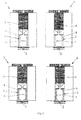
figure 1 shows four perspective views of two embodiments (left and right) of the MCD of the instant invention; -
figure 2 shows a longitudinal section of MCD alternative models applied to oxygraph reaction chamber; -
figure 3 shows MCD within the oxygraph chamber; -
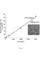
figure 4 shows a graph relating to PDMS membrane oxygen permeability:- PTFE or PDMS nanoporous membrane was applied to the electrode dome and a saturated KC1 solution was maintained between the electrode and the membrane. After calibration of the system, oxygen content was measured in the oxygraph chamber containing 1 ml of distilled water or 1 ml of a sodium dithionite (SDT) solution. Oxygen consumption was calculated as the rate of decrease in oxygen concentration and was expressed as nmol O2 x ml-1 x min-1. The inset shows a SEM image of PDMS porous membrane. PDMS prepolymer was composed of PDMS base and curing agent at a ratio of 10/1 (w/w); water was used as porogen at 50% (w/v). The spin coating condition for fabricating the membrane was set to 1500 rpm for 30 s, to obtain a thickness under 100 µm, as assessed by surface profilometer scansion. Pores dimensions were estimated by SEM imaging and a range from 50 to 250 nm was found for major diameter (
Scale bar 200 nm);
- PTFE or PDMS nanoporous membrane was applied to the electrode dome and a saturated KC1 solution was maintained between the electrode and the membrane. After calibration of the system, oxygen content was measured in the oxygraph chamber containing 1 ml of distilled water or 1 ml of a sodium dithionite (SDT) solution. Oxygen consumption was calculated as the rate of decrease in oxygen concentration and was expressed as nmol O2 x ml-1 x min-1. The inset shows a SEM image of PDMS porous membrane. PDMS prepolymer was composed of PDMS base and curing agent at a ratio of 10/1 (w/w); water was used as porogen at 50% (w/v). The spin coating condition for fabricating the membrane was set to 1500 rpm for 30 s, to obtain a thickness under 100 µm, as assessed by surface profilometer scansion. Pores dimensions were estimated by SEM imaging and a range from 50 to 250 nm was found for major diameter (
-
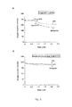
figure 5 shows graphs relating to oxygen diffusion in microchamber (Plasma/BSA treatment for PDMS surface O2 impermeabilization); microchamber was placed into the oxygraph reaction chamber and was filled with 100 ml of distilled water or with 100 ml of SDT 0.1 mg/ml. Oxygen concentration in the oxygraph chamber was monitored over time. Case A refers to closed microchamber treated for oxygen impermeabilization, whereas case B relates to Microchamber treated for oxygen impermeabilization (oxygen may diffuse through the inlet or through the semipermeable membrane at the bottom); -
figure 6 shows graphs relating to determination of oxygen consumption from cells suspended in oxygraph chamber or from adherent cells on O2 permeable membrane at the bottom of a single microchamber (placed within oxygraph). In case A, 9x10³ cells (3T3 or Alexander) were stirred in the oxygraph reaction chamber at 37°C in L15 medium. The rate of oxygen uptake (V) is expressed as nmol O2 x ml-1 x min-1. In case B, 9x10³ cells (3T3 or Alexander) were seeded on the flat O2 permeable membrane of a single microchamber (the upper chamber of the cylinder). After 24 h of incubation, microchamber was placed into the oxygraph reaction chamber and oxygen concentration was monitored over time; it is evident that adhering cells, in narrow contact with the electrode disc, interfere with measurement, so a reservoir-type chamber is necessary for the system; -
figure 7 shows graphs relating to determination of respiration from cells seeded on the middle membrane of a double microchamber (MCD) and KCN inhibition test: 9x10³ cells (3T3 or Alexander) were seeded on the flat O2 permeable membrane in the middle of MCD. After 24 h of standard incubation, DMEM was replaced with L15, and MCD was placed into the oxygraph reaction chamber where oxygen concentration was monitored (case A); cell respiration was inhibited by the addition ofKCN 5 mM (case B). Oxygen consumption rate (V) is expressed as nmol O2 x min-1; -
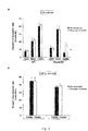
figure 8 shows graphs concerning determination of respiration from cells seeded as mono and co-cultures: MCD with cell monocultures (case A) or monolayer co-coltures (case B) were placed into the oxygraph chamber and oxygen concentration was monitored. Oxygen consumption was calculated as the rate of decrease in oxygen concentration. Data are the mean ± SD (n=3). In Alexander liver cancer cell line, the value of O2 consumption, at high cell density (64 x 103 cells/cm2), is lower, both in MCD and in the standard oxygraph chamber, similar data are reported in literature by independent studies performed on primary hepatocytes. Low correlation between MCD and standard reaction chamber in case A (Δ) is probably due to some clogged spots on low porous membrane by cells clusters, typical adhesion pattern of this cell line; -
figure 9 shows a graph relating to determination of respiration from cell cultures and co-cultures after 24 h - 48 h of incubation: MCD with cell monocultures or co-cultures were placed into the oxygraph chamber and oxygen consumption was calculated. After 24 h of standard incubation, medium was replaced with L15 and a first measurement was done; promptly, tested microchambers were removed from oxygraph reaction chamber, and left, in DMEM, in a CO2 incubator at 37°C, for other 24 h for successive measurement; -
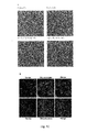
figure 10 shows, in case A, images regarding adhering cells morphological evaluation by optical microscopy: 3 x10³ and 9 x10³ cells (3T3 or Alexander) were seeded on the intermediate O2 permeable membrane, in mono and in co-culture with different ratio. After 48 h of incubation at 37°C, 5% CO2, MCD was disassembled and membrane disc placed on microscope slide for optical evaluation (scale bar 100 µm). Cytochemical test may be performed on these cells monolayer (opportunely within the microchamber); what is more, PDMS elasticity preserves, in a more efficient manner, cells adhesion, throughout staining steps. In case B, images are shown relating to optical evaluation by fluorescence microscopy: 9 x10³ cells (3T3) were seeded on the intermediate membrane in MCD and after 48 h of incubation at 37°C, 5% CO2, cells were double stained with Mitotracker Green and DAPI (specific for mitochondria and nuclei, respectively) and anti-β Tubulin antibody conjugated with FITC fluorophore and DAPI (specific for cytoskeleton Tubulin and nuclei, respectively) and visualized at the fluorescence microscope (obj. 20x and 40x); -
figure 11 relates to oxygen diffusion evaluation: oxygen uptake from cells cultured on the apical or luminal side of the intermediate semi permeable, as in case A, or oxygen impermeable membrane, as in case B, in a double microchamber (MCD). In this semi-permeable system, electrode disc is able to measure with efficiency O2 dissolved in the closer chamber. As for case A relating to oxygen permeable membrane, if cells adhere on the apical side, oxygen is consumed sooner in the upper chamber, and, promptly, the consumed molecules are replaced by those dissolved in the lower chamber, strictly in contact with the electrode disc; this is due to the gas diffusion from a more concentrated compartment to that with a lower concentration; hence the cathode reduces a number of oxygen nanomoles that is proportional to those consumed in the upper chamber (by cells). On the other hand, if cells adhere on the basal side, oxygen is consumed sooner in the lower chamber and promptly, the consumed molecules are replaced by those dissolved in the upper chamber where now O2 is more concentrated; hence the cathode reduces the same nanomoles of oxygen like those present at time zero. The evidence of this dynamic is provided by an additional matching test, performed with an intermediate oxygen impermeable membrane, and it is represented in B. As for case B relating to oxygen impermeable membrane, if cells are on the apical side, oxygen is consumed in the upper chamber and the consumed molecules are not replaced by those dissolved in the lower one because of membrane impermeability; hence the cathode reduces the oxygen nanomoles present at time zero in the lower chamber, and no consumption is registered. Then, if cells adhere on the basal side and oxygen is consumed, as the intermediate membrane is impermeable, no molecules are replaced in the lower chamber, from the upper compartment, and the oxygen consumption is registered; -
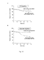
figure 12 relates to migration assay, determination of respiration from both 3T3, as in case A, and Alexander cells, as in case B. 9x10³ cells (3T3 or Alexander) were seeded on the apical side of the intermediate O2 permeable membrane within MCD; after few hours, cell migration was induced by changing serum concentration (lower (2%) and higher (10%), respectively in the upper and lower chamber) and, behind 12 h of incubation, MCD was placed into the oxygraph chamber and oxygen concentration was monitored. Measurements were done standing MCD up straight (cells were transmigrated in the luminal, or basal side and hence none O2 concentration decrement was assessed) and turning it upside down (transmigrated cells are now exposed in the upper chamber and O2 concentration decrement was assessed. Designed mixture PDMS / curing agent / water carries on high elasticity and plasticity, so that cells are able to trans migrate even if porous are smaller than those of the commercial transwell membrane. Cells trans migration was assessed by detachment and cell counting on hemocytometer. The oxygraph chamber insertablemicro-culture device 5 can be produced using, for example, moulding standard techniques, spin coating, conformal contact for the adhesion of the PDMS microchambers with the PDMS membrane. - The model described, potentially, will allow to develop MCD application in tissue regeneration studies, for bio-scaffold efficiency evaluation, also in terms of cell migration from a side to the opposite one, in function of scaffold surface chemical functionalization, expressly studied to promote or inhibit cell migration in function of a specific tissue to regenerate.
- Referring to figures, the micro-culture device (MCD) 5 is totally fabricated in polydimethylsiloxane (PDMS). A PDMS/curing agent mixture (1:10 w/w) is casted in appositely fabricated steel molds and cured at 70°C for 90 min. Essentially, the micro-culture device (MCD) 5 is composed by three elements: oxygen-
permeable nanoporous membrane 1 for cell culture, anupper microchamber 2 and a lower reservoir-type microchamber 3. The system will be described herein in details. - A PDMS/curing agent mixture (10:1 w/w) and water (as a porogen agent at a ratio of 50% water and 50% polymer base), is stirred for 30 min, spread on glass substrate by spin coating (1500 rpm, for 20 s) and then cured at 70°C for 30 min. The obtained porous membrane, detached from glass slide, results of 80 µm thickness, with porous ranging from 50 to 250 nm in diameter. In order to be suitable for cell culturing, the
semipermeable nanoporous membrane 1 is then subjected at a plasma oxygen treatment by a plasma hasher (5 min, 20% O2, 100W, 40 KHz) and incubated with 0,01% w/v poly-L-Lysine or extra cellular matrix protein for 30 min; this treatment facilitates the next cells adhesion on the polymeric membrane. - Polymerized microchambers are realized as hollow cylinders of 8 mm height (or less, or more, if required), 9 mm outer diameter, 6 mm inner diameter (cylinder side walls of 1,5 mm wide); the
upper microchamber 2 presents, on the top, a central inlet 2.1 coaxially aligned with oxygraph plunger precision bore 6 of 1 mm inner diameter, and it is intended for solutions injection by means of a syringe, at some stage during evaluation; alternatively, it has a semi-permeable membrane bonded at the upper end. Thelower microchamber 3, in the same way, has at the lower end a central inlet 3.1 positioned at the bottom and coaxially aligned with oxygraph piston bore, like the upper one (seefigure 1 ), or a semi-permeable membrane 3.2 (seefigure 1 ). The inlets conformed as telescopic sub micro-cylinders are obtained by a capillary placed on the cast and upholded, over the time of PDMS mixture pouring and polymerization, by means of an expressly fabricated mini column holder. Theupper microchamber 2 and thelower microchamber 3 are subjected to a surface chemical process (plasma hasher (5 min, 20% O2, 100W, 40 KHz) and 1h incubation with 0,01% w/v bovine serum albumin (BSA)) in order to avoid random gas exchange, and purposely address oxygen toward electrode disc. - After the above mentioned surface treatments the three elements are assembled by conformal contact with a PDMS thin film between the microchambers and the membrane, and cured at 70°C for 15-30 min. The whole system is autoclavable or easily sterilized by ethanol 70% and sterile double distilled water washing steps. Preferably, the invented system, during cells incubation, is placed and closed in a standard sterile well plate, to minimize microorganisms contamination; for this purpose, in order to well close the tray and the lid of the commercial multiwell plate, lower MCD can be realized.
- As proof of principle, the
MCD 5 has been applied tooxygraph chamber 4 of anoxygraph 7, to measure the OCR of two independent cells lines, seeded and allowed to growth in mono or co-culture, on the intermediate membrane in MCD, and the given results (see figures) are consistent to those obtained by standard tests performed on cells in agitation in the oxygraph reaction chamber barely. Indeed, MCD lets to perform OCR tests of adhering cells without detaching them, allowing different subsequent evaluations of the same sample, i.e. following a pharmacological test, after one or more days of incubation, with modest risk of contamination or stress. - Furthermore, after OCR evaluation, thanks to PDMS inertia to many chemicals used in cytochemistry, and thanks to elastomer plasticity and optical transparency (λ ≥ 235 nm), biochemical assay (i.e. MTT test) or cyto- and immunochemical evaluation can be performed within the invented device. Then MCD can be easily disassembled in the
constituent modules microchambers - Functional forms, basically, include MCD composed as:
- inlets 2.1, 3.1 on top and bottom of the
MCD 5, fabricated as an inner subcylinder telescopic bore and designed to not pour out any liquids. Under operating in static conditions the inlet is small enough (1 mm in diameter) that it is impossible for the fluid to flow through the hole at the work temperature (20-37 °C); PDMS hydrophilicity, after chemical surface treatment, results in stronger adhesiveness between water molecules and PDMS walls. Moreover the laws and principles of hydrostatic suggest that in a small capillary hole, liquid surface tension pressure is greater than pressure of the liquid, preventing any fluid leakage. This model is also practical for sample handling; - inlet 2.1 on the top and semi-permeable membrane 3.2 at the bottom. The semi-permeable membrane 3.2 at the bottom does not alter the efficiency of oxygen diffusion towards the electrode and, additionally, prevents contamination acting as a shielding nanoporous filter between sensing system and the lower microchamber; it is, mainly, intended for multiple test on the same sample, and for cell lines very sensitive to contamination, such as primary culture of somatic or stem cells.
- In both these designs, a PDMS made bracket horse is useful, but not mandatory, as open pedestal insert for gas free exchange during cell incubation in CO2 incubator; finally, within these configurations, the bracket horse insert should optimise gas diffusion through cell culture layer (as aforementioned in
WO200806104 - Possible alternative forms include an optimised intermediate porous membrane and inception of flat microporous bio-scaffold as intermediate insert, in the place of the PDMS porous membrane.
- In a first possible alternative form, an optimised porous membrane fabricated by photolithographic techniques, with the aim to control pores geometry and distribution. Photolithography is a method used in microfabrication to transfer a pattern onto a surface, exploiting chemical and physical processes. This is possible by depositing a photoresist (photosensitive polymer) on the substrate and exposing it to UV light, inducing chemical changes that allows to remove the photoresist by a developer solvent. Subsequently, the layer of the substrate in the areas that are not protected by photoresist is removed by etching process, utilizing chemical agents attaining the final mold. Specifically, to realize a porous membrane, a mold with embossed micropillars is required, to apply and cure the polymer on. The polymer membrane removed from the mold results in a regular geometric conformation and distribution of the desired pores.
- This option is related also to the application of MCD for high density trans-membrane co-culture (i.e. in vitro blood brain barrier co-culture), or for cells separation/migration devices, in which a regular porosity is necessary to allow a free gas exchange also in the presence of high cell density onto the membrane surface, in case of application for high density trans-membrane co-culture; to warrant a precise porosity, specific for different cell lines (with different cells diameter), in case of application for cells separation/migration devices.
- A second possible alternative form provides for inception of flat microporous bio-scaffold as intermediate insert, in the place of the PDMS porous membrane, intended as a novel application in tissue engineering scaffold characterization. Specifically intended for oxygen diffusion dynamic evaluation into the cultured matrix, and, hence for assessing matrix efficiency as bio-scaffold for tissue regeneration, besides, this application refers to cells migration test of chemically functionalised bio-scaffold.
- The present invention is intended for in vitro applications, mainly in the field of biology (physiological and disease models), regenerative medicine (stem cells and tissue engineering), bio- and nanobiotechnology, pharmacology (drug discovery) and tossicology.
- Essentially MCD makes possible to analyse micro volumes of samples and cells in adhesion, into the oxygraph reaction chamber. This ability can be exploited for biochemical assays on cells in mono- and/or co-culture (also at different time points) and subsequent imaging of the same sample.
- In particular it can be applied for:
- estimation of the response to toxicants and the correlation of response to exposure with the etiology and progression of dysfunction (by means of respiration and immunocytochemical assays);
- development and characterization of chemical, structural, and biological properties of nanostructured assemblies/materials to advance functional tissue engineering;
- drug delivery development and nano-formulations of existing therapeutic drugs acting at subcellular level (e.g., mitochondrial)-targeting;
- development and/or monitoring of mitochondrial gene delivery, a novel strategy for curing a mitochondrial disease.
- Moreover it can be applied for:
- estimation of oxygen consumption by Saccharomyces cerevisiae under enological conditions in order to evaluate the effect on fermentation kinetics;
- colony morphology evaluation in efficient yeast strain screening;
- yeast fermentation by weight loss measurement (CO2 escape).
-
- 1. Wieser, W. and G. Krumschnabel, Hierarchies of ATP-consuming processes: direct compared with indirect measurements, and comparative aspects. Biochem J, 2001. 355(Pt 2): p. 389-95.
- 2. Marchandeau, J.P. and G. Labbe, Evaluation of mitochondrial respiration in cultured rat hepatocytes. Methods Mol Biol, 2011. 691: p. 243-53.
- 3. Hughey, C.C., et al., Respirometric Oxidative Phosphorylation Assessment in Saponinpermeabilized Cardiac Fibers. J Vis Exp, 2011(48).
- 4. Ferramosca, A., et al., Oxygen uptake by mitochondria in demembranated human spermatozoa: a reliable tool for the evaluation of sperm respiratory efficiency. Int J Androl, 2008. 31(3): p. 337-45.
- 5. Villani, G. and G. Attardi, Polarographic assays of respiratory chain complex activity. Methods Cell Biol, 2007. 80: p. 121-33.
- 6. Pipinos, II, et al., Abnormal mitochondrial respiration in skeletal muscle in patients with peripheral arterial disease. J Vasc Surg, 2003. 38(4): p. 827-32.
- 7. Sud, D., et al., Optical imaging in microfluidic bioreactors enables oxygen monitoring for continuous cell culture. J Biomed Opt, 2006. 11(5): p. 050504.
- 8. Deshpande, R.R., et al., Microplates with integrated oxygen sensors for kinetic cell respiration measurement and cytotoxicity testing in primary and secondary cell lines. Assay Drug Dev Technol, 2005. 3(3): p. 299-307.
- 9. Beckers, S., et al., High throughput, non-invasive and dynamic toxicity screening on adherent cells using respiratory measurements. Toxicol In Vitro, 2010. 24(2): p. 686-94.
- 10. Gao, F.G., A.S. Jeevarajan, and M.M. Anderson, Long-term continuous monitoring of dissolved oxygen in cell culture medium for perfused bioreactors using optical oxygen sensors. Biotechnology and Bioengineering, 2004. 86(4): p. 425-433.
- 11. Cho, C.H., et al., Oxygen uptake rates and liver-specific functions of hepatocyte and 3T3 fibroblast co-cultures. Biotechnol Bioeng, 2007. 97(1): p. 188-99.
- 12. Oppegard, S.C. and D.T. Eddington, Device for the control of oxygen concentration in multiwell cell culture plates. Conf Proc IEEE Eng Med Biol Soc, 2009. 2009: p. 2097-100.
- 13. Papas, K.K., et al., A stirred microchamber for oxygen consumption rate measurements with pancreatic islets. Biotechnology and Bioengineering, 2007. 98(5): p. 1071-1082.
- 14. Torisawa, Y.S., et al., Multi-channel 3-D cell culture device integrated on a silicon chip for anticancer drug sensitivity test. Biomaterials, 2005. 26(14): p. 2165-72.
- 15. Mamchaoui, K. and G. Saumon, A method for measuring the oxygen consumption of intact cell monolayers. American Journal of Physiology-Lung Cellular and Molecular Physiology, 2000. 278(4): p. L858-L863.
- 16. Wu, C.C., et al., Fabrication of miniature Clark oxygen sensor integrated with microstructure. Sensors and Actuators B-Chemical, 2005. 110(2): p. 342-349.
- 17. Wu, J., et al., The effect of the closely-spaced working and auxiliary electrodes on the performance of electrochemical oxygen sensor. Electroanalysis, 2007. 19(18): p. 1939-1943.
- 18. Wu, C.C., et al., A Clark-type oxygen chip for in situ estimation of the respiratory activity of adhering cells. Talanta. 81(1-2): p. 228-34.
- 19. Park, J., Y.K. Pak, and J.J. Pak, A microfabricated reservoir-type oxygen sensor for measuring the real-time cellular oxygen consumption rate at various conditions. Sensors and Actuators B-Chemical, 2010. 147(1): p. 263-269.
- 20. Ostrovidov, S., et al., Membrane-based PDMS microbioreactor for perfused 3D primary rat hepatocyte cultures. Biomedical Microdevices, 2004. 6(4): p. 279-287.
- 21. del Alamo, M. and I. Nevares, Measurement of dissolved oxygen during red wines tank aging with chips and micro-oxygenation. Analytica Chimica Acta, 2008. 621(1): p. 68-78.
- 22. Querol, A., et al., Adaptive evolution of wine yeast. International Journal of Food Microbiology, 2003. 86(1-2): p. 3-10.
- 23. Schmidt, S.A., et al., Microvinification-how small can we go? Applied Microbiology and Biotechnology, 2011. 89(5): p. 1621-1628.
- 24. Rosenfeld, E., et al., Oxygen consumption by anaerobic Saccharomyces cerevisiae under enological conditions: Effect on fermentation kinetics. Applied and Environmental Microbiology, 2003. 69(1): p. 113-121.
- 25. Rosenfeld, E., et al., Non-respiratory oxygen consumption pathways in anaerobically-grown Saccharomyces cerevisiae: evidence and partial characterization. Yeast, 2002. 19(15): p. 1299-321.
- 26. Salmon, J.M., C. Fornairon-Bonnefond, and J.P. Mazauric, Interactions between wine lees and polyphenols: Influence on oxygen consumption capacity during simulation of wine aging. Journal of Food Science, 2002. 67(5): p. 1604-1609.
- 27. Salmon, J.M., et al., Effect of Different Yeast Strains and Their Culture Conditions on the Prevention of Wine Model Solution Browning by Yeast Lees. Journal of Agricultural and Food Chemistry, 2009. 57(9): p. 3771-3779.
- 28. Stambuk, B.U., et al., Switching the mode of sucrose utilization by Saccharomyces cerevisiae. Microbial Cell Factories, 2008. 7.
- 29.
WO200806104 - 30. George M. Whitesides, Emanuele Ostuni Shuichi Takayama Xingyu Jiang and Donald E. Soft lithography in biology and biochemistry. Ingber Annu. Rev. Biomed. Eng. 2001. 3:335-73
- 31. Chia-Wen Tsao Æ Don L. DeVoe, Bonding of thermoplastic polymer microfluidics. Microfluid Nanofluid (2009) 6:1-16
- 32. Xinyu Huang and William Synthesis and Characterization of PMMA Nanocomposites by Suspension and Emulsion Polymerization. J. Brittain Macromolecules 2001, 34, 3255-3260.
Claims (11)
- A micro culture device (5) to be inserted in a chamber (4) of an oxygraph (7), the micro culture device being divided into an upper micro-chamber (2) and a lower microchamber (3) by a semi-permeable nanoporous membrane (1), suitable for acting as adhering support for cells,
characterized in that:a) the upper microchamber (2) presents at its upper side either an inlet (2.1) coaxially aligned with an oxygraph plunger precision bore (6), or a semi-permeable membrane; andb) the lower microchamber (3) presents at its bottom side either an inlet (3.1) coaxially aligned with an oxygraph piston bore (8) or a semi-permeable membrane (3.2). - The micro culture device according to claim 1, characterized in that it is essentially of cylindrical shape.
- The micro culture device according to claim 1 or 2, characterized in that the semipermeable nanoporous membrane (1) is removable.
- The micro culture device according to any one of claims 1 to 3, further comprising a PDMS made bracket horse as open pedestal insert for gas free exchange during cell incubation in CO2 incubator.
- The micro culture device according to any one of previous claims being made of polydimethylsiloxane (PDMS), or polycarbonate (PC), or polymethylmethacrylate (PMMA).
- The micro culture device according to any one of previous claims wherein the semi-permeable nanoporous membrane (1) is preferably made of polydimethylsiloxane (PDMS), or polytetrafluoroethylene (PTFE), or the like.
- An oxygraph comprising the micro culture device according to any one of previous claims.
- Use of the oxygraph according to claim 7 for oxygen consumption rate measurement of cells adhered to the semi-permeable nanoporous membrane (1) of the micro culture device.
- A method for measuring oxygen consumption rate in an oxygraph, characterized in that it comprises the steps of:● culturing cells on the semi-permeable nanoporous membrane (1) of the micro culture device according to any one of claims 1 to 6 by the steps of:- inserting suspension cells in the upper or in the lower microchamber through suitable inlet;- incubating the MCD at controlled atmosphere, i.e. 37°C, 5% CO2, wherein the cells sediment and adhere on the surface of the semipermeable nanoporous membrane (1);● measuring oxygen consumption rate by the steps of:- inserting the MCD in the oxygraph reaction chamber keeping it in contact with the Clark electrode disc;- hermetically closing the reaction chamber;● optionally adding a compound during said step of measuring by the steps of:- opening a micro cap of the oxygraph plunger and injecting the compound into MCD through the inlet;- closing the micro cap;● running oxygen consumption rate measurement.
- Method according to claim 9, characterized in that the step of measuring oxygen consumption rate is performed at different growing phases of cells.
- Method for analysing cells after measuring oxygen consumption rate, characterized in that it comprises the steps of:- removing MCD from the oxygraph reaction chamber;- performing cytochemical assay;- disassembling MCD and recuperating the intermediate membrane;- performing morphological evaluation by microscope.
Priority Applications (1)
| Application Number | Priority Date | Filing Date | Title |
|---|---|---|---|
| EP11191000.6A EP2597148A1 (en) | 2011-11-28 | 2011-11-28 | Oxygraph chamber and insertable micro-culture device and uses thereof |
Applications Claiming Priority (1)
| Application Number | Priority Date | Filing Date | Title |
|---|---|---|---|
| EP11191000.6A EP2597148A1 (en) | 2011-11-28 | 2011-11-28 | Oxygraph chamber and insertable micro-culture device and uses thereof |
Publications (1)
| Publication Number | Publication Date |
|---|---|
| EP2597148A1 true EP2597148A1 (en) | 2013-05-29 |
Family
ID=45094526
Family Applications (1)
| Application Number | Title | Priority Date | Filing Date |
|---|---|---|---|
| EP11191000.6A Withdrawn EP2597148A1 (en) | 2011-11-28 | 2011-11-28 | Oxygraph chamber and insertable micro-culture device and uses thereof |
Country Status (1)
| Country | Link |
|---|---|
| EP (1) | EP2597148A1 (en) |
Cited By (3)
| Publication number | Priority date | Publication date | Assignee | Title |
|---|---|---|---|---|
| WO2019079384A1 (en) * | 2017-10-17 | 2019-04-25 | The Methodist Hospital System | ADMINISTRATION DEVICES |
| CN113242877A (en) * | 2018-12-11 | 2021-08-10 | 株式会社Posco | Method for preparing porous polysiloxane membrane, porous polysiloxane membrane prepared by the method, and photovoltaic module comprising the porous polysiloxane membrane |
| US11590072B2 (en) | 2015-05-15 | 2023-02-28 | The Methodist Hospital System | Implantable nanochannel delivery devices |
Citations (3)
| Publication number | Priority date | Publication date | Assignee | Title |
|---|---|---|---|---|
| WO2006095480A1 (en) | 2005-03-08 | 2006-09-14 | The Foundation For The Promotion Of Industrial Science | Cell culture chamber |
| US20070212773A1 (en) | 2006-02-16 | 2007-09-13 | Teruo Fujii | Electrical signal measuring device for cells in culture and electrical signal measuring method that uses same device |
| WO2008006104A2 (en) | 2006-07-07 | 2008-01-10 | University Of Miami | Enhanced oxygen cell culture platforms |
-
2011
- 2011-11-28 EP EP11191000.6A patent/EP2597148A1/en not_active Withdrawn
Patent Citations (5)
| Publication number | Priority date | Publication date | Assignee | Title |
|---|---|---|---|---|
| WO2006095480A1 (en) | 2005-03-08 | 2006-09-14 | The Foundation For The Promotion Of Industrial Science | Cell culture chamber |
| US20080145925A1 (en) * | 2005-03-08 | 2008-06-19 | Yasuyuki Sakai | Cell Culture Chamber |
| US20070212773A1 (en) | 2006-02-16 | 2007-09-13 | Teruo Fujii | Electrical signal measuring device for cells in culture and electrical signal measuring method that uses same device |
| WO2008006104A2 (en) | 2006-07-07 | 2008-01-10 | University Of Miami | Enhanced oxygen cell culture platforms |
| US20080009027A1 (en) | 2006-07-07 | 2008-01-10 | University Of Miami | Enhanced Oxygen Cell Culture Platforms |
Non-Patent Citations (33)
| Title |
|---|
| BECKERS, S. ET AL.: "High throughput, non-invasive and dynamic toxicity screening on adherent cells using respiratory measurements", TOXICOL IN VITRO, vol. 24, no. 2, 2010, pages 686 - 94, XP026911701, DOI: doi:10.1016/j.tiv.2009.04.018 |
| CHIA-WEN TSAO Æ; DON L. DEVOE: "Bonding of thermoplastic polymer microfluidics", MICROFLUID NANOFLUID, vol. 6, 2009, pages 1 - 16, XP019667827 |
| CHO, C.H. ET AL.: "Oxygen uptake rates and liver-specific functions of hepatocyte and 3T3 fibroblast co-cultures", BIOTECHNOL BIOENG, vol. 97, no. 1, 2007, pages 188 - 99 |
| DEL ALAMO, M.; I. NEVARES: "Measurement of dissolved oxygen during red wines tank aging with chips and micro-oxygenation", ANALYTICA CHIMICA ACTA, vol. 621, no. 1, 2008, pages 68 - 78, XP022762169, DOI: doi:10.1016/j.aca.2007.11.042 |
| DESHPANDE, R.R. ET AL.: "Microplates with integrated oxygen sensors for kinetic cell respiration measurement and cytotoxicity testing in primary and secondary cell lines", ASSAY DRUG DEV TECHNOL, vol. 3, no. 3, 2005, pages 299 - 307 |
| FERRAMOSCA, A. ET AL.: "Oxygen uptake by mitochondria in demembranated human spermatozoa: a reliable tool for the evaluation of sperm respiratory efficiency", INT J ANDROL, vol. 31, no. 3, 2008, pages 337 - 45 |
| GAO, F.G.; A.S. JEEVARAJAN; M.M. ANDERSON: "Long-term continuous monitoring of dissolved oxygen in cell culture medium for perfused bioreactors using optical oxygen sensors", BIOTECHNOLOGY AND BIOENGINEERING, vol. 86, 2004, pages 425 - 433 |
| GEORGE M. WHITESIDES; EMANUELE OSTUNI; SHUICHI TAKAYAMA; XINGYU JIANG; DONALD E: "Soft lithography in biology and biochemistry", INGBER ANNU. REV. BIOMED. ENG., vol. 3, 2001, pages 335 - 73, XP002961528, DOI: doi:10.1146/annurev.bioeng.3.1.335 |
| HUGHEY, C.C. ET AL.: "Respirometric Oxidative Phosphorylation Assessment in Saponin- permeabilized Cardiac Fibers.", J VIS EXP, 2011 |
| MAMCHAOUI K. ET AL, AM. J.PHYSIOL LUNG CELL MOL PHYSIOL, vol. 278, no. 4, April 2000 (2000-04-01) - 2 March 2012 (2012-03-02), pages L858 - L863, XP002670722 * |
| MAMCHAOUI, K.; G. SAUMON: "A method for measuring the oxygen consumption of intact cell monolayers", AMERICAN JOURNAL OF PHYSIOLOGY-LUNG CELLULAR AND MOLECULAR PHYSIOLOGY, vol. 278, no. 4, 2000, pages L858 - L863, XP002670722 |
| MARCHANDEAU, J.P.; G. LABBE: "Evaluation of mitochondrial respiration in cultured rat hepatocytes", METHODS MOL BIOL, vol. 691, 2011, pages 243 - 53 |
| OPPEGARD, S.C.; D.T. EDDINGTON: "Device for the control of oxygen concentration in multiwell cell culture plates", CONFPROC IEEE ENG MED BIOL SOC, 2009, pages 2097 - 100, XP031881056, DOI: doi:10.1109/IEMBS.2009.5332491 |
| OSTROVIDOV, S. ET AL.: "Membrane-based PDMS microbioreactor for perfused 3D primary rat hepatocyte cultures", BIOMEDICAL MICRODEVICES, vol. 6, no. 4, 2004, pages 279 - 287, XP002670721 * |
| OSTROVIDOV, S. ET AL.: "Membrane-based PDMS microbioreactor for perfused 3D primary rat hepatocyte cultures", BIOMEDICAL MICRODEVICES, vol. 6, no. 4, 2004, pages 279 - 287, XP002670721, DOI: doi:10.1023/B:BMMD.0000048560.96140.ca |
| PAPAS, K.K. ET AL.: "A stirred microchamber for oxygen consumption rate measurements with pancreatic islets", BIOTECHNOLOGY AND BIOENGINEERING, vol. 98, no. 5, 2007, pages 1071 - 1082 |
| PARK, J.; Y.K. PAK; J.J. PAK: "A microfabricated reservoir-type oxygen sensor for measuring the real-time cellular oxygen consumption rate at various conditions", SENSORS AND ACTUATORS B-CHEMICA, vol. 147, no. 1, 2010, pages 263 - 269 |
| PIPINOS, II ET AL.: "Abnormal mitochondrial respiration in skeletal muscle in patients with peripheral arterial disease.", J VASE SURG, vol. 38, no. 4, 2003, pages 827 - 32 |
| QUEROL, A. ET AL.: "Adaptive evolution of wine yeast.", INTERNATIONAL JOURNAL OF FOOD MICROBIOLOGY, vol. 86, no. 1-2, 2003, pages 3 - 10 |
| ROSENFELD, E. ET AL.: "Non-respiratory oxygen consumption pathways in anaerobically-grown Saccharomyces cerevisiae: evidence and partial characterization", YEAST, vol. 19, no. 15, 2002, pages 1299 - 321 |
| ROSENFELD, E. ET AL.: "Oxygen consumption by anaerobic Saccharomyces cerevisiae under enological conditions: Effect on fermentation kinetics", APPLIED AND ENVIRONMENTAL MICROBIOLOGY, vol. 69, no. 1, 2003, pages 113 - 121 |
| SALMON, J.M. ET AL.: "Effect of Different Yeast Strains and Their Culture Conditions on the Prevention of Wine Model Solution Browning by Yeast Lees", JOURNAL OF AGRICULTURAL AND FOOD CHEMISTRY, vol. 57, no. 9, 2009, pages 3771 - 3779 |
| SALMON, J.M.; C. FORNAIRON-BONNEFOND; J.P. MAZAURIC: "Interactions between wine lees and polyphenols: Influence on oxygen consumption capacity during simulation of wine aging", JOURNAL OF FOOD SCIENCE, vol. 67, no. 5, 2002, pages 1604 - 1609 |
| SCHMIDT, S.A. ET AL.: "Microvinification-how small can we go?", APPLIED MICROBIOLOGY AND BIOTECHNOLOGY, vol. 89, no. 5, 2011, pages 1621 - 1628, XP019880854, DOI: doi:10.1007/s00253-010-2992-6 |
| STAMBUK, B.U. ET AL.: "Switching the mode of sucrose utilization by Saccharomyces cerevisiae", MICROBIAL CELL FACTORIES, 2008, pages 7 |
| SUD, D. ET AL.: "Optical imaging in microfluidic bioreactors enables oxygen monitoring for continuous cell culture", J BIOMED OPT, vol. 11, no. 5, 2006, pages 050504 |
| TORISAWA, Y.S. ET AL.: "Multi-channel 3-D cell culture device integrated on a silicon chip for anticancer drug sensitivity test.", BIOMATERIALS, vol. 26, no. 14, 2005, pages 2165 - 72, XP025280737, DOI: doi:10.1016/j.biomaterials.2004.05.028 |
| VILLANI, G.; G. ATTARDI: "Polarographic assays of respiratory chain complex activity.", METHODS CELL BIOL, vol. 80, 2007, pages 121 - 33 |
| WIESER, W.; G. KRUMSCHNABEL: "Hierarchies of ATP-consuming processes: direct compared with indirect measurements, and comparative aspects", BIOCHEM J, vol. 355, 2001, pages 389 - 95 |
| WU, C.C. ET AL.: "A Clark-type oxygen chip for in situ estimation of the respiratory activity of adhering cells", TALANTA, vol. 81, no. 1-2, pages 228 - 34, XP026923797, DOI: doi:10.1016/j.talanta.2009.11.062 |
| WU, C.C. ET AL.: "Fabrication of miniature Clark oxygen sensor integrated with microstructure", SENSORS AND ACTUATORS B-CHEMICAL, vol. 110, no. 2, 2005, pages 342 - 349, XP025328939, DOI: doi:10.1016/j.snb.2005.02.014 |
| WU, J. ET AL.: "The effect of the closely-spaced working and auxiliary electrodes on the performance of electrochemical oxygen sensor", ELECTROANALYSIS, vol. 19, no. 18, 2007, pages 1939 - 1943 |
| XINYU HUANG; WILLIAM: "Synthesis and Characterization of PMMA Nanocomposites by Suspension and Emulsion Polymerization", J. BRITTAIN MACROMOLECULES, vol. 34, 2001, pages 3255 - 3260, XP001074615, DOI: doi:10.1021/ma001670s |
Cited By (8)
| Publication number | Priority date | Publication date | Assignee | Title |
|---|---|---|---|---|
| US11590072B2 (en) | 2015-05-15 | 2023-02-28 | The Methodist Hospital System | Implantable nanochannel delivery devices |
| US12151021B2 (en) | 2015-05-15 | 2024-11-26 | The Methodist Hospital System | Implantable nanochannel delivery devices |
| WO2019079384A1 (en) * | 2017-10-17 | 2019-04-25 | The Methodist Hospital System | ADMINISTRATION DEVICES |
| US11565094B2 (en) | 2017-10-17 | 2023-01-31 | The Methodist Hospital System | Delivery devices |
| CN113242877A (en) * | 2018-12-11 | 2021-08-10 | 株式会社Posco | Method for preparing porous polysiloxane membrane, porous polysiloxane membrane prepared by the method, and photovoltaic module comprising the porous polysiloxane membrane |
| EP3896124A4 (en) * | 2018-12-11 | 2022-01-26 | Posco | METHOD FOR MAKING A POROUS POLYSILOXANE FILM, POROUS POLYSILOXANE FILM MANUFACTURED BY THIS METHOD, AND SOLAR CELL MODULE COMPRISING THE SAME |
| CN113242877B (en) * | 2018-12-11 | 2023-05-05 | 株式会社Posco | Method for preparing porous polysiloxane film, porous polysiloxane film prepared by the method, and photovoltaic module comprising the same |
| US12396291B2 (en) | 2018-12-11 | 2025-08-19 | Posco Co., Ltd | Method for manufacturing porous polysiloxane film, porous polysiloxane film manufactured thereby, and solar cell module comprising same |
Similar Documents
| Publication | Publication Date | Title |
|---|---|---|
| CA2788575C (en) | Hanging drop devices, systems and/or methods | |
| Gao et al. | Recent developments in microfluidic devices for in vitro cell culture for cell-biology research | |
| Young et al. | Fundamentals of microfluidic cell culture in controlled microenvironments | |
| US8501462B2 (en) | Insert device for multiwell plate | |
| US9511366B2 (en) | Microfluidic device and its use for positioning of cells or organisms | |
| CA2737627C (en) | Hanging drop plate | |
| EP2411501B1 (en) | Apparatus for cell or tissue culture | |
| Zhang et al. | High-throughput superhydrophobic microwell arrays for investigating multifactorial stem cell niches | |
| WO2010062911A2 (en) | Compositions and methods of functionally enhanced in vitro cell culture system | |
| Sun et al. | A superhydrophobic chip integrated with an array of medium reservoirs for long-term hanging drop spheroid culture | |
| EP3988642A1 (en) | System for monitoring three-dimensional cell cultures | |
| Puleo et al. | Applications of MEMS technologies in tissue engineering | |
| Mai et al. | MatriGrid® based biological morphologies: Tools for 3D cell culturing | |
| US7897377B2 (en) | Cell- and tissue culture device | |
| Wang et al. | A Physiological Oxygen Gradient Liver-Zonation-on-a-Chip Reveals HIF-2α Intervention in Hepatic Lipotoxicity | |
| JP2010011814A (en) | Cultured cell observation chamber and use thereof | |
| CN222846733U (en) | Microfluidic bionic chip for three-dimensional co-culture of cells | |
| Keshavarz et al. | Cell culture techniques in microfluidic chips | |
| EP4269552A1 (en) | Flow path device and method for manufacturing same | |
| Hamon et al. | Systematic evaluation of the efficiencies of proteins and chemicals in pharmaceutical applications | |
| Super | Oxygen monitoring in a microfluidic culture device for stem cell bioprocess development | |
| WO2025254623A1 (en) | An open-top microphysiological system for use in 2d and 3d cell and tissue cultures and the operating method of this system | |
| Yi et al. | Cell Culture and Observation on Microfluidics | |
| Navarro | Optimizing Microfluidic Murine Fibroblast and Human Epidermal Keratinocyte Cell Culture in 70µm Microwells | |
| Prokop et al. | NanoLiterBioReactor: monitoring of long-term mammalian cell physiology at nanofabricated scale |
Legal Events
| Date | Code | Title | Description |
|---|---|---|---|
| PUAI | Public reference made under article 153(3) epc to a published international application that has entered the european phase |
Free format text: ORIGINAL CODE: 0009012 |
|
| AK | Designated contracting states |
Kind code of ref document: A1 Designated state(s): AL AT BE BG CH CY CZ DE DK EE ES FI FR GB GR HR HU IE IS IT LI LT LU LV MC MK MT NL NO PL PT RO RS SE SI SK SM TR |
|
| AX | Request for extension of the european patent |
Extension state: BA ME |
|
| 17P | Request for examination filed |
Effective date: 20131127 |
|
| RBV | Designated contracting states (corrected) |
Designated state(s): AL AT BE BG CH CY CZ DE DK EE ES FI FR GB GR HR HU IE IS IT LI LT LU LV MC MK MT NL NO PL PT RO RS SE SI SK SM TR |
|
| STAA | Information on the status of an ep patent application or granted ep patent |
Free format text: STATUS: THE APPLICATION IS DEEMED TO BE WITHDRAWN |
|
| 18D | Application deemed to be withdrawn |
Effective date: 20160601 |