EP2595644B1 - Amyloid-beta clearance - Google Patents
Amyloid-beta clearance Download PDFInfo
- Publication number
- EP2595644B1 EP2595644B1 EP10855049.2A EP10855049A EP2595644B1 EP 2595644 B1 EP2595644 B1 EP 2595644B1 EP 10855049 A EP10855049 A EP 10855049A EP 2595644 B1 EP2595644 B1 EP 2595644B1
- Authority
- EP
- European Patent Office
- Prior art keywords
- nia
- amyloid
- composition
- use according
- cells
- Prior art date
- Legal status (The legal status is an assumption and is not a legal conclusion. Google has not performed a legal analysis and makes no representation as to the accuracy of the status listed.)
- Active
Links
Images







Classifications
-
- A—HUMAN NECESSITIES
- A61—MEDICAL OR VETERINARY SCIENCE; HYGIENE
- A61K—PREPARATIONS FOR MEDICAL, DENTAL OR TOILETRY PURPOSES
- A61K38/00—Medicinal preparations containing peptides
- A61K38/16—Peptides having more than 20 amino acids; Gastrins; Somatostatins; Melanotropins; Derivatives thereof
- A61K38/43—Enzymes; Proenzymes; Derivatives thereof
- A61K38/46—Hydrolases (3)
- A61K38/48—Hydrolases (3) acting on peptide bonds (3.4)
- A61K38/4873—Cysteine endopeptidases (3.4.22), e.g. stem bromelain, papain, ficin, cathepsin H
-
- A—HUMAN NECESSITIES
- A61—MEDICAL OR VETERINARY SCIENCE; HYGIENE
- A61K—PREPARATIONS FOR MEDICAL, DENTAL OR TOILETRY PURPOSES
- A61K48/00—Medicinal preparations containing genetic material which is inserted into cells of the living body to treat genetic diseases; Gene therapy
- A61K48/005—Medicinal preparations containing genetic material which is inserted into cells of the living body to treat genetic diseases; Gene therapy characterised by an aspect of the 'active' part of the composition delivered, i.e. the nucleic acid delivered
-
- A—HUMAN NECESSITIES
- A61—MEDICAL OR VETERINARY SCIENCE; HYGIENE
- A61P—SPECIFIC THERAPEUTIC ACTIVITY OF CHEMICAL COMPOUNDS OR MEDICINAL PREPARATIONS
- A61P25/00—Drugs for disorders of the nervous system
-
- A—HUMAN NECESSITIES
- A61—MEDICAL OR VETERINARY SCIENCE; HYGIENE
- A61P—SPECIFIC THERAPEUTIC ACTIVITY OF CHEMICAL COMPOUNDS OR MEDICINAL PREPARATIONS
- A61P25/00—Drugs for disorders of the nervous system
- A61P25/28—Drugs for disorders of the nervous system for treating neurodegenerative disorders of the central nervous system, e.g. nootropic agents, cognition enhancers, drugs for treating Alzheimer's disease or other forms of dementia
-
- C—CHEMISTRY; METALLURGY
- C12—BIOCHEMISTRY; BEER; SPIRITS; WINE; VINEGAR; MICROBIOLOGY; ENZYMOLOGY; MUTATION OR GENETIC ENGINEERING
- C12N—MICROORGANISMS OR ENZYMES; COMPOSITIONS THEREOF; PROPAGATING, PRESERVING, OR MAINTAINING MICROORGANISMS; MUTATION OR GENETIC ENGINEERING; CULTURE MEDIA
- C12N9/00—Enzymes; Proenzymes; Compositions thereof; Processes for preparing, activating, inhibiting, separating or purifying enzymes
- C12N9/14—Hydrolases (3)
- C12N9/48—Hydrolases (3) acting on peptide bonds (3.4)
- C12N9/50—Proteinases, e.g. Endopeptidases (3.4.21-3.4.25)
- C12N9/503—Proteinases, e.g. Endopeptidases (3.4.21-3.4.25) derived from viruses
- C12N9/506—Proteinases, e.g. Endopeptidases (3.4.21-3.4.25) derived from viruses derived from RNA viruses
-
- C—CHEMISTRY; METALLURGY
- C12—BIOCHEMISTRY; BEER; SPIRITS; WINE; VINEGAR; MICROBIOLOGY; ENZYMOLOGY; MUTATION OR GENETIC ENGINEERING
- C12Y—ENZYMES
- C12Y304/00—Hydrolases acting on peptide bonds, i.e. peptidases (3.4)
- C12Y304/22—Cysteine endopeptidases (3.4.22)
- C12Y304/22044—Nuclear-inclusion-a endopeptidase (3.4.22.44)
-
- C—CHEMISTRY; METALLURGY
- C12—BIOCHEMISTRY; BEER; SPIRITS; WINE; VINEGAR; MICROBIOLOGY; ENZYMOLOGY; MUTATION OR GENETIC ENGINEERING
- C12N—MICROORGANISMS OR ENZYMES; COMPOSITIONS THEREOF; PROPAGATING, PRESERVING, OR MAINTAINING MICROORGANISMS; MUTATION OR GENETIC ENGINEERING; CULTURE MEDIA
- C12N2740/00—Reverse transcribing RNA viruses
- C12N2740/00011—Details
- C12N2740/10011—Retroviridae
- C12N2740/15011—Lentivirus, not HIV, e.g. FIV, SIV
- C12N2740/15041—Use of virus, viral particle or viral elements as a vector
- C12N2740/15043—Use of virus, viral particle or viral elements as a vector viral genome or elements thereof as genetic vector
-
- C—CHEMISTRY; METALLURGY
- C12—BIOCHEMISTRY; BEER; SPIRITS; WINE; VINEGAR; MICROBIOLOGY; ENZYMOLOGY; MUTATION OR GENETIC ENGINEERING
- C12N—MICROORGANISMS OR ENZYMES; COMPOSITIONS THEREOF; PROPAGATING, PRESERVING, OR MAINTAINING MICROORGANISMS; MUTATION OR GENETIC ENGINEERING; CULTURE MEDIA
- C12N2770/00—MICROORGANISMS OR ENZYMES; COMPOSITIONS THEREOF; PROPAGATING, PRESERVING, OR MAINTAINING MICROORGANISMS; MUTATION OR GENETIC ENGINEERING; CULTURE MEDIA ssRNA viruses positive-sense
- C12N2770/00011—Details
- C12N2770/34011—Potyviridae
- C12N2770/34022—New viral proteins or individual genes, new structural or functional aspects of known viral proteins or genes
-
- C—CHEMISTRY; METALLURGY
- C12—BIOCHEMISTRY; BEER; SPIRITS; WINE; VINEGAR; MICROBIOLOGY; ENZYMOLOGY; MUTATION OR GENETIC ENGINEERING
- C12N—MICROORGANISMS OR ENZYMES; COMPOSITIONS THEREOF; PROPAGATING, PRESERVING, OR MAINTAINING MICROORGANISMS; MUTATION OR GENETIC ENGINEERING; CULTURE MEDIA
- C12N2770/00—MICROORGANISMS OR ENZYMES; COMPOSITIONS THEREOF; PROPAGATING, PRESERVING, OR MAINTAINING MICROORGANISMS; MUTATION OR GENETIC ENGINEERING; CULTURE MEDIA ssRNA viruses positive-sense
- C12N2770/00011—Details
- C12N2770/34011—Potyviridae
- C12N2770/34033—Use of viral protein as therapeutic agent other than vaccine, e.g. apoptosis inducing or anti-inflammatory
-
- C—CHEMISTRY; METALLURGY
- C12—BIOCHEMISTRY; BEER; SPIRITS; WINE; VINEGAR; MICROBIOLOGY; ENZYMOLOGY; MUTATION OR GENETIC ENGINEERING
- C12N—MICROORGANISMS OR ENZYMES; COMPOSITIONS THEREOF; PROPAGATING, PRESERVING, OR MAINTAINING MICROORGANISMS; MUTATION OR GENETIC ENGINEERING; CULTURE MEDIA
- C12N2770/00—MICROORGANISMS OR ENZYMES; COMPOSITIONS THEREOF; PROPAGATING, PRESERVING, OR MAINTAINING MICROORGANISMS; MUTATION OR GENETIC ENGINEERING; CULTURE MEDIA ssRNA viruses positive-sense
- C12N2770/00011—Details
- C12N2770/34011—Potyviridae
- C12N2770/34071—Demonstrated in vivo effect
Definitions
- the present invention relates to a pharmaceutical composition for use for preventing or treating various amyloid ⁇ -caused diseases.
- AD Alzheimer's disease
- a ⁇ amyloid ⁇
- APP amyloid ⁇ precursor protein
- a ⁇ can exist in different forms such as monomers, oligomers (dimer, trimer, and tetramer), proto-fibrils, and fibrils, and these different conformational states are related to its toxicity.
- Oligomeric A ⁇ was shown to be approximately 10- and 40-fold more cytotoxic than fibrillar and monomeric A ⁇ , respectively 3 .
- a recent report also found that dimeric A ⁇ are 3-fold more toxic than monomeric A ⁇ , and that trimeric and tetrameric A ⁇ are upto 13-fold more toxic 4 .
- a ⁇ unquestionably plays a causative role in AD, the underlying mechanisms by which it contributes to the development of this disease are still controversial. It is widely accepted that A ⁇ exerts its pathological activity extracellularly. In pathological AD brains, A ⁇ is secreted into the extracellular space forming amyloid plaques 5 . When added into the culture media, A ⁇ can induce cell death in vitro in a variety of cell types 3,4,6 .
- a ⁇ activity is also critical for the development of AD.
- Several authors have reported the intracellular localization of A ⁇ in the brain tissues of post-mortem AD patients and in transgenic AD mice 1,7,8 .
- a closer examination with electron microscopy and immunocytochemistry revealed that A ⁇ is present in diverse subcellular organelles in neuronally differentiated P19 cells, including early endosomes, trans-Golgi network, rough endoplasmic reticulum, outer mitochondrial membrane, and nuclear envelope 9 .
- Intracellular A ⁇ was also shown to induce p53-dependent neuronal cell death 11,12 through the impairment of mitochondrial function 13 .
- the intra-hippocampal injection of an antibody directed against A ⁇ reduced not only extracellular A ⁇ deposits, but also intracellular A ⁇ accumulation. Upon dissipation of this antibody, the reappearance of the extracellular deposits was preceded by the accumulation of intracellular A ⁇ .
- NEP neprilysin
- insulin degrading enzyme insulin degrading enzyme
- endothelin-converting enzyme endothelin-converting enzyme
- uPA/tPA-plasmine a proteases that have been identified for their ability to degrade A ⁇ 16 , with NEP being the best-characterized one.
- mice The pharmacological inhibition or genetic ablation of NEP in mice has been shown to result in an increased A ⁇ deposition, accompanied by deficits in synaptic plasticity and an impairment in hippocampus-dependent memory 17,18 , while the viral or transgene-mediated overexpression of NEP reduced A ⁇ deposition and its associated cytopathology 19,20 .
- NEP overexpression did not reduce the oligomeric A ⁇ levels or improve deficits in learning and memory.
- NIa nuclear inclusion a protease of Turnip mosaic virus
- TuMV Turnip mosaic virus
- a ⁇ monomeric and oligomeric amyloid ⁇
- a pharmaceutical composition for use in preventing or treating an amyloid ⁇ -caused disease which comprises as an active ingredient a NIa (nuclear inclusion a) protease or a gene carrier containing a nucleotide sequence encoding the NIa protease.
- NIa nuclear inclusion a
- NIa nuclear inclusion a protease of Turnip mosaic virus
- TuMV Turnip mosaic virus
- a ⁇ monomeric and oligomeric amyloid ⁇
- NIa nuclear inclusion a protease of turnip mosaic virus
- TuMV turnip mosaic virus
- NIa was previously shown to possess a relatively strict substrate specificity with a preference for Val-Xaa-His-Gln ⁇ , with the scissile bond located after Gin.
- the presence of the same consensus sequence, Val 12 -His-His-Gln 15 near the presumptive ⁇ -secretase cleavage site of the amyloid- ⁇ (A ⁇ ) peptide led us to hypothesize that NIa could possess activity against A ⁇ .
- the NIa protease has the activity to cleave an oligomeric A ⁇ as well as a monomeric A ⁇ .
- the oligomeric A ⁇ is shown to be approximately 10- and 40-fold more cytotoxic than fibrillar and monomeric A ⁇ , respectively.
- a dimeric A ⁇ are 3-fold more toxic than monomeric A ⁇ , and trimeric and tetrameric A ⁇ are upto 13-fold more toxic.
- the NIa protease used in this invention is very advantageous for treating AD by clearing more toxic A ⁇ molecules.
- the NIa protease cleaves intracellular or extracellular A ⁇ . More preferably, the NIa protease used in this invention cleaves A ⁇ present in cytosol. As demonstrated in Example, the expression of NIa in neuronal cells inhibits cell death induced both by intracellularly expressed and exogenously added A ⁇ . In addition, lentiviral-mediated overexpression of NIa in the brain of AD transgenic mice reduces the levels of A ⁇ and plaque formation. These data provide additional evidence supporting a critical role for intracellular A ⁇ in the pathogenesis of AD. In this regard, NIa could be used as a novel tool to study the molecular events underlying the induction of cell death by intracellular A ⁇ . Moreover, the present invention offers proof-of-concept that the clearance of intracellular A ⁇ by a cytosolic protease could be a viable strategy for the treatment of AD.
- the NIa protease inhibits the amyloid ⁇ -induced cell death.
- the NIa protease has the activity to cleave amyloid beta 1-40 and amyloid beta 1-42 .
- the NIa protease has the activity to cleave a peptide bond between amino acids 15 and 16 of SEQ ID NO:3.
- the NIa protease used in the present invention may be provided as a protein or a gene carrier containing a nucleotide sequence encoding the NIa protease.
- NIa protease is provided in the form of a protein, it may be alternatively fused to a protein transduction domain (PTD) for effectively penetrating into cells.
- PTD protein transduction domain
- the preferable amino acid sequence of the NIa protease is set forth as SEQ ID:1.
- the protein transduction domain useful in the present invention includes, but not limited to, HIV-TAT, VP-22, a growth factor signal peptide sequence, Pep-1, Pep-7, a Drosophila Antp peptide, oligoarginine, HSV VP22 transcription regulatory protein, vFGF-derived MTS peptide, Penetratin, Transportan, Buforin II, MAP (model amphiphatic peptide), k-FGF, Ku 70, pVEC, SynB1 and HN-1.
- NIa protease is provided as a gene carrier for clearing A ⁇ , it may be constructed in a gene carrier in accordance with conventional technologies known to one of skill in the art.
- gene carrying refers to the transfer of gene into cells and has the same meaning as gene delivery.
- the NIa protease-encoding nucleotide sequence is contained in a suitable expression construct. According the expression construct, it is preferred that the NIa protease-encoding nucleotide sequence is operatively linked to a promoter.
- operatively linked refers to functional linkage between a nucleic acid expression control sequence (such as a promoter, signal sequence, or array of transcription factor binding sites) and a second nucleic acid sequence, wherein the expression control sequence affects transcription and/or translation of the nucleic acid corresponding to the second sequence.
- the promoter linked to the NIa protease gene is operable in, preferably, animal, more preferably, mammalian cells, to control transcription of the NIa protease gene, including the promoters derived from the genome of mammalian cells or from mammalian viruses, for example, CMV (cytomegalovirus) promoter, the adenovirus late promoter, the vaccinia virus 7.5K promoter, SV40 promoter, HSV tk promoter, RSV promoter, EF1 alpha promoter, metallothionein promoter, beta-actin promoter, human IL-2 gene promoter, human IFN gene promoter, human IL-4 gene promoter, human lymphotoxin gene promoter and human GM-CSF gene promoter.
- the promoter is CMV promoter.
- the expression construct used in this invention comprises a polyadenylation sequence (e.g ., bovine growth hormone terminator and SV40-derived polyadenylation sequence).
- a polyadenylation sequence e.g ., bovine growth hormone terminator and SV40-derived polyadenylation sequence.
- the expression construct for the NIa protease-encoding nucleotide sequence may have a structure of "promoter-NIa protease-encoding nucleotide sequence-polyadenylation sequence.
- the gene carrier of the present invention is constructed in a variety of forms, preferably, (i) naked recombinant DNA molecule, (ii) plasmid, (iii) viral vector, or (iv) liposome or neosome containing naked recombinant DNA molecule and plasmid.
- the NIa protease-encoding nucleotide sequence may be applied to a multitude of gene carriers useful in gene therapy, preferably, plasmid, adenovirus 22 , adeno-associated virus ( AAV, Lashford LS., et al., Gene Therapy Technologies, Applications and Regulations Ed. A. Meager, 1999 ), retrovirus 23 , herpes simplex virus 24 , vaccinia virus 25 , liposome ( Methods in Molecular Biology, Vol 199, S.C. Basu and M. Basu (Eds.), Human Press 2002 ) or neosome.
- the gene carrier of this invention is constructed by incorporating the NIa protease-encoding nucleotide sequence to retrovirus or lentivirus.
- Adenovirus has been usually employed as a gene carrier because of its mid-sized genome, ease of manipulation, high titer, wide target-cell range, and high infectivity. Both ends of the viral genome contains 100-200 bp ITRs (inverted terminal repeats), which are cis elements necessary for viral DNA replication and packaging.
- the E1 region (E1A and E1B) encodes proteins responsible for the regulation of transcription of the viral genome and a few cellular genes.
- the expression of the E2 region results in the synthesis of the proteins for viral DNA replication.
- the replication incompetent adenovirus having the deleted E1 region is usually used.
- the deleted E3 region in adenoviral vectors may provide an insertion site for transgenes 25,26 . Therefore, it is preferred that the NIa protease-encoding nucleotide sequence is inserted into either the deleted E1 region (E1A region and/or E1B region, preferably, EIB region) or the deleted E3 region, more preferably, the deleted E3 region.
- the promoter-NIa protease gene-poly A sequence is preferably present in either the deleted E1 region (E1A region and/or E1B region, preferably, EIB region) or the deleted E3 region, more preferably, the deleted E3 region.
- adenovirus can package approximately 105% of the wild-type genome, providing capacity for about 2 extra kb of DNA 27 .
- the foreign sequences described above inserted into adenovirus may be further inserted into adenoviral wild-type genome.
- the adenovirus may be of any of the 42 different known serotypes or subgroups A-F.
- Adenovirus type 5 of subgroup C is the most preferred starting material for constructing the adenoviral gene carrier of this invention.
- a great deal of biochemical and genetic information about adenovirus type 5 is known.
- the foreign genes delivered by the present adenoviral gene carrier are episomal, and therefore, have low genotoxicity to host cells. Therefore, gene therapy using the adenoviral gene carrier of this invention may be considerably safe.
- Retroviruses or lentiviruses 23 capable of carrying relatively large exogenous genes have been used as viral gene delivery vectors in the senses that they integrate their genome into a host genome and have broad host spectrum.
- the NIa protease-encoding nucleotide sequences to be transferred are inserted into the viral genome in the place of certain viral sequences to produce a replication-defective virus.
- a packaging cell line containing the gag, pol and env genes but without the LTR (long terminal repeat) and ⁇ components is constructed 28 .
- RNA transcript of the recombinant plasmid When a recombinant plasmid containing the NIa protease-encoding sequence, LTR and ⁇ is introduced into this cell line, the ⁇ sequence allows the RNA transcript of the recombinant plasmid to be packaged into viral particles, which are then secreted into the culture media Nicolas and Rubinstein "Retroviral vectors," In: Vectors: A survey of molecular cloning vectors and their uses, Rodriguez and Denhardt 29 . The media containing the recombinant retroviruses is then collected, optionally concentrated and used for gene delivery.
- a successful gene transfer using the second-generation retroviral vector has been reported.
- Kasahara et al. 30 prepared variants of moloney murine leukemia virus in which the EPO (erythropoietin) sequence is inserted in the place of the envelope region, consequently, producing chimeric proteins having novel binding properties.
- the present gene carrier can be constructed in accordance with the construction strategies for the second-generation retroviral vector.
- Adeno-associated viruses are capable of infecting non-dividing cells and various types of cells, making them useful in constructing the gene carrier of this invention.
- the detailed descriptions for use and preparation of AAV vector are found in U.S. Pat. No. 5,139,941 and 4,797,368 .
- a recombinant AAV virus is made by cotransfecting a plasmid containing the gene of the NIa protease flanked by the two AAV terminal repeats 35,36 and an expression plasmid containing the wild type AAV coding sequences without the terminal repeats 37 .
- viral vectors may be employed as a gene carrier in the present invention.
- Vectors derived from viruses such as vaccinia virus 25 ; Ridgeway, "Mammalian expression vectors," In: Vectors: A survey of molecular cloning vectors and their uses . Rodriguez and Denhardt29; Baichwal and Sugden, "Vectors for gene transfer derived from animal DNA viruses: Transient and stable expression of transferred genes," In: Kucherlapati R, ed. Gene transfer 38 (New York: Plenum Press, 117-148(1986 )) and herpes simplex virus 24 may be used in the present delivery systems for transferring both the NIa protease gene into cells.
- Liposomes are formed spontaneously when phospholipids are suspended in an excess of aqueous medium. Uposome-mediated nucleic acid delivery has been very successful as described in Nicolau and Sene 39 and Nicolau et al. 40 Example of commercially accessible reagents for transfecting animal cells using liposomes includes Lipofectamine (Gibco BRL). Liposomes entrapping the NIa protease gene interact with cells by mechanism such as endocytosis, adsorption and fusion and then transfer the sequences into cells.
- a method for delivery a gene which comprises contacting the gene carrier of this invention as described hereinabove to a biosample containing cells.
- the contacting is performed as conventional infection methods known in the art.
- the infection of hosts using viral vectors is well described in the above-cited publications.
- the present gene carrier is a naked recombinant DNA molecule or plasmid
- the NIa protease-encoding sequence and nucleotide sequence to be delivered are introduced into cells by microinjection 41,42 , calcium phosphate coprecipitation 43,44 , electroporation 45,46 , liposome-mediated transfection 47,39,40 , DEAE-dextran treatment 48 and particle bombardment 49 .
- the NIa protease gene (e.g., SEQ ID NO:2) is carried in a viral vector, more preferably a retroviral vector or lentiviral vector.
- the pharmaceutically acceptable carrier may be conventional one for formulation, including lactose, dextrose, sucrose, sorbitol, mannitol, starch, rubber arable, potassium phosphate, arginate, gelatin, potassium silicate, microcrystalline cellulose, polyvinylpyrrolidone, cellulose, water, syrups, methyl cellulose, methylhydroxy benzoate, propylhydroxy benzoate, talc, magnesium stearate, and mineral oils, but not limited to.
- the pharmaceutical composition according to the present invention may further include a lubricant, a humectant, a sweetener, a flavoring agent, an emulsifier, a suspending agent, and a preservative. Details of suitable pharmaceutically acceptable carriers and formulations can be found in Remington's Pharmaceutical Sciences (19th ed., 1995 ).
- the pharmaceutical composition of this invention may be preferably administered parenterally.
- parenterally for non-oral administration, local injection, intrathecal injection, intravenous injection or intraperitoneal injection may be employed.
- a suitable dose of the pharmaceutical composition of the present invention may vary depending on pharmaceutical formulation methods, administration methods, the patient's age, body weight, sex, severity of diseases, diet, administration time, administration route, an excretion rate and sensitivity for a used pharmaceutical composition. Physicians of ordinary skill in the art can determine an effective amount of the pharmaceutical composition for desired treatment.
- the pharmaceutical composition of the present invention is administered with a daily dose of 0.001-1000 mg/kg (body weight).
- the pharmaceutical composition may be formulated with pharmaceutically acceptable carrier and/or vehicle as described above, finally providing several forms including a unit dose form and a multi-dose form.
- the formulations include, but not limited to, a solution, a suspension or an emulsion in oil or aqueous medium, an extract, an elixir, a powder, a granule, a tablet and a capsule, and may further comprise a dispersion agent or a stabilizer.
- the pharmaceutical composition of this invention may be employed to prevent or treat a variety of diseases or disorders, including Alzheimer's disease, MCI (mild cognitive impairment), mild-to-moderate cognitive impairment, vascular dementia, cerebral amyloid angiopathy, hereditary cerebral hemorrhage, senile dementia, Down's syndrome, inclusion body myositis, age-related macular degeneration and conditions associated with Alzheimer's disease.
- MCI mimetic cognitive impairment
- mild-to-moderate cognitive impairment vascular dementia
- cerebral amyloid angiopathy cerebral amyloid angiopathy
- hereditary cerebral hemorrhage hereditary cerebral hemorrhage
- senile dementia Down's syndrome
- inclusion body myositis age-related macular degeneration and conditions associated with Alzheimer's disease.
- Cell culture reagents were purchased from GIBCO-BRL (Invitrogen, Carlsbad, CA, USA). Synthetic AP ⁇ 1-42 peptide was purchased from Sigma (St Louis, MO, USA) and Anygen (Gwangju, Korea). 6E10 antibody recognizing residues 1-17 of A ⁇ peptide was purchased from Signet TM (Dedham, MA, USA). Antibodies against HA, ⁇ -tubulin, VDAC2, Oct1, and cathepsin D were purchased from Abcam (Cambridge, MA, UK). Chitin beads were purchased from New England BioLabs (Ipswich, MA, USA). All other reagents were purchased from Sigma.
- NIa gene was cloned into pTYB12 (New England BioLabs) via the EcoRI and XhoI sites.
- the pTYB12 vector contains an N-terminal intein tag.
- the pTYB12-NIa vector was transformed into the E. coli strain BL21 (DE3) and grown at 37°C in LB medium. Induction of the NIa protein was achieved by addition of 400 ⁇ M IPTG overnight at 20°C.
- the cells were harvested, resuspended in column buffer (20 mM HEPES [pH 7.9], 500 mM NaCl, 1mM EDTA), and lysed by sonication.
- the lysate was centrifuged and the resulting supernatant was loaded onto a chitin column equilibrated with column buffer. After extensive washing, the NIa protein was eluted from the column using a column buffer containing 50 mM DTT, dialyzed in storage buffer (50 mM HEPES [pH 7.6], 1 mM EDTA, 1 mM DTT, 10% glycerol), and concentrated by Amicon Centriprep (Millipore, Billerica, MA, USA). The protein concentration was determined by the BCA method and analyzed on a 12% SDS-PAGE gel.
- NIa protease 1.5 ⁇ M of the recombinant NIa protease was incubated with 2.5 ⁇ M A ⁇ preparations in an assay buffer (HEPES [pH 7.4], 20 mM KCI, 20 mM MgCl2) at 25° C for 3 hrs.
- HPES assay buffer
- the NIa protease was pre-incubated with the cysteine protease inhibitor, N-ethylmaleimide (NEM) for 10 min at 4° C. After incubation, the mixtures were subjected to PAGE with 10-20% Tris-Tricine gel and Western blotting using the anti-A ⁇ antibody 6E10.
- reaction mixtures were analyzed by MALDI-TOF/TOF mass spectrometry (4700 Proteomics Analyzer, Applied Biosystems, Carlsbad, California, USA). As controls, NIa and A ⁇ were separately analyzed.
- B103 rat neuroblastoma cells were cultured in DMEM supplemented with 10% (vol/vol) fetal bovine serum 51 .
- a mutant NIa gene in which Asp 81 in the catalytic triad was changed to Ala was generated by a PCR mutagenesis.
- the corresponding genes were subcloned into pcDNA3 (Invitrogen) containing an N-terminal HA tag.
- Cells were transfected using Lipofectamine Reagent (Invitrogen) according to the manufacturer's protocol.
- a cytosolic A ⁇ 1-42 expression vector (pGFPUb-A ⁇ 1-42 ) was previously described 52 .
- the A ⁇ solutions 100 ⁇ M were added to a final concentration of 5 ⁇ M.
- MTT 3-[4,5-dimethylthizaol-2-yl]-2,5-diphenyl tetrazolium bromide
- a volume of MTT solution equal to 10% of the culture media volume was added to the cell culture at 37°C for 3 hrs.
- a solubilization solution (10% Triton X-100 and 0.1 N HCl in anhydrous isopropanol) in a volume equal to the culture media volume was added and further incubated at 37°C until the resulting formazan crystals were completely dissolved.
- the absorbance of the samples was measured at 570 nm, and the background absorbance of each well was measured at 690 nm.
- cultured cells were co-transformed with the experimental plasmid and a GFP plasmid and the morphology of GFP-positive cells was examined under a fluorescence microscope42(Olympus, Shinjuku, Tokyo, Japan).
- B103 rat neuroblastoma cells were washed with PBS containing 1 mM CaCl 2 and 1 mM MgCl 2 and fixed for 10 min with 3.5% paraformaldehyde. The cells were permeabilized by incubation with 0.2% Triton X-100 in PBS for 10 min, blocked with 5% BSA in PBS for 1 hr, and incubated with anti-6E10 monoclonal antibody or HA monoclonal antibody for 1 hr.
- the fixed cells were then rinsed in PBS and incubated with Alexa 488 fluor-conjugated secondary antibody (Invitrogen) and TRITC-conjugated secondary antibody (Jackson Immunoresearch, West Grove, PA, USA) for 1 hr.
- Alexa 488 fluor-conjugated secondary antibody Invitrogen
- TRITC-conjugated secondary antibody Jackson Immunoresearch, West Grove, PA, USA
- immunofluorescence microscopy immunoreactivity was captured with a fluorescence microscope (Olympus) with a ProgRes C10 plus camera (JENOPTIK, Goeschwitzer Strasse, Jena, Germany). Color coding was performed using the IMT i-solution software (IMT i-solution Inc., Vancouver, BC, Canada).
- IMT i-solution Inc. Vancouver, BC, Canada
- GFP positive cells was counted in 20 random fields per culture.
- fluorescence signals were visualized using a confocal microscope (TCS SP2, LEICA, Ernst-Leitz-Strasse, Wetzlar, Germany).
- NIa-expressing cells were fractionated using protocol previously described 53 . Briefly, the cells were harvested by scraping into homogenation buffer (200 mM sucrose, 20 mM Tris [pH7.4], 1 mM EGTA, 1 mM EDTA, 1 X complete protease inhibitor cocktail), lysed by multiple passages through a syringe with a 26-gauge needle, and ultracentrifuged at 70,000 x g for 30 min at 4° C. The pellet (crude membrane fraction) was resuspended in homogenation buffer containing 0.5% Triton X-100 and sonicated for 1 min. Aliquots (50 ⁇ g) from each fraction were analyzed by Western blotting.
- homogenation buffer 200 mM sucrose, 20 mM Tris [pH7.4], 1 mM EGTA, 1 mM EDTA, 1 X complete protease inhibitor cocktail
- the cells were harvested after washing three times with PBS, resuspended in RIPA buffer containing 1X protease inhibitor cocktail and sonicated briefly. The soluble protein fraction was recovered after centrifugation at 10,000 x g for 30 min and separated by SDS-PAGE. Protein concentration was determined by the BCA method. For the analysis of A ⁇ peptides, samples were separated by electrophoresis using 10-20% Tris-Tricine gels. Proteins were then transferred onto PVDF membrane in 50 mM Tris, 192 mM glycine, and 20% methanol. Membranes were blocked with 5% non-fat milk and incubated with antibodies against 6E10, HA, ⁇ -tubulin, VDAC2, Oct1, and cathepsin D. Bands were visualized using the ECL reagent (GE Healthcare/Amersham Bioscience, Piscataway, NJ, USA) and the intensity of each band was quantified by densitometry (Bio-Rad).
- ECL reagent GE Healthcare/
- the cDNA fragments encoding NIa and GFP were subcloned into the pLEX-MCS lentiviral vector (Openbiosystems, Huntsville, AL,USA).
- the resulting recombinant plasmids were co-transformed with packing plasmids into 293T cells and the supernatants were collected.
- Lentiviruses were collected and concentrated by ultra-centrifugation as previously described 19,54 .
- the titers of the NIa and GFP lentiviruses were estimated by measuring the amount of HIV p24 antigen using PCR.
- Transgenic AD model mice Tg-APPswe/PS1dE9, overexpressing human mutated APP and PS1 (APPswe/PS1dE9), were maintained in C57BL6 x C3H F1 hybrid mice, as described previously 55 .
- the mice were housed in normal plastic cages with free access to food and water in a temperature- and humidity-controlled environment under a 12 h light/dark cycle (lights on at 7 a.m.), and they were fed a diet of lab chow and water ad libitum.
- mice underwent bilateral intracerebroventricular (i.c.v.) infusion with 3 ⁇ l of Lenti-NIa lentivirus (1 X 10⁸ TU) or Lenti-GFP lentivirus with the same titer. After one month, the injected mice were sacrificed and perfused with 0.9% saline. The right and left hemispheres of the brain were used for histological and biochemical analyses, respectively. All animals were handled in accordance with the animal care guidelines of the Ewha Womens University School of Medicine.
- the right hemisphere was post-fixed with 4% paraformaldehyde in 0.1 M phosphate buffer (pH 7.4) at 4° C overnight and were coronally cut into 40 ⁇ m-thick sections with a vibratome (Leica VT 1000S; Leica, Germany). Free-floating sections were blocked by 5% normal goat serum, 2% BSA, and 2% FBS. A biotinylated HRP system was used for color development.
- Anti-A ⁇ antibody Bam-10 (A5213) was purchased from Sigma (USA). Microscopic studies were carried out using an Oympus BX 51 microscope equipped with a DP71 camera and DP-B software (Olympus, Japan). For the quantification of plaque levels, the numbers of plaques in each region were measured using the TOMORO ScopeEye 3.6 program (Techsan Community, Seoul, Korea).
- ELISA assays for A ⁇ (1-42) and A ⁇ (1-40) levels were described in a previous study 46 . Briefly, the frontal cerebral cortex was homogenized in Tris-buffered saline (20 mM Tris and 137 mM NaCl, [pH 7.6]) in the presence of protease inhibitor mixtures (Complete Mini; Roche, USA). Homogenates were centrifuged at 100,000 x g for 1 hr at 4°C, and the supernatant was used to measure the levels of Tris buffer-soluble forms of A ⁇ . The pellet was sonicated in 70% formic acid and centrifuged as above; the resulting supernatant was considered the formic acid extractable A ⁇ and collected for further analysis.
- the formic acid extract was neutralized with 1 M Tris-CI buffer (pH 11) in a dilution ratio of 1:20 before its use as previously described.
- the final assays were performed using Human A ⁇ (1-40) or A ⁇ (1-42) colorimetric sandwich ELISA kits (BioSource, Invitrogen) by following the manufacturer's instructions.
- NIa possesses a highly strict substrate specificity, with its cleavage sites defined by the conserved sequence motif Val-Xaa-His-Gln ⁇ , in which the scissile bond is located after Gin.
- the sequence Val-His-His-Gln is present in A ⁇ in the vicinity of the presumed o-secretase cleavage site ( Fig. 1A ).
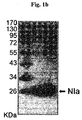
- a recombinant NIa protein was expressed in E. coli and purified to homogeneity on a chitin bead column ( Fig. 1B ).
- NIa was then incubated with a monomeric A preparation for 3 hrs in the presence or absence of the cysteine protease inhibitor, NEM.
- NIa is capable of cleaving oligomeric A ⁇ , which is known to be more toxic than monomeric A ⁇ .
- Oligomeric A was prepared by incubating a solution of A ⁇ peptide at 4°C for 36 hrs. As assessed by SDS-PAGE, the oligomeric A ⁇ preparation contained roughly equal amounts of monomeric and oligomeric A ( Fig.
- NIa cleavage products were analyzed by MALDI-TOF/TOF mass spectrometry ( FIG. 2 ).
- the monomeric A ⁇ preparation produced a single peak without contamination, whereas NIa produced three contaminating peaks.
- the peak corresponding to A was greatly reduced and two new peaks were detected ( Fig. 2B ), with molecular weights of 1,826 Da and 2,704 Da, corresponding to amino acids1-15 and 16-42 of A ⁇ , respectively ( Fig. 2A ). This result indicates that NIa cleaves the peptide bond after Gln 15 , as expected.
- B103 neuroblastoma cells were transformed with an HA-tagged NIa expression vector and stained with an anti-HA antibody. Examination with confocal microscopy revealed that NIa was expressed predominantly in the cytoplasm ( Fig. 3A ).
- the transformed cells were fractionated into the particulate (P) and soluble (S) fractions and subjected to Western blotting ( Fig. 3B ). While Oct1 (nuclear marker), VDAC2 (mitochondrial marker), and cathepsin D (lysosomal marker) were found in the particulate fraction, HA was colocalized with ⁇ -tubulin (cytosolic marker) exclusively to the soluble fraction.
- NIa prevents A ⁇ -Induced cell death
- NIa possesses activity against A within cells
- pGFPUb-A encoding a triple fusion protein of green fluorescent protein (GFP), ubiquitin (Ub), and A ⁇ .
- GFP green fluorescent protein
- Ub ubiquitin
- a ⁇ The peptide bond between Ub and A ⁇ is cleaved quickly by endogenous deubiquitinating enzymes, generating an equimolar ratio of GFP-Ub and A in the cytosol 52 .
- B103 cells were co-transformed with pGFPUb-A ⁇ and an empty plasmid, a NIa-expression plasmid, pcDNA-HA-NIa, or a mutant NIa expression plasmid, pcDNA-HA-mNIa.
- the NIa mutation consisted of an Asp to Ala substitution in the catalytic triad.
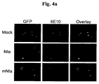
- the cells were then immunostained with the anti-A antibody, 6E10 ( Fig. 4A and B ).
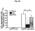
- the observed ratio in those cells expressing a mutant NIa protease plasmid (mNIa) was 42%, which was not significantly different from that obtained with an empty plasmid.
- NIa prevents A ⁇ -induced cell death
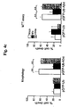
- two different methods a morphological approach and the MTT cell viability assay ( FIG. 4B and C ).
- Intracellular expression of A via pGFPUb-A resulted in a significant increase in cell death (62% by the morphological assay and 55% by the MTT assay).
- This intracellular A ⁇ -induced cell death was almost completely blocked by co-transfomation with pcDNA-HA-NIa but it was not affected in cells co-expressing pcDNA-HA-mNIa.
- Lentiviral vectors expressing NIa and GFP were generated ( FIG. 6A ).
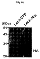
- Human 293T cells infected with Lenti-NIa showed a strong NIa expression, as assessed by Western blotting with anti-HA antibody ( Fig. 6B ).
- Double transgenic mice (APPswe/PS1dE9) were stereotaxically injected with 3 ⁇ l of Lenti-NIa (1 X 10⁸ TU) into the lateral ventricles.
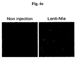
- immunohistochemistry was performed one month after injection. The NIa expression was detected in sections of mice injected with Lenti-NIa compared with the brain sections of control non-injected mice ( Fig. 6C ).
- NIa expression showed a wide distribution throughout the brain including the cerebral cortex, hippocampus, amygdala, and thalamus (data not shown).
- RT-PCR also showed the presence of the NIa transcripts in the Lenti-NIa-infected brain.
- the GAPDH signal served as a control and was equally expressed in all samples ( Fig. 6D ). Decreased A levels in the brain of APPsw / PS1 transgenic mice infused with Lenti-NIa
- Lenti-NIa was infused into the lateral ventricles of the brain of APPsw/PS1dE9 mice at 6.5 months of age.
- equal amounts of Lenti-GFP were infused in the same manner.
- the brains were removed one month after injection and the A levels in both soluble (Tris-buffer extractable) and insoluble (FA-buffer extractable) fractions were measured by ELISA.
- the Lenti-NIa infusion reduced the soluble A ⁇ 1-40 by 33% in males and by 36% in females, and the insoluble A ⁇ 1-40 by 24% in males and by 21% in females ( Fig. 7A , upper lane).
- NIa also reduced the soluble A ⁇ 1-42 by 38% in males and by 28% in females, and the insoluble A ⁇ 1-42 by 33% in males and by 36% in females ( Fig. 7A , lower lane).
- the reduction of A ⁇ 1-42 levels in the male brains was not statistically significant. Reduced A deposition in the brain of APP sw /PS1 transgenic mice infused with Lenti-NIa
- a ⁇ . is oligomerized intracellularly soon after it is generated, and these molecules are then secreted from the cell. Some of the secreted A ⁇ oligomers enter the cell through selective uptake and subsequently cause the dysfunction of subcellular organelles, which is associated with the memory and cognitive decline typically observed in AD patents 60 .
- A is detected in both intraneuronal cells and in the extracellular space of AD brains. Recent studies have demonstrated that intracellular A . levels decrease as extracellular plaques start to build up in patients with AD and in AD transgenic mouse models 10,61 . These results suggest that the accumulation of intracellular A precedes the formation of extracellular A deposits in the progression of the disease. Interestingly, in cells expressing the AD-associated mutant APP, A is kept within the cells, whereas in cells expressing wild type APP, A is mostly found to be secreted 32 . In addition, in aged mice carrying mutant presenilin 1, A aggregation is detected within neurons, but it is absent in the extracellular fluid 62 .
- proteasome activity leads to higher levels of A both in vivo and in vitro, suggesting that the proteasome is responsible for the processing of A in the cytosol 52,63,64 .
- the overproduction of A results in an overload of the proteasome, ultimately leading to an impairment of proteasome activity, a characteristic of AD 65,66 .
- NIa a plant viral protease, specifically cleaves oligomeric as well as monomeric A in vitro and is predominantly localized in the cytosol of neuronal cells.
- NIa neuronal cells inhibits cell death induced both by intracellularly expressed and exogenously added A ⁇ .
- lentiviral-mediated overexpression of NIa in the brain of AD transgenic mice was found to reduce the levels of A and plaque formation.
Landscapes
- Health & Medical Sciences (AREA)
- Life Sciences & Earth Sciences (AREA)
- Chemical & Material Sciences (AREA)
- Engineering & Computer Science (AREA)
- Bioinformatics & Cheminformatics (AREA)
- General Health & Medical Sciences (AREA)
- Organic Chemistry (AREA)
- Medicinal Chemistry (AREA)
- Genetics & Genomics (AREA)
- Animal Behavior & Ethology (AREA)
- Veterinary Medicine (AREA)
- Pharmacology & Pharmacy (AREA)
- Molecular Biology (AREA)
- Public Health (AREA)
- Zoology (AREA)
- Wood Science & Technology (AREA)
- Biomedical Technology (AREA)
- Epidemiology (AREA)
- Biotechnology (AREA)
- General Engineering & Computer Science (AREA)
- Virology (AREA)
- Biochemistry (AREA)
- Neurology (AREA)
- Neurosurgery (AREA)
- Gastroenterology & Hepatology (AREA)
- Immunology (AREA)
- Proteomics, Peptides & Aminoacids (AREA)
- Microbiology (AREA)
- Nuclear Medicine, Radiotherapy & Molecular Imaging (AREA)
- Chemical Kinetics & Catalysis (AREA)
- General Chemical & Material Sciences (AREA)
- Hospice & Palliative Care (AREA)
- Psychiatry (AREA)
- Medicines That Contain Protein Lipid Enzymes And Other Medicines (AREA)
- Enzymes And Modification Thereof (AREA)
Description
- The present invention relates to a pharmaceutical composition for use for preventing or treating various amyloid β-caused diseases.
- Alzheimer's disease (AD) is a progressive neurodegenerative disorders which affects approximately twenty four million people worldwide, and it is the most common form of dementia among older people. AD is characterized by progressive memory impairment and cognitive dysfunction. A distinct hallmark of AD is the deposition of amyloid plaques which are mainly composed of amyloid β (Aβ) of 40, 42, and 43 amino acids in length. Aβ is produced by the sequential cleavage of the amyloid β precursor protein (APP) by β- and γ-secretases1,2. Aβ can exist in different forms such as monomers, oligomers (dimer, trimer, and tetramer), proto-fibrils, and fibrils, and these different conformational states are related to its toxicity. Oligomeric Aβ was shown to be approximately 10- and 40-fold more cytotoxic than fibrillar and monomeric Aβ, respectively3. A recent report also found that dimeric Aβ are 3-fold more toxic than monomeric Aβ, and that trimeric and tetrameric Aβ are upto 13-fold more toxic4.
- Although Aβ unquestionably plays a causative role in AD, the underlying mechanisms by which it contributes to the development of this disease are still controversial. It is widely accepted that Aβ exerts its pathological activity extracellularly. In pathological AD brains, Aβ is secreted into the extracellular space forming amyloid plaques5. When added into the culture media, Aβ can induce cell death in vitro in a variety of cell types3,4,6.
- However, accumulating evidence suggests that intracellular Aβ activity is also critical for the development of AD. Several authors have reported the intracellular localization of Aβ in the brain tissues of post-mortem AD patients and in transgenic AD mice1,7,8. A closer examination with electron microscopy and immunocytochemistry revealed that Aβ is present in diverse subcellular organelles in neuronally differentiated P19 cells, including early endosomes, trans-Golgi network, rough endoplasmic reticulum, outer mitochondrial membrane, and nuclear envelope9. In a triple transgenic AD mouse model, early cognitive impairments correlated with the accumulation of intracellular Aβ in the hippocampus and amygdala, without the apparent deposition of amyloid plaques or neurifibrillary tangles10.
- Intracellular Aβ was also shown to induce p53-dependent neuronal cell death11,12 through the impairment of mitochondrial function13. The intra-hippocampal injection of an antibody directed against Aβ reduced not only extracellular Aβ deposits, but also intracellular Aβ accumulation. Upon dissipation of this antibody, the reappearance of the extracellular deposits was preceded by the accumulation of intracellular Aβ. These observations suggest that a dynamic exchange between intracellular and extracellular Aβ exists, and that intracellular Aβ serves as a source of extracellular amyloid deposits, implying a role for intracellular Aβ in the pathogenesis of AD14,15.
- Since the accumulation of Aβ is considered to be the most critical single event in the pathogenesis of AD, a catabolic elimination of Aβ from the brain would be a valuable therapeutic strategy. Several proteases, including neprilysin (NEP), insulin degrading enzyme, endothelin-converting enzyme, and uPA/tPA-plasmine, have been identified for their ability to degrade Aβ16, with NEP being the best-characterized one. The pharmacological inhibition or genetic ablation of NEP in mice has been shown to result in an increased Aβ deposition, accompanied by deficits in synaptic plasticity and an impairment in hippocampus-dependent memory17,18, while the viral or transgene-mediated overexpression of NEP reduced Aβ deposition and its associated cytopathology19,20. However, it was recently shown that NEP overexpression did not reduce the oligomeric Aβ levels or improve deficits in learning and memory. These results appear to suggest that the NEP-dependent degradation of Aβ affected plaques more efficiently than oligomeric Aβ21.
- Throughout this application, various patents and publications are referenced, and citations are provided in parentheses.
- The present inventors have made intensive endeavors to develop proteolytic therapeutics for amyloid β-caused diseases, inter alia, Alzheimer's disease. As a result, we have discovered that the nuclear inclusion a (NIa) protease of Turnip mosaic virus (TuMV) cleaves monomeric and oligomeric amyloid β (Aβ) in an effective and specific manner and significantly prevents the Aβ-induced cell death in neuronal culture cells and the Aβ-related pathology in transgenic AD (Alzheimer's disease) mice. NIa might therefore provide a novel strategy for the clearance of toxic oligomeric Aβ from the brain of AD patients.
- Accordingly, it is an object of this invention to provide a pharmaceutical composition for preventing or treating an amyloid β-caused disease.
- Other objects and advantages of the present invention will become apparent from the detailed description to follow taken in conjugation with the appended claims and drawings.
-
-
Figures 1A-1D . Cleavage of Aβ by NIa.- (A) The amino acid sequence of Aβ is aligned with the consensus cleavage site of NIa, Val-Xaa-His-Gln. (B) NIa was purified from E. coli and separated by SDS-PAGE.
Lane 1, molecular size markers; lane 2, NIa (10 µg). (1C) Monomeric Aβ (2.5 µM) was incubated with NIa (1.5 µM) in the presence or absence of NEM for 3 hrs at 25° C. The reaction mixture was separated on a Tris-tricine gel, blotted, and probed with the anti-Aβ antibody, 6E10. The density of each Aβ band was quantified by densitometry. The band intensities after 3 hr incubation (lanes 2, 4, and 6) were plotted relative to the band intensities of each sample at 0 hr (lanes
- (A) The amino acid sequence of Aβ is aligned with the consensus cleavage site of NIa, Val-Xaa-His-Gln. (B) NIa was purified from E. coli and separated by SDS-PAGE.
-
Figures 2A-2B . Mass spectra of monomeric Aβ incubated with NIa.- (A) The calculated molecular masses of the expected cleavage products are shown. (B) Monomeric Aβ (2.5 µM) was incubated with NIa (1.5 µM) for 3 hrs at 25° C and analyzed using MALDI-TOF/TOF mass spectrometry. Note that two peaks corresponding to the Aβ cleavage products as well as a peak corresponding to Aβ were detected. As controls, NIa and Aβ were analyzed separately. Three minor peaks marked by asterisks represent contamination of the NIa preparation.
-
Figures 3A-3B . Subcellular localization of NIa in B103 neuroblastoma cells.- (A) B103 neuroblastoma cells transformed with pcDNA-HA-NIa were immunostained with anti-HA antibody and observed under a confocal microscope. (B) B103 cells transformed with a blank plasmid (Mock) or pcDNA-HA-NIa (NIa) were fractionated into particulate (P) and soluble (S) fractions by differential centrifugation. The two fractions were separated by SDS-PAGE, blotted, and probed with antibodies against Oct1 (nuclear), VDAC2 (mitochondrial), cathepsin D (lysosomal), α-tubulin (cytosolic), and HA (NIa).
-
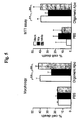
Figures 4A-4C . Degradation of intracellular Aβ and inhibition of intracellular Aβ-induced cell death by NIa.- (A) B103 neuroblastoma cells were cotransfected with pGFPUb-Aβ and an empty vector (Mock), pcDNA-HA-NIa (NIa), or pcDNA-HA-mNIa (mNIa). After 48 hrs of incubation, the cells were immunostained with the anti-Aβ antibody, 6E10. (B) The number of Aβ-positive cells (red) and GFP-expressing cells (green) were counted under the microscope and their ratio was calculated. n= 6. (C) Cell death induced by intracellular Aβ peptide was measured by morphological and MTT assays. n= 6. Error bars represent SD. **p < 0.01.
-
Figure 5 . Inhibition of exogenously added Aβ-induced cell death by NIa.- B103 neuroblastoma cells transfected with an empty vector (Mock), pcDNA-HA-NIa (NIa), or pcDNA-HA-mNIA (mNIa) were treated with Aβ (5 µM) in culture media for 48 hrs. Cell death was measured by morphological and MTT assays. n= 6. Error bars represent SD. **p < 0.01.
-
Figures 6A-6D . Lentiviral-mediated expression of NIa.- (A) Lentiviral constructs for the expression of HA-NIa and GFP. (B) Western blotting with anti-HA antibody showed the NIa expression levels in 293T cells infected with Lenti-NIa. (C) The expression of NIa in mouse brains was detected by immunohistochemistry with anti-HA antibody and anti-mouse-FITC secondary antibody. The prefrontal sections of mouse brains infused with Lenti-NIa are compared with the brain sections of control non-injected mice. (D) The brains infused with Lenti-GFP and Lenti-NIa were subjected to RT-PCR. A PCR product corresponding to NIa was detected. GAPDH was used as control. CMV, Cytomegalovirus RNA polymerase II promoter; WPRE, Woodchuck hepatitis post-transcriptional regulatory element; U5, HIV 5'-long terminal repeat; ΔU3, 3' self inactivating long terminal repeat; PSI, region of viral RNA responsible for directing packaging; PRE, binding site for the Rev protein that aids in the transport of unspliced RNAs from the nucleus to cytoplasm.
-
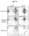
Figures 7A-7C . NIa-mediated reduction in Aβ levels and Aβ plaques in APPsw/PS1dE9 mouse brains.- (A) Brains of APPsw/PS1dE9 were bilaterally infused with Lenti-GFP and Lenti-NIa, and the amounts of Aβ1-40 and Aβ1-42 were measured by ELISA. The amounts of soluble and insoluble Aβ1-40 are shown (upper lane). The amounts of soluble and insoluble Aβ1-42 are shown (lower lane). (B) Sections of prefrontal cortex, parietal cortex, hippocampus, and piriform cortex of APPsw/PS1dE9 male mouse infused with Lenti-GFP and Lenti-NIa were stained with anti-Aβ antibody (Bam-10). (C) The number of plaques in the prefrontal cortex, parietal cortex, and piriform cortex of APPsw/PS1dE9 male mouse infused with Lenti-GFP and Lenti-NIa was counted. For Lenti-GFP infusions, n=5 for male and n=5 for female. For Lenti-NIa infusions, n=6 for male and n=3 for female. Error bars represent SD. *p < 0.05 and **p < 0.01.
- In one aspect of the present invention, there is provided a pharmaceutical composition for use in preventing or treating an amyloid β-caused disease, which comprises as an active ingredient a NIa (nuclear inclusion a) protease or a gene carrier containing a nucleotide sequence encoding the NIa protease.
- There is described a method for preventing or treating an amyloid β-caused disease, which comprises administering to a mammalian subject the pharmaceutical composition described above.
- The present inventors have made intensive endeavors to develop proteolytic therapeutics for amyloid β-caused diseases, inter alia, Alzheimer's disease. As a result, we have discovered that the nuclear inclusion a (NIa) protease of Turnip mosaic virus (TuMV) cleaves monomeric and oligomeric amyloid β (Aβ) in an effective and specific manner and significantly prevents the Aβ-induced cell death in neuronal culture cells and the Aβ-related pathology in transgenic AD (Alzheimer's disease) mice. NIa might therefore provide a novel strategy for the clearance of toxic oligomeric Aβ from the brain of AD patients.
- The nuclear inclusion a (NIa) protease of turnip mosaic virus (TuMV) is responsible for the processing of the viral polyprotein into functional proteins. NIa was previously shown to possess a relatively strict substrate specificity with a preference for Val-Xaa-His-Gln↓, with the scissile bond located after Gin. The presence of the same consensus sequence, Val12-His-His-Gln15, near the presumptive α-secretase cleavage site of the amyloid-β (Aβ) peptide led us to hypothesize that NIa could possess activity against Aβ.
- According to a preferred embodiment, the NIa protease has the activity to cleave an oligomeric Aβ as well as a monomeric Aβ. As well known to one of skill in the art, the oligomeric Aβ is shown to be approximately 10- and 40-fold more cytotoxic than fibrillar and monomeric Aβ, respectively. A dimeric Aβ are 3-fold more toxic than monomeric Aβ, and trimeric and tetrameric Aβ are upto 13-fold more toxic. In this regard, the NIa protease used in this invention is very advantageous for treating AD by clearing more toxic Aβ molecules.
- According to a preferred embodiment, the NIa protease cleaves intracellular or extracellular Aβ. More preferably, the NIa protease used in this invention cleaves Aβ present in cytosol. As demonstrated in Example, the expression of NIa in neuronal cells inhibits cell death induced both by intracellularly expressed and exogenously added Aβ. In addition, lentiviral-mediated overexpression of NIa in the brain of AD transgenic mice reduces the levels of Aβ and plaque formation. These data provide additional evidence supporting a critical role for intracellular Aβ in the pathogenesis of AD. In this regard, NIa could be used as a novel tool to study the molecular events underlying the induction of cell death by intracellular Aβ. Moreover, the present invention offers proof-of-concept that the clearance of intracellular Aβ by a cytosolic protease could be a viable strategy for the treatment of AD.
- According to a preferred embodiment, the NIa protease inhibits the amyloid β-induced cell death.
- According to a preferred embodiment, the NIa protease has the activity to cleave amyloid beta1-40 and amyloid beta1-42.
- According to a preferred embodiment, the NIa protease has the activity to cleave a peptide bond between amino acids 15 and 16 of SEQ ID NO:3.
- The NIa protease used in the present invention may be provided as a protein or a gene carrier containing a nucleotide sequence encoding the NIa protease.
- Where the NIa protease is provided in the form of a protein, it may be alternatively fused to a protein transduction domain (PTD) for effectively penetrating into cells. The preferable amino acid sequence of the NIa protease is set forth as SEQ ID:1.
- The protein transduction domain useful in the present invention includes, but not limited to, HIV-TAT, VP-22, a growth factor signal peptide sequence, Pep-1, Pep-7, a Drosophila Antp peptide, oligoarginine, HSV VP22 transcription regulatory protein, vFGF-derived MTS peptide, Penetratin, Transportan, Buforin II, MAP (model amphiphatic peptide), k-FGF,
Ku 70, pVEC, SynB1 and HN-1. - Where the NIa protease is provided as a gene carrier for clearing Aβ, it may be constructed in a gene carrier in accordance with conventional technologies known to one of skill in the art.
- The term "gene carrying" used herein refers to the transfer of gene into cells and has the same meaning as gene delivery.
- To construct the present gene carrier, it is preferred that the NIa protease-encoding nucleotide sequence is contained in a suitable expression construct. According the expression construct, it is preferred that the NIa protease-encoding nucleotide sequence is operatively linked to a promoter. The term "operatively linked" refers to functional linkage between a nucleic acid expression control sequence (such as a promoter, signal sequence, or array of transcription factor binding sites) and a second nucleic acid sequence, wherein the expression control sequence affects transcription and/or translation of the nucleic acid corresponding to the second sequence. According to the present invention, the promoter linked to the NIa protease gene is operable in, preferably, animal, more preferably, mammalian cells, to control transcription of the NIa protease gene, including the promoters derived from the genome of mammalian cells or from mammalian viruses, for example, CMV (cytomegalovirus) promoter, the adenovirus late promoter, the vaccinia virus 7.5K promoter, SV40 promoter, HSV tk promoter, RSV promoter, EF1 alpha promoter, metallothionein promoter, beta-actin promoter, human IL-2 gene promoter, human IFN gene promoter, human IL-4 gene promoter, human lymphotoxin gene promoter and human GM-CSF gene promoter. Most preferably, the promoter is CMV promoter.
- Preferably, the expression construct used in this invention comprises a polyadenylation sequence (e.g., bovine growth hormone terminator and SV40-derived polyadenylation sequence).
- The expression construct for the NIa protease-encoding nucleotide sequence may have a structure of "promoter-NIa protease-encoding nucleotide sequence-polyadenylation sequence.
- The gene carrier of the present invention is constructed in a variety of forms, preferably, (i) naked recombinant DNA molecule, (ii) plasmid, (iii) viral vector, or (iv) liposome or neosome containing naked recombinant DNA molecule and plasmid.
- The NIa protease-encoding nucleotide sequence may be applied to a multitude of gene carriers useful in gene therapy, preferably, plasmid, adenovirus22, adeno-associated virus (AAV, Lashford LS., et al., Gene Therapy Technologies, Applications and Regulations Ed. A. Meager, 1999), retrovirus23, herpes simplex virus24, vaccinia virus25, liposome (Methods in Molecular Biology, Vol 199, S.C. Basu and M. Basu (Eds.), Human Press 2002) or neosome. Most preferably, the gene carrier of this invention is constructed by incorporating the NIa protease-encoding nucleotide sequence to retrovirus or lentivirus.
- Adenovirus has been usually employed as a gene carrier because of its mid-sized genome, ease of manipulation, high titer, wide target-cell range, and high infectivity. Both ends of the viral genome contains 100-200 bp ITRs (inverted terminal repeats), which are cis elements necessary for viral DNA replication and packaging. The E1 region (E1A and E1B) encodes proteins responsible for the regulation of transcription of the viral genome and a few cellular genes. The expression of the E2 region (E2A and E2B) results in the synthesis of the proteins for viral DNA replication.
- Of adenoviral vectors developed so far, the replication incompetent adenovirus having the deleted E1 region is usually used. The deleted E3 region in adenoviral vectors may provide an insertion site for transgenes25,26. Therefore, it is preferred that the NIa protease-encoding nucleotide sequence is inserted into either the deleted E1 region (E1A region and/or E1B region, preferably, EIB region) or the deleted E3 region, more preferably, the deleted E3 region. The promoter-NIa protease gene-poly A sequence is preferably present in either the deleted E1 region (E1A region and/or E1B region, preferably, EIB region) or the deleted E3 region, more preferably, the deleted E3 region.
- In nature, adenovirus can package approximately 105% of the wild-type genome, providing capacity for about 2 extra kb of DNA27. In this regard, the foreign sequences described above inserted into adenovirus may be further inserted into adenoviral wild-type genome.
- The adenovirus may be of any of the 42 different known serotypes or subgroups A-F. Adenovirus type 5 of subgroup C is the most preferred starting material for constructing the adenoviral gene carrier of this invention. A great deal of biochemical and genetic information about adenovirus type 5 is known.
- The foreign genes delivered by the present adenoviral gene carrier are episomal, and therefore, have low genotoxicity to host cells. Therefore, gene therapy using the adenoviral gene carrier of this invention may be considerably safe.
- Retroviruses or lentiviruses23 capable of carrying relatively large exogenous genes have been used as viral gene delivery vectors in the senses that they integrate their genome into a host genome and have broad host spectrum.
- In order to construct a retroviral vector or lentiviral vector, the NIa protease-encoding nucleotide sequences to be transferred are inserted into the viral genome in the place of certain viral sequences to produce a replication-defective virus. To produce virions, a packaging cell line containing the gag, pol and env genes but without the LTR (long terminal repeat) and ψ components is constructed28. When a recombinant plasmid containing the NIa protease-encoding sequence, LTR and ψ is introduced into this cell line, the ψ sequence allows the RNA transcript of the recombinant plasmid to be packaged into viral particles, which are then secreted into the culture media Nicolas and Rubinstein "Retroviral vectors," In: Vectors: A survey of molecular cloning vectors and their uses, Rodriguez and Denhardt29. The media containing the recombinant retroviruses is then collected, optionally concentrated and used for gene delivery.
- A successful gene transfer using the second-generation retroviral vector has been reported. Kasahara et al. 30 prepared variants of moloney murine leukemia virus in which the EPO (erythropoietin) sequence is inserted in the place of the envelope region, consequently, producing chimeric proteins having novel binding properties. Likely, the present gene carrier can be constructed in accordance with the construction strategies for the second-generation retroviral vector.
- Adeno-associated viruses are capable of infecting non-dividing cells and various types of cells, making them useful in constructing the gene carrier of this invention. The detailed descriptions for use and preparation of AAV vector are found in
U.S. Pat. No. 5,139,941 and4,797,368 . - Research results for AAV as gene carriers are disclosed in LaFace et al.31, Zhou et al.32, Walsh et al.33 and Flotte et al.34 Recently, an AAV vector has been approved for Phase I human trials for the treatment of cystic fibrosis.
- Typically, a recombinant AAV virus is made by cotransfecting a plasmid containing the gene of the NIa protease flanked by the two AAV terminal repeats35,36 and an expression plasmid containing the wild type AAV coding sequences without the terminal repeats37.
- Other viral vectors may be employed as a gene carrier in the present invention. Vectors derived from viruses such as vaccinia virus25; Ridgeway, "Mammalian expression vectors," In: Vectors: A survey of molecular cloning vectors and their uses. Rodriguez and Denhardt29; Baichwal and Sugden, "Vectors for gene transfer derived from animal DNA viruses: Transient and stable expression of transferred genes," In: Kucherlapati R, ed. Gene transfer 38 (New York: Plenum Press, 117-148(1986)) and herpes simplex virus24 may be used in the present delivery systems for transferring both the NIa protease gene into cells.
- Liposomes are formed spontaneously when phospholipids are suspended in an excess of aqueous medium. Uposome-mediated nucleic acid delivery has been very successful as described in Nicolau and Sene39 and Nicolau et al.40 Example of commercially accessible reagents for transfecting animal cells using liposomes includes Lipofectamine (Gibco BRL). Liposomes entrapping the NIa protease gene interact with cells by mechanism such as endocytosis, adsorption and fusion and then transfer the sequences into cells.
- There is described a method for delivery a gene, which comprises contacting the gene carrier of this invention as described hereinabove to a biosample containing cells.
- Where the present gene carrier is constructed on the basis of viral vector construction, the contacting is performed as conventional infection methods known in the art. The infection of hosts using viral vectors is well described in the above-cited publications.
- Where the present gene carrier is a naked recombinant DNA molecule or plasmid, the NIa protease-encoding sequence and nucleotide sequence to be delivered are introduced into cells by microinjection41,42, calcium phosphate coprecipitation43,44, electroporation45,46, liposome-mediated transfection47,39,40, DEAE-dextran treatment48and particle bombardment49.
- According to a preferred embodiment, the NIa protease gene (e.g., SEQ ID NO:2) is carried in a viral vector, more preferably a retroviral vector or lentiviral vector.
- In the pharmaceutical compositions of this invention, the pharmaceutically acceptable carrier may be conventional one for formulation, including lactose, dextrose, sucrose, sorbitol, mannitol, starch, rubber arable, potassium phosphate, arginate, gelatin, potassium silicate, microcrystalline cellulose, polyvinylpyrrolidone, cellulose, water, syrups, methyl cellulose, methylhydroxy benzoate, propylhydroxy benzoate, talc, magnesium stearate, and mineral oils, but not limited to. The pharmaceutical composition according to the present invention may further include a lubricant, a humectant, a sweetener, a flavoring agent, an emulsifier, a suspending agent, and a preservative. Details of suitable pharmaceutically acceptable carriers and formulations can be found in Remington's Pharmaceutical Sciences (19th ed., 1995).
- The pharmaceutical composition of this invention may be preferably administered parenterally. For non-oral administration, local injection, intrathecal injection, intravenous injection or intraperitoneal injection may be employed.
- A suitable dose of the pharmaceutical composition of the present invention may vary depending on pharmaceutical formulation methods, administration methods, the patient's age, body weight, sex, severity of diseases, diet, administration time, administration route, an excretion rate and sensitivity for a used pharmaceutical composition. Physicians of ordinary skill in the art can determine an effective amount of the pharmaceutical composition for desired treatment. Preferably, the pharmaceutical composition of the present invention is administered with a daily dose of 0.001-1000 mg/kg (body weight).
- According to the conventional techniques known to those skilled in the art, the pharmaceutical composition may be formulated with pharmaceutically acceptable carrier and/or vehicle as described above, finally providing several forms including a unit dose form and a multi-dose form. Non-limiting examples of the formulations include, but not limited to, a solution, a suspension or an emulsion in oil or aqueous medium, an extract, an elixir, a powder, a granule, a tablet and a capsule, and may further comprise a dispersion agent or a stabilizer.
- The pharmaceutical composition of this invention may be employed to prevent or treat a variety of diseases or disorders, including Alzheimer's disease, MCI (mild cognitive impairment), mild-to-moderate cognitive impairment, vascular dementia, cerebral amyloid angiopathy, hereditary cerebral hemorrhage, senile dementia, Down's syndrome, inclusion body myositis, age-related macular degeneration and conditions associated with Alzheimer's disease. In particular, the pharmaceutical composition of this invention is a promising therapeutic for Alzheimer's disease.
- The present invention will now be described in further detail by examples. It would be obvious to those skilled in the art that these examples are intended to be more concretely illustrative and the scope of the present invention as set forth in the appended claims is not limited to or by the examples.
- Cell culture reagents were purchased from GIBCO-BRL (Invitrogen, Carlsbad, CA, USA). Synthetic APβ1-42 peptide was purchased from Sigma (St Louis, MO, USA) and Anygen (Gwangju, Korea). 6E10 antibody recognizing residues 1-17 of Aβ peptide was purchased from Signet™ (Dedham, MA, USA). Antibodies against HA, α-tubulin, VDAC2, Oct1, and cathepsin D were purchased from Abcam (Cambridge, MA, UK). Chitin beads were purchased from New England BioLabs (Ipswich, MA, USA). All other reagents were purchased from Sigma.
- To produce recombinant NIa protein in E. coli, the NIa gene was cloned into pTYB12 (New England BioLabs) via the EcoRI and XhoI sites. The pTYB12 vector contains an N-terminal intein tag. The pTYB12-NIa vector was transformed into the E. coli strain BL21 (DE3) and grown at 37°C in LB medium. Induction of the NIa protein was achieved by addition of 400 µM IPTG overnight at 20°C. The cells were harvested, resuspended in column buffer (20 mM HEPES [pH 7.9], 500 mM NaCl, 1mM EDTA), and lysed by sonication. The lysate was centrifuged and the resulting supernatant was loaded onto a chitin column equilibrated with column buffer. After extensive washing, the NIa protein was eluted from the column using a column buffer containing 50 mM DTT, dialyzed in storage buffer (50 mM HEPES [pH 7.6], 1 mM EDTA, 1 mM DTT, 10% glycerol), and concentrated by Amicon Centriprep (Millipore, Billerica, MA, USA). The protein concentration was determined by the BCA method and analyzed on a 12% SDS-PAGE gel.
- To prepare Aβ solutions, we followed the method described by Yan et al.50 and Dahlgren et al.3. Synthetic human Aβ1-42 peptides ( >95% pure by high performance liquid chromatography and mass spectrometry tests) were dissolved in dimethylsulfoxide (DMSO) to a concentration of 5 mM. For monomeric Aβ, the Aβ solution in DMSO was diluted in PBS to a final concentration of 25 µM immediately before use. For oligomeric Aβ, the Aβ solution in DMSO was diluted in PBS to a concentration of 100 µM and incubated at 4°C for 36 hrs. The physical state of Aβ was confirmed by PAGE with 10-20% Tris-Tricine gels (Bio-Rad, Hercules, CA, USA).
- 1.5 µM of the recombinant NIa protease was incubated with 2.5 µM Aβ preparations in an assay buffer (HEPES [pH 7.4], 20 mM KCI, 20 mM MgCl2) at 25° C for 3 hrs. As a control, the NIa protease was pre-incubated with the cysteine protease inhibitor, N-ethylmaleimide (NEM) for 10 min at 4° C. After incubation, the mixtures were subjected to PAGE with 10-20% Tris-Tricine gel and Western blotting using the anti-Aβ antibody 6E10. To further analyze the cleavage products, the reaction mixtures were analyzed by MALDI-TOF/TOF mass spectrometry (4700 Proteomics Analyzer, Applied Biosystems, Carlsbad, California, USA). As controls, NIa and Aβ were separately analyzed.
- B103 rat neuroblastoma cells were cultured in DMEM supplemented with 10% (vol/vol) fetal bovine serum51. A mutant NIa gene in which Asp81 in the catalytic triad was changed to Ala was generated by a PCR mutagenesis. To express the wild type and mutant NIa in B103 cells, the corresponding genes were subcloned into pcDNA3 (Invitrogen) containing an N-terminal HA tag. Cells were transfected using Lipofectamine Reagent (Invitrogen) according to the manufacturer's protocol. A cytosolic Aβ1-42 expression vector (pGFPUb-Aβ1-42) was previously described52. For the Aβ treatment, the Aβ solutions (100 µM) were added to a final concentration of 5 µM.
- Cell viability was assessed by MTT assay and cell morphological methods. The 3-[4,5-dimethylthizaol-2-yl]-2,5-diphenyl tetrazolium bromide (MTT) was solubilized in PBS to 5 mg/ml. A volume of MTT solution equal to 10% of the culture media volume was added to the cell culture at 37°C for 3 hrs. A solubilization solution (10% Triton X-100 and 0.1 N HCl in anhydrous isopropanol) in a volume equal to the culture media volume was added and further incubated at 37°C until the resulting formazan crystals were completely dissolved. The absorbance of the samples was measured at 570 nm, and the background absorbance of each well was measured at 690 nm. For the assessment of cell morphology, cultured cells were co-transformed with the experimental plasmid and a GFP plasmid and the morphology of GFP-positive cells was examined under a fluorescence microscope42(Olympus, Shinjuku, Tokyo, Japan).
- B103 rat neuroblastoma cells were washed with PBS containing 1 mM CaCl2 and 1 mM MgCl2 and fixed for 10 min with 3.5% paraformaldehyde. The cells were permeabilized by incubation with 0.2% Triton X-100 in PBS for 10 min, blocked with 5% BSA in PBS for 1 hr, and incubated with anti-6E10 monoclonal antibody or HA monoclonal antibody for 1 hr.
- The fixed cells were then rinsed in PBS and incubated with Alexa 488 fluor-conjugated secondary antibody (Invitrogen) and TRITC-conjugated secondary antibody (Jackson Immunoresearch, West Grove, PA, USA) for 1 hr. For immunofluorescence microscopy, immunoreactivity was captured with a fluorescence microscope (Olympus) with a ProgRes C10pluscamera (JENOPTIK, Goeschwitzer Strasse, Jena, Germany). Color coding was performed using the IMT i-solution software (IMT i-solution Inc., Vancouver, BC, Canada). To determine the levels of Aβ aggregation among GFP positive cells, the number of Aβ positive cells vs. GFP positive cells was counted in 20 random fields per culture. For confocal microscopy analysis, fluorescence signals were visualized using a confocal microscope (TCS SP2, LEICA, Ernst-Leitz-Strasse, Wetzlar, Germany).
- To determine the intracellular localization of the NIa protein, NIa- expressing cells were fractionated using protocol previously described53. Briefly, the cells were harvested by scraping into homogenation buffer (200 mM sucrose, 20 mM Tris [pH7.4], 1 mM EGTA, 1 mM EDTA, 1 X complete protease inhibitor cocktail), lysed by multiple passages through a syringe with a 26-gauge needle, and ultracentrifuged at 70,000 x g for 30 min at 4° C. The pellet (crude membrane fraction) was resuspended in homogenation buffer containing 0.5% Triton X-100 and sonicated for 1 min. Aliquots (50 µg) from each fraction were analyzed by Western blotting.
- The cells were harvested after washing three times with PBS, resuspended in RIPA buffer containing 1X protease inhibitor cocktail and sonicated briefly. The soluble protein fraction was recovered after centrifugation at 10,000 x g for 30 min and separated by SDS-PAGE. Protein concentration was determined by the BCA method. For the analysis of Aβ peptides, samples were separated by electrophoresis using 10-20% Tris-Tricine gels. Proteins were then transferred onto PVDF membrane in 50 mM Tris, 192 mM glycine, and 20% methanol. Membranes were blocked with 5% non-fat milk and incubated with antibodies against 6E10, HA, α-tubulin, VDAC2, Oct1, and cathepsin D. Bands were visualized using the ECL reagent (GE Healthcare/Amersham Bioscience, Piscataway, NJ, USA) and the intensity of each band was quantified by densitometry (Bio-Rad).
- The cDNA fragments encoding NIa and GFP were subcloned into the pLEX-MCS lentiviral vector (Openbiosystems, Huntsville, AL,USA). The resulting recombinant plasmids were co-transformed with packing plasmids into 293T cells and the supernatants were collected. Lentiviruses were collected and concentrated by ultra-centrifugation as previously described19,54. The titers of the NIa and GFP lentiviruses were estimated by measuring the amount of HIV p24 antigen using PCR.
- Transgenic AD model mice, Tg-APPswe/PS1dE9, overexpressing human mutated APP and PS1 (APPswe/PS1dE9), were maintained in C57BL6 x C3H F1 hybrid mice, as described previously55. The mice were housed in normal plastic cages with free access to food and water in a temperature- and humidity-controlled environment under a 12 h light/dark cycle (lights on at 7 a.m.), and they were fed a diet of lab chow and water ad libitum. Tg-APPswe/PS1dE9 mice at 6.5 months of age were randomized into the Lenti-NIa (n=9) and Lenti-GFP (n=10) groups. The mice underwent bilateral intracerebroventricular (i.c.v.) infusion with 3 µl of Lenti-NIa lentivirus (1 X 10⁸ TU) or Lenti-GFP lentivirus with the same titer. After one month, the injected mice were sacrificed and perfused with 0.9% saline. The right and left hemispheres of the brain were used for histological and biochemical analyses, respectively. All animals were handled in accordance with the animal care guidelines of the Ewha Womans University School of Medicine.
- Total RNA was isolated with TRI reagent (Sigma) from frontal cerebral cortex tissue. Reverse-transcription was performed using ImProm II reverse-transcriptase (Promega, Madison, Wisconsin, USA) with oligo-dT priming. To detect NIa expression, PCR was performed using the NIa specific primer set: 5'-ACG AAA GAC GGC CAA TGC GGA-3' and 5'-ACC CGA CGG TTG CGA TGC TT-3'. And for control experiment, PCR was performed using the GAPDH specific primer set: 5'- TCC GTG TTC CTA CCC CCA ATG-3' and 5'- GGG AGT TGC TGT TGA AGT CGC-3'.
- The right hemisphere was post-fixed with 4% paraformaldehyde in 0.1 M phosphate buffer (pH 7.4) at 4° C overnight and were coronally cut into 40 µm-thick sections with a vibratome (Leica VT 1000S; Leica, Germany). Free-floating sections were blocked by 5% normal goat serum, 2% BSA, and 2% FBS. A biotinylated HRP system was used for color development. Anti-Aβ antibody Bam-10 (A5213) was purchased from Sigma (USA). Microscopic studies were carried out using an Oympus BX 51 microscope equipped with a DP71 camera and DP-B software (Olympus, Japan). For the quantification of plaque levels, the numbers of plaques in each region were measured using the TOMORO ScopeEye 3.6 program (Techsan Community, Seoul, Korea).
- ELISA assays for Aβ(1-42) and Aβ(1-40) levels were described in a previous study46. Briefly, the frontal cerebral cortex was homogenized in Tris-buffered saline (20 mM Tris and 137 mM NaCl, [pH 7.6]) in the presence of protease inhibitor mixtures (Complete Mini; Roche, USA). Homogenates were centrifuged at 100,000 x g for 1 hr at 4°C, and the supernatant was used to measure the levels of Tris buffer-soluble forms of Aβ. The pellet was sonicated in 70% formic acid and centrifuged as above; the resulting supernatant was considered the formic acid extractable Aβ and collected for further analysis. The formic acid extract was neutralized with 1 M Tris-CI buffer (pH 11) in a dilution ratio of 1:20 before its use as previously described. The final assays were performed using Human Aβ(1-40) or Aβ(1-42) colorimetric sandwich ELISA kits (BioSource, Invitrogen) by following the manufacturer's instructions.
- Two sample-comparisons were carried out using the unpaired Student's t test with unequal variance, while multiple comparisons were made by one-way ANOVA followed by the Newman-Keuls multiple range test. A p value of less than 0.05 was accepted as being statistically significant. Data are presented as mean±SD.
- We have previously reported that NIa possesses a highly strict substrate specificity, with its cleavage sites defined by the conserved sequence motif Val-Xaa-His-Gln↓, in which the scissile bond is located after Gin. Interestingly, the sequence Val-His-His-Gln is present in Aβ in the vicinity of the presumed o-secretase cleavage site (
Fig. 1A ). Based on this finding, we aimed to determine whether NIa can specifically cleave Aβ. For this purpose, a recombinant NIa protein was expressed in E. coli and purified to homogeneity on a chitin bead column (Fig. 1B ). NIa was then incubated with a monomeric Apreparation for 3 hrs in the presence or absence of the cysteine protease inhibitor, NEM. Analysis by Western blotting revealed that the monomeric A level was greatly reduced by NIa (
Fig. 1C , lane 2 vs. 4), which was partially reversed in the presence of NEM (Fig. 1C , lane 6). The results of the densitometry analysis showed that NIa reduced Alevels by 64% in the absence of NEM and 33% in the presence of NEM, suggesting the specific cleavage of monomeric Aby NIa. - Our findings show that NEM did not completely inhibit NIa activity, which is consistent with a previous report showing that mutations of cysteine residues in the catalytic triad of NIa did not completely abolish its proteolytic activity23. We then tested whether NIa is capable of cleaving oligomeric Aβ, which is known to be more toxic than monomeric Aβ. Oligomeric Awas prepared by incubating a solution of Aβ peptide at 4°C for 36 hrs. As assessed by SDS-PAGE, the oligomeric Aβ preparation contained roughly equal amounts of monomeric and oligomeric A(
Fig. 1D ,lanes after an additional 3 hour incubation at 25°C. Our findings are consistent with a previous report showing that Aoligomerization was accelerated by an increase in incubation time and temperature24. Under the same conditions, the amount of oligomeric Awas greatly reduced by NIa (lane 4) as quantified by densitomeric assessment, which showed that only 19% of oligomeric Aβ remained. This NIa-mediated reduction of oligomeric Awas significantly blocked by NEM (lane 6) implying that NIa specifically cleaves Aβ. - To further analyze the specific cleavage of Aby NIa, the cleavage products were analyzed by MALDI-TOF/TOF mass spectrometry (
Fig. 2 ). The monomeric Aβ preparation produced a single peak without contamination, whereas NIa produced three contaminating peaks. In the reaction mixture including Aand NIa, the peak corresponding to Awas greatly reduced and two new peaks were detected (Fig. 2B ), with molecular weights of 1,826 Da and 2,704 Da, corresponding to amino acids1-15 and 16-42 of Aβ, respectively (Fig. 2A ). This result indicates that NIa cleaves the peptide bond after Gln15, as expected. - B103 neuroblastoma cells were transformed with an HA-tagged NIa expression vector and stained with an anti-HA antibody. Examination with confocal microscopy revealed that NIa was expressed predominantly in the cytoplasm (
Fig. 3A ). The transformed cells were fractionated into the particulate (P) and soluble (S) fractions and subjected to Western blotting (Fig. 3B ). While Oct1 (nuclear marker), VDAC2 (mitochondrial marker), and cathepsin D (lysosomal marker) were found in the particulate fraction, HA was colocalized with α-tubulin (cytosolic marker) exclusively to the soluble fraction. These data suggest that NIa resides predominantly in the cytosol. - To test whether NIa possesses activity against A within cells, we generated Aβ intracellularly using the plasmid pGFPUb-A, encoding a triple fusion protein of green fluorescent protein (GFP), ubiquitin (Ub), and Aβ. The peptide bond between Ub and Aβ is cleaved quickly by endogenous deubiquitinating enzymes, generating an equimolar ratio of GFP-Ub and Ain the cytosol52. B103 cells were co-transformed with pGFPUb-Aβ and an empty plasmid, a NIa-expression plasmid, pcDNA-HA-NIa, or a mutant NIa expression plasmid, pcDNA-HA-mNIa. The NIa mutation consisted of an Asp to Ala substitution in the catalytic triad. The cells were then immunostained with the anti-Aantibody, 6E10 (
Fig. 4A andB ). The results revealed that the proportion of Aβ-positive cells was 56% of the total of GFP-positive cells in those cells harboring pGFPUb-Aand an empty plasmid (Mock), whereas the ratio sharply declined to 14% in cells harboring pGFPUb-Aand pcDNA-HA-NIa (NIa). The observed ratio in those cells expressing a mutant NIa protease plasmid (mNIa) was 42%, which was not significantly different from that obtained with an empty plasmid. These data indicate that NIa can specifically degrade intracellular Aβ. - To evaluate whether NIa prevents Aβ-induced cell death, we used two different methods, a morphological approach and the MTT cell viability assay (
Fig. 4B andC ). Intracellular expression of Avia pGFPUb-Aresulted in a significant increase in cell death (62% by the morphological assay and 55% by the MTT assay). This intracellular Aβ-induced cell death was almost completely blocked by co-transfomation with pcDNA-HA-NIa but it was not affected in cells co-expressing pcDNA-HA-mNIa. Treatment of B103 cells with exogenous Aalso resulted in a considerable proportion of cell death (40% by the morphological assay and 38% by the MTT assay), which was inhibited by co-transfomation with pcDNA-HA-NIa but not by pcDNA-HA-mNIa co-expression (Fig. 5A and B ). It was previously shown that extracellular Aβ is internalized by cell surface receptors and detected in subcellular organelles such as lysosomes, mitochondria and cytosol, causing cell death through dysfunction of these organelles56-59. It appears that cytosolic NIa can cleave internalized Aβ, although it is unknown whether NIa and internalized Aare co-localized. Nonetheless, our data indicate that NIa can prevent cell death induced by both intracellularly expressed and exogenously added Aβ. - Lentiviral vectors expressing NIa and GFP were generated (
Fig. 6A ). Human 293T cells infected with Lenti-NIa showed a strong NIa expression, as assessed by Western blotting with anti-HA antibody (Fig. 6B ). Double transgenic mice (APPswe/PS1dE9) were stereotaxically injected with 3µl of Lenti-NIa (1 X 10⁸ TU) into the lateral ventricles. To evaluate the expression of NIa, immunohistochemistry was performed one month after injection. The NIa expression was detected in sections of mice injected with Lenti-NIa compared with the brain sections of control non-injected mice (Fig. 6C ). The pattern of NIa expression showed a wide distribution throughout the brain including the cerebral cortex, hippocampus, amygdala, and thalamus (data not shown). RT-PCR also showed the presence of the NIa transcripts in the Lenti-NIa-infected brain. The GAPDH signal served as a control and was equally expressed in all samples (Fig. 6D ).
Decreased Alevels in the brain of APPsw/PS1 transgenic mice infused with Lenti-NIa - To assess if NIa causes a reduction in the Alevels in mouse brains, Lenti-NIa was infused into the lateral ventricles of the brain of APPsw/PS1dE9 mice at 6.5 months of age. As a control, equal amounts of Lenti-GFP were infused in the same manner. The brains were removed one month after injection and the A levels in both soluble (Tris-buffer extractable) and insoluble (FA-buffer extractable) fractions were measured by ELISA. We found that the levels of both Aβ1-40 and Aβ1-42 were significantly reduced in both the soluble and insoluble factions of Lenti-NIa-infused brain when compared to the Lenti-GFP-infused brain (
Fig. 7A ). The Lenti-NIa infusion reduced the soluble Aβ1-40 by 33% in males and by 36% in females, and the insoluble Aβ1-40 by 24% in males and by 21% in females (Fig. 7A , upper lane). NIa also reduced the soluble Aβ1-42 by 38% in males and by 28% in females, and the insoluble Aβ1-42 by 33% in males and by 36% in females (Fig. 7A , lower lane). The reduction of Aβ1-42 levels in the male brains was not statistically significant.
Reduced Adeposition in the brain of APPsw/PS1 transgenic mice infused with Lenti-NIa - Immunohistochemical analysis revealed that the A deposition in the prefrontal cortex, parietal cortex, hippocampus and piriform cortex was remarkably decreased in the brain infused with Lenti-NIa in comparison to the brain infused with Lenti-GFP (
Fig. 7B ). Quantitative assessment of Alevels indicated that the Lenti-NIa infusion reduced the plaques by 58% in the prefrontal cortex, by 62% in the parietal cortex, and by 59% in the piriform cortex (Fig. 7C ). - The generation and accumulation of Ais the most critical event in the development of AD, suggesting that the clearance of Acould provide a valuable strategy for the treatment of AD. Although Aexits in several assembly and aggregation forms, oligomeric Aβ. is known to be the most toxic form. Aβ. is oligomerized intracellularly soon after it is generated, and these molecules are then secreted from the cell. Some of the secreted Aβ oligomers enter the cell through selective uptake and subsequently cause the dysfunction of subcellular organelles, which is associated with the memory and cognitive decline typically observed in AD patents60.
- Ais detected in both intraneuronal cells and in the extracellular space of AD brains. Recent studies have demonstrated that intracellular A. levels decrease as extracellular plaques start to build up in patients with AD and in AD transgenic mouse models10,61. These results suggest that the accumulation of intracellular Aprecedes the formation of extracellular Adeposits in the progression of the disease. Interestingly, in cells expressing the AD-associated mutant APP, Ais kept within the cells, whereas in cells expressing wild type APP, Ais mostly found to be secreted32. In addition, in aged mice carrying
mutant presenilin 1, Aaggregation is detected within neurons, but it is absent in the extracellular fluid62. The inhibition of proteasome activity leads to higher levels of Aboth in vivo and in vitro, suggesting that the proteasome is responsible for the processing of Ain the cytosol52,63,64. The overproduction of Aresults in an overload of the proteasome, ultimately leading to an impairment of proteasome activity, a characteristic of AD65,66. - These reports support a central role for intracellular Ain the pathogenesis of AD. The enhanced proteosomal activity caused by the plant polyphenol resveratrol was shown to reduce intracellular as well as extracellular Alevels and to prevent neurodegenerative disorders67. Parkin is an E3 ligase which participates in the ubiquitination of intracellularly expressed Aβ. The overexpression of parkin was found to result in a proteasome-mediated reduction of Aβ levels68, whereas the knockout of parkin caused an accumulation of Aβ deposits68,69. Enhanced clearance of intracellular Aβ. may therefore prevent plaque formation, secondary pathology and premature death.
- In this study, we show that a plant viral protease, NIa, specifically cleaves oligomeric as well as monomeric Ain vitro and is predominantly localized in the cytosol of neuronal cells.
- The expression of NIa in neuronal cells inhibits cell death induced both by intracellularly expressed and exogenously added Aβ. In addition, lentiviral-mediated overexpression of NIa in the brain of AD transgenic mice was found to reduce the levels of Aand plaque formation. These data provide additional evidence supporting a critical role for intracellular Aβ. in the pathogenesis of AD. In this regard, NIa could be used as a novel tool to study the molecular events underlying the induction of cell death by intracellular Aβ. Finally, our results offer proof-of-concept that the clearance of intracellular Aby a cytosolic protease could be a viable strategy for the treatment of AD.
- This work was supported by the National Research Foundation of Korea (NRF) grant funded by the Korea government (MEST) (No.
2009-0085747 -
- 1 LaFerla FM, Green KN, Oddo S. Intracellular amyloid-beta in Alzheimer's disease. Nat Rev Neurosci 2007; 8: 499-509.
- 2 Blennow K, de Leon MJ, Zetterberg H. Alzheimer's disease. Lancet 2006; 368: 387-403.
- 3 Dahlgren KN et al. Oligomeric and fibrillar species of amyloid-beta peptides differentially affect neuronal viability. J Biol Chem 2002; 277: 32046-32053.
- 4 Ono K, Condron MM, Teplow DB. Structure-neurotoxicity relationships of amyloid beta-protein oligomers. Proc NatlAcad Sci USA 2009; 106: 14745-14750.
- 5 Masters CL et al. Amyloid plaque core protein in Alzheimer disease and Down syndrome. Proc NatlAcad Sci U SA 1985; 82: 4245-4249.
- 6 Pike CJ et al. Neurodegeneration induced by beta-amyloid peptides in vitro: the role of peptide assembly state. J Neurosci 1993; 13: 1676-1687.
- 7 Wirths O, Multhaup G, Bayer TA. A modified beta-amyloid hypothesis: intraneuronal accumulation of the beta-amyloid peptide--the first step of a fatal cascade. J Neurochem 2004; 91: 513-520.
- 8 Mori C et al. Intraneuronal Abeta42 accumulation in Down syndrome brain. Amyloid 2002; 9: 88-102.
- 9 Grant SM, Ducatenzeiler A, Szyf M, Cuello AC. Abeta immunoreactive material is present in several intracellular compartments in transfected, neuronally differentiated, P19 cells expressing the human amyloid beta-protein precursor. J Alzheimers Dis 2000; 2: 207-222.
- 10 Billings LM et al. Intraneuronal Abeta causes the onset of early Alzheimer's disease-related cognitive deficits in transgenic mice. Neuron 2005; 45: 675-688.
- 11 Zhang Y, McLaughlin R, Goodyer C, LeBlanc A. Selective cytotoxicity of intracellular amyloid beta peptide1-42 through p53 and Bax in cultured primary human neurons. J Cell Biol 2002; 156: 519-529.
- 12 Ohyagi Y et al. Intracellular Abeta42 activates p53 promoter: a pathway to neurodegeneration in Alzheimer's disease. Faseb J 2005; 19: 255-257.
- 13 Wang X et al. Insights into amyloid-beta-induced mitochondrial dysfunction in Alzheimer disease. Free Radic Biol Med 2007; 43: 1569-1573.
- 14 Oddo S et al. Abeta immunotherapy leads to clearance of early, but not late, hyperphosphorylated tau aggregates via the proteasome. Neuron 2004; 43: 321-332.
- 15 Oddo S et al. A dynamic relationship between intracellular and extracellular pools of Abeta. Am J Pathol 2006; 168: 184-194.
- 16 Selkoe DJ. Clearing the brain's amyloid cobwebs. Neuron 2001; 32: 177-180.
- 17 Mouri A et al. Inhibition of neprilysin by thiorphan (i.c.v.) causes an accumulation of amyloid beta and impairment of learning and memory. Behav Brain Res 2006; 168: 83-91.
- 18 Farris W et al. Loss of neprilysin function promotes amyloid plaque formation and causes cerebral amyloid angiopathy. Am J Pathol 2007; 171: 241-251.
- 19 Marr RA et al. Neprilysin gene transfer reduces human amyloid pathology in transgenic mice. J Neurosci 2003; 23: 1992-1996.
- 20 Leissring MA et al. Enhanced proteolysis of beta-amyloid in APP transgenic mice prevents plaque formation, secondary pathology, and premature death. Neuron 2003; 40: 1087-1093.
- 21 Meilandt WJ et al. Neprilysin overexpression inhibits plaque formation but fails to reduce pathogenic Abeta oligomers and associated cognitive deficits in human amyloid precursor protein transgenic mice. J Neurosci 2009; 29: 1977-1986.
- 22 Lockett LJ, Molloy PL, Russell PJ, Both GW. Relative efficiency of tumor cell killing in vitro by two enzyme-prodrug systems delivered by identical adenovirus vectors. Clin Cancer Res 1997; 3: 2075-2080.
- 23 Wang G et al. Feline immunodeficiency virus vectors persistently transduce nondividing airway epithelia and correct the cystic fibrosis defect. J Clin Invest 1999; 104: R55-62.
- 24 Chambers R et al. Comparison of genetically engineered herpes simplex viruses for the treatment of brain tumors in a scid mouse model of human malignant glioma. Proc NatlAcad Sci U SA 1995; 92: 1411-1415.
- 25 Puhlmann M et al. Thymidine kinase-deleted vaccinia virus expressing purine nucleoside phosphorylase as a vector for tumor-directed gene therapy. Hum Gene Ther 1999; 10: 649-657.
- 26 Riordan JR et al. Identification of the cystic fibrosis gene: cloning and characterization of complementary DNA. Science 1989; 245: 1066-1073.
- 27 Ghosh-Choudhury G, Haj-Ahmad Y, Graham FL. Protein IX, a minor component of the human adenovirus capsid, is essential for the packaging of full length genomes. Embo J 1987; 6: 1733-1739.
- 28 Mann R, Mulligan RC, Baltimore D. Construction of a retrovirus packaging mutant and its use to produce helper-free defective retrovirus. Cell 1983; 33: 153-159.
- 29 Ridgway AA. Mammalian expression vectors. Biotechnology 1988; 10: 467-492.
- 30 Kasahara N, Dozy AM, Kan YW. Tissue-specific targeting of retroviral vectors through ligand-receptor interactions. Science 1994; 266: 1373-1376.
- 31 LaFace D, Hermonat P, Wakeland E, Peck A. Gene transfer into hematopoietic progenitor cells mediated by an adeno-associated virus vector. Virology 1988; 162: 483-486.
- 32 Zhou SZ et al. Adeno-associated virus 2-mediated gene transfer in murine hematopoietic progenitor cells. Exp Hematol 1993; 21: 928-933.
- 33 Walsh CE et al. Phenotypic correction of Fanconi anemia in human hematopoietic cells with a recombinant adeno-associated virus vector. J Clin Invest 1994; 94: 1440-1448.
- 34 Flotte TR et al. An improved system for packaging recombinant adeno-associated virus vectors capable of in vivo transduction. Gene Ther 1995; 2: 29-37.
- 35 McLaughlin SK, Collis P, Hermonat PL, Muzyczka N. Adeno-associated virus general transduction vectors: analysis of proviral structures. J Virol 1988; 62: 1963-1973.
- 36 Samulski RJ, Chang LS, Shenk T. Helper-free stocks of recombinant adeno-associated viruses: normal integration does not require viral gene expression. J Viol 1989; 63: 3822-3828.
- 37 McCarty DM, Christensen M, Muzyczka N. Sequences required for coordinate induction of adeno-associated virus p19 and p40 promoters by Rep protein. J Virgol 1991; 65: 2936-2945.
- 38 Coupar BE, Andrew ME, Boyle DB. A general method for the construction of recombinant vaccinia viruses expressing multiple foreign genes. Gene 1988; 68: 1-10.
- 39 Nicolau C, Sene C. Liposome-mediated DNA transfer in eukaryotic cells. Dependence of the transfer efficiency upon the type of liposomes used and the host cell cycle stage. Biochim Biophys Acta 1982; 721: 185-190.
- 40 Nicolau C, Legrand A, Grosse E. Liposomes as carriers for in vivo gene transfer and expression. Methods Enrymol 1987; 149: 157-176.
- 41 Capecchi MR. High efficiency transformation by direct microinjection of DNA into cultured mammalian cells. Cell 1980; 22: 479-488.
- 42 Harland R, Weintraub H. Translation of mRNA injected into Xenopus oocytes is specifically inhibited by antisense RNA. J Cell Biol 1985; 101: 1094-1099.
- 43 Graham FL, van der Eb AJ. A new technique for the assay of infectivity of human adenovirus 5 DNA. Virology 1973; 52: 456-467.
- 44 Chen C, Okayama H. High-efficiency transformation of mammalian cells by plasmid DNA. Mol Cell Biol 1987; 7: 2745-2752.
- 45 Neumann E, Schaefer-Ridder M, Wang Y, Hofschneider PH. Gene transfer into mouse lyoma cells by electroporation in high electric fields. Embo J 1982; 1: 841-845.
- 46 Tur-Kaspa R et al. Use of electroporation to introduce biologically active foreign genes into primary rat hepatocytes. Mol Cell Biol 1986; 6: 716-718.
- 47 Wong TK, Nicolau C, Hofschneider PH. Appearance of beta-lactamase activity in animal cells upon liposome-mediated gene transfer. Gene 1980; 10: 87-94.
- 48 Gopal TV. Gene transfer method for transient gene expression, stable transformation, and cotransformation of suspension cell cultures. Mol Cell Biol 1985; 5: 1188-1190.
- 49 Yang NS et al. In vivo and in vitro gene transfer to mammalian somatic cells by particle bombardment. Proc Natl Acad Sci U SA 1990; 87: 9568-9572.
- 50 Yan P et al. Matrix metalloproteinase-9 degrades amyloid-beta fibrils in vitro and compact plaques in situ. J Biol Chem 2006; 281: 24566-24574.
- 51 Song S et al. E2-25K/Hip-2 regulates caspase-12 in ER stress-mediated Abeta neurotoxicity. J Cell Biol 2008; 182: 675-684.
- 52 Lee EK et al. Cytosolic amyloid-beta peptide 42 escaping from degradation induces cell death. Biochem Biophys Res Commun 2006; 344: 471-477.
- 53 Lehel C et al. Overexpressed protein kinase C-delta and -epsilon subtypes in NIH 3T3 cells exhibit differential subcellular localization and differential regulation of sodium-dependent phosphate uptake. J Biol Chem 1994; 269: 4761-4766.
- 54 Dull T et al. A third-generation lentivirus vector with a conditional packaging system. J Virol 1998; 72: 8463-8471.
- 55 Jankowsky JL et al. Co-expression of multiple transgenes in mouse CNS: a comparison of strategies. Biomol Eng 2001; 17: 157-165.
- 56 Hansson Petersen CA et al. The amyloid beta-peptide is imported into mitochondria via the TOM import machinery and localized to mitochondrial cristae. Proc Natl Acad Sci U S A 2008; 105: 13145-13150.
- 57 Chafekar SM, Baas F, Scheper W. Oligomer-specific Abeta toxicity in cell models is mediated by selective uptake. Biochim Biophys Acta 2008; 1782: 523-531.
- 58 Almeida CG, Takahashi RH, Gouras GK. Beta-amyloid accumulation impairs multivesicular body sorting by inhibiting the ubiquitin-proteasome system. J Neurosci 2006; 26: 4277-4288.
- 59 Takuma K et al. RAGE-mediated signaling contributes to intraneuronal transport of amyloid-beta and neuronal dysfunction. Proc Natl Acad Sci USA 2009; 106: 20021-20026.
- 60 Walsh DM et al. Naturally secreted oligomers of amyloid beta protein potently inhibit hippocampal long-term potentiation in vivo. Nature 2002; 416: 535-539.
- 61 Gyure KA et al. Intraneuronal abeta-amyloid precedes development of amyloid plaques in Down syndrome. Arch Pathol Lab Med 2001; 125: 489-492.
- 62 Chui DH et al. Transgenic mice with .
- 63 Song S, Jung YK. Alzheimer's disease meets the ubiquitin-proteasome system. Trends Mol Med 2004; 10: 565-570.
- 64 Oddo S. The ubiquitin-proteasome system in Alzheimer's disease. J Cell Mol Med 2008; 12: 363-373.
- 65 Gregori L et al. Amyloid beta-protein inhibits ubiquitin-dependent protein degradation in vitro. J Biol Chem 1995; 270: 19702-19708.
- 66 Keller JN, Hanni KB, Markesbery WR. Impaired proteasome function in Alzheimer's disease. J Neurochem 2000; 75: 436-439.
- 67 Marambaud P, Zhao H, Davies P. Resveratrol promotes clearance of Alzheimer's disease amyloid-beta peptides. J Biol Chem 2005; 280: 37377-37382.
- 68 Burns MP et al. Parkin promotes intracellular Abeta1-42 clearance. Hum Mol Genet 2009; 18: 3206-3216.
- 69 Rodriguez-Navarro JA et al. Parkin deletion causes cerebral and systemic amyloidosis in human mutated tau over-expressing mice. Hum Mol Genet 2008; 17: 3128-3143.
- Having described a preferred embodiment of the present invention, it is to be understood that variants and modifications thereof falling within the scope of the invention may become apparent to those skilled in this art, and the scope of this invention is to be determined by appended claims and their equivalents.
- <110> Gwangju Institute of Science and Technology
- <120> AMYLOID-BETA CLEARANCE
- <160> 8
- <170> KopatentIn 1.71
- <210> 1
<211> 243
<212> PRT
<213> Turnip mosaic virus - <400> 1

- <210> 2
<211> 732
<212> DNA
<213> Turnip mosaic virus - <400> 2

- <210> 3
<211> 42
<212> PRT
<213> Homo sapiens - <400> 3

- <210> 4
<211> 4
<212> PRT
<213> Turnip mosaic virus - <400> 4

- <210> 5
<211> 21
<212> RNA
<213> Artificial Sequence - <220>
<223> NIaspecific primer 1 - <400> 5
acgaaagacg gccaatgcgg a 21 - <210> 6
<211> 20
<212> RNA
<213> Artificial Sequence - <220>
<223> NIa specific primer 2 - <400> 6
acccgacggt tgcgatgctt 20 - <210> 7
<211> 21
<212> RNA
<213> Artificial Sequence - <220>
<223> GAPDHspecific primer 1 - <400> 7
tccgtgttcc tacccccaat g 21 - <210> 8
<211> 21
<212> RNA
<213> Artificial Sequence - <220>
<223> GAPDH specific primer 2 - <400> 8
gggagttgct gttgaagtcg c 21
Claims (11)
- A pharmaceutical composition comprising a NIa (nuclear inclusion a) protease or a gene carrier containing a nucleotide sequence encoding the NIa protease for use in the prevention or treatment of an amyloid β-caused disease.
- The composition for use according to claim 1, wherein the NIa protease cleaves intracellular or extracellular amyloid β.
- The composition for use according to claim 2, wherein the NIa protease cleaves amyloid β present in cytosol.
- The composition for use according to claim 2, wherein the NIa protease inhibits the amyloid β-induced cell death.
- The composition for use according to claim 1, wherein the NIa protease has the activity to cleave an oligomeric amyloid beta.
- The composition for use according to claim 1, wherein the NIa protease has the activity to cleave amyloid beta1-40 and amyloid beta1-42.
- The composition for use according to claim 1, wherein the NIa protease has the activity to cleave a peptide bond between amino acids 15 and 16 of SEQ ID NO:3.
- The composition for use according to claim 1, wherein the gene carrier is a viral vector.
- The composition for use according to claim 8, wherein the gene carrier is a lentiviral vector.
- The composition for use according to claim 1, wherein the amyloid β-caused disease is a disease selected from the group consisting of Alzheimer's disease, MCI (mild cognitive impairment), mild-to-moderate cognitive impairment, vascular dementia, cerebral amyloid angiopathy, hereditary cerebral hemorrhage, senile dementia, Down's syndrome, inclusion body myositis, age-related macular degeneration and conditions associated with Alzheimer's disease.
- The composition for use according to claim 10, wherein the amyloid β-caused disease is Alzheimer's disease.
Applications Claiming Priority (1)
| Application Number | Priority Date | Filing Date | Title |
|---|---|---|---|
| PCT/KR2010/004854 WO2012011621A1 (en) | 2010-07-23 | 2010-07-23 | Amyloid-beta clearance |
Publications (3)
| Publication Number | Publication Date |
|---|---|
| EP2595644A1 EP2595644A1 (en) | 2013-05-29 |
| EP2595644A4 EP2595644A4 (en) | 2014-01-15 |
| EP2595644B1 true EP2595644B1 (en) | 2016-03-30 |
Family
ID=45497024
Family Applications (1)
| Application Number | Title | Priority Date | Filing Date |
|---|---|---|---|
| EP10855049.2A Active EP2595644B1 (en) | 2010-07-23 | 2010-07-23 | Amyloid-beta clearance |
Country Status (3)
| Country | Link |
|---|---|
| US (1) | US9498519B2 (en) |
| EP (1) | EP2595644B1 (en) |
| WO (1) | WO2012011621A1 (en) |
Families Citing this family (2)
| Publication number | Priority date | Publication date | Assignee | Title |
|---|---|---|---|---|
| JP2014144933A (en) * | 2013-01-29 | 2014-08-14 | Kwangju Inst Of Science & Technol | REMOVAL OF AMYLOID β |
| US20220389400A1 (en) * | 2019-11-08 | 2022-12-08 | Gwangju Institute Of Science And Technology | Synthetic alpha-secretase and use thereof |
Family Cites Families (5)
| Publication number | Priority date | Publication date | Assignee | Title |
|---|---|---|---|---|
| CA2435381C (en) * | 2000-06-15 | 2013-09-17 | Yale University | Mutant proteinase with reduced self-cleavage activity and method of purification |
| US7090837B2 (en) * | 2003-01-21 | 2006-08-15 | The Salk Institute For Biological Studies | Compositions and methods for tissue specific targeting of lentivirus vectors |
| US20050002897A1 (en) * | 2003-06-18 | 2005-01-06 | Ulrich Haupts | Biological entities and the pharmaceutical or diagnostic use thereof |
| EP2207568B1 (en) * | 2007-11-16 | 2017-05-31 | The Rockefeller University | Antibodies specific for the protofibril form of beta-amyloid protein |
| WO2010063460A1 (en) * | 2008-12-02 | 2010-06-10 | Universität Zürich | Recombinant production of hydrophobic peptides and fusion proteins for use in producing same |
-
2010
- 2010-07-23 EP EP10855049.2A patent/EP2595644B1/en active Active
- 2010-07-23 US US13/701,332 patent/US9498519B2/en active Active
- 2010-07-23 WO PCT/KR2010/004854 patent/WO2012011621A1/en not_active Ceased
Also Published As
| Publication number | Publication date |
|---|---|
| US9498519B2 (en) | 2016-11-22 |
| US20130108613A1 (en) | 2013-05-02 |
| WO2012011621A1 (en) | 2012-01-26 |
| EP2595644A4 (en) | 2014-01-15 |
| EP2595644A1 (en) | 2013-05-29 |
Similar Documents
| Publication | Publication Date | Title |
|---|---|---|
| US20230313190A1 (en) | Compositions and Methods for Degradation of Misfolded Proteins | |
| Kim et al. | C‐terminal fragments of amyloid precursor protein exert neurotoxicity by inducing glycogen synthase kinase‐3β expression | |
| Mueller-Steiner et al. | Antiamyloidogenic and neuroprotective functions of cathepsin B: implications for Alzheimer's disease | |
| US20110312059A1 (en) | Tau protease compositions and methods | |
| WO2002040544A9 (en) | Mutant human factor ix with an increased resistance to inhibition by heparin | |
| WO2019166588A1 (en) | Inhibition of proteolytic activity in the treatment of mitochondriopathies | |
| US20240342241A1 (en) | Compositions and Methods for Treatment and Prevention of Misfolded Proteins | |
| US9498519B2 (en) | Amyloid-beta clearance | |
| WO2019147979A2 (en) | Peptides for angiogenic therapy | |
| JP2014144933A (en) | REMOVAL OF AMYLOID β | |
| KR101131512B1 (en) | Pharmaceutical Compositions for Preventing or Treating a Neurodegenerative Disorder | |
| Han et al. | The nuclear inclusion a (NIa) protease of turnip mosaic virus (TuMV) cleaves amyloid-β | |
| Wang et al. | High efficiency adenovirus-mediated expression of truncated N-terminal huntingtin fragment (htt552) in primary rat astrocytes | |
| IL324799A (en) | Compositions and methods for treatment and prevention of neurodegenerative diseases and disorders | |
| Reich | Investigation of a Tau Aggregation Inhibitor Peptide and Peptide-Conjugated Liposomes as a Potential Treatment For Alzheimer’s Disease | |
| Tuck | The mechanisms of tau entry to the cytosol | |
| CN118786138A (en) | C-terminal fragment of apolipoprotein E and its application in inhibiting γ-secretase activity | |
| HK40110077A (en) | Compositions and methods for treatment and prevention of misfolded proteins | |
| JP2009538124A (en) | Screening method | |
| WO2016090495A1 (en) | TARGETED α-L-IDURONIDASE CONJUGATES AND USES THEREOF |
Legal Events
| Date | Code | Title | Description |
|---|---|---|---|
| PUAI | Public reference made under article 153(3) epc to a published international application that has entered the european phase |
Free format text: ORIGINAL CODE: 0009012 |
|
| 17P | Request for examination filed |
Effective date: 20121130 |
|
| AK | Designated contracting states |
Kind code of ref document: A1 Designated state(s): AL AT BE BG CH CY CZ DE DK EE ES FI FR GB GR HR HU IE IS IT LI LT LU LV MC MK MT NL NO PL PT RO SE SI SK SM TR |
|
| DAX | Request for extension of the european patent (deleted) | ||
| A4 | Supplementary search report drawn up and despatched |
Effective date: 20131218 |
|
| RIC1 | Information provided on ipc code assigned before grant |
Ipc: A61K 38/48 20060101ALI20131212BHEP Ipc: A61K 39/395 20060101ALI20131212BHEP Ipc: A61P 25/28 20060101ALI20131212BHEP Ipc: A61K 38/10 20060101AFI20131212BHEP Ipc: C12N 15/63 20060101ALI20131212BHEP Ipc: C12N 9/50 20060101ALI20131212BHEP Ipc: A61K 48/00 20060101ALI20131212BHEP Ipc: A61P 25/00 20060101ALI20131212BHEP Ipc: C12N 15/86 20060101ALI20131212BHEP |
|
| REG | Reference to a national code |
Ref country code: DE Ref legal event code: R079 Ref document number: 602010031949 Country of ref document: DE Free format text: PREVIOUS MAIN CLASS: A61K0038100000 Ipc: A61K0039395000 |
|
| RIC1 | Information provided on ipc code assigned before grant |
Ipc: C12N 15/86 20060101ALI20140821BHEP Ipc: C12N 9/50 20060101ALI20140821BHEP Ipc: A61K 38/48 20060101ALI20140821BHEP Ipc: A61P 25/00 20060101ALI20140821BHEP Ipc: C12N 15/63 20060101ALI20140821BHEP Ipc: A61K 39/395 20060101AFI20140821BHEP Ipc: A61P 25/28 20060101ALI20140821BHEP Ipc: A61K 48/00 20060101ALI20140821BHEP |
|
| GRAP | Despatch of communication of intention to grant a patent |
Free format text: ORIGINAL CODE: EPIDOSNIGR1 |
|
| INTG | Intention to grant announced |
Effective date: 20141016 |
|
| GRAS | Grant fee paid |
Free format text: ORIGINAL CODE: EPIDOSNIGR3 |
|
| GRAP | Despatch of communication of intention to grant a patent |
Free format text: ORIGINAL CODE: EPIDOSNIGR1 |
|
| INTG | Intention to grant announced |
Effective date: 20150930 |
|
| GRAA | (expected) grant |
Free format text: ORIGINAL CODE: 0009210 |
|
| AK | Designated contracting states |
Kind code of ref document: B1 Designated state(s): AL AT BE BG CH CY CZ DE DK EE ES FI FR GB GR HR HU IE IS IT LI LT LU LV MC MK MT NL NO PL PT RO SE SI SK SM TR |
|
| REG | Reference to a national code |
Ref country code: GB Ref legal event code: FG4D |
|
| REG | Reference to a national code |
Ref country code: CH Ref legal event code: EP |
|
| REG | Reference to a national code |
Ref country code: AT Ref legal event code: REF Ref document number: 784615 Country of ref document: AT Kind code of ref document: T Effective date: 20160415 |
|
| REG | Reference to a national code |
Ref country code: IE Ref legal event code: FG4D |
|
| REG | Reference to a national code |
Ref country code: DE Ref legal event code: R096 Ref document number: 602010031949 Country of ref document: DE |
|
| REG | Reference to a national code |
Ref country code: FR Ref legal event code: PLFP Year of fee payment: 7 |
|
| REG | Reference to a national code |
Ref country code: LT Ref legal event code: MG4D |
|
| PG25 | Lapsed in a contracting state [announced via postgrant information from national office to epo] |
Ref country code: NO Free format text: LAPSE BECAUSE OF FAILURE TO SUBMIT A TRANSLATION OF THE DESCRIPTION OR TO PAY THE FEE WITHIN THE PRESCRIBED TIME-LIMIT Effective date: 20160630 Ref country code: GR Free format text: LAPSE BECAUSE OF FAILURE TO SUBMIT A TRANSLATION OF THE DESCRIPTION OR TO PAY THE FEE WITHIN THE PRESCRIBED TIME-LIMIT Effective date: 20160701 Ref country code: FI Free format text: LAPSE BECAUSE OF FAILURE TO SUBMIT A TRANSLATION OF THE DESCRIPTION OR TO PAY THE FEE WITHIN THE PRESCRIBED TIME-LIMIT Effective date: 20160330 Ref country code: HR Free format text: LAPSE BECAUSE OF FAILURE TO SUBMIT A TRANSLATION OF THE DESCRIPTION OR TO PAY THE FEE WITHIN THE PRESCRIBED TIME-LIMIT Effective date: 20160330 |
|
| REG | Reference to a national code |
Ref country code: NL Ref legal event code: MP Effective date: 20160330 |
|
| REG | Reference to a national code |
Ref country code: AT Ref legal event code: MK05 Ref document number: 784615 Country of ref document: AT Kind code of ref document: T Effective date: 20160330 |
|
| PG25 | Lapsed in a contracting state [announced via postgrant information from national office to epo] |
Ref country code: SE Free format text: LAPSE BECAUSE OF FAILURE TO SUBMIT A TRANSLATION OF THE DESCRIPTION OR TO PAY THE FEE WITHIN THE PRESCRIBED TIME-LIMIT Effective date: 20160330 Ref country code: LV Free format text: LAPSE BECAUSE OF FAILURE TO SUBMIT A TRANSLATION OF THE DESCRIPTION OR TO PAY THE FEE WITHIN THE PRESCRIBED TIME-LIMIT Effective date: 20160330 Ref country code: LT Free format text: LAPSE BECAUSE OF FAILURE TO SUBMIT A TRANSLATION OF THE DESCRIPTION OR TO PAY THE FEE WITHIN THE PRESCRIBED TIME-LIMIT Effective date: 20160330 |
|
| PG25 | Lapsed in a contracting state [announced via postgrant information from national office to epo] |
Ref country code: NL Free format text: LAPSE BECAUSE OF FAILURE TO SUBMIT A TRANSLATION OF THE DESCRIPTION OR TO PAY THE FEE WITHIN THE PRESCRIBED TIME-LIMIT Effective date: 20160330 |
|
| PG25 | Lapsed in a contracting state [announced via postgrant information from national office to epo] |
Ref country code: PL Free format text: LAPSE BECAUSE OF FAILURE TO SUBMIT A TRANSLATION OF THE DESCRIPTION OR TO PAY THE FEE WITHIN THE PRESCRIBED TIME-LIMIT Effective date: 20160330 Ref country code: IS Free format text: LAPSE BECAUSE OF FAILURE TO SUBMIT A TRANSLATION OF THE DESCRIPTION OR TO PAY THE FEE WITHIN THE PRESCRIBED TIME-LIMIT Effective date: 20160730 Ref country code: EE Free format text: LAPSE BECAUSE OF FAILURE TO SUBMIT A TRANSLATION OF THE DESCRIPTION OR TO PAY THE FEE WITHIN THE PRESCRIBED TIME-LIMIT Effective date: 20160330 |
|
| PG25 | Lapsed in a contracting state [announced via postgrant information from national office to epo] |
Ref country code: SM Free format text: LAPSE BECAUSE OF FAILURE TO SUBMIT A TRANSLATION OF THE DESCRIPTION OR TO PAY THE FEE WITHIN THE PRESCRIBED TIME-LIMIT Effective date: 20160330 Ref country code: CZ Free format text: LAPSE BECAUSE OF FAILURE TO SUBMIT A TRANSLATION OF THE DESCRIPTION OR TO PAY THE FEE WITHIN THE PRESCRIBED TIME-LIMIT Effective date: 20160330 Ref country code: PT Free format text: LAPSE BECAUSE OF FAILURE TO SUBMIT A TRANSLATION OF THE DESCRIPTION OR TO PAY THE FEE WITHIN THE PRESCRIBED TIME-LIMIT Effective date: 20160801 Ref country code: AT Free format text: LAPSE BECAUSE OF FAILURE TO SUBMIT A TRANSLATION OF THE DESCRIPTION OR TO PAY THE FEE WITHIN THE PRESCRIBED TIME-LIMIT Effective date: 20160330 Ref country code: ES Free format text: LAPSE BECAUSE OF FAILURE TO SUBMIT A TRANSLATION OF THE DESCRIPTION OR TO PAY THE FEE WITHIN THE PRESCRIBED TIME-LIMIT Effective date: 20160330 Ref country code: RO Free format text: LAPSE BECAUSE OF FAILURE TO SUBMIT A TRANSLATION OF THE DESCRIPTION OR TO PAY THE FEE WITHIN THE PRESCRIBED TIME-LIMIT Effective date: 20160330 Ref country code: SK Free format text: LAPSE BECAUSE OF FAILURE TO SUBMIT A TRANSLATION OF THE DESCRIPTION OR TO PAY THE FEE WITHIN THE PRESCRIBED TIME-LIMIT Effective date: 20160330 |
|
| PG25 | Lapsed in a contracting state [announced via postgrant information from national office to epo] |
Ref country code: IT Free format text: LAPSE BECAUSE OF FAILURE TO SUBMIT A TRANSLATION OF THE DESCRIPTION OR TO PAY THE FEE WITHIN THE PRESCRIBED TIME-LIMIT Effective date: 20160330 Ref country code: BE Free format text: LAPSE BECAUSE OF FAILURE TO SUBMIT A TRANSLATION OF THE DESCRIPTION OR TO PAY THE FEE WITHIN THE PRESCRIBED TIME-LIMIT Effective date: 20160330 |
|
| REG | Reference to a national code |
Ref country code: DE Ref legal event code: R097 Ref document number: 602010031949 Country of ref document: DE |
|
| PG25 | Lapsed in a contracting state [announced via postgrant information from national office to epo] |
Ref country code: DK Free format text: LAPSE BECAUSE OF FAILURE TO SUBMIT A TRANSLATION OF THE DESCRIPTION OR TO PAY THE FEE WITHIN THE PRESCRIBED TIME-LIMIT Effective date: 20160330 |
|
| PLBE | No opposition filed within time limit |
Free format text: ORIGINAL CODE: 0009261 |
|
| STAA | Information on the status of an ep patent application or granted ep patent |
Free format text: STATUS: NO OPPOSITION FILED WITHIN TIME LIMIT |
|
| REG | Reference to a national code |
Ref country code: CH Ref legal event code: PL |
|
| 26N | No opposition filed |
Effective date: 20170103 |
|
| PG25 | Lapsed in a contracting state [announced via postgrant information from national office to epo] |
Ref country code: MC Free format text: LAPSE BECAUSE OF FAILURE TO SUBMIT A TRANSLATION OF THE DESCRIPTION OR TO PAY THE FEE WITHIN THE PRESCRIBED TIME-LIMIT Effective date: 20160330 |
|
| PG25 | Lapsed in a contracting state [announced via postgrant information from national office to epo] |
Ref country code: CH Free format text: LAPSE BECAUSE OF NON-PAYMENT OF DUE FEES Effective date: 20160731 Ref country code: LI Free format text: LAPSE BECAUSE OF NON-PAYMENT OF DUE FEES Effective date: 20160731 |
|
| REG | Reference to a national code |
Ref country code: IE Ref legal event code: MM4A |
|
| PG25 | Lapsed in a contracting state [announced via postgrant information from national office to epo] |
Ref country code: SI Free format text: LAPSE BECAUSE OF FAILURE TO SUBMIT A TRANSLATION OF THE DESCRIPTION OR TO PAY THE FEE WITHIN THE PRESCRIBED TIME-LIMIT Effective date: 20160330 |
|
| REG | Reference to a national code |
Ref country code: FR Ref legal event code: PLFP Year of fee payment: 8 |
|
| PG25 | Lapsed in a contracting state [announced via postgrant information from national office to epo] |
Ref country code: IE Free format text: LAPSE BECAUSE OF NON-PAYMENT OF DUE FEES Effective date: 20160723 |
|
| PG25 | Lapsed in a contracting state [announced via postgrant information from national office to epo] |
Ref country code: LU Free format text: LAPSE BECAUSE OF NON-PAYMENT OF DUE FEES Effective date: 20160723 |
|
| PG25 | Lapsed in a contracting state [announced via postgrant information from national office to epo] |
Ref country code: HU Free format text: LAPSE BECAUSE OF FAILURE TO SUBMIT A TRANSLATION OF THE DESCRIPTION OR TO PAY THE FEE WITHIN THE PRESCRIBED TIME-LIMIT; INVALID AB INITIO Effective date: 20100723 Ref country code: CY Free format text: LAPSE BECAUSE OF FAILURE TO SUBMIT A TRANSLATION OF THE DESCRIPTION OR TO PAY THE FEE WITHIN THE PRESCRIBED TIME-LIMIT Effective date: 20160330 |
|
| PG25 | Lapsed in a contracting state [announced via postgrant information from national office to epo] |
Ref country code: MT Free format text: LAPSE BECAUSE OF NON-PAYMENT OF DUE FEES Effective date: 20160731 Ref country code: MK Free format text: LAPSE BECAUSE OF FAILURE TO SUBMIT A TRANSLATION OF THE DESCRIPTION OR TO PAY THE FEE WITHIN THE PRESCRIBED TIME-LIMIT Effective date: 20160330 Ref country code: TR Free format text: LAPSE BECAUSE OF FAILURE TO SUBMIT A TRANSLATION OF THE DESCRIPTION OR TO PAY THE FEE WITHIN THE PRESCRIBED TIME-LIMIT Effective date: 20160330 |
|
| REG | Reference to a national code |
Ref country code: FR Ref legal event code: PLFP Year of fee payment: 9 |
|
| PG25 | Lapsed in a contracting state [announced via postgrant information from national office to epo] |
Ref country code: BG Free format text: LAPSE BECAUSE OF FAILURE TO SUBMIT A TRANSLATION OF THE DESCRIPTION OR TO PAY THE FEE WITHIN THE PRESCRIBED TIME-LIMIT Effective date: 20160330 |
|
| PG25 | Lapsed in a contracting state [announced via postgrant information from national office to epo] |
Ref country code: AL Free format text: LAPSE BECAUSE OF FAILURE TO SUBMIT A TRANSLATION OF THE DESCRIPTION OR TO PAY THE FEE WITHIN THE PRESCRIBED TIME-LIMIT Effective date: 20160330 |
|
| PGFP | Annual fee paid to national office [announced via postgrant information from national office to epo] |
Ref country code: GB Payment date: 20250624 Year of fee payment: 16 |
|
| PGFP | Annual fee paid to national office [announced via postgrant information from national office to epo] |
Ref country code: FR Payment date: 20250624 Year of fee payment: 16 |
|
| PGFP | Annual fee paid to national office [announced via postgrant information from national office to epo] |
Ref country code: DE Payment date: 20250624 Year of fee payment: 16 |