EP2594280A1 - Activin neutralisers and uses thereof for treatment of diseases associated with aberrant "host defence response" activation - Google Patents
Activin neutralisers and uses thereof for treatment of diseases associated with aberrant "host defence response" activation Download PDFInfo
- Publication number
- EP2594280A1 EP2594280A1 EP12007751.6A EP12007751A EP2594280A1 EP 2594280 A1 EP2594280 A1 EP 2594280A1 EP 12007751 A EP12007751 A EP 12007751A EP 2594280 A1 EP2594280 A1 EP 2594280A1
- Authority
- EP
- European Patent Office
- Prior art keywords
- activin
- neutraliser
- follistatin
- protein
- inflammatory
- Prior art date
- Legal status (The legal status is an assumption and is not a legal conclusion. Google has not performed a legal analysis and makes no representation as to the accuracy of the status listed.)
- Withdrawn
Links
- 108010059616 Activins Proteins 0.000 title claims abstract description 215
- 102000005606 Activins Human genes 0.000 title claims abstract description 215
- 239000000488 activin Substances 0.000 title claims abstract description 213
- 201000010099 disease Diseases 0.000 title claims abstract description 115
- 208000037265 diseases, disorders, signs and symptoms Diseases 0.000 title claims abstract description 115
- 230000004913 activation Effects 0.000 title claims abstract description 102
- 230000001594 aberrant effect Effects 0.000 title claims abstract description 72
- 230000004665 defense response Effects 0.000 title claims abstract description 14
- 238000011282 treatment Methods 0.000 title description 38
- 108010023082 activin A Proteins 0.000 claims abstract description 190
- 206010001052 Acute respiratory distress syndrome Diseases 0.000 claims abstract description 97
- 201000000028 adult respiratory distress syndrome Diseases 0.000 claims abstract description 92
- 108090000765 processed proteins & peptides Proteins 0.000 claims abstract description 81
- 102000004196 processed proteins & peptides Human genes 0.000 claims abstract description 75
- 229920001184 polypeptide Polymers 0.000 claims abstract description 74
- 239000000203 mixture Substances 0.000 claims abstract description 69
- 238000002560 therapeutic procedure Methods 0.000 claims abstract description 69
- 238000011321 prophylaxis Methods 0.000 claims abstract description 57
- 206010040047 Sepsis Diseases 0.000 claims abstract description 56
- 239000003814 drug Substances 0.000 claims abstract description 54
- 208000006545 Chronic Obstructive Pulmonary Disease Diseases 0.000 claims abstract description 53
- 239000012634 fragment Substances 0.000 claims abstract description 44
- 239000003795 chemical substances by application Substances 0.000 claims abstract description 35
- 108010023079 activin B Proteins 0.000 claims abstract description 26
- 238000004519 manufacturing process Methods 0.000 claims abstract description 22
- 150000002894 organic compounds Chemical class 0.000 claims abstract description 16
- 108090000623 proteins and genes Proteins 0.000 claims description 65
- 102000004169 proteins and genes Human genes 0.000 claims description 52
- 229940124597 therapeutic agent Drugs 0.000 claims description 43
- 230000004044 response Effects 0.000 claims description 35
- 102000004127 Cytokines Human genes 0.000 claims description 31
- 108090000695 Cytokines Proteins 0.000 claims description 31
- 230000015271 coagulation Effects 0.000 claims description 28
- 238000005345 coagulation Methods 0.000 claims description 28
- 208000015181 infectious disease Diseases 0.000 claims description 23
- 230000001717 pathogenic effect Effects 0.000 claims description 23
- 239000004094 surface-active agent Substances 0.000 claims description 22
- 230000001154 acute effect Effects 0.000 claims description 20
- 230000000770 proinflammatory effect Effects 0.000 claims description 20
- 102000004889 Interleukin-6 Human genes 0.000 claims description 19
- 108090001005 Interleukin-6 Proteins 0.000 claims description 19
- 239000007924 injection Substances 0.000 claims description 19
- 238000002347 injection Methods 0.000 claims description 19
- 208000014674 injury Diseases 0.000 claims description 19
- 208000037816 tissue injury Diseases 0.000 claims description 19
- 239000003246 corticosteroid Substances 0.000 claims description 17
- 108060008682 Tumor Necrosis Factor Proteins 0.000 claims description 15
- 102000000852 Tumor Necrosis Factor-alpha Human genes 0.000 claims description 15
- 241000700605 Viruses Species 0.000 claims description 15
- 230000009885 systemic effect Effects 0.000 claims description 15
- 206010035664 Pneumonia Diseases 0.000 claims description 14
- 208000023504 respiratory system disease Diseases 0.000 claims description 14
- 230000003110 anti-inflammatory effect Effects 0.000 claims description 13
- 239000012678 infectious agent Substances 0.000 claims description 13
- 230000008685 targeting Effects 0.000 claims description 12
- 102100021943 C-C motif chemokine 2 Human genes 0.000 claims description 11
- 108010002616 Interleukin-5 Proteins 0.000 claims description 11
- 239000003440 toxic substance Substances 0.000 claims description 11
- 231100000614 poison Toxicity 0.000 claims description 10
- 208000023275 Autoimmune disease Diseases 0.000 claims description 9
- 101710155857 C-C motif chemokine 2 Proteins 0.000 claims description 8
- 230000029663 wound healing Effects 0.000 claims description 8
- MZOFCQQQCNRIBI-VMXHOPILSA-N (3s)-4-[[(2s)-1-[[(2s)-1-[[(1s)-1-carboxy-2-hydroxyethyl]amino]-4-methyl-1-oxopentan-2-yl]amino]-5-(diaminomethylideneamino)-1-oxopentan-2-yl]amino]-3-[[2-[[(2s)-2,6-diaminohexanoyl]amino]acetyl]amino]-4-oxobutanoic acid Chemical compound OC[C@@H](C(O)=O)NC(=O)[C@H](CC(C)C)NC(=O)[C@H](CCCN=C(N)N)NC(=O)[C@H](CC(O)=O)NC(=O)CNC(=O)[C@@H](N)CCCCN MZOFCQQQCNRIBI-VMXHOPILSA-N 0.000 claims description 7
- 208000022559 Inflammatory bowel disease Diseases 0.000 claims description 7
- 206010039073 rheumatoid arthritis Diseases 0.000 claims description 7
- 230000008733 trauma Effects 0.000 claims description 7
- 241000711573 Coronaviridae Species 0.000 claims description 6
- 241000430519 Human rhinovirus sp. Species 0.000 claims description 6
- 206010047115 Vasculitis Diseases 0.000 claims description 6
- 230000009692 acute damage Effects 0.000 claims description 6
- 206010025135 lupus erythematosus Diseases 0.000 claims description 6
- 201000006417 multiple sclerosis Diseases 0.000 claims description 6
- 150000003839 salts Chemical class 0.000 claims description 6
- 238000001356 surgical procedure Methods 0.000 claims description 6
- 208000003343 Antiphospholipid Syndrome Diseases 0.000 claims description 5
- 206010003011 Appendicitis Diseases 0.000 claims description 5
- 102000004388 Interleukin-4 Human genes 0.000 claims description 5
- 108090000978 Interleukin-4 Proteins 0.000 claims description 5
- 201000009906 Meningitis Diseases 0.000 claims description 5
- 206010029888 Obliterative bronchiolitis Diseases 0.000 claims description 5
- 101800004937 Protein C Proteins 0.000 claims description 5
- 102000017975 Protein C Human genes 0.000 claims description 5
- 206010038540 Renal tubular necrosis Diseases 0.000 claims description 5
- 101800001700 Saposin-D Proteins 0.000 claims description 5
- 208000030886 Traumatic Brain injury Diseases 0.000 claims description 5
- 201000003848 bronchiolitis obliterans Diseases 0.000 claims description 5
- 208000023367 bronchiolitis obliterans with obstructive pulmonary disease Diseases 0.000 claims description 5
- 230000024203 complement activation Effects 0.000 claims description 5
- 230000004154 complement system Effects 0.000 claims description 5
- 201000004071 non-specific interstitial pneumonia Diseases 0.000 claims description 5
- 201000008482 osteoarthritis Diseases 0.000 claims description 5
- 230000002206 pro-fibrotic effect Effects 0.000 claims description 5
- 229960000856 protein c Drugs 0.000 claims description 5
- 208000002815 pulmonary hypertension Diseases 0.000 claims description 5
- 201000000306 sarcoidosis Diseases 0.000 claims description 5
- 230000009529 traumatic brain injury Effects 0.000 claims description 5
- 108050003558 Interleukin-17 Proteins 0.000 claims description 4
- 102000013691 Interleukin-17 Human genes 0.000 claims description 4
- 102100030951 Tissue factor pathway inhibitor Human genes 0.000 claims description 4
- 239000004019 antithrombin Substances 0.000 claims description 4
- 108010013555 lipoprotein-associated coagulation inhibitor Proteins 0.000 claims description 4
- 206010039710 Scleroderma Diseases 0.000 claims description 3
- BSYNRYMUTXBXSQ-UHFFFAOYSA-N Aspirin Chemical compound CC(=O)OC1=CC=CC=C1C(O)=O BSYNRYMUTXBXSQ-UHFFFAOYSA-N 0.000 claims description 2
- 108010054265 Factor VIIa Proteins 0.000 claims description 2
- 108010066451 Triggering Receptor Expressed on Myeloid Cells-1 Proteins 0.000 claims description 2
- 102000018368 Triggering Receptor Expressed on Myeloid Cells-1 Human genes 0.000 claims description 2
- 229960001138 acetylsalicylic acid Drugs 0.000 claims description 2
- 239000003242 anti bacterial agent Substances 0.000 claims description 2
- 229940088710 antibiotic agent Drugs 0.000 claims description 2
- 238000010523 cascade reaction Methods 0.000 claims description 2
- 239000004074 complement inhibitor Substances 0.000 claims description 2
- 229940012414 factor viia Drugs 0.000 claims description 2
- 239000003055 low molecular weight heparin Substances 0.000 claims description 2
- 229940127215 low-molecular weight heparin Drugs 0.000 claims description 2
- 230000001839 systemic circulation Effects 0.000 claims description 2
- 229940019333 vitamin k antagonists Drugs 0.000 claims description 2
- 102000000743 Interleukin-5 Human genes 0.000 claims 1
- 229940121375 antifungal agent Drugs 0.000 claims 1
- 239000003429 antifungal agent Substances 0.000 claims 1
- 239000003443 antiviral agent Substances 0.000 claims 1
- 238000000034 method Methods 0.000 abstract description 120
- 108010014612 Follistatin Proteins 0.000 abstract description 69
- 101001062529 Homo sapiens Follistatin-related protein 3 Proteins 0.000 abstract description 36
- 102100029379 Follistatin-related protein 3 Human genes 0.000 abstract description 35
- 108010052946 Activin Receptors Proteins 0.000 abstract description 34
- 102000018918 Activin Receptors Human genes 0.000 abstract description 34
- 150000007523 nucleic acids Chemical class 0.000 abstract description 33
- 239000008194 pharmaceutical composition Substances 0.000 abstract description 29
- 102000039446 nucleic acids Human genes 0.000 abstract description 26
- 108020004707 nucleic acids Proteins 0.000 abstract description 26
- 102000053642 Catalytic RNA Human genes 0.000 abstract description 22
- 108090000994 Catalytic RNA Proteins 0.000 abstract description 22
- 239000002679 microRNA Substances 0.000 abstract description 22
- 108091092562 ribozyme Proteins 0.000 abstract description 22
- 230000000692 anti-sense effect Effects 0.000 abstract description 19
- 108020004459 Small interfering RNA Proteins 0.000 abstract description 13
- 239000003085 diluting agent Substances 0.000 abstract description 8
- 108091070501 miRNA Proteins 0.000 abstract description 8
- 230000002163 immunogen Effects 0.000 abstract description 6
- 239000003937 drug carrier Substances 0.000 abstract description 5
- 239000002671 adjuvant Substances 0.000 abstract description 4
- 102100020921 Follistatin Human genes 0.000 abstract 1
- 238000001994 activation Methods 0.000 description 101
- 210000004027 cell Anatomy 0.000 description 101
- 208000013616 Respiratory Distress Syndrome Diseases 0.000 description 94
- 241000699670 Mus sp. Species 0.000 description 92
- 210000004072 lung Anatomy 0.000 description 87
- 102000016970 Follistatin Human genes 0.000 description 67
- 230000014509 gene expression Effects 0.000 description 60
- 108020004999 messenger RNA Proteins 0.000 description 51
- 235000018102 proteins Nutrition 0.000 description 47
- 230000001225 therapeutic effect Effects 0.000 description 45
- 239000005090 green fluorescent protein Substances 0.000 description 43
- 230000007170 pathology Effects 0.000 description 41
- 210000001519 tissue Anatomy 0.000 description 41
- 241000282414 Homo sapiens Species 0.000 description 40
- 238000011161 development Methods 0.000 description 39
- 230000018109 developmental process Effects 0.000 description 39
- 238000004458 analytical method Methods 0.000 description 37
- 102100037907 High mobility group protein B1 Human genes 0.000 description 35
- 101001025337 Homo sapiens High mobility group protein B1 Proteins 0.000 description 35
- 150000001413 amino acids Chemical class 0.000 description 35
- 101100476567 Enterococcus faecalis spc gene Proteins 0.000 description 32
- 101100093739 Escherichia coli (strain K12) rpsE gene Proteins 0.000 description 32
- 101100476575 Staphylococcus aureus ant gene Proteins 0.000 description 32
- 235000001014 amino acid Nutrition 0.000 description 32
- 230000008569 process Effects 0.000 description 32
- 102000005962 receptors Human genes 0.000 description 30
- 108020003175 receptors Proteins 0.000 description 30
- 229940024606 amino acid Drugs 0.000 description 29
- 206010069351 acute lung injury Diseases 0.000 description 28
- 238000006386 neutralization reaction Methods 0.000 description 27
- 230000000694 effects Effects 0.000 description 25
- 230000007310 pathophysiology Effects 0.000 description 25
- 238000001727 in vivo Methods 0.000 description 23
- 238000010172 mouse model Methods 0.000 description 22
- 238000000540 analysis of variance Methods 0.000 description 21
- 238000002648 combination therapy Methods 0.000 description 21
- 150000001875 compounds Chemical class 0.000 description 21
- 230000002757 inflammatory effect Effects 0.000 description 21
- 238000001543 one-way ANOVA Methods 0.000 description 21
- 230000002018 overexpression Effects 0.000 description 21
- 238000010149 post-hoc-test Methods 0.000 description 21
- 239000000243 solution Substances 0.000 description 20
- 230000030833 cell death Effects 0.000 description 19
- 230000006378 damage Effects 0.000 description 19
- 238000012744 immunostaining Methods 0.000 description 19
- 230000028709 inflammatory response Effects 0.000 description 19
- 102000037865 fusion proteins Human genes 0.000 description 18
- 108020001507 fusion proteins Proteins 0.000 description 18
- 230000001965 increasing effect Effects 0.000 description 18
- 230000001105 regulatory effect Effects 0.000 description 18
- -1 ATP (Bours Chemical class 0.000 description 17
- 102000004887 Transforming Growth Factor beta Human genes 0.000 description 17
- 108090001012 Transforming Growth Factor beta Proteins 0.000 description 17
- 239000012530 fluid Substances 0.000 description 17
- 230000001404 mediated effect Effects 0.000 description 17
- 230000000069 prophylactic effect Effects 0.000 description 17
- 208000032843 Hemorrhage Diseases 0.000 description 16
- 108010000499 Thromboplastin Proteins 0.000 description 16
- 102000002262 Thromboplastin Human genes 0.000 description 16
- 208000034158 bleeding Diseases 0.000 description 16
- 230000000740 bleeding effect Effects 0.000 description 16
- 108700011259 MicroRNAs Proteins 0.000 description 15
- 108091028043 Nucleic acid sequence Proteins 0.000 description 15
- 108091034117 Oligonucleotide Proteins 0.000 description 15
- 230000004071 biological effect Effects 0.000 description 15
- 230000006870 function Effects 0.000 description 15
- 230000004927 fusion Effects 0.000 description 15
- 230000000241 respiratory effect Effects 0.000 description 15
- 108091032973 (ribonucleotides)n+m Proteins 0.000 description 14
- 241000699666 Mus <mouse, genus> Species 0.000 description 14
- 229960001334 corticosteroids Drugs 0.000 description 14
- 238000000338 in vitro Methods 0.000 description 14
- 210000002540 macrophage Anatomy 0.000 description 14
- 239000012528 membrane Substances 0.000 description 14
- 210000004379 membrane Anatomy 0.000 description 14
- 210000000440 neutrophil Anatomy 0.000 description 14
- 238000000746 purification Methods 0.000 description 14
- 206010051379 Systemic Inflammatory Response Syndrome Diseases 0.000 description 13
- 230000006907 apoptotic process Effects 0.000 description 13
- 230000000295 complement effect Effects 0.000 description 13
- 238000002474 experimental method Methods 0.000 description 13
- 210000004969 inflammatory cell Anatomy 0.000 description 13
- 230000002401 inhibitory effect Effects 0.000 description 13
- 230000007246 mechanism Effects 0.000 description 13
- 239000012071 phase Substances 0.000 description 13
- 239000003805 procoagulant Substances 0.000 description 13
- 230000009467 reduction Effects 0.000 description 13
- 230000004083 survival effect Effects 0.000 description 13
- 230000003827 upregulation Effects 0.000 description 13
- 102000008186 Collagen Human genes 0.000 description 12
- 108010035532 Collagen Proteins 0.000 description 12
- 108020004414 DNA Proteins 0.000 description 12
- 206010061218 Inflammation Diseases 0.000 description 12
- 206010053879 Sepsis syndrome Diseases 0.000 description 12
- 208000027418 Wounds and injury Diseases 0.000 description 12
- 230000004075 alteration Effects 0.000 description 12
- 210000002821 alveolar epithelial cell Anatomy 0.000 description 12
- 230000008901 benefit Effects 0.000 description 12
- 230000027455 binding Effects 0.000 description 12
- 229920001436 collagen Polymers 0.000 description 12
- 239000000284 extract Substances 0.000 description 12
- 238000009472 formulation Methods 0.000 description 12
- 210000004276 hyalin Anatomy 0.000 description 12
- 230000004054 inflammatory process Effects 0.000 description 12
- 230000004048 modification Effects 0.000 description 12
- 238000012986 modification Methods 0.000 description 12
- 241000283707 Capra Species 0.000 description 11
- 206010014561 Emphysema Diseases 0.000 description 11
- 230000009931 harmful effect Effects 0.000 description 11
- 241000701161 unidentified adenovirus Species 0.000 description 11
- 230000003612 virological effect Effects 0.000 description 11
- 102100039897 Interleukin-5 Human genes 0.000 description 10
- 230000009286 beneficial effect Effects 0.000 description 10
- 230000015572 biosynthetic process Effects 0.000 description 10
- 230000003176 fibrotic effect Effects 0.000 description 10
- 230000003902 lesion Effects 0.000 description 10
- 239000000463 material Substances 0.000 description 10
- 230000017074 necrotic cell death Effects 0.000 description 10
- 238000002360 preparation method Methods 0.000 description 10
- 230000002829 reductive effect Effects 0.000 description 10
- 230000002459 sustained effect Effects 0.000 description 10
- 239000013598 vector Substances 0.000 description 10
- 102100031506 Complement C5 Human genes 0.000 description 9
- LFQSCWFLJHTTHZ-UHFFFAOYSA-N Ethanol Chemical compound CCO LFQSCWFLJHTTHZ-UHFFFAOYSA-N 0.000 description 9
- FAPWRFPIFSIZLT-UHFFFAOYSA-M Sodium chloride Chemical compound [Na+].[Cl-] FAPWRFPIFSIZLT-UHFFFAOYSA-M 0.000 description 9
- 230000007423 decrease Effects 0.000 description 9
- 230000002458 infectious effect Effects 0.000 description 9
- 239000002502 liposome Substances 0.000 description 9
- 230000037361 pathway Effects 0.000 description 9
- 229920001223 polyethylene glycol Polymers 0.000 description 9
- 238000003753 real-time PCR Methods 0.000 description 9
- 239000000126 substance Substances 0.000 description 9
- 230000001052 transient effect Effects 0.000 description 9
- 102100034134 Activin receptor type-1B Human genes 0.000 description 8
- 206010016654 Fibrosis Diseases 0.000 description 8
- 101000738771 Homo sapiens Receptor-type tyrosine-protein phosphatase C Proteins 0.000 description 8
- 241000124008 Mammalia Species 0.000 description 8
- 241001529936 Murinae Species 0.000 description 8
- 102100037422 Receptor-type tyrosine-protein phosphatase C Human genes 0.000 description 8
- JLCPHMBAVCMARE-UHFFFAOYSA-N [3-[[3-[[3-[[3-[[3-[[3-[[3-[[3-[[3-[[3-[[3-[[5-(2-amino-6-oxo-1H-purin-9-yl)-3-[[3-[[3-[[3-[[3-[[3-[[5-(2-amino-6-oxo-1H-purin-9-yl)-3-[[5-(2-amino-6-oxo-1H-purin-9-yl)-3-hydroxyoxolan-2-yl]methoxy-hydroxyphosphoryl]oxyoxolan-2-yl]methoxy-hydroxyphosphoryl]oxy-5-(5-methyl-2,4-dioxopyrimidin-1-yl)oxolan-2-yl]methoxy-hydroxyphosphoryl]oxy-5-(6-aminopurin-9-yl)oxolan-2-yl]methoxy-hydroxyphosphoryl]oxy-5-(6-aminopurin-9-yl)oxolan-2-yl]methoxy-hydroxyphosphoryl]oxy-5-(6-aminopurin-9-yl)oxolan-2-yl]methoxy-hydroxyphosphoryl]oxy-5-(6-aminopurin-9-yl)oxolan-2-yl]methoxy-hydroxyphosphoryl]oxyoxolan-2-yl]methoxy-hydroxyphosphoryl]oxy-5-(5-methyl-2,4-dioxopyrimidin-1-yl)oxolan-2-yl]methoxy-hydroxyphosphoryl]oxy-5-(4-amino-2-oxopyrimidin-1-yl)oxolan-2-yl]methoxy-hydroxyphosphoryl]oxy-5-(5-methyl-2,4-dioxopyrimidin-1-yl)oxolan-2-yl]methoxy-hydroxyphosphoryl]oxy-5-(5-methyl-2,4-dioxopyrimidin-1-yl)oxolan-2-yl]methoxy-hydroxyphosphoryl]oxy-5-(6-aminopurin-9-yl)oxolan-2-yl]methoxy-hydroxyphosphoryl]oxy-5-(6-aminopurin-9-yl)oxolan-2-yl]methoxy-hydroxyphosphoryl]oxy-5-(4-amino-2-oxopyrimidin-1-yl)oxolan-2-yl]methoxy-hydroxyphosphoryl]oxy-5-(4-amino-2-oxopyrimidin-1-yl)oxolan-2-yl]methoxy-hydroxyphosphoryl]oxy-5-(4-amino-2-oxopyrimidin-1-yl)oxolan-2-yl]methoxy-hydroxyphosphoryl]oxy-5-(6-aminopurin-9-yl)oxolan-2-yl]methoxy-hydroxyphosphoryl]oxy-5-(4-amino-2-oxopyrimidin-1-yl)oxolan-2-yl]methyl [5-(6-aminopurin-9-yl)-2-(hydroxymethyl)oxolan-3-yl] hydrogen phosphate Polymers Cc1cn(C2CC(OP(O)(=O)OCC3OC(CC3OP(O)(=O)OCC3OC(CC3O)n3cnc4c3nc(N)[nH]c4=O)n3cnc4c3nc(N)[nH]c4=O)C(COP(O)(=O)OC3CC(OC3COP(O)(=O)OC3CC(OC3COP(O)(=O)OC3CC(OC3COP(O)(=O)OC3CC(OC3COP(O)(=O)OC3CC(OC3COP(O)(=O)OC3CC(OC3COP(O)(=O)OC3CC(OC3COP(O)(=O)OC3CC(OC3COP(O)(=O)OC3CC(OC3COP(O)(=O)OC3CC(OC3COP(O)(=O)OC3CC(OC3COP(O)(=O)OC3CC(OC3COP(O)(=O)OC3CC(OC3COP(O)(=O)OC3CC(OC3COP(O)(=O)OC3CC(OC3COP(O)(=O)OC3CC(OC3COP(O)(=O)OC3CC(OC3CO)n3cnc4c(N)ncnc34)n3ccc(N)nc3=O)n3cnc4c(N)ncnc34)n3ccc(N)nc3=O)n3ccc(N)nc3=O)n3ccc(N)nc3=O)n3cnc4c(N)ncnc34)n3cnc4c(N)ncnc34)n3cc(C)c(=O)[nH]c3=O)n3cc(C)c(=O)[nH]c3=O)n3ccc(N)nc3=O)n3cc(C)c(=O)[nH]c3=O)n3cnc4c3nc(N)[nH]c4=O)n3cnc4c(N)ncnc34)n3cnc4c(N)ncnc34)n3cnc4c(N)ncnc34)n3cnc4c(N)ncnc34)O2)c(=O)[nH]c1=O JLCPHMBAVCMARE-UHFFFAOYSA-N 0.000 description 8
- 230000004761 fibrosis Effects 0.000 description 8
- 230000006698 induction Effects 0.000 description 8
- 238000005259 measurement Methods 0.000 description 8
- 230000003472 neutralizing effect Effects 0.000 description 8
- 239000002245 particle Substances 0.000 description 8
- 238000003752 polymerase chain reaction Methods 0.000 description 8
- 238000010186 staining Methods 0.000 description 8
- 230000017423 tissue regeneration Effects 0.000 description 8
- XLYOFNOQVPJJNP-UHFFFAOYSA-N water Chemical compound O XLYOFNOQVPJJNP-UHFFFAOYSA-N 0.000 description 8
- FBPFZTCFMRRESA-FSIIMWSLSA-N D-Glucitol Natural products OC[C@H](O)[C@H](O)[C@@H](O)[C@H](O)CO FBPFZTCFMRRESA-FSIIMWSLSA-N 0.000 description 7
- FBPFZTCFMRRESA-JGWLITMVSA-N D-glucitol Chemical compound OC[C@H](O)[C@@H](O)[C@H](O)[C@H](O)CO FBPFZTCFMRRESA-JGWLITMVSA-N 0.000 description 7
- 206010028851 Necrosis Diseases 0.000 description 7
- 102100031083 Uteroglobin Human genes 0.000 description 7
- 108090000203 Uteroglobin Proteins 0.000 description 7
- 238000007792 addition Methods 0.000 description 7
- 239000000427 antigen Substances 0.000 description 7
- 102000036639 antigens Human genes 0.000 description 7
- 108091007433 antigens Proteins 0.000 description 7
- 208000006673 asthma Diseases 0.000 description 7
- 239000002775 capsule Substances 0.000 description 7
- 230000001684 chronic effect Effects 0.000 description 7
- 238000010606 normalization Methods 0.000 description 7
- 239000002773 nucleotide Substances 0.000 description 7
- 125000003729 nucleotide group Chemical group 0.000 description 7
- 102000007863 pattern recognition receptors Human genes 0.000 description 7
- 108010089193 pattern recognition receptors Proteins 0.000 description 7
- 230000000144 pharmacologic effect Effects 0.000 description 7
- 239000000523 sample Substances 0.000 description 7
- 239000000600 sorbitol Substances 0.000 description 7
- 239000003381 stabilizer Substances 0.000 description 7
- 239000000725 suspension Substances 0.000 description 7
- 238000003786 synthesis reaction Methods 0.000 description 7
- ZRKFYGHZFMAOKI-QMGMOQQFSA-N tgfbeta Chemical compound C([C@H](NC(=O)[C@H](C(C)C)NC(=O)CNC(=O)[C@H](CCC(O)=O)NC(=O)[C@H](CCCNC(N)=N)NC(=O)[C@H](CC(N)=O)NC(=O)[C@H](CC(C)C)NC(=O)[C@H]([C@@H](C)O)NC(=O)[C@H](CCC(O)=O)NC(=O)[C@H]([C@@H](C)O)NC(=O)[C@H](CC(C)C)NC(=O)CNC(=O)[C@H](C)NC(=O)[C@H](CO)NC(=O)[C@H](CCC(N)=O)NC(=O)[C@@H](NC(=O)[C@H](C)NC(=O)[C@H](C)NC(=O)[C@@H](NC(=O)[C@H](CC(C)C)NC(=O)[C@@H](N)CCSC)C(C)C)[C@@H](C)CC)C(=O)N[C@@H]([C@@H](C)O)C(=O)N[C@@H](C(C)C)C(=O)N[C@@H](CC=1C=CC=CC=1)C(=O)N[C@@H](C)C(=O)N1[C@@H](CCC1)C(=O)N[C@@H]([C@@H](C)O)C(=O)N[C@@H](CC(N)=O)C(=O)N[C@@H](CCC(O)=O)C(=O)N[C@@H](C)C(=O)N[C@@H](CC=1C=CC=CC=1)C(=O)N[C@@H](CCCNC(N)=N)C(=O)N[C@@H](C)C(=O)N[C@@H](CC(C)C)C(=O)N1[C@@H](CCC1)C(=O)N1[C@@H](CCC1)C(=O)N[C@@H](CCCNC(N)=N)C(=O)N[C@@H](CCC(O)=O)C(=O)N[C@@H](CCCNC(N)=N)C(=O)N[C@@H](CO)C(=O)N[C@@H](CCCNC(N)=N)C(=O)N[C@@H](CC(C)C)C(=O)N[C@@H](CC(C)C)C(O)=O)C1=CC=C(O)C=C1 ZRKFYGHZFMAOKI-QMGMOQQFSA-N 0.000 description 7
- 239000003981 vehicle Substances 0.000 description 7
- 238000011740 C57BL/6 mouse Methods 0.000 description 6
- WSFSSNUMVMOOMR-UHFFFAOYSA-N Formaldehyde Chemical compound O=C WSFSSNUMVMOOMR-UHFFFAOYSA-N 0.000 description 6
- DHMQDGOQFOQNFH-UHFFFAOYSA-N Glycine Chemical compound NCC(O)=O DHMQDGOQFOQNFH-UHFFFAOYSA-N 0.000 description 6
- 241001465754 Metazoa Species 0.000 description 6
- DNIAPMSPPWPWGF-UHFFFAOYSA-N Propylene glycol Chemical compound CC(O)CO DNIAPMSPPWPWGF-UHFFFAOYSA-N 0.000 description 6
- 238000010171 animal model Methods 0.000 description 6
- 238000003556 assay Methods 0.000 description 6
- 150000001720 carbohydrates Chemical class 0.000 description 6
- 230000001413 cellular effect Effects 0.000 description 6
- HVYWMOMLDIMFJA-DPAQBDIFSA-N cholesterol Chemical compound C1C=C2C[C@@H](O)CC[C@]2(C)[C@@H]2[C@@H]1[C@@H]1CC[C@H]([C@H](C)CCCC(C)C)[C@@]1(C)CC2 HVYWMOMLDIMFJA-DPAQBDIFSA-N 0.000 description 6
- 230000034994 death Effects 0.000 description 6
- 230000008021 deposition Effects 0.000 description 6
- 238000001514 detection method Methods 0.000 description 6
- 210000002919 epithelial cell Anatomy 0.000 description 6
- 238000005194 fractionation Methods 0.000 description 6
- 230000012010 growth Effects 0.000 description 6
- RAXXELZNTBOGNW-UHFFFAOYSA-N imidazole Natural products C1=CNC=N1 RAXXELZNTBOGNW-UHFFFAOYSA-N 0.000 description 6
- 230000005764 inhibitory process Effects 0.000 description 6
- 238000007912 intraperitoneal administration Methods 0.000 description 6
- 229920000642 polymer Polymers 0.000 description 6
- 102000040430 polynucleotide Human genes 0.000 description 6
- 108091033319 polynucleotide Proteins 0.000 description 6
- 239000002157 polynucleotide Substances 0.000 description 6
- 239000000047 product Substances 0.000 description 6
- 230000001681 protective effect Effects 0.000 description 6
- 230000002685 pulmonary effect Effects 0.000 description 6
- 239000007790 solid phase Substances 0.000 description 6
- 241000894007 species Species 0.000 description 6
- 238000006467 substitution reaction Methods 0.000 description 6
- 102000040650 (ribonucleotides)n+m Human genes 0.000 description 5
- 102100034111 Activin receptor type-1 Human genes 0.000 description 5
- COXVTLYNGOIATD-HVMBLDELSA-N CC1=C(C=CC(=C1)C1=CC(C)=C(C=C1)\N=N\C1=C(O)C2=C(N)C(=CC(=C2C=C1)S(O)(=O)=O)S(O)(=O)=O)\N=N\C1=CC=C2C(=CC(=C(N)C2=C1O)S(O)(=O)=O)S(O)(=O)=O Chemical compound CC1=C(C=CC(=C1)C1=CC(C)=C(C=C1)\N=N\C1=C(O)C2=C(N)C(=CC(=C2C=C1)S(O)(=O)=O)S(O)(=O)=O)\N=N\C1=CC=C2C(=CC(=C(N)C2=C1O)S(O)(=O)=O)S(O)(=O)=O COXVTLYNGOIATD-HVMBLDELSA-N 0.000 description 5
- 108010012236 Chemokines Proteins 0.000 description 5
- 102000019034 Chemokines Human genes 0.000 description 5
- 206010015866 Extravasation Diseases 0.000 description 5
- PEDCQBHIVMGVHV-UHFFFAOYSA-N Glycerine Chemical compound OCC(O)CO PEDCQBHIVMGVHV-UHFFFAOYSA-N 0.000 description 5
- HTTJABKRGRZYRN-UHFFFAOYSA-N Heparin Chemical compound OC1C(NC(=O)C)C(O)OC(COS(O)(=O)=O)C1OC1C(OS(O)(=O)=O)C(O)C(OC2C(C(OS(O)(=O)=O)C(OC3C(C(O)C(O)C(O3)C(O)=O)OS(O)(=O)=O)C(CO)O2)NS(O)(=O)=O)C(C(O)=O)O1 HTTJABKRGRZYRN-UHFFFAOYSA-N 0.000 description 5
- 108060003951 Immunoglobulin Proteins 0.000 description 5
- 102000018071 Immunoglobulin Fc Fragments Human genes 0.000 description 5
- 108010091135 Immunoglobulin Fc Fragments Proteins 0.000 description 5
- 206010061216 Infarction Diseases 0.000 description 5
- 239000002202 Polyethylene glycol Substances 0.000 description 5
- 238000012228 RNA interference-mediated gene silencing Methods 0.000 description 5
- 102000046299 Transforming Growth Factor beta1 Human genes 0.000 description 5
- 101800002279 Transforming growth factor beta-1 Proteins 0.000 description 5
- 238000009825 accumulation Methods 0.000 description 5
- 230000009471 action Effects 0.000 description 5
- 230000009798 acute exacerbation Effects 0.000 description 5
- 238000001042 affinity chromatography Methods 0.000 description 5
- 230000001640 apoptogenic effect Effects 0.000 description 5
- 230000031018 biological processes and functions Effects 0.000 description 5
- 239000001045 blue dye Substances 0.000 description 5
- 210000004899 c-terminal region Anatomy 0.000 description 5
- 150000005829 chemical entities Chemical class 0.000 description 5
- 238000004587 chromatography analysis Methods 0.000 description 5
- 238000003776 cleavage reaction Methods 0.000 description 5
- 230000000875 corresponding effect Effects 0.000 description 5
- 230000003247 decreasing effect Effects 0.000 description 5
- 230000002074 deregulated effect Effects 0.000 description 5
- 210000003038 endothelium Anatomy 0.000 description 5
- 239000002158 endotoxin Substances 0.000 description 5
- 229960003699 evans blue Drugs 0.000 description 5
- 239000013604 expression vector Substances 0.000 description 5
- 230000036251 extravasation Effects 0.000 description 5
- 230000009368 gene silencing by RNA Effects 0.000 description 5
- 229960002897 heparin Drugs 0.000 description 5
- 229920000669 heparin Polymers 0.000 description 5
- 230000003284 homeostatic effect Effects 0.000 description 5
- 210000004408 hybridoma Anatomy 0.000 description 5
- 102000018358 immunoglobulin Human genes 0.000 description 5
- 230000007574 infarction Effects 0.000 description 5
- 239000003112 inhibitor Substances 0.000 description 5
- 230000002452 interceptive effect Effects 0.000 description 5
- 239000007788 liquid Substances 0.000 description 5
- 210000004698 lymphocyte Anatomy 0.000 description 5
- 238000007726 management method Methods 0.000 description 5
- 230000000813 microbial effect Effects 0.000 description 5
- 210000000056 organ Anatomy 0.000 description 5
- 238000007911 parenteral administration Methods 0.000 description 5
- 230000007017 scission Effects 0.000 description 5
- 239000003826 tablet Substances 0.000 description 5
- 238000001890 transfection Methods 0.000 description 5
- 230000014616 translation Effects 0.000 description 5
- UBWXUGDQUBIEIZ-UHFFFAOYSA-N (13-methyl-3-oxo-2,6,7,8,9,10,11,12,14,15,16,17-dodecahydro-1h-cyclopenta[a]phenanthren-17-yl) 3-phenylpropanoate Chemical compound CC12CCC(C3CCC(=O)C=C3CC3)C3C1CCC2OC(=O)CCC1=CC=CC=C1 UBWXUGDQUBIEIZ-UHFFFAOYSA-N 0.000 description 4
- 101710173011 Activin receptor type-1B Proteins 0.000 description 4
- 102100031168 CCN family member 2 Human genes 0.000 description 4
- CURLTUGMZLYLDI-UHFFFAOYSA-N Carbon dioxide Chemical compound O=C=O CURLTUGMZLYLDI-UHFFFAOYSA-N 0.000 description 4
- 201000003883 Cystic fibrosis Diseases 0.000 description 4
- FBPFZTCFMRRESA-KVTDHHQDSA-N D-Mannitol Chemical compound OC[C@@H](O)[C@@H](O)[C@H](O)[C@H](O)CO FBPFZTCFMRRESA-KVTDHHQDSA-N 0.000 description 4
- 102100033215 DNA nucleotidylexotransferase Human genes 0.000 description 4
- 241000283074 Equus asinus Species 0.000 description 4
- 241000588724 Escherichia coli Species 0.000 description 4
- 108010010803 Gelatin Proteins 0.000 description 4
- 102000002812 Heat-Shock Proteins Human genes 0.000 description 4
- 108010004889 Heat-Shock Proteins Proteins 0.000 description 4
- 101000799140 Homo sapiens Activin receptor type-1 Proteins 0.000 description 4
- 101000799189 Homo sapiens Activin receptor type-1B Proteins 0.000 description 4
- 101000777550 Homo sapiens CCN family member 2 Proteins 0.000 description 4
- 101000669513 Homo sapiens Metalloproteinase inhibitor 1 Proteins 0.000 description 4
- 102000003814 Interleukin-10 Human genes 0.000 description 4
- 108090000174 Interleukin-10 Proteins 0.000 description 4
- 102000004890 Interleukin-8 Human genes 0.000 description 4
- 108090001007 Interleukin-8 Proteins 0.000 description 4
- ZDXPYRJPNDTMRX-VKHMYHEASA-N L-glutamine Chemical compound OC(=O)[C@@H](N)CCC(N)=O ZDXPYRJPNDTMRX-VKHMYHEASA-N 0.000 description 4
- 208000004852 Lung Injury Diseases 0.000 description 4
- 229930195725 Mannitol Natural products 0.000 description 4
- 102100039364 Metalloproteinase inhibitor 1 Human genes 0.000 description 4
- 241000283973 Oryctolagus cuniculus Species 0.000 description 4
- 108091005804 Peptidases Proteins 0.000 description 4
- 239000004365 Protease Substances 0.000 description 4
- 108010067787 Proteoglycans Proteins 0.000 description 4
- 102000016611 Proteoglycans Human genes 0.000 description 4
- 102000000574 RNA-Induced Silencing Complex Human genes 0.000 description 4
- 108010016790 RNA-Induced Silencing Complex Proteins 0.000 description 4
- 241000700159 Rattus Species 0.000 description 4
- 108010008281 Recombinant Fusion Proteins Proteins 0.000 description 4
- 102000007056 Recombinant Fusion Proteins Human genes 0.000 description 4
- 102100037486 Reverse transcriptase/ribonuclease H Human genes 0.000 description 4
- 206010069363 Traumatic lung injury Diseases 0.000 description 4
- 239000000074 antisense oligonucleotide Substances 0.000 description 4
- 238000012230 antisense oligonucleotides Methods 0.000 description 4
- 238000013459 approach Methods 0.000 description 4
- 239000002585 base Substances 0.000 description 4
- 230000033228 biological regulation Effects 0.000 description 4
- 210000004204 blood vessel Anatomy 0.000 description 4
- 230000037396 body weight Effects 0.000 description 4
- 210000000988 bone and bone Anatomy 0.000 description 4
- 239000000872 buffer Substances 0.000 description 4
- 238000012217 deletion Methods 0.000 description 4
- 230000037430 deletion Effects 0.000 description 4
- 208000009190 disseminated intravascular coagulation Diseases 0.000 description 4
- 230000003828 downregulation Effects 0.000 description 4
- 239000008298 dragée Substances 0.000 description 4
- 238000004520 electroporation Methods 0.000 description 4
- 210000000981 epithelium Anatomy 0.000 description 4
- 229920000159 gelatin Polymers 0.000 description 4
- 239000008273 gelatin Substances 0.000 description 4
- 235000019322 gelatine Nutrition 0.000 description 4
- 235000011852 gelatine desserts Nutrition 0.000 description 4
- 230000013595 glycosylation Effects 0.000 description 4
- 238000006206 glycosylation reaction Methods 0.000 description 4
- 230000013632 homeostatic process Effects 0.000 description 4
- 238000002513 implantation Methods 0.000 description 4
- 230000001976 improved effect Effects 0.000 description 4
- 208000027866 inflammatory disease Diseases 0.000 description 4
- 230000003993 interaction Effects 0.000 description 4
- 210000000265 leukocyte Anatomy 0.000 description 4
- 231100000515 lung injury Toxicity 0.000 description 4
- HQKMJHAJHXVSDF-UHFFFAOYSA-L magnesium stearate Chemical compound [Mg+2].CCCCCCCCCCCCCCCCCC([O-])=O.CCCCCCCCCCCCCCCCCC([O-])=O HQKMJHAJHXVSDF-UHFFFAOYSA-L 0.000 description 4
- 230000007257 malfunction Effects 0.000 description 4
- 239000000594 mannitol Substances 0.000 description 4
- 235000010355 mannitol Nutrition 0.000 description 4
- 238000007491 morphometric analysis Methods 0.000 description 4
- 230000036961 partial effect Effects 0.000 description 4
- 239000000546 pharmaceutical excipient Substances 0.000 description 4
- 235000013855 polyvinylpyrrolidone Nutrition 0.000 description 4
- 239000001267 polyvinylpyrrolidone Substances 0.000 description 4
- 229920000036 polyvinylpyrrolidone Polymers 0.000 description 4
- 239000000651 prodrug Substances 0.000 description 4
- 229940002612 prodrug Drugs 0.000 description 4
- 230000004224 protection Effects 0.000 description 4
- 210000003456 pulmonary alveoli Anatomy 0.000 description 4
- 238000012552 review Methods 0.000 description 4
- 230000019491 signal transduction Effects 0.000 description 4
- 239000002904 solvent Substances 0.000 description 4
- UCSJYZPVAKXKNQ-HZYVHMACSA-N streptomycin Chemical compound CN[C@H]1[C@H](O)[C@@H](O)[C@H](CO)O[C@H]1O[C@@H]1[C@](C=O)(O)[C@H](C)O[C@H]1O[C@@H]1[C@@H](NC(N)=N)[C@H](O)[C@@H](NC(N)=N)[C@H](O)[C@H]1O UCSJYZPVAKXKNQ-HZYVHMACSA-N 0.000 description 4
- 238000013268 sustained release Methods 0.000 description 4
- 239000012730 sustained-release form Substances 0.000 description 4
- 230000000451 tissue damage Effects 0.000 description 4
- 231100000827 tissue damage Toxicity 0.000 description 4
- 238000013518 transcription Methods 0.000 description 4
- 230000035897 transcription Effects 0.000 description 4
- 238000012546 transfer Methods 0.000 description 4
- 238000013519 translation Methods 0.000 description 4
- 238000002054 transplantation Methods 0.000 description 4
- BTUOOXPZOVNPMF-UHFFFAOYSA-N 2-[4-(1,3-benzodioxol-5-yl)-2-tert-butyl-1h-imidazol-5-yl]-6-methylpyridine;hydrochloride Chemical compound Cl.CC1=CC=CC(C2=C(N=C(N2)C(C)(C)C)C=2C=C3OCOC3=CC=2)=N1 BTUOOXPZOVNPMF-UHFFFAOYSA-N 0.000 description 3
- 102100026802 72 kDa type IV collagenase Human genes 0.000 description 3
- 108010089414 Anaphylatoxins Proteins 0.000 description 3
- 108020005544 Antisense RNA Proteins 0.000 description 3
- 241000894006 Bacteria Species 0.000 description 3
- 241000537222 Betabaculovirus Species 0.000 description 3
- 102000015081 Blood Coagulation Factors Human genes 0.000 description 3
- 108010039209 Blood Coagulation Factors Proteins 0.000 description 3
- 102100022133 Complement C3 Human genes 0.000 description 3
- 241000233866 Fungi Species 0.000 description 3
- 102000005720 Glutathione transferase Human genes 0.000 description 3
- 108010070675 Glutathione transferase Proteins 0.000 description 3
- 102100031181 Glyceraldehyde-3-phosphate dehydrogenase Human genes 0.000 description 3
- 239000004471 Glycine Substances 0.000 description 3
- 102000003886 Glycoproteins Human genes 0.000 description 3
- 108090000288 Glycoproteins Proteins 0.000 description 3
- 241000238631 Hexapoda Species 0.000 description 3
- 101000627872 Homo sapiens 72 kDa type IV collagenase Proteins 0.000 description 3
- 101000990902 Homo sapiens Matrix metalloproteinase-9 Proteins 0.000 description 3
- 108010021625 Immunoglobulin Fragments Proteins 0.000 description 3
- 102000008394 Immunoglobulin Fragments Human genes 0.000 description 3
- GUBGYTABKSRVRQ-QKKXKWKRSA-N Lactose Natural products OC[C@H]1O[C@@H](O[C@H]2[C@H](O)[C@@H](O)C(O)O[C@@H]2CO)[C@H](O)[C@@H](O)[C@H]1O GUBGYTABKSRVRQ-QKKXKWKRSA-N 0.000 description 3
- 102100030412 Matrix metalloproteinase-9 Human genes 0.000 description 3
- 244000061176 Nicotiana tabacum Species 0.000 description 3
- 235000002637 Nicotiana tabacum Nutrition 0.000 description 3
- 240000007594 Oryza sativa Species 0.000 description 3
- 235000007164 Oryza sativa Nutrition 0.000 description 3
- 108091093037 Peptide nucleic acid Proteins 0.000 description 3
- 108091000080 Phosphotransferase Proteins 0.000 description 3
- 102000005622 Receptor for Advanced Glycation End Products Human genes 0.000 description 3
- 108010045108 Receptor for Advanced Glycation End Products Proteins 0.000 description 3
- 229930006000 Sucrose Natural products 0.000 description 3
- CZMRCDWAGMRECN-UGDNZRGBSA-N Sucrose Chemical compound O[C@H]1[C@H](O)[C@@H](CO)O[C@@]1(CO)O[C@@H]1[C@H](O)[C@@H](O)[C@H](O)[C@@H](CO)O1 CZMRCDWAGMRECN-UGDNZRGBSA-N 0.000 description 3
- 210000001744 T-lymphocyte Anatomy 0.000 description 3
- 108090000190 Thrombin Proteins 0.000 description 3
- 102100039360 Toll-like receptor 4 Human genes 0.000 description 3
- 239000002253 acid Substances 0.000 description 3
- 239000000443 aerosol Substances 0.000 description 3
- BFNBIHQBYMNNAN-UHFFFAOYSA-N ammonium sulfate Chemical compound N.N.OS(O)(=O)=O BFNBIHQBYMNNAN-UHFFFAOYSA-N 0.000 description 3
- 229910052921 ammonium sulfate Inorganic materials 0.000 description 3
- 235000011130 ammonium sulphate Nutrition 0.000 description 3
- 238000011861 anti-inflammatory therapy Methods 0.000 description 3
- 239000007864 aqueous solution Substances 0.000 description 3
- 210000003719 b-lymphocyte Anatomy 0.000 description 3
- 230000001580 bacterial effect Effects 0.000 description 3
- 210000004369 blood Anatomy 0.000 description 3
- 239000008280 blood Substances 0.000 description 3
- 239000003114 blood coagulation factor Substances 0.000 description 3
- 235000014633 carbohydrates Nutrition 0.000 description 3
- 239000000969 carrier Substances 0.000 description 3
- 238000005119 centrifugation Methods 0.000 description 3
- 235000012000 cholesterol Nutrition 0.000 description 3
- 229940107161 cholesterol Drugs 0.000 description 3
- 238000000576 coating method Methods 0.000 description 3
- 239000003184 complementary RNA Substances 0.000 description 3
- 230000021615 conjugation Effects 0.000 description 3
- 230000002596 correlated effect Effects 0.000 description 3
- 230000001419 dependent effect Effects 0.000 description 3
- 230000001066 destructive effect Effects 0.000 description 3
- 230000001627 detrimental effect Effects 0.000 description 3
- 238000003745 diagnosis Methods 0.000 description 3
- 230000004069 differentiation Effects 0.000 description 3
- 239000012636 effector Substances 0.000 description 3
- 238000005516 engineering process Methods 0.000 description 3
- 235000019441 ethanol Nutrition 0.000 description 3
- 239000000945 filler Substances 0.000 description 3
- 238000001914 filtration Methods 0.000 description 3
- 230000002496 gastric effect Effects 0.000 description 3
- 239000000499 gel Substances 0.000 description 3
- 238000001502 gel electrophoresis Methods 0.000 description 3
- 238000002523 gelfiltration Methods 0.000 description 3
- 108020004445 glyceraldehyde-3-phosphate dehydrogenase Proteins 0.000 description 3
- 230000036541 health Effects 0.000 description 3
- 210000000987 immune system Anatomy 0.000 description 3
- 238000001114 immunoprecipitation Methods 0.000 description 3
- 238000011534 incubation Methods 0.000 description 3
- 238000001802 infusion Methods 0.000 description 3
- 230000015788 innate immune response Effects 0.000 description 3
- 238000001990 intravenous administration Methods 0.000 description 3
- 238000005342 ion exchange Methods 0.000 description 3
- 238000002372 labelling Methods 0.000 description 3
- 239000008101 lactose Substances 0.000 description 3
- 230000000670 limiting effect Effects 0.000 description 3
- 150000002632 lipids Chemical group 0.000 description 3
- 229920006008 lipopolysaccharide Polymers 0.000 description 3
- 239000000314 lubricant Substances 0.000 description 3
- 230000014759 maintenance of location Effects 0.000 description 3
- 238000005399 mechanical ventilation Methods 0.000 description 3
- 239000011859 microparticle Substances 0.000 description 3
- 210000005087 mononuclear cell Anatomy 0.000 description 3
- 229930014626 natural product Natural products 0.000 description 3
- 244000052769 pathogen Species 0.000 description 3
- 230000026731 phosphorylation Effects 0.000 description 3
- 238000006366 phosphorylation reaction Methods 0.000 description 3
- 102000020233 phosphotransferase Human genes 0.000 description 3
- 230000001376 precipitating effect Effects 0.000 description 3
- 238000001556 precipitation Methods 0.000 description 3
- 125000002924 primary amino group Chemical group [H]N([H])* 0.000 description 3
- 238000012545 processing Methods 0.000 description 3
- 125000006239 protecting group Chemical group 0.000 description 3
- 210000001147 pulmonary artery Anatomy 0.000 description 3
- 210000002345 respiratory system Anatomy 0.000 description 3
- 235000009566 rice Nutrition 0.000 description 3
- 238000012216 screening Methods 0.000 description 3
- 210000002966 serum Anatomy 0.000 description 3
- 230000011664 signaling Effects 0.000 description 3
- 239000000779 smoke Substances 0.000 description 3
- 239000011780 sodium chloride Substances 0.000 description 3
- 239000007787 solid Substances 0.000 description 3
- 230000003019 stabilising effect Effects 0.000 description 3
- 230000000638 stimulation Effects 0.000 description 3
- 239000005720 sucrose Substances 0.000 description 3
- 208000011580 syndromic disease Diseases 0.000 description 3
- 239000000454 talc Substances 0.000 description 3
- 229910052623 talc Inorganic materials 0.000 description 3
- 235000012222 talc Nutrition 0.000 description 3
- 229960004072 thrombin Drugs 0.000 description 3
- 210000003437 trachea Anatomy 0.000 description 3
- 238000013042 tunel staining Methods 0.000 description 3
- 210000003462 vein Anatomy 0.000 description 3
- BDCBRQYHYNUWAM-UHFFFAOYSA-N 2-phenyl-4-(5-pyridin-2-yl-1h-pyrazol-4-yl)pyridine Chemical compound C1=NNC(C=2N=CC=CC=2)=C1C(C=1)=CC=NC=1C1=CC=CC=C1 BDCBRQYHYNUWAM-UHFFFAOYSA-N 0.000 description 2
- WRMNZCZEMHIOCP-UHFFFAOYSA-N 2-phenylethanol Chemical compound OCCC1=CC=CC=C1 WRMNZCZEMHIOCP-UHFFFAOYSA-N 0.000 description 2
- FWBHETKCLVMNFS-UHFFFAOYSA-N 4',6-Diamino-2-phenylindol Chemical compound C1=CC(C(=N)N)=CC=C1C1=CC2=CC=C(C(N)=N)C=C2N1 FWBHETKCLVMNFS-UHFFFAOYSA-N 0.000 description 2
- FWMNVWWHGCHHJJ-SKKKGAJSSA-N 4-amino-1-[(2r)-6-amino-2-[[(2r)-2-[[(2r)-2-[[(2r)-2-amino-3-phenylpropanoyl]amino]-3-phenylpropanoyl]amino]-4-methylpentanoyl]amino]hexanoyl]piperidine-4-carboxylic acid Chemical compound C([C@H](C(=O)N[C@H](CC(C)C)C(=O)N[C@H](CCCCN)C(=O)N1CCC(N)(CC1)C(O)=O)NC(=O)[C@H](N)CC=1C=CC=CC=1)C1=CC=CC=C1 FWMNVWWHGCHHJJ-SKKKGAJSSA-N 0.000 description 2
- DKPQHFZUICCZHF-UHFFFAOYSA-N 6-[2-tert-butyl-5-(6-methyl-2-pyridinyl)-1H-imidazol-4-yl]quinoxaline Chemical compound CC1=CC=CC(C2=C(N=C(N2)C(C)(C)C)C=2C=C3N=CC=NC3=CC=2)=N1 DKPQHFZUICCZHF-UHFFFAOYSA-N 0.000 description 2
- ZKHQWZAMYRWXGA-KQYNXXCUSA-N Adenosine triphosphate Chemical compound C1=NC=2C(N)=NC=NC=2N1[C@@H]1O[C@H](COP(O)(=O)OP(O)(=O)OP(O)(O)=O)[C@@H](O)[C@H]1O ZKHQWZAMYRWXGA-KQYNXXCUSA-N 0.000 description 2
- ZKHQWZAMYRWXGA-UHFFFAOYSA-N Adenosine triphosphate Natural products C1=NC=2C(N)=NC=NC=2N1C1OC(COP(O)(=O)OP(O)(=O)OP(O)(O)=O)C(O)C1O ZKHQWZAMYRWXGA-UHFFFAOYSA-N 0.000 description 2
- 108020000948 Antisense Oligonucleotides Proteins 0.000 description 2
- 239000004475 Arginine Substances 0.000 description 2
- CIWBSHSKHKDKBQ-JLAZNSOCSA-N Ascorbic acid Chemical compound OC[C@H](O)[C@H]1OC(=O)C(O)=C1O CIWBSHSKHKDKBQ-JLAZNSOCSA-N 0.000 description 2
- DCXYFEDJOCDNAF-UHFFFAOYSA-N Asparagine Natural products OC(=O)C(N)CC(N)=O DCXYFEDJOCDNAF-UHFFFAOYSA-N 0.000 description 2
- 102000004219 Brain-derived neurotrophic factor Human genes 0.000 description 2
- 108090000715 Brain-derived neurotrophic factor Proteins 0.000 description 2
- 206010006458 Bronchitis chronic Diseases 0.000 description 2
- 102000003930 C-Type Lectins Human genes 0.000 description 2
- 108090000342 C-Type Lectins Proteins 0.000 description 2
- 108010051109 Cell-Penetrating Peptides Proteins 0.000 description 2
- 102000020313 Cell-Penetrating Peptides Human genes 0.000 description 2
- 208000017667 Chronic Disease Diseases 0.000 description 2
- 102000012422 Collagen Type I Human genes 0.000 description 2
- 108010022452 Collagen Type I Proteins 0.000 description 2
- 206010011224 Cough Diseases 0.000 description 2
- 241000699800 Cricetinae Species 0.000 description 2
- 241000699802 Cricetulus griseus Species 0.000 description 2
- WQZGKKKJIJFFOK-QTVWNMPRSA-N D-mannopyranose Chemical compound OC[C@H]1OC(O)[C@@H](O)[C@@H](O)[C@@H]1O WQZGKKKJIJFFOK-QTVWNMPRSA-N 0.000 description 2
- 206010060902 Diffuse alveolar damage Diseases 0.000 description 2
- 239000006144 Dulbecco’s modified Eagle's medium Substances 0.000 description 2
- KCXVZYZYPLLWCC-UHFFFAOYSA-N EDTA Chemical compound OC(=O)CN(CC(O)=O)CCN(CC(O)=O)CC(O)=O KCXVZYZYPLLWCC-UHFFFAOYSA-N 0.000 description 2
- 238000002965 ELISA Methods 0.000 description 2
- 241000196324 Embryophyta Species 0.000 description 2
- 102000009123 Fibrin Human genes 0.000 description 2
- 108010073385 Fibrin Proteins 0.000 description 2
- BWGVNKXGVNDBDI-UHFFFAOYSA-N Fibrin monomer Chemical compound CNC(=O)CNC(=O)CN BWGVNKXGVNDBDI-UHFFFAOYSA-N 0.000 description 2
- 102000012673 Follicle Stimulating Hormone Human genes 0.000 description 2
- 108010079345 Follicle Stimulating Hormone Proteins 0.000 description 2
- ZHNUHDYFZUAESO-UHFFFAOYSA-N Formamide Chemical compound NC=O ZHNUHDYFZUAESO-UHFFFAOYSA-N 0.000 description 2
- 101150108366 Foxj1 gene Proteins 0.000 description 2
- 108010043121 Green Fluorescent Proteins Proteins 0.000 description 2
- 102000004144 Green Fluorescent Proteins Human genes 0.000 description 2
- 102000000849 HMGB Proteins Human genes 0.000 description 2
- 108010001860 HMGB Proteins Proteins 0.000 description 2
- 108700010013 HMGB1 Proteins 0.000 description 2
- 102000055207 HMGB1 Human genes 0.000 description 2
- 101000937269 Homo sapiens Activin receptor type-2B Proteins 0.000 description 2
- 101000897480 Homo sapiens C-C motif chemokine 2 Proteins 0.000 description 2
- 101000934372 Homo sapiens Macrosialin Proteins 0.000 description 2
- 101000645296 Homo sapiens Metalloproteinase inhibitor 2 Proteins 0.000 description 2
- 101001059454 Homo sapiens Serine/threonine-protein kinase MARK2 Proteins 0.000 description 2
- 101000669447 Homo sapiens Toll-like receptor 4 Proteins 0.000 description 2
- 102000008100 Human Serum Albumin Human genes 0.000 description 2
- 108091006905 Human Serum Albumin Proteins 0.000 description 2
- MHAJPDPJQMAIIY-UHFFFAOYSA-N Hydrogen peroxide Chemical compound OO MHAJPDPJQMAIIY-UHFFFAOYSA-N 0.000 description 2
- 101150106931 IFNG gene Proteins 0.000 description 2
- DGAQECJNVWCQMB-PUAWFVPOSA-M Ilexoside XXIX Chemical compound C[C@@H]1CC[C@@]2(CC[C@@]3(C(=CC[C@H]4[C@]3(CC[C@@H]5[C@@]4(CC[C@@H](C5(C)C)OS(=O)(=O)[O-])C)C)[C@@H]2[C@]1(C)O)C)C(=O)O[C@H]6[C@@H]([C@H]([C@@H]([C@H](O6)CO)O)O)O.[Na+] DGAQECJNVWCQMB-PUAWFVPOSA-M 0.000 description 2
- 208000032571 Infant acute respiratory distress syndrome Diseases 0.000 description 2
- ODKSFYDXXFIFQN-BYPYZUCNSA-P L-argininium(2+) Chemical compound NC(=[NH2+])NCCC[C@H]([NH3+])C(O)=O ODKSFYDXXFIFQN-BYPYZUCNSA-P 0.000 description 2
- DCXYFEDJOCDNAF-REOHCLBHSA-N L-asparagine Chemical compound OC(=O)[C@@H](N)CC(N)=O DCXYFEDJOCDNAF-REOHCLBHSA-N 0.000 description 2
- WHUUTDBJXJRKMK-VKHMYHEASA-N L-glutamic acid Chemical compound OC(=O)[C@@H](N)CCC(O)=O WHUUTDBJXJRKMK-VKHMYHEASA-N 0.000 description 2
- 229930182816 L-glutamine Natural products 0.000 description 2
- KDXKERNSBIXSRK-YFKPBYRVSA-N L-lysine Chemical compound NCCCC[C@H](N)C(O)=O KDXKERNSBIXSRK-YFKPBYRVSA-N 0.000 description 2
- 208000019693 Lung disease Diseases 0.000 description 2
- KDXKERNSBIXSRK-UHFFFAOYSA-N Lysine Natural products NCCCCC(N)C(O)=O KDXKERNSBIXSRK-UHFFFAOYSA-N 0.000 description 2
- 239000004472 Lysine Substances 0.000 description 2
- 102100025136 Macrosialin Human genes 0.000 description 2
- 101710175625 Maltose/maltodextrin-binding periplasmic protein Proteins 0.000 description 2
- 102000018697 Membrane Proteins Human genes 0.000 description 2
- 108010052285 Membrane Proteins Proteins 0.000 description 2
- 102100027159 Membrane primary amine oxidase Human genes 0.000 description 2
- 101710132836 Membrane primary amine oxidase Proteins 0.000 description 2
- 102100026262 Metalloproteinase inhibitor 2 Human genes 0.000 description 2
- 102100025751 Mothers against decapentaplegic homolog 2 Human genes 0.000 description 2
- 101710143123 Mothers against decapentaplegic homolog 2 Proteins 0.000 description 2
- 102100025748 Mothers against decapentaplegic homolog 3 Human genes 0.000 description 2
- 101710143111 Mothers against decapentaplegic homolog 3 Proteins 0.000 description 2
- MSFSPUZXLOGKHJ-UHFFFAOYSA-N Muraminsaeure Natural products OC(=O)C(C)OC1C(N)C(O)OC(CO)C1O MSFSPUZXLOGKHJ-UHFFFAOYSA-N 0.000 description 2
- 101100260702 Mus musculus Tinagl1 gene Proteins 0.000 description 2
- 102000012064 NLR Proteins Human genes 0.000 description 2
- 108091005686 NOD-like receptors Proteins 0.000 description 2
- 206010028974 Neonatal respiratory distress syndrome Diseases 0.000 description 2
- 102000011779 Nitric Oxide Synthase Type II Human genes 0.000 description 2
- 108010076864 Nitric Oxide Synthase Type II Proteins 0.000 description 2
- MWUXSHHQAYIFBG-UHFFFAOYSA-N Nitric oxide Chemical compound O=[N] MWUXSHHQAYIFBG-UHFFFAOYSA-N 0.000 description 2
- 108010047956 Nucleosomes Proteins 0.000 description 2
- 206010061876 Obstruction Diseases 0.000 description 2
- 206010067472 Organising pneumonia Diseases 0.000 description 2
- 102100034925 P-selectin glycoprotein ligand 1 Human genes 0.000 description 2
- 108010054395 P-selectin ligand protein Proteins 0.000 description 2
- 229910019142 PO4 Inorganic materials 0.000 description 2
- 206010033799 Paralysis Diseases 0.000 description 2
- 229930182555 Penicillin Natural products 0.000 description 2
- JGSARLDLIJGVTE-MBNYWOFBSA-N Penicillin G Chemical compound N([C@H]1[C@H]2SC([C@@H](N2C1=O)C(O)=O)(C)C)C(=O)CC1=CC=CC=C1 JGSARLDLIJGVTE-MBNYWOFBSA-N 0.000 description 2
- 108010013639 Peptidoglycan Proteins 0.000 description 2
- 206010073391 Platelet dysfunction Diseases 0.000 description 2
- 108010039918 Polylysine Proteins 0.000 description 2
- WCUXLLCKKVVCTQ-UHFFFAOYSA-M Potassium chloride Chemical compound [Cl-].[K+] WCUXLLCKKVVCTQ-UHFFFAOYSA-M 0.000 description 2
- 206010036790 Productive cough Diseases 0.000 description 2
- 102000001708 Protein Isoforms Human genes 0.000 description 2
- 108010029485 Protein Isoforms Proteins 0.000 description 2
- 102000009516 Protein Serine-Threonine Kinases Human genes 0.000 description 2
- 108010009341 Protein Serine-Threonine Kinases Proteins 0.000 description 2
- 206010037423 Pulmonary oedema Diseases 0.000 description 2
- 102000006382 Ribonucleases Human genes 0.000 description 2
- 108010083644 Ribonucleases Proteins 0.000 description 2
- 102100028904 Serine/threonine-protein kinase MARK2 Human genes 0.000 description 2
- 108010071390 Serum Albumin Proteins 0.000 description 2
- 102000007562 Serum Albumin Human genes 0.000 description 2
- 108010003723 Single-Domain Antibodies Proteins 0.000 description 2
- 108091027967 Small hairpin RNA Proteins 0.000 description 2
- 241000251131 Sphyrna Species 0.000 description 2
- 229920002472 Starch Polymers 0.000 description 2
- 208000007536 Thrombosis Diseases 0.000 description 2
- GWEVSGVZZGPLCZ-UHFFFAOYSA-N Titan oxide Chemical compound O=[Ti]=O GWEVSGVZZGPLCZ-UHFFFAOYSA-N 0.000 description 2
- 102000008235 Toll-Like Receptor 9 Human genes 0.000 description 2
- 108010060818 Toll-Like Receptor 9 Proteins 0.000 description 2
- 102000002689 Toll-like receptor Human genes 0.000 description 2
- 108020000411 Toll-like receptor Proteins 0.000 description 2
- 108010009583 Transforming Growth Factors Proteins 0.000 description 2
- 102000009618 Transforming Growth Factors Human genes 0.000 description 2
- 239000007983 Tris buffer Substances 0.000 description 2
- 108010015920 Type I Activin Receptors Proteins 0.000 description 2
- 102000002138 Type I Activin Receptors Human genes 0.000 description 2
- LEHOTFFKMJEONL-UHFFFAOYSA-N Uric Acid Chemical compound N1C(=O)NC(=O)C2=C1NC(=O)N2 LEHOTFFKMJEONL-UHFFFAOYSA-N 0.000 description 2
- TVWHNULVHGKJHS-UHFFFAOYSA-N Uric acid Natural products N1C(=O)NC(=O)C2NC(=O)NC21 TVWHNULVHGKJHS-UHFFFAOYSA-N 0.000 description 2
- 208000024248 Vascular System injury Diseases 0.000 description 2
- 208000012339 Vascular injury Diseases 0.000 description 2
- DPXJVFZANSGRMM-UHFFFAOYSA-N acetic acid;2,3,4,5,6-pentahydroxyhexanal;sodium Chemical compound [Na].CC(O)=O.OCC(O)C(O)C(O)C(O)C=O DPXJVFZANSGRMM-UHFFFAOYSA-N 0.000 description 2
- 230000002378 acidificating effect Effects 0.000 description 2
- 230000004721 adaptive immunity Effects 0.000 description 2
- 210000004712 air sac Anatomy 0.000 description 2
- 210000001552 airway epithelial cell Anatomy 0.000 description 2
- 150000001408 amides Chemical class 0.000 description 2
- 125000000539 amino acid group Chemical group 0.000 description 2
- 239000001166 ammonium sulphate Substances 0.000 description 2
- 230000003321 amplification Effects 0.000 description 2
- 230000033115 angiogenesis Effects 0.000 description 2
- 210000000628 antibody-producing cell Anatomy 0.000 description 2
- 239000003963 antioxidant agent Substances 0.000 description 2
- 235000006708 antioxidants Nutrition 0.000 description 2
- 101150088826 arg1 gene Proteins 0.000 description 2
- ODKSFYDXXFIFQN-UHFFFAOYSA-N arginine Natural products OC(=O)C(N)CCCNC(N)=N ODKSFYDXXFIFQN-UHFFFAOYSA-N 0.000 description 2
- 210000002565 arteriole Anatomy 0.000 description 2
- 229960001230 asparagine Drugs 0.000 description 2
- 235000009582 asparagine Nutrition 0.000 description 2
- 230000005784 autoimmunity Effects 0.000 description 2
- 239000011324 bead Substances 0.000 description 2
- 230000006399 behavior Effects 0.000 description 2
- WPYMKLBDIGXBTP-UHFFFAOYSA-N benzoic acid Chemical compound OC(=O)C1=CC=CC=C1 WPYMKLBDIGXBTP-UHFFFAOYSA-N 0.000 description 2
- 239000011230 binding agent Substances 0.000 description 2
- 230000008512 biological response Effects 0.000 description 2
- 230000000903 blocking effect Effects 0.000 description 2
- 229940077737 brain-derived neurotrophic factor Drugs 0.000 description 2
- 210000000233 bronchiolar non-ciliated Anatomy 0.000 description 2
- 206010006451 bronchitis Diseases 0.000 description 2
- 238000013276 bronchoscopy Methods 0.000 description 2
- 239000007975 buffered saline Substances 0.000 description 2
- AIYUHDOJVYHVIT-UHFFFAOYSA-M caesium chloride Chemical compound [Cl-].[Cs+] AIYUHDOJVYHVIT-UHFFFAOYSA-M 0.000 description 2
- RYYVLZVUVIJVGH-UHFFFAOYSA-N caffeine Chemical compound CN1C(=O)N(C)C(=O)C2=C1N=CN2C RYYVLZVUVIJVGH-UHFFFAOYSA-N 0.000 description 2
- 239000001569 carbon dioxide Substances 0.000 description 2
- 229910002092 carbon dioxide Inorganic materials 0.000 description 2
- 125000003178 carboxy group Chemical group [H]OC(*)=O 0.000 description 2
- 239000001768 carboxy methyl cellulose Substances 0.000 description 2
- 206010061592 cardiac fibrillation Diseases 0.000 description 2
- 230000015556 catabolic process Effects 0.000 description 2
- 230000003197 catalytic effect Effects 0.000 description 2
- 125000002091 cationic group Chemical group 0.000 description 2
- 210000000170 cell membrane Anatomy 0.000 description 2
- 230000008859 change Effects 0.000 description 2
- 238000006243 chemical reaction Methods 0.000 description 2
- 210000004978 chinese hamster ovary cell Anatomy 0.000 description 2
- 208000007451 chronic bronchitis Diseases 0.000 description 2
- 208000037976 chronic inflammation Diseases 0.000 description 2
- 230000006020 chronic inflammation Effects 0.000 description 2
- 235000019504 cigarettes Nutrition 0.000 description 2
- 238000010367 cloning Methods 0.000 description 2
- 239000000701 coagulant Substances 0.000 description 2
- 206010009887 colitis Diseases 0.000 description 2
- 108010047295 complement receptors Proteins 0.000 description 2
- 102000006834 complement receptors Human genes 0.000 description 2
- 210000002808 connective tissue Anatomy 0.000 description 2
- 238000013270 controlled release Methods 0.000 description 2
- 238000007796 conventional method Methods 0.000 description 2
- 239000000287 crude extract Substances 0.000 description 2
- 210000000805 cytoplasm Anatomy 0.000 description 2
- 230000002950 deficient Effects 0.000 description 2
- 238000006731 degradation reaction Methods 0.000 description 2
- 239000003599 detergent Substances 0.000 description 2
- 230000006866 deterioration Effects 0.000 description 2
- CGMRCMMOCQYHAD-UHFFFAOYSA-J dicalcium hydroxide phosphate Chemical compound [OH-].[Ca++].[Ca++].[O-]P([O-])([O-])=O CGMRCMMOCQYHAD-UHFFFAOYSA-J 0.000 description 2
- 238000009792 diffusion process Methods 0.000 description 2
- 238000010790 dilution Methods 0.000 description 2
- 239000012895 dilution Substances 0.000 description 2
- 238000009826 distribution Methods 0.000 description 2
- 230000007783 downstream signaling Effects 0.000 description 2
- 230000002708 enhancing effect Effects 0.000 description 2
- YQGOJNYOYNNSMM-UHFFFAOYSA-N eosin Chemical compound [Na+].OC(=O)C1=CC=CC=C1C1=C2C=C(Br)C(=O)C(Br)=C2OC2=C(Br)C(O)=C(Br)C=C21 YQGOJNYOYNNSMM-UHFFFAOYSA-N 0.000 description 2
- 238000011156 evaluation Methods 0.000 description 2
- 238000000605 extraction Methods 0.000 description 2
- 239000010685 fatty oil Substances 0.000 description 2
- 238000000855 fermentation Methods 0.000 description 2
- 230000004151 fermentation Effects 0.000 description 2
- 230000002600 fibrillogenic effect Effects 0.000 description 2
- 229950003499 fibrin Drugs 0.000 description 2
- 239000000796 flavoring agent Substances 0.000 description 2
- 229940028334 follicle stimulating hormone Drugs 0.000 description 2
- 230000005714 functional activity Effects 0.000 description 2
- 230000002538 fungal effect Effects 0.000 description 2
- 108010074605 gamma-Globulins Proteins 0.000 description 2
- 239000007789 gas Substances 0.000 description 2
- ZDXPYRJPNDTMRX-UHFFFAOYSA-N glutamine Natural products OC(=O)C(N)CCC(N)=O ZDXPYRJPNDTMRX-UHFFFAOYSA-N 0.000 description 2
- 235000011187 glycerol Nutrition 0.000 description 2
- 238000003505 heat denaturation Methods 0.000 description 2
- 239000000833 heterodimer Substances 0.000 description 2
- 239000000710 homodimer Substances 0.000 description 2
- 102000045412 human ACVR2B Human genes 0.000 description 2
- 150000004677 hydrates Chemical class 0.000 description 2
- 238000004191 hydrophobic interaction chromatography Methods 0.000 description 2
- 230000002706 hydrostatic effect Effects 0.000 description 2
- 230000036039 immunity Effects 0.000 description 2
- 229940072221 immunoglobulins Drugs 0.000 description 2
- 238000003364 immunohistochemistry Methods 0.000 description 2
- 230000000495 immunoinflammatory effect Effects 0.000 description 2
- 230000006872 improvement Effects 0.000 description 2
- 239000007928 intraperitoneal injection Substances 0.000 description 2
- 208000028867 ischemia Diseases 0.000 description 2
- 238000001155 isoelectric focusing Methods 0.000 description 2
- 238000002955 isolation Methods 0.000 description 2
- 239000003446 ligand Substances 0.000 description 2
- 238000001638 lipofection Methods 0.000 description 2
- 230000033001 locomotion Effects 0.000 description 2
- 235000019359 magnesium stearate Nutrition 0.000 description 2
- 210000004962 mammalian cell Anatomy 0.000 description 2
- 239000011159 matrix material Substances 0.000 description 2
- 230000035800 maturation Effects 0.000 description 2
- 239000002609 medium Substances 0.000 description 2
- 210000003716 mesoderm Anatomy 0.000 description 2
- LXCFILQKKLGQFO-UHFFFAOYSA-N methylparaben Chemical compound COC(=O)C1=CC=C(O)C=C1 LXCFILQKKLGQFO-UHFFFAOYSA-N 0.000 description 2
- 238000000520 microinjection Methods 0.000 description 2
- 238000002156 mixing Methods 0.000 description 2
- 239000003068 molecular probe Substances 0.000 description 2
- 150000002772 monosaccharides Chemical class 0.000 description 2
- 210000000822 natural killer cell Anatomy 0.000 description 2
- 230000003448 neutrophilic effect Effects 0.000 description 2
- 201000002652 newborn respiratory distress syndrome Diseases 0.000 description 2
- 231100000252 nontoxic Toxicity 0.000 description 2
- 230000003000 nontoxic effect Effects 0.000 description 2
- 238000003199 nucleic acid amplification method Methods 0.000 description 2
- 210000001623 nucleosome Anatomy 0.000 description 2
- 210000001672 ovary Anatomy 0.000 description 2
- 230000008506 pathogenesis Effects 0.000 description 2
- 230000001575 pathological effect Effects 0.000 description 2
- 229940049954 penicillin Drugs 0.000 description 2
- 239000000137 peptide hydrolase inhibitor Substances 0.000 description 2
- 238000010647 peptide synthesis reaction Methods 0.000 description 2
- 239000000825 pharmaceutical preparation Substances 0.000 description 2
- 238000011458 pharmacological treatment Methods 0.000 description 2
- 235000021317 phosphate Nutrition 0.000 description 2
- 150000003013 phosphoric acid derivatives Chemical class 0.000 description 2
- 230000035479 physiological effects, processes and functions Effects 0.000 description 2
- 239000002504 physiological saline solution Substances 0.000 description 2
- 229920000747 poly(lactic acid) Polymers 0.000 description 2
- 229920000656 polylysine Polymers 0.000 description 2
- 230000001323 posttranslational effect Effects 0.000 description 2
- 239000002243 precursor Substances 0.000 description 2
- 230000002035 prolonged effect Effects 0.000 description 2
- 239000003380 propellant Substances 0.000 description 2
- QELSKZZBTMNZEB-UHFFFAOYSA-N propylparaben Chemical compound CCCOC(=O)C1=CC=C(O)C=C1 QELSKZZBTMNZEB-UHFFFAOYSA-N 0.000 description 2
- 230000009979 protective mechanism Effects 0.000 description 2
- 238000001243 protein synthesis Methods 0.000 description 2
- 230000006337 proteolytic cleavage Effects 0.000 description 2
- 150000003212 purines Chemical class 0.000 description 2
- RXWNCPJZOCPEPQ-NVWDDTSBSA-N puromycin Chemical compound C1=CC(OC)=CC=C1C[C@H](N)C(=O)N[C@H]1[C@@H](O)[C@H](N2C3=NC=NC(=C3N=C2)N(C)C)O[C@@H]1CO RXWNCPJZOCPEPQ-NVWDDTSBSA-N 0.000 description 2
- 230000009257 reactivity Effects 0.000 description 2
- 238000009256 replacement therapy Methods 0.000 description 2
- 108091008146 restriction endonucleases Proteins 0.000 description 2
- 238000011808 rodent model Methods 0.000 description 2
- YGSDEFSMJLZEOE-UHFFFAOYSA-N salicylic acid Chemical compound OC(=O)C1=CC=CC=C1O YGSDEFSMJLZEOE-UHFFFAOYSA-N 0.000 description 2
- 230000028327 secretion Effects 0.000 description 2
- 235000019812 sodium carboxymethyl cellulose Nutrition 0.000 description 2
- 229920001027 sodium carboxymethylcellulose Polymers 0.000 description 2
- GEHJYWRUCIMESM-UHFFFAOYSA-L sodium sulfite Chemical compound [Na+].[Na+].[O-]S([O-])=O GEHJYWRUCIMESM-UHFFFAOYSA-L 0.000 description 2
- 239000012453 solvate Substances 0.000 description 2
- 238000001228 spectrum Methods 0.000 description 2
- 208000024794 sputum Diseases 0.000 description 2
- 210000003802 sputum Anatomy 0.000 description 2
- 238000010561 standard procedure Methods 0.000 description 2
- 235000019698 starch Nutrition 0.000 description 2
- 230000003068 static effect Effects 0.000 description 2
- 229960005322 streptomycin Drugs 0.000 description 2
- 230000035882 stress Effects 0.000 description 2
- 235000000346 sugar Nutrition 0.000 description 2
- 239000006228 supernatant Substances 0.000 description 2
- 230000001629 suppression Effects 0.000 description 2
- 239000000375 suspending agent Substances 0.000 description 2
- 201000000596 systemic lupus erythematosus Diseases 0.000 description 2
- 238000012360 testing method Methods 0.000 description 2
- MPLHNVLQVRSVEE-UHFFFAOYSA-N texas red Chemical compound [O-]S(=O)(=O)C1=CC(S(Cl)(=O)=O)=CC=C1C(C1=CC=2CCCN3CCCC(C=23)=C1O1)=C2C1=C(CCC1)C3=[N+]1CCCC3=C2 MPLHNVLQVRSVEE-UHFFFAOYSA-N 0.000 description 2
- 201000005665 thrombophilia Diseases 0.000 description 2
- 230000002103 transcriptional effect Effects 0.000 description 2
- 230000009466 transformation Effects 0.000 description 2
- 230000001131 transforming effect Effects 0.000 description 2
- LENZDBCJOHFCAS-UHFFFAOYSA-N tris Chemical compound OCC(N)(CO)CO LENZDBCJOHFCAS-UHFFFAOYSA-N 0.000 description 2
- 241000701447 unidentified baculovirus Species 0.000 description 2
- 229940116269 uric acid Drugs 0.000 description 2
- 230000002792 vascular Effects 0.000 description 2
- 239000008215 water for injection Substances 0.000 description 2
- 238000010626 work up procedure Methods 0.000 description 2
- MTCFGRXMJLQNBG-REOHCLBHSA-N (2S)-2-Amino-3-hydroxypropansäure Chemical compound OC[C@H](N)C(O)=O MTCFGRXMJLQNBG-REOHCLBHSA-N 0.000 description 1
- KIUKXJAPPMFGSW-DNGZLQJQSA-N (2S,3S,4S,5R,6R)-6-[(2S,3R,4R,5S,6R)-3-Acetamido-2-[(2S,3S,4R,5R,6R)-6-[(2R,3R,4R,5S,6R)-3-acetamido-2,5-dihydroxy-6-(hydroxymethyl)oxan-4-yl]oxy-2-carboxy-4,5-dihydroxyoxan-3-yl]oxy-5-hydroxy-6-(hydroxymethyl)oxan-4-yl]oxy-3,4,5-trihydroxyoxane-2-carboxylic acid Chemical compound CC(=O)N[C@H]1[C@H](O)O[C@H](CO)[C@@H](O)[C@@H]1O[C@H]1[C@H](O)[C@@H](O)[C@H](O[C@H]2[C@@H]([C@@H](O[C@H]3[C@@H]([C@@H](O)[C@H](O)[C@H](O3)C(O)=O)O)[C@H](O)[C@@H](CO)O2)NC(C)=O)[C@@H](C(O)=O)O1 KIUKXJAPPMFGSW-DNGZLQJQSA-N 0.000 description 1
- JNYAEWCLZODPBN-JGWLITMVSA-N (2r,3r,4s)-2-[(1r)-1,2-dihydroxyethyl]oxolane-3,4-diol Chemical class OC[C@@H](O)[C@H]1OC[C@H](O)[C@H]1O JNYAEWCLZODPBN-JGWLITMVSA-N 0.000 description 1
- LNAZSHAWQACDHT-XIYTZBAFSA-N (2r,3r,4s,5r,6s)-4,5-dimethoxy-2-(methoxymethyl)-3-[(2s,3r,4s,5r,6r)-3,4,5-trimethoxy-6-(methoxymethyl)oxan-2-yl]oxy-6-[(2r,3r,4s,5r,6r)-4,5,6-trimethoxy-2-(methoxymethyl)oxan-3-yl]oxyoxane Chemical compound CO[C@@H]1[C@@H](OC)[C@H](OC)[C@@H](COC)O[C@H]1O[C@H]1[C@H](OC)[C@@H](OC)[C@H](O[C@H]2[C@@H]([C@@H](OC)[C@H](OC)O[C@@H]2COC)OC)O[C@@H]1COC LNAZSHAWQACDHT-XIYTZBAFSA-N 0.000 description 1
- XMQUEQJCYRFIQS-YFKPBYRVSA-N (2s)-2-amino-5-ethoxy-5-oxopentanoic acid Chemical compound CCOC(=O)CC[C@H](N)C(O)=O XMQUEQJCYRFIQS-YFKPBYRVSA-N 0.000 description 1
- WRIDQFICGBMAFQ-UHFFFAOYSA-N (E)-8-Octadecenoic acid Natural products CCCCCCCCCC=CCCCCCCC(O)=O WRIDQFICGBMAFQ-UHFFFAOYSA-N 0.000 description 1
- IXPNQXFRVYWDDI-UHFFFAOYSA-N 1-methyl-2,4-dioxo-1,3-diazinane-5-carboximidamide Chemical compound CN1CC(C(N)=N)C(=O)NC1=O IXPNQXFRVYWDDI-UHFFFAOYSA-N 0.000 description 1
- IIZPXYDJLKNOIY-JXPKJXOSSA-N 1-palmitoyl-2-arachidonoyl-sn-glycero-3-phosphocholine Chemical compound CCCCCCCCCCCCCCCC(=O)OC[C@H](COP([O-])(=O)OCC[N+](C)(C)C)OC(=O)CCC\C=C/C\C=C/C\C=C/C\C=C/CCCCC IIZPXYDJLKNOIY-JXPKJXOSSA-N 0.000 description 1
- QKNYBSVHEMOAJP-UHFFFAOYSA-N 2-amino-2-(hydroxymethyl)propane-1,3-diol;hydron;chloride Chemical compound Cl.OCC(N)(CO)CO QKNYBSVHEMOAJP-UHFFFAOYSA-N 0.000 description 1
- HZLCGUXUOFWCCN-UHFFFAOYSA-N 2-hydroxynonadecane-1,2,3-tricarboxylic acid Chemical compound CCCCCCCCCCCCCCCCC(C(O)=O)C(O)(C(O)=O)CC(O)=O HZLCGUXUOFWCCN-UHFFFAOYSA-N 0.000 description 1
- LQJBNNIYVWPHFW-UHFFFAOYSA-N 20:1omega9c fatty acid Natural products CCCCCCCCCCC=CCCCCCCCC(O)=O LQJBNNIYVWPHFW-UHFFFAOYSA-N 0.000 description 1
- WRJMACJSFGGUGG-UHFFFAOYSA-N 8-chloro-6-(2-fluorophenyl)-1-methyl-4h-imidazo[1,5-a][1,4]benzodiazepine;n-phenyl-n-[1-(2-phenylethyl)piperidin-4-yl]propanamide Chemical compound C12=CC(Cl)=CC=C2N2C(C)=NC=C2CN=C1C1=CC=CC=C1F.C=1C=CC=CC=1N(C(=O)CC)C(CC1)CCN1CCC1=CC=CC=C1 WRJMACJSFGGUGG-UHFFFAOYSA-N 0.000 description 1
- QSBYPNXLFMSGKH-UHFFFAOYSA-N 9-Heptadecensaeure Natural products CCCCCCCC=CCCCCCCCC(O)=O QSBYPNXLFMSGKH-UHFFFAOYSA-N 0.000 description 1
- 102100032814 ATP-dependent zinc metalloprotease YME1L1 Human genes 0.000 description 1
- 208000020053 Abnormal inflammatory response Diseases 0.000 description 1
- 244000215068 Acacia senegal Species 0.000 description 1
- 206010066817 Activation syndrome Diseases 0.000 description 1
- 101710105225 Activin receptor type-1 Proteins 0.000 description 1
- 102100034135 Activin receptor type-1C Human genes 0.000 description 1
- 101710173005 Activin receptor type-1C Proteins 0.000 description 1
- 102100034540 Adenomatous polyposis coli protein Human genes 0.000 description 1
- 108010078606 Adipokines Proteins 0.000 description 1
- 102000014777 Adipokines Human genes 0.000 description 1
- 229920001817 Agar Polymers 0.000 description 1
- GUBGYTABKSRVRQ-XLOQQCSPSA-N Alpha-Lactose Chemical compound O[C@@H]1[C@@H](O)[C@@H](O)[C@@H](CO)O[C@H]1O[C@@H]1[C@@H](CO)O[C@H](O)[C@H](O)[C@H]1O GUBGYTABKSRVRQ-XLOQQCSPSA-N 0.000 description 1
- 208000024827 Alzheimer disease Diseases 0.000 description 1
- 108091093088 Amplicon Proteins 0.000 description 1
- 206010002091 Anaesthesia Diseases 0.000 description 1
- 208000036487 Arthropathies Diseases 0.000 description 1
- 244000186140 Asperula odorata Species 0.000 description 1
- 241000416162 Astragalus gummifer Species 0.000 description 1
- 201000001320 Atherosclerosis Diseases 0.000 description 1
- 210000002237 B-cell of pancreatic islet Anatomy 0.000 description 1
- 235000014469 Bacillus subtilis Nutrition 0.000 description 1
- 239000005711 Benzoic acid Substances 0.000 description 1
- BVKZGUZCCUSVTD-UHFFFAOYSA-M Bicarbonate Chemical compound OC([O-])=O BVKZGUZCCUSVTD-UHFFFAOYSA-M 0.000 description 1
- 108010006654 Bleomycin Proteins 0.000 description 1
- BTBUEUYNUDRHOZ-UHFFFAOYSA-N Borate Chemical compound [O-]B([O-])[O-] BTBUEUYNUDRHOZ-UHFFFAOYSA-N 0.000 description 1
- 241000283690 Bos taurus Species 0.000 description 1
- 241001598984 Bromius obscurus Species 0.000 description 1
- 102100032912 CD44 antigen Human genes 0.000 description 1
- UXVMQQNJUSDDNG-UHFFFAOYSA-L Calcium chloride Chemical compound [Cl-].[Cl-].[Ca+2] UXVMQQNJUSDDNG-UHFFFAOYSA-L 0.000 description 1
- 241000282465 Canis Species 0.000 description 1
- 208000005623 Carcinogenesis Diseases 0.000 description 1
- 102000011727 Caspases Human genes 0.000 description 1
- 108010076667 Caspases Proteins 0.000 description 1
- 241000282693 Cercopithecidae Species 0.000 description 1
- GHXZTYHSJHQHIJ-UHFFFAOYSA-N Chlorhexidine Chemical compound C=1C=C(Cl)C=CC=1NC(N)=NC(N)=NCCCCCCN=C(N)N=C(N)NC1=CC=C(Cl)C=C1 GHXZTYHSJHQHIJ-UHFFFAOYSA-N 0.000 description 1
- VEXZGXHMUGYJMC-UHFFFAOYSA-M Chloride anion Chemical compound [Cl-] VEXZGXHMUGYJMC-UHFFFAOYSA-M 0.000 description 1
- 206010008690 Chondrocalcinosis pyrophosphate Diseases 0.000 description 1
- 241000710542 Chrysichthys cranchii Species 0.000 description 1
- 108090000317 Chymotrypsin Proteins 0.000 description 1
- 206010053567 Coagulopathies Diseases 0.000 description 1
- 101100007328 Cocos nucifera COS-1 gene Proteins 0.000 description 1
- 208000035473 Communicable disease Diseases 0.000 description 1
- 102000055157 Complement C1 Inhibitor Human genes 0.000 description 1
- 108700040183 Complement C1 Inhibitor Proteins 0.000 description 1
- 108010047041 Complementarity Determining Regions Proteins 0.000 description 1
- 229920000858 Cyclodextrin Polymers 0.000 description 1
- 206010050685 Cytokine storm Diseases 0.000 description 1
- 108010008286 DNA nucleotidylexotransferase Proteins 0.000 description 1
- 230000007023 DNA restriction-modification system Effects 0.000 description 1
- 230000004568 DNA-binding Effects 0.000 description 1
- 102000004163 DNA-directed RNA polymerases Human genes 0.000 description 1
- 108090000626 DNA-directed RNA polymerases Proteins 0.000 description 1
- 101150040913 DUT gene Proteins 0.000 description 1
- 102000016911 Deoxyribonucleases Human genes 0.000 description 1
- 108010053770 Deoxyribonucleases Proteins 0.000 description 1
- 239000004375 Dextrin Substances 0.000 description 1
- 229920001353 Dextrin Polymers 0.000 description 1
- BWGNESOTFCXPMA-UHFFFAOYSA-N Dihydrogen disulfide Chemical compound SS BWGNESOTFCXPMA-UHFFFAOYSA-N 0.000 description 1
- 206010061818 Disease progression Diseases 0.000 description 1
- 206010013975 Dyspnoeas Diseases 0.000 description 1
- 238000008157 ELISA kit Methods 0.000 description 1
- LVGKNOAMLMIIKO-UHFFFAOYSA-N Elaidinsaeure-aethylester Natural products CCCCCCCCC=CCCCCCCCC(=O)OCC LVGKNOAMLMIIKO-UHFFFAOYSA-N 0.000 description 1
- 208000037487 Endotoxemia Diseases 0.000 description 1
- 102000004190 Enzymes Human genes 0.000 description 1
- 108090000790 Enzymes Proteins 0.000 description 1
- 241000283073 Equus caballus Species 0.000 description 1
- 108700024394 Exon Proteins 0.000 description 1
- 102000010834 Extracellular Matrix Proteins Human genes 0.000 description 1
- 108010037362 Extracellular Matrix Proteins Proteins 0.000 description 1
- 239000001116 FEMA 4028 Substances 0.000 description 1
- 108010074860 Factor Xa Proteins 0.000 description 1
- 241000282324 Felis Species 0.000 description 1
- 235000008526 Galium odoratum Nutrition 0.000 description 1
- 241000295559 Geastrum triplex Species 0.000 description 1
- 102000006395 Globulins Human genes 0.000 description 1
- 108010044091 Globulins Proteins 0.000 description 1
- KOSRFJWDECSPRO-WDSKDSINSA-N Glu-Glu Chemical compound OC(=O)CC[C@H](N)C(=O)N[C@@H](CCC(O)=O)C(O)=O KOSRFJWDECSPRO-WDSKDSINSA-N 0.000 description 1
- WQZGKKKJIJFFOK-GASJEMHNSA-N Glucose Natural products OC[C@H]1OC(O)[C@H](O)[C@@H](O)[C@@H]1O WQZGKKKJIJFFOK-GASJEMHNSA-N 0.000 description 1
- 229930186217 Glycolipid Natural products 0.000 description 1
- 201000005569 Gout Diseases 0.000 description 1
- 229920000084 Gum arabic Polymers 0.000 description 1
- 208000000616 Hemoptysis Diseases 0.000 description 1
- 206010019668 Hepatic fibrosis Diseases 0.000 description 1
- 229920000209 Hexadimethrine bromide Polymers 0.000 description 1
- 108010033040 Histones Proteins 0.000 description 1
- 241000282412 Homo Species 0.000 description 1
- 101000924577 Homo sapiens Adenomatous polyposis coli protein Proteins 0.000 description 1
- 101000685668 Homo sapiens Bile acyl-CoA synthetase Proteins 0.000 description 1
- 101000715643 Homo sapiens Bile salt-activated lipase Proteins 0.000 description 1
- 101000868273 Homo sapiens CD44 antigen Proteins 0.000 description 1
- 101000935040 Homo sapiens Integrin beta-2 Proteins 0.000 description 1
- 101000735459 Homo sapiens Protein mono-ADP-ribosyltransferase PARP9 Proteins 0.000 description 1
- 101001086862 Homo sapiens Pulmonary surfactant-associated protein B Proteins 0.000 description 1
- 101000612671 Homo sapiens Pulmonary surfactant-associated protein C Proteins 0.000 description 1
- 101000831567 Homo sapiens Toll-like receptor 2 Proteins 0.000 description 1
- 241000701044 Human gammaherpesvirus 4 Species 0.000 description 1
- 101710108470 Hyalin Proteins 0.000 description 1
- AVXURJPOCDRRFD-UHFFFAOYSA-N Hydroxylamine Chemical compound ON AVXURJPOCDRRFD-UHFFFAOYSA-N 0.000 description 1
- 206010058558 Hypoperfusion Diseases 0.000 description 1
- 208000001953 Hypotension Diseases 0.000 description 1
- 206010021143 Hypoxia Diseases 0.000 description 1
- 108010054477 Immunoglobulin Fab Fragments Proteins 0.000 description 1
- 102000001706 Immunoglobulin Fab Fragments Human genes 0.000 description 1
- 102000006496 Immunoglobulin Heavy Chains Human genes 0.000 description 1
- 108010019476 Immunoglobulin Heavy Chains Proteins 0.000 description 1
- 102100034343 Integrase Human genes 0.000 description 1
- 102100025390 Integrin beta-2 Human genes 0.000 description 1
- 102000002227 Interferon Type I Human genes 0.000 description 1
- 108010014726 Interferon Type I Proteins 0.000 description 1
- 102000000589 Interleukin-1 Human genes 0.000 description 1
- 108010002352 Interleukin-1 Proteins 0.000 description 1
- 102000019223 Interleukin-1 receptor Human genes 0.000 description 1
- 108050006617 Interleukin-1 receptor Proteins 0.000 description 1
- 102100026018 Interleukin-1 receptor antagonist protein Human genes 0.000 description 1
- 101710144554 Interleukin-1 receptor antagonist protein Proteins 0.000 description 1
- 102000015696 Interleukins Human genes 0.000 description 1
- 108010063738 Interleukins Proteins 0.000 description 1
- LPHGQDQBBGAPDZ-UHFFFAOYSA-N Isocaffeine Natural products CN1C(=O)N(C)C(=O)C2=C1N(C)C=N2 LPHGQDQBBGAPDZ-UHFFFAOYSA-N 0.000 description 1
- 208000012659 Joint disease Diseases 0.000 description 1
- YQEZLKZALYSWHR-UHFFFAOYSA-N Ketamine Chemical compound C=1C=CC=C(Cl)C=1C1(NC)CCCCC1=O YQEZLKZALYSWHR-UHFFFAOYSA-N 0.000 description 1
- XUJNEKJLAYXESH-REOHCLBHSA-N L-Cysteine Chemical compound SC[C@H](N)C(O)=O XUJNEKJLAYXESH-REOHCLBHSA-N 0.000 description 1
- QNAYBMKLOCPYGJ-REOHCLBHSA-N L-alanine Chemical compound C[C@H](N)C(O)=O QNAYBMKLOCPYGJ-REOHCLBHSA-N 0.000 description 1
- HNDVDQJCIGZPNO-YFKPBYRVSA-N L-histidine Chemical compound OC(=O)[C@@H](N)CC1=CN=CN1 HNDVDQJCIGZPNO-YFKPBYRVSA-N 0.000 description 1
- AGPKZVBTJJNPAG-WHFBIAKZSA-N L-isoleucine Chemical compound CC[C@H](C)[C@H](N)C(O)=O AGPKZVBTJJNPAG-WHFBIAKZSA-N 0.000 description 1
- ROHFNLRQFUQHCH-YFKPBYRVSA-N L-leucine Chemical compound CC(C)C[C@H](N)C(O)=O ROHFNLRQFUQHCH-YFKPBYRVSA-N 0.000 description 1
- FFEARJCKVFRZRR-BYPYZUCNSA-N L-methionine Chemical compound CSCC[C@H](N)C(O)=O FFEARJCKVFRZRR-BYPYZUCNSA-N 0.000 description 1
- COLNVLDHVKWLRT-QMMMGPOBSA-N L-phenylalanine Chemical compound OC(=O)[C@@H](N)CC1=CC=CC=C1 COLNVLDHVKWLRT-QMMMGPOBSA-N 0.000 description 1
- AYFVYJQAPQTCCC-GBXIJSLDSA-N L-threonine Chemical compound C[C@@H](O)[C@H](N)C(O)=O AYFVYJQAPQTCCC-GBXIJSLDSA-N 0.000 description 1
- QIVBCDIJIAJPQS-VIFPVBQESA-N L-tryptophane Chemical compound C1=CC=C2C(C[C@H](N)C(O)=O)=CNC2=C1 QIVBCDIJIAJPQS-VIFPVBQESA-N 0.000 description 1
- OUYCCCASQSFEME-QMMMGPOBSA-N L-tyrosine Chemical compound OC(=O)[C@@H](N)CC1=CC=C(O)C=C1 OUYCCCASQSFEME-QMMMGPOBSA-N 0.000 description 1
- KZSNJWFQEVHDMF-BYPYZUCNSA-N L-valine Chemical compound CC(C)[C@H](N)C(O)=O KZSNJWFQEVHDMF-BYPYZUCNSA-N 0.000 description 1
- ROHFNLRQFUQHCH-UHFFFAOYSA-N Leucine Natural products CC(C)CC(N)C(O)=O ROHFNLRQFUQHCH-UHFFFAOYSA-N 0.000 description 1
- 102000008072 Lymphokines Human genes 0.000 description 1
- 108010074338 Lymphokines Proteins 0.000 description 1
- 108091054455 MAP kinase family Proteins 0.000 description 1
- 102000043136 MAP kinase family Human genes 0.000 description 1
- 108010048043 Macrophage Migration-Inhibitory Factors Proteins 0.000 description 1
- 102100037791 Macrophage migration inhibitory factor Human genes 0.000 description 1
- 101710091439 Major capsid protein 1 Proteins 0.000 description 1
- 241000699673 Mesocricetus auratus Species 0.000 description 1
- 108010006035 Metalloproteases Proteins 0.000 description 1
- 102000005741 Metalloproteases Human genes 0.000 description 1
- 102100025725 Mothers against decapentaplegic homolog 4 Human genes 0.000 description 1
- 101710143112 Mothers against decapentaplegic homolog 4 Proteins 0.000 description 1
- 208000034486 Multi-organ failure Diseases 0.000 description 1
- 241000711408 Murine respirovirus Species 0.000 description 1
- 101000685667 Mus musculus Bile acyl-CoA synthetase Proteins 0.000 description 1
- 101000715642 Mus musculus Bile salt-activated lipase Proteins 0.000 description 1
- 102100038895 Myc proto-oncogene protein Human genes 0.000 description 1
- 101710135898 Myc proto-oncogene protein Proteins 0.000 description 1
- HSHXDCVZWHOWCS-UHFFFAOYSA-N N'-hexadecylthiophene-2-carbohydrazide Chemical compound CCCCCCCCCCCCCCCCNNC(=O)c1cccs1 HSHXDCVZWHOWCS-UHFFFAOYSA-N 0.000 description 1
- 206010028980 Neoplasm Diseases 0.000 description 1
- 206010029315 Neuromuscular blockade Diseases 0.000 description 1
- 102000007999 Nuclear Proteins Human genes 0.000 description 1
- 108010089610 Nuclear Proteins Proteins 0.000 description 1
- CTQNGGLPUBDAKN-UHFFFAOYSA-N O-Xylene Chemical compound CC1=CC=CC=C1C CTQNGGLPUBDAKN-UHFFFAOYSA-N 0.000 description 1
- 208000027771 Obstructive airways disease Diseases 0.000 description 1
- 239000005642 Oleic acid Substances 0.000 description 1
- ZQPPMHVWECSIRJ-UHFFFAOYSA-N Oleic acid Natural products CCCCCCCCC=CCCCCCCCC(O)=O ZQPPMHVWECSIRJ-UHFFFAOYSA-N 0.000 description 1
- 208000001388 Opportunistic Infections Diseases 0.000 description 1
- 108010058846 Ovalbumin Proteins 0.000 description 1
- 241001504519 Papio ursinus Species 0.000 description 1
- 241001494479 Pecora Species 0.000 description 1
- 108090000284 Pepsin A Proteins 0.000 description 1
- 102000057297 Pepsin A Human genes 0.000 description 1
- 108010022233 Plasminogen Activator Inhibitor 1 Proteins 0.000 description 1
- 102100039418 Plasminogen activator inhibitor 1 Human genes 0.000 description 1
- 206010035742 Pneumonitis Diseases 0.000 description 1
- 229920002873 Polyethylenimine Polymers 0.000 description 1
- 229920000954 Polyglycolide Polymers 0.000 description 1
- 229920001213 Polysorbate 20 Polymers 0.000 description 1
- 229920002651 Polysorbate 85 Polymers 0.000 description 1
- 241000243142 Porifera Species 0.000 description 1
- 101800000795 Proadrenomedullin N-20 terminal peptide Proteins 0.000 description 1
- 102000001253 Protein Kinase Human genes 0.000 description 1
- 241000589516 Pseudomonas Species 0.000 description 1
- 102100032617 Pulmonary surfactant-associated protein B Human genes 0.000 description 1
- 102100040971 Pulmonary surfactant-associated protein C Human genes 0.000 description 1
- 241001026602 Quintana Species 0.000 description 1
- 238000002123 RNA extraction Methods 0.000 description 1
- 108010092799 RNA-directed DNA polymerase Proteins 0.000 description 1
- 238000010240 RT-PCR analysis Methods 0.000 description 1
- 108020004511 Recombinant DNA Proteins 0.000 description 1
- 208000031074 Reinjury Diseases 0.000 description 1
- 206010063837 Reperfusion injury Diseases 0.000 description 1
- 108091028664 Ribonucleotide Proteins 0.000 description 1
- 241000283984 Rodentia Species 0.000 description 1
- 240000004808 Saccharomyces cerevisiae Species 0.000 description 1
- 208000034189 Sclerosis Diseases 0.000 description 1
- 206010039897 Sedation Diseases 0.000 description 1
- 229920005654 Sephadex Polymers 0.000 description 1
- 239000012507 Sephadex™ Substances 0.000 description 1
- 206010040070 Septic Shock Diseases 0.000 description 1
- MTCFGRXMJLQNBG-UHFFFAOYSA-N Serine Natural products OCC(N)C(O)=O MTCFGRXMJLQNBG-UHFFFAOYSA-N 0.000 description 1
- 208000032023 Signs and Symptoms Diseases 0.000 description 1
- YIQKLZYTHXTDDT-UHFFFAOYSA-H Sirius red F3B Chemical compound C1=CC(=CC=C1N=NC2=CC(=C(C=C2)N=NC3=C(C=C4C=C(C=CC4=C3[O-])NC(=O)NC5=CC6=CC(=C(C(=C6C=C5)[O-])N=NC7=C(C=C(C=C7)N=NC8=CC=C(C=C8)S(=O)(=O)[O-])S(=O)(=O)[O-])S(=O)(=O)O)S(=O)(=O)O)S(=O)(=O)[O-])S(=O)(=O)[O-].[Na+].[Na+].[Na+].[Na+].[Na+].[Na+] YIQKLZYTHXTDDT-UHFFFAOYSA-H 0.000 description 1
- 102000007374 Smad Proteins Human genes 0.000 description 1
- 108010007945 Smad Proteins Proteins 0.000 description 1
- DWAQJAXMDSEUJJ-UHFFFAOYSA-M Sodium bisulfite Chemical compound [Na+].OS([O-])=O DWAQJAXMDSEUJJ-UHFFFAOYSA-M 0.000 description 1
- 229920002125 Sokalan® Polymers 0.000 description 1
- 244000061456 Solanum tuberosum Species 0.000 description 1
- 235000002595 Solanum tuberosum Nutrition 0.000 description 1
- 241000256248 Spodoptera Species 0.000 description 1
- 108010090804 Streptavidin Proteins 0.000 description 1
- NINIDFKCEFEMDL-UHFFFAOYSA-N Sulfur Chemical compound [S] NINIDFKCEFEMDL-UHFFFAOYSA-N 0.000 description 1
- 102100036407 Thioredoxin Human genes 0.000 description 1
- AYFVYJQAPQTCCC-UHFFFAOYSA-N Threonine Natural products CC(O)C(N)C(O)=O AYFVYJQAPQTCCC-UHFFFAOYSA-N 0.000 description 1
- 239000004473 Threonine Substances 0.000 description 1
- 108010060804 Toll-Like Receptor 4 Proteins 0.000 description 1
- 102000008228 Toll-like receptor 2 Human genes 0.000 description 1
- 108010060888 Toll-like receptor 2 Proteins 0.000 description 1
- 102100024333 Toll-like receptor 2 Human genes 0.000 description 1
- 229920001615 Tragacanth Polymers 0.000 description 1
- 101710150448 Transcriptional regulator Myc Proteins 0.000 description 1
- 235000021307 Triticum Nutrition 0.000 description 1
- 244000098338 Triticum aestivum Species 0.000 description 1
- 108090000631 Trypsin Proteins 0.000 description 1
- 102000004142 Trypsin Human genes 0.000 description 1
- QIVBCDIJIAJPQS-UHFFFAOYSA-N Tryptophan Natural products C1=CC=C2C(CC(N)C(O)=O)=CNC2=C1 QIVBCDIJIAJPQS-UHFFFAOYSA-N 0.000 description 1
- 102100040247 Tumor necrosis factor Human genes 0.000 description 1
- 102000000523 Type II Activin Receptors Human genes 0.000 description 1
- 108010041546 Type II Activin Receptors Proteins 0.000 description 1
- 244000000188 Vaccinium ovalifolium Species 0.000 description 1
- KZSNJWFQEVHDMF-UHFFFAOYSA-N Valine Natural products CC(C)C(N)C(O)=O KZSNJWFQEVHDMF-UHFFFAOYSA-N 0.000 description 1
- 208000035868 Vascular inflammations Diseases 0.000 description 1
- 208000009470 Ventilator-Associated Pneumonia Diseases 0.000 description 1
- 101100323865 Xenopus laevis arg1 gene Proteins 0.000 description 1
- 101001023436 Xenopus tropicalis Forkhead box protein J1 Proteins 0.000 description 1
- 240000008042 Zea mays Species 0.000 description 1
- 235000005824 Zea mays ssp. parviglumis Nutrition 0.000 description 1
- 235000002017 Zea mays subsp mays Nutrition 0.000 description 1
- 238000012084 abdominal surgery Methods 0.000 description 1
- 230000005856 abnormality Effects 0.000 description 1
- 238000002835 absorbance Methods 0.000 description 1
- 238000010521 absorption reaction Methods 0.000 description 1
- 235000010489 acacia gum Nutrition 0.000 description 1
- 239000000205 acacia gum Substances 0.000 description 1
- 239000008351 acetate buffer Substances 0.000 description 1
- 230000021736 acetylation Effects 0.000 description 1
- 238000006640 acetylation reaction Methods 0.000 description 1
- 239000012190 activator Substances 0.000 description 1
- 239000004480 active ingredient Substances 0.000 description 1
- 230000010933 acylation Effects 0.000 description 1
- 238000005917 acylation reaction Methods 0.000 description 1
- 239000000478 adipokine Substances 0.000 description 1
- 208000011341 adult acute respiratory distress syndrome Diseases 0.000 description 1
- 238000001261 affinity purification Methods 0.000 description 1
- 239000008272 agar Substances 0.000 description 1
- 235000010419 agar Nutrition 0.000 description 1
- 229940040563 agaric acid Drugs 0.000 description 1
- 239000000556 agonist Substances 0.000 description 1
- 238000003915 air pollution Methods 0.000 description 1
- 235000004279 alanine Nutrition 0.000 description 1
- 235000010443 alginic acid Nutrition 0.000 description 1
- 229920000615 alginic acid Polymers 0.000 description 1
- 239000000783 alginic acid Substances 0.000 description 1
- 229960001126 alginic acid Drugs 0.000 description 1
- 150000004781 alginic acids Chemical class 0.000 description 1
- 125000001931 aliphatic group Chemical group 0.000 description 1
- 229910001508 alkali metal halide Inorganic materials 0.000 description 1
- 150000008045 alkali metal halides Chemical class 0.000 description 1
- 208000037884 allergic airway inflammation Diseases 0.000 description 1
- KOSRFJWDECSPRO-UHFFFAOYSA-N alpha-L-glutamyl-L-glutamic acid Natural products OC(=O)CCC(N)C(=O)NC(CCC(O)=O)C(O)=O KOSRFJWDECSPRO-UHFFFAOYSA-N 0.000 description 1
- 230000001668 ameliorated effect Effects 0.000 description 1
- 238000001949 anaesthesia Methods 0.000 description 1
- 230000037005 anaesthesia Effects 0.000 description 1
- 238000005571 anion exchange chromatography Methods 0.000 description 1
- 150000001450 anions Chemical class 0.000 description 1
- 230000008485 antagonism Effects 0.000 description 1
- 239000005557 antagonist Substances 0.000 description 1
- 210000004198 anterior pituitary gland Anatomy 0.000 description 1
- 230000000843 anti-fungal effect Effects 0.000 description 1
- 229940121363 anti-inflammatory agent Drugs 0.000 description 1
- 239000002260 anti-inflammatory agent Substances 0.000 description 1
- 230000000845 anti-microbial effect Effects 0.000 description 1
- 230000000840 anti-viral effect Effects 0.000 description 1
- 238000009175 antibody therapy Methods 0.000 description 1
- 239000003146 anticoagulant agent Substances 0.000 description 1
- 229940127219 anticoagulant drug Drugs 0.000 description 1
- 239000004599 antimicrobial Substances 0.000 description 1
- 230000005735 apoptotic response Effects 0.000 description 1
- 239000013011 aqueous formulation Substances 0.000 description 1
- 239000007900 aqueous suspension Substances 0.000 description 1
- 206010003246 arthritis Diseases 0.000 description 1
- 125000003118 aryl group Chemical group 0.000 description 1
- 230000001174 ascending effect Effects 0.000 description 1
- 235000010323 ascorbic acid Nutrition 0.000 description 1
- 239000011668 ascorbic acid Substances 0.000 description 1
- 229960005070 ascorbic acid Drugs 0.000 description 1
- QVGXLLKOCUKJST-UHFFFAOYSA-N atomic oxygen Chemical compound [O] QVGXLLKOCUKJST-UHFFFAOYSA-N 0.000 description 1
- 230000002238 attenuated effect Effects 0.000 description 1
- 230000003305 autocrine Effects 0.000 description 1
- 230000001363 autoimmune Effects 0.000 description 1
- 230000004900 autophagic degradation Effects 0.000 description 1
- 244000052616 bacterial pathogen Species 0.000 description 1
- 210000002469 basement membrane Anatomy 0.000 description 1
- 238000005452 bending Methods 0.000 description 1
- 235000010233 benzoic acid Nutrition 0.000 description 1
- 229960004365 benzoic acid Drugs 0.000 description 1
- WQZGKKKJIJFFOK-VFUOTHLCSA-N beta-D-glucose Chemical compound OC[C@H]1O[C@@H](O)[C@H](O)[C@@H](O)[C@@H]1O WQZGKKKJIJFFOK-VFUOTHLCSA-N 0.000 description 1
- WHGYBXFWUBPSRW-FOUAGVGXSA-N beta-cyclodextrin Chemical compound OC[C@H]([C@H]([C@@H]([C@H]1O)O)O[C@H]2O[C@@H]([C@@H](O[C@H]3O[C@H](CO)[C@H]([C@@H]([C@H]3O)O)O[C@H]3O[C@H](CO)[C@H]([C@@H]([C@H]3O)O)O[C@H]3O[C@H](CO)[C@H]([C@@H]([C@H]3O)O)O[C@H]3O[C@H](CO)[C@H]([C@@H]([C@H]3O)O)O3)[C@H](O)[C@H]2O)CO)O[C@@H]1O[C@H]1[C@H](O)[C@@H](O)[C@@H]3O[C@@H]1CO WHGYBXFWUBPSRW-FOUAGVGXSA-N 0.000 description 1
- 235000011175 beta-cyclodextrine Nutrition 0.000 description 1
- 229960004853 betadex Drugs 0.000 description 1
- 230000002457 bidirectional effect Effects 0.000 description 1
- 238000002306 biochemical method Methods 0.000 description 1
- 230000008827 biological function Effects 0.000 description 1
- 229960001561 bleomycin Drugs 0.000 description 1
- OYVAGSVQBOHSSS-UAPAGMARSA-O bleomycin A2 Chemical compound N([C@H](C(=O)N[C@H](C)[C@@H](O)[C@H](C)C(=O)N[C@@H]([C@H](O)C)C(=O)NCCC=1SC=C(N=1)C=1SC=C(N=1)C(=O)NCCC[S+](C)C)[C@@H](O[C@H]1[C@H]([C@@H](O)[C@H](O)[C@H](CO)O1)O[C@@H]1[C@H]([C@@H](OC(N)=O)[C@H](O)[C@@H](CO)O1)O)C=1N=CNC=1)C(=O)C1=NC([C@H](CC(N)=O)NC[C@H](N)C(N)=O)=NC(N)=C1C OYVAGSVQBOHSSS-UAPAGMARSA-O 0.000 description 1
- 230000036760 body temperature Effects 0.000 description 1
- 210000000621 bronchi Anatomy 0.000 description 1
- 210000003123 bronchiole Anatomy 0.000 description 1
- 235000010633 broth Nutrition 0.000 description 1
- 239000004067 bulking agent Substances 0.000 description 1
- DQXBYHZEEUGOBF-UHFFFAOYSA-N but-3-enoic acid;ethene Chemical compound C=C.OC(=O)CC=C DQXBYHZEEUGOBF-UHFFFAOYSA-N 0.000 description 1
- VJEONQKOZGKCAK-UHFFFAOYSA-N caffeine Natural products CN1C(=O)N(C)C(=O)C2=C1C=CN2C VJEONQKOZGKCAK-UHFFFAOYSA-N 0.000 description 1
- 229960001948 caffeine Drugs 0.000 description 1
- 239000001110 calcium chloride Substances 0.000 description 1
- 229910001628 calcium chloride Inorganic materials 0.000 description 1
- 239000001506 calcium phosphate Substances 0.000 description 1
- 229910000389 calcium phosphate Inorganic materials 0.000 description 1
- 235000011010 calcium phosphates Nutrition 0.000 description 1
- 238000011088 calibration curve Methods 0.000 description 1
- 201000011510 cancer Diseases 0.000 description 1
- 230000036952 cancer formation Effects 0.000 description 1
- 239000007963 capsule composition Substances 0.000 description 1
- 125000000837 carbohydrate group Chemical group 0.000 description 1
- 230000021523 carboxylation Effects 0.000 description 1
- 238000006473 carboxylation reaction Methods 0.000 description 1
- 231100000504 carcinogenesis Toxicity 0.000 description 1
- 238000005277 cation exchange chromatography Methods 0.000 description 1
- 229920006317 cationic polymer Polymers 0.000 description 1
- 238000004113 cell culture Methods 0.000 description 1
- 230000005779 cell damage Effects 0.000 description 1
- 230000010261 cell growth Effects 0.000 description 1
- 239000013592 cell lysate Substances 0.000 description 1
- 230000006037 cell lysis Effects 0.000 description 1
- 230000004715 cellular signal transduction Effects 0.000 description 1
- 230000004637 cellular stress Effects 0.000 description 1
- 239000001913 cellulose Substances 0.000 description 1
- 229920002678 cellulose Polymers 0.000 description 1
- 210000001175 cerebrospinal fluid Anatomy 0.000 description 1
- 230000000739 chaotic effect Effects 0.000 description 1
- 238000012512 characterization method Methods 0.000 description 1
- 239000002738 chelating agent Substances 0.000 description 1
- 231100000481 chemical toxicant Toxicity 0.000 description 1
- 229960003260 chlorhexidine Drugs 0.000 description 1
- 208000002849 chondrocalcinosis Diseases 0.000 description 1
- 108091006090 chromatin-associated proteins Proteins 0.000 description 1
- 238000013375 chromatographic separation Methods 0.000 description 1
- 208000013116 chronic cough Diseases 0.000 description 1
- 231100000749 chronicity Toxicity 0.000 description 1
- 229960002376 chymotrypsin Drugs 0.000 description 1
- 210000000215 ciliated epithelial cell Anatomy 0.000 description 1
- 230000004087 circulation Effects 0.000 description 1
- 229960000358 cisatracurium Drugs 0.000 description 1
- YXSLJKQTIDHPOT-LJCJQEJUSA-N cisatracurium Chemical compound C1=C(OC)C(OC)=CC=C1C[C@H]1[N@+](CCC(=O)OCCCCCOC(=O)CC[N@+]2(C)[C@@H](C3=CC(OC)=C(OC)C=C3CC2)CC=2C=C(OC)C(OC)=CC=2)(C)CCC2=CC(OC)=C(OC)C=C21 YXSLJKQTIDHPOT-LJCJQEJUSA-N 0.000 description 1
- 150000001860 citric acid derivatives Chemical class 0.000 description 1
- 230000035602 clotting Effects 0.000 description 1
- 239000011248 coating agent Substances 0.000 description 1
- 229910017052 cobalt Inorganic materials 0.000 description 1
- 239000010941 cobalt Substances 0.000 description 1
- GUTLYIVDDKVIGB-UHFFFAOYSA-N cobalt atom Chemical compound [Co] GUTLYIVDDKVIGB-UHFFFAOYSA-N 0.000 description 1
- 230000000112 colonic effect Effects 0.000 description 1
- 238000004040 coloring Methods 0.000 description 1
- 238000012505 colouration Methods 0.000 description 1
- 238000004440 column chromatography Methods 0.000 description 1
- 238000004891 communication Methods 0.000 description 1
- 230000002860 competitive effect Effects 0.000 description 1
- 239000002299 complementary DNA Substances 0.000 description 1
- 239000008139 complexing agent Substances 0.000 description 1
- 238000013329 compounding Methods 0.000 description 1
- 230000001010 compromised effect Effects 0.000 description 1
- 239000000470 constituent Substances 0.000 description 1
- 239000000356 contaminant Substances 0.000 description 1
- 229920001577 copolymer Polymers 0.000 description 1
- 235000005822 corn Nutrition 0.000 description 1
- 201000009805 cryptogenic organizing pneumonia Diseases 0.000 description 1
- 239000012228 culture supernatant Substances 0.000 description 1
- ATDGTVJJHBUTRL-UHFFFAOYSA-N cyanogen bromide Chemical compound BrC#N ATDGTVJJHBUTRL-UHFFFAOYSA-N 0.000 description 1
- XUJNEKJLAYXESH-UHFFFAOYSA-N cysteine Natural products SCC(N)C(O)=O XUJNEKJLAYXESH-UHFFFAOYSA-N 0.000 description 1
- 235000018417 cysteine Nutrition 0.000 description 1
- 230000016396 cytokine production Effects 0.000 description 1
- 206010052015 cytokine release syndrome Diseases 0.000 description 1
- 238000012303 cytoplasmic staining Methods 0.000 description 1
- 230000000254 damaging effect Effects 0.000 description 1
- 238000007405 data analysis Methods 0.000 description 1
- 230000007547 defect Effects 0.000 description 1
- 239000003405 delayed action preparation Substances 0.000 description 1
- 230000003111 delayed effect Effects 0.000 description 1
- 239000000412 dendrimer Substances 0.000 description 1
- 210000004443 dendritic cell Anatomy 0.000 description 1
- 229920000736 dendritic polymer Polymers 0.000 description 1
- 238000002298 density-gradient ultracentrifugation Methods 0.000 description 1
- 230000003831 deregulation Effects 0.000 description 1
- 238000013461 design Methods 0.000 description 1
- 235000019425 dextrin Nutrition 0.000 description 1
- 235000014113 dietary fatty acids Nutrition 0.000 description 1
- 230000029087 digestion Effects 0.000 description 1
- 229940042399 direct acting antivirals protease inhibitors Drugs 0.000 description 1
- 150000002016 disaccharides Chemical class 0.000 description 1
- 230000005750 disease progression Effects 0.000 description 1
- 238000004090 dissolution Methods 0.000 description 1
- 239000012153 distilled water Substances 0.000 description 1
- 231100000673 dose–response relationship Toxicity 0.000 description 1
- 238000012137 double-staining Methods 0.000 description 1
- 238000012377 drug delivery Methods 0.000 description 1
- 238000009509 drug development Methods 0.000 description 1
- 238000007876 drug discovery Methods 0.000 description 1
- 239000013583 drug formulation Substances 0.000 description 1
- 230000009977 dual effect Effects 0.000 description 1
- 239000000975 dye Substances 0.000 description 1
- 230000008482 dysregulation Effects 0.000 description 1
- 230000008143 early embryonic development Effects 0.000 description 1
- 230000005014 ectopic expression Effects 0.000 description 1
- 229960002224 eculizumab Drugs 0.000 description 1
- 230000002497 edematous effect Effects 0.000 description 1
- 230000008030 elimination Effects 0.000 description 1
- 238000003379 elimination reaction Methods 0.000 description 1
- 239000003995 emulsifying agent Substances 0.000 description 1
- 239000000839 emulsion Substances 0.000 description 1
- 238000005538 encapsulation Methods 0.000 description 1
- 230000007368 endocrine function Effects 0.000 description 1
- 210000002889 endothelial cell Anatomy 0.000 description 1
- 210000003989 endothelium vascular Anatomy 0.000 description 1
- 229940088598 enzyme Drugs 0.000 description 1
- 210000003979 eosinophil Anatomy 0.000 description 1
- 230000002327 eosinophilic effect Effects 0.000 description 1
- BEFDCLMNVWHSGT-UHFFFAOYSA-N ethenylcyclopentane Chemical compound C=CC1CCCC1 BEFDCLMNVWHSGT-UHFFFAOYSA-N 0.000 description 1
- LVGKNOAMLMIIKO-QXMHVHEDSA-N ethyl oleate Chemical compound CCCCCCCC\C=C/CCCCCCCC(=O)OCC LVGKNOAMLMIIKO-QXMHVHEDSA-N 0.000 description 1
- 229940093471 ethyl oleate Drugs 0.000 description 1
- 239000005038 ethylene vinyl acetate Substances 0.000 description 1
- 210000003527 eukaryotic cell Anatomy 0.000 description 1
- 230000001747 exhibiting effect Effects 0.000 description 1
- 238000010195 expression analysis Methods 0.000 description 1
- 239000013613 expression plasmid Substances 0.000 description 1
- 210000002744 extracellular matrix Anatomy 0.000 description 1
- 210000001723 extracellular space Anatomy 0.000 description 1
- 210000000416 exudates and transudate Anatomy 0.000 description 1
- 229930195729 fatty acid Natural products 0.000 description 1
- 239000000194 fatty acid Substances 0.000 description 1
- 239000003527 fibrinolytic agent Substances 0.000 description 1
- 230000003480 fibrinolytic effect Effects 0.000 description 1
- 210000002950 fibroblast Anatomy 0.000 description 1
- 230000002344 fibroplastic effect Effects 0.000 description 1
- 230000009791 fibrotic reaction Effects 0.000 description 1
- 238000011049 filling Methods 0.000 description 1
- 238000002637 fluid replacement therapy Methods 0.000 description 1
- GNBHRKFJIUUOQI-UHFFFAOYSA-N fluorescein Chemical compound O1C(=O)C2=CC=CC=C2C21C1=CC=C(O)C=C1OC1=CC(O)=CC=C21 GNBHRKFJIUUOQI-UHFFFAOYSA-N 0.000 description 1
- MHMNJMPURVTYEJ-UHFFFAOYSA-N fluorescein-5-isothiocyanate Chemical compound O1C(=O)C2=CC(N=C=S)=CC=C2C21C1=CC=C(O)C=C1OC1=CC(O)=CC=C21 MHMNJMPURVTYEJ-UHFFFAOYSA-N 0.000 description 1
- 108091006047 fluorescent proteins Proteins 0.000 description 1
- 102000034287 fluorescent proteins Human genes 0.000 description 1
- 235000013305 food Nutrition 0.000 description 1
- 230000037406 food intake Effects 0.000 description 1
- 238000013467 fragmentation Methods 0.000 description 1
- 238000006062 fragmentation reaction Methods 0.000 description 1
- 238000004108 freeze drying Methods 0.000 description 1
- 239000012520 frozen sample Substances 0.000 description 1
- 125000000524 functional group Chemical group 0.000 description 1
- 210000001035 gastrointestinal tract Anatomy 0.000 description 1
- 239000007903 gelatin capsule Substances 0.000 description 1
- 230000030279 gene silencing Effects 0.000 description 1
- 210000004602 germ cell Anatomy 0.000 description 1
- 239000011521 glass Substances 0.000 description 1
- 239000003862 glucocorticoid Substances 0.000 description 1
- 239000008103 glucose Substances 0.000 description 1
- 229960002989 glutamic acid Drugs 0.000 description 1
- 108010055341 glutamyl-glutamic acid Proteins 0.000 description 1
- RWSXRVCMGQZWBV-WDSKDSINSA-N glutathione Chemical compound OC(=O)[C@@H](N)CCC(=O)N[C@@H](CS)C(=O)NCC(O)=O RWSXRVCMGQZWBV-WDSKDSINSA-N 0.000 description 1
- 150000004676 glycans Polymers 0.000 description 1
- 239000008187 granular material Substances 0.000 description 1
- 238000000227 grinding Methods 0.000 description 1
- 239000003102 growth factor Substances 0.000 description 1
- 239000001963 growth medium Substances 0.000 description 1
- PJJJBBJSCAKJQF-UHFFFAOYSA-N guanidinium chloride Chemical compound [Cl-].NC(N)=[NH2+] PJJJBBJSCAKJQF-UHFFFAOYSA-N 0.000 description 1
- 229940093915 gynecological organic acid Drugs 0.000 description 1
- 230000003862 health status Effects 0.000 description 1
- 208000019622 heart disease Diseases 0.000 description 1
- 238000010438 heat treatment Methods 0.000 description 1
- 230000011132 hemopoiesis Effects 0.000 description 1
- 230000010112 hemostatic balance Effects 0.000 description 1
- 208000006454 hepatitis Diseases 0.000 description 1
- 231100000283 hepatitis Toxicity 0.000 description 1
- 238000004128 high performance liquid chromatography Methods 0.000 description 1
- HNDVDQJCIGZPNO-UHFFFAOYSA-N histidine Natural products OC(=O)C(N)CC1=CN=CN1 HNDVDQJCIGZPNO-UHFFFAOYSA-N 0.000 description 1
- 230000001744 histochemical effect Effects 0.000 description 1
- 230000036732 histological change Effects 0.000 description 1
- 238000000265 homogenisation Methods 0.000 description 1
- 102000056810 human PARP9 Human genes 0.000 description 1
- 210000005260 human cell Anatomy 0.000 description 1
- 229920002674 hyaluronan Polymers 0.000 description 1
- 229960003160 hyaluronic acid Drugs 0.000 description 1
- 238000009396 hybridization Methods 0.000 description 1
- 239000000017 hydrogel Substances 0.000 description 1
- 229960002163 hydrogen peroxide Drugs 0.000 description 1
- 229920001477 hydrophilic polymer Polymers 0.000 description 1
- 230000002209 hydrophobic effect Effects 0.000 description 1
- 235000010979 hydroxypropyl methyl cellulose Nutrition 0.000 description 1
- 239000001866 hydroxypropyl methyl cellulose Substances 0.000 description 1
- 229920003088 hydroxypropyl methyl cellulose Polymers 0.000 description 1
- 230000036543 hypotension Effects 0.000 description 1
- 230000007954 hypoxia Effects 0.000 description 1
- 230000000642 iatrogenic effect Effects 0.000 description 1
- 210000002865 immune cell Anatomy 0.000 description 1
- 230000001900 immune effect Effects 0.000 description 1
- 230000028993 immune response Effects 0.000 description 1
- 238000010166 immunofluorescence Methods 0.000 description 1
- 230000001506 immunosuppresive effect Effects 0.000 description 1
- 238000000099 in vitro assay Methods 0.000 description 1
- 238000011065 in-situ storage Methods 0.000 description 1
- 230000000415 inactivating effect Effects 0.000 description 1
- 230000002779 inactivation Effects 0.000 description 1
- 230000001939 inductive effect Effects 0.000 description 1
- 230000036512 infertility Effects 0.000 description 1
- 238000002664 inhalation therapy Methods 0.000 description 1
- 229940125369 inhaled corticosteroids Drugs 0.000 description 1
- 230000000977 initiatory effect Effects 0.000 description 1
- 238000003780 insertion Methods 0.000 description 1
- 230000037431 insertion Effects 0.000 description 1
- 230000003434 inspiratory effect Effects 0.000 description 1
- 238000009434 installation Methods 0.000 description 1
- 229940047122 interleukins Drugs 0.000 description 1
- 230000016507 interphase Effects 0.000 description 1
- 238000001361 intraarterial administration Methods 0.000 description 1
- 230000003834 intracellular effect Effects 0.000 description 1
- 230000004068 intracellular signaling Effects 0.000 description 1
- 238000000185 intracerebroventricular administration Methods 0.000 description 1
- 238000007918 intramuscular administration Methods 0.000 description 1
- 239000007927 intramuscular injection Substances 0.000 description 1
- 238000010255 intramuscular injection Methods 0.000 description 1
- 238000007913 intrathecal administration Methods 0.000 description 1
- 238000007914 intraventricular administration Methods 0.000 description 1
- 150000002500 ions Chemical class 0.000 description 1
- 239000002085 irritant Substances 0.000 description 1
- 231100000021 irritant Toxicity 0.000 description 1
- 229960000310 isoleucine Drugs 0.000 description 1
- AGPKZVBTJJNPAG-UHFFFAOYSA-N isoleucine Natural products CCC(C)C(N)C(O)=O AGPKZVBTJJNPAG-UHFFFAOYSA-N 0.000 description 1
- QXJSBBXBKPUZAA-UHFFFAOYSA-N isooleic acid Natural products CCCCCCCC=CCCCCCCCCC(O)=O QXJSBBXBKPUZAA-UHFFFAOYSA-N 0.000 description 1
- 229960003299 ketamine Drugs 0.000 description 1
- 210000003734 kidney Anatomy 0.000 description 1
- 239000004922 lacquer Substances 0.000 description 1
- 230000002045 lasting effect Effects 0.000 description 1
- 235000010445 lecithin Nutrition 0.000 description 1
- 239000000787 lecithin Substances 0.000 description 1
- 229940067606 lecithin Drugs 0.000 description 1
- 108020001756 ligand binding domains Proteins 0.000 description 1
- 125000003473 lipid group Chemical group 0.000 description 1
- 230000029226 lipidation Effects 0.000 description 1
- 230000008752 local inflammatory process Effects 0.000 description 1
- 230000004807 localization Effects 0.000 description 1
- 230000007774 longterm Effects 0.000 description 1
- 239000003580 lung surfactant Substances 0.000 description 1
- 210000003712 lysosome Anatomy 0.000 description 1
- 230000001868 lysosomic effect Effects 0.000 description 1
- 108700025647 major vault Proteins 0.000 description 1
- 230000000873 masking effect Effects 0.000 description 1
- 201000001441 melanoma Diseases 0.000 description 1
- 238000002844 melting Methods 0.000 description 1
- 230000008018 melting Effects 0.000 description 1
- 238000011880 melting curve analysis Methods 0.000 description 1
- 230000007102 metabolic function Effects 0.000 description 1
- 230000004060 metabolic process Effects 0.000 description 1
- 229910052751 metal Inorganic materials 0.000 description 1
- 239000002184 metal Substances 0.000 description 1
- 229930182817 methionine Natural products 0.000 description 1
- 229920000609 methyl cellulose Polymers 0.000 description 1
- 235000010270 methyl p-hydroxybenzoate Nutrition 0.000 description 1
- 239000004292 methyl p-hydroxybenzoate Substances 0.000 description 1
- 235000010981 methylcellulose Nutrition 0.000 description 1
- 239000001923 methylcellulose Substances 0.000 description 1
- 229960002216 methylparaben Drugs 0.000 description 1
- 239000013586 microbial product Substances 0.000 description 1
- 230000002906 microbiologic effect Effects 0.000 description 1
- 238000009629 microbiological culture Methods 0.000 description 1
- 244000005700 microbiome Species 0.000 description 1
- 239000003094 microcapsule Substances 0.000 description 1
- 230000004089 microcirculation Effects 0.000 description 1
- 238000001000 micrograph Methods 0.000 description 1
- 239000004005 microsphere Substances 0.000 description 1
- 230000003278 mimic effect Effects 0.000 description 1
- 230000001483 mobilizing effect Effects 0.000 description 1
- 102000035118 modified proteins Human genes 0.000 description 1
- 108091005573 modified proteins Proteins 0.000 description 1
- 238000001823 molecular biology technique Methods 0.000 description 1
- 238000010369 molecular cloning Methods 0.000 description 1
- 238000012900 molecular simulation Methods 0.000 description 1
- 238000012544 monitoring process Methods 0.000 description 1
- 230000000921 morphogenic effect Effects 0.000 description 1
- 230000000877 morphologic effect Effects 0.000 description 1
- 239000012120 mounting media Substances 0.000 description 1
- 210000003097 mucus Anatomy 0.000 description 1
- 208000029744 multiple organ dysfunction syndrome Diseases 0.000 description 1
- 238000002703 mutagenesis Methods 0.000 description 1
- 231100000350 mutagenesis Toxicity 0.000 description 1
- 230000035772 mutation Effects 0.000 description 1
- 210000000066 myeloid cell Anatomy 0.000 description 1
- 210000000651 myofibroblast Anatomy 0.000 description 1
- 239000002105 nanoparticle Substances 0.000 description 1
- 230000021597 necroptosis Effects 0.000 description 1
- 230000001338 necrotic effect Effects 0.000 description 1
- 239000013642 negative control Substances 0.000 description 1
- 230000014508 negative regulation of coagulation Effects 0.000 description 1
- 230000031990 negative regulation of inflammatory response Effects 0.000 description 1
- 230000001537 neural effect Effects 0.000 description 1
- 230000004770 neurodegeneration Effects 0.000 description 1
- 208000015122 neurodegenerative disease Diseases 0.000 description 1
- 230000000626 neurodegenerative effect Effects 0.000 description 1
- 230000007935 neutral effect Effects 0.000 description 1
- 231100001160 nonlethal Toxicity 0.000 description 1
- 230000001473 noxious effect Effects 0.000 description 1
- 230000030147 nuclear export Effects 0.000 description 1
- 210000004940 nucleus Anatomy 0.000 description 1
- ZQPPMHVWECSIRJ-KTKRTIGZSA-N oleic acid Chemical compound CCCCCCCC\C=C/CCCCCCCC(O)=O ZQPPMHVWECSIRJ-KTKRTIGZSA-N 0.000 description 1
- 231100000590 oncogenic Toxicity 0.000 description 1
- 230000002246 oncogenic effect Effects 0.000 description 1
- 238000011275 oncology therapy Methods 0.000 description 1
- 230000003287 optical effect Effects 0.000 description 1
- 230000004768 organ dysfunction Effects 0.000 description 1
- 150000007524 organic acids Chemical class 0.000 description 1
- 235000005985 organic acids Nutrition 0.000 description 1
- 239000003960 organic solvent Substances 0.000 description 1
- 239000003791 organic solvent mixture Substances 0.000 description 1
- 229940092253 ovalbumin Drugs 0.000 description 1
- 239000001301 oxygen Substances 0.000 description 1
- 229910052760 oxygen Inorganic materials 0.000 description 1
- PIRWNASAJNPKHT-SHZATDIYSA-N pamp Chemical compound C([C@@H](C(=O)N[C@@H](CCCNC(N)=N)C(=O)N[C@@H](CCCCN)C(=O)N[C@@H](CCCCN)C(=O)N[C@@H](CC=1C2=CC=CC=C2NC=1)C(=O)N[C@@H](CC(N)=O)C(=O)N[C@@H](CCCCN)C(=O)N[C@@H](CC=1C2=CC=CC=C2NC=1)C(=O)N[C@@H](C)C(=O)N[C@@H](CC(C)C)C(=O)N[C@@H](CO)C(=O)N[C@@H](CCCNC(N)=N)C(N)=O)NC(=O)[C@H](CCC(O)=O)NC(=O)[C@H](CO)NC(=O)[C@H](C)NC(=O)[C@@H](NC(=O)[C@H](CC(O)=O)NC(=O)[C@H](CC(C)C)NC(=O)[C@H](CCCNC(N)=N)NC(=O)[C@H](C)N)C(C)C)C1=CC=CC=C1 PIRWNASAJNPKHT-SHZATDIYSA-N 0.000 description 1
- FJKROLUGYXJWQN-UHFFFAOYSA-N papa-hydroxy-benzoic acid Natural products OC(=O)C1=CC=C(O)C=C1 FJKROLUGYXJWQN-UHFFFAOYSA-N 0.000 description 1
- 230000003076 paracrine Effects 0.000 description 1
- 239000012188 paraffin wax Substances 0.000 description 1
- 230000036285 pathological change Effects 0.000 description 1
- 231100000915 pathological change Toxicity 0.000 description 1
- 230000009745 pathological pathway Effects 0.000 description 1
- 230000001991 pathophysiological effect Effects 0.000 description 1
- 230000035515 penetration Effects 0.000 description 1
- 229940111202 pepsin Drugs 0.000 description 1
- 239000000816 peptidomimetic Substances 0.000 description 1
- 210000005259 peripheral blood Anatomy 0.000 description 1
- 239000011886 peripheral blood Substances 0.000 description 1
- 210000003819 peripheral blood mononuclear cell Anatomy 0.000 description 1
- 230000002093 peripheral effect Effects 0.000 description 1
- 230000035699 permeability Effects 0.000 description 1
- 230000002085 persistent effect Effects 0.000 description 1
- 210000001539 phagocyte Anatomy 0.000 description 1
- 238000009521 phase II clinical trial Methods 0.000 description 1
- 238000009522 phase III clinical trial Methods 0.000 description 1
- COLNVLDHVKWLRT-UHFFFAOYSA-N phenylalanine Natural products OC(=O)C(N)CC1=CC=CC=C1 COLNVLDHVKWLRT-UHFFFAOYSA-N 0.000 description 1
- 150000003904 phospholipids Chemical class 0.000 description 1
- 150000008300 phosphoramidites Chemical class 0.000 description 1
- 238000000053 physical method Methods 0.000 description 1
- 230000004962 physiological condition Effects 0.000 description 1
- 239000000049 pigment Substances 0.000 description 1
- 239000006187 pill Substances 0.000 description 1
- 239000013612 plasmid Substances 0.000 description 1
- 210000004180 plasmocyte Anatomy 0.000 description 1
- 229920001983 poloxamer Polymers 0.000 description 1
- 229920000962 poly(amidoamine) Polymers 0.000 description 1
- 229920001200 poly(ethylene-vinyl acetate) Polymers 0.000 description 1
- 229920000728 polyester Polymers 0.000 description 1
- 239000004633 polyglycolic acid Substances 0.000 description 1
- 229920002704 polyhistidine Polymers 0.000 description 1
- 229920002338 polyhydroxyethylmethacrylate Polymers 0.000 description 1
- 239000004626 polylactic acid Substances 0.000 description 1
- 235000010486 polyoxyethylene sorbitan monolaurate Nutrition 0.000 description 1
- 239000000256 polyoxyethylene sorbitan monolaurate Substances 0.000 description 1
- 235000010482 polyoxyethylene sorbitan monooleate Nutrition 0.000 description 1
- 239000000244 polyoxyethylene sorbitan monooleate Substances 0.000 description 1
- 150000004804 polysaccharides Polymers 0.000 description 1
- 229920000136 polysorbate Polymers 0.000 description 1
- 229940068977 polysorbate 20 Drugs 0.000 description 1
- 229940068968 polysorbate 80 Drugs 0.000 description 1
- 229920000053 polysorbate 80 Polymers 0.000 description 1
- 229940113171 polysorbate 85 Drugs 0.000 description 1
- 229940068965 polysorbates Drugs 0.000 description 1
- 239000013641 positive control Substances 0.000 description 1
- 230000031915 positive regulation of coagulation Effects 0.000 description 1
- 230000004481 post-translational protein modification Effects 0.000 description 1
- 239000001103 potassium chloride Substances 0.000 description 1
- 235000011164 potassium chloride Nutrition 0.000 description 1
- 230000003389 potentiating effect Effects 0.000 description 1
- 239000000843 powder Substances 0.000 description 1
- 239000003755 preservative agent Substances 0.000 description 1
- 230000007112 pro inflammatory response Effects 0.000 description 1
- 230000004647 pro-inflammatory pathway Effects 0.000 description 1
- 230000002947 procoagulating effect Effects 0.000 description 1
- 239000013587 production medium Substances 0.000 description 1
- 238000004393 prognosis Methods 0.000 description 1
- 230000035755 proliferation Effects 0.000 description 1
- 230000001737 promoting effect Effects 0.000 description 1
- 235000010232 propyl p-hydroxybenzoate Nutrition 0.000 description 1
- 239000004405 propyl p-hydroxybenzoate Substances 0.000 description 1
- 229960003415 propylparaben Drugs 0.000 description 1
- 108060006633 protein kinase Proteins 0.000 description 1
- 230000006920 protein precipitation Effects 0.000 description 1
- 230000012743 protein tagging Effects 0.000 description 1
- 230000017854 proteolysis Effects 0.000 description 1
- 230000003331 prothrombotic effect Effects 0.000 description 1
- 229950010131 puromycin Drugs 0.000 description 1
- 230000006010 pyroptosis Effects 0.000 description 1
- 238000011002 quantification Methods 0.000 description 1
- 238000002708 random mutagenesis Methods 0.000 description 1
- 239000011541 reaction mixture Substances 0.000 description 1
- 238000011084 recovery Methods 0.000 description 1
- 230000007115 recruitment Effects 0.000 description 1
- 230000000246 remedial effect Effects 0.000 description 1
- 201000002793 renal fibrosis Diseases 0.000 description 1
- 230000008439 repair process Effects 0.000 description 1
- 230000033458 reproduction Effects 0.000 description 1
- 238000011160 research Methods 0.000 description 1
- 229920005989 resin Polymers 0.000 description 1
- 239000011347 resin Substances 0.000 description 1
- 230000029058 respiratory gaseous exchange Effects 0.000 description 1
- 230000036387 respiratory rate Effects 0.000 description 1
- 239000011369 resultant mixture Substances 0.000 description 1
- 238000004366 reverse phase liquid chromatography Methods 0.000 description 1
- 238000003757 reverse transcription PCR Methods 0.000 description 1
- 230000002441 reversible effect Effects 0.000 description 1
- 239000002336 ribonucleotide Substances 0.000 description 1
- 125000002652 ribonucleotide group Chemical group 0.000 description 1
- 210000005241 right ventricle Anatomy 0.000 description 1
- 239000011435 rock Substances 0.000 description 1
- 229960003682 rocuronium bromide Drugs 0.000 description 1
- OYTJKRAYGYRUJK-FMCCZJBLSA-M rocuronium bromide Chemical compound [Br-].N1([C@@H]2[C@@H](O)C[C@@H]3CC[C@H]4[C@@H]5C[C@@H]([C@@H]([C@]5(CC[C@@H]4[C@@]3(C)C2)C)OC(=O)C)[N+]2(CC=C)CCCC2)CCOCC1 OYTJKRAYGYRUJK-FMCCZJBLSA-M 0.000 description 1
- 229960004889 salicylic acid Drugs 0.000 description 1
- 230000003248 secreting effect Effects 0.000 description 1
- 230000036280 sedation Effects 0.000 description 1
- 239000006152 selective media Substances 0.000 description 1
- 230000036303 septic shock Effects 0.000 description 1
- 238000012163 sequencing technique Methods 0.000 description 1
- 239000012679 serum free medium Substances 0.000 description 1
- 235000011803 sesame oil Nutrition 0.000 description 1
- 239000008159 sesame oil Substances 0.000 description 1
- 238000013424 sirius red staining Methods 0.000 description 1
- 238000002741 site-directed mutagenesis Methods 0.000 description 1
- 238000001542 size-exclusion chromatography Methods 0.000 description 1
- 239000002002 slurry Substances 0.000 description 1
- 150000003384 small molecules Chemical class 0.000 description 1
- 230000000391 smoking effect Effects 0.000 description 1
- 210000002460 smooth muscle Anatomy 0.000 description 1
- 239000011734 sodium Substances 0.000 description 1
- 229910052708 sodium Inorganic materials 0.000 description 1
- 235000010413 sodium alginate Nutrition 0.000 description 1
- 239000000661 sodium alginate Substances 0.000 description 1
- 229940005550 sodium alginate Drugs 0.000 description 1
- 239000001509 sodium citrate Substances 0.000 description 1
- NLJMYIDDQXHKNR-UHFFFAOYSA-K sodium citrate Chemical compound O.O.[Na+].[Na+].[Na+].[O-]C(=O)CC(O)(CC([O-])=O)C([O-])=O NLJMYIDDQXHKNR-UHFFFAOYSA-K 0.000 description 1
- 229940079827 sodium hydrogen sulfite Drugs 0.000 description 1
- 235000010267 sodium hydrogen sulphite Nutrition 0.000 description 1
- 235000010265 sodium sulphite Nutrition 0.000 description 1
- 239000007901 soft capsule Substances 0.000 description 1
- 239000007909 solid dosage form Substances 0.000 description 1
- 239000012439 solid excipient Substances 0.000 description 1
- 235000010199 sorbic acid Nutrition 0.000 description 1
- 239000004334 sorbic acid Substances 0.000 description 1
- 229940075582 sorbic acid Drugs 0.000 description 1
- 238000001179 sorption measurement Methods 0.000 description 1
- 101150070450 spc gene Proteins 0.000 description 1
- 230000002269 spontaneous effect Effects 0.000 description 1
- 238000011301 standard therapy Methods 0.000 description 1
- 239000008107 starch Substances 0.000 description 1
- 238000011146 sterile filtration Methods 0.000 description 1
- 238000004659 sterilization and disinfection Methods 0.000 description 1
- 238000003860 storage Methods 0.000 description 1
- 238000007920 subcutaneous administration Methods 0.000 description 1
- 150000005846 sugar alcohols Chemical class 0.000 description 1
- 150000008163 sugars Chemical class 0.000 description 1
- 229910052717 sulfur Inorganic materials 0.000 description 1
- 239000011593 sulfur Substances 0.000 description 1
- 208000024891 symptom Diseases 0.000 description 1
- 238000001308 synthesis method Methods 0.000 description 1
- 239000006188 syrup Substances 0.000 description 1
- 235000020357 syrup Nutrition 0.000 description 1
- 229940037128 systemic glucocorticoids Drugs 0.000 description 1
- RTKIYNMVFMVABJ-UHFFFAOYSA-L thimerosal Chemical compound [Na+].CC[Hg]SC1=CC=CC=C1C([O-])=O RTKIYNMVFMVABJ-UHFFFAOYSA-L 0.000 description 1
- 229940033663 thimerosal Drugs 0.000 description 1
- 108060008226 thioredoxin Proteins 0.000 description 1
- 229940094937 thioredoxin Drugs 0.000 description 1
- 210000000115 thoracic cavity Anatomy 0.000 description 1
- 230000025366 tissue development Effects 0.000 description 1
- 239000004408 titanium dioxide Substances 0.000 description 1
- 239000012443 tonicity enhancing agent Substances 0.000 description 1
- 238000006257 total synthesis reaction Methods 0.000 description 1
- 231100000331 toxic Toxicity 0.000 description 1
- 230000002588 toxic effect Effects 0.000 description 1
- 235000010487 tragacanth Nutrition 0.000 description 1
- 239000000196 tragacanth Substances 0.000 description 1
- 229940116362 tragacanth Drugs 0.000 description 1
- 230000009261 transgenic effect Effects 0.000 description 1
- 230000007704 transition Effects 0.000 description 1
- 230000014621 translational initiation Effects 0.000 description 1
- 230000005945 translocation Effects 0.000 description 1
- ODLHGICHYURWBS-LKONHMLTSA-N trappsol cyclo Chemical compound CC(O)COC[C@H]([C@H]([C@@H]([C@H]1O)O)O[C@H]2O[C@@H]([C@@H](O[C@H]3O[C@H](COCC(C)O)[C@H]([C@@H]([C@H]3O)O)O[C@H]3O[C@H](COCC(C)O)[C@H]([C@@H]([C@H]3O)O)O[C@H]3O[C@H](COCC(C)O)[C@H]([C@@H]([C@H]3O)O)O[C@H]3O[C@H](COCC(C)O)[C@H]([C@@H]([C@H]3O)O)O3)[C@H](O)[C@H]2O)COCC(O)C)O[C@@H]1O[C@H]1[C@H](O)[C@@H](O)[C@@H]3O[C@@H]1COCC(C)O ODLHGICHYURWBS-LKONHMLTSA-N 0.000 description 1
- 238000011269 treatment regimen Methods 0.000 description 1
- 150000003626 triacylglycerols Chemical class 0.000 description 1
- QORWJWZARLRLPR-UHFFFAOYSA-H tricalcium bis(phosphate) Chemical compound [Ca+2].[Ca+2].[Ca+2].[O-]P([O-])([O-])=O.[O-]P([O-])([O-])=O QORWJWZARLRLPR-UHFFFAOYSA-H 0.000 description 1
- 230000001960 triggered effect Effects 0.000 description 1
- GPRLSGONYQIRFK-MNYXATJNSA-N triton Chemical compound [3H+] GPRLSGONYQIRFK-MNYXATJNSA-N 0.000 description 1
- 229960000281 trometamol Drugs 0.000 description 1
- 239000012588 trypsin Substances 0.000 description 1
- 201000008827 tuberculosis Diseases 0.000 description 1
- OUYCCCASQSFEME-UHFFFAOYSA-N tyrosine Natural products OC(=O)C(N)CC1=CC=C(O)C=C1 OUYCCCASQSFEME-UHFFFAOYSA-N 0.000 description 1
- 241000712461 unidentified influenza virus Species 0.000 description 1
- 239000004474 valine Substances 0.000 description 1
- 210000004509 vascular smooth muscle cell Anatomy 0.000 description 1
- 238000007631 vascular surgery Methods 0.000 description 1
- 235000015112 vegetable and seed oil Nutrition 0.000 description 1
- 239000008158 vegetable oil Substances 0.000 description 1
- 239000001993 wax Substances 0.000 description 1
- 238000005303 weighing Methods 0.000 description 1
- 239000000080 wetting agent Substances 0.000 description 1
- BPICBUSOMSTKRF-UHFFFAOYSA-N xylazine Chemical compound CC1=CC=CC(C)=C1NC1=NCCCS1 BPICBUSOMSTKRF-UHFFFAOYSA-N 0.000 description 1
- 229960001600 xylazine Drugs 0.000 description 1
- 239000008096 xylene Substances 0.000 description 1
- 210000005253 yeast cell Anatomy 0.000 description 1
Images


Classifications
-
- A—HUMAN NECESSITIES
- A61—MEDICAL OR VETERINARY SCIENCE; HYGIENE
- A61K—PREPARATIONS FOR MEDICAL, DENTAL OR TOILETRY PURPOSES
- A61K38/00—Medicinal preparations containing peptides
- A61K38/16—Peptides having more than 20 amino acids; Gastrins; Somatostatins; Melanotropins; Derivatives thereof
- A61K38/17—Peptides having more than 20 amino acids; Gastrins; Somatostatins; Melanotropins; Derivatives thereof from animals; from humans
- A61K38/1703—Peptides having more than 20 amino acids; Gastrins; Somatostatins; Melanotropins; Derivatives thereof from animals; from humans from vertebrates
- A61K38/1709—Peptides having more than 20 amino acids; Gastrins; Somatostatins; Melanotropins; Derivatives thereof from animals; from humans from vertebrates from mammals
-
- A—HUMAN NECESSITIES
- A61—MEDICAL OR VETERINARY SCIENCE; HYGIENE
- A61K—PREPARATIONS FOR MEDICAL, DENTAL OR TOILETRY PURPOSES
- A61K39/00—Medicinal preparations containing antigens or antibodies
- A61K39/395—Antibodies; Immunoglobulins; Immune serum, e.g. antilymphocytic serum
-
- A—HUMAN NECESSITIES
- A61—MEDICAL OR VETERINARY SCIENCE; HYGIENE
- A61K—PREPARATIONS FOR MEDICAL, DENTAL OR TOILETRY PURPOSES
- A61K45/00—Medicinal preparations containing active ingredients not provided for in groups A61K31/00 - A61K41/00
- A61K45/06—Mixtures of active ingredients without chemical characterisation, e.g. antiphlogistics and cardiaca
-
- A—HUMAN NECESSITIES
- A61—MEDICAL OR VETERINARY SCIENCE; HYGIENE
- A61P—SPECIFIC THERAPEUTIC ACTIVITY OF CHEMICAL COMPOUNDS OR MEDICINAL PREPARATIONS
- A61P11/00—Drugs for disorders of the respiratory system
-
- A—HUMAN NECESSITIES
- A61—MEDICAL OR VETERINARY SCIENCE; HYGIENE
- A61P—SPECIFIC THERAPEUTIC ACTIVITY OF CHEMICAL COMPOUNDS OR MEDICINAL PREPARATIONS
- A61P29/00—Non-central analgesic, antipyretic or antiinflammatory agents, e.g. antirheumatic agents; Non-steroidal antiinflammatory drugs [NSAID]
-
- A—HUMAN NECESSITIES
- A61—MEDICAL OR VETERINARY SCIENCE; HYGIENE
- A61P—SPECIFIC THERAPEUTIC ACTIVITY OF CHEMICAL COMPOUNDS OR MEDICINAL PREPARATIONS
- A61P37/00—Drugs for immunological or allergic disorders
Definitions
- the technical field of the present invention relates to the use of an activin neutraliser or a mixture thereof for the manufacture of a medicament for the prophylaxis and / or therapy of diseases associated with aberrant "Host Defense Response" activation in a subject.
- Said diseases include ALI/ARDS, sepsis and COPD.
- the present invention also contemplates methods for a combination of therapies for the prophylaxis and / or therapy of diseases associated with aberrant HDR activation in a subject.
- the activin neutraliser composition may be co-administered with at least one additional therapeutic agent which may facilitate the desired prophylactic or therapeutic outcome.
- HDR Tissue host defence response
- Disturbance of the homeostatic balance either due to infection, injury or malfunction stereotypically activates a network of interactive biological processes that act in synergy to ensure restoration of homeostasis and secure the subject's chances of survival (Medzhitov and Janeway 2002; Medzhitov 2009; Iwasaki and Medzhitov 2010).
- processes that comprise this stereotypic activation are: (a) the activation of the coagulation and complement cascades; (b) the activation of the innate and adaptive immunity; and (c) the activation of the tissue repair process.
- HDR host defence response
- pro-inflammatory mediators initially, and anti-inflammatory mediators subsequently, allow the system to mount a protective inflammatory response and return to baseline once the initial infectious or toxic stimulant is contained.
- Exaggerated pro-inflammatory responses could lead to serious collateral tissue damage whereas exaggerated anti-inflammatory response could lead to total paralysis of the immune system leaving the host unprotected and thus exposed to any opportunistic infection.
- appropriate activation of the tissue repair processes can remediate tissue integrity and restore homeostasis; however, exaggerated and/or inappropriate activation can cause excessive deposition of extracellular matrix components such a collagen and thus lead to development of fibrosis (Wynn 2007).
- PAMPs pathogen-associated molecular patterns
- MAMPs microbe-associated molecular patterns
- PRRs pattern-recognition receptors
- PAMPs Pathogen-Associated Molecular Patterns
- PAMPs Pathogen-associated molecular patterns
- Pattern recognition receptors are germ-line encoded surface receptors that recognise PAMPs derived from microorganisms.
- Typical PAMPs include bacterial endotoxin (lipopolysaccharides, LPSs), peptidoglycan, microbial unmethylated CpG-DNA and viral double-stranded RNA (Hemmi, Takeuchi et al. 2000; Krieg 2002).
- TLRs Toll-like receptors
- NLRs NOD-like receptors
- RLRs RIGI like receptors
- CLRs C-type lectin receptors
- AIM2 absence in melanoma 2
- DAMPs Danger-Associated Molecular Patterns
- DAMPs Danger-associated molecular patterns
- DAMPs chromatin-associated protein high-mobility group box-1
- HSPs heat shock proteins
- purine metabolites such as ATP (Bours, Swennen et al. 2006) and uric acid (Kono, Chen et al. 2010).
- Pattern recognition receptors such as TLR2, TLR4 and TLR9 that recognise microbial products are also activated by HMGB1, the prototypical DAMP molecule (Park, Svetkauskaite et al. 2004; Kokkola, Andersson et al. 2005; Park, Gamboni-Robertson et al. 2006).
- cell death invariably marks the course of many diseases, in immuno-inflammatory disorders such as infectious, autoimmunity and inflammatory syndromes, cell death is not simply an outcome but, rather, an integral part of their pathophysiology (Kono and Rock 2008; Chen and Nunez 2010).
- Cell death is traditionally dichotomised into two forms: apoptosis and necrosis (although numerous other variations of cell death have been described such as pyroptosis, autophagy, aponecrosis and others).
- Apoptosis corresponds to a form of cell death during which a programmed sequence of events leads to the cell elimination without releasing harmful substances into the surrounding area. This death process is therefore self-contained and, thus, cells dying by apoptosis are either devoid of immunological activity or may actually exert immunosuppressive properties (Morimoto, Janssen et al. 2006; Thorp and Tabas 2009).
- necrotic cell death usually occurs under conditions of extreme stress (such as toxin- or ischemia-induced injury) when removal of a cell through programmed death by apoptosis fails to occur.
- extreme stress such as toxin- or ischemia-induced injury
- necrotic cell death is the escape of intracellular material from the cell due to rupture of the plasma membrane.
- cell death by necrosis appears to be immunologically very dangerous or alarming due to the release of numerous DAMPs in the micro-environment.
- necrosis can be physiological, necrosis is essentially always pathological (Kalai, Van Loo et al. 2002; Silva, do Vale et al. 2008; Kroemer, Galluzzi et al. 2009; Antoine, Williams et al. 2010).
- apoptosis can become inflammatory under conditions in which there is delayed clearance of apoptotic cells, as can occur with high levels of apoptosis, resulting in secondary necrosis (also known as aponecrosis or necroptosis) with loss of plasma membrane integrity (Silva, do Vale et al. 2008).
- Inflammation is a self-limiting and protective response of the body in which cytokines production and leucocytes activation unfold properly and eliminate infectious agents, neutralise toxic substances or remediate tissue injury.
- dys-regulated inflammatory responses can lead to a hyper-inflammatory state and contribute to the development of severe pathology (Bone 1996; Bone 1996; Kox, Volk et al. 2000; Rice and Bernard 2005; Russell 2006).
- Sepsis and the Acute Respiratory Distress Syndrome (ARDS) are classical examples of severe diseases caused by typical aberrant HDR activation.
- Sepsis is a systemic inflammatory response syndrome resulting from a microbial infection. Its pathogenesis is rather complex, but is partly attributable to dys-regulated inflammatory response. Severe sepsis is associated with organ dysfunction (i.e. hypoperfusion, tissue hypoxia, lung injury, etc.), while septic shock is a type of severe sepsis characterised by hypotension, despite fluid resuscitation (Rice and Bernard 2005; Russell 2006).
- organ dysfunction i.e. hypoperfusion, tissue hypoxia, lung injury, etc.
- septic shock is a type of severe sepsis characterised by hypotension, despite fluid resuscitation (Rice and Bernard 2005; Russell 2006).
- ARDS is a fulminant respiratory condition in which injury of the lung epithelium and endothelium triggers an uncontrolled inflammatory response and leads to often severe functional and structural deterioration of the lung (Tsushima, King et al. 2009). It usually requires mechanical ventilation and admission to an intensive care unit and is often fatal. A less severe form is termed "acute lung injury" (ALI) (Tsushima, King et al. 2009). ARDS is caused by a variety of pulmonary or extra-pulmonary factors. These include pneumonia, cryptogenic organising pneumonia, acute fibrinous organising pneumonia and diffuse alveolar damage (DAD) (Tsushima, King et al. 2009).
- ALI acute lung injury
- ALI/ARDS and sepsis are distinct disease entities there is an intimate interrelation among them (Meduri 1996; Khadaroo and Marshall 2002; Costa, Schettino et al. 2006; Tsushima, King et al. 2009). They both represent typical examples of severe pathology, eventually, associated with deregulated immune reactivity.
- a wide variety of precipitating causes are recognised for ARDS including pneumonia and aspiration of gastric contents; however, severe sepsis is the leading cause of ARDS (Bernard, Artigas et al. 1994; Tsushima, King et al. 2009).
- a very common complication in sepsis is the development of ARDS. This "cause-effect" relationship could stem from the mobilisation of similar, potentially harmful, effector mechanisms during their development.
- TNF ⁇ is highly up-regulated in both diseases, and was at one time considered as the prime therapeutic molecular target for the treatment of ARDS and sepsis, patients receiving a high dose of a TNF ⁇ -receptor-Fc fusion protein actually worsened, possibly due to induction of an iatrogenic immune defect that exacerbated infections (Fisher, Agosti et al. 1996).
- PAMPs derived from microbes and / or DAMPs released in the tissues due to injury and necrotic cell-death creates a microenvironment which activates the coagulation, complement and inflammatory systems. These, in turn, cross-amplify each other (coagulation aggravates the inflammatory system and visa-versa ), induce further tissue injury and establish a "vicious cycle" of activation and tissue injury that eventually compromises the functionality of the affected organ(s) and, finally, the survival of the subject.
- COPD Chronic Obstructive Pulmonary Disease
- Chronic obstructive pulmonary disease comprises two related diseases, namely, chronic bronchitis and emphysema. Both cause chronic obstruction of air flowing through the airways in and out of the lungs. The obstruction is generally permanent and worsens over time. The disease is associated with an abnormal inflammatory response of the lungs to noxious particles or gases such as tobacco smoke, passive inhalation of tobacco smoke, air pollution or toxic chemicals in the workplace. Tobacco smoking is the main risk factor for COPD. Emphysema develops when the alveoli, or air sacs of the lungs, become damaged causing them to enlarge and burst.
- the air sacs are the cells in the lungs where oxygen and carbon dioxide are exchanged and damage in this area makes it difficult for people with emphysema to expel air from their lungs. This causes a build-up of carbon dioxide in the body.
- Chronic bronchitis is caused by chronic inflammation of the lung airways. When the airways are inflamed and irritated thick mucus begins to form in them which, over time, can block the airways and makes breathing difficult (Han 2011).
- COPD Like sepsis and ARDS, COPD involves activation of the Host Defense Response; however, contrary to the acute nature of the activation in the former two diseases COPD involves chronic aberrant HDR activation. Pathology in COPD develops because the respiratory system reacts aberrantly to tissue injury caused by irritants such as cigarette smoke.
- COPD chronic disease
- Both the chronic component and the acute exacerbations of COPD involve activation of key processes of the HDR such as the coagulation, inflammatory, complement and tissue repair processes (Ashitani, Mukae et al. 2002; Wouters, Groenewegen et al. 2007; Ferhani, Letuve et al. 2010; Borensztajn, von der Thusen et al. 2011; Kanazawa, Tochino et al. 2011).
- HMGB1 nuclear protein known as HMGB1 (high-mobility group box-1) (Scaffidi, Misteli et al. 2002).
- HMGB1 is a non-histone nuclear protein that binds to nucleosomes and promotes DNA bending (Agresti and Bianchi 2003).
- HMGB1 is present at variable levels in most cells (Muller, Ronfani et al. 2004) and is released from cells by two distinct mechanisms. It is liberated from cells undergoing necrosis (Scaffidi, Misteli et al. 2002) or it is actively secreted from stimulated cells. Myeloid and NK cells, when activated, can secrete their nuclear HMGB1 (Gardella, Andrei et al.
- HMGB1 forms highly inflammatory complexes with DNA, LPS, IL-1 ⁇ and nucleosomes; these complexes interact with the receptor for advanced glycation end products (RAGE) as well as with TLR2, TLR4, TLR9 and IL-1 receptors and lead to the recruitment of inflammatory cells and the release of pro-inflammatory cytokines including TNF- ⁇ , IL-1 ⁇ , and IL-6 (Park, Svetkauskaite et al. 2004; Kokkola, Andersson et al. 2005; Park, Gamboni-Robertson et al. 2006).
- RAGE advanced glycation end products
- HMGB1 In contrast to other sepsis-associated cytokines, the peak of HMGB 1 release occurs during later stages of the disease (Wang, Bloom et al. 1999) and, consistently, neutralisation of HMGB1 prevented death when treatment occurred after the onset of sepsis, indicating that HMGB1 is an important therapeutic target (Yang, Ochani et al. 2004).
- HMGB1 may sustain inflammation and remodelling in the airways of COPD patients. Elevated levels of HMGB1 have been detected in airways of cigarette smokers and these levels were further elevated in smokers with COPD. The levels of HMGB1 correlated with the severity of COPD. This suggests that HMGB1 contributes to COPD pathophysiology as well (Ferhani, Letuve et al. 2010; Kanazawa, Tochino et al. 2011).
- the complement system is activated during the early stages of sepsis leading to the production of large amounts of the anaphylatoxins C3a and C5a (Nakae, Endo et al. 1994; Gerard 2003).
- C5a acts as a central mediator in sepsis by modulating other systems, including the coagulation cascade, TLR4-mediated responses and the release of cytokines, such as macrophage migration-inhibitory factor (MIF) and HMGB1 (Riedemann, Guo et al. 2004; Hawlisch, Belkaid et al. 2005; Ritis, Doumas et al. 2006; Schreiber, Rittirsch et al.
- MIF macrophage migration-inhibitory factor
- C5a concentrations in patients with COPD correlate with the decline in diffusion capacity of the lung. Furthermore, C5a concentrations are even more increased in COPD patients during acute exacerbations.
- the coagulation cascade is initiated when coagulation factors in the blood are exposed to sub-endothelial proteins following damage to the blood-vessel endothelium.
- dys-regulation of the coagulation cascade results in major complications (Levi and Ten Cate 1999).
- ARDS and / or pneumonia exhibited elevated Tissue Factor (TF) activity in BAL fluid, together with decreased activity of the fibrinolytic system caused by enhanced release of plasminogen activator inhibitor-1 (Gunther, Mosavi et al. 2000). Furthermore, patients who developed ventilator-associated pneumonia, a common prelude to the development of ARDS, exhibited increased concentrations of soluble TF levels. This pro-coagulant change was detected several days prior to clinical and microbiological diagnosis of pneumonia (Schultz, Millo et al. 2004) indicating that aberrant activation of the coagulation system may be the primary instigating event in the pathophysiology of sepsis and ARDS.
- TF Tissue Factor
- DIC disseminated intravascular coagulation
- microvascular fibrin deposition is often associated with the development of multi-organ failure owing to perturbations in the microcirculation (Abraham 2000). As DIC develops, inflammation and coagulation interact in a bidirectional manner (Esmon 2004).
- Activated thrombin can promote activation of pro-inflammatory pathways and production of pro-inflammatory molecules such as TNF, IL-1 ⁇ , IL-6 and C5a. These cytokines, in turn, can further stimulate coagulation (Bevilacqua, Pober et al. 1986; Stouthard, Levi et al. 1996; Bernard, Vincent et al. 2001).
- Recombinant activated protein C is currently the only approved medicament for the treatment of sepsis (Bernard, Vincent et al. 2001).
- the administration of activated protein C in sepsis suppresses pro-inflammatory cytokine production and decreases the adhesion of phagocytes to injured endothelium (Bernard, Vincent et al. 2001; Cheng, Liu et al. 2003).
- the anticoagulant activity of activated protein C might exacerbate bleeding complications in patients who usually have a compromised clotting system (Bernard, Vincent et al. 2001), thus, stressing the need for further refinement for this beneficial treatment.
- COPD is also associated with aberrant activation of the coagulation cascade.
- Corticosteroids are potent anti-inflammatory agents which have been used with tremendous success for treating many life threatening diseases.
- CS Corticosteroids
- studies examining the effect of large doses CS did not show any benefit (Bernard 2002)
- studies using prolonged treatment with lower doses of CS Meduri and Kanangat 1998) or early onset of CS therapy (Bone, Fisher et al. 1987) provided some evidence for improvement in patients with non-resolving acute respiratory distress syndrome (ARDS) (Meduri, Marik et al. 2007).
- corticosteroids have been widely tested in high doses for the management of COPD patients as well. In sharp contrast to the situation in asthma, corticosteroids provide little or no benefit in COPD patients and may have long-term detrimental effects. High doses of corticosteroids fail to reduce disease progression or mortality, even when combined with a long-acting beta (2)-agonist. The failure of corticosteroids as useful therapeutic agents in COPD therapy emanates, to a great extent, from an insensitivity that COPD patients often develop to corticosteroids (Barnes 2008; Barnes 2011).
- pulmonary surfactant phospholipids acting in concert with surfactant protein C and surfactant protein B, reduce surface tension in the alveoli at end-expiration and prevent alveolar collapse (Jobe 1993; Enhorning, Duffy et al. 1995; Spragg, Lewis et al. 2003).
- ARDS patients and sepsis patients with respiratory complications have dramatically reduced levels, and functionality, of their surfactant system.
- the overall defective function in these patients may contribute substantially to the characteristic gas-exchange abnormalities and may predispose such patients to injury from mechanical ventilation.
- exogenous surfactant therapy has proven to be effective in the treatment for neonatal respiratory distress syndrome; the value of surfactant replacement therapy for adults with ALI/ARDS has not yet been established.
- exogenous surfactant administration may provide some benefit in patients with ARDS (Spragg, Gilliard et al. 1994; Walmrath, Gunther et al. 1996; Gregory, Steinberg et al. 1997; Wiswell, Smith et al. 1999)
- more thorough studies have demonstrated that the use of exogenous surfactant in a heterogeneous population of patients with ARDS did not improve survival (Anzueto, Baughman et al. 1996; Spragg, Lewis et al. 2004).
- patients who received surfactant administration had a greater improvement in gas exchange during the 24-hour treatment period than patients who received standard therapy alone, suggesting the potential benefit of a longer treatment course and stressing the need for refinement for this potentially beneficial treatment.
- TGF transforming growth factor
- the transforming growth factor- ⁇ (TGF- ⁇ ) superfamily of proteins includes the sub-families of the transforming growth factors- ⁇ (TGF- ⁇ ), the activins, the bone morphogenic proteins (BMPs), the brain-derived neurotrophic factor (BDNF) and the growth/differentiation factors (GDFs) (Massague 1990; Piek, Heldin et al. 1999; Moustakas and Heldin 2009).
- CTGF- ⁇ family members bind to specific constitutively active type II serine/threonine kinase receptors which can subsequently recruit and phosphorylate specific type I serine/threonine kinase receptors.
- Deregulated expression or function of the TGF- ⁇ signalling system i.e. changes in the levels of TGF- ⁇ cytokines or receptors
- may result in pathophysiologic conditions such as autoimmune, neurodegenerative and fibrotic diseases, as well as carcinogenesis and chronic inflammation.
- Activins comprise a distinct subfamily of the TGF- ⁇ superfamily. They are dimeric proteins, consisting of ⁇ A and ⁇ B subunits which are connected by disulfide linkages. Three different isoforms of activin, i.e. homodimeric activin-A ( ⁇ A/ ⁇ A) and activin-B ( ⁇ B/ ⁇ B), and heterodimeric activin-AB ( ⁇ A/ ⁇ B), have been described (Ling, Ying et al. 1986; Vale, Rivier et al. 1986). Additional ⁇ C, ⁇ D and ⁇ E chains have been discovered (for review see Yu et al.
- activin-A, activin-AB and activin-B possess similar functional activities; however, some differences between the three isoforms have recently been described.
- An unexpected antagonism between activin-A and activin-B in the control of Ca 2+ signalling in pancreatic beta-cells has been described (Bertolino, Holmberg et al. 2008) and the selective utilisation of the type-I receptor, ALK-7, by activin-B, and not activin-A, has been proposed as the mechanism accounting for this discrepancy (Bernard, Lee et al. 2006; Bertolino, Holmberg et al. 2008).
- activin-A, activin-B and activin-AB share a substantial amount of functional activities in vitro and in vivo and it is possible that in the context of different diseases any of the aforementioned activin-A, activin-B or activin-AB can set in motion the aberrant HDR activation and contribute to pathology development.
- Activin-A was originally identified as a regulator of follicle stimulating hormone (FSH) secretion by the anterior pituitary gland (Vale, Wiater et al. 2004), and has been associated with numerous biological processes ranging from induction of mesoderm during early embryonic development (Thomsen, Woolf et al. 1990) to control of cell growth, differentiation, survival and apoptosis of various cell types, as well as regulation of hematopoiesis, angiogenesis, reproduction, metabolism and endocrine function (Woodruff and Mather 1995; Risbridger, Schmitt et al. 2001; Phillips, Jones et al. 2005).
- FSH follicle stimulating hormone
- activin-A has attracted renewed attention largely due to accumulating evidence implicating it in several facets of immunity and tissue repair (Hubner, Alzheimer et al. 1999; Munz, Hubner et al. 1999; Sulyok, Wankell et al. 2004; Phillips, de Kretser et al. 2009). Functionally, activin-A is characterised by remarkable complexity and numerous in vitro studies have suggested often conflicting activities. A pro-inflammatory role has been supported by in vitro studies which showed that activin-A induces IL-6, TNF ⁇ , IL-1 ⁇ and inducible nitric oxide synthase (iNOS) expression in macrophages (Nusing and Barsig 1999).
- iNOS inducible nitric oxide synthase
- Activin-A has also been implicated in the regulation of tissue repair and matrix remodelling related processes such as proliferation of lung fibroblasts and their differentiation into myofibroblasts (Ohga, Matsuse et al. 2000), induction of metalloprotease synthesis (Caniggia, Lye et al.
- activin-A may be involved in the pathogenesis of inflammatory bowel disease (Dohi, Ejima et al. 2005), experimental allergic airway inflammation and human asthma (Rosendahl, Checchin et al. 2001; Hardy, O'Connor et al. 2006; Karagiannidis, Hense et al. 2006), inflammatory arthropathies (Gribi, Tanaka et al.
- follistatin an endogenous activin-inhibitor.
- Over-expression, or exogenous administration, of follistatin inhibited neutrophilic and mononuclear cell inflammation and fibrosis in the bleomycin-induced lung injury model (Aoki, Kurabayashi et al. 2005), in models of experimental colitis (Dohi, Ejima et al. 2005) and in an LPS-induced sepsis-like models in mice (Jones, Mansell et al. 2007).
- ALI/ARDS and sepsis are distinct disease entities there is an intimate interrelation among them (Meduri 1996; Khadaroo and Marshall 2002; Costa, Schettino et al. 2006; Tsushima, King et al. 2009). Both sepsis and ALI/ARDS represent typical examples of severe pathology associated with aberrant HDR activation. ALI and its most severe form ARDS are fulminant respiratory conditions in which injury of the lung epithelium and endothelium triggers an uncontrolled inflammatory response and leads to often severe functional and structural deterioration of the lung lung (Herridge and Angus 2005; Ware 2006; Matuschak and Lechner 2010).
- COPD is another major cause of morbidity and mortality (Halpin and Miravitlles 2006). It represents a substantial economic and social burden throughout the world.
- COPD is the fifth leading cause of death worldwide and, despite advances in management; mortality is expected to increase in the coming decades, in marked contrast to other chronic diseases such as heart disease and stroke where there have been considerable decreases in mortality.
- the burden of COPD to patients and their families is high, both in terms of quality of life and health status.
- COPD exacerbations and hospitalisations are a major financial burden) represent the major driver of the cost and morbidity of COPD.
- COPD is clearly a disease different from sepsis and ARDS, because it is also associated with an aberrant host response to tissue damage, its pathophysiology, especially during acute exacerbations, incorporates mechanisms and processes that are involved in sepsis and ARDS and thus therapeutic agents targeting components of aberrant HDR activation will be useful for the management of COPD exacerbations.
- pharmacologic therapies have been evaluated in Phase II and Phase III clinical trials for the treatment of ALI/ARDS. These treatments include glucocorticoids, surfactants, inhaled nitric oxide, antioxidants, protease inhibitors and a variety of other anti-inflammatory treatments. Unfortunately, to date, none of these pharmacologic treatments has proven to be effective (Cepkova and Matthay 2006; Matthay and Zemans 2011).
- the current invention demonstrates that activin neutralisers are suitable for the prophylaxis and / or therapy of diseases associated with aberrant HDR activation in a subject.
- activin-A neutralisers are useful therapeutic agents both for the prophylaxis and / or therapy of diseases associated with aberrant HDR activation in a subject.
- activin-A mediates regulation of surfactant biosynthesis, activation of the coagulation system and, more importantly, applicants provide evidence implicating activin-A in the pathophysiology of ARDS.
- therapeutic administration of a recombinant soluble activin-A neutraliser (ActRIIB-Fc) at progressed stages of the disease, significantly attenuates the activin-A-induced pathology, demonstrating that activin-A neutralisers, as exemplified by ActRIIB-Fc, are useful therapeutic agents for the prophylaxis and / or therapy of diseases associated with aberrant HDR activation in a subject.
- activin-A neutralisers may be useful therapeutic agents for the treatment of COPD patients, in particular, during acute exacerbations.
- the present invention describes a role for activin neutralisers that will be useful for the prophylaxis and / or therapy of diseases associated with aberrant HDR activation in a subj ect.
- the present invention provides useful therapeutic agents in the prophylaxis and /or therapy of diseases associated with aberrant HDR activation in a subject, in the form of activin neutralisers, as exemplified by ActRIIB-Fc.
- nucleotide sequence information prepared using PatentIn presented herein after the bibliography.
- Nucleotide sequences referred to in the specification are identified by the indicator SEQ ID NO: followed by the sequence identifier (e.g. SEQ ID NO:1, SEQ ID NO: 2, etc.).
- the sequence identifier referred to in the specification correlates to the information provided in numeric indicator field ⁇ 400> in the sequence listing, which is followed by the sequence identifier (e.g. ⁇ 400>1, ⁇ 400>2, etc.). That is SEQ ID NO: 1, as detailed in the specification, correlates to the sequence indicated as ⁇ 400> 1 in the sequence listing.
- the present invention was made in view of the prior art described above and relates to the discovery that activin neutralisers, as exemplified herein by ActRIIB-Fc, are useful therapeutic agents for the prophylaxis and / or therapy of respiratory and systemic diseases associated with aberrant HDR activation in a subject.
- aberrant HDR activation may involve abnormally elevated expression of activin-A, activin-B or activin-AB.
- the results disclosed herein support the position that administration of an activin neutraliser, as exemplified by ActRIIB-Fc, attenuates pathology associated with aberrant HDR activation.
- activin neutralisers in the form of soluble ActRII polypeptides, or a fragment(s) or analogue thereof for the prophylaxis and / or therapy of diseases associated with aberrant HDR activation in a subject. Also provided are methods for producing and purifying activin neutralisers, where these are soluble ActRII polypeptides, or a fragment(s) or analogue thereof.
- activin neutralisers in the form of follistatin and follistatin-like 3 protein, or a fragment(s) or analogue thereof for the prophylaxis and / or therapy of diseases associated with aberrant HDR activation in a subject. Also provided are methods for producing and purifying activin neutralisers, where these are follistatin and follistatin-like 3 protein, or a fragment(s) or analogue thereof.
- activin neutralisers in the form of low molecular weight organic compounds for the prophylaxis and / or therapy of diseases associated with aberrant HDR activation in a subject. Also provided are methods for screening and purifying activin neutralisers, where these are low molecular weight organic compounds.
- activin neutralisers in the form of antibodies raised against either activin-A or activin-B, or activin receptors or immunogenic portions thereof, for the prophylaxis and / or therapy of diseases associated with aberrant HDR activation in a subject. Also provided are methods for producing and purifying activin neutralisers, where these are antibodies raised against activin-A or activin-B or activin receptors or immunogenic portions thereof.
- activin neutralisers in the form of antisense nucleic acids, ribozymes, triplex agents, siRNAs and miRNAs, for the prophylaxis and / or therapy of diseases associated with aberrant HDR activation in a subject. Also provided are methods for producing antisense nucleic acids, ribozymes, triplex agents, siRNAs and miRNAs.
- compositions comprising at least one activin neutraliser and a pharmaceutically acceptable carrier, adjuvant and / or diluent for the prophylaxis and / or therapy of diseases associated with aberrant HDR activation in a subject. Also provided are modes and routes of administration and dosages of said activin neutralisers.
- a seventh aspect of the present invention there is provided methods for the prophylaxis and / or therapy of diseases associated with aberrant HDR activation in a subject by administering to said subject a pharmaceutical composition comprising at least one activin neutraliser and a pharmaceutically acceptable carrier, adjuvant and / or diluent.
- the diseases that can be treated according to the method of the present invention are selected from the group consisting of: (a) acute injury of tissues inflicted by infection, toxic substances or trauma, heart infarction, wound healing pursuant to surgery, severe burns or other tissue injury, meningitis, appendicitis, renal tubular necrosis, traumatic brain injury and sepsis; (b) autoimmune diseases including, but not limited to, Rheumatoid arthritis, multiple sclerosis, inflammatory bowel disease, vasculitis, anti-phospholipid Syndrome, sclerodema, Systemic lupus erythematosous and osteoarthritis; and (c) respiratory diseases including, but not limited to, severe asthma, interstitial fibrosis, organising peumonia, non-specific interstitial pneumonia, sarcoidosis, cystic fibrosis, lung transplantation, bronchiolitis obliterans, pulmonary hypertension, pneumonia, ALI/ARDS, COPD, acute responses against infectious agents such as pathogenic viruses
- diseases associated with aberrant HDR activation in a subject by administering a pharmaceutical composition comprising at least one activin neutraliser in combination with other therapies.
- the diseases that can be treated by a combination of therapies according to the method of the present invention are selected from the group consisting of: (a) acute injury of tissues inflicted by infection, toxic substances or trauma, heart infarction, wound healing pursuant to surgery, severe bums or other tissue injury, meningitis, appendicitis, renal tubular necrosis, traumatic brain injury and sepsis; (b) autoimmune diseases including, but not limited to, Rheumatoid arthritis, multiple sclerosis, inflammatory bowel disease, vasculitis, anti-phospholipid Syndrome, scleroderma, Systemic lupus erythematosous and osteoarthritis; and (c) respiratory diseases including, but not limited to, severe asthma, interstitial fibrosis, organising peumonia, non-specific interstitial
- the term "host defence response” refers to an interactive network of simultaneously activated pathways that act in synergy to increase a subject's chance of survival when said subject's homeostatic balance, in a tissue, is substantially disturbed either due to infection, tissue injury or tissue malfunction.
- HDR host defence response
- processes that comprise the HDR particularly important to the method of the present invention, are (a) the activation of the coagulation and complement cascades, (b) the activation of the innate and adaptive immunity, and (c) the activation of the tissue repair processes.
- the term "aberrant host defence response activation” refers to the situation wherein pronounced or protracted activation of the HDR causes collateral tissue injury and contributes to development of pathology. Said activation of the HDR is evident in affected tissues by the presence of above-basal levels of endogenous activin (activin-A, activin-B or activin-AB) and development of said pathology is characterised, typically, by enhanced cell death, and / or development of a pro-coagulant state, and / or induction of an inflammatory response (in particular increased presence of neutrophils) and high levels of pro-inflammatory cytokines (including IL-6, IL-1, IL-17 and TNF ⁇ ), and / or increased presence of PAMPs (including bacterial endotoxin (lipopolysacharides (LPSs)), peptidoglycan, microbial unmethylated CpG-DNA and viral double-stranded RNA) and / or DAMPs (including HM
- the term "subject" refers to a mammal and includes, but is not limited to, a human or non-human mammal such as bovine, equine, canine, feline or ovine.
- treatment refers to management and care of a subject or the combating of disease.
- therapy refers to treatment of an established disease by some remedial, rehabilitating or curative process.
- prophylaxis refers to treatment for preventing the development or reducing the severity of a particular disease or condition.
- treatment does not necessarily imply that a subject is treated until total recovery.
- therapy and “prophylaxis” does not necessarily mean that the subject will not eventually contract a disease condition. Accordingly, “treatment” and “prophylaxis” include amelioration of the symptoms of a particular condition or preventing or otherwise reducing the risk of developing a particular condition.
- disease is to be considered in its broadest sense, including a disease condition, and refers to any deviation from or interruption of the normal structural function of any body part, organ, or system that is manifested by a characteristic set of symptoms and signs and whose etiology, pathology and prognosis may be known or unknown.
- diseases associated with aberrant HDR activation are diseases associated with aberrant HDR activation, examples of which are selected from the group consisting of: (a) acute injury of tissues inflicted by infection, toxic substances or trauma, heart infarction, wound healing pursuant to surgery, severe bums or other tissue injury, meningitis, appendicitis, renal tubular necrosis, traumatic brain injury and sepsis; (b) autoimmune diseases including, but not limited to, Rheumatoid arthritis, multiple sclerosis, inflammatory bowel disease, vasculitis, anti-phospholipid Syndrome, scleroderma, Systemic lupus erythematosous and osteoarthritis; and (c) respiratory diseases including, but not limited to, severe asthma, interstitial fibrosis, organising peumonia, non-specific interstitial pneumonia, sarcoidosis, cystic fibrosis, lung transplantation, bronchiolitis obliterans, pulmonary hypertension, pneumonia, ALI/ARDS, COPD, acute
- activin encompasses activin-A, activin-B and activin-AB, as well as variants and species homologues of these proteins.
- Activin-A is the homodimer of the protein chains ⁇ A and activin-AB is a heterodimer of ⁇ A and ⁇ B.
- activin-A refers to the activin protein possessing accession number: NM_002192.2.
- Activins B, and AB are a homodimer and heterodimer, respectively, of two closely related ⁇ subunits ( ⁇ B ⁇ B and ⁇ A ⁇ B ).
- activin-B refers to the activin protein possessing accession number: NM_002193.2.
- activin receptor refers to membrane protein kinases belonging to the family of protein-serine-threonine-kinases. Activin receptors consist of two different, but related, protein kinases, type-I and type-II. Activin initiates cellular signal transduction by first binding to the type-II receptors (activin receptor type-II (ActRII)) which then recruit and phosphorylate the type-I receptors (activin receptor type-I (ActRI)). The phosphorylation of the type-I receptor leads to activation of its kinase catalytic activity. The activated type-I receptor initiates numerous intracellular signalling pathways, including the pathway mediated by the Smad proteins.
- activin type-I receptor refers to one of the two types of activin receptors or activin receptor-like kinases (ALKs). There are several type-I activin receptors. The most important type-I activin receptors are ActRIA and ActRIB.
- ActRIA refers to the human activin receptor possessing accession number: Z22534.1.
- ActRIB refers to the human activin receptor possessing accession number: Z22536.1.
- activin type-II receptor refers to one of the two types of activin receptors.
- Activin type-II receptors are membrane protein kinases belonging to the family of protein-serine-threonine-kinases. The most important type-II activin receptors are ActRIIA and ActRIIB.
- ActRIIA refers to the human activin receptor possessing accession number: BC069707.
- ActRIIB refers to the human activin receptor possessing accession number: BC096243.
- polypeptide refers to a polymer made up of a plurality of amino acids linked together by peptide bonds.
- activin neutraliser encompasses any molecule or chemical entity (including assemblages, complexes and the like) that is capable of inhibiting or reducing the biological activity of activin in vivo and / or in vitro and thus enables partial or complete treatment of abarrent HDR activation in vivo.
- the term includes any molecule or chemical entity that binds to activin or its receptors and prevents association between them, or inhibits the catalytic activity of the activin receptors (type-I or type-II) and blocks downstream signalling.
- the term also includes any molecule or chemical entity that interferes with any of the other downstream components of activin signal transduction pathway, examples of which are Smad2, Smad3 or Smad4.
- soluble ActRII polypeptide includes any naturally occurring extracellular domain of an ActRII protein as well as any variants thereof (including mutants, fragments and peptidomimetic forms) that retain a useful activity, i.e. adequate binding affinity to activin so that when produced as soluble fusion protein with, for example, the Fc-portion of a human immunoglobulin molecule, can bind in solution to activin, prevent its association with activin receptors, and hence neutralise the biological activity of activin.
- fragment of a compound, with reference to polypeptides or proteins, is a compound possessing qualitative biological activity in common with for example, a full-length polypeptide or protein from which it is derivable.
- fragment relates to compounds including, for example: portions of a target nucleic acid sequence which encode a product having qualitative biological activity in common with, for example, a full-length polypeptide derivable from the full length nucleic acid sequence, or fragments of a target nucleic acid sequence which are suitable as specific probes or PCR primers for the detection and / or amplification of the target nucleic acid sequence, or a functional product-encoding portion thereof.
- analogue with reference to a nucleic acid sequence, means a sequence which is a derivative of a target nucleic acid sequence, the derivative comprising addition, deletion, or substitution (including conservative and non-conservative amino acid substitutions) of one or more bases and wherein the encoded polypeptide retains substantially the same function as the polypeptide encoded by the target nucleic acid molecule.
- analogue refers to a derivative of a target polypeptide comprising addition, deletion, or substitution of one or more amino acids, the analogue retaining, however, substantially the same function as the target or original polypeptide.
- the term “analogue” encompases variants of a polypeptide produced by alternative mRNA splicing (e.g. follistatin 315, follistatin 288, etc).
- a group of amino acids having aliphatic side chains includes glycine, alanine, valine, leucine and isoleucine; a group of amino acids having aliphatic-hydroxyl side chains includes serine and threonine; a group of amino acids having amide-containing side chains includes asparagine and glutamine; a group of amino acids having aromatic side chains includes phenylalanine, tyrosine and tryptophan; a group of amino acids having basic side chains includes lysine, arginine and histidine; and a group of amino acids having sulfur-containing side chains includes cysteine and methionine.
- non-conservative amino acid substitutions refers to the interchangeability of residues having different side chains.
- operably linked refers to the situation wherein, for example, a nucleic acid is placed into a functional relationship with another nucleic acid sequence.
- a promoter operably linked to a heterologous DNA which encodes a protein, promotes the production of functional mRNA corresponding to the heterologous DNA.
- follistatin protein encompasses different variants of the follistatin protein possessing accession number: NM_006350.3, as exemplified by follistatin FS315 possessing accession number: UPI000012AC56, follistatin FS288 possessing accession number: UPI000002A9EE and follistatin FS303 possessing accession number: UPI000002A9EF.
- follistatin-like 3 protein refers to a secreted glycoprotein of the follistatin-module-protein family possessing accession number: NM_005860.2.
- the term "antibody” refers to an immunoglobulin molecule able to bind to a specific epitope on an antigen (on an activin or an activin receptor), and which may be comprised of a polyclonal mixture, or be monoclonal in nature. Antibodies may be entire immunoglobulins derived from natural sources, or from recombinant sources. An antibody according to the present invention may exist in a variety of forms including, for example, whole antibody, an antibody fragment, or another immunologically active fragment thereof, such as a complementarity determining region. Similarly, the antibody may be an antibody fragment having functional antigen-binding domains, that is, heavy and light chain variable domains.
- the antibody fragment may also exist in a form selected from the group consisting of: Fv, F ab F(ab) 2 , scFv (single chain Fv), dAb (single domain antibody), bi-specific antibodies, diabodies and triabodies.
- cytokine refers to a protein released by cells that has a specific effect on the interactions between cells, on communications between cells or on the behavior of cells, thriggering among others inflammation, activation of the coagulation pathway and other tissue responses.
- the cytokines include the interleukins, the lymphokines, the adipokines and the chemokines.
- the term "therapeutically effective amount” includes a sufficient, but nontoxic amount of an activin neutraliser composition of the invention to provide the desired therapeutic effect.
- the “effective amount” will vary from subject to subject depending on one or more of a number of factors amongst, for example, the particular agent being administered, the severity of the condition being treated, the species being treated, the age and general condition of the subject and the mode, and route of administration. For any given case, an appropriate “effective amount” may be determined by one of ordinary skill in the art using only routine experimentation.
- therapeutically effective amount refers to an amount sufficient to result in one or more or the following: recession / reduction in the extent of the disease, inhibition of disease growth or progression, cessation of disease growth, relief of disease-imposed discomfort or prolongation of life of the subject having the disease or condition.
- prophylactically effective amount includes a sufficient, but nontoxic amount of an activin neutraliser composition of the invention to provide the desired therapeutic effect.
- the “effective amount” will vary from subject to subject depending on one or more of a number of factors amongst, for example, the particular agent being administered, the severity of the condition being treated, the species being treated, the age and general condition of the subject and the mode, and route of administration. For any given case, an appropriate “effective amount” may be determined by one of ordinary skill in the art using only routine experimentation. Typically, “prophylactically effective amount” refers to an amount sufficient to result in preventing the development of, or reducing the severity of, a particular disease or condition.
- the present invention was made by the discovery that an activin neutraliser, as exemplified by ActRIIB-Fc, is a useful therapeutic agent for the prophylaxis and / or therapy of diseases associated with aberrant HDR activation in a subject. Typically, these diseases involve abnormally elevated expression of activin.
- the results disclosed herein support the position that administration of an activin neutraliser inhibits or reduces pathology associated with aberrant HDR activation.
- a systemic pro-coagulant state develops that correlates with up-regulation in TF mRNA levels. Additionally, upregulation of activin-A causes reduction of Type-1 pneumonocytes and levels of SpC, SpB and SpA, and substantial alteration in the composition of airway-lining cells with ciliated epithelium dominating in the distal airways at the expense of Clara cells.
- the later phase of the activin-A-induced response is characterised by a decline in production of pro-inflammatory cytokines and an increase in expression of anti-inflammatory cytokines, a shift of macrophages towards the "alternative activated" phenotype and induction of a transient fibrotic response.
- HMGB1 Passive release of HMGB1 by ruptured apoptotic cells and active release of additional HMGB 1 from infiltrating inflammatory cells combined with high levels of several pro-inflammatory cytokines that are produced create a highly pro-inflammatory environment during the early phases of activin-A-induced response (Bianchi 2009).
- High levels of HMGB1 have been detected in patients and experimental models of sepsis and, more importantly, neutralisation of HMGB1 in the animal models ameliorated pathology (Wang, Zhu et al. 2008).
- Induction of TF-expression and development of a systemic pro-coagulant state is another way by which activin-A contributes to pathophysiology.
- activin-A As a “master regulator” of the HDR, in addition to its effects on the immune system, activin-A affects other resident cells of the lung. Up-regulation of activin-A in the airways leads to substantial alterations in the composition and functionality of airway and alveolar epithelial cells ( Figures 11 & 12 ). Interestingly, the opposite is also true since inhibition of homeostatic activin-A levels by instillation of follistatin-expressing adenoviruses in the airways of C57BL/6 mice leads to increased expression of CC10 and SpC. Thus, activin-A could play a dual function at the interphase of homeostasis and immunity, acting both as “guardian” and as “remediator” of homeostasis.
- Basal activin-A levels could contribute to a fine-tuning of the homeostatic balance; regulating, for example, the level of CC10 and SpC production.
- high levels of activin-A produced in response to infection, tissue injury or tissue malfunction, could function as a "red-alert"-like signal mobilising, as needed, the HDR.
- a surprise discovery of the present invention is the novel demonstration of the attenuation of respiratory pathology in a mammal after systemic prophylactic and, even more so, therapeutic neutralisation of activin-A.
- the present invention shows the therapeutic benefit of activin-A's neutralisation as demonstrated in the exogenous (Ad-ActA-mediated) and the endogenous (LPS-induced) activin-A-dependent models of ALI/ARDS. Even after tissue injury had been inflicted and the inflammatory response had been mobilised; neutralisation of activin-A still normalised all the pathology associated parameters.
- activin neutralisers which are useful for the prophylaxis and / or therapy of diseases associated with aberrant HDR activation are described in detail below. These examples are not to be construed as limiting the scope of the invention in any manner. Any molecule or chemical entity (including assemblages, complexes and the like) capable of inhibiting or reducing the biological activity of activin in vitro and in vivo, as described herein, is a useful therapeutic agent for the prophylaxis and / or therapy of diseases associated with aberrant HDR activation in a subject.
- activin neutralisers of the present invention include soluble ActRII polypeptides, or a fragment(s) or analogue thereof.
- the presently disclosed soluble ActRII polypeptides, or a fragment(s) or analogue thereof possess pharmacokinetic properties that makes them suitable as therapeutic agents for the prophylaxis and / or therapy of diseases associated with aberrant HDR activation in a subject.
- a soluble ActRII polypeptide, or a fragment(s) or analogue thereof may include one or more alterations in the amino acid sequence (e.g. in the ligand-binding domain) relative to a naturally occurring ActRII polypeptide, or a fragment(s) or analogue thereof.
- the alteration in the amino acid sequence may, for example, alter glycosylation of the polypeptide when produced in a mammalian, insect or other eukaryotic cell or alter proteolytic cleavage of the polypeptide relative to the naturally occurring ActRII polypeptide, or a fragment(s) or analogue thereof.
- a soluble ActRII polypeptide, or a fragment(s) or analogue thereof includes one or more modified amino acid residues selected from: a glycosylated amino acid, a PEGylated amino acid, a farnesylated amino acid, an acetylatedamino acid, a biotinylated amino acid, an amino acid conjugated to a lipid moiety, and an amino acid conjugated to an organic derivatising agent.
- a soluble ActRII polypeptide may be a fusion protein having at least a portion of the ActRII polypeptides and one or more fusion domains.
- fusion domains include, but are not limited to, an immunoglobulin heavy chain constant region (e.g. an Fc), polyhistidine, Glu-Glu, glutathione S transferase (GST), thioredoxin, maltose binding protein (MBP), or human serum albumin
- a fusion domain may be selected so as to confer a desired property. For example, some fusion domains are particularly useful for isolation of the fusion proteins by affinity chromatography.
- matrices for affinity chromatography such as glutathione-,amylase-, and nickel- or cobalt-conjugated resins are used.
- Many of such matrices are available in "kit” form, such as the Pharmacia GST purification system and theQIAexpresTM system (Qiagen) useful with (HIS 6 ) fusion partners.
- a fusion domain may be selected so as to facilitate detection of the ActRII polypeptides. Examples of such detection domains include the various fluorescent proteins (e.g. GFP) as well as "epitope tags", which are usually short peptide sequences for which a specific antibody is available.
- fusion domains have a protease cleavage site, such as for Factor Xa or Thrombin, which allows the relevant protease to partially digest the fusion proteins and thereby liberate the recombinant proteins there from. The liberated proteins can then be isolated from the fusion domain by subsequent chromatographic separation.
- an ActRII polypeptide is fused with a domain that stabilises the ActRII polypeptide in vivo (a "stabiliser" domain).
- stabilising is meant anything that increases serum half-life, regardless of whether this is because of decreased destruction, decreased clearance by the kidney, or other pharmacokinetic effect. Fusions with the Fc portion of an immunoglobulin are known to confer desirable pharmacokinetic properties on a wide range of proteins. Likewise, fusions to human serum albumin can confer desirable properties. Other types of fusion domains that may be selected include multimerising (e.g. dimerising and tetramerising domains and functional domains (that confer an additional biological function, such as further neutralisation of activin).
- An activin neutraliser of the present invention is exemplified by a fusion protein comprising an extracellular (e.g. activin-binding) domain (ActRIIB) (SEQ ID NO: 59 and SEQ ID NO: 60) fused to a human Fc ⁇ 1 domain (SEQ ID NO: 61 and SEQ ID NO: 62).
- ActRIIB extracellular domain
- human Fc ⁇ 1 domain SEQ ID NO: 61 and SEQ ID NO: 62
- an ActRII polypeptide may be placed C-terminal to a heterologous domain, or, alternatively, a heterologous domain may be placed C-terminal to an ActRII polypeptide.
- the ActRII polypeptide domain and the heterologous domain need not be adjacent in a fusion protein, and additional domains or aminoacid sequences may be included C- or N-terminal to either domain or between the domains.
- the ActRII polypeptides of the present invention contain one or more modifications that are capable of stabilising the ActRII polypeptides.
- modifications enhance the in vitro half-life of the ActRII polypeptides enhance circulatory half-life of the ActRII polypeptides or reducing proteolytic degradation of the ActRII polypeptides.
- stabilising modifications include, but are not limited to, fusion proteins (including, for example, fusion proteins comprising an ActRII polypeptide and a stabiliser domain), modifications of a glycosylation site (including, for example, addition of a glycosylation site to an ActRII polypeptide), and modifications of carbohydrate moiety (including, for example, removal of carbohydrate moieties from an ActRII polypeptide).
- fusion proteins including, for example, fusion proteins comprising an ActRII polypeptide and a stabiliser domain
- modifications of a glycosylation site including, for example, addition of a glycosylation site to an ActRII polypeptide
- modifications of carbohydrate moiety including, for example, removal of carbohydrate moieties from an ActRII polypeptide.
- an ActRII polypeptide is fused to a stabiliser domain such as an IgG molecule (e.g. an Fc domain).
- stabiliser domain not only refers to a fusion domain (e.g. Fc)
- ActRII polypeptides (unmodified or modified) of the invention can be produced by a variety of art-known techniques.
- such ActRII polypeptides, or a fragment(s) or an analogue thereof may be recombinantly produced using various expression systems (e.g. E. coli, Chinese Hamster Ovary (CHO) cells, COS cells, baculovirus) as is well known in the art.
- the modified or unmodified ActRII polypeptides may be produced by digestion of naturally occurring or recombinantly produced full-length ActRII polypeptides by using, for example, a protease, e.g.
- ActRII polypeptides may be produced from naturally occurring or recombinantly produced full-length ActRII polypeptides such as standard techniques known in the art, such as by chemical cleavage (e.g. cyanogen bromide, hydroxylamine and the like).
- activin neutralisers such as ActRII polypeptides, or a fragment(s) or analogue thereof, can be synthesised in solution or via solid-phase chemistry in accordance with conventional techniques.
- Various automatic synthesisers are commercially available and can be used in accordance with known protocols ( Stewart and Young, Solid Phase Peptide Synthesis, 2nd.Ed., Pierce Chemical Co. 1984 ; Tam et al. 1983; Merrifield 1986; Barany and Merrifield, The Peptides, Gross and Meienhofer, eds, Academic Press, New York, 1-284 ; Barany et al. 1987).
- the ActRII polypeptides of the invention may further comprise post-translational modifications in addition to any that are naturally present in the ActRII polypeptides.
- modifications include, but are not limited to, acetylation, carboxylation, glycosylation, phosphorylation, lipidation, and acylation.
- the modified ActRII polypeptides may contain non-amino acid elements, such as polyethylene glycols, lipids, poly- or mono-saccharide, and phosphates. Effects of such non-amino acid elements on the functionality of an ActRII polypeptide maybe tested as described herein for other ActRII polypeptides, or a fragment(s) or analogue thereof.
- an ActRII polypeptide When an ActRII polypeptide is produced in cells by cleaving a nascent form of the ActRII polypeptide, post-translational processing may also be important for correct folding and/or function of the protein.
- Different cells such as CHO, HeLa, MDCK293, WI38, NIH-3T3 or HEK293 have specific cellular machinery and characteristic mechanisms for such post-translational activities and may be chosen to ensure the correct modification and processing of the ActRII polypeptides.
- activin neutralisers where these are ActRII polypeptides or a fragment(s) or analogues thereof. These techniques involve, at one level, the crude fractionation of the proteinaceous and non-proteinaceous fractions. Having separated the peptide polypeptides from other proteins the peptide or polypeptide of interest can be further purified using chromatographic and electrophoretic techniques to achieve partial or complete purification (or purification to homogeneity).
- activin neutralisers of the present invention include follistatin and follistatin-like 3 protein, or a fragment(s) or analogue thereof.
- Follistatin is a monomeric glycoprotein that irreversibly binds activins and blocks their binding to the cognate activin receptors.
- follistatin neutralises the actions of activin via autocrine or paracrine effects (Bilezikjian, Blount et al. 2001; Welt, Sidis et al. 2002; Harrison, Gray et al. 2005).
- the follistatin gene consists of six exons and encodes mainly two protein forms; follistatin 315, a full-length 315 amino acid-containing polypeptide and, another polypeptide, follistatin 288, produced as a result of alternative splicing of intron 5. (Inouye, Guo et al.
- follistatin The characteristic feature of follistatin is its ability to bind activin through its three 10-cysteine "follistatin domains".
- the first "follistatin domain” contains also a heparin binding sequence that facilitates follistatin binding to cell surface heparin sulphated proteoglycans (Inouye, Ling et al. 1992; Sumitomo, Inouye et al. 1995; Wang, Keutmann et al. 2000).
- the full-length polypeptide follistatin 315 contains a C-terminal acidic tail that inhibits the, heparin binding domain-mediated, follistatin binding to cell surface proteoglycans (Keutmann, Schneyer et al. 2004).
- the spliced variant, follistatin 288 does not contain this C-terminal acidic tail region and does not bind to cell surface proteoglycans (Inouye, Ling et al. 1992; Sumitomo, Inouye et al. 1995; Wang, Keutmann et al. 2000).
- Follistatin protein, or a fragment(s) or analogue thereof is an effective activin neutraliser and is a useful therapeutic agent for the prophylaxis and / or therapy of diseases associated with aberrant HDR activation in a subject.
- Follistatin-like 3 protein is a secreted glycoprotein that was identified due to the sequence homology it shares with follistatin.
- FSTL3 is encoded by a distinct gene that has one less follistatin domain, but at the protein level shares 45% identity with follistatin (Schneyer, Tortoriello et al. 2001; Tortoriello, Sidis et al. 2001; Sidis, Tortoriello et al. 2002; Schneyer, Schoen et al. 2003; Schneyer, Sidis et al. 2004).
- FSTL3 binds irreversibly to activin with almost the same affinity as follistatin does. Thus, there could be some overlapping biological actions of follistatin and FSTL3. However, one important distinction between the two is the lack of the heparin binding sequence in FSTL3 that prevents its binding to cell surface proteoglycans, indicating that FSTL3 might function as follistatin 288 variant.
- FSTL3, or a fragment(s) or analogue thereof is an effective activin neutraliser and is a useful therapeutic agent for the prophylaxis and / or therapy of diseases associated with aberrant HDR activation in a subject.
- activin neutralisers where these are proteins such as follistatin or follistatin-like 3 protein, or a fragment(s) or analogue thereof, can employ conventional techniques of molecular biology, microbiology, recombinant DNA and immunology, all of which are within the skill of the art and fully explained in anyone of a number of well-known scientific publications, such as: " Molecular Cloning: A Laboratory Manual” Second Edition by Sambrook et al., Cold Spring Harbor Press (1989 ).
- the gene for follistatin or follistatin-like 3 protein may be isolated from cells or tissues that express follistatin or follistatin-like 3 protein by: isolating messenger RNA from the tissue or cells; using reverse transcriptase to generate the corresponding DNA sequence; and finally using the polymerase chain reaction (PCR) with the appropriate primers to amplify the DNA sequence coding for the active follistatin or follistatin-like 3 protein amino acid sequence.
- PCR polymerase chain reaction
- a polynucleotide encoding a follistatin or follistatin-like 3 protein fragment may be cloned into an appropriate expression vector, and then expressed in a suitable procaryotic, viral or eucaryotic host.
- Expressed follistatin or follistatin-like 3 protein can be purified according to standard procedures known in the art, including one or more of the following established procedures: protein precipitation, including ammonium sulfate, ethanol or polyethyleneglycol precipitation and immuno-precipitation; chromatographic techniques using ion-exchange, size-exclusion, reversed-phase, hydrophobic interaction, affinity, or immuno-affinity technologies and carried out by, for example, column chromatography, HPLC, or FPLC; electrophoretic techniques such as gel electrophoresis and HPEC; and the like.
- protein precipitation including ammonium sulfate, ethanol or polyethyleneglycol precipitation and immuno-precipitation
- chromatographic techniques using ion-exchange, size-exclusion, reversed-phase, hydrophobic interaction, affinity, or immuno-affinity technologies and carried out by, for example, column chromatography, HPLC, or FPLC
- electrophoretic techniques such as gel electrophoresis and HPEC
- a recombinant molecule or vector is constructed in which the polynucleotide sequence encoding follistatin or follistatin-like 3 protein is operably linked to a heterologous expression control sequence enabling expression of the follistatin or follistatin-like 3 protein.
- a number of appropriate expression vectors are known in the art for mammalian (including human) protein expression, and employed using standard molecular biology techniques. Such vectors may be selected from among conventional vector types including insects, such as baculovirus expression, or yeast, fungal, bacterial or viral expression systems.
- Suitable host cells or cell lines for transfection in such a method include mammalian cells, such as Human 293 cells, Chinese hamster ovary cells (CHO), the monkey COS-1 cell line or murine 3T3 cells. Similarly bacterial cells such as the various well-known strains of E. coli (e.g. HB101, MC1061) and various strains of B. subtilis, Pseudomonas, other bacilli and the like are useful as host cells for the present invention. Many strains of yeast cells known to those skilled in the art are also available as host cells for expression of the polypeptides of the present invention. Insect cells such as Spodoptera frugipedera (Sf9) cells may also be used.
- mammalian cells such as Human 293 cells, Chinese hamster ovary cells (CHO), the monkey COS-1 cell line or murine 3T3 cells.
- bacterial cells such as the various well-known strains of E. coli (e.g. HB101, MC1061) and various
- the vectors containing the DNA segments of interest can be transferred into the host cell by anyone of a number of well-known methods, depending on the type of cellular host. For example, calcium chloride transfection and electroporation are commonly utilised for procaryotic cells. Calcium phosphate treatment, electroporation, lipofection, biolistics or viral-based transfection may be used for other cellular hosts. Methods for transforming mammalian cells may also include the use of transfection, transformation, conjugation, polybrene, liposomes, electroporation, particle gun technology and microinjection (see, generally, Sambrook et al., supra ).
- Recombinant host cells are then advantageously grown in a selective medium, which inherently selects for the growth of those cells containing the introduced vector.
- the incubation conditions are ideally selected to optimise expression of the recombinant polypeptide.
- recombinant activin neutralisers may be produced by transfecting a host cell with at least one expression vector containing a recombinant polynucleotide encoding an activin neutraliser, such as follistatin or follistatin-like 3 protein, or a fragment(s) or analogue thereof, under the control of a transcriptional regulatory sequence.
- the transformed cell is then cultured under conditions that allow expression of the follistatin or follistatin-like 3 protein.
- the expressed protein may then be recovered from the cell or the culture medium, isolated, and optionally purified by appropriate means known to one of skill in the art.
- the proteins may be isolated in soluble form following cell lysis or may be extracted using known techniques, such as in guanidine chloride.
- microbial cells containing the exogenous follistatin or follistatin-like 3 protein gene may be cultured in large volume reactors, collected by centrifugation and then ruptured by, for example, high pressure homogenisation.
- the resulting cell lysate may be resuspended in an appropriate diluent / buffer and filtered to obtain an aqueous suspension of the follistatin or follistatin-like 3 protein.
- Activin neutralisers such as follistatin or follistatin-like 3 protein, or a fragment(s) or analogue thereof, may also be synthesised by methods of solid-phase chemistry well known to those of ordinary skill in the art.
- follistatin or follistatin-like 3 protein fragments may be synthesised following the solid-phase chemistry procedures of Steward and Young [ Steward, J. M. & Young, J. D., Solid Phase Peptide Synthesis. (2nd Edition) Pierce Chemical Co., Illinois, USA (1984 )].
- such a synthesis method comprises the sequential addition of one or more amino acids or suitably protected amino acids to a growing peptide chain.
- functional group(s) other than one of either the amino or carboxyl group of the first amino acid is / are protected by a suitable protecting group.
- the protected amino acid is then either attached to an inert solid support or utilised in solution by adding the next amino acid in the sequence having the complementary (amino or carboxyl) group suitably protected and under conditions suitable for forming the amide linkage.
- the protecting group is then removed from this newly added amino acid residue and the next (protected) amino acid is added, and so forth. After all the desired amino acids have been linked, any remaining protecting groups, and if necessary any solid support, is removed sequentially or concurrently to produce the final polypeptide.
- Amino acid changes in the activin neutraliser such as in follistatin or follistatin-like 3 protein, or a fragment(s) or analogue thereof, may be effected by techniques well known to those persons skilled in the relevant art.
- amino acid changes may be effected by the addition, deletion or substitution of nucleotides (conservative and / or non-conservative), whilst maintaining the proper reading frame.
- modifications in the target polynucleotide may be produced by techniques including random mutagenesis, site-directed mutagenesis, oligonucleotide-mediated or polynucleotide-mediated mutagenesis, deletion of selected region(s) through the use of existing or engineered restriction enzyme sites and the polymerase chain reaction.
- the activin neutraliser of the invention may alternatively be produced as a fusion protein.
- Suitable fusion partners are well known to those of skill in the art and include: immunoglobulin Fc fragments (such as the Fc fragment of human IgG1 or IgG2, or IgG3 or IgG4 antibodies) or other polypeptides that support the necessary dimerisation of the activin neutralising polypeptide.
- activin neutralisers where these are follistatin or follistatin-like 3 protein, or a fragment(s) or analogue thereof, are well known to those of skill in the art. These techniques involve, at one level, the crude fractionation of the proteinaceous and non-proteinaceous fractions. Having separated the peptide polypeptides from other proteins the peptide or polypeptide of interest can be further purified using chromatographic and electrophoretic techniques to achieve partial or complete purification (or purification to homogeneity).
- activin neutralisers of the present invention include compounds encompassing numerous chemical classes, though typically they are organic compounds, preferably low molecular weight organic compounds and their pharmaceutically acceptable salts.
- the presently disclosed low molecular weight organic compounds possess pharmacokinetic properties that make them suitable as therapeutic agents for the prophylaxis and / or therapy of diseases associated with aberrant HDR activation in a subject.
- a low molecular weight organic compound of the present invention preferably possess a molecular weight below 2000 amu, and more preferably between 200 and 1000 amu, and most preferably between 250 and 700 amu.
- the low molecular weight organic compounds disclosed herein selectively inhibit CTGF- ⁇ and activin type-I or type-II receptors and are capable of inhibiting or reducing the biological activity of activin. For example they are capable of interfering with any of the downstream components of the activin signal transduction pathway, for example Smad2 and Smad3 phosphorylation.
- existing low molecular weight compounds do not sufficiently distinguish TGF ⁇ from activin receptors, it is contemplated by applicants that low molecular weight organic compounds that are selective for activin receptors will be developed in the future and these will be even more useful therapeutic agents for the prophylaxis and / or therapy of diseases associated with aberrant HDR activation.
- Non-limiting examples of currently available low molecular weight organic compound activin neutralisers comprise triarylimidazole analogues, as described by Callahan et al. (Callahan et al. 2002).
- the triarylimidazole analogue is SB-431542 as described by Inman et al. (Inman et al. 2002).
- Non-limiting examples of currently available low molecular weight organic compound activin neutralisers include: 2-(5-benzo[1,3]dioxol-5-yl-2- tert -butyl-3 H- imidazol-4-yl)-6-methylpyridine hydrochloride (371 amu), also known as SB-505124, as described by Byfield (Byfield et al. 2004) which is chemically related to SB-431542; 2-phenyl-4-(3-pyridin-2-yl-1H-pyrazol-4-yl) pyridine (298 amu), also known as GW6604, as described by de Gouville et al. (de Gouville et al.
- the activin neutralisers contemplated herein include compounds from either natural or non-natural sources.
- Non-natural sources include, for example, synthetic sources.
- the low molecular weight activin neutralisers contemplated herein may be screened by processes such as natural product screening.
- compounds shown to be useful as therapeutic agents for the prophylaxis and / or therapy of diseases associated with aberrant HDR activation in a subject are chemically modified, including production of their pharmaceutically acceptable salts, hydrates, solvates and the like, according to methods known in the art.
- activin neutralisers of the present invention comprise antibodies raised against activin-A, activin-B or activin receptors, or immunogenic portions thereof.
- the presently disclosed antibodies possess pharmacokinetic properties that make them suitable as therapeutic agents for the prophylaxis and / or therapy of diseases associated with aberrant HDR activation in a subject.
- an antibody may be raised against activin-A, activin-B or activin receptors, or immunogenic portions thereof using the methods described below.
- these may be manufactured by batch fermentation with serum free medium, and then purified via a multi-step procedure incorporating chromatography and viral inactivation / removal steps.
- the antibody may be first separated by Protein A affinity chromatography and then treated with solvent/detergent to inactivate any lipid enveloped viruses. Further purification, typically by size-exclusion, reversed-phase, anion and / or cation exchange chromatographies, may be used to remove residual undesired contaminants such as proteins, solvents/detergents and nucleic acids.
- the antibody(s) thus obtained may be further purified and formulated into 0.9% saline using gel filtration columns. The formulated bulk preparation may then be sterilised and viral filtered and dispensed.
- antibodies can include but are not limited to; polyclonal, monoclonal, chimeric, single chain, Fab fragments, and a Fab expression library.
- a monoclonal antibody refers to an antibody secreted by a single clone of antibody-producing cells and which is mono specific for a particular antigen or epitope. Therefore, a monoclonal antibody displays a single binding affinity for any antigen with which it immuno-reacts.
- Activin-A, activin-B or activin receptor antibodies may be raised using methods well known to those skilled in the art.
- a monoclonal antibody typically containing Fab portions, may be prepared using the hybridoma technology described in Antibodies-A Laboratory Manual, Harlow and Lane, eds., Cold Spring Harbor Laboratory, N.Y. (1988 ). Any technique that provides for the production of antibody molecules by continuous cell lines in culture may be used. Suitable techniques include the hybridoma technique originally developed by Kohler et al. (Kohler et al.
- Immortal, antibody-producing cell lines can be created by techniques other than fusion, such as direct transformation of B lymphocytes with oncogenic DNA, or transfection with Epstein-Barr virus. See for example, M. Schreier et al. "Hybridoma Techniques” (1980 ); Hammerling et al. “Monoclonal Antibodies and T-cell Hybridomas” (1981 ); and Kennett et al. "Monoclonal Antibodies” (1980 ).
- one or more host animals can be immunised by injection with the relevant polypeptide, or a derivative (e.g. fragment or fusion protein) thereof.
- Suitable hosts include, for example, rabbits, mice, rats, sheep, goats, etc.
- Antisense nucleic acids, ribozymes, triplex agents, siRNAs and miRNAs 2.5 Antisense nucleic acids, ribozymes, triplex agents, siRNAs and miRNAs
- activin neutralisers of the present invention include antisense nucleic acids, ribozymes, triplex agents, siRNAs and miRNAs.
- the presently disclosed antisense nucleic acids, ribozymes, triplex agents, siRNAs and miRNAs possess pharmacokinetic properties that make them useful therapeutic agents for the prophylaxis and / or therapy of diseases associated with aberrant HDR activation in a subject.
- activin neutralisers where these are antisense nucleic acids, ribozymes, triplex agents and siRNAs inhibit or reduce the biological activity of activin in vivo by targeting activin, activin receptor (s) or activin-signalling-pathway key regulators at the mRNA level.
- An activin neutraliser that reduces mRNA expression levels of, for example, activin-A, activin-B, activin type-I receptors (ALK-4 or ALK-2) or activin type-II receptors (ActRIIA or ActRIIB) and consequently reduces expression of activin, such as activin-A, activin-B or activin-AB, or an activin type-I receptor, such as ALK-2 and ALK-4 or a type-II receptor, such as ActRIIA or ActRIIB, will be a useful therapeutic agent for the prophylaxis and / or therapy of diseases associated with aberrant HDR activation.
- inhibitory nucleic acid sequences that interfere with expression of activin, activin receptors or activin-signalling-pathway key regulators at the transcriptional or translational level can be used.
- the strategy called antisense, antigene or RNA-interference can be applied.
- Said strategies utilise, for example, antisense nucleic acids, ribozymes, triplex agents, siRNAs or micro-RNAs (miRNAs) to block transcription or translation of activin, for example activin-A, activin-B or activin-AB, or an activin type-I receptor, including ALK-2 and ALK-4, or a type-II receptor, including ActRIIA or ActRIIB, either by masking the respective mRNA with an antisense nucleic acid or DNA with a triplex agent, by cleaving the nucleotide sequence with a ribozyme or by destruction of the mRNA through a complex mechanism involved in RNA-interference.
- activin for example activin-A, activin-B or activin-AB
- an activin type-I receptor including ALK-2 and ALK-4
- a type-II receptor including ActRIIA or ActRIIB
- Antisense nucleic acids are DNA or RNA molecules or nucleic acid analogues (e.g. hexitol nucleic acids or peptide nucleic acids) that are complementary to at least a portion of a specific mRNA molecule (Weintraub 1990). In the cell, the antisense nucleic acids hybridise to the corresponding mRNA, forming a double-stranded molecule. The antisense nucleic acids interfere with the translation of the mRNA, since the cell will not translate an mRNA that is double-stranded.
- nucleic acid analogues e.g. hexitol nucleic acids or peptide nucleic acids
- Antisense oligomers of about 15 nucleotides are preferred, since they are easily synthesised and are less likely to cause problems than larger molecules when introduced into the target cell which produces activin such as activin-A, activin-B or activin-AB, or expresses an activin receptor. Also nucleic acids or analogues, complementary to the translation initiation site, e.g. between -10 or +10 regions of an activin nucleotide sequence, are preferred.
- the potency of antisense oligonucleotides for inhibiting activin or an activin receptor may be enhanced using various methods including addition of polylysine, encapsulation into liposomes (antibody targeted, cationic acid, Sendai virus-derived, etc.) or into nano-particles in order to deliver the oligonucleotides into cells.
- Other techniques for enhancing the antisense capacity of oligonucleotides exist, such as the conjugation of the antisense oligonucleotides for example to "cell penetrating peptides" (Manoharan, Inamati et al. 2002).
- oligonucleotide or a PNA peptide nucleic acid
- the antigene strategy e.g. triplex formation
- the oligomer winds around double-helical DNA (major groove) forming a three-stranded helix. Therefore, these antigene compounds can be designed to recognise a unique site on a chosen gene and block transcription of that gene in vivo (Maher et al. 1991; Helene 1991; Casey and Glazer 2001; Pooga, Land et al. 2001; Nielsen 2000).
- Antigene oligonucleotides as well as PNAs are easily synthesised by the skilled artisan and are even commercially available.
- Ribozymes are molecules possessing the ability to specifically cleave target single stranded RNA in a manner analogous to DNA restriction endonucleases. Through the modification of nucleotide sequences which encode these RNAs, it is possible to engineer molecules that recognise specific nucleotide sequences in an RNA molecule and cleave it ( Cech J. Amer. Med. Assn. 1988; 260:3030 ). A major advantage of this approach is that, because they are sequence-specific, only mRNAs with particular sequences are inactivated.
- Tetrahymena-type ribozymes recognise sequences which are four bases in length, while “hammerhead”-type ribozymes recognise base sequences 11-18 bases in length. The longer the recognition sequence of the ribozyme the greater the likelihood that the sequence will occur exclusively in the target mRNA species. Consequently, hammerhead-type ribozymes are preferable to tetrahymena-type ribozymes for inactivating a specific mRNA species and 18-based recognition sequences are preferable to shorter recognition sequences.
- ribozyme cleavage sites within any potential RNA target are initially identified by scanning the target molecule for ribozyme cleavage sites which include the following sequences, GUA, GUU and GUC. Once identified, short RNA sequences of between 15 and 20 ribonucleotides corresponding to the region of the target gene containing the cleavage site may be evaluated for predicted structural features such as secondary structure that may render the oligonucleotide sequence unsuitable. The suitability of candidate targets may also be evaluated by testing their accessibility to hybridization with complementary oligonucleotides, using ribonuclease protection assays.
- RNA-interference involves the insertion of small pieces of double stranded (ds) and even single stranded RNA into a cell. If the dsRNA corresponds with a gene in the cell, it will promote the destruction of mRNA produced by that gene, thereby preventing its expression.
- the technique has been shown to work on a variety of genes, even in human cells and in vivo.
- small interfering RNAs siRNA
- shRNA short-hairpin RNAs
- vectors expressing such nucleic acids can be applied in the RNA interference strategy in order to inhibit the translation of activin-, activin receptor- or activin-signalling-pathway key regulator-mRNA.
- Anti-sense RNA, DNA molecules and analogues, ribozymes, antigene compounds or nucleic acids for RNA interference of the invention may be prepared by any method known in the art for the synthesis of nucleic acid molecules. These include techniques for chemically synthesising oligonucleotides well known in the art such as for example solid phase phosphoramidite chemical synthesis. Alternatively, RNA molecules may be generated by in vitro and in vivo transcription of DNA sequences encoding the antisense RNA molecule. Such DNA sequences may be incorporated into a wide variety of vectors which incorporate suitable RNA polymerase promoters such as the T7 or SP6 polymerase promoters. Alternatively, antisense cDNA constructs that synthesise antisense RNA constitutively or inducibly, depending on the promoter used, can be introduced stably into cell lines.
- miRNAs are small, non-coding, single-stranded RNAs that comprise a new class of gene regulators (Bartel 2004). miRNAs are transcribed from the genome as longer precursor molecules which are cleaved by the ribonuclease Drosha into approximately 70-100 nt long oligonucleotides charactersised by a distinct hair-pin structure. Following nuclear export, this precursor is further cleaved by the RNAse Dicer to yield a 17-25 nt double-stranded oligonucleotide that enters the RNA-induced silencing complex (RISC).
- RISC RNA-induced silencing complex
- RISC is a multi-protein complex that separates the mature strand from the passenger strand and facilitates the interaction of mRNAs with sequences that are complementary to the mature miRNA.
- RISC loaded with miRNA and the target mRNA inhibits the translation of the mRNA by either a silencing mechanism or by degradation of the mRNA.
- the miRNA and mRNA sequences are partially complementary which enables miRNAs to target a broad but nevertheless a selective set of mRNAs.
- the therapeutic application of miRNAs involves two strategies.
- One strategy is directed toward a gain-of-function and aims to inhibit pathogenic miRNAs by using miRNA antagonists, such as anti-miRs, locked-nucleic acids (LNA) or antagomiRs.
- miRNA antagonists such as anti-miRs, locked-nucleic acids (LNA) or antagomiRs.
- the second strategy, miRNA replacement involves the re-introduction of a pathology suppressing miRNA mimic to restore a loss-of-function (Bader, Brown et al. 2010).
- Antisense nucleic acids, ribozymes, triplex agents, siRNAs and miRNAs are easily synthesised by the skilled artisan and are also commercially available.
- prodrugs of activin neutralisers described herein above are also included within the scope of the present invention.
- these prodrugs are functional derivatives of activin neutralisers which are readily converted in vivo to the required compound for use in the present invention.
- Typical procedures for the selection and preparation of prodrugs are established and described in available texts such as, for instance, H. Bundgaard (Ed), Design of Prodrugs, Elsevier, 1985 .
- the activin neutralisers described herein can be used to manufacture a medicament for the prophylaxis and / or therapy of diseases associated with aberrant HDR activation in a subject and, accordingly, the present invention provides pharmaceutical compositions comprising at least one activin neutraliser for the prophylaxis and / or therapy of diseases associated with aberrant HDR activation in a subject.
- the activin neutraliser compositions of the present invention are administered locally or via systemic circulation.
- Said compositions comprise a therapeutically effective or prophylactically effective amount of at least one activin neutraliser in admixture with pharmaceutically acceptable carriers, adjuvants and / or diluents.
- a mixure of different activin neutralisers can also be used to manufacture a medicament for the prophylaxis and / or therapy of diseases associated with aberrant HDR activation in a subject.
- activin neutraliser compositions of the present invention where these comprise soluble ActRII polypeptides, or a fragment(s) or analogue thereof, follistatin or follistatin-like 3 protein, or a fragment(s) or analogue thereof, or antibodies raised against activin-A, activin-B or activin receptors or immunogenic portions thereof (or any other proteinaceous activin neutraliser), as described above, due to their proteinaceous nature and possible difficulties of being absorbed in the alimentary tract, are to be administered via inhalation, or injection routes of administration.
- activin neutraliser compositions of the present invention where these comprise low molecular weight organic compounds, or their pharmaceutically acceptable salts, solvates, hydrates and the like, as described above, are to be administered via oral, inhalation or injection routes of administration.
- activin neutraliser compositions of the present invention where these comprise antisense nucleic acids, ribozymes, triplex agents, siRNAs or miRNAs as described above, can be administered by delivery techniques that comprise, amongst others, biochemical microinjection, liposome-mediated delivery, oligonucleotide-conjugates (with groups like cholesterol, cell-penetrating peptides, or ligands for cellular receptors), polyethylenimine-mediated delivery, polymer delivery, the use of biodegradable microparticles and electroporation.
- delivery techniques comprise, amongst others, biochemical microinjection, liposome-mediated delivery, oligonucleotide-conjugates (with groups like cholesterol, cell-penetrating peptides, or ligands for cellular receptors), polyethylenimine-mediated delivery, polymer delivery, the use of biodegradable microparticles and electroporation.
- the antisense oligonucleotide can be dissolved in the hydrophilic inside of the vesicles and delivered to the cell by fusion of the vesicle with membranes.
- the delivery process can be improved by conjugation of the antisense oligonucleotide with cholesterol (Bijsterbosch, Manoharan et al. 2001) or by the use of cationic lipids for liposome formulation (Weisman, Hirsch-Lerner et al. 2004), or by the use of ampoteric liposome formulations (Andreakos, Rauchhaus et al. 2009).
- Dendrimers have also been applied for increasing the uptake of oligonucleotides into cells.
- PAMAM dendrimers are cationic polymers that have considerable effectiveness in gene transfer or oligonucleotide delivery at the cell (Yoo and Juliano 2000).
- the pharmaceutical composition of the present invention may contain materials for modifying, maintaining or preserving, for example, the pH, osmolarity, viscosity, clarity, colour, isotonicity, odour, sterility, stability, rate of dissolution or release, adsorption or penetration of the composition.
- Suitable materials include, but are not limited to: amino acids (such as glycine, glutamine, asparagine, arginine or lysine); antimicrobials; antioxidants (such as ascorbic acid, sodium sulphite or sodium hydrogen-sulfite); buffers (such as borate, bicarbonate, Tris-HCl, citrates, phosphates, other organic acids); bulking agents (such as mannitol or glycine),chelating agents (such as ethylenediamine tetraacetic acid (EDTA)); complexing agents (such as caffeine, polyvinylpyrrolidone, betacyclodextrin or hydroxypropylbeta-cyclodextrin); fillers; monosaccharides; disaccharides and other carbohydrates (such as glucose, mannose, or dextrins); proteins (such as serum albumin, gelatin or immunoglobulins); colouring; flavouring and diluting agents; emulsifying agents; hydrophilic polymers (
- compositions of the present invention will be determined by one skilled in the art depending upon, for example, the intended route of administration, delivery format and desired dosage. See for example, Remington's Pharmaceutical Sciences, supra. Such compositions may influence the physical state, stability, rate of in vivo release and rate of in vivo clearance of an activin neutraliser.
- suitable compositions may be water for injection and physiological saline solution for parenteral administration.
- the primary vehicle or carrier in a pharmaceutical composition of the present invention may be either aqueous or non-aqueous in nature.
- a suitable vehicle or carrier may be water for injection, physiological saline solution or artificial cerebrospinal fluid, possibly supplemented with other materials common in compositions for parenteral administration.
- Neutral buffered saline or saline mixed with serum albumin are further exemplary vehicles.
- Other exemplary pharmaceutical compositions comprise Tris buffers, or acetate buffers, which may further include sorbitol or a suitable substitute thereof.
- compositions may be prepared for storage by mixing the selected composition having the desired degree of purity with optional formulation agents [Remington's Pharmaceutical Sciences, supra ] in the form of a lyophilised cake or an aqueous solution. Further, the therapeutic composition may be formulated as a lyophilisate using appropriate excipients such as sucrose.
- compositions for use in the present invention may be in the form of a pyrogen-free parenterally acceptable aqueous solution comprising the desired activin neutraliser in a pharmaceutically acceptable vehicle.
- a particularly suitable vehicle for parenteral injection is sterile distilled water in which an activin neutraliser is formulated as a sterile, isotonic, solution properly preserved.
- Yet another preparation can involve the formulation of the desired activin neutraliser with an agent, such as injectable microspheres, bio-erodible particles, polymeric compounds (polylactic acid and polyglycolic acid), beads or liposomes that provides for the controlled or sustained release of the product which may then be delivered via a depot injection.
- agent such as injectable microspheres, bio-erodible particles, polymeric compounds (polylactic acid and polyglycolic acid), beads or liposomes
- Hyaluronic acid may also be used, and this may have the effect of promoting sustained duration in the circulation.
- Other suitable means for the introduction of the desired activin neutraliser include implantable drug delivery devices.
- pharmaceutical formulations of the present invention suitable for injectable administration may be formulated in aqueous solutions, preferably in physiologically compatible buffers such as Hanks' solution, Ringer's solution or physiologically buffered saline.
- Aqueous injection suspensions may contain substances that increase the viscosity of the suspension such as sodium carboxymethyl cellulose, sorbitol ordextran.
- suspensions of the activin neutralisers may be prepared as appropriate oily injection suspensions.
- Suitable lipophilic solvents or vehicles include fatty oils, such as sesame oil, or synthetic fatty acid esters, such as ethyl oleate, triglycerides or liposomes.
- Non-lipid polycationic amino polymers may also be used for delivery.
- the suspension may also contain suitable stabilisers or agents to increase the solubility of the activin neutralisers and allow for the preparation of highly concentrated solutions.
- a pharmaceutical composition may be formulated for inhalation.
- Inhalation solutions may also be formulated with a propellant for aerosol delivery.
- solutions may be nebulised. Pulmonary administration is further described by Labiris et al. (Labiris and Dolovich 2003) which describes inhalant delivery devices and drug formulations and WO 94/20069 which describes pulmonary delivery of chemically modified proteins.
- activin neutralisers that are administered in this manner can be formulated with or without those carriers customarily used in the compounding of solid dosage forms such as tablets and capsules.
- a capsule may be designed to release the active portion of the formulation at the point in the gastrointestinal tract when bioavailability is maximised and pre-systemic degradation is minimised.
- Additional agents can be included to facilitate absorption of the therapeutic activin neutraliser. Diluents, flavourings, low melting point waxes, vegetable oils, lubricants, suspending agents, tablet disintegrating agents and binders may also be employed.
- compositions for oral administration can also be formulated using pharmaceutically acceptable carriers well known in the art in dosages suitable for oral administration.
- Such carriers enable the pharmaceutical compositions to be formulated as tablets, pills, dragees, capsules, liquids, gels, syrups, slurries, suspensions, and the like, for ingestion by the subject.
- compositions of the present invention for oral use can be obtained through combining activin neutralisers with solid excipients and processing the resultant mixture of granules (optionally, after grinding) to obtain tablets or dragee cores.
- Suitable auxiliaries can be added, if desired.
- Suitable excipients include carbohydrate or protein fillers, such as sugars, including lactose, sucrose, mannitol and sorbitol; starch from corn, wheat, rice, potato or other plants; cellulose, such as methyl cellulose, hydroxypropylmethyl-cellulose or sodium carboxymethylcellulose; gums, including arabic and tragacanth; and proteins, such as gelatin and collagen.
- disintegrating or solubilising agents may be added such as the cross-linked polyvinylpyrrolidone, agar and alginic acid, or a salt thereof such as sodium alginate.
- Dragee cores may be used in conjunction with suitable coatings, such as concentrated sugar solutions, which may also contain gum arabic, talc, polyvinylpyrrolidone, carbopol gel, polyethylene glycol, and / or titanium dioxide, lacquer solutions, and suitable organic solvents or solvent mixtures.
- Dyestuffs or pigments may be added to the tablets or dragee coatings for product identification or to characterise the quantity of activin neutraliser (i. e. dosage).
- compositions of the present invention that can be used orally also include push-fit capsules made of gelatin, as well as soft, sealed capsules made of gelatin and a coating, such as glycerol or sorbitol.
- Push-fit capsules can contain active ingredients mixed with fillers or binders, such as lactose or starches, lubricants, such as talc or magnesium stearate, and, optionally, stabilisers.
- the activin neutralisers may be dissolved or suspended in suitable liquids, such as fatty oils, liquid, or liquid polyethylene glycol with or without stabilisers.
- compositions of the present invention will be evident to those skilled in the art, including formulations involving activin neutralisers in sustained- or controlled-delivery formulations.
- Techniques for formulating a variety of other sustained- or controlled-delivery means such as liposome carriers, bio-erodible microparticles or porous beads and depot injections, are also known to those skilled in the art. See for example, WO 93/015722 that describes controlled release of porous polymeric microparticles for the delivery of pharmaceutical compositions.
- sustained-release preparations include semi-permeable polymer matrices in the form of shaped articles (e.g. films or micro capsules).
- Sustained-release matrices may include polyesters, hydrogels, polylactides ( US 3773919 , EP-B-0058481 ), copolymers of L-glutamicacid and gamma ethyl-L-glutamate (Sidman, Steber et al. 1983), poly (2-hydroxyethyl-methacrylate) (Langer et al., 1981; Langer et al. 1982), ethylene vinyl acetate (Langer et al., 1981; Langer et al. 1982) or poly-D-(-)-3-hydroxybutyric acid ( EP-A-0133988 ).
- Sustained-release compositions also include liposomes, which can be prepared by any of several methods known in the art. See for example, Eppstein et al., (Eppstein, Marsh et al. 1985), EP-B-0036676 , EP-B-0088046 and EP-B-0143949 .
- composition of the present invention to be used for in vivo administration typically must be sterile. This may be accomplished by filtration through sterile filtration membranes. Where the composition is lyophilised, sterilisation using this method may be conducted either prior to or following lyophilisation and reconstitution.
- the composition for parenteral administration may be stored in lyophilised form or in solution.
- parenteral compositions generally are placed into a container having a sterile access port, for example, an intravenous solution bag or vial having a stopper pierceable by a hypodermic injection needle.
- the pharmaceutical composition may be stored in sterile vials as a solution, suspension, gel, emulsion, solid, or a dehydrated or lyophilised powder.
- Such formulations may be stored either in a ready-to-use form or in a form (e.g. lyophilised) requiring reconstitution prior to administration.
- kits for producing a single-dose administration unit may each contain both a first container having a dried activin neutraliser and a second container having an aqueous formulation. Also included within the scope of this invention are kits containing single and multi-chambered pre-filled syringes (e.g. liquid syringes and lyosyringes).
- an effective amount of a pharmaceutical composition of the present invention to be employed therapeutically will depend, for example, upon the therapeutic context and objectives.
- One skilled in the art will appreciate that the appropriate dosage levels for treatment will thus vary depending, in part, upon the activin neutraliser delivered, the indication for which the activin neutraliser is being used, the route of administration, and the size (bodyweight, body surface or organ size) and condition (the age and general health) of the patient. Accordingly, the clinician may titre the dosage and modify the route of administration to obtain the optimal therapeutic effect.
- a typical dosage may range from about 0.1 mg/kg to up to about 500 mg/kg or more, depending on the factors mentioned above.
- Activin neutraliser compositions may be preferably injected or administered intravenously.
- compositions may be administered every three to four days, every week, or biweekly depending on the half-life and clearance rate of the particular formulation. The frequency of dosing will depend upon the pharmacokinetic parameters of the activin neutraliser in the formulation used. Typically, a composition is administered until a dosage is reached that achieves the desired effect. The composition may therefore be administered as a single dose or as multiple doses (at the same or different concentrations / dosages) over time, or as a continuous infusion. Further refinement of the appropriate dosage is routinely made. Appropriate dosages may be ascertained through use of appropriate dose-response data.
- the route of administration of the pharmaceutical composition of the present invention is in accord with known methods, e.g. orally, through injection by intravenous, intraperitoneal, intracerebral (intraparenchymal), intracerebroventricular, intramuscular, intra-ocular,intraarterial, intraportal, intralesional routes, intramedullary, intrathecal, intraventricular, transdermal, or subcutaneous; as well as intranasal, pulmonary, or sublingual means, by sustained release systems or by implantation devices.
- the compositions may be administered by bolus injection or continuously by infusion, or by implantation device.
- the composition may be administered locally via implantation of a membrane, sponge, or another appropriate material on to which the desired activin neutraliser has been absorbed or encapsulated.
- a membrane, sponge, or another appropriate material on to which the desired activin neutraliser has been absorbed or encapsulated.
- the device may be implanted into any suitable tissue or organ, and delivery of the desired molecule may be via diffusion, timed-release bolus or continuous administration.
- compositions of the present invention may be used alone or in combination with other therapeutic agents to enhance their therapeutic effects or decrease potential side effects (see combination of therapies below).
- the present invention provides methods and pharmaceutical compositions for reducing the amount of, or inhibiting, the biological activity of activin in vivo by contacting said activin or activin receptor with an activin neutraliser or neutralising activin, activin receptor(s) or an activin-signaling-pathway key regulator with a low molecular weight organic compound or targeting activin or activin receptor, or activin signalling pathway key regulators at the mRNA level with antisense nucleic acids, ribozymes, triplex agents, siRNAs or miRNAs.
- Activin neutralisers have a high affinity for activin or activin receptor(s) protein or mRNA and are capable of neutralising the biological activities of activin.
- the present invention provides methods for the prophylaxis and / or therapy of diseases associated with aberrant HDR activation in a subject by administering a prophylactically effective or therapeutically effective amount of an activin neutraliser composition to said subject.
- the pharmaceutical compositions of the present invention are useful for treating diseases associated with aberrant HDR activation in a subject.
- Diseases that may be treated by activin neutraliser compositions according to the method of the present invention include, but are not limited to: (a) acute injury of tissues inflicted by infection, toxic substances or trauma, heart infarction, wound healing pursuant to surgery, severe bums or other tissue injury, meningitis, appendicitis, renal tubular necrosis, neurodegenerative diseases, traumatic brain injury and sepsis; (b) autoimmune diseases including, but not limited to, Rheumatoid arthritis, multiple sclerosis, inflammatory bowel disease, vasculitis, anti-phospholipid Syndrome, sclerodemia, Systemic lupus erythematosous and osteoarthritis; and (c) respiratory diseases including, but not limited to, severe asthma, interstitial fibrosis, organising peumonia, non-specific interstitial pneumonia, sarcoidosis, cystic fibrosis, lung transplantation
- diseases that may be treated by administering a prophylactically effective amount and / or therapeutically effective amount of an activin neutraliser composition to a subject according to the method of the present invention are selected from the group consisting of ALI/ARDS, sepsis and COPD.
- the activin neutraliser compositions of the present invention have been demonstrated to be effective in the prophylaxis and / or therapy of diseases associated with aberrant HDR activation in two murine disease models, namely, an adenovirus-instillation (set forth in Example 9) and an LPS-installation-based model (set forth in Examples 10 and 11). Furthermore, the relevance of the present invention to human disease has been substantiated by measuring dys-regulated expression of activin-A in human ARDS subjects and, more importantly, demonstrating a correlation between sustained activin-A expression and survival of ARDS subjects (set forth in Example 8).
- administering a prophylactically effective or therapeutically effective amount of an activin neutraliser composition to a subject inhibits the capacity of activin to induce a hyper-coagulant state in said subject (as demonstrated herein in Examples 7, 9 and 10).
- Extensive cell death constitutes a key component of the pathophysiology of, and is a desirable therapeutic target for pharmacological intervention of, for example, ARDS, sepsis, COPD, respiratory and autoimmune diseases, acute responses to infectious agents (e.g. viruses, bacteria and fungi), acute responses to toxic substances and wound healing.
- Normalisation of the coagulation pathway by reducing or inhibiting the biological activity of activin will be beneficial to subjects diagnosed with the aforementioned diseases.
- administering a prophylactically effective or therapeutically effective amount of an activin neutraliser composition to a subject inhibits the capacity of activin to suppress expression of surfactant proteins in said subject (as demonstrated herein in Examples 6, 9 and 10).
- Reduced expression or function of surfactant proteins constitute a key component of the pathophysiology of, and is a desirable therapeutic target for pharmacological treatment of, for example, ARDS, sepsis, COPD and other respiratory diseases. Normalisation of surfactant protein expression by reducing or inhibiting the biological activity of activin will be beneficial to subjects diagnosed with the aforementioned diseases.
- administering a prophylactically effective or therapeutically effective amount of an activin neutraliser composition to a subject inhibits the capacity of activin to cause acute cell death in tissues in said subject, wherein said activin is aberrantly up-regulated (as demonstrated herein in Examples 4 and 10).
- Extensive cell death constitutes a key component of the pathophysiology of, and is a desirable therapeutic target for pharmacological treatment of, for example, ARDS, sepsis, COPD, respiratory and autoimmune diseases, acute responses to infectious agents (e.g. viruses, bacteria and fungi), acute responses to toxic substances, wound healing, and protection from the consequences of severe tissue stress. Reducing or inhibiting the biological activity of activin will be beneficial to subjects diagnosed with the aforementioned diseases.
- administering a prophylactically effective or therapeutically effective amount of an activin neutraliser composition to a subject inhibits the capacity of activin to induce pathogenic inflammatory responses in said subject, e.g. reduction of levels of neutrophils, IL-6, TNF ⁇ , IL-5 and MCP-1 (as demonstrated herein in Examples 4, 5, 9 and 11).
- Dys-regulated expression of cytokines constitutes a key component of the pathophysiology of, and is a desirable target for pharmacological intervention of, for example, ARDS, sepsis, COPD, respiratory and autoimmune diseases, acute responses to infectious agents (e.g. viruses, bacteria and fungi), acute responses to toxic substances and wound healing. Normalisation of said cytokine expression by reducing or inhibiting the biological activity of activin will be beneficial to subjects diagnosed with the aforementioned diseases.
- the present invention contemplates methods for a combination of therapies for the prophylaxis and / or therapy of diseases associated with aberrant HDR activation in a subject.
- the activin neutraliser composition may be co-administered with at least one additional therapeutic agent which may facilitate the desired prophylactic or therapeutic outcome.
- the present invention contemplates methods for an effective therapy which, in addition to neutralising activin present in the subject at the onset of treatment (reducing further activin-stimulated production of pathogenic mediators such as pro-coagulant, pro-inflammatory and pro-fibrotic mediators and cells), also neutralises pathogenic mediators that have accumulated in the affected tissues induced by activin before the initiation of treatment.
- pathogenic mediators such as pro-coagulant, pro-inflammatory and pro-fibrotic mediators and cells
- examples of said accumulated pathogenic mediators are pro-coagulant, pro-inflammatory or pro-fibrotic mediators and their respective cells.
- the present invention contemplates methods for various combinations of therapies that comprise: (a) neutralisation of activin to attenuate further activin-mediated stimulation of pathogenic mediator synthesis; and (b) neutralisation of already produced harmful mediators by specifically targeting said mediators pharmacologically with a conventional therapeutic approach.
- a combination of therapies comprises administering a prophylactically effective or therapeutically effective amount of an activin neutraliser composition co-administered to a subject with at least one additional therapeutic agent that targets downstream, activin-induced harmful processes. It is contemplated that therapeutic benefits will be achieved by administering lower doses of medicaments which previously targeted individual HDR processes thus, for example, minimising the side effects of, for example, anti-coagulant and anti-inflammatory medicaments.
- Examples of a combination of treatments contemplated herein include, but are not limited to: (a) a combined therapy with activin neutralisers and anti-inflammatory therapeutic agents such as corticosteroids. It is contemplated that such treatment will lead to better action of corticosteroids by preventing the development of corticosteroid resistance or increasing their efficacy. This combined therapy will be useful for the prophylaxis and / or therapy of the aforementioned diseases; (b) a combined therapy with activin neutralisers and neutralisers of pro-inflammatory and pro-fibrotic cytokines including, but not limited to, IL-4, IL-5, IL-6, TNF- ⁇ , IL-17 and MCP-1.
- This combined therapy will be useful for the prophylaxis and / or therapy of diseases where aberrant HDR activation is associated with dys-regulated expression of the aforementioned cytokines; (c) a combined therapy with activin neutralisers and agents able to block leukocyte extravasation into inflamed tissues.
- the movement of leukocytes from the peripheral blood to inflammatory sites is a fundamental component of the HDR activation. Blocking of leukocyte extravasation into affected tissues will help to prevent pathology associated with harmful inflammation.
- Agents suitable for such combined therapy may include, without being limited to, antibodies or other agents capable of neutralising the interactions of leukocytes with vascular adhesion protein-1 (VAP-1), P-selectin glycoprotein ligand-1 (PSGL-1), CD44 and others.
- VAP-1 vascular adhesion protein-1
- PSGL-1 P-selectin glycoprotein ligand-1
- This combined therapy will be useful for the prophylaxis and / or therapy of diseases including but not limited to ALI, ARDS, sepsis and COPD where aberrant HDR activation leads to exaggerated accumulation of harmful inflamatory cells, such as neutrophils, in the affected tissues; (d) a combined therapy with activin neutralisers and neutralisers of DAMPs including, but not limited to, HMGB-1 and TREM-1.
- This combined therapy will be useful for the prophylaxis and / or therapy of aberrant HDR activation is associated with dys-regulated expression of the aforementioned DAMPs; (e) a combined therapy with activin neutralisers and neutralisers of the coagulation system including, but not limited to, activated protein C (APC), active site inhibited factor VIIa (ASIS), tissue factor pathway inhibitor (TFPI), anti-thrombin (AT), low-molecular weight heparin, vitamin K antagonists and acetylsalicylic acid.
- APC activated protein C
- ASIS active site inhibited factor VIIa
- TFPI tissue factor pathway inhibitor
- AT anti-thrombin
- acetylsalicylic acid acetylsalicylic acid
- a combined therapy with activin neutralisers and neutralisers of the complement system including, but not limited to, endogenous soluble complement inhibitors (C1 inhibitor, recombinant soluble complement receptor 1, antibodies) and agents neutralising key proteins of the complement cascade reaction such as, but not limited to, agents neutralising the action of the complement-derived anaphylatoxin C5a, agents interfering with complement receptor 3 (CR3, CD18/11b)-mediated adhesion of inflammatory cells to the vascular endothelium, anti-C5 antibody therapy (eculizumab), anti-C5 receptor antibodies or neutralising polypeptides or low molecular weight inhibitors.
- endogenous soluble complement inhibitors C1 inhibitor, recombinant soluble complement receptor 1, antibodies
- agents neutralising key proteins of the complement cascade reaction such as, but not limited to, agents neutralising the action of the complement-derived anaphylatoxin C5a, agents interfering with complement receptor 3 (CR3, CD18/11b)-mediated adhesion of inflammatory cells
- This combined therapy will be useful for the prophylaxis and /or therapy of the aforementioned diseases; (g) a combined therapy with activin neutralisers and neutralisers of apoptotic and / or necrotic cell death including, but not limited to, caspase inhibitors. This combined therapy will be useful for the prophylaxis and / or therapy of the aforementioned diseases; (h) a combined therapy with activin neutralisers and surfactant replacement agent for the treatment of respiratory diseases. This combined therapy will be useful for the prophylaxis and / or therapy of respiratory diseases; and (i) a combined therapy with activin neutralisers and therapeutic agents targeting infectious agents including, but not limited to, antibiotics, anti-fungal and anti-viral medicaments. This combined therapy will be useful for the prophylaxis and / or therapy of infectious diseases associated with aberrant HDR activation in a subject.
- mice C57BL/6 female mice were obtained from Charles River Laboratories, Inc., MA, USA and were maintained in individually ventilated cages (IVC) under specific pathogen-free conditions in a temperature-controlled room (23° C ⁇ 1° C) under a 12-hour light / dark cycle and ad libitum access to food and water. All procedures had received prior approval from the Institutional and Regional Ethical Review Boards and were in accordance with the US National Institutes of Health Statement of Compliance (Assurance) with Standards for Humane Care and Use of Laboratory Animals (#A5736-01).
- IVC individually ventilated cages
- Adenovectors expressing human activin-A (the mature mouse and human activin-A polypeptides are identical at the amino-acid level) and the green fluorescent protein (pAdTrack-CMV-activin-A/GFP) or the green fluorescent protein only, (pAdTrack-CMV-GFP), referred to hereafter as Ad-ActA and Ad-GFP, respectively, were a kind gift of T. Fotsis (Medical School, University of Ioannina) (Panopoulou, Murphy et al. 2005).
- Adenoviruses were produced by transfecting HEK293A cells, purified by two rounds of caesium chloride density gradient ultracentrifugation and chromatography using PD-10 Sephadex columns (GE HealthCare Bio-Sciences, Uppsala, SE). Viral aliquots were titred on HEK293A cells using the Adeno-XTM Rapid Titer Kit (Clontech Laboratories). Endotoxin levels in all preparations were lower than 0.1 EU/ml, as demonstrated with the LAL Endotoxin Detection Assay (Cambrex Bio Science).
- mice C57BL/6 female mice (weight 20 to 25 g) were treated intra-tracheally with LPS (2.5 mg/kg) from E. coli 0111:B4 in endotoxin-free PBS, or PBS alone. Mice were analysed 2, 4 and 7 days later.
- LPS 2.5 mg/kg
- the human ActRIIB ectodomain was amplified via PCR with the following primers (5' to 3') ggactagtaa catgacggcg ccctgg (SEQ ID NO: 59) and ccagatcgcg gtgggggctg tcgg (SEQ ID NO: 60) from a plasmid containing the human ActRIIB sequence (in pCR-Blunt II-TOPO AM2-G17 ActRIIB, I.M.A.G.E. clone #40005760, The IMAGE Consortium).
- a version of the human IgG1 Fc domain incorporating a C-terminal His6 tag was produced by PCR amplifying the Fc domain (5' to 3') gcagatctaa tcgaaggtcg tggtgatccc aaatcttgtg ac (SEQ ID NO: 61) and tccctgtctc cgggtaaaca ccatcaccat caccattgag cgccgctt (SEQ ID NO: 62) from the pIgPlus expression plasmid.
- PCR products were sub-cloned into the pGEM-T easy (Promega) vectors, sequenced and fused before cloning into the expression vector pEFIRES-p (Hobbs, Jitrapakdee et al. 1998).
- Chinese hamster ovary (CHO) cells were transfected with the ActRIIBecd-FcHis6 expression vector via lipofection (Fugene 6, Roche) and selected with puromycin (Sigma-Aldrich) as previously described (Labiris and Dolovich 2003). During expansion, cells were grown in DMEM supplemented with 2 mmol/l L-glutamine, 100 ⁇ l/ml streptomycin, 100 IU/ml penicillin and 10% FCS.
- DMEM-Hams F-12 (1:1) supplemented with 2 mmol/l L-glutamine, 100 ⁇ l/ml streptomycin, 100 IU/ml penicillin, 2.5% BSA and 6.6 ml/l heparin was used as production medium.
- Cell culture supernatants were clarified by filtration through a 0.22 ⁇ m membrane (Steritop, Millipore). NaCl, 1 M, and imidazole, 5 mM, were added into the clarified supernatants and the solution was pumped through a Ni 2+ -loaded HiTrap column (GE Healthcare Life Sciences, Uppsala, Sweden) at 4° C. Protein was eluted with raising imidazole concentrations, dialysed against PBS and finally concentrated with Amicon Ultra concentrator (30000 MWCO, Millipore).
- mice were anesthetised with intra-peritoneal injection of 0.051 ml/10 g bodyweight of a mixture of ketamine (20 mg/ml, Merial SAS) and xylazine (1.3 mg/ml, Bayer AG). Once surgical anaesthesia had been established the trachea was exposed, cannulated with a blunted metal 18-gauge needle and connected to a computer-controlled small-animal ventilator (FlexiVent, SCIREQ) (Zosky, von Garnier et al. 2004; Collins, Gualano et al. 2005) (Pillow, Korfhagen et al. 2001).
- FlexiVent Small-animal ventilator
- mice were placed on a 37° C heating pad to maintain body temperature during the experiment and ventilated with a tidal volume of 8 ml/kg, at a normal respiratory rate (150 breaths/minute).
- a positive end-expiratory pressure (PEEP) of 0.2 kPa was established by placing the expiratory line in a water trap (Bates, Schuessler et al. 1997).
- Each mouse was paralysed with intra-peritoneal injection of 0.05 ml/10 g bodyweight of rocuronium bromide (0.01 mg/ml, N.V. Organon) to suppress spontaneous breathing and allowed to equilibrate on the ventilator.
- mice The lung volume history of the mice was standardised prior to measurement of lung mechanics using three deep inflations at total lung capacity (TLC). Interference of the ventilator's mechanical parts or the tracheal cannula to the measurements was eliminated using dynamic calibration as previously described (Tomioka, Bates et al. 2002).
- the static compliance (Cst), which reflects the stiffness of the lungs, total lung (R L ) and Central airway (Rn) resistance which assesses the level of constriction in the lungs were analysed and each individual value represents the average of three replicate measurements.
- Tail vein bleeding time was determined in a blind manner as previously described (Broze, Yin et al. 2001) with some modifications. Briefly, mice were anesthetised and placed prone on the site of a 37° C water bath. A transverse incision was made with a scalpel over a lateral vein, removing 1 cm of the tip of the tail. The tail was immersed in a tube containing normal saline (37° C). The time from the incision to the cessation of bleeding was recorded as the bleeding time.
- Microvascular permeability was determined using the Evans Blue dye leakage assay. Briefly, Evans Blue dye (EBD 20 mg/kg, Sigma-Aldrich) was injected into the tail vein 30 minutes before termination of the experiment. Thereafter, thoracotomy was performed and lungs were perfused free of blood with PBS under 60 cm hydrostatic pressure for 10 minutes via the right ventricle. Lungs were removed from the thoracic cavity and the trachea and the main stem bronchi and surrounding mediastinal structures were removed. The left lung was weighed and then incubated with PBS (1 volume) and of formamide (2 volumes) for 48 hours at room temperature. The extracted dye was quantitated in a spectrophotometer by measuring absorbance at 620 nm and Evans Blue dye concentration (microgram EBD per lobe) was calculated against a standard curve.
- Evans Blue dye EBD 20 mg/kg, Sigma-Aldrich
- Bronchoalveolar lavage was obtained by lavaging the airways of mice two times with 0.5 ml PBS via a tracheal cannula. Cells were counted, pelleted onto glass slides and differential cell counts for eosinophils, lymphocyte/mononuclear cells, neutrophils and macrophages was performed on Giemsa-May Grunwald-stained cytospins.
- Lungs were inflated through tracheotomy to 17 cm hydrostatic pressure with 10% buffered formalin. Lungs were excised en bloc, submersed in 10% buffered formalin overnight and processed for paraffin embedding and sectioning. In some experiments, the right lung lobe was ligated and inflated with formalin and the left lobes were recovered for RNA and protein preparations.
- 3 to 5 ⁇ m histological sections cut in the sagital plane at the level of the respiratory tree, were loaded onto a poly-Lysine slide, de-paraffinised in xylene and rehydrated in graded ethanol dilutions and distilled H 2 O and, thereafter, stained with Haematoxilin & Eosin, and PicroSirius Red for collagen or were used for immuno-staining.
- tissues were incubated overnight at 4° C with the following primary antibodies: goat anti-CC10 (1 ⁇ g/ml; Santa Cruz Biotechnologies), rat anti-CD45 (0,62 ⁇ g/ml; Beckton-Dickinson), rabbit anti-SpC (1:1000; Millipore), mouse anti- ⁇ smooth muscle actin-Cy3 (1:500; Sigma-Aldrich), Syrian hamster anti-T1 ⁇ (1:1000; DSHB, University of Iowa, Department of Biology).
- primary antibodies goat anti-CC10 (1 ⁇ g/ml; Santa Cruz Biotechnologies), rat anti-CD45 (0,62 ⁇ g/ml; Beckton-Dickinson), rabbit anti-SpC (1:1000; Millipore), mouse anti- ⁇ smooth muscle actin-Cy3 (1:500; Sigma-Aldrich), Syrian hamster anti-T1 ⁇ (1:1000; DSHB, University of Iowa, Department of Biology).
- Chicken-anti GFP (1:500) and rabbit anti-HMGB1 (1:100) were obtained from Abcam.Rat anti-CD68 (2 ⁇ g/ml; Serotec), and Mouse anti-FoxJ1 (1:1500; generous gift of Dr. S. Brody, Washington University).
- Antigen retrieval for FoxJ1 immuno-staining was performed in 50 mM Tris pH: 9.5 solution.
- Chicken Y globulin, rat gamma globulin (Jackson ImmunoResearch Europe Ltd), rabbit and goat gamma globulin (Dako Cytomation) were used as control primary antibodies.
- the following secondary antibodies were used: Goat anti-rabbit HRP (Santa Cruz), goat anti-rabbit biotinylated (Vector Labs), donkey anti-goat Texas Red, donkey anti-chicken FITC, donkey anti-rabbit Texas Red (Jackson ImmunoResearch Europe Ltd), goat anti-rat Alexa 488, goat anti-rat Alexa 594, goat anti-mouse Alexa 568, goat anti-Syrian hamster Alexa 568, goat-anti hamster Alexa 488, streptavidin Alexa 569 and goat anti-rabbit 488 (Invitrogen Corporation). After staining, sections were covered with DAPI containing fluorescent mounting medium (Molecular Probes). For immunohistochemistry, development was performed with DAB (DAKO K3468) and tissues were counterstained with Mayer's heamatoxylene dehydrated in series ethanol and mounted with DPX medium (VWR).
- DAB DAKO K3468
- Haematoxilin & Eosisn, immunohistochemistry or immunofluorescence images were recorded using a Leica DMR2 microscope equipped with a DFC320 digital camera or a LEICA TCS-SP5 confocal microscope (Leica Microsystems, Wetzlar, Germany).
- a Leica DMR2 microscope equipped with a DFC320 digital camera or a LEICA TCS-SP5 confocal microscope Leica Microsystems, Wetzlar, Germany.
- For morphometric analysis 20 to 30 optical fields at 200X final magnification were recorded and used to compute mean group values for each of the stated measurements using the ImageJ 1.41 program. Airways and blood vessels were excluded during the analysis.
- In situ labelling of fragmented DNA was performed by the TUNEL method using the terminal transferase recombinant kit (Roche Diagnostics GmbH). Briefly, de-paraffinised and rehydrated sections were incubated at room temperature for 10 minutes with the (equilibrium) mixture (devoid of TdT and nucleotides) and then with the TUNEL reaction mixture for 1 hour at 37° C in a humidified chamber. DNA fragments were labelled with fluorescein-conjugated dUTPs (Molecular Probes) using TdT (Roche Diagnostics GmbH). Labelling solution without addition of TdT enzyme was used as a negative control whereas sections pre-treated with DNase (Promega Corporation) were used as positive controls.
- Labelling reaction was terminate by immersing sections in 1 ⁇ SCC solution for 15 minutes at room temperature. Sections were washed (5 ⁇ 10 minutes in 1 ⁇ PBS) and then counterstained with DAPI for localisation of nuclei. For double staining with TUNEL and antibodies, sections were treated for antigen retrieval then stained for TUNEL and finally immuno-stained with the appropriate antibodies. Incubation time with the primary antibodies in this case was 1 hour at 37° C.
- the levels of activin-A, follistatin and TGF ⁇ were determined using appropriate ELISA kits (R&D Systems) according to the manufacturer's instructions. TGF- ⁇ 1 evaluation was done before and after acid activation to assess the levels of activated and latent TGF- ⁇ 1, respectively. Levels of IL-5, IL-6, MCP-1 and TNF- ⁇ were determined in the BAL fluids using a MILLIPLEXTM MAP kit (Millipore, Billerica, MA) and a Luminex200 instrument. The assays were performed according to the manufacturer's specifications. Cytokines were quantified using (five-point), five-fold serial diluted, calibration curves constructed from the provided standard. Data analysis was performed using the Luminex 100 IS software version 2.3.182.
- Total mouse lung RNA was isolated using the TriReagent protocol (Sigma-Aldrich). Quantitative PCR analysis was performed using primer pairs selected using the Beacon Designer v7.01 software (Premier Biosoft International), details of which are set forth below. Data were collected using a Chromo4 RT-PCR detector and analysed with the Opticon Monitor 3 software (Bio-Rad Laboratories). Relative levels of mRNA expression were normalised to GAPDH and calculated according to the ⁇ C T method (Livak and Schmittgen 2001; Schmittgen and Livak 2008). The specificity of the PCR primers was verified by melting curve analysis and direct sequencing of the amplification products.
- Primer sequences (5' to 3') and amplicon lengths (quantitative PCR analysis): Inh ⁇ A F agcagacctc ggagatcatc 136 bp SEQ ID NO: 1 Inh ⁇ A R ttggggactt ttaggaagag c 136 bp SEQ ID NO: 2 eGFP F accactacct gagcacccag tc 110 bp SEQ ID NO: 3 eGFP R gtccatgccg agagtgatcc 110 bp SEQ ID NO: 4 TGF- ⁇ 1 F tgacgtcact ggagttgtac gg 170 bp SEQ ID NO: 5 TGF- ⁇ 1 R ggttcatgtc atggatggtg c 170 bp SEQ ID NO: 6 IL-1 ⁇ F caaccaacaa gtgatattct ccatg 152 bp SEQ ID NO: 7 IL-1
- MLE12 and MLE15 mouse lung epithelial cell lines were obtained from Dr. J.A. Whitsett (Cincinnati Children's Hospital Medical Centre). Cells cultured in DMEM supplemented with 10% FCS, 1% Pen/Strep (Invitrogen Corporation), 0.5% Garamamycin (Schering Plough). Cultures, 70% confluent, were treated for 24 hours with the indicated doses of recombinant human activin-A, follistatin and TGF- ⁇ 1 (Peprotech). Cells were harvested for RNA extraction and RT-PCR analysis as described herein below.
- Volume assist-control mechanical ventilation was administered according to the ARDSnet protocol [ The Acute Respiratory Distress Syndrome Network. N Engl J Med 342:1301-1308 ].
- BAL fluid was also collected from eleven control patients who underwent diagnostic bronchoscopy for cluonic cough and / or blood-stained sputum; the diagnostic workup of these patients did not ultimately reveal any remarkable respiratory pathology. Approval for the BAL protocol was obtained by the Institutional Review Boards of the aforementioned hospitals. Also, informed, written next-of-kin consent (for ARDS patients) or, informed, written patient consent (from controls) were obtained. BAL was performed as described previously (Weisman, Hirsch-Lerner et al. 2004). Briefly, six aliquots of 20 ml of sterile normal saline at 37° C were infused through the working channel of the bronchoscope.
- the initial 20 ml portion of the BAL fluid aspirate was used for microbiological cultures and the rest was stored in ice-cold tubes. Subsequently, the BAL was filtered through sterile gauze and centrifuged at 500 ⁇ g for 15 minutes at 4° C. The supernatant was stored at -70° C for later analysis.
- PBW predicted body weight
- ALI acute lung injury
- ARDS acute respiratory distress syndrome
- ND not determined
- NR not recorded
- NA not applicable.
- PBW was calculated as "50 + [Height (cm) - 152.4] • 0.91"; for females, "50” was replaced by "45.5".
- a Determined within 12 hours before the bronchoalvelar lavage (BAL).
- BAL bronchoalvelar lavage
- b Recorded / determined within 60 minutes before BAL.
- c Refers to the time interval between the establishment of ARDS diagnosis and BAL.
- d Bronchoscopy and BAL was performed as part of the diagnostic workup for chronic cough (5 patients), hemoptysis (3 patients), suspected pneumonia (2 patients) and suspected tuberculosis (1 patient).
- Example 1 Activin-A over-expression in the airways causes severe and lasting respiratory pathology in a murine model
- Cst static
- Cdyn dynamic
- Rn total lung resistance
- Cst decreased gradually to a value approximately about 40% below the baseline by day 24, remained low until day56 and was partially normalised at four months.
- Airway resistance increased in the Ad-ActA-treated mice; however, the change was less dramatic than in Cst ( Figure 2A ). Changes in Cdyn and R followed those of Cst (data not shown).
- Hyaline membranes are a key diagnostic correlate of a range of lung pathologies involving microvascular injury, such as neonatal respiratory distress syndrome (RDS) and adult respiratory distress syndrome (ARDS).
- RDS neonatal respiratory distress syndrome
- ARDS adult respiratory distress syndrome
- the hyaline membranes are distributed in a diffuse pattern within the lung parenchyma and are often present as complex structures; involving cytoplasm and nucleoplasm of dead cells, plasma exudates and other debris.
- they In their early form, they often have a uniform appearance with a largely eosinophilic colouration which may vary according to varying predisposing factors. They are localised on the borders of the alveolar arch and can achieve significant thickness, with their overall structure being modified by the biomechanics of the alveolar sac.
- hyaline membranes in rodent models of lung pathology are much more basic structures often seen as disconnected crescent shapes laying upon the alveolar arc with a range of tinctorial stain reactions that reflects their composition. They are often associated with areas of microvascular injury, loss or microvascular sclerosis and may therefore be considered as primarily a structure correlate of plasma exudation. Although there are clear structural differences between human disease presentation and rodent models, it is probable that both reflect microvascular injury and so, chronologically, the rodent lesion may be correlative with the early stage of the human pathology. In this study applicants describe the presence of these disconnected hyaline membranes in this murine model and the associated presence of pathology to the alveolar capillary bed ( Figure 3 ).
- Example 3 Activin-A induces a transient fibrotic response that precedes the development of emphysematous lesions in a murine model
- Fibrosis and emphysema are clinically distinct pathologies; however, several studies have found that emphysema can be accompanied by fibrotic changes in the lungs of both humans and animal models (Hale, Ewing et al. 1984; Lundblad, Thompson-Figueroa et al. 2005) and, indeed, emphysema and fibrotic lesions commonly co-exist in the lungs of COPD patients. The consecutive appearance of fibrotic and emphysematous lesions in the lungs of the Ad-ActA-treated mice shows that emphysema can develop as a result of collateral damage inflicted during an attempt to clear collagen deposits from the alveolar area. These findings link dys-regulated activin-A expression to the development of emphysema and, consequently, it is contemplated that activin-A neutralisation will benefit patients with COPD.
- Example 4 Activin-A-induced lung injury is associated with acute alveolar cell death, sustained up-regulation of HMGB1 and inflammation in a murine model
- TUNEL + cells exhibited typical apoptotic cell morphology.
- the majority of the signal involved apparently fragmented cell-debris that was either attached to the basement membrane of alveolar-septa or was present in clumps in the airspaces ( Figure 7B ). This staining pattern could stem from a rapid transition of cells undergoing apoptosis into secondary necrosis and cellular rupture.
- TUNEL + material in the airspaces was admixed mainly with T1 ⁇ positive fragments ( Figure 8 ) suggesting that it was derived from Type-I pneumonocyte wall fragmentation. Occasionally, some TUNEL + Type-II pneumonocytes were also detected in the affected areas. No TUNEL + material was ever detected at any time point in the lungs of untreated or Ad-GFP-treated mice ( Figure 7C ). Rupture of damaged cells in the lung tissue could lead to release of damage-associated molecular patterns (DAMPs) which are well recognised in experimental animal models and human diseases as key pro-inflammatory "drivers" (Bianchi 2007).
- DAMPs damage-associated molecular patterns
- Example 5 Activin-A over-expression leads to the development of a dynamically evolving inflammatory response in a mammal (murine) model
- Activin-A over-expression triggered a dynamically evolving inflammatory response ( Figure 9 ) that involved neutrophils and macrophages around days 0 to 4, macrophages, neutrophils and lymphocytes around days 5 to 15 and macrophages and lymphocytes around days 15 to 56.
- macrophages CD68 + cells
- Quantitation of the mRNA levels for a series of cytokines and chemokines demonstrated that activin-A over-expression produced a "cytokine storm"-like response in the lung.
- Example 6 Activin-A alters the composition and function of lung epithelium in a murine model
- Type-II pneumonocytes were also drastically affected by activin-A. As shown in Figure 12 , SpC immuno-staining declined rapidly, reached the lowest levels already on day 7 and remained very low until day 15 ( Figure 11A ). Thereafter, SpC + cells re-appeared in clusters and usually in regions neighbouring those severely affected. Even on day 56, SpC expression was markedly heterogeneous with certain areas of the lung containing high numbers of SpC + cells and others being completely devoid ( Figure 11A ).
- activin neutralisers will be useful therapeutic agents for the prophylaxis and / or therapy of said disease.
- Example 7 Activin-A over-expression leads to a systemic pro-coagulant state in a murine model
- pro-coagulant state characterises many pathologies such as autoimmunity (including, but not limited to: rheumatoid arthritis, systemic lupus erythematosous, multiple sclerosis, inflammatory bowel disease, vasculitis and anti-phospholipis syndrome), acute responses against infectious agents such as highly pathogenic viruses (e.g.
- Example 8 Increased levels of activin-A are detected in BAL fluid of human ARDS patients; correlation between survival and activin-A-expression pattern
- BAL activin-A levels 2 to 3 ng/ml suggests that local concentration of this molecule in affected areas could reach, and even exceed, the levels of ectopically expressed activin-A in applicants experimental mice, i.e. 5 to 28 ng/ml.
- Follistatin was also elevated in the ARDS group although, pair-wise comparison of activin-A and follistatin levels in the same patient revealed that in all but one patient, the concentration of activin-A exceeded that of follistatin ( Figure 14B ).
- BAL cells were also available as frozen samples and mRNA levels of activin-A, activin-B and TGF ⁇ 1 were analysed ( Figure 14C ).
- BAL cells from ARDS patients contained approximately 40 times more activin-A mRNA. This increase appears to be selective since the same samples contained marginally elevated activin-B and only five to six-fold higher TGF ⁇ 1 mRNA.
- BAL samples were collected twice; the first, immediately upon admission to the intensive care unit and, the second, four days later. Measurement of activin-A levels in the BAL samples demonstrated that patients that had reduced levels of activin-A in their second BAL sample (as compared with the first BAL sample) had much higher chances of survival. In contrast, patients that had either similar or increased levels of activin-A in their second BAL sample (as compared with the first BAL sample) had much higher chances to succumb to ARDS.
- Example 9 Therapeutic neutralisation of activin-A protects from activin-A-induced pathology in a murine model
- activin-A As a therapeutic target, applicants used a fusion protein composed of the extracellular portion of activin-A's type-II receptor (ActRIIB) fused to the Fc-portion of the human IgG1.
- ActRIIB activin-A's type-II receptor
- Example 10 Neutralisation of endogenous activin-A attenuates LPS-induced Acute-Lung-Injury in a murine model
- LPS-treated mice had increased levels of activin-A in the BAL (approximately 3 to 4 ng/ml BAL) and exhibited a dramatic, predominantly neutrophil inflammatory response, substantial reduction in lung-compliance, reduction in SpC immuno-staining, systemic pro-coagulant state and severe tissue injury as illustrated by the abundance of TUNEL immuno-staining.
- the TUNEL staining was phenotypically similar to the one observed in Ad-ActA-treated mice. Furthermore, the involvement of the endothelium was much more prominent because many of the medium size blood vessels in the affected areas were lined with TUNEL + signal.
- Prophylactic and therapeutic neutralisation of the endogenous activin-A with ActRIIB-Fc protein similarly improved lung-compliance and tail bleeding time, reduced dramatically TUNEL immuno-staining and up-regulated SpC immuno-staining.
- the beneficial effect of activin-neutralisation in a model that depends on endogenous activin-A activity supports the conclusion that this cytokine plays a key role in the pathophysiology of ALI/ARDS.
- mice were treated prophylactically with ActRIIB-Fc and analysed 2, 4 or 7 days later.
- An additional group of mice were treated therapeutically with ActRIIB-Fc and analysed 4 days after LPS-instillation.
- Analysis of BAL samples demonstrated that LPS-instillation induced a strong, primarily neutrophilic, inflammatory response that reached maximum on day 4 and declined by day 7.
- Prophylactic and therapeutic delivery of ActRIIB-Fc modified, significantly, the inflammatory response of the LPS-treated mice.
- Example 9 and 10 demonstrate the therapeutic value of activin-A neutralisers in a mammal (murine) disease model driven by endogenous activin-A production. Furthermore, they demonstrate that the key biological response for the beneficial therapeutic effect of the activin neutraliser is not its anti-inflammatory effects (as demonstrated in the Figure 18 an activin neutraliser does not affect the inflammatory response during the acute early phase of the response and the partial attenuation of inflammatory components is evident only at later stages, as a faster resolution of inflammation). It is evident that the beneficial effects of activin neutralisers derive from their capacity to normalise the coagulation system, to normalise surfactant expression and to prevent tissue injury (e.g. cell death). Therefore, activin neutralisers are not the appropriate anti-inflammatory therapeutic agents but, rather, therapeutic agents for normalisation of pathogenic activation of the coagulation system, for normalising suppression of surfactant expression and preventing acute cell death in stressed tissues.
- compositions and methods of administration are provided.
- compositions of the present invention may be prepared and examples of which are provided below.
- the following specific compositions are to be construed as merely illustrative examples of compositions and not as a limitation of the scope of the present invention in any way.
- Example 12 Composition for inhalation administration
- a lubricating agent such as polysorbate 85 or oleic acid
- Example 13 Composition for parenteral administration
- a pharmaceutical composition of the present invention for intramuscular injection could be prepared to contain 1 ml sterile isotonic saline and 0.5 to 2.0 mg ActRIIB-Fc.
- a Pharmaceutical composition for intravenous infusion may comprise 250 ml of sterile Ringer's solution and 1 to 5 mg of ActRIIB-Fc.
- a pharmaceutical composition of the present invention in the form of a capsule may be prepared by filling a standard two-piece hard gelatin capsule with 0.5 to 5.0 mg of SB-505124 hydrochloride hydrate (Sigma-Aldrich, Product No S4696) in powdered form, 180 mg of lactose, 65 mg of talc and 12 mg of magnesium stearate.
- SB-505124 hydrochloride hydrate Sigma-Aldrich, Product No S4696
- Example 15 Injectable parenteral composition
- a pharmaceutical composition of the present invention in a form suitable for administration by injection may be prepared by mixing 0.1% by weight of ActRIIB-Fc in 12% by volume propylene glycol and isotonic saline. The solution is sterilised by filtration.
Landscapes
- Health & Medical Sciences (AREA)
- Life Sciences & Earth Sciences (AREA)
- Chemical & Material Sciences (AREA)
- Medicinal Chemistry (AREA)
- Public Health (AREA)
- Veterinary Medicine (AREA)
- General Health & Medical Sciences (AREA)
- Animal Behavior & Ethology (AREA)
- Pharmacology & Pharmacy (AREA)
- Bioinformatics & Cheminformatics (AREA)
- Engineering & Computer Science (AREA)
- Epidemiology (AREA)
- Organic Chemistry (AREA)
- Nuclear Medicine, Radiotherapy & Molecular Imaging (AREA)
- Immunology (AREA)
- General Chemical & Material Sciences (AREA)
- Chemical Kinetics & Catalysis (AREA)
- Zoology (AREA)
- Pulmonology (AREA)
- Marine Sciences & Fisheries (AREA)
- Rheumatology (AREA)
- Gastroenterology & Hepatology (AREA)
- Proteomics, Peptides & Aminoacids (AREA)
- Pain & Pain Management (AREA)
- Microbiology (AREA)
- Mycology (AREA)
- Pharmaceuticals Containing Other Organic And Inorganic Compounds (AREA)
- Medicines Containing Antibodies Or Antigens For Use As Internal Diagnostic Agents (AREA)
- Medicines That Contain Protein Lipid Enzymes And Other Medicines (AREA)
Abstract
The present invention is concerned with methods and compositions for use in the medical art and relates to the use of an activin neutraliser or a mixture thereof for the manufacture of a medicament for the prophylaxis and / or therapy of diseases associated with aberrant "Host Defence Response" activation in a subject. The present invention provides activin neutralisers in the form of soluble ActRII polypeptides, or a fragment(s) or analogue thereof; follistatin, follistatin-like 3 protein, or a fragment(s) or analogue thereof; low molecular weight organic compounds; antibodies raised against either activin-A, activin-B, or activin receptors or immunogenic portions thereof; and antisense nucleic acids, ribozymes, triplex agents, siRNAs and miRNAs. Also provided are pharmaceutical compositions, comprising a therapeutically effective or prophylactically effective amount of at least one activin neutraliser in admixture with pharmaceutically acceptable carriers, adjuvants and / or diluents. Also provided are methods for the prophylaxis and / or therapy of diseases (including a combination of therapies) associated with said aberrant Host Defence Response activation. Diseases that may be treated according to the method of the present invention include ALI/ARDS, sepsis and COPD.
Description
- The technical field of the present invention relates to the use of an activin neutraliser or a mixture thereof for the manufacture of a medicament for the prophylaxis and / or therapy of diseases associated with aberrant "Host Defence Response" activation in a subject. Said diseases include ALI/ARDS, sepsis and COPD. The present invention also contemplates methods for a combination of therapies for the prophylaxis and / or therapy of diseases associated with aberrant HDR activation in a subject. The activin neutraliser composition may be co-administered with at least one additional therapeutic agent which may facilitate the desired prophylactic or therapeutic outcome.
- Bibliographic details of the publications referred to by author in this specification are collected alphabetically at the end of the description.
- Disturbance of the homeostatic balance either due to infection, injury or malfunction stereotypically activates a network of interactive biological processes that act in synergy to ensure restoration of homeostasis and secure the subject's chances of survival (Medzhitov and Janeway 2002; Medzhitov 2009; Iwasaki and Medzhitov 2010). Among the processes that comprise this stereotypic activation, of key importance, are: (a) the activation of the coagulation and complement cascades; (b) the activation of the innate and adaptive immunity; and (c) the activation of the tissue repair process.
- Although the host defence response ("HDR") is a protective response; deviation from its normal function may turn otherwise protective processes into self-damaging ones and contribute greatly to the pathophysiology of several human diseases, examples of which are sepsis, acute respiratory distress syndrome (ARDS), ischaemia-reperfusion injury, gout, pseudo-gout, Alzheimer's disease, atherosclerosis, rheumatoid arthritis, colitis, vascular and abdominal surgery, trauma, burns and others (Meduri 1996).
- Thus, whereas activation of the coagulation system is a key protective mechanism, its aberrant over-activation can lead to disseminated intra-vascular thrombosis (a process known as hyper-coagulability). Furthermore, over-activation can lead to consumption of coagulation factors and platelet dysfunction (a process known as hypo-coagulability).
- Production of pro-inflammatory mediators initially, and anti-inflammatory mediators subsequently, allow the system to mount a protective inflammatory response and return to baseline once the initial infectious or toxic stimulant is contained. Exaggerated pro-inflammatory responses could lead to serious collateral tissue damage whereas exaggerated anti-inflammatory response could lead to total paralysis of the immune system leaving the host unprotected and thus exposed to any opportunistic infection. Similarly, appropriate activation of the tissue repair processes can remediate tissue integrity and restore homeostasis; however, exaggerated and/or inappropriate activation can cause excessive deposition of extracellular matrix components such a collagen and thus lead to development of fibrosis (Wynn 2007).
- The mobilisation of similar processes during infection, tissue injury or tissue malfunction suggests that both infectious and sterile stimuli may function through common receptors and pathways; however, it wasn't until the description of "pathogen-" and "danger-" associated molecular patterns, and their receptors, that this behaviour of the host defence system could be explained mechanistically. Recognition of "pathogen-associated molecular patterns" (PAMPs) or "microbe-associated molecular patterns" (MAMPs) by pattern-recognition receptors (PRRs) present on APCs explained the capacity of the host to discriminate between "infectious non-self" and "non-infectious self" (Medzhitov and Janeway 2002; Medzhitov 2009; Iwasaki and Medzhitov 2010) and recognition of highly pro-inflammatory "danger-associated molecular patterns" (DAMPs) release in the microenvironment by dying or stressed cells, even under microbe-free conditions, by PRRs and other specialised receptors explained the capacity of the host to sense "danger" and mobilise the "Host Defence Response" accordingly.
- Pathogen-associated molecular patterns (PAMPs) represent molecular components that are invariably present on groups of pathogens or microbes (entire molecules or, more often, part of molecules or polymeric assemblages) and are distinct from those found in the host. These conserved microbial components, largely carbohydrates and glycolipids in nature, are needed for their own survival.
- Pattern recognition receptors (PRR) are germ-line encoded surface receptors that recognise PAMPs derived from microorganisms. Typical PAMPs include bacterial endotoxin (lipopolysaccharides, LPSs), peptidoglycan, microbial unmethylated CpG-DNA and viral double-stranded RNA (Hemmi, Takeuchi et al. 2000; Krieg 2002).
- Five classes of PRRs have been identified to date: Toll-like receptors (TLRs); NOD-like receptors (NLRs); RIGI like receptors (RLRs); C-type lectin receptors (CLRs) and absence in melanoma 2 (AIM2) like
receptors 8. Following ligand recognition these receptors activate downstream signalling pathways, such as the NFκβ, MAPK and type-I interferon pathways, which result in the up-regulation of pro-inflammatory cytokines and chemokines that are important in inflammatory and antimicrobial responses. - Danger-associated molecular patterns (DAMPs) are endogenous molecules that under normal physiological conditions are sequestered within the cells, thus hidden from recognition by the immune system (Matzinger 2002; Seong and Matzinger 2004).Under conditions of infection, cellular stress or injury these molecules are released in the extra-cellular environment by dying or stressed cells and trigger inflammation. Irrespective of whether or not there are signs of infection in a tissue DAMPs in the extra-cellular spaces are generic signals of potential danger to the host (Gordon 2002; Medzhitov and Janeway 2002; Beutler 2007; Bianchi and Manfredi 2007).
- Many inflammatory and autoimmune diseases are characterised by extremely destructive tissue environments and thus in many of these conditions high levels of DAMPs are found locally and / or systemically sustaining a "vicious cycle" of injury, inflammation and repair. Prototypical DAMPs derived from necrotic or stressed cells include the chromatin-associated protein high-mobility group box-1 (HMGB1), heat shock proteins (HSPs) (Quintana and Cohen 2005) and purine metabolites, such as ATP (Bours, Swennen et al. 2006) and uric acid (Kono, Chen et al. 2010).
- Increased levels of DAMPs have been associated with several human diseases. Thus, extranuclear expression of HMGB1 has been detected in sepsis (Yang, Ochani et al. 2004), arthritis (Kokkola, Li et al. 2003; Taniguchi, Kawahara et al. 2003), systemic lupus erythematosus (SLE) (Popovic, Ek et al. 2005), cancer (Taguchi, Blood et al. 2000), hepatitis (Sitia, Iannacone et al. 2007) and COPD (Ashitani, Mukae et al. 2002; Wouters, Groenewegen et al. 2007; Ferhani, Letuve et al. 2010; Borensztajn, von der Thusen et al. 2011; Kanazawa, Tochino et al. 2011).
- Although it is apparent that PAMPs and DAMPs act in quite a different manner in order to stimulate an immune response, the existence of a dynamic cross-talk between the DAMP and PAMP activated processes has been recognised thus explaining the stereotypic nature of the host defence response. Pattern recognition receptors such as TLR2, TLR4 and TLR9 that recognise microbial products are also activated by HMGB1, the prototypical DAMP molecule (Park, Svetkauskaite et al. 2004; Kokkola, Andersson et al. 2005; Park, Gamboni-Robertson et al. 2006).
- Although cell death invariably marks the course of many diseases, in immuno-inflammatory disorders such as infectious, autoimmunity and inflammatory syndromes, cell death is not simply an outcome but, rather, an integral part of their pathophysiology (Kono and Rock 2008; Chen and Nunez 2010).
- Cell death is traditionally dichotomised into two forms: apoptosis and necrosis (although numerous other variations of cell death have been described such as pyroptosis, autophagy, aponecrosis and others).
- Apoptosis corresponds to a form of cell death during which a programmed sequence of events leads to the cell elimination without releasing harmful substances into the surrounding area. This death process is therefore self-contained and, thus, cells dying by apoptosis are either devoid of immunological activity or may actually exert immunosuppressive properties (Morimoto, Janssen et al. 2006; Thorp and Tabas 2009).
- Necrosis, on the other hand, usually occurs under conditions of extreme stress (such as toxin- or ischemia-induced injury) when removal of a cell through programmed death by apoptosis fails to occur. An important consequence of necrotic cell death is the escape of intracellular material from the cell due to rupture of the plasma membrane. In contrast to the silent death of apoptosis; cell death by necrosis appears to be immunologically very dangerous or alarming due to the release of numerous DAMPs in the micro-environment.
- Thus, whereas apoptosis can be physiological, necrosis is essentially always pathological (Kalai, Van Loo et al. 2002; Silva, do Vale et al. 2008; Kroemer, Galluzzi et al. 2009; Antoine, Williams et al. 2010). However, it is worth noting that apoptosis can become inflammatory under conditions in which there is delayed clearance of apoptotic cells, as can occur with high levels of apoptosis, resulting in secondary necrosis (also known as aponecrosis or necroptosis) with loss of plasma membrane integrity (Silva, do Vale et al. 2008).
- Inflammation is a self-limiting and protective response of the body in which cytokines production and leucocytes activation unfold properly and eliminate infectious agents, neutralise toxic substances or remediate tissue injury. However, dys-regulated inflammatory responses can lead to a hyper-inflammatory state and contribute to the development of severe pathology (Bone 1996; Bone 1996; Kox, Volk et al. 2000; Rice and Bernard 2005; Russell 2006). Sepsis and the Acute Respiratory Distress Syndrome (ARDS) are classical examples of severe diseases caused by typical aberrant HDR activation.
- Sepsis is a systemic inflammatory response syndrome resulting from a microbial infection. Its pathogenesis is rather complex, but is partly attributable to dys-regulated inflammatory response. Severe sepsis is associated with organ dysfunction (i.e. hypoperfusion, tissue hypoxia, lung injury, etc.), while septic shock is a type of severe sepsis characterised by hypotension, despite fluid resuscitation (Rice and Bernard 2005; Russell 2006).
- ARDS is a fulminant respiratory condition in which injury of the lung epithelium and endothelium triggers an uncontrolled inflammatory response and leads to often severe functional and structural deterioration of the lung (Tsushima, King et al. 2009). It usually requires mechanical ventilation and admission to an intensive care unit and is often fatal. A less severe form is termed "acute lung injury" (ALI) (Tsushima, King et al. 2009). ARDS is caused by a variety of pulmonary or extra-pulmonary factors. These include pneumonia, cryptogenic organising pneumonia, acute fibrinous organising pneumonia and diffuse alveolar damage (DAD) (Tsushima, King et al. 2009).
- Although ALI/ARDS and sepsis are distinct disease entities there is an intimate interrelation among them (Meduri 1996; Khadaroo and Marshall 2002; Costa, Schettino et al. 2006; Tsushima, King et al. 2009). They both represent typical examples of severe pathology, eventually, associated with deregulated immune reactivity. A wide variety of precipitating causes are recognised for ARDS including pneumonia and aspiration of gastric contents; however, severe sepsis is the leading cause of ARDS (Bernard, Artigas et al. 1994; Tsushima, King et al. 2009). Conversely, a very common complication in sepsis is the development of ARDS. This "cause-effect" relationship could stem from the mobilisation of similar, potentially harmful, effector mechanisms during their development.
- Originally, attention was focus primarily on the inflammatory component of ALI/ARDS and sepsis. However, anti-inflammatory therapies when tested in ARDS and sepsis patients under controlled clinical trials, failed to improve survival (at a significant level). Notably, although TNFα is highly up-regulated in both diseases, and was at one time considered as the prime therapeutic molecular target for the treatment of ARDS and sepsis, patients receiving a high dose of a TNFα-receptor-Fc fusion protein actually worsened, possibly due to induction of an iatrogenic immune defect that exacerbated infections (Fisher, Agosti et al. 1996).
- Advances in ARDS and sepsis research have shifted the paradigm from an inflammatory primary pathology to a more complex pathophysiology, which involves all the components of the host defence response and is characterised by substantial tissue injury and extensive cell death, aberrant activation of coagulation, mobilisation of early and late inflammatory mediators and altered neuronal regulation of immune cells (Meduri 1996; Rittirsch, Flierl et al. 2008).
- Furthermore, these apparently distinct biological processes do not unfold independently of each other, but rather constitute an integrated and highly interactive network. Consequently, deregulation of one of them can affect all the others amplifying potentially detrimental effects.
- Thus PAMPs derived from microbes and / or DAMPs released in the tissues due to injury and necrotic cell-death creates a microenvironment which activates the coagulation, complement and inflammatory systems. These, in turn, cross-amplify each other (coagulation aggravates the inflammatory system and visa-versa), induce further tissue injury and establish a "vicious cycle" of activation and tissue injury that eventually compromises the functionality of the affected organ(s) and, finally, the survival of the subject.
- The realisation in the art that other biological processes, besides inflammation, might play even more important roles in the pathophysiology of diseases such as sepsis and ARDS has created potential for the development of novel therapeutic approaches to treating these diseases.
- Chronic obstructive pulmonary disease (COPD) comprises two related diseases, namely, chronic bronchitis and emphysema. Both cause chronic obstruction of air flowing through the airways in and out of the lungs. The obstruction is generally permanent and worsens over time. The disease is associated with an abnormal inflammatory response of the lungs to noxious particles or gases such as tobacco smoke, passive inhalation of tobacco smoke, air pollution or toxic chemicals in the workplace. Tobacco smoking is the main risk factor for COPD. Emphysema develops when the alveoli, or air sacs of the lungs, become damaged causing them to enlarge and burst. The air sacs are the cells in the lungs where oxygen and carbon dioxide are exchanged and damage in this area makes it difficult for people with emphysema to expel air from their lungs. This causes a build-up of carbon dioxide in the body. Chronic bronchitis is caused by chronic inflammation of the lung airways. When the airways are inflamed and irritated thick mucus begins to form in them which, over time, can block the airways and makes breathing difficult (Han 2011).
- Like sepsis and ARDS, COPD involves activation of the Host Defence Response; however, contrary to the acute nature of the activation in the former two diseases COPD involves chronic aberrant HDR activation. Pathology in COPD develops because the respiratory system reacts aberrantly to tissue injury caused by irritants such as cigarette smoke.
- Although COPD is a chronic disease the functionality and tissue integrity of the lung deteriorates incrementally through acute exacerbations. Both the chronic component and the acute exacerbations of COPD involve activation of key processes of the HDR such as the coagulation, inflammatory, complement and tissue repair processes (Ashitani, Mukae et al. 2002; Wouters, Groenewegen et al. 2007; Ferhani, Letuve et al. 2010; Borensztajn, von der Thusen et al. 2011; Kanazawa, Tochino et al. 2011).
- The prototype DAMP is a nuclear protein known as HMGB1 (high-mobility group box-1) (Scaffidi, Misteli et al. 2002). HMGB1 is a non-histone nuclear protein that binds to nucleosomes and promotes DNA bending (Agresti and Bianchi 2003). HMGB1 is present at variable levels in most cells (Muller, Ronfani et al. 2004) and is released from cells by two distinct mechanisms. It is liberated from cells undergoing necrosis (Scaffidi, Misteli et al. 2002) or it is actively secreted from stimulated cells. Myeloid and NK cells, when activated, can secrete their nuclear HMGB1 (Gardella, Andrei et al. 2002; Bonaldi, Talamo et al. 2003; Semino, Ceccarelli et al. 2007) after direct translocation to the cytoplasm and accumulation in secretory lysosomes. Although apoptotic cells do not release HMGB1; macrophages engulfing apoptotic cells are induced to secrete HMGB1 (Qin, Wang et al. 2006).
- HMGB1 forms highly inflammatory complexes with DNA, LPS, IL-1β and nucleosomes; these complexes interact with the receptor for advanced glycation end products (RAGE) as well as with TLR2, TLR4, TLR9 and IL-1 receptors and lead to the recruitment of inflammatory cells and the release of pro-inflammatory cytokines including TNF-α, IL-1β, and IL-6 (Park, Svetkauskaite et al. 2004; Kokkola, Andersson et al. 2005; Park, Gamboni-Robertson et al. 2006).
- Administration of polyclonal anti-HMGB1 antibodies or the DNA-binding "A" box fragment of the
HMGB 1 protein, a competitive inhibitor of the pro-inflammatory "B" box fragment of theHMGB 1 protein, or inhibition of RAGE (one of the receptors for HMGB1), improved survival in endotoxaemia and experimental sepsis models (Wang, Bloom et al. 1999; Liliensiek, Weigand et al. 2004; Yang, Ochani et al. 2004; Qin, Wang et al. 2006). - In contrast to other sepsis-associated cytokines, the peak of
HMGB 1 release occurs during later stages of the disease (Wang, Bloom et al. 1999) and, consistently, neutralisation of HMGB1 prevented death when treatment occurred after the onset of sepsis, indicating that HMGB1 is an important therapeutic target (Yang, Ochani et al. 2004). - Interestingly, elevated HMGB1 expression may sustain inflammation and remodelling in the airways of COPD patients. Elevated levels of HMGB1 have been detected in airways of cigarette smokers and these levels were further elevated in smokers with COPD. The levels of HMGB1 correlated with the severity of COPD. This suggests that HMGB1 contributes to COPD pathophysiology as well (Ferhani, Letuve et al. 2010; Kanazawa, Tochino et al. 2011).
- As part of the innate immune response, the complement system is activated during the early stages of sepsis leading to the production of large amounts of the anaphylatoxins C3a and C5a (Nakae, Endo et al. 1994; Gerard 2003).
- Although C3a appears to play a protective role in experimental sepsis models (Kildsgaard, Hollmann et al. 2000), C5a has numerous harmful effects (Rittirsch, Flierl et al. 2008). C5a acts as a central mediator in sepsis by modulating other systems, including the coagulation cascade, TLR4-mediated responses and the release of cytokines, such as macrophage migration-inhibitory factor (MIF) and HMGB1 (Riedemann, Guo et al. 2004; Hawlisch, Belkaid et al. 2005; Ritis, Doumas et al. 2006; Schreiber, Rittirsch et al. 2006; Rittirsch, Flierl et al. 2008). In clinical studies of sepsis, increased concentrations of C3a, C4a and C5a in the plasma have been linked to poor outcome and survival (Nakae, Endo et al. 1994; Gerard 2003). Interestingly, Complement inhibition decreases the pro-coagulant response and confers organ protection in a baboon model of E. coli sepsis (Silasi-Mansat, Zhu et al. 2010).
- Complement activation has been linked to COPD pathophysiology as well. Significantly elevated levels of C5a have been found in the induced sputum of patients with COPD (Marc, Korosec et al. 2004; Marc, Kristan et al. 2010). C5a concentrations in patients with COPD correlate with the decline in diffusion capacity of the lung. Furthermore, C5a concentrations are even more increased in COPD patients during acute exacerbations.
- The coagulation cascade is initiated when coagulation factors in the blood are exposed to sub-endothelial proteins following damage to the blood-vessel endothelium. In the clinical setting of sepsis and ARDS, dys-regulation of the coagulation cascade results in major complications (Levi and Ten Cate 1999).
- Patients with ARDS and / or pneumonia exhibited elevated Tissue Factor (TF) activity in BAL fluid, together with decreased activity of the fibrinolytic system caused by enhanced release of plasminogen activator inhibitor-1 (Gunther, Mosavi et al. 2000). Furthermore, patients who developed ventilator-associated pneumonia, a common prelude to the development of ARDS, exhibited increased concentrations of soluble TF levels. This pro-coagulant change was detected several days prior to clinical and microbiological diagnosis of pneumonia (Schultz, Millo et al. 2004) indicating that aberrant activation of the coagulation system may be the primary instigating event in the pathophysiology of sepsis and ARDS.
- The extent of activation of the coagulation cascade during sepsis and ARDS can range from a low level to the occurrence of disseminated intravascular coagulation (DIC). In the initial phase of DIC, thrombin activation results in intra- and extravascular fibrin formation (a process known as hyper-coagulability). However, prolonged activation of the system leads to consumption of coagulation factors and platelet dysfunction, known as hypo-coagulability (Abraham 2000).
- In the late phase of DIC, microvascular fibrin deposition is often associated with the development of multi-organ failure owing to perturbations in the microcirculation (Abraham 2000). As DIC develops, inflammation and coagulation interact in a bidirectional manner (Esmon 2004).
- Activated thrombin can promote activation of pro-inflammatory pathways and production of pro-inflammatory molecules such as TNF, IL-1β, IL-6 and C5a. These cytokines, in turn, can further stimulate coagulation (Bevilacqua, Pober et al. 1986; Stouthard, Levi et al. 1996; Bernard, Vincent et al. 2001).
- Recombinant activated protein C is currently the only approved medicament for the treatment of sepsis (Bernard, Vincent et al. 2001). The administration of activated protein C in sepsis suppresses pro-inflammatory cytokine production and decreases the adhesion of phagocytes to injured endothelium (Bernard, Vincent et al. 2001; Cheng, Liu et al. 2003). However, the anticoagulant activity of activated protein C might exacerbate bleeding complications in patients who usually have a compromised clotting system (Bernard, Vincent et al. 2001), thus, stressing the need for further refinement for this beneficial treatment.
- COPD is also associated with aberrant activation of the coagulation cascade. There is clinical and pathological evidence of enhanced pro-thrombotic processes in COPD patients which could potentially account for the increased thrombosis in pulmonary vessels in these patients (Ashitani, Mukae et al. 2002; Wouters, Groenewegen et al. 2007; Borensztajn, von der Thusen et al. 2011).
- Corticosteroids (CS) are potent anti-inflammatory agents which have been used with tremendous success for treating many life threatening diseases. However, despite the strong inflammatory component in the pathosphysiology of sepsis and ARDS the benefit of corticosteroid treatment for these diseases is still controversial. Whereas studies examining the effect of large doses CS did not show any benefit (Bernard 2002), studies using prolonged treatment with lower doses of CS (Meduri and Kanangat 1998) or early onset of CS therapy (Bone, Fisher et al. 1987) provided some evidence for improvement in patients with non-resolving acute respiratory distress syndrome (ARDS) (Meduri, Marik et al. 2007).
- Inhaled corticosteroids have been widely tested in high doses for the management of COPD patients as well. In sharp contrast to the situation in asthma, corticosteroids provide little or no benefit in COPD patients and may have long-term detrimental effects. High doses of corticosteroids fail to reduce disease progression or mortality, even when combined with a long-acting beta (2)-agonist. The failure of corticosteroids as useful therapeutic agents in COPD therapy emanates, to a great extent, from an insensitivity that COPD patients often develop to corticosteroids (Barnes 2008; Barnes 2011).
- It is evident that there is a need in the art for alternative anti-inflammatory or combination therapies comprising, for example, corticosteroids and other medicaments for the management of ARDS, sepsis, COPD and other inflammatory diseases where corticosteroids, surprisingly, do not work effectively.
- Normally, pulmonary surfactant phospholipids, acting in concert with surfactant protein C and surfactant protein B, reduce surface tension in the alveoli at end-expiration and prevent alveolar collapse (Jobe 1993; Enhorning, Duffy et al. 1995; Spragg, Lewis et al. 2003). Regardless of the cause, ARDS patients and sepsis patients with respiratory complications have dramatically reduced levels, and functionality, of their surfactant system. The overall defective function in these patients may contribute substantially to the characteristic gas-exchange abnormalities and may predispose such patients to injury from mechanical ventilation. Although exogenous surfactant therapy has proven to be effective in the treatment for neonatal respiratory distress syndrome; the value of surfactant replacement therapy for adults with ALI/ARDS has not yet been established. Whereas several small clinical trials have suggested that exogenous surfactant administration may provide some benefit in patients with ARDS (Spragg, Gilliard et al. 1994; Walmrath, Gunther et al. 1996; Gregory, Steinberg et al. 1997; Wiswell, Smith et al. 1999), more thorough studies have demonstrated that the use of exogenous surfactant in a heterogeneous population of patients with ARDS did not improve survival (Anzueto, Baughman et al. 1996; Spragg, Lewis et al. 2004). Still, patients who received surfactant administration had a greater improvement in gas exchange during the 24-hour treatment period than patients who received standard therapy alone, suggesting the potential benefit of a longer treatment course and stressing the need for refinement for this potentially beneficial treatment.
- Although recognition that all the above mentioned processes are integral components of the Host Defence Response has raised hope for the development of effective agents for the prophylaxis and / or therapy of sepsis and / or ARDS, the attempts, so far, to target each of these components individually, as described above, has not produced any significant benefit in the clinical setting. Thus there is a need in the art for combined-targeting of the above processes or, ideally, identification and targeting of a central "master regulator(s)" of the HDR that will, in turn, provide useful therapeutic agents for prophylaxis and / or therapy of diseases associated with aberrant HDR activation.
- The transforming growth factor-β (TGF-β) superfamily of proteins includes the sub-families of the transforming growth factors-β (TGF-β), the activins, the bone morphogenic proteins (BMPs), the brain-derived neurotrophic factor (BDNF) and the growth/differentiation factors (GDFs) (Massague 1990; Piek, Heldin et al. 1999; Moustakas and Heldin 2009). In general, CTGF-β family members bind to specific constitutively active type II serine/threonine kinase receptors which can subsequently recruit and phosphorylate specific type I serine/threonine kinase receptors. Deregulated expression or function of the TGF-β signalling system (i.e. changes in the levels of TGF-β cytokines or receptors) may result in pathophysiologic conditions such as autoimmune, neurodegenerative and fibrotic diseases, as well as carcinogenesis and chronic inflammation.
- Activins comprise a distinct subfamily of the TGF-β superfamily. They are dimeric proteins, consisting of βA and βB subunits which are connected by disulfide linkages. Three different isoforms of activin, i.e. homodimeric activin-A (βA/βA) and activin-B (βB/βB), and heterodimeric activin-AB (βA/βB), have been described (Ling, Ying et al. 1986; Vale, Rivier et al. 1986). Additional βC, βD and βE chains have been discovered (for review see Yu et al. (Yu and Dolter 1997)); however, applicants results have demonstrated that overexpression of activin-βC or activin-βE in the lung of a mammal does not affect lung physiology in the way that over-expression of activin-A does, as described herein.
- In most in vitro assays, activin-A, activin-AB and activin-B possess similar functional activities; however, some differences between the three isoforms have recently been described. An unexpected antagonism between activin-A and activin-B in the control of Ca2+ signalling in pancreatic beta-cells has been described (Bertolino, Holmberg et al. 2008) and the selective utilisation of the type-I receptor, ALK-7, by activin-B, and not activin-A, has been proposed as the mechanism accounting for this discrepancy (Bernard, Lee et al. 2006; Bertolino, Holmberg et al. 2008). Nevertheless, despite these discrepancies, activin-A, activin-B and activin-AB share a substantial amount of functional activities in vitro and in vivo and it is possible that in the context of different diseases any of the aforementioned activin-A, activin-B or activin-AB can set in motion the aberrant HDR activation and contribute to pathology development.
- Activin-A was originally identified as a regulator of follicle stimulating hormone (FSH) secretion by the anterior pituitary gland (Vale, Wiater et al. 2004), and has been associated with numerous biological processes ranging from induction of mesoderm during early embryonic development (Thomsen, Woolf et al. 1990) to control of cell growth, differentiation, survival and apoptosis of various cell types, as well as regulation of hematopoiesis, angiogenesis, reproduction, metabolism and endocrine function (Woodruff and Mather 1995; Risbridger, Schmitt et al. 2001; Phillips, Jones et al. 2005).
- Over the last several years, activin-A has attracted renewed attention largely due to accumulating evidence implicating it in several facets of immunity and tissue repair (Hubner, Alzheimer et al. 1999; Munz, Hubner et al. 1999; Sulyok, Wankell et al. 2004; Phillips, de Kretser et al. 2009). Functionally, activin-A is characterised by remarkable complexity and numerous in vitro studies have suggested often conflicting activities. A pro-inflammatory role has been supported by in vitro studies which showed that activin-A induces IL-6, TNFα, IL-1β and inducible nitric oxide synthase (iNOS) expression in macrophages (Nusing and Barsig 1999). Conversely, other in vitro studies described numerous anti-inflammatory properties such as suppression of IL-6 mediated signalling (Brosh, Sternberg et al. 1995), stimulation of IL-1 receptor antagonist secretion (Ohguchi, Yamato et al. 1998) and inhibition of NO synthesis in LPS-activated macrophages (Nusing and Barsig 1999; Wang, Tai et al. 2008). Consistent with an anti-inflammatory role for activin-A are reports describing its capacity to suppress in vitro T cell (Hedger, Drummond et al. 1989), B cell (Ogawa, Funaba et al. 2008) and human natural killer cell (Robson, Wei et al. 2009) activities, to inhibit maturation of dendritic cells (Segerer, Muller et al. 2008) and notably, to promote the development of Foxp3-/IL-10 producing regulatory CD4+T cells (Semitekolou, Alissafi et al. 2009), and to synergise with TGF-β to induce Foxp3+ regulatory cells (Huber, Stahl et al. 2009). Activin-A has also been implicated in the regulation of tissue repair and matrix remodelling related processes such as proliferation of lung fibroblasts and their differentiation into myofibroblasts (Ohga, Matsuse et al. 2000), induction of metalloprotease synthesis (Caniggia, Lye et al. 1997; Yoshinaga, Mimori et al. 2008), induction of the "alternative activated" phenotype of macrophages (Ogawa, Funaba et al. 2006), growth and contractility of vascular smooth muscle cells (Kojima, Mogami et al. 1993; Cho, Yao et al. 2003) and angiogenesis (Panopoulou, Murphy et al. 2005).
- Not surprisingly, considering such wide spectrum of biological activities, and in the absence of adequate in-vivo substantiated information, both harmful and protective roles have been proposed for activin-A in the context of several immuno-inflammatory disorders. Thus, detection of high activin-A levels in patients and experimental animals adds support to the notion that activin-A may be involved in the pathogenesis of inflammatory bowel disease (Dohi, Ejima et al. 2005), experimental allergic airway inflammation and human asthma (Rosendahl, Checchin et al. 2001; Hardy, O'Connor et al. 2006; Karagiannidis, Hense et al. 2006), inflammatory arthropathies (Gribi, Tanaka et al. 2001), pulmonary (Matsuse, Fukuchi et al. 1995; Aoki, Kurabayashi et al. 2005), colonic, renal and hepatic fibrosis (Sulyok, Wankell et al. 2004; Werner and Alzheimer 2006) and sepsis (Phillips, Jones et al. 2001; Jones, Mansell et al. 2007).
- Indirect evidence in favour of a pathogenic role of activin-A in vivo has been obtained from the utilisation of follistatin, an endogenous activin-inhibitor. Over-expression, or exogenous administration, of follistatin inhibited neutrophilic and mononuclear cell inflammation and fibrosis in the bleomycin-induced lung injury model (Aoki, Kurabayashi et al. 2005), in models of experimental colitis (Dohi, Ejima et al. 2005) and in an LPS-induced sepsis-like models in mice (Jones, Mansell et al. 2007). Although these findings were interpreted to suggest that activin-A is a good therapeutic target, in most of these studies follistatin was administered prophylactically and thus the observed protection could have originated from a reduction of bleomycin-toxicity through an interference with its metabolism or biodistribution rather than from modulating any subsequent injury-induced biological responses. Furthermore, prophylactic neutralisation of endogenous activin-A in the ovalbumin model, either with follistatin or follistatin-neutralising antibodies, has produced conflicting data suggesting protective (Semitekolou, Alissafi et al. 2009) or detrimental (Hardy, O'Connor et al. 2006) roles for this molecule.
- Even though ALI/ARDS and sepsis are distinct disease entities there is an intimate interrelation among them (Meduri 1996; Khadaroo and Marshall 2002; Costa, Schettino et al. 2006; Tsushima, King et al. 2009). Both sepsis and ALI/ARDS represent typical examples of severe pathology associated with aberrant HDR activation. ALI and its most severe form ARDS are fulminant respiratory conditions in which injury of the lung epithelium and endothelium triggers an uncontrolled inflammatory response and leads to often severe functional and structural deterioration of the lung lung (Herridge and Angus 2005; Ware 2006; Matuschak and Lechner 2010). A wide variety of precipitating causes are recognised including pneumonia and aspiration of gastric contents; however, severe sepsis is the leading cause of ARDS (Bernard, Artigas et al. 1994; Tsushima, King et al. 2009). This "cause-effect" relationship could stem from the mobilisation of similar, potentially harmful, effector mechanisms during their development, with activin-A being one of them.
- Sepsis and ARDS remain the leading cause of death in intensive care units (ICUs), claiming annually, only in the United States, hundreds of thousands of lives and constituting a major burden to the health care systems worldwide (Deans, Haley et al. 2005; Herridge and Angus 2005; Ware 2006; Matuschak and Lechner 2010). Identification of rate-limiting factors in the chaotic immune reactivity of these diseases is essential for development of effective treatment strategies.
- COPD is another major cause of morbidity and mortality (Halpin and Miravitlles 2006). It represents a substantial economic and social burden throughout the world. Currently, COPD is the fifth leading cause of death worldwide and, despite advances in management; mortality is expected to increase in the coming decades, in marked contrast to other chronic diseases such as heart disease and stroke where there have been considerable decreases in mortality. The burden of COPD to patients and their families is high, both in terms of quality of life and health status. COPD exacerbations and hospitalisations (are a major financial burden) represent the major driver of the cost and morbidity of COPD.
- Although COPD is clearly a disease different from sepsis and ARDS, because it is also associated with an aberrant host response to tissue damage, its pathophysiology, especially during acute exacerbations, incorporates mechanisms and processes that are involved in sepsis and ARDS and thus therapeutic agents targeting components of aberrant HDR activation will be useful for the management of COPD exacerbations.
- Numerous pharmacologic therapies have been evaluated in Phase II and Phase III clinical trials for the treatment of ALI/ARDS. These treatments include glucocorticoids, surfactants, inhaled nitric oxide, antioxidants, protease inhibitors and a variety of other anti-inflammatory treatments. Unfortunately, to date, none of these pharmacologic treatments has proven to be effective (Cepkova and Matthay 2006; Matthay and Zemans 2011).
- The similarly disappointing results in clinical trials for therapeutic agents targeting sepsis have led opinion leaders in the art to even question the existence of key rate-limiting factors in sepsis pathophysiology which could be targeted pharmacologically to treat sepsis successfully. As stated by Rittirsch et al. "...it is now clear that the belief that a single key mediator causes sepsis, and that neutralization of such a factor could be a cure for all patients with sepsis, is erroneous. Instead, we now understand that sepsis is a complex, dynamic syndrome with great heterogeneity, and not a distinct disease. " (Rittirsch, Flierl et al. 2008).
- Thus there is a need in the art for new treatments for diseases caused by aberrant HDR activation, such as those described above, because these diseases are major causes of morbidity and / or loss of quality of life and constitute a tremendous burden on medical systems.
- Applicants contemplate that, in contrast to the targeting of individual processes of the HDR (e.g. targeting only the coagulation, complement, inflammatory or tissue repair system) that so far has provided disappointing results, pharmacological neutralisation of key regulators of the HDR activation process can lead to simultaneous normalisation of many pathogenic pathways, and thus provides therapeutic benefit to patients where aberrant HDR activation drives pathology. Therefore, a therapeutic agent that pharmacologically neutralises a key HDR regulator will simultaneously affect pathogenic activation of the coagulation, complement, inflammatory and tissue repair processes thus acting as a suitable target for the treatment of aberrant HDR activation associated pathologies. Against predictions in the art, the current invention demonstrates that activin neutralisers are suitable for the prophylaxis and / or therapy of diseases associated with aberrant HDR activation in a subject.
- In work leading up to the present invention, applicants have made the following discoveries: (a) applicants have discovered that up-regulation of activin-A is sufficient to activate all the processes of the HDR, thus demonstrating that activin-A is a "master mobiliser" of the HDR, and linking activin-A to the development of aberrant HDR activation in the context of diseases including, but not limited to, ALI/ARDS, sepsis and COPD; (b) applicants have discovered that in two independent mammal-disease models, one involving ectopic overexpression of activin-A with the use of adenovirus-mediated gene transfer in the lung (exogenous activin-A) and the other involving intra-tracheal instillation of lipopolysaccharite (LPS) (endogenous activin-A), activin-A levels have a rate-limiting function on the intensity of the HDR and the severity of pathology of the experimental animals; (c) applicants have discovered that, contrary to the prediction that pathologies such as sepsis and ARDS will not be attenuated by the targeting of a single pathogenic factor, prophylactic and, even more surprisingly, therapeutic neutralisation of activin-A in the above said two independent mammal models of ALI/ARDS protects mammals from the damaging effect of aberrant HDR activation; and (d) applicants have discovered that human subjects diagnosed with ARDS have substantially elevated levels of activin-A in their lungs, substantiating the relevance of applicants mammal-model discoveries to human disease.
- Based on the prior art described above it is now disclosed herein that prophylactic and / or therapeutic pharmacological neutralisation of activin-A can attenuate the destructive effects of aberrant HDR activation and, thus, activin-A neutralisers are useful therapeutic agents both for the prophylaxis and / or therapy of diseases associated with aberrant HDR activation in a subject.
- In the present study, which was initiated to delineate, in vivo, the consequences of deregulated activin-A expression, applicants demonstrate that ectopic expression of activin-A in the murine lung leads to a severe, sustained and continuously evolving respiratory pathology. Serial assessment of functional, histological and molecular alterations of the treated mice for a period of four months after viral instillation showed that many of the pro-inflammatory, anti-inflammatory and matrix remodelling activities that have been ascribed to activin-A by in vitro studies prevail in the complex lung environment as well, although at distinct phases of a dynamically evolving process. Moreover, applicants show for the first time that activin-A mediates regulation of surfactant biosynthesis, activation of the coagulation system and, more importantly, applicants provide evidence implicating activin-A in the pathophysiology of ARDS. Furthermore, applicants show that therapeutic administration of a recombinant soluble activin-A neutraliser (ActRIIB-Fc), at progressed stages of the disease, significantly attenuates the activin-A-induced pathology, demonstrating that activin-A neutralisers, as exemplified by ActRIIB-Fc, are useful therapeutic agents for the prophylaxis and / or therapy of diseases associated with aberrant HDR activation in a subject.
- Finally, applicants show that the final outcome of a deregulated activin-A response in the lung tissue is the development of emphysema, the hallmark of COPD, thus linking activin-A with COPD pathophysiology and therefore activin-A neutralisers, as disclosed in the present invention, may be useful therapeutic agents for the treatment of COPD patients, in particular, during acute exacerbations.
- Accordingly, the present invention describes a role for activin neutralisers that will be useful for the prophylaxis and / or therapy of diseases associated with aberrant HDR activation in a subj ect.
- Therefore, the present invention provides useful therapeutic agents in the prophylaxis and /or therapy of diseases associated with aberrant HDR activation in a subject, in the form of activin neutralisers, as exemplified by ActRIIB-Fc.
- The specification contains nucleotide sequence information prepared using PatentIn, presented herein after the bibliography. Nucleotide sequences referred to in the specification are identified by the indicator SEQ ID NO: followed by the sequence identifier (e.g. SEQ ID NO:1, SEQ ID NO: 2, etc.). The sequence identifier referred to in the specification correlates to the information provided in numeric indicator field <400> in the sequence listing, which is followed by the sequence identifier (e.g. <400>1, <400>2, etc.). That is SEQ ID NO: 1, as detailed in the specification, correlates to the sequence indicated as <400> 1 in the sequence listing.
- The present invention was made in view of the prior art described above and relates to the discovery that activin neutralisers, as exemplified herein by ActRIIB-Fc, are useful therapeutic agents for the prophylaxis and / or therapy of respiratory and systemic diseases associated with aberrant HDR activation in a subject. Typically, aberrant HDR activation may involve abnormally elevated expression of activin-A, activin-B or activin-AB. The results disclosed herein support the position that administration of an activin neutraliser, as exemplified by ActRIIB-Fc, attenuates pathology associated with aberrant HDR activation.
- According to a first aspect of the present invention, there is provided activin neutralisers in the form of soluble ActRII polypeptides, or a fragment(s) or analogue thereof for the prophylaxis and / or therapy of diseases associated with aberrant HDR activation in a subject. Also provided are methods for producing and purifying activin neutralisers, where these are soluble ActRII polypeptides, or a fragment(s) or analogue thereof.
- According to a second aspect of the present invention, there is provided activin neutralisers in the form of follistatin and follistatin-like 3 protein, or a fragment(s) or analogue thereof for the prophylaxis and / or therapy of diseases associated with aberrant HDR activation in a subject. Also provided are methods for producing and purifying activin neutralisers, where these are follistatin and follistatin-like 3 protein, or a fragment(s) or analogue thereof.
- According to a third aspect of the present invention, there is provided activin neutralisers in the form of low molecular weight organic compounds for the prophylaxis and / or therapy of diseases associated with aberrant HDR activation in a subject. Also provided are methods for screening and purifying activin neutralisers, where these are low molecular weight organic compounds.
- According to a fourth aspect of the present invention, there is provided activin neutralisers in the form of antibodies raised against either activin-A or activin-B, or activin receptors or immunogenic portions thereof, for the prophylaxis and / or therapy of diseases associated with aberrant HDR activation in a subject. Also provided are methods for producing and purifying activin neutralisers, where these are antibodies raised against activin-A or activin-B or activin receptors or immunogenic portions thereof.
- According to a fifth aspect of the present invention, there is provided activin neutralisers in the form of antisense nucleic acids, ribozymes, triplex agents, siRNAs and miRNAs, for the prophylaxis and / or therapy of diseases associated with aberrant HDR activation in a subject. Also provided are methods for producing antisense nucleic acids, ribozymes, triplex agents, siRNAs and miRNAs.
- According to a sixth aspect of the present invention, there is provided pharmaceutical compositions comprising at least one activin neutraliser and a pharmaceutically acceptable carrier, adjuvant and / or diluent for the prophylaxis and / or therapy of diseases associated with aberrant HDR activation in a subject. Also provided are modes and routes of administration and dosages of said activin neutralisers.
- According to a seventh aspect of the present invention, there is provided methods for the prophylaxis and / or therapy of diseases associated with aberrant HDR activation in a subject by administering to said subject a pharmaceutical composition comprising at least one activin neutraliser and a pharmaceutically acceptable carrier, adjuvant and / or diluent. The diseases that can be treated according to the method of the present invention are selected from the group consisting of: (a) acute injury of tissues inflicted by infection, toxic substances or trauma, heart infarction, wound healing pursuant to surgery, severe burns or other tissue injury, meningitis, appendicitis, renal tubular necrosis, traumatic brain injury and sepsis; (b) autoimmune diseases including, but not limited to, Rheumatoid arthritis, multiple sclerosis, inflammatory bowel disease, vasculitis, anti-phospholipid Syndrome, sclerodema, Systemic lupus erythematosous and osteoarthritis; and (c) respiratory diseases including, but not limited to, severe asthma, interstitial fibrosis, organising peumonia, non-specific interstitial pneumonia, sarcoidosis, cystic fibrosis, lung transplantation, bronchiolitis obliterans, pulmonary hypertension, pneumonia, ALI/ARDS, COPD, acute responses against infectious agents such as pathogenic viruses (including, but not limited to, influenza A viruses H5N1 and H1N1, coronaviruses SARS, and human rhinoviruses C and D).
- According to an eighth aspect of the present invention, there is provided methods for treatment of diseases associated with aberrant HDR activation in a subject by administering a pharmaceutical composition comprising at least one activin neutraliser in combination with other therapies. The diseases that can be treated by a combination of therapies according to the method of the present invention are selected from the group consisting of: (a) acute injury of tissues inflicted by infection, toxic substances or trauma, heart infarction, wound healing pursuant to surgery, severe bums or other tissue injury, meningitis, appendicitis, renal tubular necrosis, traumatic brain injury and sepsis; (b) autoimmune diseases including, but not limited to, Rheumatoid arthritis, multiple sclerosis, inflammatory bowel disease, vasculitis, anti-phospholipid Syndrome, scleroderma, Systemic lupus erythematosous and osteoarthritis; and (c) respiratory diseases including, but not limited to, severe asthma, interstitial fibrosis, organising peumonia, non-specific interstitial pneumonia, sarcoidosis, cystic fibrosis, lung transplantation, bronchiolitis obliterans, pulmonary hypertension, pneumonia, ALI/ARDS, COPD, acute responses against infectious agents such as pathogenic viruses (including, but not limited to, influenza A viruses H5N1 and H1N1, coronaviruses SARS, and human rhinoviruses C and D).
-
-
Figure 1 .Adenovirus-mediated activin-A over-expression in the lung murine model- A) Experimental protocol for viral instillation and activin-A neutralisation using ActRIIB-Fc. Viruses were instilled in the trachea of C57BL/6 female mice which were analysed up to 120 days post-instillation. For prophylactic or therapeutic neutralisation of activin-A, 200 µg of recombinant ActRIIB-Fc were injected i.p. on the indicated days and the mice were terminated on day 15. B) Activin-A protein concentration in BAL fluids and follistatin mRNA levels in total lung extracts from Ad-ActA (n = 5, open circles), Ad-GFP (n = 3, dark circles) and untreated control mice (n = 3, open triangles). Virus-treated groups were compared with the untreated "pool" control group using one-way analysis of variance (ANOVA) with Bonferonni's post-hoc test analysis. *P<0.05, **P<0.01 and ***P<0.001.
-
Figure 2 . Activin-A over-expression in the airways leads to severe and chronic respiratory pathology in a murine model- A) Lung mechanics were analysed in anesthetised mice using the Flexivent system. Values are means ± SEM calculated of three replicate measurements for each mouse, shown as open bars for Ad-ActA (n = 5) and black bars for Ad-GFP-treated (n = 3) mice and normalised to the values of the untreated control group (n = 3). At each time point the Ad-ActA and Ad-GFP groups were compared with the untreated control group using one-way analysis of variance (ANOVA) with Bonferonni's post-hoc test analysis. *P<0.05, **P<0.01 and ***P<0.001. B) Representative H&E stained sections from Ad-GFP (100X) or Ad-ActA-treated (100X and 400X) mice.
-
Figure 3 . Activin-A over-expression in the lung causes micro-vascular injury and plasma extravasation in a murine model- A) H&E micrographs demonstrating the formation of hyaline membranes on the alveolar walls of Ad-ActA-treated mice. Hyalin membranes are indicated by arrows. B) Evan's Blue retention in the mesenchyma of Ad-ActA-treated mice. C) Quantitation of Evan's Blue retention in the left lung lobe of untreated and Ad-GFP or Ad-ActA-treated mice. Values are means ± SEM of 6 mice per group analysed 2 and 7 days post-viral-instillation.
-
Figure 4 . Activin-A over-expression leads to a transient fibrotic reaction which resolves and paves the way to the development of emphysematous lesions in a murine model- A) Representative Sirius Red stained tissue sections from Ad-GFP and Ad-ActA-treated mice demonstrating the transient fibrotic response induced by activin-A over-expression. B) Morphometric analysis of aSMA immuno-staining, Sirius Red staining and alveolar enlargement expressed as mean ± SEM of five mice per group. The virus-treated groups were compared with the untreated "pool" group using one-way analysis of variance (ANOVA) with Bonferonni's post-hoc test analysis. *P<0.05, **P<0.01 and ***P<0.001. C) Quantitative PCR analysis of TGFβ1, Collagens I & III, CTGF, TIMPs-1 &-2 and MMPs-2 & -9 mRNA expression in the Ad-ActA, Ad-GFP and untreated groups. The Ad-ActA (n = 5 per time point, open circles) and Ad-GFP (n = 3 per time point, dark circles) at each time point were compared with the untreated "pool" group using one-way analysis of variance (ANOVA) with Bonferonni's post-hoc test analysis. *P<0.05, **P<0.01 and ***P<0.001.
-
Figure 5 . Total and active TGF-β levels in protein lung extracts from control and adenovirus-treated mice
Total (A) and active (B) CTGF-β protein levels were estimated by ELISA as described in the examples. The untreated (n = 3 per time point, white bars), Ad-GFP (n = 3 per time point, grey bars) and Ad-ActA-treated mice (n = 5 per time point, black bars) were compared at each time point using one-way analysis of variance (ANOVA) with Bonferonni's post-hoc test analysis. *P<0.05 and **P<0.01. -
Figure 6 : Adenovirus-induced peri-vascular inflammatory cell accumulation in a murine model- A) H&E stained lung sections from untreated and Ad-ActA or Ad-GFP-treated mice (8 × 10⁸ infectious particles) analysed 15 days post-viral-instillation. Similar levels of inflammatory cells accumulate in the peri-vascular region of large mascularised blood vessels suggesting that this effect is adenovector-dependent. B) The peri-vascular inflammation is not associated with any pathological change. In contrast to Ad-ActA-treated mice (black bars), even 4 × 10⁹ Ad-GFP infectious particles (grey bars) do not affect significantly lung compliance, tail bleeding time or any other parameter analysed. Lung mechanics and tail bleeding time were measured as described in the examples. Values are means ± SEM of 6 mice per group, analysed 25 days post-viral-instillation, compared with the untreated control group using one-way analysis of variance (ANOVA) with Bonferonni's post-hoc test analysis (***P<0.001).
-
Figure 7 . Activin-A induces an acute wave of alveolar cell death and a sustained increase of HMGB1 immuno-staining in a murine model- A) Low magnification of TUNEL-stained lung 48 hours post-viral-instillation. B) Representative confocal image of TUNEL and T1α double stained lung section from an Ad-ActA-treated
mouse 2 days post-viral-instillation. C) Morphometric analysis of TUNEL+ staining expressed as mean ± SEM of 3 untreated, 5 Ad-ActA and 3 Ad-GFP-treated mice per time point. Data were analysed at each time point using one-way analysis of variance (ANOVA) with Bonferonni's post-hoc test analysis. *P<0.05, **P<0.01.D) HMGB1 Immuno-staining of tissue sections from mice treated with either Ad-GFP (200X magnification) or Ad-ActA (200 and 560X magnification). HMGB1 expression is manifested as intense nuclear and diffuse cytoplasmic staining in inflammatory cells, extra-cellular staining in the parenchyma and, notably, in the lumen of vascular capillaries. Interstitial and intra-capillary staining is shown with arrows and arrowheads, respectively.
- A) Low magnification of TUNEL-stained lung 48 hours post-viral-instillation. B) Representative confocal image of TUNEL and T1α double stained lung section from an Ad-ActA-treated
-
Figure 8 . Co-staining of Ad-ActA-treated mice lungs with TUNEL, T1α, SpC and CD45
Immuno-staining of Ad-ActA-treated lung sections collected 48 hours post-viral-instillation with TUNEL and T1α (A), SpC (B) and CD45 (C) respectively. Figure A shows T1α positive material admixed with TUNEL+ debris.Figure 8 shows one TUNEL+ and one TUNEL- Type-II-pneumonocyte (filled and open arrows, respectively). Figure C shows a cluster of CD45+ cells admixed with TUNEL+ cell debris. -
Figure 9 . Activin-A over-expression in the lung triggers a chronic and continuously evolving inflammatory response in a murine model- A) Total BAL cells and differential counts of macrophages (MΦ), neutrophils (Neu) and lymphocytes (Lym) expressed as mean ± SEM. At each
time point 5 Ad-ActA (open circles), 3 Ad-GFP-treated mice (dark circles) and 3 untreated controls (open triangles) were estimated. Data were compared at each time point using one-way analysis of variance (ANOVA) with Bonferonni's post-hoc test analysis. *P<0.05, **P<0.01, ***P<0.001 (Ad-ActA vs untreated and Ad-GFP) and #P<0.05 (untreated vs Ad-GFP). B) Quantitative PCR analysis of mRNA expression of various cytokines and chemokines. In all the above experiments the Ad-ActA (n = 5, open circles) and Ad-GFP (n = 3, dark circles) groups at each time point were compared with the untreated "pool" group using one-way analysis of variance (ANOVA) with Bonferonni's post-hoc test analysis. *P<0.05, **P<0.01 and ***P<0.001.
- A) Total BAL cells and differential counts of macrophages (MΦ), neutrophils (Neu) and lymphocytes (Lym) expressed as mean ± SEM. At each
-
Figure 10 . Inflammatory cells and cytokine protein levels induced by Activin-A in a murine lung- A) Representative Giemsa-May Grunwald-stained BAL cell cytospin preparations. B) Confocal images of sequential tissue sections stained for CD45 or CD68 on
days
- A) Representative Giemsa-May Grunwald-stained BAL cell cytospin preparations. B) Confocal images of sequential tissue sections stained for CD45 or CD68 on
-
Figure 11 . Altered epithelial cell composition in Ad-ActA-treated mice- A) Haematoxilin & Eosin stained lung section from untreated, Ad-GFP and Ad-ActA-treated
mouse 15 days post-viral-instillation. The lower panel shows cropped regions of the images above. B) Representative confocal images of CC10, Foxj1 and T1α stained tissue sections from Ad-GFP or Ad-ActA-treatedmice 15 days post-viral-instillation (for CC10 and Foxj1) and 24 days for T1α.
- A) Haematoxilin & Eosin stained lung section from untreated, Ad-GFP and Ad-ActA-treated
-
Figure 12 . Down-regulation of the surfactant protein levels in Ad-ActA-treated mice- A) Confocal images from lung tissues immuno-stained for SpC at the indicated time points. The regional heterogeneity of SpC expression pattern at the later stages of the activin-A-induced response is illustrated by the inclusion of two neighbouring images (
day 34 and day 56) with representative SpC-devoid and SpC-rich regions (the border between SpC-devoid and SpC-rich regions are shown with the dashed line). SpC immuno-staining was pseudo-coloured white. Cells stained green are virus infected GFP-expressing cells. B) Quantitative PCR analysis of surfactant protein mRNA expression in the Ad-ActA, Ad-GFP and untreated groups. The Ad-ActA (n = 5 per time point, open circles) and Ad-GFP (n = 3 per time point, dark circles) virus-treated groups were compared with the untreated "pool" control group using one-way analysis of variance (ANOVA) with Bonferonni's post-hoc test analysis. *P<0.05, **P<0.01 and ***P<0.001. C) Quantitative PCR analysis of the mRNA levels of SpC and SpB in the mouse lung epithelial cell line MEL12 after 24 hour culture with recombinant activin-A (10 ng/ml) and / or follistatin (30 ng/ml). Values represent mean ± SEM of four independent experiments. The groups were compared using one-way analysis of variance (ANOVA) with Bonferonni's post-hoc test analysis. *P<0.05, **P<0.01 and ***P<0.001.
- A) Confocal images from lung tissues immuno-stained for SpC at the indicated time points. The regional heterogeneity of SpC expression pattern at the later stages of the activin-A-induced response is illustrated by the inclusion of two neighbouring images (
-
Figure 13 . Establishment of a systemic hyper-coagulant state in the Ad-ActA-treated mice- A) Demonstration of a pro-coagulant state in Ad-ActA-treated mice by measuring tail bleeding time and levels of TF mRNA by quantitative PCR analysis. Values represent mean ± SEM of five to seven mice from two independent experiments compared with the untreated control group using one-way analysis of variance (ANOVA) with Bonferonni's post-hoc test analysis. *P<0.05, **P<0.01 and ***P<0.001. B) Treatment of the mouse lung epithelial cell line MEL12 with recombinant activin-A (10 ng/ml) and / or follistatin (30 ng/ml) in vitro for 24 hours. TF mRNA levels were analysed by quantitative PCR. Values represent mean ± SEM of four independent experiments. The groups were compared using one-way analysis of variance (ANOVA) with Bonferonni's post-hoc test analysis. **P<0.01 and ***P<0.001.
-
Figure 14 . Increased levels of activin-A are found in BAL fluids of human ARDS patients and the survival of ARDS patients correlates with Activin-A kinetics.- A) Measurement of the activin-A and follistatin protein levels in BAL fluids of ARDS and control patients. B) Pair-wise correlation of activin-A and follistatin protein levels in the same patient. C) Quantitative PCR analysis of the activin-A, activin-B and TGFβ1 mRNA levels in seven ARDS patients where frozen BAL cells were available (indicated as open circles in A). Peripheral mononuclear cell preparations were used as controls. The groups were compared using one-way analysis of variance (ANOVA) with Bonferonni's post-hoc test analysis. *P<0.05, **P<0.01 and ***P<0.001. D&E) BAL samples were collected from ARDS patients twice, once immediately upon admission to the intensive care unit and a second time four days later. Activin-A protein levels estimated by ELISA are shown in pairs (first and second BAL).
Figure 14D shows patients which had lower levels of activin-A in the second BAL sample and survived ARDS, and Figure 14F shows patients with increased levels of activin-A protein in the second BAL sample and succumbed to ARDS.
- A) Measurement of the activin-A and follistatin protein levels in BAL fluids of ARDS and control patients. B) Pair-wise correlation of activin-A and follistatin protein levels in the same patient. C) Quantitative PCR analysis of the activin-A, activin-B and TGFβ1 mRNA levels in seven ARDS patients where frozen BAL cells were available (indicated as open circles in A). Peripheral mononuclear cell preparations were used as controls. The groups were compared using one-way analysis of variance (ANOVA) with Bonferonni's post-hoc test analysis. *P<0.05, **P<0.01 and ***P<0.001. D&E) BAL samples were collected from ARDS patients twice, once immediately upon admission to the intensive care unit and a second time four days later. Activin-A protein levels estimated by ELISA are shown in pairs (first and second BAL).
-
Figure 15 . Prophylactic and therapeutic neutralisation of activin-A protects experimental mice from activin-A-induced pathology in a murine model
ActRIIB-Fc recombinant protein was injected i.p. (200 µg/injection) in Ad-ActA-treated mice following a prophylactic (Ad-ActA/ActRFc-P) or therapeutic (Ad-ActA/ActRFc-T) protocol as outlined inFigure 1A . As control, equal amounts of recombinant Fc fragment of human IgG1 were injected in Ad-ActA-treated mice (Ad-ActA/Fc-P and Ad-ActA/Fc-T, respectively). A) Lung compliance and tail bleeding time were analysed as described in the examples. B) Representative H&E stained sections from mice that received prophylactic or therapeutic administration of ActRIIB-Fc or control human IgG1-Fc proteins. C) mRNA levels of activin-A, SpC, IL-6, Collagen I, TF and Argl. Values are expressed as mean ± SEM of 8 mice per group compared using one-way analysis of variance (ANOVA) with Bonferonni's post-hoc test analysis. *P<0.05, **P<0.01 and ***P<0.001 -
Figure 16 . Prophylactic and therapeutic neutralisation of activin-A attenuates LPS-induced Acute Lung Injury (ALI) in a murine model
ActRIIB-Fc recombinant protein was injected i.p. (200 µg/injection) in mice treated by intratracheal instillation of LPS (2.5 mg/kg) following a prophylactic (LPS/ActRFc-P) or therapeutic (LPS/ActRFc-T) protocol as outlined in Figure 16A. As control, equal amounts of recombinant Fc fragment of human IgG1 were injected in Ad-ActA-treated mice (LPS/Fc-P and LPS/Fc-T, respectively). Activin-A protein levels, lung compliance and tail bleeding time were analysed as described in the examples. Values are expressed as mean ± SEM of the indicated number of mice derived from two independent experiments and compared using one-way analysis of variance (ANOVA) with Bonferonni's post-hoc test analysis. *P<0.05, **P<0.01 and ***P<0.001. -
Figure 17 . Prophylactic and therapeutic neutralisation of activin-A attenuates LPS-induced tissue injury and surfactant protein down-regulation in a murine model
Representative H&E, TUNEL and SpC stained tissue sections from LPS-treated mice treated with ActRIIB-Fc or control human IgG1-Fc. TUNEL and SpC immuno-staining was pseudo-coloured white. -
Figure 18 . Inflammatory response induced by intratracheal instillation of LPS in a murine model
ActRIIB-Fc recombinant protein was injected i.p. (200 µg/injection) in mice treated by intratracheal instillation of LPS (2.5 mg/kg) following a prophylactic protocol as outlined inFigure 8A and mice were analysed 2, 4 and 7 days post-LPS-instillation. An additional group of mice was treated following the therapeutic protocol outlined inFigure 16 and analysed onday 4 post-LPS-instillation. A) Total number of inflammatory cells in the BAL fluids and numbers of macrophages, neutrophils and lymphocytes were estimated at each time point. White bars represent untreated mice, grey and black bars represent mice which had received LPS and were treated with the control human Fcγ1 or the ActRIIB-Fc protein, respectively. Ondays day 7, five untreated (n = 5), three Fcγ1-treated (n = 3) and four ActRIIB-Fc-treated mice (n = 4) were analysed. Values are expressed as mean ± SEM compared at each time point with one-way analysis of variance (ANOVA) with Bonferonni's post-hoc test analysis. **P<0.01 and ***P<0.001. B) Protein levels for IL-6, TNF-α, IL-5 and MCP-1 in BAL samples from mice treated either prophylactically or therapeutically were measured in mice terminated onday 4 post-LPS-instillation, as described in the examples. Values are expressed as mean ± SEM of the indicated number of mice derived from two independent experiments and compared using one-way analysis of variance (ANOVA) with Bonferonni's post-hoc test analysis. **P<0.01 and ***P<0.001. - Other objectives and advantages of the present invention will become apparent to those skilled in the art in view of the following detailed description.
- As used herein, the term "host defence response" ("HDR") refers to an interactive network of simultaneously activated pathways that act in synergy to increase a subject's chance of survival when said subject's homeostatic balance, in a tissue, is substantially disturbed either due to infection, tissue injury or tissue malfunction. Among the processes that comprise the HDR, particularly important to the method of the present invention, are (a) the activation of the coagulation and complement cascades, (b) the activation of the innate and adaptive immunity, and (c) the activation of the tissue repair processes.
- As used herein, the term "aberrant host defence response activation" ("aberrant HDR activation") refers to the situation wherein pronounced or protracted activation of the HDR causes collateral tissue injury and contributes to development of pathology. Said activation of the HDR is evident in affected tissues by the presence of above-basal levels of endogenous activin (activin-A, activin-B or activin-AB) and development of said pathology is characterised, typically, by enhanced cell death, and / or development of a pro-coagulant state, and / or induction of an inflammatory response (in particular increased presence of neutrophils) and high levels of pro-inflammatory cytokines (including IL-6, IL-1, IL-17 and TNFα), and / or increased presence of PAMPs (including bacterial endotoxin (lipopolysacharides (LPSs)), peptidoglycan, microbial unmethylated CpG-DNA and viral double-stranded RNA) and / or DAMPs (including HMGB1, heat shock proteins (HSPs), purine metabolites, such as ATP, and uric acid). For respiratory diseases down-regulation of surfactant protein synthesis is an additional indicator of said aberrant HDR activation.
- As used herein, the term "subject" refers to a mammal and includes, but is not limited to, a human or non-human mammal such as bovine, equine, canine, feline or ovine.
- A used herein, the terms "treatment", "therapy" and "prophylaxis" are to be considered in their broadest context. As used herein, the term "treatment" refers to management and care of a subject or the combating of disease. The term "therapy" refers to treatment of an established disease by some remedial, rehabilitating or curative process. The term "prophylaxis" refers to treatment for preventing the development or reducing the severity of a particular disease or condition. The term "treatment" does not necessarily imply that a subject is treated until total recovery. Similarly, "therapy" and "prophylaxis" does not necessarily mean that the subject will not eventually contract a disease condition. Accordingly, "treatment" and "prophylaxis" include amelioration of the symptoms of a particular condition or preventing or otherwise reducing the risk of developing a particular condition.
- As used herein, the term "disease" is to be considered in its broadest sense, including a disease condition, and refers to any deviation from or interruption of the normal structural function of any body part, organ, or system that is manifested by a characteristic set of symptoms and signs and whose etiology, pathology and prognosis may be known or unknown. Diseases included in the scope of this definition are diseases associated with aberrant HDR activation, examples of which are selected from the group consisting of: (a) acute injury of tissues inflicted by infection, toxic substances or trauma, heart infarction, wound healing pursuant to surgery, severe bums or other tissue injury, meningitis, appendicitis, renal tubular necrosis, traumatic brain injury and sepsis; (b) autoimmune diseases including, but not limited to, Rheumatoid arthritis, multiple sclerosis, inflammatory bowel disease, vasculitis, anti-phospholipid Syndrome, scleroderma, Systemic lupus erythematosous and osteoarthritis; and (c) respiratory diseases including, but not limited to, severe asthma, interstitial fibrosis, organising peumonia, non-specific interstitial pneumonia, sarcoidosis, cystic fibrosis, lung transplantation, bronchiolitis obliterans, pulmonary hypertension, pneumonia, ALI/ARDS, COPD, acute responses against infectious agents such as pathogenic viruses (including, but not limited to, influenza A viruses H5N1 and H1N1, coronaviruses SARS, and human rhinoviruses C and D).
- As used herein, the term "activin" encompasses activin-A, activin-B and activin-AB, as well as variants and species homologues of these proteins. Activin-A is the homodimer of the protein chains βA and activin-AB is a heterodimer of βA and βB. As used herein, the term "activin-A" refers to the activin protein possessing accession number: NM_002192.2. Activins B, and AB are a homodimer and heterodimer, respectively, of two closely related β subunits (βBβB and βAβB). As used herein, the term "activin-B" refers to the activin protein possessing accession number: NM_002193.2.
- As used herein the term "activin receptor" refers to membrane protein kinases belonging to the family of protein-serine-threonine-kinases. Activin receptors consist of two different, but related, protein kinases, type-I and type-II. Activin initiates cellular signal transduction by first binding to the type-II receptors (activin receptor type-II (ActRII)) which then recruit and phosphorylate the type-I receptors (activin receptor type-I (ActRI)). The phosphorylation of the type-I receptor leads to activation of its kinase catalytic activity. The activated type-I receptor initiates numerous intracellular signalling pathways, including the pathway mediated by the Smad proteins.
- As used herein, the term "activin type-I receptor" ("ActRI") refers to one of the two types of activin receptors or activin receptor-like kinases (ALKs). There are several type-I activin receptors. The most important type-I activin receptors are ActRIA and ActRIB.
- As used herein, the term "ActRIA" (or "ALK-2") refers to the human activin receptor possessing accession number: Z22534.1.
- As used herein, the term "ActRIB" (or "ALK-4") refers to the human activin receptor possessing accession number: Z22536.1.
- As used herein, the term "activin type-II receptor" ("ActRII") refers to one of the two types of activin receptors. Activin type-II receptors are membrane protein kinases belonging to the family of protein-serine-threonine-kinases. The most important type-II activin receptors are ActRIIA and ActRIIB.
- As used herein, the term "ActRIIA" refers to the human activin receptor possessing accession number: BC069707.
- As used herein, the term "ActRIIB" refers to the human activin receptor possessing accession number: BC096243.
- As used herein, the term "polypeptide" refers to a polymer made up of a plurality of amino acids linked together by peptide bonds.
- As used herein, the term "activin neutraliser" encompasses any molecule or chemical entity (including assemblages, complexes and the like) that is capable of inhibiting or reducing the biological activity of activin in vivo and / or in vitro and thus enables partial or complete treatment of abarrent HDR activation in vivo. The term includes any molecule or chemical entity that binds to activin or its receptors and prevents association between them, or inhibits the catalytic activity of the activin receptors (type-I or type-II) and blocks downstream signalling. The term also includes any molecule or chemical entity that interferes with any of the other downstream components of activin signal transduction pathway, examples of which are Smad2, Smad3 or Smad4.
- As used herein, the term "soluble ActRII polypeptide" includes any naturally occurring extracellular domain of an ActRII protein as well as any variants thereof (including mutants, fragments and peptidomimetic forms) that retain a useful activity, i.e. adequate binding affinity to activin so that when produced as soluble fusion protein with, for example, the Fc-portion of a human immunoglobulin molecule, can bind in solution to activin, prevent its association with activin receptors, and hence neutralise the biological activity of activin.
- As used herein, the term "fragment" of a compound, with reference to polypeptides or proteins, is a compound possessing qualitative biological activity in common with for example, a full-length polypeptide or protein from which it is derivable.
- With reference to nucleic acids, nucleotides and / or polynucleotides, the term "fragment" relates to compounds including, for example: portions of a target nucleic acid sequence which encode a product having qualitative biological activity in common with, for example, a full-length polypeptide derivable from the full length nucleic acid sequence, or fragments of a target nucleic acid sequence which are suitable as specific probes or PCR primers for the detection and / or amplification of the target nucleic acid sequence, or a functional product-encoding portion thereof.
- As used herein, the term "analogue", with reference to a nucleic acid sequence, means a sequence which is a derivative of a target nucleic acid sequence, the derivative comprising addition, deletion, or substitution (including conservative and non-conservative amino acid substitutions) of one or more bases and wherein the encoded polypeptide retains substantially the same function as the polypeptide encoded by the target nucleic acid molecule. Similarly, the term "analogue" as used herein refers to a derivative of a target polypeptide comprising addition, deletion, or substitution of one or more amino acids, the analogue retaining, however, substantially the same function as the target or original polypeptide. Similarly, the term "analogue" encompases variants of a polypeptide produced by alternative mRNA splicing (
e.g. follistatin 315, follistatin 288, etc). - As used herein, the term "conservative amino acid substitutions" refers to the interchangeability of residues having similar side chains. For example, a group of amino acids having aliphatic side chains includes glycine, alanine, valine, leucine and isoleucine; a group of amino acids having aliphatic-hydroxyl side chains includes serine and threonine; a group of amino acids having amide-containing side chains includes asparagine and glutamine; a group of amino acids having aromatic side chains includes phenylalanine, tyrosine and tryptophan; a group of amino acids having basic side chains includes lysine, arginine and histidine; and a group of amino acids having sulfur-containing side chains includes cysteine and methionine.
- As used herein, the term "non-conservative amino acid substitutions" refers to the interchangeability of residues having different side chains.
- As used herein, the term "operably linked" refers to the situation wherein, for example, a nucleic acid is placed into a functional relationship with another nucleic acid sequence. For example, a promoter operably linked to a heterologous DNA, which encodes a protein, promotes the production of functional mRNA corresponding to the heterologous DNA.
- As used herein, the term "follistatin" protein encompasses different variants of the follistatin protein possessing accession number: NM_006350.3, as exemplified by follistatin FS315 possessing accession number: UPI000012AC56, follistatin FS288 possessing accession number: UPI000002A9EE and follistatin FS303 possessing accession number: UPI000002A9EF.
- As used herein, the term "follistatin-like 3" protein refers to a secreted glycoprotein of the follistatin-module-protein family possessing accession number: NM_005860.2.
- As used herein, the term "antibody" refers to an immunoglobulin molecule able to bind to a specific epitope on an antigen (on an activin or an activin receptor), and which may be comprised of a polyclonal mixture, or be monoclonal in nature. Antibodies may be entire immunoglobulins derived from natural sources, or from recombinant sources. An antibody according to the present invention may exist in a variety of forms including, for example, whole antibody, an antibody fragment, or another immunologically active fragment thereof, such as a complementarity determining region. Similarly, the antibody may be an antibody fragment having functional antigen-binding domains, that is, heavy and light chain variable domains. The antibody fragment may also exist in a form selected from the group consisting of: Fv, Fab F(ab)2, scFv (single chain Fv), dAb (single domain antibody), bi-specific antibodies, diabodies and triabodies.
- As used herein, the term "cytokine" refers to a protein released by cells that has a specific effect on the interactions between cells, on communications between cells or on the behavior of cells, thriggering among others inflammation, activation of the coagulation pathway and other tissue responses. The cytokines include the interleukins, the lymphokines, the adipokines and the chemokines.
- As used herein, the term "therapeutically effective amount" includes a sufficient, but nontoxic amount of an activin neutraliser composition of the invention to provide the desired therapeutic effect. The "effective amount" will vary from subject to subject depending on one or more of a number of factors amongst, for example, the particular agent being administered, the severity of the condition being treated, the species being treated, the age and general condition of the subject and the mode, and route of administration. For any given case, an appropriate "effective amount" may be determined by one of ordinary skill in the art using only routine experimentation. Typically, "therapeutically effective amount" refers to an amount sufficient to result in one or more or the following: recession / reduction in the extent of the disease, inhibition of disease growth or progression, cessation of disease growth, relief of disease-imposed discomfort or prolongation of life of the subject having the disease or condition.
- As used herein, the term "prophylactically effective amount", includes a sufficient, but nontoxic amount of an activin neutraliser composition of the invention to provide the desired therapeutic effect. The "effective amount" will vary from subject to subject depending on one or more of a number of factors amongst, for example, the particular agent being administered, the severity of the condition being treated, the species being treated, the age and general condition of the subject and the mode, and route of administration. For any given case, an appropriate "effective amount" may be determined by one of ordinary skill in the art using only routine experimentation. Typically, "prophylactically effective amount" refers to an amount sufficient to result in preventing the development of, or reducing the severity of, a particular disease or condition.
- Hereinafter, the best mode for carrying out the present invention is described in detail.
- The present invention was made by the discovery that an activin neutraliser, as exemplified by ActRIIB-Fc, is a useful therapeutic agent for the prophylaxis and / or therapy of diseases associated with aberrant HDR activation in a subject. Typically, these diseases involve abnormally elevated expression of activin. The results disclosed herein support the position that administration of an activin neutraliser inhibits or reduces pathology associated with aberrant HDR activation.
- In a series of experiments designed to validate the consequences of a deregulated production of activin-A in the respiratory system in vivo applicants expressed activin-A in mouse airways using adenoviral-mediated gene transfer and monitored functional and structural alterations in the respiratory system. Applicant's experimental data demonstrate that sustained up-regulation of activin-A, at levels comparable with those measured in patients with ARDS, induces severe and continuously evolving respiratory pathology. The process begins with a wave of acute alveolar cell death and is followed by waves of inflammatory cells and soluble mediators. A high level of HMGB1, the archetypal DAMP protein, accompanies pathology through its entire duration, possibly contributing to the chronicity of the response. A systemic pro-coagulant state develops that correlates with up-regulation in TF mRNA levels. Additionally, upregulation of activin-A causes reduction of Type-1 pneumonocytes and levels of SpC, SpB and SpA, and substantial alteration in the composition of airway-lining cells with ciliated epithelium dominating in the distal airways at the expense of Clara cells. The later phase of the activin-A-induced response is characterised by a decline in production of pro-inflammatory cytokines and an increase in expression of anti-inflammatory cytokines, a shift of macrophages towards the "alternative activated" phenotype and induction of a transient fibrotic response. Clearance of collagen deposits that correlates with a shift in protease/anti-protease balance in favour of the former and, eventually, development of emphysematous lesions is the final morphological phenotype. A striking reduction of lung compliance, not fully restored at four months after viral-instillation, parallels the inflammatory and histological changes.
- The severe early tissue-damage, the chronic up-regulation of HMGB1 expression, the mobilisation of neutrophils and macrophages, the profile and kinetics of the induced cytokines, the reduction of SpC, SpB and SpA expression, the alveolar edema and the deposition of hyaline membranes on alveolar walls, the transient fibrotic response and the eventual accumulation of emphysematous lesions that compose the activin-A-induced pathology are also recognised in the pathophysiology of human ALI/ARDS (Meduri 1996). This resemblance combined with detection of substantially increased levels of activin-A in BAL fluids from ARDS patients (
Figure 14 ) demonstrates that activin-A plays a key role in the pathophysiology of this disease. Applicants discoveries complement earlier studies that have postulated a role for activin-A in the pathophysiology of sepsis (Khadaroo and Marshall 2002; Costa, Schettino et al. 2006; Jones, Mansell et al. 2007). Both represent typical examples of severe pathology caused by excessive and inappropriately controlled HDR activation. A wide variety of precipitating causes for ARDS are recognised including pneumonia and aspiration of gastric contents; however, severe sepsis is the leading cause of ARDS (Bernard, Artigas et al. 1994; Tsushima, King et al. 2009). This "cause-effect" relationship could stem from the mobilisation of similar, potentially harmful, effector mechanisms during their development, with activin-A being one of them. - Applicants experiments have unveiled novel activin-A-mediated functions that mechanistically explain its involvement in ARDS pathophysiology; namely, infliction of severe tissue injury through alveolar epithelial and endothelial cell death, reduction of surfactant-protein synthesis, alteration in the cellular composition of the airways, activation of the coagulation system and sustained induction of HMGB1 expression. The novel activities of activin-A described herein contribute to development of respiratory pathology in several ways. Thus, the transient wave of cell death and the sustained up-regulation of HMGB1 expression that follows are pathogenic. Passive release of HMGB1 by ruptured apoptotic cells and active release of
additional HMGB 1 from infiltrating inflammatory cells combined with high levels of several pro-inflammatory cytokines that are produced create a highly pro-inflammatory environment during the early phases of activin-A-induced response (Bianchi 2009). High levels of HMGB1 have been detected in patients and experimental models of sepsis and, more importantly, neutralisation of HMGB1 in the animal models ameliorated pathology (Wang, Zhu et al. 2008). Induction of TF-expression and development of a systemic pro-coagulant state is another way by which activin-A contributes to pathophysiology. Up-regulation of TF in alveolar epithelial cells (Bastarache, Wang et al. 2007) and neutrophil-derived, TF-mediated, increased pro-coagulant activity has been previously described in ARDS patients (Kambas, Markiewski et al. 2008). Finally, the substantial inhibitory effect of activin-A on expression of SpC, SpB and SpA is of particular interest and provides an additional mechanism that accounts for its pathogenic action in vivo. SpC-deficient animals develop pneumonitis and emphysema, and families with hereditary interstitial pulmonary disease carry mutations in the SpC gene (Nogee, Dunbar et al. 2001; Glasser, Detmer et al. 2003). Remarkably, analysis of BAL samples from human ARDS patients revealed a similar pattern of surfactant protein expression, with SpC, SpB and SpA being selectively down-regulated and SpD being unaffected (Greene, Wright et al. 1999). - As a "master regulator" of the HDR, in addition to its effects on the immune system, activin-A affects other resident cells of the lung. Up-regulation of activin-A in the airways leads to substantial alterations in the composition and functionality of airway and alveolar epithelial cells (
Figures 11 &12 ). Interestingly, the opposite is also true since inhibition of homeostatic activin-A levels by instillation of follistatin-expressing adenoviruses in the airways of C57BL/6 mice leads to increased expression of CC10 and SpC. Thus, activin-A could play a dual function at the interphase of homeostasis and immunity, acting both as "guardian" and as "remediator" of homeostasis. Basal activin-A levels could contribute to a fine-tuning of the homeostatic balance; regulating, for example, the level of CC10 and SpC production. In contrast, high levels of activin-A, produced in response to infection, tissue injury or tissue malfunction, could function as a "red-alert"-like signal mobilising, as needed, the HDR. - A surprise discovery of the present invention is the novel demonstration of the attenuation of respiratory pathology in a mammal after systemic prophylactic and, even more so, therapeutic neutralisation of activin-A.
- The present invention shows the therapeutic benefit of activin-A's neutralisation as demonstrated in the exogenous (Ad-ActA-mediated) and the endogenous (LPS-induced) activin-A-dependent models of ALI/ARDS. Even after tissue injury had been inflicted and the inflammatory response had been mobilised; neutralisation of activin-A still normalised all the pathology associated parameters. These results disclosed herein demonstrate that activin-A-induced responses do not progress autonomously but, rather, depend on the continuous presence of activin-A in the affected environment and, therefore, the contribution of activin-A to disease pathophysiology can be neutralised therapeutically. Therefore, applicants experimental data demonstrate that neutralisation of activin-A can reverse the complex and destructive cascade of aberrant HDR activation in a subject and, thus, provide a therapeutic strategy for effective prophylaxis and / or therapy of diseases associated with said aberrant HDR activation.
- Examples of activin neutralisers which are useful for the prophylaxis and / or therapy of diseases associated with aberrant HDR activation are described in detail below. These examples are not to be construed as limiting the scope of the invention in any manner. Any molecule or chemical entity (including assemblages, complexes and the like) capable of inhibiting or reducing the biological activity of activin in vitro and in vivo, as described herein, is a useful therapeutic agent for the prophylaxis and / or therapy of diseases associated with aberrant HDR activation in a subject.
- In certain aspects, activin neutralisers of the present invention include soluble ActRII polypeptides, or a fragment(s) or analogue thereof. The presently disclosed soluble ActRII polypeptides, or a fragment(s) or analogue thereof, possess pharmacokinetic properties that makes them suitable as therapeutic agents for the prophylaxis and / or therapy of diseases associated with aberrant HDR activation in a subject.
- A soluble ActRII polypeptide, or a fragment(s) or analogue thereof, may include one or more alterations in the amino acid sequence (e.g. in the ligand-binding domain) relative to a naturally occurring ActRII polypeptide, or a fragment(s) or analogue thereof. The alteration in the amino acid sequence may, for example, alter glycosylation of the polypeptide when produced in a mammalian, insect or other eukaryotic cell or alter proteolytic cleavage of the polypeptide relative to the naturally occurring ActRII polypeptide, or a fragment(s) or analogue thereof. Optionally, a soluble ActRII polypeptide, or a fragment(s) or analogue thereof, includes one or more modified amino acid residues selected from: a glycosylated amino acid, a PEGylated amino acid, a farnesylated amino acid, an acetylatedamino acid, a biotinylated amino acid, an amino acid conjugated to a lipid moiety, and an amino acid conjugated to an organic derivatising agent.
- A soluble ActRII polypeptide may be a fusion protein having at least a portion of the ActRII polypeptides and one or more fusion domains. Well known examples of such fusion domains include, but are not limited to, an immunoglobulin heavy chain constant region (e.g. an Fc), polyhistidine, Glu-Glu, glutathione S transferase (GST), thioredoxin, maltose binding protein (MBP), or human serum albumin A fusion domain may be selected so as to confer a desired property. For example, some fusion domains are particularly useful for isolation of the fusion proteins by affinity chromatography. For the purpose of affinity purification, relevant matrices for affinity chromatography, such as glutathione-,amylase-, and nickel- or cobalt-conjugated resins are used. Many of such matrices are available in "kit" form, such as the Pharmacia GST purification system and theQIAexpres™ system (Qiagen) useful with (HIS6) fusion partners. As another example, a fusion domain may be selected so as to facilitate detection of the ActRII polypeptides. Examples of such detection domains include the various fluorescent proteins (e.g. GFP) as well as "epitope tags", which are usually short peptide sequences for which a specific antibody is available. Well known epitope tags for which specific monoclonal antibodies are readily available include FLAG, influenza virus haemagglutinin (HA), His and c-myc tags. In some cases, the fusion domains have a protease cleavage site, such as for Factor Xa or Thrombin, which allows the relevant protease to partially digest the fusion proteins and thereby liberate the recombinant proteins there from. The liberated proteins can then be isolated from the fusion domain by subsequent chromatographic separation. In certain preferred embodiments, an ActRII polypeptide is fused with a domain that stabilises the ActRII polypeptide in vivo (a "stabiliser" domain). By "stabilising" is meant anything that increases serum half-life, regardless of whether this is because of decreased destruction, decreased clearance by the kidney, or other pharmacokinetic effect. Fusions with the Fc portion of an immunoglobulin are known to confer desirable pharmacokinetic properties on a wide range of proteins. Likewise, fusions to human serum albumin can confer desirable properties. Other types of fusion domains that may be selected include multimerising (e.g. dimerising and tetramerising domains and functional domains (that confer an additional biological function, such as further neutralisation of activin).
- An activin neutraliser of the present invention is exemplified by a fusion protein comprising an extracellular (e.g. activin-binding) domain (ActRIIB) (SEQ ID NO: 59 and SEQ ID NO: 60) fused to a human Fcγ1 domain (SEQ ID NO: 61 and SEQ ID NO: 62).
- It is understood that different elements of the fusion proteins may be arranged in any manner that is consistent with the desired functionality. For example, an ActRII polypeptide may be placed C-terminal to a heterologous domain, or, alternatively, a heterologous domain may be placed C-terminal to an ActRII polypeptide. The ActRII polypeptide domain and the heterologous domain need not be adjacent in a fusion protein, and additional domains or aminoacid sequences may be included C- or N-terminal to either domain or between the domains.
- In certain embodiments, the ActRII polypeptides of the present invention contain one or more modifications that are capable of stabilising the ActRII polypeptides. For example, such modifications enhance the in vitro half-life of the ActRII polypeptides enhance circulatory half-life of the ActRII polypeptides or reducing proteolytic degradation of the ActRII polypeptides. Such stabilising modifications include, but are not limited to, fusion proteins (including, for example, fusion proteins comprising an ActRII polypeptide and a stabiliser domain), modifications of a glycosylation site (including, for example, addition of a glycosylation site to an ActRII polypeptide), and modifications of carbohydrate moiety (including, for example, removal of carbohydrate moieties from an ActRII polypeptide). In the case of fusion proteins, an ActRII polypeptide is fused to a stabiliser domain such as an IgG molecule (e.g. an Fc domain). As used herein, the term "stabiliser domain" not only refers to a fusion domain (e.g. Fc) as in the case of fusion proteins, but also includes non-proteinaceous modifications such as a carbohydrate moiety, or non-proteinaceous polymer, such as polyethyleneglycol.
- In certain embodiments, ActRII polypeptides (unmodified or modified) of the invention can be produced by a variety of art-known techniques. For example, such ActRII polypeptides, or a fragment(s) or an analogue thereof, may be recombinantly produced using various expression systems (e.g. E. coli, Chinese Hamster Ovary (CHO) cells, COS cells, baculovirus) as is well known in the art. In a further embodiment, the modified or unmodified ActRII polypeptides may be produced by digestion of naturally occurring or recombinantly produced full-length ActRII polypeptides by using, for example, a protease, e.g. trypsin, thernolysin, chymotrypsin, pepsin, or paired basic amino acid converting enzyme (PACE). Computer analysis (using commercially available software, e.g. MacVector, Omega, PCGene and Molecular Simulation Inc.) can be used to identify proteolytic cleavage sites. Alternatively, such ActRII polypeptides may be produced from naturally occurring or recombinantly produced full-length ActRII polypeptides such as standard techniques known in the art, such as by chemical cleavage (e.g. cyanogen bromide, hydroxylamine and the like).
- In addition, activin neutralisers such as ActRII polypeptides, or a fragment(s) or analogue thereof, can be synthesised in solution or via solid-phase chemistry in accordance with conventional techniques. Various automatic synthesisers are commercially available and can be used in accordance with known protocols (Stewart and Young, Solid Phase Peptide Synthesis, 2nd.Ed., Pierce Chemical Co. 1984; Tam et al. 1983; Merrifield 1986; Barany and Merrifield, The Peptides, Gross and Meienhofer, eds, Academic Press, New York, 1-284; Barany et al. 1987).
- In certain embodiments, the ActRII polypeptides of the invention may further comprise post-translational modifications in addition to any that are naturally present in the ActRII polypeptides. Such modifications include, but are not limited to, acetylation, carboxylation, glycosylation, phosphorylation, lipidation, and acylation. As a result, the modified ActRII polypeptides may contain non-amino acid elements, such as polyethylene glycols, lipids, poly- or mono-saccharide, and phosphates. Effects of such non-amino acid elements on the functionality of an ActRII polypeptide maybe tested as described herein for other ActRII polypeptides, or a fragment(s) or analogue thereof. When an ActRII polypeptide is produced in cells by cleaving a nascent form of the ActRII polypeptide, post-translational processing may also be important for correct folding and/or function of the protein. Different cells (such as CHO, HeLa, MDCK293, WI38, NIH-3T3 or HEK293) have specific cellular machinery and characteristic mechanisms for such post-translational activities and may be chosen to ensure the correct modification and processing of the ActRII polypeptides.
- Methods of purifying activin neutralisers, where these are ActRII polypeptides or a fragment(s) or analogues thereof, are well known to those of skill in the art. These techniques involve, at one level, the crude fractionation of the proteinaceous and non-proteinaceous fractions. Having separated the peptide polypeptides from other proteins the peptide or polypeptide of interest can be further purified using chromatographic and electrophoretic techniques to achieve partial or complete purification (or purification to homogeneity).
- Various techniques suitable for use in purification will be well known to those of skill in the art. These include, for example, precipitation with ammonium sulphate, PEG, antibodies (immuno-precipitation) and the like or by heat denaturation, followed by centrifugation; chromatography such as affinity chromatography (Protein-A columns), ion-exchange, gel-filtration, reversed-phase, hydroxyl apatite, hydrophobic-interaction chromatography, isoelectric focusing, gel-electrophoresis, and combinations of these techniques. As is generally known in the art, it is believed that the order of conducting the various purification steps may be changed, or that certain steps may be omitted, and still result in a suitable method for the preparation of a substantially purified polypeptide.
- In certain aspects, activin neutralisers of the present invention include follistatin and follistatin-like 3 protein, or a fragment(s) or analogue thereof.
- Follistatin is a monomeric glycoprotein that irreversibly binds activins and blocks their binding to the cognate activin receptors. Thus, follistatin neutralises the actions of activin via autocrine or paracrine effects (Bilezikjian, Blount et al. 2001; Welt, Sidis et al. 2002; Harrison, Gray et al. 2005). The follistatin gene consists of six exons and encodes mainly two protein forms;
follistatin 315, a full-length 315 amino acid-containing polypeptide and, another polypeptide, follistatin 288, produced as a result of alternative splicing ofintron 5. (Inouye, Guo et al. 1991; Krummen, Woodruff et al. 1993; Sidis, Schneyer et al. 2005). The characteristic feature of follistatin is its ability to bind activin through its three 10-cysteine "follistatin domains". The first "follistatin domain" contains also a heparin binding sequence that facilitates follistatin binding to cell surface heparin sulphated proteoglycans (Inouye, Ling et al. 1992; Sumitomo, Inouye et al. 1995; Wang, Keutmann et al. 2000). The full-length polypeptide follistatin 315 contains a C-terminal acidic tail that inhibits the, heparin binding domain-mediated, follistatin binding to cell surface proteoglycans (Keutmann, Schneyer et al. 2004). In contrast, the spliced variant, follistatin 288 does not contain this C-terminal acidic tail region and does not bind to cell surface proteoglycans (Inouye, Ling et al. 1992; Sumitomo, Inouye et al. 1995; Wang, Keutmann et al. 2000). Follistatin protein, or a fragment(s) or analogue thereof is an effective activin neutraliser and is a useful therapeutic agent for the prophylaxis and / or therapy of diseases associated with aberrant HDR activation in a subject. - Follistatin-like 3 protein (FSTL3) is a secreted glycoprotein that was identified due to the sequence homology it shares with follistatin. FSTL3 is encoded by a distinct gene that has one less follistatin domain, but at the protein level shares 45% identity with follistatin (Schneyer, Tortoriello et al. 2001; Tortoriello, Sidis et al. 2001; Sidis, Tortoriello et al. 2002; Schneyer, Schoen et al. 2003; Schneyer, Sidis et al. 2004).
- FSTL3 binds irreversibly to activin with almost the same affinity as follistatin does. Thus, there could be some overlapping biological actions of follistatin and FSTL3. However, one important distinction between the two is the lack of the heparin binding sequence in FSTL3 that prevents its binding to cell surface proteoglycans, indicating that FSTL3 might function as follistatin 288 variant. FSTL3, or a fragment(s) or analogue thereof is an effective activin neutraliser and is a useful therapeutic agent for the prophylaxis and / or therapy of diseases associated with aberrant HDR activation in a subject.
- Methods of producing activin neutralisers, where these are proteins such as follistatin or follistatin-like 3 protein, or a fragment(s) or analogue thereof, can employ conventional techniques of molecular biology, microbiology, recombinant DNA and immunology, all of which are within the skill of the art and fully explained in anyone of a number of well-known scientific publications, such as: "Molecular Cloning: A Laboratory Manual" Second Edition by Sambrook et al., Cold Spring Harbor Press (1989). For example, the gene for follistatin or follistatin-like 3 protein may be isolated from cells or tissues that express follistatin or follistatin-like 3 protein by: isolating messenger RNA from the tissue or cells; using reverse transcriptase to generate the corresponding DNA sequence; and finally using the polymerase chain reaction (PCR) with the appropriate primers to amplify the DNA sequence coding for the active follistatin or follistatin-like 3 protein amino acid sequence. Also, a polynucleotide encoding a follistatin or follistatin-like 3 protein fragment may be cloned into an appropriate expression vector, and then expressed in a suitable procaryotic, viral or eucaryotic host. Expressed follistatin or follistatin-like 3 protein can be purified according to standard procedures known in the art, including one or more of the following established procedures: protein precipitation, including ammonium sulfate, ethanol or polyethyleneglycol precipitation and immuno-precipitation; chromatographic techniques using ion-exchange, size-exclusion, reversed-phase, hydrophobic interaction, affinity, or immuno-affinity technologies and carried out by, for example, column chromatography, HPLC, or FPLC; electrophoretic techniques such as gel electrophoresis and HPEC; and the like.
- For instance, for producing recombinant follistatin or follistatin-like 3 protein, or a fragment(s) or analogue thereof, for use in the present invention, the relevant DNA sequences are inserted into a suitable expression system. Preferably, a recombinant molecule or vector is constructed in which the polynucleotide sequence encoding follistatin or follistatin-like 3 protein is operably linked to a heterologous expression control sequence enabling expression of the follistatin or follistatin-like 3 protein. A number of appropriate expression vectors are known in the art for mammalian (including human) protein expression, and employed using standard molecular biology techniques. Such vectors may be selected from among conventional vector types including insects, such as baculovirus expression, or yeast, fungal, bacterial or viral expression systems.
- Suitable host cells or cell lines for transfection in such a method include mammalian cells, such as Human 293 cells, Chinese hamster ovary cells (CHO), the monkey COS-1 cell line or murine 3T3 cells. Similarly bacterial cells such as the various well-known strains of E. coli (e.g. HB101, MC1061) and various strains of B. subtilis, Pseudomonas, other bacilli and the like are useful as host cells for the present invention. Many strains of yeast cells known to those skilled in the art are also available as host cells for expression of the polypeptides of the present invention. Insect cells such as Spodoptera frugipedera (Sf9) cells may also be used.
- The vectors containing the DNA segments of interest can be transferred into the host cell by anyone of a number of well-known methods, depending on the type of cellular host. For example, calcium chloride transfection and electroporation are commonly utilised for procaryotic cells. Calcium phosphate treatment, electroporation, lipofection, biolistics or viral-based transfection may be used for other cellular hosts. Methods for transforming mammalian cells may also include the use of transfection, transformation, conjugation, polybrene, liposomes, electroporation, particle gun technology and microinjection (see, generally, Sambrook et al., supra).
- Recombinant host cells are then advantageously grown in a selective medium, which inherently selects for the growth of those cells containing the introduced vector. The incubation conditions are ideally selected to optimise expression of the recombinant polypeptide.
- Therefore, for use in the present invention, recombinant activin neutralisers may be produced by transfecting a host cell with at least one expression vector containing a recombinant polynucleotide encoding an activin neutraliser, such as follistatin or follistatin-like 3 protein, or a fragment(s) or analogue thereof, under the control of a transcriptional regulatory sequence. The transformed cell is then cultured under conditions that allow expression of the follistatin or follistatin-like 3 protein. The expressed protein may then be recovered from the cell or the culture medium, isolated, and optionally purified by appropriate means known to one of skill in the art. For example, the proteins may be isolated in soluble form following cell lysis or may be extracted using known techniques, such as in guanidine chloride.
- For example, microbial cells containing the exogenous follistatin or follistatin-like 3 protein gene may be cultured in large volume reactors, collected by centrifugation and then ruptured by, for example, high pressure homogenisation. The resulting cell lysate may be resuspended in an appropriate diluent / buffer and filtered to obtain an aqueous suspension of the follistatin or follistatin-like 3 protein.
- Activin neutralisers such as follistatin or follistatin-like 3 protein, or a fragment(s) or analogue thereof, may also be synthesised by methods of solid-phase chemistry well known to those of ordinary skill in the art. For example, follistatin or follistatin-like 3 protein fragments may be synthesised following the solid-phase chemistry procedures of Steward and Young [Steward, J. M. & Young, J. D., Solid Phase Peptide Synthesis. (2nd Edition) Pierce Chemical Co., Illinois, USA (1984)]. In general, such a synthesis method comprises the sequential addition of one or more amino acids or suitably protected amino acids to a growing peptide chain. Typically, functional group(s) other than one of either the amino or carboxyl group of the first amino acid is / are protected by a suitable protecting group. The protected amino acid is then either attached to an inert solid support or utilised in solution by adding the next amino acid in the sequence having the complementary (amino or carboxyl) group suitably protected and under conditions suitable for forming the amide linkage. The protecting group is then removed from this newly added amino acid residue and the next (protected) amino acid is added, and so forth. After all the desired amino acids have been linked, any remaining protecting groups, and if necessary any solid support, is removed sequentially or concurrently to produce the final polypeptide.
- Amino acid changes in the activin neutraliser, such as in follistatin or follistatin-like 3 protein, or a fragment(s) or analogue thereof, may be effected by techniques well known to those persons skilled in the relevant art. For example, amino acid changes may be effected by the addition, deletion or substitution of nucleotides (conservative and / or non-conservative), whilst maintaining the proper reading frame. Such modifications in the target polynucleotide may be produced by techniques including random mutagenesis, site-directed mutagenesis, oligonucleotide-mediated or polynucleotide-mediated mutagenesis, deletion of selected region(s) through the use of existing or engineered restriction enzyme sites and the polymerase chain reaction.
- The activin neutraliser of the invention may alternatively be produced as a fusion protein. For instance, it may be desirable to produce follistatin or follistatin-like 3 protein fusion proteins, to enhance expression of the protein in a selected host cell, to improve purification, or for use in monitoring the presence of follistatin or follistatin-like 3 protein in tissues, cells or cell extracts. Suitable fusion partners are well known to those of skill in the art and include: immunoglobulin Fc fragments (such as the Fc fragment of human IgG1 or IgG2, or IgG3 or IgG4 antibodies) or other polypeptides that support the necessary dimerisation of the activin neutralising polypeptide.
- Methods of purifying activin neutralisers, where these are follistatin or follistatin-like 3 protein, or a fragment(s) or analogue thereof, are well known to those of skill in the art. These techniques involve, at one level, the crude fractionation of the proteinaceous and non-proteinaceous fractions. Having separated the peptide polypeptides from other proteins the peptide or polypeptide of interest can be further purified using chromatographic and electrophoretic techniques to achieve partial or complete purification (or purification to homogeneity).
- Various techniques suitable for use in purification will be well known to those of skill in the art. These include, for example, precipitation with ammonium sulphate, PEG, antibodies (immuno-precipitation) and the like or by heat denaturation, followed by centrifugation; chromatography such as affinity chromatography (Protein-A columns), ion-exchange, gel-filtration, reversed-phase, hydroxyl apatite, hydrophobic-interaction chromatography, isoelectric focusing, gel-electrophoresis, and combinations of these techniques. As is generally known in the art, it is believed that the order of conducting the various purification steps may be changed, or that certain steps may be omitted, and still result in a suitable method for the preparation of a substantially purified polypeptide.
- In certain aspects, activin neutralisers of the present invention include compounds encompassing numerous chemical classes, though typically they are organic compounds, preferably low molecular weight organic compounds and their pharmaceutically acceptable salts. The presently disclosed low molecular weight organic compounds possess pharmacokinetic properties that make them suitable as therapeutic agents for the prophylaxis and / or therapy of diseases associated with aberrant HDR activation in a subject. A low molecular weight organic compound of the present invention preferably possess a molecular weight below 2000 amu, and more preferably between 200 and 1000 amu, and most preferably between 250 and 700 amu.
- The low molecular weight organic compounds disclosed herein selectively inhibit CTGF-β and activin type-I or type-II receptors and are capable of inhibiting or reducing the biological activity of activin. For example they are capable of interfering with any of the downstream components of the activin signal transduction pathway, for example Smad2 and Smad3 phosphorylation. Although existing low molecular weight compounds do not sufficiently distinguish TGFβ from activin receptors, it is contemplated by applicants that low molecular weight organic compounds that are selective for activin receptors will be developed in the future and these will be even more useful therapeutic agents for the prophylaxis and / or therapy of diseases associated with aberrant HDR activation.
- Non-limiting examples of currently available low molecular weight organic compound activin neutralisers comprise triarylimidazole analogues, as described by Callahan et al. (Callahan et al. 2002). Typically the triarylimidazole analogue is SB-431542 as described by Inman et al. (Inman et al. 2002).
- Further non-limiting examples of currently available low molecular weight organic compound activin neutralisers include: 2-(5-benzo[1,3]dioxol-5-yl-2-tert-butyl-3H-imidazol-4-yl)-6-methylpyridine hydrochloride (371 amu), also known as SB-505124, as described by Byfield (Byfield et al. 2004) which is chemically related to SB-431542; 2-phenyl-4-(3-pyridin-2-yl-1H-pyrazol-4-yl) pyridine (298 amu), also known as GW6604, as described by de Gouville et al. (de Gouville et al. 2005) and can be prepared as described previously in
WO 02/066462 - Typically, the activin neutralisers contemplated herein include compounds from either natural or non-natural sources. Non-natural sources include, for example, synthetic sources. The low molecular weight activin neutralisers contemplated herein may be screened by processes such as natural product screening.
- In general, compounds capable of inhibiting or reducing the biological activity of activin are identified from large libraries of both natural product or synthetic (or semi-synthetic) extracts or chemical libraries, according to methods known in the art. Those skilled in the field of drug discovery and development will understand that the precise source of test extracts or compounds is not critical to the screening procedure(s) of the invention. Compounds used in screens may include known compounds (for example, known therapeutic agents used for other diseases). Alternatively, virtually any number of unknown chemical extracts or compounds can be screened. Examples of such extracts or compounds include, but are not limited to, plant-, fungal-, prokaryotic- or animal-based extracts, fermentation broths and synthetic compounds, as well as modification of existing compounds.
- Numerous methods are also available for generating random or directed synthesis (e.g. semi-synthesis or total synthesis) of any number of chemical compounds, including, but not limited to, saccharide-, lipid-, peptide- and nucleic acid-based compounds. Synthetic compound libraries are commercially available from Brandon Associates (Merrimack, N.H.) and Aldrich Chemical (Milwaukee, Wis.). Alternatively, libraries of natural compounds in the form of bacterial, fungal, plant and animal extracts are commercially available from a number of sources including Biotics (Sussex, UK), Xenova (Slough, UK), Harbor Branch Oceangraphics Institute (Ft. Pierce, Fla.), and PharmaMar, U.S.A. (Cambridge, Mass.). In addition, natural and synthetically produced libraries are produced, if desired, according to methods known in the art, e.g. by standard extraction and fractionation methods. Furthermore, if desired, any library or compound is readily modified using standard chemical, physical or biochemical methods.
- When a crude extract is found to alter (e.g. inhibitor reduce) the activity of activin by at least 10%, 20%, 30%, 40%, 50%, 60%, 70%, 80% or 90%, or more, further fractionation of the positive lead extract is necessary to isolate chemical constituents responsible for the observed effect. Thus, the goal of the extraction, fractionation and purification process is the careful characterisation and identification of a chemical entity within the crude extract that alters (e.g. inhibits or reduces) the activity of activin. Methods of fractionation and purification of such heterogeneous extracts are known in the art.
- If desired, compounds shown to be useful as therapeutic agents for the prophylaxis and / or therapy of diseases associated with aberrant HDR activation in a subject are chemically modified, including production of their pharmaceutically acceptable salts, hydrates, solvates and the like, according to methods known in the art.
- In certain aspects, activin neutralisers of the present invention comprise antibodies raised against activin-A, activin-B or activin receptors, or immunogenic portions thereof. The presently disclosed antibodies possess pharmacokinetic properties that make them suitable as therapeutic agents for the prophylaxis and / or therapy of diseases associated with aberrant HDR activation in a subject.
- An antibody (or fragment thereof) may be raised against activin-A, activin-B or activin receptors, or immunogenic portions thereof using the methods described below. For convenient production of adequate amounts of antibody(s), these may be manufactured by batch fermentation with serum free medium, and then purified via a multi-step procedure incorporating chromatography and viral inactivation / removal steps. For example, the antibody may be first separated by Protein A affinity chromatography and then treated with solvent/detergent to inactivate any lipid enveloped viruses. Further purification, typically by size-exclusion, reversed-phase, anion and / or cation exchange chromatographies, may be used to remove residual undesired contaminants such as proteins, solvents/detergents and nucleic acids. The antibody(s) thus obtained may be further purified and formulated into 0.9% saline using gel filtration columns. The formulated bulk preparation may then be sterilised and viral filtered and dispensed.
- These antibodies can include but are not limited to; polyclonal, monoclonal, chimeric, single chain, Fab fragments, and a Fab expression library.
- A monoclonal antibody refers to an antibody secreted by a single clone of antibody-producing cells and which is mono specific for a particular antigen or epitope. Therefore, a monoclonal antibody displays a single binding affinity for any antigen with which it immuno-reacts.
- Activin-A, activin-B or activin receptor antibodies may be raised using methods well known to those skilled in the art. For instance, a monoclonal antibody, typically containing Fab portions, may be prepared using the hybridoma technology described in Antibodies-A Laboratory Manual, Harlow and Lane, eds., Cold Spring Harbor Laboratory, N.Y. (1988). Any technique that provides for the production of antibody molecules by continuous cell lines in culture may be used. Suitable techniques include the hybridoma technique originally developed by Kohler et al. (Kohler et al. 1975), the trioma technique, the human B-cell hybridoma technique, and the EBV-hybridoma technique to produce human monoclonal antibodies (Cole et al., in Monoclonal Antibodies and Cancer Therapy, pp. 77-96, Alan R. Liss, Inc., (1985)). Immortal, antibody-producing cell lines can be created by techniques other than fusion, such as direct transformation of B lymphocytes with oncogenic DNA, or transfection with Epstein-Barr virus. See for example, M. Schreier et al. "Hybridoma Techniques" (1980); Hammerling et al. "Monoclonal Antibodies and T-cell Hybridomas" (1981); and Kennett et al. "Monoclonal Antibodies" (1980).
- There are also various procedures known in the art which may be used for the production of polyclonal antibodies to activin-A, activin-B or activin receptors, or fragments thereof. For example, one or more host animals can be immunised by injection with the relevant polypeptide, or a derivative (e.g. fragment or fusion protein) thereof. Suitable hosts include, for example, rabbits, mice, rats, sheep, goats, etc.
- In certain aspects, activin neutralisers of the present invention include antisense nucleic acids, ribozymes, triplex agents, siRNAs and miRNAs. The presently disclosed antisense nucleic acids, ribozymes, triplex agents, siRNAs and miRNAs possess pharmacokinetic properties that make them useful therapeutic agents for the prophylaxis and / or therapy of diseases associated with aberrant HDR activation in a subject.
- The presently disclosed activin neutralisers where these are antisense nucleic acids, ribozymes, triplex agents and siRNAs inhibit or reduce the biological activity of activin in vivo by targeting activin, activin receptor (s) or activin-signalling-pathway key regulators at the mRNA level.
- An activin neutraliser that reduces mRNA expression levels of, for example, activin-A, activin-B, activin type-I receptors (ALK-4 or ALK-2) or activin type-II receptors (ActRIIA or ActRIIB) and consequently reduces expression of activin, such as activin-A, activin-B or activin-AB, or an activin type-I receptor, such as ALK-2 and ALK-4 or a type-II receptor, such as ActRIIA or ActRIIB, will be a useful therapeutic agent for the prophylaxis and / or therapy of diseases associated with aberrant HDR activation.
- Where inhibition of expression of activin, activin receptor or activin-signalling-pathway key regulator is desirable for prophylaxis and / or therapy of diseases associated with aberrant HDR activation, inhibitory nucleic acid sequences that interfere with expression of activin, activin receptors or activin-signalling-pathway key regulators at the transcriptional or translational level can be used. The strategy called antisense, antigene or RNA-interference can be applied.
- Said strategies utilise, for example, antisense nucleic acids, ribozymes, triplex agents, siRNAs or micro-RNAs (miRNAs) to block transcription or translation of activin, for example activin-A, activin-B or activin-AB, or an activin type-I receptor, including ALK-2 and ALK-4, or a type-II receptor, including ActRIIA or ActRIIB, either by masking the respective mRNA with an antisense nucleic acid or DNA with a triplex agent, by cleaving the nucleotide sequence with a ribozyme or by destruction of the mRNA through a complex mechanism involved in RNA-interference.
- Antisense nucleic acids are DNA or RNA molecules or nucleic acid analogues (e.g. hexitol nucleic acids or peptide nucleic acids) that are complementary to at least a portion of a specific mRNA molecule (Weintraub 1990). In the cell, the antisense nucleic acids hybridise to the corresponding mRNA, forming a double-stranded molecule. The antisense nucleic acids interfere with the translation of the mRNA, since the cell will not translate an mRNA that is double-stranded. Antisense oligomers of about 15 nucleotides are preferred, since they are easily synthesised and are less likely to cause problems than larger molecules when introduced into the target cell which produces activin such as activin-A, activin-B or activin-AB, or expresses an activin receptor. Also nucleic acids or analogues, complementary to the translation initiation site, e.g. between -10 or +10 regions of an activin nucleotide sequence, are preferred.
- The potency of antisense oligonucleotides for inhibiting activin or an activin receptor may be enhanced using various methods including addition of polylysine, encapsulation into liposomes (antibody targeted, cationic acid, Sendai virus-derived, etc.) or into nano-particles in order to deliver the oligonucleotides into cells. Other techniques for enhancing the antisense capacity of oligonucleotides exist, such as the conjugation of the antisense oligonucleotides for example to "cell penetrating peptides" (Manoharan, Inamati et al. 2002).
- Use of, for example, an oligonucleotide or a PNA (peptide nucleic acid) to stall transcription is known as the antigene strategy (e.g. triplex formation). In the case of oligonucleotides, the oligomer winds around double-helical DNA (major groove) forming a three-stranded helix. Therefore, these antigene compounds can be designed to recognise a unique site on a chosen gene and block transcription of that gene in vivo (Maher et al. 1991; Helene 1991; Casey and Glazer 2001; Pooga, Land et al. 2001; Nielsen 2000). Antigene oligonucleotides as well as PNAs are easily synthesised by the skilled artisan and are even commercially available.
- Ribozymes are molecules possessing the ability to specifically cleave target single stranded RNA in a manner analogous to DNA restriction endonucleases. Through the modification of nucleotide sequences which encode these RNAs, it is possible to engineer molecules that recognise specific nucleotide sequences in an RNA molecule and cleave it (Cech J. Amer. Med. Assn. 1988; 260:3030). A major advantage of this approach is that, because they are sequence-specific, only mRNAs with particular sequences are inactivated.
- There are two basic types of ribozymes namely, tetrahymena-type (Hasselhoff 1988) and "hammerhead"-type. Tetrahymena-type ribozymes recognise sequences which are four bases in length, while "hammerhead"-type ribozymes recognise base sequences 11-18 bases in length. The longer the recognition sequence of the ribozyme the greater the likelihood that the sequence will occur exclusively in the target mRNA species. Consequently, hammerhead-type ribozymes are preferable to tetrahymena-type ribozymes for inactivating a specific mRNA species and 18-based recognition sequences are preferable to shorter recognition sequences.
- Specific ribozyme cleavage sites within any potential RNA target are initially identified by scanning the target molecule for ribozyme cleavage sites which include the following sequences, GUA, GUU and GUC. Once identified, short RNA sequences of between 15 and 20 ribonucleotides corresponding to the region of the target gene containing the cleavage site may be evaluated for predicted structural features such as secondary structure that may render the oligonucleotide sequence unsuitable. The suitability of candidate targets may also be evaluated by testing their accessibility to hybridization with complementary oligonucleotides, using ribonuclease protection assays.
- RNA-interference involves the insertion of small pieces of double stranded (ds) and even single stranded RNA into a cell. If the dsRNA corresponds with a gene in the cell, it will promote the destruction of mRNA produced by that gene, thereby preventing its expression. The technique has been shown to work on a variety of genes, even in human cells and in vivo. For example, small interfering RNAs (siRNA), short-hairpin RNAs (shRNA) or vectors expressing such nucleic acids can be applied in the RNA interference strategy in order to inhibit the translation of activin-, activin receptor- or activin-signalling-pathway key regulator-mRNA.
- Anti-sense RNA, DNA molecules and analogues, ribozymes, antigene compounds or nucleic acids for RNA interference of the invention may be prepared by any method known in the art for the synthesis of nucleic acid molecules. These include techniques for chemically synthesising oligonucleotides well known in the art such as for example solid phase phosphoramidite chemical synthesis. Alternatively, RNA molecules may be generated by in vitro and in vivo transcription of DNA sequences encoding the antisense RNA molecule. Such DNA sequences may be incorporated into a wide variety of vectors which incorporate suitable RNA polymerase promoters such as the T7 or SP6 polymerase promoters. Alternatively, antisense cDNA constructs that synthesise antisense RNA constitutively or inducibly, depending on the promoter used, can be introduced stably into cell lines.
- MicroRNAs (miRNAs) are small, non-coding, single-stranded RNAs that comprise a new class of gene regulators (Bartel 2004). miRNAs are transcribed from the genome as longer precursor molecules which are cleaved by the ribonuclease Drosha into approximately 70-100 nt long oligonucleotides charactersised by a distinct hair-pin structure. Following nuclear export, this precursor is further cleaved by the RNAse Dicer to yield a 17-25 nt double-stranded oligonucleotide that enters the RNA-induced silencing complex (RISC). RISC is a multi-protein complex that separates the mature strand from the passenger strand and facilitates the interaction of mRNAs with sequences that are complementary to the mature miRNA. RISC loaded with miRNA and the target mRNA inhibits the translation of the mRNA by either a silencing mechanism or by degradation of the mRNA. In most cases, the miRNA and mRNA sequences are partially complementary which enables miRNAs to target a broad but nevertheless a selective set of mRNAs.
- The therapeutic application of miRNAs involves two strategies. One strategy is directed toward a gain-of-function and aims to inhibit pathogenic miRNAs by using miRNA antagonists, such as anti-miRs, locked-nucleic acids (LNA) or antagomiRs. The second strategy, miRNA replacement, involves the re-introduction of a pathology suppressing miRNA mimic to restore a loss-of-function (Bader, Brown et al. 2010).
- Antisense nucleic acids, ribozymes, triplex agents, siRNAs and miRNAs are easily synthesised by the skilled artisan and are also commercially available.
- Also included within the scope of the present invention are prodrugs of activin neutralisers described herein above. Typically, these prodrugs are functional derivatives of activin neutralisers which are readily converted in vivo to the required compound for use in the present invention. Typical procedures for the selection and preparation of prodrugs are established and described in available texts such as, for instance, H. Bundgaard (Ed), Design of Prodrugs, Elsevier, 1985.
- In certain aspects, the activin neutralisers described herein can be used to manufacture a medicament for the prophylaxis and / or therapy of diseases associated with aberrant HDR activation in a subject and, accordingly, the present invention provides pharmaceutical compositions comprising at least one activin neutraliser for the prophylaxis and / or therapy of diseases associated with aberrant HDR activation in a subject. The activin neutraliser compositions of the present invention are administered locally or via systemic circulation. Said compositions comprise a therapeutically effective or prophylactically effective amount of at least one activin neutraliser in admixture with pharmaceutically acceptable carriers, adjuvants and / or diluents. A mixure of different activin neutralisers can also be used to manufacture a medicament for the prophylaxis and / or therapy of diseases associated with aberrant HDR activation in a subject.
- In certain embodiments, activin neutraliser compositions of the present invention, where these comprise soluble ActRII polypeptides, or a fragment(s) or analogue thereof, follistatin or follistatin-like 3 protein, or a fragment(s) or analogue thereof, or antibodies raised against activin-A, activin-B or activin receptors or immunogenic portions thereof (or any other proteinaceous activin neutraliser), as described above, due to their proteinaceous nature and possible difficulties of being absorbed in the alimentary tract, are to be administered via inhalation, or injection routes of administration.
- In certain embodiments, activin neutraliser compositions of the present invention, where these comprise low molecular weight organic compounds, or their pharmaceutically acceptable salts, solvates, hydrates and the like, as described above, are to be administered via oral, inhalation or injection routes of administration.
- In certain embodiments, activin neutraliser compositions of the present invention, where these comprise antisense nucleic acids, ribozymes, triplex agents, siRNAs or miRNAs as described above, can be administered by delivery techniques that comprise, amongst others, biochemical microinjection, liposome-mediated delivery, oligonucleotide-conjugates (with groups like cholesterol, cell-penetrating peptides, or ligands for cellular receptors), polyethylenimine-mediated delivery, polymer delivery, the use of biodegradable microparticles and electroporation. The antisense oligonucleotide can be dissolved in the hydrophilic inside of the vesicles and delivered to the cell by fusion of the vesicle with membranes. The delivery process can be improved by conjugation of the antisense oligonucleotide with cholesterol (Bijsterbosch, Manoharan et al. 2001) or by the use of cationic lipids for liposome formulation (Weisman, Hirsch-Lerner et al. 2004), or by the use of ampoteric liposome formulations (Andreakos, Rauchhaus et al. 2009). Dendrimers have also been applied for increasing the uptake of oligonucleotides into cells. PAMAM dendrimers are cationic polymers that have considerable effectiveness in gene transfer or oligonucleotide delivery at the cell (Yoo and Juliano 2000).
- The pharmaceutical composition of the present invention may contain materials for modifying, maintaining or preserving, for example, the pH, osmolarity, viscosity, clarity, colour, isotonicity, odour, sterility, stability, rate of dissolution or release, adsorption or penetration of the composition. Suitable materials include, but are not limited to: amino acids (such as glycine, glutamine, asparagine, arginine or lysine); antimicrobials; antioxidants (such as ascorbic acid, sodium sulphite or sodium hydrogen-sulfite); buffers (such as borate, bicarbonate, Tris-HCl, citrates, phosphates, other organic acids); bulking agents (such as mannitol or glycine),chelating agents (such as ethylenediamine tetraacetic acid (EDTA)); complexing agents (such as caffeine, polyvinylpyrrolidone, betacyclodextrin or hydroxypropylbeta-cyclodextrin); fillers; monosaccharides; disaccharides and other carbohydrates (such as glucose, mannose, or dextrins); proteins (such as serum albumin, gelatin or immunoglobulins); colouring; flavouring and diluting agents; emulsifying agents; hydrophilic polymers (such as polyvinylpyrrolidone); low molecular weight polypeptides; salt-forming counter ions (such as sodium); preservatives (such asbenzalkonium chloride, benzoic acid, salicylic acid, thimerosal, phenethyl alcohol, methylparaben, propylparaben, chlorhexidine, sorbic acid or hydrogen peroxide); solvents (such as glycerin, propylene glycol or polyethylene glycol); sugar alcohols (such as mannitol or sorbitol); suspending agents; surfactants or wetting agents (suchas pluronics, PEG, sorbitan esters, polysorbates such as polysorbate 20, polysorbate 80, triton, tromethamine, lecithin, cholesterol, tyloxapal); stability enhancing agents (sucrose or sorbitol); tonicity enhancing agents (such as alkali metal halides (preferably sodium or potassium chloride), mannitol and sorbitol); delivery vehicles; diluents; excipients and / or pharmaceutical adjuvants [Remington's Pharmaceutical Sciences, 18th Edition, A.R. Gennaro, ed., Mack Publishing Company, (1990)].
- The optimal pharmaceutical composition of the present invention will be determined by one skilled in the art depending upon, for example, the intended route of administration, delivery format and desired dosage. See for example, Remington's Pharmaceutical Sciences, supra. Such compositions may influence the physical state, stability, rate of in vivo release and rate of in vivo clearance of an activin neutraliser. For example, suitable compositions may be water for injection and physiological saline solution for parenteral administration.
- The primary vehicle or carrier in a pharmaceutical composition of the present invention may be either aqueous or non-aqueous in nature. For example, a suitable vehicle or carrier may be water for injection, physiological saline solution or artificial cerebrospinal fluid, possibly supplemented with other materials common in compositions for parenteral administration. Neutral buffered saline or saline mixed with serum albumin are further exemplary vehicles. Other exemplary pharmaceutical compositions comprise Tris buffers, or acetate buffers, which may further include sorbitol or a suitable substitute thereof. In one embodiment of the present invention, compositions may be prepared for storage by mixing the selected composition having the desired degree of purity with optional formulation agents [Remington's Pharmaceutical Sciences, supra] in the form of a lyophilised cake or an aqueous solution. Further, the therapeutic composition may be formulated as a lyophilisate using appropriate excipients such as sucrose.
- The formulations can be delivered in a variety of methods, for example, by inhalation therapy, orally or by injection. When parenteral administration is contemplated the therapeutic compositions for use in the present invention may be in the form of a pyrogen-free parenterally acceptable aqueous solution comprising the desired activin neutraliser in a pharmaceutically acceptable vehicle. A particularly suitable vehicle for parenteral injection is sterile distilled water in which an activin neutraliser is formulated as a sterile, isotonic, solution properly preserved. Yet another preparation can involve the formulation of the desired activin neutraliser with an agent, such as injectable microspheres, bio-erodible particles, polymeric compounds (polylactic acid and polyglycolic acid), beads or liposomes that provides for the controlled or sustained release of the product which may then be delivered via a depot injection. Hyaluronic acid may also be used, and this may have the effect of promoting sustained duration in the circulation. Other suitable means for the introduction of the desired activin neutraliser include implantable drug delivery devices.
- In another embodiment, pharmaceutical formulations of the present invention suitable for injectable administration may be formulated in aqueous solutions, preferably in physiologically compatible buffers such as Hanks' solution, Ringer's solution or physiologically buffered saline. Aqueous injection suspensions may contain substances that increase the viscosity of the suspension such as sodium carboxymethyl cellulose, sorbitol ordextran. Additionally, suspensions of the activin neutralisers may be prepared as appropriate oily injection suspensions. Suitable lipophilic solvents or vehicles include fatty oils, such as sesame oil, or synthetic fatty acid esters, such as ethyl oleate, triglycerides or liposomes. Non-lipid polycationic amino polymers may also be used for delivery. Optionally, the suspension may also contain suitable stabilisers or agents to increase the solubility of the activin neutralisers and allow for the preparation of highly concentrated solutions. In another embodiment, a pharmaceutical composition may be formulated for inhalation. Inhalation solutions may also be formulated with a propellant for aerosol delivery. In yet another embodiment, solutions may be nebulised. Pulmonary administration is further described by Labiris et al. (Labiris and Dolovich 2003) which describes inhalant delivery devices and drug formulations and
WO 94/20069 - It is also contemplated that certain formulations of the present invention may be administered orally. In one embodiment of the present invention, activin neutralisers that are administered in this manner can be formulated with or without those carriers customarily used in the compounding of solid dosage forms such as tablets and capsules. For example, a capsule may be designed to release the active portion of the formulation at the point in the gastrointestinal tract when bioavailability is maximised and pre-systemic degradation is minimised. Additional agents can be included to facilitate absorption of the therapeutic activin neutraliser. Diluents, flavourings, low melting point waxes, vegetable oils, lubricants, suspending agents, tablet disintegrating agents and binders may also be employed. Pharmaceutical compositions for oral administration can also be formulated using pharmaceutically acceptable carriers well known in the art in dosages suitable for oral administration. Such carriers enable the pharmaceutical compositions to be formulated as tablets, pills, dragees, capsules, liquids, gels, syrups, slurries, suspensions, and the like, for ingestion by the subject.
- Pharmaceutical preparations of the present invention for oral use can be obtained through combining activin neutralisers with solid excipients and processing the resultant mixture of granules (optionally, after grinding) to obtain tablets or dragee cores. Suitable auxiliaries can be added, if desired. Suitable excipients include carbohydrate or protein fillers, such as sugars, including lactose, sucrose, mannitol and sorbitol; starch from corn, wheat, rice, potato or other plants; cellulose, such as methyl cellulose, hydroxypropylmethyl-cellulose or sodium carboxymethylcellulose; gums, including arabic and tragacanth; and proteins, such as gelatin and collagen. If desired, disintegrating or solubilising agents may be added such as the cross-linked polyvinylpyrrolidone, agar and alginic acid, or a salt thereof such as sodium alginate.
- Dragee cores may be used in conjunction with suitable coatings, such as concentrated sugar solutions, which may also contain gum arabic, talc, polyvinylpyrrolidone, carbopol gel, polyethylene glycol, and / or titanium dioxide, lacquer solutions, and suitable organic solvents or solvent mixtures. Dyestuffs or pigments may be added to the tablets or dragee coatings for product identification or to characterise the quantity of activin neutraliser (i. e. dosage).
- Pharmaceutical preparations of the present invention that can be used orally also include push-fit capsules made of gelatin, as well as soft, sealed capsules made of gelatin and a coating, such as glycerol or sorbitol. Push-fit capsules can contain active ingredients mixed with fillers or binders, such as lactose or starches, lubricants, such as talc or magnesium stearate, and, optionally, stabilisers. In soft capsules, the activin neutralisers may be dissolved or suspended in suitable liquids, such as fatty oils, liquid, or liquid polyethylene glycol with or without stabilisers.
- Additional pharmaceutical compositions of the present invention will be evident to those skilled in the art, including formulations involving activin neutralisers in sustained- or controlled-delivery formulations. Techniques for formulating a variety of other sustained- or controlled-delivery means, such as liposome carriers, bio-erodible microparticles or porous beads and depot injections, are also known to those skilled in the art. See for example,
WO 93/015722 US 3773919 ,EP-B-0058481 ), copolymers of L-glutamicacid and gamma ethyl-L-glutamate (Sidman, Steber et al. 1983), poly (2-hydroxyethyl-methacrylate) (Langer et al., 1981; Langer et al. 1982), ethylene vinyl acetate (Langer et al., 1981; Langer et al. 1982) or poly-D-(-)-3-hydroxybutyric acid (EP-A-0133988 ). Sustained-release compositions also include liposomes, which can be prepared by any of several methods known in the art. See for example, Eppstein et al., (Eppstein, Marsh et al. 1985),EP-B-0036676 ,EP-B-0088046 andEP-B-0143949 . - The pharmaceutical composition of the present invention to be used for in vivo administration typically must be sterile. This may be accomplished by filtration through sterile filtration membranes. Where the composition is lyophilised, sterilisation using this method may be conducted either prior to or following lyophilisation and reconstitution. The composition for parenteral administration may be stored in lyophilised form or in solution. In addition, parenteral compositions generally are placed into a container having a sterile access port, for example, an intravenous solution bag or vial having a stopper pierceable by a hypodermic injection needle.
- Once the pharmaceutical composition has been formulated, it may be stored in sterile vials as a solution, suspension, gel, emulsion, solid, or a dehydrated or lyophilised powder. Such formulations may be stored either in a ready-to-use form or in a form (e.g. lyophilised) requiring reconstitution prior to administration.
- In a specific embodiment, the present invention is directed to kits for producing a single-dose administration unit. The kits may each contain both a first container having a dried activin neutraliser and a second container having an aqueous formulation. Also included within the scope of this invention are kits containing single and multi-chambered pre-filled syringes (e.g. liquid syringes and lyosyringes).
- An effective amount of a pharmaceutical composition of the present invention to be employed therapeutically will depend, for example, upon the therapeutic context and objectives. One skilled in the art will appreciate that the appropriate dosage levels for treatment will thus vary depending, in part, upon the activin neutraliser delivered, the indication for which the activin neutraliser is being used, the route of administration, and the size (bodyweight, body surface or organ size) and condition (the age and general health) of the patient. Accordingly, the clinician may titre the dosage and modify the route of administration to obtain the optimal therapeutic effect. A typical dosage may range from about 0.1 mg/kg to up to about 500 mg/kg or more, depending on the factors mentioned above. Activin neutraliser compositions may be preferably injected or administered intravenously. Long-acting pharmaceutical compositions may be administered every three to four days, every week, or biweekly depending on the half-life and clearance rate of the particular formulation. The frequency of dosing will depend upon the pharmacokinetic parameters of the activin neutraliser in the formulation used. Typically, a composition is administered until a dosage is reached that achieves the desired effect. The composition may therefore be administered as a single dose or as multiple doses (at the same or different concentrations / dosages) over time, or as a continuous infusion. Further refinement of the appropriate dosage is routinely made. Appropriate dosages may be ascertained through use of appropriate dose-response data.
- The route of administration of the pharmaceutical composition of the present invention is in accord with known methods, e.g. orally, through injection by intravenous, intraperitoneal, intracerebral (intraparenchymal), intracerebroventricular, intramuscular, intra-ocular,intraarterial, intraportal, intralesional routes, intramedullary, intrathecal, intraventricular, transdermal, or subcutaneous; as well as intranasal, pulmonary, or sublingual means, by sustained release systems or by implantation devices. Where desired, the compositions may be administered by bolus injection or continuously by infusion, or by implantation device.
- Alternatively or additionally, the composition may be administered locally via implantation of a membrane, sponge, or another appropriate material on to which the desired activin neutraliser has been absorbed or encapsulated. Where an implantation device is used, the device may be implanted into any suitable tissue or organ, and delivery of the desired molecule may be via diffusion, timed-release bolus or continuous administration.
- The compositions of the present invention may be used alone or in combination with other therapeutic agents to enhance their therapeutic effects or decrease potential side effects (see combination of therapies below).
- In certain aspects, the present invention provides methods and pharmaceutical compositions for reducing the amount of, or inhibiting, the biological activity of activin in vivo by contacting said activin or activin receptor with an activin neutraliser or neutralising activin, activin receptor(s) or an activin-signaling-pathway key regulator with a low molecular weight organic compound or targeting activin or activin receptor, or activin signalling pathway key regulators at the mRNA level with antisense nucleic acids, ribozymes, triplex agents, siRNAs or miRNAs. Activin neutralisers have a high affinity for activin or activin receptor(s) protein or mRNA and are capable of neutralising the biological activities of activin.
- In one aspect, the present invention provides methods for the prophylaxis and / or therapy of diseases associated with aberrant HDR activation in a subject by administering a prophylactically effective or therapeutically effective amount of an activin neutraliser composition to said subject.
- Accordingly, the pharmaceutical compositions of the present invention are useful for treating diseases associated with aberrant HDR activation in a subject. Diseases that may be treated by activin neutraliser compositions according to the method of the present invention include, but are not limited to: (a) acute injury of tissues inflicted by infection, toxic substances or trauma, heart infarction, wound healing pursuant to surgery, severe bums or other tissue injury, meningitis, appendicitis, renal tubular necrosis, neurodegenerative diseases, traumatic brain injury and sepsis; (b) autoimmune diseases including, but not limited to, Rheumatoid arthritis, multiple sclerosis, inflammatory bowel disease, vasculitis, anti-phospholipid Syndrome, sclerodemia, Systemic lupus erythematosous and osteoarthritis; and (c) respiratory diseases including, but not limited to, severe asthma, interstitial fibrosis, organising peumonia, non-specific interstitial pneumonia, sarcoidosis, cystic fibrosis, lung transplantation, bronchiolitis obliterans, pulmonary hypertension, pneumonia, ALI/ARDS, COPD, acute responses against infectious agents such as pathogenic viruses (including, but not limited to, influenza A viruses H5N1 and H1N1, coronaviruses SARS, and human rhinoviruses C and D).
- In a preferred embodiment, diseases that may be treated by administering a prophylactically effective amount and / or therapeutically effective amount of an activin neutraliser composition to a subject according to the method of the present invention are selected from the group consisting of ALI/ARDS, sepsis and COPD.
- The activin neutraliser compositions of the present invention have been demonstrated to be effective in the prophylaxis and / or therapy of diseases associated with aberrant HDR activation in two murine disease models, namely, an adenovirus-instillation (set forth in Example 9) and an LPS-installation-based model (set forth in Examples 10 and 11). Furthermore, the relevance of the present invention to human disease has been substantiated by measuring dys-regulated expression of activin-A in human ARDS subjects and, more importantly, demonstrating a correlation between sustained activin-A expression and survival of ARDS subjects (set forth in Example 8).
- According to the method of the present invention, administering a prophylactically effective or therapeutically effective amount of an activin neutraliser composition to a subject inhibits the capacity of activin to induce a hyper-coagulant state in said subject (as demonstrated herein in Examples 7, 9 and 10). Extensive cell death constitutes a key component of the pathophysiology of, and is a desirable therapeutic target for pharmacological intervention of, for example, ARDS, sepsis, COPD, respiratory and autoimmune diseases, acute responses to infectious agents (e.g. viruses, bacteria and fungi), acute responses to toxic substances and wound healing. Normalisation of the coagulation pathway by reducing or inhibiting the biological activity of activin will be beneficial to subjects diagnosed with the aforementioned diseases.
- According to the method of the present invention, administering a prophylactically effective or therapeutically effective amount of an activin neutraliser composition to a subject inhibits the capacity of activin to suppress expression of surfactant proteins in said subject (as demonstrated herein in Examples 6, 9 and 10). Reduced expression or function of surfactant proteins constitute a key component of the pathophysiology of, and is a desirable therapeutic target for pharmacological treatment of, for example, ARDS, sepsis, COPD and other respiratory diseases. Normalisation of surfactant protein expression by reducing or inhibiting the biological activity of activin will be beneficial to subjects diagnosed with the aforementioned diseases.
- According to the method of the present invention, administering a prophylactically effective or therapeutically effective amount of an activin neutraliser composition to a subject inhibits the capacity of activin to cause acute cell death in tissues in said subject, wherein said activin is aberrantly up-regulated (as demonstrated herein in Examples 4 and 10). Extensive cell death constitutes a key component of the pathophysiology of, and is a desirable therapeutic target for pharmacological treatment of, for example, ARDS, sepsis, COPD, respiratory and autoimmune diseases, acute responses to infectious agents (e.g. viruses, bacteria and fungi), acute responses to toxic substances, wound healing, and protection from the consequences of severe tissue stress. Reducing or inhibiting the biological activity of activin will be beneficial to subjects diagnosed with the aforementioned diseases.
- According to the method of the present invention, administering a prophylactically effective or therapeutically effective amount of an activin neutraliser composition to a subject inhibits the capacity of activin to induce pathogenic inflammatory responses in said subject, e.g. reduction of levels of neutrophils, IL-6, TNFα, IL-5 and MCP-1 (as demonstrated herein in Examples 4, 5, 9 and 11). Dys-regulated expression of cytokines, including the aforementioned, constitutes a key component of the pathophysiology of, and is a desirable target for pharmacological intervention of, for example, ARDS, sepsis, COPD, respiratory and autoimmune diseases, acute responses to infectious agents (e.g. viruses, bacteria and fungi), acute responses to toxic substances and wound healing. Normalisation of said cytokine expression by reducing or inhibiting the biological activity of activin will be beneficial to subjects diagnosed with the aforementioned diseases.
- In certain aspects, the present invention contemplates methods for a combination of therapies for the prophylaxis and / or therapy of diseases associated with aberrant HDR activation in a subject. The activin neutraliser composition may be co-administered with at least one additional therapeutic agent which may facilitate the desired prophylactic or therapeutic outcome.
- Typically, when ALI/ARDS, sepsis and, even more so, COPD are diagnosed the pathogenic processes associated with aberrant HDR activation have been active for a significant amount of time. Therefore, the present invention contemplates methods for an effective therapy which, in addition to neutralising activin present in the subject at the onset of treatment (reducing further activin-stimulated production of pathogenic mediators such as pro-coagulant, pro-inflammatory and pro-fibrotic mediators and cells), also neutralises pathogenic mediators that have accumulated in the affected tissues induced by activin before the initiation of treatment. Examples of said accumulated pathogenic mediators are pro-coagulant, pro-inflammatory or pro-fibrotic mediators and their respective cells.
- Accordingly, the present invention contemplates methods for various combinations of therapies that comprise: (a) neutralisation of activin to attenuate further activin-mediated stimulation of pathogenic mediator synthesis; and (b) neutralisation of already produced harmful mediators by specifically targeting said mediators pharmacologically with a conventional therapeutic approach.
- According to the method of the present invention, a combination of therapies comprises administering a prophylactically effective or therapeutically effective amount of an activin neutraliser composition co-administered to a subject with at least one additional therapeutic agent that targets downstream, activin-induced harmful processes. It is contemplated that therapeutic benefits will be achieved by administering lower doses of medicaments which previously targeted individual HDR processes thus, for example, minimising the side effects of, for example, anti-coagulant and anti-inflammatory medicaments.
- Examples of a combination of treatments contemplated herein include, but are not limited to: (a) a combined therapy with activin neutralisers and anti-inflammatory therapeutic agents such as corticosteroids. It is contemplated that such treatment will lead to better action of corticosteroids by preventing the development of corticosteroid resistance or increasing their efficacy. This combined therapy will be useful for the prophylaxis and / or therapy of the aforementioned diseases; (b) a combined therapy with activin neutralisers and neutralisers of pro-inflammatory and pro-fibrotic cytokines including, but not limited to, IL-4, IL-5, IL-6, TNF-α, IL-17 and MCP-1. This combined therapy will be useful for the prophylaxis and / or therapy of diseases where aberrant HDR activation is associated with dys-regulated expression of the aforementioned cytokines; (c) a combined therapy with activin neutralisers and agents able to block leukocyte extravasation into inflamed tissues. The movement of leukocytes from the peripheral blood to inflammatory sites is a fundamental component of the HDR activation. Blocking of leukocyte extravasation into affected tissues will help to prevent pathology associated with harmful inflammation. Agents suitable for such combined therapy may include, without being limited to, antibodies or other agents capable of neutralising the interactions of leukocytes with vascular adhesion protein-1 (VAP-1), P-selectin glycoprotein ligand-1 (PSGL-1), CD44 and others. This combined therapy will be useful for the prophylaxis and / or therapy of diseases including but not limited to ALI, ARDS, sepsis and COPD where aberrant HDR activation leads to exaggerated accumulation of harmful inflamatory cells, such as neutrophils, in the affected tissues; (d) a combined therapy with activin neutralisers and neutralisers of DAMPs including, but not limited to, HMGB-1 and TREM-1. This combined therapy will be useful for the prophylaxis and / or therapy of aberrant HDR activation is associated with dys-regulated expression of the aforementioned DAMPs; (e) a combined therapy with activin neutralisers and neutralisers of the coagulation system including, but not limited to, activated protein C (APC), active site inhibited factor VIIa (ASIS), tissue factor pathway inhibitor (TFPI), anti-thrombin (AT), low-molecular weight heparin, vitamin K antagonists and acetylsalicylic acid. This combined therapy will be useful for the prophylaxis and / or therapy of the aforementioned diseases; (f) a combined therapy with activin neutralisers and neutralisers of the complement system including, but not limited to, endogenous soluble complement inhibitors (C1 inhibitor, recombinant
soluble complement receptor 1, antibodies) and agents neutralising key proteins of the complement cascade reaction such as, but not limited to, agents neutralising the action of the complement-derived anaphylatoxin C5a, agents interfering with complement receptor 3 (CR3, CD18/11b)-mediated adhesion of inflammatory cells to the vascular endothelium, anti-C5 antibody therapy (eculizumab), anti-C5 receptor antibodies or neutralising polypeptides or low molecular weight inhibitors. This combined therapy will be useful for the prophylaxis and /or therapy of the aforementioned diseases; (g) a combined therapy with activin neutralisers and neutralisers of apoptotic and / or necrotic cell death including, but not limited to, caspase inhibitors. This combined therapy will be useful for the prophylaxis and / or therapy of the aforementioned diseases; (h) a combined therapy with activin neutralisers and surfactant replacement agent for the treatment of respiratory diseases. This combined therapy will be useful for the prophylaxis and / or therapy of respiratory diseases; and (i) a combined therapy with activin neutralisers and therapeutic agents targeting infectious agents including, but not limited to, antibiotics, anti-fungal and anti-viral medicaments. This combined therapy will be useful for the prophylaxis and / or therapy of infectious diseases associated with aberrant HDR activation in a subject. - Hereinafter, the present invention is described in more detail and specifically with reference to the Examples which should not be construed as limiting the scope of the invention in any manner.
- C57BL/6 female mice were obtained from Charles River Laboratories, Inc., MA, USA and were maintained in individually ventilated cages (IVC) under specific pathogen-free conditions in a temperature-controlled room (23° C ± 1° C) under a 12-hour light / dark cycle and ad libitum access to food and water. All procedures had received prior approval from the Institutional and Regional Ethical Review Boards and were in accordance with the US National Institutes of Health Statement of Compliance (Assurance) with Standards for Humane Care and Use of Laboratory Animals (#A5736-01).
- Adenovectors expressing human activin-A (the mature mouse and human activin-A polypeptides are identical at the amino-acid level) and the green fluorescent protein (pAdTrack-CMV-activin-A/GFP) or the green fluorescent protein only, (pAdTrack-CMV-GFP), referred to hereafter as Ad-ActA and Ad-GFP, respectively, were a kind gift of T. Fotsis (Medical School, University of Ioannina) (Panopoulou, Murphy et al. 2005). Adenoviruses were produced by transfecting HEK293A cells, purified by two rounds of caesium chloride density gradient ultracentrifugation and chromatography using PD-10 Sephadex columns (GE HealthCare Bio-Sciences, Uppsala, SE). Viral aliquots were titred on HEK293A cells using the Adeno-X™ Rapid Titer Kit (Clontech Laboratories). Endotoxin levels in all preparations were lower than 0.1 EU/ml, as demonstrated with the LAL Endotoxin Detection Assay (Cambrex Bio Science).
- Applicants used adenoviruses rather than an inducible transgenic expression-system for a number of reasons. Firstly, this approach has been successfully utilised to validate the pathogenic potential of several cytokines and growth factors and has provided valuable information regarding the putative role of these molecules in human respiratory diseases (Sime, Xing et al. 1997) (Sime, Xing et al. 1997) (Bonniaud, Martin et al. 2004) (Fujita, Shannon et al. 2003) (Kolb, Margetts et al. 2001) (Xing, Ohkawara et al. 1997). Secondly, the available systems for lung-specific gene expression are based on the SpC and CC10 promoters, both of which are dramatically down-regulated by activin-A (
Figures 11 &12 ), and are thus unlikely to support sustained expression of activin-A in vivo. Finally, applicants reasoned that the provision of a non-pathogenic pro-inflammatory trigger by the adenovirus particles would simulate, more closely, the conditions under which activin-A is up-regulated in vivo. - 8 × 10⁸ infectious particles of Ad-ActA or control Ad-GFP virus (corresponding to approximately 1.5 × 10⁸ pfus) diluted in 40 µl PBS were instilled intra-trachealy (IT) into anesthetised C57BL/6 female mice, 12 to 16 weeks of age, weighing 20 to 22 g. The dose of 8 × 10⁸ infectious particles was selected for applicants experiments since it allowed the development of a non-lethal and eventually remitting phenotype providing applicants the opportunity to describe the full spectrum of phenotypic changes induced by activin-A.
- C57BL/6 female mice (
weight 20 to 25 g) were treated intra-tracheally with LPS (2.5 mg/kg) from E. coli 0111:B4 in endotoxin-free PBS, or PBS alone. Mice were analysed 2, 4 and 7 days later. - For prophylactic or therapeutic neutralisation of the ectopically expressed activin-A, 200 µg/injection/mouse of either a soluble AcRIIB fusion protein or a control human IgG1 Fc fragment (BioXcell) were injected i.p. according to the schemes outlined in
Figures 1 &16 . - The human ActRIIB ectodomain was amplified via PCR with the following primers (5' to 3') ggactagtaa catgacggcg ccctgg (SEQ ID NO: 59) and ccagatcgcg gtgggggctg tcgg (SEQ ID NO: 60) from a plasmid containing the human ActRIIB sequence (in pCR-Blunt II-TOPO AM2-G17 ActRIIB, I.M.A.G.E. clone #40005760, The IMAGE Consortium). A version of the human IgG1 Fc domain incorporating a C-terminal His6 tag was produced by PCR amplifying the Fc domain (5' to 3') gcagatctaa tcgaaggtcg tggtgatccc aaatcttgtg ac (SEQ ID NO: 61) and tccctgtctc cgggtaaaca ccatcaccat caccattgag cgccgctt (SEQ ID NO: 62) from the pIgPlus expression plasmid. The PCR products were sub-cloned into the pGEM-T easy (Promega) vectors, sequenced and fused before cloning into the expression vector pEFIRES-p (Hobbs, Jitrapakdee et al. 1998). Chinese hamster ovary (CHO) cells were transfected with the ActRIIBecd-FcHis6 expression vector via lipofection (
Fugene 6, Roche) and selected with puromycin (Sigma-Aldrich) as previously described (Labiris and Dolovich 2003). During expansion, cells were grown in DMEM supplemented with 2 mmol/l L-glutamine, 100 µl/ml streptomycin, 100 IU/ml penicillin and 10% FCS. DMEM-Hams F-12 (1:1) supplemented with 2 mmol/l L-glutamine, 100 µl/ml streptomycin, 100 IU/ml penicillin, 2.5% BSA and 6.6 ml/l heparin was used as production medium. Cell culture supernatants were clarified by filtration through a 0.22 µm membrane (Steritop, Millipore). NaCl, 1 M, and imidazole, 5 mM, were added into the clarified supernatants and the solution was pumped through a Ni2+-loaded HiTrap column (GE Healthcare Life Sciences, Uppsala, Sweden) at 4° C. Protein was eluted with raising imidazole concentrations, dialysed against PBS and finally concentrated with Amicon Ultra concentrator (30000 MWCO, Millipore). - Mice were anesthetised with intra-peritoneal injection of 0.051 ml/10 g bodyweight of a mixture of ketamine (20 mg/ml, Merial SAS) and xylazine (1.3 mg/ml, Bayer AG). Once surgical anaesthesia had been established the trachea was exposed, cannulated with a blunted metal 18-gauge needle and connected to a computer-controlled small-animal ventilator (FlexiVent, SCIREQ) (Zosky, von Garnier et al. 2004; Collins, Gualano et al. 2005) (Pillow, Korfhagen et al. 2001). The mice were placed on a 37° C heating pad to maintain body temperature during the experiment and ventilated with a tidal volume of 8 ml/kg, at a normal respiratory rate (150 breaths/minute). A positive end-expiratory pressure (PEEP) of 0.2 kPa was established by placing the expiratory line in a water trap (Bates, Schuessler et al. 1997). Each mouse was paralysed with intra-peritoneal injection of 0.05 ml/10 g bodyweight of rocuronium bromide (0.01 mg/ml, N.V. Organon) to suppress spontaneous breathing and allowed to equilibrate on the ventilator. The lung volume history of the mice was standardised prior to measurement of lung mechanics using three deep inflations at total lung capacity (TLC). Interference of the ventilator's mechanical parts or the tracheal cannula to the measurements was eliminated using dynamic calibration as previously described (Tomioka, Bates et al. 2002). The static compliance (Cst), which reflects the stiffness of the lungs, total lung (RL) and Central airway (Rn) resistance which assesses the level of constriction in the lungs were analysed and each individual value represents the average of three replicate measurements.
- Tail vein bleeding time was determined in a blind manner as previously described (Broze, Yin et al. 2001) with some modifications. Briefly, mice were anesthetised and placed prone on the site of a 37° C water bath. A transverse incision was made with a scalpel over a lateral vein, removing 1 cm of the tip of the tail. The tail was immersed in a tube containing normal saline (37° C). The time from the incision to the cessation of bleeding was recorded as the bleeding time.
- Microvascular permeability was determined using the Evans Blue dye leakage assay. Briefly, Evans Blue dye (
EBD 20 mg/kg, Sigma-Aldrich) was injected into thetail vein 30 minutes before termination of the experiment. Thereafter, thoracotomy was performed and lungs were perfused free of blood with PBS under 60 cm hydrostatic pressure for 10 minutes via the right ventricle. Lungs were removed from the thoracic cavity and the trachea and the main stem bronchi and surrounding mediastinal structures were removed. The left lung was weighed and then incubated with PBS (1 volume) and of formamide (2 volumes) for 48 hours at room temperature. The extracted dye was quantitated in a spectrophotometer by measuring absorbance at 620 nm and Evans Blue dye concentration (microgram EBD per lobe) was calculated against a standard curve. - Bronchoalveolar lavage (BAL) was obtained by lavaging the airways of mice two times with 0.5 ml PBS via a tracheal cannula. Cells were counted, pelleted onto glass slides and differential cell counts for eosinophils, lymphocyte/mononuclear cells, neutrophils and macrophages was performed on Giemsa-May Grunwald-stained cytospins.
- Lungs were inflated through tracheotomy to 17 cm hydrostatic pressure with 10% buffered formalin. Lungs were excised en bloc, submersed in 10% buffered formalin overnight and processed for paraffin embedding and sectioning. In some experiments, the right lung lobe was ligated and inflated with formalin and the left lobes were recovered for RNA and protein preparations. 3 to 5 µm histological sections, cut in the sagital plane at the level of the respiratory tree, were loaded onto a poly-Lysine slide, de-paraffinised in xylene and rehydrated in graded ethanol dilutions and distilled H2O and, thereafter, stained with Haematoxilin & Eosin, and PicroSirius Red for collagen or were used for immuno-staining.
- After blocking with normal donkey or goat serum depending on the species of the secondary antibody, tissues were incubated overnight at 4° C with the following primary antibodies: goat anti-CC10 (1 µg/ml; Santa Cruz Biotechnologies), rat anti-CD45 (0,62 µg/ml; Beckton-Dickinson), rabbit anti-SpC (1:1000; Millipore), mouse anti-α smooth muscle actin-Cy3 (1:500; Sigma-Aldrich), Syrian hamster anti-T1α (1:1000; DSHB, University of Iowa, Department of Biology). Chicken-anti GFP (1:500) and rabbit anti-HMGB1 (1:100) were obtained from Abcam.Rat anti-CD68 (2 µg/ml; Serotec), and Mouse anti-FoxJ1 (1:1500; generous gift of Dr. S. Brody, Washington University). Antigen retrieval with 10 mM Sodium Citrate buffer, pH: 6.0 for 30 minutes in a 80° C water-bath followed by 30 minutes incubation on ice was performed before immuno-staining for CC10, CD45, SpC and HMGB1. Antigen retrieval for FoxJ1 immuno-staining was performed in 50 mM Tris pH: 9.5 solution. Chicken Y globulin, rat gamma globulin (Jackson ImmunoResearch Europe Ltd), rabbit and goat gamma globulin (Dako Cytomation) were used as control primary antibodies.
- The following secondary antibodies were used: Goat anti-rabbit HRP (Santa Cruz), goat anti-rabbit biotinylated (Vector Labs), donkey anti-goat Texas Red, donkey anti-chicken FITC, donkey anti-rabbit Texas Red (Jackson ImmunoResearch Europe Ltd), goat anti-rat Alexa 488, goat anti-rat Alexa 594, goat anti-mouse Alexa 568, goat anti-Syrian hamster Alexa 568, goat-anti hamster Alexa 488, streptavidin Alexa 569 and goat anti-rabbit 488 (Invitrogen Corporation). After staining, sections were covered with DAPI containing fluorescent mounting medium (Molecular Probes). For immunohistochemistry, development was performed with DAB (DAKO K3468) and tissues were counterstained with Mayer's heamatoxylene dehydrated in series ethanol and mounted with DPX medium (VWR).
- Haematoxilin & Eosisn, immunohistochemistry or immunofluorescence images were recorded using a Leica DMR2 microscope equipped with a DFC320 digital camera or a LEICA TCS-SP5 confocal microscope (Leica Microsystems, Wetzlar, Germany). For
morphometric analysis 20 to 30 optical fields at 200X final magnification were recorded and used to compute mean group values for each of the stated measurements using the ImageJ 1.41 program. Airways and blood vessels were excluded during the analysis. - In situ labelling of fragmented DNA was performed by the TUNEL method using the terminal transferase recombinant kit (Roche Diagnostics GmbH). Briefly, de-paraffinised and rehydrated sections were incubated at room temperature for 10 minutes with the (equilibrium) mixture (devoid of TdT and nucleotides) and then with the TUNEL reaction mixture for 1 hour at 37° C in a humidified chamber. DNA fragments were labelled with fluorescein-conjugated dUTPs (Molecular Probes) using TdT (Roche Diagnostics GmbH). Labelling solution without addition of TdT enzyme was used as a negative control whereas sections pre-treated with DNase (Promega Corporation) were used as positive controls. Labelling reaction was terminate by immersing sections in 1 × SCC solution for 15 minutes at room temperature. Sections were washed (5 × 10 minutes in 1 × PBS) and then counterstained with DAPI for localisation of nuclei. For double staining with TUNEL and antibodies, sections were treated for antigen retrieval then stained for TUNEL and finally immuno-stained with the appropriate antibodies. Incubation time with the primary antibodies in this case was 1 hour at 37° C.
- The levels of activin-A, follistatin and TGFβ were determined using appropriate ELISA kits (R&D Systems) according to the manufacturer's instructions. TGF-β1 evaluation was done before and after acid activation to assess the levels of activated and latent TGF-β1, respectively. Levels of IL-5, IL-6, MCP-1 and TNF-α were determined in the BAL fluids using a MILLIPLEX™ MAP kit (Millipore, Billerica, MA) and a Luminex200 instrument. The assays were performed according to the manufacturer's specifications. Cytokines were quantified using (five-point), five-fold serial diluted, calibration curves constructed from the provided standard. Data analysis was performed using the
Luminex 100 IS software version 2.3.182. - Total mouse lung RNA was isolated using the TriReagent protocol (Sigma-Aldrich). Quantitative PCR analysis was performed using primer pairs selected using the Beacon Designer v7.01 software (Premier Biosoft International), details of which are set forth below. Data were collected using a Chromo4 RT-PCR detector and analysed with the
Opticon Monitor 3 software (Bio-Rad Laboratories). Relative levels of mRNA expression were normalised to GAPDH and calculated according to the ΔΔCT method (Livak and Schmittgen 2001; Schmittgen and Livak 2008). The specificity of the PCR primers was verified by melting curve analysis and direct sequencing of the amplification products. - Primer sequences (5' to 3') and amplicon lengths (quantitative PCR analysis):
InhβA F agcagacctc ggagatcatc 136 bp SEQ ID NO: 1 InhβA R ttggggactt ttaggaagag c 136 bp SEQ ID NO: 2 eGFP F accactacct gagcacccag tc 110 bp SEQ ID NO: 3 eGFP R gtccatgccg agagtgatcc 110 bp SEQ ID NO: 4 TGF-β1 F tgacgtcact ggagttgtac gg 170 bp SEQ ID NO: 5 TGF-β1 R ggttcatgtc atggatggtg c 170 bp SEQ ID NO: 6 IL-1β F caaccaacaa gtgatattct ccatg 152 bp SEQ ID NO: 7 IL-1β R gatccacact ctccagctgc 152 bp SEQ ID NO: 8 IL-4 F acaggagaag ggacgccat 95 bp SEQ ID NO: 9 IL-4 R gaagccctac agacgagctc a 95 bp SEQ ID NO: 10 IL-5 F agcacagtgg tgaaagagac ctt 117 bp SEQ ID NO: 11 IL-5 R tccaatgcat agctggtgat tt 117 bp SEQ ID NO: 12 IL-6 F gaggatacca ctcccaacag acc 141 bp SEQ ID NO: 13 IL-6 R aagtgcatca tcgttgttca taca 141 bp SEQ ID NO: 14 IL-8 (KC) F attgtccaaa agatgctaaa agg 104 bp SEQ ID NO: 15 IL-8 (KC) R tgtatagtgt tgtcagaagc c 104 bp SEQ ID NO: 16 IL-10 F ggttgccaag ccttatcgga 191 bp SEQ ID NO: 17 IL-10 R acctgctcca ctgccttgct 191 bp SEQ ID NO: 18 IL-13 F agaccagact cccctgtgca 123 bp SEQ ID NO: 19 IL-13 R tgggtcctgt agatggcatt g 123 bp SEQ ID NO: 20 IL-17a F gctccagaag gccctcaga 142 bp SEQ ID NO: 21 IL-17a R agctttccct ccgcattga 142 bp SEQ ID NO: 22 TNFα F catcttctca aaattcgagt gacaa 175 bp SEQ ID NO: 23 TNFα R tgggagtaga caaggtacaa ccc 175 bp SEQ ID NO: 24 IFNg F tcaagtggca tagatgtgga agaa 92 bp SEQ ID NO: 25 IFNg R tggctctgca ggattttcat g 92 bp SEQ ID NO: 26 Fst F cgacaatact ctcttcaagt gg 130 bp SEQ ID NO: 27 Fst R gggtttattc ttcttgttca ttcg 130 bp SEQ ID NO: 28 ActRIB F cactgacacc atagacattg c 82 bp SEQ ID NO: 29 ActRIB R gattgtctcg tcaaggactt c 82 bp SEQ ID NO: 30 SpC F gctccactgg catcgttgtg 124 bp SEQ ID NO: 31 SpC R gcgaaagcct caagactagg g 124 bp SEQ ID NO: 32 SpB F ccttgtcctc cgatgttc 146 bp SEQ ID NO: 33 SpB R cctgttcact ggtgttcc 146 bp SEQ ID NO: 34 SpA F caaacaatgg gagtcctcag 107 bp SEQ ID NO: 35 SpA R cacatctctc taatggtatc aaag 107 bp SEQ ID NO: 36 SpD F aggaatcaaa ggtgaaagc 109 bp SEQ ID NO: 37 SpD R ctgatagtgg gagaaggc 109 bp SEQ ID NO: 38 Arg1 F cagaagaatg gaagagtcag 250 bp SEQ ID NO: 39 Arg1 R cagatatgca gggagtcacc 250 bp SEQ ID NO: 40 ColIa1 F gagcggagag tactggatcg 142 bp SEQ ID NO: 41 ColIa1 R gttcgggctg atgtaccagt 142 bp SEQ ID NO: 42 ColIIIa1 F aggctgaagg aaacagcaaa 116 bp SEQ ID NO: 43 ColIIIa1 R tagtctcatt gccttgcgtg 116 bp SEQ ID NO: 44 TIMP1 F ctctggcatc tggcatcc 162 bp SEQ ID NO: 45 TIMP1 R gctggtataa ggtggtctcg 162 bp SEQ ID NO: 46 TIMP2 F agaaggagat ggcaagatg 157 bp SEQ ID NO: 47 TIMP2 R ggaggagatg tagcaagg 157 bp SEQ ID NO: 48 MMP2 F ttgagaagga tggcaagtat gg 100 bp SEQ ID NO: 49 MMP2 R tggaagcgga acgggaac 100 bp SEQ ID NO: 50 MMP9 F tatctgtatg gtcgtggctc taag 75 bp SEQ ID NO: 51 MMP9 R tgctgtcggc tgtggttc 75 bp SEQ ID NO: 52 TF F accatttaca aacgccccaa ag 162 bp SEQ ID NO: 53 TF R acttgccgca gggtgagg 162 bp SEQ ID NO: 54 CTGF F agcctcaaac tccaaacacc 185 bp SEQ ID NO: 55 CTGF R caacagggat ttgaccac 185 bp SEQ ID NO: 56 GAPDH F ccagtatgac tccactcacg 97 bp SEQ ID NO: 57 GAPDH R ctcctggaag atggtgatgg 97 bp SEQ ID NO: 58 - MLE12 and MLE15 mouse lung epithelial cell lines were obtained from Dr. J.A. Whitsett (Cincinnati Children's Hospital Medical Centre). Cells cultured in DMEM supplemented with 10% FCS, 1% Pen/Strep (Invitrogen Corporation), 0.5% Garamamycin (Schering Plough). Cultures, 70% confluent, were treated for 24 hours with the indicated doses of recombinant human activin-A, follistatin and TGF-β1 (Peprotech). Cells were harvested for RNA extraction and RT-PCR analysis as described herein below.
- BAL fluid was collected from twenty-one mechanically ventilated patients diagnosed with ARDS (Bernard, Artigas et al. 1994) in the intensive care units of the Evangelismos Hospital, Athens (n = 14) and the University Hospital of Alexandroupolis (n = 7). ARDS patients were deeply sedated (Ramsay sedation score = 6) with midazolam and fentanyl; neuromuscular blockade with cisatracurium was used in concordance with recent guidelines. Volume assist-control mechanical ventilation was administered according to the ARDSnet protocol [The Acute Respiratory Distress Syndrome Network. N Engl J Med 342:1301-1308]. BAL fluid was also collected from eleven control patients who underwent diagnostic bronchoscopy for cluonic cough and / or blood-stained sputum; the diagnostic workup of these patients did not ultimately reveal any remarkable respiratory pathology. Approval for the BAL protocol was obtained by the Institutional Review Boards of the aforementioned hospitals. Also, informed, written next-of-kin consent (for ARDS patients) or, informed, written patient consent (from controls) were obtained. BAL was performed as described previously (Weisman, Hirsch-Lerner et al. 2004). Briefly, six aliquots of 20 ml of sterile normal saline at 37° C were infused through the working channel of the bronchoscope. The initial 20 ml portion of the BAL fluid aspirate was used for microbiological cultures and the rest was stored in ice-cold tubes. Subsequently, the BAL was filtered through sterile gauze and centrifuged at 500 ×g for 15 minutes at 4° C. The supernatant was stored at -70° C for later analysis.
- Patient characteristics and ventilator settings (volume assist-control mode with square-wave inspiratory flow) are set forth in the table below.
ARDS Patients (n = 21) Controls (n = 11) d Age (years) 53.2 ± 19.6 53.3 ± 11.0 Sex (male / female) 15/6 5 / 6 Body mass index (kg/m2) 25.8 ± 5.0 24.0 ± 2.5 Simplified acute physiology II score a 36.2 ± 14.2 ND PaO2/inspired O2 fraction (mmHg) b 108.2 ± 39.2 NR Inspired O2 fraction b, c 0.79 ± 0.19 NR PaCO2 (mmHg) b 47.0 ± 8.5 NR Positive end-expiratory pressure (cmH2O) b 12.3 ± 3.1 NA Tidal volume (L) / (mL/kg PBW) b 0.46 ± 0.06 / 6.5 ± 0.6 NA Plateau airway pressure (cmH2O) b 29.8 ± 3.2 NA Mean airway pressure (cmH2O) b 18.7 ± 3.6 NA Time from ARDS diagnosis (hours) c 16.5 ± 18.9 NA Pulmonary ARDS - n° / total n°(%) 12/21 (57.1) NA - Values are expressed as mean ± SEM, unless otherwise specified. PBW, predicted body weight; ALI, acute lung injury; ARDS, acute respiratory distress syndrome; ND, not determined; NR, not recorded; NA, not applicable. For males, PBW was calculated as "50 + [Height (cm) - 152.4] • 0.91"; for females, "50" was replaced by "45.5". a, Determined within 12 hours before the bronchoalvelar lavage (BAL). b, Recorded / determined within 60 minutes before BAL. c, Refers to the time interval between the establishment of ARDS diagnosis and BAL. d, Bronchoscopy and BAL was performed as part of the diagnostic workup for chronic cough (5 patients), hemoptysis (3 patients), suspected pneumonia (2 patients) and suspected tuberculosis (1 patient).
- Results are expressed as mean ± SEM. Data were analysed using one-way analysis of variance (ANOVA) with Bonferonni's post-hoc test analysis. Due to minimal variations in the values of the untreated mice in
Figures 1B ,4B ,4C ,9B ,10C ,12B &13A , for analysis, the mean values of each time point were used to create an untreated control "pool" group (n = 7). The Ad-ActA (n = 5 per time point) and Ad-GFP (n = 3 per time point) groups at each time point were compared with the untreated "pool" group using one-way analysis of variance (ANOVA) with Bonferonni's post-hoc test analysis. Statistical significance of one asterisk (*) corresponds to p<0.05, two asterisks (**) to p<0.01 and three asterisks (***) to p<0.001. - Applicants expressed activin-A in the airways of C57BL/6 mice using adenovirus-mediated gene transfer and studied the mice for up to four months according to the scheme outlined in
Figure 1A . Analysis of BAL fluids demonstrated homeostatic levels of activin-A throughout the experiment in the untreated control and Ad-GFP-treated mice (approximately 150 pg/ml and approximately 300 pg/ml, respectively). In the Ad-ActA group increased expression of activin-A was evident from day 2 (approximately 5 ng/ml), peaked on day 7 (approximately 28 ng/ml) and, despite a gradual decline, it remained high for up to 56 days (Figure 1B ). A substantial increase in the mRNA levels of follistatin, which was evident onday 7, reached maximum levels aroundday 24 and persisted until day 34 (Figure 1B ). - The physiological sequelae of activin-A over-expression was evaluated by measuring static (Cst) and dynamic (Cdyn) compliance, airway (Rn) and total lung resistance (R). In the Ad-ActA-treated mice, Cst decreased gradually to a value approximately about 40% below the baseline by
day 24, remained low until day56 and was partially normalised at four months. Airway resistance increased in the Ad-ActA-treated mice; however, the change was less dramatic than in Cst (Figure 2A ). Changes in Cdyn and R followed those of Cst (data not shown). - Histological evaluation of H&E stained lung sections demonstrated sustained alveolar remodelling in the Ad-ActA-treated mice (
Figure 2B ). Fromday 2, and reaching maximum levels aroundday 24, the alveolar septa were gradually thickened by infiltrating inflammatory cells without, however, distorting grossly lung architecture. Betweendays 24 to 35, however, a heterogeneous histopathology emerged with lung fields exhibiting thickened alveolar walls with honeycombing-type morphology often adjacent to regions of thinning and fibrillation of the alveolar walls with a typical hyper-inflation and emphysematous-type morphology. Disorganisation of elastic fibres and higher incidence of hyaline membrane formation was associated with areas of alveolar edema (Figure 3 ). Fromday 35, and onwards, the number of cells infiltrating the alveolar septa declined whereas regions with alveolar wall fibrillation and hyperinflation became the dominant histological feature that persisted up to four months. - Hyaline membranes are a key diagnostic correlate of a range of lung pathologies involving microvascular injury, such as neonatal respiratory distress syndrome (RDS) and adult respiratory distress syndrome (ARDS). In these aggressive conditions the hyaline membranes are distributed in a diffuse pattern within the lung parenchyma and are often present as complex structures; involving cytoplasm and nucleoplasm of dead cells, plasma exudates and other debris. In their early form, they often have a uniform appearance with a largely eosinophilic colouration which may vary according to varying predisposing factors. They are localised on the borders of the alveolar arch and can achieve significant thickness, with their overall structure being modified by the biomechanics of the alveolar sac.
- By contrast, hyaline membranes in rodent models of lung pathology are much more basic structures often seen as disconnected crescent shapes laying upon the alveolar arc with a range of tinctorial stain reactions that reflects their composition. They are often associated with areas of microvascular injury, loss or microvascular sclerosis and may therefore be considered as primarily a structure correlate of plasma exudation. Although there are clear structural differences between human disease presentation and rodent models, it is probable that both reflect microvascular injury and so, chronologically, the rodent lesion may be correlative with the early stage of the human pathology. In this study applicants describe the presence of these disconnected hyaline membranes in this murine model and the associated presence of pathology to the alveolar capillary bed (
Figure 3 ). Given that the murine model would not show the range of predisposing factors and co-morbidities seen in human hyaline-associated lung disease it is likely that the maturation of an edematous phase to lung injury into a hyaline phase, and thence to a fibroplastic phase, reflect a more direct relationship between injury and outcome in the murine system than man. Furthermore, using the Evan's Blue extravasation assay, applicants demonstrate that activin-A over-expression in the lung leads to substantial micro-vascular injury as assessed by the retention of Evan's Blue dye in the lung parenchyma (Figure 3B & C ). - In applicants view, the presence of hyaline membranes and the substantial extravasation of Evan's Blue dye substantiate beyond any doubt that this model system is reflective of key components of aberrant HDR activation, and ARDS lung pathology in particular.
- The dramatic remodelling of the lung parenchyma, the development of "honey-combing" like lesions detected 24 to 34 days post-viral-instillation in the Ad-ActA treated group and primarily the development of persistent reduction of lung compliance prompted applicants to further investigate the contribution of fibrosis in the activin-A-induced process.
- Staining of tissue sections for collagen demonstrated development of a transient fibrotic response in the parenchyma of the Ad-ActA treated mice that preceded the development of emphysematous lesions (
Figure 4A ). Morphometric analysis of aSMA immuno-staining, collagen deposition and airspace enlargement in the alveolar compartment illustrated the sequential development of these parameters with aSMA expression reaching maximum aroundday 24, collagen deposition aroundday 34 and emphysema gradually reaching maximal levels at 4 months (Figure 4B ). - Accumulation of collagen was associated with an increase in the levels of collagens I and III, TIMP1 and CTGF (
Figure 4C ). Interestingly, the transient fibrotic response induced by activin-A over-expression was not associated with any increase in the TGFβ mRNA level (Figure 4C ) or with the total or active TGFβ protein level in total lung protein extracts (Figure 5 ). Contrary, significantly reduced level of active TGFβ was detected in the Ad-ActA-treated groups. Very low and similar levels of total TGFβ were also detected in BAL fluids from untreated (31.8 ± 12.25 pg/ml BAL), Ad-GFP-treated (31.38 ± 11.79 pg/ml BAL) or Ad-ActA-treated mice (24.45 ± 13.45 pg/ml BAL). Thus, it is unlikely that TGFβ is involved in the Ad-ActA-induced phenotype, although a minor contribution cannot be definitively excluded at the moment. - The resolution of collagen deposition and the development of emphysematous lesions correlated with a decline of TIMP1 back to the control levels and a concomitant increase in the mRNA levels for MMP2 and MMP9. No statistically significant alteration in any of the analysed parameters was observed in the Ad-GFP-treated and untreated controls (
Figure 4C ). - Fibrosis and emphysema are clinically distinct pathologies; however, several studies have found that emphysema can be accompanied by fibrotic changes in the lungs of both humans and animal models (Hale, Ewing et al. 1984; Lundblad, Thompson-Figueroa et al. 2005) and, indeed, emphysema and fibrotic lesions commonly co-exist in the lungs of COPD patients. The consecutive appearance of fibrotic and emphysematous lesions in the lungs of the Ad-ActA-treated mice shows that emphysema can develop as a result of collateral damage inflicted during an attempt to clear collagen deposits from the alveolar area. These findings link dys-regulated activin-A expression to the development of emphysema and, consequently, it is contemplated that activin-A neutralisation will benefit patients with COPD.
- In the control Ad-GFP groups, there were only minor inflammatory changes consisting essentially of inflammatory cell occupation of the adventitia of large pulmonary arteries and arterioles (
Figure 6 ). These changes did not extend into the alveolar spaces or correlate with the spatial distribution of virus infected cells. Although they persisted in some mice up today 56, they were not associated with any functional or structural alterations in the adjacent parenchyma even in mice that received five-fold higher Ad-GFP viral-loads (Figure 6 ). - To delineate if the mechanism responsible for alveolar wall damage involved apoptotic cell-death, tissue sections were analysed by TUNEL staining. A dramatic wave of TUNEL+ signal that peaked 48 hours after Ad-ActA-instillation was observed, while the level of activin-A in the BAL fluids was approximately 5 ng/ml (
Figure 7 ). Thereafter, despite further increase of activin-A to approximately 28 ng/ml, the signal rapidly declined and TUNEL+ material decreased to very low levels (Figure 7C ). Notably, a transient apoptotic response has been observed after over-expression of TGFβ in the mouse lung as well (Lee, Cho et al. 2004). Interestingly, few TUNEL+ cells exhibited typical apoptotic cell morphology. The majority of the signal involved apparently fragmented cell-debris that was either attached to the basement membrane of alveolar-septa or was present in clumps in the airspaces (Figure 7B ). This staining pattern could stem from a rapid transition of cells undergoing apoptosis into secondary necrosis and cellular rupture. - Co-staining for CD45, T1α, SpC, and TUNEL demonstrated that TUNEL+ material in the airspaces was admixed mainly with T1α positive fragments (
Figure 8 ) suggesting that it was derived from Type-I pneumonocyte wall fragmentation. Occasionally, some TUNEL+ Type-II pneumonocytes were also detected in the affected areas. No TUNEL+ material was ever detected at any time point in the lungs of untreated or Ad-GFP-treated mice (Figure 7C ). Rupture of damaged cells in the lung tissue could lead to release of damage-associated molecular patterns (DAMPs) which are well recognised in experimental animal models and human diseases as key pro-inflammatory "drivers" (Bianchi 2007). Therefore, applicants investigated if over-expression of activin-A in the airways was associated with alterations in the level or pattern of expression of HMGB1, the prototypical DAMP molecule. Indeed, Ad-ActA treated mice demonstrated substantial increase of HMGB1 immuno-staining (Figure 7D ) that was apparent already onday 2, reached maximum levels at approximatelyday 15 and persisted until approximatelyday 56. - Activin-A over-expression triggered a dynamically evolving inflammatory response (
Figure 9 ) that involved neutrophils and macrophages arounddays 0 to 4, macrophages, neutrophils and lymphocytes arounddays 5 to 15 and macrophages and lymphocytes arounddays 15 to 56. In the interstitium, macrophages (CD68+ cells) constituted approximately 30% of the CD45+ cells onday 7; however, byday 24 they constituted the vast majority of the CD45+ infiltrates. Quantitation of the mRNA levels for a series of cytokines and chemokines demonstrated that activin-A over-expression produced a "cytokine storm"-like response in the lung. A substantial increase in the levels of MCP1/CCL2 and IL6 mRNA (maximum levels already on day 2) were followed by a cocktail of TH1, TH2 and TH17 cytokines that included IL-1β, IL-4, KC/IL-8, IFNγ, TNFα and IL-17 reaching maximal levels aroundday 7, and IL-5 and IL-10 reaching maximal levels aroundday 15. The third inflammatory wave (days 15 to 56) was characterised by rapid decline of cytokine/chemokine mediators with the exception of a second wave of KC/IL-8 expression and a further increase in the levels of IL-17α. The substantial increase in the expression of Arg-1 aroundday 15 is consistent with a shift of the macrophage population towards the "alternatively activated" phenotype. The expression of selected cytokines, namely IL-6, MCP-1/CCL2, TNFα and IL-5, was also analysed in BAL samples at the protein level confirming their up-regulation in Ad-ActA treated mice with kinetics similar to those at the mRNA levels (Figure 10 ). Only minor inflammatory changes consisting essentially of inflammatory cell occupation of the adventitia of pulmonary arteries and arterioles were evident in the Ad-GFP-treated mice (Figure 6 ). These changes did not extend into the alveolar spaces or correlate with the spatial distribution of virus-infected cells. Although they persisted in some mice up today 56, they were not associated with any functional or structural alterations in adjacent parenchyma even in mice that received five-fold higher Ad-GFP viral loads (Figure 6B ). Furthermore, intra-tracheal instillation of Ad-GFP control-viruses did not lead to any significant changes in cytokine mRNA (Figure 9B ) or protein level (Figure 10 ). - Although Clara cells dominated the distil airways in the untreated and Ad-GFP-treated mice, the majority of airway epithelial cells in the Ad-ActA-treated mice between
days 15 to 35 were ciliated. Immuno-staining with cell specific markers confirmed a dramatic reduction of CC10 immuno-staining and a shift in favour of the ciliated phenotype (Figure 11A ). In the alveolar compartment, numbers of Type-I pneumonocytes gradually declined (Figure 11B ) and clusters of detached T1α+ pneumonocytes were found in the airspaces. These detached T1α+ cells were TUNEL- and, thus, apoptosis as a mechanism of Type-I pneumonocyte reduction in the chronic phase of the activin-A-induced response was deemed unlikely. - Type-II pneumonocytes were also drastically affected by activin-A. As shown in
Figure 12 , SpC immuno-staining declined rapidly, reached the lowest levels already onday 7 and remained very low until day 15 (Figure 11A ). Thereafter, SpC+ cells re-appeared in clusters and usually in regions neighbouring those severely affected. Even onday 56, SpC expression was markedly heterogeneous with certain areas of the lung containing high numbers of SpC+ cells and others being completely devoid (Figure 11A ). - Surfactant-protein mRNA levels of SpC, SpB and SpA were severely down-regulated in Ad-ActA-treated mice (
Figure 11B ). They reached the lowest arounddays 7 to 15, being reduced by approximately 70 to 80%, compared with Ad-GFP-treated and untreated controls. The inhibitory effect of activin-A was selective as SpD levels were not reduced at any time point but rather demonstrated statistically significant increase between approximatelydays 24 to 34. - The down-regulation of SpC and SpB mRNA levels stems from direct action of activin-A on the alveolar epithelial cells as it could be recapitulated in vitro by treating the epithelial cells lines MLE12 (Figure 11C) and MLE15 (data not shown) with 10 ng/ml recombinant activin-A.
- Given the fact that similar alterations in lung airway epithelial composition has been observed in severe asthma (e.g. presence of ciliated epithelial cells ascending to the terminal bronchiole level) it is contemplated that activin neutralisers, as described herein, will be useful therapeutic agents for the prophylaxis and / or therapy of said disease.
- Local inflammatory processes in the lung can promote a pro-coagulant state (Yoo and Juliano 2000). Therefore, applicants investigated the effect of activin-A over-expression on the haemostatic balance using the "tail bleeding time" assay (
Figure 13A ). Bleeding time in the Ad-ActA-treated mice, 7 days post-viral-instillation, was reduced almost 75% compared with the control groups. Bleeding time gradually improved, reaching normal levels at 4 months. The kinetics of tail bleeding time reduction correlated with an increase in mRNA levels for tissue factor (TF), a key activator of the extrinsic coagulation pathway (Bastarache, Wang et al. 2007), suggesting that up-regulation of TF could be one of the mechanisms by which activin-A interferes with the coagulation cascade. Up-regulation of TF mRNA levels stems from_a direct action of activin-A on alveolar epithelial cells since it could be recapitulated in vitro by treating the epithelial cell lines MLE12 (Figure 13B ) and MLE15 (data not shown) with 10 ng/ml recombinant activin-A (Figure 13B ). - Given that: (a) establishment of a pro-coagulant state characterises many pathologies such as autoimmunity (including, but not limited to: rheumatoid arthritis, systemic lupus erythematosous, multiple sclerosis, inflammatory bowel disease, vasculitis and anti-phospholipis syndrome), acute responses against infectious agents such as highly pathogenic viruses (e.g. influenza A viruses H5N1 and H1N1, coronaviruses SARS and human rhinoviruses C and D) and acute injury of tissues inflicted by infection, toxic substances, trauma, heart infarction or surgery; and (b) the pro-coagulant state associated with the above mentioned pathologies contributes to their pathophysiology, applicants contemplate that activin neutralisers, as described herein, will be useful therapeutic agents for the prophylaxis and / or therapy of said diseases.
- Although, in isolation, the structural and functional changes, the cytokine profile and kinetics observed in the Ad-ActA-treated mice are seen in different human respiratory diseases, taken together, they resembled in many aspects those described in ARDS (Tsushima, King et al. 2009). Therefore, applicants analysed activin-A and follistatin protein levels in BAL fluids obtained from ARDS patients. Levels of activin-A in the ARDS patient group were significantly higher than the control group (
Figure 14A ). Considering the significant dilution of BAL fluids during collection (approximately twenty-five to one hundred-fold, estimated from protein concentrations in BAL and serum), BAL activin-A levels of 2 to 3 ng/ml suggests that local concentration of this molecule in affected areas could reach, and even exceed, the levels of ectopically expressed activin-A in applicants experimental mice, i.e. 5 to 28 ng/ml. - Follistatin was also elevated in the ARDS group although, pair-wise comparison of activin-A and follistatin levels in the same patient revealed that in all but one patient, the concentration of activin-A exceeded that of follistatin (
Figure 14B ). For some of the ARDS patients BAL cells were also available as frozen samples and mRNA levels of activin-A, activin-B and TGFβ1 were analysed (Figure 14C ). In comparison with PBMCs from healthy controls, BAL cells from ARDS patients contained approximately 40 times more activin-A mRNA. This increase appears to be selective since the same samples contained marginally elevated activin-B and only five to six-fold higher TGFβ1 mRNA. - In a small group of patients BAL samples were collected twice; the first, immediately upon admission to the intensive care unit and, the second, four days later. Measurement of activin-A levels in the BAL samples demonstrated that patients that had reduced levels of activin-A in their second BAL sample (as compared with the first BAL sample) had much higher chances of survival. In contrast, patients that had either similar or increased levels of activin-A in their second BAL sample (as compared with the first BAL sample) had much higher chances to succumb to ARDS.
- These findings, in human subjects, demonstrate the involvement of activin-A in ARDS pathophysiology and leads to the conclusion that activin neutralisers, as described herein, will be useful therapeutic agents for the prophylaxis and / or therapy of said disease.
- To validate activin-A as a therapeutic target, applicants used a fusion protein composed of the extracellular portion of activin-A's type-II receptor (ActRIIB) fused to the Fc-portion of the human IgG1. Although all Ad-ActA-treated groups contained similar levels of activin-A mRNA in their lungs (Figure 15C) those treated either prophylactically or therapeutically exhibited dramatic normalisation of lung-compliance, tail bleeding time (
Figure 15A ), histology and mRNA levels of SpC, IL-6, Collagen I &III, TF and Arg-1 (Figure 15B & C). The "therapeutic" reversal of activin-A-induced physiological and histochemical changes indicates that, even after infliction of tissue damage and development of inflammation, the induced processes are highly dependent on the continuous presence of activin-A and thus pharmacological normalisation of activin-A levels was beneficial. - These findings demonstrate the therapeutic value of employing an activin neutraliser in vivo in a mammal (murine) activin-A induced disease model.
- To further validate the role of activin-A in ALI/ARDS pathophysiology, applicants utilised an established murine model in which only endogenous activin-A production is involved, namely, the model of intra-tracheal instillation of Lipopolysacharide (LPS). Mice were treated with 2.5 mg/kg of LPS intra-trachealy and analysed 4 days later. LPS-treated mice had increased levels of activin-A in the BAL (approximately 3 to 4 ng/ml BAL) and exhibited a dramatic, predominantly neutrophil inflammatory response, substantial reduction in lung-compliance, reduction in SpC immuno-staining, systemic pro-coagulant state and severe tissue injury as illustrated by the abundance of TUNEL immuno-staining. The TUNEL staining was phenotypically similar to the one observed in Ad-ActA-treated mice. Furthermore, the involvement of the endothelium was much more prominent because many of the medium size blood vessels in the affected areas were lined with TUNEL+ signal.
- Prophylactic and therapeutic neutralisation of the endogenous activin-A with ActRIIB-Fc protein similarly improved lung-compliance and tail bleeding time, reduced dramatically TUNEL immuno-staining and up-regulated SpC immuno-staining. The beneficial effect of activin-neutralisation in a model that depends on endogenous activin-A activity supports the conclusion that this cytokine plays a key role in the pathophysiology of ALI/ARDS.
- The effect of activin-A neutralisation on the LPS-induced inflammatory response was analysed in more detail. After intra-tracheal instillation of LPS, mice were treated prophylactically with ActRIIB-Fc and analysed 2, 4 or 7 days later. An additional group of mice were treated therapeutically with ActRIIB-Fc and analysed 4 days after LPS-instillation. Analysis of BAL samples demonstrated that LPS-instillation induced a strong, primarily neutrophilic, inflammatory response that reached maximum on
day 4 and declined byday 7. Prophylactic and therapeutic delivery of ActRIIB-Fc modified, significantly, the inflammatory response of the LPS-treated mice. Although two days after LPS-instillation the ActRIIB-Fc-treated mice possessed similar levels of inflammatory cells with (as) the LPS-treated control group; the inflammatory response of the ActRIIB-Fc-treated groups resolved faster. Thus, four days after LPS-instillation the ActRIIB-Fc-treated mice possessed significantly reduced numbers of neutrophils and IL-6 protein levels in their BAL (Figure 18A & B ) and seven days after LPS-instillation, while significant numbers of neutrophils were still present in the BAL samples of the LPS control group, the neutrophil levels in ActRIIB-Fc-treated mice were reduced to baseline (Figure 18B ). - Interestingly, whereas IL-6 protein levels were significantly affected by the ActRIIB-Fc treatment four days after LPS-instillation, the levels of TNF-α, IL-5 and MCP-1 were not affected significantly (
Figure 18B ). - These discoveries further strengthen the conclusions derived from Example 9 and 10 by demonstrating the therapeutic value of activin-A neutralisers in a mammal (murine) disease model driven by endogenous activin-A production. Furthermore, they demonstrate that the key biological response for the beneficial therapeutic effect of the activin neutraliser is not its anti-inflammatory effects (as demonstrated in the
Figure 18 an activin neutraliser does not affect the inflammatory response during the acute early phase of the response and the partial attenuation of inflammatory components is evident only at later stages, as a faster resolution of inflammation). It is evident that the beneficial effects of activin neutralisers derive from their capacity to normalise the coagulation system, to normalise surfactant expression and to prevent tissue injury (e.g. cell death). Therefore, activin neutralisers are not the appropriate anti-inflammatory therapeutic agents but, rather, therapeutic agents for normalisation of pathogenic activation of the coagulation system, for normalising suppression of surfactant expression and preventing acute cell death in stressed tissues. - In accordance with the description of the invention provided above, specific preferred pharmaceutical compositions of the present invention may be prepared and examples of which are provided below. The following specific compositions are to be construed as merely illustrative examples of compositions and not as a limitation of the scope of the present invention in any way.
- For an aerosol container with a capacity of 20 to 30 ml; a mixture of 10 to 50 mg of ActRIIB-Fc with 0.5 to 1.0% by weight of a lubricating agent, such as polysorbate 85 or oleic acid, was dispersed in a suitable propellant, such as Freon, and placed into an appropriate aerosol container for either intranasal or oral inhalation administration.
- A pharmaceutical composition of the present invention for intramuscular injection could be prepared to contain 1 ml sterile isotonic saline and 0.5 to 2.0 mg ActRIIB-Fc. Similarly, a Pharmaceutical composition for intravenous infusion may comprise 250 ml of sterile Ringer's solution and 1 to 5 mg of ActRIIB-Fc.
- A pharmaceutical composition of the present invention in the form of a capsule may be prepared by filling a standard two-piece hard gelatin capsule with 0.5 to 5.0 mg of SB-505124 hydrochloride hydrate (Sigma-Aldrich, Product No S4696) in powdered form, 180 mg of lactose, 65 mg of talc and 12 mg of magnesium stearate.
- A pharmaceutical composition of the present invention in a form suitable for administration by injection may be prepared by mixing 0.1% by weight of ActRIIB-Fc in 12% by volume propylene glycol and isotonic saline. The solution is sterilised by filtration.
- While specific embodiments of the subject matter have been discussed, the above specification is illustrative and not restrictive. Many variations will become apparent to those skilled in the art upon review of this specification and the claims below.
-
- Abraham, E. (2000). "Coagulation abnormalities in acute lung injury and sepsis." Am J Respir Cell Mol Biol 22(4): 401-404.
- Agresti, A. and M. E. Bianchi (2003). "HMGB proteins and gene expression." Curr Opin Genet Dev 13(2): 170-178.
- Andreakos, E., U. Rauchhaus, et al. (2009). "Amphoteric liposomes enable systemic antigen-presenting cell-directed delivery of CD40 antisense and are therapeutically effective in experimental arthritis." Arthritis Rheum 60(4): 994-1005.
- Antoine, D. J., D. P. Williams, et al. (2010). "Diet restriction inhibits apoptosis and HMGB1 oxidation and promotes inflammatory cell recruitment during acetaminophen hepatotoxicity." Mol Med 16(11-12): 479-490.
- Anzueto, A., R. P. Baughman, et al. (1996). "Aerosolized surfactant in adults with sepsis-induced acute respiratory distress syndrome. Exosurf Acute Respiratory Distress Syndrome Sepsis Study Group." N Engl J Med 334(22): 1417-1421.
- Aoki, F., M. Kurabayashi, et al. (2005). "Attenuation of bleomycin-induced pulmonary fibrosis by follistatin." Am J Respir Crit Care Med 172(6): 713-720.
- Ashitani, J., H. Mukae, et al. (2002). "Elevated plasma procoagulant and fibrinolytic markers in patients with chronic obstructive pulmonary disease." Intern Med 41(3): 181-185.
- Bader, A. G., D. Brown, et al. (2010). "The promise of microRNA replacement therapy." Cancer Res 70(18): 7027-7030.
- Barany et al., Int. J. Pep. ProteinRes., 30:705-739 (1987)
- Barnes, P. J. (2008). "Immunology of asthma and chronic obstructive pulmonary disease." Nat Rev Immunol 8(3): 183-192.
- Barnes, P. J. (2011). "Glucocorticosteroids: current and future directions." Br J Pharmacol 163(1): 29-43.
- Bartel, D. P. (2004). "MicroRNAs: genomics, biogenesis, mechanism, and function." Cell 116(2): 281-297.
- Bastarache, J. A., L. Wang, et al. (2007). "The alveolar epithelium can initiate the extrinsic coagulation cascade through expression of tissue factor." Thorax 62(7): 608-616.
- Bates, J. H., T. F. Schuessler, et al. (1997). "Temporal dynamics of acute isovolume bronchoconstriction in the rat." J Appl Physio 82(1): 55-62.
- Bernard, D. J., K. B. Lee, et al. (2006). "Activin B can signal through both ALK4 and ALK7 in gonadotrope cells." Reprod Biol Endocrinol 4: 52.
- Bernard, G. (2002). "The International Sepsis Forum's controversies in sepsis: corticosteroids should not be routinely used to treat septic shock." Crit Care 6(5): 384-386.
- Bernard, G. R., A. Artigas, et al. (1994). "The American-European Consensus Conference on ARDS. Definitions, mechanisms, relevant outcomes, and clinical trial coordination." Am J Respir Crit Care Med 149(3 Pt 1): 818-824.
- Bernard, G. R., J. L. Vincent, et al. (2001). "Efficacy and safety of recombinant human activated protein C for severe sepsis." N Engl J Med 344(10): 699-709.
- Bertolino, P., R. Holmberg, et al. (2008). "Activin B receptor ALK7 is a negative regulator of pancreatic beta-cell function." Proc Natl Acad Sci U S A 105(20): 7246-7251.
- Beutler, B. (2007). "Neo-ligands for innate immune receptors and the etiology of sterile inflammatory disease." Immunol Rev 220: 113-128.
- Bevilacqua, M. P., J. S. Pober, et al. (1986). "Recombinant tumor necrosis factor induces procoagulant activity in cultured human vascular endothelium: characterization and comparison with the actions of .
- Bianchi, M. E. (2007). "DAMPs, PAMPs and alarming: all we need to know about danger." J Leukoc Biol 81(1): 1-5.
- Bianchi, M. E. (2009). "HMGB1 loves company." J Leukoc Biol 86(3): 573-576.
- Bianchi, M. E. and A. A. Manfredi (2007). "High-mobility group box 1 (HMGB1) protein at the crossroads between innate and adaptive immunity." Immunol Rev 220: 35-46.
- Bijsterbosch, M. K., M. Manoharan, et al. (2001). "Delivery of cholesteryl-conjugated phosphorothioate oligodeoxynucleotides to Kupffer cells by lactosylated low-density lipoprotein." Biochem Pharmacol 62(5): 627-633.
- Bilezikjian, L. M., A. L. Blount, et al. (2001). "Actions of activins, inhibins and follistatins: implications in anterior pituitary function." Clin Exp Pharmacol Physiol 28(3): 244-248.
- Bonaldi, T., F. Talamo, et al. (2003). "Monocytic cells hyperacetylate chromatin protein HMGB1 to redirect it towards secretion." EMBO J 22(20): 5551-5560.
- Bone, R. C. (1996). "Immunologic dissonance: a continuing evolution in our understanding of the systemic inflammatory response syndrome (SIRS) and the multiple organ dysfunction syndrome (MODS)." Ann Intern Med 125(8): 680-687.
- Bone, R. C. (1996). "Sir Isaac Newton, sepsis, SIRS, and CARS." Crit Care Med 24(7): 1125-1128.
- Bone, R. C., C. J. Fisher, Jr., et al. (1987). "A controlled clinical trial of high-dose methylprednisolone in the treatment of severe sepsis and septic shock." N Engl J Med 317(11): 653-658.
- Bonniaud, P., G. Martin, et al. (2004). "Connective tissue growth factor is crucial to inducing a profibrotic environment in "fibrosis-resistant" BALB/c mouse lungs." Am J Respir Cell Mol Biol 31(5): 510-516.
- Borensztajn, K. S., J. H. von der Thusen, et al. (2011). "The role of coagulation in chronic inflammatory disorders: a jack of all trades." Curr Pharm Des 17(1): 9-16.
- Boulikas, T. (1996). "Liposome DNA delivery and uptake by cells (review)." Oncol Rep 3(6): 989-995.
- Bours, M. J., E. L. Swennen, et al. (2006). "Adenosine 5'-triphosphate and adenosine as endogenous signaling molecules in immunity and inflammation." Pharmacol Ther 112(2): 358-404.
- Brosh, N., D. Sternberg, et al. (1995). "The plasmacytoma growth inhibitor restrictin-P is an antagonist of .
- Broze, G. J., Jr., Z. F. Yin, et al. (2001). "A tail vein bleeding time model and delayed bleeding in hemophiliac mice." Thromb Haemost 85(4): 747-748.
- Byfield et al., (SB-505124 Is a selective inhibitor of Transforming Growth Factor-β type I receptors ALK4, ALK5 and ALK7. Mol. Pharmacol., 65(3):744-752 (2004))
- Callahan, J. F., J. L. Burgess, et al. (2002). "Identification of novel inhibitors of the transforming growth factor betal (TGF-beta1) .
- Caniggia, I., S. J. Lye, et al. (1997). "Activin is a local regulator of human cytotrophoblast cell differentiation." Endocrinology 138(9): 3976-3986.
- Casey, B. P. and P. M. Glazer (2001). "Gene targeting via triple-helix formation." Prog Nucleic Acid Res Mol Biol 67: 163-192.
- Cepkova, M. and M. A. Matthay (2006). "Pharmacotherapy of acute lung injury and the acute respiratory distress syndrome." J Intensive Care Med 21(3): 119-143.
- Chen, G. Y. and G. Nunez (2010). "Sterile inflammation: sensing and reacting to damage." Nat Rev Immunol 10(12): 826-837.
- Cheng, T., D. Liu, et al. (2003). "Activated protein C blocks p53-mediated apoptosis in ischemic human brain endothelium and is neuroprotective." Nat Med 9(3): 338-342.
- Cho, S. H., Z. Yao, et al. (2003). "Regulation of activin A expression in mast cells and asthma: its effect on the proliferation of human airway smooth muscle cells." J Immunol 170(8): 4045-4052.
- Collins, R. A., R. C. Gualano, et al. (2005). "Hyperresponsiveness to inhaled but not intravenous methacholine during acute respiratory syncytial virus infection in mice." Respir Res 6: 142.
- Costa, E. L., 1. A. Schettino, et al. (2006). "The lung in sepsis: guilty or innocent?" Endocr Metab Immune Disord Drug Targets 6(2): 213-216.
- Deans, K. J., M. Haley, et al. (2005). "Novel therapies for sepsis: a review." J Trauma 58(4): 867-874.
- de Gouville AC, Boullay V, Krysa G, Pilot J, Brusq JM, Loriolle F, Gauthier JM, Papworth SA, Laroze A, Gellibert F, Huet S.(2005). Inhibition of TGF-beta signaling by an ALK5 inhibitor protects rats from dimethylnitrosamine-induced liver fibrosis. Br J Pharmacol. 145:166-77.
- Dohi, T., C. Ejima, et al. (2005). "Therapeutic potential of follistatin for colonic inflammation in mice." Gastroenterology 128(2): 411-423.
- Enhorning, G., L. C. Duffy, et al. (1995). "Pulmonary surfactant maintains patency of conducting airways in the rat." Am J Respir Crit Care Med 151(2 Pt 1): 554-556.
- Eppstein, D. A., Y. V. Marsh, et al. (1985). "Biological activity of liposome-encapsulated murine interferon gamma is mediated by a cell membrane receptor." Proc Natl Acad Sci U S A 82(11): 3688-3692.
- Esmon, C. T. (2004). "The impact of the inflammatory response on coagulation." Thromb Res 114(5-6): 321-327.
- Ferhani, N., S. Letuve, et al. (2010). "Expression of high-.
- Fisher, C. J., Jr., J. M. Agosti, et al. (1996). "Treatment of septic shock with the tumor necrosis factor receptor:Fc fusion protein. The Soluble TNF Receptor Sepsis Study Group." N Engl J Med 334(26): 1697-1702.
- Fujita, M., J. M. Shannon, et al. (2003). "Overexpression of tumor necrosis factor-alpha diminishes pulmonary fibrosis induced by bleomycin or transforming growth factor-beta." Am J Respir Cell Mol Biol 29(6): 669-676.
- Gardella, S., C. Andrei, et al. (2002). "The nuclear protein HMGB1 is secreted by monocytes via a non-classical, vesicle-mediated secretory pathway." EMBO Rep 3(10): 995-1001.
- Gerard, C. (2003). "Complement C5a in the sepsis syndrome--too much of a good thing?" N Engl_ J Med 348(2): 167-169.
- Glasser, S. W., E. A. Detmer, et al. (2003). "Pneumonitis and emphysema in sp-C gene targeted mice." J Biol Chem 278(16): 14291-14298.
- Gordon, S. (2002). "Pattern recognition receptors: doubling up for the innate immune response." Cell 111(7): 927-930.
- Greene, K. E., J. R. Wright, et al. (1999). "Serial changes in surfactant-associated proteins in lung and serum before and after onset of ARDS." Am J Respir Crit Care Med 160(6): 1843-1850.
- Gregory, T. J., K. P. Steinberg, et al. (1997). "Bovine surfactant therapy for patients with acute respiratory distress syndrome." Am J Respir Crit Care Med 155(4): 1309-1315.
- Gribi, R., T. Tanaka, et al. (2001). "Expression of activin A in inflammatory arthropathies." Mol Cell Endocrinol 180(1-2): 163-167.
- Grygielko, E. T., W. M. Martin, et al. (2005). "Inhibition of gene markers of fibrosis with a novel inhibitor of transforming growth factor-beta type I receptor kinase in puromycin-induced nephritis." J Pharmacol Exp Ther 313(3): 943-951.
- Gunther, A., P. Mosavi, et al. (2000). "Alveolar fibrin formation caused by enhanced procoagulant and depressed fibrinolytic capacities in severe pneumonia. Comparison with the acute respiratory distress syndrome." Am J Respir Crit Care Med 161(2 Pt 1): 454-462.
- Hale, K. A., S. L. Ewing, et al. (1984). "Lung disease in long-term cigarette smokers with and without chronic air-flow obstruction." Am Rev Respir Dis 130(5): 716-721.
- Halpin, D. M. and M. Miravitlles (2006). "Chronic obstructive pulmonary disease: the disease and its burden to society." Proc Am Thorac Soc 3(7): 619-623.
- Han, M. K. (2011). "Update in chronic obstructive pulmonary disease in 2010." Am J Respir Crit Care Med 183(10): 1311-1315.
- Hardy, C. L., A. E. O'Connor, et al. (2006). "Follistatin is a candidate endogenous negative regulator of activin A in experimental allergic asthma." Clin Exp Allergy 36(7): 941-950.
- Harrison, C. A., P. C. Gray, et al. (2005). "Antagonists of activin signaling: mechanisms and potential biological applications." Trends Endocrinol Metab 16(2): 73-78.
- Hasselhoff (1988) Nature 334:585.
- Hawlisch, H., Y. Belkaid, et al. (2005). "C5a negatively regulates toll-like receptor 4-induced immune responses." Immunity 22(4): 415-426.
- Hedger, M. P., A. E. Drummond, et al. (1989). "Inhibin and activin regulate [3H]thymidine uptake by rat thymocytes and 3T3 cells in vitro." Mol Cell Endocrinol 61(1): 133-138.
- Helene, C. (1991). "The anti-gene strategy: control of gene expression by triplex-forming-oligonucleotides." Anticancer Drug Des 6(6): 569-584.
- Hemmi, H., O. Takeuchi, et al. (2000). "A Toll-like receptor recognizes bacterial DNA." Nature 408(6813): 740-745.
- Herridge, M. S. and D. C. Angus (2005). "Acute lung injury--affecting many lives." N Engl J Med 353(16): 1736-1738.
- Hobbs, S., S. Jitrapakdee, et al. (1998). "Development of a bicistronic vector driven by the human polypeptide chain elongation factor 1alpha promoter for creation of stable mammalian cell lines that express very high levels of recombinant proteins." Biochem Biophys Res Commun 252(2): 368-372.
- Huber, S., F. R. Stahl, et al. (2009). "Activin a promotes the TGF-beta-induced conversion of CD4+CD25- T cells into Foxp3+ induced regulatory T cells." J Immunol 182(8): 4633-4640.
- Hubner, G., C. Alzheimer, et al. (1999). "Activin: a novel player in tissue repair processes." Histol Histopathol 14(1): 295-304.
- Inman, G. J., F. J. Nicolas, et al. (2002). "SB-431542 is a potent and specific inhibitor of transforming growth factor-beta superfamily type I activin receptor-like kinase (ALK) receptors ALK4, ALK5, and ALK7." Mol Pharmacol 62(1): 65-74.
- Inouye, S., Y. Guo, et al. (1991). "Recombinant expression of human follistatin with 315 and 288 amino acids: chemical and biological comparison with native porcine follistatin." Endocrinology 129(2): 815-822.
- Inouye, S., N. Ling, et al. (1992). "Localization of the heparin binding site of follistatin." Mol Cell Endocrinol 90(1): 1-6.
- Iwasaki, A. and R. Medzhitov (2010). "Regulation of adaptive immunity by the innate immune system." Science 327(5963): 291-295.
- Jobe, A. H. (1993). "Pulmonary surfactant therapy." N Engl J Med 328(12): 861-868.
- Jones, K. L., A. Mansell, et al. (2007). "Activin A is a critical component of the inflammatory response, and its binding protein, follistatin, reduces mortality in endotoxemia." Proc Natl Acad Sci U S A 104(41): 16239-16244.
- Kalai, M., G. Van Loo, et al. (2002). "Tipping the balance between necrosis and apoptosis in human and murine cells treated with interferon and dsRNA." Cell Death Differ 9(9): 981-994.
- Kambas, K., M. M. Markiewski, et al. (2008). "C5a and TNF-alpha up-regulate the expression of tissue factor in intra-alveolar neutrophils of patients with the acute respiratory distress syndrome." J Immunol 180(11): 7368-7375.
- Kanazawa, H., Y. Tochino, et al. (2011). "Validity of HMGB1 measurement in epithelial lining fluid in patients with COPD." Eur J Clin Invest .
- Karagiannidis, C., G. Hense, et al. (2006). "Activin A is an acute allergen-responsive cytokine and provides a link to TGF-beta-mediated airway remodeling in asthma." J Allergy Clin Immunol 117(1): 111-118.
- Keutmann, H. T., A. L. Schneyer, et al. (2004). "The role of follistatin domains in follistatin biological action." Mol Endocrinol 18(1): 228-240.
- Khadaroo, R. G. and J. C. Marshall (2002). "ARDS and the multiple organ dysfunction syndrome. Common mechanisms of a common systemic process." Crit Care Clin 18(1): 127-141.
- Kildsgaard, J., T. J. Hollmann, et al. (2000). "Cutting edge: targeted disruption of the C3a receptor gene demonstrates a novel protective anti-inflammatory role for C3a in endotoxin-shock." J Immunol 165(10): 5406-5409.
- Kojima, I., H. Mogami, et al. (1993). "Modulation of growth of vascular smooth muscle cells by activin A." Exp Cell Res 206(1): 152-156.
- Kokkola, R., A. Andersson, et al. (2005). "RAGE is the major receptor for the proinflammatory activity of HMGB1 in rodent macrophages." Scand J Immunol 61(1): 1-9.
- Kokkola, R., J. Li, et al. (2003). "Successful treatment of collagen-induced arthritis in mice and rats by targeting extracellular high mobility group box .
- Kolb, M., P. J. Margetts, et al. (2001). "Transient expression of IL-1beta induces acute lung injury and chronic repair leading to pulmonary fibrosis." J Clin Invest 107(12): 1529-1536.
- Kohler, G. and C. Milstein (1975). "Continuous cultures of fused cells secreting antibody of predefined specificity." Nature 256(5517): 495-497.
- Kono, H., C. J. Chen, et al. (2010). "Uric acid promotes an acute inflammatory response to sterile cell death in mice." J Clin Invest 120(6): 1939-1949.
- Kono, H. and K. L. Rock (2008). "How dying cells alert the immune system to danger." Nat Rev Immunol 8(4): 279-289.
- Kox, W. J., T. Volk, et al. (2000). "Immunomodulatory therapies in sepsis." Intensive Care Med 26 Suppl 1: S124-128.
- Krieg, A. M. (2002). "CpG motifs in bacterial DNA and their immune effects." Annu Rev Immunol 20: 709-760.
- Kroemer, G., L. Galluzzi, et al. (2009). "Classification of cell death: recommendations of the Nomenclature Committee on Cell Death 2009." Cell Death Differ 16(1): 3-11.
- Krummen, L. A., T. K. Woodruff, et al. (1993). "Identification and characterization of binding proteins for inhibin and activin in human serum and follicular fluids." Endocrinology 132(1): 431-443.
- Labiris, N. R. and M. B. Dolovich (2003). "Pulmonary drug delivery. Part II: the role of inhalant delivery devices and drug formulations in therapeutic effectiveness of aerosolized medications." Br J Clin Pharmacol 56(6): 600-612.
- Langer et al. (1981). J. Biomed. Mater. Res., 15:167-277.
- Langer et al. (1982). Chem. Tech., 12:98-105.
- Lee, C. G., S. J. Cho, et al. (2004). "Early growth response gene 1-mediated apoptosis is essential for transforming growth factor beta1-induced pulmonary fibrosis." J Exp Med 200(3): 377-389.
- Levi, M. and H. Ten Cate (1999). "Disseminated intravascular coagulation." N Engl J Med 341(8): 586-592.
- Liliensiek, B., M. A. Weigand, et al. (2004). "Receptor for advanced glycation end products (RAGE) regulates sepsis but not the adaptive immune response." J Clin Invest 113(11): 1641-1650.
- Ling, N., S. Y. Ying, et al. (1986). "Pituitary FSH is released by a heterodimer of the beta-subunits from the two forms of inhibin." Nature 321(6072): 779-782.
- Livak, K. J. and T. D. Schmittgen (2001). "Analysis of relative gene expression data using real-time quantitative PCR and the 2(-Delta Delta C(T)) Method." Methods 25(4): 402-408.
- Lundblad, L. K., J. Thompson-Figueroa, et al. (2005). "Tumor necrosis factor-alpha overexpression in lung disease: a single cause behind a complex phenotype." Am J Respir Crit Care Med 171(12): 1363-1370.
- Maher et al. (1991). Antisense Res. and Dev. 1:227
- Manoharan, M., G. B. Inamati, et al. (2002). "Improving antisense oligonucleotide binding to human serum albumin: dramatic effect of ibuprofen conjugation." Chembiochem 3(12): 1257-1260.
- Marc, M. M., P. Korosec, et al. (2004). "Complement factors c3a, c4a, and c5a in chronic obstructive pulmonary disease and asthma." Am J Respir Cell Mol Biol 31(2): 216-219.
- Marc, M. M., S. S. Kristan, et al. (2010). "Complement factor C5a in acute exacerbation of Chronic Obstructive Pulmonary Disease." Scand J Immunol 71(5): 386-391.
- Massague, J. (1990). "The transforming growth factor-beta family." Annu Rev Cell Biol 6: 597-641.
- Matsuse, T., Y. Fukuchi, et al. (1995). "Expression of immunoreactive and bioactive activin A protein in adult murine lung after bleomycin treatment." Am J Respir Cell Mol Biol 13(1): 17-24.
- Matthay, M. A. and R. L. Zemans (2011). "The acute respiratory distress syndrome: pathogenesis and treatment." Annu Rev Pathol 6: 147-163.
- Matuschak, G. M. and A. J. Lechner (2010). "Acute lung injury and the acute respiratory distress syndrome: pathophysiology and treatment." Mo Med 107(4): 252-258.
- Matzinger, P. (2002). "The danger model: a renewed sense of self." Science 296(5566): 301-305.
- Meduri, G. U. (1996). "The role of the host defence response in the progression and outcome of ARDS: pathophysiological correlations and response to glucocorticoid treatment." Eur Respir J 9(12): 2650-2670.
- Meduri, G. U. and S. Kanangat (1998). "Glucocorticoid treatment of sepsis and acute respiratory distress syndrome: time for a critical reappraisal." Crit Care Med 26(4): 630-633.
- Meduri, G. U., P. E. Marik, et al. (2007). "Corticosteroids in ARDS: a counterpoint." Chest 132(3): 1093-1094; author reply 1094.
- Medzhitov, R. (2009). "Approaching the asymptote: 20 years later." Immunity 30(6): 766-775.
- Medzhitov, R. and C. A. Janeway, Jr. (2002). "Decoding the patterns of self and nonself by the innate immune system." Science 296(5566): 298-300.
- Merrifield, B. (1986). "Solid phase synthesis." Science 232(4748): 341-347.
- Morimoto, K., W. J. Janssen, et al. (2006). "Lovastatin enhances clearance of apoptotic cells (efferocytosis) with implications for chronic obstructive pulmonary disease." J Immunol 176(12): 7657-7665.
- Moustakas, A. and C. H. Heldin (2009). "The regulation of TGFbeta signal transduction." Development 136(22): 3699-3714.
- Muller, S., L. Ronfani, et al. (2004). "Regulated expression and subcellular localization of HMGB1, a chromatin protein with a cytokine function." J Intern Med 255(3): 332-343.
- Munz, B., G. Hubner, et al. (1999). "A novel role of activin in inflammation and repair." J Endocrinol 161(2): 187-193.
- Nakae, H., S. Endo, et al. (1994). "Serum complement levels and severity of sepsis." Res Commun Chem Pathol Pharmacol 84(2): 189-195.
- Nielsen, P. E. (2000). "Peptide nucleic acids: on the road to new gene therapeutic drugs." Pharmacol Toxicol 86(1): 3-7.
- Nogee, L. M., A. E. Dunbar, 3rd, et al. (2001). "A mutation in the surfactant protein C gene associated with familial interstitial lung disease." N Engl J Med 344(8): 573-579.
- Nusing, R. M. and J. Barsig (1999). "Induction of prostanoid, nitric oxide, and cytokine formation in rat bone marrow derived macrophages by activin A." Br J Pharmacol 127(4): 919-926.
- Ogawa, K., M. Funaba, et al. (2006). "Activin A functions as a Th2 cytokine in the promotion of the alternative activation of macrophages." J Immunol 177(10): 6787-6794.
- Ogawa, K., M. Funaba, et al. (2008). "A dual role of activin A in regulating immunoglobulin production of B cells." J Leukoc Biol 83(6): 1451-1458.
- Ohga, E., T. Matsuse, et al. (2000). "Activin receptors are expressed on human lung fibroblast and activin A facilitates fibroblast-mediated collagen gel contraction." Life Sci 66(17): 1603-1613.
- Ohguchi, M., K. Yamato, et al. (1998). "Activin A regulates the production of mature interleukin-1beta and interleukin-1 receptor antagonist in human monocytic cells." J Interferon Cytokine Res 18(7): 491-498.
- Panopoulou, E., C. Murphy, et al. (2005). "Activin A suppresses neuroblastoma xenograft tumor growth via antimitotic and antiangiogenic mechanisms." Cancer Res 65(5): 1877-1886.
- Park, J. S., F. Gamboni-Robertson, et al. (2006). "High .
- Park, J. S., D. Svetkauskaite, et al. (2004). "Involvement of toll-.
- Phillips, D. J., D. M. de Kretser, et al. (2009). "Activin and related proteins in inflammation: not just interested bystanders." Cytokine Growth Factor Rev 20(2): 153-164.
- Phillips, D. J., K. L. Jones, et al. (2005). "Activin A: from sometime reproductive factor to genuine cytokine." Vet Immunol Immunopathol 108(1-2): 23-27.
- Phillips, D. J., K. L. Jones, et al. (2001). "Evidence for activin A and follistatin involvement in the systemic inflammatory response." Mol Cell Endocrinol 180(1-2): 155-162.
- Piek, E., C. H. Heldin, et al. (1999). "Specificity, diversity, and regulation in TGF-beta superfamily signaling." FASEB J 13(15): 2105-2124.
- Pillow, J. J., T. R. Korfhagen, et al. (2001). "Overexpression of TGF-alpha increases lung tissue hysteresivity in transgenic mice." J Appl Physiol 91(6): 2730-2734.
- Pooga, M., T. Land, et al. (2001). "PNA oligomers as tools for specific modulation of gene expression." Biomol Eng 17(6): 183-192.
- Popovic, K., M. Ek, et al. (2005). "Increased expression of the novel proinflammatory cytokine high mobility group box .
- Qin, S., H. Wang, et al. (2006). "Role of HMGB1 in apoptosis-mediated sepsis lethality." J Exp Med 203(7): 1637-1642.
- Quintana, F. J. and I. R. Cohen (2005). "Heat shock proteins as endogenous adjuvants in sterile and septic inflammation." J Immunol 175(5): 2777-2782.
- Rice, T. W. and G. R. Bernard (2005). "Therapeutic intervention and targets for sepsis." Annu Rev Med 56: 225-248.
- Riedemann, N. C., R. F. Guo, et al. (2004). "Regulatory role of C5a on macrophage migration inhibitory factor release from neutrophils." J Immunol 173(2): 1355-1359.
- Risbridger, G. P., J. F. Schmitt, et al. (2001). "Activins and inhibins in endocrine and other tumors." Endocr Rev 22(6): 836-858.
- Ritis, K., M. Doumas, et al. (2006). "A novel C5a receptor-tissue factor cross-talk in neutroplils links innate immunity to coagulation pathways." J Immunol 177(7): 4794-4802.
- Rittirsch, D., M. A. Flierl, et al. (2008). "Functional roles for C5a receptors in sepsis." Nat Med 14(5): 551-557.
- Rittirsch, D., M. A. Flierl, et al. (2008). "Harmful molecular mechanisms in sepsis." Nat Rev Immunol 8(10): 776-787.
- Robson, N. C., H. Wei, et al. (2009). "Activin-A attenuates several human natural killer cell functions." Blood 113(14): 3218-3225.
- Rosendahl, A., D. Checchin, et al. (2001). "Activation of the TGF-beta/activin-Smad2 pathway during allergic airway inflammation." Am J Respir Cell Mol Biol 25(1): 60-68.
- Russell, J. A. (2006). "Management of sepsis." N Engl J Med 355(16): 1699-1713.
- Scaffidi, P., T. Misteli, et al. (2002). "Release of chromatin protein HMGB1 by necrotic cells triggers inflammation." Nature 418(6894): 191-195.
- Schnittgen, T. D. and K. J. Livak (2008). "Analyzing real-time PCR data by the comparative C(T) method." Nat Protoc 3(6): 1101-1108.
- Schneyer, A., A. Schoen, et al. (2003). "Differential binding and neutralization of activins A and B by follistatin and follistatin like-3 (FSTL-3/FSRP/FLRG)." Endocrinology 144(5): 1671-1674.
- Schneyer, A., Y. Sidis, et al. (2004). "Differential actions of follistatin and follistatin-like 3." Mol Cell Endocrinol 225(1-2): 25-28.
- Schneyer, A., D. Tortoriello, et al. (2001). "Follistatin-related protein (FSRP): a new member of the follistatin gene family." Mol Cell Endocrinol 180(1-2): 33-38.
- Schreiber, H., D. Rittirsch, et al. (2006). "Complement activation during sepsis in humans." Adv Exp Med Biol 586: 217-226.
- Schultz, M. J., J. Millo, et al. (2004). "Local activation of coagulation and inhibition of fibrinolysis in the lung during ventilator associated pneumonia." Thorax 59(2): 130-135.
- Segerer, S. E., N. Muller, et al. (2008). "The glycoprotein-hormones activin A and inhibin A interfere with dendritic cell maturation." Reprod Biol Endocrinol 6: 17.
- Semino, C., J. Ceccarelli, et al. (2007). "The maturation potential of NK cell clones toward autologous dendritic cells correlates with HMGB1 secretion." J Leukoc Biol 81(1): 92-99.
- Semitekolou, M., T. Alissafi, et al. (2009). "Activin-A induces regulatory T cells that suppress T helper cell immune responses and protect from allergic airway disease." J Exp Med 206(8): 1769-1785.
- Seong, S. Y. and P. Matzinger (2004). "Hydrophobicity: an ancient damage-associated molecular pattern that initiates innate immune responses." Nat Rev Immunol 4(6): 469-478.
- Sidis, Y., A. L. Schneyer, et al. (2005). "Heparin and activin-binding determinants in follistatin and FSTL3." Endocrinology 146(1): 130-136.
- Sidis, Y., D. V. Tortoriello, et al. (2002). "Follistatin-related protein and follistatin differentially neutralize endogenous vs. exogenous activin." Endocrinology 143(5): 1613-1624.
- Sidman, K. R., W. D. Steber, et al. (1983). "Controlled release of macromolecules and pharmaceuticals from synthetic polypeptides based on glutamic acid." Biopolymers 22(1): 547-556.
- Silasi-Mansat, R., H. Zhu, et al. (2010). "Complement inhibition decreases the procoagulant response and confers organ protection in a baboon model of Escherichia coli sepsis." Blood 116(6): 1002-1010.
- Silva, M. T., A. do Vale, et al. (2008). "Secondary necrosis in multicellular animals: an outcome of apoptosis with pathogenic implications." Apoptosis 13(4): 463-482.
- Sime, P. J., Z. Xing, et al. (1997). "Transient gene transfer and expression in the lung." Chest 111(6 Suppl): 89S-94S.
- Sime, P. J., Z. Xing, et al. (1997). "Adenovector-mediated gene transfer of active transforming growth factor-beta1 induces prolonged severe fibrosis in rat lung." J Clin Invest 100(4): 768-776.
- Sitia, G., M. Iannacone, et al. (2007). "Treatment with HMGB1 inhibitors diminishes CTL-induced liver disease in HBV transgenic mice." J Leukoc Biol 81(1): 100-107.
- Spragg, R. G., N. Gilliard, et al. (1994). "Acute effects of a single dose of porcine surfactant on patients with the adult respiratory distress syndrome." Chest 105(1): 195-202.
- Spragg, R. G., J. F. Lewis, et al. (2004). "Effect of recombinant surfactant protein C-based surfactant on the acute respiratory distress syndrome." N Engl J Med 351(9): 884-892.
- Spragg, R. G., J. F. Lewis, et al. (2003). "Treatment of acute respiratory distress syndrome with recombinant surfactant protein C surfactant." Am J Respir Crit Care Med 167(11): 1562-1566.
- Stouthard, J. M., M. Levi, et al. (1996). "Interleukin-6 stimulates coagulation, not fibrinolysis, in humans." Thromb Haemost 76(5): 738-742.
- Sulyok, S., M. Wankell, et al. (2004). "Activin: an important regulator of wound repair, fibrosis, and neuroprotection." Mol Cell Endocrinol 225(1-2): 127-132.
- Sumitomo, S., S. Inouye, et al. (1995). "The heparin binding site of follistatin is involved in its interaction with activin." Biochem Biophys Res Commun 208(1): 1-9.
- Taguchi, A., D. C. Blood, et al. (2000). "Blockade of RAGE-amphoterin signalling suppresses tumour growth and metastases." Nature 405(6784): 354-360.
- Tam et al. (1983). J. Am. Chem. Soc., 105:6442-6455.
- Taniguchi, N., K. Kawahara, et al. (2003). "High mobility group box .
- Thomsen, G., T. Woolf, et al. (1990). "Activins are expressed early in Xenopus embryogenesis and can induce axial mesoderm and anterior structure." Cell 63(3): 485-493.
- Thorp, E. and I. Tabas (2009). "Mechanisms and consequences of efferocytosis in advanced atherosclerosis." J Leukoc Biol 86(5): 1089-1095.
- Tomioka, S., J. H. Bates, et al. (2002). "Airway and tissue mechanics in a murine model of asthma: alveolar capsule vs. forced oscillations." J Appl Physiol 93(1): 263-270.
- Tortoriello, D. V., Y. Sidis, et al. (2001). "Human follistatin-related protein: a structural homologue of follistatin with nuclear localization." Endocrinology 142(8): 3426-3434.
- Tsushima, K., L. S. King, et al. (2009). "Acute lung injury review." Intern Med 48(9): 621-630.
- Vale, W., J. Rivier, et al. (1986). "Purification and characterization of an FSH releasing protein from porcine ovarian follicular fluid." Nature 321(6072): 776-779.
- Vale, W., E. Wiater, et al. (2004). "Activins and inhibins and their signaling." Ann N Y Acad Sci 1038: 142-147.
- Walmrath, D., A. Gunther, et al. (1996). "Bronchoscopic surfactant administration in patients with severe adult respiratory distress syndrome and sepsis." Am J Respir Crit Care Med 154(1): 57-62.
- Wang, H., O. Bloom, et al. (1999). "HMG-1 as a late mediator of endotoxin lethality in mice." Science 285(5425): 248-251.
- Wang, H., S. Zhu, et al. (2008). "Therapeutic potential of HMGB1-targeting agents in sepsis." Expert Rev Mol Med 10: e32.
- Wang, Q., H. T. Keutmann, et al. (2000). "Analysis of human follistatin structure: identification of two discontinuous N-terminal sequences coding for activin A binding and structural consequences of activin binding to native proteins." Endocrinology 141(9): 3183-3193.
- Wang, S. Y., G. X. Tai, et al. (2008). "Inhibitory effect of activin A on activation of lipopolysaccharide-stimulated mouse macrophage RAW264.7 cells." Cytokine 42(1): 85-91.
- Ware, L. B. (2006). "Pathophysiology of acute lung injury and the acute respiratory distress syndrome." Semin Respir Crit Care Med 27(4): 337-349.
- Weintraub (1990). Scientific American; 262:40.
- Weisman, S., D. Hirsch-Lerner, et al. (2004). "Nanostructure of cationic lipid-oligonucleotide complexes." Biophys J 87(1): 609-614.
- Welt, C., Y. Sidis, et al. (2002). "Activins, inhibins, and follistatins: from endocrinology to signaling. A paradigm for the new millennium." Exp Biol Med (Maywood) 227(9): 724-752.
- Werner, S. and C. Alzheimer (2006). "Roles of activin in tissue repair, fibrosis, and inflammatory disease." Cytokine Growth Factor Rev 17(3): 157-171.
- Wiswell, T. E., R. M. Smith, et al. (1999). "Bronchopulmonary segmental lavage with Surfaxin (KL(4)-surfactant) for acute respiratory distress syndrome." Am J Respir Crit Care Med 160(4): 1188-1195.
- Woodruff, T. K. and J. P. Mather (1995). "Inhibin, activin and the female reproductive axis." Annu Rev Physiol 57: 219-244.
- Wouters, E. F., K. H. Groenewegen, et al. (2007). "Systemic inflammation in chronic obstructive pulmonary disease: the role of exacerbations." Proc Am Thorac Soc 4(8): 626-634.
- Wynn, T. A. (2007). "Common and unique mechanisms regulate fibrosis in various fibroproliferative diseases." J Clin Invest 117(3): 524-529.
- Xing, Z., Y. Ohkawara, et al. (1997). "Adenoviral vector-mediated interleukin-10 expression in vivo: intramuscular gene transfer inhibits cytokine responses in endotoxemia." Gene Ther 4(2): 140-149.
- Yang, H., M. Ochani, et al. (2004). "Reversing established sepsis with antagonists of endogenous high-.
- Yoo, H. and R. L. Juliano (2000). "Enhanced delivery of antisense oligonucleotides with fluorophore-conjugated PAMAM dendrimers." Nucleic Acids Res 28(21): 4225-4231.
- Yoshinaga, K., K. Mimori, et al. (2008). "Activin A enhances MMP-7 activity via the transcription factor AP-1 in an esophageal squamous cell carcinoma cell line." Int J Oncol 33(3): 453-459.
- Yu, J. and K. E. Dolter (1997). "Production of activin A and its roles in inflammation and hematopoiesis." Cytokines Cell Mol Ther 3(3): 169-177.
- Zosky, G. R., C. von Garnier, et al. (2004). "The pattern of methacholine responsiveness in mice is dependent on antigen challenge dose." Respir Res 5: 15.

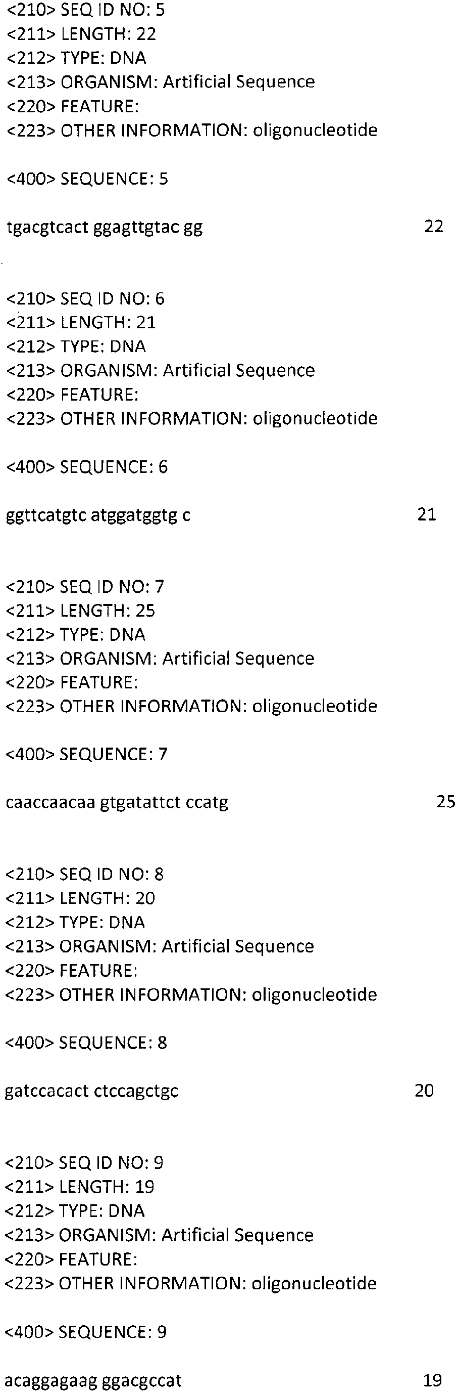

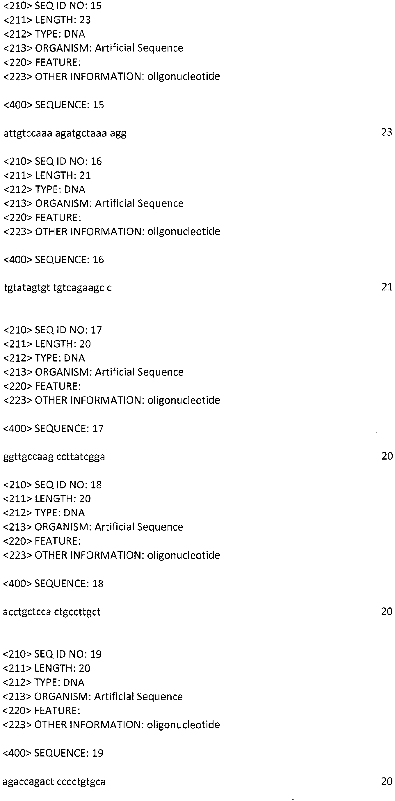
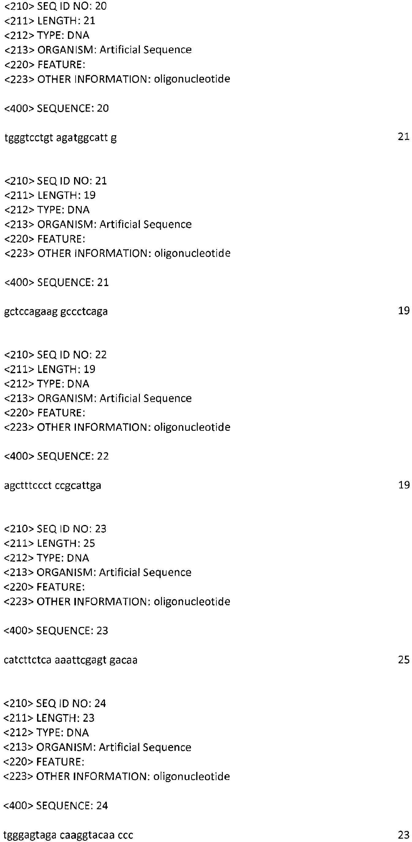
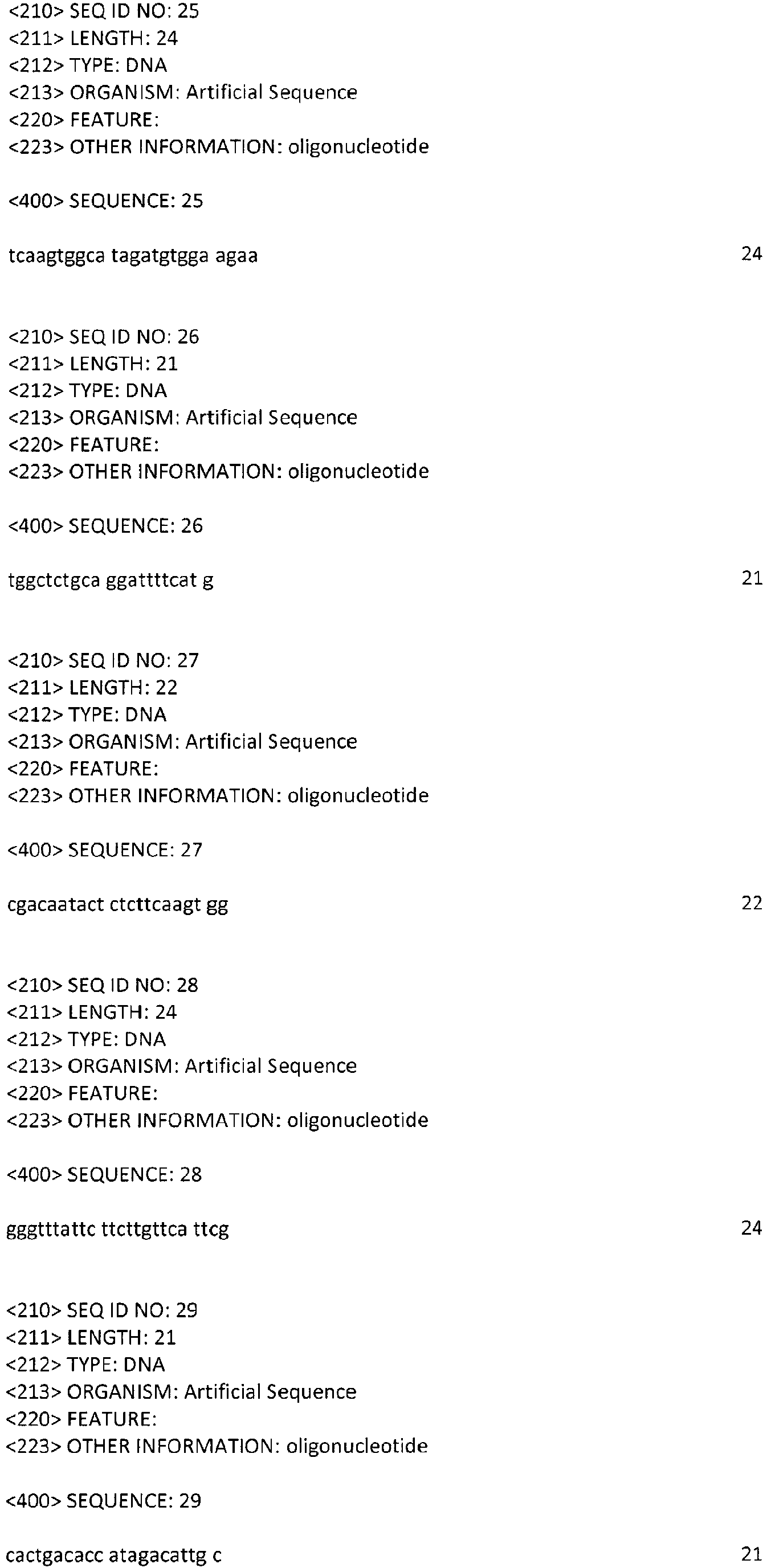

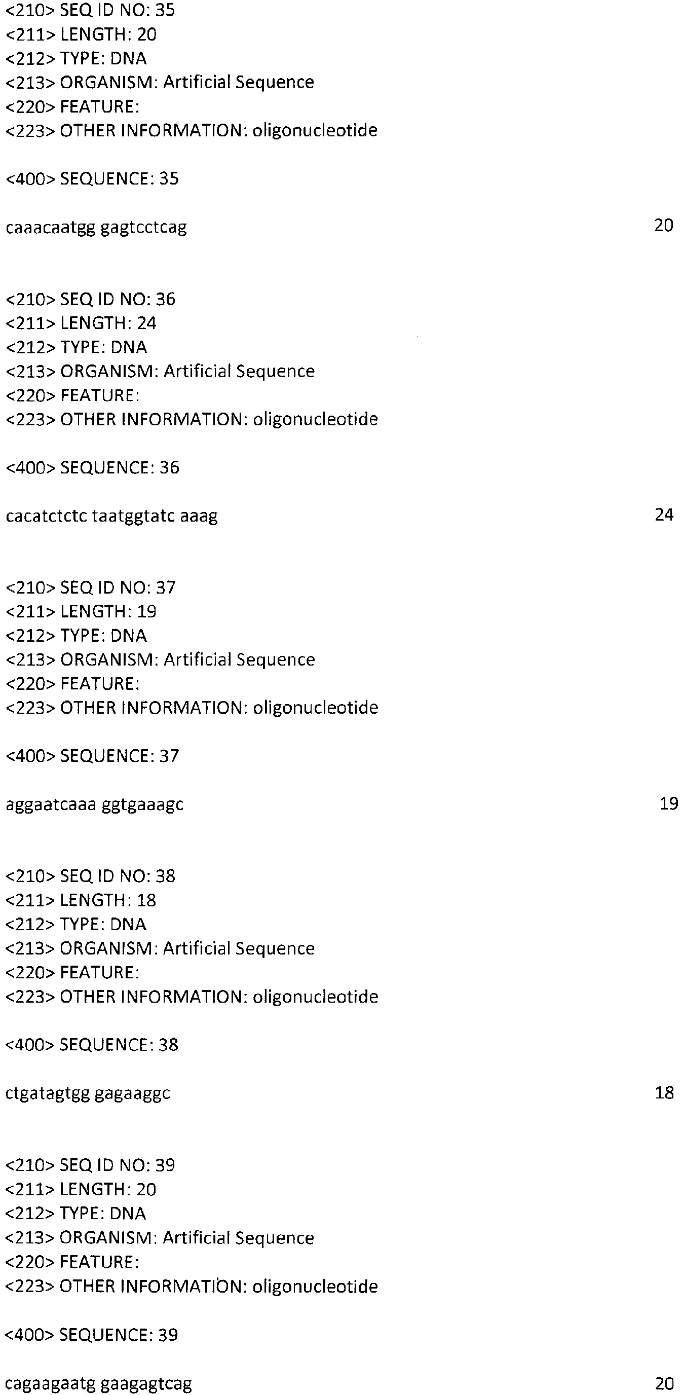
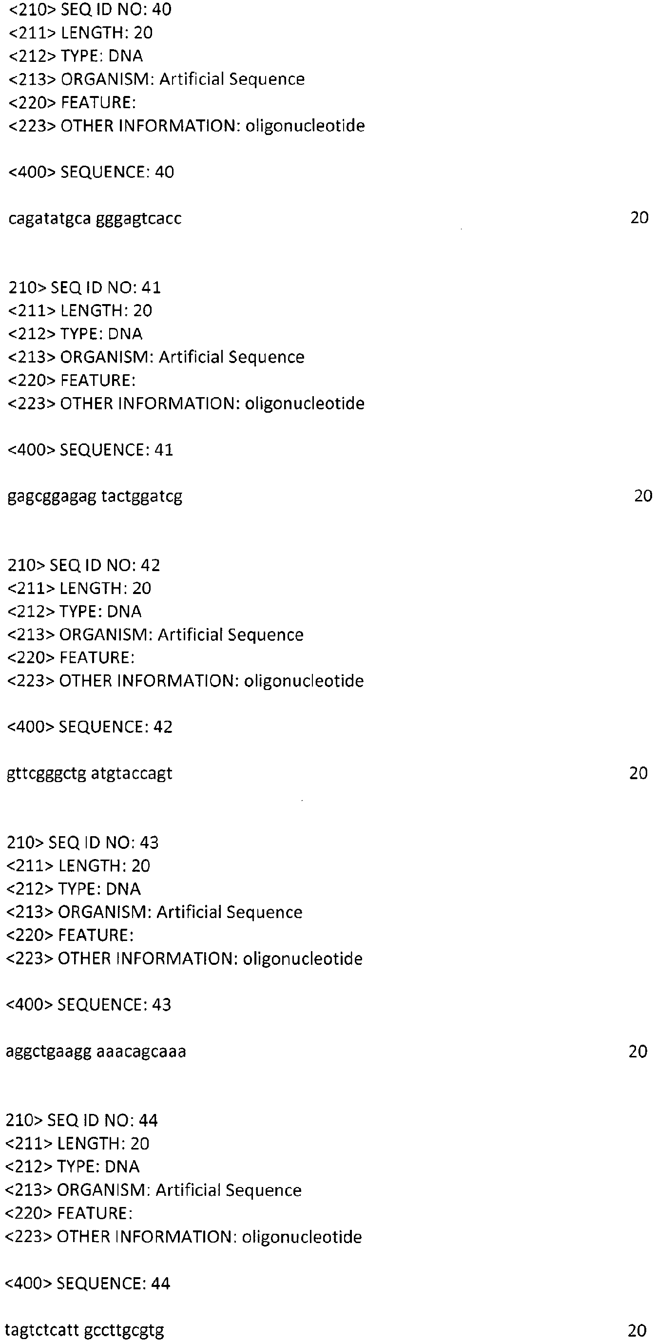


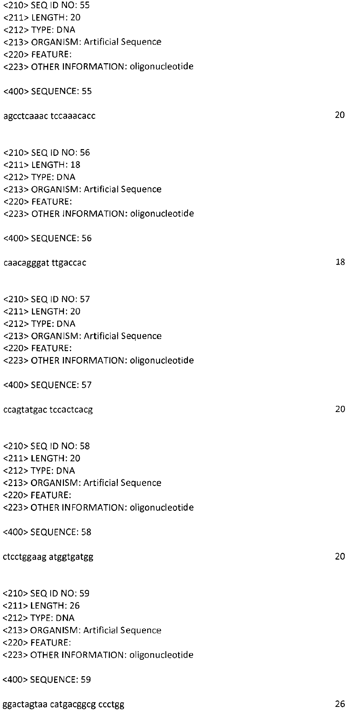
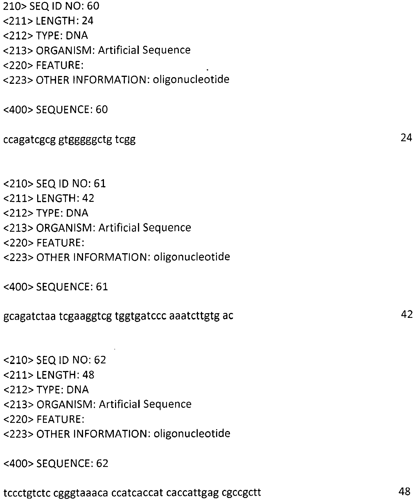
Claims (24)
- Use of an activin neutraliser or a mixture thereof for the manufacture of a medicament for the prophylaxis and / or therapy of diseases associated with aberrant Host Defence Response activation in a subject.
- Use of the activin neutraliser as in claim 1, wherein said medicament is administered via local and / or systemic circulation.
- Use of the activin neutraliser as in claim 1 or 2, wherein said activin is selected from the group consisting of: activin-A, activin-B and activin-AB.
- Use of the activin neutraliser as in claim 1, 2 or 3, wherein said diseases are selected from the group comprising: (a) acute injury of tissues inflicted by infection, toxic substances or trauma, wound healing pursuant to surgery, severe bums or other tissue injury, meningitis, appendicitis, renal tubular necrosis, traumatic brain injury and sepsis; (b) autoimmune diseases including, but not limited to, Rheumatoid arthritis, multiple sclerosis, inflammatory bowel disease, vasculitis, anti-phospholipid Syndrome, scleroderma, Systemic lupus erythematosous and osteoarthritis; and (c) respiratory diseases including, but not limited to organising peumonia, non-specific interstitial pneumonia, sarcoidosis, bronchiolitis obliterans, pulmonary hypertension, pneumonia, ALI/ARDS, COPD, acute responses against infectious agents such as pathogenic viruses including, but not limited to, influenza A viruses H5N1 and H1N1, coronaviruses SARS, and human rhinoviruses C and D.
- Use of the activin neutraliser as in claim 1, 2, 3 or 4, wherein said activin neutraliser is co-administered with at least one additional therapeutic agent.
- Use of the activin neutraliser as in claim 4, wherein said diseases are selected from the group consisting of: ALI/ARDS, sepsis and COPD.
- Use of the activin neutraliser as in claim 1, 2, 3, 4, 5 or 6, wherein said activin neutraliser is a soluble ActRII polypeptide, or a fragment(s) or analogue thereof.
- Use of the activin neutraliser as in claim 7, wherein said soluble ActRII polypeptide, or a fragment(s) or analogue is administered via injection or inhalation routes.
- Use of the activin neutraliser as in claim 7 or 8, wherein said soluble ActRII polypeptide fragment is ActRIIB-Fc.
- Use of the activin neutraliser as in claim 1, 2, 3, 4, or 5, wherein said activin neutraliser is a low molecular weight organic compound, or a pharmaceutically acceptable salt thereof.
- Use of the activin neutraliser as in claim 10, wherein said low molecular weight organic compound, or a pharmaceutically acceptable salt thereof, is administered via oral, injection or inhalation routes.
- Use of the activin neutraliser as in claim 5, wherein said at least one additional therapeutic agent is an anti-inflammatory therapeutic agent.
- Use of the activin neutraliser as in claim 12, wherein said anti-inflammatory therapeutic agent is a corticosteroid.
- Use of the activin neutraliser as in claim 5, wherein said at least one additional therapeutic agent is a neutraliser of pro-inflammatory and pro-fibrotic cytokines.
- Use of the activin neutraliser as in claim 14, wherein said neutraliser of pro-inflammatory and pro-fibrotic cytokines is selected from the group comprising: IL-4, IL-5, IL-6, TNF-α, IL-17 and MCP-1.
- Use of the activin neutraliser as in claim 5, wherein said at least one additional therapeutic agent is a neutraliser of DAMPs.
- Use of the activin neutraliser as in claim 16, wherein said neutraliser of DAMPs is selected from the group comprising: HMGB-1 and TREM-1.
- Use of the activin neutraliser as in claim 5, wherein said at least one additional therapeutic agent is a neutraliser of the coagulation system.
- Use of the activin neutraliser as in claim 18, wherein said neutraliser of the coagulation system is selected from the group comprising: activated protein C (APC), active site inhibited factor VIIa (ASIS), tissue factor pathway inhibitor (TFPI), anti-thrombin (AT), low-molecular weight heparin, vitamin K antagonists and acetylsalicylic acid.
- Use of the activin neutraliser as in claim 5, wherein said at least one additional therapeutic agent is a neutraliser of the complement system.
- Use of the activin neutraliser as in claim 20, wherein said neutraliser of the complement system is selected from the group comprising: endogenous soluble complement inhibitors and agents which neutralise key proteins of the complement cascade reaction.
- Use of the activin neutraliser as in claim 5, wherein said at least one additional therapeutic agent is a surfactant replacement agent.
- Use of the activin neutraliser as in claim 5, wherein said at least one additional therapeutic agent is an agent targeting infectious agents.
- Use of the activin neutraliser as in claim 23, wherein said agent targeting infectious agents is selected from the group comprising: an antibiotic agent, an anti-fungal agent and an anti-viral agent.
Applications Claiming Priority (1)
| Application Number | Priority Date | Filing Date | Title |
|---|---|---|---|
| GR20110100649A GR1007832B (en) | 2011-11-21 | 2011-11-21 | Activin neutralisers and uses thereof for treatment of diseases associated with aberrant "host defence response" activation |
Publications (1)
| Publication Number | Publication Date |
|---|---|
| EP2594280A1 true EP2594280A1 (en) | 2013-05-22 |
Family
ID=47178449
Family Applications (1)
| Application Number | Title | Priority Date | Filing Date |
|---|---|---|---|
| EP12007751.6A Withdrawn EP2594280A1 (en) | 2011-11-21 | 2012-11-16 | Activin neutralisers and uses thereof for treatment of diseases associated with aberrant "host defence response" activation |
Country Status (2)
| Country | Link |
|---|---|
| EP (1) | EP2594280A1 (en) |
| GR (1) | GR1007832B (en) |
Cited By (18)
| Publication number | Priority date | Publication date | Assignee | Title |
|---|---|---|---|---|
| WO2016123454A1 (en) | 2015-01-29 | 2016-08-04 | Board Of Trustees Of Miching State University | Cryptic polypeptides and uses thereof |
| WO2016130814A1 (en) * | 2015-02-11 | 2016-08-18 | The Trustees Of Columbia University In The City Of New York | Inhibitors of induced mmp-1 production |
| WO2017095428A1 (en) * | 2014-12-02 | 2017-06-08 | La Jolla Institute For Allergy And Immunology | Modulators of activin and methods for modulating immune responses and t follicular helper cells |
| US9718881B2 (en) | 2013-07-30 | 2017-08-01 | Regeneron Pharmaceuticals, Inc. | Anti-Activin A antibodies and uses thereof |
| WO2017147182A1 (en) * | 2016-02-22 | 2017-08-31 | Acceleron Pharma Inc. | Actrii antagonists for use in increasing immune activity |
| WO2018013936A1 (en) | 2016-07-15 | 2018-01-18 | Acceleron Pharma Inc. | Compositions and methods for treating pulmonary hypertension |
| WO2018075747A1 (en) * | 2016-10-20 | 2018-04-26 | Alivegen Usa, Inc. | Methods for treating muscle wasting and bone disease using novel hybrid actriib ligand trap proteins |
| US10400036B2 (en) | 2011-11-14 | 2019-09-03 | Regeneron Pharmaceuticals, Inc. | Compositions and methods for increasing muscle mass and muscle strength by specifically antagonizing GDF8 and or activin A |
| WO2020243448A1 (en) * | 2019-05-30 | 2020-12-03 | Acceleron Pharma Inc. | Actrii-binding proteins and uses thereof |
| US10913782B2 (en) | 2015-04-22 | 2021-02-09 | Biogen Ma Inc. | Hybrid ActRIIB ligand trap proteins for treating muscle wasting diseases |
| US10934349B2 (en) | 2015-04-15 | 2021-03-02 | Regeneron Pharmaceuticals, Inc. | Methods for increasing lean body mass with resistance training and a GDF8 inhibitor that is an anti-GDF8 antibody |
| US11248044B2 (en) | 2018-03-01 | 2022-02-15 | Regeneron Pharmaceuticals, Inc. | Methods for altering body composition by administering a GDF8 inhibitor and an Activin A inhibitor |
| US11884715B2 (en) | 2018-01-12 | 2024-01-30 | Keros Therapeutics, Inc. | Activin receptor type IIB variants and methods of use thereof |
| US12350313B2 (en) | 2016-11-10 | 2025-07-08 | Keros Therapeutics, Inc. | Activin receptor type IIa variants and methods of use thereof |
| US12364737B2 (en) | 2018-05-09 | 2025-07-22 | Keros Therapeutics, Inc. | Activin receptor type IIA variants and methods of use thereof |
| US12440539B2 (en) | 2020-03-20 | 2025-10-14 | Keros Therapeutics, Inc. | Methods of using activin receptor type IIB variants |
| US12522646B2 (en) | 2020-03-20 | 2026-01-13 | Keros Therapeutics, Inc. | Activin receptor type II chimeras and methods of use thereof |
| US12583906B2 (en) | 2020-04-28 | 2026-03-24 | Acceleron Pharma Inc. | Actrii proteins and use in treating post-capillary pulmonary hypertension |
Citations (15)
| Publication number | Priority date | Publication date | Assignee | Title |
|---|---|---|---|---|
| US3773919A (en) | 1969-10-23 | 1973-11-20 | Du Pont | Polylactide-drug mixtures |
| EP0036676B1 (en) | 1978-03-24 | 1984-07-11 | The Regents Of The University Of California | Method of making uniformly sized liposomes and liposomes so made |
| EP0133988A2 (en) | 1983-08-02 | 1985-03-13 | Hoechst Aktiengesellschaft | Regulating peptide-containing pharmaceutical preparations with retarded release, and process for their preparation |
| EP0058481B1 (en) | 1981-02-16 | 1986-10-01 | Zeneca Limited | Continuous release pharmaceutical compositions |
| EP0088046B1 (en) | 1982-02-17 | 1987-12-09 | Ciba-Geigy Ag | Lipids in the aqueous phase |
| EP0143949B1 (en) | 1983-11-01 | 1988-10-12 | TERUMO KABUSHIKI KAISHA trading as TERUMO CORPORATION | Pharmaceutical composition containing urokinase |
| WO1993015722A1 (en) | 1992-02-07 | 1993-08-19 | Syntex (Usa) Inc. | Controlled delivery of pharmaceuticals from preformed porous microparticles |
| WO1994020069A1 (en) | 1993-03-08 | 1994-09-15 | Amgen Inc. | Pulmonary administration of granulocyte colony stimulating factor |
| WO1999010364A1 (en) * | 1997-08-29 | 1999-03-04 | Human Genome Sciences, Inc. | Follistatin-3 |
| WO2002066462A1 (en) | 2001-02-02 | 2002-08-29 | Glaxo Group Limited | Pyrazole derivatives against tgf overexpression |
| WO2003006057A1 (en) * | 2001-07-13 | 2003-01-23 | Bioa Pty Limited | Composition and method for the treatment of disease |
| WO2005032578A1 (en) * | 2003-10-06 | 2005-04-14 | Monash University | Therapeutic method |
| WO2006012627A2 (en) * | 2004-07-23 | 2006-02-02 | Acceleron Pharma Inc. | Actrii receptor polypeptides, methods and compositions |
| WO2006083182A1 (en) * | 2005-02-07 | 2006-08-10 | Orico Limited | Use of myostatin (gdf-8) antagonists for improving wound healing and preventing fibrotic disease |
| WO2009158015A2 (en) * | 2008-06-26 | 2009-12-30 | Acceleron Pharma Inc. | Antagonists of actriib and uses for increasing red blood cell levels |
-
2011
- 2011-11-21 GR GR20110100649A patent/GR1007832B/en active IP Right Grant
-
2012
- 2012-11-16 EP EP12007751.6A patent/EP2594280A1/en not_active Withdrawn
Patent Citations (15)
| Publication number | Priority date | Publication date | Assignee | Title |
|---|---|---|---|---|
| US3773919A (en) | 1969-10-23 | 1973-11-20 | Du Pont | Polylactide-drug mixtures |
| EP0036676B1 (en) | 1978-03-24 | 1984-07-11 | The Regents Of The University Of California | Method of making uniformly sized liposomes and liposomes so made |
| EP0058481B1 (en) | 1981-02-16 | 1986-10-01 | Zeneca Limited | Continuous release pharmaceutical compositions |
| EP0088046B1 (en) | 1982-02-17 | 1987-12-09 | Ciba-Geigy Ag | Lipids in the aqueous phase |
| EP0133988A2 (en) | 1983-08-02 | 1985-03-13 | Hoechst Aktiengesellschaft | Regulating peptide-containing pharmaceutical preparations with retarded release, and process for their preparation |
| EP0143949B1 (en) | 1983-11-01 | 1988-10-12 | TERUMO KABUSHIKI KAISHA trading as TERUMO CORPORATION | Pharmaceutical composition containing urokinase |
| WO1993015722A1 (en) | 1992-02-07 | 1993-08-19 | Syntex (Usa) Inc. | Controlled delivery of pharmaceuticals from preformed porous microparticles |
| WO1994020069A1 (en) | 1993-03-08 | 1994-09-15 | Amgen Inc. | Pulmonary administration of granulocyte colony stimulating factor |
| WO1999010364A1 (en) * | 1997-08-29 | 1999-03-04 | Human Genome Sciences, Inc. | Follistatin-3 |
| WO2002066462A1 (en) | 2001-02-02 | 2002-08-29 | Glaxo Group Limited | Pyrazole derivatives against tgf overexpression |
| WO2003006057A1 (en) * | 2001-07-13 | 2003-01-23 | Bioa Pty Limited | Composition and method for the treatment of disease |
| WO2005032578A1 (en) * | 2003-10-06 | 2005-04-14 | Monash University | Therapeutic method |
| WO2006012627A2 (en) * | 2004-07-23 | 2006-02-02 | Acceleron Pharma Inc. | Actrii receptor polypeptides, methods and compositions |
| WO2006083182A1 (en) * | 2005-02-07 | 2006-08-10 | Orico Limited | Use of myostatin (gdf-8) antagonists for improving wound healing and preventing fibrotic disease |
| WO2009158015A2 (en) * | 2008-06-26 | 2009-12-30 | Acceleron Pharma Inc. | Antagonists of actriib and uses for increasing red blood cell levels |
Non-Patent Citations (230)
| Title |
|---|
| "Antibodies-A Laboratory Manual", 1988, COLD SPRING HARBOR LABORATORY |
| "Design of Prodrugs", 1985, ELSEVIER |
| "Remington's Pharmaceutical Sciences", 1990, MACK PUBLISHING COMPANY |
| "The Acute Respiratory Distress Syndrome Network", N ENGL J MED, vol. 342, pages 1301 - 1308 |
| ABRAHAM, E.: "Coagulation abnormalities in acute lung injury and sepsis", AM J RESPIR CELL MOL BIOL, vol. 22, no. 4, 2000, pages 401 - 404 |
| AGRESTI, A.; M. E. BIANCHI: "HMGB proteins and gene expression", CURR OPIN GENET DEV, vol. 13, no. 2, 2003, pages 170 - 178, XP003019721, DOI: doi:10.1016/S0959-437X(03)00023-6 |
| ANDREAKOS, E.; U. RAUCHHAUS ET AL.: "Amphoteric liposomes enable systemic antigen-presenting cell-directed delivery of CD40 antisense and are therapeutically effective in experimental arthritis", ARTHRITIS RHEUM, vol. 60, no. 4, 2009, pages 994 - 1005, XP002592978, DOI: doi:10.1002/ART.24434 |
| ANTOINE, D. J.; D. P. WILLIAMS ET AL.: "Diet restriction inhibits apoptosis and HMGB1 I oxidation and promotes inflammatory cell recruitment during acetaminophen hepatotoxicity", MOL MED, vol. 16, no. 11-12, 2010, pages 479 - 490 |
| ANZUETO, A.; R. P. BAUGLUNAN ET AL.: "Aerosolized surfactant in adults with sepsis- induced acute respiratory distress syndrome. Exosurf Acute Respiratory Distress Syndrome Sepsis Study Group", N ENGL J MED, vol. 334, no. 22, 1996, pages 1417 - 1421 |
| AOKI, F.; M. KURABAYASHI ET AL.: "Attenuation of bleomycin-induced pulmonary fibrosis by follistatin", AM J RESPIR CRIT CARE MED, vol. 172, no. 6, 2005, pages 713 - 720, XP008110899, DOI: doi:10.1164/rccm.200412-1620OC |
| ASHITANI, J.; H. MUKAE ET AL.: "Elevated plasma procoagulant and fibrinolytic markers in patients with chronic obstructive pulmonary disease", INTERN MED, vol. 41, no. 3, 2002, pages 181 - 185 |
| BADER, A. G.; D. BROWN ET AL.: "The promise of microRNA replacement therapy", CANCER RES, vol. 70, no. 18, 2010, pages 7027 - 7030 |
| BARANY ET AL., INT. J. PEP. PROTEINRES., vol. 30, 1987, pages 705 - 739 |
| BARANY; MERRIFIELD: "The Peptides", ACADEMIC PRESS, pages: 1 - 284 |
| BARNES, P. J.: "Glucocorticosteroids: current and future directions", BR J PHARMACOL, vol. 163, no. 1, 2011, pages 29 - 43 |
| BARNES, P. J.: "Immunology of asthma and chronic obstructive pulmonary disease", NAT REV IMMUNOL, vol. 8, no. 3, 2008, pages 183 - 192 |
| BARTEL, D. P.: "MicroRNAs: genomics, biogenesis, mechanism, and function", CELL, vol. 116, no. 2, 2004, pages 281 - 297, XP002359089, DOI: doi:10.1016/S0092-8674(04)00045-5 |
| BASTARACHE, J. A.; L. WANG ET AL.: "The alveolar epithelium can initiate the extrinsic coagulation cascade through expression of tissue factor", THORAX, vol. 62, no. 7, 2007, pages 608 - 616 |
| BATES, J. H.; T. F. SCHUESSLER ET AL.: "Temporal dynamics of acute isovolume bronchoconstriction in the rat", J APPL PHYSIOL, vol. 82, no. 1, 1997, pages 55 - 62 |
| BERNARD, D. J.; K. B. LEE ET AL.: "Activin B can signal through both ALK4 and ALK7 in gonadotrope cells", REPROD BIOL ENDOCRINOL, vol. 4, 2006, pages 52, XP021024750, DOI: doi:10.1186/1477-7827-4-52 |
| BERNARD, G. R.; A. ARTIGAS ET AL.: "The American-European Consensus Conference on ARDS. Definitions, mechanisms, relevant outcomes, and clinical trial coordination", AM J RESPIR CRIT CARE MED, vol. 149, 1994, pages 818 - 824 |
| BERNARD, G. R.; J. L. VINCENT ET AL.: "Efficacy and safety of recombinant human activated protein C for severe sepsis", N ENGL J MED, vol. 344, no. 10, 2001, pages 699 - 709, XP009019299, DOI: doi:10.1056/NEJM200103083441001 |
| BERNARD, G.: "The International Sepsis Forum's controversies in sepsis: corticosteroids should not be routinely used to treat septic shock", CRIT CARE, vol. 6, no. 5, 2002, pages 384 - 386 |
| BERTOLINO, P.; R. HOLMBERG ET AL.: "Activin B receptor ALK7 is a negative regulator of pancreatic beta-cell function", PROC NATL ACAD SCI USA, vol. 105, no. 20, 2008, pages 7246 - 7251 |
| BEUTLER, B.: "Neo-ligands for innate immune receptors and the etiology of sterile inflammatory disease", IMMUNOL REV, vol. 220, 2007, pages 113 - 128 |
| BEVILACQUA, M. P.; J. S. POBER ET AL.: "Recombinant tumor necrosis factor induces procoagulant activity in cultured human vascular endothelium: characterization and comparison with the actions of interleukin 1", PROC NATL ACAD SCI USA, vol. 83, no. 12, 1986, pages 4533 - 4537 |
| BIANCHI, M. E.: "DAMPs, PAMPs and alarniins: all we need to know about danger", J LEUKOC BIOL, vol. 81, no. 1, 2007, pages 1 - 5 |
| BIANCHI, M. E.: "HMGB1 loves company", J LEUKOC BIOL, vol. 86, no. 3, 2009, pages 573 - 576 |
| BIANCHI, M. E.; A. A. MANFREDI: "High-mobility group box 1 (HMGB1) protein at the crossroads between innate and adaptive immunity", IMMUNOL REV, vol. 220, 2007, pages 35 - 46 |
| BIJSTERBOSCH, M. K.; M. MANOHARAN ET AL.: "Delivery of cholesteryl-conjugated phosphorothioate oligodeoxynucleotides to Kupffer cells by lactosylated low-density lipoprotein", BIOCHEM PHARMACOL, vol. 62, no. 5, 2001, pages 627 - 633 |
| BILEZIKJIAN, L. M.; A. L. BLOUNT ET AL.: "Actions of activins, inhibins and follistatins: implications in anterior pituitary function", CLIN EXP PHARMACOL PHYSIOL, vol. 28, no. 3, 2001, pages 244 - 248 |
| BOMIIAUD, P.; G. MARTIN ET AL.: "Connective tissue growth factor is crucial to inducing a profibrotic environment in ''fibrosis-resistant'' BALB/c mouse lungs.", AM J RESPIR CELL MOL BIOL, vol. 31, no. 5, 2004, pages 510 - 516 |
| BONALDI, T.; F. TALAMO ET AL.: "Monocytic cells hyperacetylate chromatin protein HMGB1 to redirect it towards secretion", EMBO J, vol. 22, no. 20, 2003, pages 5551 - 5560, XP002279284, DOI: doi:10.1093/emboj/cdg516 |
| BONE, R. C.: "Immunologic dissonance: a continuing evolution in our understanding of the systemic inflammatory response syndrome (SIRS) and the multiple organ dysfunction syndrome (MODS", ANN INTERN MED, vol. 125, no. 8, 1996, pages 680 - 687 |
| BONE, R. C.: "Sir Isaac Newton, sepsis, SIRS, and CARS", CRIT CARE MED, vol. 24, no. 7, 1996, pages 1125 - 1128, XP009154554, DOI: doi:10.1097/00003246-199607000-00010 |
| BONE, R. C.; C. J. FISHER, JR. ET AL.: "A controlled clinical trial of high-dose methylprednisolone in the treatment of severe sepsis and septic shock", N ENGL J MED, vol. 317, no. 11, 1987, pages 653 - 658 |
| BORENSZTAJN, K. S.; J. H. VON DER THUSEN ET AL.: "The role of coagulation in chronic inflammatory disorders: a jack of all trades", CURR PHARM DES, vol. 17, no. 1, 2011, pages 9 - 16 |
| BOULIKAS, T.: "Liposome DNA delivery and uptake by cells (review", ONCOL REP, vol. 3, no. 6, 1996, pages 989 - 995 |
| BOURS, M. J.; E. L. SWENNEN ET AL.: "Adenosine 5'-triphosphate and adenosine as endogenous signaling molecules in immunity and inflammation", PHARMACOL THER, vol. 112, no. 2, 2006, pages 358 - 404, XP025038578, DOI: doi:10.1016/j.pharmthera.2005.04.013 |
| BROSH, N.; D. STERNBERG ET AL.: "The plasmacytoma growth inhibitor restrictin-P is an antagonist of interleukin 6 and interleukin 11. Identification as a stroma-derived activin A", J BIOL CHEM, vol. 270, no. 49, 1995, pages 29594 - 29600 |
| BROZE, G. J., JR.; Z. F. YIN ET AL.: "A tail vein bleeding time model and delayed bleeding in hemophiliac mice", THROMB HAEMOST, vol. 85, no. 4, 2001, pages 747 - 748, XP008110200 |
| BYFIELD: "SB-505124 Is a selective inhibitor of Transforming Growth Factor-p type I receptors ALK4, ALK5 and ALK7", MOL. PHARMACOL., vol. 65, no. 3, 2004, pages 744 - 752, XP055096104, DOI: doi:10.1124/mol.65.3.744Cit |
| CALLAHAN, J. F.; J. L. BURGESS ET AL.: "Identification of novel inhibitors of the transforming growth factor betal (TGF-betal) type 1 receptor (ALK5", J MED CHEM, vol. 45, no. 5, 2002, pages 999 - 1001 |
| CANIGGIA, I.; S. J. LYE ET AL.: "Activin is a local regulator of human cytotrophoblast cell differentiation", ENDOCRINOLOGY, vol. 138, no. 9, 1997, pages 3976 - 3986 |
| CASEY, B. P.; P. M. GLAZER: "Gene targeting via triple-helix formation", PROG NUCLEIC ACID RES MOL BIOL, vol. 67, 2001, pages 163 - 192 |
| CECH, J. AMER. MED. ASSN., vol. 260, 1988, pages 3030 |
| CEPKOVA, M.; M. A. MATTHAY: "Pharmacotherapy of acute lung injury and the acute respiratory distress syndrome", J INTENSIVE CARE MED, vol. 21, no. 3, 2006, pages 119 - 143 |
| CHEN, G. Y.; G. NUNEZ: "Sterile inflammation: sensing and reacting to damage", NAT REV IMMUNOL, vol. 10, no. 12, 2010, pages 826 - 837 |
| CHENG, T.; D. LIU ET AL.: "Activated protein C blocks p53-mediated apoptosis in ischemic human brain endothelium and is neuroprotective", NAT MED, vol. 9, no. 3, 2003, pages 338 - 342, XP002998642, DOI: doi:10.1038/nm826 |
| CHO, S. H.; Z. YAO ET AL.: "Regulation of activin A expression in mast cells and asthma: its effect on the proliferation of human airway smooth muscle cells", J IMMUNOL, vol. 170, no. 8, 2003, pages 4045 - 4052, XP008110706 |
| COLE ET AL.: "Monoclonal Antibodies and Cancer Therapy", 1985, ALAN R. LISS, INC., pages: 77 - 96 |
| COLLINS, R. A.; R. C. GUALANO ET AL.: "Hyperresponsiveness to inhaled but not intravenous methacholine during acute respiratory syncytial virus infection in mice", RESPIR RES, vol. 6, 2005, pages 142, XP021000089, DOI: doi:10.1186/1465-9921-6-142 |
| COSTA, E. L.; I. A. SCHETTINO ET AL.: "The lung in sepsis: guilty or innocent?", ENDOCR METAB IMMUNE DISORD DRUG TARGETS, vol. 6, no. 2, 2006, pages 213 - 216 |
| DE GOUVILLE AC; BOULLAY V; KRYSA G; PILOT J; BNISQ JM; LORIOLLE F; GAUTHIER JM; PAPWORTH SA; LAROZE A; GELLIBERT F: "Inhibition of TGF-beta signaling by an ALK5 inhibitor protects rats from dimethylnitrosamine-induced liver fibrosis", BR J PHANNACOL., vol. 145, 2005, pages 166 - 77, XP055096102, DOI: doi:10.1038/sj.bjp.0706172 |
| DEANS, K. J.; M. HALEY ET AL.: "Novel therapies for sepsis: a review", J TRAUMA, vol. 58, no. 4, 2005, pages 867 - 874 |
| DOHI, T.; C. EJIMA ET AL.: "Therapeutic potential of follistatin for colonic inflammation in mice", GASTROENTEROLOGY, vol. 128, no. 2, 2005, pages 411 - 423, XP005313393, DOI: doi:10.1053/j.gastro.2004.11.063 |
| ENHORNING, G.; L. C. DUFFY ET AL.: "Pulmonary surfactant maintains patency of conducting airways in the rat", AM J RESPIR CRIT CARE MED, vol. 151, 1995, pages 554 - 556 |
| EPPSTEIN, D. A.; Y. V. MARSH ET AL.: "Biological activity of liposome-encapsulated murine interferon gamma is mediated by a cell membrane receptor", PROC NATL ACAD SCI US A, vol. 82, no. 11, 1985, pages 3688 - 3692 |
| ESMON, C. T.: "The impact of the inflammatory response on coagulation", THROMB RES, vol. 114, no. 5-6, 2004, pages 321 - 327, XP004613171, DOI: doi:10.1016/j.thromres.2004.06.028 |
| FERHANI, N.; S. LETUVE ET AL.: "Expression of high-mobility group box 1 and of receptor for advanced glycation end products in chronic obstructive pulmonary disease", AM J RESPIR CRIT CARE MED, vol. 181, no. 9, 2010, pages 917 - 927 |
| FISHER, C. J., JR.; J. M. AGOSTI ET AL.: "Treatment of septic shock with the tumor necrosis factor receptor:Fc fusion protein. The Soluble TNF Receptor Sepsis Study Group", N ENGL J MED, vol. 334, no. 26, 1996, pages 1697 - 1702 |
| FUJITA, M.; J. M. SHANNON ET AL.: "Overexpression of tumor necrosis factor-alpha diminishes pulmonary fibrosis induced by bleomycin or transfonning growth factor-beta", AM J RESPIR CELL MOL BIOL, vol. 29, no. 6, 2003, pages 669 - 676 |
| GARDELLA, S.; C. ANDREI ET AL.: "The nuclear protein HMGBI is secreted by monocytes via a non-classical, vesicle-mediated secretory pathway", EMBO REP, vol. 3, no. 10, 2002, pages 995 - 1001, XP002279283, DOI: doi:10.1093/embo-reports/kvf198 |
| GERARD, C.: "Complement C5a in the sepsis syndrome--too much of a good thing?", N ENGL J MED, vol. 348, no. 2, 2003, pages 167 - 169 |
| GLASSER, S. W.; E. A. DETMER ET AL.: "Pneumonitis and emphysema in sp-C gene targeted mice", J BIOL CHEM, vol. 278, no. 16, 2003, pages 14291 - 14298, XP002394847, DOI: doi:10.1074/jbc.M210909200 |
| GORDON, S.: "Pattern recognition receptors: doubling up for the innate immune response", CELL, vol. 111, no. 7, 2002, pages 927 - 930 |
| GREENE, K. E.; J. R. WRIGHT ET AL.: "Serial changes in surfactant-associated proteins in lung and serum before and after onset of ARDS", AM J RESPIR CRIT CARE MED, vol. 160, no. 6, 1999, pages 1843 - 1850 |
| GREGORY, T. J.; K. P. STEINBERG ET AL.: "Bovine surfactant therapy for patients with acute respiratory distress syndrome", AM J RESPIR CRIT CARE MED, vol. 155, no. 4, 1997, pages 1309 - 1315 |
| GRIBI, R.; T. TANAKA ET AL.: "Expression of activin A in inflammatory arthropathies", MOL CELL ENDOCRINOL, vol. 180, no. 1-2, 2001, pages 163 - 167, XP003000873, DOI: doi:10.1016/S0303-7207(01)00517-2 |
| GRYGIELKO, E. T.; W. M. MARTIN ET AL.: "Inhibition of gene markers of fibrosis with a novel inhibitor of transforming growth factor-beta type I receptor kinase in puromycin- induced nephritis", J PHARMACOL EXP THER, vol. 313, no. 3, 2005, pages 943 - 951 |
| GUNTHER, A.; P. MOSAVI ET AL.: "Alveolar fibrin fonnation caused by enhanced procoagulant and depressed fibrinolytic capacities in severe pneumonia. Comparison with the acute respiratory distress syndrome", AM J RESPIR CRIT CARE MED, vol. 161, 2000, pages 454 - 462, XP002973713 |
| HALE, K. A.; S. L. EWING ET AL.: "Lung disease in long-term cigarette smokers with and without chronic air-flow obstruction", AM REV RESPIR DIS, vol. 130, no. 5, 1984, pages 716 - 721 |
| HALPIN, D. M.; M. MIRAVITLLES: "Chronic obstructive pulmonary disease: the disease and its burden to society", PROC AM THORAC SOC, vol. 3, no. 7, 2006, pages 619 - 623 |
| HAMMERLING ET AL., MONOCLONAL ANTIBODIES AND T-CELL HYBRIDOMAS, 1981 |
| HAN, M. K.: "Update in chronic obstructive pulmonary disease in 2010", AM J RESPIR CRIT CARE MED, vol. 183, no. 10, 2011, pages 1311 - 1315 |
| HARDY, C. L.; A. E. O'CONNOR ET AL.: "Follistatin is a candidate endogenous negative regulator of activin A in experimental allergic asthma", CLIN EXP ALLERGY, vol. 36, no. 7, 2006, pages 941 - 950, XP008110894, DOI: doi:10.1111/j.1365-2222.2006.02523.x |
| HARRISON C A ET AL: "Antagonists of activin signaling: mechanisms and potential biological applications", TRENDS IN ENDOCRINOLOGY AND METABOLISM, ELSEVIER SCIENCE PUBLISHING, NEW YORK, NY, US, vol. 16, no. 2, 1 March 2005 (2005-03-01), pages 73 - 78, XP027790070, ISSN: 1043-2760, [retrieved on 20050301] * |
| HARRISON, C. A.; P. C. GRAY ET AL.: "Antagonists of activin signaling: mechanisms and potential biological applications", TRENDS ENDOCRINOL METAB, vol. 16, no. 2, 2005, pages 73 - 78, XP025302666, DOI: doi:10.1016/j.tem.2005.01.003 |
| HASSELHOFF, NATURE, vol. 334, 1988, pages 585 |
| HAWLISCH, H.; Y. BELKAID ET AL.: "C5a negatively regulates toll-like receptor 4- induced immune responses", IMMUNITY, vol. 22, no. 4, 2005, pages 415 - 426, XP055070165, DOI: doi:10.1016/j.immuni.2005.02.006 |
| HEDGER, M. P.; A. E. DRUMMOND ET AL.: "Inhibin and activin regulate [3H]thymidine uptake by rat thymocytes and 3T3 cells in vitro", MOL CELL ENDOCRINOL, vol. 61, no. 1, 1989, pages 133 - 138, XP025708533, DOI: doi:10.1016/0303-7207(89)90198-6 |
| HELENE, C.: "The anti-gene strategy: control of gene expression by triplex-forming- oligonucleotides", ANTICANCER DRUG DES, vol. 6, no. 6, 1991, pages 569 - 584 |
| HEMMI, H.; O. TAKEUCHI ET AL.: "A Toll-like receptor recognizes bacterial DNA", NATURE, vol. 408, no. 6813, 2000, pages 740 - 745 |
| HERRIDGE, M. S.; D. C. ANGUS: "Acute lung injury--affecting many lives", N ENGL J MED, vol. 353, no. 16, 2005, pages 1736 - 1738 |
| HOBBS, S.; S. JITRAPAKDEE ET AL.: "Development of a bicistronic vector driven by the human polypeptide chain elongation factor lalpha promoter for creation of stable mammalian cell lines that express very high levels of recombinant proteins", BIOCHEM BIOPHYS RES COMMUN, vol. 252, no. 2, 1998, pages 368 - 372, XP002144790, DOI: doi:10.1006/bbrc.1998.9646 |
| HUBER, S.; F. R. STAHL ET AL.: "Activin a promotes the TGF-beta-induced conversion of CD4+CD25- T cells into Foxp3+ induced regulatory T cells", J IMMUNOL, vol. 182, no. 8, 2009, pages 4633 - 4640 |
| HUBNER, G.; C. ALZHEIMER ET AL.: "Activin: a novel player in tissue repair processes", HISTOL HISTOPATHOL, vol. 14, no. 1, 1999, pages 295 - 304 |
| INMAN, G. J.; F. J. NICOLAS ET AL.: "SB-431542 is a potent and specific inhibitor of transforming growth factor-beta superfamily type I activin receptor-like kinase (ALK) receptors ALK4, ALK5, and ALK7", MOL PHARMACOL, vol. 62, no. 1, 2002, pages 65 - 74, XP002985367, DOI: doi:10.1124/mol.62.1.65 |
| INOUYE, S.; N. LING ET AL.: "Localization of the heparin binding site of follistatin", MOL CELL ENDOCRINOL, vol. 90, no. 1, 1992, pages 1 - 6, XP025832756, DOI: doi:10.1016/0303-7207(92)90094-M |
| INOUYE, S.; Y. GUO ET AL.: "Recombinant expression of human follistatin with 315 and 288 amino acids: chemical and biological comparison with native porcine follistatin", ENDOCRINOLOGY, vol. 129, no. 2, 1991, pages 815 - 822 |
| IWASAKI, A.; R. MEDZHITOV: "Regulation of adaptive immunity by the innate immune system", SCIENCE, vol. 327, no. 5963, 2010, pages 291 - 295 |
| JOBE, A. H.: "Pulmonary surfactant therapy", N ENGL J MED, vol. 328, no. 12, 1993, pages 861 - 868 |
| JONES, K. L.; A. MANSELL ET AL.: "Activin A is a critical component of the inflammatory response, and its binding protein, follistatin, reduces mortality in endotoxemia", PROC NATL ACAD SCI USA, vol. 104, no. 41, 2007, pages 16239 - 16244 |
| KALAI, M.; G. VAN LOO ET AL.: "Tipping the balance between necrosis and apoptosis in human and murine cells treated with interferon and dsRNA", CELL DEATH DIFFER, vol. 9, no. 9, 2002, pages 981 - 994 |
| KAMBAS, K.; M. M. MARKIEWSKI ET AL.: "C5a and TNF-alpha up-regulate the expression of tissue factor in intra-alveolar neutrophils of patients with the acute respiratory distress syndrome", J IMMUNOL, vol. 180, no. 11, 2008, pages 7368 - 7375 |
| KANAZAWA, H.; Y. TOCHINO ET AL.: "Validity of HMGB1 measurement in epithelial lining fluid in patients with COPD", EUR J CLIN INVEST., 2011 |
| KARAGIANNIDIS, C.; G. HENSE ET AL.: "Activin A is an acute allergen-responsive cytokine and provides a link to TGF-beta-mediated airway remodeling in asthma", J ALLERGY CLIN IMMUNOI, vol. 117, no. 1, 2006, pages 111 - 118, XP005228133, DOI: doi:10.1016/j.jaci.2005.09.017 |
| KEIMETT ET AL., MONOCLONAL ANTIBODIES, 1980 |
| KEUTMANN, H. T.; A. L. SCHNEYER ET AL.: "The role of follistatin domains in follistatin biological action", MOL ENDOCRINOL, vol. 18, no. 1, 2004, pages 228 - 240 |
| KHADAROO, R. G.; J. C. MARSHALL: "ARDS and the multiple organ dysfunction syndrome. Common mechanisms of a common systemic process", CRIT CARE CLIN, vol. 18, no. 1, 2002, pages 127 - 141 |
| KILDSGAARD, J.; T. J. HOLLMANN ET AL.: "Cutting edge: targeted disruption of the C3a receptor gene demonstrates a novel protective anti-inflammatory role for C3a in endotoxin-shock", J IMMUNOL, vol. 165, no. 10, 2000, pages 5406 - 5409, XP002204353 |
| KOHLER, G.; C. MILSTEIN: "Continuous cultures of fused cells secreting antibody of predefined specificity", NATURE, vol. 256, no. 5517, 1975, pages 495 - 497, XP002024548 |
| KOJIMA, 1.; H. MOGAMI ET AL.: "Modulation of growth of vascular smooth muscle cells by activin A", EXP CELL RES, vol. 206, no. 1, 1993, pages 152 - 156, XP024788974, DOI: doi:10.1006/excr.1993.1131 |
| KOKKOLA, R.; A. ANDERSSON ET AL.: "RAGE is the major receptor for the proinflammatory activity of HMGB1 in rodent macrophages", SCAND J IMMUNOL, vol. 61, no. 1, 2005, pages 1 - 9 |
| KOKKOLA, R.; J. LI ET AL.: "Successful treatment of collagen-induced arthritis in mice and rats by targeting extracellular high mobility group box chromosomal protein 1 activity", ARTHRITIS RHEUM, vol. 48, no. 7, 2003, pages 2052 - 2058, XP001205688, DOI: doi:10.1002/art.11161 |
| KOLB, M.; P. J. MARGETTS ET AL.: "Transient expression ofIL-lbeta induces acute lung injury and chronic repair leading to pulmonary fibrosis", J CLIN INVEST, vol. 107, no. 12, 2001, pages 1529 - 1536 |
| KONO, H.; C. J. CHEN ET AL.: "Uric acid promotes an acute inflammatory response to sterile cell death in mice", J CLIN INVEST, vol. 120, no. 6, 2010, pages 1939 - 1949 |
| KONO, H.; K. L. ROCK: "How dying cells alert the immune system to danger", NAT REV IMMUNOL, vol. 8, no. 4, 2008, pages 279 - 289 |
| KOX, W. J.; T. VOLK ET AL.: "Immunomodulatory therapies in sepsis", INTENSIVE CARE MED, vol. 26, no. 1, 2000, pages I24 - 128 |
| KRIEG, A. M.: "CpG motifs in bacterial DNA and their immune effects", ANNU REV IMMUNOL, vol. 20, 2002, pages 709 - 760, XP009016455, DOI: doi:10.1146/annurev.immunol.20.100301.064842 |
| KROEMER, G.; L. GALLUZZI ET AL.: "Classification of cell death: recommendations of the Nomenclature Committee on Cell Death 2009", CELL DEATH DIFFER, vol. 16, no. 1, 2009, pages 3 - 11 |
| KRUMMEN, L. A.; T. K. WOODRUFF ET AL.: "Identification and characterization of binding proteins for inhibin and activin in human serum and follicular fluids", ENDOCRINOLOGY, vol. 132, no. 1, 1993, pages 431 - 443 |
| LABIRIS, N. R.; M. B. DOLOVICH: "Pulmonary drug delivery. Part II: the role of inhalant delivery devices and drug formulations in therapeutic effectiveness of aerosolized medications", BR J CLIN PHARMACOL, vol. 56, no. 6, 2003, pages 600 - 612, XP002440030, DOI: doi:10.1046/j.1365-2125.2003.01893.x |
| LANGER ET AL., CHEM. TECH., vol. 12, 1982, pages 98 - 105 |
| LANGER ET AL., J. BIOMED. MATER. RES., vol. 15, 1981, pages 167 - 277 |
| LEE, C. G.; S. J. CHO ET AL.: "Early growth response gene 1-mediated apoptosis is essential for transforming growth factor betal-induced pulmonary fibrosis", J EXP MED, vol. 200, no. 3, 2004, pages 377 - 389 |
| LEVI, M.; H. TEN CATE: "Disseminated intravascular coagulation", N ENGL J MED, vol. 341, no. 8, 1999, pages 586 - 592 |
| LILIENSIEK, B.; M. A. WEIGAND ET AL.: "Receptor for advanced glycation end products (RAGE) regulates sepsis but not the adaptive immune response", J CLIN INVEST, vol. 113, no. 11, 2004, pages 1641 - 1650 |
| LING, N.; S. Y. YING ET AL.: "Pituitary FSH is released by a heterodimer of the beta- subunits from the two forms of inhibin", NATURE, vol. 321, no. 6072, 1986, pages 779 - 782 |
| LIVAK, K. J.; T. D. SCHMITTGEN: "Analysis of relative gene expression data using real-time quantitative PCR and the 2(-Delta Delta C(T)) Method", METHODS, vol. 25, no. 4, 2001, pages 402 - 408 |
| LUNDBLAD, L. K.; J. THOMPSON-FIGUEROA ET AL.: "Tumor necrosis factor-alpha overexpression in lung disease: a single cause behind a complex phenotype", AM J RESPIR CRIT CARE MED, vol. 171, no. 12, 2005, pages 1363 - 1370 |
| M. SCHREIER ET AL., HYBRIDOMA TECHNIQUES, 1980 |
| MAHER ET AL., ANTISENSE RES. AND DEV., vol. 1, 1991, pages 227 |
| MANOHARAN, M.; G. B. INAMATI ET AL.: "Improving antisense oligonucleotide binding to human serum albumin: dramatic effect of ibuprofen conjugation", CHEMBIOCHEM, vol. 3, no. 12, 2002, pages 1257 - 1260, XP002292166 |
| MARC, M. M.; P. KOROSEC ET AL.: "Complement factors c3a, c4a, and c5a in chronic obstructive pulmonary disease and asthma", AM J RESPIR CELL MOL BIOL, vol. 31, no. 2, 2004, pages 216 - 219, XP002376860, DOI: doi:10.1165/rcmb.2003-0394OC |
| MARC, M. M.; S. S. KRISTAN ET AL.: "Complement factor C5a in acute exacerbation of Chronic Obstructive Pulmonary Disease", SCAND J IMMUNOL, vol. 71, no. 5, 2010, pages 386 - 391 |
| MASSAGUE, J.: "The transforming growth factor-beta family", ANNU REV CELL BIOL, vol. 6, 1990, pages 597 - 641, XP002110188 |
| MATSUSE, T.; Y. FUKUCHI ET AL.: "Expression of immunoreactive and bioactive activin A protein in adult murine lung after bleomycin treatment", AM J RESPIR CELL MOL BIOL, vol. 13, no. 1, 1995, pages 17 - 24, XP008054587 |
| MATTHAY, M. A.; R. L. ZEMANS: "The acute respiratory distress syndrome: pathogenesis and treatment", ANNU REV PATHOL, vol. 6, 2011, pages 147 - 163 |
| MATUSCHAK, G. M.; A. J. LECHNER: "Acute lung injury and the acute respiratory distress syndrome: pathophysiology and treatment", MO MED, vol. 107, no. 4, 2010, pages 252 - 258 |
| MATZINGER, P.: "The danger model: a renewed sense of self", SCIENCE, vol. 296, no. 5566, 2002, pages 301 - 305 |
| MEDURI, G. U.: "The role of the host defence response in the progression and outcome of ARDS: pathophysiological correlations and response to glucocorticoid treatment", EUR RESPIR J, vol. 9, no. 12, 1996, pages 2650 - 2670 |
| MEDURI, G. U.; P. E. MARIK ET AL.: "Corticosteroids in ARDS: a counterpoint", CHEST, vol. 132, no. 3, 2007, pages 1093 - 1094 |
| MEDURI, G. U.; S. KANANGAT: "Glucocorticoid treatment of sepsis and acute respiratory distress syndrome: time for a critical reappraisal", CRIT CARE MED, vol. 26, no. 4, 1998, pages 630 - 633 |
| MEDZHITOV, R.: "Approaching the asymptote: 20 years later", IMMUNITY, vol. 30, no. 6, 2009, pages 766 - 775 |
| MEDZHITOV, R.; C. A. JANEWAY, JR.: "Decoding the patterns of self and nonself by the innate immune system", SCIENCE, vol. 296, no. 5566, 2002, pages 298 - 300 |
| MERRIFIELD, B.: "Solid phase synthesis", SCIENCE, vol. 232, no. 4748, 1986, pages 341 - 347 |
| MORIMOTO, K.; W. J. JANSSEN ET AL.: "Lovastatin enhances clearance of apoptotic cells (efferocytosis) with implications for chronic obstructive pulmonary disease", J IMMUNOL, vol. 176, no. 12, 2006, pages 7657 - 7665, XP003020325 |
| MOUSTAKAS, A.; C. H. HELDIN: "The regulation of TGFbeta signal transduction", DEVELOPMENT, vol. 136, no. 22, 2009, pages 3699 - 3714 |
| MULLER, S.; L. RONFANI ET AL.: "Regulated expression and subcellular localization of HMGB1, a chromatin protein with a cytokine function", J INTERN MED, vol. 255, no. 3, 2004, pages 332 - 343 |
| MUNZ, B.; G. HUBNER ET AL.: "A novel role of activin in inflammation and repair", J ENDOCRINOL, vol. 161, no. 2, 1999, pages 187 - 193 |
| NAKAE, H.; S. ENDO ET AL.: "Serum complement levels and severity of sepsis", RES COMMUN CHEM PATHOL PHARMACOL, vol. 84, no. 2, 1994, pages 189 - 195 |
| NIELSEN, P. E.: "Peptide nucleic acids: on the road to new gene therapeutic drugs", PHARMACOL TOXICOL, vol. 86, no. 1, 2000, pages 3 - 7, XP002908351, DOI: doi:10.1111/j.0901-9928.2000.860607.x |
| NOGEE, L. M.; A. E. DUNBAR, 3RD ET AL.: "A mutation in the surfactant protein C gene associated with familial interstitial lung disease", N ENGL J MED, vol. 344, no. 8, 2001, pages 573 - 579, XP009070987 |
| NUSING, R. M.; J. BARSIG: "Induction of prostanoid, nitric oxide, and cytokine formation in rat bone marrow derived macrophages by activin A", BR J PHANNACOL, vol. 127, no. 4, 1999, pages 919 - 926 |
| OGAWA, K.; M. FUNABA ET AL.: "A dual role of activin A in regulating immunoglobulin production of B cells", J LEUKOC BIOL, vol. 83, no. 6, 2008, pages 1451 - 1458 |
| OGAWA, K.; M. FUNABA ET AL.: "Activin A functions as a Th2 cytokine in the promotion of the alternative activation of macrophages", J IMMUNOL, vol. 177, no. 10, 2006, pages 6787 - 6794 |
| OHGA, E.; T. MATSUSE ET AL.: "Activin receptors are expressed on human lung fibroblast and activin A facilitates fibroblast-mediated collagen gel contraction", LIFE SCI, vol. 66, no. 17, 2000, pages 1603 - 1613 |
| OHGUCHI, M.; K. YAMATO ET AL.: "Activin A regulates the production of mature interleukin-lbeta and interleukin-1 receptor antagonist in human monocytic cells", J INTERFERON CYTOKINE RES, vol. 18, no. 7, 1998, pages 491 - 498, XP009099262 |
| PANOPOULOU, E.; C. MURPHY ET AL.: "Activin A suppresses neuroblastoma xenograft tumor growth via antimitotic and antiangiogenic mechanisms", CANCER RES, vol. 65, no. 5, 2005, pages 1877 - 1886 |
| PARK, J. S.; D. SVETKAUSKAITE ET AL.: "Involvement of toll-like receptors 2 and 4 in cellular activation by high mobility group box 1 protein", J BIOL CHEM, vol. 279, no. 9, 2004, pages 7370 - 7377, XP002368584, DOI: doi:10.1074/jbc.M306793200 |
| PARK, J. S.; F. GAMBONI-ROBERTSON ET AL.: "High mobility group box 1 protein interacts with multiple Toll-like receptors", AM J PHYSIOL CELL PHYSIOL, vol. 290, no. 3, 2006, pages C917 - 924, XP002496887, DOI: doi:10.1152/ajpcell.00401.2005 |
| PHILLIPS D J ET AL: "Activin and related proteins in inflammation: Not just interested bystanders", CYTOKINE AND GROWTH FACTOR REVIEWS, ELSEVIER LTD, GB, vol. 20, no. 2, 1 April 2009 (2009-04-01), pages 153 - 164, XP026036635, ISSN: 1359-6101, [retrieved on 20090303], DOI: 10.1016/J.CYTOGFR.2009.02.007 * |
| PHILLIPS, D. J.; D. M. DE KRETSER ET AL.: "Activin and related proteins in inflammation: not just interested bystanders", CYTOKINE GROWTH FACTOR REV, vol. 20, no. 2, 2009, pages 153 - 164, XP026036635, DOI: doi:10.1016/j.cytogfr.2009.02.007 |
| PHILLIPS, D. J.; K. L. JONES ET AL.: "Activin A: from sometime reproductive factor to genuine cytokine", VET IMMUNOL IMMUNOPATHOL, vol. 108, no. 1-2, 2005, pages 23 - 27, XP005077443, DOI: doi:10.1016/j.vetimm.2005.08.011 |
| PHILLIPS, D. J.; K. L. JONES ET AL.: "Evidence for activin A and follistatin involvement in the systemic inflammatory response", MOL CELL ENDOCRINOL, vol. 180, no. 1-2, 2001, pages 155 - 162, XP003000872, DOI: doi:10.1016/S0303-7207(01)00516-0 |
| PIEK, E.; C. H. HELDIN ET AL.: "Specificity, diversity, and regulation in TGF-beta superfamily signaling", FASEB J, vol. 13, no. 15, 1999, pages 2105 - 2124, XP003033991 |
| PILLOW, J. J.; T. R. KORFHAGEN ET AL.: "Overexpression of TGF-alpha increases lung tissue hysteresivity in transgenic mice", J APPL PHYSIOL, vol. 91, no. 6, 2001, pages 2730 - 2734 |
| POOGA, M.; T. LAND ET AL.: "PNA oligomers as tools for specific modulation of gene expression", BIOMOL ENG, vol. 17, no. 6, 2001, pages 183 - 192, XP001031443, DOI: doi:10.1016/S1389-0344(01)00075-2 |
| POPOVIC, K.; M. EK ET AL.: "Increased expression of the novel pro inflammatory cytokine high mobility group box chromosomal protein 1 in skin lesions of patients with lupus erythematosus", ARTHRITIS RHEUM, vol. 52, no. 11, 2005, pages 3639 - 3645 |
| QIN, S.; H. WANG ET AL.: "Role of HMGB in apoptosis-mediated sepsis lethality", J EXP MED, vol. 203, no. 7, 2006, pages 1637 - 1642 |
| QUINTANA, F. J.; I. R. COHEN: "Heat shock proteins as endogenous adjuvants in sterile and septic inflammation", J IMMUNOL, vol. 175, no. 5, 2005, pages 2777 - 2782 |
| RICE, T. W.; G. R. BERNARD: "Therapeutic intervention and targets for sepsis", ANNU REV MED, vol. 56, 2005, pages 225 - 248 |
| RIEDEMANN, N. C.; R. F. GUO ET AL.: "Regulatory role of C5a on macrophage migration inhibitory factor release from neutrophils", J IMMUNOL, vol. 173, no. 2, 2004, pages 1355 - 1359 |
| RISBRIDGER, G. P.; J. F. SCHMITT ET AL.: "Activins and inhibins in endocrine and other tumors", ENDOCR REV, vol. 22, no. 6, 2001, pages 836 - 858, XP002494946, DOI: doi:10.1210/er.22.6.836 |
| RITIS, K.; M. DOUMAS ET AL.: "A novel C5a receptor-tissue factor cross-talk in neutrophils links innate immunity to coagulation pathways", J IMMUNOL, vol. 177, no. 7, 2006, pages 4794 - 4802 |
| RITTIRSCH, D.; M. A. FLIERL ET AL.: "Flmctional roles for C5a receptors in sepsis", NAT MED, vol. 14, no. 5, 2008, pages 551 - 557 |
| RITTIRSCH, D.; M. A. FLIERL ET AL.: "Harmful molecular mechanisms in sepsis", NAT REV IMMUNOL, vol. 8, no. 10, 2008, pages 776 - 787, XP002687830, DOI: doi:10.1038/NRI2402 |
| ROBSON, N. C.; H. WEI ET AL.: "Activin-A attenuates several human natural killer cell functions", BLOOD, vol. 113, no. 14, 2009, pages 3218 - 3225 |
| ROSENDAHL, A.; D. CHECCHIN ET AL.: "Activation of the TGF-beta/activin-Smad2 pathway during allergic airway inflammation", AM J RESPIR CELL MOL BIOL, vol. 25, no. 1, 2001, pages 60 - 68 |
| RUSSELL, J. A.: "Management of sepsis", N ENJZL J MED, vol. 355, no. 16, 2006, pages 1699 - 1713 |
| SAMBROOK ET AL.: "Molecular Cloning: A Laboratory Manual", 1989, COLD SPRING HARBOR PRESS |
| SCAFFIDI, P.; T. MISTELI ET AL.: "Release of chromatin protein HMGB1 by necrotic cells triggers inflammation", NATURE, vol. 418, no. 6894, 2002, pages 191 - 195, XP002261326, DOI: doi:10.1038/nature00858 |
| SCHMITTGEN, T. D.; K. J. LIVAK: "Analyzing real-time PCR data by the comparative C(T) method", NAT PROTOC, vol. 3, no. 6, 2008, pages 1101 - 1108, XP055137608, DOI: doi:10.1038/nprot.2008.73 |
| SCHNEYER, A.; A. SCHOEN ET AL.: "Differential binding and neutralization of activins A and B by follistatin and follistatin like-3 (FSTL-3/FSRP/FLRG", ENDOCRINOLOGY, vol. 144, no. 5, 2003, pages 1671 - 1674 |
| SCHNEYER, A.; D. TORTORIELLO ET AL.: "Follistatin-related protein (FSRP): a new member of the follistatin gene family", MOL CELL ENDOCRINOL, vol. 180, no. 1-2, 2001, pages 33 - 38, XP009143172, DOI: doi:10.1016/S0303-7207(01)00501-9 |
| SCHNEYER, A.; Y. SIDIS ET AL.: "Differential actions of follistatin and follistatin-like 3", MOL CELL ENDOCRINOL, vol. 225, no. 1-2, 2004, pages 25 - 28, XP004586233, DOI: doi:10.1016/j.mce.2004.02.009 |
| SCHREIBER, H.; D. RITTIRSCH ET AL.: "Complement activation during sepsis in humans", ADV EXP MED BIOL, vol. 586, 2006, pages 217 - 226 |
| SCHULTZ, M. J.; J. MILLO ET AL.: "Local activation of coagulation and inhibition of fibrinolysis in the lung during ventilator associated pneumonia", THORAX, vol. 59, no. 2, 2004, pages 130 - 135 |
| SEGERER, S. E.; N. MULLER ET AL.: "The glycoprotein-hormones activin A and inhibin A interfere with dendritic cell maturation", REPROD BIOL ENDOCRINOL, vol. 6, 2008, pages 17, XP021037457 |
| SEMINO, C.; J. CECCARELLI ET AL.: "The maturation potential of NK cell clones toward autologous dendritic cells correlates with HMGB1 secretion", J LEUKOC BIOL, vol. 81, no. 1, 2007, pages 92 - 99 |
| SEMITEKOLOU, M.; T. ALISSAFI ET AL.: "Activin-A induces regulatory T cells that suppress T helper cell immune responses and protect from allergic airway disease", J EXP MED, vol. 206, no. 8, 2009, pages 1769 - 1785 |
| SEONG, S. Y.; P. MATZINGER: "Hydrophobicity: an ancient damage-associated molecular pattern that initiates innate immune responses", NAT REV IMMUNOL, vol. 4, no. 6, 2004, pages 469 - 478 |
| SIDIS, Y.; A. L. SCHNEYER ET AL.: "Heparin and activin-binding determinants in follistatin and FSTL3", ENDOCRINOLOGY, vol. 146, no. 1, 2005, pages 130 - 136, XP055073187, DOI: doi:10.1210/en.2004-1041 |
| SIDIS, Y.; D. V. TORTORIELLO ET AL.: "Follistatin-related protein and follistatin differentially neutralize endogenous vs. exogenous activin", ENDOCRINOLOGY, vol. 143, no. 5, 2002, pages 1613 - 1624 |
| SIDMAN, K. R.; W. D. STEBER ET AL.: "Controlled release of macromolecules and pharmaceuticals from synthetic polypeptides based on glutamic acid", BIOPOLYMERS, vol. 22, no. 1, 1983, pages 547 - 556, XP009003976, DOI: doi:10.1002/bip.360220167 |
| SILASI-MANSAT, R.; H. ZHU ET AL.: "Complement inhibition decreases the procoagulant response and confers organ protection in a baboon model of Escherichia coli sepsis", BLOOD, vol. 116, no. 6, 2010, pages 1002 - 1010, XP009147068, DOI: doi:10.1182/blood-2010-02-269746 |
| SILVA, M. T.; A. DO VALE ET AL.: "Secondary necrosis in multicellular animals: an outcome of apoptosis with pathogenic implications", APOPTOSIS, vol. 13, no. 4, 2008, pages 463 - 482, XP019569392 |
| SIME, P. J.; Z. XING ET AL.: "Adenovector-mediated gene transfer of active transforming growth factor-betal induces prolonged severe fibrosis in rat lung", J CLIN INVEST, vol. 100, no. 4, 1997, pages 768 - 776, XP002317826, DOI: doi:10.1172/JCI119590 |
| SIME, P. J.; Z. XING ET AL.: "Transient gene transfer and expression in the lung", CHEST, vol. 111, no. 6, 1997, pages 89S - 94S |
| SITIA, G.; M. IANNACONE ET AL.: "Treatment with HMGBl inhibitors diminishes CTL- induced liver disease in HBV transgenic mice", J LEUKOC BIOL, vol. 81, no. 1, 2007, pages 100 - 107 |
| SPRAGG, R. G.; J. F. LEWIS ET AL.: "Effect of recombinant surfactant protein C-based surfactant on the acute respiratory distress syndrome", N ENGL J MED, vol. 351, no. 9, 2004, pages 884 - 892 |
| SPRAGG, R. G.; J. F. LEWIS ET AL.: "Treatment of acute respiratory distress syndrome with recombinant surfactant protein C surfactant", AM J RESPIR CRIT CARE MED, vol. 167, no. 11, 2003, pages 1562 - 1566, XP009024099, DOI: doi:10.1164/rccm.200207-782OC |
| SPRAGG, R. G.; N. GILLIARD ET AL.: "Acute effects of a single dose of porcine surfactant on patients with the adult respiratory distress syndrome", CHEST, vol. 105, no. 1, 1994, pages 195 - 202 |
| STEWARD, J. M.; YOUNG, J. D.: "Solid Phase Peptide Synthesis", 1984, PIERCE CHEMICAL CO. |
| STEWART; YOUNG: "Solid Phase Peptide Synthesis", 1984, PIERCE CHEMICAL CO. |
| STOUTHARD, J. M.; M. LEVI ET AL.: "Interleukin-6 stimulates coagulation, not fibrinolysis, in humans", THROMB HAEMOST, vol. 76, no. 5, 1996, pages 738 - 742 |
| SULYOK, S.; M. WANKELL ET AL.: "Activin: an important regulator of wound repair, fibrosis, and neuroprotection", MOL CELL ENDOCRINOL, vol. 225, no. 1-2, 2004, pages 127 - 132, XP004586246, DOI: doi:10.1016/j.mce.2004.07.011 |
| SUMITOMO, S.; S. INOUYE ET AL.: "The heparin binding site of follistatin is involved in its interaction with activin", BIOCHEM BIOPHYS RES COMMUN, vol. 208, no. 1, 1995, pages 1 - 9 |
| TAGUCHI, A.; D. C. BLOOD ET AL.: "Blockade of RAGE-amphoterin signalling suppresses tumour growth and metastases", NATURE, vol. 405, no. 6784, 2000, pages 354 - 360, XP002252336, DOI: doi:10.1038/35012626 |
| TAM ET AL., J. AM. CHEM. SOC., vol. 105, 1983, pages 6442 - 6455 |
| TANIGUCHI, N.; K. KAWAHARA ET AL.: "High mobility group box chromosomal protein 1 plays a role in the pathogenesis of rheumatoid arthritis as a novel cytokine", ARTHRITIS RHEUM, vol. 48, no. 4, 2003, pages 971 - 981, XP055003536, DOI: doi:10.1002/art.10859 |
| THOMSEN, G.; T. WOOLF ET AL.: "Activins are expressed early in Xenopus embryogenesis and can induce axial mesodenn and anterior stnictures", CELL, vol. 63, no. 3, 1990, pages 485 - 493 |
| THORP, E.; I. TABAS: "Mechanisms and consequences of efferocytosis in advanced atherosclerosis", J LEUKOC BIOL, vol. 86, no. 5, 2009, pages 1089 - 1095 |
| TOMIOKA, S.; J. H. BATES ET AL.: "Airway and tissue mechanics in a murine model of asthma: alveolar capsule vs. forced oscillations", J APPL PHYSIOL, vol. 93, no. 1, 2002, pages 263 - 270 |
| TORTORIELLO, D. V.; Y. SIDIS ET AL.: "Human follistatin-related protein: a structural homologue of follistatin with nuclear localization", ENDOCRINOLOGY, vol. 142, no. 8, 2001, pages 3426 - 3434 |
| TSUSHIMA, K.; L. S. KING ET AL.: "Acute lung injury review", INTERN MED, vol. 48, no. 9, 2009, pages 621 - 630 |
| VALE, W.; E. WIATER ET AL.: "Activins and inhibins and their signaling", ANN N Y ACAD SCI, vol. 1038, 2004, pages 142 - 147 |
| VALE, W.; J. RIVIER ET AL.: "Purification and characterization of an FSH releasing protein from porcine ovarian follicular fluid", NATURE, vol. 321, no. 6072, 1986, pages 776 - 779 |
| WALMRATH, D.; A. GUNTHER ET AL.: "Bronchoscopic surfactant administration in patients with severe adult respiratory distress syndrome and sepsis", AM J RESPIR CRIT CARE MED, vol. 154, no. 1, 1996, pages 57 - 62 |
| WANG, H.; O. BLOOM ET AL.: "HMG-1 as a late mediator of endotoxin lethality in mice", SCIENCE, vol. 285, no. 5425, 1999, pages 248 - 251, XP002940163, DOI: doi:10.1126/science.285.5425.248 |
| WANG, H.; S. ZHU ET AL.: "Therapeutic potential of HMGBI-targeting agents in sepsis", EXPERT REV MOL MED, vol. 10, 2008, pages E32 |
| WANG, Q.; H. T. KEUTMANN ET AL.: "Analysis of human follistatin structure: identification of two discontinuous N-terminal sequences coding for activin A binding and structural consequences of activin binding to native proteins", ENDOCRINOLOGY, vol. 141, no. 9, 2000, pages 3183 - 3193 |
| WANG, S. Y.; G. X. TAI ET AL.: "Inhibitory effect of activin A on activation of lipopolysaccharide-stimulated mouse macrophage RAW264.7 cells", CYTOKINE, vol. 42, no. 1, 2008, pages 85 - 91, XP022603033, DOI: doi:10.1016/j.cyto.2008.01.010 |
| WARE, L. B.: "Pathophysiology of acute lung injury and the acute respiratory distress syndrome", SEMIN RESPIR CRIT CARE MED, vol. 27, no. 4, 2006, pages 337 - 349 |
| WEINTRAUB, SCIENTIFIC AMERICAN, vol. 262, 1990, pages 40 |
| WEISMAN, S.; D. HIRSCH-LERNER ET AL.: "Nanostructure of cationic lipid- oligonucleotide complexes", BIOPHYS J, vol. 87, no. 1, 2004, pages 609 - 614 |
| WELT, C.; Y. SIDIS ET AL.: "Activins, inhibins, and follistatins: from endocrinology to signaling. A paradigm for the new millennium", EXP BIOL MED (MAYWOOD), vol. 227, no. 9, 2002, pages 724 - 752 |
| WERNER S ET AL: "Roles of activin in tissue repair, fibrosis, and inflammatory disease", CYTOKINE AND GROWTH FACTOR REVIEWS, ELSEVIER LTD, GB, vol. 17, no. 3, 1 June 2006 (2006-06-01), pages 157 - 171, XP024987977, ISSN: 1359-6101, [retrieved on 20060601], DOI: 10.1016/J.CYTOGFR.2006.01.001 * |
| WERNER, S.; C. ALZHEIMER: "Roles of activin in tissue repair, fibrosis, and inflammatory disease", CYTOKINE GROWTH FACTOR REV, vol. 17, no. 3, 2006, pages 157 - 171, XP024987977, DOI: doi:10.1016/j.cytogfr.2006.01.001 |
| WISWELL, T. E.; R. M. SMITH ET AL.: "Bronchopulmonary segmental lavage with Surfaxin (KL(4)-surfactant) for acute respiratory distress syndrome", AM J RESPIR CRIT CARE MED, vol. 160, no. 4, 1999, pages 1188 - 1195 |
| WOODRUFF, T. K.; J. P. MATHER: "Inhibin, activin and the female reproductive axis", ARUM REV PHYSIOL, vol. 57, 1995, pages 219 - 244 |
| WOUTERS, E. F.; K. H. GROENEWEGEN ET AL.: "Systemic inflammation in chronic obstructive pulmonary disease: the role of exacerbations", PROC AM THORAC SOC, vol. 4, no. 8, 2007, pages 626 - 634 |
| WYNN, T. A.: "Common and unique mechanisms regulate fibrosis in various fibroproliferative diseases", J CLIN INVEST, vol. 117, no. 3, 2007, pages 524 - 529, XP009137678 |
| XING, Z.; Y. OHKAWARA ET AL.: "Adenoviral vector-mediated interleukin-10 expression in vivo: intramuscular gene transfer inhibits cytokine responses in endotoxemia", GENE THER, vol. 4, no. 2, 1997, pages 140 - 149, XP000989663, DOI: doi:10.1038/sj.gt.3300371 |
| YANG, H.; M. OCHANI ET AL.: "Reversing established sepsis with antagonists of endogenous high-mobility group box I", PROC NATL ACAD SCI USA, vol. 101, no. 1, 2004, pages 296 - 301, XP002373262, DOI: doi:10.1073/pnas.2434651100 |
| YOO, H.; R. L. JULIANO: "Enhanced delivery of antisense oligonucleotides with fluorophore-conjugated PAMAM dendrimers", NUCLEIC ACIDS RES, vol. 28, no. 21, 2000, pages 4225 - 4231, XP002974213, DOI: doi:10.1093/nar/28.21.4225 |
| YOSHINAGA, K.; K. MIMORI ET AL.: "Activin A enhances MMP-7 activity via the transcription factor AP-1 in an esophageal squamous cell carcinoma cell line", INT J ONCOL, vol. 33, no. 3, 2008, pages 453 - 459 |
| YU, J.; K. E. DOLTER: "Production of activin A and its roles in inflammation and hematopoiesis", CYTOKINES CELL MOL THER, vol. 3, no. 3, 1997, pages 169 - 177 |
| ZOSKY, G. R.; C. VON GARNIER ET AL.: "The pattern of methacholine responsiveness in mice is dependent on antigen challenge dose", RESPIR RES, vol. 5, 2004, pages 15, XP021000018, DOI: doi:10.1186/1465-9921-5-15 |
Cited By (45)
| Publication number | Priority date | Publication date | Assignee | Title |
|---|---|---|---|---|
| US11655291B2 (en) | 2011-11-14 | 2023-05-23 | Regeneron Pharmaceuticals, Inc. | Compositions and methods for increasing muscle mass and muscle strength by specifically antagonizing GDF8 and or activin A |
| US10400036B2 (en) | 2011-11-14 | 2019-09-03 | Regeneron Pharmaceuticals, Inc. | Compositions and methods for increasing muscle mass and muscle strength by specifically antagonizing GDF8 and or activin A |
| US9718881B2 (en) | 2013-07-30 | 2017-08-01 | Regeneron Pharmaceuticals, Inc. | Anti-Activin A antibodies and uses thereof |
| US10526403B2 (en) | 2013-07-30 | 2020-01-07 | Regeneron Pharmaceuticals, Inc. | Anti-activin A antibodies and uses thereof |
| WO2017095428A1 (en) * | 2014-12-02 | 2017-06-08 | La Jolla Institute For Allergy And Immunology | Modulators of activin and methods for modulating immune responses and t follicular helper cells |
| WO2016123454A1 (en) | 2015-01-29 | 2016-08-04 | Board Of Trustees Of Miching State University | Cryptic polypeptides and uses thereof |
| WO2016130814A1 (en) * | 2015-02-11 | 2016-08-18 | The Trustees Of Columbia University In The City Of New York | Inhibitors of induced mmp-1 production |
| US12029788B2 (en) | 2015-04-15 | 2024-07-09 | Regeneron Pharmaceuticals, Inc. | Methods for increasing lean body mass with an exercise regimen and a GDF8 inhibitor that is an anti-GDF8 antibody |
| US10934349B2 (en) | 2015-04-15 | 2021-03-02 | Regeneron Pharmaceuticals, Inc. | Methods for increasing lean body mass with resistance training and a GDF8 inhibitor that is an anti-GDF8 antibody |
| US10913782B2 (en) | 2015-04-22 | 2021-02-09 | Biogen Ma Inc. | Hybrid ActRIIB ligand trap proteins for treating muscle wasting diseases |
| US11932677B2 (en) | 2015-04-22 | 2024-03-19 | Alivegen Inc. | Hybrid ActRIIB ligand trap proteins for treating muscle wasting diseases |
| US11292826B2 (en) | 2015-04-22 | 2022-04-05 | Biogen Ma Inc. | Hybrid ActRIIB ligand trap proteins for treating muscle wasting diseases |
| WO2017147182A1 (en) * | 2016-02-22 | 2017-08-31 | Acceleron Pharma Inc. | Actrii antagonists for use in increasing immune activity |
| JP2019510001A (en) * | 2016-02-22 | 2019-04-11 | アクセルロン ファーマ, インコーポレイテッド | ACTRII antagonists for use in increased immune activity |
| JP7058606B2 (en) | 2016-02-22 | 2022-04-22 | アクセルロン ファーマ インコーポレイテッド | ACTRII antagonist for use in increased immune activity |
| EP3496739A4 (en) * | 2016-07-15 | 2020-04-01 | Acceleron Pharma Inc. | COMPOSITIONS AND METHODS FOR TREATING PULMONARY HYPERTENSION |
| US12343378B2 (en) | 2016-07-15 | 2025-07-01 | Acceleron Pharma Inc. | Compositions and methods for treating pulmonary hypertension |
| US10946067B2 (en) | 2016-07-15 | 2021-03-16 | Acceleron Pharma Inc. | Compositions and methods for treating pulmonary hypertension |
| US10973880B2 (en) | 2016-07-15 | 2021-04-13 | Acceleron Pharma Inc. | Compositions and methods for treating pulmonary hypertension |
| RU2748278C2 (en) * | 2016-07-15 | 2021-05-21 | Акселерон Фарма Инк. | Compositions and methods of pulmonary hypertension treatment |
| US11065303B2 (en) | 2016-07-15 | 2021-07-20 | Acceleron Pharma Inc. | Compositions and methods for treating pulmonary hypertension |
| EP3928784A1 (en) * | 2016-07-15 | 2021-12-29 | Acceleron Pharma Inc. | Compositions comprising actriia polypeptides for use in treating pulmonary hypertension |
| US11219666B2 (en) | 2016-07-15 | 2022-01-11 | Acceleron Pharma Inc. | Compositions and methods for treating pulmonary hypertension |
| WO2018013936A1 (en) | 2016-07-15 | 2018-01-18 | Acceleron Pharma Inc. | Compositions and methods for treating pulmonary hypertension |
| US10722558B2 (en) | 2016-07-15 | 2020-07-28 | Acceleron Pharma Inc. | Compositions and methods for treating pulmonary hypertension |
| US10695405B2 (en) | 2016-07-15 | 2020-06-30 | Acceleron Pharma Inc. | Compositions and methods for treating pulmonary hypertension |
| US11318188B2 (en) | 2016-07-15 | 2022-05-03 | Acceleron Pharma Inc. | Compositions and methods for treating pulmonary hypertension |
| US11497794B2 (en) | 2016-07-15 | 2022-11-15 | Acceleron Pharma Inc. | Compositions and methods for treating pulmonary hypertension |
| AU2017296040B2 (en) * | 2016-07-15 | 2022-12-22 | Acceleron Pharma Inc. | Compositions and methods for treating pulmonary hypertension |
| US11622992B2 (en) | 2016-07-15 | 2023-04-11 | Acceleron Pharma Inc. | Compositions and methods for treating pulmonary hypertension |
| CN109689085A (en) * | 2016-07-15 | 2019-04-26 | 阿塞勒隆制药公司 | Compositions and methods for treating pulmonary hypertension |
| AU2017296040C1 (en) * | 2016-07-15 | 2023-06-22 | Acceleron Pharma Inc. | Compositions and methods for treating pulmonary hypertension |
| US12187783B2 (en) | 2016-10-20 | 2025-01-07 | Alivegen Inc | Methods for treating muscle wasting and bone disease using novel hybrid ActRIIB ligand trap proteins |
| WO2018075747A1 (en) * | 2016-10-20 | 2018-04-26 | Alivegen Usa, Inc. | Methods for treating muscle wasting and bone disease using novel hybrid actriib ligand trap proteins |
| US12350313B2 (en) | 2016-11-10 | 2025-07-08 | Keros Therapeutics, Inc. | Activin receptor type IIa variants and methods of use thereof |
| US12582700B2 (en) | 2016-11-10 | 2026-03-24 | Keros Therapeutics, Inc. | Activin receptor type IIA variants and methods of use thereof |
| US12269858B2 (en) | 2018-01-12 | 2025-04-08 | Keros Therapeutics, Inc. | Activin receptor type IIB variants and methods of use thereof |
| US11884715B2 (en) | 2018-01-12 | 2024-01-30 | Keros Therapeutics, Inc. | Activin receptor type IIB variants and methods of use thereof |
| US11248044B2 (en) | 2018-03-01 | 2022-02-15 | Regeneron Pharmaceuticals, Inc. | Methods for altering body composition by administering a GDF8 inhibitor and an Activin A inhibitor |
| US12364737B2 (en) | 2018-05-09 | 2025-07-22 | Keros Therapeutics, Inc. | Activin receptor type IIA variants and methods of use thereof |
| WO2020243448A1 (en) * | 2019-05-30 | 2020-12-03 | Acceleron Pharma Inc. | Actrii-binding proteins and uses thereof |
| US12545736B2 (en) | 2019-05-30 | 2026-02-10 | Acceleron Pharma Inc | ActRII-binding proteins and uses thereof |
| US12440539B2 (en) | 2020-03-20 | 2025-10-14 | Keros Therapeutics, Inc. | Methods of using activin receptor type IIB variants |
| US12522646B2 (en) | 2020-03-20 | 2026-01-13 | Keros Therapeutics, Inc. | Activin receptor type II chimeras and methods of use thereof |
| US12583906B2 (en) | 2020-04-28 | 2026-03-24 | Acceleron Pharma Inc. | Actrii proteins and use in treating post-capillary pulmonary hypertension |
Also Published As
| Publication number | Publication date |
|---|---|
| GR1007832B (en) | 2013-02-14 |
Similar Documents
| Publication | Publication Date | Title |
|---|---|---|
| EP2594280A1 (en) | Activin neutralisers and uses thereof for treatment of diseases associated with aberrant "host defence response" activation | |
| AU2017266929B2 (en) | Combinations of mRNAs encoding immune modulating polypeptides and uses thereof | |
| RU2559532C2 (en) | Antagonists of alk1 receptor and ligands and their application | |
| AU2008285626B2 (en) | Use of TLR-2 antagonists for treatment of reperfusion injury and tissue damage | |
| KR101862291B1 (en) | Compositions and methods for preventing or treating diseases, conditions, or processes characterized by aberrant fibroblast proliferation and extracellular matrix deposition | |
| CA2960054C (en) | Compositions and methods for treating and preventing inflammation | |
| AU2021225156B2 (en) | UTI fusion proteins | |
| ES2388968T3 (en) | Smad7 inhibitors for the treatment of CNS diseases | |
| EP3630158B1 (en) | Methods of use of soluble cd24 for treating immune related adverse events in cancer therapies | |
| KR102646708B1 (en) | How to Promote T Cell Responses | |
| WO2013007763A1 (en) | MODULATORS OF THE NLRP3 INFLAMMASOME IL-1ß PATHWAY FOR THE PREVENTION AND TREATMENT OF ACNE | |
| EP3214442A1 (en) | Treatment of gastrointestinal inflammation and psoriasis and asthmainflammation and psoriasis a | |
| JP2007529418A (en) | Method of treating corneal transplant rejection by use of a VEGF antagonist | |
| US20240327510A1 (en) | Drug for preventing, alleviating or treating mucosal adhesion and use thereof | |
| KR20140103122A (en) | Compositions and methods for treating glioma | |
| EA035481B1 (en) | Endoglin peptides to treat fibrotic diseases | |
| US20140288010A1 (en) | Compositions and Methods for Increasing Stem Cell Survival | |
| JP2021527066A (en) | How to use CD24 to prevent and treat leukemia recurrence | |
| JP2021506772A (en) | Recombinant IgG Fc multimer for the treatment of neuromyelitis optica | |
| WO2006089586A1 (en) | Use of a compound with clusterin activity for the treatment of diseases associated with the deposition of abnormal proteins | |
| US10028975B2 (en) | Methods for treating ischemia-reperfusion injury | |
| US20260117200A1 (en) | Recombinant rhabdovirus encoding for a gasdermin | |
| WO2025059113A1 (en) | Treatments for enhancing immune response to clostridioides difficile infections | |
| WO2017150371A1 (en) | Therapeutic agent for tissue damage | |
| KR20140032696A (en) | Composition comprising vsig4 for preventing or treating hepatitis |
Legal Events
| Date | Code | Title | Description |
|---|---|---|---|
| PUAI | Public reference made under article 153(3) epc to a published international application that has entered the european phase |
Free format text: ORIGINAL CODE: 0009012 |
|
| AK | Designated contracting states |
Kind code of ref document: A1 Designated state(s): AL AT BE BG CH CY CZ DE DK EE ES FI FR GB GR HR HU IE IS IT LI LT LU LV MC MK MT NL NO PL PT RO RS SE SI SK SM TR |
|
| AX | Request for extension of the european patent |
Extension state: BA ME |
|
| STAA | Information on the status of an ep patent application or granted ep patent |
Free format text: STATUS: THE APPLICATION IS DEEMED TO BE WITHDRAWN |
|
| 18D | Application deemed to be withdrawn |
Effective date: 20131123 |