EP2573570A1 - Cortisol immunoassay using fluorescent particles - Google Patents
Cortisol immunoassay using fluorescent particles Download PDFInfo
- Publication number
- EP2573570A1 EP2573570A1 EP12184648A EP12184648A EP2573570A1 EP 2573570 A1 EP2573570 A1 EP 2573570A1 EP 12184648 A EP12184648 A EP 12184648A EP 12184648 A EP12184648 A EP 12184648A EP 2573570 A1 EP2573570 A1 EP 2573570A1
- Authority
- EP
- European Patent Office
- Prior art keywords
- cortisol
- albumin
- base plate
- conjugate
- antibody
- Prior art date
- Legal status (The legal status is an assumption and is not a legal conclusion. Google has not performed a legal analysis and makes no representation as to the accuracy of the status listed.)
- Granted
Links
- JYGXADMDTFJGBT-VWUMJDOOSA-N hydrocortisone Chemical compound O=C1CC[C@]2(C)[C@H]3[C@@H](O)C[C@](C)([C@@](CC4)(O)C(=O)CO)[C@@H]4[C@@H]3CCC2=C1 JYGXADMDTFJGBT-VWUMJDOOSA-N 0.000 title claims abstract description 306
- 229960000890 hydrocortisone Drugs 0.000 title claims abstract description 156
- 238000003018 immunoassay Methods 0.000 title claims abstract description 28
- 239000002245 particle Substances 0.000 title claims description 74
- 108010088751 Albumins Proteins 0.000 claims abstract description 66
- 102000009027 Albumins Human genes 0.000 claims abstract description 66
- 238000000034 method Methods 0.000 claims abstract description 58
- 108091003079 Bovine Serum Albumin Proteins 0.000 claims description 31
- 229940098773 bovine serum albumin Drugs 0.000 claims description 31
- 238000012360 testing method Methods 0.000 claims description 28
- 230000001837 anti-cortisol effect Effects 0.000 claims description 27
- 238000005259 measurement Methods 0.000 claims description 27
- 229910052751 metal Inorganic materials 0.000 claims description 26
- 239000002184 metal Substances 0.000 claims description 26
- 238000005286 illumination Methods 0.000 claims description 6
- 230000035945 sensitivity Effects 0.000 abstract description 10
- 239000000126 substance Substances 0.000 description 45
- 239000002585 base Substances 0.000 description 44
- 239000004816 latex Substances 0.000 description 25
- 229920000126 latex Polymers 0.000 description 25
- 238000011088 calibration curve Methods 0.000 description 23
- 239000000523 sample Substances 0.000 description 22
- 239000000243 solution Substances 0.000 description 22
- 238000006243 chemical reaction Methods 0.000 description 16
- 239000000203 mixture Substances 0.000 description 15
- 230000005284 excitation Effects 0.000 description 13
- 239000010408 film Substances 0.000 description 13
- 238000001514 detection method Methods 0.000 description 11
- 239000000463 material Substances 0.000 description 10
- -1 oxime derivative of cortisol Chemical class 0.000 description 9
- LMDZBCPBFSXMTL-UHFFFAOYSA-N 1-ethyl-3-(3-dimethylaminopropyl)carbodiimide Chemical compound CCN=C=NCCCN(C)C LMDZBCPBFSXMTL-UHFFFAOYSA-N 0.000 description 8
- DTQVDTLACAAQTR-UHFFFAOYSA-N Trifluoroacetic acid Chemical compound OC(=O)C(F)(F)F DTQVDTLACAAQTR-UHFFFAOYSA-N 0.000 description 8
- 230000000052 comparative effect Effects 0.000 description 8
- PCHJSUWPFVWCPO-UHFFFAOYSA-N gold Chemical compound [Au] PCHJSUWPFVWCPO-UHFFFAOYSA-N 0.000 description 8
- 229910052737 gold Inorganic materials 0.000 description 8
- 239000010931 gold Substances 0.000 description 8
- 229920001577 copolymer Polymers 0.000 description 7
- 241001465754 Metazoa Species 0.000 description 6
- RWRDLPDLKQPQOW-UHFFFAOYSA-N Pyrrolidine Chemical compound C1CCNC1 RWRDLPDLKQPQOW-UHFFFAOYSA-N 0.000 description 6
- PPBRXRYQALVLMV-UHFFFAOYSA-N Styrene Chemical compound C=CC1=CC=CC=C1 PPBRXRYQALVLMV-UHFFFAOYSA-N 0.000 description 6
- 239000000427 antigen Substances 0.000 description 6
- 102000036639 antigens Human genes 0.000 description 6
- 108091007433 antigens Proteins 0.000 description 6
- 238000001840 matrix-assisted laser desorption--ionisation time-of-flight mass spectrometry Methods 0.000 description 6
- 230000003287 optical effect Effects 0.000 description 6
- 241000283690 Bos taurus Species 0.000 description 5
- ZMXDDKWLCZADIW-UHFFFAOYSA-N N,N-Dimethylformamide Chemical compound CN(C)C=O ZMXDDKWLCZADIW-UHFFFAOYSA-N 0.000 description 5
- 230000004520 agglutination Effects 0.000 description 5
- ALEXXDVDDISNDU-JZYPGELDSA-N cortisol 21-acetate Chemical compound C1CC2=CC(=O)CC[C@]2(C)[C@@H]2[C@@H]1[C@@H]1CC[C@@](C(=O)COC(=O)C)(O)[C@@]1(C)C[C@@H]2O ALEXXDVDDISNDU-JZYPGELDSA-N 0.000 description 5
- 238000001917 fluorescence detection Methods 0.000 description 5
- 239000007788 liquid Substances 0.000 description 5
- 238000004519 manufacturing process Methods 0.000 description 5
- 229920003229 poly(methyl methacrylate) Polymers 0.000 description 5
- 239000004926 polymethyl methacrylate Substances 0.000 description 5
- 238000002360 preparation method Methods 0.000 description 5
- 238000004458 analytical method Methods 0.000 description 4
- 230000015572 biosynthetic process Effects 0.000 description 4
- 239000002981 blocking agent Substances 0.000 description 4
- 238000010586 diagram Methods 0.000 description 4
- 238000002296 dynamic light scattering Methods 0.000 description 4
- 239000007850 fluorescent dye Substances 0.000 description 4
- 230000003100 immobilizing effect Effects 0.000 description 4
- 238000002372 labelling Methods 0.000 description 4
- 238000000691 measurement method Methods 0.000 description 4
- 239000000843 powder Substances 0.000 description 4
- 239000000047 product Substances 0.000 description 4
- XLYOFNOQVPJJNP-UHFFFAOYSA-N water Substances O XLYOFNOQVPJJNP-UHFFFAOYSA-N 0.000 description 4
- WEVYAHXRMPXWCK-UHFFFAOYSA-N Acetonitrile Chemical compound CC#N WEVYAHXRMPXWCK-UHFFFAOYSA-N 0.000 description 3
- 241000283707 Capra Species 0.000 description 3
- 241000699800 Cricetinae Species 0.000 description 3
- CERQOIWHTDAKMF-UHFFFAOYSA-N Methacrylic acid Chemical compound CC(=C)C(O)=O CERQOIWHTDAKMF-UHFFFAOYSA-N 0.000 description 3
- OKKJLVBELUTLKV-UHFFFAOYSA-N Methanol Chemical compound OC OKKJLVBELUTLKV-UHFFFAOYSA-N 0.000 description 3
- 241000283973 Oryctolagus cuniculus Species 0.000 description 3
- 241001494479 Pecora Species 0.000 description 3
- 239000004793 Polystyrene Substances 0.000 description 3
- 125000003178 carboxy group Chemical group [H]OC(*)=O 0.000 description 3
- 238000009826 distribution Methods 0.000 description 3
- 230000000694 effects Effects 0.000 description 3
- 230000001747 exhibiting effect Effects 0.000 description 3
- 239000003862 glucocorticoid Substances 0.000 description 3
- 230000008105 immune reaction Effects 0.000 description 3
- 239000000178 monomer Substances 0.000 description 3
- 239000003960 organic solvent Substances 0.000 description 3
- 238000006116 polymerization reaction Methods 0.000 description 3
- 229920002223 polystyrene Polymers 0.000 description 3
- 235000018102 proteins Nutrition 0.000 description 3
- 102000004169 proteins and genes Human genes 0.000 description 3
- 108090000623 proteins and genes Proteins 0.000 description 3
- 239000007787 solid Substances 0.000 description 3
- 239000003381 stabilizer Substances 0.000 description 3
- 239000006228 supernatant Substances 0.000 description 3
- 229920001059 synthetic polymer Polymers 0.000 description 3
- 238000005406 washing Methods 0.000 description 3
- KAKZBPTYRLMSJV-UHFFFAOYSA-N Butadiene Chemical compound C=CC=C KAKZBPTYRLMSJV-UHFFFAOYSA-N 0.000 description 2
- SOGAXMICEFXMKE-UHFFFAOYSA-N Butylmethacrylate Chemical compound CCCCOC(=O)C(C)=C SOGAXMICEFXMKE-UHFFFAOYSA-N 0.000 description 2
- VYZAMTAEIAYCRO-UHFFFAOYSA-N Chromium Chemical compound [Cr] VYZAMTAEIAYCRO-UHFFFAOYSA-N 0.000 description 2
- DHMQDGOQFOQNFH-UHFFFAOYSA-N Glycine Chemical compound NCC(O)=O DHMQDGOQFOQNFH-UHFFFAOYSA-N 0.000 description 2
- 239000007987 MES buffer Substances 0.000 description 2
- NQTADLQHYWFPDB-UHFFFAOYSA-N N-Hydroxysuccinimide Chemical compound ON1C(=O)CCC1=O NQTADLQHYWFPDB-UHFFFAOYSA-N 0.000 description 2
- 229920001213 Polysorbate 20 Polymers 0.000 description 2
- FAPWRFPIFSIZLT-UHFFFAOYSA-M Sodium chloride Chemical compound [Na+].[Cl-] FAPWRFPIFSIZLT-UHFFFAOYSA-M 0.000 description 2
- 239000007864 aqueous solution Substances 0.000 description 2
- 239000011324 bead Substances 0.000 description 2
- 239000007853 buffer solution Substances 0.000 description 2
- 239000005018 casein Substances 0.000 description 2
- BECPQYXYKAMYBN-UHFFFAOYSA-N casein, tech. Chemical compound NCCCCC(C(O)=O)N=C(O)C(CC(O)=O)N=C(O)C(CCC(O)=N)N=C(O)C(CC(C)C)N=C(O)C(CCC(O)=O)N=C(O)C(CC(O)=O)N=C(O)C(CCC(O)=O)N=C(O)C(C(C)O)N=C(O)C(CCC(O)=N)N=C(O)C(CCC(O)=N)N=C(O)C(CCC(O)=N)N=C(O)C(CCC(O)=O)N=C(O)C(CCC(O)=O)N=C(O)C(COP(O)(O)=O)N=C(O)C(CCC(O)=N)N=C(O)C(N)CC1=CC=CC=C1 BECPQYXYKAMYBN-UHFFFAOYSA-N 0.000 description 2
- 235000021240 caseins Nutrition 0.000 description 2
- 239000003153 chemical reaction reagent Substances 0.000 description 2
- 229910052804 chromium Inorganic materials 0.000 description 2
- 239000011651 chromium Substances 0.000 description 2
- 150000001885 cortisol derivatives Chemical class 0.000 description 2
- 230000003247 decreasing effect Effects 0.000 description 2
- 230000005684 electric field Effects 0.000 description 2
- 238000007720 emulsion polymerization reaction Methods 0.000 description 2
- 238000002795 fluorescence method Methods 0.000 description 2
- 239000012634 fragment Substances 0.000 description 2
- 239000011521 glass Substances 0.000 description 2
- 150000002443 hydroxylamines Chemical class 0.000 description 2
- 230000001678 irradiating effect Effects 0.000 description 2
- 239000002502 liposome Substances 0.000 description 2
- 239000011159 matrix material Substances 0.000 description 2
- 150000002739 metals Chemical class 0.000 description 2
- 239000005304 optical glass Substances 0.000 description 2
- BASFCYQUMIYNBI-UHFFFAOYSA-N platinum Chemical compound [Pt] BASFCYQUMIYNBI-UHFFFAOYSA-N 0.000 description 2
- 229920000642 polymer Polymers 0.000 description 2
- 235000010486 polyoxyethylene sorbitan monolaurate Nutrition 0.000 description 2
- 230000005855 radiation Effects 0.000 description 2
- 230000002787 reinforcement Effects 0.000 description 2
- 230000028327 secretion Effects 0.000 description 2
- 210000002966 serum Anatomy 0.000 description 2
- PCMORTLOPMLEFB-ONEGZZNKSA-N sinapic acid Chemical compound COC1=CC(\C=C\C(O)=O)=CC(OC)=C1O PCMORTLOPMLEFB-ONEGZZNKSA-N 0.000 description 2
- PCMORTLOPMLEFB-UHFFFAOYSA-N sinapinic acid Natural products COC1=CC(C=CC(O)=O)=CC(OC)=C1O PCMORTLOPMLEFB-UHFFFAOYSA-N 0.000 description 2
- 235000020183 skimmed milk Nutrition 0.000 description 2
- 150000003431 steroids Chemical class 0.000 description 2
- 238000003756 stirring Methods 0.000 description 2
- 238000002198 surface plasmon resonance spectroscopy Methods 0.000 description 2
- 239000004094 surface-active agent Substances 0.000 description 2
- 238000003786 synthesis reaction Methods 0.000 description 2
- 238000007740 vapor deposition Methods 0.000 description 2
- JYGXADMDTFJGBT-MKIDGPAKSA-N 11alpha-Hydrocortisone Chemical compound O=C1CC[C@]2(C)[C@H]3[C@H](O)C[C@](C)([C@@](CC4)(O)C(=O)CO)[C@@H]4[C@@H]3CCC2=C1 JYGXADMDTFJGBT-MKIDGPAKSA-N 0.000 description 1
- SMZOUWXMTYCWNB-UHFFFAOYSA-N 2-(2-methoxy-5-methylphenyl)ethanamine Chemical compound COC1=CC=C(C)C=C1CCN SMZOUWXMTYCWNB-UHFFFAOYSA-N 0.000 description 1
- NIXOWILDQLNWCW-UHFFFAOYSA-N 2-Propenoic acid Natural products OC(=O)C=C NIXOWILDQLNWCW-UHFFFAOYSA-N 0.000 description 1
- WXAWNCDYPXVODT-ZAKIEOBNSA-N 2-[(e)-[(8s,9s,10r,11s,13s,14s,17r)-11,17-dihydroxy-17-(2-hydroxyacetyl)-10,13-dimethyl-2,6,7,8,9,11,12,14,15,16-decahydro-1h-cyclopenta[a]phenanthren-3-ylidene]amino]oxyacetic acid Chemical compound OC(=O)CO/N=C/1CC[C@]2(C)[C@H]3[C@@H](O)C[C@](C)([C@@](CC4)(O)C(=O)CO)[C@@H]4[C@@H]3CCC2=C\1 WXAWNCDYPXVODT-ZAKIEOBNSA-N 0.000 description 1
- JQXYBDVZAUEPDL-UHFFFAOYSA-N 2-methylidene-5-phenylpent-4-enoic acid Chemical compound OC(=O)C(=C)CC=CC1=CC=CC=C1 JQXYBDVZAUEPDL-UHFFFAOYSA-N 0.000 description 1
- 229920005509 ACRYPET® VH Polymers 0.000 description 1
- NIXOWILDQLNWCW-UHFFFAOYSA-M Acrylate Chemical compound [O-]C(=O)C=C NIXOWILDQLNWCW-UHFFFAOYSA-M 0.000 description 1
- 241000251468 Actinopterygii Species 0.000 description 1
- 206010000830 Acute leukaemia Diseases 0.000 description 1
- 208000023275 Autoimmune disease Diseases 0.000 description 1
- RYGMFSIKBFXOCR-UHFFFAOYSA-N Copper Chemical compound [Cu] RYGMFSIKBFXOCR-UHFFFAOYSA-N 0.000 description 1
- 229920000089 Cyclic olefin copolymer Polymers 0.000 description 1
- 102000004190 Enzymes Human genes 0.000 description 1
- 108090000790 Enzymes Proteins 0.000 description 1
- 241000287828 Gallus gallus Species 0.000 description 1
- WQZGKKKJIJFFOK-GASJEMHNSA-N Glucose Natural products OC[C@H]1OC(O)[C@H](O)[C@@H](O)[C@@H]1O WQZGKKKJIJFFOK-GASJEMHNSA-N 0.000 description 1
- 239000004471 Glycine Substances 0.000 description 1
- 235000010469 Glycine max Nutrition 0.000 description 1
- 244000068988 Glycine max Species 0.000 description 1
- 201000005569 Gout Diseases 0.000 description 1
- 108060003951 Immunoglobulin Proteins 0.000 description 1
- VVQNEPGJFQJSBK-UHFFFAOYSA-N Methyl methacrylate Chemical compound COC(=O)C(C)=C VVQNEPGJFQJSBK-UHFFFAOYSA-N 0.000 description 1
- 241000699666 Mus <mouse, genus> Species 0.000 description 1
- 239000002202 Polyethylene glycol Substances 0.000 description 1
- 241000700159 Rattus Species 0.000 description 1
- 229920000297 Rayon Polymers 0.000 description 1
- 108010071390 Serum Albumin Proteins 0.000 description 1
- 102000007562 Serum Albumin Human genes 0.000 description 1
- BQCADISMDOOEFD-UHFFFAOYSA-N Silver Chemical compound [Ag] BQCADISMDOOEFD-UHFFFAOYSA-N 0.000 description 1
- 229920002125 Sokalan® Polymers 0.000 description 1
- 229930006000 Sucrose Natural products 0.000 description 1
- CZMRCDWAGMRECN-UGDNZRGBSA-N Sucrose Chemical compound O[C@H]1[C@H](O)[C@@H](CO)O[C@@]1(CO)O[C@@H]1[C@H](O)[C@@H](O)[C@H](O)[C@@H](CO)O1 CZMRCDWAGMRECN-UGDNZRGBSA-N 0.000 description 1
- 206010052779 Transplant rejections Diseases 0.000 description 1
- BZHJMEDXRYGGRV-UHFFFAOYSA-N Vinyl chloride Chemical compound ClC=C BZHJMEDXRYGGRV-UHFFFAOYSA-N 0.000 description 1
- 238000002835 absorbance Methods 0.000 description 1
- WRVRNZNDLRUXSW-UHFFFAOYSA-N acetic acid;prop-2-enoic acid Chemical compound CC(O)=O.OC(=O)C=C WRVRNZNDLRUXSW-UHFFFAOYSA-N 0.000 description 1
- 239000002253 acid Substances 0.000 description 1
- XECAHXYUAAWDEL-UHFFFAOYSA-N acrylonitrile butadiene styrene Chemical compound C=CC=C.C=CC#N.C=CC1=CC=CC=C1 XECAHXYUAAWDEL-UHFFFAOYSA-N 0.000 description 1
- 239000004676 acrylonitrile butadiene styrene Substances 0.000 description 1
- 229920000122 acrylonitrile butadiene styrene Polymers 0.000 description 1
- 230000003213 activating effect Effects 0.000 description 1
- 208000038016 acute inflammation Diseases 0.000 description 1
- 230000006022 acute inflammation Effects 0.000 description 1
- 230000001780 adrenocortical effect Effects 0.000 description 1
- 239000003513 alkali Substances 0.000 description 1
- 208000026935 allergic disease Diseases 0.000 description 1
- 229910052782 aluminium Inorganic materials 0.000 description 1
- XAGFODPZIPBFFR-UHFFFAOYSA-N aluminium Chemical compound [Al] XAGFODPZIPBFFR-UHFFFAOYSA-N 0.000 description 1
- 125000003277 amino group Chemical group 0.000 description 1
- 239000002260 anti-inflammatory agent Substances 0.000 description 1
- 229940124599 anti-inflammatory drug Drugs 0.000 description 1
- 238000013459 approach Methods 0.000 description 1
- 238000003556 assay Methods 0.000 description 1
- 238000004630 atomic force microscopy Methods 0.000 description 1
- 210000004369 blood Anatomy 0.000 description 1
- 239000008280 blood Substances 0.000 description 1
- 210000000601 blood cell Anatomy 0.000 description 1
- 230000036772 blood pressure Effects 0.000 description 1
- 239000005388 borosilicate glass Substances 0.000 description 1
- 150000001720 carbohydrates Chemical class 0.000 description 1
- 235000014633 carbohydrates Nutrition 0.000 description 1
- 230000007910 cell fusion Effects 0.000 description 1
- 230000001413 cellular effect Effects 0.000 description 1
- 238000005119 centrifugation Methods 0.000 description 1
- SQNNHEYXAJPPKH-UHFFFAOYSA-N chloroethene;prop-2-enoic acid Chemical compound ClC=C.OC(=O)C=C SQNNHEYXAJPPKH-UHFFFAOYSA-N 0.000 description 1
- 238000004587 chromatography analysis Methods 0.000 description 1
- 208000037976 chronic inflammation Diseases 0.000 description 1
- 230000006020 chronic inflammation Effects 0.000 description 1
- 239000003086 colorant Substances 0.000 description 1
- 150000001875 compounds Chemical class 0.000 description 1
- 230000001268 conjugating effect Effects 0.000 description 1
- 238000007796 conventional method Methods 0.000 description 1
- 229910052802 copper Inorganic materials 0.000 description 1
- 239000010949 copper Substances 0.000 description 1
- 239000012228 culture supernatant Substances 0.000 description 1
- 238000000151 deposition Methods 0.000 description 1
- 238000011161 development Methods 0.000 description 1
- 238000000502 dialysis Methods 0.000 description 1
- 239000006185 dispersion Substances 0.000 description 1
- 238000001035 drying Methods 0.000 description 1
- 238000007772 electroless plating Methods 0.000 description 1
- 238000001493 electron microscopy Methods 0.000 description 1
- 238000009713 electroplating Methods 0.000 description 1
- 239000003995 emulsifying agent Substances 0.000 description 1
- 230000002255 enzymatic effect Effects 0.000 description 1
- 230000032050 esterification Effects 0.000 description 1
- 238000005886 esterification reaction Methods 0.000 description 1
- CYKDLUMZOVATFT-UHFFFAOYSA-N ethenyl acetate;prop-2-enoic acid Chemical compound OC(=O)C=C.CC(=O)OC=C CYKDLUMZOVATFT-UHFFFAOYSA-N 0.000 description 1
- SUPCQIBBMFXVTL-UHFFFAOYSA-N ethyl 2-methylprop-2-enoate Chemical compound CCOC(=O)C(C)=C SUPCQIBBMFXVTL-UHFFFAOYSA-N 0.000 description 1
- 238000011156 evaluation Methods 0.000 description 1
- 239000003925 fat Substances 0.000 description 1
- 235000019197 fats Nutrition 0.000 description 1
- 238000012921 fluorescence analysis Methods 0.000 description 1
- 238000002189 fluorescence spectrum Methods 0.000 description 1
- 229920001109 fluorescent polymer Polymers 0.000 description 1
- 239000003574 free electron Substances 0.000 description 1
- 238000010353 genetic engineering Methods 0.000 description 1
- 239000008103 glucose Substances 0.000 description 1
- 150000004676 glycans Chemical class 0.000 description 1
- 125000003055 glycidyl group Chemical group C(C1CO1)* 0.000 description 1
- 230000005283 ground state Effects 0.000 description 1
- 238000009532 heart rate measurement Methods 0.000 description 1
- 229940088597 hormone Drugs 0.000 description 1
- 239000005556 hormone Substances 0.000 description 1
- 230000036737 immune function Effects 0.000 description 1
- 102000018358 immunoglobulin Human genes 0.000 description 1
- 150000002484 inorganic compounds Chemical class 0.000 description 1
- 229910010272 inorganic material Inorganic materials 0.000 description 1
- 238000011835 investigation Methods 0.000 description 1
- 238000007733 ion plating Methods 0.000 description 1
- 239000012528 membrane Substances 0.000 description 1
- 230000004060 metabolic process Effects 0.000 description 1
- 229920003145 methacrylic acid copolymer Polymers 0.000 description 1
- 229940117841 methacrylic acid copolymer Drugs 0.000 description 1
- 244000005700 microbiome Species 0.000 description 1
- 239000003094 microcapsule Substances 0.000 description 1
- 238000000386 microscopy Methods 0.000 description 1
- 239000004005 microsphere Substances 0.000 description 1
- 238000002156 mixing Methods 0.000 description 1
- 238000012986 modification Methods 0.000 description 1
- 230000004048 modification Effects 0.000 description 1
- 239000003068 molecular probe Substances 0.000 description 1
- 229920005615 natural polymer Polymers 0.000 description 1
- 238000000399 optical microscopy Methods 0.000 description 1
- 229920000620 organic polymer Polymers 0.000 description 1
- 238000006146 oximation reaction Methods 0.000 description 1
- QIWKUEJZZCOPFV-UHFFFAOYSA-N phenyl 2-methylprop-2-enoate Chemical compound CC(=C)C(=O)OC1=CC=CC=C1 QIWKUEJZZCOPFV-UHFFFAOYSA-N 0.000 description 1
- 239000008055 phosphate buffer solution Substances 0.000 description 1
- 229910052697 platinum Inorganic materials 0.000 description 1
- 239000004417 polycarbonate Substances 0.000 description 1
- 229920000515 polycarbonate Polymers 0.000 description 1
- 229920001223 polyethylene glycol Polymers 0.000 description 1
- 229920000139 polyethylene terephthalate Polymers 0.000 description 1
- 239000005020 polyethylene terephthalate Substances 0.000 description 1
- 239000000256 polyoxyethylene sorbitan monolaurate Substances 0.000 description 1
- 229920001282 polysaccharide Polymers 0.000 description 1
- 239000005017 polysaccharide Substances 0.000 description 1
- 229920002689 polyvinyl acetate Polymers 0.000 description 1
- 239000011118 polyvinyl acetate Substances 0.000 description 1
- 238000011002 quantification Methods 0.000 description 1
- 238000010791 quenching Methods 0.000 description 1
- 230000000171 quenching effect Effects 0.000 description 1
- 230000001105 regulatory effect Effects 0.000 description 1
- 230000003014 reinforcing effect Effects 0.000 description 1
- 239000013049 sediment Substances 0.000 description 1
- 238000004062 sedimentation Methods 0.000 description 1
- 230000035939 shock Effects 0.000 description 1
- 229910052709 silver Inorganic materials 0.000 description 1
- 239000004332 silver Substances 0.000 description 1
- 239000011780 sodium chloride Substances 0.000 description 1
- 238000001179 sorption measurement Methods 0.000 description 1
- 238000001228 spectrum Methods 0.000 description 1
- 210000004989 spleen cell Anatomy 0.000 description 1
- 238000004544 sputter deposition Methods 0.000 description 1
- 238000001370 static light scattering Methods 0.000 description 1
- 230000003637 steroidlike Effects 0.000 description 1
- 239000005720 sucrose Substances 0.000 description 1
- 239000000725 suspension Substances 0.000 description 1
- 229920003002 synthetic resin Polymers 0.000 description 1
- 239000000057 synthetic resin Substances 0.000 description 1
- 229940037128 systemic glucocorticoids Drugs 0.000 description 1
- 239000010409 thin film Substances 0.000 description 1
- 238000004506 ultrasonic cleaning Methods 0.000 description 1
Images


Classifications
-
- G—PHYSICS
- G01—MEASURING; TESTING
- G01N—INVESTIGATING OR ANALYSING MATERIALS BY DETERMINING THEIR CHEMICAL OR PHYSICAL PROPERTIES
- G01N33/00—Investigating or analysing materials by specific methods not covered by groups G01N1/00 - G01N31/00
- G01N33/48—Biological material, e.g. blood, urine; Haemocytometers
- G01N33/50—Chemical analysis of biological material, e.g. blood, urine; Testing involving biospecific ligand binding methods; Immunological testing
- G01N33/74—Chemical analysis of biological material, e.g. blood, urine; Testing involving biospecific ligand binding methods; Immunological testing involving hormones or other non-cytokine intercellular protein regulatory factors such as growth factors, including receptors to hormones and growth factors
- G01N33/743—Steroid hormones
-
- G—PHYSICS
- G01—MEASURING; TESTING
- G01N—INVESTIGATING OR ANALYSING MATERIALS BY DETERMINING THEIR CHEMICAL OR PHYSICAL PROPERTIES
- G01N33/00—Investigating or analysing materials by specific methods not covered by groups G01N1/00 - G01N31/00
- G01N33/48—Biological material, e.g. blood, urine; Haemocytometers
- G01N33/50—Chemical analysis of biological material, e.g. blood, urine; Testing involving biospecific ligand binding methods; Immunological testing
- G01N33/53—Immunoassay; Biospecific binding assay; Materials therefor
- G01N33/543—Immunoassay; Biospecific binding assay; Materials therefor with an insoluble carrier for immobilising immunochemicals
- G01N33/551—Immunoassay; Biospecific binding assay; Materials therefor with an insoluble carrier for immobilising immunochemicals the carrier being inorganic
- G01N33/553—Metal or metal coated
-
- G—PHYSICS
- G01—MEASURING; TESTING
- G01N—INVESTIGATING OR ANALYSING MATERIALS BY DETERMINING THEIR CHEMICAL OR PHYSICAL PROPERTIES
- G01N33/00—Investigating or analysing materials by specific methods not covered by groups G01N1/00 - G01N31/00
- G01N33/48—Biological material, e.g. blood, urine; Haemocytometers
- G01N33/50—Chemical analysis of biological material, e.g. blood, urine; Testing involving biospecific ligand binding methods; Immunological testing
- G01N33/58—Chemical analysis of biological material, e.g. blood, urine; Testing involving biospecific ligand binding methods; Immunological testing involving labelled substances
- G01N33/582—Chemical analysis of biological material, e.g. blood, urine; Testing involving biospecific ligand binding methods; Immunological testing involving labelled substances with fluorescent label
Definitions
- the present invention relates to a base plate and a method for immunoassay of cortisol using fluorescent particles.
- Fluorescence detection methods have been hitherto used in a large variety of fields, as analytic methods that are highly sensitive and simple and are capable of quantitatively determining proteins, enzymes, inorganic compounds and the like. These fluorescence detection methods are methods by which a sample which is suspected to contain a substance to be detected (test substance) that is excited by light having a specific wavelength and emits fluorescence, is irradiated with excitation light having the specific wavelength, fluorescence is detected at that time, and thereby the presence of the test substance is verified.
- test substance substance to be detected
- the substance is not a fluorescent material
- a method of bringing a substance which specifically binds to a test substance labeled with a fluorescent dye, into contact with a sample, subsequently detecting fluorescence in the same manner as described above, and thereby verifying the presence of this binding, that is, the presence of the test substance is also widely used.
- a method of utilizing the effect of electric field reinforcement by plasmon resonance so as to increase the sensitivity of detection in order to induce plasmon resonance, a sensor chip in which a metal layer is provided in a predetermined region on a transparent support is prepared, and excitation light is caused to enter the sensor chip through the surface of the support on the opposite side of the metal layer-formed surface with respect to the interface between the support and the metal film, at a predetermined angle that is greater than or equal to the total reflection angle. Irradiation of such excitation light causes generation of surface plasmon at the metal layer.
- the degree of signal reinforcement can be about 10 times, and analysis can be conducted with high sensitivity.
- a sensor chip which has a sensor unit including a metal layer, provided in a predetermined region of one surface of a dielectric plate is prepared, and the sensor unit of the sensor chip is brought into contact with a sample. Through such contact, a binding substance attached with a photoresponsive labeling substance in an amount equivalent to the amount of the test substance contained in the sample, binds to the sensor unit. Subsequently, light from the photoresponsive labeling substance, which is produced in the electric field-reinforced field produced on the metal layer by irradiating excitation light to the predetermined region, is detected, and thereby the amount of the test substance can be determined.
- this method it is also possible to use, as a photoresponsive labeling substance, a light transmitting material which transmits the light produced from a photoresponsive substance, and in which plural photoresponsive substances are included such that the metal quenching occurring when the photoresponsive substances approach the metal layer is prevented.
- Cortisol which is one kind of adrenocortical hormones, is a kind of glucocorticoid that controls the metabolism of carbohydrates, fats and proteins.
- glucocorticoids Among three kinds of glucocorticoids, cortisol is available in the largest amount in the body. It is known that an increase in the secretion of cortisol is also caused by stress, and depending on the amount of secretion, there may be effects such as a increase in the blood pressure or the blood glucose level, or a decrease in the immune function.
- cortisol also called hydrocortisone
- cortisol is clinically used as a steroidal anti-inflammatory drug, and may be used in the treatment of, for example, acute inflammation, chronic inflammation, autoimmune diseases, allergic diseases, shock, gout, acute leukemia, and allograft rejection.
- an immunoassay method for cortisol for example, an analysis method based on immunochromatoassay is known ( Anal. Chim. Acta., 2010, 682(1-2), 66-71 ).
- an analysis method based on immunochromatoassay is known as an immunoassay method for cortisol; however, development of an immunoassay method which exhibits higher sensitivity is desired.
- An object to be solved by the present invention is to provide a base plate and a method for immunoassay of cortisol, which enable immunoassay of cortisol with high sensitivity in a cortisol concentration range of particular clinical significance (that is, 2 ⁇ g/dL to 30 ⁇ g/dL).
- cortisol can be analyzed by immunoassay with high sensitivity in a clinically significant cortisol concentration range (that is, 2 ⁇ g/dL to 30 ⁇ g/dL), by using a base plate on which a cortisol albumin conjugate obtained by conjugating cortisol and albumin at certain proportions is immobilized, bringing this base plate into contact with anti-cortisol antibody-labeled fluorescent particles and cortisol as a test substance, so as to cause the cortisol on the base plate (cortisol in the cortisol/albumin conjugate) and the cortisol as a test substance to competitively bind to the anti-cortisol antibody-labeled fluorescent particles, and thereby measuring the fluorescence based on the anti-cortisol antibody-labeled fluorescent particles bound to the base plate.
- the present invention was accomplished based on these findings.
- a base plate for cortisol immunoassay on which a cortisol albumin conjugate having a cortisol/albumin ratio of from 12 to 20 is immobilized.
- a method for measuring cortisol in a test sample including:
- a cortisol albumin conjugate having a cortisol/albumin ratio of from 12 to 20.
- the albumin is a bovine serum albumin.
- the cortisol albumin conjugate is a cortisol albumin conjugate having a cortisol/albumin ratio of from 14 to 18.
- the cortisol albumin conjugate is immobilized on a metal film provided on the base plate.
- fluorescence is measured by surface plasmon fluorescence measurement or epi-illumination fluorescence measurement.
- cortisol can be analyzed by immunoassay with high sensitivity in a clinically significant cortisol concentration range (that is, 2 ⁇ g/dL to 30 ⁇ g/dL).
- Fig. 1 is a diagram illustrating the relationship between the cortisol concentration and the normalized value of fluorescence signal, B/B0, in the fluorescence measurement using Conjugate 1 (corrisol/albumin ratio is 15.7) (Example of the present invention).
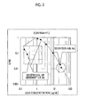
- Fig. 2 is a diagram illustrating the relationship between the cortisol concentration and the normalized value of fluorescence signal, B/B0, in the fluorescence measurement using Conjugate 2 (cortisol/albumin ratio is 11.0) (Comparative Example 1).
- Fig. 3 is a diagram illustrating the relationship between the cortisol concentration and the normalized value of fluorescence signal, B/B0, in the fluorescence measurement using Conjugate 3 (cortisol/albumin ratio is 7.0) (Comparative Example 2).
- the present invention relates to a cortisol albumin conjugate having a cortisol/albumin ratio of from 12 to 20, a base plate for cortisol immunoassay on which the conjugate is immobilized, and a method for measuring cortisol in a test sample, the method including (1) a step of bringing anti-cortisol antibody-labeled fluorescent particles and a test sample containing cortisol into contact with the base plate; and (2) a step of measuring the fluorescence based on the anti-cortisol antibody-labeled fluorescent particles bound to the base plate.
- Cortisol is a glucocorticoid having the following structure:
- albumin used in the present invention is not particularly limited, but for example, albumin derived from an animal (cattle, human being or the like), preferably a serum albumin derived from an animal (cattle, human being or the like), and particularly preferably bovine serum albumin (BSA) or the like can be used.
- albumin derived from an animal cattle, human being or the like
- serum albumin derived from an animal cattle, human being or the like
- BSA bovine serum albumin
- the cortisol/albumin ratio in the conjugate of the present invention is from 12 to 20, preferably from 14 to 18, and most preferably from 15 to 17.
- the cortisol/albumin ratio is greater than 20, solubility in water or organic solvents is decreased, and the decreased solubility affects the accuracy of measurement.
- the cortisol/albumin ratio is less than 12, the immunoassay performance becomes poor, and the effect of the present invention cannot be achieved.
- the cortisol/albumin ratio means a ratio of the number of molecules (mole number), and means the number of cortisol molecules bound to one molecule of albumin (BSA or the like).
- TFA trifluoroacetic acid
- ACN acetonitrile
- cortisol albumin conjugates are Catalogue No.: H-2384 of Sigma-Aldrich Co., Hydrocortisone 3-(O-carboxymethyl)oxime:BSA (26 mol steroid/mol BSA).
- BSA Hydrocortisone 3-(O-carboxymethyl)oxime
- the cortisol albumin ratio of this product is 26, and this product has low solubility in water or organic solvents, and could not be applied to the measurement of the present invention.
- the cortisol albumin conjugate of the present invention may be a conjugate in which cortisol and albumin are directly bonded to each other, or may be a conjugate in which cortisol and albumin are bonded via an appropriate linker.
- a cortisol albumin conjugate can be produced by producing an oxime derivative of cortisol having a carboxyl group, activating this carboxyl group by using 1-ethyl-3-(3-dimethylaminopropyl)carbodiimide (EDC), N-hydroxysuccinimide (NHS) and the like, and bonding the activated carboxyl group to an amino group of the albumin.
- the cortisol/albumin ratio for the conjugate of the present invention can be regulated by adjusting the reaction conditions for the bonding reaction between cortisol and albumin (the use amounts of cortisol and albumin, and the like).
- the aforementioned adjustment can be carried out by making reference to the literature ( Bioconjugate Chem., 1994, 4, 419-424 ).
- a base plate for cortisol immunoassay is provided by immobilizing the above-described cortisol albumin conjugate having a cortisol/albumin ratio of from 12 to 20, to a base plate.
- the type of the base plate is not particularly limited so long as the base plate is capable of the fluorescence analysis that will be described below, and any blank base plate can be used.
- the cortisol albumin conjugate can be bonded to the base plate by a method of dissolving the conjugate in a buffer solution, spot-applying the solution onto the base plate, leaving the solution to stand for a certain time, subsequently suctioning the supernatant, and drying the residue.
- the base plate In the case of performing surface plasmon fluorescence (SPF) detection that will be described below, regarding the base plate, it is preferable to use a base plate having a metal film on the surface.
- the metal that forms the metal film is not particularly limited so long as the metal is capable of producing surface plasmon resonance. Preferred examples thereof include free-electron metals such as gold, silver, copper, aluminum, and platinum, and gold is particularly preferred. Those metals can be used individually or in combinations.
- an intermediate layer formed from chromium or the like may be provided between the base plate and the layer formed of a metal.
- the thickness of the metal layer is not limited, but for example, the thickness is preferably from 0.1 nm to 500 nm, and particularly preferably from 1 nm to 200 nm. When the thickness is greater than 500 nm, the surface plasmon phenomenon of the medium cannot be sufficiently detected. Also, in the case of providing an intermediate layer formed from chromium or the like, the thickness of the intermediate layer is preferably from 0.1 nm to 10 nm.
- Formation of the metal film may be carried out according to a routine method, and can be carried out by, for example, a sputtering method, a vapor deposition method, an ion plating method, an electroplating method, or an electroless plating method.
- the metal film is preferably disposed on the base plate.
- the term "disposed on the base plate” includes the case where the metal film is disposed on the base plate so as to be in direct contact, as well as the case where the metal film is disposed over the base plate without being in direct contact with the base plate, with another layer interposed between the metal film and the base plate.
- the material of the base material for example, in the case of using the base plate for a surface plasmon resonance biosensor, a material which is transparent to laser light, such as optical glass such as BK7 (borosilicate glass), which is one kind of general optical glass, or a synthetic resin, specifically, polymethyl methacrylate, polyethylene terephthalate, polycarbonate or a cycloolefin polymer, can be used.
- optical glass such as BK7 (borosilicate glass)
- a synthetic resin specifically, polymethyl methacrylate, polyethylene terephthalate, polycarbonate or a cycloolefin polymer.
- a material which does not exhibit anisotropy to polarized light and has excellent processability is desirable.
- An example of the base plate for SPF detection may be a base plate obtained by vapor depositing a gold film on polymethyl methacrylate (PMMA).
- PMMA polymethyl methacrylate
- cortisol in a test sample can be measured by bringing the base plate for cortisol immunoassay of the present invention as described above, into contact with anti-cortisol antibody-labeled fluorescent particles and a test sample containing cortisol, and then measuring the fluorescence originating from the anti-cortisol antibody-labeled fluorescent particles bound to the cortisol albumin conjugate on the base plate.
- Particles colored with a fluorescence that can be conventionally used in an immune reaction can be used as the fluorescent particles to be used in the present invention, and for example, fluorescent polymer particles such as fluorescent polystyrene beads, and fluorescent glass particles such as fluorescent glass beads can be used.
- fluorescent polymer particles such as fluorescent polystyrene beads
- fluorescent glass particles such as fluorescent glass beads
- Specific examples of the material of the fluorescent particles include synthetic polymer powders of polymers using monomers such as styrene, methacrylic acid, glycidyl (meth)acrylate, butadiene, vinyl chloride, vinyl acetate acrylate, methyl methacrylate, ethyl methacrylate, phenyl methacrylate, and butyl methacrylate; and copolymers using two or more monomers. Latexes obtained by uniformly suspending these synthetic polymer powders are preferred. Further examples include organic polymer powders, inorganic substance powders, microorganisms, blood corpuscles
- the latex material include polystyrene, a styrene-acrylic acid copolymer, a styrene-methacrylic acid copolymer, a styrene-glycidyl (meth)acrylate copolymer, a styrene-styrene sulfonate copolymer, a methacrylic acid polymer, an acrylic acid polymer, an acrylonitrile-butadiene-styrene copolymer, a vinyl chloride-acrylic acid ester copolymer, and polyvinyl acetate acrylate.
- the latex is preferably a copolymer containing at least styrene as a monomer, and particularly preferably a copolymer of styrene with acrylic acid or methacrylic acid.
- the method for producing the latex is not particularly limited, and the latex can be produced by any polymerization method. However, if a surfactant is present at the time of antibody labeling, antibody immobilization is not easily achieved. Therefore, for the production of the latex, emulsifier-free emulsion polymerization, that is, emulsion polymerization in which no emulsifier such as surfactant is used, is preferred.
- the latex which is obtained by polymerization is fluorescent per se
- the latex can be used directly as fluorescent latex particles.
- fluorescent latex particles can be produced by adding a fluorescent substance (a fluorescent dye or the like) to the latex. That is, fluorescent latex particles can be produced by adding a fluorescent dye to a solution of latex particles containing water and a water-soluble organic solvent, and stirring the mixture.
- the average particle size of the fluorescent particles may vary with the material of the particles, the concentration range for the quantitative determination of the test substance, the measuring instrument, and the like; however, the average particle size is preferably in the range of 0.001 ⁇ m to 10 ⁇ m (more preferably 0.001 ⁇ m to 1 ⁇ m).
- Liposomes or microcapsules containing a fluorescent dye can also be used as fluorescent particles.
- the fluorescence emission color is not particularly limited so long as the color is to be emitted when the fluorescent substance is excited by absorbing ultraviolet radiation or the like, and returns to the ground state, and for example, fluorescence emission colors such as yellow-green (excitation wavelength: 505 nm/emission wavelength: 515 nm; hereinafter, the same), blue (350 nm to 356 nm/415 nm to 440 nm), red (535 nm to 580 nm/575 nm 605 nm), orange (540 nm/560 nm), red-orange (565 nm/580 nm), crimson (625 nm/645 nm), and dark red (660 nm/680 nm) can be used. Fluorescent particles emitting these fluorescence spectra are available from, for example, Invitrogen, Inc., and those fluorescent particles are commercially available under the trade name FluoSpheres (registered trademark) from the same company.
- the average particle size of the fluorescent particles used in the present invention can be measured with a commercially available particle size distribution analyzer or the like.
- Known examples of the measurement methods for particle size distribution include optical microscopy, confocal laser microscopy, electron microscopy, atomic force microscopy, static light scattering, laser diffraction, dynamic light scattering, centrifugal sedimentation, electric pulse measurement, chromatography and ultrasonic attenuation, and apparatuses corresponding to the various principles are commercially available.
- a dynamic light scattering method can be preferably used in the present invention.
- Examples of commercially available measuring apparatuses using dynamic light scattering include NanoTrack UPA (Nikkiso Co., Ltd.), a dynamic light scattering type particle size distribution analyzer, LB-550 (Horiba, Ltd.), and a concentrated system particle size analyzer, FPAR-1000 (Otsuka Electronics Co., Ltd.).
- an antibody exhibiting specificity to cortisol may be used.
- the anti-cortisol antibody that can be used include an antiserum prepared from the blood serum of an animal that has been immunized with cortisol, an immunoglobulin fraction purified from an antiserum, a monoclonal antibody obtainable by cell fusion using the spleen cells of an animal that has been immunized with cortisol, and fragments thereof [for example, F(ab')2, Fab, Fab', and Fv].
- Preparation of these antibodies can be carried out by conventional methods.
- the antibody may be modified as in the case of a chimeric antibody or the like, and a commercially available antibody or an antibody prepared from an animal blood serum or a culture supernatant by a known method can also be used.
- an antibody that can be used in the present invention is an antibody derived from an organism which can have an immune reaction, such as mouse, rat, hamster, goat, rabbit, sheep, cattle, or chicken, and specific examples thereof include mouse IgG, mouse IgM, rat IgG, rat IgM, hamster IgG, hamster IgM, rabbit IgG, rabbit IgM, goat IgG, goat IgM, sheep IgG, sheep IgM, bovine IgG, bovine IgM, and bird IgY
- mouse IgG mouse IgM, rat IgG, rat IgM, hamster IgG, hamster IgM
- a fragmentized antibody is a molecule derived from a whole antibody, having at least one antigen binding site, and specific examples thereof include Fab and F(ab')2. These fragmentized antibodies are molecules obtainable by using an enzymatic or chemical treatment, or a genetic engineering technique.
- Methods for immobilizing a binding substance such as an antibody or an antigen to particles are described in, for example, JP 2000-206115 A or the protocol attached to FluoSpheres (registered trademark) Polystyrene Microsphere F8813 of Molecular Probe, Inc., and any known methods for preparing a reagent for immune agglutination can all be used. Furthermore, any principle for physical adsorption or chemical bonding by covalent bonding can be employed as the principle for immobilizing an antibody as a binding substance to particles.
- any known substances for example, bovine serum albumin (BSA), skimmed milk, casein, a soybean-derived component, a fish-derived component, polyethylene glycol, and commercially available blocking agents for immune reaction containing these substances or equivalent substances, can be used.
- BSA bovine serum albumin
- These blocking agents can be subjected to a pretreatment such as partial modification by heat, an acid or an alkali as necessary.
- a specific method for immobilizing an antibody to particles will be illustrated below.
- An antibody solution having the concentration adjusted to 0.01 mg/mL to 20 mg/mL is added to a liquid prepared by dispersing particles to a solids concentration of 0.1% to 10%, and the mixture is mixed. Stirring is continued for 5 minutes to 48 hours under the temperature conditions of 4°C to 50°C. Subsequently, the particles are separated from the solution by centrifugation or any other method, and any antibody that is contained in the solution and has not been bound to the particles is sufficiently removed. Thereafter, an operation of washing the particles with a buffer solution is repeated 0 to 10 times.
- any component which does not participate in the antigen-antibody reaction preferably a protein, and more preferably a blocking agent such as BSA (bovine serum albumin), Block-Ace, skimmed milk or casein.
- BSA bovine serum albumin
- Block-Ace skimmed milk or casein.
- a stabilizer can be added as necessary.
- the stabilizer is not particularly limited so long as it is a compound which stabilizes an antigen or an antibody, such as sucrose or a synthetic or natural polymer such as a polysaccharide, and a commercially available product such as Immunoassay Stabilizer (Applied Biosystems, Inc.) can also be used.
- the measurement method of the present invention is construed by the broadest concept, including the detection of possible presence of cortisol and the measurement of the amount of cortisol (that is, quantification).
- a specific embodiment of the measurement method of the present invention may be a competition method.
- a base plate for cortisol immunoassay on which a cortisol albumin conjugate having a cortisol/albumin ratio of from 12 to 20 is immobilized is brought into contact with anti-cortisol antibody-labeled fluorescent particles and a test sample containing cortisol.
- an antigen-antibody reaction occurs on the base plate between the anti-cortisol antibody-labeled fluorescent particles and the cortisol on the base plate (that is, cortisol in the cortisol albumin conjugate).
- an antigen-antibody reaction occurs between the cortisol in the test sample and the anti-cortisol antibody-labeled fluorescent particles, and the antigen-antibody reaction between the anti-cortisol antibody-labeled fluorescent particles and the cortisol on the base plate (that is, cortisol in the cortisol albumin conjugate) is inhibited.
- the anti-cortisol antibody-labeled fluorescent particles that are not bound to the albumin on the base plate are removed.
- an immune complex that is, a complex between the anti-cortisol antibody-labeled fluorescent particles and the cortisol in the cortisol albumin conjugate on the base plate
- concentration of cortisol in the test sample can be measured.
- the form of fluorescence measurement may be plate reader measurement or may be flow measurement, but the measurement can be carried out by, for example, the following method.
- Plural samples with known amounts of cortisol and with different cortisol concentrations are prepared in advance, and each of these samples and anti-cortisol antibody-labeled fluorescent particles are mixed in advance.
- This liquid mixture is brought into contact with a region where a cortisol albumin conjugate is immobilized.
- the fluorescence signal from the region where the cortisol albumin conjugated is measured as plural fluorescence signals while the liquid mixture is brought into contact with the conjugate at a specific time interval.
- the change in the amount of fluorescence over time is determined for various cortisol concentrations.
- This time change is plotted on the Y-axis, while the cortisol concentration is plotted on the X-axis, and a relationship formula for the cortisol concentration against the change in the amount of fluorescence over time is obtained by using an appropriate fitting method such as a least-squares method.
- the amount of cortisol contained in the test sample can be quantitatively determined by the results of the change in the amount of fluorescence over time obtained by using a test sample intended for examination.
- the method for detecting fluorescence in the present invention is not particularly limited, but it is preferable to detect fluorescence intensity by using, for example, an instrument capable of detecting fluorescence intensity, specifically, a microplate reader or a biosensor for carrying out the detection of fluorescence caused by surface plasmon excitation (SPF).
- SPF method can make measurement with higher sensitivity than a detection method for fluorescence caused by epi-illumination excitation (hereinafter, referred to as "epi-illumination fluorescence method").
- the surface plasmon fluorescence (SPF) biosensor for example, a sensor described in JP 2008-249361 A , which includes a optical waveguide formed of a material which transmits excitation light having a predetermined wavelength; a metal film formed on one surface of this optical waveguide; a light source that generates a light beam; an optical system which causes the light beam to enter through the optical waveguide at an incident angle at which surface plasmon is generated with respect to the interface between the optical waveguide and the metal film; and a fluorescence detection means that detects the fluorescence generated as a result of excitation caused by the evanescent wave reinforced by the surface plasmon, can be used.
- SPPF surface plasmon fluorescence
- the detection system for fluorescence caused by surface plasmon excitation (SPF) using the fluorescent particles of the present invention is an assay method for detecting fluorescence from a fluorescent substance depending on the amount of a test substance immobilized on a metal thin film on a base plate, and is a method different from a so-called latex agglutination method which detects the change in the optical transparency resulting from the progress of the reaction in a solution, for example, as turbidity.
- agglutination method an antibody-sensitized latex in a latex reagent and the antigen in a specimen bind to each other through an antibody reaction, and agglutinate.
- This agglutinate increases with time, and a method of quantitatively determining the antigen concentration from the change in absorbance per unit time obtainable by irradiating near-infrared radiation to this agglutinate, constitutes the latex agglutination method.
- a detection method for a test substance which is very simple compared to the latex agglutination method, is provided.
- cortisol oxime derivative 1 was carried out by enaminating cortisol (may be abbreviated to COR or Corti), causing the product to react with a hydroxylamine derivative.
- COR Corti
- the reaction scheme is shown below.
- a cortisol-BSA conjugate was produced by the following method, by using the cortisol oxime derivative 1 synthesized in section 1-1.
- the reaction scheme is shown below.
- the cortisol oxime derivative 1 (50 mg, 115 ⁇ mol) in dry dimethylformamide (DMF, manufactured by Wako Pure Chemical Industries, Ltd.) was subjected to active esterification by using excess amounts of NHS (manufactured by Wako Pure Chemical Industries, Ltd.) and EDC (manufactured by Wako Pure Chemical Industries, Ltd.). Thereafter, the resultant was added dropwise to a phosphate buffer solution (PBS, manufactured by Wako Pure Chemical Industries, Ltd.) of BSA (bovine serum albumin, manufactured by Wako Pure Chemical Industries, Ltd., 193 mg), which is one kind of albumin.
- PBS phosphate buffer solution
- BSA bovine serum albumin
- SA sinapinic acid
- the center value of the molecular weight at 50% intensity of the maximum value of the peak intensity of a peak corresponding to the cortisol-BSA conjugate was employed as the peak of the BSA conjugate, and the position vertically drawn from this peak value was designated as the molecular weight of the cortisol-BSA conjugate.
- An example of the intensity of the MS spectrum is shown below, and the cortisol/BSA ratios of the conjugates 1, 2 and 3 thus obtained are indicated in Table 1.
- Table 1 Summary of preparation of cortisol (Corti)-BSA conjugates Conjugate 1 Conjugate 2 Conjugate 3 BSA 193 mg 322 mg 643 mg Corti derivative 50 mg 50 mg 50 mg PBS 39 mL 65 mL 130 mL Corti/BSA* 15.7 11.0 7.0 * The Corti/BSA ratio was determined by MALDI-TOF-MS described below.
- Fluorescent particles labeled with an anti-cortisol antibody were prepared in the following manner.
- 250 ⁇ L of a 50 mM MES buffer (pH 6.0) solution was added to 250 ⁇ L of a 2 mass% (solids concentration) aqueous solution of fluorescent latex particles (manufactured by Invitrogen, Inc.; average particle size 200 nm), and 100 ⁇ L of 5 mg/mL an anti-cortisol monoclonal antibody (manufactured by Hytest, Ltd.; Anti-Cortisol MAb XM210) was added thereto.
- the mixture was stirred for 15 minutes at room temperature. Thereafter, 5 ⁇ L of a 10 mg/mL aqueous EDC solution was added thereto, and the mixture was stirred for 2 hours at room temperature.
- the fluorescent latex particles were redispersed by adding 500 ⁇ L of a PBS (pH 7.4) solution containing 1 mass% BSA was added to the particles.
- a 1 mass% solution of anti-cortisol antibody-bound fluorescent latex particles was prepared.
- a base of polymethyl methacrylate (PMMA) (manufactured by Mitsubishi Rayon Co., Ltd., Acrypet VH) was provided, and a gold film having a thickness of 50 nm was deposited on one surface of the base by a vapor deposition method to a width of 4 mm.
- a chip for constructing a base plate was produced.
- a liquid containing the conjugate 1 (Example of the present invention), conjugate 2 (Comparative Example 1) or conjugate 3 (Comparative Example 2) Concentration: 50 ⁇ g/mL in 50 mM MES buffer solution, pH 6, 150 mM NaCl was spot-applied and dried.
- Each of the three types of base plates produced as such were repeatedly washed for three times by using 300 ⁇ L of a washing solution prepared in advance (a PBS solution (pH 7.4) containing 0.05 mass% Tween 20 (polyoxyethylene(20) sorbitan monolaurate, manufactured by Wako Pure Chemical Industries, Ltd.)).
- a washing solution prepared in advance a PBS solution (pH 7.4) containing 0.05 mass% Tween 20 (polyoxyethylene(20) sorbitan monolaurate, manufactured by Wako Pure Chemical Industries, Ltd.)).
- Flow channel type sensor chips were produced by encapsulating the three kinds of base plates produced according to the configuration of the second exemplary embodiment of JP 2010-190880 A , in flow channels.
- Samples containing cortisol at various concentrations were prepared, and each of the samples was mixed in advance with the anti-cortisol antibody-labeled fluorescent particles prepared in section 2. while the mixture was stirred for 10 minutes. Subsequently, the mixture was spot-applied on the flow channel type sensor chips encapsulating the base plates 1 to 3 produced in section 3-3.
- the mixture liquid was caused to flow down at a rate of 10 ⁇ L/min while pump suction was performed, and the fluorescence intensity of the gold-deposited surface where the cortisol-BSA conjugate was immobilized was continuously measured for 1.5 minutes. For each of the base plates, the rate of increase in the fluorescence intensity thus obtained per unit time was determined as the fluorescence signal value.
- a sample having a cortisol concentration of zero was prepared in the same manner, and the fluorescence signal value determined with the sample was designated as B0, while the signal value of each of the samples containing cortisol was designated as B.
- the signal value was normalized by determining the ratio B/B0.
- the performance of the cortisol-BSA conjugate was determined based on whether the standard of the calibration curve was satisfied.
- the calibration curve was defined by two items. The first item was the gradient of the calibration curve in a low concentration range, and when the reciprocal number of the gradient was calculated, a curve exhibiting a reciprocal number of 2.0 or less was considered as a standard. The second item was the deviation from the calibration curve at a measurement point in a high concentration range, and a curve exhibiting a deviation of 4% or less was considered as a standard. Within this range, the coefficient of fluctuation of the measured values was 10% or less, and an accuracy of 10% or less could be achieved. Thus, highly accurate measurement is made possible.
- the gradient of the calibration curve at 2.0 ⁇ g/dL which was the minimum concentration of cortisol that was clinically significant, was determined for the low concentration range. Furthermore, for the high concentration range, the deviations from the respective calibration curves at cortisol concentrations of 24.6 ⁇ g/dL and 29.0 ⁇ g/dL were determined, and the average value was calculated to perform an evaluation. The results are summarized in Table 2.
- cortisol can be analyzed by immunoassay with high sensitivity in a clinically significant cortisol concentration range (2 ⁇ g/dL to 30 ⁇ g/dL).
- Cortisol-BSA conjugates 4, 5 and 6 having cortisol label numbers of 11.0, 15.7 and 16.9 were produced in the same manner as in Example 1, except that the ratio of the cortisol oxime derivative 1 and the amount of BSA was varied.
- cortisol can be analyzed by immunoassay with high sensitivity at a cortisol concentration in the range of 2 ⁇ g/dL to 3 0 ⁇ g/dL.
Landscapes
- Health & Medical Sciences (AREA)
- Life Sciences & Earth Sciences (AREA)
- Engineering & Computer Science (AREA)
- Immunology (AREA)
- Molecular Biology (AREA)
- Chemical & Material Sciences (AREA)
- Urology & Nephrology (AREA)
- Hematology (AREA)
- Biomedical Technology (AREA)
- Physics & Mathematics (AREA)
- General Health & Medical Sciences (AREA)
- Cell Biology (AREA)
- Pathology (AREA)
- Food Science & Technology (AREA)
- Medicinal Chemistry (AREA)
- Biotechnology (AREA)
- Analytical Chemistry (AREA)
- Biochemistry (AREA)
- Microbiology (AREA)
- General Physics & Mathematics (AREA)
- Inorganic Chemistry (AREA)
- Endocrinology (AREA)
- Investigating, Analyzing Materials By Fluorescence Or Luminescence (AREA)
- Investigating Or Analysing Materials By The Use Of Chemical Reactions (AREA)
- Investigating Or Analysing Biological Materials (AREA)
- Investigating Or Analysing Materials By Optical Means (AREA)
Abstract
Description
- 1. Field of the Invention
- The present invention relates to a base plate and a method for immunoassay of cortisol using fluorescent particles.
- 2. Description of the Related Art
- Fluorescence detection methods have been hitherto used in a large variety of fields, as analytic methods that are highly sensitive and simple and are capable of quantitatively determining proteins, enzymes, inorganic compounds and the like. These fluorescence detection methods are methods by which a sample which is suspected to contain a substance to be detected (test substance) that is excited by light having a specific wavelength and emits fluorescence, is irradiated with excitation light having the specific wavelength, fluorescence is detected at that time, and thereby the presence of the test substance is verified. Furthermore, in the case where the substance is not a fluorescent material, a method of bringing a substance which specifically binds to a test substance labeled with a fluorescent dye, into contact with a sample, subsequently detecting fluorescence in the same manner as described above, and thereby verifying the presence of this binding, that is, the presence of the test substance, is also widely used.
- In regard to such fluorescent detection methods, a method of utilizing the effect of electric field reinforcement by plasmon resonance so as to increase the sensitivity of detection, is known. In such a method, in order to induce plasmon resonance, a sensor chip in which a metal layer is provided in a predetermined region on a transparent support is prepared, and excitation light is caused to enter the sensor chip through the surface of the support on the opposite side of the metal layer-formed surface with respect to the interface between the support and the metal film, at a predetermined angle that is greater than or equal to the total reflection angle. Irradiation of such excitation light causes generation of surface plasmon at the metal layer. Through the electric field reinforcing action brought by the generation of such surface plasmon, fluorescence is reinforced, and thereby the signal/noise ratio (S/N ratio) is increased. As compared with a fluorescence detection method involving surface plasmon excitation (hereinafter, referred to as "SPF method"), in a fluorescence detection method involving epi-illumination excitation (also called epi-illumination fluorescence method), the degree of signal reinforcement can be about 10 times, and analysis can be conducted with high sensitivity.
- For example, in the light signal detection method for determining the amount of a test substance as described in
JP 2010-190880 A - Cortisol, which is one kind of adrenocortical hormones, is a kind of glucocorticoid that controls the metabolism of carbohydrates, fats and proteins. Among three kinds of glucocorticoids, cortisol is available in the largest amount in the body. It is known that an increase in the secretion of cortisol is also caused by stress, and depending on the amount of secretion, there may be effects such as a increase in the blood pressure or the blood glucose level, or a decrease in the immune function. Furthermore, cortisol (also called hydrocortisone) is clinically used as a steroidal anti-inflammatory drug, and may be used in the treatment of, for example, acute inflammation, chronic inflammation, autoimmune diseases, allergic diseases, shock, gout, acute leukemia, and allograft rejection. As an immunoassay method for cortisol, for example, an analysis method based on immunochromatoassay is known (Anal. Chim. Acta., 2010, 682(1-2), 66-71).
- As discussed above, for example, an analysis method based on immunochromatoassay is known as an immunoassay method for cortisol; however, development of an immunoassay method which exhibits higher sensitivity is desired. An object to be solved by the present invention is to provide a base plate and a method for immunoassay of cortisol, which enable immunoassay of cortisol with high sensitivity in a cortisol concentration range of particular clinical significance (that is, 2 µg/dL to 30 µg/dL).
- The inventors of the present invention conducted a thorough investigation to solve the problems described above, and as a result, they found that cortisol can be analyzed by immunoassay with high sensitivity in a clinically significant cortisol concentration range (that is, 2 µg/dL to 30 µg/dL), by using a base plate on which a cortisol albumin conjugate obtained by conjugating cortisol and albumin at certain proportions is immobilized, bringing this base plate into contact with anti-cortisol antibody-labeled fluorescent particles and cortisol as a test substance, so as to cause the cortisol on the base plate (cortisol in the cortisol/albumin conjugate) and the cortisol as a test substance to competitively bind to the anti-cortisol antibody-labeled fluorescent particles, and thereby measuring the fluorescence based on the anti-cortisol antibody-labeled fluorescent particles bound to the base plate. The present invention was accomplished based on these findings.
- Specifically, according to an aspect of the present invention, there is provided a base plate for cortisol immunoassay on which a cortisol albumin conjugate having a cortisol/albumin ratio of from 12 to 20 is immobilized.
- According to another aspect of the present invention, there is provided a method for measuring cortisol in a test sample, the method including:
- (1) a step of bringing anti-cortisol antibody-labeled fluorescent particles and a test sample containing cortisol into contact with
- a base plate for cortisol immunoassay on which a cortisol albumin conjugate having a cortisol/albumin ratio of from 12 to 20 is immobilized; and
- (2) a step of measuring the fluorescence based on the anti-cortisol antibody-labeled fluorescent particles bound to the base plate.
- According to still another aspect of the present invention, there is provided a cortisol albumin conjugate having a cortisol/albumin ratio of from 12 to 20.
- Preferably, the albumin is a bovine serum albumin.
- Preferably, the cortisol albumin conjugate is a cortisol albumin conjugate having a cortisol/albumin ratio of from 14 to 18.
- Preferably, the cortisol albumin conjugate is immobilized on a metal film provided on the base plate.
- Preferably, in the step (2), fluorescence is measured by surface plasmon fluorescence measurement or epi-illumination fluorescence measurement.
- According to the present invention, cortisol can be analyzed by immunoassay with high sensitivity in a clinically significant cortisol concentration range (that is, 2 µg/dL to 30 µg/dL).
-
Fig. 1 is a diagram illustrating the relationship between the cortisol concentration and the normalized value of fluorescence signal, B/B0, in the fluorescence measurement using Conjugate 1 (corrisol/albumin ratio is 15.7) (Example of the present invention). -
Fig. 2 is a diagram illustrating the relationship between the cortisol concentration and the normalized value of fluorescence signal, B/B0, in the fluorescence measurement using Conjugate 2 (cortisol/albumin ratio is 11.0) (Comparative Example 1). -
Fig. 3 is a diagram illustrating the relationship between the cortisol concentration and the normalized value of fluorescence signal, B/B0, in the fluorescence measurement using Conjugate 3 (cortisol/albumin ratio is 7.0) (Comparative Example 2). - Hereinafter, the present invention will be described in more detail.
- The present invention relates to a cortisol albumin conjugate having a cortisol/albumin ratio of from 12 to 20, a base plate for cortisol immunoassay on which the conjugate is immobilized, and a method for measuring cortisol in a test sample, the method including (1) a step of bringing anti-cortisol antibody-labeled fluorescent particles and a test sample containing cortisol into contact with the base plate; and (2) a step of measuring the fluorescence based on the anti-cortisol antibody-labeled fluorescent particles bound to the base plate.
- (Cortisol)
- Cortisol is a glucocorticoid having the following structure:


- The type of albumin used in the present invention is not particularly limited, but for example, albumin derived from an animal (cattle, human being or the like), preferably a serum albumin derived from an animal (cattle, human being or the like), and particularly preferably bovine serum albumin (BSA) or the like can be used.
- (Cortisol-albumin conjugate)
- The cortisol/albumin ratio in the conjugate of the present invention is from 12 to 20, preferably from 14 to 18, and most preferably from 15 to 17. When the cortisol/albumin ratio is greater than 20, solubility in water or organic solvents is decreased, and the decreased solubility affects the accuracy of measurement. Furthermore, when the cortisol/albumin ratio is less than 12, the immunoassay performance becomes poor, and the effect of the present invention cannot be achieved.
- The cortisol/albumin ratio means a ratio of the number of molecules (mole number), and means the number of cortisol molecules bound to one molecule of albumin (BSA or the like). The cortisol/albumin ratio can be determined by, for example, matrix-assisted laser desorption-ionization time-of-flight mass spectrometry (MALDI-TOF-MS), and the specific procedure of measurement is as follows. Here, the procedure of measurement in the case of using BSA as the albumin will be described. A sample is dissolved in 0.1 mass% trifluoroacetic acid (TFA):acetonitrile (ACN) = 2/1, and the concentration is adjusted to 1 mg/mL. 4 µL of a matrix (sinapinic acid (SA)) and 1 µL of the sample are mixed, and the mixture is spot-applied in 1 µL x 4 spots on a gold plate. Thereafter, the sample is naturally dried. The gold plate is inserted into a MALDI-TOF-MS apparatus (Voyger manufactured by Applied Bio Systems, Inc.), and measurement is carried out. Accumulated data of 900 shots are obtained from each spot (N = 4). In regard to a peak corresponding to the cortisol-BSA conjugate, the position vertically drawn from the center of the area of the part which exhibits an intensity of 50% or greater of the maximum value of the peak intensity of the peak is designated as the molecular weight of the cortisol-BSA conjugate, and the average value of N = 4 is used to calculate the number of bound molecules according to the formula: (molecular weight of cortisol-BSA conjugate - molecular weight of naive BSA)/molecular weight of the cortisol derivative (435 - 18 = 417).
- Meanwhile, an example of commercially available cortisol albumin conjugates is Catalogue No.: H-2384 of Sigma-Aldrich Co., Hydrocortisone 3-(O-carboxymethyl)oxime:BSA (26 mol steroid/mol BSA). However, the cortisol albumin ratio of this product is 26, and this product has low solubility in water or organic solvents, and could not be applied to the measurement of the present invention.
- The cortisol albumin conjugate of the present invention may be a conjugate in which cortisol and albumin are directly bonded to each other, or may be a conjugate in which cortisol and albumin are bonded via an appropriate linker. For example, as will be described in the following Examples, a cortisol albumin conjugate can be produced by producing an oxime derivative of cortisol having a carboxyl group, activating this carboxyl group by using 1-ethyl-3-(3-dimethylaminopropyl)carbodiimide (EDC), N-hydroxysuccinimide (NHS) and the like, and bonding the activated carboxyl group to an amino group of the albumin.
- The cortisol/albumin ratio for the conjugate of the present invention can be regulated by adjusting the reaction conditions for the bonding reaction between cortisol and albumin (the use amounts of cortisol and albumin, and the like). The aforementioned adjustment can be carried out by making reference to the literature (Bioconjugate Chem., 1994, 4, 419-424).
- (Base plate)
- In the present invention, a base plate for cortisol immunoassay is provided by immobilizing the above-described cortisol albumin conjugate having a cortisol/albumin ratio of from 12 to 20, to a base plate. The type of the base plate is not particularly limited so long as the base plate is capable of the fluorescence analysis that will be described below, and any blank base plate can be used. The cortisol albumin conjugate can be bonded to the base plate by a method of dissolving the conjugate in a buffer solution, spot-applying the solution onto the base plate, leaving the solution to stand for a certain time, subsequently suctioning the supernatant, and drying the residue.
- In the case of performing surface plasmon fluorescence (SPF) detection that will be described below, regarding the base plate, it is preferable to use a base plate having a metal film on the surface. The metal that forms the metal film is not particularly limited so long as the metal is capable of producing surface plasmon resonance. Preferred examples thereof include free-electron metals such as gold, silver, copper, aluminum, and platinum, and gold is particularly preferred. Those metals can be used individually or in combinations. Furthermore, in consideration of the adhesiveness to the base plate, an intermediate layer formed from chromium or the like may be provided between the base plate and the layer formed of a metal. The thickness of the metal layer is not limited, but for example, the thickness is preferably from 0.1 nm to 500 nm, and particularly preferably from 1 nm to 200 nm. When the thickness is greater than 500 nm, the surface plasmon phenomenon of the medium cannot be sufficiently detected. Also, in the case of providing an intermediate layer formed from chromium or the like, the thickness of the intermediate layer is preferably from 0.1 nm to 10 nm.
- Formation of the metal film may be carried out according to a routine method, and can be carried out by, for example, a sputtering method, a vapor deposition method, an ion plating method, an electroplating method, or an electroless plating method.
- The metal film is preferably disposed on the base plate. Here, the term "disposed on the base plate" includes the case where the metal film is disposed on the base plate so as to be in direct contact, as well as the case where the metal film is disposed over the base plate without being in direct contact with the base plate, with another layer interposed between the metal film and the base plate. Regarding the material of the base material that can be used in the present invention, for example, in the case of using the base plate for a surface plasmon resonance biosensor, a material which is transparent to laser light, such as optical glass such as BK7 (borosilicate glass), which is one kind of general optical glass, or a synthetic resin, specifically, polymethyl methacrylate, polyethylene terephthalate, polycarbonate or a cycloolefin polymer, can be used. For such a base plate, preferably, a material which does not exhibit anisotropy to polarized light and has excellent processability is desirable.
- An example of the base plate for SPF detection may be a base plate obtained by vapor depositing a gold film on polymethyl methacrylate (PMMA).
- (Fluorescent particles)
- Furthermore, according to the present invention, cortisol in a test sample can be measured by bringing the base plate for cortisol immunoassay of the present invention as described above, into contact with anti-cortisol antibody-labeled fluorescent particles and a test sample containing cortisol, and then measuring the fluorescence originating from the anti-cortisol antibody-labeled fluorescent particles bound to the cortisol albumin conjugate on the base plate.
- Particles colored with a fluorescence that can be conventionally used in an immune reaction can be used as the fluorescent particles to be used in the present invention, and for example, fluorescent polymer particles such as fluorescent polystyrene beads, and fluorescent glass particles such as fluorescent glass beads can be used. Specific examples of the material of the fluorescent particles include synthetic polymer powders of polymers using monomers such as styrene, methacrylic acid, glycidyl (meth)acrylate, butadiene, vinyl chloride, vinyl acetate acrylate, methyl methacrylate, ethyl methacrylate, phenyl methacrylate, and butyl methacrylate; and copolymers using two or more monomers. Latexes obtained by uniformly suspending these synthetic polymer powders are preferred. Further examples include organic polymer powders, inorganic substance powders, microorganisms, blood corpuscles, cellular membrane fragments, and liposomes.
- In the case of using latex particles, specific examples of the latex material include polystyrene, a styrene-acrylic acid copolymer, a styrene-methacrylic acid copolymer, a styrene-glycidyl (meth)acrylate copolymer, a styrene-styrene sulfonate copolymer, a methacrylic acid polymer, an acrylic acid polymer, an acrylonitrile-butadiene-styrene copolymer, a vinyl chloride-acrylic acid ester copolymer, and polyvinyl acetate acrylate. The latex is preferably a copolymer containing at least styrene as a monomer, and particularly preferably a copolymer of styrene with acrylic acid or methacrylic acid. The method for producing the latex is not particularly limited, and the latex can be produced by any polymerization method. However, if a surfactant is present at the time of antibody labeling, antibody immobilization is not easily achieved. Therefore, for the production of the latex, emulsifier-free emulsion polymerization, that is, emulsion polymerization in which no emulsifier such as surfactant is used, is preferred.
- When the latex which is obtained by polymerization is fluorescent per se, the latex can be used directly as fluorescent latex particles. When the latex obtained by polymerization is non-fluorescent, fluorescent latex particles can be produced by adding a fluorescent substance (a fluorescent dye or the like) to the latex. That is, fluorescent latex particles can be produced by adding a fluorescent dye to a solution of latex particles containing water and a water-soluble organic solvent, and stirring the mixture.
- The average particle size of the fluorescent particles may vary with the material of the particles, the concentration range for the quantitative determination of the test substance, the measuring instrument, and the like; however, the average particle size is preferably in the range of 0.001 µm to 10 µm (more preferably 0.001 µm to 1 µm). Liposomes or microcapsules containing a fluorescent dye can also be used as fluorescent particles. The fluorescence emission color is not particularly limited so long as the color is to be emitted when the fluorescent substance is excited by absorbing ultraviolet radiation or the like, and returns to the ground state, and for example, fluorescence emission colors such as yellow-green (excitation wavelength: 505 nm/emission wavelength: 515 nm; hereinafter, the same), blue (350 nm to 356 nm/415 nm to 440 nm), red (535 nm to 580 nm/575 nm 605 nm), orange (540 nm/560 nm), red-orange (565 nm/580 nm), crimson (625 nm/645 nm), and dark red (660 nm/680 nm) can be used. Fluorescent particles emitting these fluorescence spectra are available from, for example, Invitrogen, Inc., and those fluorescent particles are commercially available under the trade name FluoSpheres (registered trademark) from the same company.
- (Method for measuring average particle size)
- The average particle size of the fluorescent particles used in the present invention can be measured with a commercially available particle size distribution analyzer or the like. Known examples of the measurement methods for particle size distribution include optical microscopy, confocal laser microscopy, electron microscopy, atomic force microscopy, static light scattering, laser diffraction, dynamic light scattering, centrifugal sedimentation, electric pulse measurement, chromatography and ultrasonic attenuation, and apparatuses corresponding to the various principles are commercially available.
- In view of the particle size range and the ease of measurement, a dynamic light scattering method can be preferably used in the present invention. Examples of commercially available measuring apparatuses using dynamic light scattering include NanoTrack UPA (Nikkiso Co., Ltd.), a dynamic light scattering type particle size distribution analyzer, LB-550 (Horiba, Ltd.), and a concentrated system particle size analyzer, FPAR-1000 (Otsuka Electronics Co., Ltd.). In the present invention, the average particle size is determined as the value of a median diameter (d = 50) measured at a measurement temperature of 25°C.
- (Anti-cortisol antibody)
- As the anti-cortisol antibody to be bonded to the fluorescent particles, an antibody exhibiting specificity to cortisol may be used. Examples of the anti-cortisol antibody that can be used include an antiserum prepared from the blood serum of an animal that has been immunized with cortisol, an immunoglobulin fraction purified from an antiserum, a monoclonal antibody obtainable by cell fusion using the spleen cells of an animal that has been immunized with cortisol, and fragments thereof [for example, F(ab')2, Fab, Fab', and Fv]. Preparation of these antibodies can be carried out by conventional methods. Furthermore, the antibody may be modified as in the case of a chimeric antibody or the like, and a commercially available antibody or an antibody prepared from an animal blood serum or a culture supernatant by a known method can also be used.
- The antibody can be used without being restricted by the animal species or the subclass. For example, an antibody that can be used in the present invention is an antibody derived from an organism which can have an immune reaction, such as mouse, rat, hamster, goat, rabbit, sheep, cattle, or chicken, and specific examples thereof include mouse IgG, mouse IgM, rat IgG, rat IgM, hamster IgG, hamster IgM, rabbit IgG, rabbit IgM, goat IgG, goat IgM, sheep IgG, sheep IgM, bovine IgG, bovine IgM, and bird IgY These antibodies are applicable to both polyclonal and monoclonal antibodies. A fragmentized antibody is a molecule derived from a whole antibody, having at least one antigen binding site, and specific examples thereof include Fab and F(ab')2. These fragmentized antibodies are molecules obtainable by using an enzymatic or chemical treatment, or a genetic engineering technique.
- Methods for immobilizing a binding substance such as an antibody or an antigen to particles are described in, for example,
JP 2000-206115 A - A specific method for immobilizing an antibody to particles will be illustrated below. An antibody solution having the concentration adjusted to 0.01 mg/mL to 20 mg/mL is added to a liquid prepared by dispersing particles to a solids concentration of 0.1% to 10%, and the mixture is mixed. Stirring is continued for 5 minutes to 48 hours under the temperature conditions of 4°C to 50°C. Subsequently, the particles are separated from the solution by centrifugation or any other method, and any antibody that is contained in the solution and has not been bound to the particles is sufficiently removed. Thereafter, an operation of washing the particles with a buffer solution is repeated 0 to 10 times. After the operation of mixing the particles and the antibody and causing the antibody to bind to the particles is carried out, it is desirable to protect the part on the particle surface that is not bound by the antibody, by using any component which does not participate in the antigen-antibody reaction, preferably a protein, and more preferably a blocking agent such as BSA (bovine serum albumin), Block-Ace, skimmed milk or casein.
- When an antigen or an antibody is immobilized to particles, a stabilizer can be added as necessary. The stabilizer is not particularly limited so long as it is a compound which stabilizes an antigen or an antibody, such as sucrose or a synthetic or natural polymer such as a polysaccharide, and a commercially available product such as Immunoassay Stabilizer (Applied Biosystems, Inc.) can also be used.
- (Measurement method)
- The measurement method of the present invention is construed by the broadest concept, including the detection of possible presence of cortisol and the measurement of the amount of cortisol (that is, quantification). A specific embodiment of the measurement method of the present invention may be a competition method.
- In the competition method according to the present invention, first, a base plate for cortisol immunoassay on which a cortisol albumin conjugate having a cortisol/albumin ratio of from 12 to 20 is immobilized, is brought into contact with anti-cortisol antibody-labeled fluorescent particles and a test sample containing cortisol. When cortisol is not present in the test sample, an antigen-antibody reaction occurs on the base plate between the anti-cortisol antibody-labeled fluorescent particles and the cortisol on the base plate (that is, cortisol in the cortisol albumin conjugate). On the other hand, when cortisol is present in the test sample, an antigen-antibody reaction occurs between the cortisol in the test sample and the anti-cortisol antibody-labeled fluorescent particles, and the antigen-antibody reaction between the anti-cortisol antibody-labeled fluorescent particles and the cortisol on the base plate (that is, cortisol in the cortisol albumin conjugate) is inhibited. After the reaction described above is completed, the anti-cortisol antibody-labeled fluorescent particles that are not bound to the albumin on the base plate are removed. Subsequently, the extent of formation of an immune complex (that is, a complex between the anti-cortisol antibody-labeled fluorescent particles and the cortisol in the cortisol albumin conjugate on the base plate) on the base plate is detected, and thereby the concentration of cortisol in the test sample can be measured.
- In the competition method, the form of fluorescence measurement may be plate reader measurement or may be flow measurement, but the measurement can be carried out by, for example, the following method. Plural samples with known amounts of cortisol and with different cortisol concentrations are prepared in advance, and each of these samples and anti-cortisol antibody-labeled fluorescent particles are mixed in advance. This liquid mixture is brought into contact with a region where a cortisol albumin conjugate is immobilized. The fluorescence signal from the region where the cortisol albumin conjugated is measured as plural fluorescence signals while the liquid mixture is brought into contact with the conjugate at a specific time interval. From these plural fluorescence signals, the change in the amount of fluorescence over time (gradient) is determined for various cortisol concentrations. This time change is plotted on the Y-axis, while the cortisol concentration is plotted on the X-axis, and a relationship formula for the cortisol concentration against the change in the amount of fluorescence over time is obtained by using an appropriate fitting method such as a least-squares method. Based on the relationship formula obtained as described above, the amount of cortisol contained in the test sample can be quantitatively determined by the results of the change in the amount of fluorescence over time obtained by using a test sample intended for examination.
- (Measurement of surface plasmon fluorescence)
- The method for detecting fluorescence in the present invention is not particularly limited, but it is preferable to detect fluorescence intensity by using, for example, an instrument capable of detecting fluorescence intensity, specifically, a microplate reader or a biosensor for carrying out the detection of fluorescence caused by surface plasmon excitation (SPF). The detection method for fluorescence caused by surface plasmon excitation (SPF method) can make measurement with higher sensitivity than a detection method for fluorescence caused by epi-illumination excitation (hereinafter, referred to as "epi-illumination fluorescence method").
- Regarding the surface plasmon fluorescence (SPF) biosensor, for example, a sensor described in
JP 2008-249361 A - The detection system for fluorescence caused by surface plasmon excitation (SPF) using the fluorescent particles of the present invention is an assay method for detecting fluorescence from a fluorescent substance depending on the amount of a test substance immobilized on a metal thin film on a base plate, and is a method different from a so-called latex agglutination method which detects the change in the optical transparency resulting from the progress of the reaction in a solution, for example, as turbidity. In the latex agglutination method, an antibody-sensitized latex in a latex reagent and the antigen in a specimen bind to each other through an antibody reaction, and agglutinate. This agglutinate increases with time, and a method of quantitatively determining the antigen concentration from the change in absorbance per unit time obtainable by irradiating near-infrared radiation to this agglutinate, constitutes the latex agglutination method. In the present invention, a detection method for a test substance, which is very simple compared to the latex agglutination method, is provided.
- The present invention will be more specifically described by way of the following Examples, but the present invention is not intended to be limited by the Examples.
- Examples
- (Example 1)
- 1. Preparation of cortisol-BSA conjugate
- 1-1. Synthesis of
cortisol oxime derivative 1 - According to the literature (Steroids, 1974, Jan. 49-64), synthesis of a
cortisol oxime derivative 1 was carried out by enaminating cortisol (may be abbreviated to COR or Corti), causing the product to react with a hydroxylamine derivative. The reaction scheme is shown below.
- One equivalent of pyrrolidine (manufactured by Wako Pure Chemical Industries, Ltd.) was added dropwise to a methanol suspension of cortisol (1 g, manufactured by Wako Pure Chemical Industries, Ltd.), and the mixture was stirred to 5 minutes at room temperature. Examine was produced in the reaction system, and the solution turned red. Thereafter, when another 1 equivalent of pyrrolidine and 1 equivalent of a hydroxylamine derivative (manufactured by Wako Pure Chemical Industries, Ltd.) were added thereto, the color disappeared. The reaction temperature was adjusted to 55°C, and the system was stirred to implement oximation. The reaction proceeded smoothly, and a cortisol oxime derivative 1 (1.1 g; 84%) was obtained.
- 1-2. Preparation of cortisol-BSA conjugate
- A cortisol-BSA conjugate was produced by the following method, by using the
cortisol oxime derivative 1 synthesized in section 1-1. The reaction scheme is shown below.
- The cortisol oxime derivative 1 (50 mg, 115 µmol) in dry dimethylformamide (DMF, manufactured by Wako Pure Chemical Industries, Ltd.) was subjected to active esterification by using excess amounts of NHS (manufactured by Wako Pure Chemical Industries, Ltd.) and EDC (manufactured by Wako Pure Chemical Industries, Ltd.). Thereafter, the resultant was added dropwise to a phosphate buffer solution (PBS, manufactured by Wako Pure Chemical Industries, Ltd.) of BSA (bovine serum albumin, manufactured by Wako Pure Chemical Industries, Ltd., 193 mg), which is one kind of albumin. After completion of the reaction, the reaction solution was purified by dialysis in an ACN (manufactured by Wako Pure Chemical Industries, Ltd.)/water = 1/1 (containing 0.1 mass% TFA (manufactured by Wako Pure Chemical Industries, Ltd.)) solution (5 L). Finally, the solution was lyophilized, and thus a white solid was obtained (this was designated as conjugate 1). Conjugate 2 and conjugate 3 were obtained in the same manner, except that the use amount of BSA and the amount of PBS were changed as indicated in Table 1.
- 1-3. Measurement of cortisol/BSA ratio (cortisol/albumin ratio) by MALDI-TOF-MS
- (Measurement procedure)
- The cortisol-BSA conjugate prepared in section 1-2. was dissolved in a solution of 0.1 mass% TFA:ACN = 2/1, and the solution was adjusted to a concentration of 1 mg/mL. 1 µL of this solution and 4 µL of a matrix (SA; sinapinic acid) were mixed, and the mixture was spot-applied in 1 µL x 4 points on a gold plate. The mixture was naturally dried, and then the gold plate was inserted into a MALDI-TOF-MS apparatus (Voyger manufactured by Applied Bio Systems, Inc.). Accumulated data of 900 shots are obtained from each spot (N = 4) to obtain the mass information. By using these data, the center value of the molecular weight at 50% intensity of the maximum value of the peak intensity of a peak corresponding to the cortisol-BSA conjugate was employed as the peak of the BSA conjugate, and the position vertically drawn from this peak value was designated as the molecular weight of the cortisol-BSA conjugate. The average value of N = 4 was used to calculate the binding number of cortisol to BSA by the formula: (molecular weight of cortisol-BSA conjugate - molecular weight of naive BSA)/molecular weight of the cortisol derivative (435 - 18 = 417). An example of the intensity of the MS spectrum is shown below, and the cortisol/BSA ratios of the
conjugates 1, 2 and 3 thus obtained are indicated in Table 1.
-
Table 1: Summary of preparation of cortisol (Corti)- BSA conjugates Conjugate 1 Conjugate 2 Conjugate 3 BSA 193 mg 322 mg 643 mg Corti derivative 50 mg 50 mg 50 mg PBS 39 mL 65 mL 130 mL Corti/BSA* 15.7 11.0 7.0 * The Corti/BSA ratio was determined by MALDI-TOF-MS described below. - 2. Preparation of fluorescent particles labeled with anti-cortisol antibody
- Fluorescent particles labeled with an anti-cortisol antibody were prepared in the following manner.
- 250 µL of a 50 mM MES buffer (pH 6.0) solution was added to 250 µL of a 2 mass% (solids concentration) aqueous solution of fluorescent latex particles (manufactured by Invitrogen, Inc.; average particle size 200 nm), and 100 µL of 5 mg/mL an anti-cortisol monoclonal antibody (manufactured by Hytest, Ltd.; Anti-Cortisol MAb XM210) was added thereto. The mixture was stirred for 15 minutes at room temperature. Thereafter, 5 µL of a 10 mg/mL aqueous EDC solution was added thereto, and the mixture was stirred for 2 hours at room temperature. 25 µL of a 2 mol/L aqueous solution of glycine (manufactured by Wako Pure Chemical Industries, Ltd.) was added thereto, and the mixture was stirred for 30 minutes. Subsequently, the mixture was centrifuged (15,000 rpm, 4°C, 15 minutes), and thus fluorescent latex particles were allowed to sediment. Thereafter, the supernatant was removed, 500 µL of a PBS solution (pH 7.4) was added to the particles, and the fluorescent latex particles were redispersed by using an ultrasonic cleaning machine. Again, the dispersion was centrifuged (15,000 rpm, 4°C, 15 minutes), and the supernatant was removed. Subsequently, the fluorescent latex particles were redispersed by adding 500 µL of a PBS (pH 7.4) solution containing 1 mass% BSA was added to the particles. Thus, a 1 mass% solution of anti-cortisol antibody-bound fluorescent latex particles was prepared.
- 3. Production of flow channel type sensor chip
- 3-1. Production of cortisol-BSA conjugate-immobilized base plate
- A base of polymethyl methacrylate (PMMA) (manufactured by Mitsubishi Rayon Co., Ltd., Acrypet VH) was provided, and a gold film having a thickness of 50 nm was deposited on one surface of the base by a vapor deposition method to a width of 4 mm. Thus, a chip for constructing a base plate was produced. On the gold-deposited surface of this chip, a liquid containing the conjugate 1 (Example of the present invention), conjugate 2 (Comparative Example 1) or conjugate 3 (Comparative Example 2) (concentration: 50 µg/mL in 50 mM MES buffer solution, pH 6, 150 mM NaCl) was spot-applied and dried. Thus, a plural number of
base plates 1 to 3, on which each of the conjugates was immobilized, were produced. - 3-2. Washing of base plate
- Each of the three types of base plates produced as such were repeatedly washed for three times by using 300 µL of a washing solution prepared in advance (a PBS solution (pH 7.4) containing 0.05 mass% Tween 20 (polyoxyethylene(20) sorbitan monolaurate, manufactured by Wako Pure Chemical Industries, Ltd.)).
- 3-3. Production of flow channel type sensor chip
- Flow channel type sensor chips were produced by encapsulating the three kinds of base plates produced according to the configuration of the second exemplary embodiment of
JP 2010-190880 A - 4. Immunoassay of cortisol using fluorescent particles
- Samples containing cortisol at various concentrations (1.1 µg/dL, 2.0 µg/dL, 3.2 µg/dL, 9.0 µg/dL, 24.6 µg/dL, and 29.0 µg/dL) were prepared, and each of the samples was mixed in advance with the anti-cortisol antibody-labeled fluorescent particles prepared in section 2. while the mixture was stirred for 10 minutes. Subsequently, the mixture was spot-applied on the flow channel type sensor chips encapsulating the
base plates 1 to 3 produced in section 3-3. After the spot-application, the mixture liquid was caused to flow down at a rate of 10 µL/min while pump suction was performed, and the fluorescence intensity of the gold-deposited surface where the cortisol-BSA conjugate was immobilized was continuously measured for 1.5 minutes. For each of the base plates, the rate of increase in the fluorescence intensity thus obtained per unit time was determined as the fluorescence signal value. - A sample having a cortisol concentration of zero was prepared in the same manner, and the fluorescence signal value determined with the sample was designated as B0, while the signal value of each of the samples containing cortisol was designated as B. The signal value was normalized by determining the ratio B/B0.
- 5. Calibration curve
- It is described in the literature "The Immunoassay Handbook, Third Edition, Edited by David Wild (2005)" that a four-parameter logistic curve model of a sigmoid function can be applied as the calibration curve for a competition method. Thus, according to this method, a four-parameter logistic curve passing the nearest neighbor points of the respective points of the fluorescence signal values obtained at the various cortisol concentrations measured in section 4. was determined by using the least squares method that is generally known as a method for obtaining an approximate curve. This curve was used as the calibration curve.
- The calibration curve determined as described above and the average values of the measured values of the respective cortisol concentrations are shown in
Fig. 1 to Fig. 3 for each of the cortisol/BSA ratio (indicated as COR/BSAin the diagrams). - The performance of the cortisol-BSA conjugate was determined based on whether the standard of the calibration curve was satisfied. The calibration curve was defined by two items. The first item was the gradient of the calibration curve in a low concentration range, and when the reciprocal number of the gradient was calculated, a curve exhibiting a reciprocal number of 2.0 or less was considered as a standard. The second item was the deviation from the calibration curve at a measurement point in a high concentration range, and a curve exhibiting a deviation of 4% or less was considered as a standard. Within this range, the coefficient of fluctuation of the measured values was 10% or less, and an accuracy of 10% or less could be achieved. Thus, highly accurate measurement is made possible.
- The gradient of the calibration curve at 2.0 µg/dL, which was the minimum concentration of cortisol that was clinically significant, was determined for the low concentration range. Furthermore, for the high concentration range, the deviations from the respective calibration curves at cortisol concentrations of 24.6 µg/dL and 29.0 µg/dL were determined, and the average value was calculated to perform an evaluation. The results are summarized in Table 2.
-
Table 2: Gentleness of calibration curve in low concentration range, and deviation of average measured values from calibration curves in high concentration range Cortisol/BSA ratio Reciprocal of calibration curve gradient in low concentration range Deviation from calibration curve in high concentration range (Reference 2.0 or less Result (Reference 4% or less) Result 15.7 1.8 Good 2.6% Good Example 11.0 1.4 Good 4.6% NG Comparative Example 7.0 2.4 NG 1.9% Good Comparative Example - From the results indicated in
Fig. 1 to Fig. 3 and Table 2, it was confirmed that by using the cortisol-BSA conjugate of the present invention is used, cortisol can be analyzed by immunoassay with high sensitivity in a clinically significant cortisol concentration range (2 µg/dL to 30 µg/dL). - (Example 2)
- Cortisol-BSA conjugates 4, 5 and 6 having cortisol label numbers of 11.0, 15.7 and 16.9 were produced in the same manner as in Example 1, except that the ratio of the
cortisol oxime derivative 1 and the amount of BSA was varied. - Samples containing cortisol at 5 levels of concentration (1.5 µg/dL, 4.3 µg/dL, 10.1 µg/dL, 20.5 µg/dL, and 33.2 µg/dL) were provided, and an immunoassay was carried out in the same manner as in Example 1. Thus, the fluorescence signal values at the various concentrations were obtained. Furthermore, calibration curves were obtained in the same manner as in Example 1. In the same manner as in Example 1, the reciprocal numbers of the gradient of the calibration curves in the low concentration range, and the deviations from the calibration curves in the high concentration range were evaluated. The reciprocal number of the gradient of the calibration curve at a cortisol concentration of 4.3 µg/dL in a low concentration range was determined, and for the cortisol concentration in the high concentration range, the deviation from the calibration curve at a cortisol concentration of 33.2 µg/dL was determined so as to guarantee measurement at 30 µg/dL, which is a clinically significant concentration. The results are shown in Table 3.
-
Table 3: Gentleness of calibration curve in low concentration range, and deviation of average measured values from calibration curves in high concentration range Cortisol/BSA ratio Reciprocal of calibration curve gradient in low concentration range Deviation from calibration curve in high concentration range (Reference 2.0 or less Result (Reference 4% or less) Result 16.9 1.4 Good -3.2% Good Example 15.7 1.6 Good -3.5% Good Comparative Example 11.0 1.2 Good -5.3% NG Comparative Example - From the results of Table 3, it was confirmed that by using the cortisol-BSA conjugate having the cortisol/albumin ratio (cortisol/BSA ratio) of the present invention, cortisol can be analyzed by immunoassay with high sensitivity at a cortisol concentration in the range of 2 µg/dL to 3 0 µg/dL.
Claims (9)
- A cortisol albumin conjugate having a cortisol/albumin ratio of from 12 to 20.
- A cortisol albumin conjugate according to claim 1 having a cortisol/albumin ratio of from 14 to 18.
- The cortisol albumin conjugate according to claim 1 or 2, wherein the albumin is bovine serum albumin.
- A base plate for cortisol immunoassay, comprising a cortisol albumin conjugate according to any one of claims 1 to 3.
- The base plate for cortisol immunoassay according to claim 4, wherein the cortisol albumin conjugate is immobilized to a metal film provided on the base plate.
- A method for measuring cortisol in a test sample, the method comprising:(1) bringing a base plate for cortisol immunoassay which has a cortisol albumin conjugate having a cortisol/albumin ratio of from 12 to 20 immobilized thereon, into contact with anti-cortisol antibody-labeled fluorescent particles and a test sample containing cortisol; and(2) measuring the fluorescence based on the anti-cortisol antibody-labeled fluorescent particles bound to the base plate.
- The method according to claim 6, wherein the albumin is bovine serum albumin.
- The method according to claim 6 or 7, wherein the measurement is carried out for a test sample having a cortisol concentration of 2 µg/dL to 30 µg/dL.
- The method according any one of claims 6 to 8, wherein in the step (2), fluorescence is measured by surface plasmon fluorescence measurement or epi-illumination fluorescence measurement.
Applications Claiming Priority (2)
| Application Number | Priority Date | Filing Date | Title |
|---|---|---|---|
| JP2011208525 | 2011-09-26 | ||
| JP2012189776A JP5416263B2 (en) | 2011-09-26 | 2012-08-30 | Cortisol immunoassay using fluorescent particles |
Publications (2)
| Publication Number | Publication Date |
|---|---|
| EP2573570A1 true EP2573570A1 (en) | 2013-03-27 |
| EP2573570B1 EP2573570B1 (en) | 2018-03-28 |
Family
ID=47044784
Family Applications (1)
| Application Number | Title | Priority Date | Filing Date |
|---|---|---|---|
| EP12184648.9A Active EP2573570B1 (en) | 2011-09-26 | 2012-09-17 | Cortisol immunoassay using fluorescent particles |
Country Status (3)
| Country | Link |
|---|---|
| US (1) | US20130078739A1 (en) |
| EP (1) | EP2573570B1 (en) |
| JP (1) | JP5416263B2 (en) |
Cited By (1)
| Publication number | Priority date | Publication date | Assignee | Title |
|---|---|---|---|---|
| CN105954271A (en) * | 2016-04-27 | 2016-09-21 | 樊福好 | Solution and detection method for detecting physique |
Families Citing this family (6)
| Publication number | Priority date | Publication date | Assignee | Title |
|---|---|---|---|---|
| WO2015021108A1 (en) * | 2013-08-07 | 2015-02-12 | Siemens Healthcare Diagnostics Inc. | Compositions and methods for detecting cortisol |
| WO2016047027A1 (en) * | 2014-09-24 | 2016-03-31 | 富士フイルム株式会社 | Fluorescence detecting method and sample cell for detection |
| US10067126B2 (en) * | 2015-05-25 | 2018-09-04 | Hunan SkyWorld Biotechnologies Co. LTD | Use of fluorescence for the quick and easy determination of s-adenosylmethionine, s-adenosylhomocysteine and homocysteine |
| CN109422791A (en) * | 2017-09-04 | 2019-03-05 | 北京维德维康生物技术有限公司 | A kind of hydrocortisone haptens, artificial antigen and the preparation method and application thereof |
| CN109324183B (en) * | 2018-11-23 | 2022-03-29 | 深圳市计量质量检测研究院(国家高新技术计量站、国家数字电子产品质量监督检验中心) | Glucocorticoid immunochromatography broad-spectrum detection card and preparation method and application thereof |
| CN110672842A (en) * | 2019-09-26 | 2020-01-10 | 科赫生物科技(北京)有限公司 | Competitive antigen detection method and test paper |
Citations (3)
| Publication number | Priority date | Publication date | Assignee | Title |
|---|---|---|---|---|
| JP2000206115A (en) | 1999-01-11 | 2000-07-28 | Eiken Chem Co Ltd | Immunoagglutination reagent using antibody fragments |
| JP2008249361A (en) | 2007-03-29 | 2008-10-16 | Fujifilm Corp | Surface plasmon sensor and immunological measurement method |
| EP2110658A1 (en) * | 2008-04-18 | 2009-10-21 | FUJIFILM Corporation | Optical signal detection method, apparatus, sample cell and kit |
Family Cites Families (3)
| Publication number | Priority date | Publication date | Assignee | Title |
|---|---|---|---|---|
| JPS5344622A (en) * | 1976-09-30 | 1978-04-21 | Mochida Pharm Co Ltd | Immunologically measuring method |
| FR2732115B1 (en) * | 1995-03-24 | 1997-06-13 | Immunotech Sa | IMMUNOLOGICAL ASSAY PROCESS FOR CORTISOL, ESPECIALLY URINARY, AND REAGENTS USED |
| NZ511705A (en) * | 2001-05-14 | 2004-03-26 | Horticulture & Food Res Inst | Methods and rapid immunoassay device for detecting progesterone and other steroids |
-
2012
- 2012-08-30 JP JP2012189776A patent/JP5416263B2/en active Active
- 2012-09-13 US US13/614,817 patent/US20130078739A1/en not_active Abandoned
- 2012-09-17 EP EP12184648.9A patent/EP2573570B1/en active Active
Patent Citations (4)
| Publication number | Priority date | Publication date | Assignee | Title |
|---|---|---|---|---|
| JP2000206115A (en) | 1999-01-11 | 2000-07-28 | Eiken Chem Co Ltd | Immunoagglutination reagent using antibody fragments |
| JP2008249361A (en) | 2007-03-29 | 2008-10-16 | Fujifilm Corp | Surface plasmon sensor and immunological measurement method |
| EP2110658A1 (en) * | 2008-04-18 | 2009-10-21 | FUJIFILM Corporation | Optical signal detection method, apparatus, sample cell and kit |
| JP2010190880A (en) | 2008-04-18 | 2010-09-02 | Fujifilm Corp | Method and apparatus for detecting optical signal, sample cell for detecting optical signal, and kit for detecting optical signal |
Non-Patent Citations (8)
| Title |
|---|
| "The Immunoassay Handbook", 2005 |
| ANAL. CHIM. ACTA., vol. 682, no. 1-2, 2010, pages 66 - 71 |
| BIOCONJUGATE CHEM., vol. 4, 1994, pages 419 - 424 |
| ESKOLA ET AL: "Direct solid-phase time-resolved immunofluorometric assay of cortisol in serum.", CLINICAL CHEMISTRY, vol. 31, no. 10, 1 October 1985 (1985-10-01), pages 1731 - 1734, XP055050128, ISSN: 0009-9147 * |
| GIANFRANCO GIRAUDI ET AL: "Characterisation of cortisol-bovine serum albumin conjugates by chromatofocusing", THE ANALYST, vol. 115, no. 12, 1 January 1990 (1990-01-01), pages 1531, XP055049693, ISSN: 0003-2654, DOI: 10.1039/an9901501531 * |
| HOSODA H ET AL: "THE PREPARATION OF STEROID N-HYDROXYSUCCINIMIDE ESTERS AND THEIR REACTIVITIES WITH BOVINE SERUM ALBUMIN", CHEMICAL AND PHARMACEUTICAL BULLETIN,, vol. 27, no. 3, 1 March 1979 (1979-03-01), pages 742 - 746, XP008056528, ISSN: 0009-2363 * |
| NARA S ET AL: "Colloidal gold probe based rapid immunochromatographic strip assay for cortisol", ANAL. CHIM. ACTA., vol. 682, no. 1-2, 2010, pages 66 - 71, XP027477332, ISSN: 0003-2670 * |
| STEROIDS, January 1974 (1974-01-01), pages 49 - 64 |
Cited By (1)
| Publication number | Priority date | Publication date | Assignee | Title |
|---|---|---|---|---|
| CN105954271A (en) * | 2016-04-27 | 2016-09-21 | 樊福好 | Solution and detection method for detecting physique |
Also Published As
| Publication number | Publication date |
|---|---|
| US20130078739A1 (en) | 2013-03-28 |
| JP2013083631A (en) | 2013-05-09 |
| JP5416263B2 (en) | 2014-02-12 |
| EP2573570B1 (en) | 2018-03-28 |
Similar Documents
| Publication | Publication Date | Title |
|---|---|---|
| EP2995952B1 (en) | Reagent kit, measurement kit, and method of measuring test substance | |
| JP5363631B2 (en) | Method for detecting a target substance using fluorescent particles | |
| EP2573570B1 (en) | Cortisol immunoassay using fluorescent particles | |
| EP3425397B1 (en) | Kit for quantitatively determining substance to be measured in biological sample and method for quantitatively determining substance to be measured in biological sample | |
| US10883986B2 (en) | Kit for quantitatively determining bile acid in biological sample, and method for quantitatively determining bile acid in biological sample | |
| EP2952880B1 (en) | Test substance measurement kit and test substance measurement method | |
| US20200408773A1 (en) | Reagent kit, measurement kit, and measurement method | |
| EP2573561B1 (en) | Thyroxine immunoassay using fluorescent particles | |
| US12385910B2 (en) | Solid phase carrier and kit | |
| US11255849B2 (en) | Kit for quantitatively determining substance to be measured in biological sample | |
| JP2015163865A (en) | Method for manufacturing cortisol-albumin conjugate, cortisol-albumin conjugate, substrate for cortisol measurement, kit for cortisol measurement, and method for manufacturing cortisol |
Legal Events
| Date | Code | Title | Description |
|---|---|---|---|
| PUAI | Public reference made under article 153(3) epc to a published international application that has entered the european phase |
Free format text: ORIGINAL CODE: 0009012 |
|
| AK | Designated contracting states |
Kind code of ref document: A1 Designated state(s): AL AT BE BG CH CY CZ DE DK EE ES FI FR GB GR HR HU IE IS IT LI LT LU LV MC MK MT NL NO PL PT RO RS SE SI SK SM TR |
|
| AX | Request for extension of the european patent |
Extension state: BA ME |
|
| 17P | Request for examination filed |
Effective date: 20130904 |
|
| RBV | Designated contracting states (corrected) |
Designated state(s): AL AT BE BG CH CY CZ DE DK EE ES FI FR GB GR HR HU IE IS IT LI LT LU LV MC MK MT NL NO PL PT RO RS SE SI SK SM TR |
|
| 17Q | First examination report despatched |
Effective date: 20161122 |
|
| GRAP | Despatch of communication of intention to grant a patent |
Free format text: ORIGINAL CODE: EPIDOSNIGR1 |
|
| INTG | Intention to grant announced |
Effective date: 20171128 |
|
| GRAS | Grant fee paid |
Free format text: ORIGINAL CODE: EPIDOSNIGR3 |
|
| GRAA | (expected) grant |
Free format text: ORIGINAL CODE: 0009210 |
|
| AK | Designated contracting states |
Kind code of ref document: B1 Designated state(s): AL AT BE BG CH CY CZ DE DK EE ES FI FR GB GR HR HU IE IS IT LI LT LU LV MC MK MT NL NO PL PT RO RS SE SI SK SM TR |
|
| REG | Reference to a national code |
Ref country code: GB Ref legal event code: FG4D |
|
| REG | Reference to a national code |
Ref country code: CH Ref legal event code: EP |
|
| REG | Reference to a national code |
Ref country code: AT Ref legal event code: REF Ref document number: 983900 Country of ref document: AT Kind code of ref document: T Effective date: 20180415 |
|
| REG | Reference to a national code |
Ref country code: IE Ref legal event code: FG4D |
|
| REG | Reference to a national code |
Ref country code: DE Ref legal event code: R096 Ref document number: 602012044417 Country of ref document: DE |
|
| PG25 | Lapsed in a contracting state [announced via postgrant information from national office to epo] |
Ref country code: FI Free format text: LAPSE BECAUSE OF FAILURE TO SUBMIT A TRANSLATION OF THE DESCRIPTION OR TO PAY THE FEE WITHIN THE PRESCRIBED TIME-LIMIT Effective date: 20180328 Ref country code: NO Free format text: LAPSE BECAUSE OF FAILURE TO SUBMIT A TRANSLATION OF THE DESCRIPTION OR TO PAY THE FEE WITHIN THE PRESCRIBED TIME-LIMIT Effective date: 20180628 Ref country code: HR Free format text: LAPSE BECAUSE OF FAILURE TO SUBMIT A TRANSLATION OF THE DESCRIPTION OR TO PAY THE FEE WITHIN THE PRESCRIBED TIME-LIMIT Effective date: 20180328 Ref country code: LT Free format text: LAPSE BECAUSE OF FAILURE TO SUBMIT A TRANSLATION OF THE DESCRIPTION OR TO PAY THE FEE WITHIN THE PRESCRIBED TIME-LIMIT Effective date: 20180328 |
|
| REG | Reference to a national code |
Ref country code: NL Ref legal event code: MP Effective date: 20180328 |
|
| REG | Reference to a national code |
Ref country code: LT Ref legal event code: MG4D |
|
| REG | Reference to a national code |
Ref country code: FR Ref legal event code: PLFP Year of fee payment: 7 |
|
| PG25 | Lapsed in a contracting state [announced via postgrant information from national office to epo] |
Ref country code: BG Free format text: LAPSE BECAUSE OF FAILURE TO SUBMIT A TRANSLATION OF THE DESCRIPTION OR TO PAY THE FEE WITHIN THE PRESCRIBED TIME-LIMIT Effective date: 20180628 Ref country code: GR Free format text: LAPSE BECAUSE OF FAILURE TO SUBMIT A TRANSLATION OF THE DESCRIPTION OR TO PAY THE FEE WITHIN THE PRESCRIBED TIME-LIMIT Effective date: 20180629 Ref country code: SE Free format text: LAPSE BECAUSE OF FAILURE TO SUBMIT A TRANSLATION OF THE DESCRIPTION OR TO PAY THE FEE WITHIN THE PRESCRIBED TIME-LIMIT Effective date: 20180328 Ref country code: LV Free format text: LAPSE BECAUSE OF FAILURE TO SUBMIT A TRANSLATION OF THE DESCRIPTION OR TO PAY THE FEE WITHIN THE PRESCRIBED TIME-LIMIT Effective date: 20180328 Ref country code: RS Free format text: LAPSE BECAUSE OF FAILURE TO SUBMIT A TRANSLATION OF THE DESCRIPTION OR TO PAY THE FEE WITHIN THE PRESCRIBED TIME-LIMIT Effective date: 20180328 |
|
| PG25 | Lapsed in a contracting state [announced via postgrant information from national office to epo] |
Ref country code: PL Free format text: LAPSE BECAUSE OF FAILURE TO SUBMIT A TRANSLATION OF THE DESCRIPTION OR TO PAY THE FEE WITHIN THE PRESCRIBED TIME-LIMIT Effective date: 20180328 Ref country code: EE Free format text: LAPSE BECAUSE OF FAILURE TO SUBMIT A TRANSLATION OF THE DESCRIPTION OR TO PAY THE FEE WITHIN THE PRESCRIBED TIME-LIMIT Effective date: 20180328 Ref country code: IT Free format text: LAPSE BECAUSE OF FAILURE TO SUBMIT A TRANSLATION OF THE DESCRIPTION OR TO PAY THE FEE WITHIN THE PRESCRIBED TIME-LIMIT Effective date: 20180328 Ref country code: AL Free format text: LAPSE BECAUSE OF FAILURE TO SUBMIT A TRANSLATION OF THE DESCRIPTION OR TO PAY THE FEE WITHIN THE PRESCRIBED TIME-LIMIT Effective date: 20180328 Ref country code: RO Free format text: LAPSE BECAUSE OF FAILURE TO SUBMIT A TRANSLATION OF THE DESCRIPTION OR TO PAY THE FEE WITHIN THE PRESCRIBED TIME-LIMIT Effective date: 20180328 Ref country code: ES Free format text: LAPSE BECAUSE OF FAILURE TO SUBMIT A TRANSLATION OF THE DESCRIPTION OR TO PAY THE FEE WITHIN THE PRESCRIBED TIME-LIMIT Effective date: 20180328 Ref country code: NL Free format text: LAPSE BECAUSE OF FAILURE TO SUBMIT A TRANSLATION OF THE DESCRIPTION OR TO PAY THE FEE WITHIN THE PRESCRIBED TIME-LIMIT Effective date: 20180328 |
|
| PG25 | Lapsed in a contracting state [announced via postgrant information from national office to epo] |
Ref country code: CZ Free format text: LAPSE BECAUSE OF FAILURE TO SUBMIT A TRANSLATION OF THE DESCRIPTION OR TO PAY THE FEE WITHIN THE PRESCRIBED TIME-LIMIT Effective date: 20180328 Ref country code: SM Free format text: LAPSE BECAUSE OF FAILURE TO SUBMIT A TRANSLATION OF THE DESCRIPTION OR TO PAY THE FEE WITHIN THE PRESCRIBED TIME-LIMIT Effective date: 20180328 Ref country code: SK Free format text: LAPSE BECAUSE OF FAILURE TO SUBMIT A TRANSLATION OF THE DESCRIPTION OR TO PAY THE FEE WITHIN THE PRESCRIBED TIME-LIMIT Effective date: 20180328 |
|
| REG | Reference to a national code |
Ref country code: AT Ref legal event code: MK05 Ref document number: 983900 Country of ref document: AT Kind code of ref document: T Effective date: 20180328 |
|
| PG25 | Lapsed in a contracting state [announced via postgrant information from national office to epo] |
Ref country code: PT Free format text: LAPSE BECAUSE OF FAILURE TO SUBMIT A TRANSLATION OF THE DESCRIPTION OR TO PAY THE FEE WITHIN THE PRESCRIBED TIME-LIMIT Effective date: 20180730 |
|
| REG | Reference to a national code |
Ref country code: DE Ref legal event code: R097 Ref document number: 602012044417 Country of ref document: DE |
|
| PG25 | Lapsed in a contracting state [announced via postgrant information from national office to epo] |
Ref country code: AT Free format text: LAPSE BECAUSE OF FAILURE TO SUBMIT A TRANSLATION OF THE DESCRIPTION OR TO PAY THE FEE WITHIN THE PRESCRIBED TIME-LIMIT Effective date: 20180328 Ref country code: DK Free format text: LAPSE BECAUSE OF FAILURE TO SUBMIT A TRANSLATION OF THE DESCRIPTION OR TO PAY THE FEE WITHIN THE PRESCRIBED TIME-LIMIT Effective date: 20180328 |
|
| PLBE | No opposition filed within time limit |
Free format text: ORIGINAL CODE: 0009261 |
|
| STAA | Information on the status of an ep patent application or granted ep patent |
Free format text: STATUS: NO OPPOSITION FILED WITHIN TIME LIMIT |
|
| 26N | No opposition filed |
Effective date: 20190103 |
|
| PG25 | Lapsed in a contracting state [announced via postgrant information from national office to epo] |
Ref country code: MC Free format text: LAPSE BECAUSE OF FAILURE TO SUBMIT A TRANSLATION OF THE DESCRIPTION OR TO PAY THE FEE WITHIN THE PRESCRIBED TIME-LIMIT Effective date: 20180328 |
|
| REG | Reference to a national code |
Ref country code: CH Ref legal event code: PL |
|
| PG25 | Lapsed in a contracting state [announced via postgrant information from national office to epo] |
Ref country code: SI Free format text: LAPSE BECAUSE OF FAILURE TO SUBMIT A TRANSLATION OF THE DESCRIPTION OR TO PAY THE FEE WITHIN THE PRESCRIBED TIME-LIMIT Effective date: 20180328 |
|
| REG | Reference to a national code |
Ref country code: BE Ref legal event code: MM Effective date: 20180930 |
|
| REG | Reference to a national code |
Ref country code: IE Ref legal event code: MM4A |
|
| PG25 | Lapsed in a contracting state [announced via postgrant information from national office to epo] |
Ref country code: LU Free format text: LAPSE BECAUSE OF NON-PAYMENT OF DUE FEES Effective date: 20180917 |
|
| PG25 | Lapsed in a contracting state [announced via postgrant information from national office to epo] |
Ref country code: IE Free format text: LAPSE BECAUSE OF NON-PAYMENT OF DUE FEES Effective date: 20180917 |
|
| PG25 | Lapsed in a contracting state [announced via postgrant information from national office to epo] |
Ref country code: CH Free format text: LAPSE BECAUSE OF NON-PAYMENT OF DUE FEES Effective date: 20180930 Ref country code: BE Free format text: LAPSE BECAUSE OF NON-PAYMENT OF DUE FEES Effective date: 20180930 Ref country code: LI Free format text: LAPSE BECAUSE OF NON-PAYMENT OF DUE FEES Effective date: 20180930 |
|
| PG25 | Lapsed in a contracting state [announced via postgrant information from national office to epo] |
Ref country code: MT Free format text: LAPSE BECAUSE OF NON-PAYMENT OF DUE FEES Effective date: 20180917 |
|
| PG25 | Lapsed in a contracting state [announced via postgrant information from national office to epo] |
Ref country code: TR Free format text: LAPSE BECAUSE OF FAILURE TO SUBMIT A TRANSLATION OF THE DESCRIPTION OR TO PAY THE FEE WITHIN THE PRESCRIBED TIME-LIMIT Effective date: 20180328 |
|
| PG25 | Lapsed in a contracting state [announced via postgrant information from national office to epo] |
Ref country code: HU Free format text: LAPSE BECAUSE OF FAILURE TO SUBMIT A TRANSLATION OF THE DESCRIPTION OR TO PAY THE FEE WITHIN THE PRESCRIBED TIME-LIMIT; INVALID AB INITIO Effective date: 20120917 |
|
| PG25 | Lapsed in a contracting state [announced via postgrant information from national office to epo] |
Ref country code: CY Free format text: LAPSE BECAUSE OF FAILURE TO SUBMIT A TRANSLATION OF THE DESCRIPTION OR TO PAY THE FEE WITHIN THE PRESCRIBED TIME-LIMIT Effective date: 20180328 Ref country code: MK Free format text: LAPSE BECAUSE OF NON-PAYMENT OF DUE FEES Effective date: 20180328 |
|
| PG25 | Lapsed in a contracting state [announced via postgrant information from national office to epo] |
Ref country code: IS Free format text: LAPSE BECAUSE OF FAILURE TO SUBMIT A TRANSLATION OF THE DESCRIPTION OR TO PAY THE FEE WITHIN THE PRESCRIBED TIME-LIMIT Effective date: 20180728 |
|
| PGFP | Annual fee paid to national office [announced via postgrant information from national office to epo] |
Ref country code: GB Payment date: 20200909 Year of fee payment: 9 |
|
| GBPC | Gb: european patent ceased through non-payment of renewal fee |
Effective date: 20210917 |
|
| PG25 | Lapsed in a contracting state [announced via postgrant information from national office to epo] |
Ref country code: GB Free format text: LAPSE BECAUSE OF NON-PAYMENT OF DUE FEES Effective date: 20210917 |
|
| P01 | Opt-out of the competence of the unified patent court (upc) registered |
Effective date: 20230515 |
|
| PGFP | Annual fee paid to national office [announced via postgrant information from national office to epo] |
Ref country code: DE Payment date: 20240730 Year of fee payment: 13 |
|
| PGFP | Annual fee paid to national office [announced via postgrant information from national office to epo] |
Ref country code: FR Payment date: 20240808 Year of fee payment: 13 |