EP2570140A2 - Bioresorbable and biocompatible compounds for surgical use - Google Patents
Bioresorbable and biocompatible compounds for surgical use Download PDFInfo
- Publication number
- EP2570140A2 EP2570140A2 EP12194841A EP12194841A EP2570140A2 EP 2570140 A2 EP2570140 A2 EP 2570140A2 EP 12194841 A EP12194841 A EP 12194841A EP 12194841 A EP12194841 A EP 12194841A EP 2570140 A2 EP2570140 A2 EP 2570140A2
- Authority
- EP
- European Patent Office
- Prior art keywords
- collagen
- chitosan
- compound
- glycosaminoglycan
- implant
- Prior art date
- Legal status (The legal status is an assumption and is not a legal conclusion. Google has not performed a legal analysis and makes no representation as to the accuracy of the status listed.)
- Withdrawn
Links
Images


Classifications
-
- A—HUMAN NECESSITIES
- A61—MEDICAL OR VETERINARY SCIENCE; HYGIENE
- A61L—METHODS OR APPARATUS FOR STERILISING MATERIALS OR OBJECTS IN GENERAL; DISINFECTION, STERILISATION OR DEODORISATION OF AIR; CHEMICAL ASPECTS OF BANDAGES, DRESSINGS, ABSORBENT PADS OR SURGICAL ARTICLES; MATERIALS FOR BANDAGES, DRESSINGS, ABSORBENT PADS OR SURGICAL ARTICLES
- A61L27/00—Materials for grafts or prostheses or for coating grafts or prostheses
- A61L27/14—Macromolecular materials
- A61L27/20—Polysaccharides
-
- A—HUMAN NECESSITIES
- A61—MEDICAL OR VETERINARY SCIENCE; HYGIENE
- A61L—METHODS OR APPARATUS FOR STERILISING MATERIALS OR OBJECTS IN GENERAL; DISINFECTION, STERILISATION OR DEODORISATION OF AIR; CHEMICAL ASPECTS OF BANDAGES, DRESSINGS, ABSORBENT PADS OR SURGICAL ARTICLES; MATERIALS FOR BANDAGES, DRESSINGS, ABSORBENT PADS OR SURGICAL ARTICLES
- A61L27/00—Materials for grafts or prostheses or for coating grafts or prostheses
- A61L27/14—Macromolecular materials
- A61L27/22—Polypeptides or derivatives thereof, e.g. degradation products
- A61L27/24—Collagen
Definitions
- the present disclosure relates to biocompatible and bioresorbable compounds containing a functionalized collagen covalently bonded directly to a glycosaminoglycan without the use of a chemical cross-linking agent.
- the present compounds and compositions containing them are useful for a variety of medical applications, including surgical implants.
- Collagen and glycosaminoglycans have been combined for the preparation of biomaterials and surgical implants.
- Non-crosslinked collagen and chitosan mixtures have weak mechanical properties rendering their manipulation difficult and the in-vivo biodegradation of the collagen is often insufficient.
- Cross linking of collagen and glycosaminoglycans, e.g. chitosan, using a cross linker agent such as glutaraldehyde is inconvenient in certain applications.
- glutaraldehyde in aqueous media leads to the formation of very high molecular glutaraldehyde polymers which are difficult to eliminate by simple washing techniques.
- glutaraldehyde polymers may hydrolyse and cause a release of glutaraldehyde or remain in-vivo and be liberated after the disappearance of the collagen/chitosan components.
- the formulation advantageously provides a tailor-made, self cross-linked glycoprotein network and may be based on highly purified and fully characterized extra-cellular matrix compounds which mimic the native extracellular matrix and provide an optimal support for cell differentiation and growth and for tissue regeneration.
- the relative amounts of oxidized collagen and glycosaminoglycans may be varied to optimize the biological, mechanical and biodegradation properties of the appropriate tissue to be repaired and/or regenerated.
- the formulations described herein can also favor the repair and/or the regeneration of tissues by the release of glycosaminoglycan oligomers, showing interesting biological properties (eg.
- the formulations can be also advantageously obtained under different physical forms by itself (eg. gel, film, sponge, yarn, knitted textile, woven textile, non-knitted non-woven mesh) or can be easily combined with other components in an open fashion.
- the present disclosure relates to compounds containing a functionalized collagen covalently bonded directly to a glycosaminoglycan without the use of a chemical cross-linking agent.
- the collagen is functionalized by oxidative cleavage : for example, this oxidative cleavage converts pendant portions of the collagen molecule into aldehydes which are reactive with the amine groups of the glycosaminoglycan.
- the functionalized collagen includes one or more reactive moieties selected from the group consisting of aldehydes, sulfones, vinylsulfones, isocyanates, and acid anhydrides.
- the glycosaminoglyan is selected from the group consisting of dermatan sulfate, hyaluronic acid, chondroitin sulfate, chitin, chitosan, heparin, keratan sulfate, keratosulfate, deacylated hyaluronic acid and derivatives and combinations thereof.
- the glycosaminoglycan is chitosan.
- the present disclosure relates to a method of forming a bioresorbable compound comprising contacting a functionalized collagen with a glycosaminoglycan, in particular under reaction conditions under which the functionalized collagen covalently binds directly to the glycosaminoglycan, without the use of a crosslinking agent.
- a deionized water solution of functionalized collagen is combined to a deionized water solution of glycosaminoglycan to allow the functionalized collagen to mix with the glycosaminoglycan and form said compound.
- the pH of the solution of functionalized collagen may be adjusted between 2 and 7.5, for example by addition of a suitable acid.
- the pH of the solution of glycosaminoglycan may be adjusted between 2 and 7.5, for example by addition of a suitable acid.
- the present disclosure also relates to a compound obtainable by such a method.
- the present disclosure further relates to a mixture consisting in a deionized water solution of functionalized collagen, for example with an adjusted pH between 2 and 7.5, combined to a deionized water solution of glycosaminoglycan, for example with an adjusted pH between 2 and 7.5.
- the present disclosure also relates to an implant comprising a compound containing a functionalized collagen covalently bonded directly to a glycosaminoglycan without the use of a crosslinking agent.
- the implant of the present disclosure comprises a sponge containing said compound.
- the implant of the present disclosure comprises a textile containing said compound.
- the implant of the present disclosure comprises a hydrogel containing said compound.
- the implant of the present disclosure comprises threads containing said compound.
- the implant of the present disclosure comprises a non knitted, non woven composite containing said compound.
- the implant of the present disclosure comprises a film containing said compound.
- the implant of the present disclosure comprises a mesh coated with a composition containing said compound.
- the present disclosure further relates to a composition
- a composition comprising a compound containing a functionalized collagen covalently bonded directly to a glycosaminoglycan without the use of a crosslinking agent.
- the present compounds may be used to form a variety of surgical implants such as gels, films, sponges, fibers, woven textiles, knitted textiles, non-woven, non-knitted textiles, and the like.
- the compounds may be combined with a substrate to form a coated implant or to add additional layers to the implant.
- the compounds may be obtained by combining a reactive solution of collagen or gelatine, modified by a chemical reaction (e.g. oxidative cleavage) to functionalize a pendant portion of the collagen with moieties which are capable of forming a covalent bond with the reactive moieties of the glycosaminoglycan.
- a chemical reaction e.g. oxidative cleavage
- the compounds, processes for preparing the compounds and design of surgical implants using the compounds are described in greater detail below.
- the methods for producing the product of the present disclosure make use of steps that are recognized as effective for inactivating viral particules and prions.
- the collagen and glycosaminoglycan may be highly purified and totally free of pendant residues providing a real advantage comparatively to the extracellular matrix made from biological tissues such as small intestine, sub mucosa or dermis. This gives the product a very high safety level while eliminating the inflammatory response.
- Collagen is a naturally occurring protein featuring good biocompatibility. It is the major structural component of vertebrates, forming extracellular fibers or networks in practically every tissue of the body, including skin, bone, cartilage, and blood vessels. In medical devices, collagen provides a more physiological, isotropic environment that has been shown to promote the growth and function of different cell types, facilitating the rapid overgrowth of host tissue after implantation.
- collagen is intended to mean any known collagen of porcine, bovine or human origin, for example natural or recombinant collagen, esterified collagen, for example methylated, ethylated or alternatively succinylated collagen, glycosylated collagen (eg. collagen glycosylated with saccharides / polysaccharides comprising free amino groups, collagen glycosylated with saccharides / polysaccharides comprising vicinal diols, collagen glycosylated with saccharides / polysaccharides comprising - CH x (NH 2 )-CH y (OH)- chemical bonds), or one of its derivatives.
- glycosylated collagen eg. collagen glycosylated with saccharides / polysaccharides comprising free amino groups, collagen glycosylated with saccharides / polysaccharides comprising vicinal diols, collagen glycosylated with saccharides / polysaccharides comprising - CH x (NH 2 )
- gelatine here includes commercial gelatine made of collagen which has been denatured by heating and in which the chains are at least partially hydrolyzed (molecular weight lower than 100 kDa).
- the collagen used can be of human or animal origin. Some non-limiting examples include, type I porcine or bovine collagen, type I or type III human collagen or mixtures in any proportions of these types. In embodiments, the collagen or gelatine used is a porcine collagen.
- the collagen can be modified by using any method known to those skilled in the art to provide pendant portions of the collagen with moieties which are capable of covalently bonding with the reactive chemical groups of a glycosaminoglycan.
- pendant moieties include aldehydes, sulfones, vinylsulfones, isocyanates, and acid anhydrides.
- the collagen may be modified through the addition of an oxidizing agent.
- an oxidizing agent creates oxidative cleavage along portions of the collagen thereby creating pendant aldehyde groups capable of reacting with the glycosaminoglycans.
- the oxidizing agent may be, for example, iodine, peroxide, periodic acid, hydrogen peroxide, a periodate, a compound containing periodate, sodium periodate, a diisocyanate compound, a halogen, a compound containing halogen, n-bromosuccinimide, a permanganate, a compound containing permanganate, ozone, a compound containing ozone, chromic acid, sulfuryl chloride, a sulfoxide, a selenoxide,. an oxidizing enzyme (oxidase) and combinations thereof.
- the oxidizing agent is periodic acid.
- oxidative technique An example of the oxidative technique is described by Tardy et al. in U.S. Patent No. 4,931,546 , the entire content of which is herein incorporated by reference. Briefly, this technique involves mixing the collagen in acid solution with an oxidizing agent, i.e., a solution of periodic acid or one of its salts, at a concentration of between 1 and 10 -5 M, in embodiments between 5 10 -3 and 10 -1 M, at a temperature of between 10 and 25° C for 10 minutes to 72 hours. This process breaks down hydroxylysine and the sugars of the collagen, thus creating reactive sites without causing crosslinking. The oxidative cleavage of collagen allows moderate cross-linking later in the collagenic material.
- an oxidizing agent i.e., a solution of periodic acid or one of its salts
- oxidized collagen is by oxidation of a 3% collagen solution by periodic acid, at a final concentration of 8mM, during 3 hours, as described by Bayon, et al.in U.S. Patent No. 6,596,304 , the entire content of which is herein incorporated by reference.
- Aldehyde groups are formed by oxidative cleavage on the lateral chains of the hydroxyl-lysine residues giving the oxidized collagen capabilities to form covalent bonds with amines.
- the oxidized collagen can be fully degraded in vivo, after a few weeks, and while not wishing to be bound by any theory, it is believed that the oxidized collagen will degrade before the glycosaminoglycan.
- glycosaminoglycan is intended to encompass complex polysaccharides having repeating units of either the same saccharide subunit or two different saccharide subunits.
- Some non-limiting examples of glycosaminoglycans include dermatan sulfate, hyaluronic acid, the chondroitin sulfates, chitin, heparin, keratan sulfate, keratosulfate, and derivatives thereof.
- derivatives may include partially and fully deacylated versions of these compounds such as chitosan and deacylated hyaluronic acid.
- the glycosaminoglycans may be extracted from a natural source, e.g. animal tissues such as squid pens and shrimp shells or vegetable sources such as mushrooms (eg "champignon de Paris"), or they may be synthetically produced or synthesized by modified microorganisms such as bacteria.
- the functionalized collagen may be combined with a glycosaminoglycan such as chitosan to crosslink and form covalent bonds.
- the glycosaminoglycan may display a degree of acetylation (DA) of about 0% to about 60%.
- the glycosaminoglycan displays a degree of acetylation (DA) of about 0.5% to about 50%.
- Samples of different degrees of acetylation can be obtained either by a heterogeneous deacetylation process or by a homogenous reacetylating process from a sample of a glycosaminoglycan that is fully deacetylated.
- the glycosaminoglycan includes a blend of chitosan with different degree of acetylation selected from about 0.5 to 60%.
- the glycosaminoglycan has a molecular weight ranging from about 100 to about 1,000,000 g/mol. In some embodiments, the glycosaminoglycan has a molecular weight ranging from about 164 (chitosan monomer) to about 1,000,000 g/mol. In embodiments, the glycosaminoglycan has a molecular weight of about 1500 to about 800,000 g/mol. In addition, the glycosaminoglycan may also display a low polydisperity index between about 1.2 to about 1.8. In particularly useful embodiments, the glycosaminoglycan is chitosan.
- the glycosaminoglycan may be a mixture of chitosans with different degrees of acetylation or a mixture of chitosans and other glycosaminoglycans, e.g. hyaluronic acid, with different degrees of acetylation and in which all glycosaminoglycan have the capabilitiesitiy, i.e. have free amino groups, to be cross-linked to the oxidized collagen.
- Compounds in accordance with the present disclosure are made by reacting a functionalized collagen with a glycosaminoglycan under conditions which cause the two components to form covalent bonds without the use of a chemical crosslinking agent.
- the two components may take the form of any solution, suspension, emulsion, semi-solid, or solid material capable of allowing the two-components to interact and crosslink.
- each component is solubilized in an acceptable solvent such as deionized water to form two separate solutions.
- the two solutions may be combined to allow the two components to mix and form the compounds described herein.
- the glycosaminoglycan is solubilized in deionized water with a stoechiometric amount of hydrochloric acid with a polymer (glycosaminoglycan) concentration ranging from about 0.5% to about 10% (w/w). It is envisioned that the pH of the glycosaminoglycan solution can be adjusted if necessary between about 2 and about 7.5 depending on the degree of acetylation.
- the functionalized collagen is also solubilized in an acceptable solvent such as deionized water to a concentration ranging from about 0.5% to about 10% (w/w). It is also envisioned that the pH of the functionalized collagen solution may be adjusted between about 2 and about 7.5. The two components in solution are mixed to a final concentration of polymer (compound functionalized collagen/glycosaminoglycan) ranging from 0.5% to 20% (w/w). In embodiments, different proportions between the functionalized collagen and the glycosaminoglycan may be used. In particular embodiments, the glycosaminoglycan may be composed of a mixture of chitosans with different degrees of acetylation (DA).
- DA acetylation
- the chitosan having a degradation time in function with its degree of acetylation K.Kurita et al,Carbohydrate polymers. Vol 42 pp.19-21,200 ; K.Tomihata et al, Biomaterials. Vol 18 n°7 pp.567-575,1997 ), the combination of slow and fast biodegradable chitosan is an important issue for the awaiting properties of the implant, i.e., progressive cell colonization of the sponge.
- the degradation of the slow biodegradable oxidized collagen and chitosan with high DA i.e.
- 35 ⁇ DA ⁇ 50 in vitro in the presence of viable cells and in vivo, helps to increase the interconnected porosity which is a key parameter for the regeneration of healthy native like tissue in the full thickness of the implant and the extent of tissue integration.
- molecules released from the controlled degradation of the biocomposite may advantageously confer to the implant highly interesting biological activities e.g. antimicrobial, anticancer, antioxidant, and immunostimulant effects, especially in the case of chitosan ( S-K. Kim et al, Carbohydrate Polymers, Vol. 62, Issue 4, pp.357-368,2005 ) and may bring, in complement of the biocompatibility and biodegradability, bioactive properties to the medical devices.
- the biological properties of released chitosan oligopolymers enhance the tissue regeneration and extend the use of the implant, e.g. to surgical sites with a high risk of contamination.
- a combination of two solutions comprising an acidic solution of oxidized collagen and an acidic solution of chitosan with one or a mix of several degree of acetylation may be used.
- the collagen is oxidized by the addition of periodic acid as the oxidizing agent and the chitosan solution is made acidic by the addition of hydrochloric acid.
- the mixture can be neutralized either with an alkaline vapour/solution or buffer solution with a pH greater than 7, leading to a cross-linked scaffold compatible for cell adhesion and proliferation.
- This combination is particularly advantageous compared to a combination of oxidized collagen and glutaraldehyde cross-linked collagen, because the latter makes a suspension which is difficult to incorporate in a homogeneous fashion to an implantable surgical device such as a three-dimensional mesh.
- the reaction between the functionalized collagen and glycosaminoglycan is characterized by a rapid increase of the viscosity of the reaction mixture when the two components are mixed.
- Viscosity measurements were performed on a viscosimeter Lamy type TVe-05.
- the solutions of oxidized collagen and chitosan were equilibrated at the temperature of 25°C for 1 hour and then mixed.
- a sample of 5ml was poured into the chamber of the viscosimeter and the evolution of viscosity against time was studied.
- the viscosity of the solution composed of oxidized collagen and chitosan and a solution composed of native collagen and chitosan were compared to highlight the type of interactions between the oxidized collagen and the chitosan.
- the solution prepared for the tests had a final polymer (collagen/glycosaminoglycan) concentration of 1% (w/w), a proportion close to 50/50 respectively of collagen and chitosan.
- the pH measured was close to 4.7 and 4.89, respectively, for the CXN/chitosan and CPP/chitosan mixtures.
- the cross-linked mixture of functionalized collagen and a glycosaminoglycan ie the compound functionalized collagen/glycosaminoglycan of the present disclosure, can be used to form a variety of surgical implants such as sponges, films, hydrogels, non-woven non-knitted meshes, three-dimensional structures such as tubular and spherical structures, microbeads, threads, rods, filaments, yarns, meshes, slings, sutures and other composite materials such as pledgets, buttresses, adhesion barriers and the like.
- the mixture can also be combined with or used to coat surgical implants, such as two-dimensional meshes, three-dimensional meshes, vascular prostheses, patches, slings and the like.
- the surgical implants which may be combined or coated with compositions which include the compounds of the present disclosure may be made from bioabsorbable or non-bioabsorbable materials.
- suitable non-absorbable materials include polyolefins, such as polyethylene, polypropylene, copolymers of polyethylene and polypropylene, and blends of polyethylene and polypropylene.
- non-absorbable materials which may be utilized include polyesters such as polyethylene terephthalate (PET), polyamides, aramides, expanded polytetrafluoroethylene, polyurethane, polyvinylidene, difluoride (PVDF), polybutester, copper alloy, silver alloy, platinum, medical grade stainless steels such as 316L medical grade stainless steel, combinations thereof, and the like.
- PET polyethylene terephthalate
- PVDF polyvinylidene
- polybutester copper alloy
- silver alloy platinum
- medical grade stainless steels such as 316L medical grade stainless steel, combinations thereof, and the like.
- Examples of commercially available polypropylene-based textile supports which may be utilized include those sold under the brand name PARIETENE ® from Sofradim.
- Suitable absorbable materials include, but are not limited to, trimethylene, carbonate, caprolactone, dioxanone, glycolic acid, lactic acid, glycolide, lactide, homopolymers thereof, copolymers thereof, and combinations thereof.
- Specific absorbable materials which may be suitable include, for example, chitosan, cellulose, oxidized cellulose, combinations thereof, and the like.
- a solution of the present compounds may be freeze-dried to form a porous sponge material capable of allowing tissue in growth and induce a progressive cell colonization of the sponge by mixing several glycosaminoglycans with different degrees of acetylation and with different degradation properties.
- the solutions described herein may include additional polymeric materials which allow the solution to form a non-porous film useful in preparing adhesion barriers.
- the compounds of the present disclosure may be combined with polyethylene glycol, and glycerol to form a non-porous film.
- the sponges or films or hydrogel materials as described herein may be used to add a coating layer on an existing surgical implant or to form a multilayer surgical implant.
- Such combination implants may be useful in forming surgical implants which prevent adhesions and the in-growth of tissue one side of the implant and encourage the in-growth of tissue and formation of adhesions on the other side of the implant.
- Some non-limiting examples include multilayer pledgets, buttresses, surgical meshes, slings and adhesion barriers.
- a solution of the present compounds may be used to form yarn by a wet spinning process as described in the patent EP0328050A2 by Bisento de rutsuka et al.
- the biological composite yarns are fully biocompatible and biodegradable with a wide range of degradation times due to the mix of several glycosaminglycans with different degrees of acetylation.
- the composite yarns of the present disclosure may be used to knit textiles with different patterns in 2 or 3 dimensions and these yarns may be used alone or combined with other biocompatible yarns such as yarns made from polylactic acid (PLA).
- PVA polylactic acid
- the textiles may be employed as implants or as a part of an implant to improve the mechanical properties of the implant.
- the textile may have high biocompatibility and good mechanical properties in a wide range of degradation times, ranging from about 2 weeks to several months.
- the molecules released from the degradation of the biocomposite or compound of the present disclosure for example oxidized collagen/chitosan, give biological activities of particular interest, i.e., antimicrobial, anticancer, antioxidant, and immunostimulant effects, especially in the case of chitosan.
- bioactive agent may be included in compositions containing the present compounds and thereby incorporated into a medical device.
- the implant can also serve as a vehicle for delivery of the bioactive agent.
- bioactive agent is used in its broadest sense and includes any substance or mixture of substances that have clinical use. Consequently, bioactive agents may or may not have pharmacological activity per se, e.g., a dye, or fragrance.
- a bioactive agent could be any agent which provides a therapeutic or prophylactic effect, a compound that affects or participates in tissue growth, cell growth, cell differentiation, an anti-adhesive compound, a compound that may be able to invoke a biological action such as an immune response, or could play any other role in one or more biological processes.
- the bioactive agent may be applied to the medial device in any suitable form of matter, e.g., films, powders, liquids, gels and the like.
- bioactive agents examples include anti-adhesives, antimicrobials, analgesics, antipyretics, anesthetics, antiepileptics, antihistamines, anti-inflammatories, cardiovascular drugs, diagnostic agents, sympathomimetics, cholinomimetics, antimuscarinics, antispasmodics, hormones, growth factors, muscle relaxants, adrenergic neuron blockers, antineoplastics, immunogenic agents, immunosuppressants, gastrointestinal drugs, diuretics, steroids, lipids, lipopolysaccharides, polysaccharides, and enzymes. It is also intended that combinations ofbioactive agents may be used.
- Anti-adhesive agents can be used to prevent adhesions from forming between the implantable medical device and the surrounding tissues opposite the target tissue.
- anti-adhesive agents may be used to prevent adhesions from forming between the coated implantable medical device and the packaging material.
- Some examples of these agents include, but are not limited to poly(vinyl pyrrolidone), carboxymethyl cellulose, hyaluronic acid, polyethylene oxide, poly vinyl alcohols and combinations thereof.
- Suitable antimicrobial agents which may be included as a bioactive agent in the bioactive coating of the present disclosure include triclosan, also known as 2,4,4'-trichloro-2'-hydroxydiphenyl ether, chlorhexidine and its salts, including chlorhexidine acetate, chlorhexidine gluconate, chlorhexidine hydrochloride, and chlorhexidine sulfate, silver and its salts, including silver acetate, silver benzoate, silver carbonate, silver citrate, silver iodate, silver iodide, silver lactate, silver laurate, silver nitrate, silver oxide, silver palmitate, silver protein, and silver sulfadiazine, polymyxin, tetracycline, aminoglycosides, such as tobramycin and gentamicin, rifampicin, bacitracin, neomycin, chloramphenicol, miconazole, quinolones such as oxolinic acid, norfloxacin
- antimicrobial proteins and peptides such as bovine lactoferrin and lactoferricin B and antimicrobial polysaccharides such as fucans and derivatives may be included as a bioactive agent in the bioactive coating of the present disclosure.
- bioactive agents which may be included as a bioactive agent in the coating composition applied in accordance with the present disclosure include: local anesthetics; non-steroidal antifertility agents; parasympathomimetic agents; psychotherapeutic agents; tranquilizers; decongestants; sedative hypnotics; steroids; sulfonamides; sympathomimetic agents; vaccines; vitamins; antimalarials; anti-migraine agents; anti-parkinson agents such as L-dopa; anti-spasmodics; anticholinergic agents (e.g.
- oxybutynin antitussives
- bronchodilators cardiovascular agents such as coronary vasodilators and nitroglycerin
- alkaloids analgesics
- narcotics such as codeine, dihydrocodeinone, meperidine, morphine and the like
- non-narcotics such as salicylates, aspirin, acetaminophen, d-propoxyphene and the like
- opioid receptor antagonists such as naltrexone and naloxone
- anti-cancer agents anti-convulsants; anti-emetics
- antihistamines anti-inflammatory agents such as hormonal agents, hydrocortisone, prednisolone, prednisone, non-hormonal agents, allopurinol, indomethacin, phenylbutazone and the like
- prostaglandins and cytotoxic drugs estrogens; antibacterials; antibiotics; anti-fungals; anti-virals; anticoagulants;
- lymphokines monokines, chemokines
- blood clotting factors hemopoietic factors, interleukins (IL-2, IL-3, IL-4, IL-6), interferons ((3-IFN, (a-IFN and y-IFN), erythropoietin, nucleases, tumor necrosis factor, colony stimulating factors (e.g., GCSF, GM-CSF, MCSF), insulin, anti-tumor agents and tumor suppressors, blood proteins, gonadotropins (e.g., FSH, LH, CG, etc.), hormones and hormone analogs (e.g., growth hormone), vaccines (e.g., tumoral, bacterial and viral antigens); somatostatin; antigens; blood coagulation factors; growth factors (e.g., nerve growth factor, insulin-like growth factor); protein inhibitors, protein antagonists, and protein agonists; nucleic acids, such as antisense molecules, DNA and RNA; oli
- Bioactive agents can also be additives, such as fucans, either native or chemically modified glucosaminoglycans, oxidized starch, emulsifiers, surfactants, humectants, buffering agents, pH modulators, chelating agents, viscosity agents and any other products which may enhance tissue repair, limit the risk of sepsis, and modulate mechanical properties of the compounds.
- the following non-limiting examples show the preparation, formulation and uses possible of the present compounds and the tensile and swelling properties of the oxidized collagen and chitosan mixture compared to a native collagen and chitosan mixture.
- a collagen/chitosan mixture was prepared by mixing an acidic solution of oxidized collagen and an acidic solution of chitosan in different proportions with a final polymer (collagen/chitosan) concentration of 1% (w/w).
- Oxidized collagen was obtained by the oxidation of a 3% collagen solution by periodic acid, at a final concentration of 8mM, at room temperature, during 3 hours, as described by Bayon, et al. in Example 4 of U.S. Pat. No. 6,596,304 . At this step the pH of the oxidized collagen solution was about 3.2.
- Solutions of native collagen were obtained by solubilizing collagen powder at a 1% final concentration, in sterile water. The pH measured close to 3.
- the chitosan was solubilized in deionized water with a stoechiometric amount of hydrochloric acid with a polymer concentration of 1% (w/w).
- the pH of the chitosan solution was about 5, but the pH could have been adjusted to 3 to have better control of the crosslink kinetic between the oxidized collagen and chitosan.
- the collagen/chitosan mixture could have been poured into a 3D mesh so as to fully cover the mesh and obtain a freeze dried sponge/mesh composite.
- the presence of the 3D mesh facilitates fastening the implant to tissue (e.g., via suturing).
- the homogeneity of the oxidized collagen/chitosan solution allows a better penetration of the solution within a 3-dimensional structure of the textile when compared to collagen that has been oxidized with a cross-linking agent.
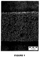
- Figure 1 represents a scanning electron microscopy image of one face of such a sponge obtained from a blend of oxidized collagen and chitosan.
- Additives such as fucans, native or chemically modified glucosaminoglycans, which may induce self chemical crosslink between collagen and glucosaminoglycans (hyaluronic acid, sulphate chondroitin, etc), oxidized starch, and any other product which may enhance tissue repair, limit the risk of sepsis, and modulate the mechanical properties of the composite (swelling rate in water, tensile strength, etc) could have been be added to the blend of oxidized collagen and chitosan.
- non-neutralized freeze-dried sponges were measured before and after the calendaring step, and then after hydration for 1 minute in a buffer (PBS IX) solution at 20°C as shown in Table 4.
- Table 4 Swelling properties of non-neutralized freeze-dried sponges in different states.
- a collagen/chitosan mixture was prepared by mixing an acidic solution of oxidized collagen and acidic solution of chitosan in different proportions as described above in Example 1, with a final polymer (collagen/chitosan) concentration of 2% (w/w).
- the mixture was poured into cylindrical moulds of different diameters ranging from 1 mm to 10 mm and freeze-dried for about 24 hours.
- the cylinders were neutralized in a buffer solution of PBS 1X for about 2 hours and then dried in a ventilated oven at 35°C overnight.
- a collagen/chitosan mixture was prepared by mixing an acidic solution of oxidized collagen and acidic solution of chitosan in different proportions as described above in Example 1, with a final polymer (collagen/chitosan) concentration of 2% (w/w).
- the mixture (about 40g) was poured into tubular moulds of different diameters ranging from 5 mm to 15 mm and freeze-dried for 24 hours.
- the tubes were neutralized in a buffer solution of PBS 1X for 2 hours and then dried in a ventilated oven at 35°C overnight.
- a 20/80 mixture of the oxidized collagen and chitosan with a final polymer (oxidized collagen/chitosan) concentration of 0.5% (w/w) with a pH adjusted to 5 was used to coat the external surface of the tubular structure bringing different permeability properties to the tubular composite material.
- a collagen/chitosan mixture was prepared as described above in Example 1, with a final polymer (collagen/chitosan) concentration of 2% (w/w).
- a sterile concentrated solution of PEG 4000 (polyethylene glycol having a molecular weight of 4000 daltons) and glycerol was added to the collagen/chitosan mixture, in order to achieve a PEG concentration of 1% and a glycerol concentration of 0.6%.
- the pH of the solution was adjusted to 6.5 by adding concentrate sodium hydroxide solution.
- the volume of the solution was then adjusted with sterile water to obtain final concentrations of collagen/chitosan, PEG, and glycerol, of 2%, 0.9%, and 0.54%, respectively.
- the solution wa is distributed in a thin layer, having a density of 0.133 g/cm 2 , on a flat hydrophobic support of PVC or polystyrene.
- Additives such as fucans, either native or chemically modified glycosaminoglycans, which may induce self-chemical crosslink between collagen and glycosaminoglycans, oxidized starch, and any other products which may enhance tissue repair, limit the risk of sepsis, and modulate the mechanical properties of the composite (such as the swelling rate in water, tensile strength, etc) may be added to the blend of oxidized collagen/chitosan.
- 100 ml of an oxidized collagen/chitosan mixture was prepared by mixing 20g of an acidic solution of oxidized collagen (pH 3.5) and 80g of an acidic solution of chitosan (pH 3.5) leading to a proportion of 20/80, with a final polymer (oxidized collagen/chitosan) concentration of 2.4% (w/w).
- the solution was then degassed by centrifugation for 10 min at 10 000 RPM at room temperature.
- the solution was spun by a spinneret with an interior diameter of 0.8 mm in a 1N sodium hydroxide bath. Then the yarn is washed with deionized water and dried ballasted by a mass of 1g at room temperature.
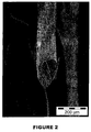
- Figure 2 represents a scanning electron microscopy image of such a yarn.
- Additives such as fucans, nanoparticles i.e. Ag + or Cu2 + for they antimicrobial properties, and any other products which may enhance tissue repair, limit the risk of sepsis, and modulate the mechanical properties of the composite (such as the swelling rate in water, tensile strength, etc) may be added to the blend of oxidized collagen/chitosan.
- oxidized collagen/chitosan mixture 100 ml of an oxidized collagen/chitosan mixture was prepared by mixing 20g of an acidic solution of oxidized collagen (pH 3.5) and 80g of an acidic solution of chitosan (pH 3.5) leading to a proportion of 20/80, with a final polymer (oxidized collagen/chitosan) concentration of 3% (w/w).
- an equivalent amount of alcohol e.g. glycerol or 1,2-propandiol
- Oxidized collagen was obtained by the oxidation of a 3% collagen solution by periodic acid, at a final concentration of 8mM, at room temperature, during 3 hours, as described above in Example 1.
- the chitosan was solubilized in deionized water with a stoechiometric amount of hydrochloric acid with a polymer concentration of 1% (w/w).
- the pH of the chitosan solution was adjusted to 3 to stop the crosslink kinetic reaction between the oxidized collagen and chitosan.
- Two-dimensional or three-dimensional meshes made of PLA or PET were soaked once, twice, or three times in an oxidized collagen/chitosan mixture, then dried and neutralized with an alkaline bath so as to cover the accessible surface of the PLA or PET fibers of the mesh.
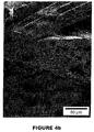
- Figures 4 through 6B represent scanning electron microscopy images of such meshes.
- the present dural repair materials may include one or two non-porous layers, a porous layer, and if necessary a reinforcement member e.g. textile.
- Two-dimensional meshes made of PLA or PET were soaked once, twice, or three times in an oxidized collagen/chitosan mixture, then dried and neutralized with an alkaline bath so as to cover the accessible surface of the PLA or PET fibers of the mesh.
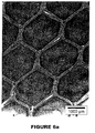
- Figures 4A , 4B , 6A , and 6B represent scanning electron microscopy images of such two-dimensional meshes.
- Three-dimensional meshes made of monofilaments and multifilament PLA threads were soaked once, twice or three times in an oxidized collagen/chitosan mixture, then dried and neutralized with an alkaline bath so as to cover the accessible surface of the PLA filaments of the mesh.
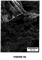
- Figures 5A and 5B represent scanning electron microscopy images of such three-dimensional meshes.
- Two-dimensional meshes made of oxidized collagen/chitosan mixture were knitted from the yarn obtained by the process described in the Example 5.
- a collagen/chitosan mixture was prepared by mixing an acidic solution of oxidized collagen and an acidic solution of chitosan with respectively 30% and 70% composition in mass.
- the acidic solution of chitosan is composed of two different degree of acetylation of 2.5 % and 26 % in the respectively proportions of 30% and 70% .
- the final polymer (oxidized collagen/chitosan) concentration is about 1% (w/w).
- the blend of oxidized collagen and chitosan (approximately 121 g) were poured within a 12cm by 7cm plastic box and freeze-dried for 24 hours. The samples were then neutralized in a 20% ammonia bath for 1 hour and thoroughly washed in deionized water until the pH reached 7. The freeze-dried sponges were then calendered to obtain a material with a final thickness of 0.13mm.
- the pH of the suspension was adjusted to 7.0 by adding concentrate sodium hydroxide solution.
- the volume of the solution was adjusted with sterile water to obtain a final concentration of collagen, chitosan, PEG, and glycerol of 2.7%, 0.55%, 0.9%, and 0.54% respectively.
- the oxidized collagen solution was then poured into a thin layer on a flat hydrophobic support of PVC or polystyrene, with a density of 0.133 g solution/cm 2 .
- the layer is then exposed to a sterile stream of air at ambient temperature leading to complete evaporation in approximately 18 hours.
- a thin layer of an oxidized collagen solution was poured on a flat hydrophobic support of PVC or polystyrene, with a density of 0.400 g solution/cm 2 .
- the surfaces were then exposed to a sterile stream of air at ambient temperature for less than one hour.
- a calendered sponge was then gently applied on the gelling layer of the oxidized collagen and the two layers were exposed to a sterile stream of air at ambient temperature overnight.
- a second layer of oxidized collagen solution was then distributed on the bi-layer composite with a reduced density of 0.133 g solution/cm 2 .
- the three layers composite was then exposed to a sterile stream of air at ambient temperature, leading to complete evaporation in approximately 18 hours.
- the composite material was then sterilized by gamma radiation.
- a thin layer of an oxidized collagen solution was poured on a flat hydrophobic support of PVC or polystyrene, with a density of 0.400 g solution/cm 2 .
- a textile reinforcement member (based on PLA or oxidized collagen/chitosan) was then laid over the collagen solution, and pressed into the solution. Additional solution was applied on top of the original volume of solution to ensure the reinforcement member was completely embedded within the solution.
- the surfaces were then exposed to a sterile stream of air at ambient temperature for less than one hour.
- a calendered sponge was then gently applied on the gelling layer of the oxidized collagen and the two layers were exposed to a sterile stream of air at ambient temperature overnight.
- a second layer of oxidized collagen solution was then distributed on the bi-layer composite with a reduced density of 0.133 g solution/cm 2 .
- the three layers composite was then exposed to a sterile stream of air at ambient temperature, leading to complete evaporation in approximately 18 hours.
- the composite material was then sterilized by gamma radiation.
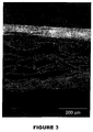
- Figure 3 represents a scanning electron microscopy image of such a multilayer implant.
Abstract
Description
- The present disclosure relates to biocompatible and bioresorbable compounds containing a functionalized collagen covalently bonded directly to a glycosaminoglycan without the use of a chemical cross-linking agent. The present compounds and compositions containing them are useful for a variety of medical applications, including surgical implants.
- Collagen and glycosaminoglycans have been combined for the preparation of biomaterials and surgical implants. Non-crosslinked collagen and chitosan mixtures have weak mechanical properties rendering their manipulation difficult and the in-vivo biodegradation of the collagen is often insufficient.
- Cross linking of collagen and glycosaminoglycans, e.g. chitosan, using a cross linker agent such as glutaraldehyde is inconvenient in certain applications. For example, the use of glutaraldehyde in aqueous media leads to the formation of very high molecular glutaraldehyde polymers which are difficult to eliminate by simple washing techniques. Upon implantation, such glutaraldehyde polymers may hydrolyse and cause a release of glutaraldehyde or remain in-vivo and be liberated after the disappearance of the collagen/chitosan components.
- It would be advantageous to provide biocomposites or implants made of functionalized collagen and glycosaminoglycans. The formulation advantageously provides a tailor-made, self cross-linked glycoprotein network and may be based on highly purified and fully characterized extra-cellular matrix compounds which mimic the native extracellular matrix and provide an optimal support for cell differentiation and growth and for tissue regeneration. The relative amounts of oxidized collagen and glycosaminoglycans may be varied to optimize the biological, mechanical and biodegradation properties of the appropriate tissue to be repaired and/or regenerated. When compared to implants based only on collagen, the formulations described herein can also favor the repair and/or the regeneration of tissues by the release of glycosaminoglycan oligomers, showing interesting biological properties (eg. angiogenic, antibacterial properties), in a time controlled fashion. The formulations can be also advantageously obtained under different physical forms by itself (eg. gel, film, sponge, yarn, knitted textile, woven textile, non-knitted non-woven mesh) or can be easily combined with other components in an open fashion.
- Accordingly, the present disclosure relates to compounds containing a functionalized collagen covalently bonded directly to a glycosaminoglycan without the use of a chemical cross-linking agent. In particular embodiments, the collagen is functionalized by oxidative cleavage : for example, this oxidative cleavage converts pendant portions of the collagen molecule into aldehydes which are reactive with the amine groups of the glycosaminoglycan. In embodiments, the functionalized collagen includes one or more reactive moieties selected from the group consisting of aldehydes, sulfones, vinylsulfones, isocyanates, and acid anhydrides. In embodiments, the functionalized collagen includes one or more reactive moieties selected from the group consisting of -CO2N(COCH2)2, -CO2N(COCH2)2, -CO2H, -CHO, -CHOCH2, - N=C=O, -SO2CH=CH2, -N(COCH)2, and -S-S-(C5H4N).
- In embodiments, the glycosaminoglyan is selected from the group consisting of dermatan sulfate, hyaluronic acid, chondroitin sulfate, chitin, chitosan, heparin, keratan sulfate, keratosulfate, deacylated hyaluronic acid and derivatives and combinations thereof. For example, the glycosaminoglycan is chitosan.
- Methods for preparing the compounds are also described. In particular, the present disclosure relates to a method of forming a bioresorbable compound comprising contacting a functionalized collagen with a glycosaminoglycan, in particular under reaction conditions under which the functionalized collagen covalently binds directly to the glycosaminoglycan, without the use of a crosslinking agent. In embodiments, a deionized water solution of functionalized collagen is combined to a deionized water solution of glycosaminoglycan to allow the functionalized collagen to mix with the glycosaminoglycan and form said compound. The pH of the solution of functionalized collagen may be adjusted between 2 and 7.5, for example by addition of a suitable acid. The pH of the solution of glycosaminoglycan may be adjusted between 2 and 7.5, for example by addition of a suitable acid. The present disclosure also relates to a compound obtainable by such a method.
- The present disclosure further relates to a mixture consisting in a deionized water solution of functionalized collagen, for example with an adjusted pH between 2 and 7.5, combined to a deionized water solution of glycosaminoglycan, for example with an adjusted pH between 2 and 7.5.
- The present disclosure also relates to an implant comprising a compound containing a functionalized collagen covalently bonded directly to a glycosaminoglycan without the use of a crosslinking agent. In embodiments, the implant of the present disclosure comprises a sponge containing said compound. In embodiments, the implant of the present disclosure comprises a textile containing said compound. In embodiments, the implant of the present disclosure comprises a hydrogel containing said compound. In embodiments, the implant of the present disclosure comprises threads containing said compound. In embodiments, the implant of the present disclosure comprises a non knitted, non woven composite containing said compound. In embodiments, the implant of the present disclosure comprises a film containing said compound. In embodiments, the implant of the present disclosure comprises a mesh coated with a composition containing said compound.
- The present disclosure further relates to a composition comprising a compound containing a functionalized collagen covalently bonded directly to a glycosaminoglycan without the use of a crosslinking agent.
- The present compounds may be used to form a variety of surgical implants such as gels, films, sponges, fibers, woven textiles, knitted textiles, non-woven, non-knitted textiles, and the like. In embodiments, the compounds may be combined with a substrate to form a coated implant or to add additional layers to the implant.
- The embodiments of the present disclosure will be described more clearly by means of the description which follows and the attached drawings in which:
-
Figure 1 represents a scanning electron microscopy image (Hitachi S800 scanning electron microscope with image acquisition and analysis system) of a freeze dried sponge according to the present disclosure, made from a 50/50 oxidized collagen (CXN)/chitosan mixture, from a side view; -
Figure 2 represents a scanning electron microscopy image (Hitachi S800 scanning electron microscope with image acquisition and analysis system) of a yarn according to the present disclosure, made from a wet-spinning process of an CXN/chitosan mixture; -
Figure 3 represents a scanning electron microscopy image (Hitachi S800 scanning electron microscope with image acquisition and analysis system) of a multilayer implant according to the present disclosure, including a textile made from polylactic acid (PLA), from a side view; -
Figure 4A represents a scanning electron microscopy images (Hitachi S800 scanning electron microscope with image acquisition and analysis system) of a two-dimensional textile according to the present disclosure, the textile being knitted with multifilaments of PLA and then coated three times with a 50/50 CXN/chitosan mixture; -
Figure 4B represents a scanning electron microscopy images (Hitachi S800 scanning electron microscope with image acquisition and analysis system) of a two-dimensional textile according to the present disclosure, at a higher magnification than forFigure 4A , the textile being knitted with multifilaments of PLA and then coated three times with a 50/50 CXN/chitosan mixture; -
Figure 5A represents a scanning electron microscopy images (Hitachi S800 scanning electron microscope with image acquisition and analysis system) of a three-dimensional textile according to the present disclosure, the textile being knitted with both monofilaments and multifilaments of PLA and being coated three times with a 50/50 CXN/chitosan mixture; -
Figure 5B represents a scanning electron microscopy images (Hitachi S800 scanning electron microscope with image acquisition and analysis system) of a three-dimensional textile according to the present disclosure, at a higher magnification than forFigure 5A , the textile - being knitted with both monofilaments and multifilaments of PLA and being coated three times with a 50/50 CXN/chitosan mixture;
-
Figure 6A represents a scanning electron microscopy images (Hitachi S800 scanning electron microscope with image acquisition and analysis system) of a two-dimensional textile according to the present disclosure, the textile being knitted with multifilaments of PET and then coated three times with a 50/50 CXN/chitosan mixture; -
Figure 6B represents a scanning electron microscopy images (Hitachi S800 scanning electron microscope with image acquisition and analysis system) of a two-dimensional textile according to the present disclosure, at a higher magnification than forFigure 4A , the textile being knitted with multifilaments of PET and then coated three times with a 50/50 CXN/chitosan mixture; and -
Figure 7 represents a viscometer reading of an oxidized collagen and chitosan mixture and a native collagen and chitosan mixture in accordance with the present disclosure. - Compounds containing a functionalized collagen covalently bonded directly to a glycosaminoglycan without the use of a chemical cross-linker agent are provided in accordance with the present disclosure. In embodiments, the compounds may be obtained by combining a reactive solution of collagen or gelatine, modified by a chemical reaction (e.g. oxidative cleavage) to functionalize a pendant portion of the collagen with moieties which are capable of forming a covalent bond with the reactive moieties of the glycosaminoglycan. The compounds, processes for preparing the compounds and design of surgical implants using the compounds are described in greater detail below. The methods for producing the product of the present disclosure make use of steps that are recognized as effective for inactivating viral particules and prions. Advantageously, the collagen and glycosaminoglycan may be highly purified and totally free of pendant residues providing a real advantage comparatively to the extracellular matrix made from biological tissues such as small intestine, sub mucosa or dermis. This gives the product a very high safety level while eliminating the inflammatory response.
- Collagen is a naturally occurring protein featuring good biocompatibility. It is the major structural component of vertebrates, forming extracellular fibers or networks in practically every tissue of the body, including skin, bone, cartilage, and blood vessels. In medical devices, collagen provides a more physiological, isotropic environment that has been shown to promote the growth and function of different cell types, facilitating the rapid overgrowth of host tissue after implantation.
- For the purpose of the present application, the term "collagen" is intended to mean any known collagen of porcine, bovine or human origin, for example natural or recombinant collagen, esterified collagen, for example methylated, ethylated or alternatively succinylated collagen, glycosylated collagen (eg. collagen glycosylated with saccharides / polysaccharides comprising free amino groups, collagen glycosylated with saccharides / polysaccharides comprising vicinal diols, collagen glycosylated with saccharides / polysaccharides comprising - CHx(NH2)-CHy(OH)- chemical bonds), or one of its derivatives.
- The term "gelatine" here includes commercial gelatine made of collagen which has been denatured by heating and in which the chains are at least partially hydrolyzed (molecular weight lower than 100 kDa).
- The collagen used can be of human or animal origin. Some non-limiting examples include, type I porcine or bovine collagen, type I or type III human collagen or mixtures in any proportions of these types. In embodiments, the collagen or gelatine used is a porcine collagen.
- The collagen can be modified by using any method known to those skilled in the art to provide pendant portions of the collagen with moieties which are capable of covalently bonding with the reactive chemical groups of a glycosaminoglycan. Examples of such pendant moieties include aldehydes, sulfones, vinylsulfones, isocyanates, and acid anhydrides. In addition, electrophilic groups such as -CO2N(COCH2)2, -CO2N(COCH2)2 -CO2H, -CHO, -CHOCH2, - N=C=O, -SO2CH=CH2, -N(COCH)2, -S-S-(C5H4N) may also be added to pendant chains of the collagen to allow covalent bonding to occur with the glycosaminoglycans.
- In embodiments, the collagen may be modified through the addition of an oxidizing agent. Contacting collagen with an oxidizing agent creates oxidative cleavage along portions of the collagen thereby creating pendant aldehyde groups capable of reacting with the glycosaminoglycans. The oxidizing agent may be, for example, iodine, peroxide, periodic acid, hydrogen peroxide, a periodate, a compound containing periodate, sodium periodate, a diisocyanate compound, a halogen, a compound containing halogen, n-bromosuccinimide, a permanganate, a compound containing permanganate, ozone, a compound containing ozone, chromic acid, sulfuryl chloride, a sulfoxide, a selenoxide,. an oxidizing enzyme (oxidase) and combinations thereof. In embodiments, the oxidizing agent is periodic acid.
- An example of the oxidative technique is described by
Tardy et al. in U.S. Patent No. 4,931,546 , the entire content of which is herein incorporated by reference. Briefly, this technique involves mixing the collagen in acid solution with an oxidizing agent, i.e., a solution of periodic acid or one of its salts, at a concentration of between 1 and 10-5 M, in embodiments between 5 10-3 and 10-1 M, at a temperature of between 10 and 25° C for 10 minutes to 72 hours. This process breaks down hydroxylysine and the sugars of the collagen, thus creating reactive sites without causing crosslinking. The oxidative cleavage of collagen allows moderate cross-linking later in the collagenic material. - Another technique for oxidized collagen is by oxidation of a 3% collagen solution by periodic acid, at a final concentration of 8mM, during 3 hours, as described by
Bayon, et al.in U.S. Patent No. 6,596,304 , the entire content of which is herein incorporated by reference. Aldehyde groups are formed by oxidative cleavage on the lateral chains of the hydroxyl-lysine residues giving the oxidized collagen capabilities to form covalent bonds with amines. The oxidized collagen can be fully degraded in vivo, after a few weeks, and while not wishing to be bound by any theory, it is believed that the oxidized collagen will degrade before the glycosaminoglycan. - The term "glycosaminoglycan" is intended to encompass complex polysaccharides having repeating units of either the same saccharide subunit or two different saccharide subunits. Some non-limiting examples of glycosaminoglycans include dermatan sulfate, hyaluronic acid, the chondroitin sulfates, chitin, heparin, keratan sulfate, keratosulfate, and derivatives thereof. Some non-limiting examples of derivatives may include partially and fully deacylated versions of these compounds such as chitosan and deacylated hyaluronic acid. The glycosaminoglycans may be extracted from a natural source, e.g. animal tissues such as squid pens and shrimp shells or vegetable sources such as mushrooms (eg "champignon de Paris"), or they may be synthetically produced or synthesized by modified microorganisms such as bacteria.
- In embodiments, the functionalized collagen may be combined with a glycosaminoglycan such as chitosan to crosslink and form covalent bonds. The glycosaminoglycan may display a degree of acetylation (DA) of about 0% to about 60%. In embodiments, the glycosaminoglycan displays a degree of acetylation (DA) of about 0.5% to about 50%. Samples of different degrees of acetylation can be obtained either by a heterogeneous deacetylation process or by a homogenous reacetylating process from a sample of a glycosaminoglycan that is fully deacetylated. In embodiments, the glycosaminoglycan includes a blend of chitosan with different degree of acetylation selected from about 0.5 to 60%.
- In embodiments, the glycosaminoglycan has a molecular weight ranging from about 100 to about 1,000,000 g/mol. In some embodiments, the glycosaminoglycan has a molecular weight ranging from about 164 (chitosan monomer) to about 1,000,000 g/mol. In embodiments, the glycosaminoglycan has a molecular weight of about 1500 to about 800,000 g/mol. In addition, the glycosaminoglycan may also display a low polydisperity index between about 1.2 to about 1.8. In particularly useful embodiments, the glycosaminoglycan is chitosan. Nevertheless, the glycosaminoglycan may be a mixture of chitosans with different degrees of acetylation or a mixture of chitosans and other glycosaminoglycans, e.g. hyaluronic acid, with different degrees of acetylation and in which all glycosaminoglycan have the capabilitiy, i.e. have free amino groups, to be cross-linked to the oxidized collagen.
- Compounds in accordance with the present disclosure are made by reacting a functionalized collagen with a glycosaminoglycan under conditions which cause the two components to form covalent bonds without the use of a chemical crosslinking agent. The two components may take the form of any solution, suspension, emulsion, semi-solid, or solid material capable of allowing the two-components to interact and crosslink.
- In embodiments, each component is solubilized in an acceptable solvent such as deionized water to form two separate solutions. The two solutions may be combined to allow the two components to mix and form the compounds described herein. In particular embodiments, the glycosaminoglycan is solubilized in deionized water with a stoechiometric amount of hydrochloric acid with a polymer (glycosaminoglycan) concentration ranging from about 0.5% to about 10% (w/w). It is envisioned that the pH of the glycosaminoglycan solution can be adjusted if necessary between about 2 and about 7.5 depending on the degree of acetylation. The functionalized collagen is also solubilized in an acceptable solvent such as deionized water to a concentration ranging from about 0.5% to about 10% (w/w). It is also envisioned that the pH of the functionalized collagen solution may be adjusted between about 2 and about 7.5. The two components in solution are mixed to a final concentration of polymer (compound functionalized collagen/glycosaminoglycan) ranging from 0.5% to 20% (w/w). In embodiments, different proportions between the functionalized collagen and the glycosaminoglycan may be used. In particular embodiments, the glycosaminoglycan may be composed of a mixture of chitosans with different degrees of acetylation (DA). The chitosan having a degradation time in function with its degree of acetylation (K.Kurita et al,Carbohydrate polymers. ; K.Tomihata et al, Biomaterials. Vol 18 n°7 pp.567-575,1997), the combination of slow and fast biodegradable chitosan is an important issue for the awaiting properties of the implant, i.e., progressive cell colonization of the sponge. In fact, the degradation of the slow biodegradable oxidized collagen and chitosan with high DA, i.e. 35≤DA≤50, in vitro in the presence of viable cells and in vivo, helps to increase the interconnected porosity which is a key parameter for the regeneration of healthy native like tissue in the full thickness of the implant and the extent of tissue integration. In embodiments, molecules released from the controlled degradation of the biocomposite, for example oxidized collagen/chitosan, may advantageously confer to the implant highly interesting biological activities e.g. antimicrobial, anticancer, antioxidant, and immunostimulant effects, especially in the case of chitosan (S-K. Kim et al, Carbohydrate Polymers, Vol. 62, Issue 4, pp.357-368,2005) and may bring, in complement of the biocompatibility and biodegradability, bioactive properties to the medical devices. The biological properties of released chitosan oligopolymers enhance the tissue regeneration and extend the use of the implant, e.g. to surgical sites with a high risk of contamination.
- In embodiments, a combination of two solutions comprising an acidic solution of oxidized collagen and an acidic solution of chitosan with one or a mix of several degree of acetylation may be used. The collagen is oxidized by the addition of periodic acid as the oxidizing agent and the chitosan solution is made acidic by the addition of hydrochloric acid. The mixture can be neutralized either with an alkaline vapour/solution or buffer solution with a pH greater than 7, leading to a cross-linked scaffold compatible for cell adhesion and proliferation. This combination is particularly advantageous compared to a combination of oxidized collagen and glutaraldehyde cross-linked collagen, because the latter makes a suspension which is difficult to incorporate in a homogeneous fashion to an implantable surgical device such as a three-dimensional mesh.
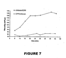
- The reaction between the functionalized collagen and glycosaminoglycan is characterized by a rapid increase of the viscosity of the reaction mixture when the two components are mixed. Viscosity measurements were performed on a viscosimeter Lamy type TVe-05. The solutions of oxidized collagen and chitosan were equilibrated at the temperature of 25°C for 1 hour and then mixed. A sample of 5ml was poured into the chamber of the viscosimeter and the evolution of viscosity against time was studied. The viscosity of the solution composed of oxidized collagen and chitosan and a solution composed of native collagen and chitosan were compared to highlight the type of interactions between the oxidized collagen and the chitosan.
- The tests were performed at 25°C with biopolymers having characteristics described below:
- Chitosan DA = 2.5% and Mw = 500,000 g/mol
- Oxidized collagen (CXN) prepared from native collagen by oxidative cleavage.
- Native collagen (CPP) without telopeptide and with helicoidal structure preserved. The average molecular weight is about 300,000 g/mol.
- The solution prepared for the tests had a final polymer (collagen/glycosaminoglycan) concentration of 1% (w/w), a proportion close to 50/50 respectively of collagen and chitosan. The pH measured was close to 4.7 and 4.89, respectively, for the CXN/chitosan and CPP/chitosan mixtures.
- As shown in
Figure 7 , there is an increase of viscosity in the case of the oxidized collagen/chitosan mixture wherein the area of stability was reached for 30 minutes at a pH of 4.7. On the other hand, only a slight increase of viscosity is observed in the case of the native collagen/chitosan solution. Therefore, the difference of the viscosity evolution against time can be attributed, partly, to the formation of a chemical crosslink between oxidized collagen and chitosan. In fact, the low viscosity increase in the case of chitosan/native collagen mixture is due to the ionic complex because the two components exhibit a high charge of density. - The cross-linked mixture of functionalized collagen and a glycosaminoglycan, ie the compound functionalized collagen/glycosaminoglycan of the present disclosure, can be used to form a variety of surgical implants such as sponges, films, hydrogels, non-woven non-knitted meshes, three-dimensional structures such as tubular and spherical structures, microbeads, threads, rods, filaments, yarns, meshes, slings, sutures and other composite materials such as pledgets, buttresses, adhesion barriers and the like. The mixture can also be combined with or used to coat surgical implants, such as two-dimensional meshes, three-dimensional meshes, vascular prostheses, patches, slings and the like.
- The surgical implants which may be combined or coated with compositions which include the compounds of the present disclosure may be made from bioabsorbable or non-bioabsorbable materials. Some non-limiting examples of suitable non-absorbable materials which may be utilized included polyolefins, such as polyethylene, polypropylene, copolymers of polyethylene and polypropylene, and blends of polyethylene and polypropylene. Other non-absorbable materials which may be utilized include polyesters such as polyethylene terephthalate (PET), polyamides, aramides, expanded polytetrafluoroethylene, polyurethane, polyvinylidene, difluoride (PVDF), polybutester, copper alloy, silver alloy, platinum, medical grade stainless steels such as 316L medical grade stainless steel, combinations thereof, and the like. Examples of commercially available polypropylene-based textile supports which may be utilized include those sold under the brand name PARIETENE® from Sofradim.
- Suitable absorbable materials include, but are not limited to, trimethylene, carbonate, caprolactone, dioxanone, glycolic acid, lactic acid, glycolide, lactide, homopolymers thereof, copolymers thereof, and combinations thereof. Specific absorbable materials which may be suitable include, for example, chitosan, cellulose, oxidized cellulose, combinations thereof, and the like.
- In embodiments, a solution of the present compounds may be freeze-dried to form a porous sponge material capable of allowing tissue in growth and induce a progressive cell colonization of the sponge by mixing several glycosaminoglycans with different degrees of acetylation and with different degradation properties. In embodiments, the solutions described herein may include additional polymeric materials which allow the solution to form a non-porous film useful in preparing adhesion barriers. In particular, the compounds of the present disclosure may be combined with polyethylene glycol, and glycerol to form a non-porous film. In still other embodiments, the sponges or films or hydrogel materials as described herein may be used to add a coating layer on an existing surgical implant or to form a multilayer surgical implant. Such combination implants may be useful in forming surgical implants which prevent adhesions and the in-growth of tissue one side of the implant and encourage the in-growth of tissue and formation of adhesions on the other side of the implant. Some non-limiting examples include multilayer pledgets, buttresses, surgical meshes, slings and adhesion barriers.
- In embodiments, a solution of the present compounds may be used to form yarn by a wet spinning process as described in the patent
EP0328050A2 by Bisento de rutsuka et al. The biological composite yarns are fully biocompatible and biodegradable with a wide range of degradation times due to the mix of several glycosaminglycans with different degrees of acetylation. The composite yarns of the present disclosure may be used to knit textiles with different patterns in 2 or 3 dimensions and these yarns may be used alone or combined with other biocompatible yarns such as yarns made from polylactic acid (PLA). The textiles may be employed as implants or as a part of an implant to improve the mechanical properties of the implant. Moreover with the functionalized collagen/glycosaminoglycan composition of the present disclosure, the textile may have high biocompatibility and good mechanical properties in a wide range of degradation times, ranging from about 2 weeks to several months. Advantageously, the molecules released from the degradation of the biocomposite or compound of the present disclosure, for example oxidized collagen/chitosan, give biological activities of particular interest, i.e., antimicrobial, anticancer, antioxidant, and immunostimulant effects, especially in the case of chitosan. - In embodiments, at least one bioactive agent may be included in compositions containing the present compounds and thereby incorporated into a medical device. In these embodiments, the implant can also serve as a vehicle for delivery of the bioactive agent. The term "bioactive agent", as used herein, is used in its broadest sense and includes any substance or mixture of substances that have clinical use. Consequently, bioactive agents may or may not have pharmacological activity per se, e.g., a dye, or fragrance. Alternatively a bioactive agent could be any agent which provides a therapeutic or prophylactic effect, a compound that affects or participates in tissue growth, cell growth, cell differentiation, an anti-adhesive compound, a compound that may be able to invoke a biological action such as an immune response, or could play any other role in one or more biological processes. It is envisioned that the bioactive agent may be applied to the medial device in any suitable form of matter, e.g., films, powders, liquids, gels and the like.
- Examples of classes of bioactive agents which may be utilized in accordance with the present disclosure include anti-adhesives, antimicrobials, analgesics, antipyretics, anesthetics, antiepileptics, antihistamines, anti-inflammatories, cardiovascular drugs, diagnostic agents, sympathomimetics, cholinomimetics, antimuscarinics, antispasmodics, hormones, growth factors, muscle relaxants, adrenergic neuron blockers, antineoplastics, immunogenic agents, immunosuppressants, gastrointestinal drugs, diuretics, steroids, lipids, lipopolysaccharides, polysaccharides, and enzymes. It is also intended that combinations ofbioactive agents may be used.
- Anti-adhesive agents can be used to prevent adhesions from forming between the implantable medical device and the surrounding tissues opposite the target tissue. In addition, anti-adhesive agents may be used to prevent adhesions from forming between the coated implantable medical device and the packaging material. Some examples of these agents include, but are not limited to poly(vinyl pyrrolidone), carboxymethyl cellulose, hyaluronic acid, polyethylene oxide, poly vinyl alcohols and combinations thereof.
- Suitable antimicrobial agents which may be included as a bioactive agent in the bioactive coating of the present disclosure include triclosan, also known as 2,4,4'-trichloro-2'-hydroxydiphenyl ether, chlorhexidine and its salts, including chlorhexidine acetate, chlorhexidine gluconate, chlorhexidine hydrochloride, and chlorhexidine sulfate, silver and its salts, including silver acetate, silver benzoate, silver carbonate, silver citrate, silver iodate, silver iodide, silver lactate, silver laurate, silver nitrate, silver oxide, silver palmitate, silver protein, and silver sulfadiazine, polymyxin, tetracycline, aminoglycosides, such as tobramycin and gentamicin, rifampicin, bacitracin, neomycin, chloramphenicol, miconazole, quinolones such as oxolinic acid, norfloxacin, nalidixic acid, pefloxacin, enoxacin and ciprofloxacin, penicillins such as oxacillin and pipracil, nonoxynol 9, fusidic acid, cephalosporins, and combinations thereof. In addition, antimicrobial proteins and peptides such as bovine lactoferrin and lactoferricin B and antimicrobial polysaccharides such as fucans and derivatives may be included as a bioactive agent in the bioactive coating of the present disclosure.
- Other bioactive agents which may be included as a bioactive agent in the coating composition applied in accordance with the present disclosure include: local anesthetics; non-steroidal antifertility agents; parasympathomimetic agents; psychotherapeutic agents; tranquilizers; decongestants; sedative hypnotics; steroids; sulfonamides; sympathomimetic agents; vaccines; vitamins; antimalarials; anti-migraine agents; anti-parkinson agents such as L-dopa; anti-spasmodics; anticholinergic agents (e.g. oxybutynin); antitussives; bronchodilators; cardiovascular agents such as coronary vasodilators and nitroglycerin; alkaloids; analgesics; narcotics such as codeine, dihydrocodeinone, meperidine, morphine and the like; non-narcotics such as salicylates, aspirin, acetaminophen, d-propoxyphene and the like; opioid receptor antagonists, such as naltrexone and naloxone; anti-cancer agents; anti-convulsants; anti-emetics; antihistamines; anti-inflammatory agents such as hormonal agents, hydrocortisone, prednisolone, prednisone, non-hormonal agents, allopurinol, indomethacin, phenylbutazone and the like; prostaglandins and cytotoxic drugs; estrogens; antibacterials; antibiotics; anti-fungals; anti-virals; anticoagulants; anticonvulsants; antidepressants; antihistamines; and immunological agents.
- Other examples of suitable bioactive agents which may be included in the coating composition include viruses and cells, peptides, polypeptides and proteins, analogs, muteins, and active fragments thereof, such as immunoglobulins, antibodies, cytokines (e.g. lymphokines, monokines, chemokines), blood clotting factors, hemopoietic factors, interleukins (IL-2, IL-3, IL-4, IL-6), interferons ((3-IFN, (a-IFN and y-IFN), erythropoietin, nucleases, tumor necrosis factor, colony stimulating factors (e.g., GCSF, GM-CSF, MCSF), insulin, anti-tumor agents and tumor suppressors, blood proteins, gonadotropins (e.g., FSH, LH, CG, etc.), hormones and hormone analogs (e.g., growth hormone), vaccines (e.g., tumoral, bacterial and viral antigens); somatostatin; antigens; blood coagulation factors; growth factors (e.g., nerve growth factor, insulin-like growth factor); protein inhibitors, protein antagonists, and protein agonists; nucleic acids, such as antisense molecules, DNA and RNA; oligonucleotides; polynucleotides; and ribozymes.
- Bioactive agents can also be additives, such as fucans, either native or chemically modified glucosaminoglycans, oxidized starch, emulsifiers, surfactants, humectants, buffering agents, pH modulators, chelating agents, viscosity agents and any other products which may enhance tissue repair, limit the risk of sepsis, and modulate mechanical properties of the compounds.
- The following non-limiting examples show the preparation, formulation and uses possible of the present compounds and the tensile and swelling properties of the oxidized collagen and chitosan mixture compared to a native collagen and chitosan mixture.
- A collagen/chitosan mixture was prepared by mixing an acidic solution of oxidized collagen and an acidic solution of chitosan in different proportions with a final polymer (collagen/chitosan) concentration of 1% (w/w).
- Oxidized collagen was obtained by the oxidation of a 3% collagen solution by periodic acid, at a final concentration of 8mM, at room temperature, during 3 hours, as described by Bayon, et al. in Example 4 of
U.S. Pat. No. 6,596,304 . At this step the pH of the oxidized collagen solution was about 3.2. - Solutions of native collagen were obtained by solubilizing collagen powder at a 1% final concentration, in sterile water. The pH measured close to 3.
- The chitosan was solubilized in deionized water with a stoechiometric amount of hydrochloric acid with a polymer concentration of 1% (w/w). The pH of the chitosan solution was about 5, but the pH could have been adjusted to 3 to have better control of the crosslink kinetic between the oxidized collagen and chitosan.
- Before freeze drying, if the application required it, the collagen/chitosan mixture could have been poured into a 3D mesh so as to fully cover the mesh and obtain a freeze dried sponge/mesh composite. The presence of the 3D mesh facilitates fastening the implant to tissue (e.g., via suturing). Moreover, the homogeneity of the oxidized collagen/chitosan solution allows a better penetration of the solution within a 3-dimensional structure of the textile when compared to collagen that has been oxidized with a cross-linking agent.
- Several mixtures of various blends of oxidized collagen and chitosan as well as native collagen and chitosan (approximately 121 g) were poured within a 12cm by 17cm plastic box and freeze-dried for 24 hours. The samples were then neutralized in a 1M sodium hydroxide bath for 1 hour and thoroughly washed in deionized water until the pH reached 7. The freeze-dried sponges were then calendered to obtain a material with a final thickness of 0.13mm.
-
Figure 1 represents a scanning electron microscopy image of one face of such a sponge obtained from a blend of oxidized collagen and chitosan. - Additives, such as fucans, native or chemically modified glucosaminoglycans, which may induce self chemical crosslink between collagen and glucosaminoglycans (hyaluronic acid, sulphate chondroitin, etc), oxidized starch, and any other product which may enhance tissue repair, limit the risk of sepsis, and modulate the mechanical properties of the composite (swelling rate in water, tensile strength, etc) could have been be added to the blend of oxidized collagen and chitosan.
- Tensile and suture tests were performed per Hounsfield H5KS (from Tinius Olsen) at room temperature on calendered freeze-dried sponges. Several blends of the native collagen and chitosan mixture and the oxidized collagen and chitosan mixture were prepared as described below in Table 1:
Table 1: Composition of collagen (oxidized or native) and chitosan of different freeze-dried calendered sponges. Batch % Oxidized collagen (w/w) % Native collagen (w/w) % Chitosan (w/w) % Polymer (collagen/chitosan) (w/w) RHF00004 / 50 50 1 RHF00006 80 / 20 1 RHF00007 50 / 50 1 RHF00008 20 / 80 1 - Several amounts of various blends (121 g) were poured within 12cm by 7cm plastic boxes and freeze-dried for 24 hours. The samples were cut to desired dimensions as described below in Table 2, neutralized, and washed in deionized water. The freeze-dried sponges were calendered to obtain a material with a final thickness of 0.13 mm. The mechanical tests were performed on hydrated samples at a speed of 50 mm/min.
Table 2: Dimensions of the samples for mechanical tests Dimensions Tensile test 2.5 x 4 cm Suture test 4 x 4 cm - The results, summarized in Table 3, exhibit an increase in the breaking strength of the samples with the amount of chitosan present. Moreover, batch FHF00004 (native collagen and chitosan with a 50/50 blend) has lower mechanical properties than RHF00007 (oxidized collagen and chitosan with a 50/50 blend). The greater mechanical values of the RHF00007 batch confirm the chemical network, ie the covalent bonding of the oxidized collagen and the chitosan, in the case of a blend composed of chitosan and oxidized collagen.
Table 3: Breaking strength and deformation of the different batches of calendered, freeze-dried sponges determined by tensile and suture tests. Tensile Tests Suture Tests Batch Breaking Strength (N) Deformation (%) Breaking Strength (N) RHF00004 4.78 57.1 0.68 RHF00006 0.51 32 / RHF00007 8.34 39.4 0.67 RHF00008 7.75 61.7 1.46 - The thickness of non-neutralized freeze-dried sponges were measured before and after the calendaring step, and then after hydration for 1 minute in a buffer (PBS IX) solution at 20°C as shown in Table 4.
Table 4: Swelling properties of non-neutralized freeze-dried sponges in different states. Non-calendered Calendered Hydrated Oxidized collagen & Chitosan (50/50) 3.38 mm 0.13 mm 0.28 mm Native collagen & Chitosan (50/50) 3.46 mm 0.12 mm 1.2 mm - The values in Table 4, obtained in the case of the hydrated samples reveal increased swelling in the native collagen/chitosan composite, wherein only physical cross-linking occurred, compared to the oxidized collagen/chitosan composite (or compound), in which covalent bonds were formed.
- A collagen/chitosan mixture was prepared by mixing an acidic solution of oxidized collagen and acidic solution of chitosan in different proportions as described above in Example 1, with a final polymer (collagen/chitosan) concentration of 2% (w/w).
- The mixture was poured into cylindrical moulds of different diameters ranging from 1 mm to 10 mm and freeze-dried for about 24 hours.
- Thereafter, the cylinders were neutralized in a buffer solution of PBS 1X for about 2 hours and then dried in a ventilated oven at 35°C overnight.
- A collagen/chitosan mixture was prepared by mixing an acidic solution of oxidized collagen and acidic solution of chitosan in different proportions as described above in Example 1, with a final polymer (collagen/chitosan) concentration of 2% (w/w).
- The mixture (about 40g) was poured into tubular moulds of different diameters ranging from 5 mm to 15 mm and freeze-dried for 24 hours.
- Thereafter, the tubes were neutralized in a buffer solution of PBS 1X for 2 hours and then dried in a ventilated oven at 35°C overnight.
- Optionally, a 20/80 mixture of the oxidized collagen and chitosan with a final polymer (oxidized collagen/chitosan) concentration of 0.5% (w/w) with a pH adjusted to 5 was used to coat the external surface of the tubular structure bringing different permeability properties to the tubular composite material.
- A collagen/chitosan mixture was prepared as described above in Example 1, with a final polymer (collagen/chitosan) concentration of 2% (w/w).
- A sterile concentrated solution of PEG 4000 (polyethylene glycol having a molecular weight of 4000 daltons) and glycerol was added to the collagen/chitosan mixture, in order to achieve a PEG concentration of 1% and a glycerol concentration of 0.6%. The pH of the solution was adjusted to 6.5 by adding concentrate sodium hydroxide solution. The volume of the solution was then adjusted with sterile water to obtain final concentrations of collagen/chitosan, PEG, and glycerol, of 2%, 0.9%, and 0.54%, respectively.
- The solution wais distributed in a thin layer, having a density of 0.133 g/cm2, on a flat hydrophobic support of PVC or polystyrene.
- The surfaces were then exposed to a sterile stream of air at 35°C, leading to complete evaporation in about 12 hours.
- Additives, such as fucans, either native or chemically modified glycosaminoglycans, which may induce self-chemical crosslink between collagen and glycosaminoglycans, oxidized starch, and any other products which may enhance tissue repair, limit the risk of sepsis, and modulate the mechanical properties of the composite (such as the swelling rate in water, tensile strength, etc) may be added to the blend of oxidized collagen/chitosan.
- 100 ml of an oxidized collagen/chitosan mixture was prepared by mixing 20g of an acidic solution of oxidized collagen (pH 3.5) and 80g of an acidic solution of chitosan (pH 3.5) leading to a proportion of 20/80, with a final polymer (oxidized collagen/chitosan) concentration of 2.4% (w/w). The solution was then degassed by centrifugation for 10 min at 10 000 RPM at room temperature. The solution was spun by a spinneret with an interior diameter of 0.8 mm in a 1N sodium hydroxide bath. Then the yarn is washed with deionized water and dried ballasted by a mass of 1g at room temperature.
-
Figure 2 represents a scanning electron microscopy image of such a yarn. - Additives, such as fucans, nanoparticles i.e. Ag+ or Cu2+ for they antimicrobial properties, and any other products which may enhance tissue repair, limit the risk of sepsis, and modulate the mechanical properties of the composite (such as the swelling rate in water, tensile strength, etc) may be added to the blend of oxidized collagen/chitosan.
- 100 ml of an oxidized collagen/chitosan mixture was prepared by mixing 20g of an acidic solution of oxidized collagen (pH 3.5) and 80g of an acidic solution of chitosan (pH 3.5) leading to a proportion of 20/80, with a final polymer (oxidized collagen/chitosan) concentration of 3% (w/w). After complete homogenization, an equivalent amount of alcohol, e.g. glycerol or 1,2-propandiol, was added to the solution and the blend was gently stirred for 1 hour. The solution was degassed by centrifugation for 10 min at 10 000 RPM at room temperature. Then, 60g of the solution was poured within 12cm by 12cm Petri dishes and left to evaporate in a ventilated oven at 40°C for 24 hours. The alcohol gel was neutralized in a 4N NH4OH bath for 1 hour and then thoroughly washed in deionized water until the water pH was closed to a value of 7. The hydrogels were conserved in sterile water at 4°C.
- Oxidized collagen was obtained by the oxidation of a 3% collagen solution by periodic acid, at a final concentration of 8mM, at room temperature, during 3 hours, as described above in Example 1.
- The chitosan was solubilized in deionized water with a stoechiometric amount of hydrochloric acid with a polymer concentration of 1% (w/w). The pH of the chitosan solution was adjusted to 3 to stop the crosslink kinetic reaction between the oxidized collagen and chitosan.
- Two-dimensional or three-dimensional meshes made of PLA or PET were soaked once, twice, or three times in an oxidized collagen/chitosan mixture, then dried and neutralized with an alkaline bath so as to cover the accessible surface of the PLA or PET fibers of the mesh.
-
Figures 4 through 6B represent scanning electron microscopy images of such meshes. - The present dural repair materials may include one or two non-porous layers, a porous layer, and if necessary a reinforcement member e.g. textile.
- Two-dimensional meshes made of PLA or PET were soaked once, twice, or three times in an oxidized collagen/chitosan mixture, then dried and neutralized with an alkaline bath so as to cover the accessible surface of the PLA or PET fibers of the mesh.
-
Figures 4A ,4B ,6A , and6B represent scanning electron microscopy images of such two-dimensional meshes. - Three-dimensional meshes made of monofilaments and multifilament PLA threads were soaked once, twice or three times in an oxidized collagen/chitosan mixture, then dried and neutralized with an alkaline bath so as to cover the accessible surface of the PLA filaments of the mesh.
-
Figures 5A and5B represent scanning electron microscopy images of such three-dimensional meshes. - Two-dimensional meshes made of oxidized collagen/chitosan mixture were knitted from the yarn obtained by the process described in the Example 5.
- A collagen/chitosan mixture was prepared by mixing an acidic solution of oxidized collagen and an acidic solution of chitosan with respectively 30% and 70% composition in mass. The acidic solution of chitosan is composed of two different degree of acetylation of 2.5 % and 26 % in the respectively proportions of 30% and 70% .The final polymer (oxidized collagen/chitosan) concentration is about 1% (w/w).
- The blend of oxidized collagen and chitosan (approximately 121 g) were poured within a 12cm by 7cm plastic box and freeze-dried for 24 hours. The samples were then neutralized in a 20% ammonia bath for 1 hour and thoroughly washed in deionized water until the pH reached 7. The freeze-dried sponges were then calendered to obtain a material with a final thickness of 0.13mm.
- To a 3.9% oxidized collagen solution, an ultra-filtered concentration of solution of PEG 4000 (polyethylene glycol having a molecular weight of 4000 g/mol) and glycerol was added in order to achieve a PEG concentration of 1% and a glycerol concentration of 0.6%.
- The pH of the suspension was adjusted to 7.0 by adding concentrate sodium hydroxide solution.
- The volume of the solution was adjusted with sterile water to obtain a final concentration of collagen, chitosan, PEG, and glycerol of 2.7%, 0.55%, 0.9%, and 0.54% respectively.
- The oxidized collagen solution was then poured into a thin layer on a flat hydrophobic support of PVC or polystyrene, with a density of 0.133 g solution/cm2.
- The layer is then exposed to a sterile stream of air at ambient temperature leading to complete evaporation in approximately 18 hours.
- A thin layer of an oxidized collagen solution was poured on a flat hydrophobic support of PVC or polystyrene, with a density of 0.400 g solution/cm2.
- The surfaces were then exposed to a sterile stream of air at ambient temperature for less than one hour.
- A calendered sponge was then gently applied on the gelling layer of the oxidized collagen and the two layers were exposed to a sterile stream of air at ambient temperature overnight.
- A second layer of oxidized collagen solution was then distributed on the bi-layer composite with a reduced density of 0.133 g solution/cm2.
- The three layers composite was then exposed to a sterile stream of air at ambient temperature, leading to complete evaporation in approximately 18 hours.
- The composite material was then sterilized by gamma radiation.
- A thin layer of an oxidized collagen solution was poured on a flat hydrophobic support of PVC or polystyrene, with a density of 0.400 g solution/cm2.
- A textile reinforcement member (based on PLA or oxidized collagen/chitosan) was then laid over the collagen solution, and pressed into the solution. Additional solution was applied on top of the original volume of solution to ensure the reinforcement member was completely embedded within the solution.
- The surfaces were then exposed to a sterile stream of air at ambient temperature for less than one hour.
- A calendered sponge was then gently applied on the gelling layer of the oxidized collagen and the two layers were exposed to a sterile stream of air at ambient temperature overnight.
- A second layer of oxidized collagen solution was then distributed on the bi-layer composite with a reduced density of 0.133 g solution/cm2.
- The three layers composite was then exposed to a sterile stream of air at ambient temperature, leading to complete evaporation in approximately 18 hours.
- The composite material was then sterilized by gamma radiation.
-
Figure 3 represents a scanning electron microscopy image of such a multilayer implant. - It will be understood that various modifications may be made to the embodiments disclosed herein. Therefore, the above description should not be construed as limiting, but merely as an exemplification of preferred embodiments. Those skilled in the art will envision other modifications within the scope and spirit of the present disclosure. Such modifications and variations are intended to come within the scope of the following claims.
Claims (16)
- A compound containing a functionalized collagen covalently bonded directly to a glycosaminoglycan without the use of a crosslinking agent, wherein the glycosaminoglycan is chitosan and wherein the functionalized collagen includes one or more reactive moieties selected from the group consisting of aldehydes, sulfones, vinylsulfones, isocyanates, and acid anhydrides.
- A compound as in claim 1 wherein the functionalized collagen includes aldehydes as reactive moieties.
- A compound as in claim 1 or 2 wherein the collagen is functionalized by oxidative cleavage.
- The compounds of one of claims 1-3 wherein the functionalized collagen includes one or more reactive moieties selected from the group consisting of -CO2N(COCH2)2, - CO2N(COCH2)2, -CO2H, -CHO, -CHOCH2, -N=C=O, -SO2CH=CH2, -N(COCH)2, and -S-S-(C5H4N).
- A compound as in claim 1 wherein the glycosaminoglycan includes a blend of chitosan with different degree of acetylation selected from about 0.5 to 60%.
- An implant comprising a compound containing a functionalized collagen covalently bonded directly to a glycosaminoglycan without the use of a crosslinking agent, wherein the glycosaminoglycan is chitosan and wherein the functionalized collagen includes one or more reactive moieties selected from the group consisting of aldehydes, sulfones, vinylsulfones, isocyanates, and acid anhydrides.
- An implant as in claim 6 comprising a sponge containing said compound.
- An implant as in claim 6 comprising a textile containing said compound.
- An implant as in claim 6 comprising a hydrogel containing said compound.
- An implant as in claim 6 comprising threads containing said compound.
- An implant as in claim 6 comprising a non knitted, non woven composite containing said compound.
- An implant as in claim 6 comprising a film containing said compound.
- An implant as in claim 6 comprising a mesh coated with a composition containing said compound.
- A method of forming a bioresorbable compound comprising contacting a functionalized collagen with a glycosaminoglycan without the use of a crosslinking agent, wherein the glycosaminoglycan is chitosan and wherein the functionalized collagen includes one or more reactive moieties selected from the group consisting of aldehydes, sulfones, vinylsulfones, isocyanates, and acid anhydrides, wherein a deionized water solution of functionalized collagen is combined to a deionized water solution of chitosan to allow the functionalized collagen to mix with the chitosan and form said compound.
- The method of claim 14, wherein the pH of the solution of functionalized collagen is adjusted between 2 and 7.5.
- The method of one of claims 14 or 15, wherein the pH of the solution of glycosaminoglycan is adjusted between 2 and 7.5.
Applications Claiming Priority (2)
| Application Number | Priority Date | Filing Date | Title |
|---|---|---|---|
| US11/899,694 US20090068250A1 (en) | 2007-09-07 | 2007-09-07 | Bioresorbable and biocompatible compounds for surgical use |
| EP08829902.9A EP2203196B1 (en) | 2007-09-07 | 2008-09-05 | Bioresorbable and biocompatible compounds for surgical use |
Related Parent Applications (1)
| Application Number | Title | Priority Date | Filing Date |
|---|---|---|---|
| EP08829902.9 Division | 2008-09-05 |
Publications (2)
| Publication Number | Publication Date |
|---|---|
| EP2570140A2 true EP2570140A2 (en) | 2013-03-20 |
| EP2570140A3 EP2570140A3 (en) | 2013-07-17 |
Family
ID=40429469
Family Applications (2)
| Application Number | Title | Priority Date | Filing Date |
|---|---|---|---|
| EP12194841.8A Withdrawn EP2570140A3 (en) | 2007-09-07 | 2008-09-05 | Bioresorbable and biocompatible compounds for surgical use |
| EP08829902.9A Active EP2203196B1 (en) | 2007-09-07 | 2008-09-05 | Bioresorbable and biocompatible compounds for surgical use |
Family Applications After (1)
| Application Number | Title | Priority Date | Filing Date |
|---|---|---|---|
| EP08829902.9A Active EP2203196B1 (en) | 2007-09-07 | 2008-09-05 | Bioresorbable and biocompatible compounds for surgical use |
Country Status (5)
| Country | Link |
|---|---|
| US (2) | US20090068250A1 (en) |
| EP (2) | EP2570140A3 (en) |
| AU (1) | AU2008294417B2 (en) |
| CA (1) | CA2698638C (en) |
| WO (1) | WO2009031047A2 (en) |
Families Citing this family (35)
| Publication number | Priority date | Publication date | Assignee | Title |
|---|---|---|---|---|
| US20090036907A1 (en) * | 2007-07-30 | 2009-02-05 | Yves Bayon | Bioresorbable knit |
| DE102007047246A1 (en) * | 2007-09-26 | 2009-04-02 | Aesculap Ag | Reinforced vascular prosthesis with antimicrobial long-term effect |
| CA2740597C (en) * | 2008-10-17 | 2017-11-21 | Sofradim Production | Multi-layered surgical patches comprising a first hydrogel precursor and a second hydrogel precursor |
| WO2010043978A2 (en) * | 2008-10-17 | 2010-04-22 | Sofradim Production | Matrix for tissue repair |
| US8968762B2 (en) | 2010-03-26 | 2015-03-03 | Sofradim Production | Implant for tissue repair including chitosan |
| BR112013012772B1 (en) | 2010-11-23 | 2019-05-14 | Elastagen Pty Ltd | Preparation and / or formulation of polysaccharide crosslinked proteins |
| FR2972626B1 (en) | 2011-03-16 | 2014-04-11 | Sofradim Production | PROSTHETIC COMPRISING A THREE-DIMENSIONAL KNIT AND ADJUSTED |
| AU2012232814A1 (en) * | 2011-03-24 | 2013-10-03 | Sofradim Production | Enzymatically degradable compositions |
| TW201311303A (en) * | 2011-06-21 | 2013-03-16 | Bvw Holding Ag | Implant localization device |
| FR2977789B1 (en) | 2011-07-13 | 2013-07-19 | Sofradim Production | PROSTHETIC FOR UMBILIC HERNIA |
| US10813630B2 (en) | 2011-08-09 | 2020-10-27 | Corquest Medical, Inc. | Closure system for atrial wall |
| US10307167B2 (en) | 2012-12-14 | 2019-06-04 | Corquest Medical, Inc. | Assembly and method for left atrial appendage occlusion |
| US10314594B2 (en) | 2012-12-14 | 2019-06-11 | Corquest Medical, Inc. | Assembly and method for left atrial appendage occlusion |
| EP2760431A1 (en) * | 2011-09-30 | 2014-08-06 | Sofradim Production | Multilayer implants for delivery of therapeutic agents |
| FR2985170B1 (en) | 2011-12-29 | 2014-01-24 | Sofradim Production | PROSTHESIS FOR INGUINAL HERNIA |
| WO2013166616A1 (en) | 2012-05-11 | 2013-11-14 | Universidad Austral De Chile | Biomaterial comprising chitosan and at least glycosaminoglycan |
| FR2994185B1 (en) | 2012-08-02 | 2015-07-31 | Sofradim Production | PROCESS FOR THE PREPARATION OF A POROUS CHITOSAN LAYER |
| FR2995788B1 (en) | 2012-09-25 | 2014-09-26 | Sofradim Production | HEMOSTATIC PATCH AND PREPARATION METHOD |
| FR2995779B1 (en) | 2012-09-25 | 2015-09-25 | Sofradim Production | PROSTHETIC COMPRISING A TREILLIS AND A MEANS OF CONSOLIDATION |
| US20140142689A1 (en) | 2012-11-21 | 2014-05-22 | Didier De Canniere | Device and method of treating heart valve malfunction |
| CA2888208A1 (en) * | 2012-11-30 | 2014-06-05 | Covidien Lp | Looped tissue fixation device |
| EP3003026B1 (en) | 2013-03-14 | 2019-07-17 | Tricol Biomedical, Inc. | Biocompatible and bioabsorbable derivatized chitosan compositions |
| US20160075765A1 (en) * | 2013-05-09 | 2016-03-17 | Agency For Science, Technology And Research | Modified collagen molecules |
| US9868811B2 (en) * | 2013-08-02 | 2018-01-16 | International Park Of Creativity | Polyurethane biofoams derived from natural products and methods of making and using thereof |
| US9566443B2 (en) | 2013-11-26 | 2017-02-14 | Corquest Medical, Inc. | System for treating heart valve malfunction including mitral regurgitation |
| WO2015181372A1 (en) * | 2014-05-30 | 2015-12-03 | Sofradim Production | Implant comprising oxidized cellulose and method for preparing such an implant |
| CN106714856B (en) * | 2014-09-18 | 2020-03-03 | 尚卓耐斯特公司 | Composition containing glycosaminoglycan and protein |
| US10842626B2 (en) | 2014-12-09 | 2020-11-24 | Didier De Canniere | Intracardiac device to correct mitral regurgitation |
| EP3059255B1 (en) | 2015-02-17 | 2020-05-13 | Sofradim Production | Method for preparing a chitosan-based matrix comprising a fiber reinforcement member |
| EP3085337B1 (en) | 2015-04-24 | 2022-09-14 | Sofradim Production | Prosthesis for supporting a breast structure |
| ES2676072T3 (en) | 2015-06-19 | 2018-07-16 | Sofradim Production | Synthetic prosthesis comprising a knitted fabric and a non-porous film and method of forming it |
| EP3195830B1 (en) | 2016-01-25 | 2020-11-18 | Sofradim Production | Prosthesis for hernia repair |
| CN105724428B (en) * | 2016-03-02 | 2017-02-22 | 广东省生态环境与土壤研究所(广东省土壤科学博物馆) | Foliage blocking agent capable of accurately regulating and controlling rice cadmium absorption transport related gene expression and application of foliage blocking agent |
| EP3312325B1 (en) | 2016-10-21 | 2021-09-22 | Sofradim Production | Method for forming a mesh having a barbed suture attached thereto and the mesh thus obtained |
| EP3398554A1 (en) | 2017-05-02 | 2018-11-07 | Sofradim Production | Prosthesis for inguinal hernia repair |
Citations (3)
| Publication number | Priority date | Publication date | Assignee | Title |
|---|---|---|---|---|
| EP0328050A2 (en) | 1988-02-08 | 1989-08-16 | E.I. Du Pont De Nemours And Company | High strength chitosan fibers and fabrics thereof |
| US4931546A (en) | 1986-07-11 | 1990-06-05 | Imedex | Process for the treatment of collagen, notably for facilitating its cross-linking, and the collagen obtained by the application of the said process |
| US6596304B1 (en) | 1998-09-18 | 2003-07-22 | Imedex Biomateriaux | Method for preparing two-layer bicomposite collagen material for preventing post-operative adhesions |
Family Cites Families (91)
| Publication number | Priority date | Publication date | Assignee | Title |
|---|---|---|---|---|
| US4400833A (en) * | 1981-06-10 | 1983-08-30 | Kurland Kenneth Z | Means and method of implanting bioprosthetics |
| FR2516927B1 (en) * | 1981-11-26 | 1986-05-23 | Merieux Fond | PROCESS FOR THE INDUSTRIAL PREPARATION OF COLLAGENIC MATERIALS FROM HUMAN PLACENTARY TISSUES, HUMAN COLLAGENIC MATERIALS OBTAINED, THEIR APPLICATION AS BIOMATERIALS |
| JPS61253065A (en) | 1985-05-02 | 1986-11-10 | 片倉チツカリン株式会社 | Medical composite material of chitosan derivative and collagen and its production |
| EP0403650B1 (en) * | 1988-03-09 | 1994-05-25 | Terumo Kabushiki Kaisha | Medical material permitting cells to enter thereinto and artificial skin |
| US5350583A (en) * | 1988-03-09 | 1994-09-27 | Terumo Kabushiki Kaisha | Cell-penetrable medical material and artificial skin |
| US4950483A (en) * | 1988-06-30 | 1990-08-21 | Collagen Corporation | Collagen wound healing matrices and process for their production |
| US5800541A (en) * | 1988-11-21 | 1998-09-01 | Collagen Corporation | Collagen-synthetic polymer matrices prepared using a multiple step reaction |
| US5162430A (en) * | 1988-11-21 | 1992-11-10 | Collagen Corporation | Collagen-polymer conjugates |
| US5306500A (en) * | 1988-11-21 | 1994-04-26 | Collagen Corporation | Method of augmenting tissue with collagen-polymer conjugates |
| US5510418A (en) * | 1988-11-21 | 1996-04-23 | Collagen Corporation | Glycosaminoglycan-synthetic polymer conjugates |
| IL95429A (en) * | 1989-09-15 | 1997-09-30 | Organogenesis | Living tissue equivalents comprising hydrated collagen lattice and a collagen gel and their production |
| US5201764A (en) * | 1990-02-28 | 1993-04-13 | Autogenesis Technologies, Inc. | Biologically compatible collagenous reaction product and articles useful as medical implants produced therefrom |
| US5256418A (en) * | 1990-04-06 | 1993-10-26 | Organogenesis, Inc. | Collagen constructs |
| NZ240451A (en) * | 1990-11-15 | 1993-03-26 | Iolab Corp | Mixture of a water-soluble polymer and a chitosan having 30-60% n-acetylation; corneal bandage lenses |
| DK0585368T3 (en) * | 1991-04-25 | 1998-03-16 | Univ Brown Res Found | Implantable biocompatible immuno-insulating vehicle for delivery of selected therapeutic products |
| US5083522A (en) * | 1991-05-13 | 1992-01-28 | Ashrow David P | Swimming harness |
| US5785983A (en) * | 1991-05-23 | 1998-07-28 | Euroresearch Srl | Non-porous collagen sheet for therapeutic use, and the method and apparatus for preparing it |
| CA2117379C (en) | 1992-02-14 | 1999-11-16 | Kypriacos A. Athanasiou | Multi-phase bioerodible implant/carrier and method of manufacturing and using same |
| IL105529A0 (en) * | 1992-05-01 | 1993-08-18 | Amgen Inc | Collagen-containing sponges as drug delivery for proteins |
| US5565210A (en) | 1993-03-22 | 1996-10-15 | Johnson & Johnson Medical, Inc. | Bioabsorbable wound implant materials |
| GB2281861B (en) * | 1993-09-21 | 1997-08-20 | Johnson & Johnson Medical | Bioabsorbable wound implant materials containing microspheres |
| US5686115A (en) * | 1993-12-01 | 1997-11-11 | Marine Polymer Technologies, Inc. | Poly-β-1→4-N-acetylucosamine copolymer composition with collagen |
| FR2715309B1 (en) * | 1994-01-24 | 1996-08-02 | Imedex | Adhesive composition, for surgical use, based on collagen modified by oxidative cutting and not crosslinked. |
| FR2715405B1 (en) | 1994-01-24 | 1996-04-05 | Imedex | Process for the elimination of prions in collagens and collagens thus obtained. |
| IL110367A (en) | 1994-07-19 | 2007-05-15 | Colbar Lifescience Ltd | Collagen-based matrix |
| US5681568A (en) * | 1994-08-19 | 1997-10-28 | Cambridge Neuroscience, Inc. | Device for delivery of substances and methods of use thereof |
| JP2987064B2 (en) * | 1994-09-12 | 1999-12-06 | グンゼ株式会社 | Artificial dura |
| BE1008955A3 (en) * | 1994-11-14 | 1996-10-01 | Univ Catholique Louvain | Process for obtaining and products obtained biomaterials. |
| US5709934A (en) * | 1994-11-22 | 1998-01-20 | Tissue Engineering, Inc. | Bipolymer foams having extracellular matrix particulates |
| US5656210A (en) * | 1995-03-31 | 1997-08-12 | Johnson & Johnson Vision Products, Inc. | Reaction injection molding as a process to prepare contact lenses |
| US5863984A (en) * | 1995-12-01 | 1999-01-26 | Universite Laval, Cite Universitaire | Biostable porous material comprising composite biopolymers |
| JP2000504333A (en) * | 1996-01-29 | 2000-04-11 | ドワロン,シャルル | Prion-free collagen and collagen-derived products and implants for a number of biomedical applications; |
| US5876444A (en) * | 1996-04-01 | 1999-03-02 | Lai; Wen-Fu | Reconstituted collagen template and the process to prepare the same |
| FR2754268B1 (en) * | 1996-10-07 | 1998-12-24 | Dev Des Utilisations Du Collag | ADHESIVE COMPOSITION BASED ON MACROMOLECULAR POLYALDEHYDE AND METHOD FOR CROSSLINKING COLLAGEN OR GELATIN |
| TW501934B (en) * | 1996-11-20 | 2002-09-11 | Tapic Int Co Ltd | Collagen material and process for making the same |
| ES2238736T3 (en) * | 1996-12-03 | 2005-09-01 | Osteobiologics, Inc. | BIODEGRADABLE POLYMER FILM. |
| WO1998030141A2 (en) | 1997-01-09 | 1998-07-16 | Cohesion Technologies, Inc. | Devices for tissue repair and methods for preparation and use thereof |
| US5866165A (en) * | 1997-01-15 | 1999-02-02 | Orquest, Inc. | Collagen-polysaccharide matrix for bone and cartilage repair |
| FR2759084B1 (en) * | 1997-02-06 | 1999-10-29 | Dev Des Utilisations Du Collag | COLLAGENIC MATERIAL USEFUL IN PARTICULAR FOR THE PREVENTION OF POST-OPERATIVE ADHESIONS |
| US5993844A (en) * | 1997-05-08 | 1999-11-30 | Organogenesis, Inc. | Chemical treatment, without detergents or enzymes, of tissue to form an acellular, collagenous matrix |
| FR2766716B1 (en) * | 1997-08-01 | 2000-02-18 | Cogent Sarl | COMPOSITE PROSTHESIS FOR PREVENTION OF POST-SURGICAL ADHESIONS AND PROCESS FOR OBTAINING SAME |
| AU743039B2 (en) | 1997-09-16 | 2002-01-17 | Integra Lifesciences Corporation | Product for promoting dural or meningeal tissue growth comprising collagen |
| US5997895A (en) * | 1997-09-16 | 1999-12-07 | Integra Lifesciences Corporation | Dural/meningeal repair product using collagen matrix |
| US6410044B1 (en) * | 1998-03-19 | 2002-06-25 | Surmodics, Inc. | Crosslinkable macromers |
| FR2786400B1 (en) * | 1998-11-30 | 2002-05-10 | Imedex Biomateriaux | PROCESS FOR THE PREPARATION OF A COLLAGENIC MATERIAL HAVING IN VIVO CONTROLLED BIODEGRADATION SPEED AND MATERIALS OBTAINED |
| US6454787B1 (en) * | 1998-12-11 | 2002-09-24 | C. R. Bard, Inc. | Collagen hemostatic foam |
| US6977231B1 (en) * | 1999-01-21 | 2005-12-20 | Nipro Corporation | Suturable adhesion-preventing membrane |
| US6391333B1 (en) * | 1999-04-14 | 2002-05-21 | Collagen Matrix, Inc. | Oriented biopolymeric membrane |
| AU7004900A (en) * | 1999-07-21 | 2001-02-05 | Imedex Biomateriaux | Adhesive protein foam for surgical and/or therapeutic uses |
| US6312474B1 (en) * | 1999-09-15 | 2001-11-06 | Bio-Vascular, Inc. | Resorbable implant materials |
| US6221109B1 (en) * | 1999-09-15 | 2001-04-24 | Ed. Geistlich Söhne AG fur Chemische Industrie | Method of protecting spinal area |
| AU1658601A (en) * | 1999-11-15 | 2001-05-30 | The Texas A & M University System | Wound sealant formed in situ |
| CA2397224A1 (en) * | 2000-02-03 | 2001-08-09 | Tissuemed Limited | Device for the closure of a surgical puncture |
| DE60018480T2 (en) * | 2000-03-09 | 2006-02-16 | Syntacoll Ag | MULTILAYER COLLAGEN MATRIX FOR TISSUE CONSTRUCTION |
| US6682760B2 (en) * | 2000-04-18 | 2004-01-27 | Colbar R&D Ltd. | Cross-linked collagen matrices and methods for their preparation |
| US6790454B1 (en) * | 2000-05-26 | 2004-09-14 | Coletica | Processes for the preparation of novel collagen-based supports for tissue engineering, and biomaterials obtained |
| US6743435B2 (en) * | 2000-08-28 | 2004-06-01 | Collagen Matrix Technologies, Inc. | Processing animal tissues by decellularizing, increasing surface area and acylating |
| US6773723B1 (en) * | 2000-08-30 | 2004-08-10 | Depuy Acromed, Inc. | Collagen/polysaccharide bilayer matrix |
| DE10043396C1 (en) | 2000-09-04 | 2002-06-20 | Ethicon Gmbh | Flexible implant |
| US7192604B2 (en) * | 2000-12-22 | 2007-03-20 | Ethicon, Inc. | Implantable biodegradable devices for musculoskeletal repair or regeneration |
| US6500464B2 (en) * | 2000-12-28 | 2002-12-31 | Ortec International, Inc. | Bilayered collagen construct |
| US7041868B2 (en) * | 2000-12-29 | 2006-05-09 | Kimberly-Clark Worldwide, Inc. | Bioabsorbable wound dressing |
| US7098315B2 (en) * | 2001-01-25 | 2006-08-29 | Nycomed Pharma As | Method of preparing a collagen sponge, a device for extracting a part of a collagen foam, and an elongated collagen sponge |
| JPWO2002081619A1 (en) * | 2001-04-02 | 2004-07-29 | 株式会社ネーテック | Complex of glycosaminoglycans and collagen and use thereof |
| AU2002320182B2 (en) | 2001-06-29 | 2008-02-21 | Cook Biotech Incorporated | Porous sponge matrix medical devices and methods |
| DE10135275A1 (en) * | 2001-07-13 | 2003-01-30 | Jotec Gmbh | Implant and process for its manufacture |
| DE10152407A1 (en) * | 2001-10-24 | 2003-05-08 | Aesculap Ag & Co Kg | Composition of at least two biocompatible chemically crosslinkable components |
| ATE420672T1 (en) * | 2002-02-21 | 2009-01-15 | Encelle Inc | CROSS-LINKED BIOACTIVE HYDROGEL MATRICES |
| US20050232979A1 (en) * | 2002-06-03 | 2005-10-20 | Yissum Research Development Company Of The Hebrew University Of Jerusalem | Multi-layer collagenic article useful for wounds healing |
| US20050137512A1 (en) * | 2003-12-23 | 2005-06-23 | Campbell Todd D. | Wound dressing and method for controlling severe, life-threatening bleeding |
| US20040101546A1 (en) * | 2002-11-26 | 2004-05-27 | Gorman Anne Jessica | Hemostatic wound dressing containing aldehyde-modified polysaccharide and hemostatic agents |
| ES2363319T3 (en) * | 2002-07-17 | 2011-07-29 | Proxy Biomedical Limited | FILM FOR MEDICAL IMPLEMENTATION. |
| US20060252981A1 (en) * | 2002-12-05 | 2006-11-09 | Hikaru Matsuda | Biocompatible implant and use of the same |
| EP1574229B1 (en) * | 2002-12-16 | 2012-06-06 | Gunze Limited | Medical film |
| EP1605862A4 (en) * | 2003-02-28 | 2008-09-03 | Fibrogen Inc | Collagen compositions and biomaterials |
| AU2004226561A1 (en) * | 2003-03-31 | 2004-10-14 | Teijin Limited | Composite of support substrate and collagen, and process for producing support substrate and composite |
| US20060135921A1 (en) * | 2003-04-04 | 2006-06-22 | Wiercinski Robert A | Porous particulate collagen sponges |
| US6949625B2 (en) * | 2003-05-12 | 2005-09-27 | Khorionyx | Injectable implant of insoluble globin |
| US8834864B2 (en) * | 2003-06-05 | 2014-09-16 | Baxter International Inc. | Methods for repairing and regenerating human dura mater |
| DK1484070T3 (en) | 2003-06-05 | 2006-03-06 | Baxter Int | Methods for healing and regeneration of human dura mater |
| US6974862B2 (en) * | 2003-06-20 | 2005-12-13 | Kensey Nash Corporation | High density fibrous polymers suitable for implant |
| US20050142161A1 (en) * | 2003-12-30 | 2005-06-30 | Freeman Lynetta J. | Collagen matrix for soft tissue augmentation |
| US20050148963A1 (en) * | 2004-01-05 | 2005-07-07 | Brennan H. G. | Bioabsorbable surgical sponge |
| US20050175659A1 (en) | 2004-02-09 | 2005-08-11 | Macomber Laurel R. | Collagen device and method of preparing the same |
| US20050267521A1 (en) * | 2004-05-13 | 2005-12-01 | St. Jude Medical Puerto Rico B.V. | Collagen sponge for arterial sealing |
| US7758654B2 (en) | 2004-05-20 | 2010-07-20 | Kensey Nash Corporation | Anti-adhesion device |
| EP1778144B1 (en) * | 2004-08-17 | 2011-01-19 | Tyco Healthcare Group LP | Anti-adhesion barrier |
| FR2889449B1 (en) * | 2005-08-05 | 2011-06-10 | Khorionyx | IMPLANTABLE PREPARATIONS |
| US7429241B2 (en) | 2005-09-29 | 2008-09-30 | Codman & Shurtleff, Inc. | Dural graft and method of preparing the same |
| JP5208752B2 (en) | 2005-10-18 | 2013-06-12 | オーガノジェネシス・インコーポレイテッド | Antibacterial collagen construct |
| US8846060B2 (en) * | 2006-01-12 | 2014-09-30 | Integra Lifesciences Corporation | Suturable dural and meningeal repair product comprising collagen matrix |
-
2007
- 2007-09-07 US US11/899,694 patent/US20090068250A1/en not_active Abandoned
-
2008
- 2008-09-05 CA CA2698638A patent/CA2698638C/en active Active
- 2008-09-05 AU AU2008294417A patent/AU2008294417B2/en not_active Ceased
- 2008-09-05 EP EP12194841.8A patent/EP2570140A3/en not_active Withdrawn
- 2008-09-05 WO PCT/IB2008/003107 patent/WO2009031047A2/en active Application Filing
- 2008-09-05 EP EP08829902.9A patent/EP2203196B1/en active Active
-
2012
- 2012-04-27 US US13/458,573 patent/US9750846B2/en active Active
Patent Citations (3)
| Publication number | Priority date | Publication date | Assignee | Title |
|---|---|---|---|---|
| US4931546A (en) | 1986-07-11 | 1990-06-05 | Imedex | Process for the treatment of collagen, notably for facilitating its cross-linking, and the collagen obtained by the application of the said process |
| EP0328050A2 (en) | 1988-02-08 | 1989-08-16 | E.I. Du Pont De Nemours And Company | High strength chitosan fibers and fabrics thereof |
| US6596304B1 (en) | 1998-09-18 | 2003-07-22 | Imedex Biomateriaux | Method for preparing two-layer bicomposite collagen material for preventing post-operative adhesions |
Non-Patent Citations (3)
| Title |
|---|
| K.KURITA ET AL., CARBOHYDRATE POLYMERS, vol. 42, 2000, pages 19 - 21 |
| K.TOMIHATA ET AL., BIOMATERIALS, vol. 18, no. 7, 1997, pages 567 - 575 |
| S-K. KIM ET AL., CARBOHYDRATE POLYMERS, vol. 62, no. 4, 2005, pages 357 - 368 |
Also Published As
| Publication number | Publication date |
|---|---|
| AU2008294417A1 (en) | 2009-03-12 |
| AU2008294417B2 (en) | 2014-04-17 |
| EP2203196A2 (en) | 2010-07-07 |
| CA2698638C (en) | 2018-08-14 |
| US20120276161A1 (en) | 2012-11-01 |
| US9750846B2 (en) | 2017-09-05 |
| EP2203196B1 (en) | 2013-11-20 |
| CA2698638A1 (en) | 2009-03-12 |
| WO2009031047A2 (en) | 2009-03-12 |
| US20090068250A1 (en) | 2009-03-12 |
| WO2009031047A3 (en) | 2010-04-22 |
| EP2570140A3 (en) | 2013-07-17 |
Similar Documents
| Publication | Publication Date | Title |
|---|---|---|
| EP2203196B1 (en) | Bioresorbable and biocompatible compounds for surgical use | |
| US10441678B2 (en) | Implant for tissue repair including chitosan | |
| AU2009305116B2 (en) | Surgical patch | |
| EP2522376B1 (en) | Method for preparing auto-sealant matrix for tissue repair | |
| AU2009305114B2 (en) | Matrix for tissue repair |
Legal Events
| Date | Code | Title | Description |
|---|---|---|---|
| PUAI | Public reference made under article 153(3) epc to a published international application that has entered the european phase |
Free format text: ORIGINAL CODE: 0009012 |
|
| AC | Divisional application: reference to earlier application |
Ref document number: 2203196 Country of ref document: EP Kind code of ref document: P |
|
| AK | Designated contracting states |
Kind code of ref document: A2 Designated state(s): AT BE BG CH CY CZ DE DK EE ES FI FR GB GR HR HU IE IS IT LI LT LU LV MC MT NL NO PL PT RO SE SI SK TR |
|
| PUAL | Search report despatched |
Free format text: ORIGINAL CODE: 0009013 |
|
| AK | Designated contracting states |
Kind code of ref document: A3 Designated state(s): AT BE BG CH CY CZ DE DK EE ES FI FR GB GR HR HU IE IS IT LI LT LU LV MC MT NL NO PL PT RO SE SI SK TR |
|
| RIC1 | Information provided on ipc code assigned before grant |
Ipc: A61L 27/22 20060101ALI20130613BHEP Ipc: A61L 27/20 20060101ALI20130613BHEP Ipc: A61L 27/24 20060101AFI20130613BHEP |
|
| STAA | Information on the status of an ep patent application or granted ep patent |
Free format text: STATUS: THE APPLICATION IS DEEMED TO BE WITHDRAWN |
|
| 18D | Application deemed to be withdrawn |
Effective date: 20140118 |