EP2567968A1 - Polypeptides targeting glycosylated Muc2 proteins, methods of synthesis, their nucleic acids and uses therof - Google Patents
Polypeptides targeting glycosylated Muc2 proteins, methods of synthesis, their nucleic acids and uses therof Download PDFInfo
- Publication number
- EP2567968A1 EP2567968A1 EP11290403A EP11290403A EP2567968A1 EP 2567968 A1 EP2567968 A1 EP 2567968A1 EP 11290403 A EP11290403 A EP 11290403A EP 11290403 A EP11290403 A EP 11290403A EP 2567968 A1 EP2567968 A1 EP 2567968A1
- Authority
- EP
- European Patent Office
- Prior art keywords
- mucus
- amino
- polypeptide
- mub
- sequence
- Prior art date
- Legal status (The legal status is an assumption and is not a legal conclusion. Google has not performed a legal analysis and makes no representation as to the accuracy of the status listed.)
- Withdrawn
Links
- 108090000765 processed proteins & peptides Proteins 0.000 title claims abstract description 147
- 102000004196 processed proteins & peptides Human genes 0.000 title claims abstract description 125
- 229920001184 polypeptide Polymers 0.000 title claims abstract description 120
- 101150058357 Muc2 gene Proteins 0.000 title claims abstract description 62
- 238000000034 method Methods 0.000 title claims abstract description 36
- 238000003786 synthesis reaction Methods 0.000 title claims abstract description 28
- 230000015572 biosynthetic process Effects 0.000 title claims abstract description 21
- 150000007523 nucleic acids Chemical class 0.000 title claims abstract description 14
- 108020004707 nucleic acids Proteins 0.000 title claims abstract description 13
- 102000039446 nucleic acids Human genes 0.000 title claims abstract description 13
- 230000008685 targeting Effects 0.000 title description 3
- 210000003097 mucus Anatomy 0.000 claims abstract description 164
- 241000282414 Homo sapiens Species 0.000 claims abstract description 75
- 210000004027 cell Anatomy 0.000 claims abstract description 63
- 125000000539 amino acid group Chemical group 0.000 claims abstract description 47
- 239000000523 sample Substances 0.000 claims abstract description 41
- 239000000203 mixture Substances 0.000 claims abstract description 36
- 210000001072 colon Anatomy 0.000 claims abstract description 30
- 238000010186 staining Methods 0.000 claims abstract description 29
- 208000022559 Inflammatory bowel disease Diseases 0.000 claims abstract description 21
- 108091035707 Consensus sequence Proteins 0.000 claims abstract description 18
- 210000000936 intestine Anatomy 0.000 claims abstract description 17
- 206010009944 Colon cancer Diseases 0.000 claims abstract description 16
- 208000037265 diseases, disorders, signs and symptoms Diseases 0.000 claims abstract description 16
- 239000003550 marker Substances 0.000 claims abstract description 16
- 201000010099 disease Diseases 0.000 claims abstract description 15
- 238000000338 in vitro Methods 0.000 claims abstract description 14
- 230000003843 mucus production Effects 0.000 claims abstract description 13
- 239000003814 drug Substances 0.000 claims abstract description 11
- 210000000440 neutrophil Anatomy 0.000 claims abstract description 11
- 206010009900 Colitis ulcerative Diseases 0.000 claims abstract description 10
- 201000006704 Ulcerative Colitis Diseases 0.000 claims abstract description 10
- 201000010879 mucinous adenocarcinoma Diseases 0.000 claims abstract description 10
- 201000003883 Cystic fibrosis Diseases 0.000 claims abstract description 9
- 239000013598 vector Substances 0.000 claims abstract description 9
- 208000001333 Colorectal Neoplasms Diseases 0.000 claims abstract description 8
- 208000005718 Stomach Neoplasms Diseases 0.000 claims abstract description 8
- 238000002474 experimental method Methods 0.000 claims abstract description 8
- 206010017758 gastric cancer Diseases 0.000 claims abstract description 8
- 201000011549 stomach cancer Diseases 0.000 claims abstract description 8
- 208000009956 adenocarcinoma Diseases 0.000 claims abstract description 7
- 208000029742 colonic neoplasm Diseases 0.000 claims abstract description 7
- 208000027866 inflammatory disease Diseases 0.000 claims abstract description 7
- 238000004519 manufacturing process Methods 0.000 claims abstract description 7
- 230000002914 neoplasic effect Effects 0.000 claims abstract description 7
- 238000012544 monitoring process Methods 0.000 claims abstract description 6
- 241000894007 species Species 0.000 claims abstract description 6
- 239000013612 plasmid Substances 0.000 claims abstract 2
- 230000000112 colonic effect Effects 0.000 claims description 52
- 210000001519 tissue Anatomy 0.000 claims description 43
- 235000001014 amino acid Nutrition 0.000 claims description 33
- 150000001413 amino acids Chemical class 0.000 claims description 24
- YBJHBAHKTGYVGT-ZKWXMUAHSA-N (+)-Biotin Chemical compound N1C(=O)N[C@@H]2[C@H](CCCCC(=O)O)SC[C@@H]21 YBJHBAHKTGYVGT-ZKWXMUAHSA-N 0.000 claims description 22
- 125000003275 alpha amino acid group Chemical group 0.000 claims description 22
- 239000012634 fragment Substances 0.000 claims description 22
- 241000186604 Lactobacillus reuteri Species 0.000 claims description 21
- 108091008324 binding proteins Proteins 0.000 claims description 18
- 238000001514 detection method Methods 0.000 claims description 14
- 108010016626 Dipeptides Proteins 0.000 claims description 13
- 238000005859 coupling reaction Methods 0.000 claims description 12
- 229960002685 biotin Drugs 0.000 claims description 11
- 235000020958 biotin Nutrition 0.000 claims description 11
- 239000011616 biotin Substances 0.000 claims description 11
- 230000008878 coupling Effects 0.000 claims description 11
- 238000010168 coupling process Methods 0.000 claims description 11
- 238000000386 microscopy Methods 0.000 claims description 10
- 230000003993 interaction Effects 0.000 claims description 9
- 241000186660 Lactobacillus Species 0.000 claims description 8
- 210000004953 colonic tissue Anatomy 0.000 claims description 8
- 229940039696 lactobacillus Drugs 0.000 claims description 8
- 241001465754 Metazoa Species 0.000 claims description 7
- 238000001727 in vivo Methods 0.000 claims description 7
- -1 spacer amino-acid Chemical class 0.000 claims description 7
- 102000004190 Enzymes Human genes 0.000 claims description 5
- 108090000790 Enzymes Proteins 0.000 claims description 5
- 239000000853 adhesive Substances 0.000 claims description 5
- 230000001070 adhesive effect Effects 0.000 claims description 5
- 210000003527 eukaryotic cell Anatomy 0.000 claims description 5
- 210000002175 goblet cell Anatomy 0.000 claims description 5
- 230000000968 intestinal effect Effects 0.000 claims description 5
- 241000186606 Lactobacillus gasseri Species 0.000 claims description 4
- 241001468157 Lactobacillus johnsonii Species 0.000 claims description 4
- 210000004899 c-terminal region Anatomy 0.000 claims description 4
- 229940079593 drug Drugs 0.000 claims description 4
- 238000011084 recovery Methods 0.000 claims description 4
- 238000010532 solid phase synthesis reaction Methods 0.000 claims description 4
- KSDTXRUIZMTBNV-INIZCTEOSA-N (2s)-2-(9h-fluoren-9-ylmethoxycarbonylamino)butanedioic acid Chemical compound C1=CC=C2C(COC(=O)N[C@@H](CC(=O)O)C(O)=O)C3=CC=CC=C3C2=C1 KSDTXRUIZMTBNV-INIZCTEOSA-N 0.000 claims description 3
- DHMQDGOQFOQNFH-UHFFFAOYSA-N Glycine Chemical compound NCC(O)=O DHMQDGOQFOQNFH-UHFFFAOYSA-N 0.000 claims description 3
- 241000192132 Leuconostoc Species 0.000 claims description 3
- 241000194017 Streptococcus Species 0.000 claims description 3
- 241000202221 Weissella Species 0.000 claims description 3
- 239000002671 adjuvant Substances 0.000 claims description 3
- 125000000151 cysteine group Chemical group N[C@@H](CS)C(=O)* 0.000 claims description 3
- 230000002209 hydrophobic effect Effects 0.000 claims description 3
- 238000010348 incorporation Methods 0.000 claims description 3
- 239000008194 pharmaceutical composition Substances 0.000 claims description 3
- 208000024891 symptom Diseases 0.000 claims description 3
- 238000002560 therapeutic procedure Methods 0.000 claims description 3
- 241001657377 Cryptobacterium Species 0.000 claims description 2
- 241000235796 Granulicatella Species 0.000 claims description 2
- 238000012404 In vitro experiment Methods 0.000 claims description 2
- 240000001046 Lactobacillus acidophilus Species 0.000 claims description 2
- 239000000546 pharmaceutical excipient Substances 0.000 claims description 2
- 230000002194 synthesizing effect Effects 0.000 claims description 2
- 238000001890 transfection Methods 0.000 claims description 2
- 102000014914 Carrier Proteins Human genes 0.000 claims 1
- 238000013452 biotechnological production Methods 0.000 abstract description 2
- 238000012824 chemical production Methods 0.000 abstract 1
- 238000001308 synthesis method Methods 0.000 abstract 1
- 108090000623 proteins and genes Proteins 0.000 description 38
- 235000018102 proteins Nutrition 0.000 description 36
- 102000004169 proteins and genes Human genes 0.000 description 36
- 239000010410 layer Substances 0.000 description 31
- WEVYAHXRMPXWCK-UHFFFAOYSA-N Acetonitrile Chemical compound CC#N WEVYAHXRMPXWCK-UHFFFAOYSA-N 0.000 description 21
- 241000283973 Oryctolagus cuniculus Species 0.000 description 18
- 102000023732 binding proteins Human genes 0.000 description 17
- 238000004458 analytical method Methods 0.000 description 15
- 238000000746 purification Methods 0.000 description 14
- 239000000284 extract Substances 0.000 description 13
- 108010063954 Mucins Proteins 0.000 description 12
- 102000015728 Mucins Human genes 0.000 description 12
- QTBSBXVTEAMEQO-UHFFFAOYSA-N Acetic acid Chemical compound CC(O)=O QTBSBXVTEAMEQO-UHFFFAOYSA-N 0.000 description 11
- 241000699666 Mus <mouse, genus> Species 0.000 description 11
- 239000011324 bead Substances 0.000 description 10
- 230000014509 gene expression Effects 0.000 description 10
- 238000010379 pull-down assay Methods 0.000 description 10
- 239000011347 resin Substances 0.000 description 10
- 229920005989 resin Polymers 0.000 description 10
- 241000700199 Cavia porcellus Species 0.000 description 8
- 210000000981 epithelium Anatomy 0.000 description 8
- 101000913082 Homo sapiens IgGFc-binding protein Proteins 0.000 description 7
- 102100026103 IgGFc-binding protein Human genes 0.000 description 7
- 210000004072 lung Anatomy 0.000 description 7
- 239000000243 solution Substances 0.000 description 7
- XLYOFNOQVPJJNP-UHFFFAOYSA-N water Substances O XLYOFNOQVPJJNP-UHFFFAOYSA-N 0.000 description 7
- 108090001008 Avidin Proteins 0.000 description 6
- 238000003776 cleavage reaction Methods 0.000 description 6
- 239000008187 granular material Substances 0.000 description 6
- 238000004128 high performance liquid chromatography Methods 0.000 description 6
- 229940051875 mucins Drugs 0.000 description 6
- 230000007017 scission Effects 0.000 description 6
- 239000002904 solvent Substances 0.000 description 6
- YDNMHDRXNOHCJH-UHFFFAOYSA-N 3-aminopyrrolidine-2,5-dione Chemical compound NC1CC(=O)NC1=O YDNMHDRXNOHCJH-UHFFFAOYSA-N 0.000 description 5
- 102000007469 Actins Human genes 0.000 description 5
- 108010085238 Actins Proteins 0.000 description 5
- 238000003820 Medium-pressure liquid chromatography Methods 0.000 description 5
- 108010052285 Membrane Proteins Proteins 0.000 description 5
- 102000018697 Membrane Proteins Human genes 0.000 description 5
- 241000327149 Streptococcus salivarius SK126 Species 0.000 description 5
- 230000002378 acidificating effect Effects 0.000 description 5
- 238000003556 assay Methods 0.000 description 5
- 230000021615 conjugation Effects 0.000 description 5
- 238000010511 deprotection reaction Methods 0.000 description 5
- 230000013595 glycosylation Effects 0.000 description 5
- 238000006206 glycosylation reaction Methods 0.000 description 5
- 210000003405 ileum Anatomy 0.000 description 5
- 238000003384 imaging method Methods 0.000 description 5
- 238000011534 incubation Methods 0.000 description 5
- 239000000463 material Substances 0.000 description 5
- 239000013642 negative control Substances 0.000 description 5
- 210000002784 stomach Anatomy 0.000 description 5
- 239000003643 water by type Substances 0.000 description 5
- FAPWRFPIFSIZLT-UHFFFAOYSA-M Sodium chloride Chemical compound [Na+].[Cl-] FAPWRFPIFSIZLT-UHFFFAOYSA-M 0.000 description 4
- 229960000583 acetic acid Drugs 0.000 description 4
- 239000000872 buffer Substances 0.000 description 4
- 230000008045 co-localization Effects 0.000 description 4
- 230000022811 deglycosylation Effects 0.000 description 4
- 238000010828 elution Methods 0.000 description 4
- 210000002919 epithelial cell Anatomy 0.000 description 4
- 239000007850 fluorescent dye Substances 0.000 description 4
- 238000002523 gelfiltration Methods 0.000 description 4
- 230000010354 integration Effects 0.000 description 4
- 238000004895 liquid chromatography mass spectrometry Methods 0.000 description 4
- BDAGIHXWWSANSR-UHFFFAOYSA-N methanoic acid Natural products OC=O BDAGIHXWWSANSR-UHFFFAOYSA-N 0.000 description 4
- 230000007935 neutral effect Effects 0.000 description 4
- 231100000252 nontoxic Toxicity 0.000 description 4
- 230000003000 nontoxic effect Effects 0.000 description 4
- 210000000056 organ Anatomy 0.000 description 4
- 230000001575 pathological effect Effects 0.000 description 4
- 239000008363 phosphate buffer Substances 0.000 description 4
- 239000013641 positive control Substances 0.000 description 4
- 238000002415 sodium dodecyl sulfate polyacrylamide gel electrophoresis Methods 0.000 description 4
- 125000003088 (fluoren-9-ylmethoxy)carbonyl group Chemical group 0.000 description 3
- USFZMSVCRYTOJT-UHFFFAOYSA-N Ammonium acetate Chemical compound N.CC(O)=O USFZMSVCRYTOJT-UHFFFAOYSA-N 0.000 description 3
- 239000005695 Ammonium acetate Substances 0.000 description 3
- 241000894006 Bacteria Species 0.000 description 3
- RTZKZFJDLAIYFH-UHFFFAOYSA-N Diethyl ether Chemical compound CCOCC RTZKZFJDLAIYFH-UHFFFAOYSA-N 0.000 description 3
- LFQSCWFLJHTTHZ-UHFFFAOYSA-N Ethanol Chemical compound CCO LFQSCWFLJHTTHZ-UHFFFAOYSA-N 0.000 description 3
- CKLJMWTZIZZHCS-REOHCLBHSA-N L-aspartic acid Chemical compound OC(=O)[C@@H](N)CC(O)=O CKLJMWTZIZZHCS-REOHCLBHSA-N 0.000 description 3
- 241001087157 Lactobacillus gasseri 202-4 Species 0.000 description 3
- OKKJLVBELUTLKV-UHFFFAOYSA-N Methanol Chemical compound OC OKKJLVBELUTLKV-UHFFFAOYSA-N 0.000 description 3
- 206010028980 Neoplasm Diseases 0.000 description 3
- 239000012505 Superdex™ Substances 0.000 description 3
- PZBFGYYEXUXCOF-UHFFFAOYSA-N TCEP Chemical compound OC(=O)CCP(CCC(O)=O)CCC(O)=O PZBFGYYEXUXCOF-UHFFFAOYSA-N 0.000 description 3
- 230000001464 adherent effect Effects 0.000 description 3
- 235000019257 ammonium acetate Nutrition 0.000 description 3
- 229940043376 ammonium acetate Drugs 0.000 description 3
- 210000000013 bile duct Anatomy 0.000 description 3
- 210000004369 blood Anatomy 0.000 description 3
- 239000008280 blood Substances 0.000 description 3
- 210000000481 breast Anatomy 0.000 description 3
- 230000003833 cell viability Effects 0.000 description 3
- 230000007541 cellular toxicity Effects 0.000 description 3
- 238000012512 characterization method Methods 0.000 description 3
- 239000012043 crude product Substances 0.000 description 3
- XUJNEKJLAYXESH-UHFFFAOYSA-N cysteine Natural products SCC(N)C(O)=O XUJNEKJLAYXESH-UHFFFAOYSA-N 0.000 description 3
- 210000004443 dendritic cell Anatomy 0.000 description 3
- 238000010790 dilution Methods 0.000 description 3
- 239000012895 dilution Substances 0.000 description 3
- 238000010166 immunofluorescence Methods 0.000 description 3
- 210000000265 leukocyte Anatomy 0.000 description 3
- 238000010859 live-cell imaging Methods 0.000 description 3
- 210000004379 membrane Anatomy 0.000 description 3
- 239000012528 membrane Substances 0.000 description 3
- 210000000066 myeloid cell Anatomy 0.000 description 3
- 229920001542 oligosaccharide Polymers 0.000 description 3
- 150000002482 oligosaccharides Chemical class 0.000 description 3
- 239000002243 precursor Substances 0.000 description 3
- 210000002307 prostate Anatomy 0.000 description 3
- 238000004007 reversed phase HPLC Methods 0.000 description 3
- 210000002966 serum Anatomy 0.000 description 3
- 239000004017 serum-free culture medium Substances 0.000 description 3
- 238000001228 spectrum Methods 0.000 description 3
- 150000003923 2,5-pyrrolediones Chemical class 0.000 description 2
- FWBHETKCLVMNFS-UHFFFAOYSA-N 4',6-Diamino-2-phenylindol Chemical compound C1=CC(C(=N)N)=CC=C1C1=CC2=CC=C(C(N)=N)C=C2N1 FWBHETKCLVMNFS-UHFFFAOYSA-N 0.000 description 2
- OSWFIVFLDKOXQC-UHFFFAOYSA-N 4-(3-methoxyphenyl)aniline Chemical compound COC1=CC=CC(C=2C=CC(N)=CC=2)=C1 OSWFIVFLDKOXQC-UHFFFAOYSA-N 0.000 description 2
- 108010039627 Aprotinin Proteins 0.000 description 2
- JHFNSBBHKSZXKB-VKHMYHEASA-N Asp-Gly Chemical compound OC(=O)C[C@H](N)C(=O)NCC(O)=O JHFNSBBHKSZXKB-VKHMYHEASA-N 0.000 description 2
- 108091006146 Channels Proteins 0.000 description 2
- 208000011231 Crohn disease Diseases 0.000 description 2
- 239000006144 Dulbecco’s modified Eagle's medium Substances 0.000 description 2
- 108050000784 Ferritin Proteins 0.000 description 2
- 102000008857 Ferritin Human genes 0.000 description 2
- 238000008416 Ferritin Methods 0.000 description 2
- 108010068561 Fructose-Bisphosphate Aldolase Proteins 0.000 description 2
- 102000001390 Fructose-Bisphosphate Aldolase Human genes 0.000 description 2
- XUJNEKJLAYXESH-REOHCLBHSA-N L-Cysteine Chemical group SC[C@H](N)C(O)=O XUJNEKJLAYXESH-REOHCLBHSA-N 0.000 description 2
- 244000116699 Lactobacillus acidophilus NCFM Species 0.000 description 2
- 235000009195 Lactobacillus acidophilus NCFM Nutrition 0.000 description 2
- 241000186673 Lactobacillus delbrueckii Species 0.000 description 2
- 244000168384 Lactobacillus delbrueckii subsp bulgaricus ND02 Species 0.000 description 2
- 235000007168 Lactobacillus delbrueckii subsp bulgaricus ND02 Nutrition 0.000 description 2
- 241000186840 Lactobacillus fermentum Species 0.000 description 2
- 241001232572 Lactobacillus fermentum 28-3-CHN Species 0.000 description 2
- 241000551273 Lactobacillus gasseri MV-22 Species 0.000 description 2
- 240000006024 Lactobacillus plantarum Species 0.000 description 2
- 241000769895 Lactobacillus reuteri 100-23 Species 0.000 description 2
- TWRXJAOTZQYOKJ-UHFFFAOYSA-L Magnesium chloride Chemical compound [Mg+2].[Cl-].[Cl-] TWRXJAOTZQYOKJ-UHFFFAOYSA-L 0.000 description 2
- 241001529936 Murinae Species 0.000 description 2
- JGFZNNIVVJXRND-UHFFFAOYSA-N N,N-Diisopropylethylamine (DIPEA) Chemical compound CCN(C(C)C)C(C)C JGFZNNIVVJXRND-UHFFFAOYSA-N 0.000 description 2
- SECXISVLQFMRJM-UHFFFAOYSA-N N-Methylpyrrolidone Chemical compound CN1CCCC1=O SECXISVLQFMRJM-UHFFFAOYSA-N 0.000 description 2
- 108010058846 Ovalbumin Proteins 0.000 description 2
- 108020002230 Pancreatic Ribonuclease Proteins 0.000 description 2
- 102000005891 Pancreatic ribonuclease Human genes 0.000 description 2
- NBIIXXVUZAFLBC-UHFFFAOYSA-N Phosphoric acid Chemical compound OP(O)(O)=O NBIIXXVUZAFLBC-UHFFFAOYSA-N 0.000 description 2
- NQRYJNQNLNOLGT-UHFFFAOYSA-N Piperidine Chemical compound C1CCNCC1 NQRYJNQNLNOLGT-UHFFFAOYSA-N 0.000 description 2
- 241001393263 Streptococcus salivarius CCHSS3 Species 0.000 description 2
- 241000194022 Streptococcus sp. Species 0.000 description 2
- 229930006000 Sucrose Natural products 0.000 description 2
- CZMRCDWAGMRECN-UGDNZRGBSA-N Sucrose Chemical compound O[C@H]1[C@H](O)[C@@H](CO)O[C@@]1(CO)O[C@@H]1[C@H](O)[C@@H](O)[C@H](O)[C@@H](CO)O1 CZMRCDWAGMRECN-UGDNZRGBSA-N 0.000 description 2
- 239000007983 Tris buffer Substances 0.000 description 2
- 238000002835 absorbance Methods 0.000 description 2
- 229940095054 ammoniac Drugs 0.000 description 2
- 238000013459 approach Methods 0.000 description 2
- 229960004405 aprotinin Drugs 0.000 description 2
- QVGXLLKOCUKJST-UHFFFAOYSA-N atomic oxygen Chemical compound [O] QVGXLLKOCUKJST-UHFFFAOYSA-N 0.000 description 2
- 108010063086 avidin-agarose Proteins 0.000 description 2
- 239000006227 byproduct Substances 0.000 description 2
- 210000002421 cell wall Anatomy 0.000 description 2
- 238000005119 centrifugation Methods 0.000 description 2
- 230000008859 change Effects 0.000 description 2
- 108091036078 conserved sequence Proteins 0.000 description 2
- 210000000805 cytoplasm Anatomy 0.000 description 2
- 238000002330 electrospray ionisation mass spectrometry Methods 0.000 description 2
- 230000005284 excitation Effects 0.000 description 2
- 235000019253 formic acid Nutrition 0.000 description 2
- 238000004108 freeze drying Methods 0.000 description 2
- 238000001641 gel filtration chromatography Methods 0.000 description 2
- 238000012744 immunostaining Methods 0.000 description 2
- ZPNFWUPYTFPOJU-LPYSRVMUSA-N iniprol Chemical compound C([C@H]1C(=O)NCC(=O)NCC(=O)N[C@H]2CSSC[C@H]3C(=O)N[C@@H](CCCCN)C(=O)N[C@@H](C)C(=O)N[C@@H](CCCNC(N)=N)C(=O)N[C@H](C(N[C@H](C(=O)N[C@@H](CCCNC(N)=N)C(=O)N[C@@H](CC=4C=CC(O)=CC=4)C(=O)N[C@@H](CC=4C=CC=CC=4)C(=O)N[C@@H](CC=4C=CC(O)=CC=4)C(=O)N[C@@H](CC(N)=O)C(=O)N[C@@H](C)C(=O)N[C@@H](CCCCN)C(=O)N[C@@H](C)C(=O)NCC(=O)N[C@@H](CC(C)C)C(=O)N[C@@H](CSSC[C@H](NC(=O)[C@H](CC(O)=O)NC(=O)[C@H](CCC(O)=O)NC(=O)[C@H](C)NC(=O)[C@H](CO)NC(=O)[C@H](CCCCN)NC(=O)[C@H](CC=4C=CC=CC=4)NC(=O)[C@H](CC(N)=O)NC(=O)[C@H](CC(N)=O)NC(=O)[C@H](CCCNC(N)=N)NC(=O)[C@H](CCCCN)NC(=O)[C@H](C)NC(=O)[C@H](CCCNC(N)=N)NC2=O)C(=O)N[C@@H](CCSC)C(=O)N[C@@H](CCCNC(N)=N)C(=O)N[C@@H]([C@@H](C)O)C(=O)N[C@@H](CSSC[C@H](NC(=O)[C@H](CC=2C=CC=CC=2)NC(=O)[C@H](CC(O)=O)NC(=O)[C@H]2N(CCC2)C(=O)[C@@H](N)CCCNC(N)=N)C(=O)N[C@@H](CC(C)C)C(=O)N[C@@H](CCC(O)=O)C(=O)N2[C@@H](CCC2)C(=O)N2[C@@H](CCC2)C(=O)N[C@@H](CC=2C=CC(O)=CC=2)C(=O)N[C@@H]([C@@H](C)O)C(=O)NCC(=O)N2[C@@H](CCC2)C(=O)N3)C(=O)NCC(=O)NCC(=O)N[C@@H](C)C(O)=O)C(=O)N[C@@H](CCC(N)=O)C(=O)N[C@H](C(=O)N[C@@H](CC=2C=CC=CC=2)C(=O)N[C@H](C(=O)N1)C(C)C)[C@@H](C)O)[C@@H](C)CC)=O)[C@@H](C)CC)C1=CC=C(O)C=C1 ZPNFWUPYTFPOJU-LPYSRVMUSA-N 0.000 description 2
- 238000002372 labelling Methods 0.000 description 2
- 230000004807 localization Effects 0.000 description 2
- 230000014759 maintenance of location Effects 0.000 description 2
- 235000013336 milk Nutrition 0.000 description 2
- 239000008267 milk Substances 0.000 description 2
- 210000004080 milk Anatomy 0.000 description 2
- 238000002156 mixing Methods 0.000 description 2
- 230000004048 modification Effects 0.000 description 2
- 238000012986 modification Methods 0.000 description 2
- 239000000178 monomer Substances 0.000 description 2
- 229940092253 ovalbumin Drugs 0.000 description 2
- 230000003647 oxidation Effects 0.000 description 2
- 238000007254 oxidation reaction Methods 0.000 description 2
- 239000001301 oxygen Substances 0.000 description 2
- 229910052760 oxygen Inorganic materials 0.000 description 2
- 230000008569 process Effects 0.000 description 2
- 238000012545 processing Methods 0.000 description 2
- 239000000047 product Substances 0.000 description 2
- 238000004393 prognosis Methods 0.000 description 2
- 239000011241 protective layer Substances 0.000 description 2
- 230000017854 proteolysis Effects 0.000 description 2
- 238000011555 rabbit model Methods 0.000 description 2
- 230000002829 reductive effect Effects 0.000 description 2
- 239000011780 sodium chloride Substances 0.000 description 2
- 239000000126 substance Substances 0.000 description 2
- 239000005720 sucrose Substances 0.000 description 2
- 238000001356 surgical procedure Methods 0.000 description 2
- 230000004083 survival effect Effects 0.000 description 2
- 230000001018 virulence Effects 0.000 description 2
- 238000012800 visualization Methods 0.000 description 2
- MTCFGRXMJLQNBG-REOHCLBHSA-N (2S)-2-Amino-3-hydroxypropansäure Chemical compound OC[C@H](N)C(O)=O MTCFGRXMJLQNBG-REOHCLBHSA-N 0.000 description 1
- DHBXNPKRAUYBTH-UHFFFAOYSA-N 1,1-ethanedithiol Chemical compound CC(S)S DHBXNPKRAUYBTH-UHFFFAOYSA-N 0.000 description 1
- QKNYBSVHEMOAJP-UHFFFAOYSA-N 2-amino-2-(hydroxymethyl)propane-1,3-diol;hydron;chloride Chemical compound Cl.OCC(N)(CO)CO QKNYBSVHEMOAJP-UHFFFAOYSA-N 0.000 description 1
- UDPCXVIKHFVPSZ-HOIFWPIMSA-N 5-[(3as,4s,6ar)-2-oxo-1,3,3a,4,6,6a-hexahydrothieno[3,4-d]imidazol-4-yl]-n-[2-[2-[2-[3-(2,5-dioxopyrrol-1-yl)propanoylamino]ethoxy]ethoxy]ethyl]pentanamide Chemical compound C([C@H]1[C@H]2NC(=O)N[C@H]2CS1)CCCC(=O)NCCOCCOCCNC(=O)CCN1C(=O)C=CC1=O UDPCXVIKHFVPSZ-HOIFWPIMSA-N 0.000 description 1
- 206010001488 Aggression Diseases 0.000 description 1
- 239000004475 Arginine Substances 0.000 description 1
- VGRHZPNRCLAHQA-IMJSIDKUSA-N Asp-Asn Chemical compound OC(=O)C[C@H](N)C(=O)N[C@@H](CC(N)=O)C(O)=O VGRHZPNRCLAHQA-IMJSIDKUSA-N 0.000 description 1
- FRYULLIZUDQONW-IMJSIDKUSA-N Asp-Asp Chemical compound OC(=O)C[C@H](N)C(=O)N[C@@H](CC(O)=O)C(O)=O FRYULLIZUDQONW-IMJSIDKUSA-N 0.000 description 1
- NTQDELBZOMWXRS-IWGUZYHVSA-N Asp-Thr Chemical compound C[C@@H](O)[C@@H](C(O)=O)NC(=O)[C@@H](N)CC(O)=O NTQDELBZOMWXRS-IWGUZYHVSA-N 0.000 description 1
- 241000283707 Capra Species 0.000 description 1
- CURLTUGMZLYLDI-UHFFFAOYSA-N Carbon dioxide Chemical compound O=C=O CURLTUGMZLYLDI-UHFFFAOYSA-N 0.000 description 1
- 241000700198 Cavia Species 0.000 description 1
- 102000012286 Chitinases Human genes 0.000 description 1
- 108010022172 Chitinases Proteins 0.000 description 1
- 241000193468 Clostridium perfringens Species 0.000 description 1
- 238000002883 ClustalW sequence alignment Methods 0.000 description 1
- 102000008186 Collagen Human genes 0.000 description 1
- 108010035532 Collagen Proteins 0.000 description 1
- 208000019399 Colonic disease Diseases 0.000 description 1
- 102000012605 Cystic Fibrosis Transmembrane Conductance Regulator Human genes 0.000 description 1
- 108010079245 Cystic Fibrosis Transmembrane Conductance Regulator Proteins 0.000 description 1
- 150000008574 D-amino acids Chemical class 0.000 description 1
- 108020004414 DNA Proteins 0.000 description 1
- 102000016911 Deoxyribonucleases Human genes 0.000 description 1
- 108010053770 Deoxyribonucleases Proteins 0.000 description 1
- 102100022166 E3 ubiquitin-protein ligase NEURL1 Human genes 0.000 description 1
- 241000283074 Equus asinus Species 0.000 description 1
- 241000588724 Escherichia coli Species 0.000 description 1
- WHUUTDBJXJRKMK-UHFFFAOYSA-N Glutamic acid Natural products OC(=O)C(N)CCC(O)=O WHUUTDBJXJRKMK-UHFFFAOYSA-N 0.000 description 1
- 102000003886 Glycoproteins Human genes 0.000 description 1
- 108090000288 Glycoproteins Proteins 0.000 description 1
- 239000007821 HATU Substances 0.000 description 1
- 241000238631 Hexapoda Species 0.000 description 1
- 241000282412 Homo Species 0.000 description 1
- 108060003951 Immunoglobulin Proteins 0.000 description 1
- 241000588915 Klebsiella aerogenes Species 0.000 description 1
- WHUUTDBJXJRKMK-VKHMYHEASA-N L-glutamic acid Chemical compound OC(=O)[C@@H](N)CCC(O)=O WHUUTDBJXJRKMK-VKHMYHEASA-N 0.000 description 1
- HNDVDQJCIGZPNO-YFKPBYRVSA-N L-histidine Chemical compound OC(=O)[C@@H](N)CC1=CN=CN1 HNDVDQJCIGZPNO-YFKPBYRVSA-N 0.000 description 1
- AYFVYJQAPQTCCC-GBXIJSLDSA-N L-threonine Chemical compound C[C@@H](O)[C@H](N)C(O)=O AYFVYJQAPQTCCC-GBXIJSLDSA-N 0.000 description 1
- OUYCCCASQSFEME-QMMMGPOBSA-N L-tyrosine Chemical compound OC(=O)[C@@H](N)CC1=CC=C(O)C=C1 OUYCCCASQSFEME-QMMMGPOBSA-N 0.000 description 1
- 244000136179 Lactobacillus acidophilus ATCC 4796 Species 0.000 description 1
- 235000011757 Lactobacillus acidophilus ATCC 4796 Nutrition 0.000 description 1
- 241001103844 Lactobacillus coryniformis subsp. torquens DSM 20004 = KCTC 3535 Species 0.000 description 1
- 241001232580 Lactobacillus crispatus 125-2-CHN Species 0.000 description 1
- 241000358620 Lactobacillus fermentum IFO 3956 Species 0.000 description 1
- 241001015700 Lactobacillus gasseri 224-1 Species 0.000 description 1
- 241001662087 Lactobacillus gasseri ATCC 33323 = JCM 1131 Species 0.000 description 1
- 241001070016 Lactobacillus gasseri JV-V03 Species 0.000 description 1
- 241001070013 Lactobacillus johnsonii ATCC 33200 Species 0.000 description 1
- 241001406038 Lactobacillus johnsonii NCC 533 Species 0.000 description 1
- 241000768462 Leuconostoc citreum KM20 Species 0.000 description 1
- 241000761344 Leuconostoc lactis KCTC 3773 Species 0.000 description 1
- 239000004472 Lysine Substances 0.000 description 1
- KDXKERNSBIXSRK-UHFFFAOYSA-N Lysine Natural products NCCCCC(N)C(O)=O KDXKERNSBIXSRK-UHFFFAOYSA-N 0.000 description 1
- PEEHTFAAVSWFBL-UHFFFAOYSA-N Maleimide Chemical compound O=C1NC(=O)C=C1 PEEHTFAAVSWFBL-UHFFFAOYSA-N 0.000 description 1
- 241000699670 Mus sp. Species 0.000 description 1
- 101150024252 Neu1 gene Proteins 0.000 description 1
- 102000005348 Neuraminidase Human genes 0.000 description 1
- 108010006232 Neuraminidase Proteins 0.000 description 1
- 239000000020 Nitrocellulose Substances 0.000 description 1
- 108091028043 Nucleic acid sequence Proteins 0.000 description 1
- WYNCHZVNFNFDNH-UHFFFAOYSA-N Oxazolidine Chemical compound C1COCN1 WYNCHZVNFNFDNH-UHFFFAOYSA-N 0.000 description 1
- 229920001213 Polysorbate 20 Polymers 0.000 description 1
- 239000004793 Polystyrene Substances 0.000 description 1
- 108010076504 Protein Sorting Signals Proteins 0.000 description 1
- 239000012979 RPMI medium Substances 0.000 description 1
- 108091081062 Repeated sequence (DNA) Proteins 0.000 description 1
- 239000006146 Roswell Park Memorial Institute medium Substances 0.000 description 1
- MTCFGRXMJLQNBG-UHFFFAOYSA-N Serine Natural products OCC(N)C(O)=O MTCFGRXMJLQNBG-UHFFFAOYSA-N 0.000 description 1
- 241000607768 Shigella Species 0.000 description 1
- 241001026378 Streptococcus mitis NCTC 12261 Species 0.000 description 1
- 238000000692 Student's t-test Methods 0.000 description 1
- 102000005262 Sulfatase Human genes 0.000 description 1
- AYFVYJQAPQTCCC-UHFFFAOYSA-N Threonine Natural products CC(O)C(N)C(O)=O AYFVYJQAPQTCCC-UHFFFAOYSA-N 0.000 description 1
- 239000004473 Threonine Substances 0.000 description 1
- 108010007389 Trefoil Factors Proteins 0.000 description 1
- 102000007641 Trefoil Factors Human genes 0.000 description 1
- 241001156824 Weissella cibaria KACC 11862 Species 0.000 description 1
- 241000021375 Xenogenes Species 0.000 description 1
- 239000002253 acid Substances 0.000 description 1
- 239000004480 active ingredient Substances 0.000 description 1
- 239000008186 active pharmaceutical agent Substances 0.000 description 1
- 230000006978 adaptation Effects 0.000 description 1
- 230000002776 aggregation Effects 0.000 description 1
- 238000004220 aggregation Methods 0.000 description 1
- 230000016571 aggressive behavior Effects 0.000 description 1
- 230000001476 alcoholic effect Effects 0.000 description 1
- 229910000147 aluminium phosphate Inorganic materials 0.000 description 1
- 238000004873 anchoring Methods 0.000 description 1
- 238000010171 animal model Methods 0.000 description 1
- 230000003466 anti-cipated effect Effects 0.000 description 1
- 230000003110 anti-inflammatory effect Effects 0.000 description 1
- 239000012062 aqueous buffer Substances 0.000 description 1
- 239000003125 aqueous solvent Substances 0.000 description 1
- ODKSFYDXXFIFQN-UHFFFAOYSA-N arginine Natural products OC(=O)C(N)CCCNC(N)=N ODKSFYDXXFIFQN-UHFFFAOYSA-N 0.000 description 1
- 210000001815 ascending colon Anatomy 0.000 description 1
- 230000001174 ascending effect Effects 0.000 description 1
- 235000003704 aspartic acid Nutrition 0.000 description 1
- 108010040443 aspartyl-aspartic acid Proteins 0.000 description 1
- 108010047857 aspartylglycine Proteins 0.000 description 1
- 238000013475 authorization Methods 0.000 description 1
- 230000008953 bacterial degradation Effects 0.000 description 1
- 241000385732 bacterium L Species 0.000 description 1
- 230000004888 barrier function Effects 0.000 description 1
- OQFSQFPPLPISGP-UHFFFAOYSA-N beta-carboxyaspartic acid Natural products OC(=O)C(N)C(C(O)=O)C(O)=O OQFSQFPPLPISGP-UHFFFAOYSA-N 0.000 description 1
- 238000012742 biochemical analysis Methods 0.000 description 1
- 230000004071 biological effect Effects 0.000 description 1
- 238000010352 biotechnological method Methods 0.000 description 1
- 238000011088 calibration curve Methods 0.000 description 1
- 239000012482 calibration solution Substances 0.000 description 1
- 201000011510 cancer Diseases 0.000 description 1
- 235000011089 carbon dioxide Nutrition 0.000 description 1
- 238000004113 cell culture Methods 0.000 description 1
- 230000001413 cellular effect Effects 0.000 description 1
- 125000003636 chemical group Chemical group 0.000 description 1
- 238000000701 chemical imaging Methods 0.000 description 1
- 239000003153 chemical reaction reagent Substances 0.000 description 1
- 239000003795 chemical substances by application Substances 0.000 description 1
- 238000002512 chemotherapy Methods 0.000 description 1
- 210000000349 chromosome Anatomy 0.000 description 1
- 230000001427 coherent effect Effects 0.000 description 1
- 229920001436 collagen Polymers 0.000 description 1
- 210000004922 colonic epithelial cell Anatomy 0.000 description 1
- 238000002052 colonoscopy Methods 0.000 description 1
- 201000010989 colorectal carcinoma Diseases 0.000 description 1
- 230000000295 complement effect Effects 0.000 description 1
- 150000001875 compounds Chemical class 0.000 description 1
- 230000001268 conjugating effect Effects 0.000 description 1
- 238000010276 construction Methods 0.000 description 1
- 239000013632 covalent dimer Substances 0.000 description 1
- 235000018417 cysteine Nutrition 0.000 description 1
- 230000006378 damage Effects 0.000 description 1
- 230000001419 dependent effect Effects 0.000 description 1
- 210000001731 descending colon Anatomy 0.000 description 1
- 238000004807 desolvation Methods 0.000 description 1
- 238000011161 development Methods 0.000 description 1
- 230000018109 developmental process Effects 0.000 description 1
- 230000004069 differentiation Effects 0.000 description 1
- 238000007865 diluting Methods 0.000 description 1
- 239000000539 dimer Substances 0.000 description 1
- 238000002224 dissection Methods 0.000 description 1
- 230000009977 dual effect Effects 0.000 description 1
- 239000000975 dye Substances 0.000 description 1
- 239000003480 eluent Substances 0.000 description 1
- 230000007613 environmental effect Effects 0.000 description 1
- 230000002255 enzymatic effect Effects 0.000 description 1
- 239000003797 essential amino acid Substances 0.000 description 1
- 235000020776 essential amino acid Nutrition 0.000 description 1
- 230000001747 exhibiting effect Effects 0.000 description 1
- 238000000605 extraction Methods 0.000 description 1
- 238000001914 filtration Methods 0.000 description 1
- 239000012530 fluid Substances 0.000 description 1
- MHMNJMPURVTYEJ-UHFFFAOYSA-N fluorescein-5-isothiocyanate Chemical compound O1C(=O)C2=CC(N=C=S)=CC=C2C21C1=CC=C(O)C=C1OC1=CC(O)=CC=C21 MHMNJMPURVTYEJ-UHFFFAOYSA-N 0.000 description 1
- 238000000799 fluorescence microscopy Methods 0.000 description 1
- 238000001943 fluorescence-activated cell sorting Methods 0.000 description 1
- 230000002496 gastric effect Effects 0.000 description 1
- 239000012362 glacial acetic acid Substances 0.000 description 1
- 235000013922 glutamic acid Nutrition 0.000 description 1
- 239000004220 glutamic acid Substances 0.000 description 1
- BEBCJVAWIBVWNZ-UHFFFAOYSA-N glycinamide Chemical group NCC(N)=O BEBCJVAWIBVWNZ-UHFFFAOYSA-N 0.000 description 1
- 239000001963 growth medium Substances 0.000 description 1
- 238000011554 guinea pig model Methods 0.000 description 1
- HNDVDQJCIGZPNO-UHFFFAOYSA-N histidine Natural products OC(=O)C(N)CC1=CN=CN1 HNDVDQJCIGZPNO-UHFFFAOYSA-N 0.000 description 1
- 210000005260 human cell Anatomy 0.000 description 1
- 238000002169 hydrotherapy Methods 0.000 description 1
- 238000005286 illumination Methods 0.000 description 1
- 238000010191 image analysis Methods 0.000 description 1
- 238000007654 immersion Methods 0.000 description 1
- 238000003125 immunofluorescent labeling Methods 0.000 description 1
- 102000018358 immunoglobulin Human genes 0.000 description 1
- 229940072221 immunoglobulins Drugs 0.000 description 1
- 230000006872 improvement Effects 0.000 description 1
- 238000001802 infusion Methods 0.000 description 1
- 210000005026 intestinal epithelial barrier Anatomy 0.000 description 1
- 210000004347 intestinal mucosa Anatomy 0.000 description 1
- 230000003834 intracellular effect Effects 0.000 description 1
- 201000010985 invasive ductal carcinoma Diseases 0.000 description 1
- 238000012933 kinetic analysis Methods 0.000 description 1
- 229940001882 lactobacillus reuteri Drugs 0.000 description 1
- 229940078795 lactoferrin Drugs 0.000 description 1
- 210000002429 large intestine Anatomy 0.000 description 1
- 239000003446 ligand Substances 0.000 description 1
- 239000007788 liquid Substances 0.000 description 1
- 238000011068 loading method Methods 0.000 description 1
- 229910001629 magnesium chloride Inorganic materials 0.000 description 1
- 210000004962 mammalian cell Anatomy 0.000 description 1
- 238000004949 mass spectrometry Methods 0.000 description 1
- 239000002609 medium Substances 0.000 description 1
- 230000001394 metastastic effect Effects 0.000 description 1
- 206010061289 metastatic neoplasm Diseases 0.000 description 1
- 244000005700 microbiome Species 0.000 description 1
- 238000010172 mouse model Methods 0.000 description 1
- 210000004400 mucous membrane Anatomy 0.000 description 1
- 238000011587 new zealand white rabbit Methods 0.000 description 1
- 229920001220 nitrocellulos Polymers 0.000 description 1
- 210000004940 nucleus Anatomy 0.000 description 1
- 238000006384 oligomerization reaction Methods 0.000 description 1
- 230000008520 organization Effects 0.000 description 1
- 238000012856 packing Methods 0.000 description 1
- 230000000242 pagocytic effect Effects 0.000 description 1
- 210000004923 pancreatic tissue Anatomy 0.000 description 1
- 238000005192 partition Methods 0.000 description 1
- 244000052769 pathogen Species 0.000 description 1
- 230000035515 penetration Effects 0.000 description 1
- 238000010647 peptide synthesis reaction Methods 0.000 description 1
- 210000001539 phagocyte Anatomy 0.000 description 1
- 230000035790 physiological processes and functions Effects 0.000 description 1
- 239000013600 plasmid vector Substances 0.000 description 1
- 108091033319 polynucleotide Proteins 0.000 description 1
- 102000040430 polynucleotide Human genes 0.000 description 1
- 239000002157 polynucleotide Substances 0.000 description 1
- 239000000256 polyoxyethylene sorbitan monolaurate Substances 0.000 description 1
- 235000010486 polyoxyethylene sorbitan monolaurate Nutrition 0.000 description 1
- 229920002223 polystyrene Polymers 0.000 description 1
- 239000002244 precipitate Substances 0.000 description 1
- 238000002360 preparation method Methods 0.000 description 1
- 125000001500 prolyl group Chemical group [H]N1C([H])(C(=O)[*])C([H])([H])C([H])([H])C1([H])[H] 0.000 description 1
- 230000006337 proteolytic cleavage Effects 0.000 description 1
- 239000013014 purified material Substances 0.000 description 1
- 239000012264 purified product Substances 0.000 description 1
- 238000011002 quantification Methods 0.000 description 1
- 239000001397 quillaja saponaria molina bark Substances 0.000 description 1
- 238000001959 radiotherapy Methods 0.000 description 1
- 239000011541 reaction mixture Substances 0.000 description 1
- 238000011160 research Methods 0.000 description 1
- 238000007142 ring opening reaction Methods 0.000 description 1
- 150000003839 salts Chemical class 0.000 description 1
- 229930182490 saponin Natural products 0.000 description 1
- 150000007949 saponins Chemical class 0.000 description 1
- 229910052594 sapphire Inorganic materials 0.000 description 1
- 239000010980 sapphire Substances 0.000 description 1
- 125000000467 secondary amino group Chemical group [H]N([*:1])[*:2] 0.000 description 1
- 230000003248 secreting effect Effects 0.000 description 1
- 230000028327 secretion Effects 0.000 description 1
- 238000002864 sequence alignment Methods 0.000 description 1
- 235000004400 serine Nutrition 0.000 description 1
- UQDJGEHQDNVPGU-UHFFFAOYSA-N serine phosphoethanolamine Chemical compound [NH3+]CCOP([O-])(=O)OCC([NH3+])C([O-])=O UQDJGEHQDNVPGU-UHFFFAOYSA-N 0.000 description 1
- 238000010008 shearing Methods 0.000 description 1
- 238000004904 shortening Methods 0.000 description 1
- 238000007086 side reaction Methods 0.000 description 1
- 210000001599 sigmoid colon Anatomy 0.000 description 1
- 210000000813 small intestine Anatomy 0.000 description 1
- 239000007787 solid Substances 0.000 description 1
- 239000007790 solid phase Substances 0.000 description 1
- 238000000527 sonication Methods 0.000 description 1
- 125000006850 spacer group Chemical group 0.000 description 1
- 239000007921 spray Substances 0.000 description 1
- 108060007951 sulfatase Proteins 0.000 description 1
- QAOWNCQODCNURD-UHFFFAOYSA-L sulfate group Chemical group S(=O)(=O)([O-])[O-] QAOWNCQODCNURD-UHFFFAOYSA-L 0.000 description 1
- 239000006228 supernatant Substances 0.000 description 1
- 230000000153 supplemental effect Effects 0.000 description 1
- 230000009897 systematic effect Effects 0.000 description 1
- 210000004876 tela submucosa Anatomy 0.000 description 1
- 238000012360 testing method Methods 0.000 description 1
- 230000001225 therapeutic effect Effects 0.000 description 1
- 125000003396 thiol group Chemical group [H]S* 0.000 description 1
- 235000008521 threonine Nutrition 0.000 description 1
- 231100000331 toxic Toxicity 0.000 description 1
- 230000002588 toxic effect Effects 0.000 description 1
- 210000003437 trachea Anatomy 0.000 description 1
- ZGYICYBLPGRURT-UHFFFAOYSA-N tri(propan-2-yl)silicon Chemical compound CC(C)[Si](C(C)C)C(C)C ZGYICYBLPGRURT-UHFFFAOYSA-N 0.000 description 1
- 239000013638 trimer Substances 0.000 description 1
- 238000005829 trimerization reaction Methods 0.000 description 1
- LENZDBCJOHFCAS-UHFFFAOYSA-N tris Chemical compound OCC(N)(CO)CO LENZDBCJOHFCAS-UHFFFAOYSA-N 0.000 description 1
- 125000002221 trityl group Chemical group [H]C1=C([H])C([H])=C([H])C([H])=C1C([*])(C1=C(C(=C(C(=C1[H])[H])[H])[H])[H])C1=C([H])C([H])=C([H])C([H])=C1[H] 0.000 description 1
- 235000002374 tyrosine Nutrition 0.000 description 1
- OUYCCCASQSFEME-UHFFFAOYSA-N tyrosine Natural products OC(=O)C(N)CC1=CC=C(O)C=C1 OUYCCCASQSFEME-UHFFFAOYSA-N 0.000 description 1
- 230000009452 underexpressoin Effects 0.000 description 1
- 241001148471 unidentified anaerobic bacterium Species 0.000 description 1
- 238000010200 validation analysis Methods 0.000 description 1
- 239000011800 void material Substances 0.000 description 1
- 238000005406 washing Methods 0.000 description 1
- 210000005253 yeast cell Anatomy 0.000 description 1
Images






Classifications
-
- C—CHEMISTRY; METALLURGY
- C07—ORGANIC CHEMISTRY
- C07K—PEPTIDES
- C07K14/00—Peptides having more than 20 amino acids; Gastrins; Somatostatins; Melanotropins; Derivatives thereof
- C07K14/195—Peptides having more than 20 amino acids; Gastrins; Somatostatins; Melanotropins; Derivatives thereof from bacteria
- C07K14/335—Peptides having more than 20 amino acids; Gastrins; Somatostatins; Melanotropins; Derivatives thereof from bacteria from Lactobacillus (G)
-
- C—CHEMISTRY; METALLURGY
- C07—ORGANIC CHEMISTRY
- C07K—PEPTIDES
- C07K14/00—Peptides having more than 20 amino acids; Gastrins; Somatostatins; Melanotropins; Derivatives thereof
- C07K14/195—Peptides having more than 20 amino acids; Gastrins; Somatostatins; Melanotropins; Derivatives thereof from bacteria
-
- C—CHEMISTRY; METALLURGY
- C07—ORGANIC CHEMISTRY
- C07K—PEPTIDES
- C07K14/00—Peptides having more than 20 amino acids; Gastrins; Somatostatins; Melanotropins; Derivatives thereof
- C07K14/195—Peptides having more than 20 amino acids; Gastrins; Somatostatins; Melanotropins; Derivatives thereof from bacteria
- C07K14/315—Peptides having more than 20 amino acids; Gastrins; Somatostatins; Melanotropins; Derivatives thereof from bacteria from Streptococcus (G), e.g. Enterococci
-
- C—CHEMISTRY; METALLURGY
- C07—ORGANIC CHEMISTRY
- C07K—PEPTIDES
- C07K7/00—Peptides having 5 to 20 amino acids in a fully defined sequence; Derivatives thereof
- C07K7/04—Linear peptides containing only normal peptide links
- C07K7/08—Linear peptides containing only normal peptide links having 12 to 20 amino acids
-
- C—CHEMISTRY; METALLURGY
- C12—BIOCHEMISTRY; BEER; SPIRITS; WINE; VINEGAR; MICROBIOLOGY; ENZYMOLOGY; MUTATION OR GENETIC ENGINEERING
- C12P—FERMENTATION OR ENZYME-USING PROCESSES TO SYNTHESISE A DESIRED CHEMICAL COMPOUND OR COMPOSITION OR TO SEPARATE OPTICAL ISOMERS FROM A RACEMIC MIXTURE
- C12P21/00—Preparation of peptides or proteins
- C12P21/02—Preparation of peptides or proteins having a known sequence of two or more amino acids, e.g. glutathione
-
- G—PHYSICS
- G01—MEASURING; TESTING
- G01N—INVESTIGATING OR ANALYSING MATERIALS BY DETERMINING THEIR CHEMICAL OR PHYSICAL PROPERTIES
- G01N33/00—Investigating or analysing materials by specific methods not covered by groups G01N1/00 - G01N31/00
- G01N33/48—Biological material, e.g. blood, urine; Haemocytometers
- G01N33/50—Chemical analysis of biological material, e.g. blood, urine; Testing involving biospecific ligand binding methods; Immunological testing
- G01N33/68—Chemical analysis of biological material, e.g. blood, urine; Testing involving biospecific ligand binding methods; Immunological testing involving proteins, peptides or amino acids
- G01N33/6893—Chemical analysis of biological material, e.g. blood, urine; Testing involving biospecific ligand binding methods; Immunological testing involving proteins, peptides or amino acids related to diseases not provided for elsewhere
-
- C—CHEMISTRY; METALLURGY
- C07—ORGANIC CHEMISTRY
- C07K—PEPTIDES
- C07K2319/00—Fusion polypeptide
- C07K2319/60—Fusion polypeptide containing spectroscopic/fluorescent detection, e.g. green fluorescent protein [GFP]
-
- C—CHEMISTRY; METALLURGY
- C07—ORGANIC CHEMISTRY
- C07K—PEPTIDES
- C07K2319/00—Fusion polypeptide
- C07K2319/61—Fusion polypeptide containing an enzyme fusion for detection (lacZ, luciferase)
Definitions
- the invention relates to isolated polypeptide(s) interacting with glycosylated Muc2 proteins in mucus layers of various biological tissues, especially colonic tissue.
- the invention also relates to a method for synthesizing such polypeptide(s) via chemical synthesis (i.e. involving solid phase synthesis) or via biotechnological production.
- the invention also relates to nucleic acid molecule(s) encoding polypeptide(s) of the invention, as well as nucleic acid delivery system such as vectors and cells or populations of cells comprising nucleic acid molecule(s) or vector(s) in relation with the invention.
- the invention also relates to composition(s) comprising the same, in particular to pharmaceutical composition(s) including if necessary carrier(s) or adjuvant(s).
- the invention may be used for staining cell(s) in in vivo, ex vivo, especially in vitro, experiments, in particular in live microscopy experiments.
- the molecule(s) comprising a polypeptide of the invention are used as probe(s) for staining mucus potentially containing Muc2 protein(s), especially, but not exclusively, colonic mucus produced by eukaryotic cell(s).
- Mucus production or composition in animal or human body might be modified as a result of physiological events as well as a result of several disease conditions such as neoplasic disease(s), non-limitatively including mucinous carcinoma(s), gastric cancer(s) or colorectal cancer(s), especially colon cancer(s), or diseases such as cystic fibrosis or intestine inflammatory disease(s) such as inflammatory bowel disease (IBD) and ulcerative colitis.
- IBD inflammatory bowel disease
- the molecule(s) of the invention can also be used as a marker of degranulation event(s), especially in living neutrophiles.
- the human gastro-intestinal mucus layer establishes a physical barrier between the luminal content and the epithelial surface. This layer provides efficient protection against luminal aggressions [1] and is continuously removed (enzymatic destruction and mechanical shearing, i.e. peristaltism) and renewed through the secretory activity of epithelial goblet cells [2]. It was recently suggested that the mucus layer allows constitution of an oxygen gradient diffusing from the intestinal epithelium into the lumen [3] even though no quantification was yet achieved.
- This gradient plays a critical role in Shigella virulence modulation in the vicinity of the intestinal epithelial barrier, and possibly controls the virulence of other pathogens in addition to keeping strictly anaerobic bacteria away from the vicinity of the epithelium [4].
- In vivo observations are a prerequisite for oxygen detection in this environment.
- fluorescent life-time imaging (FLIM) and high resolution microscopy (PALM and STORM) it is required to develop specific, non-toxic and nondestructive colonic mucus fluorescent marker(s).
- the colonic mucus is composed of two distinct layers; a firmly adherent layer associated to the epithelial surface and a loosely, more fluid, adherent one. The later is probably the result of bacterial degradation and proteolysis [5]. It is composed of 95% water and 5% mucin glycoprotein molecules, salt, immunoglobulins (IgA and IgG) and trefoil peptides [6].
- Muc2 is the predominant mucin in the colonic mucus layer and is highly glycosylated, allowing its protection from proteolysis in the lumen [8] [9] [10] [11].
- Muc2 shows differential glycosylation profiles in the small intestine (ileum) and in the large intestine (colon) respectively enriched in sialylated and sulfated oligosaccharide species [9] [12].
- Mucus production and composition modulations are commonly observed in the major inflammatory bowel diseases (IBD) like Crohn disease [13] [14] and ulcerative colitis [15].
- IBD inflammatory bowel diseases
- Muc2 expression is upregulated in malignant tumors of a broad range of organs [16] including lung [17], stomach [18] [19], breast [20], prostate [21], bile ducts [22] and colon [23]. Detecting the nature and amount of mucus is important to envision the diagnostic and prognosis of various pathological conditions.
- the MucBP protein family is characterized by the presence of well-conserved mucin binding domains (MucBD) expressed as repeats in many cell-surface MucBP of L. reuteri [25] [26].
- MucBD mucin binding domains
- MUB 70 novel MucBD of 70 amino acids length, hereafter named MUB 70 , which can be individualized as a polypeptide able to oligomerize and to specifically bind the glycosylated moiety of Muc2.
- the invention relates to a a polypeptide having a length from 10 to 80 amino-acid residues and whose polypeptidic sequence comprises or consists of the consensus sequence P1(X a )P3(X b )P5(X c )P6(X d )P7 (SEQ ID N° 1) disclosing, from N-terminal to C-terminal ends, patterns P1, P3, P5, P6 and P7, which are defined as follows:
- the polypeptidic sequence of a polypeptide of the invention comprises or consists of the consensus sequence P1(X m )P2(X n )P3XP4(X p )P5(X q )P6(X r )P7X 3 P8 (SEQ ID N°2), X being any amino-acid residue, said consensus sequence disclosing, from N-terminal to C-terminal ends, patterns P1, P2, P3, P4, P5, P6, P7 and P8, which are defined as follows:
- the consensus sequence identified under consensus sequence can also be written VXYXD/N(X m )YS/TT(X n )PGYXL(X p )F/YD(X q )D(X r )QX 3 V, wherein X is a symbol representing any amino-acid, including modified or non-conventional amino-acids, said symbol X being, when necessary, followed by a subscript number indicating the number of amino-acid residues incorporated into the consensus sequence. The absence of subscript number indicates that only one amino-acid residue is incorporated.
- the same abbreviations are generally used for the purpose of the present disclosure, unless differently specified.
- a “pattern” it is meant an amino-acid residue or a series of amino-acid residues that are to be found substantially unchanged, in their nature and/or sequence(s) and/or position(s), within all polypeptides encompassed by the consensus sequences disclosed above.
- the described patterns are thus further characterized, as a whole, by the spacer segments "Xi" providing a determined distance (expressed as a number of amino-acids) between especially two of them.
- the polypeptidic sequence of a polypeptide of the invention comprises or consists in a sequence having at least 30% identity with SEQ ID N°3, or comprises or consists in SEQ ID N° 3, or comprises or consists of a fragment, especially a fragment of contiguous amino-acid residues of at least 10 amino-acid residues, of any one of the sequences or fragment thereof encompassed by the consensus sequences SEQ ID N°1 or SEQ ID N°2 or fragments thereof, or of any one of the sequences or fragment thereof encompassed by the sequences having at least 30% identity with SEQ ID N°3 or fragment thereof.
- a polypeptide of the invention comprises or consists in a sequence having at least 23%, 25%, 29%, 40% or 50% identity with SEQ ID N°3, or a fragment thereof. Identity percentages can reach 60%, 70% or more.
- the expression "interacts" used herein means that a polypeptide of the invention binds said components of the mucus, or enters into close vicinity with such components when present in colonic mucus layers, i.e. loose mucus layer or firm mucus layer, or in intestine mucus.
- a polypeptide of the invention interacts with glycosylated Muc2 protein(s) through sulfated moieties of glycosylated Muc2 protein(s).
- a polypeptide of the invention interacts with glycosylated human, rabbit and guinea pig Muc2 protein(s) but not with murine glycosylated Muc2 protein(s).
- a polypeptide of the invention specifically interacts with mammalian glycosylated Muc2 protein(s), and in particular with glycosylated Muc2 protein(s), from colonic mucus or intestine mucus.
- the expression "specifically interacts with Muc2 protein(s)” relates to the fact that the molecule(s) of the invention do(es) not interact or not significantly interact with other secreted gel-forming mucins, taken alone or according to any combination between them, said other secreted gel-forming mucins including for example Muc5ac, Muc5b or Muc6.
- polypeptide(s) of the invention can have a dimeric or a trimeric quaternary structure.
- polypeptide(s) of the invention are able to dimerize.
- a polypeptide of the invention has a length from 10 to 100 amino-acid residues, especially from 10 to 80 or 90 amino-acid residues, in particular up to 68, 69, 70, 71 or 72 amino-acid residues.
- a polypeptide of the invention has a length of 15, 20, 30, 40, 50, 60, 65, 68, 69, 70, 71 or 72 amino-acid residues.
- the length of the polypeptide may vary depending on the species, from which it was identified, especially when identified from Lactobacillus protein(s).
- the polypeptidic sequence of a polypeptide of the invention is part of the N-terminal sequence of a mucus-binding (MUB) domain, especially a mucus-binding (MUB) domain of a species selected amongst the Lactobacillus, Streptococcus, Cryptobacterium, Weissella, Granulicatella or Leuconostoc species, and in particular is part of any one of the repeats of a L. reuteri mucus-binding (MUB) domain (For example it is part of a polypeptide having a sequence disclosed in Table 2).
- MUB mucus-binding
- part of it is meant that the information relative to the contents in amino-acid residues of the polypeptide(s) of the invention (i.e. the “nature” of said amino-acid residues and their sequence arrangement) can be derived or identified or deduced from the contents of polypeptidic domain(s) or protein(s) or fragment(s) thereof identified as relevant within the present disclosure.
- Polypeptides of short length may be preferred and may be obtained by shortening the amino-acid sequence of the identified polypeptides, to the extent that the shorter polypeptides keep the capacity of the original polypeptides to bind human colonic mucus through glycosylated Muc2 protein.
- a short polypeptide of the invention might be 10, or 17, or 25 to 40 amino-acid long, or 10, or 17, or 25 to 60 amino-acid long.
- the polypeptidic sequence of a polypeptide of the invention can be found within the N-terminal sequence of the repeats of a L. reuteri mucus-binding (MUB) domain, i.e, its sequence can be identified or deduced from the N-terminal sequence(s) of the repeats of a L. reuteri mucus-binding (MUB) domain, after aligning said N-terminal sequence(s) with the consensus sequences described herein (see Table 1). Table 1. MUB 70 repeat sequences comparison in L. reuteri mucus binding precursor AF120104.
- MUB L. reuteri mucus-binding
- MUB domain repeat aa numbers E value % identity % positive 1 548-727 2e-13 35 50 2 742-924 1e-13 35 50 3 939-1112 1e-13 32 51 4 1127-1297 1e-13 35 50 5 1317-1500 2e-86 85 91 6 1501-1684 3e-101 98 99 7 1685-1868 4e-104 100 100 8 1869-2052 3e-101 98 99 9 2053-2236 4e-104 100 100 10 2237-2420 3e-101 98 99 11 2421-2604 2e-100 98 98 12 2605-2788 3e-93 92 95 13 2974-3165 1e-08 29 47
- L . reuteri mucus-binding (MUB) domain The "repeats" of L . reuteri mucus-binding (MUB) domain are illustrated in Table 1 and the figures and the examples, as a domain encompassing around 200 amino-acid residues. Although different for each repeat, the amino-acid sequences of these repeats show common functional features related to mucus binding. The consensus sequences described herein share patterns that are considered important for said common binding functional features.
- GenBank entry of the mub gene from L. reuteri, encoding the Mub protein from L. reuteri containing such a mucus-binding (MUB) domain can be found under accession number AF120104.1 (SEQ ID N°17 and 18).
- the invention relates to a polypeptide having a length from 10 to 80 amino acid residues and whose sequence can be identified in the N-terminal sequence of the repeats of a Mucus Binding Domain (MUcBD) present in mucus binding protein (MucBP) of L. gasseri, L. johnsonii, L fermentum or L. Acidophilus (see FIG 1C and Table 2), or of Streptococcus, Weissella, or Leuconostoc species (see Table 2).
- MUcBD Mucus Binding Domain
- a polypeptide of the invention comprises or consists of a fragment of the polypeptide having the sequence of any one of SEQ ID N°5 to SEQ ID N°16, or any one of SEQ ID N°19 to SEQ ID N°22, especially a fragment within the N-terminal extremity of said polypeptide, or this polypeptide has an amino-acid sequence comprising or consisting of any one of SEQ ID N°23 to 57 or a fragment thereof.
- Table 2 MUB 70 (SEQ ID N° 3) sequence comparison in several species. Compared sequences are identified with their Genbank access numbers (SEQ ID N° 23 to SEQ ID N° 57), alignments identity results with SEQ ID N°3 are provided.
- Sbjct 2124 43% (30/70) putative mucus binding protein [Lactobacillus gasseri MV-22] Length 2986 38.
- a polypeptide of the invention has the sequence SEQ ID N°3 or SEQ ID N°4 or has an amino-acid sequence comprising or consisting of any one of SEQ ID N°23 to 57 or a fragment thereof.
- a polypeptide of the invention is a fragment of contiguous amino-acid residues of at least 10 amino-acid residues of SEQ ID N°3 or SEQ ID N°4 or of any one of SEQ ID N°5 to 16 or SEQ ID N°19 to 57.
- the polypeptide of the invention advantageously lacks or is devoid of hydrophobic domain(s). According to a preferred embodiment, it does not penetrate into living cells, especially eukaryotic cells and in particular it does not penetrate into such cells of human colon, e.g. goblet cells.
- polypeptide(s) of the invention penetrate into fixed cell(s).
- polypeptide(s) of the invention is/are not toxic to cells.
- Non-limitative examples of cells that might be impervious to the polypeptides of the invention are epithelial cells, especially human epithelial cells, myeloid cells, especially human myeloid cells, Embryonic Stem (ES) cells, especially human Embryonic Stem (ES) cells, dendritic cells, especially mouse dendritic cells.
- the polypeptides of the invention target components found at the level of neutrophile granules, especially fixed neutrophile granules, which are not yet characterized. In vitro incubation of the polypeptide with living neutrophiles, or analysis of fixed neutrophiles, therefore allows the detection of degranulation events.
- a polypeptide of the invention has an additional Cysteine residue at its N-terminal extremity.
- the presence of a free Cysteine residue may be of interest to enable attachment of additional moieties, especially markers or labels or other active groups.
- no specific amino-acid residue is required at the N-terminal extremity of a polypeptide of the invention to achieve attachment of additional moieties, since any amino-acid carboxy group or another chemical group of a polypeptide of the invention can bo used to this end.
- the polypeptide comprises or is constituted by L amino acid residues.
- the polypeptides comprise or are fully constituted by D-amino acids (excluding the chiral form of amino acids naturally synthesized by living organisms, which is the L-form), or comprise or are fully constituted by modified aminoacids. Such modifications might help preventing proteolytic cleavage by active enzymes, especially when the polypeptide is administered in vivo.
- polypeptide(s) of the invention is/are labelled, especially by coupling with a fluorophore such as Cy5, Cys5.5, or a biotin.
- a fluorophore such as Cy5, Cys5.5, or a biotin.
- the invention enables the detection or the monitoring of mucus production and/or mucus composition in human or animal body(ies), especially the detection or the monitoring of human colonic or intestinal mucus.
- the invention makes use of labeled polypeptide(s) as probe(s), especially as physiological labeled probe(s) for staining Muc2 protein(s) contained in mucus layer(s) of cell or tissue sample(s).
- the invention makes use of labeled polypeptide(s) as probe(s), especially labeled probe(s) for staining fixed or living neutrophile(s). Staining living neutrophile(s) is preferably achieved in vitro.
- Muc2 proteins are also expressed in other tissues of the human body, either when said tissues are in a healthy state or to the contrary when they reflect a pathological state.
- polypeptides of the invention may be used for detection or monitoring of mucus production and/or composition, in other tissues such as lung tissue or epithelial tissue.
- polypeptide(s) of the invention is/are associated in a molecule with a reporter or a carrier molecule or with an active molecule such as drug(s) (i.e. anti-inflammatory molecule(s)) or enzyme(s) such as DNase or chitinase (e.g. cystic fibrosis context), or fragments thereof.
- drug(s) i.e. anti-inflammatory molecule(s)
- enzyme(s) i.e. anti-inflammatory molecule(s)
- DNase or chitinase e.g. cystic fibrosis context
- polypeptides and molecules of the invention can be prepared by conventional routes, in particular chemically synthesized or engineered through biotechnological methods.
- polypeptide of the invention has the sequence SEQ ID N°3 or SEQ ID N°4 or a continuous fragment thereof
- chemical synthesis is achieved through Solid-Phase synthesis, especially trough Fmoc-SPPS, including steps of coupling with Fmoc-Asp(OtBu)-(Dmb)Gly-OH dipeptides when the synthesis reaches the positions 29 and/or 50 and/or 63 in reference to the C-terminus of SEQ ID N°3.
- said solid-phase synthesis can include steps of incorporation of pseudoproline dipeptides when the synthesis reaches positions 10 and/or 40 in reference to the C-terminus of SEQ ID N°3.
- polypeptide(s) of the invention When polypeptide(s) of the invention are prepared in recombinant cells, these cells are recombined with a polynucleotide expressing the polypeptide, using nucleic acid expression systems such as plasmid vectors.
- the invention also encompasses nucleic acid molecule(s) encoding polypeptidic sequence(s) of isolated polypeptide(s) as described herein and nucleic acid expression system(s), especially vector(s) comprising such nucleic acid molecule(s) under expression control sequences.
- production of isolated polypeptide(s) of the invention is achieved through the transfection of such vector(s) in cell(s) such as E.coli cell(s) or eukaryotic cells, including yeast cells, insect cells or mammalian cells, the culture of said cell(s) and the recovery of the protein result of the culture, especially the recovery of the polypeptide(s) of the invention.
- cell(s) such as E.coli cell(s) or eukaryotic cells, including yeast cells, insect cells or mammalian cells
- the invention also encompasses cell(s) or population of cells comprising a nucleic acid molecule or a vector as described herein, especially for use in a method of production of isolated or purified polypeptide(s) of the invention.
- composition(s) comprising polypeptide(s) of the invention, in particular pharmaceutical composition(s) when the polypeptide is associated with an active ingredient having a therapeutic effect, said composition comprising if necessary pharmaceutically acceptable excipient(s), such as carrier(s) and/or adjuvant(s),
- the molecule(s) derived from theses polypeptides according to the invention might be used in a method for manufacturing a medicament, when a step of association of a polypeptide of the invention with a biologically active molecule is performed, and therefore used in a method of therapy practised on human or animal body(ies), in particular for treating a disease selected form the following group, or its symptom(s): neoplasic disease(s), including mucinous carcinoma(s), gastric cancer(s) or colorectal cancer(s), especially colon cancer(s), cystic fibrosis disease, intestine inflammatory disease(s) such as inflammatory bowel disease (IBD) and ulcerative colitis.
- the invention thus also relates to the use of a polypeptide of the invention, as a probe or marker for staining living cell(s) or tissue(s) in in vivo, ex vivo, specifically in in vitro experiments, in particular in live microscopy experiments.
- Live microscopy encompasses for example the use of widefield microscopy on living cells (for example HT29-MTX cells stained with Cy5-MUB 70 ), 2-photons microscopy (for example on human colon ex vivo sample stained with Cy5-MUB 70 ), or 3D animal analysis (Xenogen, Ivis, Cy5-MUB 70 ), (Fluoptics, Cy5.5-MUB 70 ), or spectral imaging (for example: coloscopy).
- the invention relates to the use of a labelled polypeptide as a probe or marker for staining Muc2 protein(s) contained in mucus layer of a cell or tissue sample, especially human colonic or intestine tissue sample.
- the invention relates to the use of a polypeptide as a physiological labelled probe to detect in vitro interaction with human colonic mucus in the adhesive mucus layer of colonic tissue sample.
- physiological labelled probe it is meant a probe that is non-harmful, i.e. non-toxic, and well-tolerated by cells or biological tissues.
- the invention relates to the use of a polypeptide to detect in vitro mucus production or mucus composition in human colon, said use comprising contacting said polypeptide with a sample of colonic tissue comprising adhesive mucus layer and goblet cells and detecting stained mucus.
- the invention relates to a polypeptide of the invention, which is labelled, for use as a probe for in vivo detection of mucus production or mucus composition in human intestine, especially colon or other compartments such as lung tissue, nasal tissue or stomach tissue.
- the invention therefore relates to the use of a polypeptide or of a composition comprising the same, as a probe for staining mucus potentially containing Muc2 protein(s) or exhibiting variations in Muc2 protein(s) expression that could provide information on a change in mucus production or in mucus composition.
- the observed mucus is colonic mucus, for example in human, rabbit or guinea pig samples as well as human cell lines producing a mucus layer samples.
- stained mucus-producing cells are eukaryotic cell(s), including intestine mucus cells, such as goblet cells.
- the probes of the invention are preferably non-toxic to cells, which are preferably impervious to said probes.
- the invention is also directed to the use of a polypeptide of the invention as marker of degranulation event(s) in neutrophiles, especially an in vitro marker of degranulation event(s) in neutrophiles
- Detection in vitro of mucus production or mucus composition in human colon might serve as a basis for comparisons between samples, and therefore might serve to analyse or detect or monitor variations or modulations of mucus production or mucus composition in an human or animal body.
- Muc2 protein is naturally expressed and secreted in intestine mucus as a major component of said mucus in healthy tissue, especially in healthy colonic tissue. It has also been observed that Muc2 protein is not expressed in healthy trachea or lung tissues, and in healthy stomach tissues. A change in Muc2 expression in these tissues may thus provide information on the tissue status.
- Muc2 expression modulation is also observed in gastric cancer (increased), in ductal adenocarcinoma, in cystic fibrosis (increased), in cystic fibrosis transmembrane conductance regulator model, especially with respect to lung tissues, nasal tissue, goldbladder tissue, pancreas tissue. Muc2 expression modulation is also observed in Inflammatory Bowel Diseases (IBD) such as ulcerative colitis and Crohn disease.
- IBD Inflammatory Bowel Diseases
- Muc2 glycosylation profile is modulated in colonic diseases such as ulcerative colitis or colorectal carcinoma.
- another object of the invention is therefore the use of a polypeptide or of a composition of the invention for in vitro detecting or monitoring any one of the following disease conditions: neoplasic disease(s), including mucinous carcinoma(s), gastric cancer(s) or colorectal cancer(s), especially colon cancer(s), (but also lung, stomach, breast, prostate, or bile ducts cancers) cystic fibrosis, intestine inflammatory disease(s) such as inflammatory bowel disease (IBD) and ulcerative colitis.
- neoplasic disease(s) including mucinous carcinoma(s), gastric cancer(s) or colorectal cancer(s), especially colon cancer(s), (but also lung, stomach, breast, prostate, or bile ducts cancers) cystic fibrosis, intestine inflammatory disease(s) such as inflammatory bowel disease (IBD) and ulcerative colitis.
- IBD inflammatory bowel disease
- the invention also relates to a method for manufacturing a medicament comprising a step of association, especially, coupling, grafting or fusing a polypeptide of the invention with a biologically active molecule such as a drug or an enzyme.
- Polypeptide(s), composition(s) or medicament(s) resulting from a polypeptide associated with a biologically active molecule can be used in a method of therapy practised on a human or animal body.
- the invention thus encompasses the use of polypeptide(s), composition(s) or medicament(s) resulting from a polypeptide associated with a biologically active molecule for use in treating a disease selected from the following group, or its symptom(s): neoplasic disease(s), including mucinous carcinoma(s), gastric cancer(s) or colorectal cancer(s), especially colon cancer(s) but also lung, stomach, breast, prostate, or bile ducts cancers, cystic fibrosis, intestine inflammatory disease(s) such as inflammatory bowel disease (IBD) and ulcerative colitis.
- a disease selected from the following group, or its symptom(s): neoplasic disease(s), including mucinous carcinoma(s), gastric cancer(s) or colorectal cancer(s), especially colon cancer(s) but also lung, stomach, breast, prostate, or bile ducts cancers, cystic fibrosis, intestine inflammatory disease(s) such as
- Standard Fmoc amino acids were obtained from Applied Biosystems, and side-protected as followed : tBu for aspartic acid, glutamic acid, serine, threonine and tyrosine, trityl for cysteine, histidine, Boc for lysine, and 2,2,4,6,7- pentamethyl-dihydrobenzofuran-5-sulfonyl for arginine.
- Fmoc- amino acids and pseudoproline dipeptides were activated with HATU/DIPEA and single coupled with a eightfold molar excess with regard to the resin. Both coupling reagents, as well as N-methyl pyrrolidone (NMP), were purchased from Applied Biosystems.
- Piperidine was purchased from Sigma-Aldrich (St Louis, MO, USA). (Synthesis yield 85,4%). Synthesized peptide was collected through classical resin cleavage and HPLC detection techniques. Purification was achieved using a three-step purification method on the dimeric form of MUB 70 . Biotin and Cy5 conjugations are described herein..
- Any synthesized peptide mass was calculated using electrospray ionization mass spectrometry.
- Colonic tissue collection Ex vivo human colon samples were obtained from Dr. E. Labruyère (Institut Pasteur) and tissue processing was performed as described previously [27] and stored in serum free RMPI media (surgical procedure is described herein). Rabbit colon and ileum samples were collected on na ⁇ ve New Zealand white rabbits weighting 2.5-3 kg and fixed in PFA 3%. Same procedure was applied on intestine samples collected on guinea pigs (Charles River) and C57/B6 mice (Janvier).
- HT-29 MTX colonic epithelial cells [28] were grown to confluency in 24-well tissue culture plates in RPMI medium supplemented with 10% FCS and 1% essential amino acids. Mucus production in HT-29 MTX cells was observed after 21 days. Cell viability was determined by staining with Sytox Green (Invitrogen) as described by the manufacturer. Sytox Green only penetrates into and stains the DNA of non-viable cells. As a positive control, cells were killed by incubation in 3% PFA for 15 min (data not shown). Fluorescence was measured using a FACS flow cytometer (BD systems) recording at least 10,000 events. Data were analysed with CellQuest Pro software (BD Biosciences), and expressed as percentage survival.
- Sytox Green Invitrogen
- mice ⁇ -MUC2 pAbs (Santa Cruz sc-15334) was diluted 1:1000 and FITC-conjugated rabbit anti-mouse was diluted 1:2000.
- Host cells were detected with DAPI (nuclei, red) or using Phalloidin-Rhodamine red X (RRX)-conjugated donkey anti-mouse antibodies (Jackson Immunoresearch Antibodies) as an actin marker (stained red); both were used at a final dilution of 1:1000.
- Cy5-MUB 70 (1 mg/mL solution) was diluted 1:1000.
- goat ⁇ -MUC2 pAbs (sc-13312, Santa Cruz), mouse ⁇ -MUC5ac (Abcam), mouse ⁇ -MUC5b mAbs (Abcam), mouse ⁇ -MUC6 mAbs (sc-33668, Santa Cruz), mouse ⁇ -lactoferrin mAbs (sc-52048, Santa Cruz) and rabbit ⁇ -FCGBP pAbs (Sigma-Aldrich) antibodies were used at a 1:100 dilution.
- Corresponding HRP-Conjugated antibodies were used at a 1:1000 dilution.
- Cy5-MUB 70 was incubated (1 ⁇ g/mL) in a serum starved culture medium (DMEM and RPMI respectively) for two hours at 37°C prior observation.
- DMEM and RPMI serum starved culture medium
- Mucus collection In order to perform a pulldown assay, soluble human coionic mucus extracts were initially obtained from HT-29 MTX cell secretion product (as described in [29]. Briefly, mucus was collected using cold PBS. After sonication and centrifugation (14000rpm, 30 min), the supernatant containing the soluble mucus fraction was lyophilized (Labologic, Freeze Dryer). Eight independent batches of human mucus were processed.
- Fluorescence microscopy Fluorescent labeled tissues and cells were observed using a widefield epifluorescent microscope (Zeiss Definite Focus), laser-scanning confocal microscope (Leica TCS SP5) or a two-photons confocal microscope (Zeiss LSM710), as indicated. Image analysis was performed using Axovision, ImageJ, Zen 2008 SP 1.1 (Zeiss) and Imaris softwares as indicated.
- LC-MS data were obtained using a Waters Alliance 2695 system comprising a 2487 dual absorbance detector and coupled with a TOF-MS detector (Waters Q-TOF Micro) with the following eluents: A: water containing 0.05 formic acid and 0.04% TFA, B: solution of acetonitrile containing 0.025% formic acid. Data acquisition and process are described bellow.
- This material (loading 150 mg per run) was directly purified by RP-MPLC (AP-100/200 flash, Armen Instrument, Saint Ave, France) on a preparative column (26 x 313 mm) packed with 100 ⁇ 20 ⁇ m C18 Nucleoprep packing (Macherey & Nagel GmbH & Co, Düren, Germany), by applying a linear gradient (0,5%/min) of 30-60% solvent B (mixture of acetonitrile and solvent A, 8:2v/v) in solvent A (0.08% aqueous TFA) over 60 min at a 20 ml/min flow rate. Preserving acidic environment prevent dimerisation. The purification was monitored at 214 nm (UV detector K2501, Knauer, Berlin, Germany).
- Suitable fractions were pooled and lyophilised. (Yield 16,2%). This material was solubilised in water by adding a small amount of aqueous ammoniac in order to raise a pH of 7,5, with 2,5 equivalents of TCEP, then subjected to a second step of purification using a linear gradient (0,4%/min) of 15-40% solvent B (mixture of acetonitrile and solvent A, 8:2v/v) in solvent A (50mM ammonium acetate, pH6,5) over 60 min at a 20 ml/min flow rate. In this neutral pH conditions, dimerisation occurred during the run and was led to completion before lyophilisation of the suitable fractions. (Yield 49%).
- the resultant dimeric peptide enriched mixture was submitted to a third step of purification by applying the first step procedure in the same acidic conditions.
- the retention time of the dimeric form of the target peptide was shifted about four minutes as compared to the monomeric form and the associated truncated peptides. (Yield 39%)
- Biotin and Cy5 conjugation were operated in water upon the dimeric form of the MUB peptide using the correspondent maleimide derivatives in the presence of 3 equivalents of TCEP per mole of cysteine residue. pH was adjusted to 8 with aqueous ammoniac solution. The biotinylated peptide was obtained after addition of 2 equivalents of maleimide-PEG2-biotin (Pierce, Rockford, IL, USA). The cy5 labeling was achieved by addition of 1,2 equivalent of Cy5 Mono Maleimide (InvitroGen).
- Mass spectrometry was carried out on a quadrupole-TOF Micro mass spectrometer (Waters) equipped with a Z-spray API source and calibrated with a phosphoric acid calibration solution. Capillary, sample cone and extraction cone voltages were set at 3kV, 40V and 10V, respectively. Source and desolvation temperatures were set at 80 and 250°C, respectively (raised to 120 and 400°C in the higher flow rate conditions of LC). Data were acquired by scanning over the m/z range 150-2000 at a scan rate of 1 s and an interscan delay of 0.1 s.
- Lyophilised crude and purified products were dissolved in a mixture of water/methanol/acetic acid 49.5/49.5/1 v/v/v at a concentration of 1 ⁇ g/ ⁇ l and analysed in positive-ion mode by infusion at a flow rate of 5 ⁇ l/min.
- Three hundred spectra were combined and the resultant raw multicharged spectra were processed using the MaxEnt 1 deconvolution algorithm embedded in the Masslynx software.
- LC/MS data were obtained by selecting and combining spectra of separate peaks and shoulders of the Total Ionic Current chromatograms.
- biotinylated MUB biot-MUB 70
- Cy5-labeled MUB experimental 9009,565 - expected 9009,797.
- Two-photons microscopy Two-photons microscopy imaging of live healthy human colonic segment was performed using a commercial laser-scanning microscope (LSM710, Meta, Zeiss, Germany). Tissue autofluorescence and Cy5-MUB 70 (1 ⁇ g.ml -1 ) were detected using multiphoton excitation (MPE, red) and collagen was detected using second harmonic generation (SHG, green). All samples were imaged immediately following tissue dissection. Illumination of samples for both MPE and SHG was accomplished using a TI: sapphire femtosecond laser (140 fs, 90Mhz) tunable from 690 to 1040 nm (Cameleon ultra I, Coherent, inc).
- MPE multiphoton excitation
- SHG second harmonic generation
- MUB 70 was initially identified in Mucin Binding Proteins (MucBP), associated with the well-characterized Mucin Binding Domains (MucBD, PFAM 06458). However, this domain was described as being present in some but not all proteins of this family [26], reflecting the diversity of MucBP sequences and sizes ( Fig. 1A ). Shorter MucBP do not contain MUB 70 (i.e. L. plantarum Ip-1229 sequence [30], ( Fig. 1A ). The MUB 70 sequence could not be associated with a PFAM known homologous domain, thus its function remained unknown [26].
- MucBP Mucin Binding Proteins
- MocBD Mucin Binding Domains
- MUB 70 The sequence was named MUB 70 as its minimum conserved sequence among Lactobacillus strains is 70 amino acids. MUB 70 has been observed to be repeated from 1 to 18 times in different L . reuteri cell-surface proteins [26]. In L. Reuteri AF120104 cell-surface precursor protein, MUB 70 homologous sequences are repeated 13 times (SEQ ID N°4 to SEQ ID N°15), as described by Roos and co-workers [25] ( Fig. 1 B) . Sequence comparison was performed between these sequences showing that repeats #7 and #9 are identical and are the most conserved sequences among others (Table 1).
- MUB 70 repeat #7 sequence possesses 23% identity with AF120104 homologous proteins MUB 70 domains in other lactobacillus strains ( L . gasseri, L. johnsonii, L. fermentum and L. delbrueckii) ( Fig. 1 C) .
- This peptide (MUB 70 repeat #7) was selected as a model for chemical synthesis.
- a single cystein was incorporated at the N-terminus to allow N-terminal specific labeling ( Fig. 5 ).
- a classical Fmoc/tBu methodology (Strategy 2, Fig. 5 ) [34]
- a first synthesis at a 100 ⁇ molar scale was achieved, from a polystyrene-based resin.
- the peptide-resin was processed with a TFA cleavage solution and the resulting crude product (weight yield 66%) was analyzed by HPLC and LC-MS in acid conditions.
- a 0,5%/min gradient of acetonitrilin in acidic buffer was applied on a RP C18 Symmetry column (Waters) and the low pH of the injected sample therefore preserved to prevent oxidation of the cystein residue.
- the target peptide was detected as a major peak (around 8% by area integration) in a quite complex chromatogram ( Figure 6 ). Moreover, MS analysis of this major peak revealed the presence of a complex mixture of similar peptides with a mass deviation of -18 or +67, reflecting the presence of aspartimide and piperidide by-products in a mostly significant amount. Aspartimide formation [35] and subsequent base-catalyzed ring-opening during Fmoc-SPPS has been described to be strongly dependent of the previous coupled amino-acid [36] in relation with the global mixing time of the Asp-containing peptide resin in the FMOC deprotection solution [37].
- MUB 70 sequence accumulates eight highly sensitive occurrences (3 Asp-Gly, 2 Asp-Asn, 2 Asp-Asp, 1 Asp-Thr), among which Asp-Gly sequences are particularly prone to aspartimide formation.
- a systematic protection of each glycine amide moiety occurring before an Asp derivative coupling was achieved by coupling Fmoc-Asp(OtBu)-(Dmb)Gly-OH dipeptides (Strategy 2, Fig. 5 ), namely in position 29, 50 and 63 in reference to the C-terminus [38].
- Fmoc-Asp(OtBu)-(Dmb)Gly-OH dipeptides (Strategy 2, Fig. 5 )
- Fig. S1 The resulting resin was processed and the crude was analyzed by LC-MS according to the same protocol.
- the weight yields calculated from the crude products were similar (65%) the target peptide peak area integration was increased from 8% to 25% (Fig. S
- the remaining aspartimide and piperidide side products ( FIG. 6C ) were shown to be well separated when analyzed by RP-HPLC in neutral conditions, using 50mM ammonium acetate (pH6,5) as aqueous buffer (data not shown).
- TCEP 50mM ammonium acetate
- scaling up this protocol through a second RP-MPLC purification step revealed the high propensity of this material to dimerize in acetonitrile containing solution.
- MUB 70 Biochemical properties of MUB 70 .
- the theoretical molecular weight (MW) of a biotinylated MUB 70 (biot-MUB 70 ) is 8,8 kDa.
- MUB 70 Cell toxicity of MUB 70 .
- Cy5-MUB 70 (1 ⁇ g/mL in a serum free media) (i.e. human epithelial cells, human myeloid cells, human ES cells, mouse dendritic cells, data not shown) was also confirmed.
- Cy5-MUB 70 Specific staining of human, rabbit and guinea pig colonic mucus using Cy5-MUB 70 .
- Cy5-MUB 70 was incubated on living differentiated HT-29 MTX human epithelial colonic cells, which have the property to constitutively produce a mucus layer after differentiation (see Methods).
- Cy5-MUB 70 was binding the mucus layer at the surface of the cells.
- a Z-projection observation allowed the visualization of fluorescent mucus patches, typical of mucus aggregates produced by differentiated HT-29 MTX cells [28], as cells remained unstained ( Fig. 3A ). This result demonstrates that MUB 70 is a new Mucus Binding Domain (MUcBD).
- Biot-MUB 70 binds specifically to glycosylated Muc2 from colonic mucus .
- biot-MUB 70 a biotinylated form of MUB 70 , was synthesized (see Methods) in order to perform pulldown assays.
- Biot-MUB 70 was incubated with avidin beads and further with human colonic mucus extracts (produced in vitro from differentiated HT-29 MTX cells, see Methods). Biotin only was used as a negative control.
- the synthesized L . reuteri MUBAD or MUB 70 is a new Mucus Binding Domain (MUcBD) which possesses oligomerisation properties which might contribute to the anchoring of these commensal bacteria in the colonic mucus layer.
- MUcBD Mucus Binding Domain
- Chemically synthesized MUB 70 is a novel specific colonic mucus marker interacting with the sulfated moiety of Muc2 oligosaccharides, known as the main component of this epithelium surface protective layer.
- MUB 70 trimerisation property is believed to contribute to its interaction with Muc2 found in human, rabbit and guinea pig colonic mucus, not in mouse model.
- Conjugating MUB 70 with a fluorescent dye i.e. Cy5
- a fluorescent dye i.e. Cy5
- a fluorescent dye i.e. Cy5
- mucins including Muc2 expression and glycosylation modifications are frequently observed in mucinous carcinomas and IBD
- targeting Muc2 with MUB 70 provides promising innovative approaches to develop new diagnostic tools.
- the invention thus provides a solution for imaging living cells and organs, which requires innovative specific, efficient and well-tolerated fluorescent probes targeting cellular components and provides tools allowing to perform dynamic analysis of cell(s) and tissue(s) adaptation to environmental cues.
- a novel non-toxic fluorescent marker of 70 amino acid peptide of unknown function frequently associated to MUB domains named MUB 70 , allowing specific fluorescent staining of human colonic mucus was identified, characterized and synthesized.
- the colonic mucus layer is on the average 500 ⁇ m thick and composed of different secreted gel-forming mucins (Muc2, Muc5ac, Muc5b, Muc6).
- Muc2 is the most abundant secreted mucin forming the backbone of this cell surface protective layer.
- the synthesized peptide is highly conserved among Lactobacillus strains. Its chemical synthesis was achieved using the human commensal bacterium L . reuteri AF120104 protein as a template.
- the synthesized Cy5-MUB 70 conjugated probe specifically stained colonic mucus, on fixed human, rabbit and guinea pig tissues, but not on murine tissues, indicating that the later shows significant difference in the composition of its colonic mucus. It was also shown that this probe also stained the mucus produced by cultured human colonic cells (HT29-MTX) and by human colonic tissue explants. As demonstrated using a biotinylated derivative of MUB 70 , this peptide specifically binds to the glycoprotein Muc2, through its glycosylated moiety.
- Cy5-MUB 70 is a novel, specific fluorescent marker for mammalian colonic mucus that can be used for live imaging analysis and as marker for diagnostic and prognosis of mucinous carcinomas and IBD.
Landscapes
- Chemical & Material Sciences (AREA)
- Health & Medical Sciences (AREA)
- Life Sciences & Earth Sciences (AREA)
- Organic Chemistry (AREA)
- Molecular Biology (AREA)
- General Health & Medical Sciences (AREA)
- Biochemistry (AREA)
- Proteomics, Peptides & Aminoacids (AREA)
- Medicinal Chemistry (AREA)
- Genetics & Genomics (AREA)
- Engineering & Computer Science (AREA)
- Biophysics (AREA)
- Gastroenterology & Hepatology (AREA)
- Immunology (AREA)
- Biomedical Technology (AREA)
- Urology & Nephrology (AREA)
- Hematology (AREA)
- Microbiology (AREA)
- Biotechnology (AREA)
- Wood Science & Technology (AREA)
- Zoology (AREA)
- Cell Biology (AREA)
- Food Science & Technology (AREA)
- Physics & Mathematics (AREA)
- Analytical Chemistry (AREA)
- General Physics & Mathematics (AREA)
- Pathology (AREA)
- Chemical Kinetics & Catalysis (AREA)
- General Engineering & Computer Science (AREA)
- Bioinformatics & Cheminformatics (AREA)
- General Chemical & Material Sciences (AREA)
- Peptides Or Proteins (AREA)
- Micro-Organisms Or Cultivation Processes Thereof (AREA)
Abstract
Description
- The invention relates to isolated polypeptide(s) interacting with glycosylated Muc2 proteins in mucus layers of various biological tissues, especially colonic tissue. The invention also relates to a method for synthesizing such polypeptide(s) via chemical synthesis (i.e. involving solid phase synthesis) or via biotechnological production.
- Therefore, the invention also relates to nucleic acid molecule(s) encoding polypeptide(s) of the invention, as well as nucleic acid delivery system such as vectors and cells or populations of cells comprising nucleic acid molecule(s) or vector(s) in relation with the invention.
- The invention also relates to composition(s) comprising the same, in particular to pharmaceutical composition(s) including if necessary carrier(s) or adjuvant(s).
- The invention may be used for staining cell(s) in in vivo, ex vivo, especially in vitro, experiments, in particular in live microscopy experiments.
- According to a particular embodiment, the molecule(s) comprising a polypeptide of the invention are used as probe(s) for staining mucus potentially containing Muc2 protein(s), especially, but not exclusively, colonic mucus produced by eukaryotic cell(s).
- Mucus production or composition in animal or human body might be modified as a result of physiological events as well as a result of several disease conditions such as neoplasic disease(s), non-limitatively including mucinous carcinoma(s), gastric cancer(s) or colorectal cancer(s), especially colon cancer(s), or diseases such as cystic fibrosis or intestine inflammatory disease(s) such as inflammatory bowel disease (IBD) and ulcerative colitis.
- In addition to the possibility to use the claimed molecule(s) for immunostaining fixed cells, and according to another particular embodiment, the molecule(s) of the invention can also be used as a marker of degranulation event(s), especially in living neutrophiles.
- The human gastro-intestinal mucus layer establishes a physical barrier between the luminal content and the epithelial surface. This layer provides efficient protection against luminal aggressions [1] and is continuously removed (enzymatic destruction and mechanical shearing, i.e. peristaltism) and renewed through the secretory activity of epithelial goblet cells [2]. It was recently suggested that the mucus layer allows constitution of an oxygen gradient diffusing from the intestinal epithelium into the lumen [3] even though no quantification was yet achieved. This gradient plays a critical role in Shigella virulence modulation in the vicinity of the intestinal epithelial barrier, and possibly controls the virulence of other pathogens in addition to keeping strictly anaerobic bacteria away from the vicinity of the epithelium [4]. In vivo observations are a prerequisite for oxygen detection in this environment. In order to study this largely unexplored microenvironment at epithelial interface using emerging live imaging techniques (two-photons microscopy, fluorescent life-time imaging (FLIM) and high resolution microscopy (PALM and STORM)), it is required to develop specific, non-toxic and nondestructive colonic mucus fluorescent marker(s).
- The colonic mucus is composed of two distinct layers; a firmly adherent layer associated to the epithelial surface and a loosely, more fluid, adherent one. The later is probably the result of bacterial degradation and proteolysis [5]. It is composed of 95% water and 5% mucin glycoprotein molecules, salt, immunoglobulins (IgA and IgG) and trefoil peptides [6]. Among secreted mucins, the main gel forming molecules are Muc2, Muc5ac, Muc5b and Muc6 (expressed from chromosome 11p15.5) [7], Muc2 is the predominant mucin in the colonic mucus layer and is highly glycosylated, allowing its protection from proteolysis in the lumen [8] [9] [10] [11]. Muc2 shows differential glycosylation profiles in the small intestine (ileum) and in the large intestine (colon) respectively enriched in sialylated and sulfated oligosaccharide species [9] [12]. Mucus production and composition modulations are commonly observed in the major inflammatory bowel diseases (IBD) like Crohn disease [13] [14] and ulcerative colitis [15]. Specifically, Muc2 expression is upregulated in malignant tumors of a broad range of organs [16] including lung [17], stomach [18] [19], breast [20], prostate [21], bile ducts [22] and colon [23]. Detecting the nature and amount of mucus is important to envision the diagnostic and prognosis of various pathological conditions.
- Part of the process of identifying a molecule able to specifically bind human colonic mucus, the mucus adhesion properties of commensal bacteria were observed. These microorganisms, such as Lactobacillus spp, express cell-surface proteins named Mucus binding proteins (MucBP, PFAM PF06458) that are involved in intestinal mucus adhesion. As an example, in the human intestine, Lactobacillus reuteri (L. reuteri) has been identified as an inhabitant of the ileum and colon loosely adherent to the mucus layer [24]. The MucBP protein family is characterized by the presence of well-conserved mucin binding domains (MucBD) expressed as repeats in many cell-surface MucBP of L. reuteri [25] [26]. Considering that there is a need for tools suitable for mucus observation in various pathological situations, in particular for mucus observation in specific intestine compartments affected by such pathological situations, the inventors have identified in several bacteria, regions whose amino-acid sequences may be used to prepare polypeptides useful for mucus observation.
- Through this study, novel binding properties of L. reuteri Mucus Binding Proteins to human colonic mucus were characterized. It was demonstrated that the considered domain of interest binds to mucus proteins independently of the previous characterized MUB domain. This domain is a novel MucBD of 70 amino acids length, hereafter named MUB70, which can be individualized as a polypeptide able to oligomerize and to specifically bind the glycosylated moiety of Muc2.
- Therefore, the invention relates to a a polypeptide having a length from 10 to 80 amino-acid residues and whose polypeptidic sequence comprises or consists of the consensus sequence P1(Xa)P3(Xb)P5(Xc)P6(Xd)P7 (SEQ ID N° 1) disclosing, from N-terminal to C-terminal ends, patterns P1, P3, P5, P6 and P7, which are defined as follows:
- P1 represents the amino-acid sequence VXYXD/N, where X represents any amino-acid,
- P3 represents the amino-acid sequence GY,
- P5 represents the amino-acid sequence F/YD,
- P6 represents amino-acid residue D, and
- P7 represents amino-acid residue Q;
- a ranges from 2 to 33,
- b ranges from 2 to 11,
- c ranges from 1 to 2,
- d ranges from 1 to 3;
- According to a particular preferred embodiment, the polypeptidic sequence of a polypeptide of the invention comprises or consists of the consensus sequence P1(Xm)P2(Xn)P3XP4(Xp)P5(Xq)P6(Xr)P7X3P8 (SEQ ID N°2), X being any amino-acid residue, said consensus sequence disclosing, from N-terminal to C-terminal ends, patterns P1, P2, P3, P4, P5, P6, P7 and P8, which are defined as follows:
- P1 represents the amino-acid sequence VXYXD/N, where X represents any amino-acid,
- P2 represents the amino-acid sequence YS/TT,
- P3 represents the amino-acid sequence GY,
- P4 represents amino-acid residue L,
- P5 represents the amino-acid sequence F/YD,
- P6 represents amino-acid residue D,
- P7 represents amino-acid residue Q, and
- P8 represents amino-acid residue V,
- m ranges from 1 to 23,
- n and p range from 1 to 10,
- q is 1 or 2,
- r ranges from 1 to 3.
- Consequently, the consensus sequence identified under consensus sequence (SEQ ID N°2) can also be written VXYXD/N(Xm)YS/TT(Xn)PGYXL(Xp)F/YD(Xq)D(Xr)QX3V, wherein X is a symbol representing any amino-acid, including modified or non-conventional amino-acids, said symbol X being, when necessary, followed by a subscript number indicating the number of amino-acid residues incorporated into the consensus sequence. The absence of subscript number indicates that only one amino-acid residue is incorporated. The same abbreviations are generally used for the purpose of the present disclosure, unless differently specified.
- In the consensus sequences disclosed above and more generally in the amino-acid sequences disclosed herein, the symbol "/" inserted between two amino-acid residues means that any of these two amino-acid residues is to be found in a polypeptide of the invention encompassed by consensus sequences disclosed herein, as an alternative. As a consequence, a symbol such as "X/X" represents the presence of only one amino-acid residue having one or the other proposed nature. A symbol such as "D/N" represents only one amino-acid residue chosen among amino-acid D and amino-acid N. Conventional abbreviations are used herein, with respect to the single letter amino-acids alphabet.
- By a "pattern", it is meant an amino-acid residue or a series of amino-acid residues that are to be found substantially unchanged, in their nature and/or sequence(s) and/or position(s), within all polypeptides encompassed by the consensus sequences disclosed above. The described patterns are thus further characterized, as a whole, by the spacer segments "Xi" providing a determined distance (expressed as a number of amino-acids) between especially two of them.
- The consensus sequences disclosed herein were identified starting from sequence alignments performed by the inventors, as exemplified within the present disclosure, through conventional alignment methods or algorithms.
- According to another particular preferred embodiment, the polypeptidic sequence of a polypeptide of the invention comprises or consists in a sequence having at least 30% identity with SEQ ID N°3, or comprises or consists in SEQ ID N° 3, or comprises or consists of a fragment, especially a fragment of contiguous amino-acid residues of at least 10 amino-acid residues, of any one of the sequences or fragment thereof encompassed by the consensus sequences SEQ ID N°1 or SEQ ID N°2 or fragments thereof, or of any one of the sequences or fragment thereof encompassed by the sequences having at least 30% identity with SEQ ID N°3 or fragment thereof.
- According to a particular embodiment, a polypeptide of the invention comprises or consists in a sequence having at least 23%, 25%, 29%, 40% or 50% identity with SEQ ID N°3, or a fragment thereof. Identity percentages can reach 60%, 70% or more.
- With respect to the interaction with glycosylated Muc2 protein(s) of human colonic or intestinal mucus, the expression "interacts" used herein means that a polypeptide of the invention binds said components of the mucus, or enters into close vicinity with such components when present in colonic mucus layers, i.e. loose mucus layer or firm mucus layer, or in intestine mucus.
- According to a particular embodiment, a polypeptide of the invention interacts with glycosylated Muc2 protein(s) through sulfated moieties of glycosylated Muc2 protein(s).
- According to a specific embodiment, a polypeptide of the invention interacts with glycosylated human, rabbit and guinea pig Muc2 protein(s) but not with murine glycosylated Muc2 protein(s).
- According to a particular embodiment, a polypeptide of the invention specifically interacts with mammalian glycosylated Muc2 protein(s), and in particular with glycosylated Muc2 protein(s), from colonic mucus or intestine mucus.
- The expression "specifically interacts with Muc2 protein(s)" relates to the fact that the molecule(s) of the invention do(es) not interact or not significantly interact with other secreted gel-forming mucins, taken alone or according to any combination between them, said other secreted gel-forming mucins including for example Muc5ac, Muc5b or Muc6.
- According to a particular embodiment, polypeptide(s) of the invention can have a dimeric or a trimeric quaternary structure.
- According to a particular preferred embodiment, polypeptide(s) of the invention are able to dimerize.
- According to a particular embodiment, a polypeptide of the invention has a length from 10 to 100 amino-acid residues, especially from 10 to 80 or 90 amino-acid residues, in particular up to 68, 69, 70, 71 or 72 amino-acid residues. In a particular embodiment a polypeptide of the invention has a length of 15, 20, 30, 40, 50, 60, 65, 68, 69, 70, 71 or 72 amino-acid residues.
- The length of the polypeptide may vary depending on the species, from which it was identified, especially when identified from Lactobacillus protein(s).
- According to a particular embodiment, the polypeptidic sequence of a polypeptide of the invention is part of the N-terminal sequence of a mucus-binding (MUB) domain, especially a mucus-binding (MUB) domain of a species selected amongst the Lactobacillus, Streptococcus, Cryptobacterium, Weissella, Granulicatella or Leuconostoc species, and in particular is part of any one of the repeats of a L. reuteri mucus-binding (MUB) domain (For example it is part of a polypeptide having a sequence disclosed in Table 2).
- By "part of", it is meant that the information relative to the contents in amino-acid residues of the polypeptide(s) of the invention (i.e. the "nature" of said amino-acid residues and their sequence arrangement) can be derived or identified or deduced from the contents of polypeptidic domain(s) or protein(s) or fragment(s) thereof identified as relevant within the present disclosure.
- Polypeptides of short length may be preferred and may be obtained by shortening the amino-acid sequence of the identified polypeptides, to the extent that the shorter polypeptides keep the capacity of the original polypeptides to bind human colonic mucus through glycosylated Muc2 protein. Advantageously, a short polypeptide of the invention might be 10, or 17, or 25 to 40 amino-acid long, or 10, or 17, or 25 to 60 amino-acid long.
- According to a specific embodiment, the polypeptidic sequence of a polypeptide of the invention can be found within the N-terminal sequence of the repeats of a L. reuteri mucus-binding (MUB) domain, i.e, its sequence can be identified or deduced from the N-terminal sequence(s) of the repeats of a L. reuteri mucus-binding (MUB) domain, after aligning said N-terminal sequence(s) with the consensus sequences described herein (see Table 1).
Table 1. MUB70 repeat sequences comparison in L. reuteri mucus binding precursor AF120104. Data were obtained using BlastP software, comparing 13 MucBDs (SEQ ID N° 5 to SEQ ID N° 16), identified by their amino acids numbers boundaries, within AF120104 protein. MUB domain repeat aa numbers E value % identity % positive 1 548-727 2e-13 35 50 2 742-924 1e-13 35 50 3 939-1112 1e-13 32 51 4 1127-1297 1e-13 35 50 5 1317-1500 2e-86 85 91 6 1501-1684 3e-101 98 99 7 1685-1868 4e-104 100 100 8 1869-2052 3e-101 98 99 9 2053-2236 4e-104 100 100 10 2237-2420 3e-101 98 99 11 2421-2604 2e-100 98 98 12 2605-2788 3e-93 92 95 13 2974-3165 1e-08 29 47 - The "repeats" of L. reuteri mucus-binding (MUB) domain are illustrated in Table 1 and the figures and the examples, as a domain encompassing around 200 amino-acid residues. Although different for each repeat, the amino-acid sequences of these repeats show common functional features related to mucus binding. The consensus sequences described herein share patterns that are considered important for said common binding functional features.
- The annotated GenBank entry of the mub gene from L. reuteri, encoding the Mub protein from L. reuteri containing such a mucus-binding (MUB) domain can be found under accession number AF120104.1 (SEQ ID N°17 and 18).
- By comparison of the Mucus Binding proteins of other organisms, especially Lactobacillus, it has been possible to determine similarly functional domains with respect to Muc2 protein binding in colonic mucus, thereby providing the definition of further polypeptides. Accordingly, the invention relates to a polypeptide having a length from 10 to 80 amino acid residues and whose sequence can be identified in the N-terminal sequence of the repeats of a Mucus Binding Domain (MUcBD) present in mucus binding protein (MucBP) of L. gasseri, L. johnsonii, L fermentum or L. Acidophilus (see
FIG 1C and Table 2), or of Streptococcus, Weissella, or Leuconostoc species (see Table 2). - According to a specific embodiment, a polypeptide of the invention comprises or consists of a fragment of the polypeptide having the sequence of any one of SEQ ID N°5 to SEQ ID N°16, or any one of SEQ ID N°19 to SEQ ID N°22, especially a fragment within the N-terminal extremity of said polypeptide, or this polypeptide has an amino-acid sequence comprising or consisting of any one of SEQ ID N°23 to 57 or a fragment thereof.
Table 2. MUB70 (SEQ ID N° 3) sequence comparison in several species. Compared sequences are identified with their Genbank access numbers (SEQ ID N° 23 to SEQ ID N° 57), alignments identity results with SEQ ID N°3 are provided. MUB70 (SEQ ID N° 3) VHVQYIDGETDQMLRQDDLDGYTDETIPYSTAEGIKKFEGDGYELFKDNFPAGEKFDNDDTNDQFYTVIF SEQ ID No. : GENE ID Sbjct % Identity Sequence 23. 3252355 LBA1020| 49% (34/70) 
mucus binding protein [Lactobacillus acidophilus NCFM] 24. ref|ZP_04061590.1| Sbjct 5744% (31/70) 
MucBP domain protein [Streptococcus salivarius SK126] Length=245 25. ref|NP_964063.1| Sbjct 1542 46% (32/70) 
hypothetical protein LJ0047 [Lactobacillus johnsonii NCC 533] 26. ref|ZP_04063040.1| Sbjct 1429 44% (31/70) 
adhesion exoprotein [Streptococcus salivarius SK126] 27. ref|ZP_04061224.1| Sbjct 223 44% (31/70) 
adhesion exoprotein [Streptococcus salivarius SK126] 28. ref|ZP_03073062.1| Sbjct 1198 44% (31/70) 
LPXTG-motif cell wall anchor domain protein [Lactobacillus reuteri 100-23] 29. ref|ZP_08229673.1| Sbjct 1640% (28/70) 
mucus binding protein [Leuconostoc argentinum KCTC 3773] 30. ref|ZP_08574918.1| Sbjct 732 41% (29/70) 
cell surface protein precursor [Lactobacillus coryniformis subsp. torquens KCTC 3535] 31. ref|ZP_03073481.1| Sbjct 3821 44% (31/70) 
LPXTG-motif cell wall anchor domain protein [Lactobacillus reuteri 100-23] 32. ref|YP_004727441.1| Sbjct 60144% (31/70) 
hypothetical protein SALIVB_0614 [Streptococcus salivarius CCHSS3] 33. ref|YP_001727229.1| Sbjct 688 40% (28/70) 
mucus binding protein [Leuconostoc citreum KM20] 34. ref|ZP_04642914.1| Sbjct 2768 43% (30/70) 
adhesion exoprotein [Lactobacillus gasseri 202-4] 35. ref|ZP_06261711.1| Sbjct 2818 43% (30/70) 
gram-positive signal peptide protein, YSIRK family [Lactobacillus gasseri 224-1] 36. ref|YP_813898.1| Sbjct 2830 43% (30/70) 
adhesion exoprotein [Lactobacillus gasseri ATCC 33323] 37. ref|ZP_07712941.1| Sbjct 2124 43% (30/70) 
putative mucus binding protein [Lactobacillus gasseri MV-22] Length=2986 38. ref|YP 004033545.1| Sbjct 612 43% (30/70) 
mucus binding protein [Lactobacillus delbrueckii subsp. bulgaricus ND02] 39. ref|ZP_05549219.1| Sbjct 2301 43% (30/70) 
adhension protein [Lactobacillus crispatus 125-2-CHN] 40. ref|ZP_08047859.1| Sbjct 334 43% (30/70) 
putative mucus binding protein [Streptococcus sp. C150] 41. ref|ZP_04061106.1| Sbjct 760 41% (29/70) 
MucBP domain protein [Streptococcus salivarius SK126] 42. ref|ZP_07644834.1| Sbjct 429 37% (26/70) 
Mlp [Streptococcus mitis NCTC 12261] 43. ref|ZP_08417090.1| Sbjct 4532 39% (27/70) 
mucus binding protein [Weissella cibaria KACC 11862] 44. ref|YP_004727470.1| Sbjct 231 41% (29/70) 
hypothetical protein SALIVB_0643 [Streptococcus salivarius CCHSS3] ref|ZP_04008293.1| 45. conserved hypothetical protein [Lactobacillus johnsonii ATCC 33200] Sbjct 86 40% (28/70) 
46. ref|YP_193899.1| Sbjct 2251 39% (27/70) 
mucus binding protein [Lactobacillus acidophilus NCFM]Length=2650 47. ref|ZP_04021706.1| Sbjct 2251 39% (27/70) 
mucus binding protein [Lactobacillus acidophilus ATCC 4796] 48. ref|ZP_04061119.1| Sbjct 231 40% (28/70) 
MucBP domain protein [Streptococcus salivarius SK126] 49. ref|YP_004034456.1| Sbjct 452 41% (29/70) 
cell surface protein [Lactobacillus delbrueckii subsp. bulgaricus ND02] 50. ref|ZP_05863780.1| Sbjct 410 41% (29/70) 
conserved hypothetical protein [Lactobacillus fermentum 28-3-CHN] 51. ref|YP_001843489.1| Sbjct 533 41% (29/70) 
hypothetical protein LAF_0673 [Lactobacillus fermentum IFO 3956] 52. ref|ZP_08047833.1| Sbjct 1019 41% (29/70) 
putative mucus binding protein [Streptococcus sp. C150] 53. ref|ZP_05863779.1| Sbjct 3041% (29/70) 
predicted protein [Lactobacillus fermentum 28-3-CHN] 54. ref|ZP_07059088.1| Sbjct 1860 44% (31/70) 
conserved hypothetical protein [Lactobacillus gasseri JV-V03] 55. ref|ZP_04644067.1| Sbjct 34% 
putative cell surface protein [Lactobacillus gasseri 202-4] 1440 (24/70) 
56. ref|ZP_07712160.1| Sbjct 485 32% (22/70) 
putative mucus binding protein [Lactobacillus gasseri MV-22] 57. ref|ZP_04643870.1| Sbjct 488 32% (22/70) 
adhesion exoprotein [Lactobacillus gasseri 202-4] - In a particular embodiment, a polypeptide of the invention has the sequence SEQ ID N°3 or SEQ ID N°4 or has an amino-acid sequence comprising or consisting of any one of SEQ ID N°23 to 57 or a fragment thereof.
- In a specific embodiment, a polypeptide of the invention is a fragment of contiguous amino-acid residues of at least 10 amino-acid residues of SEQ ID N°3 or SEQ ID N°4 or of any one of SEQ ID N°5 to 16 or SEQ ID N°19 to 57.
- The polypeptide of the invention advantageously lacks or is devoid of hydrophobic domain(s). According to a preferred embodiment, it does not penetrate into living cells, especially eukaryotic cells and in particular it does not penetrate into such cells of human colon, e.g. goblet cells.
- However in a particular embodiment, polypeptide(s) of the invention penetrate into fixed cell(s).
- According to a particular embodiment, polypeptide(s) of the invention is/are not toxic to cells. Non-limitative examples of cells that might be impervious to the polypeptides of the invention are epithelial cells, especially human epithelial cells, myeloid cells, especially human myeloid cells, Embryonic Stem (ES) cells, especially human Embryonic Stem (ES) cells, dendritic cells, especially mouse dendritic cells.
- However, it was found that, according to a particular embodiment, the polypeptides of the invention target components found at the level of neutrophile granules, especially fixed neutrophile granules, which are not yet characterized. In vitro incubation of the polypeptide with living neutrophiles, or analysis of fixed neutrophiles, therefore allows the detection of degranulation events.
- In a particular embodiment, a polypeptide of the invention has an additional Cysteine residue at its N-terminal extremity. The presence of a free Cysteine residue may be of interest to enable attachment of additional moieties, especially markers or labels or other active groups.
- However, according to another particular embodiment, no specific amino-acid residue is required at the N-terminal extremity of a polypeptide of the invention to achieve attachment of additional moieties, since any amino-acid carboxy group or another chemical group of a polypeptide of the invention can bo used to this end.
- In a particular embodiment of the invention, the polypeptide comprises or is constituted by L amino acid residues.
- In a particular embodiment, the polypeptides comprise or are fully constituted by D-amino acids (excluding the chiral form of amino acids naturally synthesized by living organisms, which is the L-form), or comprise or are fully constituted by modified aminoacids. Such modifications might help preventing proteolytic cleavage by active enzymes, especially when the polypeptide is administered in vivo.
- According to a particular embodiment, polypeptide(s) of the invention is/are labelled, especially by coupling with a fluorophore such as Cy5, Cys5.5, or a biotin.
- According to a particular embodiment, the invention enables the detection or the monitoring of mucus production and/or mucus composition in human or animal body(ies), especially the detection or the monitoring of human colonic or intestinal mucus. According to a particular embodiment, the invention makes use of labeled polypeptide(s) as probe(s), especially as physiological labeled probe(s) for staining Muc2 protein(s) contained in mucus layer(s) of cell or tissue sample(s).
- According to a particular embodiment, the invention makes use of labeled polypeptide(s) as probe(s), especially labeled probe(s) for staining fixed or living neutrophile(s). Staining living neutrophile(s) is preferably achieved in vitro.
- It has further been observed by the inventors that Muc2 proteins are also expressed in other tissues of the human body, either when said tissues are in a healthy state or to the contrary when they reflect a pathological state.
- Hence, the polypeptides of the invention may be used for detection or monitoring of mucus production and/or composition, in other tissues such as lung tissue or epithelial tissue.
- In a particular embodiment, polypeptide(s) of the invention is/are associated in a molecule with a reporter or a carrier molecule or with an active molecule such as drug(s) (i.e. anti-inflammatory molecule(s)) or enzyme(s) such as DNase or chitinase (e.g. cystic fibrosis context), or fragments thereof.
- The polypeptides and molecules of the invention can be prepared by conventional routes, in particular chemically synthesized or engineered through biotechnological methods.
- In a particular embodiment wherein the polypeptide of the invention has the sequence SEQ ID N°3 or SEQ ID N°4 or a continuous fragment thereof, chemical synthesis is achieved through Solid-Phase synthesis, especially trough Fmoc-SPPS, including steps of coupling with Fmoc-Asp(OtBu)-(Dmb)Gly-OH dipeptides when the synthesis reaches the
positions 29 and/or 50 and/or 63 in reference to the C-terminus of SEQ ID N°3. - Additionally, said solid-phase synthesis can include steps of incorporation of pseudoproline dipeptides when the synthesis reaches
positions 10 and/or 40 in reference to the C-terminus of SEQ ID N°3. - This synthesis is illustrated in the examples and can be similarly used for other polypeptides having analogous amino-acid composition.
- When polypeptide(s) of the invention are prepared in recombinant cells, these cells are recombined with a polynucleotide expressing the polypeptide, using nucleic acid expression systems such as plasmid vectors.
- Therefore, the invention also encompasses nucleic acid molecule(s) encoding polypeptidic sequence(s) of isolated polypeptide(s) as described herein and nucleic acid expression system(s), especially vector(s) comprising such nucleic acid molecule(s) under expression control sequences.
- According to a particular embodiment, production of isolated polypeptide(s) of the invention is achieved through the transfection of such vector(s) in cell(s) such as E.coli cell(s) or eukaryotic cells, including yeast cells, insect cells or mammalian cells, the culture of said cell(s) and the recovery of the protein result of the culture, especially the recovery of the polypeptide(s) of the invention.
- The invention also encompasses cell(s) or population of cells comprising a nucleic acid molecule or a vector as described herein, especially for use in a method of production of isolated or purified polypeptide(s) of the invention.
- The invention also relates to composition(s) comprising polypeptide(s) of the invention, in particular pharmaceutical composition(s) when the polypeptide is associated with an active ingredient having a therapeutic effect, said composition comprising if necessary pharmaceutically acceptable excipient(s), such as carrier(s) and/or adjuvant(s),
- According to a particular embodiment, the molecule(s) derived from theses polypeptides according to the invention might be used in a method for manufacturing a medicament, when a step of association of a polypeptide of the invention with a biologically active molecule is performed, and therefore used in a method of therapy practised on human or animal body(ies), in particular for treating a disease selected form the following group, or its symptom(s): neoplasic disease(s), including mucinous carcinoma(s), gastric cancer(s) or colorectal cancer(s), especially colon cancer(s), cystic fibrosis disease, intestine inflammatory disease(s) such as inflammatory bowel disease (IBD) and ulcerative colitis.
- The invention thus also relates to the use of a polypeptide of the invention, as a probe or marker for staining living cell(s) or tissue(s) in in vivo, ex vivo, specifically in in vitro experiments, in particular in live microscopy experiments. Live microscopy encompasses for example the use of widefield microscopy on living cells (for example HT29-MTX cells stained with Cy5-MUB70), 2-photons microscopy (for example on human colon ex vivo sample stained with Cy5-MUB70), or 3D animal analysis (Xenogen, Ivis, Cy5-MUB70), (Fluoptics, Cy5.5-MUB70), or spectral imaging (for example: coloscopy).
- In a particular embodiment, the invention relates to the use of a labelled polypeptide as a probe or marker for staining Muc2 protein(s) contained in mucus layer of a cell or tissue sample, especially human colonic or intestine tissue sample.
- In a particular embodiment, the invention relates to the use of a polypeptide as a physiological labelled probe to detect in vitro interaction with human colonic mucus in the adhesive mucus layer of colonic tissue sample.
- By "physiological labelled probe", it is meant a probe that is non-harmful, i.e. non-toxic, and well-tolerated by cells or biological tissues.
- In a particular embodiment, the invention relates to the use of a polypeptide to detect in vitro mucus production or mucus composition in human colon, said use comprising contacting said polypeptide with a sample of colonic tissue comprising adhesive mucus layer and goblet cells and detecting stained mucus.
- In another particular embodiment, the invention relates to a polypeptide of the invention, which is labelled, for use as a probe for in vivo detection of mucus production or mucus composition in human intestine, especially colon or other compartments such as lung tissue, nasal tissue or stomach tissue.
- The invention therefore relates to the use of a polypeptide or of a composition comprising the same, as a probe for staining mucus potentially containing Muc2 protein(s) or exhibiting variations in Muc2 protein(s) expression that could provide information on a change in mucus production or in mucus composition.
- According to a particular embodiment, the observed mucus is colonic mucus, for example in human, rabbit or guinea pig samples as well as human cell lines producing a mucus layer samples.
- According to a particular embodiment, stained mucus-producing cells are eukaryotic cell(s), including intestine mucus cells, such as goblet cells.
- The probes of the invention are preferably non-toxic to cells, which are preferably impervious to said probes.
- However, according to another specific embodiment, the invention is also directed to the use of a polypeptide of the invention as marker of degranulation event(s) in neutrophiles, especially an in vitro marker of degranulation event(s) in neutrophiles
- Detection in vitro of mucus production or mucus composition in human colon might serve as a basis for comparisons between samples, and therefore might serve to analyse or detect or monitor variations or modulations of mucus production or mucus composition in an human or animal body. With this respect, it has been observed that Muc2 protein is naturally expressed and secreted in intestine mucus as a major component of said mucus in healthy tissue, especially in healthy colonic tissue. It has also been observed that Muc2 protein is not expressed in healthy trachea or lung tissues, and in healthy stomach tissues. A change in Muc2 expression in these tissues may thus provide information on the tissue status.
- Muc2 expression modulation is also observed in gastric cancer (increased), in ductal adenocarcinoma, in cystic fibrosis (increased), in cystic fibrosis transmembrane conductance regulator model, especially with respect to lung tissues, nasal tissue, goldbladder tissue, pancreas tissue. Muc2 expression modulation is also observed in Inflammatory Bowel Diseases (IBD) such as ulcerative colitis and Crohn disease.
- Also, Muc2 glycosylation profile is modulated in colonic diseases such as ulcerative colitis or colorectal carcinoma.
- Accordingly, another object of the invention is therefore the use of a polypeptide or of a composition of the invention for in vitro detecting or monitoring any one of the following disease conditions: neoplasic disease(s), including mucinous carcinoma(s), gastric cancer(s) or colorectal cancer(s), especially colon cancer(s), (but also lung, stomach, breast, prostate, or bile ducts cancers) cystic fibrosis, intestine inflammatory disease(s) such as inflammatory bowel disease (IBD) and ulcerative colitis.
- The invention also relates to a method for manufacturing a medicament comprising a step of association, especially, coupling, grafting or fusing a polypeptide of the invention with a biologically active molecule such as a drug or an enzyme.
- Polypeptide(s), composition(s) or medicament(s) resulting from a polypeptide associated with a biologically active molecule can be used in a method of therapy practised on a human or animal body.
- The invention thus encompasses the use of polypeptide(s), composition(s) or medicament(s) resulting from a polypeptide associated with a biologically active molecule for use in treating a disease selected from the following group, or its symptom(s): neoplasic disease(s), including mucinous carcinoma(s), gastric cancer(s) or colorectal cancer(s), especially colon cancer(s) but also lung, stomach, breast, prostate, or bile ducts cancers, cystic fibrosis, intestine inflammatory disease(s) such as inflammatory bowel disease (IBD) and ulcerative colitis.
- Other examples and features of the invention will be apparent when reading the examples and the figures, which illustrate the experiments conducted by the inventors, in complement to the features and definitions given in the present description.
-
-
Figure 1 . MUB70 identification in L. reuteri. (A) MucBP diversity illustrated comparing L. reuteri (Genbank AF120104) and L. plantarum (Ip_1229). (B) Representation of MUB70/MucBD 13 repeats of L. reuteri AF120104 (SEQ ID N° 5 to SEQ ID N° 16). (C) Sequences comparison between L. reuteri AF120104 (SEQ ID N° 11) protein sequence and homologous proteins in L. gasseri (ZP_07711585) (SEQ ID N° 19), L. johnsonii (ZP_04008294.1) (SEQ ID N° 20), L. fermentum (YP_001843489.1) (SEQ ID N° 21) and L. delbrueckii (YP_004034456.1) (SEQ ID N° 22) are performed using ClustalW software. MUB70 sequence and conserved amino acid are highlighted in light gray. MucBD (pfam 06458) sequence and conserved amino acids are highlighted in dark gray. Perfect match over all compared sequences are identified with an asterisk. Good matches are identified with a double dot or a dot. -
Figure 2 . MUB70 chemical synthesis and biochemical analysis of trimerization property. (A) RP-MPLC MUB70 final purification result. (B). SDS-PAGE visualization of the trimeric form of biot-MUB70 performed after incubation of the peptide inTris 25 mM pH=4 and pH=8. (C) Characterization of biot-MUB70 by gel filtration chromatography onSuperdex 200 5/150 GL column. The elution profile of biot-MUB70 is shown at 280 nm. -
Figure 3 . Cy5-MUB70 colonic mucus binding property. (A) HT-29 MTX living cells were incubated for 2h with Cy5-MUB70 in a serum-free media. The resulting fluorescent signal (red) was visualized at the surface of the cell layer using an epifluorescent microscope. Z-projection, performed using ImageJ software, allowed 3D localization of Cy5-MUB70 fluorescence signal in the mucus layer. Bar is 10 µm (B) MPE and SHG imaging of the binding of Cy5-MUB70 to the human colonic mucus. 3D reconstruction (isosurface representation) shows the colonic epithelium covered by the mucus layer (up to 1 000 µm) after 90 min of incubation with Cy5-MUB70. Human tissue autofluorescence is detected in the same red channel as Cy5. -
Figure 4 . Cy5-MUB70 is specifically binding to the glycosylated moiety of Muc2 secreted in the colon mucus layer. (A) Immunodetection of Muc2, Muc5ac, Muc5b and Muc6 (dotblot analysis) on human mucus extracts eluted after a pulldown assay performed with biot-MUB70 on avidin conjugated beads (see Methods). Biotin is used as a negative control. Immunodetection of Muc2 (dotblot analysis) on deglycosylated mucus extracts eluted after a pulldown assay performed with biot-MUB70 on avidin conjugated beads. Non-deglycosylated mucus extract is used as a positive control. (B) Co-localization of Muc2 (green) and Cy5-MUB (blue) observed on fixed (Carnoy) human colon samples. Actin is stained in red (Phall.RRX). Observations are performed using a confocal microscope. Bars is 20 µm. -
Figure 5 . MUB 70 (SEQ ID N° 3 or 4) synthesis strategies description. Operating sequences for designedsynthesis -
Figure 6 . MUB70 analytical HPLC profiles in TFA conditions. (A)Crude synthesis 1. MUB70 was detected as a major peak (around 8% by area integration) (B)Crude synthesis 2. Optimisation of the synthesis adding three Dmb dipeptides able to reduce aspartimide side reaction and two pseudoproline dipeptides (see Methods). MUB70 was detected as a major peak representing 25% of the area integration. (C) Monomeric MUB70 after a first step of purification (in acidic conditions, pH 6.5). (D) MUB70 after a second step of purification (neutral conditions) and (E) MUB70 after a third step of purification (neutral conditions) allowed to yield a purity above 90% on MUB70 oligomers. -
Figure 7 . Biochemical properties of MUB70 . (A) MUB70 total charge was calculated using Protein Calculator program (Scripps website). (B) Kytes and Doolittle hydropathy profile of MUB70 was generated using ExPASy bioinformatical tools. (C) Validation of biot-MUB70 purity by gel filtration chromatography onSuperdex 200 5/150 GL column. (D) Partition coefficients (K av) of the standard proteins (ferritin, 440 kDa; aldolase, 158 kDa; ovalbumin, 43 kDa; ribonuclease A, 13.7 kDa; aprotinin, 6.5 kDa) were calculated according to K av = (V e - V 0)/(Vt - V 0) (V e, elution volume; V 0, void volume; V t, total volume of the gel bed) and plotted against the corresponding molecular masses. The molecular mass of biot-MUB70 calculated using the calibration curve equation: log(M r) = 3.12 - 3.1K av. -
Figure 8 . (A) Cell toxicity of Cy5-MUB70 tested on HT-29-MTX and Hela epithelial cells. Cell survival was assessed using Sytoxgreen dye on cultures exposed to 1µg/mL Cy5-MUB70 from 0 to 10h, as indicated. NS indicates P>0.05 (Student's T-test). (B) Rabbit colonic and ileal mucus staining on fixed tissues (PFA 4%), using Cy5-MUB70 (1µg/mL) (blue). Actin is stained in red (Phall.-RRX). Bar is 50 µm. (C) Human ex vivo colon sample was incubated for 2h with 1µg/mL Cy5-MUB70 in a serum-free media. Resulting fluorescent staining was assessed using a two-photons microscope (see Methods). Z-projection, performed using ImageJ software, allows 3D localization of Cy5-MUB70 fluorescence signal. Bar is 100 µm. Colon scheme : firmly (f) and loosely (I) attached colonic mucus layers are represented at the surface of the epithelium (e). (D) Kinetics of colonic mucus with Cy5-MUB70. 3D reconstruction (isosurface representation) shows the colonic epithelium covered by the mucus layer (up to 1 000 µm) after 60,90 and 120 min of incubation with Cy5-MUB70. Human tissue autofluorescence is detected in the same red channel as Cy5. -
Figure 9 . (A) Co-localization of Muc2 (green) and Cy5-MUB (blue) performed by immunofluorescent detection on human mucus extract collected on ox vivo tissues using an epifluorescent microscope. Bar is 50 µm. (B) Co-localization of Muc2 (green) and Cy5-MUB (blue) observed on fixed (PFA 4%) rabbit colon samples. Actin is stained in red (Phall.RRX). Observations are performed using a confocal microscope. Bars is 40 µm. -
Figure 10 . (A) Human mucus negative staining using Cy5 (blue). Muc2 (green) is used as a positive control. Observations were performed by immunofluorescent detection on human mucus extract collected on ex vivo tissues using an epifluorescent microscope. Bar is 50 µm. (B) Immunodetection of FCGBP (dotblot analysis) on human mucus extracts eluted after a pulldown assay performed with biot-MUB70 on avidin conjugated beads (see Methods). Biotin is used as a negative control. (C) Immunodetection of FCGBP (dotblot analysis) on deglycosylated mucus extracts and eluted after a pulldown assay performed with biot-MUB70 on avidin conjugated beads. Non-deglycosylated mucus extract is used as a positive control. -
Figure 11 . Mucus staining using Cy5-MUB70 on fixed tissues. (A) Guinea pig colon, (B) rabbit ileum and (C) mouse colon were fixed inPFA 4%. Immunofluorescent staining was performed using Cy5-MUB70 (1 µg/mL) (blue). Actin is stained in red (Phall.-RRX), Muc2 is stained in green (α-Muc2). Observations were performed using a confocal microscope. Bar is 50 µm. -
Figure 12 . ClustalW sequence alignments of MUB domain repeats 1 to 13 found in L reuteri AF120104 and disclosed in Table 1 (SEQ ID N° 5 to SEQ ID N°16). -
Figure 13 . Staining of individual neutrophiles (white blood cells) in rabbit ileum sub-mucosa. Cy5-MUB70 was incubated onPFA 4% fixed tissues. Signal was colocalized with an α-elastase (PMN) signal. (Primary antibody is α-elastase (PMN) 1:400 and secondary antibody is anti-mouse GFP 1:400, MUBCys5 1:400, Phalloidin-RRX 1:400). This result indicates that Cy5-MUB70 is binding specifically a neutrophile component. In particular, granules staining is achieved. -
Figure 14 . Staining of individual neutrophiles (white blood cells) in human blood sample. Cy5-MUB70 was incubated onethanol 100% fixed blood sample. Signal was detected as diffused in neutrophile cytoplasm, due to membrane solubilisation following alcoholic fixation. (Details are provided on the Figure). This result confirms that Cy5-MUB70 is binding specifically a neutrophile component. In particular, granules staining is achieved. -
Figure 15 . Staining of individual neutrophiles (white blood cells) purified from fresh human blood sample. Cy5-MUB70 was incubated onPFA 4% fixed neutrophiles (PFA treatment for 15 min,wash 3 times in PBS). Signal was detected as dots distributed in neutrophile cytoplasms. (Staining with Dapi 1:1000 and EB1 C5 1:1000 in PBS + 10% FCS + 0.1% Saponin). This result indicates that Cy5-MUB70 is binding specifically a neutrophile granule component. In particular, granules staining is achieved. -
Figure 16 . GenBank data under access number AF120104.1 relating to the Mub protein sequence of L. reuteri and the corresponding nucleic acid sequence (SEQ ID N° 17 and 18). - MUB70 synthesis. Synthesis was carried out on an ABI 433 synthesizer (Applied Biosystems, Foster City, CA, USA) equipped with a conductivity flow cell to monitor Fmoc deprotection. PS-PHB-Phe Fmoc resin (
capacity 0,52 mmol/g) was purchased from Rapp Polymere GmbH (Tübingen, Germany). Dmb- and pseudoproline (oxazolidine) dipeptides were purchased from Merck-Novabiochem (Darmstadt, Germany). Standard Fmoc amino acids were obtained from Applied Biosystems, and side-protected as followed : tBu for aspartic acid, glutamic acid, serine, threonine and tyrosine, trityl for cysteine, histidine, Boc for lysine, and 2,2,4,6,7- pentamethyl-dihydrobenzofuran-5-sulfonyl for arginine. Fmoc- amino acids and pseudoproline dipeptides were activated with HATU/DIPEA and single coupled with a eightfold molar excess with regard to the resin. Both coupling reagents, as well as N-methyl pyrrolidone (NMP), were purchased from Applied Biosystems. Piperidine was purchased from Sigma-Aldrich (St Louis, MO, USA). (Synthesis yield 85,4%). Synthesized peptide was collected through classical resin cleavage and HPLC detection techniques. Purification was achieved using a three-step purification method on the dimeric form of MUB70. Biotin and Cy5 conjugations are described herein.. - Any synthesized peptide mass was calculated using electrospray ionization mass spectrometry.
- Analytical Gel filtration. 25 µg of biot-MUB70 was applied with a flow rate of 0.2 ml/min to a
Superdex ™ 5/150 column (Tricorn™) (GE Healthcare, Uppsala, Sweden) that was equilibrated with 5 CV of gel filtration buffer (25 mM TRIS, 150 mM NaCl, pH 7.5) at 4 °C before use. Standard proteins from the gel filtration calibration kit (ferritin, aldolase, ovalbumin, ribonuclease A, aprotinin; GE Healthcare, Uppsala, Sweden) were used for calibration. As control biot-MUB70 was visualized on 10% SDS-PAGE gel stained by Coomassie. - Colonic tissue collection. Ex vivo human colon samples were obtained from Dr. E. Labruyère (Institut Pasteur) and tissue processing was performed as described previously [27] and stored in serum free RMPI media (surgical procedure is described herein). Rabbit colon and ileum samples were collected on naïve New Zealand white rabbits weighting 2.5-3 kg and fixed in
PFA 3%. Same procedure was applied on intestine samples collected on guinea pigs (Charles River) and C57/B6 mice (Janvier). - Cell culture. Hela cells were grown in DMEM medium supplemental with 10% FCS. HT-29 MTX colonic epithelial cells [28] were grown to confluency in 24-well tissue culture plates in RPMI medium supplemented with 10% FCS and 1% essential amino acids. Mucus production in HT-29 MTX cells was observed after 21 days. Cell viability was determined by staining with Sytox Green (Invitrogen) as described by the manufacturer. Sytox Green only penetrates into and stains the DNA of non-viable cells. As a positive control, cells were killed by incubation in 3% PFA for 15 min (data not shown). Fluorescence was measured using a FACS flow cytometer (BD systems) recording at least 10,000 events. Data were analysed with CellQuest Pro software (BD Biosciences), and expressed as percentage survival.
- Antibodies and MUB70 probes. For immunofluorescence assay mouse α-MUC2 pAbs (Santa Cruz sc-15334) was diluted 1:1000 and FITC-conjugated rabbit anti-mouse was diluted 1:2000. Host cells were detected with DAPI (nuclei, red) or using Phalloidin-Rhodamine red X (RRX)-conjugated donkey anti-mouse antibodies (Jackson Immunoresearch Antibodies) as an actin marker (stained red); both were used at a final dilution of 1:1000. Cy5-MUB70 (1 mg/mL solution) was diluted 1:1000.
- For dot blot assay, goat α-MUC2 pAbs (sc-13312, Santa Cruz), mouse α-MUC5ac (Abcam), mouse α-MUC5b mAbs (Abcam), mouse α-MUC6 mAbs (sc-33668, Santa Cruz), mouse α-lactoferrin mAbs (sc-52048, Santa Cruz) and rabbit α-FCGBP pAbs (Sigma-Aldrich) antibodies were used at a 1:100 dilution. Corresponding HRP-Conjugated antibodies were used at a 1:1000 dilution. For staining living HT-29 MTX cells and on human colon ex vivo model, Cy5-MUB70 was incubated (1 µg/mL) in a serum starved culture medium (DMEM and RPMI respectively) for two hours at 37°C prior observation.
- Mucus collection. In order to perform a pulldown assay, soluble human coionic mucus extracts were initially obtained from HT-29 MTX cell secretion product (as described in [29]. Briefly, mucus was collected using cold PBS. After sonication and centrifugation (14000rpm, 30 min), the supernatant containing the soluble mucus fraction was lyophilized (Labologic, Freeze Dryer). Eight independent batches of human mucus were processed.
- Deglycosylation, desialylation. Mucus collected from HT-29 MTX (see above) was chemically deglycosylated using a GlycoProfile IV chemical deglycosylation kit (Sigma-Aldrich). Each treatment was performed on two independent samples. 4 mg of lyophilized mucus were processed per batch as recommended by the manufacturer. Desialylation and desulfatation were performed on 2 mg lyophilized mucus batches by adding respectively 1 mU/mL of C. perfringens neuraminidase (Neu1) (Sigma-Aldrich) in
PBS 50 mM pH=6 and A. aerogenes sulfatase inTrisHCl 50 mM pH=7.25,KCl 100 mM and 10 mM MgCl2. Reaction mixtures were incubated 2h at 37°C. - Pulldown assay. Pulldown assays were performed in the presence of 600µg biot-MUB70 bound to 500 µL Avidin-agarose beads (Thermo Scientific) in a Phosphate Buffer pH=8 buffer for 1 h at 4°C. After 3 washes, 10 mg soluble human colonic mucus extract were incubated with the loaded beads for 2h at 4°C. After 3 washes, beads were boiled in the presence of 1 X Laemli buffer. As a negative control, Avidin-agarose beads were loaded with 15 µg biotin (Sigma-Aldrich) and processed using the same procedure. Experiments were performed on two independent occasions.
- Dot blot assay. Soluble mucus components used in pulldown assays (input and output) were transferred to nitrocellulose membranes (Invitrogen), which were blocked in PBS/5% milk and further incubated with the primary antibodies diluted in PBS/1% milk/0.01% Tween20 (Sigma-Aldrich) overnight. Membranes were washed in PBS three times, then incubated with secondary antibodies for 1 hr before washing. Antibody binding was detected with chemiluminescence (ECL kit, GE Healthcare).
- Tissue immunostaining. Following
PFA 4% or Carnoy fixation, as indicated, samples were washed in PBS, incubated at 4°C in PBS containing 12% sucrose for 90 min, then in PBS with 18% sucrose overnight, and frozen in OCT (Sakura) on dry ice. 7 µm sections were obtained using a cryostat CM-3050 (Leica). - Fluorescence microscopy. Fluorescent labeled tissues and cells were observed using a widefield epifluorescent microscope (Zeiss Definite Focus), laser-scanning confocal microscope (Leica TCS SP5) or a two-photons confocal microscope (Zeiss LSM710), as indicated. Image analysis was performed using Axovision, ImageJ, Zen 2008 SP 1.1 (Zeiss) and Imaris softwares as indicated.
- Cleavage from the resin. Cleavage from the solid support and deprotection of the amino acid side chains were accomplished in one step by treatment with 92.5 : 2.5 : 2.5 : 2.5 mixture of TFA (Applied Biosystems), ethanedithiol, triisopropylsilane (Sigma-Aldrich) and water for 3 h at room temperature. After filtration of the resin, the cleavage mixture was poured into ice-cold diethyl ether. The precipitate was recovered by centrifugation, washed three times, dried, resuspended in a mixture of aqueous acetic acid and acetonitrile and lyophilised. (cleavage yield 76%).
- HPLC analysis. Analysis of crude mixtures and purity control of the final peptides were performed by RP-HPLC on an Agilent (Santa Clara, CA, USA) 1100 Series liquid chromatograph and monitored with a photodiode array detector by absorbance at 230 nm, according to both following methods a or b. A linear gradient (from a/ 30% to 40% or b/ 15% to 40%) of B (acetonitrile) in aqueous solvent A (a/ 0.08% aqueous TFA,
pH 2 or b/ 50mM ammonium acetate,pH 6,5) over 20min was applied at a 0.35ml/min flow rate on a Symmetry300 C18 3.5 µm 2.1 x 100 mm column (Waters, Manchester, UK). LC-MS data were obtained using a Waters Alliance 2695 system comprising a 2487 dual absorbance detector and coupled with a TOF-MS detector (Waters Q-TOF Micro) with the following eluents: A: water containing 0.05 formic acid and 0.04% TFA, B: solution of acetonitrile containing 0.025% formic acid. Data acquisition and process are described bellow. - Three-step purification. Solubilisation of quantitative amounts of crude peptides was achieved by mixing the lyophilised material in glacial acetic acid and rapidly diluting with water so that the final concentrations were 20 mg/ml of peptide in 20% aqueous acetic acid. This material (loading 150 mg per run) was directly purified by RP-MPLC (AP-100/200 flash, Armen Instrument, Saint Ave, France) on a preparative column (26 x 313 mm) packed with 100 Å 20 µm C18 Nucleoprep packing (Macherey & Nagel GmbH & Co, Düren, Germany), by applying a linear gradient (0,5%/min) of 30-60% solvent B (mixture of acetonitrile and solvent A, 8:2v/v) in solvent A (0.08% aqueous TFA) over 60 min at a 20 ml/min flow rate. Preserving acidic environment prevent dimerisation. The purification was monitored at 214 nm (UV detector K2501, Knauer, Berlin, Germany). Suitable fractions were pooled and lyophilised. (
Yield - Conjugation. Biotin and Cy5 conjugation were operated in water upon the dimeric form of the MUB peptide using the correspondent maleimide derivatives in the presence of 3 equivalents of TCEP per mole of cysteine residue. pH was adjusted to 8 with aqueous ammoniac solution. The biotinylated peptide was obtained after addition of 2 equivalents of maleimide-PEG2-biotin (Pierce, Rockford, IL, USA). The cy5 labeling was achieved by addition of 1,2 equivalent of Cy5 Mono Maleimide (InvitroGen). Both conjugates were purified after 30 minutes of coupling reaction by RP-HPLC on a nucleosil 5µm C18 300 Å semi-preparative column, using a linear gradient (0,75%/min) of 30-45% acetonitrile in 0.08% aqueous TFA over 20 min at a 6 ml/min flow rate. The purity was checked according the former described HPLC analytical method. The exact concentration of the purified conjugates was determined by quantitative Amino Acid Analysis; giving 69% and 20% conjugation yields for the biotinylated and the cy5-labeled products, respectively. Both constructs are resuspended in a 0.1 M Phosphate buffer pH=8, containing 0.15 M NaCl.
- Electrospray ionisation mass spectrometry. Mass spectrometry was carried out on a quadrupole-TOF Micro mass spectrometer (Waters) equipped with a Z-spray API source and calibrated with a phosphoric acid calibration solution. Capillary, sample cone and extraction cone voltages were set at 3kV, 40V and 10V, respectively. Source and desolvation temperatures were set at 80 and 250°C, respectively (raised to 120 and 400°C in the higher flow rate conditions of LC). Data were acquired by scanning over the m/z range 150-2000 at a scan rate of 1 s and an interscan delay of 0.1 s. Lyophilised crude and purified products were dissolved in a mixture of water/methanol/acetic acid 49.5/49.5/1 v/v/v at a concentration of 1 µg/µl and analysed in positive-ion mode by infusion at a flow rate of 5 µl/min. Three hundred spectra were combined and the resultant raw multicharged spectra were processed using the
MaxEnt 1 deconvolution algorithm embedded in the Masslynx software. LC/MS data were obtained by selecting and combining spectra of separate peaks and shoulders of the Total Ionic Current chromatograms. Final characterization was consistent with the expected mass: biotinylated MUB (biot-MUB70): experimental 8755,374 - expected 8755,468; Cy5-labeled MUB: experimental 9009,565 - expected 9009,797. - Colon explants surgical collection. In summary, human colon explant preparation Segments of human colon (ascending, descending and sigmoid colon) were obtained from fully informed patients undergoing surgery for colon carcinoma and were analyzed anonymously. Patient written consent was obtained, according to the French bioethics law. None of the patients had undergone radiotherapy or chemotherapy. According to the pathologist's examination rules for the longitudinally bisected colon, a healthy segment of tissue which was distant from the tumour region and devoid of metastatic cells was removed. Tissues were processed according to the French Government guidelines for research on human tissues and the French Bioethics Act, with the authorization n°RBM 2009-50.
- Two-photons microscopy. Two-photons microscopy imaging of live healthy human colonic segment was performed using a commercial laser-scanning microscope (LSM710, Meta, Zeiss, Germany). Tissue autofluorescence and Cy5-MUB70 (1µg.ml-1) were detected using multiphoton excitation (MPE, red) and collagen was detected using second harmonic generation (SHG, green). All samples were imaged immediately following tissue dissection. Illumination of samples for both MPE and SHG was accomplished using a TI: sapphire femtosecond laser (140 fs, 90Mhz) tunable from 690 to 1040 nm (Cameleon ultra I, Coherent, inc). Excitation was performed using an output wavelength of 820 nm. Beam was focused onto samples using a ZEISS Plan-apochromat 20x objective, 1-NA water-immersion (Axial resolution are; Rxy = 0,64µm, Rz = 5µm). Both MPE and SHG were collected in a backscattering geometry using the nondescanned detection. Detection bandwidth of MPE and SHG signals were respectively 570-610 nm (pseudocolored red) and 300-480 (pseudocolored green). Two dimensional (x,y plane) images (512X512 pixels per frame, each image was acquired in 6,71 seconds) were acquired from various depths (z increment of 3 µm). Acquisitions were performed with Zen 2008 SP 1.1 software acquisition package developed by ZEISS. Imaris software (http://www.bitplane.com) was used to prepare images.
- Identification of MUB70 in L. reuteri AF120104 protein sequence. MUB70 was initially identified in Mucin Binding Proteins (MucBP), associated with the well-characterized Mucin Binding Domains (MucBD, PFAM 06458). However, this domain was described as being present in some but not all proteins of this family [26], reflecting the diversity of MucBP sequences and sizes (
Fig. 1A ). Shorter MucBP do not contain MUB70 (i.e. L. plantarum Ip-1229 sequence [30], (Fig. 1A ). The MUB70 sequence could not be associated with a PFAM known homologous domain, thus its function remained unknown [26]. The sequence was named MUB70 as its minimum conserved sequence among Lactobacillus strains is 70 amino acids. MUB70 has been observed to be repeated from 1 to 18 times in different L. reuteri cell-surface proteins [26]. In L. Reuteri AF120104 cell-surface precursor protein, MUB70 homologous sequences are repeated 13 times (SEQ ID N°4 to SEQ ID N°15), as described by Roos and co-workers [25] (Fig. 1 B) . Sequence comparison was performed between these sequences showing that repeats #7 and #9 are identical and are the most conserved sequences among others (Table 1). MUB70 repeat #7 sequence possesses 23% identity with AF120104 homologous proteins MUB70 domains in other lactobacillus strains (L. gasseri, L. johnsonii, L. fermentum and L. delbrueckii) (Fig. 1 C) . This peptide (MUB70 repeat #7) was selected as a model for chemical synthesis. - Synthesis of Cy5-MUB70 and biot-MUB70. Despite considerable evolution in the field, straight chemical synthesis of long peptide chains remains a difficult task. Considering Solid Phase Peptide Synthesis, initiated by Merrifield [31], most of the deprotection and coupling difficulties are related to inter or intra-molecular hydrogen bonds occurring over the synthesis. N-alkylated amino-acids such as Dmb/Hmb [32] or pseudoproline [33] have been more recently developed to overcome the aggregation propensity of the protected peptide chain anchored on the resin. Here, the presence of hydroxyl amino acids into MUB70 sequence has provided the opportunity to introduce several properly spaced pseudoproline dipeptides. A single cystein was incorporated at the N-terminus to allow N-terminal specific labeling (
Fig. 5 ). Using a classical Fmoc/tBu methodology (Strategy 2,Fig. 5 ) [34], a first synthesis at a 100µmolar scale was achieved, from a polystyrene-based resin. The peptide-resin was processed with a TFA cleavage solution and the resulting crude product (weight yield 66%) was analyzed by HPLC and LC-MS in acid conditions. A 0,5%/min gradient of acetonitrilin in acidic buffer was applied on a RP C18 Symmetry column (Waters) and the low pH of the injected sample therefore preserved to prevent oxidation of the cystein residue. In these conditions, the target peptide was detected as a major peak (around 8% by area integration) in a quite complex chromatogram (Figure 6 ). Moreover, MS analysis of this major peak revealed the presence of a complex mixture of similar peptides with a mass deviation of -18 or +67, reflecting the presence of aspartimide and piperidide by-products in a mostly significant amount. Aspartimide formation [35] and subsequent base-catalyzed ring-opening during Fmoc-SPPS has been described to be strongly dependent of the previous coupled amino-acid [36] in relation with the global mixing time of the Asp-containing peptide resin in the FMOC deprotection solution [37]. Indeed MUB70 sequence accumulates eight highly sensitive occurrences (3 Asp-Gly, 2 Asp-Asn, 2 Asp-Asp, 1 Asp-Thr), among which Asp-Gly sequences are particularly prone to aspartimide formation. A systematic protection of each glycine amide moiety occurring before an Asp derivative coupling was achieved by coupling Fmoc-Asp(OtBu)-(Dmb)Gly-OH dipeptides (Strategy 2,Fig. 5 ), namely inposition - A first RP-MPLC purification protocol was applied in acidic environment (pH=2) in order to maintain the peptide in its reduced form. The remaining aspartimide and piperidide side products (
Fig. 6C ) were shown to be well separated when analyzed by RP-HPLC in neutral conditions, using 50mM ammonium acetate (pH6,5) as aqueous buffer (data not shown). Despite the presence of 2,5 equivalents of TCEP as reductive agent into the loaded mixture, scaling up this protocol through a second RP-MPLC purification step revealed the high propensity of this material to dimerize in acetonitrile containing solution. Moreover, oxidation of the sulfhydryl moiety occurred along the run and was led to completion before lyophilization. RP analysis at pH=2 of the resulting partially purified material showed a significant shift between both dimer and monomer-associated truncated peptides retention times (Fig. 6D ). Consequently a last RP-MPLC purification step was achieved by repeating the former protocol to yield the MUB dimeric form with above 90 % purity (Fig. 2A ). - To summarize, improvement of the synthesis by the incorporation of Dmb and pseudoproline dipeptides, followed by a three steps purification process were combined to isolate the target peptide (
Fig. 6E ) as covalent dimer with an overall weight yield of 2%. Monomer recovery and simultaneous conjugation of biotin or fluorophore via the maleimide derivatives are described in Methods. - Biochemical properties of MUB70 . MUB70 is predicted to be a negatively charged peptide at a pH higher than 4 (net charge at pH=7 is -12.9), (
Fig. 7C ) even though the surface charge of MUB70 is still unknown. No specific hydrophobic domain was predicted through a Kyte-Doolittle analysis of MUB70 (Fig. 7D ). This result is consistent with its high solubility in a phosphate buffer at pH=8 (see Methods). The theoretical molecular weight (MW) of a biotinylated MUB70 (biot-MUB70) is 8,8 kDa. However when migrating on a SDS-PAGE gel, the apparent MW is around 28kDa and this result is independent from the pH, which seems to indicate a stable oligomerization of biot-MUB70 (Fig. 2B ). This observation was confirmed with the fluorescent Cy5-MUB70 compound (data not shown). In order to confirm the predicted trimeric organization of MUB70, an analytical gel filtration was performed on biot-MUB70 in order to determine its quaternary structure. The elution profile was recorded at 280 nm. At 0.1 and 1 mg/ml, biot-MUB70 gave a single peak at an elution volume of 2.1 ml (Fig. 2C andFig. 7A and7B ). The molecular mass was determined to be 27.9 kDa, proposing that biot-MUB70, with a theoretical mass of 8.8 kDa, exists as trimer in phosphate buffer. - Cell toxicity of MUB70 . As Cy5-MUB70 was envisaged to be used on living cells and organs, its cell-toxicity has been evaluated. This probe was incubated on differentiated HT-29 MTX and on Hela cells as cell viability was assessed using a Sytox Green assay (see Methods). Incubating MUB70 (1µg/mL) for up to 10h in a serum starved media do not affect significantly cell viability (Student test, NS, p>0.05, n=3) (
Fig. 8A ). This result was consistent with the hydrophilic property of MUB70 (Fig. 7D ) which does not allow cell penetration. The absence of intracellular fluorescent signal upon exposure of different living cell types (phagocytic and non-phagocytic cells) exposed to Cy5-MUB70 (1µg/mL in a serum free media) (i.e. human epithelial cells, human myeloid cells, human ES cells, mouse dendritic cells, data not shown) was also confirmed. - Specific staining of human, rabbit and guinea pig colonic mucus using Cy5-MUB70 . Cy5-MUB70 was incubated on living differentiated HT-29 MTX human epithelial colonic cells, which have the property to constitutively produce a mucus layer after differentiation (see Methods). As observed using a live epifluorescent microscope, Cy5-MUB70 was binding the mucus layer at the surface of the cells. A Z-projection observation allowed the visualization of fluorescent mucus patches, typical of mucus aggregates produced by differentiated HT-29 MTX cells [28], as cells remained unstained (
Fig. 3A ). This result demonstrates that MUB70 is a new Mucus Binding Domain (MUcBD). This observation was confirmed by incubating Cy5-MUB70 on human colon explants. As shown inFig. 3B , the mucus layer, observed using a two-photons microscope, is stained heterogeneously on the whole width (estimated around 1000 µm) as the epithelium and lamina propria remain unstained. Proportion of mucus stained by Cy5-MUB70 might depend on Cy5-MUB70 concentration and on the thickness of the mucus layer, as thinner layers could be stained until epithelium surface (Fig. 8C ). Staining kinetic analysis indicates that Cy5-MUB70 is widely detected after 90 min onto a 1 mm thick mucus layer (Fig. 4D). - Different animal models were used to confirm this result: rabbit, guinea pig and mouse colon were tested. Interestingly colonic mucus staining using Cy5-MUB70 was confirmed on rabbit and guinea pig models (
Fig. 11A and Fig. 11B ). However, mouse colonic mucus was not stained applying the same procedure (Fig. 11 C) , which indicates major differences in its composition compared to human. The specific colonic mucus binding property of Cy5-MUB70 was confirmed on the rabbit model, as negative results were obtained on ileal mucus samples (Fig. 8D ). These results rule out the possibility of aspecific trapping of Cy5-MUB70 in mucus layers and as a control, Cy5 fluorophore does not have the property to bind human mucus (Fig. 10A ). As a conclusion, these results suggest that Cy5-MUB70 interacts with a colonic mucus secreted specific component present in human, rabbit and guinea pig but not in mouse. - Biot-MUB70 binds specifically to glycosylated Muc2 from colonic mucus. In order to identify a MUB70 ligand present in the soluble extracts of human colonic mucus, biot-MUB70, a biotinylated form of MUB70, was synthesized (see Methods) in order to perform pulldown assays. Biot-MUB70 was incubated with avidin beads and further with human colonic mucus extracts (produced in vitro from differentiated HT-29 MTX cells, see Methods). Biotin only was used as a negative control. Following initial attempts aimed at separating eluted proteins on SDS-page gel, Coomassie staining did not allow the identification of any specific protein (data not shown). A dot blot assay was then performed, focusing on secreted mucins, which are the major components of colonic mucus. Muc2 was immunodectected in the eluted fraction, and not in the negative control, and Muc5ac, Muc5b or Muc6, all detected in the input were absent from the eluted fraction (
Fig. 4A ). This result was confirmed by immunofluorescence colocalization detection of Muc2 and Cy5-MUB70 on human colon mucus purified from ex vivo samples (Fig. 4B ) and fixed purified human mucus and rabbit colon samples (Fig. 9A and 9B ). Processing a chemical deglycosylation step on the soluble mucus extract (see Methods) prior to pulldown assay abolished the interaction between Muc2 and biot-MUB70 (Fig. 4A ). It was specifically demonstrated that desulfatation (glycosylsulfatase) but not desialylation (neuramidase) lead to a loss of this interaction (Fig. 4A ), highlighting the role of sulfate groups found specifically on human colonic mucus [12] in the binding of MUB70. This result could explain the differential staining observed on rabbit ileal and colonic mucus (see above andFig. 8B ) as Muc2 - expressed and secreted in both organs - possesses differential glycosylation patterns [9] [12]. As a conclusio, MUB70 binds sulfated Muc2 oligosaccharides in colonic mucus thus exploiting its specific glycosylation profile. As a control, considering that Muc2 has been recently shown to bind covalently Fcα-binding-protein (FCGBP) [39],the role of this partner of Muc2 in the association with MUB70 was analyzed. As FCGBP binds aspecifically to the biotin-conjugated avidin beads moiety (Fig. 10B ), it was not possible to directly exclude a role of this protein in the Muc2/MUB70 interaction. However, indirectly, after deglycosylation, as an abolishment of the Muc2/MUB70 interaction was observed (see above,Fig. 4A ) it was not the case of FCGBP binding to biotin (Fig. 8B ). In addition, the colon-versus-ileum specificity of mucus MUB70 binding observed in rabbit samples (Fig. 8B ) excludes any role of FCGBP. These results suggest no specific role of FCGBP in the Muc2/MUB70 interaction. - As a summary of the experiments described here-above, the synthesized L. reuteri MUBAD or MUB70 is a new Mucus Binding Domain (MUcBD) which possesses oligomerisation properties which might contribute to the anchoring of these commensal bacteria in the colonic mucus layer. Chemically synthesized MUB70 is a novel specific colonic mucus marker interacting with the sulfated moiety of Muc2 oligosaccharides, known as the main component of this epithelium surface protective layer. MUB70 trimerisation property is believed to contribute to its interaction with Muc2 found in human, rabbit and guinea pig colonic mucus, not in mouse model. Conjugating MUB70 with a fluorescent dye (i.e. Cy5) enables the provision of a new generation physiological probe allowing direct observation of colonic mucus in ex vivo and in vivo live imaging approaches, beyong classical immunofluorescence techniques. Furthermore, as mucins (including Muc2) expression and glycosylation modifications are frequently observed in mucinous carcinomas and IBD, targeting Muc2 with MUB70 provides promising innovative approaches to develop new diagnostic tools.
- The invention thus provides a solution for imaging living cells and organs, which requires innovative specific, efficient and well-tolerated fluorescent probes targeting cellular components and provides tools allowing to perform dynamic analysis of cell(s) and tissue(s) adaptation to environmental cues.
- According to a particular embodiment of the invention, a novel non-toxic fluorescent marker of 70 amino acid peptide of unknown function frequently associated to MUB domains, named MUB70, allowing specific fluorescent staining of human colonic mucus was identified, characterized and synthesized. In humans, the colonic mucus layer is on the average 500 µm thick and composed of different secreted gel-forming mucins (Muc2, Muc5ac, Muc5b, Muc6). Muc2 is the most abundant secreted mucin forming the backbone of this cell surface protective layer. The synthesized peptide is highly conserved among Lactobacillus strains. Its chemical synthesis was achieved using the human commensal bacterium L. reuteri AF120104 protein as a template.
- The synthesized Cy5-MUB70 conjugated probe specifically stained colonic mucus, on fixed human, rabbit and guinea pig tissues, but not on murine tissues, indicating that the later shows significant difference in the composition of its colonic mucus. It was also shown that this probe also stained the mucus produced by cultured human colonic cells (HT29-MTX) and by human colonic tissue explants. As demonstrated using a biotinylated derivative of MUB70, this peptide specifically binds to the glycoprotein Muc2, through its glycosylated moiety.
- Hence Cy5-MUB70 is a novel, specific fluorescent marker for mammalian colonic mucus that can be used for live imaging analysis and as marker for diagnostic and prognosis of mucinous carcinomas and IBD.
- The chemical synthesis of a fluorescent conjugated Cy5-MUB70 marker allowed the construction of a new generation of specific markers of mucus, especially colonic mucus, that might be used as a probe for live experimental imaging of the colon. In addition, further developments are anticipated in IBD and mucinous carcinomas to envision more accurate diagnostic and prognostic tools.
-
- 1. Bergstrom, K.S., et al., Muc2 protects against lethal infectious colitis by disassociating pathogenic and commensal bacteria from the colonic mucosa. PLoS Pathog. 6(5): p. e1000902.
- 2. Rubinstein, A. and B. Tirosh, Mucus gel thickness and turnover in the gastrointestinal tract of the rat: response to cholinergic stimulus and implication for mucoadhesion. Pharm Res, 1994. 11(6): p. 794-9.
- 3. Marteyn, B., et al., Modulation of Shigella virulence in response to available oxygen in vivo. Nature, 2010. 465(7296): p. 355-8.
- 4. Marteyn, B., et al., Breathing life into pathogens: the influence of oxygen on bacterial virulence and host responses in the gastrointestinal tract. Cell Microbiol, 2010.
- 5. Johansson, M.E., J.M. Larsson, and G.C. Hansson, The two mucus layers of colon are organized by the MUC2 mucin, whereas the outer layer is a legislator of host-microbial interactions. Proc Natl Acad Sci USA. 108 Suppl 1: p. 4659-65.
- 6. Wong, W.M., R. Poulsom, and N.A. Wright, Trefoil peptides. Gut, 1999. 44(6): p. 890-5.
- 7. Lesuffleur, T., A. Zweibaum, and F.X. Real, Mucins in normal and neoplastic human gastrointestinal tissues. Crit Rev Oncol Hematol, 1994. 17(3): p. 153-80.
- 8. Tytgat, K.M., et al., Biosynthesis of human colonic mucin: Muc2 is the prominent secretory mucin. Gastroenterology, 1994. 107(5): p. 1352-63.
- 9. Karlsson, N.G., et al., Molecular characterization of the large heavily glycosylated domain glycopeptide from the rat small intestinal Muc2 mucin. Glycoconj J, 1996. 13(5): p. 823-31.
- 10. Allen, A., D.A. Hutton, and J.P. Pearson, The MUC2 gene product: a human intestinal mucin. Int J Biochem Cell Biol, 1998. 30(7): p. 797-801.
- 11. van Klinken, B.J., et al., Gastrointestinal expression and partial cDNA cloning of murine Muc2. Am J Physiol, 1999. 276(1 Pt 1): p. G115-24.
- 12. Robbe, C., et al., Evidence of regio-specific glycosylation in human intestinal mucins: presence of an acidic gradient along the intestinal tract. J Biol Chem, 2003. 278(47): p. 46337-48.
- 13. Schultsz, C., et al., The intestinal mucus layer from patients with inflammatory bowel disease harbors high numbers of bacteria compared with controls. Gastroenterology, 1999. 117(5): p. 1089-97.
- 14. Shaoul, R., et al., Colonic expression of MUC2, MUC5AC, and TFF1 in inflammatory bowel disease in children. J Pediatr Gastroenterol Nutr, 2004. 38(5): p. 488-93.
- 15. Faure, M., et al., The chronic colitis developed by HLA-B27 transgenic rats is associated with altered in vivo mucin synthesis. Dig Dis Sci, 2004. 49(2): p. 339-46.
- 16. Lau, S.K., L.M. Weiss, and P.G. Chu, Differential expression of MUC1, MUC2, and MUC5AC in carcinomas of various sites: an immunohistochemical study. Am J Clin Pathol, 2004. 122(1): p. 61-9.
- 17. Mesquita, P., et al., Coordinated expression of MUC2 and CDX-2 in mucinous carcinomas of the lung can be explained by the role of CDX-2 as transcriptional regulator of MUC2. Am J Surg Pathol, 2004. 28(9): p. 1254-5.
- 18. Strugala, V., P.W. Dettmar, and J.P. Pearson, Thickness and continuity of the adherent colonic mucus barrier in active and quiescent ulcerative colitis and Crohn's disease. Int J Clin Pract, 2008. 62(5): p. 762-9.
- 19. Khattab, A.Z., W.A. Nasif, and M. Lotfy, MUC2 and MUC6 apomucins expression in human gastric neoplasm: an immunohistochemical analysis. Med Oncol.
- 20. Mukhopadhyay, P., et al., Mucins in the pathogenesis of breast cancer implications in diagnosis, prognosis and therapy. Biochim Biophys Acta. 1815(2): p. 224-40.
- 21. Zhang, S., et al., Expression of potential target antigens for immunotherapy on primary and metastatic prostate cancers. Clin Cancer Res, 1998. 4(2): p. 295-302.
- 22. Park, S.Y., et al., Expression of MUC1, MUC2, MUC5AC and MUC6 in cholangiocarcinoma: prognostic impact. Oncol Rep, 2009. 22(3): p. 649-57.
- 23. Travo, A., et al., IR spectral imaging of secreted mucus: a promising new tool for the histopathological recognition of human colonic adenocarcinomas. Histopathology. 56(7): p. 921-31.
- 24. Ouwehand, A.C., et al., Assessment of adhesion properties of novel probiotic strains to human intestinal mucus. Int J Food Microbiol, 2001. 64(1-2): p. 119-26.
- 25. Roos, S. and H. Jonsson, A high-molecular-mass cell-surface protein from Lactobacillus reuteri 1063 adheres to mucus components. Microbiology, 2002. 148(Pt 2): p. 433-42.
- 26. Boekhorst, J., et al., Comparative analysis of proteins with a mucus-binding domain found exclusively in lactic acid bacteria. Microbiology, 2006. 152(Pt 1): p. 273-80.
- 27. Bansal, D., et al., An ex-vivo human intestinal model to study Entamoeba histolytica pathogenesis. PLoS Negl Trop Dis, 2009. 3(11): p. e551.
- 28.
- 29. Huet, G., et al., Characterization of mucins and proteoglycans synthesized by a mucin-secreting HT-29 cell subpopulation. J Cell Sci, 1995. 108 (Pt 3): p. 1275-85.
- 30. Pretzer, G., et al., Biodiversity-based identification and functional characterization of the mannose-specific adhesin of Lactobacillus plantarum. J Bacteriol, 2005. 187(17): p. 6128-36.
- 31. Merrifield, R.B., Solid Phase Peptide Synthesis. I. The Synthesis of a Tetrapeptide. Journal of the American Chemical Society, 1963. 85(14): p. 2149-2154.
- 32. Johnson, T., M. Quibell, and R.C. Sheppard, N,O-bisFmoc derivatives of N-(2-hydroxy-4-methoxybenzyl)-amino acids: useful intermediates in peptide synthesis. J Pept Sci, 1995. 1(1): p. 11-25.
- 33. Wohr, T., Pseudo-prolines as a solubilizing, structure-disrupting protection technique in peptide synthesis. Journal of the American Chemical Society, 1996. 118(39): p. 9218-9227.
- 34. Chan, C.C.a.P.D.W., Fmoc Solid Phase Peptide Synthesis. A Practical Approach.. Oxford University Press, Oxford., 2000.
- 35. Mergler, M., et al., The aspartimide problem in Fmoc-based SPPS. Part I. J Pept Sci, 2003. 9(1): p. 36-46.
- 36. Lauer, J.L., C.G. Fields, and G.B. Fields, Sequence dependence of aspartimide formation during 9-fluorenylmethoxycarbonyl solid-phase peptide synthesis. Letters in Peptide Science, 1995. 1: p. 197-205.
- 37. Quibell, M., et al., Suppression of piperidine-mediated side product formation for Asp(Obu(T))-containing peptides by the use of N-(2-Hydroxy-4-Methoxybenzyl) (Hmb) backbone amide protection. Journal of the Chemical Society-Chemical Communications, 1994. 20: p. 2343-2344.
- 38. Cardona, V., et al., Application of Dmb-Dipeptides in the Fmoc SPPS of Difficult and Aspartimide-Prone Sequences. International Journal of Peptide Research and Therapeutics, 2008. 14(4): p. 285-292.
- 39. Johansson, M.E., K.A. Thomsson, and G.C. Hansson, Proteomic analyses of the two mucus layers of the colon barrier reveal that their main component, the Muc2 mucin, is strongly bound to the Fcgbp protein. J Proteome Res, 2009. 8(7): p. 3549-57.


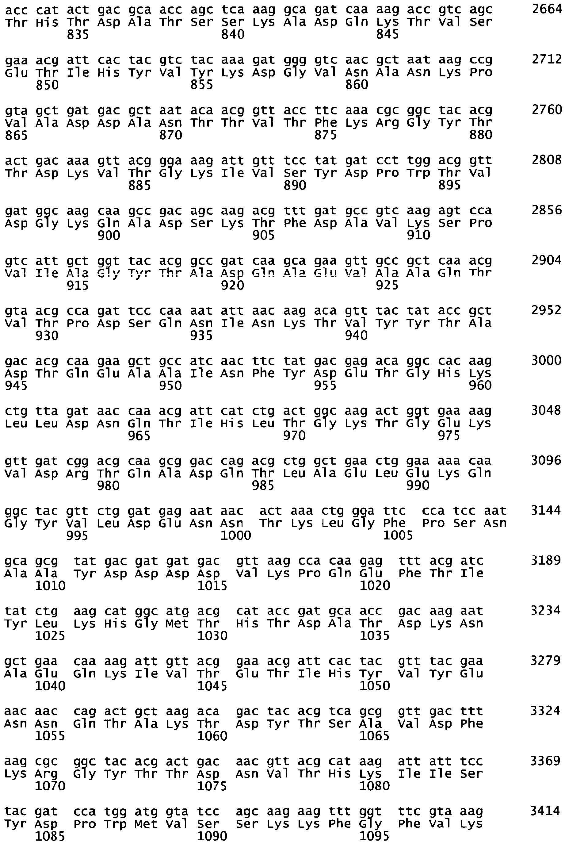


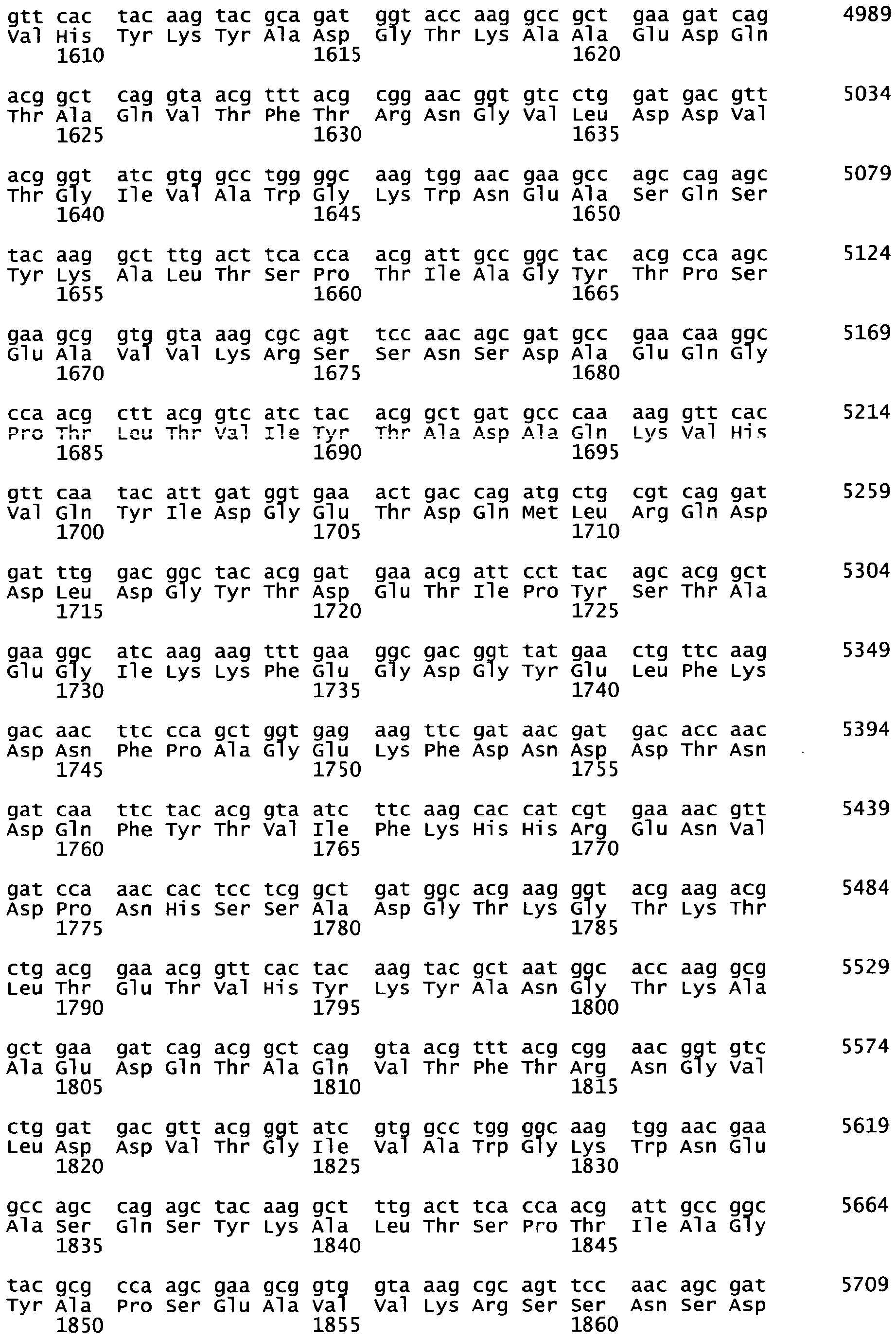


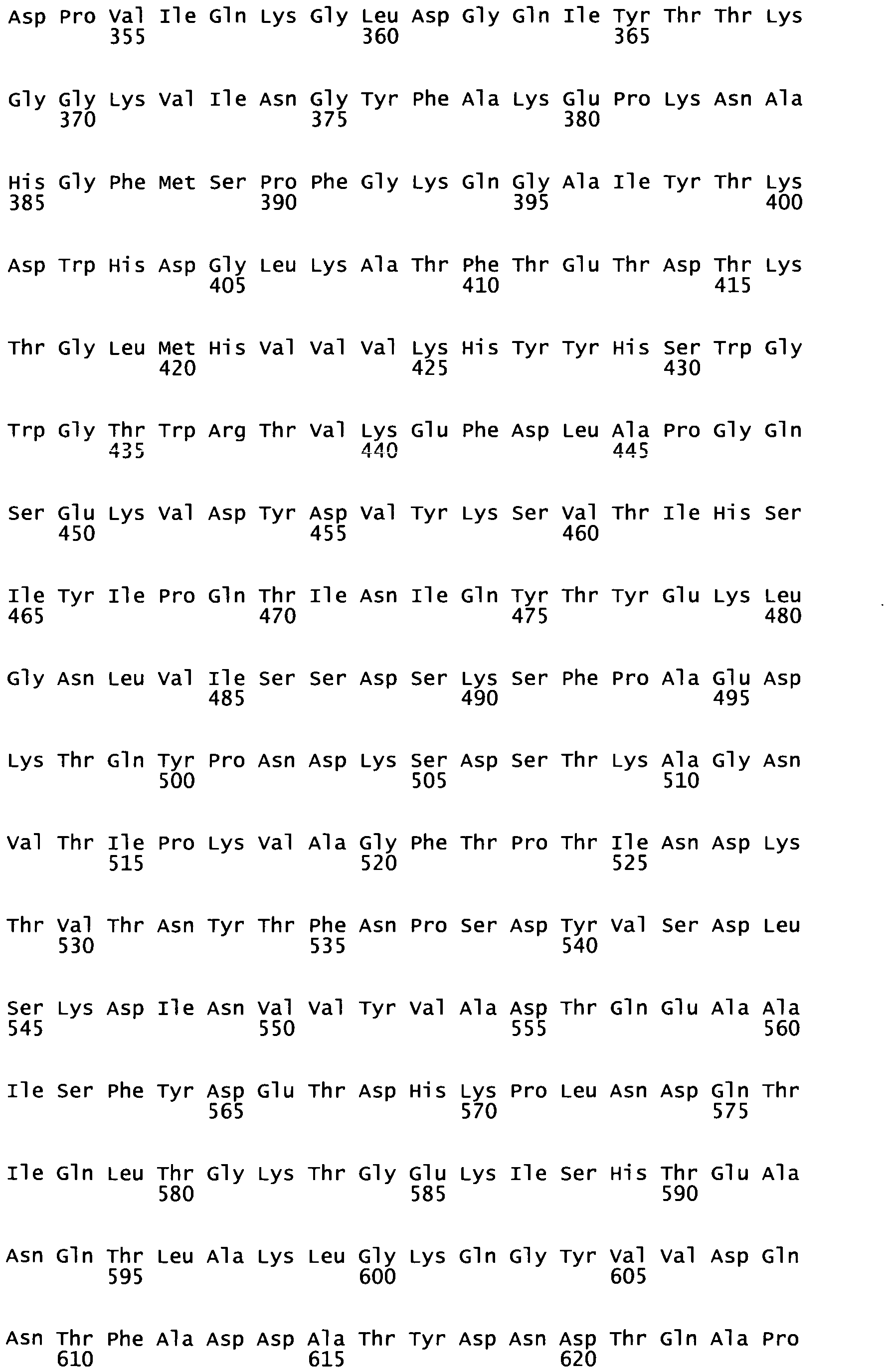


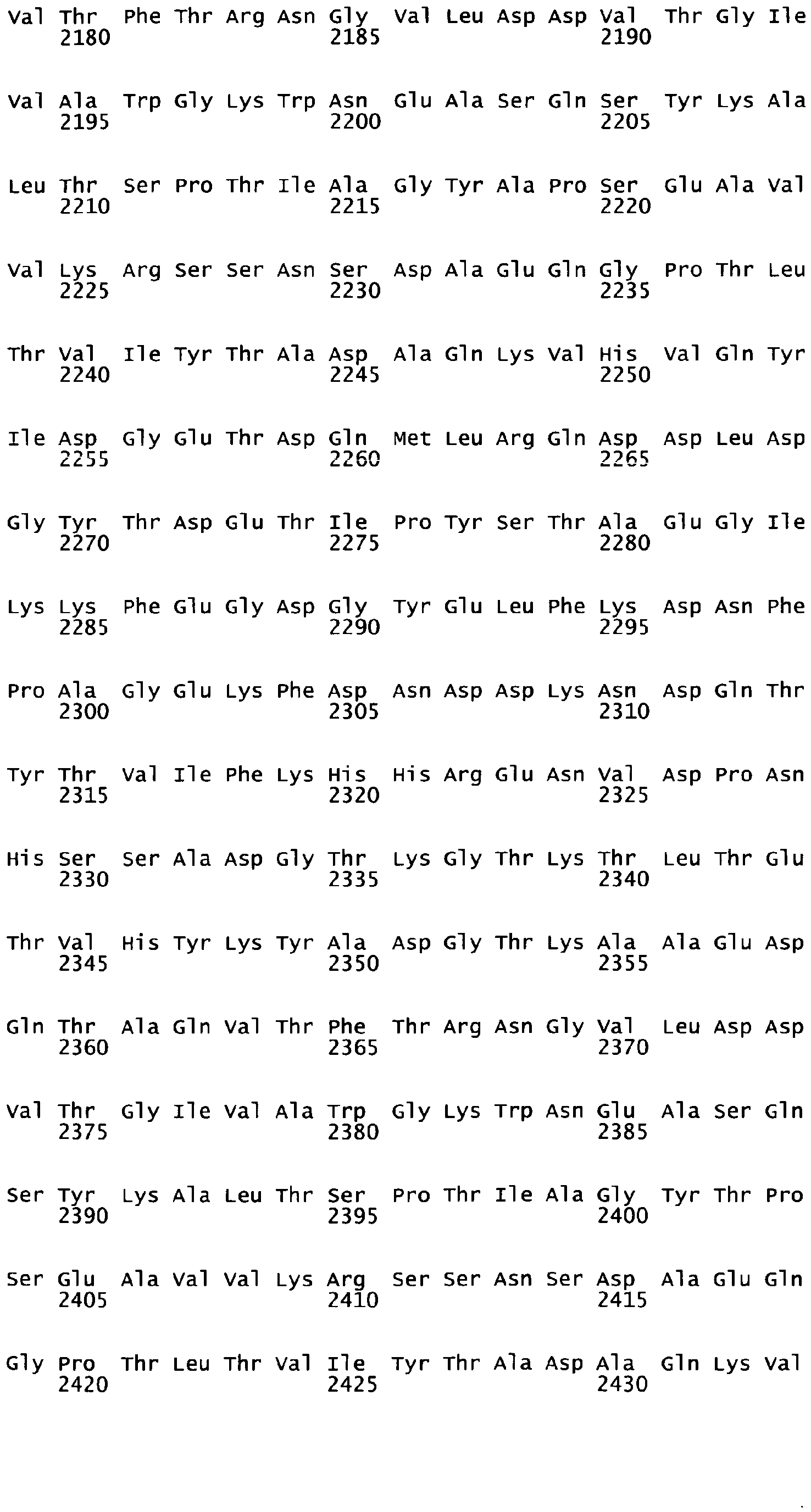


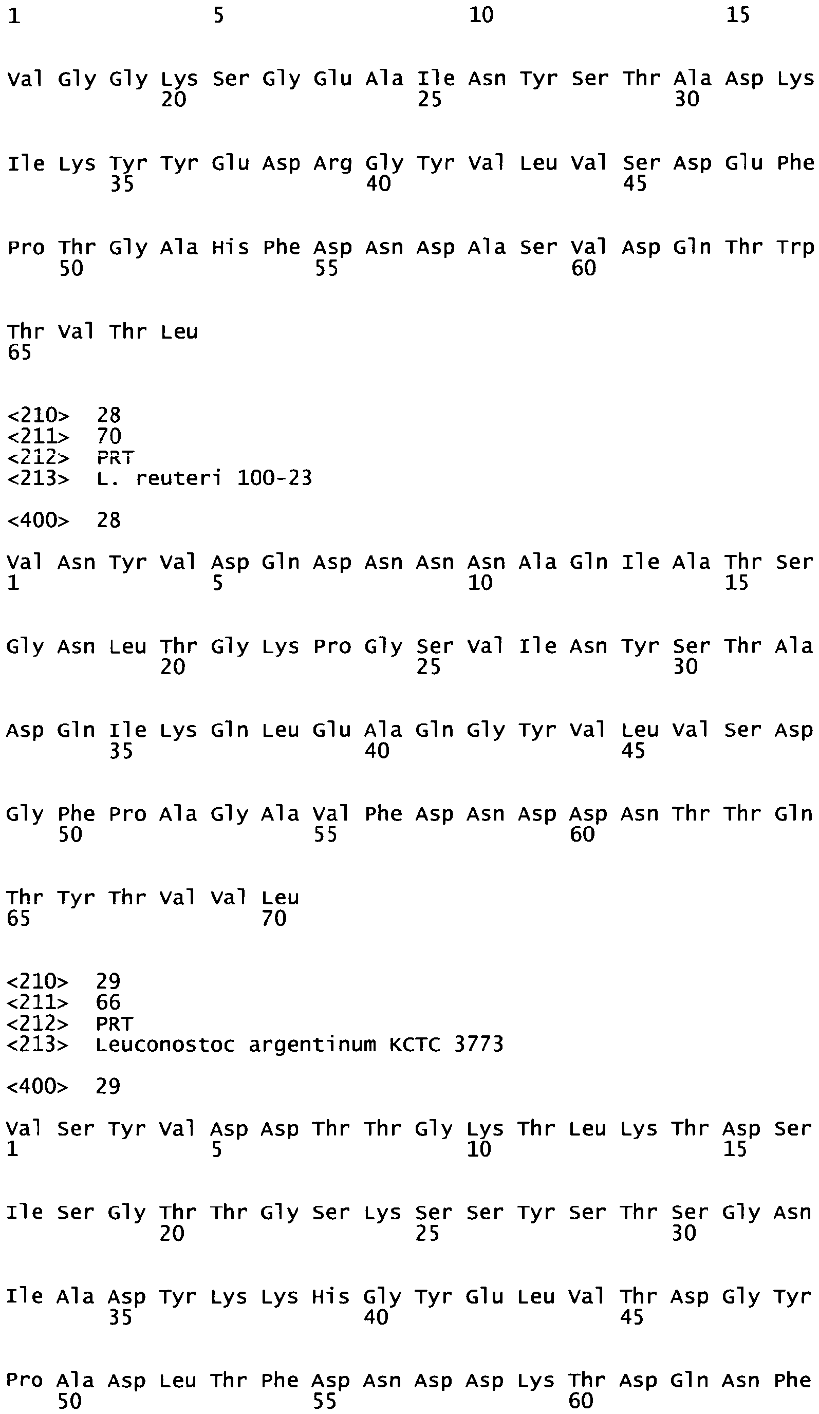


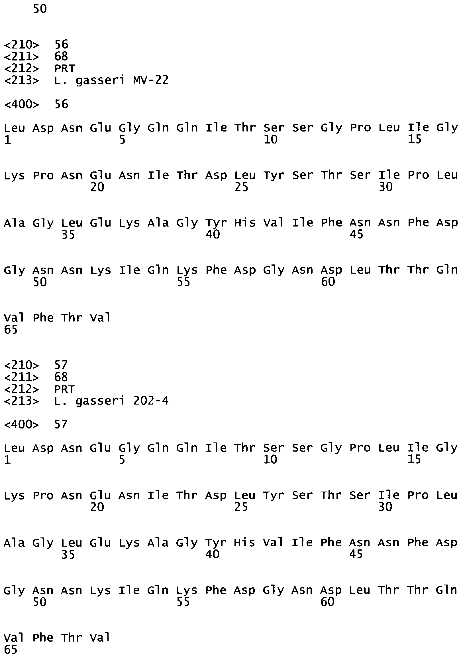
Claims (22)
- A polypeptide having a length from 10 to 80 amino-acid residues and whose polypeptidic sequence comprises or consists of the consensus sequence P1(Xa)P3(Xb)P5(Xc)P6(Xd)P7 (SEQ ID N° 1) disclosing, from N-terminal to C-terminal ends, patterns P1, P3, P5, P6 and P7, which are defined as follows:P1 represents the amino-acid sequence VXYXD/N, where X represents any amino-acid,P3 represents the amino-acid sequence GY,P5 represents the amino-acid sequence F/YD,P6 represents amino-acid residue D, andP7 represents amino-acid residue Q;and wherein said patterns are further characterized by spacer amino-acid segments (Xa), (Xb), (Xc) and (Xd) containing respectively a, b, c and d numbers of any amino-acid residue(s) which are defined as follows:a ranges from 2 to 33,b ranges from 2 to 11,c ranges from 1 to 2,d ranges from 1 to 3;and wherein said polypeptide interacts with glycosylated Muc2 protein(s), in particular interacts with sulfated moieties of glycosylated Muc2 protein, of human colonic mucus or human intestinal mucus.
- The polypeptide of Claim 1, whose polypeptidic sequence:i. comprises or consists of the consensus sequence P1(Xm)P2(Xn)P3XP4(Xp)P5(Xq)P6(Xr)P7X3P8 (SEQ ID N°2), X being any amino-acid residue, said consensus sequence disclosing, from N-terminal to C-terminal ends, patterns P1, P2, P3, P4, P5, P6, P7 and P8, which are defined as follows:P1 represents the amino-acid sequence VXYXD/N, where X represents any amino-acid,P2 represents the amino-acid sequence YS/TT,P3 represents the amino-acid sequence GY,P4 represents amino-acid residue L,P5 represents the amino-acid sequence F/YD,P6 represents amino-acid residue D,P7 represents amino-acid residue Q, andP8 represents amino-acid residue V,and wherein said patterns are further characterized by spacer amino-acid segments (Xm), (Xn), (Xp), (Xq) and (Xr) containing respectively m, n, p, q and r numbers of any amino-acid residue(s) which are defined as follows:m ranges from 1 to 23,n and p range from 1 to 10,q is 1 or 2,r ranges from 1 to 3; orii. comprises or consists in a sequence having at least 30% identity with SEQ ID N°3, or;iii. comprises or consists in SEQ ID N° 3, or;iv. comprises or consists of a fragment, especially a fragment of contiguous amino-acid residues of at least 10 amino-acid residues, of any one of the sequences defined in i) or ii) or iii).
- The polypeptide according to any one of Claims 1 or 2, whose polypeptidic sequence is part of the N-terminal sequence of a mucus-binding (MUB) domain, especially a mucus-binding (MUB) domain of a species selected amongst the Lactobacillus, Streptococcus, Cryptobacterium, Weissella, Granulicatella or Leuconostoc species, and in particular is part of any one of the repeats of a L. reuteri mucus-binding (MUB) domain.
- The polypeptide according to any one of claims 1 to 3, whose sequence can be identified in the N-terminal sequence of a Mucus Binding Domain (MUB) present in a mucus binding protein of L. gasseri, L. johnsonii, L fermentum or L. acidophilus.
- The polypeptide according to any one of Claim 1 to Claim 4, which comprises or consists of a fragment of the polypeptide having the sequence of any one of SEQ ID N°5 to SEQ ID N°16, or any one of SEQ ID N°19 to SEQ ID N°22, especially a fragment within the N-terminal extremity of said polypeptide, or which has an amino-acid sequence comprising or consisting of any one of SEQ ID N°23 to 57 or a fragment thereof.
- The polypeptide of any of claims 1 to 5 which lacks hydrophobic domain(s) and does not penetrate into living eukaryotic cells.
- The polypeptide according to any one of Claims 1 to Claim 6, which has the sequence SEQ ID N°3 or a fragment thereof of contiguous amino-acid residues of at least 10 amino-acid residues.
- The polypeptide according to any one of Claim 1 to Claim 7, which has an additional Cysteine residue at its N-terminal extremity.
- A chimeric molecule comprising a polypeptide according to any one of Claims 1 to Claim 8, which polypeptide is labelled, especially by coupling with a fluorophore or a biotin, or which polypeptide is associated with a reporter or a carrier entity, or with an active molecule such as a drug or an enzyme.
- A method for synthesizing a polypeptide or a fragment of polypeptide according to Claim 7 or Claim 8 by Solid-Phase synthesis, including steps of coupling with Fmoc-Asp(OtBu)-(Dmb)Gly-OH dipeptides when the synthesis reaches the positions 29 and/or 50 and/or 63 in reference to the C-terminus of SEQ ID N°3, and, optionally, including steps of incorporation of pseudoproline dipeptides when the synthesis reaches positions 10 and/or 40 in reference to the C-terminus of SEQ ID N°3.
- A nucleic acid molecule encoding a polypeptide according to any one of Claims 1 to 8.
- A vector, especially a plasmid, comprising a nucleic acid molecule according to Claim 11.
- A method for producing a polypeptide according to any one of Claims 1 to 8 through the transfection of a vector according to Claim 12 in a cell, the culture of said cell and the recovery of said polypeptide, or through chemical synthesis.
- A cell or a population of cells comprising a nucleic acid molecule or a vector according to any one of Claims 11 or 12, especially for use in the method of Claim 13.
- A composition comprising a polypeptide according to any one of Claims 1 to 9, in particular a pharmaceutical composition further comprising pharmaceutically acceptable excipient(s), especially carrier(s) and/or adjuvant(s).
- Use of a polypeptide according to any one of Claims 1 to 9, as a probe or marker for staining living cell(s) or tissue(s) in in vitro experiments, in particular in live microscopy experiments.
- Use of a polypeptide according to any one of Claims 1 to 9 or 16, as a probe or marker for staining Muc2 protein(s) contained in mucus layer(s), especially in adhesive mucus layers, of a cell or tissue sample, especially colonic or intestine tissue sample.
- Use of a polypeptide of any of claims 1 to 9, or of a composition according to Claim 15:- as a physiological labelled probe to detect in vitro interaction with human colonic mucus in the adhesive mucus layer of colonic tissue sample, or- to detect in vitro mucus production or mucus composition in human colon, said use comprising contacting said polypeptide with a sample of colonic tissue comprising adhesive mucus layer and goblet cells, or- in vitro detecting or monitoring any one of the following disease conditions: neoplasic disease(s), including mucinous carcinoma(s), gastric cancer(s) or colorectal cancer(s), especially colon cancer(s), cystic fibrosis, intestine inflammatory disease(s) such as inflammatory bowel disease (IBD) and ulcerative colitis.
- A polypeptide according to any one of claims 1 to 9, which is labelled, for use as a probe for in vivo detection of mucus production or mucus composition in human colon.
- Method for manufacturing a medicament comprising a step of association, especially coupling, grafting or fusing a polypeptide according to any one of Claims 1 to 8 with a biologically active molecule such as a drug or an enzyme.
- A polypeptide according to Claim 9, or of a composition according to Claim 15, or a medicament resulting from Claim 20 for use in a method of therapy practised on a human or animal body, in particular for use in treating a disease selected from the following group, or its symptom(s): neoplasic disease(s), including mucinous carcinoma(s), gastric cancer(s) or colorectal cancer(s), especially colon cancer(s), cystic fibrosis, intestine inflammatory disease(s) such as inflammatory bowel disease (IBD) and ulcerative colitis.
- Use of a polypeptide according to any one of Claims 1 to 9 as an in vitro marker of degranulation event(s) in neutrophiles.
Priority Applications (5)
| Application Number | Priority Date | Filing Date | Title |
|---|---|---|---|
| EP11290403A EP2567968A1 (en) | 2011-09-09 | 2011-09-09 | Polypeptides targeting glycosylated Muc2 proteins, methods of synthesis, their nucleic acids and uses therof |
| PCT/EP2012/067587 WO2013034749A1 (en) | 2011-09-09 | 2012-09-07 | Polypeptides targeting glycosylated muc2 proteins, methods of synthesis, their nucleic acids and uses thereof |
| EP12755887.2A EP2753639B1 (en) | 2011-09-09 | 2012-09-07 | Polypeptides targeting glycosylated muc2 proteins, methods of synthesis, their nucleic acids and uses thereof |
| US14/343,588 US9382299B2 (en) | 2011-09-09 | 2012-09-07 | Polypeptides targeting glycosylated MUC2 proteins, methods of synthesis, their nucleic acids and uses thereof |
| US15/178,049 US10138280B2 (en) | 2011-09-09 | 2016-06-09 | Polypeptides targeting glycosylated MUC2 proteins, methods of synthesis, their nucleic acids and uses thereof |
Applications Claiming Priority (1)
| Application Number | Priority Date | Filing Date | Title |
|---|---|---|---|
| EP11290403A EP2567968A1 (en) | 2011-09-09 | 2011-09-09 | Polypeptides targeting glycosylated Muc2 proteins, methods of synthesis, their nucleic acids and uses therof |
Publications (1)
| Publication Number | Publication Date |
|---|---|
| EP2567968A1 true EP2567968A1 (en) | 2013-03-13 |
Family
ID=46799271
Family Applications (2)
| Application Number | Title | Priority Date | Filing Date |
|---|---|---|---|
| EP11290403A Withdrawn EP2567968A1 (en) | 2011-09-09 | 2011-09-09 | Polypeptides targeting glycosylated Muc2 proteins, methods of synthesis, their nucleic acids and uses therof |
| EP12755887.2A Not-in-force EP2753639B1 (en) | 2011-09-09 | 2012-09-07 | Polypeptides targeting glycosylated muc2 proteins, methods of synthesis, their nucleic acids and uses thereof |
Family Applications After (1)
| Application Number | Title | Priority Date | Filing Date |
|---|---|---|---|
| EP12755887.2A Not-in-force EP2753639B1 (en) | 2011-09-09 | 2012-09-07 | Polypeptides targeting glycosylated muc2 proteins, methods of synthesis, their nucleic acids and uses thereof |
Country Status (3)
| Country | Link |
|---|---|
| US (2) | US9382299B2 (en) |
| EP (2) | EP2567968A1 (en) |
| WO (1) | WO2013034749A1 (en) |
Families Citing this family (2)
| Publication number | Priority date | Publication date | Assignee | Title |
|---|---|---|---|---|
| US11472844B2 (en) | 2017-12-11 | 2022-10-18 | Centre National De La Recherche Scientifique | Peptidomimetics, method of synthesis and uses thereof |
| CN110903350A (en) * | 2019-12-27 | 2020-03-24 | 四川科伦药物研究院有限公司 | A kind of solid-phase synthesis method of plecanatide |
Family Cites Families (1)
| Publication number | Priority date | Publication date | Assignee | Title |
|---|---|---|---|---|
| CN103476928B (en) | 2010-12-09 | 2017-03-29 | 巴斯德研究所 | MGMT-based method for obtaining high-yield recombinant protein expression |
-
2011
- 2011-09-09 EP EP11290403A patent/EP2567968A1/en not_active Withdrawn
-
2012
- 2012-09-07 WO PCT/EP2012/067587 patent/WO2013034749A1/en not_active Ceased
- 2012-09-07 US US14/343,588 patent/US9382299B2/en not_active Expired - Fee Related
- 2012-09-07 EP EP12755887.2A patent/EP2753639B1/en not_active Not-in-force
-
2016
- 2016-06-09 US US15/178,049 patent/US10138280B2/en not_active Expired - Fee Related
Non-Patent Citations (42)
| Title |
|---|
| ALLEN ET AL: "The MUC2 gene product: a human intestinal mucin.", THE INTERNATIONAL JOURNAL OF BIOCHEMISTRY & CELL BIOLOGY, vol. 30, no. 7, 1 July 1998 (1998-07-01), pages 797 - 801, XP055019786, ISSN: 1357-2725 * |
| ALLEN, A., D.A. HUTTON, J.P. PEARSON: "The MUC2 gene product: a human intestinal mucin", INT J BIOCHEM CELL BIOL, vol. 30, no. 7, 1998, pages 797 - 801, XP055019786, DOI: doi:10.1016/S1357-2725(98)00028-4 |
| BANSAL, D. ET AL.: "An ex-vivo human intestinal model to study Entamoeba histolytica pathogenesis", PLOS NEGL TROP DIS, vol. 3, no. 11, 2009, pages E551 |
| BERGSTROM, K.S. ET AL.: "Muc2 protects against lethal infectious colitis by disassociating pathogenic and commensal bacteria from the colonic mucosa", PLOS PATHOG., vol. 6, no. 5, pages E1000902 |
| BIN WANG ET AL: "Identification of a Surface Protein from Lactobacillus reuteri JCM1081 That Adheres to Porcine Gastric Mucin and Human Enterocyte-Like HT-29 Cells", CURRENT MICROBIOLOGY, SPRINGER-VERLAG, NE, vol. 57, no. 1, 1 April 2008 (2008-04-01), pages 33 - 38, XP019625653, ISSN: 1432-0991 * |
| BOEKHORST, J. ET AL.: "Comparative analysis of proteins with a mucus-binding domain found exclusively in lactic acid bacteria", MICROBIOLOGY, vol. 152, 2006, pages 273 - 80 |
| CARDONA, V. ET AL.: "Application of Dmb-Dipeptides in the Fmoc SPPS of Difficult and Aspartimide-Prone Sequences", INTERNATIONAL JOURNAL OF PEPTIDE RESEARCH AND THERAPEUTICS, vol. 14, no. 4, 2008, pages 285 - 292, XP019646546, DOI: doi:10.1007/s10989-008-9154-z |
| CHAN, C.C.A.P.D.W.: "Fmoc Solid Phase Peptide Synthosis. A Practical Approach.", 2000, OXFORD UNIVERSITY PRESS |
| D. A. MACKENZIE ET AL: "Crystal Structure of a Mucus-binding Protein Repeat Reveals an Unexpected Functional Immunoglobulin Binding Activity", JOURNAL OF BIOLOGICAL CHEMISTRY, vol. 284, no. 47, 20 November 2009 (2009-11-20), pages 32444 - 32453, XP055019700, ISSN: 0021-9258, DOI: 10.1074/jbc.M109.040907 * |
| FAURE, M. ET AL.: "The chronic colitis developed by HLA-B27 transgenic rats is associated with altered in vivo mucin synthesis", DIG DIS SCI, vol. 49, no. 2, 2004, pages 339 - 46 |
| HUET, G. ET AL.: "Characterization of mucins and proteoglycans synthesized by a mucin-secreting HT-29 cell subpopulation", J CELL SCI, vol. 108, 1995, pages 1275 - 85 |
| JOHANSSON, M.E., J.M. LARSSON, G.C. HANSSON: "The two mucus layers of colon are organized by the MUC2 mucin, whereas the outer layer is a legislator of host-microbial interactions", PROC NATL ACAD SCI U S A., vol. 108, no. 1, pages 4659 - 65 |
| JOHANSSON, M.E., K.A. THOMSSON, G.C. HANSSON: "Proteomic analyses of the two mucus layers of the colon barrier reveal that their main component, the Muc2 mucin, is strongly bound to the Fcgbp protein", J PROTEOME RES, vol. 8, no. 7, 2009, pages 3549 - 57 |
| JOHNSON, T., M. QUIBELL, R.C. SHEPPARD: "N,O-bisFmoc derivatives of N-(2-hydroxy-4-methoxybenzyl)-amino acids: useful intermediates in peptide synthesis", J PEPT SCI, vol. 1, no. 1, 1995, pages 11 - 25, XP002141383, DOI: doi:10.1002/psc.310010104 |
| KARLSSON, N.G. ET AL.: "Molecular characterization of the large heavily glycosylated domain glycopeptide from the rat small intestinal Muc2 mucin", GLYCOCONJ J, vol. 13, no. 5, 1996, pages 823 - 31 |
| LAU, S.K., L.M. WEISS, P.G. CHU: "Differential expression of MUC1, MUC2, and MUC5AC in carcinomas of various sites: an immunohistochemical study", AM J CLIN PATHOL, vol. 122, no. 1, 2004, pages 61 - 9 |
| LAUER, J.L., C.G. FIELDS, G.B. FIELDS: "Sequence dependence of aspartimide formation during 9-fIuorenylmethoxycarbonyl solid-phase peptide synthesis", LETTERS IN PEPTIDE SCIENCE, vol. 1, 1995, pages 197 - 205 |
| LESUFFLEUR, T., A. ZWEIBAUM, F.X. REAL: "Mucins in normal and neoplastic human gastrointestinal tissues", CRIT REV ONCOL HEMATOL, vol. 17, no. 3, 1994, pages 153 - 80, XP026208387, DOI: doi:10.1016/1040-8428(94)90053-1 |
| MARTEYN, B. ET AL.: "Breathing life into pathogens: the influence of oxygen on bacterial virulence and host responses in the gastrointestinal tract", CELL MICROBIOL, 2010 |
| MARTEYN, B. ET AL.: "Modulation of Shigella virulence in response to available oxygen in vivo", NATURE, vol. 465, no. 7296, 2010, pages 355 - 8 |
| MERGLER, M. ET AL.: "The aspartimide problem in Fmoc-based SPPS. Part I.", J PEPT SCI, vol. 9, no. 1, 2003, pages 36 - 46 |
| MERRIFIELD, R.B.: "Solid Phase Peptide Synthesis. I. The Synthesis of a Tetrapeptide", JOURNAL OF THE AMERICAN CHEMICAL SOCIETY, vol. 85, no. 14, 1963, pages 2149 - 2154, XP002257754, DOI: doi:10.1021/ja00897a025 |
| MESQUITA, P. ET AL.: "Coordinated expression of MUC2 and CDX-2 in mucinous carcinomas of the lung can be explained by the role of CDX-2 as transcriptional regulator of MUC2", AM J SURG PATHOL, vol. 28, no. 9, 2004, pages 1254 - 5 |
| MUKHOPADHYAY, P. ET AL.: "Mucins in the pathogenesis of breast cancer: implications in diagnosis, prognosis and therapy.", BIOCHIM BIOPHYS ACTA, vol. 1815, no. 2, pages 224 - 40, XP028177026, DOI: doi:10.1016/j.bbcan.2011.01.001 |
| OUWEHAND, A.C. ET AL.: "Assessment of adhesion properties of novel probiotic strains to human intestinal mucus", INT J FOOD MICROBIOL, vol. 64, no. 1-2, 2001, pages 119 - 26, XP002336306, DOI: doi:10.1016/S0168-1605(00)00440-2 |
| PARK, S.Y. ET AL.: "Expression of MUC1, MUC2, MUC5AC and MUC6 in cholangiocarcinoma: prognostic impact", ONCOL REP, vol. 22, no. 3, 2009, pages 649 - 57 |
| PRETZER, G. ET AL.: "Biodiversity-based identification and functional characterization of the mannose-specific adhesin of Lactobacillus plantarum", J BACTERIOL, vol. 187, no. 17, 2005, pages 6128 - 36 |
| QUIBELL, M. ET AL.: "Suppression of piperidine-mediated side product formation for Asp(Obu(T))-containing peptides by the use of N-(2-Hydroxy-4-Methoxybenzyl) (Hmb) backbone amide protection", JOURNAL OF THE CHEMICAL SOCIETY-CHEMICAL COMMUNICATIONS, vol. 20, 1994, pages 2343 - 2344 |
| ROBBE, C. ET AL.: "Evidence of regio-specific glycosylation in human intestinal mucins: presence of an acidic gradient along the intestinal tract", J BIOL CHEM, vol. 278, no. 47, 2003, pages 46337 - 48 |
| ROOS S ET AL: "A high-molecular-mass cell-surface protein from Lactobacillus reuteri 1063 adheres to mucus components", MICROBIOLOGY, SOCIETY FOR GENERAL MICROBIOLOGY, READING, GB, vol. 148, no. Pt 2, 1 February 2002 (2002-02-01), pages 433 - 442, XP002225918, ISSN: 1350-0872 * |
| ROOS, S., H. JONSSON: "A high-molecular-mass cell-surface protein from Lactobacillus reuteri 1063 adheres to mucus components", MICROBIOLOGY, vol. 148, 2002, pages 433 - 42, XP002225918 |
| RUBINSTEIN, A., B. TIROSH: "Mucus gel thickness and turnover in the gastrointestinal tract of the rat: response to cholinergic stimulus and implication for mucoadhesion", PHARM RES, vol. 11, no. 6, 1994, pages 794 - 9 |
| SCHULTSZ, C. ET AL.: "The intestinal mucus layer from patients with inflammatory bowel disease harbors high numbers of bacteria compared with controls", GASTROENTEROLOGY, vol. 117, no. 5, 1999, pages 1089 - 97, XP005138459, DOI: doi:10.1016/S0016-5085(99)70393-8 |
| SHAOUL, R. ET AL.: "Colonic expression of MUC2, MUCSAC, and TFF1 in inflammatory bowel disease in children", J PEDIATR GASTROENTEROL NUTR, vol. 38, no. 5, 2004, pages 488 - 93 |
| STERNBERG L R ET AL: "Alternative splicing of the human MUC2 gene", ARCHIVES OF BIOCHEMISTRY AND BIOPHYSICS, ACADEMIC PRESS, US, vol. 421, no. 1, 1 January 2004 (2004-01-01), pages 21 - 33, XP004480351, ISSN: 0003-9861, DOI: 10.1016/J.ABB.2003.10.002 * |
| STRUGALA, V., P.W. DETTMAR, J.P. PEARSON: "Thickness and continuity of the adherent colonic mucus barrier in active and quiescent ulcerative colitis and Crohn's disease", INT J CLIN PRACT, vol. 62, no. 5, 2008, pages 762 - 9 |
| TRAVO, A. ET AL.: "IR spectral imaging of secreted mucus: a promising new tool for the histopathological recognition of human colonic adenocarcinomas", HISTOPATHOLOGY, vol. 56, no. 7, pages 921 - 31 |
| TYTGAT, K.M. ET AL.: "Biosynthesis of human colonic mucin: Muc2 is the prominent secretory mucin", GASTROENTEROLOGY, vol. 107, no. 5, 1994, pages 1352 - 63 |
| VAN KLINKEN, B.J. ET AL.: "Gastrointestinal expression and partial cDNA cloning of murine Muc2", AM J PHYSIOL, vol. 276, 1999, pages G115 - 24 |
| WOHR, T.: "Pseudo-prolines as a solubilizing, structure-disrupting protection technique in peptide synthesis", JOURNAL OF THE AMERICAN CHEMICAL SOCIETY, vol. 118, no. 39, 1996, pages 9218 - 9227, XP002447320, DOI: doi:10.1021/ja961509q |
| WONG, W.M., R. POULSOM, N.A. WRIGHT: "Trefoil peptides", GUT, vol. 44, no. 6, 1999, pages 890 - 5 |
| ZHANG, S. ET AL.: "Expression of potential target antigens for immunotherapy on primary and metastatic prostate cancers", CLIN CANCER RES, vol. 4, no. 2, 1998, pages 295 - 302, XP000941645 |
Also Published As
| Publication number | Publication date |
|---|---|
| EP2753639A1 (en) | 2014-07-16 |
| EP2753639B1 (en) | 2017-03-01 |
| US9382299B2 (en) | 2016-07-05 |
| US20140336119A1 (en) | 2014-11-13 |
| US20160318981A1 (en) | 2016-11-03 |
| US10138280B2 (en) | 2018-11-27 |
| WO2013034749A1 (en) | 2013-03-14 |
Similar Documents
| Publication | Publication Date | Title |
|---|---|---|
| Rink et al. | To protect peptide pharmaceuticals against peptidases | |
| Ritzefeld | Sortagging: a robust and efficient chemoenzymatic ligation strategy | |
| KR102943992B1 (en) | FKBP domain having a transglutaminase recognition site | |
| CA3049738A1 (en) | Interaction between c-peptides and elastin receptor, a model for understanding vascular disease | |
| CA2481231A1 (en) | Colon tumor specific binding peptides | |
| KR20120125455A (en) | Intracelluar targeting bipodal peptide binder | |
| Radicioni et al. | Characterization of the cell penetrating properties of a human salivary proline-rich peptide | |
| US8956878B2 (en) | Polypeptides with affinity for heat shock proteins (HSPS) and HSP associated complexes (HACS) and their use in diagnosis and therapy | |
| EP3724215B1 (en) | Peptidomimetics, method of synthesis and uses thereof | |
| Pini et al. | Characterization of the branched antimicrobial peptide M6 by analyzing its mechanism of action and in vivo toxicity | |
| US20140271640A1 (en) | FGF-10 Complexes | |
| US20150307576A1 (en) | Fgf-10 complexes | |
| US10138280B2 (en) | Polypeptides targeting glycosylated MUC2 proteins, methods of synthesis, their nucleic acids and uses thereof | |
| JP2011193728A (en) | PEPTIDE HAVING BINDING CAPABILITY TO EpCAM | |
| EP2304033A2 (en) | Functionalized polypeptides | |
| CN118103387A (en) | Peptides | |
| US20230287402A1 (en) | Two component co-assembling two dimensional protein structures | |
| US20210395310A1 (en) | Cyclic Peptide | |
| CN116082496B (en) | Antigen peptide and antibody for targeting phosphorylation of STAT3 protein Ser701 and application thereof | |
| Nicolai et al. | Inner membrane protein OutB is covalently attached to peptidoglycan in the γ-proteobacterium Dickeya dadantii | |
| WO2026022392A1 (en) | Peptide and conjugates thereof | |
| KR20150131655A (en) | AIMP2-DX2-34S protein and method for manufacturing the same | |
| Wang | Developing Functional Peptides as Synthetic Receptors, Binders of Protein and Probes for Bacteria Detection | |
| Elhebieshy et al. | Total Chemical Synthesis of a Functionalized GFP Nanobody | |
| US9547007B2 (en) | Methods and compositions for in-vivo enzyme capture |
Legal Events
| Date | Code | Title | Description |
|---|---|---|---|
| PUAI | Public reference made under article 153(3) epc to a published international application that has entered the european phase |
Free format text: ORIGINAL CODE: 0009012 |
|
| AK | Designated contracting states |
Kind code of ref document: A1 Designated state(s): AL AT BE BG CH CY CZ DE DK EE ES FI FR GB GR HR HU IE IS IT LI LT LU LV MC MK MT NL NO PL PT RO RS SE SI SK SM TR |
|
| AX | Request for extension of the european patent |
Extension state: BA ME |
|
| 17P | Request for examination filed |
Effective date: 20130913 |
|
| RBV | Designated contracting states (corrected) |
Designated state(s): AL AT BE BG CH CY CZ DE DK EE ES FI FR GB GR HR HU IE IS IT LI LT LU LV MC MK MT NL NO PL PT RO RS SE SI SK SM TR |
|
| STAA | Information on the status of an ep patent application or granted ep patent |
Free format text: STATUS: THE APPLICATION IS DEEMED TO BE WITHDRAWN |
|
| 18D | Application deemed to be withdrawn |
Effective date: 20140103 |