EP2565646A1 - Diagnostic marker for kidney diseases and use thereof - Google Patents
Diagnostic marker for kidney diseases and use thereof Download PDFInfo
- Publication number
- EP2565646A1 EP2565646A1 EP11775009A EP11775009A EP2565646A1 EP 2565646 A1 EP2565646 A1 EP 2565646A1 EP 11775009 A EP11775009 A EP 11775009A EP 11775009 A EP11775009 A EP 11775009A EP 2565646 A1 EP2565646 A1 EP 2565646A1
- Authority
- EP
- European Patent Office
- Prior art keywords
- urine
- kidney disease
- concentration
- plasma
- marker
- Prior art date
- Legal status (The legal status is an assumption and is not a legal conclusion. Google has not performed a legal analysis and makes no representation as to the accuracy of the status listed.)
- Granted
Links
- 208000017169 kidney disease Diseases 0.000 title claims abstract description 159
- 239000003550 marker Substances 0.000 title claims description 88
- 210000002700 urine Anatomy 0.000 claims description 208
- AEMRFAOFKBGASW-UHFFFAOYSA-N Glycolic acid Chemical compound OCC(O)=O AEMRFAOFKBGASW-UHFFFAOYSA-N 0.000 claims description 122
- 239000000126 substance Substances 0.000 claims description 103
- KWIUHFFTVRNATP-UHFFFAOYSA-N glycine betaine Chemical compound C[N+](C)(C)CC([O-])=O KWIUHFFTVRNATP-UHFFFAOYSA-N 0.000 claims description 42
- 238000004590 computer program Methods 0.000 claims description 34
- LEVWYRKDKASIDU-QWWZWVQMSA-N D-cystine Chemical compound OC(=O)[C@H](N)CSSC[C@@H](N)C(O)=O LEVWYRKDKASIDU-QWWZWVQMSA-N 0.000 claims description 22
- 229960003067 cystine Drugs 0.000 claims description 22
- TYKJILJOXAHUFO-UHFFFAOYSA-N 2-amino-1,4-dihydroimidazol-5-one Chemical compound NC1=NC(=O)CN1 TYKJILJOXAHUFO-UHFFFAOYSA-N 0.000 claims description 20
- BPMFZUMJYQTVII-UHFFFAOYSA-N guanidinoacetic acid Chemical compound NC(=N)NCC(O)=O BPMFZUMJYQTVII-UHFFFAOYSA-N 0.000 claims description 18
- YGPSJZOEDVAXAB-UHFFFAOYSA-N kynurenine Chemical compound OC(=O)C(N)CC(=O)C1=CC=CC=C1N YGPSJZOEDVAXAB-UHFFFAOYSA-N 0.000 claims description 18
- LSSAWEXAFIQVBN-ZETCQYMHSA-N (2S)-2-amino-5-[(2,2-dimethylhydrazinyl)methylideneamino]pentanoic acid Chemical compound CN(C)N=CNCCC[C@H](N)C(=O)O LSSAWEXAFIQVBN-ZETCQYMHSA-N 0.000 claims description 17
- QVADRSWDTZDDGR-UHFFFAOYSA-N 5-oxotetrahydrofuran-2-carboxylic acid Chemical compound OC(=O)C1CCC(=O)O1 QVADRSWDTZDDGR-UHFFFAOYSA-N 0.000 claims description 17
- DHMQDGOQFOQNFH-UHFFFAOYSA-N Glycine Chemical compound NCC(O)=O DHMQDGOQFOQNFH-UHFFFAOYSA-N 0.000 claims description 16
- QIVBCDIJIAJPQS-VIFPVBQESA-N L-tryptophane Chemical compound C1=CC=C2C(C[C@H](N)C(O)=O)=CNC2=C1 QIVBCDIJIAJPQS-VIFPVBQESA-N 0.000 claims description 16
- QIVBCDIJIAJPQS-UHFFFAOYSA-N Tryptophan Natural products C1=CC=C2C(CC(N)C(O)=O)=CNC2=C1 QIVBCDIJIAJPQS-UHFFFAOYSA-N 0.000 claims description 16
- ZYBUGSRRLVJZQZ-ZETCQYMHSA-N (2s)-3-(1-acetylimidazol-4-yl)-2-aminopropanoic acid Chemical compound CC(=O)N1C=NC(C[C@H](N)C(O)=O)=C1 ZYBUGSRRLVJZQZ-ZETCQYMHSA-N 0.000 claims description 14
- HNDVDQJCIGZPNO-UHFFFAOYSA-N histidine Natural products OC(=O)C(N)CC1=CN=CN1 HNDVDQJCIGZPNO-UHFFFAOYSA-N 0.000 claims description 14
- 208000007342 Diabetic Nephropathies Diseases 0.000 claims description 12
- 208000033679 diabetic kidney disease Diseases 0.000 claims description 12
- AHLPHDHHMVZTML-UHFFFAOYSA-N Orn-delta-NH2 Natural products NCCCC(N)C(O)=O AHLPHDHHMVZTML-UHFFFAOYSA-N 0.000 claims description 11
- AHLPHDHHMVZTML-BYPYZUCNSA-N L-Ornithine Chemical compound NCCC[C@H](N)C(O)=O AHLPHDHHMVZTML-BYPYZUCNSA-N 0.000 claims description 10
- UTJLXEIPEHZYQJ-UHFFFAOYSA-N Ornithine Natural products OC(=O)C(C)CCCN UTJLXEIPEHZYQJ-UHFFFAOYSA-N 0.000 claims description 10
- 229960003104 ornithine Drugs 0.000 claims description 10
- UHDGCWIWMRVCDJ-UHFFFAOYSA-N 1-beta-D-Xylofuranosyl-NH-Cytosine Natural products O=C1N=C(N)C=CN1C1C(O)C(O)C(CO)O1 UHDGCWIWMRVCDJ-UHFFFAOYSA-N 0.000 claims description 9
- UHDGCWIWMRVCDJ-PSQAKQOGSA-N Cytidine Natural products O=C1N=C(N)C=CN1[C@@H]1[C@@H](O)[C@@H](O)[C@H](CO)O1 UHDGCWIWMRVCDJ-PSQAKQOGSA-N 0.000 claims description 9
- OEYIOHPDSNJKLS-UHFFFAOYSA-N choline Chemical compound C[N+](C)(C)CCO OEYIOHPDSNJKLS-UHFFFAOYSA-N 0.000 claims description 9
- 229960001231 choline Drugs 0.000 claims description 9
- UHDGCWIWMRVCDJ-ZAKLUEHWSA-N cytidine Chemical compound O=C1N=C(N)C=CN1[C@H]1[C@H](O)[C@@H](O)[C@H](CO)O1 UHDGCWIWMRVCDJ-ZAKLUEHWSA-N 0.000 claims description 9
- 239000004471 Glycine Substances 0.000 claims description 8
- FFEARJCKVFRZRR-BYPYZUCNSA-N L-methionine Chemical compound CSCC[C@H](N)C(O)=O FFEARJCKVFRZRR-BYPYZUCNSA-N 0.000 claims description 8
- DTERQYGMUDWYAZ-ZETCQYMHSA-N N(6)-acetyl-L-lysine Chemical compound CC(=O)NCCCC[C@H]([NH3+])C([O-])=O DTERQYGMUDWYAZ-ZETCQYMHSA-N 0.000 claims description 8
- KZSNJWFQEVHDMF-UHFFFAOYSA-N Valine Natural products CC(C)C(N)C(O)=O KZSNJWFQEVHDMF-UHFFFAOYSA-N 0.000 claims description 8
- 229930182817 methionine Natural products 0.000 claims description 8
- 239000004474 valine Substances 0.000 claims description 8
- KPGXRSRHYNQIFN-UHFFFAOYSA-N 2-oxoglutaric acid Chemical compound OC(=O)CCC(=O)C(O)=O KPGXRSRHYNQIFN-UHFFFAOYSA-N 0.000 claims description 7
- KZSNJWFQEVHDMF-BYPYZUCNSA-N L-valine Chemical compound CC(C)[C@H](N)C(O)=O KZSNJWFQEVHDMF-BYPYZUCNSA-N 0.000 claims description 7
- TUHVEAJXIMEOSA-UHFFFAOYSA-N gamma-guanidinobutyric acid Natural products NC(=[NH2+])NCCCC([O-])=O TUHVEAJXIMEOSA-UHFFFAOYSA-N 0.000 claims description 7
- APIBROGXENTUGB-ZUQRMPMESA-M triphenyl-[(e)-3-phenylprop-2-enyl]phosphanium;bromide Chemical compound [Br-].C=1C=CC=CC=1[P+](C=1C=CC=CC=1)(C=1C=CC=CC=1)C\C=C\C1=CC=CC=C1 APIBROGXENTUGB-ZUQRMPMESA-M 0.000 claims description 7
- HNDVDQJCIGZPNO-YFKPBYRVSA-N L-histidine Chemical compound OC(=O)[C@@H](N)CC1=CN=CN1 HNDVDQJCIGZPNO-YFKPBYRVSA-N 0.000 claims description 6
- 238000003745 diagnosis Methods 0.000 description 33
- 238000000034 method Methods 0.000 description 27
- OMOVVBIIQSXZSZ-UHFFFAOYSA-N [6-(4-acetyloxy-5,9a-dimethyl-2,7-dioxo-4,5a,6,9-tetrahydro-3h-pyrano[3,4-b]oxepin-5-yl)-5-formyloxy-3-(furan-3-yl)-3a-methyl-7-methylidene-1a,2,3,4,5,6-hexahydroindeno[1,7a-b]oxiren-4-yl] 2-hydroxy-3-methylpentanoate Chemical compound CC12C(OC(=O)C(O)C(C)CC)C(OC=O)C(C3(C)C(CC(=O)OC4(C)COC(=O)CC43)OC(C)=O)C(=C)C32OC3CC1C=1C=COC=1 OMOVVBIIQSXZSZ-UHFFFAOYSA-N 0.000 description 24
- 238000005259 measurement Methods 0.000 description 15
- 230000007423 decrease Effects 0.000 description 9
- 238000004458 analytical method Methods 0.000 description 8
- 238000005251 capillar electrophoresis Methods 0.000 description 8
- 238000004949 mass spectrometry Methods 0.000 description 8
- 238000000738 capillary electrophoresis-mass spectrometry Methods 0.000 description 6
- 230000001575 pathological effect Effects 0.000 description 6
- 238000011282 treatment Methods 0.000 description 6
- 230000008807 pathological lesion Effects 0.000 description 5
- XLYOFNOQVPJJNP-UHFFFAOYSA-N water Substances O XLYOFNOQVPJJNP-UHFFFAOYSA-N 0.000 description 5
- 150000001450 anions Chemical class 0.000 description 4
- 150000001768 cations Chemical class 0.000 description 4
- 208000020832 chronic kidney disease Diseases 0.000 description 4
- 206010012601 diabetes mellitus Diseases 0.000 description 4
- 239000012153 distilled water Substances 0.000 description 4
- 238000000108 ultra-filtration Methods 0.000 description 4
- -1 Glycolic acid N5-[(Dimethylamino)iminomethyl]-ornithine Chemical compound 0.000 description 3
- OKKJLVBELUTLKV-UHFFFAOYSA-N Methanol Chemical compound OC OKKJLVBELUTLKV-UHFFFAOYSA-N 0.000 description 3
- 238000004885 tandem mass spectrometry Methods 0.000 description 3
- 108010055851 Acetylglucosaminidase Proteins 0.000 description 2
- HEDRZPFGACZZDS-UHFFFAOYSA-N Chloroform Chemical compound ClC(Cl)Cl HEDRZPFGACZZDS-UHFFFAOYSA-N 0.000 description 2
- 206010018364 Glomerulonephritis Diseases 0.000 description 2
- 102100030122 Protein O-GlcNAcase Human genes 0.000 description 2
- 206010048302 Tubulointerstitial nephritis Diseases 0.000 description 2
- 239000008280 blood Substances 0.000 description 2
- 210000004369 blood Anatomy 0.000 description 2
- DDRJAANPRJIHGJ-UHFFFAOYSA-N creatinine Chemical compound CN1CC(=O)NC1=N DDRJAANPRJIHGJ-UHFFFAOYSA-N 0.000 description 2
- 238000000502 dialysis Methods 0.000 description 2
- 239000003814 drug Substances 0.000 description 2
- 229940079593 drug Drugs 0.000 description 2
- 238000011156 evaluation Methods 0.000 description 2
- 239000000706 filtrate Substances 0.000 description 2
- 238000004817 gas chromatography Methods 0.000 description 2
- 201000006334 interstitial nephritis Diseases 0.000 description 2
- 150000002500 ions Chemical class 0.000 description 2
- 230000003907 kidney function Effects 0.000 description 2
- 235000012054 meals Nutrition 0.000 description 2
- 210000005084 renal tissue Anatomy 0.000 description 2
- 238000005070 sampling Methods 0.000 description 2
- 238000012216 screening Methods 0.000 description 2
- 239000000243 solution Substances 0.000 description 2
- 238000012360 testing method Methods 0.000 description 2
- HZARZMXRTUDFRO-UHFFFAOYSA-N 2-hydroxyacetic acid 2-oxopentanedioic acid Chemical compound OCC(O)=O.OC(=O)CCC(=O)C(O)=O HZARZMXRTUDFRO-UHFFFAOYSA-N 0.000 description 1
- 102000009027 Albumins Human genes 0.000 description 1
- 108010088751 Albumins Proteins 0.000 description 1
- 102000003966 Alpha-1-microglobulin Human genes 0.000 description 1
- 101800001761 Alpha-1-microglobulin Proteins 0.000 description 1
- 102000014914 Carrier Proteins Human genes 0.000 description 1
- 102000004266 Collagen Type IV Human genes 0.000 description 1
- 108010042086 Collagen Type IV Proteins 0.000 description 1
- 102000012192 Cystatin C Human genes 0.000 description 1
- 108010061642 Cystatin C Proteins 0.000 description 1
- 108090000790 Enzymes Proteins 0.000 description 1
- 102000004190 Enzymes Human genes 0.000 description 1
- 102000016359 Fibronectins Human genes 0.000 description 1
- 108010067306 Fibronectins Proteins 0.000 description 1
- UCUNFLYVYCGDHP-BYPYZUCNSA-N L-methionine sulfone Chemical compound CS(=O)(=O)CC[C@H](N)C(O)=O UCUNFLYVYCGDHP-BYPYZUCNSA-N 0.000 description 1
- UCUNFLYVYCGDHP-UHFFFAOYSA-N L-methionine sulfone Natural products CS(=O)(=O)CCC(N)C(O)=O UCUNFLYVYCGDHP-UHFFFAOYSA-N 0.000 description 1
- 102000007547 Laminin Human genes 0.000 description 1
- 108010085895 Laminin Proteins 0.000 description 1
- 208000001647 Renal Insufficiency Diseases 0.000 description 1
- VYPSYNLAJGMNEJ-UHFFFAOYSA-N Silicium dioxide Chemical compound O=[Si]=O VYPSYNLAJGMNEJ-UHFFFAOYSA-N 0.000 description 1
- 102000004338 Transferrin Human genes 0.000 description 1
- 108090000901 Transferrin Proteins 0.000 description 1
- 239000008346 aqueous phase Substances 0.000 description 1
- 208000004670 arteriolosclerosis Diseases 0.000 description 1
- 102000015736 beta 2-Microglobulin Human genes 0.000 description 1
- 108010081355 beta 2-Microglobulin Proteins 0.000 description 1
- 108091008324 binding proteins Proteins 0.000 description 1
- 238000004364 calculation method Methods 0.000 description 1
- MIOPJNTWMNEORI-UHFFFAOYSA-N camphorsulfonic acid Chemical compound C1CC2(CS(O)(=O)=O)C(=O)CC1C2(C)C MIOPJNTWMNEORI-UHFFFAOYSA-N 0.000 description 1
- 238000005119 centrifugation Methods 0.000 description 1
- 230000008859 change Effects 0.000 description 1
- 239000007795 chemical reaction product Substances 0.000 description 1
- 238000004737 colorimetric analysis Methods 0.000 description 1
- 238000007796 conventional method Methods 0.000 description 1
- 238000012937 correction Methods 0.000 description 1
- 229940109239 creatinine Drugs 0.000 description 1
- 238000011161 development Methods 0.000 description 1
- 238000010586 diagram Methods 0.000 description 1
- 201000010099 disease Diseases 0.000 description 1
- 208000037265 diseases, disorders, signs and symptoms Diseases 0.000 description 1
- 230000000694 effects Effects 0.000 description 1
- 239000003792 electrolyte Substances 0.000 description 1
- 238000005516 engineering process Methods 0.000 description 1
- 238000006911 enzymatic reaction Methods 0.000 description 1
- 239000000194 fatty acid Substances 0.000 description 1
- 238000001914 filtration Methods 0.000 description 1
- 238000001917 fluorescence detection Methods 0.000 description 1
- 239000005350 fused silica glass Substances 0.000 description 1
- 230000024924 glomerular filtration Effects 0.000 description 1
- 238000004128 high performance liquid chromatography Methods 0.000 description 1
- 230000006872 improvement Effects 0.000 description 1
- 230000000977 initiatory effect Effects 0.000 description 1
- 238000002347 injection Methods 0.000 description 1
- 239000007924 injection Substances 0.000 description 1
- 201000006370 kidney failure Diseases 0.000 description 1
- 238000013508 migration Methods 0.000 description 1
- 230000005012 migration Effects 0.000 description 1
- 239000013641 positive control Substances 0.000 description 1
- 238000002360 preparation method Methods 0.000 description 1
- 239000000047 product Substances 0.000 description 1
- 239000000758 substrate Substances 0.000 description 1
- 208000024891 symptom Diseases 0.000 description 1
- 230000001225 therapeutic effect Effects 0.000 description 1
- 239000012581 transferrin Substances 0.000 description 1
- 210000005239 tubule Anatomy 0.000 description 1
- 238000000825 ultraviolet detection Methods 0.000 description 1
- 125000002987 valine group Chemical group [H]N([H])C([H])(C(*)=O)C([H])(C([H])([H])[H])C([H])([H])[H] 0.000 description 1
Images







Classifications
-
- G—PHYSICS
- G01—MEASURING; TESTING
- G01N—INVESTIGATING OR ANALYSING MATERIALS BY DETERMINING THEIR CHEMICAL OR PHYSICAL PROPERTIES
- G01N27/00—Investigating or analysing materials by the use of electric, electrochemical, or magnetic means
- G01N27/62—Investigating or analysing materials by the use of electric, electrochemical, or magnetic means by investigating the ionisation of gases, e.g. aerosols; by investigating electric discharges, e.g. emission of cathode
-
- G—PHYSICS
- G01—MEASURING; TESTING
- G01N—INVESTIGATING OR ANALYSING MATERIALS BY DETERMINING THEIR CHEMICAL OR PHYSICAL PROPERTIES
- G01N33/00—Investigating or analysing materials by specific methods not covered by groups G01N1/00 - G01N31/00
- G01N33/48—Biological material, e.g. blood, urine; Haemocytometers
- G01N33/50—Chemical analysis of biological material, e.g. blood, urine; Testing involving biospecific ligand binding methods; Immunological testing
- G01N33/68—Chemical analysis of biological material, e.g. blood, urine; Testing involving biospecific ligand binding methods; Immunological testing involving proteins, peptides or amino acids
- G01N33/6803—General methods of protein analysis not limited to specific proteins or families of proteins
- G01N33/6806—Determination of free amino acids
-
- G—PHYSICS
- G01—MEASURING; TESTING
- G01N—INVESTIGATING OR ANALYSING MATERIALS BY DETERMINING THEIR CHEMICAL OR PHYSICAL PROPERTIES
- G01N33/00—Investigating or analysing materials by specific methods not covered by groups G01N1/00 - G01N31/00
- G01N33/48—Biological material, e.g. blood, urine; Haemocytometers
- G01N33/50—Chemical analysis of biological material, e.g. blood, urine; Testing involving biospecific ligand binding methods; Immunological testing
- G01N33/68—Chemical analysis of biological material, e.g. blood, urine; Testing involving biospecific ligand binding methods; Immunological testing involving proteins, peptides or amino acids
- G01N33/6893—Chemical analysis of biological material, e.g. blood, urine; Testing involving biospecific ligand binding methods; Immunological testing involving proteins, peptides or amino acids related to diseases not provided for elsewhere
-
- G—PHYSICS
- G01—MEASURING; TESTING
- G01N—INVESTIGATING OR ANALYSING MATERIALS BY DETERMINING THEIR CHEMICAL OR PHYSICAL PROPERTIES
- G01N2800/00—Detection or diagnosis of diseases
- G01N2800/34—Genitourinary disorders
- G01N2800/347—Renal failures; Glomerular diseases; Tubulointerstitial diseases, e.g. nephritic syndrome, glomerulonephritis; Renovascular diseases, e.g. renal artery occlusion, nephropathy
Definitions
- the present invention relates to a diagnostic marker for kidney disease and a computer program for diagnosis of kidney disease using the marker.
- Kidney diseases are caused by pathological lesions in kidney tissues. Kidney diseases mainly include 1) glomerulonephritis due to pathological lesions in glomerulus which is responsible for blood filtration, 2) tubulointerstitial nephritis due to pathological lesions in renal tubules which is responsible for resorption of water or electrolytes from primitive urine, 3) arteriolosclerosis due to renovascular pathological lesions and the like.
- kidney disease patients When a patient develops renal failure from kidney disease, it is difficult to restore and maintain renal functions even after initiation of therapeutic treatments. In this case, the patient needs to undergo dialysis treatment.
- the number of patients who require dialysis treatment is increasing recently and it causes a problem of growth in medical costs. Thus, it is important to identify kidney disease patients at early stages by accurate diagnoses.
- Kidney disease diagnostic markers are also used such as urine albumin, type IV collagen, transferrin, IgG, laminin, fibronectin, ⁇ -1 microglobulin, ⁇ -2 microglobulin, cystatin C, N-acetyl- ⁇ -D-glucosaminidase (NAG), L-fatty acid binding protein (L-FABP), glycocyamidine and derivatives thereof and the like (see Patent Literatures 1 and 2).
- An object of the present invention is to provide novel diagnostic markers for kidney disease. Another object of the present invention is to provide a computer program for diagnosis of kidney disease using the diagnostic markers for kidney disease.
- the present inventors analyzed the specimens obtained from healthy subjects and kidney disease patients by capillary electrophoresis mass spectrometry (CE-MS) and, as a result, found new diagnostic markers for kidney disease, thereby completing the present invention.
- CE-MS capillary electrophoresis mass spectrometry
- a diagnostic marker for kidney disease which is at least one selected from:
- a computer program is also provided which is configured to cause a computer to operate the steps of:
- diagnostic markers for kidney disease useful for diagnoses with increased accuracy can be provided.
- the diagnostic markers for kidney disease of the present invention allow not only diagnosis of a subject on kidney disease but also, in case the subject has received treatment for kidney disease, evaluation of an extent of improvement in symptoms and efficacy of the treatment.
- the computer program can be provided for diagnoses of kidney disease using the above diagnostic markers for kidney disease.
- the following formulae represent the names and chemical structures of the substances used as the diagnostic markers for kidney disease of the present invention.
- the diagnostic markers for kidney disease of the present invention comprise one substance or a combination of two substances shown below.
- the diagnostic marker for kidney disease consisting of a combination of two substances includes the following combinations 1 to 4.
- the combinations 1 to 4 are specifically represented in Tables 1 to 4. Namely, the diagnostic marker for kidney disease of the present invention consisting of a combination of two substances is selected from the combinations of the substance A and the substance B shown in these Tables.
- TMG trimethylglycine
- ADMA guanidinoacetic acid
- 4-guanidinobutyric acid N 6 -acetyllysine
- N 1 -acetylhistidine histidine
- Substance A Substance B TMG Glycolic acid
- TMG ADMA TMG Guanidinoacetic acid
- TMG 4-Guanidinobutyric acid TMG N 6 -Acetyllysine
- TMG N 1 -Acetylhistidine TMG Histidine
- the kidney disease may be a disease resulting from pathological lesions in renal tissues without particular limitation.
- the kidney disease may include, for example, diabetic nephropathy, glomerulonephritis, tubulointerstitial nephritis and the like.
- the diagnostic marker for kidney disease of the present invention may be suitably used as the diagnostic marker for diabetic nephropathy accompanying diabetes.
- the diagnosis method may be the one in which the diagnostic marker for kidney disease is measured in urine and/or plasma obtained from a subject suspected to have kidney disease, without particular limitation.
- diagnosis of kidney disease can be carried out based on the result of measurement of the concentration of the diagnostic marker for kidney disease in urine and/or plasma obtained from a subject suspected to have kidney disease.
- "Diagnosis of kidney disease” includes definitive diagnosis of kidney disease, a screening test for kidney disease and an evaluation of treatment efficacy of kidney disease, among which a screening test for kidney disease is preferable.
- Urine obtained from a subject may be any urine regardless of the physical condition of the subject, timing of meals, medication which the subject receives, if any, or the time of sampling, without limitation. However, it is preferably morning urine (first urine). Plasma obtained from a subject may also be any plasma obtained from blood sampled from a subject regardless of the physical condition of the subject, timing of meals, medication which the subject receives, if any, or the time of sampling, without limitation.
- Embodiment 1 and Embodiment 2 described hereinbelow are the diagnosis methods for kidney disease in which the diagnostic markers for kidney disease consisting of a single substance are used.
- the value relating to the ratio between the concentration of the marker in urine and the concentration in plasma may include the values of "the concentration in plasma/the concentration in urine” and "the concentration in urine/the concentration in plasma"
- the diagnostic marker for kidney disease used for the above Embodiment 1 and Embodiment 2 is suitably any of glycolic acid, ADMA and 5-oxo-2-tetrahydrofuran carboxylic acid.
- Embodiment 3 is the diagnosis method of kidney disease in which the diagnostic marker for kidney disease consisting of a combination of two substances is used.
- the value relating to the concentration ratio between two substances may include, provided that two substances constituting the marker are the substances A and B, respectively, the values "A (plasma) / B (plasma)", “A (plasma) / B (urine)", “A (urine) / B (plasma)", “A (urine)/B (urine)", “B (plasma)/A (plasma)", “B (plasma)/A (urine)”, “B (urine)/A (plasma)” and “B (urine)/A (urine)” "A (plasma)” and “A (urine)” represent the concentrations of the substance A in plasma and urine, respectively, and “B (plasma)” and “B (urine)” represent the concentrations of the substance B in plasma and urine, respectively.
- the diagnostic marker for kidney disease used for the above Embodiment 3 is suitably the marker selected from the above combinations 1 to 4.
- the threshold may be determined as follows, for example. First, urine and/or plasma is taken from a subject who is confirmed to have no kidney disease according to the conventional method (i.e., a healthy subject). Next, the urine and/or plasma is measured for the concentration of the diagnostic marker for kidney disease of the present invention and the value of the concentration and the value relating to the concentration ratio are obtained. The obtained result is then used as a predetermined threshold.
- the threshold determined as above allows determination on whether or not a subject undergoing diagnosis using the diagnostic marker for kidney disease of the present invention is healthy or has kidney disease.
- the threshold may be determined by using urine and/or plasma obtained from a subject suffering from stage I or II diabetic nephropathy, instead of urine and/or plasma obtained from a healthy subject.
- the obtained threshold allows determination on whether a subject undergoing diagnosis using the diagnostic marker for kidney disease of the present invention has stage I or II diabetic nephropathy or severer kidney disease.
- samples are preferably obtained from more than one healthy subject and/or subject having stage I or II diabetic nephropathy.
- the method for measuring the concentration of the diagnostic marker for kidney disease in urine and plasma may be appropriately selected according to physical or chemical properties of the marker substance without particular limitation.
- a sample to be measured may be prepared from urine or plasma obtained from a subject suspected to have kidney disease.
- the marker contained in the sample may be then isolated by high performance liquid chromatography (HPLC), gas chromatography (GC), capillary electrophoresis (CE) and the like.
- HPLC high performance liquid chromatography
- GC gas chromatography
- CE capillary electrophoresis
- the isolated marker is then subjected to a measurement system such as UV detection, fluorescence detection, mass spectrometry (MS) and the like to measure the concentration of the marker in urine or plasma.
- the concentration of the marker in urine or plasma can be measured by bringing the marker contained in urine or plasma obtained from the subject into contact with an enzyme which can react with the marker as a substrate and subjecting the enzyme reaction product to a measurement system such as a method using redox electrodes, colorimetric method and the like.
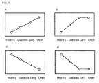
- Fig. 1 exemplifies variation patterns of the concentration or concentration ratio of the diagnostic markers for kidney disease of the present invention in respective pathological conditions of kidney diseases.
- “Healthy” represents healthy subjects
- “Diabetes” represents patients with stage I diabetic nephropathy (initial stage of nephropathy)
- “Early” represents patients with stage II diabetic nephropathy (early stage of nephropathy)
- “Overt” represents patients with stages III and IV diabetic nephropathy (overt nephropathy stage).
- the diagnosis method preferably follows "Embodiment 1" in which the concentration in urine is utilized.
- the concentration of glycolic acid in urine follows the variation pattern in which it decreases from "Healthy” to "Early” as shown in Fig. 1C .
- the value of the concentration of glycolic acid in urine obtained from a subject is lower than a predetermined threshold, it is diagnosed that the subject has kidney disease.
- the diagnosis method preferably follows “Embodiment 1" in which the concentration in urine is utilized.
- the concentration of 5-oxo-2-tetrahydrofuran carboxylic acid in urine follows the variation pattern in which it decreases from "Healthy” to "Early” as shown in Fig. 1C .
- value of the the concentration of 5-oxo-2-tetrahydrofuran carboxylic acid in urine obtained from a subject is lower than a predetermined threshold, it is diagnosed that the subject has kidney disease.
- the diagnosis method preferably follows "Embodiment 1" in which the concentration in urine is utilized or “Embodiment 2" in which the concentrations in urine and plasma are used.
- the concentration of ADMA in urine follows the variation pattern in which it decreases from "Healthy” to "Early” as shown in Fig. 1C .
- the value of the concentration of ADMA in urine obtained from a subject is lower than a predetermined threshold, it is diagnosed that the subject has kidney disease.
- the value of "the concentration of ADMA in plasma/the concentration of ADMA in urine” follows the variation pattern in which it increases from “Healthy” to "Early". Thus, when the value of "the concentration of ADMA in plasma/the concentration of ADMA in urine” is higher than a predetermined threshold, it is diagnosed that the subject has kidney disease.
- the concentration ratio between two substances is preferably the ratio between the concentration of trimethylglycine in urine and the concentration of the substance B shown in Table 1 in urine.
- the value relating to the concentration ratio between two substances is preferably the ratio between the concentration of glycolic acid in urine and the concentration of the substance B shown in Table 2 in plasma or urine.
- the value relating to the concentration ratio between two substances is more preferably the ratio between the concentration of glycolic acid in urine and the concentration of the substance B in plasma.
- the value relating to the concentration ratio between two markers is more preferably the ratio between the concentration of glycolic acid in urine and the concentration of the substance B in urine.
- the value relating to the concentration ratio between two substances is preferably the ratio between the concentration of cystine in urine and the concentration of the substance B shown in Table 3 in urine.
- the value relating to the concentration ratio between two substances is preferably the ratio between the concentration of tryptophan in plasma and the concentration of ornithine in plasma.
- the computer program of the present invention is the one which is configured to cause a computer to operate the diagnosis method using the diagnostic marker for kidney disease of the present invention, preferably the diagnosis method of kidney disease according to the above Embodiments 1 to 3.
- the present computer program can cause a computer to operate the steps of:
- the computer program causes a computer to further operate the step of comparing the value of the concentration of the marker obtained in the obtaining step with a threshold.
- the obtaining step is the step of obtaining the value of the concentration of at least one marker selected from glycolic acid, N 5 -[(dimethylamino)iminomethyl]-ornithine and 5-oxo-2-tetrahydrofuran carboxylic acid in urine or plasma obtained from a subject suspected to have kidney disease
- the determining step is the step of determining whether or not the subject has kidney disease based on the comparison result obtained in the comparing step.
- the present computer program can be configured to cause a computer to operate the diagnosis method according to Embodiment 1 described above.
- the computer program causes a computer to further operate the steps of calculating a value relating to the ratio between the concentrations of the marker in urine and plasma from the values of the concentrations of the marker in urine and plasma obtained in the obtaining step, and comparing the calculated value relating to the ratio between the concentrations of the marker in urine and plasma with a threshold.
- the obtaining step is the step of obtaining the value of the concentration of at least one substance selected from glycolic acid, N 5 -[(dimethylamino)iminomethyl]-ornithine and 5-oxo-2-tetrahydrofuran carboxylic acid in urine and plasma obtained from a subject suspected to have kidney disease and the determining step is the step of determining whether or not the subject has kidney disease based on the comparison result obtained in the comparing step.
- the present computer program can be configured to cause a computer to operate the diagnosis method according to Embodiment 2 described above.
- the computer program causes a computer to further operate the steps of calculating a value relating to the ratio between the concentrations of one substance and the other substance in the diagnostic marker for kidney disease which consists of a combination of two substances from the concentrations of the two substances in urine and/or plasma obtained in the obtaining step, and comparing the calculated value relating to the ratio between the concentrations of two substances with a threshold.
- the obtaining step is the step of obtaining the value of the concentration of at least one marker selected from the diagnostic markers for kidney disease consisting of a combination of two substances in urine and plasma obtained from a subject suspected to have kidney disease and the determining step is the step of determining whether or not the subject has the kidney disease based on the comparison result obtained in the comparing step.
- the present computer program can be configured to cause a computer to operate the diagnosis method according to Embodiment 3 described above.
- the combination of two substances in urine and/or plasma is the combination of trimethylglycine with N 5 -[(dimethylamino)iminomethyl]-ornithine or guanidinoacetic acid.
- the scope of the present invention also encompasses a computer readable memory medium storing the computer program.
- FIG. 28 An example of a system of the computer in which the computer program of the present invention is executed is shown in Fig. 28 .
- a computer 100 is mainly constructed from a main unit 110, a display unit 120 and an input device 130.
- a CPU 110a, ROM 110b, RAM 110c, a hard disk 110d, a read-out system 110e, an input-output interface 110f and an image output interface 110g are connected via a bus 110h so as to be able to communicate each other.
- the CPU 110a can execute the computer program stored in ROM 110b and the computer program loaded with RAM 110c.
- ROM 110b is made up with mask ROM, PROM, EPROM, EEPROM or the like and stores the computer program executed by the CPU 110a and data used for the execution.
- RAM 110c is made up with SRAM, DRAM or the like. RAM 110c is used for readout of the computer programs stored in ROM 110b and the hard disk 110d. RAM 110c is also utilized as a work area when the CPU 110a executes these computer programs.
- Various computer programs to be executed by CPU 110a such as an operating system and application system program and data necessary for execution of the computer programs are installed on the hard disk 110d.
- a computer program 140a which causes the computer 100 to operate the diagnosis method of kidney disease according to Embodiments 1 to 3, described herein below, and a threshold used in Embodiments 1 to 3 are also installed on the hard disk 110d.
- the read-out system 110e is made up with a flexible disk drive, a CD-ROM drive or a DVD-ROM drive and the like and can readout the computer program or data stored on a portable memory medium 140.
- the portable memory medium 140 is a memory medium well known per se in the art such as a flexible disk, a CD-ROM, a DVD-ROM and the like.
- the portable memory medium 140 stores the computer program 140a relating to the computational determination on whether or not a subject has kidney disease so as to be readout by the computer.
- the CPU 110a reads out the computer program 140a from the portable memory medium 140 and can install it on the hard disk 110d.
- An operation system providing a graphical user interface environment such as Windows ® produced and marketed by Microsoft Corporation (U.S.A.) is installed on the hard disk 110d.
- the computer program 140a relating to the determination of kidney disease operates on the operating system.
- the input-output interface 110f is made up with a serial interface such as USB, IEEE1394, RS-232C; a parallel interface such as SCSI, IDE, IEEE1284; or an analog interface formed by a D/A converter, an A/D converter or the like.
- the input-output interface 110f is connected to the input device 130 formed by a keyboard and a mouse.
- a user can input the concentration data of the diagnostic marker for kidney disease in urine and plasma obtained from a subject into the computer main unit 110 by means of the input device 130.
- the input-output interface 110f may be connected to a measurement apparatus 200 which can measure the concentration of the diagnostic marker for kidney disease in urine and plasma obtained from a subject. In this case, the concentration data of the diagnostic marker for kidney disease can be entered from the measurement apparatus 200 into the computer main unit 110.
- the image output interface 110h is connected to the display unit 120 made up with a LCD or CRT and sends an image signal to the display unit 120 according to an image data provided by the CPU 110a.
- the display unit 120 outputs the image data according to the input image signal.
- the display unit 120 also outputs the determination result provided by the CPU 110a described hereinbelow.
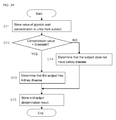
- Fig. 29 shows a flow chart executed by the CPU 110a when the diagnosis method of kidney disease of Embodiment 1 is carried out by the computer 100.
- the CPU 110a stores the value of the concentration of glycolic acid via the input-output interface 110f on RAM 110c and the hard disk 110d (Step S11).
- the CPU 110a determines that the subject has kidney disease when the value of the concentration of glycolic acid is lower than the threshold (Step S13).
- the CPU 110a determines that the subject does not have kidney disease when the value of the concentration of glycolic acid is not lower than the threshold (Step S14).
- the CPU 110a stores the determination result on the hard disk 110d and also outputs the results on the display unit 120 via the image output interface 110g (Step S15).
- glycolic acid is used as the diagnostic marker for kidney disease as described above.
- the diagnostic marker for kidney disease may be 5-oxo-2-tetrahydrofuran carboxylic acid and/or ADMA.
- Fig. 30 shows a flow chart executed by CPU 110a when the diagnosis method of kidney disease of Embodiment 2 is carried out by the computer 100.
- the CPU 110a stores the values of the concentration of ADMA on RAM 110c and the hard disk 110d (Step S21).
- the CPU 110a reads out the values of the concentration of ADMA in urine and plasma stored on RAM 110c, calculates the value of "the concentration in plasma/the concentration in urine" of ADMA (Step S22) and stores it on RAM 110c.
- the CPU 110a determines that the subject has kidney disease when the value of "the concentration in plasma/the concentration in urine" of ADMA is higher than the threshold (Step S24).
- the CPU 110a determines that the subject does not have kidney disease when the value of "the concentration in plasma/the concentration in urine” of ADMA is not higher than the threshold (Step S25).
- the CPU110a stores the determination result on the hard disk 110d and also outputs the results on the display unit 120 via the image output interface 110g (Step S26).
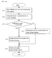
- Fig. 31 shows a flow chart executed by CPU 110a when the diagnosis method of kidney disease of Embodiment 3 is carried out by the computer 100.
- the CPU 110a stores the values of the concentrations on RAM 110c and the hard disk 110d via the input-output interface 110f (Step S31).
- the CPU 110a calculates the value of "the concentration of TMG in urine/the concentration of glycolic acid in urine” from the values of the concentrations of TMG and glycolic acid in urine (Step S32).
- the CPU 110a determines that the subject has kidney disease when the value of "the concentration of TMG in urine/the concentration of glycolic acid in urine" is higher than the threshold (Step S34).
- the CPU 110a determines that the subject does not have kidney disease when the value of "the concentration of TMG in urine/the concentration of glycolic acid in urine” is not higher than the threshold (Step S35).
- the CPU110a stores the determination result on the hard disk 110d and also outputs the results on the display unit 120 via the image output interface 110g (Step S36).
- the combination of TMG and glycolic acid is used as the diagnostic marker for kidney disease as described above.
- the diagnostic marker for kidney disease may be the diagnostic marker for kidney disease of Combinations 1 to 4 shown in Tables 1 to 4.
- the ratio between TMG and glycolic acid in urine (the value of "the concentration of TMG in urine/the concentration of glycolic acid in urine") is used as the value relating to the concentration ratio in this Embodiment.
- the value relating to the concentration ratio may be appropriately selected according to the combination of substances used as the diagnostic marker for kidney disease. More specifically, it can be the value relating to the concentration ratio shown in Tables 7 to 10 described hereinbelow in Example 2 according to the diagnostic marker for kidney disease used.
- the concentration of the marker is entered by a user via the input device 130.
- the concentration of the marker may be entered from the measurement apparatus 200 connected via the input-output interface 110f.
- the concentration of the marker measured on the measurement apparatus 200 may be stored on the portable memory medium 140 and be readout by the read-out system 110e from the portable memory medium 140.
- Specimens of plasma and morning urine were taken from 20 healthy subjects and 40 patients categorized as follows (total: 60 subjects). Namely, the patients included the groups of diabetes patients (19 patients), stage II diabetic nephropathy patients (early nephropathy; 12 patients), stages III and IV diabetic nephropathy patients (overt nephropathy; 7 patients) and chronic kidney disease patients (CKD; 2 patients). Chronic kidney disease patients mean the patients having non-diabetic nephropathy. Namely, it will be confirmed that by using the data obtained from the chronic kidney disease patients as a positive control, the result obtained in the present Example is not resulting from diabetes.
- a methanol solution (450 ⁇ L) containing both of L-methioninesulfone and D-camphor-10-sulfonic acid at a final concentration of 10 ⁇ M each as internal standards was stirred with 50 ⁇ L of plasma.
- Chloroform (500 ⁇ L) and 200 ⁇ L of distilled water were stirred therein followed by centrifugation (2,300 x g, 4°C, 5 min).
- the aqueous phase was transferred to ultrafiltration tubes (Ultrafree-MC PBCC centrifugal filter unit 5 kDa: MILLIPORE) (200 ⁇ L x 2 tubes) and subjected to ultrafiltration (9,100 x g, 4°C, 120 min).
- the filtrate was evaporated to dryness and dissolved in 25 ⁇ L of distilled water to obtain plasma samples.
- the samples were diluted two-fold and used in the measurement described hereinbelow.
- the instrument used was Agilent CE-TOF/MS system (Agilent Technologies) and the capillary used was a Fused silica capillary i.d. 50 ⁇ m x 80 cm.
- the measurement conditions were as follows.
- Substances were identified by following procedures.
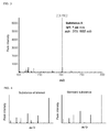
- the groups of substances were separated by CE according to ion strength (see Fig. 2 ) and continuously measured by MS (see Fig. 3 ).
- Information on the migration time (min) on CE hereinafter also abbreviated as "MT"
- mass-to-charge ratio (m/z) (calculated to 4 places of decimals; hereinafter also abbreviated as "m/z") of each substance was obtained.
- the mass-to-charge ratio of the substance of interest was used for calculation of accurate mass.
- the accurate mass of the substance of interest was then used for search among substances having estimated chemical structures (standard substance) followed by CE-MS analysis of the standard substance.
- the standard substance was subjected to MS/MS analysis (see Fig. 4 ).
- the substance of interest and the standard substance were compared for their MT, m/z and MS/MS data. When all data matched, the substance of interest was identified as the standard substance. Tha values of MT and m/z of the identified substances are shown in Table 5.
- the number of MS peaks obtained by CE-MS analysis as described in the above (3) was 700 and 902 for plasma and urine samples, respectively.
- the relative peak area values were obtained for those peaks by correction with the internal standards for plasma samples and with the internal standards and urenal creatinine for urine samples and were used for selection of marker candidates.
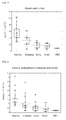
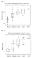
- the results are shown in Figs. 5 to 7 .
- the substances which may be used as a marker by itself are shown in Table 6.
- the measurement value for glycolic acid or 5-oxo-2-tetrahydrofuran carboxylic acid in urine follows the variation pattern in which it decreases from "Healthy” to "Early” as shown in Fig. 1C .
- the value of the concentration of glycolic acid or 5-oxo-2-tetrahydrofuran carboxylic acid in urine obtained from a subject can be used to determine whether or not the subject has kidney disease.
- the ratio of ADMA between in urine and plasma shows the tendency in which it increased from "Healthy” to "Early” as shown in Fig. 1A .
- the value of "the concentration in plasma/the concentration in urine" in urine and plasma obtained from a subject can be used to determine whether or not the subject has kidney disease.
- the substances were selected which may serve as a marker in combination.
- the concentration in combination of the substances in urine and plasma from healthy subjects and kidney disease patients in respective pathological conditions were examined. Marker candidates in the combination of two substances were chosen when the ratio obtained by dividing the concentration of one substance by the concentration of the other substance follows the variation patterns of the concentration ratio in respective pathological conditions of kidney disease as shown in Fig. 1 .
- the results are shown in Figs. 8 to 27 .
- the relations between the value relating to the concentration ratio of the marker consisting of a combination of two substances and the variation pattern of the value relating to the concentration ratio in respective pathological conditions of kidney disease are shown in Tables 7 to 10.
- Substance A Substance B Value relating to concentration ratio Variation pattern Glycolic acid ADMA B (plasma)/A (urine) Fig. 1B Glycolic acid Cytidine B (urine)/A (urine) Fig. 1B Glycolic acid Valine B (urine)/A (urine) Fig. 1A Glycolic acid Kynurenine B (plasma)/A (urine) Fig. 1A Glycolic acid 2-oxoglutaric acid B (plasma)/A (urine) Fig. 1B Glycolic acid Glycocyamidine B (urine)/A (urine) Fig. 1A Glycolic acid Choline B (plasma)/A (urine) Fig. 1A
- the value (B (plasma)/A (plasma)) obtained by dividing the value of the concentration of ornithine (substance B) in plasma by the value of the concentration of tryptophan (substance A) in plasma for the combination of two substances shown in Table 10 shows the tendency that it increases from "Healthy” to "Early” as shown in Fig. 1A .
- the value of B (plasma)/A (plasma) of two substances shown in Table 10 in plasma obtained from a subject can be used to determine whether or not the subject has kidney disease.
Landscapes
- Life Sciences & Earth Sciences (AREA)
- Health & Medical Sciences (AREA)
- Engineering & Computer Science (AREA)
- Chemical & Material Sciences (AREA)
- Immunology (AREA)
- Molecular Biology (AREA)
- Physics & Mathematics (AREA)
- Biomedical Technology (AREA)
- Urology & Nephrology (AREA)
- Hematology (AREA)
- Biochemistry (AREA)
- Analytical Chemistry (AREA)
- General Health & Medical Sciences (AREA)
- General Physics & Mathematics (AREA)
- Pathology (AREA)
- Medicinal Chemistry (AREA)
- Microbiology (AREA)
- Food Science & Technology (AREA)
- Cell Biology (AREA)
- Biotechnology (AREA)
- Proteomics, Peptides & Aminoacids (AREA)
- Chemical Kinetics & Catalysis (AREA)
- Electrochemistry (AREA)
- Biophysics (AREA)
- Bioinformatics & Computational Biology (AREA)
- Bioinformatics & Cheminformatics (AREA)
- Investigating Or Analysing Biological Materials (AREA)
Abstract
Description
- The present invention relates to a diagnostic marker for kidney disease and a computer program for diagnosis of kidney disease using the marker.
- Kidney diseases are caused by pathological lesions in kidney tissues. Kidney diseases mainly include 1) glomerulonephritis due to pathological lesions in glomerulus which is responsible for blood filtration, 2) tubulointerstitial nephritis due to pathological lesions in renal tubules which is responsible for resorption of water or electrolytes from primitive urine, 3) arteriolosclerosis due to renovascular pathological lesions and the like.
- When a patient develops renal failure from kidney disease, it is difficult to restore and maintain renal functions even after initiation of therapeutic treatments. In this case, the patient needs to undergo dialysis treatment. The number of patients who require dialysis treatment is increasing recently and it causes a problem of growth in medical costs.
Thus, it is important to identify kidney disease patients at early stages by accurate diagnoses. - In the current kidney disease diagnoses, renal function is evaluated by measurement of glomerular filtration rate (GFR).
Kidney disease diagnostic markers are also used such as urine albumin, type IV collagen, transferrin, IgG, laminin, fibronectin, α-1 microglobulin, β-2 microglobulin, cystatin C, N-acetyl-β-D-glucosaminidase (NAG), L-fatty acid binding protein (L-FABP), glycocyamidine and derivatives thereof and the like (seePatent Literatures 1 and 2). - However, in order to provide further highly accurate diagnoses of kidney diseases, measurements of GFR and use of the above diagnostic markers are not sufficient. Therefore, there is a need for development of novel markers useful for diagnoses of kidney disease.
-
- Patent Literature 1: Japanese Unexamined Patent Publication No.
HEI 2(1990)-288867 - Patent Literature 2: Japanese Unexamined Patent Publication No.
2001-174459 - An object of the present invention is to provide novel diagnostic markers for kidney disease. Another object of the present invention is to provide a computer program for diagnosis of kidney disease using the diagnostic markers for kidney disease.
- The present inventors analyzed the specimens obtained from healthy subjects and kidney disease patients by capillary electrophoresis mass spectrometry (CE-MS) and, as a result, found new diagnostic markers for kidney disease, thereby completing the present invention.
- Namely, according to the present invention, a diagnostic marker for kidney disease is provided which is at least one selected from:
- glycolic acid;
- N5-[(dimethylamino)iminomethyl]-ornithine;
- 5-oxo-2-tetrahydrofuran carboxylic acid;
- a combination consisting of trimethylglycine and any one substance selected from glycolic acid, N5-[(dimethylamino)iminomethyl]-ornithine, guanidinoacetic acid, 4-guanidinobutyric acid, N6-acetyllysine, N1-acetylhistidine and histidine;
- a combination consisting of glycolic acid and any one substance selected from N5-[(dimethylamino)iminomethyl]-ornithine, cytidine, valine, kynurenine, 2-oxoglutaric acid, glycocyamidine and choline;
- a combination consisting of cystine and any one substance selected from N1-acetylhistidine, histidine, glycine, tryptophan and methionine; and
- a combination consisting of tryptophan and ornithine.
- According to the present invention, a computer program is also provided which is configured to cause a computer to operate the steps of:
- obtaining a concentration of the above diagnostic marker in urine and/or plasma obtained from a subject suspected to have kidney disease;
- determining whether or not the subject has kidney disease based on the obtained concentration; and
- delivering the result of determination.
- According to the present invention, diagnostic markers for kidney disease useful for diagnoses with increased accuracy can be provided. The diagnostic markers for kidney disease of the present invention allow not only diagnosis of a subject on kidney disease but also, in case the subject has received treatment for kidney disease, evaluation of an extent of improvement in symptoms and efficacy of the treatment.
Further, according to the present invention, the computer program can be provided for diagnoses of kidney disease using the above diagnostic markers for kidney disease. -
-
Fig. 1 shows graphs representing variation patterns of the concentration or concentration ratio of the diagnostic markers for kidney disease of the present invention depending on respective pathological conditions of kidney diseases.Figs. 1A and 1C show the patterns in which the concentration and the like continuously increase or decrease from the healthy stage to the overt nephropathy stage andFigs. 1B and 1D show the patterns in which the concentration and the like continuously increase or decrease from the healthy stage to the early nephropathy stage and do not show any change from the early nephropathy stage to the overt nephropathy stage; -
Fig. 2 shows an example of a chart obtained by separating a group of substances by capillary electrophoresis (CE); -
Fig. 3 shows an example of a chart obtained by subjecting the substances separated by CE to mass spectrometry (MS) analysis; -
Fig. 4 shows an example of charts obtained from MS/MS analysis of a substance of interest and a standard substance; -
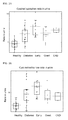
Fig. 5 is a representation of quantitative values of glycolic acid in urine in the groups of healthy subjects and respective patients; -
Fig. 6 is a representation of quantitative values of 5-oxo-2-tetrahydrofuran carboxylic acid in urine in the groups of healthy subjects and respective patients; -
Fig. 7 shows representations of quantitative values of N5-[(dimethylamino)iminomethyl]-ornithine (hereinafter also referred to as "ADMA") in the groups of healthy subjects and respective patients:Fig. 7A shows the quantitative values in urine,Fig. 7B shows the quantitative values in plasma andFig. 7C shows the ratios between the quantitative values in urine and plasma; -
Fig. 8 is a representation of the ratios between trimethylglycine (hereinafter also referred to as "TMG") in urine and guanidinoacetic acid in urine in the groups of healthy subjects and respective patients; -
Fig. 9 is a representation of the ratios between TMG in urine and glycolic acid in urine in the groups of healthy subjects and respective patients; -
Fig. 10 is a representation of the ratios between TMG in urine and ADMA in urine in the groups of healthy subjects and respective patients; -
Fig. 11 is a representation of the ratios between TMG in urine and 4-guanidinobutyric acid in urine in the groups of healthy subjects and respective patients; -
Fig. 12 is a representation of the ratios between TMG in urine and N6-acetyllysine in urine in the groups of healthy subjects and respective patients; -
Fig. 13 is a representation of the ratios between TMG in urine and N1-acetylhistidine in urine in the groups of healthy subjects and respective patients; -
Fig. 14 is a representation of the ratios between TMG in urine and histidine in urine in the groups of healthy subjects and respective patients; -
Fig. 15 is a representation of the ratios between choline in plasma and glycolic acid in urine in the groups of healthy subjects and respective patients; -
Fig. 16 is a representation of the ratios between valine in urine and glycolic acid in urine in the groups of healthy subjects and respective patients; -
Fig. 17 is a representation of the ratios between kynurenine in plasma and glycolic acid in urine in the groups of healthy subjects and respective patients; -
Fig. 18 is a representation of the ratios between glycocyamidine in urine and glycolic acid in urine in the groups of healthy subjects and respective patients; -
Fig. 19 is a representation of the ratios between ADMA in plasma and glycolic acid in urine in the groups of healthy subjects and respective patients; -
Fig. 20 is a representation of the ratios between 2-oxoglutaric acid in plasma and glycolic acid in urine in the groups of healthy subjects and respective patients; -
Fig. 21 is a representation of the ratios between cytidine in urine and glycolic acid in urine in the groups of healthy subjects and respective patients; -
Fig. 22 is a representation of the ratios between cystine in urine and histidine in urine in the groups of healthy subjects and respective patients; -
Fig. 23 is a representation of the ratios between cystine in urine and glycine in urine in the groups of healthy subjects and respective patients; -
Fig. 24 is a representation of the ratios between cystine in urine and N1-acetylhistidine in urine in the groups of healthy subjects and respective patients; -
Fig. 25 is a representation of the ratios between cystine in urine and tryptophan in urine in the groups of healthy subjects and respective patients; -
Fig. 26 is a representation of the ratios between cystine in urine and methionine in urine in the groups of healthy subjects and respective patients; -
Fig. 27 is a representation of the ratios between ornithine in plasma and tryptophan in plasma in the groups of healthy subjects and respective patients; -
Fig. 28 is a block diagram of a computer system for executing diagnosis of kidney disease; -
Fig. 29 is a flow chart executed by a CPU when a diagnosis method of kidney disease ofEmbodiment 1 is carried out by a computer; -
Fig. 30 is a flow chart executed by a CPU when a diagnosis method of kidney disease ofEmbodiment 2 is carried out by a computer; and -
Fig. 31 is a flow chart executed by a CPU when a diagnosis method of kidney disease ofEmbodiment 3 is carried out by a computer. - The following formulae represent the names and chemical structures of the substances used as the diagnostic markers for kidney disease of the present invention. The diagnostic markers for kidney disease of the present invention comprise one substance or a combination of two substances shown below.
-


Glycolic acid N5-[(Dimethylamino)iminomethyl]-ornithine (ADMA) 
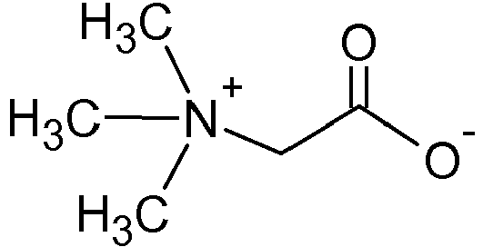
5-Oxo-2-tetra hyd rofu ran carboxylic acid Trimethylglycine (TMG) -


Guanidinoacetic acid 4-Guanidinobutyric acid 

2-Oxoglutaric acid N6-Acetyllysine -


N1-Acetylhistidine Histidine 

Cytidine Kynurenine -


Valine Glycocyamidine 

Choline Cystine -
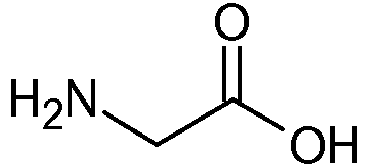

Glycine Tryptophan 

Methionine Ornithine - The diagnostic marker for kidney disease consisting of a combination of two substances includes the following
combinations 1 to 4. Thecombinations 1 to 4 are specifically represented in Tables 1 to 4. Namely, the diagnostic marker for kidney disease of the present invention consisting of a combination of two substances is selected from the combinations of the substance A and the substance B shown in these Tables. - A combination of the substance A which is trimethylglycine (TMG) and the substance B which is any one substance selected from glycolic acid, N5-[(dimethylamino)iminomethyl]-ornithine (ADMA), guanidinoacetic acid, 4-guanidinobutyric acid, N6-acetyllysine, N1-acetylhistidine and histidine.
-
Table 1 Substance A Substance B TMG Glycolic acid TMG ADMA TMG Guanidinoacetic acid TMG 4-Guanidinobutyric acid TMG N6-Acetyllysine TMG N1-Acetylhistidine TMG Histidine - A combination of the substance A which is glycolic acid and the substance B which is any one substance selected from ADMA, cytidine, valine, kynurenine, 2-oxoglutaric acid, glycocyamidine and choline.
-
Table 2 Substance A Substance B Glycolic acid ADMA Glycolic acid Cytidine Glycolic acid Valine Glycolic acid Kynurenine Glycolic acid 2-Oxoglutaric acid Glycolic acid Glycocyamidine Glycolic acid Choline - A combination of the substance A which is cystine and the substance B which is any one substance selected from N1-acetylhistidine, histidine, glycine, tryptophan and methionine.
-
Table 3 Substance A Substance B Cystine N1-acetylhistidine Cystine Histidine Cystine Glycine Cystine Tryptophan Cystine Methionine - A combination of the substance A which is tryptophan and the substance B which is ornithine.
-
Table 4 Substance A Substance B Tryptophan Ornithine - In the present invention, the kidney disease may be a disease resulting from pathological lesions in renal tissues without particular limitation. The kidney disease may include, for example, diabetic nephropathy, glomerulonephritis, tubulointerstitial nephritis and the like.
Among the above kidney diseases, the diagnostic marker for kidney disease of the present invention may be suitably used as the diagnostic marker for diabetic nephropathy accompanying diabetes. - It is possible to use the diagnostic markers for kidney disease of the present invention for carrying out diagnoses of kidney disease. The diagnosis method may be the one in which the diagnostic marker for kidney disease is measured in urine and/or plasma obtained from a subject suspected to have kidney disease, without particular limitation. For example, diagnosis of kidney disease can be carried out based on the result of measurement of the concentration of the diagnostic marker for kidney disease in urine and/or plasma obtained from a subject suspected to have kidney disease. "Diagnosis of kidney disease" includes definitive diagnosis of kidney disease, a screening test for kidney disease and an evaluation of treatment efficacy of kidney disease, among which a screening test for kidney disease is preferable.
- Urine obtained from a subject may be any urine regardless of the physical condition of the subject, timing of meals, medication which the subject receives, if any, or the time of sampling, without limitation. However, it is preferably morning urine (first urine). Plasma obtained from a subject may also be any plasma obtained from blood sampled from a subject regardless of the physical condition of the subject, timing of meals, medication which the subject receives, if any, or the time of sampling, without limitation.
- Specific examples of the diagnosis method of kidney disease using the present marker are described hereinbelow.
-
Embodiment 1 andEmbodiment 2 described hereinbelow are the diagnosis methods for kidney disease in which the diagnostic markers for kidney disease consisting of a single substance are used. -
- (1) Measure the concentration of the marker in urine or plasma obtained from a subject suspected to have kidney disease;
- (2) Compare the measured concentration of the marker with a threshold; and
- (3) Based on the result of comparison, determine whether or not the subject has kidney disease.
-
- (1) Measure the concentration of the marker in urine and plasma obtained from a subject suspected to have kidney disease;
- (2) Obtain a value relating to the ratio between the concentration of the marker in urine and the concentration in plasma;
- (3) Compare the obtained value relating to the ratio between the concentration of the marker in urine and the concentration in plasma with a threshold;
- (4) Based on the result of comparison, determine whether or not the subject has kidney disease.
- In
Embodiment 2, the value relating to the ratio between the concentration of the marker in urine and the concentration in plasma may include the values of "the concentration in plasma/the concentration in urine" and "the concentration in urine/the concentration in plasma" - The diagnostic marker for kidney disease used for the
above Embodiment 1 andEmbodiment 2 is suitably any of glycolic acid, ADMA and 5-oxo-2-tetrahydrofuran carboxylic acid. - The following
Embodiment 3 is the diagnosis method of kidney disease in which the diagnostic marker for kidney disease consisting of a combination of two substances is used. -
- (1) Measure the concentrations of two substances in urine and/ or plasma obtained from a subject suspected to have kidney disease;
- (2) Obtain a value relating to the ratio between the measured concentrations of two substances;
- (3) Compare the obtained value relating to the concentration ratio between two substances with a threshold; and
- (4) Based on the result of comparison, determine whether or not the subject has kidney disease.
- In
Embodiment 3, the value relating to the concentration ratio between two substances may include, provided that two substances constituting the marker are the substances A and B, respectively, the values "A (plasma) / B (plasma)", "A (plasma) / B (urine)", "A (urine) / B (plasma)", "A (urine)/B (urine)", "B (plasma)/A (plasma)", "B (plasma)/A (urine)", "B (urine)/A (plasma)" and "B (urine)/A (urine)"
"A (plasma)" and "A (urine)" represent the concentrations of the substance A in plasma and urine, respectively, and "B (plasma)" and "B (urine)" represent the concentrations of the substance B in plasma and urine, respectively. - The diagnostic marker for kidney disease used for the
above Embodiment 3 is suitably the marker selected from theabove combinations 1 to 4. - The threshold may be determined as follows, for example. First, urine and/or plasma is taken from a subject who is confirmed to have no kidney disease according to the conventional method (i.e., a healthy subject). Next, the urine and/or plasma is measured for the concentration of the diagnostic marker for kidney disease of the present invention and the value of the concentration and the value relating to the concentration ratio are obtained. The obtained result is then used as a predetermined threshold. The threshold determined as above allows determination on whether or not a subject undergoing diagnosis using the diagnostic marker for kidney disease of the present invention is healthy or has kidney disease.
Alternatively, the threshold may be determined by using urine and/or plasma obtained from a subject suffering from stage I or II diabetic nephropathy, instead of urine and/or plasma obtained from a healthy subject. In this case, the obtained threshold allows determination on whether a subject undergoing diagnosis using the diagnostic marker for kidney disease of the present invention has stage I or II diabetic nephropathy or severer kidney disease.
Upon determination of the threshold, samples are preferably obtained from more than one healthy subject and/or subject having stage I or II diabetic nephropathy. - The method for measuring the concentration of the diagnostic marker for kidney disease in urine and plasma may be appropriately selected according to physical or chemical properties of the marker substance without particular limitation.
For example, a sample to be measured may be prepared from urine or plasma obtained from a subject suspected to have kidney disease. The marker contained in the sample may be then isolated by high performance liquid chromatography (HPLC), gas chromatography (GC), capillary electrophoresis (CE) and the like. The isolated marker is then subjected to a measurement system such as UV detection, fluorescence detection, mass spectrometry (MS) and the like to measure the concentration of the marker in urine or plasma.
Alternatively, the concentration of the marker in urine or plasma can be measured by bringing the marker contained in urine or plasma obtained from the subject into contact with an enzyme which can react with the marker as a substrate and subjecting the enzyme reaction product to a measurement system such as a method using redox electrodes, colorimetric method and the like. -
Fig. 1 exemplifies variation patterns of the concentration or concentration ratio of the diagnostic markers for kidney disease of the present invention in respective pathological conditions of kidney diseases. In this figure, "Healthy" represents healthy subjects, "Diabetes" represents patients with stage I diabetic nephropathy (initial stage of nephropathy), "Early" represents patients with stage II diabetic nephropathy (early stage of nephropathy), and "Overt" represents patients with stages III and IV diabetic nephropathy (overt nephropathy stage). -
-
Fig. 1A shows the variation pattern with the tendency that the concentration or concentration ratio of the marker increases with the progress of the stage of kidney disease from "Healthy" to "Overt". -
Fig. 1B shows the variation pattern with the tendency that the concentration or concentration ratio of the marker increases from "Healthy" to "Early", while it is almost constant from "Early" to "Overt". -
Fig. 1C shows the variation pattern with the tendency that the concentration or concentration ratio of the marker decreases with the progress of the stage of kidney disease from "Healthy" to "Overt". -
Fig. 1D shows the variation pattern with the tendency that the concentration or concentration ratio of the marker decreases from "Healthy" to "Early", while it is almost constant from "Early" to "Overt". - When glycolic acid is used as the diagnostic marker for kidney disease, the diagnosis method preferably follows "
Embodiment 1" in which the concentration in urine is utilized. In this case, the concentration of glycolic acid in urine follows the variation pattern in which it decreases from "Healthy" to "Early" as shown inFig. 1C .
Thus, when the value of the concentration of glycolic acid in urine obtained from a subject is lower than a predetermined threshold, it is diagnosed that the subject has kidney disease. - When 5-oxo-2-tetrahydrofuran carboxylic acid is used as the diagnostic marker for kidney disease, the diagnosis method preferably follows "
Embodiment 1" in which the concentration in urine is utilized. In this case, the concentration of 5-oxo-2-tetrahydrofuran carboxylic acid in urine follows the variation pattern in which it decreases from "Healthy" to "Early" as shown inFig. 1C .
Thus, when value of the the concentration of 5-oxo-2-tetrahydrofuran carboxylic acid in urine obtained from a subject is lower than a predetermined threshold, it is diagnosed that the subject has kidney disease. - When N5-[(dimethylamino)iminomethyl]-ornithine (ADMA) is used as the diagnostic marker for kidney disease, the diagnosis method preferably follows "
Embodiment 1" in which the concentration in urine is utilized or "Embodiment 2" in which the concentrations in urine and plasma are used.
In the case ofEmbodiment 1, the concentration of ADMA in urine follows the variation pattern in which it decreases from "Healthy" to "Early" as shown inFig. 1C .
Thus, when the value of the concentration of ADMA in urine obtained from a subject is lower than a predetermined threshold, it is diagnosed that the subject has kidney disease.
In the case ofEmbodiment 2, the value of "the concentration of ADMA in plasma/the concentration of ADMA in urine" follows the variation pattern in which it increases from "Healthy" to "Early".
Thus, when the value of "the concentration of ADMA in plasma/the concentration of ADMA in urine" is higher than a predetermined threshold, it is diagnosed that the subject has kidney disease. - When the diagnostic marker for kidney disease shown in "
Combination 1" described above is used in the diagnosis method of "Embodiment 3", the concentration ratio between two substances is preferably the ratio between the concentration of trimethylglycine in urine and the concentration of the substance B shown in Table 1 in urine. - When the diagnostic marker for kidney disease shown in "
Combination 2" described above is used in the diagnosis method of "Embodiment 3", the value relating to the concentration ratio between two substances is preferably the ratio between the concentration of glycolic acid in urine and the concentration of the substance B shown in Table 2 in plasma or urine.
Particularly, when the substance B is ADMA, kynurenine, 2-oxoglutaric acid or choline, the value relating to the concentration ratio between two substances is more preferably the ratio between the concentration of glycolic acid in urine and the concentration of the substance B in plasma. When the substance B is valine, cytidine or glycocyamidine, the value relating to the concentration ratio between two markers is more preferably the ratio between the concentration of glycolic acid in urine and the concentration of the substance B in urine. - When the diagnostic marker for kidney disease shown in "
Combination 3" described above is used in the diagnosis method of "Embodiment 3", the value relating to the concentration ratio between two substances is preferably the ratio between the concentration of cystine in urine and the concentration of the substance B shown in Table 3 in urine. - When the diagnostic marker for kidney disease shown in "
Combination 4" described above is used in the diagnosis method of "Embodiment 3", the value relating to the concentration ratio between two substances is preferably the ratio between the concentration of tryptophan in plasma and the concentration of ornithine in plasma. - The computer program of the present invention is described hereinbelow.
- The computer program of the present invention is the one which is configured to cause a computer to operate the diagnosis method using the diagnostic marker for kidney disease of the present invention, preferably the diagnosis method of kidney disease according to the
above Embodiments 1 to 3.
The present computer program can cause a computer to operate the steps of: - obtaining a concentration of the diagnostic marker for kidney disease of the present invention in urine and/or plasma obtained from a subject suspected to have kidney disease (hereinafter also referred to as "obtaining step");
- determining whether or not the subject has kidney disease based on the obtained concentration (hereinafter also referred to as "determining step"); and
- delivering the result of determination.
- In the preferred embodiment of the present invention, the computer program causes a computer to further operate the step of comparing the value of the concentration of the marker obtained in the obtaining step with a threshold. In this embodiment, the obtaining step is the step of obtaining the value of the concentration of at least one marker selected from glycolic acid, N5-[(dimethylamino)iminomethyl]-ornithine and 5-oxo-2-tetrahydrofuran carboxylic acid in urine or plasma obtained from a subject suspected to have kidney disease and the determining step is the step of determining whether or not the subject has kidney disease based on the comparison result obtained in the comparing step. Accordingly, the present computer program can be configured to cause a computer to operate the diagnosis method according to
Embodiment 1 described above. - In another preferable embodiment of the present invention, the computer program causes a computer to further operate the steps of calculating a value relating to the ratio between the concentrations of the marker in urine and plasma from the values of the concentrations of the marker in urine and plasma obtained in the obtaining step, and comparing the calculated value relating to the ratio between the concentrations of the marker in urine and plasma with a threshold. In this embodiment, the obtaining step is the step of obtaining the value of the concentration of at least one substance selected from glycolic acid, N5-[(dimethylamino)iminomethyl]-ornithine and 5-oxo-2-tetrahydrofuran carboxylic acid in urine and plasma obtained from a subject suspected to have kidney disease and the determining step is the step of determining whether or not the subject has kidney disease based on the comparison result obtained in the comparing step. Accordingly, the present computer program can be configured to cause a computer to operate the diagnosis method according to
Embodiment 2 described above. - In another preferable embodiment of the present invention, the computer program causes a computer to further operate the steps of calculating a value relating to the ratio between the concentrations of one substance and the other substance in the diagnostic marker for kidney disease which consists of a combination of two substances from the concentrations of the two substances in urine and/or plasma obtained in the obtaining step, and comparing the calculated value relating to the ratio between the concentrations of two substances with a threshold. In this embodiment, the obtaining step is the step of obtaining the value of the concentration of at least one marker selected from the diagnostic markers for kidney disease consisting of a combination of two substances in urine and plasma obtained from a subject suspected to have kidney disease and the determining step is the step of determining whether or not the subject has the kidney disease based on the comparison result obtained in the comparing step. Accordingly, the present computer program can be configured to cause a computer to operate the diagnosis method according to
Embodiment 3 described above.
In a more preferable embodiment, the combination of two substances in urine and/or plasma is the combination of trimethylglycine with N5-[(dimethylamino)iminomethyl]-ornithine or guanidinoacetic acid. - The scope of the present invention also encompasses a computer readable memory medium storing the computer program.
- An example of a system of the computer in which the computer program of the present invention is executed is shown in
Fig. 28 . - A
computer 100 is mainly constructed from a main unit 110, adisplay unit 120 and aninput device 130. In the main unit 110, a CPU 110a, ROM 110b,RAM 110c, a hard disk 110d, a read-out system 110e, an input-output interface 110f and an image output interface 110g are connected via abus 110h so as to be able to communicate each other. - The CPU 110a can execute the computer program stored in ROM 110b and the computer program loaded with
RAM 110c. ROM 110b is made up with mask ROM, PROM, EPROM, EEPROM or the like and stores the computer program executed by the CPU 110a and data used for the execution.
RAM 110c is made up with SRAM, DRAM or the like.RAM 110c is used for readout of the computer programs stored in ROM 110b and the hard disk 110d.RAM 110c is also utilized as a work area when the CPU 110a executes these computer programs. - Various computer programs to be executed by CPU 110a such as an operating system and application system program and data necessary for execution of the computer programs are installed on the hard disk 110d. A
computer program 140a which causes thecomputer 100 to operate the diagnosis method of kidney disease according toEmbodiments 1 to 3, described herein below, and a threshold used inEmbodiments 1 to 3 are also installed on the hard disk 110d. - The read-
out system 110e is made up with a flexible disk drive, a CD-ROM drive or a DVD-ROM drive and the like and can readout the computer program or data stored on aportable memory medium 140.
Theportable memory medium 140 is a memory medium well known per se in the art such as a flexible disk, a CD-ROM, a DVD-ROM and the like. Theportable memory medium 140 stores thecomputer program 140a relating to the computational determination on whether or not a subject has kidney disease so as to be readout by the computer. The CPU 110a reads out thecomputer program 140a from theportable memory medium 140 and can install it on the hard disk 110d. - An operation system providing a graphical user interface environment such as Windows® produced and marketed by Microsoft Corporation (U.S.A.) is installed on the hard disk 110d.
In the description hereinbelow, thecomputer program 140a relating to the determination of kidney disease operates on the operating system. - The input-output interface 110f is made up with a serial interface such as USB, IEEE1394, RS-232C; a parallel interface such as SCSI, IDE, IEEE1284; or an analog interface formed by a D/A converter, an A/D converter or the like. The input-output interface 110f is connected to the
input device 130 formed by a keyboard and a mouse. A user can input the concentration data of the diagnostic marker for kidney disease in urine and plasma obtained from a subject into the computer main unit 110 by means of theinput device 130. The input-output interface 110f may be connected to ameasurement apparatus 200 which can measure the concentration of the diagnostic marker for kidney disease in urine and plasma obtained from a subject. In this case, the concentration data of the diagnostic marker for kidney disease can be entered from themeasurement apparatus 200 into the computer main unit 110. - The
image output interface 110h is connected to thedisplay unit 120 made up with a LCD or CRT and sends an image signal to thedisplay unit 120 according to an image data provided by the CPU 110a. Thedisplay unit 120 outputs the image data according to the input image signal. Thedisplay unit 120 also outputs the determination result provided by the CPU 110a described hereinbelow. - The process steps executed by the computer in accordance with the present computer program are described in details by referring to the figures.
Fig. 29 shows a flow chart executed by the CPU 110a when the diagnosis method of kidney disease ofEmbodiment 1 is carried out by thecomputer 100. - When a user enters a value of the concentration of glycolic acid in urine obtained from a subject via the
input device 130, the CPU 110a stores the value of the concentration of glycolic acid via the input-output interface 110f onRAM 110c and the hard disk 110d (Step S11). - The CPU 110a retrieves a threshold (= 28.4) which has been preliminarily stored on the hard disk 110d and compares it with the value of the concentration of glycolic acid (Step S12).
- The CPU 110a determines that the subject has kidney disease when the value of the concentration of glycolic acid is lower than the threshold (Step S13). The CPU 110a determines that the subject does not have kidney disease when the value of the concentration of glycolic acid is not lower than the threshold (Step S14).
- The CPU 110a stores the determination result on the hard disk 110d and also outputs the results on the
display unit 120 via the image output interface 110g (Step S15). - In
Embodiment 1 shown inFig. 29 , glycolic acid is used as the diagnostic marker for kidney disease as described above. However, it does not limit the present invention. For example, the diagnostic marker for kidney disease may be 5-oxo-2-tetrahydrofuran carboxylic acid and/or ADMA. -
Fig. 30 shows a flow chart executed by CPU 110a when the diagnosis method of kidney disease ofEmbodiment 2 is carried out by thecomputer 100. - When a user enters values of the concentration of ADMA in urine and plasma obtained from a subject via the
input device 130, the CPU 110a stores the values of the concentration of ADMA onRAM 110c and the hard disk 110d (Step S21). - The CPU 110a reads out the values of the concentration of ADMA in urine and plasma stored on
RAM 110c, calculates the value of "the concentration in plasma/the concentration in urine" of ADMA (Step S22) and stores it onRAM 110c. - The CPU 110a retrieves a threshold (= 0.094) which has been preliminarily stored on the hard disk 110d and compares it with the value of "the concentration in plasma/the concentration in urine" of ADMA (Step S23).
- The CPU 110a determines that the subject has kidney disease when the value of "the concentration in plasma/the concentration in urine" of ADMA is higher than the threshold (Step S24). The CPU 110a determines that the subject does not have kidney disease when the value of "the concentration in plasma/the concentration in urine" of ADMA is not higher than the threshold (Step S25).
- The CPU110a stores the determination result on the hard disk 110d and also outputs the results on the
display unit 120 via the image output interface 110g (Step S26). -
Fig. 31 shows a flow chart executed by CPU 110a when the diagnosis method of kidney disease ofEmbodiment 3 is carried out by thecomputer 100. - When a user enters values of the concentrations of TMG and glycolic acid in urine obtained from a subject via the
input device 130, the CPU 110a stores the values of the concentrations onRAM 110c and the hard disk 110d via the input-output interface 110f (Step S31). - The CPU 110a calculates the value of "the concentration of TMG in urine/the concentration of glycolic acid in urine" from the values of the concentrations of TMG and glycolic acid in urine (Step S32).
- The CPU 110a retrieves a threshold (= 0.64) which has been preliminarily stored on the hard disk 110d and compares it with the calculated value of "the concentration of TMG in urine/the concentration of glycolic acid in urine" (Step S33).
- The CPU 110a determines that the subject has kidney disease when the value of "the concentration of TMG in urine/the concentration of glycolic acid in urine" is higher than the threshold (Step S34). The CPU 110a determines that the subject does not have kidney disease when the value of "the concentration of TMG in urine/the concentration of glycolic acid in urine" is not higher than the threshold (Step S35).
- The CPU110a stores the determination result on the hard disk 110d and also outputs the results on the
display unit 120 via the image output interface 110g (Step S36). - In
Embodiment 3 shown inFig. 31 , the combination of TMG and glycolic acid is used as the diagnostic marker for kidney disease as described above. However, it does not limit the present invention. Namely, the diagnostic marker for kidney disease may be the diagnostic marker for kidney disease ofCombinations 1 to 4 shown in Tables 1 to 4. The ratio between TMG and glycolic acid in urine (the value of "the concentration of TMG in urine/the concentration of glycolic acid in urine") is used as the value relating to the concentration ratio in this Embodiment. However, it does not limit the present invention. The value relating to the concentration ratio may be appropriately selected according to the combination of substances used as the diagnostic marker for kidney disease. More specifically, it can be the value relating to the concentration ratio shown in Tables 7 to 10 described hereinbelow in Example 2 according to the diagnostic marker for kidney disease used. - In the above embodiments, the concentration of the marker is entered by a user via the
input device 130. However, it does not limit the present invention. For example, the concentration of the marker may be entered from themeasurement apparatus 200 connected via the input-output interface 110f. The concentration of the marker measured on themeasurement apparatus 200 may be stored on theportable memory medium 140 and be readout by the read-out system 110e from theportable memory medium 140. - The present invention is now further described by way of Examples which do not limit the present invention.
- A metabolome analysis was carried out as follows.
- Specimens of plasma and morning urine (taken on the same day) were taken from 20 healthy subjects and 40 patients categorized as follows (total: 60 subjects).
Namely, the patients included the groups of diabetes patients (19 patients), stage II diabetic nephropathy patients (early nephropathy; 12 patients), stages III and IV diabetic nephropathy patients (overt nephropathy; 7 patients) and chronic kidney disease patients (CKD; 2 patients).
Chronic kidney disease patients mean the patients having non-diabetic nephropathy. Namely, it will be confirmed that by using the data obtained from the chronic kidney disease patients as a positive control, the result obtained in the present Example is not resulting from diabetes. - A methanol solution (450 µL) containing both of L-methioninesulfone and D-camphor-10-sulfonic acid at a final concentration of 10 µM each as internal standards was stirred with 50 µL of plasma. Chloroform (500 µL) and 200 µL of distilled water were stirred therein followed by centrifugation (2,300 x g, 4°C, 5 min). The aqueous phase was transferred to ultrafiltration tubes (Ultrafree-MC PBCC
centrifugal filter unit 5 kDa: MILLIPORE) (200 µL x 2 tubes) and subjected to ultrafiltration (9,100 x g, 4°C, 120 min). The filtrate was evaporated to dryness and dissolved in 25 µL of distilled water to obtain plasma samples. The samples were diluted two-fold and used in the measurement described hereinbelow. - Distilled water (80 µL) containing the above internal standards at a final concentration of 250 µM each was stirred with 20 µL of urine, transferred to an ultrafiltration tube (Ultrafree-MC PBCC
centrifugal filter unit 5 kDa: MILLIPORE) and subjected to ultrafiltration (9,100 x g, 4°C, 60 min). The filtrate was evaporated to dryness and dissolved in 25 µL of distilled water to obtain a urine sample. The sample was diluted ten-fold and five-fold, respectively, and used in the cation measurement and anion measurement described hereinbelow. - The instrument used was Agilent CE-TOF/MS system (Agilent Technologies) and the capillary used was a Fused silica capillary i.d. 50 µm x 80 cm.
The measurement conditions were as follows. - CE voltage: cation; 27 kV, anion; 30 kV
- MS ionization: ESI Positive or Negative
- MS capillary voltage: cation; 4,000 V, anion; 3,500 V
- MS scan range: m/z 50-1,000
- Sample injection: cation; 50 mbar, 10 sec, anion; 50 mbar, 25 sec.
- Substances were identified by following procedures.
The groups of substances were separated by CE according to ion strength (seeFig. 2 ) and continuously measured by MS (seeFig. 3 ). Information on the migration time (min) on CE (hereinafter also abbreviated as "MT") and mass-to-charge ratio (m/z) (calculated to 4 places of decimals; hereinafter also abbreviated as "m/z") of each substance was obtained. The mass-to-charge ratio of the substance of interest was used for calculation of accurate mass. The accurate mass of the substance of interest was then used for search among substances having estimated chemical structures (standard substance) followed by CE-MS analysis of the standard substance.
In order to obtain substructural information (product ion), the standard substance was subjected to MS/MS analysis (seeFig. 4 ). The substance of interest and the standard substance were compared for their MT, m/z and MS/MS data. When all data matched, the substance of interest was identified as the standard substance. Tha values of MT and m/z of the identified substances are shown in Table 5. -
Table 5 Marker candidates MT (min) m/z Glycolic acid 12.78 75.0090 N5-[(Dimethylamino)iminomethyl]-ornithine 7.44 203.1502 5-Oxo-2-tetrahydrofuran carboxylic acid 10.26 129.0198 Trimethylglycine 10.64 118.0859 Guanidinoacetic acid 7.95 118.0610 4-Guanidinobutyric acid 8.00 146.0922 2-Oxoglutaric acid 22.50 145.0143 N6-Acetyllysine 10.78 189.1224 N1-Acetylhistidine 9.37 198.0867 Histidine 7.08 156.0764 Glycocyamidine 6.60 100.0502 Cytidine 9.26 244.1091 Valine 9.56 118.0858 Kynurenine 8.65 209.0914 Choline 6.01 104.1068 Cystine 10.40 241.0302 Tryptophan 9.56 205.0968 Ornithine 5.98 133.0973 Glycine 8.03 76.0395 Methionine 10.18 150.0584 - The substances whose concentration or concentration ratio in respective pathological conditions of kidney disease follow any of variation patterns shown in
Fig. 1 according to the analysis results of the samples by CE-MS were chosen as marker candidates. - The number of MS peaks obtained by CE-MS analysis as described in the above (3) was 700 and 902 for plasma and urine samples, respectively. The relative peak area values were obtained for those peaks by correction with the internal standards for plasma samples and with the internal standards and urenal creatinine for urine samples and were used for selection of marker candidates. The results are shown in
Figs. 5 to 7 . The substances which may be used as a marker by itself are shown in Table 6. -
Table 6 Variation pattern Substance which can be a merker by itself Fig. 1C · Glycolic acid (urine) · 5-Oxo-2-tetrahydrofuran carboxylic acid (urine) Fig. 1A · N5-[(dimethylamino)iminomethyl]-ornithine (plasma/urine) - The measurement value for glycolic acid or 5-oxo-2-tetrahydrofuran carboxylic acid in urine follows the variation pattern in which it decreases from "Healthy" to "Early" as shown in
Fig. 1C .
Thus, it was found that the value of the concentration of glycolic acid or 5-oxo-2-tetrahydrofuran carboxylic acid in urine obtained from a subject can be used to determine whether or not the subject has kidney disease.
The ratio of ADMA between in urine and plasma shows the tendency in which it increased from "Healthy" to "Early" as shown inFig. 1A .
Thus, it was found that the value of "the concentration in plasma/the concentration in urine" in urine and plasma obtained from a subject can be used to determine whether or not the subject has kidney disease. - From the substances identified in Example 1, the substances were selected which may serve as a marker in combination. The concentration in combination of the substances in urine and plasma from healthy subjects and kidney disease patients in respective pathological conditions were examined.
Marker candidates in the combination of two substances were chosen when the ratio obtained by dividing the concentration of one substance by the concentration of the other substance follows the variation patterns of the concentration ratio in respective pathological conditions of kidney disease as shown inFig. 1 .
The results are shown inFigs. 8 to 27 . The relations between the value relating to the concentration ratio of the marker consisting of a combination of two substances and the variation pattern of the value relating to the concentration ratio in respective pathological conditions of kidney disease are shown in Tables 7 to 10. -
Table 7 Substance A Substance B Value relating to concentration ratio Variation pattern TMG Glycolic acid A (urine)/B (urine) Fig. 1B TMG ADMA A (urine)/B (urine) Fig. 1B TMG Guanidinoacetic acid A (urine)/B (urine) Fig. 1A TMG 4-Guanidinobutyric acid A (urine)/B (urine) Fig. 1B TMG N6-Acetyllysine A (urine)/B (urine) Fig. 1B TMG N1-Acetylhistidine A (urine)/B (urine) Fig. 1B TMG Histidine A (urine)/B (urine) Fig. 1B - All of the values (A (urine)/B (urine)) obtained by dividing the value of the concentration of trimethylglycine (substance A) in urine by the value of the concentration of substance B in urine for the combinations of two substances (substances A and B) shown in Table 7 show the tendency that they increase from "Healthy" to "Early" as shown in
Fig. 1A or 1B .
Thus, it was found that the value of A (urine) / B (urine) of two substances shown in Table 7 in urine obtained from a subject can be used to determine whether or not the subject has kidney disease. -
Table 8 Substance A Substance B Value relating to concentration ratio Variation pattern Glycolic acid ADMA B (plasma)/A (urine) Fig. 1B Glycolic acid Cytidine B (urine)/A (urine) Fig. 1B Glycolic acid Valine B (urine)/A (urine) Fig. 1A Glycolic acid Kynurenine B (plasma)/A (urine) Fig. 1A Glycolic acid 2-oxoglutaric acid B (plasma)/A (urine) Fig. 1B Glycolic acid Glycocyamidine B (urine)/A (urine) Fig. 1A Glycolic acid Choline B (plasma)/A (urine) Fig. 1A - All of the values (B (plasma)/A (urine) or B (urine)/A (urine)) obtained by dividing the value of the concentration of the substance in plasma or urine by the value of the concentration of glycolic acid (substance A) in urine for the combinations of two substances shown in Table 8 show the tendency that they increase from "Healthy" to "Early" as shown in
Fig. 1A or 1B .
Thus, it was found that the value of B (plasma)/A (urine) or B (urine)/A (urine) of two substances shown in Table 8 in urine and plasma obtained from a subject can be used to determine whether or not the subject has kidney disease. -
Table 9 Substance A Substance B Value relating to concentration ratio Variation pattern Cystine N1-Acetylhistidine A (urine)/B (urine) Fig. 1B Cystine Histidine A (urine)/B (urine) Fig. 1B Cystine Glycine A (urine)/B (urine) Fig. 1B Cystine Tryptophan A (urine)/B (urine) Fig. 1B Cystine Methionine A (urine)/B (urine) Fig. 1B - All of the values (A (urine)/B (urine)) obtained by dividing the value of the concentration of cystine (substance A) in urine by the value of the concentration of the substance B in urine show the tendency that they increase from "Healthy" to "Early" as shown in
Fig. 1B .
Thus, it was found that the value of A (urine)/B (urine) of two substances shown in Table 9 in urine obtained from a subject can be used to determine whether or not the subject has kidney disease. -
Table 10 Substance A Substance B Value relating to concentration ratio Variation pattern Tryptophan Ornithine B (plasma)/A (plasma) Fig. 1A - The value (B (plasma)/A (plasma)) obtained by dividing the value of the concentration of ornithine (substance B) in plasma by the value of the concentration of tryptophan (substance A) in plasma for the combination of two substances shown in Table 10 shows the tendency that it increases from "Healthy" to "Early" as shown in
Fig. 1A .
Thus, it was found that the value of B (plasma)/A (plasma) of two substances shown in Table 10 in plasma obtained from a subject can be used to determine whether or not the subject has kidney disease. -
- 100
- Computer
- 110
- Main unit
- 110a
- CPU
- 110b
- ROM
- 110c
- RAM
- 110d
- Hard disk
- 110e
- Read-out system
- 110f
- Input-output interface
- 110g
- Image output interface
- 110h
- Bus
- 120
- Display unit
- 130
- Input-output device
- 140
- Portable memory medium
- 140a
- Computer program
- 200
- Measurement apparatus
Claims (7)
- A diagnostic marker for kidney disease which is at least one selected from:glycolic acid;N5-[(dimethylamino)iminomethyl]-ornithine;5-oxo-2-tetrahydrofuran carboxylic acid;a combination consisting of trimethylglycine and any one substance selected from glycolic acid, N5-[(dimethylamino)iminomethyl]-ornithine, guanidinoacetic acid, 4-guanidinobutyric acid, N6-acetyllysine, N1-acetylhistidine and histidine;a combination consisting of glycolic acid and any one substance selected from N5-[(dimethylamino)iminomethyl]-ornithine, cytidine, valine, kynurenine, 2-oxoglutaric acid, glycocyamidine and choline;a combination consisting of cystine and any one substance selected from N1-acetylhistidine, histidine, glycine, tryptophan and methionine; anda combination consisting of tryptophan and ornithine.
- The diagnostic marker according to claim 1, wherein the kidney disease is diabetic nephropathy.
- A computer program which is configured to cause a computer to operate the steps of:obtaining a concentration of the diagnostic marker for kidney disease according to claim 1 in urine and/or plasma obtained from a subject suspected to have kidney disease;determining whether or not the subject has the kidney disease based on the obtained concentration; anddelivering the result of determination.
- The computer program according to claim 3, which is configured to cause the computer to further operate the step of comparing the obtained concentration of the marker obtained in the obtaining step with a threshold, wherein
the obtaining step is the step of obtaining a value of the concentration of at least one marker selected from glycolic acid, N5-[(dimethylamino)iminomethyl]-ornithine and 5-oxo-2-tetrahydrofuran carboxylic acid in urine or plasma obtained from the subject suspected to have kidney disease and
the determining step is the step of determining whether or not the subject has kidney disease based on the comparison result obtained in the comparing step. - The computer program according to claim 3, which is configured to cause the computer to further operate the steps of calculating a value relating to a ratio between the concentrations of the marker in urine and plasma from values of the concentrations of the marker in urine and plasma obtained in the obtaining step, and comparing the calculated value relating to the ratio between the concentrations of the marker in urine and plasma with a threshold, wherein
the obtaining step is the step of obtaining the value of a concentration of at least one substance selected from glycolic acid, N5-[(dimethylamino)iminomethyl]-ornithine and 5-oxo-2-tetrahydrofuran carboxylic acid in urine and plasma obtained from the subject suspected to have kidney disease and
the determining step is the step of determining whether or not the subject has kidney disease based on the comparison result obtained in the comparing step. - The computer program according to claim 3, which is configured to cause the computer to further operate the steps of calculating a value relating to a ratio between the concentration of one substance and the concentration of the other substance in the diagnostic marker for kidney disease that consists of a combination of two substances from the concentrations of the two substances in urine and/or plasma obtained in the obtaining step, and comparing the calculated value relating to the ratio between the concentrations of two substances with a threshold, wherein
the obtaining step is the step of obtaining a value of the concentration of at least one marker selected from the diagnostic markers for kidney disease consisting of a combination of two substances in urine and plasma obtained from the subject suspected to have kidney disease and
the determining step is the step of determining whether or not the subject has kidney disease based on the comparison result obtained in the comparing step. - The computer program according to claim 6, wherein the diagnostic markers for kidney disease is a combination of trimethylglycine with N5-[(dimethylamino)iminomethyl]-ornithine or guanidinoacetic acid.
Applications Claiming Priority (2)
| Application Number | Priority Date | Filing Date | Title |
|---|---|---|---|
| JP2010102374 | 2010-04-27 | ||
| PCT/JP2011/060178 WO2011136228A1 (en) | 2010-04-27 | 2011-04-26 | Diagnostic marker for kidney diseases and use thereof |
Publications (3)
| Publication Number | Publication Date |
|---|---|
| EP2565646A1 true EP2565646A1 (en) | 2013-03-06 |
| EP2565646A4 EP2565646A4 (en) | 2013-10-30 |
| EP2565646B1 EP2565646B1 (en) | 2017-10-18 |
Family
ID=44861526
Family Applications (1)
| Application Number | Title | Priority Date | Filing Date |
|---|---|---|---|
| EP11775009.1A Not-in-force EP2565646B1 (en) | 2010-04-27 | 2011-04-26 | Diagnostic marker for kidney diseases and use thereof |
Country Status (5)
| Country | Link |
|---|---|
| US (1) | US8927221B2 (en) |
| EP (1) | EP2565646B1 (en) |
| JP (1) | JP5832425B2 (en) |
| CN (1) | CN102859357A (en) |
| WO (1) | WO2011136228A1 (en) |
Families Citing this family (12)
| Publication number | Priority date | Publication date | Assignee | Title |
|---|---|---|---|---|
| US20130276513A1 (en) * | 2010-10-14 | 2013-10-24 | The Regents Of The University Of California | Methods for diagnosing and assessing kidney disease |
| JP2013178239A (en) * | 2012-01-31 | 2013-09-09 | Ajinomoto Co Inc | Evaluation method of early-stage nephropathy, early-stage nephropathy evaluation device, early-stage nephropathy evaluation method, early-stage nephropathy evaluation program, early-stage nephropathy evaluation system, and information communication terminal device |
| GB201214440D0 (en) * | 2012-08-13 | 2012-09-26 | Randox Lab Ltd | Kidney disease biomarker |
| KR101361038B1 (en) * | 2013-02-21 | 2014-02-12 | 경북대학교 산학협력단 | Prediction method of glomerular filtration rate from urine samples after transplantation |
| JP2018048962A (en) * | 2016-09-23 | 2018-03-29 | シスメックス株式会社 | Method and device for assisting diagnosis of progression risk of diabetic nephropathy |
| JP2021036201A (en) * | 2017-12-18 | 2021-03-04 | 国立研究開発法人国立成育医療研究センター | Test method for diagnosing niemann-pick disease type c |
| JP7013416B2 (en) * | 2018-06-21 | 2022-01-31 | マルハニチロ株式会社 | Renal function maintenance and protective agents, and methods for evaluating their effects |
| WO2019242751A1 (en) * | 2018-06-21 | 2019-12-26 | China Medical University | Small molecular biomarkers for nephropathy and applications thereof |
| WO2019244140A1 (en) * | 2018-06-21 | 2019-12-26 | マルハニチロ株式会社 | Kidney function maintenance and protection agent, and method for evaluating effect thereof |
| CN109161591A (en) * | 2018-08-01 | 2019-01-08 | 上海交通大学医学院附属瑞金医院 | Combined genetic screening method, kit and preparation method for single-gene hereditary nephropathy |
| US12100484B2 (en) | 2021-11-01 | 2024-09-24 | Matterworks Inc | Methods and compositions for analyte quantification |
| US11754536B2 (en) | 2021-11-01 | 2023-09-12 | Matterworks Inc | Methods and compositions for analyte quantification |
Family Cites Families (5)
| Publication number | Priority date | Publication date | Assignee | Title |
|---|---|---|---|---|
| JPS6087800A (en) * | 1983-10-18 | 1985-05-17 | Kikkoman Corp | Method and reagent for determination of guanidino compound |
| US4957936A (en) * | 1989-02-01 | 1990-09-18 | Nippon Zoki Pharmaceutical Co., Ltd. | Glycocyamidine derivatives |
| US5013750A (en) | 1989-02-01 | 1991-05-07 | Nippon Zoki Pharmaceutical Co., Ltd. | Glycocyamidine derivatives |
| JP2001174459A (en) * | 1999-12-17 | 2001-06-29 | Nippon Zoki Pharmaceut Co Ltd | Renal function examination method |
| US20110246081A1 (en) * | 2007-10-15 | 2011-10-06 | Georgia Tech Research Corporation | Metabolomics-Based Identification of Disease-Causing Agents |
-
2011
- 2011-04-26 JP JP2012512860A patent/JP5832425B2/en not_active Expired - Fee Related
- 2011-04-26 WO PCT/JP2011/060178 patent/WO2011136228A1/en not_active Ceased
- 2011-04-26 EP EP11775009.1A patent/EP2565646B1/en not_active Not-in-force
- 2011-04-26 CN CN2011800207529A patent/CN102859357A/en active Pending
-
2012
- 2012-10-25 US US13/660,440 patent/US8927221B2/en not_active Expired - Fee Related
Non-Patent Citations (4)
| Title |
|---|
| LEVER M ET AL: "Abnormal glycine betaine content of the blood and urine of diabetic and renal patients", CLINICA CHIMICA ACTA, ELSEVIER BV, AMSTERDAM, NL, vol. 230, no. 1, 14 October 1994 (1994-10-14), pages 69 - 79, XP024784228, ISSN: 0009-8981, [retrieved on 19941014], DOI: 10.1016/0009-8981(94)90090-6 * |
| None * |
| OLIVIER LEVILLAIN ET AL: "Guanidino compound metabolism in rats subjected to 20% to 90% nephrectomy", KIDNEY INTERNATIONAL, vol. 47, no. 2, 1 February 1995 (1995-02-01), pages 464 - 472, XP055080125, ISSN: 0085-2538, DOI: 10.1038/ki.1995.59 * |
| See also references of WO2011136228A1 * |
Also Published As
| Publication number | Publication date |
|---|---|
| US8927221B2 (en) | 2015-01-06 |
| EP2565646A4 (en) | 2013-10-30 |
| CN102859357A (en) | 2013-01-02 |
| WO2011136228A1 (en) | 2011-11-03 |
| EP2565646B1 (en) | 2017-10-18 |
| US20130157252A1 (en) | 2013-06-20 |
| JPWO2011136228A1 (en) | 2013-07-22 |
| JP5832425B2 (en) | 2015-12-16 |
Similar Documents
| Publication | Publication Date | Title |
|---|---|---|
| EP2565646B1 (en) | Diagnostic marker for kidney diseases and use thereof | |
| Molin et al. | A comparison between MALDI-MS and CE-MS data for biomarker assessment in chronic kidney diseases | |
| JP5759372B2 (en) | Test method for diabetic nephropathy | |
| JP2011515680A (en) | Biomarkers and assays for diabetes | |
| US20130210667A1 (en) | Biomarkers for Predicting Kidney and Glomerular Pathologies | |
| JP6081912B2 (en) | c-Src selective reaction monitoring assay | |
| Tong et al. | Identification of salivary peptidomic biomarkers in chronic kidney disease patients undergoing haemodialysis | |
| MA Aziz | Correlation of urine biomarkers: microalbuminuria and spot urine protein among diabetic patients. application of spot urine protein in diabetic kidney disease, nephropathy, proteinuria estimation, diagnosing and monitoring | |
| US8012692B2 (en) | Elastin peptide fingerprints and analysis methods for MMP12 related to COPD | |
| US20100112583A1 (en) | Blood diagnosis method for dialysis patient and dialysis machine | |
| JP5522365B2 (en) | Method for acquiring abnormality level of metabolite, method for determining metabolic abnormality, and program thereof, apparatus for acquiring abnormality level of metabolite, and diagnostic program based on determination of metabolic abnormality | |
| JP5832429B2 (en) | Method or apparatus for determining the stage of chronic kidney disease or method for operating the same | |
| JPWO2013111578A1 (en) | Diagnostic method and diagnostic system for Alzheimer's disease | |
| EP3358347B1 (en) | Marker for determining diabetic nephropathy | |
| JP2014032046A (en) | Marker for identification of diabetic renal diseases, and use thereof | |
| JP7633481B2 (en) | Metabolic diagnostic biomarkers for drug-induced interstitial pneumonia | |
| US20230417774A1 (en) | Method for detecting mild cognitive impairment and mild alzheimer's disease | |
| CN118090930A (en) | Alzheimer's disease markers based on blood metabolites and their applications | |
| WO2022139613A1 (en) | Blood and urine-based test for prediction of progression of diabetic kidney disease | |
| CN119724545B (en) | A depression disease management system based on targeted metabolomics and machine learning models | |
| Lapolla et al. | Urinary peptides as a diagnostic tool for renal failure detected by matrix-assisted laser desorption/ionisation mass spectrometry: an evaluation of their clinical significance | |
| CN111487338B (en) | Non-invasive biomarker related to renal function and application thereof | |
| EP2697655A2 (en) | Method and apparatus for the analysis of biological samples | |
| CN102481320A (en) | Phenotypic proportions of serum amyloid in prediabetes and type 2 diabetes | |
| WO2023210683A1 (en) | Method for assessing risk of kidney function decline |
Legal Events
| Date | Code | Title | Description |
|---|---|---|---|
| PUAI | Public reference made under article 153(3) epc to a published international application that has entered the european phase |
Free format text: ORIGINAL CODE: 0009012 |
|
| 17P | Request for examination filed |
Effective date: 20121127 |
|
| AK | Designated contracting states |
Kind code of ref document: A1 Designated state(s): AL AT BE BG CH CY CZ DE DK EE ES FI FR GB GR HR HU IE IS IT LI LT LU LV MC MK MT NL NO PL PT RO RS SE SI SK SM TR |
|
| DAX | Request for extension of the european patent (deleted) | ||
| A4 | Supplementary search report drawn up and despatched |
Effective date: 20131002 |
|
| RIC1 | Information provided on ipc code assigned before grant |
Ipc: G01N 33/68 20060101ALI20130926BHEP Ipc: G01N 33/48 20060101ALI20130926BHEP Ipc: G01N 33/50 20060101AFI20130926BHEP |
|
| 17Q | First examination report despatched |
Effective date: 20160624 |
|
| GRAP | Despatch of communication of intention to grant a patent |
Free format text: ORIGINAL CODE: EPIDOSNIGR1 |
|
| INTG | Intention to grant announced |
Effective date: 20170517 |
|
| GRAS | Grant fee paid |
Free format text: ORIGINAL CODE: EPIDOSNIGR3 |
|
| GRAA | (expected) grant |
Free format text: ORIGINAL CODE: 0009210 |
|
| AK | Designated contracting states |
Kind code of ref document: B1 Designated state(s): AL AT BE BG CH CY CZ DE DK EE ES FI FR GB GR HR HU IE IS IT LI LT LU LV MC MK MT NL NO PL PT RO RS SE SI SK SM TR |
|
| REG | Reference to a national code |
Ref country code: GB Ref legal event code: FG4D |
|
| REG | Reference to a national code |
Ref country code: CH Ref legal event code: EP |
|
| REG | Reference to a national code |
Ref country code: AT Ref legal event code: REF Ref document number: 938398 Country of ref document: AT Kind code of ref document: T Effective date: 20171115 Ref country code: IE Ref legal event code: FG4D |
|
| REG | Reference to a national code |
Ref country code: DE Ref legal event code: R096 Ref document number: 602011042527 Country of ref document: DE |
|
| REG | Reference to a national code |
Ref country code: NL Ref legal event code: MP Effective date: 20171018 |
|
| REG | Reference to a national code |
Ref country code: LT Ref legal event code: MG4D |
|
| REG | Reference to a national code |
Ref country code: AT Ref legal event code: MK05 Ref document number: 938398 Country of ref document: AT Kind code of ref document: T Effective date: 20171018 |
|
| PG25 | Lapsed in a contracting state [announced via postgrant information from national office to epo] |
Ref country code: NL Free format text: LAPSE BECAUSE OF FAILURE TO SUBMIT A TRANSLATION OF THE DESCRIPTION OR TO PAY THE FEE WITHIN THE PRESCRIBED TIME-LIMIT Effective date: 20171018 |
|
| REG | Reference to a national code |
Ref country code: FR Ref legal event code: PLFP Year of fee payment: 8 |
|
| PG25 | Lapsed in a contracting state [announced via postgrant information from national office to epo] |
Ref country code: NO Free format text: LAPSE BECAUSE OF FAILURE TO SUBMIT A TRANSLATION OF THE DESCRIPTION OR TO PAY THE FEE WITHIN THE PRESCRIBED TIME-LIMIT Effective date: 20180118 Ref country code: ES Free format text: LAPSE BECAUSE OF FAILURE TO SUBMIT A TRANSLATION OF THE DESCRIPTION OR TO PAY THE FEE WITHIN THE PRESCRIBED TIME-LIMIT Effective date: 20171018 Ref country code: FI Free format text: LAPSE BECAUSE OF FAILURE TO SUBMIT A TRANSLATION OF THE DESCRIPTION OR TO PAY THE FEE WITHIN THE PRESCRIBED TIME-LIMIT Effective date: 20171018 Ref country code: SE Free format text: LAPSE BECAUSE OF FAILURE TO SUBMIT A TRANSLATION OF THE DESCRIPTION OR TO PAY THE FEE WITHIN THE PRESCRIBED TIME-LIMIT Effective date: 20171018 Ref country code: LT Free format text: LAPSE BECAUSE OF FAILURE TO SUBMIT A TRANSLATION OF THE DESCRIPTION OR TO PAY THE FEE WITHIN THE PRESCRIBED TIME-LIMIT Effective date: 20171018 |
|
| PG25 | Lapsed in a contracting state [announced via postgrant information from national office to epo] |
Ref country code: AT Free format text: LAPSE BECAUSE OF FAILURE TO SUBMIT A TRANSLATION OF THE DESCRIPTION OR TO PAY THE FEE WITHIN THE PRESCRIBED TIME-LIMIT Effective date: 20171018 Ref country code: RS Free format text: LAPSE BECAUSE OF FAILURE TO SUBMIT A TRANSLATION OF THE DESCRIPTION OR TO PAY THE FEE WITHIN THE PRESCRIBED TIME-LIMIT Effective date: 20171018 Ref country code: IS Free format text: LAPSE BECAUSE OF FAILURE TO SUBMIT A TRANSLATION OF THE DESCRIPTION OR TO PAY THE FEE WITHIN THE PRESCRIBED TIME-LIMIT Effective date: 20180218 Ref country code: HR Free format text: LAPSE BECAUSE OF FAILURE TO SUBMIT A TRANSLATION OF THE DESCRIPTION OR TO PAY THE FEE WITHIN THE PRESCRIBED TIME-LIMIT Effective date: 20171018 Ref country code: BG Free format text: LAPSE BECAUSE OF FAILURE TO SUBMIT A TRANSLATION OF THE DESCRIPTION OR TO PAY THE FEE WITHIN THE PRESCRIBED TIME-LIMIT Effective date: 20180118 Ref country code: GR Free format text: LAPSE BECAUSE OF FAILURE TO SUBMIT A TRANSLATION OF THE DESCRIPTION OR TO PAY THE FEE WITHIN THE PRESCRIBED TIME-LIMIT Effective date: 20180119 Ref country code: LV Free format text: LAPSE BECAUSE OF FAILURE TO SUBMIT A TRANSLATION OF THE DESCRIPTION OR TO PAY THE FEE WITHIN THE PRESCRIBED TIME-LIMIT Effective date: 20171018 |
|
| REG | Reference to a national code |
Ref country code: DE Ref legal event code: R097 Ref document number: 602011042527 Country of ref document: DE |
|
| PG25 | Lapsed in a contracting state [announced via postgrant information from national office to epo] |
Ref country code: CZ Free format text: LAPSE BECAUSE OF FAILURE TO SUBMIT A TRANSLATION OF THE DESCRIPTION OR TO PAY THE FEE WITHIN THE PRESCRIBED TIME-LIMIT Effective date: 20171018 Ref country code: EE Free format text: LAPSE BECAUSE OF FAILURE TO SUBMIT A TRANSLATION OF THE DESCRIPTION OR TO PAY THE FEE WITHIN THE PRESCRIBED TIME-LIMIT Effective date: 20171018 Ref country code: DK Free format text: LAPSE BECAUSE OF FAILURE TO SUBMIT A TRANSLATION OF THE DESCRIPTION OR TO PAY THE FEE WITHIN THE PRESCRIBED TIME-LIMIT Effective date: 20171018 Ref country code: SK Free format text: LAPSE BECAUSE OF FAILURE TO SUBMIT A TRANSLATION OF THE DESCRIPTION OR TO PAY THE FEE WITHIN THE PRESCRIBED TIME-LIMIT Effective date: 20171018 |
|
| PLBE | No opposition filed within time limit |
Free format text: ORIGINAL CODE: 0009261 |
|
| STAA | Information on the status of an ep patent application or granted ep patent |
Free format text: STATUS: NO OPPOSITION FILED WITHIN TIME LIMIT |
|
| PG25 | Lapsed in a contracting state [announced via postgrant information from national office to epo] |
Ref country code: IT Free format text: LAPSE BECAUSE OF FAILURE TO SUBMIT A TRANSLATION OF THE DESCRIPTION OR TO PAY THE FEE WITHIN THE PRESCRIBED TIME-LIMIT Effective date: 20171018 Ref country code: RO Free format text: LAPSE BECAUSE OF FAILURE TO SUBMIT A TRANSLATION OF THE DESCRIPTION OR TO PAY THE FEE WITHIN THE PRESCRIBED TIME-LIMIT Effective date: 20171018 Ref country code: SM Free format text: LAPSE BECAUSE OF FAILURE TO SUBMIT A TRANSLATION OF THE DESCRIPTION OR TO PAY THE FEE WITHIN THE PRESCRIBED TIME-LIMIT Effective date: 20171018 Ref country code: PL Free format text: LAPSE BECAUSE OF FAILURE TO SUBMIT A TRANSLATION OF THE DESCRIPTION OR TO PAY THE FEE WITHIN THE PRESCRIBED TIME-LIMIT Effective date: 20171018 |
|
| 26N | No opposition filed |
Effective date: 20180719 |
|
| PG25 | Lapsed in a contracting state [announced via postgrant information from national office to epo] |
Ref country code: MC Free format text: LAPSE BECAUSE OF FAILURE TO SUBMIT A TRANSLATION OF THE DESCRIPTION OR TO PAY THE FEE WITHIN THE PRESCRIBED TIME-LIMIT Effective date: 20171018 Ref country code: SI Free format text: LAPSE BECAUSE OF FAILURE TO SUBMIT A TRANSLATION OF THE DESCRIPTION OR TO PAY THE FEE WITHIN THE PRESCRIBED TIME-LIMIT Effective date: 20171018 |
|
| REG | Reference to a national code |
Ref country code: CH Ref legal event code: PL |
|
| REG | Reference to a national code |
Ref country code: BE Ref legal event code: MM Effective date: 20180430 |
|
| REG | Reference to a national code |
Ref country code: IE Ref legal event code: MM4A |
|
| PG25 | Lapsed in a contracting state [announced via postgrant information from national office to epo] |
Ref country code: LU Free format text: LAPSE BECAUSE OF NON-PAYMENT OF DUE FEES Effective date: 20180426 |
|
| PG25 | Lapsed in a contracting state [announced via postgrant information from national office to epo] |
Ref country code: BE Free format text: LAPSE BECAUSE OF NON-PAYMENT OF DUE FEES Effective date: 20180430 Ref country code: LI Free format text: LAPSE BECAUSE OF NON-PAYMENT OF DUE FEES Effective date: 20180430 Ref country code: CH Free format text: LAPSE BECAUSE OF NON-PAYMENT OF DUE FEES Effective date: 20180430 |
|
| PG25 | Lapsed in a contracting state [announced via postgrant information from national office to epo] |
Ref country code: IE Free format text: LAPSE BECAUSE OF NON-PAYMENT OF DUE FEES Effective date: 20180426 |
|
| PG25 | Lapsed in a contracting state [announced via postgrant information from national office to epo] |
Ref country code: MT Free format text: LAPSE BECAUSE OF NON-PAYMENT OF DUE FEES Effective date: 20180426 |
|
| PG25 | Lapsed in a contracting state [announced via postgrant information from national office to epo] |
Ref country code: TR Free format text: LAPSE BECAUSE OF FAILURE TO SUBMIT A TRANSLATION OF THE DESCRIPTION OR TO PAY THE FEE WITHIN THE PRESCRIBED TIME-LIMIT Effective date: 20171018 |
|
| PG25 | Lapsed in a contracting state [announced via postgrant information from national office to epo] |
Ref country code: HU Free format text: LAPSE BECAUSE OF FAILURE TO SUBMIT A TRANSLATION OF THE DESCRIPTION OR TO PAY THE FEE WITHIN THE PRESCRIBED TIME-LIMIT; INVALID AB INITIO Effective date: 20110426 Ref country code: PT Free format text: LAPSE BECAUSE OF FAILURE TO SUBMIT A TRANSLATION OF THE DESCRIPTION OR TO PAY THE FEE WITHIN THE PRESCRIBED TIME-LIMIT Effective date: 20171018 |
|
| PG25 | Lapsed in a contracting state [announced via postgrant information from national office to epo] |
Ref country code: CY Free format text: LAPSE BECAUSE OF FAILURE TO SUBMIT A TRANSLATION OF THE DESCRIPTION OR TO PAY THE FEE WITHIN THE PRESCRIBED TIME-LIMIT Effective date: 20171018 Ref country code: MK Free format text: LAPSE BECAUSE OF NON-PAYMENT OF DUE FEES Effective date: 20171018 |
|
| PG25 | Lapsed in a contracting state [announced via postgrant information from national office to epo] |
Ref country code: AL Free format text: LAPSE BECAUSE OF FAILURE TO SUBMIT A TRANSLATION OF THE DESCRIPTION OR TO PAY THE FEE WITHIN THE PRESCRIBED TIME-LIMIT Effective date: 20171018 |
|
| PGFP | Annual fee paid to national office [announced via postgrant information from national office to epo] |
Ref country code: DE Payment date: 20210427 Year of fee payment: 11 Ref country code: FR Payment date: 20210427 Year of fee payment: 11 |
|
| PGFP | Annual fee paid to national office [announced via postgrant information from national office to epo] |
Ref country code: GB Payment date: 20210429 Year of fee payment: 11 |
|
| REG | Reference to a national code |
Ref country code: DE Ref legal event code: R119 Ref document number: 602011042527 Country of ref document: DE |
|
| GBPC | Gb: european patent ceased through non-payment of renewal fee |
Effective date: 20220426 |
|
| PG25 | Lapsed in a contracting state [announced via postgrant information from national office to epo] |
Ref country code: GB Free format text: LAPSE BECAUSE OF NON-PAYMENT OF DUE FEES Effective date: 20220426 Ref country code: FR Free format text: LAPSE BECAUSE OF NON-PAYMENT OF DUE FEES Effective date: 20220430 Ref country code: DE Free format text: LAPSE BECAUSE OF NON-PAYMENT OF DUE FEES Effective date: 20221103 |