EP2560001B1 - Compositions and methods related to protein displacement therapy for myotonic distrophy - Google Patents
Compositions and methods related to protein displacement therapy for myotonic distrophy Download PDFInfo
- Publication number
- EP2560001B1 EP2560001B1 EP12005320.2A EP12005320A EP2560001B1 EP 2560001 B1 EP2560001 B1 EP 2560001B1 EP 12005320 A EP12005320 A EP 12005320A EP 2560001 B1 EP2560001 B1 EP 2560001B1
- Authority
- EP
- European Patent Office
- Prior art keywords
- mbnl1
- cug
- protein
- poly
- morpholino
- Prior art date
- Legal status (The legal status is an assumption and is not a legal conclusion. Google has not performed a legal analysis and makes no representation as to the accuracy of the status listed.)
- Active
Links
Images











Classifications
-
- C—CHEMISTRY; METALLURGY
- C12—BIOCHEMISTRY; BEER; SPIRITS; WINE; VINEGAR; MICROBIOLOGY; ENZYMOLOGY; MUTATION OR GENETIC ENGINEERING
- C12N—MICROORGANISMS OR ENZYMES; COMPOSITIONS THEREOF; PROPAGATING, PRESERVING, OR MAINTAINING MICROORGANISMS; MUTATION OR GENETIC ENGINEERING; CULTURE MEDIA
- C12N15/00—Mutation or genetic engineering; DNA or RNA concerning genetic engineering, vectors, e.g. plasmids, or their isolation, preparation or purification; Use of hosts therefor
- C12N15/09—Recombinant DNA-technology
- C12N15/11—DNA or RNA fragments; Modified forms thereof; Non-coding nucleic acids having a biological activity
- C12N15/113—Non-coding nucleic acids modulating the expression of genes, e.g. antisense oligonucleotides; Antisense DNA or RNA; Triplex- forming oligonucleotides; Catalytic nucleic acids, e.g. ribozymes; Nucleic acids used in co-suppression or gene silencing
-
- A—HUMAN NECESSITIES
- A61—MEDICAL OR VETERINARY SCIENCE; HYGIENE
- A61K—PREPARATIONS FOR MEDICAL, DENTAL OR TOILETRY PURPOSES
- A61K31/00—Medicinal preparations containing organic active ingredients
- A61K31/33—Heterocyclic compounds
- A61K31/395—Heterocyclic compounds having nitrogen as a ring hetero atom, e.g. guanethidine or rifamycins
- A61K31/535—Heterocyclic compounds having nitrogen as a ring hetero atom, e.g. guanethidine or rifamycins having six-membered rings with at least one nitrogen and one oxygen as the ring hetero atoms, e.g. 1,2-oxazines
- A61K31/5375—1,4-Oxazines, e.g. morpholine
-
- A—HUMAN NECESSITIES
- A61—MEDICAL OR VETERINARY SCIENCE; HYGIENE
- A61K—PREPARATIONS FOR MEDICAL, DENTAL OR TOILETRY PURPOSES
- A61K31/00—Medicinal preparations containing organic active ingredients
- A61K31/70—Carbohydrates; Sugars; Derivatives thereof
- A61K31/7028—Compounds having saccharide radicals attached to non-saccharide compounds by glycosidic linkages
- A61K31/7034—Compounds having saccharide radicals attached to non-saccharide compounds by glycosidic linkages attached to a carbocyclic compound, e.g. phloridzin
- A61K31/7036—Compounds having saccharide radicals attached to non-saccharide compounds by glycosidic linkages attached to a carbocyclic compound, e.g. phloridzin having at least one amino group directly attached to the carbocyclic ring, e.g. streptomycin, gentamycin, amikacin, validamycin, fortimicins
-
- A—HUMAN NECESSITIES
- A61—MEDICAL OR VETERINARY SCIENCE; HYGIENE
- A61K—PREPARATIONS FOR MEDICAL, DENTAL OR TOILETRY PURPOSES
- A61K47/00—Medicinal preparations characterised by the non-active ingredients used, e.g. carriers or inert additives; Targeting or modifying agents chemically bound to the active ingredient
- A61K47/50—Medicinal preparations characterised by the non-active ingredients used, e.g. carriers or inert additives; Targeting or modifying agents chemically bound to the active ingredient the non-active ingredient being chemically bound to the active ingredient, e.g. polymer-drug conjugates
- A61K47/51—Medicinal preparations characterised by the non-active ingredients used, e.g. carriers or inert additives; Targeting or modifying agents chemically bound to the active ingredient the non-active ingredient being chemically bound to the active ingredient, e.g. polymer-drug conjugates the non-active ingredient being a modifying agent
- A61K47/54—Medicinal preparations characterised by the non-active ingredients used, e.g. carriers or inert additives; Targeting or modifying agents chemically bound to the active ingredient the non-active ingredient being chemically bound to the active ingredient, e.g. polymer-drug conjugates the non-active ingredient being a modifying agent the modifying agent being an organic compound
- A61K47/543—Lipids, e.g. triglycerides; Polyamines, e.g. spermine or spermidine
-
- A—HUMAN NECESSITIES
- A61—MEDICAL OR VETERINARY SCIENCE; HYGIENE
- A61P—SPECIFIC THERAPEUTIC ACTIVITY OF CHEMICAL COMPOUNDS OR MEDICINAL PREPARATIONS
- A61P21/00—Drugs for disorders of the muscular or neuromuscular system
-
- A—HUMAN NECESSITIES
- A61—MEDICAL OR VETERINARY SCIENCE; HYGIENE
- A61P—SPECIFIC THERAPEUTIC ACTIVITY OF CHEMICAL COMPOUNDS OR MEDICINAL PREPARATIONS
- A61P21/00—Drugs for disorders of the muscular or neuromuscular system
- A61P21/04—Drugs for disorders of the muscular or neuromuscular system for myasthenia gravis
-
- A—HUMAN NECESSITIES
- A61—MEDICAL OR VETERINARY SCIENCE; HYGIENE
- A61P—SPECIFIC THERAPEUTIC ACTIVITY OF CHEMICAL COMPOUNDS OR MEDICINAL PREPARATIONS
- A61P43/00—Drugs for specific purposes, not provided for in groups A61P1/00-A61P41/00
-
- C—CHEMISTRY; METALLURGY
- C12—BIOCHEMISTRY; BEER; SPIRITS; WINE; VINEGAR; MICROBIOLOGY; ENZYMOLOGY; MUTATION OR GENETIC ENGINEERING
- C12Y—ENZYMES
- C12Y207/00—Transferases transferring phosphorus-containing groups (2.7)
- C12Y207/11—Protein-serine/threonine kinases (2.7.11)
- C12Y207/11001—Non-specific serine/threonine protein kinase (2.7.11.1), i.e. casein kinase or checkpoint kinase
-
- G—PHYSICS
- G01—MEASURING; TESTING
- G01N—INVESTIGATING OR ANALYSING MATERIALS BY DETERMINING THEIR CHEMICAL OR PHYSICAL PROPERTIES
- G01N33/00—Investigating or analysing materials by specific methods not covered by groups G01N1/00 - G01N31/00
- G01N33/48—Biological material, e.g. blood, urine; Haemocytometers
- G01N33/50—Chemical analysis of biological material, e.g. blood, urine; Testing involving biospecific ligand binding methods; Immunological testing
- G01N33/5005—Chemical analysis of biological material, e.g. blood, urine; Testing involving biospecific ligand binding methods; Immunological testing involving human or animal cells
- G01N33/5008—Chemical analysis of biological material, e.g. blood, urine; Testing involving biospecific ligand binding methods; Immunological testing involving human or animal cells for testing or evaluating the effect of chemical or biological compounds, e.g. drugs, cosmetics
-
- G—PHYSICS
- G01—MEASURING; TESTING
- G01N—INVESTIGATING OR ANALYSING MATERIALS BY DETERMINING THEIR CHEMICAL OR PHYSICAL PROPERTIES
- G01N33/00—Investigating or analysing materials by specific methods not covered by groups G01N1/00 - G01N31/00
- G01N33/48—Biological material, e.g. blood, urine; Haemocytometers
- G01N33/50—Chemical analysis of biological material, e.g. blood, urine; Testing involving biospecific ligand binding methods; Immunological testing
- G01N33/68—Chemical analysis of biological material, e.g. blood, urine; Testing involving biospecific ligand binding methods; Immunological testing involving proteins, peptides or amino acids
- G01N33/6872—Intracellular protein regulatory factors and their receptors, e.g. including ion channels
-
- G—PHYSICS
- G01—MEASURING; TESTING
- G01N—INVESTIGATING OR ANALYSING MATERIALS BY DETERMINING THEIR CHEMICAL OR PHYSICAL PROPERTIES
- G01N33/00—Investigating or analysing materials by specific methods not covered by groups G01N1/00 - G01N31/00
- G01N33/48—Biological material, e.g. blood, urine; Haemocytometers
- G01N33/50—Chemical analysis of biological material, e.g. blood, urine; Testing involving biospecific ligand binding methods; Immunological testing
- G01N33/68—Chemical analysis of biological material, e.g. blood, urine; Testing involving biospecific ligand binding methods; Immunological testing involving proteins, peptides or amino acids
- G01N33/6887—Chemical analysis of biological material, e.g. blood, urine; Testing involving biospecific ligand binding methods; Immunological testing involving proteins, peptides or amino acids from muscle, cartilage or connective tissue
-
- G—PHYSICS
- G01—MEASURING; TESTING
- G01N—INVESTIGATING OR ANALYSING MATERIALS BY DETERMINING THEIR CHEMICAL OR PHYSICAL PROPERTIES
- G01N33/00—Investigating or analysing materials by specific methods not covered by groups G01N1/00 - G01N31/00
- G01N33/48—Biological material, e.g. blood, urine; Haemocytometers
- G01N33/50—Chemical analysis of biological material, e.g. blood, urine; Testing involving biospecific ligand binding methods; Immunological testing
- G01N33/68—Chemical analysis of biological material, e.g. blood, urine; Testing involving biospecific ligand binding methods; Immunological testing involving proteins, peptides or amino acids
- G01N33/6893—Chemical analysis of biological material, e.g. blood, urine; Testing involving biospecific ligand binding methods; Immunological testing involving proteins, peptides or amino acids related to diseases not provided for elsewhere
-
- A—HUMAN NECESSITIES
- A61—MEDICAL OR VETERINARY SCIENCE; HYGIENE
- A61K—PREPARATIONS FOR MEDICAL, DENTAL OR TOILETRY PURPOSES
- A61K38/00—Medicinal preparations containing peptides
-
- C—CHEMISTRY; METALLURGY
- C12—BIOCHEMISTRY; BEER; SPIRITS; WINE; VINEGAR; MICROBIOLOGY; ENZYMOLOGY; MUTATION OR GENETIC ENGINEERING
- C12N—MICROORGANISMS OR ENZYMES; COMPOSITIONS THEREOF; PROPAGATING, PRESERVING, OR MAINTAINING MICROORGANISMS; MUTATION OR GENETIC ENGINEERING; CULTURE MEDIA
- C12N2310/00—Structure or type of the nucleic acid
- C12N2310/10—Type of nucleic acid
- C12N2310/11—Antisense
-
- C—CHEMISTRY; METALLURGY
- C12—BIOCHEMISTRY; BEER; SPIRITS; WINE; VINEGAR; MICROBIOLOGY; ENZYMOLOGY; MUTATION OR GENETIC ENGINEERING
- C12N—MICROORGANISMS OR ENZYMES; COMPOSITIONS THEREOF; PROPAGATING, PRESERVING, OR MAINTAINING MICROORGANISMS; MUTATION OR GENETIC ENGINEERING; CULTURE MEDIA
- C12N2310/00—Structure or type of the nucleic acid
- C12N2310/30—Chemical structure
- C12N2310/32—Chemical structure of the sugar
- C12N2310/323—Chemical structure of the sugar modified ring structure
- C12N2310/3233—Morpholino-type ring
-
- C—CHEMISTRY; METALLURGY
- C12—BIOCHEMISTRY; BEER; SPIRITS; WINE; VINEGAR; MICROBIOLOGY; ENZYMOLOGY; MUTATION OR GENETIC ENGINEERING
- C12N—MICROORGANISMS OR ENZYMES; COMPOSITIONS THEREOF; PROPAGATING, PRESERVING, OR MAINTAINING MICROORGANISMS; MUTATION OR GENETIC ENGINEERING; CULTURE MEDIA
- C12N2310/00—Structure or type of the nucleic acid
- C12N2310/30—Chemical structure
- C12N2310/35—Nature of the modification
- C12N2310/351—Conjugate
- C12N2310/3515—Lipophilic moiety, e.g. cholesterol
-
- G—PHYSICS
- G01—MEASURING; TESTING
- G01N—INVESTIGATING OR ANALYSING MATERIALS BY DETERMINING THEIR CHEMICAL OR PHYSICAL PROPERTIES
- G01N2500/00—Screening for compounds of potential therapeutic value
- G01N2500/02—Screening involving studying the effect of compounds C on the interaction between interacting molecules A and B (e.g. A = enzyme and B = substrate for A, or A = receptor and B = ligand for the receptor)
-
- G—PHYSICS
- G01—MEASURING; TESTING
- G01N—INVESTIGATING OR ANALYSING MATERIALS BY DETERMINING THEIR CHEMICAL OR PHYSICAL PROPERTIES
- G01N2800/00—Detection or diagnosis of diseases
- G01N2800/28—Neurological disorders
- G01N2800/2878—Muscular dystrophy
Definitions
- Myotonic Dystrophy type 1 (DM1) is autosomal dominant and characterized by progressive weakness, muscle wasting, myotonia, and multisystem impairment (abnormal cardiac conduction, neuropsychiatric impairment, cataracts). Disability occurs at an early stage due to preferential involvement of hand muscles by myotonia and weakness. Death from respiratory failure, aspiration, or cardiac arrhythmia occurs at a median age of 55, usually after several decades of severe disability. Presently there is no treatment other than supportive care. WO 2005/086825 suggests treatment of DM by administrarion of muscle-blind proteins. DM1 is caused by expansion of a CTG repeat in the 3' untranslated of DMPK, the gene encoding dystrophia myotonica protein kinase.
- the invention relates to antisense oligonucleotide targeted to the 3'-splice site or 5'-splice site of exon 7a in the ClC-1 pre-mRNA, wherein the antisense oligonucleotide is a morpholino antisense oligonucleotide or a PNA antisense oligonucleotide, and wherein the antisense oligonucleotide induces exon skipping of exon 7a. and a pharmaceutical composition comprising the same.
- the antisense oligonucleotide may be a morpholino antisense oligonucleotide or a PNA antisense oligonucleotide.
- the antisense oligonucleotide may consist of a sequence selected from SEQ ID NO: 4 or 6.
- the invention relates to the above-mentioned antisense oligonucleotide for use in treating myotonic dystrophy type 1 or type 2, and to a pharmaceutical composition comprising the antisense oligonucleotide.
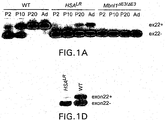
- FIG. 1 RT-PCR assay for SERCA1 exon 22 alternative splicing in mouse muscle at postnatal day 2 (P2), P10, P20, and 6 months (Ad) shows postnatal transition to exon 22 inclusion. The postnatal transition is absent in HSA LR transgenic and MBNL1 knockout mice.
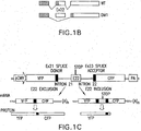
- B Inclusion of exon 22 of SERCA1 leads to a termination codon exon 22. Skipping of exon 22 leads to a termination codon in exon 23. Note that the Ex22+ transcript is not subject to nonsense mediated decay because the stop codon is within 55 nt from the final exon junction.
- C pSERF minigene spliceopathy reporter construct.
- FIG. 1 Synthesis, purification and fluorescent labeling of MBNL 1 protein and poly(CUG) transcript.
- A Coomassie-stained SDS-gels of following samples: crude protein lysate of bacteria expressing GST-MBNL 1-41-del 105 (truncated at C-terminal to remove hydrophobic domain); supernatant and pellet after 14,000 g centrifugation of crude protein; flow through after Ni-column chromatography; four samples eluted from Ni-column with different concentration of imidazole or EDTA. Next, eluate III was purified on Glutathione Sepharose column (second gel).
- FIG. 3 Interaction of recombinant MBNL1 with poly(CUG) 109 .
- A In nitrocellulose filter binding assay, a constant concentration of fluorescently-labeled poly(CUG) 109 [0.1 nM] is mixed with decreasing concentration (from 100 to 0.012 nM) of MBNL1-41 protein fused or not with GST. Unbound RNA washes through filter, protein-bound RNA is retained. Note that protein concentrations listed to the right correspond to two rows of wells on the filter.
- B Comparison of saturation curve for full length and C-terminus truncated GST-MBNL1-41.
- C Diagram showing experimental design for poly(CUG) 109 attachment assay.
- the capture oligodeoxynucleotide (ODN) is labeled at the 5' end with biotin via a 12-carbon linker.
- ODN capture oligodeoxynucleotide
- Interaction of biotin with streptavidin followed by hybridization of capture ODN to the 3' end of poly(CUG)109 tethers the transcript to plates.
- Poly(CUG)109-MBNL1 interaction is performed with excess of fluorescently-labeled MBNL1 protein.
- Figure 4 Optimization of conditions for monitoring of poly(CUG)109-MBNL1 interaction in microplate format.
- gel mobility shift assay demonstrates interaction of fluorescently-labeled poly(CUG)109 (0.5 pmole) with increasing concentration of full-length, GST-cleaved recombinant MBNL1. Note that increasing MBNL1 concentration influences both percentage of poly(CUG)109 in complex with protein and the molecular weight of complex (increasing number of MBNL1 molecules bound per transcript).
- Lower panel shows the interaction in microplate format, with fluorescence-labeled MBNL1 binding to unlabeled poly(CUG)109 tethered to plate.
- FIG. 10 The transcription unit for MBNL1 is flanked by insulators from the chicken ⁇ -globin locus. These elements have been shown to insulate transgene expression from adjacent chromatin context, improving the uniformity of transgene expression( Chung JH, et al. Proc Natl Acad Sci U S A 1997; 94(2):575-580 ).
- the attB element directs integration by phiC31 integrase.
- CBA chicken ⁇ actin. Due to the presence of several monospecific antibodies to Mbnl1, there was no need for a reporter gene or epitope tag to monitor expression levels.
- FIG. 11 Figure 7 .
- pLLC7 CMV/C ⁇ A, CMV enhancer coupled to chicken beta actin promoter; triple stop, concatamer of three SV40 polyadenylation signals and transcription terminator elements( Novak A, et al. Genesis 2000; 28(3-4): 147-155 ); GFP/NEO, Bizyme neomycin resistance-GFP fluorescence selection cassette( Hansen SG, et al. Biotechniques 2002; 32(5):1178 , 1180, 1182-1178 ); AttB, integration signal for phiC31 integrase.
- FIG. 8 Trimolecular fluorescence complementation assay for compounds that inhibit MBNL1 binding to poly(CUG)exp.
- poly(CUG)exp is interspersed with MS2 coat protein RNA recognition elements (MS2REs).
- MS2 coat protein (MS2CP) and MBNL1 are expressed as fusions with the N and C terminal halves of split Venus fluorescent protein( Nagai T, et al. Nat Biotechnol 2002; 20(1):87-90 ) (VFP), respectively.
- Assembly of MS2CP•VFPN and MBNL1•VFPC on the chimeric transcript leads to VFP fluorescence activity( Rackham O, Brown CM. EMBO J 2004; 23(16):3346-3355 ). Inhibition of MBNL1-poly(CUG)exp interactions causes loss of VFP fluorescence.
- Figure 9 shows a diagram of the transgene in HSALR transgenic mice.
- an expanded CTG repeat was inserted downstream from the stop codon in a DNA fragment containing the entire human skeletal actin gene. This fragment was used to derive HSALR transgenic mice. It was found that these transgenic mice express high levels of CUG expansion RNA in skeletal muscle. They also develop myotonia, a cardinal symptom of myotonic dystrophy, and histologic changes in skeletal muscle that resemble myotonic dystrophy.
- Figure 10 shows that MBNL1 protein is sequestered in ribonuclear foci of CUG repeat RNA in HSA LR transgenic mice.
- This high power view of a section of skeletal muscle shows a single nucleus at postnatal day 2.
- ribonuclear foci of CUG expansion RNA are shown by fluorescence in situ hybridization.
- the distribution of MBNL1 protein in the nucleus is shown by immunofluorescence.
- the merged image on the right shows that MBNL1 is sequestered in the ribonuclear foci of CUG expansion RNA.
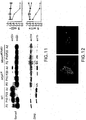
- Figure 11 shows that MBNL1 is required for normal developmental regulation of alternative splicing for SERCA1 and ZASP.
- the left panel shows reverse transcriptase-PCR (RT-PCR) analysis of alternative splicing for SERCA1, the calcium reuptake pump of the sarcoplasmic reticulum, and ZASP, a structural component of the Z disc.
- RT-PCR reverse transcriptase-PCR
- exon 22 is developmentally regulated. At postnatal day 2 (P2), exon 22 is mainly skipped. By postnatal day 20 (P20), and continuing in adults (Ad), exon 22 is mainly included.
- Figure 12 shows a morpholino composed of CAG repeats displaced MBNL 1 protein from ribonuclear foci in HSA LR transgenic mice.
- the morpholino was compromised entirely of CAG repeats and was 25 nucleotides in length (CAG25).
- CAG25 morpholino dissolved in phosphate buffered saline was injected into tibialis anterior muscle of HSA LR transgenic mice, followed by electroporation to facilitate entry into muscle cells.
- the contralateral tibialis anterior muscle was injected with saline alone, followed by electroporation.
- the distribution of MBNL1 in tibialis anterior was determined by immunofluorescence with anti-MBNL1 antibody A2764.
- MBNL1 was sequestered in ribonuclear foci (nuclei are counterstained with DAPI). In muscle injected with CAG25 morpholino, MBNL1 was became more widely distributed throughout the nucleus.
- Figure 13 shows the release of MBNL1 from ribonuclear foci following treatment with CAG25 morpholino restored proper regulation of alternative splicing for SERCA1 (3 weeks following CAG25 injection).
- injection of CAG25 morpholino into tibialis anterior improved the defect of SERCA1 alternative splicing.
- Morpholino-treated muscle is indicated by "+”.
- Results from tibialis anterior in the opposite hindlimb, injected with saline alone, are indicated by "-”.
- the side of morpholino injection was randomly determined, and this assignment remained blinded until after the splicing analysis was completed.
- Het. dupl. indicates a heteroduplex PCR product.
- Figure 15 shows that CAG25 morpholino treatment in HSA LR transgenic mice improved the alternative splicing of ClC-1, the muscle specific chloride ion channel.
- Exon 7a of C1C-1 shows developmentally regulated alternative splicing, and it was previously shown that MBNL1 is required for its normal regulation ( Kanadia et al, Science, 302:1978-1980, 2003 ).
- Adult wild-type (WT) mice show low levels of exon 7a inclusion.
- In HSA LR transgenic mice the fraction of ClC-1 splice products that include exon 7a is increased. Inclusion of exon 7a causes a frame shift and creates a premature termination codon that truncates most of the ClC-1 coding sequence.
- transcripts that include exon 7a have accelerated degradation via nonsense mediated decay, therefore they are underrepresented at steady state and on this gel.
- the higher bands on the gel are other alternative splice products, as shown ( Mankodi et al, Molecular Cell 10:35-44, 2002 .)
- injection of CAG morpholino into tibialis anterior improved the defect of ClC-1 alternative splicing. Morpholino-treated muscle is indicated by "+”. Results from tibialis anterior in the opposite hindlimb, injected with saline alone, are indicated by "-”.
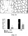
- Figure 16 shows the expression of ClC-1 chloride channel at the surface membrane is increased 3 weeks following injection of CAG25 morpholino. Immunofluorescence for ClC-1 chloride channel is shown in sections of tibialis anterior muscle from HSA LR transgenic mice. Muscle fibers show mosaic expression of ClC-1 in HSA LR muscle injected with saline. Some of these fibers are completely lacking in ClC-1 protein. Treatment with CAG25 morpholino leads to increased expression ClC-1 protein at the surface membrane of muscle fibers.
- Figure 17 uses Electromyography to show the improvement of myotonia at 3 and 6 weeks after injection of CAG25 morpholino into tibialis anterior of HSA LR transgenic mice.
- Myotonia is a state of muscle hyperexcitability in which muscle fibers display repetitive action potentials.
- Myotonia in the HSA LR transgenic mouse model of myotonic dystrophy is caused by abnormal regulation of alternative splicing for ClC-1 and subsequent reduction of chloride ion channels in muscle fibers.
- a parallel abnormality of ClC-1 alternative splicing exists in human myotonic dystrophy.
- the vertical axis shows mean myotonia severity score among 4 HSA LR transgenic mice at each timepoint.
- FIG. 18 shows that filter binding screening assay identifies compounds in the aminoglycoside family as having ability to inhibit interaction of recombinant MBNL1 protein with (CUG) 109 RNA.
- neomycin left curve
- gentamicin right curve
- the order of addition was compound + (CUG) 109 RNA (incubate 5 minutes), followed by recombinant MBNL1 protein (15 minute incubation), followed by application to filter.
- FIG. 23 Figure 19 shows that CAG25 morpholino inhibits the interaction of MBNL1 - poly(CUG) exp RNA in vitro. Interaction of recombinant MBNL1 protein with (CUG) 109 RNA was examined by gel shift assay, in the presence of increasing amounts of CAG25 morpholino.
- Figure 20 shows a screening assay for compounds that improve spliceopathy could use readouts other than protein fluorescence.
- SERCA1 exon 22 and flanking introns are used to generate a spliceopathy reporter construct using luciferase. Point mutations have been induced in SERCA1 exon 22 so that it no longer encodes a termination codon.
- this spliceopathy reporter construct is transiently transfected in COS cells, the luciferase activity is sensitive indicator of MBNL1 activity, as indicated by the > 10-fold upreglation of luciferase when cotransfected with small amounts of expression construct for GFP-tagged MBNL1.
- Figure 21 shows the effects of antisense morpholino targeting the 3' splice junction of ClC-1 exon 7a on splicing and myotonia in HSA LR transgenic mice.
- Antisense morpholino targeting the 3' splice junction of ClC-1 exon 7a was injected into tibialis anterior muscle of HSA LR mice under general anesthesia (sequence 5'- CCAGGCACGGTCTGCAACAGAGAAG-3' (SEQ ID NO: 4)).
- the contralateral muscle was injected with morpholino having the inverted sequence (5'- GAAGAGACAACGTCTGGCACGGACC -3'(SEQ ID NO: 5)). Uptake into muscle fibers was enhanced by in vivo electroporation.
- Figure 22 shows whole-cell voltage clamp from single muscle fibers shows that treatment with antisense morpholino targeting ClC-1 exon 7a restores normal chloride current density in HSA LR transgenic mice.
- Antisense morpholino targeting the 3' splice junction of ClC-1 exon 7a was injected into foot pad muscle of HSA LR mice under general anesthesia. Uptake into muscle fibers was enhanced by in vivo electroporation. The morpholino was tagged with fluorescein. 4 days later, individual FDB muscle fibers were isolated. Greater than 90% of fibers showed fluorescein uptake, and only these fibers were studied. As a control, the opposite footpad was injected with morpholino having the inverted sequence. A.
- the upper panel shows ClC-1 currents at different membrane potentials.
- the peak current density in HSA LR mice (center panel) is much lower than in wild-type mice (left panel).
- morpholino treatment in HSA LR mice right panel
- the current density is restored to levels that are similar to wild-type mice.
- the graph in the lower panel shows chloride current density in relation to membrane potential.
- the current density is markedly reduced (closed circles).
- the antisense morpholino restores chloride current density to normal levels (open circles or triangles).
- Figure 23 shows the design of antisense morpholinos.
- Figure 23A shows the inclusion of ClC-1 exon 7a induces a frame shift and premature termination codon in exon 7. Annealing of antisense morpholino to the 3' splice site of exon 7a in the ClC-1 pre-mRNA is intended to prevent spliceosomal recognition of this exon.
- Figure 23B shows the alignment of ClC-1 pre-mRNA (top strand) with antisense morpholinos targeting the 3' or 5' splice sites of exon 7a is shown.
- the control morpholino is the 5'-3' invert of the 3' splice site blocker. Exonic sequences are in upper case, intronic sequences are in lower case.
- Figure 24 shows that antisense morpholino localizes preferentially to muscle nuclei and restores ClC-1 expression at the sarcolemma.
- Figures 24A-C show a cross-section of HSA LR tibialis anterior (TA) muscle showing distribution of 3'-carboxyfluorescein-labeled antisense morpholino 3 weeks after injection. The morpholino was complementary to the 3' splice site of ClC-1 pre-mRNA.
- Muscle fibers are outlined by wheat germ agglutinin (wga) and nuclei are highlighted by DAPI.
- Figures 24D and 24 E show brightfield (D) and fluorescence (E) images of a single FDB fiber showing preferential nuclear localization of the antisense morpholino (post injection day 5).
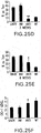
- Figure 25 shows that antisense morpholino represses splicing of ClC-1 exon 7a.
- Figure 25A reveals that RT-PCR showed reduction of exon 7a inclusion three weeks after injection of antisense (anti) morpholino (antisense 1 + antisense 2,5 ⁇ g each) into TA muscle of HSA LR mice. Pairs of injected TA muscles from each mouse are identified by "1, 2, 3.” Muscle injected with control morpholino (inv) (10 ⁇ g) was not different from untreated HSA LR muscle. HSA LR and WT mice have the same (FVB) inbred strain background.
- Figure 25B shows the inclusion of exon 7a remained partially suppressed 8 weeks after injection of antisense morpholino (20 ⁇ g antisense 1 vs. 20 ⁇ g invert control).
- Figure 25C shows that ClC-1 antisense morpholino did not correct the misregulated alternative splicing of Titin m-line exon 5.
- Figure 25F shows that the level of ClC-1 mRNA is increased 3 weeks after treatment with antisense moropholino.
- ClC-1 mRNA level is expressed in arbitrary units relative to housekeeping gene RNA polymerase II transcription factor IIB.
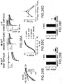
- Figure 26 shows that antisense morpholino rescues ClC-1 channel function and reverses myotonia in skeletal muscle of HSA LR mice.
- Figure 26A shows that representative ClC-1 currents obtained from flexor digitorum brevis (FDB) fibers isolated from HSA LR mice electroporated with either invert (left) or antisense (middle) morpholino and WT mice electroporated with antisense morpholino (right). The dashed lines represent the zero current level. Capacitative currents recorded from each fiber are shown in the insert of each panel (scale bars: vertical, 3 nA; horizontal, 4 ms).
- Figure 26C shows the average relative Po-V curves for the same experiments shown in (B). Smooth curves through each dataset were generated using a modified Boltzmann equation ( Lueck, J.D., et al. (2007) J Gen Physiol 129:79-94 ).
- Figure 26D shows the average relative contribution of the fast (A f /A total ), slow (A s /A total ), and non-deactivating (C/A total ) components of ClC-1 current deactivation elicited from a voltage step to -100 mV for the same experiments shown in (B).
- Figures 26E and 26F shows that myotonia was significantly reduced 3 (E) and 8 (F) weeks following injection of antisense morpholino.
- Mean ⁇ s.d.; n 3 to 7 per group.
- Antisense morpholino was injected into one TA, invert morpholino was injected into the contralateral TA, and gastrocnemius muscle served as an untreated control. **P ⁇ 0.0001 for antisense- vs invert-treated control; ANOVA.
- Figure 27 shows antisense morpholino represses exon 7a inclusion, restores ClC-1 protein expression, and rescues myotonia in Mbnl1 ⁇ E3/ ⁇ E3 mice.
- Figure 27A indicates that RT-PCR shows reduced inclusion of exon 7a at 3 weeks after injection of antisense morpholino 1 (20 ⁇ g antisense or invert control).
- Figure 28 shows a Comparison of antisense morpholinos targeting ClC-1 exon 7a.
- Antisense oligo was injected into tibialis anterior (TA) muscle of HSA LR mice and invert oligo (inv) (20 ⁇ g) was injected into the contralateral TA. Tissue was obtained 3 weeks later for analysis of ClC-1 splicing by RT-PCR.
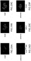
- Figure 29 shows that antisense morpholino had no effect on the formation of ribonuclear inclusions. Fluorescence in situ hybridization and immunofluorescence demonstrate co-localization of CUG exp RNA and MBNL1 protein in muscle nuclei (blue) 3 weeks after injection of HSA LR TA muscle with invert (a-c) and antisense (d-f) morpholino.
- Figure 30 shows protein displacement therapy with peptide nucleic acid (PNA) oligomers composed of CAG repeats.
- PNA peptide nucleic acid
- Figure 30A shows that PNA-CAG repeat oligos of lengths ranging from 2 to 5 CAG repeats can invade (CUG) 109 hairpins and effectively interact with expanded CUG repeat hairpin structures in vitro.
- Figure 30B shows that these PNA-CAG oligos can also inhibit the interaction of (CUG) 109 RNA with MBNL1 protein in vitro.
- FIG. 35 shows screening for compounds that inhibit interaction of MBNL1 protein and CUG expansion RNA: fluorescence anisotropy assay shows interaction of CUG expansion RNA with recombinant MBNL1 protein in vitro. Fluorescein-labeled (CUG) 36 RNA (2nM) was incubated with MBNL1 protein (100 nM) and anisotropy was measured at time points ranging from from 1 to 90 minutes. Increasing values for fluorescence anisotropy indicate interaction of fluorescein-labeled (CUG) 36 transcript with MBNL1 protein. Values are averages from 4 experiments and error bars shows SD.
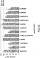
- Figure 32 shows a fluorescence anisotropy assay to screen for compounds that inhibit interaction of CUG repeat RNA with recombinant MBNL1 protein.
- Fluorescein-labeled (CUG) 36 transcript (2 nM) was incubated first with aminoglycoside compound (10 or 50 ⁇ M) and then with excess amount of recombinant MBNL1 protein (100 nM).
- % bound CUG exp ", vertical axis results are expressed as the percentage of maximal fluorescence anisotropy in assays from which aminoglycosides were omitted.
- neomycin showed the strongest inhibition of MBNL1 binding to CUG repeat RNA. Values are the average +/-SD from three measurements.
- FIG. 33 shows a diagram of enzymatic complementation assay to screen for compounds that inhibit interaction of CUG repeat RNA with recombinant MBNL1 protein.
- (CUG)109 transcripts are tethered to the surface of a streptavidin-coated microtiter plate using a capture oligonucleotide that is biotinylated. The capture oligo anneals to complementary sequence at the 3' end of the CUG repeat RNA.
- Recombinant human MBNL1 is expressed as a fusion with the PL fragment of beta-galactosidase.
- PL is a 55 amino acid fragment of beta-galactosidase.
- Figure 34 shows Enzymatic complementation assay to screen for compounds that inhibit interaction of CUG repeat RNA with recombinant MBNL 1 protein. Operation of the beta-galactosidase enzymatic complementation assay was demonstrated using two kinds of inhibitors. On the left panel, excess soluble (CUG) 109 RNA was added to the assay reaction. The soluble (CUG) 109 RNA binds to MBNL1-PL protein and prevents its retention on the microtiter plate, reflected by reduced beta-galactosidase activity (expressed on the vertical axis in terms of relative luminescence activity).
- FIG. 39 shows that injection of peptide nucleic acid (PNA) comprised of CAG repeats caused reduction of electromyographic myotonia in HSALR transgenic mouse model of myotonic dystrophy.
- PNA-(CAG)6mer or PNA-(CAG)9mer i.e., 2 or 3 CAG repeats
- Myotonia was assessed by electromyography 3 weeks following the intramuscular injection.
- vehicle alone phosphate buffered saline
- All mice had robust action myotonia prior to treatment. Assignment as to which limb received PNA vs control was randomized, and EMG analysis was performed blinded to this assignment.
- Ranges can be expressed herein as from “about” one particular value, and/or to "about” another particular value. When such a range is expressed, another embodiment includes from the one particular value and/or to the other particular value. Similarly, when values are expressed as approximations, by use of the antecedent "about,” it will be understood that the particular value forms another embodiment. It will be further understood that the endpoints of each of the ranges are significant both in relation to the other endpoint, and independently of the other endpoint. It is also understood that there are a number of values disclosed herein, and that each value is also herein disclosed as “about” that particular value in addition to the value itself. For example, if the value “10” is disclosed, then “about 10" is also disclosed.
- Primers are a subset of probes which are capable of supporting some type of enzymatic manipulation and which can hybridize with a target nucleic acid such that the enzymatic manipulation can occur.
- a primer can be made from any combination of nucleotides or nucleotide derivatives or analogs available in the art which do not interfere with the enzymatic manipulation.
- Probes are molecules capable of interacting with a target nucleic acid, typically in a sequence specific manner, for example through hybridization. The hybridization of nucleic acids is well understood in the art and discussed herein. Typically a probe can be made from any combination of nucleotides or nucleotide derivatives or analogs available in the art.
- Myotonic dystrophy type 1 (DM1), the most common form of muscular dystrophy in adults (frequency ⁇ 1 in 7,400), is a genetic disease affecting muscle, heart, and brain.
- DM1 involves a novel disease mechanism in which mRNA from the mutant gene has a direct toxic effect. The toxic gain-of-function of the mutant mRNA is entirely independent of the protein it encodes.
- DM1 is caused by expansion of a CTG repeat in the 3' untranslated region of DMPK, the gene encoding DM protein kinase((1)).
- a novel RNA-mediated disease process has recently been elucidated in DM1.
- Transcripts from the mutant DMPK allele contain an expanded CUG repeat, designated here as poly(CUG) exp .
- the repeat-bearing transcripts are not exported from the nucleus ( Taneja KL, et al. J Cell Biol 1995; 128(6):995-1002 ; Davis BM, et al. Proc Natl Acad Sci U S A 1997; 94(14):7388-7393 ). Instead, they accumulate in nuclear foci.
- proteins are recruited to the foci, and some of the proteins bind to CUG exp RNA, for example, proteins in the muscleblind (MBNL) family( Miller JW, et al. EMBO J 2000; 19(17):4439-4448 ).
- MBNL muscleblind
- One of the functions of MBNL is to regulate alternative splicing of pre-mRNA.
- sequestration of MBNL proteins in nuclear foci leads to abnormal regulation of alternative splicing for a select group of pre-mRNAs( Kanadia RN, et al. Science 2003; 302(5652):1978-1980 ; (6). This is a result of the depletion of MBNL from other regions of the nucleus.
- spliceopathy This regulatory defect is referred to herein as "spliceopathy.” Symptoms of DM1, such as, myotonia, are directly attributable to the effect on splicing regulation ( Mankodi A, et al. Mol Cell 2002;35-44 ) (ie., spliceopathy). Disclosed herein is evidence that spliceopathy in DM1 muscle can be explained, to a significant extent, by sequestration of a splicing factors in the MBNL family, including muscleblind 1 (MBNL1), muscleblind 2 (MBNL2), and muscleblind 3 (MBNL3). Splicing factors in the MBNL family are also sequestered on nuclear foci of polyCCUG expanded repeats in DM2.
- MBNL1 muscleblind 1
- MBNL2 muscleblind 2
- MBNL3 muscleblind 3
- MBNL1 is markedly depleted from the nucleoplasm in DM1 muscle cells, as it is recruited into ribonuclear foci; (2) expression of poly(CUG) exp and ablation of MBNL1 have equivalent effects on splicing regulation in mouse skeletal muscle; (3) spliceopathy in DM1 is remarkably similar to that observed in poly(CUG) exp -expressing or MBNL1 knockout mice ( Lin X, et al. Hum Mol Genet 2006 ); and (4) spliceopathy in poly(CUG) exp -expressing mice is corrected by overexpression of MBNL1. Therefore, poly(CUG) exp and its interaction with MBNL1 are valid targets for therapy.
- DM1 in skeletal muscle, a tissue with great intrinsic regenerative capacity, DM1 produces mainly fiber atrophy with little fibrosis or necrosis, an eminently reversible lesion.
- DM1 is a "composite" disease, in which distinct facets of the phenotype can be parsed to effects of spliceopathy on different transcripts, thereby impacting many different pathways.
- the spliceopathy can result in functional impairment rather than irreversible cell degeneration.
- myotonia in DM1 is a functional defect that results from chloride channelopathy ( Mankodi A, et al.
- MBNL1 is free to resume its effect on alternatively sliced transcripts, or by inhibiting sequestration of other CUG interacting protein, their normal functions can be restored.
- One method of inhibiting the interaction of MBNL1 with poly(CUG) exp is by displacing bound MBNL1 with another molecule.
- “Inhibit,” “inhibiting,” and “inhibition” mean to decrease an activity, response, condition, disease, or other biological parameter. This can include but is not limited to the complete ablation of the activity, response, condition, or disease. This may also include, for example, a 10% reduction in the activity, response, condition, or disease as compared to the native or control level. Thus, the reduction can be a 10, 20, 30, 40, 50, 60, 70, 80, 90, 100%, or any amount of reduction in between as compared to native or control levels.
- antisense oligonucleotides for use in treating myotonic dystrophy type 1 and type 2 that improve spliceopathy, as claimed.
- Spliceopathy refers to the abnormal regulation of alternative splicing.
- antisense oligonucleotides for use in treating DM1 that improves spliceopath as claimed.
- methods of treating wherein the agent consists of the morpholino set forth in SEQ ID NOs: 4, 6.
- DM2 myotonic dystrophy type 2
- said antisense oligonucleotides for use in treating DM type 1 or type 2 can affect DM by inhibiting, improving, or reversing channelopathy.
- Channelopathy is the reduction in ion channel conductance. It is understood and herein contemplated that channelopathy can result from spliceopathy of the ion channel such as ClC-1.
- Treatment means a method of reducing the effects of a disease or condition.
- Treatment can also refer to a method of reducing the disease or condition itself rather than just the symptoms.
- the treatment can be any reduction from native levels and can be but is not limited to the complete ablation of the disease, condition, or the symptoms of the disease or condition. Therefore, in the disclosed use, “treatment” can refer to a 10%, 20%, 30%, 40%, 50%, 60%, 70%, 80%, 90%, or 100% reduction in the severity of an established disease or the disease progression.
- a disclosed method for reducing the effects of prostate cancer is considered to be a treatment if there is a 10% reduction in one or more symptoms of the disease in a subject with the disease when compared to native levels in the same subject or control subjects.
- the reduction can be a 10, 20, 30, 40, 50, 60, 70, 80, 90, 100%, or any amount of reduction in between as compared to native or control levels.
- treatment does not necessarily refer to a cure of the disease or condition, but an improvement in the outlook of a disease or condition. Nevertheless, it is fully contemplated herein that “treatment” can not only refer to the ablation of the disease state, but the reversal of the condition.
- improves spliceopathy or correct spliceopathy means any change in spliceopathy that results in a change in the degree, amount or action of towards proper regulation of alternative splicing.
- antisense oligonucleotides can be modified to be morpholinos.
- Morpholino refers to synthetic oligonucleotides which have standard nucleic acid bases, bound to morpholine rings rather than the deoxyribose rings of DNA and the bases are linked through phosphorodiamidate groups instead of phosphates.
- the morpholino operates by binding to complementary RNA and blocks acceess to the RNA by other molecules.
- antisense oligonucleotides as set forth is SEQ ID NO: 4, or SEQ ID NO:6 wherein the antisense oligonuceottide is a morpholino.
- any of the disclosed antisense oligonucleotide for use in treatment can comprise a morpholino- antisense oligonucleotide.
- specifically disclosed herein are antisense oligonucleotides for use in treatment of DM type 1 or 2, wherein the antisense oligonucleotides is a morpholino oligonucleotide.
- antisense oligonucleotides for use in treating DM type 1 or 2, wherein the PNA- antisense oligonucleotides is a morpholino variant of SEQ ID NO:4; or SEQ ID NO:6.
- PNA Peptide nucleic acids
- the neutral polyamide backbone provides the added benefit of enzymatic stability, making PNA resistant to both proteases and nucleases. Together these properties make PNA an attractive reagent for biotechnology applications.
- PNA tends to fold into complex globular structures, presumably due to the collapse of the hydrophobic nucleobases. In fact, this conformational collapse has been exploited in the development of stemless PNA molecular beacons, taking advantage of the proximity between the two termini in the unhybridized state.
- a simple ç-backbone modification can transform a randomly folded peptide nucleic acid (PNA) into a right-handed helix.
- PNA peptide nucleic acid
- These conformationally preorganized helical PNAs bind to DNA and RNA with exceptionally high affinity and sequence selectivity.
- the antisense oligonucleotide disclosed herein can comprise PNA.
- disclosed herein are the antisense oligonucleotides set forth in SEQ ID NO:4; or SEQ ID NO:6, wherein the backbone has been modified as a PNA. It is understood that where a particular antisense oligonucleotides is disclosed, contemplated herein is the use of the PNA variant of that antisense oligonucleotides.
- any of the disclosed antisense oligonucleotides for use in treatment can comprise a PNA-antisense oligonucleotides.
- antisense oligonucleotides for use in treatment of DM type 1 or 2 wherein the antisense oligonucleotides is a peptide nucleic acid antisense oligonucleotides.
- antisense oligonucleotides for use in treating DM type 1 or 2 wherein the PNA- antisense oligonucleotides is a the PNA variant of SEQ ID NO:4; and SEQ ID NO:6.
- a antisense oligonucleotide (AON) targeting the 3' splice site of the chloride ion channel (ClC-1) exon 7a reverses the defect of ClC-1 alternative splicing in two mouse models of DM.
- the AON restores the full-length reading frame in ClC-1 mRNA, upregulates the level of ClC-1 mRNA, increases the expression of ClC-1 protein in the surface membrane, normalizes muscle ClC-1 current density and deactivation kinetics, and eliminates myotonic discharges.
- compositions can be delivered to the target cells in a variety of ways.
- the compositions can be delivered through electroporation, or through lipofection, or through calcium phosphate precipitation.
- the delivery mechanism chosen will depend in part on the type of cell targeted and whether the delivery is occurring for example in vivo or in vitro.
- compositions can comprise lipids such as liposomes, such as cationic liposomes (e.g., DOTMA, DOPE, DC-cholesterol) or anionic liposomes, or protein transduction domains.
- liposomes can further comprise proteins to facilitate targeting a particular cell, if desired.
- Administration of a composition comprising a compound and a cationic liposome can be administered to the blood afferent to a target organ or inhaled into the respiratory tract to target cells of the respiratory tract.
- liposomes see, e.g., Brigham et al. Am. J. Resp. Cell. Mol. Biol. 1:95-100 (1989 ); Felgner et al. Proc.
- protein transduction domains can comprise a domain from a larger protein, such as HIV-1 tat protein or herpes virus VP22, or an engineered peptide such as Endo-PorterTM.
- the compound can be administered as a component of a microcapsule that can be targeted to specific cell types, such as macrophages, or where the diffusion of the compound or delivery of the compound from the microcapsule is designed for a specific rate or dosage.
- the materials may be in solution, suspension (for example, incorporated into microparticles, liposomes, or cells). These may be targeted to a particular cell type via antibodies, receptors, or receptor ligands.
- the following references are examples of the use of this technology to target specific proteins to tumor tissue ( Senter, et al., Bioconjugate Chem., 2:447-451, (1991 ); Bagshawe, K.D., Br. J. Cancer, 60:275-281, (1989 ); Bagshawe, et al., Br. J.
- Vehicles such as "stealth” and other antibody conjugated liposomes (including lipid mediated drug targeting to colonic carcinoma), receptor mediated targeting of DNA through cell specific ligands, lymphocyte directed tumor targeting, and highly specific therapeutic retroviral targeting of murine glioma cells in vivo.
- the following references are examples of the use of this technology to target specific proteins to tumor tissue ( Hughes et al., Cancer Research, 49:6214-6220, (1989 ); and Litzinger and Huang, Biochimica et Biophysica Acta, 1104:179-187, (1992 )).
- receptors are involved in pathways of endocytosis, either constitutive or ligand induced.
- receptors cluster in clathrin-coated pits, enter the cell via clathrin-coated vesicles, pass through an acidified endosome in which the receptors are sorted, and then either recycle to the cell surface, become stored intracellularly, or are degraded in lysosomes.
- the internalization pathways serve a variety of functions, such as nutrient uptake, removal of activated proteins, clearance of macromolecules, opportunistic entry of viruses and toxins, dissociation and degradation of ligand, and receptor-level regulation. Many receptors follow more than one intracellular pathway, depending on the cell type, receptor concentration, type of ligand, ligand valency, and ligand concentration. Molecular and cellular mechanisms of receptor-mediated endocytosis has been reviewed ( Brown and Greene, DNA and Cell Biology 10:6, 399-409 (1991 )).
- compositions can be administered in a pharmaceutically acceptable carrier and can be delivered to the subject's cells in vivo and/or ex vivo by a variety of mechanisms well known in the art (e.g., uptake of naked DNA, liposome fusion, intramuscular injection of DNA via a gene gun, endocytosis and the like).
- cells or tissues can be removed and maintained outside the body according to standard protocols well known in the art.
- the compositions can be introduced into the cells via any gene transfer mechanism, such as, for example, calcium phosphate mediated gene delivery, electroporation, microinjection or proteoliposomes.
- the transduced cells can then be infused (e.g., in a pharmaceutically acceptable carrier) or homotopically transplanted back into the subject per standard methods for the cell or tissue type. Standard methods are known for transplantation or infusion of various cells into a subject.
- compositions that can be used to treat DM1 or DM2.
- morpholinos such as the antisense oligonucleotide as set forth in SEQ ID NO: 4, that can treat DM1.
- morpholino refers to synthetic oligonucleotides which have standard nucleic acid bases, bound to morpholine rings rather than the deoxyribose rings of DNA and the bases are linked through phosphorodiamidate groups instead of phosphates.
- the morpholino operates by binding to complementary RNA and blocks acceess to the RNA by other molecules.
- the morpholino may also be used to displace a molecule that is already bound to the complementary RNA strand.
- nucleic acids are made up of for example, nucleotides, nucleotide analogs, or nucleotide substitutes. Non-limiting examples of these and other molecules are discussed herein. It is understood that if, for example, an antisense molecule is introduced into a cell or cell environment through for example exogenous delivery, it is advantagous that the antisense molecule be made up of nucleotide analogs that reduce the degradation of the antisense molecule in the cellular environment.
- a nucleotide is a molecule that contains a base moiety, a sugar moiety and a phosphate moiety. Nucleotides can be linked together through their phosphate moieties and sugar moieties creating an internucleoside linkage.
- the base moiety of a nucleotide can be adenin-9-yl (A), cytosin-1-yl (C), guanin-9-yl (G), uracil-1-yl (U), and thymin-1-yl (T).
- the sugar moiety of a nucleotide is a ribose or a deoxyribose.
- the phosphate moiety of a nucleotide is pentavalent phosphate.
- An non-limiting example of a nucleotide would be 3'-AMP (3'-adenosine monophosphate) or 5'-GMP (5'-guanosine monophosphate).
- a nucleotide analog is a nucleotide which contains some type of modification to either the base, sugar, or phosphate moieties. Modifications to nucleotides are well known in the art and would include for example, 5-methylcytosine (5-me-C), 5-hydroxymethyl cytosine, xanthine, hypoxanthine, and 2-aminoadenine as well as modifications at the sugar or phosphate moieties.
- Nucleotide substitutes are molecules having similar functional properties to nucleotides, but which do not contain a phosphate moiety, such as peptide nucleic acid (PNA). Nucleotide substitutes are molecules that will recognize nucleic acids in a Watson-Crick or Hoogsteen manner, but which are linked together through a moiety other than a phosphate moiety. Nucleotide substitutes are able to conform to a double helix type structure when interacting with the appropriate target nucleic acid.
- PNA peptide nucleic acid
- conjugates can be link other types of molecules to nucleotides or nucleotide analogs to enhance for example, cellular uptake.
- Conjugates can be chemically linked to the nucleotide or nucleotide analogs.
- conjugates include but are not limited to lipid moieties such as a cholesterol moiety.
- a Watson-Crick interaction is at least one interaction with the Watson-Crick face of a nucleotide, nucleotide analog, or nucleotide substitute.
- the Watson-Crick face of a nucleotide, nucleotide analog, or nucleotide substitute includes the C2, N1, and C6 positions of a purine based nucleotide, nucleotide analog, or nucleotide substitute and the C2, N3, C4 positions of a pyrimidine based nucleotide, nucleotide analog, or nucleotide substitute.
- a Hoogsteen interaction is the interaction that takes place on the Hoogsteen face of a nucleotide or nucleotide analog, which is exposed in the major groove of duplex DNA.
- the Hoogsteen face includes the N7 position and reactive groups (NH2 or O) at the C6 position of purine nucleotides.
- sequences related to the protein molecules disclosed herein are encoded by nucleic acids or are nucleic acids.
- the sequences for the human analogs of these genes, as well as other analogs, and alleles of these genes, and splice variants and other types of variants, are available in a variety of protein and gene databases, including Genbank. Genbank can be accessed at http://www.ncbi.nih.gov/entrez/query.fcgi.
- Primers and/or probes can be designed for any given sequence given the information disclosed herein and known in the art.
- Functional nucleic acids are nucleic acid molecules that have a specific function, such as binding a target molecule or catalyzing a specific reaction.
- functional nucleic acids include antisense molecules.
- Functional nucleic acid molecules can interact with the mRNA of ClC1.
- Antisense molecules are designed to interact with a target nucleic acid molecule through either canonical or non-canonical base pairing. The antisense molecule is designed to interrupt a processing function that normally would take place on the target molecule, such as transcription.
- Antisense molecules can be designed based on the sequence of the target molecule. Numerous methods for optimization of antisense efficiency by finding the most accessible regions of the target molecule exist. Exemplary methods would be in vitro selection experiments and DNA modification studies using DMS and DEPC.
- antisense molecules bind the target molecule with a dissociation constant (k d )less than or equal to 10 -6 , 10 -8 , 10 -10 , or 10 -12 .
- k d dissociation constant
- a representative sample of methods and techniques which aid in the design and use of antisense molecules can be found in the following non-limiting list of United States patents: 5,135,917 , 5,294,533 , 5,627,158 , 5,641,754 , 5,691,317 , 5,780,607 , 5,786,138 , 5,849,903 , 5,856,103 , 5,919,772 , 5,955,590 , 5,990,088 , 5,994,320 , 5,998,602 , 6,005,095 , 6,007,995 , 6,013,522 , 6,017,898 , 6,018,042 , 6,025,198 , 6,033,910 , 6,040,296 , 6,046,
- compositions can also be administered in vivo in a pharmaceutically acceptable carrier.
- pharmaceutically acceptable is meant a material that is not biologically or otherwise undesirable, i.e., the material may be administered to a subject, along with the nucleic acid or vector, without causing any undesirable biological effects or interacting in a deleterious manner with any of the other components of the pharmaceutical composition in which it is contained.
- the carrier would naturally be selected to minimize any degradation of the active ingredient and to minimize any adverse side effects in the subject, as would be well known to one of skill in the art.
- compositions may be administered orally, parenterally (e.g., intravenously), by intramuscular injection, by intraperitoneal injection, transdermally, extracorporeally, topically or the like, including topical intranasal administration or administration by inhalant.
- topical intranasal administration means delivery of the compositions into the nose and nasal passages through one or both of the nares and can comprise delivery by a spraying mechanism or droplet mechanism, or through aerosolization of the nucleic acid or vector.
- Administration of the compositions by inhalant can be through the nose or mouth via delivery by a spraying or droplet mechanism. Delivery can also be directly to any area of the respiratory system (e.g., lungs) via intubation.
- compositions required will vary from subject to subject, depending on the species, age, weight and general condition of the subject, the severity of the allergic disorder being treated, the particular nucleic acid or vector used, its mode of administration and the like. Thus, it is not possible to specify an exact amount for every composition. However, an appropriate amount can be determined by one of ordinary skill in the art using only routine experimentation given the teachings herein.
- Parenteral administration of the composition is generally characterized by injection.
- Injectables can be prepared in conventional forms, either as liquid solutions or suspensions, solid forms suitable for solution of suspension in liquid prior to injection, or as emulsions.
- a more recently revised approach for parenteral administration involves use of a slow release or sustained release system such that a constant dosage is maintained. See, e.g., U.S. Patent No. 3,610,795 .
- the materials may be in solution, suspension (for example, incorporated into microparticles, liposomes, or cells). These may be targeted to a particular cell type via antibodies, receptors, or receptor ligands.
- the following references are examples of the use of this technology to target specific proteins to tumor tissue ( Senter, et al., Bioconjugate Chem., 2:447-451, (1991 ); Bagshawe, K.D., Br. J. Cancer, 60:275-281, (1989 ); Bagshawe, et al., Br. J.
- Vehicles such as "stealth” and other antibody conjugated liposomes (including lipid mediated drug targeting to colonic carcinoma), receptor mediated targeting of DNA through cell specific ligands, lymphocyte directed tumor targeting, and highly specific therapeutic retroviral targeting of murine glioma cells in vivo.
- the following references are examples of the use of this technology to target specific proteins to tumor tissue ( Hughes et al., Cancer Research, 49:6214-6220, (1989 ); and Litzinger and Huang, Biochimica et Biophysica Acta, 1104:179-187, (1992 )).
- receptors are involved in pathways of endocytosis, either constitutive or ligand induced.
- receptors cluster in clathrin-coated pits, enter the cell via clathrin-coated vesicles, pass through an acidified endosome in which the receptors are sorted, and then either recycle to the cell surface, become stored intracellularly, or are degraded in lysosomes.
- the internalization pathways serve a variety of functions, such as nutrient uptake, removal of activated proteins, clearance of macromolecules, opportunistic entry of viruses and toxins, dissociation and degradation of ligand, and receptor-level regulation. Many receptors follow more than one intracellular pathway, depending on the cell type, receptor concentration, type of ligand, ligand valency, and ligand concentration. Molecular and cellular mechanisms of receptor-mediated endocytosis has been reviewed ( Brown and Greene, DNA and Cell Biology 10:6, 399-409 (1991 )).
- compositions including antibodies can be used therapeutically in combination with a pharmaceutically acceptable carrier.
- Suitable carriers and their formulations are described in Remington: The Science and Practice of Pharmacy (19th ed.) ed. A.R. Gennaro, Mack Publishing Company, Easton, PA 1995 .
- an appropriate amount of a pharmaceutically-acceptable salt is used in the formulation to render the formulation isotonic.
- the pharmaceutically-acceptable carrier include, but are not limited to, saline, Ringer's solution and dextrose solution.
- the pH of the solution is preferably from about 5 to about 8, and more preferably from about 7 to about 7.5.
- Further carriers include sustained release preparations such as semipermeable matrices of solid hydrophobic polymers containing the antibody, which matrices are in the form of shaped articles, e.g., films, liposomes or microparticles. It will be apparent to those persons skilled in the art that certain carriers may be more preferable depending upon, for instance, the route of administration and concentration of composition being administered.
- compositions can be administered intramuscularly or subcutaneously. Other compounds will be administered according to standard procedures used by those skilled in the art.
- compositions may include carriers, thickeners, diluents, buffers, preservatives, surface active agents and the like in addition to the molecule of choice.
- Pharmaceutical compositions may also include one or more active ingredients such as antimicrobial agents, antiinflammatory agents, anesthetics, and the like.
- the pharmaceutical composition may be administered in a number of ways depending on whether local or systemic treatment is desired, and on the area to be treated. Administration may be topically (including ophthalmically, vaginally, rectally, intranasally), orally, by inhalation, or parenterally, for example by intravenous drip, subcutaneous, intraperitoneal or intramuscular injection.
- the disclosed antibodies can be administered intravenously, intraperitoneally, intramuscularly, subcutaneously, intracavity, or transdermally.
- Preparations for parenteral administration include sterile aqueous or non-aqueous solutions, suspensions, and emulsions.
- non-aqueous solvents are propylene glycol, polyethylene glycol, vegetable oils such as olive oil, and injectable organic esters such as ethyl oleate.
- Aqueous carriers include water, alcoholic/aqueous solutions, emulsions or suspensions, including saline and buffered media.
- Parenteral vehicles include sodium chloride solution, Ringer's dextrose, dextrose and sodium chloride, lactated Ringer's, or fixed oils.
- Intravenous vehicles include fluid and nutrient replenishers, electrolyte replenishers (such as those based on Ringer's dextrose), and the like. Preservatives and other additives may also be present such as, for example, antimicrobials, anti-oxidants, chelating agents, and inert gases and the like.
- Formulations for topical administration may include ointments, lotions, creams, gels, drops, suppositories, sprays, liquids and powders.
- Conventional pharmaceutical carriers, aqueous, powder or oily bases, thickeners and the like may be necessary or desirable.
- compositions for oral administration include powders or granules, suspensions or solutions in water or non-aqueous media, capsules, sachets, or tablets. Thickeners, flavorings, diluents, emulsifiers, dispersing aids or binders may be desirable..
- compositions may potentially be administered as a pharmaceutically acceptable acid- or base- addition salt, formed by reaction with inorganic acids such as hydrochloric acid, hydrobromic acid, perchloric acid, nitric acid, thiocyanic acid, sulfuric acid, and phosphoric acid, and organic acids such as formic acid, acetic acid, propionic acid, glycolic acid, lactic acid, pyruvic acid, oxalic acid, malonic acid, succinic acid, maleic acid, and fumaric acid, or by reaction with an inorganic base such as sodium hydroxide, ammonium hydroxide, potassium hydroxide, and organic bases such as mono-, di-, trialkyl and aryl amines and substituted ethanolamines.
- inorganic acids such as hydrochloric acid, hydrobromic acid, perchloric acid, nitric acid, thiocyanic acid, sulfuric acid, and phosphoric acid
- organic acids such as formic acid, acetic acid, propionic acid, glyco
- Effective dosages and schedules for administering the compositions may be determined empirically, and making such determinations is within the skill in the art.
- the dosage ranges for the administration of the compositions are those large enough to produce the desired effect in which the symptoms/disorder are/is effected.
- the dosage should not be so large as to cause adverse side effects, such as unwanted cross-reactions, anaphylactic reactions, and the like.
- the dosage will vary with the age, condition, sex and extent of the disease in the patient, route of administration, or whether other drugs are included in the regimen, and can be determined by one of skill in the art.
- the dosage can be adjusted by the individual physician in the event of any counterindications.
- Dosage can vary, and can be administered in one or more dose administrations daily, for one or several days.
- Guidance can be found in the literature for appropriate dosages for given classes of pharmaceutical products.
- guidance in selecting appropriate doses for antibodies can be found in the literature on therapeutic uses of antibodies, e.g., Handbook of Monoclonal Antibodies, Ferrone et al., eds., Noges Publications, Park Ridge, N.J., (1985) ch. 22 and pp. 303-357 ; Smith et al., Antibodies in Human Diagnosis and Therapy, Haber et al., eds., Raven Press, New York (1977) pp. 365-389 .
- a typical daily dosage of the antibody used alone might range from about 1 ⁇ g/kg to up to 100 mg/kg of body weight or more per day, depending on the factors mentioned above.
- a disclosed composition such as an antisense oligonucleotide morpholino or PNA, for treating, inhibiting, or preventing an DM1
- the efficacy of the therapeutic antibody can be assessed in various ways well known to the skilled practitioner. For instance, one of ordinary skill in the art will understand that a composition, such as a morpholino, disclosed herein is efficacious in treating or inhibiting a DM1 in a subject by observing that the composition reduces symptoms associated with the disease or reduces nuclear foci sequestration of MBNL1.
- Example 9 is put forth so as to provide those of ordinary skill in the art with a complete disclosure and description of how the compounds and/or compositions claimed herein are made and evaluated, and is intended to be purely exemplary and not intended to limit the disclosure.
- the other examples are comparative examples. Efforts have been made to ensure accuracy with respect to numbers (e.g., amounts, temperature, etc.), but some errors and deviations should be accounted for. Unless indicated otherwise, parts are parts by weight, temperature is in °C or is at ambient temperature, and pressure is at or near atmospheric.
- Example 1 MBNL sequestration on poly(CUG) exp leading to spliceopathy
- DMPK mRNA containing an expanded CUG repeat Transcription of the mutant allele generates DMPK mRNA containing an expanded CUG repeat. Mutant transcripts accumulate in discrete RNA nuclear (ribonuclear) foci. The RNA in foci is the fully-processed DMPK mRNA( Taneja KL, et al. J Cell Biol 1995; 128(6):995-1002 ). Reddy and colleagues have found that siRNA-mediated depletion of MBNL1 eliminated most of the ribonuclear foci in DM1 myoblasts, suggesting that the MBNL1-poly(CUG) exp interaction is the key determinant of foci formation( Dansithong W, et al. J Biol Chem 2005; 280(7):5773-5780 ).
- RNA binding proteins in the muscleblind-like (MBNL) family including MBNL1, MBNL2, and MBNL3, are sequestered in ribonuclear foci.
- MBNL proteins show strong colocalization with poly(CUG) exp in DM1 cells and consequently are depleted from the nucleoplasm ( Lin X, et al. Hum Mol Genet 2006 ; Mankodi A, et al. Hum Mol Genet 2001; 10:2165-2170 ).
- MBNL1 and MBNL2 are expressed in mature skeletal muscle, heart and brain ( Fardaei M, et al. Hum Mol Genet 2002; 11(7):805-814 ; Kanadia RN, et al.
- MBNL3 is expressed mainly in placenta. Loss of MBNL1 function leads to abnormal regulation of alternative splicing for a select group of pre-mRNAs, such as, insulin receptor and chloride channel 1. When overexpressed, all three MBNL family members can regulate splicing ( Ho TH, et al. EMBO J 2004; 23(15):3103-3112 ). Other splicing factors, such as, CUG-BP1 may contribute to spliceopathy in DM1 ( Savkur RS, et al. Nat Genet 2001; 29(1):40-47 ; Philips AV, et al.
- Example 2 Mouse models of DM1.
- HSA LR transgenic mice express human skeletal actin mRNA containing (CUG) 250 in the 3' UTR. These mice express high levels of poly(CUG) exp exclusively in skeletal muscle, and they develop myotonic myopathy 9 (see below).
- Mbnl1 ⁇ E3 / ⁇ E3 mice (derived in the Swanson lab) are homozygous for a targeted allele of Mbnl1. These mice develop myotonic myopathy but the myopathy appears less severe that in HSA LR mice 5 .
- Mbnl1 knockout mice also develop multisystemic features of DM1, such as, cataracts, progressive (ultimately fatal) cardiac disease, and abnormal CNS function.
- Example 3 Structure of poly(CUG) exp and binding to MBNL1.
- Poly(CUG) RNA forms stable hairpin structures in vitro when it is pathologically expanded ( Napierala M, Krzyzosiak WJ. J Biol Chem 1997; 272(49):31079-31085 ; Tian B, et al. Rna 2000; 6:79-87 ).
- the stem of the hairpin is an extended region of duplex RNA in which G•C and C•G base pairs are separated by a periodic U•U mismatch.
- MBNL1 binds to poly(CUG) exp in vitro in preference to poly(CUG) that is not expanded, suggesting that it recognizes poly(CUG) in a structured (duplex) form ( Miller JW, et al. EMBO J 2000; 19(17):4439-4448 ).
- the physiologic targets for splicing regulation by MBNL1 are short, 6-8 nt intronic splice enhancer/repressor elements.
- Example 4 Progressive myotonic myopathy (PMM): a composite phenotype.
- the PMM in DM1 is distinct from muscle phenotypes in other forms of dystrophy. PMM is a composite, additive phenotype resulting from independent effects of spliceopathy on different genes, and consequently, different pathways.
- Myotonia is a delay of muscle relaxation after voluntary contraction, caused by runs of action potentials that are generated in the muscle fibers.
- myotonia is associated with abnormal regulation of alternative splicing for the ClC-1 chloride channel and >70% reduction of the sarcolemmal Cl conductance ( Mankodi A, et al. Mol Cell 2002;35-44 ).
- a parallel abnormality of ClC-1 splicing and loss of ClC-1 protein occurs in human DM1 and DM2.
- myotonia in DM1 is a chloride channelopathy stemming from effects on splicing of ClC-1
- the reversal of ClC-1 spliceopathy either by antisense oligonucleotides that suppress splicing of misregulated exon, or by AAV-mediated overexpression of MBNL1
- HSA LR mice M Swanson, R Kanadia, T Wheeler, C Thornton, unpublished.
- medications provide partial relief of myotonia, they are not very effective when myotonia is severe, hence they are not widely prescribed. Because the myotonia is most severe in hand muscles that also are hampered by early weakness, it markedly interferes with manual dexterity and contributes to disability.
- a clear correlation between severity of myotonia and weakness in DM1 exists, indicating that calcium and mechanical overload due to myotonic discharges can accelerate the myopathy.
- DM1 is characterized by insulin resistance in skeletal muscle.
- Cooper and colleagues have shown that spliceopathy affects alternative splicing of insulin receptor ( Savkur RS, et al. Nat Genet 2001; 29(1):40-47 ) (INSR).
- the predominant INSR isoform expressed in DM1 muscle is the exon 11 skipped, non-muscle isoform, which has lower signaling capacity.
- the INSR spliceopathy may also influence IGF-1 signaling.
- Example 5 MBNL1 sequestration: pivotal role in muscle spliceopathy.
- Spliceopathy in DM1 targets a select group of pre-mRNAs that share a common temporal pattern of developmental regulation ( Lin X, et al. Hum Mol Genet 2006 ).
- the exons affected by spliceopathy normally undergo a synchronous splicing switch during early postnatal development in WT mice.
- loss of MBNL1, or expression of poly(CUG) exp results in identical failure of these splicing transitions.
- the spliceopathies in HSA LR transgenic mice, MBNL1 knockout mice, and human DM1 and DM2 are highly concordant.
- Example 6 Cell-based assay for compounds that correct poly(CUG) exp -induced spliceopathy.
- the assay has two components: (1) cells that display poly(CUG) exp -induced spliceopathy; and (2) a minigene construct that reports on severity of spliceopathy.
- ⁇ 70 alternatively spliced exons were analyzed in HSA LR mouse and DM1 muscle to identify exons most affected by spliceopathy.
- exon 22 of SERCA1 was one of the most severely affected, and the gene structure of SERCA1 lent itself well to adaptation for the reporter assay.
- SERCA1 exon 22 normally undergoes a postnatal splicing switch in WT muscle, but this switch fails to occur in HSA LR transgenic and MBNL1 knockout mice ( Fig. 1A ).
- Exons 22 and 23 contain alternative termination codons for SERCA1 translation ( Fig. 1B ).
- a minigene construct, pSERF was cloned to generate a fluorescence readout for the relative frequencies of exon 22 skipping and inclusion.
- pSERF contains SERCA1 exon 22 and its flanking introns ( Fig. 1C ). Splice donor and acceptor signals from the flanking exons are fused to the coding regions for yellow fluorescent protein (eYFP) and cyan fluorescent protein (eCFP). Spectral separation of these proteins is more than sufficient to resolve eYFP and eCFP components when both proteins are co-expressed. Both fluorescent proteins function as monomers and have rapid maturation times.
- the splicing outcome characteristic of normal mature skeletal muscle is inclusion of exon 22 ( Fig 1A , lane 4). The exon 22 inclusion (ex22+) transcript of pSERF encodes eYFP alone.
- exon 22 (ex22-, Fig. 1A , lanes 5-12).
- the ex22- transcript encodes eYFP-eCFP fusion protein (Y•CFP).
- pSERF was electroporated in vivo in WT or HSA LR mice and muscle was harvested for RNA analysis after 4 days. As expected, inclusion of exon 22 was low in HSA LR mice and high in WT muscle ( Fig. 1D ).
- a spliceopathy assay has several advantages for identifying compounds having therapeutic potential in DM1. Spliceopathy is a downstream consequence of the RNA mediated disease process that is directly pertinent to symptoms of DM1.
- a spliceopathy screen can capture compounds that act either on the splicing machinery or upstream of RNA processing, having any of the following desirable effects: (1) accelerated degradation of poly(CUG) exp RNA; (2) upregulation of MBNL1 activity (post-transcriptional); (3) release of MBNL1 from sequestration in nuclear foci; and (4) effects on other splicing regulators, such as, CUG-BP1, that improve the splicing defect.
- the spliceopathy assay identifies compounds that differentially inhibit the interaction of MBNL1 with its pathological target, poly(CUG) exp RNA, compared to its physiological target, the splice enhancer element in SERCA1 pre-mRNA. Finally, the variance of assay results is low. The readout is determined by the ratio of two splice products produced from a single transcription unit, rather than absolute levels for either isoform. This design minimizes variance resulting from nonuniform delivery of cell to wells or nonspecific inhibitory effects of compounds on cell metabolism or survival.
- RNA-based assays of alternative splicing the coefficient of variation for relative proportion of two alternative splice products is usually ⁇ 3%, lower than most measurements of gene expression.
- the readout for the assays disclosed herein are at least this low because direct fluorescence determination of pSERF protein products can be reasonably precise.
- MBNL1 The levels of MBNL1 in transformed cell lines are generally low, whereas expression of CUG-BP1 is fairly ubiquitous. As expected, basal splicing outcomes in transformed cell lines are similar to those observed in DM1 ( Ho TH, et al. EMBO J 2004; 23(15):3103-3112 ). However, overexpression of MBNL1 in cells lines is sufficient to drive splicing outcomes to the pattern that is characteristic of normal mature muscle (see Fig. 5 for an example concerning fast troponin T and Fig. 13 for an example concerning SERCA1). Thus, pM1 ( Fig. 6 ) is used to obtain stably transfected cell lines that overexpress MBNL1 and display splicing phenotypes similar to WT skeletal muscle.
- 107. 293 cells are co-transfected with pM1 and pCMVInt (phiC31 integrase expression construct) using conditions previously defined by Dr Calos ( Chalberg TW, et al. J Mol Biol 2006; 357(1):28-48 ). The ratio of pCMVInt to pM1 is 50:1. Cells are split 1:30 24 hrs after transfection and transferred to hygromycin selection medium the next day. Clones are isolated after 10 to 14 days of selection. Clones are analyzed for: (1) splicing outcome by transfection with pSERF followed by RT-PCR and fluorescence analysis; and (2) MBNL1 overexpression by immunofluorescence and immunoblot.
- the cell system most responsive to therapeutic effects is one in which every cell expresses poly(CUG)exp at levels just sufficient to sequester MBNL1 and induce a strong spliceopathy phenotype. Departure from this ideal reduces the responsiveness of an assay to therapeutic compounds.
- Cell-to-cell variability of transgene expression is likely to be a particular problem for poly(CUG)exp.
- Cells with subthreshold poly(CUG)exp accumulation never develop spliceopathy, whereas cells having a large burden of poly(CUG)exp are "resistant" to therapeutic effects. In either case, cells become unresponsive to test compounds and the power of the screen is correspondingly reduced.
- Constructs for expression of expanded CUG repeats present problems of instability and transgene silencing.
- three steps are employed to make stably transfected cells lines for optimal stability and uniformity of poly(CUG)exp expression.
- phiC31 integrase is used to obtain full-length, single-copy integrations.
- the transgene initially expresses a GFP-neomycin resistance selection cassette.
- FACS is used to select cells lines that have uniform expression of the selection cassette. Any deleterious effects of poly(CUG)exp are avoided during initial isolation and selection clones.
- cre recombinase is used to excise the neo-GFP cassette and activate expression of luciferase with poly(CUG)exp in the 3' UTR.
- CTG repeat tracts > 150 repeats have a strong tendency for contraction in E. coli cloning vectors ( Kang S, et al. Nat Genet 1995; 10(2):213-218 ), and it is difficult to clone repeat lengths above 250.
- any plasmid prep containing an expanded CTG repeat consists of a heterogeneous mixture of different expansion lengths, and bacterial cultures seeded from the same stock show considerable prep-to-prep variability.
- Pathogenicity of the mutant DMPK mRNA may be influenced by elements that are within the DMPK 3' UTR but outside of the repeat tract ( Amack JD, et al. Hum Mol Genet 2001; 10(18):1879-1887 ). Therefore, the DMPK 3' UTR has been incorporated in the construct to maintain cis elements that may influence the pathogenicity of poly(CUG)exp.
- Bizyme selection cassette (neomycin phosphotransferase fused to GFP)
- This cassette allows selection of clones by neomycin resistance initially and subsequently by fluorescence ( Hansen SG, et al. Biotechniques 2002; 32(5):1178 , 1180, 1182-1178 ).
- the cDNA encoding Bizyme is followed by a concatamer of three SV40 transcription terminators ( Novak A, et al. Genesis 2000; 28(3-4):147-155 ) ("triple stop").
- the triple stop element was tested and showed that it completely prevents transcription of the downstream luciferase and CTG repeat, until after excision of the Bizyme selection cassette and triple stop by cre recombinase.
- FISH fluorescence in situ hybridization
- the expected outcome in cells showing spliceopathy is sequestration of MBNL1 in ribonuclear foci.
- Optimal clones show consistent sequestration of MBNL1 in ribonuclear foci, and splicing of pSERF is expected to revert to the SERCA1 exon 22 skipped isoform (low ratio of YFP:Y•CFP fluorescence).
- measuring FRET may not offer any advantage over simple analysis of eYFP and eCFP emission ratios, when each is excited at wavelengths that provide the least cross excitation.
- the outcome of these experiments is to select the wavelengths that are optimal for determining the ratio of YFP : Y•CFP when co-expressed.
- the strategies disclosed herein are tested in a mixing experiment in which known ratios of eYFP and Y•CFP are analyzed, and calibrated against RNA splicing results determined by RT-PCR analysis of exon 22 inclusion. It is likely that stable transfection generates clones that display a variety of different splicing outcomes, depending on levels of MBNL1 or poly(CUG) exp expression achieved in a particular clone.
- this panel of "intermediate" clones are used to correlate fluorescence readouts obtained from intact cells using the fluorometer with subsequent RNA extraction for splicing analysis. In this manner, the well-to-well and assay-to-assay variance in fluorescence reading is determined across the full spectrum of splicing outcomes. If such "intermediate" clones are not available, the full spectrum of splicing outcomes can be reconstitute by transfecting (unmodified) 293 cells with pSERF and increasing amounts of pM1, to drive increasing levels of exon 22 inclusion.
- Example 7 Cell-based assay for compounds that inhibit the interaction of MBNL1 with poly(CUG)exp in vivo
- Inhibition of MBNL1-poly(CUG)exp interaction is a logical therapeutic objective in DM1.
- the spliceopathy assay can identify compounds having this activity.
- more direct screens for inhibitors of MBNL1-poly(CUG)exp interaction can be more sensitive, or they can identify different sets of compounds.
- the spliceopathy screen can give negative results for compounds that inhibit MBNL1 recognition of its physiologic (splice enhancer elements) as well as its pathologic (poly(CUG)exp) targets. Nevertheless, these compounds can preferentially inhibit the pathologic interaction at a different concentration, or furnish scaffolds that, with further investigation of structure activity relationships, can be modified to preferentially target the pathological interaction.
- siRNA-mediated knockdown of MBNL 1 resulted in ⁇ 70% reduction of foci in DM1 myoblasts. Based on these observations, it was determined that MBNL1-poly(CUG)exp interaction is the primary determinant of ribonuclear foci formation in DM1 myoblasts ( Dansithong W, et al. J Biol Chem 2005; 280(7):5773-5780 ). Disclosed herein, poly(CUG)exp transcripts in vitro show formation of very high molecular weight complexes in the presence of purified recombinant MBNL1 ( Fig. 4 ).
- 293 cells are stably transfected with an expanded repeat ( ⁇ 300 repeats, lucCUG300) or non-repeat version (lucCUG ⁇ ) of pLLC7. These cells are expected to have strong expression of luc mRNA but modest endogenous expression of MBNL1.
- MBNL1 knockdown using siRNAs that target the MBNL1 coding region ( Dansithong W, et al. J Biol Chem 2005; 280(7):5773-5780 )
- overexpression transient transfection of pM1
- MBNL1 overexpression enhances nuclear foci, reduce luciferase activity, and increase the nuclear:cytoplasmic ratio of mRNA for lucCUG300 but not for lucCUG ⁇ .
- Clones for luciferase activity are compared in the presence and absence of siRNA knockout of MBNL1. Clones that are most responsive to MBNL1 knockdown, as determined by upregulation of luciferase activity, are selected.
- the system involves 3 components: (1) chimeric transcripts that contain poly(CUG)exp adjacent to the RNA element recognized by bacteriophage MS2 coat protein; (2) MBNL1 protein fused to the C terminal portion of Venus fluorescent protein (VFPC); and (3) MS2 coat protein (MS2CP) fused to the N terminal portion of VFP (VFPN) ( Fig. 8 ).
- VFPC Venus fluorescent protein
- MS2CP MS2 coat protein
- TriFC generated strong fluorescence signals when MS2CP•VFPN and IMP1•VFPC were co-expressed with a transcript containing adjacent MS2 and IMP1 recognition elements in the 3' UTR. Mutations in the RNA target which eliminated MS2CP or IMP1 binding caused loss of fluorescence signal ( Rackham O, Brown CM. EMBO J 2004; 23(16):3346-3355 ). Results were similar with other RNA binding proteins and their respective RNA recognition elements. The background was low and VFP had the advantages of bright fluorescence, monomeric structure, and rapid maturation ( Nagai T, et al. Nat Biotechnol 2002; 20(1):87-90 ).
- a modified "CTG donor plasmid" containing a chimeric poly(CTG)exp-MS2RE insert is cloned. This fragment is subcloned directly into pLLC7 ( Fig. 7 ).
- the chimeric sequence contains two tracts of expanded CTG repeats, each consisting of 130 repeats, interspersed with two MS2 binding sites, each consisting of a dimer of the MS2RE (see Fig. 8 ).
- the cassette is removed by cre recombination.
- VFPN split Venus protein
- VFPC split Venus protein
- MBNL1-VFPC fusion protein is expected to retain high affinity for poly(CUG)exp, because MBNL1 tagged with eGFP retained its poly(CUG)exp binding and splicing regulatory activity ( Ho TH, et al. EMBO J 2004; 23(15):3103-3112 ; Fardaei M, et al. Nucleic Acids Res 2001; 29(13):2766-2771 ).
- Example 8 Biochemical assay for compounds that inhibit MBNL1 - poly ( CUG ) exp interaction in vitro
- a biochemical screen with simple components is probably the most sensitive means to detect inhibitors of MBNL1 binding to poly(CUG) exp .
- a biochemical screen can identify compounds not captured by cell-based screens due to reasons of cell toxicity, compound instability, impermeability, or protein binding. Such compounds nevertheless can provide scaffolds that can be modified to improve activity in vivo.
- a biochemical screen provides an important tool for secondary evaluation of hits from the cell-based screens.
- MBNL1 protein is hydrophobic and basic (pH 8.9) so the majority of protein synthesized in bacteria is denatured or in inclusions.
- His 6 poly-histidine
- GST-MBNL1-His 6 was expressed at high levels in BL21(DE3) cells. A purification was developed ( Fig.
- the purification scheme was optimized using MBNL1-41 and MBNL1-42 kD isoforms, which seem to have similar binding and splicing factor activities (see Fig. 5 below), as well as a truncated form which removes hydrophobic sequence at the C-terminal and improves the yield of soluble protein from E. coli.
- the truncated protein includes all four zinc-fingers and other regions that are conserved among MBNL family members, and its poly(CUG) binding activity remains intact.
- the purified recombinant MBNL1 does not display ribonuclease activity when incubated with test transcripts.
- poly(CUG) 109 Constructs for in vitro transcription of poly(CUG) 109 have been prepared and conditions for enzymatic synthesis and purification of non-radioactively labeled transcripts have been optimized ( Fig. 2B ).
- the poly(CUG) 109 transcript contains an A-rich sequence at the 3' end.
- the triplet repeat is fused directly to a modified T7 promoter to eliminate A nucleotides 5' to the poly(CUG) tract. This allows biosynthetic labeling to high specific activity using fluorescently labeled ATP, while avoiding premature terminations or internal labeling within the poly(CUG) tract that might impact RNA folding or MBNL1 binding.
- the A-rich 3' tail is devoid of secondary structure and also functions as anchoring sequence for capture of poly(CU.G) 109 by oligonucleotides attached to the surface of microtiter plates.
- transcripts are denatured at 80°C and renatured under conditions that favor folding of poly(CUG) 109 as a single hairpin structure.
- a "capture" oligodeoxynucleotide (ODN) complementary to the A-rich 3' terminus of the poly(CUG) 109 transcript, labeled either with biotin or a reactive group at the 5' end for attachment to the plate shown in Fig.
- the Multiscreen HTS nitrocellulose filter plate (Millipore) has been previously used for a similar purpose ( Bittker JA, et al. Nat Biotechnol 2002; 20(10):1024-1029 ). This plate is designed for automated handling in high throughput screens and is likely to have protein binding characteristics similar to the filter binding assay in Fig. 3A .
- the alternative assay for inhibitors of MBNL1-poly(CUG) exp interaction involves tethering of unlabeled poly(CUG) exp to microtiter plates, and measuring the amount of fluorescently-labeled MBNL1 retained on the plate due to RNA binding ( Fig. 3C ).
- Methods for synthesis and tethering of poly(CUG) 109 RNA are described in Preliminary Studies and diagrammed in Figure 3C .
- Results of fluorescence labeling of purified recombinant MBNL1 are shown in Fig. 2B . Coupling efficiency for labeling was ⁇ 2-6 fluorochromes per molecule of GST-MBNL1, using the FluoReporter protein labeling kit (Molecular Probes).
- GST-MBNL1 is used for these assays, because the presence of GST fusion partner does not appear to influence affinity of MBNL1 for poly(CUG) exp ( Fig. 3A ), and increasing the mass of MBNL1 permits higher fluorescence activity with less risk of fluorochrome attachment at the RNA binding site.
- streptavidin-coated polystyrene microtiter plates (Nunc) were noted as providing the highest capacity for poly(CUG) 109 binding and lowest background of MBNL1 binding in the absence of poly(CUG) exp .
- the material requirements for these assays are feasible. 10-100 nM concentration of MBNL1 is suitable (lanes 4-6,
- RNA attachment assay over the filter binding assay, is greater sensitivity to inhibitors because it detects varying degrees of displacement of MBNL1 from poly(CUG) 109 , whereas even a single residual MBNL1 molecule bound to poly(CUG) 109 is sufficient to induce retention on the filter.
- the potential disadvantage is the need for more liquid handling steps including aspiration of wells, which may dictate that this assay remains a work station procedure that cannot be scaled up to high throughput.
- poly(CUG) exp displays considerable resistance to ribonuclease cleavage, owing to its highly stable secondary structure( Tian B, et al. Rna 2000; 6:79-87 ).
- the A-rich 3' end of the poly(CUG) 109 transcript which contains the fluorescence label in the filter binding assay and mediates tethering in the plate attachment assay, is not protected in this manner.
- the sequence of this tail has been specifically designed to avoid dinucleotide combinations that confer RNase sensitivity. Further protection is achieved by addition of a complementary morpholino to the filter binding assay, or use of a morpholino as "capture" oligonucleotide in the plate attachment assay. Addition of irrelevant, unstructured RNA to test buffer and use of short incubation times further reduce RNase activity.
- Example 9 Correction of ClC-1 splicing eliminates chloride channelopathy and myotonia in mouse models of myotonic dystrophy
- DM type 1 (DM1), the most common muscular dystrophy affecting adults, is caused by expansion of a CTG repeat in the 3' untranslated region of the gene encoding the DM protein kinase ( DMPK ) ( Brook, J.D., et al. (1992) Cell 68:799-808 ).
- DMPK DM protein kinase
- the transcripts containing a CUG exp tract elicit abnormal regulation of alternative splicing, or spliceopathy ( Philips, A.V., et al. (1998) Science 280:737-741 ).
- the splicing defect which selectively affects a specific group of pre-mRNAs, is thought to result from reduced activity of splicing factors in the muscleblind (MBNL) family ( Kanadia, R.N., et al. (2003) Science 302:1978-1980 ), increased levels of CUG binding protein 1 ( Philips, A.V., et al. (1998) Science 280:737-741 ; Charlet, B.N., et al. (2002) Mol Cell 10:45-53 ), or both.
- transgenic mice expressing CUG exp RNA displayed myotonia and chloride channel 1 (ClC-1) splicing defects similar to those observed in DM1 ( Mankodi, A., et al. (2002) Mol Cell 10:35-44 ).
- Myotonia in the HSA LR model results from abnormal inclusion of exon 7a in the ClC-1 mRNA, owing to sequestration of MBNL1, a factor required for repression of exon 7a splicing in muscle fibers ( Kanadia, R.N., et al. (2003) Science 302:1978-1980 ).
- exon 7a causes frame shift and introduction of a premature termination codon in the ClC-1 mRNA ( Mankodi, A., et al. (2002) Mol Cell 10:35-44 ; Charlet, B.N., et al. (2002) Mol Cell 10:45-53 ); (2) truncated ClC-1 protein encoded by the exon 7a+ isoform is devoid of channel activity ( Berg, J., et al. (2004) Neurology 63:2371-2375 ); and (3) disruption of Mbnl1 in mice leads to increased inclusion of ClC-1 exon 7a and myotonia ( Kanadia, R.N., et al. (2003) Science 302:1978-1980 ).
- Morpholino AONs influence splicing outcomes without inducing degradation of their target RNAs ( Mercatante, D.R., et al. (2001) Curr Cancer Drug Targets 1:211-230 ). Therefore, the predicted effect of repressing exon 7a inclusion was to eliminate the premature termination codon in ClC-1 mRNA and thereby reduce its degradation through the nonsense-mediated decay pathway ( Lueck, J.D., et al. (2007) Am J Physiol Cell Physiol 292:C1291-1297 ).
- mice Homozygous deletion of Mbnl1 exon 3 in mice ( Mbnl1 ⁇ E3/ ⁇ E3 ) resulted in loss of Mbnl1 protein from muscle, spliceopathy that is similar to DM1 patients and HSA LR mice, reduction of ClC-1 expression and activity, and myotonia ( Kanadia, R.N., et al. (2003) Science 302:1978-1980 ; Lin, X., et al. (2006) Hum Mol Genet 15:2087-2097 ; Lueck, J.D., et al. (2007) J Gen Physiol 129:79-94 ).
- antisense morpholino or invert control was injected into TA muscle of Mbnl1 ⁇ E3/ ⁇ E3 mice.
- antisense morpholino repressed the inclusion of exon 7a ( Figure 27A,B ), increased the expression of ClC-1 protein at the sarcolemma ( Figure 27C,D ), and reduced the myotonia in Mbnl1 ⁇ E3/ ⁇ E3 mice ( Figure 27E ).
- Myotonia is the symptom by which DM1 is most often recognized, and, due to preferential involvement of hand and forearm muscles, it compromises the manual dexterity and contributes to disability.
- DM1 cells and model systems Benders, A.A., et al. (1997) J Clin Invest 100:1440-1447 ; Benders, A.A., et al. (1996) Acta Physiol Scand 156:355-367 ), and the effects of DM1 on alternative splicing of the SERCA1 calcium reuptake pump of the sarcoplasmic reticulum ( Kimura, T., et al.
- AONs influence RNA processing by annealing to pre-mRNA and blocking the access of splicing factors to splice sites or cis -acting regulatory elements ( Dominski, Z., and Kole, R. (1993) Proc Natl Acad Sci U S A 90:8673-8677 ).
- AONs that induce skipping of constitutively spliced exons have been used to bypass stop codons or restore the proper reading frame in the dystrophin mRNA ( Dunckley, M.G., et al. (1998) Hum Mol Genet 7:1083-1090 ; Wilton, S.D., et al.
- Exon 7a can show heightened susceptibility to AONs because splicing signals in alternative exons tend to be intrinsically weak and this exon is normally skipped in a fraction of ClC-1 transcripts ( Lueck, J.D., et al. (2007) Am J Physiol Cell Physiol 292:C1291-1297 ). However, ClC-1 mRNAs that include exon 7a contain premature termination codons and undergo rapid degradation ( Lueck, J.D., et al. (2007) Am J Physiol Cell Physiol 292:C1291-1297 ).
- Morpholino oligonucleotides were 5'-CCAGGCACGGTctgcaacagagaag-3' (SEQ ID NO: 4) targeting the ClC-1 3' splice site, 5'- gaagagacaacgtctggcacggacc-3' (SEQ ID NO: 5) inverted control, and 5'-ggaagtgaaacttgcCTCCATCAGG-3' (SEQ ID NO: 6) targeting the ClC-1 5' splice site.
- HSA LR Mankodi, A., et al. (2000) Science 289:1769-1773
- Mbnl1 ⁇ E3/ ⁇ E3 mice Kanadia, R.N., et al. (2003) Science 302:1978-1980
- TA muscle was pretreated by intramuscular injection of bovine hyaluronidase (15 ⁇ l, 0.4 U/ ⁇ l) (Sigma) ( McMahon, J.M., et al. (2001) Gene Ther 8:1264-1270 ).
- the antisense morpholino was labeled with carboxyfluorescein and cryosections of muscle (10 ⁇ M) were examined by fluorescence microscopy, with or without fixation in 4% paraformaldehyde. Some sections were co-labeled with TRITC- wheat germ agglutinin ( Parsons, S.A., et al. (2003) Mol Cell Biol 23:4331-4343 ) (50 ⁇ g/ml in PBS; Sigma) and 4',6-diamidino-2-phenylindole (DAPI) to highlight the surface membranes and nuclei of muscle fibers.
- TRITC- wheat germ agglutinin Parsons, S.A., et al. (2003) Mol Cell Biol 23:4331-4343
- DAPI 4',6-diamidino-2-phenylindole
- ClC-1 forward ClCm-7 5'-TGAAGGAATACCTCACACTCAAGG-3' (SEQ ID NO: 7) and reverse: ClCm-30 5'-CACGGAACACAAAGGCACTG-3' (SEQ ID NO: 8); mTitin forward: mTTN1 5'-GTGTGAGTCGCTCCAGAAACG-3' (SEQ ID NO: 9) and reverse: mTTN2 5'-CCACCACAGGACCATGTTATTTC-3' (SEQ ID NO: 10).
- RT-PCR products 22 cycles were separated on agarose gels, stained with SYBR green II, and scanned on a laser fluorimager (Molecular Dynamics). Band intensity was quantified using ImageQuant software.
- Total levels of ClC-1 mRNA were determined by quantitative real-time RT-PCR (Taqman, Applied Biosystems) relative to housekeeping gene RNA polymerase II transcription factor IIB.
- ClC-1 currents were measured and analyzed in whole cell patch clamp experiments ( Hamill, O.P., et al. (1981) Pflugers Arch 391:85-100 ) using an approach identical to that described in detail elsewhere ( Lueck, J.D., et al. (2007) J Gen Physiol 129:79-94 ). ClC-1 current density (pA/pF) was calculated in order to compare data across fibers of different sizes.
- EMG was performed under general anesthesia as described previously ( Kanadia, R.N., et al. (2003) Science 302:1978-1980 ). Images and video recordings of electromyographic myotonia in HSA LR and Mbnl1 ⁇ E3 / ⁇ E3 mice are shown in previous reports ( Kanadia, R.N., et al. (2003) Science 302:1978-1980 ; Mankodi, A., et al. (2000) Science 289:1769-1773 ). A minimum of 15 needle insertions were performed for each muscle examined.
- Myotonic discharges were graded on a 4 point scale: 0, no myotonia; 1, occasional myotonic discharge in ⁇ 50% of needle insertions; 2, myotonic discharge in >50% of needle insertions; 3: myotonic discharge with nearly every insertion.
- Group data are expressed as mean ⁇ s.d., except for patch clamp data in Figure 26 which are expressed as mean ⁇ s.e.m. Between group comparison was performed by two-tailed t-test or two way ANOVA as indicated.
- Example 10 Protein displacement therapy with peptide nucleic acid (PNA) oligomers composed of CAG repeats.
- PNA peptide nucleic acid
- FIG. 30A shows that PNA-CAG repeat oligos of lengths ranging from 2 to 5 CAG repeats can invade (CUG)109 hairpins and effectively interact with expanded CUG repeat hairpin structures in vitro.
- Figure 30B shows that these PNA-CAG oligos can also inhibit the interaction of (CUG)109 RNA with MBNL1 protein in vitro.
- Figure 30A was performed by non-denaturing polyacrylamide gel scanned with laser fluorimager shows migration of fluorescein-labeled (CUG)109 transcript (10nM).
- This transcript was incubated 30 min with different concentrations (8, 4, 2, 1, 0.5, 0.25 ⁇ M) of PNAs containing different length of CAG repeat sequence (PNA-CAG-6, Nterm-CAGCAG; PNA-CAG-9, Nterm-CAGCAGCAG; PNA-CAG-12, Nterm-CAGCAGCAGCAG (SEQ ID NO: 14); or PNA-CAG-15, Nterm-CAGCAGCAGCAGCAG) (SEQ ID NO: 15).
- Figure 30B provides a variation including 5 PNA concentrations (4, 2, 1, 0.5, 0.25 ⁇ M) and after additional 30 min incubation step with recombinant MBNL1 (200nM).
- Lane “C” in each panel is a control showing migration of fluorescein-labeled (CUG)109 transcript without PNA or MBNL1. Rapid migration of this transcript is caused by hairpin formation.
- Panel “A” shows that CAG-repeat PNA is able to interact with (CUG)109 transcript, retarding its migration on gel.
- Lanes “C+” in panel “B” show controls that contain (CUG)109 transcript and MBNL1 protein without PNA. These lanes show diffuse smear of (CUG)109 transcript, due to formation of heterogenous high molecular weight RNA-protein complexes.
- Example 11 In vivo treatment of a mouse model for DM using PNA-CAG
- FIG. 35 shows that injection of peptide nucleic acid (PNA) comprised of CAG repeats caused reduction of electromyographic myotonia in HSA LR transgenic mouse model of myotonic dystrophy.
- PNA-(CAG)6mer or PNA-(CAG)9mer i.e., 2 or 3 CAG repeats
- Myotonia was assessed by electromyography 3 weeks following the intramuscular injection.
- vehicle alone phosphate buffered saline
- All mice had robust action myotonia prior to treatment. Assignment as to which limb received PNA vs control was randomized, and EMG analysis was performed blinded to this assignment.
- Example 12 Screening for compounds that inhibit interaction of MBNL1 protein and CUG expansion RNA: fluorescence anisotropy assay shows interaction of CUG expansion RNA with recombinant MBNL1 protein in vitro.
- Fluorescein-labeled (CUG) 36 RNA (2nM) was incubated with MBNL1 protein (100 nM) and anisotropy was measured at time points ranging from from 1 to 90 minutes. Increasing values for fluorescence anisotropy indicate interaction of fluorescein-labeled (CUG) 36 transcript with MBNL1 protein. Values are averages from 4 experiments and error bars shows SD.
- Example 13 Fluorescence anisotropy assay to screen for compounds that inhibit interaction of CUG repeat RNA with recombinant MBNL1 protein.
- MBNL1 protein is known to bind CUG repeat RNAs that form a stable secondary structure (hairpin structure). Aminoglycoside antibiotics were examined to determine whether these compounds can inhibit the interaction of MBNL1 protein with CUG repeat RNA. Aminoglycosides were selected because they are known to bind structured RNA. Fluorescein-labeled (CUG) 36 transcript (2 nM) was incubated first with aminoglycoside compound (10 or 50 ⁇ M) and then with excess amount of recombinant MBNL1 protein (100 nM).
- CUG Fluorescein-labeled
- results are expressed as the percentage of maximal fluorescence anisotropy in assays from which aminoglycosides were omitted.
- neomycin showed the strongest inhibition of MBNL1 binding to CUG repeat RNA. Values are the average +/-SD from three measurements.
- Example 14 Diagram of enzymatic complementation assay to screen for compounds that inhibit interaction of CUG repeat RNA with recombinant MBNL1 protein.
- (CUG) 109 transcripts are tethered to the surface of a streptavidin-coated microtiter plate using a capture oligonucleotide that is biotinylated. The capture oligo anneals to complementary sequence at the 3' end of the CUG repeat RNA.
- Recombinant human MBNL1 is expressed as a fusion with the PL fragment of beta-galactosidase.
- PL is a 55 amino acid fragment of beta-galactosidase. Preliminary experiments determined that fusion of MBNL1 with the PL fragment did not inhibit the binding of MBNL1 protein to CUG repeat RNA.
- Example 15 Enzymatic complementation assay to screen for compounds that inhibit interaction of CUG repeat RNA with recombinant MBNL1 protein.
- beta-galactosidase enzymatic complementation assay was demonstrated using two kinds of inhibitors.
- excess soluble (CUG) 109 RNA was added to the assay reaction.
- the soluble (CUG) 109 RNA binds to MBNL1-PL protein and prevents its retention on the microtiter plate, reflected by reduced beta-galactosidase activity (expressed on the vertical axis in terms of relative luminescence activity).
- compounds having the ability to intercalate into CUG-repeat-RNA-hairpins (EtBr, ethidium bromide; or SybrGreen stain) were added at the indicated concentrations.
Landscapes
- Health & Medical Sciences (AREA)
- Life Sciences & Earth Sciences (AREA)
- Engineering & Computer Science (AREA)
- Chemical & Material Sciences (AREA)
- Molecular Biology (AREA)
- Biomedical Technology (AREA)
- General Health & Medical Sciences (AREA)
- Immunology (AREA)
- Urology & Nephrology (AREA)
- Hematology (AREA)
- Medicinal Chemistry (AREA)
- Bioinformatics & Cheminformatics (AREA)
- Biotechnology (AREA)
- Biochemistry (AREA)
- Organic Chemistry (AREA)
- Genetics & Genomics (AREA)
- Microbiology (AREA)
- Physics & Mathematics (AREA)
- Cell Biology (AREA)
- Pathology (AREA)
- General Physics & Mathematics (AREA)
- Analytical Chemistry (AREA)
- Food Science & Technology (AREA)
- Animal Behavior & Ethology (AREA)
- Pharmacology & Pharmacy (AREA)
- Public Health (AREA)
- Veterinary Medicine (AREA)
- Wood Science & Technology (AREA)
- General Engineering & Computer Science (AREA)
- Zoology (AREA)
- Proteomics, Peptides & Aminoacids (AREA)
- Epidemiology (AREA)
- Biophysics (AREA)
- General Chemical & Material Sciences (AREA)
- Chemical Kinetics & Catalysis (AREA)
- Nuclear Medicine, Radiotherapy & Molecular Imaging (AREA)
- Plant Pathology (AREA)
- Tropical Medicine & Parasitology (AREA)
- Toxicology (AREA)
- Orthopedic Medicine & Surgery (AREA)
Description
- 1. Myotonic Dystrophy type 1 (DM1) is autosomal dominant and characterized by progressive weakness, muscle wasting, myotonia, and multisystem impairment (abnormal cardiac conduction, neuropsychiatric impairment, cataracts). Disability occurs at an early stage due to preferential involvement of hand muscles by myotonia and weakness. Death from respiratory failure, aspiration, or cardiac arrhythmia occurs at a median age of 55, usually after several decades of severe disability. Presently there is no treatment other than supportive care.
WO 2005/086825 suggests treatment of DM by administrarion of muscle-blind proteins. DM1 is caused by expansion of a CTG repeat in the 3' untranslated of DMPK, the gene encoding dystrophia myotonica protein kinase. Individuals with small CTG repeat expansions of 50-100 repeats generally have mild, late-onset symptoms, whereas large expansions of a thousand or more repeats are associated with severe disease in infancy. Associations between repeat length and disease severity have been made with DNA isolated from circulating blood cells. However, it was found that in many tissues, including skeletal muscle and brain, somatic instability of the expanded CTG repeat leads to much larger expansions, of 1,000 to 5,000 repeats, even in individuals with relatively short expansions in circulating blood cells (Thornton CA, et al. Ann Neurol 1994; 35:104-107). Myotonic dystrophy type 2 (DM2) is similar to DM1, but less common and less severe. DM2 is caused by expansion of a CCTG repeat inintron 1 of the ZNF9 gene, encoding a nucleic acid binding protein. - 2. The invention relates to antisense oligonucleotide targeted to the 3'-splice site or 5'-splice site of
exon 7a in the ClC-1 pre-mRNA, wherein the antisense oligonucleotide is a morpholino antisense oligonucleotide or a PNA antisense oligonucleotide, and wherein the antisense oligonucleotide induces exon skipping of exon 7a. and a pharmaceutical composition comprising the same. The antisense oligonucleotide may be a morpholino antisense oligonucleotide or a PNA antisense oligonucleotide. The antisense oligonucleotide may consist of a sequence selected from SEQ ID NO: 4 or 6. - 3. Thus, in one aspect, the invention relates to the above-mentioned antisense oligonucleotide for use in treating
myotonic dystrophy type 1 ortype 2, and to a pharmaceutical composition comprising the antisense oligonucleotide. - 4. The accompanying drawings, which are incorporated in and constitute a part of this specification, together with the description illustrate the disclosed compositions.
- 5.
Figure 1. (A) RT-PCR assay for SERCA1exon 22 alternative splicing in mouse muscle at postnatal day 2 (P2), P10, P20, and 6 months (Ad) shows postnatal transition to exon 22 inclusion. The postnatal transition is absent in HSA LR transgenic and MBNL1 knockout mice. (B) Inclusion ofexon 22 of SERCA1 leads to atermination codon exon 22. Skipping ofexon 22 leads to a termination codon in exon 23. Note that the Ex22+ transcript is not subject to nonsense mediated decay because the stop codon is within 55 nt from the final exon junction. (C) pSERF minigene spliceopathy reporter construct. Human SERCA1exon 22 and its flanking introns have been included intact. PA, polyadenylation signal. (D) Electroporation of pSERF in muscle in vivo shows increasedexon 22 inclusion in WT compared to HSA LR transgenic mice (RT-PCR splicing assay 4 days after electroporation). - 6.
Figure 2 . Synthesis, purification and fluorescent labeling ofMBNL 1 protein and poly(CUG) transcript. (A) Coomassie-stained SDS-gels of following samples: crude protein lysate of bacteria expressing GST-MBNL 1-41-del 105 (truncated at C-terminal to remove hydrophobic domain); supernatant and pellet after 14,000 g centrifugation of crude protein; flow through after Ni-column chromatography; four samples eluted from Ni-column with different concentration of imidazole or EDTA. Next, eluate III was purified on Glutathione Sepharose column (second gel). Fraction I and II are washes with loading buffer; fractions III-V are eluates with 10mM glutathione. Purity of protein after purification is > 90%. (B) Product of fluorescent labeling of both MBNL1-41-del105 with fluorescein (left), and poly(CUG)109 labeling at the 3' end by incorporation of TAMRA conjugated to ATP. Note, that relative small amounts of such protein and RNA, as low as 62.5 fmol, are detected both in acrylamide gel (upper FluorImager scan) and microplate (lower scan). - 7.
Figure 3 . Interaction of recombinant MBNL1 with poly(CUG)109. (A) In nitrocellulose filter binding assay, a constant concentration of fluorescently-labeled poly(CUG)109 [0.1 nM] is mixed with decreasing concentration (from 100 to 0.012 nM) of MBNL1-41 protein fused or not with GST. Unbound RNA washes through filter, protein-bound RNA is retained. Note that protein concentrations listed to the right correspond to two rows of wells on the filter. (B) Comparison of saturation curve for full length and C-terminus truncated GST-MBNL1-41. Note that neither presence of GST-fusion partner nor deletion of 105 aa from C-terminus of MBNL1 have significant influence on poly(CUG)109 binding. (C) Diagram showing experimental design for poly(CUG)109 attachment assay. The capture oligodeoxynucleotide (ODN) is labeled at the 5' end with biotin via a 12-carbon linker. Interaction of biotin with streptavidin followed by hybridization of capture ODN to the 3' end of poly(CUG)109 tethers the transcript to plates. Poly(CUG)109-MBNL1 interaction is performed with excess of fluorescently-labeled MBNL1 protein. - 8.
Figure 4 . Optimization of conditions for monitoring of poly(CUG)109-MBNL1 interaction in microplate format. In upper panel, gel mobility shift assay demonstrates interaction of fluorescently-labeled poly(CUG)109 (0.5 pmole) with increasing concentration of full-length, GST-cleaved recombinant MBNL1. Note that increasing MBNL1 concentration influences both percentage of poly(CUG)109 in complex with protein and the molecular weight of complex (increasing number of MBNL1 molecules bound per transcript). Lower panel shows the interaction in microplate format, with fluorescence-labeled MBNL1 binding to unlabeled poly(CUG)109 tethered to plate. - 9.
Figure 5 . Transfection with MBNL1, but not other RNA binding proteins, drives splicing of TNNT3 from the DM1/fetal pattern to the mature muscle (fetal exon exclusion, 100 bp) pattern. - 10.
Figure 6 . The transcription unit for MBNL1 is flanked by insulators from the chicken β-globin locus. These elements have been shown to insulate transgene expression from adjacent chromatin context, improving the uniformity of transgene expression(Chung JH, et al. Proc Natl Acad Sci U S A 1997; 94(2):575-580). The attB element directs integration by phiC31 integrase. CBA, chicken β actin. Due to the presence of several monospecific antibodies to Mbnl1, there was no need for a reporter gene or epitope tag to monitor expression levels. - 11.
Figure 7 . pLLC7. CMV/CβA, CMV enhancer coupled to chicken beta actin promoter; triple stop, concatamer of three SV40 polyadenylation signals and transcription terminator elements(Novak A, et al. ); GFP/NEO, Bizyme neomycin resistance-GFP fluorescence selection cassette(Hansen SG, et al. Biotechniques 2002; 32(5):1178, 1180, 1182-1178); AttB, integration signal for phiC31 integrase. - 12.
Figure 8 . Trimolecular fluorescence complementation assay for compounds that inhibit MBNL1 binding to poly(CUG)exp. In the 3' UTR of luc mRNA, poly(CUG)exp is interspersed with MS2 coat protein RNA recognition elements (MS2REs). MS2 coat protein (MS2CP) and MBNL1 are expressed as fusions with the N and C terminal halves of split Venus fluorescent protein(Nagai T, et al. Nat Biotechnol 2002; 20(1):87-90) (VFP), respectively. Assembly of MS2CP•VFPN and MBNL1•VFPC on the chimeric transcript leads to VFP fluorescence activity(Rackham O, Brown CM. EMBO J 2004; 23(16):3346-3355). Inhibition of MBNL1-poly(CUG)exp interactions causes loss of VFP fluorescence. - 13.
Figure 9 shows a diagram of the transgene in HSALR transgenic mice. To produce a transgenic mouse model of myotonic dystrophy, an expanded CTG repeat was inserted downstream from the stop codon in a DNA fragment containing the entire human skeletal actin gene. This fragment was used to derive HSALR transgenic mice. It was found that these transgenic mice express high levels of CUG expansion RNA in skeletal muscle. They also develop myotonia, a cardinal symptom of myotonic dystrophy, and histologic changes in skeletal muscle that resemble myotonic dystrophy. - 14.
Figure 10 shows that MBNL1 protein is sequestered in ribonuclear foci of CUG repeat RNA in HSALR transgenic mice. This high power view of a section of skeletal muscle shows a single nucleus atpostnatal day 2. In the left panel, ribonuclear foci of CUG expansion RNA are shown by fluorescence in situ hybridization. In the center panel, the distribution of MBNL1 protein in the nucleus is shown by immunofluorescence. The merged image on the right shows that MBNL1 is sequestered in the ribonuclear foci of CUG expansion RNA. - 15.
Figure 11 shows that MBNL1 is required for normal developmental regulation of alternative splicing for SERCA1 and ZASP. The left panel shows reverse transcriptase-PCR (RT-PCR) analysis of alternative splicing for SERCA1, the calcium reuptake pump of the sarcoplasmic reticulum, and ZASP, a structural component of the Z disc. In wild-type mice, alternative splicing ofSERCA1 exon 22 is developmentally regulated. At postnatal day 2 (P2),exon 22 is mainly skipped. By postnatal day 20 (P20), and continuing in adults (Ad),exon 22 is mainly included. However, this transition of alternative splicing fails to occur in mice deficient for MBNL1 (Mbnl1ΔE3/ΔE3). A similar pattern of failure is seen for HSALR transgenic mice.Exon 11 of ZASP shows an alternative splicing transition during the same interval of postnatal development. The transition fromexon 11 inclusion toexon 11 exclusion fails to occur in MBNL1 deficient or HSALR transgenic mice. - 16.
Figure 12 shows a morpholino composed of CAG repeats displacedMBNL 1 protein from ribonuclear foci in HSALR transgenic mice. The morpholino was compromised entirely of CAG repeats and was 25 nucleotides in length (CAG25). CAG25 morpholino dissolved in phosphate buffered saline was injected into tibialis anterior muscle of HSALR transgenic mice, followed by electroporation to facilitate entry into muscle cells. The contralateral tibialis anterior muscle was injected with saline alone, followed by electroporation. The distribution of MBNL1 in tibialis anterior was determined by immunofluorescence with anti-MBNL1 antibody A2764. In muscle injected with saline, MBNL1 was sequestered in ribonuclear foci (nuclei are counterstained with DAPI). In muscle injected with CAG25 morpholino, MBNL1 was became more widely distributed throughout the nucleus. - 17.
Figure 13 shows the release of MBNL1 from ribonuclear foci following treatment with CAG25 morpholino restored proper regulation of alternative splicing for SERCA1 (3 weeks following CAG25 injection). In 4 different HSALR transgenic mice, injection of CAG25 morpholino into tibialis anterior improved the defect of SERCA1 alternative splicing. Morpholino-treated muscle is indicated by "+". Results from tibialis anterior in the opposite hindlimb, injected with saline alone, are indicated by "-". The side of morpholino injection was randomly determined, and this assignment remained blinded until after the splicing analysis was completed. Het. dupl. indicates a heteroduplex PCR product. - 18.
Figure 14 shows the release of MBNL1 from ribonuclear foci following treatment with CAG25 morpholino improved regulation of alternative splicing for ZASP (n = 4 different HSALR transgenic mice, 3 weeks following CAG25 injection). Morpholino-treated muscle is indicated by "+". Results from tibialis anterior in the opposite hindlimb, injected with saline alone, are indicated by "-". The side of morpholino injection was randomly determined, and this assignment remained blinded until after the splicing analysis was completed. - 19.
Figure 15 shows that CAG25 morpholino treatment in HSALR transgenic mice improved the alternative splicing of ClC-1, the muscle specific chloride ion channel.Exon 7a of C1C-1 shows developmentally regulated alternative splicing, and it was previously shown that MBNL1 is required for its normal regulation (Kanadia et al, Science, 302:1978-1980, 2003). Adult wild-type (WT) mice show low levels ofexon 7a inclusion. In HSALR transgenic mice, the fraction of ClC-1 splice products that includeexon 7a is increased. Inclusion ofexon 7a causes a frame shift and creates a premature termination codon that truncates most of the ClC-1 coding sequence. Of note, transcripts that includeexon 7a have accelerated degradation via nonsense mediated decay, therefore they are underrepresented at steady state and on this gel. The higher bands on the gel are other alternative splice products, as shown (Mankodi et al, Molecular Cell 10:35-44, 2002.) In HSALR transgenic mice, injection of CAG morpholino into tibialis anterior improved the defect of ClC-1 alternative splicing. Morpholino-treated muscle is indicated by "+". Results from tibialis anterior in the opposite hindlimb, injected with saline alone, are indicated by "-". - 20.
Figure 16 shows the expression of ClC-1 chloride channel at the surface membrane is increased 3 weeks following injection of CAG25 morpholino. Immunofluorescence for ClC-1 chloride channel is shown in sections of tibialis anterior muscle from HSALR transgenic mice. Muscle fibers show mosaic expression of ClC-1 in HSALR muscle injected with saline. Some of these fibers are completely lacking in ClC-1 protein. Treatment with CAG25 morpholino leads to increased expression ClC-1 protein at the surface membrane of muscle fibers. - 21.
Figure 17 uses Electromyography to show the improvement of myotonia at 3 and 6 weeks after injection of CAG25 morpholino into tibialis anterior of HSALR transgenic mice. Myotonia is a state of muscle hyperexcitability in which muscle fibers display repetitive action potentials. Myotonia in the HSALR transgenic mouse model of myotonic dystrophy is caused by abnormal regulation of alternative splicing for ClC-1 and subsequent reduction of chloride ion channels in muscle fibers. A parallel abnormality of ClC-1 alternative splicing exists in human myotonic dystrophy. In this experiment, the vertical axis shows mean myotonia severity score among 4 HSALR transgenic mice at each timepoint. For each mouse, CAG25 morpholino dissolved in saline was injected into tibialis anterior muscle of one hindlimb, whereas saline alone was injected into tibialis anterior on the opposite side. The side of morpholino injection was randomly assigned. Electromyography was performed by a blinded examiner. Severity of myotonia was graded on a 4 point scale: 0 = no myotonic, 1 = occasional myotonic discharge (fewer than 25% of needle insertions), 2 = abundant myotonic discharges (25-75% of needle insertions), and 3 = florid myotonia (myotonic discharges in nearly every needle insertion). Protein displacement therapy with the CAG morpholino resulted in significant reduction of myotonia at 3 and 6 weeks following injection (p < 0.001 both time points.) - 22.
Figure 18 shows that filter binding screening assay identifies compounds in the aminoglycoside family as having ability to inhibit interaction of recombinant MBNL1 protein with (CUG)109 RNA. Among 9 aminoglycoside compounds tested, neomycin (left curve) and gentamicin (right curve) showed the highest activity to inhibit formation of MBNL1-poly(CUG)exp RNA-protein complexes. The order of addition was compound + (CUG)109 RNA (incubate 5 minutes), followed by recombinant MBNL1 protein (15 minute incubation), followed by application to filter. - 23.
Figure 19 shows that CAG25 morpholino inhibits the interaction of MBNL1 - poly(CUG)exp RNA in vitro. Interaction of recombinant MBNL1 protein with (CUG)109 RNA was examined by gel shift assay, in the presence of increasing amounts of CAG25 morpholino. - 24.
Figure 20 shows a screening assay for compounds that improve spliceopathy could use readouts other than protein fluorescence.SERCA1 exon 22 and flanking introns are used to generate a spliceopathy reporter construct using luciferase. Point mutations have been induced inSERCA1 exon 22 so that it no longer encodes a termination codon. When this spliceopathy reporter construct is transiently transfected in COS cells, the luciferase activity is sensitive indicator of MBNL1 activity, as indicated by the > 10-fold upreglation of luciferase when cotransfected with small amounts of expression construct for GFP-tagged MBNL1. - 25.
Figure 21 shows the effects of antisense morpholino targeting the 3' splice junction of ClC-1exon 7a on splicing and myotonia in HSA LR transgenic mice. Antisense morpholino targeting the 3' splice junction of ClC-1exon 7a was injected into tibialis anterior muscle of HSA LR mice under general anesthesia (sequence 5'- CCAGGCACGGTCTGCAACAGAGAAG-3' (SEQ ID NO: 4)). The contralateral muscle was injected with morpholino having the inverted sequence (5'- GAAGAGACAACGTCTGGCACGGACC -3'(SEQ ID NO: 5)). Uptake into muscle fibers was enhanced by in vivo electroporation. The determination of which side received the antisense morpholino was randomized. 21 days later, myotonia was evaluated by electromyography and muscle was harvested for RT-PCR analysis of ClC-1 alternative splicing. Electromyography was blinded to the randomization. A. RT-PCR analysis of alternative splicing shows thatexon 7a inclusion products (bands exon 7a inclusion (p < 0.0001). Treatment with the antisense morpholino caused a marked reduction of myotonia in the antisense-treated muscle (p < 0.00001) (D). - 26.
Figure 22 shows whole-cell voltage clamp from single muscle fibers shows that treatment with antisense morpholino targeting ClC-1exon 7a restores normal chloride current density in HSA LR transgenic mice. Antisense morpholino targeting the 3' splice junction of ClC-1exon 7a was injected into foot pad muscle of HSA LR mice under general anesthesia. Uptake into muscle fibers was enhanced by in vivo electroporation. The morpholino was tagged with fluorescein. 4 days later, individual FDB muscle fibers were isolated. Greater than 90% of fibers showed fluorescein uptake, and only these fibers were studied. As a control, the opposite footpad was injected with morpholino having the inverted sequence. A. The upper panel shows ClC-1 currents at different membrane potentials. The peak current density in HSA LR mice (center panel) is much lower than in wild-type mice (left panel). However, after morpholino treatment in HSA LR mice (right panel), the current density is restored to levels that are similar to wild-type mice. B. To quantify this effect, the graph in the lower panel shows chloride current density in relation to membrane potential. In fibers treated with inverted morpholino, or in untreated HSA LR fibers, the current density is markedly reduced (closed circles). The antisense morpholino (closed squares) restores chloride current density to normal levels (open circles or triangles). - 27.
Figure 23 shows the design of antisense morpholinos.Figure 23A shows the inclusion of ClC-1exon 7a induces a frame shift and premature termination codon inexon 7. Annealing of antisense morpholino to the 3' splice site ofexon 7a in the ClC-1 pre-mRNA is intended to prevent spliceosomal recognition of this exon.Figure 23B shows the alignment of ClC-1 pre-mRNA (top strand) with antisense morpholinos targeting the 3' or 5' splice sites ofexon 7a is shown. The control morpholino is the 5'-3' invert of the 3' splice site blocker. Exonic sequences are in upper case, intronic sequences are in lower case. - 28.
Figure 24 shows that antisense morpholino localizes preferentially to muscle nuclei and restores ClC-1 expression at the sarcolemma.Figures 24A-C show a cross-section of HSA LR tibialis anterior (TA) muscle showing distribution of 3'-carboxyfluorescein-labeledantisense morpholino 3 weeks after injection. The morpholino was complementary to the 3' splice site of ClC-1 pre-mRNA. Muscle fibers are outlined by wheat germ agglutinin (wga) and nuclei are highlighted by DAPI.Figures 24D and 24 E show brightfield (D) and fluorescence (E) images of a single FDB fiber showing preferential nuclear localization of the antisense morpholino (post injection day 5).Figures 24F and 24G show that as compared to invert-treated control (F), immunofluorescence shows an increase of sarcolemmal ClC-1 protein in HSA LR TA muscle 3 weeks after treatment with antisense morpholino (G). Bars = 20 µM. - 29.
Figure 25 shows that antisense morpholino represses splicing of ClC-1exon 7a.Figure 25A reveals that RT-PCR showed reduction ofexon 7a inclusion three weeks after injection of antisense (anti) morpholino (antisense 1 +antisense Figure 25B shows the inclusion ofexon 7a remained partially suppressed 8 weeks after injection of antisense morpholino (20µg antisense 1 vs. 20 µg invert control).Figure 25C shows that ClC-1 antisense morpholino did not correct the misregulated alternative splicing of Titin m-line exon 5.Figures 25D and 25E show the percentage of ClC-1 splice products that includeexon 7a is shown at 3 (D) and 8 (E) weeks following morpholino injection. Mean ± s.d.; n = 3 per group; **P < 0.001; *P = 0.035 antisense- versus invert-treated controls; t-test.Figure 25F shows that the level of ClC-1 mRNA is increased 3 weeks after treatment with antisense moropholino. ClC-1 mRNA level is expressed in arbitrary units relative to housekeeping gene RNA polymerase II transcription factor IIB. Mean ± s.d.; n = 3 per group; *P = 0.06 for antisense vs invert-treated control; t-test. - 30.
Figure 26 shows that antisense morpholino rescues ClC-1 channel function and reverses myotonia in skeletal muscle of HSA LR mice.Figure 26A shows that representative ClC-1 currents obtained from flexor digitorum brevis (FDB) fibers isolated from HSA LR mice electroporated with either invert (left) or antisense (middle) morpholino and WT mice electroporated with antisense morpholino (right). The dashed lines represent the zero current level. Capacitative currents recorded from each fiber are shown in the insert of each panel (scale bars: vertical, 3 nA; horizontal, 4 ms). Superimposed traces (solid lines) of normalized ClC-1 current deactivation at -100 mV in FDB fibers obtained from invert- (circles) and antisense-treated (squares) HSA LR mice and antisense-treated WT mice (triangles) fit with a second order exponential (symbols) are shown in the insert to the left hand panel. Note that accelerated ClC-1 deactivation kinetics of FDB fibers obtained from HSA LR mice is normalized only following treatment with antisense morpholino.Figure 26B shows the voltage dependence of average instantaneous ClC-1 current density recorded from FDB fibers of 16-18 day old WT mice treated with invert morpholino (open circles; n = 11), WT mice treated with antisense morpholino (open triangles; n = 10), HSA LR mice treated with invert morpholino (filled circles; n = 12), and HSA LR mice treated with antisense morpholino (filled squares, n = 16).Figure 26C shows the average relative Po-V curves for the same experiments shown in (B). Smooth curves through each dataset were generated using a modified Boltzmann equation (Lueck, J.D., et al. (2007) J Gen Physiol 129:79-94).Figure 26D shows the average relative contribution of the fast (Af/Atotal), slow (As/Atotal), and non-deactivating (C/Atotal) components of ClC-1 current deactivation elicited from a voltage step to -100 mV for the same experiments shown in (B). Mean ± s.e.m.; *P < 0.05 invert-treated HSA LR fibers compared to each of the other experimental conditions; t-test.Figures 26E and 26F shows that myotonia was significantly reduced 3 (E) and 8 (F) weeks following injection of antisense morpholino. Mean ± s.d.; n = 3 to 7 per group. Antisense morpholino was injected into one TA, invert morpholino was injected into the contralateral TA, and gastrocnemius muscle served as an untreated control. **P < 0.0001 for antisense- vs invert-treated control; ANOVA. - 31.
Figure 27 shows antisense morpholino repressesexon 7a inclusion, restores ClC-1 protein expression, and rescues myotonia in Mbnl1 ΔE3/ΔE3 mice.Figure 27A indicates that RT-PCR shows reduced inclusion ofexon 7a at 3 weeks after injection of antisense morpholino 1 (20 µg antisense or invert control).Figure 27B shows Quantitation of splicing results shown in (A) as mean ± s. d.; (n = 3 per group); **P < 0.001 antisense- versus invert-treated control; t-test.Figure 27C and 27D shows that immmunofluorescence for ClC-1 is increased 3 weeks after injection with antisense (D) as compared to invert-treated control (C). Bar = 20 µM.Figure 27E shows Myotonia in Mbnl1 ΔE3/4E3 TA muscle is reduced 3 weeks after treatment with antisense morpholino but not in muscle treated with invert control. Mean ± s.d.; n = 3 per group; **P < 0.0001 antisense- versus invert-treated control; ANOVA. - 32.
Figure 28 shows a Comparison of antisense morpholinos targeting ClC-1exon 7a. Antisense oligo was injected into tibialis anterior (TA) muscle of HSALR mice and invert oligo (inv) (20 µg) was injected into the contralateral TA. Tissue was obtained 3 weeks later for analysis of ClC-1 splicing by RT-PCR. Antisense morpholino targeting the 3' splice site (antisense 1; 20 µg) induced a higher level ofexon 7a skipping than antisense morpholino directed against the 5' splice site (antisense 2; 20 µg). Effects ofantisense 1 alone (20 µg) were similar to co-injection ofantisense 1 and 2 (10 µg each) (n = 3 each group; 2 from each group are shown). - 33.
Figure 29 shows that antisense morpholino had no effect on the formation of ribonuclear inclusions. Fluorescence in situ hybridization and immunofluorescence demonstrate co-localization of CUGexp RNA and MBNL1 protein in muscle nuclei (blue) 3 weeks after injection of HSALR TA muscle with invert (a-c) and antisense (d-f) morpholino. - 34.
Figure 30 shows protein displacement therapy with peptide nucleic acid (PNA) oligomers composed of CAG repeats.Figure 30A shows that PNA-CAG repeat oligos of lengths ranging from 2 to 5 CAG repeats can invade (CUG)109 hairpins and effectively interact with expanded CUG repeat hairpin structures in vitro.Figure 30B shows that these PNA-CAG oligos can also inhibit the interaction of (CUG)109 RNA with MBNL1 protein in vitro. - 35.
Figure 31 shows screening for compounds that inhibit interaction of MBNL1 protein and CUG expansion RNA: fluorescence anisotropy assay shows interaction of CUG expansion RNA with recombinant MBNL1 protein in vitro. Fluorescein-labeled (CUG)36 RNA (2nM) was incubated with MBNL1 protein (100 nM) and anisotropy was measured at time points ranging from from 1 to 90 minutes. Increasing values for fluorescence anisotropy indicate interaction of fluorescein-labeled (CUG)36 transcript with MBNL1 protein. Values are averages from 4 experiments and error bars shows SD. - 36.
Figure 32 shows a fluorescence anisotropy assay to screen for compounds that inhibit interaction of CUG repeat RNA with recombinant MBNL1 protein. Fluorescein-labeled (CUG)36 transcript (2 nM) was incubated first with aminoglycoside compound (10 or 50 µM) and then with excess amount of recombinant MBNL1 protein (100 nM). To calculate the fraction of CUG repeat RNA that remains bound to MBNL1 protein ("% bound CUGexp", vertical axis), results are expressed as the percentage of maximal fluorescence anisotropy in assays from which aminoglycosides were omitted. Among the compounds tested, neomycin showed the strongest inhibition of MBNL1 binding to CUG repeat RNA. Values are the average +/-SD from three measurements. - 37.
Figure 33 shows a diagram of enzymatic complementation assay to screen for compounds that inhibit interaction of CUG repeat RNA with recombinant MBNL1 protein. (CUG)109 transcripts are tethered to the surface of a streptavidin-coated microtiter plate using a capture oligonucleotide that is biotinylated. The capture oligo anneals to complementary sequence at the 3' end of the CUG repeat RNA. Recombinant human MBNL1 is expressed as a fusion with the PL fragment of beta-galactosidase. PL is a 55 amino acid fragment of beta-galactosidase. Preliminary experiments determined that fusion of MBNL1 with the PL fragment did not inhibit the binding of MBNL1 protein to CUG repeat RNA. After incubation with test compound, unbound MBNL1-PL is washed away (panel B). Next, the complementing fragment of beta-galactosidase is added to determine the amount of MBNL1-PL that continues to interact with (CUG)109 RNA and thereby is retained on the microtiter plate. The binding of complementing fragment of beta-galactosidase to PL reconstitutes its enzymatic activity. This activity is then determined by adding substrate to provide a fluorescence or chemiluminescence signal from active beta-galactosidase. - 38.
Figure 34 shows Enzymatic complementation assay to screen for compounds that inhibit interaction of CUG repeat RNA withrecombinant MBNL 1 protein. Operation of the beta-galactosidase enzymatic complementation assay was demonstrated using two kinds of inhibitors. On the left panel, excess soluble (CUG)109 RNA was added to the assay reaction. The soluble (CUG)109 RNA binds to MBNL1-PL protein and prevents its retention on the microtiter plate, reflected by reduced beta-galactosidase activity (expressed on the vertical axis in terms of relative luminescence activity). On the right panel, compounds having the ability to intercalate into CUG-repeat-RNA-hairpins (EtBr, ethidium bromide; or SybrGreen stain) were added at the indicated concentrations. Both compounds reduce the amount of MBNL1-PL retained on plate, reflected by reduced beta-galactosidase activity. - 39.
Figure 35 shows that injection of peptide nucleic acid (PNA) comprised of CAG repeats caused reduction of electromyographic myotonia in HSALR transgenic mouse model of myotonic dystrophy. PNA-(CAG)6mer or PNA-(CAG)9mer (i.e., 2 or 3 CAG repeats) was injected into tibialis anterior muscle on a single occasion. Myotonia was assessed byelectromyography 3 weeks following the intramuscular injection. As control, vehicle alone (phosphate buffered saline) was injected in the tibialis anterior muscle of the contralateral limb. All mice had robust action myotonia prior to treatment. Assignment as to which limb received PNA vs control was randomized, and EMG analysis was performed blinded to this assignment. - 40. As used in the specification and the appended claims, the singular forms "a," "an" and "the" include plural referents unless the context clearly dictates otherwise. Thus, for example, reference to "a pharmaceutical carrier" includes mixtures of two or more such carriers, and the like.
- 41. Ranges can be expressed herein as from "about" one particular value, and/or to "about" another particular value. When such a range is expressed, another embodiment includes from the one particular value and/or to the other particular value. Similarly, when values are expressed as approximations, by use of the antecedent "about," it will be understood that the particular value forms another embodiment. It will be further understood that the endpoints of each of the ranges are significant both in relation to the other endpoint, and independently of the other endpoint. It is also understood that there are a number of values disclosed herein, and that each value is also herein disclosed as "about" that particular value in addition to the value itself. For example, if the value "10" is disclosed, then "about 10" is also disclosed. It is also understood that when a value is disclosed that "less than or equal to" the value, "greater than or equal to the value" and possible ranges between values are also disclosed, as appropriately understood by the skilled artisan. For example, if the value "10" is disclosed the "less than or equal to 10"as well as "greater than or equal to 10" is also disclosed. It is also understood that the throughout the application, data is provided in a number of different formats, and that this data, represents endpoints and starting points, and ranges for any combination of the data points. For example, if a particular data point "10" and a
particular data point 15 are disclosed, it is understood that greater than, greater than or equal to, less than, less than or equal to, and equal to 10 and 15 are considered disclosed as well as between 10 and 15. - 42. In this specification and in the claims which follow, reference will be made to a number of terms which shall be defined to have the following meanings:
- 43. "Optional" or "optionally" means that the subsequently described event or circumstance may or may not occur, and that the description includes instances where said event or circumstance occurs and instances where it does not.
- 44. "Primers" are a subset of probes which are capable of supporting some type of enzymatic manipulation and which can hybridize with a target nucleic acid such that the enzymatic manipulation can occur. A primer can be made from any combination of nucleotides or nucleotide derivatives or analogs available in the art which do not interfere with the enzymatic manipulation.
- 45. "Probes" are molecules capable of interacting with a target nucleic acid, typically in a sequence specific manner, for example through hybridization. The hybridization of nucleic acids is well understood in the art and discussed herein. Typically a probe can be made from any combination of nucleotides or nucleotide derivatives or analogs available in the art.
- 46. Throughout this application various reference is made to polyCUG repeat RNA or poly(CUG)exp RNA. It is understood and herein contemplated that these terms can be used interchangeably throughout the description and the claims. Likewise, reference is made throughout the application to polyCCUG repeat RNA or poly(CCUG)exp. It is also understood and herein contemplated that these terms can be used interchangeably throughout the description and the claims.
- 47. Myotonic dystrophy type 1 (DM1), the most common form of muscular dystrophy in adults (frequency ∼1 in 7,400), is a genetic disease affecting muscle, heart, and brain. DM1 involves a novel disease mechanism in which mRNA from the mutant gene has a direct toxic effect. The toxic gain-of-function of the mutant mRNA is entirely independent of the protein it encodes.
- 48. DM1 is caused by expansion of a CTG repeat in the 3' untranslated region of DMPK, the gene encoding DM protein kinase((1)). A novel RNA-mediated disease process has recently been elucidated in DM1. Transcripts from the mutant DMPK allele contain an expanded CUG repeat, designated here as poly(CUG)exp. The repeat-bearing transcripts are not exported from the nucleus (Taneja KL, et al. J Cell Biol 1995; 128(6):995-1002; Davis BM, et al. Proc Natl Acad Sci U S A 1997; 94(14):7388-7393). Instead, they accumulate in nuclear foci. Here, proteins are recruited to the foci, and some of the proteins bind to CUGexp RNA, for example, proteins in the muscleblind (MBNL) family(Miller JW, et al. ). One of the functions of MBNL is to regulate alternative splicing of pre-mRNA. However, sequestration of MBNL proteins in nuclear foci leads to abnormal regulation of alternative splicing for a select group of pre-mRNAs(Kanadia RN, et al. Science 2003; 302(5652):1978-1980; (6). This is a result of the depletion of MBNL from other regions of the nucleus. This regulatory defect is referred to herein as "spliceopathy." Symptoms of DM1, such as, myotonia, are directly attributable to the effect on splicing regulation (Mankodi A, et al. Mol Cell 2002;35-44) (ie., spliceopathy). Disclosed herein is evidence that spliceopathy in DM1 muscle can be explained, to a significant extent, by sequestration of a splicing factors in the MBNL family, including muscleblind 1 (MBNL1), muscleblind 2 (MBNL2), and muscleblind 3 (MBNL3). Splicing factors in the MBNL family are also sequestered on nuclear foci of polyCCUG expanded repeats in DM2. For example, it is disclosed herein that (1) MBNL1 is markedly depleted from the nucleoplasm in DM1 muscle cells, as it is recruited into ribonuclear foci; (2) expression of poly(CUG)exp and ablation of MBNL1 have equivalent effects on splicing regulation in mouse skeletal muscle; (3) spliceopathy in DM1 is remarkably similar to that observed in poly(CUG)exp-expressing or MBNL1 knockout mice (Lin X, et al. Hum Mol Genet 2006); and (4) spliceopathy in poly(CUG)exp-expressing mice is corrected by overexpression of MBNL1. Therefore, poly(CUG)exp and its interaction with MBNL1 are valid targets for therapy.
- 49. Among neurogenetic disorders, the possibility of developing effective treatment through the use of high throughput screens for therapeutic agents is especially attractive in the case of DM1. The DM1 mutation does not lead to an absence of essential protein, nor does it create a deleterious effect of mutant protein. Instead, the fundamental problem is mislocalization of MBNL1 and a consequence is the abnormal expression in adult tissue of splice isoforms that are normally expressed in neonatal (immature) tissue (Lin X, et al. Hum Mol Genet 2006). The findings of ribonuclear foci in presymptomatic DM1 patients (Mankodi A, et al. Hum Mol Genet 2001; 10:2165-2170), and in phenotypically normal transgenic mice that have low poly(CUG)exp expression (Mankodi A, et al. ), indicate that subthreshold accumulation of poly(CUG)exp has no discernable effects on muscle function. Thus, modest reduction of poly(CUG)exp or partial release of MBNL1 from ribonuclear foci translates into large therapeutic effects. By focusing on pharmacotherapy, whole-body therapeutic effects can be achieved as well as the prevention of disease progression. However, based on the character of the disease process, it also disclosed that reversal of phenotype can be achieved. For example, in skeletal muscle, a tissue with great intrinsic regenerative capacity, DM1 produces mainly fiber atrophy with little fibrosis or necrosis, an eminently reversible lesion. In addition, DM1 is a "composite" disease, in which distinct facets of the phenotype can be parsed to effects of spliceopathy on different transcripts, thereby impacting many different pathways. In many cases, the spliceopathy can result in functional impairment rather than irreversible cell degeneration. For example, myotonia in DM1 is a functional defect that results from chloride channelopathy (Mankodi A, et al. Mol Cell 2002;35-44), and the data indicate that it is reversible in a transgenic mouse model either by AAV-mediated overexpression of MBNL1 or antisense oligonucleotides that target the mis-spliced exon. By a similar logic, the insulin resistance resulting from abnormal splicing of insulin receptor is also likely to be reversible (Savkur RS, et al. Nat Genet 2001; 29(1):40-47). It is understood herein that by correcting spliceopathy associated with DM1, the disease can be treated. For example, by inhibiting the interaction of MBNL1 with poly(CUG)exp, MBNL1 is free to resume its effect on alternatively sliced transcripts, or by inhibiting sequestration of other CUG interacting protein, their normal functions can be restored. One method of inhibiting the interaction of MBNL1 with poly(CUG)exp is by displacing bound MBNL1 with another molecule.
- 50. "Inhibit," "inhibiting," and "inhibition" mean to decrease an activity, response, condition, disease, or other biological parameter. This can include but is not limited to the complete ablation of the activity, response, condition, or disease. This may also include, for example, a 10% reduction in the activity, response, condition, or disease as compared to the native or control level. Thus, the reduction can be a 10, 20, 30, 40, 50, 60, 70, 80, 90, 100%, or any amount of reduction in between as compared to native or control levels.
- 51. Disclosed herein are antisense oligonucleotides for use in treating
myotonic dystrophy type 1 andtype 2 that improve spliceopathy, as claimed. - 52. Spliceopathy refers to the abnormal regulation of alternative splicing. Thus, for example, disclosed herein are antisense oligonucleotides for use in treating DM1 that improves spliceopath, as claimed. For example, disclosed herein are methods of treating wherein the agent consists of the morpholino set forth in SEQ ID NOs: 4, 6. Also disclosed herein are antisense oligonucleotides for use in treating myotonic dystrophy type 2 (DM2) that improve spliceopathy, as claimed.
- 53. It is further understood that said antisense oligonucleotides for use in treating
DM type 1 ortype 2 can affect DM by inhibiting, improving, or reversing channelopathy. Channelopathy is the reduction in ion channel conductance. It is understood and herein contemplated that channelopathy can result from spliceopathy of the ion channel such as ClC-1. - 54. "Treatment," "treat," or "treating" mean a method of reducing the effects of a disease or condition. Treatment can also refer to a method of reducing the disease or condition itself rather than just the symptoms. The treatment can be any reduction from native levels and can be but is not limited to the complete ablation of the disease, condition, or the symptoms of the disease or condition. Therefore, in the disclosed use, "treatment" can refer to a 10%, 20%, 30%, 40%, 50%, 60%, 70%, 80%, 90%, or 100% reduction in the severity of an established disease or the disease progression. For example, a disclosed method for reducing the effects of prostate cancer is considered to be a treatment if there is a 10% reduction in one or more symptoms of the disease in a subject with the disease when compared to native levels in the same subject or control subjects. Thus, the reduction can be a 10, 20, 30, 40, 50, 60, 70, 80, 90, 100%, or any amount of reduction in between as compared to native or control levels. It is understood and herein contemplated that "treatment" does not necessarily refer to a cure of the disease or condition, but an improvement in the outlook of a disease or condition. Nevertheless, it is fully contemplated herein that "treatment" can not only refer to the ablation of the disease state, but the reversal of the condition. It is also understood that by correcting or improving spliceopathy, the disease state is being treated. Therefore, herein "improves spliceopathy" or correct spliceopathy" means any change in spliceopathy that results in a change in the degree, amount or action of towards proper regulation of alternative splicing.
- 55. It is further understood and herein contemplated that the disclosed antisense oligonucleotides can be modified to be morpholinos. Morpholino refers to synthetic oligonucleotides which have standard nucleic acid bases, bound to morpholine rings rather than the deoxyribose rings of DNA and the bases are linked through phosphorodiamidate groups instead of phosphates. The morpholino operates by binding to complementary RNA and blocks acceess to the RNA by other molecules. Thus disclosed herein are antisense oligonucleotides as set forth is SEQ ID NO: 4, or SEQ ID NO:6 wherein the antisense oligonuceottide is a morpholino. It is understood that where a particular antisense oligonucleotides is disclosed, contemplated herein is the use of the morpholino variant of that antisense oligonucleotides. Thus, it understood that any of the disclosed antisense oligonucleotide for use in treatment can comprise a morpholino- antisense oligonucleotide. Thus, for example, specifically disclosed herein are antisense oligonucleotides for use in treatment of
DM type DM type - 56. It is further understood and herein contemplated that the disclosed antisense oligonucleotides can be modified to incorporate peptide nucleic acids as the sugar backbone to the antisense oligonucleotides. Peptide nucleic acids (PNA) "are synthetic analogue of DNA and RNA, in which the naturally occurring sugarphosphate backbone has been replaced by N-(2-aminoethyl) glycine units. PNA can hybridize to complementary DNA or RNA strand through Watson-Crick base-pairing to form a hybrid duplex, with high affinity and sequence selectivity. The high binding affinity of PNA has been attributed in part to the lack of electrostatic repulsion. In addition to conferring hybridization stability, the neutral polyamide backbone provides the added benefit of enzymatic stability, making PNA resistant to both proteases and nucleases. Together these properties make PNA an attractive reagent for biotechnology applications. However, unlike DNA or RNA in the unhybridized state (single strand) whose structure, to a large degree, is extended in solution due to the negatively charged phosphate backbone, PNA tends to fold into complex globular structures, presumably due to the collapse of the hydrophobic nucleobases. In fact, this conformational collapse has been exploited in the development of stemless PNA molecular beacons, taking advantage of the proximity between the two termini in the unhybridized state. Several modifications have been made to the PNA N-(2-aminoethyl) glycine backbone in attempts to increase its rigidity." (Dragulesca-Andrasi, Aet al. (2006) JACS 128: 10258-10267)
- 57. A simple ç-backbone modification can transform a randomly folded peptide nucleic acid (PNA) into a right-handed helix. These conformationally preorganized helical PNAs bind to DNA and RNA with exceptionally high affinity and sequence selectivity. It is understood that the antisense oligonucleotide disclosed herein can comprise PNA. Thus, for example, disclosed herein are the antisense oligonucleotides set forth in SEQ ID NO:4; or SEQ ID NO:6, wherein the backbone has been modified as a PNA. It is understood that where a particular antisense oligonucleotides is disclosed, contemplated herein is the use of the PNA variant of that antisense oligonucleotides. Thus, it understood that any of the disclosed antisense oligonucleotides for use in treatment can comprise a PNA-antisense oligonucleotides. Thus, for example, specifically disclosed herein are antisense oligonucleotides for use in treatment of
DM type DM type exon 7a reverses the defect of ClC-1 alternative splicing in two mouse models of DM. By repressing the inclusion of this exon, the AON restores the full-length reading frame in ClC-1 mRNA, upregulates the level of ClC-1 mRNA, increases the expression of ClC-1 protein in the surface membrane, normalizes muscle ClC-1 current density and deactivation kinetics, and eliminates myotonic discharges. These observations indicate that the myotonia and chloride channelopathy in DM both result from abnormal alternative splicing of ClC-1 and that antisense-induced exon skipping offers a powerful method for correcting alternative splicing defects in DM. It is therefore understood and herein contemplated that the disclosed antisense oligonucleotide can be used in treatingDM type 1 andtype 2, wherein the myotonia is the result of channelopathy resulting from spliceopathy. - 58. A The disclosed compositions can be delivered to the target cells in a variety of ways. For example, the compositions can be delivered through electroporation, or through lipofection, or through calcium phosphate precipitation. The delivery mechanism chosen will depend in part on the type of cell targeted and whether the delivery is occurring for example in vivo or in vitro.
- 59. Thus, the compositions can comprise lipids such as liposomes, such as cationic liposomes (e.g., DOTMA, DOPE, DC-cholesterol) or anionic liposomes, or protein transduction domains. Liposomes can further comprise proteins to facilitate targeting a particular cell, if desired. Administration of a composition comprising a compound and a cationic liposome can be administered to the blood afferent to a target organ or inhaled into the respiratory tract to target cells of the respiratory tract. Regarding liposomes, see, e.g., Brigham et al. Am. J. Resp. Cell. Mol. Biol. 1:95-100 (1989); Felgner et al. Proc. Natl. Acad. Sci USA 84:7413-7417 (1987);
U.S. Pat. No.4,897,355 . It is understood that protein transduction domains, can comprise a domain from a larger protein, such as HIV-1 tat protein or herpes virus VP22, or an engineered peptide such as Endo-Porter™. Furthermore, the compound can be administered as a component of a microcapsule that can be targeted to specific cell types, such as macrophages, or where the diffusion of the compound or delivery of the compound from the microcapsule is designed for a specific rate or dosage. - 60. The materials may be in solution, suspension (for example, incorporated into microparticles, liposomes, or cells). These may be targeted to a particular cell type via antibodies, receptors, or receptor ligands. The following references are examples of the use of this technology to target specific proteins to tumor tissue (Senter, et al., Bioconjugate Chem., 2:447-451, (1991); Bagshawe, K.D., Br. J. Cancer, 60:275-281, (1989); Bagshawe, et al., Br. J. Cancer, 58:700-703, (1988); Senter, et al., Bioconjugate Chem., 4:3-9, (1993); Battelli, et al., Cancer Immunol. Immunother., 35:421-425, (1992); Pietersz and McKenzie, Immunolog. Reviews, 129:57-80, (1992); and Roffler, et al., Biochem. Pharmacol, 42:2062-2065, (1991)). These techniques can be used for a variety of other specific cell types. Vehicles such as "stealth" and other antibody conjugated liposomes (including lipid mediated drug targeting to colonic carcinoma), receptor mediated targeting of DNA through cell specific ligands, lymphocyte directed tumor targeting, and highly specific therapeutic retroviral targeting of murine glioma cells in vivo. The following references are examples of the use of this technology to target specific proteins to tumor tissue (Hughes et al., Cancer Research, 49:6214-6220, (1989); and Litzinger and Huang, Biochimica et Biophysica Acta, 1104:179-187, (1992)). In general, receptors are involved in pathways of endocytosis, either constitutive or ligand induced. These receptors cluster in clathrin-coated pits, enter the cell via clathrin-coated vesicles, pass through an acidified endosome in which the receptors are sorted, and then either recycle to the cell surface, become stored intracellularly, or are degraded in lysosomes. The internalization pathways serve a variety of functions, such as nutrient uptake, removal of activated proteins, clearance of macromolecules, opportunistic entry of viruses and toxins, dissociation and degradation of ligand, and receptor-level regulation. Many receptors follow more than one intracellular pathway, depending on the cell type, receptor concentration, type of ligand, ligand valency, and ligand concentration. Molecular and cellular mechanisms of receptor-mediated endocytosis has been reviewed (Brown and Greene, DNA and Cell Biology 10:6, 399-409 (1991)).
- 61. As described above, the compositions can be administered in a pharmaceutically acceptable carrier and can be delivered to the subject's cells in vivo and/or ex vivo by a variety of mechanisms well known in the art (e.g., uptake of naked DNA, liposome fusion, intramuscular injection of DNA via a gene gun, endocytosis and the like).
- 62. If ex vivo methods are employed, cells or tissues can be removed and maintained outside the body according to standard protocols well known in the art. The compositions can be introduced into the cells via any gene transfer mechanism, such as, for example, calcium phosphate mediated gene delivery, electroporation, microinjection or proteoliposomes. The transduced cells can then be infused (e.g., in a pharmaceutically acceptable carrier) or homotopically transplanted back into the subject per standard methods for the cell or tissue type. Standard methods are known for transplantation or infusion of various cells into a subject.
- 63. Disclosed are the components to be used to prepare the disclosed compositions as well as the compositions themselves to be used within the methods disclosed herein. These and other materials are disclosed herein, and it is understood that when combinations, subsets, interactions, groups, etc. of these materials are disclosed that while specific reference of each various individual and collective combinations and permutation of these compounds may not be explicitly disclosed, each is specifically contemplated and described herein. Thus, if a class of molecules A, B, and C are disclosed as well as a class of molecules D, E, and F and an example of a combination molecule, A-D is disclosed, then even if each is not individually recited each is individually and collectively contemplated meaning combinations, A-E, A-F, B-D, B-E, B-F, C-D, C-E, and C-F are considered disclosed. Likewise, any subset or combination of these is also disclosed. Thus, for example, the sub-group of A-E, B-F, and C-E would be considered disclosed. This concept applies to all aspects of this application including, but not limited to, steps in methods of making and using the disclosed compositions. Thus, if there are a variety of additional steps that can be performed it is understood that each of these additional steps can be performed with any specific embodiment or combination of embodiments of the disclosed methods.
- 64. Disclosed herein are specific compositions that can be used to treat DM1 or DM2. For example, disclosed herein are morpholinos such as the antisense oligonucleotide as set forth in SEQ ID NO: 4, that can treat DM1. Herein, "morpholino" refers to synthetic oligonucleotides which have standard nucleic acid bases, bound to morpholine rings rather than the deoxyribose rings of DNA and the bases are linked through phosphorodiamidate groups instead of phosphates. The morpholino operates by binding to complementary RNA and blocks acceess to the RNA by other molecules. Disclosed herein, the morpholino may also be used to displace a molecule that is already bound to the complementary RNA strand.
- 65. There are a variety of molecules disclosed herein that are various functional nucleic acids. The disclosed nucleic acids are made up of for example, nucleotides, nucleotide analogs, or nucleotide substitutes. Non-limiting examples of these and other molecules are discussed herein. It is understood that if, for example, an antisense molecule is introduced into a cell or cell environment through for example exogenous delivery, it is advantagous that the antisense molecule be made up of nucleotide analogs that reduce the degradation of the antisense molecule in the cellular environment.
- 66. A nucleotide is a molecule that contains a base moiety, a sugar moiety and a phosphate moiety. Nucleotides can be linked together through their phosphate moieties and sugar moieties creating an internucleoside linkage. The base moiety of a nucleotide can be adenin-9-yl (A), cytosin-1-yl (C), guanin-9-yl (G), uracil-1-yl (U), and thymin-1-yl (T). The sugar moiety of a nucleotide is a ribose or a deoxyribose. The phosphate moiety of a nucleotide is pentavalent phosphate. An non-limiting example of a nucleotide would be 3'-AMP (3'-adenosine monophosphate) or 5'-GMP (5'-guanosine monophosphate).
- 67. A nucleotide analog is a nucleotide which contains some type of modification to either the base, sugar, or phosphate moieties. Modifications to nucleotides are well known in the art and would include for example, 5-methylcytosine (5-me-C), 5-hydroxymethyl cytosine, xanthine, hypoxanthine, and 2-aminoadenine as well as modifications at the sugar or phosphate moieties.
- 68. Nucleotide substitutes are molecules having similar functional properties to nucleotides, but which do not contain a phosphate moiety, such as peptide nucleic acid (PNA). Nucleotide substitutes are molecules that will recognize nucleic acids in a Watson-Crick or Hoogsteen manner, but which are linked together through a moiety other than a phosphate moiety. Nucleotide substitutes are able to conform to a double helix type structure when interacting with the appropriate target nucleic acid.
- 69. It is also possible to link other types of molecules (conjugates) to nucleotides or nucleotide analogs to enhance for example, cellular uptake. Conjugates can be chemically linked to the nucleotide or nucleotide analogs. Such conjugates include but are not limited to lipid moieties such as a cholesterol moiety. (Letsinger et al., Proc. Natl. Acad. Sci. USA, 1989,86, 6553-6556),
- 70. A Watson-Crick interaction is at least one interaction with the Watson-Crick face of a nucleotide, nucleotide analog, or nucleotide substitute. The Watson-Crick face of a nucleotide, nucleotide analog, or nucleotide substitute includes the C2, N1, and C6 positions of a purine based nucleotide, nucleotide analog, or nucleotide substitute and the C2, N3, C4 positions of a pyrimidine based nucleotide, nucleotide analog, or nucleotide substitute.
- 71. A Hoogsteen interaction is the interaction that takes place on the Hoogsteen face of a nucleotide or nucleotide analog, which is exposed in the major groove of duplex DNA. The Hoogsteen face includes the N7 position and reactive groups (NH2 or O) at the C6 position of purine nucleotides.
- 72. There are a variety of sequences related to the protein molecules disclosed herein, all of which are encoded by nucleic acids or are nucleic acids. The sequences for the human analogs of these genes, as well as other analogs, and alleles of these genes, and splice variants and other types of variants, are available in a variety of protein and gene databases, including Genbank. Genbank can be accessed at http://www.ncbi.nih.gov/entrez/query.fcgi. Those of skill in the art understand how to resolve sequence discrepancies and differences and to adjust the compositions and methods relating to a particular sequence to other related sequences. Primers and/or probes can be designed for any given sequence given the information disclosed herein and known in the art.
- 73. Functional nucleic acids are nucleic acid molecules that have a specific function, such as binding a target molecule or catalyzing a specific reaction. For example, functional nucleic acids include antisense molecules.
- 74. Functional nucleic acid molecules can interact with the mRNA of ClC1. Antisense molecules are designed to interact with a target nucleic acid molecule through either canonical or non-canonical base pairing. The antisense molecule is designed to interrupt a processing function that normally would take place on the target molecule, such as transcription. Antisense molecules can be designed based on the sequence of the target molecule. Numerous methods for optimization of antisense efficiency by finding the most accessible regions of the target molecule exist. Exemplary methods would be in vitro selection experiments and DNA modification studies using DMS and DEPC. It is preferred that antisense molecules bind the target molecule with a dissociation constant (kd)less than or equal to 10-6, 10-8, 10-10, or 10-12. A representative sample of methods and techniques which aid in the design and use of antisense molecules can be found in the following non-limiting list of United States patents:
5,135,917 ,5,294,533 ,5,627,158 ,5,641,754 ,5,691,317 ,5,780,607 ,5,786,138 ,5,849,903 ,5,856,103 ,5,919,772 ,5,955,590 ,5,990,088 ,5,994,320 ,5,998,602 ,6,005,095 ,6,007,995 ,6,013,522 ,6,017,898 ,6,018,042 ,6,025,198 ,6,033,910 ,6,040,296 ,6,046,004 ,6,046,319 , and6,057,437 . - 75. As described above, the compositions can also be administered in vivo in a pharmaceutically acceptable carrier. By "pharmaceutically acceptable" is meant a material that is not biologically or otherwise undesirable, i.e., the material may be administered to a subject, along with the nucleic acid or vector, without causing any undesirable biological effects or interacting in a deleterious manner with any of the other components of the pharmaceutical composition in which it is contained. The carrier would naturally be selected to minimize any degradation of the active ingredient and to minimize any adverse side effects in the subject, as would be well known to one of skill in the art.
- 76. The compositions may be administered orally, parenterally (e.g., intravenously), by intramuscular injection, by intraperitoneal injection, transdermally, extracorporeally, topically or the like, including topical intranasal administration or administration by inhalant. As used herein, "topical intranasal administration" means delivery of the compositions into the nose and nasal passages through one or both of the nares and can comprise delivery by a spraying mechanism or droplet mechanism, or through aerosolization of the nucleic acid or vector. Administration of the compositions by inhalant can be through the nose or mouth via delivery by a spraying or droplet mechanism. Delivery can also be directly to any area of the respiratory system (e.g., lungs) via intubation. The exact amount of the compositions required will vary from subject to subject, depending on the species, age, weight and general condition of the subject, the severity of the allergic disorder being treated, the particular nucleic acid or vector used, its mode of administration and the like. Thus, it is not possible to specify an exact amount for every composition. However, an appropriate amount can be determined by one of ordinary skill in the art using only routine experimentation given the teachings herein.
- 77. Parenteral administration of the composition, if used, is generally characterized by injection. Injectables can be prepared in conventional forms, either as liquid solutions or suspensions, solid forms suitable for solution of suspension in liquid prior to injection, or as emulsions. A more recently revised approach for parenteral administration involves use of a slow release or sustained release system such that a constant dosage is maintained. See, e.g.,
U.S. Patent No. 3,610,795 . - 78. The materials may be in solution, suspension (for example, incorporated into microparticles, liposomes, or cells). These may be targeted to a particular cell type via antibodies, receptors, or receptor ligands. The following references are examples of the use of this technology to target specific proteins to tumor tissue (Senter, et al., Bioconjugate Chem., 2:447-451, (1991); Bagshawe, K.D., Br. J. Cancer, 60:275-281, (1989); Bagshawe, et al., Br. J. Cancer, 58:700-703, (1988); Senter, et al., Bioconjugate Chem., 4:3-9, (1993); Battelli, et al., Cancer Immunol. Immunother., 35:421-425, (1992); Pietersz and McKenzie, Immunolog. Reviews, 129:57-80, (1992); and Roffler, et al., Biochem. Pharmacol, 42:2062-2065, (1991)). Vehicles such as "stealth" and other antibody conjugated liposomes (including lipid mediated drug targeting to colonic carcinoma), receptor mediated targeting of DNA through cell specific ligands, lymphocyte directed tumor targeting, and highly specific therapeutic retroviral targeting of murine glioma cells in vivo. The following references are examples of the use of this technology to target specific proteins to tumor tissue (Hughes et al., Cancer Research, 49:6214-6220, (1989); and Litzinger and Huang, Biochimica et Biophysica Acta, 1104:179-187, (1992)). In general, receptors are involved in pathways of endocytosis, either constitutive or ligand induced. These receptors cluster in clathrin-coated pits, enter the cell via clathrin-coated vesicles, pass through an acidified endosome in which the receptors are sorted, and then either recycle to the cell surface, become stored intracellularly, or are degraded in lysosomes. The internalization pathways serve a variety of functions, such as nutrient uptake, removal of activated proteins, clearance of macromolecules, opportunistic entry of viruses and toxins, dissociation and degradation of ligand, and receptor-level regulation. Many receptors follow more than one intracellular pathway, depending on the cell type, receptor concentration, type of ligand, ligand valency, and ligand concentration. Molecular and cellular mechanisms of receptor-mediated endocytosis has been reviewed (Brown and Greene, DNA and Cell Biology 10:6, 399-409 (1991)).
- 79. The compositions including antibodies can be used therapeutically in combination with a pharmaceutically acceptable carrier.
- 80. Suitable carriers and their formulations are described in Remington: The Science and Practice of Pharmacy (19th ed.) ed. A.R. Gennaro, Mack Publishing Company, Easton, PA 1995. Typically, an appropriate amount of a pharmaceutically-acceptable salt is used in the formulation to render the formulation isotonic. Examples of the pharmaceutically-acceptable carrier include, but are not limited to, saline, Ringer's solution and dextrose solution. The pH of the solution is preferably from about 5 to about 8, and more preferably from about 7 to about 7.5. Further carriers include sustained release preparations such as semipermeable matrices of solid hydrophobic polymers containing the antibody, which matrices are in the form of shaped articles, e.g., films, liposomes or microparticles. It will be apparent to those persons skilled in the art that certain carriers may be more preferable depending upon, for instance, the route of administration and concentration of composition being administered.
- 81. Pharmaceutical carriers are known to those skilled in the art. These most typically would be standard carriers for administration of drugs to humans, including solutions such as sterile water, saline, and buffered solutions at physiological pH. The compositions can be administered intramuscularly or subcutaneously. Other compounds will be administered according to standard procedures used by those skilled in the art.
- 82. Pharmaceutical compositions may include carriers, thickeners, diluents, buffers, preservatives, surface active agents and the like in addition to the molecule of choice. Pharmaceutical compositions may also include one or more active ingredients such as antimicrobial agents, antiinflammatory agents, anesthetics, and the like.
- 83. The pharmaceutical composition may be administered in a number of ways depending on whether local or systemic treatment is desired, and on the area to be treated. Administration may be topically (including ophthalmically, vaginally, rectally, intranasally), orally, by inhalation, or parenterally, for example by intravenous drip, subcutaneous, intraperitoneal or intramuscular injection. The disclosed antibodies can be administered intravenously, intraperitoneally, intramuscularly, subcutaneously, intracavity, or transdermally.
- 84. Preparations for parenteral administration include sterile aqueous or non-aqueous solutions, suspensions, and emulsions. Examples of non-aqueous solvents are propylene glycol, polyethylene glycol, vegetable oils such as olive oil, and injectable organic esters such as ethyl oleate. Aqueous carriers include water, alcoholic/aqueous solutions, emulsions or suspensions, including saline and buffered media. Parenteral vehicles include sodium chloride solution, Ringer's dextrose, dextrose and sodium chloride, lactated Ringer's, or fixed oils. Intravenous vehicles include fluid and nutrient replenishers, electrolyte replenishers (such as those based on Ringer's dextrose), and the like. Preservatives and other additives may also be present such as, for example, antimicrobials, anti-oxidants, chelating agents, and inert gases and the like.
- 85. Formulations for topical administration may include ointments, lotions, creams, gels, drops, suppositories, sprays, liquids and powders. Conventional pharmaceutical carriers, aqueous, powder or oily bases, thickeners and the like may be necessary or desirable.
- 86. Compositions for oral administration include powders or granules, suspensions or solutions in water or non-aqueous media, capsules, sachets, or tablets. Thickeners, flavorings, diluents, emulsifiers, dispersing aids or binders may be desirable..
- 87. Some of the compositions may potentially be administered as a pharmaceutically acceptable acid- or base- addition salt, formed by reaction with inorganic acids such as hydrochloric acid, hydrobromic acid, perchloric acid, nitric acid, thiocyanic acid, sulfuric acid, and phosphoric acid, and organic acids such as formic acid, acetic acid, propionic acid, glycolic acid, lactic acid, pyruvic acid, oxalic acid, malonic acid, succinic acid, maleic acid, and fumaric acid, or by reaction with an inorganic base such as sodium hydroxide, ammonium hydroxide, potassium hydroxide, and organic bases such as mono-, di-, trialkyl and aryl amines and substituted ethanolamines.
- 88. Effective dosages and schedules for administering the compositions may be determined empirically, and making such determinations is within the skill in the art. The dosage ranges for the administration of the compositions are those large enough to produce the desired effect in which the symptoms/disorder are/is effected. The dosage should not be so large as to cause adverse side effects, such as unwanted cross-reactions, anaphylactic reactions, and the like. Generally, the dosage will vary with the age, condition, sex and extent of the disease in the patient, route of administration, or whether other drugs are included in the regimen, and can be determined by one of skill in the art. The dosage can be adjusted by the individual physician in the event of any counterindications. Dosage can vary, and can be administered in one or more dose administrations daily, for one or several days. Guidance can be found in the literature for appropriate dosages for given classes of pharmaceutical products. For example, guidance in selecting appropriate doses for antibodies can be found in the literature on therapeutic uses of antibodies, e.g., Handbook of Monoclonal Antibodies, Ferrone et al., eds., Noges Publications, Park Ridge, N.J., (1985) ch. 22 and pp. 303-357; Smith et al., Antibodies in Human Diagnosis and Therapy, Haber et al., eds., Raven Press, New York (1977) pp. 365-389. A typical daily dosage of the antibody used alone might range from about 1 µg/kg to up to 100 mg/kg of body weight or more per day, depending on the factors mentioned above.
- 89. Following administration of a disclosed composition, such as an antisense oligonucleotide morpholino or PNA, for treating, inhibiting, or preventing an DM1, the efficacy of the therapeutic antibody can be assessed in various ways well known to the skilled practitioner. For instance, one of ordinary skill in the art will understand that a composition, such as a morpholino, disclosed herein is efficacious in treating or inhibiting a DM1 in a subject by observing that the composition reduces symptoms associated with the disease or reduces nuclear foci sequestration of MBNL1.
- 90. Example 9 is put forth so as to provide those of ordinary skill in the art with a complete disclosure and description of how the compounds and/or compositions claimed herein are made and evaluated, and is intended to be purely exemplary and not intended to limit the disclosure. The other examples are comparative examples. Efforts have been made to ensure accuracy with respect to numbers (e.g., amounts, temperature, etc.), but some errors and deviations should be accounted for. Unless indicated otherwise, parts are parts by weight, temperature is in °C or is at ambient temperature, and pressure is at or near atmospheric.
- 91. Transcription of the mutant allele generates DMPK mRNA containing an expanded CUG repeat. Mutant transcripts accumulate in discrete RNA nuclear (ribonuclear) foci. The RNA in foci is the fully-processed DMPK mRNA(Taneja KL, et al. J Cell Biol 1995; 128(6):995-1002). Reddy and colleagues have found that siRNA-mediated depletion of MBNL1 eliminated most of the ribonuclear foci in DM1 myoblasts, suggesting that the MBNL1-poly(CUG)exp interaction is the key determinant of foci formation(Dansithong W, et al. J Biol Chem 2005; 280(7):5773-5780). RNA binding proteins in the muscleblind-like (MBNL) family, including MBNL1, MBNL2, and MBNL3, are sequestered in ribonuclear foci. MBNL proteins show strong colocalization with poly(CUG)exp in DM1 cells and consequently are depleted from the nucleoplasm (Lin X, et al. Hum Mol Genet 2006; Mankodi A, et al. Hum Mol Genet 2001; 10:2165-2170). MBNL1 and MBNL2 are expressed in mature skeletal muscle, heart and brain (Fardaei M, et al. Hum Mol Genet 2002; 11(7):805-814; Kanadia RN, et al. Gene Expr Patterns 2003; 3(4):459-462). MBNL3 is expressed mainly in placenta. Loss of MBNL1 function leads to abnormal regulation of alternative splicing for a select group of pre-mRNAs, such as, insulin receptor and
chloride channel 1. When overexpressed, all three MBNL family members can regulate splicing (Ho TH, et al. EMBO J 2004; 23(15):3103-3112). Other splicing factors, such as, CUG-BP1 may contribute to spliceopathy in DM1 (Savkur RS, et al. Nat Genet 2001; 29(1):40-47; Philips AV, et al. Science 1998; 280(5364):737-741), although they are not sequestered in nuclear foci of poly(CUG)exp. Expression of splice isoforms that are developmentally inappropriate leads to signs and symptoms of DM1, such as, insulin resistance and myotonia. - 92. HSA LR transgenic mice express human skeletal actin mRNA containing (CUG)250 in the 3' UTR. These mice express high levels of poly(CUG)exp exclusively in skeletal muscle, and they develop myotonic myopathy9 (see below). Mbnl1 ΔE3/ΔE3 mice (derived in the Swanson lab) are homozygous for a targeted allele of Mbnl1. These mice develop myotonic myopathy but the myopathy appears less severe that in HSA LR mice5. Mbnl1 knockout mice also develop multisystemic features of DM1, such as, cataracts, progressive (ultimately fatal) cardiac disease, and abnormal CNS function.
- 93. Poly(CUG) RNA forms stable hairpin structures in vitro when it is pathologically expanded (Napierala M, Krzyzosiak WJ. J Biol Chem 1997; 272(49):31079-31085; Tian B, et al. ). The stem of the hairpin is an extended region of duplex RNA in which G•C and C•G base pairs are separated by a periodic U•U mismatch. MBNL1 binds to poly(CUG)exp in vitro in preference to poly(CUG) that is not expanded, suggesting that it recognizes poly(CUG) in a structured (duplex) form (Miller JW, et al. ). By comparison, the physiologic targets for splicing regulation by MBNL1 are short, 6-8 nt intronic splice enhancer/repressor elements.
- 94. The PMM in DM1 is distinct from muscle phenotypes in other forms of dystrophy. PMM is a composite, additive phenotype resulting from independent effects of spliceopathy on different genes, and consequently, different pathways.
- 95. Myotonia is a delay of muscle relaxation after voluntary contraction, caused by runs of action potentials that are generated in the muscle fibers. In HSA LR transgenic mice, myotonia is associated with abnormal regulation of alternative splicing for the ClC-1 chloride channel and >70% reduction of the sarcolemmal Cl conductance (Mankodi A, et al. Mol Cell 2002;35-44). A parallel abnormality of ClC-1 splicing and loss of ClC-1 protein occurs in human DM1 and DM2. As further evidence that myotonia in DM1 is a chloride channelopathy stemming from effects on splicing of ClC-1, the reversal of ClC-1 spliceopathy, either by antisense oligonucleotides that suppress splicing of misregulated exon, or by AAV-mediated overexpression of MBNL1, lead to resolution of myotonia in HSA LR mice (M Swanson, R Kanadia, T Wheeler, C Thornton, unpublished). While medications provide partial relief of myotonia, they are not very effective when myotonia is severe, hence they are not widely prescribed. Because the myotonia is most severe in hand muscles that also are hampered by early weakness, it markedly interferes with manual dexterity and contributes to disability. Furthermore, a clear correlation between severity of myotonia and weakness in DM1 exists, indicating that calcium and mechanical overload due to myotonic discharges can accelerate the myopathy.
- 96. DM1 is characterized by insulin resistance in skeletal muscle. Cooper and colleagues have shown that spliceopathy affects alternative splicing of insulin receptor (Savkur RS, et al. Nat Genet 2001; 29(1):40-47) (INSR). The predominant INSR isoform expressed in DM1 muscle is the
exon 11 skipped, non-muscle isoform, which has lower signaling capacity. Because hybrid IGF-1/insulin receptors form in skeletal muscle, the INSR spliceopathy may also influence IGF-1 signaling. - 97. Several factors can contribute the pathogenesis of myopathy in DM1: (1) simple atrophy due to reduced anabolic influence, such as, reduced signaling through insulin and IGF-1 receptors; (2) structural abnormalities, such as, abnormal cytoskeletal organization that is observed in DM1 muscle fibers; (3) abnormal nuclear function, as reflected by an increase in the number of muscle nuclei per fiber (the earliest histologic change in DM1); (4) ineffective regeneration/repair, as reflected by abnormal myogenesis that characterizes the most severe, congenital form of DM1; (5) reduced nerve-muscle trophic support, as reflected by denervation-like changes (pyknotic nuclear clumps, angular atrophic fibers) in muscle fibers and expanded terminal arborizations and axonal proliferation in intramuscular nerves; and (6) calcium overload due to myotonia.
- 98. Spliceopathy in DM1 targets a select group of pre-mRNAs that share a common temporal pattern of developmental regulation (Lin X, et al. Hum Mol Genet 2006). The exons affected by spliceopathy normally undergo a synchronous splicing switch during early postnatal development in WT mice. However, loss of MBNL1, or expression of poly(CUG)exp, results in identical failure of these splicing transitions. The spliceopathies in HSA LR transgenic mice, MBNL1 knockout mice, and human DM1 and DM2 are highly concordant. Immunofluorescence examination of DM1 and DM2 muscle sections shows that MBNL1 is recruited into ribonuclear foci to such an extent that it is markedly depleted elsewhere in the nucleoplasm (Lin X, et al. Hum Mol Genet 2006). Taken together, these results indicate that MBNL1 has a pivotal role in the pathogenesis of spliceopathy. Furthermore, phenotypic consequences in mouse models clearly are influenced by the level of poly(CUG)exp in relation to nuclear supplies of MBNL1 protein. For example, in lines of HSA LR transgenic mice that are phenotypically normal and have subthreshold accumulation of poly(CUG)exp, crossing with MBNL1 null heterozygotes (lowering MNBL1 protein by 50%) results in spliceopathy, myotonia, and myopathy. Also, intramuscular injection of AAV-MBNL1 expression vector leads to resolution of myotonia and correction of spliceopathy in HSA LR mice.
- 99. The assay has two components: (1) cells that display poly(CUG)exp-induced spliceopathy; and (2) a minigene construct that reports on severity of spliceopathy. To develop the reporter, ∼ 70 alternatively spliced exons were analyzed in HSA LR mouse and DM1 muscle to identify exons most affected by spliceopathy. Out of 16 exons affected by spliceopathy,
exon 22 of SERCA1 was one of the most severely affected, and the gene structure of SERCA1 lent itself well to adaptation for the reporter assay. The fraction of SERCA1mRNA skipping exon 22 increased from 3 ± 0.7% in WT to 78 ± 4% in HSA LR mice, and the proportional change in DM1 and DM2 was similar (Lin X, et al. Hum Mol Genet 2006).SERCA1 exon 22 normally undergoes a postnatal splicing switch in WT muscle, but this switch fails to occur in HSA LR transgenic and MBNL1 knockout mice (Fig. 1A ). Exons 22 and 23 contain alternative termination codons for SERCA1 translation (Fig. 1B ). A minigene construct, pSERF, was cloned to generate a fluorescence readout for the relative frequencies ofexon 22 skipping and inclusion. pSERF containsSERCA1 exon 22 and its flanking introns (Fig. 1C ). Splice donor and acceptor signals from the flanking exons are fused to the coding regions for yellow fluorescent protein (eYFP) and cyan fluorescent protein (eCFP). Spectral separation of these proteins is more than sufficient to resolve eYFP and eCFP components when both proteins are co-expressed. Both fluorescent proteins function as monomers and have rapid maturation times. The splicing outcome characteristic of normal mature skeletal muscle is inclusion of exon 22 (Fig 1A , lane 4). Theexon 22 inclusion (ex22+) transcript of pSERF encodes eYFP alone. The splicing that is characteristic of DM1 muscle skips exon 22 (ex22-,Fig. 1A , lanes 5-12). The ex22- transcript encodes eYFP-eCFP fusion protein (Y•CFP). To test that pSERF is properly spliced in WT muscle and misregulated in response to poly(CUG)exp, pSERF was electroporated in vivo in WT or HSA LR mice and muscle was harvested for RNA analysis after 4 days. As expected, inclusion ofexon 22 was low in HSA LR mice and high in WT muscle (Fig. 1D ). These results indicate that pSERF can report on spliceopathy induced by poly(CUG)exp. - 100. Disclosed herein, correction of spliceopathy improves the cardinal symptoms of DM1. A spliceopathy assay has several advantages for identifying compounds having therapeutic potential in DM1. Spliceopathy is a downstream consequence of the RNA mediated disease process that is directly pertinent to symptoms of DM1. A spliceopathy screen can capture compounds that act either on the splicing machinery or upstream of RNA processing, having any of the following desirable effects: (1) accelerated degradation of poly(CUG)exp RNA; (2) upregulation of MBNL1 activity (post-transcriptional); (3) release of MBNL1 from sequestration in nuclear foci; and (4) effects on other splicing regulators, such as, CUG-BP1, that improve the splicing defect. In terms of screening for compounds that inhibit recognition of poly(CUG)exp by MBNL1, the spliceopathy assay identifies compounds that differentially inhibit the interaction of MBNL1 with its pathological target, poly(CUG)exp RNA, compared to its physiological target, the splice enhancer element in SERCA1 pre-mRNA. Finally, the variance of assay results is low. The readout is determined by the ratio of two splice products produced from a single transcription unit, rather than absolute levels for either isoform. This design minimizes variance resulting from nonuniform delivery of cell to wells or nonspecific inhibitory effects of compounds on cell metabolism or survival. In RNA-based assays of alternative splicing, the coefficient of variation for relative proportion of two alternative splice products is usually < 3%, lower than most measurements of gene expression. The readout for the assays disclosed herein are at least this low because direct fluorescence determination of pSERF protein products can be reasonably precise.
- 101. Cell lines that overexpress MBNL1 and poly(CUG)exp can be obtained in sequential steps of stable transfection, first to overexpress MBNL1, then to express poly(CUG)exp. In both cases, gene transfer is assisted by integrase from bacteriophage phiC31 to obtain full-length, single-copy transgene integrations. Stepwise introduction of MBNL1 and poly(CUG)exp constructs provides more flexibility in choosing optimal ratios of MBNL1 and poly(CUG)exp expression in cells. Spliceopathy is quantified at each step by transient transfection with reporter construct, pSERF. The order of procedures is:
- 102. 1. Stably transfect cells with MBNL1 expression construct, pM1 (
Fig. 6 ). Test splicing in these lines by transient transfection with pSERF. Select lines that have high YFP to Y•CFP ratio and strong inclusion ofexon 22. - 103. 2. Stably transfect with poly(CUG)exp expression construct, pLLC7 (
Fig. 7 ). Select cell lines that show uniform expression of the GFP-neomycin resistance cassette. - 104. 3. Excise the GFP-neomycin resistance cassette in LLC7 transgene using cre recombinase. Verify that recombination has activated expression of poly(CUG)exp, and that cells develop nuclear foci of poly(CUG)exp.
- 105. 4. Test splicing by transient transfection with pSERF. Select lines that have low YFP to Y•CFP ratio and strong exclusion of SERCA1 exon 22 (i.e., the pattern of splicing that is characteristic of DM1 muscle).
- 106. All exons presently known to be affected by spliceopathy require MBNL1 for normal regulation in skeletal muscle and show DM1-like spliceopathy in the absence of MBNL1 (Lin X, et al. Hum Mol Genet 2006). Some of these exons show antagonist regulation by CUG-BP1 and MBNL1: the normal muscle pattern of splicing is promoted by MBNL1, the pattern characteristic of DM1 or non-muscle cells is promoted by CUG-BP1 (Ho TH, et al. EMBO J 2004; 23(15):3103-3112; Philips AV, et al. Science 1998; 280(5364):737-741). The levels of MBNL1 in transformed cell lines are generally low, whereas expression of CUG-BP1 is fairly ubiquitous. As expected, basal splicing outcomes in transformed cell lines are similar to those observed in DM1 (Ho TH, et al. EMBO J 2004; 23(15):3103-3112). However, overexpression of MBNL1 in cells lines is sufficient to drive splicing outcomes to the pattern that is characteristic of normal mature muscle (see
Fig. 5 for an example concerning fast troponin T andFig. 13 for an example concerning SERCA1). Thus, pM1 (Fig. 6 ) is used to obtain stably transfected cell lines that overexpress MBNL1 and display splicing phenotypes similar to WT skeletal muscle. Initial efforts focus on 293 or COS cells. These adherent, transformed cell lines have previously been used in cell based assays in a 384 well format, and in minigene assays to assess splicing functions of MBNL1. Stable transfection can be assisted by incorporation of attB elements in the plasmid and co-transfection with pCMVInt to express phiC31 integrase. This integrase mediates recombination of plasmids containing the attB element into the mammalian genome. Integration is irreversible and may occur at any of several hundred sites having sequence homology to the bacteriophage attP integration element. Thus, phiC31 can be used for stepwise introduction of constructs into the same cell line. This integrase is effective in cells lines of diverse origin and invariably it leads to single copy integrations of full-length construct (Chalberg TW, et al. J Mol Biol 2006; 357(1):28-48). Integrations tend to occur in regions that are transcriptionally active and supportive of transgene expression (Ishikawa Y, et al. J Gene Med 2006; 8(5):646-653). Stable transfection assisted by phiC31 integrase is fairly efficient. - 107. 293 cells are co-transfected with pM1 and pCMVInt (phiC31 integrase expression construct) using conditions previously defined by Dr Calos (Chalberg TW, et al. J Mol Biol 2006; 357(1):28-48). The ratio of pCMVInt to pM1 is 50:1. Cells are split 1:30 24 hrs after transfection and transferred to hygromycin selection medium the next day. Clones are isolated after 10 to 14 days of selection. Clones are analyzed for: (1) splicing outcome by transfection with pSERF followed by RT-PCR and fluorescence analysis; and (2) MBNL1 overexpression by immunofluorescence and immunoblot. It is herein disclosed that endogenous levels of MBNL1 expression are low and
exon 22 inclusion is < 5%. An anti-MBNL1 polyclonal antibody A2764 was raised and was shown to be monospecific by immunoblot and immunofluorescence (Lin X, et al. Hum Mol Genet 2006). The optimal cell lines are those showing a high frequency ofexon 22 inclusion (high YFP:Y•CFP ratio), increased nuclear MBNL1 that is consistent from cell-to-cell, and an overall increase in MBNL1 protein by immunoblot, compared to untransfected controls. - 108. For a disease state that depends on the stoichiometry of a toxic RNA, poly(CUG)exp, and its major cellular binding protein, MBNL1, the cell system most responsive to therapeutic effects is one in which every cell expresses poly(CUG)exp at levels just sufficient to sequester MBNL1 and induce a strong spliceopathy phenotype. Departure from this ideal reduces the responsiveness of an assay to therapeutic compounds. Cell-to-cell variability of transgene expression is likely to be a particular problem for poly(CUG)exp. Cells with subthreshold poly(CUG)exp accumulation never develop spliceopathy, whereas cells having a large burden of poly(CUG)exp are "resistant" to therapeutic effects. In either case, cells become unresponsive to test compounds and the power of the screen is correspondingly reduced.
- 109. Constructs for expression of expanded CUG repeats present problems of instability and transgene silencing. To overcome these problems, three steps are employed to make stably transfected cells lines for optimal stability and uniformity of poly(CUG)exp expression. First, phiC31 integrase is used to obtain full-length, single-copy integrations. The transgene initially expresses a GFP-neomycin resistance selection cassette. Second, FACS is used to select cells lines that have uniform expression of the selection cassette. Any deleterious effects of poly(CUG)exp are avoided during initial isolation and selection clones. Third, cre recombinase is used to excise the neo-GFP cassette and activate expression of luciferase with poly(CUG)exp in the 3' UTR.
- 110. CTG repeat tracts > 150 repeats have a strong tendency for contraction in E. coli cloning vectors (Kang S, et al. Nat Genet 1995; 10(2):213-218), and it is difficult to clone repeat lengths above 250. Unavoidably, any plasmid prep containing an expanded CTG repeat consists of a heterogeneous mixture of different expansion lengths, and bacterial cultures seeded from the same stock show considerable prep-to-prep variability. This has a direct bearing on use of poly(CUG)exp-expression constructs: the genetic instability of expanded poly(CTG) compounds the problems inherent to transient transfection, namely, assay-to-assay variability in transfection efficiency and non-uniform transgene expression among cells that receive different doses of plasmid. This reduces the effectiveness of screening and create problems with transfer of assays to screening facilities, leading to choice of stable transfection as the preferred method for expressing poly(CUG)exp.
- 111. Gene transfer into mammalian cells is subject to variable transgene silencing. Silencing is enhanced by sequence repetition, as occurs in the large, multicopy transgene arrays that are typically produced with conventional procedures for stable transfection. Transgene silencing is especially prominent for transgenes that contain repetitive elements. Expanded CTG repeats are the strongest nucleosome positioning elements (Wang YH, Griffith J. Genomics 1995; 25(2):570-573), and these sequences are particularly potent inducers of transgene silencing. These silencing effects characteristically are variable between cells, and semi-heritable in clonal isolates. Thus, conventional methods for obtaining stably transfected cells lines lead to complex, multicopy (dozens to hundreds of copies) transgene arrays. These cell lines are expected to show variable silencing, marked cell-to-cell variability in poly(CUG)exp accumulation, and unstable expression in subclones or bulk cultures over time.
- 112. Pathogenicity of the mutant DMPK mRNA may be influenced by elements that are within the DMPK 3' UTR but outside of the repeat tract (Amack JD, et al. Hum Mol Genet 2001; 10(18):1879-1887). Therefore, the DMPK 3' UTR has been incorporated in the construct to maintain cis elements that may influence the pathogenicity of poly(CUG)exp.
- 113. This cassette allows selection of clones by neomycin resistance initially and subsequently by fluorescence (Hansen SG, et al. Biotechniques 2002; 32(5):1178, 1180, 1182-1178). The cDNA encoding Bizyme is followed by a concatamer of three SV40 transcription terminators (Novak A, et al. ) ("triple stop"). The triple stop element was tested and showed that it completely prevents transcription of the downstream luciferase and CTG repeat, until after excision of the Bizyme selection cassette and triple stop by cre recombinase.
- 114. Cells derived in methods disclosed herein are cotransfected with LLC7 and pCMVInt, and selected for G418 resistant clones as described above. Initially an LLC7 clone is selected containing (CTG)300-350. The threshold for nuclear retention of transcripts containing poly(CUG)exp is not sharply defined. However, observations in C2C12 cells and transgenic mice indicate that transcripts with 150 repeats are nuclear retained, and that the extent of nuclear retention increases with larger expansion lengths (Amack JD, et al. Hum Mol Genet 1999; 8(11):1975-1984). By starting with a repeat length that is close to the upper limit of for cloning CTG expansions, relatively complete nuclear retention can be obtained. This reduces background luciferase activity and more closely approximates the near-complete nuclear retention observed in human DM1. Next clones having uniform GFP fluorescence are selected by FACS. These clones are transiently transfected with cre-recombinase expression vector, then reverse selected for clones that have lost GFP expression. The clones are compared by fluorescence in situ hybridization (FISH), MBNL1 immunofluorescence, and splicing analysis after transient transfection with pSERF. FISH to detect ribonuclear foci of poly(CUG)exp combined with immunofluorescence to detect MBNL1 is an established procedure. The expected outcome in cells showing spliceopathy is sequestration of MBNL1 in ribonuclear foci. Optimal clones show consistent sequestration of MBNL1 in ribonuclear foci, and splicing of pSERF is expected to revert to the
SERCA1 exon 22 skipped isoform (low ratio of YFP:Y•CFP fluorescence). - 115. Previous reports of emission and excitation spectra for eCFP are somewhat variable, and there is potential for FRET interaction between eYFP and eCFP components of Y•CFP (Pollitt SK, et al. Neuron 2003; 40(4):685-694). Full spectral analysis is conducted for eYFP, eCFP, and Y•CFP fusion protein in cells and the extent of FRET in Y•CFP determined. This work is carried out using the Varian Eclipse fluorometer. A strong FRET interaction can provide a method to directly determine the signal from Y•CFP (i.e., excite CFP, read at YFP emission wavelength, adjust for cross excitation). Alternatively, measuring FRET may not offer any advantage over simple analysis of eYFP and eCFP emission ratios, when each is excited at wavelengths that provide the least cross excitation. The outcome of these experiments is to select the wavelengths that are optimal for determining the ratio of YFP : Y•CFP when co-expressed. The strategies disclosed herein are tested in a mixing experiment in which known ratios of eYFP and Y•CFP are analyzed, and calibrated against RNA splicing results determined by RT-PCR analysis of
exon 22 inclusion. It is likely that stable transfection generates clones that display a variety of different splicing outcomes, depending on levels of MBNL1 or poly(CUG)exp expression achieved in a particular clone. If so, this panel of "intermediate" clones, not selected for the final assay, but displaying varying degrees ofexon 22 inclusion, are used to correlate fluorescence readouts obtained from intact cells using the fluorometer with subsequent RNA extraction for splicing analysis. In this manner, the well-to-well and assay-to-assay variance in fluorescence reading is determined across the full spectrum of splicing outcomes. If such "intermediate" clones are not available, the full spectrum of splicing outcomes can be reconstitute by transfecting (unmodified) 293 cells with pSERF and increasing amounts of pM1, to drive increasing levels ofexon 22 inclusion. By either approach, the relationship between the fraction of transcripts that includeexon 22 and the ratio of eCFP/eYFP activity is straightforward: eYFP is a component of every splicing outcome, whereas eCFP wanes in direct proportion to the increasing levels ofexon 22 inclusion. From these results, the coefficient of variation between wells and Z' factor is calculated (Zhang JH, et al. J Biomol Screen 1999; 4(2):67-73), and detection threshold for effects on spliceopathy can be estimated. - 116. To accomplish this assay cells are transfected with pSERF, and 24 hours later dispensed to 96 well plates using a Labsystems Multidrop plate filler. Test compounds are added to a final concentration of 10 µM. Fluorescence analysis of pSERF splicing are determined 24 hours later. Positive results are confirmed in a repeat experiment using replicate wells, and subsequently assessed using other assays.
- 117. Inhibition of MBNL1-poly(CUG)exp interaction is a logical therapeutic objective in DM1. The spliceopathy assay can identify compounds having this activity. However, more direct screens for inhibitors of MBNL1-poly(CUG)exp interaction can be more sensitive, or they can identify different sets of compounds. For example, the spliceopathy screen can give negative results for compounds that inhibit MBNL1 recognition of its physiologic (splice enhancer elements) as well as its pathologic (poly(CUG)exp) targets. Nevertheless, these compounds can preferentially inhibit the pathologic interaction at a different concentration, or furnish scaffolds that, with further investigation of structure activity relationships, can be modified to preferentially target the pathological interaction.
- 118. siRNA-mediated knockdown of
MBNL 1 resulted in ∼70% reduction of foci in DM1 myoblasts. Based on these observations, it was determined that MBNL1-poly(CUG)exp interaction is the primary determinant of ribonuclear foci formation in DM1 myoblasts (Dansithong W, et al. J Biol Chem 2005; 280(7):5773-5780). Disclosed herein, poly(CUG)exp transcripts in vitro show formation of very high molecular weight complexes in the presence of purified recombinant MBNL1 (Fig. 4 ). When these high molecular weight complexes form in cells, they result in nuclear retention of poly(CUG)exp-containing mRNA. These findings indicate that inhibition of MBNL1 recognition results in increased nucleocytoplasmic transport and translation of poly(CUG)exp-containing mRNA. - 119. 293 cells are stably transfected with an expanded repeat (∼300 repeats, lucCUG300) or non-repeat version (lucCUGØ) of pLLC7. These cells are expected to have strong expression of luc mRNA but modest endogenous expression of MBNL1. Using these cells, the effect of MBNL1 knockdown (using siRNAs that target the MBNL1 coding region (Dansithong W, et al. J Biol Chem 2005; 280(7):5773-5780)) or overexpression (transient transfection of pM1) on luciferase activity, nuclear foci, and distribution of luc mRNA can be compared in nuclear vs cytoplasmic fractions. As compared to MBNL1 knockdown, MBNL1 overexpression enhances nuclear foci, reduce luciferase activity, and increase the nuclear:cytoplasmic ratio of mRNA for lucCUG300 but not for lucCUGØ.
- 120. The cell lines disclosed herein, can be employed in the cytoplasmic release assay without further modification. Clones for luciferase activity are compared in the presence and absence of siRNA knockout of MBNL1. Clones that are most responsive to MBNL1 knockdown, as determined by upregulation of luciferase activity, are selected.
- 121. Interaction between poly(CUG)exp and MBNL1 is evaluated in cells by combining FISH detection of poly(CUG)exp RNA with immunofluorescence detection of MBNL1 to show colocalization. Poly(CUG)exp and MBNL1 interaction can be demonstrated in cell lysates by immunoprecipitation of the poly(CUG)exp transcript with antibodies to MBNL1 (X Lin, C Thornton, unpublished). However, neither method is highly amenable to screening assays. Therefore, a fluorescence complementation assay is used to detect and quantify MBNL1-poly(CUG)exp binding in cells. This approach is a modification of the trimolecular fluorescence complementation (TriFC) method (Rackham O, Brown CM. EMBO J 2004; 23(16):3346-3355).
- 122. The system involves 3 components: (1) chimeric transcripts that contain poly(CUG)exp adjacent to the RNA element recognized by bacteriophage MS2 coat protein; (2) MBNL1 protein fused to the C terminal portion of Venus fluorescent protein (VFPC); and (3) MS2 coat protein (MS2CP) fused to the N terminal portion of VFP (VFPN) (
Fig. 8 ). When all 3 components are co-expressed, the expected result in the basal state is strong VFP fluorescence because MBNL1•VFPC and MS2CP•VFPN assemble in close proximity on the chimeric poly(CUG)exp transcript, whereas compounds that inhibit the poly(CUG)exp-MBNL1 interaction reduce VFP fluorescence. The specificity and sensitivity of TriFC as a method to detect binding of two proteins to adjacent regions of mRNA was initially demonstrated for the zipcode binding protein, IMP1, and MS2CP (Rackham O, Brown CM. EMBO J 2004; 23(16):3346-3355). TriFC generated strong fluorescence signals when MS2CP•VFPN and IMP1•VFPC were co-expressed with a transcript containing adjacent MS2 and IMP1 recognition elements in the 3' UTR. Mutations in the RNA target which eliminated MS2CP or IMP1 binding caused loss of fluorescence signal (Rackham O, Brown CM. EMBO J 2004; 23(16):3346-3355). Results were similar with other RNA binding proteins and their respective RNA recognition elements. The background was low and VFP had the advantages of bright fluorescence, monomeric structure, and rapid maturation (Nagai T, et al. Nat Biotechnol 2002; 20(1):87-90). - 123. To develop cell lines that express an mRNA target for assembly of the trimolecular complex, a modified "CTG donor plasmid" containing a chimeric poly(CTG)exp-MS2RE insert is cloned. This fragment is subcloned directly into pLLC7 (
Fig. 7 ). The chimeric sequence contains two tracts of expanded CTG repeats, each consisting of 130 repeats, interspersed with two MS2 binding sites, each consisting of a dimer of the MS2RE (seeFig. 8 ). After selecting cell lines with uniform expression of the GFP-neomycin resistance cassette, the cassette is removed by cre recombination. - 124. Two fusion proteins are constructed, MS2CP and
MBNL 1, tagged with the N- and C- portions of split Venus protein (VFPN and VFPC). MS2CP•VFPN has been shown to retain high affinity for MS2 binding sites (Rackham O, Brown CM. EMBO J 2004; 23(16):3346-3355). MBNL1-VFPC fusion protein is expected to retain high affinity for poly(CUG)exp, because MBNL1 tagged with eGFP retained its poly(CUG)exp binding and splicing regulatory activity (Ho TH, et al. EMBO J 2004; 23(15):3103-3112; Fardaei M, et al. Nucleic Acids Res 2001; 29(13):2766-2771). - 125. The specificity and localization of the trimolecular interaction is examined. Cells stably expressing chimeric poly(CUG)exp•MS2RE and control cells expressing poly(CUG)exp with no MS2 binding sites are transiently transfected with constructs encoding MS2CP•VFPN, MBNL1-VFPC, or both. Fluorescence detection of Venus protein is combined with FISH detection of poly(CUG)exp.
- 126. When the MBNL1-VFPC binding to chimeric poly(CUG)exp•MS2RE transcripts results in strong VFP fluorescence, then stable transfection of the MBNL1-VFPC construct is conducted. Analysis of well-to-well and assay-to-assay variance is carried out using the Varian fluorometer and cells stably transfected to express the chimeric transcript and MBNL1-VFPC, and transiently transfected to express MS2CP•VFPN.
- 127. A biochemical screen with simple components is probably the most sensitive means to detect inhibitors of MBNL1 binding to poly(CUG)exp. A biochemical screen can identify compounds not captured by cell-based screens due to reasons of cell toxicity, compound instability, impermeability, or protein binding. Such compounds nevertheless can provide scaffolds that can be modified to improve activity in vivo. In addition, it is possible to identify simple compounds that bind poly(CUG)exp and show modest displacement of MBNL1, and then enhance the binding affinity by presenting the compound in dimer or oligomeric structures that are spaced according to the periodicity of the poly(CUG)exp duplex. Finally, a biochemical screen provides an important tool for secondary evaluation of hits from the cell-based screens.
- 128. MBNL1 protein is hydrophobic and basic (pH 8.9) so the majority of protein synthesized in bacteria is denatured or in inclusions. By tagging human MBNL1 at the C-term with poly-histidine (His6) and at the N-term with glutathione S transferase (GST) and a Prescission protease cleavage site, GST-MBNL1-His6 was expressed at high levels in BL21(DE3) cells. A purification was developed (
Fig. 2A ) that produces ∼ 95% full-length MBNL1: (1) affinity chromatography on Ni-column; (2) affinity chromatography on GST-sepharose followed by protease cleavage to release MBNL1-His6; and (3) G25-sephadex with exchange buffer. Initially precipitation of MBNL1-His6 was found in several storage buffers. However, solubility in 100mM Tris pH 8.0, 50 mM NaCl, 10% glycerol, 0.1% Triton X-100 was good. The yields are 1 to 10 mg of MBNL1 protein per liter of bacterial culture, depending on the MBNL1 isoform. The purification scheme was optimized using MBNL1-41 and MBNL1-42 kD isoforms, which seem to have similar binding and splicing factor activities (seeFig. 5 below), as well as a truncated form which removes hydrophobic sequence at the C-terminal and improves the yield of soluble protein from E. coli. The truncated protein includes all four zinc-fingers and other regions that are conserved among MBNL family members, and its poly(CUG) binding activity remains intact. Importantly, the purified recombinant MBNL1 does not display ribonuclease activity when incubated with test transcripts. - 129. Constructs for in vitro transcription of poly(CUG)109 have been prepared and conditions for enzymatic synthesis and purification of non-radioactively labeled transcripts have been optimized (
Fig. 2B ). The poly(CUG)109 transcript contains an A-rich sequence at the 3' end. In addition, the triplet repeat is fused directly to a modified T7 promoter to eliminate A nucleotides 5' to the poly(CUG) tract. This allows biosynthetic labeling to high specific activity using fluorescently labeled ATP, while avoiding premature terminations or internal labeling within the poly(CUG) tract that might impact RNA folding or MBNL1 binding. In addition, the A-rich 3' tail is devoid of secondary structure and also functions as anchoring sequence for capture of poly(CU.G)109 by oligonucleotides attached to the surface of microtiter plates. Before binding assays, transcripts are denatured at 80°C and renatured under conditions that favor folding of poly(CUG)109 as a single hairpin structure. - 130. Next, optimal conditions were defined (ionic-strength, monovalent cations, Mg++ and other additions, temperature) for MBNL1-poly(CUG)109 binding in solution. All forms of recombinant MBNL1 that were tested (GST fused or cleaved, full-length or truncation of C terminal hydrophobic region, 41 or 42 kD isoform) bind to poly(CUG)109 at low protein concentration (
Fig. 3 , Kd values ∼ 10 nM), as determined by nitrocellulose filter binding assays. In this assay, nonspecific binding of labeled RNA to nitrocellulose was very low (< 0.2% of maximal positive signal). As described below, disclosed herein are methods comprising a non-radioactive microtiter plate filter binding assay similar to the one used to examine binding activities of different forms ofrecombinant MBNL 1 inFig. 3 . - 131. Also disclosed herein are methods involving the interaction of soluble, fluorescently-labeled MBNL1 protein with poly(CUG)109 tethered at the surface of a microtiter plate. Different commercial microtiter plates for capacity to bind in vitro transcribed poly(CUG)109 and background activity (non-specific binding of labeled MBNL1). For tethering poly(CUG)109 to plates, a "capture" oligodeoxynucleotide (ODN) complementary to the A-rich 3' terminus of the poly(CUG)109 transcript, labeled either with biotin or a reactive group at the 5' end for attachment to the plate (shown in
Fig. 3C in the case of biotin) was used. Surprisingly, the streptavidin-coated polystyrene plates (Nunc) had greater capacity to bind poly(CUG)109 and displayed lower background binding of MBNL1 protein than plates with reactive surface chemistries that allowed direct covalent attachment of the capture oligonucleotide. Next, the concentration of capture ODN that saturates binding sites (12.5 pmoles/100µl/well) and the concentration of poly(CUG)109 that saturates the capture ODN (of 5 pmole added per well, 0.5 pmole was bound to capture ODN) were determined. Additionally, conditions for fluorescence labeling of MBNL1 protein via conjugation between primary amines on protein and fluorescein-EX dyes (Molecular Probes) were determined. The detection threshold and range of linearity was determined for labeled protein in gels and microplates (Fig. 2B ). The interaction of recombinant MBNL1 with poly(CUG)109 was examined by gel shift assay, confirming the prediction that increasing the ratio of MBNL1 to transcript results in formation of high molecular complexes due to binding of many protein molecules per transcript (Fig. 4 , top panel). Finally, the efficiency of fluorescence-labeled MBNL1 binding to poly(CUG)109 transcripts tethered on plates was determined (Fig. 4 , bottom panel), which was sensitive to MBNL1 concentration as low as 6.25 nM and showed a 50-fold dynamic range. - 132. The two components of this assay, fluorescence-labeled poly(CUG)109 (
Fig. 2B ) and recombinant MBNL1 (Fig. 2A ), have been developed and optimal conditions for MBNL1 binding to poly(CUG)exp have been defined (50mM Tris pH 8.0, 50 mM NaCl, 50 mM KCl, 1 mM Mg++, 0.1 mM DTT) (see Preliminary Studies). Pilot binding assays have been carried out using nitrocellulose filters (Fig. 3A ). Next these same reagents and conditions are employed to test commercially available filter plates. The MultiscreenHTS nitrocellulose filter plate (Millipore) has been previously used for a similar purpose (Bittker JA, et al. Nat Biotechnol 2002; 20(10):1024-1029). This plate is designed for automated handling in high throughput screens and is likely to have protein binding characteristics similar to the filter binding assay inFig. 3A . - 133. The alternative assay for inhibitors of MBNL1-poly(CUG)exp interaction involves tethering of unlabeled poly(CUG)exp to microtiter plates, and measuring the amount of fluorescently-labeled MBNL1 retained on the plate due to RNA binding (
Fig. 3C ). Methods for synthesis and tethering of poly(CUG)109 RNA are described in Preliminary Studies and diagrammed inFigure 3C . Results of fluorescence labeling of purified recombinant MBNL1 are shown inFig. 2B . Coupling efficiency for labeling was ∼2-6 fluorochromes per molecule of GST-MBNL1, using the FluoReporter protein labeling kit (Molecular Probes). GST-MBNL1 is used for these assays, because the presence of GST fusion partner does not appear to influence affinity of MBNL1 for poly(CUG)exp (Fig. 3A ), and increasing the mass of MBNL1 permits higher fluorescence activity with less risk of fluorochrome attachment at the RNA binding site. As described in herein, streptavidin-coated polystyrene microtiter plates (Nunc) were noted as providing the highest capacity for poly(CUG)109 binding and lowest background of MBNL1 binding in the absence of poly(CUG)exp. The material requirements for these assays are feasible. 10-100 nM concentration of MBNL1 is suitable (lanes 4-6, -
Fig. 4 , bottom panel). At these concentrations with 100µl per well, 1 mg protein is sufficient for at least 2000 wells. The potential advantage of the RNA attachment assay, over the filter binding assay, is greater sensitivity to inhibitors because it detects varying degrees of displacement of MBNL1 from poly(CUG)109, whereas even a single residual MBNL1 molecule bound to poly(CUG)109 is sufficient to induce retention on the filter. The potential disadvantage is the need for more liquid handling steps including aspiration of wells, which may dictate that this assay remains a work station procedure that cannot be scaled up to high throughput. - 134. Disclosed herein, poly(CUG)exp displays considerable resistance to ribonuclease cleavage, owing to its highly stable secondary structure(Tian B, et al. ). However, the A-rich 3' end of the poly(CUG)109 transcript, which contains the fluorescence label in the filter binding assay and mediates tethering in the plate attachment assay, is not protected in this manner. The sequence of this tail has been specifically designed to avoid dinucleotide combinations that confer RNase sensitivity. Further protection is achieved by addition of a complementary morpholino to the filter binding assay, or use of a morpholino as "capture" oligonucleotide in the plate attachment assay. Addition of irrelevant, unstructured RNA to test buffer and use of short incubation times further reduce RNase activity.
- 135. DM type 1 (DM1), the most common muscular dystrophy affecting adults, is caused by expansion of a CTG repeat in the 3' untranslated region of the gene encoding the DM protein kinase (DMPK) (Brook, J.D., et al. (1992) Cell 68:799-808). Evidence suggests that DM1 is not caused by abnormal expression of DMPK protein, but rather that it involves a toxic gain-of-function by mutant DMPK transcripts that contain an expanded CUG repeat (CUGexp) (Osborne, R.J., and Thornton, C.A. (2006) ). The transcripts containing a CUGexp tract elicit abnormal regulation of alternative splicing, or spliceopathy (Philips, A.V., et al. (1998) Science 280:737-741). The splicing defect, which selectively affects a specific group of pre-mRNAs, is thought to result from reduced activity of splicing factors in the muscleblind (MBNL) family (Kanadia, R.N., et al. (2003) Science 302:1978-1980), increased levels of CUG binding protein 1 (Philips, A.V., et al. (1998) Science 280:737-741; Charlet, B.N., et al. (2002) Mol Cell 10:45-53), or both. Decreased activity of MBNL proteins can be attributed to sequestration of these proteins in nuclear foci of CUGexp RNA (Miller, J.W., et al. (2000) Embo J 19:4439-4448; Lin, X., et al. (2006) Hum Mol Genet 15:2087-2097).
- 136. Disclosed herein, transgenic mice expressing CUGexp RNA (HSA LR mice) displayed myotonia and chloride channel 1 (ClC-1) splicing defects similar to those observed in DM1 (Mankodi, A., et al. (2002) Mol Cell 10:35-44). Myotonia in the HSA LR model results from abnormal inclusion of
exon 7a in the ClC-1 mRNA, owing to sequestration of MBNL1, a factor required for repression ofexon 7a splicing in muscle fibers (Kanadia, R.N., et al. (2003) Science 302:1978-1980). This mechanism is supported by several lines of evidence: (1) inclusion ofexon 7a causes frame shift and introduction of a premature termination codon in the ClC-1 mRNA (Mankodi, A., et al. (2002) Mol Cell 10:35-44; Charlet, B.N., et al. (2002) Mol Cell 10:45-53); (2) truncated ClC-1 protein encoded by the exon 7a+ isoform is devoid of channel activity (Berg, J., et al. (2004) Neurology 63:2371-2375); and (3) disruption of Mbnl1 in mice leads to increased inclusion of ClC-1exon 7a and myotonia (Kanadia, R.N., et al. (2003) Science 302:1978-1980). The postulate that myotonia in DM1 results from deficiency of ClC-1 is based on observations that mouse models of DM1 display a 70-80% reduction of muscle chloride conductance (Mankodi, A., et al. (2002) Mol Cell 10:35-44; Lueck, J.D., et al. (2007) J Gen Physiol 129:79-94), coupled with previous estimates that a 75% reduction of ClC-1 conductance is sufficient to cause myotonic discharges in muscle fibers (Furman, R.E., and Barchi, R.L. (1978) Ann Neurol 4:357-365). However, the mechanism of ClC-1 downregulation and its requirement for myotonia in DM1 is controversial. Effects on sodium or potassium channels have also been implicated in DM1-associated myotonia (Franke, C., et al. (1990) J Physiol 425:391-405; Renaud, J.F., et al. (1986) Nature 319:678-680; Behrens, M.I., et al. (1994) Muscle Nerve 17:1264-1270). In addition, evidence that chloride channelopathy in DM1 results from downregulation of ClC-1 transcription, rather than abnormal splicing, has been reported (Ebralidze, A., et al. (2004) Science 303:383-387). To provide a causal link between ClC-1 alternative splicing, chloride channelopathy, and myotonia in DM1, a morpholino AON was used to selectively repress the inclusion ofexon 7a. - 137. The strategy for suppressing the inclusion of
exon 7a is diagrammed inFigure 23A . The morpholino AONs were complementary to the 3' or 5' splice sites ofexon 7a in the ClC-1 pre-mRNA (Figure 23B ). To examine tissue uptake, a carboxyfluorescein-labeled morpholino was injected into tibialis anterior (TA) muscle of HSA LR mice. Examination of tissue sections indicated that uptake of antisense morpholino was limited to the needle track. To improve uptake and distribution of AON, voltage pulses were used to electroporate muscle fibers after the AON injection. This led to uptake of antisense morpholino throughout the TA muscle (Figure 24A-C ). Of note, the AON was present in both nucleus and cytoplasm, but appeared to accumulate preferentially in the nucleus (Figure 24A,E ). - 138. To determine the effect of morpholino on splicing in HSA LR mice, total RNA was extracted from TA muscle after AON injection. Analysis of ClC-1 splicing by RT-PCR showed that antisense morpholino had the intended effect of suppressing the inclusion of
exon 7a, whereas control morpholino with inverted sequence had no effect on ClC-1 splicing in the contralateral TA (Figure 25A,D ). AON targeting the 3' splice site, or co-injection of AONs targeting the 3' and 5' splice sites, was more effective than targeting the 5' splice site alone (Figure 28 ). Effective and sustained skipping ofexon 7a was achieved after a single injection of morpholino AON. Inclusion ofexon 7a was suppressed to wild type (WT) levels for at least 3 weeks after a single injection (Figure 25A,D ), and a partial exclusion ofexon 7a was still evident after 8 weeks (Figure 25B,E ). Notably, the antisense morpholino did not affect the formation of nuclear foci containing CUGexp RNA and MBNL1 protein (Figure 29 ), nor did it correct the alternative splicing of other genes that are misregulated in DM1, such as, Titin (Figure 25C ), ZASP, or Sercal (Lin, X., et al. (2006) Hum Mol Genet 15:2087-2097). These data indicate that morpholino AON specifically corrects the ClC-1 splicing defect rather than producing a general reversal of DM-associated spliceopathy or Mbnl1 sequestration. - 139. Morpholino AONs influence splicing outcomes without inducing degradation of their target RNAs (Mercatante, D.R., et al. (2001) Curr Cancer Drug Targets 1:211-230). Therefore, the predicted effect of repressing
exon 7a inclusion was to eliminate the premature termination codon in ClC-1 mRNA and thereby reduce its degradation through the nonsense-mediated decay pathway (Lueck, J.D., et al. (2007) Am J Physiol Cell Physiol 292:C1291-1297). Consistent with this prediction, treatment with antisense morpholino, but not control morpholino with inverted sequence, led to increased levels of ClC-1 mRNA, as determined by quantitative real time RT-PCR (Figure 25F ). These results indicate that effects of CUGexp RNA on ClC-1 expression are mainly at the post-transcriptional level. Furthermore, treatment with morpholino AON increased the level of ClC-1 protein in the sarcolemma, as indicated by immunofluorescence using antibodies directed against the C-terminus (Figure 24F,G ). - 140. Herein whole cell patch clamp analysis of single flexor digitorum brevis (FDB) muscle fibers was used to show that ClC-1 current density is reduced and channel deactivation accelerated in FDB fibers of untreated HSA LR mice (Lueck, J.D., et al. (2007) J Gen Physiol 129:79-94). Therefore, the effect of AON treatment on ClC-1 channel function was determined. Hindlimb foot pads of 10-12-day-old WT and HSA LR mice were injected/electroporated with carboxyfluorescein-labeled antisense or invert morpholino. Patch clamp analysis was performed three-to-five days after injection, at a time when fibers were still small enough to maintain an effective voltage clamp (Lueck, J.D., et al. (2007) J Gen Physiol 129:79-94). Individual FDB fibers were isolated and macroscopic ClC-1 channel activity was measured in fibers exhibiting green fluorescence (
Figure 24D,E ). ClC-1 current density (Figure 26A,B ) and deactivation kinetics (Figure 26D ) were rescued to WT values as early as 3 days after morpholino AON injection, while current density and deactivation kinetics in fibers treated with invert morpholino were not different from those of untreated HSA LR fibers. The slower rate of channel deactivation observed for WT and AON-treated fibers is most likely not due to a current-dependent effect on channel gating, because reducing WT ClC-1 current magnitude in half with a prepulse does not significantly alter the kinetics of channel deactivation (Lueck, J.D., et al. (2007) J Gen Physiol 129:79-94). Rescue of ClC-1 activity was not due to a shift in channel activation since the voltage dependence of relative channel open probability (Po) was not different between antisense and invert-injected HSA LR and WT fibers (Figure 26C ). These results demonstrate that morpholino AON rescue of ClC-1 spliceopathy is sufficient to completely restore normal ClC-1 current density and channel deactivation kinetics. - 141. The effects of repressing
exon 7a inclusion on muscle physiology in vivo was determined. Electromyography (EMG) analysis by a blinded examiner revealed that myotonia was markedly reduced or absent in TA muscles of HSA LR mice after injection of antisense morpholino, whereas myotonia in the invert-injected contralateral TA was not different from uninjected muscle (Figure 26E,F ). Myotonia reduction correlated with the degree ofexon 7a skipping at 3 and 8 week time points, indicating that a single injection of antisense morpholino provided a sustained reduction in myotonia. - 142. Homozygous deletion of
Mbnl1 exon 3 in mice (Mbnl1 ΔE3/ΔE3) resulted in loss of Mbnl1 protein from muscle, spliceopathy that is similar to DM1 patients and HSA LR mice, reduction of ClC-1 expression and activity, and myotonia (Kanadia, R.N., et al. (2003) Science 302:1978-1980; Lin, X., et al. (2006) Hum Mol Genet 15:2087-2097; Lueck, J.D., et al. (2007) J Gen Physiol 129:79-94). To examine the effects of AON in this model, antisense morpholino or invert control was injected into TA muscle of Mbnl1 ΔE3/ΔE3 mice. As in the HSA LR transgenic model, antisense morpholino repressed the inclusion ofexon 7a (Figure 27A,B ), increased the expression of ClC-1 protein at the sarcolemma (Figure 27C,D ), and reduced the myotonia in Mbnl1 ΔE3/ΔE3 mice (Figure 27E ). Thus, while the pathogenesis of spliceopathy in DM is a subject of debate, rescue of the myotonia by antisense morpholino does not depend on the exact manner in which the ClC-1 splicing defect is generated. - 143. Current models of
DM 1 pathogenesis postulate that DMPK mRNA containing an expanded CUG repeat alters the function of splicing factors, leading to misregulated alternative splicing for a specific group of pre-mRNAs (Philips, A.V., et al. (1998) Science 280:737-741). In operational terms, one difficulty with this model is that functional differences between alternative splice isoforms, or biological consequences of altering the ratio of two alternative splice products, are not easy to determine. Furthermore, in the context of dozens to hundreds of transcripts whose splicing is so affected, the phenotypic consequences of any particular splicing change may be difficult to ascertain. A key finding of the present study is that misregulated alternative splicing of ClC-1 is required for the development of myotonia, a cardinal symptom of DM1, in both HSA LR and Mbnl1 ΔE3/ΔE3 mice. These results provide the clearest indication to date that CUGexp-induced spliceopathy is directly involved in producing a clinical feature of DM1. Furthermore, the results indicate an approach for dissecting the functional significance of any particular splicing change. Antisense oligonucleotides can be used to correct a specific splicing defect in DM1 cells, or to induce a DM1-like effect in WT cells. - 144. Myotonia is the symptom by which DM1 is most often recognized, and, due to preferential involvement of hand and forearm muscles, it compromises the manual dexterity and contributes to disability. In light of the abnormal calcium homeostasis observed in DM1 cells and model systems (Benders, A.A., et al. (1997) J Clin Invest 100:1440-1447; Benders, A.A., et al. (1996) Acta Physiol Scand 156:355-367), and the effects of DM1 on alternative splicing of the SERCA1 calcium reuptake pump of the sarcoplasmic reticulum (Kimura, T., et al. (2005) Hum Mol Genet 14:2189-2200; Lin, X., et al. (2006) Hum Mol Genet 15:2087-2097), it also seems that excessive calcium release due to myotonic discharges aggravate the degeneration of DM1 muscle fibers. Although previous studies have implicated sodium channels or calcium-activated potassium channels in DM1 (Franke, C., et al. (1990) J Physiol 425:391-405; Renaud, J.F., et al. (1986) Nature 319:678-680; Behrens, M.I., et al. (1994) Muscle Nerve 17:1264-1270), a second finding of the present study is confirmation that DM1-associated myotonia results primarily from a chloride channelopathy.
- 145. The number of functional ClC-1 channels in the sarcolemma was markedly decreased, the rate of channel deactivation was increased, and the maximum ClC-1 channel open probability was reduced in both HSA LR and Mbnl1 ΔE3/ΔE3 mice (Lueck, J.D., et al. (2007) J Gen Physiol 129:79-94). The observed acceleration in channel deactivation and reduction in maximal channel open probability are consistent with previously reported dominant negative effects imparted by
exon 7a encoded protein products (Berg, J., et al. (2004) Neurology 63:2371-2375). The observations that electroporation of AON in HSA LR muscle reduced levels ofexon 7a-containing transcript (Figure 25D,E ), increased full length ClC-1 transcript (Figure 25F ), and completely normalized ClC-1 current density (Figure 26B ) and deactivation gating (Figure 26D ) supports the assertion that chloride channelopathy in DM1 involves a complex combination of transdominant RNA- and protein-based mechanisms. - 146. AONs influence RNA processing by annealing to pre-mRNA and blocking the access of splicing factors to splice sites or cis-acting regulatory elements (Dominski, Z., and Kole, R. (1993) Proc Natl Acad Sci U S A 90:8673-8677). AONs that induce skipping of constitutively spliced exons have been used to bypass stop codons or restore the proper reading frame in the dystrophin mRNA (Dunckley, M.G., et al. (1998) Hum Mol Genet 7:1083-1090; Wilton, S.D., et al. (1999) Neuromuscul Disord 9:330-338; Alter, J., et al. (2006) Nat Med 12:175-177).
Exon 7a can show heightened susceptibility to AONs because splicing signals in alternative exons tend to be intrinsically weak and this exon is normally skipped in a fraction of ClC-1 transcripts (Lueck, J.D., et al. (2007) Am J Physiol Cell Physiol 292:C1291-1297). However, ClC-1 mRNAs that includeexon 7a contain premature termination codons and undergo rapid degradation (Lueck, J.D., et al. (2007) Am J Physiol Cell Physiol 292:C1291-1297). Therefore, these splice products are underrepresented at steady state, and the exact efficiency of AON-induced exon skipping was not determined. Despite this limitation, the decrease of exon 7a+ isoforms to WT levels and the normalized activity of ClC-1 channels in treated muscle fibers indicates that this intervention is highly effective and surprisingly prolonged. - 147. These results are the first to show that symptoms of DM1 are reversible using a targeted, non-gene therapeutic approach to restore a normal pattern of alternative splicing. While several drugs with anti-myotonia properties are currently available, they provide only partial relief of symptoms, and their use in DM1 is limited by the lack of controlled trials supporting their efficacy and safety (Trip, J., et al. (2006) Cochrane Database Syst Rev:CD004762). Results here indicate that targeting the ClC-1 splicing defect is highly effective for treating the myotonia in DM1.
- 148. Morpholino oligonucleotides (Gene Tools LLC) were 5'-CCAGGCACGGTctgcaacagagaag-3' (SEQ ID NO: 4) targeting the ClC-1 3' splice site, 5'- gaagagacaacgtctggcacggacc-3' (SEQ ID NO: 5) inverted control, and 5'-ggaagtgaaacttgcCTCCATCAGG-3' (SEQ ID NO: 6) targeting the ClC-1 5' splice site.
- 149. HSA LR (Mankodi, A., et al. (2000) Science 289:1769-1773) or Mbnl1 ΔE3/ΔE3 mice (Kanadia, R.N., et al. (2003) Science 302:1978-1980) were anesthetized by intraperitoneal injection of 100 mg/kg ketamine, 10 mg/kg xylazine, and 3 mg/kg acepromazine. TA muscle was pretreated by intramuscular injection of bovine hyaluronidase (15 µl, 0.4 U/µl) (Sigma) (McMahon, J.M., et al. (2001) Gene Ther 8:1264-1270). Two hours later, 10 or 20 µg of morpholino in a total volume of 20 µl phosphate buffered saline (PBS) was injected using a 30-gauge needle. TA muscle was then electroporated using electrodes placed parallel to the long axis of the muscle. Electroporation parameters were 100 V/cm, 10 pulses at 1 Hz, and 20 ms duration per pulse. Antisense or control morpholino with inverted sequence was injected into TA muscles of opposite limbs. The determination of which TA received antisense morpholino was randomized, and investigators remained blinded to this assignment until EMG analyses were completed. Other analyses were performed without blinding. For experiments to determine the distribution of injected oligos, the antisense morpholino was labeled with carboxyfluorescein and cryosections of muscle (10 µM) were examined by fluorescence microscopy, with or without fixation in 4% paraformaldehyde. Some sections were co-labeled with TRITC- wheat germ agglutinin (Parsons, S.A., et al. (2003) Mol Cell Biol 23:4331-4343) (50 µg/ml in PBS; Sigma) and 4',6-diamidino-2-phenylindole (DAPI) to highlight the surface membranes and nuclei of muscle fibers.
- 150. Mice were sacrificed three or eight weeks after morpholino injection. TA muscles were removed and frozen in liquid nitrogen. Total RNA was isolated with TriReagent (Molecular Research Center). cDNA synthesis was primed with oligo dT as described previously (Mankodi, A., et al. (2002) Mol Cell 10:35-44). Assays for alternative splicing of ClC-1 and Titin were described previously (Mankodi, A., et al. (2002) Mol Cell 10:35-44; Lin, X., et al. (2006) Hum Mol Genet 15:2087-2097). Primer sequences were
- 151. ClC-1 forward: ClCm-7 5'-TGAAGGAATACCTCACACTCAAGG-3' (SEQ ID NO: 7) and reverse: ClCm-30 5'-CACGGAACACAAAGGCACTG-3' (SEQ ID NO: 8); mTitin forward: mTTN1 5'-GTGTGAGTCGCTCCAGAAACG-3' (SEQ ID NO: 9) and reverse: mTTN2 5'-CCACCACAGGACCATGTTATTTC-3' (SEQ ID NO: 10).
- 152. RT-PCR products (22 cycles) were separated on agarose gels, stained with SYBR green II, and scanned on a laser fluorimager (Molecular Dynamics). Band intensity was quantified using ImageQuant software. Total levels of ClC-1 mRNA were determined by quantitative real-time RT-PCR (Taqman, Applied Biosystems) relative to housekeeping gene RNA polymerase II transcription factor IIB.
- 153. Frozen transverse sections of TA muscle (10 µM) were stained with affinity-purified rabbit polyclonal anti-ClC-1 antibody (1:50; Alpha Diagnostic International) as previously described (Kanadia, R.N., et al. (2006) Proc Natl Acad Sci U S A 103:11748-11753). Muscle sections from ClC-1 null mice and WT FVB mice served as negative and positive controls on each slide. Z-plane stacks consisting of 8 images separated by 0.25 µM were captured and deconvolved using Autoquant v9.3 software (Autoquant Imaging). Maximum-projection images were obtained using Metavue software (Universal Imaging Corporation). Exposure time and thresholding were identical for all comparisons of antisense vs. invert controls.
- 154. Delivery of antisense and invert morpholinos into FDB fibers was achieved by injection and electroporation of hindlimb footpads. Briefly, 12-14-day-old HSA LR mice were anesthetized by intraperitoneal injection of 100 mg/kg ketamine, 10 mg/kg xylazine, and 3 mg/kg acepromazine. Hindlimb foot pads then were injected with bovine hyaluronidase followed 1 hour later with 20 µg (10 µl, 2 µg/µl in PBS) of antisense or invert carboxyfluorescein-labeled morpholino. Uptake of morpholinos was enhanced by electroporation (100 V/cm, 20 pulses at 1 Hz, and 20 ms per pulse) of the foot pad immediately after injection (DiFranco, M., et al. (2006) Protein Expr Purif 47:281-288). Three to five days after injection/electroporation, individual FDB muscle fibers were isolated as previously described (Lueck, J.D., et al. (2007) J Gen Physiol 129:79-94). Brightfield and fluorescence (488 nm excitation) images of single FDB fibers were acquired using a 40X (1.4 NA) objective and a TILL IMAGO QE cooled-CCD camera. Only fibers exhibiting clear striations, clean surfaces and green fluorescence were chosen for electrophysiological recordings. ClC-1 currents were measured and analyzed in whole cell patch clamp experiments (Hamill, O.P., et al. (1981) Pflugers Arch 391:85-100) using an approach identical to that described in detail elsewhere (Lueck, J.D., et al. (2007) J Gen Physiol 129:79-94). ClC-1 current density (pA/pF) was calculated in order to compare data across fibers of different sizes.
- 155. EMG was performed under general anesthesia as described previously (Kanadia, R.N., et al. (2003) Science 302:1978-1980). Images and video recordings of electromyographic myotonia in HSA LR and Mbnl1 ΔE3/ΔE3 mice are shown in previous reports (Kanadia, R.N., et al. (2003) Science 302:1978-1980; Mankodi, A., et al. (2000) Science 289:1769-1773). A minimum of 15 needle insertions were performed for each muscle examined. Myotonic discharges were graded on a 4 point scale: 0, no myotonia; 1, occasional myotonic discharge in <50% of needle insertions; 2, myotonic discharge in >50% of needle insertions; 3: myotonic discharge with nearly every insertion.
- 156. Group data are expressed as mean ± s.d., except for patch clamp data in
Figure 26 which are expressed as mean ± s.e.m. Between group comparison was performed by two-tailed t-test or two way ANOVA as indicated. - 157. Previously it was shown that expanded CUG repeat RNA forms stable hairpin structures (Tian B, et al. ).
Figure 30A shows that PNA-CAG repeat oligos of lengths ranging from 2 to 5 CAG repeats can invade (CUG)109 hairpins and effectively interact with expanded CUG repeat hairpin structures in vitro.Figure 30B shows that these PNA-CAG oligos can also inhibit the interaction of (CUG)109 RNA with MBNL1 protein in vitro.Figure 30A was performed by non-denaturing polyacrylamide gel scanned with laser fluorimager shows migration of fluorescein-labeled (CUG)109 transcript (10nM). This transcript was incubated 30 min with different concentrations (8, 4, 2, 1, 0.5, 0.25 µM) of PNAs containing different length of CAG repeat sequence (PNA-CAG-6, Nterm-CAGCAG; PNA-CAG-9, Nterm-CAGCAGCAG; PNA-CAG-12, Nterm-CAGCAGCAGCAG (SEQ ID NO: 14); or PNA-CAG-15, Nterm-CAGCAGCAGCAGCAG) (SEQ ID NO: 15).Figure 30B provides a variation including 5 PNA concentrations (4, 2, 1, 0.5, 0.25 µM) and after additional 30 min incubation step with recombinant MBNL1 (200nM). Lane "C" in each panel is a control showing migration of fluorescein-labeled (CUG)109 transcript without PNA or MBNL1. Rapid migration of this transcript is caused by hairpin formation. Panel "A" shows that CAG-repeat PNA is able to interact with (CUG)109 transcript, retarding its migration on gel. Lanes "C+" in panel "B" show controls that contain (CUG)109 transcript and MBNL1 protein without PNA. These lanes show diffuse smear of (CUG)109 transcript, due to formation of heterogenous high molecular weight RNA-protein complexes. Addition of PNA displaces the MBNL1 protein from expanded CUG repeat RNA, disrupting these complexes and reconstituting a sharp band of (CUG)109 transcript. Thus, disclosed herein is the use of antisense oligonucleotides as protein displacement therapy in myotonic dystrophy. It is disclosed herein that myotonic dystrophy has a unique disease process that makes it quite susceptible to treatment: function of a group of proteins, the muscleblind (MBNL) proteins, is compromised because they are stuck onto a mutant RNA that contains CUG repeats, i.e., the proteins are sequestered. Earlier examples disclosed herein were concerned with the use of antisense oligonucleotides that have the morpholino chemistry: CAG25. Herein, is evidence that antisense oligonucleotides having the same sequence (CAG repeats, antisense to CUG repeats) but a different chemistry, peptide nucleic acids (PNAs), are also effective. - 158.
Figure 35 shows that injection of peptide nucleic acid (PNA) comprised of CAG repeats caused reduction of electromyographic myotonia in HSALR transgenic mouse model of myotonic dystrophy. PNA-(CAG)6mer or PNA-(CAG)9mer (i.e., 2 or 3 CAG repeats) was injected into tibialis anterior muscle on a single occasion. Myotonia was assessed byelectromyography 3 weeks following the intramuscular injection. As control, vehicle alone (phosphate buffered saline) was injected in the tibialis anterior muscle of the contralateral limb. All mice had robust action myotonia prior to treatment. Assignment as to which limb received PNA vs control was randomized, and EMG analysis was performed blinded to this assignment. - 159. Fluorescein-labeled (CUG)36 RNA (2nM) was incubated with MBNL1 protein (100 nM) and anisotropy was measured at time points ranging from from 1 to 90 minutes. Increasing values for fluorescence anisotropy indicate interaction of fluorescein-labeled (CUG)36 transcript with MBNL1 protein. Values are averages from 4 experiments and error bars shows SD.
- 160. MBNL1 protein is known to bind CUG repeat RNAs that form a stable secondary structure (hairpin structure). Aminoglycoside antibiotics were examined to determine whether these compounds can inhibit the interaction of MBNL1 protein with CUG repeat RNA. Aminoglycosides were selected because they are known to bind structured RNA. Fluorescein-labeled (CUG)36 transcript (2 nM) was incubated first with aminoglycoside compound (10 or 50 µM) and then with excess amount of recombinant MBNL1 protein (100 nM). To calculate the fraction of CUG repeat RNA that remains bound to MBNL1 protein ("% bound CUGexp, vertical axis), results are expressed as the percentage of maximal fluorescence anisotropy in assays from which aminoglycosides were omitted. Among the compounds tested, neomycin showed the strongest inhibition of MBNL1 binding to CUG repeat RNA. Values are the average +/-SD from three measurements.
- 161. (CUG)109 transcripts are tethered to the surface of a streptavidin-coated microtiter plate using a capture oligonucleotide that is biotinylated. The capture oligo anneals to complementary sequence at the 3' end of the CUG repeat RNA. Recombinant human MBNL1 is expressed as a fusion with the PL fragment of beta-galactosidase. PL is a 55 amino acid fragment of beta-galactosidase. Preliminary experiments determined that fusion of MBNL1 with the PL fragment did not inhibit the binding of MBNL1 protein to CUG repeat RNA. After incubation with test compound, unbound MBNL1-PL is washed away (panel B). Next, the complementing fragment of beta-galactosidase is added to determine the amount of MBNL1-PL that continues to interact with (CUG)109 RNA and thereby is retained on the microtiter plate. The binding of complementing fragment of beta-galactosidase to PL reconstitutes its enzymatic activity. This activity is then determined by adding substrate to provide a fluorescence or chemiluminescence signal from active beta-galactosidase.
- 162. Operation of the beta-galactosidase enzymatic complementation assay was demonstrated using two kinds of inhibitors. On the left panel, excess soluble (CUG)109 RNA was added to the assay reaction. The soluble (CUG)109 RNA binds to MBNL1-PL protein and prevents its retention on the microtiter plate, reflected by reduced beta-galactosidase activity (expressed on the vertical axis in terms of relative luminescence activity). On the right panel, compounds having the ability to intercalate into CUG-repeat-RNA-hairpins (EtBr, ethidium bromide; or SybrGreen stain) were added at the indicated concentrations. Both compounds reduce the amount of MBNL1-PL retained on plate, reflected by reduced beta-galactosidase activity. These results show that the enzymatic complementation assay can identify compounds that inhibit MBNL1-CUG interaction either by binding to MBNL1 protein or binding to CUG repeat RNA.
-
- Alter, J., Lou, F., Rabinowitz, A., Yin, H., Rosenfeld, J., Wilton, S.D., Partridge, T.A., and Lu, Q.L. 2006. Systemic delivery of morpholino oligonucleotide restores dystrophin expression bodywide and improves dystrophic pathology. Nat Med 12:175-177.
- Arnack JD, Mahadevan MS. The myotonic dystrophy expanded CUG repeat tract is necessary but not sufficient to disrupt C2C12 myoblast differentiation. Hum Mol Genet 2001; 10(18):1879-1887.
- Arnack JD, Paguio AP, Mahadevan MS. Cis and trans effects of the myotonic dystrophy (DM) mutation in a cell culture model. Hum Mol Genet 1999; 8(11): 1975-1984.
- Behrens, M.I., Jalil, P., Serani, A., Vergara, F., and Alvarez, O. 1994. Possible role of apamin-sensitive K+ channels in myotonic dystrophy. Muscle Nerve 17:1264-1270.
- Benders, A.A., Groenen, P.J., Oerlemans, F.T., Veerkamp, J.H., and Wieringa, B. 1997. Myotonic dystrophy protein kinase is involved in the modulation of the Ca2+ homeostasis in skeletal muscle cells. J Clin Invest 100:1440-1447.
- Benders, A.A., Wevers, R.A., and Veerkamp, J.H. 1996. Ion transport in human skeletal muscle cells: disturbances in myotonic dystrophy and Brody's disease. Acta Physiol Scand 156:355-367.
- Berg, J., Jiang, H., Thornton, C.A., and Cannon, S.C. 2004. Truncated ClC-1 mRNA in myotonic dystrophy exerts a dominant-negative effect on the Cl current. Neurology 63:2371-2375.
- Bittker JA, Le BV, Liu DR. Nucleic acid evolution and minimization by nonhomologous random recombination. Nat Biotechnol 2002; 20(10):1024-1029.
- Blakely BT, Rossi FM, Tillotson B, Palmer M, Estelles A, Blau HM. Epidermal growth factor receptor dimerization monitored in live cells. .
- Brook JD, McCurrach ME, Harley HG, Buckler AJ, Church D, Aburatani H et al. Molecular basis of myotonic dystrophy: expansion of a trinucleotide (CTG) repeat at the 3' end of a transcript encoding a protein kinase family member. Cell 1992; 68(4):799-808.
- Chalberg TW, Portlock JL, Olivares EC, Thyagarajan B, Kirby PJ, Hillman RT et al. Integration specificity of phage phiC31 integrase in the human genome. J Mol Biol 2006; 357(1):28-48.
- Charlet, B.N., Savkur, R.S., Singh, G., Philips, A.V., Grice, E.A., and Cooper, T.A. 2002. Loss of the muscle-specific chloride channel in .
- Chung JH, Bell AC, Felsenfeld G. Characterization of the chicken beta-globin insulator. Proc Natl Acad Sci U S A 1997; 94(2):575-580.
- Dansithong W, Paul S, Comai L, Reddy S. MBNL1 is the primary determinant of focus formation and aberrant insulin receptor splicing in DM1. J Biol Chem 2005; 280(7):5773-5780.
- Davis BM, McCurrach ME, Taneja KL, Singer RH, Housman DE. Expansion of a CUG trinucleotide repeat in the 3' untranslated region of myotonic dystrophy protein kinase transcripts results in nuclear retention of transcripts. Proc Natl Acad Sci U S A 1997; 94(14):7388-7393.
- DiFranco, M., Neco, P., Capote, J., Meera, P., and Vergara, J.L. 2006. Quantitative evaluation of mammalian skeletal muscle as a heterologous protein expression system. Protein Expr Purif 47:281-288.
- Dominski, Z., and Kole, R. 1993. Restoration of correct splicing in thalassemic pre-mRNA by antisense oligonucleotides. Proc Natl Acad Sci U S A 90:8673-8677.
- Dunckley, M.G., Manoharan, M., Villiet, P., Eperon, I.C., and Dickson, G. 1998. Modification of splicing in the dystrophin gene in cultured Mdx muscle cells by antisense oligoribonucleotides. Hum Mol Genet 7:1083-1090.
- Ebralidze, A., Wang, Y., Petkova, V., Ebralidse, K., and Junghans, R.P. 2004. RNA leaching of transcription factors disrupts transcription in myotonic dystrophy. Science 303:383-387.
- Fardaei M, Larkin K, Brook JD, Hamshere MG. In vivo co-localisation of MBNL protein with DMPK expanded-repeat transcripts. Nucleic Acids Res 2001; 29(13):2766-2771.
- Fardaei M, Rogers MT, Thorpe HM, Larkin K, Hamshere MG, Harper PS et al. Three proteins, MBNL, MBLL and MBXL, co-localize in vivo with nuclear foci of expanded-repeat transcripts in DM1 and DM2 cells. Hum Mol Genet 2002; 11(7):805-814.
- Franke, C., Hatt, H., Iaizzo, P.A., and Lehmann-Horn, F. 1990. Characteristics of Na+ channels and Cl- conductance in resealed muscle fibre segments from patients with myotonic dystrophy. J Physiol 425:391-405.
- Furman, R.E., and Barchi, R.L. 1978. The pathophysiology of myotonia produced by aromatic carboxylic acids. Ann Neurol 4:357-365.
- Groth AC, Olivares EC, Thyagarajan B, Calos MP. A phage integrase directs efficient site-specific integration in human cells. Proc Natl .
- Hamill, O.P., Marty, A., Neher, E., Sakmann, B., and Sigworth, F.J. 1981. Improved patch-clamp techniques for high-resolution current recording from cells and cell-free membrane patches. Pflugers Arch 391:85-100.
- Hansen SG, Cope TA, Hruby DE. BiZyme: a novel fusion protein-mediating selection of vaccinia virus recombinants by fluorescence and antibiotic resistance. Biotechniques 2002; 32(5): 1178, 1180, 1182-1178, 1180, 1187.
- Ho TH, Charlet B, Poulos MG, Singh G, Swanson MS, Cooper TA. Muscleblind proteins regulate alternative splicing. EMBO J 2004; 23(15):3103-3112.
- Ho TH, Savkur RS, Poulos MG, Mancini MA, Swanson MS, Cooper TA. Colocalization of muscleblind with RNA foci is separable from mis-regulation of alternative splicing in myotonic dystrophy. J Cell Sci 2005; 118(Pt 13):2923-2933.
- Ishikawa Y, Tanaka N, Murakami K, Uchiyama T, Kumaki S, Tsuchiya S et al. Phage phiC31 integrase-mediated genomic integration of the common cytokine receptor gamma chain in human T-cell lines. J Gene Med 2006; 8(5):646-653.
- Jansen G, Groenen PJTA, Bachner D, Jap PH, Coerwinkel M, Oerlemans F et al. Abnormal myotonic dystrophy protein kinase levels produce only mild myopathy in mice. Nat Genet 1996; 13:316-324.
- Kanadia RN, Johnstone KA, Mankodi A, Lungu C, Thornton CA, Esson D et al. A muscleblind knockout model for myotonic dystrophy. Science 2003; 302(5652):1978-1980.
- Kanadia RN, Urbinati CR, Crusselle VJ, Luo D, Lee YJ, Harrison JK et al. Developmental expression of mouse muscleblind genes Mbnl1 Mbnl2 and Mbnl3. Gene Expr Patterns 2003; 3(4):459-462.
- Kanadia, R.N., Johnstone, K.A., Mankodi, A., Lungu, C., Thornton, C.A., Esson, D., Timmers, A.M., Hauswirth, W.W., and Swanson, M.S. 2003. A muscleblind knockout model for myotonic dystrophy. Science 302:1978-1980.
- Kanadia, R.N., Shin, J., Yuan, Y., Beattie, S.G., Wheeler, T.M., Thornton, C.A., and Swanson, M.S. 2006. Reversal of RNA missplicing and myotonia after muscleblind overexpression in a mouse poly(CUG) model for myotonic dystrophy. Proc Natl Acad Sci U S A 103:11748-11753.
- Kang S, Jaworski A, Ohshima K, Wells RD. Expansion and deletion of CTG repeats from human disease genes are determined by the direction of replication in E. coli. Nat Genet 1995; 10(2):213-218.
- Kimura, T., Nakamori, M., Lueck, J.D., Pouliquin, P., Aoike, F., Fujimura, H., Dirksen, R.T., Takahashi, M.P., Dulhunty, A.F., and Sakoda, S. 2005. Altered mRNA splicing of the skeletal muscle ryanodine receptor and sarcoplasmic/endoplasmic reticulum Ca2+-ATPase in .
- Lin, X., Miller, J.W., Mankodi, A., Kanadia, R.N., Yuan, Y., Moxley, R.T., Swanson, M.S., and Thornton, C.A. 2006. Failure of MBNL1-dependent post-natal splicing transitions in myotonic dystrophy. Hum Mol Genet 15:2087-2097.
- Liquori CL, Ricker K, Moseley ML, Jacobsen JF, Kress W, Naylor SL et al. .
- Lueck, J.D., Lungu, C., Mankodi, A., Osborne, R.J., Welle, S.L., Dirksen, R.T., and Thornton, C.A. 2007. Chloride channelopathy in myotonic dystrophy resulting from loss of posttranscriptional regulation for CLCN1. Am J Physiol Cell Physiol 292:C1291-1297.
- Lueck, J.D., Mankodi, A., Swanson, M.S., Thornton, C.A., and Dirksen, R.T. 2007. Muscle chloride channel dysfunction in two mouse models of myotonic dystrophy. J Gen Physiol 129:79-94.
- Mankodi A, Lin X, Blaxall BC, Swanson MS, Thornton CA. Nuclear RNA foci in the heart in myotonic dystrophy. Circ Res 2005; 97(11):1152-1155.
- Mankodi A, Logigian E, Callahan L, McClain C, White R, Henderson D et al. Myotonic dystrophy in transgenic mice expressing an expanded CUG repeat. .
- Mankodi A, Takahashi MP, Jiang H, Beck CL, Bowers WJ, Moxley RT et al. Expanded CUG repeats trigger aberrant splicing of ClC-1 chloride channel pre-mRNA and hyperexcitability of skeletal muscle in myotonic dystrophy. Mol Cell 2002;35-44.
- Mankodi A, Urbinati CR, Yuan QP, Moxley RT, Sansone V, Krym M et al. Muscleblind localizes to nuclear foci of aberrant RNA in .
- McMahon, J.M., Signori, E., Wells, K.E., Fazio, V.M., and Wells, D.J. 2001. Optimisation of electrotransfer of plasmid into skeletal muscle by pretreatment with hyaluronidase -- increased expression with reduced muscle damage. Gene Ther 8:1264-1270.
- Mercatante, D.R., Sazani, P., and Kole, R. 2001. Modification of alternative splicing by antisense oligonucleotides as a potential chemotherapy for cancer and other diseases. Curr Cancer Drug Targets 1:211-230.
- Miller JW, Urbinati CR, Teng-umnuay P, Stenberg MG, Byrne BJ, Thornton CA et al. Recruitment of human muscleblind proteins to (CUG)(n) expansions associated with myotonic dystrophy. .
- Nagai T, Ibata K, Park ES, Kubota M, Mikoshiba K, Miyawaki A. A variant of yellow fluorescent protein with fast and efficient maturation for cell-biological applications. Nat Biotechnol 2002; 20(1):87-90.
- Napierala M, Krzyzosiak WJ. CUG repeats present in myotonin kinase RNA form metastable slippery hairpins. J Biol Chem 1997; 272(49):31079-31085.
- Novak A, Guo C, Yang W, Nagy A, Lobe CG. Z/EG, a double reporter mouse line that expresses enhanced green fluorescent protein upon Cre-mediated excision. .
- Osborne, R.J., and Thornton, C.A. 2006. RNA-dominant diseases. .
- Parsons, S.A., Wilkins, B.J., Bueno, O.F., and Molkentin, J.D. 2003. Altered skeletal muscle phenotypes in calcineurin Aalpha and Abeta gene-targeted mice. Mol Cell Biol 23:4331-4343.
- Philips AV, Timchenko LT, Cooper TA. Disruption of splicing regulated by a CUG-binding protein in myotonic dystrophy. Science 1998; 280(5364):737-741.
- Pollitt SK, Pallos J, Shao J, Desai UA, Ma AA, Thompson LM et al. A rapid cellular FRET assay of polyglutamine aggregation identifies a novel inhibitor. Neuron 2003; 40(4):685-694.
- Rackham O, Brown CM. Visualization of RNA-protein interactions in living cells: FMRP and IMP1 interact on mRNAs. EMBO J 2004; 23(16):3346-3355.
- Reddy S, Smith DBJ, Rich MM, Leferovich JM, Reilly P, Davis BM et al. Mice lacking the myotonic dystrophy protein kinase develop a late onset progressive myopathy. Nat Genet 1996; 13:325-334.
- Renaud, J.F., Desnuelle, C., Schmid-Antomarchi, H., Hugues, M., Serratrice, G., and Lazdunski, M. 1986. Expression of apamin receptor in muscles of patients with myotonic muscular dystrophy. Nature 319:678-680.
- Rizzo MA, Springer GH, Granada B, Piston DW. An improved cyan fluorescent protein variant useful for FRET. Nat Biotechnol 2004; 22(4):445-449.
- Saveliev A, Everett C, Sharpe T, Webster Z, Festenstein R. DNA triplet repeats mediate heterochromatin-protein-1-sensitive variegated gene silencing. Nature 2003; 422(6934):909-913.
- Savkur RS, Philips AV, Cooper TA. Aberrant regulation of insulin receptor alternative splicing is associated with insulin resistance in myotonic dystrophy. Nat Genet 2001; 29(1):40-47.
- Seznec H, Agbulut O, Sergeant N, Savouret C, Ghestem A, Tabti N et al. Mice transgenic for the human myotonic dystrophy region with expanded CTG repeats display muscular and brain abnormalities. Hum Mol Genet 2001; 10(23):2717-2726.
- Taneja KL, McCurrach M, Schalling M, Housman D, Singer RH. Foci of trinucleotide repeat transcripts in nuclei of myotonic dystrophy cells and tissues. J Cell Biol 1995; 128(6):995-1002.
- Thornton CA, Griggs RC, Moxley RT. Myotonic dystrophy with no trinucleotide repeat expansion. Ann Neurol 1994; 35(3):269-272.
- Thornton CA, Johnson K, Moxley RT. Myotonic dystrophy patients have larger CTG expansions in skeletal muscle than in leukocytes. Ann Neurol 1994; 35:104-107.
- Thyagarajan B, Olivares EC, Hollis RP, Ginsburg DS, Calos MP. Site-specific genomic integration in mammalian cells mediated by phage phiC31 integrase. Mol Cell Biol 2001; 21(12):3926-3934.
- Tian B, White R, Xia T, Welle S, Turner D, Mathews M et al. Expanded CUG repeat RNAs form hairpins that activate the double-stranded RNA-dependent protein kinase PKR. .
- Trip, J., Drost, G., van Engelen, B.G., and Faber, C.G. 2006. Drug treatment for myotonia. Cochrane Database Syst Rev:CD004762.
- Vicens Q, Westhof E. RNA as a drug target: the case of aminoglycosides. Chembiochem 2003; 4(10):1018-1023.
- Wang YH, Griffith J. Expanded CTG triplet blocks from the myotonic dystrophy gene create the strongest known natural nucleosome positioning elements. Genomics 1995; 25(2):570-573.
- Wilton, S.D., Lloyd, F., Carville, K., Fletcher, S., Honeyman, K., Agrawal, S., and Kole, R. 1999. Specific removal of the nonsense mutation from the mdx dystrophin mRNA using antisense oligonucleotides. Neuromuscul Disord 9:330-338.
- Zhang JH, Chung TD, Oldenburg KR. A Simple Statistical Parameter for Use in Evaluation and Validation of High Throughput Screening Assays. J Biomol Screen 1999; 4(2):67-73.
-
- <110> university of Rochester
THORNTON, Charles A.
WHEELER, Thurman
SOBEZAK, Krzysztof
OSBORNE, Robert
MILLER, Jill
SWANSON, Maurice S. - <120> Compositions and Methods Related to
Protein Displacement Therapy for Myotonic Dystrophy - <130> 21108.0080P1
- <140> unassigned
<141> 2007-09-21 - <150>
60/826,396
<151> 2006-09-21 - <160> 15
- <170> FastSEQ for windows version 4.0
- <210> 1
<211> 4207
<212> DNA
<213> Artificial Sequence - <220>
<223> Description of Aritificial Sequence: note = synthetic construct - <400> 1


- <210> 2
<211> 343
<212> PRT
<213> Artificial Sequence - <220>
<223> Description of Aritificial sequence: note = synthetic construct - <400> 2


- <210> 3
<211> 25
<212> DNA
<213> Artificial Sequence - <220>
<223> Description of Aritificial sequence: note = synthetic construct - <400> 3
agcagcagca gcagcagcag cagca 25 - <210> 4
<211> 25
<212> DNA
<213> Artificial Sequence - <220>
<223> Description of Aritificial Sequence: note = synthetic construct - <400> 4
ccaggcacgg tctgcaacag agaag 25 - <210> 5
<211> 25
<212> DNA
<213> Artificial Sequence - <220>
<223> Description of Aritificial Sequence: note = synthetic construct - <400> 5
gaagagacaa cgtctggcac ggacc 25 - <210> 6
<211> 25 - <212> DNA
<213> Artificial Sequence - <220>
<223> Description of Aritificial Sequence: note = synthetic construct - <400> 6
ggaagtgaaa cttgcctcca tcagg 25 - <210> 7
<211> 24
<212> DNA
<213> Artificial Sequence - <220>
<223> Description of Aritificial Sequence: note = synthetic construct - <400> 7
tgaaggaata cctcacactc aagg 24 - <210> 8
<211> 20
<212> DNA
<213> Artificial sequence - <220>
<223> Description of Aritificial sequence: note = synthetic construct - <400> 8
cacggaacac aaaggcactg 20 - <210> 9
<211> 21
<212> DNA
<213> Artificial Sequence - <220>
<223> Description of Aritificial sequence: note = synthetic construct - <400> 9
gtgtgagtcg ctccagaaacg 21 - <210> 10
<211> 23
<212> DNA
<213> Artificial Sequence - <220>
<223> Description of Aritificial sequence: note = synthetic construct - <400> 10
ccaccacagg accatgttat ttc 23 - <210> 11
<211> 31
<212> RNA
<213> Artificial sequence - <220>
<223> Description of Aritificial Sequence: note = synthetic construct - <400> 11
gugcuucucu guugcagacc gugccugggc a 31 - <210> 12
<211> 31
<212> RNA
<213> Artificial Sequence - <220>
<223> Description of Aritificial Sequence: note = synthetic construct - <400> 12
gccccugaug gaggcaaguu ucacuuccuc c 31 - <210> 13
<211> 25
<212> DNA
<213> Artificial Sequence - <220>
<223> Description of Aritificial sequence: note = synthetic construct - <400> 13
ggactacctc cgttcaaagt gaagg 25 - <210> 14
<211> 12
<212> DNA
<213> Artificial Sequence - <220>
<223> Description of Aritificial Sequence: note = synthetic construct - <400> 14
cagcagcagc ag 12 - <210> 15
<211> 15
<212> DNA
<213> Artificial Sequence - <220>
<223> Description of Aritificial Sequence: note = synthetic construct - <400> 15
cagcagcagc agcag 15
ATTORNEY DOCKET NO. 21108.0080P1 - 6
Claims (6)
- An antisense oligonucleotide targeted to the 3'-splice site or 5'-splice site of exon 7a in the chloride channel protein 1 (ClC-1) pre-mRNA, wherein the antisense oligonucleotide is a morpholino antisense oligonucleotide or a peptide nucleic acid (PNA) antisense oligonucleotide, and wherein the antisense oligonucleotide induces exon skipping of exon 7a.
- The antisense oligonucleotide of claim 1, wherein the antisense oligonucleotide is a morpholino antisense oligonucleotide.
- The antisense oligonucleotide of claim 1, wherein the antisense oligonucleotide is a PNA antisense oligonucleotide.
- The antisense oligonucleotide of any of claims 1 to 3, wherein the antisense oligonucleotide consists of a sequence selected from SEQ ID NO: 4 or 6.
- The antisense oligonucleotide of any of claims 1 to 4 for use in treating myotonic dystrophy type 1 or type 2.
- A pharmaceutical composition comprising the antisense oligonucleotide of any of claims 1 to 4.
Applications Claiming Priority (2)
| Application Number | Priority Date | Filing Date | Title |
|---|---|---|---|
| US82639606P | 2006-09-21 | 2006-09-21 | |
| EP07861357A EP2066812A4 (en) | 2006-09-21 | 2007-09-21 | COMPOSITIONS AND METHODS RELATING TO SLIDER THERAPY OF PROTEINS FOR MYOTONIC DISTROPHY |
Related Parent Applications (2)
| Application Number | Title | Priority Date | Filing Date |
|---|---|---|---|
| EP07861357A Division EP2066812A4 (en) | 2006-09-21 | 2007-09-21 | COMPOSITIONS AND METHODS RELATING TO SLIDER THERAPY OF PROTEINS FOR MYOTONIC DISTROPHY |
| EP07861357.7 Division | 2007-09-21 |
Publications (3)
| Publication Number | Publication Date |
|---|---|
| EP2560001A2 EP2560001A2 (en) | 2013-02-20 |
| EP2560001A3 EP2560001A3 (en) | 2013-07-17 |
| EP2560001B1 true EP2560001B1 (en) | 2016-04-13 |
Family
ID=39201119
Family Applications (3)
| Application Number | Title | Priority Date | Filing Date |
|---|---|---|---|
| EP15186785.0A Active EP3034083B1 (en) | 2006-09-21 | 2007-09-21 | Antisense oligonucleotides for use in treating myotonic dystrophy |
| EP12005320.2A Active EP2560001B1 (en) | 2006-09-21 | 2007-09-21 | Compositions and methods related to protein displacement therapy for myotonic distrophy |
| EP07861357A Withdrawn EP2066812A4 (en) | 2006-09-21 | 2007-09-21 | COMPOSITIONS AND METHODS RELATING TO SLIDER THERAPY OF PROTEINS FOR MYOTONIC DISTROPHY |
Family Applications Before (1)
| Application Number | Title | Priority Date | Filing Date |
|---|---|---|---|
| EP15186785.0A Active EP3034083B1 (en) | 2006-09-21 | 2007-09-21 | Antisense oligonucleotides for use in treating myotonic dystrophy |
Family Applications After (1)
| Application Number | Title | Priority Date | Filing Date |
|---|---|---|---|
| EP07861357A Withdrawn EP2066812A4 (en) | 2006-09-21 | 2007-09-21 | COMPOSITIONS AND METHODS RELATING TO SLIDER THERAPY OF PROTEINS FOR MYOTONIC DISTROPHY |
Country Status (6)
| Country | Link |
|---|---|
| US (2) | US20100190689A1 (en) |
| EP (3) | EP3034083B1 (en) |
| JP (3) | JP5926475B2 (en) |
| AU (1) | AU2007297535C1 (en) |
| CA (2) | CA2664189C (en) |
| WO (1) | WO2008036406A2 (en) |
Families Citing this family (32)
| Publication number | Priority date | Publication date | Assignee | Title |
|---|---|---|---|---|
| EP1629105B1 (en) * | 2003-04-29 | 2010-09-01 | AVI BioPharma, Inc. | Compositions for enhancing transport and antisense efficacy of nucleic acid analog into cells |
| US20050288246A1 (en) | 2004-05-24 | 2005-12-29 | Iversen Patrick L | Peptide conjugated, inosine-substituted antisense oligomer compound and method |
| US8067571B2 (en) | 2005-07-13 | 2011-11-29 | Avi Biopharma, Inc. | Antibacterial antisense oligonucleotide and method |
| US20100016215A1 (en) | 2007-06-29 | 2010-01-21 | Avi Biopharma, Inc. | Compound and method for treating myotonic dystrophy |
| US20110269665A1 (en) * | 2009-06-26 | 2011-11-03 | Avi Biopharma, Inc. | Compound and method for treating myotonic dystrophy |
| KR102182663B1 (en) | 2010-05-28 | 2020-11-25 | 사렙타 쎄러퓨틱스, 인코퍼레이티드 | Oligonucleotide analogues having modified intersubunit linkages and/or terminal groups |
| EP3489360A3 (en) | 2010-07-19 | 2019-08-28 | Ionis Pharmaceuticals, Inc. | Modulation of nuclear-retained rna |
| US10017763B2 (en) | 2010-09-03 | 2018-07-10 | Sarepta Therapeutics, Inc. | dsRNA molecules comprising oligonucleotide analogs having modified intersubunit linkages and/or terminal groups |
| CA2833223A1 (en) * | 2011-04-22 | 2012-10-26 | Prosensa Technologies B.V. | New compounds for treating, delaying and/or preventing a human genetic disorder such as myotonic dystrophy type 1 (dm1) |
| IL273838B (en) | 2011-05-05 | 2022-09-01 | Sarepta Therapeutics Inc | Peptide oligonucleotide conjugates ,compositions comprising the same and uses thereof |
| US9161948B2 (en) | 2011-05-05 | 2015-10-20 | Sarepta Therapeutics, Inc. | Peptide oligonucleotide conjugates |
| CA2752947A1 (en) * | 2011-06-30 | 2012-12-30 | Benjamin J. Blencowe | Foxp1 splice variants and methods and uses thereof |
| EP2751269B1 (en) | 2011-08-29 | 2016-03-23 | Ionis Pharmaceuticals, Inc. | Methods and compounds useful in conditions related to repeat expansion |
| JP2015500204A (en) | 2011-11-18 | 2015-01-05 | サレプタ セラピューティクス, インコーポレイテッド | Functionally modified oligonucleotides and subunits thereof |
| JP6542662B2 (en) | 2012-03-20 | 2019-07-10 | サレプタ セラピューティクス, インコーポレイテッド | Boronic acid conjugates of oligonucleotide analogs |
| US9909128B2 (en) * | 2012-11-15 | 2018-03-06 | The Regents Of The University Of California | Splice modulating oligonucleotides that inhibit cancer |
| CA2905178A1 (en) | 2013-03-15 | 2014-09-18 | Hycor Biomedical, Inc. | Device and associated methods for performing luminescence and fluorescence measurements of a sample |
| KR101507505B1 (en) * | 2013-04-18 | 2015-04-07 | 사회복지법인 삼성생명공익재단 | A Method for Diagnosing Myotonic Dystrophy Type 1 |
| WO2015017675A2 (en) | 2013-07-31 | 2015-02-05 | Isis Pharmaceuticals, Inc. | Methods and compounds useful in conditions related to repeat expansion |
| TW201536329A (en) | 2013-08-09 | 2015-10-01 | Isis Pharmaceuticals Inc | Compound and method for regulating the manifestation of dystrophic myotonic protein kinase (DMPK) |
| ES2779302T3 (en) * | 2013-09-04 | 2020-08-14 | Cold Spring Harbor Laboratory | Reduction of mRNA degradation with nonsense mediation |
| EP3020404A1 (en) | 2014-11-14 | 2016-05-18 | Universitat de Valéncia | Caffeine for the treatment of myotonic dystrophy type 1 and type 2 |
| EP3020403A1 (en) | 2014-11-14 | 2016-05-18 | Universitat de Valéncia | Compounds for the treatment of myotonic dystrophy |
| US11020417B2 (en) | 2015-06-04 | 2021-06-01 | Sarepta Therapeutics, Inc | Methods and compounds for treatment of lymphocyte-related diseases and conditions |
| WO2017010382A1 (en) * | 2015-07-14 | 2017-01-19 | 国立大学法人大阪大学 | Agent for treatment of myotonic dystrophy |
| WO2017182581A1 (en) * | 2016-04-20 | 2017-10-26 | Universität Basel | A method for evaluating a candidate compound (t) for treating myotonic dystrophy type i (dm1) |
| ES2659845B1 (en) * | 2016-09-19 | 2019-01-04 | Univ Valencia | Modulation of microRNAs against myotonic dystrophy type 1 and antagonists of microRNAs for this |
| LT3554553T (en) | 2016-12-19 | 2022-08-25 | Sarepta Therapeutics, Inc. | Exon skipping oligomer conjugates for muscular dystrophy |
| JP2022548261A (en) * | 2019-09-12 | 2022-11-17 | アソシエーション・アンスティトゥート・ドゥ・マイオロジー | Nucleic acid sequences for regulation of transgene expression |
| US20220411821A1 (en) * | 2019-10-28 | 2022-12-29 | University Of Florida Research Foundation, Incorporated | Gene therapy vectors |
| US11833221B2 (en) | 2021-09-01 | 2023-12-05 | Ionis Pharmaceuticals, Inc. | Oligomeric compounds for reducing DMPK expression |
| WO2023168434A1 (en) * | 2022-03-04 | 2023-09-07 | The Trustees Of Columbia University In The City Of New York | Methods and compositions to regulate mapt splicing for modeling and treatment of tauopathies |
Family Cites Families (199)
| Publication number | Priority date | Publication date | Assignee | Title |
|---|---|---|---|---|
| US600157A (en) | 1898-03-08 | Nut plate-lock | ||
| FR1601438A (en) | 1968-10-17 | 1970-08-24 | ||
| US4452901A (en) | 1980-03-20 | 1984-06-05 | Ciba-Geigy Corporation | Electrophoretically transferring electropherograms to nitrocellulose sheets for immuno-assays |
| US4376110A (en) | 1980-08-04 | 1983-03-08 | Hybritech, Incorporated | Immunometric assays using monoclonal antibodies |
| NO812612L (en) | 1980-08-06 | 1982-02-08 | Ferring Pharma Ltd | ENZYME inhibitor. |
| US4897355A (en) | 1985-01-07 | 1990-01-30 | Syntex (U.S.A.) Inc. | N[ω,(ω-1)-dialkyloxy]- and N-[ω,(ω-1)-dialkenyloxy]-alk-1-yl-N,N,N-tetrasubstituted ammonium lipids and uses therefor |
| US5506337A (en) | 1985-03-15 | 1996-04-09 | Antivirals Inc. | Morpholino-subunit combinatorial library and method |
| US4980286A (en) | 1985-07-05 | 1990-12-25 | Whitehead Institute For Biomedical Research | In vivo introduction and expression of foreign genetic material in epithelial cells |
| AU6131086A (en) | 1985-07-05 | 1987-01-30 | Whitehead Institute For Biomedical Research | Epithelial cells expressing foreign genetic material |
| US5834185A (en) | 1986-10-28 | 1998-11-10 | Johns Hopkins University | Formation of triple helix complexes of single stranded nucleic acids using nucleoside oligomers which comprise pyrimidine analogs, triple helix complexes formed thereby and oligomers used in their formation |
| EP0400047B1 (en) | 1988-02-05 | 1997-04-23 | Whitehead Institute For Biomedical Research | Modified hepatocytes and uses therefor |
| US5294533A (en) | 1988-07-05 | 1994-03-15 | Baylor College Of Medicine | Antisense oligonucleotide antibiotics complementary to the macromolecular synthesis operon, methods of treating bacterial infections and methods for identification of bacteria |
| JP3082204B2 (en) | 1988-09-01 | 2000-08-28 | ホワイトヘッド・インスティチュート・フォー・バイオメディカル・リサーチ | Recombinant retrovirus with an amphotropic and ecotropic host range |
| US5866701A (en) | 1988-09-20 | 1999-02-02 | The Board Of Regents For Northern Illinois University Of Dekalb | HIV targeted hairpin ribozymes |
| US5176996A (en) | 1988-12-20 | 1993-01-05 | Baylor College Of Medicine | Method for making synthetic oligonucleotides which bind specifically to target sites on duplex DNA molecules, by forming a colinear triplex, the synthetic oligonucleotides and methods of use |
| US5624824A (en) | 1989-03-24 | 1997-04-29 | Yale University | Targeted cleavage of RNA using eukaryotic ribonuclease P and external guide sequence |
| US5168053A (en) | 1989-03-24 | 1992-12-01 | Yale University | Cleavage of targeted RNA by RNAase P |
| JP2507895B2 (en) | 1989-12-19 | 1996-06-19 | 工業技術院長 | New synthesis system of ribozyme |
| US5084824A (en) | 1990-03-29 | 1992-01-28 | National Semiconductor Corporation | Simulation model generation from a physical data base of a combinatorial circuit |
| US5861254A (en) | 1997-01-31 | 1999-01-19 | Nexstar Pharmaceuticals, Inc. | Flow cell SELEX |
| US5869641A (en) | 1990-06-11 | 1999-02-09 | Nexstar Pharmaceuticals, Inc. | High affinity nucleic acid ligands of CD4 |
| US5580737A (en) | 1990-06-11 | 1996-12-03 | Nexstar Pharmaceuticals, Inc. | High-affinity nucleic acid ligands that discriminate between theophylline and caffeine |
| US5780228A (en) | 1990-06-11 | 1998-07-14 | Nexstar Pharmaceuticals, Inc. | High affinity nucleic acid ligands to lectins |
| US6011020A (en) | 1990-06-11 | 2000-01-04 | Nexstar Pharmaceuticals, Inc. | Nucleic acid ligand complexes |
| US5731424A (en) | 1990-06-11 | 1998-03-24 | Nexstar Pharmaceuticals, Inc. | High affinity TGFβ nucleic acid ligands and inhibitors |
| US5795721A (en) | 1990-06-11 | 1998-08-18 | Nexstar Pharmaceuticals, Inc. | High affinity nucleic acid ligands of ICP4 |
| US5503978A (en) | 1990-06-11 | 1996-04-02 | University Research Corporation | Method for identification of high affinity DNA ligands of HIV-1 reverse transcriptase |
| US5864026A (en) | 1990-06-11 | 1999-01-26 | Nexstar Pharmaceuticals, Inc. | Systematic evolution of ligands by exponential enrichment: tissue selex |
| US6001988A (en) | 1990-06-11 | 1999-12-14 | Nexstar Pharmaceuticals, Inc. | High affinity nucleic acid ligands to lectins |
| US6030776A (en) | 1990-06-11 | 2000-02-29 | Nexstar Pharmaceuticals, Inc. | Parallel SELEX |
| US5476766A (en) | 1990-06-11 | 1995-12-19 | Nexstar Pharmaceuticals, Inc. | Ligands of thrombin |
| US5543293A (en) | 1990-06-11 | 1996-08-06 | Nexstar Pharmaceuticals, Inc. | DNA ligands of thrombin |
| US5846713A (en) | 1990-06-11 | 1998-12-08 | Nexstar Pharmaceuticals, Inc. | High affinity HKGF nucleic acid ligands and inhibitors |
| US5660985A (en) | 1990-06-11 | 1997-08-26 | Nexstar Pharmaceuticals, Inc. | High affinity nucleic acid ligands containing modified nucleotides |
| US5723289A (en) | 1990-06-11 | 1998-03-03 | Nexstar Pharmaceuticals, Inc. | Parallel selex |
| US5135917A (en) | 1990-07-12 | 1992-08-04 | Nova Pharmaceutical Corporation | Interleukin receptor expression inhibiting antisense oligonucleotides |
| US5271941A (en) | 1990-11-02 | 1993-12-21 | Cho Chung Yoon S | Antisense oligonucleotides of human regulatory subunit RI.sub.α of cAMP-dependent protein kinases |
| EP0558671B1 (en) | 1990-11-21 | 1999-01-27 | Iterex Pharmaceuticals Ltd. Partnership | Synthesis of equimolar multiple oligomer mixtures, especially of oligopeptide mixtures |
| US5683874A (en) | 1991-03-27 | 1997-11-04 | Research Corporation Technologies, Inc. | Single-stranded circular oligonucleotides capable of forming a triplex with a target sequence |
| US6028186A (en) | 1991-06-10 | 2000-02-22 | Nexstar Pharmaceuticals, Inc. | High affinity nucleic acid ligands of cytokines |
| DE4216134A1 (en) | 1991-06-20 | 1992-12-24 | Europ Lab Molekularbiolog | SYNTHETIC CATALYTIC OLIGONUCLEOTIDE STRUCTURES |
| US5449754A (en) | 1991-08-07 | 1995-09-12 | H & N Instruments, Inc. | Generation of combinatorial libraries |
| US5565332A (en) | 1991-09-23 | 1996-10-15 | Medical Research Council | Production of chimeric antibodies - a combinatorial approach |
| US6261834B1 (en) | 1991-11-08 | 2001-07-17 | Research Corporation Technologies, Inc. | Vector for gene therapy |
| US5677195A (en) | 1991-11-22 | 1997-10-14 | Affymax Technologies N.V. | Combinatorial strategies for polymer synthesis |
| EP1256589A3 (en) | 1991-11-26 | 2003-09-17 | Isis Pharmaceuticals, Inc. | Oligomers containing modified pyrimidines |
| US5596079A (en) | 1991-12-16 | 1997-01-21 | Smith; James R. | Mimetics of senescent cell derived inhibitors of DNA synthesis |
| US5652094A (en) | 1992-01-31 | 1997-07-29 | University Of Montreal | Nucleozymes |
| US5573905A (en) | 1992-03-30 | 1996-11-12 | The Scripps Research Institute | Encoded combinatorial chemical libraries |
| DK0638121T3 (en) | 1992-04-28 | 1996-08-12 | Univ Yale | Targeted cleavage of RNA using eukaryotic ribonuclease P and external leader sequence |
| US5610054A (en) | 1992-05-14 | 1997-03-11 | Ribozyme Pharmaceuticals, Inc. | Enzymatic RNA molecule targeted against Hepatitis C virus |
| US5972699A (en) | 1992-05-14 | 1999-10-26 | Ribozyme Pharmaceuticals, Inc. | Method and reagent for inhibiting herpes simplex virus replication |
| US5646020A (en) | 1992-05-14 | 1997-07-08 | Ribozyme Pharmaceuticals, Inc. | Hammerhead ribozymes for preferred targets |
| US6017756A (en) | 1992-05-14 | 2000-01-25 | Ribozyme Pharmaceuticals, Inc. | Method and reagent for inhibiting hepatitis B virus replication |
| US5989906A (en) | 1992-05-14 | 1999-11-23 | Ribozyme Pharmaceuticals, Inc. | Method and reagent for inhibiting P-glycoprotein (mdr-1-gene) |
| US5693535A (en) | 1992-05-14 | 1997-12-02 | Ribozyme Pharmaceuticals, Inc. | HIV targeted ribozymes |
| US5631115A (en) | 1992-07-02 | 1997-05-20 | Sankyo Company, Limited | Looped, hairpin ribozyme |
| US5646042A (en) | 1992-08-26 | 1997-07-08 | Ribozyme Pharmaceuticals, Inc. | C-myb targeted ribozymes |
| US5288514A (en) | 1992-09-14 | 1994-02-22 | The Regents Of The University Of California | Solid phase and combinatorial synthesis of benzodiazepine compounds on a solid support |
| US5565324A (en) | 1992-10-01 | 1996-10-15 | The Trustees Of Columbia University In The City Of New York | Complex combinatorial chemical libraries encoded with tags |
| US5721099A (en) | 1992-10-01 | 1998-02-24 | Trustees Of Columbia University In The City Of New York | Complex combinatorial chemical libraries encoded with tags |
| US5891684A (en) | 1992-10-15 | 1999-04-06 | Ribozyme Pharmaceuticals, Inc. | Base-modified enzymatic nucleic acid |
| US5807683A (en) | 1992-11-19 | 1998-09-15 | Combichem, Inc. | Combinatorial libraries and methods for their use |
| US5811300A (en) | 1992-12-07 | 1998-09-22 | Ribozyme Pharmaceuticals, Inc. | TNF-α ribozymes |
| US5612215A (en) | 1992-12-07 | 1997-03-18 | Ribozyme Pharmaceuticals, Inc. | Stromelysin targeted ribozymes |
| WO1994016105A1 (en) | 1993-01-15 | 1994-07-21 | The Public Health Research Institute Of The City Of New York, Inc. | Rna assays using rna binary probes and ribozyme ligase |
| US5786138A (en) | 1993-01-29 | 1998-07-28 | Board Of Supervisors Of Louisiana State University And Agricultural And Mechanical College | Hyperstabilizing antisense nucleic acid binding agents |
| DE69425890D1 (en) | 1993-06-04 | 2000-10-19 | Us Health | METHOD FOR TREATING KAPOSI SARCOMA WITH ANTISENSE OLIGONUCLEOTIDES |
| US5962426A (en) | 1993-06-25 | 1999-10-05 | Yale University | Triple-helix forming oligonucleotides for targeted mutagenesis |
| WO1995006731A2 (en) | 1993-09-02 | 1995-03-09 | Ribozyme Pharmaceuticals, Inc. | Non-nucleotide containing enzymatic nucleic acid |
| US5712146A (en) | 1993-09-20 | 1998-01-27 | The Leland Stanford Junior University | Recombinant combinatorial genetic library for the production of novel polyketides |
| CN1525171A (en) | 1993-10-01 | 2004-09-01 | ŦԼ�и��ױ��Ǵ�ѧ���� | Multiple combinatorial chemistry libraries encoded with markers |
| US5424000A (en) | 1993-10-05 | 1995-06-13 | West Agro, Inc. | Acid cleanings and staining compositions |
| US5861288A (en) | 1993-10-18 | 1999-01-19 | Ribozyme Pharmaceuticals, Inc. | Catalytic DNA |
| US5616466A (en) | 1993-11-05 | 1997-04-01 | Cantor; Glenn H. | Ribozyme-mediated inhibition of bovine leukemia virus |
| US5578716A (en) | 1993-12-01 | 1996-11-26 | Mcgill University | DNA methyltransferase antisense oligonucleotides |
| US5712384A (en) | 1994-01-05 | 1998-01-27 | Gene Shears Pty Ltd. | Ribozymes targeting retroviral packaging sequence expression constructs and recombinant retroviruses containing such constructs |
| US5641754A (en) | 1994-01-10 | 1997-06-24 | The Board Of Regents Of The University Of Nebraska | Antisense oligonucleotide compositions for selectively killing cancer cells |
| US5683899A (en) | 1994-02-03 | 1997-11-04 | University Of Hawaii | Methods and compositions for combinatorial-based discovery of new multimeric molecules |
| US5631359A (en) | 1994-10-11 | 1997-05-20 | Ribozyme Pharmaceuticals, Inc. | Hairpin ribozymes |
| US5639647A (en) | 1994-03-29 | 1997-06-17 | Ribozyme Pharmaceuticals, Inc. | 2'-deoxy-2'alkylnucleotide containing nucleic acid |
| US5539083A (en) | 1994-02-23 | 1996-07-23 | Isis Pharmaceuticals, Inc. | Peptide nucleic acid combinatorial libraries and improved methods of synthesis |
| US5869248A (en) | 1994-03-07 | 1999-02-09 | Yale University | Targeted cleavage of RNA using ribonuclease P targeting and cleavage sequences |
| ES2199247T3 (en) | 1994-03-11 | 2004-02-16 | Pharmacopeia Drug Discovery, Inc. | DERIVATIVES OF SULFONAMIDE AND ITS USE. |
| US5834195A (en) | 1994-03-23 | 1998-11-10 | The Penn State Research Foundation | Method for identifying members of combinatorial libraries |
| WO1995027072A1 (en) | 1994-04-05 | 1995-10-12 | Pharmagenics, Inc. | Determination and identification of active compounds in a compound library |
| US6017768A (en) | 1994-05-06 | 2000-01-25 | Pharmacopeia, Inc. | Combinatorial dihydrobenzopyran library |
| US5688997A (en) | 1994-05-06 | 1997-11-18 | Pharmacopeia, Inc. | Process for preparing intermediates for a combinatorial dihydrobenzopyran library |
| US5580967A (en) | 1994-05-13 | 1996-12-03 | The Scripps Research Institute | Optimized catalytic DNA-cleaving ribozymes |
| US5595873A (en) | 1994-05-13 | 1997-01-21 | The Scripps Research Institute | T. thermophila group I introns that cleave amide bonds |
| US5683902A (en) | 1994-05-13 | 1997-11-04 | Northern Illinois University | Human papilloma virus inhibition by a hairpin ribozyme |
| US5650316A (en) | 1994-06-06 | 1997-07-22 | Research Development Foundation | Uses of triplex forming oligonucleotides for the treatment of human diseases |
| US5663046A (en) | 1994-06-22 | 1997-09-02 | Pharmacopeia, Inc. | Synthesis of combinatorial libraries |
| US5998193A (en) | 1994-06-24 | 1999-12-07 | Gene Shears Pty., Ltd. | Ribozymes with optimized hybridizing arms, stems, and loops, tRNA embedded ribozymes and compositions thereof |
| US5633133A (en) | 1994-07-14 | 1997-05-27 | Long; David M. | Ligation with hammerhead ribozymes |
| US5939268A (en) | 1994-07-26 | 1999-08-17 | The Scripps Research Institute | Combinatorial libraries of molecules and methods for producing same |
| JPH10503934A (en) | 1994-08-09 | 1998-04-14 | ノバルティス アクチエンゲゼルシャフト | Antitumor antisense oligonucleotide |
| US5688670A (en) | 1994-09-01 | 1997-11-18 | The General Hospital Corporation | Self-modifying RNA molecules and methods of making |
| WO1996008558A1 (en) | 1994-09-12 | 1996-03-21 | City Of Hope | Modulation of drug radiation resistant genes |
| US5599706A (en) | 1994-09-23 | 1997-02-04 | Stinchcomb; Dan T. | Ribozymes targeted to apo(a) mRNA |
| US5856103A (en) | 1994-10-07 | 1999-01-05 | Board Of Regents The University Of Texas | Method for selectively ranking sequences for antisense targeting |
| JPH08113591A (en) | 1994-10-14 | 1996-05-07 | Taiho Yakuhin Kogyo Kk | Oligonucleotide and anticancer drug containing the same as active ingredient |
| US6045671A (en) | 1994-10-18 | 2000-04-04 | Symyx Technologies, Inc. | Systems and methods for the combinatorial synthesis of novel materials |
| US6004617A (en) | 1994-10-18 | 1999-12-21 | The Regents Of The University Of California | Combinatorial synthesis of novel materials |
| US6030917A (en) | 1996-07-23 | 2000-02-29 | Symyx Technologies, Inc. | Combinatorial synthesis and analysis of organometallic compounds and catalysts |
| US5985356A (en) | 1994-10-18 | 1999-11-16 | The Regents Of The University Of California | Combinatorial synthesis of novel materials |
| US5603351A (en) | 1995-06-07 | 1997-02-18 | David Sarnoff Research Center, Inc. | Method and system for inhibiting cross-contamination in fluids of combinatorial chemistry device |
| US5619680A (en) | 1994-11-25 | 1997-04-08 | Berkovich; Semyon | Methods and apparatus for concurrent execution of serial computing instructions using combinatorial architecture for program partitioning |
| US5807718A (en) | 1994-12-02 | 1998-09-15 | The Scripps Research Institute | Enzymatic DNA molecules |
| US5688696A (en) | 1994-12-12 | 1997-11-18 | Selectide Corporation | Combinatorial libraries having a predetermined frequency of each species of test compound |
| US5683873A (en) | 1995-01-13 | 1997-11-04 | Innovir Laboratories, Inc. | EGS-mediated inactivation of target RNA |
| US5631146A (en) | 1995-01-19 | 1997-05-20 | The General Hospital Corporation | DNA aptamers and catalysts that bind adenosine or adenosine-5'-phosphates and methods for isolation thereof |
| US5958702A (en) | 1995-02-06 | 1999-09-28 | Benner; Steven Albert | Receptor-assisted combinatorial chemistry |
| US5994320A (en) | 1995-02-06 | 1999-11-30 | Regents Of The University Of Minnesota | Antisense oligonucleotides and methods for treating central nervous system tumors |
| US5627210A (en) | 1995-02-06 | 1997-05-06 | Chiron Corporation | Branched combinatorial libraries |
| IT1275862B1 (en) | 1995-03-03 | 1997-10-24 | Consiglio Nazionale Ricerche | ANTI-SENSE TRANSCRIPT ASSOCIATED WITH SOME TYPES OF TUMOR CELLS AND SYNTHETIC OLIGODEOXYNUCLEOTIDES USEFUL IN DIAGNOSIS AND TREATMENT |
| US5770715A (en) | 1995-03-22 | 1998-06-23 | Toagosei Co., Ltd. | Hammerhead-like nucleic acid analogues and their synthesis |
| US6013443A (en) | 1995-05-03 | 2000-01-11 | Nexstar Pharmaceuticals, Inc. | Systematic evolution of ligands by exponential enrichment: tissue SELEX |
| US5702892A (en) | 1995-05-09 | 1997-12-30 | The United States Of America As Represented By The Department Of Health And Human Services | Phage-display of immunoglobulin heavy chain libraries |
| US5834318A (en) | 1995-05-10 | 1998-11-10 | Bayer Corporation | Screening of combinatorial peptide libraries for selection of peptide ligand useful in affinity purification of target proteins |
| US5807754A (en) | 1995-05-11 | 1998-09-15 | Arqule, Inc. | Combinatorial synthesis and high-throughput screening of a Rev-inhibiting arylidenediamide array |
| US5646031A (en) | 1995-05-16 | 1997-07-08 | Northern Illinois University | SArMV and sCYMVI hairpin ribozymes |
| US5646285A (en) | 1995-06-07 | 1997-07-08 | Zymogenetics, Inc. | Combinatorial non-peptide libraries |
| US5891737A (en) | 1995-06-07 | 1999-04-06 | Zymogenetics, Inc. | Combinatorial non-peptide libraries |
| US5958792A (en) | 1995-06-07 | 1999-09-28 | Chiron Corporation | Combinatorial libraries of substrate-bound cyclic organic compounds |
| US6040296A (en) | 1995-06-07 | 2000-03-21 | East Carolina University | Specific antisense oligonucleotide composition & method for treatment of disorders associated with bronchoconstriction and lung inflammation |
| US5910408A (en) | 1995-06-07 | 1999-06-08 | The General Hospital Corporation | Catalytic DNA having ligase activity |
| US5693773A (en) | 1995-06-07 | 1997-12-02 | Hybridon Incorporated | Triplex-forming antisense oligonucleotides having abasic linkers targeting nucleic acids comprising mixed sequences of purines and pyrimidines |
| WO1996041010A1 (en) | 1995-06-07 | 1996-12-19 | Nexstar Pharmaceuticals, Inc. | Nucleic acid ligands that bind to and inhibit dna polymerases |
| ES2174075T3 (en) | 1995-06-21 | 2002-11-01 | Martek Biosciences Corp | COMBINATORY LIBRARIES OF BIOCHEMICAL COMPOUNDS MARKED AND METHODS TO PRODUCE THE SAME. |
| US5962337A (en) | 1995-06-29 | 1999-10-05 | Pharmacopeia, Inc. | Combinatorial 1,4-benzodiazepin-2,5-dione library |
| US5877021A (en) | 1995-07-07 | 1999-03-02 | Ribozyme Pharmaceuticals, Inc. | B7-1 targeted ribozymes |
| US5834588A (en) | 1995-07-14 | 1998-11-10 | Yale University | (Cyanomethylene) phosphoranes as carbonyl 1,1-dipole synthons for use in constructing combinatorial libraries |
| NO953680D0 (en) | 1995-09-18 | 1995-09-18 | Hans Prydz | Cell cycle Enzymes |
| WO1997014709A1 (en) | 1995-10-13 | 1997-04-24 | F. Hoffmann-La Roche Ag | Antisense oligomers |
| US5874443A (en) | 1995-10-19 | 1999-02-23 | Trega Biosciences, Inc. | Isoquinoline derivatives and isoquinoline combinatorial libraries |
| JP2000501284A (en) | 1995-11-14 | 2000-02-08 | ビムラックス ホールディングズ,リミテッド | Chimeric oligomer having RNA cleavage activity |
| AU708096B2 (en) | 1995-11-21 | 1999-07-29 | Icn Pharmaceuticals, Inc. | Inhibition of tumor growth by antisense oligonucleotides for IL-8 and IL-8 receptor |
| US5998203A (en) | 1996-04-16 | 1999-12-07 | Ribozyme Pharmaceuticals, Inc. | Enzymatic nucleic acids containing 5'-and/or 3'-cap structures |
| US6031071A (en) | 1996-01-24 | 2000-02-29 | Biophage, Inc. | Methods of generating novel peptides |
| US6750334B1 (en) * | 1996-02-02 | 2004-06-15 | Repligen Corporation | CTLA4-immunoglobulin fusion proteins having modified effector functions and uses therefor |
| EP1007655A1 (en) | 1996-02-15 | 2000-06-14 | National Institutes Of Health | Rnase l activators and antisense oligonucleotides effective to treat rsv infections |
| US5880972A (en) | 1996-02-26 | 1999-03-09 | Pharmacopeia, Inc. | Method and apparatus for generating and representing combinatorial chemistry libraries |
| US5877162A (en) | 1996-03-14 | 1999-03-02 | Innovir Laboratories, Inc. | Short external guide sequences |
| US5847150A (en) | 1996-04-24 | 1998-12-08 | Novo Nordisk A/S | Solid phase and combinatorial synthesis of substituted 2-methylene-2, 3-dihydrothiazoles and of arrays of substituted 2-methylene-2, 3-dihydrothiazoles |
| GB9610811D0 (en) | 1996-05-23 | 1996-07-31 | Pharmacia Spa | Combinatorial solid phase synthesis of a library of indole derivatives |
| GB9610813D0 (en) | 1996-05-23 | 1996-07-31 | Pharmacia Spa | Combinatorial solid phase synthesis of a library of benzufuran derivatives |
| US5792431A (en) | 1996-05-30 | 1998-08-11 | Smithkline Beecham Corporation | Multi-reactor synthesizer and method for combinatorial chemistry |
| US5792613A (en) | 1996-06-12 | 1998-08-11 | The Curators Of The University Of Missouri | Method for obtaining RNA aptamers based on shape selection |
| US5840500A (en) | 1996-07-11 | 1998-11-24 | Trega Biosciences, Inc. | Quinoline derivatives and quinoline combinatorial libraries |
| US5955590A (en) | 1996-07-15 | 1999-09-21 | Worcester Foundation For Biomedical Research | Conjugates of minor groove DNA binders with antisense oligonucleotides |
| WO1998008839A1 (en) | 1996-08-26 | 1998-03-05 | Eli Lilly And Company | Combinatorial process for preparing substituted thiophene libraries |
| US5886126A (en) | 1996-08-28 | 1999-03-23 | University Of South Florida | Combinatorial method of forming cascade polymer surfaces |
| US5886127A (en) | 1996-08-28 | 1999-03-23 | University Of South Florida | Combinatorial method of forming cascade polymer surfaces |
| US5877214A (en) | 1996-09-12 | 1999-03-02 | Merck & Co., Inc. | Polyaryl-poly(ethylene glycol) supports for solution-phase combinatorial synthesis |
| DE19639937C1 (en) | 1996-09-27 | 1998-03-12 | Siemens Ag | Circuit arrangement with combinatorial blocks arranged between registers |
| US5916899A (en) | 1996-10-18 | 1999-06-29 | Trega Biosciences, Inc. | Isoquinoline derivatives and isoquinoline combinatorial libraries |
| US5874566A (en) | 1996-10-25 | 1999-02-23 | Hisamitsu Pharmaceutical Co. Inc. | Il-15 triplex oligonucleotides |
| US6051698A (en) | 1997-06-06 | 2000-04-18 | Janjic; Nebojsa | Vascular endothelial growth factor (VEGF) nucleic acid ligand complexes |
| US6025371A (en) | 1996-10-28 | 2000-02-15 | Versicor, Inc. | Solid phase and combinatorial library syntheses of fused 2,4-pyrimidinediones |
| US5945070A (en) | 1996-10-31 | 1999-08-31 | Merck & Co., Inc. | Reaction vessel filter for combinatorial chemistry or biological use |
| US5972719A (en) | 1996-11-05 | 1999-10-26 | Pharmacopeia, Inc. | Combinatorial hydroxy-amino acid amide libraries |
| US5965719A (en) | 1996-11-15 | 1999-10-12 | Sunsorb Biotech, Inc. | Combinatorial synthesis of carbohydrate libraries |
| CA2273196C (en) * | 1996-12-06 | 2012-02-07 | Schering Corporation | Isolated mammalian monocyte cell genes; related reagents |
| US5859190A (en) | 1997-02-04 | 1999-01-12 | Trega Biosciences, Inc. | Combinatorial libraries of hydantoin and thiohydantoin derivatives, methods of making the libraries and compounds therein |
| US5856107A (en) | 1997-02-04 | 1999-01-05 | Trega Biosciences, Inc. | Combinatorial libraries of imidazol-pyrido-indole and imidazol-pyrido-benzothiophene derivatives, methods of making the libraries and compounds therein |
| US5925527A (en) | 1997-02-04 | 1999-07-20 | Trega Biosciences, Inc. | Tricyclic Tetrahydroquinoline derivatives and tricyclic tetrahydroquinoline combinatorial libraries |
| US6046004A (en) | 1997-02-27 | 2000-04-04 | Lorne Park Research, Inc. | Solution hybridization of nucleic acids with antisense probes having modified backbones |
| US6045755A (en) | 1997-03-10 | 2000-04-04 | Trega Biosciences,, Inc. | Apparatus and method for combinatorial chemistry synthesis |
| US6268479B1 (en) * | 1997-03-12 | 2001-07-31 | The Trustees Of Columbia University In The City Of New York | Intracellular amyloid-beta peptide binding (ERAB) polypeptide |
| US5976894A (en) | 1997-04-14 | 1999-11-02 | Pharmacopeia, Inc. | Combinatorial amide alcohol libraries |
| US5948696A (en) | 1997-06-16 | 1999-09-07 | Pharmacopeia, Inc. | Combinatorial biaryl amino acid amide libraries |
| JP2002510207A (en) | 1997-06-19 | 2002-04-02 | リボザイム・ファーマシューティカルズ・インコーポレーテッド | Hammerhead ribozymes with extended cleavage rules |
| US6300483B1 (en) | 1997-06-19 | 2001-10-09 | Ribozyme Pharmaceuticals, Inc. | Compositions inducing cleavage of RNA motifs |
| JPH1142091A (en) | 1997-07-25 | 1999-02-16 | Toagosei Co Ltd | Anti-sense nucleic acid compound |
| CA2248762A1 (en) | 1997-10-22 | 1999-04-22 | University Technologies International, Inc. | Antisense oligodeoxynucleotides regulating expression of tnf-.alpha. |
| EP1036192B1 (en) * | 1997-12-05 | 2003-02-26 | PHARMACIA & UPJOHN COMPANY | Fluorescence-based high throughput screening assays for protein kinases and phosphatases |
| US6008321A (en) | 1998-03-16 | 1999-12-28 | Pharmacopeia, Inc. | Universal linker for combinatorial synthesis |
| US6007995A (en) | 1998-06-26 | 1999-12-28 | Isis Pharmaceuticals Inc. | Antisense inhibition of TNFR1 expression |
| US6013522A (en) | 1999-02-23 | 2000-01-11 | Isis Pharmaceuticals Inc. | Antisense inhibition of human Smad1 expression |
| US8193330B2 (en) * | 1999-04-06 | 2012-06-05 | The Regents Of The University Of California | Polynucleotides comprising Neurogenin3 promoter and bHLH encoding domains |
| JP2004535751A (en) * | 1999-04-14 | 2004-12-02 | アルボー ビータ コーポレーション | CLASP-2 transmembrane protein |
| US6025198A (en) | 1999-06-25 | 2000-02-15 | Isis Pharmaceuticals Inc. | Antisense modulation of Ship-2 expression |
| US6033910A (en) | 1999-07-19 | 2000-03-07 | Isis Pharmaceuticals Inc. | Antisense inhibition of MAP kinase kinase 6 expression |
| DE19957065B4 (en) * | 1999-11-26 | 2005-01-05 | Fraunhofer-Gesellschaft zur Förderung der angewandten Forschung e.V. | Screening procedure for drugs |
| US20050112118A1 (en) * | 1999-12-02 | 2005-05-26 | Myriad Genetics, Incorporated | Compositions and methods for treating inflammatory disorders |
| US8815517B2 (en) | 1999-12-28 | 2014-08-26 | Ribonomics, Inc. | Methods for identifying functionally related genes and drug targets |
| IL133875A0 (en) | 2000-01-03 | 2001-04-30 | Hadasit Med Res Service | Protein binding parathyroid hormone nucleotide sequences and uses thereof |
| US20050009204A1 (en) * | 2003-07-11 | 2005-01-13 | Ye Fang | Multiplexed binding assays for receptor arrays |
| WO2003004992A2 (en) * | 2001-07-03 | 2003-01-16 | The Regents Of The University Of California | Mammalian sweet and amino acid heterodimeric taste receptors |
| DE60216798T2 (en) * | 2001-10-29 | 2007-10-18 | Mcgill University, Montreal | AZYCLIC LINKER-CONTAINING OLIGONUCLEOTIDES AND ITS USES |
| WO2004101786A1 (en) * | 2003-05-14 | 2004-11-25 | The University Of Hong Kong | TRANSFORMING GROWTH FACTOR-β RESPONSE ELEMENT DECOYS AND METHODS BASED THEREON |
| SE0301951D0 (en) * | 2003-06-30 | 2003-06-30 | Pyrosequencing Ab | New method |
| US20050106571A1 (en) * | 2003-10-02 | 2005-05-19 | The Regents Of The University Of California | Mammalian T1R3 sweet taste receptors |
| US7341835B2 (en) | 2004-01-13 | 2008-03-11 | Affymetrix, Inc. | Methods of analysis of alternative splicing in mouse |
| US7964570B2 (en) * | 2004-03-10 | 2011-06-21 | University Of Florida Research Foundation, Inc. | Methods and compositions for treatment of myotonia |
| PL1766010T3 (en) * | 2004-06-28 | 2011-07-29 | Univ Western Australia | Antisense oligonucleotides for inducing exon skipping and methods of use thereof |
| US20060148740A1 (en) * | 2005-01-05 | 2006-07-06 | Prosensa B.V. | Mannose-6-phosphate receptor mediated gene transfer into muscle cells |
| JP6047270B2 (en) * | 2006-08-11 | 2016-12-21 | バイオマリン テクノロジーズ ベー.フェー. | Methods and means for treating genetic disorders associated with instability of DNA repeats |
-
2007
- 2007-09-21 EP EP15186785.0A patent/EP3034083B1/en active Active
- 2007-09-21 EP EP12005320.2A patent/EP2560001B1/en active Active
- 2007-09-21 CA CA2664189A patent/CA2664189C/en active Active
- 2007-09-21 WO PCT/US2007/020503 patent/WO2008036406A2/en not_active Ceased
- 2007-09-21 CA CA2981308A patent/CA2981308C/en active Active
- 2007-09-21 US US12/442,354 patent/US20100190689A1/en not_active Abandoned
- 2007-09-21 JP JP2009529258A patent/JP5926475B2/en active Active
- 2007-09-21 EP EP07861357A patent/EP2066812A4/en not_active Withdrawn
- 2007-09-21 AU AU2007297535A patent/AU2007297535C1/en not_active Ceased
-
2013
- 2013-12-16 JP JP2013259362A patent/JP2014055983A/en active Pending
-
2014
- 2014-06-05 US US14/297,137 patent/US9371527B2/en active Active
-
2016
- 2016-04-05 JP JP2016075837A patent/JP6259000B2/en active Active
Also Published As
| Publication number | Publication date |
|---|---|
| EP2560001A2 (en) | 2013-02-20 |
| EP3034083A2 (en) | 2016-06-22 |
| AU2007297535B2 (en) | 2014-05-22 |
| JP2016127866A (en) | 2016-07-14 |
| AU2007297535A1 (en) | 2008-03-27 |
| JP6259000B2 (en) | 2018-01-10 |
| US20100190689A1 (en) | 2010-07-29 |
| AU2007297535C1 (en) | 2017-11-23 |
| EP3034083B1 (en) | 2020-12-09 |
| CA2664189A1 (en) | 2008-03-27 |
| WO2008036406A3 (en) | 2009-04-30 |
| EP2560001A3 (en) | 2013-07-17 |
| CA2981308C (en) | 2020-12-22 |
| EP2066812A4 (en) | 2010-04-28 |
| WO2008036406A9 (en) | 2008-07-03 |
| WO2008036406A2 (en) | 2008-03-27 |
| EP3034083A3 (en) | 2016-12-21 |
| JP5926475B2 (en) | 2016-05-25 |
| CA2981308A1 (en) | 2008-03-27 |
| JP2010504098A (en) | 2010-02-12 |
| US20150080452A1 (en) | 2015-03-19 |
| JP2014055983A (en) | 2014-03-27 |
| EP2066812A2 (en) | 2009-06-10 |
| CA2664189C (en) | 2017-11-21 |
| US9371527B2 (en) | 2016-06-21 |
Similar Documents
| Publication | Publication Date | Title |
|---|---|---|
| EP3034083B1 (en) | Antisense oligonucleotides for use in treating myotonic dystrophy | |
| US11873490B2 (en) | Antisense oligomers for treatment of conditions and diseases | |
| EP3390636B1 (en) | Antisense oligomers for treatment of dravet syndrome | |
| US20220290142A1 (en) | Compositions and methods for modulating splicing and protein expression | |
| CN113544267A (en) | Targeted nuclear RNA cleavage and polyadenylation using CRISPR-Cas | |
| CN102171342A (en) | Effective exon (44) skipping method and related means in Duchenne muscular dystrophy | |
| KR20090019790A (en) | Treatment of Protein Misfolding | |
| CN114127288A (en) | Modulators and modulation of advanced glycosylation end product receptor RNA | |
| CN101454335A (en) | Treatment of protein misfolding | |
| US20250059535A1 (en) | Opa1 antisense oligomers for treatment of conditions and diseases | |
| EP3931329A1 (en) | Antisense oligomers for treatment of conditions and diseases | |
| EP4536832A1 (en) | Compositions and methods for treatment of monogenic neurodevelopmental disorder | |
| JP2023522622A (en) | Compositions and methods for inhibiting TDP-43 and FUS aggregation | |
| US20240117353A1 (en) | Compositions for treatment of conditions and diseases associated with polycystin expression | |
| US20230416743A1 (en) | Compositions and methods for inhibiting snca expression | |
| HK1182447B (en) | Compositions and methods related to protein displacement therapy for myotonic distrophy | |
| WO2023235509A2 (en) | Antisense oligomers for treatment of non-sense mediated rna decay based conditions and diseases | |
| US20250283086A1 (en) | Compositions and methods for treatment of kidney disease | |
| WO2025111653A1 (en) | Improved compositions and methods for treatment of monogenic neurodevelopmental disorders | |
| WO2024249916A2 (en) | Antisense oligonucleotides for rett syndrome | |
| EP4612295A1 (en) | Antisense oligomers for treatment of non-sense mediated rna decay based conditions and diseases | |
| WO2025083153A1 (en) | Method | |
| HK40069896A (en) | Modulators and modulation of the receptor for advanced glycation end-products rna | |
| Mastroyiannopoulos et al. | Myotonic Dystrophy Type 1: Focus on the RNA Pathology and Therapy | |
| HK1262955B (en) | Antisense oligomers for treatment of dravet syndrome |
Legal Events
| Date | Code | Title | Description |
|---|---|---|---|
| PUAI | Public reference made under article 153(3) epc to a published international application that has entered the european phase |
Free format text: ORIGINAL CODE: 0009012 |
|
| 17P | Request for examination filed |
Effective date: 20120720 |
|
| AC | Divisional application: reference to earlier application |
Ref document number: 2066812 Country of ref document: EP Kind code of ref document: P |
|
| AK | Designated contracting states |
Kind code of ref document: A2 Designated state(s): AT BE BG CH CY CZ DE DK EE ES FI FR GB GR HU IE IS IT LI LT LU LV MC MT NL PL PT RO SE SI SK TR |
|
| RIC1 | Information provided on ipc code assigned before grant |
Ipc: C12Q 1/68 20060101ALI20130204BHEP Ipc: A61K 31/5375 20060101ALI20130204BHEP Ipc: G01N 33/50 20060101AFI20130204BHEP Ipc: A61K 31/7115 20060101ALI20130204BHEP Ipc: C12N 15/113 20100101ALI20130204BHEP Ipc: A61K 31/7036 20060101ALI20130204BHEP Ipc: A61K 31/7125 20060101ALI20130204BHEP Ipc: A61K 31/712 20060101ALI20130204BHEP Ipc: G01N 33/68 20060101ALI20130204BHEP |
|
| RIN1 | Information on inventor provided before grant (corrected) |
Inventor name: SOBEZAK, KRZYSZTOF Inventor name: SWANSON, MAURICE, S. Inventor name: OSBORNE, ROBERT Inventor name: WHEELER, THURMAN Inventor name: THORNTON, CHARLES, A. Inventor name: MILLER, JILL |
|
| PUAL | Search report despatched |
Free format text: ORIGINAL CODE: 0009013 |
|
| AK | Designated contracting states |
Kind code of ref document: A3 Designated state(s): AT BE BG CH CY CZ DE DK EE ES FI FR GB GR HU IE IS IT LI LT LU LV MC MT NL PL PT RO SE SI SK TR |
|
| RIC1 | Information provided on ipc code assigned before grant |
Ipc: G01N 33/68 20060101ALI20130610BHEP Ipc: A61K 31/7125 20060101ALI20130610BHEP Ipc: C12Q 1/68 20060101ALI20130610BHEP Ipc: C12N 15/113 20100101ALI20130610BHEP Ipc: A61K 31/7036 20060101ALI20130610BHEP Ipc: G01N 33/50 20060101AFI20130610BHEP Ipc: A61K 31/5375 20060101ALI20130610BHEP Ipc: A61K 31/7115 20060101ALI20130610BHEP Ipc: A61K 31/712 20060101ALI20130610BHEP |
|
| REG | Reference to a national code |
Ref country code: HK Ref legal event code: DE Ref document number: 1182447 Country of ref document: HK |
|
| 17Q | First examination report despatched |
Effective date: 20140424 |
|
| GRAP | Despatch of communication of intention to grant a patent |
Free format text: ORIGINAL CODE: EPIDOSNIGR1 |
|
| RIC1 | Information provided on ipc code assigned before grant |
Ipc: G01N 33/68 20060101ALI20150925BHEP Ipc: C12N 15/113 20100101ALI20150925BHEP Ipc: A61K 31/7115 20060101ALI20150925BHEP Ipc: G01N 33/50 20060101AFI20150925BHEP Ipc: A61K 31/7125 20060101ALI20150925BHEP Ipc: A61K 31/5375 20060101ALI20150925BHEP Ipc: C12Q 1/68 20060101ALI20150925BHEP Ipc: A61K 31/712 20060101ALI20150925BHEP |
|
| INTG | Intention to grant announced |
Effective date: 20151028 |
|
| GRAS | Grant fee paid |
Free format text: ORIGINAL CODE: EPIDOSNIGR3 |
|
| GRAA | (expected) grant |
Free format text: ORIGINAL CODE: 0009210 |
|
| AC | Divisional application: reference to earlier application |
Ref document number: 2066812 Country of ref document: EP Kind code of ref document: P |
|
| AK | Designated contracting states |
Kind code of ref document: B1 Designated state(s): AT BE BG CH CY CZ DE DK EE ES FI FR GB GR HU IE IS IT LI LT LU LV MC MT NL PL PT RO SE SI SK TR |
|
| REG | Reference to a national code |
Ref country code: GB Ref legal event code: FG4D |
|
| REG | Reference to a national code |
Ref country code: AT Ref legal event code: REF Ref document number: 790683 Country of ref document: AT Kind code of ref document: T Effective date: 20160415 Ref country code: CH Ref legal event code: EP |
|
| REG | Reference to a national code |
Ref country code: IE Ref legal event code: FG4D |
|
| REG | Reference to a national code |
Ref country code: DE Ref legal event code: R096 Ref document number: 602007045904 Country of ref document: DE |
|
| REG | Reference to a national code |
Ref country code: LT Ref legal event code: MG4D |
|
| REG | Reference to a national code |
Ref country code: AT Ref legal event code: MK05 Ref document number: 790683 Country of ref document: AT Kind code of ref document: T Effective date: 20160413 |
|
| REG | Reference to a national code |
Ref country code: NL Ref legal event code: MP Effective date: 20160413 |
|
| PG25 | Lapsed in a contracting state [announced via postgrant information from national office to epo] |
Ref country code: NL Free format text: LAPSE BECAUSE OF FAILURE TO SUBMIT A TRANSLATION OF THE DESCRIPTION OR TO PAY THE FEE WITHIN THE PRESCRIBED TIME-LIMIT Effective date: 20160413 Ref country code: FI Free format text: LAPSE BECAUSE OF FAILURE TO SUBMIT A TRANSLATION OF THE DESCRIPTION OR TO PAY THE FEE WITHIN THE PRESCRIBED TIME-LIMIT Effective date: 20160413 Ref country code: PL Free format text: LAPSE BECAUSE OF FAILURE TO SUBMIT A TRANSLATION OF THE DESCRIPTION OR TO PAY THE FEE WITHIN THE PRESCRIBED TIME-LIMIT Effective date: 20160413 Ref country code: LT Free format text: LAPSE BECAUSE OF FAILURE TO SUBMIT A TRANSLATION OF THE DESCRIPTION OR TO PAY THE FEE WITHIN THE PRESCRIBED TIME-LIMIT Effective date: 20160413 |
|
| PG25 | Lapsed in a contracting state [announced via postgrant information from national office to epo] |
Ref country code: SE Free format text: LAPSE BECAUSE OF FAILURE TO SUBMIT A TRANSLATION OF THE DESCRIPTION OR TO PAY THE FEE WITHIN THE PRESCRIBED TIME-LIMIT Effective date: 20160413 Ref country code: ES Free format text: LAPSE BECAUSE OF FAILURE TO SUBMIT A TRANSLATION OF THE DESCRIPTION OR TO PAY THE FEE WITHIN THE PRESCRIBED TIME-LIMIT Effective date: 20160413 Ref country code: LV Free format text: LAPSE BECAUSE OF FAILURE TO SUBMIT A TRANSLATION OF THE DESCRIPTION OR TO PAY THE FEE WITHIN THE PRESCRIBED TIME-LIMIT Effective date: 20160413 Ref country code: PT Free format text: LAPSE BECAUSE OF FAILURE TO SUBMIT A TRANSLATION OF THE DESCRIPTION OR TO PAY THE FEE WITHIN THE PRESCRIBED TIME-LIMIT Effective date: 20160816 Ref country code: GR Free format text: LAPSE BECAUSE OF FAILURE TO SUBMIT A TRANSLATION OF THE DESCRIPTION OR TO PAY THE FEE WITHIN THE PRESCRIBED TIME-LIMIT Effective date: 20160714 Ref country code: AT Free format text: LAPSE BECAUSE OF FAILURE TO SUBMIT A TRANSLATION OF THE DESCRIPTION OR TO PAY THE FEE WITHIN THE PRESCRIBED TIME-LIMIT Effective date: 20160413 |
|
| PG25 | Lapsed in a contracting state [announced via postgrant information from national office to epo] |
Ref country code: BE Free format text: LAPSE BECAUSE OF FAILURE TO SUBMIT A TRANSLATION OF THE DESCRIPTION OR TO PAY THE FEE WITHIN THE PRESCRIBED TIME-LIMIT Effective date: 20160413 Ref country code: IT Free format text: LAPSE BECAUSE OF FAILURE TO SUBMIT A TRANSLATION OF THE DESCRIPTION OR TO PAY THE FEE WITHIN THE PRESCRIBED TIME-LIMIT Effective date: 20160413 |
|
| REG | Reference to a national code |
Ref country code: DE Ref legal event code: R097 Ref document number: 602007045904 Country of ref document: DE |
|
| PG25 | Lapsed in a contracting state [announced via postgrant information from national office to epo] |
Ref country code: RO Free format text: LAPSE BECAUSE OF FAILURE TO SUBMIT A TRANSLATION OF THE DESCRIPTION OR TO PAY THE FEE WITHIN THE PRESCRIBED TIME-LIMIT Effective date: 20160413 Ref country code: EE Free format text: LAPSE BECAUSE OF FAILURE TO SUBMIT A TRANSLATION OF THE DESCRIPTION OR TO PAY THE FEE WITHIN THE PRESCRIBED TIME-LIMIT Effective date: 20160413 Ref country code: CZ Free format text: LAPSE BECAUSE OF FAILURE TO SUBMIT A TRANSLATION OF THE DESCRIPTION OR TO PAY THE FEE WITHIN THE PRESCRIBED TIME-LIMIT Effective date: 20160413 Ref country code: DK Free format text: LAPSE BECAUSE OF FAILURE TO SUBMIT A TRANSLATION OF THE DESCRIPTION OR TO PAY THE FEE WITHIN THE PRESCRIBED TIME-LIMIT Effective date: 20160413 Ref country code: SK Free format text: LAPSE BECAUSE OF FAILURE TO SUBMIT A TRANSLATION OF THE DESCRIPTION OR TO PAY THE FEE WITHIN THE PRESCRIBED TIME-LIMIT Effective date: 20160413 |
|
| PLBE | No opposition filed within time limit |
Free format text: ORIGINAL CODE: 0009261 |
|
| STAA | Information on the status of an ep patent application or granted ep patent |
Free format text: STATUS: NO OPPOSITION FILED WITHIN TIME LIMIT |
|
| 26N | No opposition filed |
Effective date: 20170116 |
|
| REG | Reference to a national code |
Ref country code: DE Ref legal event code: R119 Ref document number: 602007045904 Country of ref document: DE |
|
| PG25 | Lapsed in a contracting state [announced via postgrant information from national office to epo] |
Ref country code: MC Free format text: LAPSE BECAUSE OF FAILURE TO SUBMIT A TRANSLATION OF THE DESCRIPTION OR TO PAY THE FEE WITHIN THE PRESCRIBED TIME-LIMIT Effective date: 20160413 |
|
| REG | Reference to a national code |
Ref country code: CH Ref legal event code: PL |
|
| PG25 | Lapsed in a contracting state [announced via postgrant information from national office to epo] |
Ref country code: SI Free format text: LAPSE BECAUSE OF FAILURE TO SUBMIT A TRANSLATION OF THE DESCRIPTION OR TO PAY THE FEE WITHIN THE PRESCRIBED TIME-LIMIT Effective date: 20160413 |
|
| REG | Reference to a national code |
Ref country code: IE Ref legal event code: MM4A |
|
| REG | Reference to a national code |
Ref country code: HK Ref legal event code: GR Ref document number: 1182447 Country of ref document: HK Ref country code: FR Ref legal event code: ST Effective date: 20170531 |
|
| PG25 | Lapsed in a contracting state [announced via postgrant information from national office to epo] |
Ref country code: LI Free format text: LAPSE BECAUSE OF NON-PAYMENT OF DUE FEES Effective date: 20160930 Ref country code: DE Free format text: LAPSE BECAUSE OF NON-PAYMENT OF DUE FEES Effective date: 20170401 Ref country code: CH Free format text: LAPSE BECAUSE OF NON-PAYMENT OF DUE FEES Effective date: 20160930 Ref country code: IE Free format text: LAPSE BECAUSE OF NON-PAYMENT OF DUE FEES Effective date: 20160921 Ref country code: FR Free format text: LAPSE BECAUSE OF NON-PAYMENT OF DUE FEES Effective date: 20160930 |
|
| PG25 | Lapsed in a contracting state [announced via postgrant information from national office to epo] |
Ref country code: LU Free format text: LAPSE BECAUSE OF NON-PAYMENT OF DUE FEES Effective date: 20160921 |
|
| PG25 | Lapsed in a contracting state [announced via postgrant information from national office to epo] |
Ref country code: HU Free format text: LAPSE BECAUSE OF FAILURE TO SUBMIT A TRANSLATION OF THE DESCRIPTION OR TO PAY THE FEE WITHIN THE PRESCRIBED TIME-LIMIT; INVALID AB INITIO Effective date: 20070921 Ref country code: CY Free format text: LAPSE BECAUSE OF FAILURE TO SUBMIT A TRANSLATION OF THE DESCRIPTION OR TO PAY THE FEE WITHIN THE PRESCRIBED TIME-LIMIT Effective date: 20160413 |
|
| PG25 | Lapsed in a contracting state [announced via postgrant information from national office to epo] |
Ref country code: MT Free format text: LAPSE BECAUSE OF NON-PAYMENT OF DUE FEES Effective date: 20160930 Ref country code: TR Free format text: LAPSE BECAUSE OF FAILURE TO SUBMIT A TRANSLATION OF THE DESCRIPTION OR TO PAY THE FEE WITHIN THE PRESCRIBED TIME-LIMIT Effective date: 20160413 Ref country code: IS Free format text: LAPSE BECAUSE OF FAILURE TO SUBMIT A TRANSLATION OF THE DESCRIPTION OR TO PAY THE FEE WITHIN THE PRESCRIBED TIME-LIMIT Effective date: 20160413 |
|
| PG25 | Lapsed in a contracting state [announced via postgrant information from national office to epo] |
Ref country code: BG Free format text: LAPSE BECAUSE OF FAILURE TO SUBMIT A TRANSLATION OF THE DESCRIPTION OR TO PAY THE FEE WITHIN THE PRESCRIBED TIME-LIMIT Effective date: 20160413 |
|
| PGFP | Annual fee paid to national office [announced via postgrant information from national office to epo] |
Ref country code: GB Payment date: 20240927 Year of fee payment: 18 |