EP2548580A1 - Pharmaceutical for pseudo-exercise therapy - Google Patents
Pharmaceutical for pseudo-exercise therapy Download PDFInfo
- Publication number
- EP2548580A1 EP2548580A1 EP11756291A EP11756291A EP2548580A1 EP 2548580 A1 EP2548580 A1 EP 2548580A1 EP 11756291 A EP11756291 A EP 11756291A EP 11756291 A EP11756291 A EP 11756291A EP 2548580 A1 EP2548580 A1 EP 2548580A1
- Authority
- EP
- European Patent Office
- Prior art keywords
- muscle
- adiponectin
- mice
- adipor1
- exercise
- Prior art date
- Legal status (The legal status is an assumption and is not a legal conclusion. Google has not performed a legal analysis and makes no representation as to the accuracy of the status listed.)
- Withdrawn
Links
Images





Classifications
-
- G—PHYSICS
- G01—MEASURING; TESTING
- G01N—INVESTIGATING OR ANALYSING MATERIALS BY DETERMINING THEIR CHEMICAL OR PHYSICAL PROPERTIES
- G01N33/00—Investigating or analysing materials by specific methods not covered by groups G01N1/00 - G01N31/00
- G01N33/48—Biological material, e.g. blood, urine; Haemocytometers
- G01N33/50—Chemical analysis of biological material, e.g. blood, urine; Testing involving biospecific ligand binding methods; Immunological testing
- G01N33/5005—Chemical analysis of biological material, e.g. blood, urine; Testing involving biospecific ligand binding methods; Immunological testing involving human or animal cells
- G01N33/5008—Chemical analysis of biological material, e.g. blood, urine; Testing involving biospecific ligand binding methods; Immunological testing involving human or animal cells for testing or evaluating the effect of chemical or biological compounds, e.g. drugs, cosmetics
-
- C—CHEMISTRY; METALLURGY
- C12—BIOCHEMISTRY; BEER; SPIRITS; WINE; VINEGAR; MICROBIOLOGY; ENZYMOLOGY; MUTATION OR GENETIC ENGINEERING
- C12Q—MEASURING OR TESTING PROCESSES INVOLVING ENZYMES, NUCLEIC ACIDS OR MICROORGANISMS; COMPOSITIONS OR TEST PAPERS THEREFOR; PROCESSES OF PREPARING SUCH COMPOSITIONS; CONDITION-RESPONSIVE CONTROL IN MICROBIOLOGICAL OR ENZYMOLOGICAL PROCESSES
- C12Q1/00—Measuring or testing processes involving enzymes, nucleic acids or microorganisms; Compositions therefor; Processes of preparing such compositions
- C12Q1/02—Measuring or testing processes involving enzymes, nucleic acids or microorganisms; Compositions therefor; Processes of preparing such compositions involving viable microorganisms
- C12Q1/025—Measuring or testing processes involving enzymes, nucleic acids or microorganisms; Compositions therefor; Processes of preparing such compositions involving viable microorganisms for testing or evaluating the effect of chemical or biological compounds, e.g. drugs, cosmetics
-
- A—HUMAN NECESSITIES
- A61—MEDICAL OR VETERINARY SCIENCE; HYGIENE
- A61K—PREPARATIONS FOR MEDICAL, DENTAL OR TOILETRY PURPOSES
- A61K38/00—Medicinal preparations containing peptides
- A61K38/16—Peptides having more than 20 amino acids; Gastrins; Somatostatins; Melanotropins; Derivatives thereof
- A61K38/17—Peptides having more than 20 amino acids; Gastrins; Somatostatins; Melanotropins; Derivatives thereof from animals; from humans
- A61K38/22—Hormones
-
- A—HUMAN NECESSITIES
- A61—MEDICAL OR VETERINARY SCIENCE; HYGIENE
- A61P—SPECIFIC THERAPEUTIC ACTIVITY OF CHEMICAL COMPOUNDS OR MEDICINAL PREPARATIONS
- A61P3/00—Drugs for disorders of the metabolism
- A61P3/04—Anorexiants; Antiobesity agents
-
- A—HUMAN NECESSITIES
- A61—MEDICAL OR VETERINARY SCIENCE; HYGIENE
- A61P—SPECIFIC THERAPEUTIC ACTIVITY OF CHEMICAL COMPOUNDS OR MEDICINAL PREPARATIONS
- A61P3/00—Drugs for disorders of the metabolism
- A61P3/08—Drugs for disorders of the metabolism for glucose homeostasis
- A61P3/10—Drugs for disorders of the metabolism for glucose homeostasis for hyperglycaemia, e.g. antidiabetics
-
- A—HUMAN NECESSITIES
- A61—MEDICAL OR VETERINARY SCIENCE; HYGIENE
- A61P—SPECIFIC THERAPEUTIC ACTIVITY OF CHEMICAL COMPOUNDS OR MEDICINAL PREPARATIONS
- A61P43/00—Drugs for specific purposes, not provided for in groups A61P1/00-A61P41/00
-
- C—CHEMISTRY; METALLURGY
- C07—ORGANIC CHEMISTRY
- C07K—PEPTIDES
- C07K14/00—Peptides having more than 20 amino acids; Gastrins; Somatostatins; Melanotropins; Derivatives thereof
- C07K14/435—Peptides having more than 20 amino acids; Gastrins; Somatostatins; Melanotropins; Derivatives thereof from animals; from humans
- C07K14/575—Hormones
-
- G—PHYSICS
- G01—MEASURING; TESTING
- G01N—INVESTIGATING OR ANALYSING MATERIALS BY DETERMINING THEIR CHEMICAL OR PHYSICAL PROPERTIES
- G01N33/00—Investigating or analysing materials by specific methods not covered by groups G01N1/00 - G01N31/00
- G01N33/48—Biological material, e.g. blood, urine; Haemocytometers
- G01N33/50—Chemical analysis of biological material, e.g. blood, urine; Testing involving biospecific ligand binding methods; Immunological testing
- G01N33/53—Immunoassay; Biospecific binding assay; Materials therefor
- G01N33/543—Immunoassay; Biospecific binding assay; Materials therefor with an insoluble carrier for immobilising immunochemicals
- G01N33/54366—Apparatus specially adapted for solid-phase testing
- G01N33/54373—Apparatus specially adapted for solid-phase testing involving physiochemical end-point determination, e.g. wave-guides, FETS, gratings
-
- G—PHYSICS
- G01—MEASURING; TESTING
- G01N—INVESTIGATING OR ANALYSING MATERIALS BY DETERMINING THEIR CHEMICAL OR PHYSICAL PROPERTIES
- G01N33/00—Investigating or analysing materials by specific methods not covered by groups G01N1/00 - G01N31/00
- G01N33/48—Biological material, e.g. blood, urine; Haemocytometers
- G01N33/50—Chemical analysis of biological material, e.g. blood, urine; Testing involving biospecific ligand binding methods; Immunological testing
- G01N33/74—Chemical analysis of biological material, e.g. blood, urine; Testing involving biospecific ligand binding methods; Immunological testing involving hormones or other non-cytokine intercellular protein regulatory factors such as growth factors, including receptors to hormones and growth factors
-
- G—PHYSICS
- G01—MEASURING; TESTING
- G01N—INVESTIGATING OR ANALYSING MATERIALS BY DETERMINING THEIR CHEMICAL OR PHYSICAL PROPERTIES
- G01N2333/00—Assays involving biological materials from specific organisms or of a specific nature
- G01N2333/435—Assays involving biological materials from specific organisms or of a specific nature from animals; from humans
- G01N2333/705—Assays involving receptors, cell surface antigens or cell surface determinants
- G01N2333/72—Assays involving receptors, cell surface antigens or cell surface determinants for hormones
Definitions
- the present invention relates to a pharmaceutical for pseudo-exercise therapy.
- One of the goals of diabetes treatment is to prevent or inhibit progression of various chronic complications in diabetes, for example, microvascular disorders such as diabetic retinopathy, diabetic nephropathy, and diabetic neuropathy and macrovascular disorders such as cerebrovascular disorders, ischemic heart disease, and diabetic gangrene. Diet therapy and exercise therapy are fundamental in the diabetes treatment. In addition to these therapies, drug therapy using insulin preparations, sulfonyl urea drugs and incretin-related drugs stimulating pancreatic insulin secretion, biguanide drugs having mainly extra-pancreatic action, thiazolidine-based drugs, and carbohydrate absorption inhibitors ( ⁇ -glucosidase inhibitors) is used to control blood sugar levels.
- microvascular disorders such as diabetic retinopathy, diabetic nephropathy, and diabetic neuropathy and macrovascular disorders such as cerebrovascular disorders, ischemic heart disease, and diabetic gangrene. Diet therapy and exercise therapy are fundamental in the diabetes treatment.
- drug therapy using insulin preparations, s
- the exercise therapy when used for the treatment of diabetes, accelerates utilization of glucose and fatty acids and thereby decreases blood sugar levels, as an acute effect of exercise and at the same time, ameliorates insulin resistance as a chronic effect.
- an effect on weight loss, prevention of muscular atrophy or osteoporosis due to aging or lack of exercise, or amelioration of hypertension or hyperlipidemia can be recognized.
- aerobic exercise is said to be effective for amelioration of insulin resistance.
- the exercise therapy is one of fundamental therapies in the treatment of diabetes. It should be the first-line therapy for ameliorating insulin resistance in the treatment of insulin independent diabetes from which particularly many patients in Japan suffer.
- walking continuously for at least 15 minutes twice a day and three or more days a week is said to be desirable.
- Adiponectin ( J. Biol. Chem. 270, 26746-26749(1995 ); J. Biol. Chem. 271, 10697-10703(1996 ); Biochem. Biophys. Res. Commun. 221, 286-289(1996 ); and J. Biochem. 120, 803-812(1996 )) encoded by Adipoq is adipokine having antidiabetic action and anti-atherogenic action. It has been reported ( Arterioscler. Thromb. Vasc. Biol. 20, 1595-1599(2000 )) that plasma adiponectin levels are decreased in obesity, insulin resistance, and type 2 diabetes.
- adiponectin-deficient mice show insulin resistance and diabetes ( J. Biol. Chem. 277, 25863-25866(2002 ); and Nature Med. 8, 731-737(2002 )).
- the insulin sensitizing effect of adiponectin is presumed to appear due to an increase in fatty acid oxidation via activation of AMP-activated protein kinase (AMPK) ( Nature Med. 8, 1288-1295(2002 ); Proc.
- AMPK AMP-activated protein kinase
- AdipoR1 adiponectin receptor 1
- AdipoR2 adiponectin receptor 2
- AdipoR1 is much expressed in the skeletal muscle and liver
- AdipoR2 is expressed mainly in the liver.
- Both receptors are predicted to contain 7-transmembrane domains ( Nature 423, 762-769(2003 )) but to be structurally and functionally different from G-protein coupled receptors ( FASEB J. 11, 346-354(1997 ); Nature 387, 620-624(1997 ); EMBO J. 15, 3566-3578(1996 )).
- Adiponectin receptors are presumed to constitute a new receptor family.
- AdipoR1 and AdipoR2 act as a main receptor for adiponectin in vivo and play an important role in the regulation of glucose and lipid metabolism, inflammation, and oxidative stress in vivo ( Nature Med. 13, 332-339(2007 )).
- AdipoR1 activates an AMPK pathway
- AdipoR2 activates a PPAR ⁇ pathway ( Nature Med. 13, 332-339(2007 )).
- insulin resistance has been reported to be associated with mitochondrial dysfunction ( N. Engl. J. Med. 350, 664-671(2004 )) but the exact cause of mitochondrial dysfunction has not yet been found.
- the decreased adiponectin/AdipoR1 signaling may be associated with mitochondrial dysfunction, but details of adiponectin/AdipoR1 signaling have not yet been elucidated. In addition, the cascade of adiponectin/AdipoR1 signaling which has induced intracellular physiological action has been poorly elucidated.
- An object of the present invention is to provide a pharmaceutical that can be applied to diabetic patients as a pseudo-exercise therapeutic agent.
- adiponectin bound to adiponectin receptor 1 causes incorporation of calcium ions (Ca 2+ ) in skeletal muscle cells.
- muscle-specific disruption of adiponectin receptor 1 results in decreased elevation of a calcium ion concentration by adiponectin in cells which leads to decreased activation of CaMKK, AMPK, and SIRT1 and suppression of AdipoR1 results in decreased expression and deacetylation of PGC-1 ⁇ , a decreased mitochondrial content and enzymes, decreased oxidative type I myofibers and decreased oxidative stress-detoxifying enzymes in skeletal muscle.
- the present inventors have paid attention to the fact that various physiological activities induced by binding of adiponectin to adiponectin receptor 1 are substantially similar to changes in skeletal muscle cells after exercise.
- a pseudo-exercise therapy can be conducted by a drug therapy using a medicament containing, as an active ingredient, an adiponectin receptor 1 agonist compound and a post-exercise good muscle state achieved by applying a managed exercise therapy to patients can also be achieved by the drug therapy, leading to the completion of the present invention.
- the present invention provides a pharmaceutical for pseudo-exercise therapy containing an adiponectin receptor 1 agonist compound as an active ingredient.
- the present invention also provides a pharmaceutical containing an adiponectin receptor 1 agonist compound as an active ingredient and capable of changing the physiological condition of a muscle to that after exercise without putting an exercise stress.
- the above-described pharmaceutical to be used for a drug therapy as a substitute for an exercise therapy in the prevention and/or treatment of diabetics.
- the present invention provides a pseudo-exercise therapeutic agent containing an adiponectin receptor 1 agonist compound as an active ingredient; a pseudo-exercise effector containing an adiponectin receptor 1 agonist compound as an active ingredient; a type I muscle fiber enhancer containing an adiponectin receptor 1 agonist compound as an active ingredient; and an insulin-sensitivity enhancer based on a pseudo-exercise effect, which enhancer contains an adiponectin receptor 1 agonist compound as an active ingredient.
- the present invention further provides the use of an adiponectin receptor 1 agonist compound for the preparation of the above-described pharmaceutical.
- the present invention still further provides a method of conducting a pseudo-exercise therapy in mammals including humans, including a step of administering an effective amount of an adiponectin receptor 1 agonist compound to mammals including humans; a method of changing the physiological conditions of the muscle of mammals including humans to those after exercise, including the step of administering an effective amount of an adiponectin receptor 1 agonist compound to mammals including humans; and a method of increasing type I muscle fibers in the muscle of mammals including humans, including a step of administering an effective amount of an adiponectin receptor 1 agonist compound to mammals including humans.
- the pharmaceutical for pseudo-exercise therapy provided by the present invention causes, when bound to adiponectin, influx of extracellular calcium ions in skeletal muscle cells and thereby causes improvement of calcium ion signaling, improvement of glucose and lipid metabolism, improvement of exercise endurance, enhancement of expression and activation of PGC-1 ⁇ through AMPK/SIRT1, and improvement of the function of mitochondria and oxidative stress.
- These physiological changes occurring after calcium ion influx in skeletal muscle cells are substantially similar to physiological changes in skeletal muscle cells after exercise.
- the pharmaceutical of the present invention increases a proportion and amount of type I muscle fibers and at the same time, has an effect of enhancing insulin sensitivity so that administration of the pharmaceutical of the present invention can achieve, in muscle, effects similar to those achieved by applying good-quality exercise without actual exercise.
- the pharmaceutical for pseudo-exercise therapy provided by the present invention can achieve pseudo-exercise effects through a drug therapy instead of an exercise therapy by administering it to diabetic patients who are old or have difficulty in walking and are not suited for the exercise therapy or arrhythmic patients or patients suffering from diabetic chronic complication for which an exercise therapy may be contradicted. Moreover, it can be used, as an auxiliary pharmaceutical to be used in combination with exercise therapy, for patients who cannot take sufficient time for exercise therapy.
- the pharmaceutical of the present invention can achieve pseudo-exercise effects in muscle without temporary or chronic muscle disorders such as damage, rupture, or inflammation of muscle fibers which will otherwise be caused by exercise and thus has an excellent characteristic.
- the pharmaceutical of the present invention is characterized by that it is a pharmaceutical for pseudo-exercise therapy and contains an adiponectin receptor 1 agonist compound as an active ingredient.
- adiponectin receptor 1 agonist compound as used herein means a compound which, when the compound is bound to adiponectin receptor 1, can cause all or some of various physiological actions caused by binding of adiponectin, which is an internal ligand of adiponectin receptor 1, to adiponectin receptor 1 and can exhibit physiological actions similar to those of adiponectin. This term is used as a notion embracing adiponectin itself.
- various physiological actions which occur when adiponectin is bound to adiponectin receptor 1 are induced by incorporation of calcium ions in skeletal muscle cells which occurs first by binding of adiponectin to adiponectin receptor 1. Based on this finding, compounds having an agonist action for adiponectin receptor 1 can be screened with incorporation of calcium ions in skeletal muscle cells as an indicator.
- candidate compounds for adiponectin receptor 1 agonist can be screened by a method including a step of bringing test compounds into contact with cells to determine whether or not they cause intracellular influx of extracellular calcium ions and a step of selecting, from the test compounds, compounds causing intracellular influx of extracellular calcium ions as a candidate compound for adiponectin receptor 1 agonist.
- candidate agonist compound means a compound having at least a possibility as an agonist compound.
- a candidate agonist compound for adiponectin receptor 1 can be screened by a method including a step of bringing test compounds into contact with AdipoR1-deficient cells and AdipoR1-expressing cells to judge whether or not they cause intracellular influx of extracellular calcium ions and selecting, from the test compounds, compounds causing intracellular influx of extracellular calcium ions in the AdipoR1-expressing cells without substantially causing intracellular influx of extracellular calcium ions in the AdipoR1-deficient cells as the candidate agonist compound for adiponectin receptor 1.
- Candidate agonist compounds can be used as agonist compounds, but candidate agonist compounds for adiponectin receptor 1 can be screened efficiently with accuracy by a method including the following steps:
- test compound no particular limitation is imposed on the test compound and any compounds such as oligopeptides, polypeptides, nucleic acids, saccharides, and lipids as well as low-molecular organic compounds can be provided for screening. Since a number of compound libraries having many compounds stored therein have been constructed, it is efficient to provide for screening many compounds registered in such libraries.
- the kind of the cells with which the test compounds are brought into contact is not particularly limited and any somatic cell in which AdipoR1 has been expressed can be used.
- cells in which AdipoR1 have been expressed abundantly for example, skeletal muscle cells can be used.
- cells in which AdipoR1 has been expressed by introducing a gene encoding AdipoR1 can be used.
- using, for example, oocytes are also preferred. Cells are however not limited to skeletal muscle cells or oocytes.
- test compounds When the test compounds are brought into contact with the cells, it is possible to employ a common method such as a method of putting the cells in a medium adjusted to a predetermined calcium ion concentration and adding a solution of each of the test compounds having a predetermined concentration to the resulting medium, but the method is not limited to it.
- any method that those skilled in the art can employ can be used.
- influx of extracellular calcium ions into cells can be detected by measuring an increased intracellular calcium ion concentration as a result of influx of calcium ions.
- Examples of a detecting means include a method of detecting it from fluorescence intensity by using a calcium-ion fluorescent reagent (calcium ion fluorescent probe) which scavenges calcium ions and emits fluorescence and a method of electrophysiologically detecting a current caused by influx of calcium ions into cells, and combined use of these methods.
- the detection method is however not limited to them.
- the calcium-ion fluorescent reagent for example, Fluo 4-AM, Fluo 3, Fluo 3-AM, Fura 2, Fura 2-AM, Indo 1, Indo 1-AM, Quin 2, Rhod 2, and Rhod 2-AM are commercially available so that a proper one may be selected as needed, depending on the kind of the cell, kind of the test compound, or the like.
- Fura 2-AM or the like reagent is preferably used for the measurement of the intracellular calcium ion concentration, because after incorporation into cells from the outside of the cells in the presence of a lipophilic acetoxymethyl group, it is hydrolyzed into Fura 2, coupled to calcium ions, and emits fluorescence of high intensity. It is possible to refer to, for example, Pharmacia, 26, pp.544-546(1990 ) for a fluorescent reagent for measuring an intracellular calcium ion concentration.
- calcium ion fluorescent reagent When the calcium ion fluorescent reagent is used, calcium ions entering cells are coupled to a calcium ion fluorescent reagent and thereby fluorescence intensity depending on the calcium ion concentration can be obtained so that the intracellular calcium concentration can be measured with fluorescence intensity as an indicator.
- fluorescence intensity any conventional method such as observation under a fluorescent microscope or imaging can be used. It is the common practice to detect an increase in an intracellular calcium concentration from an increase in fluorescence intensity by using a calibration curve.
- a current for example, a current due to chlorine ions activated by the influx of calcium ions
- a current due to chlorine ions activated by the influx of calcium ions caused by the influx of calcium ions while using cells in which, for example, adiponectin receptor 1 has been expressed
- the method of electrophysiologically measuring the concentration while using an electrical signal as an indicator is suited for high through-put screening because it can simultaneously detect signals from a large amount of cells and only an inexpensive apparatus is necessary for high-speed detection of electrical signals.
- cells in which adiponectin receptor 1 has been expressed are used as the cells.
- cells in which adiponectin receptor 1 has been expressed by introducing a gene encoding adiponectin receptor 1 into oocytes or the like cells can be used.
- the kind of cells or method of introducing a gene is not particularly limited.
- this test compound When from the test compounds, a test compound causing intracellular influx of extracellular calcium ions is found, this test compound can be selected as a candidate agonist compound for adiponectin receptor 1.
- This candidate compound is a compound having possibility of inducing all or some of various physiological actions which will be caused by adiponectin, an internal ligand of adiponectin receptor 1, when it is bound to adiponectin receptor 1.
- AdipoR1-deficient skeletal muscle cells can be used preferably. It is also possible to use, for example, muscle tissues or myocytes isolated and sampled from an animal (muscle-R1KO animal) produced by muscle-specifically knocking out AdipoR1 by the method described specifically in Examples herein.
- the method of preparing AdipoR1-deficient cells is however not limited to the above-mentioned particular method. It is needless to say that a desired cell can be prepared by any method usable by those skilled in the art.
- test compounds causing intracellular influx of extracellular calcium ions in AdipoR1-expressing cells without substantially causing intracellular influx of extracellular calcium ions in AdipoR1-deficient cells can be selected as a candidate compound for adiponectin receptor 1 agonist.
- This method is highly precise so that the candidate agonist compound selected using this method is a compound capable of acting as an agonist compound with high possibility.
- the pharmaceutical of the present invention containing, as an active ingredient, the adiponectin receptor 1 agonist compound obtained as described above can be administered to mammals including humans as a pharmaceutical for pseudo-exercise therapy or a pharmaceutical for changing the physiological condition of muscle to that after exercise.
- adiponectin itself is preferred but also the above-mentioned agonist compounds other than adiponectin are also preferred.
- the pharmaceutical for pseudo-exercise therapy provided by the present invention is bound to adiponectin receptor 1 to cause influx of extracellular calcium ions in skeletal muscle cells and thereby induce improvement of calcium ion signaling, improvement of glucose and lipid metabolism, improvement of exercise endurance, enhancement of expression and activation of PGC-1 ⁇ through AMPK/SIRT1, and improvement of the function of mitochondria and oxidative stress.
- adiponectin receptor 1 to cause influx of extracellular calcium ions in skeletal muscle cells and thereby induce improvement of calcium ion signaling, improvement of glucose and lipid metabolism, improvement of exercise endurance, enhancement of expression and activation of PGC-1 ⁇ through AMPK/SIRT1, and improvement of the function of mitochondria and oxidative stress.
- it increases a proportion and amount of type I muscle fibers as well as effect of exercise therapy and at the same time, is effective for enhancing insulin sensitivity.
- Administration of the pharmaceutical of the present invention can therefore achieve, in muscle to which no exercise stress has been applied, an effect similar to that attained by application
- muscle therapy means a therapy capable of achieving, in muscle, an effect similar to that produced by an exercise therapy without using the exercise therapy. It can achieve various exercise effects in muscle similar to those achieved by application of actual exercise, for example, amelioration of insulin sensitivity and increase in the proportion and amount of type I muscle fibers without causing extension, shrinkage, or slide of muscle fibers as caused by actual exercise.
- the subject to which the pharmaceutical of the present invention can be applied is not particularly limited and the pharmaceutical can be applied to any patients or animals who require an exercise therapy.
- the pharmaceutical is effective for the treatment or prevention of obesity or diabetes, preferably insulin-independent diabetes.
- the treatment or prevention using the pharmaceutical is expected to change the quality of muscle and improve basal metabolism, and thereby achieve amelioration of such diseases or retard the progress thereof.
- it can be applied as a substitute for the exercise therapy to be applied for the prevention and/or treatment of diabetes or it can be applied as an adjunct to the exercise therapy.
- compositions suited for oral administration include granules, fine granules, powders, hard capsules, soft capsules, syrups, emulsions, suspensions, and liquids and solutions.
- pharmaceutical compositions suited for parenteral administration include injections for intravenous administration, intramuscular administration, or subcutaneous administration, suppositories, transdermal systems, transmucosal systems, inhalations, and patches such as cataplasms and tapes.
- Preparations obtained as a pharmaceutical composition in dry powder form such as lyophilized product may be dissolved before use and the resulting solution may be used as an injection or infusion.
- the pharmaceutical prepared as a cataplasm or tape exhibiting transdermal absorption can be applied to a position, for example, a muscle around a fractured or sprained site to promote recovery of the muscle.
- additives in solid or liquid form can be used.
- An additive may be either an organic substance or an inorganic substance.
- An oral solid preparation can be prepared, for example, by adding an excipient to a substance serving as an active ingredient and selected from the group consisting of compounds represented by the formula (I) or pharmacologically acceptable salts thereof, and hydrates or solvates thereof, adding a binder, a disintegrant, a lubricant, a colorant, a taste or smell corrigent, or the like to the resulting mixture as needed, and then processing the resulting mixture into tablets, coated tablets, granules, powders, or capsules in the conventional manner.
- excipient examples include lactose, sucrose, white soft sugar, glucose, corn starch, starch, talc, sorbit, crystalline cellulose, dextrin, kaolin, calcium carbonate, and silicon dioxide.
- binder examples include polyvinyl alcohol, polyvinyl ether, ethyl cellulose, methyl cellulose, gum Arabic, tragacanth, gelatin, Shellac, hydroxypropyl cellulose, hydroxypropylmethyl cellulose, calcium citrate, dextrin, and pectin.
- lubricant examples include magnesium stearate, talc, polyethylene glycol, silica, and hydrogenated vegetable oil.
- any colorants acceptable as a pharmaceutical additive may be used.
- cocoa powder, menthol, aromatic acids, menthe oil, borneol, cinnamon powder, and the like can be used.
- Tablets or granules can be coated with sugar, gelatin, or the like as needed.
- an antiseptic, antioxidant, or the like can be added as needed.
- Liquid preparations for oral administration such as emulsions, syrups, suspensions, solutions, or liquids can be prepared using commonly-used inert diluents; for example, water or plant oil.
- Liquid preparations may contain an adjuvant, for example, a humectant, a suspending assistant, a sweetener, a flavoring agent, a colorant, a preservative, or the like.
- the liquid preparation thus obtained may be poured in capsules such as gelatin capsules
- Examples of the solvent or suspending agent to be used for the preparation of a pharmaceutical composition for parenteral administration for example, injections or suppositories include water, propylene glycol, polyethylene glycol, benzyl alcohol, ethyl oleate, and lecithin.
- Examples of a substrate to be used for the preparation of suppositories include cacao butter, emulsified cacao butter, laurin butter, and Witepsol. No particular limitation is imposed on the preparation method of the pharmaceutical composition for parenteral administration and any method ordinarily used in this industry can be employed.
- Examples of the carrier usable in preparing a pharmaceutical composition in the form of injection include diluents such as water, ethyl alcohol, Macrogol, and propylene glycol, pH regulators or buffers such as sodium citrate, sodium acetate, and sodium phosphate; and stabilizers such as sodium pyrosulfite, ethylenediaminetetraacetic acid, thioglycolic acid, and thiolactic acid.
- the pharmaceutical composition may contain a salt, glucose, mannitol, or glycerin in an amount sufficient for preparing an isotonic solution. It may also contain a solubilizing agent, a soothing agent, or local anesthesia.
- a pharmaceutical composition in the form of ointment for example, paste, cream or gel
- substrates for example, white petrolatum, polyethylene, paraffin, glycerin, cellulose derivative, polyethylene glycol, silicone, and bentonite
- preservative usable here include methyl paraoxybenzoate, ethyl paraoxybenzoate, and propyl paraoxybenzoate.
- the above-mentioned ointment, cream, gel or paste can be applied to the surface of a common support by the conventional manner.
- the preferable substrate include a woven or nonwoven fabric made of cotton, rayon, or chemical fibers and a film or foam sheet made of soft polyvinyl chloride, polyethylene, or polyurethane.
- the dosage of it in terms of weight of the agonist compound serving as an active ingredient can be selected from a range of from about 0.01 mg to 5,000 mg a day per adult.
- the above-described dosage is preferably decreased or increased as needed, depending on the age, weight, sex, administration purpose, symptoms, or the like of the patient.
- the above-described daily dosage may be administered once or in two to four divided doses, or once in several days to several weeks after a proper interval.
- the dosage in terms of the weight of the agonist compound serving as an active ingredient is from about 0.001 mg to 500 mg a day per adult.
- mice used were from 8 to 10 week old at the time of the experiment.
- a treadmill exercise test regimen of 15 m/min for 20 min was used and exercise endurance was assessed by dividing 20 min by the number of times a mouse failed to avoid electrical shocks.
- Induction of myogenic differentiation using C2C12 cells was carried out according to the method described in the following document ( Nature Med. 8, 1288-1295(2002 )). By day 5, the cells had differentiated into multinucleated contracting myocytes. C2C12 myocytes were used after myogenic differentiation in all experiments.
- the plasmids encoding PGC-1 ⁇ and PGC-1 ⁇ -2A mutants were obtained from Dr. B.M. Spiegelman and they are all as described in the following document ( Proc. Natl Acad. Sci. USA 104, 12017-12022(2007 )).
- the plasmids encoding PGC-1 ⁇ -R13 mutants were obtained from Dr. P. Puigserver and they are all as described in the following document ( Nature, 434, 113-118(2005 )).
- the results of statistical analysis were indicated by mean ⁇ s.e.m. A difference between two groups was assessed by using unpaired two-tailed t-test. Data including two or more groups were assessed by analysis of variance (ANOVA). The method will next be described in detail.
- Constructed was a targeting vector for Adipor1 in which a third loxP site had been introduced into the intron 2 of Adipor1 gene and the floxed (loxP-introduced) neomycin-resistant gene (neoR) had been introduced into the intron 5 of the AdipoR1 gene ( FIG. 20a ).
- the strategy of culturing, electroporation of J1 embryonic stem (ES) cells (129/Sv), and screening for homologous recombinant clones was as described in J. Biol. Chem. 277, 25863-25866(2002 ), with slight modifications.
- mice Male chimeric mice were mated with C57Bl/6 female mice to generate heterozygous (Adipor1 lox/+ ) mice, and F1 progeny from two independently generated male chimeric mice were crossed with C57Bl/6 female mice to obtain F2 mice. Both mice lines showed identical phenotypes in all experiments carried out in this study. Then, the present inventors backcrossed the Adipor1 lox/+ mice (C57Bl/6 and 129/sv background) with C57Bl/6 mice more than five times.
- Adipor1 lox/+ mice C57Bl/6 and 129/sv background
- the Adiporl lox/+ mice were intercrossed with MCK-Cre mice to generate MCK-Cre Adipor1 lox/+ mice.
- MCK-Cre Adipor1 lox/+ mice were crossed with Adipor1 lox/lox mice to obtain Adipor1 lox/+ , MCK-Cre Adiporl lox/+ , Adipor1 lox/lox , and MCK-Cre Adipor1 lox/lox (muscle-R1KO) mice, respectively.
- the MCK-Cre and Adipor1 lox/lox mice were phenotypically indistinguishable; Adipor1 lox/lox mice were used as controls. All experiments in this study were conducted on male littermates.
- mice were bred in a cage on a 12-hr dark-light cycle.
- a standard feed CE-2, CLEA Japan
- a standard feed having the following composition: 25.6% (wt/wt) protein, 3.8% fiber, 6.9% ash, 50.5% carbohydrate, 4% fat, and 9.2% water was used ( Nature Med., 13, 332-339(2007 )).
- Muscle-R1KO mice were generated by crossing animals harboring a floxed AdipoR1 allele with mice that transgenically express cre recombinase under the control of the muscle creatine kinase promoter (MCK-Cre) ( Mol. Cell, 2, 559-569(1998 )) ( FIGS. 26a and 26b ).
- MCK-Cre muscle creatine kinase promoter
- AdipoR1 expressing tissues such as liver and adipose tissue were also examined, and none were found to have significantly altered AdipoR1 transcript levels ( FIG. 26c ).
- AdipoR1 expression was reduced to about 70% in heart but unaltered in non-muscle tissues, indicating that muscle-specific knockout of AdipoR1 was both successful and specific to muscle.
- none of these tissues showed any compensatory upregulation in AdipoR2 levels, and plasma adiponectin levels were not significantly altered.
- mice Genotyping of the mice was performed by using PCR.
- Two primers were used for AdipoR1 genotyping: the forward primer was 5'-ACA GGC ACA TAA CAT CTA AGG CA -3' and the reverse primer was 5'-AGC ATG ATA CTG AGT TGG CCA CA-3'.
- the two primers used for detecting the floxed AdipoR1 gene were 5'-CGG TGT TGA CGA GGC GTC CGA AG -3' and 5'-GGT CTT CGG GAT GTT CTT CCT G -3'.
- Primers used for detection of Cre were 5'-CGC CGC ATA ACC AGT GAA AC -3', 5'-ATG TCC AAT TTA CTG ACC G-3', 5'-ACA GCG TGA ATT TTG GAG TCA GAA-3', and 5'-GCT TTG CTC AGC CTC GCT AT -3'.
- the cDNA probe template of AdipoR1 was prepared by RT-PCR using specific primers. The forward primer used was 5'-GCTGAGGAAGATCAAGCATGCCCGGTG -3' and the reverse primer used was 5'-TTGGAAAAAGTCCGAGAGACCTTCTCTG -3'.
- Southern blot was carried out according to the method described in the following documents ( Nature Med. 13, 332-339(2007 ); J. Biol. Chem. 281, 26602-26614(2006 )). XbaI-digested mice genomic DNA was hybridized with the probe.
- the cDNA probe template of AdipoR1 was prepared by RT-PCR using specific primers. The forward primer was 5'-TGTCCACCATCACAGGAAGA'-3 and the reverse primer was 5'-GCTTGCCCTTCTCCTCCA -3'.
- the mouse C2C12 myoblasts were grown in 90% Dulbecco's modified high glucose Eagle's medium containing 10% (v/v) fetal bovine serum. When the cells reached 80% confluent, the myoblasts were induced to differentiate into myocytes by replacing the medium with a low serum differentiation medium (98% Dulbecco's modified high glucose Eagle's medium, 2.5% (v/v) horse serum). The medium was changed daily.
- the C2C12 cells were transfected by using Pofectamine 2000 (Invitrogen) and following the manufacturer's instructions.
- Mitochondrial content assays were carried out as described in the following document ( Cell Metab. 4, 75-87(2006 )), with slight modifications. Quantification of the mitochondrial content was conducted by using mtDNA and MitoTracker Green probe (Molecular Probes) according to the method as described in the following document ( Obes. Res. 13, 574-581(2005 )). MitoTracker Green probe preferentially accumulates in mitochondria regardless of the mitochondrial membrane potential and provides an accurate assessment of mitochondrial mass. Cells were washed with PBS and after addition of 100 nM MitoTracker Green FM (Molecular Probes), incubated at 37°C for 30 min. Cells were harvested by using trypsin/EDTA and resuspended in PBS. Fluorescence intensity was detected with excitation and emission wavelengths of 490 and 516 nm, respectively, and the values thus measured were corrected for total protein (mg/ml).
- type I muscle fiber gene expression The relationship between a change in type I muscle fiber gene expression and actual muscle fiber morphology in the skeletal muscle (soleus) of muscle-R1KO mice was examined by using light microscopy and histochemical analysis. Distinction between type I and type II fibers can be made by myosin ATPase staining at different pH values. In particular, at pH 4.3, type I muscle fibers are well stained while type II fibers are not ( Physiol. Rev. 80, 1215-1265(2000 )). Samples of the muscle were frozen in liquid nitrogen-cooled isopentane, and transverse serial sections were stained with modified Gomori trichrome ( J. Neurol. Sci. 26, 479-488(1975 )).
- Cytochrome c oxidase (COX) ( J. Cell Biol. 38, 1-14(1968 )) and succinate dehydrogenase (SDH) activities ( A Modern Approach, Muscle Biopsy (Saunders, London, 1973 )) were enzyme-histochemically analyzed and assessed.
- COX Cytochrome c oxidase
- SDH succinate dehydrogenase
- Plasma H 2 O 2 levels were measured by using an Amplex Red hydrogen peroxide assay kit (Invitrogen) ( J. Clin. Invest. 118, 789-800(2008 )). Mitochondrial H 2 O 2 levels were measured by using the Amplex red-horseradish peroxidase method (Molecular probes) ( Anal. Biochem. 253, 162-168(1997 )).
- Skeletal muscle homogenates were extracted and a tissue triglyceride content was determined by the method described in the following documents ( Nature Med. 8, 1288-1295(2002 ); Nature Med. 7, 941-946(2001 ); J. Biol. Chem. 278, 2461-2468(2003 )) with some modifications.
- lipid peroxidation was measured using a marker of oxidative injury as represented by plasma thiobarbituric acid reactive substance (TBARS) according to the method described in the following document ( J. Biol. Chem. 278, 2461-2458(2003 )).
- TBARS plasma thiobarbituric acid reactive substance
- Tissue samples were homogenized in a buffer solution containing 50 mM Tris-HCl (pH 7.4) and 1.15% KCl, and then centrifuged. The supernatant was used for the assay.
- the levels of lipid peroxidation in tissue homogenate and plasma were measured in terms of the amount of TBARS by using the LPO test (Wako Pure Chemical Industries). All other materials including chemical substances were obtained from the sources given in the following References ( Nature Med. 8, 1288-1295(2002 ); Nature Med. 7, 941-946(2001 ); Nature 423, 762-769(2003 ); J. Biol. Chem. 281, 26602-26614(2006 ); J. Biol. Chem. 281, 8748-8755(2006 ); J. Biol. Chem. 279, 30817-30822(2004 )).
- Plasma glucose levels were determined using a glucose B test (Wako Pure Chemical Industries), while plasma insulin levels were determined using insulin immunoassay (Shibayagi).
- a primed continuous infusion of insulin (Humulin R, Lilly) was given (3.0 milliunits/kg/min) and the blood glucose level, monitored every 5 min, was maintained at 120 mg/dl for 120 minutes by administration of glucose (5 g of glucose per 10 ml enriched to 20% with [6, 6- 2 H 2 ] glucose (Sigma)). Blood was sampled via tail tip after 90, 105, and 120 minutes for determination of the rate of glucose disappearance (Rd). The Rd was calculated according to nonsteady-state equations and endogenous glucose production (EGP) was calculated as a difference between Rd and exogenous glucose infusion rate ( J. Biol. Chem. 281, 26602-26614(2006 ); J. Biol. Chem. 281, 8748-8755(2006 )).
- the treadmill exercise test was conducted according to the regimen of 15 m/min for 30 min a day for 2 weeks.
- siRNAs Two pairs of siRNAs were chemically synthesized, annealed, and transfected into C2C12 myocytes by using Lipofectamine RNAiMAX (Invitrogen) ( J. Biol. Chem. 278, 2461-2468(2003 )).
- the sequences of the sense siRNAs are as follows:
- NAD + and NADH nucleotides were directly measured by the method described in the following document ( Nature, 458, 1056-1060(2009 )).
- Whole skeletal muscles or two 10 cm dishes of C2C12 myocytes were homogenized in 200 ⁇ l of an acid extraction buffer to measure the NAD + concentration, or 200 ⁇ l of alkali extraction to obtain the NADH concentration.
- homogenates were neutralized and the concentration of nucleotides was measured fluorimetrically after an enzymatic cycling reaction using 5 ⁇ l of a sample. Values for both nucleotides were detected within the linear range.
- NAD + /NADH ratios were calculated by comparing the ratios obtained from each animal or from parallel cell dishes in each experiment.
- C212 myocytes cultured on glass base dishes were treated with 2.5 ⁇ M fura-2/AM for 30 min. Fluorescence was measured with an Aquacosmos Calcium Imaging system (Hamamatsu Photonics). Ligands were delivered through the superfusing Ringer's solution (140 mM NaCl, 5.6 mM KCl, 5mM) HEPES, 2.0 mM sodium pyruvate, 1.25 mM KH 2 PO 4 , 2.0 mM.CaCl 2 , 2.0 mM MgCl 2 , 9.4 mM D-glucose (pH 7.4)).
- Ringer's solution 140 mM NaCl, 5.6 mM KCl, 5mM
- HEPES 2.0 mM sodium pyruvate
- 1.25 mM KH 2 PO 4 2.0 mM.CaCl 2
- 2.0 mM MgCl 2 2.0 mM MgCl 2
- 9.4 mM D-glucose pH 7.4
- EGTA solution 140 mM NaCl, 5.6 mM KCl, 5 mM HEPES, 2.0 mM sodium pyruvate, 1.25 mM KH 2 PO 4 , 2.0 mM MgCl 2 , 5.0 mM EGTA-2Na, 9.4 mM D-glucose (pH 7.4) was used for the bath solution.
- Fura-2 fluorescence ratios were calibrated to calcium ion signals.
- Stage V to VII oocytes were treated with 2 mg/ml of collagenase S-1 (Nitta gelatin) in calcium ion-free saline solution (82.5 mM NaCl, 2 mM KCl, 1 mM MgCl 2 , and 5 mM HEPES, pH 7.5) for 1 to 2 hr at room temperature.
- the resulting oocytes were then microinjected with 50 ng of cRNA for mAdipoR1.
- the cRNA was synthesized from linearized modified pSPUTK.
- the injected oocytes were incubated for 3 days at 18°C in a bath solution (88 mM NaCl, 1 mM KCl, 0.3 mM Ca(NO 3 ) 2 , 0.4 mM CaCl 2 , 0.8 mM MgSO 4 , 2.4 mM NaHCO 3 , 15 mM HEPES, pH 7.6) supplemented with 10 ⁇ g/ml of penicillin and streptomycin.
- a bath solution 88 mM NaCl, 1 mM KCl, 0.3 mM Ca(NO 3 ) 2 , 0.4 mM CaCl 2 , 0.8 mM MgSO 4 , 2.4 mM NaHCO 3 , 15 mM HEPES, pH 7.6
- Calcium ion activated Cl - current was monitored by applying 500-mscc depolarizing pulses of +100 mV and +50 mV from the holding potential of -80 mV.
- Adiponectin was supplied to the superfusing bath solution via a silicon tube connected to computer-driven solenoid valves. Data acquisition and analysis were carried out using Digidata 1322A and pCLAMP software.
- Ligands were supplied using the superfusing Ringer's solution (150 mM NaCl, 4 mM KCl, 2.0 mM CaCl 2 , 1.0 mM MgCl 2 , 30 mM D-glucose, 5 mM HEPES (pH 7.4)).
- Oral glucose tolerance test (OGTT) and insulin tolerance test (ITT) were conducted by the method described in the following documents ( Nature Med. 8, 1288-1295(2002 ); Nature Med. 7, 941-946(2001 )), with some modifications.
- the insulin resistance index was calculated from the product of the areas of glucose and insulin x 10 -2 in the glucose tolerance test. The results were expressed as the percentage of the value of control littermates.
- Citrate synthase activity was determined from the homogeneates by using a spectrophotometer according to the method described in the following documents ( Cell Metab. 4, 75-87(2006 ); Eur. J. Biochem. 10, 198-206(1969 ); Obes Res 13, 574-581(2005 )).
- the citrate synthase activity was measured at 37°C in 0.1M Tris-HCl (pH 8.3) assay buffer containing 0.12 mM 5,5'-dithio-bis(2-nitrobenzoic acid) and 0.6 mM oxaloacetate. After recording of absorbance at 412 nm in the first two minutes, the reaction was started with the addition of 3 mM) acetyl-CoA and a change in absorbance was measured every 10 sec for 7 min.
- Oxygen consumption of animals under the fasting condition was measured using an O 2 /CO 2 metabolism measurement system every 3 min for 24 hr (Model MK-5000; Muromachi Kikai, Tokyo, Japan). Each mouse was placed in a hermetically sealed chamber (560 ml volume) with an air flow of 0.5 l/min. The oxygen consumed was converted to milliliters/minute by multiplying it by the flow rate.
- Hydrogels were prepared by crosslinking aqueous gelatin solutions with glutaraldehyde according to the method described in the following document ( J. Control Release 64, 133-142(2000 )).
- Bay-K 8644 For incorporating Bay-K 8644 into the resulting gelatin hydrogels, 20 ⁇ l of Bay-K 8644 (0.25 ⁇ M) was added dropwise onto 20 mg of freeze-dried hydrogel and left it overnight at 4°C. Under anesthesia, the gelatin hydrogel having Bay-K 8644 incorporated therein was implanted under the skin. Then, the skin was carefully sutured with No. 4-0 nylon monofilament.
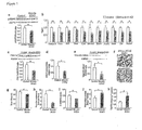
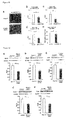
- Muscle-R1KO mice showed decreased phosphorylation of AMPK ( FIG. 1a ). Moreover, administration of adiponectin increased the phosphorylation of AMPK in the muscle of control littermates, but not that in the muscle of muscle-R1KO mice ( FIG. 7a ). On the other hand, adiponectin increased the phosphorylation of AMPK in the liver of both genotypes ( FIG. 7b ). In the muscle-R1KO mice, the molecules involved in mitochondrial biogenesis such as PGC1- ⁇ ( Cell 98, 115-124(1999 )) were also decreased at both messenger RNA (Ppargcla) ( FIG. 1b ) and protein levels ( FIG. 1c ). Adiponectin increased the expression levels of Ppargc1a in the muscle of control littermates but not those in the muscle of muscle-R1KO mice ( FIG. 7c ).
- the mitochondrial function was assessed by measuring the enzymatic activities of Cox ( FIG. 8a ) and succinate dehydrogenase (SDH) ( FIG. 8b ). Staining of soleus muscle sections has revealed a smaller number of Cox- and SDH-positive muscle fibers and a decreased intensity of Cox and SDH staining in muscle-R1KO mice ( FIGS. 8a and 8b ).
- muscle endurance test by tread mill running for assessing the involuntary physical exercise was conducted in control and muscle-R1KO mice.
- the muscle endurance of muscle-R1KO mice was significantly lower than that of the control mice (FIG. 1h).
- the molecules involved in fatty acid oxidation such as medium-chain acyl-CoA dehydrogenase (Acadm) was significantly decreased in muscle-R1KO mice ( FIG. 1b ), which was associated with decreased ⁇ oxidation ( FIG. 1i ) and increased triglyceride content ( FIG. 1j ) in skeletal muscle.
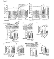
- the glucose levels and plasma insulin levels after administration of glucose were significantly higher in muscle-R1KO mice than in control mice ( FIGS. 2a and 2b ).
- the capacities of muscle-R1KO mice of regulating plasma glucose levels were significantly decreased after ingestion of insulin ( FIG. 2c ).
- disruption of AdipoR1 in muscle did not significantly alter endogenous glucose production, but significantly decreased a glucose disposal rate (Rd) and a glucose infusion rate (GIR), indicating decreased insulin sensitivity in muscle ( FIGS. 2d to 2f ).
- AMPK activity depends on phosphorylation of AMPK ⁇ (Thr 172) in its activation loop by AMPK kinase (AMPKK) ( Cell Metab. 1, 15-25(2005 ); J. Biol. Chem. 271, 27879-27887(1996 )).
- LKB1 and CaMKK ⁇ are known to be two main AMPKKs present in various tissues and cells ( Cell Metab. 1, 15-25(2005 ); Cell Metab.
- FIGS. 9a to 9e small interfering RNAs
- FIGS. 9a and 9b Suppression of the expression of AdipoR1 or CaMKK ⁇ by respective specific siRNAs greatly reduced an increase in PGC-1 ⁇ expression induced by adiponectin ( FIG. 3b ).
- FIGS. 9c and 9d the suppression of AMPK ⁇ 1 and AMPK ⁇ 2 expression ( FIGS. 9c and 9d ) by respective specific siRNAs failed to significantly reduce the PGC-1 ⁇ expression induced by adiponectin ( FIG. 3b ), suggesting that the PGC-1 ⁇ expression was AdipoR1- and CaMKK ⁇ - dependent, but it was induced by adiponectin via an AMPK-independent pathway.
- SIRT1 deacetylase activity has been reported to depend on the NAD + levels ( Nature 434, 113-118(2005 ); Nature 444, 868-874(2006 )).
- Adiponectin increased a NAD + /NADH ratio in C2C12 myocytes ( FIG. 3g ).
- PGC-1 ⁇ acetylation FIG. 3h
- NAD + /NADH ratio in vivo upon stimulation with adiponectin
- Adiponectin induces calcium ion influx via AdipoR1.
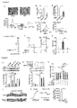
- AdipoR1 was expressed in Xenopus oocytes ( FIG. 4e ). An increase in intracellular calcium levels caused by stimulation with adiponectin was detected by monitoring calcium-ion activated Cl - current in oocytes injected with AdipoR1 complementary RNA ( FIGS. 4f and 4g ). The responses were not observed in control oocytes ( FIG. 4g ).
- Adiponectin-induced calcium ion influx is important for action expression.
- FIGS. 9b and 9h Suppression of CaMKK ⁇ or LKB1 expression by respective siRNAs specific to them ( FIGS. 9b and 9h ) significantly reduced an increase in phosphorylation of AMPK induced by adiponectin ( FIG. 5b ).
- AraA an AMPK inhibitor
- a selective inhibitor effectively and almost completely abolished an increase in PGC-1 ⁇ expression stimulated with adiponectin in C2C12 myocytes ( FIG. 5c ) as removal of extracellular calcium ions effectively abolishes calcium ion signals induced by adiponectin ( FIG. 4a ).
- Muscle-R1KO deteriorates adiponectin action in skeletal muscle.
- Muscle-R1KO exhibited decreased phosphorylation of AMPK ( FIG. 1a ) and its intracellular substrate, that is, acetyl CoA carboxylase (ACC) (FIG. 14a). Muscle-R1KO showed decreased mitochondria-specific Gomori staining ( FIG. 15a ).
- Muscle-R1KO exhibited a decrease in molecules involved in type I muscle fiber differentiation myocyte enhancer factor 2C (Mef2c) ( EMBO J. 19, 1963-1973(2000 )) ( FIGS. 1b and 16a ) and also type I muscle fiber marker myosin heavy chain (MHC) isoform I ( FIG. 15b ), troponin I (slow) (Tnni1) ( FIGS. 1e and 15c ) and myoglobin (Mb) ( Physiol. Rev. 80, 1215-1265(2000 )) ( FIG. 15d ). These findings were consistent with the histological analysis ( FIGS. 1f, 1g , and 15e ). In soleus muscle of muscle-R1KO mice, type I muscle fibers were decreased by 20% ( FIGS. 1g and 15e ).
- MHC myosin heavy chain
- the protein expression levels of MEF2, MCAD, SOD2, catalase, and Glut4 in the muscle of muscle-R1KO mice were significantly decreased compared with those seen in control mice ( FIG. 16 ). These results were consistent with the results that muscle-R1KO mice exhibited a decrease in type I muscle fibers, an increase in tissue triglyceride content, and an increase in oxidative stress in their muscle.
- Adiponectin increased the NAD + /NADH ratio and NAD + and NADH contents in C2C12 myocytes ( FIGS. 3g , 18a, and 18b ).
- Muscle-R1KO also exhibited a decrease in NAD + /NADH ratio and a decrease in NAD + and NADH contents in vivo ( FIGS. 3i , 18c and 18d ).
- Muscle-specific disruption of AdipoR1 had no significant effect on PPAR ⁇ (Ppara) levels ( FIG. 19a ), however, it resulted in significantly decreased expression levels of PPAR ⁇ target genes such as ACO (Acox1) in the muscle ( FIG. 19b ), suggesting deterioration in PGC-1 ⁇ activity. These results were consistent with the results that muscle-R1KO mice exhibit decreased PGC-1 ⁇ activity as PPAR co-activation in muscles.
- Glut4 (Slc2a4) were significantly decreased in muscles rich in type I muscle fibers such as soleus muscle and also muscles rich in type II fibers such as gastrocnemius of muscle-R1KO as compared with those in control mice ( FIGS. 19e and 19f ). These results are consistent with the notion that the observed reduction of Glut4 in the muscle of muscle-R1KO mice is not simply secondary due to alteration of the fiber-type composition.
- FIG. 20a An increase in CaMKK ⁇ activity caused by the addition of 0.25 ⁇ M Bay-K 8644, a calcium ion channel opener ( Nature 303, 535-537(1983 ); Am. J. Physiol. 258, R462-468(1990 )), ( FIG. 20a ) significantly enhanced, in the muscle of muscle-R1KO mice, insulin-stimulated phosphorylation of Akt ( FIG. 20b ), mitochondrial content ( FIG. 20c ), and function such as citrate synthase activities ( FIG. 20d ).
- AICAR Am. J. Physiol. 273, E1107-1112(1997 ) significantly increased the phosphorylation of AMPK in the muscle of muscle-R1KO mice ( FIG. 20e ).
- Activation of AMPK via AICAR Diabetologia 45, 56-65(2002 ); Cell 134, 405-415 (2008 )) and activation of SIRT1 via resveratrol ( Cell 127, 1109-1122(2006 )) significantly ameliorated insulin resistance and increased a mitochondrial content and function such as citrate synthase activities in the muscle of muscle-R1KO mice ( FIGS. 20f to 20i and 21a to 21d ).
- MnTBAP ( Nature 440, 944-948 (2006 ) indeed significantly decreased ROS such as H 2 O 2 ( FIG. 21e )
- MnTBAP alone tended to ameliorate insulin resistance and failed to significantly increase the mitochondrial content and function such as citrate synthase activities in the muscle of muscle-R1KO mice ( FIGS. 21e to 21i ).
- antioxidative stress-independent pathways such as inhibition of S6K1 by AMPK and inhibition of tissue triglyceride accumulation, together with antioxidative stress dependent pathways, contribute largely to the insulin sensitivity enhancing effects of adiponectin/AdipoR1 in muscle.
- Antioxidative stress-independent pathways such as increased NRF-1 ( FIG. 1b ) and mtTFA ( FIG. 1b ) by AMPK/SIRT1are presumed to largely contribute to mitochondrial biogenesis by adiponectin/AdipoR1in muscle.
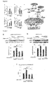
- AICAR significantly but partially rescued the AdipoR1loss in muscle ( FIG. 20f to 20i ), but exercise significantly and almost completely rescued it ( FIGS. 6a to 6d ). These results may be explained by several mechanisms. First, exercise could activate AMPK more efficiently than AICAR. In skeletal muscle, AMPK can be activated via two different pathways, that is, LKB1dependent phosphorylation and CaMKK ⁇ dependent phosphorylation.
- AICAR has been shown to activate LKB1-dependent phosphorylation and activation of AMPK as AMP mimetics inducing a structural change of AMPK that allows efficient phosphorylation by ordinarily structurally activated LKB1, it does not stimulate CaMKK ⁇ -dependent phosphorylation and activation since AICAR does not elevate an intracellular calcium ion concentration.
- exercise has been shown to elevate both intracellular AMP and calcium ion concentration so that it can stimulate both LKB1-dependent and CaMKK ⁇ -dependent phosphorylation and activation of AMPK.
- FIG. 9a Suppression of Adipor1 by specific siRNA ( FIG. 9a ) markedly reduced the increase in mitochondrial content by adiponectin ( FIG. 3a ) and at the same time, markedly reduced the increase in insulin-stimulated phosphorylation of Ser 473 in Akt ( FIG. 22a ).
- Insulin-stimulated phosphorylation of Thr 24 and Ser 253 in Foxo was markedly decreased in the skeletal muscle of muscle-R1KO mice as compared with that in control mice ( FIG. 22b ).
- FIGS. 23c and 23d Stimulation of oocytes expressing AdipoR1 with adiponectin induced inward current responses.
- BAPTA-AM Bis-(o-aminophenoxy)ethane-N,N,N9,N9-tetra-acetic acid, tetraacetoxymethyl ester
- FIGS. 25a and 25b upper panel and FIG. 25c Treatment of C2C12 myocytes with adiponectin caused a rapid, robust, and transient increase in CaMKI phosphorylation ( J. Biol. Chem. 273, 31880-31889(1998 ); Trends Biochem. Sci. 24, 232-236(1999 )) ( FIGS. 25a and 25b upper panel and FIG. 25c ). Removal of extracellular calcium ions by EGTA almost completely abolished the increased CaMKI phosphorylation stimulated with adiponectin ( FIGS. 25a and 25b upper panel, FIG. 25c ). These data indicate that the adiponectin-induced CaMKI phosphorylation presumably through CaMKK ⁇ activation almost completely depend on calcium ion entry.
- FIGS. 25a and 25b center panel, FIG. 25c Subsequent to the phosphorylation of CaMKI, adiponectin increased AMPK phosphorylation further in a sequential and partial manner depending on extracellular calcium ion influx ( FIGS. 25a and 25b center panel, FIG. 25c ).
- HDAC histone deacetylase
- FIGS. 25a and 25b lower panel, FIG. 25c The treatment of C2C12 myocytes with adiponectin increased HDAC5 phosphorylation in a sequential and partial manner depending on extracellular calcium ion influx ( FIGS. 25a and 25b lower panel, FIG. 25c ). Incubation for from 20 to 40 minutes in a calcium ion-free and EGTA-added medium without adding adiponectin (matched controls) had no significant effects on the phosphorylation amounts of CaMKI, AMPK, and HDAC5 ( FIGS. 25a to 25c ).
- PGC-1 ⁇ is a key regulator of oxidative metabolism in skeletal muscle. Reduced PGC-1 ⁇ and OXPHOS levels have been associated with insulin resistance and type 2 diabetes in humans ( Nature Genet. 34, 267-273(2003 )) and mice (FIGS. 14a to 14c). However, muscle-specific PGC-1 ⁇ -knockout mice did not exhibit insulin resistance ( J. Clin. Invest. 117, 3463-3474(2007 )). Also PGC-1 ⁇ -independent pathways such as inhibition of S6K1 by AMPK are presumed to contribute to the insulin sensitizing effects of adiponectin/AdipoR1 in muscle.
- PGC-1 ⁇ is a key regulator of a mitochondrial content and function. It has been reported that PGC-1 ⁇ expression is modulated in several physiological conditions. For example, in skeletal muscle, it is modulated by increased calcium ion signaling via molecules such as CaMK and CREB in partial response to exercise ( Nature 454, 463-469(2008 )). It has also been reported that PGC-1 ⁇ activities are modulated by several. kinds of PGC-1 ⁇ modifications, such as phosphorylation via AMPK ( Proc. Natl Acad. Sci.
- AMPK and SIRT1 could also be activated by exercise. It has been proved that muscle-R1KO mice exhibited decreased PGC-1 ⁇ expression as well as decreased PGC-1 ⁇ acetylation. Consistent with it, it has also been proved that adiponectin induces calcium ion influx via AdipoR1 and thereby activates CaMKK ⁇ and causes increased PGC-1 ⁇ expression and at the same time, adiponectin/AdipoR1 activates AMPK and SIRT1, which induces deacetylation of PGC-1 ⁇ . These data suggest that adiponectin and AdipoR1 stimulate increases in both the expression and activation of PGC-1 ⁇ in a similar manner to exercise ( FIG. 6e ).
- AdipoR1 is involved in regulation of insulin sensitivity via multiple mechanisms ( FIG. 13 ).
- First mechanism is the activation of S6K1, which has been reported to be able to provide insulin resistance via increased Ser 636/639 phosphorylation of IRS-1 ( Nature 431, 200-205(2004 )).
- S6K1 is crucially inhibited by AMPK ( J. Biol. Chem. 282, 7991-7996(2007 )).
- the AMPK activation was reduced in the skeletal muscle of muscle-R1KO mice, but activation of S6K1 and Ser 636/639 phosphorylation of IRS-1 were indeed increased.
- Second mechanism is oxidative stress increase, which has been linked as a cause of insulin resistance ( Nature 440, 944-948(2006 )) via increased Ser 302 phosphorylation of IRS-1 through JNK activation ( Nature 444, 860-867(2006 )).
- Some oxidative stress-detoxifying genes were crucially regulated by AMPK and PGC-1 ⁇ ( Cell 127, 397-408(2006 )) and the expression levels of these genes such as Sod2 and cat were reduced, which has been associated with increased TBARS in the skeletal muscle of muscle-R1KO mice.
- Third mechanism is an increased triglyceride content, which is associated with insulin resistance via increased Ser 302 phosphorylation of IRS-1 through JNK activation ( Nature 444, 860-867(2006 )).
- Molecules involved in fatty acid oxidation are crucially regulated by AMPK and PGC-1 ⁇ and the expression levels of these genes such as Mcad were reduced, which has been associated with an increased triglyceride content in the skeletal muscle of muscle-RIKO mice.
- JNK activation and Ser 302 phosphorylation in IRS-1 were indeed increased, consistent with an increase in TBARS and triglyceride content.
- AdipoR1 plays a critical role in the physiological and pathophysiological significance of adiponectin in muscle and are involved in the regulation of calcium ion signaling, PGC-1 ⁇ expression and activation, mitochondrial function and oxidative stress, glucose and lipid metabolism, and exercise endurance.
- the above-mentioned results suggest that the agonism of AdipoR1 as well as strategies of increasing AdipoR1 in muscle may provide a new treatment method for mitochondrial dysfunction, insulin resistance, and type 2 diabetes linked to obesity.
Landscapes
- Health & Medical Sciences (AREA)
- Life Sciences & Earth Sciences (AREA)
- Engineering & Computer Science (AREA)
- Chemical & Material Sciences (AREA)
- Immunology (AREA)
- Molecular Biology (AREA)
- Biomedical Technology (AREA)
- Hematology (AREA)
- General Health & Medical Sciences (AREA)
- Medicinal Chemistry (AREA)
- Urology & Nephrology (AREA)
- Bioinformatics & Cheminformatics (AREA)
- Organic Chemistry (AREA)
- Biochemistry (AREA)
- Microbiology (AREA)
- Biotechnology (AREA)
- Analytical Chemistry (AREA)
- Physics & Mathematics (AREA)
- Endocrinology (AREA)
- Food Science & Technology (AREA)
- General Physics & Mathematics (AREA)
- Cell Biology (AREA)
- Pathology (AREA)
- Proteomics, Peptides & Aminoacids (AREA)
- Zoology (AREA)
- Veterinary Medicine (AREA)
- Public Health (AREA)
- Pharmacology & Pharmacy (AREA)
- Animal Behavior & Ethology (AREA)
- Toxicology (AREA)
- Diabetes (AREA)
- Gastroenterology & Hepatology (AREA)
- General Chemical & Material Sciences (AREA)
- Chemical Kinetics & Catalysis (AREA)
- Nuclear Medicine, Radiotherapy & Molecular Imaging (AREA)
- Genetics & Genomics (AREA)
- Wood Science & Technology (AREA)
- Biophysics (AREA)
- Epidemiology (AREA)
- Obesity (AREA)
Abstract
Description
- The present invention relates to a pharmaceutical for pseudo-exercise therapy.
- One of the goals of diabetes treatment is to prevent or inhibit progression of various chronic complications in diabetes, for example, microvascular disorders such as diabetic retinopathy, diabetic nephropathy, and diabetic neuropathy and macrovascular disorders such as cerebrovascular disorders, ischemic heart disease, and diabetic gangrene. Diet therapy and exercise therapy are fundamental in the diabetes treatment. In addition to these therapies, drug therapy using insulin preparations, sulfonyl urea drugs and incretin-related drugs stimulating pancreatic insulin secretion, biguanide drugs having mainly extra-pancreatic action, thiazolidine-based drugs, and carbohydrate absorption inhibitors (α-glucosidase inhibitors) is used to control blood sugar levels.
- The exercise therapy, when used for the treatment of diabetes, accelerates utilization of glucose and fatty acids and thereby decreases blood sugar levels, as an acute effect of exercise and at the same time, ameliorates insulin resistance as a chronic effect. In addition, an effect on weight loss, prevention of muscular atrophy or osteoporosis due to aging or lack of exercise, or amelioration of hypertension or hyperlipidemia can be recognized. As the exercise therapy, aerobic exercise is said to be effective for amelioration of insulin resistance.
- Thus, the exercise therapy is one of fundamental therapies in the treatment of diabetes. It should be the first-line therapy for ameliorating insulin resistance in the treatment of insulin independent diabetes from which particularly many patients in Japan suffer. As exercise effective for the treatment, walking continuously for at least 15 minutes twice a day and three or more days a week is said to be desirable. For many diabetic patients, however, it is not easy to continue a sufficient exercise therapy.
- In particular, it is difficult to apply a sufficient walking exercise therapy to old patients having deteriorated muscle strength or patients having disorders in knee joint or hip joint. Application of the exercise therapy to diabetic patients suffering also from a disease such as arrhythmia is not always safe and it may worsen the clinical conditions of diabetic patients suffering from chronic complication (for example, patients suffering from diabetic retinopathy or diabetic nephropathy). Moreover, patients in their prime or middle-aged or older patients cannot take enough time for the exercise therapy so that they cannot often complete the scheduled exercise therapy. From such viewpoints, there is an eager demand for the development of a drug therapy for mimetically carrying out an exercise therapy.
- Adiponectin (J. Biol. Chem. 270, 26746-26749(1995); J. Biol. Chem. 271, 10697-10703(1996); Biochem. Biophys. Res. Commun. 221, 286-289(1996); and J. Biochem. 120, 803-812(1996)) encoded by Adipoq is adipokine having antidiabetic action and anti-atherogenic action. It has been reported (Arterioscler. Thromb. Vasc. Biol. 20, 1595-1599(2000)) that plasma adiponectin levels are decreased in obesity, insulin resistance, and
type 2 diabetes. Administration of adiponectin to mice causes antihyperglycemic action and ameliorates insulin resistance (Proc. Natl Acad. Sci. USA 98, 2005-2010(2001); Nature Med. 7, 941-946(2001); and Nature Med. 7, 947-953(2001)), but adiponectin-deficient mice show insulin resistance and diabetes (J. Biol. Chem. 277, 25863-25866(2002); and Nature Med. 8, 731-737(2002)). The insulin sensitizing effect of adiponectin is presumed to appear due to an increase in fatty acid oxidation via activation of AMP-activated protein kinase (AMPK) (Nature Med. 8, 1288-1295(2002); Proc. Natl Acad. Sci. USA 99, 16309-16313(2002); and Cell Metab. 1, 15-25(2005)) and moreover via peroxisome proliferator-activated receptor α (PPARα) (Nature 405, 421-424(2000); and J. Biol. Chem. 278, 2461-2468(2003)). - There is a report (Nature 423, 762-769(2003)) on the cloning of complementary DNAs (Adipor1 and Adipor2) encoding adiponectin receptor 1 (which may hereinafter be abbreviated as "AdipoR1") and an adiponectin receptor 2 (which may hereinafter be called "AdipoR2"). AdipoR1 is much expressed in the skeletal muscle and liver, while AdipoR2 is expressed mainly in the liver. Both receptors are predicted to contain 7-transmembrane domains (Nature 423, 762-769(2003)) but to be structurally and functionally different from G-protein coupled receptors (FASEB J. 11, 346-354(1997); Nature 387, 620-624(1997); EMBO J. 15, 3566-3578(1996)). Adiponectin receptors are presumed to constitute a new receptor family.
- It is indicated by using Adipor1 and/or Adipor2 knockout mice that AdipoR1 and AdipoR2 act as a main receptor for adiponectin in vivo and play an important role in the regulation of glucose and lipid metabolism, inflammation, and oxidative stress in vivo (Nature Med. 13, 332-339(2007)). In the liver, AdipoR1 activates an AMPK pathway, while AdipoR2 activates a PPARα pathway (Nature Med. 13, 332-339(2007)). On the other hand, insulin resistance has been reported to be associated with mitochondrial dysfunction (N. Engl. J. Med. 350, 664-671(2004)) but the exact cause of mitochondrial dysfunction has not yet been found. The decreased adiponectin/AdipoR1 signaling may be associated with mitochondrial dysfunction, but details of adiponectin/AdipoR1 signaling have not yet been elucidated. In addition, the cascade of adiponectin/AdipoR1 signaling which has induced intracellular physiological action has been poorly elucidated.
-
- Non-patent Document 1: J. Biol. Chem. 270, 26746-26749(1995)
- Non-patent Document 2: J. Biol. Chem. 271, 10697-10703(1996)
- Non-patent Document 3: Biochem. Biophys. Res. Commun. 221, 286-289(1996)
- Non-patent Document 4: J. Biochem. 120, 803-812(1996)
- Non-patent Document 5: Arterioscler. Thromb. Vasc. Biol. 20, 1595-1599(2000)
- Non-patent Document 6: Proc. Natl Acad. Sci. USA 98, 2005-2010(2001)
- Non-patent Document 7: Nature Med. 7, 941-946(2001)
- Non-patent Document 8: Nature Med. 7, 947-953(2001)
- Non-patent Document 9: J. Biol. Chem. 277, 25863-25866(2002)
- Non-patent Document 10: Nature Med. 8, 731-737(2002)
- Non-patent Document 11: Nature Med. 8, 1288-1295(2002)
- Non-patent Document 12: Proc. Natl Acad. Sci. USA 99, 16309-16313(2002)
- Non-patent Document 13: Cell Metab. 1, 15-25(2005)
- Non-patent Document 14: Nature 405, 421-424(2000)
- Non-patent Document 15: J. Biol. Chem. 278, 2461-2468(2003)
- Non-patent Document 16: Nature 423, 762-769(2003)
- Non-patent Document 17: Nature 423, 762-769(2003)
- Non-patent Document 18: FASEB J. 11, 346-354(1997)
- Non-patent Document 19: Nature 387, 620-624(1997)
- Non-patent Document 20: EMBO J. 15, 3566-3578(1996)
- Non-patent Document 21: Nature Med. 13, 332-339(2007)
- Non-patent Document 22: Nature Med. 13, 332-339(2007)
- Non-patent Document 23: N. Engl. J. Med. 350, 664-671(2004)
- An object of the present invention is to provide a pharmaceutical that can be applied to diabetic patients as a pseudo-exercise therapeutic agent.
- The present inventors have carried out an extensive investigation with a view to solving the above-mentioned problem. As a result, it has been found that adiponectin bound to
adiponectin receptor 1 causes incorporation of calcium ions (Ca2+) in skeletal muscle cells. It has also been found that influx, into skeletal muscle cells, of calcium ions generated when adiponectin binds to AdipoR1 is indispensable for subsequent activation of calcium ion/calmodulin-dependent protein kinase kinase β (CaMKKβ), activation of AMPK and SIRT1, increased expression and decreased acetylation of peroxisome proliferator-activated receptor γ coactivator-1 α (PGC-1α), and increased mitochondria in the muscle cells. It has also been confirmed, on the other hand, that muscle-specific disruption ofadiponectin receptor 1 results in decreased elevation of a calcium ion concentration by adiponectin in cells which leads to decreased activation of CaMKK, AMPK, and SIRT1 and suppression of AdipoR1 results in decreased expression and deacetylation of PGC-1α, a decreased mitochondrial content and enzymes, decreased oxidative type I myofibers and decreased oxidative stress-detoxifying enzymes in skeletal muscle. - The present inventors have paid attention to the fact that various physiological activities induced by binding of adiponectin to
adiponectin receptor 1 are substantially similar to changes in skeletal muscle cells after exercise. As a result, it has been found that a pseudo-exercise therapy can be conducted by a drug therapy using a medicament containing, as an active ingredient, anadiponectin receptor 1 agonist compound and a post-exercise good muscle state achieved by applying a managed exercise therapy to patients can also be achieved by the drug therapy, leading to the completion of the present invention. - The present invention provides a pharmaceutical for pseudo-exercise therapy containing an
adiponectin receptor 1 agonist compound as an active ingredient.
The present invention also provides a pharmaceutical containing anadiponectin receptor 1 agonist compound as an active ingredient and capable of changing the physiological condition of a muscle to that after exercise without putting an exercise stress.
In a preferred mode of the present invention, there is provided the above-described pharmaceutical to be used for a drug therapy as a substitute for an exercise therapy in the prevention and/or treatment of diabetics. - From another standpoint, the present invention provides a pseudo-exercise therapeutic agent containing an
adiponectin receptor 1 agonist compound as an active ingredient; a pseudo-exercise effector containing anadiponectin receptor 1 agonist compound as an active ingredient; a type I muscle fiber enhancer containing anadiponectin receptor 1 agonist compound as an active ingredient; and an insulin-sensitivity enhancer based on a pseudo-exercise effect, which enhancer contains anadiponectin receptor 1 agonist compound as an active ingredient.
The present invention further provides the use of anadiponectin receptor 1 agonist compound for the preparation of the above-described pharmaceutical. - The present invention still further provides a method of conducting a pseudo-exercise therapy in mammals including humans, including a step of administering an effective amount of an
adiponectin receptor 1 agonist compound to mammals including humans; a method of changing the physiological conditions of the muscle of mammals including humans to those after exercise, including the step of administering an effective amount of anadiponectin receptor 1 agonist compound to mammals including humans; and a method of increasing type I muscle fibers in the muscle of mammals including humans, including a step of administering an effective amount of anadiponectin receptor 1 agonist compound to mammals including humans. - The pharmaceutical for pseudo-exercise therapy provided by the present invention causes, when bound to adiponectin, influx of extracellular calcium ions in skeletal muscle cells and thereby causes improvement of calcium ion signaling, improvement of glucose and lipid metabolism, improvement of exercise endurance, enhancement of expression and activation of PGC-1α through AMPK/SIRT1, and improvement of the function of mitochondria and oxidative stress. These physiological changes occurring after calcium ion influx in skeletal muscle cells are substantially similar to physiological changes in skeletal muscle cells after exercise. In particular, the pharmaceutical of the present invention increases a proportion and amount of type I muscle fibers and at the same time, has an effect of enhancing insulin sensitivity so that administration of the pharmaceutical of the present invention can achieve, in muscle, effects similar to those achieved by applying good-quality exercise without actual exercise.
- The pharmaceutical for pseudo-exercise therapy provided by the present invention can achieve pseudo-exercise effects through a drug therapy instead of an exercise therapy by administering it to diabetic patients who are old or have difficulty in walking and are not suited for the exercise therapy or arrhythmic patients or patients suffering from diabetic chronic complication for which an exercise therapy may be contradicted. Moreover, it can be used, as an auxiliary pharmaceutical to be used in combination with exercise therapy, for patients who cannot take sufficient time for exercise therapy. The pharmaceutical of the present invention can achieve pseudo-exercise effects in muscle without temporary or chronic muscle disorders such as damage, rupture, or inflammation of muscle fibers which will otherwise be caused by exercise and thus has an excellent characteristic.
-
- [
FIG. 1] FIG. 1 shows a decreased mitochondrial content and the number of oxidative type I myofibers and reduced exercise capacity in skeletal muscle of muscle-R1KO mice. FIGS. (a) to (k) show results of skeletal muscle (a-g, i-k) obtained from Control or muscle-R1 knockout (KO) mice after 5-hour fasting, in which (a) shows phosphorylation and amount of AMPK, (b) mRNA level of Ppargc1a, Esrra, Nrf1, Tfam, Cycs, mt-Co2, Mef2c, Acadm, Sod2, Cat, and Slc2a4, (c)PGC-1α protein level, (d) a mitochondrial content as assessed by the mitochondrial DNA copy number, (e) amounts of troponin I (slow) protein, (f) soleus muscles analyzed by ATPase (pH 4.3 for type I fibers) staining (Scale bars, 100 µm), (g) quantification of fiber type distribution (f) based on fiber-type analyses, (h) exercise endurance, (i) β oxidation, (j) a triglyceride content, and (k) TBARS. All values were presented as mean ± s.e.m (n=5-12). Symbols * and ** mean P<0.05 and P<0.01, respectively, compared with control mice. - [
FIG. 2] FIG. 2 shows mechanisms of abnormal glucose and insulin homeostasis in muscle-R1KO mice. FIGS. (a) to (f) show results of hyperinsulinemic euglycermic clamp study in control and muscle-R1KO mice, more specifically, (a,c) plasma glucose and (b) plasma insulin during (a,b) oral glucose tolerance test (OGTT) (1.5 g glucose per kg body weight) or (c) during insulin tolerance test (ITT) (0.25U insulin per kg body weight); (d) endogenous glucose production (EGP), (e) rates of glucose disposal (Rd), and (f) glucose infusion rate (GIR). FIGS. (g) to (1) show (g) phosphorylation of tyrosine (pTyr) in IRS-1, (k) phosphorylation of Ser 302 in IRS-1, (1) phosphorylation of Ser 636/639 in IRS-1, (h) phosphorylation and amount of Akt, (i) phosphorylation and amount of S6K1, and (j) phosphorylation and amount of JNK in skeletal muscle treated with or without insulin (0.3 U per kg body weight) for 7.5 minutes in control mice and muscle R1KO mice after 5-hr fasting. IB means immunoblotting, while IP means immunoprecipitation. All values were presented as mean ± s.e.m. Symbols * and ** mean, in from 3 to 5 independent experiments (n= 6 to 15), P<0.05 and P<0.01, respectively, compared with control or indicated one and NS means no significant difference. - [
FIG. 3] FIG. 3 shows the result of PGC-1α expression and activation and mitochondrial biogenesis, in C2C12 myocytes, increased by adiponectin/AdipoR1 Shown in (a) to (i) are mitochondrial contents as assessed by the mitochondrial DNA copy number (a, e, f), Ppargc1a mRNA levels (b), acetyl-lysine (Ac-Lys) levels or flag immunoprecipitates (IP) checked on PGC-1α (c, d, h), and an NAD+/NADH ratio (g, i) in C2C12 myocytes treated with adiponectin for indicated time (g); in C2C12 myocytes (a to c) treated with 10 µgml-1 adiponectin for 48 hours (a, e, f) or for 1.5 hours (b) or 2 hours (c, d) and then transfected with the indicated siRNA duplex (a to c), with the wild-type or the 2A mutant form of PGC-1α (d, e), or with the R13 mutant form of PGC-1α (f); or in skeletal muscles (h, i) from control mice or muscle-R1KO mice treated with or without adiponectin. The supernatant was blotted against GAPDH as input control (c, d, h). C2C12 myocytes were used after myogenic differentiation in all experiments. All values were presented as mean ± s.e.m (n=5-10). Symbols * and ** mean P<0.05 and P<0.01, respectively, compared with control or unrelated siRNA or as indicated. - [
FIG. 4] FIG. 4 shows adiponectin-induced calcium ion influx via AdipoR1 in C2C12 myocytes and Xenopus oocytes. FIG. (a) shows images of changes in fura-2 1 minute before and after stimulation with adiponectin (30 µgml-1). The red portion corresponds to the greatest response. A bottom trace demonstrated the average calcium response of C2C12 myocytes to 1-min stimulation with adiponectin, while including application of 5mM EGTA (black bar). The shaded region around the trace represents s.e.m. (b) to (d): FIG. (b) shows Adipor1 mRNA levels (b), fura-2 calcium response (c), and the magnitude of the response (d) in C2C12 myocytes transfected with unrelated siRNA duplex or AdipoR1 siRNA duplex in response to stimulation with 30 µgml-1 adiponectin for 1 min. (e) to (g): FIG. (e) shows the amounts of AdipoR1 protein in Xenopus oocytes (depolarizing pulses of +100 mV) injected with or without Adipor1 cRNA corresponding to adiponectin (30 µg/ml), or added with or without application of 5 mM EGTA, FIG. (f) shows representative traces of calcium-ion-activated Cl-current 30 seconds before (left) and after (right) administration of adiponectin, and FIG. (g) shows the magnitude of them. [Calcium ion]i and [calcium ion]e mean intracellular and extracellular calcium ion concentrations, respectively. All values were presented as mean ± s.e.m (n=6-14). Symbols * and ** mean P<0.05 and P<0.01, respectively, compared with unrelated siRNA cells or control cells or as indicated. - [
FIG. 5] FIG. 5 shows the necessity of adiponectin-induced calcium ion influx for CaMKK and AMPK activation and PGC-1α expression. (a) and (b): FIG. (a) shows phosphorylation and amount of AMPK in C2C12 myocytes preincubated for 20 min with or without 5mM EGTA and then, treated for 5 min with adiponectin (30 µgml-1) or ionomycin (1 µM), or for 1 hr with AICAR(1 mM) and FIG. (b) shows phosphorylation and amount of AMPK in C2C12 myocytes transfected with the indicated siRNA duplex and then treated with 30 µgml-1 adiponectin for 5 min. FIG. (c) shows the amount of Ppargc1a mRNA in C2C12 myocytes preincubated for 1 hr with AraA (0.5 mM), for 6 hr with STO-609 (1 µgml-1), or for 20 min with EGTA (5 mM) and then treated for 1.5 hr with or without adiponectin (10 µgml-1). FIG. (d) shows representative images of changes in fura-2calcium response 5 min before or after stimulation with adiponectin (30 µgml-1) in a soleus muscle from control mice (top) and muscle-R1KO mice (bottom). The red portion corresponds to the greatest response and scale bars indicate 100 µm. FIG. (e) shows a trace demonstrates the calcium response of a soleus muscle in the fields presented in (d). Adiponectin was administered for the time as indicated. FIG. (f) shows the magnitude of a fura-2 calcium response signal caused by 160-sec adiponectin stimulation to a soleus muscle and a ΔF ratio shows a change in fluorescence ratio after administration of adiponectin. All values were presented as mean ± s.e.m (n=5-10). Symbols * and ** mean P<0.05 and P<0.01, respectively, compared with control cells or unrelated siRNA cells or control cells or as indicated. - [
FIG. 6] FIG. 6 shows the effect of exercise on muscle-R1KO mice. FIGS. (a) to (d) show an insulin resistance index in skeletal muscles (a), an area under the blood level curves (AUC) of plasma glucose levels during ITT (b), a mitochondrial content as assessed by the mitochondrial DNA copy number (c), and citrate synthase (CS) enzyme activity (d) of control and muscle-R1KO mice after 2-week exercise. The results are expressed as the percentage of the value in control littermates (a, b). FIG. (e) shows a scheme illustrating the signaling of adiponectin/AdipoR1 in myocytes. Both CaMKKβ and LKB1 are necessary for adiponectin-induced full AMPK activation. AMPK and SIRT1 are required for adiponectin/AdipoRl-induced PGC-1α activation. CaMKKβ activation by adiponectin-induced calcium ion influx via AdipoR1 is required for adiponectin-induced increased PGC-1α expression. PGC-1α is required for mitochondrial biogenesis stimulated with adiponectin/AdipoR1. From these data, the present inventors concluded that adiponectin and AdipoR1 increase PGC-1α expression and activity via calcium ion signaling and AMPK/SIRT1, leading to increased mitochondrial biogenesis. The present inventors focused to the molecules on which they have obtained direct evidence by both gain-of-function and loss-of-function experiments in vitro and in vivo (except for CaMK which has already been reported (Nature 454, 463-469(2008) to increase PGC-1α expression by other researches). AC means acetylation. All values were presented as mean ± s.e.m (n=5-8). Symbols * and ** mean P<0.05 and P<0.01, respectively, compared with control mice or as indicated. - [
FIG. 7] FIG. 7 shows that muscle-specific disruption of AdipoR1 results in decrease of adiponectin action in skeletal muscle. (a), (b): Phosphorylation and amount of AMPK in skeletal muscle (a) and liver (b) treated with or without adiponectin (30 µg per 10 g body weight) for 10 min. (c): PGC-1α (Ppargcla) mRNA levels in skeletal muscle treated with or without adiponectin (30 µg per 10 g body weight) for 10 min. All values are presented as mean ± s.e.m (n=5-8). Symbols * and ** mean P<0.05 and P<0.01, respectively, compared with control mice. - [
FIG. 8] FIG. 8 shows a decreased mitochondrial content, function, type II fiber in skeletal muscle of muscle-R1KO mice. (a) and (b): The cross-sections of soleus muscle were stained for Cox activity (a) and SDH activity (b). Scale bars: 100 µm. Skeletal muscle was obtained from control mice or muscle-R1KO mice. (c) to (e): MHC IIa (c), MHC IIx (d), and MHC IIb (e) mRNA levels in skeletal muscle were analyzed by RT-qPCR. All values were presented as mean ± s.e.m (n=5-8). Symbols * and ** mean P<0.05 and P<0.01, respectively, compared with control mice. - [
FIG. 9] FIG. 9 shows expression of mRNA in C2C12 cells transfected with siRNA duplex. (a) to (h): C2C12 myocytes were transfected with siRNA duplex. AdipoR1 (Adipor1) (a), CaMKKβ (Camkk2) (b), AMPKα1 (Prkaa1) (c), AMPKα2 (Prkaa2) (d), PGC-1α (Ppargcla) (e), AdipoR2 (Adipor2) (f), SIRT1 (Sirt1) (g), and LKB1 (Stk11) (h) mRNA levels were analyzed by RT-qPCR. All values were presented as mean ± s.e.m (n=8-10). Symbol ** means P<0.01 compared with unrelated siRNA cells. - [
FIG. 10] FIG. 10 shows total activity of PGC-1α in skeletal muscle. It shows total activity as assessed by multiplying expression and relative deacetylation of PGC-1α together. Skeletal muscle was obtained from control mice or muscle-R1KO mice. All values were presented as mean ± s.e.m (n=8-10). Symbols * and ** mean P<0.05 and P<0.01, respectively, compared to control or as indicated. - [
FIG. 11] FIG. 11 shows phosphorylation of CaMKI stimulated with adiponectin in skeletal muscle. It shows phosphorylation and amount of CaMKI in the skeletal muscle of control and muscle-R1KO mice treated for 5 min with or without adiponectin (30 µg per 10 g body weight). All values were presented as mean ± s.e.m (n=3-6). Symbols * and ** mean P<0.05 and P<0.01, respectively, compared with control or as indicated. - [
FIG. 12] FIG. 12 shows a decreased mitochondrial content in skeletal muscle of db/db mice. (a) and (b): Adipor1 (a) and Ppargc1a (b) mRNA levels in the skeletal muscle of wild-type (WT) and db/db mice were analyzed by RT-qPCR. (c): a mitochondrial content as assessed by the mitochondrial DNA copy number. Skeletal muscle was obtained from WT mice or db/db mice. All values were presented as mean ± s.e.m (n=5-10). Symbol ** means P<0.01 compared with WT mice. - [
FIG. 13] FIG. 13 is a scheme illustrating the actions of adiponectin/AdipoR1 in skeletal muscle. Adiponectin/AdipoRl increased phosphorylation of AMPK, the amount and activity of PGC-1α, mitochondrial biogenesis, type I myofibers, and insulin sensitivity. - [
FIG. 14] FIG. 14 shows that muscle-specific disruption of AdipoR1 results in decrease of adiponectin action in skeletal muscle. Phosphorylation and amount of ACC in the skeletal muscle of muscle-R1KO mice. All values were presented as mean ± s.e.m (n=5). Symbol ** means P<0.01 compared with control mice. - [
FIG. 15] FIG. 15 shows a decreased mitochondrial content and type I fibers in the skeletal muscle of muscle-R1KO mice. (a): Modified Gomori trichrome staining. Scale bars: 100 µm. Skeletal muscle was obtained from control mice or muscle-R1KO mice. (b) to (e): MHC I (b), troponin I (slow) (Tnni1) (c), and myoglobin (Mb) (d) mRNA levels in skeletal muscle were analyzed by RT-qPCR. (e): Quantification of fiber type distribution based on the fiber type analysis inFIG. 1f . All values were presented as mean ± s.e.m (n=5-8). Symbols * and ** mean P<0.05 and P<0.01, respectively, compared with control. - [
FIG. 16] FIG. 16 shows decreased expression levels of proteins important for the skeletal muscle in muscle-R1KO mice. (a) to (e): MEF2 (a), MCAD (b), SOD2 (c), catalase (d), and Glut4 (e) proteins in the skeletal muscle. The skeletal muscle was obtained from control mice or muscle-R1KO mice. All values were presented as mean ± s.e.m (n=4-8). Symbol ** means P<0.01, compared with control mice. - [
FIG. 17] FIG. 17 shows an increased amount of mitochondria in C2C12 myocytes treated with adiponectin. (a): A mitochondrial content as assessed by the mitochondrial DNA copy number in C2C12 myocytes preincubated with AraA (0.5 mM) for 1 hr or with STO-609 (1 µg/ml) for 6 hr and then treated with 10 µg/ml adiponectin for 48 hr. (b): After treatment of C2C12 myocytes with 10 µg/ml adiponectin for 48 hr, 100 nm MitoTracker green FM was added and the resulting mixture was incubated for 30 min. (c): An amount of mitochondria was indicated by green fluorescence and a mitochondrial content was measured by using MitoTracker green. All values were presented as mean ± s.e.m (n=5-8). Symbol ** means P<0.01 compared as indicated. - [
FIG. 18] FIG. 18 shows NAD+ and NADH contents in C2C12 myocytes and skeletal muscle. (a) to (d): NAD+ (a, c) and NADH (b, d) contents in C2C12 myocytes (a, b) and skeletal muscles (c, d) treated with adiponectin for the times indicated. Skeletal muscle was obtained from control mice or muscle-R1KO mice. All values were presented as mean ± s.e.m (n=8-10). Symbols * and ** mean P<0.05 and P<0.01, respectively, compared with control or as indicated. - [
FIG. 19] FIG. 19 shows Pparα, Aco, and Slc2a4 mRNA levels, oxygen (O2) consumption, and respiratory quotient (RQ) in the skeletal muscle of muscle-R1KO mice. (a) and (b): PPARα (Ppara) (a) and Aco (Acox1) (b) mRNA levels in the skeletal muscle of control mice and muscle-R1KO mice were analyzed by RT-qPCR. (c) and (d): Mean 24-hr values of oxygen (O2) consumption (c) and respiratory quotient (RQ) (d) in control mice and muscle-R1KO mice. (e) and (f): Glut4 (Slc2a4) mRNA levels in the gastrocnemius muscle (e) and soleus muscle (f) of control mice and muscle-R1KO mice were analyzed by RT-qPCR. All values were presented as mean ± s.e.m (n=6-14). Symbols * and ** mean P<0.05 and P<0.01, respectively, compared with control mice. - [
FIG. 20] FIG. 20 shows the effect of increased calcium influx and AICAR in the muscle of muscle-R1KO mice. (a) to (d): Control mice and muscle-R1KO mice were treated with or without 0.25 µM Bay-K 8644 as a calcium-channel opener. (f) to (i): Control mice and muscle-R1KO mice were treated with a solvent or AICAR (500 mg per kg body weight per day, 2 weeks). (a): The relative ratio of phosphorylation of CaMKI in the skeletal muscle of muscle-R1KO mice treated with or without 0.25 µM Bay-K 8644. (b): Phosphorylation and amount of Akt (Ser 473) in skeletal muscle treated with or without 0.25 µM Bay-K 8644. (c) and (h): Mitochondrial content in skeletal muscle as assessed by the mitochondrial DNA copy number. Skeletal muscle was obtained from muscle-R1KO mice. (d) and (i): Citrate synthase enzyme activity in the skeletal muscle of control mice and Muscle-R1KO mice. (e): Phosphorylation and amount of AMPK in skeletal muscle treated with or without AICAR (500 mg per kg body weight per day, 2 hours). (f) Insulin resistance index. The insulin resistance index was calculated from the product of the areas of glucose and insulin x 10-2 in the glucose tolerance test. The results were expressed as the percentage of the value of control littermates. (g): Area under the curves (AUC) of plasma glucose levels during the insulin tolerance test (ITT). All values were presented as mean ± s.e.m (n=5-8). Symbols * and ** mean P<0.05 and P<0.01, respectively, compared with control mice. - [
FIG. 21] FIG. 21 shows the effect of resveratrol and antioxidant MnTBAP on muscle-R1KO mice. (a) to (d): Control mice and muscle-R1KO mice were treated with a solvent or resveratrol (400 mg per kg body weight per day, 2 weeks). (e) to (i): Control mice and muscle-R1KO mice were treated with a solvent or an antioxidant MnTBAP (10 mg per kg body weight per day, 2 weeks). (a) and (f): Insulin resistance index. The insulin resistance index was calculated from the product of the areas of glucose and insulin x 10-2 in the glucose tolerance test. The results were expressed as the percentage of the value of control littermates. (b) and (g): Area under the curves (AUC) of plasma glucose levels during the insulin tolerance test (ITT). (c) and (h): Mitochondrial content in skeletal muscle as assessed by the mitochondrial DNA copy number. The skeletal muscle was obtained from control mice or muscle-R1KO mice. (d) and (i): Citrate synthase enzyme activity in the skeletal muscle of control mice and muscle-R1KO mice. (e): The production of reactive oxygen species (ROS) as H2O2 in the skeletal muscle of control mice and muscle-R1KO mice treated with a solvent or an antioxidant MnTBAP (10 mg per kg body weight per day, 2 weeks). All values were presented as mean ± s.e.m (n=5-8). Symbols * and ** mean P<0.05 and P<0.01, respectively, compared with control mice or as indicated. - [
FIG. 22] FIG. 22 shows insulin-induced phosphorylation of Akt in C2C12 myocytes, FOXO1 phosphorylation in skeletal muscle, and an amount of PGC-1α acetylation in C2C12 myocytes. (a): Phosphorylation (Ser 473) and amount of Akt in C2C12 myocytes treated with or without 10 µg/ml adiponectin for 48 hr and then treated with or without 10 nM insulin for 10 min. (b) Phosphorylation (Thr 24 and Ser 253) and amount of FOXO1 in the skeletal muscle of muscle-R1KO mice treated with or without insulin (0.3 U per kg body weight) for 7.5 min. (c): C2C12 myocytes were transfected with SIRT1 siRNA duplex and then treated for 2 hr with or without adiponectin (10 µg/ml). Acetyl-lysine levels were checked on PGC-1α immunoprecipitates (IP). The supernatant was blotted against GAPDH as input control. All values were presented as mean ± s.e.m (n=6-10). Symbols * and ** mean P<0.05 and P<0.01, respectively, compared with control or as indicated. - [
FIG. 23] FIG. 23 shows adiponectin-induced inward current response via AdipoR1 in C2C12 myocytes and Xenopus oocytes. (a): The whole-cell inward current response of C2C12 myocytes, which were transfected with an unrelated siRNA duplex (upper trace) and an AdipoR1 siRNA duplex (bottom trace), to stimulation with 30 µg/ml adiponectin for 30 sec. Holding potential was -60 mV. (b): Effects of AdipoR1 knockdown on the whole-cell inward current response to adiponectin. (c): Representative current traces of oocytes injected with (top) or without (bottom) AdipoR1 cRNA in response to adiponectin recorded at a holding potential of -80 mV. Adiponectin (30 µg/ml) was administered for 30 sec at the times indicated by squares. (d): The magnitude of inward current response of oocytes injected with or without AdipoR1 cRNA in response to adiponectin (30 µg/ml) along with or without administration of 5 mM EGTA. [Calcium ion](e): external calcium ion concentration. All values were presented as mean ± s.e.m (n=6-14). Symbols * and ** mean P<0.05 and P<0.01, respectively, compared with the unrelated siRNA cells or as indicated. - [
FIG. 24] FIG. 24 shows phosphorylation of AMPK stimulated with adiponectin in the presence of BAPTA-AM or U73122. (a) and (b): Phosphorylation and amount of AMPK in C2C12 myocytes. C2C12 myocytes were preincubated for 30 min after addition of 50 µM BAPTA-AM or for 20 min after addition of 5 mM EGTA and then treated for 5 min with adiponectin (30 µg/ml) (a). C2C12 myocytes were preincubated for 30 min after addition of 10 µM U73122 or for 20 min after addition of 5 mM EGTA and treated for 5 min with adiponectin (30 µg/ml) (b). All values were presented as mean ± s.e.m (n=3-6). Symbols * and ** mean P<0.05 and P<0.01, respectively, compared with control or as indicated. - [
FIG. 25] FIG. 25 shows phosphorylation of CaMKI, AMPK, and HDAC5 stimulated with adiponectin in the presence of EGTA. (a): Phosphorylation and amount of CaMKI, AMPK, and HDAC5 in C2C12 myocytes. C2C12 myocytes were preincubated for 20 min with or without EGTA (5 mM) and then treated for 0, 0.5, 1, 2, 5, 10 and 20 min with or without EGTA (5 mM) and with or without adiponectin (30 µg/ml). (b): Densitometric quantification of immunoblots (a) of phosphorylation and amount of CaMKI (upper panel), AMPK (middle panel), and HDAC5 (lower panel) levels. (c) Densitometric quantification of immunoblots (a) of phorphorylation and amount of CaMKI (upper panel), AMPK (middle panel), and HDAC5 (lower panel) levels. The result of each time point is corrected for any change in the basal status during incubation in calcium-ion-free media without adiponectin (matched controls). All values were presented as mean ± s.e.m (n=3-8). Symbols * and ** mean P<0.05 and P<0.01, respectively, compared with control or as indicated. - [
FIG. 26] FIG. 26 shows generation and characterization of muscle-R1KO mice. (a): Schematic view of the Adipor1 gene targeting strategy. Muscle-R1KO mice were generated by crossing mice with a floxed Adipor1 allele to animals that transgenically express cre recombinase under the control of the MCK promoter (MCK-Cre). (b): Southern blot analysis of genomic DNA from the liver, SKM (skeletal muscle), WAT (white adipose tissue), and BAT (brown adipose tissue) of control mice and muscleR1KO mice on C57Bl/6 background. XbaI-digested mice genomic DNA was hybridized with the probe as shown in (a). (c): Northern blot analysis of Adipor1 mRNA expressed in liver, SKM, WAT, and BAT from control mice and muscle-R1KO mice on C57Bl/6 background. - The pharmaceutical of the present invention is characterized by that it is a pharmaceutical for pseudo-exercise therapy and contains an
adiponectin receptor 1 agonist compound as an active ingredient. - The term "
adiponectin receptor 1 agonist compound" as used herein means a compound which, when the compound is bound toadiponectin receptor 1, can cause all or some of various physiological actions caused by binding of adiponectin, which is an internal ligand ofadiponectin receptor 1, toadiponectin receptor 1 and can exhibit physiological actions similar to those of adiponectin. This term is used as a notion embracing adiponectin itself. - As described specifically and in detail in Examples, various physiological actions which occur when adiponectin is bound to
adiponectin receptor 1 are induced by incorporation of calcium ions in skeletal muscle cells which occurs first by binding of adiponectin toadiponectin receptor 1. Based on this finding, compounds having an agonist action foradiponectin receptor 1 can be screened with incorporation of calcium ions in skeletal muscle cells as an indicator. - For example, candidate compounds for
adiponectin receptor 1 agonist can be screened by a method including a step of bringing test compounds into contact with cells to determine whether or not they cause intracellular influx of extracellular calcium ions and a step of selecting, from the test compounds, compounds causing intracellular influx of extracellular calcium ions as a candidate compound foradiponectin receptor 1 agonist. The term "candidate agonist compound" means a compound having at least a possibility as an agonist compound. - In a preferred mode, a candidate agonist compound for
adiponectin receptor 1 can be screened by a method including a step of bringing test compounds into contact with AdipoR1-deficient cells and AdipoR1-expressing cells to judge whether or not they cause intracellular influx of extracellular calcium ions and selecting, from the test compounds, compounds causing intracellular influx of extracellular calcium ions in the AdipoR1-expressing cells without substantially causing intracellular influx of extracellular calcium ions in the AdipoR1-deficient cells as the candidate agonist compound foradiponectin receptor 1. - Candidate agonist compounds can be used as agonist compounds, but candidate agonist compounds for
adiponectin receptor 1 can be screened efficiently with accuracy by a method including the following steps: - (a) bringing test compounds into contact with cells to judge whether or not they cause intracellular influx of extracellular calcium ions and selecting, from the test compounds, compounds causing the intracellular influx of extracellular calcium ions as candidate agonist compounds for
adiponectin receptor 1; and - (b) bringing the candidate agonist compounds selected in the step (a) into contact with each of AdipoR1-deficient cells and AdipoR1-expressing cells to judge whether or not they cause intracellular influx of extracellular calcium ions and selecting, from the candidate agonist compounds, compounds not substantially causing intracellular influx of extracellular calcium ions in the AdipoR1-deficient cells but causing intracellular influx of extracellular calcium ions in the AdipoR1-expressing cells as an
adiponectin receptor 1 agonist compound. - In these screening methods, no particular limitation is imposed on the test compound and any compounds such as oligopeptides, polypeptides, nucleic acids, saccharides, and lipids as well as low-molecular organic compounds can be provided for screening. Since a number of compound libraries having many compounds stored therein have been constructed, it is efficient to provide for screening many compounds registered in such libraries.
- The kind of the cells with which the test compounds are brought into contact is not particularly limited and any somatic cell in which AdipoR1 has been expressed can be used. Preferably, cells in which AdipoR1 have been expressed abundantly, for example, skeletal muscle cells can be used. In addition, cells in which AdipoR1 has been expressed by introducing a gene encoding AdipoR1 can be used. In such a case, using, for example, oocytes are also preferred. Cells are however not limited to skeletal muscle cells or oocytes.
- When the test compounds are brought into contact with the cells, it is possible to employ a common method such as a method of putting the cells in a medium adjusted to a predetermined calcium ion concentration and adding a solution of each of the test compounds having a predetermined concentration to the resulting medium, but the method is not limited to it.
- As a method of detecting influx of extracellular calcium ions into cells, any method that those skilled in the art can employ can be used. In general, influx of extracellular calcium ions into cells can be detected by measuring an increased intracellular calcium ion concentration as a result of influx of calcium ions. Examples of a detecting means include a method of detecting it from fluorescence intensity by using a calcium-ion fluorescent reagent (calcium ion fluorescent probe) which scavenges calcium ions and emits fluorescence and a method of electrophysiologically detecting a current caused by influx of calcium ions into cells, and combined use of these methods. The detection method is however not limited to them.
- As the calcium-ion fluorescent reagent, for example, Fluo 4-AM,
Fluo 3, Fluo 3-AM,Fura 2, Fura 2-AM,Indo 1, Indo 1-AM,Quin 2,Rhod 2, and Rhod 2-AM are commercially available so that a proper one may be selected as needed, depending on the kind of the cell, kind of the test compound, or the like. For example, Fura 2-AM or the like reagent is preferably used for the measurement of the intracellular calcium ion concentration, because after incorporation into cells from the outside of the cells in the presence of a lipophilic acetoxymethyl group, it is hydrolyzed intoFura 2, coupled to calcium ions, and emits fluorescence of high intensity. It is possible to refer to, for example, Pharmacia, 26, pp.544-546(1990) for a fluorescent reagent for measuring an intracellular calcium ion concentration. - When the calcium ion fluorescent reagent is used, calcium ions entering cells are coupled to a calcium ion fluorescent reagent and thereby fluorescence intensity depending on the calcium ion concentration can be obtained so that the intracellular calcium concentration can be measured with fluorescence intensity as an indicator. For the measurement of fluorescence intensity, any conventional method such as observation under a fluorescent microscope or imaging can be used. It is the common practice to detect an increase in an intracellular calcium concentration from an increase in fluorescence intensity by using a calibration curve.
- As a method of electrophysiologically measuring an intracellular calcium concentration, for example, as shown specifically in Examples herein, a current (for example, a current due to chlorine ions activated by the influx of calcium ions) caused by the influx of calcium ions while using cells in which, for example,
adiponectin receptor 1 has been expressed can be detected by a common electrophysiological measuring means. The method of electrophysiologically measuring the concentration while using an electrical signal as an indicator is suited for high through-put screening because it can simultaneously detect signals from a large amount of cells and only an inexpensive apparatus is necessary for high-speed detection of electrical signals. - For the measurement of an intracellular calcium ion concentration, it is also preferred to use, as the cells, cells in which
adiponectin receptor 1 has been expressed. For this purpose, as described specifically in Examples herein, cells in whichadiponectin receptor 1 has been expressed by introducing a geneencoding adiponectin receptor 1 into oocytes or the like cells can be used. However, the kind of cells or method of introducing a gene is not particularly limited. - When from the test compounds, a test compound causing intracellular influx of extracellular calcium ions is found, this test compound can be selected as a candidate agonist compound for
adiponectin receptor 1. This candidate compound is a compound having possibility of inducing all or some of various physiological actions which will be caused by adiponectin, an internal ligand ofadiponectin receptor 1, when it is bound toadiponectin receptor 1. - As the AdipoR1-deficient cells, AdipoR1-deficient skeletal muscle cells can be used preferably. It is also possible to use, for example, muscle tissues or myocytes isolated and sampled from an animal (muscle-R1KO animal) produced by muscle-specifically knocking out AdipoR1 by the method described specifically in Examples herein. The method of preparing AdipoR1-deficient cells is however not limited to the above-mentioned particular method. It is needless to say that a desired cell can be prepared by any method usable by those skilled in the art.
- The test compounds causing intracellular influx of extracellular calcium ions in AdipoR1-expressing cells without substantially causing intracellular influx of extracellular calcium ions in AdipoR1-deficient cells can be selected as a candidate compound for
adiponectin receptor 1 agonist. This method is highly precise so that the candidate agonist compound selected using this method is a compound capable of acting as an agonist compound with high possibility. - It is possible to achieve more efficient and highly precise screening by employing, as primary screening, a step of bringing test compounds into contact with cells to judge whether they cause intracellular influx of extracellular calcium ions or not and employing, as secondary screening, a step of bringing the candidate compounds selected by the primary screening into contact with AdipoR1-deficient cells and AdipoR1-expressing cells to judge whether they cause intracellular influx of extracellular calcium ions or not. The above-mentioned two-stage screening is very useful for efficiently selecting agonist compounds for
adiponectin receptor 1 from a library including many test compounds. - The pharmaceutical of the present invention containing, as an active ingredient, the
adiponectin receptor 1 agonist compound obtained as described above can be administered to mammals including humans as a pharmaceutical for pseudo-exercise therapy or a pharmaceutical for changing the physiological condition of muscle to that after exercise. As the active ingredient of the pharmaceutical of the present invention, adiponectin itself is preferred but also the above-mentioned agonist compounds other than adiponectin are also preferred. - Although the present inventors do not stick to any particular theory, the pharmaceutical for pseudo-exercise therapy provided by the present invention is bound to
adiponectin receptor 1 to cause influx of extracellular calcium ions in skeletal muscle cells and thereby induce improvement of calcium ion signaling, improvement of glucose and lipid metabolism, improvement of exercise endurance, enhancement of expression and activation of PGC-1α through AMPK/SIRT1, and improvement of the function of mitochondria and oxidative stress. As a result, it increases a proportion and amount of type I muscle fibers as well as effect of exercise therapy and at the same time, is effective for enhancing insulin sensitivity. Administration of the pharmaceutical of the present invention can therefore achieve, in muscle to which no exercise stress has been applied, an effect similar to that attained by application of good-quality exercise. - The term "pseudo-exercise therapy" as used herein means a therapy capable of achieving, in muscle, an effect similar to that produced by an exercise therapy without using the exercise therapy. It can achieve various exercise effects in muscle similar to those achieved by application of actual exercise, for example, amelioration of insulin sensitivity and increase in the proportion and amount of type I muscle fibers without causing extension, shrinkage, or slide of muscle fibers as caused by actual exercise.
- The subject to which the pharmaceutical of the present invention can be applied is not particularly limited and the pharmaceutical can be applied to any patients or animals who require an exercise therapy. Preferably, the pharmaceutical is effective for the treatment or prevention of obesity or diabetes, preferably insulin-independent diabetes. The treatment or prevention using the pharmaceutical is expected to change the quality of muscle and improve basal metabolism, and thereby achieve amelioration of such diseases or retard the progress thereof. For diabetes which is a preferred subject, it can be applied as a substitute for the exercise therapy to be applied for the prevention and/or treatment of diabetes or it can be applied as an adjunct to the exercise therapy. It can also be applied as a substitute for rehabilitation for functional recovery after injuries or as an adjunct to such rehabilitation or introduction period thereof, or it can be applied for prevention of or recovery from muscular weakness which may occur when limbs have been partially fixed after a bone fracture or a sprain. In particular, effectiveness of an exercise therapy for diabetic patients in amelioration of insulin resistance has been recognized. It is known that an onset ratio of insulin independent type diabetes is high in Japan so that using the pharmaceutical of the present invention for insulin independent diabetic patients or patients suffering from metabolic syndrome as an onset risk group of diabetes is a preferred mode.
- No particular limitation is imposed on the administration route of the pharmaceutical of the present invention and it can be administered orally or parenterally, depending on the kind of the active ingredient. Examples of pharmaceutical compositions suited for oral administration include granules, fine granules, powders, hard capsules, soft capsules, syrups, emulsions, suspensions, and liquids and solutions. Examples of pharmaceutical compositions suited for parenteral administration include injections for intravenous administration, intramuscular administration, or subcutaneous administration, suppositories, transdermal systems, transmucosal systems, inhalations, and patches such as cataplasms and tapes. Preparations obtained as a pharmaceutical composition in dry powder form such as lyophilized product may be dissolved before use and the resulting solution may be used as an injection or infusion. For example, when an application site of the pharmaceutical of the present invention is limited to a certain position, the pharmaceutical prepared as a cataplasm or tape exhibiting transdermal absorption can be applied to a position, for example, a muscle around a fractured or sprained site to promote recovery of the muscle.
- For the preparation of the pharmaceutical composition, additives in solid or liquid form can be used. An additive may be either an organic substance or an inorganic substance. An oral solid preparation can be prepared, for example, by adding an excipient to a substance serving as an active ingredient and selected from the group consisting of compounds represented by the formula (I) or pharmacologically acceptable salts thereof, and hydrates or solvates thereof, adding a binder, a disintegrant, a lubricant, a colorant, a taste or smell corrigent, or the like to the resulting mixture as needed, and then processing the resulting mixture into tablets, coated tablets, granules, powders, or capsules in the conventional manner.
- Examples of the excipient include lactose, sucrose, white soft sugar, glucose, corn starch, starch, talc, sorbit, crystalline cellulose, dextrin, kaolin, calcium carbonate, and silicon dioxide. Examples of the binder include polyvinyl alcohol, polyvinyl ether, ethyl cellulose, methyl cellulose, gum Arabic, tragacanth, gelatin, Shellac, hydroxypropyl cellulose, hydroxypropylmethyl cellulose, calcium citrate, dextrin, and pectin. Examples of the lubricant include magnesium stearate, talc, polyethylene glycol, silica, and hydrogenated vegetable oil. As the colorant, any colorants acceptable as a pharmaceutical additive may be used. As the taste or smell corrigent, cocoa powder, menthol, aromatic acids, menthe oil, borneol, cinnamon powder, and the like can be used. Tablets or granules can be coated with sugar, gelatin, or the like as needed. In addition, an antiseptic, antioxidant, or the like can be added as needed.
- Liquid preparations for oral administration such as emulsions, syrups, suspensions, solutions, or liquids can be prepared using commonly-used inert diluents; for example, water or plant oil. Liquid preparations may contain an adjuvant, for example, a humectant, a suspending assistant, a sweetener, a flavoring agent, a colorant, a preservative, or the like. The liquid preparation thus obtained may be poured in capsules such as gelatin capsules
- Examples of the solvent or suspending agent to be used for the preparation of a pharmaceutical composition for parenteral administration, for example, injections or suppositories include water, propylene glycol, polyethylene glycol, benzyl alcohol, ethyl oleate, and lecithin. Examples of a substrate to be used for the preparation of suppositories include cacao butter, emulsified cacao butter, laurin butter, and Witepsol. No particular limitation is imposed on the preparation method of the pharmaceutical composition for parenteral administration and any method ordinarily used in this industry can be employed.
- Examples of the carrier usable in preparing a pharmaceutical composition in the form of injection include diluents such as water, ethyl alcohol, Macrogol, and propylene glycol, pH regulators or buffers such as sodium citrate, sodium acetate, and sodium phosphate; and stabilizers such as sodium pyrosulfite, ethylenediaminetetraacetic acid, thioglycolic acid, and thiolactic acid. The pharmaceutical composition may contain a salt, glucose, mannitol, or glycerin in an amount sufficient for preparing an isotonic solution. It may also contain a solubilizing agent, a soothing agent, or local anesthesia.
- When a pharmaceutical composition in the form of ointment, for example, paste, cream or gel is prepared, ordinarily used substrates, stabilizers, humectants or preservatives may be used as needed. Ingredients may be mixed in the conventional manner to prepare a corresponding pharmaceutical composition. Examples of the substrate usable here include white petrolatum, polyethylene, paraffin, glycerin, cellulose derivative, polyethylene glycol, silicone, and bentonite. Examples of the preservative usable here include methyl paraoxybenzoate, ethyl paraoxybenzoate, and propyl paraoxybenzoate. When a pharmaceutical composition in the form of a plaster is prepared, the above-mentioned ointment, cream, gel or paste can be applied to the surface of a common support by the conventional manner. Examples of the preferable substrate include a woven or nonwoven fabric made of cotton, rayon, or chemical fibers and a film or foam sheet made of soft polyvinyl chloride, polyethylene, or polyurethane.
- No particular limitation is imposed on the administration amount of the pharmaceutical of the present invention and it can be selected as needed while considering the kind of the active ingredient or degree of action as an agonist and referring to the effective amount of adiponectin. When the pharmaceutical is administered orally, the dosage of it in terms of weight of the agonist compound serving as an active ingredient can be selected from a range of from about 0.01 mg to 5,000 mg a day per adult. The above-described dosage is preferably decreased or increased as needed, depending on the age, weight, sex, administration purpose, symptoms, or the like of the patient. The above-described daily dosage may be administered once or in two to four divided doses, or once in several days to several weeks after a proper interval. When the pharmaceutical is administered as an injection or infusion, the dosage in terms of the weight of the agonist compound serving as an active ingredient is from about 0.001 mg to 500 mg a day per adult.
- The present invention will hereinafter be described more specifically by Examples. It should however be borne in mind that the scope of the present invention is not limited by the following Examples.
- Mice used were from 8 to 10 week old at the time of the experiment. For the measurement of the exercise capacity in muscle-R1KO mice, a treadmill exercise test regimen of 15 m/min for 20 min was used and exercise endurance was assessed by dividing 20 min by the number of times a mouse failed to avoid electrical shocks. Induction of myogenic differentiation using C2C12 cells was carried out according to the method described in the following document (Nature Med. 8, 1288-1295(2002)). By
day 5, the cells had differentiated into multinucleated contracting myocytes. C2C12 myocytes were used after myogenic differentiation in all experiments. - The plasmids encoding PGC-1α and PGC-1α-2A mutants were obtained from Dr. B.M. Spiegelman and they are all as described in the following document (Proc. Natl Acad. Sci. USA 104, 12017-12022(2007)). The plasmids encoding PGC-1α-R13 mutants were obtained from Dr. P. Puigserver and they are all as described in the following document (Nature, 434, 113-118(2005)). The results of statistical analysis were indicated by mean ± s.e.m. A difference between two groups was assessed by using unpaired two-tailed t-test. Data including two or more groups were assessed by analysis of variance (ANOVA).
The method will next be described in detail. - Constructed was a targeting vector for Adipor1 in which a third loxP site had been introduced into the
intron 2 of Adipor1 gene and the floxed (loxP-introduced) neomycin-resistant gene (neoR) had been introduced into theintron 5 of the AdipoR1 gene (FIG. 20a ). The strategy of culturing, electroporation of J1 embryonic stem (ES) cells (129/Sv), and screening for homologous recombinant clones was as described in J. Biol. Chem. 277, 25863-25866(2002), with slight modifications. Male chimeric mice were mated with C57Bl/6 female mice to generate heterozygous (Adipor1lox/+) mice, and F1 progeny from two independently generated male chimeric mice were crossed with C57Bl/6 female mice to obtain F2 mice. Both mice lines showed identical phenotypes in all experiments carried out in this study. Then, the present inventors backcrossed the Adipor1lox/+ mice (C57Bl/6 and 129/sv background) with C57Bl/6 mice more than five times. - Animals transgenically expressing cre recombinase under the control of the muscle creatine kinase promoter (MCK-Cre) were obtained from Dr. C. R. Kahn of the Joslin Diabetes Center (Boston (MA), USA). These animals were backcrossed with C57Bl/6 mice more than five times.
- The Adiporllox/+ mice were intercrossed with MCK-Cre mice to generate MCK-Cre Adipor1lox/+ mice. MCK-Cre Adipor1lox/+ mice were crossed with Adipor1lox/lox mice to obtain Adipor1lox/+, MCK-Cre Adiporllox/+, Adipor1lox/lox, and MCK-Cre Adipor1lox/lox (muscle-R1KO) mice, respectively. The MCK-Cre and Adipor1lox/lox mice were phenotypically indistinguishable; Adipor1lox/lox mice were used as controls. All experiments in this study were conducted on male littermates.
- Mice were bred in a cage on a 12-hr dark-light cycle. In all the experiments, a standard feed (CE-2, CLEA Japan) having the following composition: 25.6% (wt/wt) protein, 3.8% fiber, 6.9% ash, 50.5% carbohydrate, 4% fat, and 9.2% water was used (Nature Med., 13, 332-339(2007)).
- Muscle-R1KO mice were generated by crossing animals harboring a floxed AdipoR1 allele with mice that transgenically express cre recombinase under the control of the muscle creatine kinase promoter (MCK-Cre) (Mol. Cell, 2, 559-569(1998)) (
FIGS. 26a and 26b ). Northern blotting (FIG. 26c ) and real-time PCR (data not shown) revealed that in gastrocnemius and soleus muscles of muscle-R1KO mice, AdipoR1 mRNA expression was drastically reduced to about 5% of that see in control mice (FIG. 26c ). Other known AdipoR1 expressing tissues such as liver and adipose tissue were also examined, and none were found to have significantly altered AdipoR1 transcript levels (FIG. 26c ). AdipoR1 expression was reduced to about 70% in heart but unaltered in non-muscle tissues, indicating that muscle-specific knockout of AdipoR1 was both successful and specific to muscle. Importantly, none of these tissues showed any compensatory upregulation in AdipoR2 levels, and plasma adiponectin levels were not significantly altered. - Genotyping of the mice was performed by using PCR. Two primers were used for AdipoR1 genotyping: the forward primer was 5'-ACA GGC ACA TAA CAT CTA AGG CA -3' and the reverse primer was 5'-AGC ATG ATA CTG AGT TGG CCA CA-3'. The two primers used for detecting the floxed AdipoR1 gene were 5'-CGG TGT TGA CGA GGC GTC CGA AG -3' and 5'-GGT CTT CGG GAT GTT CTT CCT G -3'. Primers used for detection of Cre were 5'-CGC CGC ATA ACC AGT GAA AC -3', 5'-ATG TCC AAT TTA CTG ACC G-3', 5'-ACA GCG TGA ATT TTG GAG TCA GAA-3', and 5'-GCT TTG CTC AGC CTC GCT AT -3'.
- To study AMPK phosphorylation in vivo, 30 µg of recombinant murine adiponectin per 10 g body weight was intravenously injected into mice through an inferior vena cava catheter (Nature Med. 8, 1288-1295(2002)). This resulted in an increase of plasma adiponectin levels to approximately 30 µg/ml; which showed that the adiponectin dose used in this study was comparable to endogenous adiponectin levels. Mouse full-length adiponectin was generated as described in the following documents (Nature Med. 8, 1288-1295(2002); Nature Med. 7, 941-946(2001); Nature 423, 762-769(2003)).
- Northern blot was carried out according to the method described in the following documents (Nature Med. 13, 332-339(2007); Nature 423, 762-769(2003)). Total RNA was prepared from cells or tissues by using Trizol (Invitrogen) according to the manufacturer's instruction. The cDNA probe template of AdipoR1 was prepared by RT-PCR using specific primers. The forward primer used was 5'-GCTGAGGAAGATCAAGCATGCCCGGTG -3' and the reverse primer used was 5'-TTGGAAAAAGTCCGAGAGACCTTCTCTG -3'.
- Southern blot was carried out according to the method described in the following documents (Nature Med. 13, 332-339(2007); J. Biol. Chem. 281, 26602-26614(2006)). XbaI-digested mice genomic DNA was hybridized with the probe. The cDNA probe template of AdipoR1 was prepared by RT-PCR using specific primers. The forward primer was 5'-TGTCCACCATCACAGGAAGA'-3 and the reverse primer was 5'-GCTTGCCCTTCTCCTCCA -3'.
- Real-time PCR was carried out according to the method described in the following documents (Nature Med. 13, 332-339 2007); Nature 423, 762-769(2003); J. Biol. Chem. 281, 8748-8755(2006)). Total RNA was prepared from cells or tissues by using Trizol (Invitrogen) according to the manufacturer's instructions. Quantification of mRNA was conducted using a real-time PCR method (Nature 423, 762-769(2003)) with slight modifications. The procedures used for immunoblotting are described in the following documents (Nature Med. 7, 941-946(2001); J. Biol. Chem. 281, 26602-26614(2006); J. Biol. Chem. 281, 8748-8755(2006)). The livers or muscles were freeze-clamped in liquid nitrogen in situ (Nature Med. 7, 941-946(2001); J. Biol. Chem. 281, 26602-26614(2006); J. Biol. Chem. 281, 8748-8755(2006)).
Phosphorylation and/or the protein levels of AMPKα, PGG-1α, troponin I, IRS-1, Akt, S6K1, JNK, CaMKI, MEF2, MCAD, SOD2, Catalase, Glut4, HDAC5, and FOXO1 were determined as described in the following documents (EMBO J. 19, 989-996(2000); Nature Med. 7, 941-946(2001); J. Biol. Chem. 281, 26602-26614(2006); J. Biol. Chem. 281, 8748-8755(2006); Nature 458, 1056-1060(2009); Cell Metab. 4, 75-87(2006); Nature 444, 860-867(2006); Steroids 72, 422-428(2007)). Representative data from one of 3-5-independent experiments are shown. - The mouse C2C12 myoblasts were grown in 90% Dulbecco's modified high glucose Eagle's medium containing 10% (v/v) fetal bovine serum. When the cells reached 80% confluent, the myoblasts were induced to differentiate into myocytes by replacing the medium with a low serum differentiation medium (98% Dulbecco's modified high glucose Eagle's medium, 2.5% (v/v) horse serum). The medium was changed daily. The C2C12 cells were transfected by using Pofectamine 2000 (Invitrogen) and following the manufacturer's instructions.
- Mitochondrial content assays were carried out as described in the following document (Cell Metab. 4, 75-87(2006)), with slight modifications. Quantification of the mitochondrial content was conducted by using mtDNA and MitoTracker Green probe (Molecular Probes) according to the method as described in the following document (Obes. Res. 13, 574-581(2005)). MitoTracker Green probe preferentially accumulates in mitochondria regardless of the mitochondrial membrane potential and provides an accurate assessment of mitochondrial mass. Cells were washed with PBS and after addition of 100 nM MitoTracker Green FM (Molecular Probes), incubated at 37°C for 30 min. Cells were harvested by using trypsin/EDTA and resuspended in PBS. Fluorescence intensity was detected with excitation and emission wavelengths of 490 and 516 nm, respectively, and the values thus measured were corrected for total protein (mg/ml).
- The relationship between a change in type I muscle fiber gene expression and actual muscle fiber morphology in the skeletal muscle (soleus) of muscle-R1KO mice was examined by using light microscopy and histochemical analysis. Distinction between type I and type II fibers can be made by myosin ATPase staining at different pH values. In particular, at pH 4.3, type I muscle fibers are well stained while type II fibers are not (Physiol. Rev. 80, 1215-1265(2000)). Samples of the muscle were frozen in liquid nitrogen-cooled isopentane, and transverse serial sections were stained with modified Gomori trichrome (J. Neurol. Sci. 26, 479-488(1975)). Cytochrome c oxidase (COX) (J. Cell Biol. 38, 1-14(1968)) and succinate dehydrogenase (SDH) activities (A Modern Approach, Muscle Biopsy (Saunders, London, 1973)) were enzyme-histochemically analyzed and assessed.
- Plasma H2O2 levels were measured by using an Amplex Red hydrogen peroxide assay kit (Invitrogen) (J. Clin. Invest. 118, 789-800(2008)). Mitochondrial H2O2 levels were measured by using the Amplex red-horseradish peroxidase method (Molecular probes) (Anal. Biochem. 253, 162-168(1997)).
- Skeletal muscle homogenates were extracted and a tissue triglyceride content was determined by the method described in the following documents (Nature Med. 8, 1288-1295(2002); Nature Med. 7, 941-946(2001); J. Biol. Chem. 278, 2461-2468(2003)) with some modifications. To investigate whether oxidative stress increased, lipid peroxidation was measured using a marker of oxidative injury as represented by plasma thiobarbituric acid reactive substance (TBARS) according to the method described in the following document (J. Biol. Chem. 278, 2461-2458(2003)). Tissue samples were homogenized in a buffer solution containing 50 mM Tris-HCl (pH 7.4) and 1.15% KCl, and then centrifuged. The supernatant was used for the assay. The levels of lipid peroxidation in tissue homogenate and plasma were measured in terms of the amount of TBARS by using the LPO test (Wako Pure Chemical Industries). All other materials including chemical substances were obtained from the sources given in the following References (Nature Med. 8, 1288-1295(2002); Nature Med. 7, 941-946(2001); Nature 423, 762-769(2003); J. Biol. Chem. 281, 26602-26614(2006); J. Biol. Chem. 281, 8748-8755(2006); J. Biol. Chem. 279, 30817-30822(2004)).
- Plasma glucose levels were determined using a glucose B test (Wako Pure Chemical Industries), while plasma insulin levels were determined using insulin immunoassay (Shibayagi).
- Clamp test was carried out as described in the following documents (J. Biol. Chem. 281, 26602-26614(2006); J. Biol. Chem. 281, 8748-8755(2006)), with slight modifications. Two or three days before the test, an infusion catheter was inserted into the right jugular vein under general anesthesia with sodium pentobarbital. The test was performed on mice under conscious and unstressed conditions after 6-hr fasting. A primed continuous infusion of insulin (Humulin R, Lilly) was given (3.0 milliunits/kg/min) and the blood glucose level, monitored every 5 min, was maintained at 120 mg/dl for 120 minutes by administration of glucose (5 g of glucose per 10 ml enriched to 20% with [6, 6-2H2] glucose (Sigma)). Blood was sampled via tail tip after 90, 105, and 120 minutes for determination of the rate of glucose disappearance (Rd). The Rd was calculated according to nonsteady-state equations and endogenous glucose production (EGP) was calculated as a difference between Rd and exogenous glucose infusion rate (J. Biol. Chem. 281, 26602-26614(2006); J. Biol. Chem. 281, 8748-8755(2006)).
- The treadmill exercise test was conducted according to the regimen of 15 m/min for 30 min a day for 2 weeks.
- Two pairs of siRNAs were chemically synthesized, annealed, and transfected into C2C12 myocytes by using Lipofectamine RNAiMAX (Invitrogen) (J. Biol. Chem. 278, 2461-2468(2003)). The sequences of the sense siRNAs are as follows:
- mouse AdipoR1: GAGACUGGCAACAUCUGGACATT;
- mouse AdipoR2: GCUUAGAGACACCUGUUUGUUTT.
- NAD+ and NADH nucleotides were directly measured by the method described in the following document (Nature, 458, 1056-1060(2009)). Whole skeletal muscles or two 10 cm dishes of C2C12 myocytes were homogenized in 200 µl of an acid extraction buffer to measure the NAD+ concentration, or 200 µl of alkali extraction to obtain the NADH concentration. Next, homogenates were neutralized and the concentration of nucleotides was measured fluorimetrically after an enzymatic cycling reaction using 5 µl of a sample. Values for both nucleotides were detected within the linear range. NAD+/NADH ratios were calculated by comparing the ratios obtained from each animal or from parallel cell dishes in each experiment.
- C212 myocytes cultured on glass base dishes (IWAKI) were treated with 2.5 µM fura-2/AM for 30 min. Fluorescence was measured with an Aquacosmos Calcium Imaging system (Hamamatsu Photonics). Ligands were delivered through the superfusing Ringer's solution (140 mM NaCl, 5.6 mM KCl, 5mM) HEPES, 2.0 mM sodium pyruvate, 1.25 mM KH2PO4, 2.0 mM.CaCl2, 2.0 mM MgCl2, 9.4 mM D-glucose (pH 7.4)). For the calcium chelating experiment, EGTA solution (140 mM NaCl, 5.6 mM KCl, 5 mM HEPES, 2.0 mM sodium pyruvate, 1.25 mM KH2PO4, 2.0 mM MgCl2, 5.0 mM EGTA-2Na, 9.4 mM D-glucose (pH 7.4) was used for the bath solution. Fura-2 fluorescence ratios were calibrated to calcium ion signals.
- Stage V to VII oocytes were treated with 2 mg/ml of collagenase S-1 (Nitta gelatin) in calcium ion-free saline solution (82.5 mM NaCl, 2 mM KCl, 1 mM MgCl2, and 5 mM HEPES, pH 7.5) for 1 to 2 hr at room temperature. The resulting oocytes were then microinjected with 50 ng of cRNA for mAdipoR1. The cRNA was synthesized from linearized modified pSPUTK. The injected oocytes were incubated for 3 days at 18°C in a bath solution (88 mM NaCl, 1 mM KCl, 0.3 mM Ca(NO3)2, 0.4 mM CaCl2, 0.8 mM MgSO4, 2.4 mM NaHCO3, 15 mM HEPES, pH 7.6) supplemented with 10 µg/ml of penicillin and streptomycin.
- Whole cell currents were recorded with a two-electrode voltage clamp technique. Intracellular glass electrodes were filled with 3M KCl. Signals were amplified with an OC-725C amplifier (Warner Instruments), low-pass filtered at 50 Hz, and digitized at 1 kHz. A control bath solution contained 115 mM NaCl, 2.5 mM KCl, 1.8 mM CaCl2, and 10 mM HEPES. It was titrated to pH 7.2 with NaOH. A calcium ion free solution containing 1.8 mM BaCl2 instead of CaCl2 was used. The inward current was monitored at a holding potential of -80 mV. Calcium ion activated Cl-current was monitored by applying 500-mscc depolarizing pulses of +100 mV and +50 mV from the holding potential of -80 mV. Adiponectin was supplied to the superfusing bath solution via a silicon tube connected to computer-driven solenoid valves. Data acquisition and analysis were carried out using Digidata 1322A and pCLAMP software.
- From 8-10 weeks-old mice, soleus muscles were dissected. Preparations were treated with 5 µM fura-2/AM in the presence of 0.1% Chremophore EL (Sigma) for 15 min. Fluorescence images were acquired using an inverted fluorescence microscopy (IX71, Olympus) equipped with a cooled charge-coupled device (CCD) camera (iXon, Andor) and a dry objective lens (10x, NA=0.3, Olympus) through a filter set consisting of a 327-353 nm and 369-391 nm excitation filter, a 409 nm dichroic mirror, and a 515-560 nm fluorescent filter. Ligands were supplied using the superfusing Ringer's solution (150 mM NaCl, 4 mM KCl, 2.0 mM CaCl2, 1.0 mM MgCl2, 30 mM D-glucose, 5 mM HEPES (pH 7.4)).
- Oral glucose tolerance test (OGTT) and insulin tolerance test (ITT) were conducted by the method described in the following documents (Nature Med. 8, 1288-1295(2002); Nature Med. 7, 941-946(2001)), with some modifications. The areas of the glucose and insulin curves were calculated, respectively, by multiplying the cumulative mean height of the glucose values (1 mg/ml=1 cm) and insulin values (1 ng/ml=1 cm) by time (60 min=1 cm) (Nature Med. 13, 332-339(2007); Nature Med. 7, 941-946(2001)). The insulin resistance index was calculated from the product of the areas of glucose and insulin x 10-2 in the glucose tolerance test. The results were expressed as the percentage of the value of control littermates.
- Citrate synthase activity was determined from the homogeneates by using a spectrophotometer according to the method described in the following documents (Cell Metab. 4, 75-87(2006); Eur. J. Biochem. 10, 198-206(1969); Obes Res 13, 574-581(2005)). The citrate synthase activity was measured at 37°C in 0.1M Tris-HCl (pH 8.3) assay buffer containing 0.12
mM 5,5'-dithio-bis(2-nitrobenzoic acid) and 0.6 mM oxaloacetate. After recording of absorbance at 412 nm in the first two minutes, the reaction was started with the addition of 3 mM) acetyl-CoA and a change in absorbance was measured every 10 sec for 7 min. - Oxygen consumption of animals under the fasting condition was measured using an O2/CO2 metabolism measurement system every 3 min for 24 hr (Model MK-5000; Muromachi Kikai, Tokyo, Japan). Each mouse was placed in a hermetically sealed chamber (560 ml volume) with an air flow of 0.5 l/min. The oxygen consumed was converted to milliliters/minute by multiplying it by the flow rate.
- Hydrogels were prepared by crosslinking aqueous gelatin solutions with glutaraldehyde according to the method described in the following document (J. Control Release 64, 133-142(2000)). For incorporating Bay-
K 8644 into the resulting gelatin hydrogels, 20 µl of Bay-K 8644 (0.25 µM) was added dropwise onto 20 mg of freeze-dried hydrogel and left it overnight at 4°C. Under anesthesia, the gelatin hydrogel having Bay-K 8644 incorporated therein was implanted under the skin. Then, the skin was carefully sutured with No. 4-0 nylon monofilament. - Whole-cell currents were amplified with a patch-clamp amplifier (CEZ-2400; Nihon Kohden) and were digitized with a PowerLab digitizer (AD Instruments). Ringer's solution was used for the extracellular solution. The electrode solution contained 140 mM KCl, 10 mM HEPES, 5 mM EGTA-2K, and 10 mM D-glucose (pH 7.4). The data were low-pass filtered at 2 kHz and were sampled at 20 kHz. Ligands dissolved in Ringer's solution were applied at least twice with a self-made pressure-ejection system and via a two-barreled glass capillary.
- Muscle-R1KO mice showed decreased phosphorylation of AMPK (
FIG. 1a ). Moreover, administration of adiponectin increased the phosphorylation of AMPK in the muscle of control littermates, but not that in the muscle of muscle-R1KO mice (FIG. 7a ). On the other hand, adiponectin increased the phosphorylation of AMPK in the liver of both genotypes (FIG. 7b ). In the muscle-R1KO mice, the molecules involved in mitochondrial biogenesis such as PGC1-α (Cell 98, 115-124(1999)) were also decreased at both messenger RNA (Ppargcla) (FIG. 1b ) and protein levels (FIG. 1c ). Adiponectin increased the expression levels of Ppargc1a in the muscle of control littermates but not those in the muscle of muscle-R1KO mice (FIG. 7c ). - In the muscle-R1KO mice, a decrease in the molecules involved in mitochondrial biogenesis such as estrogen-related receptor α (Esrra) (Proc. Natl Acad. Sci. USA 101, 6570-6575(2004)), the molecules involved in transcription such as nuclear respiratory factor 1 (Nrf1), and the molecules involved in mitochondrial DNA replication/translation such as mitochondrial transcription factor A (Tfam) was observed (
FIG. 1b ). Moreover, the expression levels of a number of oxidative phosphorylation and other mitochondrial genes were significantly reduced in muscle-R1KO mice. They contained cytochrome c (Cycs) and cytochrome c oxidase subunit II (mt-Co2) (FIG. 1b ). In addition, the mitochondrial DNA content of the muscle-R1KO mice was decreased (FIG. 1d ). - The mitochondrial function was assessed by measuring the enzymatic activities of Cox (
FIG. 8a ) and succinate dehydrogenase (SDH) (FIG. 8b ). Staining of soleus muscle sections has revealed a smaller number of Cox- and SDH-positive muscle fibers and a decreased intensity of Cox and SDH staining in muscle-R1KO mice (FIGS. 8a and 8b ). - In muscle-R1KO mice, the molecules involved in type I muscle fiber (Physiol. Rev. 80, 1215-1265(2000)) differentiation myocyte enhancer factors 2C (Mef2c) (EMBO J. 19, 1963-1973(2000)) (
FIG. 1b ) were decreased and also type I muscle fiber marker troponin I (slow) was decreased (FIG. 1e ). In contrast, the expression levels of MHCIIa, MHCIIx, and MHCIIb in muscle-R1KO mice were almost similar to those seen in control mice (FIGS. 8c to 8e ), indicating a reduction in oxidative, high endurance fibers in muscle-R1KO mice. These findings were consistent with the histological analysis (FIGS. 1f and 1g ). In the soleus muscle of muscle-R1KO mice, type I muscle fibers were decreased by 20% (FIG. 1g ). - To study the impact of AdipoR1 ablation on skeletal muscle function in intact animals, muscle endurance test by tread mill running for assessing the involuntary physical exercise was conducted in control and muscle-R1KO mice. The muscle endurance of muscle-R1KO mice was significantly lower than that of the control mice (FIG. 1h). Next, as a result of examination of the expression of metabolic genes, it has been found that the molecules involved in fatty acid oxidation, such as medium-chain acyl-CoA dehydrogenase (Acadm) was significantly decreased in muscle-R1KO mice (
FIG. 1b ), which was associated with decreased β oxidation (FIG. 1i ) and increased triglyceride content (FIG. 1j ) in skeletal muscle. - In muscle-R1KO mice, the expression levels of both mitochondrial and cytoplasmic oxidative stress-detoxifying genes such as manganese superoxide dismutase (Sod2) (
FIG. 1b ) and catalase (Cat) (FIG. 1b ) were decreased and oxidative stress such as thiobarbituric acid reactive substance (TBARS) was increased in muscle (FIG. 1k ). The expression of insulin-sensitive glucose transporter 4 (Slc2a4) was decreased in muscle-RIKO mice (FIG. 1b ). - The glucose levels and plasma insulin levels after administration of glucose were significantly higher in muscle-R1KO mice than in control mice (
FIGS. 2a and 2b ). The capacities of muscle-R1KO mice of regulating plasma glucose levels were significantly decreased after ingestion of insulin (FIG. 2c ). As a result of hyperinsulinemic euglycermic clamp experiment, disruption of AdipoR1 in muscle did not significantly alter endogenous glucose production, but significantly decreased a glucose disposal rate (Rd) and a glucose infusion rate (GIR), indicating decreased insulin sensitivity in muscle (FIGS. 2d to 2f ). - In agreement with the data obtained from the hyperinsulinemic euglycermic clamps, decreased tyrosine phosphorylation of IRS-1 (
FIG. 2g ) and decreased phosphorylation of Ser 473 in Akt (FIG. 2h ) were found in the skeletal muscle of muscle-R1KO mice, which was presumed to be associated with increased phosphorylation of S6K1 (FIG. 2i ) and JNK (FIG. 2j ). It has been reported that Ser 302 in mice IRS-1 is phosphorylated by JNK (Nature 444, 860-867(2006)) and phosphorylation of Ser 636/639 is mediated by mTOR and S6K1 pathways (Nature 431, 200-205(2004)), both of which would result in inhibition of insulin signaling. The amounts of phosphorylation of Ser 302 and Ser 636/639 in IRS-1 were increased in the skeletal muscle of muscle-R1KO mice (FIGS. 2k and21 ). - Next, the molecular mechanisms by which muscle-R1KO mice showed the above-mentioned phenotypes were studied. Incubation of C2C12 myocytes added with 30 µg/ml adiponectin increased a mitochondrial DNA content (
FIG. 3a ). AMPK activity depends on phosphorylation of AMPKα (Thr 172) in its activation loop by AMPK kinase (AMPKK) (Cell Metab. 1, 15-25(2005); J. Biol. Chem. 271, 27879-27887(1996)). LKB1 and CaMKKβ are known to be two main AMPKKs present in various tissues and cells (Cell Metab. 1, 15-25(2005); Cell Metab. 2, 9-19(2005); Cell Metab. 2, 21-33(2005)). Suppression of AdipoR1 or CaMKKβ or suppression of the expression of both AMPKα1 and AMPKα2 or PGC-1α by respective small interfering RNAs (siRNAs) (FIGS. 9a to 9e ) specific to them markedly reduced an increase in mitochondrial DNA content induced by adiponectin (FIG. 3a ). In contrast, even suppression of expression of AdipoR2 by specific siRNA (FIG. 9f ) failed to significantly reduce the mitochondrial biogenesis induced by adiponectin (FIG. 3a ). - Suppression of the expression of AdipoR1 or CaMKKβ by respective specific siRNAs (
FIGS. 9a and 9b ) greatly reduced an increase in PGC-1α expression induced by adiponectin (FIG. 3b ). Interestingly, the suppression of AMPKα1 and AMPKα2 expression (FIGS. 9c and 9d ) by respective specific siRNAs failed to significantly reduce the PGC-1α expression induced by adiponectin (FIG. 3b ), suggesting that the PGC-1α expression was AdipoR1- and CaMKKβ- dependent, but it was induced by adiponectin via an AMPK-independent pathway. - It has been reported that PGC-1α is activated after phosphorylation by AMPK (Proc. Natl Acad. Sci. USA 104, 12017-12022(2007)) or activated by deacetylation through SIRT1 activation (Nature 434, 113-118(2005)). Therefore, the possibility of AMPK, which had been activated by adiponectin and AdipoR1, regulating PGC-1α activities was studied. Treatment of C2C12 myocytes with adiponectin decreased PGC-1α acetylation after treatment for 2 hr (
FIG. 3c ). Suppression of AdipoR1, both AMPKα1 and AMPKα2, or SIRT1 expression (FIGS. 9a, 9c, 9d, 9g ) by respective siRNAs specific to them largely reduced the decrease in PGC-1α acetylation induced by adiponectin (FIG. 3c ). The PGC-1α-2A mutant lacking two AMPK phosphorylation sites (Proc. Natl Acad. Sci. USA 104, 12017-12022(2007)) showed a marked decrease in PGC-1α deacetylation and mitochondrial biogenesis induced by adiponectin (FIGS. 3d and 3e ). - In C2C12 myocytes expressing PGC-1α-R13 in which 13 of the potential acetylation sites of lysine was mutated into arginine (Nature 434, 113-118(2005)), adiponectin failed to further induce mitochondrial biogenesis (
FIG. 3f ). Since the capacity for undergoing acetylation is impaired in the PGC-1α-R13 mutant, these data are consistent with the dependence of adiponectin on SIRT11-mediated deacetylation of PGC-1α in activating PGC-1α. - SIRT1 deacetylase activity has been reported to depend on the NAD+ levels (Nature 434, 113-118(2005); Nature 444, 868-874(2006)). Adiponectin increased a NAD+/NADH ratio in C2C12 myocytes (
FIG. 3g ). In the muscle-R1KO mice, increased PGC-1α acetylation (FIG. 3h ) and decreased NAD+/NADH ratio in vivo upon stimulation with adiponectin (FIG. 3i ) were found. These data have suggested that the total activity as assessed by multiplying expression and deacetylation of PGC-1 is markedly reduced in muscle-R1KO mice (FIG. 10 ). - Interestingly, treatment of C2C12 myocytes with adiponectin increased an intracellular calcium ion concentration as measured by fura-2-based fluorescent imaging (
FIG. 4a ). C2C12 myocytes showed dose-dependent responses to adiponectin. Removal of extracellular free calcium ions by EGTA almost completely abolished the adiponectin-induced calcium ion response (FIG. 4a ), whereas EGTA had no effect on the ATP-induced intracellular calcium ion release. Suppression of AdipoR1 expression by using specific siRNA (FIG. 4b ) largely reduced the calcium response of C2C12 myocytes to adiponectin (FIGS. 4c and 4d ). These results suggest that the influx of extracellular calcium ions after treatment of C2C12 myocytes with adiponectin is mediated by AdipoR1. - In order to study further the importance of AdipoR1 in adiponectin-induced calcium ion responses in the gain of function study system, AdipoR1 was expressed in Xenopus oocytes (
FIG. 4e ). An increase in intracellular calcium levels caused by stimulation with adiponectin was detected by monitoring calcium-ion activated Cl- current in oocytes injected with AdipoR1 complementary RNA (FIGS. 4f and 4g ). The responses were not observed in control oocytes (FIG. 4g ). As a result of a study of the effect of removal of extracellular free calcium ions by EGTA on the calcium-ion activated Cl- current responses to adiponectin in Xenopus oocytes expressing AdipoR1, it was found that removal of extracellular free calcium ions by EGTA almost completely abolished the calcium-ion activated Cl- current responses to adiponectin in Xenopus oocytes expressing AdipoR1 (FIG. 4g ), indicating that these responses were dependent on extracellular calcium ions. - Incubation of C2C12 myocytes after addition of adiponectin thereto increased the phosphorylation of the AMPK α-subunits at Thr 172 and EGTA partially suppressed an increase in AMPK phosphorylation induced by adiponectin (
FIG. 5a ). EGTA almost completely abolished ionomycin-dependent phosphorylation of AMPK but EGTA had no effect on phosphorylation of AMPK induced by 5-aminoimidazole-4-carboxamide-1-β-D-libocide (AICAR) in C2C12 myocytes (FIG. 5a ). - Suppression of CaMKKβ or LKB1 expression by respective siRNAs specific to them (
FIGS. 9b and 9h ) significantly reduced an increase in phosphorylation of AMPK induced by adiponectin (FIG. 5b ). Although AraA, an AMPK inhibitor, only tended to decrease PGC-1α expression induced by adiponectin, calcium ion removal by EGTA or inhibition of CAMKKβ with STO-609 (J. Biol. Chem. 277, 15813-15818(2002)), a selective inhibitor, effectively and almost completely abolished an increase in PGC-1α expression stimulated with adiponectin in C2C12 myocytes (FIG. 5c ) as removal of extracellular calcium ions effectively abolishes calcium ion signals induced by adiponectin (FIG. 4a ). - Whether adiponectin-induced calcium ion influx into skeletal muscle decreased in mucle-R1KO mice or not was studied by in vivo imaging (Am. J. Physiol. Heart Circ. Physiol. 275, H1652-H1662(1998); J. Biol. Chem. 281, 1547-1554(2006)). Treatment of soleus muscle with adiponectin increased the intracellular calcium ion concentration in control mice as measured by fura-2-base fluorescent imaging. On the other hand, calcium responses of soleus muscle to adiponectin almost completely disappeared in muscle-R1KO mice (
FIGS. 5d to 5f ). These results are consistent with the observation that adiponectin significantly increased the phosphorylation of CaMKI37 and 38, intracellular substrates of CaMKKβ, via AdipoR1 in skeletal muscle in vivo (FIG. 11 ).
Whether or not simultaneous activation of calcium ion signaling pathway and AMPK/SIRT1 pathway by exercise could rescue phenotype in muscle-R1KO mice independent of AdipoR1 was studied. Two weeks' exercise significantly ameliorated insulin resistance and increased mitochondrial content and function such as citrate synthase activities in the muscle of muscle-R1KO mice (FIGS. 6a to 6d ). - The results will next be described in detail.
- Muscle-R1KO exhibited decreased phosphorylation of AMPK (
FIG. 1a ) and its intracellular substrate, that is, acetyl CoA carboxylase (ACC) (FIG. 14a). Muscle-R1KO showed decreased mitochondria-specific Gomori staining (FIG. 15a ). - Muscle-R1KO exhibited a decrease in molecules involved in type I muscle fiber differentiation myocyte enhancer factor 2C (Mef2c) (EMBO J. 19, 1963-1973(2000)) (
FIGS. 1b and16a ) and also type I muscle fiber marker myosin heavy chain (MHC) isoform I (FIG. 15b ), troponin I (slow) (Tnni1) (FIGS. 1e and15c ) and myoglobin (Mb) (Physiol. Rev. 80, 1215-1265(2000)) (FIG. 15d ). These findings were consistent with the histological analysis (FIGS. 1f, 1g , and15e ). In soleus muscle of muscle-R1KO mice, type I muscle fibers were decreased by 20% (FIGS. 1g and15e ). - The protein expression levels of MEF2, MCAD, SOD2, catalase, and Glut4 in the muscle of muscle-R1KO mice were significantly decreased compared with those seen in control mice (
FIG. 16 ). These results were consistent with the results that muscle-R1KO mice exhibited a decrease in type I muscle fibers, an increase in tissue triglyceride content, and an increase in oxidative stress in their muscle. - Incubation of C2C12 myocytes with 30 µg/ml adiponectin increased a mitochondrial DNA content and amount of mitochondria as assessed by using MitoTracker green (
FIGS. 17a to 17c ). AraA, an AMPK inhibitor, alone almost completely abolished the effect of adiponectin on the increase in mitochondrial biogenesis (FIGS. 17a and 17c ). STO-609 (J. Biol. Chem. 277, 15813-15818(2002)), a CAMKK inhibitor, almost completely abolished the effect of adiponectin on the increase in mitochondrial biogenesis. - Adiponectin increased the NAD+/NADH ratio and NAD+ and NADH contents in C2C12 myocytes (
FIGS. 3g ,18a, and 18b ). Muscle-R1KO also exhibited a decrease in NAD+/NADH ratio and a decrease in NAD+ and NADH contents in vivo (FIGS. 3i ,18c and 18d ). These results suggest that total activity as assessed by multiplying expression and deacetylation of PGC-1α is markedly reduced in muscle-R1KO mice (FIG. 10 ). - Muscle-specific disruption of AdipoR1 had no significant effect on PPARα (Ppara) levels (
FIG. 19a ), however, it resulted in significantly decreased expression levels of PPARα target genes such as ACO (Acox1) in the muscle (FIG. 19b ), suggesting deterioration in PGC-1α activity. These results were consistent with the results that muscle-R1KO mice exhibit decreased PGC-1α activity as PPAR co-activation in muscles. - Muscle-specific disruption of AdipoR1 significantly decreased O2 consumption (
FIG. 19c ), whereas it had no significant effect on RQ (FIG. 19d ). These results suggest that muscle-specific disruption of AdipoR1 resulted in decrease in both glucose and fatty acid oxidation. - The expression levels of Glut4 (Slc2a4) were significantly decreased in muscles rich in type I muscle fibers such as soleus muscle and also muscles rich in type II fibers such as gastrocnemius of muscle-R1KO as compared with those in control mice (
FIGS. 19e and 19f ). These results are consistent with the notion that the observed reduction of Glut4 in the muscle of muscle-R1KO mice is not simply secondary due to alteration of the fiber-type composition. - An increase in CaMKKβ activity caused by the addition of 0.25 µM Bay-
K 8644, a calcium ion channel opener (Nature 303, 535-537(1983); Am. J. Physiol. 258, R462-468(1990)), (FIG. 20a ) significantly enhanced, in the muscle of muscle-R1KO mice, insulin-stimulated phosphorylation of Akt (FIG. 20b ), mitochondrial content (FIG. 20c ), and function such as citrate synthase activities (FIG. 20d ). These results are consistent with the conclusion that adiponectin-induced influx of extracellular calcium ions via AdipoR1 is important for an adiponectin induced increase of mitochondrial biogenesis in muscle. - Administration of AICAR (Am. J. Physiol. 273, E1107-1112(1997)) significantly increased the phosphorylation of AMPK in the muscle of muscle-R1KO mice (
FIG. 20e ). Activation of AMPK via AICAR (Diabetologia 45, 56-65(2002); Cell 134, 405-415 (2008)) and activation of SIRT1 via resveratrol (Cell 127, 1109-1122(2006)) significantly ameliorated insulin resistance and increased a mitochondrial content and function such as citrate synthase activities in the muscle of muscle-R1KO mice (FIGS. 20f to 20i and21a to 21d ). These results are consistent with the conclusion that adiponectin-stimulated AMPK and SIRT1 activations via AdipoR1 are important for adiponectin-derived increases in insulin sensitivity and mitochondrial biogenesis in muscle. - Although MnTBAP (Nature 440, 944-948 (2006)) indeed significantly decreased ROS such as H2O2 (
FIG. 21e ), MnTBAP alone tended to ameliorate insulin resistance and failed to significantly increase the mitochondrial content and function such as citrate synthase activities in the muscle of muscle-R1KO mice (FIGS. 21e to 21i ). These results suggest the possibility that antioxidative stress-independent pathways such as inhibition of S6K1 by AMPK and inhibition of tissue triglyceride accumulation, together with antioxidative stress dependent pathways, contribute largely to the insulin sensitivity enhancing effects of adiponectin/AdipoR1 in muscle. Antioxidative stress-independent pathways such as increased NRF-1 (FIG. 1b ) and mtTFA (FIG. 1b ) by AMPK/SIRT1are presumed to largely contribute to mitochondrial biogenesis by adiponectin/AdipoR1in muscle. - AICAR significantly but partially rescued the AdipoR1loss in muscle (
FIG. 20f to 20i ), but exercise significantly and almost completely rescued it (FIGS. 6a to 6d ). These results may be explained by several mechanisms. First, exercise could activate AMPK more efficiently than AICAR. In skeletal muscle, AMPK can be activated via two different pathways, that is, LKB1dependent phosphorylation and CaMKKβ dependent phosphorylation. AICAR has been shown to activate LKB1-dependent phosphorylation and activation of AMPK as AMP mimetics inducing a structural change of AMPK that allows efficient phosphorylation by ordinarily structurally activated LKB1, it does not stimulate CaMKKβ-dependent phosphorylation and activation since AICAR does not elevate an intracellular calcium ion concentration. On the other hand, exercise has been shown to elevate both intracellular AMP and calcium ion concentration so that it can stimulate both LKB1-dependent and CaMKKβ-dependent phosphorylation and activation of AMPK. Second, exercise, but not AICAR, has been shown to stimulate enhancement of intracellular calcium ion concentration-dependent yet AMPK-independent pathways, such as activation of the members of CaMK and subsequent increase in the expression of exercise-regulated muscle genes (particularly PGC-1α) (Nature 454, 463-469(2008)), which is presumed to contribute to rescue the AdipoR1 loss in muscle. - Suppression of Adipor1 by specific siRNA (
FIG. 9a ) markedly reduced the increase in mitochondrial content by adiponectin (FIG. 3a ) and at the same time, markedly reduced the increase in insulin-stimulated phosphorylation of Ser 473 in Akt (FIG. 22a ). These data are consistent with the notion that activation of mitochondrial capacity especially by adiponectin is associated with increased insulin signaling in myocytes. - Insulin-stimulated phosphorylation of
Thr 24 and Ser 253 in Foxo (EMBO J. 19, 989-996(2000)) was markedly decreased in the skeletal muscle of muscle-R1KO mice as compared with that in control mice (FIG. 22b ). These results are consistent with the notion that many of the effects of AdipoR1 knockout in muscle such as decreased type I myofibers, poor glycemic control, and low capacity for physical exercise are similar to the effects of Foxo activation (J. Biol. Chem. 279, 41114-41123 (2004)). - The amounts of PGC-1α acetylation in adiponectin-stimulated C2C12 myocytes treated with deacetylase SIRT1 knockdown were almost the same as those in C2C12 myocytes without adiponectin stimulation. These results show the possibility that under the conditions of the present inventors such as 6-hr fasting, deacetylase SIRT1 may not be sufficiently activated in C2C12 myocytes without adiponectin stimulation (
FIG. 22c ). - Inward current responses to adiponectin were observed by whole-cell patch-clamp recordings of C2C12 myocytes at a holding potential of -60 mV (
FIG. 23a ). In addition, suppression of AdipoR1 expression by specific siRNA (FIG. 4b ) markedly reduced the whole-cell current responses to adiponectin of C2C12 myocytes (FIGS. 23a and 23b ). These results suggest that current generation upon the adiponectin treatment of C2C12 myocytes is mediated by AdipoR1. - Stimulation of oocytes expressing AdipoR1 with adiponectin induced inward current responses (
FIGS. 23c and 23d ). As a result of studying the effect of the removal of extracellular free calcium ions by EGTA on the inward current responses induced in Xenopus oocytes expressing AdipoR1 by adiponectin stimulation, it has been found that removal of extracellular free calcium ions by EGTA almost completely abolishes the adiponectin-induced inward current responses in Xenopus oocytes expressing AdipoR1 (FIG. 23d ). The above-mentioned results show that these responses were dependent on extracellular calcium ions. - Bis-(o-aminophenoxy)ethane-N,N,N9,N9-tetra-acetic acid, tetraacetoxymethyl ester (BAPTA-AM) (intracellular calcium chelator) partially suppressed adiponectin-induced increased AMPK phosphorylation to almost the same extent as EGTA (
FIG. 24a ), whereas U73122 (PLC inhibitor) had little effect on adiponectin-induced phosphorylation of AMPK in C2C12 myocytes (FIG. 24b ). - Treatment of C2C12 myocytes with adiponectin caused a rapid, robust, and transient increase in CaMKI phosphorylation (J. Biol. Chem. 273, 31880-31889(1998); Trends Biochem. Sci. 24, 232-236(1999)) (
FIGS. 25a and 25b upper panel andFIG. 25c ). Removal of extracellular calcium ions by EGTA almost completely abolished the increased CaMKI phosphorylation stimulated with adiponectin (FIGS. 25a and 25b upper panel,FIG. 25c ). These data indicate that the adiponectin-induced CaMKI phosphorylation presumably through CaMKKβ activation almost completely depend on calcium ion entry. Subsequent to the phosphorylation of CaMKI, adiponectin increased AMPK phosphorylation further in a sequential and partial manner depending on extracellular calcium ion influx (FIGS. 25a and 25b center panel,FIG. 25c ). - Next, whether adiponectin stimulates phosphorylation of histone deacetylase (HDAC) 5 subsequent to the activation of CaMKI (Nature 408, 106-111(2000)) and AMPK (Diabetes 57, 860-867(2008)) was examined. CaMK and/or AMPK signaling prevent formation of MEF2-HDAC complexes and induce nuclear export of HDAC5 by phosphorylation of this transcriptional repressor (Nature 408, 106-111(2000); Diabetes 57, 860-867(2008)). These signaling pathways may be mechanisms by which CaMK and/or AMPK increases PGC-1α expression via MEF2 in myocytes. The treatment of C2C12 myocytes with adiponectin increased HDAC5 phosphorylation in a sequential and partial manner depending on extracellular calcium ion influx (
FIGS. 25a and 25b lower panel,FIG. 25c ). Incubation for from 20 to 40 minutes in a calcium ion-free and EGTA-added medium without adding adiponectin (matched controls) had no significant effects on the phosphorylation amounts of CaMKI, AMPK, and HDAC5 (FIGS. 25a to 25c ). - It has been reported that insect olfactory receptors lack homology to GPCR, possess an opposite seven-transmembrane topology with the amino terminus located intracellularly and induce extracellular calcium ion influx (Nature 452, 1002-1006(2008)). There is therefore the possibility that signaling by AdipoR1 is similar to signaling of insect olfactory receptors. The above-mentioned results suggest the development of new antidiabetic and anti-atherogenic drugs with AdipoR1 as a molecular target.
- PGC-1α is a key regulator of oxidative metabolism in skeletal muscle. Reduced PGC-1α and OXPHOS levels have been associated with insulin resistance and
type 2 diabetes in humans (Nature Genet. 34, 267-273(2003)) and mice (FIGS. 14a to 14c). However, muscle-specific PGC-1α-knockout mice did not exhibit insulin resistance (J. Clin. Invest. 117, 3463-3474(2007)). Also PGC-1α-independent pathways such as inhibition of S6K1 by AMPK are presumed to contribute to the insulin sensitizing effects of adiponectin/AdipoR1 in muscle. - As described above, muscle-R1KO mice exhibited a decreased mitochondrial content and enzyme. PGC-1α is a key regulator of a mitochondrial content and function. It has been reported that PGC-1α expression is modulated in several physiological conditions. For example, in skeletal muscle, it is modulated by increased calcium ion signaling via molecules such as CaMK and CREB in partial response to exercise (Nature 454, 463-469(2008)). It has also been reported that PGC-1α activities are modulated by several. kinds of PGC-1α modifications, such as phosphorylation via AMPK (Proc. Natl Acad. Sci. USA 104, 12017-12022(2007)) and deacetylation via AMPK and SIRT1 (Nature 434, 113-118(2005)). AMPK and SIRT1 could also be activated by exercise. It has been proved that muscle-R1KO mice exhibited decreased PGC-1α expression as well as decreased PGC-1α acetylation. Consistent with it, it has also been proved that adiponectin induces calcium ion influx via AdipoR1 and thereby activates CaMKKβ and causes increased PGC-1α expression and at the same time, adiponectin/AdipoR1 activates AMPK and SIRT1, which induces deacetylation of PGC-1α. These data suggest that adiponectin and AdipoR1 stimulate increases in both the expression and activation of PGC-1α in a similar manner to exercise (
FIG. 6e ). - Although the decreasing degree of PGC-1α expression was approximately 40% in muscle-R1KO mice, the decreasing degrees of mitochondrial biogenesis and exercise endurance were comparable to those observed in muscle-specific PGC-1α-knockout mice (J. Biol. Chem. 282, 30014-30021(2007)), which may be explained by the finding that adiponectin and AdipoR1 increase not only PGC-1α expression but also PGC-1α activities (
FIG. 6e ). - Decreases in the expression of AdipoR1 and PGC-1α and mitochondrial DNA content were also observed in
type 2 diabetic patients (Nature Genet. 34, 267-273(2003)) and individuals at increased risk of developing diabetes due to their family history (Proc. Natl )) as well as in obese diabetic db/db mice (FIGS. 12a to 12c ). These data suggest that decreased adiponectin/AdipoR1 in pathophysiological conditions such as obesity may be a cause of PGC-1α dysregulation and mitochondrial dysfunction. - In skeletal muscle, AdipoR1 is involved in regulation of insulin sensitivity via multiple mechanisms (
FIG. 13 ). First mechanism is the activation of S6K1, which has been reported to be able to provide insulin resistance via increased Ser 636/639 phosphorylation of IRS-1 (Nature 431, 200-205(2004)). S6K1 is crucially inhibited by AMPK (J. Biol. Chem. 282, 7991-7996(2007)). The AMPK activation was reduced in the skeletal muscle of muscle-R1KO mice, but activation of S6K1 and Ser 636/639 phosphorylation of IRS-1 were indeed increased. Second mechanism is oxidative stress increase, which has been linked as a cause of insulin resistance (Nature 440, 944-948(2006)) via increased Ser 302 phosphorylation of IRS-1 through JNK activation (Nature 444, 860-867(2006)). Some oxidative stress-detoxifying genes were crucially regulated by AMPK and PGC-1α (Cell 127, 397-408(2006)) and the expression levels of these genes such as Sod2 and cat were reduced, which has been associated with increased TBARS in the skeletal muscle of muscle-R1KO mice. Third mechanism is an increased triglyceride content, which is associated with insulin resistance via increased Ser 302 phosphorylation of IRS-1 through JNK activation (Nature 444, 860-867(2006)). Molecules involved in fatty acid oxidation are crucially regulated by AMPK and PGC-1α and the expression levels of these genes such as Mcad were reduced, which has been associated with an increased triglyceride content in the skeletal muscle of muscle-RIKO mice. JNK activation and Ser 302 phosphorylation in IRS-1 were indeed increased, consistent with an increase in TBARS and triglyceride content. - It has been reported that exercise has beneficial effects on longevity and life-style related diseases, and at the same time on activation of calcium ions, AMPK, SIRT1, and PGC-1α pathways (Nature 454, 463-469(2008)). It has been demonstrated that adiponectin and AdipoR1 regulate PGC-1α and mitochondria via calcium ions and AMPK/SIRT1. Therefore, the agonism of AdipoR1 as well as strategies of increasing AdipoR1 in muscle could be pseudo-exercise effect.
- In conclusion, AdipoR1 plays a critical role in the physiological and pathophysiological significance of adiponectin in muscle and are involved in the regulation of calcium ion signaling, PGC-1α expression and activation, mitochondrial function and oxidative stress, glucose and lipid metabolism, and exercise endurance. The above-mentioned results suggest that the agonism of AdipoR1 as well as strategies of increasing AdipoR1 in muscle may provide a new treatment method for mitochondrial dysfunction, insulin resistance, and
type 2 diabetes linked to obesity.
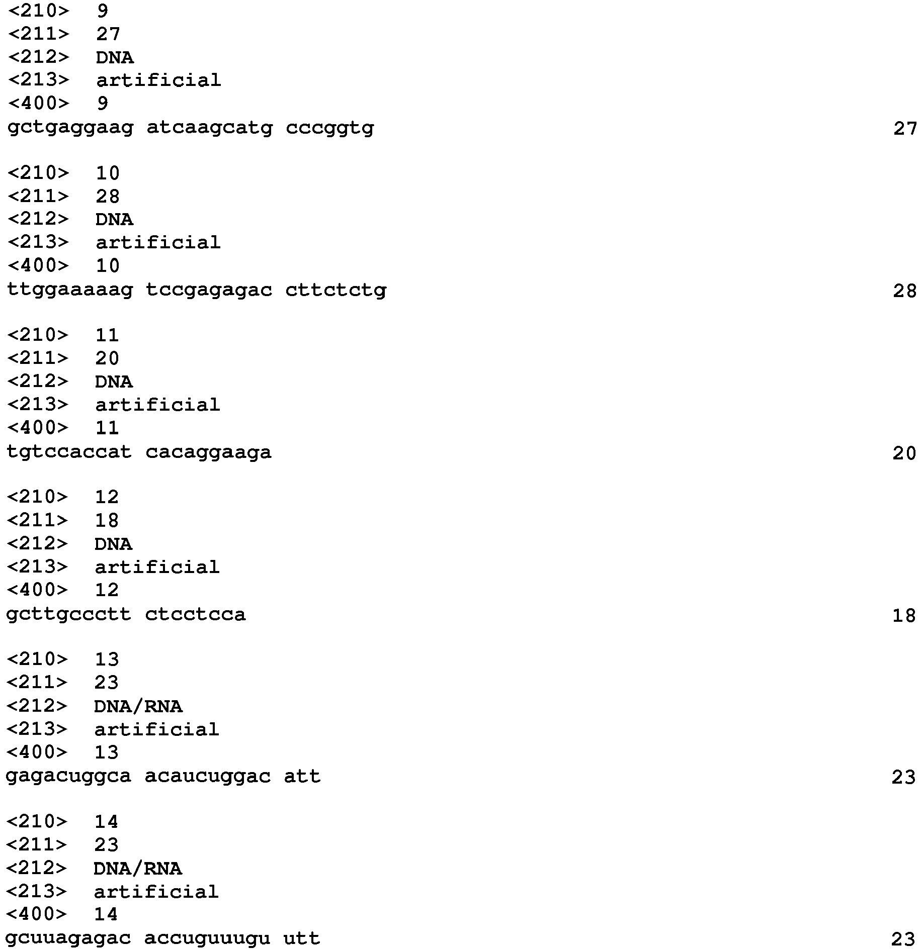
Claims (4)
- A pharmaceutical for pseudo-exercise therapy, comprising an adiponectin receptor 1 agonist compound as an active ingredient.
- A pharmaceutical comprising an adiponectin receptor 1 agonist compound as an active ingredient and changing the physiological state of muscle to a post-exercise physiological state without applying exercise stress.
- The pharmaceutical according to Claim 1 or 2, which is used for a drug therapy substituted for an exercise therapy in the prevention and/or treatment of diabetes.
- A type I muscle fiber enhancer comprising an adiponectin receptor 1 agonist compound as an active ingredient.
Applications Claiming Priority (2)
| Application Number | Priority Date | Filing Date | Title |
|---|---|---|---|
| US31428010P | 2010-03-16 | 2010-03-16 | |
| PCT/JP2011/056043 WO2011115106A1 (en) | 2010-03-16 | 2011-03-15 | Pharmaceutical for pseudo-exercise therapy |
Publications (2)
| Publication Number | Publication Date |
|---|---|
| EP2548580A1 true EP2548580A1 (en) | 2013-01-23 |
| EP2548580A4 EP2548580A4 (en) | 2013-09-11 |
Family
ID=44649188
Family Applications (2)
| Application Number | Title | Priority Date | Filing Date |
|---|---|---|---|
| EP11756291.8A Withdrawn EP2548580A4 (en) | 2010-03-16 | 2011-03-15 | PHARMACEUTICAL PRODUCT FOR PSEUDO-EXERCISE THERAPY |
| EP11756288.4A Withdrawn EP2548969A4 (en) | 2010-03-16 | 2011-03-15 | SCREENING METHOD FOR CANDIDATE AGONIST COMPOUNDS FOR RECEPTOR 1 TO ADIPONECTIN |
Family Applications After (1)
| Application Number | Title | Priority Date | Filing Date |
|---|---|---|---|
| EP11756288.4A Withdrawn EP2548969A4 (en) | 2010-03-16 | 2011-03-15 | SCREENING METHOD FOR CANDIDATE AGONIST COMPOUNDS FOR RECEPTOR 1 TO ADIPONECTIN |
Country Status (4)
| Country | Link |
|---|---|
| US (2) | US20130273567A1 (en) |
| EP (2) | EP2548580A4 (en) |
| JP (2) | JPWO2011115103A1 (en) |
| WO (2) | WO2011115103A1 (en) |
Families Citing this family (2)
| Publication number | Priority date | Publication date | Assignee | Title |
|---|---|---|---|---|
| JP2016027014A (en) * | 2014-06-24 | 2016-02-18 | 花王株式会社 | PPARγ activator |
| KR101838625B1 (en) * | 2016-09-20 | 2018-03-14 | 김브라이언 | Agonist peptide for adiponectin receptor |
Family Cites Families (7)
| Publication number | Priority date | Publication date | Assignee | Title |
|---|---|---|---|---|
| JP3955885B2 (en) * | 2002-12-29 | 2007-08-08 | 日産化学工業株式会社 | Adiponectin receptor and gene encoding the same |
| US7435808B2 (en) * | 2003-06-25 | 2008-10-14 | Bristol-Myers Squibb Company | Polynucleotides encoding novel adiponectin receptor variant, AdipoR2v2 |
| DE602004027746D1 (en) * | 2003-09-27 | 2010-07-29 | Siemens Healthcare Diagnostics | DIAGNOSTICS AND THERAPEUTICS FOR DISEASES ASSOCIATED WITH THE G-PROTEIN-COUPLED RECEPTOR ADIPOR1 (ADIPOR1) |
| US20060069024A1 (en) * | 2004-09-27 | 2006-03-30 | Purdue Research Foundation | Plant PR-5 proteins as mammalian therapeutic agents |
| JP2009523173A (en) * | 2006-01-09 | 2009-06-18 | チルドレンズ ホスピタル メディカル センター | Adiponectin for the treatment of various diseases |
| JP2008109925A (en) * | 2006-10-04 | 2008-05-15 | Japan Science & Technology Agency | Adiponectin receptor gene-deficient non-human animals and uses thereof |
| EP2231600B1 (en) * | 2007-11-16 | 2015-07-29 | Rigel Pharmaceuticals, Inc. | Carboxamide, sulfonamide and amine compounds for metabolic disorders |
-
2011
- 2011-03-15 US US13/634,011 patent/US20130273567A1/en not_active Abandoned
- 2011-03-15 EP EP11756291.8A patent/EP2548580A4/en not_active Withdrawn
- 2011-03-15 US US13/634,015 patent/US20130274185A1/en not_active Abandoned
- 2011-03-15 WO PCT/JP2011/056039 patent/WO2011115103A1/en not_active Ceased
- 2011-03-15 WO PCT/JP2011/056043 patent/WO2011115106A1/en not_active Ceased
- 2011-03-15 EP EP11756288.4A patent/EP2548969A4/en not_active Withdrawn
- 2011-03-15 JP JP2012505695A patent/JPWO2011115103A1/en active Pending
- 2011-03-15 JP JP2012505696A patent/JPWO2011115106A1/en active Pending
Non-Patent Citations (5)
| Title |
|---|
| "Abstracts of the EASD, Stockholm 2010; held in Messe Wien, Vienna, Austria on 1 October 2009", DIABETOLOGIA ; CLINICAL AND EXPERIMENTAL DIABETES AND METABOLISM, SPRINGER, BERLIN, DE, vol. 53, no. 1, 1 October 2009 (2009-10-01), - 6 August 2010 (2010-08-06), pages 1-556, XP019836294, ISSN: 1432-0428 * |
| CHINETTI G ET AL: "Expression of adiponectin receptors in human macrophages and regulation by agonists of the nuclear receptors PPARalpha, PPARgamma, and LXR", BIOCHEMICAL AND BIOPHYSICAL RESEARCH COMMUNICATIONS, ACADEMIC PRESS INC. ORLANDO, FL, US, vol. 314, no. 1, 30 January 2004 (2004-01-30), pages 151-158, XP004483574, ISSN: 0006-291X, DOI: 10.1016/J.BBRC.2003.12.058 * |
| LAFONTAN M ET AL: "Role of adipokines in the control of energy metabolism: focus on adiponectin", CURRENT OPINION IN PHARMACOLOGY, ELSEVIER SCIENCE PUBLISHERS, NL, vol. 6, no. 6, 1 December 2006 (2006-12-01), pages 580-585, XP028058481, ISSN: 1471-4892, DOI: 10.1016/J.COPH.2006.08.002 [retrieved on 2006-12-01] * |
| See also references of WO2011115106A1 * |
| YAMAUCHI T ET AL: "The fat-derived hormone adiponectin reverses insulin resistance associated with both lipoatrophy and obesity", NATURE MEDICINE, NATURE PUBLISHING GROUP, NEW YORK, NY, US, vol. 7, no. 8, 1 August 2001 (2001-08-01), pages 941-946, XP002242792, ISSN: 1078-8956, DOI: 10.1038/90984 * |
Also Published As
| Publication number | Publication date |
|---|---|
| US20130274185A1 (en) | 2013-10-17 |
| WO2011115103A1 (en) | 2011-09-22 |
| WO2011115106A1 (en) | 2011-09-22 |
| JPWO2011115103A1 (en) | 2013-06-27 |
| EP2548969A4 (en) | 2013-08-07 |
| EP2548969A1 (en) | 2013-01-23 |
| JPWO2011115106A1 (en) | 2013-06-27 |
| US20130273567A1 (en) | 2013-10-17 |
| EP2548580A4 (en) | 2013-09-11 |
Similar Documents
| Publication | Publication Date | Title |
|---|---|---|
| Favaro et al. | DRP1-mediated mitochondrial shape controls calcium homeostasis and muscle mass | |
| Li et al. | NAD+-boosting therapy alleviates nonalcoholic fatty liver disease via stimulating a novel exerkine Fndc5/irisin | |
| Brunner et al. | Mechanisms of cardiac arrhythmias and sudden death in transgenic rabbits with long QT syndrome | |
| Kaler | ATP7A-related copper transport diseases—emerging concepts and future trends | |
| EP1420800B1 (en) | Modulation of insulin-regulated aminopeptidase (irap)/angiotensin iv (at 4) receptor activity | |
| Conte et al. | Growth hormone secretagogues prevent dysregulation of skeletal muscle calcium homeostasis in a rat model of cisplatin‐induced cachexia | |
| Mittag et al. | Thyroid hormone is required for hypothalamic neurons regulating cardiovascular functions | |
| JP6533519B2 (en) | Lipid capture in RAS cancer | |
| US20100048944A1 (en) | Interactions of hedgehog and liver x receptor signaling pathways | |
| BRPI0712052A2 (en) | stabilized insulin-like growth factor polypeptides | |
| ES2751391T3 (en) | Metabolic Disorders Treatment Procedures | |
| US12059403B2 (en) | Use of SWELL1 inhibitors and modulators to treat type 2 diabetes and obesity | |
| Jin et al. | WNK1 promotes water homeostasis by acting as a central osmolality sensor for arginine vasopressin release | |
| WO2009026389A2 (en) | Regulating glp-1 and sglt-1 in gastrointestinal cells | |
| WO2007109648A2 (en) | Compositions and methods for modulating store-operated calcium entry | |
| Hoyda et al. | Adiponectin acts in the nucleus of the solitary tract to decrease blood pressure by modulating the excitability of neuropeptide Y neurons | |
| Wahl-Schott et al. | An arginine residue in the pore region is a key determinant of chloride dependence in cardiac pacemaker channels | |
| US9072777B2 (en) | Method for screening substance having proangiogenic effect | |
| ES2617309T3 (en) | Methods and trials of treatment of subjects with removal, mutation or reduced expression of SHANK3 | |
| US20130274185A1 (en) | Pharmaceutical for Pseudo-Exercise Therapy | |
| Pramanik | Galanin stimulates acetylcholine release in the rat striatum | |
| US20230047313A1 (en) | Treating heart disease in muscular dystrophy patients | |
| Lisewski et al. | Hypochlorhydria reduces mortality in heart failure caused by Kcne2 gene deletion | |
| US20160151458A1 (en) | Use Of Unacylated Ghrelin, Fragments And Analogs Thereof As Antioxidant | |
| US20250281572A1 (en) | Compositions and methods for treating and preventing metabolic disorders |
Legal Events
| Date | Code | Title | Description |
|---|---|---|---|
| PUAI | Public reference made under article 153(3) epc to a published international application that has entered the european phase |
Free format text: ORIGINAL CODE: 0009012 |
|
| 17P | Request for examination filed |
Effective date: 20121015 |
|
| AK | Designated contracting states |
Kind code of ref document: A1 Designated state(s): AL AT BE BG CH CY CZ DE DK EE ES FI FR GB GR HR HU IE IS IT LI LT LU LV MC MK MT NL NO PL PT RO RS SE SI SK SM TR |
|
| DAX | Request for extension of the european patent (deleted) | ||
| A4 | Supplementary search report drawn up and despatched |
Effective date: 20130809 |
|
| RIC1 | Information provided on ipc code assigned before grant |
Ipc: C12Q 1/02 20060101AFI20130805BHEP Ipc: A61K 38/22 20060101ALI20130805BHEP Ipc: A61K 45/00 20060101ALI20130805BHEP |
|
| RIN1 | Information on inventor provided before grant (corrected) |
Inventor name: IWABU, MASATO Inventor name: IWABU, MIKI Inventor name: KADOWAKI, TAKASHI Inventor name: YAMAUCHI, TOSHIMASA |
|
| 17Q | First examination report despatched |
Effective date: 20140411 |
|
| STAA | Information on the status of an ep patent application or granted ep patent |
Free format text: STATUS: THE APPLICATION IS DEEMED TO BE WITHDRAWN |
|
| 18D | Application deemed to be withdrawn |
Effective date: 20151013 |