EP2541248B1 - Preparation method of antigen-immobilized immunofluorescence slide, and immunofluorescence slide prepared thereby - Google Patents
Preparation method of antigen-immobilized immunofluorescence slide, and immunofluorescence slide prepared thereby Download PDFInfo
- Publication number
- EP2541248B1 EP2541248B1 EP11747611.9A EP11747611A EP2541248B1 EP 2541248 B1 EP2541248 B1 EP 2541248B1 EP 11747611 A EP11747611 A EP 11747611A EP 2541248 B1 EP2541248 B1 EP 2541248B1
- Authority
- EP
- European Patent Office
- Prior art keywords
- slide
- solution
- antigen
- reactive protein
- protein
- Prior art date
- Legal status (The legal status is an assumption and is not a legal conclusion. Google has not performed a legal analysis and makes no representation as to the accuracy of the status listed.)
- Not-in-force
Links
- 238000010166 immunofluorescence Methods 0.000 title claims description 12
- 238000002360 preparation method Methods 0.000 title description 4
- 102100032752 C-reactive protein Human genes 0.000 claims description 75
- 239000000243 solution Substances 0.000 claims description 71
- 108010074051 C-Reactive Protein Proteins 0.000 claims description 60
- 238000000034 method Methods 0.000 claims description 45
- 238000006243 chemical reaction Methods 0.000 claims description 41
- 239000008055 phosphate buffer solution Substances 0.000 claims description 38
- 102000004169 proteins and genes Human genes 0.000 claims description 21
- 108090000623 proteins and genes Proteins 0.000 claims description 21
- 230000003100 immobilizing effect Effects 0.000 claims description 19
- SJECZPVISLOESU-UHFFFAOYSA-N 3-trimethoxysilylpropan-1-amine Chemical compound CO[Si](OC)(OC)CCCN SJECZPVISLOESU-UHFFFAOYSA-N 0.000 claims description 14
- SXRSQZLOMIGNAQ-UHFFFAOYSA-N Glutaraldehyde Chemical compound O=CCCCC=O SXRSQZLOMIGNAQ-UHFFFAOYSA-N 0.000 claims description 14
- 239000000203 mixture Substances 0.000 claims description 14
- 239000002245 particle Substances 0.000 claims description 9
- 238000002156 mixing Methods 0.000 claims description 6
- 239000002105 nanoparticle Substances 0.000 claims description 6
- 238000002372 labelling Methods 0.000 claims description 5
- 238000000338 in vitro Methods 0.000 claims description 3
- 239000012460 protein solution Substances 0.000 claims 2
- 230000003213 activating effect Effects 0.000 claims 1
- 230000000887 hydrating effect Effects 0.000 claims 1
- 239000000427 antigen Substances 0.000 description 67
- 108091007433 antigens Proteins 0.000 description 67
- 102000036639 antigens Human genes 0.000 description 67
- XLYOFNOQVPJJNP-UHFFFAOYSA-N water Chemical compound O XLYOFNOQVPJJNP-UHFFFAOYSA-N 0.000 description 41
- 239000012153 distilled water Substances 0.000 description 34
- 239000002244 precipitate Substances 0.000 description 21
- 108091003079 Bovine Serum Albumin Proteins 0.000 description 17
- 229940098773 bovine serum albumin Drugs 0.000 description 17
- ZMXDDKWLCZADIW-UHFFFAOYSA-N N,N-Dimethylformamide Chemical compound CN(C)C=O ZMXDDKWLCZADIW-UHFFFAOYSA-N 0.000 description 15
- 239000000463 material Substances 0.000 description 14
- 239000000523 sample Substances 0.000 description 14
- LFQSCWFLJHTTHZ-UHFFFAOYSA-N Ethanol Chemical compound CCO LFQSCWFLJHTTHZ-UHFFFAOYSA-N 0.000 description 13
- OKKJLVBELUTLKV-UHFFFAOYSA-N Methanol Chemical compound OC OKKJLVBELUTLKV-UHFFFAOYSA-N 0.000 description 12
- LOKCTEFSRHRXRJ-UHFFFAOYSA-I dipotassium trisodium dihydrogen phosphate hydrogen phosphate dichloride Chemical compound P(=O)(O)(O)[O-].[K+].P(=O)(O)([O-])[O-].[Na+].[Na+].[Cl-].[K+].[Cl-].[Na+] LOKCTEFSRHRXRJ-UHFFFAOYSA-I 0.000 description 11
- 239000002953 phosphate buffered saline Substances 0.000 description 11
- YBJHBAHKTGYVGT-ZKWXMUAHSA-N (+)-Biotin Chemical compound N1C(=O)N[C@@H]2[C@H](CCCCC(=O)O)SC[C@@H]21 YBJHBAHKTGYVGT-ZKWXMUAHSA-N 0.000 description 10
- -1 3-mercaptopropyltrimethoxysilane-N-gamma- maleimidobutyryloxy succinimide ester Chemical class 0.000 description 10
- 238000011156 evaluation Methods 0.000 description 10
- JJGWLCLUQNFDIS-GTSONSFRSA-M sodium;1-[6-[5-[(3as,4s,6ar)-2-oxo-1,3,3a,4,6,6a-hexahydrothieno[3,4-d]imidazol-4-yl]pentanoylamino]hexanoyloxy]-2,5-dioxopyrrolidine-3-sulfonate Chemical compound [Na+].O=C1C(S(=O)(=O)[O-])CC(=O)N1OC(=O)CCCCCNC(=O)CCCC[C@H]1[C@H]2NC(=O)N[C@H]2CS1 JJGWLCLUQNFDIS-GTSONSFRSA-M 0.000 description 10
- PVGATNRYUYNBHO-UHFFFAOYSA-N (2,5-dioxopyrrolidin-1-yl) 4-(2,5-dioxopyrrol-1-yl)butanoate Chemical compound O=C1CCC(=O)N1OC(=O)CCCN1C(=O)C=CC1=O PVGATNRYUYNBHO-UHFFFAOYSA-N 0.000 description 9
- VYPSYNLAJGMNEJ-UHFFFAOYSA-N Silicium dioxide Chemical compound O=[Si]=O VYPSYNLAJGMNEJ-UHFFFAOYSA-N 0.000 description 9
- 108010090804 Streptavidin Proteins 0.000 description 9
- YXFVVABEGXRONW-UHFFFAOYSA-N Toluene Chemical compound CC1=CC=CC=C1 YXFVVABEGXRONW-UHFFFAOYSA-N 0.000 description 9
- 230000006287 biotinylation Effects 0.000 description 9
- 238000007413 biotinylation Methods 0.000 description 9
- 239000003153 chemical reaction reagent Substances 0.000 description 9
- 230000006870 function Effects 0.000 description 9
- 230000002401 inhibitory effect Effects 0.000 description 9
- 238000005406 washing Methods 0.000 description 9
- CSCPPACGZOOCGX-UHFFFAOYSA-N Acetone Chemical compound CC(C)=O CSCPPACGZOOCGX-UHFFFAOYSA-N 0.000 description 8
- 230000027455 binding Effects 0.000 description 8
- VEXZGXHMUGYJMC-UHFFFAOYSA-N Hydrochloric acid Chemical compound Cl VEXZGXHMUGYJMC-UHFFFAOYSA-N 0.000 description 7
- 241001465754 Metazoa Species 0.000 description 7
- 238000003556 assay Methods 0.000 description 7
- 238000001727 in vivo Methods 0.000 description 7
- 241000252506 Characiformes Species 0.000 description 6
- 238000000018 DNA microarray Methods 0.000 description 6
- 239000000090 biomarker Substances 0.000 description 6
- 239000011545 carbonate/bicarbonate buffer Substances 0.000 description 6
- 230000002860 competitive effect Effects 0.000 description 6
- 235000013305 food Nutrition 0.000 description 6
- 235000013376 functional food Nutrition 0.000 description 6
- 239000002086 nanomaterial Substances 0.000 description 6
- 230000035945 sensitivity Effects 0.000 description 6
- 238000002444 silanisation Methods 0.000 description 6
- 229960002685 biotin Drugs 0.000 description 5
- 235000020958 biotin Nutrition 0.000 description 5
- 239000011616 biotin Substances 0.000 description 5
- 238000005259 measurement Methods 0.000 description 5
- 239000000049 pigment Substances 0.000 description 5
- 230000008569 process Effects 0.000 description 5
- 239000012488 sample solution Substances 0.000 description 5
- UVGHPGOONBRLCX-NJSLBKSFSA-N (2,5-dioxopyrrolidin-1-yl) 6-[5-[(3as,4s,6ar)-2-oxo-1,3,3a,4,6,6a-hexahydrothieno[3,4-d]imidazol-4-yl]pentanoylamino]hexanoate Chemical compound C([C@H]1[C@H]2NC(=O)N[C@H]2CS1)CCCC(=O)NCCCCCC(=O)ON1C(=O)CCC1=O UVGHPGOONBRLCX-NJSLBKSFSA-N 0.000 description 4
- FAPWRFPIFSIZLT-UHFFFAOYSA-M Sodium chloride Chemical compound [Na+].[Cl-] FAPWRFPIFSIZLT-UHFFFAOYSA-M 0.000 description 4
- QAOWNCQODCNURD-UHFFFAOYSA-N Sulfuric acid Chemical compound OS(O)(=O)=O QAOWNCQODCNURD-UHFFFAOYSA-N 0.000 description 4
- 229910052782 aluminium Inorganic materials 0.000 description 4
- XAGFODPZIPBFFR-UHFFFAOYSA-N aluminium Chemical compound [Al] XAGFODPZIPBFFR-UHFFFAOYSA-N 0.000 description 4
- 230000000903 blocking effect Effects 0.000 description 4
- 239000007795 chemical reaction product Substances 0.000 description 4
- 238000004140 cleaning Methods 0.000 description 4
- 239000011248 coating agent Substances 0.000 description 4
- 238000000576 coating method Methods 0.000 description 4
- 238000012875 competitive assay Methods 0.000 description 4
- 238000001514 detection method Methods 0.000 description 4
- 230000000694 effects Effects 0.000 description 4
- 238000002474 experimental method Methods 0.000 description 4
- 239000011888 foil Substances 0.000 description 4
- 239000011521 glass Substances 0.000 description 4
- 239000000017 hydrogel Substances 0.000 description 4
- 239000011541 reaction mixture Substances 0.000 description 4
- 108010071390 Serum Albumin Proteins 0.000 description 3
- 102000007562 Serum Albumin Human genes 0.000 description 3
- 210000004369 blood Anatomy 0.000 description 3
- 239000008280 blood Substances 0.000 description 3
- 230000008859 change Effects 0.000 description 3
- 230000036541 health Effects 0.000 description 3
- 230000036046 immunoreaction Effects 0.000 description 3
- 239000012528 membrane Substances 0.000 description 3
- 239000011259 mixed solution Substances 0.000 description 3
- 239000003960 organic solvent Substances 0.000 description 3
- 239000000377 silicon dioxide Substances 0.000 description 3
- 238000003756 stirring Methods 0.000 description 3
- UUEWCQRISZBELL-UHFFFAOYSA-N 3-trimethoxysilylpropane-1-thiol Chemical compound CO[Si](OC)(OC)CCCS UUEWCQRISZBELL-UHFFFAOYSA-N 0.000 description 2
- 238000002965 ELISA Methods 0.000 description 2
- 108091006905 Human Serum Albumin Proteins 0.000 description 2
- 102000008100 Human Serum Albumin Human genes 0.000 description 2
- 206010020772 Hypertension Diseases 0.000 description 2
- 102000007330 LDL Lipoproteins Human genes 0.000 description 2
- 108010007622 LDL Lipoproteins Proteins 0.000 description 2
- 241000124008 Mammalia Species 0.000 description 2
- 206010035226 Plasma cell myeloma Diseases 0.000 description 2
- 150000001412 amines Chemical class 0.000 description 2
- 238000011088 calibration curve Methods 0.000 description 2
- 150000007942 carboxylates Chemical class 0.000 description 2
- 230000009084 cardiovascular function Effects 0.000 description 2
- 239000000919 ceramic Substances 0.000 description 2
- 150000001875 compounds Chemical class 0.000 description 2
- 238000001816 cooling Methods 0.000 description 2
- 239000011258 core-shell material Substances 0.000 description 2
- 208000029078 coronary artery disease Diseases 0.000 description 2
- 201000010099 disease Diseases 0.000 description 2
- 230000006806 disease prevention Effects 0.000 description 2
- 208000037265 diseases, disorders, signs and symptoms Diseases 0.000 description 2
- 125000000524 functional group Chemical group 0.000 description 2
- 238000013537 high throughput screening Methods 0.000 description 2
- 238000007654 immersion Methods 0.000 description 2
- 239000012535 impurity Substances 0.000 description 2
- 210000004185 liver Anatomy 0.000 description 2
- 238000004519 manufacturing process Methods 0.000 description 2
- 229910052751 metal Inorganic materials 0.000 description 2
- 239000002184 metal Substances 0.000 description 2
- 230000004048 modification Effects 0.000 description 2
- 238000012986 modification Methods 0.000 description 2
- 201000000050 myeloid neoplasm Diseases 0.000 description 2
- 229920001223 polyethylene glycol Polymers 0.000 description 2
- 230000002265 prevention Effects 0.000 description 2
- 238000003380 quartz crystal microbalance Methods 0.000 description 2
- 238000011160 research Methods 0.000 description 2
- 239000010703 silicon Substances 0.000 description 2
- 229910052710 silicon Inorganic materials 0.000 description 2
- 239000011780 sodium chloride Substances 0.000 description 2
- 238000001179 sorption measurement Methods 0.000 description 2
- 125000004079 stearyl group Chemical group [H]C([*])([H])C([H])([H])C([H])([H])C([H])([H])C([H])([H])C([H])([H])C([H])([H])C([H])([H])C([H])([H])C([H])([H])C([H])([H])C([H])([H])C([H])([H])C([H])([H])C([H])([H])C([H])([H])C([H])([H])C([H])([H])[H] 0.000 description 2
- 239000000758 substrate Substances 0.000 description 2
- 239000006228 supernatant Substances 0.000 description 2
- 238000012360 testing method Methods 0.000 description 2
- CXYYHBMOVJJZTD-UHFFFAOYSA-N (2,5-dioxopyrrolidin-1-yl) 3',6'-bis(dimethylamino)-3-oxospiro[2-benzofuran-1,9'-xanthene]-5-carboxylate Chemical compound C=1C(N(C)C)=CC=C2C=1OC1=CC(N(C)C)=CC=C1C2(C1=CC=2)OC(=O)C1=CC=2C(=O)ON1C(=O)CCC1=O CXYYHBMOVJJZTD-UHFFFAOYSA-N 0.000 description 1
- LOGFVTREOLYCPF-KXNHARMFSA-N (2s,3r)-2-[[(2r)-1-[(2s)-2,6-diaminohexanoyl]pyrrolidine-2-carbonyl]amino]-3-hydroxybutanoic acid Chemical compound C[C@@H](O)[C@@H](C(O)=O)NC(=O)[C@H]1CCCN1C(=O)[C@@H](N)CCCCN LOGFVTREOLYCPF-KXNHARMFSA-N 0.000 description 1
- WYTZZXDRDKSJID-UHFFFAOYSA-N (3-aminopropyl)triethoxysilane Chemical compound CCO[Si](OCC)(OCC)CCCN WYTZZXDRDKSJID-UHFFFAOYSA-N 0.000 description 1
- CUKWUWBLQQDQAC-VEQWQPCFSA-N (3s)-3-amino-4-[[(2s)-1-[[(2s)-1-[[(2s)-1-[[(2s,3s)-1-[[(2s)-1-[(2s)-2-[[(1s)-1-carboxyethyl]carbamoyl]pyrrolidin-1-yl]-3-(1h-imidazol-5-yl)-1-oxopropan-2-yl]amino]-3-methyl-1-oxopentan-2-yl]amino]-3-(4-hydroxyphenyl)-1-oxopropan-2-yl]amino]-3-methyl-1-ox Chemical compound C([C@@H](C(=O)N[C@@H]([C@@H](C)CC)C(=O)N[C@@H](CC=1NC=NC=1)C(=O)N1[C@@H](CCC1)C(=O)N[C@@H](C)C(O)=O)NC(=O)[C@@H](NC(=O)[C@H](CCCN=C(N)N)NC(=O)[C@@H](N)CC(O)=O)C(C)C)C1=CC=C(O)C=C1 CUKWUWBLQQDQAC-VEQWQPCFSA-N 0.000 description 1
- ZHWVFYLCRAMYAW-UHFFFAOYSA-N 2-(4-carboxypyridin-2-yl)pyridine-4-carboxylic acid 2-pyridin-2-ylpyridine ruthenium Chemical compound [Ru].N1=CC=CC=C1C1=CC=CC=N1.N1=CC=CC=C1C1=CC=CC=N1.OC(=O)C1=CC=NC(C=2N=CC=C(C=2)C(O)=O)=C1 ZHWVFYLCRAMYAW-UHFFFAOYSA-N 0.000 description 1
- ZJJNYYDFJCYHTM-UHFFFAOYSA-N 2-(4-methylpyridin-2-yl)pyridine-4-carboxylic acid 2-pyridin-2-ylpyridine ruthenium Chemical compound [Ru].N1=CC=CC=C1C1=CC=CC=N1.N1=CC=CC=C1C1=CC=CC=N1.CC1=CC=NC(C=2N=CC=C(C=2)C(O)=O)=C1 ZJJNYYDFJCYHTM-UHFFFAOYSA-N 0.000 description 1
- IJSMFQNTEUNRPY-UHFFFAOYSA-N 2-[3-(dimethylamino)-6-dimethylazaniumylidenexanthen-9-yl]-5-isothiocyanatobenzoate Chemical compound C=12C=CC(=[N+](C)C)C=C2OC2=CC(N(C)C)=CC=C2C=1C1=CC=C(N=C=S)C=C1C([O-])=O IJSMFQNTEUNRPY-UHFFFAOYSA-N 0.000 description 1
- 102000005862 Angiotensin II Human genes 0.000 description 1
- 101800000733 Angiotensin-2 Proteins 0.000 description 1
- 108091023037 Aptamer Proteins 0.000 description 1
- 241000283707 Capra Species 0.000 description 1
- OKTJSMMVPCPJKN-UHFFFAOYSA-N Carbon Chemical compound [C] OKTJSMMVPCPJKN-UHFFFAOYSA-N 0.000 description 1
- 208000024172 Cardiovascular disease Diseases 0.000 description 1
- 241000282693 Cercopithecidae Species 0.000 description 1
- 108090000790 Enzymes Proteins 0.000 description 1
- 102000004190 Enzymes Human genes 0.000 description 1
- 108010049003 Fibrinogen Proteins 0.000 description 1
- 102000008946 Fibrinogen Human genes 0.000 description 1
- 206010061218 Inflammation Diseases 0.000 description 1
- 102000003777 Interleukin-1 beta Human genes 0.000 description 1
- 108090000193 Interleukin-1 beta Proteins 0.000 description 1
- 102000004889 Interleukin-6 Human genes 0.000 description 1
- 108090001005 Interleukin-6 Proteins 0.000 description 1
- PEEHTFAAVSWFBL-UHFFFAOYSA-N Maleimide Chemical compound O=C1NC(=O)C=C1 PEEHTFAAVSWFBL-UHFFFAOYSA-N 0.000 description 1
- 206010028980 Neoplasm Diseases 0.000 description 1
- 208000008589 Obesity Diseases 0.000 description 1
- 241000283973 Oryctolagus cuniculus Species 0.000 description 1
- 239000002202 Polyethylene glycol Substances 0.000 description 1
- 101000940868 Rattus norvegicus Cysteine and glycine-rich protein 1 Proteins 0.000 description 1
- 230000004913 activation Effects 0.000 description 1
- 238000001994 activation Methods 0.000 description 1
- 230000032683 aging Effects 0.000 description 1
- 125000003277 amino group Chemical group 0.000 description 1
- 238000004458 analytical method Methods 0.000 description 1
- 229950006323 angiotensin ii Drugs 0.000 description 1
- 238000002820 assay format Methods 0.000 description 1
- 210000003719 b-lymphocyte Anatomy 0.000 description 1
- 230000036772 blood pressure Effects 0.000 description 1
- 210000001124 body fluid Anatomy 0.000 description 1
- 239000010839 body fluid Substances 0.000 description 1
- 230000037396 body weight Effects 0.000 description 1
- 201000011510 cancer Diseases 0.000 description 1
- 239000002041 carbon nanotube Substances 0.000 description 1
- 229910021393 carbon nanotube Inorganic materials 0.000 description 1
- 210000004027 cell Anatomy 0.000 description 1
- 239000000084 colloidal system Substances 0.000 description 1
- 238000004040 coloring Methods 0.000 description 1
- 238000004891 communication Methods 0.000 description 1
- 239000013078 crystal Substances 0.000 description 1
- 230000003247 decreasing effect Effects 0.000 description 1
- 230000001419 dependent effect Effects 0.000 description 1
- 238000003745 diagnosis Methods 0.000 description 1
- 238000010586 diagram Methods 0.000 description 1
- 238000000502 dialysis Methods 0.000 description 1
- WGNGRGQLNAWTJI-UHFFFAOYSA-L dichlororuthenium;1,10-phenanthroline;hydrate Chemical compound O.[Cl-].[Cl-].[Ru+2].C1=CN=C2C3=NC=CC=C3C=CC2=C1.C1=CN=C2C3=NC=CC=C3C=CC2=C1.C1=CN=C2C3=NC=CC=C3C=CC2=C1 WGNGRGQLNAWTJI-UHFFFAOYSA-L 0.000 description 1
- KZNICNPSHKQLFF-UHFFFAOYSA-N dihydromaleimide Natural products O=C1CCC(=O)N1 KZNICNPSHKQLFF-UHFFFAOYSA-N 0.000 description 1
- 238000010790 dilution Methods 0.000 description 1
- 239000012895 dilution Substances 0.000 description 1
- 239000012154 double-distilled water Substances 0.000 description 1
- 229940079593 drug Drugs 0.000 description 1
- 239000003814 drug Substances 0.000 description 1
- 238000001035 drying Methods 0.000 description 1
- 239000000975 dye Substances 0.000 description 1
- 230000002526 effect on cardiovascular system Effects 0.000 description 1
- 150000002148 esters Chemical class 0.000 description 1
- 239000000284 extract Substances 0.000 description 1
- 229940012952 fibrinogen Drugs 0.000 description 1
- GNBHRKFJIUUOQI-UHFFFAOYSA-N fluorescein Chemical compound O1C(=O)C2=CC=CC=C2C21C1=CC=C(O)C=C1OC1=CC(O)=CC=C21 GNBHRKFJIUUOQI-UHFFFAOYSA-N 0.000 description 1
- MHMNJMPURVTYEJ-UHFFFAOYSA-N fluorescein-5-isothiocyanate Chemical compound O1C(=O)C2=CC(N=C=S)=CC=C2C21C1=CC=C(O)C=C1OC1=CC(O)=CC=C21 MHMNJMPURVTYEJ-UHFFFAOYSA-N 0.000 description 1
- 239000007850 fluorescent dye Substances 0.000 description 1
- 230000004927 fusion Effects 0.000 description 1
- 210000001035 gastrointestinal tract Anatomy 0.000 description 1
- 238000004128 high performance liquid chromatography Methods 0.000 description 1
- HNDVDQJCIGZPNO-UHFFFAOYSA-N histidine Natural products OC(=O)C(N)CC1=CN=CN1 HNDVDQJCIGZPNO-UHFFFAOYSA-N 0.000 description 1
- 238000009396 hybridization Methods 0.000 description 1
- 210000004408 hybridoma Anatomy 0.000 description 1
- 230000036571 hydration Effects 0.000 description 1
- 238000006703 hydration reaction Methods 0.000 description 1
- 230000004054 inflammatory process Effects 0.000 description 1
- 229940100601 interleukin-6 Drugs 0.000 description 1
- 238000000691 measurement method Methods 0.000 description 1
- 230000004060 metabolic process Effects 0.000 description 1
- 238000002493 microarray Methods 0.000 description 1
- 239000005445 natural material Substances 0.000 description 1
- 235000020824 obesity Nutrition 0.000 description 1
- 210000000056 organ Anatomy 0.000 description 1
- UEZVMMHDMIWARA-UHFFFAOYSA-M phosphonate Chemical compound [O-]P(=O)=O UEZVMMHDMIWARA-UHFFFAOYSA-M 0.000 description 1
- 210000002381 plasma Anatomy 0.000 description 1
- BDERNNFJNOPAEC-UHFFFAOYSA-N propan-1-ol Chemical compound CCCO BDERNNFJNOPAEC-UHFFFAOYSA-N 0.000 description 1
- 239000002096 quantum dot Substances 0.000 description 1
- 239000011535 reaction buffer Substances 0.000 description 1
- 230000009257 reactivity Effects 0.000 description 1
- 230000002829 reductive effect Effects 0.000 description 1
- 239000012088 reference solution Substances 0.000 description 1
- 235000021067 refined food Nutrition 0.000 description 1
- 239000011369 resultant mixture Substances 0.000 description 1
- PYWVYCXTNDRMGF-UHFFFAOYSA-N rhodamine B Chemical compound [Cl-].C=12C=CC(=[N+](CC)CC)C=C2OC2=CC(N(CC)CC)=CC=C2C=1C1=CC=CC=C1C(O)=O PYWVYCXTNDRMGF-UHFFFAOYSA-N 0.000 description 1
- 229940043267 rhodamine b Drugs 0.000 description 1
- 238000009666 routine test Methods 0.000 description 1
- 210000003296 saliva Anatomy 0.000 description 1
- 239000002094 self assembled monolayer Substances 0.000 description 1
- 239000013545 self-assembled monolayer Substances 0.000 description 1
- 239000004065 semiconductor Substances 0.000 description 1
- 210000002966 serum Anatomy 0.000 description 1
- 239000007787 solid Substances 0.000 description 1
- 230000009870 specific binding Effects 0.000 description 1
- 238000007447 staining method Methods 0.000 description 1
- 239000000126 substance Substances 0.000 description 1
- 229960002317 succinimide Drugs 0.000 description 1
- 230000001629 suppression Effects 0.000 description 1
- 239000000725 suspension Substances 0.000 description 1
- 208000024891 symptom Diseases 0.000 description 1
- 125000003396 thiol group Chemical group [H]S* 0.000 description 1
- 210000001519 tissue Anatomy 0.000 description 1
Images



Classifications
-
- G—PHYSICS
- G01—MEASURING; TESTING
- G01N—INVESTIGATING OR ANALYSING MATERIALS BY DETERMINING THEIR CHEMICAL OR PHYSICAL PROPERTIES
- G01N33/00—Investigating or analysing materials by specific methods not covered by groups G01N1/00 - G01N31/00
- G01N33/48—Biological material, e.g. blood, urine; Haemocytometers
- G01N33/50—Chemical analysis of biological material, e.g. blood, urine; Testing involving biospecific ligand binding methods; Immunological testing
- G01N33/53—Immunoassay; Biospecific binding assay; Materials therefor
- G01N33/543—Immunoassay; Biospecific binding assay; Materials therefor with an insoluble carrier for immobilising immunochemicals
- G01N33/54366—Apparatus specially adapted for solid-phase testing
-
- G—PHYSICS
- G01—MEASURING; TESTING
- G01N—INVESTIGATING OR ANALYSING MATERIALS BY DETERMINING THEIR CHEMICAL OR PHYSICAL PROPERTIES
- G01N2333/00—Assays involving biological materials from specific organisms or of a specific nature
- G01N2333/435—Assays involving biological materials from specific organisms or of a specific nature from animals; from humans
- G01N2333/46—Assays involving biological materials from specific organisms or of a specific nature from animals; from humans from vertebrates
- G01N2333/47—Assays involving proteins of known structure or function as defined in the subgroups
- G01N2333/4701—Details
- G01N2333/4737—C-reactive protein
Definitions
- the present invention relates to a method of preparing an antigen-immobilized immuno-fluorescence slide including: immobilizing a C-reactive protein on a slide to prepare a protein chip, mixing an antibody that specifically binds to a target protein, with streptavidin, to label the antibody with a fluorescent nanoparticle, immune-reacting the antibody by competitive mixing, assaying with a fluorescence camera, and an immuno-fluorescence slide made by using the method.
- Biochips include various kinds of probes immobilized on a unit area of a surface of a solid support, and when such biochips are used, only with a small amount of sample, disease diagnosis, high-throughput screening (HTS), and enzyme activity measuring may be easily performed in a large scale.
- sample disease diagnosis, high-throughput screening (HTS), and enzyme activity measuring may be easily performed in a large scale.
- HTS high-throughput screening
- a 3-dimensional (3D) immobilization method was developed ( Gill and Ballesteros, Trends in Biotechnology 18:282, 2000 ).
- Examples of such a 3D immobilization method are a method using a Hydrogel ® coating slide of Packard Bioscience, a method using a polyethyleneglycol-based hydrogel of Biocept Company, and a method using solgel of LG Chemical Co., Ltd, and the like.
- a slide for use in an immuno-sensor is selected from a group consisting of glass, silicon, hydrogel, metal, ceramic, and a porous membrane.
- an antibody or antigen probe-plate made by immobilizing an antibody or an antigen on a substrate, which is based on a specific reactivity of an antigen or an antibody, is widely used for detection.
- Representative kits for such use are a rapid test kit, a routine test kit, and a biochip kit.
- kits are used to detect a target antigen or target antibody corresponding to an immobilized antibody probe or antigen probe.
- reaction groups are assembled onto a single probe-plate, and each of those reaction groups includes a test strip for the detection of an antigen or antibody.
- Food-function evaluating includes in vitro function evaluation, in vivo function evaluation using a lab animal, such as a rat, and clinical trials, performed based on those six function types, and in terms of the food industry, a demand for developing an in vivo function detection techniques using a lab animal for effectively and rapidly performing function evaluation of food materials and functional good increases.
- a target function such as a cardiovascular function
- a target function needs to be scientifically proved, and it cannot be stressed too much the importance of food-function evaluation ( Kim et al., Detecting C-reactive protein by using direct binding quartz crystal microbalance immunosensor, Journal of The Korean Society for Biotechnology and Bioengineering, Vol. 22, p. 443, 2007 ).
- in-vivo function evaluation using lab animals which is performed prior to clinical trials, is to evaluate change in bodily elements, such as body weight, blood pressure, and shape of organs.
- this method causes non-uniform results, and also, requires variability of evaluation protocols, skilled experts, and high-cost assay instruments.
- a uniform in vivo food-function evaluation method is to measure an increase or decrease of a particular biomarker protein related to a particular disease or metabolism symptoms in a lab animal, such as a rat.
- a lab animal such as a rat.
- what contains a food that is desired to obtain recognition as a functional food is administered to the lab animal.
- Korea Registered Patent 10-921237 discloses, as an in vivo function evaluation method for rapidly detecting a cardiovascular function of a food material and a functional food, a method of detecting with high sensitivity a C-reactive protein ( Biosensors and Bioelectronics, Vol. 19, p. 1193, 2004 ; Clinical Chemistry, Vol. 47, p. 403, 2001 ; New England Journal of Medicine, Vol. 340, p. 448, 1999 ; Biochemical Journal, Vol. 327, p.
- the C-reactive protein is a pentameric protein that is known as a major biomarker of coronary artery disease, hypertension, or the like, that is synthesized by induction-stimulation by interleukin-6 and interleukin-1 ⁇ in the liver of a mammal, and that has a molecular mass of about 118 killodalton (kDa).
- This method compared to an enzyme-linked immunosorbent assay (ELISA) ( American Journal of C ardiology, Vol. 1, p. 155, 2005 ; American Journal of Veterinary Research, Vol. 1, p. 62, 2005 ; Journal of Clinical Laboratory Analysis, Vol. 18, p. 280, 2004 ) for evaluating a C-reactive protein, is easily used without measurement disturbances by a coloring material, and also, has better or equal measurement sensitivity than typical other sensor measurement methods ( Biosensors and Bioelectronics, Vol. 22, p. 973, 2007 ; Biosensors and Bioelectronics, Vol. 21, p. 1987, 2006 ; Biosensors and Bioelectronics, Vol. 21, p. 1631, 2006 ; Biosensors and Bioelectronics, Vol. 21, p. 1141, 2006 ; Analytical Biochemistry, Vol. 328, p. 210, 2004 ).
- ELISA enzyme-linked immunosorbent assay
- KR 2009 0073669 A describes a crystal oscillator immune sensor.
- HAI LI ET AL: LANGMUIR, vol. 26, no. 8, pages 5603-5609 relates to aminosilane micropatterns on hydroxyl-terminated substrates.
- KIM N S ET AL: FOOD ENGINEERING PROGRESS, vol. 14, no. 1, pages 21-26 relates to glass slide-based immunosensing.
- JUNG Y ET AL.: ANAL BIOCHEM., vol. 374, no. 1, pages 99-105 describes a strategy to immobilize an antibody on various sensor surfaces.
- the present invention provides a dispensable immunosensor slide with high sensor sensitivity, which allows a user, without a complicated apparatus, such as a biochip arrayer or a micro-array scanner, to immobilize a certain volume of an antigen on a slide with a micro dispensing pipette in manufacturing or measuring a biochip to perform an immunoreaction between a target protein and an zygocyte of an antibody and a fluorescent nanoparticle, or an immunoreaction between an immobilized antigen and a mixture including a fluorescent nano-probe and a particular target molecule on an antigen coating site of the immuno-fluorescence slide, and then, fluorescent intensity or the number of fluorescence particles of the nano-probe that specifically binds to the surface of the slide by the immunoreaction is measured using a fluorescence microscope so as to identify the concentration of a target molecule, such as a body indicator.
- a fluorescence microscope so as to identify the concentration of a target molecule, such as a body indicator.
- the present invention relates to a method in accordance with claims 1, 3 and 4 and to a C-reactive-protein-immobilized immune-fluorescence slide in accordance with claim 2.
- a target protein (antigen) is immobilized on a slide, an antibody that specifically binds to the antigen is labeled with a fluorescent material, a target protein collected from a specimen is mixed with the fluorescence-labeled antibody, the mixture is reacted with the immobilized antigen site on the slide, unreacted antibody and antigen are washed with distilled water, the slide is placed in an incubator for drying, and fluorescent intensity or the number of fluorescence particles is measured by using a fluorescence microscope.
- an indirect-competitive assay format is used.
- the present invention may also be embodied by using a direct-competitive assay format or a sandwich assay format.
- An immunosensor slide used in embodiments of the present invention may be selected from the group consisting of glass, silicon, hydrogel, metal, ceramic, and a porous membrane, and may have a size of 20 to 40 X 70 to 80 mm.
- CRP C-reactive protein
- a C-reactive protein antibody may be any one of antibodies which are prepared by using C-reactive proteins induced from a mammal, such as a rat, a mouse, a rabbit, a monkey, or a human being.
- a sample collected from a specimen may be any one of a reference solution in which a C-reactive protein is dissolved in a reaction buffer solution, and a tissue extract collected from blood, serum, blood plasma, saliva, body fluid, and the liver.
- any one of a 1-5% bovine serum albumin (BSA) solution, a 1-5% rat serum albumin (RSA) solution, a 1-5% human serum albumin (HSA) solution, a 1-5% mouse serum albumin (MSA) solution, and a 1-5% goat serum albumin (GSA) solution any one of a 1-5% bovine serum albumin (BSA) solution, a 1-5% rat serum albumin (RSA) solution, a 1-5% human serum albumin (HSA) solution, a 1-5% mouse serum albumin (MSA) solution, and a 1-5% goat serum albumin (GSA) solution.
- BSA bovine serum albumin
- RSA rat serum albumin
- HSA human serum albumin
- MSA mouse serum albumin
- GSA goat serum albumin
- Immobilizing of a C-reactive protein as an antigen on a slide may be performed by the following method.
- the immobilizing of an antigen may be performed by using 3-aminopropyltrimethoxysilane (APTMS).
- APTMS 3-aminopropyltrimethoxysilane
- a modified slide is prepared using APTMS.
- the surface-washed slide was placed in a petri dish containing a solution in which 5 to 15% of APTMS is dissolved in acetone, and then, a reaction is performed at room temperature for 30 to 90 minutes. Front and rear sides of the slide are sequentially washed three to four times with acetone, a 30-70 mM phosphate buffer solution (pH 6.5-7.8), and a distilled water, and then, the slide is immersed in a beaker containing distilled water for 1 to 60 minutes.
- the slide is dried on kimwipes at room temperature for 20 to 120 minutes, heat treated at a temperature of 30 to 150°C in a convection oven for 3 to 7 hours, is left to sit for cooling, and then preserved in a dried state in a desiccator until use.
- the slide modified with APTMS is hydrated by immersion in distilled water for 10 to 20 minutes.
- the immersed slide is placed in a petri dish containing a solution including 1 to 4% glutaraldehyde and a reaction is performed for 30 to 90 minutes to activate the slide so that a protein is bindable to the surface of the slide.
- Front and rear surfaces of the activated slide are washed three to four times by using a 30-70 mM phosphate buffer solution (pH 6.5-7.8).
- the slide is immersed in a beaker containing distilled water for 1 to 60 minutes.
- the slide is taken away from the beaker and placed on kimwipes and dried for 20 to 120 minutes at room temperature.
- the glutaraldehyde used above is prepared by adding 1 to 4 ml of 30-70% glutaraldehyde to 46 to 49 ml of distilled water, and 30 to 70 ml of the prepared solution is used.
- a target protein for example, a C-reactive protein
- a C-reactive protein first, 0.01 to 0.5 mg/ml of a C-reactive protein is dissolved in a 30-70 mM phosphate buffer solution (pH 6.5-7.8) to prepare an antigen solution for immobilization.
- a petri dish containing the glutaraldehyde-activated slide is placed on a spotting guide having a lattice shape, and spotting is performed with 1 to 100 ⁇ l of the previously prepared antigen solution on a portion of the slide corresponding to a spot point on the spotting guide, and then, the resultant structure is covered with a cover and a reaction is performed for 1 to 6 hours to immobilize the antigen.
- a second immobilizing method which does not form part of the present invention, in which the immobilizing of an antigen may be performed by using 3-mercaptopropyltrimethoxysilane-N-gamma- maleimidobutyryloxy succinimide ester (MPTMS-GMBS).
- MPTMS-GMBS 3-mercaptopropyltrimethoxysilane-N-gamma- maleimidobutyryloxy succinimide ester
- an MPTMS solution in which 1-3% MPTMS is dissolved in an organic solvent solution selected from the group consisting of toluene, dimethyl foramide, and acetone is prepared.
- the slide is placed in a petri dish containing the solution and a reaction is performed at room temperature for 1 to 3 hours. Front and rear surfaces of the slide are washed three to four times with the same organic solvent as used in preparing the MPTMS solution, and the slide is placed on kimwipes and dried for 20 to 120 minutes at room temperature.
- an 1 to 3 mM GMBS solution is prepared as described below. 14 to 42 mg of a GMBS sample is dissolved in 100 ⁇ l of N,N-dimethylformamide (DMF; Sigma-Aldrich Company, USA), and then, 49.9 ml of ethanol is added thereto.
- the slide subjected to the silanization is placed in a petri dish containing the GMBS solution and a reaction is performed at room temperature for 30 to 90 minutes. Front and rear surfaces of the slide are washed three to four times with distilled water and a 30-70 mM phosphate buffer solution (pH 6.5-7.8). Then, the slide is placed on kimwipes and dried for 20 to 120 minutes at room temperature.
- a CRP antigen is immobilized in the following manner. First, a C-reactive protein is dissolved at a concentration of 0.01 to 0.5 mg/ml in a 30-70 mM phosphate buffer solution (pH 6.5-7.8) to prepare an antigen solution for immobilization. A petri dish containing a GMBS-activated slide is placed on a spotting guide, and then, 1 to 100 ⁇ l of the previously prepared antigen solution is spotted on a portion of the slide corresponding to a spot point of the spotting guide and then, the resultant structure is covered with a cover and a reaction is performed for 1 to 6 hours to immobilize the antigen.
- a C-reactive protein is dissolved at a concentration of 0.01 to 0.5 mg/ml in a 30-70 mM phosphate buffer solution (pH 6.5-7.8) to prepare an antigen solution for immobilization.
- a petri dish containing a GMBS-activated slide is placed on a spotting guide, and then
- front and rear sides of the slide are washed three to four times with a 30-70 mM phosphate buffer solution (pH 6.5-7.8), and the slide is immersed in a beaker containing distilled water for 1 to 60 minutes.
- a 30-70 mM phosphate buffer solution pH 6.5-7.8
- the slide is placed on a small plastic petri dish containing a 1 to 5% BSA solution and the resultant structure is covered with a cover and a reaction is performed for 1 to 5 hours.
- Front and rear surfaces of the slide are washed three to four times with the phosphate buffer solution described above. Then, the slide is immersed in a beaker containing distilled water for 1 to 60 minutes, and then, placed on kimwipes and dried for 20 to 120 minutes at room temperature.
- an antibody that specifically binds to an antigen immobilized on the slide is labeled.
- the labeling of an antibody may be performed by using a fluorescent staining method with high sensitivity and reproducibility, and to do this, according to the present invention, a fluorescent silica nanoparticle (hereinafter referred to as "FSNP") is used.
- FSNP fluorescent silica nanoparticle
- FSNP which is used to label an antibody that specifically binds to an antigen
- FSNP may be prepared by using a dye entrapment method in which an organic fluorescence pigment is included in a silica structure, a core shell method in which an active organic fluorescence pigment is covalently bonded to an amino silane compound and the result structure is included in a silica structure, or an inverted core shell method in which a organic fluorescence pigment is included in a silica structure and the formed particle is reacted with an organosilane compound to introduce a functional group to obtain functionality.
- examples of such an organic fluorescence pigment are dichlorotris(1,10-phenanthroline)ruthenium(II) ⁇ hydrate, fluorescein, rhodamine B, and 5(6)-Carboxytetramethylrhodamine
- examples of an active organic fluorescence pigment are fluorescein isothiocyanate, 5(6)-carboxytetramethylrhodamine N-succinimidyl ester, tetramethylrhodamine 5-isothiocyanate, bis(2,2'-bipyridine)-4'-methyl-4-carboxybipyridine-ruthenium N-succinimidyl ester-bis(hexafluorophosphate), and bis(2,2'-bipyridine)-4,4'-dicarboxybipyridine ruthenium di(N-Succinimidyl ester)bis(hexafluorophosphate), an example of an amino silane compound is (3-aminopropyl este
- FSNP needs to be modified with streptavidin (SA) which specifically binds to biotin. This modification is performed to bind SA-bonded FSNP, which is to be described later, to a biotinylated antibody.
- SA streptavidin
- MPTMS (3-mercaptopropyl)trimethoxy silane
- FSNP 1 to 5 mg of FSNP and 5 to 15 ml of an alcohol, such as ethanol, methanol, or propanol, are added to a cap tube having a capacity of 10 to 70 ml and the mixture is shaken well and then, sonicated to completely dissolve FSNP in the alcohol for 10 to 30 minutes. Thereafter, 30-70 to 200 ⁇ l of MPTMS is added to the cap tube, and while the resultant mixture is stirred at a temperature of room temperature at a low speed of 30-200 rpm, a reaction is performed for 2 to 6 hours.
- an alcohol such as ethanol, methanol, or propanol
- the reaction mixture is centrifuged at a temperature of 3 to 10 °C for 20 to 40 minutes at a speed of 7000-17000 rpm and the obtained precipitate is washed one to five times by using the same alcohol as used for the reaction, and following each washing, centrifuging is performed under the same conditions as described above to obtain a precipitate.
- a cap is separated from the tube and the tube is softly covered with an aluminum foil and dried at room temperature.
- the precipitate is dissolved in 2 to 6 ml of a 30-70 mM phosphate buffer solution (pH 6.5-7.8) to prepare a thiol-modified FSNP solution.
- a maleimide-activated SA is added to the thiol-modified FSNP to prepare SA-bonded FSNP.
- 0.1 to 0.5 mg of SA-maleimide is dissolved in 3 to 7 ml of a 30-70 mM phosphate buffer solution (pH 6.5-7.8) in a cap tube having a capacity of 10-70 ml to prepare a SA-maleimide solution.
- 0.5 to 2.0 ml of the thiol-modified FSNP solution is added thereto, and while the mixture is continuously stirred at a low speed of 30 to 200 rpm, a reaction is performed at room temperature for 1 to 3 hours.
- the reaction product is centrifuged at a temperature of 3 to 10 °C for 20 to 40 minutes at a speed of 7000-17000 rpm to obtain a precipitate.
- the precipitate is washed one to five times with 30 to 70 mM phosphate buffer solution (pH 6.5-7.8), and following each washing, centrifuging is performed under the same conditions as described above to obtain a precipitate.
- the precipitate is re-suspended with 1 to 5 ml of 30-70 mM phosphate buffer solution (pH 6.5-7.8).
- the suspension is loaded into a cap tube, wrapped with an aluminum foil, refrigerated, and reacted with a biotinylated antibody, which is described later.
- Biotinylating a C-reactive protein antibody may be performed by using one of the following methods.
- a method of biotinylating a C-reactive protein antibody includes: adding a carbonate-bicarbonate buffer to a C-reactive protein antibody to obtain a C-reactive protein antibody-containing carbonate-bicarbonate buffer, reacting a biotinamidohexanoic acid N-hydroxysuccinimide ester (NHS-LC-biotin) solution, which is a biotinylation reagent dissolved in dimethyl formamide that is an organic solvent, with the carbonate-bicarbonate buffer, and dialyzing the obtained solution in a sodium chloride solution to remove an unreacted biotinylation reagent.
- NHS-LC-biotin biotinamidohexanoic acid N-hydroxysuccinimide ester
- the biotinylating method includes adding a carbonate-bicarbonate buffer to a C-reactive protein antibody to obtain a carbonate-bicarbonate buffer containing 1.0 to 3.0 mg/ml C-reactive protein antibody, reacting 30 to 100 ⁇ l of NHS-LC-biotin solution in which 1 to 5 mg/ml NHS-LC-biotin is dissolved in dimethyl formamid with 25 to 75 ⁇ l of a carbonate-bicarbonate buffer for 1 to 5 hours in iced water, and dialyzing 75 to 175 ⁇ l of the obtained solution in 0.1 to 0.3M sodium chloride solution overnight to remove an unreacted biotinylation reagent.
- a method of biotinylating a C-reactive protein antibody includes: adding a phosphate buffered saline to a C-reactive protein antibody to prepare a C-reactive protein antibody-containing phosphate buffered saline, reacting a sulfosuccinimidyl-6-(biotinamido)hexanoate (sulfo-NHS-LC-biotin) solution, which is a water-soluble biotinylation reagent dissolved in redistilled water, with the phosphate buffered saline, and dialyzing the obtained solution in a phosphate buffered saline to remove an unreacted biotinylation reagent.
- sulfosuccinimidyl-6-(biotinamido)hexanoate sulfo-NHS-LC-biotin
- the biotinylating method includes adding a phosphate buffered saline to a C-reactive protein antibody to prepare a phosphate buffered saline solution in which the concentration of the C-reactive protein antibody is in a range of 0.5 to 3.0 mg/ml, reacting 5 to 20 ⁇ l of a sulfo-NHS-LC-biotin solution in which 2 to 20 mM sulfo-NHS-LC-biotin is dissolved in redistilled water with 30 to 800 ⁇ l of the phosphate buffered saline in iced water for 1 to 5 hours, and dialyzing 55 to 820 ⁇ l of the resultant solution in 0.05 to 0.25M phosphate buffered saline overnight to remove an unreacted biotinylation reagent.
- an antibody to which a nanomaterial is bonded is prepared.
- SA-modified FSNP is mixed with the biotinylated antibody, due to specific bonding between biotin and SA, a fluorescent material-labeled antibody is prepared.
- the SA-modified FSNP solution is sonicated for 10 to 30 minutes, and then, 200 to 600 ⁇ l of the obtained solution is added to 1 to 3 ml of 0.05 to 0.25 mg/ml biotinylated antibody solution. While the mixture is stirred at time intervals of 30 minutes at a low speed of 30 to 200 rpm for 2 to 7 minutes, a reaction is performed at room temperature for 1 to 3 hours. The reaction product is centrifuged at a temperature of 3 to 10°C for 20 to 40 minutes at a rate of 7000-17000 rpm, and a supernatant is discarded and a precipitate is used.
- the precipitate is washed one to five times with 30 to 70 mM phosphate buffer solution (pH 6.5-7.8), and following each washing, centrifuging is performed under the same conditions as described above and the obtained precipitate is used.
- the precipitate is re-suspended in 0.4 to 1.2 ml of 30-70 mM phosphate buffer solution (pH 6.5-7.8).
- FIG. 2 A schematic method of labeling an antibody that specifically binds to CRP with a fluorescent material, according to the present invention, is illustrated in FIG. 2 .
- a target protein obtained from a specimen is mixed with the labeled antibody.
- a C-reactive protein sample solution is prepared in various concentrations by using a 30-70 mM phosphate buffer solution (pH 6.5-7.8) in an eppendorf tube, and a C-reactive protein antibody solution which is prepared by specific binding between biotin and SA and to which FSNP is bonded as a nanomaterial, is added to the various concentrations of the C-reactive protein sample solution, wherein amounts of the C-reactive protein antibody solution and the C-reactive protein sample solution are the same, and the mixture remains stationary at room temperature for 5 to 300 minutes.
- the mixed mixture is reacted with an antigen immobilized on the slide.
- a petri dish containing a slide as a bio-transducer on which a C-reactive protein as an antigen is immobilized by the glutaraldehyde process or the MPTMS-GMBS process and adsorption of a non-selective protein is minimized by the bovine serum albumin (BSA) blocking process is placed on a spotting guide having a lattice structure, and the mixing is performed where the antigen is immobilized on the slide, and then, 1 to 100 ⁇ l of a mixture including a target protein obtained from a specimen which has been stationary at room temperature for 5 to 300 minutes and the labeled antibody is accurately spotted, and the resultant structure is covered with a cover, and a reaction is performed at room temperature for 0.5 to 16 hours, thereby performing an indirect-competitive assay between the antigen immobilized on the slide and a target protein collected from a specimen against the FSNP-labeled antibody. After a reaction is stopped, unreacted antibody and antigen
- FIG. 3 illustrates a diagram for explaining how the antigen immobilized on a slide binds to the fluorescence-labeled antibody by indirect-competitive assay
- FIG. 4 shows a graph of a change of the number of fluorescent particles binding to a slide when instead of a C-reactive protein collected from a specimen, a 30-70 mM phosphate buffer solution (pH 6.5-7.8) is mixed with the labeled antibody as described above, and then, the mixture is spotted on the antigen immobilized on the slide to perform an antigen-antibody reaction over time.
- a 30-70 mM phosphate buffer solution pH 6.5-7.8
- the slide is dried in an incubator for 20 to 120 minutes.
- Fluorescence intensity and the number of fluorescent particles are measured by using a fluorescence microscope.
- concentration of CRP in a specimen is high, more CRP reacts with fluorescence-labeled antibodies. Accordingly, less fluorescence-labeled antibodies bind to the antigen immobilized on the slide, and thus, fluorescence intensity and the number of fluorescent particles are reduced.
- an antigen immobilized on a slide and an antigen obtained from a specimen competitively interacts with a fluorescence-labeled antibody, thereby showing different intensity levels of fluorescence.
- CRP can be easily measured for a relatively short period of time.
- the present invention provides an immunosensor slide for easily and highly-sensitively measuring a biomarker which is an in vivo food-function indicator by a nanomole-level of evaluation by coating a C-reactive protein, which is known as a major biomarker for coronary artery disease, hypertension, and inflammation, on the slide and reacting the C-reactive protein with an antibody binding to a fluorescent nanoprobe.
- a biomarker which is an in vivo food-function indicator by a nanomole-level of evaluation by coating a C-reactive protein, which is known as a major biomarker for coronary artery disease, hypertension, and inflammation
- the present invention may establish a future food-function evaluation related basic technique that is required for a supersensitive, high-speed simultaneous measurement on, in addition to a C-reactive protein, a biomarker, such as low density lipoprotein (LDL), fibrinogen, or angiotensin II, existing in a wide concentration range in, for example, blood of a lab animal.
- a biomarker such as low density lipoprotein (LDL), fibrinogen, or angiotensin II
- the surface-cleaned slide was placed in a petri dish containing an APTMS solution in which 10% APTMS was dissolved in acetone and a reaction was performed at room temperature for 1 hour. Front and rear sides of the slides were sequentially washed with a 50 mM phosphate buffer solution (pH 7.4) and distilled water, three times for each, and then, immersed in a beaker containing distilled water for 1 minute.
- a 50 mM phosphate buffer solution pH 7.4
- the resultant slide was taken away therefrom, and then, dried on kimwipes for 30 minutes at room temperature, heat treated in a convection oven at a temperature of 120°C for 5 hours, and left to sit for cooling, and a fluorescent image thereof was obtained (see FIGS. 5A, 5B, and 5C ).
- FIGS. 5A, 5B, and 5C it was confirmed that inhibitory spots of a contaminating material, such as impurities, present on the surface of a slide before treatment with a piranha solution were removed by a piranha treatment, and even after silanization with APTMS, a fluorescent image of inhibitory spots was not observed.
- the slide was immersed in distilled water for 15 minutes, and taken away therefrom and placed in a petri dish containing glutaraldehyde 2.5% solution, and a reaction was performed for 1 hour to enable a protein to bind to the surface of the slide.
- Front and rear surfaces of the activated slide were washed three times with 50mM phosphate buffer solution (pH 7.4) and then, immersed in a beaker containing distilled water for 1 minute.
- the slide was taken away therefrom and placed on kimwipes and dried for 30 minutes at room temperature, and a fluorescent image thereof also shows no inhibitory spots on the surface of the GA-activated slide (see FIG. 5D ).
- CRP C-reactive protein
- a petri dish containing the glutaraldehyde-activated slide was placed on a spotting guide having a lattice structure, and 20 ⁇ l of the antigen solution was spotted on a portion of the slide corresponding to a spot point on the spotting guide, and then, the petri dish was covered with a cover and a reaction was performed for 1 hour to immobilize an antigen, and then, front and rear surfaces of the slide were washed three times with 50mM phosphate buffer solution (pH 7.4) and immersed in a beaker containing distilled water for 1 minute.
- 50mM phosphate buffer solution pH 7.4
- the slide was placed in a small plastic petri dish containing 1% bovine serum albumin (BSA) solution in which 1% bovine serum albumin was dissolved in a 50 mM phosphate buffer solution (pH 7.4) and the petri dish was covered with a cover and a reaction was performed for 1 hour.
- BSA bovine serum albumin
- Example 2 (not forming part of the present invention): Immobilization of antigen on slide treated with MTS-GMBS
- Immobilizing of an antigen on a slide was performed through the following processes. That is, for surface cleaning, a slide was immersed in a mixed solution including a strong hydrochloric acid (35%) and methanol (70%) at a volumetric ratio of 1:1 for 30 minutes, and then, the slide was washed three times with distilled water. After 30 minutes of immersion in a strong sulfuric acid, front and rear surfaces of the slide were washed three times with distilled water, and washed with boiled distilled water and placed on kimwipes and dried at room temperature for 30 minutes.
- a mixed solution including a strong hydrochloric acid (35%) and methanol (70%) at a volumetric ratio of 1:1 for 30 minutes
- GMBS N,N-Dimethylformamide
- DMF N,N-Dimethylformamide
- chromosllvr ® Plus St. Louis, MO, USA, HPLC ⁇ 99.9%
- ethanl 49.9 ml of ethanl was added thereto to prepare a 2 mM GMBS solution.
- the silanized slide was placed on a petri dish containing the 2 mM GMBS solution and a reaction was performed at room temperature for 1 hour. Front and rear sides of the slide were washed three times with distilled water and a 50 mM phosphate buffer solution (pH 7.4), and dried on kimwipes for 30 minutes at room temperature.
- CRP C-reactive protein
- a petri dish containing the GMBS-activated slide was placed on a spotting guide, and 20 ⁇ l of the antigen solution was spotted on a portion of the slide corresponding to a spot point on the spotting guide, and then, the petri dish was covered with a cover and a reaction was performed for 1 hour to immobilize an antigen, and then, front and rear surfaces of the slide were washed three times with 50 mM phosphate buffer solution (pH 7.4) and immersed in a beaker containing distilled water for 1 minute.
- 50 mM phosphate buffer solution pH 7.4
- the slide was placed in a small plastic petri dish containing 1% bovine serum albumin (BSA) solution in which 1% bovine serum albumin was dissolved in a 50 mM phosphate buffer solution (pH 7.4), and the petri dish was covered with a cover and a reaction was performed for 1 hour. Then, front and rear sides of the slide were washed three times with 50 mM phosphate buffer solution (pH 7.4), and the slide was immersed in a beaker containing distilled water for 1 minute, and placed on kimwipes and dried for 30 minutes at room temperature.
- BSA bovine serum albumin
- a fluorescent image of the slide was taken at the respective steps of the process of immobilizing an antigen on a MTS-GMBS treated slide, that is, surface cleaning, silanization, activation, immobilization of an antigen, and BSA blocking.
- inhibitory spots which inhibit sensor measuring on the surface of the slide, were not shown in all the fluorescent images, excluding that of the slide before the treatment with the mixed solution including a strong hydrochloric acid and methanol (1:1, volumetric ratio).
- Example 3 (not forming part of the present invention): Preparation of FSNP modified with SA
- the cap of the tube was separated and the tube was lightly covered with an aluminum foil and dried at room temperature.
- Each of the precipitates was dissolved in 4 ml of 50 mM phosphate buffer solution (pH 7.4) to prepare a thiol-modified FSNP solution.
- the precipitate was washed three times with a 50 mM phosphate buffer solution (pH 7.4), and after each washing, centrifuging was performed under the same conditions as described above.
- the precipitate was re-suspended in 2 ml of a 50 mM phosphate buffer solution (pH7.4).
- the result was added to a cap tube, wrapped with an aluminum foil, refrigerated, and reacted with a biotinylated antibody described below.
- reaction mixture was loaded into a slide-A-lyzer kit (Pierce Biotechnology, Inc) and then, dialyzed with respect to a 0.1M phosphate buffered saline (pH 7.2) overnight and an unreacted reagent was removed and a dialyzate remaining inside the dialysis membrane was recovered. and then. the total volume of the dialyzate was controlled to be 1 ml by using the same phosphate buffered saline to prepare biotinylated antibody.
- Example 5 (not forming part of the present invention): Preparation of antibody to which nanomaterial bonds
- a SA-modified FSNP solution was sonicated for 15 minutes and then, 400 ⁇ l of the result was added to 2 ml of a 0.1 mg/ml biotinylated antibody solution, and while stirring at time intervals of 30 minutes at a low speed of 100 rpm, each for 5 minutes, a reaction was performed at room temperature for 2 hours.
- the reaction product was centrifuged at a temperature of 4°C for 20 minutes at a rate of 10000 rpm, and a supernatant was discarded and a precipitate was collected.
- the precipitate was washed three times with 50 mM phosphate buffer solution (pH 7.4), and after each washing, centrifuging was performed under the same conditions as described above to obtain a precipitate.
- the precipitates were re-suspended in 0.8 ml of 50 mM phosphate buffer solution (pH 7.4).
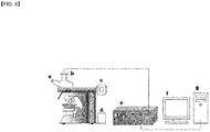
- FIG. 6 is a view of an apparatus for fluorescent assay, and a fluorescent image thereof was obtained and showed fluorescent spots of a FSNP-labeled antibody binding to the surface of the slide subjected to an indirect competitive reaction (see FIG. 5F ).
- FIG. 7 shows a graph of a calibration curve obtained by measuring a relative fluorescent degree obtained from a concentration-dependent fluorescent image of a C-reactive protein by using an FSNP-labeled antibody.
- a disposable immuno-fluorescent slide was prepared by surface-modifying a transparent slide which had a size of 30X70 mm and was used for a microscope with 3-aminopropyltrimethoxysilane or 3-mercaptopropyltrimethoxysilane-N-gamma maleimidobutyryloxy succin imide ester and immobilizing 1-100 ⁇ l of 0.01-0.5 mg/ml C-reactive protein thereon according to Examples 1 to 8.
Landscapes
- Health & Medical Sciences (AREA)
- Immunology (AREA)
- Life Sciences & Earth Sciences (AREA)
- Engineering & Computer Science (AREA)
- Molecular Biology (AREA)
- Biomedical Technology (AREA)
- Chemical & Material Sciences (AREA)
- Hematology (AREA)
- Urology & Nephrology (AREA)
- Biotechnology (AREA)
- Biochemistry (AREA)
- Cell Biology (AREA)
- Food Science & Technology (AREA)
- Medicinal Chemistry (AREA)
- Physics & Mathematics (AREA)
- Analytical Chemistry (AREA)
- Microbiology (AREA)
- General Health & Medical Sciences (AREA)
- General Physics & Mathematics (AREA)
- Pathology (AREA)
- Investigating Or Analysing Materials By The Use Of Chemical Reactions (AREA)
- Investigating Or Analysing Biological Materials (AREA)
- Peptides Or Proteins (AREA)
Description
- The present invention relates to a method of preparing an antigen-immobilized immuno-fluorescence slide including: immobilizing a C-reactive protein on a slide to prepare a protein chip, mixing an antibody that specifically binds to a target protein, with streptavidin, to label the antibody with a fluorescent nanoparticle, immune-reacting the antibody by competitive mixing, assaying with a fluorescence camera, and an immuno-fluorescence slide made by using the method.
- Biochips include various kinds of probes immobilized on a unit area of a surface of a solid support, and when such biochips are used, only with a small amount of sample, disease diagnosis, high-throughput screening (HTS), and enzyme activity measuring may be easily performed in a large scale.
- Most of methods of immobilizing a probe on a slide include immobilizing a probe on a glass slide pre-treated with a coating material. For these methods, various surface chemical materials have been proposed, and for example, a self-assembled monolayer may be used (Korean Patent Publication No.
2003-0038932 - In this regard, as a trial to increase an amount of an immobilized probe and to maintain the activity of a probe, a 3-dimensional (3D) immobilization method was developed (Gill and Ballesteros, Trends in Biotechnology 18:282, 2000). Examples of such a 3D immobilization method are a method using a Hydrogel® coating slide of Packard Bioscience, a method using a polyethyleneglycol-based hydrogel of Biocept Company, and a method using solgel of LG Chemical Co., Ltd, and the like.
- A slide for use in an immuno-sensor is selected from a group consisting of glass, silicon, hydrogel, metal, ceramic, and a porous membrane.
- Currently, an antibody or antigen probe-plate made by immobilizing an antibody or an antigen on a substrate, which is based on a specific reactivity of an antigen or an antibody, is widely used for detection. Representative kits for such use are a rapid test kit, a routine test kit, and a biochip kit.
- These kits are used to detect a target antigen or target antibody corresponding to an immobilized antibody probe or antigen probe.
- In some cases, various reaction groups are assembled onto a single probe-plate, and each of those reaction groups includes a test strip for the detection of an antigen or antibody.
- Meanwhile, as the Health Functional Food Act has taken into effect, even in Korea, it is possible to manufacture natural material-based food materials and processed food and to obtain recognition thereof as health functional food. However, to do this, materials for technically proving the functionality of health functional food, for example, cardiovascular disease prevention, aging suppression, cancer prevention, obesity prevention, immune controlling, or gastro-intestinal tracts related disease prevention, need to be presented. Accordingly, food-function evaluating is being considered more important, and as a result, evaluating of functions has a high likelihood of commercialization.
- Food-function evaluating includes in vitro function evaluation, in vivo function evaluation using a lab animal, such as a rat, and clinical trials, performed based on those six function types, and in terms of the food industry, a demand for developing an in vivo function detection techniques using a lab animal for effectively and rapidly performing function evaluation of food materials and functional good increases.
- To be recognized as a functional food, a target function, such as a cardiovascular function, needs to be scientifically proved, and it cannot be stressed too much the importance of food-function evaluation (Kim et al., Detecting C-reactive protein by using direct binding quartz crystal microbalance immunosensor, Journal of The Korean Society for Biotechnology and Bioengineering, Vol. 22, p. 443, 2007). Conventionally, in-vivo function evaluation using lab animals, which is performed prior to clinical trials, is to evaluate change in bodily elements, such as body weight, blood pressure, and shape of organs. However, this method causes non-uniform results, and also, requires variability of evaluation protocols, skilled experts, and high-cost assay instruments. A uniform in vivo food-function evaluation method is to measure an increase or decrease of a particular biomarker protein related to a particular disease or metabolism symptoms in a lab animal, such as a rat. In this regard, what contains a food that is desired to obtain recognition as a functional food is administered to the lab animal.
- Korea Registered Patent
10-921237 - This method, compared to an enzyme-linked immunosorbent assay (ELISA) (American Journal of C ardiology, Vol. 1, p. 155, 2005; American Journal of Veterinary Research, Vol. 1, p. 62, 2005; Journal of Clinical Laboratory Analysis, Vol. 18, p. 280, 2004) for evaluating a C-reactive protein, is easily used without measurement disturbances by a coloring material, and also, has better or equal measurement sensitivity than typical other sensor measurement methods (Biosensors and Bioelectronics, Vol. 22, p. 973, 2007; Biosensors and Bioelectronics, Vol. 21, p. 1987, 2006; Biosensors and Bioelectronics, Vol. 21, p. 1631, 2006; Biosensors and Bioelectronics, Vol. 21, p. 1141, 2006; Analytical Biochemistry, Vol. 328, p. 210, 2004).
- Recently, sensor measurement using a biosensor more frequently uses a nanomaterial, such as metallic colloid, carbon nanotube, fluorescent silica nanoparticle, or semiconductor quantum dot, and the use of such nanomaterials contributes to an increase in sensitivity, stability, or selectivity of sensor signals (Analytical Chemistry, Vol. 79, p. 630-707, 2007; Biosensors and Bioelectronics, Vol. 21, p. 1900, 2006; Langmuir, Vol. 22, p. 4357, 2006; Nano Letters, Vol. 5, p. 113, 2005; Biosensors and Bioelectronics, Vol. 20, p. 2454, 2005; Proceedings of the National Academy of Sciences, Vol. 100, p. 4984, 2003; Biochemical and Biophysical Research Communications, Vol. 274, p. 817, 2000).
- When an in vivo cardiovascular functionality of a food is evaluated by measuring a C-reactive protein as a biomarker present in blood of a lab animal by using a biosensor, an increase in sensor sensitivity is still a critical issue and also, ease of measurement is endlessly pursued.
-
KR 2009 0073669 A - HAI LI ET AL: LANGMUIR, vol. 26, no. 8, pages 5603-5609, relates to aminosilane micropatterns on hydroxyl-terminated substrates.
- KIM N S ET AL: FOOD ENGINEERING PROGRESS, vol. 14, no. 1, pages 21-26, relates to glass slide-based immunosensing.
- LEE, YOONSUK ET AL: PROTEOMICS, vol. 3, pages 2289-2304, describes a protein microarray.
- JUNG Y ET AL.: ANAL BIOCHEM., vol. 374, no. 1, pages 99-105, describes a strategy to immobilize an antibody on various sensor surfaces.
- STADTHERR K ET AL.: ANALYTICAL CHEMISTRY, vol. 77, no. 11, pages 3437-3443, relates to an aptamer-based protein biochip.
- The present invention provides a dispensable immunosensor slide with high sensor sensitivity, which allows a user, without a complicated apparatus, such as a biochip arrayer or a micro-array scanner, to immobilize a certain volume of an antigen on a slide with a micro dispensing pipette in manufacturing or measuring a biochip to perform an immunoreaction between a target protein and an zygocyte of an antibody and a fluorescent nanoparticle, or an immunoreaction between an immobilized antigen and a mixture including a fluorescent nano-probe and a particular target molecule on an antigen coating site of the immuno-fluorescence slide, and then, fluorescent intensity or the number of fluorescence particles of the nano-probe that specifically binds to the surface of the slide by the immunoreaction is measured using a fluorescence microscope so as to identify the concentration of a target molecule, such as a body indicator.
- The present invention relates to a method in accordance with
claims 1, 3 and 4 and to a C-reactive-protein-immobilized immune-fluorescence slide in accordance withclaim 2. - According to the present invention, a target protein (antigen) is immobilized on a slide, an antibody that specifically binds to the antigen is labeled with a fluorescent material, a target protein collected from a specimen is mixed with the fluorescence-labeled antibody, the mixture is reacted with the immobilized antigen site on the slide, unreacted antibody and antigen are washed with distilled water, the slide is placed in an incubator for drying, and fluorescent intensity or the number of fluorescence particles is measured by using a fluorescence microscope.
- To assay a sample, according to the present invention, an indirect-competitive assay format is used. However, one of ordinary skill in the art may easily know that the present invention may also be embodied by using a direct-competitive assay format or a sandwich assay format.
- An immunosensor slide used in embodiments of the present invention may be selected from the group consisting of glass, silicon, hydrogel, metal, ceramic, and a porous membrane, and may have a size of 20 to 40 X 70 to 80 mm.
- In the present invention, a C-reactive protein (hereinafter referred to as "CRP") is used as a target sample.
- A C-reactive protein antibody may be any one of antibodies which are prepared by using C-reactive proteins induced from a mammal, such as a rat, a mouse, a rabbit, a monkey, or a human being.
- A sample collected from a specimen may be any one of a reference solution in which a C-reactive protein is dissolved in a reaction buffer solution, and a tissue extract collected from blood, serum, blood plasma, saliva, body fluid, and the liver.
- As a solution for blocking a portion of the surface of the slide which does not react with an antigen, any one of a 1-5% bovine serum albumin (BSA) solution, a 1-5% rat serum albumin (RSA) solution, a 1-5% human serum albumin (HSA) solution, a 1-5% mouse serum albumin (MSA) solution, and a 1-5% goat serum albumin (GSA) solution.
- Hereinafter, each step of the method according to the present invention is described in detail.
- Immobilizing of a C-reactive protein as an antigen on a slide may be performed by the following method.
- As for the immobilizing method, the immobilizing of an antigen may be performed by using 3-aminopropyltrimethoxysilane (APTMS). For this method, first, a modified slide is prepared using APTMS. For the modification, a slide is immersed in a piranha solution (H2SO4:H2O2 = 2:1 to 4:1) for 5 to 15 minutes, and then, the slide is washed with distilled water, is sonicated in distilled water for 3 to 10 minutes, is washed by hydration with hot water having a temperature of 85 to 95 °C for 30 to 90 minutes, and then, is dried at room temperature on kimwipes for 20 to 120 minutes. The surface-washed slide was placed in a petri dish containing a solution in which 5 to 15% of APTMS is dissolved in acetone, and then, a reaction is performed at room temperature for 30 to 90 minutes. Front and rear sides of the slide are sequentially washed three to four times with acetone, a 30-70 mM phosphate buffer solution (pH 6.5-7.8), and a distilled water, and then, the slide is immersed in a beaker containing distilled water for 1 to 60 minutes. The slide is dried on kimwipes at room temperature for 20 to 120 minutes, heat treated at a temperature of 30 to 150°C in a convection oven for 3 to 7 hours, is left to sit for cooling, and then preserved in a dried state in a desiccator until use.
- The slide modified with APTMS is hydrated by immersion in distilled water for 10 to 20 minutes. The immersed slide is placed in a petri dish containing a solution including 1 to 4% glutaraldehyde and a reaction is performed for 30 to 90 minutes to activate the slide so that a protein is bindable to the surface of the slide. Front and rear surfaces of the activated slide are washed three to four times by using a 30-70 mM phosphate buffer solution (pH 6.5-7.8). Then, the slide is immersed in a beaker containing distilled water for 1 to 60 minutes. The slide is taken away from the beaker and placed on kimwipes and dried for 20 to 120 minutes at room temperature.
- The glutaraldehyde used above is prepared by adding 1 to 4 mℓ of 30-70% glutaraldehyde to 46 to 49 mℓ of distilled water, and 30 to 70 mℓ of the prepared solution is used.
- To immobilize a target protein, for example, a C-reactive protein, first, 0.01 to 0.5 mg/mℓ of a C-reactive protein is dissolved in a 30-70 mM phosphate buffer solution (pH 6.5-7.8) to prepare an antigen solution for immobilization. A petri dish containing the glutaraldehyde-activated slide is placed on a spotting guide having a lattice shape, and spotting is performed with 1 to 100 µℓ of the previously prepared antigen solution on a portion of the slide corresponding to a spot point on the spotting guide, and then, the resultant structure is covered with a cover and a reaction is performed for 1 to 6 hours to immobilize the antigen. Then, front and rear surfaces of the slide are washed three to four times with 30-70 mM phosphate buffer solution (pH 6.5-7.8), and then, immersed in a beaker containing distilled water for 1 minute. To block a portion of the surface of the glutaraldehyde-activated slide subjected to the immobilizing of the antigen which does not react with the antigen on the surface, the slide is placed in a small plastic petri dish containing 1 to 5% BSA solution dissolved in a 30-70 mM phosphate buffer solution (pH 6.5-7.8), and then, the resultant structure is covered with a cover and a reaction is performed for 1 to 5 hours, and when the reaction is stopped, front and rear surfaces of the slide blocked with BSA are washed three to four times with 30-70 mM phosphate buffer solution (pH 6.5-7.8), and then, the slide is immersed in a beaker containing distilled water for 1-60 minutes. The slide is placed on kimwipes and dried for 20-120 minutes at room temperature.
- Also disclosed is a second immobilizing method which does not form part of the present invention, in which the immobilizing of an antigen may be performed by using 3-mercaptopropyltrimethoxysilane-N-gamma- maleimidobutyryloxy succinimide ester (MPTMS-GMBS). First, for surface cleaning, a slide is immersed in a mixed solution (HCl:MtOH = 1:1 to 1:2, a volumetric ratio) including a strong hydrochloric acid (18-36%) and methanol (50-100%) for 15 to 45 minutes, and then, front and rear surfaces of the slide are washed three to four times with distilled water. After the washing of the slide with 85 to 95 °C of distilled water, the slide is placed on kimwipes and dried for 20 to 120 minutes at room temperature.
- To silanize the resultant slide, 50 to 500 mℓ of an MPTMS solution in which 1-3% MPTMS is dissolved in an organic solvent solution selected from the group consisting of toluene, dimethyl foramide, and acetone is prepared. The slide is placed in a petri dish containing the solution and a reaction is performed at room temperature for 1 to 3 hours. Front and rear surfaces of the slide are washed three to four times with the same organic solvent as used in preparing the MPTMS solution, and the slide is placed on kimwipes and dried for 20 to 120 minutes at room temperature.
- To activate the surface of the slide, an 1 to 3 mM GMBS solution is prepared as described below. 14 to 42 mg of a GMBS sample is dissolved in 100 µℓ of N,N-dimethylformamide (DMF; Sigma-Aldrich Company, USA), and then, 49.9 mℓ of ethanol is added thereto. The slide subjected to the silanization is placed in a petri dish containing the GMBS solution and a reaction is performed at room temperature for 30 to 90 minutes. Front and rear surfaces of the slide are washed three to four times with distilled water and a 30-70 mM phosphate buffer solution (pH 6.5-7.8). Then, the slide is placed on kimwipes and dried for 20 to 120 minutes at room temperature.
- A CRP antigen is immobilized in the following manner. First, a C-reactive protein is dissolved at a concentration of 0.01 to 0.5 mg/mℓ in a 30-70 mM phosphate buffer solution (pH 6.5-7.8) to prepare an antigen solution for immobilization. A petri dish containing a GMBS-activated slide is placed on a spotting guide, and then, 1 to 100µℓ of the previously prepared antigen solution is spotted on a portion of the slide corresponding to a spot point of the spotting guide and then, the resultant structure is covered with a cover and a reaction is performed for 1 to 6 hours to immobilize the antigen. Then, front and rear sides of the slide are washed three to four times with a 30-70 mM phosphate buffer solution (pH 6.5-7.8), and the slide is immersed in a beaker containing distilled water for 1 to 60 minutes. To increase assay accuracy, the slide is placed on a small plastic petri dish containing a 1 to 5% BSA solution and the resultant structure is covered with a cover and a reaction is performed for 1 to 5 hours. Front and rear surfaces of the slide are washed three to four times with the phosphate buffer solution described above. Then, the slide is immersed in a beaker containing distilled water for 1 to 60 minutes, and then, placed on kimwipes and dried for 20 to 120 minutes at room temperature.
- These two methods are schematically illustrated in
FIG. 1 - As for assaying a particular protein by using an immunosensor slide according to the present invention, an antibody that specifically binds to an antigen immobilized on the slide is labeled. The labeling of an antibody may be performed by using a fluorescent staining method with high sensitivity and reproducibility, and to do this, according to the present invention, a fluorescent silica nanoparticle (hereinafter referred to as "FSNP") is used.
- A method of preparing FSNP is briefly described below. FSNP, which is used to label an antibody that specifically binds to an antigen, may be prepared by using a dye entrapment method in which an organic fluorescence pigment is included in a silica structure, a core shell method in which an active organic fluorescence pigment is covalently bonded to an amino silane compound and the result structure is included in a silica structure, or an inverted core shell method in which a organic fluorescence pigment is included in a silica structure and the formed particle is reacted with an organosilane compound to introduce a functional group to obtain functionality. In this regard, examples of such an organic fluorescence pigment are dichlorotris(1,10-phenanthroline)ruthenium(II)·hydrate, fluorescein, rhodamine B, and 5(6)-Carboxytetramethylrhodamine, and examples of an active organic fluorescence pigment are fluorescein isothiocyanate, 5(6)-carboxytetramethylrhodamine N-succinimidyl ester, tetramethylrhodamine 5-isothiocyanate, bis(2,2'-bipyridine)-4'-methyl-4-carboxybipyridine-ruthenium N-succinimidyl ester-bis(hexafluorophosphate), and bis(2,2'-bipyridine)-4,4'-dicarboxybipyridine ruthenium di(N-Succinimidyl ester)bis(hexafluorophosphate), an example of an amino silane compound is (3-aminopropyl)triethoxysilane, and examples of an organosilane compound are compounds including carboxylate, amine, amine/phosphonate, poly(ethylene glycol), and octadecyl, carboxylate/ octadecyl functional groups.
- A method of labeling an antibody with FSNP is described below. First, FSNP needs to be modified with streptavidin (SA) which specifically binds to biotin. This modification is performed to bind SA-bonded FSNP, which is to be described later, to a biotinylated antibody. To modified FSNP with SA, first, FSNP is reacted with (3-mercaptopropyl)trimethoxy silane (MPTMS) to prepare a thiol-modified FSNP. 1 to 5 mg of FSNP and 5 to 15 mℓ of an alcohol, such as ethanol, methanol, or propanol, are added to a cap tube having a capacity of 10 to 70 mℓ and the mixture is shaken well and then, sonicated to completely dissolve FSNP in the alcohol for 10 to 30 minutes. Thereafter, 30-70 to 200 µℓ of MPTMS is added to the cap tube, and while the resultant mixture is stirred at a temperature of room temperature at a low speed of 30-200 rpm, a reaction is performed for 2 to 6 hours. The reaction mixture is centrifuged at a temperature of 3 to 10 °C for 20 to 40 minutes at a speed of 7000-17000 rpm and the obtained precipitate is washed one to five times by using the same alcohol as used for the reaction, and following each washing, centrifuging is performed under the same conditions as described above to obtain a precipitate. A cap is separated from the tube and the tube is softly covered with an aluminum foil and dried at room temperature. The precipitate is dissolved in 2 to 6 mℓ of a 30-70 mM phosphate buffer solution (pH 6.5-7.8) to prepare a thiol-modified FSNP solution.
- A maleimide-activated SA is added to the thiol-modified FSNP to prepare SA-bonded FSNP. In detail, 0.1 to 0.5 mg of SA-maleimide is dissolved in 3 to 7 mℓ of a 30-70 mM phosphate buffer solution (pH 6.5-7.8) in a cap tube having a capacity of 10-70 mℓ to prepare a SA-maleimide solution. Then, 0.5 to 2.0 mℓ of the thiol-modified FSNP solution is added thereto, and while the mixture is continuously stirred at a low speed of 30 to 200 rpm, a reaction is performed at room temperature for 1 to 3 hours. The reaction product is centrifuged at a temperature of 3 to 10 °C for 20 to 40 minutes at a speed of 7000-17000 rpm to obtain a precipitate. The precipitate is washed one to five times with 30 to 70 mM phosphate buffer solution (pH 6.5-7.8), and following each washing, centrifuging is performed under the same conditions as described above to obtain a precipitate. The precipitate is re-suspended with 1 to 5 mℓ of 30-70 mM phosphate buffer solution (pH 6.5-7.8). The suspension is loaded into a cap tube, wrapped with an aluminum foil, refrigerated, and reacted with a biotinylated antibody, which is described later.
- Biotinylating a C-reactive protein antibody may be performed by using one of the following methods.
- A method of biotinylating a C-reactive protein antibody includes: adding a carbonate-bicarbonate buffer to a C-reactive protein antibody to obtain a C-reactive protein antibody-containing carbonate-bicarbonate buffer, reacting a biotinamidohexanoic acid N-hydroxysuccinimide ester (NHS-LC-biotin) solution, which is a biotinylation reagent dissolved in dimethyl formamide that is an organic solvent, with the carbonate-bicarbonate buffer, and dialyzing the obtained solution in a sodium chloride solution to remove an unreacted biotinylation reagent.
- In detail, the biotinylating method includes adding a carbonate-bicarbonate buffer to a C-reactive protein antibody to obtain a carbonate-bicarbonate buffer containing 1.0 to 3.0 mg/mℓ C-reactive protein antibody, reacting 30 to 100 µℓ of NHS-LC-biotin solution in which 1 to 5 mg/mℓ NHS-LC-biotin is dissolved in dimethyl formamid with 25 to 75 µℓ of a carbonate-bicarbonate buffer for 1 to 5 hours in iced water, and dialyzing 75 to 175 µℓ of the obtained solution in 0.1 to 0.3M sodium chloride solution overnight to remove an unreacted biotinylation reagent.
- A method of biotinylating a C-reactive protein antibody includes: adding a phosphate buffered saline to a C-reactive protein antibody to prepare a C-reactive protein antibody-containing phosphate buffered saline, reacting a sulfosuccinimidyl-6-(biotinamido)hexanoate (sulfo-NHS-LC-biotin) solution, which is a water-soluble biotinylation reagent dissolved in redistilled water, with the phosphate buffered saline, and dialyzing the obtained solution in a phosphate buffered saline to remove an unreacted biotinylation reagent.
- In detail, the biotinylating method according to the present embodiment includes adding a phosphate buffered saline to a C-reactive protein antibody to prepare a phosphate buffered saline solution in which the concentration of the C-reactive protein antibody is in a range of 0.5 to 3.0 mg/mℓ, reacting 5 to 20 µℓ of a sulfo-NHS-LC-biotin solution in which 2 to 20 mM sulfo-NHS-LC-biotin is dissolved in redistilled water with 30 to 800 µℓ of the phosphate buffered saline in iced water for 1 to 5 hours, and dialyzing 55 to 820 µℓ of the resultant solution in 0.05 to 0.25M phosphate buffered saline overnight to remove an unreacted biotinylation reagent.
- Next, an antibody to which a nanomaterial is bonded is prepared. When the SA-modified FSNP is mixed with the biotinylated antibody, due to specific bonding between biotin and SA, a fluorescent material-labeled antibody is prepared.
- In detail, the SA-modified FSNP solution is sonicated for 10 to 30 minutes, and then, 200 to 600 µℓ of the obtained solution is added to 1 to 3 mℓ of 0.05 to 0.25 mg/mℓ biotinylated antibody solution. While the mixture is stirred at time intervals of 30 minutes at a low speed of 30 to 200 rpm for 2 to 7 minutes, a reaction is performed at room temperature for 1 to 3 hours. The reaction product is centrifuged at a temperature of 3 to 10°C for 20 to 40 minutes at a rate of 7000-17000 rpm, and a supernatant is discarded and a precipitate is used. The precipitate is washed one to five times with 30 to 70 mM phosphate buffer solution (pH 6.5-7.8), and following each washing, centrifuging is performed under the same conditions as described above and the obtained precipitate is used. The precipitate is re-suspended in 0.4 to 1.2 mℓ of 30-70 mM phosphate buffer solution (pH 6.5-7.8).
- A schematic method of labeling an antibody that specifically binds to CRP with a fluorescent material, according to the present invention, is illustrated in
FIG. 2 . - In assaying a particular protein by using an immunosensor slide according to the present invention, a target protein obtained from a specimen is mixed with the labeled antibody.
- In detail, a C-reactive protein sample solution is prepared in various concentrations by using a 30-70 mM phosphate buffer solution (pH 6.5-7.8) in an eppendorf tube, and a C-reactive protein antibody solution which is prepared by specific binding between biotin and SA and to which FSNP is bonded as a nanomaterial, is added to the various concentrations of the C-reactive protein sample solution, wherein amounts of the C-reactive protein antibody solution and the C-reactive protein sample solution are the same, and the mixture remains stationary at room temperature for 5 to 300 minutes.
- Next, the mixed mixture is reacted with an antigen immobilized on the slide.
- In detail, a petri dish containing a slide as a bio-transducer on which a C-reactive protein as an antigen is immobilized by the glutaraldehyde process or the MPTMS-GMBS process and adsorption of a non-selective protein is minimized by the bovine serum albumin (BSA) blocking process, is placed on a spotting guide having a lattice structure, and the mixing is performed where the antigen is immobilized on the slide, and then, 1 to 100 µℓ of a mixture including a target protein obtained from a specimen which has been stationary at room temperature for 5 to 300 minutes and the labeled antibody is accurately spotted, and the resultant structure is covered with a cover, and a reaction is performed at room temperature for 0.5 to 16 hours, thereby performing an indirect-competitive assay between the antigen immobilized on the slide and a target protein collected from a specimen against the FSNP-labeled antibody. After a reaction is stopped, unreacted antibody and antigen are removed therefrom by washing front and rear sides of the slide three to four times with distilled water and immersing the slide in a beaker containing distilled water for 1 to 60 minutes.
-
FIG. 3 illustrates a diagram for explaining how the antigen immobilized on a slide binds to the fluorescence-labeled antibody by indirect-competitive assay, andFIG. 4 shows a graph of a change of the number of fluorescent particles binding to a slide when instead of a C-reactive protein collected from a specimen, a 30-70 mM phosphate buffer solution (pH 6.5-7.8) is mixed with the labeled antibody as described above, and then, the mixture is spotted on the antigen immobilized on the slide to perform an antigen-antibody reaction over time. - The slide is dried in an incubator for 20 to 120 minutes.
- Fluorescence intensity and the number of fluorescent particles are measured by using a fluorescence microscope. When the concentration of CRP in a specimen is high, more CRP reacts with fluorescence-labeled antibodies. Accordingly, less fluorescence-labeled antibodies bind to the antigen immobilized on the slide, and thus, fluorescence intensity and the number of fluorescent particles are reduced. As described above, an antigen immobilized on a slide and an antigen obtained from a specimen competitively interacts with a fluorescence-labeled antibody, thereby showing different intensity levels of fluorescence. Thus, CRP can be easily measured for a relatively short period of time.
- The present invention provides an immunosensor slide for easily and highly-sensitively measuring a biomarker which is an in vivo food-function indicator by a nanomole-level of evaluation by coating a C-reactive protein, which is known as a major biomarker for coronary artery disease, hypertension, and inflammation, on the slide and reacting the C-reactive protein with an antibody binding to a fluorescent nanoprobe.
- The present invention may establish a future food-function evaluation related basic technique that is required for a supersensitive, high-speed simultaneous measurement on, in addition to a C-reactive protein, a biomarker, such as low density lipoprotein (LDL), fibrinogen, or angiotensin II, existing in a wide concentration range in, for example, blood of a lab animal.
-
-
FIG. 1 is a schematic view illustrating two methods of immobilizing a C-reactive protein on a slide. -
FIG. 2 is a schematic view illustrating a method of labeling an antibody that specifically binds to CRP with a fluorescent material. -
FIG. 3 is a schematic view illustrating binding of a fluorescence-labeled antibody to an antigen immobilized on the slide by indirect competition. -
FIG. 4 shows a graph of a change of the number of fluorescent particles binding to a slide when instead of a C-reactive protein collected from a specimen, a 30-70 mM phosphate buffer solution (pH 6.5-7.8) is mixed with the labeled antibody as described above, and then, the mixture is spotted where the antigen is immobilized on the slide to perform an antigen-antibody reaction over time. -
FIGS. 5A, 5B, and 5C show images showing that inhibitory spots of a contaminating material, such as impurities, present on the surface of a slide before treatment with a piranha solution are removed by a piranha treatment, and even after silanization with APTMS, inhibitory spots are not observed.FIG. 5D shows an image of a substrate treated with a 2.5% glutaraldehyde solution in which inhibitory spots are not observed.FIG. 5E shows an image in which inhibitory spots are not present on a BSA-blocked portion of the surface of the slide after an antigen is immobilized.FIG. 5F shows an image containing fluorescent spots of a fluorescent silica nanoparticle-labeled antibody binding to the surface of the slide subjected to an indirect competitive reaction. -
FIG. 6 is a view illustrating an apparatus for fluorescent assay. -
FIG. 7 illustrates a graph of a calibration curve of an antigen-immobilized immuno-fluorescence slide and a FSNP-labeled antibody-based C-reactive protein. - Hereinafter, the method according to the present invention is described in detail with reference to examples. However, the scope of the present invention is not limited to the examples.
- Materials used in the present experiments are described as follows:
- A recombined histidine tagged rate CRP (pure state) expressed in a mouse myeloma cell line (NSO) was obtained from R&D Systems, Inc. (Miniapolice, MN, USA) for use throughout the present experiments. A homopentameric structure thereof consisted of three non-covalent and two covalent sub units. In addition, a monoclonal anti-rat CRP antibody generated from a hybridoma obtained from a fusion of mouse myeloma and a B cell that is obtained from a mouse immunized with a purified, NSO-induced recombined rat CRP, was obtained from R&D Systems Inc. A glass slide was obtained from Coring Inc. (Kennebunk, ME, USA). A water-soluble biotinylation reagent (sulfosuccinimidyl-6-(biotinamido)hexanoate: sulfo-NHS-LC-biotin) was obtained from Pierce Biotechnology, Inc. (Rockford, IL, USA). 3-aminopropyltrimethoxysilane (APTMS), glutaraldehyde, and bovine serum albumin (BSA) were obtained from Sigma-Aldrich Chemical Co.(St. Louis, MO, USA). Other compounds used in the following experiments were guaranteed by various suppliers, and throughout experiments, double distilled water was used.
- An antigen was immobilized on a slide as follows. That is, for surface cleaning, a slide was immersed in a piranha solution (H2SO4:H2O2 = 3:1, volumetric ratio) for 10 minutes, washed with distilled water, sonicated in distilled water for 5 minutes, washed by hybridization with hot water having a temperature of 90°C for 1 hour, placed on kimwipes, and then, dried for 30 minutes at room temperature.
- To perform silanization to introduce an amino group (-NH2), which is needed to immobilize an antigen, to the surface of the slide, the surface-cleaned slide was placed in a petri dish containing an APTMS solution in which 10% APTMS was dissolved in acetone and a reaction was performed at room temperature for 1 hour. Front and rear sides of the slides were sequentially washed with a 50 mM phosphate buffer solution (pH 7.4) and distilled water, three times for each, and then, immersed in a beaker containing distilled water for 1 minute. The resultant slide was taken away therefrom, and then, dried on kimwipes for 30 minutes at room temperature, heat treated in a convection oven at a temperature of 120°C for 5 hours, and left to sit for cooling, and a fluorescent image thereof was obtained (see
FIGS. 5A, 5B, and 5C ). As shown inFIGS. 5A, 5B, and 5C , it was confirmed that inhibitory spots of a contaminating material, such as impurities, present on the surface of a slide before treatment with a piranha solution were removed by a piranha treatment, and even after silanization with APTMS, a fluorescent image of inhibitory spots was not observed. - To activate the APTMS-modified slide, the slide was immersed in distilled water for 15 minutes, and taken away therefrom and placed in a petri dish containing glutaraldehyde 2.5% solution, and a reaction was performed for 1 hour to enable a protein to bind to the surface of the slide. Front and rear surfaces of the activated slide were washed three times with 50mM phosphate buffer solution (pH 7.4) and then, immersed in a beaker containing distilled water for 1 minute. The slide was taken away therefrom and placed on kimwipes and dried for 30 minutes at room temperature, and a fluorescent image thereof also shows no inhibitory spots on the surface of the GA-activated slide (see
FIG. 5D ). - To immobilize an antigen (CRP, C-reactive protein) on a slide, first, a C-reactive protein was dissolved at a centration of 0.1 mg/mℓ in a 50 mM phosphate buffer solution (pH 7.4) to prepare an antigen solution for immobilization. A petri dish containing the glutaraldehyde-activated slide was placed on a spotting guide having a lattice structure, and 20µℓ of the antigen solution was spotted on a portion of the slide corresponding to a spot point on the spotting guide, and then, the petri dish was covered with a cover and a reaction was performed for 1 hour to immobilize an antigen, and then, front and rear surfaces of the slide were washed three times with 50mM phosphate buffer solution (pH 7.4) and immersed in a beaker containing distilled water for 1 minute. To block a portion of the glutaraldehyde-activated slide subjected to the immobilizing of an antigen which did not react with the antigen, the slide was placed in a small plastic petri dish containing 1% bovine serum albumin (BSA) solution in which 1% bovine serum albumin was dissolved in a 50 mM phosphate buffer solution (pH 7.4) and the petri dish was covered with a cover and a reaction was performed for 1 hour. Then, front and rear sides of the slide were washed three times with 50mM phosphate buffer solution (pH 7.4), and the slide was immersed in a beaker containing distilled water for 1 minute, and placed on kimwipes and dried for 30 minutes at room temperature, and a fluorescent image thereof also showed no inhibitory spots on the surface of the BSA-blocking slide after the antigen was immobilized (see
FIG. 5E ). - Immobilizing of an antigen on a slide was performed through the following processes. That is, for surface cleaning, a slide was immersed in a mixed solution including a strong hydrochloric acid (35%) and methanol (70%) at a volumetric ratio of 1:1 for 30 minutes, and then, the slide was washed three times with distilled water. After 30 minutes of immersion in a strong sulfuric acid, front and rear surfaces of the slide were washed three times with distilled water, and washed with boiled distilled water and placed on kimwipes and dried at room temperature for 30 minutes.
- To perform silanization to introduce a thiol group (-SH), which is a reaction group needed to immobilize an antigen, to the surface of a slide, 2 mℓ of (3-mercaptopropyl)trimethoxy silane (MPTMS) was added to 98 mℓ of toluene to prepare a 2% MPTMS solution. The slide was placed on a petri dish containing the solution and a reaction was performed at room temperature for 2 hours. Thereafter, the slide was washed three times with toluene, and then, placed on kimwipes and dried for 30 minutes at room temperature.
- To activate the slide modified with MPTMS, 28 mg of GMBS was dissolved in 100 µℓ of N,N-Dimethylformamide (DMF, chromosllvr®Plus, St. Louis, MO, USA, HPLC≥99.9%) and 49.9 mℓ of ethanl was added thereto to prepare a 2 mM GMBS solution. The silanized slide was placed on a petri dish containing the 2 mM GMBS solution and a reaction was performed at room temperature for 1 hour. Front and rear sides of the slide were washed three times with distilled water and a 50 mM phosphate buffer solution (pH 7.4), and dried on kimwipes for 30 minutes at room temperature.
- To immobilize an antigen (CRP, C-reactive protein) on the slide, first, a 0.1 mg/mℓ C-reactive protein was dissolved in 50 mM phosphate buffer solution(pH 7.4) to prepare an antigen solution for immobilizing. A petri dish containing the GMBS-activated slide was placed on a spotting guide, and 20 µℓ of the antigen solution was spotted on a portion of the slide corresponding to a spot point on the spotting guide, and then, the petri dish was covered with a cover and a reaction was performed for 1 hour to immobilize an antigen, and then, front and rear surfaces of the slide were washed three times with 50 mM phosphate buffer solution (pH 7.4) and immersed in a beaker containing distilled water for 1 minute. To block a portion of the GMBS-activated slide subjected to the immobilizing of an antigen which did not react with the antigen, the slide was placed in a small plastic petri dish containing 1% bovine serum albumin (BSA) solution in which 1% bovine serum albumin was dissolved in a 50 mM phosphate buffer solution (pH 7.4), and the petri dish was covered with a cover and a reaction was performed for 1 hour. Then, front and rear sides of the slide were washed three times with 50 mM phosphate buffer solution (pH 7.4), and the slide was immersed in a beaker containing distilled water for 1 minute, and placed on kimwipes and dried for 30 minutes at room temperature.
- A fluorescent image of the slide was taken at the respective steps of the process of immobilizing an antigen on a MTS-GMBS treated slide, that is, surface cleaning, silanization, activation, immobilization of an antigen, and BSA blocking. As a result, it was confirmed that inhibitory spots, which inhibit sensor measuring on the surface of the slide, were not shown in all the fluorescent images, excluding that of the slide before the treatment with the mixed solution including a strong hydrochloric acid and methanol (1:1, volumetric ratio).
- 2 mg of FSNP and 10 mℓ of ethanol were added to a cap tube having a capacity of 25 mℓ and the mixture was shaken well, and then, sonicated to completely dissolve FSNP in ethanol for 15 minutes. Thereafter, 100 µℓ of MPTMS was added to the cap tube and a reaction was performed at room temperature for 4 hours while stirring at a low sped of 100 rpm. The reaction mixture was centrifuged at a temperature of 4°C for 30 minutes at a rate of 13000 rpm and the obtained precipitate was washed three times with ethanol, and after each washing, centrifuging was performed under the same conditions as described above to obtain precipitates. The cap of the tube was separated and the tube was lightly covered with an aluminum foil and dried at room temperature. Each of the precipitates was dissolved in 4 mℓ of 50 mM phosphate buffer solution (pH 7.4) to prepare a thiol-modified FSNP solution.
- 0.25 mg of SA-msaleimide and 5 mℓ of 50 mM phosphate buffer solution (pH 7.4) were added to a cap tube having a capacity of 25 mℓ and the SA-maleimide was dissolved to prepare a SA-maleimide solution. Then, 1 mℓ of the thiol-modified FSNP solution was added thereto and while continuously stirring at a low speed of 100 rpm, a reaction was performed at room temperature for 2 hours. The reaction product was centrifuged at a temperature of 4°C for 20 minutes at a rate of 10000 rpm to obtain a precipitate. The precipitate was washed three times with a 50 mM phosphate buffer solution (pH 7.4), and after each washing, centrifuging was performed under the same conditions as described above. The precipitate was re-suspended in 2 mℓ of a 50 mM phosphate buffer solution (pH7.4). The result was added to a cap tube, wrapped with an aluminum foil, refrigerated, and reacted with a biotinylated antibody described below.
- 1 mg vial of sulfosuccinimidyl-6-(biotinamido)hexanoate (sulfo-NHS-LC-biotin) that is a water-soluble biotinylation reagent, which had been refrigerated, was moved to a room temperature condition, and 30-700 µℓ of 0.1 M phosphate buffered saline (pH 7.2) was dissolved in 30-700 µℓ vial of monoclonal anti-rat CRP antibody. 180 µℓ of distilled water was added to 1 mg vial of sulfo-NHS-LC-biotin to prepare a 10 mM sulfo-NHS-LC-biotin solution, and then, 6.67 µℓ of the mixture was added to the antibody solution to control a molecular ratio of biotin to an antibody to be 20:1. A vial containing the reaction mixture was softly shaken, and then, placed in an eppendorf tube rack and then in iced water and a reaction was performed for 2 hours. Then, the reaction mixture was loaded into a slide-A-lyzer kit (Pierce Biotechnology, Inc) and then, dialyzed with respect to a 0.1M phosphate buffered saline (pH 7.2) overnight and an unreacted reagent was removed and a dialyzate remaining inside the dialysis membrane was recovered. and then. the total volume of the dialyzate was controlled to be 1 mℓ by using the same phosphate buffered saline to prepare biotinylated antibody.
- A SA-modified FSNP solution was sonicated for 15 minutes and then, 400 µℓ of the result was added to 2 mℓ of a 0.1 mg/mℓ biotinylated antibody solution, and while stirring at time intervals of 30 minutes at a low speed of 100 rpm, each for 5 minutes, a reaction was performed at room temperature for 2 hours. The reaction product was centrifuged at a temperature of 4°C for 20 minutes at a rate of 10000 rpm, and a supernatant was discarded and a precipitate was collected. The precipitate was washed three times with 50 mM phosphate buffer solution (pH 7.4), and after each washing, centrifuging was performed under the same conditions as described above to obtain a precipitate. The precipitates were re-suspended in 0.8 mℓ of 50 mM phosphate buffer solution (pH 7.4).
- A C-reactive protein antibody solution to which FSNP was bonded as a nanomaterial, which was prepared based on a specific bonding between biotin and SA, was added to various concentrations of a C-reactive protein sample solution which was prepared by dilution using 50 mM phosphate buffer solution (pH 7.4) contained in an eppendorf tube, wherein amounts of the C-reactive protein antibody solution and the C-reactive protein sample solution were the same, and these solutions were mixed and a reaction was performed room temperature for 1 hour.
- A petri dish containing a slide as a biotransducer in which a C-reactive protein as an antigen was immobilized and adsorption of a non-selective protein was minimized by BSA blocking according to Examples 1 and 2, was placed in a spotting guide having a lattice structure, and mixing was performed where the antigen was immobilized on the slide, and 20 µℓ of a mixture including a target protein which remained stationary at room temperature for 1 hour and collected from a specimen and the labeled antibody was accurately spotted, and the resultant structure was covered and a reaction was performed at room temperature for 2 hours to perform an indirect competitive reaction between the antigen immobilized on the slide and a target protein collected from a specimen against the FSNP-labeled antibody. After a reaction was stopped, an unreacted antibody and antigen were removed by washing three times front and rear sides of the slide with distilled water, and placing the slide in a beaker containing distilled water for 1 minute.
- The slide subjected to the indirection competitive reaction according to Example 7 was placed in an incubator and dried. A fluorescent image assay was performed on the dried slide by using a fluorescence microscope, and fluorescent intensity and the number of fluorescent particles were measured.
FIG. 6 is a view of an apparatus for fluorescent assay, and a fluorescent image thereof was obtained and showed fluorescent spots of a FSNP-labeled antibody binding to the surface of the slide subjected to an indirect competitive reaction (seeFIG. 5F ).FIG. 7 shows a graph of a calibration curve obtained by measuring a relative fluorescent degree obtained from a concentration-dependent fluorescent image of a C-reactive protein by using an FSNP-labeled antibody. When the concentration of CRP in a specimen increased, more CRP reacted with a labeled antibody, and thus, the binding between the antigen immobilized on the slide and the labeled antibody decreased. In addition, the graph showed linearity in a very low concentration range of the C-reactive protein. This shows that a detection limit when CRP is measured by using the immuno-fluorescence slide is a very high of 0.1 to 1.0 ng/mℓ. - A disposable immuno-fluorescent slide was prepared by surface-modifying a transparent slide which had a size of 30X70 mm and was used for a microscope with 3-aminopropyltrimethoxysilane or 3-mercaptopropyltrimethoxysilane-N-gamma maleimidobutyryloxy succin imide ester and immobilizing 1-100 µℓ of 0.01-0.5 mg/mℓ C-reactive protein thereon according to Examples 1 to 8.
Claims (4)
- A method of preparing a C-reactive-protein-immobilized immuno-fluorescence slide, the method comprising:- immobilizing a C-reactive protein on a slide to prepare a protein chip; wherein the immobilizing of the C-reactive protein on the slide comprises:modifying the slide with 3-aminopropyltrimethoxysilane to prepare a modified slide;hydrating the slide modified with 3-aminopropyltrimethoxysilane;activating the hydrated modified slide by using a glutaraldehyde solution;dissolving target C-reactive protein at a concentration of 0.01-0.5 mg/ml in a 30-70 mM phosphate buffer solution, with pH 6.5-7.8 to prepare a C-reactive protein solution for immobilization;placing a petri dish comprising the glutaraldehyde-activated slide on a spotting guide and spotting 1-100 µℓ of the C-reactive protein solution on spotting points; andperforming an immobilization reaction on the glutaraldehyde-activated slide prepared as described above for 1-6 hours to immobilize the C-reactive protein.
- A C-reactive-protein-immobilized immuno-fluorescence slide prepared by the method of claim 1.
- An in-vitro method of measuring a C-reactive protein in a specimen by using the immuno-fluorescence slide of claim 2.
- The in-vitro method of claim 3, wherein the method comprises- labeling an antibody that specifically binds to C-reactive protein, with a fluorescent nanoparticle;- mixing a target C-reactive protein obtained from a specimen with the fluorescent labeled antibody,- reacting the mixture with the C-reactive-protein-immobilized immuno-fluorescence slide, and- assaying C-reactive protein by measuring fluorescent intensity or the number of fluorescent particles by using a fluorescence microscope.
Applications Claiming Priority (2)
| Application Number | Priority Date | Filing Date | Title |
|---|---|---|---|
| KR1020100016157A KR101140029B1 (en) | 2010-02-23 | 2010-02-23 | Preparation method of antigen-immobilized immuno- fluorescence slide and the immuno-fluoroscence slide made by the method |
| PCT/KR2011/001126 WO2011105721A2 (en) | 2010-02-23 | 2011-02-21 | Preparation method of antigen-immobilized immunofluorescence slide, and immunofluorescence slide prepared thereby |
Publications (3)
| Publication Number | Publication Date |
|---|---|
| EP2541248A2 EP2541248A2 (en) | 2013-01-02 |
| EP2541248A4 EP2541248A4 (en) | 2013-09-04 |
| EP2541248B1 true EP2541248B1 (en) | 2017-07-12 |
Family
ID=44507349
Family Applications (1)
| Application Number | Title | Priority Date | Filing Date |
|---|---|---|---|
| EP11747611.9A Not-in-force EP2541248B1 (en) | 2010-02-23 | 2011-02-21 | Preparation method of antigen-immobilized immunofluorescence slide, and immunofluorescence slide prepared thereby |
Country Status (5)
| Country | Link |
|---|---|
| US (1) | US20130029428A1 (en) |
| EP (1) | EP2541248B1 (en) |
| KR (1) | KR101140029B1 (en) |
| CN (4) | CN104316678A (en) |
| WO (1) | WO2011105721A2 (en) |
Families Citing this family (35)
| Publication number | Priority date | Publication date | Assignee | Title |
|---|---|---|---|---|
| JPWO2011077838A1 (en) * | 2009-12-25 | 2013-05-02 | コニカミノルタエムジー株式会社 | Fluorescent substance-encapsulated silica nanoparticles and biological substance labeling agent |
| EP2734843A2 (en) | 2011-07-18 | 2014-05-28 | President and Fellows of Harvard College | Engineered microbe-targeting molecules and uses thereof |
| EP2976642A4 (en) | 2013-03-15 | 2016-09-21 | Harvard College | METHODS AND COMPOSITIONS FOR IMPROVING THE DETECTION AND / OR CAPTURE OF A TARGET ENTITY |
| US9988617B2 (en) | 2013-05-21 | 2018-06-05 | President And Fellows Of Harvard College | Engineered heme-binding compositions and uses thereof |
| WO2015072724A1 (en) * | 2013-11-12 | 2015-05-21 | 광주과학기술원 | Immunochromatography strip sensor capable of measuring biomaterial concentration over broad concentration range |
| EP3546474B1 (en) | 2013-12-18 | 2021-07-07 | President and Fellows of Harvard College | Crp capture/detection of gram positive bacteria |
| WO2015101382A1 (en) * | 2013-12-30 | 2015-07-09 | Telecom Italia S.P.A. | Method for managing a node association in a wireless personal area communication network |
| CN103941017B (en) * | 2014-03-18 | 2016-01-20 | 北京普恩光德生物科技开发有限公司 | C reactive protein detection kit |
| US20170342470A1 (en) * | 2014-12-12 | 2017-11-30 | Konica Minolta, Inc. | Diluent for fluorescent nano particles, kit for immunofluorescent staining which utilizes same, solution for immunofluorescent staining, immunofluorescent staining method, and gene staining method |
| CN104678105A (en) * | 2015-01-16 | 2015-06-03 | 镇江市第一人民医院 | Enzyme linked immunosorbent assay kit for detecting Cripto-1 and preparation method thereof |
| CN107430119A (en) * | 2015-03-10 | 2017-12-01 | 西门子医疗保健诊断公司 | Quantification of functional groups on solid supports |
| TWI547693B (en) * | 2015-05-07 | 2016-09-01 | 希華晶體科技股份有限公司 | Sensing method and sensing chip |
| CN108289928B (en) | 2015-08-06 | 2024-09-17 | 哈佛大学校长及研究员协会 | Improved microbe-binding molecules and uses thereof |
| CN106093427B (en) * | 2016-06-02 | 2018-04-13 | 滨州医学院 | The new method of Infection of Toxoplasma Gondii, rubella virus and herpes simplex virus is identified at the same time |
| CN106896094B (en) * | 2017-04-11 | 2023-07-14 | 中国农业科学院饲料研究所 | A method for simultaneous detection of CLE, RAC and SBL and its special paper chip |
| CN109900895A (en) * | 2017-12-11 | 2019-06-18 | 北京和杰创新生物医学科技有限公司 | The adhesion process method on cell climbing sheet glass flake surface |
| CN110763834B (en) * | 2018-07-25 | 2023-06-30 | 福建广生堂药业股份有限公司 | Method, reagent and kit for detecting content of immune marker |
| CN108918856B (en) * | 2018-07-31 | 2021-11-09 | 济南大学 | Preparation method and application of double-MOFs material quenching type electrochemiluminescence immunosensor |
| CN109490525B (en) * | 2018-11-08 | 2021-09-07 | 南京光声超构材料研究院有限公司 | A kind of preparation method of silver bromide membrane SERS immune substrate |
| CN109633174B (en) * | 2019-01-09 | 2022-04-26 | 昆明医科大学海源学院 | C-reactive protein hypersensitivity detection method based on biotin quantum dot probe |
| CN111686826B (en) * | 2019-03-15 | 2023-05-23 | 国家纳米科学中心 | Hierarchical microfluidic chip and its application |
| CN110333341A (en) * | 2019-07-04 | 2019-10-15 | 浙江理工大学 | A method for identifying silk cultural relic samples based on protein chip technology |
| CN110646607A (en) * | 2019-07-12 | 2020-01-03 | 广东工业大学 | A kind of mesoporous silica-wrapped positively charged nano-gold conjugated antibody and preparation method and application thereof |
| CN111024468A (en) * | 2019-12-20 | 2020-04-17 | 天津金域医学检验实验室有限公司 | Method for processing sample in respiratory tract pathogen immunofluorescence detection |
| CN113030479B (en) * | 2019-12-25 | 2023-09-12 | 洛阳中科生物芯片技术有限公司 | Sample application solution for protein chip |
| CN111208288A (en) * | 2020-02-29 | 2020-05-29 | 济南德亨医学科技有限公司 | Trichomonas vaginalis quantum dot fluorescence immunoassay method |
| CN111208295A (en) * | 2020-02-29 | 2020-05-29 | 济南德亨医学科技有限公司 | Gardnerella vaginalis quantum dot immunodetection method |
| CN111266141B (en) * | 2020-03-19 | 2022-07-08 | 京东方科技集团股份有限公司 | Detection chip and modification method thereof |
| WO2021217252A1 (en) * | 2020-04-27 | 2021-11-04 | CCOA Therapeutics Inc. | Rapid and facile antibody detection using covalently immobilized self-assembled polypeptides |
| CN112014369B (en) * | 2020-08-25 | 2024-02-09 | 上海市皮肤病医院 | System and method for ultrasensitive digital chromatography rapid detection of analytes |
| CN112986580B (en) * | 2021-02-10 | 2024-03-01 | 天津中新科炬生物制药股份有限公司 | Novel coronavirus neutralizing antibody detection method and kit |
| CN113567660A (en) * | 2021-07-22 | 2021-10-29 | 中南大学湘雅二医院 | A method for detecting autoantibodies in patients with systemic lupus erythematosus |
| CN115494236B (en) * | 2022-09-09 | 2025-04-22 | 河北特温特生物科技发展有限公司 | A fluorescent immunoassay analyzer quality control card and its preparation method and application |
| CN115754281B (en) * | 2022-11-28 | 2023-08-15 | 广东省大湾区华南理工大学聚集诱导发光高等研究院 | Application of fluorescent nanoparticles in immunofluorescence histochemical staining |
| CN116626291B (en) * | 2023-05-29 | 2025-10-10 | 北京协同创新研究院 | Biosensor element for detecting new pollutants and its manufacturing method |
Family Cites Families (18)
| Publication number | Priority date | Publication date | Assignee | Title |
|---|---|---|---|---|
| JPS61272661A (en) * | 1985-05-29 | 1986-12-02 | Hitachi Ltd | Immunoassay method |
| JP3493543B2 (en) * | 1998-04-28 | 2004-02-03 | 大塚製薬株式会社 | Antibody measurement method |
| KR20030038932A (en) | 2001-11-09 | 2003-05-17 | 바이오인포메틱스 주식회사 | The method of building polymeric layer on solid surface substrate using a spin coating method |
| WO2004021004A1 (en) * | 2002-08-27 | 2004-03-11 | Kimberly-Clark Worldwide, Inc. | Membrane-based assays using time-resolved fluorescence |
| CN1298807C (en) * | 2002-11-01 | 2007-02-07 | 中国科学院大连化学物理研究所 | Functional nano-rare earth fluorescent micro particle and its preparation and application |
| CN1173182C (en) * | 2002-12-18 | 2004-10-27 | 陕西超英生物医学研究开发有限公司 | Albumen chip for detecting autoimmunity antibody of diabetes, as well as preparation and detection method |
| CN1781020A (en) * | 2003-04-25 | 2006-05-31 | 捷时雅株式会社 | Biochip and biochip kit, and method of producing the same and method of using the same |
| CN1265197C (en) * | 2003-07-04 | 2006-07-19 | 中国科学院大连化学物理研究所 | Beta-diketone-trivalent europium complex nano fluorescent probe, its preparation and use thereof |
| CN1588070A (en) * | 2004-09-06 | 2005-03-02 | 南开大学 | Method for preparing multiple component biological microarray chip |
| KR100620959B1 (en) * | 2005-06-30 | 2006-09-08 | 서강대학교산학협력단 | Label containing zeolite containing fluorescent material and its use |
| US7829347B2 (en) * | 2005-08-31 | 2010-11-09 | Kimberly-Clark Worldwide, Inc. | Diagnostic test kits with improved detection accuracy |
| CN101008644A (en) * | 2006-01-27 | 2007-08-01 | 陈云 | Protein chip used for detecting functional injury of central nervous system and manufacturing method therefor |
| CN101663581A (en) * | 2007-04-19 | 2010-03-03 | 3M创新有限公司 | Use of water-dispersible silica nanoparticles for attachment of biomolecules |
| KR100921237B1 (en) | 2007-12-31 | 2009-10-12 | 한국식품연구원 | High Sensitivity Measurement Method of C-Reactive Protein Using Crystal Oscillator Immunosensitivity Sensor and C-Reactive Immune Sensor |
| CN101216491B (en) * | 2008-01-08 | 2012-06-06 | 广州益善生物技术有限公司 | Mycobacterium tuberculosis detection liquid phase chip and method for making same |
| WO2009134822A2 (en) * | 2008-05-01 | 2009-11-05 | The Government Of The United States Of America, As Represented By The Secretary Of The Navy | Fluorescent organic nanoparticles |
| CN100585388C (en) * | 2008-05-26 | 2010-01-27 | 湖南大学 | A Method for Detecting Mycobacterium Tuberculosis Using Two-color Flow Cytometry |
| CN101526535B (en) * | 2009-04-14 | 2011-04-20 | 河南省豫康生物工程技术有限公司 | Liquid phase chip for joint detection of multiple tumor markers and preparation method thereof |
-
2010
- 2010-02-23 KR KR1020100016157A patent/KR101140029B1/en not_active Expired - Fee Related
-
2011
- 2011-02-21 WO PCT/KR2011/001126 patent/WO2011105721A2/en not_active Ceased
- 2011-02-21 US US13/261,437 patent/US20130029428A1/en not_active Abandoned
- 2011-02-21 CN CN201410498509.3A patent/CN104316678A/en active Pending
- 2011-02-21 CN CN201180019524XA patent/CN102859359A/en active Pending
- 2011-02-21 CN CN201410498499.3A patent/CN104316697A/en active Pending
- 2011-02-21 CN CN201410498519.7A patent/CN104316698A/en active Pending
- 2011-02-21 EP EP11747611.9A patent/EP2541248B1/en not_active Not-in-force
Non-Patent Citations (1)
| Title |
|---|
| STADTHERR K ET AL: "AN APTAMER-BASED PROTEIN BIOCHIP", ANALYTICAL CHEMISTRY, AMERICAN CHEMICAL SOCIETY, vol. 77, no. 11, 1 June 2005 (2005-06-01), pages 3437 - 3443, XP002394235, ISSN: 0003-2700, DOI: 10.1021/AC0483421 * |
Also Published As
| Publication number | Publication date |
|---|---|
| CN104316678A (en) | 2015-01-28 |
| EP2541248A2 (en) | 2013-01-02 |
| WO2011105721A2 (en) | 2011-09-01 |
| KR20110096739A (en) | 2011-08-31 |
| CN102859359A (en) | 2013-01-02 |
| CN104316698A (en) | 2015-01-28 |
| KR101140029B1 (en) | 2012-06-21 |
| EP2541248A4 (en) | 2013-09-04 |
| WO2011105721A9 (en) | 2012-04-19 |
| US20130029428A1 (en) | 2013-01-31 |
| CN104316697A (en) | 2015-01-28 |
| WO2011105721A3 (en) | 2012-06-07 |
Similar Documents
| Publication | Publication Date | Title |
|---|---|---|
| EP2541248A2 (en) | Preparation method of antigen-immobilized immunofluorescence slide, and immunofluorescence slide prepared thereby | |
| Wu et al. | DNA and protein microarray printing on silicon nitride waveguide surfaces | |
| US9260656B2 (en) | Fluorescent silica nano-particle, fluorescent nano-material, and biochip and assay using the same | |
| US5851840A (en) | Biotinsilane compounds and a binding matrix containing these compounds | |
| KR20140143140A (en) | Methods and devices for detection and measurement of analytes | |
| Han et al. | Biofunctional polyelectrolytes assembling on biosensors–a versatile surface coating method for protein detections | |
| JP3214795B2 (en) | Biotin silane compounds and binding matrices containing these compounds | |
| KR101195253B1 (en) | Preparation method of antigen-immobilized immuno- fluorescence slide and the immuno-fluoroscence slide made by the method | |
| Kuo et al. | Improved serum biomarker detection via self-clickable Cu2O nanoparticle-coated Ag slides with oriented and stable antibody presentation on antifouling surfaces | |
| Erdoğan et al. | Preparation of concanavalin A imprinted surface plasmon resonance based biosensors | |
| Guarisco et al. | A new aptamer immobilization strategy for protein recognition | |
| KR101195254B1 (en) | Preparation method of antigen-immobilized immuno- fluorescence slide and the immuno-fluoroscence slide made by the method | |
| JP2014062814A (en) | Method for manufacturing antigen fixed immunity fluorescent slide and immunity fluorescent slide manufactured by the same | |
| EP4119945A1 (en) | Highly sensitive immunoconjugate, preparing method thereof, in vitro diagnostic reagent and in vitro diagnostic kit including the same | |
| JP4742342B2 (en) | Method for detecting antigen-antibody reaction and kit for detecting antigen-antibody reaction | |
| US20240067805A1 (en) | Nitrocellulose membrane comprising non-covalently attached organic nanostructured molecule | |
| KR100784463B1 (en) | Method for producing protein chip using dextran derivatives and protein chip prepared by the method | |
| US20100021930A1 (en) | Application of surface plasmon resonance technology to maternal serum screening for congenital birth defects | |
| WO2005036171A1 (en) | Method and system for detection of a target analyte | |
| KR20110104461A (en) | Method for producing antigen-immobilized immunofluorescent slide and immunofluorescent slide produced thereby | |
| WO2010113974A1 (en) | Device for assaying selectively binding substance and agent for immobilizing polypeptide | |
| CN119165164A (en) | A method for constructing a SERS aptamer sensor for carcinoembryonic antigen and its application | |
| KR20250098021A (en) | surface-modified biosensor and method for detecting target material using the same | |
| JP2005214907A (en) | Substance detection method | |
| KR20080021346A (en) | Protein quantification method using SPR chip combined with dendrimer |
Legal Events
| Date | Code | Title | Description |
|---|---|---|---|
| PUAI | Public reference made under article 153(3) epc to a published international application that has entered the european phase |
Free format text: ORIGINAL CODE: 0009012 |
|
| 17P | Request for examination filed |
Effective date: 20120920 |
|
| AK | Designated contracting states |
Kind code of ref document: A2 Designated state(s): AL AT BE BG CH CY CZ DE DK EE ES FI FR GB GR HR HU IE IS IT LI LT LU LV MC MK MT NL NO PL PT RO RS SE SI SK SM TR |
|
| DAX | Request for extension of the european patent (deleted) | ||
| A4 | Supplementary search report drawn up and despatched |
Effective date: 20130802 |
|
| RIC1 | Information provided on ipc code assigned before grant |
Ipc: G01N 33/533 20060101AFI20130729BHEP Ipc: G01N 33/543 20060101ALI20130729BHEP |
|
| 17Q | First examination report despatched |
Effective date: 20140410 |
|
| GRAP | Despatch of communication of intention to grant a patent |
Free format text: ORIGINAL CODE: EPIDOSNIGR1 |
|
| INTG | Intention to grant announced |
Effective date: 20170301 |
|
| GRAS | Grant fee paid |
Free format text: ORIGINAL CODE: EPIDOSNIGR3 |
|
| GRAA | (expected) grant |
Free format text: ORIGINAL CODE: 0009210 |
|
| AK | Designated contracting states |
Kind code of ref document: B1 Designated state(s): AL AT BE BG CH CY CZ DE DK EE ES FI FR GB GR HR HU IE IS IT LI LT LU LV MC MK MT NL NO PL PT RO RS SE SI SK SM TR |
|
| REG | Reference to a national code |
Ref country code: GB Ref legal event code: FG4D |
|
| REG | Reference to a national code |
Ref country code: CH Ref legal event code: EP |
|
| REG | Reference to a national code |
Ref country code: AT Ref legal event code: REF Ref document number: 908826 Country of ref document: AT Kind code of ref document: T Effective date: 20170715 |
|
| REG | Reference to a national code |
Ref country code: IE Ref legal event code: FG4D |
|
| REG | Reference to a national code |
Ref country code: DE Ref legal event code: R096 Ref document number: 602011039502 Country of ref document: DE |
|
| REG | Reference to a national code |
Ref country code: NL Ref legal event code: MP Effective date: 20170712 |
|
| REG | Reference to a national code |
Ref country code: LT Ref legal event code: MG4D |
|
| REG | Reference to a national code |
Ref country code: AT Ref legal event code: MK05 Ref document number: 908826 Country of ref document: AT Kind code of ref document: T Effective date: 20170712 |
|
| PG25 | Lapsed in a contracting state [announced via postgrant information from national office to epo] |
Ref country code: AT Free format text: LAPSE BECAUSE OF FAILURE TO SUBMIT A TRANSLATION OF THE DESCRIPTION OR TO PAY THE FEE WITHIN THE PRESCRIBED TIME-LIMIT Effective date: 20170712 Ref country code: HR Free format text: LAPSE BECAUSE OF FAILURE TO SUBMIT A TRANSLATION OF THE DESCRIPTION OR TO PAY THE FEE WITHIN THE PRESCRIBED TIME-LIMIT Effective date: 20170712 Ref country code: LT Free format text: LAPSE BECAUSE OF FAILURE TO SUBMIT A TRANSLATION OF THE DESCRIPTION OR TO PAY THE FEE WITHIN THE PRESCRIBED TIME-LIMIT Effective date: 20170712 Ref country code: FI Free format text: LAPSE BECAUSE OF FAILURE TO SUBMIT A TRANSLATION OF THE DESCRIPTION OR TO PAY THE FEE WITHIN THE PRESCRIBED TIME-LIMIT Effective date: 20170712 Ref country code: NL Free format text: LAPSE BECAUSE OF FAILURE TO SUBMIT A TRANSLATION OF THE DESCRIPTION OR TO PAY THE FEE WITHIN THE PRESCRIBED TIME-LIMIT Effective date: 20170712 Ref country code: SE Free format text: LAPSE BECAUSE OF FAILURE TO SUBMIT A TRANSLATION OF THE DESCRIPTION OR TO PAY THE FEE WITHIN THE PRESCRIBED TIME-LIMIT Effective date: 20170712 Ref country code: NO Free format text: LAPSE BECAUSE OF FAILURE TO SUBMIT A TRANSLATION OF THE DESCRIPTION OR TO PAY THE FEE WITHIN THE PRESCRIBED TIME-LIMIT Effective date: 20171012 |
|
| PG25 | Lapsed in a contracting state [announced via postgrant information from national office to epo] |
Ref country code: GR Free format text: LAPSE BECAUSE OF FAILURE TO SUBMIT A TRANSLATION OF THE DESCRIPTION OR TO PAY THE FEE WITHIN THE PRESCRIBED TIME-LIMIT Effective date: 20171013 Ref country code: IS Free format text: LAPSE BECAUSE OF FAILURE TO SUBMIT A TRANSLATION OF THE DESCRIPTION OR TO PAY THE FEE WITHIN THE PRESCRIBED TIME-LIMIT Effective date: 20171112 Ref country code: RS Free format text: LAPSE BECAUSE OF FAILURE TO SUBMIT A TRANSLATION OF THE DESCRIPTION OR TO PAY THE FEE WITHIN THE PRESCRIBED TIME-LIMIT Effective date: 20170712 Ref country code: PL Free format text: LAPSE BECAUSE OF FAILURE TO SUBMIT A TRANSLATION OF THE DESCRIPTION OR TO PAY THE FEE WITHIN THE PRESCRIBED TIME-LIMIT Effective date: 20170712 Ref country code: LV Free format text: LAPSE BECAUSE OF FAILURE TO SUBMIT A TRANSLATION OF THE DESCRIPTION OR TO PAY THE FEE WITHIN THE PRESCRIBED TIME-LIMIT Effective date: 20170712 Ref country code: BG Free format text: LAPSE BECAUSE OF FAILURE TO SUBMIT A TRANSLATION OF THE DESCRIPTION OR TO PAY THE FEE WITHIN THE PRESCRIBED TIME-LIMIT Effective date: 20171012 Ref country code: ES Free format text: LAPSE BECAUSE OF FAILURE TO SUBMIT A TRANSLATION OF THE DESCRIPTION OR TO PAY THE FEE WITHIN THE PRESCRIBED TIME-LIMIT Effective date: 20170712 |
|
| REG | Reference to a national code |
Ref country code: DE Ref legal event code: R097 Ref document number: 602011039502 Country of ref document: DE |
|
| PG25 | Lapsed in a contracting state [announced via postgrant information from national office to epo] |
Ref country code: DK Free format text: LAPSE BECAUSE OF FAILURE TO SUBMIT A TRANSLATION OF THE DESCRIPTION OR TO PAY THE FEE WITHIN THE PRESCRIBED TIME-LIMIT Effective date: 20170712 Ref country code: RO Free format text: LAPSE BECAUSE OF FAILURE TO SUBMIT A TRANSLATION OF THE DESCRIPTION OR TO PAY THE FEE WITHIN THE PRESCRIBED TIME-LIMIT Effective date: 20170712 Ref country code: CZ Free format text: LAPSE BECAUSE OF FAILURE TO SUBMIT A TRANSLATION OF THE DESCRIPTION OR TO PAY THE FEE WITHIN THE PRESCRIBED TIME-LIMIT Effective date: 20170712 |
|
| PLBE | No opposition filed within time limit |
Free format text: ORIGINAL CODE: 0009261 |
|
| STAA | Information on the status of an ep patent application or granted ep patent |
Free format text: STATUS: NO OPPOSITION FILED WITHIN TIME LIMIT |
|
| PG25 | Lapsed in a contracting state [announced via postgrant information from national office to epo] |
Ref country code: SM Free format text: LAPSE BECAUSE OF FAILURE TO SUBMIT A TRANSLATION OF THE DESCRIPTION OR TO PAY THE FEE WITHIN THE PRESCRIBED TIME-LIMIT Effective date: 20170712 Ref country code: EE Free format text: LAPSE BECAUSE OF FAILURE TO SUBMIT A TRANSLATION OF THE DESCRIPTION OR TO PAY THE FEE WITHIN THE PRESCRIBED TIME-LIMIT Effective date: 20170712 Ref country code: IT Free format text: LAPSE BECAUSE OF FAILURE TO SUBMIT A TRANSLATION OF THE DESCRIPTION OR TO PAY THE FEE WITHIN THE PRESCRIBED TIME-LIMIT Effective date: 20170712 Ref country code: SK Free format text: LAPSE BECAUSE OF FAILURE TO SUBMIT A TRANSLATION OF THE DESCRIPTION OR TO PAY THE FEE WITHIN THE PRESCRIBED TIME-LIMIT Effective date: 20170712 |
|
| 26N | No opposition filed |
Effective date: 20180413 |
|
| PG25 | Lapsed in a contracting state [announced via postgrant information from national office to epo] |
Ref country code: SI Free format text: LAPSE BECAUSE OF FAILURE TO SUBMIT A TRANSLATION OF THE DESCRIPTION OR TO PAY THE FEE WITHIN THE PRESCRIBED TIME-LIMIT Effective date: 20170712 |
|
| REG | Reference to a national code |
Ref country code: CH Ref legal event code: PL |
|
| PG25 | Lapsed in a contracting state [announced via postgrant information from national office to epo] |
Ref country code: MC Free format text: LAPSE BECAUSE OF FAILURE TO SUBMIT A TRANSLATION OF THE DESCRIPTION OR TO PAY THE FEE WITHIN THE PRESCRIBED TIME-LIMIT Effective date: 20170712 |
|
| GBPC | Gb: european patent ceased through non-payment of renewal fee |
Effective date: 20180221 |
|
| REG | Reference to a national code |
Ref country code: IE Ref legal event code: MM4A |
|
| REG | Reference to a national code |
Ref country code: BE Ref legal event code: MM Effective date: 20180228 |
|
| PG25 | Lapsed in a contracting state [announced via postgrant information from national office to epo] |
Ref country code: LI Free format text: LAPSE BECAUSE OF NON-PAYMENT OF DUE FEES Effective date: 20180228 Ref country code: LU Free format text: LAPSE BECAUSE OF NON-PAYMENT OF DUE FEES Effective date: 20180221 Ref country code: CH Free format text: LAPSE BECAUSE OF NON-PAYMENT OF DUE FEES Effective date: 20180228 |
|
| REG | Reference to a national code |
Ref country code: FR Ref legal event code: ST Effective date: 20181031 |
|
| PG25 | Lapsed in a contracting state [announced via postgrant information from national office to epo] |
Ref country code: IE Free format text: LAPSE BECAUSE OF NON-PAYMENT OF DUE FEES Effective date: 20180221 |
|
| PG25 | Lapsed in a contracting state [announced via postgrant information from national office to epo] |
Ref country code: BE Free format text: LAPSE BECAUSE OF NON-PAYMENT OF DUE FEES Effective date: 20180228 Ref country code: FR Free format text: LAPSE BECAUSE OF NON-PAYMENT OF DUE FEES Effective date: 20180228 Ref country code: GB Free format text: LAPSE BECAUSE OF NON-PAYMENT OF DUE FEES Effective date: 20180221 |
|
| PG25 | Lapsed in a contracting state [announced via postgrant information from national office to epo] |
Ref country code: MT Free format text: LAPSE BECAUSE OF NON-PAYMENT OF DUE FEES Effective date: 20180221 |
|
| PG25 | Lapsed in a contracting state [announced via postgrant information from national office to epo] |
Ref country code: TR Free format text: LAPSE BECAUSE OF FAILURE TO SUBMIT A TRANSLATION OF THE DESCRIPTION OR TO PAY THE FEE WITHIN THE PRESCRIBED TIME-LIMIT Effective date: 20170712 |
|
| PG25 | Lapsed in a contracting state [announced via postgrant information from national office to epo] |
Ref country code: PT Free format text: LAPSE BECAUSE OF FAILURE TO SUBMIT A TRANSLATION OF THE DESCRIPTION OR TO PAY THE FEE WITHIN THE PRESCRIBED TIME-LIMIT Effective date: 20170712 Ref country code: HU Free format text: LAPSE BECAUSE OF FAILURE TO SUBMIT A TRANSLATION OF THE DESCRIPTION OR TO PAY THE FEE WITHIN THE PRESCRIBED TIME-LIMIT; INVALID AB INITIO Effective date: 20110221 |
|
| PG25 | Lapsed in a contracting state [announced via postgrant information from national office to epo] |
Ref country code: CY Free format text: LAPSE BECAUSE OF FAILURE TO SUBMIT A TRANSLATION OF THE DESCRIPTION OR TO PAY THE FEE WITHIN THE PRESCRIBED TIME-LIMIT Effective date: 20170712 Ref country code: MK Free format text: LAPSE BECAUSE OF NON-PAYMENT OF DUE FEES Effective date: 20170712 |
|
| PG25 | Lapsed in a contracting state [announced via postgrant information from national office to epo] |
Ref country code: AL Free format text: LAPSE BECAUSE OF FAILURE TO SUBMIT A TRANSLATION OF THE DESCRIPTION OR TO PAY THE FEE WITHIN THE PRESCRIBED TIME-LIMIT Effective date: 20170712 |
|
| PGFP | Annual fee paid to national office [announced via postgrant information from national office to epo] |
Ref country code: DE Payment date: 20220105 Year of fee payment: 12 |
|
| REG | Reference to a national code |
Ref country code: DE Ref legal event code: R119 Ref document number: 602011039502 Country of ref document: DE |
|
| PG25 | Lapsed in a contracting state [announced via postgrant information from national office to epo] |
Ref country code: DE Free format text: LAPSE BECAUSE OF NON-PAYMENT OF DUE FEES Effective date: 20230901 |