EP2532735A1 - Identifying optimal topography to control cellular function and neo-tissue formation - Google Patents
Identifying optimal topography to control cellular function and neo-tissue formation Download PDFInfo
- Publication number
- EP2532735A1 EP2532735A1 EP11169159A EP11169159A EP2532735A1 EP 2532735 A1 EP2532735 A1 EP 2532735A1 EP 11169159 A EP11169159 A EP 11169159A EP 11169159 A EP11169159 A EP 11169159A EP 2532735 A1 EP2532735 A1 EP 2532735A1
- Authority
- EP
- European Patent Office
- Prior art keywords
- scaffold
- cells
- tissue
- grooves
- nano
- Prior art date
- Legal status (The legal status is an assumption and is not a legal conclusion. Google has not performed a legal analysis and makes no representation as to the accuracy of the status listed.)
- Withdrawn
Links
Images



Classifications
-
- C—CHEMISTRY; METALLURGY
- C12—BIOCHEMISTRY; BEER; SPIRITS; WINE; VINEGAR; MICROBIOLOGY; ENZYMOLOGY; MUTATION OR GENETIC ENGINEERING
- C12N—MICROORGANISMS OR ENZYMES; COMPOSITIONS THEREOF; PROPAGATING, PRESERVING, OR MAINTAINING MICROORGANISMS; MUTATION OR GENETIC ENGINEERING; CULTURE MEDIA
- C12N5/00—Undifferentiated human, animal or plant cells, e.g. cell lines; Tissues; Cultivation or maintenance thereof; Culture media therefor
- C12N5/0068—General culture methods using substrates
-
- C—CHEMISTRY; METALLURGY
- C12—BIOCHEMISTRY; BEER; SPIRITS; WINE; VINEGAR; MICROBIOLOGY; ENZYMOLOGY; MUTATION OR GENETIC ENGINEERING
- C12N—MICROORGANISMS OR ENZYMES; COMPOSITIONS THEREOF; PROPAGATING, PRESERVING, OR MAINTAINING MICROORGANISMS; MUTATION OR GENETIC ENGINEERING; CULTURE MEDIA
- C12N2533/00—Supports or coatings for cell culture, characterised by material
- C12N2533/30—Synthetic polymers
- C12N2533/40—Polyhydroxyacids, e.g. polymers of glycolic or lactic acid (PGA, PLA, PLGA); Bioresorbable polymers
-
- C—CHEMISTRY; METALLURGY
- C12—BIOCHEMISTRY; BEER; SPIRITS; WINE; VINEGAR; MICROBIOLOGY; ENZYMOLOGY; MUTATION OR GENETIC ENGINEERING
- C12N—MICROORGANISMS OR ENZYMES; COMPOSITIONS THEREOF; PROPAGATING, PRESERVING, OR MAINTAINING MICROORGANISMS; MUTATION OR GENETIC ENGINEERING; CULTURE MEDIA
- C12N2535/00—Supports or coatings for cell culture characterised by topography
-
- C—CHEMISTRY; METALLURGY
- C12—BIOCHEMISTRY; BEER; SPIRITS; WINE; VINEGAR; MICROBIOLOGY; ENZYMOLOGY; MUTATION OR GENETIC ENGINEERING
- C12N—MICROORGANISMS OR ENZYMES; COMPOSITIONS THEREOF; PROPAGATING, PRESERVING, OR MAINTAINING MICROORGANISMS; MUTATION OR GENETIC ENGINEERING; CULTURE MEDIA
- C12N2535/00—Supports or coatings for cell culture characterised by topography
- C12N2535/10—Patterned coating
Definitions
- the invention is in the field of tissue engineering and regenerative medicine and in particular relates to the production of tissue scaffolds for replacing or repairing whole tissues.
- Tissue grafts are currently the most commonly used substitutes to reconstruct injured tissues since they closely imitate the structural, biochemical, biophysical and biological properties of the tissue to be replaced.
- allografts/xenografts still face the risk of potential transmission of infectious diseases, poor success rate and possibilities of immune rejection, whilst autografts present the disadvantages of creating a secondary morbid site during harvesting and limited availability.
- scaffolds are critical to recreating the in vivo milieu and allowing cells to influence their own microenvironments. Scaffolds generally allow cell attachment and migration, they deliver and retain cells and biochemical factors, they enable diffusion of vital cell nutrients and expressed products and they exert mechanical and biological influences to modify the behaviour of the cells.
- a high porosity and an adequate pore size are necessary to facilitate cell seeding and diffusion of both cells and nutrients throughout the whole structure. They should also be biodegradable to avoid the necessity of surgical removal. This means that as the cells fabricate their own natural matrix structure around themselves, the scaffold is able to provide structural integrity, but that it will eventually break down leaving the newly formed tissue, which will take over the mechanical load.
- porous structures to be employed as tissue engineering scaffolds.
- solvent casting in which a polymer is dissolved in a suitable organic solvent and the solution is cast into a mould filled with porogen particles.
- the size of the porogen particles will affect the size of the scaffold pores while the polymer to porogen ratio correlates with the amount of porosity of the final structure.
- the solvent is allowed to evaporate and the porogen is dissolved resulting in a porous structure.
- Electro spinning is a versatile technique that can be used to produce continuous fibres from submicron to nanometre diameters.
- a solution is fed through a spinerette and a high voltage is applied to the tip.
- the build up of electrostatic repulsion within the charged solution causes it to eject a thin fibrous stream.
- a mounted collector plate or rod with an opposite or grounded charge draws in the continuous fibres which arrive to form a highly porous network. This is a technique which is easy to vary so that modifying the distance to the collector, the magnitude of the applied voltage or the solution flow rate can dramatically change the overall scaffold architecture.
- tissue engineering was pioneered as the only viable alternative to address the transplantation crisis.
- Several natural (e.g. collagen) and synthetic (e.g. PLGA, PLLA) polymers have been clinically translated over the years.
- Biomaterials design has also evolved from primitive materials that matched mechanical properties and durability to biofunctional materials that aim to incorporate instructive signals into scaffolds and modulate cellular functions, such as attachment and directional migration.
- Many biological processes such as cell attachment and migration, angiogenesis and neotissue formation and development are regulated by spatially-dependent signals. Therefore, incorporation of biophysical cues through scaffold architecture is expected to imitate the native extracellular matrix environment and ultimately enhance functional neotissue formation.
- nano-textured scaffolds (a) operate on the same small scale as all the functions involved in the growth, development and ageing of the human body; and (b) offer distinct advantages over micro-scaffolds such as directional cell growth that promotes functional neotissue formation; larger surface areas to adsorb proteins; and more binding sites to cell receptors [38-42].
- a further object is to provide a scaffold having an optimized nano-topography for the formation of functional neotissue formation.
- a further object is to provide a scaffold which allows the alignment of cells in a manner closely resembling that of native body tissue formation.
- tissue engineering is the need for more complex functionality, as well as both functional and biomechanical stability in tissues which are stimulated for repair.
- a further object is to provide neotissue scaffold which stimulates or supports tissue formation with improved functional and biochemical stability. Tissue formed using the scaffold is suitable for tendon regeneration, bone regeneration, nerve generation or cornea regeneration.
- a scaffold for the growth of tissue comprising a film of supporting material, wherein the film has a plurality of nano-grooves on its surface.
- the scaffold may have a groove width of about 2,200nm.
- the scaffold may have grooves which have a line width of about 1,800nm.
- the scaffold may have grooves which have a groove depth is about 300nm.
- the supporting material is (poly)glycolide-co-lactide or a PLA/PCL.
- a scaffold suitable for the growth of human osteosarcoma cells may have grooves which have a width of 2,200nm, a line width of 1,800nm and a groove depth of 300nm.
- a scaffold suitable for the growth of human fibroblasts may have grooves which have a width of 2,200nm, a line width of 1,800nm and a groove depth of 300nm.
- a scaffold suitable for the growth of tenocytes may have grooves which have a width of 2,200nm, a line width of 1,800nm and a groove depth of 300nm.
- the invention also provides use of a tissue scaffold having nano-grooves on the surface thereof in a method of repairing or replacing tissue in a patient.
- the invention also relates to a method of repairing or replacing tissue in a patient comprising use of a tissue scaffold having nano-grooves on the surface thereof.
- Poly(glycolide-co-lactide) (PLGA) was purchased from PURAC Biomaterials, Netherlands. AlamarBlue® was purchased from BioSource International, Invitrogen, Dun Laoghaire, Ireland. Nano imprinted samples were fabricated by CRANN (Centre for Research on Adaptive Nanostructures and Nanodevices, Trinity College, Dublin). One of the nano imprint stamps (line width - 350nm, groove width - 250nm, groove depth - 35nm) had to be sourced externally from NIL Technology Denmark. ⁇ -chamber 12 well culture slides for cell culture were purchased from Thistle Scientific Ltd, Glasgow.
- SAOS2 Human osteosarcoma cells
- SAOS2 Human osteosarcoma cells
- Human Osteoblast cells were purchased from Lonza group Ltd, Switzerland.
- Bovine tenocytes were isolated from bovine tendons collected from a local abattoir. Human Tenocytes were kindly donation by Prof. Graham Riley (UEA, UK).
- the electrospinning set up consisted of a high voltage power supply (Gamma High Voltage, USA), a syringe pump (NE-1000, New Era Pump Systems Inc., USA) and a rotating drum.
- Poly(glycolide-co-lactide) (PLGA) was dissolved in chloroform to prepare an 8% w/v solution.
- the polymer solution was loaded into a syringe (Becton, Dickinson and Company, Ireland) fitted with an 18G stainless steel needle (Becton, Dickinson and Company, Ireland). The needle was subsequently connected to a high voltage power supply. Details of injection speed and voltage settings are reported in Table 1.
- the nanofibres were collected on metallic rotating drum with a diameter of 10cm, covered in tin foil. The drum was placed at a distance of 18cm away from the tip of the needle. For aligned samples a rotational speed was set to 1480rpm while randomly aligned samples were spun at 80rpm.
- PLGA solution (8% w/v chloroform) was used to fabricate polymer films with no topographical features (i.e. smooth surface).
- solvent casting method was employed to create thin polymer films. Briefly, the polymer solution was gently poured into the aluminium foil trays, avoiding bubble formation. The trays were covered with an aluminium foil cover to control the evaporation rate of the solvent.
- Nano imprinting was carried out at CRANN (Centre for Research on Adaptive Nanostructures and Nanodevices, Trinity College, Dublin) facilities.
- the films were initially solvent cast by Proxy Biomedical and forwarded to CRANN for imprinting.
- the dimensions of the grooves are listed in Table 2 below.
- Table 2 List of nano-imprinted groove dimensions Line Width (nm) Groove Width (nm) Groove Depth (nm) Supplied By 1 1800 2200 CRANN 2 1800 2200 CRANN 3 1800 2200 CRANN 4 250 350 CRANN Control N/A N/A N/A Proxy Biomedical
- SAOS2 Culture of Human osteosarcoma
- SAOS2 Human osteosarcoma
- Natural human osteoblasts (Lonza, CC-2538) were cultured in Dulbecco's modified Eagle's medium (DMEM) supplemented with 10% Fetal Bovine Serum (FBS) and 1% penicillin/streptomycin in 75cm2 flasks. The osteoblast culture was maintained at 37°C in a humidified CO2 incubator until they were approximately 80% confluent, with media being changed every 3 days. At passages 4 and 5 cells were seeded (2 x 10⁴ cells/cm2) on PLGA aligned and non-aligned electrospun scaffolds, solvent cast films and tissue culture cover slips. Cells were then assessed for with different assays at various time points. The time points for this cell type were 2, 7, 14, and 21 days.
- DMEM Dulbecco's modified Eagle's medium
- FBS Fetal Bovine Serum
- penicillin/streptomycin penicillin/streptomycin
- Bovine/human tenocytes cells were grown in Dulbecco's modified Eagle's medium (DMEM) supplemented 10% fetal bovine serum and 1% penicillin/streptomycin. Cells were subcultures when they reached 80% confluence (once a week) and maintained at 37°C and 5% carbon dioxide. Media was changed every 2-3 days. The time points for this cell type were 2, 5, and 7 days.
- DMEM Dulbecco's modified Eagle's medium
- Live/Dead® Cell Viability Assay (BioSource International, Invitrogen, Dun Laoghaire, Ireland) was performed to examine the cell viability after seeding them on nanofibers.
- the protocol was adapted from the Live/Dead ® Viability/Cytotoxicity Kit *for mammalian cells* (Invitrogen). Briefly, the cells were washed with HBSS and the staining solution of Calcein and Ethidium Homodimer was added. The cells were incubated at 37°C for 30 to 45 minutes. Following staining, the cells were viewed using flurosence microscopy and analysed using ImageJ.
- alamarBlue® cell metabolic assay BioSource International, Invitrogen, Dun Laoghaire, Ireland. To determine the cell metabolic activity throughout the study this assay was performed at predetermined time depending on cell type (mentioned above in cell culture section). Briefly, alamarBlue® dye was diluted with Hank's Balance Salt Solution, to make a 10% (v/v) alamarBlue® solution. Media was removed from each well and 0.5ml Alamar Blue® solution was added to each well. After an incubation time of 1.5 hours at 37°C, the absorbance was measured at wavelengths of 550 and 595nm using a micro plate reader (Varioskan Flash, Thermo Scientific). The level of metabolic activity was calculated using the "simplified method of calculating per cent reduction" according to the supplier's protocol.
- Cell nuclei stained with DAPI and f-actin filaments stained with rhodamine phalloidin were fixed by incubation with 4% paraformaldehyde for 5mins at room temperature. The cells were then permeabilised by washing with 0.2% Triton X. The cells were then incubated with DAPI for 5mins, and after with rhodamine phalloidin for one hour. Cell alignment was evaluated using the 'count tool' in ImagePro. The 'count tool' measured the area, aspect, and alignment of the DAPI stained nuclei.
- Collagen gel was made by mixing collagen solution 1mg/ml and 2M NaOH, adjusting concentration to ensure pH 7.0.
- a collagen gel/culture media/cell solution was prepared by mixing the collagen gel and media/cells suspension at a ratio of 1:1. The collagen/cells solution is then placed at the edge of the scaffold. At each time point cells were stained with DAPI and the distance between the nuclei and the edge of the scaffold measured.
- Taqman microarray was used to evaluate the regulation of gene expression. A selection of genes associated with phenotype, adhesion, migration, apoptosis and extracellular matrix protein, listed in Table 3, was investigated. RNA extraction was carried out at the NFB using standard RNA extraction protocols. The Taqman analysis was carried out by Prof. Graham Riley at the University of East Yale. Table 3 List of genes investigated using Taqman Microarray.
- Human fibroblasts and human and bovine tenocytes were chosen as representative cells for soft tissue repair.
- Human osteosarcoma and human osteoblasts were chosen as representative cells for hard tissue repair.
- Table 1 summarises the dimensions of the grooved samples, whilst Figure 4A - D shows an example of a non-grooved scaffold.
- Table 1 Dimensions of the nano-grooved solvent casted films.
- Taqman gene assay 3 housekeeping and other 45 genes are investigated for tenocytes and osteoblasts grouped as follows: phenotype; extracellular matrix molecules; apoptosis; migration; attachment; proliferation; immune response; pain.
- Live/dead assay or FACS are used to correlate the metabolic activity to number of live and dead cells.
- Migration assays are used to evaluate whether there is an ideal topography that will promote cellular migration
- human fibroblasts also align in the direction of the underlying topography with a groove depth of 300nm but not with a grooved depth of 35nm.
- Figure 11 shows that whilst metabolic activity is highest on a non-patterned film, the deeper grooved film (dimension 1) shows higher metabolic activity than the lower grooved scaffold.
- Options 3, 4 and 5 from Table 1 do not result in aligned cells with the requisite level of cell metabolism.
- overall a groove depth of approximately 300nm leads to both aligned cells and acceptable metabolic activity.
Landscapes
- Life Sciences & Earth Sciences (AREA)
- Engineering & Computer Science (AREA)
- Health & Medical Sciences (AREA)
- Chemical & Material Sciences (AREA)
- Biomedical Technology (AREA)
- Biotechnology (AREA)
- Organic Chemistry (AREA)
- Bioinformatics & Cheminformatics (AREA)
- Genetics & Genomics (AREA)
- Wood Science & Technology (AREA)
- Zoology (AREA)
- Microbiology (AREA)
- Biochemistry (AREA)
- General Engineering & Computer Science (AREA)
- General Health & Medical Sciences (AREA)
- Cell Biology (AREA)
- Materials For Medical Uses (AREA)
- Micro-Organisms Or Cultivation Processes Thereof (AREA)
Abstract
The invention is in the field of tissue engineering and regenerative medicine and in particular relates to the production of tissue scaffolds for replacing or repairing whole tissues. The invention identifyies the optimal topography to control cellular function and neo-tissue formation.
Description
- The invention is in the field of tissue engineering and regenerative medicine and in particular relates to the production of tissue scaffolds for replacing or repairing whole tissues.
- Over 33 million musculoskeletal injuries per year in United States alone; almost 50% of them are tendon and ligament related with approximately 95,000 new cases per year. Musculoskeletal injuries and degenerative conditions constitute a bottleneck to the healthcare system with an associated expenditure ranging from €70 to €150 billion per year. Typically, around 50% of the European population reports musculoskeletal pain at one or more sites for at least one week every month.. Surgical repairs do not fully restore function due to fibrous adhesions or failure arising from the mechanical demands placed on imperfect integrative healing at tendon-tendon or tendon-bone interfaces [9, 10]. As the human population ages and the life expectancy increases, musculoskeletal injuries will continue to rise putting a further physical and financial strain on healthcare system. Therefore, to develop strategies for functional tissue regeneration is of paramount importance. Tissue engineering aims to mimic the native tissue properties with the ultimate goal being the fabrication of implants that would closely imitate native extracellular matrix assemblies and restore function.
- Tissue grafts (autografts, allografts and xenografts) are currently the most commonly used substitutes to reconstruct injured tissues since they closely imitate the structural, biochemical, biophysical and biological properties of the tissue to be replaced. However, allografts/xenografts still face the risk of potential transmission of infectious diseases, poor success rate and possibilities of immune rejection, whilst autografts present the disadvantages of creating a secondary morbid site during harvesting and limited availability.
- For tissue transplants, cells are often implanted or seeded into an artificial structure capable of supporting three dimensional tissue formation, which can then be implanted into the body. Scaffolds may also be implanted into the body at sites where repair is required, and body cells migrate to become deposited and grow on the scaffold, ultimately the growth repairing the tissue damage. These scaffold structures are critical to recreating the in vivo milieu and allowing cells to influence their own microenvironments. Scaffolds generally allow cell attachment and migration, they deliver and retain cells and biochemical factors, they enable diffusion of vital cell nutrients and expressed products and they exert mechanical and biological influences to modify the behaviour of the cells. In a scaffold, a high porosity and an adequate pore size are necessary to facilitate cell seeding and diffusion of both cells and nutrients throughout the whole structure. They should also be biodegradable to avoid the necessity of surgical removal. This means that as the cells fabricate their own natural matrix structure around themselves, the scaffold is able to provide structural integrity, but that it will eventually break down leaving the newly formed tissue, which will take over the mechanical load.
- A number of different methods have been described in the literature for preparing porous structures to be employed as tissue engineering scaffolds. One of these is solvent casting in which a polymer is dissolved in a suitable organic solvent and the solution is cast into a mould filled with porogen particles. The size of the porogen particles will affect the size of the scaffold pores while the polymer to porogen ratio correlates with the amount of porosity of the final structure. After casting, the solvent is allowed to evaporate and the porogen is dissolved resulting in a porous structure.
- Electro spinning is a versatile technique that can be used to produce continuous fibres from submicron to nanometre diameters. Generally a solution is fed through a spinerette and a high voltage is applied to the tip. The build up of electrostatic repulsion within the charged solution causes it to eject a thin fibrous stream. A mounted collector plate or rod with an opposite or grounded charge draws in the continuous fibres which arrive to form a highly porous network. This is a technique which is easy to vary so that modifying the distance to the collector, the magnitude of the applied voltage or the solution flow rate can dramatically change the overall scaffold architecture.
- To this end, tissue engineering was pioneered as the only viable alternative to address the transplantation crisis. Several natural (e.g. collagen) and synthetic (e.g. PLGA, PLLA) polymers have been clinically translated over the years. Biomaterials design has also evolved from primitive materials that matched mechanical properties and durability to biofunctional materials that aim to incorporate instructive signals into scaffolds and modulate cellular functions, such as attachment and directional migration. Many biological processes such as cell attachment and migration, angiogenesis and neotissue formation and development are regulated by spatially-dependent signals. Therefore, incorporation of biophysical cues through scaffold architecture is expected to imitate the native extracellular matrix environment and ultimately enhance functional neotissue formation.
- Indeed, several micro- [28-30] - and nano-[31-33] fabrication technologies of polymers in the field of tissue engineering have been demonstrated to achieve topographical, spatial, chemical and immunological control over cells and therefore create more functional tissue engineering constructs [24, 34-37]. Recent data demonstrate the superiority of nano-textured scaffolds over micro-textured scaffolds. Indeed, nano-textured scaffolds: (a) operate on the same small scale as all the functions involved in the growth, development and ageing of the human body; and (b) offer distinct advantages over micro-scaffolds such as directional cell growth that promotes functional neotissue formation; larger surface areas to adsorb proteins; and more binding sites to cell receptors [38-42]. Despite that, such technologies have still not been clinically translated, since we still cannot fabricate constructs with well-defined 2D and 3D surface topography. For example, although electro-spinning has been recently introduced as a method to produce aligned scaffolds that promote cellular attachment and migration, the voids between the fibres cannot be accurately controlled. To this end, patterning technologies [43-46] have been introduced to produce nano-textured constructs with well-defined topography at the nano-level. However, the ideal surface topography for specific cell type and for optimal cellular behaviour has not been identified. Thus, herein we show that different cells differentially respond to nano-topography and that there is an ideal nano-topography for optimal cellular behaviour and functional neotissue formation.
- It is thus an object of the invention to provide a scaffold having an optimized nano-topography for the formation of functional neotissue formation. A further object is to provide a scaffold which allows the alignment of cells in a manner closely resembling that of native body tissue formation. Among the major challenges now facing tissue engineering is the need for more complex functionality, as well as both functional and biomechanical stability in tissues which are stimulated for repair. Accordingly, a further object is to provide neotissue scaffold which stimulates or supports tissue formation with improved functional and biochemical stability. Tissue formed using the scaffold is suitable for tendon regeneration, bone regeneration, nerve generation or cornea regeneration.
- According to the present invention there is provided a scaffold for the growth of tissue comprising a film of supporting material, wherein the film has a plurality of nano-grooves on its surface. The scaffold may have a groove width of about 2,200nm. The scaffold may have grooves which have a line width of about 1,800nm. The scaffold may have grooves which have a groove depth is about 300nm.
- The supporting material is (poly)glycolide-co-lactide or a PLA/PCL.
- A scaffold suitable for the growth of human osteosarcoma cells may have grooves which have a width of 2,200nm, a line width of 1,800nm and a groove depth of 300nm. A scaffold suitable for the growth of human fibroblasts may have grooves which have a width of 2,200nm, a line width of 1,800nm and a groove depth of 300nm.
- A scaffold suitable for the growth of tenocytes may have grooves which have a width of 2,200nm, a line width of 1,800nm and a groove depth of 300nm.
- The invention also provides use of a tissue scaffold having nano-grooves on the surface thereof in a method of repairing or replacing tissue in a patient.
- The invention also relates to a method of repairing or replacing tissue in a patient comprising use of a tissue scaffold having nano-grooves on the surface thereof.
-
-
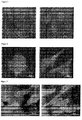
Figure 1 - Scaffold Architecture: Top, from left to right: smooth films; porous films; porous non-aligned electro-spun scaffolds. Bottom, from left to right: porous aligned; non-aligned; and aligned electro-spun scaffolds. -
Figure 2 -Biological response using human osteosarcoma cells: Top, from left to right: smooth films; porous films; porous non-aligned electro-spun scaffolds. Bottom, from left to right: porous aligned; non-aligned; and aligned electro-spun scaffolds. Cells attached only on the non-aligned and aligned electro-spun scaffolds. Moreover, only aligned electro-spun mats promote cell alignment in the direction of the substrate topography. -
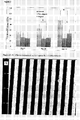
Figure 3 - Biological evaluation using human osteosarcoma cells: Cells exposed to nano-textured scaffolds exhibit significantly reduced metabolic activity after 10 and 14 days in culture. -
Figure 4A -D: AFM micrographs of scaffold option No 2 (2200x1800x35). -
Figure 5 : Biological response of human osteosarcoma cells on non-grooved scaffolds. No alignment is observed -
Figure 6 : Biological response of human osteosarcoma cells on scaffold option 1 (2200x 1 800x3 00). The cells align in the direction of the underlying topography. -
Figure 7 : Biological response of human osteosarcoma cells on scaffold option 2 (2200x1800x35). No alignment is observed. -
Figure 8 : Biological response of human fibroblasts on non-grooved scaffolds. No alignment is observed. -
Figure 9 : Biological response of human fibroblasts on scaffold option 1 (2200x1800x300). Alignment is observed due to the underlying topography. -
Figure 10 : Biological response of human fibroblasts on scaffold option 2 (2200x1800x35). No alignment is observed. -
Figure 11 : Biological response of human osteosarcoma onscaffolds option - Poly(glycolide-co-lactide) (PLGA) was purchased from PURAC Biomaterials, Netherlands. AlamarBlue® was purchased from BioSource International, Invitrogen, Dun Laoghaire, Ireland. Nano imprinted samples were fabricated by CRANN (Centre for Research on Adaptive Nanostructures and Nanodevices, Trinity College, Dublin). One of the nano imprint stamps (line width - 350nm, groove width - 250nm, groove depth - 35nm) had to be sourced externally from NIL Technology Denmark. µ-
chamber 12 well culture slides for cell culture were purchased from Thistle Scientific Ltd, Glasgow. - Human osteosarcoma cells (SAOS2) were obtained from the European Collection of Cell Cultures, Salisbury, United Kingdom. Human Osteoblast cells were purchased from Lonza group Ltd, Switzerland. Bovine tenocytes were isolated from bovine tendons collected from a local abattoir. Human Tenocytes were kindly donation by Prof. Graham Riley (UEA, UK).
- All other material was purchased from Sigma Aldrich unless otherwise stated.
- The electrospinning set up consisted of a high voltage power supply (Gamma High Voltage, USA), a syringe pump (NE-1000, New Era Pump Systems Inc., USA) and a rotating drum. Poly(glycolide-co-lactide) (PLGA) was dissolved in chloroform to prepare an 8% w/v solution. The polymer solution was loaded into a syringe (Becton, Dickinson and Company, Ireland) fitted with an 18G stainless steel needle (Becton, Dickinson and Company, Ireland). The needle was subsequently connected to a high voltage power supply. Details of injection speed and voltage settings are reported in Table 1. The nanofibres were collected on metallic rotating drum with a diameter of 10cm, covered in tin foil. The drum was placed at a distance of 18cm away from the tip of the needle. For aligned samples a rotational speed was set to 1480rpm while randomly aligned samples were spun at 80rpm.
Table 1 Electrospinning parameters used to produce aligned and random sheets of PLGA and PLA/PCL (Polylactic Acid/polycaprolactone) Material: PLGA Concentration: 8% w/v Solvent: Chloroform Voltage: 15 kV Distance: 18cm Injection Speed: 10µl/min Gauge Needle: 18G - PLGA solution (8% w/v chloroform) was used to fabricate polymer films with no topographical features (i.e. smooth surface). As solvent casting method was employed to create thin polymer films. Briefly, the polymer solution was gently poured into the aluminium foil trays, avoiding bubble formation. The trays were covered with an aluminium foil cover to control the evaporation rate of the solvent.
- Nano imprinting was carried out at CRANN (Centre for Research on Adaptive Nanostructures and Nanodevices, Trinity College, Dublin) facilities. The films were initially solvent cast by Proxy Biomedical and forwarded to CRANN for imprinting. The dimensions of the grooves are listed in Table 2 below.
Table 2 List of nano-imprinted groove dimensions Line Width (nm) Groove Width (nm) Groove Depth (nm) Supplied By 1 1800 2200 CRANN 2 1800 2200 CRANN 3 1800 2200 CRANN 4 250 350 CRANN Control N/A N/A N/A Proxy Biomedical - Human osteosarcoma (SAOS2) cells were routinely grown in McCoy's 5A basal media supplemented with 10% fetal bovine serum and 1% penicillin/streptomycin. Cells were subcultured when they reached 80% confluence (once a week) and maintained at 37°C and 5% carbon dioxide. Media was changed every 2-3 days. After 7 days the media was supplemented with ascorbic acid (50mg/L), dexamethasone (10 ng/L) and b-glycerophosphate (10mM) to differentiate the cells. The time point for this cell type was 5, 10, and 14 days.
- Natural human osteoblasts (Lonza, CC-2538) were cultured in Dulbecco's modified Eagle's medium (DMEM) supplemented with 10% Fetal Bovine Serum (FBS) and 1% penicillin/streptomycin in 75cm2 flasks. The osteoblast culture was maintained at 37°C in a humidified CO2 incubator until they were approximately 80% confluent, with media being changed every 3 days. At
passages - Bovine/human tenocytes cells were grown in Dulbecco's modified Eagle's medium (DMEM) supplemented 10% fetal bovine serum and 1% penicillin/streptomycin. Cells were subcultures when they reached 80% confluence (once a week) and maintained at 37°C and 5% carbon dioxide. Media was changed every 2-3 days. The time points for this cell type were 2, 5, and 7 days.
- Live/Dead® Cell Viability Assay (BioSource International, Invitrogen, Dun Laoghaire, Ireland) was performed to examine the cell viability after seeding them on nanofibers. The protocol was adapted from the Live/Dead® Viability/Cytotoxicity Kit *for mammalian cells* (Invitrogen). Briefly, the cells were washed with HBSS and the staining solution of Calcein and Ethidium Homodimer was added. The cells were incubated at 37°C for 30 to 45 minutes. Following staining, the cells were viewed using flurosence microscopy and analysed using ImageJ.
- Cell metabolic activity was determined using alamarBlue® cell metabolic assay (BioSource International, Invitrogen, Dun Laoghaire, Ireland). To determine the cell metabolic activity throughout the study this assay was performed at predetermined time depending on cell type (mentioned above in cell culture section). Briefly, alamarBlue® dye was diluted with Hank's Balance Salt Solution, to make a 10% (v/v) alamarBlue® solution. Media was removed from each well and 0.5ml Alamar Blue® solution was added to each well. After an incubation time of 1.5 hours at 37°C, the absorbance was measured at wavelengths of 550 and 595nm using a micro plate reader (Varioskan Flash, Thermo Scientific). The level of metabolic activity was calculated using the "simplified method of calculating per cent reduction" according to the supplier's protocol.
- Quantification of cell alignment, density, and morphology was carried out using immunocytochemistry. Cell nuclei stained with DAPI and f-actin filaments stained with rhodamine phalloidin. Briefly, cells were fixed by incubation with 4% paraformaldehyde for 5mins at room temperature. The cells were then permeabilised by washing with 0.2% Triton X. The cells were then incubated with DAPI for 5mins, and after with rhodamine phalloidin for one hour. Cell alignment was evaluated using the 'count tool' in ImagePro. The 'count tool' measured the area, aspect, and alignment of the DAPI stained nuclei.
- Collagen gel was made by mixing collagen solution 1mg/ml and 2M NaOH, adjusting concentration to ensure pH 7.0. A collagen gel/culture media/cell solution was prepared by mixing the collagen gel and media/cells suspension at a ratio of 1:1. The collagen/cells solution is then placed at the edge of the scaffold. At each time point cells were stained with DAPI and the distance between the nuclei and the edge of the scaffold measured.
- Taqman microarray was used to evaluate the regulation of gene expression. A selection of genes associated with phenotype, adhesion, migration, apoptosis and extracellular matrix protein, listed in Table 3, was investigated. RNA extraction was carried out at the NFB using standard RNA extraction protocols. The Taqman analysis was carried out by Prof. Graham Riley at the University of East Anglia.
Table 3 List of genes investigated using Taqman Microarray. Phenotype Adhesion Migration Apoptosis ECM Tenomodulin Integrin a1 RAC1 Caspase 3 Collagen I Scleraxis Integrin a2 RhoA Caspase 6 Collagen III Tenascin C Integrin a3 ROCK Caspase 7 Collagen IV Decorin Integrin a4 Calpain 1 Caspase 8Collagen V TGFβ2 Integrin a5 Calpain 2 Caspase 9 Collagen VI Follistatin Integrin b1 Collagen XII osteopontin Vinculin bone sialoprotein Paxillin alkaline phosphatase FAK osteocalcin cartilage oligomeric protein lumican versican RUNX2 TGF-β1 - Cells: Human fibroblasts and human and bovine tenocytes were chosen as representative cells for soft tissue repair. Human osteosarcoma and human osteoblasts were chosen as representative cells for hard tissue repair.
- Preliminary Results: Solvent casting was used to fabricate smooth films. Electrospinning was used to fabricate non-aligned and aligned electro-spun mats. A thermal process was used to create porosity insolvent-cast and electro-spun scaffolds.
- The reduction in metabolic activity and the poor control over scaffold architecture lead us to fabricate nano-grooved samples. Table 1 summarises the dimensions of the grooved samples, whilst
Figure 4A - D shows an example of a non-grooved scaffold.Table 1: Dimensions of the nano-grooved solvent casted films. Options Groove width (nm) Line width (nm) (Groove depth (nm) 1 2200 1800 300 2 2200 1800 35 3 2000 2000 2000 4 300 300 300 5 35 35 50 - Taqman gene assay: 3 housekeeping and other 45 genes are investigated for tenocytes and osteoblasts grouped as follows: phenotype; extracellular matrix molecules; apoptosis; migration; attachment; proliferation; immune response; pain.
- Live/dead assay or FACS are used to correlate the metabolic activity to number of live and dead cells.
- Migration assays: are used to evaluate whether there is an ideal topography that will promote cellular migration
- Small animal study: are necessary to evaluate functional neotissue formation. Multiplex ELISA: is used to evaluate protein expression
- As can be seen from
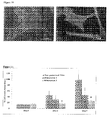
Figure 2 human osteosarcoma cells attach only on non-aligned or aligned electro-spun scaffolds but it is only an aligned electro-spun scaffold that promotes cell alignment in the direction of the substrate topography. This a critical requirement for growing tissue because non-aligned cells results in tissue which is not functional.Figure 3 shows that cells exposed to nano-textured scaffolds exhibit significantly reduced metabolic activity and prolonged culture.Figure 6 shows that human osteosarcoma cells grown on a grooved scaffold of the invention align in the direction of the underlying topography. It is significant that on a grooved scaffold where the groove depth is shallower (35nm as compared to 300nm) no such alignment can be seen. Similarly, human fibroblasts also align in the direction of the underlying topography with a groove depth of 300nm but not with a grooved depth of 35nm.Figure 11 shows that whilst metabolic activity is highest on a non-patterned film, the deeper grooved film (dimension 1) shows higher metabolic activity than the lower grooved scaffold. Similarly,Options - The words "comprises/comprising" and the words "having/including" when used herein with reference to the present invention are used to specify the presence of stated features, integers, steps or components but does not preclude the presence or addition of one or more other features, integers, steps, components or groups thereof.
- It is appreciated that certain features of the invention, which are, for clarity, described in the context of separate embodiments, may also be provided in combination in a single embodiment. Conversely, various features of the invention which are, for brevity, described in the context of a single embodiment, may also be provided separately or in any suitable sub-combination.
-
- 28. Desai, T.A., Micro- and nanoscale structures for tissue engineering constructs. Med. Eng. Phy., 2000. 22(9): p. 595-606.
- 29. Cornwell, K.G., B. Downing, and G.D. Pins, Characterizing fibroblast migration on discrete collagen threads for applications in tissue regeneration. Journal of Biomedical Materials Research Part A, 2004. 71A(1): p. 55-62.
- 30. Zeltinger, J., et al., Effect of Pore Size and Void Fraction on Cellular Adhesion, Proliferation, and Matrix Deposition. Tissue Engineering, 2001. 7(5): p. 557-572.
- 31. Carnell, L.S., et al., Aligned Mats from Electrospun Single Fibers. Macromolecules, 2008.
- 32. Zhong, S., et al., An aligned nanofibrous collagen scaffold by electrospinning and its effects on in vitro fibroblast culture. Journal of Biomedical Materials Research Part A, 2006. 79A(3): p. 456-463.
- 33. Schnell, E., et al., Guidance of glial cell migration and axonal growth on electrospun nanofibers of poly-[epsilon]-caprolactone and a collagen/poly-[epsilon]-caprolactone blend. Biomaterials, 2007. 28(19): p. 3012-3025.
- 34. Stitzel, J., et al., Controlled fabrication of a biological vascular substitute. Biomaterials, 2006. 27(7): p. 1088-1094.
- 35. Lee, L.J., Polymer Nanoengineering for Biomedical Applications. Annals of Biomedical Engineering, 2006. 34(1): p. 75-88.
- 36. Hollister, S.J., Porous scaffold design for tissue engineering. nature materials, 2005. 4: p. 518-524.
- 37. Hutmacher, D.W., Scaffold design and fabrication technologies for engineering tissues-state of the art and future prespectives. Journal of Biomaterials Science. Polymer Edition, 2001. 12(1): p. 107-124.
- 38. Stevens, M. and J. George, Exploring and engineering the cell surface interface. Science, 2005. 310: p. 1135-1138.
- 39. Xia, Y., Nanomaterials at work in biomedical research. Nature Materials, 2008. 7: p. 758-760.
- 40. Barnes, C.P., et al., Nanofiber technology: Designing the next generation of tissue engineering scaffolds. Advanced Drug Delivery Reviews, 2007. 59(14): p. 1413-1433.
- 41. Chew, S., et al., The Role of Electrospinning in the Emerging Field of Nanomedicine. Curr Pharm Des, 2006. 12(36): p. 4751-4770.
- 42. Park, H., et al., Nanofabrication and microfabrication of functional materials for tissue engineering. Tissue Engineering, 2007. 13(8): p. 1867-1877.
- 43. Yao, L., et al., Effect of functionalized micropatterned PLGA on guided neurite growth. Acta Biomaterialia, 2009. 5(2): p. 580-588.
- 44. Lamers, E., et al., The influence of nanoscale grooved substrates on osteoblast behavior and extracellular matrix deposition. Biomaterials, 2010. 31(12): p. 3307-3316.
- 45. Zhu, J., et al., The regulation of phenotype of cultured tenocytes by microgrooved surface structure. Biomaterials, 2010. 31(27): p. 6952-6958.
- 46. Brunetti, V., et al., Neurons sense nanoscale roughness with nanometer sensitivity. Proceedings of the National Academy of Sciences, 2010. 107(14): p. 6264-6269.
Claims (13)
- A scaffold for the growth of tissue comprising a film of supporting material, wherein the film has a plurality of nano-grooves on its surface.
- A scaffold as claimed in claim 1 wherein the groove width of about 2,200nm.
- A scaffold as claimed in either claim 1 or claim 2 wherein the grooves have a line width of about 1,800nm.
- A scaffold as claimed in claim 1, 2 or 3 wherein the groove depth is about 300nm.
- A scaffold as claimed in any preceding claim wherein the supporting material is (poly)glycolide-co-lactide or a PLA/PCL.
- A scaffold as claimed in any preceding claim for the growth of human osteosarcoma cells wherein the grooves have a width of 2,200nm, a line width of 1,800nm and a groove depth of 300nm.
- A scaffold as claimed in any of claims 1 to 5 for the growth of human fibroblasts wherein the grooves have a width of 2,200nm, a line width of 1,800nm and a groove depth of 300nm.
- A scaffold as claimed in any of claims 1 to 5 for the growth of tenocytes wherein the grooves have a width of 2,200nm, a line width of 1,800nm and a groove depth of 300nm.
- Use of a tissue scaffold having nano-grooves on the surface thereof in a method of repairing or replacing tissue in a patient.
- A method of repairing or replacing tissue in a patient comprising use of a tissue scaffold having nano-grooves on the surface thereof.
- A method as claimed in claim 10, wherein the human osteosarcoma cells and the nano-grooves on the mat have a width of 2,200nm, a line width of 1,800nm and a groove depth of 300nm.
- A method as claimed in claim 10 wherein the cells are human fibrblasts and the grooves have a groove width of 2,200nm, a line width of 1,800nm and a groove depth of 300nm.
- A method as claimed in any of claims 10 to 12 wherein the scaffold is a (poly)glycolide-co-lactide or a PLA/PCL scaffold.
Priority Applications (2)
| Application Number | Priority Date | Filing Date | Title |
|---|---|---|---|
| EP11169159A EP2532735A1 (en) | 2011-06-08 | 2011-06-08 | Identifying optimal topography to control cellular function and neo-tissue formation |
| PCT/EP2012/060946 WO2012168466A1 (en) | 2011-06-08 | 2012-06-08 | Identifying optimal topography to control cellular function and neo-tissue formation |
Applications Claiming Priority (1)
| Application Number | Priority Date | Filing Date | Title |
|---|---|---|---|
| EP11169159A EP2532735A1 (en) | 2011-06-08 | 2011-06-08 | Identifying optimal topography to control cellular function and neo-tissue formation |
Publications (1)
| Publication Number | Publication Date |
|---|---|
| EP2532735A1 true EP2532735A1 (en) | 2012-12-12 |
Family
ID=46354202
Family Applications (1)
| Application Number | Title | Priority Date | Filing Date |
|---|---|---|---|
| EP11169159A Withdrawn EP2532735A1 (en) | 2011-06-08 | 2011-06-08 | Identifying optimal topography to control cellular function and neo-tissue formation |
Country Status (2)
| Country | Link |
|---|---|
| EP (1) | EP2532735A1 (en) |
| WO (1) | WO2012168466A1 (en) |
Families Citing this family (1)
| Publication number | Priority date | Publication date | Assignee | Title |
|---|---|---|---|---|
| CN114292103B (en) * | 2021-12-24 | 2023-11-10 | 中国科学院上海硅酸盐研究所 | Bioactive bracket with gear groove-shaped structure and preparation method and application thereof |
Citations (2)
| Publication number | Priority date | Publication date | Assignee | Title |
|---|---|---|---|---|
| WO2001081552A1 (en) * | 2000-04-19 | 2001-11-01 | Iowa State University Research Foundation, Inc. | Patterned substrates and methods for nerve regeneration |
| US20110085968A1 (en) * | 2009-10-13 | 2011-04-14 | The Regents Of The University Of California | Articles comprising nano-materials for geometry-guided stem cell differentiation and enhanced bone growth |
-
2011
- 2011-06-08 EP EP11169159A patent/EP2532735A1/en not_active Withdrawn
-
2012
- 2012-06-08 WO PCT/EP2012/060946 patent/WO2012168466A1/en not_active Ceased
Patent Citations (2)
| Publication number | Priority date | Publication date | Assignee | Title |
|---|---|---|---|---|
| WO2001081552A1 (en) * | 2000-04-19 | 2001-11-01 | Iowa State University Research Foundation, Inc. | Patterned substrates and methods for nerve regeneration |
| US20110085968A1 (en) * | 2009-10-13 | 2011-04-14 | The Regents Of The University Of California | Articles comprising nano-materials for geometry-guided stem cell differentiation and enhanced bone growth |
Non-Patent Citations (25)
| Title |
|---|
| ALTOMARE L ET AL: "Biodegradable microgrooved polymeric surfaces obtained by photolithography for skeletal muscle cell orientation and myotube development", ACTA BIOMATERIALIA, ELSEVIER, AMSTERDAM, NL, vol. 6, no. 6, 1 June 2010 (2010-06-01), pages 1948 - 1957, XP027035710, ISSN: 1742-7061, [retrieved on 20100429] * |
| BAMES, C.P. ET AL.: "Nanofiber technology: Designing the next generation of tissue engineering scaffolds", ADVANCED DRUG DELIVERY REVIEWS, vol. 59, no. 14, 2007, pages 1413 - 1433, XP022356815, DOI: doi:10.1016/j.addr.2007.04.022 |
| BRUNETTI, V. ET AL.: "Neurons sense nanoscale roughness with nanometer sensitivity", PROCEEDINGS OF THE NATIONAL ACADEMY OF SCIENCES, vol. 107, no. 14, 2010, pages 6264 - 6269 |
| CAMELL, L.S. ET AL.: "Aligned Mats from Electrospun Single Fibers", MACROMOLECULES, 2008 |
| CORNWELL, K.G., B. DOWNING, G.D. PINS: "Characterizing fibroblast migration on discrete collagen threads for applications in tissue regeneration", JOURNAL OF BIOMEDICAL MATERIALS RESEARCH PART A, vol. 71A, no. 1, 2004, pages 55 - 62 |
| DESAI, T.A.: "Micro- and nanoscale structures for tissue engineering constructs", MED. ENG. PHY., vol. 22, no. 9, 2000, pages 595 - 606 |
| ENGINEERING, vol. 34, no. 1, 2006, pages 75 - 88 |
| HOLLISTER, S.J.: "Porous scaffold design for tissue engineering", NATURE MATERIALS, vol. 4, 2005, pages 518 - 524, XP002522163, DOI: doi:10.1038/nmat1421 |
| HUTMACHER, D.W.: "Scaffold design andfabrication technologies for engineering tissues-state of the art and future prespectives", JOURNAL OF BIOMATERIALS SCIENCE. POLYMER EDITION, vol. 12, no. 1, 2001, pages 107 - 124 |
| LAMERS, E. ET AL.: "The influence of nanoscale grooved substrates on osteoblast behavior and extracellular matrix deposition", BIOMATERIALS, vol. 31, no. 12, 2010, pages 3307 - 3316, XP026915749, DOI: doi:10.1016/j.biomaterials.2010.01.034 |
| LEE, L.J.: "Polymer Nanoengineering for Biomedical Applications", ANNALS OF BIOMEDICAL |
| LOESBERG ET AL: "The threshold at which substrate nanogroove dimensions may influence fibroblast alignment and adhesion", BIOMATERIALS, ELSEVIER SCIENCE PUBLISHERS BV., BARKING, GB, vol. 28, no. 27, 14 July 2007 (2007-07-14), pages 3944 - 3951, XP022153644, ISSN: 0142-9612, DOI: 10.1016/J.BIOMATERIALS.2007.05.030 * |
| P. ZORLUTUNA ET AL: "Nanopatterned collagen tubes for vascular tissue engineering", JOURNAL OF TISSUE ENGINEERING AND REGENERATIVE MEDICINE, vol. 2, no. 6, 1 August 2008 (2008-08-01), pages 373 - 377, XP055011490, ISSN: 1932-6254, DOI: 10.1002/term.99 * |
| PARK, H. ET AL.: "Nanofabrication and microfabrication of functional materials for tissue engineering", TISSUE ENGINEERING, vol. 13, no. 8, 2007, pages 1867 - 1877, XP002596219, DOI: doi:10.1089/TEN.2006.0198 |
| SCHNELL, E. ET AL.: "Guidance ofglial cell migration and axonal growth on electrospun nanofibers of poly-[epsilon]-caprolactone and a collagenlpoly-[epsilon]-caprolactone blend", BIOMATERIALS, vol. 28, no. 19, 2007, pages 3012 - 3025 |
| STEVENS, M., J. GEORGE: "Exploring and engineering the cell surface interface", SCIENCE, vol. 310, 2005, pages 1135 - 1138 |
| STITZEL, J. ET AL.: "Controlled fabrication of a biological vascular substitute", BIOMATERIALS, vol. 27, no. 7, 2006, pages 1088 - 1094, XP025097066, DOI: doi:10.1016/j.biomaterials.2005.07.048 |
| VAN DELFT F C M J M ET AL: "Manufacturing substrate nano-grooves for studying cell alignment and adhesion", MICROELECTRONIC ENGINEERING, ELSEVIER PUBLISHERS BV., AMSTERDAM, NL, vol. 85, no. 5-6, 1 May 2008 (2008-05-01), pages 1362 - 1366, XP022678748, ISSN: 0167-9317, [retrieved on 20080120], DOI: 10.1016/J.MEE.2008.01.028 * |
| XIA, Y.: "Nanomaterials at work in biomedical research", NATURE MATERIALS, vol. 7, 2008, pages 758 - 760 |
| YIM E K F ET AL: "Significance of synthetic nanostructures in dictating cellular response", NANOMEDICINE: NANOTECHNOLOGY, BIOLOGY AND MEDICINE, ELSEVIER, NL, vol. 1, no. 1, 1 March 2005 (2005-03-01), pages 10 - 21, XP027837934, ISSN: 1549-9634, [retrieved on 20050301] * |
| YIN Z ET AL: "The regulation of tendon stem cell differentiation by the alignment of nanofibers", BIOMATERIALS, ELSEVIER SCIENCE PUBLISHERS BV., BARKING, GB, vol. 31, no. 8, 1 March 2010 (2010-03-01), pages 2163 - 2175, XP026870585, ISSN: 0142-9612, [retrieved on 20091207] * |
| ZELTINGER, J. ET AL.: "Effect of Pore Size and Void Fraction on Cellular Adhesion, Proliferation, and Matrix Deposition", TISSUE ENGINEERING, vol. 7, no. 5, 2001, pages 557 - 572 |
| ZHONG, S. ET AL.: "An aligned nanofibrous collagen scaffold by electrospinning and its effects on in vitro fibroblast culture", JOURNAL OF BIOMEDICAL MATERIALS RESEARCH PART A, vol. 79A, no. 3, 2006, pages 456 - 463 |
| ZHU, J. ET AL.: "The regulation ofphenotype of cultured tenocytes by microgrooved surface structure", BIOMATERIALS, vol. 31, no. 27, 2010, pages 6952 - 6958, XP027124627 |
| ZORLUTUNA P ET AL: "Influence of nanopatterns on endothelial cell adhesion: Enhanced cell retention under shear stress", ACTA BIOMATERIALIA, ELSEVIER, AMSTERDAM, NL, vol. 5, no. 7, 1 September 2009 (2009-09-01), pages 2451 - 2459, XP026500008, ISSN: 1742-7061, [retrieved on 20090331], DOI: 10.1016/J.ACTBIO.2009.03.027 * |
Also Published As
| Publication number | Publication date |
|---|---|
| WO2012168466A1 (en) | 2012-12-13 |
Similar Documents
| Publication | Publication Date | Title |
|---|---|---|
| Shim et al. | Chitosan nano‐/microfibrous double‐layered membrane with rolled‐up three‐dimensional structures for chondrocyte cultivation | |
| US11737990B2 (en) | Nanofiber scaffolds for biological structures | |
| Xu et al. | An anisotropically and heterogeneously aligned patterned electrospun scaffold with tailored mechanical property and improved bioactivity for vascular tissue engineering | |
| Xin et al. | Continuing differentiation of human mesenchymal stem cells and induced chondrogenic and osteogenic lineages in electrospun PLGA nanofiber scaffold | |
| Liu et al. | Electrospun nanofibers for regenerative medicine | |
| Riboldi et al. | Skeletal myogenesis on highly orientated microfibrous polyesterurethane scaffolds | |
| Huang et al. | Nerve guidance conduits from aligned nanofibers: improvement of nerve regeneration through longitudinal nanogrooves on a fiber surface | |
| Vaquette et al. | Aligned poly (L‐lactic‐co‐e‐caprolactone) electrospun microfibers and knitted structure: a novel composite scaffold for ligament tissue engineering | |
| Blakeney et al. | Cell infiltration and growth in a low density, uncompressed three-dimensional electrospun nanofibrous scaffold | |
| Cardwell et al. | Electrospun fibre diameter, not alignment, affects mesenchymal stem cell differentiation into the tendon/ligament lineage | |
| Kai et al. | Electrospun synthetic and natural nanofibers for regenerative medicine and stem cells | |
| Inanç et al. | Periodontal ligament cellular structures engineered with electrospun poly (DL‐lactide‐co‐glycolide) nanofibrous membrane scaffolds | |
| EP1851858B1 (en) | Substrate recognition by differentiable human mesenchymal stem cells | |
| Alves da Silva et al. | Cartilage tissue engineering using electrospun PCL nanofiber meshes and MSCs | |
| Holzwarth et al. | 3D nanofibrous scaffolds for tissue engineering | |
| US8911996B2 (en) | Electrospun scaffolds and methods of generating and using same | |
| Kabiri et al. | Neural differentiation of mouse embryonic stem cells on conductive nanofiber scaffolds | |
| Theodoridis et al. | Hyaline cartilage next generation implants from adipose‐tissue–derived mesenchymal stem cells: Comparative study on 3D‐printed polycaprolactone scaffold patterns | |
| Girija et al. | Collagen-functionalized electrospun smooth and porous polymeric scaffolds for the development of human skin-equivalent | |
| Zanatta et al. | Mesenchymal stem cell adherence on poly (D, L-lactide-Co-glycolide) nanofibers scaffold is integrin-β1 receptor dependent | |
| Huang et al. | Bone marrow stromal cells cultured on poly (lactide‐co‐glycolide)/nano‐hydroxyapatite composites with chemical immobilization of Arg‐Gly‐Asp peptide and preliminary bone regeneration of mandibular defect thereof | |
| Li et al. | Biofabrication of composite tendon constructs with the fibrous arrangement, high cell density, and enhanced cell alignment | |
| Lee et al. | In vivo biofunctionality comparison of different topographic PLLA scaffolds | |
| Molde et al. | A step toward engineering thick tissues: Distributing microfibers within 3D printed frames | |
| Kim et al. | Harnessing nanotopography of electrospun nanofibrous nerve guide conduits (NGCs) for neural tissue engineering |
Legal Events
| Date | Code | Title | Description |
|---|---|---|---|
| PUAI | Public reference made under article 153(3) epc to a published international application that has entered the european phase |
Free format text: ORIGINAL CODE: 0009012 |
|
| AK | Designated contracting states |
Kind code of ref document: A1 Designated state(s): AL AT BE BG CH CY CZ DE DK EE ES FI FR GB GR HR HU IE IS IT LI LT LU LV MC MK MT NL NO PL PT RO RS SE SI SK SM TR |
|
| AX | Request for extension of the european patent |
Extension state: BA ME |
|
| STAA | Information on the status of an ep patent application or granted ep patent |
Free format text: STATUS: THE APPLICATION IS DEEMED TO BE WITHDRAWN |
|
| 18D | Application deemed to be withdrawn |
Effective date: 20130613 |