-
The present invention relates to Apicomplexan rhoptry neck protein 2 (RON2) polypeptide fragments, or homologous polypeptides thereof. The present invention is also directed to a composition comprising such polypeptides for their uses as a medicament, particularly for the prevention or the treatment of Apicomplexan parasite infection. The present invention also encompasses specific antibodies directed against these polypeptides, and method of diagnosis of Apicomplexan parasite infection implementing these polypeptides or antibodies. The invention further comprises a construct for its use in a method for crystallization of the Apicomplexan co-structure AMAI-RON2 complex. The present invention also relates to a computer-readable data storage material. Finally, the invention concerns a method for in silico or in vitro identifying or screening a potential inhibitor of vertebrate, preferably mammalian or avian, more preferably human host cell invasion by an Apicomplexan parasite.
-
Apicomplexan parasites cause widespread diseases including malaria (Plasmodium) and toxoplasmosis (Toxoplasma). The success of these obligate intracellular pathogens is largely due to their unique, conserved mechanism of active invasion (reviewed in1) involving the coordinated secretion of proteins from the microneme and rhoptry organelles2. Recently, the host cell invasion process was likened to a 'kiss and spit' mechanism3 where the parasite provides both ligand and receptor in a synchronised process4 reminiscent of the Intimin - Tir assembly used by enteropathogenic Escherichia coli5. It is noteworthy, however, that this is the first example of such a strategy employed by a eukaryotic or intracellular pathogen.
-
The molecular basis of active invasion by Apicomplexans relies on secretion of a rhoptry neck complex consisting of, at minimum, RONs 2, 4 and 5 into the host cell where RON2 is integrated into the host plasma membrane and RONs 4 and 5 are localized to its cytoplasmic face4. A surface exposed ectodomain of RON2 serves as the receptor6 for the multifunctional, micronemal secreted AMA17-12 displayed on the parasite cell surface (Fig. 6). The assembled AMA1-RON complex forms a ring like structure known as the Moving Junction (MJ), which migrates from the anterior to the posterior of the parasite13 leading to internalization of the parasite into a parasitophorous vacuole (PV). In addition to linking the parasite and host cell membranes, the MJ acts as a diffusion barrier excluding host transmembrane proteins from the PV14, thereby preventing degradation of intracellular parasites15. Thus, the fate of the parasite in the host cell is largely dependent on its ability to actively invade in a MJ dependent process.
-
It is intriguing that AMA1 and RONs 2, 4 and 5 are conserved across Apicomplexan4,16-21, suggesting the potential of a broadly conserved host cell invasion mechanism. In support of this prediction, we have recently shown that the AMA1-RON2 interaction is equally important for the invasive process by Toxoplasma gondii and Plasmodium falciparum6. Coincidentally, structural studies of TgAMA1 from our lab 22, Plasmodium vivax (PvAMA1)23 and P. falciparum (PfAMA1)24,25 revealed a conserved hydrophobic trough in the ectodomain that displays structural hallmarks of a receptor binding surface and thus may be involved in coordinating the binding of RON2. Defining a detailed blueprint of MJ complex formation is therefore critical to both an in-depth understanding of host cell invasion by these important global pathogens, and the strategic design of vaccines and therapeutics.
-
Since the ectoplasmic region of AMA1 is a promising target for a vaccine candidate against Apicomplexan infection, it is important to understand the structure and the function of said AMA1-RON2 interaction.
-
Therefore, there is a need in the art to provide a crystal co-structure and to elucidate the three-dimensional co-structure of the AMA1-RON2 interaction of Apicomplexan parasites such as Toxoplasma or Plasmodial parasites and to use such co-structures or model structures in therapeutic strategies, such as drug design and/or to identify anti-Apicomplexan vaccine candidates capable of inhibiting the host cells infection in human. This is the object of the present invention.
-
The inventors have determined and report herein the comprehensive structural and functional characterization of the Apicomplexan parasite AMA1-RON2, such as TgAMA1-TgRON2 and PfAMA1-PfRON2 assembly from which the inventors have demonstrated mechanism of host cell invasion by Apicomplexan parasites.
-
So, in a first aspect, the present invention is directed to a purified or isolated polypeptide selected from the group of polypeptides consisting of the following polypeptides:
- a)
- i) a polypeptide having at most a 75 amino-acid length and comprising the sequence of the rhoptry neck protein RON2-2 domain of an Apicomplexan parasite, said RON2-2 domain designating the region located between the two last putative transmembrane domains of RON2 peptide and extending into 14 amino-acids residues of the last transmembrane domain; or
- ii) a fragment of said polypeptide as defined in part a) i) above and comprising at least the subfragment of said RON2-2 peptide having the sequence comprised between the two cysteine residues located into said RON2-2 domain, cysteine residues inclusive; and
- b) a polypeptide homologous to the polypeptide as defined in part a) above, said homologous polypeptide having a sequence presenting an identity of at least 50 %, preferably at least 55 %, 60 %, 65 %, 70 %, 80 %, 85 %, 90 %, 95 % or 97,5 %, with that RON2-2 domain and wherein at least two amino-acids residues capable of forming a disulfide bond are present.
-
Preferably, by putative transmembrane domain, it is intended to designate an hydrophobic domain that potentially span membrane, and extending into 14 amino-acids residues of last.
-
Such hydrophobic domain that potentially span membrane can be predicted by software wellknown by the skilled person, for example, but non limited to, the TopPred™ software (http://mobyle.pasteur.fr/cgi-bin/portal.py?#forms::toppred).
-
Preferably, said two amino-acids residues capable of forming a disulfide bond are separated by a fragment having 20, preferably 19, 18, 17, 16, 15, 14, 13, 12, 11, 10, 9, 8 or 7 amino-acids residues, length. More preferably separated by a fragment having 11, 10 or 9 amino-acids residues length.
-
Preferably, said RON2-2 domain is a domain having an amino-acids sequence selected from the group consisting of the sequences:
- SEQ NO: 72: DIX1QX2-4DX5GX6-7PX8-10CX11-19CX20-29; or
- SEQ ID NO: 73: DIX1QX2-4DX5GX6-7PX8-10CX11-20CX21-30; or
- SEQ ID NO: 74: DIX1QX2-4DX5GX6-7PX8-10CX11-21CX22-31, wherein two, preferably one, amino-acids residue selected from the group of the D, I, Q, D, G and P amino-acids residues respectivelly located at position 1, 2, 4, 8,10 and 13 of the sequence SEQ ID NO: 72, SEQ ID NO: 73 or SEQ ID NO: 74 can be any amino-acid residue, and
wherein X1 to X31 designates any amino acid (polypeptide amino-acids sequence representation in one letter code), or a fragment thereof or homologous polypeptide thereof as above defined in paragraph a) ii) and b).
-
More preferably, said RON2-2 domain is a domain having an amino-acids sequence selected from the group consisting of the sequences:
- SEQ ID NO: 72: DIX1QX2-4DX5GX6-7PX6-7PX11-19CX20-29; or
- SEQ ID NO: 73: DIX1QX2-4DX5GX6-7PX8-10CX11-20CX21-30; or
- SEQ ID NO: 74: DIX1QX2-4DX5GX6-7PX8-10CX11-21CX22-31,
wherein X1 to X31 designates any amino acid.
-
As used herein, the terms "polypeptide", "protein" and "peptide" are synonym
-
In a general manner, by amino acid sequence having at least 50 %, preferably, 59 %, 60 %, 65 %, 70 %, 80 %, 85 %, 90 %, 95 % or 97,5 % , identity with a reference amino acid sequence, those having, with respect to the reference sequence, certain modifications, in particular a deletion, addition or substitution of at least one amino acid, a truncation or an elongation are preferred. In the case of a substitution of one or more consecutive or nonconsecutive amino acid(s), the substitutions are preferred in which the substituted amino acids are replaced by "equivalent" amino acids. The expression "equivalent amino acids" is aimed here at indicating any amino acid capable of being substituted with one of the amino acids of the base structure without, however, essentially modifying the biological activities of the reference polypeptide and such as will be defined later, especially in the examples.
-
These equivalent amino acids can be determined either by relying on their structural homology with the amino acids which they replace, or on results of comparative trials of biological activity between the different polypeptides capable of being carried out.
-
Particularly, it is preferred that the homologous polypeptides according to the invention having amino acid sequence having at least 50 %, preferably, 59 %, 60 %, 65 %, 70 %, 80 %, 85 %, 90 %, 95 % or 97,5 % , identity with a reference amino acid sequence, are capable:
- i) to specifically bind the AMA1 ectoplasmic domain, said AMA1 ectoplasmic domain comprising at least the subdomains DI and DII of the ectoplasmic domain of said AMA1 polypeptide, particularly by using the ELISA "binding assay" method as depicted in the examples herein (see Example 1); or
- -ii) to inhibit the formation of the AMA1/RON2-2 complex resulting from the contact between a RON2-2 polypeptide and an AMA1 polypeptide fragment comprising at least the subdomains DI and DII of the ectoplasmic domain of said AMA1 polypeptide particularly by using the competitive ELISA assay as depicted in the examples herein (see Example 1, "binding assay "") or
- - iii) to inhibit the host cells invasion by Apicomplexan parasite, particularly by using the "Invasion assay " method as depicted in the examples herein (see Examle 1);
-
By way of example, mention is made of the possibilities of substitution capable of being carried out without resulting in a profound modification of the biological activity of the reference RON2-2 (see paragraphs i), ii) and iii) above for at least one the preferred biological activities which has to be preserved. More preferably, at least 50 %, 60 %, 70 %, 80 %, 85 %, 90 %, 95 % or 100 % of the biological activity of the reference RON2-2 is preserved). Most preferably, more than 100 % of the biological activity found with the reference RON2-2 domain (or the wild type RON2-2 domain) is obtained wich such homologous polypeptides of the present invention.
-
For example, it is thus possible to replace leucine by valine or isoleucine, aspartic acid by glutamic acid, glutamine by asparagine, arginine by lysine, etc., the reverse substitutions being naturally envisageable under the same conditions.
-
By homologous polypeptides, it is also intended to designate mutated polypeptide having, with respect to the reference RON2-2 polypeptide sequence, at least one amino acid point mutation and at most 2, 3, 4, 5, 6, 7, 8, 9 and 10 point mutations, in particular a deletion, addition or substitution of amino acid. In the case of a substitution of one or more consecutive or nonconsecutive amino acid(s), the substitutions are preferred in which the substituted amino acids are replaced by "equivalent" amino acids as defined above.
-
Two amino-acids or nucleotidic sequences are said to be "identical" if the sequence of amino-acids or nucleotidic residues in the two sequences is the same when aligned for maximum matching. Sequence comparisons between two peptides or polynucleotides are typically performed by comparing sequences of two optimally aligned sequences. Optimal alignment of sequences for comparison may be conducted by homology alignment algorithm well known of the skilled person, or by visual inspection.
-
"Percentage of sequence identity" (or identity) is determined by comparing two optimally aligned sequences over a comparison window, where the portion of the peptide (also for polynucleotide) sequence in the comparison window may comprise additions or deletions (i.e., gaps) as compared to the reference sequence (which does not comprise additions or deletions) for optimal alignment of the two sequences. The percentage is calculated by determining the number of positions at which the identical amino-acid residue or nucleic acid base occurs in both sequences to yield the number of matched positions, dividing the number of matched positions by the total number of positions in the window of comparison and multiplying the result by 100 to yield the percentage of sequence identity. The definition of sequence identity given above is the definition that would use one of skill in the art. The definition by itself does not need the help of any algorithm, the said algorithms being helpful only to achieve the optimal alignments of sequences, rather than the calculation of sequence identity.
-
From the definition given above, it follows that there is a well defined and only one value for the sequence identity between two compared sequences which value corresponds to the value obtained for the best or optimal alignment.
-
It can be used for example the BLAST P "BLAST 2 sequence" (
Tatusova et al., "Blast 2 sequences - a new tool for comparing protein and nucleotide sequences", FEMS Microbiol. Lett. 174:247-250) software that is available in the web site http://www.ncbi.nlm.nih.gov/gorf/bl2.html, which is habitually used by the inventors and in general by the skilled man for comparing and determining the identity between two sequences. The "open gap penalty" and « extension gap penalty » parameters that depends on the substitution matrix selected regarding the nature and the length of the sequence to be compared is directly selected by the software (i.e "5" and "2" respectively for substitution matrix BLOSUM-62). The identity percentage between the two sequences to be compared is directly calculated by the software.
-
In a preferred embodiment, the polypeptide according to the present invention is characterized in that said RON2-2 domain of an Apicomplexan parasite is a RON2-2 domain of an Apicomplexan parasite selected from the group consisting of Aconoidasida and Conoidasida class of Apicomplexan parasite, preferably selected from the group consisting of Plasmodium, Toxoplasma, Eimeria, Neospora, Babesia or Theileria, more preferably selected from the group of Toxoplasma gondii, Plasmodium falciparum, Plasmodium berghei, Plasmodium knowlesi, Plasmodium vivax, Plasmodium yoelii, Babesia bovis, Eimeria tenella, Neospora caninum and Theileria parva species.
-
In a further preferred embodiment, the polypeptide according to the present invention is characterized in that it is a synthetic or a recombinant polypeptide.
-
In a preferred embodiment, the polypeptide according to the present invention is characterized in that it comprises a disulfide bond between the two amino-acids residues capable of forming a disulfide bond present in said subfragment.
-
In a preferred embodiment, the polypeptide according to the present invention is characterized in that it comprises the RON2-2 domain of the RON2 polypeptide having the sequence selected from the sequences SEQ ID NOs. 1, 3, 5, 7, 9, 11, 13, 15, 17 and 19, or a fragment or a homologous sequence thereof as defined in the above paragraphs a) and b).
-
In a preferred embodiment, the polypeptide according to the present invention is characterized in that it comprises the RON2-2 domain having the sequence selected from the sequences SEQ ID NOs. 2, 4, 6, 8, 10, 12, 14, 16, 18 and 20, or a fragment or a homologous sequence thereof as defined in the above paragraphs a) and b).
-
In a preferred embodiment, the polypeptide according to the present invention is characterized in that a disulfide bound (or bridge) is formed between two conserved cysteine amino-acids residues located into the RON2-2 domain.
-
These cysteine amino-acids residues are for example the cysteine residues located respectively in position aa1313 and aa1323 of the sequence SEQ ID NO:1 (T. gondii RON2 polypeptide) or in position aa2037 and aa2049 of the SEQ ID NO:3 (P.falciparum RON2 polypeptide). These two cysteine positions can be easily located from the sequences of the RON2-2 polypeptide SEQ ID NOs. 2, 4, 6, 8, 10, 12, 14, 16, 18 and 20.
-
In a more preferred embodiment, the polypeptide according to the present invention is characterized in that it has at most 64, preferably at most 63, 62, 61, 60, 55, 50, 45, 44, 43, 42, 41 and more preferably 40, 39, 38, 37 amino-acids length.
-
In another more preferred embodiment, the polypeptide according to the present invention is characterized in that it has at least 10 amino-acids length, preferably 11, 12, 13, 14, 15, 20, 25, 30 and more preferably at least 31, 32, 33, 34 or 35 amino-acids length.
-
In a further aspect, the present invention also comprises the isolated nucleic acid, DNA or RNA, coding for the polypeptide according to the present invention, or a complementary nucleic acid thereof.
-
Also is comprised in the present invention, a polypeptide according to the above invention, characterized in that it is recombinant polypeptide or a polypeptide which has been produced by chemical synthesis. These recombinant or synthetic polypeptides preferably comprises the disulfide bound formed between the two cysteines of the RON2-2 domain (or formed between two other natural or modified amino-acid capable to induce a disulfide bound whether these cysteine residues have been mutated, i.e.substitued, in the homologous polypeptide of the present invention).
-
For the production of the polypeptides of the present invention, it is possible to have recourse to genetic engineering procedures making use of micro-organisms transformed by a specific nucleic acid encoding the polypeptide.
-
Consequently, forms also part of the present invention the purified or isolated nucleic acids, such as DNA or RNA, encoding the polypeptides according to the invention.
-
Are also included in the present invention, cloning or expression vectors encoding the polypeptide according to invention, and the host cells transformed by said vectors.
-
A vector of the present invention can be either a prokaryotic or an eukaryotic vector, such as a plasmid or a virus. Expression vectors of the present invention include any DNA or RNA vectors that direct the expression of the recombinant polypeptide of the invention in the host cell, including bacterial, fungal, yeast, insect, other animal such as mammal, and plant cells.
-
The invention aims especially at cloning and/or expression vectors which contain a nucleotide sequence according to the invention.
-
The vectors according to the invention preferably contain elements which allow the expression and/or the secretion of the protein in a determined host cell. The vector must therefore contain a promoter, signals of initiation and termination of translation, as well as appropriate regions of regulation of transcription. It must be able to be maintained in a stable manner in the host cell and can optionally have particular signals which specify the secretion of the translated protein. These different elements are chosen and optimized by the person skilled in the art according to the host cell used. To this effect, the nucleotide sequences according to the invention can be inserted into autonomous replication vectors in the chosen host, or in integrative vectors of the chosen host.
-
Such vectors are prepared by methods currently used by the person skilled in the art, and the resulting vectors can be introduced into an appropriate host by standard methods, such as lipofection, electroporation, thermal shock, or chemical methods.
-
The vectors according to the invention are, for example, vectors of plasmidic or viral origin. They are useful for transforming host cells in order to clone or to express the nucleotide sequences according to the invention.
-
An expression vector of the present invention can be transformed into any suitable host cell to form a recombinant cell. A suitable host cell of the present invention includes any cell capable of expressing a nucleic acid of the present invention inserted into the expression vector. For example, a prokaryotic expression vector can be transformed into a bacterial host cell.
-
The invention likewise comprises the host cells transformed by or comprising a vector according to the invention.
-
In another aspect, the present invention is directed to a composition comprising a polypeptide according to the present invention, or a nucleic acid according to the present invention, for its use as a medicament, as an immunogen or as a vaccine.
-
These polypeptides of the present invention, particularly as immunogens or vaccines according to the invention, are capable of triggering in vivo the synthesis of specific antibodies, particularly directed against the antigenic epitope situated into the RON2-2 domain and comprising the subfragment located bewteen the two cysteine amino-acides residues as defined above.
-
In a preferred embodiment, said composition comprising a polypeptide according to the present invention, or a nucleic acid according to the present invention, is used as a pharmaceutical composition for the prevention or the treatment of Apicomplexan parasite infection.
-
Is also comprised in the present invention, the use of the polypeptide according to the invention for the manufacture of a composition for the prevention or the treatment of Apicomplexan parasite infection or pathology associated to this infection.
-
The present invention is also directed to a method of protecting a vertebrate, subject, preferably a mammalian or avian subject, more preferably a human, against infection by Apicomplexan parasite, the said method comprising administering to the said mammal the polypeptide or the nucleic acid according to the invention.
-
In a more preferred embodiment, said composition comprising a polypeptide according to the present invention, or a nucleic acid according to the present invention, is used as a pharmaceutical composition for the prevention or the treatment of toxoplasmosis, malaria, neosporosis, coccidiosis, or babesiosis or theileriosis.
-
In another aspect, the present invention relates to composition for stimulating an immune response against infection by an Apicomplexan parasite in a mammal, said composition comprising a polypeptide or a nucleic acid according to the invention.
-
Preferably, the said polypeptide or the said nucleic acid according to the invention are formulated as a vaccine in a pharmaceutically effective carrier.
-
The invention also relates to compositions comprising the polypeptides according to the invention with carrier molecules (natural or synthetic), pharmaceutically acceptable. Preferably, said composition according to the invention will be mixed with an excipient and/or a pharmaceutically acceptable vehicle.
-
In the present description, pharmaceutically acceptable vehicle is intended to indicate a compound or a combination of compounds entering into a pharmaceutical composition not provoking secondary reactions and which allows, for example, facilitation of the administration of the active compound(s), an increase in its lifespan and/or in its efficacy in the body, an increase in its solubility in solution or else an improvement in its conservation. These pharmaceutically acceptable vehicles are well known and will be adapted by the person skilled in the art as a function of the nature and of the mode of administration of the active compound(s) chosen.
-
Preferably, these compounds will be administered by the systemic route, in particular by the intravenous route, by the intramuscular, intradermal, intraperitoneal or subcutaneous route, or by the oral route. In a more preferred manner, the composition comprising the antibodies or the polypeptides according to the invention will be administered several times, in a sequential manner.
-
Their modes of administration, dosages and optimum pharmaceutical forms can be determined according to the criteria generally taken into account in the establishment of a treatment adapted to a patient such as, for example, the age or the body weight of the patient, the seriousness of his/her general condition, the tolerance to the treatment and the secondary effects noted.
-
An isolated or purified polypeptide of the present invention can be used as an immunogen to generate antibodies that bind the specific epitope contained in said polypeptide using standard techniques for polyclonal and monoclonal antibody preparation.. The antigenic peptide comprises at least 10 amino acid residues of the subfragment situated between the two cysteine residues on the RON2-2 domain as defined above, such that an antibody raised against the peptide of the invention forms a specific immune complex with that RON2-2 polypeptide. Preferably, the antigenic peptide comprises at least 11, 12, 13, 14 or 15 amino acid residues, more preferably at least 20, 25, 30 or 32, amino acid residues, even more preferably at least 35, 36, 37, 38, 39 or 40 amino acid residues, of the involved RON2-2 polypeptide containing that antigenic epitope.
-
A RON2-2 polypeptidic immunogen typically is used to prepare antibodies by immunizing a suitable subject, (e.g., rabbit, goat, mouse or other mammal) with the immunogen. An appropriate immunogenic preparation can contain, for example, recombinantly expressed or a chemically synthesized RON2-2 polypeptide, or fragment thereof, of the present invention. The preparation can further include an adjuvant, such as Freund's complete or incomplete adjuvant, alum or similar immunostimulatory agent.
-
In another aspect, the present invention is directed to a method for the production of isolated or purified antibodies, or a functional fragment thereof, capable of inhibiting the interaction between the AMA1 polypeptide and the RON2 polypeptide during host cell invasion step by Apicomplexan parasite, characterized in that said method comprises the step of:
- a) immunizing a suitable non-human mammal subject with an immunogen composition comprising a polypeptide according to the present invention, said immunogen composition optionally further includes an adjuvant; and
- b) selecting the antibodies which are capable of specifically binding the RON2-2 domain of RON2 Apicomplexan parasite, preferably said RON2-2 domain exhibiting the epitope comprising a fragment of at least 10 amino-acids residues of the subfragment of said RON2-2 peptide having the sequence comprised between the two cysteine residues located into said RON2-2 domain, or homologous fragment thereof as defined in part a) and b) above when defining the polypeptide according to the present invention.
-
In a more preferred embodiment, the immunogens in step a) are selected from the polypeptides having the sequence SEQ ID NOs. 2, 4, 6, 8, 10, 12, 14, 16, 18 or 20.
-
In another aspect, the present invention is directed to an isolated or purified antibody, or one of its functional fragments, susceptible to be obtained or obtained by the method according to the present invention, said antibody or one of its said fragments being capable of specifically binding the epitope formed by the subfragment of said RON2-2 peptide having the sequence comprised between the two cysteine residues located into said RON2-2 domain, or homologous fragment thereof as defined in claim 1, part a) and b) above when defining the polypeptide according to the present invention.
-
In another aspect, the present invention is directed to an isolated or purified antibody, or one of its functional fragments, susceptible to be obtained or obtained by the method according to the present invention, said antibody or one of its said fragments being capable of specifically binding the AMA1 polypeptide and disrupting the AMA1-RON2-2 binding.
-
Antibodies, monoclonal, polyclonal, chimeric or humanized antibodies, which are specifically directed against the polypeptide of the present invention as defined above are comprised in the invention.
-
In a more preferred embodiment, the method according to the present invention is a method for the production of isolated or purified monoclonal antibodies.
-
The antibodies according to the present invention are preferably specific monoclonal antibodies, especially of murine, chimeric or humanized origin, which can be obtained according to the standard methods well known to the person skilled in the art.
-
In general, for the preparation of monoclonal antibodies or their functional fragments, especially of murine origin, it is possible to refer to classical techniques which are well known of the skilled artisan.
-
By functional fragment of an antibody according to the invention, it is intended to indicate in particular an antibody fragment, such as Fv, scFv (sc for single chain), Fab, F(ab')2, Fab' or scFv-Fc. Said fragments have at least the sequences of the 6 characteristic CDRs of the heavy and light chain of the parent full length antibody and have in particular the capacity to recognize and to bind to the same RON2-2 epitope recognized by the complete parent antibody.
-
In a particular aspect, the antibody or one of its functional fragments, according to the present invention, is characterized in that it is a chimeric antibody and, can optionally comprises the light chain and heavy chain constant regions derived from a human antibody to form a humanized antibody, particularly when it is intended to use said antibody as therapeutic antibody.
-
The antibodies or their fragments of chimeric type according to the invention can be prepared by using the techniques of genetic recombination.
-
Antibody, or one of its functional fragments, according to the invention, or obtained by the process according to the invention, or a nucleic acid according to the invention, as a medicament forms part of the present invention, particularly for the manufacture of a pharmaceutical composition for the prevention or the treatment of Apicomplexan parasite infection.
-
In another aspect, the present invention is directed to a method of in vitro diagnosis of Apicomplexan parasite infection from a biological sample, characterized in that said biological sample is contacted with:
- an antibody susceptible to be obtained or obtained by the method according to the present invention, or an antibody according to the present invention; or
- a polypeptide according to the present invention,
optionally, said antibody or polypeptide is labeled.
-
Said antibody or said polypeptide, or one of their fragments, can be present in the form of an immunoconjugate or of a labeled antibody or polypeptide so as to obtain a detectable and/or quantifiable signal.
-
The labeled antibodies or polypeptide according to the invention, or their fragments include, for example, antibodies or polypeptides called immunoconjugates which can be conjugated, for example, with enzymes such as peroxidase, alkaline phosphatase, beta-D-galactosidase, glucose oxydase, glucose amylase, carbonic anhydrase, acetylcholinesterase, lysozyme, malate dehydrogenase or glucose 6-phosphate dehydrogenase or by a molecule such as biotin, digoxygenin or 5-bromodeoxyuridine. Fluorescent labels can be likewise conjugated to the antibodies or polypeptdides according to the invention and especially include fluorescein and its derivatives, fluorochrome, rhodamine and its derivatives, GFP (GFP for "Green Fluorescent Protein"), dansyl, umbelliferone, etc.. In such conjugates, the antibodies or polypeptides of the invention, or their functional fragments, can be prepared by methods known to the person skilled in the art. They can be coupled to the enzymes or to the fluorescent labels directly or by the intermediary of a spacer group or of a linking group such as a polyaldehyde, like glutaraldehyde, ethylenediaminetetraacetic acid (EDTA), diethylene-triaminepentaacetic acid (DPTA), or in the presence of coupling agents such as those mentioned above for the therapeutic conjugates. The conjugates containing labels of fluorescein type can be prepared by reaction with an isothiocyanate.
-
Other conjugates can likewise include chemoluminescent labels such as luminol and the dioxetanes, bioluminescent labels such as luciferase and luciferin, or else radioactive labels.
-
In a particular embodiment, the polypeptides or the antibodies, or their functional fragments, according to the invention, can be employed in a process for the detection and/or the quantification of Apicomplexan polypeptides, or antibodies directed against these Apicomplexan polypeptides, in a biological sample, for the monitoring of the efficacy of a prophylactic and/or therapeutic Apicomplexan parasite infection treatment
-
Preferably, the biological sample is formed by a biological fluid, such as serum, whole blood, cells, a tissue sample or biopsies of human origin.
-
Any procedure or conventional test can be employed in order to carry out such a detection and/or dosage. Said test can be a competition or sandwich test, or any test known to the person skilled in the art dependent on the formation of an immune complex of antibody-antigen type.
-
By way of example, a preferred method brings into play immunoenzymatic processes according to the ELISA technique, by immunofluorescence, or radioimmunoassay (RIA) technique or equivalent.
-
In another aspect, the present invention is directed to a kit comprising the following elements:
- a)
- an antibody susceptible to be obtained or obtained by the method according to the present invention, or an antibody according to the present invention; or
- a polypeptide according to the present invention,
optionally, said antibody or polypeptide is labeled;
- b) optionally, the suitable reagents for the formation of a immunological reaction; and
- c) optionally, the reagents allowing the demonstration of antigen/antibody and/or antibody/polypeptide complexes produced by the immunological reaction.
-
In another aspect, the present invention is directed to a construct for its use for crystallization of the Apicomplexan co-structure AMA1-RON2 complex, said construct comprising the following elements:
- a) a polypeptide according to the present invention; and, preferably
- b) an AMA1 polypeptide fragment comprising at least the subdomains DI and DII of the ectoplasmic domain of said AMA1 polypeptide, preferably said AMA1 polypeptide fragment is the AMA1 ectoplasmic domain comprising the DI, DII and, optionally DIII subdomains,
said RON2-2 and AMA1 polypeptide fragment being originating from the same species of Apicomplexan parasite.
-
In another aspect, the present invention is directed to a construct for its use for crystallization of the Apicomplexan co-structure AMA1-RON2 complex, said construct comprising the following elements:
- a) a polypeptide according to the present invention, said polypeptide being capable of crossreacting with the AMA1 ectodomain of at least two different species of Apicomplexan parasite; and, preferably
- b) an AMA1 polypeptide fragment comprising at least the subdomains DI and DII of the ectoplasmic domain of said at least AMA1 polypeptide which crossreacts with said polypeptide as defined in a), preferably said AMA1 polypeptide fragment is the AMAI ectoplasmic domain comprising the DI, DII and, optionally DIII subdomains,
said RON2-2 and AMA1 polypeptide fragment being originating from different species of Apicomplexan parasite.
-
In another aspect, the present invention is directed to a method for the preparation of a crystal of a polypeptide complex, preferably for diffraction measurements analysis, said method comprising contacting a polypeptide according to the present invention and an AMA1 polypeptide fragment comprising at least the domains DI and DII of said AMA1 polypeptide, and wherein, said RON2 and AMA1 polypeptide fragment being originating from the same species of Apicomplexan parasite, preferably from Toxoplasma gondii or Plasmodium falciparum species.
-
Are preferred, the AMA1 fragments or derived thereof having the sequence SEQ ID NOs. 21 and 22 when a crystal of the co-structure AMA1-RON2 of Plasmodium falciparum is intended to be prepared.
-
Are also preferred, the AMA1 fragments or derived thereof having the sequence SEQ ID NO. 75 and the fragment aa 36-456 of the SEQ ID NO. 23 when a crystal of the co-structure AMA1-RON2 of
Toxoplasma gondii is intended to be prepared.
-
These isolated polypeptide having the sequence SEQ NOs. 21, 22, 75 and the fragment aa 36-456 of the SEQ NO. 23 form part of the present invention.
-
The crystal which has been obtained or obtainable by the methods according to the invention forms also part of the present invention.
-
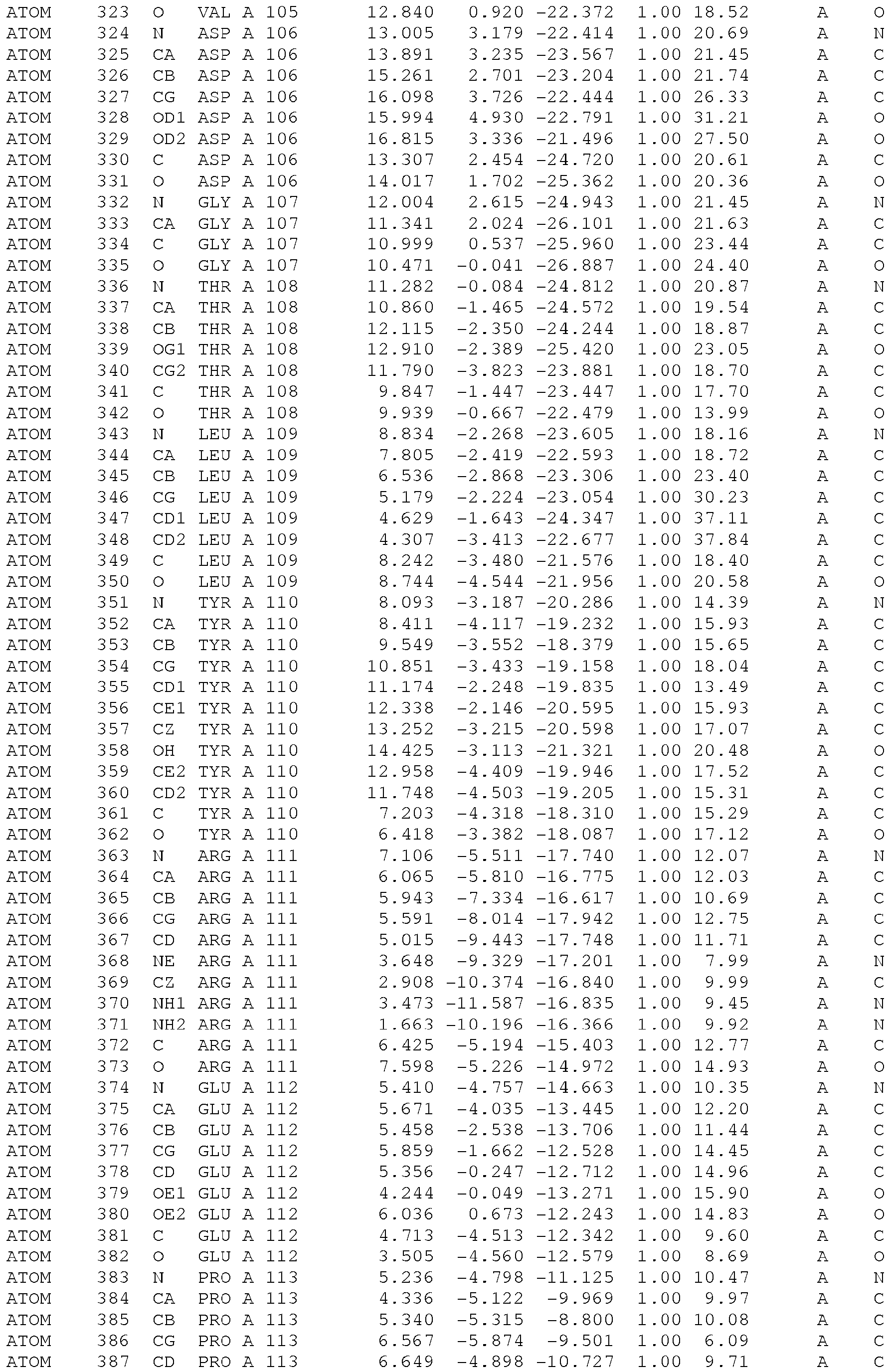
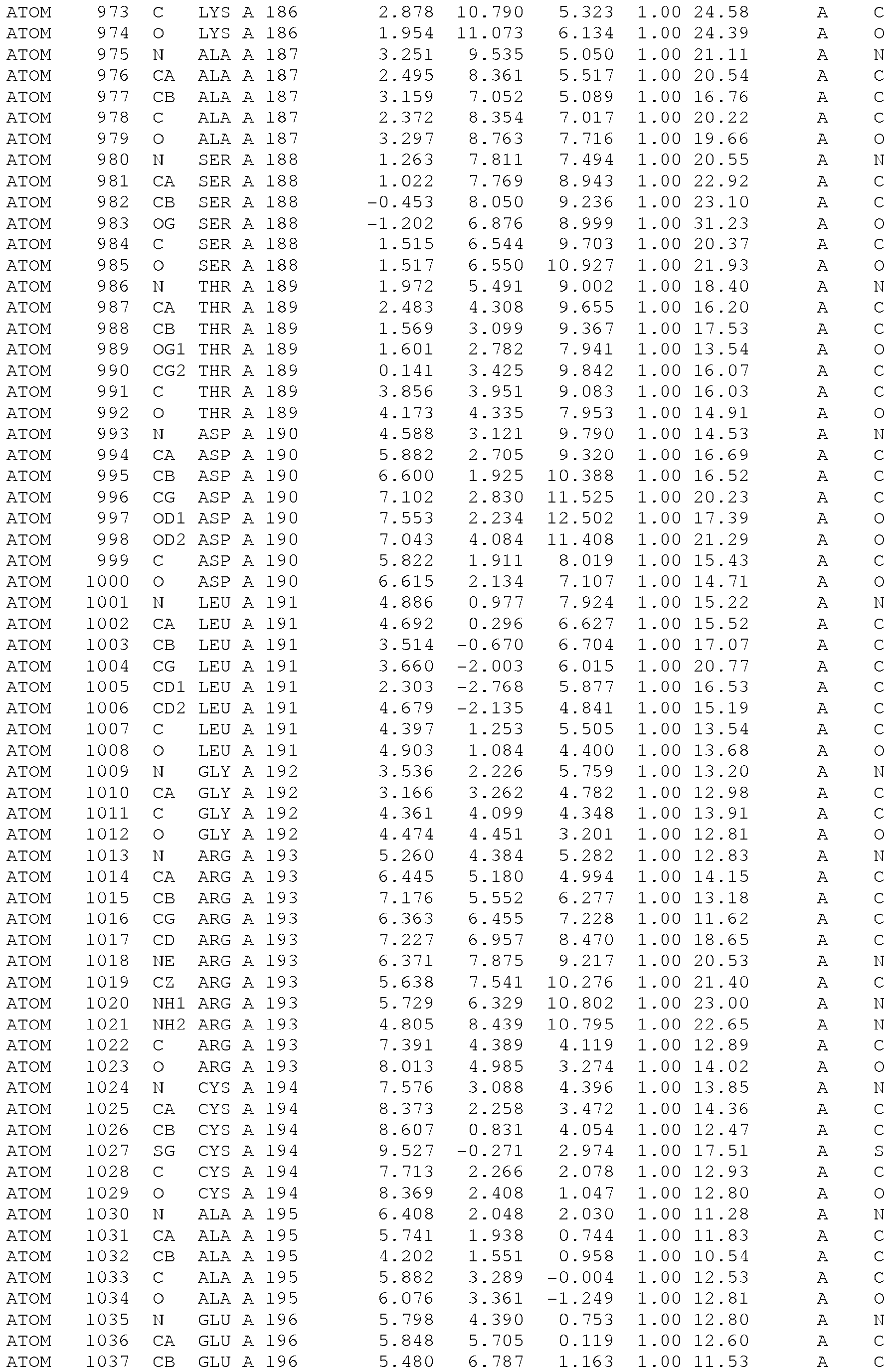
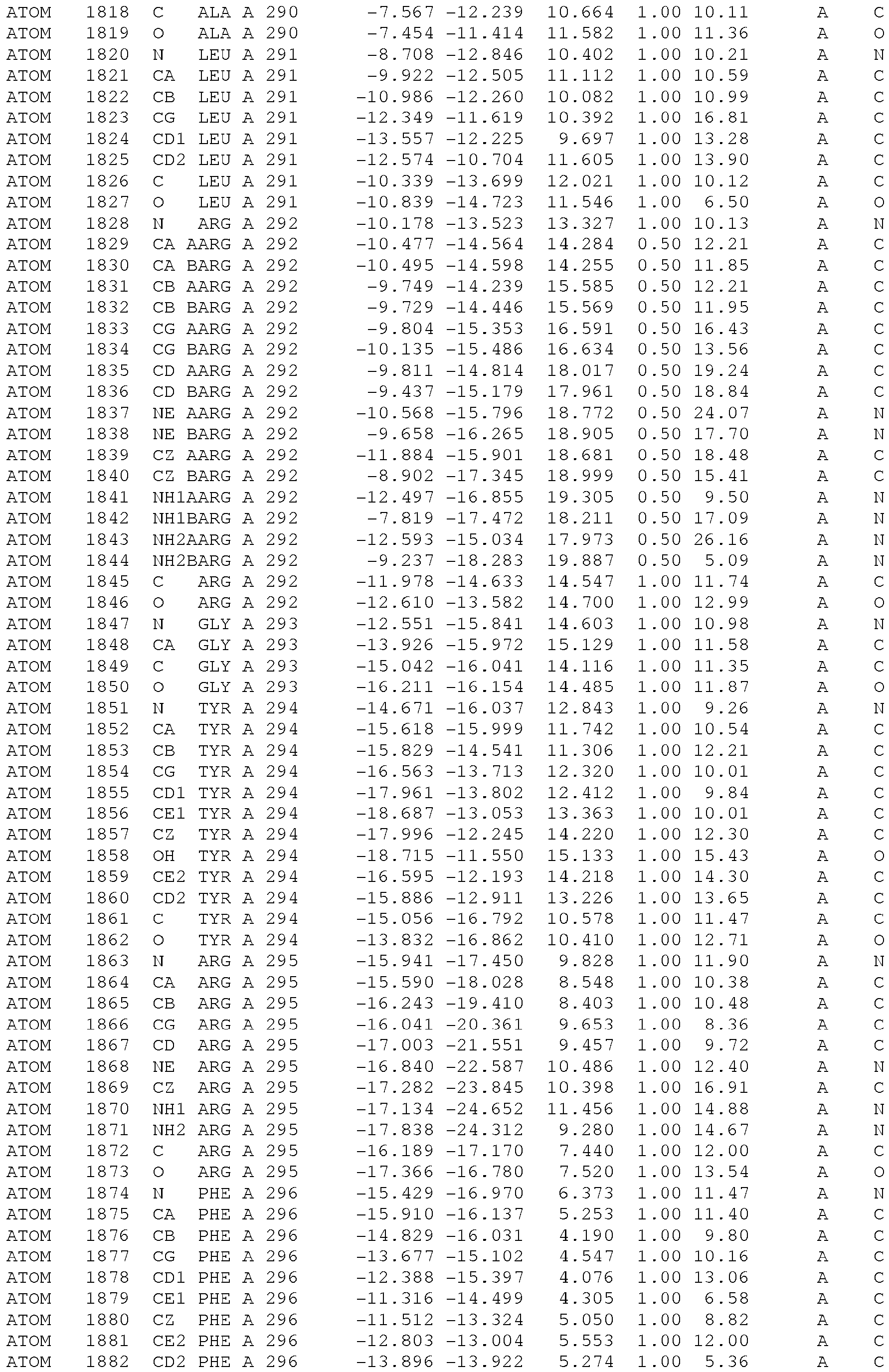
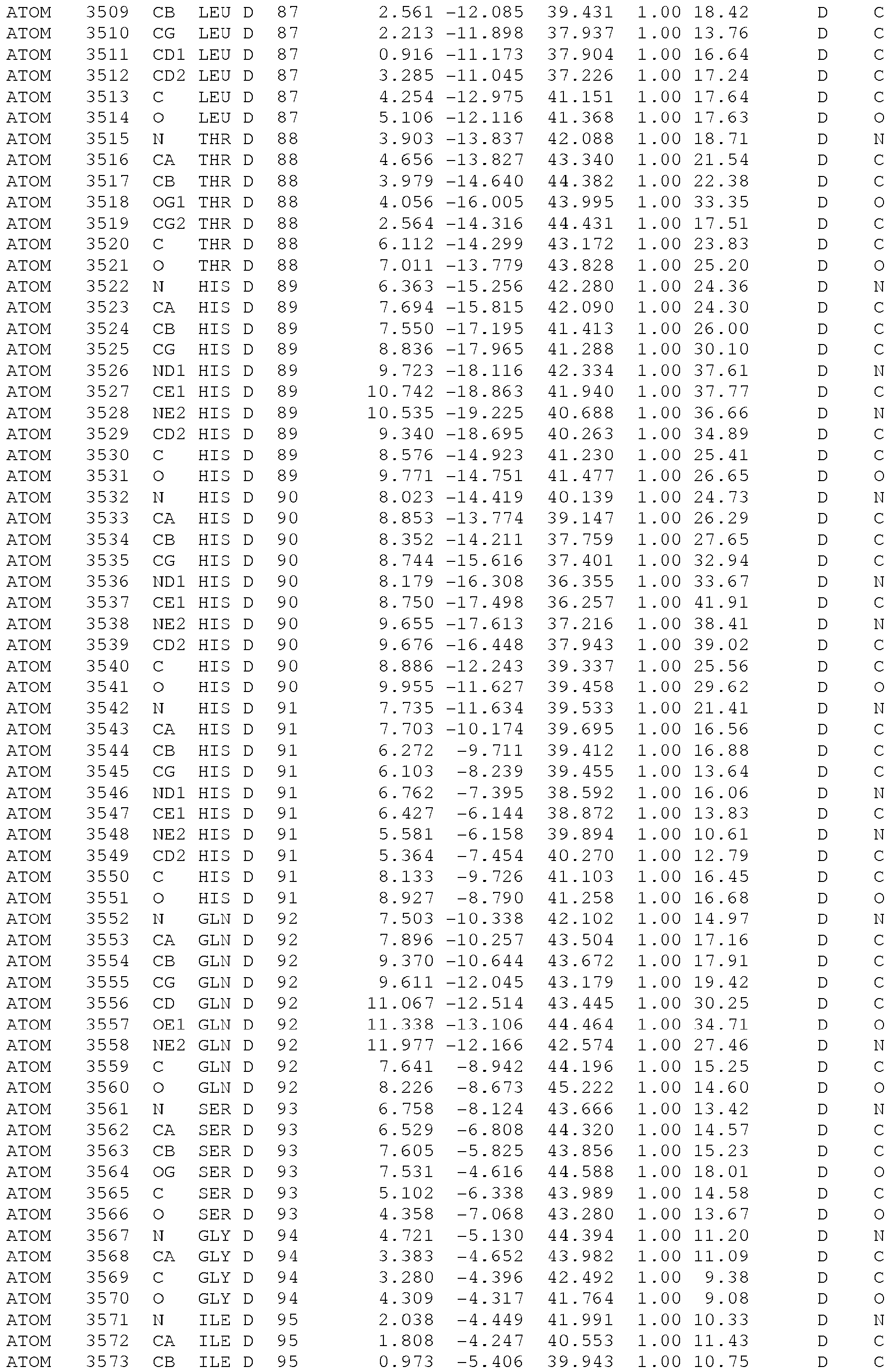
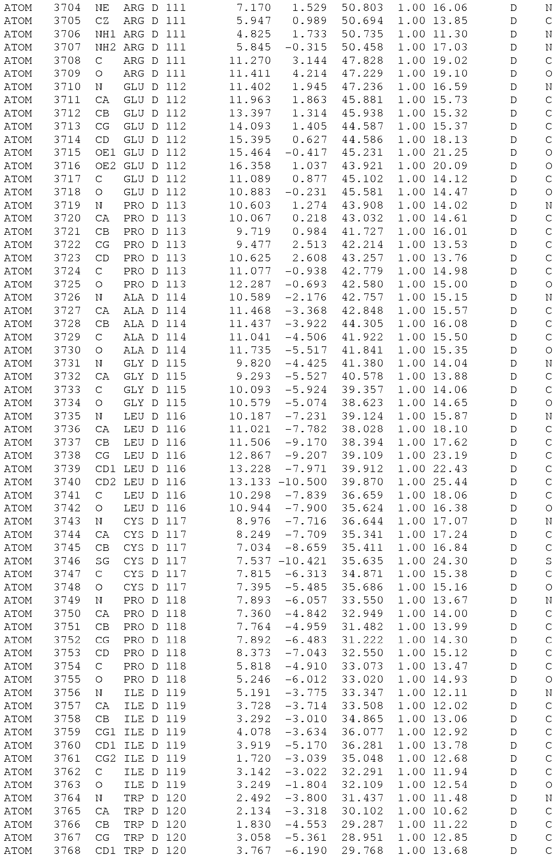
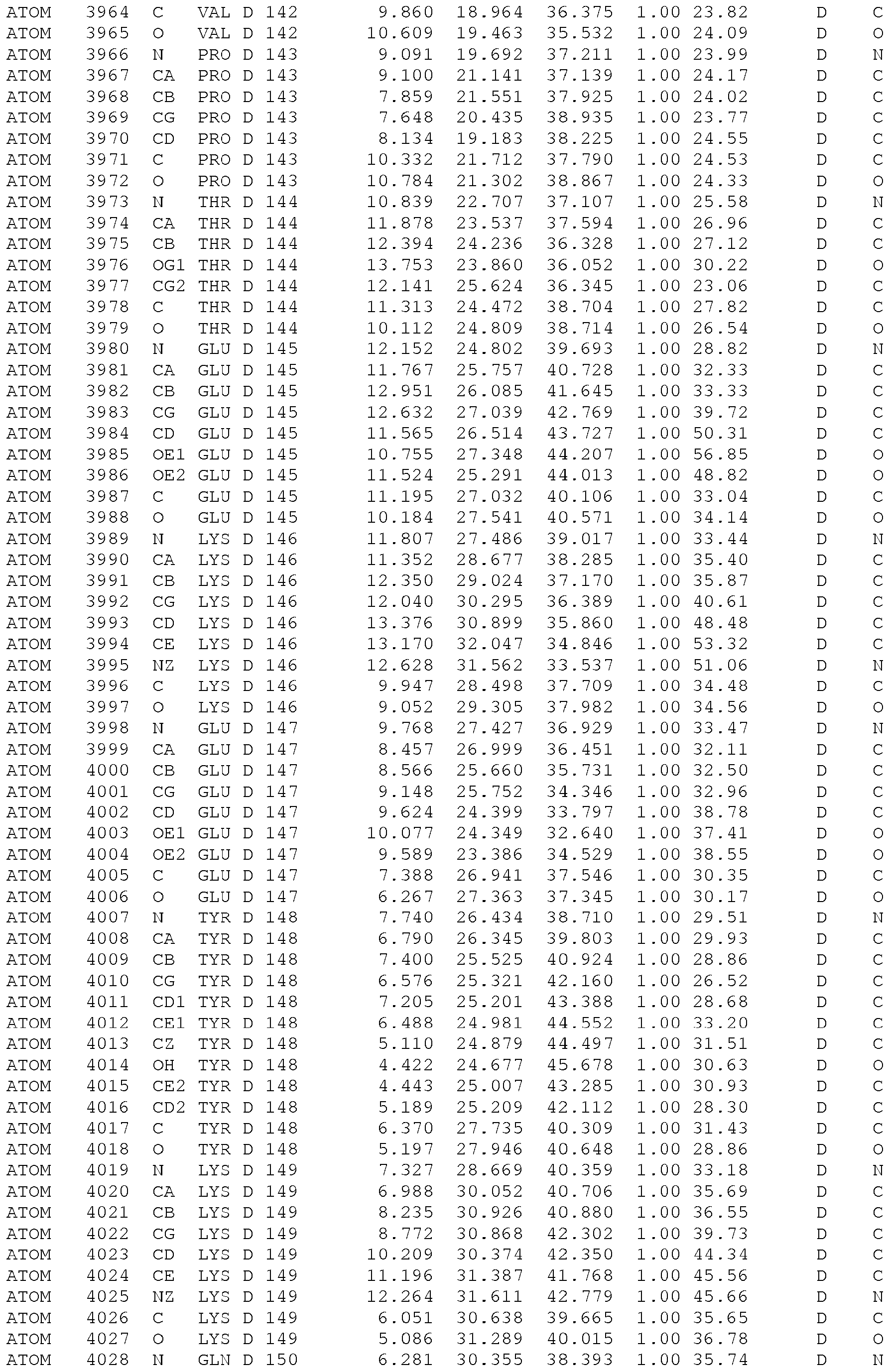
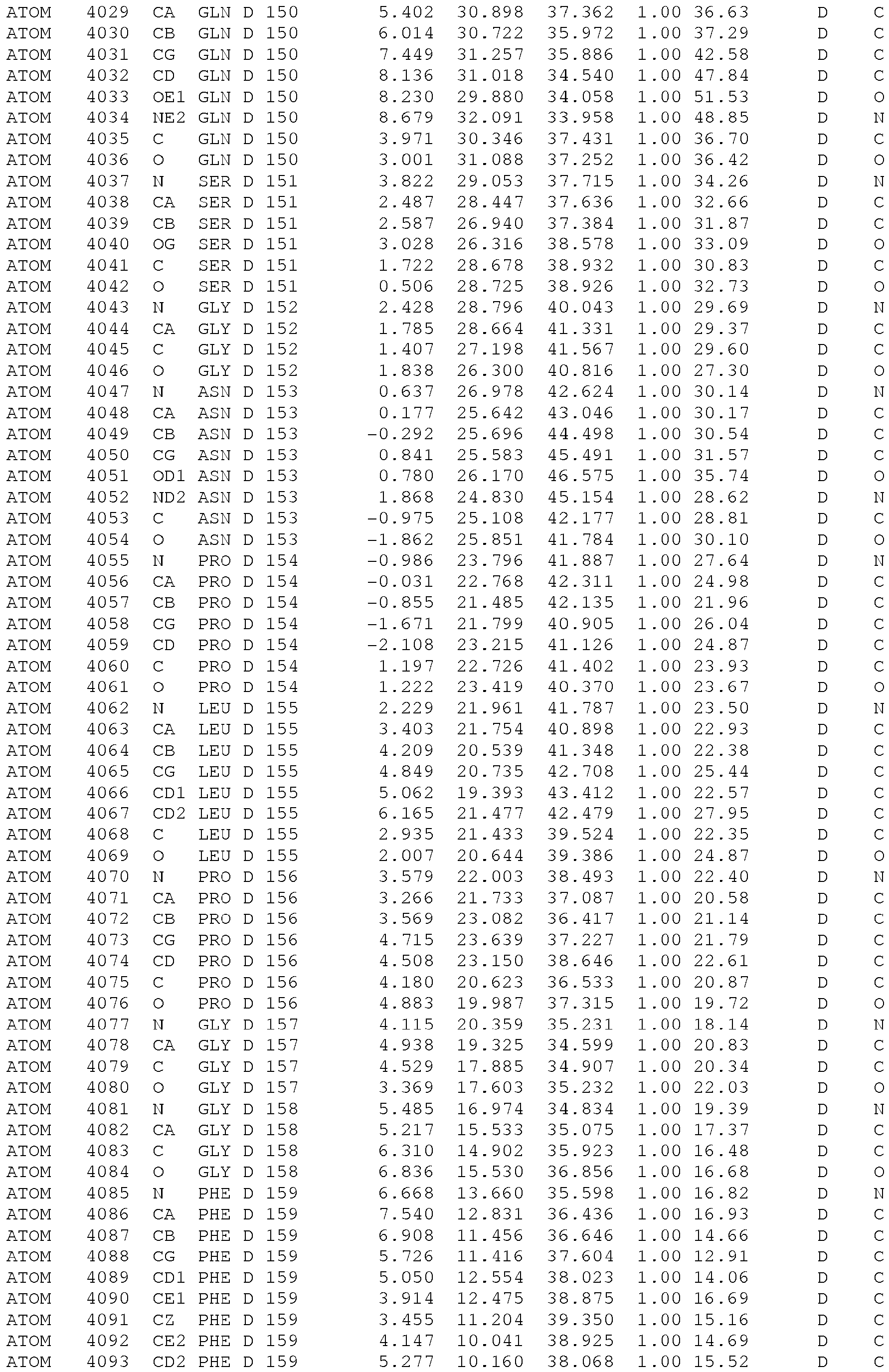
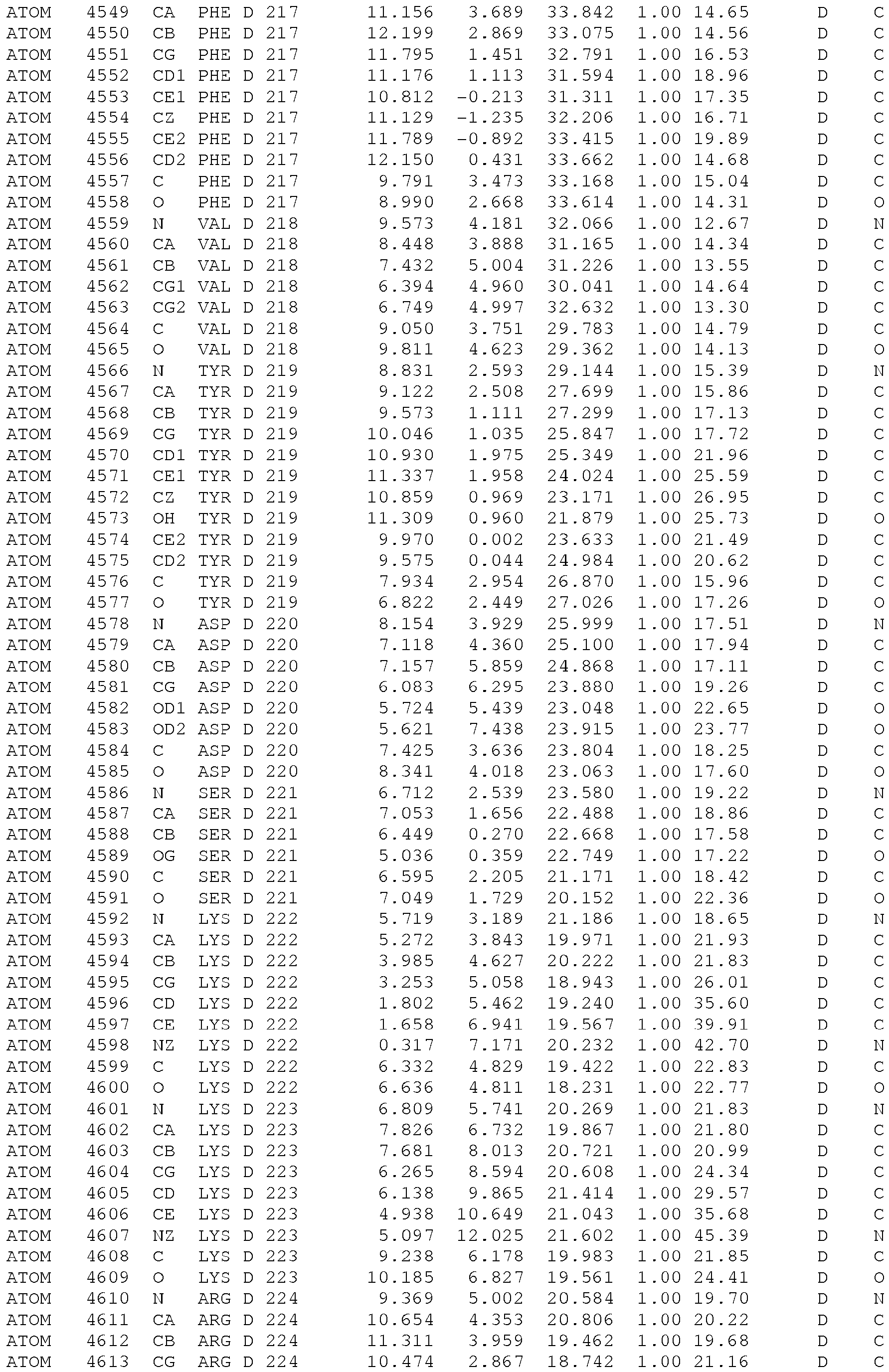
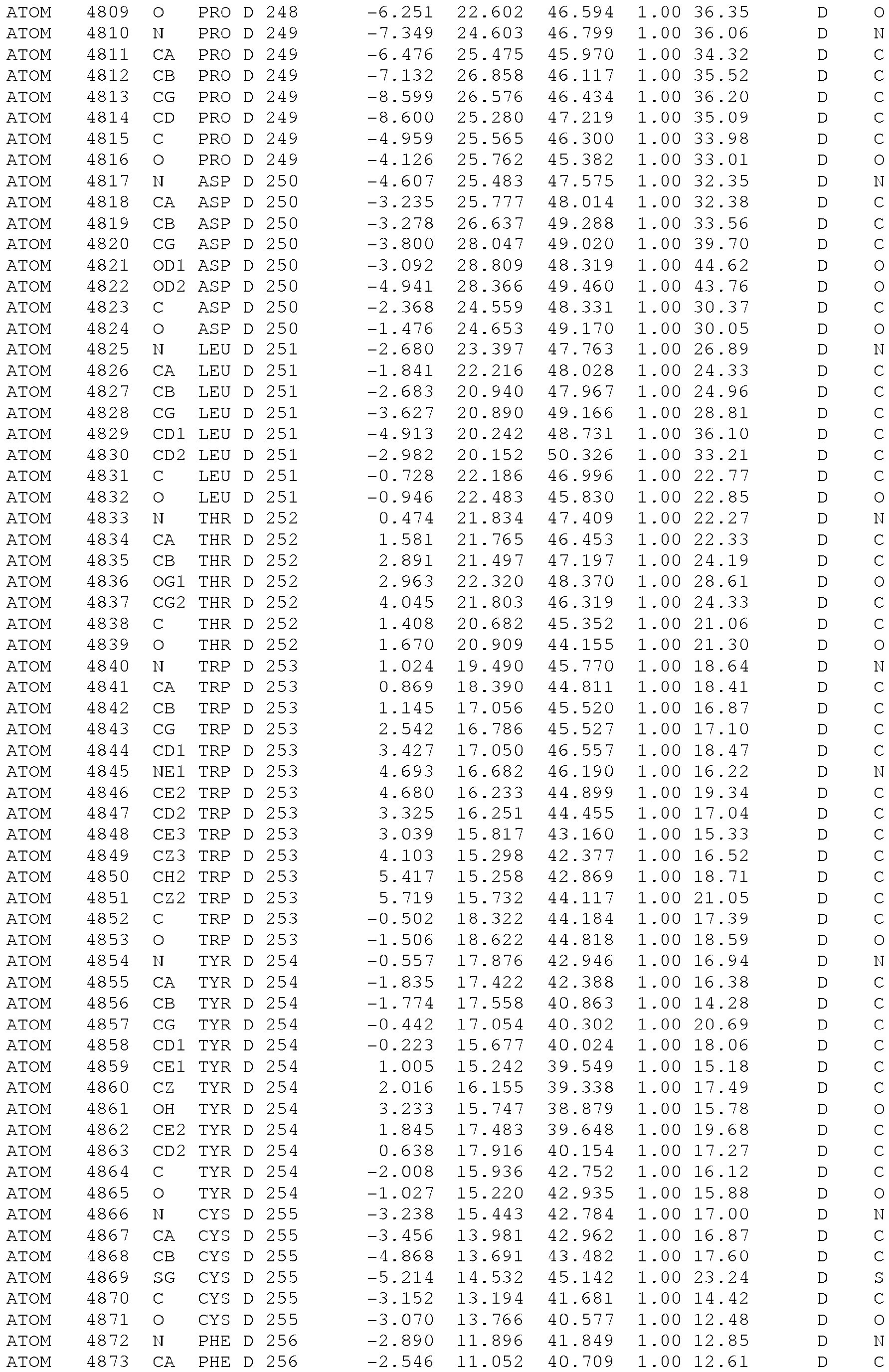
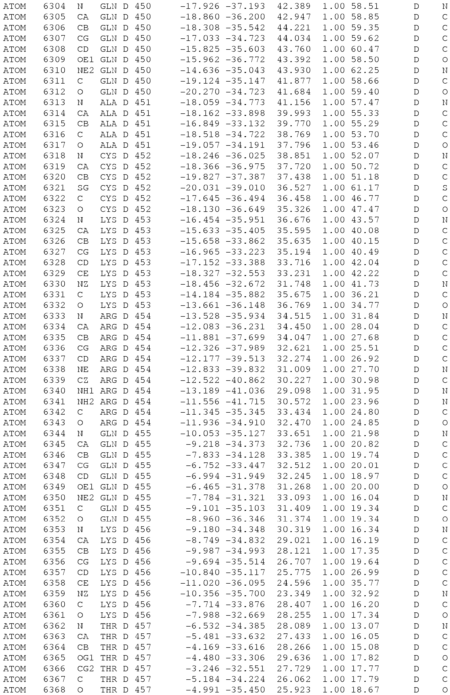
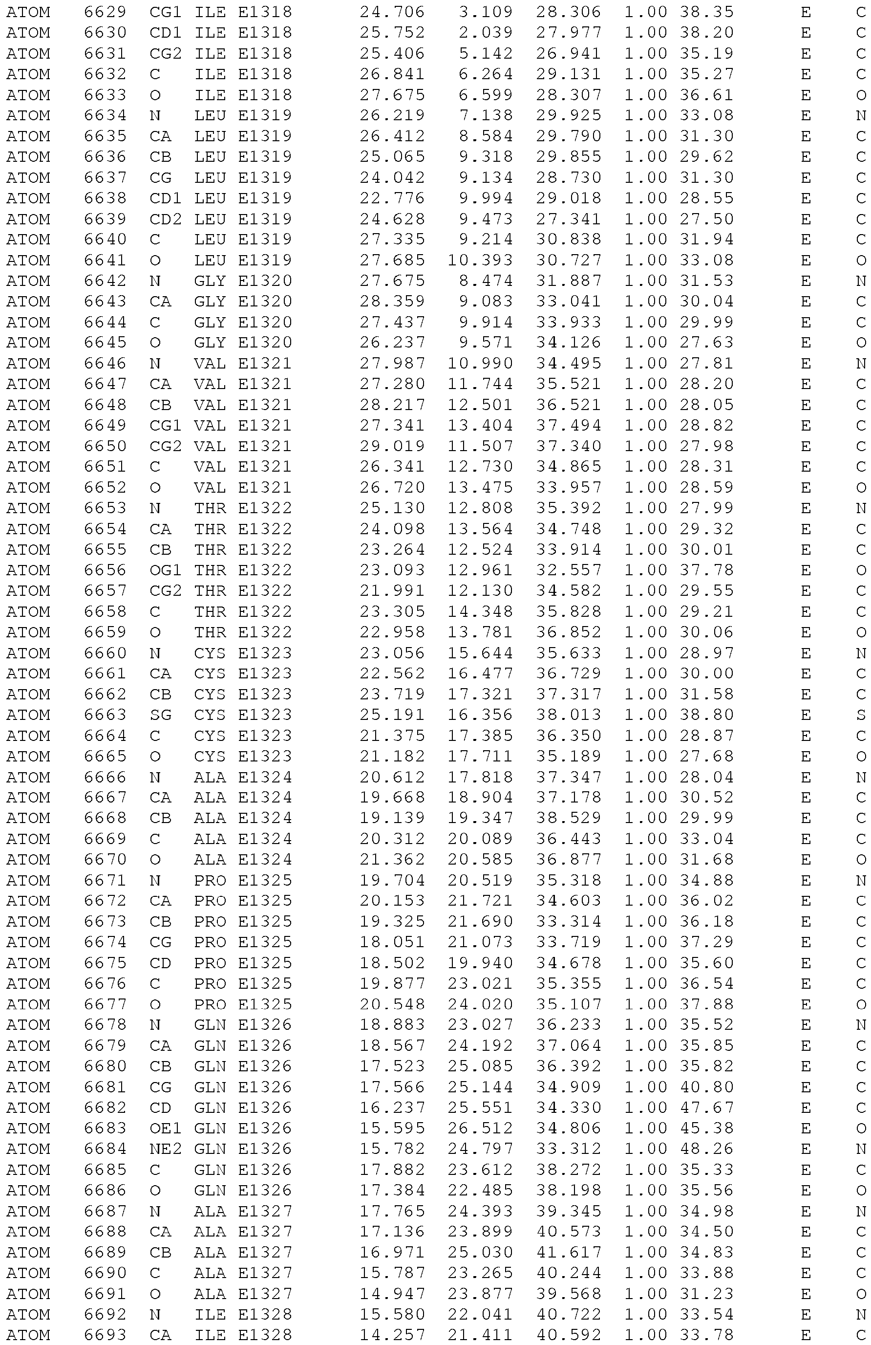
In another aspect, the present invention relates to a computer-readable data storage material encoded with computer-readable data comprising the atomic structure coordinates of the crystal according to the present invention, preferably the three-dimensional structure of the co-structure AMA1-RON2 Apicomplexan parasite, preferably from Toxoplasma genus, such as Toxoplama gondii or from Plasmodium genus, such as Plasmodium falciparum, which substantially conforms to the data given in the examples.
-
According to the present invention, the crystal of the Apicomplexan co-structure AMA1-RON2 complex can be used to determine the ability of a compound to bind to the AMA1 site associated to the RON2-2 binding, or to inhibit said AMA1-RON2 complex formation.
-
This ability is predicted by a structure-based, drug-design method that is based on the three-dimensional structure of the co-structure AMA1-RON2 complex crystal obtained by the inventors. The predicted binding of the chemical compound to the crystal of the present invention is determined by methods standard in the art.
-
Potential compounds which can bind to the AMA1 site associated to the RON2-2 binding, or to inhibit said AMA1-RON2 complex formation, can inhibit the host cells (particularly erythrocytes) invasion. These potential compounds can be designed using structure-based drug design. Until the discovery of the three-dimensional structure of the present invention, no information was available for structure-based development of therapeutic compounds based on the co-structure of the AMA1-RON2 complex. Such rational development could not be executed de novo from available linear amino acid sequence information. Structure-based drug design refers to the use of computer simulation to predict a conformational interaction between the three-dimensional AMA1 ectodomain-RON2 co-structure of the present invention and consequently, a potential therapeutic compound.
-
For example, generally, for a protein to effectively interact with a therapeutic compound, it is necessary that the three-dimensional structure of the therapeutic compound assume a compatible conformation that allows the compound to bind to the protein in such a manner that a desired result is obtained upon binding. In this case, the knowledge of the three-dimensional AMA1 ectodomain-RON2 co-structure enables a skilled artisan to design a therapeutic compound having such a compatible conformation.
-
Thus, present invention also provides:
- methods for identifying, for selecting or for screenind ligands of this AMA1 ectodomain site associated to the RON2-2 binding, said method using the said crystal co-structure analysis, and to design pharmaceutical compounds capable of interacting or binding to that domain AMA1, particularly to certain amino-acid residues of domain identified as contributing to a relevant domain/epitope, as Tyr213 and F197 in T.gondii RON2 sequence; or
- methods for identifying, for selecting or for screening vaccine or drug candidates that could be used to inhibit host cells, particularly erythocytes, invasion by, and/or growth inhibition of, Apicomplexan parasites and that can used in pharmaceutical compositions for the prevention or treatment Apicomplexan parasite infection or their associated diseases such as toxoplasmosis, malaria, neosporosis, coccidiosis, babesiosis or theileriosis.
-
So, in another aspect, the present invention relates to a computer-readable data storage material encoded with computer-readable data comprising atomic structure coordinates of the polypeptide AMA1-RON2 complex obtained by the method above for the preparation of a crystal of a polypeptide complex according to the present invention.
-
In another aspect, the present invention is directed to a method for in silico screening a potential inhibitor of the host cell invasion of mammal by an Apicomplexan parasite comprising the step of:
- selecting a potential inhibitor by performing rational drug design with the three-dimensional structure of the Apicomplexan co-structure AMA1-RON2 complex, determined from the crystal obtained by the method according to the present invention, or by using the computer-readable data storage material according to the present invention, the said selecting being optionally performed in conjunction with computer modeling.
-
In another aspect, the present invention is directed to an in vitro method for screening or identifying a potential inhibitor of the host cell invasion of mammal by an Apicomplexan parasite, said method comprising:
- a) optionally, selecting a potential inhibitor by performing the in silico method according to the present invention;
- b) determining whether this potential inhibitor is capable to specifically bind a polypeptide according to the present invention, or to the AMA1 polypeptide, or fragment thereof comprisng at least the subdomains DI and DII of the ectoplasmic domain of said AMA1 polypeptide, or/and to inhibit the formation of the AMA1/RON2 complex resulting from the contact between a polypeptide according to the present invention and an AMA1 polypeptide fragment comprising at least the subdomains DI and DII of the ectoplasmic domain of said AMA1 polypeptide; and
- c) optionally,
- administrating the potential inhibitor to a non-human animal model which has been infected by an Apicomplexan parasite or contacting in vitro the potential inhibitor with a cell model by using a cellular-based assay capable of reproducing the invasion of mammal host cell by Apicomplexan parasite; and
- d) optionally, detecting the ability of the potential inhibitor for inhibiting the host cell invasion and/or parasite growth.
-
Finally, the present invention is directed to a in vitro method for screening a potential inhibitor of the host cell invasion of mammal by an Apicomplexan parasite, said method comprising the steps of:
- a) contacting said potential inhibitor with :
- -i) a polypeptide according to the present invention, or with an AMA1 polypeptide, or a fragment thereof comprising at least the subdomains DI and DII of the ectoplasmic domain of said AMA1 polypeptide, or
- -ii) an AMA1/RON2-2 complex resulting from the contact between a polypeptide according to the present invention and an AMA1 polypeptide fragment comprising at least the subdomains DI and DII of the ectoplasmic domain of said AMA1 polypeptide;
- b) determining whether this potential inhibitor is capable to:
- -i) specifically bind the polypeptide according to the present invention, or bind said AMA1 polypeptide, or a fragment thereof, or/and
- -ii) specifically inhibit the formation of the AMA1/RON2-2 complex; and
- c) optionally,
- administrating the potential inhibitor to a non-human animal model which has been infected by an Apicomplexan parasite or contacting in vitro the potential inhibitor with a cell model by using a cellular-based assay capable of reproducing the invasion of mammal host cell by Apicomplexan parasite; and
- d) optionally, detecting the ability of the potential inhibitor for inhibiting the host cell invasion and/or parasite growth.
-
The following figures and their legends are illustrative of embodiments of the present invention and are not meant to limit the scope of the invention as encompassed by the claims.
Description of the Figures
-
- Figure 1 : Defining the TgRON2 recognition sequence for TgAMA1
a, TgRON2-2 deletion constructs; TMD, transmembrane domain. b, TgRON2 constructs label TgAMA1 in micronemes of intracellular parasites. Scale bar, 5 µm. c, SPR-based binding of TgRON2 constructs to TgAMA1 (top). TgRON2 constructs inhibit T. gondii invasion of HFF cells (bottom). d, TgRON2sp displays similar invasion inhibition properties to TgRON2-2. Student's t-test; ** p<0.01, *** p<0.001; means ± s.e.m. for n = 2, and n = 3.
- Figure 2 : TgRON2sp induces conformational change in TgAMA1
a, Top and end-on views of TgRON2sp (green) bound in the TgAMA1 apical groove (purple). Electron density map of TgRON2sp shown in green and contoured at 1.0σ
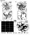
(center). b, Top and side views of TgRON2sp showing the 29Å displacement of the TgAMA1 DII loop from its ordered position in the apo TgAMA1 structure. c, Electrostatic renderings of the TgAMA1 apical surface guarded by the DII loop (top panel - orange) and in the TgRON2sp bound form (middle panel). Corresponding electrostatic view of TgRON2sp (lower panel).
- Figure 3 : TgRON2sp hot spot residues in TgAMA1 binding
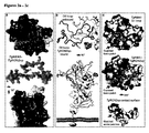
a, Alignment of Apicomplexan RON2 sequences corresponding to TgRON2sp; secondary structure and numbering based on TgRON2. b, SPR-measured relative binding of TgRON2-2 mutants to TgAMA1. c, Effect of TgRON2-2 mutants (6 µM) in inhibiting invasion by T. gondii. Student's t-test; * p<0.05,** p<0.01, *** p<0.001; means ± s.e.m. for n = 2 and n = 3.
- Figure 4 : TgAMA1 contributions to TgRON2sp complex formation
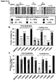
a, Apical view of apo TgAMA1 Y230A DI surface with secondary structure DII loop (yellow). Inset - overlay with apo TgAMA1 (orange) shows reorganization of the DII loop. b, Apical view of TgAMA1 Y230A in complex with TgRON2sp (dark grey). Inset - overlay with TgAMAl-TgRON2sp (purple-green) highlights limited role for Tyr230 in forming a bifurcated hydrogen bond. c, Binding consequences of TgAMA1 mutants and synergistic TgRON2-2 mutants in cell-based assays (left) and spatially represented on the surface of TgAMA1 (right).
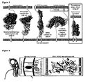
- Figure 5 : A novel host-pathogen protein ligand-receptor complex
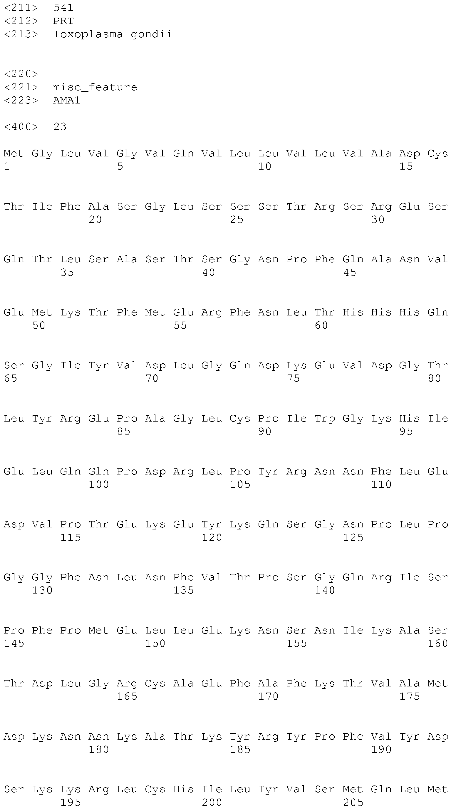
Pathogen derived proteins (left - viral; center - bacterial and right - TgAMA1-TgRON2sp representing the first protozoan complex) are shown in purple and host cell proteins shown in green. Buried surface areas are shown in Å2. PDB IDs left to right: 2VSM, 3LQA, 1F02, 1O6S, 2Y8T.
- Figure 6 : The TgAMAl-TgRON2 complex is the essential link between an Apicomplexan parasite and its target host cell.
Left - schematic of an Apicomplexan parasite in the process of active host cell invasion and parasitophorous vacuole formation. Inset center - the close junction between parasite and host cell which migrates from anterior to posterior during invasion is termed the moving junction (MJ). The critical, highly conserved components of the MJ include the transmembrane microneme-derived protein AMA1 (apical membrane antigen 1) presented on the parasite surface and serving as the receptor for rhoptry-derived RON proteins (rhoptry neck proteins 2, 4, and 5) secreted and assembled at the host cell surface. Inset right - the TgAMAl-TgRON2sp co-structure presented in this work is the first step in structurally defining how the AMA1-RONs complex assembles, and represents the bridge between host and pathogen necessary for active invasion through the MJ constriction.
- Figure 7 : Affinity of TgRON2sp for TgAMA1
(a) SPR based binding kinetics of TgRON2sp on TgAMA1: binding sensorgrams of TgRON2sp at 6 concentrations (from 33 nM to 1066 nM) to TgAMA1 covalently immobilized (2500-3000 RU) on a CM5 chip. All sensorgrams were obtained by subtracting the signal from the control flowcell. The flow rate was 50 µl/min. (b) Incubation with TgRON2sp prevents the binding of TgRON2-2 to TgAMA1 in a dose-dependent fashion. Competitive ELISAs were performed by a pre-incubation for 15 min of 1 µg/mL TgRON2-2 and increasing concentrations of TgRON2sp peptide (0 to 1 µg/mL) before being added to each well coated with 1 µg/mL of TgAMA1. Values represent means ± SD, n=2, from a representative experiment out of 2 independent assays.
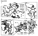
- Figure 8 : Detailed atomic interactions that define the TgAMA1-TgRON2sp interface
(a) TgAMA1 is shown in purple, TgRON2sp in green and buried solvent molecules as blue spheres. (b) Top - TgAMA1 Tyr241 forms a pi-pi stacking interaction with His1301 on TgRON2sp. Middle - The base of the TgRON2sp helix is anchored by a combination of 5 hydrogen bonds incorporating backbone and side chain interactions. Bottom - TgAMA1 Leu99, conserved in sequence and structure with PfAMA1 and PvAMA1, forms the base of the pocket occupied by the N-terminal helix of TgRON2sp. (c) Shape complementarity is observed as TgRON2sp Pro1309 is caged by a trio of TgAMA1 tyrosines and thus occupies the pocket left vacant by the displacement of Trp354 with the DII loop. (d) The hydroxyl group of the tyrosine central to the TgAMA1 apical groove (Tyr230) orients the amidated C-terminus threonine of the peptide (Thr1333) with a single hydrogen bond and also the TgRON2sp coil connector with 2 backbone hydrogen bonds. (e) Ten of the twenty-three predicted hydrogen bonds are dedicated to ordering the previously disordered Loop 2 of TgAMA1 to form the contiguous antiparallel beta-sheet and anchoring the disulfide bond. (f) Pro1325 enhances shape complementarity while 5 hydrogen bonds are spread over 5 residues of the C-terminal coil of TgRON2sp and likely responsible for ordering this region of the peptide.
- Figure 9 : Crystal contacts and an alternate crystal form of TgAMA1-TgRON2sp support the disordered structure of the displaced DII loop
(a) Overlaying the two chains of the co-structure in the monoclinic spacegroup (A - purple; D - violet) with the additional single TgAMA1 chain from the hexagonal spacegroup (forest green) and chain B of unliganded TgAMA1 (blue) reveals differences in the ordered structure of the displaced DII loop. In the unliganded structure, the DII loop is modeled in its entirety with the two tryptophans at the tip of the loop (Trp353/354) anchored by the apical groove central tyrosine (Tyr230). Inset - The last positionally conserved residues defining the base of the DII loop between all 4 structures are Asp337 at the N-terminal end and Pro362 at the C-terminal end. The loop is nearly completely modeled in chain D of the P21 crystal form (missing Ala348-Trp350), but the loop in chain A is only modeled to Pro339 and Asp360. A hexagonal crystal form with one complex in the asymmetric unit revealed crystal packing with the DII loops directed into large solvent channels and subsequently disordered; the extent of ordered structure closely mirrors that seen in chain A of the monoclinic crystal form, with the base of the DII loop modeled to His340 and Asp360. (b) A closer inspection of the crystal packing in the P21 spacegroup shows that a nearby complex closely borders on the DII loop of chain D. Inset - a detailed view of this interface reveals that crystal contacts are likely responsible for the more extensively modeled the displaced DII loop of chain D.
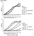
- Figure 10 : Binding analysis of TgRON2-2 mutants by ELISA
ELISAs were carried out with increasing concentrations of recombinant TgRON2-2 mutants on TgAMA1 coated at 1 µg/mL. GST alone was used as a control. Bound TgRON2-2 protein was detected using rat anti-GST (1:1000) followed by an anti-rat IgG horseradish peroxidase conjugate (1:2500) (ZyMAX, Invitrogen). (a) Mutants in the N-terminal helix of and the coil connecting the helix and cystine loop. (b) Mutants in the cystine loop and the C-terminal coil. Values represent means ± SD, n=2, from a representative experiment out of 3 independent assays. Colouring based on the SPR data presented in Fig. 3b shows a close correlation between the data provided for each assay.
- Figure 11 : In vitro binding analysis of TgRON2-2 mutants
TgRON2-2 binds specifically to exposed TgAMA1. Fixed intracellular tachyzoites were permeabilized and incubated with TgRON2-2 at various concentrations (0.04 to 10 µg/mL). Binding to TgAMA1 in micronemes was assessed by immunofluorescence using anti-GST antibody (labelling of TgRON2-2) and anti-ectodomain B3.90 antibody (labelling of TgAMA1). Scale bar, 5µm.
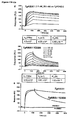
- Figure 12 : Preliminary analysis of TgAMA1 Y230A binding to TgRON2-2
(a) Elution of Ni-affinity purified native and mutant TgAMA1 off the size exclusion column revealed similar elution profiles suggesting the structural core of the mutant was conserved. Inset - SDS-PAGE showed the two proteins migrating in a similar pattern at the expected molecular weight of 48 kDa. (b) TgRON2-2 co-localizes with TgAMA1 Y230A expressed on the surface of BHK-21 cells. (c) ELISA was carried out with increasing concentrations of TgRON2-2 and recombinant TgAMA1 (orange) or TgAMA1 Y230A (yellow) proteins coated at 1 µg/mL. Bound TgRON2-2 protein was detected using rat anti-GST (1:1000) followed by an anti-rat IgG horseradish peroxidase conjugate (1:2500) (ZyMAX, Invitrogen). Binding was visualized using SIGMAFAST-OPD (o-phenylenediamine) tablet (Sigma Aldrich) and absorbance read at 492 nm. Values represent means ± SD, n=2, from a representative experiment out of 2 independent assays. (d) Multi concentration SPR binding study of TgAMA1 and TgAMA1 Y230A on TgRON2-2: binding sensorgrams of TgAMA1 (top) and TgAMA1 Y230A (bottom) at 6 concentrations (from 2.5 nM to 348 nM) to TgRON2-2 (GST fusion) captured at 612 RU on immobilized anti-GST. All sensorgrams were obtained by subtracting the signal from the control flowcell. The flow rate was 50 µL/min. (e) Simplified SPR sensorgram of TgRON2-2 on TgAMA1 (350 nM) highlights the higher dissociation rate of TgAMA1 Y230A relative to TgAMA1. Note that PfAMA1 and PvAMA1 have no cross-reactivity with TgRON2-2.
- Figure 13 : AMA1 sequence alignment of the fully processed 3-domain extracellular region using Kalign and ESPript
Accession numbers for aligned AMA1 sequences are as follows: T. gondii (ME49_055260), Neospora caninum (BAF45372), P. falciparum (XP_001348015.1), P. vivax (XP_001615447), and Babesia bovis (AAS58045.1), with the P vivax sequence modified to represent that crystallized by Pizarro et al 2005. Secondary structure and numbering is based on TgAMA1. Mutants in this study are indicated, and coloured as in Fig. 4c.
- Figure 14 : In vitro binding analysis of TgAMA1 mutants
Transiently transfected BHK-21 cells expressing TgAMA1 or a mutated version of TgAMA1 at their surface were incubated with TgRON2-2 or mutated version of TgRON2-2 at concentrations ranging from 0.1 µg/mL to 10 µg/mL, for 1 h. After five washes in PBS to remove unbound proteins, cells were fixed in 4% PAF and further processed for IFA. Binding was considered as negative when cells expressing a strong signal of TgAMA1 at their surface (using anti-ectodomain B3.90 antibody) were absolutely negative for the detection of TgRON2-2 using anti-GST antibody.
- Figure 15 : Demonstration of the cystine loop as the critical substructure for TgAMA1-TgRON2sp complex formation
(a) TgRON2-2 (red) with three additional truncation constructs honing in on the role of individual substructures; region after the cystine loop (purple), N-terminal helix (pink), and cystine loop (cyan). (b) an ELISA assay and (c) a SPR analysis shows a limited role for the complete section of TgRON2-2 from the end of the cystine loop to the C-terminus (purple), and a construct missing just the N-terminal helix is still competent to bind TgAMA1. However, once the cystine loop is removed (cyan) there is no detectable binding, supporting the insufficiency of the N-terminal helix and coil connector alone to displace the DII loop. Together, these constructs highlight a key role for the cystine loop in mediating complex formation and support our proposed binding mechanism. Error bars in (b) represent SD, n=2, from a representative experiment out of 2 independent assays. SPR-measured binding of TgRON2 segments to TgAMA1 in (c) is expressed in % of binding regarding to TgRON2-2. Error bars in (c) represent SD; t-test, *** p<0.001 for n=3.
- Figure 16 : Inhibition of host cell invasion by Plasmodium merozoites by AMA1-binding PfRON2-2 peptide
Inhibition of the P. falciparum invasion of human red blood cells by PfRON2sp construct at different concentrations compaired with the R1 peptide.
- Figure 17: PfAMA1-PfRON2sp co-structure
Example 1: Material and Method
A) Recombinant proteins, and peptides
-
Recombinant TgAMA1 and TgAMA1 Y230A were produced in insect cells and purified according to previously published protocols22. Expression of pGEX-TgRON2-2 GST was performed in E. coli and purified as described previously6. Primers used for production of recombinant TgRON2-2 constructs and mutant versions of TgAMA1 are listed in Table 3. A 37 residue, disulfide cyclised TgRON2 peptide (TgRON2sp) and an N-terminal FITC-labelled version (TgRON2sp-FITC) corresponding to residues 1297 to 1333 were synthesized by Kinexus (Vancouver, Canada).
B) Structure determination
-
Diffraction data were processed using Imosflm40 and Scala41 in the CCP4 suite of programs42. Initial phases for TgAMAl-TgRON2sp and TgAMA1 Y230A (+/-TgRON2sp) were obtained by molecular replacement using PHASER43 with the unliganded TgAMA1 structure (PDB 2X2Z). Tracing of TgRON2sp was performed manually in COOT44 and all refinements performed with Refmac545. Stereochemical analysis was performed with PROCHECK and SFCHECK in CCP442.
-
SPR, cellular binding and invasion assays. SPR analysis was performed on a BIACORE 3000 at 25°C using running buffer HBS-EP at a flow rate of 50 µL/min. Kinetic and affinity data were calculated using BIAevaluation 4.1 software. Cell culture, site-directed mutagenesis, fluorescence imaging and ELISA techniques were conducted based on previously published protocols6.
C) Molecular biology, recombinant protein production and peptide synthesis
-
The TgAMA1 Y230A construct was generated by site directed mutagenesis using the QuickChange II XL protocol (Stratagene). Recombinant TgAMA1 and TgAMA1 Y230A were produced in insect cells and purified according to a previously published protocol22. Expression of the pGEX-TgRON2-2 GST was performed using E. coli and purified as described previously6. Primers for recombinant RON2 constructs and mutant versions of TgRON2-2 are listed in Table 3. A 37 residue disulfide cyclized TgRON2 peptide (TgRON2sp - residues 1297 to 1333) and the corresponding N-terminal FITC-labelled version (TgRON2sp-FITC) were synthesized by Kinexus (Vancouver, Canada). Lyophilized peptides were solubilized in 100% DMSO and diluted in HBS (20 mM HEPES pH 7.5, 150 mM NaCl) for use in co-crystallization and functional studies.
D) Crystallization and data collection
-
Initial crystals of TgAMAl-TgRON2sp and TgAMA1 Y230A-TgRON2sp were obtained after three months in 25% PEG1500, 100 mM MIB pH 4.0. The final 1.6 µL drops consisted of equal volumes protein (AMA1 (14 mg/mL) incubated with a twofold molar excess of TgRON2sp) and reservoir solution, and equilibrated against 100 µL of reservoir solution. Cryoprotection was performed in 12.5% glycerol for 20 s and flash cooled at 100K directly in the cryostream. Diffraction data to 1.95 Å (TgAMA1-TgRON2sp) and 2.55 Å (TgAMA1 Y230A-TgRON2sp) resolution were collected on beamline 08ID-1 (CMCF) at the Canadian Light Source (CLS; Saskatoon, SK). Diffraction quality crystals for TgAMA1 Y230A were grown in sitting drops consisting of 0.8 µL protein (14 mg/mL) and 0.8 µL reservoir solution (21% PEG3350, 100 mM Tris pH 7.4), and equilibrated against 100 µL of reservoir solution. Cryoprotection of the crystal was carried out in 12.5% glycerol for 20 s and flash cooled at 100K directly in the cryostream. Diffraction data to 2.35Å were collected on beamline 9-2 at the Stanford Synchrotron Radiation Laboratory (SSRL; Menlo Park, CA).
E) Data processing, structure solution and refinement
-
Diffraction data were processed using Imosflm40 and Scala41 in the CCP4 suite of programs42. Initial phases were obtained by molecular replacement using PHASER43 with the unliganded TgAMA1 structure (PDB 2X2Z). Tracing of the RON2 peptide was performed manually in COOT44 with subsequent addition of solvent atoms and refinement in Refmac545. Stereochemical analysis was performed with PROCHECK and SFCHECK in CCP442. Data processing and refinement statistics are presented in Table 1.
F) Bioinformatics
-
RON2 sequences were aligned using ClustalW46 and illustrated in ESPript47. Acquisition numbers for aligned sequences are as follows: Toxoplasma gondii (TGME49_100100), Neospora caninum (NCLIV_064620), Plasmodium falciparum (PF14_0495), Plasmodium vivax (PVX_117880), and Babesia bovis (BBOV_I001630).
-
Binding assays. Techniques for parasite culture, site-directed mutagenesis, fluorescence imaging, and immuno blotting, as well as analysis of the interaction between TgAMA1 and TgRON2 in ELISA, mammalian BHK-21 cells and on RH tachyzoites were conducted as described in previously published protocols6. For binding assay using BHK21 cells, 0.1 µg/mL to 10 µg/mL of recombinant TgRON2 were used (no binding was observed below 0.1 µg/mL for wild-type TgRON2-2). For ELISA, TgAMA1 was coated at 1 µg/mL and TgRON2-2 recombinant proteins used at 0.0009 µg/mL to 10 µg/mL. For competitive ELISA assays, TgRON2-2 was used at 1 µg/mL and pre-incubated with increasing concentrations of TgRON2 synthetic peptide for 15 min before being added to each well. To test binding of recombinant TgRON2-2 proteins on tachyzoites, recombinant TgRON2 proteins were used at concentrations ranging from 0.04 µg/mL to 10 µg/mL. TgRON2sp-FITC was used at 2 µg/mL for 30 min on fixed and permeabilized intracellular tachyzoites.
-
SPR studies of the TgAMAl-TgRON2-2 assembly were performed on a BIACORE 3000 instrument (GE Healthcare, Biacore AB Uppsala, Sweden) at 25°C using running buffer HBS-EP (10 mM HEPES pH 7.4, 150 mM NaCl, 3 mM EDTA and 0.005% Biacore™ surfactant), at a flow rate of 50 µL/min. For GST-capture of recombinant TgRON2-2, monoclonal anti-GST (GE Healthcare) was covalently immobilized on a flow cell of a CM5 sensor chip with EDC/NHS according to the manufacturer's instructions. A control flow cell was prepared with the same chemical treatment without antibody. For affinity measurements, each concentration of TgAMA1 or TgAMA1 Y230A was injected following an injection of TgRON2-2 over 2 min at 910 nM. Regeneration of the flowcell between each run was achieved with a pulse of 10 mM HCl after a 400 s dissociation step. The binding level of TgAMA1(520 nM) on TgRON2-2 mutants was measured at 125 s, immediately after the end of injection. For determination of TgRON2sp affinity, TgAMA1 was covalently immobilized on a CM5 sensor chip by amine coupling and TgRON2-2sp was injected at different concentrations. A pulse of 100 mM HCl was used to regenerate the TgAMA1 coated surface. The kinetic and affinity data were calculated using the BIAevaluation 4.1 software using a Langmuir 1:1 fitting model.
G) Invasion assays
-
Inhibition of invasion was performed as described previously6 with 200 µg/ml (6 µM) of recombinant TgRON2 proteins. Intracellular parasites were detected by immunofluorescence microscopy (IFA) using anti-ROP1 that labelled the nascent PV. When using TgRON2sp, concentrations ranging from 8 nM to 5 µM were used.
H) Statistical analysis
-
Statistical analyses were performed in GraphPad Prism 4 for Windows, with Students's t-test (unpaired, equal variance, two-tailed test) for comparisons with data that fit a normal distribution.
Example 2: Refining the TgAMA1 binding site on TgRON2-2
-
We have shown previously that a recombinant, truncated form of TgRON2 (TgRON2-2), corresponding to a predicted surface exposed region, is capable of binding TgAMA16. While this study established the general location of RON2 recognized by AMA1, it is unlikely that the entire 65 residue sequence of TgRON2-2 is necessary for complex formation. Thus, as a first step towards refining our description of the TgAMAl-TgRON2 assembly, we generated deletion constructs targeting both N-(TgRON2-2 D1) and C-termini (TgRON2-2 D2) of the predicted TgRON2 extracellular loop (Fig. 1a). Despite an in vitro cellular binding assay showing that both deletion constructs were capable of binding TgAMA1 (Fig. 1b), surface plasmon resonance (SPR) and invasion studies revealed an important role for the N-terminal region of TgRON2-2 while the C-terminal region appeared to be dispensable (Fig. 1c). The central region of TgRON2-2 is also likely to be important for binding to TgAMA1 as it harbours a pair of highly conserved cysteines that may form a disulfide bond endowing this region with critical internal structure. Guided by these observations and secondary structure predictions, we synthesized a 37 amino acid, disulfide-cyclised peptide (TgRON2 synthetic peptide - TgRON2sp) corresponding to residues 1297-1333 (Fig 1a). We subsequently demonstrated the functional relevance of this peptide in binding TgAMA1 using cellular binding assays (Fig. 1b), host cell invasion-inhibition assays with nanomolar concentrations of TgRON2sp (Fig. 1d) (SPR determined KD of 1.42 x 10⁻⁸ M; Fig. 7a), and competition ELISA assays with TgRON2-2 (Fig. 7b). These studies, therefore, indicate that the 37 residue TgRON2sp retains the functional binding characteristics of TgRON2-2.
Example 3: Structural rearrangement induced by TgRON2sp
-
Based on the identification and characterization of TgRON2sp we next sought to establish an architectural blueprint of the AMA1-RON2 complex. To this end, we crystallized TgAMA1 in complex with TgRON2sp and refined the structure to a resolution of 1.95 Å (Table 1). A preliminary difference map revealed clear electron density in the apical groove of TgAMA1 enabling accurate tracing of the entire TgRON2sp sequence (Fig. 2a). Intriguingly, the bound TgRON2sp did not simply dock into the center of the apical groove, but rather bound in a U-shaped conformation where its α-helical N-terminus extended through an ordered coil region along one side of the AMA1 groove to a disulfide-tethered beta-hairpin loop and then folded back along the opposite side of the groove through a coil region leading to its C-terminus. By adopting this U-shaped binding strategy, the TgAMAl-TgRON2sp interface results in an extensive buried surface area of 3765 Å2 (Fig. 8a-8f, Table 2) and benefits from significant shape complementarity (complexation significance score of 1.0026). These features likely endow the MJ with the ability to withstand the mechanical sheer force associated with pulling the parasite through the constricted MJ ring.
-
A second noteworthy feature of the TgAMAl-TgRON2sp co-structure is the dramatic conformational change in the DII loop that defines one end AMA1 apical groove (Fig. 2b). This observation is intriguing for two primary reasons. First, the DII loop is predicted to be one of the most influential substructures involved in immune escape and RON2 coordination22,21,27,28 in T. gondii and Plasmodium spp and thus its rearrangement likely reflects a critical regulatory event in MJ assembly. Second, the DII loop is exceptionally well ordered in the apo TgAMA1 structure 22 where it is intimately associated with the ectodomain core through numerous interactions, including a pair of tryptophan residues that anchor the loop into hydrophobic pockets in the apical groove (Fig. 2b - orange). By inducing the conformational changes leading to displacement of the DII loop, TgRON2sp is able to expose the fully functional binding surface on TgAMA1 that displays enhanced shape and charge complementarity. For example, in its reconfigured form, the apical groove is less hydrophobic, instead relying on an extended basic patch to complements acidic patches on the TgRON2sp N-terminal helix and C-terminal tail (Fig. 2c). In addition to establishing an essential role for the DII loop in regulating access to the apical groove, the induced conformational changes also suggest a stepwise binding mechanism where contact is initiated between the end of the AMA1 apical groove opposite the DII loop, and the cystine loop region of RON2. Using this strategy, the binding energy harnessed from the initial interactions can be applied to reconfiguring the DII loop and completing binary complex formation.
Example 4: Defining hot spot residues on TgRON2sp
-
Guided by our high-resolution TgAMA1-TgRON2sp co-structure (Fig. 8a-8f) and sequence alignments (Fig. 3a), we identified individual residues and substructures on TgRON2-2 and probed their functional relevance using SPR, ELISA, cellular binding and invasion inhibition assays. In general, alanine substitutions in the N-terminal helix (D1297, H1301, D1304 and I1305), the coil connecting the helix and cystine loop (P1309), the cystine loop (C1313, I1318, L1319 and C1323), and the C-terminal coil (T1332) all showed some degree of reduced binding to TgAMA1 as determined by SPR and ELISA (Fig. 3b, 10a-10b) consistent with an extensive, composite TgAMAl-TgRON2sp interface. Of particular note, residues D1297, D1304, P1309, C1313, and C1323 all broadly conserved across Apicomplexan were identified as critical for TgAMA1 binding as was the less conserved L1319 located between the cysteines in the cystine loop. To corroborate the SPR based binding studies, we also tested the ability of the TgRON2-2 mutants to bind TgAMA1 in the micronemes of T. gondii tachyzoites (Fig. 11) and inhibit host cell invasion by T. gondii (Fig. 3c). Collectively these data highlight an extensive binding surface and point toward the importance of the cysteine-dependent conformation of RON2 in promoting complex formation consistent with our proposed stepwise binding strategy.
Example 5: Probing the hydrophobic groove of TgAMA1
-
The only previous attempt to identify residues on AMA1 critical for MJ assembly was based on a PfAMA1 mutagenesis study that implicated the central tyrosine (Tyr251) in the hydrophobic groove in complex formation28. Thus, we began our investigation into defining residues on TgAMA1 essential for TgRON2 binding by probing the analogous central tyrosine (Tyr230). When we produced recombinant TgAMA1 Y230A (Fig. 12a) and measured binding to TgRON2-2 in cell-based (Fig. 12b) and ELISA (Fig. 12c) assays, no discernable difference in binding was observed. However, SPR analysis revealed an order of magnitude weaker binding between TgRON2-2 and TgAMA1 Y230A (KD of 10-8 M) relative to native TgAMA1 (KD of 10-9 M) (Fig. 12d, 12e). This difference is significantly less than what would be expected as a result of mutating a major "hot spot" residue. To investigate the structural basis of these observations, we solved the crystal structures of TgAMA1 Y230A in the apo and TgRON2sp bound forms (Table 1). While analysis of the TgAMA1 Y230A structure revealed a conserved core (r.m.s.d of 0.60Å over 384 α-carbons) relative to native TgAMA122, an alternate conformation was observed for the tip of the DII loop proximal to the truncated tyrosine side chain (Fig. 4a). More specifically, Trp353 was rotated 67° to occupy the newly exposed space above Ala230 (Fig. 4a inset). Ultimately, however, binding of TgRON2sp resulted in a similar displacement of the DII loop as observed in the native complex (Fig. 4b) consistent with a largely conserved binary interface. The most notable effect resulting from substituting Tyr230 with an alanine is the loss of two (bifurcated) hydrogen bonds, out of twenty three observed in TgAMAl-TgRON2sp (Fig. 4b inset), that likely corresponds to the reduced affinity measured by SPR.
-
To further probe the role of specific AMA1 residues in promoting binary complex formation, we performed a detailed analysis of the buried surface areas and hydrogen bonding contributions (Fig. 8a-8f, Table 2), and filtered these through a sequence alignment (Fig. 13). Finally, we selected a set of eleven TgAMA1 residues (Fig. 13) to investigate and tested the ability of TgRON2-2 to bind these mutant forms of TgAMA1 expressed on the surface of BHK-21 cells (Fig. 4c, 14). While the individual substitutions of R111 and Y241 (base of the N-terminal helix); Y110 and Y215 (coil connector); F163, I171 and M204 (cystine loop); and E145 (C-term coil) to alanine showed no discernable binding effects, substitutions of F197 (base of cystine loop) or Y213 (base of tyrosine cage surrounding P1309) to alanine, impaired binding (Fig. 4c, 14). In fact, Y213A showed no binding at any of the concentrations tested suggesting that this highly conserved residue is a critical anchor point during complex formation. The F197A mutant showed a more moderated effect with binding disrupted only at the lowest concentration. Thus the F197A variant provided an opportunity to test the potential of synergistic TgAMAl-RON2 mutations. Total abrogation of complex formation was observed between TgAMA1 F197A and either TgRON2-2 I1318A or L1319A (tip of the cystine loop) (Fig. 4c, 14). These synergistic effects prompted us to generate three additional TgRON2-2 constructs comprising 1) from the N-terminal helix to cystine loop, 2) from the coil connector to C-terminus, and 3) from the N-terminal helix to coil connector (Fig. 15a-15c). Analysis of these constructs by ELISA demonstrated clear binding for the first two, but no interaction with the third, effectively isolating the most critical region of TgRON2-2 to the cystine loop (Fig. 15a-15c). Collectively, these results allow to refine our proposed mechanism of binding where the cystine loop in combination with the Tyr213-Pro1309 bridge act as a primary structural brace to anchor the peptide in the apical groove and promote displacement of the DII loop.
Example 6: TgAMAl-TgRON2sp: a structural perspective
-
Among the diverse mechanisms employed by viruses, pathogenic bacteria, and protozoan parasites to enter host cells, one commonality is the utilization of protein complexes to promote host cell attachment and invasion (Fig. 6). Many of the current models describing these processes are derived from seminal structural studies including the Nipah virus attachment glycoprotein in complex with human Ephrin B2 (buried interface of 2786 Å2)36, HIV gp120 with human CD4 (2051 Å2)37, the enteropathogenic E. coli translocated intimin receptor (Tir) with intimin (1294 Å2)38 and Listeria monocytogenes Internalin A with host cell E-cadherin (2630 Å2)39. Our structural characterization of TgAMA1 in complex with TgRONsp is an important addition to these studies as it is the first such complex from a protozoan parasite and incorporates one of the most extensive buried surface areas (3765 Å2) of any host-pathogen binary protein complex. The induced conformational changes in TgAMA1 upon TgRON2sp also provide an illustrative framework to predict secondary roles associated with conformational changes such as protecting surfaces from exposure to the immune system. The extended structure of the DII loop that spans much of the AMA1 ectodomain (Fig. 2b, 9a-9b) may also poise it to function as a molecular switch transmitting a signal from the reorganized apical groove to the parasite cell surface. For example, RON2 binding could trigger such processes as the association of the AMA1 C-terminal tail with the glideosome to move the junction, or alternatively, facilitate intramembrane proteolytic processing of AMA1, which has recently been shown to trigger the transition of T. gondii from invasive to replicative mode9. While the prediction of a molecular switch is speculative, the structural characterization of the TgAMAl-TgRON2sp binary complex reported here provides a critical step towards establishing a structural link between the parasite and the host cell cytoskeleton during invasion.
Example 7: PfAMA1-RON2 co-structure
-
The PfAMA1 construct used for crystallization with PfRON2 encompasses residues 104 to 438 (PlasmoDB Gene ID: PF11_0344, P. falciparum 3D7), with numbering based on the initiation methionine in the signal sequence. This construct is identical to that used for the original crystallization of apo PfAMA1 by Bai et al in 2005 25, and includes domains I and II of the fully processed N-terminal ectoplasmic region. The PfRON2 construct is based off of both a sequence alignment with the TgRON2 peptide used for successful co-crystallization with TgAMA1 and experimental data suggesting that this region is critical for erythrocyte invasion by P. falciparum (Lamarque et al 20116). The 39 amino acid PfRON2 peptide includes residues 2021 to 2059 (PlasmoDB Gene ID: PF14_0495, P. falciparum 3D7), with numbering based on the initiation methionine in the signal sequence. The X-ray crystal co-structure of PfAMA1-PfRON2 peptide in P21 was solved to 2.20 Å and refined to an Rcryst of 17.5 and an Rfree of 23.5. Residues 108 to 438 are modeled for PfAMA1, including apical surface loops that were unmodeled in the apo structure, with the exception of the extended DII loop that is disordered between residues 350 and 388 due to its displacement upon PfRON2 binding. The PfRON2 peptide is modeled from residues 2023 to 2058, and adopts a U-shaped conformation in the apical hydrophobic groove of PfAMA1. The N-terminal region of the PfRON2 peptide is assembled into an alpha-helix that overlays exceptionally well with the displaced DII loop helix of PfAMA1. A segment of ordered coil connects this helix to a disulfide bound beta-hairpin structure anchored in the opposite end of the groove, and while the overall binding paradigm is conserved with TgAMAl-TgRON2 significant structural and chemical differences are localized to this cystine loop.
Example 8: AMA1-binding PfRON2-2 peptide inhibits host cell invasion by Plasmodium merozoites
-
The synthetic peptide PfRON2sp that encompassed residues 104 to 438 (PlasmoDB Gene ID: PF11_0344, P. falciparum 3D7) was tested if it could also prevent Plasmodium merozoites entry into red blood cells. Highly synchronized P. falciparum 3D7 mature schizonts were incubated either with R1 peptide as a positive control of invasion inhibition, or PfRON2sp. Equal molar concentrations of recombinant proteins were used relative to R1 peptide concentration. Blood smears were performed 16 hours post-invasion and counting of ring-stage parasites allowed us to evaluate the invasion efficiency. As shown in Figure 16, R1 peptide was able to decrease P. falciparum 3D7 invasion capacities to 60 % at 10 µg/ml (4 µM) and to 12 % at 50 µg/ml (20 µM), which is consistent with what has already been reported (Harris KS, Casey JL, Coley AM, Masciantonio R, Sabo JK, et al. (2005) Binding hot spot for invasion inhibitory molecules on Plasmodium falciparum ). Addition of PfRON2sp showed a significant higher inhibition at 4 µM or 20 µM than R1 peptide. This shows that PfRON2sp interferes with PfAMA1-PfRON2 interaction and block Plasmodium invasion similarly to what it has been observed for Toxoplasma. Moreover, it displays a very potent inhibitor effect.
-
Here we present a comprehensive structural and functional characterization of Toxoplasma AMA1 in complex with the receptor portion of the RON2 ectodomain. Structural analysis reveals that RON2 binding induces substantial conformational change in the apical surface of AMA1. Data from mutagenesis, binding and invasion studies support a stepwise assembly mechanism where a disulfide bound substructure on RON2 is initially braced against a conserved Tyr-Pro platform in the apical groove of AMA1, thereby effecting the necessary conformational changes to establish a functional binding site. The resulting buried surface area is amongst the largest reported for analogous host pathogen binary protein complexes and likely serves a strategic role in sufficiently stabilizing the assembly to resist the considerable mechanical forces encountered as the parasite enters the host cell through the constricted MJ ring. The structure of Toxoplasma AMA1-RON2 complex in conjunction with the structure of the analogous Plasmodium complex further demonstrate a crucial role for the RON2 cystine loop and associated binding surface on AMA1 in regulating cross species specificity. This structure of the TgAMAl-TgRON2 complex in conjunction with a model of the analogous Plasmodium complex offers an important perspective on host pathogen interactions in general as it represents the first protein-protein, ligand-receptor complex from a protozoan parasite.
-
These data provide the first structural blueprint of a host-protozoan parasite protein complex enabling us to establish a structural paradigm for host cell invasion and guiding the design of improved vaccines and invasion inhibitory small molecules targeting these devastating global pathogens.
Table 1. Data collection and refinement statistics (Molecular replacement) | | | TgAMA1 Y230A | TgAMA1 Y230A-RON2sp | TgAMA1-RON2sp |
| Data collection | | | |
| | Spacegroup | P1 | P21 | P21 |
| | a, b, c (Å) | 67 52, 76 09, 88 78 | 70 52, 96 83, 78 33 | 69 86, 96 38, 78 35 |
| | α, β, γ (deg) | 71 89, 73 37, 73 50 | 90, 116 75, 90 | 90, 115 64, 90 |
| | Wavelength | 0 9795 | 0 9795 | 0 9795 |
| | Resoluhon (Å) | 38 59 - 2 35 (2 48 - 2 35) | 62 97 - 2 55 (2 69 - 2 55) | 45 00 - 1 95 (2 06 - 1 95) |
| | Measured reflections | 152470 | 173635 | 314359 |
| | Unique reflections | 63415 | 30766 | 68179 |
| | Redundancy | 2 4 (2 4) | 5 6 (5 6) | 4 6 (4 5) |
| | Completeness (%) | 96 9 (96 1) | 100 0 (100 0) | 100 0 (99 9) |
| | I/σ(I) | 7 2 (1 9) | 11 9 (4 1) | 10 6 (3 7) |
| | Rmerge a | 0 100 (0 456) | 0 108 (0 415) | 0 105 (0 479) |
| Refinement Statistics | | | |
| | Resolution (Å) | 38 59 - 2 45 (2 51 - 2 45) | 56 70 - 2 55 (2 62 -2 55) | 39 81 - 1 95 (2 00 - 1 95) |
| | Rcryt b / Rfree c | 0 212/0 305 (0 300/0 400) | 0 185/0 263 (0 218/0 293) | 0 170/0 228 (0 207/0 263) |
| | No of atoms | | | |
| | Protem (A/B/D/E) | 3158/3178/3211/3122 | 3037/252/3091/245 | 3082/260/3131/252 |
| | Solvent | 533 | 212 | 604 |
| | Bonc Acid | N/A | 8 | 8 |
| | Glycerol | N/A | 18 | N/A |
| B-values (Å2) | | | |
| | Protem (A/B/D/E) | 25 53/29 12/28 75/32 20 | 28 37/31 41/36 33/50 93 | 19 98/21 19/25 12/30 88 |
| | Solvent | 27 91 | 33 51 | 33 09 |
| | Bone Acid | N/A | 37 80 | 44 10 |
| | Glycerol | N/A | 51 27 | N/A |
| rms deviation from ideality | | | |
| | Bond lengths (Å) | 0 014 | 0 016 | 0 020 |
| | Bond angles (deg) | 1 622 | 1 695 | 1 943 |
| Ramachandran statistics | | | |
| | Most favoured | 94 6% | 95 3% | 97 5% |
| | Allowed | 5 4% | 4 7% | 2 5% |
| | Disallowed | 0 0% | 0 0% | 0 0% |
Values m parentheses are for the highest resolution shell
a Rmerge= ∑ hkl ∑ i |I hkl j -[I hkl ]| / ∑ hkl ∑i I hkl i , where [I hkl ] is the is the average of symmetry related observations of a unique reflection
b Rcryst=Σ|Fobs-Fcalc/∑Ffobs, where Fobs and Fcalc are the observed and the calculated structure factors, respectively
c Rfree is R using 5% of reflections randomly chosen and omitted from refinement |
Table 3. Primers used for mutagenesis and truncation construct generation | Oligonucleotide name | Sequence | Construct/Mutant |
| ML567 | 5'-GATGGCACATTATACGCGGAGCCTGCGGGGTTG-3' | AMA1 R111A FOR |
| ML568 | 5'-CAACCCCGCAGGCTCCGCGTATAATGTGCCATC-3 | AMA1 R111A REV |
| ML559 | 5'-CTGATGGAGGGTAAAAAGGCCTGTTCAGTCAAGGGCGAAC-3' | AMA1 Y241A FOR |
| ML560 | 5'-GTTCGCCCTTGACTGAACAGGCCTTTTTACCCTCCATCAG-3' | AMA1 Y241A REV |
| ML593 | 5'-GTTGATGGCACATTAGCCCGGGAGCCTGCGGGGTT-3' | AMA1 Y110A FOR |
| ML594 | 5'-AACCCCGCAGGCTCCCGGGCTAATGTGCCATCAAC-3' | AMA1 Y110A REV |
| ML557 | 5'-CAATAAGGCGACGAAGGCCCGTTACCCATTTGTTTATGAC-3 | AMA1 Y213A FOR |
| ML558 | 5'-GTCATAAACAAATGGGTAACGGGCCTTCGTCGCCTTATTG-3 | AMA1 Y213A REV |
| ML533 | 5' GCGACGAAGTACCGTGCCCCATTTGTTTATGAC 3' | AMA1 Y215A FOR |
| ML534 | 5'GTCATAAACAAATGG*GGC*ACGGTACTTCGTCGC3' | AMA1 Y215A REV |
| ML591 | 5'-GAGGCTTCAACTTGAATGCCGTGACGCCTAGCG-3' | AMA1 F163A FOR |
| ML592 | 5'-CGCTAGGCGTCACGGCATTCAAGTTGAAGCCTC-3 | AMA1 F163A REV |
| ML563 | 5'-CCTAGCGGGCAGCGAGCTTCACCATTTCCGATG-3' | AMA1 I171A FOR |
| ML564 | 5'-CATCGGAAATGGTGAAGCTCGCTGCCCGCTAGG-3' | AMA1 I171A REV |
| ML561 | 5'-GGAGGTGCGCCGAGGCTGCCTTTAAGACGGTCG-3 | AMA1 F197A FOR |
| ML562 | 5'-CGACCGTCTTAAAGGCAGCCTCGGCGCACCTCC-3 | AMA1 F197A REV |
| ML589 | 5'-CCTTTAAGACGGTCGCTGCGGATAAAAACAATAAGGC-3' | AMA1 M204A FOR |
| ML590 | 5'-GCCTTATTGTTTTTATCCGCAGCGACCGTCTTAAAGG-3' | AMA1 M204A REV |
| ML565 | 5'-CTTGGAAGATGTTCCGACTGCAAAAGAATACAAACAGTCAGG-3 | AMA1 E145A FOR |
| ML566 | 5'-CCTGACTGTTTGTATTCTTTTGCAGTCGGAACATCTTCCAAG-3' | AMA1 E145A REV |
| M L 457 | 5'-CTGTGCCACATCCTCGCCGTATCGATGCAGCTG-3' | AMA1 Y230A FOR |
| ML458 | 5'-CAGCTGCATCGATACGGCGAGGATGTGGCACAG-3' | AMA1 Y2301 REV |
| | | |
| M L 525 | 5'-GTTCCGCGTGGATCCGTTGCACCGGTCTCCTGCGTGACGAAC-3' | TgRON2-2 D1 FOR |
| M L 526 | 5'-GTTCGTCACGCAGGAGACCGGTGCAACGGATCCACGCGGAAC-3' | TgRON2-2 D1 REV |
| ML 531 | 5'-CAGGCCATCGCCAAGGCCACCTTCGCAGGCATGGCGCCGTAC-3' | TgRON2-2 D2 FOR |
| ML 532 | 5'-GTACGGCGCCATGCCTGCGAAGGTGGCCTTGGCGATGGCCTG-3' | TgRON2-2 D2 REV |
| ML 459 | 5'-GCGCCGCCGGTCTCCGCCGTGACGAACGAGATC-3' | TgRON2-2 C1313A FOR |
| ML 460 | 5'-GATCTCGTTCGTCACGGCGGAGACCGGCGGCGC-3' | TgRON2-2 C1313A REV |
| ML 498 | 5'-ATCCTCGGAGTCACCGCCGCGCCGCAGGCCATC-3' | TgRON2-2 C1323A FOR |
| ML 543 | 5'-GATGGCCTGCGGCGCGGCGGTGACTCCGAGGAT-3' | TgRON2-2 C1323A REV |
| ML 569 | 5'-CGTGGATCCGTTGCAGCCATCGTGCAACATATG-3' | TgRON2-2 D1297A FOR |
| ML 570 | 5'-CATATGTTGCACGATGGCTGCAACGGATCCACG-3' | TgRON2-2 D1297A REV |
| ML 571 | 5'-GCAGACATCGTGCAAGCTATGGAAGACATTGGC-3' | TgRON2-2 H1301A FOR |
| ML 572 | 5'-GCCAATGTCTTCCATAGCTTGCACGATGTCTGC-3' | TgRON2-2 H1301A REV |
| ML 463 | 5'-GTGCAACATATGGAAGCCATTGGCGGCGCGCCG-3' | TgRON2-2 D1304A FOR |
| ML 464 | 5'-CGGCGCGCCGCCAATGGCTTCCATATGTTGCAC-3' | TgRON2-2 D1304A REV |
| M L 573 | 5'-CAACATATGGAAGACGCTGGCGGCGCGCCGCCG-3' | TgRON2-2 I1305A FOR |
| ML 574 | 5'-CGGCGGCGCGCCGCCAGCGTCTTCCATATGTTG-3' | TgRON2-2 I1305A REV |
| M L 461 | 5'-GACATTGGCGGCGCGGCGCCGGTCTCCTGCGTG-3' | TgRON2-2 P1309A FOR |
| ML 462 | 5'-CACGCAGGAGACCGGCGCCGCGCCGCCAATGTC-3' | TgRON2-2 P1309A REV |
| ML 575 | 5'-TGCGTGACGAACGAGGCCCTCGGAGTCACCTGC-3' | TgRON2-2 I1318A FOR |
| ML 576 | 5'-GCAGGTGACTCCGAGGGCCTCGTTCGTCACGCA-3' | TgRON2-2 I1318A REV |
| ML 577 | 5'-GTGACGAACGAGATCGCCGGAGTCACCTGCGCG-3' | TgRON2-2 L1319A FOR |
| ML 578 | 5'-CGCGCAGGTGACTCCGGCGATCTCGTTCGTCAC-3' | TgRON2-2 L1319A REV |
| ML 579 | 5'-GCCATCGCCAAGGCCGCCACGAGCGCCGCGAGA-3' | TgRON2-2 T1332A FOR |
| ML 580 | 5'-TCTCGCGGCGCTCGTGGCGGCCTTGGCGATGGC-3' | TgRON2-2 T1332A REV |
References
-
- 1. Carruthers, V. & Boothroyd, J.C. Pulling together: an integrated model of Toxoplasma cell invasion. Current Opinion in ).
- 2. Carruthers, V.B. & Sibley, L.D. Sequential protein secretion from three distinct organelles of Toxoplasma gondii accompanies invasion of human fibroblasts. Eur J Cell Biol73, 114-23 (1997).
- 3. Boothroyd, J.C. & Dubremetz, J.F. Kiss and spit: the dual roles of Toxoplasma rhoptries. ).
- 4. Besteiro, S., Michelin, A., Poncet, J., Dubremetz, J.F. & Lebrun, M. Export of a Toxoplasma gondii rhoptry neck protein complex at the host cell membrane to form the moving junction during invasion. ).
- 5. Kenny, B. et al. Enteropathogenic E. coli (EPEC) transfers its receptor for intimate adherence into mammalian cells. Cell 91, 511-20 (1997).
- 6. Lamarque, M. et al. The RON2-AMA1 interaction is a critical step in moving junction-dependent invasion by Apicomplexan parasites. Plos Pathogens (2011).
- 7. Alexander, D.L., Mital, J., Ward, G.E., Bradley, P. & Boothroyd, J.C. Identification of the moving junction complex of Toxoplasma gondii: a collaboration between distinct secretory organelles. ).
- 8. Treeck, M. et al. Functional analysis of the leading malaria vaccine candidate AMA-1 reveals an essential role for the cytoplasmic domain in the invasion process. ).
- 9. Santos, J.M., Ferguson, D.J., Blackman, M.J. & Soldati-Favre, D. Intramembrane Cleavage of AMA1 Triggers Toxoplasma to Switch from an Invasive to a Replicative Mode. Science (2010).
- 10. Mital, J., Meissner, M., Soldati, D. & Ward, G.E. Conditional expression of Toxoplasma gondii apical membrane antigen-1 (TgAMAl) demonstrates that TgAMA1 plays a critical role in host cell invasion. Mol Biol Cell 16, 4341-9 (2005).
- 11. Kato, K., Mayer, D.C., Singh, S., Reid, M. & Miller, L.H. Domain III of Plasmodium falciparum ).
- 12. Waters, A.P. et al. A merozoite receptor protein from Plasmodium knowlesi is highly conserved and distributed throughout Plasmodium. J Biol Chem 265, 17974-9 (1990).
- 13. Aikawa, M., Miller, L.H., Johnson, J. & Rabbege, J. Erythrocyte entry by malarial parasites. A moving junction between erythrocyte and parasite. J Cell Biol 77, 72-82 (1978).
- 14. Mordue, D.G., Desai, N., Dustin, M. & Sibley, L.D. Invasion by Toxoplasma gondii establishes a moving junction that selectively excludes host cell plasma membrane proteins on the basis of their membrane anchoring. ).
- 15. Morisaki, J.H., Heuser, J.E. & Sibley, L.D. Invasion of Toxoplasma gondii occurs by active penetration of the host cell. J Cell Sci 108 (Pt 6), 2457-64 (1995).
- 16. Gaffar, F.R., Yatsuda, A.P., Franssen, F.F. & de Vries, E. Erythrocyte invasion by Babesia bovis merozoites is inhibited by polyclonal antisera directed against peptides derived from a homologue of Plasmodium falciparum ).
- 17. Alexander, D.L., Arastu-Kapur, S., Dubremetz, J.F. & Boothroyd, J.C. Plasmodium falciparum AMA1 binds a rhoptry neck protein homologous to TgRON4, a component of the moving junction in Toxoplasma gondii. ).
- 18. Cao, J. et al. Rhoptry neck protein RON2 forms a complex with microneme protein AMA1 in Plasmodium falciparum merozoites. ).
- 19. Proellocks, N.I., Coppel, R.L. & Waller, K.L. Dissecting the Apicomplexan rhoptry neck proteins. Trends Parasitol (2010).
- 20. Lebrun, M. et al. The rhoptry neck protein RON4 re-localizes at the moving junction during Toxoplasma gondii invasion. ).
- 21. Chesne-Seck, M.L. et al. Structural comparison of ).
- 22. Crawford, J., Tonkin, M.L., Grujic, O. & Boulanger, M.J. Structural characterization of apical membrane antigen 1 (AMA1) from Toxoplasma gondii. J Biol Chem 285, 15644-52 (2010).
- 23. Pizarro, J.C. et al. Crystal structure of the malaria vaccine candidate ).
- 24. Bai, T. et al. Structure of AMA1 from Plasmodium falciparum reveals a clustering of polymorphisms that surround a conserved hydrophobic pocket. Proc Natl Acad Sci U S A 102, 12736-41 (2005).
- 25. Gupta, A. et al. Refolding, purification, and crystallization of ).
- 26. Krissinel, E. & Henrick, K. Inference of macromolecular assemblies from crystalline state. J Mol Biol 372, 774-97 (2007).
- 27. Coley, A.M. et al. Structure of the malaria antigen AMA1 in complex with a growth-inhibitory antibody. ).
- 28. Collins, C.R., Withers-Martinez, C., Hackett, F. & Blackman, M.J. An inhibitory antibody blocks interactions between components of the malarial invasion machinery. ).
- 29. Coley, A.M. et al. The most polymorphic residue on Plasmodium falciparum ).
- 30. Henderson, K.A. et al. Structure of an IgNAR-AMA1 complex: targeting a conserved hydrophobic cleft broadens malarial strain recognition. Structure 15, 1452-66 (2007).
- 31. Harris, K.S. et al. Binding hot spot for invasion inhibitory molecules on Plasmodium falciparum ).
- 32. Richard, D. et al. Interaction between plasmodium falciparum ).
- 33. Lee, E.F. et al. Peptide inhibitors of the malaria surface protein, apical membrane antigen 1: Identification of key binding residues. Biopolymers (2011).
- 34. Harris, K.S. et al. Rapid optimization of a peptide inhibitor of malaria parasite invasion by comprehensive N-methyl scanning. J Biol Chem 284, 9361-71 (2009).
- 35. Remarque, E.J., Faber, B.W., Kocken, C.H. & Thomas, A.W. A diversity-covering approach to immunization with Plasmodium falciparum ).
- 36. Bowden, T.A. et al. Structural basis of Nipah and Hendra virus attachment to their cell-surface receptor ephrin-B2. Nat Struct Mol Biol 15, 567-72 (2008).
- 37. Diskin, R., Marcovecchio, P.M. & Bjorkman, P.J. Structure of a clade C HIV-1 gp120 bound to CD4 and CD4-induced antibody reveals anti-CD4 polyreactivity. Nat Struct Mol Biol 17, 608-13 (2010).
- 38. Luo, Y. et al. Crystal structure of enteropathogenic Escherichia coli intimin-receptor complex. Nature 405, 1073-7 (2000).
- 39. Schubert, W.D. et al. Structure of internalin, a major invasion protein of Listeria monocytogenes, in complex with its human receptor E-cadherin. Cell 111, 825-36 (2002).
- 40. Leslie, A.G.W. Recent changes to the MOSFLM package for processing film and image plate data. Joint CCP4 + ESF-EAMCB Newsletter on Protein Crystallography 26(1992).
- 41. Evans, P.R. Scaling and assessment of data quality. Acta Cryst D, 72-82 (2005).
- 42. Collaborative Computational Project Number 4, The CCP4 Suite- programs for protein crystallography. Acta Crystallog. sect. ).
- 43. McCoy, A.J. et al. Phaser crystallographic software. ).
- 44. Emsley, P. & Cowtan, K. Coot: model-building tools for molecular graphics. Acta Crystallog. sect. ).
- 45. Murshudov, G.N., Vagin, A.A. & Dodson, E.J. Refinement of macromolecular structures by the maximum-likelihood method. Acta Crystallog. sect. D 53, 240-255 (1997).
- 46. Thompson, J.D., Higgins, D.G. & Gibson, T.J. CLUSTAL W: improving the sensitivity of progressive multiple sequence alignment through sequence weighting, position-specific gap penalties and weight matrix choice. Nucleic Acids Res 22, 4673-80 (1994).
- 47. Gouet, P., Courcelle, E., Stuart, D.I. & Metoz, F. ESPript: multiple sequence alignments in PostScript. Bioinformatics 15, 305-308 (1999).