-
The present invention provides a mutant CD83 promoter comprising the promoter/enhancer regions of human CD83 and being dendritic cell-specific, and the use thereof, specifically for the treatment or prevention of diseases or medical conditions related to malignancy, autoimmunity or prevention of transplant rejections.
Background of invention
-
The human CD83 gene is located on chromosome 6p23 and composed of five exons and four introns spanning over a total length of 19.284 kb (Berchtold, S. et al., Ann. Hum. Genet.63:181-183 (1999)). Likewise, the murine CD83 gene spans over a total length of ~19 kb, consisting of five exons and four introns, although it is located on chromosome 13 band A 5. CD83 is expressed in most, if not all, vertebrate species. It has been reported to be expressed by humans, chimpanzee, horse, swine, cattle, panda, dog, rat, mouse, frog, elasmobranch and teleost fish, sharing the highest amino acids (aa) sequence homology with chimpanzee (pan troglodytes, 99%), horse, (equus caballus, 76%), cattle (bos taurus, 74%), swine (sus scrofa, 72 %) and mouse (mus musculus, 65%) and to a lesser extend with fish (oncorhynchus mykiss and ginglymostoma cirratum, 28% and 32% respectively) (Lechmann, M. et al., Biochem. Biophys. Res. Commun. 329:132-139 (2005); Ohta, Y. et al. J. Immunol. 173:4553-4560 (2004)).
-
The human minimal CD83 promoter sequence was disclosed in Berchtold, S. et al. Immunobiology 205:231-246(2002). A 3037 bp long fragment upstream of the transcription start codon has been cloned and used to narrow the core promoter sequence down to 261 bp. By bioinformatical analyses four specificity protein 1 (SP-1) and one NFκB-binding element were identified and could verified by electrophoretic mobility shift assays (EMSA). These findings are in concordance with those from other groups: McKinsey and colleagues described that NFκB regulates inducible CD83 gene expression in activated T lymphocytes (McKinsey, T.A. et al., Mol. Immunol. 37:783-788 (2000)) and Dudziak and coworkers published that Epstein Barr virus' latent membrane protein 1 (LMP1) induces CD83 expression in B cells via the NFκB-signaling pathway (Dudziak, D. et al., J. Virol. 77:8290-8298 (2003)). Interestingly, Berchtold and coworkers found similar activity of the 261 bp CD83 minimal promoter in several other cell types than DCs. This minimal promoter displayed activity in mature human moDCs, but also in the human monocyte cell line U937 as well as in human leukemia Jurkat T cells (both expressing CD83) and in the murine DC-like cell line DC2.4.
-
The exact promoter sequence of the murine CD83 has not been identified yet, but consistent with the human CD83 promoter region, the murine 5' region of the CD83 gene lacks a clear TATA box sequence. However, no conservation of specific transcription factor binding sequences between mice and human were found yet in the minimal promoter region (Twist, C.J. et al., Immunogenetics 48:383-393 (1998)).
-
In summary, the human 261 bp CD83 minimal promoter showed neither cell type nor maturation specific activity for DCs. This leads to the conclusion that additional regulatory elements must be involved in the cell type- and stadium-specific regulation of the human CD83 gene expression, which have not been identified yet (Ilka Knippertz, I., Thesis Universität Erlangen-Nueremberg, 91-98. 1-7-2008).
Short description of the Invention
-
The full characterization of the human dendritic cell-specific CD83 promoter as well as the underlying molecular mechanisms of the cell type- and maturation status-specific CD83 regulation was now established. To identify regulatory elements contributing to the cell type specificity of the CD83 expression a ChIP-chip™ microarray directed against acetylated lysine 9 of Histone 3 (H3K9) in immature DCs (iDC), mature DCs (mDC) and human foreskin fibroblasts (HFF) was performed. The ChIP-assay revealed a region within CD83 intron 2 that was specifically H3K9 acetylated in mDCs, whereas iDCs and HFF cells did not show this type of acetylation. Deletion mutagenesis and luciferase reporter assays revealed a 185 bp long enhancer (185 bp enhancer) within this acetylated region that specifically induced the MP -261 in the DC-like cell line XS52 and mDCs, whereas the induction was absent in the control cell lines as well as in iDCs.
-
A biocomputational analysis of the MP -261 in combination with the 185 bp enhancer predicted three NFκB- and five SP1-sites in the MP-261 as well as two IRF-sites and one SP1-site in the 185 bp enhancer. Furthermore, a third regulatory element, the CD83 upstream promoter (UpP), was proposed. Two additional NFκB-, one IRF- and one SP1-site were predicted to lie within the UpP. Furthermore, the biocomputational model foretold the interaction of those three regulatory elements to form three copies of a well known NFκB-IRF-NFκB transcriptional module in trans. The formation of such a module in trans represents a completely new molecular mechanism that has not been described so far.
-
To prove this model, a series of experiments was performed. First, adenoviral transduction of luciferase reporter vectors proved in a chromosome-like configuration that all three regulatory elements, namely UpP, MP -261 and 185 bp enhancer have to be present in the same vector to induce transcription specifically in mature dendritic cells. This clearly proved the cooperation of all three regulatory elements to induce transcriptional activity in vitro.
-
Second, the function of predicted IRF- and NFκB-sites has been verified individually by loss of function and gain of function experiments, respectively: on the one hand mutation of the IRF-sites in the UpP and the enhancer revealed that all three sites are necessary for the induction of transcription in the tripartite complex in XS52 cells and mDCs. On the other hand cotransfection of p50, p65 and cRel in combination with IRF-5 verified the functionality of the NFκB-sites by inducing luciferase reporter plasmids containing the UpP and the MP -261 in 293T cells.
-
Taken together, the cell type- and maturation status-specific expression of CD83 has been proven to be regulated by a tripartite complex consisting of UpP, MP - 261 and 185 bp enhancer. This complex forms specifically in mDCs and is mainly mediated through the interaction of IRF- and NFκB-transcription factors.
-
The invention thus provides the basis for a mutant CD83 promoter that comprises the promoter/enhancer regions of human CD83 and is human dendritic cell-specific. This promoter is particularly suitable for the treatment or prevention of diseases or medical conditions related to malignancy, autoimmunity or prevention of transplant rejections.
-
In particular the invention provides:
- (1) a mutant CD83 promoter comprising the CD83 Upstream promoter (UpP) sequence of SEQ ID NO:2, the CD83 Minimal Promoter (MP) sequence of SEQ ID NO:4 and the CD83 enhancer sequence of SEQ ID NO:5, or variants of said sequences having 90% homology over the entire length and/or being N- and/or C-terminally truncated by up to 30 nucleotides, said UpP sequences being located upstream of said MP sequence and said enhancer sequence being located 200 to 4000 nucleotides upstream of the UpP sequence or 200 to 4000 nucleotides downstream of said MP sequence;
- (2) a vector or viral vector comprising the mutant CD83 promoter of (1) above;
- (3) a cell, tissue culture or transgenic non-human organism, which comprises the mutant CD83 promoter of (1) above, or the vector or viral vector of (2) above;
- (4) the vector or viral vector of (2) above for use in (a) inducing anti-tumoral immune responses, (b) dampening unwanted immune responses in circumstances of autoimmune disorders, (c) dampening unwanted immune responses in circumstances of cell and/or organ transplantation, (d) inducing immune responses to viral and/or bacterial antigens in a patient in vivo;
- (5) a pharmaceutical composition or vaccine comprising the vector or viral vector of (2) above;
- (6) a method for producing dendritic cells ex vivo, said method comprises introducing a vector or viral vector of (2) above into a DC progenitor cell;
- (7) a transfection reagent comprising the vector or viral vector of (2) above; and
- (8) a method for inducing anti-tumoral immune responses in a patient, dampening unwanted immune responses in circumstances of autoimmune disorders in a patient, dampening unwanted immune responses in circumstances of cell and/or organ transplantation in a patient, inducing immune responses to viral and/or bacterial antigens in a patient, or vaccinating a patient, said methods comprising administering the patient a suitable amount of the vector or viral vector of (2) above.
Short Description of the Figures
-
- Figure 1 : CD83 is upregulated during DC maturation and is not expressed by HFF cells.
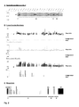
- Figure 2 : Schematic depiction of the CD83 gene locus and the respective acetylation data resulting from the ChIP-chip™ microarray analysis.
- Figure 3 : Schematic depiction of constructs for gene reporter assays.
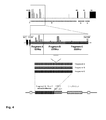
- Figure 4 : Schematic depiction of the subcloning strategy of the hyper-acetylated region of CD83 intron 2.
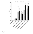
- Figure 5 : Fragment C enhances the MP -261 activity specifically in XS52 cells.
- Figure 6 : Schematic depiction of the deletion mutants C1-C14 of fragment C.
- Figure 7 : The 185 bp long deletion mutant C13 of fragment C enhances the MP - 261 activity specifically in XS52 cells.
- Figure 8 : Fragment C and the 185 bp enhancer induce the MP -261 activity specifically in mDCs.
- Figure 9 : Neither fragments A, B, C nor the 185 bp enhancer induce the MP -261 in Raji and Jurkat cells.
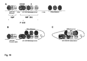
- Figure 10 : Presumed bio-computational model for the interaction of the CD83 upstream promoter, the MP -261 and the 185 bp enhancer.
- Figure 11 : Schematic depiction of reporter constructs containing the UpP and the spacer sequence S1.
- Figure 12 : The spacer sequence S1 does not significantly affect the induction of the MP -261 and the P -510 in XS52 cells.
- Figure 13: (A) The spacer sequence S1 does not significantly affect the induction of the MP -261 in mDCs. (B) The spacer sequence S1 does not significantly affect the induction of the P -510 in mDCs.
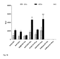
- Figure 14: (A) The ternary complex of UpP, MP -261 and 185 bp enhancer shows a specific transcriptional induction in mDCs.
- Figure 15: (A)-(C) The ternary complex of UpP, MP -261 and 185 bp enhancer shows no specific transcriptional induction in Raji, Jurkat and JCAM cells.
- Figure 16 : Schematic depiction of reporter constructs containing mutated IRF-sites used for luciferase assays.
- Figure 17: (A) and (B) Mutation of any of the three IRF-sites in the ternary complex significantly reduces the luciferase expression in XS52 cells.
- Figure 18 : Mutation of any of the three IRF-sites in the ternary complex significantly reduces the luciferase expression in mDCs.
- Figure 19: (A) and (B) Transcription factors of the NFκB-family and IRF-5 induce the MP -261 in 293T cells.
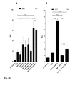
- Figure 20: (A) p65 and IRF-5 induce the UpP in 293T cells. (B) The induction of the UpP by p65 and IRF-5 is abrogated when the IRF-site 3 is mutated.
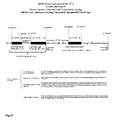
- Figure 21 : Genomic sequences of the of functional human dendritic cell-specific CD83 promoter/ enhancer regions CD83 Upstream promoter, Intermediate sequence, CD83 Minimal Promoter and 185 bp enhancer (NCBI genome build 37.2, contig GRCh37.p2). The 166 bp long CD83 upstream promoter (UpP) and the 261 bp long CD83 minimal promoter (MP-261) are separated by a 83 bp long intermediate sequence. Together these sequences constitute the CD83 promoter P -510. The P -510 cooperates with a 185 bp long enhancer element (185 bp enhancer) in the CD83 intron 2, located 4177 bp downstream of the CD83 transcription start. Referring to the NCBI human genome build 37.2, contig GRCh37.p2 (reference number NT_007592.15; SEQ ID NO:1), the UpP comprises the bp 14057533 to bp 14057698, the intermediate sequence bp 14057699 to 14057782, the MP -261 bp 14057783 to 14058042 and the 185 bp enhancer bp 14062220 to bp 14062404.
Detailed Description of the Invention
-
The mutant CD83 promoter according to aspect (1) of the invention comprises the CD83 Upstream promoter (UpP) sequence of SEQ ID NO:2, the CD83 Minimal Promoter (MP) sequence of SEQ ID NO:4 and the CD83 enhancer sequence of SEQ ID NO:5, or variants of said sequences having 90% homology over the entire length and/or being N- and/or C-terminally truncated by up to 30 nucleotides, said UpP sequences being located upstream of said MP sequence and said enhancer sequence being located 200 to 4000 nucleotides upstream of the UpP sequence or 200 to 4000 nucleotides downstream of said MP sequence. The wording "being located upstream" and "being located upstream" according to the invention implies that the respective sequences are on the same nucleotide strand and are connected by linker/spacer nucleotide molecule (of a variable length, unless the length of the linker/spacer is specifically indicated.
-
According to the invention it is preferred that the distance between the UpP sequence and the MP sequence is 50 to 200 nucleotides, preferably 70 to 120 nucleotides. Also it is preferred that the distance between the UpP sequence/MP sequence and the enhancer is 200 to 4000 nucleotides, preferably 250 to 750 nucleotides.
-
According to the invention the variants of SEQ ID NOs2, 4 and 5 may have 90% homology, preferably 95% homology, most preferably 98% homology over the entire length of the respective basis sequence. Truncation by up to 30 nucleotides, or up to 20 nucleotides or up to 10 nucleotides are possible. Particularly preferred is that the UpP sequence has SEQ ID NO:2, the MP sequence has SEQ ID NO:4, and/or the CD83 enhancer sequence has SEQ ID NO:5.
-
Further preferred is that the UpP sequence is linked to the MP sequence by the intermediate sequence of SEQ ID NO:3 or a variant thereof having 95% homology to SEQ ID NO: 3 over its entire length; and/or that the CD83 enhancer sequence is located 450 to 550 nucleotides upstream to the UpP sequence or 450 to 550 nucleotides downstream of the MP sequence, preferably 450 to 550 nucleotides upstream to the UpP sequence.
-
Particularly preferred mutant CD83 promoters comprises the sequence of nucleotides 29-1259 of SEQ ID NO:28 or the sequence of nucleotides 27-1257 of SEQ ID NO:29. Also preferred is that the mutant CD83 promoter is human dendritic cell-specific.
-
To fully characterize the CD83 promoter, including all regulatory elements that provide for the cell type and maturation status specificity of CD83 expression in mature DC, areas containing potential regulatory elements like e.g. enhancers were identified by the analysis of differential H3K9 acetylation in iDCs, mDCs and HFF cells using a ChIP-chip™ microarray assay. The first 6 kb of CD83 Intron 2 were revealed to be exclusively H3K9 acetylated in mDCs, hinting at a cell type and maturation status-specific hotspot of transcriptional activity in this area. Next, the potential enhancer element within this area was narrowed down. This was achieved by mutation mutagenesis and luciferase assays. As a result, a 185 bp fragment (185 bp enhancer) of the H3K9 acetylated region within CD83 intron 2 has been shown to induce the CD83 minimal promoter (MP -261) in a cell type-and maturation status-specific manner in mDCs.
-
Next, the molecular mechanism underlying the induction of the MP -261 by the 185 bp enhancer was unraveled. Therefore, a biocomputational analysis was performed by the co-inventor Dr. Werner. The Analysis predicted three NFκB-and five SP1-binding sites in the MP -261, two IRF- and one SP1-site in the 185 bp enhancer and additionally two NFκB- and one IRF-site in a presumed upstream promoter (UpP). Furthermore, the formation of a tripartite regulatory complex, consisting of UpP, MP -261 and 185 bp enhancer was foretold. The formation of this complex was supposedly mediated by the interaction of the IRF-and NFκB-transcription factors, thereby forming three NFκB-IRF-NFκB transcriptional modules in trans.
-
To prove this model, the cooperation of all three predicted regulatory elements, namely UpP, MP -261 and 185 bp enhancer, as well as the functionality of the predicted NFκB- and IRF-sites had to be verified. Therefore, several different experimental approached were taken: (i) Adenoviral transduction with luciferase reporter vectors to prove the cooperation of the three regulatory elements in a chromosome-like configuration. (ii) Mutation of the three IRF-sites in the luciferase reporter plasmids to verify their functionality and significance for the formation of the tripartite regulatory complex. (iii) Induction of the predicted NFκB-sites by cotransfection of plasmids coding for members of the NFκB-transcription factor family and luciferase reporter plasmids to verify their functionality. Both the transcription factor binding sites as well as the cooperation of UpP, MP -261 and the 185 bp enhancer were thereby verified. The invention is further described in the following Examples, which are not to be construed to limit the invention.
Examples
Methods
Transient transfection methods and luciferase reporter assay
-
Transfection of DNA with the DEAE-Dextran method: For promoter analyses, adherent XS52, NIH3T3 and HeLa cells were transfected with reporter plasmid DNA containing the promoter construct and a gene for the firefly luciferase by the DEAE-Dextran method in triplicates. Hence, 2 x 10⁵ cells per well were seeded in 12-well tissue culture plate (Falcon) and grown overnight at 37°C, 5% CO2. The next day, 2.5 µg of endotoxin-free plasmid DNA were diluted in 150 µl of TBS buffer and mixed with 50 µl 5 mg/ml DEAE-Dextran solution reaching a volume of 200 µl per transfection. Subsequently 0.2 µl Chloroquine solution (1 µg/µl, Roth) were added to the reaction mix. Cells were washed 1x with 500 µl/well TBS buffer. Then, the DNA-DEAE-Dextran-solution was added and plates were incubated on a rocker for 30 min to 1 h at RT. Afterwards, the DNA-DEAE-Dextran-solution was replaced by 500 µl of 10% DMSO for 2 min, cells were washed with 2 x 2 ml warm DPBS and finally 2 ml per well of warm cell culture medium was added. Transfected cells were cultured for 2 days at 37°C in a humidified atmosphere of 5% CO2 before luciferase reporter assays were performed. Transfection efficiency was assessed by transfection of a GFP coding control plasmid and subsequent FACS analysis.
-
Electroporation of Raji and Jurkat cell lines: For promoter analyses suspension Raji and Jurkat cells were transfected by electroporation with reporter plasmid DNA containing the promoter construct and a gene for the firefly luciferase. Therefore, 10 x 106 cells were harvested, washed 1x in DPBS and resuspended in 250 µl fetal calf serum (PAA) containing 20 µg of DNA. The cell suspension was incubated 10 min at RT. Subsequently 250 µl of RPMI1640 without additives were added and cells were transferred to a 4 mm electroporation cuvette (Peqlab). Electroporation was performed with a Genepulser II (Bio-Rad) at 975 µF and 260 Volt. The cells were incubated for 3 min at RT and then transferred into 10 ml prewarmed growth medium and cultivated at 37°C, 5% CO2. After 24 h 10 ml of growth medium were added. Cells were applied to a luciferase assay 48 h after electroporation. The electroporation efficiency was assessed by the electroporation of a GFP coding control plasmid and subsequent FACS analysis. Lipofection of DNA with Lipofectamine ™ LTX and PLUS ™ (Invitrogen) reagent:
-
For the generation and assembly of adenoviruses, 293 cells were transfected with Lipofectamine™ LTX without PLUS™ reagent in a T25 tissue culture flask (Nunc) as follows: For optimal transfection efficiency the cells were transfected at 40-60% confluence. On the day of transfection, 4 µg of PacI-digested pAd-plasmid in 20 µl water was mixed with 13 µl Lipofectamine™ reagent in 500 µl OptiMEM (Gibco) and incubated for 30 min at RT. The cells were washed 1x with DPBS and covered with 2.5 ml OptiMEM. After the incubation the transfection mix was added directly to the cells and incubated for 6 h at 37°C in a humidified atmosphere of 5% CO2. Afterwards the mixture was replaced by 7 ml of warm growth medium and cells were grown for 10-12 days until viral plaques could be detected. Viruses were then further amplified as described below.
-
For co-transfection of luciferase reporter constructs and pCDNA3.1 vectors coding for transcription factors, the Lipofectamine™ LTX with PLUS™ reagent was used. Therefore, 6 x 10⁴ 293T cells per well were seeded in a 24-well tissue culture plate (Falcon) one day prior to transfection in 500 µl antibiotic free growth medium. On the day of transfection 0.5 µg of total DNA, consisting of 0.05 µg reporter construct, 0.15 µg for each transcription factor construct and pCDNA 3.1 vector backbone to fill up to 0.5 µg, were diluted in 100 µl OptiMEM (Gibco). Then 0.5 µl per transfection mix PLUS reagent were added to the transfection mix, mixed vigorously and incubated for 5 min at RT. Subsequently, 1.25 µl Lipofektamin™ LTX was added to the transfection mix, vortexed and incubated 30 min at RT. After incubation the whole reaction mix was added drop wise directly to the cells. Transfected cells were cultured for 2 days at 37°C in a humidified atmosphere of 5% CO2 before luciferase reporter assays were performed. Growth medium was changed after 24 h. The transfection efficiency was assessed by the lipofection of a GFP coding control plasmid and subsequent FACS analysis.
-
Electroporation of dendritic cells with DNA using AMAXA technology: For further promoter analyses iDC were electroporated using the AMAXA Human Dendritic Cell Nucleofector Kit (Lonza) and the Nucleofector I electroporation device (Lonza), according to the manufacturer's instructions. In brief: After harvesting and washing with warm DPBS, 2 x 10⁶ immature DCs were resuspended in 100 µl freshly prepared electroporation solution (provided with the Nucleofector Kit) containing 4 µg of plasmid DNA. Cells were transferred to the electroporation cuvette and electroporated with program U-2. Immediately after electroporation 500 µl of RPMI1640 without additives were added to the cuvette and the whole cell suspension was transferred in 12-well tissue culture plate (Falcon) containing 600 µl of prewarmed RPMI1640 supplemented with 2% autologous serum, 500 U/ml human recombinant IL-4 and 800 U/ml human recombinant GM-CSF (both Cell/Genix). Directly after the transfer, the cell suspension was divided into 2 wells by pipetting 600 µl in an empty well. After 4 h 1.4 ml RPMI1640 supplemented with 1% autologous serum, 250 U/ml IL-4, 400 U/ml GM-CSF and, for cells to be matured, with LPS to a final concentration of 0.1 ng/ml, was added to the cells, which were cultured 24 h at 37°C in a humidified atmosphere of 5% CO2 before luciferase reporter assays were performed. The electroporation efficiency, generation and maturation of the dendritic cells were assessed by the electroporation of a GFP coding control plasmid and antibody staining of the appropriate lineage and maturation markers, respectively, by subsequent FACS analysis.
-
Luciferase reporter assay: Measurements of luciferase activity of DNA-transfected or adenovirally transduced cell lines and moDCs were performed in triplicates with the Luciferase Assay System (Promega) 1-2 days after transfection. Adherent 293T, HeLa, XS52 and NIH3T3 cells were washed with warm DPBS and then 200 µl/well 1x Luciferase Cell Culture Lysis Reagent (Promega, diluted 1:5 with distilled water) were added before plates were frozen for at least 2 h to -80°C. Raji, Jurkat, JCAM and dendritic cells were first transferred to a reaction vial and then washed in 1 ml warm DPBS. Afterwards cells were collected by centrifugation (500g, 5 min, RT) and lysed with 200 µl/vial 1x Luciferase Cell Culture Lysis Reagent and frozen to -80°C for at least 2 h. Subsequently, plates or vials were thawed at RT and 10 µl (293T cells) or 20 µl (DC, Raji and Jurkat, JCAM) of cell lysate were mixed with 50 µl of Luciferase Assay Substrate (Promega) in a 96-well LumiNunc plate (Nunc). Determination of RLUs (relative luminescence units) was performed in a Wallac fluorometer (Perkin-Elmer, Rotgau) and normalized to the protein concentration of the lysate.
Recombinant adenoviruses
-
Cloning of plasmids containing the recombinant adenoviral genome: For the generation of plasmids containing the recombinant adenoviral genome the pAdEasy1-system was used. Resulting plasmids were transfected into 293 cells for virus assembly and amplification.
-
Preparation of recombinant adenoviruses: All viruses were amplified in 293 cells and purified by two rounds of CsCl equilibrium density gradient ultracentrifugation. Therefore, transfected 293 cells which show viral plaques were harvested, centrifuged at 1100 rpm for 5 min at 4°C and then resuspended in 5 ml RPMI1640/2% FCS. Virus was released from cells by 3 freeze-thaw cycles and cell debris was removed by centrifugation of the lysate at 4000 rpm for 15 min at 4°C. Supernatant (2 ml) containing the virus was used to infect new 293 cells in increasing numbers (up to 15x T175 flasks) to amplify the virus; the remaining supernatant was stored at -80°C for further rounds of virus production. Viruses from 15x T175 flasks of 293 cells were prepared by 3 freeze-thaw cycles as described above and then loaded onto a CsCl gradient. For the CsCl equilibrium density gradient, 3 ml of CsCl at a density of 1.41 g/ml was overlaid with 5 ml of CsCl at a density of 1.27 g/ml and then the supernatant containing the enriched virus was filled up to 7 ml with DPBS and loaded onto the gradient. After ultracentrifugation at 32000 rpm for 2 h at 4°C, the virus band was harvested, diluted in HEPES buffer to 8 ml and loaded onto a second CsCl equilibrium density gradient as described above. Viruses were spun in an ultracentrifuge at 32000 rpm for 24 h at 4°C, before harvesting and purification with a PD-10 tip (GE Healthcare) according to the manufacturer's instructions. Finally, eluted virus (in DPBS) was mixed with 10% Glycerin and aliquots of 25 µl were frozen to -80°C. Verification of viral genomes and exclusion of wild-type contamination was performed by PCR. Physical particle concentration [viral particles (vp)/ml] was determined by OD260 reading and infectious particle concentration was determined by TCID50 assay on 293 cells.
-
Determination of the physical particle concentration: The physical particle concentration is defined by the amount of virus particles per ml [vp/ml]. To quantify the viral DNA, the virus preparation was diluted at different ratios (1:3, 1:5, 1:10, 1:50, 1:100) with viral lysis buffer (VLB [10 mM TE, 0.5% SDS]) and incubated at 56°C in a thermomixer (Eppendorf) for 10 min. Cooled samples were measured with a spectrophotometer (Eppendorf) at OD260 and the mean value was calculated considering the dilution factors.
-
Determination of the infectious particle concentration: The concentration of infectious virus particles is determined by the "tissue culture infectious dose 50" (TCID50)- method. Therefore, 293 cells were seeded in a 96-well tissue culture plate (Falcon) at a density of 105/well in RPMI1640/2% FCS. The virus was diluted from 10-1 to 10-12 in RPMI1640/2% FCS and cells were infected with two dilution series each. Cells were incubated at 37°C, 5% CO2 and 10 as well as 12 days after infection cell lysis was assessed microscopically. Wells that showed at least one plaque were considered "positive".
-
Adenoviral transduction of cells: Immature day 5 DCs were seeded in 12-well tissue culture plates (Falcon) at a concentration of 1 x 10⁶ cells/well in 250 µl medium supplemented with 800 U/ml GM-CSF and 500 U/ml IL-4. Immediately, adenovirus at 500 TCID50/cell in a final volume of 250 µl medium without cytokines was added to the cells. After 1.5 h of incubation at RT, 2.5 ml of growth medium replenished with cytokines as described before was added per well. If mDCs were needed, LPS was added 4 h after transduction to final concentration of 0.1ng/ml.
-
Raji, Jurkat and JCAM cells were seeded in 12-well tissue culture plates (Falcon) at a concentration of 1 x 10⁶ cells/well in 250 µl RPMI1640 medium supplemented with 2% FCS. Immediately, adenovirus at 500 TCID50/cell (Jurkat and JCAM cells) or 50 TCID50/cell (Raji) in a final volume of 250 µl RPMI1640 medium supplemented with 2% FCS was added to the cells. After 1.5 h of incubation at RT on the rocker, 2.5 ml of growth medium replenished with 10% FCS were added per well. To determine transduction efficacy, cells were transduced with Ad5TL and the percentage of living green fluorescent cells was assessed by flow cytometric analysis with a "FACScan" flowcytometer (BD Biosciences, Heidelberg). Only experiments that yielded transduction efficiencies of more than 70% were evaluated and are shown.
-
Polymerase chain reaction (PCR): PCR was used to (i) amplify DNA fragments for cloning from either chromosomal- or plasmid DNA and (ii) to screen adenoviral DNA for insertions.
(i) DNA fragments amplified for cloning were generated in a final reaction volume of 50 µl using a DNA polymerase containing proof-reading activity (Platinum Pfx DNA Polymerase,
Invitrogen) according to the manufacturer's instructions.
(ii) PCR reactions for screening of adenoviral DNA were performed in a final reaction volume of 25 µl with a recombinant Taq DNA Polymerase from
Invitrogen according to the manufacturer's instructions.
Table 1: PCR program for amplification of DNA fragments | Step | Temperature | Time | Cycles |
| Initial denaturation | 95°C | 5 min | 1x |
| Denaturation | 95°C | 1 min | 25x to 30x |
| Annealing | 56°C - 62°C | 1 min |
| Extension | 68°C | 0.5 min - 2 min |
| Final extension | 68°C | 10 min | 1x |
| Pause |
| | 4°C | ∞ | |
Materials
-
Schematic depiction of CD83 Intron 2 fragments for luciferase assay: Intron 2 was divided into 3 fragments (A, B, C) and subcloned in the pGL3/MP-261 reporter construct. Subsequently fragment C was narrowed down via deletion mutagenesis in the fragments C1-C14.
Adenoviruses
-
Table 3: Adenoviruses
| Adenovirus |
Specification |
| Ad5TL |
➢ Replication deficient adenovirus serotype 5; E1 region replaced by a CMV-GFP cassette |
| ➢ Kindly provided by D.T. Curiel, Birmingham, AL, USA |
| Ad5Luc1 |
➢ Replication deficient adenovirus serotype 5; E1 region replaced by a CMV-luciferase cassette |
| ➢ Kindly provided by D.T. Curiel, Birmingham, AL, USA |
| Ad261/S1 |
➢ Replication deficient adenovirus serotype 5; E1 region replaced by a MP -261/S1-luciferase cassette |
| Ad510/S1 |
➢ Replication deficient adenovirus serotype 5; E1 region replaced by a P - |
| |
510/S1-luciferase cassette |
| Ad261/S1+Es |
➢ Replication deficient adenovirus serotype 5; E1 region replaced by a MP -261/S1/185 bp enhancer sense-luciferase cassette |
| Ad261/S1+Eas |
➢ Replication deficient adenovirus serotype 5; E1 region replaced by a MP -261/S1/185 bp enhancer antisense-luciferase cassette |
| Ad510/S1+Es |
➢ Replication deficient adenovirus serotype 5; E1 region replaced by a P -510/S1/185 bp enhancer sense-luciferase cassette |
| Ad510/S1+Eas |
➢ Replication deficient adenovirus serotype 5; E1 region replaced by a P -510/S1/185 bp enhancer antisense-luciferase cassette |
| AdBasic/S1+Es |
➢ Replication deficient adenovirus serotype 5; E1 region replaced by a 185 bp enhancer sense-luciferase cassette |
| AdBasic/S1+Eas |
➢ Replication deficient adenovirus serotype 5; E1 region replaced by a 185 bp enhancer sense-luciferase cassette |
-
Cell lines and cell culture media: If not indicated otherwise, all cell culture media and reagents were purchased by Lonza (Basel, Switzerland) and PAA (Cölbe).
Table 4: Cell lines and cell culture media | Cell type | Short definition | Culture medium |
| 293(T) | Human embryonic kidney cell line, transformed with sheared adenovirus genome (stably transfected with large T antigen) | Dulbecco's Modified Eagle Medium (DMEM) (Lonza) without L-Glutamine, 4.5 g glucose/I (Lonza), 10% FCS (PAA), 1% Penicillin/ Streptomycin/ L-Glutamine (PAA) |
| HeLa | Human cervix carcinoma cell line | Dulbecco's Modified Eagle Medium (DMEM) (Lonza) without L-Glutamine, 4.5 g glucose/I (Lonza) 10% FCS (PAA)1% Penicillin/ Streptomycin/ L-Glutamine (PAA) |
| HFF | Human foreskin fibroblast primary cells | Minimum Essential Medium (DMEM) (Lonza) without L-Glutamine (Gibco), 7.5% FCS (PAA), 1% L-Glutamine (PAA), 0.001% Gentamycin |
| Jurkat | T cell leukemia derived T lymphocyte cell line | RPMI1640 without L-Glutamine (Lonza), 10% FCS (PAA), 1% Penicillin/ Streptomycin/ L-Glutamine (PAA) |
| NIH3T3 | BALB/c derived embryonic fibroblast cell line | Dulbecco's Modified Eagle Medium (DMEM) (Lonza) without L-Glutamine, 4.5 g glucose/I (Lonza), 10% FCS (PAA), 1% Penicillin/ Streptomycin/ L-Glutamine (PAA) |
| NS47 | BALB/c derived fibroblast cell line | Iscove's Modified Dulbecco's Medium (IMDM) (Lonza) without L-Glutamine (Lonza), 10% FCS (PAA), 1% Penicillin/ Streptomycin/ L-Glutamine (PAA), 1% Sodium pyruvate (PAA) |
| PBMC | (Human) Peripher blood mononuclear cells | RPMI1640 without L-Glutamine (Lonza), 1% human autologous serum, 1% Penicillin/ Streptomycin/L-Glutamine (PAA), 1% Hepes (Lonza) |
| Raji | Burkitt's lymphoma derived lymphoblastoid B cell line | RPMI1640 without L-Glutamine (Lonza), 10% FCS (PAA), 1% Penicillin/ Streptomycin/ L-Glutamine (PAA), 1% Hepes (Lonza) |
| XS52 | BALB/c derived DC-like cell line | Iscove's Modified Dulbecco's Medium (IMDM) (Lonza) without L-Glutamine (Lonza), 10% FCS (PAA), 1% Penicillin/ Streptomycin/ L-Glutamine (PAA), 1% Sodium pyruvate (PAA), 10% Supplement NS47, 10 ng/ml murine GM-CSF |
| JCAM | T cell leukemia derived T lymphocyte cell line, similar to Jurkat, but lacking the LCK kinase | RPMI1640 without L-Glutamine (Lonza), 10% FCS (PAA), 1% Penicillin/ Streptomycin/ L-Glutamine (PAA) |
DNA modifying enzymes
-
Table 5: DNA modifying enzymes
| Enzyme |
Supplier |
| DNA restriction enzymes |
New England Biolabs (NEB [Frankfurt/Main]) |
| Alkaline Phosphatase, Calf Intestinal (CIP) |
New England Biolabs (NEB [Frankfurt/Main]) |
| T4 DNA ligase (Rapid ligation kit) |
Roche (Mannheim) |
| Platinum Pfx polymerase |
Invitrogen (Karlsruhe) |
| T4 Poly nucleotide kinase (T4-PNK) |
New England Biolabs (NEB [Frankfurt/Main]) |
| Klenow fragment of DNA Polymerase I |
New England Biolabs (NEB [Frankfurt/Main]) |
Human cytokines and maturation agents
-
Table 6: Human cytokines and maturation agents
| Cytokine/Agent |
Stock concentration |
Purity |
Company |
| Lipopolysaccharide (LPS) |
1 mg/ml |
|
Sigma-Aldrich, München |
| Recombinant GM-CSF |
4x10⁴ U/ml |
>95% |
Cell Genix, Freiburg
|
| Recombinant IL-4 |
2x10⁵ U ml |
>95% |
Cell Genix, Freiburg
|
| Recombinant IL-6 |
1x10⁶ U/ml |
>95% |
Cell Genix, Freiburg
|
Cloning
-
Luciferase reporter constructs
-
Notes and abbreviations: s: sense; as: antisense; CDS: coding sequence; enh: enhancer; UPP: upstream promoter; S1:
spacer sequence 1.
Table 7. Cloned luciferase reporter constructs | Donor/ template | Excision method | Insert | Recipient | Result |
| pEGFP-N1 | Restriction digest; HindIII/XbaI | GFP CDS | pGL3/CMV_luc; HindIII/XbaI (luc CDS replaced) | PGL3/CMV-GFP |
| RP1-258E1 | PCR; KpnI | CD83 intron | 2 fragment A | pGL3/MP-261; KpnI | pGL3/fragment A s_as/MP-261 |
| RP1-258E1 | PCR; KpnI | CD83 intron | 2 fragment B | pGL3/MP-261; KpnI | pGL3/fragment B s_as/MP-261 |
| CMVßL/cRel | Restriction digest; Hind III/XbaI | cRel CDS | pCDNA 3.1; Hind III/XbaI | pCDNA 3.1/cRel |
| CMVBL/p50 | Restriction digest; Hind III/XbaI | p50 CDS | pCDNA 3.1; Hind III/XbaI | pCDNA 3.1/p50 |
| CMVBL/p65 | PCR; Hind III/XbaI | P65 CDS | pCDNA 3.1; HindIII/Xbal | pCDNA 3.1/p65 |
| pGL3/185bp enh. s/MP-261 | Restriction digest; KpnI | 185 bp enhancer | pGL3/Basic; KpnI | pGL3/185bp enh. s_as |
| pGL3/ fragment C/MP-261 | PCR; KpnI | 185 bp enhancer | pGL3/MP-261; KpnI | pGL3/185bp enh s_as/MP-261 |
| Geneart standard vector/CD83_-510 | Restriction digest; NheI/SmaI (blunt) | UPP | pGL3/Basic; NheI/XhoI (blunt) | pGL3/UPP |
| Geneart standard vector/CD83_-510 | Restriction digest; NheI/SmaI (blunt) | UPP | pGL3/185bp enh s_as /MP-261; NheI/XhoI (blunt) | pGL3/185bp enh. s_as/UPP |
| RP3-380E11 | PCR; SacI/MluI | S1 | pGL3/UPP SacI/MluI | pGL3/UPP+S1 |
| RP3-380E11 | PCR; SacI/MluI | S1 | pGL3/185bp enh. s_as/UPP SacI/MluI | pGL3/185bp enh. s_as/UPP+S1 |
| Geneart standard vector/CD83_-510 | Restriction digest; NheI/XhoI | P -510 | pGL3/MP-261 NheI/XhoI | pGL3/P -510 |
| Geneart standard vector/CD83_-510 | Restriction digest; NheI/XhoI | P -510 | pGL3/185bp enh s_as/MP-261 NheI/XhoI | pGL3/185bp enh. s_as/P -510 |
| RP3-380E11 | PCR; SacI/MluI | S1 | pGL3/P -510; SacI/MluI | pGL3/P-510+S1 |
| RP3-380E11 | PCR; SacI/MluI | S1 | pGL3/185bp enh. s_as/P -510; SacI/MluI | pGL3/185bp enh. s_as/P -510+S1 |
| RP3-380E11 | PCR; SacI/MluI | S1 | pGL3/MP-261; SacI/MluI | pGL3/MP-261+S1 |
| RP3-380E11 | PCR; SacI/MluI | S1 | pGL3/185bp enh s_as/MP-261; SacI/MluI | pGL3/185bp enh s_as /MP-261+S1 |
| RP3-380E11 | PCR; KpnI | CD83 intron | 2 fragment C | pGL3/MP-261; KpnI | pGL3/fragment C s_as/MP-261 |
| RP3-380E11 | PCR; KpnI | CD83 intron | 2 fragment A | pGL3/MP-261; KpnI | pGL3/fragment A s_as/MP-261 |
| RP3-380E11 | PCR; KpnI | CD83 intron | 2 fragment B | pGL3/MP-261; KpnI | pGL3/fragment B s_as/MP-261 |
| pGL3/fragment C/MP-261 | PCR; KpnI | CD83 intron | 2 fragments C1-C14 | pGL3/MP-261; KpnI | pGL3/fragment C 1-C14 s_as/MP-261 |
Luciferase reporter constructs with mutated IRF-sites
Notes and abbreviations:
-
Via PCR mutagenesis the IRF- Sites 1 and 2 in the 185 bp enhancer were mutated by PCR mutagenesis. The 3rd IRF-site in the P -510 promoter (or rather in the UpP) was mutated by GENEART and provided in the "GENEART standard vector/CD83_-510mut 3rd IRF-Site". Combinations of the 3 IRF-site mutations resulted in constructs, bearing 1, 2 or 3 mutated IRF-sites:
-
mut: mutated
Table 8: Cloned luciferase reporter constructs with mutated IRF-sites | Donor/template | Excision method | Insert | Recipient | Result |
| GENEART stan-dardvector/CD83_ -510mut 3rd IRF-Site | Restriction digest; NheI/XhoI | P-510mut 3rd IRF-Site | pGL3/MP-261 NheI/XhoI | pGL3/P-510mut 3rd IRF-Site |
| GENEART standard vector/CD83_-510mut 3rd IRF-Site | Restriction digest; NheI/SmaI (blunt) | UPPmut 3rd IRF-Site | pGL3/Basic; NheI/XhoI (blunt) | pGL3/UPPmut 3rd IRF-Site |
| pGL3/185bp enh. s/MP-261 | PCR; KpnI | 185 bp enhancer 1st IRF-site mut | pGL3/P -510; KpnI | pGL3/185bp enh. s_as 1St IRF-site mut/P -510 |
| pGL3/185bp enh. s/MP-261 | PCR; KpnI | 185 bp enhancer 2nd IRF-site mut | pGL3/P -510; KpnI | pGL3/185bp enh. s_as 2nd IRF-site mut/P -510 |
| pGL3/185bp enh. s/MP-261 | PCR; KpnI | 185 bp enhancer 1st /2ndIRF-Site mut | pGL3/P -510; KpnI | pGL3/185bp enh. s_as 1st/2nd IRF-site mut/P -510 |
| pGL3/185bp enh s/MP-261 | PCR; KpnI | 185 bp enhancer 1st /2ndIRF-Site mut | pGL3/P -510mut 3rd IRF-Site; KpnI | pGL3/185bp enh. s_as 1st/2nd mut/P -510mut 3rd IRF-Site |
| RP3-380E11 | PCR; SacI/MluI | S1 | pGL3/P -510mut 3rd IRF-Site; SacI/MluI | pGL3/P -510mut 3rd IRF-Site+S1 |
| RP3-380E11 | PCR; SacI/MluI | S1 | pGL3/185bp enh. s_as 1st IRF-site mut/P -510; SacI/MluI | pGL3/185bp enh. s_as 1st IRF-site mut/P -510+S1 |
| RP3-380E11 | PCR; SacI/MluI | S1 | pGL3/185bp enh. s_as 1st/2nd IRF-site mut/P -510; SacI/MluI | pGL3/185bp enh. s_as 1st/2nd IRF-site mut/P -510+S1 |
| RP3-380E11 | PCR; SacI/MluI | S1 | pGL3/185bp enh. s_as 1st/2nd mut/P -510mut 3rd IRF-Site; SacI/MluI | pGL3/185bp enh. s_as 1st/2nd mut/P -510mut 3rd IRF-Site+S1 |
8. Primers
All primers were purchased from MWG Eurofins Operon (Ebersberg).
Notes and abbreviations:
-
- fw, for: DNA oligonucleotides in forward orientation
- rev: DNA oligonucleotides in reverse orientation
- mt: mutant
- wt: wild type
- aa: amino acid
-
The original IRF-2 PCR template (EF1 /IRF-2) provided by Kay Childs (Division of Basic Medical Sciences St. George's, University of London, U.K.) contained 3 defective aa (3, 4, 5). First the defective sequence was subcloned via PCR (primers: IRF-2 fwd HindII; IRF-2 rev XbaI) in pCDNA 3.1 and from that template a PCR product with the corrected sequence was generated by using PCR mutagenesis (primers: IRF-2mut; fwd IRF-2mut rev;).
-
Cloning primers for luciferase reporter constructs
Table 9: Cloning primers for luciferase reporter constructs | Primer (SEQ ID NO:) | Sequence | Description |
| A-1 (6) | 5'-GGCGGTACCAGCTGGGGCTCTTCTCAATATTATAAAG-3' | Fragment C deletion; rev; wt |
| A-10 (7) | 5'-GGCGGTACCAGATGATTTCCAAAGGAAGGGAG-3' | Fragment C deletion; fw; wt |
| A-1b (8) | 5'-GGCGGTACCCAATATTATAAAGTCTATTTATAG-3' | Fragment C deletion; 185bp enhancer; rev; wt |
| A-2 (9) | 5'-GGCGGTACCAGGTGCCAATGGGGACAGTACG-3' | Fragment C deletion; rev; wt |
| A-3 (10) | 5'-GGCGGTACCAGAAGGCATTGCAACTCTGG-3' | Fragment C deletion; |
| A-4 (11) | 5'-GGCGGTACCGATGCTTCACTCTCCTCACC-3' | Fragment C deletion; rev; wt |
| A-8 (12) | 5'-GGCGGTACCAGTACTTTGGGCCTGGTTGATAATC-3' | Fragment C deletion; fw; wt |
| A-9 (13) | 5'-GGCGGTACCCCTATGGGTGATGCAAAACGAAAG-3' | Fragment C deletion; 185bp enhancer; fw; wt |
| C-forward (14) | 5'-CCAGGGTACCGAGGAGGTATTTTGAGAAAATATG-3' | CD83 intron 2 fragment C fw; wt |
| C-forward 2 (15) | 5'-CCAGGGTACCACAATATCATGTCTGTGAGGAGTAAAGC-3' | Fragment C deletion; fw; wt |
| C-Kurz 1 (16) | 5'-GGCGGTACCTATAATATTGAGAAGAGCCC-3' | Fragment C deletion; fw; wt |
| C-Kurz 2 (17) | 5'-GGCGGTACCATTGGCACCTATAGTACTTG-3' | Fragment C deletion; fw; wt |
| C-Kurz 3rev (18) | 5'-GGCGGTACCCTTACGCCTGTAATCCCAGC-3' | Fragment C deletion; rev; wt |
| C-reverse (19) | 5'-GCAGGGTACCTTCCTCTTCTTTGTGTAGTG-3' | CD83 intron 2 fragment C rev; wt |
| Intron2-A_for (20) | 5'-TTAAGGTACCGTAGGTGCTGCGATACC-3' | CD83 intron 2 fragment A; fw; wt |
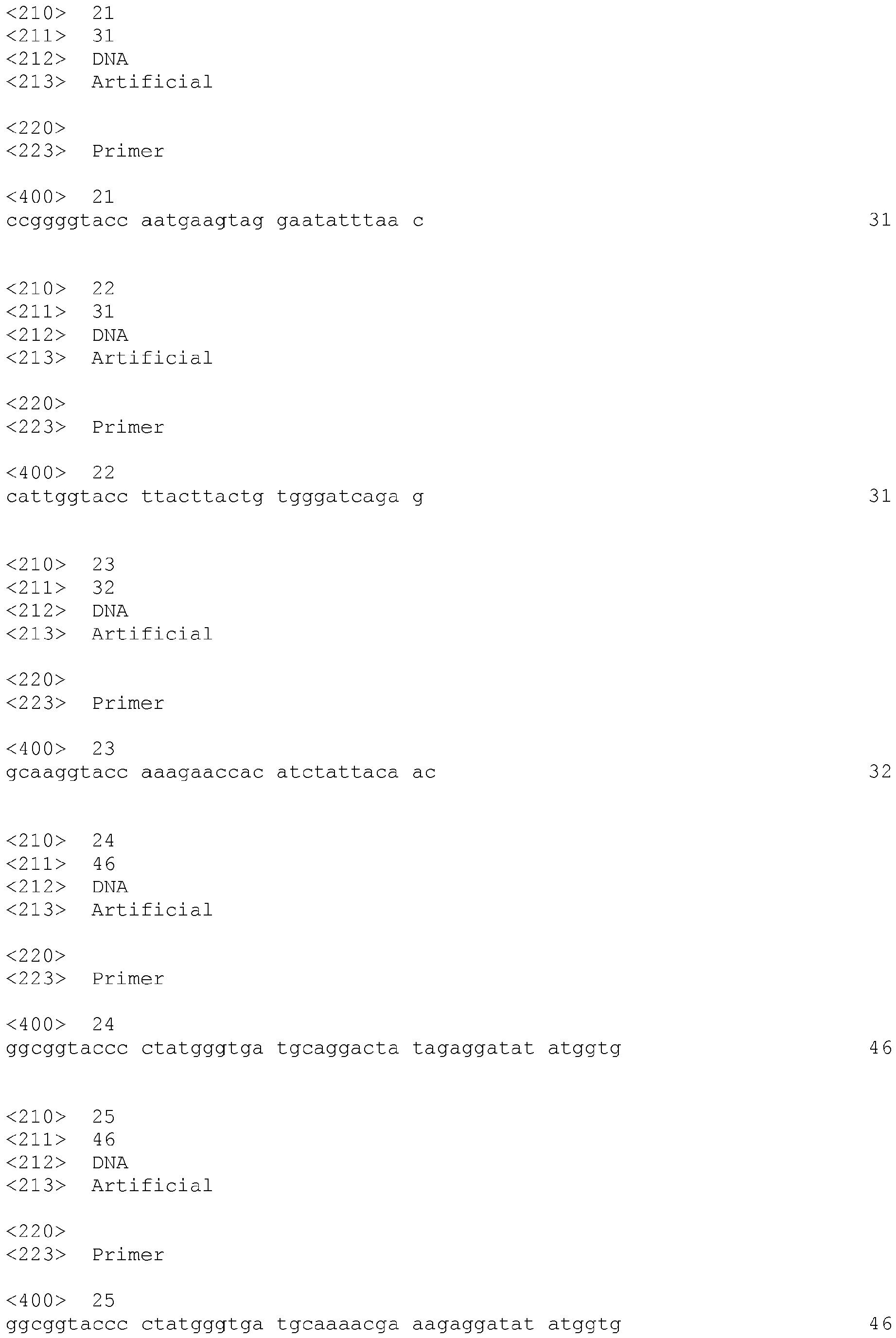
| Intron2-A_rev (21) | 5'-CCGGGGTACCAATGAAGTAGGAATATTTAAC-3' | CD83 intron 2 fragment A; rev; wt |
| Intron2-B_for (22) | 5'-CATTGGTACCTTACTTACTGTGGGATCAGAG-3' | CD83 intron 2 fragment B fw; wt |
| Intron2-B_rev (23) | 5'-GCAAGGTACCAAAGAACCACATCTATTACAAC-3' | CD83 intron 2 fragment B rev; wt |
Cloning primers for the mutated 185 bp enhancer
-
Table 10. Cloning primers for the mutated 185 bp enhancer luciferase reporter constructs.
| Primer (SEQ ID NO:) |
Sequence |
Description |
| 185bp Enh.fw/MT (24) |
|
185 bp enhancer; fw; 1st IRF-site mt |
| 185bp Enh.fw/WT (25) |
|
185 bp enhancer;fw; wt |
| 185bp Enh.rev/MT (26) |
|
185 bp enhancer; rv; 2nd IRF-site mt |
| 185bp Enh.rev/WT (27) |
|
185 bp enhancer;rev; wt |
Example 1: CD83 is upregulated during DC maturation and is not expressed by HFF cells.
-
Figure 1, (A): One hundred thousand day 5 iDCs, 20 h LPS-matured DCs and HFF cells for each setup were harvested and stained with PE-labeled mouse anti-human CD83 mAB or mouse IgG1 isotype-control for flow cytometric analyses. Each quadrangle represents 1 x 10⁴ living cells determined by a gate on forward and side light scatter and PI-staining. In each histogram plot the black line represents mean fluorescence intensity of the isotype-control stained cells and the red line represents mean florescence intensity of the anti-CD83 stained cells. (B): One million day 5 iDCs or HFF cells per well were seeded in a 12-well tissue culture plate. Immature DCs were either matured for 3 h, 6 h, 10 h and 24 h or left untreated as the HFF cells. Cells were lysed in RIPA buffer at the indicated time points. For Western blot 20 µg per lane of whole cell lysates from either immature DCs, maturing DCs (3-24 h after addition of LPS) or HFF cells were loaded on a denaturing 12.5% SDS polyacrylamide gel and blotted afterwards onto a nitrocellulose membrane. The membranes were incubated with a rat anti-human CD83 and a mouse anti-human GAPDH primary antibody and subsequently with an anti-rat HRP-conjugated or an anti-mouse HRP-conjugated secondary antibody, respectively. The signal was detected via chemiluminescence that was visualized on a photo film. (C) Total RNA was isolated from iDCs, 20 h LPS-matured DCs and HFF cells using a QIAshredder and RNeasy Mini Kit (Quiagen) according to the manufacturer's instructions and reversely transcribed with an Oligo (dT)12-18 primer. Afterwards PCR was performed with an intron spanning primer pair for CD83 and a control primer pair for GAPDH. Additionally, one PCR reaction was prepared with water instead of DNA as a negative control.
Example 2: Schematic depiction of the CD83 gene locus and the respective acetylation data resulting from the ChIP-chip
™
microarray analysis.
-
Figure 2, (A): Schematic depiction of the CD83 genelocus and nucleotide positions on chromosome 6. Coding sequence depicted in blue (CDS), exons in grey (E1-E5) and introns in red (I1-I4). (B) Data for the CD83 hyperacetylation were provided by NimbleGen Systems and interpolated subsequently over a space of 500 bp using the Signal Map software. (C) Overlay of the interpolated data for mature DCs aligned with the positions of the CD83 exons (black; E1-E5) and introns (red; I1-I4)
Example 3: Schematic depiction of constructs for gene reporter assays.
-
Figure 3: A pGL3 vector backbone only coding for the luciferase gene and lacking any regulatory element (pGL3/Basic) was used for background control. The pGL3 backbone containing the luciferase gene driven by a CMV promoter (pGL3/CMV/luc) was generated for its usage as a positive control and for internal normalization. The same plasmid coding for GFP instead of luciferase (pGL3/CMV/GFP) was used to determine transfection efficiency. As reference control a pGL3 plasmid coding for the luciferase gene driven by the CD83 minimal promoter MP -261 (pGL3/MP -261) was generated. Finally, plasmids containing a putative regulatory element in both orientations upstream of the MP -261 (pGL3/Put. reg./MP -261) were generated to study its effects on the MP - 261.
Example 4: Schematic depiction of the subcloning strategy of the hyperacetylated region of CD83 intron 2.
-
Figure 4: The first 6 kb of CD83 intron 2 were subdivided into 3 fragments A (1239 bp), B (2359 bp), and C (2220 bp). After PCR amplification each fragment was subcloned in sense and antisense orientation upstream the MP -261 into the pGL3/MP-261 plasmid. Hyperacetylated regions are depicted as blue bars, exons as black bars (E1-E5) and introns are marked in red (I1-I5). The red boxes indicate the base pair position relatively to the transcription start (bp 1).
Example 5: Fragment C enhances the MP-261 activity specifically in XS52 cells.
-
Figure 5: Two hundred thousand XS52, NIH3T3 or HeLa cells per well were seeded in a 12-well tissue culture plate and were grown overnight at 37°C, 5% CO2. The next day, the cells were transfected with 2.5 µg of reporter plasmids containing fragment A, B or C of the CD83 intron 2 in sense (s) or antisense (as) orientation upstream of the MP -261 or the pGL3/MP -261 as reference control using the DEAE-Dextran method. As controls the pGL3/Basic and the pGL3/CMV/luc vector were transfected to assess background activity and for internal normalization, respectively. The promoter activity was determined by luciferase reporter assays 48 h after transfection. The resulting RLUs were further normalized to the protein concentration of each lysate. The plasmids containing fragments A, B and C were compared to the pGL3/MP -261 vector for each cell line and the resulting p-values were determined via one way ANOVA and Bonferroni's Multiple Comparison post hoc test. * p < 0.05, ** p < 0.01, *** p < 0.001 and n.s. not significant (p>0.05). Results represent the means (±SEM) of three independently performed experiments.
Example 6: Schematic depiction of the deletion mutants C1-C14 of fragment C.
-
Figure 6: The deletion mutants C1-C14 of the CD83 intron 2 fragment C were generated by PCR amplification and cloned in sense (s) and antisense (as) orientation into the pGL3/MP-261 reporter plasmid upstream of the MP -261. Induction of the minimal promoter activity was determined in XS52, NIH3T3 and HeLa cell lines by luciferase reporter assays. Induction is depicted relatively to the activity of the pGL3/MP -261. - - - no induction of the MP -261, + weak induction of the MP -261, ++ medium induction of the MP-261; +++ strong induction of the MP-261.
Example 7: The 185 bp long deletion mutant C13 of fragment C enhances the MP -261 activity specifically in XS52 cells.
-
Figure 7: Two hundred thousand XS52, NIH3T3 or HeLa cells per well were seeded in a 12-well tissue culture plate and were grown overnight at 37°C, 5% CO2. The next day, cells were transfected with 2.5 µg of either pGL3/MP -261 reporter plasmids containing the deletion mutants C1-C14 of the fragment C of the CD83 intron 2 in sense (s) or antisense (as) orientation upstream of the MP - 261 or the pGL3/MP -261 as reference control using the DEAE-Dextran method. As controls the pGL3/Basic and the pGL3/CMV/luc vector were transfected to assess background activity and for internal normalization, respectively. The promoter activity was determined by luciferase reporter assays 48 h after transfection. The resulting RLUs were further normalized to the protein concentration of each lysate. The plasmids containing the different fragments were compared to the pGL3/MP -261 vector for each respective cell line and the resulting p-values were determined via one way ANOVA and Bonferroni's Multiple Comparison post hoc test. * p < 0.05, ** p < 0.01, *** p < 0.001 and n.s. not significant (p>0.05). Results represent the means (±SEM) of three independently performed experiments.
Example 8: Fragment C and the 185 bp enhancer induce the MP-261 activity specifically in mDCs.
-
Figure 8: Two million day 5 iDCs were electroporated with 4 µg of either pGL3/Basic, pGL3/MP-261, pGL3/FragmentC/MP-261 s/as or pGL3/185bpEnh.(C13)/MP-261 s/as luciferase reporter plasmid using the AMAXA technology. Afterwards, cells were transferred to a 12-well tissue culture plate and split into two equal fractions. One of the fractions was matured 3 h after electroporation with LPS to a final concentration of 0.1 ng/ml and the other fraction was replenished with growth medium without LPS. Cells were then grown at 37°C, 5% CO2 for 20 h. The pGL3/Basic and the pGL3/CMV/luc vector were electroporated to assess background activity and for internal normalization, respectively. The promoter activity was determined by luciferase reporter assays and the resulting RLUs were normalized to the protein concentration of each lysate. The plasmids containing the different fragments were compared to the pGL3/MP -261 vector and the resulting p-values were determined via one way ANOVA and Bonferroni's Multiple Comparison post hoc test. * p < 0.05, ** p < 0.01, *** p < 0.001. Results depict the means (±SEM) of four independently performed experiments representing four different donors in (A) and three experiments representing three different donors in (B).
Example 9: Neither fragments A, B, C nor the 185 bp enhancer induce the MP-261 in Raji and Jurkat cells.
-
Figure 9: Ten million Raji or Jurkat cells were electroporated with 20 µg of reporter plasmids containing fragment A, B, C or the 185 bp enhancer in sense (s) or antisense (as) orientation upstream of the MP -261 or the pGL3/MP -261 as reference control using a Genepulser II. As controls the pGL3/Basic and the pGL3/CMV/luc vector were electroporated to assess background activity and for internal normalization, respectively. After electroporation, cells were transferred into 10 ml prewarmed growth medium and cultivated at 37°C, 5% CO2 for 24 h. The promoter activity was determined by luciferase reporter assays and the resulting RLUs were further normalized to the protein concentration of each lysate. The plasmids containing the different fragments were compared to the pGL3/MP -261 vector for each respective cell line and the resulting p-values were determined via one way ANOVA and Bonferroni's Multiple Comparison post hoc test. * p < 0.05, ** p < 0.01, *** p < 0.001 and n.s. not significant (p>0.05). Results represent the means (±SEM) of three independently performed experiments.
Example 10: Presumed biocomputational model for the interaction of the CD83 upstream promoter, the MP-261 and the 185 bp enhancer.
-
Figure 10, (A): Depiction of the three regulatory elements with the predicted transcription factor binding sites regulating CD83 transcription: (i) The upstream promoter (UpP) containing NFxB- sites 1 and 2, IRF-site 3 and one SP1 site, (ii) the 261 bp long CD83 minimal promoter (MP -261) containing NFxB- sites 3, 4 and 5 and four SP1-sites and (iii) the 185 bp enhancer containing IRF- sites 1 and 2 as well as one SP1-site. The UpP and the MP -261 including the 82 bp naturally occurring spacer sequence are termed 510 bp promoter (P -510). Previously confirmed and published transcription factor binding sites are marked with a red arrow. (B): Designated interaction of the 185 bp enhancer with the MP -261 mediated by the interaction of IRF- sites 1 and 2 with NFκB- sites 3 and 5, respectively. (C): Ternary complex formation with the UpP as third interaction partner. The interaction between the UpP and the MP -261 is mediated by IRF-site 3 and NFκB-site 4.
Example 11: Schematic depiction of reporter constructs containing the UpP and the spacer sequence S1.
-
Figure 11: The P -510 as well as the CD83 upstream promoter (UpP) were cloned upstream of the luciferase gene into the pGL3/Basic plasmid, thus generating pGL3/P -510 and pGL3/UpP, respectively. The UpP comprised in the P -510 is highlighted in purple. Furthermore, a 500 bp long spacer sequence (S1) was inserted in between MP -261 and P -510 generating the plasmids pGL3/185 bp Enh./S1/MP -261 and pGL3/185 bp Enh./S1/MP -261, respectively. The spacer sequence was also introduced into the plasmids pGL3/MP -261 and pGL3/P -510, resulting in the plasmids pGL3/S1/MP -261 and pGL3/S1/P -510.
Example 12: The spacer sequence S1 does not significantly affect the induction of the MP -261 and the P -510 in XS52 cells.
-
Figure 12: Two hundred thousand XS52, NIH3T3 and HeLa cells per well were seeded in a 12-well tissue culture plate and were grown overnight at 37°C, 5% CO2. The next day, cells were transfected with 2.5 µg of either pGL3/MP -261 or pGL3/P -510 with or without the 185 bp enhancer in sense (s) or antisense (as) orientation either containing the spacer sequence S1 or not, using the DEAE-Dextran transfection method. As controls the pGL3/Basic and the pGL3/CMV/luc vector were transfected to assess background activity and for internal normalization, respectively. The promoter activity was determined by luciferase reporter assays 48 h after transfection. The resulting RLUs were further normalized to the protein concentration of each lysate. The plasmids containing the spacer sequence S1 were compared to the corresponding plasmids without the spacer sequence for each respective cell line. The resulting p-values were determined via one way ANOVA and Bonferroni's Multiple Comparison post hoc test. * p < 0.05, ** p < 0.01, *** p < 0.001 and n.s. not significant (p>0.05). Results represent the means (±SEM) of three independently performed experiments.
Example 13 A: The spacer sequence S1 does not significantly affect the induction of the MP-261 in mDCs.
-
Figure 13, (A): Two million day 5 iDCs were electroporated with 4 µg of either pGL3/MP -261with or without the 185 bp enhancer in sense (s) or antisense (as) orientation upstream of the MP -261 or with their respective counterparts comprising the spacer sequence S1, using the AMAXA technology. Afterwards, cells were transferred to a 12-well tissue culture plate and split into two equal fractions. Cells were matured 3 h after electroporation with LPS to a final concentration of 0.1 ng/ml and were then cultured at 37°C in a humidified atmosphere at 5% CO2 for 20 h. As controls the pGL3/Basic and the pGL3/CMV/luc vector were electroporated to assess background activity and for internal normalization, respectively. The promoter activity was determined by luciferase reporter assays. The resulting RLUs were further normalized to the protein concentration of each lysate. The plasmids containing the spacer sequence S1 were compared to the corresponding plasmids lacking the spacer sequence. The resulting p-values were determined via one way ANOVA and Bonferroni's Multiple Comparison post hoc test. * p < 0.05, ** p < 0.01, *** p < 0.001 and n.s. not significant (p>0.05). Results represent the means (±SEM) of five independently performed experiments representing different donors.
Example 13B: The spacer sequence S1 does not significantly affect the induction of the P -510 in mDCs.
-
Figure 13, (B): The cells were treated and electroporated in the same way as in (A), but instead of plasmids bearing the MP -261, plasmids comprising the P - 510 were used. The resulting p-values were determined via one way ANOVA and Bonferroni's Multiple Comparison post hoc test. * p < 0.05, ** p < 0.01, *** p < 0.001 and n.s. not significant (p>0.05). Results represent the means (±SEM) of three independently performed experiments representing different donors.
Example 14:The ternary complex of UpP, MP-261 and 185 bp enhancer shows a specific transcriptional induction in mDCs.
-
Figure 14, (A): One million day 5 iDCs per well were seeded in a 12-well tissue culture plate in 250 µl medium supplemented with 800 U/ml GM-CSF and 500 U/ml IL-4. Recombinant adenovirus as indicated at 500 TCID50/cell in a final volume of 250 µl medium without cytokines was added to the cells. After 1.5 h of incubation at RT, 2.5 ml of growth medium replenished with cytokines as described before was added per well. For the generation of mDCs LPS was added 3 h after transduction to final concentration of 0.1 ng/ml. The promoter activity was determined by luciferase reporter assays 24 h after transduction. As controls the Ad5luc1 was transduced for internal normalization. The resulting RLUs were further normalized to the protein concentration of each lysate. The transduction setups were compared to the transduction of Ad261/S1 and the resulting p-values were determined via one way ANOVA and Bonferroni's Multiple Comparison post hoc test. * p < 0.05, ** p < 0.01, *** p < 0.001. Results represent the means (±SEM) of three independently performed experiments representing different donors.
Example 15: The ternary complex of UaP, MP -261 and 185 bp enhancer shows no specific transcriptional induction in Raji, Jurkat and JCAM cells.
-
Figure 15, (A)-(C): One million Raji, Jurkat and JCAM cells per well were seeded in a 12-well tissue culture plate in 250 µl RPMI1640 supplemented with 2% FCS. Recombinant adenovirus as indicated at 500 TCID50/cell for Jurkat and JCAM and 50 TCID50/cell for Raji in a final volume of 250 µl RPMI1640 supplemented with 2% FCS was added to the cells. After 1.5 h of incubation at RT, 2.5 ml of appropriate full growth medium was added per well. Raji cells were either stimulated 3 h after transduction with LPS to a final concentration of 0.1 ng/ml or left untreated. Jurkat and JCAM cells were either stimulated 3 h after transduction with PGE2 and TNF-α to a final concentration of 1 µg/ml and 10ng/ml, respectively, or left untreated. The promoter activity was determined by luciferase reporter assays 24 h after transduction. As additional controls Ad5luc1 was transduced for internal normalization. The resulting RLUs were further normalized to the protein concentration of each lysate. The transduction setups were compared to the transduction of Ad261/S1 and the resulting p-values were determined via one way ANOVA and Bonferroni's Multiple Comparison post hoc test. * p < 0.05, ** p < 0.01, *** p < 0.001. Results represent the means (±SEM) of three independently performed experiments.
Example 16: Schematic depiction of reporter constructs containing mutated IRF-sites used for luciferase assays.
-
Figure 16: To generate the pGL3/UpP plasmid, the CD83 upstream promoter a 244 bp fragment of the P -510 containing the UpP was isolated by enzymatic digestion and cloned upstream of the luciferase gene into the pGL3/Basic plasmid. The individual IRF-sites in the pGL3/UpP, the pGL3/S1/P-510 and the pGL3/185 bp Enh./S1/P-510 plasmids were mutated by PCR mutagenesis. The IRF-sites were mutated in different combinations in concordance with the pointmutations in the EMSA control oligonucleotides.
Example 17: Mutation of any of the three IRF-sites in the ternary complex significantly reduces the luciferase expression in XS52 cells.
-
Figure 17, (A) and (B): Two hundred thousand XS52 cells per well were seeded in a 12-well tissue culture plate and were grown overnight at 37°C, 5% CO2. The next day, cells were transfected with 2.5 µg of the wildtype pGL3/UpP or the pGL3/S1/P -510 with and without the 185 bp enhancer in sense (s) or antisense (as) orientation upstream of the P -510 or with the analogical plasmids with one, two or three mutated IRF-sites using the DEAE-Dextran transfection method. The promoter activity was determined by luciferase reporter assays 48 h after transfection. As controls the pGL3/Basic and the pGL3/CMV/luc vector were transfected to assess background activity and for internal normalization, respectively. The resulting RLUs were normalized to the protein concentration of each lysate. The plasmids containing the different mutations were compared to the respective wildtype control and the resulting p-values were determined via one way ANOVA and Bonferroni's Multiple Comparison post hoc test. The mutated IRF-sites are indicated by their respective numbers. * p < 0.05, ** p < 0.01, *** p < 0.001 and n.s. not significant (p>0.05). Results represent the means (±SEM) of three independently performed experiments.
Example 18: Mutation of any of the three IRF-sites in the ternary complex significantly reduces the luciferase expression in mDCs.
-
Figure 18: Two million day 5 iDCs were electroporated with 4 µg of either wildtype pGL3/S1/P -510 with and without the 185 bp enhancer in sense (s) or antisense (as) orientation upstream of the P -510 or with the analogical plasmids with one, two or three mutated IRF-sites, using the AMAXA technology. Afterwards, cells were transferred to a 12-well tissue culture plate and split into two equal fractions. Cells were matured 3 h after electroporation with LPS to a final concentration of 0.1 ng/ml and were then cultured at 37°C in a humidified atmosphere at 5% CO2 for 20 h. The promoter activity was determined by luciferase reporter assays 48 h after transfection. As controls the pGL3/Basic and the pGL3/CMV/luc vector were transfected to assess background activity and for internal normalization, respectively. The resulting RLUs were further normalized to the protein concentration of each lysate. The plasmids containing the different mutations were compared to the respective wildtype control and the resulting p-values were determined via one way ANOVA and Bonferroni's Multiple Comparison post hoc test. The mutated IRF-sites are indicated by their respective numbers. * p < 0.05, ** p < 0.01, *** p < 0.001 and n.s. not significant (p>0.05). Results represent the means (±SEM) of three independently performed experiments from different donors.
Example 19: Transcription factors of the NFκB family and IRF-5 induce the MP-261 in 293T cells.
-
Figure 19, (A) and (B): Sixty thousand 293T cells per well were seeded in a 24-well tissue culture plate and grown overnight at 37°C, 5% CO2. The next day, cells were cotransfected with 0.05 µg of pGL3/MP -261 reporter plasmid and 0.15 µg of each plasmid coding for the indicated transcription factors (p50, p65, cRel or IRF-5 in pCDNA3.1) or empty vector backbone as control. The total amount of DNA was adjusted with pCDNA3.1 empty vector backbone to 0.5 µg. For transfection the Lipofectamine™ LTX system was used. The promoter activity was determined by luciferase reporter assays 48 h after transfection. As controls the pGL3/Basic and the pGL3/CMV/luc vector were transfected to assess background activity and for internal normalization, respectively. The resulting RLU were further normalized to the protein concentration of each lysate. The cotransfections were compared to the pGL3/MP -261 control and the resulting p-values were determined via one way ANOVA and Bonferroni's Multiple Comparison post hoc test. * p < 0.05, ** p < 0.01, *** p < 0.001 and n.s. not significant (p>0.05). Results represent the means (±SEM) of three independently performed experiments.
Example 20 A: p65 and IRF-5 induce the UpP in 293T cells.
-
Figure 20, (A): Sixty thousand 293T cells per well were seeded in a 24-well tissue culture plate and grown overnight at 37°C, 5% CO2. The next day, cells were cotransfected with 0.05 µg of pGL3/UpP reporter plasmid and 0.15 µg of each plasmid coding for the indicated transcription factors (p50, p65 and IRF-5 in pCDNA3.1) or empty vector backbone as control. The total amount of DNA was adjusted with pCDNA3.1 vector backbone to 0.5 µg. The promoter activity was determined by luciferase reporter assays 48 h after transfection. As controls the pGL3/Basic and the pGL3/CMV/luc vector were transfected to assess background activity and for internal normalization, respectively. The resulting RLU were further normalized to the protein concentration of each lysate. The cotransfections were compared to the pGL3/UpP control and the resulting p-values were determined via one way ANOVA and Bonferroni's Multiple Comparison post hoc test. * p < 0.05, ** p < 0.01, *** p < 0.001 and n.s. not significant (p>0.05). Results represent the means (±SEM) of three independently performed experiments.
Example 20B: The induction of the UpP by p65 and IRF-5 is abrogated when the IRF-site 3 is mutated.
-
Figure 20, (B): The cells were transfected as described in (A) either with 0.05 µg pGL3/UpP or pGL3/UpP 3.IRFmut reporter plasmid alone or cotransfected with 0.15 µg of each indicated transcription factor (p65 and IRF-5 in pCDNA3.1). The total amount of DNA was adjusted with pCDNA3.1 vector backbone to 0.5 µg. The pGL3/UpP and pGL3/UpP 3.IRFmut setups were compared and the resulting p-values were determined via one way ANOVA and Bonferroni's Multiple Comparison post hoc test. * p < 0.05, ** p < 0.01, *** p < 0.001 and n.s. not significant (p>0.05). Results represent the means (±SEM) of three independently performed experiments.
Sequence Listing, Free Text
-
- SEQ ID NO:
- Description
- 1
- Genomic sequence of the CD83 promoter of NT_007592.15; UpP bp 2531- 2696, intermediate sequence bp 2697-2779, MP-261 bp 2780-3040, and 185 bp enhancer bp 7218-7402
- 2
- Upstream promoter (UpP)
- 3
- Intermediate sequence
- 4
- CD83 Minimal Promoter (MP -261)
- 5
- 185 bp enhancer
- 6-27
- Primers
- 28-29
- Promoter regions of vectors pGL3/-510/S1/Es and pGL3/-510/S1/Eas, respectively