EP2514428A1 - Delta-Tocotrienol treatment and prevention of pancreatic cancer - Google Patents
Delta-Tocotrienol treatment and prevention of pancreatic cancer Download PDFInfo
- Publication number
- EP2514428A1 EP2514428A1 EP12164751A EP12164751A EP2514428A1 EP 2514428 A1 EP2514428 A1 EP 2514428A1 EP 12164751 A EP12164751 A EP 12164751A EP 12164751 A EP12164751 A EP 12164751A EP 2514428 A1 EP2514428 A1 EP 2514428A1
- Authority
- EP
- European Patent Office
- Prior art keywords
- tocotrienol
- pancreatic cancer
- level
- biomarker
- cells
- Prior art date
- Legal status (The legal status is an assumption and is not a legal conclusion. Google has not performed a legal analysis and makes no representation as to the accuracy of the status listed.)
- Granted
Links
- 206010061902 Pancreatic neoplasm Diseases 0.000 title claims abstract description 164
- 201000002528 pancreatic cancer Diseases 0.000 title claims abstract description 159
- 208000008443 pancreatic carcinoma Diseases 0.000 title claims abstract description 156
- 208000015486 malignant pancreatic neoplasm Diseases 0.000 title claims abstract description 155
- OTXNTMVVOOBZCV-UHFFFAOYSA-N 2R-gamma-tocotrienol Natural products OC1=C(C)C(C)=C2OC(CCC=C(C)CCC=C(C)CCC=C(C)C)(C)CCC2=C1 OTXNTMVVOOBZCV-UHFFFAOYSA-N 0.000 title claims abstract description 127
- ODADKLYLWWCHNB-LDYBVBFYSA-N δ-tocotrienol Chemical compound OC1=CC(C)=C2O[C@@](CC/C=C(C)/CC/C=C(C)/CCC=C(C)C)(C)CCC2=C1 ODADKLYLWWCHNB-LDYBVBFYSA-N 0.000 title claims abstract description 114
- ODADKLYLWWCHNB-UHFFFAOYSA-N 2R-delta-tocotrienol Natural products OC1=CC(C)=C2OC(CCC=C(C)CCC=C(C)CCC=C(C)C)(C)CCC2=C1 ODADKLYLWWCHNB-UHFFFAOYSA-N 0.000 title claims abstract description 112
- BTNBMQIHCRIGOU-UHFFFAOYSA-N delta-tocotrienol Natural products CC(=CCCC(=CCCC(=CCCOC1(C)CCc2cc(O)cc(C)c2O1)C)C)C BTNBMQIHCRIGOU-UHFFFAOYSA-N 0.000 title claims abstract description 112
- 235000019144 δ-tocotrienol Nutrition 0.000 title claims abstract description 112
- 239000011729 δ-tocotrienol Substances 0.000 title claims abstract description 112
- 238000011282 treatment Methods 0.000 title claims description 29
- 230000002265 prevention Effects 0.000 title claims description 7
- 239000011731 tocotrienol Substances 0.000 claims abstract description 62
- 229930003802 tocotrienol Natural products 0.000 claims abstract description 57
- 235000019148 tocotrienols Nutrition 0.000 claims abstract description 57
- 239000002246 antineoplastic agent Substances 0.000 claims abstract description 43
- 229940127089 cytotoxic agent Drugs 0.000 claims abstract description 43
- RZFHLOLGZPDCHJ-DLQZEEBKSA-N alpha-Tocotrienol Natural products Oc1c(C)c(C)c2O[C@@](CC/C=C(/CC/C=C(\CC/C=C(\C)/C)/C)\C)(C)CCc2c1C RZFHLOLGZPDCHJ-DLQZEEBKSA-N 0.000 claims abstract description 30
- OTXNTMVVOOBZCV-YMCDKREISA-N gamma-Tocotrienol Natural products Oc1c(C)c(C)c2O[C@@](CC/C=C(\CC/C=C(\CC/C=C(\C)/C)/C)/C)(C)CCc2c1 OTXNTMVVOOBZCV-YMCDKREISA-N 0.000 claims abstract description 15
- 235000019150 γ-tocotrienol Nutrition 0.000 claims abstract description 15
- 239000011722 γ-tocotrienol Substances 0.000 claims abstract description 15
- OTXNTMVVOOBZCV-WAZJVIJMSA-N γ-tocotrienol Chemical compound OC1=C(C)C(C)=C2O[C@@](CC/C=C(C)/CC/C=C(C)/CCC=C(C)C)(C)CCC2=C1 OTXNTMVVOOBZCV-WAZJVIJMSA-N 0.000 claims abstract description 15
- 239000000203 mixture Substances 0.000 claims description 50
- GJJVAFUKOBZPCB-UHFFFAOYSA-N 2-methyl-2-(4,8,12-trimethyltrideca-3,7,11-trienyl)-3,4-dihydrochromen-6-ol Chemical compound OC1=CC=C2OC(CCC=C(C)CCC=C(C)CCC=C(C)C)(C)CCC2=C1 GJJVAFUKOBZPCB-UHFFFAOYSA-N 0.000 claims description 49
- 208000008900 Pancreatic Ductal Carcinoma Diseases 0.000 claims description 49
- 201000008129 pancreatic ductal adenocarcinoma Diseases 0.000 claims description 49
- 206010028980 Neoplasm Diseases 0.000 claims description 48
- FGYKUFVNYVMTAM-UHFFFAOYSA-N (R)-2,5,8-trimethyl-2-(4,8,12-trimethyl-trideca-3t,7t,11-trienyl)-chroman-6-ol Natural products OC1=CC(C)=C2OC(CCC=C(C)CCC=C(C)CCC=C(C)C)(C)CCC2=C1C FGYKUFVNYVMTAM-UHFFFAOYSA-N 0.000 claims description 15
- 229940064063 alpha tocotrienol Drugs 0.000 claims description 15
- FGYKUFVNYVMTAM-YMCDKREISA-N beta-Tocotrienol Natural products Oc1c(C)c2c(c(C)c1)O[C@@](CC/C=C(\CC/C=C(\CC/C=C(\C)/C)/C)/C)(C)CC2 FGYKUFVNYVMTAM-YMCDKREISA-N 0.000 claims description 15
- 201000011510 cancer Diseases 0.000 claims description 15
- FGYKUFVNYVMTAM-MUUNZHRXSA-N epsilon-Tocopherol Natural products OC1=CC(C)=C2O[C@@](CCC=C(C)CCC=C(C)CCC=C(C)C)(C)CCC2=C1C FGYKUFVNYVMTAM-MUUNZHRXSA-N 0.000 claims description 15
- RZFHLOLGZPDCHJ-XZXLULOTSA-N α-Tocotrienol Chemical compound OC1=C(C)C(C)=C2O[C@@](CC/C=C(C)/CC/C=C(C)/CCC=C(C)C)(C)CCC2=C1C RZFHLOLGZPDCHJ-XZXLULOTSA-N 0.000 claims description 15
- 235000019145 α-tocotrienol Nutrition 0.000 claims description 15
- 239000011730 α-tocotrienol Substances 0.000 claims description 15
- 235000019151 β-tocotrienol Nutrition 0.000 claims description 15
- 239000011723 β-tocotrienol Substances 0.000 claims description 15
- FGYKUFVNYVMTAM-WAZJVIJMSA-N β-tocotrienol Chemical compound OC1=CC(C)=C2O[C@@](CC/C=C(C)/CC/C=C(C)/CCC=C(C)C)(C)CCC2=C1C FGYKUFVNYVMTAM-WAZJVIJMSA-N 0.000 claims description 15
- 229930003799 tocopherol Natural products 0.000 claims description 13
- 239000011732 tocopherol Substances 0.000 claims description 13
- 235000019149 tocopherols Nutrition 0.000 claims description 10
- 206010027476 Metastases Diseases 0.000 claims description 6
- 230000009401 metastasis Effects 0.000 claims description 6
- 238000002271 resection Methods 0.000 claims description 4
- 229960005277 gemcitabine Drugs 0.000 claims description 3
- SDUQYLNIPVEERB-QPPQHZFASA-N gemcitabine Chemical compound O=C1N=C(N)C=CN1[C@H]1C(F)(F)[C@H](O)[C@@H](CO)O1 SDUQYLNIPVEERB-QPPQHZFASA-N 0.000 claims description 3
- 230000002980 postoperative effect Effects 0.000 claims description 2
- QUEDXNHFTDJVIY-UHFFFAOYSA-N γ-tocopherol Chemical class OC1=C(C)C(C)=C2OC(CCCC(C)CCCC(C)CCCC(C)C)(C)CCC2=C1 QUEDXNHFTDJVIY-UHFFFAOYSA-N 0.000 claims 1
- 239000000090 biomarker Substances 0.000 abstract description 150
- 238000000034 method Methods 0.000 abstract description 114
- 230000000694 effects Effects 0.000 abstract description 18
- 238000001727 in vivo Methods 0.000 abstract description 11
- GJJVAFUKOBZPCB-ZGRPYONQSA-N (r)-3,4-dihydro-2-methyl-2-(4,8,12-trimethyl-3,7,11-tridecatrienyl)-2h-1-benzopyran-6-ol Chemical class OC1=CC=C2OC(CC/C=C(C)/CC/C=C(C)/CCC=C(C)C)(C)CCC2=C1 GJJVAFUKOBZPCB-ZGRPYONQSA-N 0.000 abstract description 8
- 150000001875 compounds Chemical class 0.000 abstract description 8
- 229940068778 tocotrienols Drugs 0.000 abstract description 8
- 238000000338 in vitro Methods 0.000 abstract description 7
- 230000001613 neoplastic effect Effects 0.000 abstract description 7
- 241000699660 Mus musculus Species 0.000 abstract description 5
- 238000011580 nude mouse model Methods 0.000 abstract description 5
- 208000037265 diseases, disorders, signs and symptoms Diseases 0.000 abstract description 3
- 210000004027 cell Anatomy 0.000 description 156
- 102100029604 Interferon alpha-inducible protein 27, mitochondrial Human genes 0.000 description 61
- 108090000623 proteins and genes Proteins 0.000 description 52
- 102000004169 proteins and genes Human genes 0.000 description 47
- 239000000523 sample Substances 0.000 description 44
- 230000006907 apoptotic process Effects 0.000 description 27
- 108091032973 (ribonucleotides)n+m Proteins 0.000 description 25
- 230000007423 decrease Effects 0.000 description 25
- 230000014509 gene expression Effects 0.000 description 25
- 239000000047 product Substances 0.000 description 24
- KDYFGRWQOYBRFD-UHFFFAOYSA-N Succinic acid Natural products OC(=O)CCC(O)=O KDYFGRWQOYBRFD-UHFFFAOYSA-N 0.000 description 22
- 230000005764 inhibitory process Effects 0.000 description 22
- 230000003902 lesion Effects 0.000 description 20
- 102000004091 Caspase-8 Human genes 0.000 description 19
- 108090000538 Caspase-8 Proteins 0.000 description 19
- IELOKBJPULMYRW-NJQVLOCASA-N D-alpha-Tocopheryl Acid Succinate Chemical compound OC(=O)CCC(=O)OC1=C(C)C(C)=C2O[C@@](CCC[C@H](C)CCC[C@H](C)CCCC(C)C)(C)CCC2=C1C IELOKBJPULMYRW-NJQVLOCASA-N 0.000 description 19
- 108091008611 Protein Kinase B Proteins 0.000 description 19
- 229940099418 d- alpha-tocopherol succinate Drugs 0.000 description 19
- 230000035755 proliferation Effects 0.000 description 19
- 239000001384 succinic acid Substances 0.000 description 18
- 108090000397 Caspase 3 Proteins 0.000 description 17
- 102100029855 Caspase-3 Human genes 0.000 description 17
- 239000013068 control sample Substances 0.000 description 17
- 108091003079 Bovine Serum Albumin Proteins 0.000 description 16
- 239000012091 fetal bovine serum Substances 0.000 description 16
- 102100033810 RAC-alpha serine/threonine-protein kinase Human genes 0.000 description 15
- 108010014186 ras Proteins Proteins 0.000 description 15
- 102000016914 ras Proteins Human genes 0.000 description 15
- HFVMEOPYDLEHBR-UHFFFAOYSA-N (2-fluorophenyl)-phenylmethanol Chemical compound C=1C=CC=C(F)C=1C(O)C1=CC=CC=C1 HFVMEOPYDLEHBR-UHFFFAOYSA-N 0.000 description 14
- 231100000590 oncogenic Toxicity 0.000 description 14
- 230000002246 oncogenic effect Effects 0.000 description 14
- 108020004414 DNA Proteins 0.000 description 13
- 238000003776 cleavage reaction Methods 0.000 description 13
- 230000001965 increasing effect Effects 0.000 description 13
- 230000007017 scission Effects 0.000 description 13
- 230000002113 chemopreventative effect Effects 0.000 description 12
- GVJHHUAWPYXKBD-IEOSBIPESA-N α-tocopherol Chemical compound OC1=C(C)C(C)=C2O[C@@](CCC[C@H](C)CCC[C@H](C)CCCC(C)C)(C)CCC2=C1C GVJHHUAWPYXKBD-IEOSBIPESA-N 0.000 description 12
- 102000053602 DNA Human genes 0.000 description 11
- 230000000295 complement effect Effects 0.000 description 11
- 230000036962 time dependent Effects 0.000 description 11
- 230000004913 activation Effects 0.000 description 10
- 210000001519 tissue Anatomy 0.000 description 10
- 230000006882 induction of apoptosis Effects 0.000 description 9
- 239000002243 precursor Substances 0.000 description 9
- 238000012216 screening Methods 0.000 description 9
- 230000011664 signaling Effects 0.000 description 9
- 125000002640 tocopherol group Chemical class 0.000 description 9
- 230000004614 tumor growth Effects 0.000 description 9
- GVJHHUAWPYXKBD-UHFFFAOYSA-N (±)-α-Tocopherol Chemical compound OC1=C(C)C(C)=C2OC(CCCC(C)CCCC(C)CCCC(C)C)(C)CCC2=C1C GVJHHUAWPYXKBD-UHFFFAOYSA-N 0.000 description 8
- 102000011727 Caspases Human genes 0.000 description 8
- 108010076667 Caspases Proteins 0.000 description 8
- ZDXPYRJPNDTMRX-VKHMYHEASA-N L-glutamine Chemical compound OC(=O)[C@@H](N)CCC(N)=O ZDXPYRJPNDTMRX-VKHMYHEASA-N 0.000 description 8
- 229930182816 L-glutamine Natural products 0.000 description 8
- 230000001640 apoptogenic effect Effects 0.000 description 8
- 239000003795 chemical substances by application Substances 0.000 description 8
- 239000003814 drug Substances 0.000 description 8
- 210000002919 epithelial cell Anatomy 0.000 description 8
- 230000012010 growth Effects 0.000 description 8
- 150000007523 nucleic acids Chemical class 0.000 description 8
- DAEPDZWVDSPTHF-UHFFFAOYSA-M sodium pyruvate Chemical compound [Na+].CC(=O)C([O-])=O DAEPDZWVDSPTHF-UHFFFAOYSA-M 0.000 description 8
- 238000010186 staining Methods 0.000 description 8
- 229960005322 streptomycin Drugs 0.000 description 8
- WGVKWNUPNGFDFJ-DQCZWYHMSA-N β-tocopherol Chemical compound OC1=CC(C)=C2O[C@@](CCC[C@H](C)CCC[C@H](C)CCCC(C)C)(C)CCC2=C1C WGVKWNUPNGFDFJ-DQCZWYHMSA-N 0.000 description 8
- 201000009030 Carcinoma Diseases 0.000 description 7
- 102000004039 Caspase-9 Human genes 0.000 description 7
- 108090000566 Caspase-9 Proteins 0.000 description 7
- 230000027455 binding Effects 0.000 description 7
- 229940079593 drug Drugs 0.000 description 7
- 239000002773 nucleotide Substances 0.000 description 7
- 125000003729 nucleotide group Chemical group 0.000 description 7
- 238000002203 pretreatment Methods 0.000 description 7
- 230000007730 Akt signaling Effects 0.000 description 6
- WZUVPPKBWHMQCE-UHFFFAOYSA-N Haematoxylin Chemical compound C12=CC(O)=C(O)C=C2CC2(O)C1C1=CC=C(O)C(O)=C1OC2 WZUVPPKBWHMQCE-UHFFFAOYSA-N 0.000 description 6
- 206010070999 Intraductal papillary mucinous neoplasm Diseases 0.000 description 6
- 108060001084 Luciferase Proteins 0.000 description 6
- 239000005089 Luciferase Substances 0.000 description 6
- 241000699670 Mus sp. Species 0.000 description 6
- 230000003247 decreasing effect Effects 0.000 description 6
- 231100000673 dose–response relationship Toxicity 0.000 description 6
- 238000000684 flow cytometry Methods 0.000 description 6
- IGMNYECMUMZDDF-UHFFFAOYSA-N homogentisic acid Chemical compound OC(=O)CC1=CC(O)=CC=C1O IGMNYECMUMZDDF-UHFFFAOYSA-N 0.000 description 6
- 208000020082 intraepithelial neoplasia Diseases 0.000 description 6
- 208000024312 invasive carcinoma Diseases 0.000 description 6
- 102000039446 nucleic acids Human genes 0.000 description 6
- 108020004707 nucleic acids Proteins 0.000 description 6
- 238000003359 percent control normalization Methods 0.000 description 6
- 108090000765 processed proteins & peptides Proteins 0.000 description 6
- 230000000750 progressive effect Effects 0.000 description 6
- 230000004952 protein activity Effects 0.000 description 6
- 208000024891 symptom Diseases 0.000 description 6
- 101100462870 Drosophila melanogaster Parp gene Proteins 0.000 description 5
- 108010040476 FITC-annexin A5 Proteins 0.000 description 5
- 229940087168 alpha tocopherol Drugs 0.000 description 5
- 238000004458 analytical method Methods 0.000 description 5
- 230000015572 biosynthetic process Effects 0.000 description 5
- 230000030833 cell death Effects 0.000 description 5
- 239000013592 cell lysate Substances 0.000 description 5
- 230000002068 genetic effect Effects 0.000 description 5
- 239000003112 inhibitor Substances 0.000 description 5
- 230000002401 inhibitory effect Effects 0.000 description 5
- 108020004999 messenger RNA Proteins 0.000 description 5
- 230000002438 mitochondrial effect Effects 0.000 description 5
- 210000000496 pancreas Anatomy 0.000 description 5
- 230000001177 retroviral effect Effects 0.000 description 5
- 238000002415 sodium dodecyl sulfate polyacrylamide gel electrophoresis Methods 0.000 description 5
- 230000001629 suppression Effects 0.000 description 5
- 229960000984 tocofersolan Drugs 0.000 description 5
- 235000004835 α-tocopherol Nutrition 0.000 description 5
- 239000002076 α-tocopherol Substances 0.000 description 5
- 235000007680 β-tocopherol Nutrition 0.000 description 5
- OJISWRZIEWCUBN-QIRCYJPOSA-N (E,E,E)-geranylgeraniol Chemical compound CC(C)=CCC\C(C)=C\CC\C(C)=C\CC\C(C)=C\CO OJISWRZIEWCUBN-QIRCYJPOSA-N 0.000 description 4
- 102100024458 Cyclin-dependent kinase inhibitor 2A Human genes 0.000 description 4
- 102000018832 Cytochromes Human genes 0.000 description 4
- 108010052832 Cytochromes Proteins 0.000 description 4
- 239000006144 Dulbecco’s modified Eagle's medium Substances 0.000 description 4
- 230000037057 G1 phase arrest Effects 0.000 description 4
- 239000006146 Roswell Park Memorial Institute medium Substances 0.000 description 4
- 230000003321 amplification Effects 0.000 description 4
- 230000000259 anti-tumor effect Effects 0.000 description 4
- 230000008901 benefit Effects 0.000 description 4
- 229940066595 beta tocopherol Drugs 0.000 description 4
- 230000005754 cellular signaling Effects 0.000 description 4
- 238000000326 densiometry Methods 0.000 description 4
- 238000003304 gavage Methods 0.000 description 4
- XWRJRXQNOHXIOX-UHFFFAOYSA-N geranylgeraniol Natural products CC(C)=CCCC(C)=CCOCC=C(C)CCC=C(C)C XWRJRXQNOHXIOX-UHFFFAOYSA-N 0.000 description 4
- OJISWRZIEWCUBN-UHFFFAOYSA-N geranylnerol Natural products CC(C)=CCCC(C)=CCCC(C)=CCCC(C)=CCO OJISWRZIEWCUBN-UHFFFAOYSA-N 0.000 description 4
- 230000009545 invasion Effects 0.000 description 4
- 210000002510 keratinocyte Anatomy 0.000 description 4
- 238000004020 luminiscence type Methods 0.000 description 4
- 238000003199 nucleic acid amplification method Methods 0.000 description 4
- 230000037361 pathway Effects 0.000 description 4
- 230000009467 reduction Effects 0.000 description 4
- 229940054269 sodium pyruvate Drugs 0.000 description 4
- 239000002904 solvent Substances 0.000 description 4
- 238000003786 synthesis reaction Methods 0.000 description 4
- 238000013042 tunel staining Methods 0.000 description 4
- 230000003442 weekly effect Effects 0.000 description 4
- 239000011590 β-tocopherol Substances 0.000 description 4
- JKMHFZQWWAIEOD-UHFFFAOYSA-N 2-[4-(2-hydroxyethyl)piperazin-1-yl]ethanesulfonic acid Chemical compound OCC[NH+]1CCN(CCS([O-])(=O)=O)CC1 JKMHFZQWWAIEOD-UHFFFAOYSA-N 0.000 description 3
- 229920001817 Agar Polymers 0.000 description 3
- 102000010565 Apoptosis Regulatory Proteins Human genes 0.000 description 3
- 108010063104 Apoptosis Regulatory Proteins Proteins 0.000 description 3
- 102000016736 Cyclin Human genes 0.000 description 3
- 108050006400 Cyclin Proteins 0.000 description 3
- 239000007995 HEPES buffer Substances 0.000 description 3
- 241000699666 Mus <mouse, genus> Species 0.000 description 3
- 108091034117 Oligonucleotide Proteins 0.000 description 3
- 229930003427 Vitamin E Natural products 0.000 description 3
- 239000008272 agar Substances 0.000 description 3
- 230000001028 anti-proliverative effect Effects 0.000 description 3
- 230000022131 cell cycle Effects 0.000 description 3
- 230000025084 cell cycle arrest Effects 0.000 description 3
- 230000010261 cell growth Effects 0.000 description 3
- 239000012627 chemopreventive agent Substances 0.000 description 3
- 229940124443 chemopreventive agent Drugs 0.000 description 3
- 238000013461 design Methods 0.000 description 3
- 238000011161 development Methods 0.000 description 3
- 230000002900 effect on cell Effects 0.000 description 3
- 239000012636 effector Substances 0.000 description 3
- YQGOJNYOYNNSMM-UHFFFAOYSA-N eosin Chemical compound [Na+].OC(=O)C1=CC=CC=C1C1=C2C=C(Br)C(=O)C(Br)=C2OC2=C(Br)C(O)=C(Br)C=C21 YQGOJNYOYNNSMM-UHFFFAOYSA-N 0.000 description 3
- WIGCFUFOHFEKBI-UHFFFAOYSA-N gamma-tocopherol Natural products CC(C)CCCC(C)CCCC(C)CCCC1CCC2C(C)C(O)C(C)C(C)C2O1 WIGCFUFOHFEKBI-UHFFFAOYSA-N 0.000 description 3
- 230000002055 immunohistochemical effect Effects 0.000 description 3
- 238000011532 immunohistochemical staining Methods 0.000 description 3
- 230000001939 inductive effect Effects 0.000 description 3
- 208000015181 infectious disease Diseases 0.000 description 3
- 230000003993 interaction Effects 0.000 description 3
- 229940043355 kinase inhibitor Drugs 0.000 description 3
- 239000006166 lysate Substances 0.000 description 3
- 238000005259 measurement Methods 0.000 description 3
- 230000007246 mechanism Effects 0.000 description 3
- 125000002496 methyl group Chemical group [H]C([H])([H])* 0.000 description 3
- 230000002018 overexpression Effects 0.000 description 3
- 239000008194 pharmaceutical composition Substances 0.000 description 3
- 239000003757 phosphotransferase inhibitor Substances 0.000 description 3
- 229920001184 polypeptide Polymers 0.000 description 3
- 230000001855 preneoplastic effect Effects 0.000 description 3
- 230000008569 process Effects 0.000 description 3
- 102000004196 processed proteins & peptides Human genes 0.000 description 3
- 230000004044 response Effects 0.000 description 3
- 210000002966 serum Anatomy 0.000 description 3
- 230000019491 signal transduction Effects 0.000 description 3
- 150000003505 terpenes Chemical group 0.000 description 3
- 238000002560 therapeutic procedure Methods 0.000 description 3
- 230000003827 upregulation Effects 0.000 description 3
- 229940046009 vitamin E Drugs 0.000 description 3
- 235000019165 vitamin E Nutrition 0.000 description 3
- 239000011709 vitamin E Substances 0.000 description 3
- 150000003712 vitamin E derivatives Chemical class 0.000 description 3
- SEBPXHSZHLFWRL-UHFFFAOYSA-N 3,4-dihydro-2,2,5,7,8-pentamethyl-2h-1-benzopyran-6-ol Chemical compound O1C(C)(C)CCC2=C1C(C)=C(C)C(O)=C2C SEBPXHSZHLFWRL-UHFFFAOYSA-N 0.000 description 2
- 206010005003 Bladder cancer Diseases 0.000 description 2
- 208000005623 Carcinogenesis Diseases 0.000 description 2
- 102100025064 Cellular tumor antigen p53 Human genes 0.000 description 2
- 108010009392 Cyclin-Dependent Kinase Inhibitor p16 Proteins 0.000 description 2
- LFQSCWFLJHTTHZ-UHFFFAOYSA-N Ethanol Chemical compound CCO LFQSCWFLJHTTHZ-UHFFFAOYSA-N 0.000 description 2
- 229940124647 MEK inhibitor Drugs 0.000 description 2
- 231100000070 MTS assay Toxicity 0.000 description 2
- 238000000719 MTS assay Methods 0.000 description 2
- 231100000002 MTT assay Toxicity 0.000 description 2
- 238000000134 MTT assay Methods 0.000 description 2
- 108091005461 Nucleic proteins Proteins 0.000 description 2
- 102100023712 Poly [ADP-ribose] polymerase 1 Human genes 0.000 description 2
- 229920000776 Poly(Adenosine diphosphate-ribose) polymerase Polymers 0.000 description 2
- 235000019774 Rice Bran oil Nutrition 0.000 description 2
- 102000049937 Smad4 Human genes 0.000 description 2
- 108700031298 Smad4 Proteins 0.000 description 2
- 108020004459 Small interfering RNA Proteins 0.000 description 2
- GLNADSQYFUSGOU-GPTZEZBUSA-J Trypan blue Chemical compound [Na+].[Na+].[Na+].[Na+].C1=C(S([O-])(=O)=O)C=C2C=C(S([O-])(=O)=O)C(/N=N/C3=CC=C(C=C3C)C=3C=C(C(=CC=3)\N=N\C=3C(=CC4=CC(=CC(N)=C4C=3O)S([O-])(=O)=O)S([O-])(=O)=O)C)=C(O)C2=C1N GLNADSQYFUSGOU-GPTZEZBUSA-J 0.000 description 2
- 206010064390 Tumour invasion Diseases 0.000 description 2
- 208000007097 Urinary Bladder Neoplasms Diseases 0.000 description 2
- 241000021375 Xenogenes Species 0.000 description 2
- 230000001594 aberrant effect Effects 0.000 description 2
- 230000002159 abnormal effect Effects 0.000 description 2
- 210000004369 blood Anatomy 0.000 description 2
- 239000008280 blood Substances 0.000 description 2
- 230000036952 cancer formation Effects 0.000 description 2
- 230000009400 cancer invasion Effects 0.000 description 2
- 231100000504 carcinogenesis Toxicity 0.000 description 2
- 230000018486 cell cycle phase Effects 0.000 description 2
- 238000009833 condensation Methods 0.000 description 2
- 230000005494 condensation Effects 0.000 description 2
- 239000002875 cyclin dependent kinase inhibitor Substances 0.000 description 2
- 208000012106 cystic neoplasm Diseases 0.000 description 2
- 201000010099 disease Diseases 0.000 description 2
- 230000003828 downregulation Effects 0.000 description 2
- 238000005516 engineering process Methods 0.000 description 2
- 238000002474 experimental method Methods 0.000 description 2
- 230000034725 extrinsic apoptotic signaling pathway Effects 0.000 description 2
- 229910052739 hydrogen Inorganic materials 0.000 description 2
- 230000006698 induction Effects 0.000 description 2
- 201000010985 invasive ductal carcinoma Diseases 0.000 description 2
- 230000035772 mutation Effects 0.000 description 2
- 230000006654 negative regulation of apoptotic process Effects 0.000 description 2
- 210000005170 neoplastic cell Anatomy 0.000 description 2
- 229940021182 non-steroidal anti-inflammatory drug Drugs 0.000 description 2
- 210000000277 pancreatic duct Anatomy 0.000 description 2
- 239000008188 pellet Substances 0.000 description 2
- 238000003752 polymerase chain reaction Methods 0.000 description 2
- 239000013641 positive control Substances 0.000 description 2
- 230000007425 progressive decline Effects 0.000 description 2
- 230000000069 prophylactic effect Effects 0.000 description 2
- 230000002829 reductive effect Effects 0.000 description 2
- 230000001105 regulatory effect Effects 0.000 description 2
- 239000008165 rice bran oil Substances 0.000 description 2
- 229920006395 saturated elastomer Polymers 0.000 description 2
- 239000004055 small Interfering RNA Substances 0.000 description 2
- 238000006467 substitution reaction Methods 0.000 description 2
- 208000014794 superficial urinary bladder carcinoma Diseases 0.000 description 2
- 238000011477 surgical intervention Methods 0.000 description 2
- 230000004083 survival effect Effects 0.000 description 2
- 208000011580 syndromic disease Diseases 0.000 description 2
- 230000001225 therapeutic effect Effects 0.000 description 2
- 235000010384 tocopherol Nutrition 0.000 description 2
- 229960001295 tocopherol Drugs 0.000 description 2
- 230000026683 transduction Effects 0.000 description 2
- 238000010361 transduction Methods 0.000 description 2
- 230000009466 transformation Effects 0.000 description 2
- 201000005112 urinary bladder cancer Diseases 0.000 description 2
- 238000001262 western blot Methods 0.000 description 2
- 102000040650 (ribonucleotides)n+m Human genes 0.000 description 1
- 241001133760 Acoelorraphe Species 0.000 description 1
- 108010059616 Activins Proteins 0.000 description 1
- 102100021569 Apoptosis regulator Bcl-2 Human genes 0.000 description 1
- BSYNRYMUTXBXSQ-UHFFFAOYSA-N Aspirin Chemical compound CC(=O)OC1=CC=CC=C1C(O)=O BSYNRYMUTXBXSQ-UHFFFAOYSA-N 0.000 description 1
- 102000052609 BRCA2 Human genes 0.000 description 1
- 108700020462 BRCA2 Proteins 0.000 description 1
- 208000023514 Barrett esophagus Diseases 0.000 description 1
- 208000023665 Barrett oesophagus Diseases 0.000 description 1
- 244000017106 Bixa orellana Species 0.000 description 1
- 101150008921 Brca2 gene Proteins 0.000 description 1
- 229940126074 CDK kinase inhibitor Drugs 0.000 description 1
- 229940100513 Caspase 8 inhibitor Drugs 0.000 description 1
- 208000037051 Chromosomal Instability Diseases 0.000 description 1
- 108700040267 Cyclin-Dependent Kinase Inhibitor Proteins Proteins 0.000 description 1
- 102000055246 Cyclin-Dependent Kinase Inhibitor Proteins Human genes 0.000 description 1
- 102100034770 Cyclin-dependent kinase inhibitor 3 Human genes 0.000 description 1
- 102000016928 DNA-directed DNA polymerase Human genes 0.000 description 1
- 108010014303 DNA-directed DNA polymerase Proteins 0.000 description 1
- 102100031480 Dual specificity mitogen-activated protein kinase kinase 1 Human genes 0.000 description 1
- 101710146526 Dual specificity mitogen-activated protein kinase kinase 1 Proteins 0.000 description 1
- 208000000471 Dysplastic Nevus Syndrome Diseases 0.000 description 1
- 101710113436 GTPase KRas Proteins 0.000 description 1
- 208000034826 Genetic Predisposition to Disease Diseases 0.000 description 1
- OINNEUNVOZHBOX-XBQSVVNOSA-N Geranylgeranyl diphosphate Natural products [P@](=O)(OP(=O)(O)O)(OC/C=C(\CC/C=C(\CC/C=C(\CC/C=C(\C)/C)/C)/C)/C)O OINNEUNVOZHBOX-XBQSVVNOSA-N 0.000 description 1
- 101000971171 Homo sapiens Apoptosis regulator Bcl-2 Proteins 0.000 description 1
- 101000945639 Homo sapiens Cyclin-dependent kinase inhibitor 3 Proteins 0.000 description 1
- 101001059454 Homo sapiens Serine/threonine-protein kinase MARK2 Proteins 0.000 description 1
- XQFRJNBWHJMXHO-RRKCRQDMSA-N IDUR Chemical compound C1[C@H](O)[C@@H](CO)O[C@H]1N1C(=O)NC(=O)C(I)=C1 XQFRJNBWHJMXHO-RRKCRQDMSA-N 0.000 description 1
- 102100026818 Inhibin beta E chain Human genes 0.000 description 1
- 206010073365 Intraductal papillary mucinous carcinoma of pancreas Diseases 0.000 description 1
- 241000209510 Liliopsida Species 0.000 description 1
- 108091054455 MAP kinase family Proteins 0.000 description 1
- 102000043136 MAP kinase family Human genes 0.000 description 1
- 206010064912 Malignant transformation Diseases 0.000 description 1
- 101100369993 Mus musculus Tnfsf10 gene Proteins 0.000 description 1
- 238000000636 Northern blotting Methods 0.000 description 1
- 108091028043 Nucleic acid sequence Proteins 0.000 description 1
- 108020005187 Oligonucleotide Probes Proteins 0.000 description 1
- 240000007594 Oryza sativa Species 0.000 description 1
- 235000007164 Oryza sativa Nutrition 0.000 description 1
- 206010033645 Pancreatitis Diseases 0.000 description 1
- 206010034764 Peutz-Jeghers syndrome Diseases 0.000 description 1
- 244000046052 Phaseolus vulgaris Species 0.000 description 1
- 235000010627 Phaseolus vulgaris Nutrition 0.000 description 1
- 101710179684 Poly [ADP-ribose] polymerase Proteins 0.000 description 1
- 108010029869 Proto-Oncogene Proteins c-raf Proteins 0.000 description 1
- 102100033479 RAF proto-oncogene serine/threonine-protein kinase Human genes 0.000 description 1
- 238000011529 RT qPCR Methods 0.000 description 1
- 101150019443 SMAD4 gene Proteins 0.000 description 1
- 102100028904 Serine/threonine-protein kinase MARK2 Human genes 0.000 description 1
- 108020004682 Single-Stranded DNA Proteins 0.000 description 1
- 238000012288 TUNEL assay Methods 0.000 description 1
- 108090001012 Transforming Growth Factor beta Proteins 0.000 description 1
- 102100030742 Transforming growth factor beta-1 proprotein Human genes 0.000 description 1
- 241000209140 Triticum Species 0.000 description 1
- 235000021307 Triticum Nutrition 0.000 description 1
- 108010078814 Tumor Suppressor Protein p53 Proteins 0.000 description 1
- JLCPHMBAVCMARE-UHFFFAOYSA-N [3-[[3-[[3-[[3-[[3-[[3-[[3-[[3-[[3-[[3-[[3-[[5-(2-amino-6-oxo-1H-purin-9-yl)-3-[[3-[[3-[[3-[[3-[[3-[[5-(2-amino-6-oxo-1H-purin-9-yl)-3-[[5-(2-amino-6-oxo-1H-purin-9-yl)-3-hydroxyoxolan-2-yl]methoxy-hydroxyphosphoryl]oxyoxolan-2-yl]methoxy-hydroxyphosphoryl]oxy-5-(5-methyl-2,4-dioxopyrimidin-1-yl)oxolan-2-yl]methoxy-hydroxyphosphoryl]oxy-5-(6-aminopurin-9-yl)oxolan-2-yl]methoxy-hydroxyphosphoryl]oxy-5-(6-aminopurin-9-yl)oxolan-2-yl]methoxy-hydroxyphosphoryl]oxy-5-(6-aminopurin-9-yl)oxolan-2-yl]methoxy-hydroxyphosphoryl]oxy-5-(6-aminopurin-9-yl)oxolan-2-yl]methoxy-hydroxyphosphoryl]oxyoxolan-2-yl]methoxy-hydroxyphosphoryl]oxy-5-(5-methyl-2,4-dioxopyrimidin-1-yl)oxolan-2-yl]methoxy-hydroxyphosphoryl]oxy-5-(4-amino-2-oxopyrimidin-1-yl)oxolan-2-yl]methoxy-hydroxyphosphoryl]oxy-5-(5-methyl-2,4-dioxopyrimidin-1-yl)oxolan-2-yl]methoxy-hydroxyphosphoryl]oxy-5-(5-methyl-2,4-dioxopyrimidin-1-yl)oxolan-2-yl]methoxy-hydroxyphosphoryl]oxy-5-(6-aminopurin-9-yl)oxolan-2-yl]methoxy-hydroxyphosphoryl]oxy-5-(6-aminopurin-9-yl)oxolan-2-yl]methoxy-hydroxyphosphoryl]oxy-5-(4-amino-2-oxopyrimidin-1-yl)oxolan-2-yl]methoxy-hydroxyphosphoryl]oxy-5-(4-amino-2-oxopyrimidin-1-yl)oxolan-2-yl]methoxy-hydroxyphosphoryl]oxy-5-(4-amino-2-oxopyrimidin-1-yl)oxolan-2-yl]methoxy-hydroxyphosphoryl]oxy-5-(6-aminopurin-9-yl)oxolan-2-yl]methoxy-hydroxyphosphoryl]oxy-5-(4-amino-2-oxopyrimidin-1-yl)oxolan-2-yl]methyl [5-(6-aminopurin-9-yl)-2-(hydroxymethyl)oxolan-3-yl] hydrogen phosphate Polymers Cc1cn(C2CC(OP(O)(=O)OCC3OC(CC3OP(O)(=O)OCC3OC(CC3O)n3cnc4c3nc(N)[nH]c4=O)n3cnc4c3nc(N)[nH]c4=O)C(COP(O)(=O)OC3CC(OC3COP(O)(=O)OC3CC(OC3COP(O)(=O)OC3CC(OC3COP(O)(=O)OC3CC(OC3COP(O)(=O)OC3CC(OC3COP(O)(=O)OC3CC(OC3COP(O)(=O)OC3CC(OC3COP(O)(=O)OC3CC(OC3COP(O)(=O)OC3CC(OC3COP(O)(=O)OC3CC(OC3COP(O)(=O)OC3CC(OC3COP(O)(=O)OC3CC(OC3COP(O)(=O)OC3CC(OC3COP(O)(=O)OC3CC(OC3COP(O)(=O)OC3CC(OC3COP(O)(=O)OC3CC(OC3COP(O)(=O)OC3CC(OC3CO)n3cnc4c(N)ncnc34)n3ccc(N)nc3=O)n3cnc4c(N)ncnc34)n3ccc(N)nc3=O)n3ccc(N)nc3=O)n3ccc(N)nc3=O)n3cnc4c(N)ncnc34)n3cnc4c(N)ncnc34)n3cc(C)c(=O)[nH]c3=O)n3cc(C)c(=O)[nH]c3=O)n3ccc(N)nc3=O)n3cc(C)c(=O)[nH]c3=O)n3cnc4c3nc(N)[nH]c4=O)n3cnc4c(N)ncnc34)n3cnc4c(N)ncnc34)n3cnc4c(N)ncnc34)n3cnc4c(N)ncnc34)O2)c(=O)[nH]c1=O JLCPHMBAVCMARE-UHFFFAOYSA-N 0.000 description 1
- 238000009825 accumulation Methods 0.000 description 1
- 229960001138 acetylsalicylic acid Drugs 0.000 description 1
- 230000009471 action Effects 0.000 description 1
- 239000000488 activin Substances 0.000 description 1
- 208000009956 adenocarcinoma Diseases 0.000 description 1
- 230000002776 aggregation Effects 0.000 description 1
- 238000004220 aggregation Methods 0.000 description 1
- OENHQHLEOONYIE-UKMVMLAPSA-N all-trans beta-carotene Natural products CC=1CCCC(C)(C)C=1/C=C/C(/C)=C/C=C/C(/C)=C/C=C/C=C(C)C=CC=C(C)C=CC1=C(C)CCCC1(C)C OENHQHLEOONYIE-UKMVMLAPSA-N 0.000 description 1
- 230000004075 alteration Effects 0.000 description 1
- 235000012665 annatto Nutrition 0.000 description 1
- 239000010362 annatto Substances 0.000 description 1
- 230000002622 anti-tumorigenesis Effects 0.000 description 1
- 230000003078 antioxidant effect Effects 0.000 description 1
- 238000013459 approach Methods 0.000 description 1
- 238000003556 assay Methods 0.000 description 1
- 239000011648 beta-carotene Substances 0.000 description 1
- 235000013734 beta-carotene Nutrition 0.000 description 1
- TUPZEYHYWIEDIH-WAIFQNFQSA-N beta-carotene Natural products CC(=C/C=C/C=C(C)/C=C/C=C(C)/C=C/C1=C(C)CCCC1(C)C)C=CC=C(/C)C=CC2=CCCCC2(C)C TUPZEYHYWIEDIH-WAIFQNFQSA-N 0.000 description 1
- 229960002747 betacarotene Drugs 0.000 description 1
- 230000031018 biological processes and functions Effects 0.000 description 1
- 230000005540 biological transmission Effects 0.000 description 1
- 229960000074 biopharmaceutical Drugs 0.000 description 1
- 239000000872 buffer Substances 0.000 description 1
- 230000005907 cancer growth Effects 0.000 description 1
- 239000012830 cancer therapeutic Substances 0.000 description 1
- 230000004663 cell proliferation Effects 0.000 description 1
- 235000013339 cereals Nutrition 0.000 description 1
- 238000006243 chemical reaction Methods 0.000 description 1
- 239000003153 chemical reaction reagent Substances 0.000 description 1
- 239000013626 chemical specie Substances 0.000 description 1
- 230000000973 chemotherapeutic effect Effects 0.000 description 1
- 239000002299 complementary DNA Substances 0.000 description 1
- 238000010276 construction Methods 0.000 description 1
- 229940043378 cyclin-dependent kinase inhibitor Drugs 0.000 description 1
- 230000007547 defect Effects 0.000 description 1
- 230000007123 defense Effects 0.000 description 1
- 238000012217 deletion Methods 0.000 description 1
- 230000037430 deletion Effects 0.000 description 1
- 235000015872 dietary supplement Nutrition 0.000 description 1
- 238000009509 drug development Methods 0.000 description 1
- 210000000981 epithelium Anatomy 0.000 description 1
- 208000021045 exocrine pancreatic carcinoma Diseases 0.000 description 1
- 238000000605 extraction Methods 0.000 description 1
- 230000006624 extrinsic pathway Effects 0.000 description 1
- 230000006870 function Effects 0.000 description 1
- OINNEUNVOZHBOX-KGODAQDXSA-N geranylgeranyl diphosphate Chemical compound CC(C)=CCC\C(C)=C/CC\C(C)=C\CC\C(C)=C\CO[P@@](O)(=O)OP(O)(O)=O OINNEUNVOZHBOX-KGODAQDXSA-N 0.000 description 1
- 239000003102 growth factor Substances 0.000 description 1
- 230000002962 histologic effect Effects 0.000 description 1
- -1 hnRNA Proteins 0.000 description 1
- 238000009396 hybridization Methods 0.000 description 1
- 239000001257 hydrogen Substances 0.000 description 1
- 238000003384 imaging method Methods 0.000 description 1
- 238000003364 immunohistochemistry Methods 0.000 description 1
- 230000002779 inactivation Effects 0.000 description 1
- 238000011534 incubation Methods 0.000 description 1
- 230000000977 initiatory effect Effects 0.000 description 1
- 206010073095 invasive ductal breast carcinoma Diseases 0.000 description 1
- 238000011835 investigation Methods 0.000 description 1
- 230000000670 limiting effect Effects 0.000 description 1
- 210000004185 liver Anatomy 0.000 description 1
- 230000007774 longterm Effects 0.000 description 1
- 238000003468 luciferase reporter gene assay Methods 0.000 description 1
- 230000036212 malign transformation Effects 0.000 description 1
- 239000003550 marker Substances 0.000 description 1
- 230000001404 mediated effect Effects 0.000 description 1
- 239000011785 micronutrient Substances 0.000 description 1
- 235000013369 micronutrients Nutrition 0.000 description 1
- 210000003470 mitochondria Anatomy 0.000 description 1
- 239000003147 molecular marker Substances 0.000 description 1
- 239000013642 negative control Substances 0.000 description 1
- 230000009826 neoplastic cell growth Effects 0.000 description 1
- 235000015097 nutrients Nutrition 0.000 description 1
- 239000002751 oligonucleotide probe Substances 0.000 description 1
- 238000005457 optimization Methods 0.000 description 1
- 201000008557 oral mucosa leukoplakia Diseases 0.000 description 1
- 210000002797 pancreatic ductal cell Anatomy 0.000 description 1
- 201000004754 pancreatic intraductal papillary-mucinous neoplasm Diseases 0.000 description 1
- 210000004923 pancreatic tissue Anatomy 0.000 description 1
- 239000012188 paraffin wax Substances 0.000 description 1
- 230000007170 pathology Effects 0.000 description 1
- 230000007310 pathophysiology Effects 0.000 description 1
- 230000009038 pharmacological inhibition Effects 0.000 description 1
- 230000026731 phosphorylation Effects 0.000 description 1
- 238000006366 phosphorylation reaction Methods 0.000 description 1
- 230000000243 photosynthetic effect Effects 0.000 description 1
- ITPLBNCCPZSWEU-PYDDKJGSSA-N phytyl diphosphate Chemical compound CC(C)CCC[C@@H](C)CCC[C@@H](C)CCC\C(C)=C\COP(O)(=O)OP(O)(O)=O ITPLBNCCPZSWEU-PYDDKJGSSA-N 0.000 description 1
- 125000001189 phytyl group Chemical group [H]C([*])([H])/C([H])=C(C([H])([H])[H])/C([H])([H])C([H])([H])C([H])([H])[C@@](C([H])([H])[H])([H])C([H])([H])C([H])([H])C([H])([H])[C@@](C([H])([H])[H])([H])C([H])([H])C([H])([H])C([H])([H])C(C([H])([H])[H])([H])C([H])([H])[H] 0.000 description 1
- 229920000729 poly(L-lysine) polymer Polymers 0.000 description 1
- 102000040430 polynucleotide Human genes 0.000 description 1
- 108091033319 polynucleotide Proteins 0.000 description 1
- 239000002157 polynucleotide Substances 0.000 description 1
- 230000029279 positive regulation of transcription, DNA-dependent Effects 0.000 description 1
- 238000001556 precipitation Methods 0.000 description 1
- 230000000861 pro-apoptotic effect Effects 0.000 description 1
- 238000004393 prognosis Methods 0.000 description 1
- XJMOSONTPMZWPB-UHFFFAOYSA-M propidium iodide Chemical compound [I-].[I-].C12=CC(N)=CC=C2C2=CC=C(N)C=C2[N+](CCC[N+](C)(CC)CC)=C1C1=CC=CC=C1 XJMOSONTPMZWPB-UHFFFAOYSA-M 0.000 description 1
- 238000000751 protein extraction Methods 0.000 description 1
- 238000011002 quantification Methods 0.000 description 1
- 230000022983 regulation of cell cycle Effects 0.000 description 1
- 238000011160 research Methods 0.000 description 1
- 230000002441 reversible effect Effects 0.000 description 1
- 238000012552 review Methods 0.000 description 1
- 235000009566 rice Nutrition 0.000 description 1
- 102000034285 signal transducing proteins Human genes 0.000 description 1
- 108091006024 signal transducing proteins Proteins 0.000 description 1
- 241000894007 species Species 0.000 description 1
- 230000009870 specific binding Effects 0.000 description 1
- 239000000126 substance Substances 0.000 description 1
- 238000001356 surgical procedure Methods 0.000 description 1
- 230000004654 survival pathway Effects 0.000 description 1
- 230000007755 survival signaling Effects 0.000 description 1
- 229940124597 therapeutic agent Drugs 0.000 description 1
- 230000005945 translocation Effects 0.000 description 1
- 208000010576 undifferentiated carcinoma Diseases 0.000 description 1
- 238000011179 visual inspection Methods 0.000 description 1
- 239000012130 whole-cell lysate Substances 0.000 description 1
- OENHQHLEOONYIE-JLTXGRSLSA-N β-Carotene Chemical compound CC=1CCCC(C)(C)C=1\C=C\C(\C)=C\C=C\C(\C)=C\C=C\C=C(/C)\C=C\C=C(/C)\C=C\C1=C(C)CCCC1(C)C OENHQHLEOONYIE-JLTXGRSLSA-N 0.000 description 1
- 150000003781 β-tocopherols Chemical class 0.000 description 1
Images
















Classifications
-
- A—HUMAN NECESSITIES
- A61—MEDICAL OR VETERINARY SCIENCE; HYGIENE
- A61K—PREPARATIONS FOR MEDICAL, DENTAL OR TOILETRY PURPOSES
- A61K31/00—Medicinal preparations containing organic active ingredients
- A61K31/33—Heterocyclic compounds
- A61K31/335—Heterocyclic compounds having oxygen as the only ring hetero atom, e.g. fungichromin
- A61K31/35—Heterocyclic compounds having oxygen as the only ring hetero atom, e.g. fungichromin having six-membered rings with one oxygen as the only ring hetero atom
- A61K31/352—Heterocyclic compounds having oxygen as the only ring hetero atom, e.g. fungichromin having six-membered rings with one oxygen as the only ring hetero atom condensed with carbocyclic rings, e.g. methantheline
- A61K31/353—3,4-Dihydrobenzopyrans, e.g. chroman, catechin
- A61K31/355—Tocopherols, e.g. vitamin E
-
- A—HUMAN NECESSITIES
- A61—MEDICAL OR VETERINARY SCIENCE; HYGIENE
- A61K—PREPARATIONS FOR MEDICAL, DENTAL OR TOILETRY PURPOSES
- A61K31/00—Medicinal preparations containing organic active ingredients
- A61K31/33—Heterocyclic compounds
- A61K31/335—Heterocyclic compounds having oxygen as the only ring hetero atom, e.g. fungichromin
- A61K31/35—Heterocyclic compounds having oxygen as the only ring hetero atom, e.g. fungichromin having six-membered rings with one oxygen as the only ring hetero atom
- A61K31/352—Heterocyclic compounds having oxygen as the only ring hetero atom, e.g. fungichromin having six-membered rings with one oxygen as the only ring hetero atom condensed with carbocyclic rings, e.g. methantheline
- A61K31/353—3,4-Dihydrobenzopyrans, e.g. chroman, catechin
-
- A—HUMAN NECESSITIES
- A61—MEDICAL OR VETERINARY SCIENCE; HYGIENE
- A61K—PREPARATIONS FOR MEDICAL, DENTAL OR TOILETRY PURPOSES
- A61K31/00—Medicinal preparations containing organic active ingredients
- A61K31/33—Heterocyclic compounds
- A61K31/395—Heterocyclic compounds having nitrogen as a ring hetero atom, e.g. guanethidine or rifamycins
- A61K31/495—Heterocyclic compounds having nitrogen as a ring hetero atom, e.g. guanethidine or rifamycins having six-membered rings with two or more nitrogen atoms as the only ring heteroatoms, e.g. piperazine or tetrazines
- A61K31/505—Pyrimidines; Hydrogenated pyrimidines, e.g. trimethoprim
- A61K31/513—Pyrimidines; Hydrogenated pyrimidines, e.g. trimethoprim having oxo groups directly attached to the heterocyclic ring, e.g. cytosine
-
- A—HUMAN NECESSITIES
- A61—MEDICAL OR VETERINARY SCIENCE; HYGIENE
- A61K—PREPARATIONS FOR MEDICAL, DENTAL OR TOILETRY PURPOSES
- A61K31/00—Medicinal preparations containing organic active ingredients
- A61K31/70—Carbohydrates; Sugars; Derivatives thereof
- A61K31/7042—Compounds having saccharide radicals and heterocyclic rings
- A61K31/7052—Compounds having saccharide radicals and heterocyclic rings having nitrogen as a ring hetero atom, e.g. nucleosides, nucleotides
-
- A—HUMAN NECESSITIES
- A61—MEDICAL OR VETERINARY SCIENCE; HYGIENE
- A61K—PREPARATIONS FOR MEDICAL, DENTAL OR TOILETRY PURPOSES
- A61K31/00—Medicinal preparations containing organic active ingredients
- A61K31/70—Carbohydrates; Sugars; Derivatives thereof
- A61K31/7088—Compounds having three or more nucleosides or nucleotides
- A61K31/7105—Natural ribonucleic acids, i.e. containing only riboses attached to adenine, guanine, cytosine or uracil and having 3'-5' phosphodiester links
-
- A—HUMAN NECESSITIES
- A61—MEDICAL OR VETERINARY SCIENCE; HYGIENE
- A61P—SPECIFIC THERAPEUTIC ACTIVITY OF CHEMICAL COMPOUNDS OR MEDICINAL PREPARATIONS
- A61P1/00—Drugs for disorders of the alimentary tract or the digestive system
- A61P1/18—Drugs for disorders of the alimentary tract or the digestive system for pancreatic disorders, e.g. pancreatic enzymes
-
- A—HUMAN NECESSITIES
- A61—MEDICAL OR VETERINARY SCIENCE; HYGIENE
- A61P—SPECIFIC THERAPEUTIC ACTIVITY OF CHEMICAL COMPOUNDS OR MEDICINAL PREPARATIONS
- A61P35/00—Antineoplastic agents
-
- A—HUMAN NECESSITIES
- A61—MEDICAL OR VETERINARY SCIENCE; HYGIENE
- A61P—SPECIFIC THERAPEUTIC ACTIVITY OF CHEMICAL COMPOUNDS OR MEDICINAL PREPARATIONS
- A61P37/00—Drugs for immunological or allergic disorders
-
- A—HUMAN NECESSITIES
- A61—MEDICAL OR VETERINARY SCIENCE; HYGIENE
- A61P—SPECIFIC THERAPEUTIC ACTIVITY OF CHEMICAL COMPOUNDS OR MEDICINAL PREPARATIONS
- A61P43/00—Drugs for specific purposes, not provided for in groups A61P1/00-A61P41/00
-
- C—CHEMISTRY; METALLURGY
- C12—BIOCHEMISTRY; BEER; SPIRITS; WINE; VINEGAR; MICROBIOLOGY; ENZYMOLOGY; MUTATION OR GENETIC ENGINEERING
- C12Q—MEASURING OR TESTING PROCESSES INVOLVING ENZYMES, NUCLEIC ACIDS OR MICROORGANISMS; COMPOSITIONS OR TEST PAPERS THEREFOR; PROCESSES OF PREPARING SUCH COMPOSITIONS; CONDITION-RESPONSIVE CONTROL IN MICROBIOLOGICAL OR ENZYMOLOGICAL PROCESSES
- C12Q1/00—Measuring or testing processes involving enzymes, nucleic acids or microorganisms; Compositions therefor; Processes of preparing such compositions
- C12Q1/68—Measuring or testing processes involving enzymes, nucleic acids or microorganisms; Compositions therefor; Processes of preparing such compositions involving nucleic acids
- C12Q1/6876—Nucleic acid products used in the analysis of nucleic acids, e.g. primers or probes
- C12Q1/6883—Nucleic acid products used in the analysis of nucleic acids, e.g. primers or probes for diseases caused by alterations of genetic material
- C12Q1/6886—Nucleic acid products used in the analysis of nucleic acids, e.g. primers or probes for diseases caused by alterations of genetic material for cancer
-
- G—PHYSICS
- G01—MEASURING; TESTING
- G01N—INVESTIGATING OR ANALYSING MATERIALS BY DETERMINING THEIR CHEMICAL OR PHYSICAL PROPERTIES
- G01N33/00—Investigating or analysing materials by specific methods not covered by groups G01N1/00 - G01N31/00
- G01N33/48—Biological material, e.g. blood, urine; Haemocytometers
- G01N33/50—Chemical analysis of biological material, e.g. blood, urine; Testing involving biospecific ligand binding methods; Immunological testing
- G01N33/5005—Chemical analysis of biological material, e.g. blood, urine; Testing involving biospecific ligand binding methods; Immunological testing involving human or animal cells
- G01N33/5008—Chemical analysis of biological material, e.g. blood, urine; Testing involving biospecific ligand binding methods; Immunological testing involving human or animal cells for testing or evaluating the effect of chemical or biological compounds, e.g. drugs, cosmetics
- G01N33/5011—Chemical analysis of biological material, e.g. blood, urine; Testing involving biospecific ligand binding methods; Immunological testing involving human or animal cells for testing or evaluating the effect of chemical or biological compounds, e.g. drugs, cosmetics for testing antineoplastic activity
-
- C—CHEMISTRY; METALLURGY
- C12—BIOCHEMISTRY; BEER; SPIRITS; WINE; VINEGAR; MICROBIOLOGY; ENZYMOLOGY; MUTATION OR GENETIC ENGINEERING
- C12Q—MEASURING OR TESTING PROCESSES INVOLVING ENZYMES, NUCLEIC ACIDS OR MICROORGANISMS; COMPOSITIONS OR TEST PAPERS THEREFOR; PROCESSES OF PREPARING SUCH COMPOSITIONS; CONDITION-RESPONSIVE CONTROL IN MICROBIOLOGICAL OR ENZYMOLOGICAL PROCESSES
- C12Q2600/00—Oligonucleotides characterized by their use
- C12Q2600/158—Expression markers
-
- G—PHYSICS
- G01—MEASURING; TESTING
- G01N—INVESTIGATING OR ANALYSING MATERIALS BY DETERMINING THEIR CHEMICAL OR PHYSICAL PROPERTIES
- G01N2333/00—Assays involving biological materials from specific organisms or of a specific nature
- G01N2333/435—Assays involving biological materials from specific organisms or of a specific nature from animals; from humans
- G01N2333/46—Assays involving biological materials from specific organisms or of a specific nature from animals; from humans from vertebrates
- G01N2333/47—Assays involving proteins of known structure or function as defined in the subgroups
- G01N2333/4701—Details
- G01N2333/4739—Cyclin; Prad 1
-
- G—PHYSICS
- G01—MEASURING; TESTING
- G01N—INVESTIGATING OR ANALYSING MATERIALS BY DETERMINING THEIR CHEMICAL OR PHYSICAL PROPERTIES
- G01N2800/00—Detection or diagnosis of diseases
- G01N2800/52—Predicting or monitoring the response to treatment, e.g. for selection of therapy based on assay results in personalised medicine; Prognosis
Definitions
- pancreas is an extremely common site for the development of early neoplasms-noninvasive clonal epithelial expansions. In a minority of persons, these clones of cells serially acquire genetic changes that can lead to an invasive adenocarcinoma. Pancreatic cancer, once invasive, is almost uniformly fatal. The epithelial cells in the advanced stage of this process are very aggressive, seemingly having an innate capability for metastasis that is exhibited rather soon after they invade beyond the duct structure into surrounding tissue. In order to alleviate the dismal prognosis associated with this disease, it is imperative that the process of pancreatic carcinogenesis be recognized and treated prior to invasion.
- Chemoprevention is the administration of agents (drugs, biologics, and nutrients) to slow progression of, reverse, or inhibit carcinogenesis thereby lowering the risk of developing invasive or clinically significant disease.
- agents drugs, biologics, and nutrients
- Mucinous cystic neoplasms MNs
- IPMNS intraductal papillary mucinous neoplasms
- Pancreatic intraepithelial neoplasia Pancreatic intraepithelial neoplasia
- pancreatic cancers are sporadic and have evidence of widespread chromosomal instability, including a high rate of translocations and deletions.
- preinvasive lesions have an early mutation in the K-Ras protein involved in the transmission of growth factor signals. In the middle stages of preinvasive progression >90% of lesions develop inactivation of the CDKN2A (p16) cyclin dependent kinase inhibitor. In later stages most preinvasive lesions also harbor mutations of the TP53 (p53) and in MADH4, the common Smad protein involved In transduction of TGF ⁇ and activin signals.
- chemopreventive agents in pancreatic neoplasia direct drug investigations for chemopreventive indications have been slow to emerge. A critical factor is the challenge of conducting studies that will define and demonstrate clinical benefit.
- pancreatic cancer Proliferation of pancreatic cancer is regulated through aberrant oncogenic Ras signaling and its effect on cyclin kinase inhibitors such as p27 ldp1 .
- cyclin kinase inhibitors such as p27 ldp1 .
- Previous studies have demonstrated that pharmacologic inhibition of one of the ras signaling pathways, the Raf-MEK-ERK pathway, elicits pancreatic cancer cell cycle arrest through induced expression of p27 ( cancer Res 2005: 65(11):4870-80 ).
- Tocotrienols the chemical form of vitamin E with an unsaturated isoprenoid side chain, are receiving attention as promising dietary supplements for cancer prevention and treatment.
- Tocotrienols are the primary form of vitamin E in the seeds of most monocot plants such as palm and cereals such as rice and wheat.
- the biosynthesis of tocotrienols and tocopherols occur exclusively in photosynthetic organisms and arise from homogentisic acid.
- Tocotrienols arise from the condensation of homogentisic acid and, geranylgeranyl diphosphate while the committed step in tocopherol synthesis is the condensation of homogentisic acid and phytyl diphosphate.
- Structurally tocopherols and tocotrienols share some resemblance consisting of a common chromanol head and a side chain at the C-2 position however, their side chains distinguish tocopherols and tocotrienols,
- tocopherol While tocopherol has a saturated phytyl tail, tocotrienol possesses an unsaturated isoprenoid side chain. Tocopherols and torotrienols are further separated into individual compounds assigned by the greek letter prefixes ( ⁇ , ⁇ , ⁇ , ⁇ ) depending on the number and position of methyl substitution on the chromanol ring. As reflected in their structural similarly, tocopherols and tocotrienols are well recognized for their antioxidant effect. However, tocotrionols are the group of natural vitamin E compounds with clear and consistent antitumor activity.
- tocopherols such as tocopherol succinate have antitumor activity however the bioavailability of the intact compound after oral consumption is poor making it unsuitable for chemopreventive interventions.
- Structure activity studies of the proapoptotic effects of vitamin E compounds have clearly documented the importance of the unsaturated isoprenoid tail of the vitamin E compounds in their antitumor bioactivity. Furthermore, these studies indicate that decreasing the number of methyl substitutions on the chromanot ring, is associated with increasing antitumor potency.
- the invention includes a method of determining the effectives of a chemotherapeutic agent by determining, In an isolated sample, a first level of a surrogate endpoint biomarker such as p27. The sample is then contacted with an experimentally effective amount of the chemotherapeutic agent being tested. After the chemotherapeutic agent has been administered, a second level of the surrogate endpoint biomarker is taken and compared to the first (pre-treatment) level. The candidate chemotherapeutic agent demonstrates effectiveness where the second (post-treatment) level of p27 is increased to a statistically significant degree over the pro-treatment and/or control levels.
- a surrogate endpoint biomarker such as p27.
- the invention provides a method of determining the effectiveness of a chemotherapeutic agent by further determining, in the isolated sample, a first level of a second surrogate endpoint biomarker such as KI-67 and/or p-MAPK. After the chemotherapeutic agent has been administered, a second level of Ki-67 and/or p-MAPK is taken and compared to the first (pre-treatment) level and/or a control.
- the candidate chemotherapeutic agent demonstrates effectiveness where the second (post-treatment) level of Ki-67 and/or p-MAPK is decreased to a statistically significant degree below the pre-treatment and/or control levels.
- Another embodiment of the invention includes a method of screening for pancreatic ductal carcinoma, or a stage of pancreatic cancer in a subject by determining, such as in an isolated sample, the level of a biomarker, namely, p27. The level of the biomarker is then compared to a corresponding control level in one or more control samples.
- the control samples are obtained from individuals who have been determined not to have pancreatic ductal carcinoma, or a stage of pancreatic cancer.
- the determination of a statistically significant similarity between the level of the biomarker in the subject and the level of the biomarker in the control sample(s) is indicative of the lack of pancreatic ductal carcinoma, or a stage of pancreatic cancer in the subject.
- a statistically significant decrease in the level of p27 in the subjects, compared to the level of the biomarker in the control sample(s), indicates the presence of pancreatic ductal carcinoma, or a stage of pancreatic cancer in the subject.
- the level of a second biomarker namely Ki-67 and/or p-MAPK
- a control level of Ki-67 and/or p-MAPK in one or more control samples is determined and compared to a control level of Ki-67 and/or p-MAPK in one or more control samples.
- a statistically significant increase between the level of Ki-67 and/or p-MAPK In the subject and the control sample(s) is indicative of pancreatic ductal carcinoma, or a stage of pancreatic cancer in the subject.
- a statistically significant similarity in the level of Ki-67 and/or p-MAPK in the subject, compared to the control sample(s) is indicative of the lack of pancreatic ductal carcinoma, or a stage of pancreatic cancer in the subject.
- the level of the biomarker in the subject and control sample(s) are known to the ordinary practitioner.
- the level of the biomarker is determined utilizing an antibody which binds the biomarker.
- the sample containing the biomarker is contacted with the antibody under conditions which allow binding of the biomarker to the antibody; the presence of the biomarker can then be quantified.
- the invention also includes compositions useful in performing the associated methods.
- the invention Includes a composition comprising a plurality of isolated proteins which bind selectively to the protein products of the associated biomarkers; namely, Ki-67 and/or p-MAPK. and p27.
- the isolated proteins selectively amplify complementary double stranded DNA.
- a composition is also included comprising a plurality of biomarker specific primers, wherein each biomarker specific primer selectively amplifies double stranded DNA complementary to a unique biomarker such as Ki-67, p-MAPK, and p27.
- the invention includes a composition comprising a plurality of isolated proteins which bind selectively to the protein products of at least two unique biomarkers, wherein each unique biomarker, is selected from the group consisting of Ki-67. p-MAPK and p27.
- the invention includes method of treating cancer, such as pancreatic ductal carcinoma, or a stage of pancreatic cancer, by administering to a subject a composition comprising a therapeutically effective amount of a tocotrienal.
- the tocotrienol is gamma-tocotrienol and/or deta-tocotrionol, which is administered as a pharmaceutical composition.
- the composition of the preferred embodiment is substantially free of alpha-tocotrienol, beta-tocotrienol and/or tocopherols.
- the composition of one embodiment comprises between about 100mg and 300mg tocobienol which is administered twice daily.
- the tocotrienol of the preferred embodiment has the formula: wherein R is selected from the group consisting of H and CH 3 .
- ⁇ -Tocotrienol inhibits pancreatic cancer cells in vivo.
- the invention includes a method whereby ⁇ -tocotrienol inhibits pancreatic tumor growth, blocks malignant transformation, and induces apoptosis in vitro.
- the inventors show the antitumorigenic properties of ⁇ -tocotrienol in vitro.
- Human pancreatic ductal carcinoma cell lines (MIA PaCa2, SW1990, BXPC3) from American type tissue culture (ATTC, Rockville, MD) were acquired, grown at 70% confluency per protocol, and treated with 6-tocotrienol.
- Immortalized human pancreatic ductal epithelial cells, HPDE 6C7 were treated under Identical conditions to investigate ⁇ -tocotrienol's selective effects on pancreatic cancer cells. Results show that ⁇ -tocotrienol selectively inhibits transformation and proliferation.
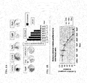
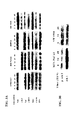
- FIG. 1A shows significant inhibition of anchorage independent growth of MIA PaCa2 cells in soft agar when treated with weekly ⁇ -tocotrienol compared to vehicle.
- HPDE 6C7 cells grown in recommended media alone did not undergo transformation in soft agar, as would be expected in this preneoplastic cell line, and served as our negative control.
- Pancreatic cancer cells (3x10 3 ) were plated in 96 well plates and treated the following day with ⁇ -tocotrienol. Proliferation was assessed by MTT at 24 hour intervals. Results show a dose dependent inhibition of proliferation ( FIGS. 1B-1D ). The IC 50 for all three pancreatic cancer cell lines was 20-25 ⁇ M at 24 hours. HPDE 6C7 cells were also treated with increasing concentrations of ⁇ -tocotrienol for 24 hours. FIG. 1E shows selective inhibition of proliferation of pancreatic cancer cell lines by ⁇ -Stocotrienol at 24 hours. HPDE 6C7 cells were relatively resistant to the antiproliferative effects of tocotrienol, even at the highest concentrations.
- results show that ⁇ -tocotrienol causes selective G1 cell cycle arrest and is associated with upregulation of the cyclin kinase inhibitor p27 klp1 .
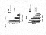
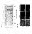
- MIA PaCa2, SW 1990, BXPC3 and HPDE 6C7 cells were treated with either ⁇ -tocotrienol 20 ⁇ M or vehicle for 24 hours. Cells were harvested, washed twice in PBS, and fixed in ethanol overnight. Pellets were washed the following day in PBS, stained with propidium iodide, and analyzed by flow cytometry for cell cycle phase.
- FIGS. 2A and 2B show significant G1 cell cycle arrest in all three pancreatic cancer cell lines treated with ⁇ -tocotrionol.
- ⁇ -tocotrienol had no effect on cell cycle phase in immortalized human pancreatic ductal epithelial cells (HPDE 6C7).
- Important regulators of the G1 cell cycle phase include members of the cyclin kinase inhibitor family. One of these Members, p27 klp1 , appears to be important in the progression of pancreatic cancer.
- FIG. 2F shows that siRNAp27 rescues tocotrienol induced G1-S cell cycle arrest in pancreatic cancer cells.
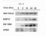
- FIG. 2C shows a time dependent upregulation in p27 klp1 expression in all three pancreatic cancer cell lines. In contrast, a downregulation of p27 klp1 is seen in HPDE 6C7 cells treated with ⁇ -tocotrienol.
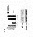
- FIG. 2D shows an increase in luciferase activity in MIA PaCa2 cells treated with ⁇ -tocotrienol. This suggests that ⁇ -tocotrienol increases p27 klp1 protein expression through transcriptional upregulation. This dynamic is further demonstrated by the rescue of tocotrienol inhibition of MiaPaCa-2 cells by siRNAp27 ( FIG. 2E ).
- ⁇ -Tocotrienol was also shown to selectively inhibits downstream effectors of oncogenic Ras signaling. More than 80% of pancreatic cancers demonstrate abnormal oncogenic Ras signaling.
- the Ras effector pathways Raf-MEK-ERK and PI3 kinase-AKT are important for cell proliferation and survival, respectively.
- FIG. 3A shows selective inhibition by ⁇ -tocotrienol of the downstream phosphorylated targets of oncogenic Ras p-craf, p-MEK, and p-ERK.
- HPDE 6C7 cells were resistant to the inhibitory effects of ⁇ -tocotrienol.
- MIA PaCa2 and SW1990 cells were then treated with ⁇ -tocotrienol (20 ⁇ M) for 24 and 48 hours.
- Cell lysates were run by SDS-PAGE and immunoblotted for p-AKT and AKT (Cell Signaling).
- FIG. 3B demonstrates a decrease in p-AKT protein levels, but not AKT at 24 and 48 hours.
- FIG. 4A shows a dose dependent induction of early apoptosis in MIA PaCa2 cells treated with ⁇ -tocotrienol. 6-tocotrienol selectively induced apoptosis in pancreatic cancer cells
- MIA PaCa2 cells and HPDE 6C7 cells were treated with vehicle or ⁇ -tocotrienol (20 ⁇ M) for 24 hours. Cells were harvested and stained for TUNEL.
- FIG. 4B shows significant staining of MIA PaCa2 cells treated with ⁇ -tocotrienol for TUNEL compared to vehicle or treated HPDE 6C7 cells. This finding suggests selective induction of apoptosis in pancreatic cancer cells.
- cell lysates were prepared from MIA PaCa2 cells treated with ⁇ -tocotrienol (20 ⁇ M) at progressive time intervals as described above. Proteins from lysates were run by SDS PAGE and immunoblotted for biomarkers of apoptosis.
- ⁇ -Tocotrionol also suppresses pancreatic cancer growth in vivo .
- pancreatic cancer cell lines resulted in induction of apoptosis in vitro and in vivo.
- the apoptotic effect of delta-tocotrienol was selective for neoplastic cells because immortalized human pancreatic ductal epithelial cells were not sensitive to delta-tocotrienol.
- Treatment of pancreatic cancer cell lines resulted in activation of the caspase cascade of the extrinsic pathway of apoptosis induction as evidenced by cleavage of Caspase 8 and Caspase 3 but not caspase 9.
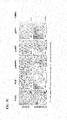
- FIG. 5A shows representative luminescent measurements of control and ⁇ -tocotrienol groups prior to treatment and at day 10. Rate of growth by day 10 of treatment is less in the tocotrienol treatment group.
- FIG. 5B shows significant reduction in tumor growth by 50% in xenografts treated with ⁇ -tocotrienol compared to vehicle. Tumors were extracted on the last day of treatment and imbedded in paraffin. Immunohistochemical staining was performed to determine the effects of ⁇ -tocotrienol on oncogenic Ras signaling targets.
- FIG. 5C shows inhibition of proliferation as evidenced by a decrease in Ki67 and induction of apoptosis by TUNEL in tumors from mice treated with ⁇ -tocotrienol as compared to tumors from the control group. Furthermore, the biomarker proteins p-MAPK and p-AKT are decreased and p27 klp1 expression is increased in ⁇ -tocotrienol treated tumors.
- SW1990 pancreatic cancer cells were cultured in complete DMEM media containing 10% fetal bovine serum (FBS) and 1% penicillin-streptomycin, and 1% L-glutamine.
- BxPc-3 pancreatic cancer cells were cultured in complete RPMI media containing 10% fetal bovine serum (FBS), and 1% penicillin-streptomycin, 1% HEPES buffer, 1% sodium pyruvate and 1% L-glutamine, HPDE6-C7 cells were cultured in serum-free keratinocyte SFM media. All cells were maintained at 37°C in a humidified incubator with 5% CO2.
- Tables I (24 hours), II (48 hours) and III (72 hours) show the associated results.
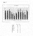
- the columns of each table are the different treatments, comprising a single dose (50 ⁇ M) as follows: Control (no treatment), Vehicle (vehicle only, maybe a solvent), T3 alpha (alpha tocotrienol), T3 beta (beta tocotrienol), T3 gamma (gamma tocotrienol), T3 delta (delta tocotrienol), T alpha (alpha tocopherol), T beta (beta tocopherol), TS (tocopherol succinate), GG (geranylgeraniol), SA (succinic acid).
- FIG. 6 illustrates the results for the 24 hour and 48 hour trials. TABLE I.
- Panc-1 (24 hours) % Control 0.167 0.156 0.151 0.161 0.142 0.147 0.142 0.109 0.147 100 Vehicle 0.15 0.146 0.147 0.14 0.155 0.151 0.164 0.138 0.149 101 T3 alpha 0.168 0.183 0.175 0.148 0.195 0.176 0206 0.172 0.178 121 T3 beta 0.164 0.168 0.167 0.123 0.138 0.143 0.126 0.134 0.145 99 T3 gamma 0.169 0.161 0.163 0.143 0.15 0.158 0.159 0.151 0.157 107 T3 delta 0.158 0.149 0.157 0.126 0.147 0.135 0.13 0.142 0.143 97 T alpha 0.188 0.183 0.189 0.166 0.183 0.181 0.186 0.16 0.180 122 T gamma 0.175 0.166 0.167 0.164 0.198 0.168 0.178 0.16 0.171 116 TS 0.155 0.168 0.164 0.158 0.155 0.163 0.165 0.167 0.162 110 GG 0.158 0.133 0.133 0.137 0.131 0.14 0.132 0.121 0.136 92 SA 0.146 0.154 0.15 0.152
- MiaPaCa-2 cells were cultured in complete DMEM media containing 10% fetal bovine serum (FBS) and 1% penicillin-streptomycin, and 1% L-glutamine.
- BxPc-3 pancreatic cancer cells were cultured in complete RPMI media containing 10% fetal bovine serum (FBS), and 1% penicillin-streptomycin, 1% HEPES buffer, 1% sodium pyruvate and 1% L-glutamine.
- HPDE6-C7 cells were cultured in serum-free keratinocyte SFM media. All cells were maintained at 37°C in a humidified incubator with 5% CO2.
- Tables IV (24 hours), V (48 hours) and VI (72 hours) show the associated results.
- the columns of each table are the different treatments, comprising a single dose (50 ⁇ M) as follows: Control (no treatment), Vehicle (vehicle only, maybe a solvent), T3 alpha (alpha tocotrienol), T3 beta (beta tocotrienol), T3 gamma (gamma tocotrienol), T3 delta (delta tocotrienol), T alpha (alpha tocopherol), T beta (beta tocopherol), TS (tocopherol succinate), GG (geranylgeraniol), SA (succinic acid).
- FIG. 7 illustrates the results for the 24 hour and 48 hour trials. TABLE IV.
- MiaPaCa-2 (24 hours) % Control 0.225 0.226 0223 0.216 0.239 0.204 0.226 0.219 0.222 100 Vehicle 0266 0.238 0.225 0.192 0.183 0.2 0.189 0.198 0.211 95 T3 alpha 0.274 0.296 0.293 0.216 0.245 0.258 0.243 0.239 0.258 116 T3 beta 0.17 0.179 0.184 0.152 0.172 0.182 0.179 0.19 0.176 79 T3gamma 0.133 0.135 0.141 0.127 0.142 0.166 0.171 0.162 0.147 66 T3 delta 0.174 0.159 0.171 0.139 0.185 0.187 0.18 0.167 0.170 77 T alpha 0.249 0.25 0.253 0.207 0.253 0.238 0.27 0.254 0.247 111 Tgamma 0.273 0.261 0.268 0.219 0.264 0.31 0.295 0.263 0.269 121 TS 0.24 0.211 0.226 0.172 0.221 0.201 0.214 0.228 0.214 96 GG 0.
- MiaPaCa-2 cells were cultured in complete DMEM media containing 10% fetal bovine serum (FBS) and 1% penicillin-streptomycin, and 1% L-glutamine.
- BxPc-3 pancreatic cancer cells were cultured in complete RPMI media containing 10% fetal bovine serum (FBS), and 1% penicillin-streptomycin, 1% HERPES buffer, 1% sodium pyruvate and 1% L-glutamine.
- HPDE6-C7 cells were cultured in serum-free keratinocyte SFM media. All cells were maintained at 37°C in a humidified Incubator with 5% CO2.
- Table VII (48 hours) and Table VIII (5 Days) show the associated results.
- the columns of each table are the different treatments, comprising a single dose (50 ⁇ M) as follows: Control (no treatment), Vehicle (vehicle only, maybe a solvent), T3 alpha (alpha tocotrienol), T3 beta (beta tocotrienol), T3 gamma (gamma tocotrienol), T3 delta (delta tocotrienol), T alpha (alpha tocopherol), T beta (beta tocopherol), TS (tocopherol succinate), GG (geranylgeraniol), SA (succinic acid). Each column represents 7 replicates with one blank.
- the numbers in the cells of each column are the output of the MTT assay; with the exception of the bottom three rows which represent, respectively: the average of the replicates, the average of the replicates including the blank, and the average of the replicates divided by the average of the control value (control is always 100).
- TABLE VII. MiaPaCa-2 (5 Days) Average Average- Blank % Blank 0.047 0.048 0.048.
- MiaPaCa-2 (5 Days) Average Average- Blank % Blank 0.051 0.052 0.052 0.052 0.053 0.052 Control 1.803 1.86 1.75 1.832 2.241 1.751 1.79 1.861 1.809 100 Vehicle 1.733 1.661 1.594 1.754 2.161 1.703 1.68 1.755 1.703 94 T3 alpha 1.497 1.573 1.569 1.55 1.811 1.589 1.47 1.580 1.528 84 T3 beta 0.97 0.781 0.977 1.154 1.408 0.944 0.891 1.018 0.966 53 T3 gamma 0.088 0.084 0.098 0.089 0.12 0.084 0.092 0.094 0.042 2 T3 delta 1.344 1.314 1.27 1.337 1.579 1.308 1.341 1.356 1.304 72 T alpha 1.56 1.647 1.59 1.619 2.059 1.526 1.588 1.656 1.604 89 T gamma 1.52 1.516 1.547 1.625 1.917 1.568
- BXPC3 cells were cultured in complete DMEM media containing 10% fetal bovine serum (FBS) and 1% penicillin-streptomycin, and 1% L-glutamine.
- BxPc-3 pancreatic cancer cells were cultured in complete RPMI media containing 10% fetal bovine serum (FBS), and 1% penicillin-streptomycin.
- FBS fetal bovine serum
- HEPES fetal bovine serum
- HPDE6-C7 cells were cultured in serum-free keratinocyte SFM media. All cells were maintained at 37°C in a humidified incubator with 5% CO2.
- Table IX 48 hours and Table X (5 Days) show the associated results.
- the columns of each table are the different treatments, comprising a single dose (50 ⁇ M) as follows: Control (no treatment), Vehicle (vehicle only, maybe a solvent), T3 alpha (alpha tocotrienol), T3 beta (beta tocotrienol), T3 gamma (gamma tocotrienol), T3 delta (delta tocotrienol), T alpha (alpha tocopherol), T beta (beta tocopherol), TS (tocopherol succinate), GG (geranylgeraniol), SA (succinic acid). Each column represents 7 replicates with one blank.
- the numbers in the cells of each column are the output of the MTT assay; with the exception of the bottom three rows which represent, respectively: the average of the replicates, the average of the replicates including the blank, and the average of the replicates divided by the average of the control value (control is always 100). TABLE IX.
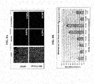
- FIG. 8A shows a significant increase in apoptotic cells in MIAPaCa2 treated with ⁇ -tocotrienol compared to vehicle or treated HPDE 6C7 cells.
- FIGS. 8B-8D MIA PaCa-2. BxPc-3. and SW1990 pancreatic cancer cells were also treated with in varying concentrations of ⁇ -tocotrienol or vehicle for 24 hours. Cells were collected and stained with Annexin V-FITC and analyzed by flow cytometry for apoptosis.
- FIGS. 8B , 8C and 8D show a dose dependent induction of apoptosis in MIA PaCa-2 cells treated with 6-tocotrienol compared to vehicle.
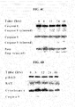
- FIG. 9A shows activation of the caspase cascade as evidenced by cleavage of Caspase 8.
- Caspase 3, and PARP in a time dependent manner.
- MIA PaCa-2 cells treated with ⁇ -tocotrienol had no significant effect on mitochondrial pro-apoptotic proteins as shown by a lack of cytochrome C release or cleavage of Caspase 9 (9B).
- FIG 9C shows selective caspase 8 and 3 cleavage in several pancreatic cancer cell lines but not in HPDE-6C7 cells, an immortalized pancreatic ductal epithelial cell line. Caspase 9 was not cleaved in the pancreatic cancer cells.
- FIG. 10 shows a time-dependent decrease in phospho-AKT (but not total AKT) in the pancreatic cancer cell lines, but not in the HPDE cells.
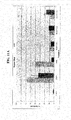
- FIG. 11A The functional overexpression of CA-AKT demonstrates the role of AKT signaling and caspase 8 in ⁇ -Tocotrienol-induced cell death ( FIG. 11A ).
- MIA PaCa-2 cells pZWL vector and myrAKT
- ⁇ -tocotrienol 40 or 50 ⁇ M
- Cells were also treated with TRAIL as a positive control.
- Cells were then harvested, fixed, permeablized and stained with trypan blue.
- Western blot FIG. 14B of cell lysates from Fig. 11A demonstrate the decrease of pAKT with 50uM treatment of tocotrienol in MiaPaca 2 parenteral cells transfected with empty vector compared to cells overexpressing constitutively active AKT.
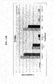
- FIG. 12A demonstrate significant inhibition of pancreatic tumor growth in mice treated with ⁇ -tocotrionol.
- Immunohistochemical staining FIG. 12B demonstrates ⁇ -tocotrienol Induced apoptosis as measured by TUNEL staining, and decreased p-AKT expression.
- FIGS. 13A and 13B demonstrate the rescue of ⁇ -tocotrienol's ability to downregulate p-AKT.
- Figures 13C through 13F show the AKT and caspase 8/3 pathways are involved in the mechanism by which tocotrienol induces cell death.
- Treatment with tocotrienol induced more cell death in the vector cells than the myr-AKT cells when compared to vehicle.
- TRAiL induced cell death was comparable to vehicle.
- pretreatment with caspase 3, 8 and 9 inhibitors rescued tocotrienol induced cell death.
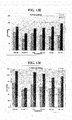
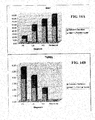
- Delta-tocotrienol inhibits pancreatic cancer metastasis as shown in FIGS. 13G-13J .
- Tocotrienol caused tumor regression at the highest dose compared to vehicle/PBS. Accordingly, tocotrienol inhibits pancreatic tumor growth in vivo.
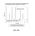
- FIGS. 13K and 13L Putative cancer chemoprevention in plasma and mouse tissue by HPLC-UV via vitamin E and delta-tocotrienol is shown in FIGS. 13K and 13L .
- Tocotrienol was detectable in plasma and was significantly concentrated in the pancreas.
- tocotrienol localizes in the pancreas by a factor of 10 when compared to liver and tumor levels.
- ⁇ -Tocotrienol selectively induces apoptosis in pancreatic cancer cell via the extrinsic apoptotic pathway.
- ⁇ -Tocotrienol selectively inhibits the PI3-K/AKT pathway and induces apoptosis in vivo and this is associated with inhibition of AKT signaling targets.
- overexpression of myr AKT in Pancreatic Cancer cells rescues the effect ⁇ -Tocotrienol induces apoptosis in vitro and in vivo.
- tocotrienol inhibition of pancreatic cancer cell growth can be rescued by consitutively active myrAKT. This suggests that tocotrienol induces apoptosis in pancreatic cancer at the level of pAKT.
- delta-tocotrienol induced apoptosis of pancreatic cancer cells likely involves modulation of the Akt signaling pathway, because the Akt survival signaling pathway is often overexpressed in pancreatic cancer and implicated in non-mitochondria associated apoptosis.
- Delta-tocotrienol suppressed the phosphorylation levels of serine/threonine kinase Akt in vitro and in vivo.
- overexpression of constitutively active Akt protected pancreatic cancer cells from undergoing apoptosis after incubation with delta-tocotrienol.
- delta-tocotrienol induces apoptosis in pancreatic cancer cells through activation of the caspase 8 cascade and suppression of the Akt survival pathway and shows that this micronutrient is useful for pancreatic cancer chemoprevention and treatment in vivo.
- chemopreventive benefit of an agent could only be shown by a reduced cancer incidence or mortality.
- the use of reduced incidence or mortality as end points makes chemoprevention trials long, large, costly, and, hence impractical except for cancers that have high event rates.
- the only reports of chemoprevention trials for pancreatic cancer have come from subset analysis of chemoprevention trials conducted for other indications. This approach has resulted in inconclusive observations of the value of agents such as ⁇ -tocopherol, ⁇ -carotene, aspirin, and non-steroidal anti-inflammatory drugs (NSAIDs).
- NSAIDs non-steroidal anti-inflammatory drugs
- IEN intraepithelial neoplasia
- MNs mucinous cystic neoplasms
- IPMNs intraductal papillary mucinous neoplasms
- Pancreatic intraepithelial neoplasia Pancreatic intraepithelial neoplasia
- SEB studies are well established in the setting of lENs that do not require surgical intervention, such as leukoplakia of the oral mucosa or Barrett's esophagus, such studies are more difficult to conduct In premalignant lesions requiring immediate surgical intervention because the index lesion is removed and is not amenable to subsequent follow-up.
- An interval of two to four weeks is standard in most practices, hence offering a window within which a chemopreventive agent can be administered to evaluate the effects of the agent on SEB modulation.
- the inventor's experiments show the value of standard SEBs such as proliferation rate, apoptosis index, and the expression of proteins (discussed below) that are modulated by the early genetic changes in these lesions.
- the invention includes surrogate endpoint biomarkers that were prevelant in the surgical specimens of patients who have undergone resection of invasive ductal adenocarcinomas of the pancreas (IPMNs).
- Tocotrienol such as ⁇ -tocotrienol from annatto bean, demonstrated selectively inhibition in human pancreatic cancer cells.
- the data shows that ⁇ -tocotrienol affects a number of molecular processes including induction of apoptosis and inhibition of tumor growth and that ⁇ -tocotrienol inhibits oncogenic signal transduction pathways in pancreatic neoplastic cells. Specifically, ⁇ -tocotrienol decreases phospho-Raf, phospho-MEK.
- ⁇ -tocotrienol induces the expression of growth inhibitory mediators such as p27 klp1 , and activates the apoptotic mediators caspase 8 and caspase 3.
- pancreatic carcinoma precursor lesions termed pancreatic intraepithelial neoplasia (panlN) were graded Ia. Ib, 2, and 3 according to published criteria (Hruban, 2001). Based on slide review, 10 representative tissue blocks were selected from ten consecutive resection specimens. Preference was given to sections showing carcinoma, (preferably more than one grade of carcinoma), nonneoplastic ducts, and precursor lesions. All cases were ductal adenocarcinoma, NOS, or well to poorly differentiated.
- Ki67 was noted to be a reliable marker of proliferation with progressive increase of a positive reaction in the nucleus from normal to intermediate to invasive carcinoma.
- An inverse progression was noted for TUNEL staining indicating oncogenic suppression of apoptosis in neoplastic pancreatic ductal cells.
- a similar inverse progression was observed in the expression of the p27 cyclin-dependent kinase inhibitor protein,
- downstream mediators of activated oncogenic Ras signaling such as phospho-MAPK and phospho-AKT are increased.
- all patients with invasive ductal cancer of the pancreas and IPMNs had noninvasive precursor lesions in their surgical specimens.
- FIG. 14A shows an increase in proliferation rate as degree of invasion and grade of tumor is increased. This is indicated by a progressive increase in the mean percentage of cells staining positive for KI-67.
- FIG. 14B shows a progressive decrease in the number of apoptotic cells stained with TUNNEL, negatively correlating with the degree of tumor invasion.
- FIG. 15 shows representative slides of non-neoplastic pancreatic ductal tissue, reactive tissue, precursor lesions (PanlN 1-3), and low and high grade invasive carcinomas. Each representative area was stained with Hematoxylin and Eosin, and for Ki-67, p27, p-MAPK, and TUNEL.
- Results show an increase in Ki-67 and p-MAPK staining as pancreatic ducts progress from non-neoplastic to PanlN to invasive carcinoma. The greatest degree of staining is in high grade tumors. In contrast p27 and TUNEL staining decrease with tumor invasiveness and progression. Indicating a loss of cell cycle inhibition and induction of apoptosis.
- Another embodiment of the invention includes a method of screening for pancreatic ductal carcinoma, or a stage of pancreatic cancer in a subject by determining, such as in an isolated sample, the level of a biomarker, namely, p27. The level of the biomarker is then compared to a corresponding control level in one or more control samples.
- the control samples are obtained from individuals who have been determined not to have pancreatic ductal carcinoma, or a stage of pancreatic cancer.
- the determination of a statistically significant increase between the level of the biomarker in the subject and the level of the biomarker in the control sample(s) is indicative of the lack of pancreatic ductal carcinoma, or a stage of pancreatic cancer in the subject.
- a statistically significant decrease in the level of p27 in the subject, compared to the level of the biomarker in the control sample(s), indicates the presence of pancreatic ductal carcinoma, or a stage of pancreatic cancer in the subject.
- the level of a second biomarker namely Ki-67 and/or p-MAPK
- a control level of Ki-67 and/or p-MAPK in one or more control samples is determined and compared to a control level of Ki-67 and/or p-MAPK in one or more control samples.
- a statistically significant increase between the level of Ki-67 and/or p-MAPK in the subject and the control sample(s) is indicative of pancreatic ductal carcinoma, or a stage of pancreatic cancer in the subject.
- a statistically significant decrease in the level of Ki-67 and/or p-MAPK in the subject, compared to the control sample(s) is indicative of the lack of pancreatic ductal carcinoma, or a stage of pancreatic cancer in the subject.
- the level of the biomarker in the subject and control sample(s) are known to the ordinary practitioner.
- the level of the biomarker is determined utilizing an antibody which binds the biomarker.
- the sample containing the biomarker is contacted with the antibody under conditions which allow binding of the biomarker to the antibody; the presence of the biomarker can then be quantified.
- the invention also includes compositions useful in performing the associated methods.
- the invention includes a composition comprising a plurality of isolated proteins which bind selectively to the protein products of the associated biomarkers; namely, Ki-67 and/or p-MAPK, and p27.
- the isolated proteins selectively amplify complementary double stranded DNA.
- a composition is also included comprising a plurality of biomarker specific primers, wherein each biomarker specific primer selectively amplifies double stranded DNA complementary to a unique biomarker such as Ki-67, p-MAPK and p27.
- the invention includes a composition comprising a plurality of isolated proteins which bind selectively to the protein products of at least two unique biomarkers, wherein each unique biomarker is selected from the group consisting of Ki-67, p-MAPK and p27.
- biomarker refers to a gene that is differentially expressed in individuals having cancer, including pancreatic cancer or a stage of pancreatic cancer as compared with those not having cancer, including pancreatic or a stage of pancreatic cancer.
- biomarker can include a gene that is differentially expressed In individuals having superficial pancreatic cancer as compared with those not having pancreatic cancer.
- biomarker specific primers refers to a set of primers which can produce double stranded DNA complementary to a portion of one or more RNA products of the biomarker of the Invention.
- the primers can include a first primer which is a sequence that can selectively hybridize to RNA, DNA or EST complementary to a biomarker of the invention to create an extension product and a second primer capable of selectively hybridizing to the extension product, which are used to produce double stranded DNA complementary to a biomarker of.the invention.
- primer refers to an oligonucleotide, whether occurring naturally as in a purified restriction digest or produced synthetically, which is capable of acting as a point of initiation of synthesis when placed under conditions in which synthesis of a primer extension product, which is complementary to a nucleic acid strand, is induced, i.e., in the presence of nucleotides and an inducing agent such as a DNA polymerase and at a suitable temperature and pH.
- the primer may be either single-stranded or double-stranded and must be sufficiently long to prime the synthesis of the desired extension product in the presence of the inducing agent The exact length of the primer will depend upon many factors, including temperature, source of primer and the method used.
- the oligonucleotide primer typically contains 15-25 or more nucleotides, although it may contain fewer nucleotides.
- the factors involved in determining the appropriate length of primer are readily known to one of ordinary skill in the art.
- the design and selection of primers embodied by the instant invention is according to methods that are standard and well known in the art, see Dieffenbach, C. W., Lowe, T. M. J., Dveksler, G. S. (1995) General Concepts for PCR Primer Design. In; PCR Primer, A Laboratory Manual (Eds. Dieffenbach, C. W, and Dvaksler. G.
- biomarker specific probe refers to a probe selectively and specifically hybridizes to RNA products of a unique biomarker.
- a biomarker specific probe can be a probe having a fluorophore and a quencher, for example a TaqMan.RTM. probe or a Molecular Beacons probe.
- a biomarker specific probe is a probe which is attached to an array and selectively and specifically hybridizes to one or more RNA products (or cDNA, EST or PCR products corresponding to said RNA products) of a unique biomarker.
- a biomarker specific probe can include oligonucleotide probes and can also include longer probes ( e.g. 60, 80, 100, 150, 200, 250, 300, 360, 400, 450, 500 Nucleotides etc.).
- probe means oligonucleotides and analogs thereof and refers to a range of chemical species that recognize polynucleotide target sequences through hydrogen bonding interactions with the nucleotide bases of the target sequences.
- the probe or the target sequences may be single- or double-stranded RNA or single- or double-stranded DNA or a combination of DNA and RNA bases.
- a probe is at least 8 nucleotides In length and less than the length of a complete gene.
- a probe may be 10, 20, 30, 50, 75, 100, 150, 200, 260, 400, 500 and up to 2000 nucleotides in length.
- Probes can include aligonucleotides modified so as to have a tag which is detectable by fluorescence, chemiluminescence and the like.
- the probe can also be modified so as to have both a detectable tag and a quencher molecule, for example Taqman.RTM. and Molecular Beacon.RTM. probes.
- RNA resulting from the biomarker can include one or more of the following species; hnRNA, mRNA, and/or one or more spliced variants of mRNA.
- proteins resulting from the molecular marker can include any proteins found in blood which correspond to the RNA resulting from the biomarker.
- control or control sample in the context of this invention refers to one or more tissue nucleic acid samples and/or a blood nucleic acid samples and/or one or more individuals who are classified as having pancreatic cancer, having one or more stages of pancreatic cancer and/or superficial bladder cancer; not having pancreatic cancer, having one or more stages of pancreatic cancer and/or superficial bladder cancer; as determined by using those techniques known to a person skilled in the art.
- control or control sample can also refer to the compilation of data derived from samples of one or more individuals who have been diagnosed as normal (not having pancreatic cancer), having pancreatic cancer, or having a stage of pancreatic cancer.
- control in the context of the experiment and will depend upon the desired comparisons.
- control in the context of screening for a prophylactic or therapeutic agent refers to a standard or reference for an assay or methodology to which other conditions can be compared.
- the term "effective amount” refers to the amount of a compound which is sufficient to reduce or ameliorate the progression and or severity of pancreatic cancer or one or more symptoms thereof, prevent the development, recurrence or onset of pancreatic cancer or one or more symptoms thereof, prevent the advancement of pancreatic cancer or one or more symptoms thereof, or enhance or improve the prophylactic or therapeutic effect(s) of another therapy.
- chemotherapeutic agent refers to any compound(s) which can be used in the treatment, management or amelioration of pancreatic cancer or one or more symptoms thereof.
- a therapy e.g., a chemotherapeutic agent
- a therapy e.g., a chemotherapeutic agent
- prevent advancement of bladder cancer cause regression of bladder cancer, or to enhance or improve the therapeutic effect(s) of another therapy (e.g., chemotherapeutic agent).
- the term “efficacy” refers to the effectiveness of a drug and/or.
- “Chemotherapeutic efficacy” is usually measured by the clinical response of the patient who has been or is being treated with a drug and/or agent.
- a drug is considered to have a high degree of efficacy, if it achieves desired clinical results, for example, the reduction of the symptoms associated with pancreatic cancer, a stage of pancreatic cancer, or the prevention of pancreatic cancer progression as described in the present specification.
- the amount of drug absorbed may be used to predict a patient's response.
- a general rule is that as the dose of a drug is increased, a greater effect is seen in the patient until a maximum desired effect is reached.
- level of expression refers to the determination of the quantity of a given nucleic acid or protein corresponding to a gene as determined by methods known to a person skilled.
- level of expression can be determined by hybridization as well as other measurements such as quantitative real-time RT PCR, which includes use of SYBR.RTM. green, TaqMan,RTM, and Molecular Beacons technology.
- the determination of differential levels of expression can include a visual inspection of differences as between the quantity of a given nucleic acid or protein, for example by analyzing the northern blot or western blot.
- the term "selectively amplified” or “selective amplification”, refers to a process whereby one or more copies of a particular target nucleic acid sequence is selectively generated from a template nucleic acid.
- Selective amplification or selectively amplified is to be compared with amplification in general which can be used as a method in combination with, for example, random primers and an oligodT primer to amplify a population of nucleic acid sequences (e.g. mRNA).
- Selective amplification is preferably done by the method of polymerase chain reaction ( Mullis and Faloona, 1987, Methods Enzymol. 155:335 ).
- the term "selectively binds" in the context of proteins encompassed by the invention refers to the specific interaction of any two of a peptide, a protein, a polypeptide, and an antibody, wherein the interaction preferentially occurs as between any two of a peptide, protein, polypeptide and antibody preferentially as compared with any other peptide, protein, polypeptide and antibody.
- the two molecules are protein molecules, a structure on the first molecule recognizes and binds to a structure on the second molecule, rather than to other proteins.
- Selective binding means that a molecule binds its specific binding partner with at least 2-fold greater affinity, and preferably at least 10-fold, 20-fold, 50-fold, 100-fold or higher affinity than it binds a non-specific molecule.
- the invention includes a method of determining the effectives of a chemotherapeutic agent by determining, in an isolated sample, a first level of a surrogate endpoint biomarker such as p27. The sample is then contacted with an experimentally effective amount of the chemotherapeutic agent being tested. After the chemotherapeutic agent has been administered, a second level of the surrogate endpoint biomarker is taken and compared to the first (pre-treatment) level. The candidate chemotherapeutic agent demonstrates effectiveness where the second (post-treatment) levels p27 are increased to a statistically significant degree over the pre-treatment and/or control levels.
- the invention provides a method of determining the effectives of a chemotherapeutic agent by further determining, in the isolated sample, a first level of a second surrogate endpoint biomarker such as Ki-67 and/or p-MAPK. After the chemotherapeutic agent has been administered, a second level of Ki-67 and/or p-MAPK is taken and compared to the first (pre-treatment) level and/or a control.
- the candidate chemotherapeutic agent demonstrates effectiveness where the second (post-treatment) levels of Ki-67 and/or p-MAPK are decreased to a statistically significant degree below the pre-treatment and/or control levels.
- Another embodiment of the invention includes a method of screening for pancreatic ductal carcinoma, or a stage of pancreatic cancer in a subject by determining, such as in an isolated sample, the level of a biomarker; namely, p27. The level of the biomarker is then compared to a corresponding control level In one or more control samples.
- the control samples are obtained from individuals who have been determined not to have pancreatic ductal carcinoma, or a stage of pancreatic cancer.
- the determination of a statistically significant increase between the level of the biomarker In the subject and the level of the biomarker in the control sample(s) is indicative of the lack of pancreatic ductal carcinoma, or a stage of pancreatic cancer in the subject.
- a statistically significant decrease in the level of p27 in the subject, compared to the level of the biomarker in the control sample(s), indicates the presence of pancreatic ductal carcinoma, or a stage of pancreatic cancer in the subject.
- the level of a second biomarker namely Ki-67 and/or p-MAPK
- a Control level of Ki-67 and/or p-MAPK in one or more control samples is determined and compared to a Control level of Ki-67 and/or p-MAPK in one or more control samples.
- a statistically significant increase between the level of Ki-67 and/or p-MAPK in the subject and the control sample(s) is indicative of pancreatic ductal carcinoma, or a stage of pancreatic cancer in the subject.
- a statistically significant decrease in the level of Ki-67 and/or p-MAPK in the subjects, compared to the control sample(s) is indicative of the lack of pancreatic ductal carcinoma, or a stage of pancreatic cancer in the subject.
- the lever of the biomarker is determined utilizing an antibody which binds the biomarker.
- the sample containing the biomarker is contacted with the antibody under conditions which allow binding of the biomarker to the antibody: the presence of the biomarker can then be quantified.
- the invention also includes compositions useful in performing the associated methods.
- the invention includes a composition comprising a plurality of isolated proteins which bind selectively to the protein products of the associated biomarkers; namely, Ki-67 and/or p-MAPK, and p27.
- the isolated proteins selectively amplify complementary double stranded DNA.
- a composition is also included comprising a plurality of biomarker specific primers, wherein each biomarker specific primer selectively amplifies double stranded DNA complementary to a unique biomarker such as Ki-67, p.MAPK and p27.
- the invention includes a composition comprising a plurality of isolated proteins which bind selectively to the protein products of at least two unique biomarkers, wherein each unique biomarker is selected from the group consisting of Ki-67, p-MAPK and p27.
- determining the levels (e.g. quantifying) of the biomarkers will be readily known to one of ordinary skill and include, but are not limited to, determining expression level, level of RNA, level of RNA product, protein level, and/or protein activity level.
Landscapes
- Health & Medical Sciences (AREA)
- Life Sciences & Earth Sciences (AREA)
- Chemical & Material Sciences (AREA)
- General Health & Medical Sciences (AREA)
- Engineering & Computer Science (AREA)
- Medicinal Chemistry (AREA)
- Veterinary Medicine (AREA)
- Public Health (AREA)
- Pharmacology & Pharmacy (AREA)
- Animal Behavior & Ethology (AREA)
- Immunology (AREA)
- Organic Chemistry (AREA)
- Molecular Biology (AREA)
- Epidemiology (AREA)
- Bioinformatics & Cheminformatics (AREA)
- Pathology (AREA)
- Proteomics, Peptides & Aminoacids (AREA)
- Biomedical Technology (AREA)
- Biochemistry (AREA)
- Analytical Chemistry (AREA)
- Microbiology (AREA)
- General Chemical & Material Sciences (AREA)
- Zoology (AREA)
- Biotechnology (AREA)
- Urology & Nephrology (AREA)
- Wood Science & Technology (AREA)
- Physics & Mathematics (AREA)
- Chemical Kinetics & Catalysis (AREA)
- Genetics & Genomics (AREA)
- Hematology (AREA)
- Nuclear Medicine, Radiotherapy & Molecular Imaging (AREA)
- Food Science & Technology (AREA)
- Hospice & Palliative Care (AREA)
- Oncology (AREA)
- Toxicology (AREA)
- Tropical Medicine & Parasitology (AREA)
- Biophysics (AREA)
- General Physics & Mathematics (AREA)
- Cell Biology (AREA)
- General Engineering & Computer Science (AREA)
Abstract
Description
- This application is a non-provisional application of co-pending
U.S. Provisional Application, No. 601805,916, filed June 27, 2006 ; which is fully incorporated herein by reference and co-pendingU.S. Nonprovisional Application No. 11/768,3T3, filed June 26, 2007 . - This invention was made with Government support under Grant No. DAMD 17-01-1-04 awarded by the Department of Defense, and NCl Grant No. 3R01CA098473-03S1. The Government has certain rights in the invention.
- The pancreas is an extremely common site for the development of early neoplasms-noninvasive clonal epithelial expansions. In a minority of persons, these clones of cells serially acquire genetic changes that can lead to an invasive adenocarcinoma. Pancreatic cancer, once invasive, is almost uniformly fatal. The epithelial cells in the advanced stage of this process are very aggressive, seemingly having an innate capability for metastasis that is exhibited rather soon after they invade beyond the duct structure into surrounding tissue. In order to alleviate the dismal prognosis associated with this disease, it is imperative that the process of pancreatic carcinogenesis be recognized and treated prior to invasion. Chemoprevention is the administration of agents (drugs, biologics, and nutrients) to slow progression of, reverse, or inhibit carcinogenesis thereby lowering the risk of developing invasive or clinically significant disease. Understanding the morphology and biology of precursor lesions of invasive pancreatic cancer has therefore become an issue of paramount importance, In the last decade, significant progress has been made in the recognition and appropriate classification of these precursor lesions. Mucinous cystic neoplasms (MCNs), intraductal papillary mucinous neoplasms (IPMNS), and pancreatic intraepithelial neoplasia (PanlN) encompass the three known morphologically distinct precursors to invasive pancreatic cancer.
- A large number of case-control and cohort studies have shown that there is a significant clustering of pancreatic cancer in some families, These high-risk inherited pancreatic cancers are estimated to represent about 10% of pancreatic cancers. Five well-known genetic syndromes with known gene defects account for approximately 20% of the families in which there is aggregation of pancreatic cancer. These syndromes include (1) BRCA2, (2) familial atypical multiple mole melanoma (p16/CDKN2A). (3) Peutz-Jeghers Syndrome; (4) HNPCC; and (5) familial pancreatitis. Majority of pancreatic cancers are sporadic and have evidence of widespread chromosomal instability, including a high rate of translocations and deletions. Nearly all (>90%) preinvasive lesions have an early mutation in the K-Ras protein involved in the transmission of growth factor signals. In the middle stages of preinvasive progression >90% of lesions develop inactivation of the CDKN2A (p16) cyclin dependent kinase inhibitor. In later stages most preinvasive lesions also harbor mutations of the TP53 (p53) and in MADH4, the common Smad protein involved In transduction of TGFβ and activin signals. Despite the enormous obvious benefit for chemopreventive agents in pancreatic neoplasia, direct drug investigations for chemopreventive indications have been slow to emerge. A critical factor is the challenge of conducting studies that will define and demonstrate clinical benefit.
- Proliferation of pancreatic cancer is regulated through aberrant oncogenic Ras signaling and its effect on cyclin kinase inhibitors such as p27ldp1. Previous studies have demonstrated that pharmacologic inhibition of one of the ras signaling pathways, the Raf-MEK-ERK pathway, elicits pancreatic cancer cell cycle arrest through induced expression of p27 (cancer Res 2005: 65(11):4870-80). Tocotrienols, the chemical form of vitamin E with an unsaturated isoprenoid side chain, are receiving attention as promising dietary supplements for cancer prevention and treatment.
- Tocotrienols are the primary form of vitamin E in the seeds of most monocot plants such as palm and cereals such as rice and wheat. The biosynthesis of tocotrienols and tocopherols occur exclusively in photosynthetic organisms and arise from homogentisic acid. Tocotrienols arise from the condensation of homogentisic acid and, geranylgeranyl diphosphate while the committed step in tocopherol synthesis is the condensation of homogentisic acid and phytyl diphosphate. Structurally tocopherols and tocotrienols share some resemblance consisting of a common chromanol head and a side chain at the C-2 position however, their side chains distinguish tocopherols and tocotrienols,
- While tocopherol has a saturated phytyl tail, tocotrienol possesses an unsaturated isoprenoid side chain. Tocopherols and torotrienols are further separated into individual compounds assigned by the greek letter prefixes (α,β,γ,δ) depending on the number and position of methyl substitution on the chromanol ring. As reflected in their structural similarly, tocopherols and tocotrienols are well recognized for their antioxidant effect. However, tocotrionols are the group of natural vitamin E compounds with clear and consistent antitumor activity. Semisynthetic tocopherols such as tocopherol succinate have antitumor activity however the bioavailability of the intact compound after oral consumption is poor making it unsuitable for chemopreventive interventions. Structure activity studies of the proapoptotic effects of vitamin E compounds have clearly documented the importance of the unsaturated isoprenoid tail of the vitamin E compounds in their antitumor bioactivity. Furthermore, these studies indicate that decreasing the number of methyl substitutions on the chromanot ring, is associated with increasing antitumor potency.
- In one embodiment, the invention includes a method of determining the effectives of a chemotherapeutic agent by determining, In an isolated sample, a first level of a surrogate endpoint biomarker such as p27. The sample is then contacted with an experimentally effective amount of the chemotherapeutic agent being tested. After the chemotherapeutic agent has been administered, a second level of the surrogate endpoint biomarker is taken and compared to the first (pre-treatment) level. The candidate chemotherapeutic agent demonstrates effectiveness where the second (post-treatment) level of p27 is increased to a statistically significant degree over the pro-treatment and/or control levels.
- In another embodiment, the invention provides a method of determining the effectiveness of a chemotherapeutic agent by further determining, in the isolated sample, a first level of a second surrogate endpoint biomarker such as KI-67 and/or p-MAPK. After the chemotherapeutic agent has been administered, a second level of Ki-67 and/or p-MAPK is taken and compared to the first (pre-treatment) level and/or a control. The candidate chemotherapeutic agent demonstrates effectiveness where the second (post-treatment) level of Ki-67 and/or p-MAPK is decreased to a statistically significant degree below the pre-treatment and/or control levels.
- Another embodiment of the invention includes a method of screening for pancreatic ductal carcinoma, or a stage of pancreatic cancer in a subject by determining, such as in an isolated sample, the level of a biomarker, namely, p27. The level of the biomarker is then compared to a corresponding control level in one or more control samples. In a preferred embodiment the control samples are obtained from individuals who have been determined not to have pancreatic ductal carcinoma, or a stage of pancreatic cancer.
- The determination of a statistically significant similarity between the level of the biomarker in the subject and the level of the biomarker in the control sample(s) is indicative of the lack of pancreatic ductal carcinoma, or a stage of pancreatic cancer in the subject. A statistically significant decrease in the level of p27 in the subjects, compared to the level of the biomarker in the control sample(s), indicates the presence of pancreatic ductal carcinoma, or a stage of pancreatic cancer in the subject.
- In an alternate embodiment, the level of a second biomarker, namely Ki-67 and/or p-MAPK, is determined and compared to a control level of Ki-67 and/or p-MAPK in one or more control samples. A statistically significant increase between the level of Ki-67 and/or p-MAPK In the subject and the control sample(s) is indicative of pancreatic ductal carcinoma, or a stage of pancreatic cancer in the subject. A statistically significant similarity in the level of Ki-67 and/or p-MAPK in the subject, compared to the control sample(s), is indicative of the lack of pancreatic ductal carcinoma, or a stage of pancreatic cancer in the subject.
- Methods of determining the level of the biomarker in the subject and control sample(s) are known to the ordinary practitioner. In one embodiment, as an example, the level of the biomarker is determined utilizing an antibody which binds the biomarker. The sample containing the biomarker is contacted with the antibody under conditions which allow binding of the biomarker to the antibody; the presence of the biomarker can then be quantified.
- The invention also includes compositions useful in performing the associated methods. For example, the invention Includes a composition comprising a plurality of isolated proteins which bind selectively to the protein products of the associated biomarkers; namely, Ki-67 and/or p-MAPK. and p27. In a preferred embodiment, the isolated proteins selectively amplify complementary double stranded DNA. A composition is also included comprising a plurality of biomarker specific primers, wherein each biomarker specific primer selectively amplifies double stranded DNA complementary to a unique biomarker such as Ki-67, p-MAPK, and p27. Alternatively, the invention includes a composition comprising a plurality of isolated proteins which bind selectively to the protein products of at least two unique biomarkers, wherein each unique biomarker, is selected from the group consisting of Ki-67. p-MAPK and p27.
- Accordingly, the invention includes method of treating cancer, such as pancreatic ductal carcinoma, or a stage of pancreatic cancer, by administering to a subject a composition comprising a therapeutically effective amount of a tocotrienal. In a preferred embodiment the tocotrienol is gamma-tocotrienol and/or deta-tocotrionol, which is administered as a pharmaceutical composition. The composition of the preferred embodiment is substantially free of alpha-tocotrienol, beta-tocotrienol and/or tocopherols. Although one of ordinary skill will recognize methods of determining the appropriate dose, the composition of one embodiment comprises between about 100mg and 300mg tocobienol which is administered twice daily.
- The tocotrienol of the preferred embodiment has the formula:

- For a fuller understanding of the nature and objects of the invention, reference should be made to the following detailed description, taken in connection with the accompanying drawings, in which:
-
FIG. 1A is an images of MIA PaCa2 cells in soft agar; significant inhibition of anchorage independent growth of cells occurred when treated with weekly δ-tocotrienol compared to vehicle. -
FIG. 1B is a graph wherein MIA PaCa2 pancreatic cancer cells (3x10³) were plated in 96 well plates and treated the following day with 6-tocotrienol. Proliferation was assessed by MTT at 24 hour intervals. Results show a dose dependent inhibition of proliferation. The IC50 for all three pancreatic cancer cell lines was 20-25µM at 24 hours. HPDE 6C7 cells were also treated with increasing concentrations of δ-tocotnenol for 24 hours. -
FIG. 1C is a graph wherein SW1990 pancreatic cancer cells (3x10³) were plated in 96 well plates and treated the following day with δ-tocotrienol. Proliferation was assessed by MTT at 24 hour intervals. Results show a dose dependent inhibition of proliferation. The IC50 for all three pancreatic cancer cell lines was 20-25µM at 24 hours. HPDE 6C7 cells were also treated with increasing concentrations of δ-tocotrienot for 24 hours. -
FIG. 1D is a graph wherein BXPC3 pancreatic cancer cells (3x10³) were plated in 96 well plates and treated the following day with δ-tocotrienol. Proliferation was assessed by MTT at 24 hour intervals. Results show a dose dependent inhibition of proliferation. The IC50 for all three pancreatic cancer cell lines was 20-25µM at 24 hours. HPDE 6C7 cells were also treated with increasing concentrations of δ-tocotrienol for 24 hours. -
FIG. 1E is a graph showing selective inhibition of proliferation of pancreatic cancer cell lines by 6-tocotrienol at 24 hours. HPDE 6C7 cells were relatively resistant to the antiproliferative effects of tocotrienol, even at the highest concentrations. -
FIG. 2A is a graph showing significant G1 cell cycle arrest in HPDE 6C7 and SW1990 pancreatic cancer cell lines treated with δ-tocotrienol (25µM). In contrast, δ-tocotrienol had no effect on cell cycle phase in immortalized human pancreatic ductal epithelial cells (HPDE 6C7). -
FIG. 2B is a graph showing significant G1 cell cycle arrest in MaPaCa2 and BXPC3 pancreatic cancer cell lines treated with δ-tocotrienol (20µM). In contrast, δ-tocotrienol had no effect on cell cycle phase in immortalized human pancreatic ductal epithelial cells (HPDE 6C7). -
FIG. 2C is a series of blots showing a time dependent upregulation in p27kip1 expression in all four pancreatic cancer cell lines. In contrast, a downregulation of p27klp1 is seen in HPDE 6C7 cells treated with δ-tocotrienol. -
FIG. 2D is a graph showing an increase in luciferase activity in MIA PaCa2*cells treated with δ-tocotrienol. -
FIG. 2E is a graph and associated blots showing siRNAp27 rescues tocotrienol inhibition of pancreatic cancer cell growth. -
FIG. 2F is a pair of graphs showing that siRNAp27 rescues tocotrienol induced G1-S cell cycle arrest in pancreatic cancer cells. -
FIG. 3A is a blot series showing selective inhibition by δ-tocotrienol of the downstream phosphorylated targets of oncogenic Ras p-cRaf, p-MEK, and p-ERK. HPDE 6C7 cells were resistant to the inhibitory effects of δ-tocotrienol. -
FIG. 3B is a blot series showing MIA PaCa2 and SW1990 cells treated with δ-tocotrienol (20µM) for 24 and 48 hours. Cell lysates were run by SDS-PAGE and immunoblotted for p-AKT and AKT (Cell Signaling). The blots show a decrease in p-AKT protein levels, but not AKT at 24 and 48 hours. -
FIG. 4A is a graph showing activation of the caspase cascade as evidenced by cleavage ofCaspase 8,Caspase 3, and Parp in a time dependent manner. -
FIG. 4D is a series of images showing activation of the caspase cascade as evidenced by cleavage ofCaspase 8,Caspase 3, and Parp in a time dependent manner. -
FIG. 4C is a series of blots showing activation of the caspase cascade as evidenced by cleavage ofCaspase 8,Caspase 3, and Parp in a time dependent manner. -
FIG. 4D is a series of blots showing activation of the caspase cascade as evidenced by cleavage ofCaspase 8,Caspase 3, and Parp in a time dependent manner. -
FIG. 5A shows representative luminescent measurements of control and δ-tocotrienol groups prior to treatment and atday 10. Rate of growth byday 10 of treatment is less in the tocotrienol treatment group. Stable transfectants of MIA PaCa2 cells with luciferase were injected subcutaneously into nude mice. Tumor volumes were measured every 2 days with calibers and the rate of growth was determined weekly by measuring tumor luminescence using theMS 100 Xenogen system. Tumors of similar volume (100-150mm3) and rate of growth based on luminescence were randomized to receive either vehicle or δ-tocotrienol (100mgl/kg/day) by gavage for 20 days. -
FIG. 5B is a graph showing significant reduction in tumor growth by 50% in xenografts treated with δ-tocotrionol compared to vehicle. -
FIG. 5C is a series of immunohistochemical stains of the effects of δ-tocotrienol on oncogenic Ras signaling targets. Inhibition of proliferation is evidenced by a decrease in Ki67 and Induction of apoptosis by TUNEL in tumors from mice treated with δ-tocotrienol as compared to tumors from the control group. -
FIG. 6 is a graph of the MTS analysis of SW1990 pancreatic cancer cells (Panc-1), 24 and 48 hours, shown in Tables I-II. -
FIG. 7 is a graph of the MTS analysis of MiaPaCa2 pancreatic cancer cells, 24 and 48 hours, shown in Tables IV-V. -
FIG. 8A is a series of images showing a significant increase in apoptotic cells In MIAPaCa2 treated with δ-tocotrienol compared to vehicle or treated HPDE 6C7 cells. -
FIG. 8B is a graph wherein MIA PaCa-2 pancreatic cancer cells were treated with in varying concentrations of δ-tocotrienol or vehicle for 24 hours. Cells were collected and stained with Annexin V-FITC and analyzed by flow cytometry for apoptosis. -
FIG. 8C is a graph wherein BXPC3 pancreatic cancer cells were treated with in varying concentrations of δ-tocotrienol or vehicle for 24 hours. Cells were collected and stained with Annexin V-FITC and analyzed by flow cytometry for apoptosis. -
FIG. 8D is a graph wherein SW9990 pancreatic cancer cells were treated with in varying concentrations of δ-tocotrienol or vehicle for 24 hours. Cells were collected and stained with Annexin V-FITC and analyzed by flow cytometry for apoptosis. -
FIG. 9A shows activation of the caspase cascade as evidenced by cleavage ofCaspase 8,Caspase 3, and PARP in a time dependent manner. -
FIG. 9B shows MIA PaCa-2 cells treated with δ-tocotrienol had no significant effect on mitochondrial pro-apoptotic proteins as shown by a lack of cytochrome C release or cleavage ofCaspase 9. -
FIG. 9C showsselective caspase Caspase 9 was not cleaved in the pancreatic cancer cells. -
FIG. 10 shows a time-dependent decrease in phospho-AKT (but not total AKT) in the pancreatic cancer cell lines, but not in the HPDE cells. -
FIG.11A is a graph wherein MIA PaCa-2 cells (pZWL vector and myrAKT) were treated without serum vehicle or δ-tocobienol (50µM) for 24 hours, with and without a 10-hour caspase 8/3 inhibitor pretreatment. Cells were also treated with TRAIL as a positive control. Cells were then harvested, fixed, permeablized and stained with trypan blue. The graph shows a significant increase in apoptotic cells in vector compared to vehicle, myr-AKT, orcaspase 8/3 inhibitor treated cells. -
FIG. 11B is a graph wherein MIA PaCa-2 cells (pZWL vector and myrAKT) were treated without serum vehicle or δ-tocotrienol (40µM) for 24 hours, with and without a 10-hour caspase 8/3 inhibitor pretreatment. Cells were then harvested, fixed, permeablized and stained for Tunel. The graph shows a significant increase in apoptotic cells in vector compared to vehicle, myr-AKT. orcaspase 8/3 inhibitor treated cells. -
FIG. 12A MIA PaCa2 xenografts in nude mice were measured every second day for tumor growth. Mice were treated with either rice bran oil (vehicle) or δ-tocotrienol (100mg/kg/day) via gavage five times per week. Results demonstrate significant inhibition of pancreatic tumor growth in mice treated with δ-tocotrienol. -
FIG. 12B Immunohistochemical staining demonstrates δ-tocotrienol induced apoptosis as measured by TUNNEL staining, and decreased p-AKT expression. -
FIG. 13A is a series of blots demonstrating the rescue of δ-Tocotrienol suppression Pl3K-AKT signaling after infection of Mia-PaCa2 pancreatic cancer cells with pWZL retroviral vector encoding myristoylated AKT. -
FIG. 13B is a graph of pAKT densitometry, demonstrating the rescue of δ-Tacotrienol suppression PI3K-AKT signaling after infection of Mia-PaCa2 pancreatic cancer cells with pWZL retroviral vector encoding myristoylated AKT. -
FIG. 13C is a series of blots showing that delta-tocotrienol modulates AKT signaling. Mia.
PaCa2 pancreatic cancer cells with pWZL retroviral vector encoding myristoylated AKT. -
FIG. 13D is a graph showing (p-AKT/Total)/B-actin densitometry, -
FIG.13E is a graph showing %FLIP(long 55kda)/B-actin densitometry. -
FIG.13F is a graph showing %FLIP(short 28kda)/B-actin densitometry. -
FIG. 13G are saturated luminescent images of In vivo results showing delta-tocotrienol inhibits pancreatic cancer metastasis. Images show comparison of vehicle vs. 100MPK -
FIG. 13H is graph quantification of tumor metastasis by mouse weight group. -
FIG. 13I is a graph showing the relative intensity (p/s)/time for PBS, vehicle, 50 MPK (T3), 100MPK(T3). -
FIG. 13J saturated photoluminescent images of the accumulation of tocotrienol in mouse pancreas. -
FIG. 13K is a chromatogram comparing blank plasma versus spiked plasma with delta tocotrienol after precipitation and extraction.FIG. - 13L is a graph showing tissue levels of delta-tocotrienol.FIG.
- 14A is a graph showing an increase in proliferation rate as the degree of invasion and grade of tumor is increased. This is indicated by a progressive increase in the mean percentage of cells staining positive for KI-67.
-
FIG. 14B is a graph showing a progressive decrease in the number of apoptotic cells stained with TUNEL. negatively correlating with the degree of tumor invasion. -
FIG. 15 is a series of representative slides of non-neoplastic pancreatic ductal tissue, reactive tissue, precursor lesions (PanlN 1-3), and low and high grade invasive carcinomas. Each representative area was stained with Hematoxylin and Eosin, and for KI-67. p27, p-MAPK, and TUNEL. Results show an increase in Ki-67 and p-MAPK staining as pancreatic ducts progress from nonneoplastic to PanlN to invasive carcinoma. The greatest degree of staining is in high grade tumors. In contrast p27 and TUNEL staining decrease with tumor Invasiveness and progression. Indicating a loss of cell cycle inhibition and induction of apoptosis. - In the following detailed description of the preferred embodiments, reference is made to the accompanying drawings, which form a part hereof, and within which are shown by way of Illustration specific embodiments by which the invention may be practiced. It is to be understood that other embodiments may be utilized and structural changes may be made without departing from the scope of the invention.
- In one embodiment, the invention includes a method whereby δ-tocotrienol inhibits pancreatic tumor growth, blocks malignant transformation, and induces apoptosis in vitro. Here, the inventors show the antitumorigenic properties of δ-tocotrienol in vitro. Human pancreatic ductal carcinoma cell lines (MIA PaCa2, SW1990, BXPC3) from American type tissue culture (ATTC, Rockville, MD) were acquired, grown at 70% confluency per protocol, and treated with 6-tocotrienol. Immortalized human pancreatic ductal epithelial cells, HPDE 6C7, were treated under Identical conditions to investigate δ-tocotrienol's selective effects on pancreatic cancer cells. Results show that δ-tocotrienol selectively inhibits transformation and proliferation.
- MIA PaCa2, SW1890, BXPC3, and HPDE 6C7 cells were treated with increasing concentrations of δ-tocotrienol,
FIG. 1A shows significant inhibition of anchorage independent growth of MIA PaCa2 cells in soft agar when treated with weekly δ-tocotrienol compared to vehicle. HPDE 6C7 cells grown in recommended media alone did not undergo transformation in soft agar, as would be expected in this preneoplastic cell line, and served as our negative control. - Pancreatic cancer cells (3x10³) were plated in 96 well plates and treated the following day with δ-tocotrienol. Proliferation was assessed by MTT at 24 hour intervals. Results show a dose dependent inhibition of proliferation (
FIGS. 1B-1D ). The IC50 for all three pancreatic cancer cell lines was 20-25µM at 24 hours. HPDE 6C7 cells were also treated with increasing concentrations of δ-tocotrienol for 24 hours.FIG. 1E shows selective inhibition of proliferation of pancreatic cancer cell lines by δ-Stocotrienol at 24 hours. HPDE 6C7 cells were relatively resistant to the antiproliferative effects of tocotrienol, even at the highest concentrations. - The observed antiproliferative effect of δ-tocotrienol on pancreatic cancer cells likely occurs through cell cycle regulation. Results also show that δ-tocotrienol causes selective G1 cell cycle arrest and is associated with upregulation of the cyclin kinase inhibitor p27klp1. MIA PaCa2,
SW 1990, BXPC3 and HPDE 6C7 cells were treated with either δ-tocotrienol 20µM or vehicle for 24 hours. Cells were harvested, washed twice in PBS, and fixed in ethanol overnight. Pellets were washed the following day in PBS, stained with propidium iodide, and analyzed by flow cytometry for cell cycle phase. -
FIGS. 2A and 2B show significant G1 cell cycle arrest in all three pancreatic cancer cell lines treated with δ-tocotrionol. In contrast, δ-tocotrienol had no effect on cell cycle phase in immortalized human pancreatic ductal epithelial cells (HPDE 6C7). Important regulators of the G1 cell cycle phase include members of the cyclin kinase inhibitor family. One of these Members, p27klp1, appears to be important in the progression of pancreatic cancer.FIG. 2F shows that siRNAp27 rescues tocotrienol induced G1-S cell cycle arrest in pancreatic cancer cells. - Cell cycle inhibition by δ-tocotrienol was associated with altered protein expression of p27Klp1. The same 4 cell lines were treated with δ-tocotrienol (20µM) and collected at progressive time intervals. Whole cell lysates were made from cell pellets washed twice in PBS and lysed in M-PER (Mammalian Protein Extraction Reagent, Pierce). Proteins from lysates were separated by SDS-PAGE and immunoblotted for p27klp1 (BD Biosciences).
FIG. 2C shows a time dependent upregulation in p27klp1 expression in all three pancreatic cancer cell lines. In contrast, a downregulation of p27klp1 is seen in HPDE 6C7 cells treated with δ-tocotrienol. - The mechanism by which δ-tocotrienol upregulates p27klp1 protein expression was Investigated using a luciferase reporter assay. Mia PaCa2 cells transfected with a p27klp1 luciferase reporter (a gift from Dr. Pledger, Moffitt Cancer Center, Tampa) were treated with either δ-tocotrienol (20µM) or vehicle for 24 hours. Luciferase activity was then measured using a luminometer.
FIG. 2D shows an increase in luciferase activity in MIA PaCa2 cells treated with δ-tocotrienol. This suggests that δ-tocotrienol increases p27klp1 protein expression through transcriptional upregulation. This dynamic is further demonstrated by the rescue of tocotrienol inhibition of MiaPaCa-2 cells by siRNAp27 (FIG. 2E ). - δ-Tocotrienol was also shown to selectively inhibits downstream effectors of oncogenic Ras signaling. More than 80% of pancreatic cancers demonstrate abnormal oncogenic Ras signaling. The Ras effector pathways Raf-MEK-ERK and PI3 kinase-AKT are important for cell proliferation and survival, respectively. Data indicates that δ-tocotrienol has an inhibitory effect art oncogenic Ras signaling.
- Pancreatic cancer cell lines (MIA PaCa2, SW1990, BXPC3) and HPOE 6C7 cells were treated with δ-tocotrienol (20µM) and cell lysates were collected and prepared at progressive time intervals as described above. Proteins from lysates were run by SDS PAGE and immunobiotted with the following antibodies: Ras, p-cRaf (ser 338), p-MEK1/2, MEK1/2, pERK 44/42, ERK 44/42 (Cell Signaling) and c-Raf (BD Transduction Laboratories).
FIG. 3A shows selective inhibition by δ-tocotrienol of the downstream phosphorylated targets of oncogenic Ras p-craf, p-MEK, and p-ERK. - HPDE 6C7 cells were resistant to the inhibitory effects of δ-tocotrienol. MIA PaCa2 and SW1990 cells were then treated with δ-tocotrienol (20µM) for 24 and 48 hours. Cell lysates were run by SDS-PAGE and immunoblotted for p-AKT and AKT (Cell Signaling).
FIG. 3B demonstrates a decrease in p-AKT protein levels, but not AKT at 24 and 48 hours. Taken together, the inhibition of downstream effectors of oncogenic Ras signaling by δ-tocotrienol without an affect on Ras protein levels may indicate a negative regulatory role of δ-tocotrienol on the function of oncogenic Ras. - In addition to its chemopreventive effects, δ-Tocotrienol induces apoptosis in pancreatic cancer. MIA PaCa2 cells were treated with either δ-tocotrienol or vehicle for 24 hours. Cells were collected and stained with Annexin V-FITC and analyzed by flow cytometry for apoptosis.
FIG. 4A shows a dose dependent induction of early apoptosis in MIA PaCa2 cells treated with δ-tocotrienol. 6-tocotrienol selectively induced apoptosis in pancreatic cancer cells - MIA PaCa2 cells and HPDE 6C7 cells were treated with vehicle or δ-tocotrienol (20µM) for 24 hours. Cells were harvested and stained for TUNEL.
FIG. 4B shows significant staining of MIA PaCa2 cells treated with δ-tocotrienol for TUNEL compared to vehicle or treated HPDE 6C7 cells. This finding suggests selective induction of apoptosis in pancreatic cancer cells. Next, cell lysates were prepared from MIA PaCa2 cells treated with δ-tocotrienol (20µM) at progressive time intervals as described above. Proteins from lysates were run by SDS PAGE and immunoblotted for biomarkers of apoptosis.FIG. 4C shows activation of the caspase cascade as evidenced by cleavage ofCaspase 8,Caspase 3, and Parp in a time dependent manner. Interestingly, MIA PaCa2 cells treated with δ-tocotrienol had no significant effect on mitochondrial pro-apoptotic proteins as shown by a lack of cytochrome C release or cleavage ofCaspase 9. Taken together, these data show that induction of apoptosis by δ-tocotrienol in pancreatic cancer cells occurs through activation of the extrinsic apoptotic pathway. - δ-Tocotrionol also suppresses pancreatic cancer growth in vivo.
- Treatment of pancreatic cancer cell lines resulted in induction of apoptosis in vitro and in vivo. The apoptotic effect of delta-tocotrienol was selective for neoplastic cells because immortalized human pancreatic ductal epithelial cells were not sensitive to delta-tocotrienol. Treatment of pancreatic cancer cell lines resulted in activation of the caspase cascade of the extrinsic pathway of apoptosis induction as evidenced by cleavage of
Caspase 8 andCaspase 3 but notcaspase 9. Further evidence of the lack of mitochondrial mediated apoptosis in delta-tocotrienol induced apoptosis of pancreatic cancer cells was demonstrated by the lack of cytochrome C release and the absence of delta-tocotrienol modulation of mitochondrial associated proteins such as Bcl-2 and Bad. - Stable transfectants of MIA PaCa2 cells with luciferase were injected subcutaneously into nude mice. Tumor volumes were measured every 2 days with calibers and the rate of growth was determined weekly by measuring tumor luminescence using the
IVIS 100 Xenogen system. Tumors of similar volume (100-150mm3) and rate of growth based on luminescence were randomized to receive either vehicle or δ-tocotrienol (100mg/kg/day) by gavage for 20 days.FIG. 5A shows representative luminescent measurements of control and δ-tocotrienol groups prior to treatment and atday 10. Rate of growth byday 10 of treatment is less in the tocotrienol treatment group. -
FIG. 5B shows significant reduction in tumor growth by 50% in xenografts treated with δ-tocotrienol compared to vehicle. Tumors were extracted on the last day of treatment and imbedded in paraffin. Immunohistochemical staining was performed to determine the effects of δ-tocotrienol on oncogenic Ras signaling targets.FIG. 5C shows inhibition of proliferation as evidenced by a decrease in Ki67 and induction of apoptosis by TUNEL in tumors from mice treated with δ-tocotrienol as compared to tumors from the control group. Furthermore, the biomarker proteins p-MAPK and p-AKT are decreased and p27klp1 expression is increased in δ-tocotrienol treated tumors. These in vivo findings demonstrate reproducible alterations in cell signaling proteins shown in vitro, above. - SW1990 pancreatic cancer cells (Panc-1) were cultured in complete DMEM media containing 10% fetal bovine serum (FBS) and 1% penicillin-streptomycin, and 1% L-glutamine. BxPc-3 pancreatic cancer cells were cultured in complete RPMI media containing 10% fetal bovine serum (FBS), and 1% penicillin-streptomycin, 1% HEPES buffer, 1% sodium pyruvate and 1% L-glutamine, HPDE6-C7 cells were cultured in serum-free keratinocyte SFM media. All cells were maintained at 37°C in a humidified incubator with 5% CO2.
- Tables I (24 hours), II (48 hours) and III (72 hours) show the associated results. The columns of each table are the different treatments, comprising a single dose (50µM) as follows: Control (no treatment), Vehicle (vehicle only, maybe a solvent), T3 alpha (alpha tocotrienol), T3 beta (beta tocotrienol), T3 gamma (gamma tocotrienol), T3 delta (delta tocotrienol), T alpha (alpha tocopherol), T beta (beta tocopherol), TS (tocopherol succinate), GG (geranylgeraniol), SA (succinic acid). The numbers In the cells of each column are the output of the MTS assay; with the exception of the last column which represents the average of the replicates, divided by the average of the control value (control is always 100). Each column represents 9 replicates.
FIG. 6 illustrates the results for the 24 hour and 48 hour trials.TABLE I. Panc-1 (24 hours) % Control 0.167 0.156 0.151 0.161 0.142 0.147 0.142 0.109 0.147 100 Vehicle 0.15 0.146 0.147 0.14 0.155 0.151 0.164 0.138 0.149 101 T3 alpha 0.168 0.183 0.175 0.148 0.195 0.176 0206 0.172 0.178 121 T3 beta 0.164 0.168 0.167 0.123 0.138 0.143 0.126 0.134 0.145 99 T3 gamma 0.169 0.161 0.163 0.143 0.15 0.158 0.159 0.151 0.157 107 T3 delta 0.158 0.149 0.157 0.126 0.147 0.135 0.13 0.142 0.143 97 T alpha 0.188 0.183 0.189 0.166 0.183 0.181 0.186 0.16 0.180 122 T gamma 0.175 0.166 0.167 0.164 0.198 0.168 0.178 0.16 0.171 116 TS 0.155 0.168 0.164 0.158 0.155 0.163 0.165 0.167 0.162 110 GG 0.158 0.133 0.133 0.137 0.131 0.14 0.132 0.121 0.136 92 SA 0.146 0.154 0.15 0.152 0.172 0.165 0.179 0.165 0.160 109 TABLE II. Panc-1 (48 hours) % Control 0.161 0.156 0.16 0.137 0.148 0.152 0.15 0.152 0.152 100 Vehicle 0.169 0.152 0.16 0.146 0.143 0.149 0.16 0.162 0.154 102 T3 alpha 0.168 0.162 0.16 0.146 0.175 0.205 0.2 0.187 0.175 116 T3 beta 0.108 0.093 0.09 0.104 0.109 0.117 0.1 0.102 0.104 68 T3 gamma 0.114 0.125 0.13 0.123 0.137 0.137 0.16 0.16 0.135 89 T3 delta 0.131 0.133 0.12 0.12 0.126 0.119 0.12 0.142 0.127 83 T alpha 0.172 0.17 0.16 0.154 0.154 0.167 0.18 0.146 0.163 107 T gamma 0.131 0.133 0.14 0.141 0.142 0.122 0.16 0.164 0.142 94 TS 0.122 0.166 0.16 0.135 0.148 0.174 0.18 0.173 0.157 104 GG 0.078 0.129 0.13 0.128 0.133 0.118 0.14 0.144 0.125 83 SA 0.122 0.149 0.17 0.138 0.177 0.159 0.18 0.178 0.159 105 TABLE III. Panc-1 (72 hours) % Control 0.464 0.511 0.45 0.421 0.428 0.42 0.43 0.433 0.444 100 Vehicle 0.409 0.358 0.35 0.321 0.345 0.304 0.32 0.336 0.344 77 T3 alpha 0.279 0.282 0.29 0.238 0.302 0.279 025 0.254 0.273 61 T3 beta 0.349 0.316 0.33 0.298 0.323 0.287 0.28 0.275 0.307 69 T3 gamma 0.776 0.744 0.88 0.734 0.812 0.817 0.77 0.759 0.786 177 T3 delta 0.667 0.615 0.61 0.587 0.582 0.491 0.52 0.604 0.584 132 T alpha 0.523 0.478 0.46 0.453 0.443 0.456 0.38 0.451 0.456 103 T gamma 0.5 0.497 0.46 0.505 0.515 0.469 0.43 0.451 0.478 108 TS 0.998 0.962 1.02 0.887 0.936 0.897 0.85 0.801 0.918 207 GG 0.784 0.713 0.76 0.734 0.682 0.665 0.65 0.627 0.702 158 SA 0.596 0.547 0.57 0.515 0.544 0.44 0.4 0.427 0.504 113 - MiaPaCa-2 cells were cultured in complete DMEM media containing 10% fetal bovine serum (FBS) and 1% penicillin-streptomycin, and 1% L-glutamine. BxPc-3 pancreatic cancer cells were cultured in complete RPMI media containing 10% fetal bovine serum (FBS), and 1% penicillin-streptomycin, 1% HEPES buffer, 1% sodium pyruvate and 1% L-glutamine. HPDE6-C7 cells were cultured in serum-free keratinocyte SFM media. All cells were maintained at 37°C in a humidified incubator with 5% CO2.
- Tables IV (24 hours), V (48 hours) and VI (72 hours) show the associated results. The columns of each table are the different treatments, comprising a single dose (50µM) as follows: Control (no treatment), Vehicle (vehicle only, maybe a solvent), T3 alpha (alpha tocotrienol), T3 beta (beta tocotrienol), T3 gamma (gamma tocotrienol), T3 delta (delta tocotrienol), T alpha (alpha tocopherol), T beta (beta tocopherol), TS (tocopherol succinate), GG (geranylgeraniol), SA (succinic acid). The numbers in the cells of each column are the output of the MTS assay; with the exception of the last column which represents the average of the replicates, divided by the average of the control value (control is always 100). Each column represents 9 replicates.
FIG. 7 illustrates the results for the 24 hour and 48 hour trials.TABLE IV. MiaPaCa-2 (24 hours) % Control 0.225 0.226 0223 0.216 0.239 0.204 0.226 0.219 0.222 100 Vehicle 0266 0.238 0.225 0.192 0.183 0.2 0.189 0.198 0.211 95 T3 alpha 0.274 0.296 0.293 0.216 0.245 0.258 0.243 0.239 0.258 116 T3 beta 0.17 0.179 0.184 0.152 0.172 0.182 0.179 0.19 0.176 79 T3gamma 0.133 0.135 0.141 0.127 0.142 0.166 0.171 0.162 0.147 66 T3 delta 0.174 0.159 0.171 0.139 0.185 0.187 0.18 0.167 0.170 77 T alpha 0.249 0.25 0.253 0.207 0.253 0.238 0.27 0.254 0.247 111 Tgamma 0.273 0.261 0.268 0.219 0.264 0.31 0.295 0.263 0.269 121 TS 0.24 0.211 0.226 0.172 0.221 0.201 0.214 0.228 0.214 96 GG 0.186 0.166 0.175 0.18 0.196 0.199 0.184 0.178, 0.183 82 SA 0.24 0.248 0.25 1.245 0.236 0.219 0.214 0.204 0.232 105 TABLE V MiaPaCa-2 (48 hours) % Control 0.364 0.354 0.321 0.362 0.416 0.296 0.382 0.397 0.362 100 Vehicle 0.374 0.357 0.356 0.331 0.382 0.389 0.396 0.335 0.365 101 T3 alpha 0.336 0.325 0.343 0.327 0.39 0.376 0.322 0.286 0.338 93 T3 beta 0.193 0.137 0.227 0.223 0.231 0.272 0.225 0.27 0.222 61 T3 gamma 0.095 0.116 0.111 0.128 0.159 0.147 0.167 0.152 0.134 37 T3 delta 0.229 0.22 0.218 0.232 0.294 0.272 0.221 0.243 0.241 67 T alpha 0.308 0.313 0.342 0.269 0.352 0.277 0.305 0.339 0.313 86 T gamma 0.329 0.265 0.304 0.25 0.289 0.341 0.301 0.279 0.295 81 TS 0.257 0.254 0.262 0.237 0.258 0.275 0.32 0.337 0.275 76 GG 0.154 0.194 0.181 0.198 0.236 0.198 0.193 0.192 0.193 53 SA 0.334 0.335 0.333 0.304 0.32 0.294 0.352 0.348 0.328 90 TABLE VI MiaPaCa-2 (72 hours) % Control 0.464 0.511 0.447 0.421 0.428 0.42 0.43 0.433 0.444 100 Vehicle 0.409 0.358 0.353 0.321 0.345 0.304 0.322 0.336 0.344 77 T3 alpha 0.279 0.282 0.294 0.238 0.302 0.279 0.254 0.254 0.273 61 T3 beta 0.349 0.316 0.328 0.298 0.323 0.287 0.281 0.275 0.307 69 T3 gamma 0.776 0.744 0.876 0.734 0.812 0.817 0.773 0.759 0.786 177 T3 delta 0.667 0.615 0.605 0.587 0.582 0.491 0.524 0.604 0.584 132 T alpha 0.623 0.478 0.464 0.453 0.443 0.456 0.383 0.451 0.456 103 T gamma 0.5 0.497 0.46 0.505 0.515 0.469 0.429 0.451 0.478 108 TS 0.998 0.962 1.016 0.887 0.936 0.897 0.85 0.801 0.918 207 GG 0.784 0.713 0.757 0.734 0.682 0.665 0.65 0.627 0.702 158 SA 0.596 0.547 0.566 0.515 0.544 0.44 0.397 0.427 0.504 114 - MiaPaCa-2 cells were cultured in complete DMEM media containing 10% fetal bovine serum (FBS) and 1% penicillin-streptomycin, and 1% L-glutamine. BxPc-3 pancreatic cancer cells were cultured in complete RPMI media containing 10% fetal bovine serum (FBS), and 1% penicillin-streptomycin, 1% HERPES buffer, 1% sodium pyruvate and 1% L-glutamine. HPDE6-C7 cells were cultured in serum-free keratinocyte SFM media. All cells were maintained at 37°C in a humidified Incubator with 5% CO2.
- Table VII (48 hours) and Table VIII (5 Days) show the associated results. The columns of each table are the different treatments, comprising a single dose (50µM) as follows: Control (no treatment), Vehicle (vehicle only, maybe a solvent), T3 alpha (alpha tocotrienol), T3 beta (beta tocotrienol), T3 gamma (gamma tocotrienol), T3 delta (delta tocotrienol), T alpha (alpha tocopherol), T beta (beta tocopherol), TS (tocopherol succinate), GG (geranylgeraniol), SA (succinic acid). Each column represents 7 replicates with one blank. The numbers in the cells of each column are the output of the MTT assay; with the exception of the bottom three rows which represent, respectively: the average of the replicates, the average of the replicates including the blank, and the average of the replicates divided by the average of the control value (control is always 100).
TABLE VII. MiaPaCa-2 (5 Days) Average Average- Blank % Blank 0.047 0.048 0.048. 0.045 0.049 0.041 0.046 Control 0.384 0.372 0.341 0.333 0.382 0.345 0.332 0.356 0.310 100 Vehicle 0.342 0.315 0.309 0.342 0.347 0.321 0.306 0.326 0.280 90 T3 alpha 0.349 0.341 0.356 0.329 0.349 0.316 0.323 0.338 0.292 94 T3 beta 0.272 0.235 0.233 0.226 0.252 0.219 0.239 0.239 0.193 62 T3 gamma 0.185 0.181 0.156 0.183 0.21 0.141 0.137 0.170 0.124 40 T3 delta 0.276 0.284 0.262 0.239 0.275 0.239 0.269 0.263 0.217 70 T alpha 0.365 0.352 0.339 0.338 0.35 0.297 0.35 0.342 0.296 95 T gamma 0.35 0.323 0.312 0.36 0.348 0.328 0.348 0.338 0.292 94 TS 0.215 0.184 0.2 0.187 0.194 0.19 0.185 0.194 0.148 48 GG 0.329 0.339 0.321 0.328 0.342 0.317 0.341 0.331 0.285 92 SA 0.276 0.269 0.268 0.276 0.3 0.262 0.266 0.214 0.228 74 TABLE VIII. MiaPaCa-2 (5 Days) Average Average- Blank % Blank 0.051 0.052 0.052 0.052 0.052 0.053 0.052 Control 1.803 1.86 1.75 1.832 2.241 1.751 1.79 1.861 1.809 100 Vehicle 1.733 1.661 1.594 1.754 2.161 1.703 1.68 1.755 1.703 94 T3 alpha 1.497 1.573 1.569 1.55 1.811 1.589 1.47 1.580 1.528 84 T3 beta 0.97 0.781 0.977 1.154 1.408 0.944 0.891 1.018 0.966 53 T3 gamma 0.088 0.084 0.098 0.089 0.12 0.084 0.092 0.094 0.042 2 T3 delta 1.344 1.314 1.27 1.337 1.579 1.308 1.341 1.356 1.304 72 T alpha 1.56 1.647 1.59 1.619 2.059 1.526 1.588 1.656 1.604 89 T gamma 1.52 1.516 1.547 1.625 1.917 1.568 1.635 1.618 1.566 87 TS 0.732 0.776 0.786 0.714 0.939 0.859 0.968 0.825 0.773 43 GG 1.738 1.608 1.577 1.744 2.134 1.606 1.711 1.731 1.679 93 SA 1.36 1.346 1.488 1.609 1.84 1.412 1.408 1.495 1.443 80 - BXPC3 cells were cultured in complete DMEM media containing 10% fetal bovine serum (FBS) and 1% penicillin-streptomycin, and 1% L-glutamine. BxPc-3 pancreatic cancer cells were cultured in complete RPMI media containing 10% fetal bovine serum (FBS), and 1% penicillin-streptomycin. 1% HEPES buffer. 1% sodium pyruvate and 1% L-glutamine. HPDE6-C7 cells were cultured in serum-free keratinocyte SFM media. All cells were maintained at 37°C in a humidified incubator with 5% CO2.
- Table IX (48 hours) and Table X (5 Days) show the associated results. The columns of each table are the different treatments, comprising a single dose (50µM) as follows: Control (no treatment), Vehicle (vehicle only, maybe a solvent), T3 alpha (alpha tocotrienol), T3 beta (beta tocotrienol), T3 gamma (gamma tocotrienol), T3 delta (delta tocotrienol), T alpha (alpha tocopherol), T beta (beta tocopherol), TS (tocopherol succinate), GG (geranylgeraniol), SA (succinic acid). Each column represents 7 replicates with one blank. The numbers in the cells of each column are the output of the MTT assay; with the exception of the bottom three rows which represent, respectively: the average of the replicates, the average of the replicates including the blank, and the average of the replicates divided by the average of the control value (control is always 100).
TABLE IX. BXCPC3 (48 Hours) Average Average- Blank % Blank 0.048 0.047 0.0491 0.051 0.05 0.047 0.049 Control 0.279 0.282 0.28 0.273 0.267 0.287 0.274 0.277 0.228 100 Vehicle 0.255 - 0.274 0.276 0.266 0.295 0.292 0.285 0.276 0.227 100 T3 alpha 0.279 0275 0.295 0.301 0.3 0.273 0.275 0.285 0.236 104 T3 beta 0.263 0.261 0.27 0.254 0.278 0.255 0.274 0.265 0.216 85 T3 gamma 0.259 0.257 0.289 0.279 0.268 0.292 0.283 0.275 0.226 99 T3 delta 0.26 0.255 0.24 0.263 0.271 0.277 0.288 0.265 0.216 95 T alpha 0.333 0.328 0.352 0.286 0.3 0.341 0.312 0.323 0.274 120 Tgamma 0.32 0.356 0.335 0.304 0.277 0.28 0.306 0.311 0.262 115 TS 0.271 0.274 0.296 0.271 0.282 0.274 0.277 0.278 0.229 100 GG 0.308 0.272 0.299 0.291 0.299 0.3 0.294 0.295 0.246 108 SA 0.273 0.28 0.271 0.271 0.254 0.263 0.255 0.267 0.218 95 TABLE X. BXPC3 (5 Days) Average Average- Blank % Blank 0.052 0.054 0.056 0.055 0.055 0.059 0.055 Control 0.626 0.613 0.62 0.618 0.67 0.626 0.642 0.631 0.576 100 Vehicle 0.641 0.644 0.647 0.653 0.669 0.659 0.648 0.652 0.597 104 T3 alpha 0.584 0.637 0.594 0.63 0.62 0.61 0.618 0.613 0.558 97 T3 beta 0.468 0.512 0.495 0.481 0.457 0.483 0.411 0.472 0.417 72 T3 gamma 0.418 0.446 0.446 0.457 0.428 0.412 0.42 0.432 0.377 66 T3 delta 0.485 0.49 0.522 6.539 0.551 0.523 0.527 0.520 0.465 81 T alpha 0.657 0.642 0.666 0.659 0.705 0.66 0.672 0.666 0.611 106 T gamma 0.628 0.66 0.653 0.649 0.682 0.615 0.661 0.650 0.595 103 TS 0.192. 0.127 0.142 0.162 0.116 0.129 0.199. 0.152 0.097 17 GG 0.621 0.656 0.672 0.663 0.657 0.724 0.66 0.665 0.610 106 SA 0.456 0.471 0.497 0.487 0.493 0.48 0.494 0.483 0.428 74 -
FIG. 8A shows a significant increase in apoptotic cells in MIAPaCa2 treated with δ-tocotrienol compared to vehicle or treated HPDE 6C7 cells.FIGS. 8B-8D . MIA PaCa-2. BxPc-3. and SW1990 pancreatic cancer cells were also treated with in varying concentrations of δ-tocotrienol or vehicle for 24 hours. Cells were collected and stained with Annexin V-FITC and analyzed by flow cytometry for apoptosis.FIGS. 8B ,8C and 8D show a dose dependent induction of apoptosis in MIA PaCa-2 cells treated with 6-tocotrienol compared to vehicle. -
FIG. 9A shows activation of the caspase cascade as evidenced by cleavage ofCaspase 8.Caspase 3, and PARP in a time dependent manner. Interestingly, MIA PaCa-2 cells treated with δ-tocotrienol had no significant effect on mitochondrial pro-apoptotic proteins as shown by a lack of cytochrome C release or cleavage of Caspase 9 (9B).FIG 9C showsselective caspase Caspase 9 was not cleaved in the pancreatic cancer cells.FIG. 10 shows a time-dependent decrease in phospho-AKT (but not total AKT) in the pancreatic cancer cell lines, but not in the HPDE cells. - The functional overexpression of CA-AKT demonstrates the role of AKT signaling and
caspase 8 in δ-Tocotrienol-induced cell death (FIG. 11A ). MIA PaCa-2 cells (pZWL vector and myrAKT) were treated without serum vehicle or δ-tocotrienol (40 or 50µM) for 24 hours, with and without a 10-hour caspase 8 inhibitor pretreatment Cells were also treated with TRAIL as a positive control. Cells were then harvested, fixed, permeablized and stained with trypan blue. Western blot (FIG. 14B ) of cell lysates fromFig. 11A demonstrate the decrease of pAKT with 50uM treatment of tocotrienol inMiaPaca 2 parenteral cells transfected with empty vector compared to cells overexpressing constitutively active AKT. - MIA PaCa2 xenografts in nude mice were measured every second day for tumor growth. Mice were treated with either rice bran oil (vehicle) or δ-tocotrienol (100mg/kg/day) via gavage five times per week. Results (
FIG. 12A ) demonstrate significant inhibition of pancreatic tumor growth in mice treated with δ-tocotrionol. Immunohistochemical staining (FIG. 12B ) demonstrates δ-tocotrienol Induced apoptosis as measured by TUNEL staining, and decreased p-AKT expression. - Moreover, infection of Mia-PaCa2 pancreatic cancer cells with pWZL retroviral vector encoding myristoylated AKT demonstrates rescue of δ-Tocotrienol suppression PI3K-AKT signaling. MiaPaCa-2 parental cell lines were stably transfected using a retroviral pWZL vector construct expressing constitutively active myristoylated AKT/ to generate MiaPaCa-2AKT cells.
FIGS. 13A and13B demonstrate the rescue of δ-tocotrienol's ability to downregulate p-AKT. -
Figures 13C through 13F show the AKT andcaspase 8/3 pathways are involved in the mechanism by which tocotrienol induces cell death. Treatment with tocotrienol induced more cell death in the vector cells than the myr-AKT cells when compared to vehicle. TRAiL induced cell death was comparable to vehicle. Lastly, pretreatment withcaspase - Delta-tocotrienol inhibits pancreatic cancer metastasis as shown in
FIGS. 13G-13J . Tocotrienol caused tumor regression at the highest dose compared to vehicle/PBS. Accordingly, tocotrienol inhibits pancreatic tumor growth in vivo. - Putative cancer chemoprevention in plasma and mouse tissue by HPLC-UV via vitamin E and delta-tocotrienol is shown in
FIGS. 13K and13L . Tocotrienol was detectable in plasma and was significantly concentrated in the pancreas. Surprisingly, tocotrienol localizes in the pancreas by a factor of 10 when compared to liver and tumor levels. These results indicate the mechanism for tocotrienol action. - Taken together, these findings show δ-Tocotrienol selectively induces apoptosis in pancreatic cancer cell via the extrinsic apoptotic pathway. δ-Tocotrienol selectively inhibits the PI3-K/AKT pathway and induces apoptosis in vivo and this is associated with inhibition of AKT signaling targets. Moreover, overexpression of myr AKT in Pancreatic Cancer cells rescues the effect δ-Tocotrienol induces apoptosis in vitro and in vivo. Also, tocotrienol inhibition of pancreatic cancer cell growth can be rescued by consitutively active myrAKT. This suggests that tocotrienol induces apoptosis in pancreatic cancer at the level of pAKT.
- It can be seen that delta-tocotrienol induced apoptosis of pancreatic cancer cells likely involves modulation of the Akt signaling pathway, because the Akt survival signaling pathway is often overexpressed in pancreatic cancer and implicated in non-mitochondria associated apoptosis. Delta-tocotrienol suppressed the phosphorylation levels of serine/threonine kinase Akt in vitro and in vivo. Furthermore, overexpression of constitutively active Akt protected pancreatic cancer cells from undergoing apoptosis after incubation with delta-tocotrienol. Together, these data indicate that delta-tocotrienol induces apoptosis in pancreatic cancer cells through activation of the
caspase 8 cascade and suppression of the Akt survival pathway and shows that this micronutrient is useful for pancreatic cancer chemoprevention and treatment in vivo. - Historically, the chemopreventive benefit of an agent could only be shown by a reduced cancer incidence or mortality. The use of reduced incidence or mortality as end points makes chemoprevention trials long, large, costly, and, hence impractical except for cancers that have high event rates. Indeed, the only reports of chemoprevention trials for pancreatic cancer have come from subset analysis of chemoprevention trials conducted for other indications. This approach has resulted in inconclusive observations of the value of agents such as α-tocopherol, β-carotene, aspirin, and non-steroidal anti-inflammatory drugs (NSAIDs).
- Developing "early phase" clinical trial methodologies for chemopreventive agents akin to the phase I and phase II studies for traditional cancer therapeutics is an important research priority to advance chemopreventive drug development in pancreatic cancer. Ideally these studies should confirm not only the potential for chemoprevention, but also answer questions about optimal dose and schedule. In response to these challenges, several investigators have proposed and initiated studies that focus on candidate surrogate end point biomarkers (SEBs) of the target lesion pathophysiology rather than on long-term cancer prevention.
- In SEB trials, reduction in tumor incidence is replaced with evidence for reversal of one or more elements of the neoplastic phenotype, such as abnormal proliferation, cell survival, or aberrant gene expression. Specifically, these strategies focus on the effect of agents on a significant precancerous lesion, intraepithelial neoplasia (IEN). Whereas IEN is a validated precancer in most epithelial tissues, mucinous cystic neoplasms (MCNs), intraductal papillary mucinous neoplasms (IPMNs), and Pancreatic intraepithelial neoplasia (PanIN), are the best characterized IENs in pancreatic tissue. The progress of these lesions to invasive carcinoma is well characterized and surgical removals of these lesions are already recommended medical practice for prevention of pancreatic cancer.
- Although SEB studies are well established in the setting of lENs that do not require surgical intervention, such as leukoplakia of the oral mucosa or Barrett's esophagus, such studies are more difficult to conduct In premalignant lesions requiring immediate surgical intervention because the index lesion is removed and is not amenable to subsequent follow-up. However, the need for sequential procedures to diagnose and remove invasive and preinvasive pancreatic lessions, provides an opportunity for short-term SEB studies. An interval of two to four weeks is standard in most practices, hence offering a window within which a chemopreventive agent can be administered to evaluate the effects of the agent on SEB modulation. The inventor's experiments show the value of standard SEBs such as proliferation rate, apoptosis index, and the expression of proteins (discussed below) that are modulated by the early genetic changes in these lesions.
- In one embodiment, the invention includes surrogate endpoint biomarkers that were prevelant in the surgical specimens of patients who have undergone resection of invasive ductal adenocarcinomas of the pancreas (IPMNs). Tocotrienol, such as δ-tocotrienol from annatto bean, demonstrated selectively inhibition in human pancreatic cancer cells. The data shows that δ-tocotrienol affects a number of molecular processes including induction of apoptosis and inhibition of tumor growth and that δ-tocotrienol inhibits oncogenic signal transduction pathways in pancreatic neoplastic cells. Specifically, δ-tocotrienol decreases phospho-Raf, phospho-MEK. phospho-ERK, and phospho-AKT levels In human pancreatic cancer cells. Furthermore, δ-tocotrienol induces the expression of growth inhibitory mediators such as p27klp1, and activates the apoptotic mediators caspase 8 and
caspase 3. - Cases of resected pancreatic ductal carcinoma from 1988 to 2006 were collected from the Moffitt Cancer Center Tumor Registry and Pathology information system. Slides from each case were reviewed by a single pathologist for histological tumor type, grade, and presence of ductal precursor lesions. Histologic type and grade were assigned based on current W.H.O nomenclature. Pancreatic carcinoma precursor lesions, termed pancreatic intraepithelial neoplasia (panlN) were graded Ia. Ib, 2, and 3 according to published criteria (Hruban, 2001). Based on slide review, 10 representative tissue blocks were selected from ten consecutive resection specimens. Preference was given to sections showing carcinoma, (preferably more than one grade of carcinoma), nonneoplastic ducts, and precursor lesions. All cases were ductal adenocarcinoma, NOS, or well to poorly differentiated.
- The data shown in
FIGS. 14A through 15 demonstrates the accuracy with which these intermediate biomarkers can be measured. Ki67 was noted to be a reliable marker of proliferation with progressive increase of a positive reaction in the nucleus from normal to intermediate to invasive carcinoma. An inverse progression was noted for TUNEL staining indicating oncogenic suppression of apoptosis in neoplastic pancreatic ductal cells. A similar inverse progression was observed in the expression of the p27 cyclin-dependent kinase inhibitor protein, In contrast, downstream mediators of activated oncogenic Ras signaling such as phospho-MAPK and phospho-AKT are increased. Remarkably, all patients with invasive ductal cancer of the pancreas and IPMNs had noninvasive precursor lesions in their surgical specimens. - No cases of undifferentiated carcinoma or ductal adenocarcinoma variants were selected for this pilot study. One section was cut from each black for routine Hematoxylin and eosin stains. Sections cut for immunohistochemistry were placed on poly-L-lysine coated slides. Antibodies used included Ki-67. p27, and p-MAPK. A TUNEL assay was used to evaluate for apoptosis. Immunohistochemical studies were performed using standard immunohistochemical techniques. Slides were then scanned into the Ariol SL-50 (version 3.0.70) from Applied Imaging for accurate, reproducible, and objective high throughput analysis. Images were reviewed by one pathologist for representative areas of carcinoma, PanIN, reactive ducts, and non-neoplastic ducts. Well-differentiated and moderately differentiated carcinomas were grouped into one category of low grade for the purpose of this study. Poony differentiated carcinoma was classified as high grade carcinoma. Additional areas of perineural invasion, if present on the slide, were also selected for analysis.
-
FIG. 14A shows an increase in proliferation rate as degree of invasion and grade of tumor is increased. This is indicated by a progressive increase in the mean percentage of cells staining positive for KI-67. In contrastFIG. 14B shows a progressive decrease in the number of apoptotic cells stained with TUNNEL, negatively correlating with the degree of tumor invasion. -
FIG. 15 shows representative slides of non-neoplastic pancreatic ductal tissue, reactive tissue, precursor lesions (PanlN 1-3), and low and high grade invasive carcinomas. Each representative area was stained with Hematoxylin and Eosin, and for Ki-67, p27, p-MAPK, and TUNEL. - Results show an increase in Ki-67 and p-MAPK staining as pancreatic ducts progress from non-neoplastic to PanlN to invasive carcinoma. The greatest degree of staining is in high grade tumors. In contrast p27 and TUNEL staining decrease with tumor invasiveness and progression. Indicating a loss of cell cycle inhibition and induction of apoptosis.
- Another embodiment of the invention includes a method of screening for pancreatic ductal carcinoma, or a stage of pancreatic cancer in a subject by determining, such as in an isolated sample, the level of a biomarker, namely, p27. The level of the biomarker is then compared to a corresponding control level in one or more control samples. In a preferred embodiment the control samples are obtained from individuals who have been determined not to have pancreatic ductal carcinoma, or a stage of pancreatic cancer.
- The determination of a statistically significant increase between the level of the biomarker in the subject and the level of the biomarker in the control sample(s) is indicative of the lack of pancreatic ductal carcinoma, or a stage of pancreatic cancer in the subject. A statistically significant decrease in the level of p27 in the subject, compared to the level of the biomarker in the control sample(s), indicates the presence of pancreatic ductal carcinoma, or a stage of pancreatic cancer in the subject.
- in an alternate embodiment, the level of a second biomarker, namely Ki-67 and/or p-MAPK, is determined and compared to a control level of Ki-67 and/or p-MAPK in one or more control samples. A statistically significant increase between the level of Ki-67 and/or p-MAPK in the subject and the control sample(s) is indicative of pancreatic ductal carcinoma, or a stage of pancreatic cancer in the subject. A statistically significant decrease in the level of Ki-67 and/or p-MAPK in the subject, compared to the control sample(s), is indicative of the lack of pancreatic ductal carcinoma, or a stage of pancreatic cancer in the subject.
- Methods of determining the level of the biomarker in the subject and control sample(s) are known to the ordinary practitioner. In one embodiment, as an example, the level of the biomarker is determined utilizing an antibody which binds the biomarker. The sample containing the biomarker is contacted with the antibody under conditions which allow binding of the biomarker to the antibody; the presence of the biomarker can then be quantified.
- The invention also includes compositions useful in performing the associated methods. For example, the invention includes a composition comprising a plurality of isolated proteins which bind selectively to the protein products of the associated biomarkers; namely, Ki-67 and/or p-MAPK, and p27. In a preferred embodiment, the isolated proteins selectively amplify complementary double stranded DNA. A composition is also included comprising a plurality of biomarker specific primers, wherein each biomarker specific primer selectively amplifies double stranded DNA complementary to a unique biomarker such as Ki-67, p-MAPK and p27. Alternatively, the invention includes a composition comprising a plurality of isolated proteins which bind selectively to the protein products of at least two unique biomarkers, wherein each unique biomarker is selected from the group consisting of Ki-67, p-MAPK and p27.
- As used herein, the term "biomarker" refers to a gene that is differentially expressed in individuals having cancer, including pancreatic cancer or a stage of pancreatic cancer as compared with those not having cancer, including pancreatic or a stage of pancreatic cancer. The term "biomarker" can include a gene that is differentially expressed In individuals having superficial pancreatic cancer as compared with those not having pancreatic cancer.
- The term "biomarker specific primers" as used herein refers to a set of primers which can produce double stranded DNA complementary to a portion of one or more RNA products of the biomarker of the Invention. For example, the primers can include a first primer which is a sequence that can selectively hybridize to RNA, DNA or EST complementary to a biomarker of the invention to create an extension product and a second primer capable of selectively hybridizing to the extension product, which are used to produce double stranded DNA complementary to a biomarker of.the invention.
- The term, "primer", as used herein refers to an oligonucleotide, whether occurring naturally as in a purified restriction digest or produced synthetically, which is capable of acting as a point of initiation of synthesis when placed under conditions in which synthesis of a primer extension product, which is complementary to a nucleic acid strand, is induced, i.e., in the presence of nucleotides and an inducing agent such as a DNA polymerase and at a suitable temperature and pH. The primer may be either single-stranded or double-stranded and must be sufficiently long to prime the synthesis of the desired extension product in the presence of the inducing agent The exact length of the primer will depend upon many factors, including temperature, source of primer and the method used. For example, for diagnostic applications, depending on the complexity of the target sequence, the oligonucleotide primer typically contains 15-25 or more nucleotides, although it may contain fewer nucleotides, The factors involved in determining the appropriate length of primer are readily known to one of ordinary skill in the art. In general, the design and selection of primers embodied by the instant invention is according to methods that are standard and well known in the art, see Dieffenbach, C. W., Lowe, T. M. J., Dveksler, G. S. (1995) General Concepts for PCR Primer Design. In; PCR Primer, A Laboratory Manual (Eds. Dieffenbach, C. W, and Dvaksler. G. S.) Cold Spring Harbor Laboratory Press, New York, 133-155; Innis, M. A., and Gelfand, D. H. (1990) Optimization of PCRs. In: PCR protocols, A Guide to Methods and Applications (Eds. Innis, M. A., Geifand, D. H., Sninsky, J. J;. and White, T. J.) Academic Press, San Diego, 3-12; Sharrocks. A. D. (1994) The design of primers for PCR. In: PCR Technology, Current innovations (Eds. Griffin, H. G., and Griffin, A. M, Ed.) CRC Press, London. 5-11.
- The term "biomarker specific probe" as used herein refers to a probe selectively and specifically hybridizes to RNA products of a unique biomarker. In one embodiment a biomarker specific probe can be a probe having a fluorophore and a quencher, for example a TaqMan.RTM. probe or a Molecular Beacons probe. In another embodiment a biomarker specific probe is a probe which is attached to an array and selectively and specifically hybridizes to one or more RNA products (or cDNA, EST or PCR products corresponding to said RNA products) of a unique biomarker. A biomarker specific probe can include oligonucleotide probes and can also include longer probes (e.g. 60, 80, 100, 150, 200, 250, 300, 360, 400, 450, 500 Nucleotides etc.).
- As used herein, the term "probe" means oligonucleotides and analogs thereof and refers to a range of chemical species that recognize polynucleotide target sequences through hydrogen bonding interactions with the nucleotide bases of the target sequences. The probe or the target sequences may be single- or double-stranded RNA or single- or double-stranded DNA or a combination of DNA and RNA bases. A probe is at least 8 nucleotides In length and less than the length of a complete gene. A probe may be 10, 20, 30, 50, 75, 100, 150, 200, 260, 400, 500 and up to 2000 nucleotides in length. Probes can include aligonucleotides modified so as to have a tag which is detectable by fluorescence, chemiluminescence and the like. The probe can also be modified so as to have both a detectable tag and a quencher molecule, for example Taqman.RTM. and Molecular Beacon.RTM. probes.
- As used herein, the term "product of the biomarker" or "biomarker product" refers to the RNA or protein which corresponds or is encoded by the biomarker (i.e. is transcribed from the gene or genetic element or is translated from RNA which is transcribed from the gene or genetic element). For example, in some embodiments RNA resulting from the biomarker can include one or more of the following species; hnRNA, mRNA, and/or one or more spliced variants of mRNA. In some embodiments, proteins resulting from the molecular marker can include any proteins found in blood which correspond to the RNA resulting from the biomarker.
- As used herein, the term "control" or "control sample" in the context of this invention refers to one or more tissue nucleic acid samples and/or a blood nucleic acid samples and/or one or more individuals who are classified as having pancreatic cancer, having one or more stages of pancreatic cancer and/or superficial bladder cancer; not having pancreatic cancer, having one or more stages of pancreatic cancer and/or superficial bladder cancer; as determined by using those techniques known to a person skilled in the art. The term control or control sample can also refer to the compilation of data derived from samples of one or more individuals who have been diagnosed as normal (not having pancreatic cancer), having pancreatic cancer, or having a stage of pancreatic cancer. As would be understood by a person skilled in the art-the term control is used in the context of the experiment and will depend upon the desired comparisons. As used herein, the term "control" in the context of screening for a prophylactic or therapeutic agent refers to a standard or reference for an assay or methodology to which other conditions can be compared.
- As used herein, the term "effective amount" refers to the amount of a compound which is sufficient to reduce or ameliorate the progression and or severity of pancreatic cancer or one or more symptoms thereof, prevent the development, recurrence or onset of pancreatic cancer or one or more symptoms thereof, prevent the advancement of pancreatic cancer or one or more symptoms thereof, or enhance or improve the prophylactic or therapeutic effect(s) of another therapy.
- As used herein, the terms "chemotherapeutic agent" refers to any compound(s) which can be used in the treatment, management or amelioration of pancreatic cancer or one or more symptoms thereof.
- As used herein, the term "therapeutically effective amount" refers to that amount of a therapy (e.g., a chemotherapeutic agent) sufficient to result In the amelioration of pancreatic cancer or one or more symptoms thereof, prevent advancement of bladder cancer cause regression of bladder cancer, or to enhance or improve the therapeutic effect(s) of another therapy (e.g., chemotherapeutic agent).
- As used herein, the term "efficacy" refers to the effectiveness of a drug and/or. "Chemotherapeutic efficacy" is usually measured by the clinical response of the patient who has been or is being treated with a drug and/or agent. A drug is considered to have a high degree of efficacy, if it achieves desired clinical results, for example, the reduction of the symptoms associated with pancreatic cancer, a stage of pancreatic cancer, or the prevention of pancreatic cancer progression as described in the present specification. The amount of drug absorbed may be used to predict a patient's response. A general rule is that as the dose of a drug is increased, a greater effect is seen in the patient until a maximum desired effect is reached.
- As used herein, the term "level of expression" refers to the determination of the quantity of a given nucleic acid or protein corresponding to a gene as determined by methods known to a person skilled. In reference to RNA, hnRNA, mRNA or spliced variants of mRNA corresponding to a biomarker of the invention, level of expression can be determined by hybridization as well as other measurements such as quantitative real-time RT PCR, which includes use of SYBR.RTM. green, TaqMan,RTM, and Molecular Beacons technology. Note that as used herein the determination of differential levels of expression can include a visual inspection of differences as between the quantity of a given nucleic acid or protein, for example by analyzing the northern blot or western blot.
- As used herein, the term "selectively amplified" or "selective amplification", refers to a process whereby one or more copies of a particular target nucleic acid sequence is selectively generated from a template nucleic acid. Selective amplification or selectively amplified is to be compared with amplification in general which can be used as a method in combination with, for example, random primers and an oligodT primer to amplify a population of nucleic acid sequences (e.g. mRNA). Selective amplification is preferably done by the method of polymerase chain reaction (Mullis and Faloona, 1987, Methods Enzymol. 155:335).
- As used herein, the term "selectively binds" in the context of proteins encompassed by the invention refers to the specific interaction of any two of a peptide, a protein, a polypeptide, and an antibody, wherein the interaction preferentially occurs as between any two of a peptide, protein, polypeptide and antibody preferentially as compared with any other peptide, protein, polypeptide and antibody. For example, when the two molecules are protein molecules, a structure on the first molecule recognizes and binds to a structure on the second molecule, rather than to other proteins. "Selective binding", "Selective binding", as the term is used herein, means that a molecule binds its specific binding partner with at least 2-fold greater affinity, and preferably at least 10-fold, 20-fold, 50-fold, 100-fold or higher affinity than it binds a non-specific molecule.
- Therefore, in one embodiment, the invention includes a method of determining the effectives of a chemotherapeutic agent by determining, in an isolated sample, a first level of a surrogate endpoint biomarker such as p27. The sample is then contacted with an experimentally effective amount of the chemotherapeutic agent being tested. After the chemotherapeutic agent has been administered, a second level of the surrogate endpoint biomarker is taken and compared to the first (pre-treatment) level. The candidate chemotherapeutic agent demonstrates effectiveness where the second (post-treatment) levels p27 are increased to a statistically significant degree over the pre-treatment and/or control levels.
- In another embodiment, the invention provides a method of determining the effectives of a chemotherapeutic agent by further determining, in the isolated sample, a first level of a second surrogate endpoint biomarker such as Ki-67 and/or p-MAPK. After the chemotherapeutic agent has been administered, a second level of Ki-67 and/or p-MAPK is taken and compared to the first (pre-treatment) level and/or a control. The candidate chemotherapeutic agent demonstrates effectiveness where the second (post-treatment) levels of Ki-67 and/or p-MAPK are decreased to a statistically significant degree below the pre-treatment and/or control levels.
- Another embodiment of the invention includes a method of screening for pancreatic ductal carcinoma, or a stage of pancreatic cancer in a subject by determining, such as in an isolated sample, the level of a biomarker; namely, p27. The level of the biomarker is then compared to a corresponding control level In one or more control samples. In a preferred embodiment the control samples are obtained from individuals who have been determined not to have pancreatic ductal carcinoma, or a stage of pancreatic cancer.
- The determination of a statistically significant increase between the level of the biomarker In the subject and the level of the biomarker in the control sample(s) is indicative of the lack of pancreatic ductal carcinoma, or a stage of pancreatic cancer in the subject. A statistically significant decrease in the level of p27 in the subject, compared to the level of the biomarker in the control sample(s), indicates the presence of pancreatic ductal carcinoma, or a stage of pancreatic cancer in the subject.
- In an alternate embodiment, the level of a second biomarker, namely Ki-67 and/or p-MAPK, is determined and compared to a Control level of Ki-67 and/or p-MAPK in one or more control samples. A statistically significant increase between the level of Ki-67 and/or p-MAPK in the subject and the control sample(s) is indicative of pancreatic ductal carcinoma, or a stage of pancreatic cancer in the subject. A statistically significant decrease in the level of Ki-67 and/or p-MAPK in the subjects, compared to the control sample(s), is indicative of the lack of pancreatic ductal carcinoma, or a stage of pancreatic cancer in the subject.
- Methods of determining the level of the biomarker in the subject and control sample(s) are known to the ordinary practitioner. In one embodiment, as an example, the lever of the biomarker is determined utilizing an antibody which binds the biomarker. The sample containing the biomarker is contacted with the antibody under conditions which allow binding of the biomarker to the antibody: the presence of the biomarker can then be quantified.
- The invention also includes compositions useful in performing the associated methods. For example, the invention includes a composition comprising a plurality of isolated proteins which bind selectively to the protein products of the associated biomarkers; namely, Ki-67 and/or p-MAPK, and p27. In a preferred embodiment, the isolated proteins selectively amplify complementary double stranded DNA. A composition is also included comprising a plurality of biomarker specific primers, wherein each biomarker specific primer selectively amplifies double stranded DNA complementary to a unique biomarker such as Ki-67, p.MAPK and p27. Alternatively, the invention includes a composition comprising a plurality of isolated proteins which bind selectively to the protein products of at least two unique biomarkers, wherein each unique biomarker is selected from the group consisting of Ki-67, p-MAPK and p27.
- Methods of determining the levels (e.g. quantifying) of the biomarkers will be readily known to one of ordinary skill and include, but are not limited to, determining expression level, level of RNA, level of RNA product, protein level, and/or protein activity level.
-
- 1. A method of treating or preventing cancer, comprising administering to a subject a composition comprising a therapeutically effective amount of a tocotrienol
- 2. The method of
embodiment 1, wherein the tocotrienol is selected from the group consisting of gamma-tocotrienol and delta-tocotrienol. - 3. The method of
embodiment 1 wherein the composition comprising a therapeutically- effective amount of tocotrienol is administered as a pharmaceutical composition. - 4. The method of
embodiment 1 wherein the composition is substantially free of alpha- tocotrienol and beta-tocotrienol. - 5. The method of
embodiment 1 wherein the composition is substantially free of at least one tocotrienol selected from the group consisting of alpha-tocotrienol, beta-tocotrienol and gamma-tocotrienol. - 6. The method of
embodiment 1 wherein the composition is substantially free of tocopherols. - 7. The method of
embodiment 1 wherein the composition comprises between about 100mg and 300mg tocotrienol. - 8. The method of
embodiment 1 wherein the composition further comprises a therapeutically effective amount of a chemotherapeutic agent selected from the group consisting of gemcitabine and 5-FU. - 9. The method of
embodiment 1 wherein the cancer is pancreatic ductal carcinoma, or a stage of pancreatic cancer. - 10. A method of upregulating the expression of p27 in a cell, comprising the step of contacting the cell with a composition further comprising an effective amount of a tocotrienol.
- 11. The method of
embodiment 10 wherein the tocotrienol is selected from the group consisting of gamma-tocotrienol and delta-tocotrienol. - 12. The method of
embodiment 10 wherein the composition comprising a therapeutically effective amount of tocotrienol is administered as a pharmaceutical composition. - 13. The method of
embodiment 10 wherein the composition is substantially free of alpha- tocotrienol and beta-tocotrienol. - 14. The method of
embodiment 10 wherein the composition is substantially free of at least one tocotrienol selected from the group consisting of alpha-tocotrienol and beta-tocotrienol. - 15. A method of treating a tumor, comprising the step of contacting the tumor with a chemotherapeutic agent adapted to increase the expression of p27 within a cell.
- 16. The method of
embodiment 15 wherein the tumor is a pancreatic ductal carcinoma, or a stage of pancreatic cancer. - 17. The method of
embodiment 15 wherein the chemotherapeutic agent is a small interfering RNA molecule. - 18. A method of treating a tumor, comprising the step of contacting the tumor with a chemotherapeutic agent adapted to decrease the expression within a cell of a biomarker selected from the group consisting of Ki-67 and p-MAPK.
- 19. The method of embodiment 18 wherein the tumor is a pancreatic ductal carcinoma, or a stage of pancreatic cancer.
- 20. The method of embodiment 18 wherein the chemotherapeutic agent is a small interfering RNA molecule.
- 21. A method of determining the effectives of a chemotherapeutic agent, comprising:
- determining, in an isolated sample, a first level of p27;
- contacting the sample with an experimentally effective amount of the chemotherapeutic agent;
- determining, subsequent to contacting the isolated sample with the experimentally effective amount of the chemotherapeutic agent, a second level of p27; and
- comparing the first level of p27 and the second level of the surrogate endpoint biomarker; wherein the determination of a statistically significant difference between the first and second levels of p27 is indicative of the chemotherapeutic agent's effectiveness.
- 22. The method of embodiment 21, wherein a statistically significant increase in the second level of p27 compared to the first level of p27 is indicative of the chemotherapeutic agent's effectiveness.
- 23. The method of embodiment 21, wherein a statistically significant decrease in the second level of p27 compared to the first level of p27 is indicative of the chemotherapeutic agent's lack of effectiveness.
- 24. The method of embodiment 21, further comprising:
- determining, in an isolated sample, a first level of a second surrogate endpoint biomarker selected from the group consisting of Ki-67, p-MAPK, p-AKT and a combination thereof;
- determining, subsequent to contacting the isolated sample with the experimentally effective amount of the chemotherapeutic agent, a second level of the second surrogate endpoint biomarker; and
- comparing the first level of the second surrogate endpoint biomarker and the second level of the second surrogate endpoint biomarker;
- wherein the determination of a statistically significant difference between the first and
- second levels of the second surrogate endpoint biomarker is indicative of the chemotherapeutic agent's effectiveness.
- 25. The method of
embodiment 24, wherein a statistically significant increase in the second level of the second surrogate endpoint biomarker compared to the first level of the second surrogate endpoint biomarker is indicative of the chemotherapeutic agent's effectiveness. - 26. The method of
embodiment 24, wherein a statistically significant decrease in the second level of the second surrogate endpoint biomarker compared to the first level of the second surrogate endpoint biomarker is indicative of the chemotherapeutic agent's lack of effectiveness. - 27. The method of embodiment 21, wherein the first and second level is determine by determining a level of RNA product of p27.
- 28. The method of embodiment 21, wherein the first and second level is determined by determining a level of RNA encoded by p27.
- 29. The method of embodiment 21, wherein the level of p27 is determined utilizing an antibody which binds p27, said method further comprising: contacting the sample containing p27 with the antibody under conditions which allow binding of p27 to the antibody; and quantifying the presence of p27 in the sample.
- 30. The method of embodiment 21 wherein the level of p27 is selected from the group consisting of gene expression level, level of RNA, level of RNA product, protein level, and protein activity level.
- 31. A method of determining the effectives of a chemotherapeutic agent, comprising:
- determining, in an isolated sample, a first level of a surrogate endpoint biomarker selected from the group consisting of Ki-67, p-MAPK, p-AKT and a combination thereof;
- contacting the sample with an experimentally effective amount of the chemotherapeutic agent; determining, subsequent to contacting the isolated sample with the experimentally effective amount of the chemotherapeutic agent, a second level of the surrogate endpoint biomarker; and
- comparing the first level of the surrogate endpoint biomarker and the second level of the surrogate endpoint biomarker,
- wherein the determination of a statistically significant difference between the first and
- second levels of the surrogate endpoint biomarker is indicative of the chemotherapeutic agent's effectiveness.
- 32. The method of embodiment 31, wherein a statistically significant decrease in the second level of the surrogate endpoint biomarker compared to the first level of the surrogate endpoint biomarker is indicative of the chemotherapeutic agent's effectiveness.
- 33. The method of embodiment 31, wherein a statistically significant increase in the second level of the surrogate endpoint biomarker compared to the first level of the surrogate endpoint biomarker is indicative of the chemotherapeutic agent's lack of effectiveness.
- 34. The method of embodiment 31, wherein the first and second level of the surrogate endpoint biomarker is determined by determining a level of RNA product of the surrogate endpoint biomarker.
- 35. The method of embodiment 31, wherein the first and second level of the surrogate endpoint biomarker is determined by determining a level of RNA encoded by the surrogate endpoint biomarker.
- 36. The method of embodiment 31, wherein the level of the surrogate endpoint biomarker is determined utilizing an antibody which binds the surrogate endpoint biomarker, said method further comprising: contacting the sample containing the surrogate endpoint biomarker with the antibody under conditions which allow binding of the surrogate endpoint biomarker to the antibody; and quantifying the presence of the surrogate endpoint biomarker in the sample.
- 37. The method of embodiment 31 wherein the level of the surrogate endpoint biomarker is selected from the group consisting of gene expression level, level of RNA, level of RNA product, protein level, and protein activity level.
- 38. A method of screening for pancreatic ductal carcinoma, or a stage of pancreatic cancer, in a subject, comprising:
- determining, in an isolated sample, the level of p27; and
- comparing the level of p27 to a control level of p27 in one or more control samples;
- wherein the determination of a statistically significant difference between the level of p27 in the subject and the level of p27 in one or more control samples is indicative of pancreatic ductal carcinoma, or a stage of pancreatic cancer in the subject.
- 39. The method of embodiment 38 wherein the level of p27 is selected from the group consisting of gene expression level, level of RNA, protein level, and protein activity level.
- 40. The method of
embodiment 3 8, wherein the one or more control samples are obtained from subjects known not to have pancreatic ductal carcinoma, or a stage of pancreatic cancer. - 41. The method of
embodiment 40, wherein a statistically significant similarity or increase in the level of p27 in the subject compared to the level of p27 in one or more control samples is indicative of the lack of pancreatic ductal carcinoma, or a stage of pancreatic cancer in the subject. - 42. The method of
embodiment 40, wherein a statistically significant decrease in the level of p27 in the subject compared to the level of p27 in one or more control samples is indicative of the presence of pancreatic ductal carcinoma, or a stage of pancreatic cancer in the subject. - 43. The method of embodiment 38, wherein the one or more control samples are obtained from subjects known to have pancreatic ductal carcinoma, or a stage of pancreatic cancer.
- 44. The method of embodiment 43, wherein a statistically increase in the level of p27 in the subject compared to the level of p27 in one or more control samples is indicative of the lack of pancreatic ductal carcinoma, or a stage of pancreatic cancer in the subject.
- 45. The method of embodiment 43, wherein a statistically significant similarity or decrease in the level of p27 in the subject compared to the level of p27 in one or more control samples is indicative of the presence of pancreatic ductal carcinoma, or a stage of pancreatic cancer in the subject.
- 46. The method of embodiment 38, further comprising:
- determining, in an isolated sample, the level of a biomarker selected from the group consisting of Ki-67, p-MAPK and a combination thereof; and
- comparing the level of the biomarker to a control level of the biomarker in one or more control samples;
- wherein the determination of a statistically significant difference between the level of the biomarker in the subject and the level of the biomarker in one or more control samples is indicative of pancreatic ductal carcinoma, or a stage of pancreatic cancer in the subject.
- 47. The method of embodiment 38 wherein the level of the biomarker is selected from the group consisting of gene expression level, level of RNA, protein level, and protein activity level.
- 48. The method of embodiment 46, wherein the one or more control samples are obtained from subjects known not to have pancreatic ductal carcinoma, or a stage of pancreatic cancer.
- 49. The method of
embodiment 48, wherein a statistically significant increase in the level of the biomarker in the subject compared to the level of the biomarker in one or more control samples is indicative of the presence of pancreatic ductal carcinoma, or a stage of pancreatic cancer in the subject. - 50. The method of
embodiment 48, wherein a statistically significant similarity or decrease in the level of the biomarker in the subject compared to the level of the biomarker in one or more control samples is indicative of the lack of pancreatic ductal carcinoma, or a stage of pancreatic cancer in the subject. - 51. The method of embodiment 46, wherein a statistically significant similarity or increase in the level of the biomarker in the subject compared to the level of the biomarker in one or more control samples is indicative of the presence of pancreatic ductal carcinoma, or a stage of pancreatic cancer in the subject.
- 52. The method of embodiment 46, wherein a statistically significant decrease in the level of the biomarker in the subject compared to the level of the biomarker in one or more control samples is indicative of the lack of pancreatic ductal carcinoma, or a stage of pancreatic cancer in the subject.
- 53. A method of screening for pancreatic ductal carcinoma, or a stage of pancreatic cancer, in a subject, comprising:
- determining, in an isolated sample, the level of a biomarker selected from the group consisting of Ki-67, p-MAPK and a combination thereof; and
- comparing the level of the biomarker to a control level of the biomarker in one or more control samples;
- wherein the determination of a statistically significant difference between the level of the biomarker in the subject and the level of the biomarker in one or more control samples is indicative of pancreatic ductal carcinoma, or a stage of pancreatic cancer in the subject.
- 54. The method of
embodiment 53 wherein the level of the biomarker is selected from the group consisting of gene expression level, level of RNA, protein level, and protein activity level. - 55. The method of
embodiment 53, wherein the one or more control samples are obtained from subjects known not to have pancreatic ductal carcinoma, or a stage of pancreatic cancer. - 56. The method of
embodiment 55, wherein a statistically significant increase in the level of the biomarker in the subject compared to the level of the biomarker in one or more control samples is indicative of the presence of pancreatic ductal carcinoma, or a stage of pancreatic cancer in the subject. - 57. The method of
embodiment 55, wherein a statistically significant similarity or decrease in the level of the biomarker in the subject compared to the level of the biomarker in one or more control samples is indicative of the lack of pancreatic ductal carcinoma, or a stage of pancreatic cancer in the subject. - 58. The method of
embodiment 53, wherein the one or more control samples are obtained from subjects known to have pancreatic ductal carcinoma, or a stage of pancreatic cancer. - 59. The method of embodiment 58, wherein a statistically significant similarity or increase in the level of the biomarker in the subject compared to the level of the biomarker in one or more control samples is indicative of the presence of pancreatic ductal carcinoma, or a stage of pancreatic cancer in the subject.
- 60. The method of embodiment 58, wherein a statistically significant decrease in the level of the biomarker in the subject compared to the level of the biomarker in one or more control samples is indicative of the lack of pancreatic ductal carcinoma, or a stage of pancreatic cancer in the subject.
- 61. A composition for the use in screening for pancreatic cancer in a subject, comprising a plurality of isolated proteins which bind selectively to the protein products of a biomarker selected from the group consisting of Ki-67, p-MAPK, p27 and a combination thereof.
- 62. A composition for the use in screening for pancreatic cancer in a subject, comprising a plurality of biomarker specific primers, wherein each biomarker specific primer selectively amplifies double stranded DNA complementary to a unique biomarker selected from the group consisting of Ki-67, p-MAPK and p27.
- 63. A composition for the use in screening for pancreatic cancer in a subject, comprising a plurality of isolated proteins which bind selectively to the protein products of at least two unique biomarkers, wherein each unique biomarker is selected from the group consisting of Ki-67, p-MAPK and p27.
- 64. A method of preventing the development of pancreatic cancer in a subject in need thereof comprising the step of administering a compound comprising a therapeutically effective amount of a tocotrienol to the subject.
- 65. The method of embodiment 64 wherein the tocotrienol is selected from the group consisting of gamma-tocotrienol and delta-tocotrienol.
- 66. The method of embodiment 64 wherein the subject is selected from the group consisting of subjects with a genetic predisposition to pancreatic cancer and subjects with preneoplastic lesions.
- 67. The method of
embodiment 66 wherein the preneoplastic lesions are selected from the group consisting of PanlN, IPMN and MCN. - 68. A method of preventing metastasis in a postoperative subject, wherein the subject has had a resection of pancreatic cancer, comprising the step of administering a compound comprising a therapeutically effective amount of a tocotrienol to the subject.
- 69. The method of
embodiment 68 wherein the tocotrienol is selected from the group consisting of gamma-tocotrienol and delta-tocotrienol. - 70. A method of inducing apoptosis in a cancer cell, comprising contacting the cell with a composition comprising a therapeutically effective amount of a tocotrienol.
- 71. The method of
embodiment 70 wherein the tocotrienol is selected from the group consisting of gamma-tocotrienol and delta-tocotrienol. - 72. The method of
embodiment 70 wherein the composition is substantially free of alpha- tocotrienol and beta-tocotrienol. - 73. The method of
embodiment 70 wherein the composition is substantially free of at least one tocotrienol selected from the group consisting of alpha-tocotrienol, beta-tocotrienol and gamma-tocotrienol. - 74. The method of
embodiment 70 wherein the composition is substantially free of tocopherols. - 75. The method of
embodiment 70 wherein the cancer is pancreatic ductal carcinoma, or a stage of pancreatic cancer. - 76. A method of inhibiting the oncogenic PI3K-AKT signal transduction pathway in a cancer cell, comprising contacting the cell with a composition comprising a therapeutically effective amount of a tocotrienol.
- 77. The method of
embodiment 76 wherein the tocotrienol is selected from the group consisting of gamma-tocotiienol and delta-tocotrienol. - 78. The method of
embodiment 76 wherein the composition is substantially free of alpha- tocotrienol and beta-tocotrienol. - 79. The method of
embodiment 76 wherein the composition is substantially free of at least one tocotrienol selected from the group consisting of alpha-tocotrienol, beta-tocotrienol and gamma-tocotrienol. - 80. The method of
embodiment 76 wherein the composition is substantially free of tocopherols. - 81. The method of
embodiment 76 wherein the cancer is pancreatic ductal carcinoma, or a stage of pancreatic cancer. - It will be seen that the advantages set forth above, and those made apparent from the foregoing description, are efficiently attained and since certain changes may be made in the above construction without departing from the scope of the invention, it is intended that all matters contained In the foregoing description or shown in the accompanying drawings shall be interpreted as illustrative and not in a limiting sense.
- It is also to be understood that the following claims are intended to cover all of the generic and specific features of the invention herein described, and all statements of the scope of the invention which, as a matter of language, might be said to fall there between. Now that the invention has been described,
Claims (7)
- A composition comprising a therapeutically effective amount of a tocotrienol and a chemotherapeutic agent for the prevention or treatment of pancreatic cancer comprising administering a therapeutically effective amount of the composition to a subject, wherein the tocotrienol comprises delta-tocotrienol, and wherein the chemotherapeutic agent is selected from the group consisting of gemcitabine and 5-FU.
- The composition of claim 1 wherein the composition is substantially free of alpha-tocotrienol and beta-tocotrienol.
- The composition of claim 1 wherein the composition is substantially free of at least one tocotrienol selected from the group consisting of alpha-tocotrienol, beta-tocotrienol and gamma-tocotrienol.
- The composition of claim 1 wherein the composition is substantially free of tocopherols.
- The composition of claim 1 wherein the composition comprises between about 100 mg and 300 mg tocotrienol.
- The composition of claim 1 wherein the cancer is pancreatic ductal carcinoma, or a stage of pancreatic cancer.
- A composition comprising a therapeutically effective amount of a tocotrienol and a chemotherapeutic agent for the prevention of metastasis in a postoperative subject, wherein the subject has had a resection of pancreatic cancer, comprising the administration of a therapeutically effective amount of the composition to the subject, wherein the tocotrienol comprises delta-tocotrienol, and wherein the chemotherapeutic agent is selected from the group consisting of gemcitabine and 5-FU.
Applications Claiming Priority (3)
| Application Number | Priority Date | Filing Date | Title |
|---|---|---|---|
| US80591606P | 2006-06-27 | 2006-06-27 | |
| US11/768,373 US8288369B2 (en) | 2006-06-27 | 2007-06-26 | Delta-tocotrienol treatment and prevention of pancreatic cancer |
| EP07809941.3A EP2035024B1 (en) | 2006-06-27 | 2007-06-27 | Delta-tocotrienol treatment and prevention of pancreatic cancer |
Related Parent Applications (2)
| Application Number | Title | Priority Date | Filing Date |
|---|---|---|---|
| EP07809941.3A Division EP2035024B1 (en) | 2006-06-27 | 2007-06-27 | Delta-tocotrienol treatment and prevention of pancreatic cancer |
| EP07809941.3 Division | 2007-06-27 |
Publications (2)
| Publication Number | Publication Date |
|---|---|
| EP2514428A1 true EP2514428A1 (en) | 2012-10-24 |
| EP2514428B1 EP2514428B1 (en) | 2013-12-18 |
Family
ID=38846294
Family Applications (2)
| Application Number | Title | Priority Date | Filing Date |
|---|---|---|---|
| EP12164751.5A Not-in-force EP2514428B1 (en) | 2006-06-27 | 2007-06-27 | Delta-Tocotrienol treatment and prevention of pancreatic cancer |
| EP07809941.3A Not-in-force EP2035024B1 (en) | 2006-06-27 | 2007-06-27 | Delta-tocotrienol treatment and prevention of pancreatic cancer |
Family Applications After (1)
| Application Number | Title | Priority Date | Filing Date |
|---|---|---|---|
| EP07809941.3A Not-in-force EP2035024B1 (en) | 2006-06-27 | 2007-06-27 | Delta-tocotrienol treatment and prevention of pancreatic cancer |
Country Status (10)
| Country | Link |
|---|---|
| US (1) | US8288369B2 (en) |
| EP (2) | EP2514428B1 (en) |
| JP (1) | JP5490532B2 (en) |
| KR (1) | KR20090034826A (en) |
| CN (1) | CN101478978B (en) |
| AU (1) | AU2007265402B2 (en) |
| CA (1) | CA2653728A1 (en) |
| MY (1) | MY154820A (en) |
| RU (1) | RU2008150744A (en) |
| WO (1) | WO2008002611A2 (en) |
Families Citing this family (13)
| Publication number | Priority date | Publication date | Assignee | Title |
|---|---|---|---|---|
| WO2007137187A2 (en) | 2006-05-18 | 2007-11-29 | Molecular Profiling Institute, Inc. | System and method for determining individualized medical intervention for a disease state |
| US8768629B2 (en) * | 2009-02-11 | 2014-07-01 | Caris Mpi, Inc. | Molecular profiling of tumors |
| CN106153918A (en) * | 2008-10-14 | 2016-11-23 | 卡里斯Mpi公司 | Describe tumor type biological marker pattern and the gene target of feature set and the protein targets of gene expression |
| CA2743211A1 (en) | 2008-11-12 | 2010-05-20 | Caris Life Sciences Luxembourg Holdings, S.A.R.L. | Methods and systems of using exosomes for determining phenotypes |
| US9128101B2 (en) | 2010-03-01 | 2015-09-08 | Caris Life Sciences Switzerland Holdings Gmbh | Biomarkers for theranostics |
| WO2011127219A1 (en) | 2010-04-06 | 2011-10-13 | Caris Life Sciences Luxembourg Holdings | Circulating biomarkers for disease |
| CN102250840B (en) * | 2010-05-18 | 2014-06-18 | 上海睿智化学研究有限公司 | Human pancreatic cancer cell line and its application |
| US20140161827A1 (en) * | 2011-05-09 | 2014-06-12 | University Of Virginia Patent Foundation | Compositions and methods for treating cancer |
| KR20160045547A (en) * | 2014-10-17 | 2016-04-27 | 에스케이텔레콤 주식회사 | Composition for diagnosing pancreatic cancer and method for diagnosing pancreatic cancer using the same |
| US20170331994A1 (en) * | 2014-11-27 | 2017-11-16 | Panasonic Intellectual Property Management Co., Ltd. | Imaging apparatus |
| WO2020051231A1 (en) * | 2018-09-04 | 2020-03-12 | H. Lee Moffitt Cancer Center & Research Institute Inc. | Use of delta-tocotrienol for treating cancer |
| EP4469040A4 (en) * | 2022-01-27 | 2025-12-17 | H Lee Moffitt Cancer Center & Res Institute Inc | USE OF DELTA-TOCOTRIENOL TO REDUCE THE PROGRESSION OF A NEOPLASM IN CANCER |
| CN114533882B (en) * | 2022-02-22 | 2023-09-26 | 朱理查德澄朗 | Tocotrienol compositions for the treatment of cancer |
Citations (3)
| Publication number | Priority date | Publication date | Assignee | Title |
|---|---|---|---|---|
| WO2001051043A2 (en) * | 2000-01-12 | 2001-07-19 | Kgk Synergize | Compositions and methods for treatment of neoplastic diseases with combinations of limonoids, flavonoids and tocotrienols |
| WO2001058889A1 (en) * | 2000-02-11 | 2001-08-16 | Research Development Foundation | Tocopherols, tocotrienols, other chroman and side chain derivatives and uses thereof |
| WO2003053407A1 (en) * | 2001-12-19 | 2003-07-03 | Research Development Foundation | Liposomal delivery of vitamin e based compounds |
Family Cites Families (13)
| Publication number | Priority date | Publication date | Assignee | Title |
|---|---|---|---|---|
| CA2124086A1 (en) * | 1991-11-22 | 1993-05-27 | Ronald H. Lane | Tocotrienols and tocotrienol-like compounds and methods for their use |
| GB9508823D0 (en) * | 1995-05-01 | 1995-06-21 | Scotia Holdings Plc | Drug Derivatives |
| IL131628A0 (en) * | 1997-03-04 | 2001-01-28 | Wisconsin Alumni Res Found | Method of suppressing tumor growth with combinations of isoprenoids and statins |
| US6251400B1 (en) * | 1997-09-26 | 2001-06-26 | Kgk Synergize Inc | Compositions and methods of treatment of neoplastic diseases and hypercholesterolemia with citrus limonoids and flavonoids and tocotrienols |
| ID28460A (en) * | 1998-07-08 | 2001-05-24 | Lipogenics Inc | COMPOSITION AND METHODS FOR TREATMENT AND PREVENTION OF BONE DISEASE USING TOCOTRIENOL |
| RU2232758C2 (en) * | 1998-09-23 | 2004-07-20 | Рисерч Дивелопмент Фаундейшн | Tocopherols, tocotrienols and other derivatives of chroman and by-side chains and methods for treatment with their using |
| US6187811B1 (en) * | 1998-10-28 | 2001-02-13 | Lipogenics, Inc. | Methods for treating benign prostatic hyperplasia using tocotrienols |
| GB0026018D0 (en) * | 2000-10-24 | 2000-12-13 | Novartis Nutrition Ag | New composition |
| US6670392B2 (en) | 2001-04-06 | 2003-12-30 | Bioadvantex Pharma Inc. | Compositions and methods for the prevention and treatment of human prostate cancer |
| US20030007961A1 (en) * | 2001-06-22 | 2003-01-09 | Wilburn Michael D. | Orthomolecular vitamin E derivatives |
| CA2585025A1 (en) * | 2004-10-22 | 2006-05-04 | Redpath Integrated Pathology, Inc. | Dynamic genomic deletion expansion and formulation of molecular marker panels for integrated molecular pathology diagnosis and characterization of tissue, cellular fluid, and purefluid specimens |
| TW200716764A (en) * | 2005-05-02 | 2007-05-01 | Genenews Inc | Bladder cancer biomarkers and uses thereof |
| AU2006321727A1 (en) * | 2005-12-09 | 2007-06-14 | Metaproteomics, Llc | Protein kinase modulation by hops and Acacia products |
-
2007
- 2007-06-26 US US11/768,373 patent/US8288369B2/en not_active Expired - Fee Related
- 2007-06-27 KR KR1020087031667A patent/KR20090034826A/en not_active Withdrawn
- 2007-06-27 EP EP12164751.5A patent/EP2514428B1/en not_active Not-in-force
- 2007-06-27 JP JP2009518248A patent/JP5490532B2/en not_active Expired - Fee Related
- 2007-06-27 RU RU2008150744/15A patent/RU2008150744A/en not_active Application Discontinuation
- 2007-06-27 CN CN2007800244269A patent/CN101478978B/en not_active Expired - Fee Related
- 2007-06-27 EP EP07809941.3A patent/EP2035024B1/en not_active Not-in-force
- 2007-06-27 WO PCT/US2007/014912 patent/WO2008002611A2/en not_active Ceased
- 2007-06-27 AU AU2007265402A patent/AU2007265402B2/en not_active Ceased
- 2007-06-27 CA CA002653728A patent/CA2653728A1/en not_active Abandoned
-
2008
- 2008-12-19 MY MYPI20085216A patent/MY154820A/en unknown
Patent Citations (3)
| Publication number | Priority date | Publication date | Assignee | Title |
|---|---|---|---|---|
| WO2001051043A2 (en) * | 2000-01-12 | 2001-07-19 | Kgk Synergize | Compositions and methods for treatment of neoplastic diseases with combinations of limonoids, flavonoids and tocotrienols |
| WO2001058889A1 (en) * | 2000-02-11 | 2001-08-16 | Research Development Foundation | Tocopherols, tocotrienols, other chroman and side chain derivatives and uses thereof |
| WO2003053407A1 (en) * | 2001-12-19 | 2003-07-03 | Research Development Foundation | Liposomal delivery of vitamin e based compounds |
Non-Patent Citations (18)
| Title |
|---|
| AHN KS ET AL: "gamma-tocotrienol inhibits nuclear factor-kappa B signaling pathway through inhibition of receptor-interacting protein and TAK1 leading to suppression of antiapoptotic gene products and Potentiation of apoptosis", JOURNAL OF BIOLOGICAL CHEMISTRY, vol. 282, no. 1, January 2007 (2007-01-01), pages 809 - 820, XP002560854 * |
| CANCER RES, vol. 65, no. 11, 2005, pages 4870 - 80 |
| DATABASE MEDLINE [online] US NATIONAL LIBRARY OF MEDICINE (NLM), BETHESDA, MD, US; 2009, HUSSEIN DEEMA ET AL: "d-[delta]-Tocotrienol-mediated suppression of the proliferation of human PANC-1, MIA PaCa-2, and BxPC-3 pancreatic carcinoma cells.", XP002682023, Database accession no. NLM19346993 * |
| DIEFFENBACH, C. W.; LOWE, T. M. J.; VEKSLER, G. S.: "PCR Primer, A Laboratory Manual", 1995, COLD SPRING HARBOR LABORATORY PRESS, article "General Concepts for PCR Primer Design", pages: 133 - 155 |
| GOH S H ET AL: "Inhibition of tumour promotion by various palm-oil tocotrienols", INTERNATIONAL JOURNAL OF CANCER, vol. 57, no. 4, 1994, pages 529 - 531, XP002560849 * |
| HODUL PAMELA J ET AL: "Involvement of Akt signaling pathway in caspase 8 mediated apoptosis induced by tocotrienol in pancreatic cancer cells.", PROCEEDINGS OF THE AMERICAN ASSOCIATION FOR CANCER RESEARCH ANNUAL MEETING, vol. 48, April 2007 (2007-04-01), & 98TH ANNUAL MEETING OF THE AMERICAN-ASSOCIATION-FOR-CANCER-RESEARCH; LOS ANGELES, CA, USA; APRIL 14 -18, 2007, pages 1309, XP002682022 * |
| HODUL PJ ET AL: "Delta-tocotrienol inhibits pancreatic cancer cell proliferation through inhibition of Raf-MEK-ERK signaling and induction of p27kip1 expression.", PROCEEDINGS OF THE AMERICAN ASSOCIATION FOR CANCER RESEARCH ANNUAL MEETING, vol. 48, April 2007 (2007-04-01), & 98TH ANNUAL MEETING OF THE AMERICAN-ASSOCIATION-FOR-CANCER-RESEARCH; LOS ANGELES, CA, USA; APRIL 14 -18, 2007, pages 1142, XP002682020 * |
| HUSSEIN DEEMA ET AL: "d-delta-Tocotrienol suppresses the proliferation of human pancreas carcinoma and adenocareinoma cells", FASEB JOURNAL, vol. 21, no. 6, April 2007 (2007-04-01), & EXPERIMENTAL BIOLOGY 2007 ANNUAL MEETING; WASHINGTON, DC, USA; APRIL 28 -MAY 02, 2007, pages A1094, XP002682021 * |
| INNIS, M. A.; GELFAND, D. H.: "PCR protocols, A Guide to Methods and Applications", 1990, ACADEMIC PRESS, article "Optimization of PCRs", pages: 3 - 12 |
| MULLIS; FALOONA, METHODS ENZYMOI., vol. 155, 1987, pages 335 |
| NEUZIL J ET AL: "Vitamin E analogs, a novel group of "mitocans," as anticancer agents: The importance of being redox-silent", MOLECULAR PHARMACOLOGY, vol. 71, no. 5, May 2007 (2007-05-01), pages 1185 - 1199, XP002560855 * |
| OETTLE H ET AL: "Gemcitabine in combination with 5-fluorouracil with or without folinic acid in the treatment of pancreatic cancer.", CANCER, vol. 95, no. 4 Suppl, 2002, pages 912 - 922, XP002560850 * |
| PANCREAS, vol. 38, no. 4, 2009, pages e124 - e136 * |
| RICKMANN ET AL: "Tocotrienols Induce Apoptosis and Autophagy in Rat Pancreatic Stellate Cells Through the Mitochondrial Death Pathway", GASTROENTEROLOGY, vol. 132, no. 7, 16 April 2007 (2007-04-16), pages 2518 - 2532, XP022111200 * |
| SHARROCKS. A. D.: "PCR Technology, Current innovations", 1994, CRC PRESS, article "The design of primers for PCR", pages: 5 - 11 |
| VRAKA PANAYINTA S ET AL: "Synthesis of novel vitamin E- selenium antioxidants and study of their antiproliferative and apoptotic properties.", PROCEEDINGS OF THE AMERICAN ASSOCIATION FOR CANCER RESEARCH ANNUAL MEETING, vol. 47, April 2006 (2006-04-01), & 97TH ANNUAL MEETING OF THE AMERICAN-ASSOCIATION-FOR-CANCER-RESEARCH (AACR); WASHINGTON, DC, USA; APRIL 01 -05, 2006, pages 267, XP002560851 * |
| WALI VIKRAM B ET AL: "The synergistic inhibitory effect of combined statin and gamma-tocotricnol treatment on the growth of the highly malignant plus SA mammary epithelial cells in culture is mediated through the inhibition of MAPK and Akt mitogenic signaling.", PROCEEDINGS OF THE AMERICAN ASSOCIATION FOR CANCER RESEARCH ANNUAL MEETING, vol. 48, April 2007 (2007-04-01), & 98TH ANNUAL MEETING OF THE AMERICAN-ASSOCIATION-FOR-CANCER-RESEARCH; LOS ANGELES, CA, USA; APRIL 14 -18, 2007, pages 1132, XP002560853 * |
| YU FU-LI ET AL: "Compare the preventive effect of alpha-tocopherol, alpha-, gamma-, and delta-tocotrienols on the formation of aflatoxin B1 (AFB1) epoxide and the potential to prevent liver cancer carcinogenesis at the initiation.", PROCEEDINGS OF THE AMERICAN ASSOCIATION FOR CANCER RESEARCH ANNUAL MEETING, vol. 48, April 2007 (2007-04-01), & 98TH ANNUAL MEETING OF THE AMERICAN-ASSOCIATION-FOR-CANCER-RESEARCH; LOS ANGELES, CA, USA; APRIL 14 -18, 2007, pages 4, XP002560852 * |
Also Published As
| Publication number | Publication date |
|---|---|
| CA2653728A1 (en) | 2008-01-03 |
| WO2008002611A3 (en) | 2008-05-29 |
| EP2514428B1 (en) | 2013-12-18 |
| US20080004233A1 (en) | 2008-01-03 |
| AU2007265402B2 (en) | 2012-02-02 |
| EP2035024A4 (en) | 2010-01-27 |
| US8288369B2 (en) | 2012-10-16 |
| AU2007265402A1 (en) | 2008-01-03 |
| MY154820A (en) | 2015-07-31 |
| JP5490532B2 (en) | 2014-05-14 |
| RU2008150744A (en) | 2010-08-10 |
| KR20090034826A (en) | 2009-04-08 |
| EP2035024B1 (en) | 2014-01-08 |
| CN101478978B (en) | 2012-11-28 |
| JP2009545524A (en) | 2009-12-24 |
| WO2008002611A2 (en) | 2008-01-03 |
| EP2035024A2 (en) | 2009-03-18 |
| CN101478978A (en) | 2009-07-08 |
Similar Documents
| Publication | Publication Date | Title |
|---|---|---|
| EP2514428B1 (en) | Delta-Tocotrienol treatment and prevention of pancreatic cancer | |
| Alborzinia et al. | MYCN mediates cysteine addiction and sensitizes neuroblastoma to ferroptosis | |
| Marsit et al. | MicroRNA responses to cellular stress | |
| Du et al. | Methylation of NRN1 is a novel synthetic lethal marker of PI3K‐Akt‐mTOR and ATR inhibitors in esophageal cancer | |
| US9198910B2 (en) | Methods for the treatment of cancer | |
| JP2009545524A5 (en) | ||
| ES2956508T3 (en) | Deoxycytidine derivatives for use in cancer therapies | |
| US8846653B2 (en) | Delta-tocotrienol treatment and prevention of pancreatic cancer | |
| CN110300585A (en) | Anticancer compound and application thereof | |
| Duan et al. | ULK1 Depletion Protects Mice from Diethylnitrosamine-Induced Hepatocarcinogenesis by Promoting Apoptosis and Inhibiting Autophagy | |
| US20160304961A1 (en) | Method for predicting the response to chemotherapy treatment in patients suffering from colorectal cancer | |
| US10982284B2 (en) | Compositions and methods APC, CREB, and BAD pathways to assess and affect cancer | |
| WO2018228990A1 (en) | Cyclin dependent kinase 4/6 inhibitors for use in methods of treating cancer | |
| Ensoy et al. | Evernic acid: a low‐toxic and selective alternative to chemotherapeutic agents in the treatment of ovarian cancer | |
| CN111868265B (en) | Use of DNA recombination repair function scoring RDS in cancer treatment | |
| HK1135599A (en) | Delta-tocotrienol treatment and prevention of pancreatic cancer | |
| Zhou et al. | SLC46A1 deficiency-mediated folate restriction suppresses colorectal cancer progression through epigenetic-transcriptional reprogramming | |
| Ljujić et al. | Analysis of alpha-1 antitrypsin expression in multidrug resistant cell lines | |
| WO2015109218A1 (en) | Targeted cancer treatment by hsp90 inhibitors ganetespib and nvp-auy922 | |
| WO2026075615A1 (en) | Biomarkers for cancer therapy and uses thereof | |
| Al Madhoun et al. | Comparative Transcriptome Analyses of Metastatic and Non-Metastatic Colorectal Cancer Cells Delineate Differential Mechanisms of CHOP Upregulation in Response to ONC201 Treatment | |
| Haastrup | The Role of Mitochondrial Outer Membrane Translocase Subunit, TOMM22, in Pancreatic Cancer Pathobiology | |
| Nolan | MiRNA-124-3p Reduces Cell Viability in Cisplatin Resistant Neuroblastoma Cell Models. | |
| McCubrey et al. | B7. Molecular mechanisms in cancer | |
| Piaskowski et al. | Why the recurrence is inevitable in glioma? Lesson from IDH1 study |
Legal Events
| Date | Code | Title | Description |
|---|---|---|---|
| PUAI | Public reference made under article 153(3) epc to a published international application that has entered the european phase |
Free format text: ORIGINAL CODE: 0009012 |
|
| AC | Divisional application: reference to earlier application |
Ref document number: 2035024 Country of ref document: EP Kind code of ref document: P |
|
| AK | Designated contracting states |
Kind code of ref document: A1 Designated state(s): AT BE BG CH CY CZ DE DK EE ES FI FR GB GR HU IE IS IT LI LT LU LV MC MT NL PL PT RO SE SI SK TR |
|
| 17P | Request for examination filed |
Effective date: 20130419 |
|
| GRAP | Despatch of communication of intention to grant a patent |
Free format text: ORIGINAL CODE: EPIDOSNIGR1 |
|
| INTG | Intention to grant announced |
Effective date: 20130710 |
|
| GRAS | Grant fee paid |
Free format text: ORIGINAL CODE: EPIDOSNIGR3 |
|
| GRAA | (expected) grant |
Free format text: ORIGINAL CODE: 0009210 |
|
| AC | Divisional application: reference to earlier application |
Ref document number: 2035024 Country of ref document: EP Kind code of ref document: P |
|
| AK | Designated contracting states |
Kind code of ref document: B1 Designated state(s): AT BE BG CH CY CZ DE DK EE ES FI FR GB GR HU IE IS IT LI LT LU LV MC MT NL PL PT RO SE SI SK TR |
|
| REG | Reference to a national code |
Ref country code: GB Ref legal event code: FG4D |
|
| REG | Reference to a national code |
Ref country code: CH Ref legal event code: EP |
|
| REG | Reference to a national code |
Ref country code: AT Ref legal event code: REF Ref document number: 645272 Country of ref document: AT Kind code of ref document: T Effective date: 20140115 |
|
| REG | Reference to a national code |
Ref country code: IE Ref legal event code: FG4D Ref country code: NL Ref legal event code: T3 |
|
| REG | Reference to a national code |
Ref country code: NL Ref legal event code: T3 |
|
| REG | Reference to a national code |
Ref country code: DE Ref legal event code: R096 Ref document number: 602007034385 Country of ref document: DE Effective date: 20140213 |
|
| REG | Reference to a national code |
Ref country code: CH Ref legal event code: NV Representative=s name: PATENTANWAELTE SCHAAD, BALASS, MENZL AND PARTN, CH |
|
| PG25 | Lapsed in a contracting state [announced via postgrant information from national office to epo] |
Ref country code: SE Free format text: LAPSE BECAUSE OF FAILURE TO SUBMIT A TRANSLATION OF THE DESCRIPTION OR TO PAY THE FEE WITHIN THE PRESCRIBED TIME-LIMIT Effective date: 20131218 Ref country code: FI Free format text: LAPSE BECAUSE OF FAILURE TO SUBMIT A TRANSLATION OF THE DESCRIPTION OR TO PAY THE FEE WITHIN THE PRESCRIBED TIME-LIMIT Effective date: 20131218 Ref country code: LT Free format text: LAPSE BECAUSE OF FAILURE TO SUBMIT A TRANSLATION OF THE DESCRIPTION OR TO PAY THE FEE WITHIN THE PRESCRIBED TIME-LIMIT Effective date: 20131218 |
|
| REG | Reference to a national code |
Ref country code: AT Ref legal event code: MK05 Ref document number: 645272 Country of ref document: AT Kind code of ref document: T Effective date: 20131218 |
|
| REG | Reference to a national code |
Ref country code: LT Ref legal event code: MG4D |
|
| PG25 | Lapsed in a contracting state [announced via postgrant information from national office to epo] |
Ref country code: LV Free format text: LAPSE BECAUSE OF FAILURE TO SUBMIT A TRANSLATION OF THE DESCRIPTION OR TO PAY THE FEE WITHIN THE PRESCRIBED TIME-LIMIT Effective date: 20131218 |
|
| PG25 | Lapsed in a contracting state [announced via postgrant information from national office to epo] |
Ref country code: BE Free format text: LAPSE BECAUSE OF FAILURE TO SUBMIT A TRANSLATION OF THE DESCRIPTION OR TO PAY THE FEE WITHIN THE PRESCRIBED TIME-LIMIT Effective date: 20131218 Ref country code: EE Free format text: LAPSE BECAUSE OF FAILURE TO SUBMIT A TRANSLATION OF THE DESCRIPTION OR TO PAY THE FEE WITHIN THE PRESCRIBED TIME-LIMIT Effective date: 20131218 Ref country code: IS Free format text: LAPSE BECAUSE OF FAILURE TO SUBMIT A TRANSLATION OF THE DESCRIPTION OR TO PAY THE FEE WITHIN THE PRESCRIBED TIME-LIMIT Effective date: 20140418 |
|
| PG25 | Lapsed in a contracting state [announced via postgrant information from national office to epo] |
Ref country code: RO Free format text: LAPSE BECAUSE OF FAILURE TO SUBMIT A TRANSLATION OF THE DESCRIPTION OR TO PAY THE FEE WITHIN THE PRESCRIBED TIME-LIMIT Effective date: 20131218 Ref country code: PL Free format text: LAPSE BECAUSE OF FAILURE TO SUBMIT A TRANSLATION OF THE DESCRIPTION OR TO PAY THE FEE WITHIN THE PRESCRIBED TIME-LIMIT Effective date: 20131218 Ref country code: AT Free format text: LAPSE BECAUSE OF FAILURE TO SUBMIT A TRANSLATION OF THE DESCRIPTION OR TO PAY THE FEE WITHIN THE PRESCRIBED TIME-LIMIT Effective date: 20131218 Ref country code: SK Free format text: LAPSE BECAUSE OF FAILURE TO SUBMIT A TRANSLATION OF THE DESCRIPTION OR TO PAY THE FEE WITHIN THE PRESCRIBED TIME-LIMIT Effective date: 20131218 Ref country code: CZ Free format text: LAPSE BECAUSE OF FAILURE TO SUBMIT A TRANSLATION OF THE DESCRIPTION OR TO PAY THE FEE WITHIN THE PRESCRIBED TIME-LIMIT Effective date: 20131218 Ref country code: CY Free format text: LAPSE BECAUSE OF FAILURE TO SUBMIT A TRANSLATION OF THE DESCRIPTION OR TO PAY THE FEE WITHIN THE PRESCRIBED TIME-LIMIT Effective date: 20131218 Ref country code: ES Free format text: LAPSE BECAUSE OF FAILURE TO SUBMIT A TRANSLATION OF THE DESCRIPTION OR TO PAY THE FEE WITHIN THE PRESCRIBED TIME-LIMIT Effective date: 20131218 Ref country code: PT Free format text: LAPSE BECAUSE OF FAILURE TO SUBMIT A TRANSLATION OF THE DESCRIPTION OR TO PAY THE FEE WITHIN THE PRESCRIBED TIME-LIMIT Effective date: 20140418 |
|
| REG | Reference to a national code |
Ref country code: DE Ref legal event code: R097 Ref document number: 602007034385 Country of ref document: DE |
|
| PLBE | No opposition filed within time limit |
Free format text: ORIGINAL CODE: 0009261 |
|
| STAA | Information on the status of an ep patent application or granted ep patent |
Free format text: STATUS: NO OPPOSITION FILED WITHIN TIME LIMIT |
|
| PG25 | Lapsed in a contracting state [announced via postgrant information from national office to epo] |
Ref country code: DK Free format text: LAPSE BECAUSE OF FAILURE TO SUBMIT A TRANSLATION OF THE DESCRIPTION OR TO PAY THE FEE WITHIN THE PRESCRIBED TIME-LIMIT Effective date: 20131218 |
|
| 26N | No opposition filed |
Effective date: 20140919 |
|
| REG | Reference to a national code |
Ref country code: DE Ref legal event code: R097 Ref document number: 602007034385 Country of ref document: DE Effective date: 20140919 |
|
| PG25 | Lapsed in a contracting state [announced via postgrant information from national office to epo] |
Ref country code: LU Free format text: LAPSE BECAUSE OF FAILURE TO SUBMIT A TRANSLATION OF THE DESCRIPTION OR TO PAY THE FEE WITHIN THE PRESCRIBED TIME-LIMIT Effective date: 20140627 Ref country code: MC Free format text: LAPSE BECAUSE OF FAILURE TO SUBMIT A TRANSLATION OF THE DESCRIPTION OR TO PAY THE FEE WITHIN THE PRESCRIBED TIME-LIMIT Effective date: 20131218 |
|
| REG | Reference to a national code |
Ref country code: IE Ref legal event code: MM4A |
|
| PG25 | Lapsed in a contracting state [announced via postgrant information from national office to epo] |
Ref country code: IE Free format text: LAPSE BECAUSE OF NON-PAYMENT OF DUE FEES Effective date: 20140627 |
|
| PG25 | Lapsed in a contracting state [announced via postgrant information from national office to epo] |
Ref country code: SI Free format text: LAPSE BECAUSE OF FAILURE TO SUBMIT A TRANSLATION OF THE DESCRIPTION OR TO PAY THE FEE WITHIN THE PRESCRIBED TIME-LIMIT Effective date: 20131218 |
|
| PG25 | Lapsed in a contracting state [announced via postgrant information from national office to epo] |
Ref country code: MT Free format text: LAPSE BECAUSE OF FAILURE TO SUBMIT A TRANSLATION OF THE DESCRIPTION OR TO PAY THE FEE WITHIN THE PRESCRIBED TIME-LIMIT Effective date: 20131218 |
|
| PG25 | Lapsed in a contracting state [announced via postgrant information from national office to epo] |
Ref country code: BG Free format text: LAPSE BECAUSE OF FAILURE TO SUBMIT A TRANSLATION OF THE DESCRIPTION OR TO PAY THE FEE WITHIN THE PRESCRIBED TIME-LIMIT Effective date: 20131218 |
|
| REG | Reference to a national code |
Ref country code: FR Ref legal event code: PLFP Year of fee payment: 10 |
|
| PG25 | Lapsed in a contracting state [announced via postgrant information from national office to epo] |
Ref country code: IT Free format text: LAPSE BECAUSE OF FAILURE TO SUBMIT A TRANSLATION OF THE DESCRIPTION OR TO PAY THE FEE WITHIN THE PRESCRIBED TIME-LIMIT Effective date: 20131218 Ref country code: GR Free format text: LAPSE BECAUSE OF FAILURE TO SUBMIT A TRANSLATION OF THE DESCRIPTION OR TO PAY THE FEE WITHIN THE PRESCRIBED TIME-LIMIT Effective date: 20140319 |
|
| PG25 | Lapsed in a contracting state [announced via postgrant information from national office to epo] |
Ref country code: HU Free format text: LAPSE BECAUSE OF FAILURE TO SUBMIT A TRANSLATION OF THE DESCRIPTION OR TO PAY THE FEE WITHIN THE PRESCRIBED TIME-LIMIT; INVALID AB INITIO Effective date: 20070627 Ref country code: TR Free format text: LAPSE BECAUSE OF FAILURE TO SUBMIT A TRANSLATION OF THE DESCRIPTION OR TO PAY THE FEE WITHIN THE PRESCRIBED TIME-LIMIT Effective date: 20131218 |
|
| PGFP | Annual fee paid to national office [announced via postgrant information from national office to epo] |
Ref country code: CH Payment date: 20160627 Year of fee payment: 10 Ref country code: GB Payment date: 20160627 Year of fee payment: 10 |
|
| PGFP | Annual fee paid to national office [announced via postgrant information from national office to epo] |
Ref country code: NL Payment date: 20160626 Year of fee payment: 10 Ref country code: FR Payment date: 20160628 Year of fee payment: 10 |
|
| PGFP | Annual fee paid to national office [announced via postgrant information from national office to epo] |
Ref country code: DE Payment date: 20160628 Year of fee payment: 10 |
|
| REG | Reference to a national code |
Ref country code: DE Ref legal event code: R119 Ref document number: 602007034385 Country of ref document: DE |
|
| REG | Reference to a national code |
Ref country code: CH Ref legal event code: PL |
|
| REG | Reference to a national code |
Ref country code: NL Ref legal event code: MM Effective date: 20170701 |
|
| GBPC | Gb: european patent ceased through non-payment of renewal fee |
Effective date: 20170627 |
|
| PG25 | Lapsed in a contracting state [announced via postgrant information from national office to epo] |
Ref country code: NL Free format text: LAPSE BECAUSE OF NON-PAYMENT OF DUE FEES Effective date: 20170701 |
|
| REG | Reference to a national code |
Ref country code: FR Ref legal event code: ST Effective date: 20180228 |
|
| PG25 | Lapsed in a contracting state [announced via postgrant information from national office to epo] |
Ref country code: LI Free format text: LAPSE BECAUSE OF NON-PAYMENT OF DUE FEES Effective date: 20170630 Ref country code: GB Free format text: LAPSE BECAUSE OF NON-PAYMENT OF DUE FEES Effective date: 20170627 Ref country code: CH Free format text: LAPSE BECAUSE OF NON-PAYMENT OF DUE FEES Effective date: 20170630 Ref country code: DE Free format text: LAPSE BECAUSE OF NON-PAYMENT OF DUE FEES Effective date: 20180103 |
|
| PG25 | Lapsed in a contracting state [announced via postgrant information from national office to epo] |
Ref country code: FR Free format text: LAPSE BECAUSE OF NON-PAYMENT OF DUE FEES Effective date: 20170630 |