EP2511370A1 - Probe for detecting polymorphism in disease-associated gene, and use thereof - Google Patents
Probe for detecting polymorphism in disease-associated gene, and use thereof Download PDFInfo
- Publication number
- EP2511370A1 EP2511370A1 EP10835966A EP10835966A EP2511370A1 EP 2511370 A1 EP2511370 A1 EP 2511370A1 EP 10835966 A EP10835966 A EP 10835966A EP 10835966 A EP10835966 A EP 10835966A EP 2511370 A1 EP2511370 A1 EP 2511370A1
- Authority
- EP
- European Patent Office
- Prior art keywords
- seq
- base
- oligonucleotide
- probe
- primer
- Prior art date
- Legal status (The legal status is an assumption and is not a legal conclusion. Google has not performed a legal analysis and makes no representation as to the accuracy of the status listed.)
- Granted
Links
Images





Classifications
-
- C—CHEMISTRY; METALLURGY
- C12—BIOCHEMISTRY; BEER; SPIRITS; WINE; VINEGAR; MICROBIOLOGY; ENZYMOLOGY; MUTATION OR GENETIC ENGINEERING
- C12Q—MEASURING OR TESTING PROCESSES INVOLVING ENZYMES, NUCLEIC ACIDS OR MICROORGANISMS; COMPOSITIONS OR TEST PAPERS THEREFOR; PROCESSES OF PREPARING SUCH COMPOSITIONS; CONDITION-RESPONSIVE CONTROL IN MICROBIOLOGICAL OR ENZYMOLOGICAL PROCESSES
- C12Q1/00—Measuring or testing processes involving enzymes, nucleic acids or microorganisms; Compositions therefor; Processes of preparing such compositions
- C12Q1/68—Measuring or testing processes involving enzymes, nucleic acids or microorganisms; Compositions therefor; Processes of preparing such compositions involving nucleic acids
- C12Q1/6876—Nucleic acid products used in the analysis of nucleic acids, e.g. primers or probes
- C12Q1/6883—Nucleic acid products used in the analysis of nucleic acids, e.g. primers or probes for diseases caused by alterations of genetic material
- C12Q1/6886—Nucleic acid products used in the analysis of nucleic acids, e.g. primers or probes for diseases caused by alterations of genetic material for cancer
-
- C—CHEMISTRY; METALLURGY
- C12—BIOCHEMISTRY; BEER; SPIRITS; WINE; VINEGAR; MICROBIOLOGY; ENZYMOLOGY; MUTATION OR GENETIC ENGINEERING
- C12Q—MEASURING OR TESTING PROCESSES INVOLVING ENZYMES, NUCLEIC ACIDS OR MICROORGANISMS; COMPOSITIONS OR TEST PAPERS THEREFOR; PROCESSES OF PREPARING SUCH COMPOSITIONS; CONDITION-RESPONSIVE CONTROL IN MICROBIOLOGICAL OR ENZYMOLOGICAL PROCESSES
- C12Q2527/00—Reactions demanding special reaction conditions
- C12Q2527/107—Temperature of melting, i.e. Tm
-
- C—CHEMISTRY; METALLURGY
- C12—BIOCHEMISTRY; BEER; SPIRITS; WINE; VINEGAR; MICROBIOLOGY; ENZYMOLOGY; MUTATION OR GENETIC ENGINEERING
- C12Q—MEASURING OR TESTING PROCESSES INVOLVING ENZYMES, NUCLEIC ACIDS OR MICROORGANISMS; COMPOSITIONS OR TEST PAPERS THEREFOR; PROCESSES OF PREPARING SUCH COMPOSITIONS; CONDITION-RESPONSIVE CONTROL IN MICROBIOLOGICAL OR ENZYMOLOGICAL PROCESSES
- C12Q2600/00—Oligonucleotides characterized by their use
- C12Q2600/106—Pharmacogenomics, i.e. genetic variability in individual responses to drugs and drug metabolism
Definitions
- the present invention relates to a probe for detecting a polymorphism in a disease-related gene and use of the probe.
- Each of a RAS protein and a RAF protein is a protein that forms a cascade of intracellular signaling in a RAS/RAF/MAPK pathway.
- the RAF protein is activated by an activated RAS protein
- a MEK protein is activated by the activated RAF protein
- a MAPK protein is activated by the activated MEK protein.
- a K-ras protein that is a kind of RAS protein is a GDP/GTP binding protein having GTPase activity.
- the K-ras protein is encoded by a K-ras gene located on chromosome 12. It is known that the K-ras gene has a mutation in codons 12 to 13 and the like thereof (Non-Patent Documents 1-3).
- the mutation in codon 12 is, in a partial sequence of SEQ ID NO: 1 in the K-ras gene, a substitution of adenine (a), cytosine (c), or thymine (t) for guanine (g) at the 220th base (n) or a substitution of adenine (a), cytosine (c), or thymine (t) for guanine (g) at the 221st base (n).
- the mutation in codon 13 is, in the base sequence of SEQ ID NO: 1 in the K-ras gene, a substitution of thymine (t) for guanine (g) at the 223rd base (k) or a substitution of adenine (a) for guanine (g) at the 224th base (r).
- glycine (G) at the 12th position of the K-ras protein is mutated to serine (S), arginine (R), cysteine (C), aspartic acid (D), alanine (A), valine (V), asparagine (N), phenylalanine (F), or leucine (L).
- glycine (G) at the 13th position of the K-ras protein is mutated to aspartic acid (D) or cysteine (C). It has been reported that the mutation in codon 12 or 13 of the K-ras gene is connected to, for example, cancers such as colon cancer and pancreatic cancer, congenital diseases such as CFC (cardio-facio-cutaneous) and the like, and drug resistance to an anti-EGFR antibody drug (Non-Patent Documents 1-3).
- the detection of the presence or absence of these mutations in the K-ras gene i.e., the detection of polymorphisms in the K-ras gene is very important in, for example, diagnosis of the above-mentioned diseases, selection of more effective treatment methods for the diseases, and the like.
- a BRAF protein that is a kind of RAF protein is a protein having serine-threonine kinase activity.
- the BRAF protein is encoded by the BRAF gene located on chromosome 7. It has also been reported that, as well as the mutation in the K-ras gene, a mutation in the BRAF gene is connected to the above-mentioned cancers, congenital diseases, and drug resistance (Non-Patent Documents 1-3).
- a mutation in the BRAF gene a substitution of adenine (a) for thymine (t) at the 229th base (w) in a partial sequence of SEQ ID NO: 2 in the BRAF gene is known.
- the 600th amino acid in the BRAF protein becomes valine (V).
- the 600th amino acid in the BRAF protein becomes glutamic acid (E). It is considered that tumorigenicity is caused by the mutation of this amino-acid residue. Therefore, the detection of the presence or absence of the mutation in the BRAF gene, i.e., the detection of a polymorphism in the BRAF gene besides the mutation in the K-ras gene makes it possible to further improve the accuracy of, for example, diagnosis of the above-mentioned diseases, selection of more effective treatment methods for the diseases, and the like.
- the PCR-RFLP method is carried out by amplifying a detection target region in a target DNA in a sample by PCR, treating the obtained amplification product with a restriction enzyme, and typing the change in restriction fragment length caused by a polymorphism according to Southern hybridization.
- a target mutation is present in the gene, the recognition site of the restriction enzyme disappears.
- Tm Melting Temperature
- the Tm value becomes higher as the complementarity between the single-stranded nucleic acids of the hybrid becomes higher, and becomes lower as the complementarity between the same becomes lower.
- the Tm value of a hybrid composed of a nucleic acid containing the target polymorphism (e.g., Y) and a probe that is 100% complementary thereto is determined beforehand (this Tm value represents the evaluation standard value).
- the Tm value of a hybrid composed of the test nucleic acid and the probe is measured (this Tm value represents the measured value).
- the test nucleic acid shows a perfect match with the probe, i.e., the detection target site in the test nucleic acid is the target polymorphism (Y).
- the measured value is lower than the evaluation standard value, it can be determined that the test nucleic acid shows a mismatch with the probe, i.e., the detection target site in the test nucleic acid is the other polymorphism (X).
- a polymorphism can be detected merely by heat-treating a PCR reaction solution containing the probe and then measuring a signal, for example. Thus, it is possible to automate a detection device.
- the detection of a polymorphism in the K-ras gene is really important in, for example, diagnosis of the above-described diseases and selection of treatment methods for the diseases.
- the present invention is intended to provide a probe that can identify a polymorphism in a K-ras gene that is a disease-related gene easily with high reliability, and use of the probe.
- the present invention provides a probe for detecting a polymorphism in a K-ras gene that is a disease-related gene, including at least one of oligonucleotides (P1), (P2), (P3), (P1'), (P2'), and (P3'):
- the present invention also provides a reagent for detecting a polymorphism in a disease-related gene, containing the probe of the present invention.
- the present invention also provides a method for detecting a polymorphism in a K-ras gene that is a disease-related gene, the method comprising the step of: detecting a polymorphism in a K-ras gene that is a disease-related gene using the probe of the present invention.
- a polymorphism in a K-ras gene can be identified easily with high reliability by Tm analysis, for example.
- Tm analysis for example, even in the case where a K-ras gene having a wild-type target polymorphism and a K-ras gene having a mutant-type target polymorphism are present together in a sample, the type of the polymorphism or the presence or absence of the mutation can be detected easily with high reliability by Tm analysis using the probe of the present invention. Therefore, the present invention is particularly useful when applied to a sample containing both the wild-type K-ras gene and the mutant-type K-ras gene.
- a polymorphism in a K-ras gene can be identified easily with high reliability, so that, for example, the detection result can be reflected in diagnosis of the above-mentioned diseases and selection of treatment methods for the diseases. Therefore, it can be said that the present invention is very useful in the medical field and the like.
- a detection target polymorphism in the K-ras gene is a polymorphism in codons 12 to 13 of the K-ras gene.
- the detection target polymorphism is a polymorphism in codon 12 from the 220th to 222nd bases (nnt) and a polymorphism in codon 13 from the 223rd to 225th bases (krc) in a partial sequence of SEQ ID NO: 1 in the K-ras gene.
- the n is g, c, t, or a.
- the k is g or t.
- the r is g or a. Codon 12 having a wild-type polymorphism is represented by ggt.
- Codon 12 having a mutant-type polymorphism can be represented by any of nine types of agt, cgt, tgt, gat, gct, gtt, aat, ttt, and ctt, for example.
- Codon 13 having a wild-type polymorphism is represented by ggc.
- Codon 13 having a mutant-type polymorphism can be represented by any of gac and tgc, for example.
- the base sequence of the K-ras gene is registered as a region from the 5001st to 50675th bases under GenBank Accession No. NG_007524, for example.
- the base sequence of SEQ ID NO: 1 is a partial sequence of the K-ras gene and corresponds to a region from the 10351st to 10850th bases in the base sequence of Accession No. NG_007524.
- the sequence of codons 12 to 13 corresponds to a region from the 10570th to 10575th bases in the base sequence of Accession No. NG_007524.
- the K-ras gene in which any of bases of codons 12 to 13 are of the mutant-type is referred to as the "mutant-type K-ras gene"
- the K-ras gene in which bases of codons 12 to 13 are of the wild-type is referred to as the "wild-type K-ras gene or normal K-ras gene”.
- the site at which the above-described polymorphism occurs i.e., the sequence of the 220th to 225th bases in the base sequence of SEQ ID NO: 1 (sense strand) or the sequence of bases corresponding to the 220th to 225th bases of the sense strand in the complementary strand thereto (antisense strand) is referred to as the "detection target site”.
- the sequence of SEQ ID NO: 1 (sense strand) or the complementary sequence thereto (antisense strand) a region including the detection target site and to which the probe can hybridize is referred to as a "hybridization region or detection target sequence".
- a perfect match means that bases at the detection target site are complementary to bases to be paired therewith in the probe, and preferably means that the detection target sequence is perfectly complementary to the probe.
- a mismatch means that the bases at the detection target site are not complementary to bases to be paired therewith in the probe, and preferably means that the detection target sequence is perfectly complementary to the probe except for the detection target site.
- a region to be amplified in the K-ras gene hereinafter is referred to as an "amplification target region".
- the amplification target region may be, for example, a region in the sense strand of the K-ras gene, a region corresponding thereto in the antisense strand, or both of them.
- the terms "sense strand” and “antisense strand” encompass, for example, amplification products of the sense strand and amplification products of the antisense strand, respectively.
- the ends of a base sequence mean the endmost bases on the 5' side and the 3' side in the base sequence. Furthermore, a 5' end region is a region including several bases from the 5' end in a base sequence, and a 3' end region is a region including several bases from the 3' end in a base sequence.
- the several bases mean, for example, 1 to 10, 1 to 4, 1 to 3, or 1 to 2 bases from the end.
- the Zth base (Z is a positive integer) from an end of a base sequence is a numerical order counted with the base at the end as the first base.
- the first base from the end means the base at the end
- the second base from the end means the base next to the base at the end.
- the probe of the present invention is, as mentioned above, a probe for detecting a polymorphism in a K-ras gene that is a disease-related gene, including at least one of oligonucleotides (P1), (P2), (P3), (P1'), (P2'), and (P3'):
- the probe of the present invention is also referred to as a probe for K-ras.
- the base length of each of the oligonucleotides (P1) and (P1') is 11- to 50-mer, preferably 13- to 30-mer, and more preferably 15- to 20-mer.
- the base length of each of the oligonucleotides (P2) and (P2') is 15-to 50-mer, preferably 15- to 30-mer, and more preferably 15- to 20-mer.
- the base length of each of the oligonucleotides (P3) and (P3') is 17- to 50-mer, preferably 17- to 30-mer, and more preferably 17- to 20-mer.
- the probe of the present invention is a probe for detecting, a polymorphism in codon 12 of the 220th to 222nd bases (nnt) and a polymorphism in codon 13 of the 223rd to 225th bases (krc) in the partial sequence of SEQ ID NO: 1 in the K-ras gene.
- the probe of the present invention is, for example, a probe for detecting, in the base sequence of SEQ ID NO: 1, a polymorphism (g/a, g/c, or g/t) of the 220th base (n), a polymorphism (g/a, g/c, or g/t) of the 221st base (n), a polymorphism (g/t) of the 223rd base (k), or a polymorphism (g/a) of the 224th base (r).
- n is guanine (g), adenine (a), cytosine (c), or thymine (t)
- k is guanine (g) or thymine (t)
- r is guanine (g) or adenine (a).
- the sequence “nntkrc” can be, for example, any of the above-mentioned sequences each having a polymorphism (a wild-type 1 and mutant-types 2-13).
- Each of the oligonucleotides (P1), (P2), and (P3) is complementary to the sense strand of the K-ras gene, and the polymorphism can be checked through hybridization of the oligonucleotide with the sense strand, for example.
- Each of the oligonucleotides (P1), (P2), and (P3) preferably includes a base sequence (5'-gymann-3') complementary to a base sequence (5'-nntkrc-3') including the 220th to 225th bases in SEQ ID NO: 1. Examples of the sequence "nntkrc" includes the sequences of the above-mentioned wild-type 1 and mutant types 2-13.
- y is cytosine (c) or thymine (t)
- m is cytosine (c) or adenine (a)
- n is guanine (g), adenine (a), cytosine (c), or thymine (t) (the same applies hereinafter).
- Each of the oligonucleotides (P1), (P2), and (P3) preferably has a sequence "gccacc" as the sequence "gymann", for example.
- this sequence shows a perfect match with the wild-type sequence of codons 12 to 13 of the K-ras gene. Therefore, for example, the polymorphism in the K-ras gene can be detected based on whether or not this sequence shows a perfect match with the sequence of codons 12 to 13 in the K-ras gene.
- the probe including the oligonucleotide having this sequence is referred to as a wild-type probe.
- Each of the oligonucleotides (P1), (P2), and (P3) may have a sequence of "gccact”, “gccacg”, “gccaca”, “gccatc”, “gccagc”, “gccaac”, “gcaacc”, “gtcacc”, “gccatt”, “gccaaa”, “gccaag”, or “gtcact” as the sequence "gymann", for example.
- the sequence shows a perfect match with any of the sequences of the mutant-types 2-13 of codons 12 to 13.
- the polymorphism in the K-ras gene can be detected through determination of whether or not the sequence shows a perfect match with any of the sequences of the mutant-types 2-13 of codons 12 to 13 of the K-ras gene.
- a probe including an oligonucleotide having any of these sequences is referred to as a mutant-type probe.
- the oligonucleotide (P1) has a base complementary to the 230th base in its 5' end region, preferably at a position of the 1st to 4th bases from the 5' end, more preferably at a position of the 1st to 3rd bases from the same, and particularly preferably at a position of the 2nd base from the same or the base at the 5' end.
- the oligonucleotide (P2) has a base complementary to the 234th base in its 5' end region, preferably at a position of the 1st to 4th bases from the 5' end, more preferably at a position of the 1st to 3rd bases from the same, and particularly preferably at a position of the 2nd base from the same or the base at the 5' end.
- the oligonucleotide (P3) has a base complementary to the 236th base in its 5' end region, preferably at a position of the 1st to 4th bases from the 5' end, more preferably at a position of the 1st to 3rd bases from the same, and particularly preferably at a position of the 2nd base from the same or the base at the 5' end.
- the oligonucleotide (P1) can be, for example, an oligonucleotide of SEQ ID NO: 3.
- Examples of the oligonucleotide (P2) includes oligonucleotides of SEQ ID NOs: 4, 5, and 29.
- the oligonucleotide (P3) can be, for example, an oligonucleotide of SEQ ID NO: 6.
- the underlined part "gymann" is a sequence complementary to "nntkrc" that is a detection target site in SEQ ID NO: 1 of the K-ras gene.
- a probe including the oligonucleotide of SEQ ID NO: 6 is preferred.
- 5'-cctac gymann agctccaactac-3' (SEQ ID NO: 3)
- 5'-cttgcctac gymann agctccaactac-3' (SEQ ID NO: 4)
- 5'-cttgcctac gymann agctccaactacca-3' SEQ ID NO: 5'-cttgcctac gymann -3' (SEQ ID NO: 29) 5'-ctcttgcctac gymann agctccaact-3' (SEQ ID NO: 6)
- the sequence "gymann” is, as mentioned above, for example, at least one of "gccacc”, “gccact”, “gccacg”, “gccaca”, “gccatc”, “gccagc”, “gccaac”, “gcaacc”, “gtcacc”, “gccatt”, “gccaaa”, “gccaag”, and "gtcact”.
- the oligonucleotide of SEQ ID NO: 3 can be, for example, an oligonucleotide of SEQ ID NO: 7.
- the oligonucleotide of SEQ ID NO: 4 can be, for example, an oligonucleotide of SEQ ID NO: 8.
- the oligonucleotide of SEQ ID NO: 5 can be, for example, an oligonucleotide of SEQ ID NO: 9.
- the oligonucleotide of SEQ ID NO: 6 can be, for example, an oligonucleotide of SEQ ID NO: 10.
- the underlined part "gccacc” is complementary to a detection target site in the sense strand of the wild-type K-ras gene and can be used as a wild-type probe.
- the oligonucleotide of SEQ ID NO: 29 can be, for example, an oligonucleotide of SEQ ID NO: 30, and the underlined part "gtcacc" is complementary to the sequence of the mutant-type 9 in which codon 13 has a mutation, and can be used as a mutant-type probe.
- an oligonucleotide of SEQ ID NO: 31 having an additional sequence (a region indicated with capital letters) at the 3' end of SEQ ID NO: 10 may also be used.
- a probe including the oligonucleotide of SEQ ID NO: 10 or 31 is preferred.
- 5'-cctac gccacc agctccaactac-3' (SEQ ID NO: 7)
- 5'-cttgcctac gccacc agctccaactac-3' (SEQ ID NO: 8)
- 5'-ctcttgcctac gcacc agctccaact-3' 5'-ctcttgcctac gcacc agctccaact-3'
- 5'-ctcttgcctac gcacc agctccaactTGCTGGCTACGC-3' (SEQ ID NO: 31)
- the oligonucleotides (P1'), (P2'), and (P3') are, as mentioned above, complementary to the oligonucleotides (P1), (P2), and (P3), respectively.
- the oligonucleotides (P1'), (P2'), and (P3') can also be referred to as follows:
- Each of the oligonucleotides (P1'), (P2'), and (P3') is homologous to the sense strand of the K-ras gene, and the polymorphism can be checked through hybridization of the oligonucleotide with the antisense strand.
- the base sequence complementary to the base sequence (gymann) of the detection target site in the antisense strand is represented by nntkrc.
- the sequence "nntkrc" is as mentioned above.
- a probe including an oligonucleotide having this sequence is referred to as a wild-type probe.
- a probe including an oligonucleotide having this sequence is referred to as a mutant-type probe.
- the oligonucleotide (P1') has the 230th base in its 3' end region, preferably at a position of the 1st to 4th bases from the 3' end, more preferably at a position of the 1st to 3rd bases from the same, particularly preferably at a position of the 2nd base from the same or the base at the 3' end.
- the oligonucleotide (P2') has the 234th base in its 3' end region, preferably at a position of the 1st to 4th bases from the 3' end, more preferably at a position of the 1st to 3rd bases from the same, particularly preferably at a position of the 2nd base from the same or the base at the 3' end.
- the oligonucleotide (P3') has the 236th base in its 3' end region, preferably at a position of the 1st to 4th bases from the 3' end, more preferably at a position of the 1st to 3rd bases from the same, particularly preferably at a position of the 2nd base from the same or the base at the 3' end.
- the probe of the present invention may be, for example, a probe including any of the above-described oligonucleotides or a probe composed of any of the same.
- the probe may further have an additional sequence, for example.
- each of the oligonucleotides (P1) to (P3) has the additional sequence at its 3' end, for example.
- the oligonucleotide having the additional sequence can be, for example, the above-mentioned oligonucleotide of SEQ ID NO: 31. It is preferred that each of the oligonucleotides (P1') to (P3') has the additional sequence at its 5' end, for example.
- the additional sequence of each of the oligonucleotides (P1) to (P3) is, for example, preferably a sequence that is not complementary to the sense strand.
- the additional sequence of each of the oligonucleotides (P1') to (P3') is, for example, preferably a sequence that is not complementary to the sense strand.
- the base length of the additional sequence is, for example, preferably from 1- to 30-mer.
- each of the oligonucleotides (P1) to (P3) may be, for example, an oligonucleotide that is composed of a base sequence in which one or more bases other than a base site corresponding to the detection target site and bases complementary to the 230th, 234th, and 236th bases have been deleted, substituted, or added and can hybridize to the detection target sequence.
- each of the oligonucleotides (P1') to (P3') may be, for example, an oligonucleotide that is composed of a base sequence in which one or more bases other than a base site corresponding to the detection target site and the 230th, 234th, and 236th bases have been deleted, substituted, or added and can hybridize to the detection target sequence.
- probes may be used alone or in a combination of two or more of them, for example. When two or more probes are used, it is preferred that the wild-type probe and the mutant-type probe are used in combination, for example. By the combined use, not only detecting a wild-type polymorphism and a mutant-type polymorphism but also identifying the mutant-type polymorphism becomes possible.
- the probe of the present invention preferably is a labeled probe having a labeling substance.
- the oligonucleotide is labeled (modified) with the labeling substance.
- a site to be labeled with the labeling substance is not particularly limited, and preferably it is the 5' end region or the 3' end region, more preferably the 5' end or the 3' end, for example.
- a base to be labeled with the labeling substance preferably is cytosine (c) or guanine (g), for example.
- the base may be labeled directly with the labeling substance, or alternatively, it may be labeled indirectly with the labeling substance, for example. In the latter case, for example, by labeling any site in a nucleotide residue containing the base, the base can be labeled indirectly with the labeling substance.
- the base in each of the above-mentioned oligonucleotides (P1) to (P3), it is preferred that cytosine (c) at its 5' end has been labeled with the labeling substance, for example.
- guanine (g) at its 3' end has been labeled with the labeling substance, for example.
- Each of the oligonucleotides (P1), (P2), and (P3) preferably has the labeling substance in its 5' end region.
- the labeling substance is located preferably at a position of the 1st the 4th bases from the 5' end, more preferably at a position of the 1st to 3rd bases from the same, and particularly preferably at a position of the 2nd base from the same or the base at the 5' end.
- the base complementary to any of the 227th to 233rd bases has the labeling substance
- it is more preferred that the base complementary to the 230th base has the labeling substance.
- the base complementary to any of the 231st to 237th bases has the labeling substance, and it is more preferred that the base complementary to the 234th base has the labeling substance.
- the base complementary to the oligonucleotide (P3) for example, it is preferred that the base complementary to any of the 233rd to 239th bases has the labeling substance, and it is more preferred that the base complementary to the 236th base has the labeling substance.
- Each of the oligonucleotides (P1'), (P2'), and (P3') preferably has the labeling substance in its 3' end region.
- the labeling substance is located preferably at a position of the 1st to 4th bases from the 3' end, more preferably at a position of the 1st to 3rd bases from the same, and particularly preferably at a position of the 2nd base from the same or the base at the 3' end.
- the oligonucleotide (P1') it is preferred that any of the 227th to 233rd bases has the labeling substance, and it is more preferred that the 230th base has the labeling substance, for example.
- oligonucleotide (P2') for example, it is preferred that any of the 231st to 237th bases has the labeling substance, and it is more preferred that the 234th base has the labeling substance.
- the oligonucleotide (P3') for example, it is preferred that any of the 233rd to 239th bases has the labeling substance, and it is more preferred that the 236th base has the labeling substance.
- the labeling substance is not particularly limited, and preferably is the one that gives off a signal depending on whether the labeled probe is present independently or it forms a hybrid, for example.
- the type of signal is not particularly limited, and examples of the signal include fluorescence and colouring.
- examples of a signal value include fluorescence intensity and the like.
- examples of a signal value include reflectance, absorbance, and transmittance. The signal may be given off from the labeling substance directly or indirectly, for example.
- the labeling substance is not particularly limited, and examples thereof include fluorescent substances such as a fluorophore and the like.
- the fluorescent substance include fluorescein, phosphor, rhodamine, and polymethine dye derivatives.
- Examples of commercially available fluorescent substances include Pacific Blue (registered trademark, produced by Molecular Probes), BODIPY FL (registered trademark, produced by Molecular Probes), FluorePrime (trade name, produced by Amersham Pharmacia), Fluoredite (trade name, produced by Millipore Corporation), FAM (registered trademark, produced by ABI), Cy3 and Cy5 (trade names, produced by Amersham Pharmacia), and TAMRA (registered trademark, produced by Molecular Probes).
- the detection conditions for the fluorescent substance are not particularly limited, and can be determined as appropriate depending on the type of fluorescent substance to be used, for example.
- Pacific Blue can be detected at a detection wavelength from 450 to 480 nm
- TAMRA can be detected at a detection wavelength from 585 to 700 nm
- BODIPY FL can be detected at a detection wavelength from 515 to 555 nm.
- the labeled probe is, for example, a labeled probe that shows a signal independently and shows no signal when it forms a hybrid, or a labeled probe that shows no signal independently and shows a signal when it forms a hybrid.
- the labeling substance is a fluorescent substance
- the labeled probe preferably is a probe that is labeled with the fluorescent substance and shows fluorescence independently, and shows reduced (e.g., quenched) fluorescence when it forms a hybrid, for example.
- fluorescence quenching Such a phenomenon generally is called fluorescence quenching. Probes utilizing this phenomenon generally are called fluorescence quenching probes.
- a preferred probe is, for example, one in which the 3' end or the 5' end of the oligonucleotide is labeled with the fluorescent substance, and the base at the end to be labeled preferably is cytosine (c) or guanine (g).
- the base sequence of the fluorescence quenching probe preferably is designed so that, for example, when the fluorescence quenching probe forms a hybrid with a test nucleic acid, the base to be paired with the labeled cytosine (c) at the end or a base away therefrom by one to three bases in the test nucleic acid is guanine (g).
- a base away from the base to be paired with cytosine (c) by one base is a base located next to the base to be paired with cytosine (c).
- Such a probe generally is called a guanine quenching probe, and is known as a so-called QProbe (registered trademark).
- QProbe registered trademark
- the base sequence of the fluorescence quenching probe preferably is designed so that, for example, when the fluorescence quenching probe forms a hybrid with a test nucleic acid, the base to be paired with the labeled guanine (g) at the end or a base away therefrom by one to three bases in the test nucleic acid is cytosine (c).
- a phosphate group may be added to the 3' end.
- a test nucleic acid can be prepared by a nucleic acid amplification method such as PCR, for example.
- the probe of the present invention may be added to the reaction system of the nucleic acid amplification reaction.
- the 3' end of the probe has a phosphate group added thereto, it is possible to sufficiently prevent the probe itself from being elongated by the nucleic acid amplification reaction.
- a similar effect is obtained also by adding a labeling substance such as described above to the 3' end.
- the detection method is by no means limited as long as it is a method utilizing the hybridization of the detection target sequence and the probe.
- the polymorphism detection method according to the present invention is described below.
- the method for detecting a polymorphism according to the present invention is, as mentioned above, a method for detecting a polymorphism in a disease-related gene, including the step of: detecting a polymorphism in a K-ras gene using the probe of the present invention.
- the method of the present invention preferably includes the steps of:
- the method of the present invention is characterized in that it uses the probe of the present invention, and other configurations, conditions, and the like are not limited to those described below.
- the probe of the present invention preferably is a labeled probe.
- the reaction system is a reaction solution, for example.
- the test nucleic acid may be a single-stranded nucleic acid or a double-stranded nucleic acid.
- step (A) preferably includes the step of dissociating the double-stranded test nucleic acid by heating the reaction system, for example, as described below.
- the probe of the present invention can hybridize with the single-stranded nucleic acid.
- the test nucleic acid may be a nucleic acid contained inherently in a sample or an amplification product of the nucleic acid, for example.
- the latter is preferable because, for example, it allows the detection accuracy to be improved.
- the amplification product can be prepared by amplifying the nucleic acid in the sample as a template nucleic acid according to a nucleic acid amplification method, for example.
- the amplification product may be an amplification product obtained by using DNA in the sample as a template or an amplification product obtained by using cDNA synthesized from RNA in the sample as a template, for example.
- the RNA in the sample include RNAs such as total RNA and mRNA, and the cDNA can be synthesized from, for example, the RNA such as described above by, for example, RT-PCR (Reverse Transcription PCR).
- the method of the present invention may further include, for example, the following step (X):
- step (A) it is only necessary that the probe is contained in the reaction sysytem, and the timing of the addition of the probe is not particulaly limited, for example.
- the reaction system in step (A) may be newly prepared using the amplification product obtained in step (X) and the probe, or may be the reaction system of the amplification reaction in step (X), for example.
- the probe may be added to the reaction system of the amplification reaction before or during step (X), for example.
- the probe may be added to the reaction system after step (X).
- the method for amplifying the nucleic acid is not particularly limited, and examples thereof include a PCR (Polymerase Chain Reaction) method, a NASBA (Nucleic Acid Sequence Based Amplification) method, a TMA (Transcription-Mediated Amplification) method, and a SDA (Strand Displacement Amplification) method.
- the PCR method is preferable.
- the conditions of the method for amplifying the nucleic acid are not particularly limited, and the method can be carried out using conventionally known techniques.
- a primer for amplifying a region including a detection target polymorphism (hereinafter also referred to as a "primer for K-ras") in the K-ras gene.
- the sequence of the primer is not particularly limited as long as a detection target sequence including the detection target site can be amplified, for example, and the sequence of the primer can be set as appropriate by a conventionally known method, depending on the detection target sequence, sequences in the vicinity thereof, and the like.
- the length of the primer is not particularly limited, and can be set to a general length. For example, the length of the primer may be 10- to 40-mer.
- the primer for K-ras for example, either one of a forward primer (hereinafter also referred to as an "F primer”) for amplifying the sense strand of the gene and a reverse primer (hereinafter also referred to as an "R primer”) for amplifying the antisense strand of the gene may be used. It is preferable to use a primer set including a pair composed of these primers. Examples of the F primer and the R primer are shown below. It is to be noted that they are merely illustrative and by no means limit the present invention.
- F primer forward primer
- R primer reverse primer
- F primer F1 5'-accttatgtgtgacatgttctaatatagtcacattttc-3' (SEQ ID NO: 11)
- F2 5'-aaggcctgctgaaaatgactg-3'
- F1-LP 5'-ggtactggtggagtatttgatagtgt-3'
- R primer R2 5'-ggtcctgcaccagtaatatgca-3'
- m c or a R3-LP 5'-cacaaaatgattctgaattagctgtatcg-3' (SEQ ID NO: 34)
- the F primer and the R primer may be, for example, primers designed so that they can anneal to a region including the whole sequence or a partial sequence of codons 12 to 13, for example.
- the sequence of codons 12 to 13 can be set to a wild-type sequence, for example. It is, however, preferred that the sequence is set to a mutant-type sequence.
- the mutant-type detection target sequence can be amplified efficiently even when the amount of the mutant-type K-ras gene is small, for example.
- Examples of the R primer are shown below. It is to be noted that they are merely illustrative and by no means limit the present invention.
- the underlined part is a sequence complementary to the whole sequence (nntkrc) or a partial sequence of codons 12 to 13, and the base indicated with a capital letter is the base complementary to the mutant-type polymorphism.
- the R-WT primer is a primer that has a sequence complementary to the sequence "ggtggc" that is the whole sequence (nntkrc) and amplifies a wild-type target site.
- Each of R-c12-XGT, R-c12-GXT, and R-c13-TGC is a primer that amplifies a mutant-type target site.
- the R-c12-XGT has a sequence complementary to a sequence "ngtggc” that is the whole sequence (nntkrc).
- the R-c12-GXT has a sequence complementary to the sequence "ntggc” that is a partial sequence (ntkrc).
- the R-c13-TGC has a sequence complementary to the sequence "tgc” that is a whole sequence (krc) of codon 13.
- the R-c13-GAC has a sequence complementary to the sequence "ac” that is a partial sequence (rc) of codon 13.
- R primer R-WT 5'-ctcttgcctacgccacc-3' (SEQ ID NO: 13)
- D t, g, or a R-c12-GXT 5'-gcactcttgcctacgccaD-3' (SEQ ID NO: 15)
- D t, g, or a R-c13-TGC 5'-caaggcactcttgcctacgcA-3' (SEQ ID NO: 16)
- R-c13-GAC 5'-tcaaggcactcttgcctacgT-3' SEQ ID NO: 17
- the combination of the primers is not particularly limited, and for example, it is preferable to use an F primer and an R primer as a primer set composed of them. Further, with respect to at least one of the F primer and the R primer, it is preferred that a primer for amplifying a wild-type detection target region and a primer for amplifying a mutant-type detection target region are used in combination. Specifically, an F primer, an R primer for amplifying a wild-type detection target region, and an R primer for amplifying a mutant-type detection target region are used in combination.
- the primer for amplifying a wild-type detection target region is also referred to as a wild-type primer
- the primer for amplifying a mutant-type detection target region is also referred to as a mutant-type primer.
- Examples of the combination of the F primer and the R primers include a combination of at least one of an F1 primer and an F2 primer, an R-WT primer, an R-c12-XGT primer, and an R-c12-GXT primer and a combination of at least one of an F1 primer and an F2 primer, and an R-WT primer, an R-c13-TGC primer, and an R-c13-GAC primer.
- a combination of an F1-LP primer (SEQ ID NO: 32) and an R1-LP primer (SEQ ID NO: 33) or an R3-LP(SEQ ID NO: 34) also is preferable.
- the amount of the primer to be added is not particularly limited.
- the amount of one type of primer to be added in the reaction system is, for example, 0.1 to 2 ⁇ mol/l, preferably 0.25 to 1.5 ⁇ mol/l, and particularly preferably 0.5 to 1 ⁇ mol/l.
- the ratio (the molar ratio F : R) between the F primer (F) and R primer (R) to be added is not particularly limited, and is, for example, preferably from 1 : 0.25 to 1 : 4, more preferably from 1 : 0.5 to 1 : 2.
- the ratio (molar ratio) of the probe according to the present invention to be added relative to the test nucleic acid is not particularly limited. It preferably is 1 or less because this allows a detection signal to be produced sufficiently.
- the amount of the test nucleic acid may be, for example, the total amount of a perfect match nucleic acid having a perfect match sequence and a mismatch nucleic acid having a mismatch sequence, or may be the total amount of an amplification product containing a perfect match sequence and an amplification product containing a mismatch sequence.
- the ratio (molar ratio) of the probe to be added relative to the perfect match nucleic acid eventually becomes 10 or less, more preferably 5 or less, and still more preferably 3 or less.
- the lower limit of the ratio is not particularly limited, and is, for example, 0.001 or more, preferably 0.01 or more, and more preferably 0.1 or more.
- the ratio of the probe of the present invention to be added relative to the test nucleic acid may be the molar ratio thereof relative to a double-stranded nucleic acid or relative to a single-stranded nucleic acid, for example.
- the amount of the probe of the present invention to be added in the reaction system is not particularly limited. For example, it is preferable to add one type of probe so that its concentration is in the range from 10 to 1000 nmol/l, more preferably from 20 to 500 nmol/l.
- the molar ratio of the probe relative to the test nucleic acid preferably is, for example, 1 or less, because this allows sufficient signal values to be produced, for example.
- the ratio of the probe to be added relative to the test nucleic acid may be the molar ratio thereof relative to a double-stranded nucleic acid or relative to a single-stranded nucleic acid, for example.
- a sample to which the polymorphism detection method of the present invention is applied is not particularly limited, and examples thereof include biological samples.
- the biological samples include: tissues such as a large intestine, rectum, and pancreas; whole blood; blood cells such as leukocyte cells; oral cells such as oral mucosa; somatic cells such as nail and hair cells; germ cells; sputum; amniotic fluid; paraffin-embedded tissues; urine; gastric juice; and liquid obtained by gastrolavage.
- a method for collecting the sample, a method for preparing a test nucleic acid from the sample, and the like are not particularly limited, and any conventionally known methods can be employed.
- the polymorphism detection method of the present invention can be utilized in so-called Tm analysis (also called melting curve analysis) such as mentioned above.
- Tm analysis also called melting curve analysis
- the following is an explanation of a Tm value in Tm analysis. For example, when a solution containing a double-stranded DNA is heated, absorbance at 260 nm increases. This is caused by the fact that the hydrogen bonds between the strands composing the double-stranded DNA are broken by the heating, whereby the double-stranded DNA is dissociated into single-stranded DNAs (melting of DNA).
- a melting temperature Tm generally is defined as the temperature at the time when the amount of increase in absorbance reaches 50% of the total amount of increase in absorbance.
- the measurement of a signal indicating the melting state of a hybrid between the test nucleic acid and the probe may be, for example, the measurement of absorbance at 260 nm as mentioned above or the measurement of a signal of the labeling substance.
- a labeled probe labeled with the labeling substance is used as the probe as mentioned above and that a signal of the labeling substance is measured.
- the labeled probe may be, for example, a labeled probe that shows a signal independently and shows no signal when it forms a hybrid, or a labeled probe that shows no signal independently and shows a signal when it forms a hybrid.
- the former probe does not show a signal when it forms a hybrid (double-stranded DNA) with the amplification product and shows a signal when the probe is dissociated from the amplification product by heating.
- the latter probe shows a signal when it forms a hybrid (double-stranded DNA) with the amplification product, and the signal is reduced (quenched) when the probe is dissociated from the amplification product by heating. Therefore, by detecting the signal of the labeling substance, the detection of the progress of the melting of the hybrid, the determination of the Tm value, and the like can be achieved, as in the case where the absorbance at 260 nm is measured.
- the signal of the labeling substance may be detected under the condition specific to the signal of the labeling substance, for example. Examples of the condition include an excitation wavelength and a detection wavelength.
- the labeled probe and the labeling substance are as mentioned above.
- step (B) the detection of the polymorphism based on change in the signal value can be performed by a conventional method.
- the change in signal value is compared with the change in the hybrid between the probe and the mutant-type detection target sequence and/or the change in the hybrid between the probe and the wild-type detection target sequence, and whether the polymorphism is of a mutant-type or a wild-type can be determined. That is, when the polymorphism is the same as the mutant-type detection target sequence, it is determined as a mutant-type polymorphism, and when it is the same as the wild-type detection target sequence, it is determined as a wild-type polymorphism.
- the polymorphism can be determined through determination of the Tm value based on the change in signal and comparison of Tm values.
- the Tm value is determined based on the change in signal value.
- the measured Tm value is compared with the Tm wt value with respect to the wild-type detection target sequence and/or the Tm mt value with respect to the mutant-type detection target sequence.
- the measured Tm value is the same or about the same as the Tm wt value with respect to the wild-type detection target sequence, it is determined that the polymorphism is of a wild-type.
- it is lower than the Tm wt value it is determined that the polymorphism is of a mutant-type.
- the polymorphism is of a mutant-type.
- it is lower than the Tm mt value it is determined that the polymorphism is of a wild-type.
- the temperature about the same as the Tm value means, for example, the temperature in the range of ⁇ 3°C of the Tm value.
- the polymorphism detection method of the present invention is described with reference to an illustrative example.
- the present example is directed to the case where the probe of the present invention is a labeled probe labeled with a fluorescent substance, a template nucleic acid is amplified in the presence of the probe, and the resultant amplification product is used as the test nucleic acid.
- the method of the present invention is characterized in that the probe of the present invention is used in the method, and the other steps and conditions are by no means limited.
- genomic DNA is isolated from the biological sample. Isolation of the genomic DNA from the biological sample can be achieved by a conventionally known method. Specifically, the isolation can be achieved using a commercially available genomic DNA isolation kit (trade name "GFX Genomic Blood DNA Purification kit”; GE Healthcare Bio-Sciences) or the like, for example.
- GFX Genomic Blood DNA Purification kit trade name "GFX Genomic Blood DNA Purification kit”; GE Healthcare Bio-Sciences
- a reaction solution is prepared by adding a labeled probe to a sample containing the isolated genomic DNA.
- a labeled probe for example, QProbe (registered trademark) is preferable, as mentioned above.
- the labeled probe may be added to the sample containing the isolated genomic DNA or may be mixed with the genomic DNA in a solvent, for example.
- the solvent is not particularly limited, and examples thereof include conventionally known solvents including: buffer solutions such as Tris-HCl and the like; solvents respectively containing KCl, MgCl 2 , MgSO 4 , glycerol, and the like; and reaction solutions for nucleic acid amplification, such as reaction solutions for PCR.
- the timing of the addition of the labeled probe is not particularly limited.
- the labeled probe may be added before, during, or after the nucleic acid amplification reaction.
- it is preferable to add the labeled probe to the reaction solution before the nucleic acid amplification reaction because, for example, it is not necessary to expose the reaction solution to the external environment in order to add the labeled probe and it is possible to carry out the nucleic acid amplification reaction and the measurement of signal values successively.
- the 3' end of the labeled probe is modified with the labeling substance or a phosphate group, as mentioned above.
- a sequence including a detection target site in which a detection target polymorphism occurs is amplified in the presence of the labeled probe by a nucleic acid amplification method such as PCR.
- a nucleic acid amplification method such as PCR.
- the present invention is described using the case where PCR is used as the nucleic acid amplification method as an example, the present invention is not limited thereto. Also, conditions for the PCR are not particularly limited, and the PCR can be carried out according to a conventionally known method.
- the reaction solution containing the genomic DNA, the labeled probe, and the primer is subjected to PCR.
- the composition of the reaction solution is not particularly limited, and those skilled in the art can set the composition as appropriate.
- the reaction solution further may contain: a polymerase such as DNA polymerase; nucleoside triphosphate; a buffer solution; any of various types of catalysts; and the like, for example.
- the amount of the labeled probe and the primer to be added in the reaction solution is not particularly limited, and may be in the above-mentioned ranges, respectively, for example.
- the DNA polymerase is not particularly limited, and conventionally known polymerases derived from heat-resistant bacteria can be used, for example.
- Thermus aquaticus-derived DNA polymerases U.S. Patent Nos. 4,889,818 and 5,079,352 ) (trade name "Taqpolymerase")
- Thermus thermophilus-derived DNA polymerase WO 91/09950
- rTth DNA polymerase Pyrococcus furiosus-derived DNA polymerase
- EP-A 455 430 (trademark "Vent"), produced by New England Biolabs), and the like, for example, are commercially available.
- Thermus aquaticus-derived heat-resistant DNA polymerases are preferable.
- the amount of the DNA polymerase to be added in the reaction solution is not particularly limited, and is, for example, 1 to 100 U/ml, preferably 5 to 50 U/ml, and more preferably 20 to 40 U/ml.
- 1 U generally is defined as the activity for incorporating 10 nmol of total nucleotides into an acid-insoluble precipitate at 74°C in 30 minutes in a reaction solution for activity measurement using activated salmon sperm DNA as a template primer.
- the composition of the reaction solution for activity measurement is as follows, for example: 25 mmol/l TAPS buffer (pH 9.3, 25°C), 50 mmol/l KCl, 2 mmol/l MgCl 2 , 1 mmol/l mercaptoethanol, 200 ⁇ mol/l dATP, 200 ⁇ mol/l dGTP, 200 ⁇ mol/l dTTP, 100 ⁇ mol/l [ ⁇ - 32 P] dCTP, and 0.25 mg/mL activated salmon sperm DNA.
- the nucleoside triphosphate generally is dNTP (dATP, dCTP, dGTP, and dTTP or dUTP).
- the amount of dNTP to be added in the reaction solution is not particularly limited, and is, for example, 0.01 to 1 mmol/l, preferably 0.05 to 0.5 mmol/l, and more preferably 0.1 to 0.3 mmol/l.
- buffer solution examples include Tris-HCl, Tricine, MES, MOPS, HEPES, and CAPS, and it is possible to use commercially available buffer solutions for PCR and buffer solutions included in commercially available PCR kits.
- the reaction solution further may contain heparin, betaine, KCl, MgCl 2 , MgSO 4 , glycerol, or the like, and the amount of these components to be added may be set within ranges where they do not interfere with the PCR reaction, for example.
- the total volume of the reaction solution is not particularly limited, and can be determined as appropriate depending on the device to be used, such as a thermal cycler, and the like, for example.
- the total volume generally is 1 to 500 ⁇ l, preferably 10 to 100 ⁇ l.
- the cycle conditions of the PCR are not particularly limited. For example, for (1) dissociation of a double-stranded DNA as a test nucleic acid into single-stranded DNAs; (2) annealing of the primer to the single-stranded DNA; and (3) elongation of the primer through a polymerase reaction, conditions shown in Table 1 below can be employed.
- the number of cycles in the PCR is not particularly limited. It is preferably 30 cycles or more with the three steps described in the following items (1) to (3) as one cycle, for example.
- the upper limit of the total number of cycles is not particularly limited and is, for example, 100 cycles or less, preferably 70 cycles or less, and more preferably 50 cycles or less.
- the temperature change in each of the steps can be controlled automatically using a thermal cycler or the like, for example.
- Temperature (°C) and Time (seconds) (1) Dissociation into single-stranded DNAs e.g., 90°C to 99°C, 1 to 120 seconds preferably, 92°C to 95°C, 1 to 60 seconds (2) Annealing of primer e.g., 40°C to 70°C, 1 to 300 seconds preferably, 50°C to 70°C, 5 to 60 seconds (3) Elongation reaction e.g., 50°C to 80°C, 1 to 300 seconds preferably, 50°C to 75°C, 5 to 60 seconds
- the amount of the labeled probe to be added in the reaction solution is not particularly limited.
- the labeled probe is added so that its concentration is in the range from 10 to 1000 nmol/l, more preferably from 20 to 500 nmol/l.
- the molar ratio of the labeled probe relative to the test nucleic acid is, for example, preferably 1 or less, because this allows a sufficient signal to be produced, for example.
- the ratio of the labeled probe to be added relative to the test nucleic acid may be, for example, the molar ratio thereof relative to a double-stranded nucleic acid or relative to a single-stranded nucleic acid.
- dissociation of the obtained amplification product (double-stranded DNA) and hybridization of a single-stranded DNA obtained through the dissociation and the labeled probe are carried out.
- This can be achieved by, for example, changing the temperature of the reaction solution in the presence of the labeled probe.
- it is preferred that the reaction solution to which the labeled probe has been already added is subjected to an amplification reaction, after which the temperature of the reaction solution is changed, as mentioned above.
- the heating temperature in the dissociation step is not particularly limited as long as it is a temperature at which the double-stranded amplification product can be dissociated into single strands.
- the heating temperature is, for example, from 85°C to 95°C.
- the heating time is not particularly limited, and generally is from 1 second to 10 minutes, preferably from 1 second to 5 minutes.
- the hybridization of the labeled probe with the dissociated single-stranded DNA can be achieved by, for example, lowering the heating temperature in the dissociation step after completion of the dissociation step.
- the temperature condition is, for example, from 40°C to 50°C.
- the time period for conducting the treatment at this temperature is not particularly limited, and is, for example, from 1 to 600 seconds.
- signal values indicating the melting states of the hybrid between the amplification product and the labeled probe are measured while changing the temperature of the reaction solution. Specifically, for example, the reaction solution (the hybrid between the single-stranded DNA and the labeled probe) is heated, and the change in signal value accompanying the temperature rise is measured.
- a guanine quenching probe i.e., a probe in which cytosine (c) at the end is labeled
- fluorescence is reduced (or quenched) when the probe hybridizes with the single-stranded DNA, and fluorescence is emitted when the probe is dissociated. Therefore, in this case, the hybrid with reduced (quenched) fluorescence may be heated gradually, and the increase in fluorescence intensity accompanying the temperature rise may be measured, for example.
- the temperature range used in the measurement is not particularly limited.
- the initiation temperature is, for example, from room temperature to 85°C, preferably from 25°C to 70°C, and the end temperature is, for example, from 40°C to 105°C.
- the temperature rising rate is not particularly limited, and is, for example, from 0.1 to 20 °C/sec., preferably from 0.3 to 5 °C/sec.
- the Tm value is determined by analyzing the change in signal value. Specifically, from the obtained fluorescence intensities, the amount of change in fluorescence intensity per unit time (-d amount of increase in fluorescence intensity/dt) at each temperature is calculated, and the temperature at which the amount of change is smallest can be determined as the Tm value. The temperature at which the amount of increase in fluorescence intensity per unit time (d amount of increase in fluorescence intensity/t) is largest can also be determined as the Tm value.
- the amount of decrease in fluorescence intensity may be measured, in contrast to the case stated above.
- the change in signal value may be analyzed at each detection wavelength.
- the Tm value can be calculated using MELTCALC software (http://www.meltcalc.com/), which is known conventionally, or the like, for example. Also, the Tm value can be determined by a nearest neighbour method.
- the type of the polymorphism can be determined based on which of the probes shows the Tm value of the perfectly complementary hybrid.
- the change in signal at the time of hybrid formation may be measured, for example. That is, when forming a hybrid by lowering the temperature of the reaction system containing the probe, the change in the signal accompanying the lowering of the temperature may be measured.
- the labeled probe in the case where a labeled probe that shows a signal independently and shows no signal when it forms a hybrid (e.g., a guanine quenching probe) is used, the labeled probe emits fluorescence when a single-stranded DNA and the labeled probe are dissociated, and the fluorescence is reduced (or quenched) when the temperature is lowered to allow the labeled probe to form a hybrid. Therefore, in this case, the temperature of the reaction solution may be lowered gradually, and the decrease in fluorescence intensity accompanying the temperature lowering may be measured, for example.
- a labeled probe that shows a signal independently and shows no signal when it forms a hybrid e.g., a guanine quenching probe
- the labeled probe when a labeled probe that shows no signal independently and shows a signal when it forms a hybrid is used, the labeled probe does not emit fluorescence when the single-stranded DNA and the labeled probe are dissociated, and the labeled probe emits fluorescence when the temperature is lowered to allow the labeled probe to form a hybrid. Therefore, in this case, the temperature of the reaction solution may be lowered gradually, and the increase in fluorescence intensity accompanying the lowering of the temperature may be measured, for example.
- the probe of the present invention and a probe for detecting a polymorphism in a gene other than a K-ras gene can be used in combination.
- a polymorphism(s) in two or more types of genes including the K-ras gene can be detected in the same reaction system through the combined use of the probe of the present invention and the probe for detecting a gene other than the K-ras gene.
- the gene other than the K-ras gene can be, for example, a BRAF gene that is a disease-related gene as well as the K-ras gene. Therefore, a polymorphism(s) in the K-ras gene and the BRAF gene that are related to diseases can be detected.
- a detection target polymorphism in the BRAF gene is such that, in a partial sequence of SEQ ID NO: 2 in the BRAF gene, the 229th base (w) is thymine (t) when the base is of a wild-type and is adenine (a) when the base is of a mutant type, for example.
- the 600th amino acid in the BRAF protein is valine (V)
- the 600th amino acid in the BRAF protein is glutamic acid (E).
- the BRAF gene is of a mutant type as mentioned above, for example, it can be determined that it is possible that the subject has any of the above-mentioned diseases and has drug resistance.
- the base sequence of the BRAF gene is registered as a region from the 5001st to 195753rd bases under GenBank accession No. NG_007873, for example.
- the base sequence of SEQ ID NO: 2 is a partial sequence of the BRAF gene and corresponds to a region from the 176201st to 176700th bases in the base sequence of the Accession No. NG_007873.
- the detection target site corresponds to the 176429th base in the base sequence of the Accession No. NG_007873.
- w is adenine or thymine.
- the BRAF gene in which the 229th base (w) in the base sequence of SEQ ID NO: 2 is of a mutant-type is referred to as the "mutant-type BRAF gene”
- the BRAF gene in which the 229th base (w) is of a wild-type is referred to as the "wild-type BRAF gene or normal BRAF gene”.
- the probe for detecting a polymorphism in a BRAF gene is also referred to as a "probe for BRAF".
- the probe for BRAF is not particularly limited and can be, for example a probe including at least one of oligonucleotides (P4) and (P4'):
- the base length of each of the oligonucleotides (P4) and (P4') is 9- to 50-mer, preferably 10- to 50-mer, more preferably 13- to 30-mer, and yet more preferably 15- to 20-mer.
- the oligonucleotide (P4) is, for example, homologous to the sense strand of the BRAF gene, and a polymorphism can be checked through hybridization of the oligonucleotide with the antisense strand.
- the oligonucleotide (P4) has the 237th base in its 3' end region, preferably at a position of the 1st to 4th bases from its 3' end, more preferably at a position of the 1st to 3rd bases from the same, and particularly preferably at a position of 2nd base from the same or the base at the 3' end.
- oligonucleotide (P4) examples include oligonucleotides of SEQ ID NOs: 19 and 35.
- an underlined part w is the base homologous to the detection target site w in the BRAF gene of SEQ ID NO: 2.
- w is a (SEQ ID NOs: 27 and 36) in SEQ ID NOs: 19 and 35
- the probe shows a perfect match with the detection target sequence of the mutant-type BRAF gene.
- Such a probe is also referred to as a mutant-type probe.
- the probe shows a perfect match with the detection target sequence of the wild-type BRAF gene.
- a probe is also referred to as a wild-type probe. Therefore, the polymorphism in the BRAF gene can be detected based on whether or not the oligonucleotides show a perfect match with the detection target sequence in the BRAF gene. They are merely illustrative and by no means limit the present invention.
- the oligonucleotide (P4') is complementary to the oligonucleotide (P4) as mentioned above.
- the oligonucleotide (P4') can also be, for example, referred to as follows:
- the oligonucleotide (P4') is complementary to the sense strand of BRAF gene, and a polymorphism can be checked through hybridization of the oligonucleotide with the sense strand.
- the base (w) complementary to the base (w) of the detection target site in the sense strand is represented by (w) that is a or t.
- the oligonucleotide (P4') has a base complementary to the 237th base in its 5' end region, preferably at a position of the 1st to 4th bases from the 5' end, more preferably at a position of the 1st to 3rd bases from the same, and particularly preferably at a position of the 2nd base from the same or the base at the 5' end.
- the probe for BRAF may be, for example, a probe that includes or is composed of the oligonucleotide.
- the probe for BRAF preferably is, for example, a labeled probe having a labeling substance.
- the labeling substance is the same as mentioned above.
- the oligonucleotide (P4) preferably has the labeling substance in its 3' end region. Specifically, for example, the labeling substance is located preferably at a position of the 1st the 4th bases from the 3' end, more preferably at a position of the 1st to 3rd bases from the same, and particularly preferably at a position of the 2nd base from the same or the base at the 3' end.
- the labeling substance is located preferably at a position of the 1st the 4th bases from the 3' end, more preferably at a position of the 1st to 3rd bases from the same, and particularly preferably at a position of the 2nd base from the same or the base at the 3' end.
- the oligonucleotide (P4) for example, it is preferred that any of the 234th to 240th bases has the labeling substance, and it is more preferred that the 237th base has the labeling substance, for example.
- the oligonucleotide (P4') preferably has the labeling substance in its 5' end region. Specifically, for example, the labeling substance is located preferably at a position of the 1st the 4th bases from the 5' end, more preferably at a position of the 1st to 3rd bases from the same, and particularly preferably at a position of the 2nd base from the same or the base at the 5' end.
- the base complementary to any of the 234th to 240th bases has the labeling substance
- it is more preferred that the base complementary to the 237th base has the labeling substance, for example.
- the combination of the probe for K-ras and the probe for BRAF is not particularly limited, and can be, for example, a combination of a wild-type probe for K-ras including an oligonucleotide of SEQ ID NOs: 7 to 10, 30, and 31 and a mutant-type probe for BRAF including an oligonucleotide of SEQ ID NOs: 27 and 36.
- the respective probes are labeled with labeling substances that are detected under different detection conditions.
- two or more types of polymorphisms can be detected easily using the same reaction system by merely changing the detection condition.
- step (A) is a step of, while changing the temperature of a reaction system containing the test nucleic acid for detecting a polymorphism, the probe for K-ras, and the probe for BRAF, measuring a signal value indicating the melting state of a hybrid between the test nucleic acid and each of the probes.
- the probe for BRAF is, for example, the same as mentioned above.
- the test nucleic acid may further be subjected to, as mentioned above, a step of producing an amplification product from the template nucleic acid, i.e., step (X).
- a step of producing an amplification product from the template nucleic acid i.e., step (X).
- a primer for amplifying a sequence including a detection target polymorphism in the BRAF gene hereinafter also referred to as a "primer for BRAF”
- an amplification product is produced using a primer for amplifying a region including the 229th base in the base sequence of SEQ ID NO: 2.
- the sequence of the primer is not particularly limited as long as a detection target sequence including the detection target site can be amplified, for example.
- the sequence of the primer can be set as appropriate by a conventionally known method depending on the detection target sequence, sequences in the vicinity thereof, and the like.
- the length of the primer is not particularly limited and can be any of the above-mentioned lengths.
- the primer for BRAF may be used in combination when the primer for K-ras is used, for example.
- the primer for BRAF for example, either one of a forward primer (F primer) for amplifying the sense strand of the gene and a reverse primer (R primer) for amplifying the antisense strand of the gene may be used. It is preferable to use a primer set including a pair composed of these primers. Examples of the F primer and the R primer are shown below. It is to be noted that they are merely illustrative and by no means limit the present invention.
- the primer for BRAF is, for example, preferably a combination of a primer of SEQ ID NO: 38 and a primer of SEQ ID NO: 39.
- the amount of the probe for BRAF and the primer for BRAF are not particularly limited, and may be the same as those of the probe for K-ras and the primer for K-ras, for example.
- the reagent of the present invention is a reagent for detecting a polymorphism in a disease-related gene, containing the probe of the present invention.
- the reagent of the present invention is characterized in that it contains the above-described probe of the present invention, and the other configurations and conditions are by no means limited.
- the reagent of the present invention can also be referred to as a probe kit for use in the detection of the polymorphism in the K-ras gene, for example.
- the reagent may contain one type of probe or two or more types of probes, for example. Specifically, the reagent may contain either one type of a wild-type probe for K-ras and a mutant-type probe for K-ras or two or more types of them.
- the reagent of the present invention may further contain a primer or a primer set for amplifying a region including the detection target site in the K-ras gene.
- a primer or a primer set for amplifying a region including the detection target site in the K-ras gene examples include those mentioned above.
- the reagent of the present invention may further contain a primer or a primer set for amplifying a region including the detection target site in the BRAF gene.
- a primer or a primer set for amplifying a region including the detection target site in the BRAF gene examples include those mentioned above.
- the reagent of the present invention further may contain components necessary for the nucleic acid amplification reaction, for example.
- Specific examples of such components include: polymerases such as DNA polymerases; nucleoside triphosphate; buffer solutions; and various kinds of catalysts.
- the reagent of the present invention may be referred to as a reagent kit for detection and may further include instructions for use.
- polymorphisms in K-ras genes were detected by carrying out Tm analysis in the presence of a wild-type oligonucleotide or a mutant-type oligonucleotide.
- an oligonucleotide (SEQ ID NO: 22) having wild-type codons 12 to 13 and oligonucleotides (SEQ ID NOs: 23 to 26) each having a mutant-type codon 12 or 13 were prepared.
- the underlined part corresponds to the sequence of codons 12 to 13, and the base indicated with a capital letter shows a mutant-type polymorphism.
- reaction solutions shown in Table 2 below.
- concentration of the respective oligonucleotides were adjusted to 10 ⁇ mol/l, thus providing reaction solutions shown in Table 2 below.
- Each of the reaction solutions was subjected to Tm analysis using a fully-automated SNPs analyzer (trade name: i-densy (registered trademark), produced by ARKRAY, Inc.). Tm analysis was carried out by treating the reaction solutions at 95°C for 1 second and at 40°C for 60 seconds, then heating it from 40°C to 95°C at a temperature rising rate of 1°C/3 seconds, and measuring the change in fluorescence intensity over time at a wavelength from 585 to 700 nm.
- SNPs analyzer trade name: i-densy (registered trademark)
- the following wild-type probe 1 was used as a probe for detecting a polymorphism.
- the probe showed a perfect match with a detection target sequence in the sense strand of the wild-type K-ras gene.
- the sequence of the underlined bases is complementary to the sequence of wild-type codons 12 to 13.
- the 5' end of the wild-type probe 1 was labeled with a fluorescent substance, TAMRA, and the 3' end of the same was phosphorylated.
- 5'-(TAMRA)-cctacgccaccagctccaactac-P-3' SEQ ID NO: 7
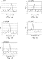
- FIGs. 1A to 1E are graphs showing Tm analysis results and indicate the change in fluorescence intensity accompanying the temperature rise.
- FIG. 1A shows the result obtained when the oligonucleotide was WT
- FIG. 1B shows the result obtained when the oligonucleotide was c12-TGT
- FIG. 1C shows the result obtained when the oligonucleotide was c12-GAT
- FIG. 1D shows the result obtained when the oligonucleotide was c13-GAC
- FIG. 1E shows the result obtained when the oligonucleotide was c12-AAT.
- the horizontal axis indicates the temperature (°C) at the time of measurement.
- the vertical axis indicates the change in fluorescence intensity, and the unit thereof is "d amount of change in fluorescence intensity/dt" (dF/dt), being a differential value of the amount of change in fluorescence intensity.
- the Tm value of the hybrid formed between the probe and the oligonucleotide was as follows: 73°C when the oligonucleotide was WT; 65°C when the oligonucleotide was c12-TGT, c12-GAT, or c13-GAC; and 60°C when the oligonucleotide was c12-AAT.
- a peak for WT was observed at 73°C.
- peaks for c12-TGT, c12-GAT, and c13-GAC in each of which a single base in codon 12 or 13 was different from the corresponding base in WT were observed at 65°C.
- a peak for c12-AAT in which two bases in codon 12 were different from the corresponding two bases in WT was observed at 60°C.
- the wild-type probe according to the present example could distinguish between the wild-type polymorphism and a mutant-type polymorphism and detect them. Moreover, it was found that, the probe according to the present example could distinguish between a polymorphism of a single base mismatch and a polymorphism of a double base mismatch and detect them.
- polymorphisms in K-ras genes were detected by carrying out Tm analysis in the presence of a wild-type plasmid and a mutant-type plasmid.
- a wild-type plasmid (WT) obtained through insertion of an oligonucleotide having wild-type codons 12 to 13 and two types of mutant-type plasmids (c12-TGT and c12-GAT) each obtained through insertion of an oligonucleotide having a mutant-type codon 12 and a wild-type codon 13 were prepared.
- the sequence of codons 12 to 13 was "ggtggc".
- mutant-type plasmid In the mutant-type plasmid (c12-TGT), the sequence of codons 12 to 13 was "Tgtggc", and the base indicated with a capital letter indicates the mutant-type polymorphism. In the mutant-type plasmid (c12-GAT), the sequence of codons 12 to 13 is "gAtggc", and the base indicated with a capital letter indicates the mutant-type polymorphism.
- These plasmids were mixed at predetermined proportions shown below, thus preparing five types of plasmid samples. Each of these plasmid samples contained 2 ⁇ 10 4 copies/ ⁇ l plasmids.
- PCR 25 ⁇ l of each of the PCR reaction solutions shown in Table 4 below was subjected to PCR and Tm analysis using a fully-automated SNPs analyzer (trade name: i-densy (registered trademark), produced by ARRAY, Inc.). PCR was carried out as follows. A treatment at 95°C for 60 seconds was conducted, and then a cycle of a treatment at 95°C for 5 seconds and at 64°C for 15 seconds was repeated a total of 50 cycles. Subsequently, Tm analysis was conducted under the same conditions as in Example 1.
- R-c12-XGT the underlined part corresponds to the sequence of codons 12 to 13.
- R-c12-GXT the underlined part corresponds to a sequence of two bases from the 5' end of codon 12 and codon 13.
- Each of R-c12-XGT and R-c12-GXT is a mixture (degenerate primer) of three types of oligonucleotides in which D is t, g, or a.
- the following wild-type probe 2 was used as a probe for detecting a polymorphism.
- the probe showed a perfect match with a detection target sequence in the sense strand of the wild-type K-ras gene.
- the sequence of the underlined bases is complementary to the sequence of wild-type codons 12 to 13.
- the 5' end of the wild-type probe 2 was labeled with a fluorescent dye, TAMRA, and the 3' end of the same was phosphorylated.
- 5'-(TAMRA)-cctgcctac gccacc agctccaactac-P-3' SEQ ID NO: 8
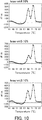
- FIGs. 2A to 2E are graphs showing the Tm analysis results and indicate the change in fluorescence intensity accompanying the temperature rise.
- FIG. 2A shows the result obtained when the plasmid sample was WT 100%
- FIG. 2B shows the result obtained when the plasmid sample was c12-TGT 3%
- FIG. 2C shows the result obtained when the plasmid sample was c12-TGT 100%
- FIG. 2D shows the result obtained when the plasmid sample was c12-GAT 1%
- FIG. 2E shows the result obtained when the plasmid sample was c12-GAT 100%.
- FIGs. 2A shows the result obtained when the plasmid sample was WT 100%
- FIG. 2B shows the result obtained when the plasmid sample was c12-TGT 3%
- FIG. 2C shows the result obtained when the plasmid sample was c12-TGT 100%
- FIG. 2D shows the result obtained when the plasmid sample was c12-GAT
- the horizontal axis indicates the temperature (°C) at the time of measurement
- the vertical axis indicates the change in fluorescence intensity
- the unit thereof is "d amount of change in fluorescence intensity/dt" (dF/dt), being a differential value of the amount of change in fluorescence intensity
- dF/dt dF/dt
- the Tm value of the hybrid formed between the probe and the plasmid was as follows: 75°C when the plasmid was WT; 68°C when the plasmid was c12-TGT; and 69°C when the plasmid was c12-GAT.
- polymorphisms in K-ras genes were detected by carrying out Tm analysis in the presence of a wild-type plasmid and a mutant-type plasmid.
- a wild-type plasmid (WT) obtained through insertion of an oligonucleotide having wild-type codons 12 to 13 and a mutant-type plasmid (c13-GAC) obtained through insertion of an oligonucleotide having a wild-type codon 12 and a mutant-type codon 13 were prepared.
- the wild-type plasmid (WT) is the same as that used in Example 2.
- the sequence of codons 12 to 13 was "ggtgAc", and the base indicated with a capital letter indicates the mutant-type polymorphism.
- PCR and Tm analysis were carried out in the same manner as in Example 2 except that 25 ⁇ l of each of the PCR reaction solutions shown in Table 6 below was used.
- R-WT primer SEQ ID NO: 13
- R-c13-TGC primer SEQ ID NO: 16
- R-c13-GAC primer SEQ ID NO: 17
- the following wild-type probe 3 was used as a probe for detecting a polymorphism.
- the probe showed a perfect match with a detection target sequence in the sense strand of the wild-type K-ras gene.
- the sequence of the underlined bases is complementary to the sequence of wild-type codons 12 to 13.
- the 5' end of the wild-type probe 3 was labeled with a fluorescent dye, TAMRA, and the 3' end of the same was phosphorylated.
- 5'-(TAMRA)-cctgcctacgccaccagctccaactacca-P-3' SEQ ID NO: 9
- FIGs. 3A to 3C are graphs showing the Tm analysis results and indicate the change in fluorescence intensity accompanying the temperature rise.
- FIG. 3A shows the result obtained when the plasmid sample was WT 100%
- FIG. 3B shows the result obtained when the plasmid sample was c13-GAC 3%
- FIG. 3C shows the result obtained when the plasmid sample was c13-GAC 100%.
- FIGs. 3A to 3C are graphs showing the Tm analysis results and indicate the change in fluorescence intensity accompanying the temperature rise.
- FIG. 3A shows the result obtained when the plasmid sample was WT 100%
- FIG. 3B shows the result obtained when the plasmid sample was c13-GAC 3%
- FIG. 3C shows the result obtained when the plasmid sample was c13-GAC 100%.
- FIGs. 3A shows the result obtained when the plasmid sample was WT 100%
- FIG. 3B shows the result obtained when the plasmid sample was c
- the horizontal axis indicates the temperature (°C) at the time of measurement
- the vertical axis indicates the change in fluorescence intensity
- the unit thereof is "d amount of change in fluorescence intensity/dt" (dF/dt), being a differential value of the amount of change in fluorescence intensity.
- the Tm value of the hybrid formed between the probe and the plasmid was as follows: 77°C when the plasmid was WT; and 72°C when the plasmid was c13-GAC.
- polymorphisms in K-ras genes and BRAF genes were detected by carrying out Tm analysis in the presence of a wild-type plasmid and a mutant-type plasmid.
- kras plasmids obtained through insertion of the respective oligonucleotides having codons 12 to 13 shown in Table 7 below were prepared.
- the kras plasmid in which codons 12 to 13 are of a wild-type was referred to as a wild-type plasmid (kras-wt).
- the kras plasmid in which at least one of codons 12 to 13 has a mutant-type polymorphism was referred to as a mutant-type plasmid (kras-mt), and 12 types of "kras-mt 2" to "kras-mt 13" were prepared depending on the respective mutant-type polymorphisms thereof.
- a mutant-type plasmid (kras-mt)
- 12 types of "kras-mt 2" to "kras-mt 13” were prepared depending on the respective mutant-type polymorphisms thereof.
- codons 12 to 13 in Table 7 below the bases indicated with capital letters show mutant-type polymorphisms.
- braf plasmids obtained through insertion of the respective oligonucleotides each having a base shown in Table 7 below as the detection site were prepared.
- the braf plasmid obtained through insertion of the oligonucleotide having a wild-type detection site was referred to as a wild-type plasmid (braf-wt).
- the braf plasmid obtained through insertion of the oligonucleotide having a mutant-type detection site was referred to as a mutant-type plasmid (braf-mt).
- Each of the bases in Table 7 below is the 229th base (w) in the base sequence of SEQ ID NO: 2. In Table 7, the base indicated with a capital letter is a mutant-type polymorphism.
- the kras plasmids and the braf plasmids were mixed at a predetermined ratios shown below, thus preparing 14 types of plasmid samples.
- Each of these plasmid samples contained 1 ⁇ 10 4 copies/pl of plasmids.
- PCR 50 ⁇ l of each of the PCR reaction solutions shown in Table 9 below was subjected to PCR and Tm analysis using a fully-automated SNPs analyzer (trade name: i-densy (registered trademark), produced by ARRAY, Inc.). PCR was carried out as follows. A treatment at 95°C for 30 seconds was conducted, and then a cycle of a treatment at 95°C for 1 second and at 58°C for 15 seconds was repeated a total of 50 cycles.
- i-densy registered trademark
- Tm analysis was carried out by treating the reaction solutions at 95°C for 1 second and at 40°C for 60 seconds, then heating it from 40°C to 85°C at a temperature rising rate of 1°C/3 seconds, and measuring the change in fluorescence intensity over time at a wavelength from 520 to 555 nm (BODIPY FL) and from 585 to 700 nm (TAMRA).
- the probe for K-ras has a sequence that can hybridize to the sense strand of the K-ras gene, and in the sequence, the bases indicated with capital letters are complementary to the 220th to 225th bases (nntkrc) in SEQ ID NO: 1.
- the 5' end of the probe for K-ras was labeled with a fluorescent substance, BODIPY FL, and the 3' end of the same was phosphorylated.
- the probe for BRAF has a sequence that can hybridize to the antisense strand of the BRAF gene, and in the sequence, the base indicated with the capital letter corresponds to the 229th base (w) in SEQ ID NO: 2.
- the 3' end of the probe for BRAF was labeled with a fluorescent substance, TAMRA.
- TAMRA fluorescent substance
- FIGs. 4 to 7 show graphs showing the Tm analysis results and indicate the change in fluorescence intensity accompanying the temperature rise.
- FIG. 4 shows the results obtained when the respective plasmid samples were WT 100%, kras-mt 2 10%, and kras-mt 3 10%.
- FIG. 5 shows the results obtained when the respective plasmid samples were kras-mt 4 10%, kras-mt 5 10%, and kras-mt 6 10%.
- FIG. 6 shows the results obtained when the respective plasmid samples were kras-mt 7 10%, kras-mt 8 10%, kras-mt 9 10%, and kras-mt 10 10%.
- FIG. 4 shows the results obtained when the respective plasmid samples were WT 100%, kras-mt 2 10%, and kras-mt 3 10%.
- FIG. 5 shows the results obtained when the respective plasmid samples were kras-mt 4 10%, kras-mt 5 10%, and kras-
- the horizontal axis indicates the temperature (°C) at the time of measurement
- the vertical axis indicates the change in fluorescence intensity
- the unit thereof is "d amount of change in fluorescence intensity/dt" (dF/dt)
- dF/dt dF/dt
- the Tm value of the hybrid formed between the probe for K-ras and the plasmid was as follows: 73°C when the plasmid was kras-wt; 67°C when the plasmid was kras-mt 2; 66°C when the plasmid was kras-mt 3; 67°C when the plasmid was kras-mt 4; 67°C when the plasmid was kras-mt 5;
- polymorphisms in the K-ras gene and the BRAF gene were detected by carrying out Tm analysis with respect to a DNA extract obtained from a clinical specimen of a paraffin-embedded section.
- the DNA extract was obtained from the clinical specimen of a paraffin-embedded section using the DNA extraction kit (trade name: TaKaRaDEXPAT (registered trademark), product code: 9091, produced by Takara Bio Inc).
- PCR 50 ⁇ l of each of the PCR reaction solutions shown in Table 10 below was subjected to PCR and Tm analysis using a fully-automated SNPs analyzer (trade name: i-densy (registered trademark), produced by ARKRAY, Inc.). PCR was carried out as follows. A treatment at 95°C for 60 seconds was conducted, and then a cycle of a treatment at 95°C for 1 second and at 62°C for 15 seconds was repeated a total of 50 cycles.
- Tm analysis was carried out by treating the reaction solutions at 95°C for 1 second and at 40°C for 60 seconds, then heating it from 40°C to 85°C at a temperature rising rate of 1°C/3 seconds, and measuring the change in fluorescence intensity over time at a wavelength from 520 to 555 nm (BODIPY FL) and from 585 to 700 nm (TAMRA).
- primers for K-ras As the primers for K-ras, the same primers as used in Example 4 were used. The sequences of the primers for BRAF are shown below. (Primer for BRAF) F3 primer 5'-tgcttgctctgataggaaatgagatctac-3' (SEQ ID NO: 38) R5 primer 5'-aaactgatgggacccactccat-3' (SEQ ID NO: 39)
- the same probe used in Example 4 was used.
- the sequence of the probe for BRAF is shown below. In the sequence, the base indicated with a capital letter corresponds to the 229th base (w) in SEQ ID NO: 2.
- the 3' end of the probe for BRAF was labeled with a fluorescent substance, TAMRA. (Probe for BRAF) 5'-gctacagAgaaatctc-(TAMRA)-3' (SEQ ID NO: 36)
- FIGs. 8A and 8B are graphs showing the Tm analysis results and indicate the change in fluorescence intensity accompanying the temperature rise.
- FIG. 8A shows the result with respect to the K-ras gene
- FIG. 8B shows the result with respect to the BRAF gene.
- the horizontal axis indicates the temperature (°C) at the time of measurement.
- the vertical axis indicates the change in fluorescence intensity, and the unit thereof is "d amount of change in fluorescence intensity/dt" (dF/dt), being a differential value of the amount of change in fluorescence intensity.
- the Tm value of the hybrid formed between the probe for K-ras and the K-ras gene of a wild-type was about 73°C.
- the Tm value of the hybrid formed between the probe for BRAF and the BRAF gene was as follows: 52°C when the BRAF gene is of a wild-type; and 45°C when the BRAF gene is of a mutant type.
- FIG. 8A two peaks were observed with respect to the K-ras gene. Therefore, it was found that the K-ras gene contains a wild-type sequence and a mutant-type sequence.
- FIG. 8B a peak was observed only at the Tm value of a wild-type DNA with respect to the BRAF gene. Therefore, it was found that the DNA is of a wild-type. It was expected that the DNA extract collected from the clinical specimen of a paraffin-embedded section contained contaminants, or a template DNA was fragmented. However, according to the present example, the peaks with respect to the K-ras gene and the BRAF gene could be detected clearly.
- polymorphisms in K-ras genes were detected by carrying out Tm analysis in the presence of a wild-type plasmid and a mutant-type plasmid.
- the kras plasmids were mixed in the same manner as in Example 4 except that the braf plasmids in Table 8 were not included, thus preparing plasmid samples.
- Each of the plasmid samples contains 75 copies/ ⁇ l of plasmids.
- PCR 50 ⁇ l of each of the PCR reaction solutions shown in Table 11 below was subjected to PCR and Tm analysis using a fully-automated SNPs analyzer (trade name: i-densy (registered trademark), produced by ARRAY, Inc.). PCR was carried out as follows. A treatment at 95°C for 60 seconds was conducted, and then a cycle of a treatment at 95°C for 1 second and at 58°C for 15 seconds was repeated a total of 50 cycles.
- i-densy registered trademark
- Tm analysis was carried out by treating the reaction solutions at 95°C for 1 second and at 40°C for 60 seconds, then heating it from 40°C to 85°C at a temperature rising rate of 1°C/3 seconds, and measuring the change in fluorescence intensity over time at a wavelength from 520 to 555 nm (BODIPY FL).
- the R1-LP primer was a mixture (degenerate primer) of two types of oligonucleotides in which m is c or a.
- F1-LP primer SEQ ID NO: 32
- R1-LP primer SEQ ID NO: 33
- 5'-gaattagctgtatcgtmaaggcactc-3' m c or a
- a wild-type probe 6 having the following sequence was used.
- the sequence of the underlined bases is complementary to the sequence of the wild-type codons 12 to 13.
- the 5' end of the wild-type probe 6 was labeled with a fluorescent substance, BODIPY FL, and the 3' end of the same was phosphorylated.
- 5'-(BODIPY FL)-ctcttgcctacg ccacc agctccaacttgctggctacgc- P-3' SEQ ID NO: 31
- FIGs. 9 to 12 show graphs showing the Tm analysis results and indicate the change in fluorescence intensity accompanying the temperature rise.
- FIG. 9 shows the results obtained when the respective plasmid samples were WT 100%, kras-mt 2 10%, and kras-mt 3 10%.
- FIG. 10 shows the results obtained when the respective plasmid samples were kras-mt 4 10%, kras-mt 5 10%, and kras-mt 6 10%.
- FIG. 11 shows the results obtained when the respective plasmid samples were kras-mt 7 10%, kras-mt 8 10%, and kras-mt 9 10%.
- FIG. 9 shows the results obtained when the respective plasmid samples were WT 100%, kras-mt 2 10%, and kras-mt 3 10%.
- FIG. 10 shows the results obtained when the respective plasmid samples were kras-mt 4 10%, kras-mt 5 10%, and kras-mt 6 10%.
- FIG. 12 shows the results obtained when the respective plasmid samples were kras-mt 10 10%, kras-mt 11 10%, kras-mt 12 10%, and kras-mt 13 10%.
- the horizontal axis indicates the temperature (°C) at the time of measurement
- the vertical axis indicates the change in fluorescence intensity
- the unit thereof is "d amount of change in fluorescence intensity/dt" (dF/dt), being a differential value of the amount of change in fluorescence intensity.
- the Tm value of the hybrid formed between the probe for K-ras and the plasmid was as follows: 72°C when the plasmid was kras-wt; 66°C when the plasmid was kras-mt 2; 66°C when the plasmid was kras-mt 3; 66°C when the plasmid was kras-mt 4; 66°C when the plasmid was kras-mt 5; 65°C when the plasmid was kras-mt 6; 66°C when the plasmid was kras-mt 7; 66°C when the plasmid was kras-mt 8; 66°C when the plasmid was kras-mt 9; 62°C when the plasmid was kras-mt 10; 61°C when the plasmid was kras-mt 11; 60°C when the plasmid was kras-mt 12; and 59
- the wild-type probe of the present example could distinguish between the wild-type polymorphism and the mutant-type polymorphism and detect them.
- polymorphisms in K-ras genes were detected by carrying out Tm analysis in the presence of a wild-type plasmid and a mutant-type plasmid.
- plasmid samples the plasmid samples, WT and mt 2 to 9 among the plasmids samples of Example 6 were used.
- PCR 50 ⁇ l of each of the PCR reaction solutions shown in Table 12 below was subjected to PCR and Tm analysis using a fully-automated SNPs analyzer (trade name: i-densy (registered trademark), produced by ARKRAY, Inc.). PCR was carried out as follows. A treatment at 95°C for 60 seconds was conducted, and then a cycle of a treatment at 95°C for 1 second and at 57.5°C for 15 seconds was repeated a total of 50 cycles.
- Tm analysis was carried out by treating the reaction solutions at 95°C for 1 second and at 40°C for 60 seconds, then heating it from 40°C to 65°C at a temperature rising rate of 1°C/3 seconds, and measuring the change in fluorescence intensity over time at a wavelength from 585 to 700 nm (TAMRA).
- R3-LP primer As the F1-LP primer, the same primer as used in Example 6 was used. The sequence of the R3-LP primer is shown below.
- R3-LP primer SEQ ID NO: 34
- a mutant-type probe 7 having the following sequence was used.
- the sequence of the underlined bases is complementary to the sequence of the wild-type codon 12 and the mutant-type codon 13 (gac).
- the 5' end of the wild-type probe 7 was labeled with a fluorescent substance, TAMRA, and the 3' end of the same was phosphorylated.
- 5'-(TAMRA)-cttgcctacgtcacc-P-3' SEQ ID NO: 30
- FIGs. 13 to 15 show graphs showing the Tm analysis results and indicate the change in fluorescence intensity accompanying the temperature rise.
- FIG. 13 shows the results obtained when the respective plasmid samples were WT 100%, kras-mt 2 10%, and kras-mt 3 10%.
- FIG. 14 shows the results obtained when the respective plasmid samples were kras-mt 4 10%, kras-mt 5 10%, and kras-mt 6 10%.
- FIG. 15 shows the results obtained when the respective plasmid samples were kras-mt 7 10%, kras-mt 8 10% and kras-mt 9 10%. In each of FIGs.
- the horizontal axis indicates the temperature (°C) at the time of measurement
- the vertical axis indicates the change in fluorescence intensity
- the unit thereof is "d amount of change in fluorescence intensity/dt" (dF/dt), being a differential value of the amount of change in fluorescence intensity.
- the Tm value of the hybrid formed between the probe and each of the plasmid was as follows: 54°C when the plasmid was kras-wt; 53°C when the plasmid was kras-mt 2; 53.5°C when the plasmid was kras-mt 3; 53°C when the plasmid was kras-mt 4; 53°C when the plasmid was kras-mt 5; 53°C when the plasmid was kras-mt 6; 53°C when the plasmid was kras-mt 7; 53°C when the plasmid was kras-mt 8; and 57°C when the plasmid was kras-mt 9.
- a polymorphism in a K-ras gene can be identified easily with high reliability by Tm analysis, for example.
- Tm analysis for example, even in the case where a K-ras gene having a wild-type target polymorphism and a K-ras gene having a mutant-type target polymorphism are present together in a sample, the presence or absence of the mutation can be detected easily with high reliability by Tm analysis using the probe of the present invention. Therefore, the present invention is particularly useful when applied to a sample containing both the wild-type K-ras gene and the mutant-type K-ras gene.
- a polymorphism in a K-ras gene can be identified easily with high reliability, so that, for example, the detection result can be reflected in diagnosis of the above-mentioned diseases and selection of treatment methods for the diseases. Therefore, it can be said that the present invention is very useful in the medical field and the like.
Landscapes
- Chemical & Material Sciences (AREA)
- Life Sciences & Earth Sciences (AREA)
- Health & Medical Sciences (AREA)
- Organic Chemistry (AREA)
- Proteomics, Peptides & Aminoacids (AREA)
- Engineering & Computer Science (AREA)
- Immunology (AREA)
- Pathology (AREA)
- Analytical Chemistry (AREA)
- Zoology (AREA)
- Genetics & Genomics (AREA)
- Wood Science & Technology (AREA)
- Physics & Mathematics (AREA)
- Biotechnology (AREA)
- Microbiology (AREA)
- Molecular Biology (AREA)
- Hospice & Palliative Care (AREA)
- Biophysics (AREA)
- Oncology (AREA)
- Biochemistry (AREA)
- Bioinformatics & Cheminformatics (AREA)
- General Engineering & Computer Science (AREA)
- General Health & Medical Sciences (AREA)
- Measuring Or Testing Involving Enzymes Or Micro-Organisms (AREA)
Abstract
(P1) a 11- to 50-mer oligonucleotide that is composed of a base sequence complementary to a base sequence including the 220th to 230th bases in SEQ ID NO: 1 and has a base complementary to the 230th base in its 5' end region;
(P1') an oligonucleotide composed of a base sequence complementary to the oligonucleotide (P1);
(P2) a 15- to 50-mer oligonucleotide that is composed of a base sequence complementary to a base sequence including the 220th to 234th bases in SEQ ID NO: 1 and has a base complementary to the 234th base in its 5' end region;
(P2') an oligonucleotide composed of a base sequence complementary to the oligonucleotide (P2);
(P3) a 17- to 50-mer oligonucleotide that is composed of a base sequence complementary to a base sequence including the 220th to 236th bases in SEQ ID NO: 1 and has a base complementary to the 236th base in its 5' end region; and
(P3') an oligonucleotide composed of a base sequence complementary to the oligonucleotide (P3).
Description
- The present invention relates to a probe for detecting a polymorphism in a disease-related gene and use of the probe.
- Each of a RAS protein and a RAF protein is a protein that forms a cascade of intracellular signaling in a RAS/RAF/MAPK pathway. In the RAS/RAF/MAPK pathway, the RAF protein is activated by an activated RAS protein, a MEK protein is activated by the activated RAF protein, and further, a MAPK protein is activated by the activated MEK protein. By this activation, cell growth and cell differentiation are controlled.
- A K-ras protein that is a kind of RAS protein is a GDP/GTP binding protein having GTPase activity. In humans, the K-ras protein is encoded by a K-ras gene located on chromosome 12. It is known that the K-ras gene has a mutation in codons 12 to 13 and the like thereof (Non-Patent Documents 1-3). The mutation in codon 12 is, in a partial sequence of SEQ ID NO: 1 in the K-ras gene, a substitution of adenine (a), cytosine (c), or thymine (t) for guanine (g) at the 220th base (n) or a substitution of adenine (a), cytosine (c), or thymine (t) for guanine (g) at the 221st base (n). The mutation in
codon 13 is, in the base sequence of SEQ ID NO: 1 in the K-ras gene, a substitution of thymine (t) for guanine (g) at the 223rd base (k) or a substitution of adenine (a) for guanine (g) at the 224th base (r). By the mutation in codon 12, glycine (G) at the 12th position of the K-ras protein is mutated to serine (S), arginine (R), cysteine (C), aspartic acid (D), alanine (A), valine (V), asparagine (N), phenylalanine (F), or leucine (L). By the mutation incodon 13, glycine (G) at the 13th position of the K-ras protein is mutated to aspartic acid (D) or cysteine (C). It has been reported that the mutation incodon 12 or 13 of the K-ras gene is connected to, for example, cancers such as colon cancer and pancreatic cancer, congenital diseases such as CFC (cardio-facio-cutaneous) and the like, and drug resistance to an anti-EGFR antibody drug (Non-Patent Documents 1-3). Therefore, the detection of the presence or absence of these mutations in the K-ras gene, i.e., the detection of polymorphisms in the K-ras gene is very important in, for example, diagnosis of the above-mentioned diseases, selection of more effective treatment methods for the diseases, and the like. - Moreover, a BRAF protein that is a kind of RAF protein is a protein having serine-threonine kinase activity. In humans, the BRAF protein is encoded by the BRAF gene located on chromosome 7. It has also been reported that, as well as the mutation in the K-ras gene, a mutation in the BRAF gene is connected to the above-mentioned cancers, congenital diseases, and drug resistance (Non-Patent Documents 1-3). As the mutation in the BRAF gene, a substitution of adenine (a) for thymine (t) at the 229th base (w) in a partial sequence of SEQ ID NO: 2 in the BRAF gene is known. When the base is of a wild-type (t), the 600th amino acid in the BRAF protein becomes valine (V). When the base is of a mutant-type (a), the 600th amino acid in the BRAF protein becomes glutamic acid (E). It is considered that tumorigenicity is caused by the mutation of this amino-acid residue. Therefore, the detection of the presence or absence of the mutation in the BRAF gene, i.e., the detection of a polymorphism in the BRAF gene besides the mutation in the K-ras gene makes it possible to further improve the accuracy of, for example, diagnosis of the above-mentioned diseases, selection of more effective treatment methods for the diseases, and the like.
- On the other hand, as a method for detecting a polymorphism in a gene, various methods have been reported. Examples thereof include a PCR (Polymerase Chain Reaction)-RFLP (Restriction Fragment Length Polymorphism) method and the like.
- The PCR-RFLP method is carried out by amplifying a detection target region in a target DNA in a sample by PCR, treating the obtained amplification product with a restriction enzyme, and typing the change in restriction fragment length caused by a polymorphism according to Southern hybridization. When a target mutation is present in the gene, the recognition site of the restriction enzyme disappears. Thus, it is possible to detect the presence or absence of the mutation based on the presence or absence of cleavage, i.e., the change in restriction fragment length.
- However, in the PCR-RFLP method, for example, after the PCR, it is necessary to conduct a cumbersome procedure of treating the obtained amplification product with a restriction enzyme and conducting an analysis. Furthermore, in order to treat the obtained amplification product with a restriction enzyme, the amplification product has to be temporarily taken out. Thus, there is a risk that the amplification product obtained in a first reaction may be dispersed and mix with a second reaction that is different from the first reaction. Such problems make the automation of polymorphism detection difficult.
- In light of these problems, Tm (Melting Temperature) analysis is attracting attention as a method for detecting a polymorphism in recent years. In Tm analysis, first, using a probe complementary to a region including a detection target polymorphism, a hybrid (double-stranded nucleic acid) of a nucleic acid to be examined (hereinafter simply referred to as a "test nucleic acid") with the probe is formed. Then, the thus-obtained hybrid is heat-treated, and dissociation (melting) of the hybrid into single-stranded nucleic acids accompanying the temperature rise is detected by measuring a signal such as absorbance. By determining the Tm value based on the result of the detection, the polymorphism is determined. The Tm value becomes higher as the complementarity between the single-stranded nucleic acids of the hybrid becomes higher, and becomes lower as the complementarity between the same becomes lower. Thus, in the case where the polymorphism in a detection target site is X or Y, the Tm value of a hybrid composed of a nucleic acid containing the target polymorphism (e.g., Y) and a probe that is 100% complementary thereto is determined beforehand (this Tm value represents the evaluation standard value). Subsequently, the Tm value of a hybrid composed of the test nucleic acid and the probe is measured (this Tm value represents the measured value). Then, when this measured value is the same as the evaluation standard value, it can be determined that the test nucleic acid shows a perfect match with the probe, i.e., the detection target site in the test nucleic acid is the target polymorphism (Y). On the other hand, when the measured value is lower than the evaluation standard value, it can be determined that the test nucleic acid shows a mismatch with the probe, i.e., the detection target site in the test nucleic acid is the other polymorphism (X). According to such a method, a polymorphism can be detected merely by heat-treating a PCR reaction solution containing the probe and then measuring a signal, for example. Thus, it is possible to automate a detection device.
- However, in detection methods utilizing such Tm analysis, it is necessary to determine the difference in a single base from the Tm value, for example. Further, in the case where a gene has a plurality of polymorphisms, since analysis of even one sample is accompanied by a considerable amount of work, there is a problem in that the analysis of many samples is impractical. Therefore, in particular, even in the case where a wild-type polymorphism and a plurality of mutant-type polymorphisms are present together, it is required to detect the presence or absence of the mutations accurately.
-
- [Non-Patent Document 1] Cancer Epidemiology Biomarkers, November, 2000, pp. 1193-1197
- [Non-Patent Document 2] J Mol Diagn., November, 2006, Vol. 8, No. 5, pp. 540-543
- [Not-Patent Document 3] J Natl Cancer Inst., October 7, 2009, Vol. 101, No. 19, pp. 1308-1324 (Epub: September 8, 2009)
- For the above-described reasons, the detection of a polymorphism in the K-ras gene is really important in, for example, diagnosis of the above-described diseases and selection of treatment methods for the diseases. Hence, the present invention is intended to provide a probe that can identify a polymorphism in a K-ras gene that is a disease-related gene easily with high reliability, and use of the probe.
- In order to achieve the aforementioned object, the present invention provides a probe for detecting a polymorphism in a K-ras gene that is a disease-related gene, including at least one of oligonucleotides (P1), (P2), (P3), (P1'), (P2'), and (P3'):
- (P1) a 11- to 50-mer oligonucleotide that is composed of a base sequence complementary to a base sequence including the 220th to 230th bases in SEQ ID NO: 1 and has a base complementary to the 230th base in its 5' end region;
- (P1') an oligonucleotide composed of a base sequence complementary to the oligonucleotide (P1);
- (P2) a 15- to 50-mer oligonucleotide that is composed of a base sequence complementary to a base sequence including the 220th to 234th bases in SEQ ID NO: 1 and has a base complementary to the 234th base in its 5' end region;
- (P2') an oligonucleotide composed of a base sequence complementary to the oligonucleotide (P2);
- (P3) a 17- to 50-mer oligonucleotide that is composed of a base sequence complementary to a base sequence including the 220th to 236th bases in SEQ ID NO: 1 and has a base complementary to the 236th base in its 5' end region; and
- (P3') an oligonucleotide composed of a base sequence complementary to the oligonucleotide (P3).
- The present invention also provides a reagent for detecting a polymorphism in a disease-related gene, containing the probe of the present invention.
- The present invention also provides a method for detecting a polymorphism in a K-ras gene that is a disease-related gene, the method comprising the step of: detecting a polymorphism in a K-ras gene that is a disease-related gene using the probe of the present invention.
- According to the probe of the present invention, a polymorphism in a K-ras gene can be identified easily with high reliability by Tm analysis, for example. Specifically, for example, even in the case where a K-ras gene having a wild-type target polymorphism and a K-ras gene having a mutant-type target polymorphism are present together in a sample, the type of the polymorphism or the presence or absence of the mutation can be detected easily with high reliability by Tm analysis using the probe of the present invention. Therefore, the present invention is particularly useful when applied to a sample containing both the wild-type K-ras gene and the mutant-type K-ras gene. As described above, according to the present invention, a polymorphism in a K-ras gene can be identified easily with high reliability, so that, for example, the detection result can be reflected in diagnosis of the above-mentioned diseases and selection of treatment methods for the diseases. Therefore, it can be said that the present invention is very useful in the medical field and the like.
-
- [
FIG. 1] FIGs 1A to 1E are graphs illustrating the results of Tm analysis with respect to the respective reaction solutions, each containing a wild-type oligonucleotide and a mutant-type oligonucleotide in Example 1 of the present invention. - [
FIG. 2] FIGs 2A to 2E are graphs illustrating the results of Tm analysis with respect to the respective reaction solutions, each containing a wild-type plasmid and a mutant-type plasmid in Example 2 of the present invention. - [
FIG. 3] FIGs 3A to 3C are graphs illustrating the results of Tm analysis with respect to the respective reaction solutions, each containing a wild-type plasmid and a mutant-type plasmid in Example 3 of the present invention. - [
FIG. 4] FIG. 4 shows graphs illustrating the results of Tm analysis with respect to the respective reaction solutions, each containing a wild-type plasmid and a mutant-type plasmid in Example 4 of the present invention. - [
FIG. 5] FIGs. 5 shows other graphs illustrating the results of Tm analysis with respect to the respective reaction solutions, each containing a wild-type plasmid and a mutant-type plasmid in Example 4 of the present invention. - [
FIG. 6] FIG. 6 shows other graphs illustrating the results of Tm analysis with respect to the respective reaction solutions, each containing a wild-type plasmid and a mutant-type plasmid in Example 4 of the present invention. - [
FIG. 7] FIG. 7 shows graphs illustrating the results of Tm analysis with respect to the respective reaction solutions, each containing a wild-type plasmid and a mutant-type plasmid in Example 4 of the present invention. - [
FIG. 8] FIGs. 8A and 8B are graphs illustrating the results of Tm analysis with respect to a reaction solution containing a DNA extract from a clinical specimen in Example 5 of the present invention. - [
FIG. 9] FIG. 9 shows graphs illustrating the results of Tm analysis with respect to the respective reaction solutions, each containing a wild-type plasmid and a mutant-type plasmid in Example 6 of the present invention. - [
FIG. 10] FIG. 10 shows other graphs illustrating the results of Tm analysis with respect to the respective reaction solutions, each containing a wild-type plasmid and a mutant-type plasmid in Example 6 of the present invention. - [
FIG. 11] FIG. 11 shows other graphs illustrating the results of Tm analysis with respect to the respective reaction solutions, each containing a wild-type plasmid and a mutant-type plasmid in Example 6 of the present invention. - [
FIG. 12] FIG. 12 shows other graphs illustrating the results of Tm analysis with respect to the respective reaction solutions, each containing a wild-type plasmid and a mutant-type plasmid in Example 6 of the present invention. - [
FIG. 13] FIG. 13 shows graphs illustrating the results of Tm analysis with respect to the respective reaction solutions, each containing a wild-type plasmid and a mutant-type plasmid in Example 7 of the present invention. - [
FIG. 14] FIG. 14 shows other graphs illustrating the results of Tm analysis with respect to the respective reaction solutions, each containing a wild-type plasmid and a mutant-type plasmid in Example 7 of the present invention. - [
FIG. 15] FIG. 15 shows other graphs illustrating the results of Tm analysis with respect to the respective reaction solutions, each containing a wild-type plasmid and a mutant-type plasmid in Example 7 of the present invention. - In the present invention, a detection target polymorphism in the K-ras gene is a polymorphism in codons 12 to 13 of the K-ras gene. Specifically, for example, the detection target polymorphism is a polymorphism in codon 12 from the 220th to 222nd bases (nnt) and a polymorphism in
codon 13 from the 223rd to 225th bases (krc) in a partial sequence of SEQ ID NO: 1 in the K-ras gene. The n is g, c, t, or a. The k is g or t. The r is g or a. Codon 12 having a wild-type polymorphism is represented by ggt. Codon 12 having a mutant-type polymorphism can be represented by any of nine types of agt, cgt, tgt, gat, gct, gtt, aat, ttt, and ctt, for example.Codon 13 having a wild-type polymorphism is represented by ggc.Codon 13 having a mutant-type polymorphism can be represented by any of gac and tgc, for example. When at least one of the polymorphism in codon 12 and the polymorphism incodon 13 of the K-ras gene is of a mutant-type, for example, it can be determined that resistance against an anti-EGFR antibody drug such as cetuximab is exerted. When it is of a wild-type, for example, it can be determined that resistance is not exerted. As the polymorphisms in codons 12 to 13 from the 220th to 225th bases in the sense strand of the K-ras gene, a wild-type 1 and mutant-types 2-13 are shown below as examples. In the following sequences, underlined bases are mutant-type bases.Wild-type 1 (WT) ggtggc Mutant-type 2 (c12-AGT) agtggc 3 (c12-CGT) cgtggc 4 (c12-TGT) tgtggc 5 (c12-GAT) gatggc 6 (c12-GCT) gctggc 7 (c12-GTT) gttggc 8 (c13-TGC) ggttgc 9 (c13-GAC) ggtgac 10 (c12-AAT) aatggc 11 (c12-TTT) tttggc 12 (c12-CTT) cttggc 13 (c12-AGT, c13-GAC) agtgac - The base sequence of the K-ras gene is registered as a region from the 5001st to 50675th bases under GenBank Accession No. NG_007524, for example. The base sequence of SEQ ID NO: 1 is a partial sequence of the K-ras gene and corresponds to a region from the 10351st to 10850th bases in the base sequence of Accession No. NG_007524. The sequence of codons 12 to 13 corresponds to a region from the 10570th to 10575th bases in the base sequence of Accession No. NG_007524.
- In the present invention, hereinafter, the K-ras gene in which any of bases of codons 12 to 13 are of the mutant-type is referred to as the "mutant-type K-ras gene", and the K-ras gene in which bases of codons 12 to 13 are of the wild-type is referred to as the "wild-type K-ras gene or normal K-ras gene".
- In the present invention, the site at which the above-described polymorphism occurs, i.e., the sequence of the 220th to 225th bases in the base sequence of SEQ ID NO: 1 (sense strand) or the sequence of bases corresponding to the 220th to 225th bases of the sense strand in the complementary strand thereto (antisense strand), is referred to as the "detection target site". In the sequence of SEQ ID NO: 1 (sense strand) or the complementary sequence thereto (antisense strand), a region including the detection target site and to which the probe can hybridize is referred to as a "hybridization region or detection target sequence". Regarding the detection target sequence, the one showing a perfect match with the probe is referred to as a "perfect-match sequence", and the one showing a mismatch with the probe is referred to as a "mismatch sequence". In the present invention, a perfect match means that bases at the detection target site are complementary to bases to be paired therewith in the probe, and preferably means that the detection target sequence is perfectly complementary to the probe. In the present invention, a mismatch means that the bases at the detection target site are not complementary to bases to be paired therewith in the probe, and preferably means that the detection target sequence is perfectly complementary to the probe except for the detection target site.
- In the present invention, in the case where the K-ras gene is amplified and further, the probe of the present invention is caused to hybridize with the resultant amplification product, a region to be amplified in the K-ras gene hereinafter is referred to as an "amplification target region". The amplification target region may be, for example, a region in the sense strand of the K-ras gene, a region corresponding thereto in the antisense strand, or both of them. In the present invention, the terms "sense strand" and "antisense strand" encompass, for example, amplification products of the sense strand and amplification products of the antisense strand, respectively.
- In the present invention, the ends of a base sequence mean the endmost bases on the 5' side and the 3' side in the base sequence. Furthermore, a 5' end region is a region including several bases from the 5' end in a base sequence, and a 3' end region is a region including several bases from the 3' end in a base sequence. The several bases mean, for example, 1 to 10, 1 to 4, 1 to 3, or 1 to 2 bases from the end. In the present invention, the Zth base (Z is a positive integer) from an end of a base sequence is a numerical order counted with the base at the end as the first base. For example, the first base from the end means the base at the end, and the second base from the end means the base next to the base at the end.
- The probe of the present invention is, as mentioned above, a probe for detecting a polymorphism in a K-ras gene that is a disease-related gene, including at least one of oligonucleotides (P1), (P2), (P3), (P1'), (P2'), and (P3'):
- (P1) a 11- to 50-mer oligonucleotide that is composed of a base sequence complementary to a base sequence including the 220th to 230th bases in SEQ ID NO: 1 and has a base complementary to the 230th base in its 5' end region;
- (P1') an oligonucleotide composed of a base sequence complementary to the oligonucleotide (P1);
- (P2) a 15- to 50-mer oligonucleotide that is composed of a base sequence complementary to a base sequence including the 220th to 234th bases in SEQ ID NO: 1 and has a base complementary to the 234th base in its 5' end region;
- (P2') an oligonucleotide composed of a base sequence complementary to the oligonucleotide (P2);
- (P3) a 17- to 50-mer oligonucleotide that is composed of a base sequence complementary to a base sequence including the 220th to 236th bases in SEQ ID NO: 1 and has a base complementary to the 236th base in its 5' end region; and
- (P3') an oligonucleotide composed of a base sequence complementary to the oligonucleotide (P3).
- Hereinafter, the probe of the present invention is also referred to as a probe for K-ras. The base length of each of the oligonucleotides (P1) and (P1') is 11- to 50-mer, preferably 13- to 30-mer, and more preferably 15- to 20-mer. The base length of each of the oligonucleotides (P2) and (P2') is 15-to 50-mer, preferably 15- to 30-mer, and more preferably 15- to 20-mer. The base length of each of the oligonucleotides (P3) and (P3') is 17- to 50-mer, preferably 17- to 30-mer, and more preferably 17- to 20-mer.
- The probe of the present invention is a probe for detecting, a polymorphism in codon 12 of the 220th to 222nd bases (nnt) and a polymorphism in
codon 13 of the 223rd to 225th bases (krc) in the partial sequence of SEQ ID NO: 1 in the K-ras gene. Specifically, the probe of the present invention is, for example, a probe for detecting, in the base sequence of SEQ ID NO: 1, a polymorphism (g/a, g/c, or g/t) of the 220th base (n), a polymorphism (g/a, g/c, or g/t) of the 221st base (n), a polymorphism (g/t) of the 223rd base (k), or a polymorphism (g/a) of the 224th base (r). In the sequence "nntkrc" of the 220th to 225th bases in the base sequence of SEQ ID NO: 1, n is guanine (g), adenine (a), cytosine (c), or thymine (t), k is guanine (g) or thymine (t), and r is guanine (g) or adenine (a). The sequence "nntkrc" can be, for example, any of the above-mentioned sequences each having a polymorphism (a wild-type 1 and mutant-types 2-13). - Each of the oligonucleotides (P1), (P2), and (P3) is complementary to the sense strand of the K-ras gene, and the polymorphism can be checked through hybridization of the oligonucleotide with the sense strand, for example. Each of the oligonucleotides (P1), (P2), and (P3) preferably includes a base sequence (5'-gymann-3') complementary to a base sequence (5'-nntkrc-3') including the 220th to 225th bases in SEQ ID NO: 1.
Examples of the sequence "nntkrc" includes the sequences of the above-mentioned wild-type 1 and mutant types 2-13. In the sequence "gymann" that is complementary to the sequence "nntkrc", y is cytosine (c) or thymine (t), m is cytosine (c) or adenine (a), n is guanine (g), adenine (a), cytosine (c), or thymine (t) (the same applies hereinafter). - Each of the oligonucleotides (P1), (P2), and (P3) preferably has a sequence "gccacc" as the sequence "gymann", for example. When the oligonucleotide has this sequence, this sequence shows a perfect match with the wild-type sequence of codons 12 to 13 of the K-ras gene. Therefore, for example, the polymorphism in the K-ras gene can be detected based on whether or not this sequence shows a perfect match with the sequence of codons 12 to 13 in the K-ras gene. Specifically, for example, whether the polymorphism in the K-ras gene is of a wild-type or a mutant-type can be detected, and whether or not the mutant-type K-ras gene has a single-base mutation or a double-base mutation can be detected. Hereinafter, the probe including the oligonucleotide having this sequence is referred to as a wild-type probe.
- Each of the oligonucleotides (P1), (P2), and (P3) may have a sequence of "gccact", "gccacg", "gccaca", "gccatc", "gccagc", "gccaac", "gcaacc", "gtcacc", "gccatt", "gccaaa", "gccaag", or "gtcact" as the sequence "gymann", for example. When the oligonucleotide has any of these sequences, the sequence shows a perfect match with any of the sequences of the mutant-types 2-13 of codons 12 to 13. Therefore, the polymorphism in the K-ras gene can be detected through determination of whether or not the sequence shows a perfect match with any of the sequences of the mutant-types 2-13 of codons 12 to 13 of the K-ras gene. Hereinafter, a probe including an oligonucleotide having any of these sequences is referred to as a mutant-type probe.
- The oligonucleotide (P1) has a base complementary to the 230th base in its 5' end region, preferably at a position of the 1st to 4th bases from the 5' end, more preferably at a position of the 1st to 3rd bases from the same, and particularly preferably at a position of the 2nd base from the same or the base at the 5' end. The oligonucleotide (P2) has a base complementary to the 234th base in its 5' end region, preferably at a position of the 1st to 4th bases from the 5' end, more preferably at a position of the 1st to 3rd bases from the same, and particularly preferably at a position of the 2nd base from the same or the base at the 5' end. The oligonucleotide (P3) has a base complementary to the 236th base in its 5' end region, preferably at a position of the 1st to 4th bases from the 5' end, more preferably at a position of the 1st to 3rd bases from the same, and particularly preferably at a position of the 2nd base from the same or the base at the 5' end.
- The oligonucleotide (P1) can be, for example, an oligonucleotide of SEQ ID NO: 3. Examples of the oligonucleotide (P2) includes oligonucleotides of SEQ ID NOs: 4, 5, and 29. The oligonucleotide (P3) can be, for example, an oligonucleotide of SEQ ID NO: 6. In each of the base sequences of SEQ ID NOs: 3, 4, 5, 6, and 29, the underlined part "gymann" is a sequence complementary to "nntkrc" that is a detection target site in SEQ ID NO: 1 of the K-ras gene. Among them, a probe including the oligonucleotide of SEQ ID NO: 6 is preferred.
5'-cctacgymannagctccaactac-3' (SEQ ID NO: 3) 5'-cttgcctacgymannagctccaactac-3' (SEQ ID NO: 4) 5'-cttgcctacgymannagctccaactacca-3' (SEQ ID NO: 5) 5'-cttgcctacgymann-3' (SEQ ID NO: 29) 5'-ctcttgcctacgymannagctccaact-3' (SEQ ID NO: 6) - In each of the oligonucleotides (P1), (P2), and (P3), the sequence "gymann" is, as mentioned above, for example, at least one of "gccacc", "gccact", "gccacg", "gccaca", "gccatc", "gccagc", "gccaac", "gcaacc", "gtcacc", "gccatt", "gccaaa", "gccaag", and "gtcact".
- The oligonucleotide of SEQ ID NO: 3 can be, for example, an oligonucleotide of SEQ ID NO: 7. The oligonucleotide of SEQ ID NO: 4 can be, for example, an oligonucleotide of SEQ ID NO: 8. The oligonucleotide of SEQ ID NO: 5 can be, for example, an oligonucleotide of SEQ ID NO: 9. The oligonucleotide of SEQ ID NO: 6 can be, for example, an oligonucleotide of SEQ ID NO: 10. In each of the base sequences, the underlined part "gccacc" is complementary to a detection target site in the sense strand of the wild-type K-ras gene and can be used as a wild-type probe. The oligonucleotide of SEQ ID NO: 29 can be, for example, an oligonucleotide of SEQ ID NO: 30, and the underlined part "gtcacc" is complementary to the sequence of the mutant-type 9 in which
codon 13 has a mutation, and can be used as a mutant-type probe. Furthermore, an oligonucleotide of SEQ ID NO: 31 having an additional sequence (a region indicated with capital letters) at the 3' end of SEQ ID NO: 10 may also be used. Among them, a probe including the oligonucleotide of SEQ ID NO: 10 or 31 is preferred.5'-cctacgccaccagctccaactac-3' (SEQ ID NO: 7) 5'-cttgcctacgccaccagctccaactac-3' (SEQ ID NO: 8) 5'-cttgcctacgccaccagctccaactacca-3' (SEQ ID NO: 9) 5'-ctcttgcctacgccaccagctccaact-3' (SEQ ID NO: 10) 5'-ctcttgcctacgccaccagctccaactTGCTGGCTACGC-3' (SEQ ID NO: 31) 5'-cttgcctacgtcacc-3' (SEQ ID NO: 30) - The oligonucleotides (P1'), (P2'), and (P3') are, as mentioned above, complementary to the oligonucleotides (P1), (P2), and (P3), respectively. For example, the oligonucleotides (P1'), (P2'), and (P3') can also be referred to as follows:
- (P1') an 11- to 50-mer oligonucleotide that is composed of a base sequence including the 220th to 230th bases in SEQ ID NO: 1 and has the 230th base in its 3' end region;
- (P2') a 15- to 50-mer oligonucleotide that is composed of a base sequence including the 220th to 234th bases in SEQ ID NO: 1 and has the 234th base in its 3' end region; and
- (P3') a 17- to 50-mer oligonucleotide that is composed of a base sequence including the 220th to 236th bases in SEQ ID NO: 1 that has the 236th base in its 3' end region.
- Each of the oligonucleotides (P1'), (P2'), and (P3') is homologous to the sense strand of the K-ras gene, and the polymorphism can be checked through hybridization of the oligonucleotide with the antisense strand. In each of the oligonucleotides (P1'), (P2'), and (P3'), the base sequence complementary to the base sequence (gymann) of the detection target site in the antisense strand is represented by nntkrc. The sequence "nntkrc" is as mentioned above. When the sequence is of a wild-type, a probe including an oligonucleotide having this sequence is referred to as a wild-type probe. When the sequence is of a mutant-type, a probe including an oligonucleotide having this sequence is referred to as a mutant-type probe.
- The oligonucleotide (P1') has the 230th base in its 3' end region, preferably at a position of the 1st to 4th bases from the 3' end, more preferably at a position of the 1st to 3rd bases from the same, particularly preferably at a position of the 2nd base from the same or the base at the 3' end. The oligonucleotide (P2') has the 234th base in its 3' end region, preferably at a position of the 1st to 4th bases from the 3' end, more preferably at a position of the 1st to 3rd bases from the same, particularly preferably at a position of the 2nd base from the same or the base at the 3' end. The oligonucleotide (P3') has the 236th base in its 3' end region, preferably at a position of the 1st to 4th bases from the 3' end, more preferably at a position of the 1st to 3rd bases from the same, particularly preferably at a position of the 2nd base from the same or the base at the 3' end.
- The probe of the present invention may be, for example, a probe including any of the above-described oligonucleotides or a probe composed of any of the same. In the former case, the probe may further have an additional sequence, for example. It is preferred that each of the oligonucleotides (P1) to (P3) has the additional sequence at its 3' end, for example. The oligonucleotide having the additional sequence can be, for example, the above-mentioned oligonucleotide of SEQ ID NO: 31. It is preferred that each of the oligonucleotides (P1') to (P3') has the additional sequence at its 5' end, for example. The additional sequence of each of the oligonucleotides (P1) to (P3) is, for example, preferably a sequence that is not complementary to the sense strand. The additional sequence of each of the oligonucleotides (P1') to (P3') is, for example, preferably a sequence that is not complementary to the sense strand. The base length of the additional sequence is, for example, preferably from 1- to 30-mer.
- In the probe of the present invention, each of the oligonucleotides (P1) to (P3) may be, for example, an oligonucleotide that is composed of a base sequence in which one or more bases other than a base site corresponding to the detection target site and bases complementary to the 230th, 234th, and 236th bases have been deleted, substituted, or added and can hybridize to the detection target sequence. Moreover, in the probe of the present invention, each of the oligonucleotides (P1') to (P3') may be, for example, an oligonucleotide that is composed of a base sequence in which one or more bases other than a base site corresponding to the detection target site and the 230th, 234th, and 236th bases have been deleted, substituted, or added and can hybridize to the detection target sequence.
- These probes may be used alone or in a combination of two or more of them, for example. When two or more probes are used, it is preferred that the wild-type probe and the mutant-type probe are used in combination, for example. By the combined use, not only detecting a wild-type polymorphism and a mutant-type polymorphism but also identifying the mutant-type polymorphism becomes possible.
- The probe of the present invention preferably is a labeled probe having a labeling substance. For example, it is preferable that the oligonucleotide is labeled (modified) with the labeling substance. In the oligonucleotide, a site to be labeled with the labeling substance is not particularly limited, and preferably it is the 5' end region or the 3' end region, more preferably the 5' end or the 3' end, for example. As described below, in the oligonucleotide, a base to be labeled with the labeling substance preferably is cytosine (c) or guanine (g), for example. The base may be labeled directly with the labeling substance, or alternatively, it may be labeled indirectly with the labeling substance, for example. In the latter case, for example, by labeling any site in a nucleotide residue containing the base, the base can be labeled indirectly with the labeling substance. In each of the above-mentioned oligonucleotides (P1) to (P3), it is preferred that cytosine (c) at its 5' end has been labeled with the labeling substance, for example. In each of the above-described oligonucleotides (P1') to (P3'), it is preferred that guanine (g) at its 3' end has been labeled with the labeling substance, for example.
- Each of the oligonucleotides (P1), (P2), and (P3) preferably has the labeling substance in its 5' end region. Specifically, for example, the labeling substance is located preferably at a position of the 1st the 4th bases from the 5' end, more preferably at a position of the 1st to 3rd bases from the same, and particularly preferably at a position of the 2nd base from the same or the base at the 5' end. In the oligonucleotide (P1), for example, it is preferred that the base complementary to any of the 227th to 233rd bases has the labeling substance, and it is more preferred that the base complementary to the 230th base has the labeling substance. In the oligonucleotide (P2), for example, it is preferred that the base complementary to any of the 231st to 237th bases has the labeling substance, and it is more preferred that the base complementary to the 234th base has the labeling substance. In the oligonucleotide (P3), for example, it is preferred that the base complementary to any of the 233rd to 239th bases has the labeling substance, and it is more preferred that the base complementary to the 236th base has the labeling substance.
- Each of the oligonucleotides (P1'), (P2'), and (P3') preferably has the labeling substance in its 3' end region. Specifically, for example, the labeling substance is located preferably at a position of the 1st to 4th bases from the 3' end, more preferably at a position of the 1st to 3rd bases from the same, and particularly preferably at a position of the 2nd base from the same or the base at the 3' end. In the oligonucleotide (P1'), for example, it is preferred that any of the 227th to 233rd bases has the labeling substance, and it is more preferred that the 230th base has the labeling substance, for example. In the oligonucleotide (P2'), for example, it is preferred that any of the 231st to 237th bases has the labeling substance, and it is more preferred that the 234th base has the labeling substance. In the oligonucleotide (P3'), for example, it is preferred that any of the 233rd to 239th bases has the labeling substance, and it is more preferred that the 236th base has the labeling substance.
- The labeling substance is not particularly limited, and preferably is the one that gives off a signal depending on whether the labeled probe is present independently or it forms a hybrid, for example. The type of signal is not particularly limited, and examples of the signal include fluorescence and colouring. When the signal is fluorescence, examples of a signal value include fluorescence intensity and the like. When the signal is colouring, examples of a signal value include reflectance, absorbance, and transmittance. The signal may be given off from the labeling substance directly or indirectly, for example.
- The labeling substance is not particularly limited, and examples thereof include fluorescent substances such as a fluorophore and the like. Examples of the fluorescent substance include fluorescein, phosphor, rhodamine, and polymethine dye derivatives. Examples of commercially available fluorescent substances include Pacific Blue (registered trademark, produced by Molecular Probes), BODIPY FL (registered trademark, produced by Molecular Probes), FluorePrime (trade name, produced by Amersham Pharmacia), Fluoredite (trade name, produced by Millipore Corporation), FAM (registered trademark, produced by ABI), Cy3 and Cy5 (trade names, produced by Amersham Pharmacia), and TAMRA (registered trademark, produced by Molecular Probes). The detection conditions for the fluorescent substance are not particularly limited, and can be determined as appropriate depending on the type of fluorescent substance to be used, for example. For example, Pacific Blue can be detected at a detection wavelength from 450 to 480 nm, TAMRA can be detected at a detection wavelength from 585 to 700 nm, and BODIPY FL can be detected at a detection wavelength from 515 to 555 nm. When such a labeled probe is used, for example, by detecting fluorescence as the signal and measuring the fluorescence intensity as the signal value, hybridization and dissociation can be checked easily based on the change in fluorescence intensity.
- Preferably, the labeled probe is, for example, a labeled probe that shows a signal independently and shows no signal when it forms a hybrid, or a labeled probe that shows no signal independently and shows a signal when it forms a hybrid. In the case where the labeling substance is a fluorescent substance, the labeled probe preferably is a probe that is labeled with the fluorescent substance and shows fluorescence independently, and shows reduced (e.g., quenched) fluorescence when it forms a hybrid, for example. Such a phenomenon generally is called fluorescence quenching. Probes utilizing this phenomenon generally are called fluorescence quenching probes. Among these fluorescence quenching probes, a preferred probe is, for example, one in which the 3' end or the 5' end of the oligonucleotide is labeled with the fluorescent substance, and the base at the end to be labeled preferably is cytosine (c) or guanine (g). In the case where the base at the end is cytosine (c), the base sequence of the fluorescence quenching probe preferably is designed so that, for example, when the fluorescence quenching probe forms a hybrid with a test nucleic acid, the base to be paired with the labeled cytosine (c) at the end or a base away therefrom by one to three bases in the test nucleic acid is guanine (g). A base away from the base to be paired with cytosine (c) by one base is a base located next to the base to be paired with cytosine (c). Such a probe generally is called a guanine quenching probe, and is known as a so-called QProbe (registered trademark). When such a guanine quenching probe hybridizes to the test nucleic acid, there occurs a phenomenon that, for example, as the fluorescent substance-labeled cytosine (c) at the end approaches guanine (g) in the test nucleic acid, fluorescence of the fluorescent substance becomes weak, in other words, the fluorescence intensity is reduced. By using such a probe, hybridization and dissociation can be checked easily based on the change in fluorescence intensity. Similarly, in the case where the above-described base at the end is guanine (g), the base sequence of the fluorescence quenching probe preferably is designed so that, for example, when the fluorescence quenching probe forms a hybrid with a test nucleic acid, the base to be paired with the labeled guanine (g) at the end or a base away therefrom by one to three bases in the test nucleic acid is cytosine (c).
- In the probe of the present invention, for example, a phosphate group may be added to the 3' end. As described below, a test nucleic acid can be prepared by a nucleic acid amplification method such as PCR, for example. At this time, the probe of the present invention may be added to the reaction system of the nucleic acid amplification reaction. In such a case, when the 3' end of the probe has a phosphate group added thereto, it is possible to sufficiently prevent the probe itself from being elongated by the nucleic acid amplification reaction. A similar effect is obtained also by adding a labeling substance such as described above to the 3' end.
- In the detection of the polymorphism using the probe of the present invention, the detection method is by no means limited as long as it is a method utilizing the hybridization of the detection target sequence and the probe. As the method for detecting the polymorphism, the polymorphism detection method according to the present invention is described below.
- The method for detecting a polymorphism according to the present invention is, as mentioned above, a method for detecting a polymorphism in a disease-related gene, including the step of: detecting a polymorphism in a K-ras gene using the probe of the present invention.
- For example, the method of the present invention preferably includes the steps of:
- (A) while changing the temperature of a reaction system containing a test nucleic acid for detecting the polymorphism and the probe of the present invention, measuring a signal value indicating the melting state of a hybrid between the test nucleic acid and the probe; and
- (B) detecting the polymorphism in the test nucleic acid based on the change in the signal value accompanying the temperature change.
- The method of the present invention is characterized in that it uses the probe of the present invention, and other configurations, conditions, and the like are not limited to those described below. As described above, the probe of the present invention preferably is a labeled probe. In the present invention, the reaction system is a reaction solution, for example.
- In the present invention, the test nucleic acid may be a single-stranded nucleic acid or a double-stranded nucleic acid. When the test nucleic acid is a double-stranded nucleic acid, step (A) preferably includes the step of dissociating the double-stranded test nucleic acid by heating the reaction system, for example, as described below. By dissociating the double-stranded nucleic acid into single-stranded nucleic acids, the probe of the present invention can hybridize with the single-stranded nucleic acid.
- In the present invention, the test nucleic acid may be a nucleic acid contained inherently in a sample or an amplification product of the nucleic acid, for example. The latter is preferable because, for example, it allows the detection accuracy to be improved. The amplification product can be prepared by amplifying the nucleic acid in the sample as a template nucleic acid according to a nucleic acid amplification method, for example. The amplification product may be an amplification product obtained by using DNA in the sample as a template or an amplification product obtained by using cDNA synthesized from RNA in the sample as a template, for example. Examples of the RNA in the sample include RNAs such as total RNA and mRNA, and the cDNA can be synthesized from, for example, the RNA such as described above by, for example, RT-PCR (Reverse Transcription PCR).
- For example, in the case where the test nucleic acid is the amplification product, the method of the present invention may further include, for example, the following step (X):
- (X) producing the amplification product from a template nucleic acid.
- In step (A), it is only necessary that the probe is contained in the reaction sysytem, and the timing of the addition of the probe is not particulaly limited, for example. In the case where the test nucleic acid is the amplification product, the reaction system in step (A) may be newly prepared using the amplification product obtained in step (X) and the probe, or may be the reaction system of the amplification reaction in step (X), for example. In the latter case, the probe may be added to the reaction system of the amplification reaction before or during step (X), for example.
Alternatively, the probe may be added to the reaction system after step (X). - The method for amplifying the nucleic acid is not particularly limited, and examples thereof include a PCR (Polymerase Chain Reaction) method, a NASBA (Nucleic Acid Sequence Based Amplification) method, a TMA (Transcription-Mediated Amplification) method, and a SDA (Strand Displacement Amplification) method. Among them, the PCR method is preferable. The conditions of the method for amplifying the nucleic acid are not particularly limited, and the method can be carried out using conventionally known techniques.
- In the production of the amplification product from the template nucleic acid, it is preferable to use a primer for amplifying a region including a detection target polymorphism (hereinafter also referred to as a "primer for K-ras") in the K-ras gene. The sequence of the primer is not particularly limited as long as a detection target sequence including the detection target site can be amplified, for example, and the sequence of the primer can be set as appropriate by a conventionally known method, depending on the detection target sequence, sequences in the vicinity thereof, and the like. The length of the primer is not particularly limited, and can be set to a general length. For example, the length of the primer may be 10- to 40-mer.
- As the primer for K-ras, for example, either one of a forward primer (hereinafter also referred to as an "F primer") for amplifying the sense strand of the gene and a reverse primer (hereinafter also referred to as an "R primer") for amplifying the antisense strand of the gene may be used. It is preferable to use a primer set including a pair composed of these primers. Examples of the F primer and the R primer are shown below. It is to be noted that they are merely illustrative and by no means limit the present invention.
-
(F primer) F1 5'-accttatgtgtgacatgttctaatatagtcacattttc-3' (SEQ ID NO: 11) F2 5'-aaggcctgctgaaaatgactg-3' (SEQ ID NO: 12) F1-LP 5'-ggtactggtggagtatttgatagtgt-3' (SEQ ID NO: 32) (R primer) R2 5'-ggtcctgcaccagtaatatgca-3' (SEQ ID NO: 18) R1-LP 5'-gaattagctgtatcgtmaaggcactc-3' (SEQ ID NO: 33) m = c or a R3-LP 5'-cacaaaatgattctgaattagctgtatcg-3' (SEQ ID NO: 34) - The F primer and the R primer may be, for example, primers designed so that they can anneal to a region including the whole sequence or a partial sequence of codons 12 to 13, for example. In this case, the sequence of codons 12 to 13 can be set to a wild-type sequence, for example. It is, however, preferred that the sequence is set to a mutant-type sequence. By setting the sequence to be of a mutant-type, the mutant-type detection target sequence can be amplified efficiently even when the amount of the mutant-type K-ras gene is small, for example. Examples of the R primer are shown below. It is to be noted that they are merely illustrative and by no means limit the present invention. In each of the following sequences, the underlined part is a sequence complementary to the whole sequence (nntkrc) or a partial sequence of codons 12 to 13, and the base indicated with a capital letter is the base complementary to the mutant-type polymorphism. The R-WT primer is a primer that has a sequence complementary to the sequence "ggtggc" that is the whole sequence (nntkrc) and amplifies a wild-type target site. Each of R-c12-XGT, R-c12-GXT, and R-c13-TGC is a primer that amplifies a mutant-type target site. The R-c12-XGT has a sequence complementary to a sequence "ngtggc" that is the whole sequence (nntkrc). The R-c12-GXT has a sequence complementary to the sequence "ntggc" that is a partial sequence (ntkrc). The R-c13-TGC has a sequence complementary to the sequence "tgc" that is a whole sequence (krc) of
codon 13. The R-c13-GAC has a sequence complementary to the sequence "ac" that is a partial sequence (rc) ofcodon 13. -
(R primer) R-WT 5'-ctcttgcctacgccacc-3' (SEQ ID NO: 13) R-c12-XGT 5'-cactcttgcctacgccacD-3' (SEQ ID NO: 14) D=t, g, or a R-c12-GXT 5'-gcactcttgcctacgccaD-3' (SEQ ID NO: 15) D=t, g, or a R-c13-TGC 5'-caaggcactcttgcctacgcA-3' (SEQ ID NO: 16) R-c13-GAC 5'-tcaaggcactcttgcctacgT-3' (SEQ ID NO: 17) - In the present invention, the combination of the primers is not particularly limited, and for example, it is preferable to use an F primer and an R primer as a primer set composed of them. Further, with respect to at least one of the F primer and the R primer, it is preferred that a primer for amplifying a wild-type detection target region and a primer for amplifying a mutant-type detection target region are used in combination. Specifically, an F primer, an R primer for amplifying a wild-type detection target region, and an R primer for amplifying a mutant-type detection target region are used in combination. The primer for amplifying a wild-type detection target region is also referred to as a wild-type primer, and the primer for amplifying a mutant-type detection target region is also referred to as a mutant-type primer. By using the wild-type primer and the mutant-type primer in combination as mentioned above, a mutant-type detection target region can be detected with high accuracy even when the amount of the mutant-type K-ras gene is small, for example. Examples of the combination of the F primer and the R primers include a combination of at least one of an F1 primer and an F2 primer, an R-WT primer, an R-c12-XGT primer, and an R-c12-GXT primer and a combination of at least one of an F1 primer and an F2 primer, and an R-WT primer, an R-c13-TGC primer, and an R-c13-GAC primer. Moreover, for example, a combination of an F1-LP primer (SEQ ID NO: 32) and an R1-LP primer (SEQ ID NO: 33) or an R3-LP(SEQ ID NO: 34) also is preferable.
- In the reaction system, the amount of the primer to be added is not particularly limited. For example, the amount of one type of primer to be added in the reaction system is, for example, 0.1 to 2 µmol/l, preferably 0.25 to 1.5 µmol/l, and particularly preferably 0.5 to 1 µmol/l. When an F primer and an R primer are used, the ratio (the molar ratio F : R) between the F primer (F) and R primer (R) to be added is not particularly limited, and is, for example, preferably from 1 : 0.25 to 1 : 4, more preferably from 1 : 0.5 to 1 : 2.
- In step (A), the ratio (molar ratio) of the probe according to the present invention to be added relative to the test nucleic acid is not particularly limited. It preferably is 1 or less because this allows a detection signal to be produced sufficiently. At this time, the amount of the test nucleic acid may be, for example, the total amount of a perfect match nucleic acid having a perfect match sequence and a mismatch nucleic acid having a mismatch sequence, or may be the total amount of an amplification product containing a perfect match sequence and an amplification product containing a mismatch sequence. Although the amount of the perfect match nucleic acid in the test nucleic acid generally is unknown, it is preferable that the ratio (molar ratio) of the probe to be added relative to the perfect match nucleic acid (the amplification product containing the perfect match sequence) eventually becomes 10 or less, more preferably 5 or less, and still more preferably 3 or less. The lower limit of the ratio is not particularly limited, and is, for example, 0.001 or more, preferably 0.01 or more, and more preferably 0.1 or more. The ratio of the probe of the present invention to be added relative to the test nucleic acid may be the molar ratio thereof relative to a double-stranded nucleic acid or relative to a single-stranded nucleic acid, for example.
- The amount of the probe of the present invention to be added in the reaction system is not particularly limited. For example, it is preferable to add one type of probe so that its concentration is in the range from 10 to 1000 nmol/l, more preferably from 20 to 500 nmol/l. In the reaction solution, the molar ratio of the probe relative to the test nucleic acid preferably is, for example, 1 or less, because this allows sufficient signal values to be produced, for example. The ratio of the probe to be added relative to the test nucleic acid may be the molar ratio thereof relative to a double-stranded nucleic acid or relative to a single-stranded nucleic acid, for example.
- A sample to which the polymorphism detection method of the present invention is applied is not particularly limited, and examples thereof include biological samples. Specific examples of the biological samples include: tissues such as a large intestine, rectum, and pancreas; whole blood; blood cells such as leukocyte cells; oral cells such as oral mucosa; somatic cells such as nail and hair cells; germ cells; sputum; amniotic fluid; paraffin-embedded tissues; urine; gastric juice; and liquid obtained by gastrolavage. In the present invention, a method for collecting the sample, a method for preparing a test nucleic acid from the sample, and the like are not particularly limited, and any conventionally known methods can be employed.
- The polymorphism detection method of the present invention can be utilized in so-called Tm analysis (also called melting curve analysis) such as mentioned above. The following is an explanation of a Tm value in Tm analysis. For example, when a solution containing a double-stranded DNA is heated, absorbance at 260 nm increases. This is caused by the fact that the hydrogen bonds between the strands composing the double-stranded DNA are broken by the heating, whereby the double-stranded DNA is dissociated into single-stranded DNAs (melting of DNA). Then, when every double-stranded DNA is dissociated into single-stranded DNAs, the absorbance of the solution becomes about 1.5 times as large as the absorbance at the time when the heating was initiated (i.e., the absorbance of the solution containing only the double-stranded nucleic acid), whereby it can be determined that the melting is complete. Based on this phenomenon, a melting temperature Tm generally is defined as the temperature at the time when the amount of increase in absorbance reaches 50% of the total amount of increase in absorbance.
- In step (A), the measurement of a signal indicating the melting state of a hybrid between the test nucleic acid and the probe may be, for example, the measurement of absorbance at 260 nm as mentioned above or the measurement of a signal of the labeling substance. Specifically, it is preferable that a labeled probe labeled with the labeling substance is used as the probe as mentioned above and that a signal of the labeling substance is measured. The labeled probe may be, for example, a labeled probe that shows a signal independently and shows no signal when it forms a hybrid, or a labeled probe that shows no signal independently and shows a signal when it forms a hybrid. The former probe does not show a signal when it forms a hybrid (double-stranded DNA) with the amplification product and shows a signal when the probe is dissociated from the amplification product by heating. On the other hand, the latter probe shows a signal when it forms a hybrid (double-stranded DNA) with the amplification product, and the signal is reduced (quenched) when the probe is dissociated from the amplification product by heating. Therefore, by detecting the signal of the labeling substance, the detection of the progress of the melting of the hybrid, the determination of the Tm value, and the like can be achieved, as in the case where the absorbance at 260 nm is measured. The signal of the labeling substance may be detected under the condition specific to the signal of the labeling substance, for example. Examples of the condition include an excitation wavelength and a detection wavelength. The labeled probe and the labeling substance are as mentioned above.
- In step (B), the detection of the polymorphism based on change in the signal value can be performed by a conventional method. As a specific example, for example, the change in signal value is compared with the change in the hybrid between the probe and the mutant-type detection target sequence and/or the change in the hybrid between the probe and the wild-type detection target sequence, and whether the polymorphism is of a mutant-type or a wild-type can be determined. That is, when the polymorphism is the same as the mutant-type detection target sequence, it is determined as a mutant-type polymorphism, and when it is the same as the wild-type detection target sequence, it is determined as a wild-type polymorphism. On the other hand, for example, the polymorphism can be determined through determination of the Tm value based on the change in signal and comparison of Tm values. First, the Tm value is determined based on the change in signal value. Then, the measured Tm value is compared with the Tmwt value with respect to the wild-type detection target sequence and/or the Tmmt value with respect to the mutant-type detection target sequence. When the measured Tm value is the same or about the same as the Tmwt value with respect to the wild-type detection target sequence, it is determined that the polymorphism is of a wild-type. When it is lower than the Tmwt value, it is determined that the polymorphism is of a mutant-type. When it is the same or about the same as the Tmmt value with respect to the mutant-type detection target sequence, it is determined that the polymorphism is of a mutant-type. When it is lower than the Tmmt value, it is determined that the polymorphism is of a wild-type. The temperature about the same as the Tm value means, for example, the temperature in the range of ±3°C of the Tm value.
- Next, the polymorphism detection method of the present invention is described with reference to an illustrative example. The present example is directed to the case where the probe of the present invention is a labeled probe labeled with a fluorescent substance, a template nucleic acid is amplified in the presence of the probe, and the resultant amplification product is used as the test nucleic acid. The method of the present invention is characterized in that the probe of the present invention is used in the method, and the other steps and conditions are by no means limited.
- First, genomic DNA is isolated from the biological sample. Isolation of the genomic DNA from the biological sample can be achieved by a conventionally known method. Specifically, the isolation can be achieved using a commercially available genomic DNA isolation kit (trade name "GFX Genomic Blood DNA Purification kit"; GE Healthcare Bio-Sciences) or the like, for example.
- Next, a reaction solution is prepared by adding a labeled probe to a sample containing the isolated genomic DNA. As the labeled probe, for example, QProbe (registered trademark) is preferable, as mentioned above.
- The labeled probe may be added to the sample containing the isolated genomic DNA or may be mixed with the genomic DNA in a solvent, for example. The solvent is not particularly limited, and examples thereof include conventionally known solvents including: buffer solutions such as Tris-HCl and the like; solvents respectively containing KCl, MgCl2, MgSO4, glycerol, and the like; and reaction solutions for nucleic acid amplification, such as reaction solutions for PCR.
- The timing of the addition of the labeled probe is not particularly limited. For example, the labeled probe may be added before, during, or after the nucleic acid amplification reaction. In particular, it is preferable to add the labeled probe to the reaction solution before the nucleic acid amplification reaction because, for example, it is not necessary to expose the reaction solution to the external environment in order to add the labeled probe and it is possible to carry out the nucleic acid amplification reaction and the measurement of signal values successively. In this case, it is preferable that the 3' end of the labeled probe is modified with the labeling substance or a phosphate group, as mentioned above.
- Subsequently, using the isolated genomic DNA as a template, a sequence including a detection target site in which a detection target polymorphism occurs is amplified in the presence of the labeled probe by a nucleic acid amplification method such as PCR. Although the present invention is described using the case where PCR is used as the nucleic acid amplification method as an example, the present invention is not limited thereto. Also, conditions for the PCR are not particularly limited, and the PCR can be carried out according to a conventionally known method.
- Specifically, the reaction solution containing the genomic DNA, the labeled probe, and the primer is subjected to PCR. The composition of the reaction solution is not particularly limited, and those skilled in the art can set the composition as appropriate. In addition to the genomic DNA, the labeled probe, and the primer, the reaction solution further may contain: a polymerase such as DNA polymerase; nucleoside triphosphate; a buffer solution; any of various types of catalysts; and the like, for example. The amount of the labeled probe and the primer to be added in the reaction solution is not particularly limited, and may be in the above-mentioned ranges, respectively, for example.
- The DNA polymerase is not particularly limited, and conventionally known polymerases derived from heat-resistant bacteria can be used, for example. As specific examples of such polymerases, Thermus aquaticus-derived DNA polymerases (
U.S. Patent Nos. 4,889,818 and5,079,352 ) (trade name "Taqpolymerase"), Thermus thermophilus-derived DNA polymerase (WO 91/09950 WO 92/9689 EP-A 455 430 - The amount of the DNA polymerase to be added in the reaction solution is not particularly limited, and is, for example, 1 to 100 U/ml, preferably 5 to 50 U/ml, and more preferably 20 to 40 U/ml. With regard to the unit of activity (U) of DNA polymerases, 1 U generally is defined as the activity for incorporating 10 nmol of total nucleotides into an acid-insoluble precipitate at 74°C in 30 minutes in a reaction solution for activity measurement using activated salmon sperm DNA as a template primer. The composition of the reaction solution for activity measurement is as follows, for example: 25 mmol/l TAPS buffer (pH 9.3, 25°C), 50 mmol/l KCl, 2 mmol/l MgCl2, 1 mmol/l mercaptoethanol, 200 µmol/l dATP, 200 µmol/l dGTP, 200 µmol/l dTTP, 100 µmol/l [α-32P] dCTP, and 0.25 mg/mL activated salmon sperm DNA.
- The nucleoside triphosphate generally is dNTP (dATP, dCTP, dGTP, and dTTP or dUTP). The amount of dNTP to be added in the reaction solution is not particularly limited, and is, for example, 0.01 to 1 mmol/l, preferably 0.05 to 0.5 mmol/l, and more preferably 0.1 to 0.3 mmol/l.
- Examples of the buffer solution include Tris-HCl, Tricine, MES, MOPS, HEPES, and CAPS, and it is possible to use commercially available buffer solutions for PCR and buffer solutions included in commercially available PCR kits.
- The reaction solution further may contain heparin, betaine, KCl, MgCl2, MgSO4, glycerol, or the like, and the amount of these components to be added may be set within ranges where they do not interfere with the PCR reaction, for example.
- The total volume of the reaction solution is not particularly limited, and can be determined as appropriate depending on the device to be used, such as a thermal cycler, and the like, for example. The total volume generally is 1 to 500 µl, preferably 10 to 100 µl.
- Next, PCR is conducted. The cycle conditions of the PCR are not particularly limited. For example, for (1) dissociation of a double-stranded DNA as a test nucleic acid into single-stranded DNAs; (2) annealing of the primer to the single-stranded DNA; and (3) elongation of the primer through a polymerase reaction, conditions shown in Table 1 below can be employed. The number of cycles in the PCR is not particularly limited. It is preferably 30 cycles or more with the three steps described in the following items (1) to (3) as one cycle, for example. The upper limit of the total number of cycles is not particularly limited and is, for example, 100 cycles or less, preferably 70 cycles or less, and more preferably 50 cycles or less. The temperature change in each of the steps can be controlled automatically using a thermal cycler or the like, for example.
-
[Table 1] Temperature (°C) and Time (seconds) (1) Dissociation into single-stranded DNAs e.g., 90°C to 99°C, 1 to 120 seconds preferably, 92°C to 95°C, 1 to 60 seconds (2) Annealing of primer e.g., 40°C to 70°C, 1 to 300 seconds preferably, 50°C to 70°C, 5 to 60 seconds (3) Elongation reaction e.g., 50°C to 80°C, 1 to 300 seconds preferably, 50°C to 75°C, 5 to 60 seconds - The amount of the labeled probe to be added in the reaction solution is not particularly limited. For example, it is preferred that the labeled probe is added so that its concentration is in the range from 10 to 1000 nmol/l, more preferably from 20 to 500 nmol/l. In the reaction solution, the molar ratio of the labeled probe relative to the test nucleic acid is, for example, preferably 1 or less, because this allows a sufficient signal to be produced, for example. The ratio of the labeled probe to be added relative to the test nucleic acid may be, for example, the molar ratio thereof relative to a double-stranded nucleic acid or relative to a single-stranded nucleic acid.
- Next, dissociation of the obtained amplification product (double-stranded DNA) and hybridization of a single-stranded DNA obtained through the dissociation and the labeled probe are carried out. This can be achieved by, for example, changing the temperature of the reaction solution in the presence of the labeled probe. In this case, it is preferred that the reaction solution to which the labeled probe has been already added is subjected to an amplification reaction, after which the temperature of the reaction solution is changed, as mentioned above.
- The heating temperature in the dissociation step is not particularly limited as long as it is a temperature at which the double-stranded amplification product can be dissociated into single strands. The heating temperature is, for example, from 85°C to 95°C. The heating time is not particularly limited, and generally is from 1 second to 10 minutes, preferably from 1 second to 5 minutes.
- The hybridization of the labeled probe with the dissociated single-stranded DNA can be achieved by, for example, lowering the heating temperature in the dissociation step after completion of the dissociation step. The temperature condition is, for example, from 40°C to 50°C. The time period for conducting the treatment at this temperature is not particularly limited, and is, for example, from 1 to 600 seconds.
- Then, signal values indicating the melting states of the hybrid between the amplification product and the labeled probe are measured while changing the temperature of the reaction solution. Specifically, for example, the reaction solution (the hybrid between the single-stranded DNA and the labeled probe) is heated, and the change in signal value accompanying the temperature rise is measured. As mentioned above, in the case where a guanine quenching probe, i.e., a probe in which cytosine (c) at the end is labeled, is used, fluorescence is reduced (or quenched) when the probe hybridizes with the single-stranded DNA, and fluorescence is emitted when the probe is dissociated. Therefore, in this case, the hybrid with reduced (quenched) fluorescence may be heated gradually, and the increase in fluorescence intensity accompanying the temperature rise may be measured, for example.
- When measuring the change in fluorescence intensity, the temperature range used in the measurement is not particularly limited. The initiation temperature is, for example, from room temperature to 85°C, preferably from 25°C to 70°C, and the end temperature is, for example, from 40°C to 105°C. The temperature rising rate is not particularly limited, and is, for example, from 0.1 to 20 °C/sec., preferably from 0.3 to 5 °C/sec.
- Next, the Tm value is determined by analyzing the change in signal value. Specifically, from the obtained fluorescence intensities, the amount of change in fluorescence intensity per unit time (-d amount of increase in fluorescence intensity/dt) at each temperature is calculated, and the temperature at which the amount of change is smallest can be determined as the Tm value. The temperature at which the amount of increase in fluorescence intensity per unit time (d amount of increase in fluorescence intensity/t) is largest can also be determined as the Tm value. When a probe that shows no signal independently and shows a signal when it forms a hybrid is used as the labeled probe instead of the fluorescence quenching probe, the amount of decrease in fluorescence intensity may be measured, in contrast to the case stated above. For example, in the case where a plurality of labeled probes respectively labeled with labeling substances that are detected at different detection wavelengths are used in place of the labeled probe, the change in signal value may be analyzed at each detection wavelength.
- The Tm value can be calculated using MELTCALC software (http://www.meltcalc.com/), which is known conventionally, or the like, for example. Also, the Tm value can be determined by a nearest neighbour method.
- Then, based on the Tm value, it is determined whether any of the 220th, 221th, 223th or 224th bases in the base sequence shown in SEQ ID NO: 1 of the K-ras gene is of the wild-type or the mutant type in the detection target sequence. In Tm analysis, a perfectly complementary hybrid (match) exhibits a higher Tm value, which indicates dissociation, than a hybrid with one or several different bases (mismatch). Therefore, with regard to the labeled probe, by determining the Tm value of a perfectly complementary hybrid and the Tm value of a hybrid with a difference in one or several bases beforehand, it is possible to determine whether the base in the detection target sequence is of the wild-type or the mutant type. Furthermore, as mentioned above, by using the wild-type detection probe and the mutant-type detection probes in combination, the type of the polymorphism can be determined based on which of the probes shows the Tm value of the perfectly complementary hybrid.
- In the present invention, instead of raising the temperature of the reaction system containing the probe (heating the hybrid), and then measuring the change in signal accompanying the temperature rise as mentioned above, the change in signal at the time of hybrid formation may be measured, for example. That is, when forming a hybrid by lowering the temperature of the reaction system containing the probe, the change in the signal accompanying the lowering of the temperature may be measured.
- As a specific example, in the case where a labeled probe that shows a signal independently and shows no signal when it forms a hybrid (e.g., a guanine quenching probe) is used, the labeled probe emits fluorescence when a single-stranded DNA and the labeled probe are dissociated, and the fluorescence is reduced (or quenched) when the temperature is lowered to allow the labeled probe to form a hybrid. Therefore, in this case, the temperature of the reaction solution may be lowered gradually, and the decrease in fluorescence intensity accompanying the temperature lowering may be measured, for example. On the other hand, when a labeled probe that shows no signal independently and shows a signal when it forms a hybrid is used, the labeled probe does not emit fluorescence when the single-stranded DNA and the labeled probe are dissociated, and the labeled probe emits fluorescence when the temperature is lowered to allow the labeled probe to form a hybrid. Therefore, in this case, the temperature of the reaction solution may be lowered gradually, and the increase in fluorescence intensity accompanying the lowering of the temperature may be measured, for example.
- In the polymorphism detection method of the present invention, for example, the probe of the present invention and a probe for detecting a polymorphism in a gene other than a K-ras gene can be used in combination. A polymorphism(s) in two or more types of genes including the K-ras gene can be detected in the same reaction system through the combined use of the probe of the present invention and the probe for detecting a gene other than the K-ras gene. The gene other than the K-ras gene can be, for example, a BRAF gene that is a disease-related gene as well as the K-ras gene. Therefore, a polymorphism(s) in the K-ras gene and the BRAF gene that are related to diseases can be detected. Clinical cases of the above-mentioned diseases where a polymorphism in the K-ras gene is of a wild-type and a polymorphism in the BRAF gene is of a mutant type have been reported. Therefore, by detecting the presence or absence of a mutation (polymorphism) in the BRAF gene in addition to the K-ras gene, for example, accuracy of diagnosis of the above-mentioned diseases, selection of more effective treatment methods for the diseases, and the like can be further improved.
- A detection target polymorphism in the BRAF gene is such that, in a partial sequence of SEQ ID NO: 2 in the BRAF gene, the 229th base (w) is thymine (t) when the base is of a wild-type and is adenine (a) when the base is of a mutant type, for example. When the base is of a wild-type (t), the 600th amino acid in the BRAF protein is valine (V), and when the base is of a mutant type (a), the 600th amino acid in the BRAF protein is glutamic acid (E). When the BRAF gene is of a mutant type as mentioned above, for example, it can be determined that it is possible that the subject has any of the above-mentioned diseases and has drug resistance.
- The base sequence of the BRAF gene is registered as a region from the 5001st to 195753rd bases under GenBank accession No. NG_007873, for example. The base sequence of SEQ ID NO: 2 is a partial sequence of the BRAF gene and corresponds to a region from the 176201st to 176700th bases in the base sequence of the Accession No. NG_007873. The detection target site corresponds to the 176429th base in the base sequence of the Accession No. NG_007873. In the base sequence of SEQ ID NO: 2, w is adenine or thymine.
- Hereinafter, the BRAF gene in which the 229th base (w) in the base sequence of SEQ ID NO: 2 is of a mutant-type is referred to as the "mutant-type BRAF gene", and the BRAF gene in which the 229th base (w) is of a wild-type is referred to as the "wild-type BRAF gene or normal BRAF gene".
- Hereinafter, the probe for detecting a polymorphism in a BRAF gene is also referred to as a "probe for BRAF". The probe for BRAF is not particularly limited and can be, for example a probe including at least one of oligonucleotides (P4) and (P4'):
- (P4) a 9- to 50-mer oligonucleotide that is composed of a base sequence including the 229th to 237th bases in SEQ ID NO: 2 and has the 237th base in its 3' end region; and
- (P4') an oligonucleotide composed of a base sequence complementary to the oligonucleotide (P4).
- The base length of each of the oligonucleotides (P4) and (P4') is 9- to 50-mer, preferably 10- to 50-mer, more preferably 13- to 30-mer, and yet more preferably 15- to 20-mer.
- The oligonucleotide (P4) is, for example, homologous to the sense strand of the BRAF gene, and a polymorphism can be checked through hybridization of the oligonucleotide with the antisense strand.
- The oligonucleotide (P4) has the 237th base in its 3' end region, preferably at a position of the 1st to 4th bases from its 3' end, more preferably at a position of the 1st to 3rd bases from the same, and particularly preferably at a position of 2nd base from the same or the base at the 3' end.
- Examples of the oligonucleotide (P4) include oligonucleotides of SEQ ID NOs: 19 and 35. In each of the base sequences of SEQ ID NOs: 19 and 35, an underlined part w is the base homologous to the detection target site w in the BRAF gene of SEQ ID NO: 2. When w is a (SEQ ID NOs: 27 and 36) in SEQ ID NOs: 19 and 35, the probe shows a perfect match with the detection target sequence of the mutant-type BRAF gene. Such a probe is also referred to as a mutant-type probe. In contrast, when w is t (SEQ ID NOs: 28 and 37) in SEQ ID NOs: 19 and 35, the probe shows a perfect match with the detection target sequence of the wild-type BRAF gene. Such a probe is also referred to as a wild-type probe. Therefore, the polymorphism in the BRAF gene can be detected based on whether or not the oligonucleotides show a perfect match with the detection target sequence in the BRAF gene. They are merely illustrative and by no means limit the present invention.
(Probe for BRAF) 5'-ctagctacagwgaaatctc-3' (SEQ ID NO: 19) 5'-ctagctacagagaaatctc-3' (SEQ ID NO: 27) 5'-ctagctacagtgaaatctc-3' (SEQ ID NO: 28) 5'-gctacagwgaaatctc-3' (SEQ ID NO: 35) 5'-gctacagagaaatctc-3' (SEQ ID NO: 36) 5'-gctacagtgaaatctc-3' (SEQ ID NO: 37) - The oligonucleotide (P4') is complementary to the oligonucleotide (P4) as mentioned above. The oligonucleotide (P4') can also be, for example, referred to as follows:
- (P4') a 9- to 50-mer oligonucleotide that is composed of a base sequence complementary to a base sequence including the 229th to 237th bases in SEQ ID NO: 2 and has a base complementary to the 237th base in its 5' end region.
- The oligonucleotide (P4') is complementary to the sense strand of BRAF gene, and a polymorphism can be checked through hybridization of the oligonucleotide with the sense strand. In the oligonucleotide (P4'), the base (w) complementary to the base (w) of the detection target site in the sense strand is represented by (w) that is a or t.
- The oligonucleotide (P4') has a base complementary to the 237th base in its 5' end region, preferably at a position of the 1st to 4th bases from the 5' end, more preferably at a position of the 1st to 3rd bases from the same, and particularly preferably at a position of the 2nd base from the same or the base at the 5' end.
- The probe for BRAF may be, for example, a probe that includes or is composed of the oligonucleotide.
- The probe for BRAF preferably is, for example, a labeled probe having a labeling substance. The labeling substance is the same as mentioned above.
- The oligonucleotide (P4) preferably has the labeling substance in its 3' end region. Specifically, for example, the labeling substance is located preferably at a position of the 1st the 4th bases from the 3' end, more preferably at a position of the 1st to 3rd bases from the same, and particularly preferably at a position of the 2nd base from the same or the base at the 3' end. In the oligonucleotide (P4), for example, it is preferred that any of the 234th to 240th bases has the labeling substance, and it is more preferred that the 237th base has the labeling substance, for example.
- The oligonucleotide (P4') preferably has the labeling substance in its 5' end region. Specifically, for example, the labeling substance is located preferably at a position of the 1st the 4th bases from the 5' end, more preferably at a position of the 1st to 3rd bases from the same, and particularly preferably at a position of the 2nd base from the same or the base at the 5' end. In the oligonucleotide (P4'), for example, it is preferred that the base complementary to any of the 234th to 240th bases has the labeling substance, and it is more preferred that the base complementary to the 237th base has the labeling substance, for example.
- In the present invention, the combination of the probe for K-ras and the probe for BRAF is not particularly limited, and can be, for example, a combination of a wild-type probe for K-ras including an oligonucleotide of SEQ ID NOs: 7 to 10, 30, and 31 and a mutant-type probe for BRAF including an oligonucleotide of SEQ ID NOs: 27 and 36.
- When two or more types of the probes are added to a single reaction system, it is preferable that the respective probes are labeled with labeling substances that are detected under different detection conditions. With this configuration, two or more types of polymorphisms can be detected easily using the same reaction system by merely changing the detection condition.
- Specifically, it is preferred that step (A) is a step of, while changing the temperature of a reaction system containing the test nucleic acid for detecting a polymorphism, the probe for K-ras, and the probe for BRAF, measuring a signal value indicating the melting state of a hybrid between the test nucleic acid and each of the probes. The probe for BRAF is, for example, the same as mentioned above.
- In the present invention, the test nucleic acid may further be subjected to, as mentioned above, a step of producing an amplification product from the template nucleic acid, i.e., step (X). In the production of the amplification product from the template nucleic acid, it is preferable to use a primer for amplifying a sequence including a detection target polymorphism in the BRAF gene (hereinafter also referred to as a "primer for BRAF") in addition to the primer for K-ras, for example. That is, it is preferred that an amplification product is produced using a primer for amplifying a region including the 229th base in the base sequence of SEQ ID NO: 2. The sequence of the primer is not particularly limited as long as a detection target sequence including the detection target site can be amplified, for example. The sequence of the primer can be set as appropriate by a conventionally known method depending on the detection target sequence, sequences in the vicinity thereof, and the like. The length of the primer is not particularly limited and can be any of the above-mentioned lengths. The primer for BRAF may be used in combination when the primer for K-ras is used, for example.
- As the primer for BRAF, for example, either one of a forward primer (F primer) for amplifying the sense strand of the gene and a reverse primer (R primer) for amplifying the antisense strand of the gene may be used. It is preferable to use a primer set including a pair composed of these primers. Examples of the F primer and the R primer are shown below. It is to be noted that they are merely illustrative and by no means limit the present invention. The primer for BRAF is, for example, preferably a combination of a primer of SEQ ID NO: 38 and a primer of SEQ ID NO: 39.
-
(Primer for BRAF) F primer 5'-cctttacttactacacctcagatatat-3' (SEQ ID NO: 20) F3 primer 5'-tgcttgctctgataggaaaatgagatctac-3' (SEQ ID NO: 38) R primer 5'-acaactgttcaaactgatgggac-3' (SEQ ID NO: 21) R5 primer 5'-aaactgatgggacccactccat-3' (SEQ ID NO: 39) - The amount of the probe for BRAF and the primer for BRAF are not particularly limited, and may be the same as those of the probe for K-ras and the primer for K-ras, for example.
- The reagent of the present invention is a reagent for detecting a polymorphism in a disease-related gene, containing the probe of the present invention. The reagent of the present invention is characterized in that it contains the above-described probe of the present invention, and the other configurations and conditions are by no means limited. The reagent of the present invention can also be referred to as a probe kit for use in the detection of the polymorphism in the K-ras gene, for example.
- The reagent may contain one type of probe or two or more types of probes, for example. Specifically, the reagent may contain either one type of a wild-type probe for K-ras and a mutant-type probe for K-ras or two or more types of them.
- The reagent of the present invention may further contain a primer or a primer set for amplifying a region including the detection target site in the K-ras gene. Examples of the primer include those mentioned above.
- The reagent of the present invention may further contain a primer or a primer set for amplifying a region including the detection target site in the BRAF gene. Examples of the primer include those mentioned above.
- The reagent of the present invention further may contain components necessary for the nucleic acid amplification reaction, for example. Specific examples of such components include: polymerases such as DNA polymerases; nucleoside triphosphate; buffer solutions; and various kinds of catalysts. The reagent of the present invention may be referred to as a reagent kit for detection and may further include instructions for use.
- Next, the examples of the present invention are described. The present invention is, however, by no means limited by the following examples. Examples
- In the present example, polymorphisms in K-ras genes were detected by carrying out Tm analysis in the presence of a wild-type oligonucleotide or a mutant-type oligonucleotide.
- As partial sequences of the K-ras genes, an oligonucleotide (SEQ ID NO: 22) having wild-type codons 12 to 13 and oligonucleotides (SEQ ID NOs: 23 to 26) each having a mutant-
type codon 12 or 13 were prepared. In each ofSEQ ID NOs 22 to 26, the underlined part corresponds to the sequence of codons 12 to 13, and the base indicated with a capital letter shows a mutant-type polymorphism. -
WT (SEQ ID NO: 22) 5'-aacttgtggtagttggagctggtggctaggcaagagtgccttgacgata-3' c12-TGT (SEQ ID NO: 23) 5'-aacttgtggtagttggagctTgtggcgtaggcaagagtgccttgacgata-3' c12-GAT (SEQ ID NO: 24) 5'-aacttgtggtagttggagctgAtggctaggcaagagtgccttgacgata-3' c13-GAC (SEQ ID NO: 25) 5'-aacttgtggtagttggagctggtgAcgtaggcaagagtgccttgacgata-3' c12-AAT (SEQ ID NO: 26) 5'-aacttgtggtagttggagctAAtggcataggcaagagtgccttgacgata-3' - The concentration of the respective oligonucleotides were adjusted to 10 µmol/l, thus providing reaction solutions shown in Table 2 below. Each of the reaction solutions was subjected to Tm analysis using a fully-automated SNPs analyzer (trade name: i-densy (registered trademark), produced by ARKRAY, Inc.). Tm analysis was carried out by treating the reaction solutions at 95°C for 1 second and at 40°C for 60 seconds, then heating it from 40°C to 95°C at a temperature rising rate of 1°C/3 seconds, and measuring the change in fluorescence intensity over time at a wavelength from 585 to 700 nm.
-
[Table 2] (Composition of reaction solution: unit µl) Distilled water 18.6875 10 × Universal Buffer *1 2.5 80 v/v% glycerol 1.5625 5 µmol/ l probe 1 10 µmol/l oligonucleotide 1.25 Total 25 µl *1: manufactured by NIPPON GENE CO., LTD. - As a probe for detecting a polymorphism, the following wild-
type probe 1 was used. The probe showed a perfect match with a detection target sequence in the sense strand of the wild-type K-ras gene. In the sequence, the sequence of the underlined bases is complementary to the sequence of wild-type codons 12 to 13. The 5' end of the wild-type probe 1 was labeled with a fluorescent substance, TAMRA, and the 3' end of the same was phosphorylated.
5'-(TAMRA)-cctacgccaccagctccaactac-P-3' (SEQ ID NO: 7) - The results thereof are shown in
FIGs. 1A to 1E. FIGs. 1A to 1E are graphs showing Tm analysis results and indicate the change in fluorescence intensity accompanying the temperature rise.FIG. 1A shows the result obtained when the oligonucleotide was WT,FIG. 1B shows the result obtained when the oligonucleotide was c12-TGT,FIG. 1C shows the result obtained when the oligonucleotide was c12-GAT,FIG. 1D shows the result obtained when the oligonucleotide was c13-GAC, andFIG. 1E shows the result obtained when the oligonucleotide was c12-AAT. The horizontal axis indicates the temperature (°C) at the time of measurement. The vertical axis indicates the change in fluorescence intensity, and the unit thereof is "d amount of change in fluorescence intensity/dt" (dF/dt), being a differential value of the amount of change in fluorescence intensity. The Tm value of the hybrid formed between the probe and the oligonucleotide was as follows: 73°C when the oligonucleotide was WT; 65°C when the oligonucleotide was c12-TGT, c12-GAT, or c13-GAC; and 60°C when the oligonucleotide was c12-AAT. - As can be seen from
FIG. 1A , a peak for WT was observed at 73°C. Further, as can be seen fromFIGs. 1B to 1D , peaks for c12-TGT, c12-GAT, and c13-GAC in each of which a single base incodon 12 or 13 was different from the corresponding base in WT were observed at 65°C. Furthermore, as can be seen fromFIG. 1E , a peak for c12-AAT in which two bases in codon 12 were different from the corresponding two bases in WT was observed at 60°C. As described above, it was found that, for example, even when the wild-type K-ras gene and a plurality of mutant-type K-ras genes are present together, the wild-type probe according to the present example could distinguish between the wild-type polymorphism and a mutant-type polymorphism and detect them. Moreover, it was found that, the probe according to the present example could distinguish between a polymorphism of a single base mismatch and a polymorphism of a double base mismatch and detect them. - In the present example, polymorphisms in K-ras genes were detected by carrying out Tm analysis in the presence of a wild-type plasmid and a mutant-type plasmid.
- As partial sequences (from the 30th to 349th bases in SEQ ID NO: 1) of the K-ras gene, a wild-type plasmid (WT) obtained through insertion of an oligonucleotide having wild-type codons 12 to 13 and two types of mutant-type plasmids (c12-TGT and c12-GAT) each obtained through insertion of an oligonucleotide having a mutant-type codon 12 and a wild-
type codon 13 were prepared. In the wild-type plasmid (WT), the sequence of codons 12 to 13 was "ggtggc". In the mutant-type plasmid (c12-TGT), the sequence of codons 12 to 13 was "Tgtggc", and the base indicated with a capital letter indicates the mutant-type polymorphism. In the mutant-type plasmid (c12-GAT), the sequence of codons 12 to 13 is "gAtggc", and the base indicated with a capital letter indicates the mutant-type polymorphism. These plasmids were mixed at predetermined proportions shown below, thus preparing five types of plasmid samples. Each of these plasmid samples contained 2×10⁴ copies/µl plasmids. -
[Table 3] (Plasmid sample) Mixing ratio of plasmid WT c12-TGT c12- GAT WT 100% 100% 0% 0% c12- TGT 3%97% 3% 0% c12- TGT 100%0% 100% 0% c12- GAT 1%99% 0% 1% c12- GAT 100%0% 0% 100% - 25 µl of each of the PCR reaction solutions shown in Table 4 below was subjected to PCR and Tm analysis using a fully-automated SNPs analyzer (trade name: i-densy (registered trademark), produced by ARRAY, Inc.). PCR was carried out as follows. A treatment at 95°C for 60 seconds was conducted, and then a cycle of a treatment at 95°C for 5 seconds and at 64°C for 15 seconds was repeated a total of 50 cycles. Subsequently, Tm analysis was conducted under the same conditions as in Example 1.
-
[Table 4] (Composition of PCR reaction solution: unit µl) Distilled water 15.97 10 × Universal Buffer*1 2.5 80 v/v% glycerol 0.78 2.5 mmol/l dNTP 2 5 µmol/ l probe 1 100 µmol/l F1 primer 0.9 100 µmol/l R-WT primer 0.05 100 µmol/l R-c12-XGT primer 0.15 100 µmol/l R-c12-GXT primer 0.15 20 w/v% BSA 0.25 5 U/µl Gene Taq FP*2 0.25 Plasmid sample 1 Total 25 µl *2: manufactured by NIPPON GENE CO., LTD. - Sequences of the F primer and various R primers are shown below. In the sequence of R-c12-XGT, the underlined part corresponds to the sequence of codons 12 to 13. In the sequence of R-c12-GXT, the underlined part corresponds to a sequence of two bases from the 5' end of codon 12 and
codon 13. Each of R-c12-XGT and R-c12-GXT is a mixture (degenerate primer) of three types of oligonucleotides in which D is t, g, or a.F1 primer (SEQ ID NO: 11) 5'-accttatgtgtgacatgttctaatatagtcacattttc-3' R-WT primer (SEQ ID NO: 13) 5'-ctcttgcctacgccacc-3' R-c12-XGT 5'-cactcttgcctacgccacD-3' (SEQ ID NO: 14) D=t, g, or a R-c12-GXT 5'-gcactcttgcctacgccaD-3' (SEQ ID NO: 15) D=t, g, or a - As a probe for detecting a polymorphism, the following wild-type probe 2 was used. The probe showed a perfect match with a detection target sequence in the sense strand of the wild-type K-ras gene. In the sequence, the sequence of the underlined bases is complementary to the sequence of wild-type codons 12 to 13. The 5' end of the wild-type probe 2 was labeled with a fluorescent dye, TAMRA, and the 3' end of the same was phosphorylated.
5'-(TAMRA)-cctgcctacgccaccagctccaactac-P-3' (SEQ ID NO: 8) - The results thereof are shown in
FIGs. 2A to 2E. FIGs. 2A to 2E are graphs showing the Tm analysis results and indicate the change in fluorescence intensity accompanying the temperature rise.FIG. 2A shows the result obtained when the plasmid sample wasWT 100%,FIG. 2B shows the result obtained when the plasmid sample was c12-TGT 3%,FIG. 2C shows the result obtained when the plasmid sample was c12-TGT 100%,FIG. 2D shows the result obtained when the plasmid sample was c12-GAT 1%, andFIG. 2E shows the result obtained when the plasmid sample was c12-GAT 100%. In each ofFIGs. 2A to 2E , the horizontal axis indicates the temperature (°C) at the time of measurement, and the vertical axis indicates the change in fluorescence intensity, and the unit thereof is "d amount of change in fluorescence intensity/dt" (dF/dt), being a differential value of the amount of change in fluorescence intensity The Tm value of the hybrid formed between the probe and the plasmid was as follows: 75°C when the plasmid was WT; 68°C when the plasmid was c12-TGT; and 69°C when the plasmid was c12-GAT. - As can be seen from
FIG. 2A , when the plasmid sample wasWT 100%, a peak was observed only at the Tm value of WT. As can be seen fromFIG. 2C , when the plasmid sample was c12-TGT 100%, a peak was observed only at the Tm value of c12-TGT. As can be seen fromFIG. 2E , when the plasmid sample was c12-GAT 100%, a peak was observed only at the Tm value of c12-GAT. In contrast, when the plasmid sample was a mixed sample of the wild-type plasmid and the mutant-type plasmid, peaks were observed at two Tm values. That is, as can be seen fromFIG. 2B , when the plasmid sample was c12-TGT 3%, peaks were observed both at around the Tm value of WT and at around the Tm value of c12-TGT. Furthermore, as can be seen fromFIG. 2D , when the plasmid sample was c12-GAT 1%, peaks were observed at around both the Tm values of WT and c12-GAT. As described above, it was found that, even when a wild-type polymorphism and a small amount of a mutant-type polymorphism are present together, they can be distinguished from each other and detected using the wild-type probe according to the present example, the wild-type F primer, the wild-type R primer, and the mutant-type R primer. - In the present example, polymorphisms in K-ras genes were detected by carrying out Tm analysis in the presence of a wild-type plasmid and a mutant-type plasmid.
- As partial sequences (from the 30th to 349th bases in SEQ ID NO: 1) of the K-ras gene, a wild-type plasmid (WT) obtained through insertion of an oligonucleotide having wild-type codons 12 to 13 and a mutant-type plasmid (c13-GAC) obtained through insertion of an oligonucleotide having a wild-type codon 12 and a mutant-
type codon 13 were prepared. The wild-type plasmid (WT) is the same as that used in Example 2. In the mutant-type plasmid (c13-GAC), the sequence of codons 12 to 13 was "ggtgAc", and the base indicated with a capital letter indicates the mutant-type polymorphism. These plasmids were mixed at predetermined ratios shown below, thus preparing three types of plasmid samples. Each of these plasmid samples contained 2 × 10⁴ copies/µl plasmid. -
[Table 5] (Plasmid sample) Mixing ratio of plasmid WT c13- GAC WT 100% 100% 0% c13- GAC 3%97% 3% c13- GAC 100%0% 100% - PCR and Tm analysis were carried out in the same manner as in Example 2 except that 25µl of each of the PCR reaction solutions shown in Table 6 below was used.
-
[Table 6] (Composition of PCR reaction solution: unit µl) Distilled water 16.82 10 × Universal Buffer*1 2.5 80 v/v% glycerol 0.78 2.5 mmol/l dNTP 2 5 µmol/l polymorphism detection probe 1 100 µmol/l F1 primer 0.375 100 µmol/l R-WT primer 0.05 100 µmol/l R-c13-TGC primer 0.05 100 µmol/l R-c13-GAC primer 0.05 20 w/v% BSA 0.25 5 U/µl Gene Taq FP*2 0.125 Plasmid sample 1 Total 25 µl - As the F1 primer, the same primer as used in Example 2 was used. The sequences of the various R primers are shown below.
R-WT primer (SEQ ID NO: 13) 5'-ctcttgcctacgccacc-3' R-c13-TGC primer (SEQ ID NO: 16) 5'-caaggcactcttgcctacgca-3' R-c13-GAC primer (SEQ ID NO: 17) 5'-tcaaggcactcttgcctacgt-3' - As a probe for detecting a polymorphism, the following wild-
type probe 3 was used. The probe showed a perfect match with a detection target sequence in the sense strand of the wild-type K-ras gene. In the sequence, the sequence of the underlined bases is complementary to the sequence of wild-type codons 12 to 13. The 5' end of the wild-type probe 3 was labeled with a fluorescent dye, TAMRA, and the 3' end of the same was phosphorylated.
5'-(TAMRA)-cctgcctacgccaccagctccaactacca-P-3' (SEQ ID NO: 9) - The results thereof are shown in
FIGs. 3A to 3C. FIGs. 3A to 3C are graphs showing the Tm analysis results and indicate the change in fluorescence intensity accompanying the temperature rise.FIG. 3A shows the result obtained when the plasmid sample wasWT 100%,FIG. 3B shows the result obtained when the plasmid sample was c13-GAC 3%, andFIG. 3C shows the result obtained when the plasmid sample was c13-GAC 100%. In each ofFIGs. 3A to 3C , the horizontal axis indicates the temperature (°C) at the time of measurement, and the vertical axis indicates the change in fluorescence intensity, and the unit thereof is "d amount of change in fluorescence intensity/dt" (dF/dt), being a differential value of the amount of change in fluorescence intensity. The Tm value of the hybrid formed between the probe and the plasmid was as follows: 77°C when the plasmid was WT; and 72°C when the plasmid was c13-GAC. - As can be seen from
FIG. 3A , when the plasmid sample wasWT 100%, a peak was observed only at the Tm value of WT. As can be seen fromFIG. 3C , when the plasmid sample was c13-GAC 100%, a peak was observed only at the Tm value of c13-GAC. In contrast, as can be seen fromFIG. 3B , when the plasmid sample was c13-GAC 3% containing the wild-type plasmid and the mutant-type plasmid, peaks were observed both at the Tm value of WT and at the Tm value of c13-GAC. As described above, it was found that, even when a wild-type polymorphism and a small amount of a mutant-type polymorphism are present together, they can be distinguished from each other and detected using the wild-type probe according to the present example, the wild-type F primer, the wild-type R primer, and the mutant-type R primer. - In the present example, polymorphisms in K-ras genes and BRAF genes were detected by carrying out Tm analysis in the presence of a wild-type plasmid and a mutant-type plasmid.
- As partial sequences (from the 30th to 349th bases in SEQ ID NO: 1) of the K-ras gene, kras plasmids obtained through insertion of the respective oligonucleotides having codons 12 to 13 shown in Table 7 below were prepared. The kras plasmid in which codons 12 to 13 are of a wild-type was referred to as a wild-type plasmid (kras-wt). The kras plasmid in which at least one of codons 12 to 13 has a mutant-type polymorphism was referred to as a mutant-type plasmid (kras-mt), and 12 types of "kras-mt 2" to "kras-
mt 13" were prepared depending on the respective mutant-type polymorphisms thereof. In codons 12 to 13 in Table 7 below, the bases indicated with capital letters show mutant-type polymorphisms. - Furthermore, as partial sequences (from the 51st to 350th bases in SEQ ID NO: 2) of the BRAF gene, braf plasmids obtained through insertion of the respective oligonucleotides each having a base shown in Table 7 below as the detection site were prepared. The braf plasmid obtained through insertion of the oligonucleotide having a wild-type detection site was referred to as a wild-type plasmid (braf-wt). The braf plasmid obtained through insertion of the oligonucleotide having a mutant-type detection site was referred to as a mutant-type plasmid (braf-mt). Each of the bases in Table 7 below is the 229th base (w) in the base sequence of SEQ ID NO: 2. In Table 7, the base indicated with a capital letter is a mutant-type polymorphism.
-
[Table 7] (kras plasmid) codon 12 codon 13kras-wt ggt ggc kras-mt 2 Agt ggc kras- mt 3Cgt ggc kras-mt 4 Tgt ggc kras- mt 5gAt ggc kras-mt 6 gCt ggc kras-mt 7 gTt ggc kras-mt 8 ggt Tgc kras-mt 9 ggt gAc kras- mt 10AAt ggc kras-mt 11 TTt ggc kras-mt 12 CTt ggc kras- mt 13Agt gAc (braf plasmid) BRAF gene braf-wt t braf-mt A - The kras plasmids and the braf plasmids were mixed at a predetermined ratios shown below, thus preparing 14 types of plasmid samples. Each of these plasmid samples contained 1 × 10⁴ copies/pl of plasmids.
-
[Table 8] Plasmid sample name Plasmid name Proportion WT 100% kras-wt 50.0% braf-wt 50.0% kras-mt 2 10% kras-wt 45.0% kras-mt 2 5.0% braf-wt 50.0% kras-mt 3 10% kras-wt 45.0% kras-mt 3 5.0% braf-wt 50.0% kras-mt 4 10% kras-wt 45.0% kras-mt 4 5.0% braf-wt 50.0% kras-mt 5 10% kras-wt 45.0% kras-mt 5 5.0% braf-wt 50.0% kras-mt 6 10% kras-wt 45.0% kras-mt 6 5.0% braf-wt 50.0% kras-mt 7 10% kras-wt 45.0% kras-mt 7 5.0% braf-wt 50.0% kras-mt 8 10% kras-wt 45.0% kras-mt 8 5.0% braf-wt 50.0% kras-mt 9 10% kras-wt 45.0% kras-mt 9 5.0% braf-wt 50.0% kras-mt 10 10% kras-wt 45.0% kras-mt 10 5.0% braf-wt 50.0% kras-mt 11 10% kras-wt 45.0% kras-mt 11 5.0% braf-wt 50.0% kras-mt 12 10% kras-wt 45.0% kras-mt 12 5.0% braf-wt 50.0% kras-mt1310% kras-wt 45.0% kras-mt13 5.0% braf-wt 50.0% braf mt kras-wt 50.0% braf-wt 45.0% braf-mt 5.0% - 50 µl of each of the PCR reaction solutions shown in Table 9 below was subjected to PCR and Tm analysis using a fully-automated SNPs analyzer (trade name: i-densy (registered trademark), produced by ARRAY, Inc.). PCR was carried out as follows. A treatment at 95°C for 30 seconds was conducted, and then a cycle of a treatment at 95°C for 1 second and at 58°C for 15 seconds was repeated a total of 50 cycles. Subsequently, Tm analysis was carried out by treating the reaction solutions at 95°C for 1 second and at 40°C for 60 seconds, then heating it from 40°C to 85°C at a temperature rising rate of 1°C/3 seconds, and measuring the change in fluorescence intensity over time at a wavelength from 520 to 555 nm (BODIPY FL) and from 585 to 700 nm (TAMRA).
-
[Table 9] (Composition of PCR reaction solution: unit µl) Distilled water 35.71 1 mol/l Tris-HCl (pH 8.6) 1.25 20 w/v% BSA 0.5 10 w/v% NaN3 0.23 80 v/v% glycerol 1.56 100 mmol/l MgCl2 0.75 1 mol/l KCl 1.25 10 mmol/ l dNTP 1 100 µmol/l F2 primer for K-ras 0.5 100 µmol/l R2 primer for K-ras 0.25 5 µmol/l probe for K-ras 2 100 µmol/l F primer for BRAF 0.25 100 µmol/l R primer for BRAF 0.5 5 µmol/l probe for BRAF 2 5 U/µl Gene Taq FP*2 0.25 Plasmid sample 2 Total 50 µl - Sequences of the primers for K-ras and the primers for BRAF are shown below.
-
F2 primer 5'-aaggcctgctgaaaatgactg-3' (SEQ ID NO: 12) R2 primer 5'-ggtcctgcaccagtaatatgca-3' (SEQ ID NO: 18) (Primer for BRAF) F primer 5'-cctttacttactacacctcagatatat-3' (SEQ ID NO: 20) R primer 5'-acaactgttcaaactgatgggac-3' (SEQ ID NO: 21) - Sequences of the probe for Kras and the probe for BRAF are shown below. The probe for K-ras has a sequence that can hybridize to the sense strand of the K-ras gene, and in the sequence, the bases indicated with capital letters are complementary to the 220th to 225th bases (nntkrc) in SEQ ID NO: 1. The 5' end of the probe for K-ras was labeled with a fluorescent substance, BODIPY FL, and the 3' end of the same was phosphorylated. The probe for BRAF has a sequence that can hybridize to the antisense strand of the BRAF gene, and in the sequence, the base indicated with the capital letter corresponds to the 229th base (w) in SEQ ID NO: 2. The 3' end of the probe for BRAF was labeled with a fluorescent substance, TAMRA.
(Probe for K-ras) 5'-(BODIPY FL)-ctcttgcctacGCCACCagctccaact-P-3' (SEQ ID NO: 10) (Probe for BRAF) 5'-ctagctacagAgaaatctc-(TAMRA)-3' (SEQ ID NO: 27) - The results thereof are shown in
FIGs. 4 to 7. FIGs. 4 to 7 show graphs showing the Tm analysis results and indicate the change in fluorescence intensity accompanying the temperature rise.FIG. 4 shows the results obtained when the respective plasmid samples were WT 100%, kras-mt 2 10%, and kras-mt 3 10%.FIG. 5 shows the results obtained when the respective plasmid samples were kras-mt 4 10%, kras-mt 5 10%, and kras-mt 6 10%.FIG. 6 shows the results obtained when the respective plasmid samples were kras-mt 7 10%, kras-mt 8 10%, kras-mt 9 10%, and kras-mt 10 10%.FIG. 7 shows the results obtained when the respective plasmid samples were kras-mt 11 10%, kras-mt 12 10%, kras-mt13 10%, and braf-mt 10%. In each ofFIGs. 4 to 7 , the horizontal axis indicates the temperature (°C) at the time of measurement, and the vertical axis indicates the change in fluorescence intensity, and the unit thereof is "d amount of change in fluorescence intensity/dt" (dF/dt), being a differential value of the amount of change in fluorescence intensity The Tm value of the hybrid formed between the probe for K-ras and the plasmid was as follows: 73°C when the plasmid was kras-wt; 67°C when the plasmid was kras-mt 2; 66°C when the plasmid was kras-mt 3; 67°C when the plasmid was kras-mt 4; 67°C when the plasmid was kras-mt 5; 66°C when the plasmid was kras-mt 6; 67°C when the plasmid was kras-mt 7; 66°C when the plasmid was kras-mt 8; 67°C when the plasmid was kras-mt 9; 62°C when the plasmid was kras-mt 10; 62°C when the plasmid was kras-mt 11; 62°C when the plasmid was kras-mt 12; and 60°C when the plasmid was kras-mt 13. The Tm value of the hybrid formed between the probe for BRAF and the plasmid was as follows: 51°C when the plasmid was braf-wt; and 57°C when the plasmid was braf-mt. - As can be seen from
FIG. 4 , when the plasmid sample wasWT 100%, peaks were observed only at the Tm value of kras-wt and at the Tm value of braf-wt. In contrast, as can be seen fromFIGs. 4 to 7 , when the plasmid sample was any of the various plasmid samples each containing a wild-type plasmid and a mutant-type plasmid, a total of three peaks at around the Tm values of kras-wt, kras-mt, and braf-wt or a total of three peaks at around the Tm values of kras-wt, braf-wt, and braf-mt were observed. As described above, it was found that, a polymorphism(s) in the K-ras gene and the BRAF gene can be detected using the probes for K-ras and the probes for BRAF of the present example together in one reaction solution. - In the present example, polymorphisms in the K-ras gene and the BRAF gene were detected by carrying out Tm analysis with respect to a DNA extract obtained from a clinical specimen of a paraffin-embedded section.
- The DNA extract was obtained from the clinical specimen of a paraffin-embedded section using the DNA extraction kit (trade name: TaKaRaDEXPAT (registered trademark), product code: 9091, produced by Takara Bio Inc).
- 50 µl of each of the PCR reaction solutions shown in Table 10 below was subjected to PCR and Tm analysis using a fully-automated SNPs analyzer (trade name: i-densy (registered trademark), produced by ARKRAY, Inc.). PCR was carried out as follows. A treatment at 95°C for 60 seconds was conducted, and then a cycle of a treatment at 95°C for 1 second and at 62°C for 15 seconds was repeated a total of 50 cycles. Subsequently, Tm analysis was carried out by treating the reaction solutions at 95°C for 1 second and at 40°C for 60 seconds, then heating it from 40°C to 85°C at a temperature rising rate of 1°C/3 seconds, and measuring the change in fluorescence intensity over time at a wavelength from 520 to 555 nm (BODIPY FL) and from 585 to 700 nm (TAMRA).
-
[Table 10] (Composition of PCR reaction solution: unit µl) Distilled water 31.96 1 mol/l Tris-HCl (pH 8.6) 1.25 20 w/v% BSA 0.5 10 w/v% NaN3 0.23 80 v/v% glycerol 1.56 100 mmovl MgCl2 0.75 1 mol/l KCl 1.25 10 mmol/ l dNTP 1 100 µmol/l F2 primer for K-ras 0.5 100 µmol/l R2 primer for K-ras 0.25 5 µmol/l probe for K-ras 2 100 µmol/l F3 primer for BRAF 0.25 100 µmol/l R5 primer for BRAF 0.5 5 µmol/l probe for BRAF 2 0.94 U/µl Gene Taq FP*2 2 DNA extract 4 Total 50 - As the primers for K-ras, the same primers as used in Example 4 were used. The sequences of the primers for BRAF are shown below.
(Primer for BRAF) F3 primer 5'-tgcttgctctgataggaaaatgagatctac-3' (SEQ ID NO: 38) R5 primer 5'-aaactgatgggacccactccat-3' (SEQ ID NO: 39) - As the probe for K-ras, the same probe used in Example 4 was used. The sequence of the probe for BRAF is shown below. In the sequence, the base indicated with a capital letter corresponds to the 229th base (w) in SEQ ID NO: 2. The 3' end of the probe for BRAF was labeled with a fluorescent substance, TAMRA.
(Probe for BRAF) 5'-gctacagAgaaatctc-(TAMRA)-3' (SEQ ID NO: 36) - The results thereof are shown in
FIGs. 8A and 8B. FIGs. 8A and 8B are graphs showing the Tm analysis results and indicate the change in fluorescence intensity accompanying the temperature rise.FIG. 8A shows the result with respect to the K-ras gene, andFIG. 8B shows the result with respect to the BRAF gene. The horizontal axis indicates the temperature (°C) at the time of measurement. The vertical axis indicates the change in fluorescence intensity, and the unit thereof is "d amount of change in fluorescence intensity/dt" (dF/dt), being a differential value of the amount of change in fluorescence intensity. The Tm value of the hybrid formed between the probe for K-ras and the K-ras gene of a wild-type was about 73°C. The Tm value of the hybrid formed between the probe for BRAF and the BRAF gene was as follows: 52°C when the BRAF gene is of a wild-type; and 45°C when the BRAF gene is of a mutant type. - As can be seen from
FIG. 8A , two peaks were observed with respect to the K-ras gene. Therefore, it was found that the K-ras gene contains a wild-type sequence and a mutant-type sequence. As can be seen fromFIG. 8B , a peak was observed only at the Tm value of a wild-type DNA with respect to the BRAF gene. Therefore, it was found that the DNA is of a wild-type. It was expected that the DNA extract collected from the clinical specimen of a paraffin-embedded section contained contaminants, or a template DNA was fragmented. However, according to the present example, the peaks with respect to the K-ras gene and the BRAF gene could be detected clearly. - In the present example, polymorphisms in K-ras genes were detected by carrying out Tm analysis in the presence of a wild-type plasmid and a mutant-type plasmid.
- The kras plasmids were mixed in the same manner as in Example 4 except that the braf plasmids in Table 8 were not included, thus preparing plasmid samples. Each of the plasmid samples contains 75 copies/µl of plasmids.
- 50 µl of each of the PCR reaction solutions shown in Table 11 below was subjected to PCR and Tm analysis using a fully-automated SNPs analyzer (trade name: i-densy (registered trademark), produced by ARRAY, Inc.). PCR was carried out as follows. A treatment at 95°C for 60 seconds was conducted, and then a cycle of a treatment at 95°C for 1 second and at 58°C for 15 seconds was repeated a total of 50 cycles. Subsequently, Tm analysis was carried out by treating the reaction solutions at 95°C for 1 second and at 40°C for 60 seconds, then heating it from 40°C to 85°C at a temperature rising rate of 1°C/3 seconds, and measuring the change in fluorescence intensity over time at a wavelength from 520 to 555 nm (BODIPY FL).
-
[Table 11] (Composition of PCR reaction solution: unit µl) Distilled water 37.285 1 mol/l Tris-HCl (pH 8.6) 1.25 20 w/v% BSA 0.5 10 w/v% NaN3 0.23 80 v/v% glycerol 1.56 1 mol/l MgCl2 0.075 1 mol/l KCl 1.25 10 mmol/ l dNTP 1 100 µmol/l F1-LP primer 0.5 100 µmol/l R1-LP primer 0.25 100 µmol/l probe 0.1 0.94 U/µl Gene Taq FP*2 2 Plasmid sample 4 Total 50 - Sequences of the F1-LP primer and the R1-LP primer are shown below. The R1-LP primer was a mixture (degenerate primer) of two types of oligonucleotides in which m is c or a.
F1-LP primer (SEQ ID NO: 32) 5'-ggtactggtggagtatttgatagtgt-3' R1-LP primer (SEQ ID NO: 33) 5'-gaattagctgtatcgtmaaggcactc-3' m = c or a - As the probe for detecting a polymorphism, a wild-type probe 6 having the following sequence was used. In the sequence, the sequence of the underlined bases is complementary to the sequence of the wild-type codons 12 to 13. The 5' end of the wild-type probe 6 was labeled with a fluorescent substance, BODIPY FL, and the 3' end of the same was phosphorylated.
5'-(BODIPY FL)-ctcttgcctacgccaccagctccaacttgctggctacgc- P-3' (SEQ ID NO: 31) - The results thereof are shown in
FIGs. 9 to 12. FIGs. 9 to 12 show graphs showing the Tm analysis results and indicate the change in fluorescence intensity accompanying the temperature rise.FIG. 9 shows the results obtained when the respective plasmid samples were WT 100%, kras-mt 2 10%, and kras-mt 3 10%.FIG. 10 shows the results obtained when the respective plasmid samples were kras-mt 4 10%, kras-mt 5 10%, and kras-mt 6 10%.FIG. 11 shows the results obtained when the respective plasmid samples were kras-mt 7 10%, kras-mt 8 10%, and kras-mt 9 10%.FIG. 12 shows the results obtained when the respective plasmid samples were kras-mt 10 10%, kras-mt 11 10%, kras-mt 12 10%, and kras-mt 13 10%. In each ofFIGs. 9 to 12 , the horizontal axis indicates the temperature (°C) at the time of measurement, and the vertical axis indicates the change in fluorescence intensity, and the unit thereof is "d amount of change in fluorescence intensity/dt" (dF/dt), being a differential value of the amount of change in fluorescence intensity. The Tm value of the hybrid formed between the probe for K-ras and the plasmid was as follows: 72°C when the plasmid was kras-wt; 66°C when the plasmid was kras-mt 2; 66°C when the plasmid was kras-mt 3; 66°C when the plasmid was kras-mt 4; 66°C when the plasmid was kras-mt 5; 65°C when the plasmid was kras-mt 6; 66°C when the plasmid was kras-mt 7; 66°C when the plasmid was kras-mt 8; 66°C when the plasmid was kras-mt 9; 62°C when the plasmid was kras-mt 10; 61°C when the plasmid was kras-mt 11; 60°C when the plasmid was kras-mt 12; and 59°C when the plasmid was kras-mt 13. - As can be seen from
FIG. 9 , when the plasmid sample wasWT 100%, a peak was observed only at the Tm value of kras-wt. In contrast, as can be seen fromFIGs. 9 to 12 , when the plasmid sample was any of the various plasmid samples containing a wild-type plasmid and a mutant-type plasmid, a total of two peaks were observed at around the Tm values of kras-wt and kras-mt. As described above, it was found that, even when a wild-type polymorphism and a small amount of a mutant-type polymorphism are present together, the wild-type probe of the present example could distinguish between the wild-type polymorphism and the mutant-type polymorphism and detect them. - In the present example, polymorphisms in K-ras genes were detected by carrying out Tm analysis in the presence of a wild-type plasmid and a mutant-type plasmid. As plasmid samples, the plasmid samples, WT and mt 2 to 9 among the plasmids samples of Example 6 were used.
- 50 µl of each of the PCR reaction solutions shown in Table 12 below was subjected to PCR and Tm analysis using a fully-automated SNPs analyzer (trade name: i-densy (registered trademark), produced by ARKRAY, Inc.). PCR was carried out as follows. A treatment at 95°C for 60 seconds was conducted, and then a cycle of a treatment at 95°C for 1 second and at 57.5°C for 15 seconds was repeated a total of 50 cycles. Subsequently, Tm analysis was carried out by treating the reaction solutions at 95°C for 1 second and at 40°C for 60 seconds, then heating it from 40°C to 65°C at a temperature rising rate of 1°C/3 seconds, and measuring the change in fluorescence intensity over time at a wavelength from 585 to 700 nm (TAMRA).
-
[Table 12] (Composition of PCR reaction solution: unit µl) Distilled water 33.77 1 mol/l Tris-HCl (pH 8.6) 1.25 20 w/v% BSA 0.5 10 w/v% NaN3 0.23 80 v/ v% glycerol 5 1 mol/l MgCl2 0.15 1 mol/l KCl 1.25 10 mmol/ l dNTP 1 100 µmol/l F1-LP primer 0.6 100 µmol/l R3-LP primer 0.15 100 µmol/l probe 0.1 0.94 U/µl Gene Taq FP*2 2 Plasmid sample 4 Total 50 - As the F1-LP primer, the same primer as used in Example 6 was used. The sequence of the R3-LP primer is shown below.
R3-LP primer (SEQ ID NO: 34)
5'-cacaaaatgattctgaattagctgtatcg-3' - As the probe for detecting a polymorphism, a mutant-type probe 7 having the following sequence was used. In the sequence, the sequence of the underlined bases is complementary to the sequence of the wild-type codon 12 and the mutant-type codon 13 (gac). The 5' end of the wild-type probe 7 was labeled with a fluorescent substance, TAMRA, and the 3' end of the same was phosphorylated.
5'-(TAMRA)-cttgcctacgtcacc-P-3' (SEQ ID NO: 30) - The results thereof are shown in
FIGs. 13 to 15. FIGs. 13 to 15 show graphs showing the Tm analysis results and indicate the change in fluorescence intensity accompanying the temperature rise.FIG. 13 shows the results obtained when the respective plasmid samples were WT 100%, kras-mt 2 10%, and kras-mt 3 10%.FIG. 14 shows the results obtained when the respective plasmid samples were kras-mt 4 10%, kras-mt 5 10%, and kras-mt 6 10%.FIG. 15 shows the results obtained when the respective plasmid samples were kras-mt 7 10%, kras-mt 8 10% and kras-mt 9 10%. In each ofFIGs. 13 to 15 , the horizontal axis indicates the temperature (°C) at the time of measurement, and the vertical axis indicates the change in fluorescence intensity, and the unit thereof is "d amount of change in fluorescence intensity/dt" (dF/dt), being a differential value of the amount of change in fluorescence intensity. The Tm value of the hybrid formed between the probe and each of the plasmid was as follows: 54°C when the plasmid was kras-wt; 53°C when the plasmid was kras-mt 2; 53.5°C when the plasmid was kras-mt 3; 53°C when the plasmid was kras-mt 4; 53°C when the plasmid was kras-mt 5; 53°C when the plasmid was kras-mt 6; 53°C when the plasmid was kras-mt 7; 53°C when the plasmid was kras-mt 8; and 57°C when the plasmid was kras-mt 9. - As can be seen from
FIGs. 13 to 15 , when the plasmid sample was kras-mt 9 10%, a big single peak was observed at about 57°C. When the plasmid sample was any of kras-wt and the other plasmid samples each containing a mutant-type plasmid, only a small peak was observed at about 53°C to 54°C. As described above, when the plasmid sample was any of the wild-type sample and the samples each containing the wild-type plasmid and any of the mutant-type plasmids 1 to 8, no peak was observed. Therefore, the probe according to the present example could distinguish the mutant-type plasmid 9 10% by the position of the peak and detect it. - As described above, according to the probe of the present invention, a polymorphism in a K-ras gene can be identified easily with high reliability by Tm analysis, for example. Specifically, for example, even in the case where a K-ras gene having a wild-type target polymorphism and a K-ras gene having a mutant-type target polymorphism are present together in a sample, the presence or absence of the mutation can be detected easily with high reliability by Tm analysis using the probe of the present invention. Therefore, the present invention is particularly useful when applied to a sample containing both the wild-type K-ras gene and the mutant-type K-ras gene. As described above, according to the present invention, a polymorphism in a K-ras gene can be identified easily with high reliability, so that, for example, the detection result can be reflected in diagnosis of the above-mentioned diseases and selection of treatment methods for the diseases. Therefore, it can be said that the present invention is very useful in the medical field and the like.


Claims (37)
- A probe for detecting a polymorphism in a K-ras gene that is a disease-related gene, the probe comprising at least one of oligonucleotides (P1), (P2), (P3), (P1'), (P2'), and (P3'):(P1) a 11- to 50-mer oligonucleotide that is composed of a base sequence complementary to a base sequence including the 220th to 230th bases in SEQ ID NO: 1 and has a base complementary to the 230th base in its 5' end region;(P1') an oligonucleotide composed of a base sequence complementary to the oligonucleotide (P1);(P2) a 15- to 50-mer oligonucleotide that is composed of a base sequence complementary to a base sequence including the 220th to 234th bases in SEQ ID NO: 1 and has a base complementary to the 234th base in its 5' end region;(P2') an oligonucleotide composed of a base sequence complementary to the oligonucleotide (P2);(P3) a 17- to 50-mer oligonucleotide that is composed of a base sequence complementary to a base sequence including the 220th to 236th bases in SEQ ID NO: 1 and has a base complementary to the 236th base in its 5' end region; and(P3') an oligonucleotide composed of a base sequence complementary to the oligonucleotide (P3).
- The probe according to claim 1, wherein
the base sequence from the 220th to 225th bases in the base sequence of SEQ ID NO: 1 is at least one of ggtggc, agtggc, cgtggc, tgtggc, gatggc, gctggc, gttggc, ggttgc, ggtgac, aatggc, tttggc, cttggc, and agtgac. - The probe according to claim 1, wherein
in the oligonucleotide (P1), the base complementary to the 230th base is any of the 1st to 4th bases from the 5' end;
in the oligonucleotide (P2), the base complementary to the 234th base is any of the 1st to 4th bases from the 5' end; and
in the oligonucleotide (P3), the base complementary to the 236th base is any of the 1st to 4th bases from the 5' end. - The probe according to claim 1, wherein
in the oligonucleotide (P1), the base complementary to the 230th base is the base at the 5' end,
in the oligonucleotide (P2), the base complementary to the 234th base is the base at the 5' end,
in the oligonucleotide (P3), the base complementary to the 236th base is the base at the 5' end. - The probe according to claim 1, wherein
the oligonucleotide (P1) is an oligonucleotide of SEQ ID NO: 3, the oligonucleotide (P2) is an oligonucleotide of SEQ ID NO: 4, 5, or 29, and the oligonucleotide (P3) is an oligonucleotide of SEQ ID NO: 6: 5'-cctacgymannagctccaactac-3' (SEQ ID NO: 3) 5'-cttgcctacgymannagctccaactac-3' (SEQ ID NO: 4) 5'-cttgcctacgymannagctccaactacca-3' (SEQ ID NO: 5) 5'-cttgcctacgymann-3' (SEQ ID NO: 29) 5'-ctcttgcctacgymannagctccaact-3' (SEQ ID NO: 6). - The probe according to claim 1, wherein
the oligonucleotide (P1) is an oligonucleotide of SEQ ID NO: 7,
the oligonucleotide (P2) is an oligonucleotides of SEQ ID NOs: 8, 9, or 30, and
the oligonucleotide (P3) is an oligonucleotide of SEQ ID NO: 10 or 31:5'-cctacgccaccagctccaactac-3' (SEQ ID NO: 7) 5'-cttgcctacgccaccagctccaactac-3' (SEQ ID NO: 8) 5'-cttgcctacgccaccagctccaactacca-3' (SEQ ID NO: 9) 5'-cttgcctacgtcacc-3' (SEQ ID NO: 30) 5'-ctcttgcctacgccaccagctccaact-3' (SEQ ID NO: 10) 5'-ctcttgcctacgccaccagctccaacttgctggctacgc-3' (SEQ ID NO: 31). - The probe according to claim 1, wherein the probe is a labeled probe having a fluorescent labeling substance.
- The probe according to claim 7, wherein
each of the oligonucleotides (P1), (P2), and (P3) has the fluorescent labeling substance in its 5' end region, and
each of the oligonucleotides (P1'), (P2'), and (P3') has the fluorescent labeling substance in its 3' end region. - The probe according to claim 7, wherein
each of the oligonucleotides (P1), (P2), and (P3) has the fluorescent labeling substance at a position of the 1st to 4th bases from its 5' end, and
each of the oligonucleotides (P1'), (P2'), and (P3') has the fluorescent labeling substance at a position of the 1st to 4th bases from its 3' end. - The probe according to claim 7, wherein
in the oligonucleotide (P1), the base complementary to the 230th base has the fluorescent labeling substance,
in the oligonucleotide (P1'), the 230th base in SEQ ID NO: 1 has the fluorescent labeling substance,
in the oligonucleotide (P2), the base complementary to the 234th base has the fluorescent labeling substance,
in the oligonucleotide (P2'), the 234th base in SEQ ID NO: 1 has the fluorescent labeling substance,
in the oligonucleotide (P3), the base complementary to the 236th base has the fluorescent labeling substance, and
in the oligonucleotide (P3'), the 236th base in SEQ ID NO: 1 has the fluorescent labeling substance. - The probe according to claim 1, wherein the probe is a probe for use in Tm analysis.
- A reagent for detecting a polymorphism in a disease-related gene, the reagent comprising the probe according to claim 1.
- The reagent according to claim 12, further comprising a primer for amplifying a region including a detection target polymorphism in a K-ras gene.
- The reagent according to claim 13, wherein the primer is a primer for amplifying a region including the 220th to 225th bases in the base sequence of SEQ ID NO:1.
- The reagent according to claim 13, wherein
the primer is at least one of a primer composed of an oligonucleotide of at least one of SEQ ID NOs: 11, 12, and 32 and a primer composed of an oligonucleotide of at least one of SEQ ID NOs: 13-18, 33, and 34:5'-accttatgtgtgacatgttctaatatagtcacattttc-3' (SEQ ID NO: 11) 5'-aaggcctgctgaaaatgactg-3' (SEQ ID NO: 12) 5'-ggtactggtggagtatttgatagtgt-3' (SEQ ID NO: 32) 5'-ctcttgcctacgccacc-3' (SEQ ID NO: 13) 5'-cactcttgcctacgccacd-3' (SEQ ID NO: 14) 5'-gcactcttgcctacgccad-3' (SEQ ID NO: 15) 5'-caaggcactcttgcctacgca-3' (SEQ ID NO: 16) 5'-tcaaggcactcttgcctacgt-3' (SEQ ID NO: 17) 5'-ggtcctgcaccagtaatatgca-3' (SEQ ID NO: 18) 5'-gaattagctgtatcgtmaaggcactc-3' (SEQ ID NO: 33) 5'-cacaaaatgattctgaattagctgtatcg-3' (SEQ ID NO: 34). - The reagent according to claim 13, wherein
the primer is at least one of a primer composed of an oligonucleotide of SEQ ID NO: 13 and a primer composed of an oligonucleotide of at least one of SEQ ID NOs: 14-17. - The reagent according to claim 12, further comprising a probe for detecting a polymorphism in a BRAF gene, wherein
the probe is composed of an oligonucleotide that can hybridize to a region including the 229th base in SEQ ID NO: 2. - The reagent according to claim 17, wherein
the probe includes oligonucleotides composed of the base sequences of SEQ ID NOs: 27 and 36:5'-ctagctacagagaaatctc-3' (SEQ ID NO: 27) 5'-gctacagagaaatctc-3' (SEQ ID NO: 36). - The reagent according to claim 17, further comprising a primer for amplifying a region including a detection target polymorphism in a BRAF gene.
- The reagent according to claim 19, wherein the primer is a primer for amplifying a region including the 229th base in the base sequence of SEQ ID NO:2.
- The reagent according to claim 20, wherein
the primer is at least one of a primer composed of an oligonucleotide of at least one of SEQ ID NOs: 20 and 38 and a primer composed of an oligonucleotide of at least one of SEQ ID NOs: 21 and 39:5'-cctttacttactacacctcagatatat-3' (SEQ ID NO: 20) 5'-tgcttgctctgataggaaaatgagatctac-3' (SEQ ID NO: 38) 5'-acaactgttcaaactgatgggac-3' (SEQ ID NO: 21) 5'-aaactgatgggacccactccat-3' (SEQ ID NO: 39). - A method for detecting a polymorphism in a disease-related gene, the method comprising the step of:detecting a polymorphism in a K-ras gene that is a disease-related gene using the probe according to claim 1.
- The method according to claim 22, further comprising the steps of:(A) while changing the temperature of a reaction system containing a test nucleic acid for detecting the polymorphism and the probe according to claim 1, measuring a signal value indicating the melting state of the hybrid between the test nucleic acid and the probe; and(B) detecting the polymorphism in the test nucleic acid based on the change in the signal value accompanying the temperature change.
- The method according to claim 23, wherein
the reaction system contains at least two types of probe, and
each of the at least two types of probe has a different fluorescent labeling substance. - The method according to claim 23, wherein
in step (A), the test nucleic acid is an amplification product obtained by amplifying a template nucleic acid. - The method according to claim 25, further comprising the step of:(X) producing an amplification product from the template nucleic acid.
- The method according to claim 26, wherein
step (X) is a step of producing an nucleic acid amplification product from the template nucleic acid in the reaction system in the presence of the probe, and
in step (A), the signal value is measured while changing the temperature of the reaction system in step (X). - The method according to claim 26, wherein
in step (X), the amplification product is produced using a primer for amplifying a region including the 220th to 225th bases in the base sequence of SEQ ID NO: 1. - The method according to claim 28, wherein
the primer is at least one of a primer composed of an oligonucleotide of at least one of SEQ ID NOs: 11, 12, and 32 and a primer composed of an oligonucleotide of at least one of SEQ ID NOs: 13-18, 33, and 34:5'-accttatgtgtgacatgttctaatatagtcacattttc-3' (SEQ ID NO: 11) 5'-aaggcctgctgaaaatgactg-3' (SEQ ID NO: 12) 5'-ggtactggtggagtatttgatagtgt-3' (SEQ ID NO: 32) 5'-ctcttgcctacgccacc-3' (SEQ ID NO: 13) 5'-cactcttgcctacgccacd-3' (SEQ ID NO: 14) 5'-gcactcttgcctacgccad-3' (SEQ ID NO: 15) 5'-caaggcactcttgcctacgca-3' (SEQ ID NO: 16) 5'-tcaaggcactcttgcctacgt-3' (SEQ ID NO: 17) 5'-ggtcctgcaccagtaatatgca-3' (SEQ ID NO: 18) 5'-gaattagctgtatcgtmaaggcactc-3' (SEQ ID NO: 33) 5'-cacaaaatgattctgaattagctgtatcg-3' (SEQ ID NO: 34). - The method according to claim 28, wherein
the primer is at least one of a primer composed of an oligonucleotide of SEQ ID NO: 13 and a primer composed of an oligonucleotides of at least one of SEQ ID NOs: 14-17. - The method according to claim 22, wherein a polymorphism in a BRAF gene that is a disease-related gene is detected in the same reaction system.
- The method according to claim 31, wherein
step (A) is a step of, while changing the temperature of a reaction system containing the test nucleic acid for detecting a polymorphism, the probe according to claim 1, and a probe for detecting a polymorphism in a BRAF gene, measuring a signal value indicating the melting state of the hybrid between the test nucleic acid and each of the probes. - The method according to claim 32, wherein
the probe for detecting a polymorphism in a BRAF gene is a probe composed of an oligonucleotide that can hybridize to a region including the 229th base in the base sequence of SEQ ID NO: 2. - The method according to claim 33, wherein
the probe for detecting a polymorphism in a BRAF gene includes oligonucleotides composed of the base sequences of SEQ ID NOs: 27 and 36:5'-ctagctacagagaaatctc-3' (SEQ ID NO: 27) 5'-gctacagagaaatctc-3' (SEQ ID NO: 36). - The method according to claim 32, wherein
each of the probe according to claim 1 and the probe for detecting a polymorphism in a BRAF gene is a labeled probe having a different labeling substance. - The method according to claim 28, wherein
in step (X), the amplification product is produced using also a primer for amplifying a region including the 229th base in the base sequence of SEQ ID NO: 2. - The method according to claim 36, wherein
the primer is at least one of a primer composed of an oligonucleotide of at least one of SEQ ID NOs: 20 and 38 and a primer composed of an oligonucleotide of at least one of SEQ ID NOs: 21 and 39:5'-cctttacttactacacctcagatatat-3' (SEQ ID NO: 20) 5'-tgcttgctctgataggaaaatgagatctac-3' (SEQ ID NO: 38) 5'-acaactgttcaaactgatgggac-3' (SEQ ID NO: 21) 5'-aaactgatgggacccactccat-3' (SEQ ID NO: 39).
Applications Claiming Priority (2)
| Application Number | Priority Date | Filing Date | Title |
|---|---|---|---|
| JP2009278061 | 2009-12-07 | ||
| PCT/JP2010/071918 WO2011071046A1 (en) | 2009-12-07 | 2010-12-07 | Probe for detecting polymorphism in disease-associated gene, and use thereof |
Publications (3)
| Publication Number | Publication Date |
|---|---|
| EP2511370A1 true EP2511370A1 (en) | 2012-10-17 |
| EP2511370A4 EP2511370A4 (en) | 2013-07-10 |
| EP2511370B1 EP2511370B1 (en) | 2016-09-07 |
Family
ID=44145590
Family Applications (1)
| Application Number | Title | Priority Date | Filing Date |
|---|---|---|---|
| EP10835966.2A Active EP2511370B1 (en) | 2009-12-07 | 2010-12-07 | PROBE FOR DETECTING POLYMORPHISMS IN THE K-ras GENE, AND USE THEREOF |
Country Status (6)
| Country | Link |
|---|---|
| US (1) | US9200326B2 (en) |
| EP (1) | EP2511370B1 (en) |
| JP (1) | JP5917144B2 (en) |
| KR (1) | KR101372876B1 (en) |
| CN (1) | CN102317454A (en) |
| WO (1) | WO2011071046A1 (en) |
Cited By (1)
| Publication number | Priority date | Publication date | Assignee | Title |
|---|---|---|---|---|
| WO2015091525A1 (en) * | 2013-12-16 | 2015-06-25 | Syddansk Universitet | Ras exon 2 skipping for cancer treatment |
Families Citing this family (6)
| Publication number | Priority date | Publication date | Assignee | Title |
|---|---|---|---|---|
| JP6153758B2 (en) | 2012-04-20 | 2017-06-28 | アークレイ株式会社 | Polymorph detection probe, polymorph detection method, drug efficacy determination method, and polymorph detection kit |
| JP2014103904A (en) * | 2012-11-28 | 2014-06-09 | Toyo Kohan Co Ltd | Oligonucleotide probe for detecting mutation in k-ras |
| CN103602748A (en) * | 2013-11-28 | 2014-02-26 | 瑞希基因科技(北京)股份有限公司 | Pyrophosphoric acid sequencing method combined with fluorogenic quantitative PCR technology |
| JP2016077208A (en) * | 2014-10-16 | 2016-05-16 | 東洋紡株式会社 | Detection method of KRAS gene mutation |
| JP6754202B2 (en) * | 2016-03-15 | 2020-09-09 | アークレイ株式会社 | K-ras gene amplification forward primer set, K-ras gene amplification kit, K-ras gene amplification method, polymorphism analysis method, and drug efficacy determination method |
| CN110684849A (en) * | 2019-12-05 | 2020-01-14 | 苏州绘真医学检验有限公司 | Primer, probe, kit and method for detecting KRAS gene mutation of human circulating tumor cell based on ddPCR |
Family Cites Families (18)
| Publication number | Priority date | Publication date | Assignee | Title |
|---|---|---|---|---|
| US5079352A (en) | 1986-08-22 | 1992-01-07 | Cetus Corporation | Purified thermostable enzyme |
| US4889818A (en) | 1986-08-22 | 1989-12-26 | Cetus Corporation | Purified thermostable enzyme |
| AU646430B2 (en) | 1989-12-22 | 1994-02-24 | F. Hoffmann-La Roche Ag | Recombinant expression vectors and purification methods for thermus thermophilus DNA polymerase |
| US5322785A (en) | 1990-04-26 | 1994-06-21 | New England Biolabs, Inc. | Purified thermostable DNA polymerase obtainable from thermococcus litoralis |
| WO1992009689A1 (en) | 1990-12-03 | 1992-06-11 | Stratagene | PURIFIED THERMOSTABLE $i(PYROCOCCUS FURIOSUS) |
| US6582908B2 (en) * | 1990-12-06 | 2003-06-24 | Affymetrix, Inc. | Oligonucleotides |
| US5538848A (en) * | 1994-11-16 | 1996-07-23 | Applied Biosystems Division, Perkin-Elmer Corp. | Method for detecting nucleic acid amplification using self-quenching fluorescence probe |
| US6268136B1 (en) * | 1997-06-16 | 2001-07-31 | Exact Science Corporation | Methods for stool sample preparation |
| WO2001083821A2 (en) * | 2000-05-01 | 2001-11-08 | Gen-Probe Incorporated | Polynucleotide probes for detection and quantitation of candida species |
| WO2002014555A2 (en) * | 2000-08-11 | 2002-02-21 | University Of Utah Research Foundation | Single-labeled oligonucleotide probes |
| JP4537053B2 (en) * | 2001-06-25 | 2010-09-01 | ジョージア テック リサーチ コーポレーション | Double resonance energy transfer nucleic acid probe |
| US20040121348A1 (en) | 2001-10-26 | 2004-06-24 | Ribopharma Ag | Compositions and methods for treating pancreatic cancer |
| JP2005506385A (en) * | 2001-10-26 | 2005-03-03 | リボファーマ アーゲー | Medicament for treating pancreatic cancer |
| WO2003035870A1 (en) * | 2001-10-26 | 2003-05-01 | Ribopharma Ag | Drug for treating a carcinoma of the pancreas |
| WO2008084672A1 (en) * | 2007-01-10 | 2008-07-17 | Arkray, Inc. | Method for confirming performance of optical detector and reference reagent used for same |
| US7803543B2 (en) | 2007-01-19 | 2010-09-28 | Chang Gung University | Methods and kits for the detection of nucleotide mutations using peptide nucleic acid as both PCR clamp and sensor probe |
| CN102076849A (en) * | 2008-12-16 | 2011-05-25 | 爱科来株式会社 | Method for detecting controls for nucleic acid amplification, and use thereof |
| CN103555722B (en) * | 2009-04-03 | 2016-08-31 | 戴瑟纳制药公司 | Methods and compositions for specifically inhibiting KRAS using asymmetric double-stranded RNA |
-
2010
- 2010-12-07 WO PCT/JP2010/071918 patent/WO2011071046A1/en not_active Ceased
- 2010-12-07 JP JP2011514218A patent/JP5917144B2/en active Active
- 2010-12-07 US US13/514,315 patent/US9200326B2/en active Active
- 2010-12-07 CN CN2010800082548A patent/CN102317454A/en active Pending
- 2010-12-07 KR KR1020117017327A patent/KR101372876B1/en active Active
- 2010-12-07 EP EP10835966.2A patent/EP2511370B1/en active Active
Cited By (2)
| Publication number | Priority date | Publication date | Assignee | Title |
|---|---|---|---|---|
| WO2015091525A1 (en) * | 2013-12-16 | 2015-06-25 | Syddansk Universitet | Ras exon 2 skipping for cancer treatment |
| US10266828B2 (en) | 2013-12-16 | 2019-04-23 | Syddansk Universitet | RAS exon 2 skipping for cancer treatment |
Also Published As
| Publication number | Publication date |
|---|---|
| JPWO2011071046A1 (en) | 2013-04-22 |
| KR101372876B1 (en) | 2014-03-10 |
| US9200326B2 (en) | 2015-12-01 |
| JP5917144B2 (en) | 2016-05-11 |
| WO2011071046A1 (en) | 2011-06-16 |
| EP2511370A4 (en) | 2013-07-10 |
| EP2511370B1 (en) | 2016-09-07 |
| KR20110098853A (en) | 2011-09-01 |
| US20120244540A1 (en) | 2012-09-27 |
| CN102317454A (en) | 2012-01-11 |
Similar Documents
| Publication | Publication Date | Title |
|---|---|---|
| EP2447378B1 (en) | Probe for detection of polymorphism in EGFR gene, amplification primer, and use thereof | |
| EP2574680A1 (en) | Probe, polymorphism detection method, method of evaluating drug efficacy or tolerance, and reagent kit | |
| EP2511370B1 (en) | PROBE FOR DETECTING POLYMORPHISMS IN THE K-ras GENE, AND USE THEREOF | |
| EP2025764B1 (en) | Probe for detection of mutation in abl gene and use thereof | |
| EP2119796B1 (en) | Probe for detecting mutation in jak2 gene and use thereof | |
| EP2495318A1 (en) | Probe for detecting mpl gene polymorphism and use of the same | |
| EP2450443A1 (en) | Target sequence amplification method, polymorphism detection method, and reagents for use in the methods | |
| EP2447376A1 (en) | Polymorphism detection probe, polymorphism detection method, evaluation of drug efficacy, and polymorphism detection kit | |
| EP2518143A1 (en) | PROBES FOR DETECTION OF POLYMORPHISMS OF c-kit GENE, AND USE THEREOF | |
| EP2653560B1 (en) | Probe, and polymorphism detection method using the same | |
| EP2520665B1 (en) | Probe, polymorphism detection method, method of evaluating drug efficacy or tolerance, and reagent kit | |
| EP2711435B1 (en) | Mutation detection probe, mutation detection method, method of evaluating drug efficacy, and mutation detection kit | |
| KR101472940B1 (en) | Probe for detecting polymorphism in egfr genes, and usage therefor | |
| EP2407560B1 (en) | Probe for detection of polymorphism in abl gene, and use thereof | |
| JP5860667B2 (en) | Primer set for detecting EGFR exon 21L858R gene polymorphism and use thereof |
Legal Events
| Date | Code | Title | Description |
|---|---|---|---|
| PUAI | Public reference made under article 153(3) epc to a published international application that has entered the european phase |
Free format text: ORIGINAL CODE: 0009012 |
|
| 17P | Request for examination filed |
Effective date: 20120704 |
|
| AK | Designated contracting states |
Kind code of ref document: A1 Designated state(s): AL AT BE BG CH CY CZ DE DK EE ES FI FR GB GR HR HU IE IS IT LI LT LU LV MC MK MT NL NO PL PT RO RS SE SI SK SM TR |
|
| DAX | Request for extension of the european patent (deleted) | ||
| A4 | Supplementary search report drawn up and despatched |
Effective date: 20130610 |
|
| RIC1 | Information provided on ipc code assigned before grant |
Ipc: C12N 15/09 20060101AFI20130604BHEP Ipc: C12Q 1/68 20060101ALI20130604BHEP |
|
| 17Q | First examination report despatched |
Effective date: 20140402 |
|
| GRAP | Despatch of communication of intention to grant a patent |
Free format text: ORIGINAL CODE: EPIDOSNIGR1 |
|
| INTG | Intention to grant announced |
Effective date: 20160414 |
|
| GRAS | Grant fee paid |
Free format text: ORIGINAL CODE: EPIDOSNIGR3 |
|
| GRAA | (expected) grant |
Free format text: ORIGINAL CODE: 0009210 |
|
| AK | Designated contracting states |
Kind code of ref document: B1 Designated state(s): AL AT BE BG CH CY CZ DE DK EE ES FI FR GB GR HR HU IE IS IT LI LT LU LV MC MK MT NL NO PL PT RO RS SE SI SK SM TR |
|
| REG | Reference to a national code |
Ref country code: GB Ref legal event code: FG4D |
|
| REG | Reference to a national code |
Ref country code: CH Ref legal event code: EP |
|
| REG | Reference to a national code |
Ref country code: IE Ref legal event code: FG4D |
|
| REG | Reference to a national code |
Ref country code: AT Ref legal event code: REF Ref document number: 826907 Country of ref document: AT Kind code of ref document: T Effective date: 20161015 |
|
| REG | Reference to a national code |
Ref country code: DE Ref legal event code: R096 Ref document number: 602010036315 Country of ref document: DE |
|
| REG | Reference to a national code |
Ref country code: FR Ref legal event code: PLFP Year of fee payment: 7 |
|
| REG | Reference to a national code |
Ref country code: LT Ref legal event code: MG4D |
|
| REG | Reference to a national code |
Ref country code: NL Ref legal event code: MP Effective date: 20160907 |
|
| PG25 | Lapsed in a contracting state [announced via postgrant information from national office to epo] |
Ref country code: LT Free format text: LAPSE BECAUSE OF FAILURE TO SUBMIT A TRANSLATION OF THE DESCRIPTION OR TO PAY THE FEE WITHIN THE PRESCRIBED TIME-LIMIT Effective date: 20160907 Ref country code: FI Free format text: LAPSE BECAUSE OF FAILURE TO SUBMIT A TRANSLATION OF THE DESCRIPTION OR TO PAY THE FEE WITHIN THE PRESCRIBED TIME-LIMIT Effective date: 20160907 Ref country code: HR Free format text: LAPSE BECAUSE OF FAILURE TO SUBMIT A TRANSLATION OF THE DESCRIPTION OR TO PAY THE FEE WITHIN THE PRESCRIBED TIME-LIMIT Effective date: 20160907 Ref country code: RS Free format text: LAPSE BECAUSE OF FAILURE TO SUBMIT A TRANSLATION OF THE DESCRIPTION OR TO PAY THE FEE WITHIN THE PRESCRIBED TIME-LIMIT Effective date: 20160907 Ref country code: NO Free format text: LAPSE BECAUSE OF FAILURE TO SUBMIT A TRANSLATION OF THE DESCRIPTION OR TO PAY THE FEE WITHIN THE PRESCRIBED TIME-LIMIT Effective date: 20161207 |
|
| REG | Reference to a national code |
Ref country code: AT Ref legal event code: MK05 Ref document number: 826907 Country of ref document: AT Kind code of ref document: T Effective date: 20160907 |
|
| PG25 | Lapsed in a contracting state [announced via postgrant information from national office to epo] |
Ref country code: LV Free format text: LAPSE BECAUSE OF FAILURE TO SUBMIT A TRANSLATION OF THE DESCRIPTION OR TO PAY THE FEE WITHIN THE PRESCRIBED TIME-LIMIT Effective date: 20160907 Ref country code: GR Free format text: LAPSE BECAUSE OF FAILURE TO SUBMIT A TRANSLATION OF THE DESCRIPTION OR TO PAY THE FEE WITHIN THE PRESCRIBED TIME-LIMIT Effective date: 20161208 Ref country code: ES Free format text: LAPSE BECAUSE OF FAILURE TO SUBMIT A TRANSLATION OF THE DESCRIPTION OR TO PAY THE FEE WITHIN THE PRESCRIBED TIME-LIMIT Effective date: 20160907 Ref country code: SE Free format text: LAPSE BECAUSE OF FAILURE TO SUBMIT A TRANSLATION OF THE DESCRIPTION OR TO PAY THE FEE WITHIN THE PRESCRIBED TIME-LIMIT Effective date: 20160907 Ref country code: NL Free format text: LAPSE BECAUSE OF FAILURE TO SUBMIT A TRANSLATION OF THE DESCRIPTION OR TO PAY THE FEE WITHIN THE PRESCRIBED TIME-LIMIT Effective date: 20160907 |
|
| PG25 | Lapsed in a contracting state [announced via postgrant information from national office to epo] |
Ref country code: RO Free format text: LAPSE BECAUSE OF FAILURE TO SUBMIT A TRANSLATION OF THE DESCRIPTION OR TO PAY THE FEE WITHIN THE PRESCRIBED TIME-LIMIT Effective date: 20160907 Ref country code: EE Free format text: LAPSE BECAUSE OF FAILURE TO SUBMIT A TRANSLATION OF THE DESCRIPTION OR TO PAY THE FEE WITHIN THE PRESCRIBED TIME-LIMIT Effective date: 20160907 |
|
| PG25 | Lapsed in a contracting state [announced via postgrant information from national office to epo] |
Ref country code: BE Free format text: LAPSE BECAUSE OF FAILURE TO SUBMIT A TRANSLATION OF THE DESCRIPTION OR TO PAY THE FEE WITHIN THE PRESCRIBED TIME-LIMIT Effective date: 20160907 Ref country code: SK Free format text: LAPSE BECAUSE OF FAILURE TO SUBMIT A TRANSLATION OF THE DESCRIPTION OR TO PAY THE FEE WITHIN THE PRESCRIBED TIME-LIMIT Effective date: 20160907 Ref country code: BG Free format text: LAPSE BECAUSE OF FAILURE TO SUBMIT A TRANSLATION OF THE DESCRIPTION OR TO PAY THE FEE WITHIN THE PRESCRIBED TIME-LIMIT Effective date: 20161207 Ref country code: PT Free format text: LAPSE BECAUSE OF FAILURE TO SUBMIT A TRANSLATION OF THE DESCRIPTION OR TO PAY THE FEE WITHIN THE PRESCRIBED TIME-LIMIT Effective date: 20170109 Ref country code: AT Free format text: LAPSE BECAUSE OF FAILURE TO SUBMIT A TRANSLATION OF THE DESCRIPTION OR TO PAY THE FEE WITHIN THE PRESCRIBED TIME-LIMIT Effective date: 20160907 Ref country code: CZ Free format text: LAPSE BECAUSE OF FAILURE TO SUBMIT A TRANSLATION OF THE DESCRIPTION OR TO PAY THE FEE WITHIN THE PRESCRIBED TIME-LIMIT Effective date: 20160907 Ref country code: PL Free format text: LAPSE BECAUSE OF FAILURE TO SUBMIT A TRANSLATION OF THE DESCRIPTION OR TO PAY THE FEE WITHIN THE PRESCRIBED TIME-LIMIT Effective date: 20160907 Ref country code: IS Free format text: LAPSE BECAUSE OF FAILURE TO SUBMIT A TRANSLATION OF THE DESCRIPTION OR TO PAY THE FEE WITHIN THE PRESCRIBED TIME-LIMIT Effective date: 20170107 Ref country code: SM Free format text: LAPSE BECAUSE OF FAILURE TO SUBMIT A TRANSLATION OF THE DESCRIPTION OR TO PAY THE FEE WITHIN THE PRESCRIBED TIME-LIMIT Effective date: 20160907 |
|
| REG | Reference to a national code |
Ref country code: DE Ref legal event code: R097 Ref document number: 602010036315 Country of ref document: DE |
|
| PLBE | No opposition filed within time limit |
Free format text: ORIGINAL CODE: 0009261 |
|
| STAA | Information on the status of an ep patent application or granted ep patent |
Free format text: STATUS: NO OPPOSITION FILED WITHIN TIME LIMIT |
|
| PG25 | Lapsed in a contracting state [announced via postgrant information from national office to epo] |
Ref country code: DK Free format text: LAPSE BECAUSE OF FAILURE TO SUBMIT A TRANSLATION OF THE DESCRIPTION OR TO PAY THE FEE WITHIN THE PRESCRIBED TIME-LIMIT Effective date: 20160907 |
|
| REG | Reference to a national code |
Ref country code: CH Ref legal event code: PL |
|
| 26N | No opposition filed |
Effective date: 20170608 |
|
| PG25 | Lapsed in a contracting state [announced via postgrant information from national office to epo] |
Ref country code: SI Free format text: LAPSE BECAUSE OF FAILURE TO SUBMIT A TRANSLATION OF THE DESCRIPTION OR TO PAY THE FEE WITHIN THE PRESCRIBED TIME-LIMIT Effective date: 20160907 |
|
| PG25 | Lapsed in a contracting state [announced via postgrant information from national office to epo] |
Ref country code: MC Free format text: LAPSE BECAUSE OF FAILURE TO SUBMIT A TRANSLATION OF THE DESCRIPTION OR TO PAY THE FEE WITHIN THE PRESCRIBED TIME-LIMIT Effective date: 20160907 |
|
| REG | Reference to a national code |
Ref country code: IE Ref legal event code: MM4A |
|
| PG25 | Lapsed in a contracting state [announced via postgrant information from national office to epo] |
Ref country code: CH Free format text: LAPSE BECAUSE OF NON-PAYMENT OF DUE FEES Effective date: 20161231 Ref country code: LU Free format text: LAPSE BECAUSE OF NON-PAYMENT OF DUE FEES Effective date: 20161207 Ref country code: LI Free format text: LAPSE BECAUSE OF NON-PAYMENT OF DUE FEES Effective date: 20161231 |
|
| PG25 | Lapsed in a contracting state [announced via postgrant information from national office to epo] |
Ref country code: IE Free format text: LAPSE BECAUSE OF NON-PAYMENT OF DUE FEES Effective date: 20161207 |
|
| REG | Reference to a national code |
Ref country code: FR Ref legal event code: PLFP Year of fee payment: 8 |
|
| PG25 | Lapsed in a contracting state [announced via postgrant information from national office to epo] |
Ref country code: HU Free format text: LAPSE BECAUSE OF FAILURE TO SUBMIT A TRANSLATION OF THE DESCRIPTION OR TO PAY THE FEE WITHIN THE PRESCRIBED TIME-LIMIT; INVALID AB INITIO Effective date: 20101207 Ref country code: CY Free format text: LAPSE BECAUSE OF FAILURE TO SUBMIT A TRANSLATION OF THE DESCRIPTION OR TO PAY THE FEE WITHIN THE PRESCRIBED TIME-LIMIT Effective date: 20160907 |
|
| PG25 | Lapsed in a contracting state [announced via postgrant information from national office to epo] |
Ref country code: MK Free format text: LAPSE BECAUSE OF FAILURE TO SUBMIT A TRANSLATION OF THE DESCRIPTION OR TO PAY THE FEE WITHIN THE PRESCRIBED TIME-LIMIT Effective date: 20160907 Ref country code: TR Free format text: LAPSE BECAUSE OF FAILURE TO SUBMIT A TRANSLATION OF THE DESCRIPTION OR TO PAY THE FEE WITHIN THE PRESCRIBED TIME-LIMIT Effective date: 20160907 |
|
| PG25 | Lapsed in a contracting state [announced via postgrant information from national office to epo] |
Ref country code: MT Free format text: LAPSE BECAUSE OF NON-PAYMENT OF DUE FEES Effective date: 20161207 |
|
| PG25 | Lapsed in a contracting state [announced via postgrant information from national office to epo] |
Ref country code: AL Free format text: LAPSE BECAUSE OF FAILURE TO SUBMIT A TRANSLATION OF THE DESCRIPTION OR TO PAY THE FEE WITHIN THE PRESCRIBED TIME-LIMIT Effective date: 20160907 |
|
| REG | Reference to a national code |
Ref country code: DE Ref legal event code: R082 Ref document number: 602010036315 Country of ref document: DE |
|
| PGFP | Annual fee paid to national office [announced via postgrant information from national office to epo] |
Ref country code: DE Payment date: 20251211 Year of fee payment: 16 |
|
| PGFP | Annual fee paid to national office [announced via postgrant information from national office to epo] |
Ref country code: GB Payment date: 20251219 Year of fee payment: 16 |
|
| PGFP | Annual fee paid to national office [announced via postgrant information from national office to epo] |
Ref country code: IT Payment date: 20251223 Year of fee payment: 16 |
|
| PGFP | Annual fee paid to national office [announced via postgrant information from national office to epo] |
Ref country code: FR Payment date: 20251222 Year of fee payment: 16 |