EP2506009A1 - Method and kit for detecting and quantifying intracellular histamine and histamine release - Google Patents
Method and kit for detecting and quantifying intracellular histamine and histamine release Download PDFInfo
- Publication number
- EP2506009A1 EP2506009A1 EP11159980A EP11159980A EP2506009A1 EP 2506009 A1 EP2506009 A1 EP 2506009A1 EP 11159980 A EP11159980 A EP 11159980A EP 11159980 A EP11159980 A EP 11159980A EP 2506009 A1 EP2506009 A1 EP 2506009A1
- Authority
- EP
- European Patent Office
- Prior art keywords
- basophils
- mast cells
- cells
- histamine
- fluorochrome
- Prior art date
- Legal status (The legal status is an assumption and is not a legal conclusion. Google has not performed a legal analysis and makes no representation as to the accuracy of the status listed.)
- Withdrawn
Links
- NTYJJOPFIAHURM-UHFFFAOYSA-N Histamine Chemical compound NCCC1=CN=CN1 NTYJJOPFIAHURM-UHFFFAOYSA-N 0.000 title claims abstract description 260
- 229960001340 histamine Drugs 0.000 title claims abstract description 130
- 238000000034 method Methods 0.000 title claims abstract description 125
- 230000003834 intracellular effect Effects 0.000 title claims abstract description 33
- 210000003651 basophil Anatomy 0.000 claims abstract description 202
- 210000003630 histaminocyte Anatomy 0.000 claims abstract description 157
- BFMYDTVEBKDAKJ-UHFFFAOYSA-L disodium;(2',7'-dibromo-3',6'-dioxido-3-oxospiro[2-benzofuran-1,9'-xanthene]-4'-yl)mercury;hydrate Chemical compound O.[Na+].[Na+].O1C(=O)C2=CC=CC=C2C21C1=CC(Br)=C([O-])C([Hg])=C1OC1=C2C=C(Br)C([O-])=C1 BFMYDTVEBKDAKJ-UHFFFAOYSA-L 0.000 claims abstract description 65
- 108010028700 Amine Oxidase (Copper-Containing) Proteins 0.000 claims abstract description 56
- 238000004163 cytometry Methods 0.000 claims abstract description 13
- 102000016893 Amine Oxidase (Copper-Containing) Human genes 0.000 claims abstract 10
- 210000004027 cell Anatomy 0.000 claims description 115
- 239000000872 buffer Substances 0.000 claims description 64
- 239000003153 chemical reaction reagent Substances 0.000 claims description 60
- 230000002934 lysing effect Effects 0.000 claims description 35
- 239000003550 marker Substances 0.000 claims description 32
- 150000001875 compounds Chemical class 0.000 claims description 31
- WSFSSNUMVMOOMR-UHFFFAOYSA-N Formaldehyde Chemical compound O=C WSFSSNUMVMOOMR-UHFFFAOYSA-N 0.000 claims description 30
- 239000012190 activator Substances 0.000 claims description 28
- 239000002245 particle Substances 0.000 claims description 28
- 230000008823 permeabilization Effects 0.000 claims description 27
- 238000012360 testing method Methods 0.000 claims description 26
- PHEDXBVPIONUQT-UHFFFAOYSA-N Cocarcinogen A1 Natural products CCCCCCCCCCCCCC(=O)OC1C(C)C2(O)C3C=C(C)C(=O)C3(O)CC(CO)=CC2C2C1(OC(C)=O)C2(C)C PHEDXBVPIONUQT-UHFFFAOYSA-N 0.000 claims description 24
- PRQROPMIIGLWRP-BZSNNMDCSA-N chemotactic peptide Chemical compound CSCC[C@H](NC=O)C(=O)N[C@@H](CC(C)C)C(=O)N[C@H](C(O)=O)CC1=CC=CC=C1 PRQROPMIIGLWRP-BZSNNMDCSA-N 0.000 claims description 24
- PHEDXBVPIONUQT-RGYGYFBISA-N phorbol 13-acetate 12-myristate Chemical compound C([C@]1(O)C(=O)C(C)=C[C@H]1[C@@]1(O)[C@H](C)[C@H]2OC(=O)CCCCCCCCCCCCC)C(CO)=C[C@H]1[C@H]1[C@]2(OC(C)=O)C1(C)C PHEDXBVPIONUQT-RGYGYFBISA-N 0.000 claims description 24
- 238000010186 staining Methods 0.000 claims description 21
- PGHMRUGBZOYCAA-UHFFFAOYSA-N ionomycin Natural products O1C(CC(O)C(C)C(O)C(C)C=CCC(C)CC(C)C(O)=CC(=O)C(C)CC(C)CC(CCC(O)=O)C)CCC1(C)C1OC(C)(C(C)O)CC1 PGHMRUGBZOYCAA-UHFFFAOYSA-N 0.000 claims description 19
- PGHMRUGBZOYCAA-ADZNBVRBSA-N ionomycin Chemical compound O1[C@H](C[C@H](O)[C@H](C)[C@H](O)[C@H](C)/C=C/C[C@@H](C)C[C@@H](C)C(/O)=C/C(=O)[C@@H](C)C[C@@H](C)C[C@@H](CCC(O)=O)C)CC[C@@]1(C)[C@@H]1O[C@](C)([C@@H](C)O)CC1 PGHMRUGBZOYCAA-ADZNBVRBSA-N 0.000 claims description 19
- 206010020751 Hypersensitivity Diseases 0.000 claims description 18
- 108010004729 Phycoerythrin Proteins 0.000 claims description 15
- 102000009438 IgE Receptors Human genes 0.000 claims description 14
- 108010073816 IgE Receptors Proteins 0.000 claims description 14
- 208000026935 allergic disease Diseases 0.000 claims description 14
- 230000007815 allergy Effects 0.000 claims description 14
- 229920004890 Triton X-100 Polymers 0.000 claims description 13
- 239000013504 Triton X-100 Substances 0.000 claims description 13
- 210000003743 erythrocyte Anatomy 0.000 claims description 13
- 238000000338 in vitro Methods 0.000 claims description 9
- 230000002009 allergenic effect Effects 0.000 claims description 8
- 239000012528 membrane Substances 0.000 claims description 8
- 230000001413 cellular effect Effects 0.000 claims description 6
- 238000013537 high throughput screening Methods 0.000 claims description 4
- 238000000295 emission spectrum Methods 0.000 claims description 3
- 238000010212 intracellular staining Methods 0.000 claims 2
- 230000004913 activation Effects 0.000 description 58
- 102100039160 Amiloride-sensitive amine oxidase [copper-containing] Human genes 0.000 description 45
- 230000014509 gene expression Effects 0.000 description 31
- 239000000523 sample Substances 0.000 description 28
- 101000897042 Homo sapiens Nucleotide pyrophosphatase Proteins 0.000 description 26
- 102100021969 Nucleotide pyrophosphatase Human genes 0.000 description 26
- 210000004369 blood Anatomy 0.000 description 18
- 239000008280 blood Substances 0.000 description 18
- 239000013566 allergen Substances 0.000 description 17
- 108010002386 Interleukin-3 Proteins 0.000 description 16
- 230000000638 stimulation Effects 0.000 description 11
- 230000000284 resting effect Effects 0.000 description 10
- 238000004132 cross linking Methods 0.000 description 9
- 238000001514 detection method Methods 0.000 description 9
- 238000011534 incubation Methods 0.000 description 9
- 230000003827 upregulation Effects 0.000 description 9
- 238000004458 analytical method Methods 0.000 description 8
- 238000006243 chemical reaction Methods 0.000 description 8
- KCXVZYZYPLLWCC-UHFFFAOYSA-N EDTA Chemical compound OC(=O)CN(CC(O)=O)CCN(CC(O)=O)CC(O)=O KCXVZYZYPLLWCC-UHFFFAOYSA-N 0.000 description 7
- JLDSMZIBHYTPPR-UHFFFAOYSA-N Alexa Fluor 405 Substances CC[NH+](CC)CC.CC[NH+](CC)CC.CC[NH+](CC)CC.C12=C3C=4C=CC2=C(S([O-])(=O)=O)C=C(S([O-])(=O)=O)C1=CC=C3C(S(=O)(=O)[O-])=CC=4OCC(=O)N(CC1)CCC1C(=O)ON1C(=O)CCC1=O JLDSMZIBHYTPPR-UHFFFAOYSA-N 0.000 description 6
- UIIMBOGNXHQVGW-UHFFFAOYSA-M Sodium bicarbonate Chemical compound [Na+].OC([O-])=O UIIMBOGNXHQVGW-UHFFFAOYSA-M 0.000 description 6
- 230000000694 effects Effects 0.000 description 6
- 238000000684 flow cytometry Methods 0.000 description 6
- 238000011002 quantification Methods 0.000 description 6
- 239000000758 substrate Substances 0.000 description 6
- 101150004010 CXCR3 gene Proteins 0.000 description 5
- 238000005119 centrifugation Methods 0.000 description 5
- 239000013642 negative control Substances 0.000 description 5
- 230000000172 allergic effect Effects 0.000 description 4
- 208000030961 allergic reaction Diseases 0.000 description 4
- 239000000427 antigen Substances 0.000 description 4
- 102000036639 antigens Human genes 0.000 description 4
- 108091007433 antigens Proteins 0.000 description 4
- 208000010668 atopic eczema Diseases 0.000 description 4
- 239000012472 biological sample Substances 0.000 description 4
- 239000008366 buffered solution Substances 0.000 description 4
- MHMNJMPURVTYEJ-UHFFFAOYSA-N fluorescein-5-isothiocyanate Chemical compound O1C(=O)C2=CC(N=C=S)=CC=C2C21C1=CC=C(O)C=C1OC1=CC(O)=CC=C21 MHMNJMPURVTYEJ-UHFFFAOYSA-N 0.000 description 4
- 239000013641 positive control Substances 0.000 description 4
- JKMHFZQWWAIEOD-UHFFFAOYSA-N 2-[4-(2-hydroxyethyl)piperazin-1-yl]ethanesulfonic acid Chemical compound OCC[NH+]1CCN(CCS([O-])(=O)=O)CC1 JKMHFZQWWAIEOD-UHFFFAOYSA-N 0.000 description 3
- 235000018185 Betula X alpestris Nutrition 0.000 description 3
- 235000018212 Betula X uliginosa Nutrition 0.000 description 3
- 239000007995 HEPES buffer Substances 0.000 description 3
- 239000012981 Hank's balanced salt solution Substances 0.000 description 3
- OKKJLVBELUTLKV-UHFFFAOYSA-N Methanol Chemical compound OC OKKJLVBELUTLKV-UHFFFAOYSA-N 0.000 description 3
- 238000001574 biopsy Methods 0.000 description 3
- 238000011088 calibration curve Methods 0.000 description 3
- 231100000673 dose–response relationship Toxicity 0.000 description 3
- 210000000265 leukocyte Anatomy 0.000 description 3
- 230000001404 mediated effect Effects 0.000 description 3
- 235000018102 proteins Nutrition 0.000 description 3
- 102000004169 proteins and genes Human genes 0.000 description 3
- 108090000623 proteins and genes Proteins 0.000 description 3
- 229910000030 sodium bicarbonate Inorganic materials 0.000 description 3
- 239000000126 substance Substances 0.000 description 3
- 239000006228 supernatant Substances 0.000 description 3
- 210000001519 tissue Anatomy 0.000 description 3
- 206010027654 Allergic conditions Diseases 0.000 description 2
- 102100022749 Aminopeptidase N Human genes 0.000 description 2
- 108090000695 Cytokines Proteins 0.000 description 2
- 102000004127 Cytokines Human genes 0.000 description 2
- 101000757160 Homo sapiens Aminopeptidase N Proteins 0.000 description 2
- 101001046686 Homo sapiens Integrin alpha-M Proteins 0.000 description 2
- 101001023379 Homo sapiens Lysosome-associated membrane glycoprotein 1 Proteins 0.000 description 2
- 101001133085 Homo sapiens Sialomucin core protein 24 Proteins 0.000 description 2
- 102100022338 Integrin alpha-M Human genes 0.000 description 2
- 108010063738 Interleukins Proteins 0.000 description 2
- 102000015696 Interleukins Human genes 0.000 description 2
- 108010064171 Lysosome-Associated Membrane Glycoproteins Proteins 0.000 description 2
- 102000014944 Lysosome-Associated Membrane Glycoproteins Human genes 0.000 description 2
- 102100035133 Lysosome-associated membrane glycoprotein 1 Human genes 0.000 description 2
- 102100034258 Sialomucin core protein 24 Human genes 0.000 description 2
- PXIPVTKHYLBLMZ-UHFFFAOYSA-N Sodium azide Chemical compound [Na+].[N-]=[N+]=[N-] PXIPVTKHYLBLMZ-UHFFFAOYSA-N 0.000 description 2
- 102000002689 Toll-like receptor Human genes 0.000 description 2
- 108020000411 Toll-like receptor Proteins 0.000 description 2
- 108010004469 allophycocyanin Proteins 0.000 description 2
- 238000000149 argon plasma sintering Methods 0.000 description 2
- 210000000601 blood cell Anatomy 0.000 description 2
- 239000003795 chemical substances by application Substances 0.000 description 2
- 239000003814 drug Substances 0.000 description 2
- 229940079593 drug Drugs 0.000 description 2
- 239000000975 dye Substances 0.000 description 2
- 230000000138 effect on histamine Effects 0.000 description 2
- 239000002158 endotoxin Substances 0.000 description 2
- 239000010931 gold Substances 0.000 description 2
- 229910052737 gold Inorganic materials 0.000 description 2
- 238000009169 immunotherapy Methods 0.000 description 2
- 230000004068 intracellular signaling Effects 0.000 description 2
- 238000002372 labelling Methods 0.000 description 2
- 229920006008 lipopolysaccharide Polymers 0.000 description 2
- 238000005259 measurement Methods 0.000 description 2
- 239000000203 mixture Substances 0.000 description 2
- 238000012544 monitoring process Methods 0.000 description 2
- 238000013425 morphometry Methods 0.000 description 2
- 239000002105 nanoparticle Substances 0.000 description 2
- 150000004633 phorbol derivatives Chemical class 0.000 description 2
- 239000002644 phorbol ester Substances 0.000 description 2
- 230000003389 potentiating effect Effects 0.000 description 2
- 230000037452 priming Effects 0.000 description 2
- 230000008569 process Effects 0.000 description 2
- 108090000765 processed proteins & peptides Proteins 0.000 description 2
- 230000001105 regulatory effect Effects 0.000 description 2
- 230000003578 releasing effect Effects 0.000 description 2
- 230000004044 response Effects 0.000 description 2
- 238000012216 screening Methods 0.000 description 2
- 210000003491 skin Anatomy 0.000 description 2
- 238000007390 skin biopsy Methods 0.000 description 2
- VGIRNWJSIRVFRT-UHFFFAOYSA-N 2',7'-difluorofluorescein Chemical compound OC(=O)C1=CC=CC=C1C1=C2C=C(F)C(=O)C=C2OC2=CC(O)=C(F)C=C21 VGIRNWJSIRVFRT-UHFFFAOYSA-N 0.000 description 1
- HIYAVKIYRIFSCZ-CYEMHPAKSA-N 5-(methylamino)-2-[[(2S,3R,5R,6S,8R,9R)-3,5,9-trimethyl-2-[(2S)-1-oxo-1-(1H-pyrrol-2-yl)propan-2-yl]-1,7-dioxaspiro[5.5]undecan-8-yl]methyl]-1,3-benzoxazole-4-carboxylic acid Chemical compound O=C([C@@H](C)[C@H]1O[C@@]2([C@@H](C[C@H]1C)C)O[C@@H]([C@@H](CC2)C)CC=1OC2=CC=C(C(=C2N=1)C(O)=O)NC)C1=CC=CN1 HIYAVKIYRIFSCZ-CYEMHPAKSA-N 0.000 description 1
- 241000238876 Acari Species 0.000 description 1
- 241000251468 Actinopterygii Species 0.000 description 1
- 239000012099 Alexa Fluor family Substances 0.000 description 1
- 206010002198 Anaphylactic reaction Diseases 0.000 description 1
- 108010089414 Anaphylatoxins Proteins 0.000 description 1
- OYPRJOBELJOOCE-UHFFFAOYSA-N Calcium Chemical compound [Ca] OYPRJOBELJOOCE-UHFFFAOYSA-N 0.000 description 1
- 244000025254 Cannabis sativa Species 0.000 description 1
- 241000196324 Embryophyta Species 0.000 description 1
- 101000686824 Enterobacteria phage N4 Virion DNA-directed RNA polymerase Proteins 0.000 description 1
- 101000644628 Escherichia phage Mu Tail fiber assembly protein U Proteins 0.000 description 1
- HTTJABKRGRZYRN-UHFFFAOYSA-N Heparin Chemical compound OC1C(NC(=O)C)C(O)OC(COS(O)(=O)=O)C1OC1C(OS(O)(=O)=O)C(O)C(OC2C(C(OS(O)(=O)=O)C(OC3C(C(O)C(O)C(O3)C(O)=O)OS(O)(=O)=O)C(CO)O2)NS(O)(=O)=O)C(C(O)=O)O1 HTTJABKRGRZYRN-UHFFFAOYSA-N 0.000 description 1
- 241000238631 Hexapoda Species 0.000 description 1
- FYYHWMGAXLPEAU-UHFFFAOYSA-N Magnesium Chemical compound [Mg] FYYHWMGAXLPEAU-UHFFFAOYSA-N 0.000 description 1
- 241000237852 Mollusca Species 0.000 description 1
- 229930040373 Paraformaldehyde Natural products 0.000 description 1
- 108090001050 Phosphoric Diester Hydrolases Proteins 0.000 description 1
- 102000004861 Phosphoric Diester Hydrolases Human genes 0.000 description 1
- 102000001253 Protein Kinase Human genes 0.000 description 1
- 108010014608 Proto-Oncogene Proteins c-kit Proteins 0.000 description 1
- 102000016971 Proto-Oncogene Proteins c-kit Human genes 0.000 description 1
- 206010070834 Sensitisation Diseases 0.000 description 1
- 102000043977 Tetraspanins Human genes 0.000 description 1
- 108700031126 Tetraspanins Proteins 0.000 description 1
- 239000000556 agonist Substances 0.000 description 1
- 230000002052 anaphylactic effect Effects 0.000 description 1
- 208000003455 anaphylaxis Diseases 0.000 description 1
- 235000021120 animal protein Nutrition 0.000 description 1
- 239000012805 animal sample Substances 0.000 description 1
- 239000003146 anticoagulant agent Substances 0.000 description 1
- 229940127219 anticoagulant drug Drugs 0.000 description 1
- 238000013459 approach Methods 0.000 description 1
- 238000003556 assay Methods 0.000 description 1
- 210000003719 b-lymphocyte Anatomy 0.000 description 1
- 230000001580 bacterial effect Effects 0.000 description 1
- 239000011324 bead Substances 0.000 description 1
- 230000008901 benefit Effects 0.000 description 1
- 230000002902 bimodal effect Effects 0.000 description 1
- 239000013575 birch pollen allergen Substances 0.000 description 1
- 210000004204 blood vessel Anatomy 0.000 description 1
- 229910052791 calcium Inorganic materials 0.000 description 1
- 239000011575 calcium Substances 0.000 description 1
- HIYAVKIYRIFSCZ-UHFFFAOYSA-N calcium ionophore A23187 Natural products N=1C2=C(C(O)=O)C(NC)=CC=C2OC=1CC(C(CC1)C)OC1(C(CC1C)C)OC1C(C)C(=O)C1=CC=CN1 HIYAVKIYRIFSCZ-UHFFFAOYSA-N 0.000 description 1
- 210000000170 cell membrane Anatomy 0.000 description 1
- 230000036755 cellular response Effects 0.000 description 1
- 230000004186 co-expression Effects 0.000 description 1
- 230000015271 coagulation Effects 0.000 description 1
- 238000005345 coagulation Methods 0.000 description 1
- 230000000052 comparative effect Effects 0.000 description 1
- 230000000295 complement effect Effects 0.000 description 1
- 230000001186 cumulative effect Effects 0.000 description 1
- 230000009089 cytolysis Effects 0.000 description 1
- 238000011161 development Methods 0.000 description 1
- 238000003745 diagnosis Methods 0.000 description 1
- 238000002405 diagnostic procedure Methods 0.000 description 1
- 230000004069 differentiation Effects 0.000 description 1
- XPPKVPWEQAFLFU-UHFFFAOYSA-J diphosphate(4-) Chemical compound [O-]P([O-])(=O)OP([O-])([O-])=O XPPKVPWEQAFLFU-UHFFFAOYSA-J 0.000 description 1
- 235000011180 diphosphates Nutrition 0.000 description 1
- 230000009977 dual effect Effects 0.000 description 1
- 239000000428 dust Substances 0.000 description 1
- 235000013399 edible fruits Nutrition 0.000 description 1
- 238000005516 engineering process Methods 0.000 description 1
- 230000005284 excitation Effects 0.000 description 1
- 238000002474 experimental method Methods 0.000 description 1
- 235000019688 fish Nutrition 0.000 description 1
- 235000013305 food Nutrition 0.000 description 1
- 239000012634 fragment Substances 0.000 description 1
- 239000008187 granular material Substances 0.000 description 1
- 229960002897 heparin Drugs 0.000 description 1
- 229920000669 heparin Polymers 0.000 description 1
- 239000008241 heterogeneous mixture Substances 0.000 description 1
- 230000006872 improvement Effects 0.000 description 1
- 239000002919 insect venom Substances 0.000 description 1
- 150000002540 isothiocyanates Chemical class 0.000 description 1
- 235000021374 legumes Nutrition 0.000 description 1
- 229910052749 magnesium Inorganic materials 0.000 description 1
- 239000011777 magnesium Substances 0.000 description 1
- 208000008585 mastocytosis Diseases 0.000 description 1
- 235000013372 meat Nutrition 0.000 description 1
- WSFSSNUMVMOOMR-NJFSPNSNSA-N methanone Chemical compound O=[14CH2] WSFSSNUMVMOOMR-NJFSPNSNSA-N 0.000 description 1
- 244000005700 microbiome Species 0.000 description 1
- 235000013336 milk Nutrition 0.000 description 1
- 239000008267 milk Substances 0.000 description 1
- 210000004080 milk Anatomy 0.000 description 1
- 239000003226 mitogen Substances 0.000 description 1
- 239000003607 modifier Substances 0.000 description 1
- 239000003068 molecular probe Substances 0.000 description 1
- 210000005036 nerve Anatomy 0.000 description 1
- 235000014571 nuts Nutrition 0.000 description 1
- 229960000470 omalizumab Drugs 0.000 description 1
- 210000003463 organelle Anatomy 0.000 description 1
- 229920002866 paraformaldehyde Polymers 0.000 description 1
- 244000045947 parasite Species 0.000 description 1
- 230000037361 pathway Effects 0.000 description 1
- 239000013610 patient sample Substances 0.000 description 1
- 230000010118 platelet activation Effects 0.000 description 1
- 102000004196 processed proteins & peptides Human genes 0.000 description 1
- 239000000047 product Substances 0.000 description 1
- 108060006633 protein kinase Proteins 0.000 description 1
- 239000001397 quillaja saponaria molina bark Substances 0.000 description 1
- 229930182490 saponin Natural products 0.000 description 1
- 150000007949 saponins Chemical class 0.000 description 1
- 230000008313 sensitization Effects 0.000 description 1
- 235000015170 shellfish Nutrition 0.000 description 1
- 150000003384 small molecules Chemical class 0.000 description 1
- 235000013599 spices Nutrition 0.000 description 1
- 239000012089 stop solution Substances 0.000 description 1
- 230000002123 temporal effect Effects 0.000 description 1
- MPLHNVLQVRSVEE-UHFFFAOYSA-N texas red Chemical compound [O-]S(=O)(=O)C1=CC(S(Cl)(=O)=O)=CC=C1C(C1=CC=2CCCN3CCCC(C=23)=C1O1)=C2C1=C(CCC1)C3=[N+]1CCCC3=C2 MPLHNVLQVRSVEE-UHFFFAOYSA-N 0.000 description 1
- 230000001225 therapeutic effect Effects 0.000 description 1
- 230000001960 triggered effect Effects 0.000 description 1
- 235000013311 vegetables Nutrition 0.000 description 1
- 231100000611 venom Toxicity 0.000 description 1
Images


Classifications
-
- G—PHYSICS
- G01—MEASURING; TESTING
- G01N—INVESTIGATING OR ANALYSING MATERIALS BY DETERMINING THEIR CHEMICAL OR PHYSICAL PROPERTIES
- G01N33/00—Investigating or analysing materials by specific methods not covered by groups G01N1/00 - G01N31/00
- G01N33/48—Biological material, e.g. blood, urine; Haemocytometers
- G01N33/50—Chemical analysis of biological material, e.g. blood, urine; Testing involving biospecific ligand binding methods; Immunological testing
- G01N33/5005—Chemical analysis of biological material, e.g. blood, urine; Testing involving biospecific ligand binding methods; Immunological testing involving human or animal cells
- G01N33/5008—Chemical analysis of biological material, e.g. blood, urine; Testing involving biospecific ligand binding methods; Immunological testing involving human or animal cells for testing or evaluating the effect of chemical or biological compounds, e.g. drugs, cosmetics
- G01N33/5044—Chemical analysis of biological material, e.g. blood, urine; Testing involving biospecific ligand binding methods; Immunological testing involving human or animal cells for testing or evaluating the effect of chemical or biological compounds, e.g. drugs, cosmetics involving specific cell types
- G01N33/5047—Cells of the immune system
-
- G—PHYSICS
- G01—MEASURING; TESTING
- G01N—INVESTIGATING OR ANALYSING MATERIALS BY DETERMINING THEIR CHEMICAL OR PHYSICAL PROPERTIES
- G01N33/00—Investigating or analysing materials by specific methods not covered by groups G01N1/00 - G01N31/00
- G01N33/48—Biological material, e.g. blood, urine; Haemocytometers
- G01N33/50—Chemical analysis of biological material, e.g. blood, urine; Testing involving biospecific ligand binding methods; Immunological testing
- G01N33/58—Chemical analysis of biological material, e.g. blood, urine; Testing involving biospecific ligand binding methods; Immunological testing involving labelled substances
- G01N33/582—Chemical analysis of biological material, e.g. blood, urine; Testing involving biospecific ligand binding methods; Immunological testing involving labelled substances with fluorescent label
-
- G—PHYSICS
- G01—MEASURING; TESTING
- G01N—INVESTIGATING OR ANALYSING MATERIALS BY DETERMINING THEIR CHEMICAL OR PHYSICAL PROPERTIES
- G01N33/00—Investigating or analysing materials by specific methods not covered by groups G01N1/00 - G01N31/00
- G01N33/48—Biological material, e.g. blood, urine; Haemocytometers
- G01N33/50—Chemical analysis of biological material, e.g. blood, urine; Testing involving biospecific ligand binding methods; Immunological testing
- G01N33/68—Chemical analysis of biological material, e.g. blood, urine; Testing involving biospecific ligand binding methods; Immunological testing involving proteins, peptides or amino acids
- G01N33/6803—General methods of protein analysis not limited to specific proteins or families of proteins
- G01N33/6806—Determination of free amino acids
- G01N33/6812—Assays for specific amino acids
-
- G—PHYSICS
- G01—MEASURING; TESTING
- G01N—INVESTIGATING OR ANALYSING MATERIALS BY DETERMINING THEIR CHEMICAL OR PHYSICAL PROPERTIES
- G01N2333/00—Assays involving biological materials from specific organisms or of a specific nature
- G01N2333/90—Enzymes; Proenzymes
- G01N2333/902—Oxidoreductases (1.)
- G01N2333/906—Oxidoreductases (1.) acting on nitrogen containing compounds as donors (1.4, 1.5, 1.7)
- G01N2333/90605—Oxidoreductases (1.) acting on nitrogen containing compounds as donors (1.4, 1.5, 1.7) acting on the CH-NH2 group of donors (1.4)
- G01N2333/90633—Oxidoreductases (1.) acting on nitrogen containing compounds as donors (1.4, 1.5, 1.7) acting on the CH-NH2 group of donors (1.4) with oxygen as acceptor (1.4.3) in general
- G01N2333/90638—Oxidoreductases (1.) acting on nitrogen containing compounds as donors (1.4, 1.5, 1.7) acting on the CH-NH2 group of donors (1.4) with oxygen as acceptor (1.4.3) in general with a definite EC number (1.4.3.-)
-
- G—PHYSICS
- G01—MEASURING; TESTING
- G01N—INVESTIGATING OR ANALYSING MATERIALS BY DETERMINING THEIR CHEMICAL OR PHYSICAL PROPERTIES
- G01N2800/00—Detection or diagnosis of diseases
- G01N2800/24—Immunology or allergic disorders
Definitions
- the present invention relates to a cytometric method and a kit for detecting intracellular histamine and histamine release in mast cells and/or basophils at single cell level.
- Mast cells and/or basophils play a central role in the IgE-mediated allergic reaction.
- allergen-specific IgE antibodies are produced by B lymphocytes. These IgE antibodies bind to Fc ⁇ RI receptors found on the surface of mast cells and/or basophils. This process is called sensitization.
- sensitization The next time(s) one runs across said allergen, it cross-links Fc ⁇ RI-bound specific IgE antibodies on said sensitized IgE-primed mast cells and/or basophils. This cross-linking ultimately induces degranulation of said mast cells and/or basophils, which leads to the release of various potent mediators such as histamine that contribute to the allergic reaction.
- basophils can also be activated by various other stimuli such as anaphylatoxins, bacterial products, cytokines, phorbol esters, etc.
- the activation of basophils is further characterized by an up-regulation of the activation markers CD63 and CD203c on their membranes.
- Dvorak et al J Histochem Cytochem, 41:787-800 ) successfully localized intracellular histamine in mast cells by applying an enzyme-affinity-gold method based on the affinity of the histaminase diamine oxidase (DAO) for its substrate.
- DAO histaminase diamine oxidase
- MAPK mitogen protein kinase
- the present invention provides methods for detecting and optionally quantifying intracellular histamine and histamine release in individual mast cells and/or basophils. More particularly, the inventors have found that intracellular histamine and histamine release can successfully be analyzed and quantified using cytometric detection methods applying an enzyme-affinity-fluorochrome method based on the affinity of the histaminase diamine oxidase (DAO) for its substrate.
- DAO histaminase diamine oxidase
- An important advantage of this technology is that detection of histamine can occur intracellularly, without needing to activate the cells to ensure histamine release.
- the combination with cytometric methods allows a selective detection of histamine from basophils and/or mast cells.
- the methods of the invention allow a differentiation in the type of activation (i.e. piecemeal or anaphylactic) of the cells, which can be of interest both in a scientific and in a therapeutic context.
- the invention thus provides methods for detecting intracellular histamine and histamine release in individual mast cells and/or basophils. More particularly the methods of the invention comprise the steps of:
- the methods of the invention are suitable for use on isolated mast cells and basophils, subpopulations comprising mast cells and/or basophils cell lines or biological samples.
- Biological samples which can be analysed include but are not limited to a blood sample, a tissue biopsy, e.g. a skin biopsy.
- the mast cells and/or basophils are provided as an isolated (pure) population of mast cells and/or basophils.
- the sample is not an isolated (pure) mast cell and/or basophil population but contains other cells than mast cells and basophils (e.g. a biological sample)
- identification and optionally selection of the mast cells and/or basophils in the sample is desirable.
- the mast cells and/or basophils are identified by using selection marker.
- the methods of the invention for detecting intracellular histamine and histamine release in individual mast cells and/or basophils in a sample comprise the steps of:
- the cellular membrane selection marker is identified using an antibody directed against the marker. More particularly, the antibody is labeled with a fluorochrome, whereby the fluorochrome has a distinct emission spectrum separated spectrally from the fluorochrome-labeled histaminase diamine oxidase.
- the one or more selection markers can comprises IgE, or the IgE receptor, CD203c, etc..
- the methods of the invention further allow to further characterize activated cell populations of mast cells as to their histamine content and/or release.
- the cells can be stained with an activation marker.
- the one or more activation markers comprise activation markers such as CD63 or CD203c, etc..
- the basophil and/or mast cell population is further purified by removing the cell types other than basophils and/or mast cells.
- a lysing reagent can be used, to selectively lyse red blood cells.
- the lysing reagent can be present in the fixation buffer or the method of the invention can comprise a separate step wherein the cells are treated with lysing buffer comprising a lysing reagent.
- the methods of the present invention make use of a fixation buffer and a permeabilization buffer.
- said fixation buffer comprises formaldehyde as a fixating reagent.
- said permeabilization buffer comprises Triton-X-100.
- the fluorochrome used to label histamine diamine oxidase is phycoerythrin.
- the methods of the present invention further comprise the step of comparing the signal from said fluorochrome-labeled histaminase diamine oxidase to a reference value (e.g, obtained from resting cells), wherein said reference value can be used to evaluate degranulation of said mast cells and basophils.
- a reference value e.g, obtained from resting cells
- the methods of the invention include a calibration of the amount of intracellular histamine detected. This is ensured by adding calibration particles labeled with said fluorochrome of said fluorochrome-labeled histaminase diamine oxidase to said mast cells and basophils.
- the methods of the present invention are particularly suitable for detection of intracellular histamine and histamine release in mast cells and/or basophils, more particularly in individual mast cells and/or basophils.
- the methods of the invention can be used to determine the effect of a test compound on histamine content and/or release of mast cells and basophils.
- the methods of the invention comprise the step of incubating said mast cells and basophils at optimal temperature conditions with a test compound prior to mixing said cells with fixation buffer, and further carrying out the method steps as described herein. Comparison of a sample with and without the test compound will allow determination of whether or not the test compound influences the histamine content and/or release by said mast cells and/or basophils.
- the methods of the invention further comprise the step of incubating mast cells and basophils with an activator.
- activators include but are not limited to anti-IgE antibody, N-formyl-met-leu-phe (fMLP) or phorbol 12-myristate 13-acetate/ionomycin (PMA/ionomycin).
- the activator is incubated with the cells, prior to mixing the cells with fixation buffer.
- kits for detecting intracellular histamine and histamine release in individual mast cells and basophils by flow cytometry comprising:
- the fixation buffer comprises formaldehyde as a fixation reagent.
- said fixation buffer comprises a lysing reagent, more particularly a lysing reagent for red blood cells.
- said permeabilization buffer comprises Triton-X-100.
- the fluorochrome label present on said histaminase diamine oxidase present in the kits of the present invention is different from the fluorochrome present on the antibody directed against the selection marker.
- said histaminase diamine oxidase is labeled with phycoerythrin.
- kits of the present invention further comprise instructions for performing the methods as described herein.
- kits of the present invention further comprise calibration particles labeled with the fluorochrome of said fluorochrome-labeled histaminase diamine oxidase.
- the calibration particles are optionally fluorochrome-labeled calibration particles comprising the fluorochrome at quantitated levels;
- kits of the present invention comprise one or more fluorochrome-labeled antibodies directed against an activation marker, preferably CD63 or CD203c.
- kits of the present invention further comprise an activator for basophils and/or mast cells.
- Suitable activators include but are not limited to anti-IgE antibody, N-formyl-met-leu-phe (fMLP) or phorbol 12-myristate 13-acetate/ionomycin (PMA/ionomycin).
- Yet another aspect of the present invention relates to the uses of the kits and the methods of the present invention for different applications.
- the kit is used for detecting intracellular histamine and histamine release in individual mast cells and basophils by flow cytometry. More particularly it is envisaged that the kit is used for detecting degranulation of individual mast cells and basophils by cytometry. In further embodiments the kit is used for quantifying the histamine content of individual mast cells and basophils by cytometry. In further embodiments the methods and kits of the invention are used to characterize activated basophils and mast cells.
- the invention relates to the use of the kits and methods of the invention for in vitro determination of allergenic reaction. More particularly the methods and kits can be used as an in vitro allergy test.
- the invention relates to the use of the kits and methods of the invention for determining whether a compound has an effect on histamine content and/or release by basophils and/or mast cells.
- the tests can be used to determine whether a compound has a histamine releasing effect.
- the present invention further provides the use of the method disclosed herein for high-throughput screening of the histamine releasing effect of compounds or for screening the ability of compounds to counter an allergenic reaction.
- the present invention is directed to cytometric analysis methods for the detection of intracellular histamine and histamine release in mast cells and basophils. More particularly the methods of the invention allow the detection of intracellular histamine and histamine release in individual mast cells and basophils.
- the basis of the invention is the finding that intracellular histamine can be qualitatively and quantitatively detected by cytometry by an enzyme-affinity method wherein histaminase diamine oxidase (DAO) is coupled to laser-excitable fluorochromes.
- DAO histaminase diamine oxidase
- Mast cells and basophils are characterized by the presence of granules rich in histamine. Activation of mast cells and basophils ultimately leads to degranulation and the release of histamine.
- Histamine release is physiologically relevant. Histamine release has traditionally been measured in the supernatant of activated mast cells and basophils, representing only an average of cells analyzed and not informing on cells or cell subpopulations with heterogeneity in responses.
- the present method allows localizing histamine at single cell level, which is an improvement over traditional methods.
- the methods of the present invention further allow the identification of activated basophils and mast cells.
- the methods of the present invention require that the cells are adequately treated to allow the histaminase diamine oxidase to enter the cell and reach its substrate histamine.
- the methods of the present invention comprise a fixing step with a fixation buffer comprising a fixation reagent and a permeabilization step with a permeabilization reagent.
- fixation buffer an aqueous buffered solution comprising a fixation reagent.
- Methanol- or (para)formaldehyde-based fixation reagents are preferred, more preferably (para)formaldehyde-based fixation reagents.
- the fixation buffer may further comprise a lysing reagent, most particularly a lysing reagent for red blood cells.
- lysis and “lysing” are used interchangeably herein and relate to the process of lysing non nucleated blood cells (i.e. red blood cells) in order to avoid any interference with the cytometric analysis of nucleated blood cells.
- suitable lysing reagents are preferably selected from any kind of erythrocyte lysing reagents known from prior art.
- the use of a fixation buffer comprising a fixation reagent and a lysing reagent is particularly envisaged when a sample comprising red blood cells, such as a whole blood sample is analyzed.
- a fixation buffer comprising a fixation reagent and a lysing reagent advantageously allows lysing of red blood cells and fixing of leukocytes to be performed in a single step.
- An example of such fixation buffer comprising a fixation reagent and a lysing reagent is Phosflow Lyse/Fix Buffer (BD Biosciences) which is an aqueous buffered solution containing formaldehyde as a fixation reagent.
- Phosflow Lyse/Fix Buffer BD Biosciences
- the fixation and the lysing reagent may also be used separately.
- the cells require permeabilization.
- the permeabilizing reagent creates apertures in the cell membranes without affecting the gross morphology of the cell such that flow cytometric light scattering characteristics of the cell are not affected.
- Saponin-and Triton-X-100-based permeabilizing reagents are preferred, more preferably Triton-X-100-based permeabilizing reagents.
- permeabilization buffer is meant herein an aqueous buffered solution comprising a permeabilization reagent.
- the mast cells and basophils are fixed with a fixation buffer comprising formaldehyde and permeabilized with a Triton-X-100 comprising permeabilizing buffer, more preferably a permeabilizing buffer comprising from 0.01 to 1 %, preferably from 0.02 to 0.5 %, more preferably from 0.05 to 0.2 %, most preferably about 0.1 % Triton-X-100.
- the cells comprised in a whole blood sample are mixed with a fixation buffer comprising a lysing reagent and formaldehyde, such that red blood cells in the sample are lysed and the remaining cells are fixed, and the fixed cells are permeabilized with a Triton-X-100 comprising permeabilizing buffer, more particularly a permeabilizing buffer comprising from 0.01 to 1 %, preferably from 0.02 to 0.5 %, more preferably from 0.05 to 0.2 %, most preferably about 0.1 % Triton-X-100.
- the methods of the present invention further comprise the step of staining said mast cells and basophils intracellularly with fluorochrome-labeled histaminase diamine oxidase.
- the histaminase diamine oxidase is conjugated to a fluorochrome by techniques well known in the art.
- Different fluorochromes can be applied to conjugate the histaminase diamine oxidase such as, for example, fluorescent nanoparticles such as those sold under the trade name Alexa Fluor® Dyes, biotin-streptavidin labeled with phycoerythrin/Cy7, fluorescein isothiocyanate (FITC), Horizon-V500, phycoerythrin.
- the histaminase diamine oxidase is conjugated to phycoerythrin, which has been shown to give a good resolution.
- Staining with said fluorochrome-labeled histaminase diamine oxidase may be achieved through incubating said fixed and permeabilized mast cells and basophils with said said fluorochrome-labeled histaminase diamine oxidase at 37°C for a predetermined period of time, such as for example for up to 1 h, preferably for between 30 min and 1 h, more preferably for about 45 min.
- the present invention provides a method for detecting intracellular histamine and histamine release in individual mast cells and basophils comprising the steps of:
- the mast cells and basophils are identified and/or selected out by way of one or more selection markers.
- selection marker refers to any molecule that may be used, alone or in combination, to facilitate the identification of mast cells and basophils. These selection markers are typically cellular membrane molecules, although intracellular molecules may also be used. Suitable selection markers include, but are not restricted to, IgE, Fc ⁇ RI, the lineage-specific CD203c, CD123/HLADR and chemokine receptor 3 (CCR3). The one or more selection markers referred to herein may include activation markers, as will be detailed below.
- the selection markers are detected by way of an antibody directed against the marker.
- antibody as used herein is meant to include any antigen-binding fragment which can be used in the context of the present invention.
- Antibodies directed against the markers envisaged in the context of the present invention are known to the skilled person and are commercially available.
- the invention provides methods which comprise detecting a basophil and/or mast cell selection marker with an antibody directed against a mast cell and/or basophil selection marker.
- the antibodies directed against the selection markers used in the context of the present invention are labeled, most particularly fluorochrome labeled.
- Staining said mast cells and basophils with said fluorochrome-labeled antibody directed against a mast cell and/or basophil selection marker may be performed according to well known techniques for staining cell surface molecules, such as, for example, incubating said mast cells and basophils with said antibody on ice for a predetermined period of time, such as for example for up to 1 h, preferably for between 10 min and 30 min, more preferably for about 20 min.
- fluorochromes that may be used include fluorescein isothiocyanate (FITC), Texas Red (sulforhodamine isothiocyanate), allophycocyanin (APC), Cy3, Cy5, Oregon Green isothiocyanta, phycoerythrin (PE), fluorescent nanoparticles such as those sold under the trade name Invitrogen Q-dots® or Dyomics dyes such as DY-647 or DY-747.
- FITC fluorescein isothiocyanate
- APC allophycocyanin
- PE Cy3, Cy5, Oregon Green isothiocyanta
- PE phycoerythrin
- fluorescent nanoparticles such as those sold under the trade name Invitrogen Q-dots® or Dyomics dyes such as DY-647 or DY-747.
- said fluorochromes preferably have distinct emission spectra, more preferably they are separated spectrally from one another.
- mast cells and basophils are stained for the selection marker prior to the fixing step and staining with the histaminase.
- methods for detecting intracellular histamine and histamine release in individual mast cells and/or basophils comprise the steps of:
- the mast cells and/or basophils to be analyzed using the methods of the present invention may be comprised in a blood sample, e.g. a whole blood sample or a (partially) purified blood sample, a tissue biopsy, e.g. a skin biopsy, or any other source which comprise multiple mast cells and/or basophils.
- the mast cells and basophils may also be provided as an isolated (pure) population of mast cells and/or basophils.
- Basophils represent a small white blood cell subpopulation.
- basophils are comprised in a white blood cell fraction.
- Mast cells are found resident in tissues throughout the body, particularly in association with structures such as blood vessels and nerves, and in proximity to surfaces that interface the external environment.
- the methods of the present invention may comprise the step of treating the sample with EDTA, heparin or any other anticoagulants in order to prevent coagulation.
- the methods of the invention may comprise the step of lysing the red blood cells with a lysing reagent.
- the lysing reagent is present in the fixation buffer.
- the methods of the invention comprise a separate step, wherein the sample is contacted with a lysing buffer comprising a lysing reagent. Examples of lysing reagents are provided herein below.
- the methods of the present invention further comprise the step of analyzing said stained mast cells and basophils on a cytometer.
- the analysis by cytometry is carried out as known from the prior art. Cytometry allows the rapid measurement of light scatter or morphometry and fluorescence emission produced by suitably illuminated cells or particles. The cells or particles produce signals when they individually pass through or are exposed to a beam of light. Each cell or particle is measured separately and the output represents cumulative individual cytometric characteristics. An important analytical feature of modern cytometers is their ability to measure multiple cellular parameters. The scattered light and fluorescence emissions of each cell or particle are collected by detectors and sent to a computer, where the distribution of the population with respect to the different parameters is represented.
- Scattered light collected in the same direction as the incident light is related to cell size, and scattered light collected at an angle of 90 indicates particle complexity.
- Morphometry and particle complexity is related to cell surface characteristics and the number and type of organelles present in the cell. Size and complexity are considered intrinsic parameters since they can be obtained without having to stain the sample.
- samples may be stained using different fluorochromes.
- said fluorochromes may be conjugated to antibodies which specifically bind to antigens to gain information on said antigens.
- the enzyme histaminase diamine oxidase is conjugated to a fluorochrome in order to obtain information on its substrate histamine.
- mast cells and basophils may be gated out from a whole blood sample by applying side scatter and staining of a mast cell and basophil selection marker.
- Fluorescence activated flow cytometry provides a method for sorting a heterogeneous mixture of biological cells into two or more containers, one cell at a time, based upon the specific light scattering and fluorescent characteristics of each cell.
- LSC laser scanning cytometry
- a scan line oscillates on the cell and the fluorescent energy emitted by each cell is collected and specifically detected. This allows the analysis of samples on different substrates.
- Cytometers useful for carrying out the present invention include conventional and state of the art flow cytometers made by Beckman Coulter, Toa Medical Electronics, Partec, Bio-Rad, or Becton Dickinson and image cytometers made by General Electric, and Becton Dickinson.
- the signals generated from said fluorochrome-labeled histaminase diamine oxidase may be compared to a reference value.
- the reference value is indicative of degranulation of mast cells and basophils and is used to determine whether a mast cell or basophil has undergone degranulation or not.
- a reference value may provide the signal from a mast cell or basophil that has undergone degranulation, for instance a mast cell or basophil that has been incubated with an activator. Additionally or alternatively, the a resting basophil or mast cell may be used as a reference value.
- calibration particles may be added to the mast cells and basophils to be analyzed.
- calibration particles are meant herein a set of fluorescently labeled particles which can be used to calibrate the detection with the fluorochrome-labeled histaminase.
- the particles are labeled with one or more fluorochrome.
- the fluorochrome is identical to that of the fluorochrome-labeled histaminase.
- the particles are labeled with a mixture of fluorochromes at different intensities that allows excitation at any wavelength between 365 and 650 nm that enable to construct a calibration curve in the channels of the flow cytometer in which the fluorochrome-labeled histaminase diamine oxidase is measured.
- a suitable example of calibration particles include SPHERO® Ultra Rainbow particles (Spherotech, Lake Forest, IL, US).
- a further example of suitable calibration particles are the QuantiBrite® particles. Indeed, the different quantified levels of R-phycoerythrin (PE) intensity on the separate beads provide for an exact quantification of the available PE molecules inside the individual cells that are stained with the PE-labeled diamino-oxidase.
- a calibration curve is made using the calibration particles based on the strongest staining cell.
- this is a resting cell, as this cell retains all of its histamine.
- Resting cells can be identified based on the expression of selection markers but no, or only limited expression of activation markers. For instance, resting basophils express CCR3 but not CD63.
- the amount of histamine in the activated cells can then be quantified based on the calibration curve. As the amount of histamine in a resting cell may vary from one individual to another, it may be of interest to calibrate detection for every patient.
- the methods of the invention will comprise calibrating the histamine content using calibration particles and/or based on the histamine content of a resting cell.
- the methods of the present invention comprise analyzing the signal generated at different time points. This allows to monitor the evolution of histamine content in the cells, and thus allows a measurement of histamine release by the cells at different time points, i.e. same samples or parallel samples incubated for longer time periods etc..
- the methods of the invention may be used to further characterize activated mast cells and basophils, more particularly to determine the relationship between the expression of activation markers and the histamine release in these cells. This can be achieved by staining the cells for use in the methods of the present invention with an activation marker.
- the methods according to the present invention further comprise the step of staining said mast cells and/or basophils for the presence of an activation marker.
- an “activation marker” refers to a molecule that may be used to identify activated mast cells and basophils.
- activated mast cells and basophils denote mast cells and basophils that have undergone cross-linking of Fc ⁇ RI-bound IgE antibodies.
- Activated basophils not only release potent mediators such as histamine but also up-regulate the expression of different membrane markers, e.g., CD63, CD203c, CD11b, CD13, CD107a, CD164, CD300a. Accordingly, these molecules may be used as activation markers for basophils.
- the same principles are applicable for mast cells but using other activation markers such as CD107 and CD117 (c-kit).
- the expression of markers is typically identified using labeled antibodies directed against these markers.
- the antibodies are fluorochrome labeled (examples of fluorochrome labels are provided above).
- the staining of mast cells and basophils with a fluorochrome-labeled antibody directed against an activation marker may be performed according to well known techniques for staining cell surface molecules, such as, for example, incubating said mast cells and basophils with said antibody on ice for a predetermined period of time, such as for example for up to 1 h, preferably for between 10 min and 30 min, more preferably for about 20 min.
- the dual labeling of intracellular histamine and intracellular and/or membrane activation markers is advantageous in that it allows analyzing the relationship between expression of activation markers and histamine release, which is still elusive at present.
- the present invention provides methods wherein the mast cells and/or basophils are incubated with an activator prior to analyzing said mast cells and basophils according to the methods disclosed herein.
- activator refers herein to any substance that induces activation of mast cells and basophils, thereby leading to degranulation and histamine release.
- activators include anti-IgE antibody, anti-Fc ⁇ RI antibody, N-formyl-L-methionyl-L-leucyl-L-phenylalanine (fMLP), phorbol 12-myristate 13-acetate/ionomycin (PMA/ionomycin), complement factor 5a (C5a), platelet activation factor (PAF), toll-like receptor (TLR) agonists, ionomycin, calcium ionophore A23187, lipopolysaccharides (LPS) and phytohaemagglutinins (PHA).
- the activator is selected from the group comprising: anti-IgE antibody, N-formyl-met-leu-phe (fMLP) and phorbol 12-myristate 13-acetate/ionomycin (PMA/ionomycin) or allergen.
- the methods of the present invention may be used to identify the effect of a test compound on the histamine content and/or release of a mast and/or basophil sample.
- the present invention provides methods wherein the mast cells and/or basophils are incubated with a test compound prior to analyzing said mast cells and basophils according to the methods disclosed herein.
- test compound includes but is not limited to a protein, a peptide or a mixture of proteins or peptides or a protein extract from a biological source, preferably selected from the group of insect venoms, foods (fruits, vegetables, seeds, legumes, nuts, spices, fish, shellfish, mollusks, egg, fowl, meat and milk), tree pollens, grass pollens, weed pollens, epidermal and animal proteins, dust and storage mites, insects, parasites, microorganisms, small molecules, DNA, RNA etc.
- a biological source preferably selected from the group of insect venoms, foods (fruits, vegetables, seeds, legumes, nuts, spices, fish, shellfish, mollusks, egg, fowl, meat and milk), tree pollens, grass pollens, weed pollens, epidermal and animal proteins, dust and storage mites, insects, parasites, microorganisms, small molecules, DNA, RNA etc.
- the methods of the invention will include the necessary positive and/or negative controls, such as but not limited to samples to which no test compound has been added, samples to which a known activator has been added etc, samples to which a control compound has been added etc.. Suitable controls are known to the skilled person.
- the incubation of mast cells and/or basophils with an activator or a test compound is preferably performed at optimal temperature conditions, preferably in the range of from 30 to 40°C, more preferably at about 37°C.
- the time of incubation may range from between 10 seconds up to 2 hours, preferably from between 20 seconds up to 100 min, more preferably from between 10 min up to 80 min, even more preferably from between 20 min up to 60 min.
- the incubation is performed at different time points so as to allow a definition of histamine release under gradual, piecemeal release or sudden and complete release.
- the incubation is preferably stopped by chilling on ice and adding a stop solution containing 1 to 20 mmol/l of EDTA, preferably 5 to 15 mmol/l, more preferably 10 mmol/l.
- the incubation of mast cells and basophils with an activator or a test compound is preferably performed in an activation buffer.
- a suitable activation buffer may contain optimum calcium and magnesium concentration in optimized physico-chemical conditions.
- a suitable example of activation buffer is Hank's balanced salt solution that additionally contains 20 ⁇ M HEPES and 7.5% NaHCO 3 at pH 7.4. The activation buffer may also be used for deleting the activator and the test substance.
- the activation buffer may comprise interleukin(IL)-3.
- IL-3 interleukin(IL)-3.
- the inventors have found that priming the mast cells and basophils with IL-3 potentiates the effect of incubation with an activator, i.e. will promote degranulation, while IL-3 itself does not induce degranulation.
- the signal of the fluorochrome-labeled histaminase diamine oxidase of mast cells and basophils that have been incubated with an activator prior to analysis according to the methods of the present invention may be used as a reference value which is indicative of degranulation of mast cells and basophils.
- the signal of the fluorochrome-labeled histaminase diamine oxidase of mast cells and basophils that have been incubated with a test compound according to the present method may be compared to a reference value indicative of degranulation of mast cells and basophils.
- a signal lower than said reference value indicates that the test compound has induced degranulation and is indicative of an allergenic effect of said test compound, whereas a signal higher than said reference value indicates that the test compound has not induced degranulation.
- kits for detecting and quantifying intracellular histamine and histamine release in individual mast cells and basophils are particularly suited for carrying out the methods of the present invention.
- the present invention provides a kit for detecting intracellular histamine and histamine release in individual mast cells and basophils comprising:
- fixation reagent comprises (para)formaldehyde or methanol, more preferably (para)formaldehyde.
- permeabilization reagent comprises Triton-X-100 or saponin, more preferably Triton-X-100.
- kits of the invention comprise a fluoro-chrome labeled antibody against a selection marker.
- said fluorochrome-labeled antibody directed against a selection marker is selected from fluorochrome-labeled antibody directed against a selection marker such as but not limited to IgE, Fc ⁇ RI, the lineage-specific CD203c, CD123/HLADR and chemokine receptor 3 (CCR3).
- kits of the invention comprise phycoerythrin (PE)-labeled histaminase diamine oxidase.
- Suitable fixation buffers and permeabilization buffers envisaged in the context of the present invention are described herein and further known to the person skilled in the art.
- kits of the present may comprise one or more additional reagents which are of particular use for carrying out the methods of the invention.
- kits additionally comprise a lysing reagent.
- the lysing reagent may be comprised in the fixation reagent or may be provided in a separate lysing buffer.
- Suitable lysing reagents include any kind of erythrocyte lysing buffers known from the art.
- a suitable fixation buffer comprising a fixation reagent and a lysing reagent is Phosflow Lyse/Fix Buffer (BD Biosciences) which is an aqueous buffered solution containing formaldehyde as a fixation reagent.
- the kit further comprises a reference value or tools for obtaining a reference value, such as a reference which is indicative of degranulation of mast cells and basophils.
- a reference value may be determined by incubating mast cells and/or basophils with an activator which induces degranulation.
- the kit may also comprise an activator and activation buffer. Suitable activators include, but are not restricted to, anti-IgE antibody, N-formyl-met-leu-phe (fMLP) and phorbol 12-myristate 13-acetate/ionomycin (PMA/ionomycin).
- the kit may additionally comprise a positive control and/or a negative control for degranulation.
- suitable positive controls include an activator, while a suitable negative control is, for instance, activation buffer not comprising an activator.
- the kit according to the invention comprises calibration particles. Examples of calibration particles are described herein above.
- the kit comprises one or more fluorochrome-labeled antibodies directed against an activation marker. More particularly, the fluorochrome-labeled antibody directed against an activation marker is a fluorochrome-labeled antibody directed against CD63, CD203c, CD11b, CD13, CD107a, CD164 or CD300a. In particular embodiments, the kits of the invention comprise an anti-CD63 antibody or a fluorochrome-labeled anti-CD203c antibody.
- the kit further comprises instructions for use of the kit according to the methods of the present disclosure.
- the kit further comprises instructions for quantifying the intracellular histamine content of individual mast cells and basophils.
- Another aspect of the present invention relates to the applications of the methods and kits of the present invention.
- kits of the present invention may be useful for detecting intracellular histamine and histamine release in individual mast cells and/or basophils.
- the methods of the present invention are used to determine histamine content and/or release of basophils and mast cells in biological samples, more particularly human or animal samples.
- the determination of histamine content and/or release can be determined independently of whether the cell is considered as “resting” or “activated” based on activation markers.
- the methods and the kits are used for detecting degranulation of mast cells and/or basophils. Activation of mast cells and basophils ultimately lead to degranulation and the release of histamine. Accordingly, in one embodiment the methods and the kits may be used for detecting activated mast cells and basophils.
- the methods and the kits may be used for an in vitro allergy test, wherein mast cells and/or basophils are analyzed that are comprised in a sample from a patient, such as a (whole) blood sample or a (skin) biopsy.
- a sample from a patient such as a (whole) blood sample or a (skin) biopsy.
- the presence of degranulated mast cells and/or basophils in said sample is indicative of an allergic reaction in said patient.
- the present invention provides in vitro diagnostic methods for determining and/or characterizing allergic conditions. More particularly, the methods and kits are envisaged for use in the diagnosis of all allergic conditions that rely upon IgE-mediated as well as IgE-independent activation of mast cells and basophils. More particularly, the methods and kits of the invention will allow to distinguish between a piecemeal activation and an anaphylactic reaction of the basophils and mast cells.
- the methods and kits of the invention can be used to monitor the effect of drugs that aim to silence mast cells and basophils (e.g. in mastocytosis) on patients, by analysis of patient samples. This includes monitoring of allergen-specific immunotherapy and anti-IgE treatment (e.g. omalizumab).
- the invention provides methods and kits are for monitoring efficacy of treatment, such as allergen-specific immunotherapy.
- the methods and kits are used to determine the allergenic or anti-allergenic effect of a compound. Typically, this encompasses testing a sample with and without the test compound and comparing the histamine content and/or release in the basophils and/or mast cells of the sample. In a further embodiment, the methods and the kits may be used for high-throughput screening of compounds, more particularly the high throughput screening of the allergenic effect of compounds or the effect of drugs on an induced allergenic effect.
- the methods and kits may be used for quantifying the histamine content of individual mast cells and basophils. More particularly this can be of use in the study of intracellular signaling ( Ebo, 2007, Clin Exp All, 37:1668-75 )
- the methods and kits may be used for studying the histamine release pattern induced by a compound, such as an activator or a test compound, wherein the mast cells and basophils are incubated with said compound at different time points. Histamine release may be defined as gradual piecemeal release or sudden and complete release.
- the methods and kits may be used for studying the relationship between histamine release and expression of activation markers.
- the present example describes the identification of resting and activated basophils following challenge with anti-IgE antibody using the method according to the present invention.
- the cells were then stained with 20 ⁇ L of monoclonal anti-human IgE (clone GE-1, Sigma Aldrich GmBH, Steinheim, Germany) labeled with Alexa Fluor 405 (Molecular Probes, Invitrogen, Paisley, UK) as described by the manufacturer and 10 ⁇ L of monoclonal anti-human CD63-FITC (clone H5C6, BD Biosciences) and 10 ⁇ L CD203c-APC (clone NP4D6 Biolegend, San Diego, CA, USA) for 20 min on ice.
- monoclonal anti-human IgE clone GE-1, Sigma Aldrich GmBH, Steinheim, Germany
- Alexa Fluor 405 Molecular Probes, Invitrogen, Paisley, UK
- DAO diamine oxidase
- Flow cytometric analysis and quantification of basophil activation was performed on a FACSCanto II flow cytometer (BD Immunocytometry Systems, San Jose, CA, USA). Standardization of intracellular histamine content was performed using standardized fluorospheres (SPHERO Ultra Rainbow Calibration particles, Spherotech, Lake Forest, IL, USA) as described by the manufacturer. Results were expressed as mean fluorescence intensity (MFI) and used where appropriate.
- FACSCanto II flow cytometer BD Immunocytometry Systems, San Jose, CA, USA. Standardization of intracellular histamine content was performed using standardized fluorospheres (SPHERO Ultra Rainbow Calibration particles, Spherotech, Lake Forest, IL, USA) as described by the manufacturer. Results were expressed as mean fluorescence intensity (MFI) and used where appropriate.
- This example shows that intracellular histamine and histamine release by in vitro activated basophils can successfully be analyzed using multicolor flow cytometry applying an enzyme-affinity-fluorochrome method based on the affinity of the histaminase diamine oxidase (DAO) for its substrate. Furthermore it shows that the combination of fixation with paraformaldehyde and permeabilization with Triton X-100 give good results for combined staining of intracellular histamine and the surface activation markers CD63 and CD203c.
- DAO histaminase diamine oxidase
- the present examples describes the kinetics of activation marker expression and histamine release after Fc ⁇ RI cross-linking by anti-IgE antibody and recombinant Bet v 1 (rBet v 1) allergen.
- the cells were further processed and analyzed according to the protocol of Example 1.
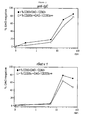
- Figure 2 shows that challenging the cells with (optimal concentrations of) anti-IgE and allergen elicits a quick histamine release that starts within minutes and peaks at about 15-20 minutes.
- the present example shows the activation patterns of basophils following Fc ⁇ RI cross-linking by anti-IgE antibody or recombinant Bet v 1 (rBet v 1) allergen or following challenge with fMLP.
- the cells were further processed and analyzed according to the protocol of Example 1.
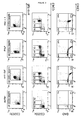
- CD203c bright+ Cross-linking of Fc ⁇ RI by anti-IgE and rBet v 1 in birch pollen allergic patients and challenging cells with fMLP almost invariantly resulted in a significant up-regulation of CD203c expression (CD203c bright+ ) .
- CD203 brigh+ cells the distribution of CD63 expression was bimodal, i.e. negative (CD63 - ) or positive (CD63 + ).
- CD63 + cells comprised a CD63 brigh+ and CD63 dim+ subpopulation ( fig 3 mid panel, R4 and R5).
- CD203c brigh+ CD63 + cells demonstrated a clear histamine release, particularly CD63 brigh+ cells ( fig 3 , bottom panel).
- cCD203c bright+ cells that did not co-express CD63 upon stimulation did not release histamine ( fig 3 , bottom panel).
- basophils that significantly up-regulate CD203c upon stimulation with anti-IgE antibody, allergen or fMLP generally exhibit a bi-modal distribution of CD63 expression, i.e. a CD63 negative and positive subpopulation ( Buhring et al, 2004, Int Arch Allergy Immunol, 133:317-329 ; Hennersdorf et al, 2005, Cell Res, 15:325-335 ; Chirumbolo et al, 2008, Clin Mol Allergy, 6:12 ).
- the individual cell responses of CD63 expression are generally quite heterogeneous, varying from completely absent (CD63 - ) over weak or modest (CD63 dim+ ) to very strong (CD63 brigh+ ).
- the present example shows the activation patterns of basophils following stimulation with PMA without or with IO.
- the cells were further processed and analyzed according to the protocol of Example 1.
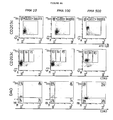
- Figure 4 displays a representative sample for stimulation of cells with PMA with or without IO. From this figure it emerges that challenging the cells with PMA elicited a dissimilar pattern to cells activated by Fc ⁇ RI cross-linking or by fMLP. Actually, PMA clearly up-regulated expression of CD203c but elicited only poor dose-dependent expression of CD63 without clear release of histamine. In contrast, challenging the basophils of patients and control individuals with PMA-IO resulted in a significant expression of CD203c and dose-dependent CD63 expression with corresponding histamine release.
- the present example confirms that PMA-induced CD63 expression is rather poor or almost absent ( MacGlashan, 2010, Clin Exp Allergy, 40:1365-1377 ), indicating that phorbol esters not to induce significant degranulation ( Dvorak et al, 1992, Am J Pathol, 141:1309-1322 ). Adding ionomycin to PMA induces a clear dose-dependent up-regulation of CD63.
- the present example shows the activation patterns of basophils following stimulation with IL-3.
- the cells were further processed and analyzed according to the protocol of Example 1.
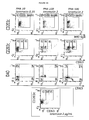
- FIG. 5 summarizes the stimulation experiments with IL-3. It is confirmed that IL-3 predominantly up-regulates CD203c without significant expression of CD63 nor release of histamine. On the contrary, the (a subpopulation of these) CD203 bright+ CD63 - cells appeared to contain quantitatively more histamine than the CD203c dim+ CD63 - cells. Furthermore, as exemplified in this particular patient, IL-3 was demonstrated to facilitate basophil stimulation by allergen, as incubation of the cells by this cytokine induced shifting from CD203c up-regulation to CD203c and CD63 up-regulation.
- the present example confirms that in vitro challenging of the basophils with IL-3 rather prime [35-42] the basophils with an up-regulation of CD203c [10;38] but without significant expression of CD63 [10;12;37] nor histamine release [35-37;43;44]. On the contrary, priming with IL-3 appears to increase intracellular histamine content.
Landscapes
- Health & Medical Sciences (AREA)
- Life Sciences & Earth Sciences (AREA)
- Engineering & Computer Science (AREA)
- Immunology (AREA)
- Molecular Biology (AREA)
- Biomedical Technology (AREA)
- Chemical & Material Sciences (AREA)
- Urology & Nephrology (AREA)
- Hematology (AREA)
- Cell Biology (AREA)
- Physics & Mathematics (AREA)
- Microbiology (AREA)
- Biochemistry (AREA)
- Pathology (AREA)
- General Physics & Mathematics (AREA)
- General Health & Medical Sciences (AREA)
- Biotechnology (AREA)
- Food Science & Technology (AREA)
- Medicinal Chemistry (AREA)
- Analytical Chemistry (AREA)
- Bioinformatics & Cheminformatics (AREA)
- Bioinformatics & Computational Biology (AREA)
- Biophysics (AREA)
- Proteomics, Peptides & Aminoacids (AREA)
- Toxicology (AREA)
- Tropical Medicine & Parasitology (AREA)
- Measuring Or Testing Involving Enzymes Or Micro-Organisms (AREA)
- Investigating Or Analysing Biological Materials (AREA)
Abstract
The present invention provides methods and kits for detecting and quantifying intracellular histamine and histamine release in individual mast cells and/or basophils by cytometry based on an enzyme-affinity method wherein histaminase diamine oxidase (DAO) is coupled to a laser-excitable fluorochrome.
Description
- The present invention relates to a cytometric method and a kit for detecting intracellular histamine and histamine release in mast cells and/or basophils at single cell level.
- Mast cells and/or basophils play a central role in the IgE-mediated allergic reaction. Upon the first encounter with an allergen, allergen-specific IgE antibodies are produced by B lymphocytes. These IgE antibodies bind to FcεRI receptors found on the surface of mast cells and/or basophils. This process is called sensitization. The next time(s) one runs across said allergen, it cross-links FcεRI-bound specific IgE antibodies on said sensitized IgE-primed mast cells and/or basophils. This cross-linking ultimately induces degranulation of said mast cells and/or basophils, which leads to the release of various potent mediators such as histamine that contribute to the allergic reaction. Alternatively, basophils can also be activated by various other stimuli such as anaphylatoxins, bacterial products, cytokines, phorbol esters, etc. The activation of basophils is further characterized by an up-regulation of the activation markers CD63 and CD203c on their membranes.
- Mast cell and basophil activation was first studied by quantification of the amount of mediator release. Traditionally, histamine release has been quantified using fluorimetric and colorimetric techniques that measure its extracellular content after degranulation of activated basophils and mast cells in the supernatant. However, the time and cost consuming two-step approach i.e. (a) cell incubation and (b) quantification of low concentrations of this rapidly metabolized compound has hampered the application of such assays. Moreover, as in these techniques histamine is quantified in the supernatant, results represent only an average of (isolated) cells analyzed and do not inform on cells with heterogeneity in responses.
- In 1993 Dvorak et al (J Histochem Cytochem, 41:787-800) successfully localized intracellular histamine in mast cells by applying an enzyme-affinity-gold method based on the affinity of the histaminase diamine oxidase (DAO) for its substrate. In 1994, this DAO-gold based technique was applied to localize histamine in activated basophils (Dvorak et al, 1994, Int Arch Allergy Immunol, 105:8-11; Dvorak et al, 1995, Blood, 86:3560-3566; Dvorak et al, 1996, Blood, 88:4090-4101; Dvorak et al, 1996, J Allergy Clin Immunol, 98:355-370; Dvorak et al, 1998, Prog Histochem Cytochem,33:III-320).
- In the early 1990ies, the discovery of the basophil activation marker CD63 induced the development of a flow cytometric technique to analyze and quantify in vitro activation of basophils (Knol et al, 1991, J Allergy Clin Immunol, 88:328-338). During the last decade potentials and limitations of this technique (basophil activation test: BAT) have been repeatedly reviewed (Ebo et al, 2004, Clin Exp Allergy, 34:332-339; Ebo et al, 2006, Allergy, 61:028-1039; Ebo et al, 2008, Cytometry B clin Cytom, 74:201-210). At present, the tetraspanin CD63 (gp50) of the lysosome-associated membrane protein (LAMP) family and the lineage-specific ecto-enzyme CD203c (ectonucleotide pyrophosphate/ phosphodiesterase or ENPP-3) (but also intracellular signaling molecules such as phosphorylated p38 mitogen protein kinase (MAPK) have been validated as potential read outs for flow-assisted analysis of in vitro activated basophils). Interestingly, it has emerged that the up-regulation of CD63 and CD203c display distinct kinetics and are regulated by different pathways. Furthermore, the relationship between both activation markers and mediator release remains still elusive. Comparative analyses between flow-assisted phenotyping and quantification of extracellular histamine has revealed that expression of these markers can (to some extent) be dissociated from mediator release (MacGlashan, 2010, Clin Exp Allergy, 40:1365-1377; MacGlashan, 1995, J Leukoc Biol, 58:177-188). However, this method requires homogeneous cell populations and results represent an average of purified cells analyzed.
- Accordingly, there remains a need for improved methods for measuring the activation of basophils and/or mast cells.
- The present invention provides methods for detecting and optionally quantifying intracellular histamine and histamine release in individual mast cells and/or basophils. More particularly, the inventors have found that intracellular histamine and histamine release can successfully be analyzed and quantified using cytometric detection methods applying an enzyme-affinity-fluorochrome method based on the affinity of the histaminase diamine oxidase (DAO) for its substrate. An important advantage of this technology is that detection of histamine can occur intracellularly, without needing to activate the cells to ensure histamine release. Moreover, the combination with cytometric methods allows a selective detection of histamine from basophils and/or mast cells. Moreover the methods of the invention allow a differentiation in the type of activation (i.e. piecemeal or anaphylactic) of the cells, which can be of interest both in a scientific and in a therapeutic context.
- In a first aspect the invention thus provides methods for detecting intracellular histamine and histamine release in individual mast cells and/or basophils. More particularly the methods of the invention comprise the steps of:
- a) fixing said mast cells and/or basophils with a fixation buffer comprising a fixation reagent;
- b) permeabilizing said mast cells and/or basophils with a permeabilization buffer comprising a permeabilization agent;
- d) staining said mast cells and/or basophils intracellularly with fluorochrome-labeled histaminase diamine oxidase; and
- e) analyzing said stained mast cells and/or basophils on a cytometer, wherein the signal from said fluorochrome-labeled histaminase diamine oxidase is indicative of the quantity of histamine present per cell in each of said mast cells and/or basophils.
- The methods of the invention are suitable for use on isolated mast cells and basophils, subpopulations comprising mast cells and/or basophils cell lines or biological samples. Biological samples which can be analysed include but are not limited to a blood sample, a tissue biopsy, e.g. a skin biopsy. In particular embodiments of the present invention, the mast cells and/or basophils are provided as an isolated (pure) population of mast cells and/or basophils.
- Where the sample is not an isolated (pure) mast cell and/or basophil population but contains other cells than mast cells and basophils (e.g. a biological sample), identification and optionally selection of the mast cells and/or basophils in the sample is desirable.
- In particular embodiments of the methods of the present invention, the mast cells and/or basophils are identified by using selection marker. Thus, in these embodiments, the methods of the invention for detecting intracellular histamine and histamine release in individual mast cells and/or basophils in a sample comprise the steps of:
- a) staining said mast cells and/or basophils for one or more selection markers; and further
- b) fixing said mast cells and/or basophils with a fixation buffer comprising a fixation reagent;
- c) permeabilizing said mast cells and/or basophils with a permeabilization buffer comprising a permeabilization agent;
- d) staining said mast cells and/or basophils intracellularly with fluorochrome-labeled histaminase diamine oxidase; and
- e) analyzing said stained mast cells and/or basophils on a cytometer, wherein the signal from said fluorochrome-labeled histaminase diamine oxidase is indicative of the quantity of histamine present per cell in each of said mast cells and/or basophils.
- In particular embodiments, the cellular membrane selection marker is identified using an antibody directed against the marker. More particularly, the antibody is labeled with a fluorochrome, whereby the fluorochrome has a distinct emission spectrum separated spectrally from the fluorochrome-labeled histaminase diamine oxidase. Different cellular membrane selection markers for basophils and/or mast cells are envisaged for use in the methods of the present invention. In particular embodiments, the one or more selection markers can comprises IgE, or the IgE receptor, CD203c, etc..
- The methods of the invention further allow to further characterize activated cell populations of mast cells as to their histamine content and/or release. For this purpose, the cells can be stained with an activation marker. Thus, in particular embodiments of the methods of the present invention, the one or more activation markers comprise activation markers such as CD63 or CD203c, etc..
- Additionally or alternatively, where the sample contains cells other than basophils and/or mast cells, the basophil and/or mast cell population is further purified by removing the cell types other than basophils and/or mast cells. For instance, where the sample may contain red blood cells, a lysing reagent can be used, to selectively lyse red blood cells. The lysing reagent can be present in the fixation buffer or the method of the invention can comprise a separate step wherein the cells are treated with lysing buffer comprising a lysing reagent.
- As indicated above, the methods of the present invention make use of a fixation buffer and a permeabilization buffer. In particular embodiments of the methods of the present invention said fixation buffer comprises formaldehyde as a fixating reagent. In particular embodiments of the methods of the present invention said permeabilization buffer comprises Triton-X-100. In particular embodiments, the fluorochrome used to label histamine diamine oxidase is phycoerythrin.
- In particular embodiments, the methods of the present invention further comprise the step of comparing the signal from said fluorochrome-labeled histaminase diamine oxidase to a reference value (e.g, obtained from resting cells), wherein said reference value can be used to evaluate degranulation of said mast cells and basophils.
- In further particular embodiments, the methods of the invention include a calibration of the amount of intracellular histamine detected. This is ensured by adding calibration particles labeled with said fluorochrome of said fluorochrome-labeled histaminase diamine oxidase to said mast cells and basophils.
- The methods of the present invention are particularly suitable for detection of intracellular histamine and histamine release in mast cells and/or basophils, more particularly in individual mast cells and/or basophils.
- In particular embodiments, the methods of the invention can be used to determine the effect of a test compound on histamine content and/or release of mast cells and basophils. In these embodiments, the methods of the invention comprise the step of incubating said mast cells and basophils at optimal temperature conditions with a test compound prior to mixing said cells with fixation buffer, and further carrying out the method steps as described herein. Comparison of a sample with and without the test compound will allow determination of whether or not the test compound influences the histamine content and/or release by said mast cells and/or basophils. In particular embodiments the methods of the invention further comprise the step of incubating mast cells and basophils with an activator. Examples of suitable activators include but are not limited to anti-IgE antibody, N-formyl-met-leu-phe (fMLP) or phorbol 12-myristate 13-acetate/ionomycin (PMA/ionomycin). The activator is incubated with the cells, prior to mixing the cells with fixation buffer.
- In another aspect, the present invention provides kits for detecting intracellular histamine and histamine release in individual mast cells and basophils by flow cytometry comprising:
- a fluorochrome-labeled histaminase diamine oxidase;
- one or more fluorochrome-labeled antibodies directed against a selection marker, more particularly a cellular membrane marker for basophils and mast cells;
- fixation buffer comprising a fixation reagent; and
- permeabilization buffer comprising a permeabilization reagent.
- In particular embodiments of the kits of the invention the fixation buffer comprises formaldehyde as a fixation reagent. In particular embodiments of the kits of the invention said fixation buffer comprises a lysing reagent, more particularly a lysing reagent for red blood cells. In particular embodiments of the kits of the present invention said permeabilization buffer comprises Triton-X-100.
- Typically, the fluorochrome label present on said histaminase diamine oxidase present in the kits of the present invention is different from the fluorochrome present on the antibody directed against the selection marker. In particular embodiments of the kits of the present invention said histaminase diamine oxidase is labeled with phycoerythrin.
- In particular embodiments the kits of the present invention further comprise instructions for performing the methods as described herein.
- In particular embodiments the kits of the present invention further comprise calibration particles labeled with the fluorochrome of said fluorochrome-labeled histaminase diamine oxidase. The calibration particles are optionally fluorochrome-labeled calibration particles comprising the fluorochrome at quantitated levels;
- In particular embodiments the kits of the present invention comprise one or more fluorochrome-labeled antibodies directed against an activation marker, preferably CD63 or CD203c.
- In particular embodiments the kits of the present invention further comprise an activator for basophils and/or mast cells. Suitable activators include but are not limited to anti-IgE antibody, N-formyl-met-leu-phe (fMLP) or phorbol 12-myristate 13-acetate/ionomycin (PMA/ionomycin).
- Yet another aspect of the present invention relates to the uses of the kits and the methods of the present invention for different applications.
- In particular embodiments the kit is used for detecting intracellular histamine and histamine release in individual mast cells and basophils by flow cytometry. More particularly it is envisaged that the kit is used for detecting degranulation of individual mast cells and basophils by cytometry. In further embodiments the kit is used for quantifying the histamine content of individual mast cells and basophils by cytometry. In further embodiments the methods and kits of the invention are used to characterize activated basophils and mast cells.
- In particular embodiment, the invention relates to the use of the kits and methods of the invention for in vitro determination of allergenic reaction. More particularly the methods and kits can be used as an in vitro allergy test.
- In particular embodiments, the invention relates to the use of the kits and methods of the invention for determining whether a compound has an effect on histamine content and/or release by basophils and/or mast cells. In further particular embodiments, the tests can be used to determine whether a compound has a histamine releasing effect.
- The present invention further provides the use of the method disclosed herein for high-throughput screening of the histamine releasing effect of compounds or for screening the ability of compounds to counter an allergenic reaction.
- The present invention is illustrated by the following figures which are to be considered for illustrative purposes only and in no way limit the invention to the embodiments disclosed therein:
-
Figure 1 provides the identification of resting and activated basophils following challenge with anti-IgE antibody using the method according to the present invention. Flow cytometric analysis using a density plot of side scatter (SSC) and anti-IgE (A). At least 1000 anti-IgE-Alexa Fluor 405 and anti-CD203c-APC positive basophils are gated out in a rectangular region (B). Within this gate co-expression of CD203c and CD63 was analyzed and revealed 3 subpopulations of CD63 expression: CD203cbrigh+CD63- (R3), CD203cbrigh+CD63dim+ (R4) and CD203cbrigh+CD63 brigh+ (R5) (C). Histamine-containing basophils were defined as PE-DAO+ cells. Fifty-six percent of the basophils completely released their histamine content (DAO- cells) and are situated in lower-right quadrant of panel D. Note that the Y-axis in panel C and D is bi-exponential.. -
Figure 2 provides the kinetics of histamine release in basophils after FcεRI cross-linking by anti-IgE antibody (top) orrBet v 1 allergen (bottom). Histamine release is expressed as % DAO- cells by all CD203cbright cells (%CD203cbrigh+DAO-/CD203cbrigh+) and by CD203cbrigh+ cells that co-express CD63 (% CD63+DAO-/CD63+). -
Figure 3 provides the activation patterns of basophils after FcεRI cross-linking by anti-IgE antibody orrBet v 1 allergen or after stimulation with fMLP. Selection of anti-IgE-Alexa fluor 405 and anti-cD203c-APC positive basophils (upper panel), expression of the activation markers CD63 and CD203c (middle panel) and histamine release coupled to expression of CD63 (bottom panel) in cells incubated with (activation) buffer, anti-IgE antibody, fMLP and recombinant birch pollen allergen (rBet v 1). Basophils that completely released histamine (DAO- cells) are situated in lower-right quadrants of the bottom panel. Note that the Y-axis is bi-exponential. As inFigure 1 , remark the distribution of activated basophils with a CD203cbrigh+CD63-, CD203cbrigh+CD63dim+ and CD203cbrigh+CD63brigh+ subpopulation. Upon stimulation withrBet v -
Figure 4 provides activation patterns of basophils following stimulation with PMA without or with IO
Selection of anti-IgE-Alexa fluor 405 and anti-cD203c-APC positive basophils (upper panel), expression of the activation markers CD63 and CD203c (middle panel) and histamine release coupled to expression of CD63 (bottom panel) in cells incubated withPMA ionomycin 2 µg/mL. Basophils that completely released histamine (DAO- cells) are situated in lower-right quadrants of the bottom panel. Note that the Y-axis is bi-exponential. As inFigure 1 , remark the distribution of activated basophils with a CD203cbrigh+CD63-, CD203cbrigh+CD63dim+ and CD203cbrigh+CD63brigh+ subpopulation. -
Figure 5 provides activation patterns of basophils following incubation with IL-3
Selection of anti-IgE-Alexa fluor 405 and anti-cD203c-APC positive basophils (upper panel), expression of the activation markers CD63 and CD203c (middle panel) and histamine release coupled to expression of CD63 (bottom panel) in cells incubated with (activation) buffer, IL-3 1 ng/mL, IL-3 10 ng/mL,rBet v 1 0.01 µg/mL, IL-3 1 ng/mL +rBet v 1 0.01 µg/mL and IL-3 10 ng/mL +rBet v 1 0.01 µg/mL. Basophils that completely released histamine (DAO- cells) are situated in lower-right quadrants of the bottom panel. Note that the Y-axis is bi-exponential. As inFigure 1 , remark the distribution of activated basophils with a CD203cbrigh+CD63-, CD203cbrigh+CD63dim+ and CD203cbrigh+CD63brigh+ subpopulation. - Each of these illustrations represents particular embodiments of the features concerned and the corresponding features are not to be interpreted as limited to this specific embodiment.
- The terminology used herein is not intended to be limiting, since the scope of the present invention will be limited only by the appended claims.
- As used herein, the singular forms "a", "an", and "the" include both singular and plural referents unless the context clearly dictates otherwise.
- The terms "comprising", "comprises" and "comprised of" as used herein are synonymous with "including", "includes" or "containing", "contains", and are inclusive or open-ended and do not exclude additional, non-recited members, elements or method steps. Where embodiments are referred to as "comprising" particular features, elements or steps, this is intended to specifically include embodiments which consist of the listed features, elements or steps.
- The recitation of numerical ranges by endpoints includes all numbers and fractions subsumed within the respective ranges, as well as the recited endpoints.
- The term "about" as used herein when referring to a measurable value such as a parameter, an amount, a temporal duration, and the like, is meant to encompass variations of +/-10% or less, preferably +/-5% or less, more preferably +/-1% or less, and still more preferably +/-0.1% or less of and from the specified value, insofar such variations are appropriate to perform in the disclosed invention. It is to be understood that the value to which the modifier "about" refers is itself also specifically, and preferably, disclosed.
- All documents cited in the present specification are hereby incorporated by reference in their entirety.
- Unless otherwise defined, all terms used in disclosing the invention, including technical and scientific terms, have the meaning as commonly understood by one of ordinary skill in the art to which this invention belongs. By means of further guidance, definitions for the terms used in the description are included to better appreciate the teaching of the present invention.
- Reference throughout this specification to "one embodiment" or "an embodiment" means that a particular feature, structure or characteristic described in connection with the embodiment is included in at least one embodiment of the present invention. Thus, appearances of the phrases "in one embodiment" or "in an embodiment" in various places throughout this specification are not necessarily all referring to the same embodiment, but may. Furthermore, the particular features, structures or characteristics may be combined in any suitable manner, as would be apparent to a person skilled in the art from this disclosure, in one or more embodiments. Furthermore, while some embodiments described herein include some but not other features included in other embodiments, combinations of features of different embodiments are meant to be within the scope of the invention, and form different embodiments, as would be understood by those in the art. For example, in the following claims, any of the claimed embodiments can be used in any combination.
- The present invention is directed to cytometric analysis methods for the detection of intracellular histamine and histamine release in mast cells and basophils. More particularly the methods of the invention allow the detection of intracellular histamine and histamine release in individual mast cells and basophils. The basis of the invention is the finding that intracellular histamine can be qualitatively and quantitatively detected by cytometry by an enzyme-affinity method wherein histaminase diamine oxidase (DAO) is coupled to laser-excitable fluorochromes. Mast cells and basophils are characterized by the presence of granules rich in histamine. Activation of mast cells and basophils ultimately leads to degranulation and the release of histamine. However, it is the histamine release as such which is physiologically relevant. Histamine release has traditionally been measured in the supernatant of activated mast cells and basophils, representing only an average of cells analyzed and not informing on cells or cell subpopulations with heterogeneity in responses. The present method allows localizing histamine at single cell level, which is an improvement over traditional methods. The methods of the present invention further allow the identification of activated basophils and mast cells.
- The methods of the present invention require that the cells are adequately treated to allow the histaminase diamine oxidase to enter the cell and reach its substrate histamine. Thus, the methods of the present invention comprise a fixing step with a fixation buffer comprising a fixation reagent and a permeabilization step with a permeabilization reagent.
- Methods of fixation are well known in the art. With "fixation buffer" is meant herein an aqueous buffered solution comprising a fixation reagent. Methanol- or (para)formaldehyde-based fixation reagents are preferred, more preferably (para)formaldehyde-based fixation reagents.
- Optionally, the fixation buffer may further comprise a lysing reagent, most particularly a lysing reagent for red blood cells. The terms "lysis" and "lysing" are used interchangeably herein and relate to the process of lysing non nucleated blood cells (i.e. red blood cells) in order to avoid any interference with the cytometric analysis of nucleated blood cells. Accordingly, suitable lysing reagents are preferably selected from any kind of erythrocyte lysing reagents known from prior art. The use of a fixation buffer comprising a fixation reagent and a lysing reagent is particularly envisaged when a sample comprising red blood cells, such as a whole blood sample is analyzed. A fixation buffer comprising a fixation reagent and a lysing reagent advantageously allows lysing of red blood cells and fixing of leukocytes to be performed in a single step. An example of such fixation buffer comprising a fixation reagent and a lysing reagent is Phosflow Lyse/Fix Buffer (BD Biosciences) which is an aqueous buffered solution containing formaldehyde as a fixation reagent. However, it will be understood to the skilled person that the fixation and the lysing reagent may also be used separately.
- In order for the histaminase diamine oxidase to gain access to intracellular histamine, the cells require permeabilization. Ideally, the permeabilizing reagent creates apertures in the cell membranes without affecting the gross morphology of the cell such that flow cytometric light scattering characteristics of the cell are not affected. Such methods of permeabilizing cells are well known in the art. Saponin-and Triton-X-100-based permeabilizing reagents are preferred, more preferably Triton-X-100-based permeabilizing reagents. With "permeabilization buffer" is meant herein an aqueous buffered solution comprising a permeabilization reagent.
- In particular embodiments of the methods of the present invention, the mast cells and basophils are fixed with a fixation buffer comprising formaldehyde and permeabilized with a Triton-X-100 comprising permeabilizing buffer, more preferably a permeabilizing buffer comprising from 0.01 to 1 %, preferably from 0.02 to 0.5 %, more preferably from 0.05 to 0.2 %, most preferably about 0.1 % Triton-X-100.
- In further particular embodiments of the methods of the present invention, the cells comprised in a whole blood sample are mixed with a fixation buffer comprising a lysing reagent and formaldehyde, such that red blood cells in the sample are lysed and the remaining cells are fixed, and the fixed cells are permeabilized with a Triton-X-100 comprising permeabilizing buffer, more particularly a permeabilizing buffer comprising from 0.01 to 1 %, preferably from 0.02 to 0.5 %, more preferably from 0.05 to 0.2 %, most preferably about 0.1 % Triton-X-100.
- After fixing and permeabilization of the cells, the methods of the present invention further comprise the step of staining said mast cells and basophils intracellularly with fluorochrome-labeled histaminase diamine oxidase. The histaminase diamine oxidase is conjugated to a fluorochrome by techniques well known in the art. Different fluorochromes can be applied to conjugate the histaminase diamine oxidase such as, for example, fluorescent nanoparticles such as those sold under the trade name Alexa Fluor® Dyes, biotin-streptavidin labeled with phycoerythrin/Cy7, fluorescein isothiocyanate (FITC), Horizon-V500, phycoerythrin. In a preferred embodiment, the histaminase diamine oxidase is conjugated to phycoerythrin, which has been shown to give a good resolution. Staining with said fluorochrome-labeled histaminase diamine oxidase may be achieved through incubating said fixed and permeabilized mast cells and basophils with said said fluorochrome-labeled histaminase diamine oxidase at 37°C for a predetermined period of time, such as for example for up to 1 h, preferably for between 30 min and 1 h, more preferably for about 45 min.
- Thus, the present invention provides a method for detecting intracellular histamine and histamine release in individual mast cells and basophils comprising the steps of:
- a) fixing said mast cells and basophils with fixation buffer comprising a fixation reagent;
- b) permeabilizing said mast cells and basophils with permeabilization reagent;
- c) staining said mast cells and basophils intracellularly with fluorochrome-labeled histaminase diamine oxidase; and
- d) analyzing said stained mast cells and basophils on a cytometer, wherein the signal from said fluorochrome-labeled histaminase diamine oxidase is indicative of the quantity of histamine present per cell in each of said mast cells and basophils.
- In particular embodiments of the methods of the present invention, the mast cells and basophils are identified and/or selected out by way of one or more selection markers.
- As used herein, a "selection marker", refers to any molecule that may be used, alone or in combination, to facilitate the identification of mast cells and basophils. These selection markers are typically cellular membrane molecules, although intracellular molecules may also be used. Suitable selection markers include, but are not restricted to, IgE, FcεRI, the lineage-specific CD203c, CD123/HLADR and chemokine receptor 3 (CCR3). The one or more selection markers referred to herein may include activation markers, as will be detailed below.
- Typically the selection markers are detected by way of an antibody directed against the marker. The term antibody as used herein is meant to include any antigen-binding fragment which can be used in the context of the present invention. Antibodies directed against the markers envisaged in the context of the present invention are known to the skilled person and are commercially available. Thus, in particular embodiments, the invention provides methods which comprise detecting a basophil and/or mast cell selection marker with an antibody directed against a mast cell and/or basophil selection marker.
- In further particular embodiments of the methods of the present invention the antibodies directed against the selection markers used in the context of the present invention are labeled, most particularly fluorochrome labeled.
- Staining said mast cells and basophils with said fluorochrome-labeled antibody directed against a mast cell and/or basophil selection marker may be performed according to well known techniques for staining cell surface molecules, such as, for example, incubating said mast cells and basophils with said antibody on ice for a predetermined period of time, such as for example for up to 1 h, preferably for between 10 min and 30 min, more preferably for about 20 min.
- The antibodies used throughout the present disclosure may be labeled with any fluorochrome known in the art. Non-limiting examples of fluorochromes that may be used include fluorescein isothiocyanate (FITC), Texas Red (sulforhodamine isothiocyanate), allophycocyanin (APC), Cy3, Cy5, Oregon Green isothiocyanta, phycoerythrin (PE), fluorescent nanoparticles such as those sold under the trade name Invitrogen Q-dots® or Dyomics dyes such as DY-647 or DY-747. When using multiple fluorochromes in the methods of the invention, said fluorochromes preferably have distinct emission spectra, more preferably they are separated spectrally from one another.
- Typically, the mast cells and basophils are stained for the selection marker prior to the fixing step and staining with the histaminase. Thus, in particular embodiments, methods for detecting intracellular histamine and histamine release in individual mast cells and/or basophils comprise the steps of:
- a) staining said cells for the presence of one or more selection markers for basophils and/or mast cells
- b) fixing said mast cells and/or basophils with fixation buffer comprising a fixation reagent;
- c) permeabilizing said mast cells and/or basophils with permeabilization reagent;
- d) staining said mast cells and/or basophils intracellularly with fluorochrome-labeled histaminase diamine oxidase; and
- e) analyzing said stained mast cells and/or basophils on a cytometer, wherein the signal from said fluorochrome-labeled histaminase diamine oxidase is indicative of the quantity of histamine present per cell in each of said mast cells and basophils.
- The mast cells and/or basophils to be analyzed using the methods of the present invention may be comprised in a blood sample, e.g. a whole blood sample or a (partially) purified blood sample, a tissue biopsy, e.g. a skin biopsy, or any other source which comprise multiple mast cells and/or basophils. The mast cells and basophils may also be provided as an isolated (pure) population of mast cells and/or basophils. In a preferred embodiment, a whole blood sample is analyzed. Basophils represent a small white blood cell subpopulation. Thus, in particular embodiments, basophils are comprised in a white blood cell fraction. Mast cells are found resident in tissues throughout the body, particularly in association with structures such as blood vessels and nerves, and in proximity to surfaces that interface the external environment.
- When analyzing a blood sample, the methods of the present invention may comprise the step of treating the sample with EDTA, heparin or any other anticoagulants in order to prevent coagulation.
- When analyzing a sample potentially comprising red blood cells, the methods of the invention may comprise the step of lysing the red blood cells with a lysing reagent. In particular embodiments the lysing reagent is present in the fixation buffer. In alternative embodiments, the methods of the invention comprise a separate step, wherein the sample is contacted with a lysing buffer comprising a lysing reagent. Examples of lysing reagents are provided herein below.
- The methods of the present invention further comprise the step of analyzing said stained mast cells and basophils on a cytometer. The analysis by cytometry is carried out as known from the prior art. Cytometry allows the rapid measurement of light scatter or morphometry and fluorescence emission produced by suitably illuminated cells or particles. The cells or particles produce signals when they individually pass through or are exposed to a beam of light. Each cell or particle is measured separately and the output represents cumulative individual cytometric characteristics. An important analytical feature of modern cytometers is their ability to measure multiple cellular parameters. The scattered light and fluorescence emissions of each cell or particle are collected by detectors and sent to a computer, where the distribution of the population with respect to the different parameters is represented. Scattered light collected in the same direction as the incident light is related to cell size, and scattered light collected at an angle of 90 indicates particle complexity. Morphometry and particle complexity is related to cell surface characteristics and the number and type of organelles present in the cell. Size and complexity are considered intrinsic parameters since they can be obtained without having to stain the sample. To obtain additional or more specific information, samples may be stained using different fluorochromes. For instance, said fluorochromes may be conjugated to antibodies which specifically bind to antigens to gain information on said antigens. In the present invention the enzyme histaminase diamine oxidase is conjugated to a fluorochrome in order to obtain information on its substrate histamine.
- Using cytometry, a specific population of cells can be "gated" and analyzed separately from the remainder of the population. For example, mast cells and basophils may be gated out from a whole blood sample by applying side scatter and staining of a mast cell and basophil selection marker.
- Fluorescence activated flow cytometry provides a method for sorting a heterogeneous mixture of biological cells into two or more containers, one cell at a time, based upon the specific light scattering and fluorescent characteristics of each cell. In laser scanning cytometry (LSC) a scan line oscillates on the cell and the fluorescent energy emitted by each cell is collected and specifically detected. This allows the analysis of samples on different substrates. Cytometers useful for carrying out the present invention include conventional and state of the art flow cytometers made by Beckman Coulter, Toa Medical Electronics, Partec, Bio-Rad, or Becton Dickinson and image cytometers made by General Electric, and Becton Dickinson.
- In particular embodiments of the methods of the present invention, the signals generated from said fluorochrome-labeled histaminase diamine oxidase may be compared to a reference value. In particular embodiments, the reference value is indicative of degranulation of mast cells and basophils and is used to determine whether a mast cell or basophil has undergone degranulation or not. A reference value may provide the signal from a mast cell or basophil that has undergone degranulation, for instance a mast cell or basophil that has been incubated with an activator. Additionally or alternatively, the a resting basophil or mast cell may be used as a reference value.
- To ensure calibration of the intracellular histamine detection, calibration particles may be added to the mast cells and basophils to be analyzed. With "calibration particles" are meant herein a set of fluorescently labeled particles which can be used to calibrate the detection with the fluorochrome-labeled histaminase. Most particularly, the particles are labeled with one or more fluorochrome. In further particular embodiments, the fluorochrome is identical to that of the fluorochrome-labeled histaminase. In further particular embodiments, the particles are labeled with a mixture of fluorochromes at different intensities that allows excitation at any wavelength between 365 and 650 nm that enable to construct a calibration curve in the channels of the flow cytometer in which the fluorochrome-labeled histaminase diamine oxidase is measured. A suitable example of calibration particles include SPHERO® Ultra Rainbow particles (Spherotech, Lake Forest, IL, US). A further example of suitable calibration particles are the QuantiBrite® particles. Indeed, the different quantified levels of R-phycoerythrin (PE) intensity on the separate beads provide for an exact quantification of the available PE molecules inside the individual cells that are stained with the PE-labeled diamino-oxidase.
- In particular embodiments, a calibration curve is made using the calibration particles based on the strongest staining cell. Typically this is a resting cell, as this cell retains all of its histamine. Resting cells can be identified based on the expression of selection markers but no, or only limited expression of activation markers. For instance, resting basophils express CCR3 but not CD63. The amount of histamine in the activated cells can then be quantified based on the calibration curve. As the amount of histamine in a resting cell may vary from one individual to another, it may be of interest to calibrate detection for every patient. Thus, in particular embodiments, the methods of the invention will comprise calibrating the histamine content using calibration particles and/or based on the histamine content of a resting cell.
- In particular embodiments the methods of the present invention comprise analyzing the signal generated at different time points. This allows to monitor the evolution of histamine content in the cells, and thus allows a measurement of histamine release by the cells at different time points, i.e. same samples or parallel samples incubated for longer time periods etc..
- The methods of the invention may be used to further characterize activated mast cells and basophils, more particularly to determine the relationship between the expression of activation markers and the histamine release in these cells. This can be achieved by staining the cells for use in the methods of the present invention with an activation marker.
- Thus, in further embodiments, the methods according to the present invention further comprise the step of staining said mast cells and/or basophils for the presence of an activation marker.
- As used herein, an "activation marker" refers to a molecule that may be used to identify activated mast cells and basophils. As used herein, "activated mast cells and basophils" denote mast cells and basophils that have undergone cross-linking of FcεRI-bound IgE antibodies. Activated basophils not only release potent mediators such as histamine but also up-regulate the expression of different membrane markers, e.g., CD63, CD203c, CD11b, CD13, CD107a, CD164, CD300a. Accordingly, these molecules may be used as activation markers for basophils.
- The same principles are applicable for mast cells but using other activation markers such as CD107 and CD117 (c-kit). As indicated above, the expression of markers is typically identified using labeled antibodies directed against these markers. In particular embodiments the antibodies are fluorochrome labeled (examples of fluorochrome labels are provided above). The staining of mast cells and basophils with a fluorochrome-labeled antibody directed against an activation marker may be performed according to well known techniques for staining cell surface molecules, such as, for example, incubating said mast cells and basophils with said antibody on ice for a predetermined period of time, such as for example for up to 1 h, preferably for between 10 min and 30 min, more preferably for about 20 min.
- The dual labeling of intracellular histamine and intracellular and/or membrane activation markers is advantageous in that it allows analyzing the relationship between expression of activation markers and histamine release, which is still elusive at present.
- In particular embodiments it can be of interest to determine the reaction of mast cells and/or basophils to an activator. For instance, it may be of interest to determine whether cells are capable of reacting normally to an activator. Alternatively, in the context of screening compounds for the effect on histamine content and/or release, it may be of interest to add an activator to the cells. Thus, in particular embodiments, the present invention provides methods wherein the mast cells and/or basophils are incubated with an activator prior to analyzing said mast cells and basophils according to the methods disclosed herein.
- An "activator" refers herein to any substance that induces activation of mast cells and basophils, thereby leading to degranulation and histamine release. Non-limiting examples of activators include anti-IgE antibody, anti-FcεRI antibody, N-formyl-L-methionyl-L-leucyl-L-phenylalanine (fMLP), phorbol 12-myristate 13-acetate/ionomycin (PMA/ionomycin), complement factor 5a (C5a), platelet activation factor (PAF), toll-like receptor (TLR) agonists, ionomycin, calcium ionophore A23187, lipopolysaccharides (LPS) and phytohaemagglutinins (PHA). When using mast cells and basophils from an allergic patient, natural or recombinant allergen and components thereof causing an allergic reaction in said patient may also be used as activator. Preferably, the activator is selected from the group comprising: anti-IgE antibody, N-formyl-met-leu-phe (fMLP) and phorbol 12-myristate 13-acetate/ionomycin (PMA/ionomycin) or allergen.
- As detailed above, the methods of the present invention may be used to identify the effect of a test compound on the histamine content and/or release of a mast and/or basophil sample. Thus, in particular embodiments, the present invention provides methods wherein the mast cells and/or basophils are incubated with a test compound prior to analyzing said mast cells and basophils according to the methods disclosed herein.
- The nature of the test compound is not critical and includes but is not limited to a protein, a peptide or a mixture of proteins or peptides or a protein extract from a biological source, preferably selected from the group of insect venoms, foods (fruits, vegetables, seeds, legumes, nuts, spices, fish, shellfish, mollusks, egg, fowl, meat and milk), tree pollens, grass pollens, weed pollens, epidermal and animal proteins, dust and storage mites, insects, parasites, microorganisms, small molecules, DNA, RNA etc.
- Typically, the methods of the invention will include the necessary positive and/or negative controls, such as but not limited to samples to which no test compound has been added, samples to which a known activator has been added etc, samples to which a control compound has been added etc.. Suitable controls are known to the skilled person.
- The incubation of mast cells and/or basophils with an activator or a test compound is preferably performed at optimal temperature conditions, preferably in the range of from 30 to 40°C, more preferably at about 37°C. The time of incubation may range from between 10 seconds up to 2 hours, preferably from between 20 seconds up to 100 min, more preferably from between 10 min up to 80 min, even more preferably from between 20 min up to 60 min. In particular embodiments of the methods of the invention, the incubation is performed at different time points so as to allow a definition of histamine release under gradual, piecemeal release or sudden and complete release. The incubation is preferably stopped by chilling on ice and adding a stop solution containing 1 to 20 mmol/l of EDTA, preferably 5 to 15 mmol/l, more preferably 10 mmol/l.
- The incubation of mast cells and basophils with an activator or a test compound is preferably performed in an activation buffer. A suitable activation buffer may contain optimum calcium and magnesium concentration in optimized physico-chemical conditions. A suitable example of activation buffer is Hank's balanced salt solution that additionally contains 20 µM HEPES and 7.5% NaHCO3 at pH 7.4. The activation buffer may also be used for deleting the activator and the test substance.
- In particular embodiments the activation buffer may comprise interleukin(IL)-3. The inventors have found that priming the mast cells and basophils with IL-3 potentiates the effect of incubation with an activator, i.e. will promote degranulation, while IL-3 itself does not induce degranulation.
- As indicated above, the signal of the fluorochrome-labeled histaminase diamine oxidase of mast cells and basophils that have been incubated with an activator prior to analysis according to the methods of the present invention may be used as a reference value which is indicative of degranulation of mast cells and basophils.
- The signal of the fluorochrome-labeled histaminase diamine oxidase of mast cells and basophils that have been incubated with a test compound according to the present method may be compared to a reference value indicative of degranulation of mast cells and basophils. A signal lower than said reference value indicates that the test compound has induced degranulation and is indicative of an allergenic effect of said test compound, whereas a signal higher than said reference value indicates that the test compound has not induced degranulation.
- In another aspect, the present invention provides kits for detecting and quantifying intracellular histamine and histamine release in individual mast cells and basophils. The kits of the present invention are particularly suited for carrying out the methods of the present invention.
- In particular embodiments, the present invention provides a kit for detecting intracellular histamine and histamine release in individual mast cells and basophils comprising:
- a fluorochrome-labeled histaminase diamine oxidase;
- a fluorochrome-labeled antibody directed against a selection marker;
- fixation buffer comprising a fixation reagent; and
- permeabilization buffer comprising a permeabilization reagent.
- In a particular embodiment said fixation reagent comprises (para)formaldehyde or methanol, more preferably (para)formaldehyde. In a further embodiment said permeabilization reagent comprises Triton-X-100 or saponin, more preferably Triton-X-100.
- The kits of the invention comprise a fluoro-chrome labeled antibody against a selection marker. In particular embodiments said fluorochrome-labeled antibody directed against a selection marker is selected from fluorochrome-labeled antibody directed against a selection marker such as but not limited to IgE, FcεRI, the lineage-specific CD203c, CD123/HLADR and chemokine receptor 3 (CCR3).
- As detailed above, the nature of the fluorochrome of the fluorochrome-labeled histaminase diamine oxidase is not critical to the present invention. However, in particular embodiments, the kits of the invention comprise phycoerythrin (PE)-labeled histaminase diamine oxidase.
- Suitable fixation buffers and permeabilization buffers envisaged in the context of the present invention are described herein and further known to the person skilled in the art.
- The kits of the present may comprise one or more additional reagents which are of particular use for carrying out the methods of the invention.
- Thus, in particular embodiments, the kits additionally comprise a lysing reagent. The lysing reagent may be comprised in the fixation reagent or may be provided in a separate lysing buffer. Suitable lysing reagents include any kind of erythrocyte lysing buffers known from the art. A suitable fixation buffer comprising a fixation reagent and a lysing reagent is Phosflow Lyse/Fix Buffer (BD Biosciences) which is an aqueous buffered solution containing formaldehyde as a fixation reagent.
- In further particular embodiments, the kit further comprises a reference value or tools for obtaining a reference value, such as a reference which is indicative of degranulation of mast cells and basophils. A reference value may be determined by incubating mast cells and/or basophils with an activator which induces degranulation. Accordingly, the kit may also comprise an activator and activation buffer. Suitable activators include, but are not restricted to, anti-IgE antibody, N-formyl-met-leu-phe (fMLP) and phorbol 12-myristate 13-acetate/ionomycin (PMA/ionomycin).
- In particular embodiments, the kit may additionally comprise a positive control and/or a negative control for degranulation. Examples of suitable positive controls include an activator, while a suitable negative control is, for instance, activation buffer not comprising an activator.
- In particular embodiments, the kit according to the invention comprises calibration particles. Examples of calibration particles are described herein above.
- In particular embodiments, the kit comprises one or more fluorochrome-labeled antibodies directed against an activation marker. More particularly, the fluorochrome-labeled antibody directed against an activation marker is a fluorochrome-labeled antibody directed against CD63, CD203c, CD11b, CD13, CD107a, CD164 or CD300a. In particular embodiments, the kits of the invention comprise an anti-CD63 antibody or a fluorochrome-labeled anti-CD203c antibody.
- In further embodiments, the kit further comprises instructions for use of the kit according to the methods of the present disclosure. In a particular embodiment, the kit further comprises instructions for quantifying the intracellular histamine content of individual mast cells and basophils.
- Another aspect of the present invention relates to the applications of the methods and kits of the present invention.
- As detailed above, the methods and the kits of the present invention may be useful for detecting intracellular histamine and histamine release in individual mast cells and/or basophils.
- Typically, the methods of the present invention are used to determine histamine content and/or release of basophils and mast cells in biological samples, more particularly human or animal samples. The determination of histamine content and/or release can be determined independently of whether the cell is considered as "resting" or "activated" based on activation markers.
- In particular embodiments the methods and the kits are used for detecting degranulation of mast cells and/or basophils. Activation of mast cells and basophils ultimately lead to degranulation and the release of histamine. Accordingly, in one embodiment the methods and the kits may be used for detecting activated mast cells and basophils.
- In further particular embodiments, the methods and the kits may be used for an in vitro allergy test, wherein mast cells and/or basophils are analyzed that are comprised in a sample from a patient, such as a (whole) blood sample or a (skin) biopsy. The presence of degranulated mast cells and/or basophils in said sample is indicative of an allergic reaction in said patient.
- Thus in particular embodiments, the present invention provides in vitro diagnostic methods for determining and/or characterizing allergic conditions. More particularly, the methods and kits are envisaged for use in the diagnosis of all allergic conditions that rely upon IgE-mediated as well as IgE-independent activation of mast cells and basophils. More particularly, the methods and kits of the invention will allow to distinguish between a piecemeal activation and an anaphylactic reaction of the basophils and mast cells.
- Similarly the methods and kits of the invention can be used to monitor the effect of drugs that aim to silence mast cells and basophils (e.g. in mastocytosis) on patients, by analysis of patient samples. This includes monitoring of allergen-specific immunotherapy and anti-IgE treatment (e.g. omalizumab). Thus, in particular embodiments, the invention provides methods and kits are for monitoring efficacy of treatment, such as allergen-specific immunotherapy.
- In particular embodiments, the methods and kits are used to determine the allergenic or anti-allergenic effect of a compound. Typically, this encompasses testing a sample with and without the test compound and comparing the histamine content and/or release in the basophils and/or mast cells of the sample. In a further embodiment, the methods and the kits may be used for high-throughput screening of compounds, more particularly the high throughput screening of the allergenic effect of compounds or the effect of drugs on an induced allergenic effect.
- In further embodiments the methods and kits may be used for quantifying the histamine content of individual mast cells and basophils. More particularly this can be of use in the study of intracellular signaling (Ebo, 2007, Clin Exp All, 37:1668-75)
- In one embodiment, the methods and kits may be used for studying the histamine release pattern induced by a compound, such as an activator or a test compound, wherein the mast cells and basophils are incubated with said compound at different time points. Histamine release may be defined as gradual piecemeal release or sudden and complete release.
- In a further embodiment, the methods and kits may be used for studying the relationship between histamine release and expression of activation markers.
- In the following examples endotoxin-free heparinized whole blood samples from healthy control individuals and birch pollen allergic patients were used. Birch pollen allergic patients were selected upon clinical history documented by standardized skin tests and quantification of sIgE.
- The present example describes the identification of resting and activated basophils following challenge with anti-IgE antibody using the method according to the present invention.
- Aliquots of 200 µL whole blood from patients and control individuals were challenged at 37°C with 200 µL anti-IgE antibody (10 µg/mL; clone G7-18, BD Biosciences, Erembodegem, Belgium) for 20 min. The reaction was stopped by chilling on ice, adding 1 mL ice-cooled PBS/10 mmol/L EDTA and centrifugation for 5 min (4 °C, 200xg).
- The cells were then stained with 20 µL of monoclonal anti-human IgE (clone GE-1, Sigma Aldrich GmBH, Steinheim, Germany) labeled with Alexa Fluor 405 (Molecular Probes, Invitrogen, Paisley, UK) as described by the manufacturer and 10 µL of monoclonal anti-human CD63-FITC (clone H5C6, BD Biosciences) and 10 µL CD203c-APC (clone NP4D6 Biolegend, San Diego, CA, USA) for 20 min on ice.
- The cells were subsequently lysed/fixed with 2 mL Lyse/Fix Buffer (BD Biosciences) for 20 min at 37 °C. Cells were washed with and re-suspended in PBS with 0.1 % Triton-X-100 (PBS-TX, pH=7.4).
- To stain
intracellular histamine 10 µL Phycoerythrin-labeled (labeling was performed by BD Biosciences) diamine oxidase (DAO, Sigma-Aldrich) was added and incubated at 37 °C for 45 min. Cells were washed with 0.3 mL PBS, re-suspended in PBS with 0.1% sodiumazide. - Flow cytometric analysis and quantification of basophil activation was performed on a FACSCanto II flow cytometer (BD Immunocytometry Systems, San Jose, CA, USA). Standardization of intracellular histamine content was performed using standardized fluorospheres (SPHERO Ultra Rainbow Calibration particles, Spherotech, Lake Forest, IL, USA) as described by the manufacturer. Results were expressed as mean fluorescence intensity (MFI) and used where appropriate.
- Side scatter and anti-IgE staining were applied to gate out at least 1000 basophils (
Figure 1A , R1). Within this gate activated basophils were identified as CD203cbright+ (Figure 1B , R2). In these CD203cbright+ cells, 3 types of CD63 expression were delineable: i) a CD63- subpopulation, ii) an intermediate CD63 dim+ subpopulation and finally, iii) a CD63brigh+ subpopulation (Figure 1C , respectively gates R3, R4 and R5). Histamine-containing basophils were defined as DAO+ cells (Figure 1D ). It emerged that histamine release in these anti-IgE stimulated basophils was generally restricted to the CD63brigh+ cells.. Note that in this example of IgE-stimulatedbasophils 56% of basophils demonstrated complete degranulation (right lower quadrant of panel D). - This example shows that intracellular histamine and histamine release by in vitro activated basophils can successfully be analyzed using multicolor flow cytometry applying an enzyme-affinity-fluorochrome method based on the affinity of the histaminase diamine oxidase (DAO) for its substrate. Furthermore it shows that the combination of fixation with paraformaldehyde and permeabilization with Triton X-100 give good results for combined staining of intracellular histamine and the surface activation markers CD63 and CD203c.
- The present examples describes the kinetics of activation marker expression and histamine release after FcεRI cross-linking by anti-IgE antibody and recombinant Bet v 1 (rBet v 1) allergen.
- Aliquots of 200 µL whole blood from patients and control individuals were challenged at 37 °C with 200 µL anti-IgE antibody (10 µg/mL; clone G7-18, BD Bioscience) or
recombinant Bet v 1 allergen (rBet v 1: 0.01 µg/mL, kindly provided by Stallergènes, Antony Cedex, France) at different time points (ranging from 20 seconds up to 60 minutes). The reactions were stopped by chilling on ice, adding 1 mL ice-cooled PBS/10 mmol/L EDTA and centrifugation for 5 min (4 °C, 200xg). - The cells were further processed and analyzed according to the protocol of Example 1.
-
Figure 2 shows that challenging the cells with (optimal concentrations of) anti-IgE and allergen elicits a quick histamine release that starts within minutes and peaks at about 15-20 minutes. - Kinetics of histamine release obtained by carrying out the flow cytometric method of the present invention are consistent to the findings with traditional techniques.
- The present example shows the activation patterns of basophils following FcεRI cross-linking by anti-IgE antibody or recombinant Bet v 1 (rBet v 1) allergen or following challenge with fMLP.
- Aliquots of 200 µL whole blood samples were challenged at 37 °C with 200 µL anti-IgE antibody (10 µg/mL; clone G7-18 BD Bioscience, Erembodegem Belgium) as positive control, 200 µL activation buffer (Hank's balanced salt solution (Invitrogen, Paisley, UK) with 20 µM HEPES and 7.5 % NaHCO3, pH 7.4, without IL-3) as a negative control, N-formyl-met-leu-phe (fMLP: : 0.5 µg/mL) (Sigma-Aldrich, St-Louis, MO, SA) or recombinant Bet v 1 (rBet v 1: 0.01 µg/mL, kindly provided by Stallergènes, Antony Cedex, France) allergen for 20 min. The reactions were stopped by chilling on ice, adding 1 mL ice-cooled PBS/10 mmol/L EDTA and centrifugation for 5 min (4 °C, 200xg).
- The cells were further processed and analyzed according to the protocol of Example 1.
- Cross-linking of FcεRI by anti-IgE and
rBet v 1 in birch pollen allergic patients and challenging cells with fMLP almost invariantly resulted in a significant up-regulation of CD203c expression (CD203cbright+). In these CD203brigh+ cells, the distribution of CD63 expression was bimodal, i.e. negative (CD63-) or positive (CD63+). However, in most cases the CD63+ cells comprised a CD63brigh+ and CD63dim+ subpopulation (fig 3 mid panel, R4 and R5). CD203cbrigh+CD63+ cells demonstrated a clear histamine release, particularly CD63brigh+ cells (fig 3 , bottom panel). In contrast, cCD203cbright+ cells that did not co-express CD63 upon stimulation did not release histamine (fig 3 , bottom panel). - Cross-linking of FcεRI by anti-IgE or challenging with fMLP in healthy control individuals disclosed almost similar results (data not shown). Healthy control individuals demonstrated no up-regulation of CD203c or CD63 and did not release histamine upon challenging the cells with rBet v 1 (data not shown).
- This example confirms that basophils that significantly up-regulate CD203c upon stimulation with anti-IgE antibody, allergen or fMLP, generally exhibit a bi-modal distribution of CD63 expression, i.e. a CD63 negative and positive subpopulation (Buhring et al, 2004, Int Arch Allergy Immunol, 133:317-329; Hennersdorf et al, 2005, Cell Res, 15:325-335; Chirumbolo et al, 2008, Clin Mol Allergy, 6:12). However, the individual cell responses of CD63 expression are generally quite heterogeneous, varying from completely absent (CD63-) over weak or modest (CD63dim+) to very strong (CD63brigh+). This heterogeneity parallels the observations by others who demonstrated low and high CD63+ populations of varying sizes upon IgE-mediated stimulation of the cells (Buhring et al, 2004, Int Arch Allergy Immunol, 133:317-329; Hennersdorf et al, 2005, Cell Res, 15:325-335; Chirumbolo et al, 2008, Clin Mol Allergy, 6:12).
- The present example shows the activation patterns of basophils following stimulation with PMA without or with IO.
- Aliquots of 200 µL whole blood samples were challenged at 37 °C with phorbol 12-myristate 13-acetate (PMA; 500, 100 or 10 ng/mL) without or with ionomycin (IO; 0.25 or 2 µg/mL) (Sigma) for 20 min. The reactions were stopped by chilling on ice, adding 1 mL ice-cooled PBS/10 mmol/L EDTA and centrifugation for 5 min (4 °C, 200xg).
- The cells were further processed and analyzed according to the protocol of Example 1.
-
Figure 4 displays a representative sample for stimulation of cells with PMA with or without IO. From this figure it emerges that challenging the cells with PMA elicited a dissimilar pattern to cells activated by FcεRI cross-linking or by fMLP. Actually, PMA clearly up-regulated expression of CD203c but elicited only poor dose-dependent expression of CD63 without clear release of histamine. In contrast, challenging the basophils of patients and control individuals with PMA-IO resulted in a significant expression of CD203c and dose-dependent CD63 expression with corresponding histamine release. Low doses of PMA-IO elicited an almost isolated up-regulation of CD203c without significant expression of CD63 or release of histamine, whereas higher doses of PMA-IO triggered a clear CD63 expression and histamine release (see lower right quadrant). In contrast, to antigen, anti-IgE or fMLP, PMA-IO-induced histamine release was not confined to the CD63bright+ cells but also occurred to some extent in the CD63dim+ cells. Ionomycin by itself did not elicit basophil activation. - The present example confirms that PMA-induced CD63 expression is rather poor or almost absent (MacGlashan, 2010, Clin Exp Allergy, 40:1365-1377), indicating that phorbol esters not to induce significant degranulation (Dvorak et al, 1992, Am J Pathol, 141:1309-1322). Adding ionomycin to PMA induces a clear dose-dependent up-regulation of CD63.
- The present example shows the activation patterns of basophils following stimulation with IL-3.
- Aliquots of 200 µL whole blood samples were challenged at 37 °C with 200 µL activation buffer (Hank's balanced salt solution (Invitrogen, Paisley, UK) with 20 µM HEPES and 7.5 % NaHCO3, pH 7.4, without IL-3) as a negative control, 1 or 10 ng/mL interleukin (IL-)3 (PeproTech, London, England),
recombinant Bet v 1 allergen (rBet v 1: 0.01 µg/mL, kindly provided by Stallergènes, Antony Cedex, France) or a combination of 0.01 µg/mL rBetv - The cells were further processed and analyzed according to the protocol of Example 1.
-
Figure 5 summarizes the stimulation experiments with IL-3. It is confirmed that IL-3 predominantly up-regulates CD203c without significant expression of CD63 nor release of histamine. On the contrary, the (a subpopulation of these) CD203bright+CD63- cells appeared to contain quantitatively more histamine than the CD203cdim+CD63- cells. Furthermore, as exemplified in this particular patient, IL-3 was demonstrated to facilitate basophil stimulation by allergen, as incubation of the cells by this cytokine induced shifting from CD203c up-regulation to CD203c and CD63 up-regulation. - The present example confirms that in vitro challenging of the basophils with IL-3 rather prime [35-42] the basophils with an up-regulation of CD203c [10;38] but without significant expression of CD63 [10;12;37] nor histamine release [35-37;43;44]. On the contrary, priming with IL-3 appears to increase intracellular histamine content.
Claims (27)
- A method for detecting and optionally quantifying histamine and histamine release in individual mast cells and basophils comprising the steps of:a) fixing said mast cells and basophils with a fixation buffer comprising a fixation reagent,b) permeabilizing said mast cells and basophils with permeabilization buffer comprising a permeabilization reagent,c) staining said mast cells and basophils intracellularly with fluorochrome-labeled histaminase diamine oxidase, andd) analyzing said mast cells and basophils in a cytometer,
wherein the intracellular staining of said mast cells and basophils is indicative of the quantity of histamine present per cell in each of said mast cells and basophils. - The method of claim 1, which further comprises the steps of staining said cells for one or more mast cell and/or basophil selection markers; and wherein said analyzing step comprises analyzing said stained mast cells and basophils on a cytometer, wherein the signal from said fluorochrome-labeled histaminase diamine oxidase is indicative of the quantity of histamine present per cell in each of said mast cells and basophils.
- The method of claim 2, wherein said selection marker is a cellular membrane marker selected from IgE or FcεRI.
- The method of claims 2 or 3, wherein said marker is identified with a fluorochrome-labeled antibody directed against said marker whereby said fluorochrome has a distinct emission spectrum from the fluorochrome-labeled histaminase diamine oxidase.
- The method according to any of claims 1 to 4, wherein said histaminase diamine oxidase is labeled with phycoerythrin.
- The method of claim 1 or 5, which further comprises the step of comparing the signal from said fluorochrome-labeled histaminase diamine oxidase to a reference value, wherein said reference value is indicative of degranulation of said mast cells and basophils.
- The method according to any one of claims 1 to 5, wherein said fixation buffer further comprises a lysing reagent for red blood cells.
- The method according to any one of claims 1 to 7, wherein said method further comprises, between steps (b) and (c), a step comprising the treatment of the sample with a lysing buffer.
- The method according to any of claims 1 to 8, wherein calibration particles are added to the cells and wherein said intracellular staining of basophils and mast cells is quantified based on said calibration particles.
- The method according to claim 9, wherein said calibration particles contain a quantified amount of PE molecules.
- The method according to any of claims 1 to 10, further comprising the step of incubating said mast cells and basophils at optimal temperature conditions with a test compound prior to mixing said cells with fixation buffer.
- The method according to any of claims 1 to 11, further comprising the step of incubating said mast cells and basophils with an activator selected from the group comprising: anti-IgE antibody, N-formyl-met-leu-phe (fMLP) or phorbol 12-myristate 13-acetate/ionomycin (PMA/ionomycin), prior to mixing said incubated cells with fixation buffer.
- A kit for detecting intracellular histamine and histamine release in individual mast cells and basophils by cytometry comprising:- a fluorochrome-labeled histaminase diamine oxidase;- fixation buffer comprising a fixation reagent; and- permeabilization buffer comprising a permeabilization reagent.
- The kit according to claim 14 further comprising, one or more fluorochrome-labeled antibodies directed against a selection marker for identification mast cells and basophils.
- The kit according to claim 13 or 14, which further comprises calibration particles labeled with said fluorochrome of said fluorochrome-labeled histaminase diamine oxidase.
- The kit according to any of claims 13 to 15, wherein said fixation buffer comprises formaldehyde.
- The kit according to any of claims 13 to 16, wherein said permeabilization buffer comprises Triton-X-100.
- The kit according to any of claims 13 to 17, wherein said histaminase diamine oxidase is labeled with phycoerythrin
- The kit according to any of claims 13 to 18, wherein said fixation buffer further comprises a lysing reagent or which further comprises a lysing buffer comprising a lysing reagent.
- The kit according to any of claims 13 to 19, which further comprises an activator selected from the group comprising: anti-IgE antibody, N-formyl-met-leu-phe (fMLP) or phorbol 12-myristate 13-acetate/ionomycin (PMA/ionomycin).
- The kit according to any of claims 13 to 20, which further comprises instructions for performing the methods according to any one of claims 1 to 12.
- Use of the kit according to any of claims 13 to 20 for detecting intracellular histamine and histamine release in individual mast cells and basophils by cytometry.
- Use of the kit according to claim 22 for detecting degranulation of in individual mast cells and/or basophils by cytometry.
- Use of the kit according to claim 22 for quantifying the histamine content of individual mast cells and/or basophils by cytometry.
- Use of the kit according to any of claims 22 to 24 for an in vitro allergy test.
- Use of the kit according to any of claims 21 to 24 for testing whether a compound has an allergenic effect.
- Use of the method according to any one of claims 1 to 12, for high-throughput screening of the allergenic effect of compounds.
Priority Applications (2)
| Application Number | Priority Date | Filing Date | Title |
|---|---|---|---|
| EP11159980A EP2506009A1 (en) | 2011-03-28 | 2011-03-28 | Method and kit for detecting and quantifying intracellular histamine and histamine release |
| EP12161798.9A EP2506011B1 (en) | 2011-03-28 | 2012-03-28 | Method and kit for detecting and quantifying intracellular histamine and histamine release |
Applications Claiming Priority (1)
| Application Number | Priority Date | Filing Date | Title |
|---|---|---|---|
| EP11159980A EP2506009A1 (en) | 2011-03-28 | 2011-03-28 | Method and kit for detecting and quantifying intracellular histamine and histamine release |
Publications (1)
| Publication Number | Publication Date |
|---|---|
| EP2506009A1 true EP2506009A1 (en) | 2012-10-03 |
Family
ID=45874734
Family Applications (2)
| Application Number | Title | Priority Date | Filing Date |
|---|---|---|---|
| EP11159980A Withdrawn EP2506009A1 (en) | 2011-03-28 | 2011-03-28 | Method and kit for detecting and quantifying intracellular histamine and histamine release |
| EP12161798.9A Not-in-force EP2506011B1 (en) | 2011-03-28 | 2012-03-28 | Method and kit for detecting and quantifying intracellular histamine and histamine release |
Family Applications After (1)
| Application Number | Title | Priority Date | Filing Date |
|---|---|---|---|
| EP12161798.9A Not-in-force EP2506011B1 (en) | 2011-03-28 | 2012-03-28 | Method and kit for detecting and quantifying intracellular histamine and histamine release |
Country Status (1)
| Country | Link |
|---|---|
| EP (2) | EP2506009A1 (en) |
Cited By (1)
| Publication number | Priority date | Publication date | Assignee | Title |
|---|---|---|---|---|
| WO2016004367A1 (en) * | 2014-07-03 | 2016-01-07 | Richard Torres | Novel methods of tissue processing and imaging |
-
2011
- 2011-03-28 EP EP11159980A patent/EP2506009A1/en not_active Withdrawn
-
2012
- 2012-03-28 EP EP12161798.9A patent/EP2506011B1/en not_active Not-in-force
Non-Patent Citations (23)
| Title |
|---|
| BUHRING ET AL., INT ARCH ALLERGY IMMUNOL, vol. 133, 2004, pages 317 - 329 |
| CHIRUMBOLO ET AL., CLIN MOL ALLERGY, vol. 6, 2008, pages 12 |
| D. G. EBO ET AL: "Combined analysis of intracellular signalling and immunophenotype of human peripheral blood basophils by flow cytometry: a proof of concept", CLINICAL & EXPERIMENTAL ALLERGY, vol. 37, no. 11, 1 November 2007 (2007-11-01), pages 1668 - 1675, XP055016326, ISSN: 0954-7894, DOI: 10.1111/j.1365-2222.2007.02819.x * |
| DIDIER G. EBO ET AL: "Analyzing histamine release by flow cytometry (HistaFlow): A novel instrument to study the degranulation patterns of basophils", JOURNAL OF IMMUNOLOGICAL METHODS, vol. 375, no. 1-2, 1 January 2012 (2012-01-01), pages 30 - 38, XP055016369, ISSN: 0022-1759, DOI: 10.1016/j.jim.2011.09.003 * |
| DVORAK ANN M ET AL: "Diamine oxidase-gold ultrastructural localization of histamine in human skin biopsies containing mast cells stimulated to degranulate in vivo by exposure to recombinant human stem cell factor", BLOOD, vol. 90, no. 8, 1997, pages 2893 - 2900, XP055016398, ISSN: 0006-4971 * |
| DVORAK ET AL., AM J PATHOL, vol. 141, 1992, pages 1309 - 1322 |
| DVORAK ET AL., BLOOD, vol. 86, 1995, pages 3560 - 3566 |
| DVORAK ET AL., BLOOD, vol. 88, 1996, pages 4090 - 4101 |
| DVORAK ET AL., INT ARCH ALLERGY IMMUNOL, vol. 105, 1994, pages 8 - 11 |
| DVORAK ET AL., J ALLERGY CLIN IMMUNOL, vol. 98, 1996, pages 355 - 370 |
| DVORAK ET AL., J HISTOCHEM CYTOCHEM, vol. 41, 1993, pages 787 - 800 |
| DVORAK ET AL., PROG HISTOCHEM CYTOCHEM, vol. 33, 1998, pages III-320 |
| EBO ET AL., ALLERGY, vol. 61, 2006, pages 028 - 1039 |
| EBO ET AL., CLIN EXP ALLERGY, vol. 34, 2004, pages 332 - 339 |
| EBO ET AL., CYTOMETRY B CLIN CYTOM, vol. 74, 2008, pages 201 - 210 |
| EBO, CLIN EXP ALL, vol. 37, 2007, pages 1668 - 75 |
| HENNERSDORF ET AL., CELL RES, vol. 15, 2005, pages 325 - 335 |
| JACOBI H H ET AL: "Histamine immunocytochemistry: a new method for detection of basophils in peripheral blood", JOURNAL OF IMMUNOLOGICAL METHODS, ELSEVIER SCIENCE PUBLISHERS B.V.,AMSTERDAM, NL, vol. 237, no. 1-2, 1 April 2000 (2000-04-01), pages 29 - 37, XP004192492, ISSN: 0022-1759, DOI: 10.1016/S0022-1759(00)00131-9 * |
| KNOL ET AL., J ALLERGY CLIN IMMUNOL, vol. 88, 1991, pages 328 - 338 |
| MACGLASHAN D JR: "Expression of CD203c and CD63 in human basophils: relationship to differential regulation of piecemeal and anaphylactic degranulation processes", CLINICAL AND EXPERIMENTAL ALLERGY, vol. 40, no. 9, September 2010 (2010-09-01), pages 1365 - 1377, XP002667183, ISSN: 0954-7894 * |
| MACGLASHAN, CLIN EXP ALLERGY, vol. 40, 2010, pages 1365 - 1377 |
| MACGLASHAN, J LEUKOC BIOL, vol. 58, 1995, pages 177 - 188 |
| SZEBERENYI J B ET AL: "Intracellular histamine content increases during in vitro dendritic cell differentiation", INFLAMMATION RESEARCH, vol. 50, no. Supplement 2, April 2001 (2001-04-01), pages S112 - S113, XP002667182, ISSN: 1023-3830 * |
Cited By (3)
| Publication number | Priority date | Publication date | Assignee | Title |
|---|---|---|---|---|
| WO2016004367A1 (en) * | 2014-07-03 | 2016-01-07 | Richard Torres | Novel methods of tissue processing and imaging |
| JP2017524913A (en) * | 2014-07-03 | 2017-08-31 | トーレス、リチャード | Novel methods for tissue processing and imaging |
| US10591392B2 (en) | 2014-07-03 | 2020-03-17 | Applikate Technologies Llc | Simultaneous dehydration and staining of tissue for deep imaging |
Also Published As
| Publication number | Publication date |
|---|---|
| EP2506011B1 (en) | 2015-09-23 |
| EP2506011A2 (en) | 2012-10-03 |
| EP2506011A3 (en) | 2012-11-14 |
Similar Documents
| Publication | Publication Date | Title |
|---|---|---|
| JP5058802B2 (en) | Whole blood products for cytometric analysis of cell signaling pathways | |
| JP6109464B2 (en) | A sensitive multi-parameter method for the analysis of rare events in biological samples | |
| Perfetto et al. | Amine reactive dyes: an effective tool to discriminate live and dead cells in polychromatic flow cytometry | |
| CN107076766B (en) | Blood CRP quality control substance and its quality control method | |
| EP1664719A2 (en) | Methods for substantially simultaneous evaluation of a sample containing a cellular target and a soluble analyte | |
| US20120083007A1 (en) | Basophil Activation Based Allergy Diagnostic Test | |
| US20230349890A1 (en) | Method for determination of basophil activation and kits therefore | |
| Akkaya et al. | A simple, versatile antibody-based barcoding method for flow cytometry | |
| CN107904278B (en) | Method for detecting influence of drug on cell proliferation | |
| US20060024744A1 (en) | Methods for substantially simultaneous evaluation of a sample containing a cellular target and a soluble analyte | |
| JP2018526625A (en) | Method for evaluating cell surface receptors of blood cells | |
| CN103430890B (en) | The method for building up of ROS detection model and its application in a kind of zebra fish body | |
| US20150160198A1 (en) | Devices and methods for determining and/or isolating cells such as circulating cancer or fetal cells | |
| US11119109B2 (en) | Method for detecting basophil activation | |
| EP2506011B1 (en) | Method and kit for detecting and quantifying intracellular histamine and histamine release | |
| Bantly et al. | CellVue® Claret, a new far-red dye, facilitates polychromatic assessment of immune cell proliferation | |
| Rodrigues et al. | Comparative leukocyte morphometric analysis between endemic Anurans from Brazil and the invasive species Lithobates catesbeianus | |
| Allegra et al. | Implementation of the EGFP‐K562 flow cytometric NK test: Determination of NK cytotoxic activity in healthy elderly volunteers before and after feeding | |
| WO2023046111A1 (en) | Basophil activation detection method and application thereof | |
| Venet et al. | Flow cytometry developments and perspectives in clinical studies: examples in ICU patients | |
| EP3649473B1 (en) | Method and kit for the multi-parametric analysis of programmed cell death by flow cytometry | |
| AU2011215826B2 (en) | Compositions and methods for predicting cardiovascular events | |
| JP7368678B1 (en) | Methods for measuring the relative abundance of specific cell subpopulations in a CD4+ T cell population | |
| EP4484951A1 (en) | Bat-based method for determining the prevalence and/or severity of an allergy in an individual and kits therefore | |
| JP5757624B2 (en) | Screening method for antiallergic factors |
Legal Events
| Date | Code | Title | Description |
|---|---|---|---|
| PUAI | Public reference made under article 153(3) epc to a published international application that has entered the european phase |
Free format text: ORIGINAL CODE: 0009012 |
|
| AK | Designated contracting states |
Kind code of ref document: A1 Designated state(s): AL AT BE BG CH CY CZ DE DK EE ES FI FR GB GR HR HU IE IS IT LI LT LU LV MC MK MT NL NO PL PT RO RS SE SI SK SM TR |
|
| AX | Request for extension of the european patent |
Extension state: BA ME |
|
| STAA | Information on the status of an ep patent application or granted ep patent |
Free format text: STATUS: THE APPLICATION IS DEEMED TO BE WITHDRAWN |
|
| 18D | Application deemed to be withdrawn |
Effective date: 20130404 |