EP2505638A1 - Neuronal switching circuit - Google Patents
Neuronal switching circuit Download PDFInfo
- Publication number
- EP2505638A1 EP2505638A1 EP11002544A EP11002544A EP2505638A1 EP 2505638 A1 EP2505638 A1 EP 2505638A1 EP 11002544 A EP11002544 A EP 11002544A EP 11002544 A EP11002544 A EP 11002544A EP 2505638 A1 EP2505638 A1 EP 2505638A1
- Authority
- EP
- European Patent Office
- Prior art keywords
- cells
- neuronal
- afferent
- responder
- sender
- Prior art date
- Legal status (The legal status is an assumption and is not a legal conclusion. Google has not performed a legal analysis and makes no representation as to the accuracy of the status listed.)
- Withdrawn
Links
- 230000001537 neural effect Effects 0.000 title claims abstract description 32
- 210000002569 neuron Anatomy 0.000 claims abstract description 59
- 238000000034 method Methods 0.000 claims abstract description 38
- 210000004027 cell Anatomy 0.000 claims description 95
- 210000001519 tissue Anatomy 0.000 claims description 72
- 230000005540 biological transmission Effects 0.000 claims description 45
- 210000005036 nerve Anatomy 0.000 claims description 29
- 210000004498 neuroglial cell Anatomy 0.000 claims description 27
- 238000000338 in vitro Methods 0.000 claims description 25
- 210000003050 axon Anatomy 0.000 claims description 19
- 230000008054 signal transmission Effects 0.000 claims description 18
- 230000012010 growth Effects 0.000 claims description 16
- 239000011159 matrix material Substances 0.000 claims description 13
- 239000000126 substance Substances 0.000 claims description 12
- 210000002808 connective tissue Anatomy 0.000 claims description 11
- 238000005259 measurement Methods 0.000 claims description 10
- 230000004044 response Effects 0.000 claims description 9
- 239000011248 coating agent Substances 0.000 claims description 6
- 238000000576 coating method Methods 0.000 claims description 6
- 238000012544 monitoring process Methods 0.000 claims description 6
- 235000015097 nutrients Nutrition 0.000 claims description 6
- 238000012258 culturing Methods 0.000 claims description 5
- 230000000694 effects Effects 0.000 claims description 5
- 238000012360 testing method Methods 0.000 claims description 5
- 230000005534 acoustic noise Effects 0.000 claims description 4
- 208000037265 diseases, disorders, signs and symptoms Diseases 0.000 claims description 4
- 238000009509 drug development Methods 0.000 claims description 4
- 210000000653 nervous system Anatomy 0.000 claims description 4
- 238000010899 nucleation Methods 0.000 claims description 4
- 238000011160 research Methods 0.000 claims description 4
- 201000010099 disease Diseases 0.000 claims description 3
- 239000007943 implant Substances 0.000 claims description 3
- 210000004877 mucosa Anatomy 0.000 claims description 3
- 230000008878 coupling Effects 0.000 claims description 2
- 238000010168 coupling process Methods 0.000 claims description 2
- 238000005859 coupling reaction Methods 0.000 claims description 2
- 230000001939 inductive effect Effects 0.000 claims description 2
- 210000004324 lymphatic system Anatomy 0.000 claims description 2
- 210000000663 muscle cell Anatomy 0.000 claims description 2
- 230000001737 promoting effect Effects 0.000 claims description 2
- 230000002792 vascular Effects 0.000 claims description 2
- 208000003098 Ganglion Cysts Diseases 0.000 description 10
- 208000005400 Synovial Cyst Diseases 0.000 description 10
- 210000000268 efferent neuron Anatomy 0.000 description 10
- 241000282414 Homo sapiens Species 0.000 description 9
- 210000000056 organ Anatomy 0.000 description 9
- 210000001153 interneuron Anatomy 0.000 description 7
- 210000003766 afferent neuron Anatomy 0.000 description 6
- 210000001608 connective tissue cell Anatomy 0.000 description 6
- 210000000105 enteric nervous system Anatomy 0.000 description 6
- 230000003993 interaction Effects 0.000 description 6
- 238000004113 cell culture Methods 0.000 description 5
- 210000004907 gland Anatomy 0.000 description 5
- 239000000463 material Substances 0.000 description 5
- 239000002609 medium Substances 0.000 description 5
- 108010025020 Nerve Growth Factor Proteins 0.000 description 4
- 102000015336 Nerve Growth Factor Human genes 0.000 description 4
- 230000004087 circulation Effects 0.000 description 4
- 238000005516 engineering process Methods 0.000 description 4
- 230000006870 function Effects 0.000 description 4
- 239000010410 layer Substances 0.000 description 4
- 229940053128 nerve growth factor Drugs 0.000 description 4
- 230000003287 optical effect Effects 0.000 description 4
- 238000012545 processing Methods 0.000 description 4
- 239000002699 waste material Substances 0.000 description 4
- 230000036982 action potential Effects 0.000 description 3
- 238000013459 approach Methods 0.000 description 3
- 210000004556 brain Anatomy 0.000 description 3
- 210000003169 central nervous system Anatomy 0.000 description 3
- 230000008859 change Effects 0.000 description 3
- 238000011161 development Methods 0.000 description 3
- 230000018109 developmental process Effects 0.000 description 3
- 239000003814 drug Substances 0.000 description 3
- 210000001035 gastrointestinal tract Anatomy 0.000 description 3
- 239000000499 gel Substances 0.000 description 3
- 210000000412 mechanoreceptor Anatomy 0.000 description 3
- 210000004379 membrane Anatomy 0.000 description 3
- 239000012528 membrane Substances 0.000 description 3
- 210000001525 retina Anatomy 0.000 description 3
- 230000011664 signaling Effects 0.000 description 3
- 210000002460 smooth muscle Anatomy 0.000 description 3
- 210000000329 smooth muscle myocyte Anatomy 0.000 description 3
- 230000000392 somatic effect Effects 0.000 description 3
- 230000008093 supporting effect Effects 0.000 description 3
- 241000251539 Vertebrata <Metazoa> Species 0.000 description 2
- 238000003491 array Methods 0.000 description 2
- 230000008901 benefit Effects 0.000 description 2
- 229960000074 biopharmaceutical Drugs 0.000 description 2
- 230000015572 biosynthetic process Effects 0.000 description 2
- 238000004891 communication Methods 0.000 description 2
- 230000008602 contraction Effects 0.000 description 2
- 230000004069 differentiation Effects 0.000 description 2
- 239000007789 gas Substances 0.000 description 2
- 230000002518 glial effect Effects 0.000 description 2
- 239000001963 growth medium Substances 0.000 description 2
- 210000002768 hair cell Anatomy 0.000 description 2
- 238000001727 in vivo Methods 0.000 description 2
- 210000000936 intestine Anatomy 0.000 description 2
- 239000007788 liquid Substances 0.000 description 2
- 230000007246 mechanism Effects 0.000 description 2
- 239000000203 mixture Substances 0.000 description 2
- 238000004264 monolayer culture Methods 0.000 description 2
- 230000004118 muscle contraction Effects 0.000 description 2
- 230000003955 neuronal function Effects 0.000 description 2
- 210000002220 organoid Anatomy 0.000 description 2
- 230000001734 parasympathetic effect Effects 0.000 description 2
- 230000003248 secreting effect Effects 0.000 description 2
- 230000028327 secretion Effects 0.000 description 2
- 230000001953 sensory effect Effects 0.000 description 2
- 210000002027 skeletal muscle Anatomy 0.000 description 2
- 230000016160 smooth muscle contraction Effects 0.000 description 2
- 239000007787 solid Substances 0.000 description 2
- 230000000638 stimulation Effects 0.000 description 2
- 210000000225 synapse Anatomy 0.000 description 2
- 210000001779 taste bud Anatomy 0.000 description 2
- 230000035899 viability Effects 0.000 description 2
- FHVDTGUDJYJELY-UHFFFAOYSA-N 6-{[2-carboxy-4,5-dihydroxy-6-(phosphanyloxy)oxan-3-yl]oxy}-4,5-dihydroxy-3-phosphanyloxane-2-carboxylic acid Chemical compound O1C(C(O)=O)C(P)C(O)C(O)C1OC1C(C(O)=O)OC(OP)C(O)C1O FHVDTGUDJYJELY-UHFFFAOYSA-N 0.000 description 1
- 108010035532 Collagen Proteins 0.000 description 1
- 102000008186 Collagen Human genes 0.000 description 1
- 206010013710 Drug interaction Diseases 0.000 description 1
- 239000006144 Dulbecco’s modified Eagle's medium Substances 0.000 description 1
- 108010073385 Fibrin Proteins 0.000 description 1
- 102000009123 Fibrin Human genes 0.000 description 1
- BWGVNKXGVNDBDI-UHFFFAOYSA-N Fibrin monomer Chemical compound CNC(=O)CNC(=O)CN BWGVNKXGVNDBDI-UHFFFAOYSA-N 0.000 description 1
- 102000008946 Fibrinogen Human genes 0.000 description 1
- 108010049003 Fibrinogen Proteins 0.000 description 1
- WQZGKKKJIJFFOK-GASJEMHNSA-N Glucose Natural products OC[C@H]1OC(O)[C@H](O)[C@@H](O)[C@@H]1O WQZGKKKJIJFFOK-GASJEMHNSA-N 0.000 description 1
- 108060003393 Granulin Proteins 0.000 description 1
- 241000282412 Homo Species 0.000 description 1
- JVTAAEKCZFNVCJ-UHFFFAOYSA-M Lactate Chemical compound CC(O)C([O-])=O JVTAAEKCZFNVCJ-UHFFFAOYSA-M 0.000 description 1
- 102100025532 Male-enhanced antigen 1 Human genes 0.000 description 1
- 241001465754 Metazoa Species 0.000 description 1
- 239000006146 Roswell Park Memorial Institute medium Substances 0.000 description 1
- 230000001133 acceleration Effects 0.000 description 1
- 239000011543 agarose gel Substances 0.000 description 1
- 229940072056 alginate Drugs 0.000 description 1
- 235000010443 alginic acid Nutrition 0.000 description 1
- 229920000615 alginic acid Polymers 0.000 description 1
- 150000001413 amino acids Chemical class 0.000 description 1
- 230000003321 amplification Effects 0.000 description 1
- QVGXLLKOCUKJST-UHFFFAOYSA-N atomic oxygen Chemical compound [O] QVGXLLKOCUKJST-UHFFFAOYSA-N 0.000 description 1
- 210000003403 autonomic nervous system Anatomy 0.000 description 1
- 210000000270 basal cell Anatomy 0.000 description 1
- 210000002469 basement membrane Anatomy 0.000 description 1
- 230000033228 biological regulation Effects 0.000 description 1
- 238000001574 biopsy Methods 0.000 description 1
- 230000017531 blood circulation Effects 0.000 description 1
- 230000008081 blood perfusion Effects 0.000 description 1
- 210000004204 blood vessel Anatomy 0.000 description 1
- 239000006285 cell suspension Substances 0.000 description 1
- 230000003833 cell viability Effects 0.000 description 1
- 238000012512 characterization method Methods 0.000 description 1
- 238000006243 chemical reaction Methods 0.000 description 1
- 239000003153 chemical reaction reagent Substances 0.000 description 1
- 238000003501 co-culture Methods 0.000 description 1
- 210000003477 cochlea Anatomy 0.000 description 1
- 230000019771 cognition Effects 0.000 description 1
- 229920001436 collagen Polymers 0.000 description 1
- 230000000295 complement effect Effects 0.000 description 1
- 150000001875 compounds Chemical class 0.000 description 1
- 238000012136 culture method Methods 0.000 description 1
- 238000013500 data storage Methods 0.000 description 1
- 238000013461 design Methods 0.000 description 1
- 208000035475 disorder Diseases 0.000 description 1
- 238000009826 distribution Methods 0.000 description 1
- 229940079593 drug Drugs 0.000 description 1
- 239000012636 effector Substances 0.000 description 1
- 238000010291 electrical method Methods 0.000 description 1
- 210000001671 embryonic stem cell Anatomy 0.000 description 1
- 230000009932 equilibrioception Effects 0.000 description 1
- 238000011156 evaluation Methods 0.000 description 1
- 230000005284 excitation Effects 0.000 description 1
- 230000003492 excitotoxic effect Effects 0.000 description 1
- 231100000063 excitotoxicity Toxicity 0.000 description 1
- 230000007717 exclusion Effects 0.000 description 1
- 238000002474 experimental method Methods 0.000 description 1
- 210000003608 fece Anatomy 0.000 description 1
- 229950003499 fibrin Drugs 0.000 description 1
- 229940012952 fibrinogen Drugs 0.000 description 1
- 239000012530 fluid Substances 0.000 description 1
- 210000000609 ganglia Anatomy 0.000 description 1
- 239000011521 glass Substances 0.000 description 1
- 239000008103 glucose Substances 0.000 description 1
- 108091005708 gustatory receptors Proteins 0.000 description 1
- 230000001900 immune effect Effects 0.000 description 1
- 210000000987 immune system Anatomy 0.000 description 1
- 230000001976 improved effect Effects 0.000 description 1
- 238000011534 incubation Methods 0.000 description 1
- 150000002500 ions Chemical class 0.000 description 1
- 230000002427 irreversible effect Effects 0.000 description 1
- 210000003734 kidney Anatomy 0.000 description 1
- 230000003155 kinesthetic effect Effects 0.000 description 1
- 210000004185 liver Anatomy 0.000 description 1
- 210000004072 lung Anatomy 0.000 description 1
- 238000004519 manufacturing process Methods 0.000 description 1
- 238000004949 mass spectrometry Methods 0.000 description 1
- 108010082117 matrigel Proteins 0.000 description 1
- 108091008704 mechanoreceptors Proteins 0.000 description 1
- 230000025350 membrane depolarization involved in regulation of action potential Effects 0.000 description 1
- 230000004060 metabolic process Effects 0.000 description 1
- 238000000386 microscopy Methods 0.000 description 1
- 238000012806 monitoring device Methods 0.000 description 1
- 210000003205 muscle Anatomy 0.000 description 1
- 210000004699 muscle spindle Anatomy 0.000 description 1
- 230000004770 neurodegeneration Effects 0.000 description 1
- 238000003199 nucleic acid amplification method Methods 0.000 description 1
- 238000005457 optimization Methods 0.000 description 1
- 239000001301 oxygen Substances 0.000 description 1
- 229910052760 oxygen Inorganic materials 0.000 description 1
- 210000000608 photoreceptor cell Anatomy 0.000 description 1
- 230000035479 physiological effects, processes and functions Effects 0.000 description 1
- 230000006461 physiological response Effects 0.000 description 1
- 229920000515 polycarbonate Polymers 0.000 description 1
- 229920000642 polymer Polymers 0.000 description 1
- 229920002223 polystyrene Polymers 0.000 description 1
- 230000001242 postsynaptic effect Effects 0.000 description 1
- 230000008569 process Effects 0.000 description 1
- 238000004886 process control Methods 0.000 description 1
- 230000035755 proliferation Effects 0.000 description 1
- 230000009023 proprioceptive sensation Effects 0.000 description 1
- 238000005086 pumping Methods 0.000 description 1
- 210000003370 receptor cell Anatomy 0.000 description 1
- 108020003175 receptors Proteins 0.000 description 1
- 230000008929 regeneration Effects 0.000 description 1
- 238000011069 regeneration method Methods 0.000 description 1
- 230000012191 relaxation of muscle Effects 0.000 description 1
- 210000000844 retinal pigment epithelial cell Anatomy 0.000 description 1
- 230000035945 sensitivity Effects 0.000 description 1
- 230000020341 sensory perception of pain Effects 0.000 description 1
- 210000002363 skeletal muscle cell Anatomy 0.000 description 1
- 230000012232 skeletal muscle contraction Effects 0.000 description 1
- 210000003491 skin Anatomy 0.000 description 1
- 210000000278 spinal cord Anatomy 0.000 description 1
- 230000000087 stabilizing effect Effects 0.000 description 1
- 210000000130 stem cell Anatomy 0.000 description 1
- 238000006467 substitution reaction Methods 0.000 description 1
- 230000002889 sympathetic effect Effects 0.000 description 1
- 208000024891 symptom Diseases 0.000 description 1
- 230000000946 synaptic effect Effects 0.000 description 1
- 238000003786 synthesis reaction Methods 0.000 description 1
- 229920001059 synthetic polymer Polymers 0.000 description 1
- 102000015130 taste receptor activity proteins Human genes 0.000 description 1
- 229940124597 therapeutic agent Drugs 0.000 description 1
- 230000010989 thermoception Effects 0.000 description 1
- 230000009772 tissue formation Effects 0.000 description 1
- 231100000041 toxicology testing Toxicity 0.000 description 1
- 238000011269 treatment regimen Methods 0.000 description 1
Images



Classifications
-
- C—CHEMISTRY; METALLURGY
- C12—BIOCHEMISTRY; BEER; SPIRITS; WINE; VINEGAR; MICROBIOLOGY; ENZYMOLOGY; MUTATION OR GENETIC ENGINEERING
- C12N—MICROORGANISMS OR ENZYMES; COMPOSITIONS THEREOF; PROPAGATING, PRESERVING, OR MAINTAINING MICROORGANISMS; MUTATION OR GENETIC ENGINEERING; CULTURE MEDIA
- C12N5/00—Undifferentiated human, animal or plant cells, e.g. cell lines; Tissues; Cultivation or maintenance thereof; Culture media therefor
- C12N5/06—Animal cells or tissues; Human cells or tissues
- C12N5/0697—Artificial constructs associating cells of different lineages, e.g. tissue equivalents
-
- G—PHYSICS
- G01—MEASURING; TESTING
- G01N—INVESTIGATING OR ANALYSING MATERIALS BY DETERMINING THEIR CHEMICAL OR PHYSICAL PROPERTIES
- G01N33/00—Investigating or analysing materials by specific methods not covered by groups G01N1/00 - G01N31/00
- G01N33/48—Biological material, e.g. blood, urine; Haemocytometers
- G01N33/50—Chemical analysis of biological material, e.g. blood, urine; Testing involving biospecific ligand binding methods; Immunological testing
- G01N33/5005—Chemical analysis of biological material, e.g. blood, urine; Testing involving biospecific ligand binding methods; Immunological testing involving human or animal cells
- G01N33/5008—Chemical analysis of biological material, e.g. blood, urine; Testing involving biospecific ligand binding methods; Immunological testing involving human or animal cells for testing or evaluating the effect of chemical or biological compounds, e.g. drugs, cosmetics
- G01N33/5044—Chemical analysis of biological material, e.g. blood, urine; Testing involving biospecific ligand binding methods; Immunological testing involving human or animal cells for testing or evaluating the effect of chemical or biological compounds, e.g. drugs, cosmetics involving specific cell types
- G01N33/5058—Neurological cells
-
- C—CHEMISTRY; METALLURGY
- C12—BIOCHEMISTRY; BEER; SPIRITS; WINE; VINEGAR; MICROBIOLOGY; ENZYMOLOGY; MUTATION OR GENETIC ENGINEERING
- C12N—MICROORGANISMS OR ENZYMES; COMPOSITIONS THEREOF; PROPAGATING, PRESERVING, OR MAINTAINING MICROORGANISMS; MUTATION OR GENETIC ENGINEERING; CULTURE MEDIA
- C12N2502/00—Coculture with; Conditioned medium produced by
- C12N2502/08—Coculture with; Conditioned medium produced by cells of the nervous system
- C12N2502/081—Coculture with; Conditioned medium produced by cells of the nervous system neurons
-
- C—CHEMISTRY; METALLURGY
- C12—BIOCHEMISTRY; BEER; SPIRITS; WINE; VINEGAR; MICROBIOLOGY; ENZYMOLOGY; MUTATION OR GENETIC ENGINEERING
- C12N—MICROORGANISMS OR ENZYMES; COMPOSITIONS THEREOF; PROPAGATING, PRESERVING, OR MAINTAINING MICROORGANISMS; MUTATION OR GENETIC ENGINEERING; CULTURE MEDIA
- C12N2502/00—Coculture with; Conditioned medium produced by
- C12N2502/13—Coculture with; Conditioned medium produced by connective tissue cells; generic mesenchyme cells, e.g. so-called "embryonic fibroblasts"
- C12N2502/1347—Smooth muscle cells
-
- C—CHEMISTRY; METALLURGY
- C12—BIOCHEMISTRY; BEER; SPIRITS; WINE; VINEGAR; MICROBIOLOGY; ENZYMOLOGY; MUTATION OR GENETIC ENGINEERING
- C12N—MICROORGANISMS OR ENZYMES; COMPOSITIONS THEREOF; PROPAGATING, PRESERVING, OR MAINTAINING MICROORGANISMS; MUTATION OR GENETIC ENGINEERING; CULTURE MEDIA
- C12N2503/00—Use of cells in diagnostics
- C12N2503/04—Screening or testing on artificial tissues
Definitions
- the present invention relates to a method of assembling a biological switching circuit (1).
- the invention comprises assembly of a tissue capable of producing a signal, an afferent signal transmitter, neurons which can act as a control centre (4), an efferent signal transmitter and a tissue which is able to respond to the neuronal signal.
- the invention also relates to a biological switching circuit (1) producible by the method of the invention and its use.
- the organ regulation in the human organism takes place via complex neuronal switching circuits.
- the easiest configuration is composed of a signal-sending organ, a conglomerate of receiving and assimilating neurons and a response back into the organ or into another organ.
- ganglia of the enteric, symphatic and parasympathic nervous systems and the neuronal structures of spinal cord and brain For almost hundred years centuries has tried to emulate neuronal interactions by tissue culture of networks of neurons ( Harrison RG. 1907. Experiments in transplanting limbs and their bearings on the problems of the development of nerves. J Exp Zool 4:239-281 ).
- the state of the art of in vitro reconstitution/characterization of the nervous system is characterized by three different types of tissue culture methods: a) culturing of brain slices, b) culturing of monolayer culture of neurons with and without glia cells and c) three-dimensional neuronal tissue culture, which in the following will be discussed in more detail.
- Organotypic brain-slice cultures have been used as models to investigate mechanisms and treatment strategies for neuronal disorders ( Gähwiler, Trends in Neuroscience (1997) 20,471-477 and Stoppini et al., Journal of Neuroscience Methods, 37 (1991) 173-182 ).
- slice cultures display more in vivo-like mechanisms of excitotoxicity than reported for primary neuronal cultures ( Kristensen et al., Brain Research 917 (2001) 21-44 ).
- Preynat-Seauve et al. use air-liquid interface culture to engineer three-dimensional human nervous tissues from human embryonic stem cells.
- An important feature of this technique is the improved exchange between air and tissue. This allows the development of a relatively thick three-dimensional culture and yields dense interconnected neural tissue with structurally and functionally distinct areas. But even this three-dimensional arrangement of neurons in an in vitro developed organotypic tissue culture represents only a poor model of the neuronal control centre in man - the ganglion.
- None of these technologies has been able to emulate a neuronal switching circuit consisting of a somatic tissue sending a signal through nerve fibres into a neuronal control centre and a somatic tissue responding to a corresponding signal of the neuronal control centre.
- a biological switching circuit (1) consisting of a biological sender (3), an afferent transmission line (6), a control centre (4), an efferent transmission line (7) and a responder tissue can be established outside the organism according to the method of this invention.
- a biological switching unit comprising this biological switching circuit (1) can be used to study neuronal function and also provides a test system for performing any kind of safety and efficacy substance evaluation including lead selection for new pharmaceuticals.
- the biological switching circuit (1) as a whole or parts thereof can be implanted in organisms, preferably immunological compatible organisms, for complementation, restoration and/or regeneration of neuronal functions.
- fully functional transplants can be produced on the basis of one or more such biological switching units.
- the biological switching unit furthermore can be used to complement and/or to substitute technical systems comprising technical switching circuits.
- the present invention relates to a method for assembly of an in vitro biological switching circuit (1), comprising the step of seeding a control centre (4), comprising one or more cells capable of forming an afferent transmission line (6), one or more cells capable of forming an efferent transmission line (7) and one or more connective tissue cells (13), wherein the afferent transmission line (6) formed is capable of connecting at least one sender (3) to the control centre (4) in a way allowing transmission of a signal to the control centre (4) and the efferent transmission line (7) formed is capable of connecting at least one responder (5) to the control centre (4) in a way allowing transmission of a signal to said at least one responder (5).
- the present invention relates to an in vitro biological switching circuit (1) producible by the described method.
- the present invention relates to a kit comprising the biological switching unit comprising the biological switching circuit (1) further comprising devices for monitoring (21) the activity of the control centre (4), the sender (3) and/or the responder (5).
- the present invention relates to the use of the biological switching circuit for research and drug development on neuronal diseases, for safety and efficacy testing of substances, as an implant, as a transplant and as a substitute of technical systems comprising technical switching circuits.
- the terms used herein are defined as described in " A multilingual glossary of biotechnological terms: (IUPAC Recommendations)", Leuenberger, H.G.W, Nagel, B. and Klbl, H. eds.
- the present invention provides a method for assembly of an in vitro biological switching circuit (1), comprising the step of seeding a control centre (4), comprising one or more cells capable of forming an afferent transmission line (6), one or more cells capable of forming an efferent transmission line (7) and one or more connective tissue cells (13), wherein the afferent transmission line (6) formed is capable of connecting at least one sender (3) to the control centre (4) in a way allowing transmission of a signal to the control centre (4) and the efferent transmission line (7) formed is capable of connecting at least one responder (5) to the control centre (4) in a way allowing transmission of a signal to said at least one responder (5).
- the present invention preferably also comprises the step of providing at least one sender (3) and/or at least one responder (5).
- the sender (3) is preferably a cell, a group of cells, or a tissue, which is generating a nerve impulse if it stimulated by one or more stimuli of the typical five senses - sight (ophthalmoception), hearing (audioception), taste (gustaoception), smell (olfacoception or olfacception), and touch (tactioception) or any of the additional senses such as temperature (thermoception), kinesthetic sense (proprioception), pain (nociception), balance (equilibrioception) and acceleration (kinesthesioception) connected to the afferent transmission line (6).
- the respective cell, group of cells or tissues generating nerve impulses in response to a stimulus are preferably tissue explants and/or extracted single cell suspensions of the respective cell types maintain in tissue culture.
- Table I below provides examples of suitable cell types and the respective sense and stimulus.
- Table I Sense Stimulus Cell type Architecture sight light cells of the retina, such as retinal pigment epithelial cells, photoreceptor cells, bipolar cells composed in the ten distinct layers of vertebrate retina hearing acoustic noise hair cells cochlea shape taste biochemical changes, nutrients, osmolarity, pH cells of the taste bud, such as basal cells, gustatory receptor cells including gustatoring hair, supporting cells taste bud smell odor smell nerve cells tiny patch of tissue touch pressure, vibration, changes in volume, shear stress, temperature, Mechanoreceptors or mechanonociceptors Golgi tendon organs and neuromuscular spindles in skeletal muscles, Meissner, Pacinian, Merkel and Ruffini corpuscles in the skin
- the responder (5) is a cell, a group of cells or a tissue which is capable of responding to the neuronal signal of the control centre (4), preferably selected from the group consisting of smooth and/or skeletal muscle cells, adenocytes, mucosa cells, neuronal cells, a vascular system, nervous system and/or a lymphatic system.
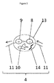
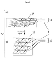
- the one or more cells capable of forming an afferent signal transmission line comprise, consist or essentially consist of afferent neuronal cells (8), afferent neuronal cells (8) and glia cells (10), axons (11) of afferent neuronal cells (8) coated with glia cells (10), and/or bundles of axons (11) of afferent neuronal cells (8) with or without glia cell (10) coating combined into nerves (12) .
- the afferent transmission lines (6) are afferent nerve fibres.
- These nerves (12) are capable of forming an afferent transmission line (6) and are capable of connecting at least one sender (3) to at least one control centre (4).
- the neuronal cells are selected from the group of differentiated cells derived from donor and/or prenatal neuronal cells differentiated in vitro.
- the growth of these nerves (12) towards and/or into a group of sender (3) cells or a sender (3) tissue is preferably stimulated by the addition of nerve growth factor into the group of sender (3) cells or sender (3) tissue located in the vicinity of the cells capable of forming an afferent transmission line (6).
- the cells or tissue forming the sender (3) are seeded at a distance towards the cells capable of forming an afferent transmission line (6) of less than 100 mm, preferably less than 30 mm.
- An alternative approach to promote growth of the cells forming the afferent transmission line (6) towards the sender (3) and to electrically connect the afferent transmission line (6) with sender (3) tissue or cells is to separately pre-incubate a control centre (4) formed according to the method of the invention and to add nerve growth factor in semi-solid medium at the later position of the sender (3) tissue. It is preferred that the sender (3) cell, cells or tissue is added once the afferent transmission line (6) has grown into the sender (3) culture space. In either embodiment it is preferred that both the growth space for the control centre and the growth space for the sender (3) is connected by a channel.
- the length of such a channel is between 500 mm and 5 mm, preferably between 300 mm and 10 mm, more preferably between 100 mm and 20 mm.
- the length of the channel is less than 30 mm.
- the cells capable of forming an efferent transmission line (7) comprise, consist or essentially consist of efferent neuronal cells (9), efferent neuronal cells (9) and glia cells (10), axons (11) of efferent neuronal cells (9) coated with glia cells (10), and/or bundles of axons (11) of efferent neuronal cells (9) with or without glia cell (10) coating combined into nerves (12).
- the signal transmission lines are efferent nerve fibres.
- the neuronal cells are selected from a group of differentiated cells derived from donor and/or prenatal neuronal cells differentiated in vitro.
- the growth of these cells towards and/or into a group of responder (5) cells or responder (5) tissue is preferably stimulated by the addition of nerve growth factor into the group of responder (5) cells or responder (5) tissue located in the vicinity of the cells capable of forming an afferent transmission line (6).
- the cells or tissue forming the responder (5) are seeded at a distance towards the cells capable of forming an efferent transmission line (7) of less than 100 mm, preferably less than 30 mm.
- Another approach to promote growth of the cells capable of forming an efferent transmission line (7) in order to connect the efferent transmission line (7) to responder (5) tissue or responder (5) cells is to separately pre-incubate a control centre (4) and add nerve growth factor in semi-solid medium at the later position of the responder (5) tissue.
- the responder (5) tissue is added once the axon (11)/nerve (12) growth has arrived in the responder (5) culture space.
- both the growth space for the control centre and the growth space for the responder (5) is connected by a channel.
- the length of such a channel is between 500 mm and 5 mm, preferably between 300 mm and 10 mm, more preferably between 100 mm and 20 mm.
- the length of the channel is less than 30 mm.
- the cells forming the control centre (4) comprise, consist or essentially consist of at least one afferent neuronal cell (8), one efferent neuronal cell (9), and at least one connective tissue (13) cell arranged in a common structure.
- the connective tissue (13) cell is a glia cell (10).
- the neuronal cells are derived from differentiated neuronal cells, derived from adult organism or prenatal cells derived from neuronal tissue of developing organisms whereas glia cells (10) preferably derived from prenatal glial tissue.
- the cells forming the control centre (4) are seeded simultaneously or subsequently into an area of a growth matrix (14), preferably a macro-porous matrix (14) providing globular and/or open cell culture space of 50 ⁇ m to 3 mm diameter, preferably from 100 ⁇ m to 250 ⁇ m.
- the preferred shape of the culture space for the control centre is spheric or star-like shape.
- Preferred materials of such matrices are selected from the group consisting of acellularised human ganglion tissue and biodegradable scaffolds.
- neuronal and connective tissue (13) cells preferably glia cells (10), within such matrix (14) structures these structures are preferably coated with connective tissue matrix or connective tissue (13) cells.
- Such an arrangement of cells, matrices and coating allows maintaining the control centre (4) in behaviour comparable to a human ganglion.
- control the cells seeded to form the control centre further comprise at least one interneuron.
- an interneuron is seeded, of two or more interconnected biological switching circuits are to be connected.
- cells have to be seeded with a density of 1 x 10 6 to 1 x 10 9 cells per cm 3 effective cell culture space, preferably 1 x 10 8 to 5 x 10 8 .
- the ratio between neuronal cells and glia cells (10) is preferably between 2:1 and 5:1, preferably 3:1. These are the preferred ratios for the establishment of a control centre.
- a cell culture period of between 2 and 10 days is necessary for the proper arrangement of the three cell types, i.e. afferent and efferent neuron(s) and connective tissue cell(s), preferably glia cell(s) within the control centre (4).
- no other somatic tissues such as sender (3) or responder (5) cells or tissues are cultivated in their corresponding cell culture cavities or alternatively that the cavities for the control centre (4), the sender (3) and/or the responder (5) are fluidically disconnected during this incubation step. Fluidic disconnection may be achieved by placing valves in the channels connecting the cell culture spaces for the control centre (4), the sender (3) and the responder (5). It is preferred that an exchange of culture media is carried out periodically. The amount and frequency is chosen in such to be sufficient to support cell viability.
- the afferent and efferent transmission lines (7) have to be formed by the outgrowth of axons (11) from the neuronal cells of the control centre (4) and the connection of their endings into the sender (3) and/or responder (5) tissue.
- control centre (4) formed resembles the structure of a human ganglion, preferably a ganglion of the enteric nervous system, more preferably a ganglion of the plexus myentericus , and/or a ganglion of the plexus submucosus.
- the enteric nervous system is one of the three divisions of the autonomic nervous system, the other being the sympathetic and parasympathetic.
- One major advantage in contrast to other divisions is that the enteric nervous system can perform many functions independently of the central nervous system ( Furness and Costa, Neuroscience, 5 (1980), 1-20 ), so it is the preferred system for the assembly of an in vitro biological switching circuit (1).
- the cells used for seeding of the control centre are derived from the enteric nervous system or differentiated into such cells.
- the responder (5) is/are one or more smooth muscle cells maintained in tissue culture in a physiological arrangement.
- the control centre (4) may be a ganglion emulating the plexus submucosus.
- the responder (5) is/are one or more gland cells of the mucosa maintained in tissue culture in a physiological arrangement.
- plexuses Both of these plexuses together have an effect on the blood flow and on the interactions with the immune system of the intestine in vivo.
- Direct communications between plexus myentericus and plexus submucosus ascertain that secretion, blood perfusion and muscle contraction during the propulsion of the faeces are carried out in a coordinated fashion.
- a biological switching unit modelling the plexus myentericus and a biological switching unit modelling the plexus submucosus are seeded in culture spaces adjacent to each other and are preferably seeded togther with one or more interneurons, which will provide an interaction between the two plexuses.
- the one or more cells involved in the method for assembly of an in vitro biological switching circuit (1) are seeded within a three-dimensional structured matrix (14) and/or on a structured surface.
- the matrix (14) and/or the structured surface comprise or consist of matrigel, fibrin gel, agarose gel, alginate gel, synthetic gel and/or crosslinkable polymers.
- the cell cavities for the elements that are assembled into according to the method of the present invention are all comprised in a self-contained device.
- a self-contained device i.e. a device which provides structured matrix (14) is depicted in figures 4 and 5 .
- the culture spaces for the control centre (4), the sender (3) and the responder (5) and optionally separate medium flow channels are micro-machined into a suitable material.
- a suitable material is translucent to allow monitoring of the progress of assembly as well as the integrity of the switching circuit (1) once assembled.
- Suitable materials comprise glass, polystyrol or polycarbonat.
- the culture spaces may be filled completely or in part with matrices supporting the growth of the cells to be seeded. If the culture spaces are filled entirely it is preferred that the matrices are biodegradable to allow the ingrowth of the cells.
- oxygen oxygenated medium may be supplied to the cells. This is preferred, if the culture system is closed towards the environment.
- the culture spaces and/or the channels for transmission line formation may be designed in such to allow gas exchange, i.e. they are either completely or partially open to the environment or they are covered by a membrane that allows gas exchange (O 2 /CO 2 ).
- the signalling of the sender (3) will be monitored as outlined below:
- Signal transmission through afferent and efferent transmission lines (7) and action potential generation in the control centre (4) preferably can be monitored by electroencephalogram, microelectrode arrays (MEA) or optical sensors (20) measuring the excitation of substances sensitive to neuronal action potentials e.g.
- monitoring (21) of the responder (5) preferably can be carried out by smooth and skeletal muscle contraction and relaxation (preferably microscopy, pressure and volume measurements), secretory tissue (quantitative measurement of secretory compounds of glands e.g. ELISA-technology) and neuronal tissue (methods as described for the control centre (4)).
- the three-dimensional structured matrix (14) and/or structured surface provides an interface for connecting the biological switching unit to at least one technical switching circuit, preferably an electric or computational circuit, i.e. a biological interface.
- an interface allows the input or read-out of electric signals into the biological switching circuit (1).
- the technical switching unit can receive and process signals received from a biological switching circuit (1) comprising a mechanonociceptor as sender (3) and output information on the tension/pressure thus sensed.
- a typical technical switching unit used in the context of the present invention comprises a bus system for communication, a data storage device, e.g. hard drive, RAM and/or ROM, and a central processing unit (CPU).
- the technical switching unit further comprises an output device, e.g. a graphic processing unit (GPU) and a display unit.
- GPU graphic processing unit
- the method for assembly of an in vitro biological switching circuit (1) comprises at least one of the steps of co-culturing extracted neuronal cells with glia cells (10) supported by a defined assembly architecture, promoting the signal transmission line growth to the sender (3) and the responder (5) cells/tissues, coupling into the sender (3) and/or responder (5), inducing an impulse of the sender (3) and/or measurement of the response at the receiver (19).
- Biosensors & Bioelectronics 15 (2000) 383-396 described that neuronal networks cultured on MEAs remain pharmacologically histiotypic and can provide reliable, quantitative data on drug interactions with neural tissue. They use the system as a test platform for toxicological studies, for drug development and a tissue-based biosensor (20).
- the present invention further provides an in vitro biological switching circuit (1) producible by the described method for assembly of an in vitro biological switching circuit (1). It is one advantage of the invention, that this in vitro network has fully human characteristics.
- the invention provides a kit comprising the biological switching unit comprising the biological switching circuit (1) and further comprising devices for monitoring (21) the activity of the control centre (4), the sender (3) and/or the responder (5).
- Such devices may comprise sensor (20) for connecting to the control centre (4), which can sense, e.g. ion fluctuations within the cells forming the control centre (4).
- the present invention provides the use of the biological switching circuit (1) and/or unit for research and drug development on neuronal diseases such as Alzheimer, Parkinson, symptoms of neurodegeneration, for safety and efficacy testing of substances, as an implant and a transplant and may enable identification of candidate therapeutic agents.
- the present invention further provides an in vitro biological switching circuit (1) and/or unit which serves as a physiological model that can be used to characterise the physiology of various organs and/or tissues such as intestine, retina, skin, lung, hair cells of the ear, secretoric glands, blood vessels, muscle, immune tissues, liver, kidney and others.
- an in vitro biological switching circuit (1) and/or unit which serves as a physiological model that can be used to characterise the physiology of various organs and/or tissues such as intestine, retina, skin, lung, hair cells of the ear, secretoric glands, blood vessels, muscle, immune tissues, liver, kidney and others.
- the present invention of the biological switching circuit (1) and/or unit can further be used as a substitute of technical systems comprising technical switching circuits.
- One possible application is the substitution of technical circuits comprising temperature sensors in temperature zones of 37°C used for exact measurement of activity of metabolic processes with a respective process control.
- the use of the biological switching circuit (1) may increase both sensitivity and the dynamic of the control of such systems.
Landscapes
- Health & Medical Sciences (AREA)
- Engineering & Computer Science (AREA)
- Biomedical Technology (AREA)
- Life Sciences & Earth Sciences (AREA)
- Chemical & Material Sciences (AREA)
- Immunology (AREA)
- Cell Biology (AREA)
- Bioinformatics & Cheminformatics (AREA)
- Biotechnology (AREA)
- Molecular Biology (AREA)
- Genetics & Genomics (AREA)
- Organic Chemistry (AREA)
- Hematology (AREA)
- Microbiology (AREA)
- Zoology (AREA)
- Urology & Nephrology (AREA)
- Wood Science & Technology (AREA)
- General Health & Medical Sciences (AREA)
- Biochemistry (AREA)
- Physics & Mathematics (AREA)
- Analytical Chemistry (AREA)
- Medicinal Chemistry (AREA)
- Food Science & Technology (AREA)
- General Physics & Mathematics (AREA)
- Pathology (AREA)
- Tropical Medicine & Parasitology (AREA)
- Neurology (AREA)
- Neurosurgery (AREA)
- General Engineering & Computer Science (AREA)
- Toxicology (AREA)
- Apparatus Associated With Microorganisms And Enzymes (AREA)
Abstract
The present invention relates to a method of assembling a biological switching circuit (1). In a preferred embodiment the invention comprises assembly of a tissue capable of producing a signal, an afferent signal transmitter, neurons which can act as a control centre (4), an efferent signal transmitter and a tissue which is able to respond to the neuronal signal. The invention also relates to a biological switching circuit (1) producible by the method of the invention and its use.
Description
- The present invention relates to a method of assembling a biological switching circuit (1). In a preferred embodiment the invention comprises assembly of a tissue capable of producing a signal, an afferent signal transmitter, neurons which can act as a control centre (4), an efferent signal transmitter and a tissue which is able to respond to the neuronal signal. The invention also relates to a biological switching circuit (1) producible by the method of the invention and its use.
- The organ regulation in the human organism takes place via complex neuronal switching circuits. The easiest configuration is composed of a signal-sending organ, a conglomerate of receiving and assimilating neurons and a response back into the organ or into another organ. During evolution nature developed for this easy circuit and all overlying higher complex neuronal circuits' highly efficient biological building blocks in vivo represented by ganglia of the enteric, symphatic and parasympathic nervous systems and the neuronal structures of spinal cord and brain. For almost hundred years mankind has tried to emulate neuronal interactions by tissue culture of networks of neurons (Harrison RG. 1907. Experiments in transplanting limbs and their bearings on the problems of the development of nerves. J Exp Zool 4:239-281). The state of the art of in vitro reconstitution/characterization of the nervous system is characterized by three different types of tissue culture methods: a) culturing of brain slices, b) culturing of monolayer culture of neurons with and without glia cells and c) three-dimensional neuronal tissue culture, which in the following will be discussed in more detail. Organotypic brain-slice cultures have been used as models to investigate mechanisms and treatment strategies for neuronal disorders (Gähwiler, Trends in Neuroscience (1997) 20,471-477 and Stoppini et al., Journal of Neuroscience Methods, 37 (1991) 173-182). At the beginning of this century it has been described that slice cultures display more in vivo-like mechanisms of excitotoxicity than reported for primary neuronal cultures (Kristensen et al., Brain Research 917 (2001) 21-44).
- This type of culture seems particularly well suited for developmental studies of synaptic connectivity and plasticity of neurons. However, tissue slicing leads to an irreversible cut of individual axons and corresponding nerve fibres. These can not be regenerated in vitro and, therefore, this technology is limited to the study of interaction between individual intact neurons in the remaining healthy tissue.
- In contrast the approach of co-culturing neurons and glia cells in monolayers as described by Jane Faria et al. (Differentiation (2006) 74:562-572) aims to build neuronal interactions de novo in vitro. This technology has led to two-dimensional networks of neurons in tissue culture plates supported by glial connective tissue. These networks, however, only provide data on individual neuronal interactions, which can neither react to a physiological stimulus nor act on a physiological response tissue.
- To overcome the limitations of the non-physiological two-dimensional network in monolayer-cultures significant efforts have been devoted over the last decade to provide neurons with an environment and/or scaffold allowing them to arrange in a three-dimensional space. Progress in that respect has been made on tissue formation as described by Preynat-Seauve et al. (Stem Cells 2009; 27:509-520). Preynat-Seauve et al. use air-liquid interface culture to engineer three-dimensional human nervous tissues from human embryonic stem cells. An important feature of this technique is the improved exchange between air and tissue. This allows the development of a relatively thick three-dimensional culture and yields dense interconnected neural tissue with structurally and functionally distinct areas. But even this three-dimensional arrangement of neurons in an in vitro developed organotypic tissue culture represents only a poor model of the neuronal control centre in man - the ganglion.
- None of these technologies has been able to emulate a neuronal switching circuit consisting of a somatic tissue sending a signal through nerve fibres into a neuronal control centre and a somatic tissue responding to a corresponding signal of the neuronal control centre.
- Surprisingly a biological switching circuit (1) consisting of a biological sender (3), an afferent transmission line (6), a control centre (4), an efferent transmission line (7) and a responder tissue can be established outside the organism according to the method of this invention. A biological switching unit comprising this biological switching circuit (1) can be used to study neuronal function and also provides a test system for performing any kind of safety and efficacy substance evaluation including lead selection for new pharmaceuticals. Furthermore the biological switching circuit (1) as a whole or parts thereof can be implanted in organisms, preferably immunological compatible organisms, for complementation, restoration and/or regeneration of neuronal functions. Moreover fully functional transplants can be produced on the basis of one or more such biological switching units. The biological switching unit furthermore can be used to complement and/or to substitute technical systems comprising technical switching circuits.
- In a first aspect the present invention relates to a method for assembly of an in vitro biological switching circuit (1), comprising the step of seeding a control centre (4), comprising one or more cells capable of forming an afferent transmission line (6), one or more cells capable of forming an efferent transmission line (7) and one or more connective tissue cells (13), wherein the afferent transmission line (6) formed is capable of connecting at least one sender (3) to the control centre (4) in a way allowing transmission of a signal to the control centre (4) and the efferent transmission line (7) formed is capable of connecting at least one responder (5) to the control centre (4) in a way allowing transmission of a signal to said at least one responder (5).
- In a second aspect the present invention relates to an in vitro biological switching circuit (1) producible by the described method.
- In a third aspect the present invention relates to a kit comprising the biological switching unit comprising the biological switching circuit (1) further comprising devices for monitoring (21) the activity of the control centre (4), the sender (3) and/or the responder (5).
- In a fourth aspect the present invention relates to the use of the biological switching circuit for research and drug development on neuronal diseases, for safety and efficacy testing of substances, as an implant, as a transplant and as a substitute of technical systems comprising technical switching circuits.
- Before the present invention is described in detail below, it is to be understood that this invention is not limited to the particular methodology, protocols and reagents described herein as these may vary. It is also to be understood that the terminology used herein is for the purpose of describing particular embodiments only, and is not intended to limit the scope of the present invention which will be limited only by the appended claims. Unless defined otherwise, all technical and scientific terms used herein have the same meanings as commonly understood by one of ordinary skill in the art.
- Preferably, the terms used herein are defined as described in "A multilingual glossary of biotechnological terms: (IUPAC Recommendations)", Leuenberger, H.G.W, Nagel, B. and Klbl, H. eds. (1995), Helvetica Chimica Acta, CH-4010 Basel, Switzerland) and as described in "Pharmaceutical Substances: Syntheses, Patents, Applications" by Axel Kleemann and Jurgen Engel, Thieme Medical Publishing, 1999; the "Merck Index: An Encyclopedia of Chemicals, Drugs, and Biologicals", edited by Susan Budavari et al., CRC Press, 1996, and the United States Pharmacopeia-25/National Formulary-20, published by the United States Pharmcopeial Convention, Inc., Rockville Md., 2001 and Jörg Knäblein (Modem Biopharmaceuticals - Design, Development and Optimization, 1. Edition, August 2005 Wiley-VCH, Weinheim).
- Throughout this specification and the claims which follow, unless the context requires otherwise, the word "comprise", and variations such as "comprises" and "comprising", will be understood to imply the inclusion of a stated feature, integer or step or group of features, integers or steps but not the exclusion of any other feature, integer or step or group of integers or steps. In the following passages different aspects of the invention are defined in more detail. Each aspect so defined may be combined with any other aspect or aspects unless clearly indicated to the contrary. In particular, any feature indicated as being preferred or advantageous may be combined with any other feature or features indicated as being preferred or advantageous.
- Several documents are cited throughout the text of this specification. Each of the documents cited herein (including all patents, patent applications, scientific publications, manufacturer's specifications, instructions, etc.), whether supra or infra, are hereby incorporated by reference in their entirety. Nothing herein is to be construed as an admission that the invention is not entitled to antedate such disclosure by virtue of prior invention.
- In the following, some definitions of terms frequently used in this specification are provided. These terms will, in each instance of its use, in the remainder of the specification have the respectively defined meaning and preferred meanings:
- "Afferent'' means carrying toward.
- "efferent neurons" which are also referred to as sensory or receptor neurons are neuronal cells that carry a nerve impulse, preferably from various sensory cells, towards the central nervous system or the enteric nervous system. Preferably afferent neurons connect to efferent neurons, for input transmission to a responder tissue or they connect to interneurons, which are also referred to as relay neurons or association neurons. In this case the interneuron is able to mediate nerve impulses of a second switching circuit into the described biological switching circuit.
- "Afferent transmission line" comprises one or more afferent neurons and optionally one or more glia cells, which preferably coat or surround the one or more efferent neurons.
- "Axon" is the single outgrowth of a neuronal cell, which is preferably ending within a non-neuronal tissue to receive or send a signal.
- "Cells" means cell lines or primary cells of vertebrates or invertebrates.
- "Control and/or distribution centre" means cluster of neurons and incoming and leaving nerve fibres.
- "Common structure" means cells, preferably enclosed in a membrane, with stabilizing, connective tissue cells and with the ability to be in contact sender and/or responder via cell appendices, e.g. axons.
- "Efferent" means carrying away.
- "Efferent neurons", which are also referred to as motor or effector neurons are neuronal cells that carry a nerve impulse away from the central nervous system or the enteric nervous system. Preferably efferent neurons connect to an afferent neuron to receive an impulse or to interneurons, which are also referred to as relay neurons or association neurons. In this case the interneuron is able to mediate nerve impulses of a second switching circuit into the described biological switching circuit.
- "Efferent transmission line" comprises one or more efferent neurons and optionally one or more glia cells, which preferably coat or surround the one or more efferent neurons.
- "Matrix" means substances or mixtures of substances, which provide a spatial architecture, maintain viability and tissue specificity, enhance proliferation, differentiation, and function of cells and/or organoid or organ formation such as collagen, fibrinogen, basal lamina, connective tissues and/or synthetic polymers.
- "Medium" (plural form: "media") means growth supporting or viability maintaining liquid with nutrients and substances for cultivation of cells. Examples of suitable media comprise DMEM, Ham's F12, RPMI and plasma of different origin.
- "Nerve" is an axon and/or a number of axons coated or non-coated with glial cells.
- "Nerve impulse" also referred to as "action potential" is a short-lasting event in which the electrical membrane potential of a nerve cell rapidly rises and falls.
- "Organoids" means artificial, de novo generated, functional cell aggregates of different types of cells in vitro that show at least one organ or tissue function, preferably shows the majority of organ or tissue functions.
- "Responder" means cells and tissues, which are able to respond to a nerve impulse received from efferent neurons, preferably by contraction/relaxation or secretion.
- "Sender" means cells and/or tissues, which are able to stimulate a nerve impulse in afferent neurons, preferably in response to tension, pressure, changes in volume, shear stress, nutrients concentration or change thereof, osmolarity, pH, temperature, CO2 concentration or change thereof, O2 concentration or change thereof, biochemical changes, light, odor, acoustic noise, taste, colour, and wavelength.
- "Substance" means a matter of definite composition and properties.
- "Technical switching circuit" means any non-biological switching circuit (1) containing or comprising one or more of the following: a technical sensor (20), a biological circuit interface for providing and/or receiving electrical action potential from neurons, a signal input, a signal processing unit also termed central processing unit (CPU), a signal output and an actor, such as electrical circuits, computational circuits, and hybrid switching units combining for example physical and chemical signals.
- "Tissues" means biopsy material or explants taken from patients or animals or in vitro generated tissues.
- "Transmitter" means a carrier of nerve impulses across a synapse to a postsynaptic element, such as smooth and/or skeletal muscle, glands or neuronal tissue.
- To overcome limitations associated with prior art methods, the present invention provides a method for assembly of an in vitro biological switching circuit (1), comprising the step of seeding a control centre (4), comprising one or more cells capable of forming an afferent transmission line (6), one or more cells capable of forming an efferent transmission line (7) and one or more connective tissue cells (13), wherein the afferent transmission line (6) formed is capable of connecting at least one sender (3) to the control centre (4) in a way allowing transmission of a signal to the control centre (4) and the efferent transmission line (7) formed is capable of connecting at least one responder (5) to the control centre (4) in a way allowing transmission of a signal to said at least one responder (5).
- The present invention preferably also comprises the step of providing at least one sender (3) and/or at least one responder (5). The sender (3) is preferably a cell, a group of cells, or a tissue, which is generating a nerve impulse if it stimulated by one or more stimuli of the typical five senses - sight (ophthalmoception), hearing (audioception), taste (gustaoception), smell (olfacoception or olfacception), and touch (tactioception) or any of the additional senses such as temperature (thermoception), kinesthetic sense (proprioception), pain (nociception), balance (equilibrioception) and acceleration (kinesthesioception) connected to the afferent transmission line (6). The respective cell, group of cells or tissues generating nerve impulses in response to a stimulus are preferably tissue explants and/or extracted single cell suspensions of the respective cell types maintain in tissue culture. Table I below provides examples of suitable cell types and the respective sense and stimulus.
Table I Sense Stimulus Cell type Architecture sight light cells of the retina, such as retinal pigment epithelial cells, photoreceptor cells, bipolar cells composed in the ten distinct layers of vertebrate retina hearing acoustic noise hair cells cochlea shape taste biochemical changes, nutrients, osmolarity, pH cells of the taste bud, such as basal cells, gustatory receptor cells including gustatoring hair, supporting cells taste bud smell odor smell nerve cells tiny patch of tissue touch pressure, vibration, changes in volume, shear stress, temperature, Mechanoreceptors or mechanonociceptors Golgi tendon organs and neuromuscular spindles in skeletal muscles, Meissner, Pacinian, Merkel and Ruffini corpuscles in the skin - The responder (5) is a cell, a group of cells or a tissue which is capable of responding to the neuronal signal of the control centre (4), preferably selected from the group consisting of smooth and/or skeletal muscle cells, adenocytes, mucosa cells, neuronal cells, a vascular system, nervous system and/or a lymphatic system.
- In a preferred embodiment of the method for assembly of an in vitro biological switching circuit (1), the one or more cells capable of forming an afferent signal transmission line comprise, consist or essentially consist of afferent neuronal cells (8), afferent neuronal cells (8) and glia cells (10), axons (11) of afferent neuronal cells (8) coated with glia cells (10), and/or bundles of axons (11) of afferent neuronal cells (8) with or without glia cell (10) coating combined into nerves (12) . Preferably, the afferent transmission lines (6) are afferent nerve fibres. These nerves (12) are capable of forming an afferent transmission line (6) and are capable of connecting at least one sender (3) to at least one control centre (4). To promote the growth of the afferent signal transmission line it is preferred that the neuronal cells are selected from the group of differentiated cells derived from donor and/or prenatal neuronal cells differentiated in vitro.
- The growth of these nerves (12) towards and/or into a group of sender (3) cells or a sender (3) tissue is preferably stimulated by the addition of nerve growth factor into the group of sender (3) cells or sender (3) tissue located in the vicinity of the cells capable of forming an afferent transmission line (6). Preferably the cells or tissue forming the sender (3) are seeded at a distance towards the cells capable of forming an afferent transmission line (6) of less than 100 mm, preferably less than 30 mm. An alternative approach to promote growth of the cells forming the afferent transmission line (6) towards the sender (3) and to electrically connect the afferent transmission line (6) with sender (3) tissue or cells is to separately pre-incubate a control centre (4) formed according to the method of the invention and to add nerve growth factor in semi-solid medium at the later position of the sender (3) tissue. It is preferred that the sender (3) cell, cells or tissue is added once the afferent transmission line (6) has grown into the sender (3) culture space. In either embodiment it is preferred that both the growth space for the control centre and the growth space for the sender (3) is connected by a channel. Preferably the length of such a channel is between 500 mm and 5 mm, preferably between 300 mm and 10 mm, more preferably between 100 mm and 20 mm. To be able to place all elements of the switching circuit generated according to the method of the present invention on a small device, it is preferred that the length of the channel is less than 30 mm.
- In a preferred embodiment of the method for assembly of an in vitro biological switching circuit (1), the cells capable of forming an efferent transmission line (7) comprise, consist or essentially consist of efferent neuronal cells (9), efferent neuronal cells (9) and glia cells (10), axons (11) of efferent neuronal cells (9) coated with glia cells (10), and/or bundles of axons (11) of efferent neuronal cells (9) with or without glia cell (10) coating combined into nerves (12). Preferably, the signal transmission lines are efferent nerve fibres. To promote the growth of the efferent signal transmission line it is preferred that the neuronal cells are selected from a group of differentiated cells derived from donor and/or prenatal neuronal cells differentiated in vitro. The growth of these cells towards and/or into a group of responder (5) cells or responder (5) tissue is preferably stimulated by the addition of nerve growth factor into the group of responder (5) cells or responder (5) tissue located in the vicinity of the cells capable of forming an afferent transmission line (6). Preferably the cells or tissue forming the responder (5) are seeded at a distance towards the cells capable of forming an efferent transmission line (7) of less than 100 mm, preferably less than 30 mm. Another approach to promote growth of the cells capable of forming an efferent transmission line (7) in order to connect the efferent transmission line (7) to responder (5) tissue or responder (5) cells is to separately pre-incubate a control centre (4) and add nerve growth factor in semi-solid medium at the later position of the responder (5) tissue. In this case the responder (5) tissue is added once the axon (11)/nerve (12) growth has arrived in the responder (5) culture space. In either embodiment it is preferred that both the growth space for the control centre and the growth space for the responder (5) is connected by a channel. Preferably the length of such a channel is between 500 mm and 5 mm, preferably between 300 mm and 10 mm, more preferably between 100 mm and 20 mm. To be able to place all elements of the switching circuit generated according to the method of the present invention on a small device, it is preferred that the length of the channel is less than 30 mm.
- In a preferred embodiment of the method for assembly of an in vitro biological switching circuit (1), the cells forming the control centre (4) comprise, consist or essentially consist of at least one afferent neuronal cell (8), one efferent neuronal cell (9), and at least one connective tissue (13) cell arranged in a common structure. Preferably, the connective tissue (13) cell is a glia cell (10). Preferably the neuronal cells are derived from differentiated neuronal cells, derived from adult organism or prenatal cells derived from neuronal tissue of developing organisms whereas glia cells (10) preferably derived from prenatal glial tissue.
- In a preferred embodiment the cells forming the control centre (4) are seeded simultaneously or subsequently into an area of a growth matrix (14), preferably a macro-porous matrix (14) providing globular and/or open cell culture space of 50 µm to 3 mm diameter, preferably from 100 µm to 250 µm. The preferred shape of the culture space for the control centre is spheric or star-like shape. Preferred materials of such matrices are selected from the group consisting of acellularised human ganglion tissue and biodegradable scaffolds. To support and direct the arrangement of neuronal and connective tissue (13) cells, preferably glia cells (10), within such matrix (14) structures these structures are preferably coated with connective tissue matrix or connective tissue (13) cells. Such an arrangement of cells, matrices and coating allows maintaining the control centre (4) in behaviour comparable to a human ganglion.
- In a further preferred embodiment of the method of the present invention the control the cells seeded to form the control centre further comprise at least one interneuron. Preferably, an interneuron is seeded, of two or more interconnected biological switching circuits are to be connected.
- To ensure proper arrangement of cells into three-dimensional neuronal networks cells have to be seeded with a density of 1 x 10⁶ to 1 x 10⁹ cells per cm3 effective cell culture space, preferably 1 x 10⁸ to 5 x 10⁸. The ratio between neuronal cells and glia cells (10) is preferably between 2:1 and 5:1, preferably 3:1. These are the preferred ratios for the establishment of a control centre. A cell culture period of between 2 and 10 days is necessary for the proper arrangement of the three cell types, i.e. afferent and efferent neuron(s) and connective tissue cell(s), preferably glia cell(s) within the control centre (4). During the establishment of the control centre (4) it is preferred that no other somatic tissues such as sender (3) or responder (5) cells or tissues are cultivated in their corresponding cell culture cavities or alternatively that the cavities for the control centre (4), the sender (3) and/or the responder (5) are fluidically disconnected during this incubation step. Fluidic disconnection may be achieved by placing valves in the channels connecting the cell culture spaces for the control centre (4), the sender (3) and the responder (5). It is preferred that an exchange of culture media is carried out periodically. The amount and frequency is chosen in such to be sufficient to support cell viability.
- As set out above, it is preferred that once the cell arrangement in the control centre (4) culture cavity is completed that then the afferent and efferent transmission lines (7) have to be formed by the outgrowth of axons (11) from the neuronal cells of the control centre (4) and the connection of their endings into the sender (3) and/or responder (5) tissue.
- In a preferred embodiment the control centre (4) formed resembles the structure of a human ganglion, preferably a ganglion of the enteric nervous system, more preferably a ganglion of the plexus myentericus, and/or a ganglion of the plexus submucosus. The enteric nervous system is one of the three divisions of the autonomic nervous system, the other being the sympathetic and parasympathetic. One major advantage in contrast to other divisions is that the enteric nervous system can perform many functions independently of the central nervous system (Furness and Costa, Neuroscience, 5 (1980), 1-20), so it is the preferred system for the assembly of an in vitro biological switching circuit (1). Accordingly, in a preferred embodiment the cells used for seeding of the control centre are derived from the enteric nervous system or differentiated into such cells.
- In the preferred established ganglion emulating the plexus myentericus the responder (5) is/are one or more smooth muscle cells maintained in tissue culture in a physiological arrangement. In another embodiment the control centre (4) may be a ganglion emulating the plexus submucosus. In this case the responder (5) is/are one or more gland cells of the mucosa maintained in tissue culture in a physiological arrangement.
- Both of these plexuses together have an effect on the blood flow and on the interactions with the immune system of the intestine in vivo. Direct communications between plexus myentericus and plexus submucosus ascertain that secretion, blood perfusion and muscle contraction during the propulsion of the faeces are carried out in a coordinated fashion. In a preferred embodiment a biological switching unit modelling the plexus myentericus and a biological switching unit modelling the plexus submucosus are seeded in culture spaces adjacent to each other and are preferably seeded togther with one or more interneurons, which will provide an interaction between the two plexuses.
- In a preferred embodiment the one or more cells involved in the method for assembly of an in vitro biological switching circuit (1) are seeded within a three-dimensional structured matrix (14) and/or on a structured surface. Preferably the matrix (14) and/or the structured surface comprise or consist of matrigel, fibrin gel, agarose gel, alginate gel, synthetic gel and/or crosslinkable polymers.
- Preferably, the cell cavities for the elements that are assembled into according to the method of the present invention are all comprised in a self-contained device. An example of such a device, i.e. a device which provides structured matrix (14) is depicted in
figures 4 and5 . In a preferred embodiment the culture spaces for the control centre (4), the sender (3) and the responder (5) and optionally separate medium flow channels are micro-machined into a suitable material. Preferably, such material is translucent to allow monitoring of the progress of assembly as well as the integrity of the switching circuit (1) once assembled. Suitable materials comprise glass, polystyrol or polycarbonat. As set out above the culture spaces may be filled completely or in part with matrices supporting the growth of the cells to be seeded. If the culture spaces are filled entirely it is preferred that the matrices are biodegradable to allow the ingrowth of the cells. - To provide the cells with oxygen oxygenated medium may be supplied to the cells. This is preferred, if the culture system is closed towards the environment. Alternatively, the culture spaces and/or the channels for transmission line formation may be designed in such to allow gas exchange, i.e. they are either completely or partially open to the environment or they are covered by a membrane that allows gas exchange (O2/CO2).
- To monitor functionality of such a biological switching circuit (1) methods to measure the signalling of the sender (3), the signal transmission to the control centre (4), the amplification of the signal in the control centre (4) e.g. the neuronal action potential, the transmission of the signal to the responder (5) and the response of the tissue. Preferably the signalling of the sender (3) will be monitored as outlined below:
- tension- microscopic observation
- pressure - pressure sensor
- changes in volume -volume measurement
- nutrients - glucose/lactate/amino acid measurements
- osmolarity - osmolarimeter
- pH - pH-sensor
- temperature - temperature sensor, infrared sensor,
- CO2 O2 - optical measurements,
- light -photo/light
- odor - measurement of single odor molecules by mass spectrometry
- acoustic noise - acoustic sensor
- colour, wavelength - wavelength/optical sensors
- Signal transmission through afferent and efferent transmission lines (7) and action potential generation in the control centre (4), preferably can be monitored by electroencephalogram, microelectrode arrays (MEA) or optical sensors (20) measuring the excitation of substances sensitive to neuronal action potentials e.g.
- Finally the monitoring (21) of the responder (5) preferably can be carried out by smooth and skeletal muscle contraction and relaxation (preferably microscopy, pressure and volume measurements), secretory tissue (quantitative measurement of secretory compounds of glands e.g. ELISA-technology) and neuronal tissue (methods as described for the control centre (4)).
- Electrical methods for measuring system parameters are important in diagnosing the status of biological organisms, including humans (Gusev, Biomedical Measurements 8 (1993) 60-62). They assess the electrical resistance of appropriate tissue regions. The value of various parameters and their variation with time yield information's on the state of for instance a special organism, specific organs and systems.
- Preferably, the three-dimensional structured matrix (14) and/or structured surface provides an interface for connecting the biological switching unit to at least one technical switching circuit, preferably an electric or computational circuit, i.e. a biological interface. Such an interface allows the input or read-out of electric signals into the biological switching circuit (1). For example, the technical switching unit can receive and process signals received from a biological switching circuit (1) comprising a mechanonociceptor as sender (3) and output information on the tension/pressure thus sensed. A typical technical switching unit used in the context of the present invention comprises a bus system for communication, a data storage device, e.g. hard drive, RAM and/or ROM, and a central processing unit (CPU). Preferably the technical switching unit further comprises an output device, e.g. a graphic processing unit (GPU) and a display unit.
- In a preferred embodiment the method for assembly of an in vitro biological switching circuit (1) comprises at least one of the steps of co-culturing extracted neuronal cells with glia cells (10) supported by a defined assembly architecture, promoting the signal transmission line growth to the sender (3) and the responder (5) cells/tissues, coupling into the sender (3) and/or responder (5), inducing an impulse of the sender (3) and/or measurement of the response at the receiver (19). To monitor the status of the system it is preferred to cultivate the in vitro biological switching circuit (1) on microelectrode arrays (MEA). Morefield et al. (Biosensors & Bioelectronics 15 (2000) 383-396) described that neuronal networks cultured on MEAs remain pharmacologically histiotypic and can provide reliable, quantitative data on drug interactions with neural tissue. They use the system as a test platform for toxicological studies, for drug development and a tissue-based biosensor (20).
- The present invention further provides an in vitro biological switching circuit (1) producible by the described method for assembly of an in vitro biological switching circuit (1). It is one advantage of the invention, that this in vitro network has fully human characteristics.
- Furthermore, the invention provides a kit comprising the biological switching unit comprising the biological switching circuit (1) and further comprising devices for monitoring (21) the activity of the control centre (4), the sender (3) and/or the responder (5). Such devices may comprise sensor (20) for connecting to the control centre (4), which can sense, e.g. ion fluctuations within the cells forming the control centre (4).
- To overcome the limitations associated with prior art, the present invention provides the use of the biological switching circuit (1) and/or unit for research and drug development on neuronal diseases such as Alzheimer, Parkinson, symptoms of neurodegeneration, for safety and efficacy testing of substances, as an implant and a transplant and may enable identification of candidate therapeutic agents.
- The present invention further provides an in vitro biological switching circuit (1) and/or unit which serves as a physiological model that can be used to characterise the physiology of various organs and/or tissues such as intestine, retina, skin, lung, hair cells of the ear, secretoric glands, blood vessels, muscle, immune tissues, liver, kidney and others.
- Alternatively, the present invention of the biological switching circuit (1) and/or unit can further be used as a substitute of technical systems comprising technical switching circuits. One possible application is the substitution of technical circuits comprising temperature sensors in temperature zones of 37°C used for exact measurement of activity of metabolic processes with a respective process control. The use of the biological switching circuit (1) may increase both sensitivity and the dynamic of the control of such systems.
- It is to be understood that the above described embodiments are intended to illustrate rather than limit or restrict the scope of the disclosure. Variations may be made to the embodiments without departing from the spirit of the disclosure as claimed.
-
- Fig. 1:
- Depicts a schematic drawing of a preferred configuration of a sender (3) tissue. a) Axons of the afferent transmission lines (6) are ending in between of smooth muscle layers of human gut (22). b) The mechanical extension of this in vitro smooth muscle layers leads to nerve impulses (generation of action potentials), signalling through the nerve (12) into the respective afferent neuronal cell bodies located in the control centre.
- Fig. 2:
- Depicts the control centre (4), which is formed by co-culture of at least one afferent neuron (8), one efferent neuron (9) and one connective tissue cell (glia cell) (10), architecturally arranged in a three-dimensional spheroid matrix (14) housing the cells and allowing exit of growing axons (11). This ganglion-like control centre (4) is preferably coated with matrix proteins or a connective tissue cell layer (14).
- Fig. 3:
- Depicts a schematic drawing of a preferred configuration of a responder (5) tissue. a) Axons of the efferent transmission lines (7) are connected through synapses allowing release of transmitters to smooth muscle cells of the gut (24). b) The release of such transmitters in response to a nerve impulse from the control centre (4) leads to smooth muscle contraction (25).
- Fig. 4:
- Panel A and B depict a top-down view of a preferred embodiment of a biological switching circuit (1), comprising a channel system for media flow (2), an inflow-port (17), a pump (15), three valves (16), a waste collector (18), cultures space for the control centre (4), culture spaces for the sender (3) and the responder (5), wherein the culture space of the sender (3) and the responder (5) are connected or the same. The outgrowth of afferent (6) and efferent (7) transmission lines is supported by a channel circuit combining the control centre (4) with a combined sender and responder culture space (3, 5). Panel A depicts a biological switching circuit which comprises a separate channel system for the afferent and efferent signal transmission lines and for the media flow circulation whilst panel B depicts a biological switching circuit wherein the afferent and efferent transmission lines are combined with the media flow circulation. Both systems support the establishment of a biological switching circuit for modelling gut contraction in response to extension. The arrangement of the pump and valves support continuous circulation of culture medium with an option to intermittently or permanently refresh nutrients and remove waste.
- Fig. 5:
- Panel A depicts a top-down view of a preferred embodiment of a biological switching circuit (1), comprising a channel system for media flow (2), an inflow-port (17), a pump (15), three valves (16), two waste collectors (18), cultures space for the control centre (4), culture spaces for the sender (3) and the responder (5). The outgrowth of afferent (6) and efferent (7) transmission lines is supported by channels connecting the control centre (4) with the individual culture spaces of a sender (3) and responder (5). Panel A depicts a biological switching circuit comprising a separate channel system for the transmission lines and for the media flow circulation whilst panel B depicts a system where the transmission lines are combined with the media flow channels. Both systems support the establishment of a biological switching circuit for modelling the cognition of impulses to our sense systems on sender (3) site for example by light or heat sensing and modelling the reaction to these impulses on the responder (5) site by contraction / relaxation of muscle cells, production of secrete by gland cells.
- Fig. 6:
- Depicts a three-dimensional view of the biological switching circuit (1) depicted in
figure 5a comprising optical sensors (20), controlling the tissue culture spaces for the sender (3) and the control centre (4) and the responder (5). A receiver (19) is completing this monitoring device (21). -
- (1)
- biological switching circuit
- (2)
- media flow
- (3)
- sender
- (4)
- control centre
- (5)
- responder
- (6)
- afferent transmission line
- (7)
- efferent transmission line
- (8)
- afferent neuronal cell
- (9)
- efferent neuronal cell
- (10)
- glia cell
- (11)
- axon
- (12)
- nerve
- (13)
- coating / connective tissue
- (14)
- matrix
- (15)
- pumping device
- (16)
- valve
- (17)
- fluid reservoir / inflow-port
- (18)
- waste collector
- (19)
- receiver
- (20)
- sensors
- (21)
- device for monitoring
- (22)
- sender tissue before stimulation
- (23)
- sender tissue after stimulation
- (24)
- responder tissue before signal
- (25)
- responder tissue after signal
Claims (15)
- A method for assembly of an in vitro biological switching circuit (1), comprising the step of seeding a control centre (4), comprising one or more cells capable of forming an afferent transmission line (6), one or more cells capable of forming an efferent transmission line (7) and one or more connective tissue (13) cells, wherein the afferent transmission line (6) formed is capable of connecting at least one sender (3) to the control centre (4) in a way allowing transmission of a signal to the control centre (4) and the efferent transmission line (7) formed is capable of connecting at least one responder (5) to the control centre (4) in a way allowing transmission of a signal to said at least one responder (5).
- The method of claim 1, further comprising the step of providing at least one sender (3) and/or at least one responder (5).
- The method of claim 1 or 2, wherein said sender (3) is a cell, a group of cells, or a tissue which is capable of producing a signal in response to a stimulus preferably selected from the group consisting of tension, pressure, changes in volume, shear stress, nutrients, osmolarity, pH, temperature, CO2, O2, biochemical changes, light, odor, acoustic noise, taste, colour, and wavelength.
- The method of any of claims 1 to 3, wherein the one or more cells capable of forming an afferent signal transmission line is/are selected from the group ofa. afferent neuronal cells (8),b. afferent neuronal cells (8) and glia cells (10)c. axons (11) of afferent neuronal cells (8) coated with glia cells (10), and/ord. bundles of axons (11) of afferent neuronal cells (8) with or without glia cell (10) coating combined into nerves (12).
- The method of any of claims 1 or 4, wherein the at least one efferent signal transmission line is/area. efferent neuronal cells (9),b. efferent neuronal cells (9) and glia cells (10).c. axons (11) of efferent neuronal cells (9) coated with glia cells (10), and/ord. bundles of axons (11) of efferent neuronal cells (9) with or without glia cell (10) coating combined into nerves (12).
- The method of any of claims 1 to 5, wherein the signal transmission lines are nerve (12) fibres.
- The method of any of claims 1 to 6, wherein the cells forming the control centre (4) comprise or consist of at least one afferent neuronal cell (8), one efferent neuronal cell (9), and at least one connective tissue (13) cell, preferably at least one glia cell (10), arranged in a common structure.
- The method of any of claims 1 to 7, wherein said responder (5) is a cell, a group of cells or a tissue which is capable of responding to the neuronal signal of the control centre (4), preferably selected from the group consisting of muscle cells, adenocytes, mucosa cells, neuronal cells, a vascular system, nervous system and/or a lymphatic system.
- The method of any of claims 1 to 8, wherein the one or more cells capable of forming an afferent transmission line (6), one or more cells capable of forming an efferent transmission line (7) and one or more connective tissue (13) cells and optionally the cell, group of cells or tissues forming the sender and/or responder are seeded within a three-dimensional structured matrix (14) and/or on a structured surface.
- The method of claim 9, wherein the three-dimensional structured matrix (14) and/or structured surface provides an interface for connecting the biological switching unit to at least one technical switching circuit, preferably an electric circuit.
- The method of any of claims 1 to 10, comprising at least one of the steps of:a. co-culturing extracted neuronal cells with glia cells (10) supported by a defined assembly architecture,b. promoting the signal transmission line growth to the sender (3) and the responder (5) cells/tissues,c. coupling into the sender (3) and/or responder (5),d. inducing an impulse of the sender (3) and/ore. measurement of the response at the receiver (19).
- An in vitro biological switching circuit (1) producible by the method of steps 1 to 11.
- A kit comprising the biological switching unit comprising the biological switching circuit (1) according to claim 12 and further comprising devices for monitoring (21) the activity of the control centre (4), the sender (3) and/or the responder (5).
- Use of the biological switching circuit (1) according to claim 12 or the biological switching unit according to claim 13:a. for research and drug development on neuronal diseases,b. for safety and efficacy testing of substances,c. as an implant andd. as a transplant.
- Use of the biological switching circuit (1) according to claim 12 or the biological switching unit according to claim 13 as a substitute of technical systems comprising technical switching circuits.
Priority Applications (1)
| Application Number | Priority Date | Filing Date | Title |
|---|---|---|---|
| EP11002544A EP2505638A1 (en) | 2011-03-28 | 2011-03-28 | Neuronal switching circuit |
Applications Claiming Priority (1)
| Application Number | Priority Date | Filing Date | Title |
|---|---|---|---|
| EP11002544A EP2505638A1 (en) | 2011-03-28 | 2011-03-28 | Neuronal switching circuit |
Publications (1)
| Publication Number | Publication Date |
|---|---|
| EP2505638A1 true EP2505638A1 (en) | 2012-10-03 |
Family
ID=44063290
Family Applications (1)
| Application Number | Title | Priority Date | Filing Date |
|---|---|---|---|
| EP11002544A Withdrawn EP2505638A1 (en) | 2011-03-28 | 2011-03-28 | Neuronal switching circuit |
Country Status (1)
| Country | Link |
|---|---|
| EP (1) | EP2505638A1 (en) |
Cited By (3)
| Publication number | Priority date | Publication date | Assignee | Title |
|---|---|---|---|---|
| DE102013011768A1 (en) | 2013-07-10 | 2015-01-15 | Fraunhofer-Gesellschaft zur Förderung der angewandten Forschung e.V. | Circulation system and method for the vital supply of cell cultures in a microfluidic network |
| WO2017117547A1 (en) * | 2015-12-31 | 2017-07-06 | President And Fellows Of Harvard College | Methods for generating neural tissue and uses thereof |
| WO2018013578A1 (en) * | 2016-07-11 | 2018-01-18 | Trustees Of Tufts College | Innervated artificial intestine compositions |
Citations (1)
| Publication number | Priority date | Publication date | Assignee | Title |
|---|---|---|---|---|
| WO2009155369A1 (en) * | 2008-06-17 | 2009-12-23 | The Board Of Trustees Of The Leland Stanford Junior University | Apparatus and methods for controlling cellular development |
-
2011
- 2011-03-28 EP EP11002544A patent/EP2505638A1/en not_active Withdrawn
Patent Citations (1)
| Publication number | Priority date | Publication date | Assignee | Title |
|---|---|---|---|---|
| WO2009155369A1 (en) * | 2008-06-17 | 2009-12-23 | The Board Of Trustees Of The Leland Stanford Junior University | Apparatus and methods for controlling cellular development |
Non-Patent Citations (16)
| Title |
|---|
| "A multilingual glossary of biotechnological terms: (IUPAC Recommendations)", 1995, HELVETICA CHIMICA ACTA |
| "Merck Index: An Encyclopedia of Chemicals, Drugs, and Biologicals", 1996, CRC PRESS |
| "United States Pharmacopeia-25/National Formulary-20", 2001, UNITED STATES PHARMCOPEIAL CONVENTION, INC. |
| AXEL KLEEMANN, JURGEN ENGEL: "Pharmaceutical Substances: Syntheses, Patents, Applications", 1999, THIEME MEDICAL PUBLISHING |
| FURNESS, COSTA, NEUROSCIENCE, vol. 5, 1980, pages 1 - 20 |
| GAHWILER, TRENDS IN NEUROSCIENCE, vol. 20, 1997, pages 471 - 477 |
| GUSEV, BIOMEDICAL MEASUREMENTS, vol. 8, 1993, pages 60 - 62 |
| HARRISON RG.: "Experiments in transplanting limbs and their bearings on the problems of the development of nerves", J EXP ZOOL, vol. 4, 1907, pages 239 - 281 |
| J6RG KNÄBLEIN: "Modem Biopharmaceuticals - Design, Development and Optimization", August 2005, WILEY-VCH |
| JANE FARIA ET AL., DIFFERENTIATION, vol. 74, 2006, pages 562 - 572 |
| KRISTENSEN ET AL., BRAIN RESEARCH, vol. 917, 2001, pages 21 - 44 |
| MOREFIELD ET AL., BIOSENSORS & BIOELECTRONICS, vol. 15, 2000, pages 383 - 396 |
| PAUTOT SOPHIE ET AL: "Colloid-guided assembly of oriented 3D neuronal networks.", NATURE METHODS AUG 2008 LNKD- PUBMED:18641658, vol. 5, no. 8, August 2008 (2008-08-01), pages 735 - 740, XP002639718, ISSN: 1548-7105 * |
| PREYNAT-SEAUVE ET AL., STEM CELLS, vol. 27, 2009, pages 509 - 520 |
| STOPPINI ET AL., JOURNAL OF NEUROSCIENCE METHODS, vol. 37, 1991, pages 173 - 182 |
| WHEELER B C ET AL: "Designing neural networks in culture", PROCEEDINGS OF THE IEEE IEEE USA, vol. 98, no. 3, March 2010 (2010-03-01), pages 398 - 406, XP002639717, ISSN: 0018-9219 * |
Cited By (3)
| Publication number | Priority date | Publication date | Assignee | Title |
|---|---|---|---|---|
| DE102013011768A1 (en) | 2013-07-10 | 2015-01-15 | Fraunhofer-Gesellschaft zur Förderung der angewandten Forschung e.V. | Circulation system and method for the vital supply of cell cultures in a microfluidic network |
| WO2017117547A1 (en) * | 2015-12-31 | 2017-07-06 | President And Fellows Of Harvard College | Methods for generating neural tissue and uses thereof |
| WO2018013578A1 (en) * | 2016-07-11 | 2018-01-18 | Trustees Of Tufts College | Innervated artificial intestine compositions |
Similar Documents
| Publication | Publication Date | Title |
|---|---|---|
| Park et al. | Integrating organs-on-chips: multiplexing, scaling, vascularization, and innervation | |
| Gulino et al. | Tissue response to neural implants: the use of model systems toward new design solutions of implantable microelectrodes | |
| Ao et al. | One-stop microfluidic assembly of human brain organoids to model prenatal cannabis exposure | |
| Leng et al. | Advances in in vitro models of neuromuscular junction: focusing on organ‐on‐a‐chip, organoids, and biohybrid robotics | |
| Kamm et al. | Perspective: The promise of multi-cellular engineered living systems | |
| Rouleau et al. | Functional bioengineered models of the central nervous system | |
| Gupta et al. | Lab-on-a-chip devices as an emerging platform for stem cell biology | |
| Marx | Organs from the lab | |
| Li et al. | Toward the next generation of vascularized human neural organoids | |
| Visone et al. | Cardiac meets skeletal: what’s new in microfluidic models for muscle tissue engineering | |
| Caffrey et al. | Toward three-dimensional in vitro models to study neurovascular unit functions in health and disease | |
| Zhao et al. | Retinal organoids: a next-generation platform for high-throughput drug discovery | |
| Kofman et al. | Human mini brains and spinal cords in a dish: Modeling strategies, current challenges, and prospective advances | |
| Zhao et al. | Multiscale brain research on a microfluidic chip | |
| JP2011224371A (en) | Bio-hybrid implant for connecting neural interface with host nervous system | |
| Lavazza | ‘Consciousnessoids’: clues and insights from human cerebral organoids for the study of consciousness | |
| Zhu et al. | Engineered human organoids for biomedical applications | |
| Zarkesh et al. | Synthetic developmental biology: Engineering approaches to guide multicellular organization | |
| Lokai et al. | A review on current brain organoid technologies from a biomedical engineering perspective | |
| JP2017501745A (en) | Fluidic device and perfusion system for the reconstruction of complex biological tissue outside the body | |
| Vazquez | Microfluidic and microscale assays to examine regenerative strategies in the neuro retina | |
| KR20210011975A (en) | Systems for cell culture in bioreactors | |
| Patel et al. | Microinstrumentation for brain organoids | |
| Naskar et al. | Reprogramming the stem cell behavior by shear stress and electric field stimulation: lab-on-a-chip based biomicrofluidics in regenerative medicine | |
| CN119639575A (en) | Neuromuscular multi-element coupling organoid microfluidic chip, device and method |
Legal Events
| Date | Code | Title | Description |
|---|---|---|---|
| PUAI | Public reference made under article 153(3) epc to a published international application that has entered the european phase |
Free format text: ORIGINAL CODE: 0009012 |
|
| AK | Designated contracting states |
Kind code of ref document: A1 Designated state(s): AL AT BE BG CH CY CZ DE DK EE ES FI FR GB GR HR HU IE IS IT LI LT LU LV MC MK MT NL NO PL PT RO RS SE SI SK SM TR |
|
| AX | Request for extension of the european patent |
Extension state: BA ME |
|
| STAA | Information on the status of an ep patent application or granted ep patent |
Free format text: STATUS: THE APPLICATION IS DEEMED TO BE WITHDRAWN |
|
| 18D | Application deemed to be withdrawn |
Effective date: 20130404 |