EP2500357A2 - Dual variable domain immunoglobulin and uses thereof - Google Patents
Dual variable domain immunoglobulin and uses thereof Download PDFInfo
- Publication number
- EP2500357A2 EP2500357A2 EP12161999A EP12161999A EP2500357A2 EP 2500357 A2 EP2500357 A2 EP 2500357A2 EP 12161999 A EP12161999 A EP 12161999A EP 12161999 A EP12161999 A EP 12161999A EP 2500357 A2 EP2500357 A2 EP 2500357A2
- Authority
- EP
- European Patent Office
- Prior art keywords
- binding
- binding protein
- antibody
- disease
- domain
- Prior art date
- Legal status (The legal status is an assumption and is not a legal conclusion. Google has not performed a legal analysis and makes no representation as to the accuracy of the status listed.)
- Withdrawn
Links
Images

Classifications
-
- C—CHEMISTRY; METALLURGY
- C07—ORGANIC CHEMISTRY
- C07K—PEPTIDES
- C07K16/00—Immunoglobulins [IGs], e.g. monoclonal or polyclonal antibodies
- C07K16/18—Immunoglobulins [IGs], e.g. monoclonal or polyclonal antibodies against material from animals or humans
- C07K16/24—Immunoglobulins [IGs], e.g. monoclonal or polyclonal antibodies against material from animals or humans against cytokines, lymphokines or interferons
- C07K16/241—Tumor Necrosis Factors
-
- A—HUMAN NECESSITIES
- A61—MEDICAL OR VETERINARY SCIENCE; HYGIENE
- A61K—PREPARATIONS FOR MEDICAL, DENTAL OR TOILETRY PURPOSES
- A61K39/00—Medicinal preparations containing antigens or antibodies
- A61K2039/505—Medicinal preparations containing antigens or antibodies comprising antibodies
-
- C—CHEMISTRY; METALLURGY
- C07—ORGANIC CHEMISTRY
- C07K—PEPTIDES
- C07K2317/00—Immunoglobulins specific features
- C07K2317/20—Immunoglobulins specific features characterized by taxonomic origin
- C07K2317/21—Immunoglobulins specific features characterized by taxonomic origin from primates, e.g. man
-
- C—CHEMISTRY; METALLURGY
- C07—ORGANIC CHEMISTRY
- C07K—PEPTIDES
- C07K2317/00—Immunoglobulins specific features
- C07K2317/30—Immunoglobulins specific features characterized by aspects of specificity or valency
- C07K2317/31—Immunoglobulins specific features characterized by aspects of specificity or valency multispecific
-
- C—CHEMISTRY; METALLURGY
- C07—ORGANIC CHEMISTRY
- C07K—PEPTIDES
- C07K2317/00—Immunoglobulins specific features
- C07K2317/60—Immunoglobulins specific features characterized by non-natural combinations of immunoglobulin fragments
- C07K2317/64—Immunoglobulins specific features characterized by non-natural combinations of immunoglobulin fragments comprising a combination of variable region and constant region components
-
- C—CHEMISTRY; METALLURGY
- C07—ORGANIC CHEMISTRY
- C07K—PEPTIDES
- C07K2317/00—Immunoglobulins specific features
- C07K2317/90—Immunoglobulins specific features characterized by (pharmaco)kinetic aspects or by stability of the immunoglobulin
- C07K2317/94—Stability, e.g. half-life, pH, temperature or enzyme-resistance
Definitions
- the present invention relates to multivalent and multispecific binding proteins, methods of making, and specifically to their uses in the prevention and/or treatment of acute and chronic inflammatory, cancer, and other diseases.
- Engineered proteins such as multispecific antibodies capable of binding two or more antigens are known in the art. Such multispecific binding proteins can be generated using cell fusion, chemical conjugation, or recombinant DNA techniques.
- Bispecific antibodies have been produced using the quadroma technology (see Milstein, C. and A.C. Cuello, Nature, 1983. 305(5934): p. 537-40 ) based on the somatic fusion of two different hybridoma cell lines expressing murine monoclonal antibodies with the desired specificities of the bispecific antibody. Because of the random pairing of two different Ig heavy and light chains within the resulting hybrid-hybridoma (or quadroma) cell line, up to ten different immunogloblin species are generated of which only one is the functional bispecific antibody. The presence of mispaired byproducts, and significantly reduced production yields, means sophisticated purification procedures are required.
- Bispecific antibodies can be produced by chemical conjugation of two different mAbs (see Staerz, U.D., et al., Nature, 1985. 314(6012): p. 628-31 ). This approach does not yield homogeneous preparation. Other approaches have used chemical conjugation of two different monoclonal antibodies or smaller antibody fragments (see Brennan, M., et al., Science, 1985. 229(4708): p. 81-3 ). Another method is the coupling of two parental antibodies with a hetero-bifunctional crosslinker, but the resulting preparations of bispecific antibodies suffer from significant molecular heterogeneity because reaction of the crosslinker with the parental antibodies is not site-directed.
- Tandem scFv molecules represent a straightforward format simply connecting the two scFv molecules with an additional peptide linker.
- the two scFv fragments present in these tandem scFv molecules form separate folding entities.
- Various linkers can be used to connect the two scFv fragments and linkers with a length of up to 63 residues (see Nakanishi, K., et al.,. Annu Rev Immunol, 2001. 19: p. 423-74 ).
- the parental scFv fragments can normally be expressed in soluble form in bacteria, it is, however, often observed that tandem scFv molecules form insoluble aggregates in bacteria.
- Bispecific diabodies utilize the diabody format for expression. Diabodies are produced from scFv fragments by reducing the length of the linker connecting the VH and VL domain to approximately 5 residues (see Peipp, M. and T. Valerius, Biochem Soc Trans, 2002. 30(4): p. 507-11 ). This reduction of linker size facilitates dimerization of two polypeptide chains by crossover pairing of the VH and VL domains. Bispecific diabodies are produced by expressing, two polypeptide chains with, either the structure VHA-VLB and VHB-VLA (VH-VL configuration), or VLA-VHB and VLB-VHA (VL-VH configuration) within the same cell.
- knob-into-hole diabodies One approach to force the generation of bispecific diabodies is the production of knob-into-hole diabodies (see Holliger, P., T. Prospero, and G. Winter, Proc Natl Acad Sci U S A, 1993. 90(14): p. 6444-8.18 ). This was demonstrated for a bispecific diabody directed against HER2 and CD3.
- a large knob was introduced in the VH domain by exchanging Val37 with Phe and Leu45 with Trp and a complementary hole was produced in the VL domain by mutating Phe98 to Met and Tyr87 to Ala, either in the anti- HER2 or the anti-CD3 variable domains.
- Single-chain diabodies represent an alternative strategy to improve the formation of bispecific diabody-like molecules (see Holliger, P. and G. Winter, Cancer Immunol Immunother, 1997. 45(3-4): p, 128-30 ; Wu, A.M., et al., Immunotechnology, 1996. 2(1): p. 21-36 ).
- Bispecific single-chain diabodies are produced by connecting the two diabody-forming polypeptide chains with an additional middle linker with a length of approximately 15 amino acid residues. Consequently, all molecules with a molecular weight corresponding to monomeric single-chain diabodies (50-60 kDa) are bispecific.
- di-diabody More recently diabody have been fused to Fc to generate more Ig-like molecules, named di-diabody (see Lu, D., et al., J Biol Chem, 2004. 279(4): p. 2856-65 ).
- di-diabody multivalent antibody construct comprising two Fab repeats in the heavy chain of an IgG and capable of binding four antigen molecules has been described (see WO 0177342A1 , and Miller, K., et al., J Immunol, 2003. 170(9): p. 4854-61 ).
- the present invention provides a novel family of binding proteins capable of binding two or more antigens with high affinity.
- This invention pertains to multivalent binding proteins capable of binding two or more antigens.
- the present invention provides a novel family of binding proteins capable of binding two or more antigens with high affinity.
- the invention provides a binding protein comprising a polypeptide chain, wherein said polypeptide chain comprises VD1-(X1)n-VD2-C-(X2)n, wherein VD1 is a first variable domain, VD2 is a second variable domain, C is a constant domain, X1 represents an amino acid or polypeptide, X2 represents an Fc region and n is 0 or 1.
- VD1 and VD2 in the binding protein are heavy chain variable domains. More preferably the heavy chain variable domain is selected from the group consisting of a murine heavy chain variable domain, a human heavy chain variable domain, a CDR grafted heavy chain variable domain, and a humanized heavy chain variable domain.
- VD1 and VD2 are capable of binding the same antigen. In another embodiment VD1 and VD2 are capable of binding different antigens.
- C is a heavy chain constant domain. More preferably X1 is a linker with the proviso that
- X1 is not CH1.
- X1 is a linker selected from the group consisting of AKTTPKLEEGEFSEAR; AKTTPKLEEGEFSEARV; AKTTPKLGG; SAKTTPKLGG; AKTTPKLEEGEFSEARV; SAKTTP; SAKTTPKLGG; RADAAP; RADAAPTVS; RADAAAAGGPGS; RADAAAA(G 4 S) 4 ; SAKTTP; SAKTTPKLGG; SAKTTPKLEEGEFSEARV; ADAAP, ADAAPTVSIFPP; TVAAP; TVAAPSVFIFPP; QPKAAP; QPKAAPSVTLFPP; AKTTPP; AKTTPPSVTPLAP; AKTTAP; AKTTAPSVYPLAP; ASTKGP; ASTKGPSVFPLAP, GGGGSGGGGSGGGGS; GENKVEYAPALMALS; GPAKELTPLKEAKVS; and GHEAAAVMQVQYPAS.
- X2 is an
- the binding protein disclosed above comprises a polypeptide chain, wherein said polypeptide chain comprises VD1-(X1)n-VD2-C-(X2)n, wherein VD1 is a first heavy chain variable domain, VD2 is a second heavy chain variable domain, C is a heavy chain constant domain, X1 is a linker with the proviso that it is not CH1, and X2 is an Fc region.
- VD1 and VD2 in the binding protein are light chain variable domains.
- the light chain variable domain is selected from the group consisting of a murine light chain variable domain, a human light chain variable domain, a CDR grafted light chain variable domain, and a humanized light chain variable domain.
- VD1 and VD2 are capable of binding the same antigen.
- VD1 and VD2 are capable of binding different antigens.
- C is a light chain constant domain.
- X1 is a linker with the proviso that X1 is not CL1.
- X1 is a linker selected from the group consisting of AKTTPKLEEGEFSEAR; AKTTPKLEEGEFSEARV; AKTTPKLGG; SAKTTPKLGG; AKTTPKLEEGEFSEARV; SAKTTP; SAKTTPKLGG; RADAAP; RADAAPTVS; RADAAAAGGPGS; RADAAAA(G 4 S) 4 ; SAKTTF; SAKTTPKLGG; SAKTTPKLEEGEFSEARV; ADAAP; ADAAPTVSIFPP; TVAAP; TVAAPSVFIFPP; QPKAAP; QPKAAPSVTLFPP; AKTTPP; AKTTPPSVTPLAP; AKTTAP; AKTTAPSVYPLAP; ASTKGP; and ASTKGPSVFPLAP.
- the binding protein does not comprise X2.
- the binding protein disclosed above comprises a polypeptide chain, wherein said polypeptide chain comprises VD1-(X1)n-VD2-C-(X2)n, wherein VD1 is a first light chain variable domain, VD2 is a second light chain variable domain, C is a light chain constant domain, X1 is a linker with the proviso that it is not CH1, and X2 does not comprise an Fc region.
- the invention provides a binding protein comprising two polypeptide chains, wherein said first polypeptide chain comprises VD1-(X1)n-VD2-C-(X2)n, wherein VD1 is a first heavy chain variable domain, VD2 is a second heavy chain variable domain, C is a heavy chain constant domain, X1 is a linker with the proviso that it is not CH1, and X2 is an Fc region; and said second polypeptide chain comprises VD1-(X1)n-VD2-C-(X2)n, wherein VD1 is a first light chain variable domain, VD2 is a second light chain variable domain, C is a light chain constant domain, X1 is a linker with the proviso that it is not CH1, and X2 does not comprise an Fc region.
- the Dual Variable Domain (DVD) binding protein comprises four polypeptide chains wherein the first two polypeptide chains comprises VD1-(X1)n-VD2-C-(X2)n, respectively wherein VD1 is a first heavy chain variable domain, VD2 is a second heavy chain variable domain, C is a heavy chain constant domain, X1 is a linker with the proviso that it is not CH1, and X2 is an Fc region; and the second two polypeptide chain comprises VD1-(X1)n-VD2-C-(X2)n respectively, wherein VD1 is a first light chain variable domain, VD2 is a second light chain variable domain, C is a light chain constant domain, X1 is a linker with the proviso that it is not CH1, and X2 does not comprise an Fc region.
- Such a Dual Variable Domain (DVD) protein has four antigen binding sites.
- the binding proteins disclosed above are capable of binding one or more targets.
- the target is selected from the group consisting of cytokines, cell surface proteins, enzymes and receptors.
- the binding protein is capable of modulating a biological function of one or more targets. More preferably the binding protein is capable of neutralizing one or more targets.
- the binding protein of the invention is capable of binding cytokines selected from the group consisting of lymphokines, monokines, and polypeptide hormones.
- the binding protein is capable of binding pairs of cytokines selected from the group consisting of IL-1 ⁇ and IL-1 ⁇ ; IL-12 and IL-18, TNF ⁇ and IL-23, TNF ⁇ and IL-13; TNF and IL-18; TNF and IL-12; TNF and IL-1beta; TNF and MIF; TNF and IL-17; and TNF and IL-15; TNF and VEGF; VEGFR and EGFR; IL-13 and IL-9; IL-13 and IL-4; IL-13 and IL-5; IL-13 and IL-25; IL-13 and TARC; IL-13 and MDC; IL-13 and MIF; IL-13 and TGF- ⁇ ; IL-13 and LHR agonist; IL-13 and CL25; IL-13 and SPRR2a; IL-13 and SPRR2b; IL-13 and ADAM8, and TNF ⁇ and PGE4, IL-13 and PED2, TNF and PEG2.
- cytokines selected from the group consisting of IL
- the binding protein of the invention is capable of binding pairs of targets selected from the group consisting of CD138 and CD20; CD138 and CD40; CD19 and CD20; CD20 and CD3; CD38 & CD138; CD38 and CD20; CD38 and CD40; CD40 and CD20; CD-8 and IL-6; CSPGs and RGM A; CTLA-4 and BTNO2; IGF1 and IGF2; IGF1/2 and Erb2B; IL-12 and TWEAK; IL-13 and IL-1 ⁇ ; MAG and RGM A; NgR and RGM A; NogoA and RGM A; OMGp and RGM A; PDL-1 and CTLA-4; RGM A and RGM B; Te38 and TNF ⁇ ; TNF ⁇ and Blys; TNF ⁇ and CD-22; TNF ⁇ and CTLA-4; TNF ⁇ and GP130; TNF ⁇ and IL-12p40; and TNF ⁇ and RANK ligand.
- targets selected from the group consisting of CD138 and CD20; CD138 and
- the binding protein capable of binding human IL-1 ⁇ and human IL-1 ⁇ comprises a DVD heavy chain amino acid sequence selected from the group consisting of SEQ ID NO. 33, SEQ ID NO. 37, SEQ ID NO. 41, SEQ ID NO. 45, SEQ ID NO. 47, SEQ ID NO. 51, SEQ ID NO. 53, SEQ ID NO. 55, SEQ ID NO. 57, and SEQ ID NO. 59; and a DVD light chain amino acid sequence selected from the group consisting of SEQ ID NO. 35, SEQ ID NO. 39, SEQ ID NO. 43, SEQ ID NO. 46, SEQ ID NO. 49, SEQ ID NO. 52, SEQ ID NO. 54, SEQ ID NO. 56, SEQ ID NO.
- the binding protein capable of binding murine IL-1 ⁇ and murine IL-1 ⁇ comprises a DVD heavy chain amino acid sequence SEQ ID NO. 105, and a DVD light chain amino acid sequence SEQ ID NO. 109.
- the binding protein capable of binding IL-12 and IL-18 comprises a DVD heavy chain amino acid sequence selected from the group consisting of SEQ ID NO. 83, SEQ ID NO. 90, SEQ ID NO. 93, SEQ ID NO. 95, and SEQ ID NO. 114; and a DVD light chain amino acid sequence selected from the group consisting of SEQ ID NO. 86, SEQ ID NO. 91, SEQ ID NO. 94, SEQ ID NO. 46, SEQ ID NO. 96, and SEQ ID NO. 116.
- the binding protein capable of binding CD20 and CD3 comprises a DVD heavy chain amino acid sequence is SEQ ID NO. 97, and a DVD light chain SEQ ID NO. 101.
- the binding protein of the invention is capable of binding one, two or more cytokines, cytokine-related proteins, and cytokine receptors selected from the group consisting of BMP1, BMP2, BMP3B (GDF10), BMP4, BMP6, BMP8, CSF1 (M-CSF), CSF2 (GM-CSF), CSF3 (G-CSF), EPO, FGF1 (aFGF), FGF2 (bFGF), FGF3 (int-2), FGF4 (HST), FGF5, FGF6 (HST-2), FGF7 (KGF), FGF9, FGF10, FGF11, FGF12, FGF12B, FGF14, FGF16, FGF17, FGF19, FGF20, FGF21, FGF23, IGF1, IGF2, IFNA1, IFNA2, IFNA4, IFNA5, IFNA6, IFNA7, IFNB1; IFNG, IFNW1, FIL1, FIL1 (EPSILON), FIL1
- the binding protein of the invention is capable of binding one or more chemokines, chemokine receptors, and chemokine-related proteins selected from the group consisting of CCL1 (I-309), CCL2 (MCP-1 / MCAF), CCL3 (MIP-1a), CCL4 (MIP-1b), CCL5 (RANTES), CCL7 (MCP-3), CCL8 (mcp-2), CCL11 (eotaxin), CCL13 (MCP-4), CCL15 (MIP-1d), CCL16 (HCC-4), CCL17 (TARC), CCL18 (PARC), CCL19 (MIP-3b), CCL20 (MIP-3a), CCL21 (SLC / exodus-2), CCL22 (MDC / STC-1), CCL23 (MPIF-1), CCL24 (MPIF-2 eotaxin-2), CCL25 (TECK), CCL26 (eotaxin-3), CCL27 (CTACK/ILC), CCL28, CXCL1 (GRO
- the binding protein of the invention is capable of binding cell surface protein selected from the group consisting of integrins.
- the binding protein of the invention is capable of binding enzyme selected from the group consisting of kinases and proteases.
- the binding protein of the invention is capable of binding receptor selected from the group consisting of lymphokine receptor, monokine receptor, and polypeptide hormone receptor.
- the binding protein is multivalent. More preferably the binding protein is multispecific.
- the multivalent and or multispecific binding proteins described above have desirable properties particularly from a therapeutic standpoint.
- the multivalent and or multispecific binding protein may (1) be internalized (and/or catabolized) faster than a bivalent antibody by a cell expressing an antigen to which the antibodies bind; (2) be an agonist antibody; and/or (3) induce cell death and/or apoptosis of a cell expressing an antigen which the multivalent antibody is capable of binding to.
- the "parent antibody" which provides at least one antigen binding specificity of the multivalent and or multispecific binding proteins may be one which is internalized (and/or catabolized) by a cell expressing an antigen to which the antibody binds; and/or may be an agonist, cell death-inducing, and/or apoptosis-inducing antibody, and the multivalent and or multispecific binding protein as described herein may display improvement(s) in one or more of these properties.
- the parent antibody may lack any one or more of these properties, but may be endowed with them when constructed as a multivalent binding protein as hereindescribed.
- the binding protein of the invention has an on rate constant (Kon) to one or more targets selected from the group consisting of: at least about 10 2 M -1 s -1 ; at least about 10 3 M -1 s -1 ; at least about 10 4 M -1 s -1 ; at least about 10 5 M -1 s -1 ; and at least about 10 6 M -1 s -1 , as measured by surface plasmon resonance.
- Kon on rate constant
- the binding protein of the invention has an on rate constant (Kon) to one or more targets between 10 2 M -1 s -1 to 10 3 M -1 s -1 ; between 10 3 M -1 s -1 to 10 4 M -1 s -1 ; between 10 4 M -1 s -1 to 10 5 M -1 s -1 ; or between 10 5 M -1 s -1 to 10 6 M -1 s -1 , as measured by surface plasmon resonance.
- Kon on rate constant
- the binding protein has an off rate constant (Koff) for one or more targets selected from the group consisting of: at most about 10 -3 s -1 ; at most about 10 -4 s -1 ; at most about 10 -5 s -1 ; and at most about 10 -6 s -1 , as measured by surface plasmon resonance.
- the binding protein of the invention has an off rate constant (Koff) to one or more targets of 10 -3 s -1 to 10 -4 s -1 ; of 10 -4 s -1 to 10 -5 s -1 ; or of 10 -5 s -1 to 10 -6 s -1 , as measured by surface plasmon resonance.
- the binding protein has a dissociation constant (K D ) to one or more targets selected from the group consisting of: at most about 10 -7 M; at most about 10 -8 M; at most about 10 -9 M; at most about 10 -10 M; at most about 10 -11 M; at most about 10 -12 M; and at most 10 -13 M.
- the binding protein of the invention has a dissociation constant (K D ) to IL-12 or IL-23 of 10 -7 M to 10 -8 M; of 10 -8 M to 10 -9 M; of 10 -9 M to 10 -10 M; of 10 -10 to 10 -11 M; of 10 -11 M to 10 -12 M; or of 10 -12 to M 10 -13 M.
- the binding protein described above is a conjugate further comprising an agent selected from the group consisting of; an immunoadhension molecule, an imaging agent, a therapeutic agent, and a cytotoxic agent.
- the imaging agent is selected from the group consisting of a radiolabel, an enzyme, a fluorescent label, a luminescent label, a bioluminescent label, a magnetic label, and biotin. More preferably the imaging agent is a radiolabel selected from the group consisting of: 3 H, 14 C, 35 S, 90 Y, 99 Tc, 111 In, 125 I, 131 I, 177 Lu, 166 Ho, and 153 Sm.
- the therapeutic or cytotoxic agent is selected from the group consisting of; an anti-metabolite, an alkylating agent, an antibiotic, a growth factor, a cytokine, an anti-angiogenic agent, an anti-mitotic agent, an anthracycline, toxin, and an apoptotic agent.
- the binding protein described above is a crystallized binding protein and exists as a crystal.
- the crystal is a carrier-free pharmaceutical controlled release crystal.
- the crystallized binding protein has a greater half life in vivo than the soluble counterpart of said binding protein.
- the crystallized binding protein retains biological activity.
- the binding protein described above is glycosylated.
- the glycosylation is a human glycosylation pattern.
- a further embodiment provides a vector comprising the isolated nucleic acid disclosed above wherein said vector is selected from the group consisting of pcDNA; pTT ( Durocher et al., Nucleic Acids Research 2002, Vol 30, No.2 ); pTT3 (pTT with additional multiple cloning site; pEFBOS ( Mizushima, S. and Nagata, S., (1990) Nucleic acids Research Vol 18, No. 17 ); pBV; pJV; pcDNA3.1 TOPO, pEF6 TOPO and pBJ.
- a host cell is transformed with the vector disclosed above.
- the host cell is a prokaryotic cell, More preferably the host cell is E.Coli.
- the host cell is an eukaryotic cell.
- the eukaryotic cell is selected from the group consisting of protist cell, animal cell, plant cell and fungal cell. More preferably the host cell is a mammalian cell including, but not limited to, CHO and COS; or a fungal cell such as Saccharomyces cerevisiae; or an insect cell such as Sf9.
- Another aspect of the invention provides a method of producing a binding protein disclosed above comprising culturing any one of the host cells also disclosed above in a culture medium under conditions sufficient to produce the binding protein.
- Preferably 50%-75% of the binding protein produced by this method is a dual specific tetravalent binding protein. More preferably 75%-90% of the binding protein produced by this method is a dual specific tetravalent binding protein. Most preferably 90%-95% of the binding protein produced is a dual specific tetravalent binding protein.
- Another embodiment provides a binding protein produced according to the method disclosed above.
- compositions for the release of a binding protein wherein the composition comprises a formulation which in turn comprises a crystallized binding protein, as disclosed above and an ingredient; and at least one polymeric carrier.
- the polymeric carrier is a polymer selected from one or more of the group consisting of: poly (acrylic acid), poly (cyanoacrylates), poly (amino acids), poly (anhydrides), poly (depsipeptide), poly (esters), poly (lactic acid), poly (lactic-co-glycolic acid) or PLGA, poly (b-hydroxybutryate), poly (caprolactone), poly (dioxanone); poly (ethylene glycol), poly ((hydroxypropyl) methacrylamide, poly [(organo)phosphazene], poly (ortho esters), poly (vinyl alcohol), poly (vinylpyrrolidone), maleic anhydride- alkyl vinyl ether copolymers, pluronic polyols, albumin, alginate, cellulose and cellulose derivative
- the ingredient is selected from the group consisting of albumin, sucrose, trehalose, lactitol, gelatin, hydroxypropyl- ⁇ -cyclodextin, methoxypolyethylene glycol and polyethylene glycol.
- Another embodiment provides a method for treating a mammal comprising the step of administering to the mammal an effective amount of the composition disclosed above.
- the invention also provides a pharmaceutical composition comprising a binding protein, as disclosed above and a pharmaceutically acceptable carrier.
- the pharmaceutical composition comprises at least one additional therapeutic agent for treating a disorder.
- the additional agent is selected from the group consisting of: Therapeutic agent, imaging agent, cytotoxic agent, angiogenesis inhibitors (including but not limited to anti-VEGF antibodies or VEGF-trap); kinase inhibitors (including but not limited to KDR and TIE-2 inhibitors); co-stimulation molecule blockers (including but not limited to anti-B7.1, anti-B7.2, CTLA4-Ig, anti-CD20); adhesion molecule blockers (including but not limited to anti-LFA-1 Abs, anti-E/L selectin Abs, small molecule inhibitors); anti-cytokine antibody or functional fragment thereof (including but not limited to anti-IL-18, anti-TNF, anti-IL-6/cytokine receptor antibodies); methotrexate; cyclosporin; rapamycin; FK506; detectable label or reporter;
- the invention provides a method for treating a human subject suffering from a disorder in which the target, or targets, capable of being bound by the binding protein disclosed above is detrimental, comprising administering to the human subject a binding protein disclosed above such that the activity of the target, or targets in the human subject is inhibited and treatment is achieved.
- the disorder is selected from the group comprising arthritis, osteoarthritis, juvenile chronic arthritis, septic arthritis, Lyme arthritis, psoriatic arthritis, reactive arthritis, spondyloarthropathy, systemic lupus erythematosus, Crohn's disease, ulcerative colitis, inflammatory bowel disease, insulin dependent diabetes mellitus, thyroiditis, asthma, allergic diseases, psoriasis, dermatitis scleroderma, graft versus host disease, organ transplant rejection, acute or chronic immune disease associated with organ transplantation, sarcoidosis, atherosclerosis, disseminated intravascular coagulation, Kawasaki's disease, Grave's disease, nephrotic syndrome, chronic fatigue syndrome, Wegener's granulomatosis, Henoch-Schoenlein purpurea, microscopic vasculitis of the kidneys, chronic active hepatitis, uveitis, septic shock, toxic shock syndrome, sepsis syndrome, cache
- the invention provides a method of treating a patient suffering from a disorder comprising the step of administering any one of the binding proteins disclosed above before, concurrent, or after the administration of a second agent, as discussed above.
- the second agent is selected from the group consisting of budenoside, epidermal growth factor, corticosteroids, cyclosporin, sulfasalazine, aminosalicylates, 6-mercaptopurine, azathioprine, metronidazole, lipoxygenase inhibitors, mesalamine, olsalazine, balsalazide, antioxidants, thromboxane inhibitors, IL-1 receptor antagonists, anti-IL-1 ⁇ monoclonal antibodies, anti-IL-6 monoclonal antibodies, growth factors, elastase inhibitors, pyridinyl-imidazole compounds, antibodies or agonists of TNF, LT, IL-1, IL-2, IL-6, IL-7, IL
- compositions disclosed above are administered to the subject by at least one mode selected from parenteral, subcutaneous, intramuscular, intravenous, intrarticular, intrabronchial, intraabdominal, intracapsular, intracartilaginous, intracavitary, intracelial, intracerebellar, intracerebroventricular, intracolic, intracervical, intragastric, intrahepatic, intramyocardial, intraosteal, intrapelvic, intrapericardiac, intraperitoneal, intrapleural, intraprostatic, intrapulmonary, intrarectal, intrarenal, intraretinal, intraspinal, intrasynovial, intrathoracic, intrauterine, intravesical, bolus, vaginal, rectal, buccal, sublingual, intranasal, and transdermal.
- parenteral subcutaneous, intramuscular, intravenous, intrarticular, intrabronchial, intraabdominal, intracapsular, intracartilaginous, intracavitary, intracelial, intra
- the anti-idiotype antibody includes any protein or peptide containing molecule that comprises at least a portion of an immunoglobulin molecule such as, but not limited to, at least one complementarily determining region (CDR) of a heavy or light chain or a ligand binding portion thereof, a heavy chain or light chain variable region, a heavy chain or light chain constant region, a framework region, or; any portion thereof, that can be incorporated into a binding protein of the present invention.
- CDR complementarily determining region
- binding proteins of the invention are capable of binding one or more targets selected from the group consisting of ABCF1; ACVR1; ACVR1B; ACVR2; ACVR2B; ACVRL1; ADORA2A; Aggrecan; AGR2; AICDA; AIF1; AIG1; AKAP1; AKAP2; AMH; AMHR2; ANGPT1; ANGPT2; ANGPTL3; ANGPTL4; ANPEP; APC; APOC1; AR; AZGP1 (zinc-a-glycoprotein); B7.1; B7.2; BAD; BAFF; BAG1; BAI1; BCL2; BCL6; BDNF; BLNK; BLR1 (MDR15); BlyS; BMP1; BMP2; BMP3B (GDF10); BMP4; BMP6; BMP8; BMPR1A; BMPR1B; BMPR2; BPAG1 (plectin); BRCA1; C19or
- This invention pertains to multivalent and/or multispecific binding proteins capable of binding two or more antigens.
- the invention relates to dual variable domain immunoglobulins (DVD-Ig), and pharmaceutical compositions thereof, as well as nucleic acids, recombinant expression vectors and host cells for making such DVD-Igs.
- DVD-Ig dual variable domain immunoglobulins
- Methods of using the DVD-Igs of the invention to detect specific antigens, either in vitro or in vivo are also encompassed by the invention.
- Polypeptide refers to any polymeric chain of amino acids.
- peptide and protein are used interchangeably with the term polypeptide and also refer to a polymeric chain of amino acids.
- polypeptide encompasses native or artificial proteins, protein fragments and polypeptide analogs of a protein sequence.
- a polypeptide may be monomeric or polymeric.
- isolated protein or "isolated polypeptide” is a protein or polypeptide that by virtue of its origin or source of derivation is not associated with naturally associated components that accompany it in its native state; is substantially free of other proteins from the same species; is expressed by a cell from a different species; or does not occur in nature.
- a polypeptide that is chemically synthesized or synthesized in a cellular system different from the cell from which it naturally originates will be “isolated” from its naturally associated components.
- a protein may also be rendered substantially free of naturally associated components by isolation, using protein purification techniques well known in the art.
- recovering refers to the process of rendering a chemical species such as a polypeptide substantially free of naturally associated components by isolation, e.g., using protein purification techniques well known in the art.
- Bio activity refers to all inherent biological properties of the antigen. Biological properties include but are not limited to binding receptor; induction of cell proliferation, inhibiting cell growth, inductions of other cytokines, induction of apoptosis, and enzymatic activity.
- specific binding in reference to the interaction of an antibody, a protein, or a peptide with a second chemical species, mean that the interaction is dependent upon the presence of a particular structure (e.g., an antigenic determinant or epitope) on the chemical species; for example, an antibody recognizes and binds to a specific protein structure rather than to proteins generally. If an antibody is specific for epitope "A”, the presence of a molecule containing epitope A (or free, unlabeled A), in a reaction containing labeled "A” and the antibody, will reduce the amount of labeled A bound to the antibody.
- a particular structure e.g., an antigenic determinant or epitope
- antibody broadly refers to any immunoglobulin (Ig) molecule comprised of four polypeptide chains, two heavy (H) chains and two light (L) chains, or any functional fragment, mutant, variant, or derivation thereof, which retains the essential epitope binding features of an Ig molecule.
- Ig immunoglobulin
- Such mutant, variant, or derivative antibody formats are known in the art. Nonlimiting embodiments of which are discussed below.
- each heavy chain is comprised of a heavy chain variable region (abbreviated herein as HCVR or VH) and a heavy chain constant region.
- the heavy chain constant region is comprised of three domains, CH1, CH2 and CH3.
- Each light chain is comprised of a light chain variable region (abbreviated herein as LCVR or VL) and a light chain constant region.
- the light chain constant region is comprised of one domain, CL.
- the VH and VL regions can be further subdivided into regions of hypervariability, termed complementarity determining regions (CDR), interspersed with regions that are more conserved, termed framework regions (FR).
- CDR complementarity determining regions
- Each VH and VL is composed of three CDRs and four FRs, arranged from amino-terminus to carboxy-terminus in the following order: FR1, CDR1, FR2, CDR2, FR3, CDR3, FR4.
- Immunoglobulin molecules can be of any type (e.g., IgG, IgE, IgM, IgD, IgA and IgY), class (e.g., IgG 1, IgG2, IgG 3, IgG4, IgA1 and IgA2) or subclass.
- Fc region is used to define the C-terminal region of an immunoglobulin heavy chain, which may be generated by papain digestion of an intact antibody,
- the Fc region may be a native sequence Fc region or a variant Fc region.
- the Fc region of an immunoglobulin generally comprises two constant domains, a CH2 domain and a CH3 domain, and optionally comprises a CH4 domain. Replacements of amino acid residues in the Fc portion to alter antibody effector function are known in the art ( Winter, et al. US PAT NOS 5,648,260 ; 5624821 ).
- the Fc portion of an antibody mediates several important effector functions e.g.
- cytokine induction ADCC
- phagocytosis phagocytosis
- complement dependent cytotoxicity CDC
- half-life/ clearance rate of antibody and antigen-antibody complexes In some cases these effector functions are desirable for therapeutic antibody but in other cases might be unnecessary or even deleterious, depending on the therapeutic objectives.
- Certain human IgG isotypes particularly IgG1 and IgG3, mediate ADCC and CDC via binding to Fc ⁇ Rs and complement Clq, respectively.
- Neonatal Fc receptors are the critical components determining the circulating half-life of antibodies.
- at least one amino acid residue is replaced in the constant region of the antibody, for example the Fc region of the antibody, such that effector functions of the antibody are altered.
- the dimerization of two identical heavy chains of an immunoglobulin is mediated by the dimerization of CH3 domains and is stabilized by the disulfide bonds within the hinge region ( Huber et al. Nature; 264: 415-20 ; Thies et al 1999 J Mol Biol; 293: 67-79 .). Mutation of cysteine residues within the hinge regions to prevent heavy chain-heavy chain disulfide bonds will destabilize dimeration of CH3 domains. Residues responsible for CH3 dimerization have been identified ( Dall'Acqua 1998 Biochemistry 37: 9266-73 .). Therefore, it is possible to generate a monovalent half-Ig.
- Mutations to disrupt the dimerization of CH3 domain may not have greater adverse effect on its FcRn binding as the residues important for CH3 dimerization are located on the inner interface of CH3 b sheet structure, whereas the region responsible for FcRn binding is located on the outside interface of CH2-CH3 domains.
- the half Ig molecule may have certain advantage in tissue penetration due to its smaller size than that of a regular antibody.
- at least one amino acid residue is replaced in the constant region of the binding protein of the invention, for example the Fc region, such that the dimerization of the heavy chains is disrupted, resulting in half DVD Ig molecules.
- antibody portion refers to one or more fragments of an antibody that retain the ability to specifically bind to an antigen. It has been shown that the antigen-binding function of an antibody can be performed by fragments of a full-length antibody. Such antibody embodiments may also be bispecific, dual specific, or multi-specific formats; specifically binding to two or more different antigens.
- binding fragments encompassed within the term "antigen-binding portion" of an antibody include (i) a Fab fragment, a monovalent fragment consisting of the VL, VH, CL and CH1 domains; (ii) a F(ab') 2 fragment, a bivalent fragment comprising two Fab fragments linked by a disulfide bridge at the hinge region; (iii) a Fd fragment consisting of the VH and CHl domains; (iv) a Fv fragment consisting of the VL and VH domains of a single arm of an antibody, (v) a dAb fragment ( Ward et al., (1989) Nature 341:544-546 , Winter et al., PCT publication WO 90/05144 A1 herein incorporated by reference), which comprises a single variable domain; and (vi) an isolated complementarity determining region (CDR).
- CDR complementarity determining region
- the two domains of the Fv fragment, VL and VH are coded for by separate genes, they can be joined, using recombinant methods, by a synthetic linker that enables them to be made as a single protein chain in which the VL and VH regions pair to form monovalent molecules (known as single chain Fv (scFv); see e.g ., Bird et al. (1988) Science 242:423-426 ; and Huston et al. (1988) Proc. Natl. Acad. Sci. USA 85:5879-5883 ).
- Such single chain antibodies are also intended to be encompassed within the term "antigen-binding portion" of an antibody.
- Other forms of single chain antibodies, such as diabodies are also encompassed.
- Diabodies are bivalent, bispecific antibodies in which VH and VL domains are expressed on a single polypeptide chain, but using a linker that is too short to allow for pairing between the two domains on the same chain, thereby forcing the domains to pair with complementary domains of another chain and creating two antigen binding sites (see e.g., Holliger, P., et al. (1993) Proc. Natl. Acad. Sci. USA 90:6444-6448 ; Poljak, R.J., et al. (1994) Structure 2:1121-1123 ).
- Such antibody binding portions are known in the art ( Kontermann and Dubel eds., Antibody Engineering (2001) Springer-Verlag. New York. 790 pp.
- single chain antibodies also include "linear antibodies” comprising a pair of tandem Fv segments (VH-CHl-VH-CHl) which, together with complementary light chain polypeptides, form a pair of antigen binding regions ( Zapata et al. Protein Eng. 8(10):1057-1062 (1995 ); and US Patent No. 5,641,870 ).
- multivalent binding protein is used throughout this specification to denote a binding protein comprising two or more antigen binding sites.
- the multivalent binding protein is preferably engineered to have the three or more antigen binding sites, and is generally not a naturally occurring antibody.
- multispecific binding protein refers to a binding protein capable of binding two or more related or unrelated targets.
- Dual variable domain (DVD) binding proteins of the invention comprise two or more antigen binding sites and are tetravalent or multivalent binding proteins. DVDs may be monospecific, i.e capable of binding one antigen or multispecific, i.e. capable of binding two or more antigens.
- DVD binding proteins comprising two heavy chain DVD polypeptides and two light chain DVD polypeptides are refered to a DVD Ig.
- Each half of a DVD Ig comprises a heavy chain DVD polypeptide, and a light chain DVD polypeptide, and two antigen binding sites.
- Each binding site comprises a heavy chain variable domain and a light chain variable domain with a total of 6 CDRs involved in antigen binding per antigen binding site.
- bispecific antibody refers to full-length antibodies that are generated by quadroma technology (see Milstein, C. and A.C. Cuello, Nature, 1983. 305(5934): p. 537-40 ), by chemical conjugation of two different mAbs (see Staerz, U.D., et al., Nature, 1985. 314(6012): p. 628-31 ), or by knob-into-hole or similar approaches which introduces mutations in the Fc region (see Holliger, P., T. Prospero, and G. Winter, Proc Natl Acad Sci U S A, 1993.90(14): p.
- a bispecific antibody binds one antigen (or epitope) on one of its two binding arms (one pair of HC/LC), and binds a different antigen (or epitope) on its second arm (a different pair of HC/LC).
- a bispecific antibody has two distinct antigen binding arms (in both specificity and CDR sequences), and is mono-valent for each antigen it binds to.
- dual-specific antibody refers to full-length antibodies that can bind two different antigens (or epitopes) in each of its two binding arms (a pair of HC/LC) (see PCT publication WO 02/02773 ). Accordingly a dual-specific binding protein has two identical antigen binding arms, with identical specificity and identical CDR sequences, and is bi-valent for each antigen it binds to.
- a "functional antigen binding site" of a binding protein is one which is capable of binding a target antigen.
- the antigen binding affinity of the antigen binding site is not necessarily as strong as the parent antibody from which the antigen binding site is derived, but the ability to bind antigen must be measurable using any one of a variety of methods known for evaluating antibody binding to an antigen.
- the antigen binding affinity of each of the antigen binding sites of a multivalent antibody herein need not be quantitatively the same.
- cytokine is a generic term for proteins released by one cell population, which act on another cell population as intercellular mediators.
- lymphokines include lymphokines, monokines, and traditional polypeptide hormones. Included among the cytokines are growth hormone such as human growth hormone, N-methionyl human growth hormone, and bovine growth hormone; parathyroid hormone; thyroxine; insulin; proinsulin; relaxin; prorelaxin; glycoprotein hormones such as follicle stimulating hormone (FSH), thyroid stimulating hormone (TSH), and luteinizing hormone (LH); hepatic growth factor; fibroblast growth factor; prolactin; placental lactogen; tumor necrosis factor-alpha and - beta; mullerian-inhibiting substance; mouse gonadotropin-associated peptide; inhibin; activin; vascular endothelial growth factor; integrin; thrombopoietin (TPO); nerve growth factors such as NGF-alpha; platelet
- Linker is used to denote polypeptides comprising two or more amino acid residues joined by peptide bonds and are used to link one or more antigen binding portions. Such linker polypeptides are well known in the art (see e.g ., Holliger, P., et al. (1993) Proc. Natl. Acad. Sci. USA 90:6444-6448 ; Poljak, R.J,, et al. (1994) Structure 2:1121-1123 ).
- Prefered linker include, but are not limited to, AKTTPKLEEGEFSEAR; AKTTPKLEBGEPSBARV; AKTTPKLGG; SAKTTPKLGG; AKTTPKLEEGEFSEARV; SAKTTP; SAKTTPKLGG; RADAAP; RADAAPTVS; RADAAAAGGPGS; RADAAAA(G 4 S) 4 ; SAKTTP; SAKTTPKLGG; SAKTTPKLEEGEFSEARV; ADAAP; ADAAPTVSIFPP; TVAAP; TVAAPSVFIFPP; QPKAAP; QPKAAPSVTLFPP; AKTTPP; AKTTPPSVTPLAP; AKTTAP; AKTTAPSVYPLAP; ASTKGP; ASTKGPSVFPLAP.
- An immunoglobulin constant domain refers to a heavy or light chain constant domain.
- Human IgG heavy chain and light chain constant domain amino acid sequences are known in the art.
- the term "monoclonal antibody” as used herein refers to an antibody obtained from a population of substantially homogeneous antibodies, i.e., the individual antibodies comprising the population are identical except for possible naturally occurring mutations that may be present in minor amounts. Monoclonal antibodies are highly specific, being directed against a single antigen. Furthermore, in contrast to polyclonal antibody preparations that typically include different antibodies directed against different determinants (epitopes), each monoclonal antibody is directed against a single determinant on the antigen.
- the modifier "monoclonal” is not to be construed as requiring production of the antibody by any particular method.
- human antibody is intended to include antibodies having variable and constant regions derived from human germline immunoglobulin sequences.
- the human antibodies of the invention may include amino acid residues not encoded by human germline immunoglobulin sequences (e.g., mutations introduced by random or site-specific mutagenesis in vitro or by somatic mutation in vivo), for example in the CDRs and in particular CDR3.
- human antibody as used herein, is not intended to include antibodies in which CDR sequences derived from the germline of another mammalian species, such as a mouse, have been grafted onto human framework sequences.
- recombinant human antibody is intended to include all human antibodies that are prepared, expressed, created or isolated by recombinant means, such as antibodies expressed using a recombinant expression vector transfected into a host cell (described further in Section II C, below), antibodies isolated from a recombinant, combinatorial human antibody library ( Hoogenboom H.R., (1997) TIB Tech. 15:62-70 ; Azzazy H., and Highsmith W.E., (2002) Clin. Biochem. 35:425-445 ; Gavilondo J.V., and Larrick J.W. (2002) BioTechniques 29:128-145 ; Hoogenboom H., and Chames P.
- such recombinant human antibodies are subjected to in vitro mutagenesis (or, when an animal transgenic for human Ig sequences is used, in vivo somatic mutagenesis) and thus the amino acid sequences of the VH and VL regions of the recombinant antibodies are sequences that, while derived from and related to human germline VH and VL sequences, may not naturally exist within the human antibody germline repertoire in vivo.
- affinity matured antibody is one with one or more alterations in one or more CDRs thereof which result an improvement in the affinity of the antibody for antigen, compared to a parent antibody which does not possess those alteration(s).
- Preferred affinity matured antibodies will have nanomolar or even picomolar affinities for the target antigen.
- Affinity matured antibodies are produced by procedures known in the art. Marks et al. BidlTechnology 10:779-783 (1992 ) describes affinity maturation by VH and VL domain shuffling. Random mutagenesis of CDR and/or framework residues is described by: Barbas et al. Proc Nat. Acad. Sci, USA 91:3809-3813 (1994 ); Schier et al.
- chimeric antibody refers to antibodies which comprise heavy and light chain variable region sequences from one species and constant region sequences from another species, such as antibodies having murine heavy and light chain variable regions linked to human constant regions.
- CDR-grafted antibody refers to antibodies which comprise heavy and light chain variable region sequences from one species but in which the sequences of one or more of the CDR regions of VH and/or VL are replaced with CDR sequences of another species, such as antibodies having murine heavy and light chain variable regions in which one or more of the murine CDRs (e.g., CDR3) has been replaced with human CDR sequences.
- humanized antibody refers to antibodies which comprise heavy and light chain variable region sequences from a non-human species (e.g ., a mouse) but in which at least a portion of the VH and/or VL sequence has been altered to be more "human-like", i.e ., more similar to human germline variable sequences.
- a non-human species e.g ., a mouse
- human CDR-grafted antibody in which human CDR sequences are introduced into non-human VH and VL sequences to replace the corresponding nonhuman CDR sequences.
- Kabat numbering “Kabat definitions and “Kabat labeling” are used interchangeably herein, These terms, which are recognized in the art, refer to a system of numbering amino acid residues which are more variable ( i.e . hypervariable) than other amino acid residues in the heavy and light chain variable regions of an antibody, or an antigen binding portion thereof ( Kabat et al. (1971) Ann, NY Acad, Sci. 190:382-391 and , Kabat, E.A., et al. (1991) Sequences of Proteins of Immunological Interest, Fifth Edition, U.S. Department of Health and Human Services, NIH Publication No. 91-3242 ).
- the hypervariable region ranges from amino acid positions 31 to 35 for CDR1, amino acid positions 50 to 65 for CDR2, and amino acid positions 95 to 102 for CDR3.
- the hypervariable region ranges from amino acid positions 24 to 34 for CDR1, amino acid positions 50 to 56 for CDR2, and amino acid positions 89 to 97 for CDR3.
- CDR refers to the complementarity determining region within antibody variable sequences. There are three CDRs in each of the variable regions of the heavy chain and the light chain, which are designated CDR1, CDR2 and CDR3, for each of the variable regions.
- CDR set refers to a group of three CDRs that occur in a single variable region capable of binding the antigen. The exact boundaries of these CDRs have been defined differently according to different systems. The system described by Kabat ( Kabat et al., Sequences of Proteins of Immunological Interest (National Institutes of Health, Bethesda, Md.
- CDR boundary definitions may not strictly follow one of the above systems, but will nonetheless overlap with the Kabat CDRs, although they may be shortened or lengthened in light of prediction or experimental findings that particular residues or groups of residues or even entire CDRs do not significantly impact antigen binding.
- the methods used herein may utilize CDRs defined according to any of these systems, although preferred embodiments use Kabat or Chothia defined CDRs.
- the term "framework” or "framework sequence” refers to the remaining sequences of a variable region minus the CDRs. Because the exact definition of a CDR sequence can be determined by different systems, the meaning of a framework sequence is subject to correspondingly different interpretations.
- the six CDRs (CDR-L1, -L2, and -L3 of light chain and CDR-H1, -H2, and -H3 of heavy chain) also divide the framework regions on the light chain and the heavy chain into four sub-regions (FR1, FR2, FR3 and FR4) on each chain, in which CDR1 is positioned between FR1 and FR2, CDR2 between FR2 and FR3, and CDR3 between FR3 and FR4.
- a framework region represents the combined FR's within the variable region of a single, naturally occurring immunoglobulin chain.
- a FR represents one of the four sub- regions, and FRs represents two or more of the four sub- regions constituting a framework region.
- the term "germline antibody gene” or “gene fragment” refers to an immunoglobulin sequence encoded by non- lymphoid cells that have not undergone the maturation process that leads to genetic rearrangement and mutation for expression of a particular immunoglobulin. (See, e.g., Shapiro et al., Crit. Rev. Immunol. 22(3): 183-200 (2002 ); Marchalonis et al., Adv Exp Med Biol. 484:13-30 (2001 )).
- One of the advantages provided by various embodiments of the present invention stems from the recognition that germline antibody genes are more likely than mature antibody genes to conserve essential amino acid sequence structures characteristic of individuals in the species, hence less likely to be recognized as from a foreign source when used therapeutically in that species.
- humanized antibody is an antibody or a variant, derivative, analog or fragment thereof which immunospecifically binds to an antigen of interest and which comprises a framework (FR) region having substantially the amino acid sequence of a human antibody and a. complementary determining region (CDR) having substantially the amino acid sequence of a non-human antibody.
- CDR complementary determining region
- substantially in the context of a CDR refers to a CDR having an amino acid sequence at least 80%, preferably at least 85%, at least 90%, at least 95%, at least 98% or at least 99% identical to the amino acid sequence of a non-human antibody CDR.
- a humanized antibody comprises substantially all of at least one, and typically two, variable domains (Fab, Fab', F(ab') 2, FabC, Fv) in which all or substantially all of the CDR regions correspond to those of a non-human immunoglobulin (i.e., donor antibody) and all or substantially all of the framework regions are those of a human immunoglobulin consensus sequence.
- a humanized antibody also comprises at least a portion of an immunoglobulin constant region (Fc), typically that of a human immunoglobulin.
- a humanized antibody contains both the light chain as well as at least the variable domain of a heavy chain.
- the antibody also may include the CH1, hinge, CH2, CH3, and CH4 regions of the heavy chain.
- a humanized antibody only contains a humanized light chain. In some embodiments, a humanized antibody only contains a humanized heavy chain. In specific embodiments, a humanized antibody only contains a humanized variable domain of a light chain and/or humanized heavy chain.
- neutralizing refers to neutralization of biological activity of a cytokine when a binding protein specifically binds the cytokine.
- the neutralizing binding protein binds the cytokine and reduces its biologically activity by at least about 20%, 40%, 60%, 80%, 85% or more.
- activity includes activities such as the binding specificity/affinity of a DVD-Ig for two or more antigens.
- epitope includes any polypeptide determinant capable of specific binding to an immunoglobulin or T-cell receptor.
- epitope determinants include chemically active surface groupings of molecules such as amino acids, sugar side chains, phosphoryl, or sulfonyl, and, in certain embodiments, may have specific three dimensional structural characteristics, and/or specific charge characteristics.
- An epitope is a region of an antigen that is bound by an antibody.
- an antibody is said to specifically bind an antigen when it preferentially recognizes its target antigen in a complex mixture of proteins and/or macromolecules.
- surface plasmon resonance refers to an optical phenomenon that allows for the analysis of real-time biospecific interactions by detection of alterations in protein concentrations within a biosensor matrix, for example using the BIAcore system (Pharmacia Biosensor AB, Uppsala, Sweden and Piscataway, NJ).
- BIAcore Pharmaacia Biosensor AB, Uppsala, Sweden and Piscataway, NJ.
- Jönsson, U., et al. (1993) Ann. Biol. Clin. 51:19-26 Jönsson, U., et al. (1991) Biotechniques 11:620-627 ; Johnsson, B., et al. (1995) J. Mol. Recognit. 8:125-131 ; and Johnnson, B., et al. (1991) Anal. Biochem. 198:268-277 .
- K on is intended to refer to the on rate constant for association of an antibody to the antigen to form the antibody/antigen complex as is known in the art.
- K off is intended to refer to the off rate constant for dissociation of an antibody from the antibody/antigen complex as is known in the art.
- K d is intended to refer to the dissociation constant of a particular antibody-antigen interaction as is known in the art.
- label binding protein refers to a protein with a label incorporated that provides for the identification of the binding protein.
- the label is a detectable marker, e.g., incorporation of a radiolabeled amino acid or attachment to a polypeptide of biotinyl moieties that can be detected by marked avidin (e.g., streptavidin containing a fluorescent marker or enzymatic activity that can be detected by optical or colorimetric methods).
- labels for polypeptides include, but are not limited to, the following: radioisotopes or radionuclides (e.g., 3 H, 14 C, 35 S, 90 Y, 99 Tc, 111 In, 125 I, 131 I, 117 Lu, 166 Ho, or 153 Sm); fluorescent labels (e.g., FITC, rhodamine, lanthanide phosphors), enzymatic labels (e.g., horseradish peroxidase, luciferase, alkaline phosphatase); chemiluminescent markers; biotinyl groups; predetermined polypeptide epitopes recognized by a secondary reporter (e.g., leucine zipper pair sequences, binding sites for secondary antibodies, metal binding domains, epitope tags); and magnetic agents, such as gadolinium chelates.
- radioisotopes or radionuclides e.g., 3 H, 14 C, 35 S, 90 Y, 99 Tc,
- conjugate refers to a binding protein, such as an antibody, chemically linked to a second chemical moiety, such as a therapeutic or cytotoxic agent.
- agent is used herein to denote a chemical compound, a mixture of chemical compounds, a biological macromolecule, or an extract made from biological materials.
- the therapeutic or cytotoxic agents include, but are not limited to, pertussis toxin, taxol, cytochalasin B, gramicidin D, ethidium bromide, emetine, mitomycin, etoposide, tenoposide, vincristine, vinblastine, colchicin, doxorubicin, daunorubicin, dihydroxy anthracin dione, mitoxantrone, mithramycin, actinomycin D, 1-dehydrotestosterone, glucocorticoids, procaine, tetracaine, lidocaine, propranolol, and puromycin and analogs or homologs thereof.
- crystal refers to an antibody, or antigen binding portion thereof, that exists in the form of a crystal.
- Crystals are one form of the solid state of matter, which is distinct from other forms such as the amorphous solid state or the liquid crystalline state.
- Crystals are composed of regular, repeating, three-dimensional arrays of atoms, ions, molecules (e.g., proteins such as antibodies), or molecular assemblies (e.g., antigen/antibody complexes). These three-dimensional arrays are arranged according to specific mathematical relationships that are well-understood in the field.
- the fundamental unit, or building block, that is repeated in a crystal is called the asymmetric unit.
- Repetition of the asymmetric unit in an arrangement that conforms to a given, well-defined crystallographic symmetry provides the "unit cell" of the crystal. Repetition of the unit cell by regular translations in all three dimensions provides the crystal. See Giege, R. and Ducruix, A. Barrett, Crystallization of Nucleic Acids and Proteins, a Practical Approach, 2nd ea., pp. 20 1-16, Oxford University Press, New York, New York, (1999 )."
- polynucleotide as referred to herein means a polymeric form of two or more nucleotides, either ribonucleotides or deoxvnucleotides or a modified form of either type of nucleotide.
- the term includes single and double stranded forms of DNA but preferably is doublestranded DNA.
- isolated polynucleotide shall mean a polynucleotide (e.g., of genomic, cDNA, or synthetic origin, or some combination thereof) that, by virtue of its origin , the "isolated polynucleotide”: is not associated with all or a portion of a polynucleotide with which the "isolated polynucleotide” is found in nature; is operably linked to a polynucleotide that it is not linked to in nature; or does not occur in nature as part of a larger sequence.
- isolated polynucleotide e.g., of genomic, cDNA, or synthetic origin, or some combination thereof
- vector is intended to refer to a nucleic acid molecule capable of transporting another nucleic acid to which it has been linked.
- plasmid refers to a circular double stranded DNA loop into which additional DNA segments may be ligated.
- viral vector Another type of vector is a viral vector, wherein additional DNA segments may be ligated into the viral genome.
- Certain vectors are capable of autonomous replication in a host cell into which they are introduced (e.g., bacterial vectors having a bacterial origin of replication and episomal mammalian vectors).
- vectors e.g., non-episomal mammalian vectors
- vectors can be integrated into the genome of a host cell upon introduction into the host cell, and thereby are replicated along with the host genome.
- certain vectors are capable of directing the expression of genes to which they are operatively linked.
- Such vectors are referred to herein as "recombinant expression vectors" (or simply, "expression vectors”).
- expression vectors of utility in recombinant DNA techniques are often in the form of plasmids.
- plasmid and vector may be used interchangeably as the plasmid is the most commonly used form of vector.
- the invention is intended to include such other forms of expression vectors, such as viral vectors (e.g., replication defective retroviruses, adenoviruses and adeno-associated viruses), which serve equivalent functions.
- operably linked refers to a juxtaposition wherein the components described are in a relationship permitting them to function in their intended manner.
- a control sequence "operably linked" to a coding sequence is ligated in such a way that expression of the coding sequence is achieved under conditions compatible with the control sequences.
- "Operably linked” sequences include both expression control sequences that are contiguous with the gene of interest and expression control sequences that act in trans or at a distance to control the gene of interest.
- expression control sequence refers to polynucleotide sequences which are necessary to effect the expression and processing of coding sequences to which they are ligated.
- Expression control sequences include appropriate transcription initiation, termination, promoter and enhancer sequences; efficient RNA processing signals such as splicing and polyadenylation signals; sequences that stabilize cytoplasmic mRNA; sequences that enhance translation efficiency (i.e., Kozak consensus sequence); sequences that enhance protein stability; and when desired, sequences that enhance protein secretion.
- the nature of such control sequences differs depending upon the host organism; in prokaryotes, such control sequences generally include promoter, ribosomal binding site, and transcription termination sequence; in eukaryotes, generally, such control sequences include promoters and transcription termination sequence.
- control sequences is intended to include components whose presence is essential for expression and processing, and can also include additional components whose presence is advantageous, for example, leader sequences and fusion partner sequences.
- Transformation refers to any process by which exogenous DNA enters a host cell. Transformation may occur under natural or artificial conditions using various methods well known in the art. Transformation may rely on any known method for the insertion of foreign nucleic acid sequences into a prokaryotic or eukaryotic host cell. The method is selected based on the host cell being transformed and may include, but is not limited to, viral infection, electroporation, lipofection, and particle bombardment. Such "transformed” cells include stably transformed cells in which the inserted DNA is capable of replication either as an autonomously replicating plasmid or as part of the host chromosome. They also include cells which transiently express the inserted DNA or RNA for limited periods of time.
- host cell is intended to refer to a cell into which exogenous DNA has been introduced. It should be understood that such terms are intended to refer not only to the particular subject cell, but, to the progeny of such a cell. Because certain modifications may occur in succeeding generations due to either mutation or environmental influences, such progeny may not, in fact, be identical to the parent cell, but are still included within the scope of the term "host cell” as used herein.
- host cells include prokaryotic and eukaryotic cells selected from any of the Kingdoms of life.
- Preferred eukaryotic cells include protist, fungal, plant and animal cells.
- host cells include but are not limited to the prokaryotic cell line E.Coli; mammalian cell lines CHO, HEK 293 and COS; the insect cell line Sf9; and the fungal cell Saccharomyces cerevisiae.
- Standard techniques may be used for recombinant DNA, oligonucleotide synthesis, and tissue culture and transformation (e.g., electroporation, lipofection).
- Enzymatic reactions and purification techniques may be performed according to manufacturer's specifications or as commonly accomplished in the art or as described herein.
- the foregoing techniques and procedures may be generally performed according to conventional methods well known in the art and as described in various general and more specific references that are cited and discussed throughout the present specification. See e.g., Sambrook et al. Molecular Cloning: A Laboratory Manual (2d ed., Cold Spring Harbor Laboratory Press, Cold Spring Harbor, N.Y. (1989 )), which is incorporated herein by reference for any purpose.
- Transgenic organism refers to an organism having cells that contain a transgene, wherein the transgene introduced into the organism (or an ancestor of the organism) expresses a polypeptide not naturally expressed in the organism.
- a "transgene” is a DNA construct, which is stably and operably integrated into the genome of a cell from which a transgenic organism develops, directing the expression of an encoded gene product in one or more cell types or tissues of the transgenic organism.
- the term “regulate”and “modulate” are used interchangeably, and, as used herein, refers to a change or an alteration in the activity of a molecule of interest (e.g., the biological activity of a cytokine). Modulation may be an increase or a decrease in the magnitude of a certain activity or function of the molecule of interest. Exemplary activities and functions of a molecule include, but are not limited to, binding characteristics, enzymatic activity, cell receptor activation, and signal transduction.
- a modulator is a compound capable of changing or altering an activity or function of a molecule of interest (e.g., the biological activity of a cytokine).
- a modulator may cause an increase or decrease in the magnitude of a certain activity or function of a molecule compared to the magnitude of the activity or function observed in the absence of the modulator.
- a modulator is an inhibitor, which decreases the magnitude of at least one activity or function of a molecule.
- Exemplary inhibitors include, but are not limited to, proteins, peptides, antibodies, peptibodies, carbohydrates or small organic molecules. Peptibodies are described, e.g., in WO01/83525 .
- agonist refers to a modulator that, when contacted with a molecule of interest, causes an increase in the magnitude of a certain activity or function of the molecule compared to the magnitude of the activity or function observed in the absence of the agonist.
- agonists of interest may include, but are not limited to, polypeptides, nucleic acids, carbohydrates, or any other molecules that bind to the antigen.
- antagonist refers to a modulator that, when contacted with a molecule of interest causes a decrease in the magnitude of a certain activity or function of the molecule compared to the magnitude of the activity or function observed in the absence of the antagonist.
- antagonists of interest include those that block or modulate the biological or immunological activity of of the antigen, Antagonists and inhibitors of antigens may include, but are not limited to, proteins, nucleic acids, carbohydrates, or any other molecules, which bind to the antigen.
- the term "effective amount” refers to the amount of a therapy which is sufficient to reduce or ameliorate the severity and/or duration of a disorder or one or more symptoms thereof, prevent the advancement of a disorder, cause regression of a disorder, prevent the recurrence, development, onset or progression of one or more symptoms associated with a disorder, detect a disorder, or enhance or improve the prophylactic or therapeutic effect(s) of another therapy (e.g. , prophylactic or therapeutic agent).
- sample includes, but is not limited to, any quantity of a substance from a living thing or formerly living thing.
- living things include, but are not limited to, humans, mice, rats, monkeys, dogs, rabbits and other animals.
- substances include, but are not limited to, blood, serum, urine, synovial fluid, cells, organs, tissues, bone marrow, lymph nodes and spleen.
- the invention pertains to Dual Variable Domain binding proteins capable of binding one or more targets and methods of making the same.
- the binding protein comprises a polypeptide chain, wherein said polypeptide chain comprises VD1-(X1)n-VD2-C-(X2)n, wherein VD1 is a first variable domain, VD2 is a second variable domain, C is a constant domain, X1 represents an amino acid or polypeptide, X2 represents an Fc region and n is 0 or 1.
- the binding protein of the invention can be generated using various techniques.
- the invention provides expression vectors, host cell and methods of generating the binding protein.
- variable domains of the DVD binding protein can be obtained from parent antibodies, including polyclonal and monoclonal antibodies capable of binding antigens of interest. These antibodies may be naturally occurring or may be generated by recombinant technology.
- Monoclonal antibodies can be prepared using a wide variety of techniques known in the art including the use of hybridoma, recombinant, and phage display technologies, or a combination thereof.
- monoclonal antibodies can be produced using hybridoma techniques including those known in the art and taught, for example, in Harlow et al., Antibodies: A Laboratory Manual, (Cold Spring Harbor Laboratory Press, 2nd ed. 1988 ); Hammerling, et al., in: Monoclonal Antibodies and T-Cell Hybridomas 563-681 (Elsevier, N.Y., 1981 ) (said references incorporated by reference in their entireties).
- the term "monoclonal antibody” as used herein is not limited to antibodies produced through hybridoma technology.
- the term “monoclonal antibody” refers to an antibody that is derived from a single clone, including any eukaryotic, prokaryotic, or phage clone, and not the method by which it is produced.
- Hybridomas are selected, cloned and further screened for desirable characteristics, including robust hybridoma growth, high antibody production and desirable antibody characteristics, as discussed in Example 1below.
- Hybridomas may be cultured and expanded in vivo in syngeneic animals, in animals that lack an immune system, e.g., nude mice, or in cell culture in vitro.
- the hybridomas are mouse hybridomas.
- the hybridomas are produced in a non-human, non-mouse species such as rats, sheep, pigs, goats, cattle or horses.
- the hybridomas are human hybridomas, in which a human non-secretory myeloma is fused with a human cell expressing an antibody capable of binding a specific antigen.
- Recombinant monoclonal antibodies are also generated from single, isolated lymphocytes using a procedure referred to in the art as the selected lymphocyte antibody method (SLAM), as described in U.S. Patent No. 5,627,052 , PCT Publication WO 92/02551 and Babcock, J.S. et al. (1996) Proc. Natl. Acad. Sci. USA 93:7843-7548 .
- SAM selected lymphocyte antibody method
- single cells secreting antibodies of interest e.g ., lymphocytes derived from an immunized animal
- immunoglobulin constant regions e.g., human constant regions
- mammalian host cells such as COS or CHO cells.
- the host cells transfected with the amplified immunoglobulin sequences, derived from in vivo selected lymphocytes, can then undergo further analysis and selection in vitro, for example by panning the transfected cells to isolate cells expressing antibodies to the antigen of interest.
- the amplified immunoglobulin sequences further can be manipulated in vitro, such as by in vitro affinity maturation methods such as those described in PCT Publication WO 97/29131 and PCT Publication WO 00/56772 .
- Monoclonal antibodies are also produced by immunizing a non-human animal comprising some, or all, of the human immunoglobulin locus with an antigen of interest.
- the non-human animal is a XENOMOUSE transgenic mouse, an engineered mouse strain that comprises large fragments of the human immunoglobulin loci and is deficient in mouse antibody production. See, e.g., Green et al. Nature Genetics 7:13-21 (1994 ) and United States Patents 5,916,771 , 5,939,598 , 5,985,615 , 5,998,209 , 6,075,181 , 6,091,001 , 6,114,598 and 6,130,364 .
- WO 91/10741 published July 25,1991 , WO 94/02602, published February 3, 1994 , WO 96/34096 and WO 96/33735 , both published October 31, 1996, WO 98/16654, published April 23, 1998 , WO 98/24893, published June 11, 1998 , WO 98/50433, published November 12, 1998 , WO 99/45031, published September 10, 1999 , WO 99/53049, published October 21, 1999 , WO 00 09560, published February 24, 2000 and WO 00/037504, published June 29, 2000 .
- the XENOMOUSE transgenic mouse produces an adult-like human repertoire of fully human antibodies, and generates antigen-specific human Mabs.
- the XENOMOUSE transgenic mouse contains approximately 80% of the human antibody repertoire through introduction of megabase sized, germline configuration YAC fragments of the human heavy chain loci and x light chain loci. See Mendez et al., Nature Genetics 15:146-156 (1997 ), Green and Jakobovits J. Exp. Med. 188:483-495 (1998 ), the disclosures of which are hereby incorporated by reference.
- In vitro methods also can be used to make the parent antibodies, wherein an antibody library is screened to identify an antibody having the desired binding specificity.
- Methods for such screening of recombinant antibody libraries are well known in the art and include methods described in, for example, Ladner et al. U.S. Patent No. 5,223,409 ; Kang et al. PCT Publication No. WO 92/18619 ; Dower et al. PCT Publication No. WO 91/17271 ; Winter et al. PCT Publication No. WO 92/20791 ; Markland et al. PCT Publication No. WO 92/15679 ; Breitling et al. PCT Publication No.
- Parent antibodies of the present invention can also be generated using various phage display methods known in the art.
- phage display methods functional antibody domains are displayed on the surface of phage particles which carry the polynucleotide sequences encoding them.
- phage can be utilized to display antigen-binding domains expressed from a repertoire or combinatorial antibody library (e. g., human or murine).
- Phage expressing an antigen binding domain that binds the antigen of interest can be selected or identified with antigen, e.g., using labeled antigen or antigen bound or captured to a solid surface or bead.
- Phage used in these methods are typically filamentous phage including fd and M13 binding domains expressed from phage with Fab, Fv or disulfide stabilized Fv antibody domains recombinantly fused to either the phage gene III or gene VIII protein.
- Examples of phage display methods that can be used to make the antibodies of the present invention include those disclosed in Brinkman et al., J. Immunol. Methods 182:41-50 (1995 ); Ames et al., J. Immunol. Methods 184:177-186 (1995 ); Kettleborough et al., Eur. J. Immunol.
- the antibody coding regions from the phage can be isolated and used to generate whole antibodies including human antibodies or any other desired antigen binding fragment, and expressed in any desired host, including mammalian cells, insect cells, plant cells, yeast, and bacteria, e.g., as described in detail below.
- a covalent fusion is created between an mRNA and the peptide or protein that it encodes by in vitro translation of synthetic mRNAs that carry puromycin, a peptidyl acceptor antibiotic, at their 3' end.
- a specific mRNA can be enriched from a complex mixture of mRNAs (e.g ., a combinatorial library) based on the properties of the encoded peptide or protein, e.g ., antibody, or portion thereof, such as binding of the antibody, or portion thereof, to the dual specificity antigen.
- Nucleic acid sequences encoding antibodies, or portions thereof, recovered from screening of such libraries can be expressed by recombinant means as described above (e.g ., in mammalian host cells) and, moreover, can be subjected to further affinity maturation by either additional rounds of screening of mRNA-peptide fusions in which mutations have been introduced into the originally selected sequence(s), or by other methods for affinity maturation in vitro of recombinant antibodies, as described above.
- the parent antibodies can also be generated using yeast display methods known in the art.
- yeast display methods genetic methods are used to tether antibody domains to the yeast cell wall and display them on the surface of yeast.
- yeast can be utilized to display antigen-binding domains expressed from a repertoire or combinatorial antibody library (e. g., human or murine).
- yeast display methods that can be used to make the parent antibodies include those disclosed in Wittrup, et al. U.S. Patent No. 6,699,658 incorporated herein by reference.
- CDR-grafted parent antibodies comprise heavy and light chain variable region sequences from a human antibody wherein one or more of the CDR regions of V H and/or V L are replaced with CDR sequences of murine antibodies capable of binding antigen of interest.
- a framework sequence from any human antibody may serve as the template for CDR grafting.
- straight chain replacement onto such a framework often leads to some loss of binding affinity to the antigen. The more homologous a human antibody is to the original murine antibody, the less likely the possibility that combining the murine CDRs with the human framework will introduce distortions in the CDRs that could reduce affinity.
- the human variable framework that is chosen to replace the murine variable framework apart from the CDRs have at least a 65% sequence identity with the murine antibody variable region framework. It is more preferable that the human and murine variable regions apart from the CDRs have at least 70% sequence identify. It is even more preferable that the human and murine variable regions apart from the CDRs have at least 75% sequence identity. It is most preferable that the human and murine variable regions apart from the CDRs have at least 80% sequence identity. Methods for producing such antibodies are known in the art (see EP 239,400 ; PCT publication WO 91/09967 ; U.S. Pat. Nos.
- Humanized antibodies are antibody molecules from non-human species antibody that binds the desired antigen having one or more complementarity determining regions (CDRs) from the non-human species and framework regions from a human immunoglobulin molecule.
- CDRs complementarity determining regions
- Known human Ig sequences are disclosed, e.g., www.ncbi.nlm.nih.gov/entrez-/query.fcgi; www.atcc.org/phage/hdb.html; www.sciquest.com/; www.abcam.com/; www.antibodyresource.com/onlinecomp.html; www.public.iastate.edu/.about.pedro/research_tools.html; www.mgen.uni-heidelberg.de/SD/TT/TT.html; www.whfreeman.com/immunology/CH-05/kuby05.htm; www.library.thinkquest.org/12429/Immune/
- Framework residues in the human framework regions may be substituted with the corresponding residue from the CDR donor antibody to alter, preferably improve, antigen binding.
- These framework substitutions are identified by methods well known in the art, e.g., by modeling of the interactions of the CDR and framework residues to identify framework residues important for antigen binding and sequence comparison to identify unusual framework residues at particular positions. (See, e.g., Queen et al., U.S. Pat. No. 5,585,089 ; Riechmann et al., Nature 332:323 (1988 ), which are incorporated herein by reference in their entireties.) Three-dimensional immunoglobulin models are commonly available and are familiar to those skilled in the art.
- Antibodies can be humanized using a variety of techniques known in the art, such as but not limited to those described in Jones et al., Nature 321:522 (1986 ); Verhoeyen et al., Science 239:1534 (1988 )), Sims et al., J. Immunol, 151: 2296 (1993 ); Chothia and Lesk, J. Mol. Biol, 196:901 (1987 ), Carter et al., Proc. Natl. Acad. Sci. U.S.A. 89:4285 (1992 ); Presta et al., J.
- Parent monoclonal antibodies may be selected from various monoclonal antibodies capable of binding specific targets and well known in the art. These include, but are not limited to anti-TNF antibody ( US Patent No. 6,258,562 ), anti-IL-12 and or anti-IL-12p40 antibody ( US Patent No.
- anti-IL-18 antibody US 2005/0147610 A1
- Parent monoclonal antibodies may also be selected from various therapeutic antibodies approved for use, in clinical trials, or in development for clinical use.
- therapeutic antibodies include, but are not limited to, rituximab (Rituxan®, IDEC/Genentech/Roche) (see for example U. S. Pat. No. 5,736,137 ), a chimeric anti-CD20 antibody approved to treat Non-Hodgkin's lymphoma; HuMax-CD20, an anti-CD20 currently being developed by Genmab, an anti-CD20 antibody described in U.S. Pat. No.
- trastuzumab Herceptin®, Genentech
- trastuzumab Herceptin®, Genentech
- pertuzumab rhuMab-2C4, Omnitarg®
- cetuximab Erbitux®, Imclone
- PCT WO 96/40210 a chimeric anti-EGFR antibody in clinical trials for a variety of cancers
- ABX-EGF U.S. Pat. No. 6,235,883
- HuMax- EGFr U.S. Ser. No. 10/172,317
- Genmab 425, EMD55900, EMD62000, and EMD72000 (Merck KGaA) ( U.S. Pat. No. 5,558,864 ; Murthy et al.
- KSB-102 KS Biomedix
- MR1-1 IVAX, National Cancer Institute
- SC100 Scancell
- alemtuzumab Campath®, Millenium
- muromonab-CD3 Orthoclone OKT3®
- an anti-CD3 antibody developed by Ortho Biotech/Johnson & Johnson, ibritumomab tiuxetan (Zevalin®)
- an anti-CD20 antibody developed by IDEC/Schering AG
- gemtuzumab ozogamicin Mylotarg®
- an anti-CD33 p67 protein
- the dual variable domain immunoglobulin (DVD-Ig) molecule is designed such that two different light chain variable domains (VL) from the two different parent mAbs are linked in tandem directly or via a short linker by recombinant DNA techniques, followed by the light chain constant domain.
- the heavy chain comprises two different heavy chain variable domains (VH) linked in tandem, followed by the constant domain CH1 and Fc region ( Fig.1A ).
- variable domains can be obtained using recombinant DNA techniques from a parent antibody generated by any one of the methods described above.
- the variable domain is a murine heavy or light chain variable domain. More preferably the variable domain is a CDR grafted or a humanized variable heavy or light chain domain. Most preferably the variable domain is a human heavy or light chain variable domain.
- first and second variable domains are linked directly to each other using recombinant DNA techniques.
- variable domains are linked via a linker sequence.
- Three or more variable domains may also be linked directly or via a linker sequence.
- the variable domains may bind the same antigen or may bind different antigens.
- DVD molecules of the invention may include one immunoglobulin variable domain and one non- immunoglobulin variable domain such as ligand binding domain of a receptor, active domain of an enzyme. DVD molecules may also comprise 2 or more non-Ig domains.
- the linker sequence may be a single amino acid or a polypeptide sequence.
- the linker sequences are selected from the group consisting of AKTTPKLEEGEFSEAR; AKTTPKLEEGEFSEARV; AKTTPKLGG; SAKTTPKLGG; AKTTPKLEEGEFSEARV; SAKTTP; SAKTTPKLGG; RADAAP; RADAAPTVS; RADAAAAGGPGS; RADAAAA(G 4 S) 4 ; SAKTTP; SAKTTPKLGG; SAKTTPKLEEGEFSEARV; ADAAP; ADAAPTVSIFPP; TVAAP; TVAAPSVPTFPP; QPKAAP; QPKAAPSVTLFPP; AKTTPP; AKTTPPSVTPLAP; AKTTAP; AKTTAPSVYPLAP; ASTKGP; ASTKGPSVFPLAP; GGGGSGGGGSGGGGS; GENKVEYAPALMALS; GPAKELTPLKEAKVS; and GHEAAAVMQVQYPA
- linker sequences are based on crystal structure analysis of several Fab molecules.
- DVD Igs of the invention were generated using N-terminal 5-6 amino acid residues, or 11-12 amino acid residues, of CL or CHl as linker in light chain and heavy chain of DVD-Ig, respectively.
- N-terminal residues of CL or CHl domains particularly the first 5-6 amino acid residues, adopt a loop conformation without strong secondary structures, therefore can act as flexible linkers between the two variable domains.
- the N-terminal residues of CL or CHl domains are natural extension of the variable domains, as they are part of the Ig sequences, therefore minimize to a large extent any immunogenicity potentially arising from the linkers and junctions.
- linker sequences may include any sequence of any length of CL/CHl domain but not all residues of CL/CHl domain; for example the first 5-12 amino acid residues of the CL/CHl domains; the light chain linkers can be from C ⁇ or C ⁇ ; and the heavy chain linkers can be derived from CHl of any isotypes, including C ⁇ 1, C ⁇ 2, C ⁇ 3, C ⁇ 4, C ⁇ 1, C ⁇ 2, C ⁇ , C ⁇ , and C ⁇ .
- Linker sequences may also be derived from other proteins such as Ig-like proteins, (e.g.TCR, FcR, KIR); G/S based sequences (e.g G4S repeats); hinge region-derived sequences; and other natural sequences from other proteins.
- a constant domain is linked to the two linked variable domains using recombinant DNA techniques.
- sequence comprising linked heavy chain variable domains is linked to a heavy chain constant domain and sequence comprising linked light chain variable domains is linked to a light chain constant domain.
- the constant domains are human heavy chain constant domain and human light chain constant domain respectively.
- the DVD heavy chain is further linked to an Fc region.
- the Fc region may be a native sequence Fc region, or a variant Fc region.
- Most preferably the Fc region is a human Fc region.
- the Fc region includes Fc region from IgG1, IgG2, IgG3, IgG4, IgA, IgM, IgE, or IgD.
- two heavy chain DVD polypeptides and two light chain DVD polypeptides are combined to form a DVD-Ig molecule.
- Detailed description of specific DVD-Ig molecules capable of binding specific targets, and methods of making the same, is provided in the Examples section below.
- Binding proteins of the present invention may be produced by any of a number of techniques known in the art. For example, expression from host cells, wherein expression vector(s) encoding the DVD heavy and DVD light chains is (are) transfected into a host cell by standard techniques.
- the various forms of the term "transfection" are intended to encompass a wide variety of techniques commonly used for the introduction of exogenous DNA into a prokaryotic or eukaryotic host cell, e.g., electroporation, calcium-phosphate precipitation, DEAE-dextran transfection and the like.
- DVD proteins of the invention are preferable, and most preferable in mammalian host cells, because such eukaryotic cells (and in particular mammalian cells) are more likely than prokaryotic cells to assemble and secrete a properly folded and immunologically active DVD protein.
- Preferred mammalian host cells for expressing the recombinant antibodies of the invention include Chinese Hamster Ovary (CHO cells) (including dhfr- CHO cells, described in Urlaub and Chasin, (1980) Proc. Natl. Acad. Sci. USA 77:4216-4220 , used with a DHFR selectable marker, e.g ., as described in R.J. Kaufman and P.A. Sharp (1982) Mol. Biol. 159:601-621 ), NS0 myeloma cells, COS cells and SP2 cells.
- Chinese Hamster Ovary CHO cells
- dhfr- CHO cells described in Urlaub and Chasin, (1980) Proc. Natl. Acad. Sci. USA 77:4216-4220 , used with a DHFR selectable marker, e.g ., as described in R.J. Kaufman and P.A. Sharp (1982) Mol. Biol. 159:601-621
- DVD proteins When recombinant expression vectors encoding DVD proteins are introduced into mammalian host cells, the DVD proteins are produced by culturing the host cells for a period of time sufficient to allow for expression of the DVD proteins in the host cells or, more preferably, secretion of the DVD proteins into the culture medium in which the host cells are grown. DVD proteins can be recovered from the culture medium using standard protein purification methods.
- a recombinant expression vector encoding both the DVD heavy chain and the DVD light chain is introduced into dhfr- CHO cells by calcium phosphate-mediated transfection.
- the DVD heavy and light chain genes are each operatively linked to CMV enhancer/AdMLP promoter regulatory elements to drive high levels of transcription of the genes.
- the recombinant expression vector also carries a DHFR gene, which allows for selection of CHO cells that have been transfected with the vector using methotrexate selection/amplification. The selected transformant host cells are cultured to allow for expression of the DVD heavy and light chains and intact DVD protein is recovered from the culture medium.
- the invention provides a method of synthesizing a DVD protein of the invention by culturing a host cell of the invention in a suitable culture medium until a DVD protein of the invention is synthesized. The method can further comprise isolating the DVD protein from the culture medium.
- DVD-Ig An important feature of DVD-Ig is that it can be produced and purified in a similar way as a conventional antibody.
- the production of DVD-Ig results in a homogeneous, single major product with desired dual-specific activity, without any sequence modification of the constant region or chemical modifications of any kind.
- Other previously described methods to generate "bi-specific”, “multi-specific”, and “multi-specific multivalent” full length binding proteins do not lead to a single primary product but instead lead to the intracellular or secreted production of a mixture of assembled inactive, mono-specific, multi-specific, multivalent, full length binding proteins, and multivalent full length binding proteins with combination of different binding sites.
- the present invention includes a method to express a dual variable domain light chain and a dual variable domain heavy chain in a single cell leading to a single primary product of a "dual-specific tetravalent full length binding protein".
- the present invention provides a preferred method to express a dual variable domain light chain and a dual variable domain heavy chain in a single cell leading to a "primary product" of a "dual-specific tetravalent full length binding protein", where the "primary product" is more than 50% of all assembled protein, comprising a dual variable domain light chain and a dual variable domain heavy chain.
- the present invention provides a more preferred method to express a dual variable domain light chain and a dual variable domain heavy chain in a single cell leading to a single "primary product" of a "dual-specific tetravalent full length binding protein", where the "primary product" is more than 75% of all assembled protein, comprising a dual variable domain light chain and a dual variable domain heavy chain.
- the present invention provides a most preferred method to express a dual variable domain light chain and a dual variable domain heavy chain in a single cell leading to a single "primary product" of a "dual-specific tetravalent full length binding protein", where the "primary product" is more than 90% of all assembled protein, comprising a dual variable domain light chain and a dual variable domain heavy chain.
- a labeled binding protein wherein the binding protein of the invention is derivatized or linked to another functional molecule (e.g., another peptide or protein).
- a labeled binding protein of the invention can be derived by functionally linking an binding protein of the invention (by chemical coupling, genetic fusion, noncovalent association or otherwise) to one or more other molecular entities, such as another antibody (e.g., a bispecific antibody or a diabody), a detectable agent, a cytotoxic agent, a pharmaceutical agent, and/or a protein or peptide that can mediate association of the binding protein with another molecule (such as a streptavidin core region or a polyhistidine tag).
- another antibody e.g., a bispecific antibody or a diabody
- detectable agent e.g., a cytotoxic agent, a pharmaceutical agent
- a protein or peptide that can mediate association of the binding protein with another molecule (such as a streptavidin core region
- Useful detectable agents with which a binding protein of the invention may be derivatized include fluorescent compounds.
- Exemplary fluorescent detectable agents include fluorescein, fluorescein isothiocyanate, rhodamine, 5-dimethylamine-1-napthalenesulfonyl chloride, phycoerythrin and the like.
- a binding protein may also be derivatized with detectable enzymes, such as alkaline phosphatase, horseradish peroxidase, glucose oxidase and the like. When a binding protein is derivatized with a detectable enzyme, it is detected by adding additional reagents that the enzyme uses to produce a detectable reaction product.
- a binding protein may also be derivatized with biotin, and detected through indirect measurement of avidin or streptavidin binding.
- Another embodiment of the invention provides a crystallized binding protein and formulations and compositions comprising such crystals.
- the crystallized binding protein has a greater half-life in vivo than the soluble counterpart of the binding protein.
- the binding protein retains biological activity after crystallization.
- Crystallized binding protein of the invention may be produced according methods known in the art and as disclosed in WO 02072636 , incorporated herein by reference.
- Another embodiment of the invention provides a glycosylated binding protein wherein the antibody or antigen-binding portion thereof comprises one or more carbohydrate residues.
- Nascent in vivo protein production may undergo further processing, known as post-translational modification.
- sugar (glycosyl) residues may be added enzymatically, a process known as glycosylation.
- glycosylation The resulting proteins bearing covalently linked oligosaccharide side chains are known as glycosylated proteins or glycoproteins.
- Antibodies are glycoproteins with one or more carbohydrate residues in the Fc domain, as well as the variable domain.
- Carbohydrate residues in the Fc domain have important effect on the effector function of the Fc domain, with minimal effect on antigen binding or half-life of the antibody ( R. Jefferis, Biotechnol. Prog. 21 (2005), pp. 11-16 ).
- glycosylation of the variable domain may have an effect on the antigen binding activity of the antibody.
- Glycosylation in the variable domain may have a negative effect on antibody binding affinity, likely due to steric hindrance ( Co, M.S., et al., Mol. Immunol. (1993) 30:1361- 1367 ), or result in increased affinity for the antigen ( Wallick, S.C., et al., Exp. Med. (1988) 168:1099-1109 ; Wright, A., et al., EMBO J. (1991) 10:2717 2723 ).
- One aspect of the present invention is directed to generating glycosylation site mutants in which the O- or N-linked glycosylation site of the binding protein has been mutated.
- One skilled in the art can generate such mutants using standard well-known technologies.
- Glycosylation site mutants that retain the biological activity but have increased or decreased binding activity are another object of the present invention.
- the glycosylation of the antibody or antigen-binding portion of the invention is modified.
- an aglycoslated antibody can be made (i.e., the antibody lacks glycosylation).
- Glycosylation can be altered to, for example, increase the affinity of the antibody for antigen.
- carbohydrate modifications can be accomplished by, for example, altering one or more sites of glycosylation within the antibody sequence.
- one or more amino acid substitutions can be made that result in elimination of one or more variable region glycosylation sites to thereby eliminate glycosylation at that site.
- Such aglycosylation may increase the affinity of the antibody for antigen.
- Such an approach is described in further detail in PCT Publication WO2003016466A2 and U.S. Pat. Nos. 5,714,350 and 6,350,861 , each of which is incorporated herein by reference in its entirety.
- a modified binding protein of the invention can be made that has an altered type of glycosylation, such as a hypofucosylated antibody having reduced amounts of fucosyl residues or an antibody having increased bisecting GIcNAc structures.
- Such altered glycosylation patterns have been demonstrated to increase the ADCC ability of antibodies.
- carbohydrate modifications can be accomplished by, for example, expressing the antibody in a host cell with altered glycosylation machinery. Cells with altered glycosylation machinery have been described in the art and can be used as host cells in which to express recombinant antibodies of the invention to thereby produce an antibody with altered glycosylation. See, for example, Shields, R. L. et al. (2002) J. Biol. Chem.
- Protein glycosylation depends on the amino acid sequence of the protein of interest, as well as the host cell in which the protein is expressed. Different organisms may produce different glycosylation enzymes (eg., glycosyltransferases and glycosidases), and have different substrates (nucleotide sugars) available. Due to such factors, protein glycosylation pattern, and composition of glycosyl residues, may differ depending on the host system in which the particular protein is expressed. Glycosyl residues useful in the invention may include, but are not limited to, glucose, galactose, mannose, fucose, n-acetylglucosamine and sialic acid.
- the glycosylated binding protein comprises glycosyl residues such that the glycosylation pattern is human.
- a therapeutic protein produced in a microorganism host such as yeast
- glycosylated utilizing the yeast endogenous pathway may be reduced compared to that of the same protein expressed in a mammalian cell, such as a CHO cell line.
- Such glycoproteins may also be immunogenic in humans and show reduced half-life in vivo after administration.
- Specific receptors in humans and other animals may recognize specific glycosyl residues and promote the rapid clearance of the protein from the bloodstream.
- a practitioner may prefer a therapeutic protein with a specific composition and pattern of glycosylation, for example glycosylation composition and pattern identical, or at least similar, to that produced in human cells or in the species-specific cells of the intended subject animal.
- glycosylated proteins different from that of a host cell may be achieved by genetically modifying the host cell to express heterologous glycosylation enzymes. Using techniques known in the art a practitioner may generate antibodies or antigen-binding portions thereof exhibiting human protein glycosylation. For example, yeast strains have been genetically modified to express non-naturally occurring glycosylation enzymes such that glycosylated proteins (glycoproteins) produced in these yeast strains exhibit protein glycosylation identical to that of animal cells, especially human cells ( U.S patent applications 20040018590 and 20020137134 and PCT publication WO2005100584 A2 ).
- an anti-idiotypic (anti-Id) antibody specific for such binding proteins of the invention.
- An anti-Id antibody is an antibody, which recognizes unique determinants generally associated with the antigen-binding region of another antibody.
- the anti-Id can be prepared by immunizing an animal with the binding protein or a CDR containing region thereof. The immunized animal will recognize, and respond to the idiotypic determinants of the immunizing antibody and produce an anti-Id antibody.
- the anti-Id antibody may also be used as an "immunogen" to induce an immune response in yet another animal, producing a so-called anti-anti-Id antibody.
- a protein of interest may be expressed using a library of host cells genetically engineered to express various glycosylation enzymes, such that member host cells of the library produce the protein of interest with variant glycosylation patterns.
- a practitioner may then select and isolate the protein of interest with particular novel glycosylation patterns.
- the protein having a particularly selected novel glycosylation pattern exhibits improved or altered biological properties.
- the binding proteins of the invention can be used to detect the antigens (e.g., in a biological sample, such as serum or plasma), using a conventional immunoassay, such as an enzyme linked immunosorbent assays (ELISA), an radioimmunoassay (RIA) or tissue immunohistochemistry.
- a conventional immunoassay such as an enzyme linked immunosorbent assays (ELISA), an radioimmunoassay (RIA) or tissue immunohistochemistry.
- ELISA enzyme linked immunosorbent assays
- RIA radioimmunoassay
- tissue immunohistochemistry tissue immunohistochemistry.
- the DVD-Ig is directly or indirectly labeled with a detectable substance to facilitate detection of the bound or unbound antibody. Suitable detectable substances include various enzymes, prosthetic groups, fluorescent materials, luminescent materials and radioactive materials.
- suitable enzymes include horseradish peroxidase, alkaline phosphatase, ⁇ -galactosidase, or acetylcholinesterase;
- suitable prosthetic group complexes include streptavidin/biotin and avidin/biotin;
- suitable fluorescent materials include umbelliferone, fluorescein, fluorescein isothiocyanate, rhodamine, dichlorotriazinylamine fluorescein, dansyl chloride or phycoerythrin;
- an example of a luminescent material includes luminol; and examples of suitable radioactive material include 3 H, 14 C, 35 S, 90 Y, 99Tc, 111 In, 125 I, 131 I, 177 Lu, 166 Ho, or 153 Sm.
- the binding proteins of the invention preferably are capable of Neutralizing the activity of the antigens both in vitro and in vivo. Accordingly, such DVD-Igs can be used to inhibit antigen activity, e.g ., in a cell culture containing the antigens, in human subjects or in other mammalian subjects having the antigens with which a binding protein of the invention cross-reacts.
- the invention provides a method for reducing antigen activity in a subject suffering from a disease or disorder in which the antigen activity is detrimental.
- a binding protein of the invention can be administered to a human subject for therapeutic purposes.
- a disorder in which antigen activity is detrimental is intended to include diseases and other disorders in which the presence of the antigen in a subject suffering from the disorder has been shown to be or is suspected of being either responsible for the pathophysiology of the disorder or a factor that contributes to a worsening of the disorder. Accordingly, a disorder in which antigen activity is detrimental is a disorder in which reduction of antigen activity is expected to alleviate the symptoms and/or progression of the disorder. Such disorders may be evidenced, for example, by an increase in the concentration of the antigen in a biological fluid of a subject suffering from the disorder (e.g ., an increase in the concentration of antigen in serum, plasma, synovial fluid, etc. of the subject).
- disorders that can be treated with the binding proteins of the invention include those disorders discussed below and in the section pertaining to pharmaceutical compositions of the antibodies of the invention.
- the DVD-Igs of the invention may bind one antigen or multiple antigens.
- antigens include, but are not limited to, the targets listed in the following databases, which databases are incorporated herein by reference. These target databases include those listings:
- DVD-Igs are useful as therapeutic agents to simultaneously block two different targets to enhance efficacy/safety and/or increase patient coverage.
- targets may include soluble targets (IL-13 and TNF) and cell surface receptor targets (VEGFR and EGFR). It can also be used to induce redirected cytotoxicity between tumor cells and T cells (Her2 and CD3) for cancer therapy, or between autoreactive cell and effectoe cells for autoimmune/transplantation, or between any target cell and effector cell to eliminate disease-causing cells in any given disease.
- DVD-Ig can be used to trigger receptor clustering and activation when it is designed to target two different epitopes on the same receptor. This may have benefit in making agonistic and antagonistic anti-GPCR therapeutics.
- DVD-Ig can be used to target two different epitopes on one cell for clustering/signaling (two cell surface molecules) or signaling (on one molecule).
- a DVD-Ig molecule can be designed to triger CTLA-4 ligation, and a negative signal by targeting two different epitopes (or 2 copies of the same epitope) of CTLA-4 extracellular domain, leading to down regulation of the immune response.
- CTLA-4 is a clinically validated target for therapeutic treatment of a number of immunological disorders.
- CTLA-4/B7 interactions negatively regulate T cell activation by attenuating cell cycle progression, IL-2 production, and proliferation of T cells following activation, and CTLA-4 (CD152) engagement can down-regulate T cell activation and promote the induction of immune tolerance.
- CTLA-4 (CD152) engagement can down-regulate T cell activation and promote the induction of immune tolerance.
- CTLA-4 (CD152) engagement can down-regulate T cell activation and promote the induction of immune tolerance.
- CTLA-4 (CD152) engagement can down-regulate T cell activation and promote the induction of immune tolerance.
- CTLA-4 (CD152) engagement can down-regulate T cell activation and promote the induction of immune tolerance.
- CTLA-4 (CD152) engagement can down-regulate T cell activation and promote the induction of immune tolerance.
- CTLA-4 (CD152) engagement can down-regulate T cell activation and promote the induction of immune tolerance.
- CTLA-4 (CD152) engagement can down-regulate
- a cell member-bound single chain antibody was generated, and significantly inhibited allogeneic rejection in mice ( Hwang 2002 JI 169:633 ).
- artificial APC surface-linked single-chain antibody to CTLA-4 was generated and demonstrated to attenuate T cell responses ( Griffin 2000 JI 164:4433 ).
- CTLA-4 ligation was achieved by closely localized member-bound antibodies in artificial systems. While these experiments provide proof-of-concept for immune down-regulation by triggering CTLA-4 negative signaling, the reagents used in these reports are not suitable for therapeutic use.
- CTLA-4 ligation may be achieved by using a DVD-Ig molecule, which target two different epitopes (or 2 copies of the same epitope) of CTLA-4 extracellular domain.
- DVD-Ig molecule which target two different epitopes (or 2 copies of the same epitope) of CTLA-4 extracellular domain.
- the rationale is that the distance spanning two binding sites of an IgG, approximately 150-170 ⁇ , is too large for active ligation of CTLA-4 (30-50 ⁇ between 2 CTLA-4 homodimer).
- the distance between the two binding sites on DVD-Ig (one arm) is much shorter, also in the range of 30-50 ⁇ , allowing proper ligation of CTLA-4.
- DVD-Ig can target two different members of a cell surface receptor complex (e.g, IL-12R alpha and beta). Furthermore, DVD-Ig can target CR1 and a soluble protein/pathogen to drive rapid clearance of the target soluble protein/pathogen.
- a cell surface receptor complex e.g, IL-12R alpha and beta.
- DVD-Ig can target CR1 and a soluble protein/pathogen to drive rapid clearance of the target soluble protein/pathogen.
- DVD-Igs of the invention can be employed for tissue-specific delivery (target a tissue marker and a disease mediator for enhanced local PK thus higher efficacy and/or lower toxicity), including intracellular delivery (targeting an internalizing receptor and a intracellular molecule), delivering to inside brain (targeting transferrin receptor and a CNS disease mediator for crossing the blood-brain barrier).
- DVD-Ig can also serve as a carrier protein to deliver an antigen to a specific location via binding to a non-neutralizing epitope of that antigen and also to increase the half-life of the antigen.
- DVD-Ig can be designed to either be physically linked to medical devices implanted into patients or target these medical devices (see Burke, Sandra E.; Kuntz, Richard E.; Schwartz, Lewis B., Zotarolimus (ABT-578) eluting stents. Advanced Drug Delivery Reviews (2006), 58(3), 437-446 ; Surface coatings for biological activation and functionalization of medical devices, Hildebrand, H.
- mediators including but not limited to cytokines
- Stents have been used for years in interventional cardiology to clear blocked arteries and to improve the flow of blood to the heart muscle.
- traditional bare metal stents have been known to cause restenosis (re-narrowing of the artery in a treated area) in some patients and can lead to blood clots.
- an anti-CD34 antibody coated stent has been described which reduced restenosis and prevents blood clots from occurring by capturing endothelial progenitor cells (EPC) circulating throughout the blood.
- EPC endothelial progenitor cells
- the EPCs adhere to the hard surface of the stent forming a smooth layer that not only promotes healing but prevents restenosis and blood clots, complications previously associated with the use of stents ( Aoji et al. 2005 J Am Coll Cardiol. 45(10):1574-9 ).
- a prosthetic vascular conduit (artificial artery) coated with anti-EPC antibodies would eliminate the need to use arteries from patients legs or arms for bypass surgery grafts. This would reduce surgery and anesthesia times, which in turn will reduce coronary surgery deaths.
- DVD-Ig are designed in such a way that it binds to a cell surface marker (such as CD34) as well as a protein (or an epitope of any kind, including but not limited to lipids and polysaccharides) that has been coated on the implanted device to facilitate the cell recruitment.
- a cell surface marker such as CD34
- a protein or an epitope of any kind, including but not limited to lipids and polysaccharides
- DVD-Igs can be coated on medical devices and upon implantation and releasing all DVDs from the device (or any other need which may require additional fresh DVD-Ig, including aging and denaturation of the already loaded DVD-Ig) the device could be reloaded by systemic administration of fresh DVD-Ig to the patient, where the DVD.Ig is designed to binds to a target of interest (a cytokine, a cell surface marker (such as CD34) etc.) with one set of binding sites and to a target coated on the device (including a protein, an epitope of any kind, including but not limited to lipids, polysaccharides and polymers) with the other.
- a target of interest a cytokine, a cell surface marker (such as CD34) etc.
- a target coated on the device including a protein, an epitope of any kind, including but not limited to lipids, polysaccharides and polymers
- DVD-Ig molecules of the invention are also useful as therapeutic molecules to treat various diseases.
- Such DVD molecules may bind one or more targets involved in a specific disease. Examples of such targets in various diseases are described below.
- Allergic asthma is characterized by the presence of eosinophilia, goblet cell metaplasia, epithelial cell alterations, airway hyperreactivity (AHR), and Th2 and Th1 cytokine expression, as well as elevated serum IgE levels. It is now widely accepted that airway inflammation is the key factor underlying the pathogenesis of asthma, involving a complex interplay of inflammatory cells such as T cells, B cells, eosinophils, mast cells and macrophages, and of their secreted mediators including cytokines and chemokines. Corticosteroids are the most important anti-inflammatory treatment for asthma today, however their mechanism of action is non-specific and safety concerns exist, especially in the juvenile patient population.
- IL-13 in mice mimics many of the features of asthma, including AHR, mucus hypersecretion and airway fibrosis, independently of eosinophilic inflammation ( Finotto et al., International Immunology (2005), 17(8), 993-1007 ; Padilla et al., Journal of Immunology (2005), 174(12), 8097-8105 ).
- IL-13 has been implicated as having a pivotal role in causing pathological responses associated with asthma.
- the development of anti-IL-13 monoclonal antibody therapy to reduce the effects of IL-13 in the lung is an exciting new approach that offers considerable promise as a novel treatment for asthma.
- mediators of differential immunological pathways are also involved in asthma pathogenesis, and blocking these mediators, in addition to IL-13, may offer additional therapeutic benefit.
- target pairs include, but are not limited to, IL-13 and a pro-inflammatory cytokine, such as tumor necrosis factor- ⁇ (TNF- ⁇ ).
- TNF- ⁇ tumor necrosis factor- ⁇
- TNF- ⁇ may amplify the inflammatory response in asthma and may be linked to disease severity ( McDonnell, et al., Progress in Respiratory Research (2001), 31(New Drugs for Asthma, Allergy and COPD), 247-250 .). This suggests that blocking both IL-13 and TNF- ⁇ may have beneficial effects, particularly in severe airway disease.
- the DVD-Ig of the invention binds the targets IL-13 and TNF ⁇ and is used for treating asthma.
- Animal models such as OVA-induced asthma mouse model, where both inflammation and AHR can be assessed, are known in the art and may be used to determine the ability of various DVD-Ig molecules to treat asthma.
- Animal models for studying asthma are disclosed in Coffman, et al., Journal of Experimental Medicine (2005), 201(12), 1875-1879 ; Lloyd, et al., Advances in Immunology (2001), 77, 263-295 ; Boyce et al., Journal of Experimental Medicine (2005), 201(12), 1869-1873 ; and Snibson, et al., Journal of the British Society for Allergy and Clinical Immunology (2005), 35(2), 146-52 .
- targets include, but are not limited to, IL-13 and IL-1beta, since IL-1beta is also implicated in inflammatory response in asthma; IL-13 and cytokines and chemokines that are involved in inflammation, such as IL-13 and IL-9; IL-13 and IL-4; IL-13 and IL-5; IL-13 and IL-25; IL-13 and TARC; IL-13 and MDC; IL-13 and MIF; IL-13 and TGF- ⁇ ; IL-13 and LHR agonist; IL-13 and CL25; IL-13 and SPRR2a; IL-13 and SPRR2b; and IL-13 and ADAM8.
- IL-13 and IL-1beta since IL-1beta is also implicated in inflammatory response in asthma
- IL-13 and cytokines and chemokines that are involved in inflammation such as IL-13 and IL-9; IL-13 and IL-4; IL-13 and IL-5; IL-13 and IL-25; IL-13 and TARC;
- the present invention also provides DVD-Igs capable of binding one or more targets involved in asthma selected from the group consisting of CSF1 (MCSF), CSF2 (GM-CSF), CSF3 (GCSF), FGF2, IFNA1, IFNB 1, IFNG, histamine and histamine receptors, IL1A, IL1B, IL2, IL3, IL4, IL5, IL6, IL7, IL8, IL9, IL10, IL11, IL12A, IL12B, IL13, IL14, IL15, IL16, IL17, IL18, IL19, KITLG, PDGFB, IL2RA, IL4R, IL5RA, IL8RA, IL8RB, IL12RB1, IL12RB2, IL13RA1, IL13RA2, IL18R1, TSLP, CCL1, CCL2, CCL3, CCL4, CCL5, CCL7, CCL8, CCL13, CCL17, CCL18, C
- RA Rheumatoid arthritis
- RA a systemic disease
- cytokines including TNF, chemokines, and growth factors are expressed in diseased joints.
- Systemic administration of anti-TNF antibody or sTNFR fusion protein to mouse models of RA was shown to be anti-inflammatory and joint protective.
- Clinical investigations in which the activcity of TNF in RA patients was blocked with intravenously administered infliximab Harriman G, Harper LK, Schaible TF. 1999 Summary of clinical trials in rheumatoid arthritis using infliximab, an anti-TNFalpha treatment.
- cytokines have been identified and have been shown to be of benefit in animal models, including interleukin-15, interleukin-17, and interleukin-18, and clinical trials of these agents are currently under way.
- Dual-specific antibody therapy combining anti-TNF and another mediator, has great potential in enhancing clinical efficacy and/or patient coverage. For example, blocking both TNF and VEGF can potentially eradicate inflammation and angiogenesis, both of which are involved in pathophysiology of RA.
- Blocking other pairs of targets involved in RA including, but not limited to, TNF and IL-18; TNF and IL-12; TNF and IC-23; TNF and IL-1beta; TNF and MIF; TNF and IL-17; and TNF and IL-15 with specific DVD Igs is also contemplated.
- the immunopathogenic hallmark of SLE is the polyclonal B cell activation, which leads to hyperglobulinemia, autoantibody production and immune complex formation.
- the fundamental abnormality appears to be the failure of T cells to suppress the forbidden B cell clones due to generalized T cell dysregulation.
- B and T-cell interaction is facilitated by several cytokines such as IL-10 as well as co-stimulatory molecules such as CD40 and CD40L, B7 and CD28 and CTLA-4, which initiate the second signal.
- B cell targeted therapies CD-20, CD-22, CD-19, CD28, CD4, CD80, HLA-DRA, IL10, IL2, IL4, TNFRSF5, TNFRSF6, TNFSF5, TNFSF6, BLR1, HDAC4, HDAC5, HDAC7A, HDAC9, ICOSL, IGBP1, MS4A1, RGS1, SLA2, CD81, IFNB1, IL10, TNFRSF5, TNFRSF7, TNFSF5, AICDA, BLNK, GALNAC4S-6ST, HDAC4, HDAC5, HDAC7A, HDAC9, IL10, IL11, IL4, INHA, INHBA, KLF6, TNFRSF7, CD28, CD38, CD69, CD80, CD83, CD86, DPP4, FCER2, IL2RA, TNFRSF8, TNFSF7, CD24, CD37, CD40, CD72, CD74,
- Cytokine modulation the key principle is that the net biologic response in any tissue is the result of a balance between local levels of proinflammatory or anti-inflammatory cytokines (see Sfikakis PP et al 2005 Curr Opin Rheumatol 17:550-7 ).
- SLE is considered to be a Th-2 driven disease with documented elevations in serum IL-4, IL-6, IL-10.
- DVD Igs capable of binding one or more targets selected from the group consisting of IL-4, IL-6, IL-10, IFN-a, and TNF-a are also contemplated. Combination of targets discussed above will enhance therapeutic efficacy for SLE which can be tested in a number of lupus preclinical models (see Peng SL (2004) Methods Mol Med.;102:227-72 ).
- MS Multiple sclerosis
- MBP myelin basic protein
- MS is a disease of complex pathologies, which involves infiltration by CD4+ and CD8+ T cells and of response within the central nervous system.
- Expression in the CNS of cytokines, reactive nitrogen species and costimulator molecules have all been described in MS.
- immunological mechanisms that contribute to the development of autoimmunity.
- IL-12 is a proinflammatory cytokine that is produced by APC and promotes differentiation of Th1 effector cells. IL-12 is produced in the developing lesions of patients with MS as well as in EAE-affected animals. Previously it was shown that interference in IL-12 pathways effectively prevents EAE in rodents, and that in vivo neutralization of IL-12p40 using a anti-IL-12 mAb has beneficial effects in the myelin-induced EAE model in common marmosets.
- TWEAK is a member of the TNF family, constitutively expressed in the central nervous system (CNS), with pro-inflammatory, proliferative or apoptotic effects depending upon cell types. Its receptor, Fn14, is expressed in CNS by endothelial cells, reactive astrocytes and neurons. TWEAK and Fn14 mRNA expression increased in spinal cord during experimental autoimmune encephalomyelitis (EAE). Anti-TWEAK antibody treatment in myelin oligodendrocyte glycoprotein (MOG) induced EAE in C57BL/6 mice resulted in a reduction of disease severity and leukocyte infiltration when mice were treated after the priming phase.
- MOG myelin oligodendrocyte glycoprotein
- One aspect of the invention pertains to DVD Ig molecules capable of binding one or more, preferably two, targets selected from the group consisting of IL-12, TWEAK, IL-23, CXCL13, CD40, CD40L, IL-18, VEGF, VLA-4, TNF, CD45RB, CD200, IFNgamma, GM-CSF, FGF, C5, CD52, and CCR2.
- a preferred embodiment includes a dual-specific anti-IL-12/TWEAK DVD Ig as a therapeutic agent beneficial for the treatment of MS.
- Several animal models for assessing the usefulness of the DVD molecules to treat MS are known in the art (see Steinman L, et al., (2005) Trends Immunol.
- the pathophysiology of sepsis is initiated by the outer membrane components of both gram-negative organisms (lipopolysaccharide [LPS], lipid A, endotoxin) and gram-positive organisms (lipoteichoic acid, peptidoglycan). These outer membrane components are able to bind to the CD 14 receptor on the surface of monocytes. By virtue of the recently described toll-like receptors, a signal is then transmitted to the cell, leading to the eventual production of the proinflammatory cytokines tumor necrosis factor-alpha (TNF-alpha) and interleukin-1 (IL-I).
- TNF-alpha tumor necrosis factor-alpha
- IL-1 interleukin-1
- cytokines especially tumor necrosis factor (TNF) and interleukin (IL)-1, have been shown to be critical mediators of septic shock. These cytokines have a direct toxic effect on tissues; they also activate phospholipase A2. These and other effects lead to increased concentrations of platelet-activating factor, promotion of nitric oxide synthase activity, promotion of tissue infiltration by neutrophils, and promotion of neutrophil activity.
- TNF tumor necrosis factor
- IL-1 interleukin
- lymphocyte apoptosis can be triggered by the absence of IL-2 or by the release of glucocorticoids, granzymes, or the so-called 'death' cytokines: tumor necrosis factor alpha or Fas ligand.
- Apoptosis proceeds via auto-activation of cytosolic and/or mitochondrial caspases, which can be influenced by the pro- and anti-apoptotic members of the Bcl-2 family.
- cytosolic and/or mitochondrial caspases which can be influenced by the pro- and anti-apoptotic members of the Bcl-2 family.
- not only can treatment with inhibitors of apoptosis prevent lymphoid cell apoptosis; it may also improve outcome.
- lymphocyte apoptosis represents an attractive therapeutic target for the septic patient.
- a dual-specific agent targeting both inflammatory mediator and a apoptotic mediator may have added benefit.
- One aspect of the invention pertains to DVD Igs capable of binding one or more targets involved in sepsis, preferably two targets, selected from the group consisting TNF, IL-1, MIF, IL-6, IL-8, IL-18, IL-12, IL-23, FasL, LPS, Toll-like receptors, TLR-4, tissue factor, MIP-2, ADORA2A, CASP1, CASP4, IL10, IL1B, NFKB1, PROC, TNFRSF1A, CSF3, IL10, IL1B, IL6, ADORA2A, CCR3, IL10, IL1B,IL1RN, MIF, NFKB1, PTAFR, TLR2, TLR4, GPR44, HMOX1, midkine, IRAK1, NFKB2, SERPINA1, SERPINE1, and TREM1.
- targets involved in sepsis preferably two targets, selected from the group consisting TNF, IL-1, MIF, IL-6, IL-8, IL
- Chronic neurodegenerative diseases are usually age-dependent diseases characterized by progressive loss of neuronal functions (neuronal cell death, demyelination), loss of mobility and loss of memory. Emerging knowledge of the mechanisms underlying chronic neurodegenerative diseases (e.g. Alzheimer's disease disease) show a complex etiology and a variety of factors have been recognized to contribute to their development and progression e.g. age, glycemic status, amyloid production and multimerization, accumulation of advanced glycation-end products (AGE) which bind to their receptor RAGE (receptor for AGE), increased brain oxidative stress, decreased cerebral blood flow, neuroinflammation including release of inflammatory cytokines and chemokines, neuronal dysfunction and microglial activation.
- AGE advanced glycation-end products
- the DVD-Ig molecules of the invention can bind one or more targets involved in Chronic neurodegenerative diseases such as Alzheimers.
- targets include, but are not limited to, any mediator, soluble or cell surface, implicated in AD pathogenesis e.g AGE (S 100 A, amphoterin), pro-inflammatory cytokines (e.g. IL-1), chemokines (e.g. MCP 1), molecules that inhibit nerve regeneration (e.g. Nogo, RGM A), molecules that enhance neurite growth (neurotrophins).
- the efficacy of DVD-Ig molecules can be validated in pre-clinical animal models such as the transgenic mice that over-express amyloid precursor protein or RAGE and develop Alzheimer's disease-like symptoms.
- DVD-Ig molecules can be constructed and tested for efficacy in the animal models and the best therapeutic DVD-Ig can be selected for testing in human patients.
- DVD-Ig molecules can also be employed for treatment of other neurodegenerative diseases such as Parkinson's disease.
- Alpha-Synuclein is involved in Parkinson's pathology.
- a DVD-Ig capable of targeting alpha-synuclein and inflammatory mediators such as TNF, IL-1, MCP-1 can prove effective therapy for Parkinson's disease and are contemplated in the invention.
- SCI spinal cord injury
- Most spinal cord injuries are contusion or compression injuries and the primary injury is usually followed by secondary injury mechanisms (inflammatory mediators e.g. cytokines and chemokines) that worsen the initial injury and result in significant enlargement of the lesion area, sometimes more than 10-fold.
- secondary injury mechanisms inflammatory mediators e.g. cytokines and chemokines
- These primary and secondary mechanisms in SCI are very similar to those in brain injury caused by other means e.g. stroke.
- MP methylprednisolone
- Such factors are the myelin-associated proteins NogoA, OMgp and MAG, RGM A, the scar-associated CSPG (Chondroitin Sulfate Proteoglycans) and inhibitory factors on reactive astrocytes (some semaphorins and ephrins).
- CSPG Chodroitin Sulfate Proteoglycans
- inhibitory factors on reactive astrocytes some semaphorins and ephrins.
- neurite growth stimulating factors like neurotrophins, laminin, L1 and others.
- This ensemble of neurite growth inhibitory and growth promoting molecules may explain that blocking single factors, like NogoA or RGM A, resulted in significant functional recovery in rodent SCI models, because a reduction of the inhibitory influences could shift the balance from growth inhibition to growth promotion.
- DVD-Igs capable of binding target pairs such as NgR and RGM A; NogoA and RGM A; MAG and RGM A; OMGp and RGM A; RGM A and RGM B; CSPGs and RGM A; aggrecan, midkine, neurocan, versican, phosphacan, Te38 and TNF-a; Aß globulomer-specific antibodies combined with antibodies promoting dendrite & axon sprouting are provided.
- Dendrite pathology is a very early sign of AD and it is known that NOGO A restricts dendrite growth.
- targets may include any combination of NgR-p75, NgR-Troy, NgR-Nogo66 (Nogo), NgR-Lingo, Lingo-Troy, Lingo-p75, MAG or Omgp. Additionally, targets may also include any mediator, soluble or cell surface, implicated in inhibition of neurite e.g Nogo, Ompg, MAG, RGM A, semaphorins, ephrins, soluble A-b, pro-inflammatory cytokines (e.g. IL-1), chemokines (e.g. MIP l a), molecules that inhibit nerve regeneration.
- cytokines e.g. IL-1
- chemokines e.g. MIP l a
- DVD-Ig molecules can be validated in pre-clinical animal models of spinal cord injury.
- these DVD-Ig molecules can be constructed and tested for efficacy in the animal models and the best therapeutic DVD-Ig can be selected for testing in human patients.
- DVD-Ig molecules can be constructed that target two distinct ligand binding sites on a single receptor e.g. Nogo receptor which binds three ligand Nogo, Ompg, and MAG and RAGE that binds A-b and S100 A.
- neurite outgrowth inihibitors e.g. nogo and nogo receptor, also play a role in preventing nerve regeneration in immunological diseases like multiple sclerosis.
- DVD-Ig molecules that can block the function of one immune mediator eg a cytokine like IL-12 and a neurite outgrowth inhibitor molecule eg nogo or RGM may offer faster and greater efficacy than blocking either an immune or an neurite outgrowth inhibitor molecule alone.
- Antibodies may exert antitumor effects by inducing apoptosis, redirected cytotoxicity, interfering with ligand-receptor interactions, or preventing the expression of proteins that are critical to the neoplastic phenotype.
- antibodies can target components of the tumor microenvironment, perturbing vital structures such as the formation of tumor-associated vasculature.
- Antibodies can also target receptors whose ligands are growth factors, such as the epidermal growth factor receptor.
- the antibody thus inhibits natural ligands that stimulate cell growth from binding to targeted tumor cells.
- antibodies may induce an anti-idiotype network, complement-mediated cytotoxicity, or antibody-dependent cellular cytotoxicity (ADCC).
- ADCC antibody-dependent cellular cytotoxicity
- DVD Igs capable of binding the following pairs of targets to treat oncological disease are also contemplated: IGF1 and IGF2; IGF1/2 and Erb2B; VEGFR and EGFR; CD20 and CD3, CD138 and CD20, CD38 and CD20, CD38 & CD138, CD40 and CD20, CD138 and CD40, CD38 and CD40.
- Target combinations include one or more members of the EGF/erb-2/erb-3 family.
- Other targets (one or more) involved in oncological diseases that DVD Igs may bind include, but are not limited to those selected from the group consisting of: CD52, CD20, CD19, CD3, CD4, CD8, BMP6, IL12A, IL1A, IL1B, IL2, IL24, INHA, TNF, TNFSF10, BMP6, EGF, FGF1, FGF10, FGF11, FGF12, FGF13, FGF14, FGF16, FGF17, FGF18, FGF19, FGF2, FGF20, FGF21, FGF22, FGF23, FGF3, FGF4, FGF5, FGF6, FGF7, FGF8, FGF9, GRP, IGF1, IGF2, IL12A, IL1A, IL1B, IL2, INHA, TGFA, TGFB1, TGFB2, TGFB3, VEGF, CDK2, EGF,
- the invention also provides pharmaceutical compositions comprising a binding protein, of the invention and a pharmaceutically acceptable carrier.
- the pharmaceutical compositions comprising binding proteins of the invention are for use in, but not limited to, diagnosing, detecting, or monitoring a disorder, in preventing, treating, managing, or ameliorating of a disorder or one or more symptoms thereof, and/or in research.
- a composition comprises one or more binding proteins of the invention.
- the pharmaceutical composition comprises one or more binding proteins of the invention and one or more prophylactic or therapeutic agents other than binding proteins of the invention for treating a disorder.
- the composition may further comprise of a carrier, diluent or excipient.
- the binding proteins of the invention can be incorporated into pharmaceutical compositions suitable for administration to a subject.
- the pharmaceutical composition comprises a binding protein of the invention and a pharmaceutically acceptable carrier.
- pharmaceutically acceptable carrier includes any and all solvents, dispersion media, coatings, antibacterial and antifungal agents, isotonic and absorption delaying agents, and the like that are physiologically compatible.
- pharmaceutically acceptable carriers include one or more of water, saline, phosphate buffered saline, dextrose, glycerol, ethanol and the like, as well as combinations thereof.
- isotonic agents for example, sugars, polyalcohols such as mannitol, sorbitol, or sodium chloride in the composition.
- Pharmaceutically acceptable carriers may further comprise minor amounts of auxiliary substances such as wetting or emulsifying agents, preservatives or buffers, which enhance the shelf life or effectiveness of the antibody or antibody portion.
- Various delivery systems are known and can be used to administer one or more antibodies of the invention or the combination of one or more antibodies of the invention and a prophylactic agent or therapeutic agent useful for preventing, managing, treating, or ameliorating a disorder or one or more symptoms thereof, e.g., encapsulation in liposomes, microparticles, microcapsules, recombinant cells capable of expressing the antibody or antibody fragment, receptor- mediated endocytosis (see, e. g., Wu and Wu, J. Biol Chem. 262:4429-4432 (1987 )), construction of a nucleic acid as part of a retroviral or other vector, etc.
- a prophylactic agent or therapeutic agent useful for preventing, managing, treating, or ameliorating a disorder or one or more symptoms thereof, e.g., encapsulation in liposomes, microparticles, microcapsules, recombinant cells capable of expressing the antibody or antibody fragment, receptor- mediated endocytosis (
- Methods of administering a prophylactic or therapeutic agent of the invention include, but are not limited to, parenteral administration (e.g., intradermal, intramuscular, intraperitoneal, intravenous and subcutaneous), epidurala administration, intratumoral administration, and mucosal adminsitration (e.g., intranasal and oral routes).
- parenteral administration e.g., intradermal, intramuscular, intraperitoneal, intravenous and subcutaneous
- epidurala administration e.g., intratumoral administration
- mucosal adminsitration e.g., intranasal and oral routes.
- pulmonary administration can be employed, e.g., by use of an inhaler or nebulizer, and formulation with an aerosolizing agent. See, e.g., U.S. Pat. Nos.
- a binding protein of the invention, combination therapy, or a composition of the invention is administered using Alkermes AIR® pulmonary drug delivery technology (Alkermes, Inc., Cambridge, Mass.).
- prophylactic or therapeutic agents of the invention are administered intramuscularly, intravenously, intratumorally, orally, intranasally, pulmonary, or subcutaneously.
- the prophylactic or therapeutic agents may be administered by any convenient route, for example by infusion or bolus injection, by absorption through epithelial or mucocutaneous linings (e.g., oral mucosa, rectal and intestinal mucosa, etc.) and may be administered together with other biologically active agents. Administration can be systemic or local.
- the prophylactic or therapeutic agents of the invention may be desirable to administer the prophylactic or therapeutic agents of the invention locally to the area in need of treatment; this may be achieved by, for example, and not by way of limitation, local infusion, by injection, or by means of an implant, said implant being of a porous or non-porous material, including membranes and matrices, such as sialastic membranes, polymers, fibrous matrices (e.g., Tissuel®), or collagen matrices.
- an effective amount of one or more antibodies of the invention antagonists is administered locally to the affected area to a subject to prevent, treat, manage, and/or ameliorate a disorder or a symptom thereof.
- an effective amount of one or more antibodies of the invention is administered locally to the affected area in combination with an effective amount of one or more therapies (e. g., one or more prophylactic or therapeutic agents) other than a binding protein of the invention of a subject to prevent, treat, manage, and/or ameliorate a disorder or one or more symptoms thereof.
- therapies e. g., one or more prophylactic or therapeutic agents
- the prophylactic or therapeutic agent can be delivered in a controlled release or sustained release system.
- a pump may be used to achieve controlled or sustained release (see Langer, supra; Sefton, 1987, CRC Crit. Ref. Biomed. Eng. 14:20 ; Buchwald et al., 1980, Surgery 88:507 ; Saudek et al., 1989, N. Engl, J. Med. 321:574 ).
- polymeric materials can be used to achieve controlled or sustained release of the therapies of the invention (see e.g., Medical Applications of Controlled Release, Langer and Wise (eds.), CRC Pres., Boca Raton, Fla.
- polymers used in sustained release formulations include, but are not limited to, poly(2-hydroxy ethyl methacrylate), poly(methyl methacrylate), poly(acrylic acid), poly(ethylene-co-vinyl acetate), poly(methacrylic acid), polyglycolides (PLG), polyanhydrides, poly(N- vinyl pyrrolidone), poly(vinyl alcohol), polyacrylamide, poly(ethylene glycol), polylactides (PLA), poly(lactide-co-glycolides) (PLGA), and polyorthoesters.
- the polymer used in a sustained release formulation is inert, free of leachable impurities, stable on storage, sterile, and biodegradable.
- a controlled or sustained release system can be placed in proximity of the prophylactic or therapeutic target, thus requiring only a fraction of the systemic dose (see, e.g., Goodson, in Medical Applications of Controlled Release, supra, vol. 2, pp. 115-138 (1984 )).
- Controlled release systems are discussed in the review by Langer (1990, Science 249:1527-1533 ). Any technique known to one of skill in the art can be used to produce sustained release formulations comprising one or more therapeutic agents of the invention. See, e.g., U. S. Pat. No. 4,526 , 938 , PCT publication WO 91/05548 , PCT publication WO 96/20698 , Ning et al.
- the composition of the invention is a nucleic acid encoding a prophylactic or therapeutic agent
- the nucleic acid can be administered in vivo to promote expression of its encoded prophylactic or therapeutic agent, by constructing it as part of an appropriate nucleic acid expression vector and administering it so that it becomes intracellular, e.g., by use of a retroviral vector (see U. S. Pat. No.
- a nucleic acid can be introduced intracellularly and incorporated within host cell DNA for expression by homologous recombination.
- a pharmaceutical composition of the invention is formulated to be compatible with its intended route of administration.
- routes of administration include, but are not limited to, parenteral, e.g., intravenous, intradermal, subcutaneous, oral, intranasal (e.g., inhalation), transdermal (e.g., topical), transmucosal, and rectal administration.
- the composition is formulated in accordance with routine procedures as a pharmaceutical composition adapted for intravenous, subcutaneous, intramuscular, oral, intranasal, or topical administration to human beings.
- compositions for intravenous administration are solutions in sterile isotonic aqueous buffer.
- the composition may also include a solubilizing agent and a local anesthetic such as lignocamne to ease pain at the site of the injection.
- compositions of the invention are to be administered topically, the compositions can be formulated in the form of an ointment, cream, transdermal patch, lotion, gel, shampoo, spray, aerosol, solution, emulsion, or other form well-known to one of skill in the art. See, e.g., Remington's Pharmaceutical Sciences and Introduction to Pharmaceutical Dosage Forms, 19th ed., Mack Pub. Co., Easton, Pa. (1995 ).
- viscous to semi-solid or solid forms comprising a carrier or one or more excipients compatible with topical application and having a dynamic viscosity preferably greater than water are typically employed.
- Suitable formulations include, without limitation, solutions, suspensions, emulsions, creams, ointments, powders, liniments, salves, and the like, which are, if desired, sterilized or mixed with auxiliary agents (e.g., preservatives, stabilizers, wetting agents, buffers, or salts) for influencing various properties, such as, for example, osmotic pressure.
- auxiliary agents e.g., preservatives, stabilizers, wetting agents, buffers, or salts
- Other suitable topical dosage forms include sprayable aerosol preparations wherein the active ingredient, preferably in combination with a solid or liquid inert carrier, is packaged in a mixture with a pressurized volatile (e.g., a gaseous propellant, such as freon) or in a squeeze bottle.
- a pressurized volatile e.g., a gaseous propellant, such as freon
- humectants can also be added to pharmaceutical composition
- the composition can be formulated in an aerosol form, spray, mist or in the form of drops.
- prophylactic or therapeutic agents for use according to the present invention can be conveniently delivered in the form of an aerosol spray presentation from pressurized packs or a nebuliser, with the use of a suitable propellant (e.g., dichlorodifluoromethane, trichlorofluoromethane, dichlorotetrafluoroethane, carbon dioxide or other suitable gas).
- a suitable propellant e.g., dichlorodifluoromethane, trichlorofluoromethane, dichlorotetrafluoroethane, carbon dioxide or other suitable gas.
- the dosage unit may be determined by providing a valve to deliver a metered amount.
- Capsules and cartridges for use in an inhaler or insufflator may be formulated containing a powder mix of the compound and a suitable powder base such as lactose or starch.
- compositions can be formulated orally in the form of tablets, capsules, cachets, gelcaps, solutions, suspensions, and the like.
- Tablets or capsules can be prepared by conventional means with pharmaceutically acceptable excipients such as binding agents (e.g., pregelatinised maize starch, polyvinylpyrrolidone, or hydroxypropyl methylcellulose); fillers (e.g., lactose, microcrystalline cellulose, or calcium hydrogen phosphate) ; lubricants (e.g., magnesium stearate, talc, or silica); disintegrants (e.g., potato starch or sodium starch glycolate) ; or wetting agents (e.g., sodium lauryl sulphate).
- binding agents e.g., pregelatinised maize starch, polyvinylpyrrolidone, or hydroxypropyl methylcellulose
- fillers e.g., lactose, microcrystalline cellulose, or calcium hydrogen phosphate
- Liquid preparations for oral administration may take the form of, but not limited to, solutions, syrups or suspensions, or they may be presented as a dry product for constitution with water or other suitable vehicle before use.
- Such liquid preparations may be prepared by conventional means with pharmaceutically acceptable additives such as suspending agents (e.g., sorbitol syrup, cellulose derivatives, or hydrogenated edible fats); emulsifying agents (e.g., lecithin or acacia); non-aqueous vehicles (e.g., almond oil, oily esters, ethyl alcohol, or fractionated vegetable oils); and preservatives (e.g., methyl or propyl-p- hydroxybenzoates or sorbic acid).
- the preparations may also contain buffer salts, flavoring, coloring, and sweetening agents as appropriate.
- Preparations for oral administration may be suitably formulated for slow release, controlled release, or sustained release of a prophylactic or therapeutic agent(s).
- the method of the invention may comprise pulmonary administration, e.g., by use of an inhaler or nebulizer, of a composition formulated with an aerosolizing agent.
- pulmonary administration e.g., by use of an inhaler or nebulizer, of a composition formulated with an aerosolizing agent.
- a composition formulated with an aerosolizing agent See, e.g., U.S. Pat. Nos, 6,019, 968 , 5,985, 320 , 5, 985,309 , 5,934,272 , 5,874,064 , 5,855,913 , 5,290,540 , and 4,880,078 ; and PCT Publication Nos. WO 92/19244 , WO 97/32572 , WO 97/44013 , WO 98/31346 , and WO 99/66903 , each of which is incorporated herein by reference their entireties.
- the method of the invention may comprise administration of a composition formulated for parenteral administration by injection (e. g., by bolus injection or continuous infusion).
- Formulations for injection may be presented in unit dosage form (e.g., in ampoules or in multi-dose containers) with an added preservative.
- the compositions may take such forms as suspensions, solutions or emulsions in oily or aqueous vehicles, and may contain formulatory agents such as suspending, stabilizing and/or dispersing agents.
- the active ingredient may be in powder form for constitution with a suitable vehicle (e.g., sterile pyrogen-free water) before use.
- compositions formulated as depot preparations may additionally comprise of administration of compositions formulated as depot preparations.
- long acting formulations may be administered by implantation (e.g., subcutaneously or intramuscularly) or by intramuscular injection.
- the compositions may be formulated with suitable polymeric or hydrophobic materials (e.g., as an emulsion in an acceptable oil) or ion exchange resins, or as sparingly soluble derivatives (e.g., as a sparingly soluble salt).
- compositions formulated as neutral or salt forms include those formed with anions such as those derived from hydrochloric, phosphoric, acetic, oxalic, tartaric acids, etc., and those formed with cations such as those derived from sodium, potassium, ammonium, calcium, ferric hydroxides, isopropylamine, triethylamine, 2- ethylamino ethanol, histidine, procaine, etc.
- compositions are supplied either separately or mixed together in unit dosage form, for example, as a dry lyophilized powder or water free concentrate in a hermetically sealed container such as an ampoule or sachette indicating the quantity of active agent.
- a hermetically sealed container such as an ampoule or sachette indicating the quantity of active agent.
- composition can be dispensed with an infusion bottle containing sterile pharmaceutical grade water or saline.
- an ampoule of sterile water for injection or saline can be provided so that the ingredients may be mixed prior to administration.
- the invention also provides that one or more of the prophylactic or therapeutic agents, or pharmaceutical compositions of the invention is packaged in a hermetically sealed container such as an ampoule or sachette indicating the quantity of the agent.
- a hermetically sealed container such as an ampoule or sachette indicating the quantity of the agent.
- one or more of the prophylactic or therapeutic agents, or pharmaceutical compositions of the invention is supplied as a dry sterilized lyophilized powder or water free concentrate in a hermetically sealed container and can be reconstituted (e.g., with water or saline) to the appropriate concentration for administration to a subject.
- one or more of the prophylactic or therapeutic agents or pharmaceutical compositions of the invention is supplied as a dry sterile lyophilized powder in a hermetically sealed container at a unit dosage of at least 5 mg, more preferably at least 10 mg, at least 15 mg, at least 25 mg, at least 35 mg, at least 45 mg, at least 50 mg, at least 75 mg, or at least 100 mg.
- the lyophilized prophylactic or therapeutic agents or pharmaceutical compositions of the invention should be stored at between 2° C. and 8° C.
- the prophylactic or therapeutic agents, or pharmaceutical compositions of the invention should be administered within 1 week, preferably within 5 days, within 72 hours, within 48 hours, within 24 hours, within 12 hours, within 6 hours, within 5 hours, within 3 hours, or within 1 hour after being reconstituted.
- one or more of the prophylactic or therapeutic agents or pharmaceutical compositions of the invention is supplied in liquid form in a hermetically sealed container indicating the quantity and concentration of the agent.
- the liquid form of the administered composition is supplied in a hermetically sealed container at least 0.25 mg/ml, more preferably at least 0.5 mg/ml, at least 1 mg/ml, at least 2.5 mg/ml, at least 5 mg/ml, at least 8 mg/ml, at least 10 mg/ml, at least 15 mg/kg, at least 25 mg/ml, at least 50 mg/ml, at least 75 mg/ml or at least 100 mg/ml.
- the liquid form should be stored at between 2° C. and 8° C. in its original container.
- the binding proteins of the invention can be incorporated into a pharmaceutical composition suitable for parenteral administration.
- the antibody or antibody-portions will be prepared as an injectable solution containing 0.1-250 mg/ml binding protein.
- the injectable solution can be composed of either a liquid or lyophilized dosage form in a flint or amber vial, ampule or pre-filled syringe.
- the buffer can be L-histidine (1-50 mM), optimally 5-10mM, at pH 5.0 to 7.0 (optimally pH 6.0).
- Other suitable buffers include but are not limited to, sodium succinate, sodium citrate, sodium phosphate or potassium phosphate.
- Sodium chloride can be used to modify the toxicity of the solution at a concentration of 0-300 mM (optimally 150 mM for a liquid dosage form).
- Cryoprotectants can be included for a lyophilized dosage form, principally 0-10% sucrose (optimally 0.5-1.0%).
- Other suitable cryoprotectants include trehalose and lactose.
- Bulking agents can be included for a lyophilized dosage form, principally 1-10% mannitol (optimally 2-4%).
- Stabilizers can be used in both liquid and lyophilized dosage forms, principally 1-50 mM L-Methionine (optimally 5-10 mM).
- compositions comprising the binding proteins of the invention prepared as an injectable solution for parenteral administration can further comprise an agent useful as an adjuvant, such as those used to increase the absorption, or dispersion of a therapeutic protein (e.g., antibody).
- an agent useful as an adjuvant such as those used to increase the absorption, or dispersion of a therapeutic protein (e.g., antibody).
- a particularly useful adjuvant is hyaluronidase, such as Hylenex® (recombinant human hyaluronidase).
- hyaluronidase in the injectable solution improves human bioavailability following parenteral administration, particularly subcutaneous administration. It also allows for greater injection site volumes (i.e. greater than 1 ml) with less pain and discomfort, and minimum incidence of injection site reactions. (see WO2004078140 , and US2006104968 incorporated herein by reference).
- compositions of this invention may be in a variety of forms. These include, for example, liquid, semi-solid and solid dosage forms, such as liquid solutions (e.g ., injectable and infusible solutions), dispersions or suspensions, tablets, pills, powders, liposomes and suppositories.
- liquid solutions e.g ., injectable and infusible solutions
- dispersions or suspensions tablets, pills, powders, liposomes and suppositories.
- the preferred form depends on the intended mode of administration and therapeutic application. Typical preferred compositions are in the form of injectable or infusible solutions, such as compositions similar to those used for passive immunization of humans with other antibodies.
- the preferred mode of administration is parenteral (e.g ., intravenous, subcutaneous, intraperitoneal, intramuscular).
- the antibody is administered by intravenous infusion or injection.
- the antibody is administered by intramuscular or subcutaneous injection.
- compositions typically must be sterile and stable under the conditions of manufacture and storage.
- the composition can be formulated as a solution, microemulsion, dispersion, liposome, or other ordered structure suitable to high drug concentration.
- Sterile injectable solutions can be prepared by incorporating the active compound (i.e ., antibody or antibody portion) in the required amount in an appropriate solvent with one or a combination of ingredients enumerated above, as required, followed by filtered sterilization.
- dispersions are prepared by incorporating the active compound into a sterile vehicle that contains a basic dispersion medium and the required other ingredients from those enumerated above.
- the preferred methods of preparation are vacuum drying and spray-drying that yields a powder of the active ingredient plus any additional desired ingredient from a previously sterile-filtered solution thereof.
- the proper fluidity of a solution can be maintained, for example, by the use of a coating such as lecithin, by the maintenance of the required particle size in the case of dispersion and by the use of surfactants.
- Prolonged absorption of injectable compositions can be brought about by including, in the composition, an agent that delays absorption, for example, monostearate salts and gelatin.
- the binding proteins of the present invention can be administered by a variety of methods known in the art, although for many therapeutic applications, the preferred route/mode of administration is subcutaneous injection, intravenous injection or infusion. As will be appreciated by the skilled artisan, the route and/or mode of administration will vary depending upon the desired results.
- the active compound may be prepared with a carrier that will protect the compound against rapid release, such as a controlled release formulation, including implants, transdermal patches, and microencapsulated delivery systems.
- a controlled release formulation including implants, transdermal patches, and microencapsulated delivery systems.
- Biodegradable, biocompatible polymers can be used, such as ethylene vinyl acetate, polyanhydrides, polyglycolic acid, collagen, polyorthoesters, and polylactic acid. Many methods for the preparation of such formulations are patented or generally known to those skilled in the art. See, e.g., Sustained and Controlled Release Drug Delivery Systems, J.R. Robinson, ed., Marcel Dekker, Inc., New York,
- a binding protein of the invention may be orally administered, for example, with an inert diluent or an assimilable edible carrier.
- the compound (and other ingredients, if desired) may also be enclosed in a hard or soft shell gelatin capsule, compressed into tablets, or incorporated directly into the subject's diet.
- the compounds may be incorporated with excipients and used in the form of ingestible tablets, buccal tablets, troches, capsules, elixirs, suspensions, syrups, wafers, and the like.
- a binding protein of the invention is coformulated with and/or coadministered with one or more additional therapeutic agents that are useful for treating disorders in which IL-12 activity is detrimental.
- a binding protein of the invention may be coformulated and/or coadministered with one or more additional antibodies that bind other targets (e.g ., antibodies that bind other cytokines or that bind cell surface molecules).
- one or more antibodies of the invention may be used in combination with two or more of the foregoing therapeutic agents.
- Such combination therapies may advantageously utilize lower dosages of the administered therapeutic agents, thus avoiding possible toxicities or complications associated with the various monotherapies.
- a binding protein is linked to a half-life extending vehicle known in the art.
- vehicles include, but are not limited to, the Fc domain, polyethylene glycol, and dextran.
- Such vehicles are described, e.g., in U.S. Application Serial No. 09/428,082 and published PCT Application No. WO 99/25044 , which are hereby incorporated by reference for any purpose.
- nucleic acid sequences encoding a binding protein of the invention or another prophylactic or therapeutic agent of the invention are administered to treat, prevent, manage, or ameliorate a disorder or one or more symptoms thereof by way of gene therapy.
- Gene therapy refers to therapy performed by the administration to a subject of an expressed or expressible nucleic acid.
- the nucleic acids produce their encoded antibody or prophylactic or therapeutic agent of the invention that mediates a prophylactic or therapeutic effect.
- the binding proteins of the invention are useful in treating various diseases wherein the targets that are recognized by the binding proteins are detrimental.
- diseases include, but are not limited to, rheumatoid arthritis, osteoarthritis, juvenile chronic arthritis, septic arthritis, Lyme arthritis, psoriatic arthritis, reactive arthritis, spondyloarthropathy, systemic lupus erythematosus, Crohn's disease, ulcerative colitis, inflammatory bowel disease, insulin dependent diabetes mellitus, thyroiditis, asthma, allergic diseases, psoriasis, dermatitis scleroderma, graft versus host disease, organ transplant rejection, acute or chronic immune disease associated with organ transplantation, sarcoidosis, atherosclerosis, disseminated intravascular coagulation, Kawasaki's disease, Grave's disease, nephrotic syndrome, chronic fatigue syndrome, Wegener's granulomatosis, Henoch-Schoenlein purpurea, microscopic vasculitis of
- the binding proteins of the invention can be used to treat humans suffering from autoimmune diseases, in particular those associated with inflammation, including, rheumatoid arthritis, spondylitis, allergy, autoimmune diabetes, autoimmune uveitis.
- the binding proteins of the invention or antigen-binding portions thereof are used to treat rheumatoid arthritis, Crohn's disease, multiple sclerosis, insulin dependent diabetes mellitus and psoriasis.
- a binding protein of the invention also can be administered with one or more additional therapeutic agents useful in the treatment of various diseases.
- a binding protein of the invention can be used alone or in combination to treat such diseases. It should be understood that the binding proteins can be used alone or in combination with an additional agent, e.g., a therapeutic agent, said additional agent being selected by the skilled artisan for its intended purpose.
- the additional agent can be a therapeutic agent art-recognized as being useful to treat the disease or condition being treated by the antibody of the present invention.
- the additional agent also can be an agent that imparts a beneficial attribute to the therapeutic composition e.g., an agent which effects the viscosity of the composition.
- the combinations which are to be included within this invention are those combinations useful for their intended purpose.
- the agents set forth below are illustrative for purposes and not intended to be limited.
- the combinations, which are part of this invention can be the antibodies of the present invention and at least one additional agent selected from the lists below.
- the combination can also include more than one additional agent, e.g., two or three additional agents if the combination is such that the formed composition can perform its intended function.
- Preferred combinations to treat autoimmune and inflammatory diseases are non-steroidal anti-inflammatory drug(s) also referred to as NSAIDS which include drugs like ibuprofen.
- NSAIDS non-steroidal anti-inflammatory drug(s) also referred to as NSAIDS which include drugs like ibuprofen.
- Other preferred combinations are corticosteroids including prednisolone; the well known side-effects of steroid use can be reduced or even eliminated by tapering the steroid dose required when treating patients in combination with the DVD Igs of this invention.
- Non-limiting examples of therapeutic agents for rheumatoid arthritis with which an antibody, or antibody portion, of the invention can be combined include the following: cytokine suppressive anti-inflammatory drug(s) (CSAIDs); antibodies to or antagonists of other human cytokines or growth factors, for example, TNF, LT, IL-1, TL-2, IL-3, IL-4, IL-5, IL-6, IL-7, IL-8, IL-15, IL-16, IL-18, TL-21, IL-23, interferons, EMAP-II, GM-CSF, FGF, and PDGF.
- CSAIDs cytokine suppressive anti-inflammatory drug
- Binding proteins of the invention can be combined with antibodies to cell surface molecules such as CD2, CD3, CD4, CDB, CD25, CD28, CD30, CD40, CD45, CD69, CD80 (B7.1), CD86 (B7.2), CD90, CTLA or their ligands including CD154 (gp39 or CD40L),
- Preferred combinations of therapeutic agents may interfere at different points in the autoimmune and subsequent inflammatory cascade; preferred examples include TNF antagonists like chimeric. humanized or human TNF antibodies, D2E7, ( PCT Publication No. WO 97/29131 ), CA2 (Remicade TM ), CDP 571, and soluble p55 or p75 TNF receptors, derivatives, thereof, (p75TNFR1gG (Enbrel TM ) or p55TNFR1gG (Lenercept), and also TNF ⁇ converting enzyme (TACE) inhibitors; similarly IL-1 inhibitors (Interleukin-1-converting enzyme inhibitors, IL-1RA etc.) may be effective for the same reason.
- TNF antagonists like chimeric. humanized or human TNF antibodies, D2E7, ( PCT Publication No. WO 97/29131 ), CA2 (Remicade TM ), CDP 571, and soluble p55 or p75 TNF receptors, derivatives, thereof, (
- Yet another preferred combination include key players of the autoimmune response which may act parallel to, dependent on or in concert with IL-12 function; especially preferred are IL-18 antagonists including IL-18 antibodies or soluble IL-18 receptors, or IL-18 binding proteins. It has been shown that IL-12 and IL-18 have overlapping but distinct functions and a combination of antagonists to both may be most effective. Yet another preferred combination are non-depleting anti-CD4 inhibitors. Yet other preferred combinations include antagonists of the co-stimulatory pathway CD80 (B7.1) or CD86 (B7.2) including antibodies, soluble receptors or antagonistic ligands.
- binding proteins of the invention may also be combined with agents, such as methotrexate, 6-MP, azathioprine sulphasalazine, mesalazine, olsalazine chloroquinine/hydroxychloroquine, pencillamine, aurothiomalate (intramuscular and oral), azathioprine, cochicine, corticosteroids (oral, inhaled and local injection), beta-2 adrenoreceptor agonists (salbutamol, terbutaline, salmeteral), xanthines (theophylline, aminophylline), cromoglycate, nedocromil, ketotifen, ipratropium and oxitropium, cyclosporin, FK506, rapamycin, mycophenolate mofetil, leflunomide, NSAIDs, for example, ibuprofen, corticosteroids such as prednisolone, phosphodiesterase inhibitors,
- IL-1 ⁇ converting enzyme inhibitors IL-1 ⁇ converting enzyme inhibitors
- TACE TNF ⁇ converting enzyme
- T-cell signalling inhibitors such as kinase inhibitors, metalloproteinase inhibitors, sulfasalazine, azathioprine, 6-mercaptopurines, angiotensin converting enzyme inhibitors, soluble cytokine receptors and derivatives thereof (e.g.
- soluble p55 or p75 TNF receptors and the derivatives p75TNFRIgG Enbrel TM and p55TNFRIgG (Lenercept)
- sIL-1RI sIL-1RII
- sIL-6R antiinflammatory cytokines
- IL-4, IL-10, IL-11, IL-13 and TGF ⁇ celecoxib, folic acid, hydroxychloroquine sulfate, rofecoxib, etanercept, infliximab, naproxen, valdecoxib, sulfasalazine, methylprednisolone, meloxicam, methylprednisolone acetate, gold sodium thiomalate, aspirin, triamcinolone acetonide, propoxyphene napsylate/apap, folate, nabumetone, diclofenac, piroxicam, etodolac, diclofenac sodium, oxaprozin, oxycodone hcl, hydrocodone bitartrate/apap, diclofenac sodium/misoprostol, fentanyl, anakinra, human recombinant, tramadol hcl,
- Nonlimiting additional agents which can also be used in combination with a binding protein to treat rheumatoid arthritis include, but are not limited to, the following: non-steroidal anti-inflammatory drug(s) (NSATDs); cytokine suppressive anti-inflammatory drug(s) (CSAIDs); CDP-571/BAY-10-3356 (humanized anti-TNF ⁇ antibody; Celltech/Bayer); cA2/infliximab (chimeric anti-TNF ⁇ antibody; Centocor); 75 kdTNFR-IgG/etanercept (75 kD TNF receptor-IgG fusion protein; Immunex; see e.g ., Arthritis & Rheumatism (1994) Vol. 37, S295 ; J.
- NSATDs non-steroidal anti-inflammatory drug
- CSAIDs cytokine suppressive anti-inflammatory drug
- CDP-571/BAY-10-3356 humanized anti-TNF ⁇ antibody; Celltech/Bayer
- Anti-Tac humanized anti-IL-2R ⁇ ; Protein Design Labs/Roche
- IL-4 anti-inflammatory cytokine; DNAX/Schering
- IL-10 SCH 52000; recombinant IL-10, anti-inflammatory cytokine; DNAX/Schering
- IL-4 IL-10 and/or IL-4 agonists ( e.g ., agonist antibodies)
- IL-1RA IL-1 receptor antagonist; Synergen/Amgen
- anakinra (Kineret ® /Amgen)
- TNF-bp/s-TNF soluble TNF binding protein; see e.g ., Arthritis & Rheumatism (1996) Vol. 39, No.
- T-614 cytokine inhibitor; see e.g ., Arthritics & Rheumatism (1996) Vol. 39, No. 9 (supplement), S282 ); prostaglandin E1 (see e.g ., Arthritis & Rheumatism (1996) Vol. 39, No. 9 (supplement), S282 ); Tenidap (non-steroidal anti-inflammatory drug; see e.g ., Arthritis & Rheumatism (1996) Vol. 39, No. 9 (supplement), S280 ); Naproxen (non-steroidal anti-inflammatory drug; see e.g ., Neuro Report (1996) Vol. 7, pp.
- ICE inhibitor inhibitor of the enzyme interleukin-1 ⁇ converting enzyme
- zap-70 and/or lck inhibitor inhibitor of the tyrosine kinase zap-70 or lck
- VEGF inhibitor and/or VEGFR inhibitor inhibitors of vascular endothelial cell growth factor or vascular endothelial cell growth factor receptor; inhibitors of angiogenesis
- corticosteroid anti-inflammatory drugs e.g ., SB203580
- TNF-convertase inhibitors anti-IL-12 antibodies; anti-IL-18 antibodies; interleukin-11 (see e.g., Arthritis & Rheumatism (1996) Vol. 39, No.
- interleukin-13 see e.g ., Arthritis & Rheumatism (1996) Vol. 39, No. 9 (supplement), S308 ); interleukin -17 inhibitors (see e.g ., Arthritis & Rheumatism (1996) Vol. 39, No.
- the binding protein or antigen-binding portion thereof is administered in combination with one of the following agents for the treatment of rheumatoid arthritis: small molecule inhibitor of KDR (ABT-123), small molecule inhibitor of Tie-2; methotrexate; prednisone; celecoxib; folic acid; hydroxychloroquine sulfate; rofecoxib; etanercept; infliximab; leflunomide; naproxen; valdecoxib; sulfasalazine; methylprednisolone; ibuprofen; meloxicam; methylprednisolone acetate; gold sodium thiomalate,; aspirin; azathioprine; triamcinolone acetonide; propxyphene napsylate/apap; folate; nabumetone; diclofenac; piroxicam; etodolac; diclofe
- Non-limiting examples of therapeutic agents for inflammatory bowel disease with which a binding protein of the invention can be combined include the following: budenoside; epidermal growth factor; corticosteroids; cyclosporin, sulfasalazine; aminosalicylates; 6-mercaptopurine; azathioprine; metronidazole; lipoxygenase inhibitors; mesalamine; olsalazine; balsalazide; antioxidants; thromboxane inhibitors; IL-1 receptor antagonists; anti-IL-1 ⁇ monoclonal antibodies; anti-IL-6 monoclonal antibodies; growth factors; elastase inhibitors; pyridinyl-imidazole compounds; antibodies to or antagonists of other human cytokines or growth factors, for example, TNF, LT, IL-1, IL-2, IL-6, IL-7, IL-8, IL-15, IL-16, IL-17, IL-18, EMAP-II,
- Antibodies of the invention, or antigen binding portions thereof can be combined with antibodies to cell surface molecules such as CD2, CD3, CD4, CD8, CD25, CD28, CD30, CD40, CD45, CD69, CD90 or their ligands.
- the antibodies of the invention, or antigen binding portions thereof may also be combined with agents, such as methotrexate, cyclosporin, FK506, rapamycin, mycophenolate mofetil, leflunomide, NSAIDs, for example, ibuprofen, corticosteroids such as prednisolone, phosphodiesterase inhibitors, adenosine agonists, antithrombotic agents, complement inhibitors, adrenergic agents, agents which interfere with signalling by proinflammatory cytokines such as TNF ⁇ or IL-1 (e.g.
- IL-1 ⁇ converting enzyme inhibitors IL-1 ⁇ converting enzyme inhibitors
- TNF ⁇ converting enzyme inhibitors T-cell signalling inhibitors such as kinase inhibitors, metalloproteinase inhibitors, sulfasalazine, azathioprine, 6-mercaptopurines, angiotensin converting enzyme inhibitors, soluble cytokine receptors and derivatives thereof (e.g. soluble p55 or p75 TNF receptors, sIL-1RI, sIL-1RII, sIL-6R) and antiinflammatory cytokines (e.g. IL-4, IL-10, IL-13, IL-13 and TGF ⁇ ).
- IL-4, IL-10, IL-13, IL-13 and TGF ⁇ antiinflammatory cytokines
- TNF antagonists for example, anti-TNF antibodies, D2E7 ( PCT Publication No. WO 97129131 ; HUMIRA), CA2 (REMICADE), CDP 571, TNFR-Ig constructs, (p75TNFRIgG (ENBREL) and p55TNFRIgG (LENERCEPT)) inhibitors and PDE4 inhibitors.
- Antibodies of the invention, or antigen binding portions thereof, can be combined with corticosteroids, for example, budenoside and dexamethasone.
- Binding proteins of the invention or antigen binding portions thereof may also be combined with agents such as sulfasalazine, 5-aminosalicylic acid and olsalazine, and agents which interfere with synthesis or action of proinflammatory cytokines such as IL-1, for example, IL-1 ⁇ converting enzyme inhibitors and IL-Ira.
- Antibodies of the invention or antigen binding portion thereof may also be used with T cell signaling inhibitors, for example, tyrosine kinase inhibitors 6-mercaptopurines. Binding proteins of the invention, or antigen binding portions thereof, can be combined with IL-11.
- Binding proteins of the invention can be combined with mesalamine, prednisone, azathioprine, mercaptopurine, infliximab, methylprednisolone sodium succinate, diphenoxylate/atrop sulfate, loperamide hydrochloride, methotrexate, omeprazole, folate, ciprofloxacin/dextrose-water, hydrocodone bitatrate/apap, tetracycline hydrochloride, fluocinonide, metronidazole, thimerosal/boric acid, cholestyramine/sucrose, ciprofloxacin hydrochloride, hyoscyamine sulfate, meperidine hydrochloride, midazolam hydrochloride, oxycodone hcl/acetaminophen, promethazine hydrochloride, sodium phosphate, sulfamethoxazole
- Non-limiting examples of therapeutic agents for multiple sclerosis with which binding proteins of the invention can be combined include the following: corticosteroids; prednisolone; methylprednisolone; azathioprine; cyclophosphamide; cyclosporine; methotrexate; 4-aminopyridine; tizanidine; interferon- ⁇ 1a (AVONEX; Biogen); interferon- ⁇ 1b (BETASERON; Chiron/Berlex); interferon ⁇ -n3) (Interferon Sciences/Fujimoto), interferon- ⁇ (Alfa Wassermann/J&J), interferon ⁇ 1A-IF (Serono/Inhale Therapeutics), Peginterferon ⁇ 2b (Enzon/Schering-Plough), Copolymer 1 (Cop-1; COPAXONE; Teva Pharmaceutical Industries, Inc.); hyperbaric oxygen; intravenous immunoglobulin; clabribine, antibodies to or antagonists of other human
- Binding proteins of the invention can be combined with antibodies to cell surface molecules such as CD2, CD3, CD4, CD8, CD19, CD20, CD25, CD28, CD30, CD40, CD45, CD69, CD80, CD86, CD90 or their ligands.
- Binding proteins of the invention may also be combined with agents, such as methotrexate, cyclosporine, FK506, rapamycin, mycophenolate mofetil, leflunomide, NSAIDs, for example, ibuprofen, corticosteroids such as prednisolone, phosphodiesterase inhibitors, adensosine agonists, antithrombotic agents, complement inhibitors, adrenergic agents, agents which interfere with signalling by proinflammatory cytokines such as TNFa or IL-1 (e.g.
- IL-1 ⁇ converting enzyme inhibitors TACE inhibitors
- T-cell signaling inhibitors such as kinase inhibitors, metalloproteinase inhibitors, sulfasalazine, azathioprine, 6-mercaptopurines, angiotensin converting enzyme inhibitors, soluble cytokine receptors and derivatives thereof (e.g. soluble p55 or p75 TNF receptors, sIL-1RI, sIL-1RII, sIL-6R) and antiinflammatory cytokines (e.g. IL-4, IL-10, IL-13 and TGF ⁇ ).
- IL-4, IL-10, IL-13 and TGF ⁇ antiinflammatory cytokines
- Preferred examples of therapeutic agents for multiple sclerosis in which binding proteins of the invention can be combined tinclude interferon- ⁇ , for example, IFN ⁇ 1a and IFN ⁇ 1b; copaxone, corticosteroids, caspase inhibitors, for example inhibitors of caspase-1, IL-1 inhibitors, TNF inhibitors, and antibodies to CD40 ligand and CD80.
- interferon- ⁇ for example, IFN ⁇ 1a and IFN ⁇ 1b
- copaxone corticosteroids
- caspase inhibitors for example inhibitors of caspase-1, IL-1 inhibitors, TNF inhibitors, and antibodies to CD40 ligand and CD80.
- the binding proteins of the invention may also be combined with agents, such as alemtuzumab, dronabinol, Unimed, daclizumab, mitoxantrone, xaliproden hydrochloride, fampridine, glatiramer acetate, natalizumab, sinnabidol, a-immunokine NNS03, ABR-215062, AnergiX.MS, chemokine receptor antagonists, BBR-2778, calagualine, CPI-1189, LEM (liposome encapsulated mitoxantrone), THC.CBD (cannabinoid agonist) MBP-8298, mesopram (PDE4 inhibitor), MNA-715, anti-IL-6 receptor antibody, neurovax, pirfenidone allotrap 1258 (RDP-1258), sTNF-R1, talampanel, teriflunomide,TGF-beta2, tiplimotide, VLA-4 antagonists
- Non-limiting examples of therapeutic agents for Angina with which binding proteins of the invention can be combined include the following: aspirin, nitroglycerin, isosorbide mononitrate, metoprolol succinate, atenolol, metoprolol tartrate, amlodipine besylate, diltiazem hydrochloride, isosorbide dinitrate, clopidogrel bisulfate, nifedipine, atorvastatin calcium, potassium chloride, furosemide, simvastatin, verapamil hcl, digoxin, propranolol hydrochloride, carvedilol, lisinopril, spironolactone, hydrochlorothiazide, enalapril maleate, nadolol, ramipril, enoxaparin sodium, heparin sodium, valsartan, sotalol hydrochloride, fenofibrate, ezet
- Non-limiting examples of therapeutic agents for Ankylosing Spondylitis with which binding proteins of the invention can be combined include the following: ibuprofen, diclofenac and misoprostol, naproxen, meloxicam, indomethacin, diclofenac, celecoxib, rofecoxib, Sulfasalazine, Methotrexate, azathioprine, minocyclin, prednisone, etanercept, infliximab.
- Non-limiting examples of therapeutic agents for Asthma with which binding proteins of the invention can be combined include the following: albuterol, salmeterol/fluticasone, montelukast sodium, fluticasone propionate, budesonide, prednisone, salmeterol xinafoate, levalbuterol hcl, albuterol sulfate/ipratropium, prednisolone sodium phosphate, triamcinolone acetonide, beclomethasone dipropionate, ipratropium bromide, azithromycin, pirbuterol acetate, prednisolone, theophylline anhydrous, methylprednisolone sodium succinate, clarithromycin, zafirlukast, formoterol fumarate, influenza virus vaccine, methylprednisolone, amoxicillin trihydrate, flunisolide, allergy injection, cromolyn sodium, fexofenadine hydroch
- Non-limiting examples of therapeutic agents for COPD with which binding proteins of the invention can be combined include the following: albuterol sulfate/ipratropium, ipratropium bromide, salmeterol/fluticasone, albuterol, salmeterol xinafoate, fluticasone propionate, prednisone, theophylline anhydrous, methylprednisolone sodium succinate, montelukast sodium, budesonide, formoterol fumarate, triamcinolone acetonide, levofloxacin, guaifenesin, azithromycin, beclomethasone dipropionate, levalbuterol hcl, flunisolide, ceftriaxone sodium, amoxicillin trihydrate, gatifloxacin, zafirlukast, amoxicillin/clavulanate, flunisolide/menthol, chlorpheniramine/hydrocodone, metaprotereno
- Non-limiting examples of therapeutic agents for HCV with which binding proteins of the invention can be combined include the following: Interferon-alpha-2a, Interferon-alpha-2b, Interferon-alpha con1, Interferon-alpha-n1, Pegylated interferon-alpha-2a, Pegylated interferon-alpha-2b, ribavirin, Peginterferon alfa-2b + ribavirin, Ursodeoxycholic Acid, Glycyrrhizic Acid, Thymalfasin, Maxamine, VX-497 and any compounds that are used to treat HCV through intervention with the following targets:HCV polymerase, HCV protease, HCV helicase, HCV IRES (internal ribosome entry site).
- Non-limiting examples of therapeutic agents for Idiopathic Pulmonary Fibrosis with which binding proteins of the invention can be combined include the following: prednisone, azathioprine, albuterol, colchicine, albuterol sulfate, digoxin, gamma interferon, methylprednisolone sod succ, lorazepam, furosemide, lisinopril, nitroglycerin, spironolactone, cyclophosphamide, ipratropium bromide, actinomycin d, alteplase, fluticasone propionate, levofloxacin, metaproterenol sulfate, morphine sulfate, oxycodone hel, potassium chloride, triamcinolone acetonide, tacrolimus anhydrous, calcium, interferon-alpha, methotrexate, mycophenolate mofetil, Interferon-gamma-1 ⁇
- Non-limiting examples of therapeutic agents for Myocardial Infarction with which binding proteins of the invention can be combined include the following: aspirin, nitroglycerin, metoprolol tartrate, enoxaparin sodium, heparin sodium, clopidogrel bisulfate, carvedilol, atenolol, morphine sulfate, metoprolol succinate, warfarin sodium, lisinopril, isosorbide mononitrate, digoxin, furosemide, simvastatin, ramipril, tenecteplase, enalapril maleate, torsemide, retavase, losartan potassium, quinapril hcl/mag carb, bumetanide, alteplase, enalaprilat, amiodarone hydrochloride, tirofiban hcl m-hydrate, diltiazem hydrochloride, captopril,
- Non-limiting examples of therapeutic agents for Psoriasis with which binding proteins of the invention can be combined include the following: small molecule inhibitor of KDR (ABT-123), small molecule inhibitor of Tie-2, calcipotriene, clobetasol propionate, triamcinolone acetonide, halobetasol propionate, tazarotene, methotrexate, fluocinonide, betamethasone diprop augmented, fluocinolone acetonide, acitretin, tar shampoo, betamethasone valerate, mometasone furoate, ketoconazole, pramoxine/fluocinolone, hydrocortisone valerate, flurandrenolide, urea, betamethasone, clobetasol propionate/emoll, fluticasone propionate, azithromycin, hydrocortisone, moisturizing formula, folic acid, desonide, pimecrolimus, coal tar,
- Non-limiting examples of therapeutic agents for Psoriatic Arthritis with which binding proteins of the invention can be combined include the following: methotrexate, etanercept, rofecoxib, celecoxib, folic acid, sulfasalazine, naproxen, leflunomide, methylprednisolone acetate, indomethacin, hydroxychloroquine sulfate, prednisone, sulindac, betamethasone diprop augmented, infliximab, methotrexate, folate, triamcinolone acetonide, diclofenac, dimethylsulfoxide, piroxicam, diclofenac sodium, ketoprofen, meloxicam, methylprednisolone, nabumetone, tolmetin sodium, calcipotriene, cyclosporine, diclofenac sodium/misoprostol, fluocinonide, gluco
- Non-limiting examples of therapeutic agents for Restenosis with which binding proteins of the invention can be combined include the following: sirolimus, paclitaxel, everolimus, tacrolimus, ABT-578, acetaminophen.
- Non-limiting examples of therapeutic agents for Sciatica with which binding proteins of the invention can be combined include the following: hydrocodone bitartrate/apap, rofecoxib, cyclobenzaprine hcl, methylprednisolone, naproxen, ibuprofen, oxycodone hcl/acetaminophen, celecoxib, valdecoxib, methylprednisolone acetate, prednisone, codeine phosphate/apap, tramadol hcl/acetaminophen, metaxalone, meloxicam, methocarbamol, lidocaine hydrochloride, diclofenac sodium, gabapentin, dexamethasone, carisoprodol, ketorolac tromethamine, indomethacin, acetaminophen, diazepam, nabumetone, oxycodone hcl, tizanidine
- Preferred examples of therapeutic agents for SLE (Lupus) in which binding proteins of the invention can be combined include the following: NSAIDS, for example, diclofenac, naproxen, ibuprofen, piroxicam, indomethacin; COX2 inhibitors, for example, Celecoxib, rofecoxib, valdecoxib; anti-malarials, for example, hydroxychloroquine; Steroids, for example, prednisone, prednisolone, budenoside, dexamethasone; Cytotoxics, for example, azathioprine, cyclophosphamide, mycophenolate mofetil, methotrexate; inhibitors of PDE4 or purine synthesis inhibitor, for example Cellcept.
- NSAIDS for example, diclofenac, naproxen, ibuprofen, piroxicam, indomethacin
- COX2 inhibitors for example, Celecoxib, rof
- Binding proteins of the invention may also be combined with agents such as sulfasalazine, 5-aminosalicylic acid, olsalazine, Imuran and agents which interfere with synthesis, production or action of pro-inflammatory cytokines such as IL-1, for example, caspase inhibitors like IL-1 ⁇ converting enzyme inhibitors and IL-1ra. Binding proteins of the invention may also be used with T cell signaling inhibitors, for example, tyrosine kinase inhibitors; or molecules that target T cell activation molecules, for example, CTLA-4-IgG or anti-B7 family antibodies, anti-PD-1 family antibodies.
- Binding proteins of the invention can be combined with IL-11 or anti-cytokine antibodies, for example, fonotolizumab (anti-IFNg antibody), or anti-receptor receptor antibodies, for example, anti-IL-6 receptor antibody and antibodies to B-cell surface molecules.
- Antibodies of the invention or antigen binding portion thereof may also be used with LJP 394 (abetimus), agents that deplete or inactivate B-cells, for example, Rituximab (anti-CD20 antibody), lymphostat-B (anti-BlyS antibody), TNF antagonists, for example, anti-TNF antibodies, D2E7 ( PCT Publication No. WO 97/29131 ; HUMIRA), CA2 (REMICADE), CDP 571, TNFR-Ig constructs, (p75TNFRIgG (ENBREL) and p55TNFRIgG (LENERCEPT)).
- compositions of the invention may include a "therapeutically effective amount” or a “prophylactically effective amount” of a binding protein of the invention.
- a “therapeutically effective amount” refers to an amount effective, at dosages and for periods of time necessary, to achieve the desired therapeutic result.
- a therapeutically effective amount of the binding protein may be determined by a person skilled in the art and may vary according to factors such as the disease state, age, sex, and weight of the individual, and the ability of the binding protein to elicit a desired response in the individual.
- a therapeutically effective amount is also one in which any toxic or detrimental effects of the antibody, or antibody portion, are outweighed by the therapeutically beneficial effects.
- prophylactically effective amount refers to an amount effective, at dosages and for periods of time necessary, to achieve the desired prophylactic result. Typically, since a prophylactic dose is used in subjects prior to or at an earlier stage of disease, the prophylactically effective amount will be less than the therapeutically effective amount.
- Dosage regimens may be adjusted to provide the optimum desired response (e.g., a therapeutic or prophylactic response). For example, a single bolus may be administered, several divided doses may be administered over time or the dose may be proportionally reduced or increased as indicated by the exigencies of the therapeutic situation. It is especially advantageous to formulate parenteral compositions in dosage unit form for ease of administration and uniformity of dosage.
- Dosage unit form as used herein refers to physically discrete units suited as unitary dosages for the mammalian subjects to be treated; each unit containing a predetermined quantity of active compound calculated to produce the desired therapeutic effect in association with the required pharmaceutical carrier.
- An exemplary, non-limiting range for a therapeutically or prophylactically effective amount of an binding protein of the invention is 0.1-20 mg/kg, more preferably 1-10 mg/kg. It is to be noted that dosage values may vary with the type and severity of the condition to be alleviated. It is to be further understood that for any particular subject, specific dosage regimens should be adjusted over time according to the individual need and the professional judgment of the person administering or supervising the administration of the compositions, and that dosage ranges set forth herein are exemplary only and are not intended to limit the scope or practice of the claimed composition.
- the dual variable domain immunoglobulin (DVD-Ig) molecule is designed such that two different light chain variable domains (VL) from the two different parent mAbs are linked in tandem directly or via a short linker by recombinant RNA techniques, followed by the light chain constant domain.
- the heavy chain comprises two different heavy chain variable domains (VH) linked in tandem, followed by the constant domain CH1 and Fc region ( Fig.1A ).
- Example 1.1 Generation of murine monoclonal antibodies to IL-1 ⁇ and IL-1 ⁇
- Monoclonal Antibodies to IL-1 ⁇ and IL-1 ⁇ were generated as follows using Hybridoma technology well known in the art.
- Immunizing dosages ranged from 5.0 to 20.0 ⁇ g/mouse/injection for all antigens for both primary and boost immunizations.
- ImmunEasy adjuvant was purchased from Qiagen (Waltham, MA) and used at Adjuvant /antigen ratio of 20ml ImmunEasy adjuvant per 10.0 ⁇ g antigen.
- Each group of animals to be immunized contained 5 IL-1 ⁇ KO mice obtained from Dr. Yoichiro Iwakura (University of Tokyo, Minato-ku, Tokyo, Japan).
- mice were immunized according to dosing schedule described below.
- MRC-5 cells were purchased from ATCC (Manassas, VA) and used for IL-1 bioassay.
- Human IL-8 ELISA kits and control mouse anti-hIL-1 ⁇ and ⁇ antibodies were purchased from R&D Systems (Minneapolis, MN).
- adjuvant-antigen mixture was prepared by first gently mixing the adjuvant in a vial using a vortex. The desired amount of adjuvant was removed from the vial and put into an autoclaved 1.5mL microcentrifuge tube.
- the antigen was prepared in PBS or saline with concentration ranging from 0.5-1.0 mg/ml. The calculated amount of antigen was then added to the microcentrifuge tube with the adjuvant and the solution was mixed by gently pipetting up and down 5 times.
- the adjuvant-antigen mixture was incubated at room temperature for 15 min and then mixed again by gently pipetting up and down 5 times. The adjuvant-antigen solution was drawn into the proper syringe for animal injection.
- a total of 5-20 ⁇ g of antigen was injected in a volume of 50-100 ⁇ l. Each animal was immunized, and then boosted 2 to 3 times depending on the titer. Animals with good titers were given a final intravenous boost before fusion and generation of hybridomas.
- Hybridoma generated as described above, were screened and antibody titer determined using ELISA: Protein antigens were directly coated on ELISA plates for detecting the specific antibodies using standard ELISA procedures. Briefly, ELISA plates were coated with 100 ⁇ l of either rhIL-1 ⁇ or rhIL-1 ⁇ (1.0 ⁇ g/ml in PBS) overnight at 4°C. Plates were washed 3 times with 250 ⁇ l PBS/0.5%Tween 20 and blocked with 200 ⁇ l blocking buffer (2% BSA in PBS with 0.5%Tween 20 ). Diluted sera or hybridoma supernatant (100 ⁇ l) was added to each well, and incubated at room temperature for 2 hrs.
- Hybridoma clones producing antibodies that showed high specific binding activity in the ELISA were subcloned and purified, and affinity (Biacore) and potency (MRC-5 bioassay) of the antibodies were characterized as follows.
- the MRC-5 cell line is a human lung fibroblast cell line that produces IL-8 in response to human IL-1 ⁇ and IL-1 ⁇ in a dose-dependent manner (see Dinarello, C. A., K. Muegge, and S. K. Durum. 2000. Current Protocols in Immunology 6:1). MRC-5 cells were cultured in 10% FBS complete MEM and grown at 37°C in a 5% CO 2 incubator.
- Example 1.1.D Cloning and sequencing of the murine monoclonal antibodies to IL-1 ⁇ and IL-1 ⁇ :
- VH and VL genes variable heavy (VH) and light (VL) genes of all anti-IL-1a/b mAbs described in Table 1 and additional antibodies were carried out after isolation and purification of the total RNA from the each hybridoma cell line using Trizol reagent (Invitrogen) according to the manufacturer's instructions. Amplification of both VH and VL genes was carried out using the IgGVH and Ig ⁇ VL oligonucleotides from the Mouse Ig-Primer Set (Novagen, Madison, WI) with One-tube RT-PCR kit (Qiagen) as suggested by the manufacturer.
- the final PCR product was subcloned into pEF6 TOPO mammalian expression vector (Invitrogen) by TOPO cloning according to the manufacturer's instructions.
- Table 3 shows the PCR primers' sequences: Table 3: P1: 5'ATGGTGTCCACAGCTCAGTTCC3' SEQ ID NO. 13 P2: 5' GC AGC CAC CGT ACG CCG GTT TAT TTC CAG 3' SEQ ID NO. 14 P3: 5' CGT ACG GTG GCT GCA CCA TCT GTC 3' SEQ ID NO. 15 P4: 5' TCA ACA CTC TCC CCT GTT GAA GC 3' SEQ ID NO. 16
- 3D12.E3-VH was PCR amplified using primers P5 and P6; meanwhile human C ⁇ 1 gene (in pBOS vector generated in-house at ABC) was amplified using primers P7 and P8. Both PCR reactions were performed according to standard PCR techniques and procedures. The two PCR products were gel-purified, and used together as overlapping template for the subsequent overlapping PCR reaction using primers P5 and P8 using standard PCR conditions. The final PCR product, the chimeric light chain 3D12.E3-VH-hC ⁇ 1, was subcloned into pcDNA3.1 TOPO mammalian expression vector (Invitrogen) according to the manufacturer's instructions.
- Table 4 shows the PCR primers' sequences: Table 4: P5: 5' ATG GCT TGG GTG TGG ACC TTG C 3' SEQ ID NO. 17 P6: 5' GGG CCC TTG GTC GAC GCT GAG GAG ACG GTG ACT GAG G 3' SEQ ID NO. 18 P7: 5' GCG TCG ACC AAG GGC CCA TCG GTC TTC C 3' SEQ ID NO. 19 P8: 5' TC ATT TAC CCG GAG ACA GGG AGA GGC 3' SEQ ID NO. 20
- chimeric 13F5.GS-VH-C ⁇ 1 was generated using primers P21/P22 (for VH) and P7/P8 (for hC ⁇ 1) and cloned into pcDNA3.1 TOPO vecter
- chimeric 13F5.G5-VL-C ⁇ was generated using primers P23/P24 (for VL) and P3/P4 (for hCk) and cloned into pEF6 TOPO vector.
- Table 5 shows the PCR primers' sequences: Table 5: P21 : 5' ATA GAA TGG AGC TGG GTT TTC CTC 3' SEQ ID NO.
- 13F5.G5-VL-C ⁇ and 13F5.G5-VH-C ⁇ 1 were co-expressed in COS using Lipofectamin (Invitrogen) for 72hr, and the medium collected and IgG purified by Protein A chromatography.
- 13F5.G5-VL-C ⁇ and 13F5.G5-VH-C ⁇ 1 were co-expressed in COS using Lipofectamin (Invitrogen) for 72hr, and the medium collected and IgG purified by Protein A chromatography.
- Both purified chimeric Abs were characterized by Biacore, and MRC-5 bioassay to confirm activity. The results showed that these chimeric Abs displayed similar affinity and potency to that of the original murine mAbs.
- Example 1.3 Construction, expression, and purification of IL-1 ⁇ / ⁇ dual variable domain immunoglobulin (DVD-Ig)
- Fig.1 B The construct used to generate DVD-Ig capable of binding hIL-1 ⁇ and hIL-1 ⁇ is illustrated in Fig.1 B.
- parent mAbs including two high affinity murine Abs, anti-hIL-1 ⁇ (clone 3D12.E3) and anti-hIL-1 ⁇ (clone 13F5.G5), were obtained by immunizing Balb/c mice with recombinant IL-1 ⁇ protein (rhIL-1 ⁇ ) and recombinant IL-1 ⁇ protein (rhIL-1 ⁇ ), respectively.
- the VL/VH genes of these two hybridoma clones were isolated by RT-PCR using the mouse Ig Primer Kit (Novagen, Madison, WI).
- the VL/VH genes were first converted into chimeric antibodies (with human constant regions) to confirm activity and potency.
- the VH and VL of 13F5.G5 was directly fused to the N-terminus of the VH and VL of 3D12,E3, respectively (as shown in Fig.1B ),
- the DVD2-Ig was constructed similarly, except that it had a linker between the two variable domains in both the light chain (the linker sequence is ADAAP) and the heavy chain (the linker sequence is AKTTPP). These sequences were selected from the N-termini of murine Ck and CH1 sequences.
- linker sequences selected from the N-termini of murine Ck and CH1, are natural extension of the variable domains and exhibit a flexible conformation without significant secondary structures based on the analysis of several Fab crystal structures. The detailed procedures of the PCR cloning is described below:
- Example 1.3.A Molecular cloning of hIL-1a/bDVD1-Ig:
- 13F5.G5-VH was PCR amplified using primers P21 and P25; meanwhile 3D12.E3-VH-hC ⁇ 1 was amplified using primers P14 and P8. Both PCR reactions were performed according to standard PCR techniques and procedures. The two PCR products were gel-purified, and used together as overlapping template for the subsequent overlapping PCR reaction using primers P21 and P8 using standard PCR conditions. The final PCR product, the DVD1-Ig heavy chain hIL-1a/bDVD1-VH-hC ⁇ 1, was subcloned into pcDNA3.1 TOPO mammalian expression vector (Invitrogen) according to the manufacturer's instructions.
- Table 6 shows the PCR primers' sequences: Table 6 P14: 5' CAG ATC CAG TTG GTG CAG TCT GG3' SEQ ID NO. 25 P25: 5' CAC CAA CTG GAT CTG TGA GGA GAC GGT GAC TGA GG 3' SEQ ID NO. 26
- the final PCR product the hIL-1a/bDVD1-Ig light chain hIL-1a/bDVD1-VL-hC ⁇ , was subcloned into pEF6 TOPO mammalian expression vector (Invitrogen) according to the manufacturer's instructions.
- Table 7 shows the PCR primers' sequences: Table 7 P16: 5' AAT ATC CAG ATG ACA CAG ACT ACA TCC 3' SEQ ID NO.27 P26: 5' GTGT CAT CTG GAT ATT CCG TTT TAT TTC CAG CTT TG 3' SEQ ID NO.28
- Example 1.3.B Molecular cloning of hIL-1a/bDVD2-Ig:
- 13F5.G5-VH was PCR amplified using primers P21 and P17; meanwhile 3D12.E3-VH-hC ⁇ 1 was amplified using primers P18 and P8. Both PCR reactions were performed according to standard PCR techniques and procedures. The two PCR products were gel-purified, and used together as overlapping template for the subsequent overlapping PCR reaction using primers P21 and P8 using standard PCR conditions. The final PCR product, the DVD2-Ig heavy chain hIL-1a/bDVD2-VH-hC ⁇ 1, was subcloned into pcDNA3.1 TOPO mammalian expression vector (Invitrogen) according to the manufacturer's instructions.
- Table 8 shows the PCR primers' sequences: Table 8 P17: 5' TGG GGG TGT CGT TTT GGC TGA GG 3' SEQ ID NO.29 P18: 5' GCC AAA ACG ACA CCC CCA CAG ATC CAG TTG GTG CAG 3' SEQ ID NO.30
- the final PCR product the h1L-1a/bDVD2-Ig light chain hIL-1a/bDVD2-VL-hC ⁇ , was subcloned into pEF6 TOPO mammalian expression vector (Invitrogen) according to the manufacturer's instructions.
- Table 9 shows the PCR primers' sequences: Table 9 P19: 5' TGG TGC AGC ATC AGC CCG TTT TAT TTC 3' SEQ ID NO.31 P20: 5' GCT GAT GCT GCA CCA AAT ATC CAG ATG ACA CAG 3' SEQ ID NO.32
- Example 1.3.C Expression and Purification of hIL-1a/bDVD1-Igs
- the heavy and light chain of each construct was subcloned into pcDNA3.1 TOPO and pEF6 TOPO vectors (Invitrogen Inc.), respectively, and sequenced to ensure accuracy.
- the plasmids encoding the heavy and light chains of each construct were transiently expressed using Lipofectamine 2000 and 293fectin reagents, respectively in COS cells as well as human embryonic kidney 293 cells (American Type Culture Collection, Manassas, VA).
- the cell culture media was harvested 72 hr-post transient transfection and antibodies purified using protein A chromatography (Pierce, Rockford, IL) according to manufacturer's instructions.
- the Abs were analyzed by SDS-PAGE and quantitated by A280 and BCA (Pierce, Rockford, IL).
- Table 11 shows that the expression levels of hIL-1a/bDVD1-Ig and hIL-1a/1bDVD2-Ig are comparable to that of the chimeric Abs, indicating that the DVD-Ig can be expressed efficiently in mammalian cells. Table 11.
- Example 1.4 Mass spectrometry and SEC analysis of hIL-1a/b DVD-Ig
- MW molecular weight
- 10 uL of DVD-Ig 10 uL was reduced by 1.0 M DTT solution (5 uL).
- a PLRP-S, 8u, 4000A, and 1x150 mm protein column (Michrom BioResource, Auburn, MA) was used to separate heavy and light chains of DVD-Ig.
- Agilent HP1100 Capillary HPLC Agilent Technologies Inc., Pala Alto, CA was used with the mass spectrometer QSTAR (Applied Biosystems, Foster City, CA). The valco valve was set at 10 minutes to switch the flow from waste to MS for desalting sample.
- Buffer A was 0.02% TFA, 0.08% FA, 0.1% ACN and 99.8% HPLC-H2O.
- Buffer B contained 0.02% TFA, 0.08% FA, 0.1% HPLC-H2O, and 99.8% ACN.
- the HPLC flow rate was 50 uL/min, and the sample injection volume was 8.0 mL.
- the temperature of the column oven was set at 60°C, and separation gradient was: 5%B for 5 minutes; 5%B to 65%B for 35 minutes; 65%B to 95%B for another 5 minutes, and 95%B to 5%B for 5 minutes.
- TOFMS scan was from 800 to 2500 amu, and cycles were 3600.
- a Protein MicroTrap cartridge (Michrom BioResource, Auburn, MA) was used for desalting the sample.
- the HPLC gradient was: 5%B for 5 minutes; 5%B to 95%B in 1 minutes; and from 95%B to 5%B in another 4 minutes.
- the QSTAR TOFMS scan was from 2000 to 3500 amu, and cycles were 899. All MS raw data were analyzed using the Analyst QS software (Applied Biosystems).
- SEC analysis of the DVD-Ig purified DVD-Ig and chimeric Abs, in PBS, were applied on a Superose 6 10/300 G2, 300 x 10 mm column (Amersham Bioscience, Piscataway, NJ).
- DVD-Ig and chimeric Abs were purified by protein A chromatography.
- the purification yield (3-5mg/L) was consistent with hIgG quantification of the expression medium for each protein.
- the composition and purity of the purified DVD-Igs and chimeric Abs were analyzed by SDS-PAGE in both reduced and non-reduced conditions. In non-reduced condition, each of the four proteins migrated as a single band. The DVD-Ig proteins showed larger M.W. than the chimeric Abs, as expected. In non-reducing condition, each of the four proteins yielded two bands, one heavy chain and one light chain. Again, the heavy and light chains of the DVD-Igs were larger in size than that of the chimeric Abs.
- each DVD-Ig is expressed as a single species, and the heavy and light chains are efficiently paired to form an IgG-like molecule.
- the sizes of the heavy and light chains as well as the full-length protein of two DVD-Ig molecules are consistent with their calculated molecular mass based on amino acid sequences (see Table 11).
- DVD-Ig In order to determine the precise molecular weight of DVD-Ig, mass spectrometry was employed. As shown in Table I, the experimentally determined molecular mass of each DVD-Ig, including the light chain, heavy chain, and the full-length protein, is in good agreement with the predicted value.
- SEC size exclusion chromatography
- Both chimeric Abs and DVD2-Ig exhibited a single peak, demonstrating physical homogeneity as monomeric proteins.
- the 3D12.E3 chimeric Ab showed a smaller physical size then 13F5.G5 chimeric Ab, indicating that 3D12.E3 chimeric Ab adopted a more compact, globular shape.
- DVD1-Ig revealed a major peak as well as a shoulder peak on the right, suggesting that a portion of DVD1-Ig is possibly in an aggregated form in current buffer condition.
- Example 1.5 Analysis of in vitro stability of hIL-1a/b DVD-Igs
- DVD-Ig The physical stability of DVD-Ig was tested as follows. Purified antibodies in the concentration range of 0.2-0.4 mg/ml in PBS underwent 3 freeze-thaw cycles between -80°C and 25°C, or were incubated at 4°C, 25°C, or 40°C, for 4 weeks and 8 weeks, followed by analysis using size exclusion chromatography (SEC) analysis (see Table 12). Table 12.
- SEC size exclusion chromatography
- DVD1-Ig showed some aggregation on SCE after purification. In the stability analysis, DVD1-Ig also showed aggregations in PBS under different conditions; however the percentage of aggregated form of DVD1-Ig did not increase during prolonged storage or at higher temperatures. The percentage of the fragmented form of DVD1-Ig were in the formal range, similar to that of the chimeric 3D12.E3 Ab. In contrast, DVD2-Ig showed exceptional stability. Neither aggregation nor fragmentation was detected for DVD2-Ig in all conditions tested, and 100% of DVD2-Ig maintained as intact monomeric molecule.
- Example 1.6 Determination of antigen binding affinity of hIL-1a/bDVD-Igs
- the kinetics of DVD-Ig binding to rhIL1- ⁇ and rhIL1- ⁇ was determined by surface plasmon resonance-based measurements with a Biacore 3000 instrument (Biacore AB, Uppsala, Sweden) using HBS-EP (10 mM HEPES, pH 7,4, 150 mM NaCl, 3 mM EDTA, and 0.005% surfactant P20) at 25°C. All chemicals were obtained from Biacore AB (Uppsala, Sweden) or otherwise from a different source as described herein.
- rate equations derived from the 1:1 Langmuir binding model were fitted simultaneously to association and dissociation phases of all ten injections (using global fit analysis) using the Bioevaluation 4.0.1 software.
- Purified DVD-Ig samples were diluted in Heper-buffered saline for capture across goat anti-human IgG Fc specific reaction surfaces and injected over reaction matrices at a flow rate of 5 ml/min.
- the association and dissociation rate constants, kon (M-1s-1) and koff (s-1) were determined under a continuous flow rate of 25 ml/min.
- Rate constants were derived by making kinetic binding measurements at ten different antigen concentrations ranging from 1.25 to 1000 nM.
- KD koff/kon.
- Aliquots of rhIL1 ⁇ / ⁇ samples were also simultaneously injected over a blank reference and reaction CM surface to record and subtract any nonspecific binding background to eliminate the majority of the refractive index change and injection noise. Surfaces were regenerated with two subsequent 25 ml injections of 10 mM Glycine (pH 1.5) at a flow rate of 5 ml/min.
- the anti-Fc antibody immobilized surfaces were completely regenerated and retained their full capture capacity over twelve cycles.
- DVD-Ig tetravalent dual-specific antigen binding of DVD-Ig was also analyzed by Biacore (Table 14).
- DVD-Ig was first captured via a goat anti-human Fc antibody on the Biacore sensor chip, and the first antigen was injected and a binding signal observed. As the DVD-Ig was saturated by the first antigen, the second antigen was then injected and the second signal observed. This was done either by first injecting IL-1 ⁇ then IL-1 ⁇ or by first injecting IL-1 ⁇ followed by IL-1 ⁇ for DVD2-Ig. In either sequence, a dual-binding activity was detected. Similar results were obtained for DVD1-Ig.
- each DVD-Ig was able to bind both antigens simultaneously as a dual-specific tetravalent molecule.
- the stoichiometry of both DVD-Ig to the first antigen either hIL-1 ⁇ or hIL-1 ⁇
- the second antigen i.e. hIL-1 ⁇ or hIL-1 ⁇
- the second antigen was between 1.0 and 1.3.
- DVD-Ig is able to bind two IL-1 ⁇ and two IL- ⁇ molecules.
- DVD-Ig was first captured via a goat anti-human Fc antibody on the Biacore sensor chip, and the first antigen was injected and a binding signal observed, followed by the injection of the second antigen.
- Table 14 Stoichiometry analysis of hIL-1a/b DVD-Ig in tetravalent dual-specific binding to IL-1 ⁇ / ⁇ Response Unit Stoichiometry Captured Ab 1st antigen 2nd antigen hIL-1 ⁇ :DVD-Ig hIL-1 ⁇ :DVD-Ig DVD1-Ig; 932 hIL-1 ⁇ : 190 hIL-1 ⁇ : 75 2.3 1.0 DVD1-Ig: 1092 hIL-1 ⁇ : 141 hIL-1 ⁇ : 107 1.1 1.5 DVD2-Ig: 1324 hIL-1 ⁇ : 209 hIL-1 ⁇ : 137 1.8 1.3 DVD2-Ig: 1184 hIL-1 ⁇ : 159 hIL-1 ⁇ : 131 1.2 1.6
- DVD2-Ig was purified by Protein A chromatography instead of target-specific affinity chromatography, any potential misfolded and/or mismatched VL/VH domains, if present, can be assessed by binding studies against the 2 different antigens. Such binding analysis was conducted by size exclusion liquid chromatography (SEC). DVD2-Ig, alone or after a 120-min incubation period at 37°C with IL-1 ⁇ , IL-1 ⁇ , or both IL-1 ⁇ and IL-1 ⁇ , in equal molar ratio, were applied to the column. Each of the antigens was also run alone as controls.
- SEC size exclusion liquid chromatography
- the biological activity of DVD-Ig was measured using MRC-5 bioassay.
- the MRC-5 cell line is a human lung fibroblast cell line that produces IL-8 in response to human IL-1 ⁇ and IL-1 ⁇ in a dose-dependent manner.
- MRC-5 cells were obtained from ATCC and cultured in 10% FBS complete MEM at 37°C in a 5% CO2 incubator.
- 50ul of Ab (1E-7 to 1E-12 M) in MEM/10%FBS was added to a 96 well plate and pre-incubated with 50ul of hIL-1 ⁇ or hIL-1 ⁇ (200pg/ml) for 1hr at 37°C, 5% CO2.
- MRC-5 cells at a concentration of 1E5/ml were then added (100ul) to all wells and the plates were incubated overnight at 37°C in a 5% CO2 incubator.
- the supernatants were harvested, and human IL-8 production measured by standard ELISA (R&D Systems, Minneapolis, MN). Neutralizing activity of the DVD-Ig was determined by its ability to inhibit IL-8 production.
- both DVD-Igs were able to neutralize hIL-1 ⁇ and hIL-1 ⁇ . Consistent with the binding affinity to hIL-1 ⁇ , the neutralizing activities of DVDI-Ig and DVD2-Ig against hIL-1 ⁇ were also similar, i.e. 3-fold less than that of the chimeric Abs (see Table III). Consistent with its binding affinity for hIL-1 ⁇ , the neutralizing activity of DVD2-Ig to hIL-1 ⁇ is slightly less than that of the chimeric Ab 13F5.G5, but 3-fold higher than that of DVD1-Ig. Overall there was no significant decrease in the biological activities of DVD-Ig molecules compared to the original mAbs.
- DVD-Ig was able to inhibit IL-8 production in the presence of both IL-1 ⁇ and IL-1 ⁇
- equal amounts of hIL-1 ⁇ and hIL-1 ⁇ were added in the same culture system of MRC-5 assay.
- Both DVDI-Ig and DVD2-Ig were able to inhibit IL-8 synthesis by MRC-5 cells in the presence of both IL-1 ⁇ and IL-1 ⁇ , with activities similar to that of mono-assays where only one cytokine was present (Table 13).
- the dual-inhibition activity of DVD2-Ig (1.2 nM) was higher than that of DVD1-Ig (2.2 nM).
- DVD-Ig molecules with different parent mAb pairs were constructed.
- four different DVD-Ig constructs were generated: 2 with a short linker and 2 with a long linker, each in two different domain orientations: a-b-C (alpha-beta-constant domain) and b-a-C (beta-alpha-constant domain).
- the linker sequences were derived from the N-terminal sequence of human Ck or CH1 domain, as follows:
- variable domains of the two mAbs were first jointed in tandem using overlapping PCR as described for hIL-1abDVD1-Ig and hIL-1abDVD2-Ig. The jointed pieces were then subcloned in pBOS vecter using homologous recombination. Briefly, vectors were linearized by restriction digestion (2ug of pBOS-hCk vector were digested with FspAI and BsiWI in O+ buffer, and 2ug of pBOS-hC ⁇ z,non a vector was digested with FspAI and SaII in O+ buffer).
- DH5 ⁇ competent cells were thaw on ice, and mixed with 20-50ng jointed PCR product and 20-50 ng of linearized vector (in every 50ul DH5 ⁇ cells). The mixture was mixed gently and incubated on ice for 45 minutes, followed by heat shock at 42°C for 1 minute. Then 100ul SOC medium were added and incubated at 37°C for 1 hour. The transformation culture was inoculated on LB/Agar plates containing Ampicilin and incubated at 37°C for 18-20 hours. The bacterial clones were isolated, from which DNA was purified and subjected to sequencing analysis. The final sequence-verified clones were co-transfected (matching HV and LC of the same Ab pair) in COS or 293 cells for Ab expression and purification, as previously described.
- Characteristics of the purified DVD-Ig proteins are summarized in Table 16.
- the left section of the table 16 shows the specificity, binding affinity, and neutralization potency of the 2 pairs of mAbs used for the construction of the new hIL-1a/bDVD-Ig molecules.
- Antibodies 18F4.2C8 and 1B12.4H4 were used to construct hIL-1a/bDVD3a-Ig, hIL-1a/bDVD4a-1g, hIL-1a/bDVD3b-Ig, and hIL-1a/bDVD4b-Ig, hIL-1a/bDVD3a-Ig and hIL-1a/bDVD4a-Ig were in a-b-C orientation, with a short and long linker, respectively, hIL-1a/bDVD3b-Ig and hIL-1a/bDVD4b-Ig were in b-a-C orientation, with a short and long linker, respectively.
- Antibodies 6H3.1A4 and 6B12.4F6 were used to construct hIL-1a/bDVD5a-Ig, hIL-1a/bDVD6a-Ig, hIL-1a/bDVD5b-Ig, and hIL-1a/bDVD6b-Ig, hIL-1a/bDVD5a-Ig and hIL-1a/bDVD6a-Ig were in ⁇ -b-C orientation, with a short and long linker, respectively, hIL-1a/bDVD5b-Ig and hIL-1a/bDVD6b-Ig were in b-a-C orientation, with a short and long linker, respectively.
- Characteristics of the new DVD constructs are summarized in Table 16. Affinity (Kd) and biological activity (IC50) were determined by Biacore and MRC-5 bioassay, respectively. SDS-PAGE analysis of all new DVD proteins showed normal migration patterns in both reduced and non-reduced conditions, similar to a regular antibody and DVD1/2-Ig. Table 16. Characterization of new DVD-Ig molecules derived from new mAb pairs Affinity (K d ) M Potency (IC 50 ) M mAb Specif. K d (M) IC 50 (M) DVD Orient.
- DVDs with the long linker performed better than the ones with the short linker in terms of binding and neutralizing of both antigens.
- those with the b-a-C orientation showed good binding to and neutralization of both antigens
- the DVDs with an a-b-C orientation showed good binding to and neutralization of IL-1 ⁇ and reduced binding to and neutralization of IL-1 ⁇ (e.g. DVD4b vs. DVD4a).
- Example 3 Generation of DVD-Ig capable of binding IL-12 and IL-18
- DVD-Ig molecules capable of binding IL-12 and IL-18 were constructed as described above using two parent mAbs, one against human IL-12p40 (ABT874), and the other against human IL-18 (ABT325).
- Four different anti-IL12/18 DVD-Ig constructs were generated: 2 with short linker and 2 with long linker, each in two different domain orientations: 12-18-C and 18-12-C (Table VI).
- the linker sequences, derived from the N-terminal sequence of human C ⁇ /C ⁇ or CH1 domain, were as follows:
- the table 17 below describes the heavy chain and light chain constructs used to express each anti-IL12/IL18 DVD-Ig protein.
- Table 17. Constructs to express anti-IL12/IL18 DVD-Ig proteins DVD-Ig protein Heavy chain construct Light chain construct DVD1218SL DVD1218HC-SL DVD1218LC-SL DVD1218LL DVD1218HC-LL DVD1218LC-LL DVD1812SL DVD1812HC-SL DVD1812LC-SL DVD1812LL DVD1812HC-LL DVD1812LC-LL DVD1812LC-LL DVD1812LC-LL DVD1812LC-LL DVD1812LC-LL DVD1812LC-LL DVD1812LC-LL DVD1812LC-LL DVD1812LC-LL DVD1812LC-LL DVD1812LC-LL DVD1812LC-LL DVD1812LC-LL
- VH domain of ABT-874 was PCR amplified using primers Primer 1 and Primer 2L or Primer 2S respectively; meanwhile VH domain of ABT-325 was amplified using primers Primer 3L or Primer 3S and Primer 4 respectively. Both PCR reactions were performed according to standard PCR techniques and procedures. The two PCR products were gel-purified, and used together as overlapping template for the subsequent overlapping PCR reaction using primers Primer 1 and Primer 4 using standard PCR conditions. The overlapping PCR products were subcloned into Srf I and Sal I double digested pBOS-hC ⁇ 1,z non-a mammalian expression vector (Abbott) by using standard homologous recombination approach.
- VL domain of ABT-874 was PCR amplified using primers Primer 5 and Primer 6L or Primer 6S respectively; meanwhile VL domain of ABT-325 was amplified using primers Primer 7L or Primer 7S and Primer 8 respectively. Both PCR reactions were performed according to standard PCR techniques and procedures. The two PCR products were gel-purified, and used together as overlapping template for the subsequent overlapping PCR reaction using primers Primer 5 and Primer 8 using standard PCR conditions.
- VH domain of ABT-325 was PCR amplified using primers Primer 1 and Primer 9L or Primer 9S respectively; meanwhile VH domain of ABT-874 was amplified using primers Primer 10L or Primer 10S and Primer 4 respectively. Both PCR reactions were performed according to standard PCR techniques and procedures. The two PCR products were gel-purified, and used together as overlapping template for the subsequent overlapping PCR reaction using primers Primer 1 and Primer 4 using standard PCR conditions.
- VL domain of ABT-325 was PCR amplified using primers Primer 11 and Primer 12L or Primer 12S respectively; meanwhile VL domain of ABT-874 was amplified using primers Primer 13L or Primer 13S and Primer 14 respectively. Both PCR reactions were performed according to standard PCR techniques and procedures. The two PCR products were gel-purified, and used together as overlapping template for the subsequent overlapping PCR reaction using primers Primer 11 and Primer 14 using standard PCR conditions.
- Example 3.2 Determination of antigen binding affinity of IL-12/IL-18 DVD Igs
- the binding affinity of anti-IL-12/18 DVD-Igs to hIL-12 and hIL-18 were determined by Biacore (Table 21).
- the neutralization activity against IL-18 was determined by KG-1 assay (Konishi, K., et al.,). Briefly, IL-18 samples (in a final concentration of 2 ng/ml) were pre-incubated with DVD-Ig (in final concentrations between 0 and 10 mg/ml) at 37°C for 1 hr, and then added to KG-1 cells (3x10 6 /ml) in RPMI medium containing 10 ng/ml hTNF, followed by incubation at 37°C for 16-20 hr.
- Table 21 shows the specificity, binding affinity, and neutralization activity of the 2 fully human mAbs used for the construction of the anti-IL-12/IL-18 DVD molecules. As shown in the Table VI, these mAbs have high affinity and neutralization activity. A summary of the characterization of the anti-IL-18/IL-12 DVD constructs is shown in Table VI. SDS-PAGE analysis of all new DVD proteins showed normal migration patterns in both reduced and non-reduced conditions, similar to a regular antibody and DVD1/2-Ig. SEC analysis indicated all molecules were normal, exhibiting peaks in the 200 kD region. The Biacore binding data are consistent with the neutralization activity in the biological assays.
- Example 3.3 Biological activity of anti-IL-12/IL-18 DVD-Ig in vivo
- Both IL-12 and IL-18 are required to produce optimal IFN ⁇ in response to various stimuli.
- the biological activity of anti-IL-12/IL-18 DVD-Ig in vivo was determined using the huPBMC-SCID mouse model.
- anti-IL-12 antibody ABT-874
- anti-IL-18 antibody ABT-325
- anti-IL-12/anti-IL-18 DVD-Ig were injected i.p. or i.v. (250 mg/mouse each) followed by transfer of freshly purified human PBMCs (huPBMC) i.p. into SCID mice. Fifteen minutes later, mice were challenged with dried staphylococcus aureus cells (SAC) to induce human IFN ⁇ production.
- SAC staphylococcus aureus cells
- Example 3.6 Generation of an additional anti-12/anti-18 DVD-Ig (1D4.1-ABT325)
- Table 23 Amino acid sequence of 1D4.1-ABT325 DVD-Ig Protein Sequence Protein region Sequence Identifier 12345678901234567890 1D4.1-ABT325 DVD-Ig HEAVY VARIABLE SEQ ID NO.:114 1D4.1 VH SEQ ID NO.:115 LINKER SEQ ID NO.:99 ASTKGP ABT-325 VH SEQ ID NO.:85 CH SEQ ID NO.:34 1D4.1-ABT325 DVD-Ig LIGHT VARIABLE SEQ ID NO.:116 1D4.1 VL SEQ ID NO.:117 LINKER SEQ ID NO.:44 ABT-325 VL SEQ ID NO.:89 CL SEQ ID NO.:36 Table 24.
- Anti-CD20/anti-CD3 DVD-Igs were generated using murine anti-human-CD20 (clone 5F1) and anti-human-CD3 (clone OKT3) parent antibodies.
- the initial constructs included 2 DVD-Igs with different domain orientations.
- the anti-CD3/anti-CD20 DVD-Ig was constructed in the order of V CD3 -linker-V CD20 -constant, and anti-CD20/anti-CD3 DVD-Ig was constructed in the order of V CD20 -linker-V CD3 -Constant.
- anti-CD20 binding activity was diminished in the anti-CD3/anti-CD20 DVD-Ig molecule, even though the anti-CD3 activity was conserved in this design.
- the anti-CD20/anti-CD3 DVD-Ig was generated as chimeric Ig i.e the constant region was a human constant region.
- human T cell line Jurkat and B cell line Raji were blocked with human IgG and then stained with murine anti-hCD3 mAb OKT3, murine anti-hCD20 mAb 1F5, and anti-CD20/anti-CD3 DVD-Ig. Cells were then washed and stained with FITC-labeled goat anti-murine IgG (with no anti-hIgG cross-reactivity).
- Anti-CD20/CD3 DVD-Ig bound both T and B cells, whereas CD3 and CD20 mAbs bound only T or B cells, respectively.
- CD20/CD3 DVD-Ig The amino acid sequence of CD20/CD3 DVD-Ig is disclosed in Table 25.
- Table 25 Amino acid sequence of CD20CD3DVD-Ig Protein Sequence Protein region Sequence Identifier 12345678901234567890 DVD HEAVY VARIABLE CD20CD3DVD-Ig SEQ ID NO.:97 5F1 VH SEQ ID NO.:98 LINKER SEQ ID NO.:99 AKTTAPSVYPLAP OKT3 VH SEQ ID NO.:100 CH SEQ ID NO.:34 CD20CD3DVD-Ig LIGHT VARIABLE SEQ ID NO.:101 5F1 VL SEQ ID NO.:102 LINKER SEQ ID NO.:103 ADAAPTVSIFPP OKT3 VL SEQ ID NO.:104 CL SEQ ID NO.:36
- mouse-anti-mouse IL-1 ⁇ / ⁇ DVD-Ig molecules were constructed as described below.
- Mouse-anti-mouse IL-1 ⁇ / ⁇ DVD-Ig molecules were constructed using two mouse anti-mouse IL-1 ⁇ / ⁇ mAbs (9H10 and 10G11) generated from IL-1 ⁇ double KO mice.
- Mouse anti-mouse IL-1 ⁇ , and mouse anti-mouse IL-1 ⁇ , monoclonal antibodies were generated by immunizing IL-1 ⁇ / ⁇ double KO mice with mouse IL-1 ⁇ , or mouse IL-1 ⁇ , respectively.
- One mouse anti-mouse IL-1 ⁇ (Clone 9H10), and one mouse anti-mouse IL-1 ⁇ mAb (clone 10G11), were selected and used to generate mIL-1 ⁇ / ⁇ DVD-Ig molecules.
- Various linker sizes and different domain orientations were tested.
- the final functional mIL-1 ⁇ / ⁇ DVD-Ig molecules was constructed in a orientation of V(anti-mIL-1 ⁇ )-linker-V(anti-mIL-1 ⁇ )-murine constant region (C ⁇ 2a and C ⁇ ).
- the cloning, expression, and purification procedures were similar to that of the hIL-1 ⁇ / ⁇ DVD-Ig.
- the cloning of mIL-1 ⁇ / ⁇ DVD-Ig was carried out using similar overlapping PCR and homologous recombination as described for hIL-1 ⁇ / ⁇ DVD3-Ig.
- Murine mIL-1 ⁇ / ⁇ DVD-Ig retained affinity/in vitro potency against both IL-1 ⁇ and IL-1 ⁇ .
- Table 27 shows the characterization of mAbs 9H10 (anti-mIL-1 ⁇ ), 10G11 (anti-mIL-1 ⁇ ), and mIL-1 ⁇ / ⁇ DVD-Ig. Table 27. Characterization of mDVD4-Ig Antigen K D (M) IC 50 (M) 9H10 mIL-1 ⁇ 1.73E-10 2.00E-10 10G11 mIL-1 ⁇ 2.30E-10 3.70E-10 mIL-1 ⁇ / ⁇ /DVD-Ig mIL-1 ⁇ 7.66E-10 2.00E-09 mIL-1 ⁇ 6.94E-10 8.00E-10
- Example 5.2 In vivo activity of mIL-1 ⁇ / ⁇ DVD-Ig in arthritis model:
- mice were immunized with bovine type II collagen in CFA at the base of the tail. The mice were boosted with Zymosan intraperitoneally (i.p) at day 21. After disease onset at day 24-27, mice were selected and divided into separate groups of 10 mice each. The mean arthritis score of the control group, and anti-cytokine groups was comparable at the start of treatment.
- mice were injected every other day with 1-3 mg/kg of anti-IL-1alpha mAb, anti-IL-1beta mAb, combination of anti-IL-1-alpha/anti-IL-1beta mAbs, or murine anti-IL-1alpha/beta DVD4-Ig intraperitoneally, Mice were carefully examined three times a week for the visual appearance of arthritis in peripheral joints, and scores for disease activity determined.
- Blockade of IL-1 in the therapeutic mode effectively reduced the severity of arthritis, with anti-IL-1beta showing greater efficacy (24% reduction in mean arthritis score compared to control group) than anti-IL-1-alpha (10% reduction).
- An additive effect was observed between anti-IL-1-alpha and anti-IL-1beta, with a 40% reduction in mean arthritis score in mice treated with both anti-IL-1alpha and anti-IL-1beta mAbs.
- the treatment of mDVD-Ig exhibited 47% reduction in mean arthritis score, demonstrating the in vivo therapeutic efficacy of mDVD-Ig. Similar efficacy weas also observed in the measurements of joint swelling in this animal model.
- the present invention incorporates by reference in their entirety techniques well known in the field of molecular biology and drug delivery. These techniques include, but are not limited to, techniques described in the following publications:
- the present invention includes the following embodiments 1-76:
Abstract
Description
- This application claims the benefit of priority to
US provisional application no. 60/709,911 filed August 19, 2005 US provisional application no. 60/732,892 filed November 2, 2005 - The present invention relates to multivalent and multispecific binding proteins, methods of making, and specifically to their uses in the prevention and/or treatment of acute and chronic inflammatory, cancer, and other diseases.
- Engineered proteins, such as multispecific antibodies capable of binding two or more antigens are known in the art. Such multispecific binding proteins can be generated using cell fusion, chemical conjugation, or recombinant DNA techniques.
- Bispecific antibodies have been produced using the quadroma technology (see Milstein, C. and A.C. Cuello, Nature, 1983. 305(5934): p. 537-40) based on the somatic fusion of two different hybridoma cell lines expressing murine monoclonal antibodies with the desired specificities of the bispecific antibody. Because of the random pairing of two different Ig heavy and light chains within the resulting hybrid-hybridoma (or quadroma) cell line, up to ten different immunogloblin species are generated of which only one is the functional bispecific antibody. The presence of mispaired byproducts, and significantly reduced production yields, means sophisticated purification procedures are required.
- Bispecific antibodies can be produced by chemical conjugation of two different mAbs (see Staerz, U.D., et al., Nature, 1985. 314(6012): p. 628-31). This approach does not yield homogeneous preparation. Other approaches have used chemical conjugation of two different monoclonal antibodies or smaller antibody fragments (see Brennan, M., et al., Science, 1985. 229(4708): p. 81-3). Another method is the coupling of two parental antibodies with a hetero-bifunctional crosslinker, but the resulting preparations of bispecific antibodies suffer from significant molecular heterogeneity because reaction of the crosslinker with the parental antibodies is not site-directed. To obtain more homogeneous preparations of bispecific antibodies two different Fab fragments have been chemically crosslinked at their hinge cysteine residues in a site-directed manner (see Glennie, M.J., et al., J Immunol, 1987. 139(7): p. 2367-75). But this method results in Fab'2 fragments, not full IgG molecule.
- A wide variety of other recombinant bispecific antibody formats have been developed in the recent past (see Kriangkum, J., et al., Biomol Eng, 2001. 18(2): p. 31-40). Amongst them tandem single-chain Fv molecules and diabodies, and various derivatives there of, are the most widely used formats for the construction of recombinant bispecific antibodies. Routinely, construction of these molecules starts from two single-chain Fv (scFv) fragments that recognize different antigens (see Economides, A.N., et al., Nat Med, 2003. 9(1): p. 47-52). Tandem scFv molecules (taFv) represent a straightforward format simply connecting the two scFv molecules with an additional peptide linker. The two scFv fragments present in these tandem scFv molecules form separate folding entities. Various linkers can be used to connect the two scFv fragments and linkers with a length of up to 63 residues (see Nakanishi, K., et al.,. Annu Rev Immunol, 2001. 19: p. 423-74). Although the parental scFv fragments can normally be expressed in soluble form in bacteria, it is, however, often observed that tandem scFv molecules form insoluble aggregates in bacteria. Hence, refolding protocols or the use of mammalian expression systems are routinely applied to produce soluble tandem scFv molecules. In a recent study, in vivo expression by transgenic rabbits and cattle of a tandem scFv directed against CD28 and a melanoma-assocated proteoglycan was reported (see Gracie, J.A., et al., J Clin Invest, 1999. 104(10): p. 1393-401). In this construct, the two scFv molecules were connected by a CH1 linker and serum concentrations of up to 100 mg/L of the bispecific antibody were found. Various strategies including variations of the domain order or using middle linkers with varying length or flexibility were employed to allow soluble expression in bacteria. A few studies have now reported expression of soluble tandem scFv molecules in bacteria (see Leung, B.P., et al., J Immunol, 2000. 164(12): p. 6495-502; Ito, A., et al., J Immunol, 2003. 170(9): p. 4802-9; Karni, A., et al., J Neuroimmunol, 2002. 125(1-2): p. 134-40) using either a very short Ala3 linker or long glycine/serine-rich linkers. In a recent study, phage display of a tandem scFv repertoire containing randomized middle linkers with a length of 3 or 6 residues was employed to enrich for those molecules that are produced in soluble and active form in bacteria. This approach resulted in the isolation of a preferred tandem scFv molecule with a 6 amino acid residue linker (see Arndt, M. and J. Krauss, Methods Mol Biol, 2003. 207: p. 305-21). It is unclear whether this linker sequence represents a general solution to the soluble expression of tandem scFv molecules. Nevertheless, this study demonstrated that phage display of tandem scFv molecules in combination with directed mutagenesis is a powerful tool to enrich for these molecules, which can be expressed in bacteria in an active form.
- Bispecific diabodies (Db) utilize the diabody format for expression. Diabodies are produced from scFv fragments by reducing the length of the linker connecting the VH and VL domain to approximately 5 residues (see Peipp, M. and T. Valerius, Biochem Soc Trans, 2002. 30(4): p. 507-11). This reduction of linker size facilitates dimerization of two polypeptide chains by crossover pairing of the VH and VL domains. Bispecific diabodies are produced by expressing, two polypeptide chains with, either the structure VHA-VLB and VHB-VLA (VH-VL configuration), or VLA-VHB and VLB-VHA (VL-VH configuration) within the same cell. A large variety of different bispecific diabodies have been produced in the past and most of them cab be expressed in soluble form in bacteria. However, a recent comparative study demonstrates that the orientation of the variable domains can influence expression and formation of active binding sites (see Mack, M., G. Riethmuller, and P. Kufer, Proc Natl Acad Sci U S A, 1995. 92(15): p. 7021-5). Nevertheless, soluble expression in bacteria represents an important advantage over tandem scFv molecules. However, since two different polypeptide chains are expressed within a single cell inactive homodimers can be produced together with active heterodimers. This necessitates the implementation of additional purification steps in order to obtain homogenous preparations of bispecific diabodies. One approach to force the generation of bispecific diabodies is the production of knob-into-hole diabodies (see Holliger, P., T. Prospero, and G. Winter, Proc Natl Acad Sci U S A, 1993. 90(14): p. 6444-8.18). This was demonstrated for a bispecific diabody directed against HER2 and CD3. A large knob was introduced in the VH domain by exchanging Val37 with Phe and Leu45 with Trp and a complementary hole was produced in the VL domain by mutating Phe98 to Met and Tyr87 to Ala, either in the anti- HER2 or the anti-CD3 variable domains. By using this approach the production of bispecific diabodies could be increased from 72% by the parental diabody to over 90% by the knob-into-hole diabody. Importantly, production yields did only slightly decrease as a result of these mutations. However, a reduction in antigen-binding activity was observed for several analyzed constructs. Thus, this rather elaborate approach requires the analysis of various constructs in order to identify those mutations that produce heterodimeric molecule with unaltered binding activity. In addition, such approach requires mutational modification of the immunoglobulin sequence at the constant region, thus creating non-native and non-natural form of the antibody sequence, which may result in increased immunogenicity, poor in vivo stability, as well as undesireable pharmacokinetics.
- Single-chain diabodies (scDb) represent an alternative strategy to improve the formation of bispecific diabody-like molecules (see Holliger, P. and G. Winter, Cancer Immunol Immunother, 1997. 45(3-4): p, 128-30; Wu, A.M., et al., Immunotechnology, 1996. 2(1): p. 21-36). Bispecific single-chain diabodies are produced by connecting the two diabody-forming polypeptide chains with an additional middle linker with a length of approximately 15 amino acid residues. Consequently, all molecules with a molecular weight corresponding to monomeric single-chain diabodies (50-60 kDa) are bispecific. Several studies have demonstrated that bispecific single chain diabodies are expressed in bacteria in soluble and active form with the majority of purified molecules present as monomers (see Holliger, P. and G. Winter, Cancer Immunol Immunother, 1997. 45(3-d): p. 128-30; Wu, A.M., et al., Immunotechnology, 1996. 2(1): p. 21-36; Pluckthun, A. and P. Pack, Immunotechnology, 1997. 3(2): p. 83-105; Ridgway, J.B., et al., Protein Eng, 1996. 9(7): p. 617-21). Thus, single-chain diabodies combine the advantages of tandem scFvs (all monomers are bispecific) and diabodies (soluble expression in bacteria).
- More recently diabody have been fused to Fc to generate more Ig-like molecules, named di-diabody (see Lu, D., et al., J Biol Chem, 2004. 279(4): p. 2856-65). In addition, multivalent antibody construct comprising two Fab repeats in the heavy chain of an IgG and capable of binding four antigen molecules has been described (see
WO 0177342A1 - There is a need in the art for improved multivalent binding proteins capable of binding two or more antigens. The present invention provides a novel family of binding proteins capable of binding two or more antigens with high affinity.
- This invention pertains to multivalent binding proteins capable of binding two or more antigens. The present invention provides a novel family of binding proteins capable of binding two or more antigens with high affinity.
- In one embodiment the invention provides a binding protein comprising a polypeptide chain, wherein said polypeptide chain comprises VD1-(X1)n-VD2-C-(X2)n, wherein VD1 is a first variable domain, VD2 is a second variable domain, C is a constant domain, X1 represents an amino acid or polypeptide, X2 represents an Fc region and n is 0 or 1. In a preferred embodiment the VD1 and VD2 in the binding protein are heavy chain variable domains. More preferably the heavy chain variable domain is selected from the group consisting of a murine heavy chain variable domain, a human heavy chain variable domain, a CDR grafted heavy chain variable domain, and a humanized heavy chain variable domain. In a preferred embodiment VD1 and VD2 are capable of binding the same antigen. In another embodiment VD1 and VD2 are capable of binding different antigens. Preferably C is a heavy chain constant domain. More preferably X1 is a linker with the proviso that
- X1 is not CH1. Most preferably X1 is a linker selected from the group consisting of AKTTPKLEEGEFSEAR; AKTTPKLEEGEFSEARV; AKTTPKLGG; SAKTTPKLGG; AKTTPKLEEGEFSEARV; SAKTTP; SAKTTPKLGG; RADAAP; RADAAPTVS; RADAAAAGGPGS; RADAAAA(G4S)4; SAKTTP; SAKTTPKLGG; SAKTTPKLEEGEFSEARV; ADAAP, ADAAPTVSIFPP; TVAAP; TVAAPSVFIFPP; QPKAAP; QPKAAPSVTLFPP; AKTTPP; AKTTPPSVTPLAP; AKTTAP; AKTTAPSVYPLAP; ASTKGP; ASTKGPSVFPLAP, GGGGSGGGGSGGGGS; GENKVEYAPALMALS; GPAKELTPLKEAKVS; and GHEAAAVMQVQYPAS. Preferably X2 is an Fc region. More preferably X2 is a variant Fc region.
- In a preferred embodiment the binding protein disclosed above comprises a polypeptide chain, wherein said polypeptide chain comprises VD1-(X1)n-VD2-C-(X2)n, wherein VD1 is a first heavy chain variable domain, VD2 is a second heavy chain variable domain, C is a heavy chain constant domain, X1 is a linker with the proviso that it is not CH1, and X2 is an Fc region.
- In another embodiment VD1 and VD2 in the binding protein are light chain variable domains. Preferably the light chain variable domain is selected from the group consisting of a murine light chain variable domain, a human light chain variable domain, a CDR grafted light chain variable domain, and a humanized light chain variable domain. In one embodiment VD1 and VD2 are capable of binding the same antigen. In another embodiment VD1 and VD2 are capable of binding different antigens. Preferably C is a light chain constant domain. More preferably X1 is a linker with the proviso that X1 is not CL1. Preferably X1 is a linker selected from the group consisting of AKTTPKLEEGEFSEAR; AKTTPKLEEGEFSEARV; AKTTPKLGG; SAKTTPKLGG; AKTTPKLEEGEFSEARV; SAKTTP; SAKTTPKLGG; RADAAP; RADAAPTVS; RADAAAAGGPGS; RADAAAA(G4S)4; SAKTTF; SAKTTPKLGG; SAKTTPKLEEGEFSEARV; ADAAP; ADAAPTVSIFPP; TVAAP; TVAAPSVFIFPP; QPKAAP; QPKAAPSVTLFPP; AKTTPP; AKTTPPSVTPLAP; AKTTAP; AKTTAPSVYPLAP; ASTKGP; and ASTKGPSVFPLAP. Preferably the binding protein does not comprise X2.
- In a preferred embodiment the binding protein disclosed above comprises a polypeptide chain, wherein said polypeptide chain comprises VD1-(X1)n-VD2-C-(X2)n, wherein VD1 is a first light chain variable domain, VD2 is a second light chain variable domain, C is a light chain constant domain, X1 is a linker with the proviso that it is not CH1, and X2 does not comprise an Fc region.
- In another preferred embodiment the invention provides a binding protein comprising two polypeptide chains, wherein said first polypeptide chain comprises VD1-(X1)n-VD2-C-(X2)n, wherein VD1 is a first heavy chain variable domain, VD2 is a second heavy chain variable domain, C is a heavy chain constant domain, X1 is a linker with the proviso that it is not CH1, and X2 is an Fc region; and said second polypeptide chain comprises VD1-(X1)n-VD2-C-(X2)n, wherein VD1 is a first light chain variable domain, VD2 is a second light chain variable domain, C is a light chain constant domain, X1 is a linker with the proviso that it is not CH1, and X2 does not comprise an Fc region. Most preferably the Dual Variable Domain (DVD) binding protein comprises four polypeptide chains wherein the first two polypeptide chains comprises VD1-(X1)n-VD2-C-(X2)n, respectively wherein VD1 is a first heavy chain variable domain, VD2 is a second heavy chain variable domain, C is a heavy chain constant domain, X1 is a linker with the proviso that it is not CH1, and X2 is an Fc region; and the second two polypeptide chain comprises VD1-(X1)n-VD2-C-(X2)n respectively, wherein VD1 is a first light chain variable domain, VD2 is a second light chain variable domain, C is a light chain constant domain, X1 is a linker with the proviso that it is not CH1, and X2 does not comprise an Fc region. Such a Dual Variable Domain (DVD) protein has four antigen binding sites.
- In another preferred embodiment the binding proteins disclosed above are capable of binding one or more targets. Preferably the target is selected from the group consisting of cytokines, cell surface proteins, enzymes and receptors. Preferably the binding protein is capable of modulating a biological function of one or more targets. More preferably the binding protein is capable of neutralizing one or more targets. The binding protein of the invention is capable of binding cytokines selected from the group consisting of lymphokines, monokines, and polypeptide hormones. In a specific embodiment the binding protein is capable of binding pairs of cytokines selected from the group consisting of IL-1α and IL-1β; IL-12 and IL-18, TNFα and IL-23, TNFα and IL-13; TNF and IL-18; TNF and IL-12; TNF and IL-1beta; TNF and MIF; TNF and IL-17; and TNF and IL-15; TNF and VEGF; VEGFR and EGFR; IL-13 and IL-9; IL-13 and IL-4; IL-13 and IL-5; IL-13 and IL-25; IL-13 and TARC; IL-13 and MDC; IL-13 and MIF; IL-13 and TGF-β; IL-13 and LHR agonist; IL-13 and CL25; IL-13 and SPRR2a; IL-13 and SPRR2b; IL-13 and ADAM8, and TNFα and PGE4, IL-13 and PED2, TNF and PEG2. In another embodiment the binding protein of the invention is capable of binding pairs of targets selected from the group consisting of CD138 and CD20; CD138 and CD40; CD19 and CD20; CD20 and CD3; CD38 & CD138; CD38 and CD20; CD38 and CD40; CD40 and CD20; CD-8 and IL-6; CSPGs and RGM A; CTLA-4 and BTNO2; IGF1 and IGF2; IGF1/2 and Erb2B; IL-12 and TWEAK; IL-13 and IL-1β; MAG and RGM A; NgR and RGM A; NogoA and RGM A; OMGp and RGM A; PDL-1 and CTLA-4; RGM A and RGM B; Te38 and TNFα; TNFα and Blys; TNFα and CD-22; TNFα and CTLA-4; TNFα and GP130; TNFα and IL-12p40; and TNFα and RANK ligand.
- In one embodiment, the binding protein capable of binding human IL-1α and human IL-1β comprises a DVD heavy chain amino acid sequence selected from the group consisting of SEQ ID NO. 33, SEQ ID NO. 37, SEQ ID NO. 41, SEQ ID NO. 45, SEQ ID NO. 47, SEQ ID NO. 51, SEQ ID NO. 53, SEQ ID NO. 55, SEQ ID NO. 57, and SEQ ID NO. 59; and a DVD light chain amino acid sequence selected from the group consisting of SEQ ID NO. 35, SEQ ID NO. 39, SEQ ID NO. 43, SEQ ID NO. 46, SEQ ID NO. 49, SEQ ID NO. 52, SEQ ID NO. 54, SEQ ID NO. 56, SEQ ID NO. 58, and SEQ ID NO. 60. In another embodiment, the binding protein capable of binding murine IL-1α and murine IL-1β comprises a DVD heavy chain amino acid sequence SEQ ID NO. 105, and a DVD light chain amino acid sequence SEQ ID NO. 109.
- In one embodiment, the binding protein capable of binding IL-12 and IL-18 comprises a DVD heavy chain amino acid sequence selected from the group consisting of SEQ ID NO. 83, SEQ ID NO. 90, SEQ ID NO. 93, SEQ ID NO. 95, and SEQ ID NO. 114; and a DVD light chain amino acid sequence selected from the group consisting of SEQ ID NO. 86, SEQ ID NO. 91, SEQ ID NO. 94, SEQ ID NO. 46, SEQ ID NO. 96, and SEQ ID NO. 116.
- In one embodiment the binding protein capable of binding CD20 and CD3 comprises a DVD heavy chain amino acid sequence is SEQ ID NO. 97, and a DVD light chain SEQ ID NO. 101.
- In another embodiment the binding protein of the invention is capable of binding one, two or more cytokines, cytokine-related proteins, and cytokine receptors selected from the group consisting of BMP1, BMP2, BMP3B (GDF10), BMP4, BMP6, BMP8, CSF1 (M-CSF), CSF2 (GM-CSF), CSF3 (G-CSF), EPO, FGF1 (aFGF), FGF2 (bFGF), FGF3 (int-2), FGF4 (HST), FGF5, FGF6 (HST-2), FGF7 (KGF), FGF9, FGF10, FGF11, FGF12, FGF12B, FGF14, FGF16, FGF17, FGF19, FGF20, FGF21, FGF23, IGF1, IGF2, IFNA1, IFNA2, IFNA4, IFNA5, IFNA6, IFNA7, IFNB1; IFNG, IFNW1, FIL1, FIL1 (EPSILON), FIL1 (ZETA), IL1A, II1B, IL2, IL3, IL4, IL5, IL6, IL7, IL8, IL9, IL10, IL11, IL12A, IL12B, IL13, IL14, IL15, IL16, IL17, IL17B, IL18, IL19, IL20, IL22, IL23, IL24, IL25, IL26, IL27, IL28A, IL28B, IL29, IL30, PDGFA, PDGFB, TGFA, TGFB1, TGFB2, TGFB3, LTA (TNF-b), LTB, TNF (TNF-a), TNFSF4 (OX40 ligand), TNFSF5 (CD40 ligand), TNFSF6 (FasL), TNFSF7 (CD27 ligand), TNFSF8 (CD30 ligand), TNFSF9 (4-1BB ligand), TNFSF10 (TRAIL), TNFSF11 (TRANCE), TNFSF12 (APO3L), TNFSF13 (April), TNFSF13B, TNFSF14 (HVEM-L), TNFSF15 (VEGI), TNFSF18, FIGF (VEGFD), VEGF, VEGFB, VEGFC, IL1R1, IL1R2, IL1RL1, IL1RL2, IL2RA, IL2RB, IL2RG, IL3RA, IL4R, IL5RA, IL6R, IL7R, IL8RA, IL8RB, IL9R, L10RA, IL10RB, IL11RA, IL12RB1, IL12RB2, IL13RA1, IL13RA2, IL15RA, IL17R, IL18R1, IL20RA, IL21R, IL22R, IL1HY1, IL1RAP, IL1RAPL1, ILIRAPL2, IL1RN, IL6ST, IL18BP, IL18RAP, IL22RA2, AIF1, HGF, LEP (leptin), PTN, and THPO.
- The binding protein of the invention is capable of binding one or more chemokines, chemokine receptors, and chemokine-related proteins selected from the group consisting of CCL1 (I-309), CCL2 (MCP-1 / MCAF), CCL3 (MIP-1a), CCL4 (MIP-1b), CCL5 (RANTES), CCL7 (MCP-3), CCL8 (mcp-2), CCL11 (eotaxin), CCL13 (MCP-4), CCL15 (MIP-1d), CCL16 (HCC-4), CCL17 (TARC), CCL18 (PARC), CCL19 (MIP-3b), CCL20 (MIP-3a), CCL21 (SLC / exodus-2), CCL22 (MDC / STC-1), CCL23 (MPIF-1), CCL24 (MPIF-2 eotaxin-2), CCL25 (TECK), CCL26 (eotaxin-3), CCL27 (CTACK/ILC), CCL28, CXCL1 (GRO1), CXCL2 (GRO2), CXCL3 (GRO3), CXCL5 (ENA-78), CXCL6 (GCP-2), CXCL9 (MIG), CXCL10 (IP 10), CXCL11 (1-TAC), CXCL12 (SDF1), CXCL13, CXCL14, CXCL16, PF4 (CXCL4), PPBP (CXCL7), CX3CL1 (SCYD1), SCYE1, XCL1 (lymphatactin), XCL2 (SCM-1b), BLR1 (MDR15), CCBP2 (D6 / JAB61), CCR1 (CKR1 / HM145), CCR2 (mcp-1RB / RA), CCR3 (CKR3 / CMKBR3), CCR4, CCR5 (CMKBR5 / ChemR13), CCR6 (CMKBR6 CKR-L3 / STRL22 / DRY6), CCR7 (CKR7 / EBI1), CCR8 (CMKBR8 / TBR1 / CKR-L1), CCR9 (GPR-9-6), CCRL1 (VSHK1), CCRL2 (L-CCR), XCR1 (GPR5 / CCXCR1), CMKLR1, CMKOR1 (RDC1), CX3CR1 (V28), CXCR4, GPR2 (CCR10), GPR31, GPR281 (FKSG80), CXCR3 (GPR9/CKR-L2), CXCR6 (TYMSTR /STRL33 / Bonzo), HM74, IL8RA (IL8Ra), IL8RB (IL8Rb), LTB4R (GPR16), TCP10, CKLFSF2, CKLFSF3, CKLFSF4, CKLFSF5, CKLFSF6, CKLFSF7, CKLFSF8, BDNF, C5R1, CSF3, GRCC10 (C10), EPO, FY (DARC), GDF5, HIF1A, IL8, PRL, RGS3, RGS 13, SDF2, SLIT2, TLR2, TLR4, TREM1, TREM2, and VHL. The binding protein of the invention is capable of binding cell surface protein selected from the group consisting of integrins. The binding protein of the invention is capable of binding enzyme selected from the group consisting of kinases and proteases. The binding protein of the invention is capable of binding receptor selected from the group consisting of lymphokine receptor, monokine receptor, and polypeptide hormone receptor.
- In a preferred embodiment the binding protein is multivalent. More preferably the binding protein is multispecific. The multivalent and or multispecific binding proteins described above have desirable properties particularly from a therapeutic standpoint. For instance, the multivalent and or multispecific binding protein may (1) be internalized (and/or catabolized) faster than a bivalent antibody by a cell expressing an antigen to which the antibodies bind; (2) be an agonist antibody; and/or (3) induce cell death and/or apoptosis of a cell expressing an antigen which the multivalent antibody is capable of binding to. The "parent antibody" which provides at least one antigen binding specificity of the multivalent and or multispecific binding proteins may be one which is internalized (and/or catabolized) by a cell expressing an antigen to which the antibody binds; and/or may be an agonist, cell death-inducing, and/or apoptosis-inducing antibody, and the multivalent and or multispecific binding protein as described herein may display improvement(s) in one or more of these properties. Moreover, the parent antibody may lack any one or more of these properties, but may be endowed with them when constructed as a multivalent binding protein as hereindescribed.
- In another embodiment the binding protein of the invention has an on rate constant (Kon) to one or more targets selected from the group consisting of: at least about 102M-1s-1; at least about 103M-1s-1; at least about 104M-1s-1; at least about 105M-1s-1; and at least about 106M-1s-1, as measured by surface plasmon resonance. Preferably, the binding protein of the invention has an on rate constant (Kon) to one or more targets between 102M-1s-1 to 103M-1s-1; between 103M-1s-1 to 104M-1s-1; between 104M-1s-1 to 105M-1s-1; or between 105M-1s-1 to 106M-1s-1, as measured by surface plasmon resonance.
- In another embodiment the binding protein has an off rate constant (Koff) for one or more targets selected from the group consisting of: at most about 10-3s-1; at most about 10-4s-1; at most about 10-5s-1; and at most about 10-6s-1, as measured by surface plasmon resonance. Preferably, the binding protein of the invention has an off rate constant (Koff) to one or more targets of 10-3s-1 to 10-4s-1; of 10-4s-1 to 10-5s-1; or of 10-5s-1 to 10-6s-1, as measured by surface plasmon resonance.
- In another embodiment the binding protein has a dissociation constant (KD) to one or more targets selected from the group consisting of: at most about 10-7 M; at most about 10-8 M; at most about 10-9 M; at most about 10-10 M; at most about 10-11 M; at most about 10-12 M; and at most 10-13 M. Preferably, the binding protein of the invention has a dissociation constant (KD) to IL-12 or IL-23 of 10-7 M to 10-8 M; of 10-8 M to 10-9 M; of 10-9 M to 10-10 M; of 10-10 to 10-11 M; of 10-11 M to 10-12 M; or of 10-12 to M 10-13 M.
- In another embodiment the binding protein described above is a conjugate further comprising an agent selected from the group consisting of; an immunoadhension molecule, an imaging agent, a therapeutic agent, and a cytotoxic agent. Preferably the imaging agent is selected from the group consisting of a radiolabel, an enzyme, a fluorescent label, a luminescent label, a bioluminescent label, a magnetic label, and biotin. More preferably the imaging agent is a radiolabel selected from the group consisting of: 3H, 14C, 35S, 90Y, 99Tc, 111In, 125I, 131I, 177Lu, 166Ho, and 153Sm. Preferably the therapeutic or cytotoxic agent is selected from the group consisting of; an anti-metabolite, an alkylating agent, an antibiotic, a growth factor, a cytokine, an anti-angiogenic agent, an anti-mitotic agent, an anthracycline, toxin, and an apoptotic agent.
- In another embodiment the binding protein described above is a crystallized binding protein and exists as a crystal. Preferably the crystal is a carrier-free pharmaceutical controlled release crystal. More preferably the crystallized binding protein has a greater half life in vivo than the soluble counterpart of said binding protein. Most preferably the crystallized binding protein retains biological activity.
- In another embodiment the binding protein described above is glycosylated. Preferably the glycosylation is a human glycosylation pattern.
- One aspect of the invention pertains to an isolated nucleic acid encoding any one of the binding protein disclosed above. A further embodiment provides a vector comprising the isolated nucleic acid disclosed above wherein said vector is selected from the group consisting of pcDNA; pTT (Durocher et al., Nucleic Acids Research 2002, Vol 30, No.2); pTT3 (pTT with additional multiple cloning site; pEFBOS (Mizushima, S. and Nagata, S., (1990) Nucleic acids Research Vol 18, No. 17); pBV; pJV; pcDNA3.1 TOPO, pEF6 TOPO and pBJ.
- In another aspect a host cell is transformed with the vector disclosed above. Preferably the host cell is a prokaryotic cell, More preferably the host cell is E.Coli. In a related embodiment the host cell is an eukaryotic cell. Preferably the eukaryotic cell is selected from the group consisting of protist cell, animal cell, plant cell and fungal cell. More preferably the host cell is a mammalian cell including, but not limited to, CHO and COS; or a fungal cell such as Saccharomyces cerevisiae; or an insect cell such as Sf9.
- Another aspect of the invention provides a method of producing a binding protein disclosed above comprising culturing any one of the host cells also disclosed above in a culture medium under conditions sufficient to produce the binding protein. Preferably 50%-75% of the binding protein produced by this method is a dual specific tetravalent binding protein. More preferably 75%-90% of the binding protein produced by this method is a dual specific tetravalent binding protein. Most preferably 90%-95% of the binding protein produced is a dual specific tetravalent binding protein.
- Another embodiment provides a binding protein produced according to the method disclosed above.
- One embodiment provides a composition for the release of a binding protein wherein the composition comprises a formulation which in turn comprises a crystallized binding protein, as disclosed above and an ingredient; and at least one polymeric carrier. Preferably the polymeric carrier is a polymer selected from one or more of the group consisting of: poly (acrylic acid), poly (cyanoacrylates), poly (amino acids), poly (anhydrides), poly (depsipeptide), poly (esters), poly (lactic acid), poly (lactic-co-glycolic acid) or PLGA, poly (b-hydroxybutryate), poly (caprolactone), poly (dioxanone); poly (ethylene glycol), poly ((hydroxypropyl) methacrylamide, poly [(organo)phosphazene], poly (ortho esters), poly (vinyl alcohol), poly (vinylpyrrolidone), maleic anhydride- alkyl vinyl ether copolymers, pluronic polyols, albumin, alginate, cellulose and cellulose derivatives, collagen, fibrin, gelatin, hyaluronic acid, oligosaccharides, glycaminoglycans, sulfated polyeaccharides, blends and copolymers thereof. Preferably the ingredient is selected from the group consisting of albumin, sucrose, trehalose, lactitol, gelatin, hydroxypropyl-β-cyclodextin, methoxypolyethylene glycol and polyethylene glycol. Another embodiment provides a method for treating a mammal comprising the step of administering to the mammal an effective amount of the composition disclosed above.
- The invention also provides a pharmaceutical composition comprising a binding protein, as disclosed above and a pharmaceutically acceptable carrier. In a further embodiment the pharmaceutical composition comprises at least one additional therapeutic agent for treating a disorder. Preferably the additional agent is selected from the group consisting of: Therapeutic agent, imaging agent, cytotoxic agent, angiogenesis inhibitors (including but not limited to anti-VEGF antibodies or VEGF-trap); kinase inhibitors (including but not limited to KDR and TIE-2 inhibitors); co-stimulation molecule blockers (including but not limited to anti-B7.1, anti-B7.2, CTLA4-Ig, anti-CD20); adhesion molecule blockers (including but not limited to anti-LFA-1 Abs, anti-E/L selectin Abs, small molecule inhibitors); anti-cytokine antibody or functional fragment thereof (including but not limited to anti-IL-18, anti-TNF, anti-IL-6/cytokine receptor antibodies); methotrexate; cyclosporin; rapamycin; FK506; detectable label or reporter; a TNF antagonist; an antirheumatic; a muscle relaxant, a narcotic, a non-steroid anti-inflammatory drug (NSAID), an analgesic, an anesthetic, a sedative, a local anesthetic, a neuromuscular blocker, an antimicrobial, an antipsoriatic, a corticosteriod, an anabolic steroid, an erythropoietin, an immunization, an immunoglobulin, an immunosuppressive, a growth hormone, a hormone replacement drug, a radiopharmaceutical, an antidepressant, an antipsychotic, a stimulant, an asthma medication, a beta agonist, an inhaled steroid, an epinephrine or analog, a cytokine, and a cytokine antagonist.
- In another aspect, the invention provides a method for treating a human subject suffering from a disorder in which the target, or targets, capable of being bound by the binding protein disclosed above is detrimental, comprising administering to the human subject a binding protein disclosed above such that the activity of the target, or targets in the human subject is inhibited and treatment is achieved. Preferably the disorder is selected from the group comprising arthritis, osteoarthritis, juvenile chronic arthritis, septic arthritis, Lyme arthritis, psoriatic arthritis, reactive arthritis, spondyloarthropathy, systemic lupus erythematosus, Crohn's disease, ulcerative colitis, inflammatory bowel disease, insulin dependent diabetes mellitus, thyroiditis, asthma, allergic diseases, psoriasis, dermatitis scleroderma, graft versus host disease, organ transplant rejection, acute or chronic immune disease associated with organ transplantation, sarcoidosis, atherosclerosis, disseminated intravascular coagulation, Kawasaki's disease, Grave's disease, nephrotic syndrome, chronic fatigue syndrome, Wegener's granulomatosis, Henoch-Schoenlein purpurea, microscopic vasculitis of the kidneys, chronic active hepatitis, uveitis, septic shock, toxic shock syndrome, sepsis syndrome, cachexia, infectious diseases, parasitic diseases, acquired immunodeficiency syndrome, acute transverse myelitis, Huntington's chorea, Parkinson's disease, Alzheimer's disease, stroke, primary biliary cirrhosis, hemolytic anemia, malignancies, heart failure, myocardial infarction, Addison's disease, sporadic, polyglandular deficiency type I and polyglandular deficiency type II, Schmidt's syndrome, adult (acute) respiratory distress syndrome, alopecia, alopecia areata, seronegative arthopathy, arthropathy, Reiter's disease, psoriatic arthropathy, ulcerative colitic arthropathy, enteropathic synovitis, chlamydia, yersinia and salmonella associated arthropathy, spondyloarthopathy, atheromatous disease/arteriosclerosis, atopic allergy, autoimmune bullous disease, pemphigus vulgaris, pemphigus foliaceus, pemphigoid, linear IgA disease, autoimmune haemolytic anaemia, Coombs positive haemolytic anaemia, acquired pernicious anaemia, juvenile pernicious anaemia, myalgic encephalitis/Royal Free Disease, chronic mucocutaneous candidiasis, giant cell arteritis, primary sclerosing hepatitis, cryptogenic autoimmune hepatitis, Acquired Immunodeficiency Disease Syndrome, Acquired Immunodeficiency Related Diseases, Hepatitis B, Hepatitis C, common varied immunodeficiency (common variable hypogammaglobulinaemia), dilated cardiomyopathy, female infertility, ovarian failure, premature ovarian failure, fibrotic lung disease, cryptogenic fibrosing alveolitis, post-inflammatory interstitial lung disease, interstitial pneumonitis, connective tissue disease associated interstitial lung disease, mixed connective tissue disease associated lung disease, systemic sclerosis associated interstitial lung disease, rheumatoid arthritis associated interstitial lung disease, systemic lupus erythematosus associated lung disease, dermatomyositis/polymyositis associated lung disease, Sjögren's disease associated lung disease, ankylosing spondylitis associated lung disease, vasculitic diffuse lung disease, haemosiderosis associated lung disease, drug-induced interstitial lung disease, fibrosis, radiation fibrosis, bronchiolitis obliterans, chronic eosinophilic pneumonia, lymphocytic infiltrative lung disease, postinfectious interstitial lung disease, gouty arthritis, autoimmune hepatitis, type-1 autoimmune hepatitis (classical autoimmune or lupoid hepatitis), type-2 autoimmune hepatitis (anti-LKM antibody hepatitis), autoimmune mediated hypoglycaemia, type B insulin resistance with acanthosis nigricans, hypoparathyroidism, acute immune disease associated with organ transplantation, chronic immune disease associated with organ transplantation, osteoarthrosis, primary sclerosing cholangitis, psoriasis type 1, psoriasis type 2, idiopathic leucopaenia, autoimmune neutropaenia, renal disease NOS, glomerulonephritides, microscopic vasulitis of the kidneys, lyme disease, discoid lupus erythematosus, male infertility idiopathic or NOS, sperm autoimmunity, multiple sclerosis (all subtypes), sympathetic ophthalmia, pulmonary hypertension secondary to connective tissue disease, Goodpasture's syndrome, pulmonary manifestation of polyarteritis nodosa, acute rheumatic fever, rheumatoid spondylitis, Still's disease, systemic sclerosis, Sjörgren's syndrome, Takayasu's disease/arteritis, autoimmune thrombocytopaenia, idiopathic thrombocytopaenia, autoimmune thyroid disease, hyperthyroidism, goitrous autoimmune hypothyroidism (Hashimoto's disease), atrophic autoimmune hypothyroidism, primary myxoedema, phacogenic uveitis, primary vasculitis, vitiligo acute liver disease, chronic liver diseases, alcoholic cirrhosis, alcohol-induced liver injury, choleosatatis, idiosyncratic liver disease, Drug-Induced hepatitis, Non-alcoholic Steatohepatitis, allergy and asthma, group B streptococci (GBS) infection, mental disorders (e.g., depression and schizophrenia), Th2 Type and Th1 Type mediated diseases, acute and chronic pain (different forms of pain), and cancers such as lung, breast, stomach, bladder, colon, pancreas, ovarian, prostate and rectal cancer and hematopoietic malignancies (leukemia and lymphoma), Abetalipoprotemia, Acrocyanosis, acute and chronic parasitic or infectious processes, acute leukemia, acute lymphoblastic leukemia (ALL), acute myeloid leukemia (AML), acute or chronic bacterial infection, acute pancreatitis, acute renal failure, adenocarcinomas, aerial ectopic beats, AIDS dementia complex, alcohol-induced hepatitis, allergic conjunctivitis, allergic contact dermatitis, allergic rhinitis, allograft rejection, alpha-1- antitrypsin deficiency, amyotrophic lateral sclerosis, anemia, angina pectoris, anterior horn cell degeneration, anti cd3 therapy, antiphospholipid syndrome, anti-receptor hypersensitivity reactions, aordic and peripheral aneuryisms, aortic dissection, arterial hypertension, arteriosclerosis, arteriovenous fistula, ataxia, atrial fibrillation (sustained or paroxysmal), atrial flutter, atrioventricular block, B cell lymphoma, bone graft rejection, bone marrow transplant (BMT) rejection, bundle branch block, Burkitt's lymphoma, Burns, cardiac arrhythmias, cardiac stun syndrome, cardiac tumors, cardiomyopathy, cardiopulmonary bypass inflammation response, cartilage transplant rejection, cerebellar cortical degenerations, cerebellar disorders, chaotic or multifocal atrial tachycardia, chemotherapy associated disorders, chromic myelocytic leukemia (CML), chronic alcoholism, chronic inflammatory pathologies, chronic lymphocytic leukemia (CLL), chronic obstructive pulmonary disease (COPD), chronic salicylate intoxication, colorectal carcinoma, congestive heart failure, conjunctivitis, contact dermatitis, cor pulmonale, coronary artery disease, Creutzfeldt-Jakob disease, culture negative sepsis, cystic fibrosis, cytokine therapy associated disorders, Dementia, pugilistica, demyelinating diseases, dengue hemorrhagic fever, dermatitis, dermatologic conditions, diabetes, diabetes mellitus, diabetic ateriosclerotic disease, Diffuse Lewy body disease, dilated congestive cardiomyopathy, disorders of the basal ganglia, Down's Syndrome in middle age, drug- induced movement disorders induced by drugs which block CNS dopamine receptors, drug sensitivity, eczema, encephalomyelitis, endocarditis, endocrinopathy, epiglottitis, epstein-barr virus infection, erythromelalgia, extrapyramidal and cerebellar disorders, familial hematophagocytic lymphohistiocytosis, fetal thymus implant rejection, Friedreich's ataxia, functional peripheral arterial disorders, fungal sepsis, gas gangrene, gastric ulcer, glomerular nephritis, graft rejection of any organ or tissue, gram negative sepsis, gram positive sepsis, granulomas due to intracellular organisms, hairy cell leukemia, Hallerrorden-Spatz disease, hashimoto's thyroiditis, hay fever, heart transplant rejection, hemachromatosis, hemodialysis, hemolytic uremic syndrome/thrombolytic thrombocytopenic purpura, hemorrhage, hepatitis (A), His bundle arrythmias, HIV infection/HIV neuropathy, Hodgkin's disease, hyperkinetic movement disorders, hypersensitity reactions, hypersensitivity pneumonitis, hypertension, hypokinetic movement disorders, hypothalamic-pituitary-adrenal axis evaluation, idiopathic Addison's disease, idiopathic pulmonary fibrosis, antibody mediated cytotoxicity, Asthenia, infantile spinal muscular atrophy, inflammation of the aorta, influenza a, ionizing radiation exposure, iridocyclitis/uveitis/optic neuritis, ischemia- reperfusion injury, ischemic stroke, juvenile rheumatoid arthritis, juvenile spinal muscular atrophy, Kaposi's sarcoma, kidney transplant rejection, legionella, leishmaniasis, leprosy, lesions of the corticospinal system, lipedema, liver transplant rejection, lymphederma, malaria, malignamt Lymphoma, malignant histiocytosis, malignant melanoma, meningitis, meningococcemia, metabolic/idiopathic, migraine headache, mitochondrial multi.system disorder, mixed connective tissue disease, monoclonal gammopathy, multiple myeloma, multiple systems degenerations (Mencel Dejerine- Thomas Shi-Drager and Machado-Joseph), myasthenia gravis, mycobacterium avium intracellulare, mycobacterium tuberculosis, myelodyplastic syndrome, myocardial infarction, myocardial ischemic disorders, nasopharyngeal carcinoma, neonatal chronic lung disease, nephritis, nephrosis, neurodegenerative diseases, neurogenic I muscular atrophies , neutropenic fever, non- hodgkins lymphoma, occlusion of the abdominal aorta and its branches, occulsive arterial disorders, okt3 therapy, orchitis/epidydimitis, orchitis/vasectomy reversal procedures, organomegaly, osteoporosis, pancreas transplant rejection, pancreatic carcinoma, paraneoplastic syndrome/hypercalcemia of malignancy, parathyroid transplant rejection, pelvic inflammatory disease, perennial rhinitis, pericardial disease, peripheral atherlosclerotic disease, peripheral vascular disorders, peritonitis, pernicious anemia, pneumocystis carinii pneumonia, pneumonia, POEMS syndrome (polyneuropathy, organomegaly, endocrinopathy, monoclonal gammopathy, and skin changes syndrome), post perfusion syndrome, post pump syndrome, post-MI cardiotomy syndrome, preeclampsia, Progressive supranucleo Palsy, primary pulmonary hypertension, radiation therapy, Raynaud's phenomenon and disease, Raynoud's disease, Refsum's disease, regular narrow QRS tachycardia, renovascular hypertension, reperfusion injury, restrictive cardiomyopathy, sarcomas, scleroderma, senile chorea, Senile Dementia of Lewy body type, seronegative arthropathies, shock, sickle cell anemia, skin allograft rejection, skin changes syndrome, small bowel transplant rejection, solid tumors, specific arrythmias, spinal ataxia, spinocerebellar degenerations, streptococcal myositis, structural lesions of the cerebellum, Subacute sclerosing panencephalitis, Syncope, syphilis of the cardiovascular system, systemic anaphalaxis, systemic inflammatory response syndrome, systemic onset juvenile rheumatoid arthritis, T-cell or FAB ALL, Telangiectasia, thromboangitis obliterans, thrombocytopenia, toxicity, transplants, trauma/hemorrhage, type III hypersensitivity reactions, type IV hypersensitivity, unstable angina, uremia, urosepsis, urticaria, valvular heart diseases, varicose veins, ,vasculitis, venous diseases, venous thrombosis, ventricular fibrillation, viral and fungal infections, vital encephalitis/aseptic meningitis, vital-associated hemaphagocytic syndrome, Wernicke- Korsakoff syndrome, Wilson's disease, xenograft rejection of any organ or tissue.
- In another aspect the invention provides a method of treating a patient suffering from a disorder comprising the step of administering any one of the binding proteins disclosed above before, concurrent, or after the administration of a second agent, as discussed above. In a preferred embodiment the second agent is selected from the group consisting of budenoside, epidermal growth factor, corticosteroids, cyclosporin, sulfasalazine, aminosalicylates, 6-mercaptopurine, azathioprine, metronidazole, lipoxygenase inhibitors, mesalamine, olsalazine, balsalazide, antioxidants, thromboxane inhibitors, IL-1 receptor antagonists, anti-IL-1β monoclonal antibodies, anti-IL-6 monoclonal antibodies, growth factors, elastase inhibitors, pyridinyl-imidazole compounds, antibodies or agonists of TNF, LT, IL-1, IL-2, IL-6, IL-7, IL-8, IL-12, IL-13, IL-15, IL-16, IL-18, IL-23, EMAP-II, GM-CSF, FGF, and PDGF, antibodies of CD2, CD3, CD4, CD8, CD-19, CD25, CD28, CD30, CD40, CD45, CD69, CD90 or their ligands, methotrexate, cyclosporin, FK506, rapamycin, mycophenolate mofetil, leflunomide, NSAIDs, ibuprofen, corticosteroids, prednisolone, phosphodiesterase inhibitors, adensosine agonists, antithrombotic agents, complement inhibitors, adrenergic agents, IRAK, NIK, IKK, p38, MAP kinase inhibitors, IL-1β converting enzyme inhibitors, TNFαconverting enzyme inhibitors, T-cell signalling inhibitors, metalloproteinase inhibitors, sulfasalazine, azathioprine, 6-mercaptopurines, angiotensin converting enzyme inhibitors, soluble cytokine receptors, soluble p55 TNF receptor, soluble p75 TNF receptor, sIL-1RI, sIL-1RII, sIL-6R, antiinflammatory cytokines, IL-4, IL-10, IL-11, IL-13 and TGFβ.
- In a preferred embodiment the pharmaceutical compositions disclosed above are administered to the subject by at least one mode selected from parenteral, subcutaneous, intramuscular, intravenous, intrarticular, intrabronchial, intraabdominal, intracapsular, intracartilaginous, intracavitary, intracelial, intracerebellar, intracerebroventricular, intracolic, intracervical, intragastric, intrahepatic, intramyocardial, intraosteal, intrapelvic, intrapericardiac, intraperitoneal, intrapleural, intraprostatic, intrapulmonary, intrarectal, intrarenal, intraretinal, intraspinal, intrasynovial, intrathoracic, intrauterine, intravesical, bolus, vaginal, rectal, buccal, sublingual, intranasal, and transdermal.
- One aspect of the invention provides at least one anti-idiotype antibody to at least one binding protein of the present invention. The anti-idiotype antibody includes any protein or peptide containing molecule that comprises at least a portion of an immunoglobulin molecule such as, but not limited to, at least one complementarily determining region (CDR) of a heavy or light chain or a ligand binding portion thereof, a heavy chain or light chain variable region, a heavy chain or light chain constant region, a framework region, or; any portion thereof, that can be incorporated into a binding protein of the present invention.
- In another embodiment the binding proteins of the invention are capable of binding one or more targets selected from the group consisting of ABCF1; ACVR1; ACVR1B; ACVR2; ACVR2B; ACVRL1; ADORA2A; Aggrecan; AGR2; AICDA; AIF1; AIG1; AKAP1; AKAP2; AMH; AMHR2; ANGPT1; ANGPT2; ANGPTL3; ANGPTL4; ANPEP; APC; APOC1; AR; AZGP1 (zinc-a-glycoprotein); B7.1; B7.2; BAD; BAFF; BAG1; BAI1; BCL2; BCL6; BDNF; BLNK; BLR1 (MDR15); BlyS; BMP1; BMP2; BMP3B (GDF10); BMP4; BMP6; BMP8; BMPR1A; BMPR1B; BMPR2; BPAG1 (plectin); BRCA1; C19orf10 (IL27w); C3; C4A; C5; C5R1; CANT1; CASP1; CASP4; CAV1; CCBP2 (D6/ JAB61); CCL1 (I-309); CCL11 (eotaxin); CCL13 (MCP-4); CCL15 (MIP-1d); CCL16 (HCC-4); CCL17 (TARC); CCL18 (PARC); CCL19 (M1P-3b); CCL2 (MCP -1); MCAF; CCL20 (MIP-3a); CCL21 (MIP-2); SLC; exodus-2; CCL22 (MDC / STC-1); CCL23 (MPIF-1); CCL24 (MPIF-2 / eotaxin-2); CCL25 (TECK); CCL26 (eotaxin-3); CCL27 (CTACK / ILC); CCL28; CCL3 (MIP-1a); CCL4 (MIP-1b); CCL5 (RANTES); CCL7 (MCP-3); CCL8 (mcp-2); CCNA1; CCNA2; CCND1; CCNE1; CCNE2; CCR1 (CKR1 / HM145); CCR2 (mcp-1RB / RA);CCR3 (CKR3 / CMKBR3); CCR4; CCR5 (CMKBR5 / ChemR13); CCR6 (CMKBR6 / CKR-L3 / STRL22 / DRY6); CCR7 (CKR7 / BBI1); CCR8 (CMKBR8 / TBR1 / CKR-L1); CCR9 (GPR-9-6); CCRL1 (VSRK1); CCRL2 (L-CCR); CD164; CD19; CD1C; CD20; CD200; CD-22; CD24; CD28; CD3; CD37; CD38; CD3E; CD3G; CD3Z; CD4; CD40; CD40L; CD44; CD45RB; CD52; CD69; CD72; CD74; CD79A; CD79B; CD8; CD80; CD81; CD83; CD86; CDH1 (E-cadherin); CDH10; CDH12; CDH13; CDH18; CDH19; CDH20; CDH5; CDH7; CDH8; CDH9; CDK2; CDK3; CDK4; CDK5; CDK6; CDK7; CDK9; CDKN1A (p21Wap1/Cip1); CDKN1B (p27Kip1); CDKN1C; CDKN2A (p16INK4a); CDKN2B; CDKN2C; CDKN3; CEBPB; CBR1; CHGA; CHGB; Chitinase; CHST10; CKLFSF2; CKLFSF3; CKLFSF4; CKLFSF5; CKLFSF6; CKLFSF7; CKLFSF8; CLDN3; CLDN7 (claudin-7); CLN3; CLU (clusterin); CMKLR1; CMKOR1 (RDC1); CNR1; COLI8A1; COL1A1; COL4A3; COL6A1; CR2; CRP; CSF1 (M-CSF); CSF2 (GM-CSF); CSF3 (GCSF); CTLA4; CTNNB1 (b-catenin); CTSB (cathapsin B); CX3CL1 (SCYD1); CX3CR1 (V28); CXCL1 (GRO1); CXCL10 (IP-10); CXCL11 (I-TAC / IP-9); CXCL12 (SDF1); CXCL13; CXCL14; CXCL16; CXCL2 (GRO2); CXCL3 (GRO3); CXCL5 (ENA-78 / LIX); CXCL6 (GCP-2); CXCL9 (MIG); CXCR3 (GPR9/CKR-L2); CXCR4; CXCR6 (TYMSTR/STRL33 / Bonzo); CYB5; CYC1; CYSLTR1; DAB2IP; DES; DKFZp45IJ0118; DNCL1; DPP4; E2F1; ECGF1; EDG1; EFNA1; EFNA3; EFNB2; EGF; EGFR; ELAC2; ENG; ENO1; ENO2; ENO3; EPHB4; EPO; ERBB2 (Her-2); EREG; ERK8; ESR1; ESR2; F3 (TF); FADD; FasL; FASN; PCER1A; FCER2; FCGR3A; FGF; FGF1 (aFGF); FGF10; FGF11; FGF12; FGF12B; FGF13; FGF14; FGF16; FGF17; FGF18; FGF19; FGF2 (bFGF); FGF20; FGF21; FGF22; FGF23; FGF3 (int-2); FGF4 (HST); FGF5; FGF6 (HST-2); FGF7 (KGF); FGF8; FGF9; FGFR3; FIGF (VEGFD); FIL1 (EPSILON); FIL1 (ZETA); FLJ12584; FLJ25530; FLRT1 (fibronectin); FLT1; FOS; FOSL1 (FRA-1); FY (DARC); GABRP (GABAa); GAGEB1; GAGEC1; GALNAC4S-6ST; GATA3; GDF5; GFI1; GGT1; GM-CSF; GNAS1; GNRHI; GPR2 (CCR10); GPR31; GPR44; GPR81 (FKSG80); GRCC10 (C10); GRP; GSN (Gelsolin); GSTP1; HAVCR2; HDAC4; HDAC5; HDAC7A; HDAC9; HOP; HIF1A; HIP1; histamine and histamine receptors; HLA-A; HLA-DRA; HM74; HMOX1; HUMCYT2A; ICEBERG; ICOSL; ID2; IFN-a; IFNA1, IFNA2; IFNA4; IFNA5; IFNA6; IFNA7; IPNB1; IFNgamma; IFNW1; IGBP1; IGF1; IGF1R; IGF2; IGFBP2; IGFBP3; IGFBP6; IL-1; lL10; IL10RA; IL10RB; IL11; IL11RA; IL-12; IL12A; IL12B; IL12RB1; IL12RB2; IL13; IL13RA1; R13RA2; IL14; IL15; IL15RA; IL16; IL17; IL17B; IL17C; IL17R; IL18; IL18BP; II,18R1; IL18RAP; IL19; IL1A; IL1B; IL1F10; IL1F5; IL1F6; IL1F7; IL1P8; IL1F9; IL1HY1; IL1R1; ILIR2; IL1RAP; IL1RAPL1; IL1RAPL2;IL1RL1;IL1RL2 IL1RN; IL2; IL20; IL20RA; IL21R; IL22; IL22R; IL22RA2; IL23; IL24; IL25; IL26; IL27; IL28A; IL28B; IL29; IL2RA; IL2RB; IL2RG; IL3; IL30; IL3RA; IL4; IL4R; IL5; IL5RA; IL6; ILGR; IL6ST (glycoprotein 130); IL7; IL7R; IL8; IL8RA; IL8RB; IL8RB; IL9; IL9R; ILK; INHA; INHBA; INSL3; INSL4; IRAK1; IRAK2; ITGA1; ITGA2; ITGA3; ITGA6 (a6 integrin); ITGAV; ITGB3; ITGB4 (b 4 integrin); JAG1; JAK1 IAK3; JUN; K6HF; KAI1; KDR; KITLG; KLF5 (GC Box BP); KLF6; KLK10; KLK12; KLK13; KLK14; KLK15; KLK3; KLK4; KLK5; KLK6; KLK9; KRT1; KRT19 (Keratin 19); KRT2A; KRTHB6 (hair-specific type II keratin); LAMA5; LEP (leptin); Lingo-p75; Lingo-Troy; LPS; LTA (TNF-b); LTB; LTB4R (GPR16); LTB4R2; LTBR; MACMARCKS; MAG or Omgp ; MAP2K7 (c-Jun); MDK; MIB1; midkine; MIF; MIP-2; MKI67 (Ki-67); MMP2; MMP9; MS4A1; MSMB; MT3 (metallothionectin-III); MTSS1; MUC1 (mucin); MYC; MYD88; NCK2; neurocan; NFKB1; NFKB2; NGFB (NGF); NGFR; NgR-Lingo; NgR-Nogo66 (Nogo); NgR-p75; NgR-Troy; NME1 (NM23A); NOX5; NPPB; NR0B1; NR0B2; NR1D1; NR1D2; NR1H2; NR1H3; NR1H4; NR1I2; NR1I3; NR2C1; NR2C2; NR2E1; NR2E3; NR2F1; NR2F2; NR2F6; NR3C1; NR3C2; NR4A1; NR4A2; NR4A3; NR5A1; NR5A2; NR6A1; NRP1; NRP2; NT5B; NTN4; ODZ1; OPRD1; P2RX7; PAP; PART1; PATE; PAWR; PCA3; PCNA; PDGFA; PDGFB; PECAM1; PF4 (CXCL4); PGF; PGR; phosphacan; PIAS2; PIK3CG; PLAU (uPA); PLG; PLXDC1; PPBP (CXCL7); PPID; PR1; PRKCQ; PRKD1; PRL; PROC; PROK2; PSAP; PSCA; PTAFR; PTEN; PIGS2 (COX-2); PTN; RAC2 (p21Rac2); RARB; RGS1; RGS13; RGS3; RNF110 (ZNF144); ROBO2; S100A2; SCGB1D2 (lipophilin B); SCGB2AI (mammaglobin 2); SCGB2A2 (mammaglobin 1); SCYE1 (endothelial Monocyte-activating cytokine); SDF2; SERPINAL; SERPlNA3; SERPINB5 (maspin); SERPINE1 (PAI-1); SERPINF1; SHBG; SLA2; SLC2A2; SLC33A1; SLC43A1; SLIT2; SPP1; SPRR1B (Spr1); ST6GAL1; STAB1; STAT6; STEAP; STEAP2; TB4R2; TBX21; TCP10; TDGF1; TEK; TGFA; TGFB1; TGFB1T1; TGFB2; TGFB3; TGFBI; TGFBR1; TGFBR2; TGFBR3; TH1L; THBS1 (thrombospondin-1); THBS2; THBS4; THPO; TIE (Tie-1); TIMP3; tissue factor; TLR10; TLR2; TLR3; TLR4; TLR5; TLR6; TLR7; TLR8; TLR9; TNF; TNF-a; TNFAIP2 (B94); TNFAIP3; TNFRSF11A; TNFRSF1A; TNFRSF1B; TNFRSF21; TNFRSF5; TNFRSF6 (Fas); TNFRSF7; TNFRSF8; TNFRSF9; TNFSF10 (TRAIL); TNFSF11 (TRANCE); TNFSF12 (APO3L); TNFSF13 (April); TNFSF13B; TNFSF14 (HVEM-L); TNFSF15 (VEGI); TNFSF18; TNFSF4 (OX40 ligand); TNFSF5 (CD40 ligand); TNFSF6 (FasL); TNFSF7 (CD27 ligand); TNFSF8 (CD30 ligand); TNFSF9 (4-IBB ligand); TOLLIP; Toll-like receptors; TOP2A (topoisomerase Iia); TP53; TPM1; TPM2; TRADD; TRAF1; TRAF2; TRAF3; TRAF4; TRAF5; TRAF6; TREM1; TREM2; TRPC6; TSLP; TWEAK; VEGF; VEGFB; VEGFC; versican; VHL C5; VLA-4; XCL1 (lymphotactin); XCL2 (SCM-1b); XCR1 (GPR5 / CCXCR1); YY1; and ZFPM2.
-
-
Figure 1A is a schematic representation of Dual Variable Domain (DVD)-Ig constructs and shows the strategy for generation of a DVD-Ig from two parent antibodies;Figure 1B , is a schematic representation of constructs DVD1-Ig, DVD2-Ig, and two chimeric mono-specific antibodies from hybridoma clones 2D13.E3 (anti-IL-α) and 13F5.G5 (anti-IL-1β). - This invention pertains to multivalent and/or multispecific binding proteins capable of binding two or more antigens. Specifically, the invention relates to dual variable domain immunoglobulins (DVD-Ig), and pharmaceutical compositions thereof, as well as nucleic acids, recombinant expression vectors and host cells for making such DVD-Igs. Methods of using the DVD-Igs of the invention to detect specific antigens, either in vitro or in vivo are also encompassed by the invention.
- Unless otherwise defined herein, scientific and technical terms used in connection with the present invention shall have the meanings that are commonly understood by those of ordinary skill in the art. The meaning and scope of the terms should be clear, however, in the event of any latent ambiguiy, definitions provided herein take precedent over any dictionary or extrinsic definition. Further, unless otherwise required by context, singular terms shall include pluralities and plural terms shall include the singular. In this application, the use of "or" means "and/or" unless stated otherwise. Furthermore, the use of the term "including", as well as other forms, such as "includes" and "included", is not limiting. Also, terms such as "element" or "component" encompass both elements and components comprising one unit and elements and components that comprise more than one subunit unless specifically stated otherwise.
- Generally, nomenclatures used in connection with, and techniques of, cell and tissue culture, molecular biology, immunology, microbiology, genetics and protein and nucleic acid chemistry and hybridization described herein are those well known and commonly used in the art. The methods and techniques of the present invention are generally performed according to conventional methods well known in the art and as described in various general and more specific references that are cited and discussed throughout the present specification unless otherwise indicated. Enzymatic reactions and purification techniques are performed according to manufacturer's specifications, as commonly accomplished in the art or as described herein. The nomenclatures used in connection with, and the laboratory procedures and techniques of, analytical chemistry, synthetic organic chemistry, and medicinal and pharmaceutical chemistry described herein are those well known and commonly used in the art, Standard techniques are used for chemical syntheses, chemical analyses, pharmaceutical preparation, formulation, and delivery, and treatment of patients.
- That the present invention may be more readily understood, select terms are defined below. The term "Polypeptide" as used herein, refers to any polymeric chain of amino acids. The terms "peptide" and "protein" are used interchangeably with the term polypeptide and also refer to a polymeric chain of amino acids. The term "polypeptide" encompasses native or artificial proteins, protein fragments and polypeptide analogs of a protein sequence. A polypeptide may be monomeric or polymeric.
- The term "isolated protein" or "isolated polypeptide" is a protein or polypeptide that by virtue of its origin or source of derivation is not associated with naturally associated components that accompany it in its native state; is substantially free of other proteins from the same species; is expressed by a cell from a different species; or does not occur in nature. Thus, a polypeptide that is chemically synthesized or synthesized in a cellular system different from the cell from which it naturally originates will be "isolated" from its naturally associated components. A protein may also be rendered substantially free of naturally associated components by isolation, using protein purification techniques well known in the art.
- The term "recovering" as used herein, refers to the process of rendering a chemical species such as a polypeptide substantially free of naturally associated components by isolation, e.g., using protein purification techniques well known in the art.
- "Biological activity " as used herein, refers to all inherent biological properties of the antigen. Biological properties include but are not limited to binding receptor; induction of cell proliferation, inhibiting cell growth, inductions of other cytokines, induction of apoptosis, and enzymatic activity.
- The terms specific binding" or "specifically binding", as used herein, in reference to the interaction of an antibody, a protein, or a peptide with a second chemical species, mean that the interaction is dependent upon the presence of a particular structure (e.g., an antigenic determinant or epitope) on the chemical species; for example, an antibody recognizes and binds to a specific protein structure rather than to proteins generally. If an antibody is specific for epitope "A", the presence of a molecule containing epitope A (or free, unlabeled A), in a reaction containing labeled "A" and the antibody, will reduce the amount of labeled A bound to the antibody.
- The term "antibody", as used herein, broadly refers to any immunoglobulin (Ig) molecule comprised of four polypeptide chains, two heavy (H) chains and two light (L) chains, or any functional fragment, mutant, variant, or derivation thereof, which retains the essential epitope binding features of an Ig molecule. Such mutant, variant, or derivative antibody formats are known in the art. Nonlimiting embodiments of which are discussed below.
- In a full-length antibody, each heavy chain is comprised of a heavy chain variable region (abbreviated herein as HCVR or VH) and a heavy chain constant region. The heavy chain constant region is comprised of three domains, CH1, CH2 and CH3. Each light chain is comprised of a light chain variable region (abbreviated herein as LCVR or VL) and a light chain constant region. The light chain constant region is comprised of one domain, CL. The VH and VL regions can be further subdivided into regions of hypervariability, termed complementarity determining regions (CDR), interspersed with regions that are more conserved, termed framework regions (FR). Each VH and VL is composed of three CDRs and four FRs, arranged from amino-terminus to carboxy-terminus in the following order: FR1, CDR1, FR2, CDR2, FR3, CDR3, FR4. Immunoglobulin molecules can be of any type (e.g., IgG, IgE, IgM, IgD, IgA and IgY), class (e.g., IgG 1, IgG2,
IgG 3, IgG4, IgA1 and IgA2) or subclass. - The term "Fc region" is used to define the C-terminal region of an immunoglobulin heavy chain, which may be generated by papain digestion of an intact antibody, The Fc region may be a native sequence Fc region or a variant Fc region. The Fc region of an immunoglobulin generally comprises two constant domains, a CH2 domain and a CH3 domain, and optionally comprises a CH4 domain. Replacements of amino acid residues in the Fc portion to alter antibody effector function are known in the art (
Winter, et al. US PAT NOS 5,648,260 ;5624821 ). The Fc portion of an antibody mediates several important effector functions e.g. cytokine induction, ADCC, phagocytosis, complement dependent cytotoxicity (CDC) and half-life/ clearance rate of antibody and antigen-antibody complexes. In some cases these effector functions are desirable for therapeutic antibody but in other cases might be unnecessary or even deleterious, depending on the therapeutic objectives. Certain human IgG isotypes, particularly IgG1 and IgG3, mediate ADCC and CDC via binding to FcγRs and complement Clq, respectively. Neonatal Fc receptors (FcRn) are the critical components determining the circulating half-life of antibodies. In still another embodiment at least one amino acid residue is replaced in the constant region of the antibody, for example the Fc region of the antibody, such that effector functions of the antibody are altered. The dimerization of two identical heavy chains of an immunoglobulin is mediated by the dimerization of CH3 domains and is stabilized by the disulfide bonds within the hinge region (Huber et al. Nature; 264: 415-20; Thies et al 1999 J Mol Biol; 293: 67-79.). Mutation of cysteine residues within the hinge regions to prevent heavy chain-heavy chain disulfide bonds will destabilize dimeration of CH3 domains. Residues responsible for CH3 dimerization have been identified (Dall'Acqua 1998 Biochemistry 37: 9266-73.). Therefore, it is possible to generate a monovalent half-Ig. Interestingly, these monovalent half Ig molecules have been found in nature for both IgG and IgA subclasses (Seligman 1978 Ann Immunol 129: 855-70.;Biewenga et al 1983 Clin Exp lmmunol 51 : 395-400). The stoichiometry of FcRn: Ig Fc region has been determined to be 2:1 (West et al .2000 Biochemistry 39: 9698-708), and half Fc is sufficient for mediating FcRn binding (Kim et al 1994 Eur J Immunol; 24: 542-548.). Mutations to disrupt the dimerization of CH3 domain may not have greater adverse effect on its FcRn binding as the residues important for CH3 dimerization are located on the inner interface of CH3 b sheet structure, whereas the region responsible for FcRn binding is located on the outside interface of CH2-CH3 domains. However the half Ig molecule may have certain advantage in tissue penetration due to its smaller size than that of a regular antibody. In one embodiment at least one amino acid residue is replaced in the constant region of the binding protein of the invention, for example the Fc region, such that the dimerization of the heavy chains is disrupted, resulting in half DVD Ig molecules. - The term "antigen-binding portion" of an antibody (or simply "antibody portion"), as used herein, refers to one or more fragments of an antibody that retain the ability to specifically bind to an antigen. It has been shown that the antigen-binding function of an antibody can be performed by fragments of a full-length antibody. Such antibody embodiments may also be bispecific, dual specific, or multi-specific formats; specifically binding to two or more different antigens. Examples of binding fragments encompassed within the term "antigen-binding portion" of an antibody include (i) a Fab fragment, a monovalent fragment consisting of the VL, VH, CL and CH1 domains; (ii) a F(ab')2 fragment, a bivalent fragment comprising two Fab fragments linked by a disulfide bridge at the hinge region; (iii) a Fd fragment consisting of the VH and CHl domains; (iv) a Fv fragment consisting of the VL and VH domains of a single arm of an antibody, (v) a dAb fragment (Ward et al., (1989) Nature 341:544-546,
Winter et al., PCT publication WO 90/05144 A1 US Patent No. 5,641,870 ). - The term "multivalent binding protein" is used throughout this specification to denote a binding protein comprising two or more antigen binding sites. The multivalent binding protein is preferably engineered to have the three or more antigen binding sites, and is generally not a naturally occurring antibody. The term "multispecific binding protein" refers to a binding protein capable of binding two or more related or unrelated targets. Dual variable domain (DVD) binding proteins of the invention comprise two or more antigen binding sites and are tetravalent or multivalent binding proteins. DVDs may be monospecific, i.e capable of binding one antigen or multispecific, i.e. capable of binding two or more antigens. DVD binding proteins comprising two heavy chain DVD polypeptides and two light chain DVD polypeptides are refered to a DVD Ig. Each half of a DVD Ig comprises a heavy chain DVD polypeptide, and a light chain DVD polypeptide, and two antigen binding sites. Each binding site comprises a heavy chain variable domain and a light chain variable domain with a total of 6 CDRs involved in antigen binding per antigen binding site.
- The term "bispecific antibody", as used herein, refers to full-length antibodies that are generated by quadroma technology (see Milstein, C. and A.C. Cuello, Nature, 1983. 305(5934): p. 537-40), by chemical conjugation of two different mAbs (see Staerz, U.D., et al., Nature, 1985. 314(6012): p. 628-31), or by knob-into-hole or similar approaches which introduces mutations in the Fc region (see Holliger, P., T. Prospero, and G. Winter, Proc Natl Acad Sci U S A, 1993.90(14): p. 6444-8.18), resulting in multiple different immunogloblin species of which only one is the functional bispecific antibody. By molecular function, a bispecific antibody binds one antigen (or epitope) on one of its two binding arms (one pair of HC/LC), and binds a different antigen (or epitope) on its second arm (a different pair of HC/LC). By this definition, a bispecific antibody has two distinct antigen binding arms (in both specificity and CDR sequences), and is mono-valent for each antigen it binds to.
- The term "dual-specific antibody", as used herein, refers to full-length antibodies that can bind two different antigens (or epitopes) in each of its two binding arms (a pair of HC/LC) (see
PCT publication WO 02/02773 - A "functional antigen binding site" of a binding protein is one which is capable of binding a target antigen. The antigen binding affinity of the antigen binding site is not necessarily as strong as the parent antibody from which the antigen binding site is derived, but the ability to bind antigen must be measurable using any one of a variety of methods known for evaluating antibody binding to an antigen. Moreover, the antigen binding affinity of each of the antigen binding sites of a multivalent antibody herein need not be quantitatively the same.
- The term "cytokine" is a generic term for proteins released by one cell population, which act on another cell population as intercellular mediators. Examples of such cytokines are lymphokines, monokines, and traditional polypeptide hormones. Included among the cytokines are growth hormone such as human growth hormone, N-methionyl human growth hormone, and bovine growth hormone; parathyroid hormone; thyroxine; insulin; proinsulin; relaxin; prorelaxin; glycoprotein hormones such as follicle stimulating hormone (FSH), thyroid stimulating hormone (TSH), and luteinizing hormone (LH); hepatic growth factor; fibroblast growth factor; prolactin; placental lactogen; tumor necrosis factor-alpha and - beta; mullerian-inhibiting substance; mouse gonadotropin-associated peptide; inhibin; activin; vascular endothelial growth factor; integrin; thrombopoietin (TPO); nerve growth factors such as NGF-alpha; platelet-growth factor; transforming growth factors (TGFs) such as TGF-alpha and TGF-beta; insulin-like growth factor-1 and -11; erythropoietin (EPO); osteoinductive factors; interferons such as interferon-alpha, -beta and -gamma colony stimulating factors (CSFs) such as macrophage-CSF (M-CSF); granulocyte macrophage-CSF (GM-CSF); and granulocyte-CSF (G-CSF); interleukins (ILs) such as IL-1, IL-2, IL-3, IL-4, IL-5, IL-6, IL-7, IL-8, IL-9, IL-10, IL-11, IL-12, IL-13, IL-15, IL-18, IL-23; a tumor necrosis factor such as TNF-alpha or TNF-beta; and other polypeptide factors including LIF and kit ligand (KL). As used herein, the term cytokine includes proteins from natural sources or from recombinant cell culture and biologically active equivalents of the native sequence cytokines.
- The term "Linker" is used to denote polypeptides comprising two or more amino acid residues joined by peptide bonds and are used to link one or more antigen binding portions. Such linker polypeptides are well known in the art (see e.g., Holliger, P., et al. (1993) Proc. Natl. Acad. Sci. USA 90:6444-6448; Poljak, R.J,, et al. (1994) Structure 2:1121-1123). Prefered linker include, but are not limited to, AKTTPKLEEGEFSEAR; AKTTPKLEBGEPSBARV; AKTTPKLGG; SAKTTPKLGG; AKTTPKLEEGEFSEARV; SAKTTP; SAKTTPKLGG; RADAAP; RADAAPTVS; RADAAAAGGPGS; RADAAAA(G4S)4; SAKTTP; SAKTTPKLGG; SAKTTPKLEEGEFSEARV; ADAAP; ADAAPTVSIFPP; TVAAP; TVAAPSVFIFPP; QPKAAP; QPKAAPSVTLFPP; AKTTPP; AKTTPPSVTPLAP; AKTTAP; AKTTAPSVYPLAP; ASTKGP; ASTKGPSVFPLAP.
- An immunoglobulin constant domain refers to a heavy or light chain constant domain. Human IgG heavy chain and light chain constant domain amino acid sequences are known in the art.
- The term "monoclonal antibody" as used herein refers to an antibody obtained from a population of substantially homogeneous antibodies, i.e., the individual antibodies comprising the population are identical except for possible naturally occurring mutations that may be present in minor amounts. Monoclonal antibodies are highly specific, being directed against a single antigen. Furthermore, in contrast to polyclonal antibody preparations that typically include different antibodies directed against different determinants (epitopes), each monoclonal antibody is directed against a single determinant on the antigen. The modifier "monoclonal" is not to be construed as requiring production of the antibody by any particular method.
- The term "human antibody", as used herein, is intended to include antibodies having variable and constant regions derived from human germline immunoglobulin sequences. The human antibodies of the invention may include amino acid residues not encoded by human germline immunoglobulin sequences (e.g., mutations introduced by random or site-specific mutagenesis in vitro or by somatic mutation in vivo), for example in the CDRs and in particular CDR3. However, the term "human antibody", as used herein, is not intended to include antibodies in which CDR sequences derived from the germline of another mammalian species, such as a mouse, have been grafted onto human framework sequences.
- The term "recombinant human antibody", as used herein, is intended to include all human antibodies that are prepared, expressed, created or isolated by recombinant means, such as antibodies expressed using a recombinant expression vector transfected into a host cell (described further in Section II C, below), antibodies isolated from a recombinant, combinatorial human antibody library (Hoogenboom H.R., (1997) TIB Tech. 15:62-70; Azzazy H., and Highsmith W.E., (2002) Clin. Biochem. 35:425-445; Gavilondo J.V., and Larrick J.W. (2002) BioTechniques 29:128-145; Hoogenboom H., and Chames P. (2000) Immunology Today 21:371-378), antibodies isolated from an animal (e.g., a mouse) that is transgenic for human immunoglobulin genes (see e.g., Taylor, L. D., et al. (1992) Nucl. Acids Res. 20:6287-6295; Kellermann S-A., and Green L.L. (2002) Current Opinion in Biotechnology 13:593-597; Little M. et al (2000) Immunology Today 21:364-370) or antibodies prepared, expressed, created or isolated by any other means that involves splicing of human immunoglobulin gene sequences to other DNA sequences. Such recombinant human antibodies have variable and constant regions derived from human germline immunoglobulin sequences. In certain embodiments, however, such recombinant human antibodies are subjected to in vitro mutagenesis (or, when an animal transgenic for human Ig sequences is used, in vivo somatic mutagenesis) and thus the amino acid sequences of the VH and VL regions of the recombinant antibodies are sequences that, while derived from and related to human germline VH and VL sequences, may not naturally exist within the human antibody germline repertoire in vivo.
- An "affinity matured" antibody is one with one or more alterations in one or more CDRs thereof which result an improvement in the affinity of the antibody for antigen, compared to a parent antibody which does not possess those alteration(s). Preferred affinity matured antibodies will have nanomolar or even picomolar affinities for the target antigen. Affinity matured antibodies are produced by procedures known in the art. Marks et al. BidlTechnology 10:779-783 (1992) describes affinity maturation by VH and VL domain shuffling. Random mutagenesis of CDR and/or framework residues is described by: Barbas et al. Proc Nat. Acad. Sci, USA 91:3809-3813 (1994); Schier et al. Gene 169:147-155 (1995); Yelton et al. J. Immunol. 155:1994-2004 (1995); Jackson et al., J. Immunol. 154(7):3310-9 (1995); and Hawkins et al, J. Mol. BioL 226:889-896 (1992).
- The term "chimeric antibody" refers to antibodies which comprise heavy and light chain variable region sequences from one species and constant region sequences from another species, such as antibodies having murine heavy and light chain variable regions linked to human constant regions.
- The term "CDR-grafted antibody" refers to antibodies which comprise heavy and light chain variable region sequences from one species but in which the sequences of one or more of the CDR regions of VH and/or VL are replaced with CDR sequences of another species, such as antibodies having murine heavy and light chain variable regions in which one or more of the murine CDRs (e.g., CDR3) has been replaced with human CDR sequences.
- The term "humanized antibody" refers to antibodies which comprise heavy and light chain variable region sequences from a non-human species (e.g., a mouse) but in which at least a portion of the VH and/or VL sequence has been altered to be more "human-like", i.e., more similar to human germline variable sequences. One type of humanized antibody is a CDR-grafted antibody, in which human CDR sequences are introduced into non-human VH and VL sequences to replace the corresponding nonhuman CDR sequences.
- The terms "Kabat numbering", "Kabat definitions and "Kabat labeling" are used interchangeably herein, These terms, which are recognized in the art, refer to a system of numbering amino acid residues which are more variable (i.e. hypervariable) than other amino acid residues in the heavy and light chain variable regions of an antibody, or an antigen binding portion thereof ( Kabat et al. (1971) Ann, NY Acad, Sci. 190:382-391 and , Kabat, E.A., et al. (1991) Sequences of Proteins of Immunological Interest, Fifth Edition, U.S. Department of Health and Human Services, NIH Publication No. 91-3242). For the heavy chain variable region, the hypervariable region ranges from amino acid positions 31 to 35 for CDR1, amino acid positions 50 to 65 for CDR2, and amino acid positions 95 to 102 for CDR3. For the light chain variable region, the hypervariable region ranges from amino acid positions 24 to 34 for CDR1, amino acid positions 50 to 56 for CDR2, and amino acid positions 89 to 97 for CDR3.
- As used herein, the term "CDR" refers to the complementarity determining region within antibody variable sequences. There are three CDRs in each of the variable regions of the heavy chain and the light chain, which are designated CDR1, CDR2 and CDR3, for each of the variable regions. The term "CDR set" as used herein refers to a group of three CDRs that occur in a single variable region capable of binding the antigen. The exact boundaries of these CDRs have been defined differently according to different systems. The system described by Kabat (Kabat et al., Sequences of Proteins of Immunological Interest (National Institutes of Health, Bethesda, Md. (1987) and (1991)) not only provides an unambiguous residue numbering system applicable to any variable region of an antibody, but also provides precise residue boundaries defining the three CDRs. These CDRs may be referred to as Kabat CDRs. Chothia and coworkers (Chothia &Lesk, J. Mol. Biol. 196:901-917 (1987) and Chothia et al., Nature 342:877-883 (1989)) found that certain sub- portions within Kabat CDRs adopt nearly identical peptide backbone conformations, despite having great diversity at the level of amino acid sequence. These sub-portions were designated as L1, L2 and L3 or H1, H2 and H3 where the "L" and the "H" designates the light chain and the heavy chains regions, respectively. These regions may be referred to as Chothia CDRs, which have boundaries that overlap with Kabat CDRs. Other boundaries defining CDRs overlapping with the Kabat CDRs have been described by Padlan (FASEB J. 9:133-139 (1995)) and MacCallum (J Mol Biol 262(5):732-45 (1996)). Still other CDR boundary definitions may not strictly follow one of the above systems, but will nonetheless overlap with the Kabat CDRs, although they may be shortened or lengthened in light of prediction or experimental findings that particular residues or groups of residues or even entire CDRs do not significantly impact antigen binding. The methods used herein may utilize CDRs defined according to any of these systems, although preferred embodiments use Kabat or Chothia defined CDRs.
- As used herein, the term "framework" or "framework sequence" refers to the remaining sequences of a variable region minus the CDRs. Because the exact definition of a CDR sequence can be determined by different systems, the meaning of a framework sequence is subject to correspondingly different interpretations. The six CDRs (CDR-L1, -L2, and -L3 of light chain and CDR-H1, -H2, and -H3 of heavy chain) also divide the framework regions on the light chain and the heavy chain into four sub-regions (FR1, FR2, FR3 and FR4) on each chain, in which CDR1 is positioned between FR1 and FR2, CDR2 between FR2 and FR3, and CDR3 between FR3 and FR4. Without specifying the particular sub-regions as FR1, FR2, FR3 or FR4, a framework region, as referred by others, represents the combined FR's within the variable region of a single, naturally occurring immunoglobulin chain. As used herein, a FR represents one of the four sub- regions, and FRs represents two or more of the four sub- regions constituting a framework region.
- As used herein, the term "germline antibody gene" or "gene fragment" refers to an immunoglobulin sequence encoded by non- lymphoid cells that have not undergone the maturation process that leads to genetic rearrangement and mutation for expression of a particular immunoglobulin. (See, e.g., Shapiro et al., Crit. Rev. Immunol. 22(3): 183-200 (2002); Marchalonis et al., Adv Exp Med Biol. 484:13-30 (2001)). One of the advantages provided by various embodiments of the present invention stems from the recognition that germline antibody genes are more likely than mature antibody genes to conserve essential amino acid sequence structures characteristic of individuals in the species, hence less likely to be recognized as from a foreign source when used therapeutically in that species.
- As used herein, the term "humanized antibody" is an antibody or a variant, derivative, analog or fragment thereof which immunospecifically binds to an antigen of interest and which comprises a framework (FR) region having substantially the amino acid sequence of a human antibody and a. complementary determining region (CDR) having substantially the amino acid sequence of a non-human antibody. As used herein, the term "substantially" in the context of a CDR refers to a CDR having an amino acid sequence at least 80%, preferably at least 85%, at least 90%, at least 95%, at least 98% or at least 99% identical to the amino acid sequence of a non-human antibody CDR. A humanized antibody comprises substantially all of at least one, and typically two, variable domains (Fab, Fab', F(ab') 2, FabC, Fv) in which all or substantially all of the CDR regions correspond to those of a non-human immunoglobulin (i.e., donor antibody) and all or substantially all of the framework regions are those of a human immunoglobulin consensus sequence. Preferably, a humanized antibody also comprises at least a portion of an immunoglobulin constant region (Fc), typically that of a human immunoglobulin. In some embodiments, a humanized antibody contains both the light chain as well as at least the variable domain of a heavy chain. The antibody also may include the CH1, hinge, CH2, CH3, and CH4 regions of the heavy chain. In some embodiments, a humanized antibody only contains a humanized light chain. In some embodiments, a humanized antibody only contains a humanized heavy chain. In specific embodiments, a humanized antibody only contains a humanized variable domain of a light chain and/or humanized heavy chain.
- As used herein, the term "neutralizing" refers to neutralization of biological activity of a cytokine when a binding protein specifically binds the cytokine. Preferably the neutralizing binding protein binds the cytokine and reduces its biologically activity by at least about 20%, 40%, 60%, 80%, 85% or more.
- The term "activity" includes activities such as the binding specificity/affinity of a DVD-Ig for two or more antigens.
- The term "epitope" includes any polypeptide determinant capable of specific binding to an immunoglobulin or T-cell receptor. In certain embodiments, epitope determinants include chemically active surface groupings of molecules such as amino acids, sugar side chains, phosphoryl, or sulfonyl, and, in certain embodiments, may have specific three dimensional structural characteristics, and/or specific charge characteristics. An epitope is a region of an antigen that is bound by an antibody. In certain embodiments, an antibody is said to specifically bind an antigen when it preferentially recognizes its target antigen in a complex mixture of proteins and/or macromolecules.
- The term "surface plasmon resonance", as used herein, refers to an optical phenomenon that allows for the analysis of real-time biospecific interactions by detection of alterations in protein concentrations within a biosensor matrix, for example using the BIAcore system (Pharmacia Biosensor AB, Uppsala, Sweden and Piscataway, NJ). For further descriptions, see Jönsson, U., et al. (1993) Ann. Biol. Clin. 51:19-26 ; Jönsson, U., et al. (1991) Biotechniques 11:620-627; Johnsson, B., et al. (1995) J. Mol. Recognit. 8:125-131; and Johnnson, B., et al. (1991) Anal. Biochem. 198:268-277.
- The term "Kon", as used herein, is intended to refer to the on rate constant for association of an antibody to the antigen to form the antibody/antigen complex as is known in the art.
- The term "Koff", as used herein, is intended to refer to the off rate constant for dissociation of an antibody from the antibody/antigen complex as is known in the art.
- The term "Kd", as used herein, is intended to refer to the dissociation constant of a particular antibody-antigen interaction as is known in the art.
- The term "labeled binding protein" as used herein, refers to a protein with a label incorporated that provides for the identification of the binding protein. Preferably, the label is a detectable marker, e.g., incorporation of a radiolabeled amino acid or attachment to a polypeptide of biotinyl moieties that can be detected by marked avidin (e.g., streptavidin containing a fluorescent marker or enzymatic activity that can be detected by optical or colorimetric methods). Examples of labels for polypeptides include, but are not limited to, the following: radioisotopes or radionuclides (e.g., 3H, 14C, 35S, 90Y, 99Tc, 111In, 125I, 131I, 117Lu, 166Ho, or 153Sm); fluorescent labels (e.g., FITC, rhodamine, lanthanide phosphors), enzymatic labels (e.g., horseradish peroxidase, luciferase, alkaline phosphatase); chemiluminescent markers; biotinyl groups; predetermined polypeptide epitopes recognized by a secondary reporter (e.g., leucine zipper pair sequences, binding sites for secondary antibodies, metal binding domains, epitope tags); and magnetic agents, such as gadolinium chelates.
- The term "conjugate" refers to a binding protein, such as an antibody, chemically linked to a second chemical moiety, such as a therapeutic or cytotoxic agent. The term "agent" is used herein to denote a chemical compound, a mixture of chemical compounds, a biological macromolecule, or an extract made from biological materials. Preferably the therapeutic or cytotoxic agents include, but are not limited to, pertussis toxin, taxol, cytochalasin B, gramicidin D, ethidium bromide, emetine, mitomycin, etoposide, tenoposide, vincristine, vinblastine, colchicin, doxorubicin, daunorubicin, dihydroxy anthracin dione, mitoxantrone, mithramycin, actinomycin D, 1-dehydrotestosterone, glucocorticoids, procaine, tetracaine, lidocaine, propranolol, and puromycin and analogs or homologs thereof.
- The terms "crystal", and "crystallized" as used herein, refer to an antibody, or antigen binding portion thereof, that exists in the form of a crystal. Crystals are one form of the solid state of matter, which is distinct from other forms such as the amorphous solid state or the liquid crystalline state. Crystals are composed of regular, repeating, three-dimensional arrays of atoms, ions, molecules (e.g., proteins such as antibodies), or molecular assemblies (e.g., antigen/antibody complexes). These three-dimensional arrays are arranged according to specific mathematical relationships that are well-understood in the field. The fundamental unit, or building block, that is repeated in a crystal is called the asymmetric unit. Repetition of the asymmetric unit in an arrangement that conforms to a given, well-defined crystallographic symmetry provides the "unit cell" of the crystal. Repetition of the unit cell by regular translations in all three dimensions provides the crystal. See Giege, R. and Ducruix, A. Barrett, Crystallization of Nucleic Acids and Proteins, a Practical Approach, 2nd ea., pp. 20 1-16, Oxford University Press, New York, New York, (1999)."
- The term "polynucleotide" as referred to herein means a polymeric form of two or more nucleotides, either ribonucleotides or deoxvnucleotides or a modified form of either type of nucleotide. The term includes single and double stranded forms of DNA but preferably is doublestranded DNA.
- The term "isolated polynucleotide" as used herein shall mean a polynucleotide (e.g., of genomic, cDNA, or synthetic origin, or some combination thereof) that, by virtue of its origin , the "isolated polynucleotide": is not associated with all or a portion of a polynucleotide with which the "isolated polynucleotide" is found in nature; is operably linked to a polynucleotide that it is not linked to in nature; or does not occur in nature as part of a larger sequence.
- The term "vector", as used herein, is intended to refer to a nucleic acid molecule capable of transporting another nucleic acid to which it has been linked. One type of vector is a "plasmid", which refers to a circular double stranded DNA loop into which additional DNA segments may be ligated. Another type of vector is a viral vector, wherein additional DNA segments may be ligated into the viral genome. Certain vectors are capable of autonomous replication in a host cell into which they are introduced (e.g., bacterial vectors having a bacterial origin of replication and episomal mammalian vectors). Other vectors (e.g., non-episomal mammalian vectors) can be integrated into the genome of a host cell upon introduction into the host cell, and thereby are replicated along with the host genome. Moreover, certain vectors are capable of directing the expression of genes to which they are operatively linked. Such vectors are referred to herein as "recombinant expression vectors" (or simply, "expression vectors"). In general, expression vectors of utility in recombinant DNA techniques are often in the form of plasmids. In the present specification, "plasmid" and "vector" may be used interchangeably as the plasmid is the most commonly used form of vector. However, the invention is intended to include such other forms of expression vectors, such as viral vectors (e.g., replication defective retroviruses, adenoviruses and adeno-associated viruses), which serve equivalent functions.
- The term "operably linked" refers to a juxtaposition wherein the components described are in a relationship permitting them to function in their intended manner. A control sequence "operably linked" to a coding sequence is ligated in such a way that expression of the coding sequence is achieved under conditions compatible with the control sequences. "Operably linked" sequences include both expression control sequences that are contiguous with the gene of interest and expression control sequences that act in trans or at a distance to control the gene of interest. The term "expression control sequence" as used herein refers to polynucleotide sequences which are necessary to effect the expression and processing of coding sequences to which they are ligated. Expression control sequences include appropriate transcription initiation, termination, promoter and enhancer sequences; efficient RNA processing signals such as splicing and polyadenylation signals; sequences that stabilize cytoplasmic mRNA; sequences that enhance translation efficiency (i.e., Kozak consensus sequence); sequences that enhance protein stability; and when desired, sequences that enhance protein secretion. The nature of such control sequences differs depending upon the host organism; in prokaryotes, such control sequences generally include promoter, ribosomal binding site, and transcription termination sequence; in eukaryotes, generally, such control sequences include promoters and transcription termination sequence. The term "control sequences" is intended to include components whose presence is essential for expression and processing, and can also include additional components whose presence is advantageous, for example, leader sequences and fusion partner sequences.
- "Transformation", as defined herein, refers to any process by which exogenous DNA enters a host cell. Transformation may occur under natural or artificial conditions using various methods well known in the art. Transformation may rely on any known method for the insertion of foreign nucleic acid sequences into a prokaryotic or eukaryotic host cell. The method is selected based on the host cell being transformed and may include, but is not limited to, viral infection, electroporation, lipofection, and particle bombardment. Such "transformed" cells include stably transformed cells in which the inserted DNA is capable of replication either as an autonomously replicating plasmid or as part of the host chromosome. They also include cells which transiently express the inserted DNA or RNA for limited periods of time.
- The term "recombinant host cell" (or simply "host cell"), as used herein, is intended to refer to a cell into which exogenous DNA has been introduced. It should be understood that such terms are intended to refer not only to the particular subject cell, but, to the progeny of such a cell. Because certain modifications may occur in succeeding generations due to either mutation or environmental influences, such progeny may not, in fact, be identical to the parent cell, but are still included within the scope of the term "host cell" as used herein. Preferably host cells include prokaryotic and eukaryotic cells selected from any of the Kingdoms of life. Preferred eukaryotic cells include protist, fungal, plant and animal cells. Most preferably host cells include but are not limited to the prokaryotic cell line E.Coli; mammalian cell lines CHO, HEK 293 and COS; the insect cell line Sf9; and the fungal cell Saccharomyces cerevisiae.
- Standard techniques may be used for recombinant DNA, oligonucleotide synthesis, and tissue culture and transformation (e.g., electroporation, lipofection). Enzymatic reactions and purification techniques may be performed according to manufacturer's specifications or as commonly accomplished in the art or as described herein. The foregoing techniques and procedures may be generally performed according to conventional methods well known in the art and as described in various general and more specific references that are cited and discussed throughout the present specification. See e.g., Sambrook et al. Molecular Cloning: A Laboratory Manual (2d ed., Cold Spring Harbor Laboratory Press, Cold Spring Harbor, N.Y. (1989)), which is incorporated herein by reference for any purpose.
- "Transgenic organism", as known in the art and as used herein, refers to an organism having cells that contain a transgene, wherein the transgene introduced into the organism (or an ancestor of the organism) expresses a polypeptide not naturally expressed in the organism. A "transgene" is a DNA construct, which is stably and operably integrated into the genome of a cell from which a transgenic organism develops, directing the expression of an encoded gene product in one or more cell types or tissues of the transgenic organism.
- The term "regulate"and "modulate" are used interchangeably, and, as used herein, refers to a change or an alteration in the activity of a molecule of interest (e.g., the biological activity of a cytokine). Modulation may be an increase or a decrease in the magnitude of a certain activity or function of the molecule of interest. Exemplary activities and functions of a molecule include, but are not limited to, binding characteristics, enzymatic activity, cell receptor activation, and signal transduction.
- Correspondingly, the term "modulator," as used herein, is a compound capable of changing or altering an activity or function of a molecule of interest (e.g., the biological activity of a cytokine). For example, a modulator may cause an increase or decrease in the magnitude of a certain activity or function of a molecule compared to the magnitude of the activity or function observed in the absence of the modulator. In certain embodiments, a modulator is an inhibitor, which decreases the magnitude of at least one activity or function of a molecule. Exemplary inhibitors include, but are not limited to, proteins, peptides, antibodies, peptibodies, carbohydrates or small organic molecules. Peptibodies are described, e.g., in
WO01/83525 - The term "agonist", as used herein, refers to a modulator that, when contacted with a molecule of interest, causes an increase in the magnitude of a certain activity or function of the molecule compared to the magnitude of the activity or function observed in the absence of the agonist. Particular agonists of interest may include, but are not limited to, polypeptides, nucleic acids, carbohydrates, or any other molecules that bind to the antigen.
- The term "antagonist" or "inhibitor", as used herein, refer to a modulator that, when contacted with a molecule of interest causes a decrease in the magnitude of a certain activity or function of the molecule compared to the magnitude of the activity or function observed in the absence of the antagonist. Particular antagonists of interest include those that block or modulate the biological or immunological activity of of the antigen, Antagonists and inhibitors of antigens may include, but are not limited to, proteins, nucleic acids, carbohydrates, or any other molecules, which bind to the antigen.
- As used herein, the term "effective amount" refers to the amount of a therapy which is sufficient to reduce or ameliorate the severity and/or duration of a disorder or one or more symptoms thereof, prevent the advancement of a disorder, cause regression of a disorder, prevent the recurrence, development, onset or progression of one or more symptoms associated with a disorder, detect a disorder, or enhance or improve the prophylactic or therapeutic effect(s) of another therapy (e.g. , prophylactic or therapeutic agent).
- The term "sample", as used herein, is used in its broadest sense. A "biological sample", as used herein, includes, but is not limited to, any quantity of a substance from a living thing or formerly living thing. Such living things include, but are not limited to, humans, mice, rats, monkeys, dogs, rabbits and other animals. Such substances include, but are not limited to, blood, serum, urine, synovial fluid, cells, organs, tissues, bone marrow, lymph nodes and spleen.
- The invention pertains to Dual Variable Domain binding proteins capable of binding one or more targets and methods of making the same. Preferably the binding protein comprises a polypeptide chain, wherein said polypeptide chain comprises VD1-(X1)n-VD2-C-(X2)n, wherein VD1 is a first variable domain, VD2 is a second variable domain, C is a constant domain, X1 represents an amino acid or polypeptide, X2 represents an Fc region and n is 0 or 1. The binding protein of the invention can be generated using various techniques. The invention provides expression vectors, host cell and methods of generating the binding protein.
- The variable domains of the DVD binding protein can be obtained from parent antibodies, including polyclonal and monoclonal antibodies capable of binding antigens of interest. These antibodies may be naturally occurring or may be generated by recombinant technology.
- Monoclonal antibodies can be prepared using a wide variety of techniques known in the art including the use of hybridoma, recombinant, and phage display technologies, or a combination thereof. For example, monoclonal antibodies can be produced using hybridoma techniques including those known in the art and taught, for example, in Harlow et al., Antibodies: A Laboratory Manual, (Cold Spring Harbor Laboratory Press, 2nd ed. 1988); Hammerling, et al., in: Monoclonal Antibodies and T-Cell Hybridomas 563-681 (Elsevier, N.Y., 1981) (said references incorporated by reference in their entireties). The term "monoclonal antibody" as used herein is not limited to antibodies produced through hybridoma technology. The term "monoclonal antibody" refers to an antibody that is derived from a single clone, including any eukaryotic, prokaryotic, or phage clone, and not the method by which it is produced. Hybridomas are selected, cloned and further screened for desirable characteristics, including robust hybridoma growth, high antibody production and desirable antibody characteristics, as discussed in Example 1below. Hybridomas may be cultured and expanded in vivo in syngeneic animals, in animals that lack an immune system, e.g., nude mice, or in cell culture in vitro. Methods of selecting, cloning and expanding hybridomas are well known to those of ordinary skill in the art. In a preferred embodiment, the hybridomas are mouse hybridomas. In another preferred embodiment, the hybridomas are produced in a non-human, non-mouse species such as rats, sheep, pigs, goats, cattle or horses. In another embodiment, the hybridomas are human hybridomas, in which a human non-secretory myeloma is fused with a human cell expressing an antibody capable of binding a specific antigen.
- Recombinant monoclonal antibodies are also generated from single, isolated lymphocytes using a procedure referred to in the art as the selected lymphocyte antibody method (SLAM), as described in
U.S. Patent No. 5,627,052 ,PCT Publication WO 92/02551 PCT Publication WO 97/29131 PCT Publication WO 00/56772 - Monoclonal antibodies are also produced by immunizing a non-human animal comprising some, or all, of the human immunoglobulin locus with an antigen of interest. In a preferred embodiment, the non-human animal is a XENOMOUSE transgenic mouse, an engineered mouse strain that comprises large fragments of the human immunoglobulin loci and is deficient in mouse antibody production. See, e.g., Green et al. Nature Genetics 7:13-21 (1994) and United States Patents
5,916,771 ,5,939,598 ,5,985,615 ,5,998,209 ,6,075,181 ,6,091,001 ,6,114,598 and6,130,364 . See alsoWO 91/10741, published July 25,1991 WO 94/02602, published February 3, 1994 WO 96/34096 WO 96/33735 WO 98/16654, published April 23, 1998 WO 98/24893, published June 11, 1998 WO 98/50433, published November 12, 1998 WO 99/45031, published September 10, 1999 WO 99/53049, published October 21, 1999 WO 00 09560, published February 24, 2000 WO 00/037504, published June 29, 2000 - In vitro methods also can be used to make the parent antibodies, wherein an antibody library is screened to identify an antibody having the desired binding specificity. Methods for such screening of recombinant antibody libraries are well known in the art and include methods described in, for example,
Ladner et al. U.S. Patent No. 5,223,409 ;Kang et al. PCT Publication No. WO 92/18619 Dower et al. PCT Publication No. WO 91/17271 Winter et al. PCT Publication No. WO 92/20791 Markland et al. PCT Publication No. WO 92/15679 Breitling et al. PCT Publication No. WO 93/01288 McCafferty et al. PCT Publication No. WO 92/01047 Garrad et al. PCT Publication No. WO 92/09690 US patent application publication 20030186374 , andPCT Publication No. WO 97/29131 - Parent antibodies of the present invention can also be generated using various phage display methods known in the art. In phage display methods, functional antibody domains are displayed on the surface of phage particles which carry the polynucleotide sequences encoding them. In a particular, such phage can be utilized to display antigen-binding domains expressed from a repertoire or combinatorial antibody library (e. g., human or murine). Phage expressing an antigen binding domain that binds the antigen of interest can be selected or identified with antigen, e.g., using labeled antigen or antigen bound or captured to a solid surface or bead. Phage used in these methods are typically filamentous phage including fd and M13 binding domains expressed from phage with Fab, Fv or disulfide stabilized Fv antibody domains recombinantly fused to either the phage gene III or gene VIII protein. Examples of phage display methods that can be used to make the antibodies of the present invention include those disclosed in Brinkman et al., J. Immunol. Methods 182:41-50 (1995); Ames et al., J. Immunol. Methods 184:177-186 (1995); Kettleborough et al., Eur. J. Immunol. 24:952-958 (1994); Persic et al., Gene 187 9-18 (1997); Burton et al., Advances in Immunology 57:191-280 (1994);
PCT application No. PCT/GB91/01134 PCT publications WO 90/02809 WO 91/10737 WO 92/01047 WO 92/18619 WO 93/11236 WO 95/15982 WO 95/20401 U.S. Pat. Nos. 5,698,426 ;5,223,409 ;5,403,484 ;5,580,717 ;5,427,908 ;5,750,753 ;5,821,047 ;5,571,698 ;5,427,908 ;5,516,637 ;5,780 ,225 ;5,658,727 ;5,733,743 and5,969,108 ; each of which is incorporated herein by reference in its entirety. - As described in the above references, after phage selection, the antibody coding regions from the phage can be isolated and used to generate whole antibodies including human antibodies or any other desired antigen binding fragment, and expressed in any desired host, including mammalian cells, insect cells, plant cells, yeast, and bacteria, e.g., as described in detail below. For example, techniques to recombinantly produce Fab, Fab' and F(ab')2 fragments can also be employed using methods known in the art such as those disclosed in
PCT publication WO 92/22324 U.S. Pat. 4,946,778 and5,258 ,498 ; Huston et al., Methods in Enzymology 203:46-88 (1991); Shu et al., PNAS 90:7995-7999 (1993); and Skerra et al., Science 240:1038-1040 (1988). - Alternative to screening of recombinant antibody libraries by phage display, other methodologies known in the art for screening large combinatorial libraries can be applied to the identification of parent antibodies. One type of alternative expression system is one in which the recombinant antibody library is expressed as RNA-protein fusions, as described in PCT Publication No.
WO 98/31700 by Szostak and Roberts - In another approach the parent antibodies can also be generated using yeast display methods known in the art. In yeast display methods, genetic methods are used to tether antibody domains to the yeast cell wall and display them on the surface of yeast. In particular, such yeast can be utilized to display antigen-binding domains expressed from a repertoire or combinatorial antibody library (e. g., human or murine). Examples of yeast display methods that can be used to make the parent antibodies include those disclosed in
Wittrup, et al. U.S. Patent No. 6,699,658 incorporated herein by reference. - The antibodies described above can be further modified to generate CDR grafted and Humanized parent antibodies. CDR-grafted parent antibodies comprise heavy and light chain variable region sequences from a human antibody wherein one or more of the CDR regions of VH and/or VL are replaced with CDR sequences of murine antibodies capable of binding antigen of interest. A framework sequence from any human antibody may serve as the template for CDR grafting. However, straight chain replacement onto such a framework often leads to some loss of binding affinity to the antigen. The more homologous a human antibody is to the original murine antibody, the less likely the possibility that combining the murine CDRs with the human framework will introduce distortions in the CDRs that could reduce affinity. Therefore, it is preferable that the human variable framework that is chosen to replace the murine variable framework apart from the CDRs have at least a 65% sequence identity with the murine antibody variable region framework. It is more preferable that the human and murine variable regions apart from the CDRs have at least 70% sequence identify. It is even more preferable that the human and murine variable regions apart from the CDRs have at least 75% sequence identity. It is most preferable that the human and murine variable regions apart from the CDRs have at least 80% sequence identity. Methods for producing such antibodies are known in the art (see
EP 239,400 PCT publication WO 91/09967 U.S. Pat. Nos. 5,225,539 ;5,530,101 ; and5,585,089 ), veneering or resurfacing (EP 592,106 EP 519,596 U.S. Pat. No. 5,565,352 ). - Humanized antibodies are antibody molecules from non-human species antibody that binds the desired antigen having one or more complementarity determining regions (CDRs) from the non-human species and framework regions from a human immunoglobulin molecule. Known human Ig sequences are disclosed, e.g., www.ncbi.nlm.nih.gov/entrez-/query.fcgi; www.atcc.org/phage/hdb.html; www.sciquest.com/; www.abcam.com/; www.antibodyresource.com/onlinecomp.html; www.public.iastate.edu/.about.pedro/research_tools.html; www.mgen.uni-heidelberg.de/SD/TT/TT.html; www.whfreeman.com/immunology/CH-05/kuby05.htm; www.library.thinkquest.org/12429/Immune/Antibody.html; www.hhmi.org/grants/lectures/1996/vlab/; www.path.cam.ac.uk/.about.mrc7/m- ikeimages.html; www.antibodyresource.com/; mcb.harvard.edu/BioLinks/Immunology.html.www.immunologylink.com/; pathbox.wustl.edu/.about.hcenter/index.- html; www.biotech.ufl.edu/.about.hel/; www.pebio.com/pa/340913/340913.html- ; www.nal.usda.gov/awic/pubs/antibodyl; www.m.ehime-u.acjp/.about.yasubito-/Elisa.html; www.biodesign.com/table.asp; www.icnet.uk/axp/facs/davies/lin- ks.html; www.biotech.ufl.edul.about.feel/protocol.html; www.isac-net.org/sites_geo.html; aximtl.imt.uni-marburg.de/.about.rek/AEP- Start.html; baserv.uci.kun.nl/.about.jraats/linksl.html; www.recab.uni-hd.de/immuno.bme.nwu.edu/; www.mrc-cpe.cam.ac.uk/imt-doc/pu-blic/INTRO.html; www.ibt.unam.mx/vir/V_mice.html;imgt.cnusc.fr:8104/; www.biochem.ucl.ac.uk/.about.martin/abs/index.html; antibody.bath.ac.uk/; abgen.cvm.tamu.edu/lab/wwwabgen.html; www.unizh.ch/.about.honegger/AHOsem-inar/Slide01/html; www.cryst.bbk.ac.uk/.about.ubcg07s/; www.nimr.mrc.ac.uk/CC/ccaewg/ccaewg.htm; www.path.cam.ac.uk/.about.mrc7/h-umanisation/TAHHP.html; www.ibt.unam.mx/vir/structure/stat_aim.html; www.biosci.missouri.edu/smithgp/index.html; www.cryst.bioc.cam.ac.uk/.abo-ut.fmolina/Web-pages/Pept/spottech.html; www.jerini.de/fr roducts.htm; www.patents.ibm.com/ibm.html.Kabat et al., Sequences of Proteins of Immunological Interest, U.S. Dept. Health (1983), each entirely incorporated herein by reference. Such imported sequences can be used to reduce immunogenicity or reduce, enhance or modify binding, affinity, on-rate, off-rate, avidity, specificity, half-life, or any other suitable characteristic, as known in the art.
- Framework residues in the human framework regions may be substituted with the corresponding residue from the CDR donor antibody to alter, preferably improve, antigen binding. These framework substitutions are identified by methods well known in the art, e.g., by modeling of the interactions of the CDR and framework residues to identify framework residues important for antigen binding and sequence comparison to identify unusual framework residues at particular positions. (See, e.g.,
Queen et al., U.S. Pat. No. 5,585,089 ; Riechmann et al., Nature 332:323 (1988), which are incorporated herein by reference in their entireties.) Three-dimensional immunoglobulin models are commonly available and are familiar to those skilled in the art. Computer programs are available which illustrate and display probable three-dimensional conformational structures of selected candidate immunoglobulin sequences. Inspection of these displays permits analysis of the likely role of the residues in the functioning of the candidate immunoglobulin sequence, i.e., the analysis of residues that influence the ability of the candidate immunoglobulin to bind its antigen. In this way, FR residues can be selected and combined from the consensus and import sequences so that the desired antibody characteristic, such as increased affinity for the target antigen(s), is achieved. In general, the CDR residues are directly and most substantially involved in influencing antigen binding. Antibodies can be humanized using a variety of techniques known in the art, such as but not limited to those described in Jones et al., Nature 321:522 (1986); Verhoeyen et al., Science 239:1534 (1988)), Sims et al., J. Immunol, 151: 2296 (1993); Chothia and Lesk, J. Mol. Biol, 196:901 (1987), Carter et al., Proc. Natl. Acad. Sci. U.S.A. 89:4285 (1992); Presta et al., J. Immunol, 151:2623 (1993), Padlan, Molecular Immunology 28(4/5):489-498 (1991); Studnicka et al., Protein Engineering 7(6):805-814 (1994); Roguska. et al., PNAS 91:969-973 (1994);PCT publication WO 91/09967 PCT/: US98/16280 US96/18978 US91/09630 US91/05939 US94/01234 GB89/01334 GB91/01134 GB92/01755 WO90/14443 WO90/14424 WO90/14430 EP 229246 EP 592,106 EP 519,596 EP 239,400 U.S. Pat. Nos, 5,565,332 ,5,723,323 ,5,976,862 ,5,824,514 ,5,817,483 ,5814476 ,5763192 ,5723323 ,5,766886 ,5,714,352 ,6,204,023 ,6,180,370 ,5,693,762 ,5,530,101 ,5,585,089 ,5,225,539 ;4,816,567 , each entirely incorporated herein by reference, included references cited therein, - Parent monoclonal antibodies may be selected from various monoclonal antibodies capable of binding specific targets and well known in the art. These include, but are not limited to anti-TNF antibody (
US Patent No. 6,258,562 ), anti-IL-12 and or anti-IL-12p40 antibody (US Patent No. 6,914,128 ); anti-IL-18 antibody (US 2005/0147610 A1 ), anti-C5, anti-CBL, anti-CD147, anti-gp120, anti-VLA-4, anti-CD11a, anti-CD18, anti-VEGF, anti-CD40L, anti-Id, anti-ICAM-1, anti-CXCL13, anti-CD2, anti-EGFR, anti-TGF-beta 2, anti-E-selectin, anti-Fact VII, anti-Her2/neu, anti-F gp, anti-CD11/18, anti-CD14, anti-ICAM-3, anti-CD80, anti-CD4, anti-CD3, anti-CD23, anti-beta2-integrin, anti-alpha4beta7, anti-CD52, anti-HLA DR, anti-CD22, anti-CD20, anti-MIF, anti-CD64 (FcR), anti-TCR alpha beta, anti-CD2, anti-Hep B, anti-CA 125, anti-EpCAM, anti-gp120, anti-CMV, anti-gpIIbIIIa, anti-IgE, anti-CD25, anti-CD33, anti-HLA, anti-VNRintegrin, anti-IL-lalpha, anti-IL, 1beta, anti-IL-1 receptor, anti-IL-2 receptor, anti-IL-4, anti-IL-4 receptor, anti-IL5, anti-IL-5 receptor, anti-IL-6, anti-IL-8, anti-IL-9, anti-IL-13, anti-IL-13 receptor, anti-IL-17, and anti-IL-23 (see Presta LG. 2005 Selection, design, and engineering of therapeutic antibodies J Allergy Clin Immunol. 116:731-6 and http://www.path.cam.ac.uk/~mrc7/humanisation/antibodies.html). - Parent monoclonal antibodies may also be selected from various therapeutic antibodies approved for use, in clinical trials, or in development for clinical use. Such therapeutic antibodies include, but are not limited to, rituximab (Rituxan®, IDEC/Genentech/Roche) (see for example
U. S. Pat. No. 5,736,137 ), a chimeric anti-CD20 antibody approved to treat Non-Hodgkin's lymphoma; HuMax-CD20, an anti-CD20 currently being developed by Genmab, an anti-CD20 antibody described inU.S. Pat. No. 5, 500,362 , AME-133 (Applied Molecular Evolution), hA20 (Immunomedics, Inc.), HumaLYM (Intracel), and PRO70769 (PCT/US2003/040426 , entitled "Immunoglobulin Variants and Uses Thereof"), trastuzumab (Herceptin®, Genentech) (see for exampleU.S. Pat. No. 5,677,171 ), a humanized anti- Her2/neu antibody approved to treat breast cancer; pertuzumab (rhuMab-2C4, Omnitarg®), currently being developed by Genentech; an anti-Her2 antibody described inU.S. Pat. No. 4,753,894 ; cetuximab (Erbitux®, Imclone) (U.S. Pat. No. 4,943,533 ;PCT WO 96/40210 U.S. Pat. No. 6,235,883 ), currently being developed by Abgenix-Immunex-Amgen; HuMax- EGFr (U.S. Ser. No. 10/172,317 U.S. Pat. No. 5,558,864 ; Murthy et al. 1987, Arch Biochem Biophys. 252(2):549-60; Rodeck et al., 1987, J Cell Biochem. 35(4):315-20; Kettleborough et al., 1991, Protein Eng. 4(7):773-83); ICR62 (Institute of Cancer Research) (PCTWO 95/20045 U.S. Pat. No. 5,891,996 ;U.S. Pat. No. 6,506,883 ; Mateo et al, 1997, Immunotechnology, 3(1):71-81); mAb-806 (Ludwig Institue for Cancer Research, Memorial Sloan-Kettering) (Jungbluth et al. 2003, Proc Natl Acad Sci USA. 100(2):639-44); KSB-102 (KS Biomedix); MR1-1 (IVAX, National Cancer Institute) (PCT WO 0162931A2 PCT WO 01/88138 - The dual variable domain immunoglobulin (DVD-Ig) molecule is designed such that two different light chain variable domains (VL) from the two different parent mAbs are linked in tandem directly or via a short linker by recombinant DNA techniques, followed by the light chain constant domain. Similarly, the heavy chain comprises two different heavy chain variable domains (VH) linked in tandem, followed by the constant domain CH1 and Fc region (
Fig.1A ). - The variable domains can be obtained using recombinant DNA techniques from a parent antibody generated by any one of the methods described above. In a preferred embodiment the variable domain is a murine heavy or light chain variable domain. More preferably the variable domain is a CDR grafted or a humanized variable heavy or light chain domain. Most preferably the variable domain is a human heavy or light chain variable domain.
- In one embodiment the first and second variable domains are linked directly to each other using recombinant DNA techniques. In another embodiment the variable domains are linked via a linker sequence. Preferably two variable domains are linked. Three or more variable domains may also be linked directly or via a linker sequence. The variable domains may bind the same antigen or may bind different antigens. DVD molecules of the invention may include one immunoglobulin variable domain and one non- immunoglobulin variable domain such as ligand binding domain of a receptor, active domain of an enzyme. DVD molecules may also comprise 2 or more non-Ig domains.
- The linker sequence may be a single amino acid or a polypeptide sequence. Preferably the linker sequences are selected from the group consisting of AKTTPKLEEGEFSEAR; AKTTPKLEEGEFSEARV; AKTTPKLGG; SAKTTPKLGG; AKTTPKLEEGEFSEARV; SAKTTP; SAKTTPKLGG; RADAAP; RADAAPTVS; RADAAAAGGPGS; RADAAAA(G4S)4; SAKTTP; SAKTTPKLGG; SAKTTPKLEEGEFSEARV; ADAAP; ADAAPTVSIFPP; TVAAP; TVAAPSVPTFPP; QPKAAP; QPKAAPSVTLFPP; AKTTPP; AKTTPPSVTPLAP; AKTTAP; AKTTAPSVYPLAP; ASTKGP; ASTKGPSVFPLAP; GGGGSGGGGSGGGGS; GENKVEYAPALMALS; GPAKELTPLKEAKVS; and GHEAAAVMQVQYPAS. The choice of linker sequences is based on crystal structure analysis of several Fab molecules. There is a natural flexible linkage between the variable domain and the CHl/CL constant domain in Fab or antibody molecular structure. This natural linkage comprises approximately 10-12 amino acid residues, contributed by 4-6 residues from C-terminus of V domain and 4-6 residues from the N-terminus of CL/CHl domain. DVD Igs of the invention were generated using N-terminal 5-6 amino acid residues, or 11-12 amino acid residues, of CL or CHl as linker in light chain and heavy chain of DVD-Ig, respectively. The N-terminal residues of CL or CHl domains, particularly the first 5-6 amino acid residues, adopt a loop conformation without strong secondary structures, therefore can act as flexible linkers between the two variable domains. The N-terminal residues of CL or CHl domains are natural extension of the variable domains, as they are part of the Ig sequences, therefore minimize to a large extent any immunogenicity potentially arising from the linkers and junctions.
- Other linker sequences may include any sequence of any length of CL/CHl domain but not all residues of CL/CHl domain; for example the first 5-12 amino acid residues of the CL/CHl domains; the light chain linkers can be from Cκ or Cλ; and the heavy chain linkers can be derived from CHl of any isotypes, including Cγ1, Cγ2, Cγ3, Cγ4, Cα1, Cα2, Cδ, Cε, and Cµ. Linker sequences may also be derived from other proteins such as Ig-like proteins, (e.g.TCR, FcR, KIR); G/S based sequences (e.g G4S repeats); hinge region-derived sequences; and other natural sequences from other proteins.
- In a preferred embodiment a constant domain is linked to the two linked variable domains using recombinant DNA techniques. Preferably sequence comprising linked heavy chain variable domains is linked to a heavy chain constant domain and sequence comprising linked light chain variable domains is linked to a light chain constant domain. Preferably the constant domains are human heavy chain constant domain and human light chain constant domain respectively. Most preferably the DVD heavy chain is further linked to an Fc region. The Fc region may be a native sequence Fc region, or a variant Fc region. Most preferably the Fc region is a human Fc region. In a preferred embodiment the Fc region includes Fc region from IgG1, IgG2, IgG3, IgG4, IgA, IgM, IgE, or IgD.
- In a most preferred embodiment two heavy chain DVD polypeptides and two light chain DVD polypeptides are combined to form a DVD-Ig molecule. Detailed description of specific DVD-Ig molecules capable of binding specific targets, and methods of making the same, is provided in the Examples section below.
- Binding proteins of the present invention may be produced by any of a number of techniques known in the art. For example, expression from host cells, wherein expression vector(s) encoding the DVD heavy and DVD light chains is (are) transfected into a host cell by standard techniques. The various forms of the term "transfection" are intended to encompass a wide variety of techniques commonly used for the introduction of exogenous DNA into a prokaryotic or eukaryotic host cell, e.g., electroporation, calcium-phosphate precipitation, DEAE-dextran transfection and the like. Although it is possible to express the DVD proteins of the invention in either prokaryotic or eukaryotic host cells, expression of DVD proteins in eukaryotic cells is preferable, and most preferable in mammalian host cells, because such eukaryotic cells (and in particular mammalian cells) are more likely than prokaryotic cells to assemble and secrete a properly folded and immunologically active DVD protein.
- Preferred mammalian host cells for expressing the recombinant antibodies of the invention include Chinese Hamster Ovary (CHO cells) (including dhfr- CHO cells, described in Urlaub and Chasin, (1980) Proc. Natl. Acad. Sci. USA 77:4216-4220, used with a DHFR selectable marker, e.g., as described in R.J. Kaufman and P.A. Sharp (1982) Mol. Biol. 159:601-621), NS0 myeloma cells, COS cells and SP2 cells. When recombinant expression vectors encoding DVD proteins are introduced into mammalian host cells, the DVD proteins are produced by culturing the host cells for a period of time sufficient to allow for expression of the DVD proteins in the host cells or, more preferably, secretion of the DVD proteins into the culture medium in which the host cells are grown. DVD proteins can be recovered from the culture medium using standard protein purification methods.
- In a preferred system for recombinant expression of DVD proteins of the invention, a recombinant expression vector encoding both the DVD heavy chain and the DVD light chain is introduced into dhfr- CHO cells by calcium phosphate-mediated transfection. Within the recombinant expression vector, the DVD heavy and light chain genes are each operatively linked to CMV enhancer/AdMLP promoter regulatory elements to drive high levels of transcription of the genes. The recombinant expression vector also carries a DHFR gene, which allows for selection of CHO cells that have been transfected with the vector using methotrexate selection/amplification. The selected transformant host cells are cultured to allow for expression of the DVD heavy and light chains and intact DVD protein is recovered from the culture medium. Standard molecular biology techniques are used to prepare the recombinant expression vector, transfect the host cells, select for transformants, culture the host cells and recover the DVD protein from the culture medium. Still further the invention provides a method of synthesizing a DVD protein of the invention by culturing a host cell of the invention in a suitable culture medium until a DVD protein of the invention is synthesized. The method can further comprise isolating the DVD protein from the culture medium.
- An important feature of DVD-Ig is that it can be produced and purified in a similar way as a conventional antibody. The production of DVD-Ig results in a homogeneous, single major product with desired dual-specific activity, without any sequence modification of the constant region or chemical modifications of any kind. Other previously described methods to generate "bi-specific", "multi-specific", and "multi-specific multivalent" full length binding proteins do not lead to a single primary product but instead lead to the intracellular or secreted production of a mixture of assembled inactive, mono-specific, multi-specific, multivalent, full length binding proteins, and multivalent full length binding proteins with combination of different binding sites. As an example, based on the design described by Miller and Presta (
PCT publication WO2001/077342(A1) - Surprisingly the design of the "dual-specific multivalent full length binding proteins" of the present invention leads to a dual variable domain light chain and a dual variable domain heavy chain which assemble primarily to the desired "dual-specific multivalent full length binding proteins".
- At least 50%, preferably 75% and more preferably 90% of the assembled, and expressed dual variable domain immunoglobulin molecules are the desired dual-specific tetravalent protein. This aspect of the invention particularly enhances the commercial utility of the invention. Therefore, the present invention includes a method to express a dual variable domain light chain and a dual variable domain heavy chain in a single cell leading to a single primary product of a "dual-specific tetravalent full length binding protein".
- The present invention provides a preferred method to express a dual variable domain light chain and a dual variable domain heavy chain in a single cell leading to a "primary product" of a "dual-specific tetravalent full length binding protein", where the "primary product" is more than 50% of all assembled protein, comprising a dual variable domain light chain and a dual variable domain heavy chain.
- The present invention provides a more preferred method to express a dual variable domain light chain and a dual variable domain heavy chain in a single cell leading to a single "primary product" of a "dual-specific tetravalent full length binding protein", where the "primary product" is more than 75% of all assembled protein, comprising a dual variable domain light chain and a dual variable domain heavy chain.
- The present invention provides a most preferred method to express a dual variable domain light chain and a dual variable domain heavy chain in a single cell leading to a single "primary product" of a "dual-specific tetravalent full length binding protein", where the "primary product" is more than 90% of all assembled protein, comprising a dual variable domain light chain and a dual variable domain heavy chain.
- One embodiment provides a labeled binding protein wherein the binding protein of the invention is derivatized or linked to another functional molecule (e.g., another peptide or protein). For example, a labeled binding protein of the invention can be derived by functionally linking an binding protein of the invention (by chemical coupling, genetic fusion, noncovalent association or otherwise) to one or more other molecular entities, such as another antibody (e.g., a bispecific antibody or a diabody), a detectable agent, a cytotoxic agent, a pharmaceutical agent, and/or a protein or peptide that can mediate association of the binding protein with another molecule (such as a streptavidin core region or a polyhistidine tag).
- Useful detectable agents with which a binding protein of the invention may be derivatized include fluorescent compounds. Exemplary fluorescent detectable agents include fluorescein, fluorescein isothiocyanate, rhodamine, 5-dimethylamine-1-napthalenesulfonyl chloride, phycoerythrin and the like. A binding protein may also be derivatized with detectable enzymes, such as alkaline phosphatase, horseradish peroxidase, glucose oxidase and the like. When a binding protein is derivatized with a detectable enzyme, it is detected by adding additional reagents that the enzyme uses to produce a detectable reaction product. For example, when the detectable agent horseradish peroxidase is present, the addition of hydrogen peroxide and diaminobenzidine leads to a colored reaction product, which is detectable, a binding protein may also be derivatized with biotin, and detected through indirect measurement of avidin or streptavidin binding.
- Another embodiment of the invention provides a crystallized binding protein and formulations and compositions comprising such crystals. In one embodiment the crystallized binding protein has a greater half-life in vivo than the soluble counterpart of the binding protein. In another embodiment the binding protein retains biological activity after crystallization.
- Crystallized binding protein of the invention may be produced according methods known in the art and as disclosed in
WO 02072636 - Another embodiment of the invention provides a glycosylated binding protein wherein the antibody or antigen-binding portion thereof comprises one or more carbohydrate residues. Nascent in vivo protein production may undergo further processing, known as post-translational modification. In particular, sugar (glycosyl) residues may be added enzymatically, a process known as glycosylation. The resulting proteins bearing covalently linked oligosaccharide side chains are known as glycosylated proteins or glycoproteins. Antibodies are glycoproteins with one or more carbohydrate residues in the Fc domain, as well as the variable domain. Carbohydrate residues in the Fc domain have important effect on the effector function of the Fc domain, with minimal effect on antigen binding or half-life of the antibody (R. Jefferis, Biotechnol. Prog. 21 (2005), pp. 11-16). In contrast, glycosylation of the variable domain may have an effect on the antigen binding activity of the antibody. Glycosylation in the variable domain may have a negative effect on antibody binding affinity, likely due to steric hindrance (Co, M.S., et al., Mol. Immunol. (1993) 30:1361- 1367), or result in increased affinity for the antigen (Wallick, S.C., et al., Exp. Med. (1988) 168:1099-1109; Wright, A., et al., EMBO J. (1991) 10:2717 2723).
- One aspect of the present invention is directed to generating glycosylation site mutants in which the O- or N-linked glycosylation site of the binding protein has been mutated. One skilled in the art can generate such mutants using standard well-known technologies. Glycosylation site mutants that retain the biological activity but have increased or decreased binding activity are another object of the present invention.
- In still another embodiment, the glycosylation of the antibody or antigen-binding portion of the invention is modified. For example, an aglycoslated antibody can be made (i.e., the antibody lacks glycosylation). Glycosylation can be altered to, for example, increase the affinity of the antibody for antigen. Such carbohydrate modifications can be accomplished by, for example, altering one or more sites of glycosylation within the antibody sequence. For example, one or more amino acid substitutions can be made that result in elimination of one or more variable region glycosylation sites to thereby eliminate glycosylation at that site. Such aglycosylation may increase the affinity of the antibody for antigen. Such an approach is described in further detail in
PCT Publication WO2003016466A2 U.S. Pat. Nos. 5,714,350 and6,350,861 , each of which is incorporated herein by reference in its entirety. - Additionally or alternatively, a modified binding protein of the invention can be made that has an altered type of glycosylation, such as a hypofucosylated antibody having reduced amounts of fucosyl residues or an antibody having increased bisecting GIcNAc structures. Such altered glycosylation patterns have been demonstrated to increase the ADCC ability of antibodies. Such carbohydrate modifications can be accomplished by, for example, expressing the antibody in a host cell with altered glycosylation machinery. Cells with altered glycosylation machinery have been described in the art and can be used as host cells in which to express recombinant antibodies of the invention to thereby produce an antibody with altered glycosylation. See, for example, Shields, R. L. et al. (2002) J. Biol. Chem. 277:26733-26740; Umana et al. (1999) Nat. Biotech. 17:176-1, as well as, European Patent No:
EP 1,176,195 ;PCT Publications WO 03/035835 WO 99/54342 80 - Protein glycosylation depends on the amino acid sequence of the protein of interest, as well as the host cell in which the protein is expressed. Different organisms may produce different glycosylation enzymes (eg., glycosyltransferases and glycosidases), and have different substrates (nucleotide sugars) available. Due to such factors, protein glycosylation pattern, and composition of glycosyl residues, may differ depending on the host system in which the particular protein is expressed. Glycosyl residues useful in the invention may include, but are not limited to, glucose, galactose, mannose, fucose, n-acetylglucosamine and sialic acid. Preferably the glycosylated binding protein comprises glycosyl residues such that the glycosylation pattern is human.
- It is known to those skilled in the art that differing protein glycosylation may result in differing protein characteristics. For instance, the efficacy of a therapeutic protein produced in a microorganism host, such as yeast, and glycosylated utilizing the yeast endogenous pathway may be reduced compared to that of the same protein expressed in a mammalian cell, such as a CHO cell line. Such glycoproteins may also be immunogenic in humans and show reduced half-life in vivo after administration. Specific receptors in humans and other animals may recognize specific glycosyl residues and promote the rapid clearance of the protein from the bloodstream. Other adverse effects may include changes in protein folding, solubility, susceptibility to proteases, trafficking, transport, compartmentalization, secretion, recognition by other proteins or factors, antigenicity, or allergenicity. Accordingly, a practitioner may prefer a therapeutic protein with a specific composition and pattern of glycosylation, for example glycosylation composition and pattern identical, or at least similar, to that produced in human cells or in the species-specific cells of the intended subject animal.
- Expressing glycosylated proteins different from that of a host cell may be achieved by genetically modifying the host cell to express heterologous glycosylation enzymes. Using techniques known in the art a practitioner may generate antibodies or antigen-binding portions thereof exhibiting human protein glycosylation. For example, yeast strains have been genetically modified to express non-naturally occurring glycosylation enzymes such that glycosylated proteins (glycoproteins) produced in these yeast strains exhibit protein glycosylation identical to that of animal cells, especially human cells (
U.S patent applications 20040018590 and20020137134 andPCT publication WO2005100584 A2 - In addition to the binding proteins, the present invention is also directed to an anti-idiotypic (anti-Id) antibody specific for such binding proteins of the invention. An anti-Id antibody is an antibody, which recognizes unique determinants generally associated with the antigen-binding region of another antibody. The anti-Id can be prepared by immunizing an animal with the binding protein or a CDR containing region thereof. The immunized animal will recognize, and respond to the idiotypic determinants of the immunizing antibody and produce an anti-Id antibody. The anti-Id antibody may also be used as an "immunogen" to induce an immune response in yet another animal, producing a so-called anti-anti-Id antibody.
- Further, it will be appreciated by one skilled in the art that a protein of interest may be expressed using a library of host cells genetically engineered to express various glycosylation enzymes, such that member host cells of the library produce the protein of interest with variant glycosylation patterns. A practitioner may then select and isolate the protein of interest with particular novel glycosylation patterns. Preferably, the protein having a particularly selected novel glycosylation pattern exhibits improved or altered biological properties.
- Given their ability to bind to two or more antigens the binding proteins of the invention can be used to detect the antigens (e.g., in a biological sample, such as serum or plasma), using a conventional immunoassay, such as an enzyme linked immunosorbent assays (ELISA), an radioimmunoassay (RIA) or tissue immunohistochemistry. The DVD-Ig is directly or indirectly labeled with a detectable substance to facilitate detection of the bound or unbound antibody. Suitable detectable substances include various enzymes, prosthetic groups, fluorescent materials, luminescent materials and radioactive materials. Examples of suitable enzymes include horseradish peroxidase, alkaline phosphatase, β-galactosidase, or acetylcholinesterase; examples of suitable prosthetic group complexes include streptavidin/biotin and avidin/biotin; examples of suitable fluorescent materials include umbelliferone, fluorescein, fluorescein isothiocyanate, rhodamine, dichlorotriazinylamine fluorescein, dansyl chloride or phycoerythrin; an example of a luminescent material includes luminol; and examples of suitable radioactive material include 3H, 14C, 35S, 90Y, 99Tc, 111In, 125I, 131I, 177Lu, 166Ho, or 153Sm.
- The binding proteins of the invention preferably are capable of Neutralizing the activity of the antigens both in vitro and in vivo. Accordingly, such DVD-Igs can be used to inhibit antigen activity, e.g., in a cell culture containing the antigens, in human subjects or in other mammalian subjects having the antigens with which a binding protein of the invention cross-reacts. In another embodiment, the invention provides a method for reducing antigen activity in a subject suffering from a disease or disorder in which the antigen activity is detrimental. A binding protein of the invention can be administered to a human subject for therapeutic purposes.
- As used herein, the term "a disorder in which antigen activity is detrimental" is intended to include diseases and other disorders in which the presence of the antigen in a subject suffering from the disorder has been shown to be or is suspected of being either responsible for the pathophysiology of the disorder or a factor that contributes to a worsening of the disorder. Accordingly, a disorder in which antigen activity is detrimental is a disorder in which reduction of antigen activity is expected to alleviate the symptoms and/or progression of the disorder. Such disorders may be evidenced, for example, by an increase in the concentration of the antigen in a biological fluid of a subject suffering from the disorder (e.g., an increase in the concentration of antigen in serum, plasma, synovial fluid, etc. of the subject). Non-limiting examples of disorders that can be treated with the binding proteins of the invention include those disorders discussed below and in the section pertaining to pharmaceutical compositions of the antibodies of the invention.
- The DVD-Igs of the invention may bind one antigen or multiple antigens. Such antigens include, but are not limited to, the targets listed in the following databases, which databases are incorporated herein by reference. These target databases include those listings:
- Therapeutic targets (http://xin.cz3.nus.edu.sg/group/cjttd/ttd.asp);
- Cytokines and cytokine receptors (http://wwv.cytokinewebfacts.com/,
- http://www.copewitheytokines.de/cope.cgi, and
- http://cmbi.bjmu.edu.cn/cmbidata/cgf/CGF_Database/cytokine.medic.kumamoto-u.acjp/CFC/indexR.html);
- Chemokines (http://cytokine.medic.kumamoto-u.ac.jp/CFC/CK/Chemokine.html);
- Chemokine receptors and GPCRs (http://csp.medic.kumamoto-u.ac.jp/CSP/Receptor.html, http://www.gpcr.org/7tm/);
- Olfactory Receptors (http://senselab.med.yale.edu/senselab/ORDB/default.asp);
- Receptors (http://www.iuphar-db.org/iuphar-rd/list/index.htm);
- Cancer targets (http://cged.hgc.jp/cgi-bin/input.cgi);
- Secreted proteins as potential antibody targets (http://spd.cbi.pku.edu.cn/);
- Protein kinases (http://spd.cbi.pku.edu.cn/), and
- Human CD markers (http://content.labvelocity.com/tools/6/1226/CD_table_final_locked.pdf) and
- (Zola H, 2005 CD molecules 2005: human cell differentiation molecules Blood, 106:3123-6).
- DVD-Igs are useful as therapeutic agents to simultaneously block two different targets to enhance efficacy/safety and/or increase patient coverage. Such targets may include soluble targets (IL-13 and TNF) and cell surface receptor targets (VEGFR and EGFR). It can also be used to induce redirected cytotoxicity between tumor cells and T cells (Her2 and CD3) for cancer therapy, or between autoreactive cell and effectoe cells for autoimmune/transplantation, or between any target cell and effector cell to eliminate disease-causing cells in any given disease.
- In addition, DVD-Ig can be used to trigger receptor clustering and activation when it is designed to target two different epitopes on the same receptor. This may have benefit in making agonistic and antagonistic anti-GPCR therapeutics. In this case, DVD-Ig can be used to target two different epitopes on one cell for clustering/signaling (two cell surface molecules) or signaling (on one molecule). Similarly, a DVD-Ig molecule can be designed to triger CTLA-4 ligation, and a negative signal by targeting two different epitopes (or 2 copies of the same epitope) of CTLA-4 extracellular domain, leading to down regulation of the immune response. CTLA-4 is a clinically validated target for therapeutic treatment of a number of immunological disorders. CTLA-4/B7 interactions negatively regulate T cell activation by attenuating cell cycle progression, IL-2 production, and proliferation of T cells following activation, and CTLA-4 (CD152) engagement can down-regulate T cell activation and promote the induction of immune tolerance. However, the strategy of attenuating T cell activation by agonistic antibody engagement of CTLA-4 has been unsuccessful since CTLA-4 activation requires ligation. The molecular interaction of CTLA-4/B7 is in "skewed zipper" arrays, as demonstrated by crystal structural analysis (Stamper 2001 Nature410:608). However none of the currently available CTLA-4 binding reagents have ligation properties, including anti-CTLA-4 monoclonal antibodies. There have been several attempts to address this issue. In one case, a cell member-bound single chain antibody was generated, and significantly inhibited allogeneic rejection in mice (Hwang 2002 JI 169:633). In a separate case, artificial APC surface-linked single-chain antibody to CTLA-4 was generated and demonstrated to attenuate T cell responses (Griffin 2000 JI 164:4433). In both cases, CTLA-4 ligation was achieved by closely localized member-bound antibodies in artificial systems. While these experiments provide proof-of-concept for immune down-regulation by triggering CTLA-4 negative signaling, the reagents used in these reports are not suitable for therapeutic use. To this end, CTLA-4 ligation may be achieved by using a DVD-Ig molecule, which target two different epitopes (or 2 copies of the same epitope) of CTLA-4 extracellular domain. The rationale is that the distance spanning two binding sites of an IgG, approximately 150-170Å, is too large for active ligation of CTLA-4 (30-50 Å between 2 CTLA-4 homodimer). However the distance between the two binding sites on DVD-Ig (one arm) is much shorter, also in the range of 30-50 Å, allowing proper ligation of CTLA-4.
- Similarly, DVD-Ig can target two different members of a cell surface receptor complex (e.g, IL-12R alpha and beta). Furthermore, DVD-Ig can target CR1 and a soluble protein/pathogen to drive rapid clearance of the target soluble protein/pathogen.
- Additionally, DVD-Igs of the invention can be employed for tissue-specific delivery (target a tissue marker and a disease mediator for enhanced local PK thus higher efficacy and/or lower toxicity), including intracellular delivery (targeting an internalizing receptor and a intracellular molecule), delivering to inside brain (targeting transferrin receptor and a CNS disease mediator for crossing the blood-brain barrier). DVD-Ig can also serve as a carrier protein to deliver an antigen to a specific location via binding to a non-neutralizing epitope of that antigen and also to increase the half-life of the antigen. Furthermore, DVD-Ig can be designed to either be physically linked to medical devices implanted into patients or target these medical devices (see Burke, Sandra E.; Kuntz, Richard E.; Schwartz, Lewis B., Zotarolimus (ABT-578) eluting stents. Advanced Drug Delivery Reviews (2006), 58(3), 437-446; Surface coatings for biological activation and functionalization of medical devices, Hildebrand, H. F.; Blanchemain, N.; Mayer, G.; Chai, F.; Lefebvre, M.; Boschin, F., Surface and Coatings Technology (2006), 200(22-23), 6318-6324; Drug/ device combinations for local drug therapies and infection prophylaxis, Wu, Peng; Grainger, David W., Biomaterials (2006), 27(11), 2450-2467; Mediation of the cytokine network in the implantation of orthopedic devices., Marques, A. P.; Hunt, J. A.; Reis, Rui L., Biodegradable Systems in Tissue Engineering and Regenerative Medicine (2005), 377-397). Briefly, directing appropriate types of cell to the site of medical implant may promote healing and restoring normal tissue function. Alternatively, inhibition of mediators (including but not limited to cytokines), released upon device implantation by a DVD coupled to or target to a device is also provided. For example, Stents have been used for years in interventional cardiology to clear blocked arteries and to improve the flow of blood to the heart muscle. However, traditional bare metal stents have been known to cause restenosis (re-narrowing of the artery in a treated area) in some patients and can lead to blood clots. Recently, an anti-CD34 antibody coated stent has been described which reduced restenosis and prevents blood clots from occurring by capturing endothelial progenitor cells (EPC) circulating throughout the blood. Endothelial cells are cells that line blood vessels, allowing blood to flow smoothly. The EPCs adhere to the hard surface of the stent forming a smooth layer that not only promotes healing but prevents restenosis and blood clots, complications previously associated with the use of stents (Aoji et al. 2005 J Am Coll Cardiol. 45(10):1574-9). In addition to improving outcomes for patients requiring stents, there are also implications for patients requiring cardiovascular bypass surgery. For example, a prosthetic vascular conduit (artificial artery) coated with anti-EPC antibodies would eliminate the need to use arteries from patients legs or arms for bypass surgery grafts. This would reduce surgery and anesthesia times, which in turn will reduce coronary surgery deaths. DVD-Ig are designed in such a way that it binds to a cell surface marker (such as CD34) as well as a protein (or an epitope of any kind, including but not limited to lipids and polysaccharides) that has been coated on the implanted device to facilitate the cell recruitment. Such approaches can also be applied to other medical implants in general. Altyernatively, DVD-Igs can be coated on medical devices and upon implantation and releasing all DVDs from the device (or any other need which may require additional fresh DVD-Ig, including aging and denaturation of the already loaded DVD-Ig) the device could be reloaded by systemic administration of fresh DVD-Ig to the patient, where the DVD.Ig is designed to binds to a target of interest (a cytokine, a cell surface marker (such as CD34) etc.) with one set of binding sites and to a target coated on the device (including a protein, an epitope of any kind, including but not limited to lipids, polysaccharides and polymers) with the other. This technology has the advantage of extending the usefulness of coated implants.
- DVD-Ig molecules of the invention are also useful as therapeutic molecules to treat various diseases. Such DVD molecules may bind one or more targets involved in a specific disease. Examples of such targets in various diseases are described below.
- Many proteins have been implaicated in general autoimmune and inflammatory responses, inlucding C5, CCL1 (I-309), CCL11 (eotaxin), CCL13 (mcp-4), CCL15 (MIP-1d), CCL16 (HCC-4), CCL17 (TARC), CCL18 (PARC), CCL19, CCL2 (mcp-1), CCL20 (MIP-3a), CCL21 (MIP-2), CCL23 (MPIF-1), CCL24 (MPIF-2 / eotaxin-2), CCL25 (TECK), CCL26, CCL3 (MIP-1a), CCL4 (MIP-1b), CCL5 (RANTES), CCL7 (mcp-3), CCL8 (mcp-2), CXCL1, CXCL10 (IP-10), CXCL11 (I-TAC / IP-9), CXCL12 (SDF1), CXCL13, CXCL14, CXCL2, CXCL3, CXCL5 (ENA-78 / LIX), CXCL6 (GCP-2), CXCL9, IL13, IL8, CCL13 (mcp-4), CCR1, CCR2, CCR3, CCR4, CCR5, CCR6, CCR7, CCR8, CCR9, CX3CR1, IL8RA, XCR1 (CCXCR1), IFNA2, IL10, IL13, IL17C, IL1A, IL1B, IL1F10, IL1F5, IL1F6, IL1F7, IL1F8, IL1F9, IL22, IL5, IL8, IL9, LTA, LTB, MIF, SCYE1 (endothelial Monocyte-activating cytokine), SPP1, TNF, TNFSF5, IFNA2, IL10RA, IL10RB, IL13, IL13RA1, IL5RA, IL9, IL9R, ABCF1, BCL6, C3, C4A, CEBPB, CRP, ICEBERG, IL1R1, IL1RN, IL8RB, LTB4R, TOLLIP, FADD, IRAK1, IRAK2, MYD88, NCK2, TNFAIP3, TRADD, TRAF1, TRAF2, TRAF3, TRAF4, TRAF5, TRAF6, ACVR1, ACVR1B, ACVR2, ACVR2B, ACVRL1, CD28, CD3E, CD3G, CD3Z, CD69, CD80, CD86, CNR1, CTLA4, CYSLTR1, FCER1A, FCER2, FCGR3A, GPR44, HAVCR2, OPRD1, P2RX7, TLR2, TLR3, TLR4, TLR5, TLR6, TLR7, TLR8, TLR9, TLR10, BLR1, CCL1, CCL2, CCL3, CCL4, CCL5, CCL7, CCL8, CCL11, CCL13, CCL15, CCL16, CCL17, CCL18, CCL19, CCL20, CCL21, CCL22, CCL23, CCL24, CCL25, CCR1, CCR2, CCR3, CCR4, CCR5, CCR6, CCR7, CCR8, CCR9, CX3CL1, CX3CR1, CXCL1, CXCL2, CXCL3, CXCL5, CXCL6, CXCL10, CXCL11, CXCL12, CXCL13, CXCR4, GPR2, SCYE1, SDF2, XCL1, XCL2, XCR1, AMH, AMHR2, BMPR1A, BMPR1B, BMPR2, C19orf10 (IL27w), CER1, CSF1, CSF2, CSF3, DKPZp451J0118, FGF2, GFI1, IFNA1, IPNB1, IFNG, XGF1, IL1A, IL1B, TL1R1, IL1R2, IL2, IL2RA, IL2RB, IL2RG, IL3, IL4, IL4R, IL5, IL5RA, IL6, IL6R, IL6ST, IL7, IL8, IL8RA, IL8RB, IL9, IL9R, IL10, IL10RA, EL10RB, IL11, IL11RA, IL12A, IL12B, IL12RB1, IL12RB2, IL13, IL13RA1, IL13RA2, IL15, IL15RA, IL16, IL17, IL17R, IL18, IL18R1, IL19, IL20, KITLG, LEP, LTA, LTB, LTB4R, LTB4R2, LTBR, MIF, NPPB, PDGFB, TBX21, TDGF1, TGFA, TGFB1, TGFB1I1, TGFB2, TGFB3, TGPBI, TGFBR1, TGFBR2, TGFBR3, TH1L, TNF, TNFRSF1A, TNFRSF1B, TNFRSF7, TNFRSF8, TNFRSF9, TNFRSF11A, TNFRSF21, TNFSF4, TNFSF5, TNFSF6, TNFSF11, VEGF, ZFPM2, and RNF110 (ZNF144). In one aspect, DVD-Igs capable of binding one or more of the targets listed above are provided.
- Allergic asthma is characterized by the presence of eosinophilia, goblet cell metaplasia, epithelial cell alterations, airway hyperreactivity (AHR), and Th2 and Th1 cytokine expression, as well as elevated serum IgE levels. It is now widely accepted that airway inflammation is the key factor underlying the pathogenesis of asthma, involving a complex interplay of inflammatory cells such as T cells, B cells, eosinophils, mast cells and macrophages, and of their secreted mediators including cytokines and chemokines. Corticosteroids are the most important anti-inflammatory treatment for asthma today, however their mechanism of action is non-specific and safety concerns exist, especially in the juvenile patient population. The development of more specific and targeted therapies is therefore warranted. There is increasing evidence that IL-13 in mice mimics many of the features of asthma, including AHR, mucus hypersecretion and airway fibrosis, independently of eosinophilic inflammation (Finotto et al., International Immunology (2005), 17(8), 993-1007; Padilla et al., Journal of Immunology (2005), 174(12), 8097-8105).
- IL-13 has been implicated as having a pivotal role in causing pathological responses associated with asthma. The development of anti-IL-13 monoclonal antibody therapy to reduce the effects of IL-13 in the lung is an exciting new approach that offers considerable promise as a novel treatment for asthma. However other mediators of differential immunological pathways are also involved in asthma pathogenesis, and blocking these mediators, in addition to IL-13, may offer additional therapeutic benefit. Such target pairs include, but are not limited to, IL-13 and a pro-inflammatory cytokine, such as tumor necrosis factor-α (TNF-α). TNF-α may amplify the inflammatory response in asthma and may be linked to disease severity (McDonnell, et al., Progress in Respiratory Research (2001), 31(New Drugs for Asthma, Allergy and COPD), 247-250.). This suggests that blocking both IL-13 and TNF-α may have beneficial effects, particularly in severe airway disease. In a preferred embodiment the DVD-Ig of the invention binds the targets IL-13 and TNFα and is used for treating asthma.
- Animal models such as OVA-induced asthma mouse model, where both inflammation and AHR can be assessed, are known in the art and may be used to determine the ability of various DVD-Ig molecules to treat asthma. Animal models for studying asthma are disclosed in Coffman, et al., Journal of Experimental Medicine (2005), 201(12), 1875-1879; Lloyd, et al., Advances in Immunology (2001), 77, 263-295; Boyce et al., Journal of Experimental Medicine (2005), 201(12), 1869-1873; and Snibson, et al., Journal of the British Society for Allergy and Clinical Immunology (2005), 35(2), 146-52. In addition to routine safety assessments of these target pairs specific tests for the degree of immunosuppression may be warranted and helpful in selecting the best target pairs (see Luster et al., Toxicology (1994), 92(1-3), 229-43; Descotes, et al., Developments in biological standardization (1992), 77 99-102; Hart et al., Journal of Allergy and Clinical Immunology (2001), 108(2), 250-257).
- Based on the rationale disclosed above and using the same evaluation model for efficacy and safety other pairs of targets that DVD-Ig molecules can bind and be useful to treat asthma may be determined. Preferably such targets include, but are not limited to, IL-13 and IL-1beta, since IL-1beta is also implicated in inflammatory response in asthma; IL-13 and cytokines and chemokines that are involved in inflammation, such as IL-13 and IL-9; IL-13 and IL-4; IL-13 and IL-5; IL-13 and IL-25; IL-13 and TARC; IL-13 and MDC; IL-13 and MIF; IL-13 and TGF-β; IL-13 and LHR agonist; IL-13 and CL25; IL-13 and SPRR2a; IL-13 and SPRR2b; and IL-13 and ADAM8. The present invention also provides DVD-Igs capable of binding one or more targets involved in asthma selected from the group consisting of CSF1 (MCSF), CSF2 (GM-CSF), CSF3 (GCSF), FGF2, IFNA1, IFNB 1, IFNG, histamine and histamine receptors, IL1A, IL1B, IL2, IL3, IL4, IL5, IL6, IL7, IL8, IL9, IL10, IL11, IL12A, IL12B, IL13, IL14, IL15, IL16, IL17, IL18, IL19, KITLG, PDGFB, IL2RA, IL4R, IL5RA, IL8RA, IL8RB, IL12RB1, IL12RB2, IL13RA1, IL13RA2, IL18R1, TSLP, CCL1, CCL2, CCL3, CCL4, CCL5, CCL7, CCL8, CCL13, CCL17, CCL18, CCL19, CCL20, CCL22, CCL24,CX3CL1, CXCL1, CXCL2, CXCL3, XCL1, CCR2, CCR3, CCR4, CCR5, CCR6, CCR7, CCR8, CX3CR1, GPR2, XCR1, FOS, GATA3, JAK1, JAK3, STAT6, TBX21, TGFB1, TNFSF6, YY1, CYSLTR1, FCER1A, FCER2, LTB4R, TB4R2, LTBR, and Chitinase.
- Rheumatoid arthritis (RA), a systemic disease, is characterized by a chronic inflammatory reaction in the synovium of joints and is associated with degeneration of cartilage and erosion of juxta-articular bone. Many pro-inflammatory cytokines including TNF, chemokines, and growth factors are expressed in diseased joints. Systemic administration of anti-TNF antibody or sTNFR fusion protein to mouse models of RA was shown to be anti-inflammatory and joint protective. Clinical investigations in which the activcity of TNF in RA patients was blocked with intravenously administered infliximab (Harriman G, Harper LK, Schaible TF. 1999 Summary of clinical trials in rheumatoid arthritis using infliximab, an anti-TNFalpha treatment. Ann Rheum Dis 58 Suppl 1:I61-4. ), a chimeric anti-TNF monoclonal antibody (mAB), has provided evidence that TNF regulates IL-6, IL-8, MCP-1, and VEGF production, recruitment of immune and inflammatory cells into joints, angiogenesis, and reduction of blood levels of matrix metalloproteinases-1 and -3. A better understanding of the inflammatory pathway in rheumatoid arthritis has led to identification of other therapeutic targets involved in rheumatoid arthritis. Promising treatments such as interleukin-6 antagonists (MRA), CTLA4Ig (abatacept, Genovese Mc et al 2005 Abatacept for rheumatoid arthritis refractory to tumor necrosis factor alpha inhibition. N Engl J Med. 353:1114-23.), and anti-B cell therapy (rituximab, Okamoto H, Kamatani N. 2004 Rituximab for rheumatoid arthritis. N Engl J Med. 351:1909) have already been tested in randomized controlled trials over the past year. Other cytokines have been identified and have been shown to be of benefit in animal models, including interleukin-15, interleukin-17, and interleukin-18, and clinical trials of these agents are currently under way. Dual-specific antibody therapy, combining anti-TNF and another mediator, has great potential in enhancing clinical efficacy and/or patient coverage. For example, blocking both TNF and VEGF can potentially eradicate inflammation and angiogenesis, both of which are involved in pathophysiology of RA. Blocking other pairs of targets involved in RA including, but not limited to, TNF and IL-18; TNF and IL-12; TNF and IC-23; TNF and IL-1beta; TNF and MIF; TNF and IL-17; and TNF and IL-15 with specific DVD Igs is also contemplated. In addition to routine safety assessments of these target pairs, specific tests for the degree of immunosuppression may be warranted and helpful in selecting the best target pairs (see Luster et al., Toxicology (1994), 92(1-3), 229-43; Descotes, et al., Developments in biological standardization (1992), 77 99-102; Hart et al., Journal of Allergy and Clinical Immunology (2001), 108(2), 250-257). Whether a DVD Ig molecule will be useful for the treatment of rheumatoid arthritis can be assessed using pre-clinical animal RA models such as the collagen-induced arthritis mouse model. Other useful models are also well known in the art (see Brand DD., Comp Med. (2005) 55(2):114-22).
- The immunopathogenic hallmark of SLE is the polyclonal B cell activation, which leads to hyperglobulinemia, autoantibody production and immune complex formation. The fundamental abnormality appears to be the failure of T cells to suppress the forbidden B cell clones due to generalized T cell dysregulation. In addition, B and T-cell interaction is facilitated by several cytokines such as IL-10 as well as co-stimulatory molecules such as CD40 and CD40L, B7 and CD28 and CTLA-4, which initiate the second signal. These interactions together with impaired phagocytic clearance of immune complexes and apoptotic material, perpetuate the immune response with resultant tissue injury. The following targets may be involved in SLE and can potentially be used for DVD-Ig approach for therapeutic intervention: B cell targeted therapies: CD-20, CD-22, CD-19, CD28, CD4, CD80, HLA-DRA, IL10, IL2, IL4, TNFRSF5, TNFRSF6, TNFSF5, TNFSF6, BLR1, HDAC4, HDAC5, HDAC7A, HDAC9, ICOSL, IGBP1, MS4A1, RGS1, SLA2, CD81, IFNB1, IL10, TNFRSF5, TNFRSF7, TNFSF5, AICDA, BLNK, GALNAC4S-6ST, HDAC4, HDAC5, HDAC7A, HDAC9, IL10, IL11, IL4, INHA, INHBA, KLF6, TNFRSF7, CD28, CD38, CD69, CD80, CD83, CD86, DPP4, FCER2, IL2RA, TNFRSF8, TNFSF7, CD24, CD37, CD40, CD72, CD74, CD79A, CD79B, CR2, IL1R2, ITGA2, ITGA3, MS4A1, ST6GAL1, CD1C, CHSTI0 HLA-A, HLA-DRA, and NT5E.; co-stimulatory signals: CTLA4 or B7.1/B7.2; inhibition of B cell survival. BlyS, BAFF; Complement inactivation: C5; Cytokine modulation: the key principle is that the net biologic response in any tissue is the result of a balance between local levels of proinflammatory or anti-inflammatory cytokines (see Sfikakis PP et al 2005 Curr Opin Rheumatol 17:550-7). SLE is considered to be a Th-2 driven disease with documented elevations in serum IL-4, IL-6, IL-10. DVD Igs capable of binding one or more targets selected from the group consisting of IL-4, IL-6, IL-10, IFN-a, and TNF-a are also contemplated. Combination of targets discussed above will enhance therapeutic efficacy for SLE which can be tested in a number of lupus preclinical models (see Peng SL (2004) Methods Mol Med.;102:227-72).
- Multiple sclerosis (MS) is a complex human autoimmune-type disease with a predominantly unknown etiology. Immunologic destruction of myelin basic protein (MBP) throughout the nervous system is the major pathology of multiple sclerosis. MS is a disease of complex pathologies, which involves infiltration by CD4+ and CD8+ T cells and of response within the central nervous system. Expression in the CNS of cytokines, reactive nitrogen species and costimulator molecules have all been described in MS. Of major consideration are immunological mechanisms that contribute to the development of autoimmunity. In particular, antigen expression, cytokine and leukocyte interactions, and regulatory T-cells, which help balance/modulate other T-cells such as Th1 and Th2 cells, are important areas for therapeutic target identification.
- IL-12 is a proinflammatory cytokine that is produced by APC and promotes differentiation of Th1 effector cells. IL-12 is produced in the developing lesions of patients with MS as well as in EAE-affected animals. Previously it was shown that interference in IL-12 pathways effectively prevents EAE in rodents, and that in vivo neutralization of IL-12p40 using a anti-IL-12 mAb has beneficial effects in the myelin-induced EAE model in common marmosets.
- TWEAK is a member of the TNF family, constitutively expressed in the central nervous system (CNS), with pro-inflammatory, proliferative or apoptotic effects depending upon cell types. Its receptor, Fn14, is expressed in CNS by endothelial cells, reactive astrocytes and neurons. TWEAK and Fn14 mRNA expression increased in spinal cord during experimental autoimmune encephalomyelitis (EAE). Anti-TWEAK antibody treatment in myelin oligodendrocyte glycoprotein (MOG) induced EAE in C57BL/6 mice resulted in a reduction of disease severity and leukocyte infiltration when mice were treated after the priming phase.
- One aspect of the invention pertains to DVD Ig molecules capable of binding one or more, preferably two, targets selected from the group consisting of IL-12, TWEAK, IL-23, CXCL13, CD40, CD40L, IL-18, VEGF, VLA-4, TNF, CD45RB, CD200, IFNgamma, GM-CSF, FGF, C5, CD52, and CCR2. A preferred embodiment includes a dual-specific anti-IL-12/TWEAK DVD Ig as a therapeutic agent beneficial for the treatment of MS. Several animal models for assessing the usefulness of the DVD molecules to treat MS are known in the art (see Steinman L, et al., (2005) Trends Immunol. 26(11):565-71; Lublin FD., et al., (1985) Springer Semin Immunopathol.8(3):197-208; Genain CP, et al., (1997) J Mol Med. 75(3):187-97; Tuohy VK, et al., (1999) J Exp Med. 189(7):1033-42; Owens T, et al., (1995) Neurol Clin.13(1):51-73; and 't Hart BA, et al., (2005) J Immunol 175(7):4761-8. In addition to routine safety assessments of these target pairs specific tests for the degree of immunosuppression may be warranted and helpful in selecting the best target pairs (see Luster et al., Toxicology (1994), 92(1-3), 229-43; Descotes, et al., Developments in biological standardization (1992), 77 99-102; Jones R. 2000 Rovelizumab (ICOS Corp). IDrugs.3(4):442-6).
- The pathophysiology of sepsis is initiated by the outer membrane components of both gram-negative organisms (lipopolysaccharide [LPS], lipid A, endotoxin) and gram-positive organisms (lipoteichoic acid, peptidoglycan). These outer membrane components are able to bind to the CD 14 receptor on the surface of monocytes. By virtue of the recently described toll-like receptors, a signal is then transmitted to the cell, leading to the eventual production of the proinflammatory cytokines tumor necrosis factor-alpha (TNF-alpha) and interleukin-1 (IL-I). Overwhelming inflammatory and immune responses are essential features of septic shock and play a central part in the pathogenesis of tissue damage, multiple organ failure, and death induced by sepsis. Cytokines, especially tumor necrosis factor (TNF) and interleukin (IL)-1, have been shown to be critical mediators of septic shock. These cytokines have a direct toxic effect on tissues; they also activate phospholipase A2. These and other effects lead to increased concentrations of platelet-activating factor, promotion of nitric oxide synthase activity, promotion of tissue infiltration by neutrophils, and promotion of neutrophil activity.
- The treatment of sepsis and septic shock remains a clinical conundrum, and recent prospective trials with biological response modifiers (i.e. anti-TNF, anti-MIF) aimed at the inflammatory response have shown only modest clinical benefit. Recently, interest has shifted toward therapies aimed at reversing the accompanying periods of immune suppression. Studies in experimental animals and critically ill patients have demonstrated that increased apoptosis of lymphoid organs and some parenchymal tissues contribute to this immune suppression, anergy, and organ system dysfunction. During sepsis syndromes, lymphocyte apoptosis can be triggered by the absence of IL-2 or by the release of glucocorticoids, granzymes, or the so-called 'death' cytokines: tumor necrosis factor alpha or Fas ligand. Apoptosis proceeds via auto-activation of cytosolic and/or mitochondrial caspases, which can be influenced by the pro- and anti-apoptotic members of the Bcl-2 family. In experimental animals, not only can treatment with inhibitors of apoptosis prevent lymphoid cell apoptosis; it may also improve outcome. Although clinical trials with anti-apoptotic agents remain distant due in large part to technical difficulties associated with their administration and tissue targeting, inhibition of lymphocyte apoptosis represents an attractive therapeutic target for the septic patient. Likewise, a dual-specific agent targeting both inflammatory mediator and a apoptotic mediator, may have added benefit. One aspect of the invention pertains to DVD Igs capable of binding one or more targets involved in sepsis, preferably two targets, selected from the group consisting TNF, IL-1, MIF, IL-6, IL-8, IL-18, IL-12, IL-23, FasL, LPS, Toll-like receptors, TLR-4, tissue factor, MIP-2, ADORA2A, CASP1, CASP4, IL10, IL1B, NFKB1, PROC, TNFRSF1A, CSF3, IL10, IL1B, IL6, ADORA2A, CCR3, IL10, IL1B,IL1RN, MIF, NFKB1, PTAFR, TLR2, TLR4, GPR44, HMOX1, midkine, IRAK1, NFKB2, SERPINA1, SERPINE1, and TREM1. The efficacy of such DVD Igs for sepsis can be assessed in preclinical animal models known in the art (see Buras JA, et al.,(2005) Nat Rev Drug Discov. 4(10):854-65 and Calandra T, et al., (2000) Nat Med. 6(2):164-70).
- Chronic neurodegenerative diseases are usually age-dependent diseases characterized by progressive loss of neuronal functions (neuronal cell death, demyelination), loss of mobility and loss of memory. Emerging knowledge of the mechanisms underlying chronic neurodegenerative diseases (e.g. Alzheimer's disease disease) show a complex etiology and a variety of factors have been recognized to contribute to their development and progression e.g. age, glycemic status, amyloid production and multimerization, accumulation of advanced glycation-end products (AGE) which bind to their receptor RAGE (receptor for AGE), increased brain oxidative stress, decreased cerebral blood flow, neuroinflammation including release of inflammatory cytokines and chemokines, neuronal dysfunction and microglial activation. Thus these chronic neurodegenerative diseases represent a complex interaction between multiple cell types and mediators. Treatment strategies for such diseases are limited and mostly constitute either blocking inflammatory processes with non-specific anti-inflammatory agents (eg corticosteroids, COX inhibitors) or agents to prevent neuron loss and/or synaptic functions. These treatments fail to stop disease progression. Recent studies suggest that more targeted therapies such as antibodies to soluble A-b peptide (including the A-b oligomeric forms) can not only help stop disease progression but may help maintain memory as well. These preliminary observations suggest that specific therapies targeting more than one disease mediator (e.g. A-b and a pro-inflammatory cytokine such as TNF) may provide even better therapeutic efficacy for chronic neurodegenerative diseases than observed with targeting a single disease mechanism (e.g. soluble A-balone) (see C.E. Shepherd, et al, Neurobiol Aging. 2005 Oct 24; Nelson RB., Curr Pharm Des. 2005;11;3335; William L. Klein.; Neurochem Int. 2002 ; 41:345; Michelle C Janelsins, et al., J Neuroinflammation. 2005 ;2:23; Soloman B., Curr Alzheimer Res. 2004;1:149; Igor Klyubin, et al., Nat Med. 2005;11:556-61; Arancio O, et al., EMBO Journal (2004) 1-10; Bornemann KD, et al., Am J Pathol. 2001;158:63; Deane R, et al., Nat Med. 2003;9:907-13; and Eliezer Masliah, et al., Neuron. 2005;46;857).
- The DVD-Ig molecules of the invention can bind one or more targets involved in Chronic neurodegenerative diseases such as Alzheimers. Such targets include, but are not limited to, any mediator, soluble or cell surface, implicated in AD pathogenesis e.g AGE (S 100 A, amphoterin), pro-inflammatory cytokines (e.g. IL-1), chemokines (e.g. MCP 1), molecules that inhibit nerve regeneration (e.g. Nogo, RGM A), molecules that enhance neurite growth (neurotrophins). The efficacy of DVD-Ig molecules can be validated in pre-clinical animal models such as the transgenic mice that over-express amyloid precursor protein or RAGE and develop Alzheimer's disease-like symptoms. In addition, DVD-Ig molecules can be constructed and tested for efficacy in the animal models and the best therapeutic DVD-Ig can be selected for testing in human patients. DVD-Ig molecules can also be employed for treatment of other neurodegenerative diseases such as Parkinson's disease. Alpha-Synuclein is involved in Parkinson's pathology. A DVD-Ig capable of targeting alpha-synuclein and inflammatory mediators such as TNF, IL-1, MCP-1 can prove effective therapy for Parkinson's disease and are contemplated in the invention.
- Despite an increase in knowledge of the pathologic mechanisms, spinal cord injury (SCI) is still a devastating condition and represents a medical indication characterized by a high medical need. Most spinal cord injuries are contusion or compression injuries and the primary injury is usually followed by secondary injury mechanisms (inflammatory mediators e.g. cytokines and chemokines) that worsen the initial injury and result in significant enlargement of the lesion area, sometimes more than 10-fold. These primary and secondary mechanisms in SCI are very similar to those in brain injury caused by other means e.g. stroke. No satisfying treatment exists and high dose bolus injection of methylprednisolone (MP) is the only used therapy within a narrow time window of 8 h post injury. This treatment, however, is only intended to prevent secondary injury without causing any significant functional recovery. It is heavily critisized for the lack of unequivocal efficacy and severe adverse effects, like immunosuppression with subsequent infections and severe histopathological muscle alterations. No other drugs, biologics or small molecules, stimulating the endogenous regenerative potential are approved, but promising treatment principles and drug candidates have shown efficacy in animal models of SCI in recent years. To a large extent the lack of functional recovery in human SCI is caused by factors inhibiting neurite growth, at lesion sites, in scar tissue, in myelin as well as on injury-associated cells. Such factors are the myelin-associated proteins NogoA, OMgp and MAG, RGM A, the scar-associated CSPG (Chondroitin Sulfate Proteoglycans) and inhibitory factors on reactive astrocytes (some semaphorins and ephrins). However, at the lesion site not only growth inhibitory molecules are found but also neurite growth stimulating factors like neurotrophins, laminin, L1 and others. This ensemble of neurite growth inhibitory and growth promoting molecules may explain that blocking single factors, like NogoA or RGM A, resulted in significant functional recovery in rodent SCI models, because a reduction of the inhibitory influences could shift the balance from growth inhibition to growth promotion. However, recoveries observed with blocking a single neurite outgrowth inhibitory molecule were not complete. To achieve faster and more pronounced recoveries either blocking two neurite outgrowth inhibitory molecules e.g Nogo and RGM A, or blocking an neurite outgrowth inhibitory molecule and enhancing functions of a neurite outgrowth enhancing molecule e.g Nogo and neurotrophins, or blocking a neurite outgrowth inhibitory moleclule e.g. Nogo and a pro-inflammatory molecule e.g. TNF, may be desirable (see McGee AW, et al., Trends Neurosci. 2003;26:193; Marco Domeniconi, et al., J Neurol Sci. 2005;233:43; Milan Makwanal, et al., FEBS J. 2005;272:2628; Barry J. Dickson, Science. 2002;298:1959; Felicia Yu Hsuan Teng, et al., J Neurosci Res. 2005;79:273; Tara Karnezis, et al., Nature Neuroscience 2004; 7, 736; Gang Xu, et al., J. Neurochem. 2004; 91; 1018).
- In one aspect, DVD-Igs capable of binding target pairs such as NgR and RGM A; NogoA and RGM A; MAG and RGM A; OMGp and RGM A; RGM A and RGM B; CSPGs and RGM A; aggrecan, midkine, neurocan, versican, phosphacan, Te38 and TNF-a; Aß globulomer-specific antibodies combined with antibodies promoting dendrite & axon sprouting are provided. Dendrite pathology is a very early sign of AD and it is known that NOGO A restricts dendrite growth. One can combine such type of ab with any of the SCI-candidate (myelin-proteins) Ab. Other DVD-Ig targets may include any combination of NgR-p75, NgR-Troy, NgR-Nogo66 (Nogo), NgR-Lingo, Lingo-Troy, Lingo-p75, MAG or Omgp. Additionally, targets may also include any mediator, soluble or cell surface, implicated in inhibition of neurite e.g Nogo, Ompg, MAG, RGM A, semaphorins, ephrins, soluble A-b, pro-inflammatory cytokines (e.g. IL-1), chemokines (e.g. MIP l a), molecules that inhibit nerve regeneration. The efficacy of anti-nogo / anti-RGM A or similar DVD-Ig molecules can be validated in pre-clinical animal models of spinal cord injury. In addition, these DVD-Ig molecules can be constructed and tested for efficacy in the animal models and the best therapeutic DVD-Ig can be selected for testing in human patients. In addition, DVD-Ig molecules can be constructed that target two distinct ligand binding sites on a single receptor e.g. Nogo receptor which binds three ligand Nogo, Ompg, and MAG and RAGE that binds A-b and S100 A. Furthermore, neurite outgrowth inihibitors e.g. nogo and nogo receptor, also play a role in preventing nerve regeneration in immunological diseases like multiple sclerosis. Inhibition of nogo-nogo receptor interaction has been shown to enhance recovery in animal models of multiple sclerosis. Therefore, DVD-Ig molecules that can block the function of one immune mediator eg a cytokine like IL-12 and a neurite outgrowth inhibitor molecule eg nogo or RGM may offer faster and greater efficacy than blocking either an immune or an neurite outgrowth inhibitor molecule alone.
- Monoclonal antibody therapy has emerged as an important therapeutic modality for cancer (von Mehren M, et al 2003 Monoclonal antibody therapy for cancer. Annu Rev Med.;54:343-69). Antibodies may exert antitumor effects by inducing apoptosis, redirected cytotoxicity, interfering with ligand-receptor interactions, or preventing the expression of proteins that are critical to the neoplastic phenotype. In addition, antibodies can target components of the tumor microenvironment, perturbing vital structures such as the formation of tumor-associated vasculature. Antibodies can also target receptors whose ligands are growth factors, such as the epidermal growth factor receptor. The antibody thus inhibits natural ligands that stimulate cell growth from binding to targeted tumor cells. Alternatively, antibodies may induce an anti-idiotype network, complement-mediated cytotoxicity, or antibody-dependent cellular cytotoxicity (ADCC). The use of dual-specific antibody that targets two separate tumor mediators will likely give additional benefit compared to a mono-specific therapy. DVD Igs capable of binding the following pairs of targets to treat oncological disease are also contemplated: IGF1 and IGF2; IGF1/2 and Erb2B; VEGFR and EGFR; CD20 and CD3, CD138 and CD20, CD38 and CD20, CD38 & CD138, CD40 and CD20, CD138 and CD40, CD38 and CD40. Other target combinations include one or more members of the EGF/erb-2/erb-3 family. Other targets (one or more) involved in oncological diseases that DVD Igs may bind include, but are not limited to those selected from the group consisting of: CD52, CD20, CD19, CD3, CD4, CD8, BMP6, IL12A, IL1A, IL1B, IL2, IL24, INHA, TNF, TNFSF10, BMP6, EGF, FGF1, FGF10, FGF11, FGF12, FGF13, FGF14, FGF16, FGF17, FGF18, FGF19, FGF2, FGF20, FGF21, FGF22, FGF23, FGF3, FGF4, FGF5, FGF6, FGF7, FGF8, FGF9, GRP, IGF1, IGF2, IL12A, IL1A, IL1B, IL2, INHA, TGFA, TGFB1, TGFB2, TGFB3, VEGF, CDK2, EGF, FGF10, FGF18, FGF2, FGF4, FGF7, IGF1, IGF1R, IL2, VEGF, BCL2, CD164, CDKN1A, CDKN1B, CDKN1C, CDKN2A, CDKN2B, CDKN2C, CDKN3, GNRH1, IGFBP6, IL1A, IL1B, ODZ1, PAWR, PLG, TGFB1I1, AR, BRCA1, CDK3, CDK4, CDK5, CDK6, CDK7, CDK9, E2F1, EGFR, ENO1, ERBB2, ESR1, ESR2, IGFBP3, IGFBP6, IL2, INSL4, MYC, NOX5, NR6A1, PAP, PCNA, PRKCQ, PRKD1, PRL, TP53, FGF22, FGF23, FGF9, IGFBP3, IL2, INHA, KLK6, TP53, CHGB, GNRH1, IGF1, IGF2, INHA, INSL3, INSL4, PRL, KLK6, SHBG, NR1D1, NR1H3, NR1I3, NR2F6, NR4A3, ESR1, ESR2, NR0B1 NR0B2 NR1D2, NR1H2, NR1H4, NR1I2, NR2C1, NR2C2, NR2E1, NR2E3, NR2F1, NR2F2, NR3C1, NR3C2, NR4A1, NR4A2, NR5A1, NR5A2, NR6A1, PGR, RARB, FGF1, FGF2, FGF6, KLK3, KRT1, APOC1, BRCA1, CHGA, CHGB, CLU, COL1A1, COL6A1, EGF, ERBB2, ERK8, FGF1, FGF10, FGP11, FGF13, FGF14, FGF16, FGF17, FGF18, FGF2, FGF20, FGF21, FGF22, FGF23, FGF3, FGF4, FGF5, FGF6, FGF7, FGF8, FGF9, GNRH1, IGF1, IGF2, IGFBP3, IGFBP6, IL12A, IL1A, IL1B, IL2, IL24, INHA, INSL3, INSL4, KLK10, KLK12, KLK13, KLK14, KLK15, KLK3, KLK4, KLK5, KLK6, KLK9, MMP2, MMP9, MSMB, NTN4, ODZ1, PAP, PLAU, PRL, PSAP, SERPINA3, SHBG, TGFA, TIMP3, CD44, CDR1, CDH10 CDH19, CDH20, CDH7, CDH9, CDH1, CDH10, CDH13, CDH18, CDH19, CDH20, CDH7, CDH8, CDH9, ROBO2, CD44, ILK, ITGA1, APC, CD164, COL6A1, MTSS1, PAP, TGFB1I1, AGR2, AIG1, AKAP1, AKAP2, CANT1, CAV1, CDH12, CLDN3, CLN3, CYB5, CYC1, DAB2IP, DES, DNCL1, ELAC2, ENO2, ENO3, FASN, FLJ12584, FLJ25530, GAGEB1, GAGEC1, GGT1, GSTP1, HIP1, HUMCYT2A, IL29, K6HF, KAI1, KRT2A, MIB1, PART1, PATE, PCA3, PIAS2, PIK3CG, PPID, PR1, PSCA, SLC2A2, SLC33A1, SLC43A1, STEAP, STEAP2, TPM1, TPM2, TRPC6, ANGPT1, ANGPT2, ANPEP, ECGF1, EREG, FGF1, FGF2, FIGF, FLT1, JAG1, KDR, LAMA5, NRP1, NRP2, PGF, PLXDC1, STAB1, VEGF, VEGFC, ANGPTL3, BAI1, COL4A3, IL8, LAMA5, NRP1, NRP2, STAB 1, ANGPTL4, PECAM1, PF4, PROK2, SERPINF1, TNFAIP2, CCLI1, CCL2, CXCL1, CXCL10, CXCL3, CXCL5, CXCL6, CXCL9, IFNA1, IFNB1, IFNG, IL1B, IL6, MDK, EDG1, EFNA1, EFNA3, EFNB2, EGF, EPHB4, FGFR3, HGF, IGF1, ITGB3, PDGFA, TEK, TGFA, TGFB1, TGFB2, TGFBR1, CCL2, CDH5, COL18A1, EDG1, ENG, ITGAV, ITGB3, THBS1, THBS2, BAD, BAG1, BCL2, CCNA1, CCNA2, CCND1, CCNE1, CCNE2, CDH1 (E-cadherin), CDKN1B (p27Kip1), CDKN2A (p16INK4a), COL6A1, CTNNB1 (b-catenin), CTSB (cathepsin B), ERBB2 (Her-2), ESR1, ESR2, F3 (TF), FOSL1 (FRA-1), GATA3, GSN (Gelsolin), IGFBP2, IL2RA, IL6, IL6R, IL6ST (glycoprotein 130), ITGA6 (a6 integrin), JUN, KLK5, KRT19, MAP2K7 (c-Jun), MKI67 (Ki-67), NGFB (NGF), NGFR, NME1 (NM23A), PGR, PLAU (uPA), PTEN, SERPINB5 (maspin), SERPINB1 (PAT-1), TGFA, THBS1 (thrombospondin-1), TIE (Tie-1), TNFRSF6 (Fas), TNFSF6 (FasL), TOP2A (topoisomerase Iia), TP53, AZGP1 (zinc-a-glycoprotein), BPAG1 (plectin), CDKN1A (p21Wap1/Cip1), CLDN7 (claudin-7), CLU (clusterin), ERBB2 (Her-2), FGF1, FLRT1 (fibronectin), GABRP (GABAa), GNAS1, ID2, ITGA6 (a6 integrin), ITGB4 (b 4 integrin), KLF5 (GC Box BP), KRT19 (Keratin 19), KRTHB6 (hair-specific type II keratin), MACMARCKS, MT3 (metallothionectin-ffi), MUCI (mucin), PTGS2 (COX-2), RAC2 (p21Rac2), S100A2, SCGB1D2 (lipophilin B), SCGB2A1 (mammaglobin 2), SCGB2A2 (mammaglobin 1), SPRR1B (Spr1), THBS1, THBS2, THBS4, and TNFAIP2 (B94).
- The invention also provides pharmaceutical compositions comprising a binding protein, of the invention and a pharmaceutically acceptable carrier. The pharmaceutical compositions comprising binding proteins of the invention are for use in, but not limited to, diagnosing, detecting, or monitoring a disorder, in preventing, treating, managing, or ameliorating of a disorder or one or more symptoms thereof, and/or in research. In a specific embodiment, a composition comprises one or more binding proteins of the invention. In another embodiment, the pharmaceutical composition comprises one or more binding proteins of the invention and one or more prophylactic or therapeutic agents other than binding proteins of the invention for treating a disorder. Preferably, the prophylactic or therapeutic agents known to be useful for or having been or currently being used in the prevention, treatment, management, or amelioration of a disorder or one or more symptoms thereof. In accordance with these embodiments, the composition may further comprise of a carrier, diluent or excipient.
- The binding proteins of the invention can be incorporated into pharmaceutical compositions suitable for administration to a subject. Typically, the pharmaceutical composition comprises a binding protein of the invention and a pharmaceutically acceptable carrier. As used herein, "pharmaceutically acceptable carrier" includes any and all solvents, dispersion media, coatings, antibacterial and antifungal agents, isotonic and absorption delaying agents, and the like that are physiologically compatible. Examples of pharmaceutically acceptable carriers include one or more of water, saline, phosphate buffered saline, dextrose, glycerol, ethanol and the like, as well as combinations thereof. In many cases, it will be preferable to include isotonic agents, for example, sugars, polyalcohols such as mannitol, sorbitol, or sodium chloride in the composition. Pharmaceutically acceptable carriers may further comprise minor amounts of auxiliary substances such as wetting or emulsifying agents, preservatives or buffers, which enhance the shelf life or effectiveness of the antibody or antibody portion.
- Various delivery systems are known and can be used to administer one or more antibodies of the invention or the combination of one or more antibodies of the invention and a prophylactic agent or therapeutic agent useful for preventing, managing, treating, or ameliorating a disorder or one or more symptoms thereof, e.g., encapsulation in liposomes, microparticles, microcapsules, recombinant cells capable of expressing the antibody or antibody fragment, receptor- mediated endocytosis (see, e. g., Wu and Wu, J. Biol Chem. 262:4429-4432 (1987)), construction of a nucleic acid as part of a retroviral or other vector, etc. Methods of administering a prophylactic or therapeutic agent of the invention include, but are not limited to, parenteral administration (e.g., intradermal, intramuscular, intraperitoneal, intravenous and subcutaneous), epidurala administration, intratumoral administration, and mucosal adminsitration (e.g., intranasal and oral routes). In addition, pulmonary administration can be employed, e.g., by use of an inhaler or nebulizer, and formulation with an aerosolizing agent. See, e.g.,
U.S. Pat. Nos. 6, 019,968 ,5,985, 320 ,5,985,309 ,5,934, 272 ,5,874,064 ,5,855,913 ,5,290, 540 , and4,880,078 ; andPCT Publication Nos. WO 92/19244 WO 97/32572 WO 97/44013 WO 98/31346 WO 99/66903 - In a specific embodiment, it may be desirable to administer the prophylactic or therapeutic agents of the invention locally to the area in need of treatment; this may be achieved by, for example, and not by way of limitation, local infusion, by injection, or by means of an implant, said implant being of a porous or non-porous material, including membranes and matrices, such as sialastic membranes, polymers, fibrous matrices (e.g., Tissuel®), or collagen matrices. In one embodiment, an effective amount of one or more antibodies of the invention antagonists is administered locally to the affected area to a subject to prevent, treat, manage, and/or ameliorate a disorder or a symptom thereof. In another embodiment, an effective amount of one or more antibodies of the invention is administered locally to the affected area in combination with an effective amount of one or more therapies (e. g., one or more prophylactic or therapeutic agents) other than a binding protein of the invention of a subject to prevent, treat, manage, and/or ameliorate a disorder or one or more symptoms thereof.
- In another embodiment, the prophylactic or therapeutic agent can be delivered in a controlled release or sustained release system. In one embodiment, a pump may be used to achieve controlled or sustained release (see Langer, supra; Sefton, 1987, CRC Crit. Ref. Biomed. Eng. 14:20; Buchwald et al., 1980, Surgery 88:507; Saudek et al., 1989, N. Engl, J. Med. 321:574). In another embodiment, polymeric materials can be used to achieve controlled or sustained release of the therapies of the invention (see e.g., Medical Applications of Controlled Release, Langer and Wise (eds.), CRC Pres., Boca Raton, Fla. (1974); Controlled Drug Bioavailability, Drug Product Design and Performance, Smolen and Ball (eds.), Wiley, New York (1984); Ranger and Peppas, 1983, J., Macromol. Sci. Rev. Macromol. Chem. 23:61; see also Levy et al., 1985, Science 228:190; During et al., 1989, Ann. Neurol. 25:351; Howard et al., 1989, J. Neurosurg. 71:105);
U.S. Pat. No. 5,679,377 ;U.S. Pat. No. 5, 916,597 ;U. S. Pat. No. 5,912,015 ;U.S. Pat. No. 5,989,463 ;U.S. Pat. No. 5,128,326 ;PCT Publication No. WO 99/15154 PCT Publication No. WO 99/20253 - Controlled release systems are discussed in the review by Langer (1990, Science 249:1527-1533). Any technique known to one of skill in the art can be used to produce sustained release formulations comprising one or more therapeutic agents of the invention. See, e.g.,
U. S. Pat. No. 4,526 ,938 ,PCT publication WO 91/05548 PCT publication WO 96/20698 - In a specific embodiment, where the composition of the invention is a nucleic acid encoding a prophylactic or therapeutic agent, the nucleic acid can be administered in vivo to promote expression of its encoded prophylactic or therapeutic agent, by constructing it as part of an appropriate nucleic acid expression vector and administering it so that it becomes intracellular, e.g., by use of a retroviral vector (see
U. S. Pat. No. 4,980,286 ), or by direct injection, or by use of microparticle bombardment (e.g., a gene gun; Biolistic, Dupont), or coating with lipids or cell-surface receptors or transfecting agents, or by administering it in linkage to a homeobox-like peptide which is known to enter the nucleus (see, e.g., Joliot et al., 1991, Proc. Natl. Acad. Sci. USA 88:1864-1868). Alternatively, a nucleic acid can be introduced intracellularly and incorporated within host cell DNA for expression by homologous recombination. - A pharmaceutical composition of the invention is formulated to be compatible with its intended route of administration. Examples of routes of administration include, but are not limited to, parenteral, e.g., intravenous, intradermal, subcutaneous, oral, intranasal (e.g., inhalation), transdermal (e.g., topical), transmucosal, and rectal administration. In a specific embodiment, the composition is formulated in accordance with routine procedures as a pharmaceutical composition adapted for intravenous, subcutaneous, intramuscular, oral, intranasal, or topical administration to human beings. Typically, compositions for intravenous administration are solutions in sterile isotonic aqueous buffer. Where necessary, the composition may also include a solubilizing agent and a local anesthetic such as lignocamne to ease pain at the site of the injection.
- If the compositions of the invention are to be administered topically, the compositions can be formulated in the form of an ointment, cream, transdermal patch, lotion, gel, shampoo, spray, aerosol, solution, emulsion, or other form well-known to one of skill in the art. See, e.g., Remington's Pharmaceutical Sciences and Introduction to Pharmaceutical Dosage Forms, 19th ed., Mack Pub. Co., Easton, Pa. (1995). For non- sprayable topical dosage forms, viscous to semi-solid or solid forms comprising a carrier or one or more excipients compatible with topical application and having a dynamic viscosity preferably greater than water are typically employed. Suitable formulations include, without limitation, solutions, suspensions, emulsions, creams, ointments, powders, liniments, salves, and the like, which are, if desired, sterilized or mixed with auxiliary agents (e.g., preservatives, stabilizers, wetting agents, buffers, or salts) for influencing various properties, such as, for example, osmotic pressure. Other suitable topical dosage forms include sprayable aerosol preparations wherein the active ingredient, preferably in combination with a solid or liquid inert carrier, is packaged in a mixture with a pressurized volatile (e.g., a gaseous propellant, such as freon) or in a squeeze bottle. Moisturizers or humectants can also be added to pharmaceutical compositions and dosage forms if desired. Examples of such additional ingredients are well-known in the art.
- If the method of the invention comprises intranasal administration of a composition, the composition can be formulated in an aerosol form, spray, mist or in the form of drops. In particular, prophylactic or therapeutic agents for use according to the present invention can be conveniently delivered in the form of an aerosol spray presentation from pressurized packs or a nebuliser, with the use of a suitable propellant (e.g., dichlorodifluoromethane, trichlorofluoromethane, dichlorotetrafluoroethane, carbon dioxide or other suitable gas). In the case of a pressurized aerosol the dosage unit may be determined by providing a valve to deliver a metered amount. Capsules and cartridges (composed of, e.g., gelatin) for use in an inhaler or insufflator may be formulated containing a powder mix of the compound and a suitable powder base such as lactose or starch.
- If the method of the invention comprises oral administration, compositions can be formulated orally in the form of tablets, capsules, cachets, gelcaps, solutions, suspensions, and the like. Tablets or capsules can be prepared by conventional means with pharmaceutically acceptable excipients such as binding agents (e.g., pregelatinised maize starch, polyvinylpyrrolidone, or hydroxypropyl methylcellulose); fillers (e.g., lactose, microcrystalline cellulose, or calcium hydrogen phosphate) ; lubricants (e.g., magnesium stearate, talc, or silica); disintegrants (e.g., potato starch or sodium starch glycolate) ; or wetting agents (e.g., sodium lauryl sulphate). The tablets may be coated by methods well-known in the art. Liquid preparations for oral administration may take the form of, but not limited to, solutions, syrups or suspensions, or they may be presented as a dry product for constitution with water or other suitable vehicle before use. Such liquid preparations may be prepared by conventional means with pharmaceutically acceptable additives such as suspending agents (e.g., sorbitol syrup, cellulose derivatives, or hydrogenated edible fats); emulsifying agents (e.g., lecithin or acacia); non-aqueous vehicles (e.g., almond oil, oily esters, ethyl alcohol, or fractionated vegetable oils); and preservatives (e.g., methyl or propyl-p- hydroxybenzoates or sorbic acid). The preparations may also contain buffer salts, flavoring, coloring, and sweetening agents as appropriate. Preparations for oral administration may be suitably formulated for slow release, controlled release, or sustained release of a prophylactic or therapeutic agent(s).
- The method of the invention may comprise pulmonary administration, e.g., by use of an inhaler or nebulizer, of a composition formulated with an aerosolizing agent. See, e.g.,
U.S. Pat. Nos, 6,019, 968 ,5,985, 320 ,5, 985,309 ,5,934,272 ,5,874,064 ,5,855,913 ,5,290,540 , and4,880,078 ; andPCT Publication Nos. WO 92/19244 WO 97/32572 WO 97/44013 WO 98/31346 WO 99/66903 - The method of the invention may comprise administration of a composition formulated for parenteral administration by injection (e. g., by bolus injection or continuous infusion). Formulations for injection may be presented in unit dosage form (e.g., in ampoules or in multi-dose containers) with an added preservative. The compositions may take such forms as suspensions, solutions or emulsions in oily or aqueous vehicles, and may contain formulatory agents such as suspending, stabilizing and/or dispersing agents. Alternatively, the active ingredient may be in powder form for constitution with a suitable vehicle (e.g., sterile pyrogen-free water) before use.
- The methods of the invention may additionally comprise of administration of compositions formulated as depot preparations. Such long acting formulations may be administered by implantation (e.g., subcutaneously or intramuscularly) or by intramuscular injection. Thus, for example, the compositions may be formulated with suitable polymeric or hydrophobic materials (e.g., as an emulsion in an acceptable oil) or ion exchange resins, or as sparingly soluble derivatives (e.g., as a sparingly soluble salt).
- The methods of the invention encompasse administration of compositions formulated as neutral or salt forms. Pharmaceutically acceptable salts include those formed with anions such as those derived from hydrochloric, phosphoric, acetic, oxalic, tartaric acids, etc., and those formed with cations such as those derived from sodium, potassium, ammonium, calcium, ferric hydroxides, isopropylamine, triethylamine, 2- ethylamino ethanol, histidine, procaine, etc.
- Generally, the ingredients of compositions are supplied either separately or mixed together in unit dosage form, for example, as a dry lyophilized powder or water free concentrate in a hermetically sealed container such as an ampoule or sachette indicating the quantity of active agent. Where the mode of administration is infusion, composition can be dispensed with an infusion bottle containing sterile pharmaceutical grade water or saline. Where the mode of administration is by injection, an ampoule of sterile water for injection or saline can be provided so that the ingredients may be mixed prior to administration.
- In particular, the invention also provides that one or more of the prophylactic or therapeutic agents, or pharmaceutical compositions of the invention is packaged in a hermetically sealed container such as an ampoule or sachette indicating the quantity of the agent. In one embodiment, one or more of the prophylactic or therapeutic agents, or pharmaceutical compositions of the invention is supplied as a dry sterilized lyophilized powder or water free concentrate in a hermetically sealed container and can be reconstituted (e.g., with water or saline) to the appropriate concentration for administration to a subject. Preferably, one or more of the prophylactic or therapeutic agents or pharmaceutical compositions of the invention is supplied as a dry sterile lyophilized powder in a hermetically sealed container at a unit dosage of at least 5 mg, more preferably at least 10 mg, at least 15 mg, at least 25 mg, at least 35 mg, at least 45 mg, at least 50 mg, at least 75 mg, or at least 100 mg. The lyophilized prophylactic or therapeutic agents or pharmaceutical compositions of the invention should be stored at between 2° C. and 8° C. in its original container and the prophylactic or therapeutic agents, or pharmaceutical compositions of the invention should be administered within 1 week, preferably within 5 days, within 72 hours, within 48 hours, within 24 hours, within 12 hours, within 6 hours, within 5 hours, within 3 hours, or within 1 hour after being reconstituted. In an alternative embodiment, one or more of the prophylactic or therapeutic agents or pharmaceutical compositions of the invention is supplied in liquid form in a hermetically sealed container indicating the quantity and concentration of the agent. Preferably, the liquid form of the administered composition is supplied in a hermetically sealed container at least 0.25 mg/ml, more preferably at least 0.5 mg/ml, at least 1 mg/ml, at least 2.5 mg/ml, at least 5 mg/ml, at least 8 mg/ml, at least 10 mg/ml, at least 15 mg/kg, at least 25 mg/ml, at least 50 mg/ml, at least 75 mg/ml or at least 100 mg/ml. The liquid form should be stored at between 2° C. and 8° C. in its original container.
- The binding proteins of the invention can be incorporated into a pharmaceutical composition suitable for parenteral administration. Preferably, the antibody or antibody-portions will be prepared as an injectable solution containing 0.1-250 mg/ml binding protein. The injectable solution can be composed of either a liquid or lyophilized dosage form in a flint or amber vial, ampule or pre-filled syringe. The buffer can be L-histidine (1-50 mM), optimally 5-10mM, at pH 5.0 to 7.0 (optimally pH 6.0). Other suitable buffers include but are not limited to, sodium succinate, sodium citrate, sodium phosphate or potassium phosphate. Sodium chloride can be used to modify the toxicity of the solution at a concentration of 0-300 mM (optimally 150 mM for a liquid dosage form). Cryoprotectants can be included for a lyophilized dosage form, principally 0-10% sucrose (optimally 0.5-1.0%). Other suitable cryoprotectants include trehalose and lactose. Bulking agents can be included for a lyophilized dosage form, principally 1-10% mannitol (optimally 2-4%). Stabilizers can be used in both liquid and lyophilized dosage forms, principally 1-50 mM L-Methionine (optimally 5-10 mM). Other suitable bulking agents include glycine, arginine, can be included as 0-0.05% polysorbate-80 (optimally 0.005-0.01%). Additional surfactants include but are not limited to polysorbate 20 and BRIJ surfactants. The pharmaceutical composition comprising the binding proteins of the invention prepared as an injectable solution for parenteral administration, can further comprise an agent useful as an adjuvant, such as those used to increase the absorption, or dispersion of a therapeutic protein (e.g., antibody). A particularly useful adjuvant is hyaluronidase, such as Hylenex® (recombinant human hyaluronidase). Addition of hyaluronidase in the injectable solution improves human bioavailability following parenteral administration, particularly subcutaneous administration. It also allows for greater injection site volumes (i.e. greater than 1 ml) with less pain and discomfort, and minimum incidence of injection site reactions. (see
WO2004078140 , andUS2006104968 incorporated herein by reference). - The compositions of this invention may be in a variety of forms. These include, for example, liquid, semi-solid and solid dosage forms, such as liquid solutions (e.g., injectable and infusible solutions), dispersions or suspensions, tablets, pills, powders, liposomes and suppositories. The preferred form depends on the intended mode of administration and therapeutic application. Typical preferred compositions are in the form of injectable or infusible solutions, such as compositions similar to those used for passive immunization of humans with other antibodies. The preferred mode of administration is parenteral (e.g., intravenous, subcutaneous, intraperitoneal, intramuscular). In a preferred embodiment, the antibody is administered by intravenous infusion or injection. In another preferred embodiment, the antibody is administered by intramuscular or subcutaneous injection.
- Therapeutic compositions typically must be sterile and stable under the conditions of manufacture and storage. The composition can be formulated as a solution, microemulsion, dispersion, liposome, or other ordered structure suitable to high drug concentration. Sterile injectable solutions can be prepared by incorporating the active compound (i.e., antibody or antibody portion) in the required amount in an appropriate solvent with one or a combination of ingredients enumerated above, as required, followed by filtered sterilization. Generally, dispersions are prepared by incorporating the active compound into a sterile vehicle that contains a basic dispersion medium and the required other ingredients from those enumerated above. In the case of sterile, lyophilized powders for the preparation of sterile injectable solutions, the preferred methods of preparation are vacuum drying and spray-drying that yields a powder of the active ingredient plus any additional desired ingredient from a previously sterile-filtered solution thereof. The proper fluidity of a solution can be maintained, for example, by the use of a coating such as lecithin, by the maintenance of the required particle size in the case of dispersion and by the use of surfactants. Prolonged absorption of injectable compositions can be brought about by including, in the composition, an agent that delays absorption, for example, monostearate salts and gelatin.
- The binding proteins of the present invention can be administered by a variety of methods known in the art, although for many therapeutic applications, the preferred route/mode of administration is subcutaneous injection, intravenous injection or infusion. As will be appreciated by the skilled artisan, the route and/or mode of administration will vary depending upon the desired results. In certain embodiments, the active compound may be prepared with a carrier that will protect the compound against rapid release, such as a controlled release formulation, including implants, transdermal patches, and microencapsulated delivery systems. Biodegradable, biocompatible polymers can be used, such as ethylene vinyl acetate, polyanhydrides, polyglycolic acid, collagen, polyorthoesters, and polylactic acid. Many methods for the preparation of such formulations are patented or generally known to those skilled in the art. See, e.g., Sustained and Controlled Release Drug Delivery Systems, J.R. Robinson, ed., Marcel Dekker, Inc., New York, 1978.
- In certain embodiments, a binding protein of the invention may be orally administered, for example, with an inert diluent or an assimilable edible carrier. The compound (and other ingredients, if desired) may also be enclosed in a hard or soft shell gelatin capsule, compressed into tablets, or incorporated directly into the subject's diet. For oral therapeutic administration, the compounds may be incorporated with excipients and used in the form of ingestible tablets, buccal tablets, troches, capsules, elixirs, suspensions, syrups, wafers, and the like. To administer a compound of the invention by other than parenteral administration, it may be necessary to coat the compound with, or co-administer the compound with, a material to prevent its inactivation.
- Supplementary active compounds can also be incorporated into the compositions. In certain embodiments, a binding protein of the invention is coformulated with and/or coadministered with one or more additional therapeutic agents that are useful for treating disorders in which IL-12 activity is detrimental. For example, a binding protein of the invention may be coformulated and/or coadministered with one or more additional antibodies that bind other targets (e.g., antibodies that bind other cytokines or that bind cell surface molecules). Furthermore, one or more antibodies of the invention may be used in combination with two or more of the foregoing therapeutic agents. Such combination therapies may advantageously utilize lower dosages of the administered therapeutic agents, thus avoiding possible toxicities or complications associated with the various monotherapies.
- In certain embodiments, a binding protein is linked to a half-life extending vehicle known in the art. Such vehicles include, but are not limited to, the Fc domain, polyethylene glycol, and dextran. Such vehicles are described, e.g., in
U.S. Application Serial No. 09/428,082 and publishedPCT Application No. WO 99/25044 - In a specific embodiment, nucleic acid sequences encoding a binding protein of the invention or another prophylactic or therapeutic agent of the invention are administered to treat, prevent, manage, or ameliorate a disorder or one or more symptoms thereof by way of gene therapy. Gene therapy refers to therapy performed by the administration to a subject of an expressed or expressible nucleic acid. In this embodiment of the invention, the nucleic acids produce their encoded antibody or prophylactic or therapeutic agent of the invention that mediates a prophylactic or therapeutic effect.
- Any of the methods for gene therapy available in the art can be used according to the present invention. For general reviews of the methods of gene therapy, see Goldspiel et al., 1993, Clinical Pharmacy 12:488-505; Wu and Wu, 1991, Biotherapy 3:87-95; Tolstoshev, 1993, Ann. Rev. Pharmacol. Toxicol. 32:573-596; Mulligan, Science 260:926- 932 (1993); and Morgan and Anderson, 1993, Ann. Rev. Biochem. 62:191-217; May, 1993, TIBTECH 11(5):155-215. Methods commonly known in the art of recombinant DNA technology which can be used are described in Ausubel et al. (eds.), Current Protocols in Molecular Biology, John Wiley &Sons, NY (1993); and Kriegler, Gene Transfer and Expression, A Laboratory Manual, Stockton Press, NY (1990). Detailed description of various methods of gene therapy are disclosed in
US20050042664 A1 which is incorporated herein by reference. - The binding proteins of the invention are useful in treating various diseases wherein the targets that are recognized by the binding proteins are detrimental. Such diseases include, but are not limited to, rheumatoid arthritis, osteoarthritis, juvenile chronic arthritis, septic arthritis, Lyme arthritis, psoriatic arthritis, reactive arthritis, spondyloarthropathy, systemic lupus erythematosus, Crohn's disease, ulcerative colitis, inflammatory bowel disease, insulin dependent diabetes mellitus, thyroiditis, asthma, allergic diseases, psoriasis, dermatitis scleroderma, graft versus host disease, organ transplant rejection, acute or chronic immune disease associated with organ transplantation, sarcoidosis, atherosclerosis, disseminated intravascular coagulation, Kawasaki's disease, Grave's disease, nephrotic syndrome, chronic fatigue syndrome, Wegener's granulomatosis, Henoch-Schoenlein purpurea, microscopic vasculitis of the kidneys, chronic active hepatitis, uveitis, septic shock, toxic shock syndrome, sepsis syndrome, cachexia, infectious diseases, parasitic diseases, acquired immunodeficiency syndrome, acute transverse myelitis, Huntington's chorea, Parkinson's disease, Alzheimer's disease, stroke, primary biliary cirrhosis, hemolytic anemia, malignancies, heart failure, myocardial infarction, Addison's disease, sporadic, polyglandular deficiency type I and polyglandular deficiency type II, Schmidt's syndrome, adult (acute) respiratory distress syndrome, alopecia, alopecia areata, seronegative arthopathy, arthropathy, Reiter's disease, psoriatic arthropathy, ulcerative colitic arthropathy, enteropathic synovitis, chlamydia, yersinia and salmonella associated arthropathy, spondyloarthopathy, atheromatous disease/arteriosclerosis, atopic allergy, autoimmune bullous disease, pemphigus vulgaris, pemphigus foliaceus, pemphigoid, linear IgA disease, autoimmune haemolytic anaemia, Coombs positive haemolytic anaemia, acquired pernicious anaemia, juvenile pernicious anaemia, myalgic encephalitis/Royal Free Disease, chronic mucocutaneous candidiasis, giant cell arteritis, primary sclerosing hepatitis, cryptogenic autoimmune hepatitis, Acquired Immunodeficiency Disease Syndrome, Acquired Immunodeficiency Related Diseases, Hepatitis B, Hepatitis C, common varied immunodeficiency (common variable hypogammaglobulinaemia), dilated cardiomyopathy, female infertility, ovarian failure, premature ovarian failure, fibrotic lung disease, cryptogenic fibrosing alveolitis, post-inflammatory interstitial lung disease, interstitial pneumonitis, connective tissue disease associated interstitial lung disease, mixed connective tissue disease associated lung disease, systemic sclerosis associated interstitial lung disease, rheumatoid arthritis associated interstitial lung disease, systemic lupus erythematosus associated lung disease, dermatomyositis/polymyositis associated lung disease, Sjögren's disease associated lung disease, ankylosing spondylitis associated lung disease, vasculitic diffuse lung disease, haemosiderosis associated lung disease, drug-induced interstitial lung disease, fibrosis, radiation fibrosis, bronchiolitis obliterans, chronic eosinophilic pneumonia, lymphocytic infiltrative lung disease, postinfectious interstitial lung disease, gouty arthritis, autoimmune hepatitis, type-1 autoimmune hepatitis (classical autoimmune or lupoid hepatitis), type-2 autoimmune hepatitis (anti-LKM antibody hepatitis), autoimmune mediated hypoglycaemia, type B insulin resistance with acanthosis nigricans, hypoparathyroidism, acute immune disease associated with organ transplantation, chronic immune disease associated with organ transplantation, osteoarthrosis, primary sclerosing cholangitis, psoriasis type 1, psoriasis type 2, idiopathic leucopaenia, autoimmune neutropaenia, renal disease NOS, glomerulonephritides, microscopic vasulitis of the kidneys, lyme disease, discoid lupus erythematosus, male infertility idiopathic or NOS, sperm autoimmunity, multiple sclerosis (all subtypes), sympathetic ophthalmia, pulmonary hypertension secondary to connective tissue disease, Goodpasture's syndrome, pulmonary manifestation of polyarteritis nodosa, acute rheumatic fever, rheumatoid spondylitis, Still's disease, systemic sclerosis, Sjörgren's syndrome, Takayasu's disease/arteritis, autoimmune thrombocytopaenia, idiopathic thrombocytopaenia, autoimmune thyroid disease, hyperthyroidism, goitrous autoimmune hypothyroidism (Hashimoto's disease), atrophic autoimmune hypothyroidism, primary myxoedema, phacogenic uveitis, primary vasculitis, vitiligo acute liver disease, chronic liver diseases, alcoholic cirrhosis, alcohol-induced liver injury, choleosatatis, idiosyncratic liver disease, Drug-Induced hepatitis, Non-alcoholic Steatohepatitis, allergy and asthma, group B streptococci (GBS) infection, mental disorders (e.g., depression and schizophrenia), Th2 Type and Th1 Type mediated diseases, acute and chronic pain (different forms of pain), and cancers such as lung, breast, stomach, bladder, colon, pancreas, ovarian, prostate and rectal cancer and hematopoietic malignancies (leukemia and lymphoma), Abetalipoprotemia, Acrocyanosis, acute and chronic parasitic or infectious processes, acute leukemia, acute lymphoblastic leukemia (ALL), acute myeloid leukemia (AML), acute or chronic bacterial infection, acute pancreatitis, acute renal failure, adenocarcinomas, aerial ectopic beats, AIDS dementia complex, alcohol-induced hepatitis, allergic conjunctivitis, allergic contact dermatitis, allergic rhinitis, allograft rejection, alpha-1- antitrypsin deficiency, amyotrophic lateral sclerosis, anemia, angina pectoris, anterior horn cell degeneration, anti cd3 therapy, antiphospholipid syndrome, anti-receptor hypersensitivity reactions, aordic and peripheral aneuryisms, aortic dissection, arterial hypertension, arteriosclerosis, arteriovenous fistula, ataxia, atrial fibrillation (sustained or paroxysmal), atrial flutter, atrioventricular block, B cell lymphoma, bone graft rejection, bone marrow transplant (BMT) rejection, bundle branch block, Burkitt's lymphoma, Burns, cardiac arrhythmias, cardiac stun syndrome, cardiac tumors, cardiomyopathy, cardiopulmonary bypass inflammation response, cartilage transplant rejection, cerebellar cortical degenerations, cerebellar disorders, chaotic or multifocal atrial tachycardia, chemotherapy associated disorders, chromic myelocytic leukemia (CML), chronic alcoholism, chronic inflammatory pathologies, chronic lymphocytic leukemia (CLL), chronic obstructive pulmonary disease (COPD), chronic salicylate intoxication, colorectal carcinoma, congestive heart failure, conjunctivitis, contact dermatitis, cor pulmonale, coronary artery disease, Creutzfeldt-Jakob disease, culture negative sepsis, cystic fibrosis, cytokine therapy associated disorders, Dementia pugilistica, demyelinating diseases, dengue hemorrhagic fever, dermatitis, dermatologic conditions, diabetes, diabetes mellitus, diabetic ateriosclerotic disease, Diffuse Lewy body disease, dilated congestive cardiomyopathy, disorders of the basal ganglia, Down's Syndrome in middle age, drug- induced movement disorders induced by drugs which block CNS dopamine receptors, drug sensitivity, eczema, encephalomyelitis, endocarditis, endocrinopathy, epiglottitis, epstein-barr virus infection, erythromolalgia, extrapyramidal and cerebellar disorders, familial hematophagocytic lymphohistiocytosis, fetal thymus implant rejection, Friedreich's ataxia, functional peripheral arterial disorders, fungal sepsis, gas gangrene, gastric ulcer, glomerular nephritis, graft rejection of any organ or tissue, gram negative sepsis, gram positive sepsis, granulomas due to intracellular organisms, hairy cell leukemia, Hallerrorden-Spatz disease, hashimoto's thyroiditis, hay fever, heart transplant rejection, hemachromatosis, hemodialysis, hemolytic uremic syndrome/thrombolytic thrombocytopenic purpura, hemorrhage, hepatitis (A), His bundle arrythmias, HIV infection/HIV neuropathy, Hodgkin's disease, hyperkinetic movement disorders, hypersensitity reactions, hypersensitivity pneumonitis, hypertension, hypokinetic movement disorders, hypothalanic-pituilary-adrenal axis evaluation, idiopathic Addison's disease, idiopathic pulmonary fibrosis, antibody mediated cytotoxicity, Asthenia, infantile spinal muscular atrophy, inflammation of the aorta, influenza a, ionizing radiation exposure, iridocyclitis/uveitis/optic neuritis, ischemia- reperfusion injury, ischemic stroke, juvenile rheumatoid arthritis, juvenile spinal muscular atrophy, Kaposi's sarcoma, kidney transplant rejection, legionella, leishmaniasis, leprosy, lesions of the corticospinal system, lipedema, liver transplant rejection, lymphederma, malaria, malignamt Lymphoma, malignant histiocytosis, malignant melanoma, meningitis, meningococcemia, metabolic/idiopathic, migraine headache, mitochondrial multi system disorder, mixed connective tissue disease, monoclonal gammopathy, multiple myeloma, multiple systems degenerations (Mencel Dejerine- Thomas Shi-Drager and Machado-Joseph), myasthenia gravis, mycobacterium avium intracellulare, mycobacterium tuberculosis, myelodyplastic syndrome, myocardial infarction, myocardial ischemic disorders, nasopharyngeal carcinoma, neonatal chronic lung disease, nephritis, nephrosis, neurodegenerative diseases, neurogenic I muscular atrophies , neutropenic fever, non-hodgkins lymphoma, occlusion of the abdominal aorta and its branches, occulsive arterial disorders, okt3 therapy, orchitis/epidydimitis, orchitis/vasectomy reversal procedures, organomegaly, osteoporosis, pancreas transplant rejection, pancreatic carcinoma, paraneoplastic syndrome/hypercalcemia of malignancy, parathyroid transplant rejection, pelvic inflammatory disease, perennial rhinitis, pericardial disease, peripheral atherlosclerotic disease, peripheral vascular disorders, peritonitis, pemicious anemia, pneumocystis carinii pneumonia, pneumonia, POEMS syndrome (polyneuropathy, organomegaly, endocrinopathy, monoclonal gammopathy, and skin changes syndrome), post perfusion syndrome, post pump syndrome, post-MI cardiotomy syndrome, preeclampsia, Progressive supranucleo Palsy, primary pulmonary hypertension, radiation therapy, Raynaud's phenomenon and disease, Raynoud's disease, Refsum's disease, regular narrow QRS tachycardia, renovascular hypertension, reperfusion injury, restrictive cardiomyopathy, sarcomas, scleroderma, senile chorea, Senile Dementia of Lewy body type, seronegative arthropathies, shock, sickle cell anemia, skin allograft rejection, skin changes syndrome, small bowel transplant rejection, solid tumors, specific arrythmias, spinal ataxia, spinocerebellar degenerations, streptococcal myositis, structural lesions of the cerebellum, Subacute sclerosing panencephalitis, Syncope, syphilis of the cardiovascular system, systemic anaphalaxis, systemic inflammatory response syndrome, systemic onset juvenile rheumatoid arthritis, T-cell or FAB ALL, Telangiectasia, thromboangitis obliterans, thrombocytopenia, toxicity, transplants, trauma/hemorrhage, type III hypersensitivity reactions, type IV hypersensitivity, unstable angina, uremia, urosepsis, urticaria, valvular heart diseases, varicose veins, ,vasculitis, venous diseases, venous thrombosis, ventricular fibrillation, viral and fungal infections, vital encephalitis/aseptic meningitis, vital-associated hemaphagocytic syndrome, Wernicke- Korsakoff syndrome, Wilson's disease, xenograft rejection of any organ or tissue. (see
Peritt et al. PCT publication No. WO2002097048A2 Leonard et al., PCT publication No. WO9524918 A1 Salfeld et al., PCT publication No. WO00/56772A1 - The binding proteins of the invention can be used to treat humans suffering from autoimmune diseases, in particular those associated with inflammation, including, rheumatoid arthritis, spondylitis, allergy, autoimmune diabetes, autoimmune uveitis.
- Preferably, the binding proteins of the invention or antigen-binding portions thereof, are used to treat rheumatoid arthritis, Crohn's disease, multiple sclerosis, insulin dependent diabetes mellitus and psoriasis.
- A binding protein of the invention also can be administered with one or more additional therapeutic agents useful in the treatment of various diseases.
- A binding protein of the invention can be used alone or in combination to treat such diseases. It should be understood that the binding proteins can be used alone or in combination with an additional agent, e.g., a therapeutic agent, said additional agent being selected by the skilled artisan for its intended purpose. For example, the additional agent can be a therapeutic agent art-recognized as being useful to treat the disease or condition being treated by the antibody of the present invention. The additional agent also can be an agent that imparts a beneficial attribute to the therapeutic composition e.g., an agent which effects the viscosity of the composition.
- It should further be understood that the combinations which are to be included within this invention are those combinations useful for their intended purpose. The agents set forth below are illustrative for purposes and not intended to be limited. The combinations, which are part of this invention, can be the antibodies of the present invention and at least one additional agent selected from the lists below. The combination can also include more than one additional agent, e.g., two or three additional agents if the combination is such that the formed composition can perform its intended function.
- Preferred combinations to treat autoimmune and inflammatory diseases are non-steroidal anti-inflammatory drug(s) also referred to as NSAIDS which include drugs like ibuprofen. Other preferred combinations are corticosteroids including prednisolone; the well known side-effects of steroid use can be reduced or even eliminated by tapering the steroid dose required when treating patients in combination with the DVD Igs of this invention. Non-limiting examples of therapeutic agents for rheumatoid arthritis with which an antibody, or antibody portion, of the invention can be combined include the following: cytokine suppressive anti-inflammatory drug(s) (CSAIDs); antibodies to or antagonists of other human cytokines or growth factors, for example, TNF, LT, IL-1, TL-2, IL-3, IL-4, IL-5, IL-6, IL-7, IL-8, IL-15, IL-16, IL-18, TL-21, IL-23, interferons, EMAP-II, GM-CSF, FGF, and PDGF. Binding proteins of the invention, or antigen binding portions thereof, can be combined with antibodies to cell surface molecules such as CD2, CD3, CD4, CDB, CD25, CD28, CD30, CD40, CD45, CD69, CD80 (B7.1), CD86 (B7.2), CD90, CTLA or their ligands including CD154 (gp39 or CD40L),
- Preferred combinations of therapeutic agents may interfere at different points in the autoimmune and subsequent inflammatory cascade; preferred examples include TNF antagonists like chimeric. humanized or human TNF antibodies, D2E7, (
PCT Publication No. WO 97/29131 - The binding proteins of the invention may also be combined with agents, such as methotrexate, 6-MP, azathioprine sulphasalazine, mesalazine, olsalazine chloroquinine/hydroxychloroquine, pencillamine, aurothiomalate (intramuscular and oral), azathioprine, cochicine, corticosteroids (oral, inhaled and local injection), beta-2 adrenoreceptor agonists (salbutamol, terbutaline, salmeteral), xanthines (theophylline, aminophylline), cromoglycate, nedocromil, ketotifen, ipratropium and oxitropium, cyclosporin, FK506, rapamycin, mycophenolate mofetil, leflunomide, NSAIDs, for example, ibuprofen, corticosteroids such as prednisolone, phosphodiesterase inhibitors, adensosine agonists, antithrombotic agents, complement inhibitors, adrenergic agents, agents which interfere with signalling by proinflammatory cytokines such as TNF□or IL-1 (e.g. IRAK, NIK, IKK, p38 or MAP kinase inhibitors), IL-1β converting enzyme inhibitors, TNFαconverting enzyme (TACE) inhibitors, T-cell signalling inhibitors such as kinase inhibitors, metalloproteinase inhibitors, sulfasalazine, azathioprine, 6-mercaptopurines, angiotensin converting enzyme inhibitors, soluble cytokine receptors and derivatives thereof (e.g. soluble p55 or p75 TNF receptors and the derivatives p75TNFRIgG (Enbrel™ and p55TNFRIgG (Lenercept)), sIL-1RI, sIL-1RII, sIL-6R), antiinflammatory cytokines (e.g. IL-4, IL-10, IL-11, IL-13 and TGFβ), celecoxib, folic acid, hydroxychloroquine sulfate, rofecoxib, etanercept, infliximab, naproxen, valdecoxib, sulfasalazine, methylprednisolone, meloxicam, methylprednisolone acetate, gold sodium thiomalate, aspirin, triamcinolone acetonide, propoxyphene napsylate/apap, folate, nabumetone, diclofenac, piroxicam, etodolac, diclofenac sodium, oxaprozin, oxycodone hcl, hydrocodone bitartrate/apap, diclofenac sodium/misoprostol, fentanyl, anakinra, human recombinant, tramadol hcl, salsalate, sulindac, cyanocobalamin/fa/pyridoxine, acetaminophen, alendronate sodium, prednisolone, morphine sulfate, lidocaine hydrochloride, indomethacin, glucosamine sulf/chondroitin, amitriptyline hcl, sulfadiazine, oxycodone hcl/acetaminophen, olopatadine hcl, misoprostol, naproxen sodium, omeprazole, cyclophosphamide, rituximab, IL-1 TRAP, MRA, CTLA4-IG, IL-18 BP, anti-IL-18, Anti-IL15, BIRB-796, SCIO-469, VX-702, AMG-548, VX-740, Roflumilast, IC-485, CDC-801, and Mesopram. Preferred combinations include methotrexate or leflunomide and in moderate or severe rheumatoid arthritis cases, cyclosporine.
- Nonlimiting additional agents which can also be used in combination with a binding protein to treat rheumatoid arthritis include, but are not limited to, the following: non-steroidal anti-inflammatory drug(s) (NSATDs); cytokine suppressive anti-inflammatory drug(s) (CSAIDs); CDP-571/BAY-10-3356 (humanized anti-TNFα antibody; Celltech/Bayer); cA2/infliximab (chimeric anti-TNFα antibody; Centocor); 75 kdTNFR-IgG/etanercept (75 kD TNF receptor-IgG fusion protein; Immunex; see e.g., Arthritis & Rheumatism (1994) Vol. 37, S295; J. Invest. Med. (1996) Vol. 44, 235A); 55 kdTNP-IgG (55 kD TNF receptor-IgG fusion protein; Hoffmann-LaRoche); IDEC-CB9.1/SB 210396 (non-depleting primatized anti-CD4 antibody; BDEC/SmithKline; see e.g., Arthritis & Rheumatism (1995) Vol. 38, S185); DAB 486-IL-2 and/or DAB 389-IL-2 (IL-2 fusion proteins; Seragen; see e.g., Arthritis & Rheumatism (1993) Vol. 36, 1223); Anti-Tac (humanized anti-IL-2Rα; Protein Design Labs/Roche); IL-4 (anti-inflammatory cytokine; DNAX/Schering); IL-10 (SCH 52000; recombinant IL-10, anti-inflammatory cytokine; DNAX/Schering); IL-4; IL-10 and/or IL-4 agonists (e.g., agonist antibodies); IL-1RA (IL-1 receptor antagonist; Synergen/Amgen); anakinra (Kineret®/Amgen) ; TNF-bp/s-TNF (soluble TNF binding protein; see e.g., Arthritis & Rheumatism (1996) Vol. 39, No. 9 (supplement), S284; Amer: J. Physiol. - Heart and Circulatory Physiology (1995) Vol. 268, pp. 37-42); R973401 (phosphodiesterase Type IV inhibitor; see e.g., Arthritis & Rheumatism (1996) Vol. 39, No. 9 (supplement), S282); MK-966 (COX-2 Inhibitor; see e.g., Arthritis & Rheumatism (1996) Vol. 39, No. 9 (supplement), S81); Iloprost (see e.g., Arthritis & Rheumatism (1996) Vol. 39, No. 9 (supplement), S82); methotrexate; thalidomide (see e.g., Arthritis & Rheumatism (1996) Vol. 39, No. 9 (supplement), S282) and thalidomide-related drugs (e.g., Celgen); leflunomide (anti-inflammatory and cytokine inhibitor; see e.g., Arthritis & Rheumatism (1996) Vol. 39, No. 9 (supplement), S131; Inflammation Research (1996) Vol. 45, pp. 103-107); tranexamic acid (inhibitor of plasminogen activation; see e.g., Arthritis & Rheumatism (1996) Vol. 39, No. 9 (supplement), S284); T-614 (cytokine inhibitor; see e.g., Arthritics & Rheumatism (1996) Vol. 39, No. 9 (supplement), S282); prostaglandin E1 (see e.g., Arthritis & Rheumatism (1996) Vol. 39, No. 9 (supplement), S282); Tenidap (non-steroidal anti-inflammatory drug; see e.g., Arthritis & Rheumatism (1996) Vol. 39, No. 9 (supplement), S280); Naproxen (non-steroidal anti-inflammatory drug; see e.g., Neuro Report (1996) Vol. 7, pp. 1209-1213); Meloxicam (non-steroidal anti-inflammatory drug); Ibuprofen (non-steroidal anti-inflammatory drug); Piroxicam (non-steroidal anti-inflammatory drug); Diclofenac (non-steroidal anti-inflammatory drug); Indomethacin (non-steroidal anti-inflammatory drug); Sulfasalazine (see e.g., Arthritis & Rheumatism (1996) Vol. 39, No. 9 (supplement), S281); Azathioprine (see e.g., Arthritis Rheumatism (1996) Vol. 39, No. 9 (supplement), S281); ICE inhibitor (inhibitor of the enzyme interleukin-1β converting enzyme); zap-70 and/or lck inhibitor (inhibitor of the tyrosine kinase zap-70 or lck); VEGF inhibitor and/or VEGFR inhibitor (inhibitors of vascular endothelial cell growth factor or vascular endothelial cell growth factor receptor; inhibitors of angiogenesis); corticosteroid anti-inflammatory drugs (e.g., SB203580); TNF-convertase inhibitors; anti-IL-12 antibodies; anti-IL-18 antibodies; interleukin-11 (see e.g., Arthritis & Rheumatism (1996) Vol. 39, No. 9 (supplement), S296); interleukin-13 (see e.g., Arthritis & Rheumatism (1996) Vol. 39, No. 9 (supplement), S308); interleukin -17 inhibitors (see e.g., Arthritis & Rheumatism (1996) Vol. 39, No. 9 (supplement), S120); gold; penicillamine; chloroquine; chlorambucil; hydroxychloroquine; cyclosporine; cyclophosphamide; total lymphoid irradiation; anti-thymocyte globulin; anti-CD4 antibodies; CD5-toxins; orally-administered peptides and collagen; lobenzarit disodium; Cytokine Regulating Agents (CRAs) HP228 and HP466 (Houghten Pharmaceuticals, Inc.); ICAM-1 antisense phosphorothioate oligo-deoxynucleotides (ISIS 2302; Isis Pharmaceuticals, Inc.); soluble complement receptor 1 (TP10; T Cell Sciences, Inc.); prednisone; orgotein; glycosaminoglycan polysulphate; minocycline; anti-IL2R antibodies; marine and botanical lipids (fish and plant seed fatty acids; see e.g., DeLuca et al. (1995) Rheum. Dis. Clin. North Am. 21:759-777); auranofin; phonylbutazone; meclofenamic acid; flufenamic acid; intravenous immune globulin; zileuton; azaribine; mycophenolic acid (RS-61443); tacrolimus (FK-506); sirolimus (rapamycin); amiprilose (therafectin); cladribine (2-chlorodeoxyadenosine); methotrexate; antivirals; and immune modulating agents.
- In one embodiment, the binding protein or antigen-binding portion thereof, is administered in combination with one of the following agents for the treatment of rheumatoid arthritis: small molecule inhibitor of KDR (ABT-123), small molecule inhibitor of Tie-2; methotrexate; prednisone; celecoxib; folic acid; hydroxychloroquine sulfate; rofecoxib; etanercept; infliximab; leflunomide; naproxen; valdecoxib; sulfasalazine; methylprednisolone; ibuprofen; meloxicam; methylprednisolone acetate; gold sodium thiomalate,; aspirin; azathioprine; triamcinolone acetonide; propxyphene napsylate/apap; folate; nabumetone; diclofenac; piroxicam; etodolac; diclofenac sodium; oxaprozin; oxycodone hcl; hydrocodone bitartrate/apap; diclofenac sodium/misoprostol; fentanyl; anakinra, human recombinant; tramadol hcl; salsalate; sulindac; cyanocobalamin/fa/pyridoxine; acetaminophen; alendronate sodium; prednisolone; morphine sulfate; lidocaine hydrochloride; indomethacin; glucosamine sulfate/chondroitin; cyclosporine; amitriptyline hcl; sulfadiazine; oxycodone hcl/acetaminophen; olopatadine hcl; misoprostol; naproxen sodium; omeprazole; mycophenolate mofetil; cyclophosphamide; rituximab; IL-1 TRAP; MRA; CTLA4-IG; IL-18 BP; ABT-874; ABT-325 (anti-IL 18); anti-IL 15; BIRB-796; SCIO-469; VX-702; AMG-548; VX-740; Roflumilast; IC-485; CDC-801; and mesopram.
- Non-limiting examples of therapeutic agents for inflammatory bowel disease with which a binding protein of the invention can be combined include the following: budenoside; epidermal growth factor; corticosteroids; cyclosporin, sulfasalazine; aminosalicylates; 6-mercaptopurine; azathioprine; metronidazole; lipoxygenase inhibitors; mesalamine; olsalazine; balsalazide; antioxidants; thromboxane inhibitors; IL-1 receptor antagonists; anti-IL-1β monoclonal antibodies; anti-IL-6 monoclonal antibodies; growth factors; elastase inhibitors; pyridinyl-imidazole compounds; antibodies to or antagonists of other human cytokines or growth factors, for example, TNF, LT, IL-1, IL-2, IL-6, IL-7, IL-8, IL-15, IL-16, IL-17, IL-18, EMAP-II, GM-CSF, FGF, and PDGF. Antibodies of the invention, or antigen binding portions thereof, can be combined with antibodies to cell surface molecules such as CD2, CD3, CD4, CD8, CD25, CD28, CD30, CD40, CD45, CD69, CD90 or their ligands. The antibodies of the invention, or antigen binding portions thereof, may also be combined with agents, such as methotrexate, cyclosporin, FK506, rapamycin, mycophenolate mofetil, leflunomide, NSAIDs, for example, ibuprofen, corticosteroids such as prednisolone, phosphodiesterase inhibitors, adenosine agonists, antithrombotic agents, complement inhibitors, adrenergic agents, agents which interfere with signalling by proinflammatory cytokines such as TNFα or IL-1 (e.g. IRAK, NIK, IKK, p38 or MAP kinase inhibitors), IL-1β converting enzyme inhibitors, TNFα converting enzyme inhibitors, T-cell signalling inhibitors such as kinase inhibitors, metalloproteinase inhibitors, sulfasalazine, azathioprine, 6-mercaptopurines, angiotensin converting enzyme inhibitors, soluble cytokine receptors and derivatives thereof (e.g. soluble p55 or p75 TNF receptors, sIL-1RI, sIL-1RII, sIL-6R) and antiinflammatory cytokines (e.g. IL-4, IL-10, IL-13, IL-13 and TGFβ).
- Preferred examples of therapeutic agents for Crohn's disease in which a binding protein can be combined include the following: TNF antagonists, for example, anti-TNF antibodies, D2E7 (
PCT Publication No. WO 97129131 - Non-limiting examples of therapeutic agents for multiple sclerosis with which binding proteins of the invention can be combined include the following: corticosteroids; prednisolone; methylprednisolone; azathioprine; cyclophosphamide; cyclosporine; methotrexate; 4-aminopyridine; tizanidine; interferon-β1a (AVONEX; Biogen); interferon-β1b (BETASERON; Chiron/Berlex); interferon α-n3) (Interferon Sciences/Fujimoto), interferon-α (Alfa Wassermann/J&J), interferon β1A-IF (Serono/Inhale Therapeutics), Peginterferon α 2b (Enzon/Schering-Plough), Copolymer 1 (Cop-1; COPAXONE; Teva Pharmaceutical Industries, Inc.); hyperbaric oxygen; intravenous immunoglobulin; clabribine, antibodies to or antagonists of other human cytokines or growth factors and their receptors, for example, TNF, LT, IL-1, IL-2, IL-6, IL-7, IL-8, IL-23, IL-15, IL-16, IL-18, EMAP-II, GM-CSF, FGF, and PDGF. Binding proteins of the invention can be combined with antibodies to cell surface molecules such as CD2, CD3, CD4, CD8, CD19, CD20, CD25, CD28, CD30, CD40, CD45, CD69, CD80, CD86, CD90 or their ligands. Binding proteins of the invention, may also be combined with agents, such as methotrexate, cyclosporine, FK506, rapamycin, mycophenolate mofetil, leflunomide, NSAIDs, for example, ibuprofen, corticosteroids such as prednisolone, phosphodiesterase inhibitors, adensosine agonists, antithrombotic agents, complement inhibitors, adrenergic agents, agents which interfere with signalling by proinflammatory cytokines such as TNFa or IL-1 (e.g. IRAK, NIK, IKK, p38 or MAP kinase inhibitors), IL-1β converting enzyme inhibitors, TACE inhibitors, T-cell signaling inhibitors such as kinase inhibitors, metalloproteinase inhibitors, sulfasalazine, azathioprine, 6-mercaptopurines, angiotensin converting enzyme inhibitors, soluble cytokine receptors and derivatives thereof (e.g. soluble p55 or p75 TNF receptors, sIL-1RI, sIL-1RII, sIL-6R) and antiinflammatory cytokines (e.g. IL-4, IL-10, IL-13 and TGFβ).
- Preferred examples of therapeutic agents for multiple sclerosis in which binding proteins of the invention can be combined tinclude interferon-β, for example, IFNβ1a and IFNβ1b; copaxone, corticosteroids, caspase inhibitors, for example inhibitors of caspase-1, IL-1 inhibitors, TNF inhibitors, and antibodies to CD40 ligand and CD80.
- The binding proteins of the invention, may also be combined with agents, such as alemtuzumab, dronabinol, Unimed, daclizumab, mitoxantrone, xaliproden hydrochloride, fampridine, glatiramer acetate, natalizumab, sinnabidol, a-immunokine NNS03, ABR-215062, AnergiX.MS, chemokine receptor antagonists, BBR-2778, calagualine, CPI-1189, LEM (liposome encapsulated mitoxantrone), THC.CBD (cannabinoid agonist) MBP-8298, mesopram (PDE4 inhibitor), MNA-715, anti-IL-6 receptor antibody, neurovax, pirfenidone allotrap 1258 (RDP-1258), sTNF-R1, talampanel, teriflunomide,TGF-beta2, tiplimotide, VLA-4 antagonists (for example, TR-14035, VLA4 Ultrahaler, Antegran-ELAN/Biogen), interferon, gamma antagonists, IL-4 agonists.
- Non-limiting examples of therapeutic agents for Angina with which binding proteins of the invention can be combined include the following: aspirin, nitroglycerin, isosorbide mononitrate, metoprolol succinate, atenolol, metoprolol tartrate, amlodipine besylate, diltiazem hydrochloride, isosorbide dinitrate, clopidogrel bisulfate, nifedipine, atorvastatin calcium, potassium chloride, furosemide, simvastatin, verapamil hcl, digoxin, propranolol hydrochloride, carvedilol, lisinopril, spironolactone, hydrochlorothiazide, enalapril maleate, nadolol, ramipril, enoxaparin sodium, heparin sodium, valsartan, sotalol hydrochloride, fenofibrate, ezetimibe, bumetanide, losartan potassium, lisinopril/hydrochlorothiazide, felodipine, captopril, bisoprolol fumarate.
- Non-limiting examples of therapeutic agents for Ankylosing Spondylitis with which binding proteins of the invention can be combined include the following: ibuprofen, diclofenac and misoprostol, naproxen, meloxicam, indomethacin, diclofenac, celecoxib, rofecoxib, Sulfasalazine, Methotrexate, azathioprine, minocyclin, prednisone, etanercept, infliximab.
- Non-limiting examples of therapeutic agents for Asthma with which binding proteins of the invention can be combined include the following: albuterol, salmeterol/fluticasone, montelukast sodium, fluticasone propionate, budesonide, prednisone, salmeterol xinafoate, levalbuterol hcl, albuterol sulfate/ipratropium, prednisolone sodium phosphate, triamcinolone acetonide, beclomethasone dipropionate, ipratropium bromide, azithromycin, pirbuterol acetate, prednisolone, theophylline anhydrous, methylprednisolone sodium succinate, clarithromycin, zafirlukast, formoterol fumarate, influenza virus vaccine, methylprednisolone, amoxicillin trihydrate, flunisolide, allergy injection, cromolyn sodium, fexofenadine hydrochloride, flunisolide/menthol, amoxicillin/clavulanate, levofloxacin, inhaler assist device, guaifenesin, dexamethasone sodium phosphate, moxifloxacin hcl, doxycycline hyclate, guaifenesin/d-methorphan, p-ephedrine/cod/chlorphenir, gatifloxacin, cetirizine hydrochloride, mometasone furoate, salmeterol xinafoate, benzonatate, cephalexin, pe/hydrocadone/chlorphenir, cetirizine hcl/pseudoephed, phenylephrinelcod/promethazine, codeine/promethazine, cefprozil, dexamethasone, guaifenesin/pseudoephedrine, chlorpheniramine/hydrocodone, nedocromil sodium, terbutaline sulfate, epinephrine, methylprednisolone, metaproterenol sulfate.
- Non-limiting examples of therapeutic agents for COPD with which binding proteins of the invention can be combined include the following: albuterol sulfate/ipratropium, ipratropium bromide, salmeterol/fluticasone, albuterol, salmeterol xinafoate, fluticasone propionate, prednisone, theophylline anhydrous, methylprednisolone sodium succinate, montelukast sodium, budesonide, formoterol fumarate, triamcinolone acetonide, levofloxacin, guaifenesin, azithromycin, beclomethasone dipropionate, levalbuterol hcl, flunisolide, ceftriaxone sodium, amoxicillin trihydrate, gatifloxacin, zafirlukast, amoxicillin/clavulanate, flunisolide/menthol, chlorpheniramine/hydrocodone, metaproterenol sulfate, methylprednisolone, mometasone furoate, p-ephedrine/cod/chlorphenir, pirbuterol acetate, p-ephedrine/loratadine, terbutaline sulfate, tiotropium bromide, (R,R)-formoterol, TgAAT, Cilomilast, Roflumilast.
- Non-limiting examples of therapeutic agents for HCV with which binding proteins of the invention can be combined include the following: Interferon-alpha-2a, Interferon-alpha-2b, Interferon-alpha con1, Interferon-alpha-n1, Pegylated interferon-alpha-2a, Pegylated interferon-alpha-2b, ribavirin, Peginterferon alfa-2b + ribavirin, Ursodeoxycholic Acid, Glycyrrhizic Acid, Thymalfasin, Maxamine, VX-497 and any compounds that are used to treat HCV through intervention with the following targets:HCV polymerase, HCV protease, HCV helicase, HCV IRES (internal ribosome entry site).
- Non-limiting examples of therapeutic agents for Idiopathic Pulmonary Fibrosis with which binding proteins of the invention can be combined include the following: prednisone, azathioprine, albuterol, colchicine, albuterol sulfate, digoxin, gamma interferon, methylprednisolone sod succ, lorazepam, furosemide, lisinopril, nitroglycerin, spironolactone, cyclophosphamide, ipratropium bromide, actinomycin d, alteplase, fluticasone propionate, levofloxacin, metaproterenol sulfate, morphine sulfate, oxycodone hel, potassium chloride, triamcinolone acetonide, tacrolimus anhydrous, calcium, interferon-alpha, methotrexate, mycophenolate mofetil, Interferon-gamma-1β
- Non-limiting examples of therapeutic agents for Myocardial Infarction with which binding proteins of the invention can be combined include the following: aspirin, nitroglycerin, metoprolol tartrate, enoxaparin sodium, heparin sodium, clopidogrel bisulfate, carvedilol, atenolol, morphine sulfate, metoprolol succinate, warfarin sodium, lisinopril, isosorbide mononitrate, digoxin, furosemide, simvastatin, ramipril, tenecteplase, enalapril maleate, torsemide, retavase, losartan potassium, quinapril hcl/mag carb, bumetanide, alteplase, enalaprilat, amiodarone hydrochloride, tirofiban hcl m-hydrate, diltiazem hydrochloride, captopril, irbesartan, valsartan, propranolol hydrochloride, fosinopril sodium, lidocaine hydrochloride, eptifibatide, cefazolin sodium, atropine sulfate, aminocaproic acid, spironolactone, interferon, sotalol hydrochloride, potassium chloride, docusate sodium, dobutamine hcl, alprazolam, pravastatin sodium, atorvastatin calcium, midazolam hydrochloride, meperidine hydrochloride, isosorbide dinitrate, epinephrine, dopamine hydrochloride, bivalirudin, rosuvastatin, ezetimibe/simvastatin, avasimibe, cariporide.
- Non-limiting examples of therapeutic agents for Psoriasis with which binding proteins of the invention can be combined include the following: small molecule inhibitor of KDR (ABT-123), small molecule inhibitor of Tie-2, calcipotriene, clobetasol propionate, triamcinolone acetonide, halobetasol propionate, tazarotene, methotrexate, fluocinonide, betamethasone diprop augmented, fluocinolone acetonide, acitretin, tar shampoo, betamethasone valerate, mometasone furoate, ketoconazole, pramoxine/fluocinolone, hydrocortisone valerate, flurandrenolide, urea, betamethasone, clobetasol propionate/emoll, fluticasone propionate, azithromycin, hydrocortisone, moisturizing formula, folic acid, desonide, pimecrolimus, coal tar, diflorasone diacetate, etanercept folate, lactic acid, methoxsalen, hc/bismuth subgal/znox/resor, methylprednisolone acetate, prednisone, sunscreen, halcinonide, salicylic acid, anthralin, clocortolone pivalate, coal extract, coal tar/salicylic acid, coal tar/salicylic acid/sulfur, desoximetasone, diazepam, emollient, fluocinonide/emollient, mineral oil/castor oil/na lact, mineral oil/peanut oil, petroleum/isopropyl myristate, psoralen, salicylic acid, soap/tribromsalan, thimerosal/boric acid, celecoxib, infliximab, cyclosporine, alefacept, efalizumab, tacrolimus, pimecrolimus, PUVA, UVB, sulfasalazine.
- Non-limiting examples of therapeutic agents for Psoriatic Arthritis with which binding proteins of the invention can be combined include the following: methotrexate, etanercept, rofecoxib, celecoxib, folic acid, sulfasalazine, naproxen, leflunomide, methylprednisolone acetate, indomethacin, hydroxychloroquine sulfate, prednisone, sulindac, betamethasone diprop augmented, infliximab, methotrexate, folate, triamcinolone acetonide, diclofenac, dimethylsulfoxide, piroxicam, diclofenac sodium, ketoprofen, meloxicam, methylprednisolone, nabumetone, tolmetin sodium, calcipotriene, cyclosporine, diclofenac sodium/misoprostol, fluocinonide, glucosamine sulfate, gold sodium thiomalate, hydrocodone bitartrate/apap, ibuprofen, risedronate sodium, sulfadiazine, thioguanine, valdecoxib, alefacept, efalizumab.
- Non-limiting examples of therapeutic agents for Restenosis with which binding proteins of the invention can be combined include the following: sirolimus, paclitaxel, everolimus, tacrolimus, ABT-578, acetaminophen.
- Non-limiting examples of therapeutic agents for Sciatica with which binding proteins of the invention can be combined include the following: hydrocodone bitartrate/apap, rofecoxib, cyclobenzaprine hcl, methylprednisolone, naproxen, ibuprofen, oxycodone hcl/acetaminophen, celecoxib, valdecoxib, methylprednisolone acetate, prednisone, codeine phosphate/apap, tramadol hcl/acetaminophen, metaxalone, meloxicam, methocarbamol, lidocaine hydrochloride, diclofenac sodium, gabapentin, dexamethasone, carisoprodol, ketorolac tromethamine, indomethacin, acetaminophen, diazepam, nabumetone, oxycodone hcl, tizanidine hcl, diclofenac sodium/misoprostol, propoxyphene napsylate/apap, asa/oxycod/oxycodone ter, ibuprofen/hydrocodone bit, tramadol hcl, etodolac, propoxyphene hcl, amitriptyline hcl, carisoprodol/codeine phos/asa, morphine sulfate, multivitamins, naproxen sodium, orphenadrine citrate, temazepam.
- Preferred examples of therapeutic agents for SLE (Lupus) in which binding proteins of the invention can be combined include the following: NSAIDS, for example, diclofenac, naproxen, ibuprofen, piroxicam, indomethacin; COX2 inhibitors, for example, Celecoxib, rofecoxib, valdecoxib; anti-malarials, for example, hydroxychloroquine; Steroids, for example, prednisone, prednisolone, budenoside, dexamethasone; Cytotoxics, for example, azathioprine, cyclophosphamide, mycophenolate mofetil, methotrexate; inhibitors of PDE4 or purine synthesis inhibitor, for example Cellcept. Binding proteins of the invention, may also be combined with agents such as sulfasalazine, 5-aminosalicylic acid, olsalazine, Imuran and agents which interfere with synthesis, production or action of pro-inflammatory cytokines such as IL-1, for example, caspase inhibitors like IL-1β converting enzyme inhibitors and IL-1ra. Binding proteins of the invention may also be used with T cell signaling inhibitors, for example, tyrosine kinase inhibitors; or molecules that target T cell activation molecules, for example, CTLA-4-IgG or anti-B7 family antibodies, anti-PD-1 family antibodies. Binding proteins of the invention, can be combined with IL-11 or anti-cytokine antibodies, for example, fonotolizumab (anti-IFNg antibody), or anti-receptor receptor antibodies, for example, anti-IL-6 receptor antibody and antibodies to B-cell surface molecules. Antibodies of the invention or antigen binding portion thereof may also be used with LJP 394 (abetimus), agents that deplete or inactivate B-cells, for example, Rituximab (anti-CD20 antibody), lymphostat-B (anti-BlyS antibody), TNF antagonists, for example, anti-TNF antibodies, D2E7 (
PCT Publication No. WO 97/29131 - The pharmaceutical compositions of the invention may include a "therapeutically effective amount" or a "prophylactically effective amount" of a binding protein of the invention. A "therapeutically effective amount" refers to an amount effective, at dosages and for periods of time necessary, to achieve the desired therapeutic result. A therapeutically effective amount of the binding protein may be determined by a person skilled in the art and may vary according to factors such as the disease state, age, sex, and weight of the individual, and the ability of the binding protein to elicit a desired response in the individual. A therapeutically effective amount is also one in which any toxic or detrimental effects of the antibody, or antibody portion, are outweighed by the therapeutically beneficial effects. A "prophylactically effective amount" refers to an amount effective, at dosages and for periods of time necessary, to achieve the desired prophylactic result. Typically, since a prophylactic dose is used in subjects prior to or at an earlier stage of disease, the prophylactically effective amount will be less than the therapeutically effective amount.
- Dosage regimens may be adjusted to provide the optimum desired response (e.g., a therapeutic or prophylactic response). For example, a single bolus may be administered, several divided doses may be administered over time or the dose may be proportionally reduced or increased as indicated by the exigencies of the therapeutic situation. It is especially advantageous to formulate parenteral compositions in dosage unit form for ease of administration and uniformity of dosage. Dosage unit form as used herein refers to physically discrete units suited as unitary dosages for the mammalian subjects to be treated; each unit containing a predetermined quantity of active compound calculated to produce the desired therapeutic effect in association with the required pharmaceutical carrier. The specification for the dosage unit forms of the invention are dictated by and directly dependent on (a) the unique characteristics of the active compound and the particular therapeutic or prophylactic effect to be achieved, and (b) the limitations inherent in the art of compounding such an active compound for the treatment of sensitivity in individuals.
- An exemplary, non-limiting range for a therapeutically or prophylactically effective amount of an binding protein of the invention is 0.1-20 mg/kg, more preferably 1-10 mg/kg. It is to be noted that dosage values may vary with the type and severity of the condition to be alleviated. It is to be further understood that for any particular subject, specific dosage regimens should be adjusted over time according to the individual need and the professional judgment of the person administering or supervising the administration of the compositions, and that dosage ranges set forth herein are exemplary only and are not intended to limit the scope or practice of the claimed composition.
- It will be readily apparent to those skilled in the art that other suitable modifications and adaptations of the methods of the invention described herein are obvious and may be made using suitable equivalents without departing from the scope of the invention or the embodiments disclosed herein. Having now described the present invention in detail, the same will be more clearly understood by reference to the following examples, which are included for purposes of illustration only and are not intended to be limiting of the invention.
- The dual variable domain immunoglobulin (DVD-Ig) molecule is designed such that two different light chain variable domains (VL) from the two different parent mAbs are linked in tandem directly or via a short linker by recombinant RNA techniques, followed by the light chain constant domain. Similarly, the heavy chain comprises two different heavy chain variable domains (VH) linked in tandem, followed by the constant domain CH1 and Fc region (
Fig.1A ). - Monoclonal Antibodies to IL-1α and IL-1β were generated as follows using Hybridoma technology well known in the art.
- Purified recombinant human IL-1α and murine IL-1β (R&D Systems) were used as immunogens as well as coating antigens in titer assays and screening ELISA. Immunizing dosages ranged from 5.0 to 20.0 µg/mouse/injection for all antigens for both primary and boost immunizations. ImmunEasy adjuvant was purchased from Qiagen (Waltham, MA) and used at Adjuvant /antigen ratio of 20ml ImmunEasy adjuvant per 10.0µg antigen. Each group of animals to be immunized contained 5 IL-1αβ KO mice obtained from Dr. Yoichiro Iwakura (University of Tokyo, Minato-ku, Tokyo, Japan). The mice were immunized according to dosing schedule described below. MRC-5 cells were purchased from ATCC (Manassas, VA) and used for IL-1 bioassay. Human IL-8 ELISA kits and control mouse anti-hIL-1α and β antibodies (MAB200 and MAB201) were purchased from R&D Systems (Minneapolis, MN).
- Briefly, adjuvant-antigen mixture was prepared by first gently mixing the adjuvant in a vial using a vortex. The desired amount of adjuvant was removed from the vial and put into an autoclaved 1.5mL microcentrifuge tube. The antigen was prepared in PBS or saline with concentration ranging from 0.5-1.0 mg/ml. The calculated amount of antigen was then added to the microcentrifuge tube with the adjuvant and the solution was mixed by gently pipetting up and down 5 times. The adjuvant-antigen mixture was incubated at room temperature for 15 min and then mixed again by gently pipetting up and down 5 times. The adjuvant-antigen solution was drawn into the proper syringe for animal injection. A total of 5-20 µg of antigen was injected in a volume of 50-100µl. Each animal was immunized, and then boosted 2 to 3 times depending on the titer. Animals with good titers were given a final intravenous boost before fusion and generation of hybridomas.
- Hybridoma, generated as described above, were screened and antibody titer determined using ELISA: Protein antigens were directly coated on ELISA plates for detecting the specific antibodies using standard ELISA procedures. Briefly, ELISA plates were coated with 100 µl of either rhIL-1α or rhIL-1β (1.0 µg/ml in PBS) overnight at 4°C. Plates were washed 3 times with 250 µl PBS/0.5%Tween20 and blocked with 200µl blocking buffer (2% BSA in PBS with 0.5%Tween20). Diluted sera or hybridoma supernatant (100µl) was added to each well, and incubated at room temperature for 2 hrs. Plates were then washed 3 times with PBS/0.5%Tween20, HRP-goat anti-murine IgG was used for detection, and binding ODs were observed at 450nm. Hybridoma clones producing antibodies that showed high specific binding activity in the ELISA were subcloned and purified, and affinity (Biacore) and potency (MRC-5 bioassay) of the antibodies were characterized as follows.
- The following assays were used to characterize the antibodies produced by the hybridomas described in example 1.1.B.
- Real-time binding interactions between captured antibody (mouse anti-rmIL1 antibody captured on a biosensor matrix via goat anti-mouse IgG) and rmIL-1 were measured by surface plasmon resonance (SPR) using the BIAcore system (Biacore AB, Uppsala, Sweden) according to manufacturer's instructions and standard procedures. Briefly, rmIL-1 was diluted in HBS running buffer (Biacore AB) and 50 µl aliquots were injected through the immobilized protein matrices at a flow rate of 5 ml/min. The concentrations of rhIL1 employed were 62.5,125,187.5,250, 375, 500, 750,1000,1500 and 2000 nM. To determine the dissociation constant (off-rate), association constant (on-rate), BIAcore kinetic evaluation software (version 3.1) was used.
- The MRC-5 cell line is a human lung fibroblast cell line that produces IL-8 in response to human IL-1α and IL-1β in a dose-dependent manner (see Dinarello, C. A., K. Muegge, and S. K. Durum. 2000. Current Protocols in Immunology 6:1). MRC-5 cells were cultured in 10% FBS complete MEM and grown at 37°C in a 5% CO2 incubator. To determine neutralizing potencies of the mAbs against recombinant human IL-1α or IL-1β, different concentrations (0-10µg/ml) of mAb (50µl) was added to a 96-well plate and pre-incubated with 50µl of rhIL-1a or rhIL-1b (10-50 pg/ml) for 1hr at 37°C. The supernatants were harvested, diluted, and IL-8 concentrations measured by ELISA using a standard IL-8 ELISA kit (R&D Systems). Antibody potency was determined by its ability to inhibit IL-8 production by MRC-5 cells.
- Based on Biacore and MRC-5 bioassay, a number of murine anti-hIL-1a and anti-hIL-1b antibodies with high affinity and potency were identified, as shown in Table 1 below:
Table 1. Generation and characterization of murine anti-hIL-1a/b mAbs. mAb Clone# Specificity KD(M) IC50 (M) 3D12.E3 hIL-1α 1.11E-09 6.70E-10 18F4.2C8 hIL-1α 5.78E-10 8.90E-11 6H3.1A4.3E11 hIL-1α 3.54E-10 2.40E-10 13F5.G5 hIL-1β 2.91E-10 6.00E-10 1B12.4H4 hIL-1β 2.13E-10 5.30E-10 6B12.4F6 hIL-1β 5.54E-10 3.20E-10 - Cloning and sequencing of the variable heavy (VH) and light (VL) genes of all anti-IL-1a/b mAbs described in Table 1 and additional antibodies were carried out after isolation and purification of the total RNA from the each hybridoma cell line using Trizol reagent (Invitrogen) according to the manufacturer's instructions. Amplification of both VH and VL genes was carried out using the IgGVH and IgκVL oligonucleotides from the Mouse Ig-Primer Set (Novagen, Madison, WI) with One-tube RT-PCR kit (Qiagen) as suggested by the manufacturer. DNA fragments resulting from productive amplifications were cloned into pCR-TOPO vector (Invitrogen) according to the manufacturer's instructions. Multiple VH and VL clones were then sequenced by the dideoxy chain termination method using an ABI 3000 sequencer (Applied Biosystems, Foster City, CA). The sequences of all mAb VL and VH genes are shown below in Table 2.
TABLE 2: Murine monoclonal antibodies capable of binding human IL-1α or IL-1β Protein Sequence Identifier Sequence 12345678901234567890 VH 3D12.E3 SEQ ID NO.:1 
VL 3D12.E3 SEQ ID NO.:2 
VH 18F4.2C8 SEQ ID NO.:3 
VL 18F4.2C8 SEQ ID NO.:4 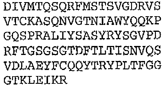
VH 6H3.1A4.3E11 SEQ ID NO.:5 
VL 6H3.1A4.3E11 SEQ ID NO.: 6 
VH 13F5.G5 SEQ ID NO.:7 
VL 13F5.G5 SEQ ID NO.:8 
VH 1B12.4H4 SEQ ID NO.:9 
VL 1B12.4H4 SEQ ID NO.:10 
VH 6B12.4F6 SEQ ID NO.:11 
VL 6B12.4F6 SEQ ID NO.:12 
- All mAbs described above were converted to chimeric (with human constant region) and expressed, purified, and characterized to confirm activity and will be used as controls for subsequent DVD-Ig analysis. To convert 3D12.E3 into chimeric form, 3D12.E3-VL was PCR amplified using primers P1 and P2; meanwhile human Ck gene (in pBOS vector generated in-house at ABC) was amplified using primers P3 and P4. Both PCR reactions were performed according to standard PCR techniques and procedures. The two PCR products were gel-purified, and used together as overlapping template for the subsequent overlapping PCR reaction using primers P1 and P4 using standard PCR conditions. The final PCR product, the chimeric light chain 3D12.E3-VL-hCk, was subcloned into pEF6 TOPO mammalian expression vector (Invitrogen) by TOPO cloning according to the manufacturer's instructions. Table 3 shows the PCR primers' sequences:
Table 3: P1: 5'ATGGTGTCCACAGCTCAGTTCC3' SEQ ID NO. 13 P2: 5' GC AGC CAC CGT ACG CCG GTT TAT TTC CAG 3' SEQ ID NO. 14 P3: 5' CGT ACG GTG GCT GCA CCA TCT GTC 3' SEQ ID NO. 15 P4: 5' TCA ACA CTC TCC CCT GTT GAA GC 3' SEQ ID NO. 16 - To convert 3D12.E3 heavy chain into chimeric form, 3D12.E3-VH was PCR amplified using primers P5 and P6; meanwhile human Cγ1 gene (in pBOS vector generated in-house at ABC) was amplified using primers P7 and P8. Both PCR reactions were performed according to standard PCR techniques and procedures. The two PCR products were gel-purified, and used together as overlapping template for the subsequent overlapping PCR reaction using primers P5 and P8 using standard PCR conditions. The final PCR product, the chimeric light chain 3D12.E3-VH-hCγ1, was subcloned into pcDNA3.1 TOPO mammalian expression vector (Invitrogen) according to the manufacturer's instructions. Table 4 shows the PCR primers' sequences:
Table 4: P5: 5' ATG GCT TGG GTG TGG ACC TTG C 3' SEQ ID NO. 17 P6: 5' GGG CCC TTG GTC GAC GCT GAG GAG ACG GTG ACT GAG G 3' SEQ ID NO. 18 P7: 5' GCG TCG ACC AAG GGC CCA TCG GTC TTC C 3' SEQ ID NO. 19 P8: 5' TC ATT TAC CCG GAG ACA GGG AGA GGC 3' SEQ ID NO. 20 - Similarly, chimeric 13F5.GS-VH-Cγ1 was generated using primers P21/P22 (for VH) and P7/P8 (for hCγ1) and cloned into pcDNA3.1 TOPO vecter, and chimeric 13F5.G5-VL-Cκ was generated using primers P23/P24 (for VL) and P3/P4 (for hCk) and cloned into pEF6 TOPO vector. Table 5 shows the PCR primers' sequences:
Table 5: P21 : 5' ATA GAA TGG AGC TGG GTT TTC CTC 3' SEQ ID NO. 21 P22: 5' GGG CCC TTG GTC GAC GC TGA GGA GAC GGT GAC TGA 3' SEQ ID NO. 22 P23: 5' ATG GTC CTC ATG TCC TTG CTG TTC 3' SEQ ID NO. 23 P24: 5' GC AGC CAC CGT ACG CCG TTT TAT TTC CAG CTT TG 3' SEQ ID NO. 24 - To express chimeric Abs, 13F5.G5-VL-Cκ and 13F5.G5-VH-Cγ1 were co-expressed in COS using Lipofectamin (Invitrogen) for 72hr, and the medium collected and IgG purified by Protein A chromatography. Similarly, 13F5.G5-VL-Cκ and 13F5.G5-VH-Cγ1 were co-expressed in COS using Lipofectamin (Invitrogen) for 72hr, and the medium collected and IgG purified by Protein A chromatography. Both purified chimeric Abs were characterized by Biacore, and MRC-5 bioassay to confirm activity. The results showed that these chimeric Abs displayed similar affinity and potency to that of the original murine mAbs.
- The construct used to generate DVD-Ig capable of binding hIL-1α and hIL-1β is illustrated in
Fig.1 B. Briefly, parent mAbs including two high affinity murine Abs, anti-hIL-1α (clone 3D12.E3) and anti-hIL-1β (clone 13F5.G5), were obtained by immunizing Balb/c mice with recombinant IL-1α protein (rhIL-1α) and recombinant IL-1β protein (rhIL-1β), respectively. The VL/VH genes of these two hybridoma clones were isolated by RT-PCR using the mouse Ig Primer Kit (Novagen, Madison, WI). The VL/VH genes were first converted into chimeric antibodies (with human constant regions) to confirm activity and potency. To generate DVD1-Ig, the VH and VL of 13F5.G5 was directly fused to the N-terminus of the VH and VL of 3D12,E3, respectively (as shown inFig.1B ), The DVD2-Ig was constructed similarly, except that it had a linker between the two variable domains in both the light chain (the linker sequence is ADAAP) and the heavy chain (the linker sequence is AKTTPP). These sequences were selected from the N-termini of murine Ck and CH1 sequences. These linker sequences, selected from the N-termini of murine Ck and CH1, are natural extension of the variable domains and exhibit a flexible conformation without significant secondary structures based on the analysis of several Fab crystal structures. The detailed procedures of the PCR cloning is described below: - 13F5.G5-VH was PCR amplified using primers P21 and P25; meanwhile 3D12.E3-VH-hCγ1 was amplified using primers P14 and P8. Both PCR reactions were performed according to standard PCR techniques and procedures. The two PCR products were gel-purified, and used together as overlapping template for the subsequent overlapping PCR reaction using primers P21 and P8 using standard PCR conditions. The final PCR product, the DVD1-Ig heavy chain hIL-1a/bDVD1-VH-hCγ1, was subcloned into pcDNA3.1 TOPO mammalian expression vector (Invitrogen) according to the manufacturer's instructions. Table 6 shows the PCR primers' sequences:
Table 6 P14: 5' CAG ATC CAG TTG GTG CAG TCT GG3' SEQ ID NO. 25 P25: 5' CAC CAA CTG GAT CTG TGA GGA GAC GGT GAC TGA GG 3' SEQ ID NO. 26 - To generate hIL-1a/bDVD1-Ig light chain, 13F5.G5-VL was PCR amplified using primers P23 and P26; meanwhile 3D12.E3-VL-hCκ was amplified using primers P16 and P4. Both PCR reactions were performed according to standard PCR techniques and procedures. The two PCR products were gel-purified, and used together as overlapping template for the subsequent overlapping PCR reaction using primers P23 and P4 using standard PCR conditions. The final PCR product, the hIL-1a/bDVD1-Ig light chain hIL-1a/bDVD1-VL-hCκ, was subcloned into pEF6 TOPO mammalian expression vector (Invitrogen) according to the manufacturer's instructions. Table 7 shows the PCR primers' sequences:
Table 7 P16: 5' AAT ATC CAG ATG ACA CAG ACT ACA TCC 3' SEQ ID NO.27 P26: 5' GTGT CAT CTG GAT ATT CCG TTT TAT TTC CAG CTT TG 3' SEQ ID NO.28 - 13F5.G5-VH was PCR amplified using primers P21 and P17; meanwhile 3D12.E3-VH-hCγ1 was amplified using primers P18 and P8. Both PCR reactions were performed according to standard PCR techniques and procedures. The two PCR products were gel-purified, and used together as overlapping template for the subsequent overlapping PCR reaction using primers P21 and P8 using standard PCR conditions. The final PCR product, the DVD2-Ig heavy chain hIL-1a/bDVD2-VH-hCγ1, was subcloned into pcDNA3.1 TOPO mammalian expression vector (Invitrogen) according to the manufacturer's instructions. Table 8 shows the PCR primers' sequences:
Table 8 P17: 5' TGG GGG TGT CGT TTT GGC TGA GG 3' SEQ ID NO.29 P18: 5' GCC AAA ACG ACA CCC CCA CAG ATC CAG TTG GTG CAG 3' SEQ ID NO.30 - To generate hIL-1a/bDVD2-Ig light chain, 13F5.G5-VL was PCR amplified using primers P23 and P19; meanwhile 3D12.E3-VL-hCκ was amplified using primers P20 and P4. Both PCR reactions were performed according to standard PCR techniques and procedures. The two PCR products were gel-purified, and used together as overlapping template for the subsequent overlapping PCR reaction using primers P23 and P4 using standard PCR conditions. The final PCR product, the h1L-1a/bDVD2-Ig light chain hIL-1a/bDVD2-VL-hCκ, was subcloned into pEF6 TOPO mammalian expression vector (Invitrogen) according to the manufacturer's instructions. Table 9 shows the PCR primers' sequences:
Table 9 P19: 5' TGG TGC AGC ATC AGC CCG TTT TAT TTC 3' SEQ ID NO.31 P20: 5' GCT GAT GCT GCA CCA AAT ATC CAG ATG ACA CAG 3' SEQ ID NO.32 - The final sequences of hIL-1a/bDVD1-Ig and hIL-1a/bDVD2-Ig are described in Table 10:
Table 10: Amino acid sequence of hIL-1α/βDVD1-Ig and hIL-1α/βDVD2-Ig Protein Sequence Protein region Sequence Identifier 12345678901234567890 DVD HEAVY VARIABLE hIL-1a/bDVD1-Ig SEQ ID NO.:33 
VH 13F5.G5 SEQ ID NO.:7 
Linker None 3D12.E3 VH SEQ ID NO.:1 
CH SEQ ID NO.:34 
DVD LIGHT VARIABLE HIL-1a/bDVD1-Ig SEQ ID NO.:35 
13F5.G5 VL SEQ ID NO.:8 
Linker None 3D12.E3 VL SEQ ID NO.:2 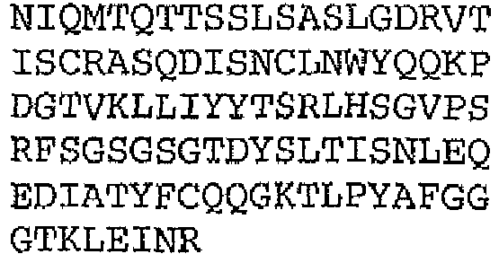
CL SEQ ID NO.:36 

DVD HEAVY VARIABLE hIL-1a/bDVD2-Ig SEQ ID NO.:37 
13F5.G5 VH SEQ ID NO.:7 
Linker SEQ ID NO.:38 AKTTPP 3D12.E3 VH SEQ ID NO.:1 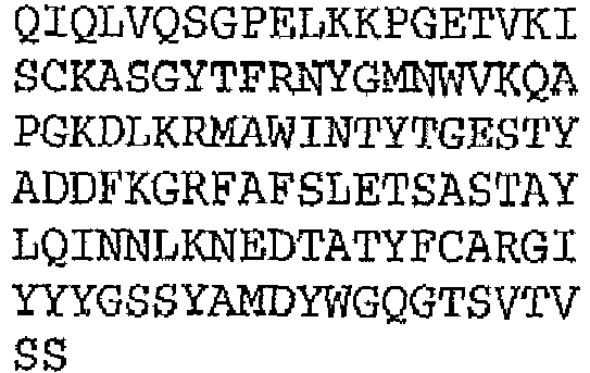
CH SEQ ID NO.:34 
DVD LIGHT VARIABLE HIL-1a/bDVD2-Ig SEQ ID NO.:39 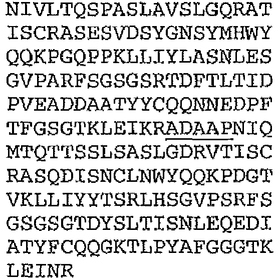
13F5.G5 VL SEQ ID NO.:8 
Linker SEQ ID NO.:40 ADAAP 3D12.E3 VL SEQ ID NO.:2 
CL SEQ ID NO.:36 
- The heavy and light chain of each construct was subcloned into pcDNA3.1 TOPO and pEF6 TOPO vectors (Invitrogen Inc.), respectively, and sequenced to ensure accuracy. The plasmids encoding the heavy and light chains of each construct were transiently expressed using Lipofectamine 2000 and 293fectin reagents, respectively in COS cells as well as human embryonic kidney 293 cells (American Type Culture Collection, Manassas, VA). The cell culture media was harvested 72 hr-post transient transfection and antibodies purified using protein A chromatography (Pierce, Rockford, IL) according to manufacturer's instructions. The Abs were analyzed by SDS-PAGE and quantitated by A280 and BCA (Pierce, Rockford, IL). Table 11 shows that the expression levels of hIL-1a/bDVD1-Ig and hIL-1a/1bDVD2-Ig are comparable to that of the chimeric Abs, indicating that the DVD-Ig can be expressed efficiently in mammalian cells.
Table 11. Expression and molecular weight analysis of hIL-1a/bDVD-Ig Expression level (ng/ml) Molecular mass (Dalton) COS Freestyle 293 Light Chain Heavy Chain Full length Mock 0 0 3D12.E3-Ch 2788 3886 23,696 49,914 147,220 13F5.G5-Ch 3260 3562 24,084 49,518 147,204 DVD1-Ig 2988 3300 35,797 (35,790) 64,380 (64,371) 200,346 (200,521) DVD2-Ig 2433 3486 36,222 (36,220) 64,976 (64,973) 202,354 (202,573) The molecular mass of the light chain, heavy chain, and full length of DVD1-Ig and DVD2-Ig determined experimentally by mass spectrometry are shown in parenthesis. - For measuring molecular weight (MW) of light and heavy chains of DVD-Ig, 10 uL of DVD-Ig (0.8 ug/uL) was reduced by 1.0 M DTT solution (5 uL). A PLRP-S, 8u, 4000A, and 1x150 mm protein column (Michrom BioResource, Auburn, MA) was used to separate heavy and light chains of DVD-Ig. Agilent HP1100 Capillary HPLC (Agilent Technologies Inc., Pala Alto, CA) was used with the mass spectrometer QSTAR (Applied Biosystems, Foster City, CA). The valco valve was set at 10 minutes to switch the flow from waste to MS for desalting sample. Buffer A was 0.02% TFA, 0.08% FA, 0.1% ACN and 99.8% HPLC-H2O. Buffer B contained 0.02% TFA, 0.08% FA, 0.1% HPLC-H2O, and 99.8% ACN. The HPLC flow rate was 50 uL/min, and the sample injection volume was 8.0 mL. The temperature of the column oven was set at 60°C, and separation gradient was: 5%B for 5 minutes; 5%B to 65%B for 35 minutes; 65%B to 95%B for another 5 minutes, and 95%B to 5%B for 5 minutes. TOFMS scan was from 800 to 2500 amu, and cycles were 3600. To determine the MW of full length DVD-Ig, a Protein MicroTrap cartridge (Michrom BioResource, Auburn, MA) was used for desalting the sample. The HPLC gradient was: 5%B for 5 minutes; 5%B to 95%B in 1 minutes; and from 95%B to 5%B in another 4 minutes. The QSTAR TOFMS scan was from 2000 to 3500 amu, and cycles were 899. All MS raw data were analyzed using the Analyst QS software (Applied Biosystems). For SEC analysis of the DVD-Ig, purified DVD-Ig and chimeric Abs, in PBS, were applied on a Superose 6 10/300 G2, 300 x 10 mm column (Amersham Bioscience, Piscataway, NJ). An HPLC instrument, Model 10A (Shimadzu, Columbia, MD) was used for SEC. All proteins were determined using UV detection at 280 nm and 214 nm. The elution was isocratic at a flow rate of 0.5 mL/min. For stability study, samples in the concentration range of 0.2-0.4 mg/ml in PBS underwent 3 freeze-thaw cycles between -80°C and 25°C, or were incubated at 4°C, 25°C, or 40°C, for 4 weeks and 8 weeks, followed by SEC analysis.
- DVD-Ig and chimeric Abs were purified by protein A chromatography. The purification yield (3-5mg/L) was consistent with hIgG quantification of the expression medium for each protein. The composition and purity of the purified DVD-Igs and chimeric Abs were analyzed by SDS-PAGE in both reduced and non-reduced conditions. In non-reduced condition, each of the four proteins migrated as a single band. The DVD-Ig proteins showed larger M.W. than the chimeric Abs, as expected. In non-reducing condition, each of the four proteins yielded two bands, one heavy chain and one light chain. Again, the heavy and light chains of the DVD-Igs were larger in size than that of the chimeric Abs. The SDS-PAGE showed that each DVD-Ig is expressed as a single species, and the heavy and light chains are efficiently paired to form an IgG-like molecule. The sizes of the heavy and light chains as well as the full-length protein of two DVD-Ig molecules are consistent with their calculated molecular mass based on amino acid sequences (see Table 11).
- In order to determine the precise molecular weight of DVD-Ig, mass spectrometry was employed. As shown in Table I, the experimentally determined molecular mass of each DVD-Ig, including the light chain, heavy chain, and the full-length protein, is in good agreement with the predicted value. To further study the physical properties of DVD-Ig in solution, size exclusion chromatography (SEC) was used to analyze each protein. Both chimeric Abs and DVD2-Ig exhibited a single peak, demonstrating physical homogeneity as monomeric proteins. The 3D12.E3 chimeric Ab showed a smaller physical size then 13F5.G5 chimeric Ab, indicating that 3D12.E3 chimeric Ab adopted a more compact, globular shape. DVD1-Ig revealed a major peak as well as a shoulder peak on the right, suggesting that a portion of DVD1-Ig is possibly in an aggregated form in current buffer condition.
- The physical stability of DVD-Ig was tested as follows. Purified antibodies in the concentration range of 0.2-0.4 mg/ml in PBS underwent 3 freeze-thaw cycles between -80°C and 25°C, or were incubated at 4°C, 25°C, or 40°C, for 4 weeks and 8 weeks, followed by analysis using size exclusion chromatography (SEC) analysis (see Table 12).
Table 12. in vitro stability analysis of hIL-1a/b DVD-Ig by SEC 3D12.E3-Ch 13F5.G5-Ch DVD1-Ig DVD2-Ig Agg Ab Frgm Agg Ab Frgm Agg Ab Frgm Agg Ab Frgm 3xFreeze -Thaw 1.72 98.28 0.00 13.0 87.0 0.0 46.50 53.50 0.00 0.0 100.0 0.0 4°C @ 4 Wks 0.85 99.15 0.00 4.2 95.8 0.0 42.43 56.63 0.94 0.0 100.0 0.0 25°C @ 4 Wks 1.29 98.71 0.00 0.0 100.0 0.0 45.66 54.34 0.00 0.0 100.0 0.0 40°C @ 4 Wks 1.65 98.35 0.00 20.3 78.1 1.6 36.70 59.42 3.88 0.0 100.0 0.0 4°C @ 8 Wks 5.35 90.33 4.32 2.2 97.8 0.0 38.18 56.91 4.91 0.0 100.0 0.0 25°C @ 8 Wks 1.11 60.55 38.34 1.4 97.5 1.0 24.42 67.39 8.19 0.0 100.0 0.0 40°C @ 8 Wks 4.74 81.47 13.79 34.6 65.4 0.0 20.55 67.16 12.29 0.0 100.0 0.0 The degree of aggregation and fragmentation are shown in percentage, whereas the percentage of Ab represents intact molecule.
Agg: aggregates;
Ab: intact antibody;
Frgm: fragments. - Both chimeric Abs showed minor degrees of aggregation and fragmentation, normal for a regular IgG molecule. DVD1-Ig showed some aggregation on SCE after purification. In the stability analysis, DVD1-Ig also showed aggregations in PBS under different conditions; however the percentage of aggregated form of DVD1-Ig did not increase during prolonged storage or at higher temperatures. The percentage of the fragmented form of DVD1-Ig were in the formal range, similar to that of the chimeric 3D12.E3 Ab. In contrast, DVD2-Ig showed exceptional stability. Neither aggregation nor fragmentation was detected for DVD2-Ig in all conditions tested, and 100% of DVD2-Ig maintained as intact monomeric molecule.
- The kinetics of DVD-Ig binding to rhIL1-α and rhIL1-β was determined by surface plasmon resonance-based measurements with a Biacore 3000 instrument (Biacore AB, Uppsala, Sweden) using HBS-EP (10 mM HEPES, pH 7,4, 150 mM NaCl, 3 mM EDTA, and 0.005% surfactant P20) at 25°C. All chemicals were obtained from Biacore AB (Uppsala, Sweden) or otherwise from a different source as described herein. Approximately, 5000 RU of goat anti-human IgG Fcγ fragment specific polyclonal antibody (Pierce Biotechnology Inc, Rockford, IL) diluted in 10 mM sodium acetate (pH 4.5) was directly immobilized across a CM5 research grade biosensor chip using a standard amine coupling kit according to manufacturer's instructions and procedures at 25 mg/ml. Unreacted moieties on the biosensor surface were blocked with ethanolamine. Modified carboxymethyl dextran surface in flowcell 2 and 4 was used as a reaction surface. Unmodified carboxymethyl dextran without goat anti-human IgG in
flow cell 1 and 3 was used as the reference surface. For kinetic analysis, rate equations derived from the 1:1 Langmuir binding model were fitted simultaneously to association and dissociation phases of all ten injections (using global fit analysis) using the Bioevaluation 4.0.1 software. Purified DVD-Ig samples were diluted in Heper-buffered saline for capture across goat anti-human IgG Fc specific reaction surfaces and injected over reaction matrices at a flow rate of 5 ml/min. The association and dissociation rate constants, kon (M-1s-1) and koff (s-1) were determined under a continuous flow rate of 25 ml/min. Rate constants were derived by making kinetic binding measurements at ten different antigen concentrations ranging from 1.25 to 1000 nM. The equilibrium dissociation constant (M) of the reaction between DVD-Ig and rhIL1α/β was then calculated from the kinetic rate constants by the following formula: KD = koff/kon. Aliquots of rhIL1α/β samples were also simultaneously injected over a blank reference and reaction CM surface to record and subtract any nonspecific binding background to eliminate the majority of the refractive index change and injection noise. Surfaces were regenerated with two subsequent 25 ml injections of 10 mM Glycine (pH 1.5) at a flow rate of 5 ml/min. The anti-Fc antibody immobilized surfaces were completely regenerated and retained their full capture capacity over twelve cycles. The apparent stoichiometry of the captured DVD-Ig- rhIL1α/β complex was calculated under saturating binding conditions (steady-state equilibrium) using the following formula:
- The Biacore analysis indicated the chimeric Abs possessed similar binding kinetics and affinities to IL-1 as the original hybridoma mAbs, indicating that the correct VL/VH sequences had been isolated (Table III). The overall binding parameters of the two DVD-Igs to hIL-1α were similar, with the affinities of the DVD-Igs being only 2-3 fold less than that of the chimeric 3D12.E3 Ab. The binding affinity of DVD2-Ig to hIL-1β was slightly less than the chimeric Ab 13F5.G5, but 3-fold higher than that of DVD1-Ig. The affinity of the two DVD-Igs to hIL-1 as compared to the affinity of chimeric Abs to hIL-1 was similar as indicated by the evaluation of the stoichiometry to IL-1. Both chimeric Abs, being bivalent monospecific, bound to IL-1α and IL-1β on Biocore with a stoichiometry of 1.6 and 1.7, respectively. This is common for an IgG due to inter-molecular interference when antibodies are immobilized densely on the Biacore sense chip resulting in stoichiometry being in the range from 1.5 to 2.0. The stoichiometry of both DVD-Igs for hIL-1α and hTL-1β were similar to that of the two chimeric Abs, indicating that both DVD-Igs possessed bivalent binding capability to each antigen.
Table 13. Functional characterization of anti-IL-1 DVD-Ig molecule Antigen kon (M-1 s-1) koff (s-1) Kd (M) Stoichiometry Potency IC50(M) 3D13.E3 hIL-1α 6.43E+05 7.13E-04 1.11E-09 2.0 6.70E-10 3D12.E3-Ch hIL-1α 4.12E+05 5.52E-04 1.34E-09 1.6 7.00E-10 DVD1-Ig hIL-1α 3.70E+04 1.05E-04 2.83E-09 1.8 2.30E-09 DVD2-Ig hIL-1α 7.35E+04 2.52E-04 3.42E-09 2.0 2.90E-09 13F5.G5 hIL-1β 2.13E+06 6.21E-04 2.91E-10 1.8 6.00E-10 13F5.G5-Ch hIL-1β 1.41E+06 6.54E-04 4.62E-10 1.7 5.30E-10 DVD1-Ig hIL-1β 6.09E+05 1.59E-03 2.60E-09 1.5 3.10E-09 DVD2-Ig hIL-1β 1.19E+06 9.50E-04 7.98E-10 1.8 1.60E-09 Affinity and stoichiometry were measured by Biacore; Potency (IC50) was determined by MRC-5 bioassay. - In addition, tetravalent dual-specific antigen binding of DVD-Ig was also analyzed by Biacore (Table 14). DVD-Ig was first captured via a goat anti-human Fc antibody on the Biacore sensor chip, and the first antigen was injected and a binding signal observed. As the DVD-Ig was saturated by the first antigen, the second antigen was then injected and the second signal observed. This was done either by first injecting IL-1β then IL-1α or by first injecting IL-1α followed by IL-1β for DVD2-Ig. In either sequence, a dual-binding activity was detected. Similar results were obtained for DVD1-Ig. Thus each DVD-Ig was able to bind both antigens simultaneously as a dual-specific tetravalent molecule. As shown in Table IV, the stoichiometry of both DVD-Ig to the first antigen, either hIL-1α or hIL-1β, were larger than 1.5, similar to that of mono-specific bivalent binding. Upon the injection of the second antigen, while DVD-Ig was already occupied by the first antigen, the stoichiometry of both DVD-Igs to the second antigen (i.e. hIL-1α or hIL-1β) was between 1.0 and 1.3. Thus DVD-Ig is able to bind two IL-1α and two IL-β molecules. DVD-Ig was first captured via a goat anti-human Fc antibody on the Biacore sensor chip, and the first antigen was injected and a binding signal observed, followed by the injection of the second antigen.
Table 14. Stoichiometry analysis of hIL-1a/b DVD-Ig in tetravalent dual-specific binding to IL-1α/β Response Unit Stoichiometry Captured Ab 1st antigen 2nd antigen hIL-1α:DVD-Ig hIL-1β:DVD-Ig DVD1-Ig; 932 hIL-1α: 190 hIL-1β: 75 2.3 1.0 DVD1-Ig: 1092 hIL-1β: 141 hIL-1α: 107 1.1 1.5 DVD2-Ig: 1324 hIL-1α: 209 hIL-1β: 137 1.8 1.3 DVD2-Ig: 1184 hIL-1β: 159 hIL-1α: 131 1.2 1.6 - Because DVD2-Ig was purified by Protein A chromatography instead of target-specific affinity chromatography, any potential misfolded and/or mismatched VL/VH domains, if present, can be assessed by binding studies against the 2 different antigens. Such binding analysis was conduced by size exclusion liquid chromatography (SEC). DVD2-Ig, alone or after a 120-min incubation period at 37°C with IL-1α, IL-1β, or both IL-1α and IL-1β, in equal molar ratio, were applied to the column. Each of the antigens was also run alone as controls. The SEC results indicated that DVD2-Ig was able to bind IL-1α and IL-1β in solution, and such binding resulted in a shift to the SEC signal indicating an increase in the dynamic size of DVD2-Ig when it was in complex with either antigen. The shift of the DVD2-Ig signal was 100%, not partial, suggesting all DVD2-Ig molecules were able to bind the antigen. In the presence of both IL-1α and IL-1β, there was a further and complete shift of the DVD2-Ig signal, indicating all DVD2-Ig molecules were able to bind both antigens in a uniform fasion. This experiment demonstrated that DVD-Ig was expressed as a functionally homogeneous protein. This has significant implications as it demonstrates that DVD-Ig can be produced as a homogeneous single, functional species, which differs from all previously described bi-specific, multi-specific, and multi-valent immunoglobulin-like and immunoglobulin-derived molecules.
- The biological activity of DVD-Ig was measured using MRC-5 bioassay. The MRC-5 cell line is a human lung fibroblast cell line that produces IL-8 in response to human IL-1α and IL-1β in a dose-dependent manner. MRC-5 cells were obtained from ATCC and cultured in 10% FBS complete MEM at 37°C in a 5% CO2 incubator. To determine neutralizing activity of the DVD-Ig against human IL-1α or 1L-1β, 50ul of Ab (1E-7 to 1E-12 M) in MEM/10%FBS was added to a 96 well plate and pre-incubated with 50ul of hIL-1α or hIL-1β (200pg/ml) for 1hr at 37°C, 5% CO2. MRC-5 cells at a concentration of 1E5/ml were then added (100ul) to all wells and the plates were incubated overnight at 37°C in a 5% CO2 incubator. The supernatants were harvested, and human IL-8 production measured by standard ELISA (R&D Systems, Minneapolis, MN). Neutralizing activity of the DVD-Ig was determined by its ability to inhibit IL-8 production.
- As shown in Table 13, both DVD-Igs were able to neutralize hIL-1α and hIL-1β. Consistent with the binding affinity to hIL-1α, the neutralizing activities of DVDI-Ig and DVD2-Ig against hIL-1α were also similar, i.e. 3-fold less than that of the chimeric Abs (see Table III). Consistent with its binding affinity for hIL-1β, the neutralizing activity of DVD2-Ig to hIL-1β is slightly less than that of the chimeric Ab 13F5.G5, but 3-fold higher than that of DVD1-Ig. Overall there was no significant decrease in the biological activities of DVD-Ig molecules compared to the original mAbs.
- To determine if DVD-Ig was able to inhibit IL-8 production in the presence of both IL-1α and IL-1β, equal amounts of hIL-1α and hIL-1β were added in the same culture system of MRC-5 assay. Both DVDI-Ig and DVD2-Ig were able to inhibit IL-8 synthesis by MRC-5 cells in the presence of both IL-1α and IL-1β, with activities similar to that of mono-assays where only one cytokine was present (Table 13). In this assay where both IL-1α and IL-1β were present, the dual-inhibition activity of DVD2-Ig (1.2 nM) was higher than that of DVD1-Ig (2.2 nM).
- Additional DVD-Ig molecules with different parent mAb pairs, as shown in Table 15, were constructed. For each pair of mAbs, four different DVD-Ig constructs were generated: 2 with a short linker and 2 with a long linker, each in two different domain orientations: a-b-C (alpha-beta-constant domain) and b-a-C (beta-alpha-constant domain). The linker sequences, were derived from the N-terminal sequence of human Ck or CH1 domain, as follows:
- Short linker: light chain: TVAAP; heavy chain: ASTKGP
- Long linker: light chain: TVAAPSVFIFPP; heavy chain: ASTKGPSVFPLAP
- All heavy and light chain constructs were subcloned into the pBOS expression vector, and expressed in COS cells or freestyle 293 cells.
- To construct new DVD clones, the variable domains of the two mAbs, both light chain and heavy chain, were first jointed in tandem using overlapping PCR as described for hIL-1abDVD1-Ig and hIL-1abDVD2-Ig. The jointed pieces were then subcloned in pBOS vecter using homologous recombination. Briefly, vectors were linearized by restriction digestion (2ug of pBOS-hCk vector were digested with FspAI and BsiWI in O+ buffer, and 2ug of pBOS-hCγ z,non a vector was digested with FspAI and SaII in O+ buffer). The digested samples were run on 1% agarose gel and the backbone fragment purified in 50ul water. For homologous recombination and transformation, DH5α competent cells were thaw on ice, and mixed with 20-50ng jointed PCR product and 20-50 ng of linearized vector (in every 50ul DH5α cells). The mixture was mixed gently and incubated on ice for 45 minutes, followed by heat shock at 42°C for 1 minute. Then 100ul SOC medium were added and incubated at 37°C for 1 hour. The transformation culture was inoculated on LB/Agar plates containing Ampicilin and incubated at 37°C for 18-20 hours. The bacterial clones were isolated, from which DNA was purified and subjected to sequencing analysis. The final sequence-verified clones were co-transfected (matching HV and LC of the same Ab pair) in COS or 293 cells for Ab expression and purification, as previously described.
- Characteristics of the purified DVD-Ig proteins are summarized in Table 16. The left section of the table 16 shows the specificity, binding affinity, and neutralization potency of the 2 pairs of mAbs used for the construction of the new hIL-1a/bDVD-Ig molecules. Antibodies 18F4.2C8 and 1B12.4H4 (see example 1.1.D) were used to construct hIL-1a/bDVD3a-Ig, hIL-1a/bDVD4a-1g, hIL-1a/bDVD3b-Ig, and hIL-1a/bDVD4b-Ig, hIL-1a/bDVD3a-Ig and hIL-1a/bDVD4a-Ig were in a-b-C orientation, with a short and long linker, respectively, hIL-1a/bDVD3b-Ig and hIL-1a/bDVD4b-Ig were in b-a-C orientation, with a short and long linker, respectively. Antibodies 6H3.1A4 and 6B12.4F6 were used to construct hIL-1a/bDVD5a-Ig, hIL-1a/bDVD6a-Ig, hIL-1a/bDVD5b-Ig, and hIL-1a/bDVD6b-Ig, hIL-1a/bDVD5a-Ig and hIL-1a/bDVD6a-Ig were in α-b-C orientation, with a short and long linker, respectively, hIL-1a/bDVD5b-Ig and hIL-1a/bDVD6b-Ig were in b-a-C orientation, with a short and long linker, respectively. The molecular cloning of these additional hIL-1a/bDVD-Igs were performed using the procedure previously described for hIL-1a/bDVD1-Ig (see example 1.3), using overlapping PCR procedures. The amino acid sequences of these additional hIL-1a/bDVD-Igs are disclosed in Table 15.
Table 15: Amino acid sequence of heavy chain and light chain of six DVD Ig capable of binding IL-1α and IL-1β. Protein Sequence Protein region Sequence Identifier 12345678901234567890 DVD HEAVY VARIABLE hIL-1a/b DVD3a-Ig SEQ ID NO.:41 
18F4.2C8 VH SEQ ID NO.:3 
LINKER SEQ ID NO.:42 ASTKGP 1B12.4H4 VH SEQ ID NO.:9 
CH SEQ ID NO.:34 
DVD LIGHT VARIABLE HIL. 1a/b DVD3a-Ig SEQ ID NO.:43 
18F4.2C8 VL SEQIDNO.:4 
LINKER SEQ ID NO.:44 TVAAP 1B12.4H4 VL SEQ ID NO.:10 
CL SEQ ID NO.:36 
DVD HEAVY VARIABLE hIL-1a/b DVD3b-Ig SEQ ID NO.:45 
1B12.4H4 VH SEQ ID NO.:9 

LINKER SEQ ID NO.:42 ASTKGP 18F4.2C8 VH SEQ ID NO.:3 
CH SEQ ID NO.:34 
DVD LIGHT VARIABLE HIL-1a/b DVD3b-Ig SEQ ID NO.:46 
1.B12.4H4 VL SEQ ID NO.:10 
LINKER SEQ ID NO.:44 TVAAP 18F4.2C8 VL SEQ ID NO.:4 
CL SEQ ID NO.:36 
DVD HEAVY VARIABLE hIL-1a/b DVD4a-Ig SEQ ID NO.:47 
18F4.2C8 VH SEQ ID NO.:3 
LINKER SEQ ID NO.:48 ASTKGPSVFPLAP 1B12.4H4 VH SEQ ID NO.:9 
CH SEQ ID NO.:34 
DVD LIGHT VARIABLE HIL-1a/bDVD4a-Ig SEQ ID NO.:49 
18F4.2C8 VL SEQ ID NO.:4 
LINKER SEQ ID NO.:50 TVAAPSVFIFPP 1B12.4H4 VL SEQ ID NO.:10 
CL SEQ ID NO.:36 
DVD HEAVY VARIABLE hIL-1a/b DVD4b-Ig SEQ ID NO.:51 
1B12.4H4 VH SEQ ID NO.:9 

LINKER SEQ ID NO.:48 ASTKGPSVFPLAP 18F4.2C8 VH SEQ ID NO.:3 
CH SEQ ID NO.:34 
DVD LIGHT VARIABLE HIL-1a/b DVD4b-Ig SEQ ID NO.:52 
IB12.4H4 VL SEQ ID NO.:10 
LINKER SEQ ID NO.:50 TVAAPSVFIFPP 18F4.2C8 VL SEQ ID NO.:4 
CL SEQ ID NO.:36 
DVD HEAVY VARIABLE hIL-1a/b DVD5a-Ig SEQ ID NO.:53 
6H3.1A4.3E11 VH SEQ ID NO.:5 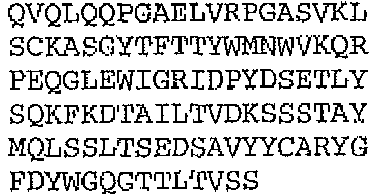
LINKER SEQ ID NO.:42 ASTKGP 6B12.4F6 VH SEQ ID NO.:11 
CH SEQ ID NO.:34 
DVD LIGHT VARIABLE HIL-1a/b DVD5a-Ig SEQ ID NO.:54 

6H3.1A4.3E11 VL SEQ ID NO.:6 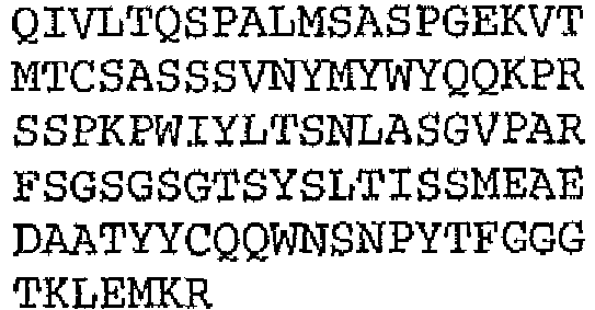
LINKER SEQ ID NO.:44 
6B12.4F6 VL SEQ ID NO.:12 
CL SEQ ID NO.:36 
DVD HEAVY VARIABLE hIL-1a/b DVD5b-Ig SEQ ID NO.:55 
6B12.4F6 VH SEQ ID NO.:11 
LINKER SEQ ID NO.:42 ASTKGP 6H3.1A4.3E11 VH SEQ ID NO.:5 
CH SEQ ID NO.:34 
DVD LIGHT VARIABLE HIL-1a/b DVD5b-Ig SEQ ID NO.:56 
6B12.4F6 VL SEQ ID NO.:12 
LINKER SEQ ID NO.:44 TVAAP 6H3.1A4.3E11 VL SEQ ID NO.:6 
CL SEQ ID NO.:36 
DVD HEAVY VARIABLE hIL-1a/b DVD6a-Ig SEQ ID NO.:57 
6H3.1A4.3E11 VH SEQ ID NO.:5 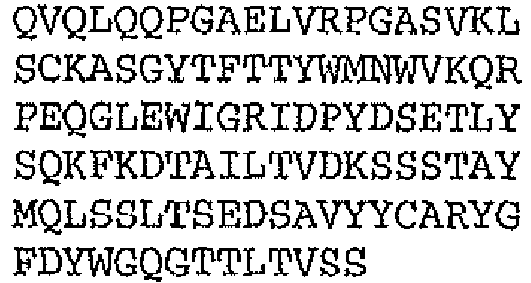
LINKER SEQ ID NO.:48 ASTKGPSVFPLAP 6B12.4F6 VH SEQ ID NO.:11 
CH SEQ ID NO.:34 
DVD LIGHT VARIABLE HIL- SEQ ID NO.:58 
1a/b DVD 6a-Ig 
6H3.1A4.3E11 VL SEQ ID NO.:6 
LINKER SEQ ID NO.:50 TVAAPSVFIFPP 6B12.4F6 VL SEQ ID NO.:12 
CL SEQ ID NO.:36 
DVD HEAVY VARIABLE hIL-1a/b DVD6b-Ig SEQ ID NO.:59 
6B12.4F6 VH SEQ ID NO.:11 
LINKER SEQ ID NO.:48 ASTKGPSVFPLAP 6H3.1A4.3E11 VH SEQ ID NO.:5 
CH SEQ ID NO.:34 
DVD LIGHT VARIABLE HIL-1a/b DVD6b-Ig SEQ ID NO.:60 
6B12.4F6 VL SEQ ID NO.:12 
LINKER SEQ ID NO.:50 TVAAPSVFIFPP 6H3.1A4.3E11 VL SEQ ID NO.:6 
CL SEQ ID NO.:36 
- Characteristics of the new DVD constructs are summarized in Table 16. Affinity (Kd) and biological activity (IC50) were determined by Biacore and MRC-5 bioassay, respectively. SDS-PAGE analysis of all new DVD proteins showed normal migration patterns in both reduced and non-reduced conditions, similar to a regular antibody and DVD1/2-Ig.
Table 16. Characterization of new DVD-Ig molecules derived from new mAb pairs Affinity (Kd) M Potency (IC50) M mAb Specif. Kd (M) IC50 (M) DVD Orient. Linker IL-1α IL-1β IL-1α IL-1β 18F4.2C8 rhIL-1α 5.95E-10 3.30E-10 DVD3a a-b-C short 8.37E-10 6.37E-08 7.50E-10 NA DVD4a a-b-C long 7.01E-10 9.30E-10 3.50E-10 1.00E-08 1B12.4H4 rhIL-1β 2.61E-10 6.00E-10 DVD3b b-a-C short 1.24E-09 1.90E-10 7.00E-10 4-00E-10 DVD4b b-a-C long 5.60E-10 1.28E-10 3.50E-10 5.00E-10 6H3.1A4 rhIL-1α 3.54E-10 2.40E-10 DVD5a a-b-C short 5.08E-10 1.25E-08 2.60E-09 1.90E-08 DVD6a a-b-C long 1.06E-09 2.09E-09 2.30E-09 7.00E-08 6B12.4F6 ThIL-1β 5.54E-10 4.00E-10 DVD5b b-a-C short 1.32E-08 6.71E-10 3.30E-09 2.50E-10 DVD6b b-a-C long 8.20E-10 6.97E-10 1.00E-09 7.50E-10 NA: no neutralization activity detected. - The functional characterization of the new DVD molecules revealed that with either orientation, DVDs with the long linker performed better than the ones with the short linker in terms of binding and neutralizing of both antigens. With respect to DVDs with the long linkers, those with the b-a-C orientation showed good binding to and neutralization of both antigens, while the DVDs with an a-b-C orientation showed good binding to and neutralization of IL-1α and reduced binding to and neutralization of IL-1β (e.g. DVD4b vs. DVD4a). The DVD-Ig molecule, DVD4b, bound and neutralized both IL-1α and IL-1β with sub-nM and fully retained the binding and neutralizing characteristics of the parent mAbs.
- DVD-Ig molecules capable of binding IL-12 and IL-18 were constructed as described above using two parent mAbs, one against human IL-12p40 (ABT874), and the other against human IL-18 (ABT325). Four different anti-IL12/18 DVD-Ig constructs were generated: 2 with short linker and 2 with long linker, each in two different domain orientations: 12-18-C and 18-12-C (Table VI). The linker sequences, derived from the N-terminal sequence of human Cλ/Cκ or CH1 domain, were as follows:
- For DVD1218 constructs (ABT874 has a Vλ):
- light chain (λ):Short linker: QPKAAP; Long linker: QPKAAPSVTLFPP
- heavy chain (γ1): Short linker: ASTKGP; Long linker: ASTKGPSVFPLAP
- For DVD1812 constructs (ABT325 has a Vκ):
- light chain (κ):Short linker: TVAAP ; Long linker: TVAAPSVFIFPP
- heavy chain (γ1): Short linker: ASTKGP; Long linker: ASTKGPSVFPLAP
- All heavy and light chain constructs were subcloned into the pBOS expression vector, and expressed in COS cells or freestyle 293 cells, followed by purification by Protein A chromatography. The purified materials were subjected to SDS-PAGE and SEC, and their profiles were similar to that of the DVD2-Ig.
- The table 17 below describes the heavy chain and light chain constructs used to express each anti-IL12/IL18 DVD-Ig protein.
Table 17. Constructs to express anti-IL12/IL18 DVD-Ig proteins DVD-Ig protein Heavy chain construct Light chain construct DVD1218SL DVD1218HC-SL DVD1218LC-SL DVD1218LL DVD1218HC-LL DVD1218LC-LL DVD1812SL DVD1812HC-SL DVD1812LC-SL DVD1812LL DVD1812HC-LL DVD1812LC-LL - To generate heavy chain constructs DVD1218HC-LL and DVD1218HC-SL, VH domain of ABT-874 was PCR amplified using primers Primer 1 and Primer 2L or Primer 2S respectively; meanwhile VH domain of ABT-325 was amplified using primers Primer 3L or Primer 3S and Primer 4 respectively. Both PCR reactions were performed according to standard PCR techniques and procedures. The two PCR products were gel-purified, and used together as overlapping template for the subsequent overlapping PCR reaction using primers Primer 1 and Primer 4 using standard PCR conditions. The overlapping PCR products were subcloned into Srf I and Sal I double digested pBOS-hCγ1,z non-a mammalian expression vector (Abbott) by using standard homologous recombination approach.
- To generate light chain constructs DVD1218LC-LL and DVD1218LC-SL, VL domain of ABT-874 was PCR amplified using
primers Primer 5 and Primer 6L or Primer 6S respectively; meanwhile VL domain of ABT-325 was amplified using primers Primer 7L or Primer 7S and Primer 8 respectively. Both PCR reactions were performed according to standard PCR techniques and procedures. The two PCR products were gel-purified, and used together as overlapping template for the subsequent overlapping PCR reaction usingprimers Primer 5 and Primer 8 using standard PCR conditions. The overlapping PCR products were subcloned into Srf I and Not I double digested pBOS-hCk mammalian expression vector (Abbott) by using standard homologous recombination approach. The primers used for these constructions are listed below in table 18:Table 18: 
SEQ ID NO.:61 Primer2-S:CACCTCTGGGCCCTTGGTCGACGCTGAAGAGACGGTGACCATTGT SEQ ID NO.:62 
SEQ ID NO.:63 Primer3-S:TCTTCAGCGTCGACCAAGGGCCCAGAGGTGCAGCTGGTGCAGTCT SEQ ID NO.:64 
SEQ ID NO.:65 Primer 4:GTAGTCCTTGACCAGGCAGCC SEQ ID NO.:66 
SEQ ID NO.:67 Primer 6-S:TATTTCGGGGGCAGCCTTGGGCTGACCTAGTACTGTGACCTTGGT SEQ ID NO.:68 
SEQ ID NO.:69 Primer7-S:CTAGGTCAGCCCAAGGCTGCCCCCGAAATAGTGATGACGCAGTCT SEQ ID NO.:70 
SEQ ID NO.:71 
SEQ ID NO.:72 - To generate heavy chain constructs DVD181.2HC-LL and DVD1812HC-SL, VH domain of ABT-325 was PCR amplified using primers Primer 1 and Primer 9L or Primer 9S respectively; meanwhile VH domain of ABT-874 was amplified using primers Primer 10L or Primer 10S and Primer 4 respectively. Both PCR reactions were performed according to standard PCR techniques and procedures. The two PCR products were gel-purified, and used together as overlapping template for the subsequent overlapping PCR reaction using primers Primer 1 and Primer 4 using standard PCR conditions. The overlapping PCR products were subcloned into Srf I and Sal I double digested pBOS-hCγ1,z non-a mammalian expression vector (Abbott) by using standard homologous recombination approach. The following are primers' sequences:
- To generate light chain constructs DVD1812LC-LL and DVD1812LC-SL, VL domain of ABT-325 was PCR amplified using primers Primer 11 and Primer 12L or Primer 12S respectively; meanwhile VL domain of ABT-874 was amplified using primers Primer 13L or Primer 13S and Primer 14 respectively. Both PCR reactions were performed according to standard PCR techniques and procedures. The two PCR products were gel-purified, and used together as overlapping template for the subsequent overlapping PCR reaction using primers Primer 11 and Primer 14 using standard PCR conditions. The overlapping PCR products were subcloned into Srf I and Not I double digested pBOS-hCk mammalian expression vector (Abbott) by using standard homologous recombination approach. The primers used for these constructions are listed below in table 19:
Table 19: Primer 9-S: CACCTGTGGGCCCTTGGTCGACGCTGAAGAGACGGTGACCATTGT SEQ ID NO.:73 
SEQ ID NO.:74 Primer 10-S:TCTICAGCGTCGACCAAGGGCCCACAGGTGCAGCTGGTGGAGTCT SEQ ID NO.:75 
SEQ ID NO.:76 
SEQ ID NO.:77 Primer 12-S: AGACTGTGGTGCAGCCACAGTTCGTTTAATCTCCAGTCGTGT SEQ ID NO.:78 
SEQ ID NO.:79 Primer 13-S: AAACGAACTGTGGCTGCACCACAGTCTGTGCTGACTCAGCCC SEQ ID NO.:80 
SEQ ID NO.:81 
SEQ ID NO.:82 - The final DNA sequences for eight heavy and light chain constructs of anti-IL12/IL-18 DVD-Ig are as shown in table 20:
Table 20: Amino acid sequence of DVD binding proteins capable of binding IL-12 and IL-18 Protein Sequence Protein region Sequence Identifier 12345678901234567890 DVD HEAVY VARIABLE DVD1218HC-SL SEQ ID NO.:83 
ABT-874 VH SEQ ID NO.:84 
LINKER SEQ ID NO.:42 
ABT-325 VH SEQ ID NO.:85 
CH SEQ ID NO.:34 
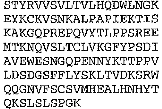
DVD LIGHT VARIABLE DVD1218LC-SL SEQ ID NO.:86 
ABT-874 VL SEQ ID NO.:87 
LINKER SEQ ID NO.:88 QPKAAP ABT-325 VL SEQ ID NO.:89 
CL SEQ ID NO.:36 
DVD HEAVY VARIABLE DVD1218HC-LL SEQ ID NO.:90 

ABT-874 VH SEQ ID NO.:84 
LINKER SEQ ID NO.:48 ASTKGPSVFPLAP ABT-325 VH SEQ ID NO.:85 
CH SEQ ID NO.:34 
DVD LIGHT VARIABLE DVD1218LC-LL SEQ ID NO.:91 
ABT-874 VL SEQ ID NO.:87 

LINKER SEQ ID NO.:92 QPKAAPSVTLFPP ABT-325 VL SEQ ID NO.:89 
CL SEQ ID NO.:36 
DVD HEAVY VARIABLE DVD1812HC-SL SEQ ID NO.:93 
ABT-325 VH SEQ ID NO.:85 
LINKER SEQ ID NO.:42 ASTKGP ABT-874 VH SEQ ID NO.:84 
CH SEQ ID NO.:34 

DVD LIGHT VARIABLE DVD1812LC-SL SEQ ID NO.:94 
ABT-35 VL SEQ ID NO.:89 
LINKER SEQ ID NO.:44 TVAAP ABT-874 VL SEQ ID NO.:87 
CL SEQ ID NO.:36 
DVDHEAVY VARIABLE DVD1812HC-LL SEQ ID NO.:95 

ABT-325 VH SEQ ID NO.:85 
LINKER SEQ ID NO.:48 ASTKGPSVFPLAP ABT-875 VH SEQ ID NO.:84 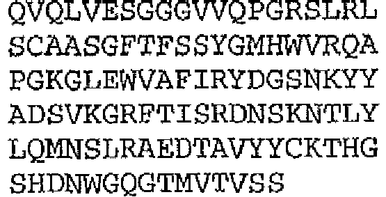
CH SEQ ID NO.:34 
DVD LIGHT VARIABLE DVD1812LC-LL SEQ ID NO.:96 
ABT-325 VL SEQ ID NO.:89 

LINKER SEQ ID NO.:50 TVAAPSVFIFPP ABT-874 VL SEQ ID NO.:87 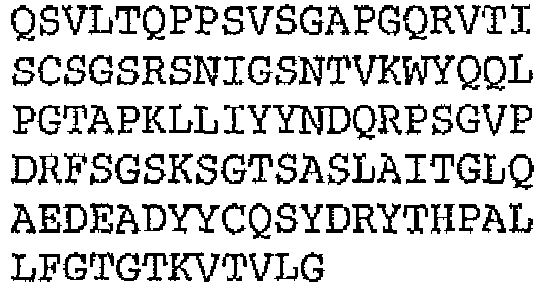
CL SEQ ID NO.:36 
- The binding affinity of anti-IL-12/18 DVD-Igs to hIL-12 and hIL-18 were determined by Biacore (Table 21). The neutralization activity against IL-18 was determined by KG-1 assay (Konishi, K., et al.,). Briefly, IL-18 samples (in a final concentration of 2 ng/ml) were pre-incubated with DVD-Ig (in final concentrations between 0 and 10 mg/ml) at 37°C for 1 hr, and then added to KG-1 cells (3x10⁶/ml) in RPMI medium containing 10 ng/ml hTNF, followed by incubation at 37°C for 16-20 hr. The culture supernatants were collected and human IFN-γ production in each sample was determined by ELISA (R&D Systems). Inhibition activities of the DVD molecules against IL-18, presented as IC50 values, are shown in Table VI. To determine the inhibition activities of anti-IL-12/18 DVD molecules against IL-12, an IL-12-induced IFN-γ production assay from activated PHA blast cells was employed (D'Andrea, A et al.,) For production of human IFN-γ, PHA blast cells were incubated for 18 hours with human IL-12. Sub-maximal stimulation (55-75% of maximum) was obtained with a human IL-12 concentration of 200 pg/mL. Supernatants were assayed for IFN-γ using a specific human IFN-γ ELISA (Endogen, Cambridge, MA). Neutralizing IL-12 DVDs interfere with IL-12 induced IFN-γ production. The neutralization activity of DVD is determined by measuring the DVD concentration required to inhibit 50% of the IFN-γ production by human PHA blast cells, as shown in Table 21.
Table 21. Characterization of anti-IL-18/IL-12 DVD-Ig molecules Affinity (Kd, M) Potency (IC50,M) MAb Specif. Kd (M) IC50 (M) DVD Orient. Linker IL-12 IL-18 IL-12 IL-18 ABT874 hIL-12 6.47E-11 5.0E-12 DVD1218-SL 12-18-C short 3.81E-11 6.22E-10 6.93E-12 1.8E-10 DVD1218-LL 12-18-C long 2.38E-11 6.64E-10 3.04E-12 1.8E-10 ABT325 hIL-18 1.37E-10 3.0E-10 DVD1812-SL 18-12-C short 1.82E-09 1.91E-10 3.66E-10 4.0E-11 DVD1812-LL 18-12-C long 1.13E-10 1.62E-10 1.18E-10 7.8E-11 Affinity (Kd) was determined by Biacore and potency (IC50) determined by KG-1 bioassay (IL-18) and PBMC assay (IL-12). - Table 21 shows the specificity, binding affinity, and neutralization activity of the 2 fully human mAbs used for the construction of the anti-IL-12/IL-18 DVD molecules. As shown in the Table VI, these mAbs have high affinity and neutralization activity. A summary of the characterization of the anti-IL-18/IL-12 DVD constructs is shown in Table VI. SDS-PAGE analysis of all new DVD proteins showed normal migration patterns in both reduced and non-reduced conditions, similar to a regular antibody and DVD1/2-Ig. SEC analysis indicated all molecules were normal, exhibiting peaks in the 200 kD region. The Biacore binding data are consistent with the neutralization activity in the biological assays.
- Both IL-12 and IL-18 are required to produce optimal IFNγ in response to various stimuli. The biological activity of anti-IL-12/IL-18 DVD-Ig in vivo was determined using the huPBMC-SCID mouse model. In this model, anti-IL-12 antibody (ABT-874) anti-IL-18 antibody (ABT-325) or the anti-IL-12/anti-IL-18 DVD-Ig were injected i.p. or i.v. (250 mg/mouse each) followed by transfer of freshly purified human PBMCs (huPBMC) i.p. into SCID mice. Fifteen minutes later, mice were challenged with dried staphylococcus aureus cells (SAC) to induce human IFNγ production. Animals (n=7-8/group) were sacrificed 18-20 hrs later and serum huIFNγ levels were determined by ELISA. ABT 874 and ABT-325 inhibited SAC-induced IFNγ by approximately 70% which represents maximum IFNγ inhibition with each compound in this model. However, treatment of mince with ABT-874 + ABT-325 and anti-IL-12/anti-IL-18 DVD-Ig inhibited IFNγ production by almost 100%. These results suggest that the anti-IL-12/anti-IL-18 DVD-Ig molecule is functionally active in vivo.
- The overall Pharmacokinetic and pharmacodynamic profile of anti-IL-12/IL-18 DVD-Ig was similar to the parent mass in mice, i.e 73% bioavailability, comparable to regular IgG. Similar pharmacokinetics, i.e. rapid clearance after day 6-8, was also observed for other mAbs (e.g. human, rat etc,) probably due to anti-human IgG response.
- Male SD rats were dosed with anti-IL-12/L-18 DVD-Ig at 4mg/kg either i.v. or s.c. The early part of the PK curves looked normal and very similar to those of other human antibodies. An accurate half-life in both groups could not be derived because of the rapid clearance of DVD-Ig beginning on day 6. The sudden drop in DVD-Ig concentration after day 6 may be due to the RAHA response. However, similar profile has also been observed for one of the parent antibodies (ABT-874) used for construction of this DVD-Ig in this particular experiment, as well as other mono-specific human antibodies previously studied. Based on DVD-Ig concentration up to day 6 in both s.c and i.v. groups, bioavailability of DVD-Ig was estimated. Two out of three rats showed 80 -95% bioavailability, and the average bioavailability in the three mice was 73%
- Results of physical and chemical characterization of 293 cell-derived, protein A purified, anti-IL-12/anti-IL-18 DVD-Ig are summarized in Table 22.
Table 22. Physical/Chemical Characterization of anti-IL-12/anti-IL-18 DVD-Ig Parameters Tested Assay/Methodology Findings/Comments Affinity (Kd) • IL-12 Biacore • 38 pM (65 pM for ABT-874) • IL-18 Biacore • 622 pM (137 pM for ABT-325) Potency (IC50) • IL-12 • PHA-Blast Assay • 7 pM (5 pM for ABT874) • IL-18 • KG-1 Assay • 180 pM (300 pM for ABT-325) M.W MS • HC: 64130 (theo. 64127) • LC: 36072 (theo. 36072) Amino acid sequence Sequencing - MS All matched Disulfide bonds Peptide mapping All 20 disulfide bonds are matched Glycosylation profile Similar to other in-house fully human antibodies - NGA2F and NGA1F observed as the major forms Charge Cation Exchange Homogeneity heterogeneity (WCX-10) PI cIEF 9.42 (ABT-874: 9.46) Dynamic size DSL 7.69nM (5.34 nM for ABT-325) Purity/aggregates SDS-PGE • Homogeneity on both reducing (∼64Kd HC and - 36Kd LC bands) and non-reducing (one band) gels SEC AUC • One peak (- 100%) observed immediately after protein A purification by SEC • ~ 16-17% aggregates observed after 2 cycles of freeze-thaw by AUC Stability (freeze/thaw) SEC ∼ 5% aggregates after 2 freeze-thaw cycles, increased to ∼13% after additional 10 freeze-thaw cycles. The reason for that is unsolved (process-related, sequence-specific, or LC lamda/kappa hybrid) PK profile Rat i.v. & s.c. Similar to (or limited by) parental mAbs. Bioavailability Rat i.v. vs s.c. Average 73%; Overall similar to parental mAbs - An additional anti-IL-12/IL-18 DVD-Ig molecule with a different parent anti-IL-12 mAb (clone# ID4.1), as shown in Table 23, was constructed. The 1D4.1-ABT325 DVD-Ig construct was generated with a short linker derived from the N-terminal sequence of human Ck and CH1 domain, as follows:
- Short linker: light chain: TVAAP; heavy chain: ASTKGP
- All heavy and light chain constructs were subcloned into the pBOS expression vector, expressed in COS cells or freestyle 293 cells, and characterized as described above. 1D4.1-ABT325 DVD-Ig fully retains the activities of the two original mAbs (Table 24).
Table 23: Amino acid sequence of 1D4.1-ABT325 DVD-Ig Protein Sequence Protein region Sequence Identifier 12345678901234567890 1D4.1-ABT325 DVD-Ig HEAVY VARIABLE SEQ ID NO.:114 
1D4.1 VH SEQ ID NO.:115 
LINKER SEQ ID NO.:99 ASTKGP ABT-325 VH SEQ ID NO.:85 
CH SEQ ID NO.:34 

1D4.1-ABT325 DVD-Ig LIGHT VARIABLE SEQ ID NO.:116 
1D4.1 VL SEQ ID NO.:117 
LINKER SEQ ID NO.:44 
ABT-325 VL SEQ ID NO.:89 
CL SEQ ID NO.:36 
Table 24. Characterization 1D4.1-ABT325 DVD-Ig molecule mAb Affinity (Kd, M) Potency (IC50,M) IL-12 IL-18 IL-12 IL-18 1D4.1 1.20E-10 N/A 4.18E-10 N/A ABT325 N/A 1.91E-10 N/A 6.87E-11 1D4.1-ABT325 DVD-Ig 1.33E-10 1.59E-10 2.17E-10 1.20E-10 Affinity (Kd) was determined by Biacore and potency (IC50) determined by KG-1 bioassay (IL-18) and PBMC assay (IL-12). - Anti-CD20/anti-CD3 DVD-Igs were generated using murine anti-human-CD20 (clone 5F1) and anti-human-CD3 (clone OKT3) parent antibodies. The initial constructs included 2 DVD-Igs with different domain orientations. The anti-CD3/anti-CD20 DVD-Ig was constructed in the order of VCD3-linker-VCD20-constant, and anti-CD20/anti-CD3 DVD-Ig was constructed in the order of VCD20-linker-VCD3-Constant. However, in a preliminary cell surface binding study, anti-CD20 binding activity was diminished in the anti-CD3/anti-CD20 DVD-Ig molecule, even though the anti-CD3 activity was conserved in this design. In contrast, both anti-CD3 and anti-CD20 binding activities were fully conserved in the anti-CD20/anti-CD3 DVD-Ig molecule, indicating this is the optimal domain orientation for these two mAbs/targets combination in a DVD-Ig format. Therefore the anti-CD20/anti-CD3 DVD-Ig construct was chosen for subsequent studies.
- The anti-CD20/anti-CD3 DVD-Ig was generated as chimeric Ig i.e the constant region was a human constant region. For binding analysis, human T cell line Jurkat and B cell line Raji were blocked with human IgG and then stained with murine anti-hCD3 mAb OKT3, murine anti-hCD20 mAb 1F5, and anti-CD20/anti-CD3 DVD-Ig. Cells were then washed and stained with FITC-labeled goat anti-murine IgG (with no anti-hIgG cross-reactivity). Anti-CD20/CD3 DVD-Ig bound both T and B cells, whereas CD3 and CD20 mAbs bound only T or B cells, respectively. The amino acid sequence of CD20/CD3 DVD-Ig is disclosed in Table 25.
Table 25: Amino acid sequence of CD20CD3DVD-Ig Protein Sequence Protein region Sequence Identifier 12345678901234567890 DVD HEAVY VARIABLE CD20CD3DVD-Ig SEQ ID NO.:97 
5F1 VH SEQ ID NO.:98 

LINKER SEQ ID NO.:99 AKTTAPSVYPLAP OKT3 VH SEQ ID NO.:100 
CH SEQ ID NO.:34 
CD20CD3DVD-Ig LIGHT VARIABLE SEQ ID NO.:101 
5F1 VL SEQ ID NO.:102 
LINKER SEQ ID NO.:103 ADAAPTVSIFPP OKT3 VL SEQ ID NO.:104 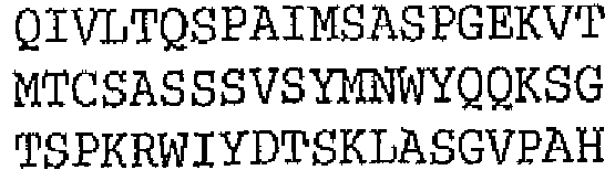

CL SEQ ID NO.:36 
- To study key issues concerning pharmacokinetics, in vivo efficacy, tissue penetration, and immunogenicity of DVD-Ig molecules, mouse-anti-mouse IL-1α/β DVD-Ig molecules were constructed as described below.
- Mouse-anti-mouse IL-1α/β DVD-Ig molecules were constructed using two mouse anti-mouse IL-1α/β mAbs (9H10 and 10G11) generated from IL-1αβ double KO mice. Mouse anti-mouse IL-1α, and mouse anti-mouse IL-1β, monoclonal antibodies were generated by immunizing IL-1α/β double KO mice with mouse IL-1α, or mouse IL-1β, respectively. One mouse anti-mouse IL-1α (Clone 9H10), and one mouse anti-mouse IL-1β mAb (clone 10G11), were selected and used to generate mIL-1α/β DVD-Ig molecules. Various linker sizes and different domain orientations were tested. The final functional mIL-1α/β DVD-Ig molecules was constructed in a orientation of V(anti-mIL-1β)-linker-V(anti-mIL-1β)-murine constant region (Cγ2a and Cκ).. The cloning, expression, and purification procedures were similar to that of the hIL-1α/β DVD-Ig. The cloning of mIL-1α/β DVD-Ig was carried out using similar overlapping PCR and homologous recombination as described for hIL-1α/β DVD3-Ig. The sequences of mIL-1α/β DVD-Ig are shown below in Table 26:
Table 26: Amino acid sequence of mIL-1α/β DVD-Ig Protein Sequence Protein region Sequence Identifier 12345678901234567890 mIL-1α/β DVD-Ig HEAVY VARIABLE SEQ ID NO.:105 
10G11 VH SEQ ID NO.:106 
LINKER SEQ ID NO.:99 AKTTAPSVYPLAP 9H10 VH SEQ ID NO.:107 
CH SEQ ID NO.:108 
SEQ ID NO.:109 
mIL-1α/β DVD-Ig LIGHT VARIABLE 
10G11 VL SEQ ID NO.:110 
LINKER SEQ ID NO.:111 ADAAPTVSIFPP 9H10VL SEQ ID NO.:112 
CL SEQ ID NO.:113 
- Murine mIL-1α/β DVD-Ig retained affinity/in vitro potency against both IL-1α and IL-1β. Table 27 shows the characterization of mAbs 9H10 (anti-mIL-1α), 10G11 (anti-mIL-1β), and mIL-1α/β DVD-Ig.
Table 27. Characterization of mDVD4-Ig Antigen KD (M) IC50 (M) 9H10 mIL-1α 1.73E-10 2.00E-10 10G11 mIL-1β 2.30E-10 3.70E-10 mIL-1α/β/DVD-Ig mIL-1α 7.66E-10 2.00E-09 mIL-1β 6.94E-10 8.00E-10 - The therapeutic effects of anti-IL-1alpha, anti-IL-1beta, combined anti-IL-1-alpha/anti-IL-1beta, and murine anti-IL-1alpha/beta DVD4-Ig, were evaluated in a collagen-induced arthritis mouse model well known in the art. Briefly, male DBA-1 mice were immunized with bovine type II collagen in CFA at the base of the tail. The mice were boosted with Zymosan intraperitoneally (i.p) at day 21. After disease onset at day 24-27, mice were selected and divided into separate groups of 10 mice each. The mean arthritis score of the control group, and anti-cytokine groups was comparable at the start of treatment. To neutralize IL-1, mice were injected every other day with 1-3 mg/kg of anti-IL-1alpha mAb, anti-IL-1beta mAb, combination of anti-IL-1-alpha/anti-IL-1beta mAbs, or murine anti-IL-1alpha/beta DVD4-Ig intraperitoneally, Mice were carefully examined three times a week for the visual appearance of arthritis in peripheral joints, and scores for disease activity determined.
- Blockade of IL-1 in the therapeutic mode effectively reduced the severity of arthritis, with anti-IL-1beta showing greater efficacy (24% reduction in mean arthritis score compared to control group) than anti-IL-1-alpha (10% reduction). An additive effect was observed between anti-IL-1-alpha and anti-IL-1beta, with a 40% reduction in mean arthritis score in mice treated with both anti-IL-1alpha and anti-IL-1beta mAbs. Surprisingly, at the same dose level, the treatment of mDVD-Ig exhibited 47% reduction in mean arthritis score, demonstrating the in vivo therapeutic efficacy of mDVD-Ig. Similar efficacy weas also observed in the measurements of joint swelling in this animal model.
- The present invention incorporates by reference in their entirety techniques well known in the field of molecular biology and drug delivery. These techniques include, but are not limited to, techniques described in the following publications:
- Ausubel et al. (eds.), Current Protocols in Molecular Biology, John Wiley &Sons, NY (1993);
- Ausubel, F.M. et al. eds., Short Protocols In Molecular Biology (4th Ed. 1999) John Wiley & Sons, NY. (ISBN 0-471-32938-X).
- Controlled Drug Bioavailability, Drug Product Design and Performance, Smolen and Ball (eds.), Wiley, New York (1984);
- Giege, R. and Ducruix, A. Barrett, Crystallization of Nucleic Acids and Proteins, a Practical Approach, 2nd ea., pp. 20 1-16, Oxford University Press, New York, New York, (1999);
- Goodson, in Medical Applications of Controlled Release, vol. 2, pp. 115-138 (1984);
- Hammerling, et al., in: Monoclonal Antibodies and T-Cell Hybridomas 563-681 (Elsevier, N.Y., 1981;
- Harlow et al. , Antibodies: A Laboratory Manual, (Cold Spring Harbor Laboratory Press, 2nd ed. 1988);
- Kabat et al., Sequences of Proteins of Immunological Interest (National Institutes of Health, Bethesda, Md. (1987) and (1991);
- Kabat, E.A., et al. (1991) Sequences of Proteins of Immunological Interest, Fifth Edition, U.S. Department of Health and Human Services, NIH Publication No. 91-3242;
- Kontermann and Dubel eds., Antibody Engineering (2001) Springer-Verlag. New York. 790 pp. (ISBN 3-540-41354-5).
- Kriegler, Gene Transfer and Expression, A Laboratory Manual, Stockton Press, NY (1990);
- Lu and Weiner eds., Cloning and Expression Vectors for Gene Function Analysis (2001) BioTechniques Press. Westborough, M*A. 298 pp. (ISBN 1-881299-21-X).
- Medical Applications of Controlled Release, Langer and Wise (eds.), CRC Pres., Boca Raton, Fla. (1974);
- Old, R.W. & S.B. Primrose, Principles of Gene Manipulation: An Introduction To Genetic Engineering (3d Ed. 1985) Blackwell Scientific Publications, Boston. Studies in Microbiology; V.2:409 pp. (ISBN 0-632-01318-4).
- Sambrook, J. et al. eds., Molecular Cloning: A Laboratory Manual (2d Ed. 1989) Cold Spring Harbor Laboratory Press, NY. Vols. 1-3. (ISBN 0-87969-309-6).
- Sustained and Controlled Release Drug Delivery Systems, J.R. Robinson, ed., Marcel Dekker, Inc., New York, 1978
- Winnacker, E.L. From Genes To Clones: Introduction To Gene Technology (1987) VCH Publishers, NY (translated by Horst Ibelgaufts). 634 pp. (ISBN 0-89573-614-4).
-
4,526, 938 ;4,753,894 ;4,816,567 ;4,880,078 ;4,943,533 ;4,946,778 ;4,980,286 ;5, 500,362 ;5,916,597 ;5, 985,309 ;5,128,326 ;5,223,409 ;5,225,539 ;5,258, 498 ;5,290, 540 ;5,403,484 ;5,427,908 ;5,516,637 ;5,530,101 ;5,558,864 ;5,565,332 ;5,571,698 ;5,580,717 ;5,585,089 ;5,624,821 ;5,627,052 ,5,641,870 ;5,648,260 ;5,658,727 ;5,677,171 ;5,679,377 ;5,693,762 ;5,698,426 ;5,714,350 ;5,714,352 ;5,723,323 ;5,733,743 ;5,736,137 ;5,750,753 ;5,763,192 ;5,766,886 ;5,780, 225 ;5,814,476 ;5,817,483 ;5,821,047 ;5,824,514 ;5,855,913 ;5,874,064 ;5,891,996 ;5,912,015 ;5,916,771 ;5,934, 272 ;5,939,598 ;5,969,108 ;5,976,862 ;5,985, 320 ;5,985,309 ;5,985,615 ;5,989,463 ;5,998,209 ;6, 019,968 ;6,075,181 ;6,091,001 ;6,114,598 ;6,130,364 ;6,180,370 ;6,204,023 ;6,235,883 ;6,258,562 6,350,861 ;6,506, 883 ;6,699,658 ;6,914,128 -
10/172,317
09/428,082 -
-
PCT/GB91/01134 PCT/US2003/040426 PCT/US91/05939 PCT/US91/09630 PCT/US94/01234 PCT/US96/18978 PCT/US98/16280 WO 00 09560 WO 00/037504 WO 00/56772 WO 01/58956 WO 01/77342 WO 01/83525 WO 01/88138 WO 0162931A2 WO 0177342A1 WO 02/02773 WO 02072636 WO 03/035835 WO 90/02809 WO 90/05144 A1 WO 91/05548 WO 91/09967 WO 91/10737 WO 91/10741 WO 91/17271 WO 92/01047 WO 92/02551 WO 92/09690 WO 92/15679 WO 92/18619 WO 92/19244 WO 92/20791 WO 92/22324 WO 93/01288 WO 93/11236 WO 94/02602 WO 95/15982 WO 95/20045 WO 95/20401 WO 96/20698 WO 96/33735 WO 96/34096 WO 96/40210 WO 97/29131 WO 97/32572 WO 97/44013 WO 98/16654 WO 98/24893 WO 98/31346 WO 98/31700 WO 98/50433 WO 99/15154 WO 99/20253 WO 99/25044 WO 99/45031 WO 99/53049 WO 99/54342 WO 99/66903 WO00/56772A1 WO2001/077342(A1 );WO2002097048A2 ;WO2003016466A2 ;WO2004078140 ;W0200510058 4A2 WO90/14424 WO90/14430 WO90/14443 WO9524918 A1 EP 1,176,195 ;EP 229,246 EP 239,400 EP 519,596 EP 519,596 EP 592,106 GB89/01334 GB91/01134 GB92101755 -
- Ames et al., J. Immunol. Methods 184:177-186 (1995);
- Aoji et al., 2005 J Am Coll Cardiol. 45(10):1574-9;
- Arancio O, et al., EMBO Journal (2004) 1-10;
- Arndt, M. and J. Krauss, Methods Mol Biol, 2003. 207: p. 305-21;
- Azzazy H., and Highsmith W.E., (2002) Clin. Biochem. 35:425-445;
- Babcock, J.S. et al. (1996) Proc. Natl. Acad. Sci. USA 93:7843-7848;
- Barbas et al. (1991) PNAS 88:7978-7982;
- Barbas et al. Proc Nat. Acad. Sci, USA 91:3809-3813 (1994);
- Barry J. Dickson, Science. (2002) 298:1959;
- Better et al., Science 240:1041-1043 (1988);
- Biewenga et al 1983 Clin Exp Immunol 51: 395-400;
- Bird et al. (1988) Science 242:423-426;
- Bornemann KD, et al., Am J Pathol. 2001;158:63;
- Boyce et al., Journal of Experimental Medicine (2005), 201(12), 1869-1873;
- Brand DD. (2005) Comp Med. 55(2):114-22;
- Brennan, M., et al., Science, (1985). 229(4708): p. 81-3);
- Brinkman et al., J. Immunol. Methods 182:41-50 (1995);
- Buchwald et al., 1980, Surgery 88:507;
- Buras JA, et al.,(2005) Nat Rev Drug Discov. 4(10):854-65;
- Burke, Sandra E., et al., Advanced Drug Delivery Reviews (2006), 58(3), 437-446;
- Burton et al., Advances in Immunology 57:191-280 (1994);
- C.E. Shepherd, et al., Neurobiol Aging. 2005 Oct 24;
- Calandra T, et al., (2000) Nat Med. 6(2):164-70;
- Carter et al., Proc. Natl. Acad. Sci. U.S.A. 89:4285 (1992);
- Chothia &Lesk, J. Mol. Biol. 196:901-917 (1987);
- Chothia et al., Nature 342:877-883 (1989);
- Clackson et al. (1991) Nature 352:624-628;
- Cleek et al., 1997, Pro. Int'l. Symp. Control. Rel. Bioact. Mater. 24:853-854;
- Co, M.S., et al., Mol. Immunol. (1993) 30:1361-1367;
- Coffman, et al., Journal of Experimental Medicine (2005), 201(12), 1875-1879;
- Dall'Acqua 1998 Biochemistry 37: 9266-73;
- D'Andrea, A., et al., J Exp Med, 1992. 176(5): p. 1387-98;
- Deane R, et al., Nat Med. 2003;9:907-13;
- Descotes J, et al., Developments in biological standardization (1992), 77 99-102;
- Dinarello, C. A., K. Muegge, and S. K. Durum. 2000. Current Protocols in Immunology 6:1 During et al., 1989, Ann. Neurol. 25:351;
- Durocher et al., Nucleic Acids Research 2002, Vol 30, No.2;
- Economides, A.N., et al., Nat Med, 2003. 9(1): p. 47-52;
- Eliezer Masliah, et al., Neuron. 2005;46:857;
- Felicia Yu Hsuan Teng, et al., J Neurosci Res. (2005) 79:273;
- Finotto et al., International Immunology (2005), 17(8), 993-1007;
- Fuchs et al. (1991) Bio/Technology 9:1370-1372;
- Gang Xu, et al., J. Neurochem.2004; 91; 1018;.
- Garrad et al. (1991) Bio/Technology 9:1373-1377;
- Gavilondo J.V., and Larrick J.W. (2002) BioTechniques 29:128-145;
- Genain CP, et al., (1997) J Mol Med. 75(3):187-97;
- Genovese Mc et al 2005 N Engl J Med. 353:1114-23;
- Glennie, M.J., et al.., J Immunol, 1987.139(7): p. 2367-75;
- Goldspiel et al., 1993, Clinical Pharmacy 12:488-505;
- Gracie, J.A., et al., J Clin Invest, 1999.104(10); p. 1393-401;
- Gram et al. (1992) PNAS 89:3576-3580;
- Green and Jakobovits J. Exp. Med. 188:483-495 (1998);
- Green et al. Nature Genetics 7:13-21 (1994);
- Griffin (2000) JI 164:4433;
- Griffiths et al. (1993) EMBO J 12:725 734;
- Harriman G, Harper LK, Schaible TF. 1999 Ann Rheum Dis 58 Suppl 1:I61-4;
- Hart et al., Journal of Allergy and Clinical Immunology (2001), 108(2), 250-257);
- Hawkins et al. (1992) J Mol Biol 226:889-896;
- Hay et al. (1992) Hum Antibod Hybridomas 3:81-85;
- Hildebrand, H. F., et al., Surface and Coatings Technology (2006), 200(22-23), 6318-6324;
- Holliger, P. and G. Winter, Cancer Immunol Immunother, 1997. 45(3-4): p. 128-30;
- Holliger, P., et al. (1993) Proc. Natl. Acad. Sci. USA 90:6444-6448;
- Holliger, P., T. Prospero, and G. Winter, Proc Natl Acad Sci U S A, 1993. 90(14): p. 6444-8;
- Hoogenboom et al. (1991) Nuc Acid Res 19:4133-4137;
- Hoogenboom H., and Chames P. (2000) Immunology Today 21:371-378;
- Hoogenboom H.R., (1997) TIB Tech. 15;62-70;
- Howard et al., 1989, J. Neurosurg. 7 1:105);
- Huber et al. Nature; 264: 415-20; Thies et al 1999 J Mol Biol; 293: 67-79;
- Huse et al. (1989) Science 246:1275-1281;
- Huston et al. (1988) Proc. Natl. Acad. Sci. USA 85:5879-5883;
- Huston et al., Methods in Enzymology 203:46-88 (1991);
- Hwang 2002 JI 169:633;
- Igor Klyubin, et al., Nat Med. 2005, 11:556-61;
- Ito, A., et al., J Immunol, 2003. 170(9): p. 4802-9;
- Jackson et al., J. Immunol. 154(7):3310-9 (1995);
- Johnnson, B., et al. (1991) Anal. Biochem. 198:268-277;
- Johnsson, B., et al. (1995) J. Mol. Recognit. 8:125-131;
- Joliot et al., 1991, Proc. Natl. Acad. Sci. USA 88:1864-1868;
- Jones et al., Nature 321:522 (1986);
- Jones R. 2000 IDrugs. 3(4):442-6;
- Jönsson, U., et al. (1991) Biotechniques 11:620-627;
- Jönsson, U., et al. (1993) Ann. Biol. Clin. 51:19-26;
- Joosten, L.A., et al., Arthritis Rheum, 1996. 39(5): p. 797-809;
- Jungbluth et al. 2003, Proc Natl Acad Sci USA. 100(2):639-44);
- Kabat et al. (1971) Ann. NY Acad, Sci. 190:382-391;
- Karni, A., et al., J Neuroimmunol, 2002. 125(1-2): p. 134-40;
- Kellermann S-A., and Green L.L. (2002) Current Opinion in Biotechnology 13:593-597;
- Kettleborough et al., 1991, Protein Eng. 4(7):773-83);
- Kettleborough et al., Eur. J. Immunol. 24:952-958 (1994);
- Kim et al 1994 Eur J Immunol; 24: 542-548;
- Kipriyanov, S.M., et al., Int J Cancer, 1998.77(5): p. 763-72;
- Konishi, K., et al., J Immunol Methods, 1997. 209(2): p. 187-91;
- Kostelny, S.A., M.S. Cole, and J.Y. Tso, J Immunol, 1992. 148(5): p. 1547-53;
- Kriangkum, J., et al., Biomol Eng, 2001. 18(2): p. 31-40;
- Lam et al., 1997, Proc. Int'l. Symp. Control Rel. Bioact. Mater. 24:759- 760;
- Langer (1990), Science 249:1527-1533;
- Le Gall, F., et al., FEBS Lett, 1999. 453(1-2): p. 164-8;
- Le Gall, F., et al., J Immunol Methods, 2004. 285(1): p. 111-27;
- Leung, B.P., et al., J Immunol, 2000. 164(12): p. 6495-502;
- Levy et al., 1985, Science 228:190;
- Little M. et al (2000) Immunology Today 21:364-370;
- Lloyd, Clare M., et al., Advances in Immunology (2001), 77 263-295;
- Lu, D., et al., J Biol Chem, 2004. 279(4): p. 2856-65;
- Lublin FD., et al., (1985) Springer Semin Immunopathol.8(3):197-208;
- Luster et al., Toxicology (1994), 92(1-3), 229-43;
- MacCallum, J Mol Biol 262(5):732-45 (1996);
- Mack, M., et al., Proc Natl Acad Sci U S A, 1995. 92(15): p. 7021-5;
- Marchalonis et al., Adv Exp Med Biol. 484:13-30 (2001);
- Marco Domeniconi, et al., J Neurol Sci. 2005, 233:43;
- Marks et al. BioTechnology 10:779-783 (1992);
- Marques, A. P.,et al., Biodegradable Systems in Tissue Engineering and Regenerative Medicine (2005), 377-397;
- Mateo et al, 1997, Immunotechnology, 3(1):71-81);
- May, 1993, TIBTECH 11(5):155-215;
- McCafferty et al., Nature (1990) 348:552-554;
- McDonnell, et al., Progress in Respiratory Research (2001), 31(New Drugs for Asthma, Allergy and COPD), 247-250;
- McGee AW, et al., Trends Neurosci. 2003, 26:193;
- McIntosh, J.K., et al., J Immunol, 1989. 143(1): p. 162-7;
- Mendez et al., Nature Genetics 15:146-156 (1997);
- Merchant, A.M., et al., Nat Biotechnol, 1998. 16(7): p. 677-81;
- Michelle C Janelsins, et al., J Neuroinflammation. 2005 ;2:23;
- Milan Makwanal, et al., FEBS J. 2005, 272:2628;
- Miller, K., et al., J Immunol, 2003. 170(9): p. 4854-61;
- Milstein, C. and A.C. Cuello, Nature, 1983. 305(5934): p. 537-40;
- Mizushima, S. and Nagata, S., (1990) Nucleic acids Research Vol 18, No. 17;
- Modjtahedi et al, 1996, Br J Cancer, 73(2):228-35;
- Modjtahedi et al, 2003, Int J Cancer, 105(2):273-80;
- Modjtahedi et al., 1993, Br J Cancer. 1993, 67(2):247-53;
- Modjtahedi et al., 1993, J. Cell Biophys. 1993, 22(1-3):129-46;
- Morgan and Anderson, 1993, Ann. Rev. Biochem. 62:191-217;
- Mulligan, Science 260:926- 932 (1993);
- Mullinax et al., BioTechniques 12(6):864-869 (1992);
- Murthy et al. 1987, Arch Biochem Biophys. 252(2):549-60;
- Nakanishi, K., et al.,. Annu Rev Immunol, 2001. 19: p. 423-74;
- Nelson RB, Curr Pharm Des. 2005;11:3335;
- Ning et al. , 1996, Radiotherapy &Oncology 39:179-189;
- Okamoto H, Kamatani N. 2004. N Engl J Med. 351:1909;
- Owens T, et al., (1995) Neurol Clin.13(1):51-73;
- Padilla et al., Journal of Immunology (2005), 174(12), 8097-8105;
- Padlan, FASEB J. 9:133-139 (1995);
- Padlan, Molecular Immunology 28(4/5):489-498 (1991);
- Peipp, M. and T. Valerius, Biochem Soc Trans, 2002. 30(4): p. 507-11;
- Peng SL (2004) Methods Mol Med.;102: 227-72;
- Persic et al., Gene 187 9-18 (1997);
- Pluckthun, A. and P. Pack, Immunotechnology, 1997.3(2): p. 83-105;
- Poljak, R.J., et al. (1994) Structure 2:1121-1123;
- Presta et al., J. Immunol. 151:2623 (1993),
- Presta LG. 2005 J Allergy Clin Immunol. 116:731-6;
- R. Jefferis, Biotechnol. Prog. 21 (2005), pp. 11-16;
- R.J. Kaufman and P.A. Sharp (1982) Mol. Biol. 159:601-621;
- Ranger and Peppas, 1983, J., Macromol. Sci. Rev. Macromol. Chem. 23:61;
- Ridgway, J.B., et al., Protein Eng, 1996. 9(7): p. 617-21;
- Riechmann et al., Nature 332:323 (1988);
- Roberts, R.W. and Szostak, J.W. (1997) Proc. Natl. Acad. Sci. USA 94:12297-12302;
- Rodeck et al., 1987, J Cell Biochem. 35(4):315-20;
- Roguska et al., PNAS 91:969-973 (1994);
- Saudek et al., 1989, N. Engl. J. Med. 321:574;
- Sawai et al., AJRI 34:26-34 (1995);
- Schier et al. Gene 169:147- 155 (1995);
- Sefton, 1987, CRC Crit. Ref. Biomed. Eng. 14:20;
- Seligman 1978 Ann Immunol 129: 855-70.;
- Sfikakis PP et al (2005) Curr Opin Rheumatol 17:550-7;
- Shapiro et al., Crit. Rev. Immunol. 22(3): 183-200 (2002);
- Shields, R. L. et al. (2002) J. Biol. Chem. 277:26733-26740;
- Shu et al., PNAS 90:7995-7999 (1993);
- Sims et al., J. Immunol. 151: 2296 (1993);
- Skerra et al., Science 240:1038-1040 (1988);.
- Snibson K J; et al., Journal of the British Society for Allergy and Clinical Immunology (2005), 35(2), 146-52;
- Soloman B, Curr Alzheimer Res. 2004;1:149;
- Song et al., 1995, PDA Journal of Pharmaceutical Science &Technology 50:372-397;
- Staerz, U.D., et al., Nature, 1985. 314(6012): p. 628-31;
- Steinman L, et al., (2005) Trends Immunol. 26(11):565-71;
- Studnicka et al., Protein Engineering 7(6):805-814 (1994);
- 't Hart BA, et al., (2005) J Immunol 175(7):4761-8;
- Tara Karnezis, et al., Nature Neuroscience (2004) 7, 736;
- Taylor, L. D., et al. (1992) Nucl. Acids Res. 20:6287-6295;
- Tolstoshev, 1993, Ann. Rev. Pharmacol. Toxicol. 32:573-596;
- Tuohy VK, et al., (1999) J Exp Med. 189(7): 1033-42;
- Umana et al. (1999) Nat. Biotech. 17:176-1;
- Urlaub and Chasin, (1980) Proc. Natl. Acad. Sci. USA 77:4216-4220;
- Verhoeyen et al., Science 239:1534 (1998)),
- von Mehren M, et al (2003) Annu Rev Med.;54:343-69;
- Wallick, S.C., et al., Exp. Med. (1988) 168:1099-1109;
- Ward et al., (1989) Nature 341:544-546;
- West et al.2000 Biochemistry 39: 9698-708;
- William L. Klein, Neurochem Int. 2002 ;41:345;
- Wright, A., et al., EMBO J. (1991) 10:2717 2723;
- Wu and Wu, 1991, Biotherapy 3:87-95;
- Wu and Wu, J. Biol. Chem. 262:4429-4432 (1987);
- Wu, A.M., et al., Immunotechnology, 1996. 2(1): p. 21-36;
- Wu, A.M., et al., Immunotechnology, 1996. 2(1): p. 21-36;
- Wu, Peng; Grainger, David W., Biomaterials (2006), 27(11), 2450-2467;
- Yelton et al. J. Immunol. 155:1994-2004 (1995);
- Zapata et al. Protein Eng. 8(10):1057-1062 (1995);
- The present invention includes the following embodiments 1-76:
- 1. A binding protein comprising a polypeptide chain, wherein said polypeptide chain comprises VD1-(X1)n-VD2-C-(X2)n, wherein VD1 is a first variable domain, VD2 is a second variable domain, C is a constant domain, X1 represents an amino acid or polypeptide, X2 represents an Fc region and n is 0 or 1.
- 2. The binding protein according to 1, wherein said VD1 and VD2 are heavy chain variable domains.
- 3. The binding protein according to 2, wherein said heavy chain variable domain is selected from the group consisting of a murine heavy chain variable domain, a human heavy chain variable domain, a CDR grafted heavy chain variable domain, and a humanized heavy chain variable domain.
- 4. The binding protein according to - 2, wherein VD1 and VD2 are capable of binding the same antigen.
- 5. The binding protein according to 2, wherein VD1 and VD2 are capable of binding different antigens.
- 6. The binding protein according to 2, wherein C is a heavy chain constant domain.
- 7. The binding protein according to 6, wherein X1 is a linker with the proviso that X1 is not CH1.
- 8. The binding protein according to 7, wherein the linker is selected from the group consisting of AKTTPKLEEGEFSEAR; AKTTPKLEEGEFSEARV; AKTTPKLGG; SAKTTPKLGG; AKTTPKLEEGEFSEARV; SAKTTP; SAKTTPKLGG; RADAAP; RADAAPTVS; RADAAAAGGPGS; RADAAAA(G4S)4; SAKTTP; SAKTTPKLGG; SAKTTPKLEEGEPSEARV; ADAAP; ADAAPTVSIFPP; TVAAP; TVAAPSVFIFPP; QPKAAP; QPKAAPSVTLFPP; AKTTPP; AKTTPPSVTPLAP; AKTTAP; AKTTAPSVYPLAP; ASTKGP; ASTKGPSVFPLAP; GGGGSGGGGSGGGGS; GENKVEYAPALMALS; GPAKELTPLKEAKVS ; and GHEAAAVMQVQYPAS.
- 9. The binding protein according to 7, wherein X2 is an Fc region.
- 10. The binding protein according to 9, wherein said Fc region is a variant Fc region.
- 11. A binding protein comprising a polypeptide chain, wherein said polypeptide chain comprises VD1-(X1)n-VD2-C-(X2)n, wherein VD1 is a first heavy chain variable domain, VD2 is a second heavy chain variable domain, C is a heavy chain constant domain, X1 is a linker with the proviso that it is not CH1, and X2 is an Fc region.
- 12. The binding protein according to 1, wherein said VD1 and VD2 are light chain variable domains.
- 13. The binding protein according to 12, wherein said light chain variable domain is selected from the group consisting of a murine light chain variable domain, a human light chain variable domain, a CDR grafted light chain variable domain, and a humanized light chain variable domain.
- 14. The binding protein according to 12, wherein VD1 and VD2 are capable of binding the same antigen.
- 15. The binding protein according to 12, wherein VD1 and VD2 are capable of binding different antigens.
- 16. The binding protein according to 12, wherein C is a light chain constant domain.
- 17. The binding protein according to 16, wherein X1 is a linker with the proviso that X1 is not CL1.
- 18. The binding protein according to 17, wherein the linker is selected from the group consisting of AKTTPKLEEGEFSEAR; AKTTPKLEEGEFSEARV; AKTTPKLGG; SAKTTPKLGG; AKTTPKLEEGEFSEARV; SAKTTP; SAKTTPKLGG; RADAAP; RADAAPTVS; RADAAAAGGPGS; RADAAAA(G4S)4; SAKTTP; SAKTTPKLGG; SAKTTPKLEEGEFSEARV; ADAAP; ADAAPTVSIFPP; TVAAP; TV AAPSVFIFPP; QPKAAP; QPKAAPSVTLFPP; AKTTPP; AKTTPPSVTPLAP; AKTTAP; AKTTAPSVYPLAP; ASTKGP; ASTKGPSVFPLAP; GGGGSGGGGSGGGGS; GENKVEYAPALMALS; GPAKELTPLKEAKVS; and GHEAAAVMQVQYPAS.
- 19. The binding protein according to 17, wherein the binding protein does not comprise X2.
- 20. A binding protein comprising a polypeptide chain, wherein said polypeptide chain comprises VD1-(X1)n-VD2-C-(X2)n, wherein VD1 is a first light chain variable domain, VD2 is a second light chain variable domain, C is a light chain constant domain, X1 is a linker with the proviso that it is not CH1, and X2 does not comprise an Fc region.
- 21. A binding protein comprising first and second polypeptide chains, wherein said first polypeptide chain comprises VD1-(X1)n-VD2-C-(X2)n, wherein VD1 is a first heavy chain variable domain, VD2 is a second heavy chain variable domain, C is a heavy chain constant domain, X1 is a linker with the proviso that it is not CH1, and X2 is an Fc region; and said second polypeptide chain comprises VD1-(X1)n-VD2-C-(X2)n, wherein VD1 is a first light chain variable domain, VD2 is a second light chain variable domain, C is a light chain constant domain, X1 is a linker with the proviso that it is not CH1, and X2 does not comprise an Fc region.
- 22. A binding protein comprising four polypeptide chains, wherein two polypeptide chains comprise VD1-(X1)n-VD2-C-(X2)n, wherein VD1 is a first heavy chain variable domain, VD2 is a second heavy chain variable domain, C is a heavy chain constant domain, X1 is a linker with the proviso that it is not CH1, and X2 is an Fc region; and two polypeptide chains comprises VD1-(X1)n-VD2-C-(X2)n, wherein VD1 is a first light chain variable domain, VD2 is a second light chain variable domain, C is a light chain constant domain, X1 is a linker with the proviso that it is not CH1, and X2 does not comprise an Fc region.
- 23. The binding protein according to 1, wherein said binding protein is capable of binding one or more targets.
- 24. The binding protein according to 23, wherein the target is selected from the group consisting of ABCF1; ACVR1; ACVR1B; ACVR2; ACVR2B; ACVRL1; ADORA2A; Aggrecan; AGR2; AICDA; AIF1; AIG1; AKAP1; AKAP2; AMH; AMHR2; ANGPT1; ANGPT2; ANGPTL3; ANGPTL4; ANPEP; APC; APOC1; AR; AZGP1 (zinc-a-glycoprotein); B7.1; B7.2; BAD; BAFF; BAG1; BAI1; BCL2; BCL6; BDNF; BLNK; BLR1 (MDR15); BlyS; BMP1; BMP2; BMP3B (GDF10); BMP4; BMP6; BMP8; BMPR1A; BMPR1B; BMPR2; BPAG1 (plectin); BRCA1; C19orf10 (IL27w); C3; C4A; C5; C5R1; CANT1; CASP1; CASP4; CAV1; CCBP2 (D6 / JAB61); CCL1 (I-309); CCL11 (eotaxin); CCL13 (MCP-4); CCL15 (MIP-1d); CCL16 (HCC-4); CCL17 (TARC); CCL18 (PARC); CCL19 (MIP-3b); CCL2 (MCP -1); MCAF; CCL20 (MIP-3a); CCL21 (MIP-2); SLC; exodus-2; CCL22 (MDC / STC-1); CCL23 (MPIF-1); CCL24 (MPIF-2 / eotaxin-2); CCL25 (TECK); CCL26 (eotaxin-3); CCL27 (CTACK / ILC); CCL28; CCL3 (MIP-1a); CCL4 (MIP-1b); CCL5 (RANTES); CCL7 (MCP-3); CCL8 (mcp-2); CCNA1; CCNA2; CCND1; CCNE1; CCNE2; CCR1 (CKR1/ / HM145); CCR2 (mcp-1RB / RA);CCR3 (CKR3 / CMKBR3); CCR4; CCR5 (CMKBR5/ ChemR13); CCR6 (CMKBR6 / CKR-L3 / STRL22 / DRY6); CCR7 (CKR7 EBI1); CCR8 (CMKBR8/ TER1/ CKR-L1); CCR9 (GPR-9-6); CCRL1 (VSHK1); CCRL2 (L-CCR); CD164; CD19; CD1C; CD20; CD200; CD-22; CD24; CD28; CD3; CD37; CD38; CD3E; CD3G; CD3Z; CD4; CD40; CD40L; CD44; CD45RB; CD52; CD69; CD72; CD74; CD79A; CD79B; CD8; CD80; CD81; CD83; CD86; CDH1 (E-cadherin); CDH10; CDH12; CDH13; CDH18; CDH19; CDH2O; CDH5; CDH7; CDH8; CDH9; CDK2; CDK3; CDK4; CDK5; CDK6; CDK7; CDK9; CDKN1A (p21Wap1/Cip1); CDKN1B (p27Kip1); CDKN1C; CDKN2A (p16INK4a); CDKN2B; CDKN2C; CDKN3; CEBPB; CER1; CHGA; CHGB; Chitinase; CHST10; CKLFSF2; CKLFSF3; CKLFSF4; CKLFSF5; CKLFSF6; CKLFSF7; CKLFSF8; CLDN3; CLDN7 (claudin-7); CLN3; CLU (clusterin); CMKLR1; CMKOR1 (RDC1); CNR1; COL18A1; COL1A1; COL4A3; COL6A1; CR2; CRP; CSF1 (M-CSF); CSF2 (GM-CSF); CSF3 (GCSF); CTLA4; CTNNB1 (b-catenin); CTSB (cathepsin B); CX3CL1 (SCYD1); CX3CR1 (V28); CXCL1 (GRO1); CXCL10 (IP-10); CXCL11 (I-TAC / IP-9); CXCL12 (SDP1); CXCL13; CXCL14; CXCL16; CXCL2 (GRO2); CXCL3 (GRO3); CXCL5 (ENA-78 / LIX); CXCL6 (GCP-2); CXCL9 (MIG); CXCR3 (GPR9/CKR-L2); CXCR4; CXCR6 (TYMSTR /STRL33 / Bonzo); CYB5; CYC1; CYSLTR1; DAB2IP; DES; DKFZp451J0118; DNCL1; DPP4; E2F1; ECGF1; EDG1; EFNA1; EFNA3; EFNB2; EGF; EGFR; ELAC2; ENG; ENO1; ENO2; ENO3; EPHB4; EPO; ERBB2 (Her-2); EREG; ERK8; ESR1; ESR2; F3 (TF); FADD; FasL; FASN; FCER1A; FCER2; FCGR3A; FGF; FGF1 (aFGF); FGF10; FGF11; FGF12; FGF12B; FGF13; FGF14; FGF16; FGF17; FGF18; FGF19; FGF2 (bFGF); FGF20; FGF21; FGF22; FGF23; FGF3 (int-2); FGF4 (HST); FGF5; FGF6 (HST-2); FGF7 (KGF); FGF8; FGF9; FGFR3; FIGF (VEGFD); FIL1 (EPSILON); FIL1 (ZETA); FLJ12584; FLJ25530; FLRT1 (fibronectin); FLT1; FOS; FOSL1 (FRA-1); FY (DARC); GABRP (GABAa); GAGEB1; GAGEC1; GALNAC4S-6ST; GATA3; GDF5; GFI1; GGT1; GM-CSF; GNAS1; GNRH1; GPR2 (CCR10); GPR31; GPR44; GPR81 (FKSG80); GRCC10 (C10); GRP; GSN (Gelsolin); GSTP1; HAVCR2; HDAC4; HDAC5; HDAC7A; HDAC9; HGF; H1F1A; HIP1; histamine and histamine receptors; HLA-A; HLA-DRA; HM74; HMOX1; HUMCYT2A; ICEBERG; ICOSL; ID2; IFN-a; IFNA1; IFNA2; IFNA4; IFNA5; IFNA6; IFNA7; IFNB1; IFNgamma; IFNW1; IGBP1; IGF1; IGF1R; IGF2; IGFBP2; IGFBP3; IGFBP6; IL-1; IL10; IL10RA; IL10RB; IL11; IL11RA; IL-12; IL12A; IL12B; IL12RB1; IL12RB2; IL13; IL13RA1; IL13RA2; IL14; IL15; IL15RA; IL16; IL17; 1L17B; 1L17C; IL17R; IL18; IL18BP; IL18R1; IL18RAP; IL19; IL1A; IL1B; IL1F10; IL1F5; IL1F6; IL1F7; IL1F8; IL1F9; IL1HY1; IL1R1; IL1R2; IL1RAP; IL1RAPL1; IL1RAPL2; IL1RL1; IL1RL2 IL1RN; IL2; IL24; IL20RA; IL21R; IL22; IL22R; IL22RA2; IL23; IL24; IL25; IL26; IL27; IL28A; IL28B; IL29; IL2RA; IL2RB; IL2RG; IL3; IL30; IL3RA; IL4; IL4R; IL5; IL5RA; IL6; IL6R; IL6ST (glycoprotein 130); IL7; IL7R; IL8; IL8RA; IL8RB; IL8RB; IL9; IL9R; ILK; INHA; INHBA; INSL3; INSL4; IRAK1; IRAK2; ITGA1; ITGA2; ITGA3; ITGA6 (a6 integrin); ITGAV; ITGB3; ITGB4 (b 4 integrin); JAG1; JAK1; JAK3; JUN; K6HF; KAI1; KDR; KITLG; KLF5 (GC Box BP); KLF6; KLK10; KLK12; KLK13; KLK14; KLK15; KLK3; KLK4; KLK5; KLK6; KLK9; KRT1; KRT19 (Keratin 19); KRT2A; KRTHB6 (hair-specific type II keratin); LAMA5; LEP (leptin); Lingo-p75; Lingo-Troy; LPS; LTA (TNF-b); LTB; LTB4R (GPR16); LTB4R2; LTBR; MACMARCKS; MAG or Omgp; MAP2K7 (c-Jun); MDK; M1B1; midkine; MIF; MIP-2; MKI67 (Ki-67); MMP2; MMP9; MS4A1; MSMB; MT3 (metallothionectin-III); MTSS1; MUC1 (mucin); MYC; MYD88; NCK2; neurocan; NFKB1; NFKB2; NGFB (NGF); NGFR; NgR-Lingo; NgR-Nogo66 (Nogo); NgR-p75; NgR-Troy; NME1 (NM23A); NOX5; NPPB; NR0B1; NR0B2; NR1D1; NR1D2; NR1H2; NR1H3; NR1H4; NR1I2; NR1I3; NR2C1; NR2C2; NR2E1; NR2E3; NR2F1; NR2F2; NR2F6; NR3C1; NR3C2; NR4A1; NR4A2; NR4A3; NR5A1; NR5A2; NR6A1; NRP1; NRP2; NT5E; NTN4; ODZ1; OPRD1; P2RX7; PAP; PART1; PATE; PAWR; PCA3; PCNA; PDGFA; PDGFB; PECAM1; PF4 (CXCL4); PGF; PGR; phosphacan; PIAS2; PIK3CG; PLAU (uPA); PLG; PLXDC1; PPBP (CXCL7); PPID; PR1; PRKCQ; PRKD1; PRL; PROC; PROK2; PSAP; PSCA; PTAFR; PTEN; PTGS2 (COX-2); PTN; RAC2 (p21Rac2); RARB; RGS1; RGS13; RGS3; RNF110 (ZNF144); ROBO2; S100A2; SCGB1D2 (lipophilin B); SCGB2A1 (mammaglobin 2); SCGB2A2 (mammaglobin 1); SCYE1 (endothelial Monocyte-activating cytokine); SDF2; SERPINA1; SERPINA3; SERPINB5 (maspin); SERPINE1 (PAI-1); SERPINF1; SHBG; SLA2; SLC2A2; SLC33A1; SLC43A1; SLIT2; SPP1; SPRR1B (Spr1); ST6GAL1; STAB1; STAT6; STEAP; STEAP2; TB4R2; TBX21; TCP10; TDGF1; TEK; TGFA; TGFB1; TGFB1I1; TGFB2; TGFB3; TGFB1; TGFBR1; TGFBR2; TGFBR3; TH1L; THBS1 (thrombospondin-1); THBS2; THBS4; THPO; TIE (Tie-1); TIMP3; tissue factor; TLR10; TLR2; TLR3; TLR4; TLR5; TLR6; TLR7; TLR8; TLR9; TNF; TNF-a; TNFAIP2 (B94); TNFAIP3; TNFRSF11A; TNFRSF1A; TNFRSF1B; TNFRSF21; TNFRSF5; TNFRSF6 (Fas); TNFRSF7; TNFRSF8; TNFRSF9; TNFSF10 (TRAIL); TNFSF11 (TRANCE); TNFSF12 (APO3L); TNFSF13 (April); TNFSF13B; TNFSF14 (HVEM-L); TNFSF15 (VEGI); TNFSF18; TNFSF4 (OX40 ligand); TNFSF5 (CD40 ligand); TNFSF6 (FasL); TNFSF7 (CD27 ligand); TNFSF8 (CD30 ligand); TNFSF9 (4-1BB ligand); TOLLIP; Toll-like receptors; TOP2A (topoisomerase Iia); TP53; TPM1; TPM2; TRADD; TRAF1; TRAF2; TRAF3; TRAF4; TRAF5; TRAF6; TREM1; TREM2; TRPC6; TSLP; TWEAK; VEGF; VEGFB; VEGFC; versican; VHL C5; VLA-4; XCL1 (lymphotactin); XCL2 (SCM-1b); XCR1 (GPR5/ CCXCR1); YY1; and ZFPM2.
- 25. The binding protein according to 1, wherein said binding protein is capable of binding a two targets, wherein the two targets are selected from the group consisting of CD138 and CD20; CD138 and CD40; CD20 and CD3; CD38 & CD138; CD38 and CD20; CD38 and CD40; CD40 and CD20; CD19 and CD20; CD-8 and IL-6; PDL-1 and CTLA-4; CTLA-4 and BTN02; CSPGs and RGM A; IGF1 and IGF2; IGF1/2 and Erb2B; IL-12 and IL-18; IL-12 and TWEAK; IL-13 and ADAM8; IL-13 and CL25; IL-13 and IL-1beta ; IL-13 and IL-25; IL-13 and IL-4; IL-13 and IL-5; IL-13 and IL-9; IL-13 and LHR agonist; IL-13 and MDC; IL-13 and MIF; IL-13 and PED2 ; IL-13 and SPRR2a; IL-13 and SPRR2b; IL-13 and TARC; IL-13 and TGF-β; IL-1α and IL-1β; MAG and RGM A; NgR and RGM A; NogoA and RGM A; OMGp and RGM A; RGM A and RGM B; Te38 and TNFα; TNFα and IL-12; TNFα and IL-12p40; TNFα and IL-13; TNFα and IL-15 ; TNFα and IL-17; TNFα and IL-18; TNFα and IL-1beta; TNFα and IL-23; TNFα and MIF; TNFα and PEG2 ; TNFα and PGE4 ; TNFα and VEGF; and VEGFR and EGFR; TNFα and RANK ligand; TNFα and Blys; TNFα and GP130; TNFα and CD-22; and TNFα and CTLA-4.
- 26. The binding protein according to 23, wherein the binding protein is capable of modulating a biological function of one or more targets.
- 27. The binding protein according to 23, wherein the binding protein is capable of neutralizing one or more targets.
- 28. The binding protein according to any one of 23-25, wherein the target is selected from the group consisting of cytokine, chemokine, cell surface protein, enzyme and receptor.
- 29. The binding protein according to 28 wherein the cytokine is selected from the group consisting of lymphokines, monokines, and polypeptide hormones.
- 30. The binding protein according to 29, wherein said cytokines are IL-1α and IL-1β.
- 31. The binding protein according to 30, wherein the binding protein comprises a DVD heavy chain amino acid sequence selected from the group consisting of SEQ ID NO. 33, SEQ ID NO. 37, SEQ ID NO. 41, SEQ ID NO. 45, SEQ ID NO. 47, SEQ ID NO. 51, SEQ ID NO. 53, SEQ ID NO. 55, SEQ ID NO. 57, and SEQ ID NO. 59; and a DVD light chain amino acid sequence selected from the group consisting of SEQ ID NO. 35, SEQ ID NO. 39, SEQ ID NO. 43, SEQ ID NO. 46, SEQ ID NO. 49, SEQ ID NO. 52, SEQ ID NO. 54, SEQ ID NO. 56, SEQ ID NO. 58, and SEQ ID NO. 60.
- 32. The binding protein according to 29, wherein said cytokines are TNF-α and IL-13.
- 33. The binding protein according to 29, wherein said cytokines are IL-12 and IL-18.
- 34. The binding protein according to 33, wherein the binding protein comprises a DVD heavy chain amino acid sequence selected from the group consisting of SEQ ID NO. 83, SEQ ID NO. 90, SEQ ID NO. 93, SEQ ID NO. 95, and SEQ ID NO. 114; and a DVD light chain amino acid sequence selected from the group consisting of SEQ ID NO. 86, SEQ ID NO. 91, SEQ ID NO. 94, SEQ ID NO. 46, SEQ ID NO. 96, and SEQ ID NO. 116.
- 35. The binding protein according to 28 wherein the chemokine is selected from the group consisting of CCR2, CCR5 and CXCL-13.
- 36. The binding protein according to 28 wherein the cell surface protein is an integrin.
- 37. The binding protein according to 28 wherein the cell surface proteins are CD-20 and CD3.
- 38. The binding protein according to 37, wherein the binding protein comprises a DVD heavy chain amino acid sequence is SEQ ID NO. 97, and a DVD light chain SEQ ID NO. 101.
- 39. The binding protein according to 28 wherein the enzyme is selected from the group consisting of kinases and proteases.
- 40. The binding protein according to 28 wherein the receptor is selected from the group consisting of lymphokine receptor, monokine receptor, and polypeptide hormone receptor.
- 41. The binding protein according to 28, wherein said binding protein has an on rate constant(Kon) to said one or more targets selected from the group consisting of: at least about 102M-1s-1; at least about 103M-1s-1; at least about 104M-1s-1; at least about 105M-1s-1; and at least about 106M-1s-1, as measured by surface plasmon resonance.
- 42. The binding protein according to 28, wherein said binding protein has an off rate constant(Koff) to said one or more targets selected from the group consisting of: at most about 10-3s1 1; at most about 10-4s-1; at most about 10-5s-1; and at most about 10-6s-1, as measured by surface plasmon resonance.
- 43. The binding protein according to 28, wherein said binding protein has a dissociation constant (KD) to said one or more targets selected from the group consisting of: at most about 10-7 M; at most about 10-8 M; at most about 10-9 M; at most about 10-10 M; at most about 10-11 M; at most about 10-12 M; and at most 10-13 M.
- 44. A binding protein conjugate comprising a binding protein described in any one of 1-43, said binding protein conjugate further comprising an agent selected from the group consisting of; an immunoadhension molecule, an imaging agent, a therapeutic agent, and a cytotoxic agent.
- 45. The binding protein conjugate according to 44, wherein said agent is an imaging agent selected from the group consisting of a radiolabel, an enzyme, a fluorescent label, a luminescent label, a bioluminescent label, a magnetic label, and biotin.
- 46. The binding protein conjugate according to 45, wherein said imaging agent is a radiolabel selected from the group consisting of: 3H, 14C, 35S, 90Y, 99Tc, 111In, 125I, 131I, 177Lu, 166Ho, and 153Sm.
- 47. The binding protein conjugate according to 45, wherein said agent is a therapeutic or cytotoxic agent selected from the group consisting of; an anti-metabolite, an alkylating agent, an antibiotic, a growth factor, a cytokine, an anti-angiogenic agent, an anti-mitotic agent, an anthracycline, toxin, and an apoptotic agent.
- 48. The binding protein according to 1, wherein said binding protein is a crystallized binding protein.
- 49. The crystallized binding protein according to 48, wherein said crystal is a carrier-free pharmaceutical controlled release crystal.
- 50. The crystallized binding protein according to 48, wherein said binding protein has a greater half life in vivo than the soluble counterpart of said binding protein.
- 51. The crystallized binding protein according to 48, wherein said binding protein retains biological activity.
- 52. An isolated nucleic acid encoding a binding protein amino acid sequence of any one of 1-22.
- 53. A vector comprising an isolated nucleic acid according to 52.
- 54. The vector of 53 wherein said vector is selected from the group consisting of pcDNA, pTT, pTT3, pEFBOS, pBV, pJV, pcDNA3.1 TOPO, pEF6 TOPO, and pBJ.
- 55. A host cell comprising a vector according to 53.
- 56. The host cell according to 55, wherein said host cell is a prokaryotic cell.
- 57. The host cell according to 56, wherein said host cell is E.Coli.
- 58. The host cell according to 55, wherein said host cell is a eukaryotic cell.
- 59. The host cell according to 58, wherein said eukaryotic cell is selected from the group consisting of protist cell, animal cell, plant cell and fungal cell.
- 60. The host cell according to 58, wherein said eukaryotic cell is an animal cell selected from the group consisting of; a mammalian cell, an avian cell, and an insect cell.
- 61. The host cell according to 58, wherein said host cell is a CHO cell.
- 62. The host cell according to 58, wherein said host cell is COS.
- 63. The host cell according to 58, wherein said host cell is a yeast cell.
- 64. The host cell according to 63, wherein said yeast cell is Saccharomyces cerevisiae.
- 65. The host cell according to 58, wherein said host cell is an insect Sf9 cell.
- 66. A method of producing a binding protein, comprising culturing a host cell described in any one of 55-65 in culture medium under conditions sufficient to produce the binding protein.
- 67. The method of 66, wherein 50%-75% of the binding protein produced is a dual specific tetravalent binding protein.
- 68. The method of 66, wherein 75%-90% of the binding protein produced is a dual specific tetravalent binding protein.
- 69. The method of 66, wherein 90%-95% of the binding protein produced is a dual specific tetravalent binding protein.
- 70. A protein produced according to the method of 66.
- 71. A pharmaceutical composition comprising the binding protein of any one of 1-51 and 70, and a pharmaceutically acceptable carrier.
- 72. The pharmaceutical composition of 71 further comprising at least one additional therapeutic agent.
- 73. The pharmaceutical composition of 72, wherein said additional agent is selected from the group consisting of: Therapeutic agent, imaging agent, cytotoxic agent, angiogenesis inhibitors; kinase inhibitors; co-stimulation molecule blockers; adhesion molecule blockers; anti-cytokine antibody or functional fragment thereof; methotrexate; cyclosporin; rapamycin; FK506; detectable label or reporter; a TNF antagonist; an antirheumatic; a muscle relaxant, a narcotic, a non-steroid anti-inflammatory drug (NSAID), an analgesic, an anesthetic, a sedative, a local anesthetic, a neuromuscular blocker, an antimicrobial, an antipsoriatic, a corticosteriod, an anabolic steroid, an erythropoietin, an immunization, an immunoglobulin, an immunosuppressive, a growth hormone, a hormone replacement drug, a radiopharmaceutical, an antidepressant, an antipsychotic, a stimulant, an asthma medication, a beta agonist, an inhaled steroid, an epinephrine or analog, a cytokine, and a cytokine antagonist.
- 74. A method for treating a subject for a disease or a disorder by administering to the subject the binding protein of any one of 1-51 and 70 such that treatment is achieved.
- 75. The method of 74, wherein said disorder is selected from the group comprising rheumatoid arthritis, osteoarthritis, juvenile chronic arthritis, septic arthritis, Lyme arthritis, psoriatic arthritis, reactive arthritis, spondyloarthropathy, systemic lupus erythematosus, Crohn's disease, ulcerative colitis, inflammatory bowel disease, insulin dependent diabetes mellitus, thyroiditis, asthma, allergic diseases, psoriasis, dermatitis scleroderma, graft versus host disease, organ transplant rejection, acute or chronic immune disease associated with organ transplantation, sarcoidosis, atherosclerosis, disseminated intravascular coagulation, Kawasaki's disease, Grave's disease, nephrotic syndrome, chronic fatigue syndrome, Wegener's granulomatosis, Henoch-Schoenlein purpurea, microscopic vasculitis of the kidneys, chronic active hepatitis, uveitis, septic shock, toxic shock syndrome, sepsis syndrome, cachexia, infectious diseases, parasitic diseases, acquired immunodeficiency syndrome, acute transverse myelitis, Huntington's chorea, Parkinson's disease, Alzheimer's disease, stroke, primary biliary cirrhosis, hemolytic anemia, malignancies, heart failure, myocardial infarction, Addison's disease, sporadic, polyglandular deficiency type I and polyglandular deficiency type II, Schmidt's syndrome, adult (acute) respiratory distress syndrome, alopecia, alopecia areata, seronegative arthopathy, arthropathy, Reiter's disease, psoriatic arthropathy, ulcerative colitic arthropathy, enteropathic synovitis, chlamydia, yersinia and salmonella associated arthropathy, spondyloarthopathy, atheromatous disease/arteriosclerosis, atopic allergy, autoimmune bullous disease, pemphigus vulgaris, pemphigus foliaceus, pemphigoid, linear IgA disease, autoimmune haemolytic anaemia, Coombs positive haemolytic anaemia, acquired pernicious anaemia, juvenile pernicious anaemia, myalgic encephalitis/Royal Free Disease, chronic mucocutaneous candidiasis, giant cell arteritis, primary sclerosing hepatitis, cryptogenic autoimmune hepatitis, Acquired Immunodeficiency Disease Syndrome, Acquired Immunodeficiency Related Diseases, Hepatitis B, Hepatitis C, common varied immunodeficiency (common variable hypogammaglobulinaemia), dilated cardiomyopathy, female infertility, ovarian failure, premature ovarian failure, fibrotic lung disease, cryptogenic fibrosing alveolitis, post-inflammatory interstitial lung disease, interstitial pneumonitis, connective tissue disease associated interstitial lung disease, mixed connective tissue disease associated lung disease, systemic sclerosis associated interstitial lung disease, rheumatoid arthritis associated interstitial lung disease, systemic lupus erythematosus associated lung disease, dermatomyositis/polymyositis associated lung disease, Sjögren's disease associated lung disease, ankylosing spondylitis associated lung disease, vasculitic diffuse lung disease, haemosiderosis associated lung disease, drug-induced interstitial lung disease, fibrosis, radiation fibrosis, bronchiolitis obliterans, chronic eosinophilic pneumonia, lymphocytic infiltrative lung disease, postinfectious interstitial lung disease, gouty arthritis, autoimmune hepatitis, type-1 autoimmune hepatitis (classical autoimmune or lupoid hepatitis), type-2 autoimmune hepatitis (anti-LKM antibody hepatitis), autoimmune mediated hypoglycaemia, type B insulin resistance with acanthosis nigricans, hypoparathyroidism, acute immune disease associated with organ transplantation, chronic immune disease associated with organ transplantation, osteoarthrosis, primary sclerosing cholangitis, psoriasis type 1, psoriasis type 2, idiopathic leucopaenia, autoimmune neutropaenia, renal disease NOS, glomerulonephritides, microscopic vasulitis of the kidneys, lyme disease, discoid lupus erythematosus, male infertility idiopathic or NOS, sperm autoimmunity, multiple sclerosis (all subtypes), sympathetic ophthalmia, pulmonary hypertension secondary to connective tissue disease, Goodpasture's syndrome, pulmonary manifestation of polyarteritis nodosa, acute rheumatic fever, rheumatoid spondylitis, Still's disease, systemic sclerosis, Sjörgren's syndrome, Takayasu's disease/arteritis, autoimmune thrombocytopaenia, idiopathic thrombocytopaenia, autoimmune thyroid disease, hyperthyroidism, goitrous autoimmune hypothyroidism (Hashimoto's disease), atrophic autoimmune hypothyroidism, primary myxoedema, phacogenic uveitis, primary vasculitis, vitiligo acute liver disease, chronic liver diseases, alcoholic cirrhosis, alcohol-induced liver injury, choleosatatis, idiosyncratic liver disease, Drug-Induced hepatitis, Non-alcoholic Steatohepatitis, allergy and asthma, group B streptococci (GBS) infection, mental disorders (e.g., depression and schizophrenia), Th2 Type and Th1 Type mediated diseases, acute and chronic pain (different forms of pain), and cancers such as lung, breast, stomach, bladder, colon, pancreas, ovarian, prostate and rectal cancer and hematopoietic malignancies (leukemia and lymphoma) Abetalipoprotemia, Acrocyanosis, acute and chronic parasitic or infectious processes, acute leukemia, acute lymphoblastic leukemia (ALL), acute myeloid leukemia (AML), acute or chronic bacterial infection, acute pancreatitis, acute renal failure, adenocarcinomas, aerial ectopic beats, AIDS dementia complex, alcohol-induced hepatitis, allergic conjunctivitis, allergic contact dermatitis, allergic rhinitis, allograft rejection, alpha-1- antitrypsin deficiency, amyotrophic lateral sclerosis, anemia, angina pectoris, anterior horn cell degeneration, anti cd3 therapy, antiphospholipid syndrome, anti-receptor hypersensitivity reactions, aordic and peripheral aneuryisms, aortic dissection, arterial hypertension, arteriosclerosis, arteriovenous fistula, ataxia, atrial fibrillation (sustained or paroxysmal), atrial flutter, atrioventricular block, B cell lymphoma, bone graft rejection, bone marrow transplant (BMT) rejection, bundle branch block, Burkitt's lymphoma, Burns, cardiac arrhythmias, cardiac stun syndrome, cardiac tumors, cardiomyopathy, cardiopulmonary bypass inflammation response, cartilage transplant rejection, cerebellar cortical degenerations, cerebellar disorders, chaotic or multifocal atrial tachycardia, chemotherapy associated disorders, chromic myelocytic leukemia (CML), chronic alcoholism, chronic inflammatory pathologies, chronic lymphocytic leukemia (CLL), chronic obstructive pulmonary disease (COPD), chronic salicylate intoxication, colorectal carcinoma, congestive heart failure, conjunctivitis, contact dermatitis, cor pulmonale, coronary artery disease, Creutzfeldt-Jakob disease, culture negative sepsis, cystic fibrosis, cytokine therapy associated disorders, Dementia pugilistica, demyelinating diseases, dengue hemorrhagic fever, dermatitis, dermatologic conditions, diabetes, diabetes mellitus, diabetic ateriosclerotic disease, Diffuse Lewy body disease, dilated congestive cardiomyopathy, disorders of the basal ganglia, Down's Syndrome in middle age, drug- induced movement disorders induced by drugs which block CNS dopamine receptors, drug sensitivity, eczema, encephalomyelitis, endocarditis, endocrinopathy, epiglottitis, epstein-barr virus infection, erythromelalgia, extrapyramidal and cerebellar disorders, familial hematophagocytic lymphohistiocytosis, fetal thymus implant rejection, Friedreich's ataxia, functional peripheral arterial disorders, fungal sepsis, gas gangrene, gastric ulcer, glomerular nephritis, graft rejection of any organ or tissue, gram negative sepsis, gram positive sepsis, granulomas due to intracellular organisms, hairy cell leukemia, Hallerrorden-Spatz disease, hashimoto's thyroiditis, hay fever, heart transplant rejection, hemachromatosis, hemodialysis, hemolytic uremic syndrome/thrombolytic thrombocytopenic purpura, hemorrhage, hepatitis (A), His bundle arrythmias, HIV infection/HIV neuropathy, Hodgkin's disease, hyperkinetic movement disorders, hypersensitity reactions, hypersensitivity pneumonitis, hypertension, hypokinetic movement disorders, hypothalamic-pituitary-adrenal axis evaluation, idiopathic Addison's disease, idiopathic pulmonary fibrosis, antibody mediated cytotoxicity, Asthenia, infantile spinal muscular atrophy, inflammation of the aorta, influenza a, ionizing radiation exposure, iridocyclitis/uveitis/optic neuritis, ischemia-reperfusion injury, ischemic stroke, juvenile rheumatoid arthritis, juvenile spinal muscular atrophy, Kaposi's sarcoma, kidney transplant rejection, legionella, leishmaniasis, leprosy, lesions of the corticospinal system, lipedema, liver transplant rejection, lymphederma, malaria, malignamt Lymphoma, malignant histiocytosis, malignant melanoma, meningitis, meningococcemia, metabolic/idiopathic, migraine headache, mitochondrial multi.system disorder, mixed connective tissue disease, monoclonal gammopathy, multiple myeloma, multiple systems degenerations (Mencel Dejerine- Thomas Shi-Drager and Machado-Joseph), myasthenia gravis, mycobacterium avium intracellulare, mycobacterium tuberculosis, myelodyplastic syndrome, myocardial infarction, myocardial ischemic disorders, nasopharyngeal carcinoma, neonatal chronic lung disease, nephritis, nephrosis, neurodegenerative diseases, neurogenic I muscular atrophies , neutropenic fever, non- hodgkins lymphoma, occlusion of the abdominal aorta and its branches, occulsive arterial disorders, okt3 therapy, orchitis/epidydimitis, orchitis/vasectomy reversal procedures, organomegaly, osteoporosis, pancreas transplant rejection, pancreatic carcinoma, paraneoplastic syndrome/hypercalcemia of malignancy, parathyroid transplant rejection, pelvic inflammatory disease, perennial rhinitis, pericardial disease, peripheral atherlosclerotic disease, peripheral vascular disorders, peritonitis, pernicious anemia, pneumocystis carinii pneumonia, pneumonia, POEMS syndrome (polyneuropathy, organomegaly, endocrinopathy, monoclonal gammopathy, and skin changes syndrome), post perfusion syndrome, post pump syndrome, post-MI cardiotomy syndrome, preeclampsia, Progressive supranucleo Palsy, primary pulmonary hypertension, radiation therapy, Raynaud's phenomenon and disease, Raynoud's disease, Refsum's disease, regular narrow QRS tachycardia, renovascular hypertension, reperfusion injury, restrictive cardiomyopathy, sarcomas, scleroderma, senile chorea, Senile Dementia of Lewy body type, seronegative arthropathies, shock, sickle cell anemia, skin allograft rejection, skin changes syndrome, small bowel transplant rejection, solid tumors, specific arrythmias, spinal ataxia, spinocerebellar degenerations, streptococcal myositis, structural lesions of the cerebellum, Subacute sclerosing panencephalitis, Syncope, syphilis of the cardiovascular system, systemic anaphalaxis, systemic inflammatory response syndrome, systemic onset juvenile rheumatoid arthritis, T-cell or FAB ALL, Telangiectasia, thromboangitis obliterans, thrombocytopenia, toxicity, transplants, trauma/hemorrhage, type III hypersensitivity reactions, type IV hypersensitivity, unstable angina, uremia, urosepsis, urticaria, valvular heart diseases, varicose veins, , vasculitis, venous diseases, venous thrombosis, ventricular fibrillation, viral and fungal infections, vital encephalitis/aseptic meningitis, vital-associated hemaphagocytic syndrome, Wernicke- Korsakoff syndrome, Wilson's disease, xenograft rejection of any organ or tissue.
- 76. The method according to 74, wherein said administering to the subject is by at least one mode selected from parenteral, subcutaneous, intramuscular, intravenous, intrarticular, intrabronchial, intraabdominal, intracapsular, intracartilaginous, intracavitary, intracelial, intracerebellar, intracerebroventricular, intracolic, intracervical, intragastric, intrahepatic, intramyocardial, intraosteal, intrapelvic, intrapericardiac, intraperitoneal, intrapleural, intraprostatic, intrapulmonary, intrarectal, intrarenal, intraretinal, intraspinal, intrasynovial, intrathoracic, intrauterine, intravesical, bolus, vaginal, rectal, buccal, sublingual, intranasal, and transdermal.
- Although a number of embodiments and features have been described above, it will be understood by those skilled in the art that modifications and variations of the described embodiments and features may be made without departing from the present disclosure or the invention as defined in the appended claims. Each of the publications mentioned herein is incorporated by reference.


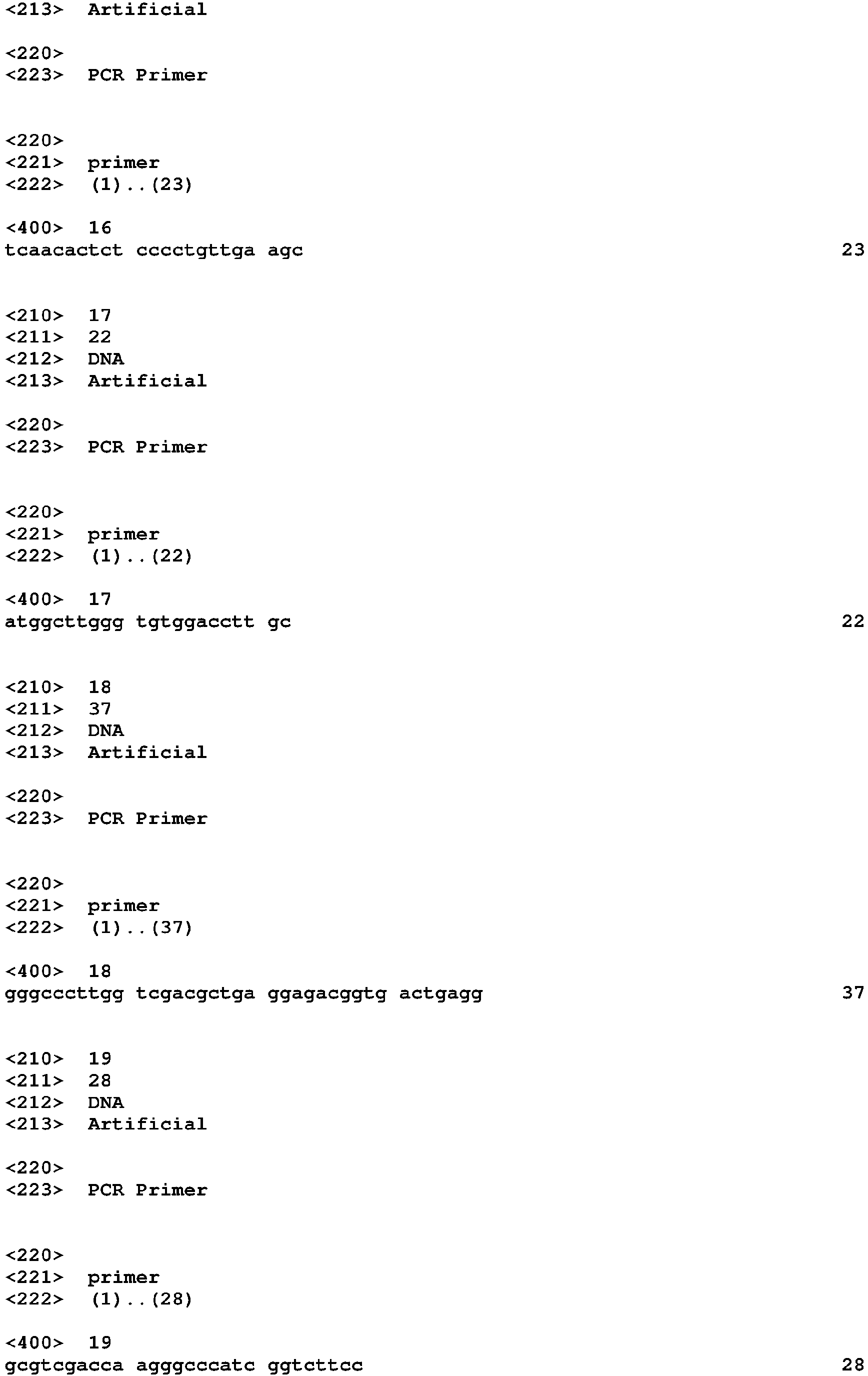


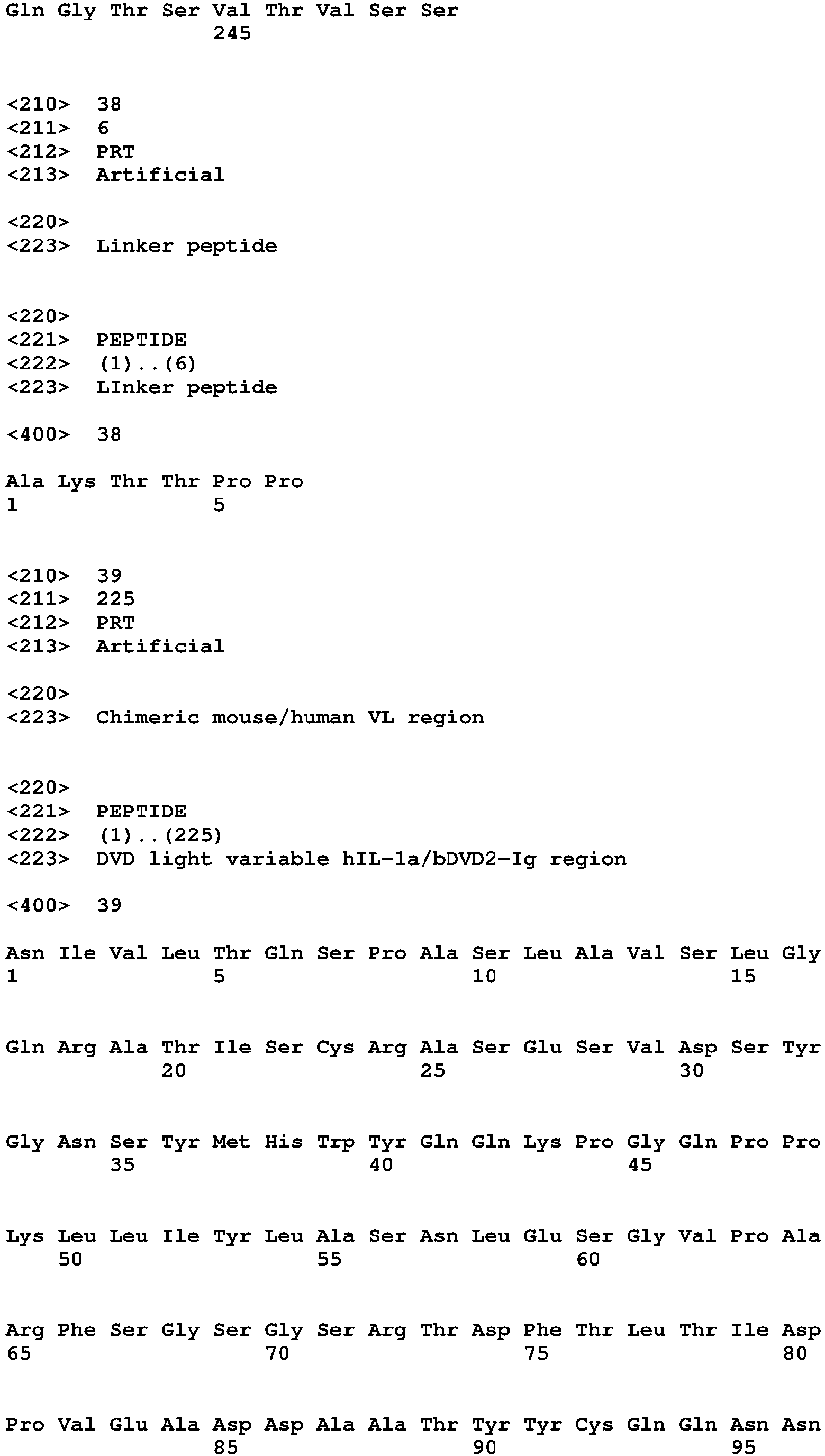


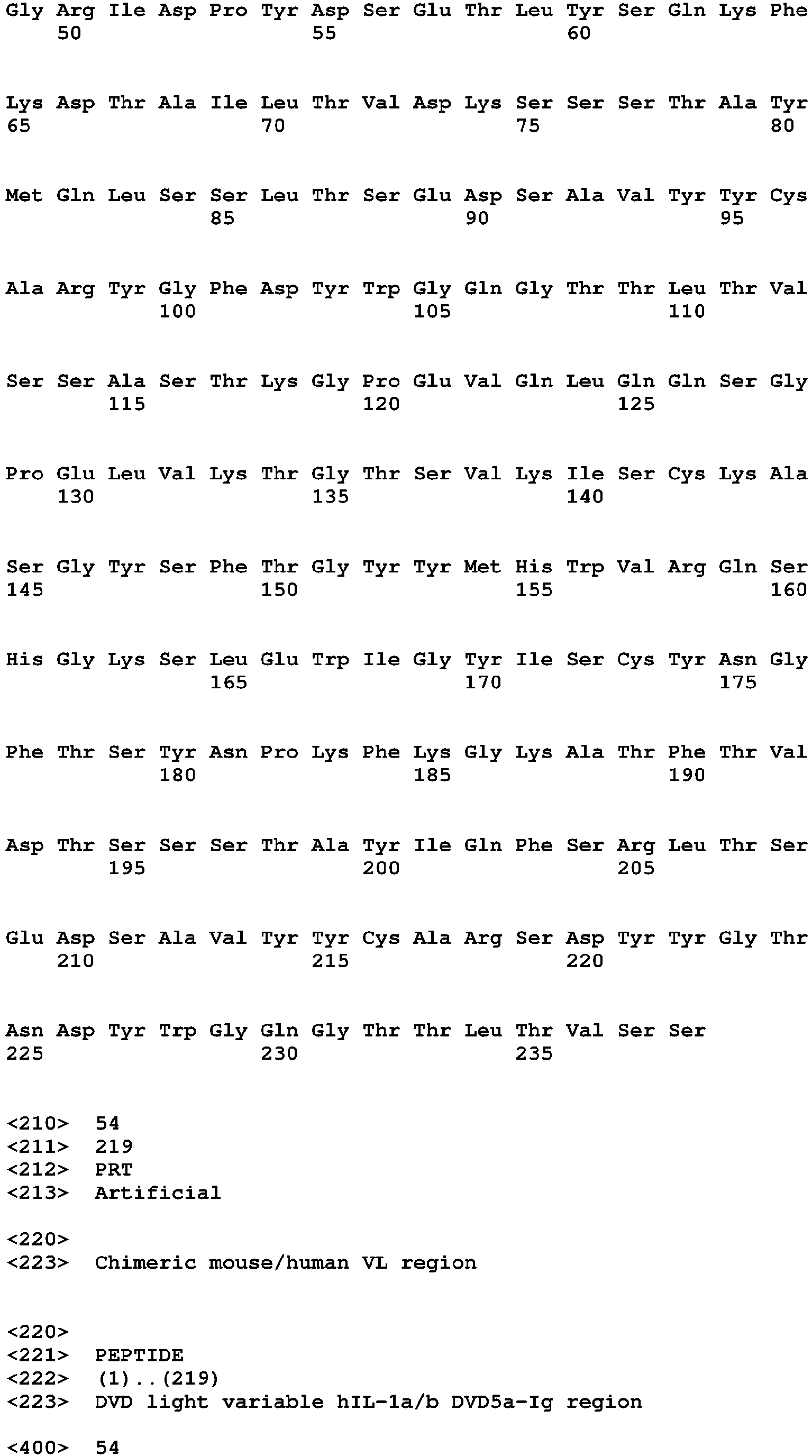


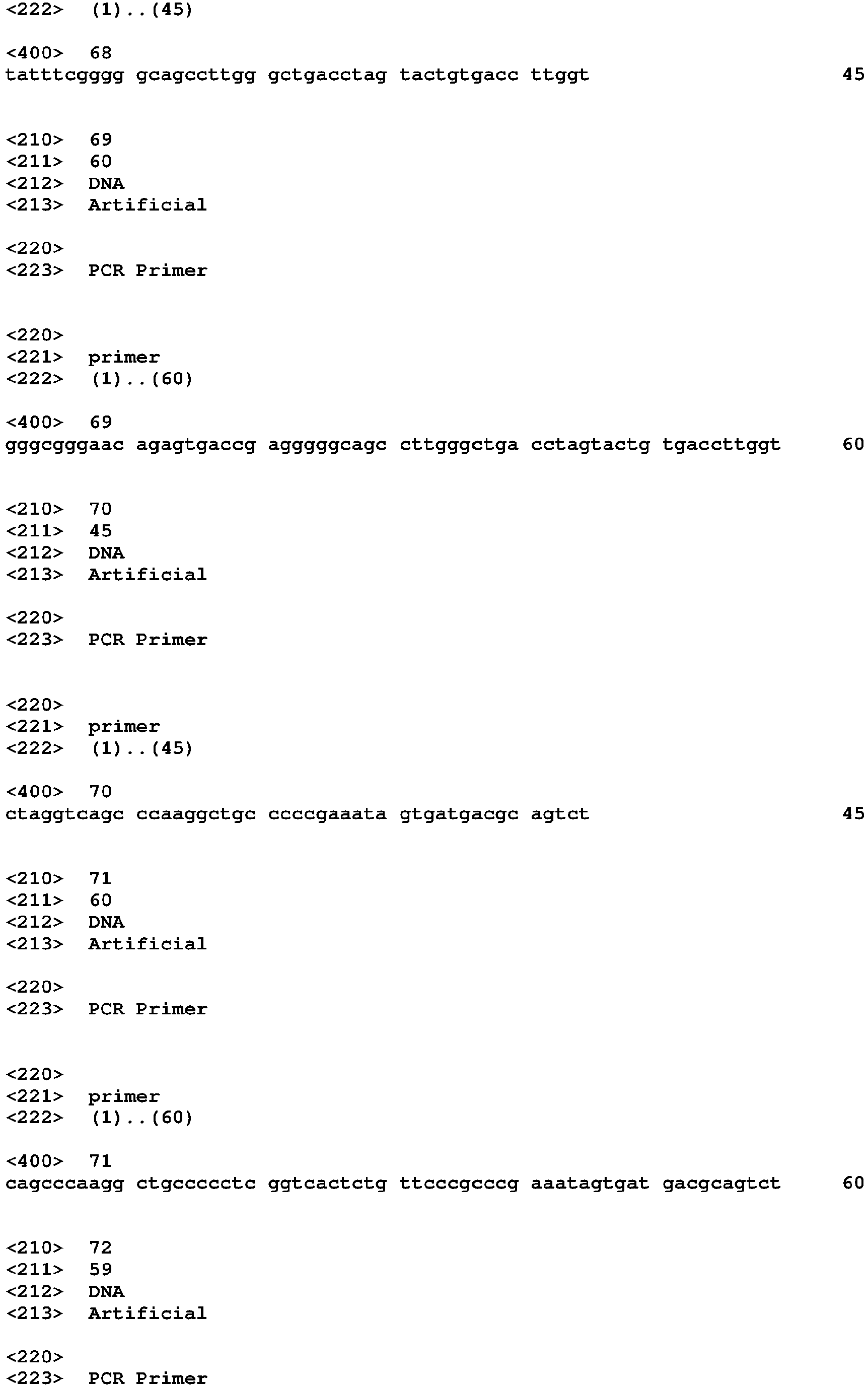


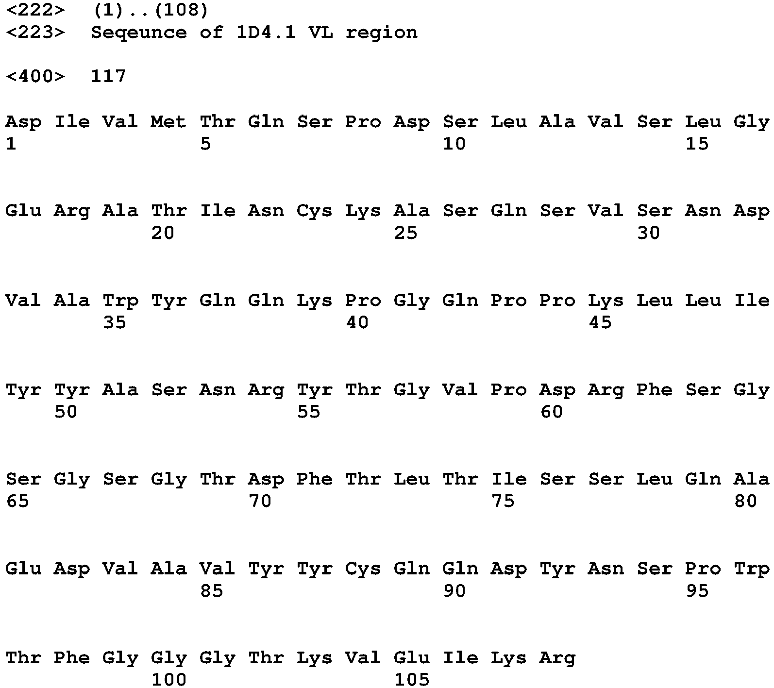
Claims (21)
- A binding protein comprising first and second polypeptide chains, wherein:said first polypeptide chain comprises formula 1: VD1-(X1)n-VD2-C-(X2)n, wherein VD1 is a first variable domain, VD2 is a second variable domain, C is a constant domain, X1 is a linker with the proviso that it is not a constant domain, X2 is an Fc region, and n is 0 or 1; andsaid second polypeptide chain comprises formula 2: VD1-(X1)n-VD2-C, wherein VD1 is a first variable domain, VD2 is a second variable domain, C is a constant domain, and X1 is a linker with the proviso that it is not a constant domain, and n is 0 or 1;wherein said two polypeptide chains of said binding protein form two functional binding domains, wherein one functional binding domain binds to IL-1β, and the other functional binding domain binds to a target antigen other than IL-1β.
- The binding protein according to Claim 1, wherein, for said two functional binding domains, one functional binding domain binds to IL-1β, and the other functional binding domain binds to a target antigen selected from the group consisting of: IL-1α, IL-13, and TNFα.
- The binding protein according to Claim 1 or Claim 2, wherein said Fc region, X2, is a variant of a native sequence Fc region, containing mutations to disrupt dimerization of the CH3 domain.
- The binding protein according to any one of Claims 1-3, wherein:C in said first polypeptide chain is CH1, and C in said second polypeptide chain is CL; orC in said first polypeptide chain is CL, and C in said second polypeptide chain is CH1.
- The binding protein according to any one of Claims 1-4, wherein X1 is a linker selected from the group consisting of AKTTPKLEEGEFSEAR; AKTTPKLEEGEFSEARV; AKTTPKLGG; RADAAP; RADAAPTVS; RADAAAAGGPGS; RADAAAA(G4S)4; SAKTTP; SAKTTPKLGG; SAKTTPKLEEGEFSEARV; ADAAP; ADAAPTVSIFPP; TVAAP; TVAAPSVFIFPP; QPKAAP; QPKAAPSVTLFPP; AKTTPP; AKTTPPSVTPLAP; AKTTAP; AKTTAPSVYPLAP; ASTKGP; ASTKGPSVFPLAP; GGGGSGGGGSGGGGS; GENKVEYAPALMALS; GPAKELTPLKEAKVS; and GHEAAAVMQVQYPAS.
- The binding protein according to any one of Claims 1-5, the binding protein further comprising:a third polypeptide chain having formula 1, and a fourth polypeptide having formula 2;wherein the four polypeptide chains of said binding protein form four functional binding domains, wherein two functional binding domains bind to IL-1β, and two functional binding domains bind to a target antigen other than IL-1β.
- The binding protein according to Claim 6, wherein, for said four functional binding domains, two functional binding domains bind to IL-1β, and the other functional binding domains bind to a target antigen selected from the group consisting of: IL-1α, IL-13, and TNFα.
- The binding protein according to any one of Claims 1-7, wherein, in said polypeptide chain of formula 1, VD1 is a heavy chain variable domain, and VD2 is a heavy chain variable domain; and wherein, in said polypeptide chain of formula 2, VD1 is a light chain variable domain, and VD2 is a light chain variable domain.
- The binding protein according to any one of Claims 1-8, wherein, for (X2)n, n is 0.
- The binding protein according to any one of Claims 1-8, wherein the variable domains VD1 on polypeptide chain formula 1 and formula 2 form a binding domain that binds IL-1 β.
- The binding protein according to any one of Claims 1-8, wherein the variable domains VD2 on polypeptide chain formula 1 and formula 2 form a binding domain that binds IL-1 β.
- The binding protein according to any one of Claims 1-8, wherein VD1 on polypeptide chain formula 1 comprises an amino acid sequence selected from the group consisting of SEQ ID NO:7, SEQ ID NO:9, and SEQ ID NO:11, and VD1 on polypeptide chain formula 2 comprises an amino acid sequence selected from the group consisting of SEQ ID NO:8, SEQ ID NO:10, and SEQ ID NO:12.
- The binding protein according to any one of Claims 1-8, wherein polypeptide chain formula 1 comprises an amino acid sequence selected from the group consisting of SEQ ID NO:33 and SEQ ID NO:37, and polypeptide chain formula 2 comprises an amino acid sequence selected from the group consisting of SEQ ID NO:35 and SEQ ID NO:39.
- A binding protein conjugate comprising a binding protein as described in any one of Claims 1-13, said binding protein conjugate further comprising an agent selected from the group consisting of an immunoadhension molecule, an imaging agent, a therapeutic agent, and a cytotoxic agent.
- The binding protein according to any one of Claims 1-13, wherein said binding protein is a crystallized binding protein.
- An isolated nucleic acid encoding an amino acid sequence of a binding protein according to any one of Claims 1-13.
- A vector comprising an isolated nucleic acid according to Claim 16.
- A host cell comprising a vector according to Claim 17.
- A pharmaceutical composition comprising the binding protein of any one of Claims 1-13 and a pharmaceutically acceptable carrier.
- The binding protein according to any one of Claims 1-13 for use in treating a subject for an autoimmune or inflammatory disease.
- The binding protein according to Claim 20 for use in treating a subject for asthma, arthritis, or inflammatory bowel disease.
Applications Claiming Priority (3)
| Application Number | Priority Date | Filing Date | Title |
|---|---|---|---|
| US70991105P | 2005-08-19 | 2005-08-19 | |
| US73289205P | 2005-11-02 | 2005-11-02 | |
| EP06813554A EP1928506A4 (en) | 2005-08-19 | 2006-08-18 | Dual variable domain immunoglobin and uses thereof |
Related Parent Applications (2)
| Application Number | Title | Priority Date | Filing Date |
|---|---|---|---|
| EP06813554A Division EP1928506A4 (en) | 2005-08-19 | 2006-08-18 | Dual variable domain immunoglobin and uses thereof |
| EP06813554.0 Division | 2006-08-18 |
Publications (2)
| Publication Number | Publication Date |
|---|---|
| EP2500357A2 true EP2500357A2 (en) | 2012-09-19 |
| EP2500357A3 EP2500357A3 (en) | 2012-10-24 |
Family
ID=46690864
Family Applications (7)
| Application Number | Title | Priority Date | Filing Date |
|---|---|---|---|
| EP12161970A Withdrawn EP2500354A3 (en) | 2005-08-19 | 2006-08-18 | Dual variable domain immunoglobulin and uses thereof |
| EP12162029A Withdrawn EP2500358A3 (en) | 2005-08-19 | 2006-08-18 | Dual variable domain immunoglobulin and uses thereof |
| EP20120161999 Withdrawn EP2500357A3 (en) | 2005-08-19 | 2006-08-18 | Dual variable domain immunoglobulin and uses thereof |
| EP12161976A Withdrawn EP2500355A3 (en) | 2005-08-19 | 2006-08-18 | Dual variable domain immunoglobulin and uses thereof |
| EP20120161985 Withdrawn EP2500356A3 (en) | 2005-08-19 | 2006-08-18 | Dual variable domain immunoglobulin and uses thereof |
| EP12161964A Withdrawn EP2500353A3 (en) | 2005-08-19 | 2006-08-18 | Dual variable domain immunoglobulin and uses thereof |
| EP12162057A Withdrawn EP2500359A3 (en) | 2005-08-19 | 2006-08-18 | Dual variable domain immunoglobulin and uses thereof |
Family Applications Before (2)
| Application Number | Title | Priority Date | Filing Date |
|---|---|---|---|
| EP12161970A Withdrawn EP2500354A3 (en) | 2005-08-19 | 2006-08-18 | Dual variable domain immunoglobulin and uses thereof |
| EP12162029A Withdrawn EP2500358A3 (en) | 2005-08-19 | 2006-08-18 | Dual variable domain immunoglobulin and uses thereof |
Family Applications After (4)
| Application Number | Title | Priority Date | Filing Date |
|---|---|---|---|
| EP12161976A Withdrawn EP2500355A3 (en) | 2005-08-19 | 2006-08-18 | Dual variable domain immunoglobulin and uses thereof |
| EP20120161985 Withdrawn EP2500356A3 (en) | 2005-08-19 | 2006-08-18 | Dual variable domain immunoglobulin and uses thereof |
| EP12161964A Withdrawn EP2500353A3 (en) | 2005-08-19 | 2006-08-18 | Dual variable domain immunoglobulin and uses thereof |
| EP12162057A Withdrawn EP2500359A3 (en) | 2005-08-19 | 2006-08-18 | Dual variable domain immunoglobulin and uses thereof |
Country Status (1)
| Country | Link |
|---|---|
| EP (7) | EP2500354A3 (en) |
Families Citing this family (5)
| Publication number | Priority date | Publication date | Assignee | Title |
|---|---|---|---|---|
| JOP20200236A1 (en) | 2012-09-21 | 2017-06-16 | Regeneron Pharma | Anti-cd3 antibodies, bispecific antigen-binding molecules that bind cd3 and cd20, and uses thereof |
| TWI754319B (en) | 2014-03-19 | 2022-02-01 | 美商再生元醫藥公司 | Methods and antibody compositions for tumor treatment |
| PT3221359T (en) | 2014-11-17 | 2020-06-23 | Regeneron Pharma | Methods for tumor treatment using cd3xcd20 bispecific antibody |
| AU2019331024A1 (en) | 2018-08-31 | 2021-03-18 | Regeneron Pharmaceuticals, Inc. | Dosing strategy that mitigates cytokine release syndrome for CD3/C20 bispecific antibodies |
| TW202330598A (en) * | 2021-06-30 | 2023-08-01 | 大陸商江蘇恆瑞醫藥股份有限公司 | Antigen-binding molecules that specifically bind to baff and il-12/23 and uses thereof |
Citations (121)
| Publication number | Priority date | Publication date | Assignee | Title |
|---|---|---|---|---|
| GB101134A (en) | 1916-05-18 | 1916-08-17 | Cornelius Maashant | Improvements in or relating to Padlocks, Bolts and like Fastening Devices. |
| US4526938A (en) | 1982-04-22 | 1985-07-02 | Imperial Chemical Industries Plc | Continuous release formulations |
| EP0229246A2 (en) | 1986-01-15 | 1987-07-22 | ANT Nachrichtentechnik GmbH | Method for decoding digital signals, as well as a Viterbi decoder and applications |
| EP0239400A2 (en) | 1986-03-27 | 1987-09-30 | Medical Research Council | Recombinant antibodies and methods for their production |
| US4753894A (en) | 1984-02-08 | 1988-06-28 | Cetus Corporation | Monoclonal anti-human breast cancer antibodies |
| US4816567A (en) | 1983-04-08 | 1989-03-28 | Genentech, Inc. | Recombinant immunoglobin preparations |
| US4880078A (en) | 1987-06-29 | 1989-11-14 | Honda Giken Kogyo Kabushiki Kaisha | Exhaust muffler |
| WO1990002809A1 (en) | 1988-09-02 | 1990-03-22 | Protein Engineering Corporation | Generation and selection of recombinant varied binding proteins |
| WO1990005144A1 (en) | 1988-11-11 | 1990-05-17 | Medical Research Council | Single domain ligands, receptors comprising said ligands, methods for their production, and use of said ligands and receptors |
| US4943533A (en) | 1984-03-01 | 1990-07-24 | The Regents Of The University Of California | Hybrid cell lines that produce monoclonal antibodies to epidermal growth factor receptor |
| US4946778A (en) | 1987-09-21 | 1990-08-07 | Genex Corporation | Single polypeptide chain binding molecules |
| WO1990014443A1 (en) | 1989-05-16 | 1990-11-29 | Huse William D | Co-expression of heteromeric receptors |
| WO1990014424A1 (en) | 1989-05-16 | 1990-11-29 | Scripps Clinic And Research Foundation | Method for isolating receptors having a preselected specificity |
| WO1990014430A1 (en) | 1989-05-16 | 1990-11-29 | Scripps Clinic And Research Foundation | A new method for tapping the immunological repertoire |
| US4980286A (en) | 1985-07-05 | 1990-12-25 | Whitehead Institute For Biomedical Research | In vivo introduction and expression of foreign genetic material in epithelial cells |
| WO1991005548A1 (en) | 1989-10-10 | 1991-05-02 | Pitman-Moore, Inc. | Sustained release composition for macromolecular proteins |
| WO1991009967A1 (en) | 1989-12-21 | 1991-07-11 | Celltech Limited | Humanised antibodies |
| WO1991010741A1 (en) | 1990-01-12 | 1991-07-25 | Cell Genesys, Inc. | Generation of xenogeneic antibodies |
| WO1991010737A1 (en) | 1990-01-11 | 1991-07-25 | Molecular Affinities Corporation | Production of antibodies using gene libraries |
| WO1991017271A1 (en) | 1990-05-01 | 1991-11-14 | Affymax Technologies N.V. | Recombinant library screening methods |
| WO1992001047A1 (en) | 1990-07-10 | 1992-01-23 | Cambridge Antibody Technology Limited | Methods for producing members of specific binding pairs |
| WO1992002551A1 (en) | 1990-08-02 | 1992-02-20 | B.R. Centre Limited | Methods for the production of proteins with a desired function |
| WO1992009690A2 (en) | 1990-12-03 | 1992-06-11 | Genentech, Inc. | Enrichment method for variant proteins with altered binding properties |
| US5128326A (en) | 1984-12-06 | 1992-07-07 | Biomatrix, Inc. | Drug delivery systems based on hyaluronans derivatives thereof and their salts and methods of producing same |
| WO1992015679A1 (en) | 1991-03-01 | 1992-09-17 | Protein Engineering Corporation | Improved epitode displaying phage |
| WO1992018619A1 (en) | 1991-04-10 | 1992-10-29 | The Scripps Research Institute | Heterodimeric receptor libraries using phagemids |
| WO1992019244A2 (en) | 1991-05-01 | 1992-11-12 | Henry M. Jackson Foundation For The Advancement Of Military Medicine | A method for treating infectious respiratory diseases |
| WO1992020791A1 (en) | 1990-07-10 | 1992-11-26 | Cambridge Antibody Technology Limited | Methods for producing members of specific binding pairs |
| EP0519596A1 (en) | 1991-05-17 | 1992-12-23 | Merck & Co. Inc. | A method for reducing the immunogenicity of antibody variable domains |
| WO1992022324A1 (en) | 1991-06-14 | 1992-12-23 | Xoma Corporation | Microbially-produced antibody fragments and their conjugates |
| WO1993001288A1 (en) | 1991-07-08 | 1993-01-21 | Deutsches Krebsforschungszentrum Stiftung des öffentlichen Rechts | Phagemide for screening antibodies |
| WO1993011236A1 (en) | 1991-12-02 | 1993-06-10 | Medical Research Council | Production of anti-self antibodies from antibody segment repertoires and displayed on phage |
| US5223409A (en) | 1988-09-02 | 1993-06-29 | Protein Engineering Corp. | Directed evolution of novel binding proteins |
| US5225539A (en) | 1986-03-27 | 1993-07-06 | Medical Research Council | Recombinant altered antibodies and methods of making altered antibodies |
| US5258498A (en) | 1987-05-21 | 1993-11-02 | Creative Biomolecules, Inc. | Polypeptide linkers for production of biosynthetic proteins |
| WO1994002602A1 (en) | 1992-07-24 | 1994-02-03 | Cell Genesys, Inc. | Generation of xenogeneic antibodies |
| EP0592106A1 (en) | 1992-09-09 | 1994-04-13 | Immunogen Inc | Resurfacing of rodent antibodies |
| WO1995015982A2 (en) | 1993-12-08 | 1995-06-15 | Genzyme Corporation | Process for generating specific antibodies |
| WO1995020045A1 (en) | 1994-01-21 | 1995-07-27 | The Institute Of Cancer Research: Royal Cancer Hospital | Antibodies to egf receptor and their antitumour effect |
| WO1995020401A1 (en) | 1994-01-31 | 1995-08-03 | Trustees Of Boston University | Polyclonal antibody libraries |
| WO1995024918A1 (en) | 1994-03-14 | 1995-09-21 | Genetics Institute, Inc. | Use of il-12 and il-12 antagonists in the treatment of autoimmune diseases |
| US5500362A (en) | 1987-01-08 | 1996-03-19 | Xoma Corporation | Chimeric antibody with specificity to human B cell surface antigen |
| US5516637A (en) | 1994-06-10 | 1996-05-14 | Dade International Inc. | Method involving display of protein binding pairs on the surface of bacterial pili and bacteriophage |
| US5530101A (en) | 1988-12-28 | 1996-06-25 | Protein Design Labs, Inc. | Humanized immunoglobulins |
| WO1996020698A2 (en) | 1995-01-05 | 1996-07-11 | The Board Of Regents Acting For And On Behalf Of The University Of Michigan | Surface-modified nanoparticles and method of making and using same |
| US5558864A (en) | 1991-03-06 | 1996-09-24 | Merck Patent Gesellschaft Mit Beschrankter Haftung | Humanized and chimeric anti-epidermal growth factor receptor monoclonal antibodies |
| US5565352A (en) | 1993-11-24 | 1996-10-15 | Arch Development Corporation | Deubiquitinating enzyme: compositions and methods |
| US5565332A (en) | 1991-09-23 | 1996-10-15 | Medical Research Council | Production of chimeric antibodies - a combinatorial approach |
| WO1996033735A1 (en) | 1995-04-27 | 1996-10-31 | Abgenix, Inc. | Human antibodies derived from immunized xenomice |
| WO1996034096A1 (en) | 1995-04-28 | 1996-10-31 | Abgenix, Inc. | Human antibodies derived from immunized xenomice |
| WO1996040210A1 (en) | 1995-06-07 | 1996-12-19 | Imclone Systems Incorporated | Antibody and antibody fragments for inhibiting the growth of tumors |
| US5624821A (en) | 1987-03-18 | 1997-04-29 | Scotgen Biopharmaceuticals Incorporated | Antibodies with altered effector functions |
| US5641870A (en) | 1995-04-20 | 1997-06-24 | Genentech, Inc. | Low pH hydrophobic interaction chromatography for antibody purification |
| WO1997029131A1 (en) | 1996-02-09 | 1997-08-14 | Basf Aktiengesellschaft | HUMAN ANTIBODIES THAT BIND HUMAN TNF$g(a) |
| WO1997032572A2 (en) | 1996-03-04 | 1997-09-12 | The Penn State Research Foundation | Materials and methods for enhancing cellular internalization |
| US5677171A (en) | 1988-01-12 | 1997-10-14 | Genentech, Inc. | Monoclonal antibodies directed to the HER2 receptor |
| US5679377A (en) | 1989-11-06 | 1997-10-21 | Alkermes Controlled Therapeutics, Inc. | Protein microspheres and methods of using them |
| WO1997044013A1 (en) | 1996-05-24 | 1997-11-27 | Massachusetts Institute Of Technology | Aerodynamically light particles for pulmonary drug delivery |
| US5698426A (en) | 1990-09-28 | 1997-12-16 | Ixsys, Incorporated | Surface expression libraries of heteromeric receptors |
| US5714350A (en) | 1992-03-09 | 1998-02-03 | Protein Design Labs, Inc. | Increasing antibody affinity by altering glycosylation in the immunoglobulin variable region |
| US5714352A (en) | 1996-03-20 | 1998-02-03 | Xenotech Incorporated | Directed switch-mediated DNA recombination |
| US5723323A (en) | 1985-03-30 | 1998-03-03 | Kauffman; Stuart Alan | Method of identifying a stochastically-generated peptide, polypeptide, or protein having ligand binding property and compositions thereof |
| US5733743A (en) | 1992-03-24 | 1998-03-31 | Cambridge Antibody Technology Limited | Methods for producing members of specific binding pairs |
| US5736137A (en) | 1992-11-13 | 1998-04-07 | Idec Pharmaceuticals Corporation | Therapeutic application of chimeric and radiolabeled antibodies to human B lymphocyte restricted differentiation antigen for treatment of B cell lymphoma |
| WO1998016654A1 (en) | 1996-10-11 | 1998-04-23 | Japan Tobacco, Inc. | Production of a multimeric protein by cell fusion method |
| US5750753A (en) | 1996-01-24 | 1998-05-12 | Chisso Corporation | Method for manufacturing acryloxypropysilane |
| US5763192A (en) | 1986-11-20 | 1998-06-09 | Ixsys, Incorporated | Process for obtaining DNA, RNA, peptides, polypeptides, or protein, by recombinant DNA technique |
| WO1998024893A2 (en) | 1996-12-03 | 1998-06-11 | Abgenix, Inc. | TRANSGENIC MAMMALS HAVING HUMAN IG LOCI INCLUDING PLURAL VH AND Vλ REGIONS AND ANTIBODIES PRODUCED THEREFROM |
| US5766886A (en) | 1991-12-13 | 1998-06-16 | Xoma Corporation | Modified antibody variable domains |
| US5780225A (en) | 1990-01-12 | 1998-07-14 | Stratagene | Method for generating libaries of antibody genes comprising amplification of diverse antibody DNAs and methods for using these libraries for the production of diverse antigen combining molecules |
| WO1998031346A1 (en) | 1997-01-16 | 1998-07-23 | Massachusetts Institute Of Technology | Preparation of particles for inhalation |
| WO1998031700A1 (en) | 1997-01-21 | 1998-07-23 | The General Hospital Corporation | Selection of proteins using rna-protein fusions |
| WO1998050433A2 (en) | 1997-05-05 | 1998-11-12 | Abgenix, Inc. | Human monoclonal antibodies to epidermal growth factor receptor |
| US5855913A (en) | 1997-01-16 | 1999-01-05 | Massachusetts Instite Of Technology | Particles incorporating surfactants for pulmonary drug delivery |
| WO1999015154A1 (en) | 1997-09-24 | 1999-04-01 | Alkermes Controlled Therapeutics, Inc. | Methods for fabricating polymer-based controlled release preparations |
| US5891996A (en) | 1972-09-17 | 1999-04-06 | Centro De Inmunologia Molecular | Humanized and chimeric monoclonal antibodies that recognize epidermal growth factor receptor (EGF-R); diagnostic and therapeutic use |
| WO1999020253A1 (en) | 1997-10-23 | 1999-04-29 | Bioglan Therapeutics Ab | Encapsulation method |
| WO1999025044A1 (en) | 1997-11-07 | 1999-05-20 | Nathan Cohen | Microstrip patch antenna with fractal structure |
| US5912015A (en) | 1992-03-12 | 1999-06-15 | Alkermes Controlled Therapeutics, Inc. | Modulated release from biocompatible polymers |
| US5916597A (en) | 1995-08-31 | 1999-06-29 | Alkermes Controlled Therapeutics, Inc. | Composition and method using solid-phase particles for sustained in vivo release of a biologically active agent |
| US5934272A (en) | 1993-01-29 | 1999-08-10 | Aradigm Corporation | Device and method of creating aerosolized mist of respiratory drug |
| WO1999045031A2 (en) | 1998-03-03 | 1999-09-10 | Abgenix, Inc. | Cd147 binding molecules as therapeutics |
| WO1999053049A1 (en) | 1998-04-15 | 1999-10-21 | Abgenix, Inc. | Epitope-driven human antibody production and gene expression profiling |
| WO1999054342A1 (en) | 1998-04-20 | 1999-10-28 | Pablo Umana | Glycosylation engineering of antibodies for improving antibody-dependent cellular cytotoxicity |
| US5985309A (en) | 1996-05-24 | 1999-11-16 | Massachusetts Institute Of Technology | Preparation of particles for inhalation |
| US5998209A (en) | 1995-04-21 | 1999-12-07 | Abgenix, Inc. | Generation of large genomic DNA deletions |
| WO1999066903A2 (en) | 1998-06-24 | 1999-12-29 | Advanced Inhalation Research, Inc. | Large porous particles emitted from an inhaler |
| US6019968A (en) | 1995-04-14 | 2000-02-01 | Inhale Therapeutic Systems, Inc. | Dispersible antibody compositions and methods for their preparation and use |
| WO2000009560A2 (en) | 1998-08-17 | 2000-02-24 | Abgenix, Inc. | Generation of modified molecules with increased serum half-lives |
| US6075181A (en) | 1990-01-12 | 2000-06-13 | Abgenix, Inc. | Human antibodies derived from immunized xenomice |
| WO2000037504A2 (en) | 1998-12-23 | 2000-06-29 | Pfizer Inc. | Human monoclonal antibodies to ctla-4 |
| US6091001A (en) | 1995-03-29 | 2000-07-18 | Abgenix, Inc. | Production of antibodies using Cre-mediated site-specific recombination |
| WO2000056772A1 (en) | 1999-03-25 | 2000-09-28 | Knoll Gmbh | Human antibodies that bind human il-12 and methods for producing |
| US6130364A (en) | 1995-03-29 | 2000-10-10 | Abgenix, Inc. | Production of antibodies using Cre-mediated site-specific recombination |
| US6204023B1 (en) | 1985-11-01 | 2001-03-20 | Xoma Ltd. | Modular assembly of antibody genes, antibodies prepared thereby and use |
| WO2001058956A2 (en) | 2000-02-10 | 2001-08-16 | Abbott Laboratories | Antibodies that bind human interleukin-18 and methods of making and using |
| WO2001062931A2 (en) | 2000-02-25 | 2001-08-30 | The Government Of The United States, As Represented By The Secretary Of The Department Of Health And Human Services | ANTI-EGFRvIII SCFVS WITH IMPROVED CYTOTOXICITY AND YIELD, IMMUNOTOXINS BASED THEREON, AND METHODS OF USE THEREOF |
| WO2001077342A1 (en) | 2000-04-11 | 2001-10-18 | Genentech, Inc. | Multivalent antibodies and uses therefor |
| WO2001083525A2 (en) | 2000-05-03 | 2001-11-08 | Amgen Inc. | Modified peptides, comprising an fc domain, as therapeutic agents |
| WO2001088138A1 (en) | 2000-05-19 | 2001-11-22 | Scancell Limited | Humanised antibodies to the epidermal growth factor receptor |
| WO2002002773A2 (en) | 2000-06-29 | 2002-01-10 | Abbott Laboratories | Dual specificity antibodies and methods of making and using |
| EP1176195A1 (en) | 1999-04-09 | 2002-01-30 | Kyowa Hakko Kogyo Co., Ltd. | Method for controlling the activity of immunologically functional molecule |
| WO2002072636A2 (en) | 2000-12-28 | 2002-09-19 | Altus Biologics Inc. | Crystals of whole antibodies and fragments thereof and methods for making and using them |
| US20020137134A1 (en) | 2000-06-28 | 2002-09-26 | Gerngross Tillman U. | Methods for producing modified glycoproteins |
| WO2002097048A2 (en) | 2001-05-30 | 2002-12-05 | Centocor, Inc. | ANTI-p40 IMMUNOGLOBULIN DERIVED PROTEINS, COMPOSITIONS, METHODS AND USES |
| WO2003016466A2 (en) | 2001-08-17 | 2003-02-27 | Eli Lilly And Company | ANTI-Aβ ANTIBODIES |
| WO2003035835A2 (en) | 2001-10-25 | 2003-05-01 | Genentech, Inc. | Glycoprotein compositions |
| US20030186374A1 (en) | 2001-10-01 | 2003-10-02 | Hufton Simon E. | Multi-chain eukaryotic display vectors and uses thereof |
| US20040018590A1 (en) | 2000-06-28 | 2004-01-29 | Gerngross Tillman U. | Combinatorial DNA library for producing modified N-glycans in lower eukaryotes |
| US6699658B1 (en) | 1996-05-31 | 2004-03-02 | Board Of Trustees Of The University Of Illinois | Yeast cell surface display of proteins and uses thereof |
| WO2004078140A2 (en) | 2003-03-05 | 2004-09-16 | Halozyme, Inc. | SOLUBLE HYALURONIDASE GLYCOPROTEIN (sHASEGP), PROCESS FOR PREPARING THE SAME, USES AND PHARMACEUTICAL COMPOSITIONS COMPRISING THEREOF |
| US20050042664A1 (en) | 2003-08-22 | 2005-02-24 | Medimmune, Inc. | Humanization of antibodies |
| US6914128B1 (en) | 1999-03-25 | 2005-07-05 | Abbott Gmbh & Co. Kg | Human antibodies that bind human IL-12 and methods for producing |
| US20050147610A1 (en) | 2003-11-12 | 2005-07-07 | Tariq Ghayur | IL-18 binding proteins |
| WO2005100584A2 (en) | 2004-04-15 | 2005-10-27 | Glycofi, Inc. | Production of galactosylated glycoproteins in lower eukaryotes |
| US20060104968A1 (en) | 2003-03-05 | 2006-05-18 | Halozyme, Inc. | Soluble glycosaminoglycanases and methods of preparing and using soluble glycosaminogly ycanases |
| US9105939B2 (en) | 2008-03-10 | 2015-08-11 | Nissan Motor Co., Ltd. | Battery with battery electrode and method of manufacturing same |
| US9109630B2 (en) | 2013-01-15 | 2015-08-18 | Jtekt Corporation | Rolling bearing unit |
| US9401234B2 (en) | 2013-03-22 | 2016-07-26 | Polytronics Technology Corp. | Over-current protection device |
| US9618978B2 (en) | 2013-10-03 | 2017-04-11 | Acer Incorporated | Methods for controlling a touch panel and portable computers using the same |
| US9816280B1 (en) | 2016-11-02 | 2017-11-14 | Matthew Reitnauer | Portable floor |
Family Cites Families (8)
| Publication number | Priority date | Publication date | Assignee | Title |
|---|---|---|---|---|
| US5780A (en) | 1848-09-19 | Improvement in preparing shoe-pegs | ||
| US225A (en) | 1837-06-03 | Machine fob | ||
| DE4118120A1 (en) * | 1991-06-03 | 1992-12-10 | Behringwerke Ag | TETRAVALENT BISPECIFIC RECEPTORS, THEIR PRODUCTION AND USE |
| WO1995009917A1 (en) * | 1993-10-07 | 1995-04-13 | The Regents Of The University Of California | Genetically engineered bispecific tetravalent antibodies |
| US6015884A (en) * | 1996-03-28 | 2000-01-18 | The Johns Hopkins University | Soluble divalent and multivalent heterodimeric analogs of proteins |
| CA2410551A1 (en) * | 2000-06-30 | 2002-01-10 | Vlaams Interuniversitair Instituut Voor Biotechnologie Vzw (Vib) | Heterodimeric fusion proteins |
| WO2006020258A2 (en) * | 2004-07-17 | 2006-02-23 | Imclone Systems Incorporated | Novel tetravalent bispecific antibody |
| EP2516670B1 (en) | 2009-12-23 | 2016-08-24 | DuPont Nutrition Biosciences ApS | Method |
-
2006
- 2006-08-18 EP EP12161970A patent/EP2500354A3/en not_active Withdrawn
- 2006-08-18 EP EP12162029A patent/EP2500358A3/en not_active Withdrawn
- 2006-08-18 EP EP20120161999 patent/EP2500357A3/en not_active Withdrawn
- 2006-08-18 EP EP12161976A patent/EP2500355A3/en not_active Withdrawn
- 2006-08-18 EP EP20120161985 patent/EP2500356A3/en not_active Withdrawn
- 2006-08-18 EP EP12161964A patent/EP2500353A3/en not_active Withdrawn
- 2006-08-18 EP EP12162057A patent/EP2500359A3/en not_active Withdrawn
Patent Citations (149)
| Publication number | Priority date | Publication date | Assignee | Title |
|---|---|---|---|---|
| GB101134A (en) | 1916-05-18 | 1916-08-17 | Cornelius Maashant | Improvements in or relating to Padlocks, Bolts and like Fastening Devices. |
| US5891996A (en) | 1972-09-17 | 1999-04-06 | Centro De Inmunologia Molecular | Humanized and chimeric monoclonal antibodies that recognize epidermal growth factor receptor (EGF-R); diagnostic and therapeutic use |
| US4526938A (en) | 1982-04-22 | 1985-07-02 | Imperial Chemical Industries Plc | Continuous release formulations |
| US4816567A (en) | 1983-04-08 | 1989-03-28 | Genentech, Inc. | Recombinant immunoglobin preparations |
| US4753894A (en) | 1984-02-08 | 1988-06-28 | Cetus Corporation | Monoclonal anti-human breast cancer antibodies |
| US4943533A (en) | 1984-03-01 | 1990-07-24 | The Regents Of The University Of California | Hybrid cell lines that produce monoclonal antibodies to epidermal growth factor receptor |
| US5128326A (en) | 1984-12-06 | 1992-07-07 | Biomatrix, Inc. | Drug delivery systems based on hyaluronans derivatives thereof and their salts and methods of producing same |
| US5824514A (en) | 1985-03-30 | 1998-10-20 | Stuart A. Kauffman | Process for the production of expression vectors comprising at least one stochastic sequence of polynucleotides |
| US5976862A (en) | 1985-03-30 | 1999-11-02 | Ixsys Corporation | Process for obtaining DNA, RNA, peptides, polypeptides, or proteins, by recombinant DNA technique |
| US5814476A (en) | 1985-03-30 | 1998-09-29 | Stuart Kauffman | Process for the production of stochastically-generated transcription or translation products |
| US5817483A (en) | 1985-03-30 | 1998-10-06 | Stuart Kauffman | Process for the production of stochastically-generated peptides,polypeptides or proteins having a predetermined property |
| US5723323A (en) | 1985-03-30 | 1998-03-03 | Kauffman; Stuart Alan | Method of identifying a stochastically-generated peptide, polypeptide, or protein having ligand binding property and compositions thereof |
| US4980286A (en) | 1985-07-05 | 1990-12-25 | Whitehead Institute For Biomedical Research | In vivo introduction and expression of foreign genetic material in epithelial cells |
| US6204023B1 (en) | 1985-11-01 | 2001-03-20 | Xoma Ltd. | Modular assembly of antibody genes, antibodies prepared thereby and use |
| EP0229246A2 (en) | 1986-01-15 | 1987-07-22 | ANT Nachrichtentechnik GmbH | Method for decoding digital signals, as well as a Viterbi decoder and applications |
| US5225539A (en) | 1986-03-27 | 1993-07-06 | Medical Research Council | Recombinant altered antibodies and methods of making altered antibodies |
| EP0239400A2 (en) | 1986-03-27 | 1987-09-30 | Medical Research Council | Recombinant antibodies and methods for their production |
| US5763192A (en) | 1986-11-20 | 1998-06-09 | Ixsys, Incorporated | Process for obtaining DNA, RNA, peptides, polypeptides, or protein, by recombinant DNA technique |
| US5500362A (en) | 1987-01-08 | 1996-03-19 | Xoma Corporation | Chimeric antibody with specificity to human B cell surface antigen |
| US5648260A (en) | 1987-03-18 | 1997-07-15 | Scotgen Biopharmaceuticals Incorporated | DNA encoding antibodies with altered effector functions |
| US5624821A (en) | 1987-03-18 | 1997-04-29 | Scotgen Biopharmaceuticals Incorporated | Antibodies with altered effector functions |
| US5258498A (en) | 1987-05-21 | 1993-11-02 | Creative Biomolecules, Inc. | Polypeptide linkers for production of biosynthetic proteins |
| US4880078A (en) | 1987-06-29 | 1989-11-14 | Honda Giken Kogyo Kabushiki Kaisha | Exhaust muffler |
| US4946778A (en) | 1987-09-21 | 1990-08-07 | Genex Corporation | Single polypeptide chain binding molecules |
| US5677171A (en) | 1988-01-12 | 1997-10-14 | Genentech, Inc. | Monoclonal antibodies directed to the HER2 receptor |
| US5403484A (en) | 1988-09-02 | 1995-04-04 | Protein Engineering Corporation | Viruses expressing chimeric binding proteins |
| US5571698A (en) | 1988-09-02 | 1996-11-05 | Protein Engineering Corporation | Directed evolution of novel binding proteins |
| US5223409A (en) | 1988-09-02 | 1993-06-29 | Protein Engineering Corp. | Directed evolution of novel binding proteins |
| WO1990002809A1 (en) | 1988-09-02 | 1990-03-22 | Protein Engineering Corporation | Generation and selection of recombinant varied binding proteins |
| WO1990005144A1 (en) | 1988-11-11 | 1990-05-17 | Medical Research Council | Single domain ligands, receptors comprising said ligands, methods for their production, and use of said ligands and receptors |
| US6180370B1 (en) | 1988-12-28 | 2001-01-30 | Protein Design Labs, Inc. | Humanized immunoglobulins and methods of making the same |
| US5693762A (en) | 1988-12-28 | 1997-12-02 | Protein Design Labs, Inc. | Humanized immunoglobulins |
| US5530101A (en) | 1988-12-28 | 1996-06-25 | Protein Design Labs, Inc. | Humanized immunoglobulins |
| US5585089A (en) | 1988-12-28 | 1996-12-17 | Protein Design Labs, Inc. | Humanized immunoglobulins |
| WO1990014424A1 (en) | 1989-05-16 | 1990-11-29 | Scripps Clinic And Research Foundation | Method for isolating receptors having a preselected specificity |
| WO1990014443A1 (en) | 1989-05-16 | 1990-11-29 | Huse William D | Co-expression of heteromeric receptors |
| WO1990014430A1 (en) | 1989-05-16 | 1990-11-29 | Scripps Clinic And Research Foundation | A new method for tapping the immunological repertoire |
| WO1991005548A1 (en) | 1989-10-10 | 1991-05-02 | Pitman-Moore, Inc. | Sustained release composition for macromolecular proteins |
| US5679377A (en) | 1989-11-06 | 1997-10-21 | Alkermes Controlled Therapeutics, Inc. | Protein microspheres and methods of using them |
| WO1991009967A1 (en) | 1989-12-21 | 1991-07-11 | Celltech Limited | Humanised antibodies |
| WO1991010737A1 (en) | 1990-01-11 | 1991-07-25 | Molecular Affinities Corporation | Production of antibodies using gene libraries |
| US6114598A (en) | 1990-01-12 | 2000-09-05 | Abgenix, Inc. | Generation of xenogeneic antibodies |
| US6075181A (en) | 1990-01-12 | 2000-06-13 | Abgenix, Inc. | Human antibodies derived from immunized xenomice |
| US5780225A (en) | 1990-01-12 | 1998-07-14 | Stratagene | Method for generating libaries of antibody genes comprising amplification of diverse antibody DNAs and methods for using these libraries for the production of diverse antigen combining molecules |
| US5939598A (en) | 1990-01-12 | 1999-08-17 | Abgenix, Inc. | Method of making transgenic mice lacking endogenous heavy chains |
| WO1991010741A1 (en) | 1990-01-12 | 1991-07-25 | Cell Genesys, Inc. | Generation of xenogeneic antibodies |
| US5427908A (en) | 1990-05-01 | 1995-06-27 | Affymax Technologies N.V. | Recombinant library screening methods |
| WO1991017271A1 (en) | 1990-05-01 | 1991-11-14 | Affymax Technologies N.V. | Recombinant library screening methods |
| US5580717A (en) | 1990-05-01 | 1996-12-03 | Affymax Technologies N.V. | Recombinant library screening methods |
| WO1992001047A1 (en) | 1990-07-10 | 1992-01-23 | Cambridge Antibody Technology Limited | Methods for producing members of specific binding pairs |
| US5969108A (en) | 1990-07-10 | 1999-10-19 | Medical Research Council | Methods for producing members of specific binding pairs |
| WO1992020791A1 (en) | 1990-07-10 | 1992-11-26 | Cambridge Antibody Technology Limited | Methods for producing members of specific binding pairs |
| WO1992002551A1 (en) | 1990-08-02 | 1992-02-20 | B.R. Centre Limited | Methods for the production of proteins with a desired function |
| US5627052A (en) | 1990-08-02 | 1997-05-06 | B.R. Centre, Ltd. | Methods for the production of proteins with a desired function |
| US5698426A (en) | 1990-09-28 | 1997-12-16 | Ixsys, Incorporated | Surface expression libraries of heteromeric receptors |
| WO1992009690A2 (en) | 1990-12-03 | 1992-06-11 | Genentech, Inc. | Enrichment method for variant proteins with altered binding properties |
| US5821047A (en) | 1990-12-03 | 1998-10-13 | Genentech, Inc. | Monovalent phage display |
| WO1992015679A1 (en) | 1991-03-01 | 1992-09-17 | Protein Engineering Corporation | Improved epitode displaying phage |
| US5558864A (en) | 1991-03-06 | 1996-09-24 | Merck Patent Gesellschaft Mit Beschrankter Haftung | Humanized and chimeric anti-epidermal growth factor receptor monoclonal antibodies |
| WO1992018619A1 (en) | 1991-04-10 | 1992-10-29 | The Scripps Research Institute | Heterodimeric receptor libraries using phagemids |
| US5658727A (en) | 1991-04-10 | 1997-08-19 | The Scripps Research Institute | Heterodimeric receptor libraries using phagemids |
| WO1992019244A2 (en) | 1991-05-01 | 1992-11-12 | Henry M. Jackson Foundation For The Advancement Of Military Medicine | A method for treating infectious respiratory diseases |
| US5290540A (en) | 1991-05-01 | 1994-03-01 | Henry M. Jackson Foundation For The Advancement Of Military Medicine | Method for treating infectious respiratory diseases |
| EP0519596A1 (en) | 1991-05-17 | 1992-12-23 | Merck & Co. Inc. | A method for reducing the immunogenicity of antibody variable domains |
| WO1992022324A1 (en) | 1991-06-14 | 1992-12-23 | Xoma Corporation | Microbially-produced antibody fragments and their conjugates |
| WO1993001288A1 (en) | 1991-07-08 | 1993-01-21 | Deutsches Krebsforschungszentrum Stiftung des öffentlichen Rechts | Phagemide for screening antibodies |
| US5565332A (en) | 1991-09-23 | 1996-10-15 | Medical Research Council | Production of chimeric antibodies - a combinatorial approach |
| WO1993011236A1 (en) | 1991-12-02 | 1993-06-10 | Medical Research Council | Production of anti-self antibodies from antibody segment repertoires and displayed on phage |
| US5766886A (en) | 1991-12-13 | 1998-06-16 | Xoma Corporation | Modified antibody variable domains |
| US6350861B1 (en) | 1992-03-09 | 2002-02-26 | Protein Design Labs, Inc. | Antibodies with increased binding affinity |
| US5714350A (en) | 1992-03-09 | 1998-02-03 | Protein Design Labs, Inc. | Increasing antibody affinity by altering glycosylation in the immunoglobulin variable region |
| US5912015A (en) | 1992-03-12 | 1999-06-15 | Alkermes Controlled Therapeutics, Inc. | Modulated release from biocompatible polymers |
| US5733743A (en) | 1992-03-24 | 1998-03-31 | Cambridge Antibody Technology Limited | Methods for producing members of specific binding pairs |
| WO1994002602A1 (en) | 1992-07-24 | 1994-02-03 | Cell Genesys, Inc. | Generation of xenogeneic antibodies |
| EP0592106A1 (en) | 1992-09-09 | 1994-04-13 | Immunogen Inc | Resurfacing of rodent antibodies |
| US5736137A (en) | 1992-11-13 | 1998-04-07 | Idec Pharmaceuticals Corporation | Therapeutic application of chimeric and radiolabeled antibodies to human B lymphocyte restricted differentiation antigen for treatment of B cell lymphoma |
| US5934272A (en) | 1993-01-29 | 1999-08-10 | Aradigm Corporation | Device and method of creating aerosolized mist of respiratory drug |
| US5565352A (en) | 1993-11-24 | 1996-10-15 | Arch Development Corporation | Deubiquitinating enzyme: compositions and methods |
| WO1995015982A2 (en) | 1993-12-08 | 1995-06-15 | Genzyme Corporation | Process for generating specific antibodies |
| WO1995020045A1 (en) | 1994-01-21 | 1995-07-27 | The Institute Of Cancer Research: Royal Cancer Hospital | Antibodies to egf receptor and their antitumour effect |
| WO1995020401A1 (en) | 1994-01-31 | 1995-08-03 | Trustees Of Boston University | Polyclonal antibody libraries |
| WO1995024918A1 (en) | 1994-03-14 | 1995-09-21 | Genetics Institute, Inc. | Use of il-12 and il-12 antagonists in the treatment of autoimmune diseases |
| US5516637A (en) | 1994-06-10 | 1996-05-14 | Dade International Inc. | Method involving display of protein binding pairs on the surface of bacterial pili and bacteriophage |
| US6506883B2 (en) | 1994-11-18 | 2003-01-14 | Centro De Inmunologia Molecular | Humanized and chimeric monoclonal antibodies that recognize epidermal growth factor receptor (EGF-R); diagnostic and therapeutic use |
| WO1996020698A2 (en) | 1995-01-05 | 1996-07-11 | The Board Of Regents Acting For And On Behalf Of The University Of Michigan | Surface-modified nanoparticles and method of making and using same |
| US6130364A (en) | 1995-03-29 | 2000-10-10 | Abgenix, Inc. | Production of antibodies using Cre-mediated site-specific recombination |
| US6091001A (en) | 1995-03-29 | 2000-07-18 | Abgenix, Inc. | Production of antibodies using Cre-mediated site-specific recombination |
| US6019968A (en) | 1995-04-14 | 2000-02-01 | Inhale Therapeutic Systems, Inc. | Dispersible antibody compositions and methods for their preparation and use |
| US5641870A (en) | 1995-04-20 | 1997-06-24 | Genentech, Inc. | Low pH hydrophobic interaction chromatography for antibody purification |
| US5998209A (en) | 1995-04-21 | 1999-12-07 | Abgenix, Inc. | Generation of large genomic DNA deletions |
| WO1996033735A1 (en) | 1995-04-27 | 1996-10-31 | Abgenix, Inc. | Human antibodies derived from immunized xenomice |
| WO1996034096A1 (en) | 1995-04-28 | 1996-10-31 | Abgenix, Inc. | Human antibodies derived from immunized xenomice |
| WO1996040210A1 (en) | 1995-06-07 | 1996-12-19 | Imclone Systems Incorporated | Antibody and antibody fragments for inhibiting the growth of tumors |
| US5916597A (en) | 1995-08-31 | 1999-06-29 | Alkermes Controlled Therapeutics, Inc. | Composition and method using solid-phase particles for sustained in vivo release of a biologically active agent |
| US5750753A (en) | 1996-01-24 | 1998-05-12 | Chisso Corporation | Method for manufacturing acryloxypropysilane |
| WO1997029131A1 (en) | 1996-02-09 | 1997-08-14 | Basf Aktiengesellschaft | HUMAN ANTIBODIES THAT BIND HUMAN TNF$g(a) |
| US6258562B1 (en) | 1996-02-09 | 2001-07-10 | Basf Aktiengesellschaft | Human antibodies that bind human TNFα |
| US5985320A (en) | 1996-03-04 | 1999-11-16 | The Penn State Research Foundation | Materials and methods for enhancing cellular internalization |
| WO1997032572A2 (en) | 1996-03-04 | 1997-09-12 | The Penn State Research Foundation | Materials and methods for enhancing cellular internalization |
| US5714352A (en) | 1996-03-20 | 1998-02-03 | Xenotech Incorporated | Directed switch-mediated DNA recombination |
| US5985615A (en) | 1996-03-20 | 1999-11-16 | Abgenix, Inc. | Directed switch-mediated DNA recombination |
| WO1997044013A1 (en) | 1996-05-24 | 1997-11-27 | Massachusetts Institute Of Technology | Aerodynamically light particles for pulmonary drug delivery |
| US5874064A (en) | 1996-05-24 | 1999-02-23 | Massachusetts Institute Of Technology | Aerodynamically light particles for pulmonary drug delivery |
| US5985309A (en) | 1996-05-24 | 1999-11-16 | Massachusetts Institute Of Technology | Preparation of particles for inhalation |
| US6699658B1 (en) | 1996-05-31 | 2004-03-02 | Board Of Trustees Of The University Of Illinois | Yeast cell surface display of proteins and uses thereof |
| WO1998016654A1 (en) | 1996-10-11 | 1998-04-23 | Japan Tobacco, Inc. | Production of a multimeric protein by cell fusion method |
| US5916771A (en) | 1996-10-11 | 1999-06-29 | Abgenix, Inc. | Production of a multimeric protein by cell fusion method |
| WO1998024893A2 (en) | 1996-12-03 | 1998-06-11 | Abgenix, Inc. | TRANSGENIC MAMMALS HAVING HUMAN IG LOCI INCLUDING PLURAL VH AND Vλ REGIONS AND ANTIBODIES PRODUCED THEREFROM |
| WO1998031346A1 (en) | 1997-01-16 | 1998-07-23 | Massachusetts Institute Of Technology | Preparation of particles for inhalation |
| US5855913A (en) | 1997-01-16 | 1999-01-05 | Massachusetts Instite Of Technology | Particles incorporating surfactants for pulmonary drug delivery |
| WO1998031700A1 (en) | 1997-01-21 | 1998-07-23 | The General Hospital Corporation | Selection of proteins using rna-protein fusions |
| WO1998050433A2 (en) | 1997-05-05 | 1998-11-12 | Abgenix, Inc. | Human monoclonal antibodies to epidermal growth factor receptor |
| US6235883B1 (en) | 1997-05-05 | 2001-05-22 | Abgenix, Inc. | Human monoclonal antibodies to epidermal growth factor receptor |
| WO1999015154A1 (en) | 1997-09-24 | 1999-04-01 | Alkermes Controlled Therapeutics, Inc. | Methods for fabricating polymer-based controlled release preparations |
| US5989463A (en) | 1997-09-24 | 1999-11-23 | Alkermes Controlled Therapeutics, Inc. | Methods for fabricating polymer-based controlled release devices |
| WO1999020253A1 (en) | 1997-10-23 | 1999-04-29 | Bioglan Therapeutics Ab | Encapsulation method |
| WO1999025044A1 (en) | 1997-11-07 | 1999-05-20 | Nathan Cohen | Microstrip patch antenna with fractal structure |
| WO1999045031A2 (en) | 1998-03-03 | 1999-09-10 | Abgenix, Inc. | Cd147 binding molecules as therapeutics |
| WO1999053049A1 (en) | 1998-04-15 | 1999-10-21 | Abgenix, Inc. | Epitope-driven human antibody production and gene expression profiling |
| WO1999054342A1 (en) | 1998-04-20 | 1999-10-28 | Pablo Umana | Glycosylation engineering of antibodies for improving antibody-dependent cellular cytotoxicity |
| WO1999066903A2 (en) | 1998-06-24 | 1999-12-29 | Advanced Inhalation Research, Inc. | Large porous particles emitted from an inhaler |
| WO2000009560A2 (en) | 1998-08-17 | 2000-02-24 | Abgenix, Inc. | Generation of modified molecules with increased serum half-lives |
| WO2000037504A2 (en) | 1998-12-23 | 2000-06-29 | Pfizer Inc. | Human monoclonal antibodies to ctla-4 |
| WO2000056772A1 (en) | 1999-03-25 | 2000-09-28 | Knoll Gmbh | Human antibodies that bind human il-12 and methods for producing |
| US6914128B1 (en) | 1999-03-25 | 2005-07-05 | Abbott Gmbh & Co. Kg | Human antibodies that bind human IL-12 and methods for producing |
| EP1176195A1 (en) | 1999-04-09 | 2002-01-30 | Kyowa Hakko Kogyo Co., Ltd. | Method for controlling the activity of immunologically functional molecule |
| WO2001058956A2 (en) | 2000-02-10 | 2001-08-16 | Abbott Laboratories | Antibodies that bind human interleukin-18 and methods of making and using |
| WO2001062931A2 (en) | 2000-02-25 | 2001-08-30 | The Government Of The United States, As Represented By The Secretary Of The Department Of Health And Human Services | ANTI-EGFRvIII SCFVS WITH IMPROVED CYTOTOXICITY AND YIELD, IMMUNOTOXINS BASED THEREON, AND METHODS OF USE THEREOF |
| WO2001077342A1 (en) | 2000-04-11 | 2001-10-18 | Genentech, Inc. | Multivalent antibodies and uses therefor |
| WO2001083525A2 (en) | 2000-05-03 | 2001-11-08 | Amgen Inc. | Modified peptides, comprising an fc domain, as therapeutic agents |
| WO2001088138A1 (en) | 2000-05-19 | 2001-11-22 | Scancell Limited | Humanised antibodies to the epidermal growth factor receptor |
| US20040018590A1 (en) | 2000-06-28 | 2004-01-29 | Gerngross Tillman U. | Combinatorial DNA library for producing modified N-glycans in lower eukaryotes |
| US20020137134A1 (en) | 2000-06-28 | 2002-09-26 | Gerngross Tillman U. | Methods for producing modified glycoproteins |
| WO2002002773A2 (en) | 2000-06-29 | 2002-01-10 | Abbott Laboratories | Dual specificity antibodies and methods of making and using |
| WO2002072636A2 (en) | 2000-12-28 | 2002-09-19 | Altus Biologics Inc. | Crystals of whole antibodies and fragments thereof and methods for making and using them |
| WO2002097048A2 (en) | 2001-05-30 | 2002-12-05 | Centocor, Inc. | ANTI-p40 IMMUNOGLOBULIN DERIVED PROTEINS, COMPOSITIONS, METHODS AND USES |
| WO2003016466A2 (en) | 2001-08-17 | 2003-02-27 | Eli Lilly And Company | ANTI-Aβ ANTIBODIES |
| US20030186374A1 (en) | 2001-10-01 | 2003-10-02 | Hufton Simon E. | Multi-chain eukaryotic display vectors and uses thereof |
| WO2003035835A2 (en) | 2001-10-25 | 2003-05-01 | Genentech, Inc. | Glycoprotein compositions |
| WO2004078140A2 (en) | 2003-03-05 | 2004-09-16 | Halozyme, Inc. | SOLUBLE HYALURONIDASE GLYCOPROTEIN (sHASEGP), PROCESS FOR PREPARING THE SAME, USES AND PHARMACEUTICAL COMPOSITIONS COMPRISING THEREOF |
| US20060104968A1 (en) | 2003-03-05 | 2006-05-18 | Halozyme, Inc. | Soluble glycosaminoglycanases and methods of preparing and using soluble glycosaminogly ycanases |
| US20050042664A1 (en) | 2003-08-22 | 2005-02-24 | Medimmune, Inc. | Humanization of antibodies |
| US20050147610A1 (en) | 2003-11-12 | 2005-07-07 | Tariq Ghayur | IL-18 binding proteins |
| WO2005100584A2 (en) | 2004-04-15 | 2005-10-27 | Glycofi, Inc. | Production of galactosylated glycoproteins in lower eukaryotes |
| US9105939B2 (en) | 2008-03-10 | 2015-08-11 | Nissan Motor Co., Ltd. | Battery with battery electrode and method of manufacturing same |
| US9109630B2 (en) | 2013-01-15 | 2015-08-18 | Jtekt Corporation | Rolling bearing unit |
| US9401234B2 (en) | 2013-03-22 | 2016-07-26 | Polytronics Technology Corp. | Over-current protection device |
| US9618978B2 (en) | 2013-10-03 | 2017-04-11 | Acer Incorporated | Methods for controlling a touch panel and portable computers using the same |
| US9816280B1 (en) | 2016-11-02 | 2017-11-14 | Matthew Reitnauer | Portable floor |
Non-Patent Citations (298)
| Title |
|---|
| "Antibody Engineering", 2001, SPRINGER-VERLAG, pages: 790 |
| "Cloning and Expression Vectors for Gene Function Analysis", 2001, BIOTECHNIQUES PRESS, pages: 298 |
| "Molecular Cloning: A Laboratory Manual", vol. 1-3, 1989, COLD SPRING HARBOR LABORATORY PRESS |
| "Remington's Pharmaceutical Sciences and Introduction to Pharmaceutical Dosage Forms, 19th ed.,", 1995, MACK PUB. CO. |
| "Short Protocols In Molecular Biology", 1999, JOHN WILEY & SONS |
| "Sustained and Controlled Release Drug Delivery Systems", 1978, MARCEL DEKKER, INC. |
| AMER. J. PHYSIOL. - HEART AND CIRCULATORY PHYSIOLOGY, vol. 268, 1995, pages 37 - 42 |
| AMES ET AL., J. IMMUNOL. METHODS, vol. 184, 1995, pages 177 - 186 |
| AOJI ET AL., J AM COLL CARDIOL., vol. 45, no. 10, 2005, pages 1574 - 9 |
| ARANCIO 0 ET AL., EMBO JOURNAL, 2004, pages 1 - 10 |
| ARNDT, M.; J. KRAUSS, METHODS MOL BIOL, vol. 207, 2003, pages 305 - 21 |
| ARTHRITIS & RHEUMATISM, vol. 36, 1993, pages 1223 |
| ARTHRITIS & RHEUMATISM, vol. 37, 1994, pages 295 |
| ARTHRITIS & RHEUMATISM, vol. 38, 1995, pages 185 |
| ARTHRITIS & RHEUMATISM, vol. 39, no. 9, 1996, pages 120 |
| ARTHRITIS & RHEUMATISM, vol. 39, no. 9, 1996, pages 131 |
| ARTHRITIS & RHEUMATISM, vol. 39, no. 9, 1996, pages 280 |
| ARTHRITIS & RHEUMATISM, vol. 39, no. 9, 1996, pages 281 |
| ARTHRITIS & RHEUMATISM, vol. 39, no. 9, 1996, pages 282 |
| ARTHRITIS & RHEUMATISM, vol. 39, no. 9, 1996, pages 284 |
| ARTHRITIS & RHEUMATISM, vol. 39, no. 9, 1996, pages 308 |
| ARTHRITIS & RHEUMATISM, vol. 39, no. 9, 1996, pages 5296 |
| ARTHRITIS & RHEUMATISM, vol. 39, no. 9, 1996, pages 81 |
| ARTHRITIS & RHEUMATISM, vol. 39, no. 9, 1996, pages 82 |
| ARTHRITIS & RHEUMATISM, vol. 39, no. 9, 1996, pages 8281 |
| AUSUBEL ET AL.: "Current Protocols in Molecular Biology", 1993, JOHN WILEY &SONS |
| AZZAZY H.; HIGHSMITH W.E., CLIN. BIOCHEM., vol. 35, 2002, pages 425 - 445 |
| BABCOCK, J.S. ET AL., PROC. NATL. ACAD. SCI. USA, vol. 93, 1996, pages 7843 - 7848 |
| BARBAS ET AL., PNAS, vol. 88, 1991, pages 7978 - 7982 |
| BARBAS ET AL., PROC NAT. ACAD. SCI, vol. 91, 1994, pages 3809 - 3813 |
| BARRY J. DICKSON, SCIENCE, vol. 298, 2002, pages 1959 |
| BETTER ET AL., SCIENCE, vol. 240, 1988, pages 1041 - 1043 |
| BIEWENGA ET AL., CLIN EXP IMMUNOL, vol. 51, 1983, pages 395 - 400 |
| BIRD ET AL., SCIENCE, vol. 242, 1988, pages 423 - 426 |
| BORNEMANN KD ET AL., AM J PATHOL., vol. 158, 2001, pages 63 |
| BOYCE ET AL., JOURNAL OF EXPERIMENTAL MEDICINE, vol. 201, no. 12, 2005, pages 1869 - 1873 |
| BRAND DD, COMP MED., vol. 55, no. 2, 2005, pages 114 - 22 |
| BRAND DD., COMP MED., vol. 55, no. 2, 2005, pages 114 - 22 |
| BRENNAN, M. ET AL., SCIENCE, vol. 229, no. 4708, 1985, pages 81 - 3 |
| BRENRAN, M. ET AL., SCIENCE, vol. 229, no. 4708, 1985, pages 81 - 3 |
| BRINKMAN ET AL., J. LMMUNOL. METHODS, vol. 182, 1995, pages 41 - 50 |
| BRINKMAN ET AL., J. LNUNUNOL. METHODS, vol. 182, 1995, pages 41 - 50 |
| BUCHWALD ET AL., SURGERY, vol. 88, 1980, pages 507 |
| BURAS JA ET AL., NAT REV DRUG DISCOV., vol. 4, no. 10, 2005, pages 854 - 65 |
| BURKE, SANDRA E. ET AL., ADVANCED DRUG DELIVERY RE-VIEWS, vol. 58, no. 3, 2006, pages 437 - 446 |
| BURKE, SANDRA E.; KUNTZ, RICHARD E.; SCHWARTZ, LEWIS B.: "Zotarolimus (ABT-578) eluting stents", ADVANCED DRUG DELIVERY REVIEWS, vol. 58, no. 3, 2006, pages 437 - 446, XP002627592, DOI: doi:10.1016/J.ADDR.2006.01.021 |
| BURTON ET AL., ADVANCES IN IMMUNOLOGY, vol. 57, 1994, pages 191 - 280 |
| BURTON, ADVANCES IN IMMUNOLOGY, vol. 57, 1994, pages 191 - 280 |
| C.E. SHEPHERD ET AL., NEUROBIOL AGING., 24 October 2005 (2005-10-24) |
| C.E. SHEPHERD, NEUROBIOL AGING., 24 October 2005 (2005-10-24) |
| CALANDRA T ET AL., NAT MED., vol. 6, no. 2, 2000, pages 164 - 70 |
| CARTER ET AL., PROC. NATL. ACAD. SCI. U.S.A., vol. 89, 1992, pages 4285 |
| CHOTHIA ET AL., NATURE, vol. 342, 1989, pages 877 - 883 |
| CHOTHIA; LESK, J, MOL. BIOL., vol. 196, 1987, pages 901 - 917 |
| CHOTHIA; LESK, J. MOL. BIOL, vol. 196, 1987, pages 901 |
| CHOTHIA; LESK, J. MOL. BIOL., vol. 196, 1987, pages 901 - 917 |
| CLACKSON ET AL., NATURE, vol. 352, 1991, pages 624 - 628 |
| CLEEK ET AL., PRO. INT'L. SYMP. CONTROL. REL. BIOACT. MATER., vol. 24, 1997, pages 853 - 854 |
| CLEEK ET AL.: "Biodegradable Polymeric Carriers for a bFGF Antibody for Cardiovascular Application", PRO. INT'L. SYMP. CONTROL. REL. BIOACT. MATER., vol. 24, 1997, pages 853 - 854 |
| CO, M.S. ET AL., MOL. IMMUNOL., vol. 30, 1993, pages 1361 - 1367 |
| CO, M.S. ET AL., MOL. IMUNUNOL., vol. 30, 1993, pages 1361W 1367 |
| COFFMAN ET AL., JOURNAL OF EXPERIMENTAL MEDICINE, vol. 201, no. 12, 2005, pages 1875 - 1879 |
| DALL'ACQUA, BIOCHEMISTRY, vol. 37, 1998, pages 9266 - 73 |
| D'ANDREA, A. ET AL., J EXP MED, vol. 176, no. 5, 1992, pages 1387 - 98 |
| DEANE R ET AL., NAT MED., vol. 9, 2003, pages 907 - 13 |
| DELUCA ET AL., RHEUM. DIS. CLIN. NORTH AM., vol. 21, 1995, pages 759 - 777 |
| DESCOTES ET AL., DEVELOPMENTS IN BIOLOGICAL STANDARDIZATION, vol. 77, 1992, pages 99 - 102 |
| DESCOTES J ET AL., DEVELOPMENTS IN BIOLOGICAL STANDARDIZATION, vol. 77, 1992, pages 99 - 102 |
| DINARELLO, C. A.; K. MUEGGE; S. K. DURUM, CURRENT PROTOCOLS IN IMMUNOLOGY, vol. 6, 2000, pages 1 |
| DURING ET AL., ANN. NEUROL., vol. 25, 1989, pages 351 |
| DUROCHER ET AL., NUCLEIC ACIDS RESEARCH, vol. 30, no. 2, 2002 |
| ECONOMIDES, A.N. ET AL., NAT MED, vol. 9, no. 1, 2003, pages 47 - 52 |
| ELIEZER MASLIAH ET AL., NEURON, vol. 46, 2005, pages 857 |
| FELICIA YU HSUAN TENG ET AL., J NEUROSCI RES., vol. 79, 2005, pages 273 |
| FINOTTO ET AL., INTERNATIONAL IMMUNOLOGY, vol. 17, no. 8, 2005, pages 993 - 1007 |
| FUCHS ET AL., BIO/TECHNOLOGY, vol. 2, 1991, pages 1370 - 1372 |
| FUCHS ET AL., BIO/TECHNOLOGY, vol. 9, 1991, pages 1370 - 1372 |
| GANG XU ET AL., J, NEUROCHEM., vol. 91, 2004, pages 1018 |
| GANG XU ET AL., J. NEUROCHEM., vol. 91, 2004, pages 1018 |
| GARRAD ET AL., BIO/TECHNOLOGY, vol. 2, 1991, pages 1373 - 1377 |
| GARRAD ET AL., BIO/TECHNOLOGY, vol. 9, 1991, pages 1373 - 1377 |
| GAVILONDO J.V.; LARRICK J.W., BIOTECHNIQUES, vol. 29, 2002, pages 128 - 145 |
| GENAIN CP ET AL., J MOL MED., vol. 75, no. 3, 1997, pages 187 - 97 |
| GENOVESE MC ET AL., N ENGL J MED., vol. 353, 2005, pages 1114 - 23 |
| GENOVESE ME ET AL.: "Abatacept for rheumatoid arthritis refractory to tumor necrosis factor alpha inhibition", N ENGL J MED., vol. 353, 2005, pages 1114 - 23, XP055087310, DOI: doi:10.1056/NEJMoa050524 |
| GIEGE, R.; DUCRUIX, A. BARRETT: "Crystallization of Nucleic Acids and Proteins, a Practical Approach", vol. 20, 1999, OXFORD UNIVERSITY PRESS, pages: 1 - 16 |
| GIEGE, R.; DUCRUIX, A. BARRETT: "Crystallization of Nucleic Acids and Proteins, a Practical Approach, 2nd ed.", 1999, OXFORD UNIVERSITY PRESS, pages: 20 1 - 16 |
| GLENNIE, M.J. ET AL., J IMMUNOL, vol. 139, no. 7, 1987, pages 2367 - 75 |
| GOLDSPIEL ET AL., CLINICAL PHARMACY, vol. 12, 1993, pages 488 - 505 |
| GOODSON, MEDICAL APPLICATIONS OF CONTROLLED RELEASE, vol. 2, 1984, pages 115 - 138 |
| GRACIE, J. A. ET AL., J CLIN INVEST, vol. 104, no. 10, 1999, pages 1393 - 401 |
| GRACIE, J.A. ET AL., J CLIN INVEST, vol. 104, no. 10, 1999, pages 1393 - 401 |
| GRAM ET AL., PNAS, vol. 89, 1992, pages 3576 - 3580 |
| GREEN ET AL., NATURE GENETICS, vol. 7, 1994, pages 13 - 21 |
| GREEN; JAKOBOVITS, J. EXP. MED., vol. 188, 1998, pages 483 - 495 |
| GRIFFIN, JI, vol. 164, 2000, pages 4433 |
| GRIFFITHS ET AL., EMBO J, vol. 12, 1993, pages 725 - 734 |
| HAMMERLING ET AL.: "Monoclonal Antibodies and T-Cell Hybridomas", 1981, ELSEVIER, pages: 563 - 681 |
| HARLOW ET AL.: "Antibodies: A Laboratory Manual, 2nd ed.", 1988, COLD SPRING HARBOR LABORATORY PRESS |
| HARRIMAN G; HARPER LK; SCHAIBLE TF., ANN RHEUM DIS, vol. 58, no. 1, 1999, pages 161 - 4 |
| HARRIMAN G; HARPER LK; SCHAIBLE TF: "Summary of clinical trials in rheumatoid arthritis using infliximab, an anti-TNFalpha treatment", ANN RHEUM DIS, vol. 58, no. 1, 1999, pages 161 - 4, XP055101148 |
| HART ET AL., JOURNAL OF ALLERGY AND CLINICAL IMMUNOLOGY, vol. 108, no. 2, 2001, pages 250 - 257 |
| HAWKINS ET AL., J MOL BIOL, vol. 226, 1992, pages 889 - 896 |
| HAWKINS ET AL., J. MOL. BIOL, vol. 226, 1992, pages 889 - 896 |
| HAY ET AL., HUM ANTIBOD HYBRIDOMAS, vol. 3, 1992, pages 81 - 85 |
| HILDEBRAND, H. F., SURFACE AND COATINGS TECHNOLOGY, vol. 200, no. 22-23, 2006, pages 6318 - 6324 |
| HILDEBRAND, H. P.; BLANCHEMAIN, N.; MAYER, G.; CHAI, F.; LEFEBVRE, M.; BOSCHIN, F., SURFACE AND COATINGS TECHNOLOGY, vol. 200, no. 22-23, 2006, pages 6318 - 6324 |
| HOLLIGER, P. ET AL., PROC. NATD. ACAD. SCI. USA, vol. 9Q, 1993, pages 6444 - 6448 |
| HOLLIGER, P. ET AL., PROC. NATL. ACAD. SCI. USA, vol. 90, 1993, pages 6444 - 6448 |
| HOLLIGER, P.; G. WINTER, CANCER IMMUNOL IMMUNOTHER, vol. 45, 1997, pages 3 - 4 |
| HOLLIGER, P.; G. WINTER, CANCER IMMUNOL IMMUNOTHER, vol. 45, no. 3-4, 1997, pages 128 - 30 |
| HOLLIGER, P.; T. PROSPERO; G. WINTER, PROC NATL ACAD SCI USA, vol. 90, no. 14, 1993 |
| HOLLIGER, P.; T. PROSPERO; G. WINTER, PROC NATL ACAD SCI USA, vol. 90, no. 14, 1993, pages 6444 - 8 |
| HOOGENBOOM ET AL., NUC ACID RES, vol. 19, 1991, pages 4133 - 4137 |
| HOOGENBOOM H.; CHAMES P., IMMUNOLOGY TODAY, vol. 21, 2000, pages 371 - 378 |
| HOOGENBOOM H.R., TIB TECH., vol. 15, 1997, pages 62 - 70 |
| HOWARD ET AL., J. NEUROSURG., vol. 7 1, 1989, pages 105 |
| HUBER ET AL., NATURE, vol. 264, pages 415 - 20 |
| HUSE ET AL., SCIENCE, vol. 246, 1989, pages 1275 - 1281 |
| HUSTON ET AL., METHODS IN ENZYMOLOGY, vol. 203, 1991, pages 46 - 88 |
| HUSTON ET AL., PROC. NATL. ACAD. SCI. USA, vol. 83, 1988, pages 5879 - 5883 |
| HUSTON ET AL., PROC. NATL. ACAD. SCI. USA, vol. 85, 1988, pages 5879 - 5883 |
| HWANG, JI, vol. 169, 2002, pages 633 |
| IGOR KLYUBIN ET AL., NAT MED., vol. 11, 2005, pages 556 - 61 |
| INFLAMMATION RESEARCH, vol. 45, 1996, pages 103 - 107 |
| ITO, A. ET AL., J IMMUNOL, vol. 170, no. 9, 2003, pages 4802 - 9 |
| ITO, A. ET AL., J INUNUNOL, vol. 170, no. 9, 2003, pages 4802 - 9 |
| J. INVEST. MED., vol. 44, 1996, pages 235A |
| J.R. ROBINSON,: "Sustqined and Controlled Release Drug Delivery Systems", 1978, MARCEL DEKKER, INC. |
| JACKSON ET AL., J. IMMUNOL., vol. 154, no. 7, 1995, pages 3310 - 9 |
| JANSSON, U. ET AL., ANN. BIOL. CLIN., vol. 51, 1993, pages 19 - 26 |
| JOHNNSON, B. ET AL., ANAL. BIOCHEM., vol. 198, 1991, pages 268 - 277 |
| JOHNSSON, B. ET AL., J. MOL. RECOGNIT., vol. 8, 1995, pages 125 - 131 |
| JOLIOT ET AL., PROC. NATL. ACAD. SCI. USA, vol. 88, 1991, pages 1864 - 1868 |
| JONES ET AL., NATURE, vol. 321, 1986, pages 522 |
| JONES R., IDRUGS, vol. 3, no. 4, 2000, pages 442 - 6 |
| JONES R., ROVELIZUMAB (ICOS CORP). LDRUGS., vol. 3, no. 4, 2000, pages 442 - 6 |
| JÖNSSON, U. ET AL., ANN. BIOL. CLIN., vol. 51, 1993, pages 19 - 26 |
| JÖNSSON, U. ET AL., BIOTECHNIQUES, vol. 11, 1991, pages 620 - 627 |
| JOOSTEN, L.A. ET AL., ARTHRITIS RHEUM, vol. 39, no. 5, 1996, pages 797 - 809 |
| JUNGBLUTH ET AL., PROC NATL ACAD SCI USA., vol. 100, no. 2, 2003, pages 639 - 44 |
| KABAT ET AL., ANN, NY ACAD, SCI., vol. 190, 1971, pages 382 - 391 |
| KABAT ET AL., ANN. NY ACAD, SCI., vol. 190, 1971, pages 382 - 391 |
| KABAT ET AL.: "Sequences of Proteins of Immunalogical Interest", 1987, NATIONAL INSTITUTES OF HEALTH |
| KABAT ET AL.: "Sequences of Proteins of Immunological Interest", 1983, U.S. DEPT. HEALTH |
| KABAT ET AL.: "Sequences of Proteins of Immunological Interest", 1987, NATIONAL INSTITUTES OF HEALTH |
| KABAT, E.A. ET AL.: "Sequences of Proteins of Immunological Interest Fifth", 1991, NIH PUBLICATION NO. 91-3242 |
| KABAT, E.A. ET AL.: "Sequences of Proteins of Immunological Interest, Fifth Edition,", 1991, NIH PUBLICATION NO. 91-3242 |
| KARNI, A. ET AL., J NEUROIMMUNOL, vol. 125, no. 1-2, 2002, pages 134 - 40 |
| KELLERMANN S-A.; GREEN L.L., CURRENT OPINION IN BIOTECHNOLOGY, vol. 13, 2002, pages 593 - 597 |
| KETTLEBOROUGH ET AL., EUR. J. IMMUNOL., vol. 24, 1994, pages 952 - 958 |
| KETTLEBOROUGH ET AL., PROTEIN ENG., vol. 4, no. 7, 1991, pages 1773 - 83 |
| KETTLEBOROUGH ET AL., PROTEIN ENG., vol. 4, no. 7, 1991, pages 773 - 83 |
| KIM ET AL., BUR J IMMUNOL, vol. 24, 1994, pages 542 - 548 |
| KIM ET AL., EUR J IMMUNOL, vol. 24, 1994, pages 542 - 548 |
| KIPRIYANOV, S.M. ET AL., INT J CANCER, vol. 77, no. 5, 1998, pages 763 - 72 |
| KONISHI, K. ET AL., J IMMUNOL METHODS, vol. 209, no. 2, 1997, pages 187 - 91 |
| KONTERMANN AND DUBEL: "Antibod En ineerin", 2001, SPRINGER-VERLAG, pages: 790 |
| KOSTELNY, S.A.; M.S. COLE; J.Y. TSO, J IMMUNOL, vol. 148, no. 5, 1992, pages 1547 - 53 |
| KRIANGKUM, J. ET AL., BIOMOL ENG, vol. 18, no. 2, 2001, pages 31 - 40 |
| KRIEGLER: "Gene Transfer and Expression, A Laboratory Manual", 1990, STOCKTON PRESS |
| LAM ET AL., PROC. BAT'1. SYMP. CONTROL PEL. BIOACT. MATER., vol. 24, 1997, pages 759 - 760 |
| LAM ET AL.: "Microencapsulation of Recombinant Humanized Monoclonal Antibody for Local Delivery", PROC. INT'L. SYMP. CONTROL REL. BIOACT. MATER., vol. 24, 1997, pages 759 - 760 |
| LANGER AND WISE: "Medical Applications of Controlled Release", 1974, CRC PRES. |
| LANGER, SCIENCE, vol. 249, 1990, pages 1527 - 1533 |
| LE GALL, F. ET AL., FEBS LETT, vol. 453, no. 1-2, 1999, pages 164 - 8 |
| LE GALL, F. ET AL., J IMMUNOL METHODS, vol. 285, no. 1, 2004, pages 111 - 27 |
| LEUNG, B.P. ET AL., J IMMUNOL, vol. 164, no. 12, 2000, pages 6495 - 502 |
| LEVY ET AL., SCIENCE, vol. 228, 1985, pages 190 |
| LITTLE M. ET AL., IMMUNOLOGY TODAY, vol. 21, 2000, pages 364 - 370 |
| LLOYD ET AL., ADVANCES IN IMMUNOLOGY, vol. 77, 2001, pages 263 - 295 |
| LLOYD, CLARE M. ET AL., ADVANCES IN IMMUNOLOGY, vol. 77, 2001, pages 263 - 295 |
| LU, D ET AL., J BIOL CHEM, vol. 279, no. 4, pages 2856 - 65 |
| LU, D. ET AL., J BIOL CHEM, vol. 279, no. 4, 2004, pages 2856 - 65 |
| LUBLIN FD. ET AL., SPRINGER SEMIN IMMUNOPATHOL., vol. 8, no. 3, 1985, pages 197 - 208 |
| LUSTER ET AL., TOXICOLOGY, vol. 92, no. 1-3, 1994, pages 229 - 43 |
| MACCALLUM, J MOL BIOL, vol. 262, no. 5, 1996, pages 732 - 45 |
| MACK, M. ET AL., PROC NATL ACAD SCI USA, vol. 92, no. 15, 1995, pages 7021 - 5 |
| MACK, M.; G. RIETHMULLER; P. KUFER, PROC NATL ACAD SCI USA, vol. 92, no. 15, 1995, pages 7021 - 5 |
| MARCHALONIS ET AL., ADV EXP MED BIOL., vol. 484, 2001, pages 13 - 30 |
| MARCO DOMENICONI ET AL., J NEUROL SCI., vol. 233, 2005, pages 43 |
| MARKS ET AL., BIDLTECHNOLOGY, vol. 10, 1992, pages 779 - 783 |
| MARKS ET AL., BIOTECHNOLOGY, vol. 10, 1992, pages 779 - 783 |
| MARQUES, A. P., BIODEGRADABLE SYSTEMS IN TISSUE ENGINEERING AND REGENERATIVE MEDICINE, 2005, pages 377 - 397 |
| MARQUES, A. P.; HUNT, J. A.; REIS, RUI L., BIODEGRADABLE SYSTEMS IN TISSUE ENGINEERING AND REGENERATIVE MEDICINE, 2005, pages 377 - 397 |
| MATEO ET AL., IMMUNOTECHNOLOGY, vol. 3, no. 1, 1997, pages 71 - 81 |
| MATEO, IMMUNOTECHNOLOGY, vol. 3, no. 1, 1997, pages 71 - 81 |
| MCCAFFERTY ET AL., NATURE, vol. 348, 1990, pages 552 - 554 |
| MCDONNELL ET AL., PROGRESS IN RESPIRATORY RESEARCH, vol. 3, 2001, pages 1 |
| MCDONNELL ET AL.: "New Drugs for Asthma, Allergy and COPD", PROGRESS IN RESPIRATORY RESEARCH, vol. 31, 2001, pages 247 - 250 |
| MCGEE AW ET AL., TRENDS NEUROSCI., vol. 26, 2003, pages 193 |
| MCINTOSH, J.K. ET AL., J IMMUNOL, vol. 143, no. 1, 1989, pages 162 - 7 |
| MENDEZ ET AL., NATURE GENETICS, vol. 15, 1997, pages 146 - 156 |
| MERCHANT, A.M. ET AL., NAT BIOTECHNOL, vol. 16, no. 7, 1998, pages 677 - 81 |
| MICHELLE C JANELSINS ET AL., J NEUROINFLAMMATION, vol. 2, 2005, pages 23 |
| MICHELLE C JANELSINS ET AL., J NEUROINFLAMMATION., vol. 2, 2005, pages 23 |
| MILAN MAKWANAL ET AL., FEBS J., vol. 272, 2005, pages 2628 |
| MILLER, K. ET AL., J IMMUNOL, vol. 170, no. 9, 2003, pages 4854 - 61 |
| MILLER, K., J IMMUNOL, vol. 170, no. 9, 2003, pages 4854 - 61 |
| MILSTEIN, C.; A.C. CUELLO, NATURE, vol. 305, no. 5934, 1983, pages 537 - 40 |
| MIZUSHIMA, S.; NAGATA, S., NUCLEIC ACIDS RESEARCH, vol. 18, no. 17, 1990 |
| MODJTAHEDI ET AL., BR J CANCER, vol. 67, no. 2, 1993, pages 247 - 53 |
| MODJTAHEDI ET AL., BR J CANCER, vol. 73, no. 2, 1996, pages 228 - 35 |
| MODJTAHEDI ET AL., BR J CANCER., vol. 67, no. 2, 1993, pages 247 - 53 |
| MODJTAHEDI ET AL., INT J CANCER, vol. 105, no. 2, 2003, pages 273 - 80 |
| MODJTAHEDI ET AL., J. CELL BIOPHYS, vol. 22, no. 1-3, 1993, pages 129 - 46 |
| MODJTAHEDI ET AL., J. CELL BIOPHYS., vol. 22, no. 1-3, 1993, pages 129 - 46 |
| MORGAN; ANDERSON, ANN. REV. BIOCHEM., vol. 62, 1993, pages 191 - 217 |
| MULLIGAN, SCIENCE, vol. 260, 1993, pages 926 - 932 |
| MULLINAX ET AL., BIOTECHNIQUES, vol. 12, no. 6, 1992, pages 864 - 869 |
| MURTHY ET AL., ARCH BIOCHEM BIOPHYS., vol. 252, no. 2, 1987, pages 549 - 60 |
| NAKANISHI, K. ET AL., ANNU REV IMMUNOL, vol. 19, 2001, pages 423 - 74 |
| NELSON RB, CURR PHARM DES., vol. 11, 2005, pages 3335 |
| NELSON RB., CURR PHARM DES., vol. 11, 2005, pages 3335 |
| NEURO REPORT, vol. 7, 1996, pages 1209 - 1213 |
| NING ET AL., RADIOTHERAPY &ONCOLOGY, vol. 39, 1996, pages 179 - 189 |
| NING ET AL.: "Intratumoral Radioimmunotheraphy of a Human Colon Cancer Xenograft Using a Sustained-Release Gel", RADIOTHERAPY &ONCOLOGY, vol. 39, 1996, pages 179 - 189 |
| OKAMOTO H; KAMATANI N., N ENGL J MED., vol. 351, 2004, pages 1909 |
| OKAMOTO H; KAMATANI N.: "Rituximab for rheumatoid arthritis", N ENGL J MED., vol. 351, 2004, pages 1909, XP008076291, DOI: doi:10.1056/NEJM200410283511820 |
| OLD, R.W.; S.B. PRIMROSE: "Principles of Gene Manipulation: An Introduction To Genetic Engineering", vol. 2, 1985, BLACKWELL SCIENTIFIC PUBLICATIONS, pages: 409 |
| OWENS T ET AL., NEURAL CLIN., vol. 13, no. 1, 1995, pages 51 - 73 |
| OWENS T ET AL., NEUROL CLIN., vol. 13, no. 1, 1995, pages 51 - 73 |
| PADILLA ET AL., JOURNAL OF IMMUNOLOGY, vol. 174, no. 12, 2005, pages 8097 - 8105 |
| PADLAN, FASEB J., vol. 9, 1995, pages 133 - 139 |
| PADLAN, MOLECULAR IMMUNOLOGY, vol. 28, no. 4/5, 1991 |
| PADLAN, MOLECULAR IMMUNOLOGY, vol. 28, no. 4/5, 1991, pages 489 - 498 |
| PEIPP, M.; T. VALERIUS, BIOCHEM SOC TRANS, vol. 30, no. 4, 2002, pages 507 - 11 |
| PENG SL, METHODS MOL MED., vol. 102, 2004, pages 227 - 72 |
| PERSIC ET AL., GENE, vol. 187, 1997, pages 9 - 18 |
| PLUCKTHUN, A.; P. PACK, IMMUNOTECHNOLOGY, vol. 3, no. 2, 1997, pages 83 - 105 |
| POLJAK, R.J. ET AL., STRUCTURE, vol. 2, 1994, pages 1121 - 1123 |
| PRESTA ET AL., J. IMMUNOL., vol. 151, 1993, pages 2623 |
| PRESTA LG, J ALLERGY CLIN IMMUNOL., vol. 116, 2005, pages 731 - 6 |
| PRESTA LG.: "Selection, design, and engineering of therapeutic antibodies", J ALLERGY CLIN IMMUNOL., vol. 116, 2005, pages 731 - 6, XP005094459, DOI: doi:10.1016/j.jaci.2005.08.003 |
| R. JEFFERIS, BIOLECLMOL. PROG., vol. 21, 2005, pages 11 - 16 |
| R. JEFFERIS, BIOTECHNOL. PROG., vol. 21, 2005, pages 11 - 16 |
| R.J. KAUFMAN; P.A. SHARP, MOL. BIOL., vol. 159, 1982, pages 601 - 621 |
| RANGER; PEPPAS, J., MACROMOL. SCI. REV. MACROMOL. CHEM., vol. 23, 1983, pages 61 |
| RIDGWAY, J.B. ET AL., PROTEIN ENG, vol. 9, no. 7, 1996, pages 617 - 21 |
| RIECHMANN ET AL., NATURE, vol. 332, 1988, pages 323 |
| ROBERTS, R.W.; SZOSTAK, J.W., PROC. NATL. ACAD, SCI. USA, vol. 94, 1997, pages 12297 - 12302 |
| ROBERTS, R.W.; SZOSTAK, J.W., PROC. NATL. ACAD. SCI. USA, vol. 94, 1997, pages 12297 - 12302 |
| RODECK ET AL., J CELL BIOCHEM., vol. 35, no. 4, 1987, pages 315 - 20 |
| ROGUSKA ET AL., PNAS, vol. 91, 1994, pages 969 - 973 |
| ROGUSKA. ET AL., PNAS, vol. 91, 1994, pages 969 - 973 |
| SAMBROOK ET AL.: "Molecular Cloning: A Laboratory Manual, 2nd ed.", 1989, COLD SPRING HARBOR LABORATORY PRESS |
| SAUDEK ET AL., N. BNGL. J. MED., vol. 321, 1989, pages 574 |
| SAUDEK ET AL., N. ENGL. J. MED., vol. 321, 1989, pages 574 |
| SAWAI ET AL., AJRI, vol. 34, 1995, pages 26 - 34 |
| SCHIER ET AL., GENE, vol. 169, 1995, pages 147 - 155 |
| SEFTON, CRC CRIT. REF. BIOMED. ENG., vol. 14, 1987, pages 20 |
| SELIGMAN, ANN IMMUNOL, vol. 129, 1978, pages 855 - 70 |
| SFIKAKIS PP ET AL., CURR OPIN RHEUMATOL, vol. 17, 2005, pages 550 - 7 |
| SHAPIRO ET AL., CRIT. REV. BNMUNOL., vol. 22, no. 3, 2002, pages 183 - 200 |
| SHAPIRO ET AL., CRIT. REV. IMMUNOL., vol. 22, no. 3, 2002, pages 183 - 200 |
| SHIELDS, R. L. ET AL., J. BIOL. CHEM., vol. 277, 2002, pages 26733 - 26740 |
| SHU ET AL., PNAS, vol. 90, 1993, pages 7995 - 7999 |
| SIMS ET AL., J. IMMUNOL, vol. 151, 1993, pages 2296 |
| SIMS ET AL., J. IMMUNOL., vol. 151, 1993, pages 2296 |
| SKERRA ET AL., SCIENCE, vol. 240, 1988, pages 1038 - 1040 |
| SMOLEN AND BALL: "Controlled Drug Bioavailability, Drug Product Design and Performance", 1984, WILEY |
| SNIBSON ET AL., JOURNAL OF THE BRITISH SOCIETY FOR ALLERGY AND CLINICAL IMMUNOLOGY, vol. 35, no. 2, 2005, pages 146 - 52 |
| SNIBSON K J ET AL., JOURNAL OF THE BRITISH SOCIETY FOR ALLERGY AND CLINICAL IMMUNOLOGY, vol. 35, no. 2, 2005, pages 146 - 52 |
| SOLOMAN B, CURR ALZHEIMER RES., vol. 1, 2004, pages 149 |
| SOLOMAN B., CURR ALZHEIMER RES., vol. 1, 2004, pages 149 |
| SONG ET AL., PDA JOURNAL OF PHARMACEUTICAL SCIENCE &TECHNOLOGY, vol. 50, 1995, pages 372 - 397 |
| SONG ET AL.: "Antibody Mediated Lung Targeting of Long- Circulating Emulsions", PDA JOURNAL OF PHARMACEUTICAL SCIENCE &TECHNOLOGY, vol. 50, 1995, pages 372 - 397 |
| STAERZ, U.D. ET AL., NATURE, vol. 314, no. 601.2, 1985, pages 628 - 3 1 |
| STAERZ, U.D. ET AL., NATURE, vol. 314, no. 6012, 1985, pages 628 - 31 |
| STEINMAN L ET AL., TRENDS IMMUNOL., vol. 26, no. 11, 2005, pages 565 - 71 |
| STUDNICKA ET AL., PROTEIN ENGINEERING, vol. 7, no. 6, 1994, pages 805 - 814 |
| T HART BA ET AL., J IMMUNOL, vol. 175, no. 7, 2005, pages 4761 - 8 |
| TARA KAMEZIS ET AL., NATURE NEUROSCIENCE, vol. 7, 2004, pages 736 |
| TARA KARNEZIS ET AL., NATURE NEUROSCIENCE, vol. 7, 2004, pages 736 |
| TAYLOR, L. D. ET AL., NUCL. ACIDS RES., vol. 20, 1992, pages 6287 - 6295 |
| THIES ET AL., J MOL BIOL, vol. 293, 1999, pages 67 - 79 |
| TIBTECH, vol. 11, no. 5, May 1993 (1993-05-01), pages 155 - 215 |
| TIBTECH, vol. 11, no. S, May 1993 (1993-05-01), pages 155 - 215 |
| TOLSTOSHEV, ANN. REV. PHARMACOL. TOXICOL., vol. 32, 1993, pages 573 - 596 |
| TUOHY VK ET AL., J EXP MED., vol. 189, no. 7, 1999, pages 1033 - 42 |
| UMANA ET AL., NAT. BIOTECH., vol. 17, 1999, pages 176 - 1 |
| URLAUB; CHASIN, PROC. NATL. ACAD. SCI. USA, vol. 77, 1980, pages 4216 - 4220 |
| VERHOEYEN ET AL., SCIENCE, vol. 239, 1988, pages 1534 |
| VON MEHREN M ET AL., ANNU REV MED., vol. 54, 2003, pages 343 - 69 |
| WALLICK, S.C. ET AL., EXP. MED., vol. 168, 1988, pages 1099 - 1109 |
| WARD ET AL., NATURE, vol. 341, 1989, pages 544 - 546 |
| WEST ET AL., BIOCHEMISTRY, vol. 39, 2000, pages 9698 - 708 |
| WILLIAM L. KLEIN, NEUROCHEM INT., vol. 41, 2002, pages 345 |
| WILLIAM L. KLEIN., NEUROCHEM INT., vol. 41, 2002, pages 345 |
| WINNACKER, E.L.: "From Genes To Clones; Introduction To Gene Technology", 1987, VCH PUBLISHERS, pages: 634 |
| WRIGHT, A. ET AL., EMBO J., vol. 10, 1991, pages 2717 2723 |
| WU, A.M. ET AL., IMMUNOTECHNOLOGY, vol. 2, no. 1, 1996, pages 21 - 36 |
| WU, PENG; GRAINGER, DAVID W., BIOMATERIALS, vol. 27, no. 11, 2006, pages 2450 - 2467 |
| WU; WU, BIOTHERAPY, vol. 3, 1991, pages 87 - 95 |
| WU; WU, J. BIOL. CHEM., vol. 262, 1987 |
| WU; WU, J. BIOL. CHEM., vol. 262, 1987, pages 4429 - 4432 |
| YELTON ET AL., J. IMMUNOL., vol. 155, 1995, pages 1994 - 2004 |
| ZAPATA ET AL., PROTEIN ENG., vol. 8, no. 10, 1995, pages 1057 - 1062 |
Also Published As
| Publication number | Publication date |
|---|---|
| EP2500359A2 (en) | 2012-09-19 |
| EP2500356A3 (en) | 2012-10-24 |
| EP2500357A3 (en) | 2012-10-24 |
| EP2500359A3 (en) | 2012-10-17 |
| EP2500355A2 (en) | 2012-09-19 |
| EP2500356A2 (en) | 2012-09-19 |
| EP2500355A3 (en) | 2012-10-24 |
| EP2500358A3 (en) | 2012-10-17 |
| EP2500353A2 (en) | 2012-09-19 |
| EP2500358A2 (en) | 2012-09-19 |
| EP2500354A3 (en) | 2012-10-24 |
| EP2500353A3 (en) | 2012-10-10 |
| EP2500354A2 (en) | 2012-09-19 |
Similar Documents
| Publication | Publication Date | Title |
|---|---|---|
| US7612181B2 (en) | Dual variable domain immunoglobulin and uses thereof | |
| AU2006283532B2 (en) | Dual variable domain immunoglobin and uses thereof | |
| KR101373695B1 (en) | Dual variable domain immunoglobulin and uses thereof | |
| US20090215992A1 (en) | Dual variable domain immunoglobulin and uses thereof | |
| EP2500357A2 (en) | Dual variable domain immunoglobulin and uses thereof | |
| JP2016020349A (en) | Dual variable domain immunoglobulin and uses thereof | |
| AU2014203217B2 (en) | Dual variable domain immunoglobin and uses thereof | |
| AU2012205249B2 (en) | Dual variable domain immunoglobin and uses thereof |
Legal Events
| Date | Code | Title | Description |
|---|---|---|---|
| PUAI | Public reference made under article 153(3) epc to a published international application that has entered the european phase |
Free format text: ORIGINAL CODE: 0009012 |
|
| AC | Divisional application: reference to earlier application |
Ref document number: 1928506 Country of ref document: EP Kind code of ref document: P |
|
| AK | Designated contracting states |
Kind code of ref document: A2 Designated state(s): AT BE BG CH CY CZ DE DK EE ES FI FR GB GR HU IE IS IT LI LT LU LV MC NL PL PT RO SE SI SK TR |
|
| AX | Request for extension of the european patent |
Extension state: BA HR MK RS |
|
| PUAL | Search report despatched |
Free format text: ORIGINAL CODE: 0009013 |
|
| AK | Designated contracting states |
Kind code of ref document: A3 Designated state(s): AT BE BG CH CY CZ DE DK EE ES FI FR GB GR HU IE IS IT LI LT LU LV MC NL PL PT RO SE SI SK TR |
|
| AX | Request for extension of the european patent |
Extension state: BA HR MK RS |
|
| RIC1 | Information provided on ipc code assigned before grant |
Ipc: C07K 16/24 20060101AFI20120914BHEP Ipc: A61K 39/395 20060101ALI20120914BHEP Ipc: C07K 16/46 20060101ALI20120914BHEP Ipc: C07K 16/28 20060101ALI20120914BHEP |
|
| 17P | Request for examination filed |
Effective date: 20130424 |
|
| RAP1 | Party data changed (applicant data changed or rights of an application transferred) |
Owner name: ABBVIE INC. |
|
| REG | Reference to a national code |
Ref country code: HK Ref legal event code: DE Ref document number: 1175483 Country of ref document: HK |
|
| 17Q | First examination report despatched |
Effective date: 20150424 |
|
| STAA | Information on the status of an ep patent application or granted ep patent |
Free format text: STATUS: THE APPLICATION IS DEEMED TO BE WITHDRAWN |
|
| 18D | Application deemed to be withdrawn |
Effective date: 20150905 |
|
| REG | Reference to a national code |
Ref country code: HK Ref legal event code: WD Ref document number: 1175483 Country of ref document: HK |