EP2493917B1 - Breast tumor markers and methods of use thereof - Google Patents
Breast tumor markers and methods of use thereof Download PDFInfo
- Publication number
- EP2493917B1 EP2493917B1 EP10787709.4A EP10787709A EP2493917B1 EP 2493917 B1 EP2493917 B1 EP 2493917B1 EP 10787709 A EP10787709 A EP 10787709A EP 2493917 B1 EP2493917 B1 EP 2493917B1
- Authority
- EP
- European Patent Office
- Prior art keywords
- tumor
- homo sapiens
- protein
- ermp1
- cells
- Prior art date
- Legal status (The legal status is an assumption and is not a legal conclusion. Google has not performed a legal analysis and makes no representation as to the accuracy of the status listed.)
- Active
Links
Images


Classifications
-
- G—PHYSICS
- G01—MEASURING; TESTING
- G01N—INVESTIGATING OR ANALYSING MATERIALS BY DETERMINING THEIR CHEMICAL OR PHYSICAL PROPERTIES
- G01N33/00—Investigating or analysing materials by specific methods not covered by groups G01N1/00 - G01N31/00
- G01N33/48—Biological material, e.g. blood, urine; Haemocytometers
- G01N33/50—Chemical analysis of biological material, e.g. blood, urine; Testing involving biospecific ligand binding methods; Immunological testing
- G01N33/53—Immunoassay; Biospecific binding assay; Materials therefor
- G01N33/575—Immunoassay; Biospecific binding assay; Materials therefor for cancer
- G01N33/57515—Immunoassay; Biospecific binding assay; Materials therefor for cancer of the breast
-
- C—CHEMISTRY; METALLURGY
- C07—ORGANIC CHEMISTRY
- C07K—PEPTIDES
- C07K14/00—Peptides having more than 20 amino acids; Gastrins; Somatostatins; Melanotropins; Derivatives thereof
- C07K14/435—Peptides having more than 20 amino acids; Gastrins; Somatostatins; Melanotropins; Derivatives thereof from animals; from humans
- C07K14/46—Peptides having more than 20 amino acids; Gastrins; Somatostatins; Melanotropins; Derivatives thereof from animals; from humans from vertebrates
- C07K14/47—Peptides having more than 20 amino acids; Gastrins; Somatostatins; Melanotropins; Derivatives thereof from animals; from humans from vertebrates from mammals
- C07K14/4701—Peptides having more than 20 amino acids; Gastrins; Somatostatins; Melanotropins; Derivatives thereof from animals; from humans from vertebrates from mammals not used
- C07K14/4748—Tumour specific antigens; Tumour rejection antigen precursors [TRAP], e.g. MAGE
-
- C—CHEMISTRY; METALLURGY
- C07—ORGANIC CHEMISTRY
- C07K—PEPTIDES
- C07K16/00—Immunoglobulins [IG], e.g. monoclonal or polyclonal antibodies
- C07K16/18—Immunoglobulins [IG], e.g. monoclonal or polyclonal antibodies against material from animals or humans
- C07K16/28—Immunoglobulins [IG], e.g. monoclonal or polyclonal antibodies against material from animals or humans against receptors, cell surface antigens or cell surface determinants
- C07K16/30—Immunoglobulins [IG], e.g. monoclonal or polyclonal antibodies against material from animals or humans against receptors, cell surface antigens or cell surface determinants from tumour cells
- C07K16/3015—Breast
-
- C—CHEMISTRY; METALLURGY
- C07—ORGANIC CHEMISTRY
- C07K—PEPTIDES
- C07K16/00—Immunoglobulins [IG], e.g. monoclonal or polyclonal antibodies
- C07K16/44—Immunoglobulins [IG], e.g. monoclonal or polyclonal antibodies against material not provided for elsewhere, e.g. haptens, metals, DNA, RNA, amino acids
-
- C—CHEMISTRY; METALLURGY
- C12—BIOCHEMISTRY; BEER; SPIRITS; WINE; VINEGAR; MICROBIOLOGY; ENZYMOLOGY; MUTATION OR GENETIC ENGINEERING
- C12N—MICROORGANISMS OR ENZYMES; COMPOSITIONS THEREOF; PROPAGATING, PRESERVING, OR MAINTAINING MICROORGANISMS; MUTATION OR GENETIC ENGINEERING; CULTURE MEDIA
- C12N15/00—Mutation or genetic engineering; DNA or RNA concerning genetic engineering, vectors, e.g. plasmids, or their isolation, preparation or purification; Use of hosts therefor
- C12N15/09—Recombinant DNA-technology
- C12N15/11—DNA or RNA fragments; Modified forms thereof; Non-coding nucleic acids having a biological activity
- C12N15/113—Non-coding nucleic acids modulating the expression of genes, e.g. antisense oligonucleotides; Antisense DNA or RNA; Triplex- forming oligonucleotides; Catalytic nucleic acids, e.g. ribozymes; Nucleic acids used in co-suppression or gene silencing
- C12N15/1135—Non-coding nucleic acids modulating the expression of genes, e.g. antisense oligonucleotides; Antisense DNA or RNA; Triplex- forming oligonucleotides; Catalytic nucleic acids, e.g. ribozymes; Nucleic acids used in co-suppression or gene silencing against oncogenes or tumor suppressor genes
-
- C—CHEMISTRY; METALLURGY
- C12—BIOCHEMISTRY; BEER; SPIRITS; WINE; VINEGAR; MICROBIOLOGY; ENZYMOLOGY; MUTATION OR GENETIC ENGINEERING
- C12Q—MEASURING OR TESTING PROCESSES INVOLVING ENZYMES, NUCLEIC ACIDS OR MICROORGANISMS; COMPOSITIONS OR TEST PAPERS THEREFOR; PROCESSES OF PREPARING SUCH COMPOSITIONS; CONDITION-RESPONSIVE CONTROL IN MICROBIOLOGICAL OR ENZYMOLOGICAL PROCESSES
- C12Q1/00—Measuring or testing processes involving enzymes, nucleic acids or microorganisms; Compositions therefor; Processes of preparing such compositions
- C12Q1/68—Measuring or testing processes involving enzymes, nucleic acids or microorganisms; Compositions therefor; Processes of preparing such compositions involving nucleic acids
- C12Q1/6876—Nucleic acid products used in the analysis of nucleic acids, e.g. primers or probes
- C12Q1/6883—Nucleic acid products used in the analysis of nucleic acids, e.g. primers or probes for diseases caused by alterations of genetic material
- C12Q1/6886—Nucleic acid products used in the analysis of nucleic acids, e.g. primers or probes for diseases caused by alterations of genetic material for cancer
-
- G—PHYSICS
- G01—MEASURING; TESTING
- G01N—INVESTIGATING OR ANALYSING MATERIALS BY DETERMINING THEIR CHEMICAL OR PHYSICAL PROPERTIES
- G01N33/00—Investigating or analysing materials by specific methods not covered by groups G01N1/00 - G01N31/00
- G01N33/48—Biological material, e.g. blood, urine; Haemocytometers
- G01N33/50—Chemical analysis of biological material, e.g. blood, urine; Testing involving biospecific ligand binding methods; Immunological testing
- G01N33/5005—Chemical analysis of biological material, e.g. blood, urine; Testing involving biospecific ligand binding methods; Immunological testing involving human or animal cells
- G01N33/5008—Chemical analysis of biological material, e.g. blood, urine; Testing involving biospecific ligand binding methods; Immunological testing involving human or animal cells for testing or evaluating the effect of chemical or biological compounds, e.g. drugs, cosmetics
- G01N33/5011—Chemical analysis of biological material, e.g. blood, urine; Testing involving biospecific ligand binding methods; Immunological testing involving human or animal cells for testing or evaluating the effect of chemical or biological compounds, e.g. drugs, cosmetics for testing antineoplastic activity
-
- C—CHEMISTRY; METALLURGY
- C12—BIOCHEMISTRY; BEER; SPIRITS; WINE; VINEGAR; MICROBIOLOGY; ENZYMOLOGY; MUTATION OR GENETIC ENGINEERING
- C12N—MICROORGANISMS OR ENZYMES; COMPOSITIONS THEREOF; PROPAGATING, PRESERVING, OR MAINTAINING MICROORGANISMS; MUTATION OR GENETIC ENGINEERING; CULTURE MEDIA
- C12N2310/00—Structure or type of the nucleic acid
- C12N2310/10—Type of nucleic acid
- C12N2310/11—Antisense
- C12N2310/111—Antisense spanning the whole gene, or a large part of it
-
- C—CHEMISTRY; METALLURGY
- C12—BIOCHEMISTRY; BEER; SPIRITS; WINE; VINEGAR; MICROBIOLOGY; ENZYMOLOGY; MUTATION OR GENETIC ENGINEERING
- C12N—MICROORGANISMS OR ENZYMES; COMPOSITIONS THEREOF; PROPAGATING, PRESERVING, OR MAINTAINING MICROORGANISMS; MUTATION OR GENETIC ENGINEERING; CULTURE MEDIA
- C12N2310/00—Structure or type of the nucleic acid
- C12N2310/10—Type of nucleic acid
- C12N2310/14—Type of nucleic acid interfering nucleic acids [NA]
-
- C—CHEMISTRY; METALLURGY
- C12—BIOCHEMISTRY; BEER; SPIRITS; WINE; VINEGAR; MICROBIOLOGY; ENZYMOLOGY; MUTATION OR GENETIC ENGINEERING
- C12Q—MEASURING OR TESTING PROCESSES INVOLVING ENZYMES, NUCLEIC ACIDS OR MICROORGANISMS; COMPOSITIONS OR TEST PAPERS THEREFOR; PROCESSES OF PREPARING SUCH COMPOSITIONS; CONDITION-RESPONSIVE CONTROL IN MICROBIOLOGICAL OR ENZYMOLOGICAL PROCESSES
- C12Q2600/00—Oligonucleotides characterized by their use
- C12Q2600/158—Expression markers
Definitions
- the present invention relates to detection of breast tumors through antibodies capable of selectively interacting with the ERMP1 marker, as well as methods for tumor diagnosis using such ligand.
- Tumor markers or biomarkers
- Tumor markers are substances that can be produced by tumor cells or by other cells of the body in response to cancer.
- a protein biomarker is either a single protein or a panel of different proteins, that could be used to unambiguously distinguish a disease state.
- a biomarker would have both a high specificity and sensitivity, being represented in a significant percentage of the cases of given disease and not in healthy state.
- Biomarkers can be identified in different biological samples, like tissue biopsies or preferably biological fluids (saliva, urine, blood-derivatives and other body fluids), whose collection does not necessitate invasive treatments.
- Tumor marker levels may be categorized in three major classes on the basis of their clinical use. Diagnostic markers can be used in the detection and diagnosis of cancer. Prognostics markers are indicative of specific outcomes of the disease and can be used to define predictive models that allow the clinicians to predict the likely prognosis of the disease at time of diagnosis. Moreover, prognosis markers are helpful to monitor the patient response to a drug therapy and facilitate a more personalized patient management. A decrease or return to a normal level may indicate that the cancer is responding to therapy, whereas an increase may indicate that the cancer is not responding. After treatment has ended, tumor marker levels may be used to check for recurrence of the tumor. Finally, therapeutic markers can be used to develop tumor-specific drugs or affinity ligand (i.e. antibodies) for a prophylactic intervention.
- tumor marker levels are not altered in all of people with a certain cancer disease, especially if the cancer is at early stage. Some tumor marker levels can also be altered in patients with noncancerous conditions. Most biomarkers commonly used in clinical practice do not reach a sufficiently high level of specificity and sensitivity to unambiguously distinguish a tumor from a normal state.
- the human epidermal growth factor receptor is a marker protein overproduced in about 20% of breast cancers, whose expression is typically associated with a more aggressive and recurrent tumors of this class.
- Screening tests are a way of detecting cancer early, before there are any symptoms. For a screening test to be helpful, it should have high sensitivity and specificity. Sensitivity refers to the test's ability to identify people who have the disease. Specificity refers to the test's ability to identify people who do not have the disease.
- Sensitivity refers to the test's ability to identify people who have the disease.
- Specificity refers to the test's ability to identify people who do not have the disease.
- Different molecular biology approaches such as analysis of DNA sequencing, small nucleotide polymorphyms, in situ hybridization and whole transcriptional profile analysis have done remarkable progresses to discriminate a tumor state from a normal state and are accelerating the knowledge process in the tumor field. However so far different reasons are delaying their use in the common clinical practice, including the higher analysis complexity and their expensiveness.
- Other diagnosis tools whose application is increasing in clinics include in situ hybridization and gene sequencing.
- Immuno-HistoChemistry a technique that allows the detection of proteins expressed in tissues and cells using specific antibodies, is the most commonly used method for the clinical diagnosis of tumor samples. This technique enables the analysis of cell morphology and the classification of tissue samples on the basis of their immunoreactivity.
- IHC can be used in clinical practice to detect cancerous cells of tumor types for which protein markers and specific antibodies are available. In this context, the identification of a large panel of markers for the most frequent cancer classes would have a great impact in the clinical diagnosis of the disease.
- cancer therapies had limited efficacy due to severity of side effects and overall toxicity. Indeed, a major effort in cancer therapy is the development of treatments able to target specifically tumor cells causing limited damages to surrounding normal cells thereby decreasing adverse side effects. Recent developments in cancer therapy in this direction are encouraging, indicating that in some cases a cancer specific therapy is feasible.
- the development and commercialization of humanized monoclonal antibodies that recognize specifically tumor-associated markers and promote the elimination of cancer is one of the most promising solutions that appears to be an extremely favorable market opportunity for pharmaceutical companies.
- the number of therapeutic antibodies available on the market or under clinical studies is very limited and restricted to specific cancer classes.
- Bevacizumab proved to be effective in prolonging the life of patients with metastatic colorectal, breast and lung cancers. Cetuximab demostrated efficacy in patients with tumor types refractory to standard chemotherapeutic treatments ( Adams G.P. and Weiner L.M. (2005) Monoclonal antibody therapy cancer. Nat Biotechnol. 23:1147-57 ).
- WO 2007/109376 discloses assays for detecting expression of ERMP1 in tissue samples from breast cancer patients.
- tumor markers available today have a limited utility in clinics due to either their incapability to detect all tumor subtypes of the defined cancers types and/or to distinguish unambiguously tumor vs. normal tissues.
- licensed monoclonal antibodies combined with standard chemotherapies are not effective against the majority of cases. Therefore, there is a great demand for new tools to advance the diagnosis and treatment of cancer.
- a second limitation is that neither transcription profiles nor analysis of total protein content discriminate post-translation modifications, which often occur during oncogenesis. These modifications, including phosphorylations, acetylations, and glycosylations, or protein cleavages influence significantly protein stability, localization, interactions, and functions (5).
- the approach that we used to identify the protein markers included in the present invention is based on an innovative immuno-proteomic technology. In essence, a library of recombinant human proteins has been produced from E. coli and is being used to generate polyclonal antibodies against each of the recombinant proteins.
- TMAs Tissue microarrays
- the tumor markers are visualized by immuno-histochemistry, the classical technology applied in all clinical pathology laboratories. Since TMAs also include healthy tissues, the specificity of the antibodies for the tumors can be immediately appreciated and information on the relative level of expression and cellular localization of the markers can be obtained. In our approach the markers are subjected to a validation process consisting in a molecular and cellular characterization.
- the present invention provides new means for the detection and treatment of breast tumors, based on the use of antibodies to a protein marker specific for these tumor types, namely the endoplasmic reticulum metallopeptidase 1 (ERMP1).
- ERMP1 endoplasmic reticulum metallopeptidase 1
- the invention also provides a method for the diagnosis of breast tumor, comprising a step of detecting the ERMP1 marker in a biological sample, e.g. in a tissue sample of a subject suspected of having or at risk of developing malignancies or susceptible to cancer recurrences.
- antibodies capable of selectively interacting with the ERMP1 marker.
- the present invention provides antibodies that specifically bind to ERMP1 protein and their use in the diagnosis of breast cancer.
- Endoplasmic reticulum metallopeptidase 1 (ERMP1, synonyms: FLJ23309, FXNA, KIAA1815; GENE ID: ENSG00000099219; Transcript IDs: ENST00000214893, ENST00000339450, ENST00000381506; Protein IDs: ENSP00000214893, ENSP00000340427, ENSP00000370917) is a transmembrane metallopeptidase, so far described as localized to the endoplasmic reticulum. ERMP1 transcript has been found differentially expressed in the rat ovary at the time of folliculogenesis.
- ERMP1 has been also included in a patent application (Publication number US 2003064439 ) on novel nucleic acid sequences encoding melanoma associated antigen molecules, however no solid data documented the expression of ERMP1 protein in tumor.
- an antibody generated towards the amino acid region 1 to 204 of the ERMP1 protein SEQ ID NO:74 showed a selective immunoreactivity in histological preparation of breast cancer tissues, which indicates the presence of this protein in this cancer type.
- our immunoistochemistry analysis indicates that the protein shows plasma membrane localization in tumor cells.
- the invention provides a method of screening a breast tissue sample for malignancy, which comprises determining the presence in said sample of the tumor marker protein ERMP1 SEQ ID NO:74 by means of an antibody against the amino acid region 1 to 204 of said ERMP1 protein.
- the marker protein can be detected by means of labeled monoclonal or polyclonal antibodies that specifically bind to the target protein.
- the methods for detecting proteins in a tissue sample are known to one skilled in the art and include immunoradiometric, immunoenzymatic or immunohistochemical techniques, such as radioimmunoassays, immunofluorescent assays or enzyme-linked immunoassays.
- each Tissue Micro Array (TMA) slide includes tissue samples suspected of malignancy taken from different patients, and an equal number of normal tissue samples from the same patients as controls.
- TMA Tissue Micro Array
- a further aspect of the invention is a method in vitro for determining the presence of a breast tumor in a subject, which comprises the steps of:
- antibody refers to all types of immunoglobulins, including IgG, IgM, IgA, IgD and IgE.
- Such antibodies may include polyclonal, monoclonal, chimeric, single chain, antibodies or fragments such as Fab or scFv.
- the antibodies may be of various origin, including human, mouse, rat, rabbit and horse, or chimeric antibodies.
- the production of antibodies is well known in the art.
- various hosts including goats, rabbits, rats, mice, and others, may be immunized by injection with polypeptides of the present invention or any fragment or oligopeptide or derivative thereof which has immunogenic properties or forms a suitable epitope.
- Monoclonal antibodies may be produced following the procedures described in Kohler and Milstein, Nature 265:495 (1975 ) or other techniques known in the art.
- the antibodies to the tumor marker of the invention can be used to detect the presence of the marker in histologic preparations or to distinguish tumor cells from normal cells.
- the antibodies may be labeled with radioactive, fluorescent or enzyme labels.
- the invention further discloses a diagnostic kit containing suitable means for detection, in particular the polypeptides or polynucleotides, antibodies or fragments or derivatives thereof described above, reagents, buffers, solutions and materials needed for setting up and carrying out the immunoassays.
- suitable means for detection in particular the polypeptides or polynucleotides, antibodies or fragments or derivatives thereof described above, reagents, buffers, solutions and materials needed for setting up and carrying out the immunoassays.
- Parts of the kit of the invention can be packaged individually in vials or bottles or in combination in containers or multicontainer units.
- Example 1 Generation of recombinant human protein antigens and antibodies to identify tumor markers
- the entire coding region or suitable fragments of the genes encoding the target proteins were designed for cloning and expression using bioinformatic tools with the human genome sequence as template (Lindskog M et al (2005). Where present, the leader sequence for secretion was replaced with the ATG codon to drive the expression of the recombinant proteins in the cytoplasm of E. coli.
- genes were PCR-amplified from templates derived from Mammalian Gene Collection ( http://mgc.nci.nih.gov/ ) clones using specific primers.
- Clonings were designed so as to fuse a 10 histidine tag sequence at the 5' end, annealed to in house developed vectors, derivatives of vector pSP73 (Promega) adapted for the T4 ligation independent cloning method ( Nucleic Acids Res. 1990 October 25; 18(20): 6069-6074 ) and used to transform E.coli NovaBlue cells recipient strain.
- E. coli tranformants were plated onto selective LB plates containing 100 ⁇ g/ml ampicillin (LB Amp) and positive E.coli clones were identified by restriction enzyme analysis of purified plasmid followed by DNA sequence analysis. For expression, plasmids were used to transform BL21-(DE3) E.coli cells and BL21-(DE3) E.
- coli cells harbouring the plasmid were inoculated in ZYP-5052 growth medium (Studier, 2005) and grown at 37°C for 24 hours. Afterwards, bacteria were collected by centrifugation, lysed into B-Per Reagent containing 1 mM MgCl2, 100 units DNAse I (Sigma), and 1 mg/ml lysozime (Sigma). After 30 min at room temperature under gentle shaking, the lysate was clarified by centrifugation at 30.000 g for 40 min at 4°C.
- the column was washed with 50 mM TRIS-HCl buffer, 1 mM TCEP, 6M urea, 60 mM imidazole, 0.5M NaCl, pH 8.
- Recombinant proteins were eluted with the same buffer containing 500 mM imidazole. Proteins were analysed by SDS-Page and their concentration was determined by Bradford assay using the BIORAD reagent (BIORAD) with a bovine serum albumin standard according to the manufacturer's recommendations. The identity of recombinant affinity purified proteins was further confirmed by mass spectrometry (MALDI-TOF), using standard procedures.
- the purified proteins were used to immunize CD1 mice (6 week-old females, Charles River laboratories, 5 mice per group) intraperitoneally, with 3 protein doses of 20 micrograms each, at 2 week-interval. Freund's complete adjuvant was used for the first immunization, while Freund's incomplete adjuvant was used for the two booster doses. Two weeks after the last immunization animals were bled and sera collected from each animal was pooled.
- Gene fragments of the expected size were obtained by PCR from specific clones of the Mammalian Gene Collection or, alternatively, from cDNA generated from pools of total RNA derived from Human testis, Human placenta, Human bone marrow, Human fetal brain, using primers specific for each gene.
- ERMP1 For the ERMP1 gene, a fragment corresponding to nucleotides 55-666 of the transcript ENST00000339450 and encoding a protein of 204 residues, corresponding to the amino acid region from 1 to 204 of ENSP00000340427 sequence was obtained.
- TMA Tissue Micro Array
- TMAs include both tumor and healthy tissues
- specificity of the antibodies for the tumors can be immediately appreciated.
- the use of this technology has the important advantage of giving a first hand evaluation on the potential of the markers in clinics. Conversely, since mRNA levels not always correlate with protein levels (approx. 50% correlation), studies based on transcription profile do not provide solid information regarding the expression of protein markers.
- a tissue microarray was prepared containing formalin-fixed paraffin-embedded cores of human tissues from patients affected by breast cancer and corresponding normal tissues as controls and analyzed using the specific antibody sample.
- the TMA design consisted in 10 tumor breast tumor samples and 10 normal tissues from 5 well pedigreed patients (equal to two tumor samples and 2 normal tissues from each patient) to identify promising target molecules differentially expressed in cancer and normal cells.
- the direct comparison between tumor and normal tissues of each patient allowed the identification of antibodies that stain specifically tumor cells and provided indication of target expression in breast tumor.
- tissue microarray was prepared containing 100 formalin-fixed paraffin-embedded cores of human breast tissues from 50 patients (equal to two tissue samples from each patient).
- TMA production was performed essentially as previously described ( Kononen J et al (1998) Nature Med. 4:844-847 ; Kallioniemi OP et al (2001) Hum. Mol. Genet. 10:657-662 ). Briefly, a hole was made in the recipient TMA block.
- TMA recipient blocks were baked at 42 ⁇ 0>C for 2 h prior to sectioning.
- the TMA blocks were sectioned with 2-3 mm thicknes using a waterfall microtome (Leica), and placed onto poly-L-lysinated glass slides for immunohistochemical analysis. Automated immunohistochemistry was performed as previously described ( Stamm C. et al (2004) Clin. Proteomics 1:285-300 ).
- the glass slides were incubated for 30' min in 60°C, de-paraffinized in xylene (2 x 15 min) using the Bio-Clear solution (Midway. Scientific, Melbourne, Australia), and re-hydrated in graded alcohols.
- slides were immersed 0.01 M Na-citrate buffer, pH 6.0 at 99°C for 30 min Slides were placed in the Autostainer (R) (DakoCytomation) and endogenous peroxidase was initially blocked with 3% H2O2, for 5 min.
- the staining results have been evaluated by a trained pathologist at the light microscope, and scored according to both the percentage of immunostained cells and the intensity of staining.
- the individual values and the combined score (from 0 to 300) were recorded in a custom-tailored database.
- Digital images of the immunocytochemical findings have been taken at a Leica DM LB light microscope, equipped with a Leica DFC289 color camera.
- TMA design representing tumor tissue samples and normal tissues, derived from patients affected by breast tumor.
- the results from tissue profiling showed that the antibodies specific for the recombinant proteins (see Example 1) are strongly immunoreactive on breast tumor cancer tissues, while no or poor reactivity was detected in normal tissues, indicating the presence of the target proteins in breast tumors. Based on this finding, the detection of target proteins in tissue samples can be associated with breast tumor. In some cases immunoreactivity accumulated at the cell membrane of tumor cells providing a first-hand indication on the surface localization of the target proteins.
- Table reports the percentage of positive breast tumor tissue samples after staining with the target specific antibodies. Marker name Percentage of Breast tumor tissues showing positive IHC staining ERMP1 45** (**) The antibody stains the cell membrane of tumor cells
- the specificity of the antibodies for each target proteins was assessed by Western blot analysis on total protein extracts from eukaryotic cells transiently transfected with plasmid constructs containing the complete sequences of the genes encoding the target proteins. Where indicated, expression and localization of target proteins were investigated by confocal microscopy analysis of transfected cells. Such experiments were carried out on ERMP1 (cloned sequence corresponding to Transcripts ENST00000339450).
- cDNA were generated from pools of total RNA derived from Human testis, Human placenta, Human bone marrow, Human fetal brain, in reverse transcription reactions and the entire coding regions were PCR-amplified with specific primers pairs. PCR products were cloned into plasmid pcDNA3 (Invitrogen). HeLa or Hek-293T cells were grown in DMEM-10% FCS supplemented with 1 mM Glutamine were transiently transfected with preparation of the resulting plasmids and with the empty vector as negative control using the Lipofectamine-2000 transfection reagent (Invitrogen). After 48 hours, cells were collected and analysed by Western blot or confocal microscopy.
- the secondary HRP-conjugated antibody (goat anti-mouse immunoglobulin/HRP, Perkin Elmer) was diluted 1:5000 in blocking buffer and chemiluminescence detection was carried out using a Chemidoc-IT UVP CCD camera (UVP) and the Western lightningTMcheminulescence Reagent Plus (Perkin Elmer), according to the manufacturer's protocol.
- the cells were plated on glass cover slips and after 48 h were washed with PBS and fixed with 3% p -formaldheyde solution in PBS for 20 min at RT.
- For surface staining cells were incubated overnight at 4°C with polyclonal antibodies (1:200). The cells were then stained with Alexafluor 488-labeled goat anti-mouse antibodies (Molecular Probes).
- DAPI Alexafluor 488-labeled goat anti-mouse antibodies
- DAPI Molecular Probes
- Live/Dead® red fixable was used to visualize membrane.
- the cells were mounted with glycerol plastine and observed under a laser-scanning confocal microscope (LeicaSP5).
- the selected coding sequence for ERMP1 was cloned in a eukaryotic expression vector and the derived plasmid used for transient transfection of HeLa or HEK293T cells. Expression of ERMP1 was analysed in trasnfected HEK-293T cells. Overall the data confirmed that the marker-specific antibodies recognized specifically their target proteins. For cells transfected with ERMP1-encoding plasmid, a band of high molecular mass was specifically detected by the anti-ERMP1 antibody indicating that the protein forms stable aggregates ( Figure 3A ).
- Target proteins were also assessed by WB and/or Flow cytometry on total extracts from breast tumor cell lines, including BT549, MCF7, MDA-MB231 and SKBR-3.
- Western blot and flow cytometry analysis of ERMP1 are represented.
- Western blot analysis shows a band of high molecular mass detected in the breast cell lines MCF7 and SKBR-3, showing an electrophoretic pattern similar to that reported in transfected cells (see Example 3). This further confirms the existence of stable aggregates for this protein confirming its expression in cell lines derived from breast tumor ( Figure 29B).
- Flow cytometry analysis indicates that ERMP1 is detected on the surface of the SKBR-3 cell line ( Figure 3C ).
- marker genes were silenced in tumor cell lines by the siRNA technology and the influence of the reduction of marker expression on cell parameters relevant for tumor development was assessed in in vitro assays.
- the expression of marker genes was knocked down in a panel of epithelial tumor cell lines previously shown to express the tumor markers using a panel of marker-specific siRNAs (whose target sequences are reported in the Table II) using the HiPerfect transfection reagent (QIAGEN) following the manufacturer's protocol.
- QIAGEN HiPerfect transfection reagent
- the reduction of gene transcription was assessed by quantitative RT-PCR (Q-RT-PCR) on total RNA, by evaluating the relative marker transcript level, using the beta-actin, GAPDH or MAPK genes as internal normalization control.
- Q-RT-PCR quantitative RT-PCR
- cell proliferation and migration/invasiveness assays were carried out to assess the effect of the reduced marker expression.
- Cell proliferation was determined using the MTT assay, a colorimetric assay based on the cellular conversion of a tetrazolium salt into a purple colored formazan product. Absorbance of the colored solution can be quantified using a spectrophotometer to provide an estimate of the number of attached living cells.
- the Boyden in vitro invasion assay was tested using the Boyden in vitro invasion assay, as compared to control cell lines treated with a scramble siRNA.
- This assay is based on a chamber of two medium-filled compartments separated by a microporous membrane. Cells are placed in the upper compartment and are allowed to migrate through the pores of the membrane into the lower compartment, in which chemotactic agents are present. After an appropriate incubation time, the membrane between the two compartments is fixed and stained, and the number of cells that have migrated to the lower side of the membrane is determined.
- a transwell system equipped with 8- ⁇ m pore polyvinylpirrolidone-free polycarbonate filters, was used.
- the upper sides of the porous polycarbonate filters were coated with 50 ⁇ g/cm 2 of reconstituted Matrigel basement membrane and placed into six-well culture dishes containing complete growth medium. Cells (1x10 4 cells/well) were loaded into the upper compartment in serum-free growth medium. After 16 h of incubation at 37°C, non-invading cells were removed mechanically using cotton swabs, and the microporous membrane was stained with Diff-Quick solution. Chemotaxis was evaluated by counting the cells migrated to the lower surface of the polycarbonate filters (six randomly chosen fields, mean ⁇ SD).
Landscapes
- Health & Medical Sciences (AREA)
- Life Sciences & Earth Sciences (AREA)
- Chemical & Material Sciences (AREA)
- Engineering & Computer Science (AREA)
- Immunology (AREA)
- Molecular Biology (AREA)
- Organic Chemistry (AREA)
- Biomedical Technology (AREA)
- Genetics & Genomics (AREA)
- Biochemistry (AREA)
- General Health & Medical Sciences (AREA)
- Medicinal Chemistry (AREA)
- Hematology (AREA)
- Urology & Nephrology (AREA)
- Proteomics, Peptides & Aminoacids (AREA)
- Biotechnology (AREA)
- Biophysics (AREA)
- Zoology (AREA)
- Analytical Chemistry (AREA)
- Physics & Mathematics (AREA)
- Microbiology (AREA)
- Pathology (AREA)
- Bioinformatics & Cheminformatics (AREA)
- Cell Biology (AREA)
- Wood Science & Technology (AREA)
- General Physics & Mathematics (AREA)
- Toxicology (AREA)
- Food Science & Technology (AREA)
- General Engineering & Computer Science (AREA)
- Oncology (AREA)
- Tropical Medicine & Parasitology (AREA)
- Gastroenterology & Hepatology (AREA)
- Plant Pathology (AREA)
- Hospice & Palliative Care (AREA)
- Peptides Or Proteins (AREA)
- Measuring Or Testing Involving Enzymes Or Micro-Organisms (AREA)
Description
- The present invention relates to detection of breast tumors through antibodies capable of selectively interacting with the ERMP1 marker, as well as methods for tumor diagnosis using such ligand.
- Tumor markers are substances that can be produced by tumor cells or by other cells of the body in response to cancer. In particular, a protein biomarker is either a single protein or a panel of different proteins, that could be used to unambiguously distinguish a disease state. Ideally, a biomarker would have both a high specificity and sensitivity, being represented in a significant percentage of the cases of given disease and not in healthy state.
- Biomarkers can be identified in different biological samples, like tissue biopsies or preferably biological fluids (saliva, urine, blood-derivatives and other body fluids), whose collection does not necessitate invasive treatments. Tumor marker levels may be categorized in three major classes on the basis of their clinical use. Diagnostic markers can be used in the detection and diagnosis of cancer. Prognostics markers are indicative of specific outcomes of the disease and can be used to define predictive models that allow the clinicians to predict the likely prognosis of the disease at time of diagnosis. Moreover, prognosis markers are helpful to monitor the patient response to a drug therapy and facilitate a more personalized patient management. A decrease or return to a normal level may indicate that the cancer is responding to therapy, whereas an increase may indicate that the cancer is not responding. After treatment has ended, tumor marker levels may be used to check for recurrence of the tumor. Finally, therapeutic markers can be used to develop tumor-specific drugs or affinity ligand (i.e. antibodies) for a prophylactic intervention.
- Currently, although an abnormal tumor marker level may suggest cancer, this alone is usually not enough to accurately diagnose cancer and their measurement in body fluids is frequently combined with other tests, such as a biopsy and radioscopic examination. Frequently, tumor marker levels are not altered in all of people with a certain cancer disease, especially if the cancer is at early stage. Some tumor marker levels can also be altered in patients with noncancerous conditions. Most biomarkers commonly used in clinical practice do not reach a sufficiently high level of specificity and sensitivity to unambiguously distinguish a tumor from a normal state.
- To date the number of markers that are expressed abnormally is limited to certain types/subtypes of cancer, some of which are also found in other diseases. (http://www.cancer.gov/cancertopics/factsheet).
- For instance, the human epidermal growth factor receptor (HER2) is a marker protein overproduced in about 20% of breast cancers, whose expression is typically associated with a more aggressive and recurrent tumors of this class.
- Screening tests are a way of detecting cancer early, before there are any symptoms. For a screening test to be helpful, it should have high sensitivity and specificity. Sensitivity refers to the test's ability to identify people who have the disease. Specificity refers to the test's ability to identify people who do not have the disease. Different molecular biology approaches such as analysis of DNA sequencing, small nucleotide polymorphyms, in situ hybridization and whole transcriptional profile analysis have done remarkable progresses to discriminate a tumor state from a normal state and are accelerating the knowledge process in the tumor field. However so far different reasons are delaying their use in the common clinical practice, including the higher analysis complexity and their expensiveness. Other diagnosis tools whose application is increasing in clinics include in situ hybridization and gene sequencing.
- Currently, Immuno-HistoChemistry (IHC), a technique that allows the detection of proteins expressed in tissues and cells using specific antibodies, is the most commonly used method for the clinical diagnosis of tumor samples. This technique enables the analysis of cell morphology and the classification of tissue samples on the basis of their immunoreactivity. However, at present, IHC can be used in clinical practice to detect cancerous cells of tumor types for which protein markers and specific antibodies are available. In this context, the identification of a large panel of markers for the most frequent cancer classes would have a great impact in the clinical diagnosis of the disease.
- In the last decades, an overwhelming number of studies remarkably contributed to the comprehension of the molecular mechanisms leading to cancer. However, this scientific progress in the molecular oncology field has not been paralleled by a comparable progress in cancer diagnosis and therapy. Surgery and/or radiotherapy are the still the main modality of local treatment of cancer in the majority of patients. However, these treatments are effective only at initial phases of the disease and in particular for solid tumors of epithelial origin, as is the case of colon, lung, breast, prostate and others, while they are not effective for distant recurrence of the disease. In some tumor classes, chemotherapy treatments have been developed, which generally relies on drugs, hormones and antibodies, targeting specific biological processes used by cancers to grow and spread. However, so far many cancer therapies had limited efficacy due to severity of side effects and overall toxicity. Indeed, a major effort in cancer therapy is the development of treatments able to target specifically tumor cells causing limited damages to surrounding normal cells thereby decreasing adverse side effects. Recent developments in cancer therapy in this direction are encouraging, indicating that in some cases a cancer specific therapy is feasible. In particular, the development and commercialization of humanized monoclonal antibodies that recognize specifically tumor-associated markers and promote the elimination of cancer is one of the most promising solutions that appears to be an extremely favorable market opportunity for pharmaceutical companies. However, at present the number of therapeutic antibodies available on the market or under clinical studies is very limited and restricted to specific cancer classes. So far licensed monoclonal antibodies currently used in clinics for the therapy of specific tumor classes, show only a partial efficacy and are frequently associated with chemotherapies to increase their therapeutic effect. Administration of Trastuzumab (Herceptin), a commercial monoclonal antibody targeting HER2, a protein overproduced in about 20% of breast cancers, in conjunction with Taxol adjuvant chemotherapy induces tumor remission in about 42% of the cases. Bevacizumab (Avastin) and Cetuximab (Erbitux) are two monoclonal antibodies recently licensed for use in humans, targeting the endothelial and epithelial growth factors respectively that, combined with adjuvant chemotherapy, proved to be effective against different tumor diseases. Bevacizumab proved to be effective in prolonging the life of patients with metastatic colorectal, breast and lung cancers. Cetuximab demostrated efficacy in patients with tumor types refractory to standard chemotherapeutic treatments (Adams G.P. and Weiner L.M. (2005) Monoclonal antibody therapy cancer. Nat Biotechnol. 23:1147-57).
WO 2007/109376 discloses assays for detecting expression of ERMP1 in tissue samples from breast cancer patients. - In summary, available screening tests for tumor diagnosis are uncomfortable or invasive and this sometimes limits their applications. Moreover tumor markers available today have a limited utility in clinics due to either their incapability to detect all tumor subtypes of the defined cancers types and/or to distinguish unambiguously tumor vs. normal tissues. Similarly, licensed monoclonal antibodies combined with standard chemotherapies are not effective against the majority of cases. Therefore, there is a great demand for new tools to advance the diagnosis and treatment of cancer.
- Most popular approaches used to discover new tumor markers are based on genome-wide transcription profile or total protein content analyses of tumor. These studies usually lead to the identification of groups of mRNAs and proteins which are differentially expressed in tumors. Validation experiments then follow to eventually single out, among the hundreds of RNAs/proteins identified, the very few that have the potential to become useful markers. Although often successful, these approaches have several limitations and often, do not provide firm indications on the association of protein markers with tumor. A first limitation is that, since frequently mRNA levels not always correlate with corresponding protein abundance (approx. 50% correlation), studies based on transcription profile do not provide solid information regarding the expression of protein markers in tumor (1, 2, 3, 4).
- A second limitation is that neither transcription profiles nor analysis of total protein content discriminate post-translation modifications, which often occur during oncogenesis. These modifications, including phosphorylations, acetylations, and glycosylations, or protein cleavages influence significantly protein stability, localization, interactions, and functions (5).
- As a consequence, large scale studies generally result in long lists of differentially expressed genes that would require complex experimental paths in order to validate the potential markers. However, large scale genomic/proteomic studies reporting novel tumor markers frequently lack of confirmation data on the reported potential novel markers and thus do not provide solid demonstration on the association of the described protein markers with tumor.
- The approach that we used to identify the protein markers included in the present invention is based on an innovative immuno-proteomic technology. In essence, a library of recombinant human proteins has been produced from E. coli and is being used to generate polyclonal antibodies against each of the recombinant proteins.
- The screening of the antibodies library on Tissue microarrays (TMAs) carrying clinical samples from different patients affected by the tumor under investigation leads to the identification of specific tumor marker proteins. Therefore, by screening TMAs with the antibody library, the tumor markers are visualized by immuno-histochemistry, the classical technology applied in all clinical pathology laboratories. Since TMAs also include healthy tissues, the specificity of the antibodies for the tumors can be immediately appreciated and information on the relative level of expression and cellular localization of the markers can be obtained. In our approach the markers are subjected to a validation process consisting in a molecular and cellular characterization.
- Altogether, the detection the marker proteins disclosed in the present invention selectively in tumor samples and the subsequent validation experiments leads to an unambiguous confirmation of the marker identity and confirm its association with defined tumor classes. Moreover this process provides an indication of the possible use of the proteins as tools for diagnostic or therapeutic intervention. For instance, markers showing a surface cellular localization could be both diagnostic and therapeutic markers against which both chemical and antibody therapies can be developed. Differently, markers showing a cytoplasmic expression could be more likely considered for the development of tumor diagnostic tests and chemotherapy/small molecules treatments.
- The present invention provides new means for the detection and treatment of breast tumors, based on the use of antibodies to a protein marker specific for these tumor types, namely the endoplasmic reticulum metallopeptidase 1 (ERMP1).
- The invention also provides a method for the diagnosis of breast tumor, comprising a step of detecting the ERMP1 marker in a biological sample, e.g. in a tissue sample of a subject suspected of having or at risk of developing malignancies or susceptible to cancer recurrences.
- Also disclosed are antibodies capable of selectively interacting with the ERMP1 marker.
- The present invention provides antibodies that specifically bind to ERMP1 protein and their use in the diagnosis of breast cancer.
- Endoplasmic reticulum metallopeptidase 1 (ERMP1, synonyms: FLJ23309, FXNA, KIAA1815; GENE ID: ENSG00000099219; Transcript IDs: ENST00000214893, ENST00000339450, ENST00000381506; Protein IDs: ENSP00000214893, ENSP00000340427, ENSP00000370917) is a transmembrane metallopeptidase, so far described as localized to the endoplasmic reticulum. ERMP1 transcript has been found differentially expressed in the rat ovary at the time of folliculogenesis. A lower level of ERMP1 transcript in the rat ovary resulted in substantial loss of primordial, primary and secondary follicles, and structural disorganization of the ovary, suggesting that is required for normal ovarian histogenesis (9). ERMP1 has been also included in a patent application (Publication number
US 2003064439 ) on novel nucleic acid sequences encoding melanoma associated antigen molecules, however no solid data documented the expression of ERMP1 protein in tumor. As described below, an antibody generated towards theamino acid region 1 to 204 of the ERMP1 protein SEQ ID NO:74 showed a selective immunoreactivity in histological preparation of breast cancer tissues, which indicates the presence of this protein in this cancer type. In particular our immunoistochemistry analysis indicates that the protein shows plasma membrane localization in tumor cells. - Moreover, localization analysis of ovary tumor cell lines showed that this proteins is exposed on the cell surface and accessible to the binding of specific antibodies.
- Therefore, the invention provides a method of screening a breast tissue sample for malignancy, which comprises determining the presence in said sample of the tumor marker protein ERMP1 SEQ ID NO:74 by means of an antibody against the
amino acid region 1 to 204 of said ERMP1 protein. The marker protein can be detected by means of labeled monoclonal or polyclonal antibodies that specifically bind to the target protein. The methods for detecting proteins in a tissue sample are known to one skilled in the art and include immunoradiometric, immunoenzymatic or immunohistochemical techniques, such as radioimmunoassays, immunofluorescent assays or enzyme-linked immunoassays. Other known protein analysis techniques, such as polyacrylamide gel electrophoresis (PAGE), Western blot or Dot blot are suitable as well. Preferably, the detection of the protein marker is carried out with the immune-histochemistry technology, particularly by means of High Through-Put methods that allow the analyses of the antibody immune-reactivity simultaneously on different tissue samples immobilized on a microscope slide. Briefly, each Tissue Micro Array (TMA) slide includes tissue samples suspected of malignancy taken from different patients, and an equal number of normal tissue samples from the same patients as controls. The direct comparison of samples by qualitative or quantitative measurement, e.g. by enzimatic or colorimetric reactions, allows the identification of tumors. - A further aspect of the invention is a method in vitro for determining the presence of a breast tumor in a subject, which comprises the steps of:
- providing a sample of the tissue suspected of containing tumor cells;
- determining the presence of a tumor marker protein ERMP1 SEQ ID NO:74 in said tissue sample by detecting the expression of the marker protein by means of an antibody raised against the
amino acid region 1 to 204 of said ERMP1 protein; - The methods and techniques for carrying out the assay are known to one skilled in the art and are preferably based on immunoreactions for detecting proteins
- The term "antibody" as used herein refers to all types of immunoglobulins, including IgG, IgM, IgA, IgD and IgE. Such antibodies may include polyclonal, monoclonal, chimeric, single chain, antibodies or fragments such as Fab or scFv. The antibodies may be of various origin, including human, mouse, rat, rabbit and horse, or chimeric antibodies. The production of antibodies is well known in the art. For the production of antibodies in experimental animals, various hosts including goats, rabbits, rats, mice, and others, may be immunized by injection with polypeptides of the present invention or any fragment or oligopeptide or derivative thereof which has immunogenic properties or forms a suitable epitope. Monoclonal antibodies may be produced following the procedures described in Kohler and Milstein, Nature 265:495 (1975) or other techniques known in the art.
- The antibodies to the tumor marker of the invention can be used to detect the presence of the marker in histologic preparations or to distinguish tumor cells from normal cells. To that purpose, the antibodies may be labeled with radioactive, fluorescent or enzyme labels.
- The invention further discloses a diagnostic kit containing suitable means for detection, in particular the polypeptides or polynucleotides, antibodies or fragments or derivatives thereof described above, reagents, buffers, solutions and materials needed for setting up and carrying out the immunoassays. Parts of the kit of the invention can be packaged individually in vials or bottles or in combination in containers or multicontainer units.
-
-
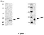
Figure 1 . Analysis of purified ERMP1 recombinant protein
Left panel: Comassie staining of purified His-tag ERMP1 fusion protein separated by SDS-PAGE; Right panel: WB on the recombinant protein stained with anti- ERMP1 antibody. Arrow marks the protein band of the expected size. Molecular weight markers are reported on the left. -
Figure 2 . Staining of breast tumor TMA with anti-ERMP1 antibodies
Examples of TMA of tumor (lower panel) and normal tissue samples (upper panel) stained with anti-ERMP1 antibodies. The antibody stains specifically tumor cells (in dark gray). -
Figure 3 . Expression and localization of ERMP1 in tumor cell lines- Panel A: Western blot analysis of ERMP1 expression in total protein extracts separated by SDS-PAGE from HEK-293T cells (corresponding to 1x10⁶ cells) transfected with the empty pcDNA3 vector (lane 1) or with the plasmid construct encoding the ERMP1 gene (lane 2);
- Panel B: Western blot analysis of ERMP1 expression in total protein extracts separated by SDS-PAGE from MCF-7 (lane 1) and SKBR-3 (lane 2) tumor cells (corresponding to 2x10⁵ cells). Arrow marks the expected ERMP1 band. Molecular weight markers are reported on the left.
- Panel C: Flow cytometry analysis of ERMP1 cell surface localization in SKBR-3 tumor cells stained with a negative control antibody (filled curve or with anti-ERMP1 antibody (empty curve). X axis, Fluorescence scale; Y axis, Cells (expressed as % relatively to major peaks).
-
Figure 4 . ERMP1 confers malignant cell phenotype - The proliferation and the invasiveness properties of the MCF7 cell line were assessed after transfection with ERMP1-siRNA and a scramble siRNA control using the MTT and the Boyden in vitro invasion assays, respectively.- Panel A. Cell migration/invasiveness measured by the Boyden migration assay. The graph represents the reduced migration/invasiveness of MCF7 treated with the ERMP1-specific siRNA. Small boxes under the columns show the visual counting of the migrated cells.
- Panel B. Cell proliferation determined by the MTT incorporation assay. The graph represents the reduced proliferation of the MCF7 tumor cells upon treatment with ERMP1-siRNA, as determined by spectrophotometric reading.
- The entire coding region or suitable fragments of the genes encoding the target proteins, were designed for cloning and expression using bioinformatic tools with the human genome sequence as template (Lindskog M et al (2005). Where present, the leader sequence for secretion was replaced with the ATG codon to drive the expression of the recombinant proteins in the cytoplasm of E. coli. For cloning, genes were PCR-amplified from templates derived from Mammalian Gene Collection (http://mgc.nci.nih.gov/) clones using specific primers. Clonings were designed so as to fuse a 10 histidine tag sequence at the 5' end, annealed to in house developed vectors, derivatives of vector pSP73 (Promega) adapted for the T4 ligation independent cloning method (Nucleic Acids Res. 1990 October 25; 18(20): 6069-6074) and used to transform E.coli NovaBlue cells recipient strain. E. coli tranformants were plated onto selective LB plates containing 100 µg/ml ampicillin (LB Amp) and positive E.coli clones were identified by restriction enzyme analysis of purified plasmid followed by DNA sequence analysis. For expression, plasmids were used to transform BL21-(DE3) E.coli cells and BL21-(DE3) E. coli cells harbouring the plasmid were inoculated in ZYP-5052 growth medium (Studier, 2005) and grown at 37°C for 24 hours. Afterwards, bacteria were collected by centrifugation, lysed into B-Per Reagent containing 1 mM MgCl2, 100 units DNAse I (Sigma), and 1 mg/ml lysozime (Sigma). After 30 min at room temperature under gentle shaking, the lysate was clarified by centrifugation at 30.000 g for 40 min at 4°C. All proteins were purified from the inclusion bodies by resuspending the pellet coming from lysate centrifugation in 40 mM TRIS-HCl, 1 mM TCEP {Tris(2-carboxyethyl)-phosphine hydrochloride, Pierce} and 6M guanidine hydrochloride, pH 8 and performing an IMAC in denaturing conditions. Briefly, the resuspended material was clarified by centrifugation at 30.000 g for 30 min and the supernatant was loaded on 0.5 ml columns of Ni-activated Chelating Sepharose Fast Flow (Pharmacia). The column was washed with 50 mM TRIS-HCl buffer, 1 mM TCEP, 6M urea, 60 mM imidazole, 0.5M NaCl, pH 8. Recombinant proteins were eluted with the same buffer containing 500 mM imidazole. Proteins were analysed by SDS-Page and their concentration was determined by Bradford assay using the BIORAD reagent (BIORAD) with a bovine serum albumin standard according to the manufacturer's recommendations. The identity of recombinant affinity purified proteins was further confirmed by mass spectrometry (MALDI-TOF), using standard procedures.
- To generate antisera, the purified proteins were used to immunize CD1 mice (6 week-old females, Charles River laboratories, 5 mice per group) intraperitoneally, with 3 protein doses of 20 micrograms each, at 2 week-interval. Freund's complete adjuvant was used for the first immunization, while Freund's incomplete adjuvant was used for the two booster doses. Two weeks after the last immunization animals were bled and sera collected from each animal was pooled.
- Gene fragments of the expected size were obtained by PCR from specific clones of the Mammalian Gene Collection or, alternatively, from cDNA generated from pools of total RNA derived from Human testis, Human placenta, Human bone marrow, Human fetal brain, using primers specific for each gene.
- For the ERMP1 gene, a fragment corresponding to nucleotides 55-666 of the transcript ENST00000339450 and encoding a protein of 204 residues, corresponding to the amino acid region from 1 to 204 of ENSP00000340427 sequence was obtained.
- A clone encoding the correct amino acid sequence was identified for the gene/gene-fragment and, upon expression in E. coli, a protein of the correct size was produced and subsequently purified using affinity chromatography (
Figure 1 left panels). As shown in the figures, in some case SDS-PAGE analysis of affinity-purified recombinant proteins revealed the presence of extra bands, of either higher and/or lower masses. Mass spectrometry analysis confirmed that they corresponded to either aggregates or degradation products of the protein under analysis. - Antibodies generated by immunization specifically recognized their target proteins in Western blot (WB) (
Figure 1 right panel). - The analysis of the antibodies' capability to recognize their target proteins in tumor samples was carried out by Tissue Micro Array (TMA), a miniaturized immuno-histochemistry technology suitable for HTP analysis that allows to analyse the antibody immuno-reactivity simultaneously on different tissue samples immobilized on a microscope slide.
- Since the TMAs include both tumor and healthy tissues, the specificity of the antibodies for the tumors can be immediately appreciated. The use of this technology, differently from approaches based on transcription profile, has the important advantage of giving a first hand evaluation on the potential of the markers in clinics. Conversely, since mRNA levels not always correlate with protein levels (approx. 50% correlation), studies based on transcription profile do not provide solid information regarding the expression of protein markers.
- A tissue microarray was prepared containing formalin-fixed paraffin-embedded cores of human tissues from patients affected by breast cancer and corresponding normal tissues as controls and analyzed using the specific antibody sample. In total, the TMA design consisted in 10 tumor breast tumor samples and 10 normal tissues from 5 well pedigreed patients (equal to two tumor samples and 2 normal tissues from each patient) to identify promising target molecules differentially expressed in cancer and normal cells. The direct comparison between tumor and normal tissues of each patient allowed the identification of antibodies that stain specifically tumor cells and provided indication of target expression in breast tumor.
- To further confirm the association of each protein with breast tumors a tissue microarray was prepared containing 100 formalin-fixed paraffin-embedded cores of human breast tissues from 50 patients (equal to two tissue samples from each patient).
- All formalin fixed, paraffin embedded tissues used as donor blocks for TMA production were selected from the archives at the IEO (Istituto Europeo Oncologico, Milan). Corresponding whole tissue sections were examined to confirm diagnosis and tumour classification, and to select representative areas in donor blocks. Normal tissues were defined as microscopically normal (non- neoplastic) and were generally selected from specimens collected from the vicinity of surgically removed tumors. The TMA production was performed essentially as previously described (Kononen J et al (1998) Nature Med. 4:844-847; Kallioniemi OP et al (2001) Hum. Mol. Genet. 10:657-662). Briefly, a hole was made in the recipient TMA block. A cylindrical core tissue sample (1 mm in diameter) from the donor block was acquired and deposited in the recipient TMA block. This was repeated in an automated tissue arrayer "Galileo TMA CK 3500" (BioRep, Milan) until a complete TMA design was produced. TMA recipient blocks were baked at 42 <0>C for 2 h prior to sectioning. The TMA blocks were sectioned with 2-3 mm thicknes using a waterfall microtome (Leica), and placed onto poly-L-lysinated glass slides for immunohistochemical analysis. Automated immunohistochemistry was performed as previously described (Kampf C. et al (2004) Clin. Proteomics 1:285-300). In brief, the glass slides were incubated for 30' min in 60°C, de-paraffinized in xylene (2 x 15 min) using the Bio-Clear solution (Midway. Scientific, Melbourne, Australia), and re-hydrated in graded alcohols. For antigen retrieval, slides were immersed 0.01 M Na-citrate buffer, pH 6.0 at 99°C for 30 min Slides were placed in the Autostainer (R) (DakoCytomation) and endogenous peroxidase was initially blocked with 3% H2O2, for 5 min. Slides were then blocked in Dako Cytomation Wash Buffer containing 5% Bovine serum albumin (BSA) and subsequently incubated with mouse antibodies for 30' (dilution 1:200 in Dako Real ™ dilution buffer). After washing with DakoCytomation wash buffer, slides were incubated with the goat anti-mouse peroxidase conjugated Envision(R) for 30 min each at room temperature (DakoCytomation). Finally, diaminobenzidine (DakoCytomation) was used as chromogen and Harris hematoxylin (Sigma- Aldrich) was used for counterstaining. The slides were mounted with Pertex(R) (Histolab).
- The staining results have been evaluated by a trained pathologist at the light microscope, and scored according to both the percentage of immunostained cells and the intensity of staining. The individual values and the combined score (from 0 to 300) were recorded in a custom-tailored database. Digital images of the immunocytochemical findings have been taken at a Leica DM LB light microscope, equipped with a Leica DFC289 color camera.
- A TMA design was obtained, representing tumor tissue samples and normal tissues, derived from patients affected by breast tumor. The results from tissue profiling showed that the antibodies specific for the recombinant proteins (see Example 1) are strongly immunoreactive on breast tumor cancer tissues, while no or poor reactivity was detected in normal tissues, indicating the presence of the target proteins in breast tumors. Based on this finding, the detection of target proteins in tissue samples can be associated with breast tumor. In some cases immunoreactivity accumulated at the cell membrane of tumor cells providing a first-hand indication on the surface localization of the target proteins.
- The capability of target-specific antibodies to stain breast tumor tissues is summarized in Table I. Representative examples of microscopic enlargements of tissue samples stained by each antibody are reported in
Figure 2 ). - Table reports the percentage of positive breast tumor tissue samples after staining with the target specific antibodies.
Marker name Percentage of Breast tumor tissues showing positive IHC staining ERMP1 45** (**) The antibody stains the cell membrane of tumor cells - The specificity of the antibodies for each target proteins was assessed by Western blot analysis on total protein extracts from eukaryotic cells transiently transfected with plasmid constructs containing the complete sequences of the genes encoding the target proteins. Where indicated, expression and localization of target proteins were investigated by confocal microscopy analysis of transfected cells. Such experiments were carried out on ERMP1 (cloned sequence corresponding to Transcripts ENST00000339450).
- For clonings, cDNA were generated from pools of total RNA derived from Human testis, Human placenta, Human bone marrow, Human fetal brain, in reverse transcription reactions and the entire coding regions were PCR-amplified with specific primers pairs. PCR products were cloned into plasmid pcDNA3 (Invitrogen). HeLa or Hek-293T cells were grown in DMEM-10% FCS supplemented with 1 mM Glutamine were transiently transfected with preparation of the resulting plasmids and with the empty vector as negative control using the Lipofectamine-2000 transfection reagent (Invitrogen). After 48 hours, cells were collected and analysed by Western blot or confocal microscopy. For Western blot, cells were lysed with PBS buffer containing 1% Triton X100 and total cell extracts (corresponding to 2x10⁵ cells) were separated on pre-cast SDS-PAGE gradient gels (NuPage 4-12% Bis-Tris gel, Invitrogen) under reducing conditions, followed by electro-transfer to nitrocellulose membranes (Invitrogen) according to the manufacturer's recommendations. The membranes were blocked in blocking buffer composed of 1xPBS-0.1% Tween 20 (PBST) added with 10% dry milk, for 1 h at room temperature, incubated with the antibody diluted 1:2500 in blocking buffer containing 1% dry milk and washed in PBST-1%. The secondary HRP-conjugated antibody (goat anti-mouse immunoglobulin/HRP, Perkin Elmer) was diluted 1:5000 in blocking buffer and chemiluminescence detection was carried out using a Chemidoc-IT UVP CCD camera (UVP) and the Western lightning™cheminulescence Reagent Plus (Perkin Elmer), according to the manufacturer's protocol.
- For confocal microscopy analysis, the cells were plated on glass cover slips and after 48 h were washed with PBS and fixed with 3% p-formaldheyde solution in PBS for 20 min at RT. For surface staining, cells were incubated overnight at 4°C with polyclonal antibodies (1:200). The cells were then stained with Alexafluor 488-labeled goat anti-mouse antibodies (Molecular Probes). DAPI (Molecular Probes) was used to visualize nuclei; Live/Dead® red fixable (Molecular Probes) was used to visualize membrane. The cells were mounted with glycerol plastine and observed under a laser-scanning confocal microscope (LeicaSP5).
- The selected coding sequence for ERMP1 was cloned in a eukaryotic expression vector and the derived plasmid used for transient transfection of HeLa or HEK293T cells. Expression of ERMP1 was analysed in trasnfected HEK-293T cells. Overall the data confirmed that the marker-specific antibodies recognized specifically their target proteins. For cells transfected with ERMP1-encoding plasmid, a band of high molecular mass was specifically detected by the anti-ERMP1 antibody indicating that the protein forms stable aggregates (
Figure 3A ). - The presence of protein bands corresponding to the marker protein was also investigated in tissue homogenates of breast tumor biopsies as compared to normal tissues from patients. Homogenates were prepared by mechanic tissue disruption in buffer containing 40 mM TRIS-HCl, 1 mM TCEP {Tris(2-carboxyethyl)-phosphine hydrochloride, Pierce} and 6M guanidine hydrochloride, pH 8. Western blot was performed by separation of the total protein extracts (20 µg/lane) proteins were detected by specific antibodies.
- Expression of target proteins was also assessed by WB and/or Flow cytometry on total extracts from breast tumor cell lines, including BT549, MCF7, MDA-MB231 and SKBR-3.
- In each analysis, cells were cultured in under ATCC recommended conditions, and sub-confluent cell monolayers were detached with PBS-0.5 mM EDTA. For Western blot analysis, cells were lysed by several freeze-thaw passages in PBS-1% Triton. Total protein extracts were loaded on SDS-PAGE (2x10⁵ cells/lane), and subjected to WB with specific antibodies as described above.
- For flow cytometry analysis, cells (2x10⁴ per well) were pelletted in 96 U-bottom microplates by centrifugation at 200 x g for 5 min at 4°C and incubated for 1 hour at 4°C with the appropriate dilutions of the marker-specific antibodies. Cells were washed twice in PBS-5% FCS and incubated for 20 min with the appropriate dilution of R-Phycoerythrin (PE)-conjugated secondary antibodies (Jackson Immuno Research, PA, USA) at 4°C. After washing, cells were analysed by a FACS Canto II flow cytometer (Becton Dickinson). Data were analyzed with FlowJo 8.3.3 program.
- Western blot and flow cytometry analysis of ERMP1 are represented. Western blot analysis shows a band of high molecular mass detected in the breast cell lines MCF7 and SKBR-3, showing an electrophoretic pattern similar to that reported in transfected cells (see Example 3). This further confirms the existence of stable aggregates for this protein confirming its expression in cell lines derived from breast tumor (Figure 29B). Flow cytometry analysis indicates that ERMP1 is detected on the surface of the SKBR-3 cell line (
Figure 3C ). - To verify that the proteins included in the present invention can be exploited as targets for therapeutic applications, the effect of marker depletion was evaluated in vitro in cellular studies generally used to define the role of newly discovered proteins in tumor development. Marker-specific knockdown and control tumor cell lines were assayed for their proliferation and the migration/invasiveness phenotypes using the MTT and the Boyden in vitro invasion assay, respectively.
- Expression of marker genes were silenced in tumor cell lines by the siRNA technology and the influence of the reduction of marker expression on cell parameters relevant for tumor development was assessed in in vitro assays. The expression of marker genes was knocked down in a panel of epithelial tumor cell lines previously shown to express the tumor markers using a panel of marker-specific siRNAs (whose target sequences are reported in the Table II) using the HiPerfect transfection reagent (QIAGEN) following the manufacturer's protocol. As control, cells treated with irrelevant siRNA (scrambled siRNA) were analysed in parallel. At different time points (ranging from 24 to 72 hours) post transfection, the reduction of gene transcription was assessed by quantitative RT-PCR (Q-RT-PCR) on total RNA, by evaluating the relative marker transcript level, using the beta-actin, GAPDH or MAPK genes as internal normalization control. Afterwards, cell proliferation and migration/invasiveness assays were carried out to assess the effect of the reduced marker expression. Cell proliferation was determined using the MTT assay, a colorimetric assay based on the cellular conversion of a tetrazolium salt into a purple colored formazan product. Absorbance of the colored solution can be quantified using a spectrophotometer to provide an estimate of the number of attached living cells. Approximately 5 x 10³ cells/100µl were seeded in 96-well plates in DMEM with 10% FCS to allow cell attachment. After overnight incubation with DMEM without FCS, the cells were treated with 2,5% FBS for 72 hours. Four hours before harvest 15 µL of the MTT dye solution (Promega) were added to each well. After 4-hour incubation at 37°C, the formazan precipitates were solubilized by the addition of 100 µL of solubilization solution (Promega) for 1h at 37°C,. Absorbance at 570 nm was determined on a multiwell plate reader (SpectraMax, Molecular Devices).
- Cell migration/invasiveness was tested using the Boyden in vitro invasion assay, as compared to control cell lines treated with a scramble siRNA. This assay is based on a chamber of two medium-filled compartments separated by a microporous membrane. Cells are placed in the upper compartment and are allowed to migrate through the pores of the membrane into the lower compartment, in which chemotactic agents are present. After an appropriate incubation time, the membrane between the two compartments is fixed and stained, and the number of cells that have migrated to the lower side of the membrane is determined. For this assay, a transwell system, equipped with 8-µm pore polyvinylpirrolidone-free polycarbonate filters, was used. The upper sides of the porous polycarbonate filters were coated with 50 µg/cm2 of reconstituted Matrigel basement membrane and placed into six-well culture dishes containing complete growth medium. Cells (1x10⁴ cells/well) were loaded into the upper compartment in serum-free growth medium. After 16 h of incubation at 37°C, non-invading cells were removed mechanically using cotton swabs, and the microporous membrane was stained with Diff-Quick solution. Chemotaxis was evaluated by counting the cells migrated to the lower surface of the polycarbonate filters (six randomly chosen fields, mean ± SD).
- Examples of this analysis are reported for ERMP1 in the breast tumor cell line MCF7. Gene silencing experiments with marker-specific siRNA reduced the marker transcripts (approximately 30-40 fold reduction), as determined by Q-RT_PCR. T II reports the sequences targeted by the siRNA molecules. The reduction of the expression of either of the two genes significantly impairs the proliferation and the invasiveness phenotypes of the MCF7 breast tumor cell line (Figures 30 and 36). This indicates that the protein is involved in tumor development and are therefore likey targets for the development of anti-cancer therapies.
Table II NCBI gene siRNA Target Sequence ERMP1 CCCGTGGTTCATCTGATATAA AAGGACTTTGCTCGGCGTTTA TACGTGGATGTTTGTAACGTA CTCGTATTGGCTCAATCATAA -
- 1) Anderson, L., and Seilhamer, J. (1997). A comparison of selected mRNA and protein abundances in human liver. Electrophoresis 18, 533 - 537;
- 2) Chen, G., Gharib, T. G., Wang, H., Huang, C. C., Kuick, R., Thomas, D. G., Shedden, K. A., Misek, D. E., Taylor, J. M., Giordano, T. J., Kardia, S. L., Iannettoni, M. D., Yee, J., Hogg, P. J., Orringer, M. B., Hanash, S. M., and Beer, D. G. (2003) Protein profiles associated with survival in lung adenocarcinoma. Proc. Natl. Acad. ;
- 3) Ginestier, C., Charafe-Jauffret, E., Bertucci, F., Eisinger, F., Geneix, J., Bechlian, D., Conte, N., Adelaide, J., Toiron, Y., Nguyen, C., Viens, P., Mozziconacci, M. J., Houlgatte, R., Birnbaum, D., and Jacquemier, J. (2002) Distinct and complementary information provided by use of tissue and DNA microarrays in the study of breast tumor markers. Am. J. Pathol. 161, 1223 - 1233;
- 4) Gygi, S. P., Rochon, Y., Franza, B. R., and Aebersold, R. (1999) Correlation between protein and mRNA abundance in yeast. Mol. Cell. Biol. 19, 1720 - 1730; Nishizuka, S., Charboneau, L., Young, L., Major, S., Reinhold, W. C., Waltham, M., Kouros-Mehr, H., Bussey, K. J., Lee, J. K., Espina, V., Munson, P. J., Petricoin, E., III, Liotta, L. A., and Weinstein, J. N. (2003) Proteomic profiling of the NCI-60 cancer cell lines using new high-density reverse-phase lysate microarrays. Proc. Natl. Acad. ;
- 5) Tyers, M., and Mann, M. (2003) From genomics to proteomics. Nature 422, 193 - 197;
- 6) Kagara N, Tanaka N, Noguchi S, Hirano T. (2007) Zinc and its transporter ZIP10 are involved in invasive behavior of breast cancer cells. Cancer Sci. 98:692 - 697;
- 7) Kasper G, Weiser AA, Rump A, Sparbier K, Dahl E, Hartmann A, Wild P, Schwidetzky U, Castaños-Vélez E, Lehmann K. (2005) Expression levels of the putative zinc transporter LIV-1 are associated with a better outcome of breast cancer patients. Int J Cancer. 117:961 - 973;
- 8) Nguyen ST, Hasegawa S, Tsuda H, Tomioka H, Ushijima M, Noda M, Omura K, Miki Y, (2007) Identification of a predictive gene expression signature of cervical lymph node metastasis in oral squamous cell carcinoma., Cancer Sci. 98:740 - 746;
- 9) Garcia-Rudaz,C., Luna,F., Tapia,V., Kerr,B., Colgin,L., Galimi,F., Dissen,G.A., Rawlings,N.D. and Ojeda,S.R. (2007) Fxna, a novel gene differentially expressed in the rat ovary at the time of folliculogenesis, is required for normal ovarian histogenesis. Development. 134, 945-957;
- 10) Kratzschmar J, Haendler B, Eberspaecher U, Roosterman D, Donner P, Schleuning WD. (1996) The human cysteine-rich secretory protein (CRISP) family. Primary structure and tissue distribution of CRISP-1, CRISP-2 and CRISP-3. Eur J Biochem. 236:827 - 836
- 11) Bjartell,A., Johansson,R., Bjork,T., adaleanu,V., Lundwall,A., Lilja,H., Kjeldsen,L. and Udby,L. (2006) Immunohistochemical detection of cysteine-rich secretory protein 3 in tissue and in serum from men with cancer or benign enlargement of the prostate gland, Prostate. 66: 591 - 603;
- 12) Bjartell,A.S., Al-Ahmadie,H., Serio,A.M., Eastham,J.A., Eggener,S.E., Fine,S.W., Udby,L., Gerald,W.L., Vickers,A.J., Lilja,H., Reuter,V.E. and Scardino,P.T. (2007), Association of cysteine-rich secretory protein 3 and beta-microseminoprotein with outcome after radical prostatectomy. Clin. Cancer Res. 13: 4130-4138.
-
- <110> Externautics S.p.A.
- <120> breast tumor markers and methos of use thereof
- <130> 1298PCT
- <160> 94
- <170> Patent In version 3.3
- <210> 1
<211> 132
<212> PRT
<213> Homo sapiens - <400> 1

- <210> 2
<211> 399
<212> DNA
<213> Homo sapiens - <400> 2


- <210> 3
<211> 147
<212> PRT
<213> Homo sapiens - <400> 3

- <210> 4
<211> 932
<212> DNA
<213> Homo sapiens - <400> 4

- <210> 5
<211> 131
<212> PRT
<213> Homo sapiens - <400> 5


- <210> 6
<211> 2950
<212> DNA
<213> Homo sapiens - <400> 6


- <210> 7
<211> 316
<212> PRT
<213> Homo sapiens - <400> 7


- <210> 8
<211> 316
<212> PRT
<213> Homo sapiens - <400> 8


- <210> 9
<211> 1210
<212> DNA
<213> Homo sapiens - <400> 9


- <210> 10
<211> 1502
<212> DNA
<213> Homo sapiens - <400> 10


- <210> 11
<211> 157
<212> PRT
<213> Homo sapiens - <400> 11


- <210> 12
<211> 474
<212> DNA
<213> Homo sapiens - <400> 12

- <210> 13
<211> 241
<212> PRT
<213> Homo sapiens - <400> 13


- <210> 14
<211> 912
<212> DNA
<213> Homo sapiens - <400> 14


- <210> 15
<211> 279
<212> PRT
<213> Homo sapiens - <400> 15

- <210> 16
<211> 102
<212> PRT
<213> Homo sapiens - <400> 16


- <210> 17
<211> 716
<212> PRT
<213> Homo sapiens - <400> 17


- <210> 18
<211> 112
<212> PRT
<213> Homo sapiens - <400> 18

- <210> 19
<211> 1841
<212> DNA
<213> Homo sapiens - <400> 19

- <210> 20
<211> 2206
<212> DNA
<213> Homo sapiens - <400> 20


- <210> 21
<211> 4456
<212> DNA
<213> Homo sapiens - <400> 21


- <210> 22
<211> 336
<212> DNA
<213> Homo sapiens - <400> 22


- <210> 23
<211> 831
<212> PRT
<213> Homo sapiens - <400> 23


- <210> 24
<211> 831
<212> PRT
<213> Homo sapiens - <400> 24


- <210> 25
<211> 5227
<212> DNA
<213> Homo sapiens - <400> 25


- <210> 26
<211> 5432
<212> DNA
<213> Homo sapiens - <400> 26


- <210> 27
<211> 543
<212> PRT
<213> Homo sapiens - <400> 27


- <210> 28
<211> 938
<212> PRT
<213> Homo sapiens - <400> 28


- <210> 29
<211> 230
<212> PRT
<213> Homo sapiens - <400> 29


- <210> 30
<211> 1172
<212> PRT
<213> Homo sapiens - <400> 30


- <210> 31
<211> 873
<212> PRT
<213> Homo sapiens - <400> 31


- <210> 32
<211> 3927
<212> DNA
<213> Homo sapiens - <400> 32


- <210> 33
<211> 3932
<212> DNA
<213> Homo sapiens - <400> 33


- <210> 34
<211> 4073
<212> DNA
<213> Homo sapiens - <400> 34


- <210> 35
<211> 4666
<212> DNA
<213> Homo sapiens - <400> 35



- <210> 36
<211> 3513
<212> DNA
<213> Homo sapiens - <400> 36


- <210> 37
<211> 406
<212> PRT
<213> Homo sapiens - <400> 37


- <210> 38
<211> 426
<212> PRT
<213> Homo sapiens - <400> 38


- <210> 39
<211> 426
<212> PRT
<213> Homo sapiens - <400> 39


- <210> 40
<211> 396
<212> PRT
<213> Homo sapiens - <400> 40

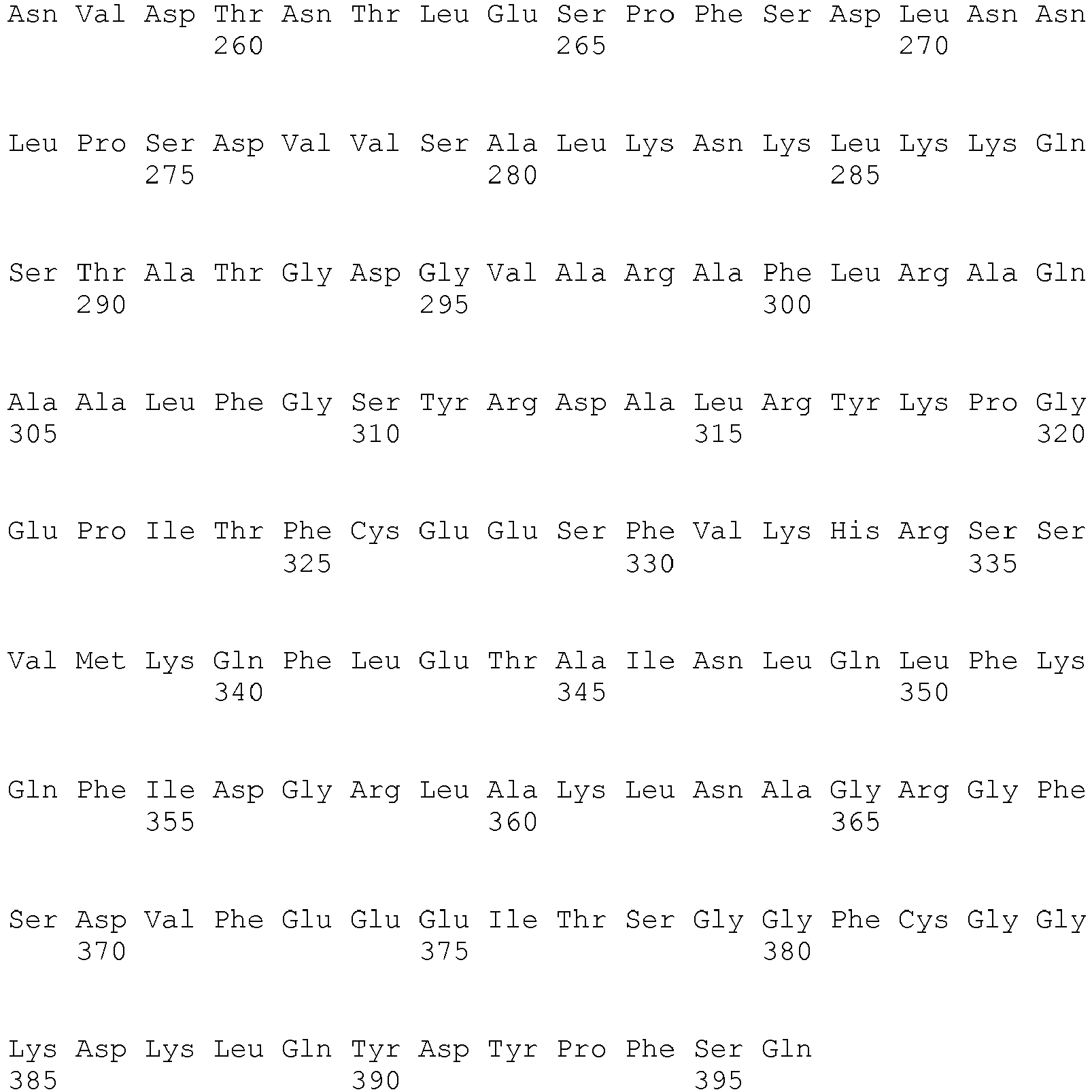
- <210> 41
<211> 2117
<212> DNA
<213> Homo sapiens - <400> 41


- <210> 42
<211> 2177
<212> DNA
<213> Homo sapiens - <400> 42


- <210> 43
<211> 2007
<212> DNA
<213> Homo sapiens - <400> 43
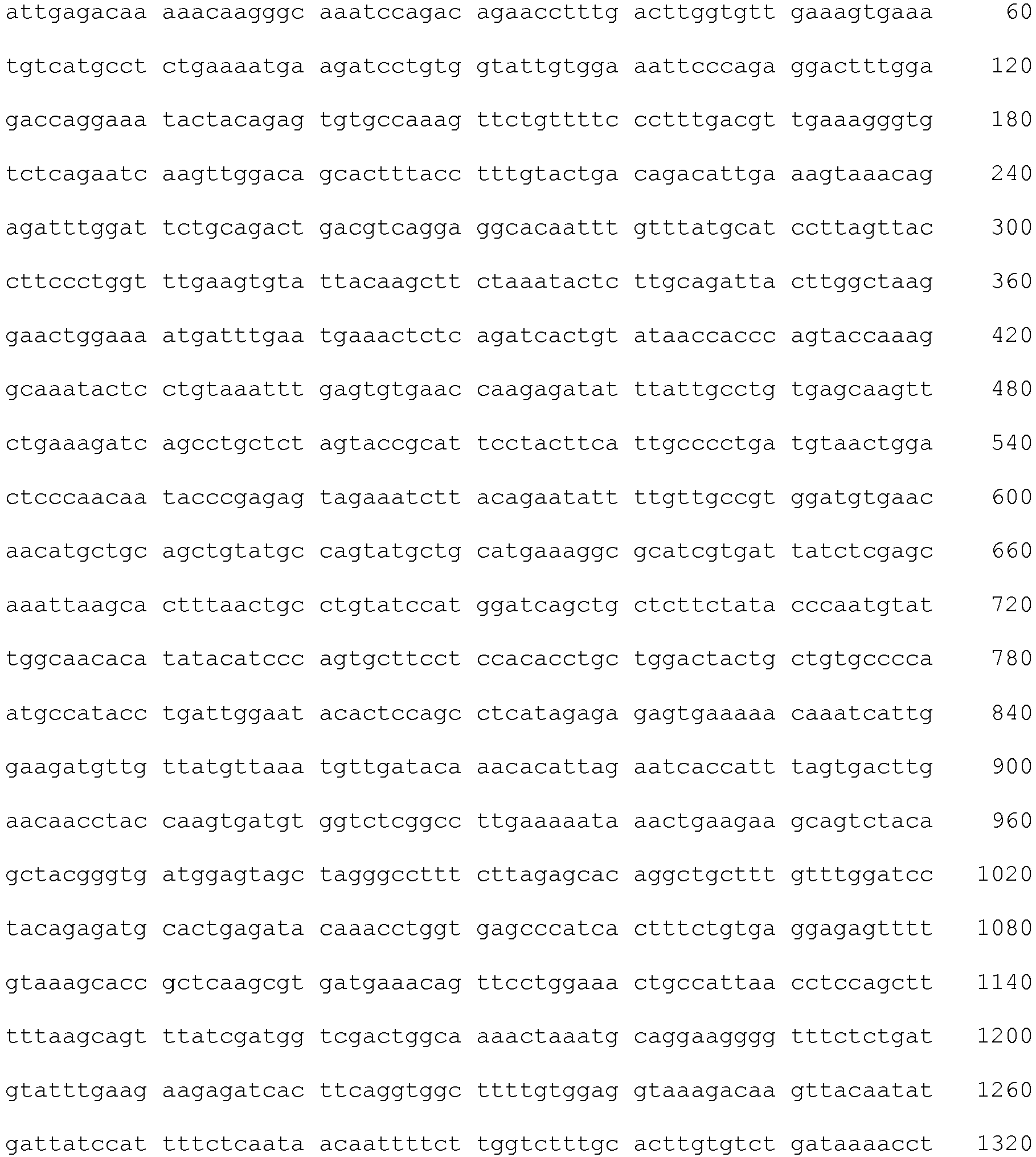

- <210> 44
<211> 2197
<212> DNA
<213> Homo sapiens - <400> 44


- <210> 45
<211> 423
<212> PRT
<213> Homo sapiens - <400> 45


- <210> 46
<211> 212
<212> PRT
<213> Homo sapiens - <400> 46
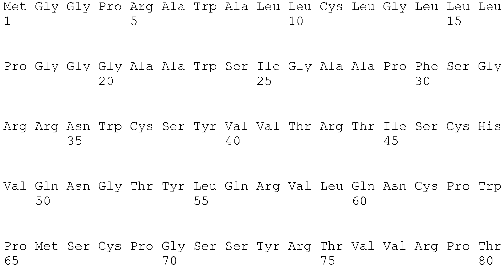

- <210> 47
<211> 175
<212> PRT
<213> Homo sapiens - <400> 47


- <210> 48
<211> 34
<212> PRT
<213> Homo sapiens - <400> 48

- <210> 49
<211> 445
<212> PRT
<213> Homo sapiens - <400> 49


- <210> 50
<211> 443
<212> PRT
<213> Homo sapiens - <400> 50


- <210> 51
<211> 441
<212> PRT
<213> Homo sapiens - <400> 51


- <210> 52
<211> 439
<212> PRT
<213> Homo sapiens - <400> 52


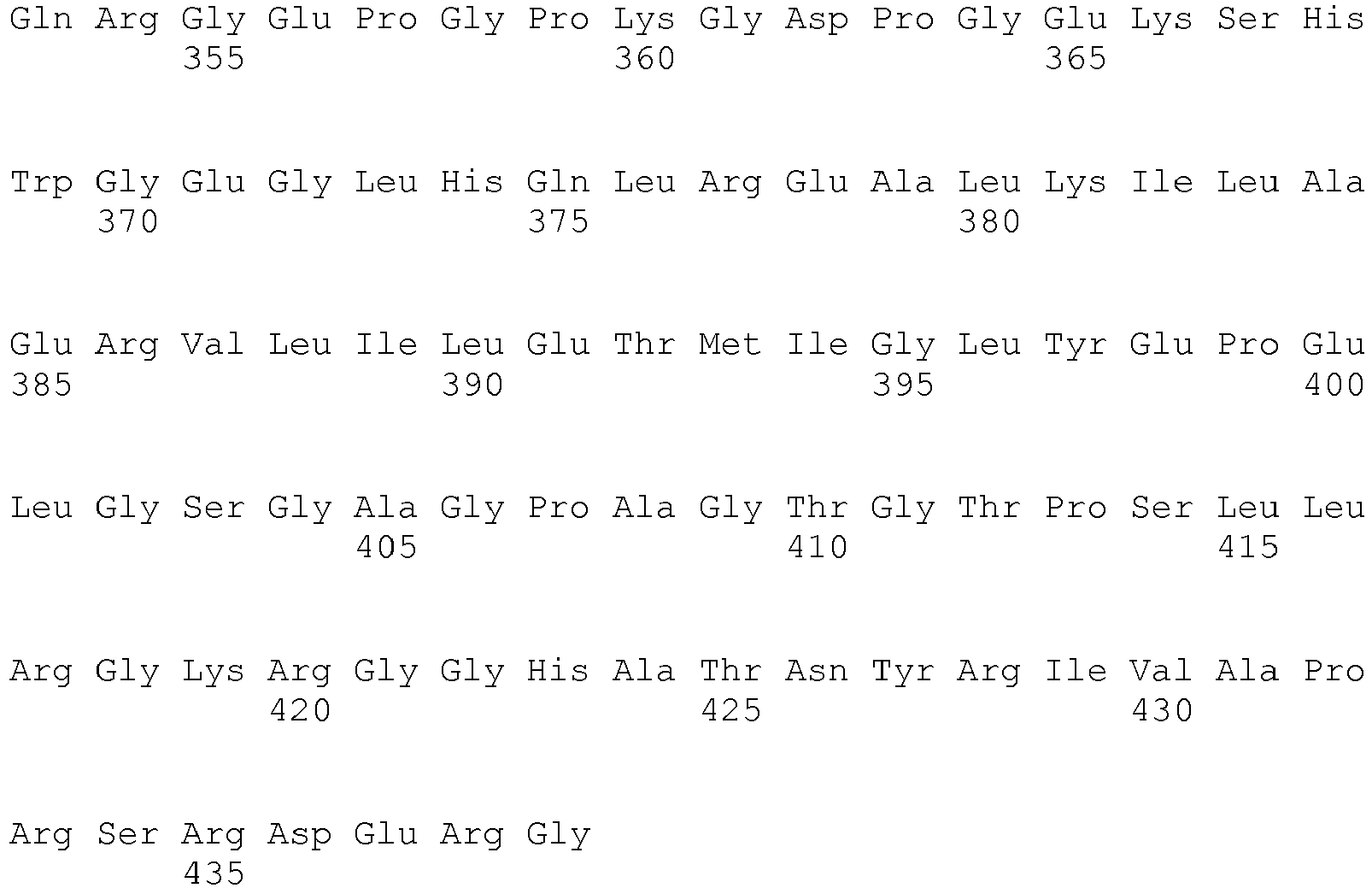
- <210> 53
<211> 422
<212> PRT
<213> Homo sapiens - <400> 53


- <210> 54
<211> 212
<212> PRT
<213> Homo sapiens - <400> 54


- <210> 55
<211> 175
<212> PRT
<213> Homo sapiens - <400> 55


- <210> 56
<211> 236
<212> PRT
<213> Homo sapiens - <220>
<221> misc_feature
<222> (1) .. (1)
<223> Xaa can be any naturally occurring amino acid - <400> 56


- <210> 57
<211> 305
<212> PRT
<213> Homo sapiens - <400> 57

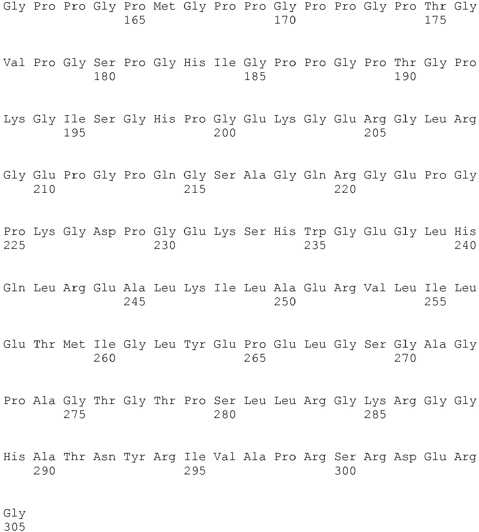
- <210> 58
<211> 226
<212> PRT
<213> Homo sapiens - <400> 58


- <210> 59
<211> 2031
<212> DNA
<213> Homo sapiens - <400> 59


- <210> 60
<211> 769
<212> DNA
<213> Homo sapiens - <400> 60

- <210> 61
<211> 641
<212> DNA
<213> Homo sapiens - <400> 61

- <210> 62
<211> 482
<212> DNA
<213> Homo sapiens - <400> 62

- <210> 63
<211> 2066
<212> DNA
<213> Homo sapiens - <400> 63


- <210> 64
<211> 2060
<212> DNA
<213> Homo sapiens - <400> 64


- <210> 65
<211> 2054
<212> DNA
<213> Homo sapiens - <400> 65


- <210> 66
<211> 2048
<212> DNA
<213> Homo sapiens - <400> 66


- <210> 67
<211> 1997
<212> DNA
<213> Homo sapiens - <400> 67


- <210> 68
<211> 1823
<212> DNA
<213> Homo sapiens - <400> 68


- <210> 69
<211> 1976
<212> DNA
<213> Homo sapiens - <400> 69


- <210> 70
<211> 2111
<212> DNA
<213> Homo sapiens - <400> 70


- <210> 71
<211> 707
<212> DNA
<213> Homo sapiens - <400> 71

- <210> 72
<211> 2034
<212> DNA
<213> Homo sapiens - <400> 72


- <210> 73
<211> 518
<212> PRT
<213> Homo sapiens - <400> 73



- <210> 74
<211> 904
<212> PRT
<213> Homo sapiens - <400> 74


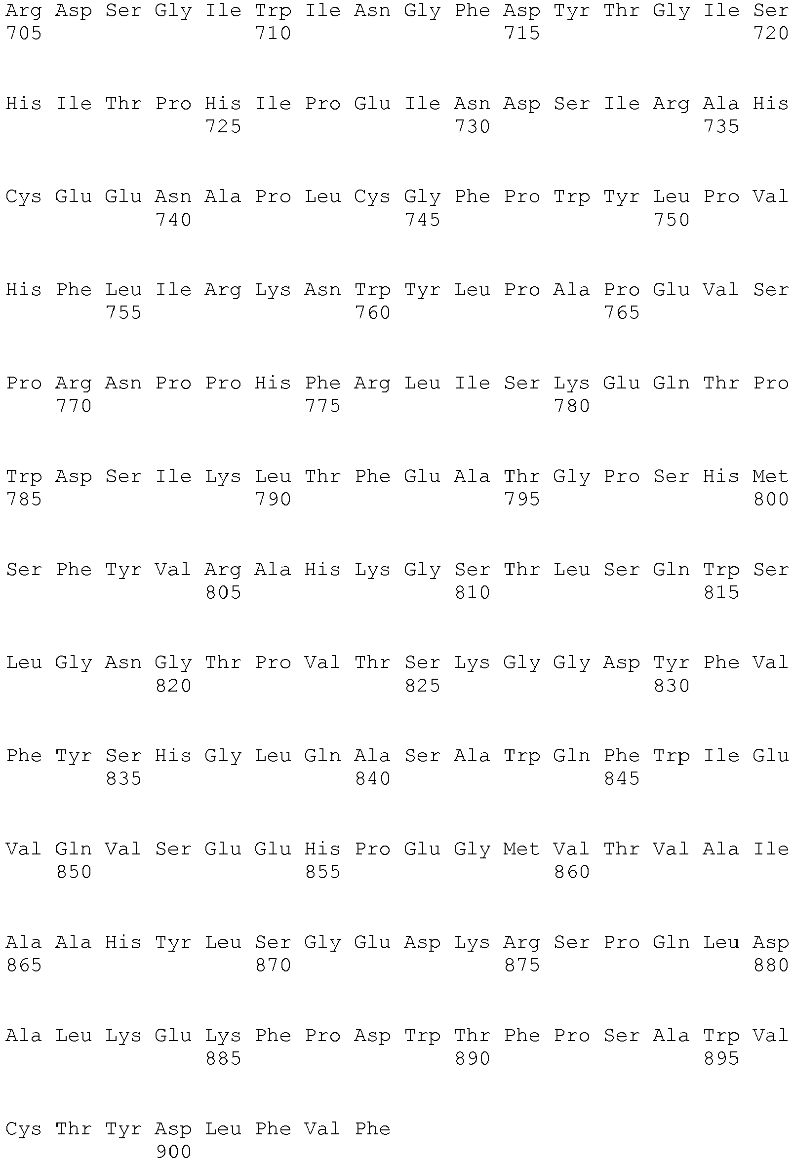
- <210> 75
<211> 419
<212> PRT
<213> Homo sapiens - <400> 75


- <210> 76
<211> 4974
<212> DNA
<213> Homo sapiens - <400> 76


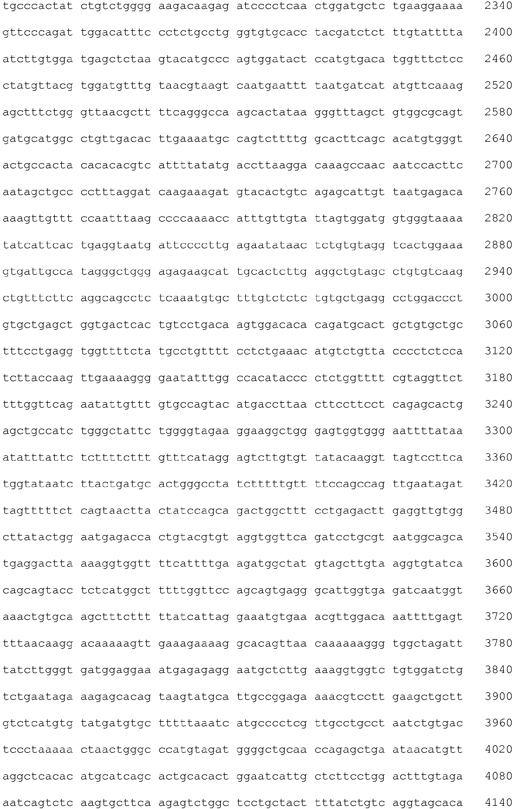

- <210> 77
<211> 5338
<212> DNA
<213> Homo sapiens - <400> 77


- <210> 78
<211> 5387
<212> DNA
<213> Homo sapiens - <400> 78


- <210> 79
<211> 245
<212> PRT
<213> Homo sapiens - <400> 79


- <210> 80
<211> 245
<212> PRT
<213> Homo sapiens - <400> 80


- <210> 81
<211> 245
<212> PRT
<213> Homo sapiens - <400> 81


- <210> 82
<211> 2073
<212> DNA
<213> Homo sapiens - <400> 82


- <210> 83
<211> 2081
<212> DNA
<213> Homo sapiens - <400> 83


- <210> 84
<211> 2122
<212> DNA
<213> Homo sapiens - <400> 84


- <210> 85
<211> 409
<212> PRT
<213> Homo sapiens - <400> 85


- <210> 86
<211> 314
<212> PRT
<213> Homo sapiens - <400> 86


- <210> 87
<211> 1544
<212> DNA
<213> Homo sapiens - <400> 87


- <210> 88
<211> 1192
<212> DNA
<213> Homo sapiens - <400> 88


- <210> 89
<211> 21
<212> DNA
<213> Homo sapiens - <400> 89
cgaggacaat ctggatatca a 21 - <210> 90
<211> 21
<212> DNA
<213> Homo sapiens - <400> 90
ctggagccct cgagcaagaa a 21 - <210> 91
<211> 21
<212> DNA
<213> Homo sapiens - <400> 91
cccgtggttc atctgatata a 21 - <210> 92
<211> 21
<212> DNA
<213> Homo sapiens - <400> 92
aaggactttg ctcggcgttt a 21 - <210> 93
<211> 21
<212> DNA
<213> Homo sapiens - <400> 93
tacgtggatg tttgtaacgt a 21 - <210> 94
<211> 21
<212> DNA
<213> Homo sapiens - <400> 94
ctcgtattgg ctcaatcata a 21
Claims (3)
- A method of screening a breast tissue sample for malignancy, said method comprising determining the presence in said sample of the tumor marker protein ERMP1 SEQ ID NO:74 by means of an antibody against the amino acid region 1 to 204 of said ERMP1 protein.
- A method according to claim 1, which is based on immunoradiometric, immunoenzymatic or immunohistochemical techniques.
- A method in vitro for determining the presence of a breast tumor in a subject, which comprises the steps of:(a) providing a sample of the tissue suspected of containing tumor cells;(b) determining the presence of the tumor marker protein ERMP1 SEQ ID NO:74 in said tissue sample by detecting the expression of the marker protein by means of an antibody raised against the amino acid region 1 to 204 of said ERMP1 protein;wherein the detection of the tumor marker in the tissue sample is indicative of the presence of tumor in said subject.
Priority Applications (2)
| Application Number | Priority Date | Filing Date | Title |
|---|---|---|---|
| EP10787709.4A EP2493917B8 (en) | 2009-10-26 | 2010-10-26 | Breast tumor markers and methods of use thereof |
| EP17209384.1A EP3345920A1 (en) | 2009-10-26 | 2010-10-26 | Breast tumor markers and methods of use thereof |
Applications Claiming Priority (3)
| Application Number | Priority Date | Filing Date | Title |
|---|---|---|---|
| EP09174060 | 2009-10-26 | ||
| EP10787709.4A EP2493917B8 (en) | 2009-10-26 | 2010-10-26 | Breast tumor markers and methods of use thereof |
| PCT/EP2010/066146 WO2011051277A1 (en) | 2009-10-26 | 2010-10-26 | Breast tumor markers and methods of use thereof |
Related Child Applications (2)
| Application Number | Title | Priority Date | Filing Date |
|---|---|---|---|
| EP17209384.1A Division-Into EP3345920A1 (en) | 2009-10-26 | 2010-10-26 | Breast tumor markers and methods of use thereof |
| EP17209384.1A Division EP3345920A1 (en) | 2009-10-26 | 2010-10-26 | Breast tumor markers and methods of use thereof |
Publications (3)
| Publication Number | Publication Date |
|---|---|
| EP2493917A1 EP2493917A1 (en) | 2012-09-05 |
| EP2493917B1 true EP2493917B1 (en) | 2019-12-11 |
| EP2493917B8 EP2493917B8 (en) | 2020-03-04 |
Family
ID=43466760
Family Applications (2)
| Application Number | Title | Priority Date | Filing Date |
|---|---|---|---|
| EP10787709.4A Active EP2493917B8 (en) | 2009-10-26 | 2010-10-26 | Breast tumor markers and methods of use thereof |
| EP17209384.1A Withdrawn EP3345920A1 (en) | 2009-10-26 | 2010-10-26 | Breast tumor markers and methods of use thereof |
Family Applications After (1)
| Application Number | Title | Priority Date | Filing Date |
|---|---|---|---|
| EP17209384.1A Withdrawn EP3345920A1 (en) | 2009-10-26 | 2010-10-26 | Breast tumor markers and methods of use thereof |
Country Status (3)
| Country | Link |
|---|---|
| US (2) | US20130017546A1 (en) |
| EP (2) | EP2493917B8 (en) |
| WO (1) | WO2011051277A1 (en) |
Families Citing this family (6)
| Publication number | Priority date | Publication date | Assignee | Title |
|---|---|---|---|---|
| EP2494361B2 (en) | 2009-10-26 | 2019-01-09 | Externautics S.p.A. | Ovary tumor markers and methods of use thereof |
| CN105461795A (en) * | 2014-05-15 | 2016-04-06 | 马恒标 | Tumor arrestin variant DV379 and application thereof |
| EP3298031B1 (en) | 2015-05-18 | 2020-10-21 | BiOMVis Srl | Immunogenic compositions containing bacterial outer membrane vesicles and therapeutic uses thereof |
| IL276028B2 (en) | 2018-01-26 | 2025-02-01 | Exelixis Inc | Compounds for the treatment of kinase-dependent disorders |
| GEAP202415420A (en) | 2018-01-26 | 2024-12-25 | Exelixis Inc | Compounds for the treatment of kinase-dependent disorders |
| CN111936487B (en) | 2018-01-26 | 2024-07-09 | 埃克塞里艾克西斯公司 | Compounds for the treatment of kinase dependent disorders |
Family Cites Families (14)
| Publication number | Priority date | Publication date | Assignee | Title |
|---|---|---|---|---|
| US20020137890A1 (en) | 1997-03-31 | 2002-09-26 | Genentech, Inc. | Secreted and transmembrane polypeptides and nucleic acids encoding the same |
| US6835822B1 (en) * | 1999-10-28 | 2004-12-28 | Agensys, Inc. | Diagnosis and therapy of cancer using SGP28-related molecules |
| US7078205B2 (en) | 2000-02-17 | 2006-07-18 | Millennium Pharmaceuticals, Inc. | Nucleic acid sequences encoding melanoma associated antigen molecules, aminotransferase molecules, atpase molecules, acyltransferase molecules, pyridoxal-phosphate dependent enzyme molecules and uses therefor |
| US20020165152A1 (en) * | 2000-11-07 | 2002-11-07 | Rosana Kapeller-Libermann | 14089, a novel human trypsin serine protease and uses thereof |
| AU2002351828A1 (en) * | 2001-11-05 | 2003-05-19 | Deutsches Krebsforschungszentrum | Novel genetic markers for leukemias |
| US20070237770A1 (en) | 2001-11-30 | 2007-10-11 | Albert Lai | Novel compositions and methods in cancer |
| WO2004094623A2 (en) * | 2003-04-04 | 2004-11-04 | Incyte Corporation | Cell adhesion and extracellular matrix proteins |
| DE10344799A1 (en) * | 2003-09-26 | 2005-04-14 | Ganymed Pharmaceuticals Ag | Identification of surface-associated antigens for tumor diagnosis and therapy |
| US7319011B2 (en) | 2004-04-08 | 2008-01-15 | Duke University | Method for distinguishing follicular thyroid adenoma (FTA) from follicular thyroid carcinoma (FTC) |
| US20060204503A1 (en) * | 2005-01-31 | 2006-09-14 | Biogen Idec Ma Inc. | Treatment of cancer using antibodies to polypeptides differentially expressed in human lung tumors |
| KR100694508B1 (en) * | 2005-05-24 | 2007-03-13 | 울산대학교 산학협력단 | Pharmaceutical composition for the treatment of cancer diseases comprising H4 antibody and immunotherapy method of cancer using the same |
| JP2009513125A (en) * | 2005-10-28 | 2009-04-02 | ビオメリュー・ソシエテ・アノニム | Cancer detection method |
| CN101437536A (en) * | 2006-03-23 | 2009-05-20 | 诺华有限公司 | Anti-tumor cell antigen antibody therapy |
| WO2008039475A2 (en) | 2006-09-26 | 2008-04-03 | The Board Of Trustees Of The University Of Arkansas | A gene expression profiling based identification of genomic signatures of multiple myeloma and uses thereof |
-
2010
- 2010-10-26 EP EP10787709.4A patent/EP2493917B8/en active Active
- 2010-10-26 EP EP17209384.1A patent/EP3345920A1/en not_active Withdrawn
- 2010-10-26 WO PCT/EP2010/066146 patent/WO2011051277A1/en not_active Ceased
- 2010-10-26 US US13/503,430 patent/US20130017546A1/en not_active Abandoned
-
2016
- 2016-10-31 US US15/338,601 patent/US20170227543A1/en not_active Abandoned
Non-Patent Citations (1)
| Title |
|---|
| None * |
Also Published As
| Publication number | Publication date |
|---|---|
| EP2493917B8 (en) | 2020-03-04 |
| US20130017546A1 (en) | 2013-01-17 |
| US20170227543A1 (en) | 2017-08-10 |
| EP2493917A1 (en) | 2012-09-05 |
| EP3345920A1 (en) | 2018-07-11 |
| WO2011051277A1 (en) | 2011-05-05 |
Similar Documents
| Publication | Publication Date | Title |
|---|---|---|
| EP2494351B1 (en) | Colon and rectal tumor markers and methods of use thereof | |
| US20120052071A1 (en) | Tumor markers and methods of use thereof | |
| EP2493916B1 (en) | Lung tumor markers and methods of use thereof | |
| EP2438442B1 (en) | Methods and assays for measuring p95 and/or p95 complexes in a sample and antibodies specific for p95 | |
| US20180313841A1 (en) | Ovary Tumor Markers And Methods Of Use Thereof | |
| US20170227543A1 (en) | Breast Tumor Markers And Methods Of Use Thereof | |
| US20150093396A1 (en) | Prostate Tumor Markers And Methods Of Use Thereof | |
| US10359428B2 (en) | Tumor marker, monoclonal antibodies and methods of use thereof | |
| Jiang et al. | Peptide mimic isolated by autoantibody reveals human arrest defective 1 overexpression is associated with poor prognosis for colon cancer patients | |
| US20130137106A1 (en) | Tumor Marker and Methods of Use Thereof | |
| US9182404B2 (en) | Tumor markers and methods of use thereof | |
| Grifantini et al. | A novel polyclonal antibody library for expression profiling of poorly characterized, membrane and secreted human proteins | |
| US8507213B1 (en) | Methods and compositions for treating diseases targeting maba | |
| WO2015081446A1 (en) | Methods for the prognosis of breast cancer |
Legal Events
| Date | Code | Title | Description |
|---|---|---|---|
| PUAI | Public reference made under article 153(3) epc to a published international application that has entered the european phase |
Free format text: ORIGINAL CODE: 0009012 |
|
| 17P | Request for examination filed |
Effective date: 20120522 |
|
| AK | Designated contracting states |
Kind code of ref document: A1 Designated state(s): AL AT BE BG CH CY CZ DE DK EE ES FI FR GB GR HR HU IE IS IT LI LT LU LV MC MK MT NL NO PL PT RO RS SE SI SK SM TR |
|
| DAX | Request for extension of the european patent (deleted) | ||
| 17Q | First examination report despatched |
Effective date: 20131205 |
|
| STAA | Information on the status of an ep patent application or granted ep patent |
Free format text: STATUS: EXAMINATION IS IN PROGRESS |
|
| GRAP | Despatch of communication of intention to grant a patent |
Free format text: ORIGINAL CODE: EPIDOSNIGR1 |
|
| STAA | Information on the status of an ep patent application or granted ep patent |
Free format text: STATUS: GRANT OF PATENT IS INTENDED |
|
| INTG | Intention to grant announced |
Effective date: 20190621 |
|
| RAP1 | Party data changed (applicant data changed or rights of an application transferred) |
Owner name: EXTERNAUTICS S.P.A. |
|
| RIN1 | Information on inventor provided before grant (corrected) |
Inventor name: NOGAROTTO, RENZO Inventor name: PARRI, MATTEO Inventor name: CAMPAGNOLI, SUSANNA Inventor name: GRANDI, ALBERTO Inventor name: PIERLEONI, ANDREA Inventor name: GRIFANTINI, RENATA Inventor name: PILERI, PIERO |
|
| RAP1 | Party data changed (applicant data changed or rights of an application transferred) |
Owner name: EXTERNAUTICS S.P.A. |
|
| GRAS | Grant fee paid |
Free format text: ORIGINAL CODE: EPIDOSNIGR3 |
|
| GRAA | (expected) grant |
Free format text: ORIGINAL CODE: 0009210 |
|
| STAA | Information on the status of an ep patent application or granted ep patent |
Free format text: STATUS: THE PATENT HAS BEEN GRANTED |
|
| AK | Designated contracting states |
Kind code of ref document: B1 Designated state(s): AL AT BE BG CH CY CZ DE DK EE ES FI FR GB GR HR HU IE IS IT LI LT LU LV MC MK MT NL NO PL PT RO RS SE SI SK SM TR |
|
| REG | Reference to a national code |
Ref country code: GB Ref legal event code: FG4D |
|
| REG | Reference to a national code |
Ref country code: CH Ref legal event code: EP |
|
| REG | Reference to a national code |
Ref country code: AT Ref legal event code: REF Ref document number: 1212100 Country of ref document: AT Kind code of ref document: T Effective date: 20191215 |
|
| REG | Reference to a national code |
Ref country code: DE Ref legal event code: R096 Ref document number: 602010062352 Country of ref document: DE |
|
| REG | Reference to a national code |
Ref country code: IE Ref legal event code: FG4D |
|
| REG | Reference to a national code |
Ref country code: DE Ref legal event code: R081 Ref document number: 602010062352 Country of ref document: DE Owner name: EXTERNAUTICS SOCIETA A RESPONSABILITA LIMITATA, IT Free format text: FORMER OWNER: EXTERNAUTICS S.P.A., SIENA, IT |
|
| REG | Reference to a national code |
Ref country code: DE Ref legal event code: R081 Ref document number: 602010062352 Country of ref document: DE Owner name: EXTERNAUTICS SOCIETA A RESPONSABILITA LIMITATA, IT Free format text: FORMER OWNER: EXTERNAUTICS S.R.L., SIENA, IT |
|
| REG | Reference to a national code |
Ref country code: CH Ref legal event code: PK Free format text: BERICHTIGUNG B8 |
|
| REG | Reference to a national code |
Ref country code: NL Ref legal event code: MP Effective date: 20191211 |
|
| REG | Reference to a national code |
Ref country code: LT Ref legal event code: MG4D |
|
| PG25 | Lapsed in a contracting state [announced via postgrant information from national office to epo] |
Ref country code: LT Free format text: LAPSE BECAUSE OF FAILURE TO SUBMIT A TRANSLATION OF THE DESCRIPTION OR TO PAY THE FEE WITHIN THE PRESCRIBED TIME-LIMIT Effective date: 20191211 Ref country code: GR Free format text: LAPSE BECAUSE OF FAILURE TO SUBMIT A TRANSLATION OF THE DESCRIPTION OR TO PAY THE FEE WITHIN THE PRESCRIBED TIME-LIMIT Effective date: 20200312 Ref country code: FI Free format text: LAPSE BECAUSE OF FAILURE TO SUBMIT A TRANSLATION OF THE DESCRIPTION OR TO PAY THE FEE WITHIN THE PRESCRIBED TIME-LIMIT Effective date: 20191211 Ref country code: BG Free format text: LAPSE BECAUSE OF FAILURE TO SUBMIT A TRANSLATION OF THE DESCRIPTION OR TO PAY THE FEE WITHIN THE PRESCRIBED TIME-LIMIT Effective date: 20200311 Ref country code: SE Free format text: LAPSE BECAUSE OF FAILURE TO SUBMIT A TRANSLATION OF THE DESCRIPTION OR TO PAY THE FEE WITHIN THE PRESCRIBED TIME-LIMIT Effective date: 20191211 Ref country code: NO Free format text: LAPSE BECAUSE OF FAILURE TO SUBMIT A TRANSLATION OF THE DESCRIPTION OR TO PAY THE FEE WITHIN THE PRESCRIBED TIME-LIMIT Effective date: 20200311 Ref country code: LV Free format text: LAPSE BECAUSE OF FAILURE TO SUBMIT A TRANSLATION OF THE DESCRIPTION OR TO PAY THE FEE WITHIN THE PRESCRIBED TIME-LIMIT Effective date: 20191211 Ref country code: ES Free format text: LAPSE BECAUSE OF FAILURE TO SUBMIT A TRANSLATION OF THE DESCRIPTION OR TO PAY THE FEE WITHIN THE PRESCRIBED TIME-LIMIT Effective date: 20191211 |
|
| PG25 | Lapsed in a contracting state [announced via postgrant information from national office to epo] |
Ref country code: HR Free format text: LAPSE BECAUSE OF FAILURE TO SUBMIT A TRANSLATION OF THE DESCRIPTION OR TO PAY THE FEE WITHIN THE PRESCRIBED TIME-LIMIT Effective date: 20191211 Ref country code: RS Free format text: LAPSE BECAUSE OF FAILURE TO SUBMIT A TRANSLATION OF THE DESCRIPTION OR TO PAY THE FEE WITHIN THE PRESCRIBED TIME-LIMIT Effective date: 20191211 |
|
| PG25 | Lapsed in a contracting state [announced via postgrant information from national office to epo] |
Ref country code: AL Free format text: LAPSE BECAUSE OF FAILURE TO SUBMIT A TRANSLATION OF THE DESCRIPTION OR TO PAY THE FEE WITHIN THE PRESCRIBED TIME-LIMIT Effective date: 20191211 |
|
| PG25 | Lapsed in a contracting state [announced via postgrant information from national office to epo] |
Ref country code: CZ Free format text: LAPSE BECAUSE OF FAILURE TO SUBMIT A TRANSLATION OF THE DESCRIPTION OR TO PAY THE FEE WITHIN THE PRESCRIBED TIME-LIMIT Effective date: 20191211 Ref country code: RO Free format text: LAPSE BECAUSE OF FAILURE TO SUBMIT A TRANSLATION OF THE DESCRIPTION OR TO PAY THE FEE WITHIN THE PRESCRIBED TIME-LIMIT Effective date: 20191211 Ref country code: EE Free format text: LAPSE BECAUSE OF FAILURE TO SUBMIT A TRANSLATION OF THE DESCRIPTION OR TO PAY THE FEE WITHIN THE PRESCRIBED TIME-LIMIT Effective date: 20191211 Ref country code: NL Free format text: LAPSE BECAUSE OF FAILURE TO SUBMIT A TRANSLATION OF THE DESCRIPTION OR TO PAY THE FEE WITHIN THE PRESCRIBED TIME-LIMIT Effective date: 20191211 Ref country code: PT Free format text: LAPSE BECAUSE OF FAILURE TO SUBMIT A TRANSLATION OF THE DESCRIPTION OR TO PAY THE FEE WITHIN THE PRESCRIBED TIME-LIMIT Effective date: 20200506 |
|
| PG25 | Lapsed in a contracting state [announced via postgrant information from national office to epo] |
Ref country code: SM Free format text: LAPSE BECAUSE OF FAILURE TO SUBMIT A TRANSLATION OF THE DESCRIPTION OR TO PAY THE FEE WITHIN THE PRESCRIBED TIME-LIMIT Effective date: 20191211 Ref country code: SK Free format text: LAPSE BECAUSE OF FAILURE TO SUBMIT A TRANSLATION OF THE DESCRIPTION OR TO PAY THE FEE WITHIN THE PRESCRIBED TIME-LIMIT Effective date: 20191211 Ref country code: IS Free format text: LAPSE BECAUSE OF FAILURE TO SUBMIT A TRANSLATION OF THE DESCRIPTION OR TO PAY THE FEE WITHIN THE PRESCRIBED TIME-LIMIT Effective date: 20200411 |
|
| REG | Reference to a national code |
Ref country code: DE Ref legal event code: R097 Ref document number: 602010062352 Country of ref document: DE |
|
| REG | Reference to a national code |
Ref country code: AT Ref legal event code: MK05 Ref document number: 1212100 Country of ref document: AT Kind code of ref document: T Effective date: 20191211 |
|
| PLBE | No opposition filed within time limit |
Free format text: ORIGINAL CODE: 0009261 |
|
| STAA | Information on the status of an ep patent application or granted ep patent |
Free format text: STATUS: NO OPPOSITION FILED WITHIN TIME LIMIT |
|
| PG25 | Lapsed in a contracting state [announced via postgrant information from national office to epo] |
Ref country code: DK Free format text: LAPSE BECAUSE OF FAILURE TO SUBMIT A TRANSLATION OF THE DESCRIPTION OR TO PAY THE FEE WITHIN THE PRESCRIBED TIME-LIMIT Effective date: 20191211 |
|
| 26N | No opposition filed |
Effective date: 20200914 |
|
| PG25 | Lapsed in a contracting state [announced via postgrant information from national office to epo] |
Ref country code: AT Free format text: LAPSE BECAUSE OF FAILURE TO SUBMIT A TRANSLATION OF THE DESCRIPTION OR TO PAY THE FEE WITHIN THE PRESCRIBED TIME-LIMIT Effective date: 20191211 Ref country code: PL Free format text: LAPSE BECAUSE OF FAILURE TO SUBMIT A TRANSLATION OF THE DESCRIPTION OR TO PAY THE FEE WITHIN THE PRESCRIBED TIME-LIMIT Effective date: 20191211 Ref country code: SI Free format text: LAPSE BECAUSE OF FAILURE TO SUBMIT A TRANSLATION OF THE DESCRIPTION OR TO PAY THE FEE WITHIN THE PRESCRIBED TIME-LIMIT Effective date: 20191211 |
|
| REG | Reference to a national code |
Ref country code: DE Ref legal event code: R119 Ref document number: 602010062352 Country of ref document: DE |
|
| REG | Reference to a national code |
Ref country code: CH Ref legal event code: PL |
|
| GBPC | Gb: european patent ceased through non-payment of renewal fee |
Effective date: 20201026 |
|
| PG25 | Lapsed in a contracting state [announced via postgrant information from national office to epo] |
Ref country code: MC Free format text: LAPSE BECAUSE OF FAILURE TO SUBMIT A TRANSLATION OF THE DESCRIPTION OR TO PAY THE FEE WITHIN THE PRESCRIBED TIME-LIMIT Effective date: 20191211 Ref country code: LU Free format text: LAPSE BECAUSE OF NON-PAYMENT OF DUE FEES Effective date: 20201026 |
|
| REG | Reference to a national code |
Ref country code: BE Ref legal event code: MM Effective date: 20201031 |
|
| PG25 | Lapsed in a contracting state [announced via postgrant information from national office to epo] |
Ref country code: DE Free format text: LAPSE BECAUSE OF NON-PAYMENT OF DUE FEES Effective date: 20210501 Ref country code: FR Free format text: LAPSE BECAUSE OF NON-PAYMENT OF DUE FEES Effective date: 20201031 |
|
| PG25 | Lapsed in a contracting state [announced via postgrant information from national office to epo] |
Ref country code: BE Free format text: LAPSE BECAUSE OF NON-PAYMENT OF DUE FEES Effective date: 20201031 Ref country code: CH Free format text: LAPSE BECAUSE OF NON-PAYMENT OF DUE FEES Effective date: 20201031 Ref country code: LI Free format text: LAPSE BECAUSE OF NON-PAYMENT OF DUE FEES Effective date: 20201031 Ref country code: GB Free format text: LAPSE BECAUSE OF NON-PAYMENT OF DUE FEES Effective date: 20201026 |
|
| PG25 | Lapsed in a contracting state [announced via postgrant information from national office to epo] |
Ref country code: IE Free format text: LAPSE BECAUSE OF NON-PAYMENT OF DUE FEES Effective date: 20201026 |
|
| PG25 | Lapsed in a contracting state [announced via postgrant information from national office to epo] |
Ref country code: TR Free format text: LAPSE BECAUSE OF FAILURE TO SUBMIT A TRANSLATION OF THE DESCRIPTION OR TO PAY THE FEE WITHIN THE PRESCRIBED TIME-LIMIT Effective date: 20191211 Ref country code: MT Free format text: LAPSE BECAUSE OF FAILURE TO SUBMIT A TRANSLATION OF THE DESCRIPTION OR TO PAY THE FEE WITHIN THE PRESCRIBED TIME-LIMIT Effective date: 20191211 Ref country code: CY Free format text: LAPSE BECAUSE OF FAILURE TO SUBMIT A TRANSLATION OF THE DESCRIPTION OR TO PAY THE FEE WITHIN THE PRESCRIBED TIME-LIMIT Effective date: 20191211 |
|
| PG25 | Lapsed in a contracting state [announced via postgrant information from national office to epo] |
Ref country code: MK Free format text: LAPSE BECAUSE OF FAILURE TO SUBMIT A TRANSLATION OF THE DESCRIPTION OR TO PAY THE FEE WITHIN THE PRESCRIBED TIME-LIMIT Effective date: 20191211 |
|
| PGFP | Annual fee paid to national office [announced via postgrant information from national office to epo] |
Ref country code: IT Payment date: 20240404 Year of fee payment: 14 |
|
| PG25 | Lapsed in a contracting state [announced via postgrant information from national office to epo] |
Ref country code: IT Free format text: LAPSE BECAUSE OF NON-PAYMENT OF DUE FEES Effective date: 20241026 |