EP2492341A1 - Multipotent nestin-positive cells - Google Patents
Multipotent nestin-positive cells Download PDFInfo
- Publication number
- EP2492341A1 EP2492341A1 EP10824506A EP10824506A EP2492341A1 EP 2492341 A1 EP2492341 A1 EP 2492341A1 EP 10824506 A EP10824506 A EP 10824506A EP 10824506 A EP10824506 A EP 10824506A EP 2492341 A1 EP2492341 A1 EP 2492341A1
- Authority
- EP
- European Patent Office
- Prior art keywords
- cells
- haematopoietic
- isolated
- nestin
- cell
- Prior art date
- Legal status (The legal status is an assumption and is not a legal conclusion. Google has not performed a legal analysis and makes no representation as to the accuracy of the status listed.)
- Withdrawn
Links
- 210000004027 cell Anatomy 0.000 title claims abstract description 321
- 102000008730 Nestin Human genes 0.000 title claims abstract description 95
- 108010088225 Nestin Proteins 0.000 title claims abstract description 95
- 210000005055 nestin Anatomy 0.000 title claims abstract description 81
- 230000003394 haemopoietic effect Effects 0.000 claims abstract description 121
- 210000000130 stem cell Anatomy 0.000 claims abstract description 110
- 238000000034 method Methods 0.000 claims abstract description 50
- 210000002901 mesenchymal stem cell Anatomy 0.000 claims abstract description 41
- 241000124008 Mammalia Species 0.000 claims abstract description 38
- 208000037265 diseases, disorders, signs and symptoms Diseases 0.000 claims abstract description 31
- 238000000338 in vitro Methods 0.000 claims abstract description 29
- 201000010099 disease Diseases 0.000 claims abstract description 28
- 239000003814 drug Substances 0.000 claims abstract description 25
- 238000011282 treatment Methods 0.000 claims abstract description 18
- 210000002894 multi-fate stem cell Anatomy 0.000 claims abstract description 6
- 210000001185 bone marrow Anatomy 0.000 claims description 101
- 238000012423 maintenance Methods 0.000 claims description 53
- 230000014509 gene expression Effects 0.000 claims description 50
- 230000004069 differentiation Effects 0.000 claims description 44
- 210000001519 tissue Anatomy 0.000 claims description 34
- 239000002609 medium Substances 0.000 claims description 32
- 108090000623 proteins and genes Proteins 0.000 claims description 30
- 239000008194 pharmaceutical composition Substances 0.000 claims description 22
- 210000004369 blood Anatomy 0.000 claims description 18
- 239000008280 blood Substances 0.000 claims description 18
- 239000001963 growth medium Substances 0.000 claims description 17
- 239000000203 mixture Substances 0.000 claims description 12
- 210000000056 organ Anatomy 0.000 claims description 12
- 210000004524 haematopoietic cell Anatomy 0.000 claims description 11
- 230000008569 process Effects 0.000 claims description 11
- 230000007812 deficiency Effects 0.000 claims description 10
- 238000002360 preparation method Methods 0.000 claims description 10
- 208000007502 anemia Diseases 0.000 claims description 6
- 239000013642 negative control Substances 0.000 claims description 6
- 206010035226 Plasma cell myeloma Diseases 0.000 claims description 5
- 238000010899 nucleation Methods 0.000 claims description 5
- 239000013641 positive control Substances 0.000 claims description 5
- 206010002961 Aplasia Diseases 0.000 claims description 4
- 230000001464 adherent effect Effects 0.000 claims description 4
- 206010028537 myelofibrosis Diseases 0.000 claims description 4
- 230000017423 tissue regeneration Effects 0.000 claims description 4
- 206010025323 Lymphomas Diseases 0.000 claims description 3
- 201000003793 Myelodysplastic syndrome Diseases 0.000 claims description 3
- 230000002934 lysing effect Effects 0.000 claims description 3
- 201000000050 myeloid neoplasm Diseases 0.000 claims description 3
- 208000004235 neutropenia Diseases 0.000 claims description 3
- 206010000830 Acute leukaemia Diseases 0.000 claims description 2
- 208000031220 Hemophilia Diseases 0.000 claims description 2
- 208000009292 Hemophilia A Diseases 0.000 claims description 2
- 208000010190 Monoclonal Gammopathy of Undetermined Significance Diseases 0.000 claims description 2
- 208000008601 Polycythemia Diseases 0.000 claims description 2
- 206010037549 Purpura Diseases 0.000 claims description 2
- 241001672981 Purpura Species 0.000 claims description 2
- 208000024207 chronic leukemia Diseases 0.000 claims description 2
- 201000005328 monoclonal gammopathy of uncertain significance Diseases 0.000 claims description 2
- 238000011069 regeneration method Methods 0.000 claims description 2
- 230000002265 prevention Effects 0.000 abstract description 5
- 229940079593 drug Drugs 0.000 abstract description 2
- 230000007257 malfunction Effects 0.000 abstract 1
- 241001465754 Metazoa Species 0.000 description 34
- 241000699670 Mus sp. Species 0.000 description 34
- 102000016607 Diphtheria Toxin Human genes 0.000 description 30
- 108010053187 Diphtheria Toxin Proteins 0.000 description 30
- 241000699660 Mus musculus Species 0.000 description 23
- 238000011830 transgenic mouse model Methods 0.000 description 23
- 230000009467 reduction Effects 0.000 description 21
- 210000002798 bone marrow cell Anatomy 0.000 description 20
- 210000004271 bone marrow stromal cell Anatomy 0.000 description 19
- 210000001789 adipocyte Anatomy 0.000 description 17
- 210000001612 chondrocyte Anatomy 0.000 description 14
- 239000000047 product Substances 0.000 description 13
- TWRXJAOTZQYOKJ-UHFFFAOYSA-L Magnesium chloride Chemical compound [Mg+2].[Cl-].[Cl-] TWRXJAOTZQYOKJ-UHFFFAOYSA-L 0.000 description 12
- 238000004458 analytical method Methods 0.000 description 11
- 230000004064 dysfunction Effects 0.000 description 11
- 238000001727 in vivo Methods 0.000 description 11
- 238000004519 manufacturing process Methods 0.000 description 11
- 102000004269 Granulocyte Colony-Stimulating Factor Human genes 0.000 description 10
- 108010017080 Granulocyte Colony-Stimulating Factor Proteins 0.000 description 10
- 210000000988 bone and bone Anatomy 0.000 description 10
- 238000011161 development Methods 0.000 description 10
- 230000018109 developmental process Effects 0.000 description 10
- 210000003743 erythrocyte Anatomy 0.000 description 10
- 230000001105 regulatory effect Effects 0.000 description 10
- 230000009261 transgenic effect Effects 0.000 description 10
- 239000003981 vehicle Substances 0.000 description 10
- 230000015572 biosynthetic process Effects 0.000 description 9
- 230000001413 cellular effect Effects 0.000 description 9
- 230000000694 effects Effects 0.000 description 9
- 238000001943 fluorescence-activated cell sorting Methods 0.000 description 9
- 102100020880 Kit ligand Human genes 0.000 description 8
- 230000002829 reductive effect Effects 0.000 description 8
- 210000002820 sympathetic nervous system Anatomy 0.000 description 8
- 241000699666 Mus <mouse, genus> Species 0.000 description 7
- 210000001178 neural stem cell Anatomy 0.000 description 7
- 210000005259 peripheral blood Anatomy 0.000 description 7
- 239000011886 peripheral blood Substances 0.000 description 7
- 239000000546 pharmaceutical excipient Substances 0.000 description 7
- 230000000750 progressive effect Effects 0.000 description 7
- 238000010186 staining Methods 0.000 description 7
- 238000011870 unpaired t-test Methods 0.000 description 7
- 241001529936 Murinae Species 0.000 description 6
- NPGIHFRTRXVWOY-UHFFFAOYSA-N Oil red O Chemical compound Cc1ccc(C)c(c1)N=Nc1cc(C)c(cc1C)N=Nc1c(O)ccc2ccccc12 NPGIHFRTRXVWOY-UHFFFAOYSA-N 0.000 description 6
- 108010039445 Stem Cell Factor Proteins 0.000 description 6
- 210000001772 blood platelet Anatomy 0.000 description 6
- 210000000265 leukocyte Anatomy 0.000 description 6
- 229910001629 magnesium chloride Inorganic materials 0.000 description 6
- 230000005012 migration Effects 0.000 description 6
- 238000013508 migration Methods 0.000 description 6
- 239000000243 solution Substances 0.000 description 6
- 210000000952 spleen Anatomy 0.000 description 6
- 102000002260 Alkaline Phosphatase Human genes 0.000 description 5
- 108020004774 Alkaline Phosphatase Proteins 0.000 description 5
- 102100036008 CD48 antigen Human genes 0.000 description 5
- 239000006144 Dulbecco’s modified Eagle's medium Substances 0.000 description 5
- 229920002683 Glycosaminoglycan Polymers 0.000 description 5
- 101000716130 Homo sapiens CD48 antigen Proteins 0.000 description 5
- 206010028980 Neoplasm Diseases 0.000 description 5
- 239000013543 active substance Substances 0.000 description 5
- 230000001332 colony forming effect Effects 0.000 description 5
- 238000001514 detection method Methods 0.000 description 5
- 238000002474 experimental method Methods 0.000 description 5
- 230000012010 growth Effects 0.000 description 5
- 238000003364 immunohistochemistry Methods 0.000 description 5
- 108020004999 messenger RNA Proteins 0.000 description 5
- 210000000963 osteoblast Anatomy 0.000 description 5
- 230000004072 osteoblast differentiation Effects 0.000 description 5
- 239000000523 sample Substances 0.000 description 5
- 239000000126 substance Substances 0.000 description 5
- 101150091111 ACAN gene Proteins 0.000 description 4
- 108060003345 Adrenergic Receptor Proteins 0.000 description 4
- 102000017910 Adrenergic receptor Human genes 0.000 description 4
- 102100036601 Aggrecan core protein Human genes 0.000 description 4
- 108010067219 Aggrecans Proteins 0.000 description 4
- 101150102644 Angpt1 gene Proteins 0.000 description 4
- 208000032467 Aplastic anaemia Diseases 0.000 description 4
- 108050007957 Cadherin Proteins 0.000 description 4
- 102000000905 Cadherin Human genes 0.000 description 4
- 102100021592 Interleukin-7 Human genes 0.000 description 4
- 108010002586 Interleukin-7 Proteins 0.000 description 4
- KFZMGEQAYNKOFK-UHFFFAOYSA-N Isopropanol Chemical compound CC(C)O KFZMGEQAYNKOFK-UHFFFAOYSA-N 0.000 description 4
- 206010056886 Mucopolysaccharidosis I Diseases 0.000 description 4
- 108050000637 N-cadherin Proteins 0.000 description 4
- 208000002537 Neuronal Ceroid-Lipofuscinoses Diseases 0.000 description 4
- 102000004264 Osteopontin Human genes 0.000 description 4
- 108010081689 Osteopontin Proteins 0.000 description 4
- 239000004793 Polystyrene Substances 0.000 description 4
- 102100021669 Stromal cell-derived factor 1 Human genes 0.000 description 4
- 101150097457 Vcam1 gene Proteins 0.000 description 4
- 238000002679 ablation Methods 0.000 description 4
- 238000003556 assay Methods 0.000 description 4
- 230000008901 benefit Effects 0.000 description 4
- 230000033228 biological regulation Effects 0.000 description 4
- 230000022131 cell cycle Effects 0.000 description 4
- 230000001010 compromised effect Effects 0.000 description 4
- 230000008030 elimination Effects 0.000 description 4
- 238000003379 elimination reaction Methods 0.000 description 4
- 210000001671 embryonic stem cell Anatomy 0.000 description 4
- 210000003714 granulocyte Anatomy 0.000 description 4
- 238000002347 injection Methods 0.000 description 4
- 239000007924 injection Substances 0.000 description 4
- 239000007758 minimum essential medium Substances 0.000 description 4
- 210000001616 monocyte Anatomy 0.000 description 4
- 238000007911 parenteral administration Methods 0.000 description 4
- 239000008188 pellet Substances 0.000 description 4
- 102000004169 proteins and genes Human genes 0.000 description 4
- 238000003753 real-time PCR Methods 0.000 description 4
- 238000011084 recovery Methods 0.000 description 4
- 238000002054 transplantation Methods 0.000 description 4
- 210000003954 umbilical cord Anatomy 0.000 description 4
- OPIFSICVWOWJMJ-AEOCFKNESA-N 5-bromo-4-chloro-3-indolyl beta-D-galactoside Chemical compound O[C@@H]1[C@@H](O)[C@@H](O)[C@@H](CO)O[C@H]1OC1=CNC2=CC=C(Br)C(Cl)=C12 OPIFSICVWOWJMJ-AEOCFKNESA-N 0.000 description 3
- QTBSBXVTEAMEQO-UHFFFAOYSA-N Acetic acid Chemical compound CC(O)=O QTBSBXVTEAMEQO-UHFFFAOYSA-N 0.000 description 3
- 108010048154 Angiopoietin-1 Proteins 0.000 description 3
- 102000009088 Angiopoietin-1 Human genes 0.000 description 3
- 208000004434 Calcinosis Diseases 0.000 description 3
- 108010035532 Collagen Proteins 0.000 description 3
- 102000008186 Collagen Human genes 0.000 description 3
- 206010053138 Congenital aplastic anaemia Diseases 0.000 description 3
- 101100447432 Danio rerio gapdh-2 gene Proteins 0.000 description 3
- 108700039887 Essential Genes Proteins 0.000 description 3
- 108010067306 Fibronectins Proteins 0.000 description 3
- 102000016359 Fibronectins Human genes 0.000 description 3
- 101150112014 Gapdh gene Proteins 0.000 description 3
- 101000692455 Homo sapiens Platelet-derived growth factor receptor beta Proteins 0.000 description 3
- ZMXDDKWLCZADIW-UHFFFAOYSA-N N,N-Dimethylformamide Chemical compound CN(C)C=O ZMXDDKWLCZADIW-UHFFFAOYSA-N 0.000 description 3
- 102100026547 Platelet-derived growth factor receptor beta Human genes 0.000 description 3
- 235000011449 Rosa Nutrition 0.000 description 3
- 239000000048 adrenergic agonist Substances 0.000 description 3
- 230000000735 allogeneic effect Effects 0.000 description 3
- 210000000601 blood cell Anatomy 0.000 description 3
- 229920001436 collagen Polymers 0.000 description 3
- 150000001875 compounds Chemical class 0.000 description 3
- 230000006378 damage Effects 0.000 description 3
- 208000035475 disorder Diseases 0.000 description 3
- 238000009826 distribution Methods 0.000 description 3
- -1 drinkable vial Substances 0.000 description 3
- 230000013020 embryo development Effects 0.000 description 3
- 238000005516 engineering process Methods 0.000 description 3
- 210000002950 fibroblast Anatomy 0.000 description 3
- 239000012634 fragment Substances 0.000 description 3
- 238000002513 implantation Methods 0.000 description 3
- 208000017482 infantile neuronal ceroid lipofuscinosis Diseases 0.000 description 3
- 229940100994 interleukin-7 Drugs 0.000 description 3
- 238000012771 intravital microscopy Methods 0.000 description 3
- 239000007788 liquid Substances 0.000 description 3
- 210000004185 liver Anatomy 0.000 description 3
- 210000004698 lymphocyte Anatomy 0.000 description 3
- 230000003211 malignant effect Effects 0.000 description 3
- 239000000463 material Substances 0.000 description 3
- 230000035755 proliferation Effects 0.000 description 3
- 210000002966 serum Anatomy 0.000 description 3
- 230000004083 survival effect Effects 0.000 description 3
- 238000012360 testing method Methods 0.000 description 3
- 238000002560 therapeutic procedure Methods 0.000 description 3
- 210000000689 upper leg Anatomy 0.000 description 3
- 210000005166 vasculature Anatomy 0.000 description 3
- XLYOFNOQVPJJNP-UHFFFAOYSA-N water Substances O XLYOFNOQVPJJNP-UHFFFAOYSA-N 0.000 description 3
- JWZZKOKVBUJMES-UHFFFAOYSA-N (+-)-Isoprenaline Chemical compound CC(C)NCC(O)C1=CC=C(O)C(O)=C1 JWZZKOKVBUJMES-UHFFFAOYSA-N 0.000 description 2
- ZGGNJJJYUVRADP-ACJLOTCBSA-N 2-[4-[(2R)-2-[[(2R)-2-(3-chlorophenyl)-2-hydroxyethyl]amino]propyl]phenoxy]acetic acid Chemical compound C([C@@H](C)NC[C@H](O)C=1C=C(Cl)C=CC=1)C1=CC=C(OCC(O)=O)C=C1 ZGGNJJJYUVRADP-ACJLOTCBSA-N 0.000 description 2
- 201000010000 Agranulocytosis Diseases 0.000 description 2
- CIWBSHSKHKDKBQ-JLAZNSOCSA-N Ascorbic acid Chemical compound OC[C@H](O)[C@H]1OC(=O)C(O)=C1O CIWBSHSKHKDKBQ-JLAZNSOCSA-N 0.000 description 2
- 108091003079 Bovine Serum Albumin Proteins 0.000 description 2
- 101100289995 Caenorhabditis elegans mac-1 gene Proteins 0.000 description 2
- 108010012236 Chemokines Proteins 0.000 description 2
- 102000019034 Chemokines Human genes 0.000 description 2
- 102000010970 Connexin Human genes 0.000 description 2
- 108050001175 Connexin Proteins 0.000 description 2
- 102000004127 Cytokines Human genes 0.000 description 2
- 108090000695 Cytokines Proteins 0.000 description 2
- 108020004414 DNA Proteins 0.000 description 2
- 229920002307 Dextran Polymers 0.000 description 2
- 201000004939 Fanconi anemia Diseases 0.000 description 2
- 102000018233 Fibroblast Growth Factor Human genes 0.000 description 2
- 108050007372 Fibroblast Growth Factor Proteins 0.000 description 2
- WSFSSNUMVMOOMR-UHFFFAOYSA-N Formaldehyde Chemical compound O=C WSFSSNUMVMOOMR-UHFFFAOYSA-N 0.000 description 2
- 241000287828 Gallus gallus Species 0.000 description 2
- 208000010055 Globoid Cell Leukodystrophy Diseases 0.000 description 2
- 208000009329 Graft vs Host Disease Diseases 0.000 description 2
- 208000023661 Haematological disease Diseases 0.000 description 2
- 102100031573 Hematopoietic progenitor cell antigen CD34 Human genes 0.000 description 2
- 208000032672 Histiocytosis haematophagic Diseases 0.000 description 2
- 101000777663 Homo sapiens Hematopoietic progenitor cell antigen CD34 Proteins 0.000 description 2
- 101000599951 Homo sapiens Insulin-like growth factor I Proteins 0.000 description 2
- 101000716729 Homo sapiens Kit ligand Proteins 0.000 description 2
- 101000617130 Homo sapiens Stromal cell-derived factor 1 Proteins 0.000 description 2
- 208000015204 Hurler-Scheie syndrome Diseases 0.000 description 2
- 206010061598 Immunodeficiency Diseases 0.000 description 2
- 208000029462 Immunodeficiency disease Diseases 0.000 description 2
- 102100037852 Insulin-like growth factor I Human genes 0.000 description 2
- 102000012411 Intermediate Filament Proteins Human genes 0.000 description 2
- 108010061998 Intermediate Filament Proteins Proteins 0.000 description 2
- 208000028226 Krabbe disease Diseases 0.000 description 2
- 208000008955 Mucolipidoses Diseases 0.000 description 2
- 206010072928 Mucolipidosis type II Diseases 0.000 description 2
- 102100022219 NF-kappa-B essential modulator Human genes 0.000 description 2
- 101710090077 NF-kappa-B essential modulator Proteins 0.000 description 2
- 102000004140 Oncostatin M Human genes 0.000 description 2
- 108090000630 Oncostatin M Proteins 0.000 description 2
- 206010033661 Pancytopenia Diseases 0.000 description 2
- 229930040373 Paraformaldehyde Natural products 0.000 description 2
- 102000010780 Platelet-Derived Growth Factor Human genes 0.000 description 2
- 108010038512 Platelet-Derived Growth Factor Proteins 0.000 description 2
- 102100033762 Proheparin-binding EGF-like growth factor Human genes 0.000 description 2
- 102000016971 Proto-Oncogene Proteins c-kit Human genes 0.000 description 2
- 108010014608 Proto-Oncogene Proteins c-kit Proteins 0.000 description 2
- 239000013614 RNA sample Substances 0.000 description 2
- 101150106167 SOX9 gene Proteins 0.000 description 2
- 108700032475 Sex-Determining Region Y Proteins 0.000 description 2
- 102100022978 Sex-determining region Y protein Human genes 0.000 description 2
- 108010074687 Signaling Lymphocytic Activation Molecule Family Member 1 Proteins 0.000 description 2
- CDBYLPFSWZWCQE-UHFFFAOYSA-L Sodium Carbonate Chemical compound [Na+].[Na+].[O-]C([O-])=O CDBYLPFSWZWCQE-UHFFFAOYSA-L 0.000 description 2
- 101710088580 Stromal cell-derived factor 1 Proteins 0.000 description 2
- 201000001322 T cell deficiency Diseases 0.000 description 2
- 206010043391 Thalassaemia beta Diseases 0.000 description 2
- 108700019146 Transgenes Proteins 0.000 description 2
- 108010000134 Vascular Cell Adhesion Molecule-1 Proteins 0.000 description 2
- 102100023543 Vascular cell adhesion protein 1 Human genes 0.000 description 2
- 208000026589 Wolman disease Diseases 0.000 description 2
- 238000010521 absorption reaction Methods 0.000 description 2
- 238000009825 accumulation Methods 0.000 description 2
- 210000000577 adipose tissue Anatomy 0.000 description 2
- 210000004504 adult stem cell Anatomy 0.000 description 2
- 239000000556 agonist Substances 0.000 description 2
- 238000002617 apheresis Methods 0.000 description 2
- 230000006907 apoptotic process Effects 0.000 description 2
- 210000003651 basophil Anatomy 0.000 description 2
- 229940098773 bovine serum albumin Drugs 0.000 description 2
- 239000001506 calcium phosphate Substances 0.000 description 2
- 239000002775 capsule Substances 0.000 description 2
- 210000000845 cartilage Anatomy 0.000 description 2
- 210000003169 central nervous system Anatomy 0.000 description 2
- 239000000919 ceramic Substances 0.000 description 2
- 230000000295 complement effect Effects 0.000 description 2
- 210000002808 connective tissue Anatomy 0.000 description 2
- 229960003964 deoxycholic acid Drugs 0.000 description 2
- KXGVEGMKQFWNSR-LLQZFEROSA-N deoxycholic acid Chemical compound C([C@H]1CC2)[C@H](O)CC[C@]1(C)[C@@H]1[C@@H]2[C@@H]2CC[C@H]([C@@H](CCC(O)=O)C)[C@@]2(C)[C@@H](O)C1 KXGVEGMKQFWNSR-LLQZFEROSA-N 0.000 description 2
- UREBDLICKHMUKA-CXSFZGCWSA-N dexamethasone Chemical compound C1CC2=CC(=O)C=C[C@]2(C)[C@]2(F)[C@@H]1[C@@H]1C[C@@H](C)[C@@](C(=O)CO)(O)[C@@]1(C)C[C@@H]2O UREBDLICKHMUKA-CXSFZGCWSA-N 0.000 description 2
- 229960003957 dexamethasone Drugs 0.000 description 2
- 210000003979 eosinophil Anatomy 0.000 description 2
- 239000003797 essential amino acid Substances 0.000 description 2
- 235000020776 essential amino acid Nutrition 0.000 description 2
- 238000000605 extraction Methods 0.000 description 2
- 229940126864 fibroblast growth factor Drugs 0.000 description 2
- 208000024908 graft versus host disease Diseases 0.000 description 2
- 239000003102 growth factor Substances 0.000 description 2
- 238000005534 hematocrit Methods 0.000 description 2
- 229920000669 heparin Polymers 0.000 description 2
- 230000013632 homeostatic process Effects 0.000 description 2
- 230000007813 immunodeficiency Effects 0.000 description 2
- 238000011534 incubation Methods 0.000 description 2
- CGIGDMFJXJATDK-UHFFFAOYSA-N indomethacin Chemical compound CC1=C(CC(O)=O)C2=CC(OC)=CC=C2N1C(=O)C1=CC=C(Cl)C=C1 CGIGDMFJXJATDK-UHFFFAOYSA-N 0.000 description 2
- NOESYZHRGYRDHS-UHFFFAOYSA-N insulin Chemical compound N1C(=O)C(NC(=O)C(CCC(N)=O)NC(=O)C(CCC(O)=O)NC(=O)C(C(C)C)NC(=O)C(NC(=O)CN)C(C)CC)CSSCC(C(NC(CO)C(=O)NC(CC(C)C)C(=O)NC(CC=2C=CC(O)=CC=2)C(=O)NC(CCC(N)=O)C(=O)NC(CC(C)C)C(=O)NC(CCC(O)=O)C(=O)NC(CC(N)=O)C(=O)NC(CC=2C=CC(O)=CC=2)C(=O)NC(CSSCC(NC(=O)C(C(C)C)NC(=O)C(CC(C)C)NC(=O)C(CC=2C=CC(O)=CC=2)NC(=O)C(CC(C)C)NC(=O)C(C)NC(=O)C(CCC(O)=O)NC(=O)C(C(C)C)NC(=O)C(CC(C)C)NC(=O)C(CC=2NC=NC=2)NC(=O)C(CO)NC(=O)CNC2=O)C(=O)NCC(=O)NC(CCC(O)=O)C(=O)NC(CCCNC(N)=N)C(=O)NCC(=O)NC(CC=3C=CC=CC=3)C(=O)NC(CC=3C=CC=CC=3)C(=O)NC(CC=3C=CC(O)=CC=3)C(=O)NC(C(C)O)C(=O)N3C(CCC3)C(=O)NC(CCCCN)C(=O)NC(C)C(O)=O)C(=O)NC(CC(N)=O)C(O)=O)=O)NC(=O)C(C(C)CC)NC(=O)C(CO)NC(=O)C(C(C)O)NC(=O)C1CSSCC2NC(=O)C(CC(C)C)NC(=O)C(NC(=O)C(CCC(N)=O)NC(=O)C(CC(N)=O)NC(=O)C(NC(=O)C(N)CC=1C=CC=CC=1)C(C)C)CC1=CN=CN1 NOESYZHRGYRDHS-UHFFFAOYSA-N 0.000 description 2
- 210000000936 intestine Anatomy 0.000 description 2
- 229940039009 isoproterenol Drugs 0.000 description 2
- 208000025014 late infantile neuronal ceroid lipofuscinosis Diseases 0.000 description 2
- 231100000518 lethal Toxicity 0.000 description 2
- 230000001665 lethal effect Effects 0.000 description 2
- 230000000670 limiting effect Effects 0.000 description 2
- 230000007774 longterm Effects 0.000 description 2
- 230000003526 lymphopoietic effect Effects 0.000 description 2
- 210000001161 mammalian embryo Anatomy 0.000 description 2
- 230000035800 maturation Effects 0.000 description 2
- 210000003593 megakaryocyte Anatomy 0.000 description 2
- 208000020460 mucolipidosis II alpha/beta Diseases 0.000 description 2
- 208000022018 mucopolysaccharidosis type 2 Diseases 0.000 description 2
- 208000011045 mucopolysaccharidosis type 3 Diseases 0.000 description 2
- 230000003387 muscular Effects 0.000 description 2
- 210000000933 neural crest Anatomy 0.000 description 2
- 210000001982 neural crest cell Anatomy 0.000 description 2
- 230000001537 neural effect Effects 0.000 description 2
- 201000007642 neuronal ceroid lipofuscinosis 1 Diseases 0.000 description 2
- 210000000440 neutrophil Anatomy 0.000 description 2
- 239000002773 nucleotide Substances 0.000 description 2
- 125000003729 nucleotide group Chemical group 0.000 description 2
- 230000011164 ossification Effects 0.000 description 2
- 210000004663 osteoprogenitor cell Anatomy 0.000 description 2
- 230000000242 pagocytic effect Effects 0.000 description 2
- 229920002866 paraformaldehyde Polymers 0.000 description 2
- 230000036961 partial effect Effects 0.000 description 2
- 230000001575 pathological effect Effects 0.000 description 2
- 210000004197 pelvis Anatomy 0.000 description 2
- 210000003668 pericyte Anatomy 0.000 description 2
- 210000004786 perivascular cell Anatomy 0.000 description 2
- 239000006187 pill Substances 0.000 description 2
- 239000002243 precursor Substances 0.000 description 2
- 238000004321 preservation Methods 0.000 description 2
- 239000002096 quantum dot Substances 0.000 description 2
- 238000010839 reverse transcription Methods 0.000 description 2
- 238000003757 reverse transcription PCR Methods 0.000 description 2
- SQGYOTSLMSWVJD-UHFFFAOYSA-N silver(1+) nitrate Chemical compound [Ag+].[O-]N(=O)=O SQGYOTSLMSWVJD-UHFFFAOYSA-N 0.000 description 2
- 210000001057 smooth muscle myoblast Anatomy 0.000 description 2
- DAEPDZWVDSPTHF-UHFFFAOYSA-M sodium pyruvate Chemical compound [Na+].CC(=O)C([O-])=O DAEPDZWVDSPTHF-UHFFFAOYSA-M 0.000 description 2
- 210000001562 sternum Anatomy 0.000 description 2
- 230000000638 stimulation Effects 0.000 description 2
- 229960005322 streptomycin Drugs 0.000 description 2
- 210000002536 stromal cell Anatomy 0.000 description 2
- 238000007920 subcutaneous administration Methods 0.000 description 2
- 239000000725 suspension Substances 0.000 description 2
- 230000002889 sympathetic effect Effects 0.000 description 2
- 230000001988 toxicity Effects 0.000 description 2
- 231100000419 toxicity Toxicity 0.000 description 2
- QORWJWZARLRLPR-UHFFFAOYSA-H tricalcium bis(phosphate) Chemical compound [Ca+2].[Ca+2].[Ca+2].[O-]P([O-])([O-])=O.[O-]P([O-])([O-])=O QORWJWZARLRLPR-UHFFFAOYSA-H 0.000 description 2
- 230000008728 vascular permeability Effects 0.000 description 2
- 230000035899 viability Effects 0.000 description 2
- 230000003442 weekly effect Effects 0.000 description 2
- SFLSHLFXELFNJZ-QMMMGPOBSA-N (-)-norepinephrine Chemical compound NC[C@H](O)C1=CC=C(O)C(O)=C1 SFLSHLFXELFNJZ-QMMMGPOBSA-N 0.000 description 1
- OYHQOLUKZRVURQ-NTGFUMLPSA-N (9Z,12Z)-9,10,12,13-tetratritiooctadeca-9,12-dienoic acid Chemical compound C(CCCCCCC\C(=C(/C\C(=C(/CCCCC)\[3H])\[3H])\[3H])\[3H])(=O)O OYHQOLUKZRVURQ-NTGFUMLPSA-N 0.000 description 1
- 108091032973 (ribonucleotides)n+m Proteins 0.000 description 1
- JKMHFZQWWAIEOD-UHFFFAOYSA-N 2-[4-(2-hydroxyethyl)piperazin-1-yl]ethanesulfonic acid Chemical compound OCC[NH+]1CCN(CCS([O-])(=O)=O)CC1 JKMHFZQWWAIEOD-UHFFFAOYSA-N 0.000 description 1
- QKNYBSVHEMOAJP-UHFFFAOYSA-N 2-amino-2-(hydroxymethyl)propane-1,3-diol;hydron;chloride Chemical compound Cl.OCC(N)(CO)CO QKNYBSVHEMOAJP-UHFFFAOYSA-N 0.000 description 1
- APIXJSLKIYYUKG-UHFFFAOYSA-N 3 Isobutyl 1 methylxanthine Chemical compound O=C1N(C)C(=O)N(CC(C)C)C2=C1N=CN2 APIXJSLKIYYUKG-UHFFFAOYSA-N 0.000 description 1
- FWMNVWWHGCHHJJ-SKKKGAJSSA-N 4-amino-1-[(2r)-6-amino-2-[[(2r)-2-[[(2r)-2-[[(2r)-2-amino-3-phenylpropanoyl]amino]-3-phenylpropanoyl]amino]-4-methylpentanoyl]amino]hexanoyl]piperidine-4-carboxylic acid Chemical compound C([C@H](C(=O)N[C@H](CC(C)C)C(=O)N[C@H](CCCCN)C(=O)N1CCC(N)(CC1)C(O)=O)NC(=O)[C@H](N)CC=1C=CC=CC=1)C1=CC=CC=C1 FWMNVWWHGCHHJJ-SKKKGAJSSA-N 0.000 description 1
- SARKXLKWFKNUMR-UHFFFAOYSA-N 4-benzamido-5-chloro-2-methylbenzenediazonium;chloride Chemical compound [Cl-].C1=C([N+]#N)C(C)=CC(NC(=O)C=2C=CC=CC=2)=C1Cl SARKXLKWFKNUMR-UHFFFAOYSA-N 0.000 description 1
- CKLBXIYTBHXJEH-UHFFFAOYSA-J 75881-23-1 Chemical compound [Cl-].[Cl-].[Cl-].[Cl-].[Cu+2].[N-]1C(N=C2C3=CC=C(CSC(N(C)C)=[N+](C)C)C=C3C(N=C3C4=CC=C(CSC(N(C)C)=[N+](C)C)C=C4C(=N4)[N-]3)=N2)=C(C=C(CSC(N(C)C)=[N+](C)C)C=C2)C2=C1N=C1C2=CC(CSC(N(C)C)=[N+](C)C)=CC=C2C4=N1 CKLBXIYTBHXJEH-UHFFFAOYSA-J 0.000 description 1
- 208000023761 AL amyloidosis Diseases 0.000 description 1
- 102100024643 ATP-binding cassette sub-family D member 1 Human genes 0.000 description 1
- 102000007469 Actins Human genes 0.000 description 1
- 108010085238 Actins Proteins 0.000 description 1
- 208000024893 Acute lymphoblastic leukemia Diseases 0.000 description 1
- 208000014697 Acute lymphocytic leukaemia Diseases 0.000 description 1
- 201000011452 Adrenoleukodystrophy Diseases 0.000 description 1
- 102100035248 Alpha-(1,3)-fucosyltransferase 4 Human genes 0.000 description 1
- 101150014183 Alpl gene Proteins 0.000 description 1
- NLXLAEXVIDQMFP-UHFFFAOYSA-N Ammonia chloride Chemical compound [NH4+].[Cl-] NLXLAEXVIDQMFP-UHFFFAOYSA-N 0.000 description 1
- 206010002965 Aplasia pure red cell Diseases 0.000 description 1
- 206010068220 Aspartylglucosaminuria Diseases 0.000 description 1
- 206010003594 Ataxia telangiectasia Diseases 0.000 description 1
- 239000012583 B-27 Supplement Substances 0.000 description 1
- 208000010839 B-cell chronic lymphocytic leukemia Diseases 0.000 description 1
- 208000032800 BCR-ABL1 positive blast phase chronic myelogenous leukemia Diseases 0.000 description 1
- 208000032791 BCR-ABL1 positive chronic myelogenous leukemia Diseases 0.000 description 1
- 208000008035 Back Pain Diseases 0.000 description 1
- 102100026189 Beta-galactosidase Human genes 0.000 description 1
- 208000004860 Blast Crisis Diseases 0.000 description 1
- 108010049955 Bone Morphogenetic Protein 4 Proteins 0.000 description 1
- 102100024505 Bone morphogenetic protein 4 Human genes 0.000 description 1
- 206010006002 Bone pain Diseases 0.000 description 1
- 241000283690 Bos taurus Species 0.000 description 1
- 101001011741 Bos taurus Insulin Proteins 0.000 description 1
- 101150029001 CDH2 gene Proteins 0.000 description 1
- UXVMQQNJUSDDNG-UHFFFAOYSA-L Calcium chloride Chemical compound [Cl-].[Cl-].[Ca+2] UXVMQQNJUSDDNG-UHFFFAOYSA-L 0.000 description 1
- 201000009030 Carcinoma Diseases 0.000 description 1
- 208000024172 Cardiovascular disease Diseases 0.000 description 1
- 208000006332 Choriocarcinoma Diseases 0.000 description 1
- 208000010833 Chronic myeloid leukaemia Diseases 0.000 description 1
- 102000029816 Collagenase Human genes 0.000 description 1
- 108060005980 Collagenase Proteins 0.000 description 1
- 206010010099 Combined immunodeficiency Diseases 0.000 description 1
- 102000003706 Complement factor D Human genes 0.000 description 1
- 108090000059 Complement factor D Proteins 0.000 description 1
- 206010010356 Congenital anomaly Diseases 0.000 description 1
- 102000015775 Core Binding Factor Alpha 1 Subunit Human genes 0.000 description 1
- 108010024682 Core Binding Factor Alpha 1 Subunit Proteins 0.000 description 1
- 108010051219 Cre recombinase Proteins 0.000 description 1
- 238000000018 DNA microarray Methods 0.000 description 1
- 206010064581 Desmoplastic small round cell tumour Diseases 0.000 description 1
- 208000000398 DiGeorge Syndrome Diseases 0.000 description 1
- 201000004449 Diamond-Blackfan anemia Diseases 0.000 description 1
- 206010013082 Discomfort Diseases 0.000 description 1
- KCXVZYZYPLLWCC-UHFFFAOYSA-N EDTA Chemical compound OC(=O)CN(CC(O)=O)CCN(CC(O)=O)CC(O)=O KCXVZYZYPLLWCC-UHFFFAOYSA-N 0.000 description 1
- 208000032027 Essential Thrombocythemia Diseases 0.000 description 1
- 208000006168 Ewing Sarcoma Diseases 0.000 description 1
- 208000010201 Exanthema Diseases 0.000 description 1
- 102000010834 Extracellular Matrix Proteins Human genes 0.000 description 1
- 108010037362 Extracellular Matrix Proteins Proteins 0.000 description 1
- 206010016807 Fluid retention Diseases 0.000 description 1
- 101150085536 GJA1 gene Proteins 0.000 description 1
- 101150012281 GJC1 gene Proteins 0.000 description 1
- SXRSQZLOMIGNAQ-UHFFFAOYSA-N Glutaraldehyde Chemical compound O=CCCCC=O SXRSQZLOMIGNAQ-UHFFFAOYSA-N 0.000 description 1
- 101150112082 Gpnmb gene Proteins 0.000 description 1
- 206010018687 Granulocytopenia Diseases 0.000 description 1
- 201000001885 Griscelli syndrome Diseases 0.000 description 1
- 239000007995 HEPES buffer Substances 0.000 description 1
- 208000010496 Heart Arrest Diseases 0.000 description 1
- 208000036066 Hemophagocytic Lymphohistiocytosis Diseases 0.000 description 1
- HTTJABKRGRZYRN-UHFFFAOYSA-N Heparin Chemical compound OC1C(NC(=O)C)C(O)OC(COS(O)(=O)=O)C1OC1C(OS(O)(=O)=O)C(O)C(OC2C(C(OS(O)(=O)=O)C(OC3C(C(O)C(O)C(O3)C(O)=O)OS(O)(=O)=O)C(CO)O2)NS(O)(=O)=O)C(C(O)=O)O1 HTTJABKRGRZYRN-UHFFFAOYSA-N 0.000 description 1
- 101800001649 Heparin-binding EGF-like growth factor Proteins 0.000 description 1
- 208000017604 Hodgkin disease Diseases 0.000 description 1
- 208000010747 Hodgkins lymphoma Diseases 0.000 description 1
- 101001022185 Homo sapiens Alpha-(1,3)-fucosyltransferase 4 Proteins 0.000 description 1
- 101000846416 Homo sapiens Fibroblast growth factor 1 Proteins 0.000 description 1
- 101000738771 Homo sapiens Receptor-type tyrosine-protein phosphatase C Proteins 0.000 description 1
- 101000799461 Homo sapiens Thrombopoietin Proteins 0.000 description 1
- 101000904724 Homo sapiens Transmembrane glycoprotein NMB Proteins 0.000 description 1
- 208000001953 Hypotension Diseases 0.000 description 1
- 102000004627 Iduronidase Human genes 0.000 description 1
- 108010003381 Iduronidase Proteins 0.000 description 1
- 206010052210 Infantile genetic agranulocytosis Diseases 0.000 description 1
- 102000004877 Insulin Human genes 0.000 description 1
- 108090001061 Insulin Proteins 0.000 description 1
- 239000002211 L-ascorbic acid Substances 0.000 description 1
- 235000000069 L-ascorbic acid Nutrition 0.000 description 1
- MIJPAVRNWPDMOR-ZAFYKAAXSA-N L-ascorbic acid 2-phosphate Chemical compound OC[C@H](O)[C@H]1OC(=O)C(OP(O)(O)=O)=C1O MIJPAVRNWPDMOR-ZAFYKAAXSA-N 0.000 description 1
- 102000004058 Leukemia inhibitory factor Human genes 0.000 description 1
- 108090000581 Leukemia inhibitory factor Proteins 0.000 description 1
- 208000000501 Lipidoses Diseases 0.000 description 1
- 206010024585 Lipidosis Diseases 0.000 description 1
- 208000031422 Lymphocytic Chronic B-Cell Leukemia Diseases 0.000 description 1
- 206010025327 Lymphopenia Diseases 0.000 description 1
- 208000015439 Lysosomal storage disease Diseases 0.000 description 1
- 108010046938 Macrophage Colony-Stimulating Factor Proteins 0.000 description 1
- 102000007651 Macrophage Colony-Stimulating Factor Human genes 0.000 description 1
- 108010049137 Member 1 Subfamily D ATP Binding Cassette Transporter Proteins 0.000 description 1
- 201000011442 Metachromatic leukodystrophy Diseases 0.000 description 1
- CERQOIWHTDAKMF-UHFFFAOYSA-M Methacrylate Chemical compound CC(=C)C([O-])=O CERQOIWHTDAKMF-UHFFFAOYSA-M 0.000 description 1
- 102100026632 Mimecan Human genes 0.000 description 1
- 108091013859 Mimecan Proteins 0.000 description 1
- 208000002678 Mucopolysaccharidoses Diseases 0.000 description 1
- 206010028095 Mucopolysaccharidosis IV Diseases 0.000 description 1
- 206010056893 Mucopolysaccharidosis VII Diseases 0.000 description 1
- 208000028781 Mucopolysaccharidosis type 1 Diseases 0.000 description 1
- 208000025915 Mucopolysaccharidosis type 6 Diseases 0.000 description 1
- 208000034578 Multiple myelomas Diseases 0.000 description 1
- 101000716728 Mus musculus Kit ligand Proteins 0.000 description 1
- 101000904718 Mus musculus Transmembrane glycoprotein NMB Proteins 0.000 description 1
- 208000000112 Myalgia Diseases 0.000 description 1
- 208000033761 Myelogenous Chronic BCR-ABL Positive Leukemia Diseases 0.000 description 1
- 208000033776 Myeloid Acute Leukemia Diseases 0.000 description 1
- 208000014767 Myeloproliferative disease Diseases 0.000 description 1
- 239000012580 N-2 Supplement Substances 0.000 description 1
- 206010028813 Nausea Diseases 0.000 description 1
- 206010029260 Neuroblastoma Diseases 0.000 description 1
- 208000014060 Niemann-Pick disease Diseases 0.000 description 1
- 208000015914 Non-Hodgkin lymphomas Diseases 0.000 description 1
- 108091034117 Oligonucleotide Proteins 0.000 description 1
- 102000004067 Osteocalcin Human genes 0.000 description 1
- 108090000573 Osteocalcin Proteins 0.000 description 1
- 229910019142 PO4 Inorganic materials 0.000 description 1
- 108010016731 PPAR gamma Proteins 0.000 description 1
- 101150023417 PPARG gene Proteins 0.000 description 1
- 102000012132 Peroxisome proliferator-activated receptor gamma Human genes 0.000 description 1
- 108091000080 Phosphotransferase Proteins 0.000 description 1
- 208000006664 Precursor Cell Lymphoblastic Leukemia-Lymphoma Diseases 0.000 description 1
- 208000003670 Pure Red-Cell Aplasia Diseases 0.000 description 1
- 206010037660 Pyrexia Diseases 0.000 description 1
- 238000001190 Q-PCR Methods 0.000 description 1
- 238000002123 RNA extraction Methods 0.000 description 1
- 239000012980 RPMI-1640 medium Substances 0.000 description 1
- 102100037422 Receptor-type tyrosine-protein phosphatase C Human genes 0.000 description 1
- 201000002883 Scheie syndrome Diseases 0.000 description 1
- 201000004283 Shwachman-Diamond syndrome Diseases 0.000 description 1
- 208000032140 Sleepiness Diseases 0.000 description 1
- FAPWRFPIFSIZLT-UHFFFAOYSA-M Sodium chloride Chemical compound [Na+].[Cl-] FAPWRFPIFSIZLT-UHFFFAOYSA-M 0.000 description 1
- 206010041349 Somnolence Diseases 0.000 description 1
- 108010043267 Sp7 Transcription Factor Proteins 0.000 description 1
- 102100030684 Sphingosine-1-phosphate phosphatase 1 Human genes 0.000 description 1
- 101710168942 Sphingosine-1-phosphate phosphatase 1 Proteins 0.000 description 1
- 208000002152 Splenic rupture Diseases 0.000 description 1
- 229920002472 Starch Polymers 0.000 description 1
- 108010090804 Streptavidin Proteins 0.000 description 1
- QAOWNCQODCNURD-UHFFFAOYSA-L Sulfate Chemical compound [O-]S([O-])(=O)=O QAOWNCQODCNURD-UHFFFAOYSA-L 0.000 description 1
- 102000006601 Thymidine Kinase Human genes 0.000 description 1
- 108020004440 Thymidine kinase Proteins 0.000 description 1
- RTAQQCXQSZGOHL-UHFFFAOYSA-N Titanium Chemical compound [Ti] RTAQQCXQSZGOHL-UHFFFAOYSA-N 0.000 description 1
- 102100032317 Transcription factor Sp7 Human genes 0.000 description 1
- 102000004338 Transferrin Human genes 0.000 description 1
- 108090000901 Transferrin Proteins 0.000 description 1
- 102000056172 Transforming growth factor beta-3 Human genes 0.000 description 1
- 108090000097 Transforming growth factor beta-3 Proteins 0.000 description 1
- 102100023935 Transmembrane glycoprotein NMB Human genes 0.000 description 1
- 208000006110 Wiskott-Aldrich syndrome Diseases 0.000 description 1
- 208000017733 acquired polycythemia vera Diseases 0.000 description 1
- 230000009471 action Effects 0.000 description 1
- 230000003213 activating effect Effects 0.000 description 1
- 230000004913 activation Effects 0.000 description 1
- 239000000654 additive Substances 0.000 description 1
- 239000000853 adhesive Substances 0.000 description 1
- 230000001070 adhesive effect Effects 0.000 description 1
- 239000002671 adjuvant Substances 0.000 description 1
- 229940126157 adrenergic receptor agonist Drugs 0.000 description 1
- 230000002776 aggregation Effects 0.000 description 1
- 238000004220 aggregation Methods 0.000 description 1
- 238000011316 allogeneic transplantation Methods 0.000 description 1
- 108010029483 alpha 1 Chain Collagen Type I Proteins 0.000 description 1
- AWUCVROLDVIAJX-UHFFFAOYSA-N alpha-glycerophosphate Natural products OCC(O)COP(O)(O)=O AWUCVROLDVIAJX-UHFFFAOYSA-N 0.000 description 1
- 201000008333 alpha-mannosidosis Diseases 0.000 description 1
- 230000004075 alteration Effects 0.000 description 1
- 150000001413 amino acids Chemical class 0.000 description 1
- 230000003321 amplification Effects 0.000 description 1
- 206010002022 amyloidosis Diseases 0.000 description 1
- 239000003242 anti bacterial agent Substances 0.000 description 1
- 230000002924 anti-infective effect Effects 0.000 description 1
- 229940088710 antibiotic agent Drugs 0.000 description 1
- 230000001640 apoptogenic effect Effects 0.000 description 1
- 230000004596 appetite loss Effects 0.000 description 1
- 229960005070 ascorbic acid Drugs 0.000 description 1
- 210000003719 b-lymphocyte Anatomy 0.000 description 1
- 108010005774 beta-Galactosidase Proteins 0.000 description 1
- 101150053306 bglap gene Proteins 0.000 description 1
- 238000001574 biopsy Methods 0.000 description 1
- 101150067309 bmp4 gene Proteins 0.000 description 1
- 238000010322 bone marrow transplantation Methods 0.000 description 1
- 210000002805 bone matrix Anatomy 0.000 description 1
- IXIBAKNTJSCKJM-BUBXBXGNSA-N bovine insulin Chemical compound C([C@@H](C(=O)N[C@@H](CC(C)C)C(=O)N[C@H]1CSSC[C@H]2C(=O)N[C@@H](C)C(=O)N[C@@H](CO)C(=O)N[C@H](C(=O)N[C@H](C(N[C@@H](CO)C(=O)N[C@@H](CC(C)C)C(=O)N[C@@H](CC=3C=CC(O)=CC=3)C(=O)N[C@@H](CCC(N)=O)C(=O)N[C@@H](CC(C)C)C(=O)N[C@@H](CCC(O)=O)C(=O)N[C@@H](CC(N)=O)C(=O)N[C@@H](CC=3C=CC(O)=CC=3)C(=O)N[C@@H](CSSC[C@H](NC(=O)[C@H](C(C)C)NC(=O)[C@H](CC(C)C)NC(=O)[C@H](CC=3C=CC(O)=CC=3)NC(=O)[C@H](CC(C)C)NC(=O)[C@H](C)NC(=O)[C@H](CCC(O)=O)NC(=O)[C@H](C(C)C)NC(=O)[C@H](CC(C)C)NC(=O)[C@H](CC=3NC=NC=3)NC(=O)[C@H](CO)NC(=O)CNC1=O)C(=O)NCC(=O)N[C@@H](CCC(O)=O)C(=O)N[C@@H](CCCNC(N)=N)C(=O)NCC(=O)N[C@@H](CC=1C=CC=CC=1)C(=O)N[C@@H](CC=1C=CC=CC=1)C(=O)N[C@@H](CC=1C=CC(O)=CC=1)C(=O)N[C@@H]([C@@H](C)O)C(=O)N1[C@@H](CCC1)C(=O)N[C@@H](CCCCN)C(=O)N[C@@H](C)C(O)=O)C(=O)N[C@@H](CC(N)=O)C(O)=O)=O)CSSC[C@@H](C(N2)=O)NC(=O)[C@H](CCC(N)=O)NC(=O)[C@H](CCC(O)=O)NC(=O)[C@H](C(C)C)NC(=O)[C@@H](NC(=O)CN)[C@@H](C)CC)C(C)C)NC(=O)[C@H](CCC(N)=O)NC(=O)[C@H](CC(N)=O)NC(=O)[C@@H](NC(=O)[C@@H](N)CC=1C=CC=CC=1)C(C)C)C1=CN=CN1 IXIBAKNTJSCKJM-BUBXBXGNSA-N 0.000 description 1
- 210000004556 brain Anatomy 0.000 description 1
- 239000001110 calcium chloride Substances 0.000 description 1
- 229910001628 calcium chloride Inorganic materials 0.000 description 1
- FUFJGUQYACFECW-UHFFFAOYSA-L calcium hydrogenphosphate Chemical compound [Ca+2].OP([O-])([O-])=O FUFJGUQYACFECW-UHFFFAOYSA-L 0.000 description 1
- 229910000389 calcium phosphate Inorganic materials 0.000 description 1
- 235000011010 calcium phosphates Nutrition 0.000 description 1
- 201000011510 cancer Diseases 0.000 description 1
- 238000010370 cell cloning Methods 0.000 description 1
- 238000004113 cell culture Methods 0.000 description 1
- 230000032823 cell division Effects 0.000 description 1
- 239000002771 cell marker Substances 0.000 description 1
- 230000003833 cell viability Effects 0.000 description 1
- 229920002678 cellulose Polymers 0.000 description 1
- 235000010980 cellulose Nutrition 0.000 description 1
- 238000005119 centrifugation Methods 0.000 description 1
- 230000008859 change Effects 0.000 description 1
- 238000006243 chemical reaction Methods 0.000 description 1
- 239000003795 chemical substances by application Substances 0.000 description 1
- 238000002512 chemotherapy Methods 0.000 description 1
- 208000032852 chronic lymphocytic leukemia Diseases 0.000 description 1
- 230000003021 clonogenic effect Effects 0.000 description 1
- 230000015271 coagulation Effects 0.000 description 1
- 238000005345 coagulation Methods 0.000 description 1
- 239000011248 coating agent Substances 0.000 description 1
- 238000000576 coating method Methods 0.000 description 1
- 229960002424 collagenase Drugs 0.000 description 1
- 238000004040 coloring Methods 0.000 description 1
- 230000000052 comparative effect Effects 0.000 description 1
- 239000002299 complementary DNA Substances 0.000 description 1
- 230000001143 conditioned effect Effects 0.000 description 1
- 238000012790 confirmation Methods 0.000 description 1
- 238000007596 consolidation process Methods 0.000 description 1
- 239000000470 constituent Substances 0.000 description 1
- 238000011109 contamination Methods 0.000 description 1
- 230000008878 coupling Effects 0.000 description 1
- 238000010168 coupling process Methods 0.000 description 1
- 238000005859 coupling reaction Methods 0.000 description 1
- 238000012258 culturing Methods 0.000 description 1
- 230000009089 cytolysis Effects 0.000 description 1
- 208000024389 cytopenia Diseases 0.000 description 1
- 230000001086 cytosolic effect Effects 0.000 description 1
- 230000003247 decreasing effect Effects 0.000 description 1
- 230000007547 defect Effects 0.000 description 1
- 230000001419 dependent effect Effects 0.000 description 1
- 230000000779 depleting effect Effects 0.000 description 1
- 230000003831 deregulation Effects 0.000 description 1
- 229910003460 diamond Inorganic materials 0.000 description 1
- 239000010432 diamond Substances 0.000 description 1
- 235000019700 dicalcium phosphate Nutrition 0.000 description 1
- 229940095079 dicalcium phosphate anhydrous Drugs 0.000 description 1
- 239000003085 diluting agent Substances 0.000 description 1
- 238000010790 dilution Methods 0.000 description 1
- 239000012895 dilution Substances 0.000 description 1
- BVTBRVFYZUCAKH-UHFFFAOYSA-L disodium selenite Chemical compound [Na+].[Na+].[O-][Se]([O-])=O BVTBRVFYZUCAKH-UHFFFAOYSA-L 0.000 description 1
- 238000010494 dissociation reaction Methods 0.000 description 1
- 230000005593 dissociations Effects 0.000 description 1
- 238000004090 dissolution Methods 0.000 description 1
- 239000012153 distilled water Substances 0.000 description 1
- 210000002242 embryoid body Anatomy 0.000 description 1
- 210000002257 embryonic structure Anatomy 0.000 description 1
- 230000035194 endochondral ossification Effects 0.000 description 1
- 230000003511 endothelial effect Effects 0.000 description 1
- 239000003623 enhancer Substances 0.000 description 1
- 230000006862 enzymatic digestion Effects 0.000 description 1
- 230000010437 erythropoiesis Effects 0.000 description 1
- DEFVIWRASFVYLL-UHFFFAOYSA-N ethylene glycol bis(2-aminoethyl)tetraacetic acid Chemical compound OC(=O)CN(CC(O)=O)CCOCCOCCN(CC(O)=O)CC(O)=O DEFVIWRASFVYLL-UHFFFAOYSA-N 0.000 description 1
- 201000005884 exanthem Diseases 0.000 description 1
- 238000010195 expression analysis Methods 0.000 description 1
- 210000002744 extracellular matrix Anatomy 0.000 description 1
- 230000002349 favourable effect Effects 0.000 description 1
- 238000011049 filling Methods 0.000 description 1
- 239000000796 flavoring agent Substances 0.000 description 1
- 235000019634 flavors Nutrition 0.000 description 1
- 238000007667 floating Methods 0.000 description 1
- 238000000684 flow cytometry Methods 0.000 description 1
- MHMNJMPURVTYEJ-UHFFFAOYSA-N fluorescein-5-isothiocyanate Chemical compound O1C(=O)C2=CC(N=C=S)=CC=C2C21C1=CC=C(O)C=C1OC1=CC(O)=CC=C21 MHMNJMPURVTYEJ-UHFFFAOYSA-N 0.000 description 1
- 201000008049 fucosidosis Diseases 0.000 description 1
- 230000002068 genetic effect Effects 0.000 description 1
- 201000008977 glycoproteinosis Diseases 0.000 description 1
- 239000008187 granular material Substances 0.000 description 1
- 230000002709 granulomonocytic effect Effects 0.000 description 1
- 244000144993 groups of animals Species 0.000 description 1
- 230000005802 health problem Effects 0.000 description 1
- 230000002489 hematologic effect Effects 0.000 description 1
- 208000034737 hemoglobinopathy Diseases 0.000 description 1
- 230000011132 hemopoiesis Effects 0.000 description 1
- 229960002897 heparin Drugs 0.000 description 1
- ZFGMDIBRIDKWMY-PASTXAENSA-N heparin Chemical compound CC(O)=N[C@@H]1[C@@H](O)[C@H](O)[C@@H](COS(O)(=O)=O)O[C@@H]1O[C@@H]1[C@@H](C(O)=O)O[C@@H](O[C@H]2[C@@H]([C@@H](OS(O)(=O)=O)[C@@H](O[C@@H]3[C@@H](OC(O)[C@H](OS(O)(=O)=O)[C@H]3O)C(O)=O)O[C@@H]2O)CS(O)(=O)=O)[C@H](O)[C@H]1O ZFGMDIBRIDKWMY-PASTXAENSA-N 0.000 description 1
- 210000003209 hepatic oval cell Anatomy 0.000 description 1
- 210000001624 hip Anatomy 0.000 description 1
- 230000009097 homeostatic mechanism Effects 0.000 description 1
- 229940088597 hormone Drugs 0.000 description 1
- 239000005556 hormone Substances 0.000 description 1
- 210000005260 human cell Anatomy 0.000 description 1
- 238000009396 hybridization Methods 0.000 description 1
- 229910052588 hydroxylapatite Inorganic materials 0.000 description 1
- 230000036543 hypotension Effects 0.000 description 1
- 230000036039 immunity Effects 0.000 description 1
- 229960003444 immunosuppressant agent Drugs 0.000 description 1
- 239000003018 immunosuppressive agent Substances 0.000 description 1
- 230000001976 improved effect Effects 0.000 description 1
- 238000010348 incorporation Methods 0.000 description 1
- 229960000905 indomethacin Drugs 0.000 description 1
- 230000001939 inductive effect Effects 0.000 description 1
- 208000015181 infectious disease Diseases 0.000 description 1
- 238000001802 infusion Methods 0.000 description 1
- 239000004615 ingredient Substances 0.000 description 1
- 239000003112 inhibitor Substances 0.000 description 1
- 230000002401 inhibitory effect Effects 0.000 description 1
- 229940125396 insulin Drugs 0.000 description 1
- 238000001361 intraarterial administration Methods 0.000 description 1
- 238000007918 intramuscular administration Methods 0.000 description 1
- 238000007913 intrathecal administration Methods 0.000 description 1
- 238000001990 intravenous administration Methods 0.000 description 1
- 201000004614 iritis Diseases 0.000 description 1
- 238000002955 isolation Methods 0.000 description 1
- 210000002510 keratinocyte Anatomy 0.000 description 1
- 230000002045 lasting effect Effects 0.000 description 1
- 210000005240 left ventricle Anatomy 0.000 description 1
- 208000032839 leukemia Diseases 0.000 description 1
- 208000036546 leukodystrophy Diseases 0.000 description 1
- 230000013190 lipid storage Effects 0.000 description 1
- 150000002632 lipids Chemical class 0.000 description 1
- 230000033001 locomotion Effects 0.000 description 1
- 238000001325 log-rank test Methods 0.000 description 1
- 208000019017 loss of appetite Diseases 0.000 description 1
- 235000021266 loss of appetite Nutrition 0.000 description 1
- 239000007937 lozenge Substances 0.000 description 1
- 230000001926 lymphatic effect Effects 0.000 description 1
- 231100001023 lymphopenia Toxicity 0.000 description 1
- 239000003550 marker Substances 0.000 description 1
- 230000007246 mechanism Effects 0.000 description 1
- 230000001404 mediated effect Effects 0.000 description 1
- 210000002752 melanocyte Anatomy 0.000 description 1
- 210000003716 mesoderm Anatomy 0.000 description 1
- 208000030159 metabolic disease Diseases 0.000 description 1
- 230000004060 metabolic process Effects 0.000 description 1
- 230000000813 microbial effect Effects 0.000 description 1
- 238000000386 microscopy Methods 0.000 description 1
- 230000011278 mitosis Effects 0.000 description 1
- 206010028093 mucopolysaccharidosis Diseases 0.000 description 1
- 201000002273 mucopolysaccharidosis II Diseases 0.000 description 1
- 208000005340 mucopolysaccharidosis III Diseases 0.000 description 1
- 208000000690 mucopolysaccharidosis VI Diseases 0.000 description 1
- 208000025919 mucopolysaccharidosis type 7 Diseases 0.000 description 1
- 210000003205 muscle Anatomy 0.000 description 1
- 208000013465 muscle pain Diseases 0.000 description 1
- 210000000066 myeloid cell Anatomy 0.000 description 1
- 230000003525 myelopoietic effect Effects 0.000 description 1
- 208000010125 myocardial infarction Diseases 0.000 description 1
- 230000008693 nausea Effects 0.000 description 1
- 210000005036 nerve Anatomy 0.000 description 1
- 210000000653 nervous system Anatomy 0.000 description 1
- 210000002569 neuron Anatomy 0.000 description 1
- 201000008051 neuronal ceroid lipofuscinosis Diseases 0.000 description 1
- 229960002748 norepinephrine Drugs 0.000 description 1
- SFLSHLFXELFNJZ-UHFFFAOYSA-N norepinephrine Natural products NCC(O)C1=CC=C(O)C(O)=C1 SFLSHLFXELFNJZ-UHFFFAOYSA-N 0.000 description 1
- 238000003199 nucleic acid amplification method Methods 0.000 description 1
- 235000015097 nutrients Nutrition 0.000 description 1
- 230000010355 oscillation Effects 0.000 description 1
- 230000001582 osteoblastic effect Effects 0.000 description 1
- VYNDHICBIRRPFP-UHFFFAOYSA-N pacific blue Chemical compound FC1=C(O)C(F)=C2OC(=O)C(C(=O)O)=CC2=C1 VYNDHICBIRRPFP-UHFFFAOYSA-N 0.000 description 1
- 210000000496 pancreas Anatomy 0.000 description 1
- 201000003045 paroxysmal nocturnal hemoglobinuria Diseases 0.000 description 1
- XYJRXVWERLGGKC-UHFFFAOYSA-D pentacalcium;hydroxide;triphosphate Chemical compound [OH-].[Ca+2].[Ca+2].[Ca+2].[Ca+2].[Ca+2].[O-]P([O-])([O-])=O.[O-]P([O-])([O-])=O.[O-]P([O-])([O-])=O XYJRXVWERLGGKC-UHFFFAOYSA-D 0.000 description 1
- 210000001428 peripheral nervous system Anatomy 0.000 description 1
- 239000008196 pharmacological composition Substances 0.000 description 1
- NBIIXXVUZAFLBC-UHFFFAOYSA-K phosphate Chemical compound [O-]P([O-])([O-])=O NBIIXXVUZAFLBC-UHFFFAOYSA-K 0.000 description 1
- 239000010452 phosphate Substances 0.000 description 1
- 102000020233 phosphotransferase Human genes 0.000 description 1
- 230000001766 physiological effect Effects 0.000 description 1
- 231100000572 poisoning Toxicity 0.000 description 1
- 230000000607 poisoning effect Effects 0.000 description 1
- 208000037244 polycythemia vera Diseases 0.000 description 1
- 229920001184 polypeptide Polymers 0.000 description 1
- 229920002223 polystyrene Polymers 0.000 description 1
- 230000003389 potentiating effect Effects 0.000 description 1
- 102000004196 processed proteins & peptides Human genes 0.000 description 1
- 108090000765 processed proteins & peptides Proteins 0.000 description 1
- XJMOSONTPMZWPB-UHFFFAOYSA-M propidium iodide Chemical compound [I-].[I-].C12=CC(N)=CC=C2C2=CC=C(N)C=C2[N+](CCC[N+](C)(CC)CC)=C1C1=CC=CC=C1 XJMOSONTPMZWPB-UHFFFAOYSA-M 0.000 description 1
- 238000000746 purification Methods 0.000 description 1
- 230000005855 radiation Effects 0.000 description 1
- 238000001959 radiotherapy Methods 0.000 description 1
- 206010037844 rash Diseases 0.000 description 1
- 102000005962 receptors Human genes 0.000 description 1
- 108020003175 receptors Proteins 0.000 description 1
- 230000001172 regenerating effect Effects 0.000 description 1
- 230000002441 reversible effect Effects 0.000 description 1
- 230000001020 rhythmical effect Effects 0.000 description 1
- 238000007363 ring formation reaction Methods 0.000 description 1
- 238000005070 sampling Methods 0.000 description 1
- 210000001732 sebaceous gland Anatomy 0.000 description 1
- 230000035945 sensitivity Effects 0.000 description 1
- 238000000926 separation method Methods 0.000 description 1
- 239000012679 serum free medium Substances 0.000 description 1
- 229910001961 silver nitrate Inorganic materials 0.000 description 1
- 210000003491 skin Anatomy 0.000 description 1
- 210000000329 smooth muscle myocyte Anatomy 0.000 description 1
- AWUCVROLDVIAJX-GSVOUGTGSA-N sn-glycerol 3-phosphate Chemical compound OC[C@@H](O)COP(O)(O)=O AWUCVROLDVIAJX-GSVOUGTGSA-N 0.000 description 1
- 229910000029 sodium carbonate Inorganic materials 0.000 description 1
- 239000011780 sodium chloride Substances 0.000 description 1
- 229940054269 sodium pyruvate Drugs 0.000 description 1
- 239000011781 sodium selenite Substances 0.000 description 1
- 229960001471 sodium selenite Drugs 0.000 description 1
- 235000015921 sodium selenite Nutrition 0.000 description 1
- AKHNMLFCWUSKQB-UHFFFAOYSA-L sodium thiosulfate Chemical compound [Na+].[Na+].[O-]S([O-])(=O)=S AKHNMLFCWUSKQB-UHFFFAOYSA-L 0.000 description 1
- 235000019345 sodium thiosulphate Nutrition 0.000 description 1
- 239000007787 solid Substances 0.000 description 1
- 239000002904 solvent Substances 0.000 description 1
- 230000002269 spontaneous effect Effects 0.000 description 1
- 235000019698 starch Nutrition 0.000 description 1
- 210000000603 stem cell niche Anatomy 0.000 description 1
- 239000011550 stock solution Substances 0.000 description 1
- 235000000346 sugar Nutrition 0.000 description 1
- 150000008163 sugars Chemical class 0.000 description 1
- 239000006228 supernatant Substances 0.000 description 1
- 239000004094 surface-active agent Substances 0.000 description 1
- 208000024891 symptom Diseases 0.000 description 1
- 208000011580 syndromic disease Diseases 0.000 description 1
- 239000006188 syrup Substances 0.000 description 1
- 235000020357 syrup Nutrition 0.000 description 1
- 239000003826 tablet Substances 0.000 description 1
- 206010043554 thrombocytopenia Diseases 0.000 description 1
- 230000035921 thrombopoiesis Effects 0.000 description 1
- 210000002303 tibia Anatomy 0.000 description 1
- 239000010936 titanium Substances 0.000 description 1
- 229910052719 titanium Inorganic materials 0.000 description 1
- 238000013518 transcription Methods 0.000 description 1
- 230000035897 transcription Effects 0.000 description 1
- 238000012546 transfer Methods 0.000 description 1
- 239000012581 transferrin Substances 0.000 description 1
- 238000013519 translation Methods 0.000 description 1
- 235000019731 tricalcium phosphate Nutrition 0.000 description 1
- 229910000391 tricalcium phosphate Inorganic materials 0.000 description 1
- 229940078499 tricalcium phosphate Drugs 0.000 description 1
- 230000001173 tumoral effect Effects 0.000 description 1
- UONOETXJSWQNOL-UHFFFAOYSA-N tungsten carbide Chemical compound [W+]#[C-] UONOETXJSWQNOL-UHFFFAOYSA-N 0.000 description 1
- 210000005043 type IV intermediate filament Anatomy 0.000 description 1
- VBEQCZHXXJYVRD-GACYYNSASA-N uroanthelone Chemical compound C([C@@H](C(=O)N[C@H](C(=O)N[C@@H](CS)C(=O)N[C@@H](CC(N)=O)C(=O)N[C@@H](CS)C(=O)N[C@H](C(=O)N[C@@H]([C@@H](C)CC)C(=O)NCC(=O)N[C@@H](CC=1C=CC(O)=CC=1)C(=O)N[C@@H](CO)C(=O)NCC(=O)N[C@@H](CC(O)=O)C(=O)N[C@@H](CCCNC(N)=N)C(=O)N[C@@H](CS)C(=O)N[C@@H](CCC(N)=O)C(=O)N[C@@H]([C@@H](C)O)C(=O)N[C@@H](CCCNC(N)=N)C(=O)N[C@@H](CC(O)=O)C(=O)N[C@@H](CC(C)C)C(=O)N[C@@H](CCCNC(N)=N)C(=O)N[C@@H](CC=1C2=CC=CC=C2NC=1)C(=O)N[C@@H](CC=1C2=CC=CC=C2NC=1)C(=O)N[C@@H](CCC(O)=O)C(=O)N[C@@H](CC(C)C)C(=O)N[C@@H](CCCNC(N)=N)C(O)=O)C(C)C)[C@@H](C)O)NC(=O)[C@H](CO)NC(=O)[C@H](CC(O)=O)NC(=O)[C@H](CC(C)C)NC(=O)[C@H](CO)NC(=O)[C@H](CCC(O)=O)NC(=O)[C@@H](NC(=O)[C@H](CC=1NC=NC=1)NC(=O)[C@H](CCSC)NC(=O)[C@H](CS)NC(=O)[C@@H](NC(=O)CNC(=O)CNC(=O)[C@H](CC(N)=O)NC(=O)[C@H](CC(C)C)NC(=O)[C@H](CS)NC(=O)[C@H](CC=1C=CC(O)=CC=1)NC(=O)CNC(=O)[C@H](CC(O)=O)NC(=O)[C@H](CC=1C=CC(O)=CC=1)NC(=O)[C@H](CO)NC(=O)[C@H](CO)NC(=O)[C@H]1N(CCC1)C(=O)[C@H](CS)NC(=O)CNC(=O)[C@H]1N(CCC1)C(=O)[C@H](CC=1C=CC(O)=CC=1)NC(=O)[C@H](CO)NC(=O)[C@@H](N)CC(N)=O)C(C)C)[C@@H](C)CC)C1=CC=C(O)C=C1 VBEQCZHXXJYVRD-GACYYNSASA-N 0.000 description 1
- 230000003966 vascular damage Effects 0.000 description 1
- 210000003556 vascular endothelial cell Anatomy 0.000 description 1
- 230000003612 virological effect Effects 0.000 description 1
- 238000005406 washing Methods 0.000 description 1
- DGVVWUTYPXICAM-UHFFFAOYSA-N β‐Mercaptoethanol Chemical compound OCCS DGVVWUTYPXICAM-UHFFFAOYSA-N 0.000 description 1
Images





Classifications
-
- A—HUMAN NECESSITIES
- A61—MEDICAL OR VETERINARY SCIENCE; HYGIENE
- A61K—PREPARATIONS FOR MEDICAL, DENTAL OR TOILETRY PURPOSES
- A61K35/00—Medicinal preparations containing materials or reaction products thereof with undetermined constitution
- A61K35/12—Materials from mammals; Compositions comprising non-specified tissues or cells; Compositions comprising non-embryonic stem cells; Genetically modified cells
- A61K35/28—Bone marrow; Haematopoietic stem cells; Mesenchymal stem cells of any origin, e.g. adipose-derived stem cells
-
- C—CHEMISTRY; METALLURGY
- C12—BIOCHEMISTRY; BEER; SPIRITS; WINE; VINEGAR; MICROBIOLOGY; ENZYMOLOGY; MUTATION OR GENETIC ENGINEERING
- C12N—MICROORGANISMS OR ENZYMES; COMPOSITIONS THEREOF; PROPAGATING, PRESERVING, OR MAINTAINING MICROORGANISMS; MUTATION OR GENETIC ENGINEERING; CULTURE MEDIA
- C12N5/00—Undifferentiated human, animal or plant cells, e.g. cell lines; Tissues; Cultivation or maintenance thereof; Culture media therefor
- C12N5/06—Animal cells or tissues; Human cells or tissues
- C12N5/0602—Vertebrate cells
- C12N5/0652—Cells of skeletal and connective tissues; Mesenchyme
- C12N5/0662—Stem cells
-
- A—HUMAN NECESSITIES
- A61—MEDICAL OR VETERINARY SCIENCE; HYGIENE
- A61P—SPECIFIC THERAPEUTIC ACTIVITY OF CHEMICAL COMPOUNDS OR MEDICINAL PREPARATIONS
- A61P35/00—Antineoplastic agents
- A61P35/02—Antineoplastic agents specific for leukemia
-
- A—HUMAN NECESSITIES
- A61—MEDICAL OR VETERINARY SCIENCE; HYGIENE
- A61P—SPECIFIC THERAPEUTIC ACTIVITY OF CHEMICAL COMPOUNDS OR MEDICINAL PREPARATIONS
- A61P7/00—Drugs for disorders of the blood or the extracellular fluid
-
- A—HUMAN NECESSITIES
- A61—MEDICAL OR VETERINARY SCIENCE; HYGIENE
- A61P—SPECIFIC THERAPEUTIC ACTIVITY OF CHEMICAL COMPOUNDS OR MEDICINAL PREPARATIONS
- A61P7/00—Drugs for disorders of the blood or the extracellular fluid
- A61P7/06—Antianaemics
-
- C—CHEMISTRY; METALLURGY
- C12—BIOCHEMISTRY; BEER; SPIRITS; WINE; VINEGAR; MICROBIOLOGY; ENZYMOLOGY; MUTATION OR GENETIC ENGINEERING
- C12N—MICROORGANISMS OR ENZYMES; COMPOSITIONS THEREOF; PROPAGATING, PRESERVING, OR MAINTAINING MICROORGANISMS; MUTATION OR GENETIC ENGINEERING; CULTURE MEDIA
- C12N5/00—Undifferentiated human, animal or plant cells, e.g. cell lines; Tissues; Cultivation or maintenance thereof; Culture media therefor
- C12N5/06—Animal cells or tissues; Human cells or tissues
- C12N5/0602—Vertebrate cells
- C12N5/0652—Cells of skeletal and connective tissues; Mesenchyme
- C12N5/0662—Stem cells
- C12N5/0663—Bone marrow mesenchymal stem cells (BM-MSC)
-
- C—CHEMISTRY; METALLURGY
- C12—BIOCHEMISTRY; BEER; SPIRITS; WINE; VINEGAR; MICROBIOLOGY; ENZYMOLOGY; MUTATION OR GENETIC ENGINEERING
- C12N—MICROORGANISMS OR ENZYMES; COMPOSITIONS THEREOF; PROPAGATING, PRESERVING, OR MAINTAINING MICROORGANISMS; MUTATION OR GENETIC ENGINEERING; CULTURE MEDIA
- C12N5/00—Undifferentiated human, animal or plant cells, e.g. cell lines; Tissues; Cultivation or maintenance thereof; Culture media therefor
- C12N5/06—Animal cells or tissues; Human cells or tissues
- C12N5/0602—Vertebrate cells
- C12N5/0652—Cells of skeletal and connective tissues; Mesenchyme
- C12N5/0662—Stem cells
- C12N5/0664—Dental pulp stem cells, Dental follicle stem cells
-
- C—CHEMISTRY; METALLURGY
- C12—BIOCHEMISTRY; BEER; SPIRITS; WINE; VINEGAR; MICROBIOLOGY; ENZYMOLOGY; MUTATION OR GENETIC ENGINEERING
- C12N—MICROORGANISMS OR ENZYMES; COMPOSITIONS THEREOF; PROPAGATING, PRESERVING, OR MAINTAINING MICROORGANISMS; MUTATION OR GENETIC ENGINEERING; CULTURE MEDIA
- C12N5/00—Undifferentiated human, animal or plant cells, e.g. cell lines; Tissues; Cultivation or maintenance thereof; Culture media therefor
- C12N5/06—Animal cells or tissues; Human cells or tissues
- C12N5/0602—Vertebrate cells
- C12N5/0652—Cells of skeletal and connective tissues; Mesenchyme
- C12N5/0662—Stem cells
- C12N5/0665—Blood-borne mesenchymal stem cells, e.g. from umbilical cord blood
-
- C—CHEMISTRY; METALLURGY
- C12—BIOCHEMISTRY; BEER; SPIRITS; WINE; VINEGAR; MICROBIOLOGY; ENZYMOLOGY; MUTATION OR GENETIC ENGINEERING
- C12N—MICROORGANISMS OR ENZYMES; COMPOSITIONS THEREOF; PROPAGATING, PRESERVING, OR MAINTAINING MICROORGANISMS; MUTATION OR GENETIC ENGINEERING; CULTURE MEDIA
- C12N5/00—Undifferentiated human, animal or plant cells, e.g. cell lines; Tissues; Cultivation or maintenance thereof; Culture media therefor
- C12N5/06—Animal cells or tissues; Human cells or tissues
- C12N5/0602—Vertebrate cells
- C12N5/0652—Cells of skeletal and connective tissues; Mesenchyme
- C12N5/0662—Stem cells
- C12N5/0666—Mesenchymal stem cells from hair follicles
-
- C—CHEMISTRY; METALLURGY
- C12—BIOCHEMISTRY; BEER; SPIRITS; WINE; VINEGAR; MICROBIOLOGY; ENZYMOLOGY; MUTATION OR GENETIC ENGINEERING
- C12N—MICROORGANISMS OR ENZYMES; COMPOSITIONS THEREOF; PROPAGATING, PRESERVING, OR MAINTAINING MICROORGANISMS; MUTATION OR GENETIC ENGINEERING; CULTURE MEDIA
- C12N5/00—Undifferentiated human, animal or plant cells, e.g. cell lines; Tissues; Cultivation or maintenance thereof; Culture media therefor
- C12N5/06—Animal cells or tissues; Human cells or tissues
- C12N5/0602—Vertebrate cells
- C12N5/0652—Cells of skeletal and connective tissues; Mesenchyme
- C12N5/0662—Stem cells
- C12N5/0667—Adipose-derived stem cells [ADSC]; Adipose stromal stem cells
-
- C—CHEMISTRY; METALLURGY
- C12—BIOCHEMISTRY; BEER; SPIRITS; WINE; VINEGAR; MICROBIOLOGY; ENZYMOLOGY; MUTATION OR GENETIC ENGINEERING
- C12N—MICROORGANISMS OR ENZYMES; COMPOSITIONS THEREOF; PROPAGATING, PRESERVING, OR MAINTAINING MICROORGANISMS; MUTATION OR GENETIC ENGINEERING; CULTURE MEDIA
- C12N5/00—Undifferentiated human, animal or plant cells, e.g. cell lines; Tissues; Cultivation or maintenance thereof; Culture media therefor
- C12N5/06—Animal cells or tissues; Human cells or tissues
- C12N5/0602—Vertebrate cells
- C12N5/0652—Cells of skeletal and connective tissues; Mesenchyme
- C12N5/0662—Stem cells
- C12N5/0668—Mesenchymal stem cells from other natural sources
-
- A—HUMAN NECESSITIES
- A61—MEDICAL OR VETERINARY SCIENCE; HYGIENE
- A61K—PREPARATIONS FOR MEDICAL, DENTAL OR TOILETRY PURPOSES
- A61K35/00—Medicinal preparations containing materials or reaction products thereof with undetermined constitution
- A61K35/12—Materials from mammals; Compositions comprising non-specified tissues or cells; Compositions comprising non-embryonic stem cells; Genetically modified cells
- A61K2035/124—Materials from mammals; Compositions comprising non-specified tissues or cells; Compositions comprising non-embryonic stem cells; Genetically modified cells the cells being hematopoietic, bone marrow derived or blood cells
Definitions
- the present invention relates to the use of at least one isolated multipotential stem cell for the maintenance of haematopoiesis in vitro, where, preferably, said multipotential stem cell is a mesenchymal stem cell or, more preferably, said mesenchymal stem cell is a mesenchymal cell capable of expressing the Nestin protein.
- the present invention also relates to an isolated cell population of adult nestin-positive mesenchymal cells of a mammal, including human cells, to the use thereof for the manufacturing of a medicament designed for the maintenance of haematopoiesis in a mammal, for the prevention and/or treatment of at least one disease associated with a dysfunction of the maintenance of haematopoiesis in a mammal, and for maintaining and expanding the adult haematopoietic stem cells of said mammal, including a human being.
- the present invention also relates to a method for maintaining haematopoiesis in vitro or to a method for evaluating the haematopoietic capacity of a mammal.
- haematopoietic stem cells are preferably located in perivascular regions of the bone marrow ( Kiel et al., 2005. Cell 121: 1109-1121 ), close to reticular cells with a high expression of the CXCL12/SDF-1 chemokine ( Sugiyama et al., 2006. Immunity 25: 977-988 ).
- CD146 is expressed in perivascular cells that have osteoprogenitor capacity and may self-renew and reconstitute haematopoiesis ( Sacchetti et al., 2007. Cell 131: 324-336 ).
- CD45- CD146 + cells are a part of the haematopoietic niche and this combination of markers is also present in vascular endothelial cells.
- HSCs granulocyte colony-stimulating factor
- SNS sympathetic nervous system
- the release thereof into the blood is regulated by the molecular clock and directed by the SNS, which cyclically releases noradrenaline in the bone marrow, thereby activating the ⁇ 3 adrenergic receptors and consequently inducing rhythmic oscillations in the expression of Cxc / 12 ( Mendez-Ferrer et al., 2008. Nature, 452: 442-447 ).
- Nestin is an intermediate filament protein. It is a marker of multi-lineage progenitor cells and its presence in these cells indicates the multipotentiality of the cells and their regenerative capacity ( Wiese et al., 2004. CMLS, Cell. Mol. Life Sci., 61: 2510-2522 ). In this regard, it is well-known that Nestin+ cells in the bulb region of the capillary follicle have stem cell properties, are multipotent (mesenchymal stem cells) and may generate neural lineage cells in vitro and in vivo ( Mignone et al., 2007. Cell Cycle, 6 (17): 2161-2170 ).
- Nestin+ cells may differentiate, for example, neural stem cells and neuronal cells, hepatic oval cells, muscle satellite cells, pancreatic stem cells, Leydig progenitor cells, smooth muscle cells, sebaceous gland cells, melanocytes and keratinocytes.
- haematopoietic progenitor cells involves the infusion of these cells obtained from the bone marrow, the peripheral blood ( Körbling et al., 1981. Exp Hematol 9: 684-690 ), the umbilical cord or the foetal liver into a patient who has previously been conditioned to receive the graft. It has become a therapeutic method for a wide variety of diseases, such as malignant haemopathies, aplastic anaemia, immunodeficiencies and a large number of solid tumours. Currently, over 30,000 patients are transplanted every year worldwide. The selection of the source and the type of transplant are determined by different factors. Peripheral blood (PB) was the source of haematopoietic progenitors in 90% of autologous transplants and in 30% of allogeneic transplants.
- PB Peripheral blood
- autologous transplants have a number of advantages (they do not require searching for a donour, lower toxicity related to the procedure, they may be performed on older patients (60 - 65 years old), there are no undesirable graft-versus-host disease (GVHD) complications, they allow for consolidation in solid tumours, when the tumoral mass is smaller, a lower number of cells are required for medullary reconstruction than in allogeneic transplants, and it is an option for patients who, for various reasons, cannot receive allogeneic transplants), they present a lower frequency of medullary recovery due to defects in the medullary microenvironment, influenced by the baseline disease and/or previous treatments.
- GVHD graft-versus-host disease
- haematopoietic precursors obtained from peripheral blood requires the administration of granulo-monocytic CSF (GM-CSF).
- GM-CSF granulo-monocytic CSF
- the main advantage of HTC from PB over bone marrow transplantation lies in the fact that the former contain a greater number of cells and better implantation is achieved.
- the umbilical cord is the third source of cells for transplantation in adults and the second in children. It has been used in genetic and malignant diseases and in patients with total or partial compatibility, familial and nonfamilial.
- the use of immature, compromised haematopoietic progenitors obtained from the umbilical cord has the disadvantage that its efficacy in adults is yet to be proven, since the number of cells is too small to provide a lasting implantation ( Broxmeyer et al., 1990. Int J Cell Cloning 8: 76 ).
- haematopoietic stem cells in vivo and/or in vitro , and to expand them, for example in order to apply them in therapy.
- the present invention relates to the use of at least one isolated multipotential stem cell for the maintenance of haematopoiesis in vitro, where, preferably, said multipotential stem cell is a mesenchymal stem cell or, more preferably, said mesenchymal stem cell is a mesenchymal cell capable of expressing the Nestin protein.
- the present invention also relates to the use of any of the stem cells described above for the manufacturing of a medicament designed for the maintenance of haematopoiesis in a mammal, or the prevention and/or treatment of at least one disease associated with a dysfunction of the maintenance of haematopoiesis in a mammal.
- the present invention also relates to a method for maintaining haematopoiesis in vitro or a method for evaluating the haematopoietic capacity of a mammal.
- the present invention demonstrates that Nestin-positive cells are mesenchymal stem cells and, therefore, multipotent, capable of differentiating into cell lineages such as, for example, osteoblast, chondrocyte or adipocyte lineages.
- Said Nestin-positive cells express high levels of molecules involved in the maintenance of haematopoietic stem cells, i.e. in haematopoiesis.
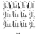
- the expression of Cxc / 12, a chemokine involved, for example, in the migration of haematopoietic cells from the foetal liver to the bone marrow, is about 50 times greater in Nestin-positive cells than in the rest of stromal cells.
- the expression of other genes in charge of regulating the maintenance of haematopoietic stem cells, such as, for example Kitl, II7 or Vcam1, is between 140 and 800 times greater in Nestin-positive cells than in the rest of stromal cells.
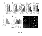
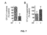
- mice wherein 90% of the Nestin+ cells were selectively eliminated present a 2-4-fold reduction in the number of haemopoietic stem cells in the bone marrow have a 90% reduction in the capacity of haemopoietic progenitors to nest in the bone marrow.
- the haemopoietic progenitors and the haemopoietic stem cells were reduced by ⁇ 50%. This reduction was associated with a proportional, selective increase in the spleen, without any differences being detected in the cell cycle or the frequency of apoptotic cells.
- haematopoietic stem cells and haemopoietic progenitors were mobilised from the bone marrow to extramedullary sites after depleting the nestin + cells.
- the severe reductions in HSCs detected suggest that nestin + cells play a central role in the maintenance of HSCs and in haematopoiesis. Therefore, placing these nestin + stem cells in contact with haematopoietic stem cells leads to the maintenance of haematopoiesis.
- This use has a clear industrial application, for example, in the field of treating diseases associated with a dysfunction of the maintenance of haematopoiesis in a mammal or the maintenance of haematopoiesis in vitro in order to allow for the generation of cells derived from haematopoietic stem cells, such as, for example, blood cells.
- the authors of the present invention have isolated a population of nestin-positive mesenchymal cells, which promote the self-renewal and/or expansion of haematopoietic stem cells, both in vitro and in vivo.
- a first aspect of the invention relates to an isolated cell population, hereinafter cell population of the invention, which comprises at least one multipotential Nestin-positive stem cell.
- the multipotential Nestin-positive stem cell is a mesenchymal cell.
- the multipotential Nestin-positive stem cell is a non-adherent cell.
- the multipotential Nestin-positive stem cell is obtained by means of a process which comprises:
- the mammal of step (a) is a human being.
- the cell population of the invention further comprises at least one haematopoietic stem cell.
- the haematopoietic stem cell is human.
- composition of the invention which comprises the isolated cell population of the invention.
- the composition is a pharmaceutical composition.
- the composition further comprises a pharmaceutically acceptable vehicle.
- the composition further comprises another active principle.
- Another aspect relates to the use of an isolated cell population of the invention or a pharmaceutical composition of the invention for the maintenance of haematopoiesis in vitro. Another aspect relates to the use of an isolated cell population of the invention or a pharmaceutical composition of the invention for the maintenance of haematopoiesis in vivo.
- Another aspect of the invention relates to the use of an isolated cell population of the invention, or a pharmaceutical composition of the invention, for the self-renewal of haematopoietic stem cells.
- Another aspect of the invention relates to the use of an isolated cell population of the invention, or a pharmaceutical composition of the invention, for the expansion of haematopoietic stem cells.
- Another aspect relates to the use of an isolated cell population of the invention or a pharmaceutical composition of the invention for the preparation of a medicament.
- Another aspect relates to the use of an isolated cell population of the invention or a pharmaceutical composition of the invention for the preparation of a medicament designed for the maintenance of haematopoiesis in a mammal.
- the mammal is a human being.
- Another aspect relates to the use of an isolated cell population of the invention or a pharmaceutical composition of the invention for the preparation of a medicament designed for tissue repair and regeneration.
- the tissue is blood.
- Another aspect relates to the use of an isolated cell population of the invention or a pharmaceutical composition of the invention for the preparation of a medicament designed for the treatment of blood and haematopoietic organ diseases.
- the disease evolves with a myelopoiesis or lymphopoiesis deficiency.
- blood and haematopoietic organ diseases are recorded, without being limited thereto, in the third chapter of the list of ICD-10 codes (tenth version of the International Statistical Classification of Diseases and Related Health Problems).
- ICD-10 codes tenth version of the International Statistical Classification of Diseases and Related Health Problems.
- blood and haematopoietic organ diseases are understood to be the following:
- the disease is selected from the list that comprises: myeloma, benign monoclonal gammopathy, hypoplasia and medullary aplasia, myelofibrosis, myelodysplastic syndrome, anaemia, polycythaemia, neutropenia, acute leukemia, chronic leukemia, lymphoma, purpura, haemophilia, or any combination thereof.
- Another aspect of the invention relates to a method for obtaining haematopoietic cells in vitro, hereinafter method for obtaining haematopoietic cells of the invention, which comprises:
- Another aspect of the invention relates to the haematopoietic cells obtainable by the method for obtaining haematopoietic cells of the invention.
- Another aspect of the present invention relates to the use of at least one isolated multipotential stem cell for the maintenance of haematopoiesis in vitro.
- Stem cells are undifferentiated cells that have the capacity to divide without losing their properties and to produce both differentiated cells and undifferentiated cells. Depending on the origin of the stem cells, we can distinguish between embryonic stem cells and adult stem cells. In the present invention, we refer to an adult stem cell or an embryonic stem cell.
- the multipotential stem cell of the present invention is capable of differentiating into different cell types from the same embryonic layer ( Weissman et al., 2001. Annu Rev Cell Dev Biol, 17: 387-403 ) and, as a consequence, into any derived adult tissue.
- isolated refers to the fact that the stem cells remain outside the human or animal body.
- haematopoiesis refers to the preservation of the haematopoiesis process, i.e. the preservation of the generation, regulation and production of cells derived from haematopoietic stem cells, as well as the division of said haematopoietic stem cells.
- haemopoiesis may be used as synonymous with the term “haematopoiesis”.
- Haematopoietic stem cells may be, without being limited thereto, "long-term haematopoietic stem cells” (LT-HSC) or “short-term haematopoietic stem cells” (ST-HSC).
- LT-HSC long-term haematopoietic stem cells
- ST-HSC short-term haematopoietic stem cells
- the cells derived from haematopoietic stem cells may be, without being limited thereto, compromised haematopoietic progenitors capable of differentiating into a myelocytic or lymphopoietic cell line, erythrocytes, platelets, granulocytes (neutrophils, basophils, eosinophils), monocytes or lymphocytes.
- the cells derived from haematopoietic stem cells may be any of the precursors of erythrocytes, platelets, granulocytes, monocytes or lymphocytes.
- haematopoiesis Under in vivo conditions, haematopoiesis is produced in various organs or tissues depending on the stage of development of the individual or even the development of pathological conditions. Therefore, during the course of approximately the second and third weeks of embryonic development, haematopoiesis is produced in the vitelline sac. Approximately from the sixth week, haematopoietic tissue appears in the liver. From the third month of embryonic development, myeloid tissue begins to develop in the bone marrow, which is where the haematopoiesis process is primarily produced. From the eighth month of embryonic development, haematopoietic tissue appears in the spleen.
- haematopoietic activity is maintained in the bone marrow; however, in the course of certain pathological conditions, haematopoietic foci may be observed in some of the aforementioned organs; in this case, haematopoiesis is extramedullary.
- the maintenance of haematopoiesis in vitro of the present invention may be performed in cells isolated from the haematopoietic tissue of any of the organs that have said tissue, such as, for example, without being limited thereto, any of the organs cited in the preceding paragraph in any stage of development of the individual.
- a preferred embodiment of the present invention relates to the use wherein the multipotential stem cell is a mesenchymal cell.
- Mesenchymal cells are derived from any mesenchymal tissue.
- Mesenchymal tissue is the tissue derived from the embryonic layer called mesoderm.
- the mesenchymal tissue wherefrom the mesenchymal cell of the present invention is derived is selected from the list that comprises lax connective tissue, dense connective tissue, adipose tissue, cartilaginous tissue, bone tissue, haematopoietic tissue, blood tissue or muscular tissue.
- the mesenchymal cell of the present invention is derived from haematopoietic tissue. More preferably, the mesenchymal cell is derived from the bone marrow.
- the bone marrow is a tissue that is found, for example, without being limited thereto, inside the long bones, vertebrae, ribs, sternum, cranial bones, scapular waist or pelvis.
- the bone marrow is red bone marrow, which occupies the spongy tissue of flat bones, such as, for example, without being limited thereto, the sternum, the vertebrae, the pelvis or the ribs.
- This type of bone marrow is the one that has the haematopoietic function.
- the mesenchymal cells of the present invention may be derived from the umbilical cord.
- Another preferred embodiment of the present invention relates to the use wherein the mesenchymal stem cell is a Nestin-positive cell.
- the Nestin protein is a type IV intermediate filament protein. This protein is expressed in undifferentiated cells during the early stages of development of the central nervous system and the peripheral nervous system, as well as organs such as the pancreas or muscular tissue.
- the Nestin protein is considered to be a stem cell marker. Those cells that express said protein are cells which behave like mesenchymal cells, taking into consideration their differentiation capacity. Therefore, the Nestin-positive mesenchymal cell of the present invention refers to a mesenchymal cell capable of expressing the Nestin protein.
- Nestin-positive mesenchymal cells may express said protein to a variable extent depending on multifactorial conditions, such as, for example, the type of tissue wherein the mesenchymal cell is found and the stage of development of the tissue whereto it belongs, etc.
- the examples of the present invention show that the drastic reduction in Nestin-positive mesenchymal cells in a mouse causes a 75% reduction in the number of haematopoietic bone marrow stem cells, capable of generating haemopoietic colonies after being cultured for long periods of time.
- the stem cell constitutes an isolated cell population.
- the cell population is composed of, or comprises, any of the multipotent stem cells, mesenchymal stem cells or Nestin-positive mesenchymal cells.
- Said cell population may be composed of any combination of multipotent stem cells, mesenchymal stem cells or Nestin-positive mesenchymal cells, or may comprise any combination of the aforementioned cells.
- Another aspect of the present invention relates to the use of at least one multipotential stem cell for the manufacturing of a medicament designed for the maintenance of haematopoiesis in a mammal.
- Another aspect of the invention relates to the use of at least one multipotential stem cell for the manufacturing of a medicament designed for the prevention or treatment of at least one disease associated with a dysfunction of the maintenance of haematopoiesis in a mammal.
- prevention involves avoiding the onset of diseases that evolve with a dysfunction of the maintenance of haematopoiesis.
- treatment as understood in the present invention, entails combatting the effects caused by said dysfunction, in order to stabilise the individual's condition or prevent subsequent damages.
- the normal condition is that wherein said cellularity is maintained above a minimum percentage, such as, for example, without being limited thereto, above 20, 25, 30 or 35%.
- the hypocellularity condition does not affect the present invention. I.e.
- the disease associated with said dysfunction of the maintenance of haematopoiesis is a consequence of the reduction in the division and/or differentiation of haematopoietic cells, which causes a decrease in the production of each of the cells derived from haematopoietic stem cells with respect to a control production value for said cells.
- Said comparison may be made by determining the concentration of any type of differentiated haematopoietic cell and comparing it to the reference levels.
- These reference levels are the control levels.
- the reference level of erythrocytes is 4.5.10 6 cells/mm 3 in the case of males and the reference level of leukocytes is 5000 cells/mm 3 .
- the aforementioned reference levels are minimum levels below which we would consider that there is a decrease in the production of each of the cells derived from haematopoietic stem cells.
- cellularity refers to the ratio between haematopoietic cells and adipose tissue (primary constituent cells of the bone marrow), expressed as the percentage of cells.
- a preferred embodiment relates to the use wherein the disease associated with the dysfunction of the maintenance of haematopoiesis is a disease that evolves with a myelopoiesis or lymphopoiesis deficiency.
- defect refers to a functioning that is lower than normal, i.e. to a division and/or differentiation capacity of myelopoietic or lymphopoietic stem cells that is below the reference levels established for each type of cells derived from haematopoietic stem cells and for each sex.
- Myelopoiesis is the process that leads to the generation, development and maturation of blood myeloid cells, i.e. erythrocytes, platelets, granulocytes (neutrophils, basophils, eosinophils) or monocytes.
- Erythrocytes are generated by a process called erythropoiesis.
- Platelets are generated by thrombopoiesis.
- Granulocytes are generated by granulopoiesis.
- Monocytes are generated by monopoiesis.
- Lymphopoiesis is the process that allows for the generation, development and maturation of lymphocytes.
- the disease that evolves with a myelopoiesis or lymphopoiesis deficiency is selected from the list that comprises aplastic anaemia, pancytopenia, erythroblastopenia, erythrocytic aplasia, Fanconi's anaemia, Blackfan-Diamond's syndrome, panmyelopthisis, dyserythropoietic anaemia, dyshaematopoietic anaemia, granulocytopenia (agranulocytosis), neutropenia and lymphopenia.
- Another preferred embodiment of the present invention relates to the use of at least one multipotential stem cell for the manufacturing of a medicament, where the multipotential stem cell is a mesenchymal cell.
- Another preferred embodiment relates to the use wherein the mesenchymal stem cell is a Nestin-positive cell.
- the stem cell constitutes an isolated cell population.
- Said medicament comprises:
- the medicament may comprise any of said cells with the vehicle, excipient or other active substance in any combination thereof.
- vehicle and the excipient must be pharmaceutically acceptable.
- vehicle refers to those substances, or combination of substances, known in the pharmaceutical sector, which are used in the preparation of pharmaceutical administration forms, and include, without being limited thereto, solids, liquids, solvents and surfactants.
- the vehicle may be an inert substance or a substance with an action analogous to that of any of the sequences of the present invention.
- the function of the vehicle is to facilitate the incorporation of the multipotential stem cell, mesenchymal cell or Nestin-positive mesenchymal cell and/or other compounds, allow for a better dosage and administration, or give consistency and shape to the medicament.
- the form of presentation is liquid, the vehicle is the diluent.
- excipient refers to a substance that contributes to the absorption of any of the sequences of the present invention, stabilises said sequence or contributes to the preparation of the medicament in the sense of giving it consistency or providing flavours that make it more pleasant.
- excipients may have the function of maintaining the ingredients bound, such as, for example, starches, sugars or celluloses; a sweetening function; a colouring function; the function of protecting the medicament, such as, for example, insulating it from the air and/or moisture; the function of filling a pill, capsule or any other form of presentation, such as, for example, dibasic calcium phosphate; a disintegrating function, to facilitate the dissolution of the components and the absorption thereof in the intestine, without excluding other types of excipients not mentioned in this paragraph.
- pharmaceutically acceptable refers to the fact that the compound is permitted and evaluated, such that it does not cause any damage to the bodies whereto it is administered.
- pharmaceutically acceptable refers to the fact that the compound allows for the activity of the multipotential stem cell, mesenchymal cell or Nestin-positive mesenchymal cell of the present invention.
- active substance refers to an active substance that must allow for the activity of any of the cells of the invention, i.e. it must be compatible with the multipotential stem cell, mesenchymal cell or Nestin-positive mesenchymal cell.
- composition of the present invention may be presented in the form of solutions or any other clinically permitted administration form.
- the medicament of the present invention may be presented in a form adapted for oral or parenteral administration.
- the form adapted for oral administration refers to a physical state that allows for the oral administration thereof.
- the form adapted for oral administration is selected from the list that comprises, without being limited thereto, drops, syrup, tisane, elixir, suspension, extemporaneous suspension, drinkable vial, tablet, capsule, granules, cachet, lozenge, pill, troche or lyophilisate.
- parenteral administration refers to a physical state that allows for the injectable administration thereof, i.e. preferably in the liquid state.
- Parenteral administration may be performed by intramuscular, intraarterial, intravenous, intradermal, subcutaneous or intra-osseous route, without being solely limited to these types of parenteral administration routes.
- the medicament be presented in a form adapted for sublingual, nasal, intrathecal, bronchial, lymphatic, transdermal or inhaled administration.
- Another preferred embodiment of the present invention relates to the use of at least one multipotential stem cell, mesenchymal cell or Nestin-positive mesenchymal cell, where the mammal is a human being.
- Another aspect of the present invention relates to a method for the maintenance of haematopoiesis in vitro, which comprises:
- the multipotential stem cell preferably the mesenchymal stem cell and, more preferably, the Nestin-positive mesenchymal cell share the same niche as the haematopoietic stem cells in the bone marrow. Therefore, the first step of the method for the maintenance of haematopoiesis in vitro involves placing said isolated multipotential stem cell in contact with at least one haematopoietic stem cell in any stage of development prior to the differentiation thereof.
- the incubation of said cell mixture must be performed in a culture medium that allows for the division and/or differentiation of the haematopoietic stem cell.
- "adequate culture medium” is understood to mean any solution that comprises necessary nutrients for the division and/or differentiation, or for the recovery or isolation, of any of the cells derived from the haematopoietic stem cells of the present invention. Said culture is performed under favourable temperature and pH conditions.
- the culture medium is selected, without being limited thereto, from the list that comprises DMEM (Dulbecco's Modified Eagle's Medium), RPMI 1640, F12, F10, MCDB 131, MEM (Minimum Essential Media) or DMEM/F12.
- the culture medium may be supplemented with other components, such as, for example, without being limited thereto, CO 2 , O 2 , serum or serum substitute, amino acids, antibiotics, etc.
- any culture medium known in the state of the art may be used to culture the haematopoietic stem cells. Some types of culture media are described in the examples section of the present invention.
- a preferred embodiment of the present invention relates to the method for the maintenance of haematopoiesis in vitro, where the multipotential stem cell is a mesenchymal cell. Another preferred embodiment of the present invention relates to the method where the mesenchymal stem cell is a Nestin-positive cell. According to another preferred embodiment of the method for the maintenance of haematopoiesis in vitro, the stem cell constitutes an isolated cell population.
- Another aspect of the present invention relates to a method for determining the maintenance of the haematopoietic capacity of a mammal, which comprises:
- expression product refers to any product resulting from the expression of the nucleotide sequence that encodes the Nestin protein of the mammal.
- the resulting expression product is understood to mean, for example, the messenger RNA that is obtained from the transcription of the nucleotide sequence, the processed messenger RNA, the protein resulting from the translation of any of the messenger RNAs or the cDNA sequence (DNA complementary to the messenger RNA sequence).
- the analysis of the expression product is performed by means of any technique known in the state of the art.
- the presence of the expression product described in a preceding paragraph indicates the production of the Nestin protein in the cells of the tissue isolated from said mammal.
- the detection of said presence is dependent on the sensitivity of the detection technique used in the analysis.
- a preferred embodiment of the present invention relates to a method for determining the maintenance of the haematopoietic capacity of a mammal, which comprises:
- the negative control is, for example, without being limited thereto, a sample of haematopoietic tissue where the cells are not capable of maintaining haematopoiesis, i.e. where said cells are not capable, for example, of maintaining cellularity within certain desired percentages (known by persons skilled in the art) for any type of cell derived from a haematopoietic stem cell.
- the desired cellularity percentages may be, without being limited thereto, percentages below 20%, i.e. the negative control presents a dysfunction of the maintenance of haematopoiesis.
- the positive control is, for example, without being limited thereto, a sample of haematopoietic tissue where the cells are capable of maintaining haematopoiesis, i.e. where said cells are capable, for example, of maintaining cellularity within certain desired percentages (known by persons skilled in the art) for any type of cell derived from a haematopoietic stem cell.
- the desired cellularity percentages may be, without being limited thereto, percentages greater than 20%, i.e. the positive control presents the capacity to maintain haematopoiesis.
- significant difference refers to a difference calculated in section (b) of the method that is greater than a pre-defined standard error multiplied by a pre-defined security.
- the pre-defined security may have a value, for example, without being limited thereto, of 95% (p ⁇ 0.05) or 99% (p ⁇ 0.01).
- absence of a significant difference refers to a difference calculated in section (b) of the method that is equal to or less than a pre-defined standard error multiplied by a pre-defined security.
- Another preferred embodiment relates to the method for evaluating the haematopoietic capacity of a mammal, where said mammal is a human being.
- FIG. 1 Shows that Nes:GFP + cells are mesenchymal stem cells.
- FIG. 2 Shows that the Nes:GFP + cells are physically associated with the HSCs.
- FIG. 3 Shows that the HSC cells are located close to the Nes:GFP+ cells in the BM. Immunohistochemistry against CD15; Ter119, Gr-1, CD3e, B220 and Mac-1 (Lin), and CD48 in bone marrow sections of Nes-Gfp transgenic mice. The tips of the arrows indicate examples of CD150+ CD48-Lin- HSCs.
- the HSCs were frequently located adjacent to (A, C) or close to (B, D) the Nes:GFP+ cells of the endostium, and most of them in direct contact with (E, G-I) or close to (J-L) the Nes:GFP+ cells that surrounded the sinuses.
- the asterisks indicate CD150+ CD48+ megakaryocytes, easily identifiable by their large size and homogeneous cytoplasmic distribution of CD150. Grid, 50 ⁇ m.
- Figure 4 Shows that the selective elimination of cells that express nestin sharply reduces the content and nesting of HSC cells in the bone marrow.
- FIG. 5 Shows that the Nes:GFP+ cells in the cranial bone marrow are perivascular. Intravital microscopy of the calvaria of Nes-Gfp transgenic mice injected with Qdot by i.v. route (thin arrow).
- FIG. 6 Shows that selective cellular depletion in Nes-Cre/iDTR mice does not alter the cell cycle of the haemopoietic progenitors.
- Figure 7 Shows that selective cellular depletion in the long-term cultures from the bone marrow of Nes-Cre/iDTR mice reduces the number of haemopoietic progenitors.
- the number of colony-forming units in culture (CFU-C) was determined in the adherent fraction (A) and the supernatants (B) of myeloid bone marrow cultures (Dexter type) from Nes-Cre / iDTR double transgenic mice.
- the diphtheria toxin (DT, 100 ng/ml) was added to the medium when the cultures were seeded and at the weekly changes of half of the culture medium.
- Figure 8 Shows that selective cellular depletion in Nes-CreliDTR mice reduces the survival but does not affect the vascular permeability or the histology of the bone marrow or the haemogram.
- Figure 9 Shows the expression of critical genes for the maintenance of HSCs. Quantitative RT-PCR from RNA samples obtained from the CD45 - GFP + and CD45 - GFP- cells isolated from the bone marrow of Nes-Gfp transgenic mice, in order to determine the expression of Cxc / 12, stem cell factor/kit ligand (KitI), interleukin-7 (IL7), vascular cell adhesion molecule-1 (Vcam1), osteopontin (Spp1) and N-cadherin (Cdh2), Gapdh, angiopoietin-1 (Angpt1), in the Nes:GFP+ cells and the rest of the CD45- bone marrow population.
- KitI stem cell factor/kit ligand
- IL7 interleukin-7
- Vcam1 vascular cell adhesion molecule-1
- Spp1 osteopontin
- Cdh2 N-cadherin
- Gapdh angiopoietin-1
- Figure 10 Shows the results of quantitative RT-PCR using a different housekeeping gene.
- Figure 11 Shows a representative human mesensphere.
- Figure 12 Shows the osteoblast differentiation of human mesenspheres. We may observe the alkaline phosphatase and Von Kossa stainings of the CFU-F derived from each human primary mesensphere cultured under osteoblast differentiation conditions.
- Nes:GFP+ cells are mesenchymal stem cells (MSCs)
- Nes:GFP+ cells were genuine MSCs.
- the entire fibroblast colony-forming units (CFU-F) and clonogenic capacity of the bone marrow resided in the Nes:GFP + fraction ( Fig. 1A ).
- the culture of all the remaining CD45 - Nes:GFP- bone marrow cells at equal or greater density did not generate any CFU-F.
- the cells quickly lost the expression of GFP ( Fig. 1B ) and differentiated into mesenchymal progenitors, as was determined using the CFU-F assay.
- the CD45 - Nes:GFP + and CD45 - Nes:GFP- cells isolated by FACS were seeded under conditions that favour differentiation into osteoblast, adipocyte and chondrocyte lineages.
- the CD45 - Nes:GFP + fraction showed a robust capacity to differentiate into mesenchymal lineages, whereas the CD45 - Nes:GFP- fraction did not generate any offspring.
- Different genes necessary for differentiation into OBs ( Fig. 1 C ), adipocytes ( Fig. 1 E ) and chondrocytes ( Fig. 1G ) were significantly upwardly regulated in CD45 - Nes:GFP + cells for 3 weeks of culture with a differentiation medium.
- the mature, mineralising, adipocytic or chondrocytic phenotype was confirmed after one month under culture ( Fig. 1D, F, H ), thereby demonstrating their capacity to differentiate into multiple lineages.
- neural stem cells which express nestin, may self-renew when they are cultured as floating spheres ( Stemple & Anderson, 1992. Cell. Dec 11; 71 (6): 973-85 ), we adapted culture conditions of neural crest stem cells ( Pardal et al., 2007. Cell, 131: 364-377 ) and pericytes ( Crisan et al., 2008. Curr Protoc Stem Cell Biol, Chapter 2, Unit 2B 2 1-2B 2 13 ) in order to expand the Nes:GFP+ cells in low-attachment culture plates.
- the CD45 - Nes:GFP + cells but not the CD45 - Nes:GFP- cells (isolated by FACS), formed clonal spheres when they were seeded at low density ( Fig. 11 ). These mesenchymal spheres, herein called “mesenspheres”, had a mean diameter of 85 ⁇ 6 ⁇ m and continued to express GFP after 7-10 days under culture ( Fig. 1J-K ). The sphere formation efficiency (6.5 ⁇ 0.7%) was similar to that of neural crest stem cells ( Molofsky et al., 2003. Nature, 425: 962-967 ).
- the frequency of the spheres was remarkably similar (6.9 ⁇ 0.7 %) when the CD45 - Nes:GFP + cells were seeded in 96-well plates (1 cell / well).
- the self-renewal capacity of CD45 - Nes:GFP + cells was studied by dissociating the mesenspheres and seeding them under the same conditions.
- the clonal mesenspheres obtained from triple transgenic animals showed spontaneous differentiation into adipocytes, the identity whereof was confirmed by means of staining with Oil Red O, and into Obs, unequivocally identified by the expression of ⁇ -galactosidase directed by the Co/2.3 promoter ( ⁇ 53%; 27/51; Fig. 1N-O ).
- the mesenspheres that had already shown signs of adipocyte differentiation were seeded under conditions that favour differentiation into chondrocytes, they accumulated mucopolysaccharides characteristic of the cartilage (stained with "Alcian Blue"; Fig. 1P ) and increased the expression of the genes necessary for chondrocyte differentiation ( Fig. 1Q ).
- Fig 1B the expression analysis by Q-PCR shows a rapid reduction in the expression of Gfp one week after seeding the cells with CFU-F formation medium.
- Fig. 1C shows different determination markers towards the osteoblast lineage (alkaline phosphatase, Alpl; Runx2) and genes associated with the differentiation into OBs (bone morphogenetic protein-4, Bmp4; osteoglycin, Ogn ; osterix, Sp7 ; osteocalcin, Bglap; osteoactivin, Gpnmb). Said markers showed a progressive increase in expression in CFU-Fs derived from the isolated CD45 - Nes:GFP + cells cultured for 4 weeks in osteoblast differentiation medium.
- OBs bone morphogenetic protein-4, Bmp4
- osteoglycin osteoglycin, Ogn ; osterix, Sp7 ; osteocalcin, Bglap; osteoactivin, Gpnmb
- Fig. 1D we may observe the confirmation of the presence of osteoprogenitor cells only in the CD45 - Nes:GFP + population by means of the detection of the activity of alkaline phosphatase and the calcium deposits.
- the CD45 - Nes:GFP- cells did not generate any colonies.
- Fig. 1E we may observe the progressive differentiation into adipocytes from CFU-Fs obtained from CD45 - Nes:GFP + cells cultured for 3 weeks with adipocyte differentiation medium, made evident by an increase in the expression of adipsin (Cfd) and peroxisome proliferator-activated receptor gamma 2 ( Pparg ).
- Cfd adipsin
- Pparg peroxisome proliferator-activated receptor gamma 2
- FIG. 1F shows that the mature differentiated phenotype of the adipocytes derived from the isolated CD45 - Nes:GFP + cells was confirmed in the 4-week-long cultures by means of Oil Red O staining; on the contrary, the CD45 - Nes:GFP- cells did not generate any colonies (box).
- Fig. 1G shows that the progressive differentiation of the CFU-Fs obtained from the isolated CD45 - Nes:GFP + cells into chondrocytes was manifested in an increase in the expression of aggrecan during the 3 weeks of culture with chondrocyte differentiation medium ( Acan ).
- Acan chondrocyte differentiation medium
- FIG. 1H we may observe the accumulation of Alcian blue + mucopolysaccharides in cell pellets derived from CD45 - Nes:GFP + cells cultured for 3 weeks with chondrocyte differentiation medium.
- the CD45 - Nes:GFP + cells but not the rest of the CD45 - bone marrow population, form clonal spheres after 7 days under low-density culture.
- Fig. 1L shows that the CD45 - Nes:GFP + cells isolated and cultured together in polystyrene plates quickly lost the expression of GFP and differentiated into mesenchymal lineages; the GFP-adipocytes were clearly shown by the refringent lipid deposits under bright field.
- Fig. 1L shows that the CD45 - Nes:GFP + cells isolated and cultured together in polystyrene plates quickly lost the expression of GFP and differentiated into mesenchymal lineages; the GFP-adipocytes were clearly shown by the refringent lipid deposits under bright
- Fig. 1M we may observe multi- and unilocular refringent adipocytes, as well as cells migrating from the outer layer of the sphere, adhered to the surface of the culture plate.
- Fig. 1N-O shows that, after 3 weeks under culture, 53% of the clonal mesenspheres showed signs of multi-lineage differentiation into CoI2.3-LacZ + OBs and Oil red O + adipocytes.
- Fig. 1N-O shows that, after 3 weeks under culture, 53% of the clonal mesenspheres showed signs of multi-lineage differentiation into CoI2.3-LacZ + OBs and Oil red O + adipocytes.
- CD45 - Nes:GFP + bone marrow cells were significantly upwardly or downwardly regulated in the CD45 - Nes:GFP + bone marrow cells, as compared to all the other stem cells studied. These analyses revealed that most of the upwardly regulated genes were involved in metabolic and biosynthetic processes, whereas most of the downwardly regulated genes were involved in mitosis and cell division. These results indicate that CD45 - Nes:GFP + bone marrow cells are relatively quiescent but metabolically very active.
- HSCs may be identified and isolated with great purity using a combination of SLAM markers ( Kiel et al., 2005. Cell, 121: 1109-1121 ).
- SLAM markers Kiel et al., 2005. Cell, 121: 1109-1121 .
- immunohistochemistry was performed on bone marrow cryocuts from Nes-Gfp transgenic mice using haemopoietic lineage markers (anti-Ter119, Gr-1, CD3e, B220 and Mac-1), CD48 and CD150 ( Figs. 2A-B and Fig. 3 ).
- the CD150 + CD48- Lin- HSC cells represented a very small subpopulation ( ⁇ 0.005%) of the nucleated bone marrow cells.
- Nes:GFP+ cells contribute to the stromal layer.
- these cells were frequently located in association with cobblestone-forming areas, rich in haemopoietic progenitors ( Fig. 2C ). Therefore, these results suggest a close physical association between Nes:GFP+ cells and HSCs in the BM.
- EXAMPLE 4 Demonstration that nestin+ cells are necessary for the nesting of HSCs in the BM.
- the mesensphere formation frequency of the CD45 - Nes:GFP + cells obtained from the cranial bone marrow was identical to that found in the bone marrow of long bones, and also exclusive within the CD45 - population.
- the CD45 - Nes:GFP + cells also contained all the CFU-F activity of the cranial bone marrow.
- the frequency and morphology of the Nes:GFP + cranial bone marrow cells were not affected by the lethal irradiation, in agreement with the characteristic radio-resistance of MSCs.
- the CD150 + CD48 - LSK cells were isolated by flow cytometry, stained with a fluorescent lipophilic dye and injected by i.v.
- HSCs turned out to be extremely sensitive to selective cellular depletion in Nes-Cre / iDTR double transgenic mice, as manifested in a ⁇ 75% reduction in the number of bone marrow LSK cells ( Fig. 4D ) and a ⁇ 58% reduction in the number of CD150 + CD48 - LSK cells ( Fig. 4E ).
- Fig. 4D bone marrow LSK cells
- Fig. 4E CD150 + CD48 - LSK cells
- haemopoietic progenitors in the bone marrow was directly caused by selective cellular depletion in the bone marrow, since treatment of the primary bone marrow cultures of Nes-Cre / iDTR double transgenic mice with DT reduced the number of haemopoietic progenitors by ⁇ 70%, as compared to single transgenic controls ( Fig. 7 ).
- Fig. 8B-F the mortality was not caused by haematopoietic failure, but by the depletion of nestin + cells or the offspring thereof in vital organs, such as the heart and/or the brain.
- Fig. 8B-C we may observe that the FITC-Dextran dye, prepared and injected by i.v. route, as previously described (von Andrian, 1996), in the Nes- Cre / iDTR animals treated with DT, remained in the cranial bone marrow vasculature ( Fig. 8C ), as was also the case in the control animals ( Fig. 8B ), which indicates the absence of vascular damage.
- Fig. 8C the FITC-Dextran dye
- FIG. 8D-E shows that the bone marrow histology of the Nes-Cre / iDTR animals treated with DT (8E) is similar to that of the control animals (8D).
- Fig. 8F shows that selective cellular depletion in Nes-Cre / iDTR mice did not affect the haemogram or the number of circulating progenitors.
- the quantity of erythrocytes (RBC), haemoglobin (Hgb), haematocrit (Hct), platelets (Plt), leukocytes (WBC) and haemopoietic progenitors (CFU-C) in the blood did not change significantly in the Nes-Cre / iDTR and Nes-Cre / iDTR / ROSA-Gfp mice 24-48 h after the treatment with DT, as compared to the levels observed in transgenic animals injected with PBS or in control mice treated with DT.
- Fig. 4A-E we may observe that, following selective cell ablation in Nes-Cre / iDTR double transgenic mice, the haemopoietic progenitors move from the bone marrow to extramedullary sites. Fig.
- FIG. 4A shows that a single injection of diphtheria toxin (DT, 4 ⁇ g/kg, i.p.) in Nes-Cre / ROSA-Gfp / iDTR triple transgenic mice completely eliminated the GFP + bone marrow population after 24-48 h, as revealed by the absence of GFP + cells, and compared to the normal levels in triple transgenic control mice injected with PBS or in Nes-Cre / ROSA-Gfp double transgenic mice injected with DT.
- DT diphtheria toxin
- Fig. 4C shows the detection of a 2.8-fold increase in the number of Lin-CD48- cells detected in the spleen of Nestin-Cre / iDTR mice 24-48h after the injection with DT, as compared to double transgenic control animals injected with PBS or single transgenic animals treated with DT.
- Fig. 4C shows the detection of a 2.8-fold increase in the number of Lin-CD48- cells detected in the spleen of Nestin-Cre / iDTR mice 24-48h after the injection with DT, as compared to double transgenic control animals injected with PBS or single transgenic animals treated with DT.
- Fig. 4F we may observe that the depletion of nestin + cells reduces the nesting of haematopoietic progenitors in the bone marrow.
- the Nes-Cre / iDTR, Nes-Cre ERT2 / iDTR mice and the iDTR controls were injected with DT (4 ⁇ g/kg, i.p.) and lethally irradiated (1.2 Gy, one dose) 16-20 h later.
- Five million nucleated cells obtained from the bone marrow of congenic animals were injected by i.v. route in a volume of 200 ⁇ l of PBS.
- the bone marrow was seeded 3 h later in the medium designed for the growth of colony-forming units in culture (CFU-C).
- CFU-C colony-forming units in culture
- the number of CFU-Cs nested for each femur was corrected to represent the total bone marrow by multiplying by 16.9, since a femur contains approximately 5.9% of the total mouse bone marrow, as previously described ( Katayama et al., 2006. Cell, 124: 407-421 ).
- one or two irradiated animals did not receive a transplant, in order to correct the data in the event that residual progenitors from the recipient were found.
- Fig. 4G-1 shows that HSCs rapidly nest close to the GFP + bone marrow cells of Nes-Gfp transgenic mice.
- the bone marrow cells of congenic mice were stained with biotinylated antibodies against haemopoietic lineage markers (detected with streptavidin conjugated with Pacific Orange), a-c-kit conjugated with APC, a-Sca-1 conjugated with Pacific Blue, a-CD150 conjugated with PE and a-CD48 conjugated with FITC.
- the CD150 + CD48- LSK cells were isolated and stained with Vybrant DyD (Invitrogen).
- a number of HSCs between 5,000 and 11,000 were injected by i.v. route in lethally irradiated Nes-Gfp transgenic mice, as described in previous studies ( Lo Celso et al., 2009. Nature, 457: 92-96 ).
- connexins 45 and 43 were also 200 to 500 times greater in the CD45 - Nes:GFP + cells than in the CD45 - Nes:GFP- cells ( Fig. 9C ), which suggests that there is electromechanical coupling between the CD45 - Nes:GFP + cells innervated by sympathetic nerve terminals ( Katayama et al., 2006. Cell, 124: 407-421 ; Mendez-Ferrer et al., 2008. Nature, 452: 442-447 ; Yamazaki and Allen, 1990. Am J Anat, 187: 261-276 ).
- cytokines, hormones and the SNS regulate both the attraction of HSCs and bone formation in the bone marrow niche by means of direct control of the nestin + MSCs, where the expression of critical genes for the maintenance of HSCs and the cellular fate (proliferation and differentiation) is regulated in a co-ordinated manner.
- nestin + MSCs in the haemopoietic niche is based on the following evidence: i) The present invention has shown a marked, nonrandom proximity between both stem cells under homeostasis conditions. This physical association takes place both in the endostium and the medullary parenchyma. ii) Nestin + MSCs are innervated by the SNS, rich in the expression of Cxcl12 and the functional ⁇ 3 adrenergic receptor.
- Nestin + MSCs present very high expression levels of molecules that are critical for the maintenance and the quiescence of HSCs in the "osteoblastic niche", such as angiopoietin-1, osteopontin, N-cadherin and c-kit ligand.
- the present invention shows evidence that both the osteoblast proliferation and differentiation of nestin + bone marrow MSCs are selectively induced by PTH and inhibited by G-CSF and the sympathetic fibres that innervate nestin + cells. Therefore, these results extend the previous observations about the inhibitory effect of the SNS on bone formation and the expansion of HSCs induced by PTH ( Adams et al., 2007. Nat Biotechnol, 25: 238-243 ; Calvi et al., 2003. Nature, 425: 841-846 ), both described as being exclusively mediated by OBs, to MSCs.
- the studies performed make it possible to propose the existence of a single niche in the bone marrow formed by the MSC-HSC association, which is strictly regulated at the local level by the microenvironment and also at a distance, by means of humoral and autonomous nervous system signals.
- mice were used: Nes-Gfp ( Mignone et al., 2004. Cell Cycle, 6: 2161-2170 ), FVB-Tg( Col1a1-cre ) 1Kry/Mmcd ( Dacquin et al., 2002. Dev Dyn, 224: 245-251 ), B6.Cg(SJL)-TgN( Nes-cre )1Kln ( Tronche et al., 1999. Nat Genet, 23: 99-103 ), Nes-Cre ERT2 , C57BL/6-Gt(ROSA)26Sortm1(HBEGF)Awai/J ( Buch et al., 2005.
- the bone marrow was eluted with FACS medium described in previous studies ( Molofsky et al., 2003. Nature, 425: 962-967 ), made up of Leibovitz L-15 medium (Invitrogen) supplemented with 1 mg/ml bovine serum albumin (BSA, Sigma), 10 mM HEPES (Sigma), pH 7.4, and 1% penicillin-streptomycin (PS, Invitrogen). After lysing the erythrocytes with 0.8% NH 4 Cl, the bone marrow was enzymatically processed in the same manner described to isolate the neural crest cells from the postnatal intestine ( Molofsky et al., 2003.
- the cells were immunomagnetically enriched using magnetic spheres conjugated with an anti-CD45 antibody (Milteyi Biotec), following the manufacturer's instructions, and the CD45 - GFP + and CD45 - GFP- cells were isolated by means of FACS.
- the cells were seeded at clonal density ( ⁇ 1,000 cells/cm 2 ) in 35-mm plates ( StemCell Technologies ) or in single wells of ultralow-attachment 96-well plates ( Corning ).
- the composition of the culture medium was adapted from that of neural crest cells ( Pardal et al., 2007.
- FGF fibroblast growth factor
- IGF-1 insulin-like growth factor-1
- EGF epidermal growth factor
- PDGF platelet-derived growth factor
- OSM oncostatin M
- ESGRO® leukemia inhibitory factor
- ESGRO® leukemia inhibitory factor
- Osteoblast differentiation was induced by culturing the cells for 4 weeks with 50 ⁇ g/ml L-ascorbic acid 2-phosphate, 10 mM glycerophosphate (Sigma) and 15% FBS in ⁇ -MEM supplemented with PS (Invitrogen).
- Adipocyte differentiation was induced with 1 ⁇ M dexamethasone, 100 ⁇ M indomethacin, 0.5 mM 3-isobutyl-I-methylxanthine (IBMX), 10 ⁇ g/ml insulin (Sigma) and 10% FBS in ⁇ -MEM supplemented with PS.
- Chondrocyte differentiation was induced in confluent CFU-F cultures prepared as previously described ( Mendez-Ferrer et al., 2008.
- the cells were washed twice with PBS and incubated for 20 min at room temperature with 50 ⁇ g/ml Naphtol AS-MX phosphate, 0.5% N,N-dimethylformamide and 0.6 mg/ml Fast Red Violet LB in 0.1 M Tris-HCl, pH 8.9.
- the formation of calcium deposits was examined by means of von Kossa staining. To this end, the cells were washed 3 times and stained with newly prepared 5% silver nitrate for 30 min. After 3 washes, the reaction was developed with 5% sodium carbonate in 25% formalin for 5 min.
- the cells were fixed with 5% sodium thiosulfate for 2 min and washed 3 times.
- the adipocytes were stained with Oil Red O in the following manner: the cells were washed with 60% isopropanol and allowed to dry completely.
- a 6:4 dilution in distilled water was prepared from a stock solution containing 0.35 g/ml Oil Red O in isopropanol (Sigma), filtered 20 min later. The cells were incubated for 10 min with this solution and washed 4 times.
- the fixed cell cultures and pellets were incubated for 30 min at room temperature with 1% Alcian blue 8GX (Sigma) in 3% acetic acid, pH 2.5, and washed 4 times.
- porous ceramic cubes ⁇ 3 mm 3
- Caform® tricalcium phosphate
- the ossicles were placed in a tube containing the fibronectin solution and stirred for 1 min whilst negative pressure was applied by suction with a 60-ml syringe with a 21 g needle through the tube cap. The cap was replaced with a new one and the same process was repeated.
- the fibronectin-coated ossicles were allowed to dry overnight in a laminar flow hood.
- the newly isolated cells were introduced into the ossicles by means of the same process described, whereas the spheres were deposited on the surface of the ossicles. In both cases, the cells were allowed to adhere to the ossicles, in the sphere growth medium, for 24 h in the incubator.
- the ossicles were implanted s.c. under the dorsal skin of anaesthesised adult animals, from the same litter, but which did not have the transgenes.
- the anaesthesised animals were injected with 100 U of sodium heparin (i.p., Sigma), in order to prevent coagulation, and perfused through the left ventricle with ⁇ 20 ml of 2 mM MgCl 2 in cold PBS, followed by 100 ml of 2% paraformaldehyde, 0.2% glutaraldehyde, 5 mM EGTA and 2 mM MgCl 2 in PBS, pH 7.4, at 4°C.
- the ossicles were recovered and post-fixed for 2 h at 4°C with the same fixing solution.
- the X-gal staining was performed with 5 mM K 3 Fe(CN) 6 , 5 mM K 4 Fe(CN) 6 , 2 mM MgCl 2 , 0.01 % sodium deoxycholate (Sigma), 1 mg/ml 5-Bromo-4-chloro-3-indoxyl-beta-D-galactopyranoside (X-gal, BioSynth AG®) and 0.02% Nonidet P-40 (Roche) in PBS at 37°C under movement overnight.
- the ossicles were partially decalcified with 0.25 mM EDTA for 2 or 3 days and processed for cryostat sectioning (10 ⁇ m) using a tungsten carbide razor blade (Diamond Knives) and 4x methacrylate-coated plates and the CryoJane adhesive transfer tape system (Instrumedics).
- the immunohistochemistry with signal amplification was performed as previously described ( Mendez-Ferrer et al., 2008. Nature, 452: 442-447 ).
- the immunohistochemistry for the detection of the SLAM markers has been described in previous studies ( Kiel et al., 2005. Cell, 121: 1109-1121 ).
- Isoproterenol or the BRL37344 agonist (2 mg/kg; Sigma) were injected by i.p. route 2 h before sacrificing the animals; the adrenergic agonists were also present, at a concentration of 50 ⁇ M, during the enzymatic digestion and cell separation by FACS, performed at 37°C and room temperature, respectively.
- Medullary aspirates obtained from healthy donours were washed with PBS.
- the bone marrow cells were obtained by centrifugation.
- the red blood series cells were lysed and the samples were enzymaticaly and mechanically digested as described in the murine samples.
- the samples were immunomagnetically depleted from most of the CD45+ cells using magnetic spheres conjugated with an anti-human antibody (Miltenyi Biotec), following the manufacturer's recommendations.
- the CD45-cells were isolated by means of an automatic separator (FACS) and seeded at clonal density ( ⁇ 1,000 cells/cm 2 ) under the same conditions that promote the growth of murine mesenspheres, except that the growth factors were of human origin.
- the spheres had the same appearance as the murine spheres formed in 7-10 days ( Fig. 11 ).
- the primary spheres were individually collected, digested with collagenase (Stem Cell Technologies) and seeded in three different culture media:
- the isolated primary spheres were bound to phosphocalcic ceramic ossicles and subcutaneously implanted in immunodeficient mice.
- the ossicles were extracted after two months, enzymatically digested and separated by FACS using combinations of the MSC CD105, CD140b and CD146 markers, and subcultured in a mesensphere-forming medium.
- the mesensphere-forming capacity of each population is indicated in Table 4.
- Table 4 Self-renewal of human mesenspheres in vivo.
- the sphere formation efficiency (as a percentage with respect to the number of cells seeded) of each population within the different ossicles is indicated.
- CD105+ CD140b+ CD146+ CD140b+ 32 (9/28) 47 (65/137) 11 (4/38) 5.5 (1/18) 10.5 (4/38)
Landscapes
- Health & Medical Sciences (AREA)
- Life Sciences & Earth Sciences (AREA)
- Engineering & Computer Science (AREA)
- Biomedical Technology (AREA)
- Chemical & Material Sciences (AREA)
- Developmental Biology & Embryology (AREA)
- Biotechnology (AREA)
- Zoology (AREA)
- Organic Chemistry (AREA)
- Bioinformatics & Cheminformatics (AREA)
- Wood Science & Technology (AREA)
- Genetics & Genomics (AREA)
- Cell Biology (AREA)
- General Health & Medical Sciences (AREA)
- Hematology (AREA)
- Immunology (AREA)
- General Engineering & Computer Science (AREA)
- Biochemistry (AREA)
- Microbiology (AREA)
- Rheumatology (AREA)
- Animal Behavior & Ethology (AREA)
- Veterinary Medicine (AREA)
- Medicinal Chemistry (AREA)
- Pharmacology & Pharmacy (AREA)
- Public Health (AREA)
- Virology (AREA)
- Epidemiology (AREA)
- Chemical Kinetics & Catalysis (AREA)
- Nuclear Medicine, Radiotherapy & Molecular Imaging (AREA)
- General Chemical & Material Sciences (AREA)
- Diabetes (AREA)
- Oncology (AREA)
- Medicines Containing Material From Animals Or Micro-Organisms (AREA)
- Micro-Organisms Or Cultivation Processes Thereof (AREA)
- Measuring Or Testing Involving Enzymes Or Micro-Organisms (AREA)
Abstract
Description
- The present invention relates to the use of at least one isolated multipotential stem cell for the maintenance of haematopoiesis in vitro, where, preferably, said multipotential stem cell is a mesenchymal stem cell or, more preferably, said mesenchymal stem cell is a mesenchymal cell capable of expressing the Nestin protein. The present invention also relates to an isolated cell population of adult nestin-positive mesenchymal cells of a mammal, including human cells, to the use thereof for the manufacturing of a medicament designed for the maintenance of haematopoiesis in a mammal, for the prevention and/or treatment of at least one disease associated with a dysfunction of the maintenance of haematopoiesis in a mammal, and for maintaining and expanding the adult haematopoietic stem cells of said mammal, including a human being. Moreover, the present invention also relates to a method for maintaining haematopoiesis in vitro or to a method for evaluating the haematopoietic capacity of a mammal.
- It has been suggested that haematopoietic stem cells (HSCs) are preferably located in perivascular regions of the bone marrow (Kiel et al., 2005. Cell 121: 1109-1121), close to reticular cells with a high expression of the CXCL12/SDF-1 chemokine (Sugiyama et al., 2006. Immunity 25: 977-988). However, the identity and the function of these cells are unknown. In human bone marrow, CD146 is expressed in perivascular cells that have osteoprogenitor capacity and may self-renew and reconstitute haematopoiesis (Sacchetti et al., 2007. Cell 131: 324-336). However, it is not clear whether CD45- CD146+ cells are a part of the haematopoietic niche and this combination of markers is also present in vascular endothelial cells.
- The migration of HSCs offers a useful paradigm to study the cellular elements that form the haemopoietic niche, since the attraction of HSCs is directly controlled by the bone marrow microenvironment. Previous studies have revealed that the mobilisation of HSCs induced by the granulocyte colony-stimulating factor (G-CSF) is regulated by the sympathetic nervous system (SNS) (Katayama et al., 2006. Cell, 124: 407-421). In homeostasis, a low number of HSCs abandon the bone marrow and go into the blood stream (Abkowitz et al., 2003. Blood, 102: 1249-1253; Wright et al., 2001. Science, 294: 1933-1936). The release thereof into the blood is regulated by the molecular clock and directed by the SNS, which cyclically releases noradrenaline in the bone marrow, thereby activating the β3 adrenergic receptors and consequently inducing rhythmic oscillations in the expression of Cxc/12 (Mendez-Ferrer et al., 2008. Nature, 452: 442-447).
- On the other hand, previous studies have shown that the CD45- CD146+ perivascular cells in the bone marrow and other tissues contain osteoprogenitors (Sacchetti et al., 2007. Cell 131: 324-336) or MSCs (Crisan et al., 2008. Cell Stem Cell, 3: 301-313), and recent data suggest that cells capable of endochondral ossification are necessary for the formation of the haemopoietic niche in foetal bone marrow (Chan et al., 2008. Nature, 457[7228]: 490-4).
- Nestin is an intermediate filament protein. It is a marker of multi-lineage progenitor cells and its presence in these cells indicates the multipotentiality of the cells and their regenerative capacity (Wiese et al., 2004. CMLS, Cell. Mol. Life Sci., 61: 2510-2522). In this regard, it is well-known that Nestin+ cells in the bulb region of the capillary follicle have stem cell properties, are multipotent (mesenchymal stem cells) and may generate neural lineage cells in vitro and in vivo (Mignone et al., 2007. Cell Cycle, 6 (17): 2161-2170). The discussion in Mignore et al., 2007, mentions the cell types whereinto Nestin+ cells may differentiate, for example, neural stem cells and neuronal cells, hepatic oval cells, muscle satellite cells, pancreatic stem cells, Leydig progenitor cells, smooth muscle cells, sebaceous gland cells, melanocytes and keratinocytes.
- The transplantation of haematopoietic progenitor cells involves the infusion of these cells obtained from the bone marrow, the peripheral blood (Körbling et al., 1981. Exp Hematol 9: 684-690), the umbilical cord or the foetal liver into a patient who has previously been conditioned to receive the graft. It has become a therapeutic method for a wide variety of diseases, such as malignant haemopathies, aplastic anaemia, immunodeficiencies and a large number of solid tumours. Currently, over 30,000 patients are transplanted every year worldwide. The selection of the source and the type of transplant are determined by different factors. Peripheral blood (PB) was the source of haematopoietic progenitors in 90% of autologous transplants and in 30% of allogeneic transplants.
- The morbidity and mortality of the procedure have improved in recent years thanks to a better knowledge of the histocompatibility system, the development of anti-infective therapy, the use of environments with scant microbial contamination, haemotherapeutic support and the administration of potent immunosuppressants.
- It is necessary to guarantee a correct haematopoietic function following high-dose chemoradiotherapeutic treatment. Moreover, there are several limitations to allogeneic transplantation, which has led to the development of autotransplantation in recent years. This involves obtaining haemotopoietic progenitor cells from the patients themselves, preserving them and reinfusing them, after administering doses of ablative chemotherapy and/or radiotherapy.
- However, despite the fact that autologous transplants have a number of advantages (they do not require searching for a donour, lower toxicity related to the procedure, they may be performed on older patients (60 - 65 years old), there are no undesirable graft-versus-host disease (GVHD) complications, they allow for consolidation in solid tumours, when the tumoral mass is smaller, a lower number of cells are required for medullary reconstruction than in allogeneic transplants, and it is an option for patients who, for various reasons, cannot receive allogeneic transplants), they present a lower frequency of medullary recovery due to defects in the medullary microenvironment, influenced by the baseline disease and/or previous treatments.
- The transplantation of haematopoietic precursors obtained from peripheral blood requires the administration of granulo-monocytic CSF (GM-CSF). The main advantage of HTC from PB over bone marrow transplantation lies in the fact that the former contain a greater number of cells and better implantation is achieved. Moreover, it presents a number of additional advantages: there is no need to use an operating-room, which prevents all the risks that this entails, fast haematological recovery following the implantation, lower possibility of obtaining malignant cells, reduced risk of infection of the collected material, and haematopoietic cells may be obtained in situations wherein it is difficult to extract them from the bone marrow (myelofibrosis, pelvic irradiation, etc.)
- However, sometimes the necessary cellularity is not obtained with a single apheresis process, and several donation (or self-donation) processes are necessary, with the consequent damage to the donour, who suffers various symptoms: lumbar, bone and muscle pain, cephalea, hypotension, general discomfort, somnolence, loss of appetite, rashes, nausea, fever, fluid retention, etc. Cases of cardiac arrest and myocardial infarction during the apheresis have been described in donours with a history of cardiovascular diseases. Other complications include increased size of the spleen and splenic rupture, as well as signs of extramedullary haematopoiesis. Epiescleritis and iritis may also appear.
- Currently, the umbilical cord is the third source of cells for transplantation in adults and the second in children. It has been used in genetic and malignant diseases and in patients with total or partial compatibility, familial and nonfamilial. However, the use of immature, compromised haematopoietic progenitors obtained from the umbilical cord has the disadvantage that its efficacy in adults is yet to be proven, since the number of cells is too small to provide a lasting implantation (Broxmeyer et al., 1990. Int J Cell Cloning 8: 76).
- Consequently, it is difficult to maintain the production of blood cells derived from haematopoietic stem cells (in vivo and/or in vitro), and to expand them, for example in order to apply them in therapy.
- The present invention relates to the use of at least one isolated multipotential stem cell for the maintenance of haematopoiesis in vitro, where, preferably, said multipotential stem cell is a mesenchymal stem cell or, more preferably, said mesenchymal stem cell is a mesenchymal cell capable of expressing the Nestin protein. The present invention also relates to the use of any of the stem cells described above for the manufacturing of a medicament designed for the maintenance of haematopoiesis in a mammal, or the prevention and/or treatment of at least one disease associated with a dysfunction of the maintenance of haematopoiesis in a mammal. Preferably, said disease evolves with a myelopoiesis or lymphopoiesis deficiency. Moreover, the present invention also relates to a method for maintaining haematopoiesis in vitro or a method for evaluating the haematopoietic capacity of a mammal.
- The present invention demonstrates that Nestin-positive cells are mesenchymal stem cells and, therefore, multipotent, capable of differentiating into cell lineages such as, for example, osteoblast, chondrocyte or adipocyte lineages.
- Said Nestin-positive cells express high levels of molecules involved in the maintenance of haematopoietic stem cells, i.e. in haematopoiesis. The expression of Cxc/12, a chemokine involved, for example, in the migration of haematopoietic cells from the foetal liver to the bone marrow, is about 50 times greater in Nestin-positive cells than in the rest of stromal cells. The expression of other genes in charge of regulating the maintenance of haematopoietic stem cells, such as, for example Kitl, II7 or Vcam1, is between 140 and 800 times greater in Nestin-positive cells than in the rest of stromal cells.
- In this regard, mice wherein 90% of the Nestin+ cells were selectively eliminated present a 2-4-fold reduction in the number of haemopoietic stem cells in the bone marrow have a 90% reduction in the capacity of haemopoietic progenitors to nest in the bone marrow. Likewise, whereas the total cellularity and the number of Lin- CD48- bone marrow cells in these mice were not affected, the haemopoietic progenitors and the haemopoietic stem cells were reduced by ∼ 50%. This reduction was associated with a proportional, selective increase in the spleen, without any differences being detected in the cell cycle or the frequency of apoptotic cells. Therefore, these studies indicate that haematopoietic stem cells and haemopoietic progenitors were mobilised from the bone marrow to extramedullary sites after depleting the nestin+ cells. The severe reductions in HSCs detected suggest that nestin+ cells play a central role in the maintenance of HSCs and in haematopoiesis. Therefore, placing these nestin+ stem cells in contact with haematopoietic stem cells leads to the maintenance of haematopoiesis. This use has a clear industrial application, for example, in the field of treating diseases associated with a dysfunction of the maintenance of haematopoiesis in a mammal or the maintenance of haematopoiesis in vitro in order to allow for the generation of cells derived from haematopoietic stem cells, such as, for example, blood cells.
- Consequently, the authors of the present invention have isolated a population of nestin-positive mesenchymal cells, which promote the self-renewal and/or expansion of haematopoietic stem cells, both in vitro and in vivo.
- Therefore, a first aspect of the invention relates to an isolated cell population, hereinafter cell population of the invention, which comprises at least one multipotential Nestin-positive stem cell. In a preferred embodiment of this aspect of the invention, the multipotential Nestin-positive stem cell is a mesenchymal cell. In another, more preferred embodiment of this aspect of the invention, the multipotential Nestin-positive stem cell is a non-adherent cell. In another, more preferred embodiment of this aspect of the invention, the multipotential Nestin-positive stem cell is obtained by means of a process which comprises:
- a) Obtaining a bone marrow aspirate from a mammal,
- b) Lysing the red-series cells,
- c) Selecting the CD45- cells, and
- d) Seeding the cells of step (c) in an adequate medium.
- In an even more preferred embodiment, the mammal of step (a) is a human being.
- In another preferred embodiment, the cell population of the invention further comprises at least one haematopoietic stem cell. In another, more preferred embodiment, the haematopoietic stem cell is human.
- Another aspect relates to a composition, hereinafter composition of the invention, which comprises the isolated cell population of the invention. In a preferred embodiment of this aspect, the composition is a pharmaceutical composition. In another, more preferred embodiment, the composition further comprises a pharmaceutically acceptable vehicle. In another, more preferred embodiment, the composition further comprises another active principle.
- Another aspect relates to the use of an isolated cell population of the invention or a pharmaceutical composition of the invention for the maintenance of haematopoiesis in vitro. Another aspect relates to the use of an isolated cell population of the invention or a pharmaceutical composition of the invention for the maintenance of haematopoiesis in vivo.
- Another aspect of the invention relates to the use of an isolated cell population of the invention, or a pharmaceutical composition of the invention, for the self-renewal of haematopoietic stem cells.
- Another aspect of the invention relates to the use of an isolated cell population of the invention, or a pharmaceutical composition of the invention, for the expansion of haematopoietic stem cells.
- Another aspect relates to the use of an isolated cell population of the invention or a pharmaceutical composition of the invention for the preparation of a medicament.
- Another aspect relates to the use of an isolated cell population of the invention or a pharmaceutical composition of the invention for the preparation of a medicament designed for the maintenance of haematopoiesis in a mammal. In a preferred embodiment of this aspect of the invention, the mammal is a human being.
- Another aspect relates to the use of an isolated cell population of the invention or a pharmaceutical composition of the invention for the preparation of a medicament designed for tissue repair and regeneration. In a preferred embodiment, the tissue is blood.
- Another aspect relates to the use of an isolated cell population of the invention or a pharmaceutical composition of the invention for the preparation of a medicament designed for the treatment of blood and haematopoietic organ diseases. In a preferred embodiment, the disease evolves with a myelopoiesis or lymphopoiesis deficiency.
- The blood and haematopoietic organ diseases are recorded, without being limited thereto, in the third chapter of the list of ICD-10 codes (tenth version of the International Statistical Classification of Diseases and Related Health Problems). Amongst others, in this specification, "blood and haematopoietic organ diseases" are understood to be the following:
-
- • Malignant neoplasms
- ○ Haematologic
- ■ Leukemias
- ■ Acute lymphoblastic leukemia
- ■ Acute non-lymphoblastic leukemia
- ■ Chronic lymphocytic leukemia
- ■ Chronic myeloid leukemia, in accelerated phase or blast crisis
- ■ Lymphomas
- ■ Hodgkin's disease
- ■ Non-Hodgkin's lymphoma
- ■ Myelomas
- ■ Multiple myeloma (Kahler's disease)
- ■ Leukemias
- ○ Solid-tumour cancers
- ■ Neuroblastoma
- ■ Desmoplastic small round cell tumours
- ■ Ewing's sarcoma
- ■ Choriocarcinoma
- ○ Haematologic
- • Haematological diseases
- ○ Phagocytic disorders
- ■ Myelodysplasia
- ○ Anaemias
- ■ Paroxysmal nocturnal haemoglobinuria (severe aplasia)
- ■ Aplastic anaemia
- ■ Pure red-cell aplasia
- ○ Myeloproliferative disorders
- ■ Polycythemia vera
- ■ Essential thrombocytosis
- ■ Myelofibrosis
- ○ Phagocytic disorders
- • Metabolic disorders
- ○ amyloidosis
■ Amyloid light-chain amyloidosis
- ○ amyloidosis
- • Environmentally-induced diseases
- ○ Radiation poisoning
- • Viral diseases
- ○ HIV[3]
-
- • Lysosomal storage diseases
- ○ Lipidosis (lipid storage disorders)
- ■ Neuronal ceroid lipofuscinosis
- ■ Infantile neuronal ceroid lipofuscinosis (INCL, Santavuori's disease)
- ■ Jansky-Bielschowsky's disease (late infantile neuronal ceroid lipofuscinosis)
- ■ Sphyngolipidosis
- ■ Niemann-Pick's disease
- ■ Gaucher' disease
- ■ Leukodystrophies
- ■ Adrenoleukodystrophy
- ■ Metachromatic leukodystrophy
- ■ Krabbe's disease (globoid-cell leukodystrophy)
- ■ Neuronal ceroid lipofuscinosis
- ○ Mucopolysaccharidosis
- ■
- ■ Hurler's syndrome (MPS IH, α-L-iduronidase deficiency)
- ■ Scheie's syndrome (MPS I S)
- ■ Hurler-Scheie's syndrome (MPS I H-S)
- ■ Hunter's syndrome (MPS II, iduronidase sulfate deficiency)
- ■ Sanfilippo's syndrome (MPS III)
- ■ Morquio's syndrome (MPS IV)
- ■ Maroteaux-Lamy's syndrome (MPS VI)
- ■ Sly's syndrome (MPS VII)
- ■
- ○ Glycoproteinosis
- ■
- ■ Mucolipidosis II (I-cell disease)
- ■ Fucosidosis
- ■ Aspartylglucosaminuria
- ■ Alpha-mannosidosis
- ■
- ○ Others
- ■
- ■ Wolman's disease (acid lipase deficiency)
- ■
- ○ Lipidosis (lipid storage disorders)
- • Immunodeficiencies
- ○ T-cell deficiency
- ■ Ataxia telangiectasia
- ■ DiGeorge's syndrome
- ○ Combined B- and T-cell deficiencies
- ■ Severe combined immunodeficiency (SCID), all types
- ○ Well-defined syndromes
- ■ Wiskott-Aldrich's syndrome
- ○ Phagocytic disorders
- ■ Kostmann's syndrome
- ■ Shwachman-Diamond's syndrome
- ○ Immune deregulation diseases
- ■ Griscelli's syndrome, type II
- ○ Innate immune deficiencies
- ■ NF-kappa-B essential modulator deficiency (NEMO) (Inhibitor of kappa light polypeptide gene enhancer in B-cell kinase gamma deficiency)
- ○ T-cell deficiency
- • Haematological diseases
- ○
- ■ Haemoglobinopathies
- ■ Falciform-cell disease
- ■ β-thalassaemia major (Cooley's anaemia)
- ○ Anaemias
- ■ Aplastic anaemia
- ■ Diamond-Blackfan's anaemia
- ■ Fanconi's anaemia
- ■ Aplastic anaemia
- ○ Cytopenias
- ■ Amegakaryocytic thrombocytopenia
- ○ Haemophagocytic syndromes
- ■ Haemophagocytic lymphohistiocytosis (HLH)
- ○
- In another preferred embodiment, the disease is selected from the list that comprises: myeloma, benign monoclonal gammopathy, hypoplasia and medullary aplasia, myelofibrosis, myelodysplastic syndrome, anaemia, polycythaemia, neutropenia, acute leukemia, chronic leukemia, lymphoma, purpura, haemophilia, or any combination thereof.
- Another aspect of the invention relates to a method for obtaining haematopoietic cells in vitro, hereinafter method for obtaining haematopoietic cells of the invention, which comprises:
- a) placing at least one isolated Nestin-positive cell in contact with at least one isolated haematopoietic stem cell, and
- b) incubating the product obtained in section (a) in an adequate culture medium for the division and/or differentiation of the haematopoietic stem cell. In a preferred embodiment of this aspect of the invention, the Nestin-positive cell is isolated from the bone marrow. In another preferred embodiment, the Nestin-positive cell is a multipotent stem cell. In another preferred embodiment, the Nestin-positive cell is a mesenchymal stem cell. In another preferred embodiment, the Nestin-positive cell is a non-adherent cell. In another preferred embodiment, the Nestin-positive cell is human.
- Another aspect of the invention relates to the haematopoietic cells obtainable by the method for obtaining haematopoietic cells of the invention.
- Another aspect of the present invention relates to the use of at least one isolated multipotential stem cell for the maintenance of haematopoiesis in vitro.
- Stem cells are undifferentiated cells that have the capacity to divide without losing their properties and to produce both differentiated cells and undifferentiated cells. Depending on the origin of the stem cells, we can distinguish between embryonic stem cells and adult stem cells. In the present invention, we refer to an adult stem cell or an embryonic stem cell.
- The multipotential stem cell of the present invention is capable of differentiating into different cell types from the same embryonic layer (Weissman et al., 2001. Annu Rev Cell Dev Biol, 17: 387-403) and, as a consequence, into any derived adult tissue.
- The term "isolated" refers to the fact that the stem cells remain outside the human or animal body.
- As understood in the present invention, the term "maintenance of haematopoiesis" refers to the preservation of the haematopoiesis process, i.e. the preservation of the generation, regulation and production of cells derived from haematopoietic stem cells, as well as the division of said haematopoietic stem cells. Hereinafter, the term "haemopoiesis" may be used as synonymous with the term "haematopoiesis".
- Haematopoietic stem cells may be, without being limited thereto, "long-term haematopoietic stem cells" (LT-HSC) or "short-term haematopoietic stem cells" (ST-HSC).
- The cells derived from haematopoietic stem cells may be, without being limited thereto, compromised haematopoietic progenitors capable of differentiating into a myelocytic or lymphopoietic cell line, erythrocytes, platelets, granulocytes (neutrophils, basophils, eosinophils), monocytes or lymphocytes. Likewise, the cells derived from haematopoietic stem cells may be any of the precursors of erythrocytes, platelets, granulocytes, monocytes or lymphocytes.
- Under in vivo conditions, haematopoiesis is produced in various organs or tissues depending on the stage of development of the individual or even the development of pathological conditions. Therefore, during the course of approximately the second and third weeks of embryonic development, haematopoiesis is produced in the vitelline sac. Approximately from the sixth week, haematopoietic tissue appears in the liver. From the third month of embryonic development, myeloid tissue begins to develop in the bone marrow, which is where the haematopoiesis process is primarily produced. From the eighth month of embryonic development, haematopoietic tissue appears in the spleen. In adult individuals, the haematopoietic activity is maintained in the bone marrow; however, in the course of certain pathological conditions, haematopoietic foci may be observed in some of the aforementioned organs; in this case, haematopoiesis is extramedullary.
- The maintenance of haematopoiesis in vitro of the present invention may be performed in cells isolated from the haematopoietic tissue of any of the organs that have said tissue, such as, for example, without being limited thereto, any of the organs cited in the preceding paragraph in any stage of development of the individual.
- A preferred embodiment of the present invention relates to the use wherein the multipotential stem cell is a mesenchymal cell.
- Mesenchymal cells are derived from any mesenchymal tissue. Mesenchymal tissue is the tissue derived from the embryonic layer called mesoderm. The mesenchymal tissue wherefrom the mesenchymal cell of the present invention is derived is selected from the list that comprises lax connective tissue, dense connective tissue, adipose tissue, cartilaginous tissue, bone tissue, haematopoietic tissue, blood tissue or muscular tissue. Preferably, the mesenchymal cell of the present invention is derived from haematopoietic tissue. More preferably, the mesenchymal cell is derived from the bone marrow. The bone marrow is a tissue that is found, for example, without being limited thereto, inside the long bones, vertebrae, ribs, sternum, cranial bones, scapular waist or pelvis. Preferably, the bone marrow is red bone marrow, which occupies the spongy tissue of flat bones, such as, for example, without being limited thereto, the sternum, the vertebrae, the pelvis or the ribs. This type of bone marrow is the one that has the haematopoietic function. Moreover, the mesenchymal cells of the present invention may be derived from the umbilical cord.
- Another preferred embodiment of the present invention relates to the use wherein the mesenchymal stem cell is a Nestin-positive cell.
- The Nestin protein is a type IV intermediate filament protein. This protein is expressed in undifferentiated cells during the early stages of development of the central nervous system and the peripheral nervous system, as well as organs such as the pancreas or muscular tissue. The Nestin protein is considered to be a stem cell marker. Those cells that express said protein are cells which behave like mesenchymal cells, taking into consideration their differentiation capacity. Therefore, the Nestin-positive mesenchymal cell of the present invention refers to a mesenchymal cell capable of expressing the Nestin protein. Nestin-positive mesenchymal cells may express said protein to a variable extent depending on multifactorial conditions, such as, for example, the type of tissue wherein the mesenchymal cell is found and the stage of development of the tissue whereto it belongs, etc. The examples of the present invention show that the drastic reduction in Nestin-positive mesenchymal cells in a mouse causes a 75% reduction in the number of haematopoietic bone marrow stem cells, capable of generating haemopoietic colonies after being cultured for long periods of time.
- According to another preferred embodiment, the stem cell constitutes an isolated cell population. The cell population is composed of, or comprises, any of the multipotent stem cells, mesenchymal stem cells or Nestin-positive mesenchymal cells. Said cell population may be composed of any combination of multipotent stem cells, mesenchymal stem cells or Nestin-positive mesenchymal cells, or may comprise any combination of the aforementioned cells.
- Another aspect of the present invention relates to the use of at least one multipotential stem cell for the manufacturing of a medicament designed for the maintenance of haematopoiesis in a mammal.
- Another aspect of the invention relates to the use of at least one multipotential stem cell for the manufacturing of a medicament designed for the prevention or treatment of at least one disease associated with a dysfunction of the maintenance of haematopoiesis in a mammal.
- The term "prevention", as understood in the present invention, involves avoiding the onset of diseases that evolve with a dysfunction of the maintenance of haematopoiesis. The term "treatment", as understood in the present invention, entails combatting the effects caused by said dysfunction, in order to stabilise the individual's condition or prevent subsequent damages.
- The term "dysfunction of the maintenance of haematopoiesis", as used in the present invention, refers to a quantitative alteration of haematopoiesis which causes the total or partial loss of the maintenance of haematopoiesis and, as a consequence of said dysfunction, there is a decrease in cellularity (hypocellularity) in the bone marrow of the individual with respect to a normal condition. The normal condition is that wherein said cellularity is maintained above a minimum percentage, such as, for example, without being limited thereto, above 20, 25, 30 or 35%. The hypocellularity condition does not affect the present invention. I.e. the disease associated with said dysfunction of the maintenance of haematopoiesis is a consequence of the reduction in the division and/or differentiation of haematopoietic cells, which causes a decrease in the production of each of the cells derived from haematopoietic stem cells with respect to a control production value for said cells. Said comparison may be made by determining the concentration of any type of differentiated haematopoietic cell and comparing it to the reference levels. These reference levels are the control levels. For example, the reference level of erythrocytes is 4.5.106 cells/mm3 in the case of males and the reference level of leukocytes is 5000 cells/mm3. The aforementioned reference levels are minimum levels below which we would consider that there is a decrease in the production of each of the cells derived from haematopoietic stem cells.
- The term "cellularity", as understood in the present invention, refers to the ratio between haematopoietic cells and adipose tissue (primary constituent cells of the bone marrow), expressed as the percentage of cells.
- A preferred embodiment relates to the use wherein the disease associated with the dysfunction of the maintenance of haematopoiesis is a disease that evolves with a myelopoiesis or lymphopoiesis deficiency.
- The term "deficiency", as understood in the present invention, refers to a functioning that is lower than normal, i.e. to a division and/or differentiation capacity of myelopoietic or lymphopoietic stem cells that is below the reference levels established for each type of cells derived from haematopoietic stem cells and for each sex.
- Myelopoiesis is the process that leads to the generation, development and maturation of blood myeloid cells, i.e. erythrocytes, platelets, granulocytes (neutrophils, basophils, eosinophils) or monocytes. Erythrocytes are generated by a process called erythropoiesis. Platelets are generated by thrombopoiesis. Granulocytes are generated by granulopoiesis. Monocytes are generated by monopoiesis. Lymphopoiesis is the process that allows for the generation, development and maturation of lymphocytes.
- The disease that evolves with a myelopoiesis or lymphopoiesis deficiency is selected from the list that comprises aplastic anaemia, pancytopenia, erythroblastopenia, erythrocytic aplasia, Fanconi's anaemia, Blackfan-Diamond's syndrome, panmyelopthisis, dyserythropoietic anaemia, dyshaematopoietic anaemia, granulocytopenia (agranulocytosis), neutropenia and lymphopenia.
- Another preferred embodiment of the present invention relates to the use of at least one multipotential stem cell for the manufacturing of a medicament, where the multipotential stem cell is a mesenchymal cell. Another preferred embodiment relates to the use wherein the mesenchymal stem cell is a Nestin-positive cell. According to another preferred embodiment, the stem cell constitutes an isolated cell population.
- Said medicament comprises:
- the multipotential stem cell, mesenchymal cell or Nestin-positive mesenchymal cell, and
- a pharmacologically acceptable vehicle,
- a pharmacologically acceptable excipient, or
- another active substance.
- The medicament may comprise any of said cells with the vehicle, excipient or other active substance in any combination thereof.
- Moreover, the vehicle and the excipient must be pharmaceutically acceptable.
- The term "vehicle" refers to those substances, or combination of substances, known in the pharmaceutical sector, which are used in the preparation of pharmaceutical administration forms, and include, without being limited thereto, solids, liquids, solvents and surfactants. The vehicle may be an inert substance or a substance with an action analogous to that of any of the sequences of the present invention. The function of the vehicle is to facilitate the incorporation of the multipotential stem cell, mesenchymal cell or Nestin-positive mesenchymal cell and/or other compounds, allow for a better dosage and administration, or give consistency and shape to the medicament. When the form of presentation is liquid, the vehicle is the diluent.
- The term "excipient" refers to a substance that contributes to the absorption of any of the sequences of the present invention, stabilises said sequence or contributes to the preparation of the medicament in the sense of giving it consistency or providing flavours that make it more pleasant. Thus, excipients may have the function of maintaining the ingredients bound, such as, for example, starches, sugars or celluloses; a sweetening function; a colouring function; the function of protecting the medicament, such as, for example, insulating it from the air and/or moisture; the function of filling a pill, capsule or any other form of presentation, such as, for example, dibasic calcium phosphate; a disintegrating function, to facilitate the dissolution of the components and the absorption thereof in the intestine, without excluding other types of excipients not mentioned in this paragraph.
- The term "pharmacologically acceptable" refers to the fact that the compound is permitted and evaluated, such that it does not cause any damage to the bodies whereto it is administered.
- The term "pharmaceutically acceptable" refers to the fact that the compound allows for the activity of the multipotential stem cell, mesenchymal cell or Nestin-positive mesenchymal cell of the present invention.
- The term "active substance" refers to an active substance that must allow for the activity of any of the cells of the invention, i.e. it must be compatible with the multipotential stem cell, mesenchymal cell or Nestin-positive mesenchymal cell.
- I.e. at least one multipotential stem cell, mesenchymal cell or Nestin-positive mesenchymal cell is formulated in an appropriate pharmaceutical and pharmacological composition (medicament), in a therapeutically effective quantity. The medicament may be formulated jointly with one or more pharmaceutically and pharmacologically acceptable vehicles, adjuvants or excipients, and may also comprise another active substance.
- In every case, the form of presentation of the medicament will be adapted to the type of administration used; for this reason, the composition of the present invention may be presented in the form of solutions or any other clinically permitted administration form.
- The medicament of the present invention may be presented in a form adapted for oral or parenteral administration.
- The form adapted for oral administration refers to a physical state that allows for the oral administration thereof. The form adapted for oral administration is selected from the list that comprises, without being limited thereto, drops, syrup, tisane, elixir, suspension, extemporaneous suspension, drinkable vial, tablet, capsule, granules, cachet, lozenge, pill, troche or lyophilisate.
- The form adapted for parenteral administration refers to a physical state that allows for the injectable administration thereof, i.e. preferably in the liquid state. Parenteral administration may be performed by intramuscular, intraarterial, intravenous, intradermal, subcutaneous or intra-osseous route, without being solely limited to these types of parenteral administration routes.
- Another possibility is that the medicament be presented in a form adapted for sublingual, nasal, intrathecal, bronchial, lymphatic, transdermal or inhaled administration.
- Another preferred embodiment of the present invention relates to the use of at least one multipotential stem cell, mesenchymal cell or Nestin-positive mesenchymal cell, where the mammal is a human being.
- Another aspect of the present invention relates to a method for the maintenance of haematopoiesis in vitro, which comprises:
- a) placing at least one isolated multipotential stem cell in contact with at least one isolated haematopoietic stem cell, and
- b) incubating the product obtained in section (a) in an adequate culture medium for the division and/or differentiation of the haematopoietic stem cell.
- As shown in the examples section of the present invention, the multipotential stem cell, preferably the mesenchymal stem cell and, more preferably, the Nestin-positive mesenchymal cell share the same niche as the haematopoietic stem cells in the bone marrow. Therefore, the first step of the method for the maintenance of haematopoiesis in vitro involves placing said isolated multipotential stem cell in contact with at least one haematopoietic stem cell in any stage of development prior to the differentiation thereof.
- The incubation of said cell mixture must be performed in a culture medium that allows for the division and/or differentiation of the haematopoietic stem cell.
- In the present invention, "adequate culture medium" is understood to mean any solution that comprises necessary nutrients for the division and/or differentiation, or for the recovery or isolation, of any of the cells derived from the haematopoietic stem cells of the present invention. Said culture is performed under favourable temperature and pH conditions. The culture medium is selected, without being limited thereto, from the list that comprises DMEM (Dulbecco's Modified Eagle's Medium), RPMI 1640, F12, F10, MCDB 131, MEM (Minimum Essential Media) or DMEM/F12. Moreover, the culture medium may be supplemented with other components, such as, for example, without being limited thereto, CO2, O2, serum or serum substitute, amino acids, antibiotics, etc. However, any culture medium known in the state of the art may be used to culture the haematopoietic stem cells. Some types of culture media are described in the examples section of the present invention.
- The incubation described in section (b) of the method for the maintenance of haematopoiesis in vitro must be performed during the desired period of time, provided that the adequate conditions for said maintenance of haematopoiesis are maintained. To this end, periodical renewal of said medium, or the addition of new culture medium, or the recovery of part of the medium wherein the cells derived from any of the haematopoietic stem cells of the present invention are found may be necessary.
- A preferred embodiment of the present invention relates to the method for the maintenance of haematopoiesis in vitro, where the multipotential stem cell is a mesenchymal cell. Another preferred embodiment of the present invention relates to the method where the mesenchymal stem cell is a Nestin-positive cell. According to another preferred embodiment of the method for the maintenance of haematopoiesis in vitro, the stem cell constitutes an isolated cell population.
- Another aspect of the present invention relates to a method for determining the maintenance of the haematopoietic capacity of a mammal, which comprises:
- a) analysing at least one expression product of the gene that encodes the Nestin protein in the cells of a sample of haematopoietic tissue isolated from said mammal, and
- b) assigning the presence of said expression product analysed in section (a) to the maintenance of the haematopoietic capacity of the organ wherefrom said tissue is derived.
- The term "expression product", as understood in the present invention, refers to any product resulting from the expression of the nucleotide sequence that encodes the Nestin protein of the mammal. Thus, the resulting expression product is understood to mean, for example, the messenger RNA that is obtained from the transcription of the nucleotide sequence, the processed messenger RNA, the protein resulting from the translation of any of the messenger RNAs or the cDNA sequence (DNA complementary to the messenger RNA sequence).
- The analysis of the expression product is performed by means of any technique known in the state of the art.
- The presence of the expression product described in a preceding paragraph indicates the production of the Nestin protein in the cells of the tissue isolated from said mammal. The detection of said presence is dependent on the sensitivity of the detection technique used in the analysis.
- A preferred embodiment of the present invention relates to a method for determining the maintenance of the haematopoietic capacity of a mammal, which comprises:
- a) determining the concentration of at least one expression product of the gene that encodes the Nestin protein in the cells of a sample of haematopoietic tissue isolated from said mammal,
- b) calculating the difference between the concentration of the expression product of the gene that encodes the Nestin protein determined in section (a) and the concentration of the expression of said gene in a positive and/or a negative control, and
- c) assigning a significant difference with respect to the positive control, and/or the absence of a significant difference with respect to the negative control, calculated in section (b), to the maintenance of the haematopoietic capacity of the tissue wherefrom said sample is derived.
- The negative control is, for example, without being limited thereto, a sample of haematopoietic tissue where the cells are not capable of maintaining haematopoiesis, i.e. where said cells are not capable, for example, of maintaining cellularity within certain desired percentages (known by persons skilled in the art) for any type of cell derived from a haematopoietic stem cell. The desired cellularity percentages may be, without being limited thereto, percentages below 20%, i.e. the negative control presents a dysfunction of the maintenance of haematopoiesis.
- The positive control is, for example, without being limited thereto, a sample of haematopoietic tissue where the cells are capable of maintaining haematopoiesis, i.e. where said cells are capable, for example, of maintaining cellularity within certain desired percentages (known by persons skilled in the art) for any type of cell derived from a haematopoietic stem cell. The desired cellularity percentages may be, without being limited thereto, percentages greater than 20%, i.e. the positive control presents the capacity to maintain haematopoiesis.
- The term "significant difference", as used in the present invention, refers to a difference calculated in section (b) of the method that is greater than a pre-defined standard error multiplied by a pre-defined security. The pre-defined security may have a value, for example, without being limited thereto, of 95% (p < 0.05) or 99% (p < 0.01). On the contrary, the "absence of a significant difference" refers to a difference calculated in section (b) of the method that is equal to or less than a pre-defined standard error multiplied by a pre-defined security.
- Another preferred embodiment relates to the method for evaluating the haematopoietic capacity of a mammal, where said mammal is a human being.
- Both multipotential Nestin-positive stem cells and haematopoietic stem cells are adult cells, and were obtained without destroying any embryos or affecting the viability thereof. Throughout the description and the claims, the word "comprises" and the variants thereof are not intended to exclude other technical characteristics, additives, components or steps. For persons skilled in the art, other objects, advantages and characteristics of the invention will arise partly from the description and partly from the practise of the invention. The following figures and examples are provided for illustrative purposes, and are not intended to limit the scope of the present invention.
- In order to complement the description that has been made, as well as contribute to a better understanding of the characteristics of the invention, in accordance with some of the embodiments thereof, the following figures are shown below, for illustrative, non-limiting purposes:
-
Figure 1 .- Shows that Nes:GFP+ cells are mesenchymal stem cells. - (A) n = 12.
- (B)n=4.
- (C)n=4.
- (D) Cells isolated as CD45- Nes:GFP+ and CD45- Nes:GFP- (box) cultured for 4 weeks with an osteoblast differentiation medium.
- (E) Progressive differentiation into adipocytes from CFU-Fs obtained from CD45- Nes:GFP+ cells cultured for 3 weeks with adipocyte differentiation medium (n = 4).
- (F) CD45- Nes:GFP- cells did not generate any colonies (box).
- (G) Acan: expression of aggrecan during the 3 weeks of culture with chondrocyte differentiation medium.
- (H) Accumulation of Alcian blue+ mucopolysaccharides in cell pellets from CD45- Nes:GFP+ cells cultured for 3 weeks with chondrocyte differentiation medium (n = 4).
- (I) Clonal spheres: The CD45- Nes:GFP+ cells following 7 days of low-density culture; bright field.
- (J-K) Expression of GFP in a sphere after 10 days of culture; (J) bright field; (K) fluorescence.
- (L) Differentiation of CD45- Nes:GFP+ cells into mesenchymal lineages.
- (M) Multi- and unilocular refringent adipocytes, as well as cells migrating from the outer layer of the sphere, adhered to the surface of the culture plate.
- (N-O) Clonal mesenspheres (n = 51) differentiated into Col2.3-LacZ+ OBs and Oil red O+adipocytes.
- (P) Representative example of clonal mesensphere that accumulated Alcian blue+ mucopolysaccharides after 3 weeks under culture with chondrocyte differentiation medium (n = 4).
- (Q) Progressive differentiation of the clonal mesenspheres cultured for 3 weeks in chondrocyte differentiation medium (n = 5-11), manifested by the increased expression of (sex determining region Y)-box 9 (Sox9), aggrecan (Acan) and type IIα1 (Co/2α1) and type XIα2 (Col11α2) collagens.
- Scale, 100 µm (F, I, K, N, P); 500 µm (H); 50 µm (L-M, O). * p < 0.05; ** p < 0.01; *** p < 0.001; two-tailed unpaired t-test; the error bars indicate the standard error.
-
Figure 2 .- Shows that the Nes:GFP+ cells are physically associated with the HSCs. - (A, B) Immunohistochemistry against CD150 (arrows), CD48 and haemopoietic lineage markers in bone marrow sections of Nes-Gfp mice. Representative images of CD150+ CD48- Lin- HSCs located adjacent to the Nes:GFP+ cells of the endostium (A) and the sinuses (B); (A) The CD150+ CD48 / Lin+ megakaryocytes, indicated with asterisks, were easily identified thanks to their large size and homogeneous CD150 staining. Deconvolutions of the Z projections.
- (C) The scant Nes:GFP+ cells detectable in primary myeloid cultures were frequently associated with cobblestone-forming areas, rich in haematopoietic progenitors. Combined fluorescence and phase contrast image.
- Scale, 50 µm.
-
Figure 3 .- Shows that the HSC cells are located close to the Nes:GFP+ cells in the BM. Immunohistochemistry against CD15; Ter119, Gr-1, CD3e, B220 and Mac-1 (Lin), and CD48 in bone marrow sections of Nes-Gfp transgenic mice. The tips of the arrows indicate examples of CD150+ CD48-Lin- HSCs. The HSCs were frequently located adjacent to (A, C) or close to (B, D) the Nes:GFP+ cells of the endostium, and most of them in direct contact with (E, G-I) or close to (J-L) the Nes:GFP+ cells that surrounded the sinuses. The asterisks indicate CD150+ CD48+ megakaryocytes, easily identifiable by their large size and homogeneous cytoplasmic distribution of CD150. Grid, 50 µm. -
Figure 4 .- Shows that the selective elimination of cells that express nestin sharply reduces the content and nesting of HSC cells in the bone marrow. - (A) n = 5-10.
- (B) n = 5-7.
- (C) n = 3-8.
- (D-E) n = 4-5.
- (F) n = 4-8.
- (G) Shortest mean distances between the Nes:GFP+ cells and the HSCs nested in the bone marrow, or between these and the bone wall, 2 h (n = 16), 48 h (n = 30) and 96 h (n = 14) later.
- (H-I) Representative examples of HSCs stained with DyD (thin arrow) nested in less than 2 h close to GFP+ cells (thick arrow) in the bone marrow of Nes-Gfp mice. The bone matrix was visualised by means of the second harmonic signal generated by collagen when it is illuminated by femtosecond pulses with a titanium:sapphire laser.
- * p < 0.05, ** p < 0.01, *** p < 0.001; two-tailed unpaired t-test; the error bars indicate standard error.
-
Figure 5 .- Shows that the Nes:GFP+ cells in the cranial bone marrow are perivascular. Intravital microscopy of the calvaria of Nes-Gfp transgenic mice injected with Qdot by i.v. route (thin arrow). - (A-C) Nes:GFP+ cells (thick arrow) surrounding the vessels of the coronary suture.
- (D-F) Nes:GFP+ cells in the endostium.
-
Figure 6 .- Shows that selective cellular depletion in Nes-Cre/iDTR mice does not alter the cell cycle of the haemopoietic progenitors. Frequency of CD48-, Lin-Sca-1+ c-kit+ (LSK) (A) and CD150+ CD48- LSK (B) cells in the bone marrow of Nes-Cre/iDTR double transgenic mice or iDTR control animals 24 h after the injection of diphtheria toxin (DT). The bone marrow samples, stained as described in the section on Materials and Methods, were fixed and permeabilised, and TUNEL was detected using the Mebstain Apoptosis Kit Direct (MBL) and FACS. Each point corresponds to a mouse. ** p < 0.01, two-tailed unpaired t-test. -
Figure 7 .- Shows that selective cellular depletion in the long-term cultures from the bone marrow of Nes-Cre/iDTR mice reduces the number of haemopoietic progenitors. The number of colony-forming units in culture (CFU-C) was determined in the adherent fraction (A) and the supernatants (B) of myeloid bone marrow cultures (Dexter type) from Nes-Cre / iDTR double transgenic mice. The diphtheria toxin (DT, 100 ng/ml) was added to the medium when the cultures were seeded and at the weekly changes of half of the culture medium. The data were normalised on the basis of similar bone marrow cultures from iDTR control animals in order to discard the non-specific toxicity of the DT in vitro (n = 3 mice). ** p < 0.05; two-tailed unpaired t-test. The error bars indicate standard error. -
Figure 8 .- Shows that selective cellular depletion in Nes-CreliDTR mice reduces the survival but does not affect the vascular permeability or the histology of the bone marrow or the haemogram. - (A) Reduced survival of the Nes-CreliDTR mice following selective cell ablation. Survival percentage of the Nes-Cre / iDTR and Nes-Cre / iDTR / ROSA-Gfp mice triple-injected with diphtheria toxin (DT, 4 µg/kg, i.p.; line without elements at the ends) or PBS (line with arrow tips at both ends) (n = 10), as compared to control animals treated with DT (line with black circles at both ends, n = 5). *** p < 0.001 (Log-rank test).
- (B-C) FITC-Dextran dye, prepared and injected by i.v. route in the Nes-Cre / iDTR animals treated with DT in the vasculature of the cranial bone marrow (C), or in control animals (B).
- (D-E) Bone marrow histology of the Nes-Cre / iDTR animals treated with DT (E) and histology of the control animals (D). Representative examples of n = 4 mice. Scale, 100 µm (B-C); 50 µm (D-E).
- (F) Quantity of erythrocytes (RBC), haemoglobin (Hgb), haematocrit (Hct), platelets (PIt), leukocytes (WBC) and haemopoietic progenitors (CFU-C) in the blood of Nes-Cre / iDTR and Nes-Cre / iDTR / ROSA-Gfp mice 24-48 h after the treatment with DT, as compared to the levels observed in transgenic animals injected with PBS or in control mice treated with DT (n = 4-7). Two-tailed unpaired t-test. The error bars indicate standard error.
-
Figure 9 .- Shows the expression of critical genes for the maintenance of HSCs. Quantitative RT-PCR from RNA samples obtained from the CD45- GFP+ and CD45- GFP- cells isolated from the bone marrow of Nes-Gfp transgenic mice, in order to determine the expression of Cxc/12, stem cell factor/kit ligand (KitI), interleukin-7 (IL7), vascular cell adhesion molecule-1 (Vcam1), osteopontin (Spp1) and N-cadherin (Cdh2), Gapdh, angiopoietin-1 (Angpt1), in the Nes:GFP+ cells and the rest of the CD45- bone marrow population. - (A) n = 6.
- (B) The animals were injected with saline, a non-selective b-adrenergic agonist (isoproterenol, b-AR) or a selective β3 adrenergic receptor agonist (BRL37344; (β3-AR) (2 mg/kg, i.p.) and were sacrificed 2 h later. (n = 6).
- (C) Expression of connexins 45 (Gjc1) and 43 (Gja1). (n = 3).
- * p < 0.05, ** p < 0.01, *** p < 0.001; two-tailed unpaired t-test; the error bars indicate standard error.
-
Figure 10 .- Shows the results of quantitative RT-PCR using a different housekeeping gene. - (A-B) Quantitative PCR was performed from RNA samples obtained from the CD45- GFP+ and CD45- GFP- cells isolated from the bone marrow of Nes-Gfp transgenic mice. Very reproducible results were obtained using β-actin instead of Gapdh as the housekeeping gene. Expression of critical genes for the maintenance of HSCs (A) or for mesenchymal differentiation (B). (n = 3-7). * p < 0.05, ** p < 0.01, *** p < 0.001; two-tailed unpaired t-test. The error bars indicate standard error.
-
Figure 11 .- Shows a representative human mesensphere. -
Figure 12 .- Shows the osteoblast differentiation of human mesenspheres. We may observe the alkaline phosphatase and Von Kossa stainings of the CFU-F derived from each human primary mesensphere cultured under osteoblast differentiation conditions. - Below we will illustrate the invention by means of illustrative, non-limiting assays performed by the inventors, which describe the use of the multipotential cells of the present invention for the maintenance of haematopoiesis.
- In the first place, the hypothesis was raised that Nes:GFP+ cells were genuine MSCs. In fact, the entire fibroblast colony-forming units (CFU-F) and clonogenic capacity of the bone marrow resided in the Nes:GFP+ fraction (
Fig. 1A ). The culture of all the remaining CD45- Nes:GFP- bone marrow cells at equal or greater density did not generate any CFU-F. After seeding the CD45- Nes:GFP+ cells under different culture conditions, including "MSC" medium, the cells quickly lost the expression of GFP (Fig. 1B ) and differentiated into mesenchymal progenitors, as was determined using the CFU-F assay. In order to more exhaustively analyse their differentiation potential, the CD45- Nes:GFP+ and CD45- Nes:GFP- cells isolated by FACS were seeded under conditions that favour differentiation into osteoblast, adipocyte and chondrocyte lineages. The CD45- Nes:GFP+ fraction showed a robust capacity to differentiate into mesenchymal lineages, whereas the CD45- Nes:GFP- fraction did not generate any offspring. Different genes necessary for differentiation into OBs (Fig. 1 C ), adipocytes (Fig. 1 E ) and chondrocytes (Fig. 1G ) were significantly upwardly regulated in CD45- Nes:GFP+ cells for 3 weeks of culture with a differentiation medium. The mature, mineralising, adipocytic or chondrocytic phenotype was confirmed after one month under culture (Fig. 1D, F, H ), thereby demonstrating their capacity to differentiate into multiple lineages. - Since neural stem cells, which express nestin, may self-renew when they are cultured as floating spheres (Stemple & Anderson, 1992. Cell. Dec 11; 71 (6): 973-85), we adapted culture conditions of neural crest stem cells (Pardal et al., 2007. Cell, 131: 364-377) and pericytes (Crisan et al., 2008. Curr Protoc Stem Cell Biol, ) in order to expand the Nes:GFP+ cells in low-attachment culture plates. The CD45- Nes:GFP+ cells, but not the CD45- Nes:GFP- cells (isolated by FACS), formed clonal spheres when they were seeded at low density (
Fig. 11 ). These mesenchymal spheres, herein called "mesenspheres", had a mean diameter of 85 ± 6 µm and continued to express GFP after 7-10 days under culture (Fig. 1J-K ). The sphere formation efficiency (6.5 ± 0.7%) was similar to that of neural crest stem cells (Molofsky et al., 2003. Nature, 425: 962-967). The frequency of the spheres was remarkably similar (6.9 ± 0.7 %) when the CD45- Nes:GFP+ cells were seeded in 96-well plates (1 cell / well). The self-renewal capacity of CD45- Nes:GFP+ cells was studied by dissociating the mesenspheres and seeding them under the same conditions. A mean of 4.4 ± 0.8 secondary mesenspheres were obtained from each primary sphere (n = 19), a frequency lower than that described from neural stem cells (Molofsky et al., 2003. Nature, 425: 962-967), possibly due to the dense extracellular matrix produced by CD45- Nes:GFP+ cells, which hinders an effective dissociation of the spheres without compromising the cell viability. After two weeks under culture, the clonal mesenspheres, as well as the multiclonal cultures from CD45- Nes:GFP+ cells (Fig. 1L ), continued to proliferate but showed a progressive loss of the expression of GFP and began to differentiate into mesenchymal lineages (Fig. 1M ). In order to more exhaustively study the mesenchymal differentiation capacity of CD45- Nes:GFP+ cells, we crossed Nes-Gfp transgenic mice with a line that expresses Cre recombinase under the 2.3-kb proximal fragment of the collagen α1(I) promoter (Col2.3-Cre), selectively expressed in OBs (Dacquin et al., 2002. Dev Dyn, 224: 245-251), and with the ROSA26 / IoxP-stop-IoxP-LacZ (R26R) reporter line. After two weeks under culture, the clonal mesenspheres obtained from triple transgenic animals showed spontaneous differentiation into adipocytes, the identity whereof was confirmed by means of staining with Oil Red O, and into Obs, unequivocally identified by the expression of β-galactosidase directed by the Co/2.3 promoter (∼ 53%; 27/51;Fig. 1N-O ). When the mesenspheres that had already shown signs of adipocyte differentiation were seeded under conditions that favour differentiation into chondrocytes, they accumulated mucopolysaccharides characteristic of the cartilage (stained with "Alcian Blue";Fig. 1P ) and increased the expression of the genes necessary for chondrocyte differentiation (Fig. 1Q ). These data clearly show that the Nes:GFP+ mesenspheres are self-renewable and multipotent in vitro. - According to
Fig. 1A , the entire capacity of the bone marrow to generate fibroblast colony-forming units (CFU-F) or mesenchymal activity is contained in the Nes:GFP+ population (n = 12). InFig 1B , the expression analysis by Q-PCR shows a rapid reduction in the expression of Gfp one week after seeding the cells with CFU-F formation medium. -
Fig. 1C shows different determination markers towards the osteoblast lineage (alkaline phosphatase, Alpl; Runx2) and genes associated with the differentiation into OBs (bone morphogenetic protein-4, Bmp4; osteoglycin, Ogn; osterix, Sp7; osteocalcin, Bglap; osteoactivin, Gpnmb). Said markers showed a progressive increase in expression in CFU-Fs derived from the isolated CD45- Nes:GFP+ cells cultured for 4 weeks in osteoblast differentiation medium. InFig. 1D , we may observe the confirmation of the presence of osteoprogenitor cells only in the CD45- Nes:GFP+ population by means of the detection of the activity of alkaline phosphatase and the calcium deposits. The CD45- Nes:GFP- cells did not generate any colonies. InFig. 1E , we may observe the progressive differentiation into adipocytes from CFU-Fs obtained from CD45- Nes:GFP+ cells cultured for 3 weeks with adipocyte differentiation medium, made evident by an increase in the expression of adipsin (Cfd) and peroxisome proliferator-activated receptor gamma 2 (Pparg).Fig. 1F shows that the mature differentiated phenotype of the adipocytes derived from the isolated CD45- Nes:GFP+ cells was confirmed in the 4-week-long cultures by means of Oil Red O staining; on the contrary, the CD45- Nes:GFP- cells did not generate any colonies (box).Fig. 1G shows that the progressive differentiation of the CFU-Fs obtained from the isolated CD45- Nes:GFP+ cells into chondrocytes was manifested in an increase in the expression of aggrecan during the 3 weeks of culture with chondrocyte differentiation medium (Acan). InFig. 1H , we may observe the accumulation of Alcian blue+ mucopolysaccharides in cell pellets derived from CD45- Nes:GFP+ cells cultured for 3 weeks with chondrocyte differentiation medium. As shown inFig. 1l , the CD45- Nes:GFP+ cells, but not the rest of the CD45- bone marrow population, form clonal spheres after 7 days under low-density culture.Fig. 1L shows that the CD45- Nes:GFP+ cells isolated and cultured together in polystyrene plates quickly lost the expression of GFP and differentiated into mesenchymal lineages; the GFP-adipocytes were clearly shown by the refringent lipid deposits under bright field. InFig. 1M , we may observe multi- and unilocular refringent adipocytes, as well as cells migrating from the outer layer of the sphere, adhered to the surface of the culture plate.Fig. 1N-O shows that, after 3 weeks under culture, 53% of the clonal mesenspheres showed signs of multi-lineage differentiation into CoI2.3-LacZ+ OBs and Oil red O+ adipocytes. InFig. 1Q , we may observe the progressive differentiation of the clonal mesenspheres cultured for 3 weeks in chondrocyte differentiation medium, manifested in the increased expression of (sex determining region Y)-box 9 (Sox9), aggrecan (Acan) and collagens type Ilα1 (CoI2α1) and type XIα2 (CoI11α2). - In order to elucidate the origin and identity of the Nes:GFP+ MSCs, we performed comparative analyses of the complete genome of CD45- Nes:GFP+ bone marrow cells and that of different stem cells, obtained from the "Gene Expression Omnibus" (http://www.ncbi.nim.nih.gov/geo/) and "Stem-base" (http://www.stembase.ca/?path=/) databases. These analyses comprised human and murine MSCs, mouse embryonic and neural stem cells, multipotent murine cells derived from embryonic carcinomas, mouse satellite cells and human haematopoietic stem cells (HSCs) (Table 1). In order to compare the data from the human and murine genomes, the mouse genes that are homologous to human genes were identified using HomoloGene (http://www.ncbi.nlm.nih.gov/homologene). As a result, the expression of 9,000 paired genes annotated in databases was finally used for the study. The impartial hierarchical clustering and principal component analyses revealed that CD45- Nes:GFP+ bone marrow cells were very different from adult neural stem cells. Although CD45- Nes:GFP+ cells are different from all the stem cells included in the analysis, their expression pattern was closer to that of human CD45- CD146+ osteoprogenitor cells (Sacchetti et al., 2007. Cell, 131: 324-336). Table 1. List of experiments with DNA chips and their accession numbers m, mouse; BM, bone marrow; NSC, neural stem cell; h, human; FB, foetal blood; PB, peripheral blood; EB, embryoid bodies; MSC, mesenchymal stem cell; ESC, embryonic stem cell.


- Gene ontology analyses were performed on the transcripts that were significantly upwardly or downwardly regulated in the CD45- Nes:GFP+ bone marrow cells, as compared to all the other stem cells studied. These analyses revealed that most of the upwardly regulated genes were involved in metabolic and biosynthetic processes, whereas most of the downwardly regulated genes were involved in mitosis and cell division. These results indicate that CD45- Nes:GFP+ bone marrow cells are relatively quiescent but metabolically very active.
- HSCs may be identified and isolated with great purity using a combination of SLAM markers (Kiel et al., 2005. Cell, 121: 1109-1121). In order to evaluate the spatial relationship between Nes:GFP+ cells and HSCs, immunohistochemistry was performed on bone marrow cryocuts from Nes-Gfp transgenic mice using haemopoietic lineage markers (anti-Ter119, Gr-1, CD3e, B220 and Mac-1), CD48 and CD150 (
Figs. 2A-B andFig. 3 ). The CD150+ CD48- Lin- HSC cells represented a very small subpopulation (∼ 0.005%) of the nucleated bone marrow cells. Despite the low frequency of both HSCs and Nes:GFP+ cells, the great majority (88%; 37/42) of the CD150+ CD48- Lin-cells were located less than 5 cell diameters from the Nes:GFP+ cells, and most of them (60%; 25/42) were located directly adjacent to the Nes:GFP+ cells distributed in the sinuses (62%) or the endostium (26%;Fig. 2A , B.Fig. 3 ). The statistical inference from the Poisson distribution expected for the Nes:GFP+ cells in the sampling area demonstrates that the co-location of the CD150+ CD48- Lin- and Nes:GFP+ cells observed is highly significant (p < 10-16) . Total bone marrow was cultured under conditions that promote myeloid growth (Dexter type), in order to determine whether Nes:GFP+ cells contribute to the stromal layer. Despite the low number of Nes:GFP+ cells detectable in these cultures, these cells were frequently located in association with cobblestone-forming areas, rich in haemopoietic progenitors (Fig. 2C ). Therefore, these results suggest a close physical association between Nes:GFP+ cells and HSCs in the BM. - The high expression of CxcI12, Vcam1, KitI, II7 and Angpt1 in CD45- Nes:GFP+ cells and the marked reduction thereof during the mobilisation induced by the granulocyte colony-stimulating factor (G-CSF) or following β-adrenergic stimulation suggested that nestin+ cells could regulate the attraction of HSCs and their traffic in the bone marrow. In order to determine whether nestin+ cells played a significant role in the nesting of HSCs in the bone marrow, 5 x 10⁶ nucleated bone marrow cells from wild-type congenic animals were transplanted into lethally irradiated Nes-Cre / iDTR mice, Nes-CreERT2 / iDTR mice and iDTR controls, following a previously described protocol (Katayama et al., 2006. Cell, 124: 407-421). The nesting of CFU-Cs in the bone marrow was reduced by 73% in the Nes-Cre / iDTR mice and by 90% in the Nes-CreERT2 / iDTR animals (Fig. 4N), which indicates that nestin+ cells are in fact necessary for the nesting of haemopoietic progenitors in the BM.
- In order to determine whether nestin+ bone marrow cells directly regulated the nesting of HSCs, the migration of previously isolated HSCs with a high degree of purification, and subsequently labelled and trasplanted into lethally irradiated Nes-Gfp transgenic mice, was visualised by means of intravital microscopy. Since these in vivo image studies can only be performed in the cranial bone marrow, but not in the long bones (Lo Celso et al., 2009. Nature, 457: 92-96), we first validated the calvaria model by comparing the frequency, distribution and physiological properties (capacity to form CFU-C and mesenspheres) of the CD45- Nes:GFP+ cells isolated from the bone marrow of the cranium and the long bones. The Nes:GFP+ calvarial cells were also perivascular, as verified by intravital microscopy of Nes-Gfp transgenic mice injected with Qdot by i.v. route in order to stain the vasculature (
Fig. 5 ). The mesensphere formation frequency of the CD45- Nes:GFP+ cells obtained from the cranial bone marrow was identical to that found in the bone marrow of long bones, and also exclusive within the CD45- population. The CD45- Nes:GFP+ cells also contained all the CFU-F activity of the cranial bone marrow. The frequency and morphology of the Nes:GFP+ cranial bone marrow cells were not affected by the lethal irradiation, in agreement with the characteristic radio-resistance of MSCs. The CD150+ CD48- LSK cells were isolated by flow cytometry, stained with a fluorescent lipophilic dye and injected by i.v. route in lethally irradiated adult Nes-Gfp transgenic mice. The nesting of HSCs in the bone marrow was directly visualised in thecranial bone marrow 2, 48 and 96 h later, by a combination of confocal and two-photon microscopy (Lo Celso et al., 2009. Nature, 457: 92-96). The entire cranial bone marrow was scanned and Z projections were obtained, wherefrom the shortest distance between the HSCs that had nested in the bone marrow and the Nes:GFP+ cells and the bone surface was calculated (Fig. 4O). The analyses performed clearly showed that the HSCs quickly nested close to the Nes:GFP+ bone marrow cells (Fig. 40-Q), which indicates that nestin+ cells direct the migration of HSCs in the bone marrow. - In order to determine whether nestin+ cells are in fact necessary for the maintenance of HSCs in the bone marrow, we attempted to eliminate them by crossing Nes-Cre mice with a line that Cre-inducibly expresses the diphtheria toxin (DT) receptor (Buch et al., 2005. Nat Methods, 2: 419-426) (iDTR) and with the ROSA26 / IoxP-stop-IoxP-GFP (ROSA-GFP) reporter line (Mao et al., 2001. Blood, 97: 324-326). A single injection of DT (4 µg/kg, i.p.) completely eliminated the GFP+ bone marrow cells in 24-48 h (
Fig. 4A ), and caused a reduction of ∼ 45% in the total bone marrow cellularity (Fig. 4B ), associated with a ∼ 40% increase in the number of cells stained with propidium iodide, probably due to the abrupt reduction in cytokines and growth factors that are critical for different haemopoietic progenitors (such as IL-7, the c-kit ligand and M-CSF) and which are produced in large quantities by nestin+ bone marrow cells. A 2.3-fold increase in the number of Lin- CD48- haemopoietic progenitors was also detected in the spleen of Nes-Cre / iDTR double transgenic mice, as compared to single transgenic controls, 24-48 h after the treatment with DT (Fig. 4C ), which suggests an extramedullary migration of the haemopoietic progenitors. - It is worth noting that HSCs turned out to be extremely sensitive to selective cellular depletion in Nes-Cre / iDTR double transgenic mice, as manifested in a ∼ 75% reduction in the number of bone marrow LSK cells (
Fig. 4D ) and a ∼ 58% reduction in the number of CD150+ CD48- LSK cells (Fig. 4E ). We studied whether this greater reduction in HSCs was caused by apoptosis, but no differences were detected between the two groups of animals in regards to the frequency of CD48- TUNEL+ LSK or CD150+ CD48- TUNEL+ LSK cells (Fig. 6 ). The reduction in haemopoietic progenitors in the bone marrow was directly caused by selective cellular depletion in the bone marrow, since treatment of the primary bone marrow cultures of Nes-Cre / iDTR double transgenic mice with DT reduced the number of haemopoietic progenitors by ∼ 70%, as compared to single transgenic controls (Fig. 7 ). Treatment of Nes-Cre / iDTR double transgenic mice and Nes-Cre / ROSA-Gfp / iDTR triple transgenic mice with DT was lethal in 50% of the animals after 24 h and in 80% of them 48 h later (n = 10), whereas it did not affect the viability of Nes-Cre / ROSA-Gfp double transgenic mice and single transgenic control mice (Fig. 8A ). Despite the reduction in total bone marrow cellularity, the vascular permeability, the histology and the haemogram were normal (Fig. 8B-F ), which suggests that the mortality was not caused by haematopoietic failure, but by the depletion of nestin+ cells or the offspring thereof in vital organs, such as the heart and/or the brain. InFig. 8B-C , we may observe that the FITC-Dextran dye, prepared and injected by i.v. route, as previously described (von Andrian, 1996), in the Nes-Cre / iDTR animals treated with DT, remained in the cranial bone marrow vasculature (Fig. 8C ), as was also the case in the control animals (Fig. 8B ), which indicates the absence of vascular damage.Fig. 8D-E shows that the bone marrow histology of the Nes-Cre / iDTR animals treated with DT (8E) is similar to that of the control animals (8D).Fig. 8F shows that selective cellular depletion in Nes-Cre / iDTR mice did not affect the haemogram or the number of circulating progenitors. The quantity of erythrocytes (RBC), haemoglobin (Hgb), haematocrit (Hct), platelets (Plt), leukocytes (WBC) and haemopoietic progenitors (CFU-C) in the blood did not change significantly in the Nes-Cre / iDTR and Nes-Cre / iDTR / ROSA-Gfp mice 24-48 h after the treatment with DT, as compared to the levels observed in transgenic animals injected with PBS or in control mice treated with DT. - In
Fig. 4A-E , we may observe that, following selective cell ablation in Nes-Cre / iDTR double transgenic mice, the haemopoietic progenitors move from the bone marrow to extramedullary sites.Fig. 4A shows that a single injection of diphtheria toxin (DT, 4 µg/kg, i.p.) in Nes-Cre / ROSA-Gfp / iDTR triple transgenic mice completely eliminated the GFP+ bone marrow population after 24-48 h, as revealed by the absence of GFP+ cells, and compared to the normal levels in triple transgenic control mice injected with PBS or in Nes-Cre / ROSA-Gfp double transgenic mice injected with DT. InFig. 4B , we may observe the reduced cellularity in the bone marrow of the femurs and tibias of Nes-Cre / iDTR and Nes-Cre / iDTR / ROSA-Gfp mice 24-48 h after the treatment with DT, which did not affect the animals that did not have the iDTR transgene, as a control.Fig. 4C shows the detection of a 2.8-fold increase in the number of Lin-CD48- cells detected in the spleen of Nestin-Cre / iDTR mice 24-48h after the injection with DT, as compared to double transgenic control animals injected with PBS or single transgenic animals treated with DT. InFig. 4D-E , we may observe a reduction in the number of bone marrow HSC cells in Nes-Cre / iDTR mice after selective cell ablation. 24-48 h later, treatment of the Nes-Cre / iDTR and Nes-Cre / ROSA-Gfp / iDTR mice with DT caused a ∼ 75% reduction (D) in the number of Lin- Sca-1+ c-kit+ (LSK) and a ∼ 58% reduction (E) in the CD150+ CD48- bone marrow LSK cells.Figs. 4F-I show that nestin+ bone marrow cells are necessary for the nesting of HSCs and haemopoietic progenitors. For example, inFig. 4F we may observe that the depletion of nestin+ cells reduces the nesting of haematopoietic progenitors in the bone marrow. The Nes-Cre / iDTR, Nes-CreERT2 / iDTR mice and the iDTR controls were injected with DT (4 µg/kg, i.p.) and lethally irradiated (1.2 Gy, one dose) 16-20 h later. Five million nucleated cells obtained from the bone marrow of congenic animals were injected by i.v. route in a volume of 200 µl of PBS. The bone marrow was seeded 3 h later in the medium designed for the growth of colony-forming units in culture (CFU-C). The number of CFU-Cs nested for each femur was corrected to represent the total bone marrow by multiplying by 16.9, since a femur contains approximately 5.9% of the total mouse bone marrow, as previously described (Katayama et al., 2006. Cell, 124: 407-421). In each experiment, one or two irradiated animals (of the same age, genotype and gender) did not receive a transplant, in order to correct the data in the event that residual progenitors from the recipient were found. The selective elimination of nestin+ cells in the Nes-Cre / iDTR and Nes-CreERT2 / iDTR mice significantly compromised the nesting of haemopoietic progenitors in the bone marrow. On the other hand,Fig. 4G-1 shows that HSCs rapidly nest close to the GFP+ bone marrow cells of Nes-Gfp transgenic mice. The bone marrow cells of congenic mice were stained with biotinylated antibodies against haemopoietic lineage markers (detected with streptavidin conjugated with Pacific Orange), a-c-kit conjugated with APC, a-Sca-1 conjugated with Pacific Blue, a-CD150 conjugated with PE and a-CD48 conjugated with FITC. The CD150+ CD48- LSK cells were isolated and stained with Vybrant DyD (Invitrogen). A number of HSCs between 5,000 and 11,000 were injected by i.v. route in lethally irradiated Nes-Gfp transgenic mice, as described in previous studies (Lo Celso et al., 2009. Nature, 457: 92-96). - In order to study in more depth the regulation of the haematopoietic stem cell niche by the G-CSF factor and the Central Nervous System (SNS), we analysed the expression of genes that regulate the maintenance of HSCs and their attraction in the bone marrow (CXCL12/SDF-1, c-kit ligand, angiopoietin-1, interleukin-7, vascular cell adhesion molecule-1, osteopontin and N-cadherin) in CD45- Nes:GFP+ and CD45- Nes:GFP- cells isolated from the bone marrow of mice treated with G-CSF or injected with a vehicle, as in previous studies (Katayama et al., 2006. Cell, 124: 407-421). The expression of these genes was extremely high (close to or greater than that of Gapdh) in the CD45- Nes:GFP+ cells and, with the exception of Angpt1, 50 to 800 times greater than in the CD45- Nes:GFP- bone marrow cells. Moreover, the expression of these genes (except for osteopontin and N-cadherin) significantly decreased in the CD45- Nes:GFP+ cells, but not in the CD45- Nes:GFP- cells, after the mobilisation of HSCs induced by G-CSF (
Fig. 9A ) or stimulation of the β3 adrenergic receptor (Fig. 9B ). The use of a different housekeeping gene showed very similar results (Fig. 10 ). The expression of connexins 45 and 43 was also 200 to 500 times greater in the CD45- Nes:GFP+ cells than in the CD45- Nes:GFP- cells (Fig. 9C ), which suggests that there is electromechanical coupling between the CD45- Nes:GFP+ cells innervated by sympathetic nerve terminals (Katayama et al., 2006. Cell, 124: 407-421; Mendez-Ferrer et al., 2008. Nature, 452: 442-447; Yamazaki and Allen, 1990. Am J Anat, 187: 261-276). Therefore, cytokines, hormones and the SNS regulate both the attraction of HSCs and bone formation in the bone marrow niche by means of direct control of the nestin+ MSCs, where the expression of critical genes for the maintenance of HSCs and the cellular fate (proliferation and differentiation) is regulated in a co-ordinated manner. - The relevant role of nestin+ MSCs in the haemopoietic niche is based on the following evidence: i) The present invention has shown a marked, nonrandom proximity between both stem cells under homeostasis conditions. This physical association takes place both in the endostium and the medullary parenchyma. ii) Nestin+ MSCs are innervated by the SNS, rich in the expression of Cxcl12 and the functional β3 adrenergic receptor. iii) Nestin+ MSCs present very high expression levels of molecules that are critical for the maintenance and the quiescence of HSCs in the "osteoblastic niche", such as angiopoietin-1, osteopontin, N-cadherin and c-kit ligand.
- The present invention shows evidence that both the osteoblast proliferation and differentiation of nestin+ bone marrow MSCs are selectively induced by PTH and inhibited by G-CSF and the sympathetic fibres that innervate nestin+ cells. Therefore, these results extend the previous observations about the inhibitory effect of the SNS on bone formation and the expansion of HSCs induced by PTH (Adams et al., 2007. Nat Biotechnol, 25: 238-243; Calvi et al., 2003. Nature, 425: 841-846), both described as being exclusively mediated by OBs, to MSCs. iv) G-CSF or the agonists of β3 adrenergic receptors, the activation whereof also promotes the exit of HSCs from the bone marrow, significantly reduced the expression of genes that control the attraction of HSCs in the bone marrow niche (Cxcl12, Kitl, Angpt1 and Vcam1). The results shown in the present invention indicate that the co-ordinated regulation of the haemopoietic and mesenchymal lineages also takes place at the level of stem cells, since neuronal (SNS) and humoral (PTH) homeostatic mechanisms regulate in tandem the maintenance and quiescence of HSCs and the proliferation and differentiation of MSCs. v) It is worth noting that the selective cellular depletion experiments based on two independent transgenic lines (iDTR mice crossed with Nes-Cre and Nes-CreERT2 animals) resulted, in both cases, in a rapid reduction in the number of CD150+ CD48- bone marrow LSK cells and the mobilisation thereof towards the spleen. The fact that the depletion of nestin+ cells in primary bone marrow cultures also reduced the number of haemopoietic progenitors demonstrates that the effect was directly caused by selective cell ablation in the bone marrow. On the other hand, this reduction is sharper and more marked than that described following the selective elimination of OBs using the expression of thymidine kinase directed by Col2.3 (Zhu et al., 2007. Blood, 109: 3706-3712), which supports the role of nestin+ cells in the haemopoietic niche. vi) Finally, it was demonstrated that nestin+ cells are necessary for the nesting of HSCs in the bone marrow using two different methods. In a group of experiments, the elimination of nestin+ cells, using both DTR-based depletion models, compromised the nesting of haemopoietic progenitors in the bone marrow of lethally irradiated recipient animals. The same effect was not observed in the migration of leukocytes, in agreement with recent evidence that supports the existence of different mechanisms that regulate the traffic of mature vs. immature blood cells. In another group of experiments that the isolated HSCs with a high degree of purity, labelled and injected in lethally irradiated recipient animals, quickly (in less than two hours) nest close to the Nes:GFP+ bone marrow cells, as additional evidence of the role of nestin+ MSCs as cells that regulate the attraction of HSCs in the bone marrow niche. Overall, these studies indicate a relevant role for nestin+ MSCs in the haemopoietic niche.
- The studies performed make it possible to propose the existence of a single niche in the bone marrow formed by the MSC-HSC association, which is strictly regulated at the local level by the microenvironment and also at a distance, by means of humoral and autonomous nervous system signals.
- The following mice were used: Nes-Gfp (Mignone et al., 2004. Cell Cycle, 6: 2161-2170), FVB-Tg(Col1a1-cre) 1Kry/Mmcd (Dacquin et al., 2002. Dev Dyn, 224: 245-251), B6.Cg(SJL)-TgN(Nes-cre)1Kln (Tronche et al., 1999. Nat Genet, 23: 99-103), Nes-CreERT2 , C57BL/6-Gt(ROSA)26Sortm1(HBEGF)Awai/J (Buch et al., 2005. Nat Methods, 2: 419-426), B6;129-Gt(ROSA)26Sortm2Sho/J (Mao et al., 2001. Blood, 97: 324-326), RCE:LoxP, Tg(CAG-Bgeo/GFP)21 Lbe/J (Z/EG) and B6.12954-Gt(ROSA)26Sortm1Sor/J (Jackson Laboratories) transgenic mice and C57BL/6-CD45.1 congenic mice (Frederick Cancer Research Center). Except as otherwise indicated at the foot of the figures, all the animals used were adults. The procedures were approved by the animal use and care committee at the Mount Sinai School of Medicine.
- In order to extract the cells, the bone marrow was eluted with FACS medium described in previous studies (Molofsky et al., 2003. Nature, 425: 962-967), made up of Leibovitz L-15 medium (Invitrogen) supplemented with 1 mg/ml bovine serum albumin (BSA, Sigma), 10 mM HEPES (Sigma), pH 7.4, and 1% penicillin-streptomycin (PS, Invitrogen). After lysing the erythrocytes with 0.8% NH4Cl, the bone marrow was enzymatically processed in the same manner described to isolate the neural crest cells from the postnatal intestine (Molofsky et al., 2003. Nature, 425: 962-967). The cells were immunomagnetically enriched using magnetic spheres conjugated with an anti-CD45 antibody (Milteyi Biotec), following the manufacturer's instructions, and the CD45- GFP+ and CD45- GFP- cells were isolated by means of FACS. For the sphere formation assay, the cells were seeded at clonal density (< 1,000 cells/cm2) in 35-mm plates (StemCell Technologies) or in single wells of ultralow-attachment 96-well plates (Corning). The composition of the culture medium was adapted from that of neural crest cells (Pardal et al., 2007. Cell, 131: 364-377) and of pericytes (Crisan et al., 2008a. Curr Protoc Stem Cell Biol, ), and contained 15% of chicken embryo extract, prepared as previously described (Stemple and Anderson, 1992. Cell, 71: 973-985), 0.1 mM β-mercaptoethanol, 1% of non-essential amino acids (Sigma), 1% of N2 supplement and 2% of B27 supplement (Invitrogen), fibroblast growth factor (FGF)-basic, insulin-like growth factor-1 (IGF-1), epidermal growth factor (EGF), platelet-derived growth factor (PDGF), oncostatin M (OSM) (Peprotech) and leukemia inhibitory factor (ESGRO®, Millipore) (all at 20 ng/ml) in DMEM/F12 (1:1) / human endothelial (Invitrogen) (1:2). The cultures were kept at 37°C with 5% CO2 in a water bath incubator and left untouched for a week, in order to prevent cell aggregation. Half of the culture medium was changed on a weekly basis.
- Osteoblast differentiation was induced by culturing the cells for 4 weeks with 50 µg/ml L-ascorbic acid 2-phosphate, 10 mM glycerophosphate (Sigma) and 15% FBS in α-MEM supplemented with PS (Invitrogen). Adipocyte differentiation was induced with 1 µM dexamethasone, 100 µM indomethacin, 0.5 mM 3-isobutyl-I-methylxanthine (IBMX), 10 µg/ml insulin (Sigma) and 10% FBS in α-MEM supplemented with PS. Chondrocyte differentiation was induced in confluent CFU-F cultures prepared as previously described (Mendez-Ferrer et al., 2008. Nature, 452: 442-447), and also in cell pellets, with 10-7 M dexamethasone, 10-4 M L-ascorbic acid, 1 mM sodium pyruvate, 1 mM non-essential amino acids, 1x ITS+1 (10 mg/l bovine insulin, 5.5 mg/l transferrin, 5 µg/L sodium selenite, 4.7 µg/ml linoleic acid, and 0.5 mg/ml bovine serum albumin; Sigma) and 10 ng/ml TGF-β3 (Peprotech) in DMEM (Invitrogen) for 4 weeks. All the cultures were kept at 37°C with 5% CO2 in a water bath incubator, and half of the medium was changed twice a week. In order to study the differentiation into mesenchymal lineages, the cells were washed with PBS supplemented with 0.1 mM CaCl2 and 1 mM MgCl2 (modPBS), and fixed for 10 min at room temperature with 3% paraformaldehyde (Sigma) in modPBS. In order to detect the alkaline phosphatase activity, the cells were washed twice with PBS and incubated for 20 min at room temperature with 50 µg/ml Naphtol AS-MX phosphate, 0.5% N,N-dimethylformamide and 0.6 mg/ml Fast Red Violet LB in 0.1 M Tris-HCl, pH 8.9. The formation of calcium deposits was examined by means of von Kossa staining. To this end, the cells were washed 3 times and stained with newly prepared 5% silver nitrate for 30 min. After 3 washes, the reaction was developed with 5% sodium carbonate in 25% formalin for 5 min. After washing 3 times, the cells were fixed with 5% sodium thiosulfate for 2 min and washed 3 times. The adipocytes were stained with Oil Red O in the following manner: the cells were washed with 60% isopropanol and allowed to dry completely. A 6:4 dilution in distilled water was prepared from a stock solution containing 0.35 g/ml Oil Red O in isopropanol (Sigma), filtered 20 min later. The cells were incubated for 10 min with this solution and washed 4 times. In order to stain the mucopolysaccharides characteristic of the cartilage, the fixed cell cultures and pellets were incubated for 30 min at room temperature with 1% Alcian blue 8GX (Sigma) in 3% acetic acid, pH 2.5, and washed 4 times.
- The porous ceramic cubes (∼3 mm3), containing 65% of calcium phosphate hydroxyapatite and 35% of tricalcium phosphate (Ceraform®), were washed twice in order to eliminate small fragments detached from the cubes, autoclaved and coated with 0.1 mg/ml bovine plasma fibronectin (Sigma). In order to eliminate the air from the ossicles and ensure total coating of the surface thereof, the ossicles were placed in a tube containing the fibronectin solution and stirred for 1 min whilst negative pressure was applied by suction with a 60-ml syringe with a 21 g needle through the tube cap. The cap was replaced with a new one and the same process was repeated. The fibronectin-coated ossicles were allowed to dry overnight in a laminar flow hood. The newly isolated cells were introduced into the ossicles by means of the same process described, whereas the spheres were deposited on the surface of the ossicles. In both cases, the cells were allowed to adhere to the ossicles, in the sphere growth medium, for 24 h in the incubator. The ossicles were implanted s.c. under the dorsal skin of anaesthesised adult animals, from the same litter, but which did not have the transgenes.
- Two months after the transplant, the anaesthesised animals were injected with 100 U of sodium heparin (i.p., Sigma), in order to prevent coagulation, and perfused through the left ventricle with ∼ 20 ml of 2 mM MgCl2 in cold PBS, followed by 100 ml of 2% paraformaldehyde, 0.2% glutaraldehyde, 5 mM EGTA and 2 mM MgCl2 in PBS, pH 7.4, at 4°C. The ossicles were recovered and post-fixed for 2 h at 4°C with the same fixing solution. Following two washes with 2 mM MgCl2 in cold PBS, the ossicles were washed for 10 min with 2 mM MgCl2, 0.01% sodium deoxycholate (Sigma) and 0.02% Nonidet P-40 (Roche) in PBS. The X-gal staining was performed with 5 mM K3Fe(CN)6, 5 mM K4Fe(CN)6, 2 mM MgCl2, 0.01 % sodium deoxycholate (Sigma), 1 mg/ml 5-Bromo-4-chloro-3-indoxyl-beta-D-galactopyranoside (X-gal, BioSynth AG®) and 0.02% Nonidet P-40 (Roche) in PBS at 37°C under movement overnight. The ossicles were partially decalcified with 0.25 mM EDTA for 2 or 3 days and processed for cryostat sectioning (10 µm) using a tungsten carbide razor blade (Diamond Knives) and 4x methacrylate-coated plates and the CryoJane adhesive transfer tape system (Instrumedics). The immunohistochemistry with signal amplification was performed as previously described (Mendez-Ferrer et al., 2008. Nature, 452: 442-447). The immunohistochemistry for the detection of the SLAM markers has been described in previous studies (Kiel et al., 2005. Cell, 121: 1109-1121).
- Isoproterenol or the BRL37344 agonist (2 mg/kg; Sigma) were injected by i.p. route 2 h before sacrificing the animals; the adrenergic agonists were also present, at a concentration of 50 µM, during the enzymatic digestion and cell separation by FACS, performed at 37°C and room temperature, respectively. The treatments with G-CSF (Katayama et al., 2006. Cell, 124: 407-421) or PTH (Adams et al., 2007. Nat Biotechnol, 25: 238-243) have been described in previous studies.
- The cells separated by FACS were directly recovered in lysis solution and the RNA was extracted using the Dynabeads® mRNA DIRECT™ Micro Kit (Invitrogen). The reverse transcription was performed with the Reverse Transcription System (Promega). The quantitative PCR protocol has been previously described (Mendez-Ferrer et al., 2008. Nature, 452: 442-447). The oligonucleotide sequences used are included in Table 2.
Table 2. Primer sequences used for the quantitative PCR Primers Sequence Product (bp) Ring (°C) formation Cxc/12_Fw SEQ ID NO: 1 Cxc/12_Rv SEQ ID NO: 2 118 60 Gfp_Fw SEQ ID NO: 3 Gfp_Rv SEQ ID NO: 4 280 60 Kitl_Fw SEQ ID NO: 5 Kitl_Rv SEQ ID NO: 6 65 60 Angpt1_Fw SEQ ID NO: 7 Angpt1_Rv SEQ ID NO: 8 138 60 IL7_Fw SEQ ID NO: 9 IL7_Rv SEQ ID NO: 10 102 60 Vcam1_Fw SEQ ID NO: 11 Vcam1_Rv SEQ ID NO: 12 121 60 Spp1_Fw SEQ ID NO: 13 Spp1_Rv SEQ ID NO: 14 149 60 Ogn_Fw SEQ ID NO: 15 Ogn_Rv SEQ ID NO: 16 122 60 Alpl_Fw SEQ ID NO: 17 Alpl_Rv SEQ ID NO: 18 73 60 Gpnmb_Fw SEQ ID NO: 19 Gpnmb_Rv SEQ ID NO: 20 65 60 Runx2_Fw SEQ ID NO: 21 Runx2_Rv SEQ ID NO: 22 128 60 Sp7_Fw SEQ ID NO: 23 Sp7_Rv SEQ ID NO: 24 275 60 Bglap_Fw SEQ ID NO: 25 Bglap_Rv SEQ ID NO: 26 183 60 Bmp4_Fw SEQ ID NO: 27 Bmp4_Rv SEQ ID NO: 28 69 60 Pparg_Fw SEQ ID NO: 29 Pparg_Rv SEQ ID NO: 30 100 60 Cfd_Fw SEQ ID NO: 31 Cfd_Rv SEQ ID NO: 32 50 60 Acan_Fw SEQ ID NO: 33 Acan_Rv SEQ ID NO: 34 269 60 Sox9_Fw SEQ ID NO: 35 Sox9_Rv SEQ ID NO: 36 186 60 Gjc1_Fw SEQ ID NO: 37 Gjc1_Rv SEQ ID NO: 38 245 60 Gja1_Fw SEQ ID NO: 39 Gja1_Rv SEQ ID NO: 40 119 60 Gapdh_Fw SEQ ID NO: 41 Gapdh_Rv SEQ ID NO: 42 77 60 Note: The abbreviation Fw refers to primers with forward sequences and the abbreviation Rv refers to primers with reverse sequences. The size of the fragment that is amplified with each of the corresponding Fw/Rv primer pairs is measured in base pairs (pb). The "ring formation" parameter indicates the optimal hybridisation temperature between the Fw and Rv primers and the DNA template sequence. - Medullary aspirates obtained from healthy donours were washed with PBS. The bone marrow cells were obtained by centrifugation. The red blood series cells were lysed and the samples were enzymaticaly and mechanically digested as described in the murine samples.
- The samples were immunomagnetically depleted from most of the CD45+ cells using magnetic spheres conjugated with an anti-human antibody (Miltenyi Biotec), following the manufacturer's recommendations. The CD45-cells were isolated by means of an automatic separator (FACS) and seeded at clonal density (< 1,000 cells/cm2) under the same conditions that promote the growth of murine mesenspheres, except that the growth factors were of human origin.
- The spheres had the same appearance as the murine spheres formed in 7-10 days (
Fig. 11 ). - The primary spheres were individually collected, digested with collagenase (Stem Cell Technologies) and seeded in three different culture media:
- 1- Mesensphere-forming medium (medium B).
- 2- Modified mesensphere-forming medium, wherein the chicken embryo extract is replaced with 10% human serum (medium A).
- 3- Phenol-free αMEM medium, containing 10% FBS and 1% penicillin-streptomycin (Invitrogen), conditions which promote the growth of the fibroblast colony-forming units (CFU-F).
- The number of secondary spheres and CFU-F obtained from each primary sphere are indicated in Table 3.
Table 3. Number of secondary spheres and CFU-F generated from each primary sphere Medium A Medium B 43.5 ± 6.6 spheres (n = 24) 7.4 ± 2.1 spheres (n = 16) - We observe a significant expansion of the sphere-forming cells following the in vitro subculture. The MSC function of the primary spheres was confirmed by the generation of a high number of CFU-F from each primary sphere (Table 3). These CFU-F were alkaline phosphatase+ and were capable of differentiating into adipocytes and mineralising osteoblasts, as was verified by means of von Kossa staining of the calcium deposits (
Fig. 12 ). - In order to test self-renewal in vitro, the secondary spheres were digested and subcultured under the same conditions. A mean of 436.4 ± 42.3 tertiary spheres were obtained from each secondary sphere (n = 13). Therefore, these data demonstrated a 3,230-to18,983-fold expansion of the sphere-forming cells. This expansion was approximately 6 times greater in medium A, which had no animal products. Said culture conditions would be potentially useful to expand human mesenchymal stem cells (MSCs).
- In order to test self-renewal in vivo, the isolated primary spheres were bound to phosphocalcic ceramic ossicles and subcutaneously implanted in immunodeficient mice. The ossicles were extracted after two months, enzymatically digested and separated by FACS using combinations of the MSC CD105, CD140b and CD146 markers, and subcultured in a mesensphere-forming medium. The mesensphere-forming capacity of each population is indicated in Table 4.
Table 4. Self-renewal of human mesenspheres in vivo. The sphere formation efficiency (as a percentage with respect to the number of cells seeded) of each population within the different ossicles is indicated. CD105+ CD140b+ CD146+ CD140b+ 32 (9/28) 47 (65/137) 11 (4/38) 5.5 (1/18) 10.5 (4/38) - Despite the low number of mesenspheres that were positive for the classic MSC markers, they were rich in sphere-forming cells, which indicates that human mesenspheres may self-renew in vivo.
- Our previous studies suggest that Nestin-positive MSCs are required for the self-renewal of HSC. In order to test the possible use of human mesenspheres to promote self-renewal in vitro and/or expansion, the human primary mesenspheres formed after 10 days under culture were co-cultured with CD34+ bone marrow cells in serum-free medium (StemSpan, StemCell Technologies), supplemented with 10 mg/ml heparin (Sigma), 10 ng/ml mouse stem cell factor, 20 ng/ml of human thrombopoietin and 10 ng/ml of human FGF-1 (R&D). The presence of mesenspheres in the culture medium was associated with a 21-fold expansion in the number of CD34+ cells after 12 days of culture, as compared to the growth in the absence of mesenspheres under the same conditions. Therefore, the MSCs cultured under these conditions could potentially be used to promote the expansion and self-renewal of HSC cells in vitro.




Claims (32)
- An isolated cell population which comprises at least one multipotential Nestin-positive stem cell.
- An isolated cell population according to the preceding claim, wherein the multipotential Nestin-positive stem cell is a mesenchymal stem cell.
- An isolated cell population according to the preceding claim, wherein the Nestin-positive mesenchymal stem cell is non-adherent.
- An isolated cell population according to any of claims 1-3, wherein the multipotential Nestin-positive stem cell is obtained by means of a process which comprises:a) obtaining an aspirate from the bone marrow of a mammal,b) lysing the red-series cells,c) selecting the CD45- cells, andd) seeding the cells of step (c) in an adequate medium.
- An isolated cell population according to claim 4, wherein the mammal is a human.
- An isolated cell population according to any of claims 1-5, which further comprises at least one haematopoietic stem cell.
- An isolated cell population according to claim 6, wherein the haematopoietic stem cell is human.
- A composition which comprises an isolated cell population according to any of claims 1-7.
- A composition according to the preceding claim, which is a pharmaceutical composition.
- A composition according to any of claims 8-9, which further comprises a pharmaceutically acceptable vehicle.
- A composition according to any of claims 8-10, which further comprises another active principle.
- The use of an isolated cell population according to any of claims 1-7, or a pharmaceutical composition according to claims 8-11, for the maintenance of haematopoiesis in vitro.
- The use of an isolated cell population according to any of claims 1-7, or a pharmaceutical composition according to claims 8-11, for the self-renewal of haematopoietic stem cells.
- The use of an isolated cell population according to any of claims 1-7, or a pharmaceutical composition according to claims 8-11, for the expansion of haematopoietic stem cells.
- The use of an isolated cell population according to any of claims 1-7, or a pharmaceutical composition according to claims 8-11, for the preparation of a medicament.
- The use of an isolated cell population according to any of claims 1-7, or a pharmaceutical composition according to claims 7-11, for the preparation of a medicament designed for the maintenance of haematopoiesis in a mammal.
- The use of an isolated cell population or a pharmaceutical composition according to the preceding claim, wherein the mammal is a human.
- The use of an isolated cell population according to any of claims 1-7, or a pharmaceutical composition according to claims 8-11, for the preparation of a medicament designed for tissue repair and regeneration.
- The use of an isolated cell population or a pharmaceutical composition according to the preceding claim, wherein the tissue is the blood.
- The use of an isolated cell population according to any of claims 1-7, or a pharmaceutical composition according to claims 8-11, for the preparation of a medicament designed for the treatment of blood and haematopoietic organ diseases.
- The use of an isolated cell population or a pharmaceutical composition according to the preceding claim, wherein the disease evolves with a myelopoiesis or lymphopoiesis deficiency.
- The use of an isolated cell population or a pharmaceutical composition according to any of claims 20-21, wherein the disease is selected from the list that comprises: myeloma, benign monoclonal gammopathy, hypoplasia and medullary aplasia, myelofibrosis, myelodysplastic syndrome, anaemia, polycythaemia, neutropenia, acute leukemia, chronic leukemia, lymphoma, purpura, haemophilia, or any combination thereof.
- A method for the maintenance of haematopoiesis in vitro, which comprises:a) placing at least one isolated Nestin-positive cell in contact with at least one isolated haematopoietic stem cell, andb) incubating the product obtained in section (a) in an adequate culture medium for the division and/or differentiation of the haematopoietic stem cell.
- A method for obtaining haematopoietic cells in vitro, which comprises:a) placing at least one isolated Nestin-positive cell in contact with at least one isolated haematopoietic stem cell, andb) incubating the product obtained in section (a) in an adequate culture medium for the division and/or differentiation of the haematopoietic stem cell.
- A method according to any of claims 23-24, wherein the Nestin-positive cell is isolated from the bone marrow.
- A method according to any of claims 23-25, wherein the Nestin-positive cell is a multipotent stem cell.
- A method according to any of claims 23-26, wherein the Nestin-positive cell is a mesenchymal stem cell.
- A method according to any of claims 23-26, wherein the Nestin-positive cell and the haematopoietic stem cell are human.
- The haematopoietic cells obtainable by the method of claims 24-28.
- A method for determining the maintenance of the haematopoietic capacity of a mammal, which comprises:a) analysing at least one expression product of the gene that encodes the Nestin protein in the cells of a sample of haematopoietic tissue isolated from said mammal, andb) assigning the presence of said expression product analysed in section (a) to the maintenance of the haematopoietic capacity of the organ wherefrom said tissue is derived.
- A method for determining the maintenance of the haematopoietic capacity of a mammal, which comprises:a) determining the concentration of at least one expression product of the gene that encodes the Nestin protein in the cells of a sample of haematopoietic tissue isolated from said mammal,b) calculating the difference between the concentration of the expression product of the gene that encodes the Nestin protein determined in section (a) and the concentration of the expression of said gen in a positive and/or negative control, andc) assigning a significant difference with respect to the positive control, and/or the absence of a significant difference with respect to the negative control, calculated in section (b), to the maintenance of the haematopoietic capacity of the tissue wherefrom said sample is derived.
- A method according to any of claims 30 or 31, where the mammal is a human.
Applications Claiming Priority (2)
| Application Number | Priority Date | Filing Date | Title |
|---|---|---|---|
| ES200930890A ES2358146B1 (en) | 2009-10-22 | 2009-10-22 | USE OF POSITIVE NESTINE MESENQUIMAL CELLS FOR THE MAINTENANCE OF HEMATOPOYESIS. |
| PCT/ES2010/070682 WO2011048253A1 (en) | 2009-10-22 | 2010-10-22 | Multipotent nestin-positive cells |
Publications (2)
| Publication Number | Publication Date |
|---|---|
| EP2492341A1 true EP2492341A1 (en) | 2012-08-29 |
| EP2492341A4 EP2492341A4 (en) | 2013-09-11 |
Family
ID=43880330
Family Applications (1)
| Application Number | Title | Priority Date | Filing Date |
|---|---|---|---|
| EP10824506.9A Withdrawn EP2492341A4 (en) | 2009-10-22 | 2010-10-22 | MULTIPOTENTE NESTINPOSITIVE CELLS |
Country Status (4)
| Country | Link |
|---|---|
| US (1) | US20130022582A1 (en) |
| EP (1) | EP2492341A4 (en) |
| ES (1) | ES2358146B1 (en) |
| WO (1) | WO2011048253A1 (en) |
Families Citing this family (3)
| Publication number | Priority date | Publication date | Assignee | Title |
|---|---|---|---|---|
| CA2840307C (en) | 2011-07-06 | 2021-10-19 | Cell Therapy Limited | Progenitor cells of mesodermal lineage |
| CN107846903A (en) * | 2015-05-28 | 2018-03-27 | 细胞结构公司 | The stem cell in placenta source and its for recovering regeneration engine, correcting protein group defect and extending the purposes in aging individuals life-span |
| WO2017027512A1 (en) * | 2015-08-13 | 2017-02-16 | Teva Pharmaceutical Industries Ltd. | Use of laquinimod to treat traumatic brain injury |
Family Cites Families (2)
| Publication number | Priority date | Publication date | Assignee | Title |
|---|---|---|---|---|
| US20020123143A1 (en) * | 1997-08-22 | 2002-09-05 | Jean Toma | Multipotent stem cells from peripheral tissues and uses thereof |
| US20090053182A1 (en) * | 2007-05-25 | 2009-02-26 | Medistem Laboratories, Inc. | Endometrial stem cells and methods of making and using same |
-
2009
- 2009-10-22 ES ES200930890A patent/ES2358146B1/en not_active Expired - Fee Related
-
2010
- 2010-10-22 EP EP10824506.9A patent/EP2492341A4/en not_active Withdrawn
- 2010-10-22 US US13/503,137 patent/US20130022582A1/en not_active Abandoned
- 2010-10-22 WO PCT/ES2010/070682 patent/WO2011048253A1/en not_active Ceased
Also Published As
| Publication number | Publication date |
|---|---|
| ES2358146A1 (en) | 2011-05-06 |
| EP2492341A4 (en) | 2013-09-11 |
| US20130022582A1 (en) | 2013-01-24 |
| WO2011048253A1 (en) | 2011-04-28 |
| ES2358146B1 (en) | 2012-03-23 |
Similar Documents
| Publication | Publication Date | Title |
|---|---|---|
| US7807458B2 (en) | Multilineage-inducible cells and uses thereof | |
| K. Batsali et al. | Mesenchymal stem cells derived from Wharton's Jelly of the umbilical cord: biological properties and emerging clinical applications | |
| Caplan | Why are MSCs therapeutic? New data: new insight | |
| US7078230B2 (en) | Adipose tissue-derived stromal cell that expresses characteristics of a neuronal cell | |
| CN101748096B (en) | Subtotipotent stem cells, preparation method and use thereof | |
| JP6348848B2 (en) | Proliferation of mesenchymal stem cells | |
| US9255249B2 (en) | Isolation and purification of hematopoietic stem cells from post-liposuction lipoaspirates | |
| US20040092011A1 (en) | Adipocytic differentiated adipose derived adult stem cells and uses thereof | |
| US20110182866A1 (en) | Isolation of stem cell precursors and expansion in non-adherent conditions | |
| US20120276070A1 (en) | Induced Pluripotent Stem Cells and Related Methods | |
| KR102150244B1 (en) | CD49f promoting proliferation, multipotency and reprogramming of Adult Stem Cells via PI3K/AKT/GSK3 pathway | |
| BRPI0613190A2 (en) | immunophenotype and immunogenicity of adipose-derived human cells | |
| KR101327076B1 (en) | Method of inducing chondrogenic, osteogenic, neurogenic or adipocytic differentiation of human turbinate mesenchymal stromal cells | |
| Miao et al. | Megakaryocyte–bone marrow stromal cell aggregates demonstrate increased colony formation and alkaline phosphatase expression in vitro | |
| Takizawa et al. | Ex vivo expansion of hematopoietic stem cells: mission accomplished? | |
| US20130022582A1 (en) | Multipotent nestin-positive cells | |
| Wang et al. | Adipose‐Derived Mesenchymal Stem Cell Facilitate Hematopoietic Stem Cell Proliferation via the Jagged‐1/Notch‐1/Hes Signaling Pathway | |
| US20150216933A1 (en) | Hematopoietic stem cell growth factor | |
| US20100150880A1 (en) | Isolated mesenchymal cell population and method for isolating and using same | |
| CN110551688B (en) | Composition for inducing reprogramming of somatic cells into hematopoietic stem/progenitor cells and promoting in-vitro expansion of hematopoietic stem/progenitor cells and application thereof | |
| RU2525143C1 (en) | METHOD FOR CORD BLOOD MONONUCLEAR CELLS (cbMNC) EXPANSION ex vivo IN PRESENCE OF MULTIPOTENT MESENCHYMAL STROMAL CELLS (MMSCS) | |
| WO2025178117A1 (en) | Composition for use in suppressing differentiation into osteoclasts | |
| Dzierzak et al. | Stem cell researchers find their niche | |
| Rojphaisarn | Isolation and characterization of mesenchymal stem cells from umbilical cord, placenta and amnion | |
| Hare | The Effect of Chemotherapy on Stromal Components of the Bone Marrow |
Legal Events
| Date | Code | Title | Description |
|---|---|---|---|
| PUAI | Public reference made under article 153(3) epc to a published international application that has entered the european phase |
Free format text: ORIGINAL CODE: 0009012 |
|
| 17P | Request for examination filed |
Effective date: 20120522 |
|
| AK | Designated contracting states |
Kind code of ref document: A1 Designated state(s): AL AT BE BG CH CY CZ DE DK EE ES FI FR GB GR HR HU IE IS IT LI LT LU LV MC MK MT NL NO PL PT RO RS SE SI SK SM TR |
|
| RIN1 | Information on inventor provided before grant (corrected) |
Inventor name: URBANO ISPIZUA, ALVARO Inventor name: MENDEZ FERRER, SIMON |
|
| DAX | Request for extension of the european patent (deleted) | ||
| A4 | Supplementary search report drawn up and despatched |
Effective date: 20130812 |
|
| RIC1 | Information provided on ipc code assigned before grant |
Ipc: A61P 7/00 20060101ALI20130806BHEP Ipc: A61K 35/28 20060101ALI20130806BHEP Ipc: C12N 5/0775 20100101AFI20130806BHEP |
|
| RIC1 | Information provided on ipc code assigned before grant |
Ipc: A61P 7/00 20060101ALI20140506BHEP Ipc: A61K 35/28 20060101ALI20140506BHEP Ipc: C12N 5/0775 20100101AFI20140506BHEP |
|
| STAA | Information on the status of an ep patent application or granted ep patent |
Free format text: STATUS: THE APPLICATION IS DEEMED TO BE WITHDRAWN |
|
| 18D | Application deemed to be withdrawn |
Effective date: 20150501 |