EP2470902B1 - Agents for treating disease - Google Patents
Agents for treating disease Download PDFInfo
- Publication number
- EP2470902B1 EP2470902B1 EP10782326.2A EP10782326A EP2470902B1 EP 2470902 B1 EP2470902 B1 EP 2470902B1 EP 10782326 A EP10782326 A EP 10782326A EP 2470902 B1 EP2470902 B1 EP 2470902B1
- Authority
- EP
- European Patent Office
- Prior art keywords
- antibody
- ser
- antibody fragment
- cells
- heavy chain
- Prior art date
- Legal status (The legal status is an assumption and is not a legal conclusion. Google has not performed a legal analysis and makes no representation as to the accuracy of the status listed.)
- Active
Links
Images


Classifications
-
- G—PHYSICS
- G01—MEASURING; TESTING
- G01N—INVESTIGATING OR ANALYSING MATERIALS BY DETERMINING THEIR CHEMICAL OR PHYSICAL PROPERTIES
- G01N33/00—Investigating or analysing materials by specific methods not covered by groups G01N1/00 - G01N31/00
- G01N33/48—Biological material, e.g. blood, urine; Haemocytometers
- G01N33/50—Chemical analysis of biological material, e.g. blood, urine; Testing involving biospecific ligand binding methods; Immunological testing
- G01N33/5005—Chemical analysis of biological material, e.g. blood, urine; Testing involving biospecific ligand binding methods; Immunological testing involving human or animal cells
- G01N33/5008—Chemical analysis of biological material, e.g. blood, urine; Testing involving biospecific ligand binding methods; Immunological testing involving human or animal cells for testing or evaluating the effect of chemical or biological compounds, e.g. drugs, cosmetics
- G01N33/5044—Chemical analysis of biological material, e.g. blood, urine; Testing involving biospecific ligand binding methods; Immunological testing involving human or animal cells for testing or evaluating the effect of chemical or biological compounds, e.g. drugs, cosmetics involving specific cell types
- G01N33/5047—Cells of the immune system
- G01N33/505—Cells of the immune system involving T-cells
-
- A—HUMAN NECESSITIES
- A61—MEDICAL OR VETERINARY SCIENCE; HYGIENE
- A61P—SPECIFIC THERAPEUTIC ACTIVITY OF CHEMICAL COMPOUNDS OR MEDICINAL PREPARATIONS
- A61P37/00—Drugs for immunological or allergic disorders
-
- A—HUMAN NECESSITIES
- A61—MEDICAL OR VETERINARY SCIENCE; HYGIENE
- A61P—SPECIFIC THERAPEUTIC ACTIVITY OF CHEMICAL COMPOUNDS OR MEDICINAL PREPARATIONS
- A61P37/00—Drugs for immunological or allergic disorders
- A61P37/02—Immunomodulators
- A61P37/06—Immunosuppressants, e.g. drugs for graft rejection
-
- C—CHEMISTRY; METALLURGY
- C07—ORGANIC CHEMISTRY
- C07K—PEPTIDES
- C07K16/00—Immunoglobulins [IG], e.g. monoclonal or polyclonal antibodies
- C07K16/18—Immunoglobulins [IG], e.g. monoclonal or polyclonal antibodies against material from animals or humans
- C07K16/28—Immunoglobulins [IG], e.g. monoclonal or polyclonal antibodies against material from animals or humans against receptors, cell surface antigens or cell surface determinants
- C07K16/2803—Immunoglobulins [IG], e.g. monoclonal or polyclonal antibodies against material from animals or humans against receptors, cell surface antigens or cell surface determinants against the immunoglobulin superfamily
- C07K16/2812—Immunoglobulins [IG], e.g. monoclonal or polyclonal antibodies against material from animals or humans against receptors, cell surface antigens or cell surface determinants against the immunoglobulin superfamily against CD4
-
- C—CHEMISTRY; METALLURGY
- C07—ORGANIC CHEMISTRY
- C07K—PEPTIDES
- C07K2299/00—Coordinates from 3D structures of peptides, e.g. proteins or enzymes
-
- C—CHEMISTRY; METALLURGY
- C07—ORGANIC CHEMISTRY
- C07K—PEPTIDES
- C07K2317/00—Immunoglobulins specific features
- C07K2317/30—Immunoglobulins specific features characterized by aspects of specificity or valency
- C07K2317/34—Identification of a linear epitope shorter than 20 amino acid residues or of a conformational epitope defined by amino acid residues
-
- C—CHEMISTRY; METALLURGY
- C07—ORGANIC CHEMISTRY
- C07K—PEPTIDES
- C07K2317/00—Immunoglobulins specific features
- C07K2317/50—Immunoglobulins specific features characterized by immunoglobulin fragments
- C07K2317/55—Fab or Fab'
-
- C—CHEMISTRY; METALLURGY
- C07—ORGANIC CHEMISTRY
- C07K—PEPTIDES
- C07K2317/00—Immunoglobulins specific features
- C07K2317/50—Immunoglobulins specific features characterized by immunoglobulin fragments
- C07K2317/56—Immunoglobulins specific features characterized by immunoglobulin fragments variable (Fv) region, i.e. VH and/or VL
- C07K2317/565—Complementarity determining region [CDR]
-
- C—CHEMISTRY; METALLURGY
- C07—ORGANIC CHEMISTRY
- C07K—PEPTIDES
- C07K2317/00—Immunoglobulins specific features
- C07K2317/70—Immunoglobulins specific features characterized by effect upon binding to a cell or to an antigen
- C07K2317/75—Agonist effect on antigen
-
- G—PHYSICS
- G01—MEASURING; TESTING
- G01N—INVESTIGATING OR ANALYSING MATERIALS BY DETERMINING THEIR CHEMICAL OR PHYSICAL PROPERTIES
- G01N2333/00—Assays involving biological materials from specific organisms or of a specific nature
- G01N2333/435—Assays involving biological materials from specific organisms or of a specific nature from animals; from humans
- G01N2333/705—Assays involving receptors, cell surface antigens or cell surface determinants
- G01N2333/70503—Immunoglobulin superfamily, e.g. VCAMs, PECAM, LFA-3
- G01N2333/70514—CD4
Definitions
- the present invention is concerned with agents for the treatment of disease, and specifically treatment via the activation of CD4+CD25+ regulatory T cells through the T-cell surface receptor CD4.
- the invention involves agents capable of the activation of CD4+CD25+ regulatory T cells and their use in the treatment of disease, in particular autoimmune diseases, as well as in methods performed in vitro.
- WO 2007/135684 discloses a method of treatment of anti-CD4 autoimmunity.
- T-cells belong to the lymphocytes and are responsible for a number of key functions in the immune system.
- T-cells thymocytes
- thymocytes differentiate in the thymus gland from hematopoietic progenitor cells formed in bone marrow.
- Part of the differentiation process is the expression of characteristic surface receptors, mainly the glycoproteins CD4 and CD8.
- T-cells expressing CD4, so-called CD4+ T-cells bind MHC class II complexes ( Reinerz and Schlossman, Cell 19, 821-827 (1980 ); Reinerz et al., PNAS USA 77, 1588-1592 (1980 )), while CD8+ T-cells bind MHC class I complexes ( Fitch, Microbiol. Rev. 50, 50-69 (1986 )).
- T-cells are released into blood and lymph.
- CD4 positive cells can differentiate into T helper subpopulations (Th1 and Th2), but also into regulatory T-cells. Regulatory T-cells can be further divided into subclasses, the thymus derived (nTreg) inducible ones (iTregs) being the most evaluated.
- Treg subpopulations such as for example Tr1 or Th3
- the present invention refers to CD4 positive thymus derived Tregs (nTregs) and inducible Tregs, both expressing the transcription factor Foxp3.
- nTregs CD4 positive thymus derived Tregs
- inducible Tregs display inducible or transient Foxp3 expression, which is reversible.
- a main characteristic of CD4 positive CD25 Treg cells is their anergic phenotype, meaning that they do not proliferate upon TCR stimulation, which can be restored by the addition of exogenous IL-2.
- Tregs comprises maintaining homeostasis concerning immune responses and self tolerance. Treg dysfunction is correlated with autoimmune diseases.
- regulatory T-cells can be isolated via the surface receptor glycoproteins CD4, CD25, and characterized by intracellular staining of FOXP3.
- a further surface protein represents CD127 (IL-7 R), which is downregulated in Treg cells, and can be used for further purification of Tregs.
- CD39 encoded by CD39
- GARP glycoprotein A repetitions predominant
- Human CD4 is encoded on chromosome 12 and belongs to the immunoglobulin (Ig) superfamily. Its natural function as a T-cell surface receptor is related to T-cell activation by binding of MHC class II complexes.
- CD4 can bind the HIV-1 gp120 protein, the P4HB/CDI protein, and human herpes virus HHV-7 capsid proteins. Interactions with the HIV-1 gp120 and Vpu proteins also have been reported.
- CD4 has 458 amino acids. The peptide sequence is shown in Figure 1 .
- the UniProt entry P01730 provides the domain structure of CD4 as shown below in Table 1, and in Figure 6 .
- the first 25 amino acids are a signal peptide, which is cleaved off in the biologically active form.
- Positions 26 through 396 constitute the extracellular domain, which is followed by the transmembrane region, positions 397 through 418.
- Asn 296 and Asn 325 are known glycosylation sites ( König et al., J. Biol. Chem. 263, 9502-9507 (1988 ); Carr et al., J. Biol. Chem. 264, 21286-21295 (1989 )).
- positions 419 through 458, is the cytoplasmic domain.
- the binding site for the Tyrosine protein kinase LCK (p56 lck ) ( Rudd et al., PNAS USA 85, 5190-5194 (1988 ); Veillette et al., Cell 55, 301 (1988 )), which is part of the signaling pathway activated by ligands binding to CD4.
- Table 1 Table showing the domain structure of CD4 (according to UniProt P01730) Feature Positions Length Description Signal peptide 1 - 25 25 Chain 26 - 458 433 T-cell surface glycoprotein CD4 Topological domain 26 - 396 371 Extracellular Transmembrane region 397 - 418 22 Potential Topological domain 419 - 458 40 Cytoplasmic (potential) Domain 26 - 125 100 Ig-like V-type Domain 126 - 203 78 Ig-like C2-type 1 Domain 204 - 317 78 Ig-like C2-type 2 Domain 318 - 374 78 Ig-like C2-type 3 Region 427 - 455 29 HIV-1 Vpu-susceptibility domain Glycosylation site 296 1 NeuAc(a2-3)Gal(b1-4)GlcNAc(b1-2)Man(a1-3)[Gal(b1-4)GlcNAc(b1-2)Man(a1-6)]Man(b1-4)GlcNAc(b1-4)G
- the extracellular part comprises 4 immunoglobulin-like domains.
- the first one, the N-terminal domain, comprising positions 26 through 125 is an Ig-like V-type domain.
- CDR1, CDR2, and CDR3 Ashkenazi et al., PNAS USA 87, 7150-7154 (1990 )) (see Figure 6 ).
- the CDR1 and CDR2 spans are involved in the binding of class II MHC molecules ( Moebius et al., PNAS USA 89, 12008-120012 (1992 )), the gp120 HIV-1 envelope protein ( Moebius et al., J. Exp. Med.
- Phe 68 of CDR2 plays a key role for recognition and binding of class II MHC molecules and the gp120 HIV-1 envelope protein ( Sharma et al., Biochemistry 44, 16192-16202 (2005 )). All known ligands of CD4 bind to the N-terminal Ig-like V-type domain.
- CD4 + CD25 + Tregs inhibit polyclonal and antigen-specific T cell activation.
- the suppression can be mediated e.g. by a cell contact-dependent mechanism that requires activation of CD4 + CD25 + Tregs via the TCR but Tregs do not show a proliferative response upon TCR activation or stimulation with mitogenic antibodies (anergic) ( Shevach, Nature Rev. Immunol 2 : 389 (2002 ).
- mitogenic antibodies anergic
- CD4+CD25+ regulatory T cells have a controlling influence on immune system activity has meant that they have been recognized as a potential target for treating diseases, such as autoimmune diseases, where it is desirable to exert a control on the immune system.
- Autoimmunity is the failure of an organism to recognise its own constituent parts (down to sub-molecular levels) as "self", which results in an immune response against its own cells and tissues. Any disease that results from such an aberrant immune response is termed an autoimmune disease.
- Autoimmune diseases include multiple sclerosis (MS), rheumatoid arthritis (RA), psoriasis, psoriatic arthritis, colitis ulcerosa, Crohn's disease, Type I Diabetes Mellitus (T1D), myasthenia gravis (MG), autoimmune polyglandular syndrome type II (APS-II), Hashimoto's thyroiditis (HT), systemic lupus erythematosus (SLE), Sjörgens Syndrome and autoimmune lymphoproliferative syndrome (ALS).
- MS multiple sclerosis
- RA rheumatoid arthritis
- psoriasis psoriatic arthritis
- colitis ulcerosa Crohn's disease
- Activation of 'autoreactive' T cells by presentation of autoantigens processed by antigen presenting cells (APC) leads to their clonal expansion and migration to the specific tissues, where they induce inflammation and tissue destruction.
- APC antigen presenting cells
- immunosuppressive drugs Suppression of these T effector cell function by using immunosuppressive drugs is a principal therapeutic strategy that has been used successfully to treat autoimmune diseases.
- these drugs induce general immune suppression due to their poor selectivity, resulting in inhibition of not only the harmful functions of the immune system, but also useful ones. As a consequence, several risks like infection, cancer and drug toxicity may occur.
- CD4 + T cells play a major part in initiating and maintaining autoimmunity. Accordingly, it has been proposed to use mAbs against CD4 + T cells surface molecules, and in particular anti-CD4 mAbs, as immunosuppressive agents. Although numerous clinical studies confirmed the potential interest of this approach, they also raised several issues to be addressed in order to make anti-CD4 mAbs more suitable for use in routine clinical practice.
- CD4 mAbs Several different mechanisms of action for CD4 mAbs have been proposed including: (1) antagonism of CD4-MHC II interactions resulting in inhibition of T cell activation, (2) CD4 receptor modulation as determined by a decrease in cell surface expression of CD4, (3) partial signaling through the CD4 receptor in the absence of T cell receptor cross-linking which can suppress subsequent T cell activation and trigger CD4 T cell apoptotic death, (4) Fc-mediated complement-dependent cytotoxicity (CDC) or antibody-dependent cellular cytotoxicity (ADCC) leading to CD4 T cell depletion, and (5) stimulation of regulatory T cells.
- CDC complement-dependent cytotoxicity
- ADCC antibody-dependent cellular cytotoxicity
- WO2004/083247 which are capable of activating CD4+CD25+ regulatory T cells.
- the antibodies described in WO2004/083247 are humanized versions of the mouse antibody, mB-F5, a murine IgG1 anti-human CD4 described by Racadot et al. (Clin. Exp. Rheum., 10, 365-374 (1992 )).
- the epitope of mB-F5 was reported by Racadot et al., as spanning the Ig-like C2 type 1 and type 2 domains of human CD4 from amino acid 162 to amino acid 232 as shown in Figure 6 .
- BT061 a humanized monoclonal IgG1
- the present invention provides an antibody or antibody fragment defined in claim 1.
- the present invention provides a nucleic acid defined in claim 7.
- the present invention provides a vector defined in claim 8.
- the present invention provides a host cell or hybridoma defined in claim 9.
- the present invention provides a method for the production of an antibody or antibody fragment defined in claim 10.
- the present invention provides a pharmaceutical composition defined in claim 11.
- the present invention provides an antibody or antibody fragment for use in medicine defined in claim 13.
- the present invention provides a use of an antibody or antibody fragment defined in claim 14.
- the present invention provides a method of screening for the presence of CD4+CD25+ T regulatory cells in a sample defined in claim 15.
- the present invention provides a kit defined in claim 16. Further features are defined in the dependent claims.
- the present description provides a method for screening for a molecule capable of binding to CD4 is described, the method comprising:
- the present inventors have unexpectedly found that the humanized antibody BT061 binds to a domain of CD4 which was previously unrecognized as a ligand binding site. This finding is particularly surprising given what was known in the art as the epitope for the murine antibody, mB-F5, from which BT061 was derived. The present inventors have also established the residues of BT061 that are involved in binding the CD4 molecule and have surprisingly found that not all of the CDRs of BT061 are involved in CD4 binding.
- the identification of the binding region, and details of the mechanism of binding has enabled the development of further screening methods, and of antibodies and antibody fragments capable of activating CD4+CD25+ regulatory T cells.
- a method for screening for an antibody or antibody fragment capable of binding with CD4 comprising:
- the present description also provides an antibody or antibody fragment capable of activating CD4+CD25+ regulatory T cells comprising an antibody or antibody fragment capable of activating CD4+CD25+ regulatory T cells comprising CDR1 and CDR2 of BT061 light chain and CDR1 and CDR3 of BT061 heavy chain optionally with amino acid substitutions in the sequences of the CDRs provided:
- BT-061 binds to both T helper and regulatory Tcells and selectively activates regulatory T cells without activation of T helper cells.
- the knowledge of the structures of BT061 and how these interact with the extracellular region of CD4 provides a means to design and produce agents with similar properties to BT061 in terms of CD4 binding and selective activation of regulatory T cells.
- a method for screening for a molecule capable of binding to CD4 comprising: (a) providing one or more candidate molecules; and (b) determining whether the one or more candidate molecules is capable of binding to one or more of the following regions or amino acids of human CD4: amino acids 148 to 154, amino acids 164 to 168 and amino acids amino acids 185, 187, 189, 190 and 192; (c) selecting a molecule determined in step (b) to be capable of binding to CD4. More particularly, the regions of human CD4 are amino acids 148 to 154, amino acids 164 to 168 and amino acids 185 to 192.
- Steps (a) to (c) can be completed on a computer system and the interaction between CD4 and one or more candidate molecules modeled, based on the information provided herein regarding the interaction between BT061 and human CD4, i.e. the screening is conducted via computer assisted molecule design.
- a three dimensional structural model of CD4 and in particular, the extracellular region thereof, is generated in the computer system by interaction of the amino acids sequence for at least part of CD4 and software known to the person skilled in the art, for example Discovery Studio (Accelrys®) or Benchware 3D Explorer (Tripos). These programs further allow the inputting of, or de novo generation of, sequence and/or structural information on one or more candidate molecules.
- the ability of a candidate molecule to bind to the regions of CD4 identified as being important for BT061 binding can then be examined.
- the method for screening for a molecule capable of binding to CD4 can comprise as steps (a) and (b): (i) entering into a computer system or program an amino acid sequence from CD4 comprising at least amino acids 148 to 154, 164 to 168 and acids 185, 187, 189, 190 and 192; (ii) generating a three-dimensional model of the polypeptide or peptide encoded by the amino acid sequence; (iii) generating or entering the three dimensional structure of one or more candidate molecules; and (iv) simulating the interaction between the amino acid sequences of CD4 and a candidate molecule to determine whether the candidate molecule is capable of binding to CD4 via amino acids 148 to 154, 164 to 168 and acids 185, 187, 189, 190 and 192.
- candidate molecules can further be restricted by considering the features of the CD4-BT061 interaction described below in Example 1.
- candidate molecules can be restricted to those that bind to the regions of CD4 without salt bridges and/or those that include one or more components which are accommodated in one or more of the binding pockets on the surface of CD4 involving:
- This computer implemented method can further comprise a step (d) in which the molecule selected in step (c) is produced.
- the selected molecule is a peptide
- the amino acid sequence of the peptide is taken from the computer and the peptide is manufactured in vitro. Thereafter, the activity of the selected molecule can be assessed in vitro, by contacting the selected molecule with a peptide or polypeptide comprising the relevant regions/amino acids of CD4, or by contacting the selected molecule with a cell expressing CD4.
- steps (a) to (c) of the method of screening may be conducted in vitro, and step (b) can comprise contacting one or more candidate molecules with the relevant regions/amino acids of CD4 and determining whether the candidate molecule is able to bind to one or more of these regions.
- An example of this comprises a method for screening for one or more molecules capable of binding to CD4 comprising: (a) contacting the one or more molecules with a peptide comprising one or more of the following regions of human CD4: amino acids 148 to 154, amino acids 164 to 168, and amino acids 185 to 192; and (b) detecting whether the one or more molecules binds to the one or more regions of the peptide, wherein the molecule does not comprise CDR1, CDR2 and CDR3 of BT061 heavy chain and CDR1, CDR2 and CDR3 of BT061 light chain.
- the method preferably comprises screening a library of molecules.
- the library can be a phage display library prepared according to methods known in the art.
- the library may be a peptide library reflecting a systematic combination of different amino acids/peptides in large number.
- a peptide library is synthesized on solid phase, mostly on resin, which can be made as flat surface or beads (solid phase peptide synthesis).
- This library can be used for protein-protein interactions, drug discovery, purification of proteins and variation in the antibody recognition sequence to generate antibody variants with different affinities.
- Ribosome display comprises a process which results in translated proteins that are associated with their mRNA progenitor which is used, as a complex, to bind to an immobilized ligand in a selection step.
- mRNA display results in translated peptides or proteins that are associated with their mRNA progenitor via a puromycin linkage.
- Bacterial display represent a library technology of polypeptides displayed on the surface of bacteria can be screened using flow cytometry or iterative selection procedures (biopanning).
- yeast display In the yeast display (Abbott) technique, a protein of interest is displayed as a fusion to the Aga2p protein on the surface of yeast.
- the Aga2p protein is naturally used by yeast to mediate cell-cell contacts during yeast cell mating.
- display of a protein via Aga2p projects the protein away from the cell surface, minimizing potential interactions with other molecules on the yeast cell wall.
- the use of magnetic separation and flow cytometry in conjunction with a yeast display library is a highly effective method to isolate high affinity protein ligands against nearly any receptor through directed evolution.
- Polysome display comprises very large library of peptides displayed on bacterial polysomes (Mattheakis et al., 1994).
- MULTIPIN® peptide technology can also be used to generate the libraries ( Tribbick et al., J Immunl. Methods (2002) 267: 27-35 ).
- the selected molecule or a candidate molecule can be contacted with a peptide or polypeptide comprising one or more of the following regions or amino acids (the "relevant regions/amino acids of CD4") of human CD4: amino acids 148 to 154, amino acids 164 to 168 and amino acids 185, 187, 189, 190 and 192, wherein the amino acids are numbered as shown in Figure 6 .
- the regions of human CD4 are: amino acids 148 to 154, amino acids 164 to 168 and amino acids 185 to 192. More preferably, the peptide or polypeptide comprises all of these regions.
- the peptide or polypeptide further comprises at least one of the following amino acids of human CD4: Lys26, Arg156, Arg159, Lys161 and Lys192.
- the peptide or polypeptide comprises the Ig-like C2-type 1 domain of human CD4 (i.e. amino acids 126 to 205) and optionally also the Ig-like V-type domain.
- D1 of CD4 is utilized to stabilize the epitope. Accordingly, where the peptide or polypeptide is to be used in a competitive binding assay, the use of D1 is preferred. However, it is noted that one or two amino acids from these regions may be removed. It is preferred that the peptide is less than 50 amino acids in length and more preferably less than 20 amino acids in length.
- the peptides may be natural peptides, for example those made by enzymatic cleavage of CD4 or directly by host cell expression, or they may be synthetic peptides.
- the peptides may also be modified, for example via PEGylation, phosphorylation, amydation, acetylation, labeling with Biotin, or fluorescent dyes such as FITC, or labeling with isotopes. Further modifications might use techniques like the "Multiple antigen peptide application". With such a technology one can produce high-titer anti-peptide antibodies and synthetic peptide vaccines.
- This system utilizes the ⁇ - and ⁇ -amino groups of lysine to form a backbone to which multiple peptide chains can be attached.
- the methods are preferably performed with peptides sequences from human CD4. However, they can equally be performed with homologous regions of CD4 proteins of other mammals, or other molecules containing the Ig-like C2-type 1 domain.
- the step of contacting the one or more molecules, selected molecule or candidate molecule with a peptide and the step of detecting whether the one or more molecules binds to the one or more regions of the peptide can be conducted according to methods known in the art.
- the peptide may be a linear peptide which is spotted or fixed onto a membrane.
- the molecules which are able to bind to the CD4 peptide sequence become trapped.
- peptides may be created which can mimic the conformation of the wild-type human CD4 epitope. This can be done by structure-based molecular design methods known in the art.
- Minicell display (Patent No. US 7,125,679 ) is also possible, which includes the preparation of peptides for screening that are expressed on the outer surface containing oligonucleotide library.
- Flitrix Invitrogen Corp. random peptide library which uses the bacterial flagellar protein FliC and thioredoxin can also be used.
- mass spectrometry or Solid Phase Epitope Recovery (SPHERE) (Genzyme) can also be utilized ( Lawendowski et al., J Immunol., (2002) 169: 2414-2421 ).
- SPHERE Solid Phase Epitope Recovery
- Detection of binding may comprise performing X-ray crystallography or NMR.
- molecules can be selected which bind to the peptide without a salt bridge, using methods of X-ray crystallography which are known in the art.
- the method can comprise contacting the selected molecule or candidate molecule with a cell expressing CD4, and in particular a CD4+CD25+ regulatory T cell.
- a cell expressing CD4 and in particular a CD4+CD25+ regulatory T cell.
- This can be done in particular to determine the ability of the selected molecule or candidate molecule to modulate the activity of, and in particular activate CD4+ CD25+ regulatory T cells (preferably selectively activate T regs cells without activation of T helper cells), or to determine the ability of the selected molecule or candidate molecule to reduce, or down modulate, CD4 receptor expression, in particular on specific lymphocyte populations in an in vitro culture of PBMC (peripheral blood mononuclear cells).
- PBMC peripheral blood mononuclear cells
- the selected molecule or the candidate molecule are antibodies or antibody fragments, and in particular those of the IgG1 type, as discussed further below.
- Treg can, in general, be isolated using commercially available isolation kits (magnetic beads isolation) sorting for CD25, CD27, CD62L and/or CD127 and additional intracellular staining for FoxP3.
- Tregs are negative for CD127, positive for CD25 and Foxp3.
- CD39 a cell surface associated ectonucleotidase, can be also used to purify Treg with strong suppressor functions ( Mandapathil et al., J Immunol. Methods (2009). 346 (1-2), 55-63 ).
- Commercially available kits may use a combination of negative selection of CD4+ followed by positive isolation of CD25 positive resulting in a CD4 CD25 positive cell population. These cells can be further processed.
- CD25 and the transcription factor Foxp3 are expression markers associated with suppressive function of Tregs. Although intracellular staining with Foxp3 confirms the regulatory phenotype, due to the intracellular staining the cells are not viable for further therapeutic use. Foxp3 is commonly used as an intracellular marker of activated Tregs/functionally active Tregs.
- BT061 and the molecule being screened for, binds to an epitope which is distinct from that bound by other commercially available antibodies
- isolation kits magnetic bead isolation
- CD4 antibody e.g.SK-3 OKT4
- Tregs The ability of the candidate molecule to activate Tregs can be assayed by examining the Treg suppressive activity, after contact with the candidate molecule, by co-culturing Tregs with CD4 positive CD25 negative effector T-cells.
- Activated Tregs are able to inhibit proliferation of CD4+ CD25- effector T-cells, which can be labeled with CFSE (assessment of cell expansion via CFSE dilution assay).
- CFSE assertssment of cell expansion via CFSE dilution assay.
- the proliferation of effector cells can be determined by [3H] Thymidine incorporation.
- suppressive capacity can be assayed by for example a mixed lymphocyte reaction (MLR).
- MLR mixed lymphocyte reaction
- Cell division of effector T-cell can be inhibited by the suppressive action of Tregs.
- naive autologous CD4+ CD25- T responder cells are stimulated with irradiated allogenic stimulator PBMCs.
- Tregs or conventional T cells are titrated into the culture and proliferation can be assessed by Thymidine incorporation.
- Activation of Treg can also be assayed through determination of cyclic AMP production (as described in WO 2008/092905 ).
- Cytokines affected by activated Tregs in a co-culture can also be measured to determine the activity of these cells towards effector cells.
- Tregs exert their suppressive activity also via IL-2 consumption which results in proliferation inhibition of T effector cells.
- IL-4 or IFN gamma can be also determined in the co-culture assay and are reduced in case of activated Tregs.
- surface activation markers on T effector cells, such as CD25 are reduced when Treg cells are activated and exert suppressive activity.
- Tregs represent only a small proportion in the blood (2-10%)
- several expansion strategies are known. For example following TCR stimulation with an anti-CD3 and costimulation with anti-CD28 and rapamycin can be used to increase the number of T-cells. Rapamycin promotes the selective survival of Tregs, but not Teffector cells.
- Treg cells can be expanded polyclonally in vitro by using anti-CD3 and anti-CD28 antibody (for stimulation) in combination with IL-2 and/or IL-15 able to increase Treg numbers while preserving suppressive capacity ( Earle et al., Clin. Immunol. (2005) 115: 3-9 ).
- the step of contacting can comprise (i) incubating the candidate or selected molecule with peripheral blood mononuclear cells (PBMC) from human donor blood at 37°C; (ii) staining the incubated cells with an anti-CD4 labeled antibody which does not compete with BT061 for CD4 binding; and (iii) detecting the quantity of staining, to determine the occupation of CD4 receptors , and therefore the quantity of CD4 molecules present on the cell surfaces.
- PBMC peripheral blood mononuclear cells
- this assay involves isolating PBMC (i.e. lymphocytes and monocytes) from human donor blood, which are then incubated with various concentrations of the candidate or selected molecule (preferably an antibody or antibody fragment) at 37°C. After incubation for 3 hours, the cells are stained by adding fluorochrome-labelled antibodies, such as phycoerythrin anti-CD4, which bind to a different epitope on CD4 to that bound by BT061. These staining antibodies label CD4 receptors on specific lymphocyte populations.
- PBMC i.e. lymphocytes and monocytes
- this technique enables the quantity of CD4 receptors on the cell surface to be determined, independently of binding of CD4 by the candidate or selected molecule.
- the measurement is performed in a flow cytometer in which the antibody-labelled cells pass through a laser beam and are stimulated. When stimulated by the laser beam, the stained cells fluoresce in proportion to the bound antibody, and the light emitted is captured by the flow cytometer and then evaluated using the software known in the art, e.g. FlowJo software (Tree Star, Inc). Using the software (such as Parallel Line Assay software, Stegmann Systems) it is then possible to calculate a relative activity (potency) for the sample in relation to the standard run in parallel.
- the software such as Parallel Line Assay software, Stegmann Systems
- the contacting step can comprise contacting the CD4 peptide or polypeptide (preferably comprising D1 and D2 of CD4) or cell expressing CD4, and the candidate/selected molecule with a competitor antibody or antibody fragment having the heavy and light chain variable domains of BT061 to determine if the candidate/selected molecule is able to block binding of the competitor antibody or antibody fragment to CD4.
- the one or more candidate molecules may be a peptide or a non-peptide.
- the one or more molecules may be a mimotope, a peptidomimetic, a small molecule, a recognition protein based on a natural or engineered lipocalin, an oligonucleotide, an siRNA, a DARPin, a fibronectin, an affibody, a Kunitz-type inhibitor, a peptide aptamer, a ribozyme, a toxin, a camelid, an antibody, an antibody fragment or an antibody-derived molecule.
- Mimotopes are peptides mimicking protein, carbohydrates or lipid epitopes and can be generated by phage display technology. When selected by antibodies, they represent exclusively B-cell epitopes and are devoid of antigen/allergen-specific T-cell epitopes. Coupled to carriers or presented in a multiple antigenic peptide form mimotopes achieve immunogenicity and induce epitope-specific antibody responses upon vaccination.
- a peptidomimetic is a small protein-like chain designed to mimic a peptide. They typically arise from modification of an existing peptide in order to alter the molecule's properties. For example, they may arise from modifications to change the molecule's stability or biological activity. These modifications involve changes to the peptide that will not occur naturally (such as altered backbones and the incorporation of non-natural amino acids).
- peptidomimetics were those designed and synthesized with the purpose of binding to target proteins in order to induce cancer cells into a form of programmed cell death called apoptosis. Essentially these work by mimicking key interactions that activate apoptotic pathway in the cell.
- a foldamer is a discrete chain molecule or oligomer that adopts a secondary structure stabilized by non-covalent interactions ( Gellman, Acc. Chem. Res (1998) 31 (4): 173-180 ; Hill et al., Chem. Rev. (2001) 101 (12): 3893-4012 ). They are artificial molecules that mimic the ability of proteins, nucleic acids, and polysaccharides to fold into well-defined conformations, such as helices and ⁇ -sheets. Foldamers have been demonstrated to display a number of interesting supramolecular properties including molecular self-assembly, molecular recognition, and host-guest chemistry. They are studied as models of biological molecules and have been shown to display antimicrobial activity. They also have great potential application to the development of new functional materials.
- a small molecule is a low molecular weight organic compound which is by definition not a polymer.
- the upper molecular weight limit for a small molecule is approximately 800 Daltons which allows for the possibility to rapidly diffuse across cell membranes so that they can reach intracellular sites of action.
- a small molecule exerts high affinity to a biopolymer such as protein, nucleic acid, or polysaccharide and in addition alters the activity or function of the biopolymer.
- DARPins Designed Ankyrin Repeat Proteins
- Ankyrin Proteins are artificial proteins which are also able to recognize an antigen or antigenic structures. They are structurally derived from Ankyrin Proteins, are about 14 kDa (166 amino acid) and consist of three repeat motifs. They display a comparable affinity to antigens as antibodies. ( Stumpp et al., Drug Discov. Today (2008) 13, Nr. 15-16, S. 695-701 ).
- the Affibody® molecules are small and robust high affinity protein molecules that can be engineered to bind specifically to a large number of target proteins.
- camelid refers to antibodies produced by camelids and comprising a heavy chain homodimer, and derivatives of these molecules ( Muyldermans et al., Veterinary Immunology and Immunopathology, 128; 1-3; pp.178-183 (2009 )).
- the candidate molecule is an antibody or an antibody fragment.
- an antibody, an antibody fragment or an antibody-derived molecule covers monoclonal antibodies, polyclonal antibodies, multi-specific antibodies and antibody fragments.
- antibody fragment includes, in particular, fragments comprising Fab, Fab', F(ab)' 2 , Fv and scFv fragments and dia-or tribodies. Preferably these are based on humanized or human antibodies. More preferably the antibody comprises a constant region/domain, i.e. an Fc portion.
- this constant region can be selected among constant domains from any class of immunoglobulins, including IgM, IgG, IgD, IgA and IgE, and any isotype including IgG1, IgG2, IgG3 and IgG4.
- Preferred constant regions are selected among constant domains of IgG, in particular IgG1.
- Fc portions of different Ig subclasses are bound by cellular FcR that are specific for individual subclasses.
- Three different FcR classes are known that bind IgG isotypes with discrete affinities, CD16, CD32 and CD64. Diverse patterns of Fc ⁇ R are expressed by various different immune cells such as monocytes, B cells, natural killer (NK) cells and others.
- NK natural killer
- Fc ⁇ 1 receptor which is mainly expressed on monocytes
- monocytes the Fc ⁇ receptors
- Fc ⁇ 1 receptor which is mainly expressed on monocytes
- the presence of monocytes in a culture of PBMC is necessary and sufficient to confer CD4 down modulation in BT061-treated T cells.
- the candidate molecule is capable of binding to an Fc receptor, preferably Fc ⁇ RI (i.e. CD64) and most preferably comprises the Fc portion of an IgG1 antibody.
- Fc ⁇ RI i.e. CD64
- the candidate molecule is capable of binding to monocytes via an Fc receptor.
- the molecule being screened is not an antibody or antibody fragment which comprises CDR1, CDR2 and CDR3 of BT061 heavy chain and CDR1, CDR2 and CDR3 of BT061 light chain. These CDR sequences are shown in Figures 2A and 2B .
- the molecule being screened is not the murine B-F5 molecule described by Racadot et al. (Clin. Exp. Rheum., 10, 365-374 (1992 )).
- the antibodies or antibody fragments comprise CDR1 and CDR2 of BT061 light chain and CDR1 and CDR3 of BT061 heavy chain optionally with amino acid substitutions in the sequences of the CDRs provided:
- the candidate molecule is an antibody or antibody fragment comprising V domains having at least 70%, at least 80%, at least 85%, more preferably at least 90% sequence identity with the V domains of BT061 (i.e. SEQ ID No: 2 and SEQ ID No: 3) and comprising the sequence motif SGYSY (SEQ ID No: 10) in CDR1 of the light chain V domain, the sequence motif LASILE (SEQ ID No: 11) in CDR2 of the light chain V domain and the sequence motif SYY/F/HRYD (SEQ ID No: 13) in CDR3 of the heavy chain V domain.
- the one or more molecules screened are a set of candidate molecules defined by their similarity to the antibody BT061 and those residues within BT061 which the present inventors have identified as being important in the interaction between the BT061 antibody and the CD4.
- this method is preferably one which identifies molecules having approximately equivalent or better binding affinity/specificity and Treg activation activity compared to BT061.
- the peptide sequences of the light chain and heavy chain of the humanized antibody BT061 are shown in Figures 2A and 2B , respectively.
- the CDRs of the light and heavy chain are marked on these Figures.
- the amino acid residues within these CDRs are identified by residue type and number, in which the number represents the position of the amino acid within the variable region of the light or heavy chain BT061 antibody as represented in Figures 2A and 2B .
- residue type and number has been done for the purpose of clearly identifying the amino acid residue of the BT061 CDR which is being referred to.
- the number of the residue is not intended to limit the residue to being in that position in the candidate antibody or fragment being screened in the method.
- Ser32 may be at position 31 within a light chain CDR1 if a non-essential amino acid residue has been deleted from the section 1 to 30 of the light chain.
- the amino acid substitutions in the sequence of CDR1 and CDR2 of BT061 light chain and CDR1 and CDR3 of the BT061 heavy chain are selected from those set out in Table 4 and Table 5 below. More preferably the antibody or antibody fragment comprises a light chain comprising Tyr53 or Phe53 and a heavy chain comprising Ser28 (as shown in Table 7 in Example 1). Alternatively, or in addition, the antibody or antibody fragment comprises a light chain containing Asp64, and/or the antibody or antibody fragment comprises a heavy chain having at least one of Asp31 and Glu56 (as shown in Table 8, in Example 1).
- the antibody or antibody fragment may further comprise the CDR3 of BT061 light chain and/or the CDR2 of BT061 heavy chain optionally with amino acid substitutions in the sequences of these CDRs wherein the substitutions are selected from those set out in Table 4 and Table 5.
- Candidate antibody or antibody fragments for use in the screening methods can be generated by mutating the known sequence of BT061. In particular, where these mutations are within the CDRs they can be targeted mutations to ensure that the amino acids recited above are retained, or the desired substitutions are made.
- step (a) of the screening method is performed in vitro, targeted mutagenesis can be used to cause amino acid exchange in the defined positions within the CDRs.
- targeted mutagenesis can be used to cause amino acid exchange in the defined positions within the CDRs.
- steps (a) to (c) of the screening method are to be performed in a computer system, the three dimensional structure of the candidate antibodies or antibody fragments can be generated by inputting the amino acid sequence in a manner similar to that indicated above in relation to CD4.
- Isosteric variations are those which do not cause steric effects, i.e. they do not change the conformation of the antibody in any way.
- a method for screening for a candidate molecule is described, which is an antibody or antibody fragment, that is capable of binding with CD4 comprising: (a) providing an antibody or antibody fragment comprising CDR1 and CDR2 of BT061 light chain and CDR1 and CDR3 of BT061 heavy chain optionally with amino acid substitutions in the sequences of the CDRs provided:
- the method can further comprise a step in which the candidate antibody or antibody fragment is contacted with a cell expressing CD4.
- steps (a) to (c) of the screening method can be performed in vitro and step (b) can comprise contacting the candidate antibody or antibody fragment with a cell expressing CD4.
- the ability to bind CD4 is determined based on the ability of the candidate to activate CD4+CD25+ regulatory T cells.
- this can be a method for screening for an antibody or antibody fragment capable of activating CD4+CD25+regulatory T cells which comprises:
- These methods may both further comprise production of the selected molecule, and in particular the selected antibody or antibody fragment, in order for their further use or downstream analysis.
- the invention further provides a method for producing a therapeutic composition and therapeutic compositions obtainable by the method.
- the method comprises:
- the therapeutic composition can be manufactured by combining the molecule with one or more pharmaceutically acceptable carriers or diluents.
- the work of the present inventors has allowed the identification of the features of BT061 which are important for their interaction with, and activation of, CD4+ CD25+ regulatory T cells. This has led to the identification of specific mutants/variants of the BT061 antibody which will be capable of activating CD4+CD25+ regulatory T cells.
- the present invention provides an antibody or antibody fragment defined in claim 1.
- An antibody or antibody fragment capable of activating CD4+CD25+ regulatory T cells is also described, which comprises CDR1 and CDR2 of BT061 light chain and CDR1 and CDR3 of BT061 heavy chain optionally with amino acid substitutions in the sequences of the CDRs provided:
- An antibody or antibody fragment capable of activating CD4+CD25+ regulatory T cells is also described, which comprises CDR1 and CDR2 of BT061 light chain and CDR1 and CDR3 of BT061 heavy chain optionally with amino acid substitutions in the sequences of the CDRs provided:
- the peptide sequences of the light chain and heavy chain of the humanized antibody BT061 are shown in Figures 2A and 2B , respectively.
- the CDRs of the light and heavy chain are marked on these Figures.
- the amino acid residues within these CDRs are identified by residue type and number, in which the number represents the position of the amino acid within the variable region of the light or heavy chain BT061 antibody as represented in Figures 2A and 2B .
- residue type and number has been done for the purpose of clearly identifying the amino acid residue of the BT061 CDR which is being referred to.
- the number of the residue is not intended to limit the residue to being in that position in the antibody, or fragment, of the invention.
- Ser32 may be at position 31 within a light chain CDR1 if a non-essential amino acid residue has been deleted from the section 1 to 30 of the light chain.
- the amino acid substitutions in the sequence of CDR1 and CDR2 of BT061 light chain and CDR1 and CDR3 of the BT061 heavy chain are selected from those set out in Table 4 and Table 5 above. More preferably the antibody or antibody fragment comprises a light chain comprising Tyr53 or Phe53 and a heavy chain comprising Ser28. Alternatively, or in addition, the antibody or antibody fragment comprises a light chain containing Asp64, and/or the antibody or antibody fragment comprises a heavy chain having at least one of amino acids indicated in Table 8 of Example 1 to be important for interaction. In particular, the heavy chain may comprise Asp31 and/or Glu56.
- the antibody or antibody fragment further comprises the CDR3 of BT061 light chain and the CDR2 of BT061 heavy chain optionally with amino acid substitutions in the sequences of these CDRs wherein the substitutions are selected from those set out in Table 4 and Table 5.
- recognition and binding of the CD4 epitope by the BT061 antibody is based on a particular constitution and conformation of the three complementarity determining areas CDR1, CDR2, and CDR3.
- CDR1, CDR2, and CDR3 Even though only CDR1 and CDR2 of the light chain and CDR1 and CDR3 of the heavy chain are in direct contact with CD4, all six CDRs are very densely packed and mutually support each other's conformation.
- many positions in the CDRs do not tolerate any amino acid substitution without significant loss of affinity and potency of BT061. In some positions, however, substitutions do not destabilize the structure.
- the antibody or antibody fragment comprises the sequence (SEQ ID No: 14): wherein the amino acids at positions 24 to 29, 31, 37, 60, 96 to 98 and 101 marked as "X” are selected from those shown at the corresponding positions in Table 4, and further comprising the sequence (SEQ ID No: 15): wherein the amino acids at positions 31 to 34, 51 to 67, 103, 107 to 110, 112 and 113 marked as "X" are selected from those shown at the corresponding positions in Table 5.
- the antibody or antibody fragment may comprise SGYSY (SEQ ID No: 10) from CDR1 of BT061 light chain and/or the sequence LASILE (SEQ ID No: 11) from CDR2 of BT061 light chain and/or the sequence YYRYD (SEQ ID No: 12) from CDR3 of BT061 heavy chain.
- the antibody or antibody fragment capable of activating CD4+CD25+ regulatory T cells may have V domains that are at least 80% identical, more preferably at least 90% identical to the V domains of the antibody BT061, the V domains comprising:
- the antibody or antibody fragment comprises the CDR sequence of BT061 light chain and the CDR sequences of BT061 heavy chain with a single amino acid substitution wherein the substitution is:
- the antibody or antibody fragment may further comprise the remaining variable domain sequences of BT061 heavy and light chains.
- the antibodies or fragments thereof of the present invention can, in particular, be manufactured by mutagenesis of the polynucleotide sequence known to encode the variable domains of the murine B-F5 antibody and the BT061 antibody (as described in WO2004/083247 ).
- the antibodies and fragments are preferably IgG1 antibodies, and/or preferably comprise an Fc portion such that the antibody or antibody fragment is capable of binding to an Fc receptor, preferably Fc ⁇ RI (i.e. CD64).
- Fc ⁇ RI i.e. CD64
- the antibody or antibody fragment comprises the Fc portion of an IgG1 antibody.
- the antibody or antibody fragment is capable of binding to monocytes via an Fc receptor.
- an isolated peptide comprising less than 50 amino acids of human CD4 protein and including one or more of the following regions of human CD4: amino acids 148 to 154, amino acids 164 to 168, and amino acids 185 to 192.
- the isolated peptide comprises two of these regions, more preferably three.
- the isolated peptide comprises less than 30 amino acids, and most preferably the isolated peptide comprises less than 20 amino acids.
- the present invention also includes nucleic acids encoding the antibody or antibody fragment described herein.
- the nucleic acid can be RNA or DNA but is preferably DNA, and most preferably encodes the V domain of the H chain or of the L chain of the antibodies or fragments.
- the polynucleotide may be fused with a polynucleotide coding for the constant region of a human H or L chain, for the purpose of expressing the complete H and L chains.
- the invention also makes use of expression cassettes, wherein a polynucleotide as described above is linked to appropriate control sequences to allow the regulation of its transcription and translation in a chosen host cell.
- Further embodiments are recombinant vectors comprising a polynucleotide or an expression cassette as described above.
- polynucleotide as described above can be linked within an expression vector to appropriate control sequences allowing the regulation of its transcription and translation in a chosen host cell.
- recombinant DNA constructs can be obtained and introduced into host cells by the well known techniques of recombinant DNA and genetic engineering.
- Useful host cells can be prokaryotic or eukaryotic cells.
- suitable eukaryotic cells are plant cells, cells of yeast such as Saccharomyces, cells of insects such as Drosophila , or Spodoptera, and mammalian cells such as HeLa, CHO, 3T3, C127, BHK, COS, etc.
- the antibodies or fragments described herein can be obtained by culturing a host cell containing an expression vector comprising a nucleic acid sequence encoding said antibody under conditions suitable for the expression thereof and recovering said antibody from the host cell culture.
- the host cell can also be a hybridoma obtained by fusing a cell producing an antibody of the present invention with a myeloma cell.
- the antibody or antibody fragment has medical and non-medical uses as described further below.
- a pharmaceutical composition of the present invention comprises the antibody or antibody fragment and a pharmaceutically-acceptable carrier or diluent.
- the antibody or antibody fragment can further comprise a label.
- a label such as GFP or a fluorescent dye (e.g. FITC, high performance dyLight), a radioactive isotope, biotin, HRP, etc.
- the antibody or antibody fragment of the present invention is capable of selectively activating CD4+CD25+ regulatory T cells it has particular use in therapy.
- a method of treating a subject suffering from or preventing a subject suffering from an autoimmune disease or transplant rejection is also described, comprising administering to said subject an antibody or antibody fragment according to the present invention.
- the present invention also provides an antibody or antibody fragment as described in the claims for use in medicine, and specifically for use in the treatment of autoimmune disease or transplant rejection.
- the present invention provides the use of an antibody or antibody fragment as described in the claims for the manufacture of a medicament for use in treating an autoimmune disease or transplant rejection.
- Suitable medical uses and methods of treatment are those as described for BT061 in WO2009/112502 , WO2009/121690 , WO2009/124815 and WO2010/034590 .
- the autoimmune disease is selected from psoriasis, rheumatoid arthritis, multiple sclerosis, type-1 diabetes, inflammatory bowel disease, Crohn's disease, autoimmune thyreoditis, autoimmune myasthenia gravis, systemic lupus erythematosus, ulcerative colitis, atopic dermatitis, myocarditis and transplant-related diseases such as graft-versus host or host-versus graft reactions, or general organ tolerance issues.
- the treatment of psoriasis and rheumatoid arthritis is particularly preferred.
- a method of treating a subject suffering from or preventing a subject suffering from an autoimmune disease or transplant rejection comprising removing a sample comprising CD4+ CD25+ regulatory T cells from the subject, contacting the sample with an antibody or antibody fragment as described herein to activate CD4+ CD25+ regulatory T cells and administering the activated cells to the subject.
- Such a method may additionally include an in vitro step of increasing the number of Treg cells. This can be done using the expansion strategies described herein (Peters et al., 2008).
- activated CD4+CD25+ regulatory T cells are described, which have been activated in vitro using the antibody or antibody fragment of the present invention. These activated T regulatory cells can be for use in medicine, and in particular for use in the treatment of autoimmune disease or transplant rejection. Similarly, use of CD4+CD25+ regulatory T cells activated using the antibody or antibody fragment of the present invention is also described, for the manufacture of a medicament for use in the treatment of autoimmune disease or transplant rejection.
- the present invention includes a method for screening, isolating or/and activating Tregs from patients suffering from an autoimmune disease and may display (but not necessarily) a disabled population of Tregs.
- the specific binding mode of the antibody and fragments thereof of the present invention enables the antibody not only to bind to CD4 but more importantly to activate Tregs. Isolation of Tregs may occur using BT061 or the antibody or antibody fragment of the present invention.
- CD4 selection has to occur by a CD4 antibody which does not compete with BT061 for binding to CD4.
- an expansion strategy can be included before the activation step.
- Tregs might be isolated using BT061 and/or the antibody or antibody fragment of the present invention and transferred back to the patient in "Treg cell based immmunotherapy". Alternatively, Tregs might be stimulated directly by administering into a patient either intravenously or subcutaneously.
- Treg based immunotherapy is of great public interest to induce tolerance in autoimmune diseases or transplants.
- Several approaches exist to generate inducible Tregs (Tregs) in vitro such as the use of retinoic acid inducing FoxP3 expression or co-culturing with bone marrow derived DCs in addition to stimulation with anti-CD3 and anti-CD28 antibodies.
- Tregs inducible Tregs
- PCT Application No. PCT/US2009/054631 refers to a method of purification of FoxP3 Tregs for the treatment of autoimmune diseases (“Treg based immunotherapy") using LAP and CD121b.
- Tregs require large amounts of Tregs and these cells should retain their regulatory phenotype. This is at present only achieved by selecting natural Tregs. Inducible Tregs generated by vitro methods have the disadvantage that they might revert their phenotype into effector cells, causing an unpredictable risk for the patients.
- BT061 has been demonstrated to activate Tregs in a mixed lymphocyte reaction ( WO 2009112502 A1 ). This is due to the specific unexpected binding mode to CD4 described herein.
- the antibody and antibody fragments and isolated peptides of the present invention also have a number of in vitro uses.
- the antibody or antibody fragment described herein can be used for activating CD4+ CD25+ regulatory T cells in vitro , or for identifying CD4+ CD25+ regulatory T cells in vitro.
- the antibody or antibody fragment of the present invention can be used in a method for screening for the presence of CD4+ CD25+ T regulatory cells in a sample.
- a method for screening for the presence of CD4+ CD25+ T regulatory cells in a sample can comprise the step of contacting a labeled antibody or antibody fragment with the sample, washing the sample to remove unbound antibody and detecting the presence of the label in the sample.
- the CD4+ CD25+ T regulatory cells are activated CD4+ CD25+ T regulatory cells.
- the sample is preferably a blood sample taken from a subject suffering from an autoimmune disease.
- the present invention also describes a kit for isolating CD4+ CD25+ regulatory T cells comprising magnetic beads coated with the antibody or antibody fragment described herein.
- the kit may further comprise a second anti-CD25 antibody and/or anti-CD4+ antibody. Additional antibodies to carry out further selection steps, e.g. positive selection for CD39, a negative selection step for CD127, depletion of CD19 positive cells, can also be included.
- LAP latency associated peptide
- GARP a negative selection step for CD127
- CD121b Il-1 receptor type 2
- cells isolated with the antibody or antibody fragment of the present invention can be also cryopreserved ( Peters et al., PLoS One (2008) 3; 9: e 3161 ).
- the present invention provides an in vitro method for the activation of CD4+ CD25+ regulatory T cells comprising contacting the cells with the antibody or antibody fragments described herein.
- Methods for assessing the suppressive capacity of activated Tregs comprise (beside the above mentioned MLR), MLR assays determining the activation state of effector T-cells via cytokine release or expressing of activation markers on T effector cells (such as the proliferation and cytokine assay described in WO 2009112592 A1 ).
- Such methods may additionally comprise a first step of isolating the CD4+CD25+ regulatory T cells. If such a step is completed with an antibody this antibody should be a non-competing CD4 antibody (e.g. OKT4 or SK3).
- cells can be activated in the main step with the antibody or fragment thereof of the present invention, which binds to a distinct epitope on CD4.
- cells might be isolated using other surface expression markers of Tregs, which are distinct from CD4 such as CD25 or CD39, or by negative selection via CD127.
- the antibody of the present invention has been described earlier as being capable of stimulating Tregs, which can be confirmed by co-culture with T effector cells.
- Tregs are able to suppress the proliferation of CD8 positive T cells by inhibiting the production of Il-2 and IFN gamma by alloreactive CD8 positive T-cells.
- pre activated CD4+ CD25+ Tregs render suppressed CD8+ cells unable to express CD25 upon re-stimulation.
- a crystal structure of human CD4 complexed with the BT061 Fab fragment was obtained by x-ray diffraction.
- Recombinant human CD4 has been produced using conventional methods: Different constructs of CD4 were cloned by standard procedures into vectors for heterologous expression in insect cells followed by purification via NiNTA. The Fab fragment of BT061 was cleaved from the intact antibody using the protease papain and purified by protein A. Subsequently the Fab fragment was further purified by size exclusion chromatography.
- the CD4-Fab complex was formed by mixing the purified proteins, with a molar excess of CD4 and further purification by size exclusion chromatography.
- Crystals of the CD4:BT061 complex were prepared by the method of co-crystallisation, meaning that the purified complex was used in crystallisation trials employing both, a standard screen with approximately 1200 different conditions, as well as crystallisation conditions identified using literature data. Conditions initially obtained have been optimised using standard strategies, systematically varying parameters critically influencing crystallisation, such as temperature, protein concentration, drop ratio, and others. These conditions were also refined by systematically varying pH or precipitant concentrations.
- Crystals were flash-frozen and measured at a temperature of 100 K.
- the X-ray Diffraction data of the CD4:BT061 complex were collected at the SWISS LIGHT SOURCE (SLS, Villigen, Switzerland) using cryogenic conditions. The structure was solved and refined to a final resolution of 2.9 A.
- phase information necessary to determine and analyse the structure was obtained by molecular replacement.
- Published models of CD4 and a Fab fragment were used as a search model.
- the asymmetric unit (as shown Figure 3 ) consists of two such complexes with an overall RMS resolution of 2.9 ⁇ .
- the signal peptides are absent from the CD4 molecule and the peptide chains only comprise the Ig-like V-type domain, the Ig-like C2-type 1 domain, and part of the Ig-like C2-type 2 domain.
- the exact chains in the crystal structure reach from amino acid residue 26 through to residue 258, as shown in Figure 1 in which the amino acids not represented by the crystal structure are marked inside a dashed frame. Accordingly, the CD4 molecules only have the first two disulphide brides, one between Cys 41 and Cys 109 , and another one between Cys 155 and Cys 184 .
- the light chains of the two BT061 units in the crystal structure reach from position 1 through to position 215 in one unit, and from 1 through only 182 in the other unit, as shown in Figure 2A .
- the heavy chains of the two BT061 units in the crystal structure reach from position 2 through 219 in one unit, as shown in Figure 2B , and from position 2 through 220 in the other unit. Also in the heavy chains there are two disulphide bridges, one between Cys 22 and Cys 98 , and another one between Cys 151 and Cys 207 .
- BT061 does not bind to the N-terminal domain of CD4, there is the possibility of concurrent binding of BT061 and a class II MHC molecule, or a gp120 HIV-1 envelope protein (as shown in Figures 4 and 5 ).
- the crystal structure shows that the binding site on the surface of CD4 consists of a cluster of 7 consecutive amino acids (Gly 148 through Gln 154 ), a cluster of 6 consecutive amino acids (Gln 164 through Thr 168 ), plus Thr 185 , Leu 187 , Asn 189 , Gln 190 , and Lys 192 , as shown in Figure 7 .
- Table 7 shows the matrix of interacting partners of CD4 and BT061. There are only few pure 1:1 interactions; most of the listed amino acids of both, CD4 and BT061 interact with more than one partner. The interactions are mainly hydrogen bonds, complemented by van-der-Waals contacts and polar interactions.
- Figure 7 shows the CD4 amino acids of the BT061 binding site as a schematic representation of their relative orientation. Besides the amino acids fulfilling directly the above mentioned distance criterion, a number of amino acids are available for additional interactions with BT061 upon moderate changes of conformation of both, BT061 and CD4. Those amino acids, Arg 156 , Gln 188 , Asn 189 , Gln 190 , and Lys 192 are likely to be involved in the mechanism of signal transduction of CD4.
- BT061 binds with both, the light and the heavy chain to CD4.
- the respective amino acids of the light chain are shown in Figure 8 .
- the amino acids of heavy chain involved in binding to CD4 are shown in Figure 9 .
- Tyr 105 of the BT061 heavy chain plays a very important role for the interaction with CD4. Its side chain perfectly fits into a pocket on the surface of CD4 ( Figure 10 ).
- salt bridges are typical elements of interaction between antibodies no salt bridges between CD4 and BT061 exist in the crystal structure.
- BT061 shows a number of intramolecular salt bridges. One of them, formed between Arg 104 and Asp 106 , confers polar interactions with a pocket on the surface of CD4 ( Figure 11 ).
- the formation of the intramolecular salt bridges can be influenced by substances with buffer activity, which in turn can induce conformational changes of BT061 and the complex with CD4, thus contributing to signal transduction of CD4.
- Tyr 34 of the BT061 light chain Much like Tyr 105 of the heavy chain, Tyr 34 accommodates in a pocket on the surface of CD4 ( Figure 12 ).
- the CD4 amino acids Lys 26 , Arg 156 , Lys 161 , and Lys 192 are available for additional interactions with charge complementary amino acids of BT061 as indicated in Table 8 shown below.
- BT061 variable domain mutants were manufactured by introduction of specific mutations into the nucleotide sequences encoding the variable domains of BT061 and expression of the mutated amino acid sequence and therefore antibody or antibody fragments, using standard techniques for DNA manipulation and antibody production.
- the ability of the BT061 variants to activate CD4+CD25+ regulatory T cells was determined by in vitro assay. A negative control of an antibody which does not bind to CD4 was used. The BT061 variant having a mutation in the heavy chain CDR3 (Y105W) was also created as a negative control; based on the results obtained in example 1, it was predicted that this amino acid was critical to BT061 binding. As positive controls, an antibody produced from the the master light and heavy chain sequence was used, as well as a sample from BT061 produced for clinical trials.
- CD4+ CD25+ regulatory T cells were freshly isolated from a first donor (donor A).
- PBMCs were obtained from EDTA-blood by density gradient centrifugation.
- Tregs, responder T cells (Tresp) and APCs were immunomagnetically separated from PBMCs as previously described ( Haas et al., J Immunol. 2007 Jul 15; 179(2):1322-30 ).
- Tregs were pre-incubated with or without plate-bound antibody for 2 days. The number and phenotype of Tregs was determined by six colour flow cytometry as previously described (Haas et al., 2007).
- Tregs were transferred to responder T cells (second donor B) in the presence of T-cell depleted and irradiated PBMCs (first donor A). After stimulation, [ 3 H] thymidine was added and proliferation of responder T cells was measured after five days.
- the knockout mutant of LC master HC Y105W did not bind CD4 and was unable to activate regulatory T cells. Further the results indicate that single positions within the heavy chain can be mutated at least according to the substitutions set out in Table 5 (single variants HC R33K, A63G and double variants HC R33K and A63G retained activity). Further, as predicted by the crystal structure work, the residues surrounding Tyr34 in the light chain which make up a loop also appear to be crucial for binding.
Landscapes
- Health & Medical Sciences (AREA)
- Immunology (AREA)
- Life Sciences & Earth Sciences (AREA)
- Chemical & Material Sciences (AREA)
- Engineering & Computer Science (AREA)
- Cell Biology (AREA)
- Molecular Biology (AREA)
- Biomedical Technology (AREA)
- General Health & Medical Sciences (AREA)
- Medicinal Chemistry (AREA)
- Organic Chemistry (AREA)
- Bioinformatics & Cheminformatics (AREA)
- Urology & Nephrology (AREA)
- Hematology (AREA)
- Biochemistry (AREA)
- Tropical Medicine & Parasitology (AREA)
- Proteomics, Peptides & Aminoacids (AREA)
- Genetics & Genomics (AREA)
- Biophysics (AREA)
- Toxicology (AREA)
- Pathology (AREA)
- Biotechnology (AREA)
- General Physics & Mathematics (AREA)
- Microbiology (AREA)
- Analytical Chemistry (AREA)
- Physics & Mathematics (AREA)
- Food Science & Technology (AREA)
- Public Health (AREA)
- Veterinary Medicine (AREA)
- Chemical Kinetics & Catalysis (AREA)
- Nuclear Medicine, Radiotherapy & Molecular Imaging (AREA)
- Pharmacology & Pharmacy (AREA)
- Animal Behavior & Ethology (AREA)
- General Chemical & Material Sciences (AREA)
- Transplantation (AREA)
- Peptides Or Proteins (AREA)
- Medicines Containing Antibodies Or Antigens For Use As Internal Diagnostic Agents (AREA)
- Medicines That Contain Protein Lipid Enzymes And Other Medicines (AREA)
- Investigating Or Analysing Biological Materials (AREA)
- Micro-Organisms Or Cultivation Processes Thereof (AREA)
Description
- The present invention is concerned with agents for the treatment of disease, and specifically treatment via the activation of CD4+CD25+ regulatory T cells through the T-cell surface receptor CD4. The invention involves agents capable of the activation of CD4+CD25+ regulatory T cells and their use in the treatment of disease, in particular autoimmune diseases, as well as in methods performed in vitro.
-
WO 2007/135684 discloses a method of treatment of anti-CD4 autoimmunity. - T-cells belong to the lymphocytes and are responsible for a number of key functions in the immune system. In mammals, T-cells (thymocytes) differentiate in the thymus gland from hematopoietic progenitor cells formed in bone marrow. Part of the differentiation process is the expression of characteristic surface receptors, mainly the glycoproteins CD4 and CD8. T-cells expressing CD4, so-called CD4+ T-cells, bind MHC class II complexes (Reinerz and Schlossman, Cell 19, 821-827 (1980); Reinerz et al., PNAS USA 77, 1588-1592 (1980)), while CD8+ T-cells bind MHC class I complexes (Fitch, Microbiol. Rev. 50, 50-69 (1986)). T-cells are released into blood and lymph.
- CD4 positive cells can differentiate into T helper subpopulations (Th1 and Th2), but also into regulatory T-cells. Regulatory T-cells can be further divided into subclasses, the thymus derived (nTreg) inducible ones (iTregs) being the most evaluated.
- Although there are other Treg subpopulations, such as for example Tr1 or Th3, the present invention refers to CD4 positive thymus derived Tregs (nTregs) and inducible Tregs, both expressing the transcription factor Foxp3. As a major difference Foxp3 is stably and permanently expressed in nTregs confirming the irreversible Treg phenotype, whereas inducible Tregs display inducible or transient Foxp3 expression, which is reversible.
- Tregs secrete immunomodulatory cytokines such as IL-10, TGF beta or IL-35 and exert suppressive activity on effector T-cells via several mechanisms, for example via suppression of the production of proinflammatory cytokines, direct cell-cell contact and modulating the activation state or function on antigen presenting cells (APC) (Shevach et al., Immunity (2009) 30; 636-645). A main characteristic of CD4 positive CD25 Treg cells is their anergic phenotype, meaning that they do not proliferate upon TCR stimulation, which can be restored by the addition of exogenous IL-2.
- A prominent role for Tregs comprises maintaining homeostasis concerning immune responses and self tolerance. Treg dysfunction is correlated with autoimmune diseases.
- Commonly, regulatory T-cells can be isolated via the surface receptor glycoproteins CD4, CD25, and characterized by intracellular staining of FOXP3. A further surface protein represents CD127 (IL-7 R), which is downregulated in Treg cells, and can be used for further purification of Tregs. Additionally, expression of CD39 (endonucleotidase) (Borselino et al., Blood (2007) 110, 1225-1232) or GARP (glycoprotein A repetitions predominant (GARP, or LRRC32) (Wang et al., PNAS (2009) 106, 32. 13439-13444).
- Human CD4 is encoded on chromosome 12 and belongs to the immunoglobulin (Ig) superfamily. Its natural function as a T-cell surface receptor is related to T-cell activation by binding of MHC class II complexes. In addition, CD4 can bind the HIV-1 gp120 protein, the P4HB/CDI protein, and human herpes virus HHV-7 capsid proteins. Interactions with the HIV-1 gp120 and Vpu proteins also have been reported. CD4 has 458 amino acids. The peptide sequence is shown in
Figure 1 . - The UniProt entry P01730 provides the domain structure of CD4 as shown below in Table 1, and in
Figure 6 . The first 25 amino acids are a signal peptide, which is cleaved off in the biologically active form. Positions 26 through 396 constitute the extracellular domain, which is followed by the transmembrane region, positions 397 through 418. Asn296 and Asn325 are known glycosylation sites (König et al., J. Biol. Chem. 263, 9502-9507 (1988); Carr et al., J. Biol. Chem. 264, 21286-21295 (1989)). - The last part, positions 419 through 458, is the cytoplasmic domain. Here is the binding site for the Tyrosine protein kinase LCK (p56lck) (Rudd et al., PNAS USA 85, 5190-5194 (1988); Veillette et al., Cell 55, 301 (1988)), which is part of the signaling pathway activated by ligands binding to CD4.
Table 1 Table showing the domain structure of CD4 (according to UniProt P01730) Feature Positions Length Description Signal peptide 1 - 25 25 Chain 26 - 458 433 T-cell surface glycoprotein CD4 Topological domain 26 - 396 371 Extracellular Transmembrane region 397 - 418 22 Potential Topological domain 419 - 458 40 Cytoplasmic (potential) Domain 26 - 125 100 Ig-like V-type Domain 126 - 203 78 Ig-like C2- type 1Domain 204 - 317 78 Ig-like C2- type 2Domain 318 - 374 78 Ig-like C2- type 3Region 427 - 455 29 HIV-1 Vpu-susceptibility domain Glycosylation site 296 1 NeuAc(a2-3)Gal(b1-4)GlcNAc(b1-2)Man(a1-3)[Gal(b1-4)GlcNAc(b1-2)Man(a1-6)]Man(b1-4)GlcNAc(b1-4)GlcNAc Glycosylation site 325 1 NeuAc(a2-3)Gal(b1-4)GlcNAc(b1-2)Man(a1-3)[Gal(b1-4)GlcNAc(b1-2)Man(a1-6)]Man(b1-4)GlcNAc(b1-4)GlcNAc Disulfide bond 41 ↔ 109 Disulfide bond 155 ↔ 184 Disulfide bond 328 ↔ 370 Lipidation site 419 1 S-palmitoyl cysteine Lipidation site 422 1 S-palmitoyl cysteine - The extracellular part comprises 4 immunoglobulin-like domains. The first one, the N-terminal domain, comprising positions 26 through 125 is an Ig-like V-type domain. Based on the homology to antibodies, it has three homologues of antigen-complementary-determining regions, CDR1, CDR2, and CDR3 (Ashkenazi et al., PNAS USA 87, 7150-7154 (1990)) (see
Figure 6 ). The CDR1 and CDR2 spans are involved in the binding of class II MHC molecules (Moebius et al., PNAS USA 89, 12008-120012 (1992)), the gp120 HIV-1 envelope protein (Moebius et al., J. Exp. Med. 176, 507-517 (1992)) and anti-CD4 antibodies (Lanza et al., PNAS USA 90, 11683-11687 (1993)). Phe68 of CDR2 plays a key role for recognition and binding of class II MHC molecules and the gp120 HIV-1 envelope protein (Sharma et al., Biochemistry 44, 16192-16202 (2005)). All known ligands of CD4 bind to the N-terminal Ig-like V-type domain. - The mechanism of how regulatory T cells work is not fully clear. CD4+CD25+ Tregs inhibit polyclonal and antigen-specific T cell activation. The suppression can be mediated e.g. by a cell contact-dependent mechanism that requires activation of CD4+CD25+ Tregs via the TCR but Tregs do not show a proliferative response upon TCR activation or stimulation with mitogenic antibodies (anergic) (Shevach, Nature Rev. Immunol 2 : 389 (2002). Once stimulated, they are competent to suppress in an antigen-independent manner the response of CD4+ T cells and CD8+ T cells as well as inhibit B-cell activation and clonal expansion.
- The ability of CD4+CD25+ regulatory T cells to have a controlling influence on immune system activity has meant that they have been recognized as a potential target for treating diseases, such as autoimmune diseases, where it is desirable to exert a control on the immune system.
- Autoimmunity is the failure of an organism to recognise its own constituent parts (down to sub-molecular levels) as "self", which results in an immune response against its own cells and tissues. Any disease that results from such an aberrant immune response is termed an autoimmune disease. Autoimmune diseases include multiple sclerosis (MS), rheumatoid arthritis (RA), psoriasis, psoriatic arthritis, colitis ulcerosa, Crohn's disease, Type I Diabetes Mellitus (T1D), myasthenia gravis (MG), autoimmune polyglandular syndrome type II (APS-II), Hashimoto's thyroiditis (HT), systemic lupus erythematosus (SLE), Sjörgens Syndrome and autoimmune lymphoproliferative syndrome (ALS).
- Autoimmune disease occurs when T cells recognise and react to 'self' molecules, that is, molecules produced by the cells of the host. Activation of 'autoreactive' T cells by presentation of autoantigens processed by antigen presenting cells (APC) leads to their clonal expansion and migration to the specific tissues, where they induce inflammation and tissue destruction.
- Suppression of these T effector cell function by using immunosuppressive drugs is a principal therapeutic strategy that has been used successfully to treat autoimmune diseases. However these drugs induce general immune suppression due to their poor selectivity, resulting in inhibition of not only the harmful functions of the immune system, but also useful ones. As a consequence, several risks like infection, cancer and drug toxicity may occur.
- It is generally agreed that CD4+ T cells play a major part in initiating and maintaining autoimmunity. Accordingly, it has been proposed to use mAbs against CD4+ T cells surface molecules, and in particular anti-CD4 mAbs, as immunosuppressive agents. Although numerous clinical studies confirmed the potential interest of this approach, they also raised several issues to be addressed in order to make anti-CD4 mAbs more suitable for use in routine clinical practice.
- Several different mechanisms of action for CD4 mAbs have been proposed including: (1) antagonism of CD4-MHC II interactions resulting in inhibition of T cell activation, (2) CD4 receptor modulation as determined by a decrease in cell surface expression of CD4, (3) partial signaling through the CD4 receptor in the absence of T cell receptor cross-linking which can suppress subsequent T cell activation and trigger CD4 T cell apoptotic death, (4) Fc-mediated complement-dependent cytotoxicity (CDC) or antibody-dependent cellular cytotoxicity (ADCC) leading to CD4 T cell depletion, and (5) stimulation of regulatory T cells.
- Several anti-CD4 antibodies targeting T cells have been in clinical development (Schulze-Koops et al., J Rheumatol. 25(11): 2065-76 (1998); Mason et al., J Rheumatol. 29(2): 220-9 (2002); Choy et al., Rheumatology 39(10): 1139-46 (2000); Herzyk et al., Infect Immun. 69(2): 1032-43 (2001); Kon et al., Eur Respir J. 18(1): 45-52 (2001); Mourad et al., Transplantation 65(5): 632-41 (1998); Skov et al., Arch Dermatol. 139(11): 1433-9 (2003); Jabado et al., J Immunol. 158(1): 94-103 (1997)) mainly aiming at CD4 cell depletion with only a few CD4 antibodies having been attributed to the other mechanisms like TRX-1, TNX-355, IDEC-151, OKTcdr4A.
- The approach of using agents aimed at the activation of regulatory T cells for the therapy of autoimmune diseases has proven to be extremely difficult. Activation of Tregs via the TCR using the agonistic anti-CD3 antibody OKT-3 (Abramowicz et al, N Engl. J Med. 1992 Sep 3;327(10):736) or via the co-stimulatory molecule CD28 using the superagonistic anti-CD28 antibody TGN 1412 lead to complete depletion of regulatory T cell population as well as other conventional T cells and the systemic induction and release of excessive amounts of pro-inflammatory cytokines including IFN-γ, TNF-α, IL-1 and IL-2, resulting in a clinically apparent cytokine release syndrome (CRS) in humans (Suntharalingam et al, N Engl. J Med. 2006 Sep 7;355(10):1018-28).
- However, recently humanized anti-CD4 antibodies have been described in
WO2004/083247 which are capable of activating CD4+CD25+ regulatory T cells. The antibodies described inWO2004/083247 are humanized versions of the mouse antibody, mB-F5, a murine IgG1 anti-human CD4 described by Racadot et al. (Clin. Exp. Rheum., 10, 365-374 (1992)). The epitope of mB-F5 was reported by Racadot et al., as spanning the Ig-like C2 type 1 andtype 2 domains of human CD4 from amino acid 162 to amino acid 232 as shown inFigure 6 . - Subsequent clinical trials reported in
WO2009/112502 ,WO2009/121690 ,WO2009/124815 and inWO2010/034590 , using one of these antibodies, designated BT061 (a humanized monoclonal IgG1), has resulted in the successful treatment of patients suffering from psoriasis and rheumatoid arthritis, providing proof that these antibodies are capable of treating autoimmune diseases safely and with good efficacy. - The promising clinical results achieved has increased the interest in providing further therapeutic agents having similar properties. It is therefore the aim of the present invention to provide screening methods for identifying such agents, and to provide further therapeutic agents.
- Thus in a first aspect, the present invention provides an antibody or antibody fragment defined in
claim 1. In a second aspect, the present invention provides a nucleic acid defined in claim 7. In a third aspect, the present invention provides a vector defined in claim 8. In a fourth aspect, the present invention provides a host cell or hybridoma defined in claim 9. In a fifth aspect, the present invention provides a method for the production of an antibody or antibody fragment defined inclaim 10. In a sixth aspect, the present invention provides a pharmaceutical composition defined in claim 11. In a seventh aspect, the present invention provides an antibody or antibody fragment for use in medicine defined in claim 13. In an eighth aspect the present invention provides a use of an antibody or antibody fragment defined in claim 14. In a ninth aspect the present invention provides a method of screening for the presence of CD4+CD25+ T regulatory cells in a sample defined in claim 15. In an tenth aspect the present invention provides a kit defined in claim 16. Further features are defined in the dependent claims. - The present description provides a method for screening for a molecule capable of binding to CD4 is described, the method comprising:
- (a) providing one or more candidate molecules;
- (b) determining whether the one or more candidate molecules is capable of binding to one or more of the following regions of human CD4: amino acids 148 to 154,
amino acids 164 to 168 and amino acids 185 to 192; and - (c) selecting a molecule determined in step (b) to be capable of binding to CD4.
- The present inventors have unexpectedly found that the humanized antibody BT061 binds to a domain of CD4 which was previously unrecognized as a ligand binding site. This finding is particularly surprising given what was known in the art as the epitope for the murine antibody, mB-F5, from which BT061 was derived. The present inventors have also established the residues of BT061 that are involved in binding the CD4 molecule and have surprisingly found that not all of the CDRs of BT061 are involved in CD4 binding.
- The identification of the binding region, and details of the mechanism of binding, has enabled the development of further screening methods, and of antibodies and antibody fragments capable of activating CD4+CD25+ regulatory T cells.
- A method for screening for an antibody or antibody fragment capable of binding with CD4 is also described, said method comprising:
- (a) providing an antibody or antibody fragment comprising CDR1 and CDR2 of BT061 light chain and CDR1 and CDR3 of BT061 heavy chain optionally with amino acid substitutions in the sequences of the CDRs provided:
- (i) the light chain CDR1 comprises: Ser32; Gly33; and Tyr 34;
- (ii) the light chain CDR2 comprises: Leu54; and Ile57;
- (iii) the heavy chain CDR1 comprises Asp31, Glu31, Thr31, Cys31, Pro31, Met31 or Tyr31; and
- (iv) the heavy chain CDR3 comprises Tyr103, Phe103 or His103; Arg104; Tyr105; Asp106; and Trp110, Phe110, His 110 or Tyr110,
- (b) determining whether the antibody or antibody fragment is capable of binding to CD4, and
- (c) selecting the antibody or antibody fragment determined in step (b) to be capable of binding to CD4,
- The present description also provides an antibody or antibody fragment capable of activating CD4+CD25+ regulatory T cells comprising an antibody or antibody fragment capable of activating CD4+CD25+ regulatory T cells comprising CDR1 and CDR2 of BT061 light chain and CDR1 and CDR3 of BT061 heavy chain optionally with amino acid substitutions in the sequences of the CDRs provided:
- (i) the light chain CDR1 comprises: Ser32; Gly33; and Tyr 34;
- (ii) the light chain CDR2 comprises: Leu54; and Ile57;
- (iii) the heavy chain CDR1 comprises Asp31, Glu31, Thr31, Cys31, Pro31, Met31 or Tyr31; and
- (iv) the heavy chain CDR3 comprises Tyr103, Phe103 or His103; Arg104; Tyr105; Asp106; and Trp110, Phe110, His 110 or Tyr110,
- The invention will be illustrated by way of example only, with reference to the following Figures, in which:
-
Figure 1 shows the peptide sequence (SEQ ID No: 1) and disulphide bridges of human CD4 (UniProt ID P01730). -
Figure 2A shows the peptide sequence (SEQ ID No: 2) of the light chain of the humanized antibody BT-061. The residues of the CDRs are shown with boxes (CDR1: SEQ ID No: 4, CDR2: SEQ ID No: 5 and CDR3: SEQ ID No: 6). Residues surrounded by dashed frames are not represented by the crystal structure. -
Figure 2B shows the peptide sequence (SEQ ID No: 3) of the heavy chain of the humanized antibody BT-061.The residues of the CDRs are shown with boxes (CDR 1: SEQ ID No: 7, CDR2: SEQ ID No: 8 and CDR 3: SEQ ID No: 9). Residues surrounded by dashed frames are not represented by the crystal structure. -
Figure 3 provides a representation of the asymmetric unit of CD4-BT061 crystal structure. -
Figure 4 provides a representation of the CD4-BT061 crystal structure superimposed with a crystal structure of a CD4 complex with a class II MHC molecule (PDB code 1JL4). -
Figure 5 provides a representation of the CD4-BT061 crystal structure superimposed with a crystal structure of a complex of CD4 with the gp120 HIV-1 protein (PDB code 2NY1). The latter, in addition, is bound to theantibody 17b. -
Figure 6 shows the peptide sequence (SEQ ID No: 1) and domain structure of human CD4 (Uniprot ID P01730). -
Figure 7 provides a representation of the BT061 binding site on the surface of CD4. All amino acids shown are part of the Ig-like C2-type 1 domain, which follows the N-terminal Ig-like V-type domain. -
Figure 8 provides the BT061 amino acid light chain sequence (SEQ ID No: 2). Amino acids involved in binding to CD4 are marked by rounded frames. -
Figure 9 provides the BT061 amino acid heavy chain sequence (SEQ ID No: 3). Amino acids involved in binding to CD4 are marked by rounded frames. -
Figure 10 provides a representation of the BT061 binding site on surface of CD4. Amino acids forming the binding pocket for Tyr105 of the BT061 heavy chain are circled. -
Figure 11 provides a representation of the BT061 binding site on surface of CD4. Amino acids forming the binding pocket for Arg104 to Asp106 of the BT061 heavy chain are circled. -
Figure 12 provides a representation of the BT061 binding site on surface of CD4. Amino acids forming the binding pocket for Tyr34 of the BT061 light chain are circled. - Methods for screening for one or more molecules capable of binding to CD4, and preferably human CD4, are described herein. As indicated above, the information provided herein describes the interaction between CD4 and antibody BT061, which is capable of activating CD4+CD25+ regulatory T cells. In particular, BT-061 binds to both T helper and regulatory Tcells and selectively activates regulatory T cells without activation of T helper cells. The knowledge of the structures of BT061 and how these interact with the extracellular region of CD4 provides a means to design and produce agents with similar properties to BT061 in terms of CD4 binding and selective activation of regulatory T cells.
- Thus, a method for screening for a molecule capable of binding to CD4 is described, the method comprising: (a) providing one or more candidate molecules; and (b) determining whether the one or more candidate molecules is capable of binding to one or more of the following regions or amino acids of human CD4: amino acids 148 to 154,
amino acids 164 to 168 and amino acidsamino acids amino acids 164 to 168 and amino acids 185 to 192. - Steps (a) to (c) can be completed on a computer system and the interaction between CD4 and one or more candidate molecules modeled, based on the information provided herein regarding the interaction between BT061 and human CD4, i.e. the screening is conducted via computer assisted molecule design. In particular, a three dimensional structural model of CD4, and in particular, the extracellular region thereof, is generated in the computer system by interaction of the amino acids sequence for at least part of CD4 and software known to the person skilled in the art, for example Discovery Studio (Accelrys®) or Benchware 3D Explorer (Tripos). These programs further allow the inputting of, or de novo generation of, sequence and/or structural information on one or more candidate molecules. The ability of a candidate molecule to bind to the regions of CD4 identified as being important for BT061 binding (which are discussed further below) can then be examined.
- In particular, the method for screening for a molecule capable of binding to CD4 can comprise as steps (a) and (b): (i) entering into a computer system or program an amino acid sequence from CD4 comprising at least amino acids 148 to 154, 164 to 168 and
acids acids - The selection of candidate molecules can further be restricted by considering the features of the CD4-BT061 interaction described below in Example 1. In particular, candidate molecules can be restricted to those that bind to the regions of CD4 without salt bridges and/or those that include one or more components which are accommodated in one or more of the binding pockets on the surface of CD4 involving:
- (i) amino acids residues S152, V153, Q154, Q164, G165, T185, V186, L187 and K192 of CD4 (as shown in
Figure 10 ); - (ii) amino acid residues S150, S152, G165 and G166 of CD4 (as shown in
Figure 11 ); and/or - (iii) amino acid residue S150, P151, S152 andK167 (as shown in
Figure 12 ). - This computer implemented method can further comprise a step (d) in which the molecule selected in step (c) is produced. For example, where the selected molecule is a peptide the amino acid sequence of the peptide is taken from the computer and the peptide is manufactured in vitro. Thereafter, the activity of the selected molecule can be assessed in vitro, by contacting the selected molecule with a peptide or polypeptide comprising the relevant regions/amino acids of CD4, or by contacting the selected molecule with a cell expressing CD4. These in vitro steps are described further below in relation to when steps (a) to (c) are conducted in vitro.
- In particular, as an alternative to the computer assisted method described above, steps (a) to (c) of the method of screening may be conducted in vitro, and step (b) can comprise contacting one or more candidate molecules with the relevant regions/amino acids of CD4 and determining whether the candidate molecule is able to bind to one or more of these regions. An example of this comprises a method for screening for one or more molecules capable of binding to CD4 comprising: (a) contacting the one or more molecules with a peptide comprising one or more of the following regions of human CD4: amino acids 148 to 154,
amino acids 164 to 168, and amino acids 185 to 192; and (b) detecting whether the one or more molecules binds to the one or more regions of the peptide,
wherein the molecule does not comprise CDR1, CDR2 and CDR3 of BT061 heavy chain and CDR1, CDR2 and CDR3 of BT061 light chain. - The method preferably comprises screening a library of molecules. In particular, the library can be a phage display library prepared according to methods known in the art. The library may be a peptide library reflecting a systematic combination of different amino acids/peptides in large number. Usually, a peptide library is synthesized on solid phase, mostly on resin, which can be made as flat surface or beads (solid phase peptide synthesis). This library can be used for protein-protein interactions, drug discovery, purification of proteins and variation in the antibody recognition sequence to generate antibody variants with different affinities.
- Further libraries besides the phage display include yeast display, bacterial display, mRNA display, ribosomal and polysomal display. Ribosome display comprises a process which results in translated proteins that are associated with their mRNA progenitor which is used, as a complex, to bind to an immobilized ligand in a selection step. mRNA display results in translated peptides or proteins that are associated with their mRNA progenitor via a puromycin linkage.
- Bacterial display represent a library technology of polypeptides displayed on the surface of bacteria can be screened using flow cytometry or iterative selection procedures (biopanning).
- In the yeast display (Abbott) technique, a protein of interest is displayed as a fusion to the Aga2p protein on the surface of yeast. The Aga2p protein is naturally used by yeast to mediate cell-cell contacts during yeast cell mating. As such, display of a protein via Aga2p projects the protein away from the cell surface, minimizing potential interactions with other molecules on the yeast cell wall. The use of magnetic separation and flow cytometry in conjunction with a yeast display library is a highly effective method to isolate high affinity protein ligands against nearly any receptor through directed evolution.
- Polysome display comprises very large library of peptides displayed on bacterial polysomes (Mattheakis et al., 1994). MULTIPIN® peptide technology can also be used to generate the libraries (Tribbick et al., J Immunl. Methods (2002) 267: 27-35).
- In the in vitro contacting step the selected molecule or a candidate molecule can be contacted with a peptide or polypeptide comprising one or more of the following regions or amino acids (the "relevant regions/amino acids of CD4") of human CD4: amino acids 148 to 154,
amino acids 164 to 168 andamino acids Figure 6 . Preferably the regions of human CD4 are: amino acids 148 to 154,amino acids 164 to 168 and amino acids 185 to 192. More preferably, the peptide or polypeptide comprises all of these regions. Still more preferably the peptide or polypeptide further comprises at least one of the following amino acids of human CD4: Lys26, Arg156, Arg159, Lys161 and Lys192. Most preferably the peptide or polypeptide comprises the Ig-like C2-type 1 domain of human CD4 (i.e. amino acids 126 to 205) and optionally also the Ig-like V-type domain. In particular, D1 of CD4 is utilized to stabilize the epitope. Accordingly, where the peptide or polypeptide is to be used in a competitive binding assay, the use of D1 is preferred. However, it is noted that one or two amino acids from these regions may be removed. It is preferred that the peptide is less than 50 amino acids in length and more preferably less than 20 amino acids in length. - The peptides may be natural peptides, for example those made by enzymatic cleavage of CD4 or directly by host cell expression, or they may be synthetic peptides. The peptides may also be modified, for example via PEGylation, phosphorylation, amydation, acetylation, labeling with Biotin, or fluorescent dyes such as FITC, or labeling with isotopes. Further modifications might use techniques like the "Multiple antigen peptide application". With such a technology one can produce high-titer anti-peptide antibodies and synthetic peptide vaccines. This system utilizes the α- and ε-amino groups of lysine to form a backbone to which multiple peptide chains can be attached. Depending on the number of lysine tiers, different numbers of peptide branches can be synthesized. This eliminates the need to conjugate the antigen to a protein carrier (Briand et al., J Immunol Methods (1992). 156; 2: pp 255-265).
- As indicated above, the methods are preferably performed with peptides sequences from human CD4. However, they can equally be performed with homologous regions of CD4 proteins of other mammals, or other molecules containing the Ig-like C2-
type 1 domain. - The step of contacting the one or more molecules, selected molecule or candidate molecule with a peptide and the step of detecting whether the one or more molecules binds to the one or more regions of the peptide, can be conducted according to methods known in the art. In particular, the peptide may be a linear peptide which is spotted or fixed onto a membrane. During the contacting step, the molecules which are able to bind to the CD4 peptide sequence become trapped.
- Alternatively, peptides may be created which can mimic the conformation of the wild-type human CD4 epitope. This can be done by structure-based molecular design methods known in the art.
- The following display methods are mentioned which can be used for screening:
- To screen for linear epitopes an epitope mapping technique can be used. Amino acid sequence representing parts of the target epitope (e.g. 10 - 15 amino acids), which overlap by one amino acid, are spotted onto a membrane (e.g. cellulose). Subsequently it is possible to screen for proteins, or peptides recognizing the spotted amino acid sequence. Several rounds of selection can be done with different stringency conditions to select high affinity binders.
- To screen for discontinuous epitopes techniques such as phage display have been developed. Contemporary standard libraries of linear or cyclic peptides have a diversity of approximately 109 independent clones, meaning libraries with up to seven randomized positions can theoretically guarantee comprehensive coverage of the potential sequence repertoire. In vitro translation systems result in peptide libraries with a higher diversity since coupling of the peptide with its mRNA is achieved in a cell-free system involving small particles of RNA/peptide/ribosome or only mRNA/peptide complexes. Further libraries include polysomal or ribosomal display (Mattheakis et al., PNAS 1994; 91(19):9022-6) or the PROfusion technology (Roberts and Szostak, PNAS (1997) 94(23):12297-302). The latter technology comprises a covalent fusion between an mRNA and the peptide or protein that it encodes can be generated by in vitro translation of synthetic mRNAs that carry puromycin, a peptidyl acceptor antibiotic, at their 3' end.
- Minicell display (Patent No.
US 7,125,679 ) is also possible, which includes the preparation of peptides for screening that are expressed on the outer surface containing oligonucleotide library. Similarly. Flitrix (Invitrogen Corp.) random peptide library which uses the bacterial flagellar protein FliC and thioredoxin can also be used. - Further, as well as the above mentioned display methods, mass spectrometry or Solid Phase Epitope Recovery (SPHERE) (Genzyme) can also be utilized (Lawendowski et al., J Immunol., (2002) 169: 2414-2421).
- Detection of binding may comprise performing X-ray crystallography or NMR. In particular, molecules can be selected which bind to the peptide without a salt bridge, using methods of X-ray crystallography which are known in the art.
- Alternatively, or in addition, the method can comprise contacting the selected molecule or candidate molecule with a cell expressing CD4, and in particular a CD4+CD25+ regulatory T cell. This can be done in particular to determine the ability of the selected molecule or candidate molecule to modulate the activity of, and in particular activate CD4+ CD25+ regulatory T cells (preferably selectively activate T regs cells without activation of T helper cells), or to determine the ability of the selected molecule or candidate molecule to reduce, or down modulate, CD4 receptor expression, in particular on specific lymphocyte populations in an in vitro culture of PBMC (peripheral blood mononuclear cells). It is preferred that the selected molecule or the candidate molecule are antibodies or antibody fragments, and in particular those of the IgG1 type, as discussed further below.
- For the modulation assay, Treg can, in general, be isolated using commercially available isolation kits (magnetic beads isolation) sorting for CD25, CD27, CD62L and/or CD127 and additional intracellular staining for FoxP3. Tregs are negative for CD127, positive for CD25 and Foxp3. CD39, a cell surface associated ectonucleotidase, can be also used to purify Treg with strong suppressor functions (Mandapathil et al., J Immunol. Methods (2009). 346 (1-2), 55-63). Commercially available kits may use a combination of negative selection of CD4+ followed by positive isolation of CD25 positive resulting in a CD4 CD25 positive cell population. These cells can be further processed. CD25 and the transcription factor Foxp3 are expression markers associated with suppressive function of Tregs. Although intracellular staining with Foxp3 confirms the regulatory phenotype, due to the intracellular staining the cells are not viable for further therapeutic use. Foxp3 is commonly used as an intracellular marker of activated Tregs/functionally active Tregs.
- Further, due to the fact that BT061, and the molecule being screened for, binds to an epitope which is distinct from that bound by other commercially available antibodies, one can purify Tregs with commercially available isolation kits (magnetic bead isolation) and additionally another CD4 antibody (e.g.SK-3 OKT4), which are non-competing for the BT061 binding site on CD4 and subsequently assay for activation of Tregs with the candidate molecule or selected molecule.
- The ability of the candidate molecule to activate Tregs can be assayed by examining the Treg suppressive activity, after contact with the candidate molecule, by co-culturing Tregs with CD4 positive CD25 negative effector T-cells. Activated Tregs are able to inhibit proliferation of CD4+ CD25- effector T-cells, which can be labeled with CFSE (assessment of cell expansion via CFSE dilution assay). Alternatively the proliferation of effector cells can be determined by [3H] Thymidine incorporation.
- More particularly, suppressive capacity can be assayed by for example a mixed lymphocyte reaction (MLR). Cell division of effector T-cell can be inhibited by the suppressive action of Tregs. For this naive autologous CD4+ CD25- T responder cells are stimulated with irradiated allogenic stimulator PBMCs. Tregs or conventional T cells are titrated into the culture and proliferation can be assessed by Thymidine incorporation.
- Activation of Treg can also be assayed through determination of cyclic AMP production (as described in
WO 2008/092905 ). - Cytokines affected by activated Tregs in a co-culture can also be measured to determine the activity of these cells towards effector cells. For example, Tregs exert their suppressive activity also via IL-2 consumption which results in proliferation inhibition of T effector cells. IL-4 or IFN gamma can be also determined in the co-culture assay and are reduced in case of activated Tregs. Furthermore surface activation markers on T effector cells, such as CD25, are reduced when Treg cells are activated and exert suppressive activity.
- Additionally, determination of cell death (via factors such as Bim) in effector cells (which can be also CD4 positive) induced by activated Tregs in co-culture represents a further method to examine whether Tregs are activated (Pandiyan et al., Nature Immunol. (2007) 8 1353-1362).
- Since Tregs represent only a small proportion in the blood (2-10%), several expansion strategies are known. For example following TCR stimulation with an anti-CD3 and costimulation with anti-CD28 and rapamycin can be used to increase the number of T-cells. Rapamycin promotes the selective survival of Tregs, but not Teffector cells.
- Several polyclonal expansion protocols are available e.g. Following positive selection for CD4/CD25 Treg cells can be expanded polyclonally in vitro by using anti-CD3 and anti-CD28 antibody (for stimulation) in combination with IL-2 and/or IL-15 able to increase Treg numbers while preserving suppressive capacity (Earle et al., Clin. Immunol. (2005) 115: 3-9).
- In relation to modulation of CD4 expression, it is noted that by adding anti-CD4 antibodies, the expression of CD4 receptors on cell surfaces can be reduced. This feature can be used as basis for a potency assay to determine the degree of CD4 binding. In particular, in this assay the step of contacting can comprise (i) incubating the candidate or selected molecule with peripheral blood mononuclear cells (PBMC) from human donor blood at 37°C; (ii) staining the incubated cells with an anti-CD4 labeled antibody which does not compete with BT061 for CD4 binding; and (iii) detecting the quantity of staining, to determine the occupation of CD4 receptors , and therefore the quantity of CD4 molecules present on the cell surfaces.
- More specifically, this assay involves isolating PBMC (i.e. lymphocytes and monocytes) from human donor blood, which are then incubated with various concentrations of the candidate or selected molecule (preferably an antibody or antibody fragment) at 37°C. After incubation for 3 hours, the cells are stained by adding fluorochrome-labelled antibodies, such as phycoerythrin anti-CD4, which bind to a different epitope on CD4 to that bound by BT061. These staining antibodies label CD4 receptors on specific lymphocyte populations. As the BT061 epitope and the fluorochrome-labelled CD4 antibody used recognise different epitopes on the CD4 molecule and do not compete, this technique enables the quantity of CD4 receptors on the cell surface to be determined, independently of binding of CD4 by the candidate or selected molecule. The measurement is performed in a flow cytometer in which the antibody-labelled cells pass through a laser beam and are stimulated. When stimulated by the laser beam, the stained cells fluoresce in proportion to the bound antibody, and the light emitted is captured by the flow cytometer and then evaluated using the software known in the art, e.g. FlowJo software (Tree Star, Inc). Using the software (such as Parallel Line Assay software, Stegmann Systems) it is then possible to calculate a relative activity (potency) for the sample in relation to the standard run in parallel.
- The contacting step can comprise contacting the CD4 peptide or polypeptide (preferably comprising D1 and D2 of CD4) or cell expressing CD4, and the candidate/selected molecule with a competitor antibody or antibody fragment having the heavy and light chain variable domains of BT061 to determine if the candidate/selected molecule is able to block binding of the competitor antibody or antibody fragment to CD4.
- The one or more candidate molecules may be a peptide or a non-peptide. In particular, the one or more molecules may be a mimotope, a peptidomimetic, a small molecule, a recognition protein based on a natural or engineered lipocalin, an oligonucleotide, an siRNA, a DARPin, a fibronectin, an affibody, a Kunitz-type inhibitor, a peptide aptamer, a ribozyme, a toxin, a camelid, an antibody, an antibody fragment or an antibody-derived molecule.
- Mimotopes are peptides mimicking protein, carbohydrates or lipid epitopes and can be generated by phage display technology. When selected by antibodies, they represent exclusively B-cell epitopes and are devoid of antigen/allergen-specific T-cell epitopes. Coupled to carriers or presented in a multiple antigenic peptide form mimotopes achieve immunogenicity and induce epitope-specific antibody responses upon vaccination.
- A peptidomimetic is a small protein-like chain designed to mimic a peptide. They typically arise from modification of an existing peptide in order to alter the molecule's properties. For example, they may arise from modifications to change the molecule's stability or biological activity. These modifications involve changes to the peptide that will not occur naturally (such as altered backbones and the incorporation of non-natural amino acids).
- An example of peptidomimetics were those designed and synthesized with the purpose of binding to target proteins in order to induce cancer cells into a form of programmed cell death called apoptosis. Essentially these work by mimicking key interactions that activate apoptotic pathway in the cell.
- A foldamer is a discrete chain molecule or oligomer that adopts a secondary structure stabilized by non-covalent interactions (Gellman, Acc. Chem. Res (1998) 31 (4): 173-180; Hill et al., Chem. Rev. (2001) 101 (12): 3893-4012). They are artificial molecules that mimic the ability of proteins, nucleic acids, and polysaccharides to fold into well-defined conformations, such as helices and β-sheets. Foldamers have been demonstrated to display a number of interesting supramolecular properties including molecular self-assembly, molecular recognition, and host-guest chemistry. They are studied as models of biological molecules and have been shown to display antimicrobial activity. They also have great potential application to the development of new functional materials.
- A small molecule is a low molecular weight organic compound which is by definition not a polymer. The upper molecular weight limit for a small molecule is approximately 800 Daltons which allows for the possibility to rapidly diffuse across cell membranes so that they can reach intracellular sites of action. A small molecule exerts high affinity to a biopolymer such as protein, nucleic acid, or polysaccharide and in addition alters the activity or function of the biopolymer.
- DARPins (Designed Ankyrin Repeat Proteins) are artificial proteins which are also able to recognize an antigen or antigenic structures. They are structurally derived from Ankyrin Proteins, are about 14 kDa (166 amino acid) and consist of three repeat motifs. They display a comparable affinity to antigens as antibodies. (Stumpp et al., Drug Discov. Today (2008) 13, Nr. 15-16, S. 695-701).
- The Affibody® molecules are small and robust high affinity protein molecules that can be engineered to bind specifically to a large number of target proteins.
- The term "camelid" refers to antibodies produced by camelids and comprising a heavy chain homodimer, and derivatives of these molecules (Muyldermans et al., Veterinary Immunology and Immunopathology, 128; 1-3; pp.178-183 (2009)).
- It is preferred that the candidate molecule is an antibody or an antibody fragment. The phrase "an antibody, an antibody fragment or an antibody-derived molecule" covers monoclonal antibodies, polyclonal antibodies, multi-specific antibodies and antibody fragments. The term "antibody fragment" includes, in particular, fragments comprising Fab, Fab', F(ab)'2, Fv and scFv fragments and dia-or tribodies. Preferably these are based on humanized or human antibodies. More preferably the antibody comprises a constant region/domain, i.e. an Fc portion. Where the antibody comprises a human constant region, this constant region can be selected among constant domains from any class of immunoglobulins, including IgM, IgG, IgD, IgA and IgE, and any isotype including IgG1, IgG2, IgG3 and IgG4. Preferred constant regions are selected among constant domains of IgG, in particular IgG1.
- The Fc portions of different Ig subclasses are bound by cellular FcR that are specific for individual subclasses. Three different FcR classes are known that bind IgG isotypes with discrete affinities, CD16, CD32 and CD64. Diverse patterns of FcγR are expressed by various different immune cells such as monocytes, B cells, natural killer (NK) cells and others. The present inventors have found in vitro that the ability of the constant region of the antibody BT061 to bind Fc receptors (FcR) is critical for the ability of the antibody to cause CD4 down modulation in T cells. In particular, in vitro studies indicate that of the Fcγ receptors, Fcγ1 receptor, which is mainly expressed on monocytes, is primarily involved; the presence of monocytes in a culture of PBMC is necessary and sufficient to confer CD4 down modulation in BT061-treated T cells.
- Accordingly, in one example, the candidate molecule is capable of binding to an Fc receptor, preferably FcγRI (i.e. CD64) and most preferably comprises the Fc portion of an IgG1 antibody. In addition, or alternatively, the candidate molecule is capable of binding to monocytes via an Fc receptor.
- The molecule being screened is not an antibody or antibody fragment which comprises CDR1, CDR2 and CDR3 of BT061 heavy chain and CDR1, CDR2 and CDR3 of BT061 light chain. These CDR sequences are shown in
Figures 2A and2B . Preferably, the molecule being screened is not the murine B-F5 molecule described by Racadot et al. (Clin. Exp. Rheum., 10, 365-374 (1992)). - Where the one or more candidate molecules are antibodies or antibody fragments, the antibodies or antibody fragments comprise CDR1 and CDR2 of BT061 light chain and CDR1 and CDR3 of BT061 heavy chain optionally with amino acid substitutions in the sequences of the CDRs provided:
- (i) the light chain CDR1 comprises: Ser32; Gly33; and Tyr 34;
- (ii) the light chain CDR2 comprises: Leu54; and Ile57;
- (iii) the heavy chain CDR1 comprises Asp31, Glu31, Thr31, Cys31, Pro31, Met31 or Tyr31; and
- (iv) the heavy chain CDR3 comprises Tyr103, Phe103 or His103; Arg104; Tyr105; Asp106; and Trp110, Phe110, His 110 or Tyr110.
- Alternatively, the candidate molecule is an antibody or antibody fragment comprising V domains having at least 70%, at least 80%, at least 85%, more preferably at least 90% sequence identity with the V domains of BT061 (i.e. SEQ ID No: 2 and SEQ ID No: 3) and comprising the sequence motif SGYSY (SEQ ID No: 10) in CDR1 of the light chain V domain, the sequence motif LASILE (SEQ ID No: 11) in CDR2 of the light chain V domain and the sequence motif SYY/F/HRYD (SEQ ID No: 13) in CDR3 of the heavy chain V domain.
- In this method the one or more molecules screened are a set of candidate molecules defined by their similarity to the antibody BT061 and those residues within BT061 which the present inventors have identified as being important in the interaction between the BT061 antibody and the CD4. In particular, this method is preferably one which identifies molecules having approximately equivalent or better binding affinity/specificity and Treg activation activity compared to BT061.
- The peptide sequences of the light chain and heavy chain of the humanized antibody BT061 are shown in
Figures 2A and2B , respectively. The CDRs of the light and heavy chain are marked on these Figures. Herein the amino acid residues within these CDRs are identified by residue type and number, in which the number represents the position of the amino acid within the variable region of the light or heavy chain BT061 antibody as represented inFigures 2A and2B . The use of residue type and number has been done for the purpose of clearly identifying the amino acid residue of the BT061 CDR which is being referred to. However, it will be appreciated that the number of the residue is not intended to limit the residue to being in that position in the candidate antibody or fragment being screened in the method. For example, in an antibody of this embodiment Ser32 may be atposition 31 within a light chain CDR1 if a non-essential amino acid residue has been deleted from thesection 1 to 30 of the light chain. - The amino acid substitutions in the sequence of CDR1 and CDR2 of BT061 light chain and CDR1 and CDR3 of the BT061 heavy chain are selected from those set out in Table 4 and Table 5 below. More preferably the antibody or antibody fragment comprises a light chain comprising Tyr53 or Phe53 and a heavy chain comprising Ser28 (as shown in Table 7 in Example 1). Alternatively, or in addition, the antibody or antibody fragment comprises a light chain containing Asp64, and/or the antibody or antibody fragment comprises a heavy chain having at least one of Asp31 and Glu56 (as shown in Table 8, in Example 1). The antibody or antibody fragment may further comprise the CDR3 of BT061 light chain and/or the CDR2 of BT061 heavy chain optionally with amino acid substitutions in the sequences of these CDRs wherein the substitutions are selected from those set out in Table 4 and Table 5.
- Candidate antibody or antibody fragments for use in the screening methods can be generated by mutating the known sequence of BT061. In particular, where these mutations are within the CDRs they can be targeted mutations to ensure that the amino acids recited above are retained, or the desired substitutions are made.
- Where step (a) of the screening method is performed in vitro, targeted mutagenesis can be used to cause amino acid exchange in the defined positions within the CDRs. Through knowing the corresponding DNA sequence of the amino acids one can create a library of specific mutants containing a range of desired amino acid substitutions. Where steps (a) to (c) of the screening method are to be performed in a computer system, the three dimensional structure of the candidate antibodies or antibody fragments can be generated by inputting the amino acid sequence in a manner similar to that indicated above in relation to CD4.


- Isosteric variations are those which do not cause steric effects, i.e. they do not change the conformation of the antibody in any way.
- In addition, a method for screening for a candidate molecule is described, which is an antibody or antibody fragment, that is capable of binding with CD4 comprising: (a) providing an antibody or antibody fragment comprising CDR1 and CDR2 of BT061 light chain and CDR1 and CDR3 of BT061 heavy chain optionally with amino acid substitutions in the sequences of the CDRs provided:
- (i) the light chain CDR1 comprises: Ser32; Gly33; and Tyr 34;
- (ii) the light chain CDR2 comprises: Leu54; and Ile57;
- (iii) the heavy chain CDR1 comprises Asp31, Glu31, Thr31, Cys31, Pro31, Met31 or Tyr31; and
- (iv) the heavy chain CDR3 comprises Tyr103, Phe103 or His103; Arg104; Tyr105; Asp106; and Trp110, Phe110, His 110 or Tyr110,
- (b) determining whether the antibody or antibody fragment is capable of binding to CD4, and (c) selecting the antibody or antibody fragment determined in step (b) to be capable of binding to CD4, wherein the antibody or antibody fragment does not comprise CDR1, CDR2 and CDR3 of BT061 heavy chain and CDR1, CDR2 and CDR3 of BT061 light chain.
- Steps (a) to (c) of the screening method according to this method can be performed in a computer system in a manner similar to that described above. In particular, a number of BT061 variants can be generated in silico based on information provided herein on the residues important for the interaction of BT061 with its CD4 epitope, and the interaction of these variants with CD4 simulated.
- (b) determining whether the antibody or antibody fragment is capable of binding to CD4, and (c) selecting the antibody or antibody fragment determined in step (b) to be capable of binding to CD4, wherein the antibody or antibody fragment does not comprise CDR1, CDR2 and CDR3 of BT061 heavy chain and CDR1, CDR2 and CDR3 of BT061 light chain.
- The method can further comprise a step in which the candidate antibody or antibody fragment is contacted with a cell expressing CD4.
- As an alternative to the method performed in a computer system, steps (a) to (c) of the screening method can be performed in vitro and step (b) can comprise contacting the candidate antibody or antibody fragment with a cell expressing CD4. Preferably the ability to bind CD4 is determined based on the ability of the candidate to activate CD4+CD25+ regulatory T cells.
- In particular, this can be a method for screening for an antibody or antibody fragment capable of activating CD4+CD25+regulatory T cells which comprises:
- (a) contacting one or more antibody or antibody fragments with CD4+CD25+ regulatory T cells;
- (b) assessing the ability of the antibody or antibody fragment to activate said CD4+CD25+ regulatory T cells; and
- (c) identifying an antibody or antibody fragment which activates the CD4+CD25+ regulatory T cells,
- (i) the light chain CDR1 comprises: Ser32; Gly33; and Tyr 34;
- (ii) the light chain CDR2 comprises: Leu54; and Ile57;
- (iii) the heavy chain CDR1 comprises Asp31, Glu31, Thr31, Cys31, Pro31, Met31 or Tyr31; and
- (iv) the heavy chain CDR3 comprises Tyr103, Phe103 or His103; Arg104; Tyr105; Asp106; and Trp110, Phe110, His 110 or Tyr110,
- It is noted that the description and preferred features provided in relation to the screening method earlier also apply to the screening method described above.
- These methods may both further comprise production of the selected molecule, and in particular the selected antibody or antibody fragment, in order for their further use or downstream analysis.
- Accordingly, the invention further provides a method for producing a therapeutic composition and therapeutic compositions obtainable by the method. In particular, the method comprises:
- (a) selecting a molecule determined as being capable of activating CD4+CD25+ regulatory T cells using the method according the screening methods described above: and
- (b) producing a therapeutic composition comprising the molecule.
- In particular, the therapeutic composition can be manufactured by combining the molecule with one or more pharmaceutically acceptable carriers or diluents.
- The work of the present inventors has allowed the identification of the features of BT061 which are important for their interaction with, and activation of, CD4+ CD25+ regulatory T cells. This has led to the identification of specific mutants/variants of the BT061 antibody which will be capable of activating CD4+CD25+ regulatory T cells.
- Accordingly, the present invention provides an antibody or antibody fragment defined in
claim 1. - An antibody or antibody fragment capable of activating CD4+CD25+ regulatory T cells is also described, which comprises CDR1 and CDR2 of BT061 light chain and CDR1 and CDR3 of BT061 heavy chain optionally with amino acid substitutions in the sequences of the CDRs provided:
- (i) the light chain CDR1 comprises: Ser32; Gly33; and Tyr 34;
- (ii) the light chain CDR2 comprises: Leu54; and Ile57;
- (iii) the heavy chain CDR1 comprises Asp31, Glu31, Thr31, Cys31, Pro31, Met31 or Tyr31; and
- (iv) the heavy chain CDR3 comprises Tyr103, Phe103 or His103; Arg104; Tyr105; Asp106; and Trp110, Phe110, His 110 or Tyr110,
- An antibody or antibody fragment capable of activating CD4+CD25+ regulatory T cells is also described, which comprises CDR1 and CDR2 of BT061 light chain and CDR1 and CDR3 of BT061 heavy chain optionally with amino acid substitutions in the sequences of the CDRs provided:
- (i) the light chain CDR1 comprises: Ser32 or Pro32; Gly33 or Ala33; and Tyr 34;
- (ii) the light chain CDR2 comprises: Leu54, Ile54 or Thr54; and Ile57, Leu57; Val57 or Thr57;
- (iii) the heavy chain CDR1 comprises Asp31, Glu31, Thr31, Cys31, Pro31, Met31 or Tyr31; and
- (iv) the heavy chain CDR3 comprises Tyr103, Phe103 or His103; Arg104; Tyr105; Asp106; and Trp110, Phe110, His 110 or Tyr110,
- As discussed above, the peptide sequences of the light chain and heavy chain of the humanized antibody BT061 are shown in
Figures 2A and2B , respectively. The CDRs of the light and heavy chain are marked on these Figures. As discussed above, the amino acid residues within these CDRs are identified by residue type and number, in which the number represents the position of the amino acid within the variable region of the light or heavy chain BT061 antibody as represented inFigures 2A and2B . The use of residue type and number has been done for the purpose of clearly identifying the amino acid residue of the BT061 CDR which is being referred to. However, it will be appreciated that the number of the residue is not intended to limit the residue to being in that position in the antibody, or fragment, of the invention. For example, in an antibody of the invention Ser32 may be atposition 31 within a light chain CDR1 if a non-essential amino acid residue has been deleted from thesection 1 to 30 of the light chain. - The amino acid substitutions in the sequence of CDR1 and CDR2 of BT061 light chain and CDR1 and CDR3 of the BT061 heavy chain are selected from those set out in Table 4 and Table 5 above. More preferably the antibody or antibody fragment comprises a light chain comprising Tyr53 or Phe53 and a heavy chain comprising Ser28. Alternatively, or in addition, the antibody or antibody fragment comprises a light chain containing Asp64, and/or the antibody or antibody fragment comprises a heavy chain having at least one of amino acids indicated in Table 8 of Example 1 to be important for interaction. In particular, the heavy chain may comprise Asp31 and/or Glu56. The antibody or antibody fragment further comprises the CDR3 of BT061 light chain and the CDR2 of BT061 heavy chain optionally with amino acid substitutions in the sequences of these CDRs wherein the substitutions are selected from those set out in Table 4 and Table 5.
- In particular, recognition and binding of the CD4 epitope by the BT061 antibody is based on a particular constitution and conformation of the three complementarity determining areas CDR1, CDR2, and CDR3. Even though only CDR1 and CDR2 of the light chain and CDR1 and CDR3 of the heavy chain are in direct contact with CD4, all six CDRs are very densely packed and mutually support each other's conformation. As a consequence, many positions in the CDRs do not tolerate any amino acid substitution without significant loss of affinity and potency of BT061. In some positions, however, substitutions do not destabilize the structure.
- Examples of such conservative substitutions are given in Tables 4 and 5. In these tables the sequence variations have been selected such that the overall interaction network is preserved. The syntax of attractive as well as repulsive interactions is maintained. Directed polar interactions can be inverted, e.g. donor-acceptor pair of hydrogen bridges, the ionic partners of salt bridges, or loci of the partners of dipole-quadrupole interactions can be switched.
- In further embodiments the antibody or antibody fragment comprises the sequence (SEQ ID No: 14):wherein the amino acids at positions 24 to 29, 31, 37, 60, 96 to 98 and 101 marked as "X" are selected from those shown at the corresponding positions in Table 4,

and further comprising the sequence (SEQ ID No: 15):wherein the amino acids at
positions 31 to 34, 51 to 67, 103, 107 to 110, 112 and 113 marked as "X" are selected from those shown at the corresponding positions in Table 5. - In particular, specific amino acid motifs within CDR1 and CDR2 of the light chain and within CDR3 of the heavy chain of BT061 are important for CD4 binding. Accordingly, the antibody or antibody fragment may comprise SGYSY (SEQ ID No: 10) from CDR1 of BT061 light chain and/or the sequence LASILE (SEQ ID No: 11) from CDR2 of BT061 light chain and/or the sequence YYRYD (SEQ ID No: 12) from CDR3 of BT061 heavy chain.
- Further, the antibody or antibody fragment capable of activating CD4+CD25+ regulatory T cells may have V domains that are at least 80% identical, more preferably at least 90% identical to the V domains of the antibody BT061, the V domains comprising:
- (i) the sequence motif SGYSY (SEQ ID No: 10) in CDR1 of the light chain V domain;
- (ii) the sequence motif LASILE (SEQ ID No: 11) in CDR2 of the light chain V domain; and
- (iii) the sequence motif SYXRYD where X is Y, F or H (SEQ ID No: 13) in CDR3 of the heavy chain V domain,
- In specific embodiments of the present invention the antibody or antibody fragment comprises the CDR sequence of BT061 light chain and the CDR sequences of BT061 heavy chain with a single amino acid substitution wherein the substitution is:
- (i) A63G in the heavy chain;
- (ii) R33K in the heavy chain; or
- (iii) L98I in the light chain.
- (i) R33K and A63G in the heavy chain; or
- (ii) L98I in the light chain and R33K in the heavy chain.
- In these specific embodiments of the invention the antibody or antibody fragment may further comprise the remaining variable domain sequences of BT061 heavy and light chains.
- The antibodies or fragments thereof of the present invention can, in particular, be manufactured by mutagenesis of the polynucleotide sequence known to encode the variable domains of the murine B-F5 antibody and the BT061 antibody (as described in
WO2004/083247 ). - It is noted that the definitions and preferred features for antibody and antibody fragments described above in relation to the earlier screening methods also apply to the present invention. In particular, the antibodies and fragments are preferably IgG1 antibodies, and/or preferably comprise an Fc portion such that the antibody or antibody fragment is capable of binding to an Fc receptor, preferably FcγRI (i.e. CD64). Most preferably the antibody or antibody fragment comprises the Fc portion of an IgG1 antibody. In addition, or alternatively, the antibody or antibody fragment is capable of binding to monocytes via an Fc receptor.
- Further, an isolated peptide is described, comprising less than 50 amino acids of human CD4 protein and including one or more of the following regions of human CD4: amino acids 148 to 154,
amino acids 164 to 168, and amino acids 185 to 192. Preferably, the isolated peptide comprises two of these regions, more preferably three. In addition or alternatively the isolated peptide comprises less than 30 amino acids, and most preferably the isolated peptide comprises less than 20 amino acids. - Further, a mimotope peptide of the isolated peptide described above is described.
- The present invention also includes nucleic acids encoding the antibody or antibody fragment described herein. The nucleic acid can be RNA or DNA but is preferably DNA, and most preferably encodes the V domain of the H chain or of the L chain of the antibodies or fragments. The polynucleotide may be fused with a polynucleotide coding for the constant region of a human H or L chain, for the purpose of expressing the complete H and L chains.
- The invention also makes use of expression cassettes, wherein a polynucleotide as described above is linked to appropriate control sequences to allow the regulation of its transcription and translation in a chosen host cell. Further embodiments are recombinant vectors comprising a polynucleotide or an expression cassette as described above.
- The polynucleotide as described above can be linked within an expression vector to appropriate control sequences allowing the regulation of its transcription and translation in a chosen host cell. These recombinant DNA constructs can be obtained and introduced into host cells by the well known techniques of recombinant DNA and genetic engineering.
- Useful host cells can be prokaryotic or eukaryotic cells. Among suitable eukaryotic cells are plant cells, cells of yeast such as Saccharomyces, cells of insects such as Drosophila, or Spodoptera, and mammalian cells such as HeLa, CHO, 3T3, C127, BHK, COS, etc. The antibodies or fragments described herein can be obtained by culturing a host cell containing an expression vector comprising a nucleic acid sequence encoding said antibody under conditions suitable for the expression thereof and recovering said antibody from the host cell culture.
- According to the present invention the host cell can also be a hybridoma obtained by fusing a cell producing an antibody of the present invention with a myeloma cell.
- The antibody or antibody fragment has medical and non-medical uses as described further below.
- In view of the medical use, the antibody or antibody fragment described herein can be formulated in a pharmaceutical composition. In particular, a pharmaceutical composition of the present invention comprises the antibody or antibody fragment and a pharmaceutically-acceptable carrier or diluent.
- Still further, the antibody or antibody fragment can further comprise a label. Techniques of antibody labeling are well known in the art. Accordingly, by way of example only, the antibody can be labeled with a fluorescent label, such as GFP or a fluorescent dye (e.g. FITC, high performance dyLight), a radioactive isotope, biotin, HRP, etc
- Methods of treatment using the antibody or antibody fragment of the present invention are also described. Since the antibody or antibody fragment is capable of selectively activating CD4+CD25+ regulatory T cells it has particular use in therapy. A method of treating a subject suffering from or preventing a subject suffering from an autoimmune disease or transplant rejection is also described, comprising administering to said subject an antibody or antibody fragment according to the present invention. Thus the present invention also provides an antibody or antibody fragment as described in the claims for use in medicine, and specifically for use in the treatment of autoimmune disease or transplant rejection. Accordingly, the present invention provides the use of an antibody or antibody fragment as described in the claims for the manufacture of a medicament for use in treating an autoimmune disease or transplant rejection. Suitable medical uses and methods of treatment are those as described for BT061 in
WO2009/112502 ,WO2009/121690 ,WO2009/124815 andWO2010/034590 . - In a preferred embodiment the autoimmune disease is selected from psoriasis, rheumatoid arthritis, multiple sclerosis, type-1 diabetes, inflammatory bowel disease, Crohn's disease, autoimmune thyreoditis, autoimmune myasthenia gravis, systemic lupus erythematosus, ulcerative colitis, atopic dermatitis, myocarditis and transplant-related diseases such as graft-versus host or host-versus graft reactions, or general organ tolerance issues. The treatment of psoriasis and rheumatoid arthritis is particularly preferred.
- A method of treating a subject suffering from or preventing a subject suffering from an autoimmune disease or transplant rejection is also described, comprising removing a sample comprising CD4+ CD25+ regulatory T cells from the subject, contacting the sample with an antibody or antibody fragment as described herein to activate CD4+ CD25+ regulatory T cells and administering the activated cells to the subject. Such a method may additionally include an in vitro step of increasing the number of Treg cells. This can be done using the expansion strategies described herein (Peters et al., 2008).
- Similarly, activated CD4+CD25+ regulatory T cells are described, which have been activated in vitro using the antibody or antibody fragment of the present invention. These activated T regulatory cells can be for use in medicine, and in particular for use in the treatment of autoimmune disease or transplant rejection. Similarly, use of CD4+CD25+ regulatory T cells activated using the antibody or antibody fragment of the present invention is also described, for the manufacture of a medicament for use in the treatment of autoimmune disease or transplant rejection.
- Patients with an autoimmune disease such as rheumatoid arthritis display Tregs which have a lower suppressive capacity, which might be due to pro-inflammatory cytokines, such as TNF alpha. The present invention includes a method for screening, isolating or/and activating Tregs from patients suffering from an autoimmune disease and may display (but not necessarily) a disabled population of Tregs. The specific binding mode of the antibody and fragments thereof of the present invention enables the antibody not only to bind to CD4 but more importantly to activate Tregs. Isolation of Tregs may occur using BT061 or the antibody or antibody fragment of the present invention. In case that an activation step will follow, CD4 selection has to occur by a CD4 antibody which does not compete with BT061 for binding to CD4. Alternatively an expansion strategy can be included before the activation step.
- Tregs might be isolated using BT061 and/or the antibody or antibody fragment of the present invention and transferred back to the patient in "Treg cell based immmunotherapy". Alternatively, Tregs might be stimulated directly by administering into a patient either intravenously or subcutaneously.
- Treg based immunotherapy is of great public interest to induce tolerance in autoimmune diseases or transplants. Several approaches exist to generate inducible Tregs (Tregs) in vitro such as the use of retinoic acid inducing FoxP3 expression or co-culturing with bone marrow derived DCs in addition to stimulation with anti-CD3 and anti-CD28 antibodies. For example,
PCT Application No. PCT/US2009/054631 refers to a method of purification of FoxP3 Tregs for the treatment of autoimmune diseases ("Treg based immunotherapy") using LAP and CD121b. - Therapeutic applications require large amounts of Tregs and these cells should retain their regulatory phenotype. This is at present only achieved by selecting natural Tregs. Inducible Tregs generated by vitro methods have the disadvantage that they might revert their phenotype into effector cells, causing an unpredictable risk for the patients.
- However, BT061 has been demonstrated to activate Tregs in a mixed lymphocyte reaction (
WO 2009112502 A1 ). This is due to the specific unexpected binding mode to CD4 described herein. - The antibody and antibody fragments and isolated peptides of the present invention also have a number of in vitro uses. In particular, the antibody or antibody fragment described herein can be used for activating CD4+ CD25+ regulatory T cells in vitro, or for identifying CD4+ CD25+ regulatory T cells in vitro.
- More specifically, the antibody or antibody fragment of the present invention can be used in a method for screening for the presence of CD4+ CD25+ T regulatory cells in a sample. Such a method can comprise the step of contacting a labeled antibody or antibody fragment with the sample, washing the sample to remove unbound antibody and detecting the presence of the label in the sample.
- In particular, in such a method of screening the CD4+ CD25+ T regulatory cells are activated CD4+ CD25+ T regulatory cells. The sample is preferably a blood sample taken from a subject suffering from an autoimmune disease.
- The present invention also describes a kit for isolating CD4+ CD25+ regulatory T cells comprising magnetic beads coated with the antibody or antibody fragment described herein. The kit may further comprise a second anti-CD25 antibody and/or anti-CD4+ antibody. Additional antibodies to carry out further selection steps, e.g. positive selection for CD39, a negative selection step for CD127, depletion of CD19 positive cells, can also be included. LAP (latency associated peptide), GARP or CD121b (Il-1 receptor type 2) can be used further for characterization of Treg phenotype.
- Following Treg isolation, cells isolated with the antibody or antibody fragment of the present invention can be also cryopreserved (Peters et al., PLoS One (2008) 3; 9: e 3161).
- Still further, the present invention provides an in vitro method for the activation of CD4+ CD25+ regulatory T cells comprising contacting the cells with the antibody or antibody fragments described herein. Methods for assessing the suppressive capacity of activated Tregs comprise (beside the above mentioned MLR), MLR assays determining the activation state of effector T-cells via cytokine release or expressing of activation markers on T effector cells (such as the proliferation and cytokine assay described in
WO 2009112592 A1 ). Such methods may additionally comprise a first step of isolating the CD4+CD25+ regulatory T cells. If such a step is completed with an antibody this antibody should be a non-competing CD4 antibody (e.g. OKT4 or SK3). This allows the cells to be activated in the main step with the antibody or fragment thereof of the present invention, which binds to a distinct epitope on CD4. In another scenario cells might be isolated using other surface expression markers of Tregs, which are distinct from CD4 such as CD25 or CD39, or by negative selection via CD127. - The antibody of the present invention has been described earlier as being capable of stimulating Tregs, which can be confirmed by co-culture with T effector cells. Tregs are able to suppress the proliferation of CD8 positive T cells by inhibiting the production of Il-2 and IFN gamma by alloreactive CD8 positive T-cells. In addition it has been demonstrated (
WO 2009112592 ) that pre activated CD4+ CD25+ Tregs render suppressed CD8+ cells unable to express CD25 upon re-stimulation. - The invention will now be described further in relation to the following specific embodiments.
- A crystal structure of human CD4 complexed with the BT061 Fab fragment was obtained by x-ray diffraction.
- Recombinant human CD4 has been produced using conventional methods: Different constructs of CD4 were cloned by standard procedures into vectors for heterologous expression in insect cells followed by purification via NiNTA. The Fab fragment of BT061 was cleaved from the intact antibody using the protease papain and purified by protein A. Subsequently the Fab fragment was further purified by size exclusion chromatography.
- The CD4-Fab complex was formed by mixing the purified proteins, with a molar excess of CD4 and further purification by size exclusion chromatography.
- Crystals of the CD4:BT061 complex were prepared by the method of co-crystallisation, meaning that the purified complex was used in crystallisation trials employing both, a standard screen with approximately 1200 different conditions, as well as crystallisation conditions identified using literature data. Conditions initially obtained have been optimised using standard strategies, systematically varying parameters critically influencing crystallisation, such as temperature, protein concentration, drop ratio, and others. These conditions were also refined by systematically varying pH or precipitant concentrations.
- Crystals were flash-frozen and measured at a temperature of 100 K.
- The X-ray Diffraction data of the CD4:BT061 complex were collected at the SWISS LIGHT SOURCE (SLS, Villigen, Switzerland) using cryogenic conditions. The structure was solved and refined to a final resolution of 2.9 A.
- The crystals belong to space group P21 with two complexes in the asymmetric unit. Data were processed using the programmes XDS and XSCALE. Data collection statistics are summarised in Table 6 below.
Table 6 Statistics of data collection and processing Complex CD4:BT061 X-ray source PX (SLS1) Wavelength [Å] 1.0000 Detector PILATUS 6M Temperature [K] 100 Space group P 21 Cell: a; b; c [Å] 110.18; 78.94; 132.85 α; β ; γ [°] 90.0; 94.8; 90.0 Resolution [Å]2 2.88 (3.15-2.99) Unique reflections 2 50962 (6585) Multiplicity 2 2.9 (2.8) Completeness [%] 2 98.2 (98.3) Rsym[%] 2,3 8.4 (44.3) Rmeas [%] 2,4 10.3 (54.7) I / σ I 2 - (-) mean(I)/sigma 2,5 11.60 (2.72) 1 SWISS LIGHT SOURCE (SLS, Villigen, Switzerland)
2 Numbers in brackets correspond to the resolution bin with Rsym = 44.3 %. - The phase information necessary to determine and analyse the structure was obtained by molecular replacement. Published models of CD4 and a Fab fragment were used as a search model.
- Subsequent model building and refinement was performed according to standard protocols with the software packages CCP4 and COOT. The asymmetric unit (as shown
Figure 3 ) consists of two such complexes with an overall RMS resolution of 2.9Å. - In the crystal structure the signal peptides are absent from the CD4 molecule and the peptide chains only comprise the Ig-like V-type domain, the Ig-like C2-
type 1 domain, and part of the Ig-like C2-type 2 domain. The exact chains in the crystal structure reach from amino acid residue 26 through to residue 258, as shown inFigure 1 in which the amino acids not represented by the crystal structure are marked inside a dashed frame. Accordingly, the CD4 molecules only have the first two disulphide brides, one between Cys41 and Cys109, and another one between Cys155 and Cys184. - The light chains of the two BT061 units in the crystal structure reach from
position 1 through to position 215 in one unit, and from 1 through only 182 in the other unit, as shown inFigure 2A . There are two disulphide bridges in the BT061 light chains, Cys23 - Cys92 and Cys138 - Cys198. - The heavy chains of the two BT061 units in the crystal structure reach from
position 2 through 219 in one unit, as shown inFigure 2B , and fromposition 2 through 220 in the other unit. Also in the heavy chains there are two disulphide bridges, one between Cys22 and Cys98, and another one between Cys151 and Cys207. - Analysis of the crystal structure shows that in contrast to other known ligands of CD4, binding of BT061 involves the Ig-like C2-
type 1 domain, only. This constitutes an entirely new binding mode, documented by the crystal structure given in the Appendix. - As shown in
Figure 3 BT061 does not bind to the N-terminal domain of CD4, there is the possibility of concurrent binding of BT061 and a class II MHC molecule, or a gp120 HIV-1 envelope protein (as shown inFigures 4 and5 ). - Detailed analysis of the crystal structure reveals the interacting amino acids of both, CD4 and BT061. Selection was based on a distance criterion. Taking into account the resolution of the crystal structure, all CD4 amino acids having non-hydrogen atoms within a sphere of radius d = 4.5Å around any non-hydrogen atom of an BT061 amino acid have been selected as interacting partners. Radius d has been selected as the typical distance of a non-bonded interaction, 3Å, extended by half the crystal structure resolution of 2.9Å (d= 3Å + ½ × 2.9Å = 4.45Å).
- Hence, the crystal structure shows that the binding site on the surface of CD4 consists of a cluster of 7 consecutive amino acids (Gly148 through Gln154), a cluster of 6 consecutive amino acids (Gln164 through Thr168), plus Thr185, Leu187, Asn189, Gln190, and Lys192, as shown in
Figure 7 . - Table 7 below shows the matrix of interacting partners of CD4 and BT061. There are only few pure 1:1 interactions; most of the listed amino acids of both, CD4 and BT061 interact with more than one partner. The interactions are mainly hydrogen bonds, complemented by van-der-Waals contacts and polar interactions.
- In the top row of Table 7 the amino acids of CD4 that interact with amino acids of BT061 are shown. On the left margin the amino acids of BT061 interacting with CD4 are given. Each "x" corresponds to at least one interaction. Since there are only few 1:1 interaction pairings, pattern extended in rows or columns are found. Ser150 and Pro151 of CD4 interact with both, the light and the heavy chain of BT061. Val186, which is an important part of the binding pocket for Tyr105 of the BT061 heavy chain is not listed here, because there are no direct interactions with amino acids of BT061. All the interactions identified are either hydrogen bonds, polar interactions, van-der-Waals contacts, or combinations of these types of interaction. Identification of the interacting amino acids is based on the crystal structure given in the appendix. A distance criterion was applied, taking into account all amino acids which have at least one non-hydrogen atom closer than d=4.5Å to a non-hydrogen atom of the other molecular partner. Roughly speaking, d corresponds to the typical distance for a non-covalent interaction of 3Å augmented by half the overall resolution of the crystal structure (2.9Å).

-
Figure 7 shows the CD4 amino acids of the BT061 binding site as a schematic representation of their relative orientation. Besides the amino acids fulfilling directly the above mentioned distance criterion, a number of amino acids are available for additional interactions with BT061 upon moderate changes of conformation of both, BT061 and CD4. Those amino acids, Arg156, Gln188, Asn189, Gln190, and Lys192 are likely to be involved in the mechanism of signal transduction of CD4. - BT061 binds with both, the light and the heavy chain to CD4. The respective amino acids of the light chain are shown in
Figure 8 . The amino acids of heavy chain involved in binding to CD4 are shown inFigure 9 . - Tyr105 of the BT061 heavy chain plays a very important role for the interaction with CD4. Its side chain perfectly fits into a pocket on the surface of CD4 (
Figure 10 ). - Even though salt bridges are typical elements of interaction between antibodies no salt bridges between CD4 and BT061 exist in the crystal structure. Interestingly, BT061 shows a number of intramolecular salt bridges. One of them, formed between Arg104 and Asp106, confers polar interactions with a pocket on the surface of CD4 (
Figure 11 ). The formation of the intramolecular salt bridges can be influenced by substances with buffer activity, which in turn can induce conformational changes of BT061 and the complex with CD4, thus contributing to signal transduction of CD4. - The other very important residue for the interaction with CD4 is Tyr34 of the BT061 light chain. Much like Tyr105 of the heavy chain, Tyr34 accommodates in a pocket on the surface of CD4 (
Figure 12 ). - Comparing the crystal structure of the complex of CD4 with BT061 with crystal structures of the CD4 complexes with a class II MHC molecule, and with the gp120 HIV-1 envelope protein it can immediately be seen that BT061 is bound to an entirely different part of the CD4 surface. With respect to the other ligands of CD4, BT061 binds on the opposite side of the extracellular part of CD4 (
Figures 4 and5 ) and does not interfere with the binding sites of other ligands. - The CD4 amino acids Lys26, Arg156, Lys161, and Lys192 are available for additional interactions with charge complementary amino acids of BT061 as indicated in Table 8 shown below.

- In the top row of Table 8 the amino acids of CD4 that can form salt bridges with amino acids of BT061 are shown. On the left margin the possible salt bridge partner amino acids of BT061 are given. Each "x" corresponds to a possible salt bridge. The pairs indicated do not match the distance criterion of d=4.5Å mentioned above. However, a series of moderate conformational changes in both, CD4 and BT061 can result in the formation of the indicated salt bridges. For BT061, a shift of the sequence stretches Ser56 to Gly68 of light chain, and Ser25 to Cys32, as well as Ser52 to Gly59 of the heavy chain allows forming the salt bridges with Asp31 and Glu56 of CD4.
- BT061 variable domain mutants were manufactured by introduction of specific mutations into the nucleotide sequences encoding the variable domains of BT061 and expression of the mutated amino acid sequence and therefore antibody or antibody fragments, using standard techniques for DNA manipulation and antibody production.
- The following BT061 variants were made:
- (1) six variants having one mutation each in all 6 CDRs : the mutations in CDR1, CDR2 and CDR3 of the light chain are G33A, A55G and L98I, respectively; the mutations in CDR1, CDR2 and CDR3 of the heavy chain are R33K, A63G, and S101T, respectively;
- (2) one variant having two mutations in one variable domain: R33K and A63G in the heavy chain variable domain; and
- (3) three variants having parallel mutation of both heavy and light chain (e.g. combinations of heavy and light chain variants from part (1): light chain L98I with heavy chain R33K; light chain A55G with heavy chain R33K; and light chain G33A with heavy chain R33K.
- The ability of the BT061 variants to activate CD4+CD25+ regulatory T cells was determined by in vitro assay. A negative control of an antibody which does not bind to CD4 was used. The BT061 variant having a mutation in the heavy chain CDR3 (Y105W) was also created as a negative control; based on the results obtained in example 1, it was predicted that this amino acid was critical to BT061 binding. As positive controls, an antibody produced from the the master light and heavy chain sequence was used, as well as a sample from BT061 produced for clinical trials.
- In vitro assay:
- The induction of suppressive capacity of CD4+ CD25+ regulatory T cells (Tregs) from healthy donors by variants of the antibody BT061 was assessed in allogeneic mixed lymphocytes reactions (indirect co-culture).
- CD4+ CD25+ regulatory T cells were freshly isolated from a first donor (donor A). PBMCs were obtained from EDTA-blood by density gradient centrifugation. Tregs, responder T cells (Tresp) and APCs were immunomagnetically separated from PBMCs as previously described (Haas et al., J Immunol. 2007 Jul 15; 179(2):1322-30). Tregs were pre-incubated with or without plate-bound antibody for 2 days. The number and phenotype of Tregs was determined by six colour flow cytometry as previously described (Haas et al., 2007). Pre-incubated Tregs were transferred to responder T cells (second donor B) in the presence of T-cell depleted and irradiated PBMCs (first donor A). After stimulation, [3H] thymidine was added and proliferation of responder T cells was measured after five days.
- The results of the in vitro assay are shown below in Table 9. The percentage inhibition of responder T cell inhibition was measured three times for each variant and an average was taken.
Table 9. Results from in vitro assay with BT061 variants Variant % Inh1 % Inh2 % Inh3 Avg. Std.dev. LC master HC master 27,7% 25,4% 25,9% 26,3% 1,0% BT61 Lot 52690 26,7% 24,1% 27,8% 26,2% 1,6% LC master HC A63G 33,1% 25,5% 19,5% 26,0% 5,6% LC master HC R33K-A63G 15,7% 21,5% 21,9% 19,7% 2,8% LC L98I HC R33K 9,2% 24,3% 24,6% 19,4% 7,2% LC master HC R33K 19,8% 18,8% 15,0% 17,9% 2,1% HC master LC L98I 12,2% 8,4% 18,2% 12,9% 4,0% LC A55G HC R33K 12,1% 6,4% 19,3% 12,6% 5,3% LC G33A HC R33K 6,5% 7,9% 9,0% 7,8% 1,0% Negative control 3,0% 7,0% 5,2% 5,1% 1,6% LC master HCY105W 5,3% 2,0% 7,0% 4,8% 2,0% LC master HC S101T 3,1% -3,6% 0,0% -0,2% 2,7% - As predicted the knockout mutant of LC master HC Y105W did not bind CD4 and was unable to activate regulatory T cells. Further the results indicate that single positions within the heavy chain can be mutated at least according to the substitutions set out in Table 5 (single variants HC R33K, A63G and double variants HC R33K and A63G retained activity). Further, as predicted by the crystal structure work, the residues surrounding Tyr34 in the light chain which make up a loop also appear to be crucial for binding.


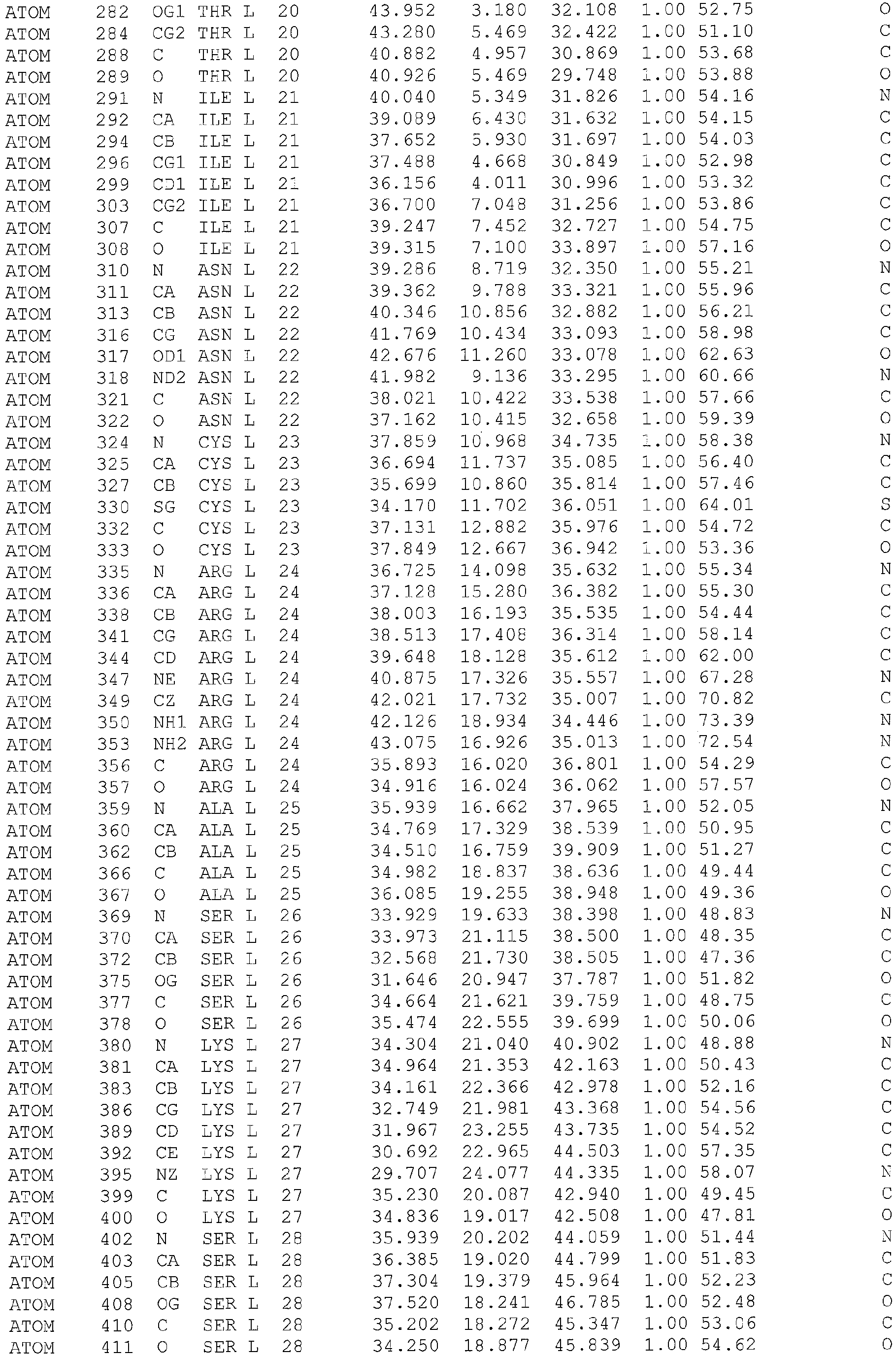

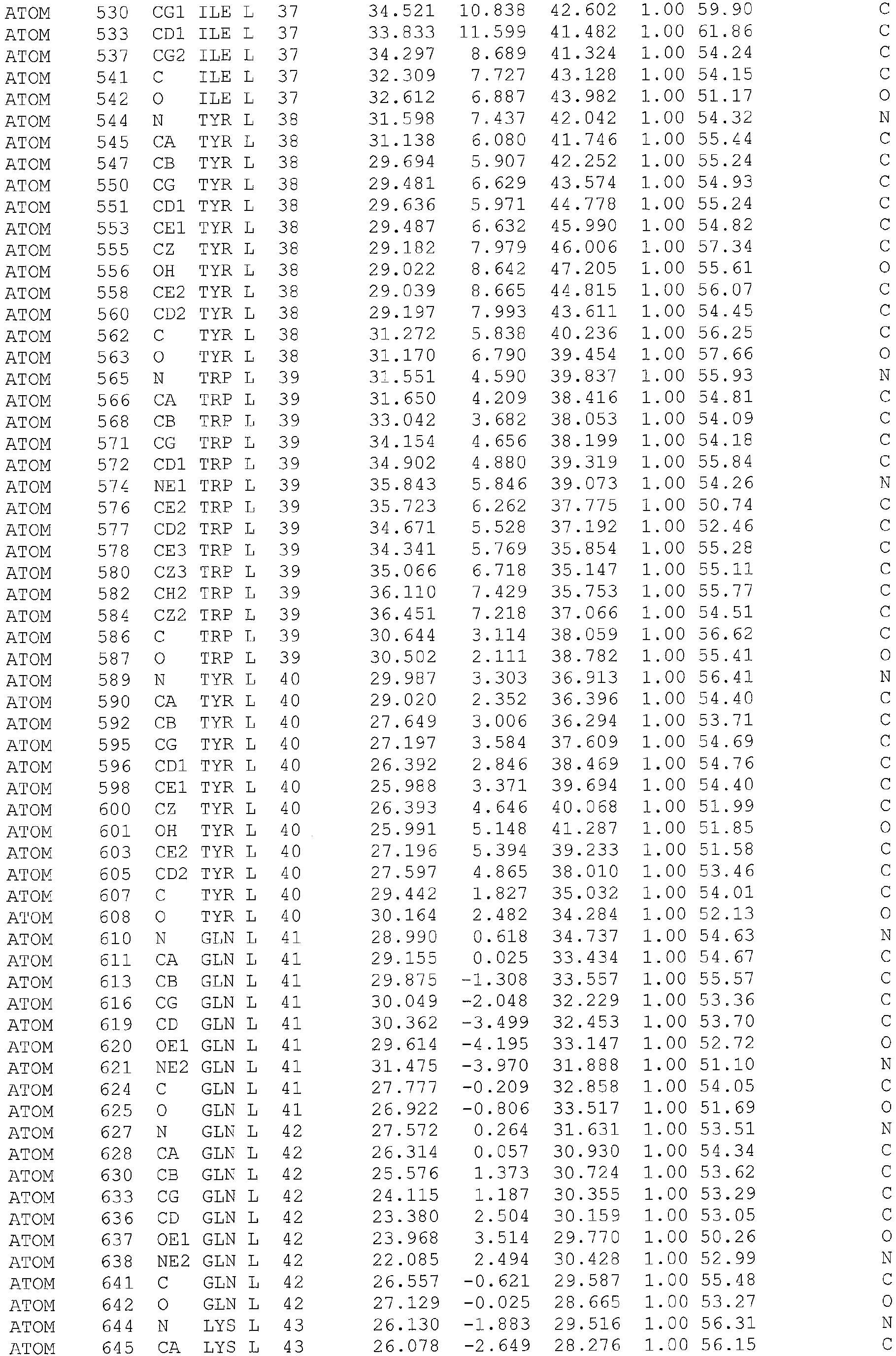


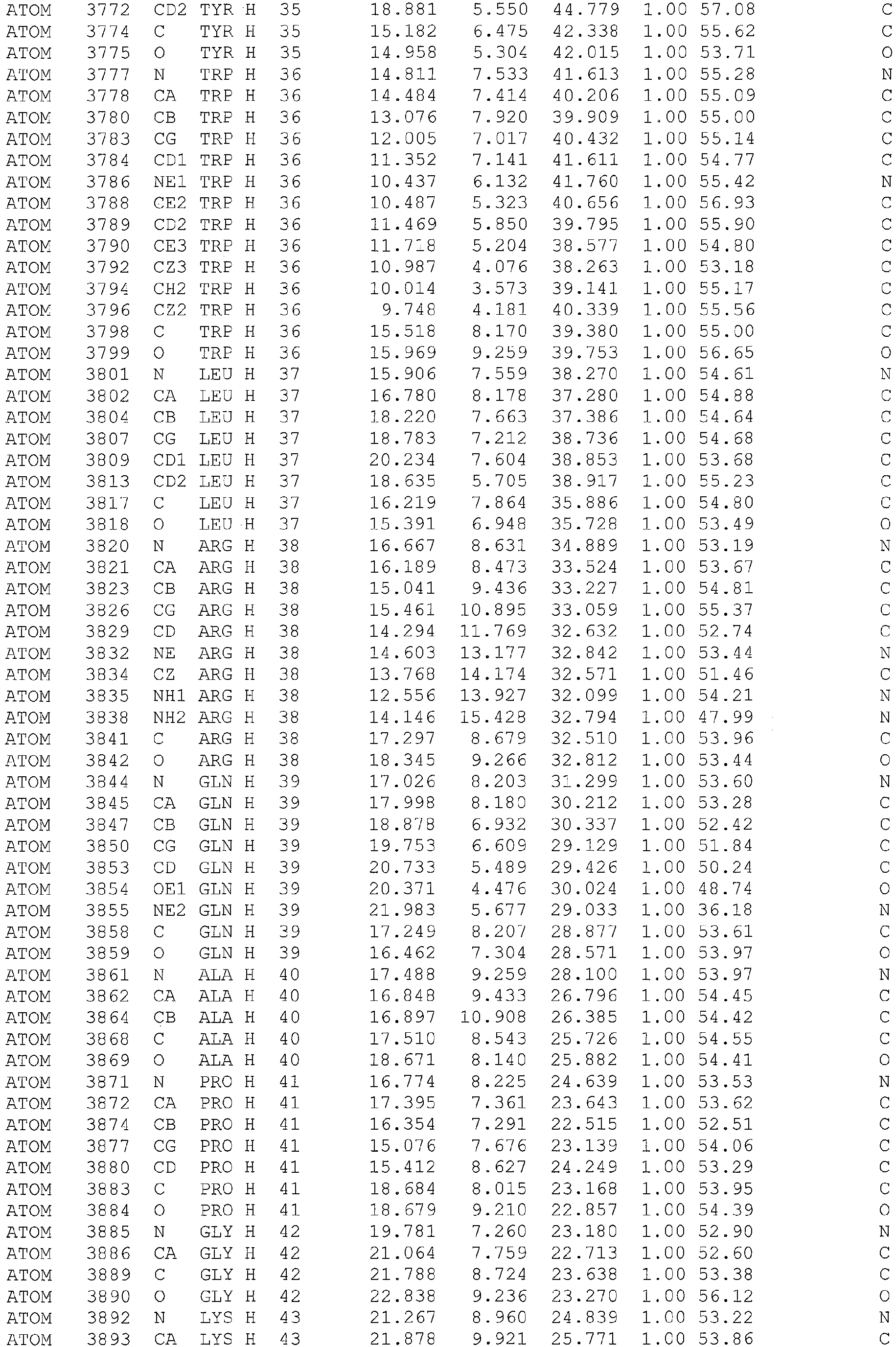


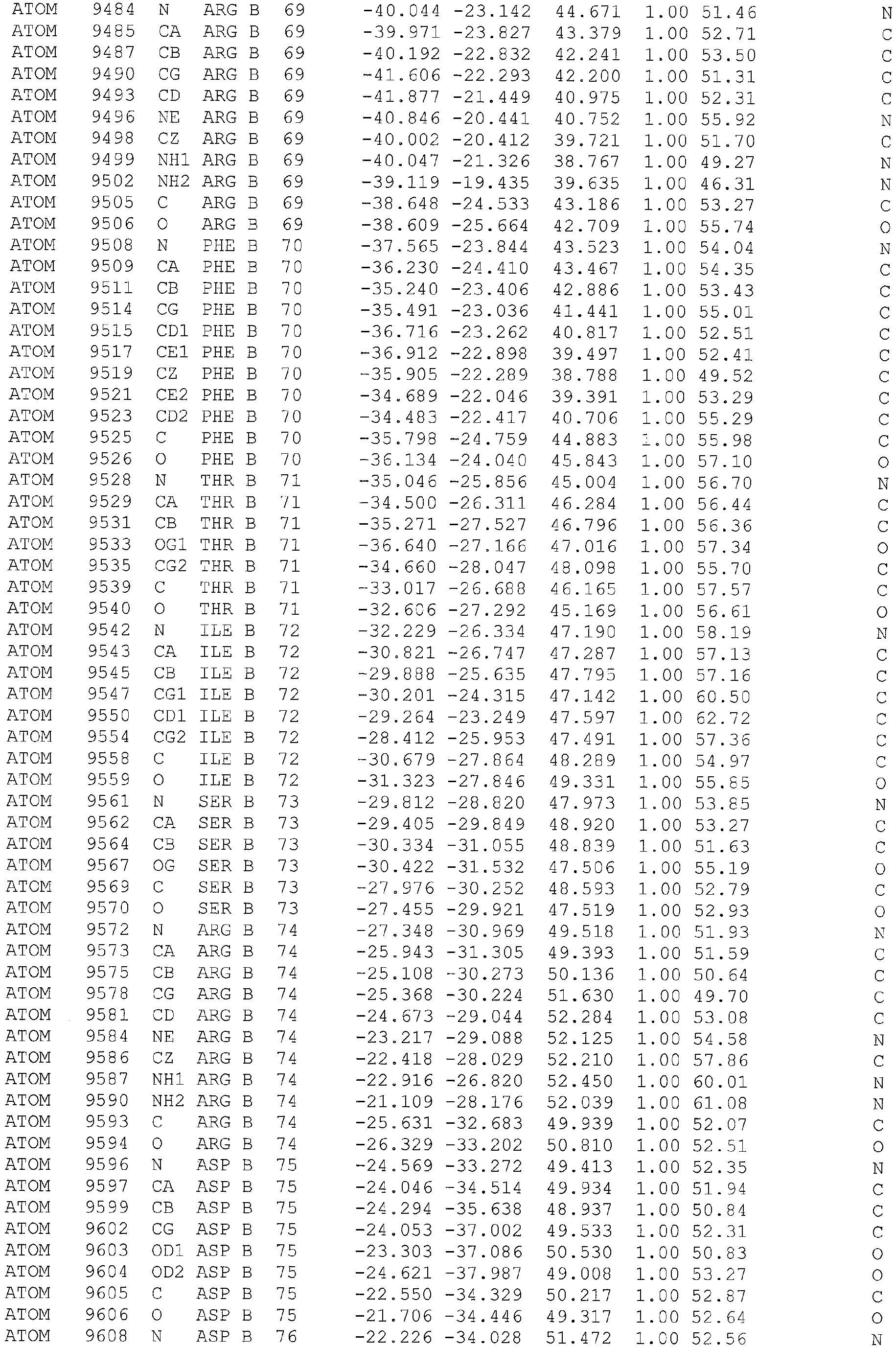


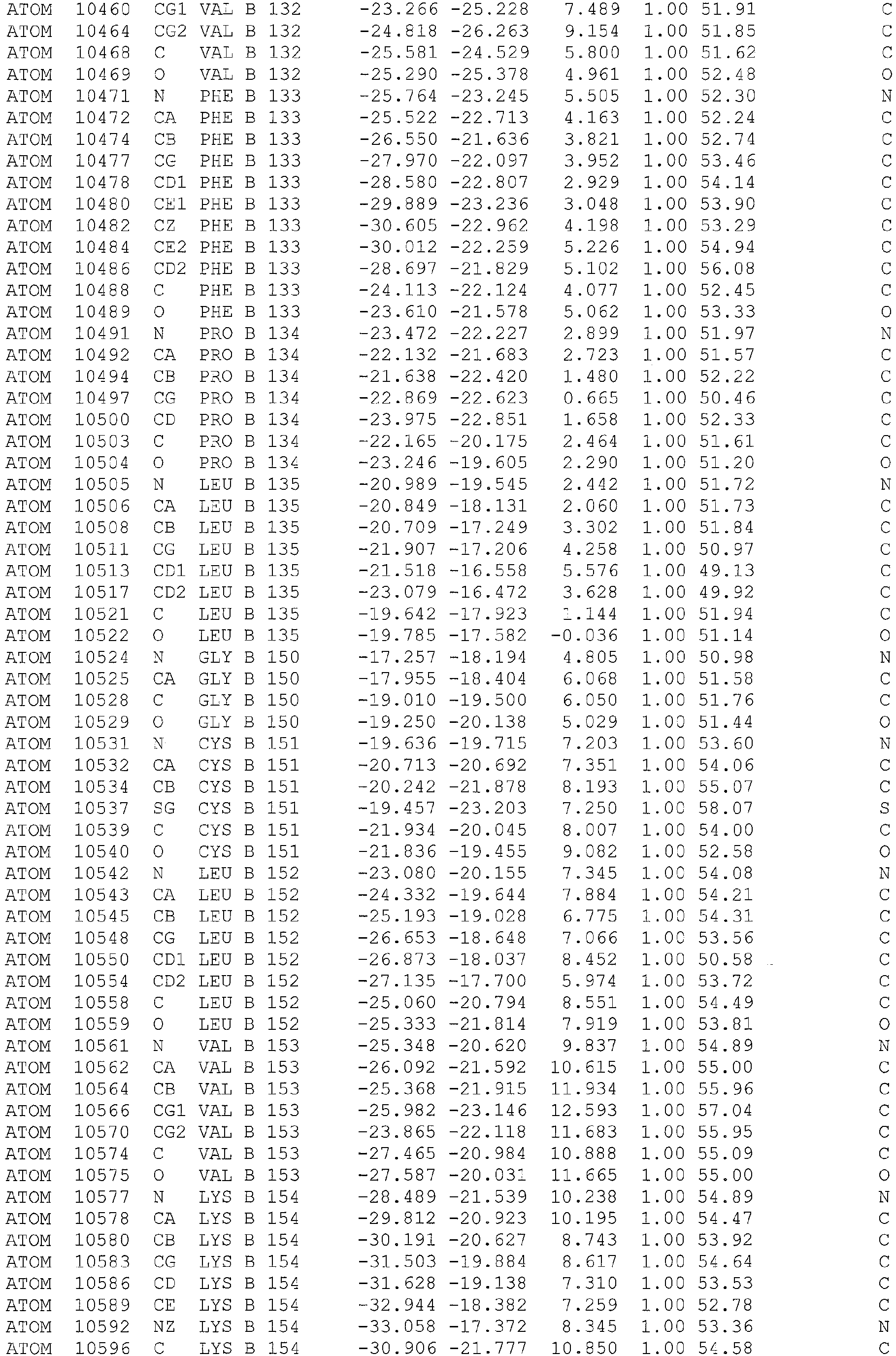








Claims (16)
- An antibody or antibody fragment capable of activating CD4+CD25+ regulatory T cells, wherein the antibody or antibody fragment comprises:(i) a light chain CDR1 comprising: Arg, Lys, Gln or Asn 24; Ala or Gly 25; Ser or Thr 26; Lys, Arg or Glu 27; Ser, Gly or Pro 28; Val, Ala or Ile 29; Ser 30; Thr, Ser, Asn, Gln or Asp 31; Ser 32; Gly 33; Tyr 34; Ser 35; Tyr 36; Ile, Val or Leu 37; and Tyr 38;(ii) a light chain CDR2 comprising: Leu 54; Ala 55; Ser 56; Ile 57; Leu 58; Glu 59; and Ser, Asn, Gln, Thr, Glu or Asp 60;(iii) a light chain CDR3 comprising: Gln 93; His 94; Ser 95; Arg or Lys 96; Glu, Asp or Arg 97; Leu, Gly or Ile 98; Pro 99; Trp 100; and Thr or Ser 101;(iv) a heavy chain CDR1 comprising: Asp, Glu, Thr, Cys, Pro, Met or Tyr 31; Cys, Ser, Ala, Gly or Val 32; Arg, Lys, Ser, Thr or Glu 33; Met, Ile, Leu, Ala or Val 34; and Tyr 35;(v) a heavy chain CDR2 comprising: Ile, Ala, Val or Gly 51; Ser, Asp, Gly, Ala, or Thr 52; Val, Ser, Gly, Thr or Ile 53; Lys, Arg, or Tyr 54; Ser, Asn, Gln, Thr or Glu 55; Glu, Asp or Arg 56; Asn, Asp, Tyr, Gln or Glu 57; Tyr, His or Lys 58; Gly or Ser 59; Ala, Ser or Thr 60; Asn, Gln or Asp 61; Tyr, Phe or His 62; Ala, Gly or Ser 63; Glu, Asp, Gln, Asn or Arg 64; Ser, Gly, Ala or Asn 65; Val, Ile, Ala, Ser or Gly 66; Arg, Lys, Gln, Tyr, His or Glu 67; and Gly 68; and(vi) a heavy chain CDR3 comprising: Ser 101; Tyr 102; Tyr, Phe or His 103; Arg 104; Tyr 105; Asp 106; Val, Ile, Pro, Asp, Thr or Glu 107; Gly or Ala 108; Ala or Ser 109; Trp, Phe, His or Tyr 110; Phe 111, Ala or Ser 112; and Tyr, Phe, His or Asn 113,and wherein the antibody or antibody fragment does not comprise CDR1, CDR2 and CDR3 of BT061 heavy chain and CDR1, CDR2 and CDR3 of BT061 light chain.
- An antibody or antibody fragment according to claim 1 comprising the sequence:wherein the amino acids at positions 24 to 29, 31, 37, 60, 96 to 98 and 101 are selected from those shown at the corresponding positions in Table 4,

and further comprising the sequence:wherein the amino acids at positions 31 to 34, 51 to 67, 103, 107 to 110, 112 and 113 are selected from those shown at the corresponding positions in Table 5.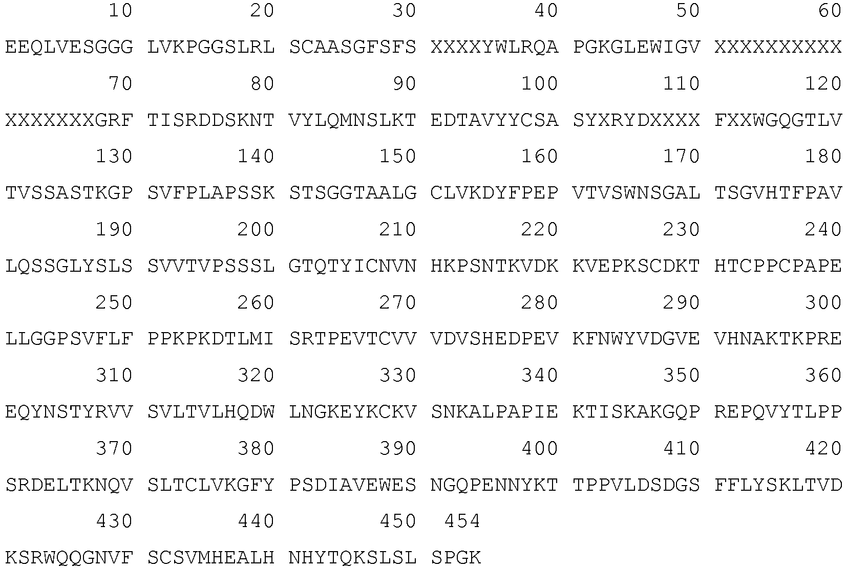
- An antibody or antibody fragment according to claim 1 comprising V domains that are at least 80% identical to the V domains of the antibody BT061.
- An antibody or antibody fragment according to claim 1 wherein:(a) the antibody or antibody fragment comprises a light chain comprising Tyr53 or Phe 53; and/or(b) the antibody or antibody fragment comprises a heavy chain comprising Ser28; and/or(c) the antibody or antibody fragment comprises a light chain containing Asp64; and/or(d) the antibody or antibody fragment comprises a heavy chain comprising Glu 56.
- An antibody or antibody fragment according to claim 1 or claim 4, comprising the CDR sequences of BT061 light chain and the CDR sequences of BT061 heavy chain:(a) with a single amino acid substitution wherein the substitution is:(i) A63G in the heavy chain;(ii) R33K in the heavy chain; or(iii) L98I in the light chain;or(b) with a double amino acid substitution wherein the substitutions are:(i) R33K and A63G in the heavy chain; or(ii) L98I in the light chain and R33K in the heavy chain,and preferably wherein the antibody or antibody fragment according to (a) or (b) further comprises the remaining variable domain sequences of BT061.
- An antibody or antibody fragment according to any one of claims 1 to 5: (a) further comprising an Fc receptor capable of binding to CD64; and/or (b) wherein the antibody is an IgG antibody.
- A nucleic acid encoding the antibody or antibody fragment according to any one of claims 1 to 6.
- A vector comprising the nucleic acid of claim 7.
- A host cell or hybridoma comprising the nucleic acid of claim 7 or the vector of claim 8.
- A method for the production of an antibody or antibody fragment according to any one of claims 1 to 6 comprising a step of culturing the host cell according to claim 9 in a culture medium under conditions allowing the expression of the antibody or antibody fragment and separating the antibody or fragment from the culture medium.
- A pharmaceutical composition comprising the antibody or antibody fragment according to any one of claims 1 to 6 and further comprising a pharmaceutically acceptable carrier.
- An antibody or antibody fragment according to any one of claims 1 to 6 further comprising a label.
- An antibody or antibody fragment according to any one of claims 1 to 6 for use in medicine, preferably wherein (i) the use is the treatment of an autoimmune disease or transplant rejection, or (ii) the use is for the manufacture of a medicament for treating autoimmune disease or transplant rejection.
- Use of an antibody or antibody fragment according to any one of claims 1 to 6 for activating CD4+CD25+ regulatory T cells in vitro, or for identifying CD4+CD25+ regulatory T cells in vitro.
- A method of screening for the presence of CD4+CD25+ T regulatory cells in a sample comprising a step of contacting a labeled antibody or antibody fragment according to claim 12 with the sample, washing the sample to remove unbound antibody and detecting the presence of the label in the sample, preferably wherein the CD4+CD25+ T regulatory cells are activated, and more preferably wherein the sample is a biological sample taken from a subject suffering from an autoimmune disease or from transplant rejection.
- A kit for isolating CD4+CD25+ regulatory T cells comprising magnetic beads coated with the antibody or antibody fragment of any one of claims 1 to 6 and a second anti-CD4 antibody or an anti-CD25 antibody.
Applications Claiming Priority (2)
| Application Number | Priority Date | Filing Date | Title |
|---|---|---|---|
| GBGB0920944.6A GB0920944D0 (en) | 2009-11-30 | 2009-11-30 | Agents for treating disease |
| PCT/EP2010/068579 WO2011064407A1 (en) | 2009-11-30 | 2010-11-30 | Agents for treating disease |
Publications (2)
| Publication Number | Publication Date |
|---|---|
| EP2470902A1 EP2470902A1 (en) | 2012-07-04 |
| EP2470902B1 true EP2470902B1 (en) | 2017-07-19 |
Family
ID=41572903
Family Applications (1)
| Application Number | Title | Priority Date | Filing Date |
|---|---|---|---|
| EP10782326.2A Active EP2470902B1 (en) | 2009-11-30 | 2010-11-30 | Agents for treating disease |
Country Status (17)
| Country | Link |
|---|---|
| US (1) | US9995733B2 (en) |
| EP (1) | EP2470902B1 (en) |
| JP (1) | JP5959438B2 (en) |
| KR (1) | KR101872746B1 (en) |
| CN (1) | CN102687010B (en) |
| AU (1) | AU2010323045B2 (en) |
| BR (1) | BR112012012911A8 (en) |
| CA (1) | CA2781836A1 (en) |
| ES (1) | ES2643640T3 (en) |
| GB (1) | GB0920944D0 (en) |
| IL (1) | IL219956B (en) |
| MX (1) | MX347247B (en) |
| NZ (1) | NZ600889A (en) |
| RU (1) | RU2598719C2 (en) |
| SG (2) | SG10201407938TA (en) |
| WO (1) | WO2011064407A1 (en) |
| ZA (1) | ZA201204845B (en) |
Families Citing this family (12)
| Publication number | Priority date | Publication date | Assignee | Title |
|---|---|---|---|---|
| EP1460088A1 (en) | 2003-03-21 | 2004-09-22 | Biotest AG | Humanized anti-CD4 antibody with immunosuppressive properties |
| EP2471543A1 (en) | 2010-12-02 | 2012-07-04 | Fraunhofer-Gesellschaft zur Förderung der angewandten Forschung e.V. | Tolerance induction or immunosupression to prevent in particular Graft-versus-Host-Disease (GvHD) by short-term pre-incubation of transplanted cell suspensions, tissues or organs coated with ligands to cell surface molecules |
| US20170232084A1 (en) | 2013-04-26 | 2017-08-17 | Enzo Biochem Inc. | Immune modulation for the treatment of age-related macular degeneration |
| US20140322188A1 (en) | 2013-04-26 | 2014-10-30 | Enzo Biochem, Inc. | Tolerizing treatments for autoimmune disease |
| GB201413899D0 (en) | 2014-08-06 | 2014-09-17 | Johnson Matthey Plc | Catalyst and use therof |
| WO2016171980A1 (en) * | 2015-04-24 | 2016-10-27 | Bristol-Myers Squibb Company | Polypeptides targeting hiv fusion |
| US20180126021A1 (en) * | 2016-11-04 | 2018-05-10 | Covidien Lp | Sterilization system and method |
| CA3182945A1 (en) * | 2020-07-06 | 2022-01-13 | Israel Vlodavsky | Heparanase-neutralizing a54 monoclonal antibody |
| GB202017681D0 (en) | 2020-11-09 | 2020-12-23 | T Balance Therapeutics Gmbh | Anti-CD4 antibody or fragment thereof for medical use |
| AU2022492449A1 (en) * | 2022-12-29 | 2025-07-10 | Chang Gung Memorial Hospital, Linkou | Antibodies specific to transmembrane and coiled-coil domain family 3 and uses thereof |
| GB202401501D0 (en) | 2024-02-05 | 2024-03-20 | T Balance Therapeutics Gmbh | Medical use of regulatory t cell activator |
| GB202409801D0 (en) | 2024-07-05 | 2024-08-21 | T Balance Therapeutics Gmbh | Products for use in the treatment and prevention of allergy and associated lung conditions/diseases |
Family Cites Families (84)
| Publication number | Priority date | Publication date | Assignee | Title |
|---|---|---|---|---|
| PT90657B (en) | 1988-05-27 | 1995-03-01 | Ortho Pharma Corp | PROCESS FOR THE PREPARATION OF PEPTIDES BLOCKING HIV-1 LIGATION CD4 RECEPTOR PROTEIN |
| JPH0768273B2 (en) * | 1988-12-02 | 1995-07-26 | カルピス食品工業株式会社 | Anti-HIV peptide |
| US5530101A (en) | 1988-12-28 | 1996-06-25 | Protein Design Labs, Inc. | Humanized immunoglobulins |
| IL162181A (en) | 1988-12-28 | 2006-04-10 | Pdl Biopharma Inc | A method of producing humanized immunoglubulin, and polynucleotides encoding the same |
| AU5656890A (en) | 1989-05-02 | 1990-11-29 | Genelabs Incorporated | Chemically modified cd4 peptide fragments having anti-retroviral properties |
| GB8912497D0 (en) | 1989-05-31 | 1989-07-19 | Cobbold Stephen P | Monoclonal antibodies |
| US5690933A (en) | 1989-05-31 | 1997-11-25 | Glaxo Wellcome Inc. | Monoclonal antibodies for inducing tolerance |
| US5859205A (en) | 1989-12-21 | 1999-01-12 | Celltech Limited | Humanised antibodies |
| US20020099179A1 (en) | 1989-12-21 | 2002-07-25 | Linda K. Jolliffe | Cdr-grafted antibodies |
| GB8928874D0 (en) | 1989-12-21 | 1990-02-28 | Celltech Ltd | Humanised antibodies |
| HUT60768A (en) | 1990-03-16 | 1992-10-28 | Sandoz Ag | Process for producing cd25 fixing molecules |
| US7084260B1 (en) | 1996-10-10 | 2006-08-01 | Genpharm International, Inc. | High affinity human antibodies and human antibodies against human antigens |
| DE69126301T2 (en) | 1990-11-27 | 1998-01-02 | Biogen, Inc., Cambridge, Mass. | HIV-INDUCED SYNCYTIA BLOCKING ANTI-CD-4 ANTIBODIES |
| US7192584B2 (en) | 1991-03-18 | 2007-03-20 | Centocor, Inc. | Methods of treating psoriasis with anti-TNF antibodies |
| US6136310A (en) | 1991-07-25 | 2000-10-24 | Idec Pharmaceuticals Corporation | Recombinant anti-CD4 antibodies for human therapy |
| JPH05244982A (en) | 1991-12-06 | 1993-09-24 | Sumitomo Chem Co Ltd | Humanized b-b10 |
| US5777085A (en) | 1991-12-20 | 1998-07-07 | Protein Design Labs, Inc. | Humanized antibodies reactive with GPIIB/IIIA |
| IL101769A (en) | 1992-05-03 | 2007-02-11 | Yeda Res & Dev | Tnf receptor action modulation |
| US6270766B1 (en) | 1992-10-08 | 2001-08-07 | The Kennedy Institute Of Rheumatology | Anti-TNF antibodies and methotrexate in the treatment of arthritis and crohn's disease |
| AU5152293A (en) | 1992-10-08 | 1994-05-09 | Kennedy Institute Of Rheumatology, The | Treatment of autoimmune and inflammatory disorders |
| ATE204299T1 (en) | 1993-03-05 | 2001-09-15 | Bayer Ag | HUMAN MONOCLONAL ANTI-TNF ALPHA ANTIBODIES |
| EP0631783A1 (en) | 1993-06-03 | 1995-01-04 | Mitsubishi Chemical Corporation | Antiviral combinations of 2',3'-di-deoxyribonucleosides with 6-benzyl-1-ethoxymethyl-5-substituted uracil derivatives |
| JPH09510952A (en) | 1993-10-06 | 1997-11-04 | ザ ケネディー インスティチュート オブ リューマトロジー | Treatment of autoimmune and inflammatory diseases |
| US20020068057A1 (en) | 1994-03-10 | 2002-06-06 | Marc Feldmann | Treatment of autoimmune and inflammatory disorders |
| EP0840618B1 (en) | 1995-05-18 | 2003-04-23 | Ortho-McNeil Pharmaceutical, Inc. | Induction of immunological tolerance by the use of non-depleting anti-cd4 antibodies |
| ATE239041T1 (en) | 1996-02-09 | 2003-05-15 | Basf Ag | HUMAN ANTIBODIES THAT BIND TO HUMAN TNFALPHA |
| US20010056066A1 (en) | 1996-07-26 | 2001-12-27 | Smithkline Beecham Corporation | Method of treating immune cell mediated systemic diseases |
| EP0946195A1 (en) | 1996-10-01 | 1999-10-06 | Celltech Therapeutics Limited | Pharmaceutical products containing protein-tyrosine kinase inhibitors and anti-cd4 antibodies |
| US6037454A (en) | 1996-11-27 | 2000-03-14 | Genentech, Inc. | Humanized anti-CD11a antibodies |
| DE19722888A1 (en) | 1997-05-28 | 1998-12-03 | Thomas Prof Dr Huenig | Human CD28 specific monoclonal antibodies for antigen-unspecific activation of T lymphocytes |
| WO2000058362A1 (en) | 1999-03-26 | 2000-10-05 | Human Genome Sciences, Inc. | Neutrokine-alpha binding proteins and methods based thereon |
| DE60042785D1 (en) | 1999-06-09 | 2009-10-01 | Immunomedics Inc | IMMUNOTHERAPY OF AUTOIMMUNE DISEASES THROUGH THE USE OF B-CELL SPECIFIC ANTIBODIES |
| WO2001016182A2 (en) * | 1999-08-27 | 2001-03-08 | The Government Of The United States Of America, As Represented By The Secretary, Department Of Health And Human Services | POLYPEPTIDES THAT BIND HIV gp120 AND RELATED NUCLEIC ACIDS, ANTIBODIES, COMPOSITIONS, AND METHODS OF USE |
| US7304127B2 (en) | 1999-08-27 | 2007-12-04 | United States Of America As Represented By The Secretary, Department Of Health And Human Services | Polypeptides that bind HIV gp120 and related nucleic acids, antibodies, compositions, and methods of use |
| WO2001093908A1 (en) | 2000-06-02 | 2001-12-13 | Regents Of The University Of Minnesota | Immunotherapeutic method to prevent islet cell rejection |
| WO2002022212A2 (en) | 2000-09-18 | 2002-03-21 | Idec Pharmaceuticals Corporation | Combination therapy for treatment of autoimmune diseases using b cell depleting/immunoregulatory antibody combination |
| DE10050935A1 (en) | 2000-10-11 | 2002-05-02 | Tegenero Gmbh | Use of CD28-specific monoclonal antibodies to stimulate blood cells that do not carry CD28 |
| CA2434972C (en) | 2001-01-16 | 2010-06-08 | Vascular Therapies, Llc | Implantable device containing resorbable matrix material and anti-proliferative drugs for preventing or treating failure of hemodialysis vascular access and other vascular grafts |
| NZ528250A (en) | 2001-03-07 | 2006-09-29 | Childrens Medical Center | Method to screen peptide display libraries using minicell display |
| EP1241249A1 (en) | 2001-03-12 | 2002-09-18 | Gerold Schuler | CD4+CD25+regulatory T cells from human blood |
| CN1247258C (en) | 2001-04-24 | 2006-03-29 | 默克专利有限公司 | Combination therapy using anti-angiogenic agents and tnfa |
| GB2376466A (en) | 2001-06-14 | 2002-12-18 | Mark Frewin | TRX1 antibody |
| DE60238533D1 (en) | 2001-06-14 | 2011-01-20 | Cambridge Entpr Ltd | Non-depleting antibody TRX-1 against CD4 and its uses |
| US7541443B2 (en) | 2001-06-14 | 2009-06-02 | Tolerrx, Inc. | Anti-CD4 antibodies |
| ES2392287T3 (en) | 2001-12-04 | 2012-12-07 | Theramab Llc | Peptide or protein containing a C'-D loop of the CD28 receptor family |
| DE10212108A1 (en) | 2002-03-13 | 2003-10-02 | Tegenero Ag | Use of an active substance that binds to CD28 for the production of a pharmaceutical composition |
| DE10230223A1 (en) | 2002-07-04 | 2004-01-22 | Tegenero Ag | Microparticles with CD28-specific monoclonal antibodies |
| AU2003291625B2 (en) | 2002-09-16 | 2009-10-08 | Genentech, Inc. | Compositions and methods for the treatment of immune related diseases |
| US7388079B2 (en) * | 2002-11-27 | 2008-06-17 | The Regents Of The University Of California | Delivery of pharmaceutical agents via the human insulin receptor |
| US7501494B2 (en) | 2003-01-15 | 2009-03-10 | United Biomedical, Inc. | Designed deimmunized monoclonal antibodies for protection against HIV exposure and treatment of HIV infection |
| EP1460088A1 (en) * | 2003-03-21 | 2004-09-22 | Biotest AG | Humanized anti-CD4 antibody with immunosuppressive properties |
| ME00426B (en) | 2003-04-09 | 2011-10-10 | Genentech Inc | Therapy of autoimmune disease in a patient with an inadequate response to a tnf-alpha inhibitor |
| GB0314461D0 (en) | 2003-06-20 | 2003-07-23 | Isis Innovation | Suppression of transplant rejection |
| US7691970B2 (en) | 2003-08-25 | 2010-04-06 | Pieris Ag | Muteins of a bilin-binding protein with affinity for a given target |
| EP1600164A3 (en) | 2003-09-22 | 2006-05-17 | TeGenero AG | Use of a CD28 binding substance for the production of a pharmaceutical composition with dose-dependent efficacy |
| DE10352900A1 (en) | 2003-11-11 | 2005-06-16 | Tegenero Ag | Use of a CD28-binding active substance for the preparation of a pharmaceutical composition for the treatment of B-CLL |
| RU2007102055A (en) | 2004-06-22 | 2008-07-27 | Толерркс, Инк. (Us) | OPTIMIZED ADMINISTRATION OF DOSES OF ANTI-CD4-ANTIBODIES FOR TOLERANCE INDUCTION IN PRIMATES |
| RU2007104022A (en) | 2004-07-02 | 2008-08-10 | Дженентек, Инк. (Us) | COMPOSITIONS FOR THE TREATMENT OF NON-HODGKINIAN LYMPHOMA AND METHODS OF APPLICATION |
| US20070270431A1 (en) | 2004-09-29 | 2007-11-22 | Kowa Co., Ltd. | Preventive and/or Therapeutic Medicine for Rheumatoid Arthritis |
| WO2006050949A2 (en) | 2004-11-11 | 2006-05-18 | Tegenero Ag | Superagonistic anti-cd28 antibody |
| US8119589B2 (en) | 2004-11-12 | 2012-02-21 | University Of North Carolina At Charlotte | Modulation of CD4+ T cell responses by a tick saliva protein, Salp15 and polypeptides derived therefrom |
| DE102004063494A1 (en) | 2004-12-23 | 2006-07-13 | Tegenero Ag | antibody |
| JP4730733B2 (en) | 2005-05-02 | 2011-07-20 | 国立大学法人京都大学 | Method for detecting regulatory T cells using expression of type 4 folate receptor as an index, and immunostimulant |
| US8663634B2 (en) | 2005-07-11 | 2014-03-04 | Macrogenics, Inc. | Methods for the treatment of autoimmune disorders using immunosuppressive monoclonal antibodies with reduced toxicity |
| NZ594784A (en) | 2005-08-18 | 2013-06-28 | Genmab As | Therapy with CD4 binding peptides and radiation |
| AU2006290433B2 (en) | 2005-08-26 | 2012-06-07 | Roche Glycart Ag | Modified antigen binding molecules with altered cell signaling activity |
| US8101365B2 (en) * | 2006-01-05 | 2012-01-24 | Genentech, Inc. | Anti-EphB4 antibodies and methods using same |
| US20070218062A1 (en) | 2006-03-16 | 2007-09-20 | Genentech, Inc. | Methods of treating lupus using CD4 antibodies |
| JP2009529915A (en) * | 2006-03-20 | 2009-08-27 | ゾーマ テクノロジー リミテッド | Human antibodies and methods specific for gastrin substances |
| WO2007117602A2 (en) | 2006-04-07 | 2007-10-18 | Biogen Idec Ma Inc. | Isolation and use of human regulatory t cells |
| SMP200800064B (en) | 2006-04-21 | 2009-11-06 | Novartis Ag | Pharmaceutical compositions of anti-cd40 antagonist antibodies |
| WO2007135684A2 (en) * | 2006-05-22 | 2007-11-29 | Hadasit Medical Research Services & Development Limited | Method of treatment of anti-cd4 autoimmunity |
| US8557533B2 (en) | 2007-02-01 | 2013-10-15 | Helmut Jonuleit | Screening method for the identification of agents capable of activating CD4+CD25+ regulatory T-cells through interactions with the HIV-1 GP120 binding site on CD4 |
| EP3067368A1 (en) | 2007-04-27 | 2016-09-14 | Genentech, Inc. | Potent, stable and non-immunosuppressive anti-cd4 antibodies |
| RU2539110C2 (en) | 2008-03-13 | 2015-01-10 | Биотест Аг | Method of treating autoimmune disease (versions) |
| MX2010010027A (en) | 2008-03-13 | 2011-05-02 | Biotest Ag | Agent for treating disease. |
| EP2262838B1 (en) | 2008-03-13 | 2016-04-13 | Biotest AG | Agent for treating disease |
| EP2101173A1 (en) | 2008-03-14 | 2009-09-16 | Vivalis | In vitro method to determine whether a drug candidate active against a target protein is active against a variant of said protein |
| WO2010022341A1 (en) | 2008-08-21 | 2010-02-25 | The United State Of America, As Represented By The Secretary, Department Of Health And Human Services | Methods of enriching and using regulatory t cells |
| ES2335381B1 (en) | 2008-09-24 | 2011-02-28 | Fundacio Institut De Recerca De L'hospital Universitari Vall D'hebron | IN VITRO METHOD AND KIT FOR THE FORECAST OR PREDICTION OF THE RESPONSE PART OF PATIENTS WITH REUMATOID ARTHRITIS TO THE TREATMENT WITH BLOCKING AGENTS OF THE TNFALFA FACTOR. |
| RU2531548C2 (en) * | 2008-09-29 | 2014-10-20 | Биотест Аг | Composition for treating disease |
| ES2351456B1 (en) | 2009-06-24 | 2011-11-28 | Fundacio Institut De Recerca De L'hospital Universitari Vall D'hebron | IN VITRO METHOD FOR THE FORECAST OR PREDICTION OF THE RESPONSE BY PATIENTS WITH REUMATOID ARTHRITIS TO THE TREATMENT WITH AGENTS RECOGNIZING THE CD20 MEMBRANE RECEIVER OF THE LYMPHOCYTES B |
| JP5399219B2 (en) | 2009-11-24 | 2014-01-29 | 株式会社ケイティーバイオ | Method and apparatus for predicting the efficacy of human anti-TNFα antibody for rheumatoid arthritis |
| WO2011158798A1 (en) | 2010-06-14 | 2011-12-22 | 国立大学法人山口大学 | Method for observation and early prediction of clinical course of therapeutic effect on psoriasis, and kit for use in the method |
-
2009
- 2009-11-30 GB GBGB0920944.6A patent/GB0920944D0/en not_active Ceased
-
2010
- 2010-11-30 EP EP10782326.2A patent/EP2470902B1/en active Active
- 2010-11-30 NZ NZ600889A patent/NZ600889A/en not_active IP Right Cessation
- 2010-11-30 SG SG10201407938TA patent/SG10201407938TA/en unknown
- 2010-11-30 KR KR1020127016947A patent/KR101872746B1/en not_active Expired - Fee Related
- 2010-11-30 CN CN201080059459.9A patent/CN102687010B/en active Active
- 2010-11-30 CA CA2781836A patent/CA2781836A1/en not_active Abandoned
- 2010-11-30 AU AU2010323045A patent/AU2010323045B2/en not_active Ceased
- 2010-11-30 JP JP2012541473A patent/JP5959438B2/en active Active
- 2010-11-30 RU RU2012127378/10A patent/RU2598719C2/en not_active IP Right Cessation
- 2010-11-30 BR BR112012012911A patent/BR112012012911A8/en not_active Application Discontinuation
- 2010-11-30 WO PCT/EP2010/068579 patent/WO2011064407A1/en not_active Ceased
- 2010-11-30 SG SG2012039442A patent/SG181117A1/en unknown
- 2010-11-30 MX MX2012006199A patent/MX347247B/en active IP Right Grant
- 2010-11-30 ES ES10782326.2T patent/ES2643640T3/en active Active
-
2012
- 2012-05-23 IL IL219956A patent/IL219956B/en not_active IP Right Cessation
- 2012-05-30 US US13/483,280 patent/US9995733B2/en active Active
- 2012-06-28 ZA ZA2012/04845A patent/ZA201204845B/en unknown
Non-Patent Citations (1)
| Title |
|---|
| None * |
Also Published As
| Publication number | Publication date |
|---|---|
| CN102687010B (en) | 2016-06-15 |
| AU2010323045B2 (en) | 2015-09-24 |
| ES2643640T3 (en) | 2017-11-23 |
| CN102687010A (en) | 2012-09-19 |
| KR20120102733A (en) | 2012-09-18 |
| JP2013511996A (en) | 2013-04-11 |
| MX347247B (en) | 2017-04-17 |
| IL219956B (en) | 2019-07-31 |
| ZA201204845B (en) | 2016-06-29 |
| RU2598719C2 (en) | 2016-09-27 |
| SG10201407938TA (en) | 2015-01-29 |
| GB0920944D0 (en) | 2010-01-13 |
| MX2012006199A (en) | 2012-06-19 |
| NZ600889A (en) | 2015-09-25 |
| US9995733B2 (en) | 2018-06-12 |
| KR101872746B1 (en) | 2018-06-29 |
| EP2470902A1 (en) | 2012-07-04 |
| BR112012012911A2 (en) | 2016-10-25 |
| JP5959438B2 (en) | 2016-08-02 |
| SG181117A1 (en) | 2012-07-30 |
| US20130004513A1 (en) | 2013-01-03 |
| CA2781836A1 (en) | 2011-06-03 |
| BR112012012911A8 (en) | 2018-02-27 |
| WO2011064407A1 (en) | 2011-06-03 |
| AU2010323045A1 (en) | 2012-07-19 |
| RU2012127378A (en) | 2014-01-10 |
| IL219956A0 (en) | 2012-07-31 |
Similar Documents
| Publication | Publication Date | Title |
|---|---|---|
| US9995733B2 (en) | Agents for treating disease | |
| US20230256018A1 (en) | T cell receptors and engineered cells expressing same | |
| US20230146195A1 (en) | Bcma binding molecules and methods of use thereof | |
| JP2021184731A (en) | Antibodies directed against cd127 | |
| US20240018268A1 (en) | Anti-idiotypic antibodies against anti-cd19 antibodies | |
| TW201938203A (en) | Antigen-binding proteins targeting shared antigens | |
| US20040092718A1 (en) | Use of a CD28 binding substance for making a pharmaceutical composition | |
| KR20170128234A (en) | Antibodies specific for ROR1 and chimeric antigen receptors | |
| JP7747645B2 (en) | Antibodies and chimeric antigen receptors targeting glypican-3 (GPC3) and methods of use thereof | |
| WO2024022512A1 (en) | Claudin-6 binding moieties and uses thereof | |
| US12269870B2 (en) | Antigen binding regions against fibronectin type III domains and methods of using the same | |
| WO2022127871A1 (en) | Gucy2c binding molecules and uses thereof | |
| WO2024046468A1 (en) | Fusion proteins targeting lysosomal degradation pathway | |
| CN119654345A (en) | Chimeric antigen receptors targeting interleukin 13 receptor subunit alpha 2 and methods of use thereof | |
| CN117222667A (en) | Bifunctional molecules for treating immune disorders | |
| KR20250075590A (en) | Antibodies and chimeric antigen receptors targeting GCC and methods of using the same | |
| WO2026021525A1 (en) | Gprc5d binding moieties and uses thereof | |
| HK40117019A (en) | Claudin-6 binding moieties and uses thereof | |
| HK40119547A (en) | Antibody and chimeric antigen receptors targeting gcc and methods of use thereof | |
| CN121752594A (en) | Anti-OX 40L antibodies and uses thereof | |
| HK40094778A (en) | Cd20 binding molecules and uses thereof |
Legal Events
| Date | Code | Title | Description |
|---|---|---|---|
| PUAI | Public reference made under article 153(3) epc to a published international application that has entered the european phase |
Free format text: ORIGINAL CODE: 0009012 |
|
| 17P | Request for examination filed |
Effective date: 20120328 |
|
| AK | Designated contracting states |
Kind code of ref document: A1 Designated state(s): AL AT BE BG CH CY CZ DE DK EE ES FI FR GB GR HR HU IE IS IT LI LT LU LV MC MK MT NL NO PL PT RO RS SE SI SK SM TR |
|
| REG | Reference to a national code |
Ref country code: HK Ref legal event code: DE Ref document number: 1166367 Country of ref document: HK |
|
| RIN1 | Information on inventor provided before grant (corrected) |
Inventor name: ZUBER, CHANTAL Inventor name: CZELOTH, NIKLAS Inventor name: UHEREK, CHRISTOPH Inventor name: ENGLING, ANDRE Inventor name: WALLMEIER, HOLGER Inventor name: OSTERROTH, FRANK Inventor name: BRUECHER, CHRISTOPH Inventor name: SCHULZ, GREGOR Inventor name: VOELP, KIRSTEN Inventor name: DAELKEN, BENJAMIN |
|
| DAX | Request for extension of the european patent (deleted) | ||
| 17Q | First examination report despatched |
Effective date: 20150407 |
|
| GRAP | Despatch of communication of intention to grant a patent |
Free format text: ORIGINAL CODE: EPIDOSNIGR1 |
|
| STAA | Information on the status of an ep patent application or granted ep patent |
Free format text: STATUS: GRANT OF PATENT IS INTENDED |
|
| INTG | Intention to grant announced |
Effective date: 20161207 |
|
| GRAS | Grant fee paid |
Free format text: ORIGINAL CODE: EPIDOSNIGR3 |
|
| GRAJ | Information related to disapproval of communication of intention to grant by the applicant or resumption of examination proceedings by the epo deleted |
Free format text: ORIGINAL CODE: EPIDOSDIGR1 |
|
| GRAL | Information related to payment of fee for publishing/printing deleted |
Free format text: ORIGINAL CODE: EPIDOSDIGR3 |
|
| STAA | Information on the status of an ep patent application or granted ep patent |
Free format text: STATUS: EXAMINATION IS IN PROGRESS |
|
| INTC | Intention to grant announced (deleted) | ||
| GRAR | Information related to intention to grant a patent recorded |
Free format text: ORIGINAL CODE: EPIDOSNIGR71 |
|
| STAA | Information on the status of an ep patent application or granted ep patent |
Free format text: STATUS: GRANT OF PATENT IS INTENDED |
|
| GRAA | (expected) grant |
Free format text: ORIGINAL CODE: 0009210 |
|
| STAA | Information on the status of an ep patent application or granted ep patent |
Free format text: STATUS: THE PATENT HAS BEEN GRANTED |
|
| INTG | Intention to grant announced |
Effective date: 20170608 |
|
| AK | Designated contracting states |
Kind code of ref document: B1 Designated state(s): AL AT BE BG CH CY CZ DE DK EE ES FI FR GB GR HR HU IE IS IT LI LT LU LV MC MK MT NL NO PL PT RO RS SE SI SK SM TR |
|
| REG | Reference to a national code |
Ref country code: GB Ref legal event code: FG4D |
|
| REG | Reference to a national code |
Ref country code: CH Ref legal event code: EP |
|
| REG | Reference to a national code |
Ref country code: IE Ref legal event code: FG4D |
|
| REG | Reference to a national code |
Ref country code: AT Ref legal event code: REF Ref document number: 910877 Country of ref document: AT Kind code of ref document: T Effective date: 20170815 |
|
| REG | Reference to a national code |
Ref country code: DE Ref legal event code: R096 Ref document number: 602010043758 Country of ref document: DE |
|
| REG | Reference to a national code |
Ref country code: FR Ref legal event code: PLFP Year of fee payment: 8 |
|
| REG | Reference to a national code |
Ref country code: CH Ref legal event code: NV Representative=s name: BOVARD AG PATENT- UND MARKENANWAELTE, CH |
|
| REG | Reference to a national code |
Ref country code: NL Ref legal event code: MP Effective date: 20170719 |
|
| REG | Reference to a national code |
Ref country code: ES Ref legal event code: FG2A Ref document number: 2643640 Country of ref document: ES Kind code of ref document: T3 Effective date: 20171123 |
|
| REG | Reference to a national code |
Ref country code: LT Ref legal event code: MG4D |
|
| REG | Reference to a national code |
Ref country code: AT Ref legal event code: MK05 Ref document number: 910877 Country of ref document: AT Kind code of ref document: T Effective date: 20170719 |
|
| PG25 | Lapsed in a contracting state [announced via postgrant information from national office to epo] |
Ref country code: HR Free format text: LAPSE BECAUSE OF FAILURE TO SUBMIT A TRANSLATION OF THE DESCRIPTION OR TO PAY THE FEE WITHIN THE PRESCRIBED TIME-LIMIT Effective date: 20170719 Ref country code: NL Free format text: LAPSE BECAUSE OF FAILURE TO SUBMIT A TRANSLATION OF THE DESCRIPTION OR TO PAY THE FEE WITHIN THE PRESCRIBED TIME-LIMIT Effective date: 20170719 Ref country code: SE Free format text: LAPSE BECAUSE OF FAILURE TO SUBMIT A TRANSLATION OF THE DESCRIPTION OR TO PAY THE FEE WITHIN THE PRESCRIBED TIME-LIMIT Effective date: 20170719 Ref country code: NO Free format text: LAPSE BECAUSE OF FAILURE TO SUBMIT A TRANSLATION OF THE DESCRIPTION OR TO PAY THE FEE WITHIN THE PRESCRIBED TIME-LIMIT Effective date: 20171019 Ref country code: FI Free format text: LAPSE BECAUSE OF FAILURE TO SUBMIT A TRANSLATION OF THE DESCRIPTION OR TO PAY THE FEE WITHIN THE PRESCRIBED TIME-LIMIT Effective date: 20170719 Ref country code: LT Free format text: LAPSE BECAUSE OF FAILURE TO SUBMIT A TRANSLATION OF THE DESCRIPTION OR TO PAY THE FEE WITHIN THE PRESCRIBED TIME-LIMIT Effective date: 20170719 Ref country code: AT Free format text: LAPSE BECAUSE OF FAILURE TO SUBMIT A TRANSLATION OF THE DESCRIPTION OR TO PAY THE FEE WITHIN THE PRESCRIBED TIME-LIMIT Effective date: 20170719 |
|
| PG25 | Lapsed in a contracting state [announced via postgrant information from national office to epo] |
Ref country code: LV Free format text: LAPSE BECAUSE OF FAILURE TO SUBMIT A TRANSLATION OF THE DESCRIPTION OR TO PAY THE FEE WITHIN THE PRESCRIBED TIME-LIMIT Effective date: 20170719 Ref country code: GR Free format text: LAPSE BECAUSE OF FAILURE TO SUBMIT A TRANSLATION OF THE DESCRIPTION OR TO PAY THE FEE WITHIN THE PRESCRIBED TIME-LIMIT Effective date: 20171020 Ref country code: IS Free format text: LAPSE BECAUSE OF FAILURE TO SUBMIT A TRANSLATION OF THE DESCRIPTION OR TO PAY THE FEE WITHIN THE PRESCRIBED TIME-LIMIT Effective date: 20171119 Ref country code: RS Free format text: LAPSE BECAUSE OF FAILURE TO SUBMIT A TRANSLATION OF THE DESCRIPTION OR TO PAY THE FEE WITHIN THE PRESCRIBED TIME-LIMIT Effective date: 20170719 Ref country code: BG Free format text: LAPSE BECAUSE OF FAILURE TO SUBMIT A TRANSLATION OF THE DESCRIPTION OR TO PAY THE FEE WITHIN THE PRESCRIBED TIME-LIMIT Effective date: 20171019 Ref country code: PL Free format text: LAPSE BECAUSE OF FAILURE TO SUBMIT A TRANSLATION OF THE DESCRIPTION OR TO PAY THE FEE WITHIN THE PRESCRIBED TIME-LIMIT Effective date: 20170719 |
|
| REG | Reference to a national code |
Ref country code: DE Ref legal event code: R097 Ref document number: 602010043758 Country of ref document: DE |
|
| PG25 | Lapsed in a contracting state [announced via postgrant information from national office to epo] |
Ref country code: RO Free format text: LAPSE BECAUSE OF FAILURE TO SUBMIT A TRANSLATION OF THE DESCRIPTION OR TO PAY THE FEE WITHIN THE PRESCRIBED TIME-LIMIT Effective date: 20170719 Ref country code: CZ Free format text: LAPSE BECAUSE OF FAILURE TO SUBMIT A TRANSLATION OF THE DESCRIPTION OR TO PAY THE FEE WITHIN THE PRESCRIBED TIME-LIMIT Effective date: 20170719 Ref country code: DK Free format text: LAPSE BECAUSE OF FAILURE TO SUBMIT A TRANSLATION OF THE DESCRIPTION OR TO PAY THE FEE WITHIN THE PRESCRIBED TIME-LIMIT Effective date: 20170719 |
|
| PLBE | No opposition filed within time limit |
Free format text: ORIGINAL CODE: 0009261 |
|
| STAA | Information on the status of an ep patent application or granted ep patent |
Free format text: STATUS: NO OPPOSITION FILED WITHIN TIME LIMIT |
|
| PG25 | Lapsed in a contracting state [announced via postgrant information from national office to epo] |
Ref country code: SM Free format text: LAPSE BECAUSE OF FAILURE TO SUBMIT A TRANSLATION OF THE DESCRIPTION OR TO PAY THE FEE WITHIN THE PRESCRIBED TIME-LIMIT Effective date: 20170719 Ref country code: SK Free format text: LAPSE BECAUSE OF FAILURE TO SUBMIT A TRANSLATION OF THE DESCRIPTION OR TO PAY THE FEE WITHIN THE PRESCRIBED TIME-LIMIT Effective date: 20170719 Ref country code: EE Free format text: LAPSE BECAUSE OF FAILURE TO SUBMIT A TRANSLATION OF THE DESCRIPTION OR TO PAY THE FEE WITHIN THE PRESCRIBED TIME-LIMIT Effective date: 20170719 |
|
| 26N | No opposition filed |
Effective date: 20180420 |
|
| PG25 | Lapsed in a contracting state [announced via postgrant information from national office to epo] |
Ref country code: MC Free format text: LAPSE BECAUSE OF FAILURE TO SUBMIT A TRANSLATION OF THE DESCRIPTION OR TO PAY THE FEE WITHIN THE PRESCRIBED TIME-LIMIT Effective date: 20170719 |
|
| PG25 | Lapsed in a contracting state [announced via postgrant information from national office to epo] |
Ref country code: SI Free format text: LAPSE BECAUSE OF FAILURE TO SUBMIT A TRANSLATION OF THE DESCRIPTION OR TO PAY THE FEE WITHIN THE PRESCRIBED TIME-LIMIT Effective date: 20170719 Ref country code: LU Free format text: LAPSE BECAUSE OF NON-PAYMENT OF DUE FEES Effective date: 20171130 |
|
| REG | Reference to a national code |
Ref country code: BE Ref legal event code: MM Effective date: 20171130 |
|
| REG | Reference to a national code |
Ref country code: IE Ref legal event code: MM4A |
|
| PG25 | Lapsed in a contracting state [announced via postgrant information from national office to epo] |
Ref country code: MT Free format text: LAPSE BECAUSE OF NON-PAYMENT OF DUE FEES Effective date: 20171130 |
|
| REG | Reference to a national code |
Ref country code: FR Ref legal event code: PLFP Year of fee payment: 9 |
|
| PG25 | Lapsed in a contracting state [announced via postgrant information from national office to epo] |
Ref country code: IE Free format text: LAPSE BECAUSE OF NON-PAYMENT OF DUE FEES Effective date: 20171130 |
|
| PG25 | Lapsed in a contracting state [announced via postgrant information from national office to epo] |
Ref country code: BE Free format text: LAPSE BECAUSE OF NON-PAYMENT OF DUE FEES Effective date: 20171130 |
|
| PG25 | Lapsed in a contracting state [announced via postgrant information from national office to epo] |
Ref country code: HU Free format text: LAPSE BECAUSE OF FAILURE TO SUBMIT A TRANSLATION OF THE DESCRIPTION OR TO PAY THE FEE WITHIN THE PRESCRIBED TIME-LIMIT; INVALID AB INITIO Effective date: 20101130 |
|
| REG | Reference to a national code |
Ref country code: DE Ref legal event code: R082 Ref document number: 602010043758 Country of ref document: DE Representative=s name: PAGE, WHITE & FARRER GERMANY LLP, DE Ref country code: DE Ref legal event code: R081 Ref document number: 602010043758 Country of ref document: DE Owner name: OGEN GMBH, DE Free format text: FORMER OWNER: BIOTEST AKTIENGESELLSCHAFT, 63303 DREIEICH, DE |
|
| REG | Reference to a national code |
Ref country code: CH Ref legal event code: PUE Owner name: OGEN GMBH, DE Free format text: FORMER OWNER: BIOTEST AG, DE |
|
| REG | Reference to a national code |
Ref country code: ES Ref legal event code: PC2A Owner name: OGEN GMBH Effective date: 20190920 |
|
| PG25 | Lapsed in a contracting state [announced via postgrant information from national office to epo] |
Ref country code: CY Free format text: LAPSE BECAUSE OF NON-PAYMENT OF DUE FEES Effective date: 20170719 |
|
| REG | Reference to a national code |
Ref country code: GB Ref legal event code: 732E Free format text: REGISTERED BETWEEN 20191024 AND 20191030 |
|
| PG25 | Lapsed in a contracting state [announced via postgrant information from national office to epo] |
Ref country code: MK Free format text: LAPSE BECAUSE OF FAILURE TO SUBMIT A TRANSLATION OF THE DESCRIPTION OR TO PAY THE FEE WITHIN THE PRESCRIBED TIME-LIMIT Effective date: 20170719 |
|
| PG25 | Lapsed in a contracting state [announced via postgrant information from national office to epo] |
Ref country code: TR Free format text: LAPSE BECAUSE OF FAILURE TO SUBMIT A TRANSLATION OF THE DESCRIPTION OR TO PAY THE FEE WITHIN THE PRESCRIBED TIME-LIMIT Effective date: 20170719 |
|
| PG25 | Lapsed in a contracting state [announced via postgrant information from national office to epo] |
Ref country code: PT Free format text: LAPSE BECAUSE OF FAILURE TO SUBMIT A TRANSLATION OF THE DESCRIPTION OR TO PAY THE FEE WITHIN THE PRESCRIBED TIME-LIMIT Effective date: 20170719 |
|
| PG25 | Lapsed in a contracting state [announced via postgrant information from national office to epo] |
Ref country code: AL Free format text: LAPSE BECAUSE OF FAILURE TO SUBMIT A TRANSLATION OF THE DESCRIPTION OR TO PAY THE FEE WITHIN THE PRESCRIBED TIME-LIMIT Effective date: 20170719 |
|
| P01 | Opt-out of the competence of the unified patent court (upc) registered |
Effective date: 20230519 |
|
| PGFP | Annual fee paid to national office [announced via postgrant information from national office to epo] |
Ref country code: FR Payment date: 20250930 Year of fee payment: 16 |
|
| REG | Reference to a national code |
Ref country code: CH Ref legal event code: U11 Free format text: ST27 STATUS EVENT CODE: U-0-0-U10-U11 (AS PROVIDED BY THE NATIONAL OFFICE) Effective date: 20251201 |
|
| REG | Reference to a national code |
Ref country code: DE Ref legal event code: R082 Ref document number: 602010043758 Country of ref document: DE Representative=s name: WESTPHAL, MUSSGNUG & PARTNER PATENTANWAELTE MI, DE |
|
| PGFP | Annual fee paid to national office [announced via postgrant information from national office to epo] |
Ref country code: DE Payment date: 20250930 Year of fee payment: 16 |
|
| PGFP | Annual fee paid to national office [announced via postgrant information from national office to epo] |
Ref country code: GB Payment date: 20251001 Year of fee payment: 16 |
|
| PGFP | Annual fee paid to national office [announced via postgrant information from national office to epo] |
Ref country code: IT Payment date: 20251022 Year of fee payment: 16 |
|
| PGFP | Annual fee paid to national office [announced via postgrant information from national office to epo] |
Ref country code: CH Payment date: 20251201 Year of fee payment: 16 |
|
| PGFP | Annual fee paid to national office [announced via postgrant information from national office to epo] |
Ref country code: ES Payment date: 20251212 Year of fee payment: 16 |