EP2456458B1 - Dendritic cell vaccines for asparaginyl - beta - hydroxylase expressing tumors - Google Patents
Dendritic cell vaccines for asparaginyl - beta - hydroxylase expressing tumors Download PDFInfo
- Publication number
- EP2456458B1 EP2456458B1 EP10735417.7A EP10735417A EP2456458B1 EP 2456458 B1 EP2456458 B1 EP 2456458B1 EP 10735417 A EP10735417 A EP 10735417A EP 2456458 B1 EP2456458 B1 EP 2456458B1
- Authority
- EP
- European Patent Office
- Prior art keywords
- aah
- cells
- subject
- dendritic cells
- cell
- Prior art date
- Legal status (The legal status is an assumption and is not a legal conclusion. Google has not performed a legal analysis and makes no representation as to the accuracy of the status listed.)
- Not-in-force
Links
Images




Classifications
-
- C—CHEMISTRY; METALLURGY
- C12—BIOCHEMISTRY; BEER; SPIRITS; WINE; VINEGAR; MICROBIOLOGY; ENZYMOLOGY; MUTATION OR GENETIC ENGINEERING
- C12N—MICROORGANISMS OR ENZYMES; COMPOSITIONS THEREOF; PROPAGATING, PRESERVING, OR MAINTAINING MICROORGANISMS; MUTATION OR GENETIC ENGINEERING; CULTURE MEDIA
- C12N5/00—Undifferentiated human, animal or plant cells, e.g. cell lines; Tissues; Cultivation or maintenance thereof; Culture media therefor
- C12N5/06—Animal cells or tissues; Human cells or tissues
- C12N5/0602—Vertebrate cells
- C12N5/0634—Cells from the blood or the immune system
- C12N5/0639—Dendritic cells, e.g. Langherhans cells in the epidermis
-
- A—HUMAN NECESSITIES
- A61—MEDICAL OR VETERINARY SCIENCE; HYGIENE
- A61K—PREPARATIONS FOR MEDICAL, DENTAL OR TOILETRY PURPOSES
- A61K39/00—Medicinal preparations containing antigens or antibodies
- A61K39/0005—Vertebrate antigens
- A61K39/0011—Cancer antigens
-
- A—HUMAN NECESSITIES
- A61—MEDICAL OR VETERINARY SCIENCE; HYGIENE
- A61K—PREPARATIONS FOR MEDICAL, DENTAL OR TOILETRY PURPOSES
- A61K40/00—Cellular immunotherapy
- A61K40/10—Cellular immunotherapy characterised by the cell type used
- A61K40/19—Dendritic cells
-
- A—HUMAN NECESSITIES
- A61—MEDICAL OR VETERINARY SCIENCE; HYGIENE
- A61K—PREPARATIONS FOR MEDICAL, DENTAL OR TOILETRY PURPOSES
- A61K40/00—Cellular immunotherapy
- A61K40/20—Cellular immunotherapy characterised by the effect or the function of the cells
- A61K40/24—Antigen-presenting cells [APC]
-
- A—HUMAN NECESSITIES
- A61—MEDICAL OR VETERINARY SCIENCE; HYGIENE
- A61K—PREPARATIONS FOR MEDICAL, DENTAL OR TOILETRY PURPOSES
- A61K40/00—Cellular immunotherapy
- A61K40/40—Cellular immunotherapy characterised by antigens that are targeted or presented by cells of the immune system
- A61K40/41—Vertebrate antigens
- A61K40/42—Cancer antigens
-
- A—HUMAN NECESSITIES
- A61—MEDICAL OR VETERINARY SCIENCE; HYGIENE
- A61P—SPECIFIC THERAPEUTIC ACTIVITY OF CHEMICAL COMPOUNDS OR MEDICINAL PREPARATIONS
- A61P35/00—Antineoplastic agents
-
- A—HUMAN NECESSITIES
- A61—MEDICAL OR VETERINARY SCIENCE; HYGIENE
- A61P—SPECIFIC THERAPEUTIC ACTIVITY OF CHEMICAL COMPOUNDS OR MEDICINAL PREPARATIONS
- A61P35/00—Antineoplastic agents
- A61P35/04—Antineoplastic agents specific for metastasis
-
- A—HUMAN NECESSITIES
- A61—MEDICAL OR VETERINARY SCIENCE; HYGIENE
- A61P—SPECIFIC THERAPEUTIC ACTIVITY OF CHEMICAL COMPOUNDS OR MEDICINAL PREPARATIONS
- A61P37/00—Drugs for immunological or allergic disorders
- A61P37/02—Immunomodulators
- A61P37/04—Immunostimulants
-
- A—HUMAN NECESSITIES
- A61—MEDICAL OR VETERINARY SCIENCE; HYGIENE
- A61P—SPECIFIC THERAPEUTIC ACTIVITY OF CHEMICAL COMPOUNDS OR MEDICINAL PREPARATIONS
- A61P43/00—Drugs for specific purposes, not provided for in groups A61P1/00-A61P41/00
-
- C—CHEMISTRY; METALLURGY
- C07—ORGANIC CHEMISTRY
- C07K—PEPTIDES
- C07K14/00—Peptides having more than 20 amino acids; Gastrins; Somatostatins; Melanotropins; Derivatives thereof
- C07K14/435—Peptides having more than 20 amino acids; Gastrins; Somatostatins; Melanotropins; Derivatives thereof from animals; from humans
- C07K14/705—Receptors; Cell surface antigens; Cell surface determinants
-
- C—CHEMISTRY; METALLURGY
- C12—BIOCHEMISTRY; BEER; SPIRITS; WINE; VINEGAR; MICROBIOLOGY; ENZYMOLOGY; MUTATION OR GENETIC ENGINEERING
- C12N—MICROORGANISMS OR ENZYMES; COMPOSITIONS THEREOF; PROPAGATING, PRESERVING, OR MAINTAINING MICROORGANISMS; MUTATION OR GENETIC ENGINEERING; CULTURE MEDIA
- C12N9/00—Enzymes; Proenzymes; Compositions thereof; Processes for preparing, activating, inhibiting, separating or purifying enzymes
- C12N9/0004—Oxidoreductases (1.)
- C12N9/0071—Oxidoreductases (1.) acting on paired donors with incorporation of molecular oxygen (1.14)
-
- A—HUMAN NECESSITIES
- A61—MEDICAL OR VETERINARY SCIENCE; HYGIENE
- A61K—PREPARATIONS FOR MEDICAL, DENTAL OR TOILETRY PURPOSES
- A61K39/00—Medicinal preparations containing antigens or antibodies
- A61K2039/51—Medicinal preparations containing antigens or antibodies comprising whole cells, viruses or DNA/RNA
- A61K2039/515—Animal cells
- A61K2039/5154—Antigen presenting cells [APCs], e.g. dendritic cells or macrophages
-
- A—HUMAN NECESSITIES
- A61—MEDICAL OR VETERINARY SCIENCE; HYGIENE
- A61K—PREPARATIONS FOR MEDICAL, DENTAL OR TOILETRY PURPOSES
- A61K39/00—Medicinal preparations containing antigens or antibodies
- A61K2039/545—Medicinal preparations containing antigens or antibodies characterised by the dose, timing or administration schedule
-
- C—CHEMISTRY; METALLURGY
- C12—BIOCHEMISTRY; BEER; SPIRITS; WINE; VINEGAR; MICROBIOLOGY; ENZYMOLOGY; MUTATION OR GENETIC ENGINEERING
- C12N—MICROORGANISMS OR ENZYMES; COMPOSITIONS THEREOF; PROPAGATING, PRESERVING, OR MAINTAINING MICROORGANISMS; MUTATION OR GENETIC ENGINEERING; CULTURE MEDIA
- C12N2500/00—Specific components of cell culture medium
- C12N2500/90—Serum-free medium, which may still contain naturally-sourced components
-
- C—CHEMISTRY; METALLURGY
- C12—BIOCHEMISTRY; BEER; SPIRITS; WINE; VINEGAR; MICROBIOLOGY; ENZYMOLOGY; MUTATION OR GENETIC ENGINEERING
- C12N—MICROORGANISMS OR ENZYMES; COMPOSITIONS THEREOF; PROPAGATING, PRESERVING, OR MAINTAINING MICROORGANISMS; MUTATION OR GENETIC ENGINEERING; CULTURE MEDIA
- C12N2501/00—Active agents used in cell culture processes, e.g. differentation
- C12N2501/20—Cytokines; Chemokines
- C12N2501/22—Colony stimulating factors (G-CSF, GM-CSF)
-
- C—CHEMISTRY; METALLURGY
- C12—BIOCHEMISTRY; BEER; SPIRITS; WINE; VINEGAR; MICROBIOLOGY; ENZYMOLOGY; MUTATION OR GENETIC ENGINEERING
- C12N—MICROORGANISMS OR ENZYMES; COMPOSITIONS THEREOF; PROPAGATING, PRESERVING, OR MAINTAINING MICROORGANISMS; MUTATION OR GENETIC ENGINEERING; CULTURE MEDIA
- C12N2501/00—Active agents used in cell culture processes, e.g. differentation
- C12N2501/20—Cytokines; Chemokines
- C12N2501/23—Interleukins [IL]
-
- C—CHEMISTRY; METALLURGY
- C12—BIOCHEMISTRY; BEER; SPIRITS; WINE; VINEGAR; MICROBIOLOGY; ENZYMOLOGY; MUTATION OR GENETIC ENGINEERING
- C12N—MICROORGANISMS OR ENZYMES; COMPOSITIONS THEREOF; PROPAGATING, PRESERVING, OR MAINTAINING MICROORGANISMS; MUTATION OR GENETIC ENGINEERING; CULTURE MEDIA
- C12N2501/00—Active agents used in cell culture processes, e.g. differentation
- C12N2501/20—Cytokines; Chemokines
- C12N2501/24—Interferons [IFN]
-
- C—CHEMISTRY; METALLURGY
- C12—BIOCHEMISTRY; BEER; SPIRITS; WINE; VINEGAR; MICROBIOLOGY; ENZYMOLOGY; MUTATION OR GENETIC ENGINEERING
- C12N—MICROORGANISMS OR ENZYMES; COMPOSITIONS THEREOF; PROPAGATING, PRESERVING, OR MAINTAINING MICROORGANISMS; MUTATION OR GENETIC ENGINEERING; CULTURE MEDIA
- C12N2501/00—Active agents used in cell culture processes, e.g. differentation
- C12N2501/50—Cell markers; Cell surface determinants
- C12N2501/52—CD40, CD40-ligand (CD154)
Definitions
- liver cancer including hepatocellular and cholangiocellular carcinoma is one of the most common cancers in the world. In the United States, liver cancer is the fifth common cause of cancer death in men and the ninth in women. In spite of the dramatic progress in diagnosis and local control of liver cancer, the mortality rate in the United States has been increasing constantly. One of the reasons for the increasing mortality might be that, unlike other cancers including breast and colorectal cancers, liver cancer is resistant to systemic treatment such as chemotherapy. Therefore, there is no effective treatment of liver cancer once it becomes a systemic disease.
- US 2005/113329 focuses on the identification of aspartyl (asparaginyl)-p-hydroxylase (AAH) over-expression as a marker of malignancy and invasiveness and suggests antisense inhibition of AAH as a therapeutic approach.
- US 2006/057129 describes how to generate dendritic cell vaccine using dendritic cells differentiated from stem cells and subsequently genetically modified. The document refers to the expression or transduction of immunogenic epitopes in the dendritic cells. Hoffman T.K. et al., Journal of Immunotherapy 24: 162-171 (2001 ) describe enhancing cross-priming capability of dendritic cells by certain cytokines. Xu S.
- the invention provides an isolated mature aspartyl (asparaginyl)- ⁇ -hydroxylase (AAH)-loaded dendritic cell for use in a method of treating an AAH-expressing tumor by reducing growth of said tumor in a subject, wherein growth of said AAH-expressing tumor is reduced after administration of said dendritic cell, wherein said dendritic cell is primed with a combination of cytokines comprising of GM-CSF and IFN- ⁇ ex vivo, prior to administrating to said subject.
- AAH isolated mature aspartyl
- the invention provides a method of producing primed mature AAH-loaded dendritic cells, comprising contacting isolated dendritic cells with an antigen comprising AAH, and following said antigen-contacting step, contacting said dendritic cells with a combination of cytokines, said combination comprising GM-CSF and IFN- ⁇ .
- the invention provides a vaccine composition comprising primed dendritic cells produced by the production method above.
- the invention provides a vaccine containing AAH-loaded mature dendritic cells for use in the method of treatment of AAH-expressing tumors in a mammalian subject.
- the invention provides autologous dendritic cells primed according to the invention for use in a method of inhibiting tumor growth and/or preventing metastasis in a mammal, wherein said primed autologous dendritic cells are administered to a subject suffering from an AAH-bearing tumor.
- the invention provides autologous dendritic cells which are primed by a combination of cytokines comprising GM-CSF, IFN- ⁇ , and CD40L for use in a method of preventing development of a tumor in a mammal, wherein said subject is at risk of developing an AAH-bearing tumor, due to a family history of individuals who have been identified with such a tumor.
- the invention provides the cell or vaccine for use in a method of treating an AAH-expressing tumor of the present invention in a subject, wherein said subject or mammal is a human, canine, feline, equine, bovine, or porcine subject or mammal.
- the invention provides a solution to the longstanding problem of an alternative to chemotherapy for treatment of patients diagnosed with cancer.
- the invention provides mature dendritic cells for use in a method of reducing growth of an aspartyl (asparaginyl)-p-hydroxylase (AAH or ASPH)-expressing tumor in a subject which involves administering to the subject an isolated mature AAH-loaded dendritic cell or population of cells. Growth of an AAH-expressing tumor is reduced after administration of such a dendritic cell vaccine. For example, tumor growth and tumor burden is reduced by 10%, 20%, 50%, 75%, 2-fold, 5-fold, 10-fold, or more.
- the methods are used to reduce and eliminate AAH-expressing tumors from mammalian subjects, such as human patients.
- the compositions and methods are also suitable for use in companion animals and livestock, e.g., human, canine, feline, equine, bovine, or porcine subjects.
- AAH-expressing tumors include most tumor types such as tumors of gastrointestinal tissues (e.g., esophagus, stomach, colon), pancreas, liver (e.g., cholangiocellular carcinoma, hepatocellular carcinoma), breast, prostate, cervix, ovary, fallopian tube, larynx, lung, thyroid, gall bladder, kidney, bladder, and brain (e.g., glioblastoma) as well as numerous others described below.
- AAH-expressing tumors include primary tumors that express an increased level of AAH compared to normal tissue as well as tumors that arise by metastasis from such AAH-overexpressing primary tumors.
- the dendritic cells used in the vaccination method are activated ex vivo with a combination of cytokines comprising GM-CSF and IFN- ⁇ prior administering them to the subject.
- the latter step yields a population of dendritic cells with improved anti-tumor activity.
- An improved method of producing primed dendritic cells is carried out by contacting isolated dendritic cells with an antigen comprising AAH or a combination of tumor antigens comprising AAH and ⁇ -fetoprotein (AFP) and treating the cells to yield a population of matured and activated antigen-presenting cells.
- the dendritic cells are contacted with the combination of cytokines.
- the combination comprises GM-CSF and IFN- ⁇ .
- the combination may further comprise IL-4.
- the combination comprises CD40L.
- the dendritic cells are contacted with the combination of cytokines for at least 10 hours (e.g., 12, 24, 36, 40, 48 hours or more).
- the antigen is in a soluble form or bound to a solid support.
- the solid support comprises a polystyrene bead such as a biodegradable bead or particle.
- Dendritic cells are obtained from a subject by known methods such as leukapheresis or cytopheresis.
- the subject from which the cells are obtained is suffering from or at risk of developing cancer.
- the patient has been diagnosed with an AAH-expressing tumor.
- Patients who are at risk of developing AAH-bearing tumors include those with a family history of individuals who have been identified with such a cancer.
- a vaccine composition comprising primed dendritic cells produced as described above.
- the vaccine is useful to inhibit tumor growth, prevent tumor growth, and inhibit or reduce metastasis.
- the invention provides mature dendritic cells for use in a method of inhibiting tumor growth in a mammal which is carried out by identifying a subject suffering from an AAH-bearing tumor, and administering to the subject autologous dendritic cells, which have been primed according to the method described above.
- the invention provides mature dendritic cells for use in a method of preventing development of a tumor in a mammal, which comprises the step of identifying a subject at risk of developing an AAH-bearing tumor (such as one with a family history of cancer), and administering to the subject the antigen-primed and activated autologous dendritic cells.
- the invention provides mature dendritic cells for use in a method of preventing metastasis of an AAH-bearing tumor which is carried out by identifying a subject suffering from an AAH-bearing tumor, and administering to the subject autologous dendritic cells, as described above.
- polypeptides and other compositions are purified.
- a substantially pure AAH polypeptide or variant thereof is preferably obtained by expression of a recombinant nucleic acid encoding the polypeptide or by chemically synthesizing the protein.
- a polypeptide or protein is substantially pure when it is separated from those contaminants which accompany it in its natural state (proteins and other naturally-occurring organic molecules).
- the polypeptide is substantially pure when it constitutes at least 60%, by weight, of the protein in the preparation.
- the protein in the preparation is at least 75%, more preferably at least 90%, and most preferably at least 99%, by weight, AAH.
- substantially pure polypeptides include recombinant polypeptides derived from a eukaryote but produced in E. coli or another prokaryote, or in a eukaryote other than that from which the polypeptide was originally derived.
- Dendritic cells or other cells e.g., immune cells such as macrophages, B cells, T cells, used in the methods are purified or isolated.
- the term "isolated" means that the cell is substantially free of other cell types or cellular material with which it naturally occurs.
- a sample of cells of a particular tissue type or phenotype is “substantially pure” when it is at least 60% of the cell population.
- the preparation is at least 75%, more preferably at least 90%, and most preferably at least 99% or 100%, of the cell population. Purity is measured by any appropriate standard method, for example, by fluorescence-activated cell sorting (FACS).
- FACS fluorescence-activated cell sorting
- the methods of generating antigen-primed DCs has several advantages over earlier methods.
- the cells are not only antigen-loaded but the subsequent cytokine incubation yield superior activated antigen-primed antigen presenting cells with improved anti-tumor and anti-metastatic activity.
- AAH is a protein, the expression of which is associated with a number of cancer types (see, e.g., USPN 6,815,415; USPN 6,812,206; USPN 6,797,696; USPN 6,783,758; USPN 6,835,370; and USPN 7,094,556).
- Over-expression of HAAH has been detected by immunohistochemical staining (1HC) in numerous cancers lung (e.g., adenocarcinoma, bronchioalveolar carcinoma, and other non-small cell lung cancers such as sqamous cell carcinoma subtype ( Luu et al., 2009, Hum. Pathol.
- liver e.g., hepatocellular carcinoma, cholangiocarcinoma, and cancer of biliary epithelial cells ( Wang et al., 2010, Hepatology 52:164-173 )
- gastrointestinal tissues e.g., colon, stomach, esophagus
- pancreas prostate, ovary, bile duct, breast, kidney, bladder
- brain e.g., glioblastoma and neuroblastoma.
- HAAH is highly specific for cancer; it has been detected by immunohistochemistry in >99% of tumor specimens tested (n>1000) and is not present in adjacent non-affected tissue, or in tissue samples from normal individuals.
- Immunotherapy using AAH-primed cells is administered to patients with tumors that express AAH on their cell surface.
- tumors include liver cancers such as hepatocellular carcinoma and cholangiocarcinoma, as well as adenocarcinomas of the breast and colon.
- liver cancers such as hepatocellular carcinoma and cholangiocarcinoma
- adenocarcinomas of the breast and colon Such a therapeutic strategy is useful for not only treatment but prevention of cancers.
- Dendritic cell vaccines using AAH were found to cure established hepatocellular carcinoma in immunocompetent mice.
- AAH dendritic cell vaccines reduce growth of AAH-expressing tumors to decrease tumor burden and eradicate tumors in humans as well.
- Immunotherapy of malignant tumors is attractive in terms of the specific response to tumor cells.
- a rationale for immunotherapy is that cancer cells are replete with potential antigens, which become immunogenic when presented by antigen-presenting cells to helper and cytotoxic T cells.
- Immunotherapy for liver cancer is a viable clinical approach, because liver cancer cells express several tumor-associated proteins, which are not or only slightly expressed in normal tissues. For example, AFP, which is not expressed in adult tissues, is extensively expressed in hepatocellular carcinoma (HCC).
- HCC hepatocellular carcinoma
- AFP is not involved in cancer progression, cancer cells that do not express AFP dominated during APP-targeting immunotherapy.
- AFP should be presented on the cell surface with major histocompatibility complex (MHC) class I molecule, because AFP is not distributed on the cell surface.
- MHC class I molecules are often down-regulated in many cancer cells, AFP may not be appropriately recognized by the T cells.
- studies were undertaken to identify another antigen that is associated with liver cancer to be used to primed cells for cell-based therapy. Given the results using AFP as an antigen, the anti-tumor results with AAH were surprising.
- AAH also known as ASPH
- ASPH is strongly expressed in hepatocellular and cholangiocellular carcinoma, but is not expressed in normal cell counterparts of those tissue types.
- AAH is therefore a tumor-associated protein.
- AAH is useful for immunotherapy of liver cancer, because of its unique nature.
- AAH confers cancer cells on motility, which is associated with cancer metastasis; thus, therapy targeting AAH-expressing cells is effective to suppress metastatic lesions as well as the primary tumor.
- AAH is a membrane protein, most part of which is exposed to extracellular space, it is easier for immune cells to access to the protein even when class I molecules are down-regulated.
- no antigenic protein specific to cholangiocellular carcinoma is known.
- AAH is uniquely qualified as a target molecule of immunotherapy against liver cancer.
- DCs dendritic cells
- the methods described herein include a DC-based immunization for induction of immunity against AAH.
- the present methods improve upon a previously described method for generating immunogenic mouse DCs loaded with protein of interest ( Gehring et al., 2008, J. Immunol. Meth. 332:18-30 ) in at least two ways.
- LPS lipopolysaccharides
- DCs are derived from hematopoietic cells, and are specialized to capture and process antigens, converting proteins to peptides that are presented on MHC molecules and recognized by T cells. DCs strongly induce and control T-cell mediated immunity.
- DC-based immunotherapy is based on the fact that tumors are replete with potential antigens, and these become immunogenic when presented by DCs.
- DCs are obtained from a subject, e.g. by cytopheresis.
- the cells, which are now ex vivo are loaded (i.e., contacted) with tumor antigens (e.g., tumor cell lysates; apoptotic or necrotic tumor cells; recombinant, synthetic, or purified tumor antigen peptide or protein; or nucleic acid such as RNA encoding tumor antigens), stimulated for maturation, and re-injected into patients to induce strong T-cell immunity.
- tumor antigens e.g., tumor cell lysates; apoptotic or necrotic tumor cells; recombinant, synthetic, or purified tumor antigen peptide or protein; or nucleic acid such as RNA encoding tumor antigens
- AAH is type II membrane protein with a catalytic domain in a C-terminal region. Most parts of the protein are located at the outside of the cell membrane, allowing the access of immune cells to this protein. AAH protein is strongly expressed in HCCs, cholangiocarcinomas, and adenocarcinomas of breast or colon origin, and is barely detectable or completely undetectable in normal tissue counterparts. Thus, AAH is an ideal target molecule for DC-based immunotherapy of HCC and other AAH-bearing tumors.
- AAH in tumor cells increases the cell motility, which confers malignant phenotype on tumor cells.
- a clinicopathological study on HCC revealed that AAH overexpression is associated with histological grade and intrahepatic metastasis. Therefore, targeting the AAH-expressing tumor cell population suppresses the development and progression of AAH-bearing tumors. Accession No. S83325; His motif is underlined; conserved sequences within the catalytic domain are designated by bold type; catalytic domain comprises residues 650-700 of SEQ ID NO:1). (SEQ ID NO:2 ; GENBANK Accession No. S83325; codon encoding initiating methionine is underlined).
- Antigen-presenting cells such as DCs are obtained from subjects, e.g., human patients suffering from or at risk of developing cancer, using the process of leukapheresis. Such as procedure is typically carried out at an apheresis center (Dayl). Dendritic cells are purified using known methods and are contacted with antigen, such as AAH or AAH and AFP together. Following antigen contact, the cells are cultured with a mixture of cytokines (Day 2-3). The antigen-primed and activated DCs are then administered to the patient (Day 3-4).
- An exemplary course of therapy includes three administrations of DCs over a four week period.
- mice 7-8-week-old female BALB/c (H-2 d ) mice were purchased from Harlan Sprague Dawley, Inc., and kept under specific pathogen-free conditions.
- Immunomagnetic beads (1.3 ⁇ m; Calbiochem) were washed three times with 50 mM borate buffer, pH 8.5, and resuspended in 50 mM borate buffer, pH 9.0, with 0.1 mg/ml AAH or GFP protein. Then the suspension was incubated overnight at room temperature under constant agitation. The beads were pelleted, washed with PBS, and resuspended in PBS at a concentration of 30 mg solid content/ml.
- DC isolation was performed using known methods (e.g., ( Gehring et al., 2008, J. Immunol. Meth. 332:18-30 )).
- Cells are optionally obtained by leukapheresis.
- 10 ⁇ g pUMVC3-hFLex, an expression plasmid encoding Fms-like tyrosine kinase receptor-3 ligand (FLT3L) was injected into the mouse tail vein on day 0 and 6.
- Splenocytes were prepared from the FLT3L-injected mice, using NH 4 Cl red blood cell-lysis buffer, and 5 x 10 7 cells were incubated in serum-free DMEM with 10 ⁇ l magnetic microbeads for 4-6 hours.
- the cells were collected, passed through a magnetic field using MACS MS column (Miltenyi) to enrich the cells ingesting magnetic beads. Then the cells were subjected to density-gradient centrifugation using Lympholyte M (Cedarlane) to eliminate free beads and dead cells. The viable cells were collected, washed twice with Hanks' buffered salt solution (HBSS), and used for subsequent experiments. The viability of the isolated cells was > 90%, and the percentage of the CD11c positive population in the isolated cells were 70-80%.
- DCs were cultured in HEPES-buffered RPMI1640 supplemented with 10% mouse serum (Equitech Bio), 2 mM L-glutamine, 50 ⁇ M 2-mercaptoethanol, 100 U/ml penicillin, 100 ⁇ g/ml streptomycin, 20 ng/ml GMCSF, 100 ng/ml IL-4, 20 ng/ml IFN- ⁇ , and 1 ⁇ g/ml CD40L (all cytokines were purchased from Peprotech) on a 6-well ultra-low attachment plate (Corning) for 40 h.
- splenocytes were grown in serum-free X-VIVO 15 (Lonza) supplemented with 2 mM L-glutamine, 50 nM 2-mercaptoethanol, 100 U/ml penicillin, and 100 ⁇ g/ml streptomycin.
- splenocytes were grown in the complete medium consisting of HEPES-buffered RPMI1640 supplemented with 10% fetal bovine serum (Atlantic Bio), 2 mM L-glutamine, 50 ⁇ M 2-mercaptoethanol, 100 U/ml penicillin, 100 ⁇ g/ml streptomycin, 1 x non-essential amino acids (Lonza), 0.5 x amino acid solution (Invitrogen), and 1 mM sodium pyruvate.
- SP2/0 cells were cultured in Dulbecco's minimum essential medium supplemented with 20% fetal bovine serum, 2 mM L-glutamine, 100 U/ml penicillin, and 100 ⁇ g/ml streptomycin.
- DCs or splenocytes were grown in the serum-free medium with or without AAH protein for 48 h. Then the culture supernatants were collected, centrifuged, and subjected to ELISA assays.
- ELISA for IL-12, p70, IFN- ⁇ , and IL-4 was performed using ELISA Ready-SET-Go! kit (eBioscience) according to the manufacturer's instructions.
- cell surface markers was analyzed by flow cytometry as described previously (Gehring, et al., 2008). 3-6 x 10 5 cells were used for staining. The cells were washed with staining buffer (HBSS containing 5% FBS), incubated with 2.5 ⁇ g/ml Mouse BD Fc Block (BD Biosciences) for 5 min at 4 °C, and further incubated with staining antibodies for 30 min at 4 °C.
- staining buffer HBSS containing 5% FBS
- BD Biosciences 2.5 ⁇ g/ml Mouse BD Fc Block
- the antibodies used were anti-CD 11c (HL3; BD Biosciences), anti-CD40 (3/23; BD Biosciences), anti-CD54 (3E2; BD Biosciences), anti-CD80 (16-10A1; BD Biosciences), anti-CD86 (GLI; BD Biosciences), anti-I-A d (AMS-32.1; BD Biosciences), and anti-CD8a (53-6.7; eBioscience), all of which were conjugated with fluorochrome.
- Hamster, rat, and mouse immunoglobulin G isotype-matched controls were also used for staining controls. After washing twice, the cells were resuspended in staining buffer, and were subjected to flow cytometry using FACSCalibur (BD Biosciences). Dead cells were eliminated from analysis by staining the cells with 7- amino-actinomycin D (eBioscience). Data were processed with CellQuest software (BD Biosciences) followed by FlowJo software (Tree Star).
- mice After the culture with cytokines for 40 h, 2.5 x 10 5 cells were injected subcutaneously into the right and left flanks (5 x 10 5 cells per mouse) on day 0 and 14. On day 28, the immunized mice were used for further experiments including cytokine release and cytotoxicity assay.
- Splenocytes were prepared from immunized mice, and 5 x 10 7 splenocytes were grown in complete medium with 0.5 ⁇ g/ml AAH protein for 2 days and further grown for 2 days after addition of 10 ng/ml IL-2.
- Two x 10 4 target SP2/0 cells were mixed with 6 - 60 x 10 4 effector splenocytes in the serum-free medium, centrifuged at 200 x g for 5 min, and incubated in CO 2 incubator for 4 h. Culture supernatants were collected, and released lactate dehydrogenase activity in the supernatants were measured by using LDH Cytotoxicity Detection Kit (Roche) in accordance with manufacturer's instruction.
- % target cell lysis was calculated using the following formula: A effector + target ⁇ A effector ⁇ A target divided by A target + TritonX ⁇ A target ⁇ 100, where A indicates absorbance at 490 nm.
- the protocol previously used includes in vivo expansion of DCs, ingestion of magnetic microbeads on which an antigen and stimulants of DCs including LPS and anti-CD40 antibody are coated, and isolation of DCs by passing through a magnetic column.
- the improved method involved stimulating isolated DCs by growing for two days in the presence of various cytokines, because stimulants should interact with the corresponding receptors on the cell surface, but the stimulants on the beads that were ingested by DCs cannot.
- cytokines that stimulate and promote maturation of DCs including IL-4, IFN- ⁇ , and CD40L. Therefore, the combinatorial effect of these cytokines on the maturation of DCs was examined.
- mice were immunized twice with a 2-week interval with DCs ingesting AAH or GFP-coated beads, or beads without any antigens. Two weeks after the last immunization, splenocytes were prepared from the immunized mice, cultured for 48 h in the presence of AAH, and the concentration of IFN- ⁇ was measured in culture supernatants to examine whether splenocytes from AAH-immunized mice produce IFN- ⁇ in response to AAH stimulation.
- cytotoxicity was evaluated by using AAH-expressing SP2/0 cells as target cells.
- SP2/0 cells were co-cultured for 4 h with the splenocytes from the immunized mice after activated with AAH protein and IL-2.
- the efficiencies of target cell lyses were estimated by measuring lactate dehydrogenase activities released from dying target cells.
- the splenocytes from the AAH-immunized mice had more CTL activity than the other control splenocytes, suggesting that AAH immunization induced AAH-responsive CTLs.
- mice When mice were immunized with AAH-loaded DCs, AAH-responsive T H 1 cells and CTLs were induced. Antigen-specific T cell expansion is another important indicator that indicates that antigen-responsive T cells were produced.
- AAH immunization using AAH-primed DCs induces antitumor immunity.
- Mice are immunized with AAH-loaded DCs twice with a 2-week interval, and two weeks after the last immunization.
- SP2/0 cells are implanted under the skin, the growth of implanted tumor sizes is measured. Data obtained from this tumor burden model reflects the preventive effect of the immunization.
- SP2/0 cells are implanted first, then when tumors grow to 5 mm in diameter, the mice are immunized with AAH-loaded DCs, and the effect on tumor growth measured.
- AAH-targeted immunotherapy also suppresses metastasis.
- Mice are immunized with AAH-loaded DCs followed by injection of SP2/0 cells into the tail vein. Two weeks later, the lungs are excised to count the number of nodules formed. A reduction in the amount of nodules in immunized mice compared to the amount in unimmunized mice indicates that the AAH-loaded DC immunization regimen suppresses metastasis of AAH-bearing tumors.
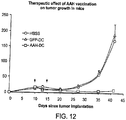
- Vaccination with AAH-loaded mature dendritic cells dramatically reduced tumor burden (tumor volume) of an AAH-expressing tumor.
- the therapeutic effect of AAH vaccination on the growth of tumor in mice is shown in Figs. 11 and 12 .
- 5 ⁇ 10 5 BNL 1ME A.7R.1 mouse hepatoma cells were subcutaneously implanted into the flanks of 6-week-old BALB/c mice.
- 5 ⁇ 10 5 mature DCs loaded with AAH or GFP, or HBSS were subcutaneously injected into the tumor-bearing mice.
- Tumor size was measure weekly.
- AFP is the only HCC-associated protein that can be antigenic to mice and humans. However, it has been shown as well that the anti-tumor effect is not sufficient for human HCC patients. However, simultaneous immunization with AAH and AFP augments the immune response to tumors expressing both AAH and AFP, like HCC.
- Stable lines of AFP-expressing SP2/0 cells are made by introducing an AFP expression plasmid DNA.
- CTL activity in vitro in the mice immunized with AFP-loaded and AAH-loaded DCs is measured. Increased cytotoxicity following immunization indicates that immunization lead to an anti-tumor effect of combination AAH and AFP immunization on the growth AAH and/or AFP-bearing tumors.
Landscapes
- Health & Medical Sciences (AREA)
- Life Sciences & Earth Sciences (AREA)
- Chemical & Material Sciences (AREA)
- General Health & Medical Sciences (AREA)
- Organic Chemistry (AREA)
- Engineering & Computer Science (AREA)
- Veterinary Medicine (AREA)
- Public Health (AREA)
- Animal Behavior & Ethology (AREA)
- Bioinformatics & Cheminformatics (AREA)
- Genetics & Genomics (AREA)
- Zoology (AREA)
- Immunology (AREA)
- Medicinal Chemistry (AREA)
- Biomedical Technology (AREA)
- Wood Science & Technology (AREA)
- Epidemiology (AREA)
- Biotechnology (AREA)
- Pharmacology & Pharmacy (AREA)
- Microbiology (AREA)
- Biochemistry (AREA)
- General Chemical & Material Sciences (AREA)
- Chemical Kinetics & Catalysis (AREA)
- Nuclear Medicine, Radiotherapy & Molecular Imaging (AREA)
- General Engineering & Computer Science (AREA)
- Molecular Biology (AREA)
- Cell Biology (AREA)
- Oncology (AREA)
- Hematology (AREA)
- Mycology (AREA)
- Biophysics (AREA)
- Gastroenterology & Hepatology (AREA)
- Proteomics, Peptides & Aminoacids (AREA)
- Toxicology (AREA)
- Medicines Containing Antibodies Or Antigens For Use As Internal Diagnostic Agents (AREA)
- Medicines Containing Material From Animals Or Micro-Organisms (AREA)
- Micro-Organisms Or Cultivation Processes Thereof (AREA)
- Medicines That Contain Protein Lipid Enzymes And Other Medicines (AREA)
- Peptides Or Proteins (AREA)
- Enzymes And Modification Thereof (AREA)
Description
- Liver cancer including hepatocellular and cholangiocellular carcinoma is one of the most common cancers in the world. In the United States, liver cancer is the fifth common cause of cancer death in men and the ninth in women. In spite of the dramatic progress in diagnosis and local control of liver cancer, the mortality rate in the United States has been increasing constantly. One of the reasons for the increasing mortality might be that, unlike other cancers including breast and colorectal cancers, liver cancer is resistant to systemic treatment such as chemotherapy. Therefore, there is no effective treatment of liver cancer once it becomes a systemic disease.
US 2005/113329 focuses on the identification of aspartyl (asparaginyl)-p-hydroxylase (AAH) over-expression as a marker of malignancy and invasiveness and suggests antisense inhibition of AAH as a therapeutic approach.
US 2006/057129 describes how to generate dendritic cell vaccine using dendritic cells differentiated from stem cells and subsequently genetically modified. The document refers to the expression or transduction of immunogenic epitopes in the dendritic cells.
Hoffman T.K. et al., Journal of Immunotherapy 24: 162-171 (2001) describe enhancing cross-priming capability of dendritic cells by certain cytokines.
Xu S. et al., Surgery 140: 170-178 (2006) describe toll-receptor 8-primed dendritic cells and that antitumor T-cell generation by toll receptor 8-primed dendritic cells is mediated by IL-12 production.
Knippertz I. et al., Journal of Immunotherapy 32: 524-538 (2009) disclose the use of an adenoviral vector encoding human CD40 ligand to establish dendritic cells that feature both migration potential and prolonged secretion of the key T-helper 1 cytokine interleukin-12p70. - The invention is defined by the claims.
- In a first aspect, the invention provides an isolated mature aspartyl (asparaginyl)-β-hydroxylase (AAH)-loaded dendritic cell for use in a method of treating an AAH-expressing tumor by reducing growth of said tumor in a subject, wherein growth of said AAH-expressing tumor is reduced after administration of said dendritic cell, wherein said dendritic cell is primed with a combination of cytokines comprising of GM-CSF and IFN-γ ex vivo, prior to administrating to said subject.
- In a second aspect, the invention provides a method of producing primed mature AAH-loaded dendritic cells, comprising contacting isolated dendritic cells with an antigen comprising AAH, and following said antigen-contacting step, contacting said dendritic cells with a combination of cytokines, said combination comprising GM-CSF and IFN-γ.
- In a third aspect, the invention provides a vaccine composition comprising primed dendritic cells produced by the production method above.
In a fourth aspect, the invention provides a vaccine containing AAH-loaded mature dendritic cells for use in the method of treatment of AAH-expressing tumors in a mammalian subject. - In a fifth aspect, the invention provides autologous dendritic cells primed according to the invention for use in a method of inhibiting tumor growth and/or preventing metastasis in a mammal, wherein said primed autologous dendritic cells are administered to a subject suffering from an AAH-bearing tumor.
- In a sixth aspect, the invention provides autologous dendritic cells which are primed by a combination of cytokines comprising GM-CSF, IFN-γ, and CD40L for use in a method of preventing development of a tumor in a mammal, wherein said subject is at risk of developing an AAH-bearing tumor, due to a family history of individuals who have been identified with such a tumor.
- In a seventh aspect, the invention provides the cell or vaccine for use in a method of treating an AAH-expressing tumor of the present invention in a subject, wherein said subject or mammal is a human, canine, feline, equine, bovine, or porcine subject or mammal.
- The invention provides a solution to the longstanding problem of an alternative to chemotherapy for treatment of patients diagnosed with cancer. The invention provides mature dendritic cells for use in a method of reducing growth of an aspartyl (asparaginyl)-p-hydroxylase (AAH or ASPH)-expressing tumor in a subject which involves administering to the subject an isolated mature AAH-loaded dendritic cell or population of cells. Growth of an AAH-expressing tumor is reduced after administration of such a dendritic cell vaccine. For example, tumor growth and tumor burden is reduced by 10%, 20%, 50%, 75%, 2-fold, 5-fold, 10-fold, or more. The methods are used to reduce and eliminate AAH-expressing tumors from mammalian subjects, such as human patients. The compositions and methods are also suitable for use in companion animals and livestock, e.g., human, canine, feline, equine, bovine, or porcine subjects.
- AAH-expressing tumors include most tumor types such as tumors of gastrointestinal tissues (e.g., esophagus, stomach, colon), pancreas, liver (e.g., cholangiocellular carcinoma, hepatocellular carcinoma), breast, prostate, cervix, ovary, fallopian tube, larynx, lung, thyroid, gall bladder, kidney, bladder, and brain (e.g., glioblastoma) as well as numerous others described below. AAH-expressing tumors include primary tumors that express an increased level of AAH compared to normal tissue as well as tumors that arise by metastasis from such AAH-overexpressing primary tumors.
- The dendritic cells used in the vaccination method are activated ex vivo with a combination of cytokines comprising GM-CSF and IFN-γ prior administering them to the subject. The latter step yields a population of dendritic cells with improved anti-tumor activity.
An improved method of producing primed dendritic cells is carried out by contacting isolated dendritic cells with an antigen comprising AAH or a combination of tumor antigens comprising AAH and α-fetoprotein (AFP) and treating the cells to yield a population of matured and activated antigen-presenting cells. Following the antigen-contacting step, the dendritic cells are contacted with the combination of cytokines. The combination comprises GM-CSF and IFN-γ. The combination may further comprise IL-4. Optionally, the combination comprises CD40L. The dendritic cells are contacted with the combination of cytokines for at least 10 hours (e.g., 12, 24, 36, 40, 48 hours or more). The antigen is in a soluble form or bound to a solid support. For example, the solid support comprises a polystyrene bead such as a biodegradable bead or particle. Dendritic cells are obtained from a subject by known methods such as leukapheresis or cytopheresis. - The subject from which the cells are obtained is suffering from or at risk of developing cancer. The patient has been diagnosed with an AAH-expressing tumor. Patients who are at risk of developing AAH-bearing tumors include those with a family history of individuals who have been identified with such a cancer.
- Also within the invention is a vaccine composition comprising primed dendritic cells produced as described above. The vaccine is useful to inhibit tumor growth, prevent tumor growth, and inhibit or reduce metastasis. The invention provides mature dendritic cells for use in a method of inhibiting tumor growth in a mammal which is carried out by identifying a subject suffering from an AAH-bearing tumor, and administering to the subject autologous dendritic cells, which have been primed according to the method described above. The invention provides mature dendritic cells for use in a method of preventing development of a tumor in a mammal, which comprises the step of identifying a subject at risk of developing an AAH-bearing tumor (such as one with a family history of cancer), and administering to the subject the antigen-primed and activated autologous dendritic cells. The invention provides mature dendritic cells for use in a method of preventing metastasis of an AAH-bearing tumor which is carried out by identifying a subject suffering from an AAH-bearing tumor, and administering to the subject autologous dendritic cells, as described above.
- The polypeptides and other compositions are purified. For example, a substantially pure AAH polypeptide or variant thereof is preferably obtained by expression of a recombinant nucleic acid encoding the polypeptide or by chemically synthesizing the protein. A polypeptide or protein is substantially pure when it is separated from those contaminants which accompany it in its natural state (proteins and other naturally-occurring organic molecules). Typically, the polypeptide is substantially pure when it constitutes at least 60%, by weight, of the protein in the preparation. Preferably, the protein in the preparation is at least 75%, more preferably at least 90%, and most preferably at least 99%, by weight, AAH. Purity is measured by any appropriate method, e.g., column chromatography, polyacrylamide gel electrophoresis, or HPLC analysis. Accordingly, substantially pure polypeptides include recombinant polypeptides derived from a eukaryote but produced in E. coli or another prokaryote, or in a eukaryote other than that from which the polypeptide was originally derived.
- Dendritic cells or other cells, e.g., immune cells such as macrophages, B cells, T cells, used in the methods are purified or isolated. With regard to cells, the term "isolated" means that the cell is substantially free of other cell types or cellular material with which it naturally occurs. For example, a sample of cells of a particular tissue type or phenotype is "substantially pure" when it is at least 60% of the cell population. Preferably, the preparation is at least 75%, more preferably at least 90%, and most preferably at least 99% or 100%, of the cell population. Purity is measured by any appropriate standard method, for example, by fluorescence-activated cell sorting (FACS).
- The methods of generating antigen-primed DCs has several advantages over earlier methods. The cells are not only antigen-loaded but the subsequent cytokine incubation yield superior activated antigen-primed antigen presenting cells with improved anti-tumor and anti-metastatic activity.
- These and other capabilities of the invention, along with the invention itself, will be more fully understood after a review of the following figures, detailed description, and claims.
-
-
Fig. 1 is a bar graph showing IL-12 production by isolated DCs. -
Fig. 2 is a series of line graphs showing expression of various surface DC markers on isolated DCs. -
Fig. 3 is a bar graph showing mean fluorescence on surface DC markers. -
Fig. 4 is a series of flow cytometric graphs and a bar graph showing an increased population of CD8+ DCs grown in the presence of cytokines. -
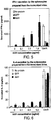
Fig. 5 is a bar graph showing IFN-γ secretion. -
Fig. 6 is a bar graph showing IL-4 secretion. -
Fig. 7 is a line graph showing the results of a cytotoxicity assay. -
Fig. 8 is a diagram of AAH as expressed by a cell. -
Fig. 9 is a diagram of a method of generating mature antigen-loaded DCs. -
Fig. 10 is a flow chart of a schedule of immunization with AAH-loaded DCs. -
Fig. 11 is a diagram showing a protocol for determining the therapeutic effect of AAH vaccination on tumor growth in mice. -
Fig. 12 is a line graph showing therapeutic effect of AAH vaccination on tumor growth in mice. - AAH is a protein, the expression of which is associated with a number of cancer types (see, e.g., USPN 6,815,415; USPN 6,812,206; USPN 6,797,696; USPN 6,783,758; USPN 6,835,370; and USPN 7,094,556). Over-expression of HAAH has been detected by immunohistochemical staining (1HC) in numerous cancers lung (e.g., adenocarcinoma, bronchioalveolar carcinoma, and other non-small cell lung cancers such as sqamous cell carcinoma subtype (Luu et al., 2009, Hum. Pathol. 40:639-644), liver (e.g., hepatocellular carcinoma, cholangiocarcinoma, and cancer of biliary epithelial cells (Wang et al., 2010, Hepatology 52:164-173), gastrointestinal tissues (e.g., colon, stomach, esophagus), pancreas, prostate, ovary, bile duct, breast, kidney, bladder, and brain (e.g., glioblastoma and neuroblastoma. Another survey of tumor tissue samples and cell lines confirmed AAH expression in the following additional cancers (compared to non-cancer tissues): laryngeal carcinoma, cervical cancer, fallopian tube cancer, liver cancer (e.g., cholangiocarcinoma), kidney cancer, breast cancer, cervical cancer, ovarian cancer, Fallopian tube carcinoma, laryngeal cancer, lung cancer, thyroid cancer, pancreatic cancer, thymic carcinoma, prostate cancer, bladder cancer, esophageal cancer, gastric cancer, gallbladder cancer, colon cancer, and rectal cancer (Song et al., 2010, Chinese J. of Cell. and Mol. Immunol. 26: 141-144). HAAH is highly specific for cancer; it has been detected by immunohistochemistry in >99% of tumor specimens tested (n>1000) and is not present in adjacent non-affected tissue, or in tissue samples from normal individuals.
- Immunotherapy using AAH-primed cells is administered to patients with tumors that express AAH on their cell surface. Such tumors include liver cancers such as hepatocellular carcinoma and cholangiocarcinoma, as well as adenocarcinomas of the breast and colon. Such a therapeutic strategy is useful for not only treatment but prevention of cancers.
- Prior to the invention, there has been no effective systemic treatment of liver cancer, in spite of the advances in diagnosis and local control of the disease. Immunotherapy is used to treat systemic and localized liver cancer, because of its specificity.
- Dendritic cell vaccines using AAH were found to cure established hepatocellular carcinoma in immunocompetent mice. AAH dendritic cell vaccines reduce growth of AAH-expressing tumors to decrease tumor burden and eradicate tumors in humans as well.
- Immunotherapy of malignant tumors is attractive in terms of the specific response to tumor cells. A rationale for immunotherapy is that cancer cells are replete with potential antigens, which become immunogenic when presented by antigen-presenting cells to helper and cytotoxic T cells. Immunotherapy for liver cancer, whether systemic or not, is a viable clinical approach, because liver cancer cells express several tumor-associated proteins, which are not or only slightly expressed in normal tissues. For example, AFP, which is not expressed in adult tissues, is extensively expressed in hepatocellular carcinoma (HCC). Several studies have shown that AFP-specific immune reaction can be induced not only in mice but also in humans. However, a clinical trial of HCC immunotherapy that targets AFP did not show even partial tumor responses. Some possible reasons are: because AFP is not involved in cancer progression, cancer cells that do not express AFP dominated during APP-targeting immunotherapy. In order for HCC cells to be recognized by AFP-responsive T cells, AFP should be presented on the cell surface with major histocompatibility complex (MHC) class I molecule, because AFP is not distributed on the cell surface. However, as MHC class I molecules are often down-regulated in many cancer cells, AFP may not be appropriately recognized by the T cells. To overcome the problems of liver cancer immunotherapy, studies were undertaken to identify another antigen that is associated with liver cancer to be used to primed cells for cell-based therapy. Given the results using AFP as an antigen, the anti-tumor results with AAH were surprising.
- AAH, also known as ASPH, is strongly expressed in hepatocellular and cholangiocellular carcinoma, but is not expressed in normal cell counterparts of those tissue types. AAH is therefore a tumor-associated protein. AAH is useful for immunotherapy of liver cancer, because of its unique nature. First, AAH confers cancer cells on motility, which is associated with cancer metastasis; thus, therapy targeting AAH-expressing cells is effective to suppress metastatic lesions as well as the primary tumor. Second, because AAH is a membrane protein, most part of which is exposed to extracellular space, it is easier for immune cells to access to the protein even when class I molecules are down-regulated. Third, other than AAH, no antigenic protein specific to cholangiocellular carcinoma is known. Thus, AAH is uniquely qualified as a target molecule of immunotherapy against liver cancer.
- An immunotherapy approach involves injection of dendritic cells (DCs) loaded with the protein of interest. DCs are specialized cells that capture, process, and present antigens to T cells to induce and control T cell-mediated immunity. DCs are widely used to immunize not only laboratory animals but also tumor-bearing patients. The methods described herein include a DC-based immunization for induction of immunity against AAH. The present methods improve upon a previously described method for generating immunogenic mouse DCs loaded with protein of interest (Gehring et al., 2008, J. Immunol. Meth. 332:18-30) in at least two ways. One is that to stimulate DCs, lipopolysaccharides (LPS) was previously used, a strong pyrogen that may be inappropriate for in vivo use. Second is that DC stimulation was relatively weak, judging by IL-12 secretion by DCs, and by the expression of various surface DC markers. Improvements were therefore made to the method for the generation of immunogenic DCs.
- The most promising approach for HCC immunotherapy is the one using DCs, as they have a strong capacity of inducing T cell-mediated tumor immunity. DCs are derived from hematopoietic cells, and are specialized to capture and process antigens, converting proteins to peptides that are presented on MHC molecules and recognized by T cells. DCs strongly induce and control T-cell mediated immunity.
- DC-based immunotherapy is based on the fact that tumors are replete with potential antigens, and these become immunogenic when presented by DCs. DCs are obtained from a subject, e.g. by cytopheresis. The cells, which are now ex vivo are loaded (i.e., contacted) with tumor antigens (e.g., tumor cell lysates; apoptotic or necrotic tumor cells; recombinant, synthetic, or purified tumor antigen peptide or protein; or nucleic acid such as RNA encoding tumor antigens), stimulated for maturation, and re-injected into patients to induce strong T-cell immunity.
- AAH is type II membrane protein with a catalytic domain in a C-terminal region. Most parts of the protein are located at the outside of the cell membrane, allowing the access of immune cells to this protein. AAH protein is strongly expressed in HCCs, cholangiocarcinomas, and adenocarcinomas of breast or colon origin, and is barely detectable or completely undetectable in normal tissue counterparts. Thus, AAH is an ideal target molecule for DC-based immunotherapy of HCC and other AAH-bearing tumors.
- Overexpressed AAH in tumor cells increases the cell motility, which confers malignant phenotype on tumor cells. A clinicopathological study on HCC revealed that AAH overexpression is associated with histological grade and intrahepatic metastasis. Therefore, targeting the AAH-expressing tumor cell population suppresses the development and progression of AAH-bearing tumors.


- Antigen-presenting cells (APCs) such as DCs are obtained from subjects, e.g., human patients suffering from or at risk of developing cancer, using the process of leukapheresis. Such as procedure is typically carried out at an apheresis center (Dayl). Dendritic cells are purified using known methods and are contacted with antigen, such as AAH or AAH and AFP together. Following antigen contact, the cells are cultured with a mixture of cytokines (Day 2-3). The antigen-primed and activated DCs are then administered to the patient (Day 3-4). An exemplary course of therapy includes three administrations of DCs over a four week period.
- The following materials and methods were used to generate the data described herein.
- 7-8-week-old female BALB/c (H-2d) mice were purchased from Harlan Sprague Dawley, Inc., and kept under specific pathogen-free conditions.
- Immunomagnetic beads (1.3 µm; Calbiochem) were washed three times with 50 mM borate buffer, pH 8.5, and resuspended in 50 mM borate buffer, pH 9.0, with 0.1 mg/ml AAH or GFP protein. Then the suspension was incubated overnight at room temperature under constant agitation. The beads were pelleted, washed with PBS, and resuspended in PBS at a concentration of 30 mg solid content/ml.
- DC isolation was performed using known methods (e.g., (Gehring et al., 2008, J. Immunol. Meth. 332:18-30)). Cells are optionally obtained by leukapheresis. 10 µg pUMVC3-hFLex, an expression plasmid encoding Fms-like tyrosine kinase receptor-3 ligand (FLT3L), was injected into the mouse tail vein on
day 0 and 6. Splenocytes were prepared from the FLT3L-injected mice, using NH4Cl red blood cell-lysis buffer, and 5 x 10⁷ cells were incubated in serum-free DMEM with 10 µl magnetic microbeads for 4-6 hours. The cells were collected, passed through a magnetic field using MACS MS column (Miltenyi) to enrich the cells ingesting magnetic beads. Then the cells were subjected to density-gradient centrifugation using Lympholyte M (Cedarlane) to eliminate free beads and dead cells. The viable cells were collected, washed twice with Hanks' buffered salt solution (HBSS), and used for subsequent experiments. The viability of the isolated cells was > 90%, and the percentage of the CD11c positive population in the isolated cells were 70-80%. - DCs were cultured in HEPES-buffered RPMI1640 supplemented with 10% mouse serum (Equitech Bio), 2 mM L-glutamine, 50 µM 2-mercaptoethanol, 100 U/ml penicillin, 100 µg/ml streptomycin, 20 ng/ml GMCSF, 100 ng/ml IL-4, 20 ng/ml IFN-γ, and 1 µg/ml CD40L (all cytokines were purchased from Peprotech) on a 6-well ultra-low attachment plate (Corning) for 40 h. For cytokine release, splenocytes were grown in serum-free X-VIVO 15 (Lonza) supplemented with 2 mM L-glutamine, 50 nM 2-mercaptoethanol, 100 U/ml penicillin, and 100 µg/ml streptomycin. For the cytotoxicity assay, splenocytes were grown in the complete medium consisting of HEPES-buffered RPMI1640 supplemented with 10% fetal bovine serum (Atlantic Bio), 2 mM L-glutamine, 50 µM 2-mercaptoethanol, 100 U/ml penicillin, 100 µg/ml streptomycin, 1 x non-essential amino acids (Lonza), 0.5 x amino acid solution (Invitrogen), and 1 mM sodium pyruvate. SP2/0 cells were cultured in Dulbecco's minimum essential medium supplemented with 20% fetal bovine serum, 2 mM L-glutamine, 100 U/ml penicillin, and 100 µg/ml streptomycin.
- DCs or splenocytes were grown in the serum-free medium with or without AAH protein for 48 h. Then the culture supernatants were collected, centrifuged, and subjected to ELISA assays. ELISA for IL-12, p70, IFN-γ, and IL-4 was performed using ELISA Ready-SET-Go! kit (eBioscience) according to the manufacturer's instructions.
- The expression of cell surface markers was analyzed by flow cytometry as described previously (Gehring, et al., 2008). 3-6
x 105 cells were used for staining. The cells were washed with staining buffer (HBSS containing 5% FBS), incubated with 2.5 µg/ml Mouse BD Fc Block (BD Biosciences) for 5 min at 4 °C, and further incubated with staining antibodies for 30 min at 4 °C. The antibodies used were anti-CD 11c (HL3; BD Biosciences), anti-CD40 (3/23; BD Biosciences), anti-CD54 (3E2; BD Biosciences), anti-CD80 (16-10A1; BD Biosciences), anti-CD86 (GLI; BD Biosciences), anti-I-Ad (AMS-32.1; BD Biosciences), and anti-CD8a (53-6.7; eBioscience), all of which were conjugated with fluorochrome. Hamster, rat, and mouse immunoglobulin G isotype-matched controls were also used for staining controls. After washing twice, the cells were resuspended in staining buffer, and were subjected to flow cytometry using FACSCalibur (BD Biosciences). Dead cells were eliminated from analysis by staining the cells with 7- amino-actinomycin D (eBioscience). Data were processed with CellQuest software (BD Biosciences) followed by FlowJo software (Tree Star). - After the culture with cytokines for 40 h, 2.5 x 10⁵ cells were injected subcutaneously into the right and left flanks (5 x 10⁵ cells per mouse) on
day day 28, the immunized mice were used for further experiments including cytokine release and cytotoxicity assay. - Splenocytes were prepared from immunized mice, and 5 x 10⁷ splenocytes were grown in complete medium with 0.5 µg/ml AAH protein for 2 days and further grown for 2 days after addition of 10 ng/ml IL-2. Two x 104 target SP2/0 cells were mixed with 6 - 60
x 104 effector splenocytes in the serum-free medium, centrifuged at 200 x g for 5 min, and incubated in CO2 incubator for 4 h. Culture supernatants were collected, and released lactate dehydrogenase activity in the supernatants were measured by using LDH Cytotoxicity Detection Kit (Roche) in accordance with manufacturer's instruction. % target cell lysis was calculated using the following formula:
- The protocol previously used includes in vivo expansion of DCs, ingestion of magnetic microbeads on which an antigen and stimulants of DCs including LPS and anti-CD40 antibody are coated, and isolation of DCs by passing through a magnetic column. Instead of stimulating DCs with the stimulants coated on the beads, the improved method involved stimulating isolated DCs by growing for two days in the presence of various cytokines, because stimulants should interact with the corresponding receptors on the cell surface, but the stimulants on the beads that were ingested by DCs cannot. There exist some cytokines that stimulate and promote maturation of DCs including IL-4, IFN-γ, and CD40L. Therefore, the combinatorial effect of these cytokines on the maturation of DCs was examined. Expansion, ingestion, and isolation of DCs were performed. Isolated cells were cultured for 40 hours in the presence of GM-CSF and/or IL-4, and/or IFN-γ, and/or CD40L. GM-CSF was always added because it was necessary for survival of DCs. Mature, but not immature, DCs vigorously produce IL-12, which is essential for polarizing helper and cytotoxic T cells. Therefore, IL-12 levels were used as an indicator of DC stimulation. Supernatants of the culture was collected, the concentration of IL-12 p70 was measured by ELISA. The results indicated that IL-12 p70 was the highest when all cytokines were present in the DC culture (
Figure 1 ). Each of the cytokines are used for stimulation alone or in combination. Preferably, all the cytokines are used to stimulate DCs. However, CD40L is optional. - To investigate the effect of the cytokine stimulation on the expression of mature DC markers, the marker expression of DCs grown for 40 h in the presence of the cytokines to that of DCs without culture was measured. The expression of CD11c, a marker for pan-DCs, was not changed after the culture (
Figures 2 ,3 ). However, all the mature DC markers including CD40, CD54, CD80, CD86, and MHC class II (I-Ad), were significantly up-regulated in the DCs grown for 40 h (Figures 2 ,3 ). CD8a+ DCs are the subset of DCs that can efficiently stimulate CTLs. The effect of the cytokine stimulation on CD8a+ DC population was evaluated. As shown inFigure 4 , the percentage of CD8a+ population in DCs was significantly increased during the stimulation. These results indicate that the generated cells by this method without using LPS had sufficient quality for DC-based immunization. - To examine whether
T H1 response was induced by the AAH immunization, the production of IFN-γ, aT H1 cytokine, in response to AAH protein was measured. Mice were immunized twice with a 2-week interval with DCs ingesting AAH or GFP-coated beads, or beads without any antigens. Two weeks after the last immunization, splenocytes were prepared from the immunized mice, cultured for 48 h in the presence of AAH, and the concentration of IFN-γ was measured in culture supernatants to examine whether splenocytes from AAH-immunized mice produce IFN-γ in response to AAH stimulation. The splenocytes from AAH immunized mice vigorously produced IFN-γ in a dose-dependent manner (Figure 5 ). Although splenocytes from three groups produced considerable amount of IFN-γ in the presence of concanavalin A (Con A), the control splenocytes produced only subtle IFN-γ when stimulated with AAH (Figure 5 ). This result indicates thatT H1 cells responsive to AAH were generated in the immunized mice. - To investigate whether humoral immunity against AAH was induced by the DCs loaded with AAH, the concentration of IL-4, a
T H2 cytokine, was measured in the same samples as IFN-γ. Con A-stimulated splenocytes produced large amount of IL-4 independent of the immunization (Figure 6 ). By contrast, the splenocytes prepared from the mice immunized with three types of DCs produced only trace amounts of IL-4 (Figure 6 ), indicating thatT H2 cells responsive to AAH were not efficiently induced by AAH immunization. - To determine whether CTLs responsive to AAH were induced in AAH-immunized mice, cytotoxicity was evaluated by using AAH-expressing SP2/0 cells as target cells. SP2/0 cells were co-cultured for 4 h with the splenocytes from the immunized mice after activated with AAH protein and IL-2. The efficiencies of target cell lyses were estimated by measuring lactate dehydrogenase activities released from dying target cells. As shown in
Figure 7 , the splenocytes from the AAH-immunized mice had more CTL activity than the other control splenocytes, suggesting that AAH immunization induced AAH-responsive CTLs. - When mice were immunized with AAH-loaded DCs, AAH-
responsive T H1 cells and CTLs were induced. Antigen-specific T cell expansion is another important indicator that indicates that antigen-responsive T cells were produced. - AAH immunization using AAH-primed DCs induces antitumor immunity. Mice are immunized with AAH-loaded DCs twice with a 2-week interval, and two weeks after the last immunization. SP2/0 cells are implanted under the skin, the growth of implanted tumor sizes is measured. Data obtained from this tumor burden model reflects the preventive effect of the immunization. To evaluate the curative effect of AAH immunization, SP2/0 cells are implanted first, then when tumors grow to 5 mm in diameter, the mice are immunized with AAH-loaded DCs, and the effect on tumor growth measured.
- AAH-targeted immunotherapy also suppresses metastasis. Mice are immunized with AAH-loaded DCs followed by injection of SP2/0 cells into the tail vein. Two weeks later, the lungs are excised to count the number of nodules formed. A reduction in the amount of nodules in immunized mice compared to the amount in unimmunized mice indicates that the AAH-loaded DC immunization regimen suppresses metastasis of AAH-bearing tumors.
- Vaccination with AAH-loaded mature dendritic cells dramatically reduced tumor burden (tumor volume) of an AAH-expressing tumor. The therapeutic effect of AAH vaccination on the growth of tumor in mice is shown in
Figs. 11 and12 . 5 × 10⁵ BNL 1ME A.7R.1 mouse hepatoma cells were subcutaneously implanted into the flanks of 6-week-old BALB/c mice. Atday - AFP is the only HCC-associated protein that can be antigenic to mice and humans. However, it has been shown as well that the anti-tumor effect is not sufficient for human HCC patients. However, simultaneous immunization with AAH and AFP augments the immune response to tumors expressing both AAH and AFP, like HCC.
- Stable lines of AFP-expressing SP2/0 cells are made by introducing an AFP expression plasmid DNA. By using the cells, CTL activity in vitro in the mice immunized with AFP-loaded and AAH-loaded DCs is measured. Increased cytotoxicity following immunization indicates that immunization lead to an anti-tumor effect of combination AAH and AFP immunization on the growth AAH and/or AFP-bearing tumors.
Claims (16)
- An isolated mature aspartyl (asparaginyl)-β-hydroxylase (AAH)-loaded dendritic cell for use in a method of treating an AAH-expressing tumour by reducing growth of said tumor in a subject, wherein growth of said AAH-expressing tumor is reduced after administration of said dendritic cell, wherein said dendritic cell is primed with a combination of cytokines including GM-CSF and IFN-γ ex vivo, prior to administrating to said subject.
- The isolated cell for use according to claim 1, wherein the tumour to be treated is selected from the group consisting of liver, gastrointestinal, pancreas, breast, prostate, cervix, ovary, fallopian tube, larynx, lung, thyroid, gall bladder, kidney, bladder, and brain tumours.
- The isolated cell for use according to claim 1, wherein said dendritic cell is activated with a combination of cytokines including GM-CSF, IFN-γ, and CD40L ex vivo, prior to said administering to said subject.
- A method of producing primed mature AAH-loaded dendritic cells, comprising contacting isolated dendritic cells with an antigen comprising AAH, and following said antigen-contacting step, contacting said dendritic cells with a combination of cytokines, said combination comprising GM-CSF and IFN-γ.
- The method of claim 4, wherein said combination further comprises CD40L and / or IL-4.
- The method of claim 4, wherein said dendritic cells are contacted with said combination of cytokines for at least 10 hours, preferably for at least 40 hours.
- The isolated cell for use according to claim 1 or method of claim 4, wherein said antigen further comprises α-fetoprotein (AFP).
- The isolated cell for use according to claim 1 or method of claim 4, wherein said antigen is in a soluble form or is bound to a solid support.
- The isolated cell for use or method of claim 8, wherein said solid support comprises a polystyrene bead or said solid support is a biodegradable.
- The isolated cell for use according to claim 1, wherein said dendritic cells are obtained from a subject by leukapheresis.
- The isolated cell for use according to claim 10, wherein said subject is suffering from or at risk of developing cancer.
- A vaccine composition comprising primed dendritic cells produced by the method of claim 4.
- A vaccine containing AAH-loaded mature dendritic cells for use in a method of treatment of AAH-expressing tumours in a mammalian subject, wherein the method is defined as in any of claims 1-3, and 7-11.
- Autologous dendritic cells primed by the method of claim 4 or 5 for use in a method of inhibiting tumour growth and/or preventing metastasis, wherein said primed autologous dendritic cells are administered to a subject suffering from an AAH-bearing tumour.
- Autologous dendritic cells which are primed as defined in claim 4 for use in a method of preventing development of a tumour in a subject wherein said subject is a mammal at risk of developing an AAH-bearing tumour due to a family history of individuals who have been identified with such a tumour.
- The cells for use of claim 1, 14 or 15, or the vaccine for use of claim 13, wherein said subject is human, canine, feline, equine, bovine, or porcine
Applications Claiming Priority (5)
| Application Number | Priority Date | Filing Date | Title |
|---|---|---|---|
| US22842909P | 2009-07-24 | 2009-07-24 | |
| US23112709P | 2009-08-04 | 2009-08-04 | |
| US23928809P | 2009-09-02 | 2009-09-02 | |
| US24074509P | 2009-09-09 | 2009-09-09 | |
| PCT/US2010/043056 WO2011011688A2 (en) | 2009-07-24 | 2010-07-23 | DENDRITIC CELL VACCINES FOR ASPARAGINYL- β - HYDROXYLASE EXPRESSING TUMORS |
Publications (2)
| Publication Number | Publication Date |
|---|---|
| EP2456458A2 EP2456458A2 (en) | 2012-05-30 |
| EP2456458B1 true EP2456458B1 (en) | 2018-12-26 |
Family
ID=42830734
Family Applications (1)
| Application Number | Title | Priority Date | Filing Date |
|---|---|---|---|
| EP10735417.7A Not-in-force EP2456458B1 (en) | 2009-07-24 | 2010-07-23 | Dendritic cell vaccines for asparaginyl - beta - hydroxylase expressing tumors |
Country Status (10)
| Country | Link |
|---|---|
| US (1) | US9687537B2 (en) |
| EP (1) | EP2456458B1 (en) |
| JP (2) | JP5872468B2 (en) |
| KR (1) | KR101911380B1 (en) |
| CN (1) | CN102596234B (en) |
| AU (1) | AU2010275443B2 (en) |
| CA (1) | CA2768398C (en) |
| ES (1) | ES2711504T3 (en) |
| NZ (1) | NZ597724A (en) |
| WO (1) | WO2011011688A2 (en) |
Families Citing this family (7)
| Publication number | Priority date | Publication date | Assignee | Title |
|---|---|---|---|---|
| ES2711504T3 (en) * | 2009-07-24 | 2019-05-06 | Rhode Island Hospital | Dendritic cell vaccines for tumors that express asparaginyl-beta-hydroxylase |
| CN105308177A (en) * | 2013-03-07 | 2016-02-03 | 新干细胞肿瘤学有限责任公司 | Individualized high purity hepatocellular carcinoma stem cells, methods and use of the same |
| US9744223B2 (en) * | 2013-03-15 | 2017-08-29 | Panacea Pharmaceuticals, Inc. | Therapeutic cancer vaccine targeted to HAAH (aspartyl-[asparaginyl]-β-hydroxylase) |
| CN103405759B (en) * | 2013-07-23 | 2014-12-10 | 蔡颖 | Method for preparing tumor-specific DC vaccine by applying CD34+ cells of umbilical cord blood |
| JP7025119B2 (en) * | 2014-04-24 | 2022-02-24 | ロード アイランド ホスピタル | Aspartic acid-β-hydroxylase induces an epitope-specific T cell response in tumors |
| US20220054614A1 (en) * | 2018-12-13 | 2022-02-24 | Rhode Island Hospital | Inhibition of asph expressing tumor growth and progression |
| GB202010095D0 (en) * | 2020-07-01 | 2020-08-12 | Tcer Ab | Immunotherapy |
Family Cites Families (17)
| Publication number | Priority date | Publication date | Assignee | Title |
|---|---|---|---|---|
| EP0973530A4 (en) * | 1996-12-10 | 2002-01-16 | Hadasit Med Res Service | Serum-derived factor inducing cell differentiation and medical uses thereof |
| US20050123545A1 (en) * | 1999-11-08 | 2005-06-09 | Wands Jack R. | Diagnosis and treatment of malignant neoplasms |
| US6835370B2 (en) * | 1999-11-08 | 2004-12-28 | Rhode Island Hospital | Diagnosis and treatment of malignant neoplasms |
| US20030031670A1 (en) | 1999-11-08 | 2003-02-13 | Jack R. Wands | Diagnosis and treatment of malignant neoplasms |
| WO2003028642A2 (en) | 2001-10-01 | 2003-04-10 | Rhode Island Hospital, A Lifespan Partner | Methods of inhibiting metastases |
| TWI259206B (en) * | 2002-09-24 | 2006-08-01 | Univ Nat Cheng Kung | A DNA vaccine containing a tumor associated gene and a cytokine gene and the method producing thereof |
| DE60322559D1 (en) * | 2002-10-02 | 2008-09-11 | Hoffmann La Roche | NEW, MHC CLASS II ASSOCIATED PEPTIDES |
| ES2321939T3 (en) * | 2003-02-27 | 2009-06-15 | Northwest Biotherapeutics, Inc. | GENERATION OF DENDRITIC CELLS FROM MONOCITIC DENDRITIC PRECURSOR CELLS WITH GM-CSF IN THE ABSENCE OF ADDITIONAL CYTOKINS. |
| AU2004291910A1 (en) | 2003-11-14 | 2005-06-02 | Massachusetts Institute Of Technology | Anti-hydroxylase antibodies and uses thereof |
| CA2504451A1 (en) * | 2004-08-10 | 2006-02-10 | Geron Corporation | Dendritic cell vaccines for treating cancer made from embryonic stem cells |
| KR101300546B1 (en) | 2004-08-24 | 2013-09-02 | 추가이 세이야쿠 가부시키가이샤 | Adjuvant Therapy with the Use of Anti-Glypican 3 Antibody |
| GB2426581A (en) | 2005-05-27 | 2006-11-29 | Univ Nottingham | Immunoassay methods |
| KR100675761B1 (en) | 2005-09-01 | 2007-01-30 | 주식회사 바이넥스 | Method for Inducing Transdifferentiation of Acute Myeloid Leukemia Cells to Dendritic Cells Using 1,2-Dioctanoyl-snh-glycerol3-phosphate |
| CA2632263C (en) * | 2005-12-08 | 2022-01-11 | Northwest Biotherapeutics, Inc. | Compositions and methods for inducing the activation of immature monocytic dendritic cells |
| CN101755045B (en) * | 2007-05-17 | 2014-02-12 | 生物载体株式会社 | Preparation method of dendritic cells |
| CN101754768B (en) * | 2007-07-19 | 2013-07-10 | 健康研究股份有限公司 | Survivin peptides as cancer vaccines |
| ES2711504T3 (en) * | 2009-07-24 | 2019-05-06 | Rhode Island Hospital | Dendritic cell vaccines for tumors that express asparaginyl-beta-hydroxylase |
-
2010
- 2010-07-23 ES ES10735417T patent/ES2711504T3/en active Active
- 2010-07-23 WO PCT/US2010/043056 patent/WO2011011688A2/en not_active Ceased
- 2010-07-23 EP EP10735417.7A patent/EP2456458B1/en not_active Not-in-force
- 2010-07-23 AU AU2010275443A patent/AU2010275443B2/en not_active Ceased
- 2010-07-23 NZ NZ597724A patent/NZ597724A/en not_active IP Right Cessation
- 2010-07-23 JP JP2012521829A patent/JP5872468B2/en not_active Expired - Fee Related
- 2010-07-23 KR KR1020127004697A patent/KR101911380B1/en not_active Expired - Fee Related
- 2010-07-23 CA CA2768398A patent/CA2768398C/en active Active
- 2010-07-23 US US12/842,494 patent/US9687537B2/en active Active
- 2010-07-23 CN CN201080033781.4A patent/CN102596234B/en not_active Expired - Fee Related
-
2015
- 2015-10-08 JP JP2015199935A patent/JP2016053036A/en active Pending
Non-Patent Citations (1)
| Title |
|---|
| None * |
Also Published As
| Publication number | Publication date |
|---|---|
| AU2010275443B2 (en) | 2016-05-12 |
| KR101911380B1 (en) | 2018-10-25 |
| US20110076290A1 (en) | 2011-03-31 |
| WO2011011688A3 (en) | 2011-03-17 |
| JP2013500261A (en) | 2013-01-07 |
| KR20120052346A (en) | 2012-05-23 |
| CA2768398C (en) | 2020-01-07 |
| EP2456458A2 (en) | 2012-05-30 |
| US9687537B2 (en) | 2017-06-27 |
| CN102596234B (en) | 2016-11-16 |
| WO2011011688A2 (en) | 2011-01-27 |
| JP5872468B2 (en) | 2016-03-01 |
| CA2768398A1 (en) | 2011-01-27 |
| ES2711504T3 (en) | 2019-05-06 |
| CN102596234A (en) | 2012-07-18 |
| NZ597724A (en) | 2013-10-25 |
| AU2010275443A1 (en) | 2012-02-02 |
| JP2016053036A (en) | 2016-04-14 |
Similar Documents
| Publication | Publication Date | Title |
|---|---|---|
| JP2016053036A (en) | DENDRITIC CELL VACCINES AGAINST ASPARAGINYL-β-HYDROXYLASE EXPRESSING TUMORS | |
| Liu et al. | Synthetic MUC1 breast cancer vaccine containing a Toll-like receptor 7 agonist exerts antitumor effects | |
| JP2015520129A (en) | Multivalent breast cancer vaccine | |
| CN118284689A (en) | Super DC expressing immune checkpoint inhibitors and uses thereof | |
| Yang et al. | Generated SecPen_NY-ESO-1_ubiquitin-pulsed dendritic cell cancer vaccine elicits stronger and specific T cell immune responses | |
| Schetters et al. | Adaptable antigen matrix platforms for peptide vaccination strategies and T cell-mediated anti-tumor immunity | |
| US20260061044A1 (en) | Mutated Influenza Virus, Pharmaceutical Composition, and Use | |
| JP2021512966A (en) | Vaccine composition and its use | |
| US20240398864A1 (en) | Pharmaceutical composition | |
| Eisenberg et al. | Transcutaneous immunization with hydrophilic recombinant gp100 protein induces antigen-specific cellular immune response | |
| JP4803460B2 (en) | HLA-A24 molecule-binding squamous cell carcinoma antigen-derived peptide | |
| JP2018527286A (en) | Identification of immunogenic MHC class II peptides for immune-based therapy | |
| HK1171386B (en) | DENDRITIC CELL VACCINES FOR ASPARAGINYL-β-HYDROXYLASE EXPRESSING TUMORS | |
| HK1171386A (en) | DENDRITIC CELL VACCINES FOR ASPARAGINYL-β-HYDROXYLASE EXPRESSING TUMORS | |
| JP6811194B2 (en) | Immunogenic preprocalcitonin peptide | |
| TWI398262B (en) | Immunogenic peptides of tumor associated antigen l6 and uses thereof in cancer therapy | |
| CN113144181B (en) | B7H 3-targeted DNA vaccine, and preparation method and application thereof | |
| Peng et al. | Induction of cytotoxic T-lymphocyte responses using dendritic cells transfected with hepatocellular carcinoma mRNA | |
| US20260116941A1 (en) | Modified mammalian vesicles and compositions and methods related thereto | |
| Tanwar | Combination of Pre-existing and New Strategies to Improve CAR-T Cells’ Efficiency. | |
| JP2026515943A (en) | Apheresis products transduced by adenovirus vectors | |
| Baudrexler et al. | P04. 01 Volatile profiling using an eNose allows differentiation of volatile phases derived from serum, DC, or MLC culture supernatants from healthy or leukemic samples | |
| Wang et al. | Preparation and identification of a novel immunomodulator composed of muramyl dipeptide and anti-CD10 monoclonal antibody for treatment of minimal residual disease in acute leukemia children | |
| WO2021085624A1 (en) | Composition for treatment and/or prevention of tumor | |
| JP2008289380A (en) | Novel cancer antigen peptides and uses thereof |
Legal Events
| Date | Code | Title | Description |
|---|---|---|---|
| PUAI | Public reference made under article 153(3) epc to a published international application that has entered the european phase |
Free format text: ORIGINAL CODE: 0009012 |
|
| 17P | Request for examination filed |
Effective date: 20120224 |
|
| AK | Designated contracting states |
Kind code of ref document: A2 Designated state(s): AL AT BE BG CH CY CZ DE DK EE ES FI FR GB GR HR HU IE IS IT LI LT LU LV MC MK MT NL NO PL PT RO SE SI SK SM TR |
|
| DAX | Request for extension of the european patent (deleted) | ||
| 17Q | First examination report despatched |
Effective date: 20140904 |
|
| STAA | Information on the status of an ep patent application or granted ep patent |
Free format text: STATUS: EXAMINATION IS IN PROGRESS |
|
| GRAP | Despatch of communication of intention to grant a patent |
Free format text: ORIGINAL CODE: EPIDOSNIGR1 |
|
| STAA | Information on the status of an ep patent application or granted ep patent |
Free format text: STATUS: GRANT OF PATENT IS INTENDED |
|
| RIC1 | Information provided on ipc code assigned before grant |
Ipc: C12N 9/02 20060101ALI20180207BHEP Ipc: C12N 5/0784 20100101ALI20180207BHEP Ipc: A61K 39/00 20060101AFI20180207BHEP Ipc: C07K 14/705 20060101ALI20180207BHEP |
|
| INTG | Intention to grant announced |
Effective date: 20180312 |
|
| GRAS | Grant fee paid |
Free format text: ORIGINAL CODE: EPIDOSNIGR3 |
|
| RIN1 | Information on inventor provided before grant (corrected) |
Inventor name: WANDS, JACK, R. Inventor name: SHIMODA, MASAFUMI |
|
| GRAA | (expected) grant |
Free format text: ORIGINAL CODE: 0009210 |
|
| STAA | Information on the status of an ep patent application or granted ep patent |
Free format text: STATUS: THE PATENT HAS BEEN GRANTED |
|
| AK | Designated contracting states |
Kind code of ref document: B1 Designated state(s): AL AT BE BG CH CY CZ DE DK EE ES FI FR GB GR HR HU IE IS IT LI LT LU LV MC MK MT NL NO PL PT RO SE SI SK SM TR |
|
| REG | Reference to a national code |
Ref country code: GB Ref legal event code: FG4D |
|
| REG | Reference to a national code |
Ref country code: CH Ref legal event code: EP |
|
| REG | Reference to a national code |
Ref country code: AT Ref legal event code: REF Ref document number: 1080491 Country of ref document: AT Kind code of ref document: T Effective date: 20190115 |
|
| REG | Reference to a national code |
Ref country code: DE Ref legal event code: R096 Ref document number: 602010056059 Country of ref document: DE |
|
| REG | Reference to a national code |
Ref country code: IE Ref legal event code: FG4D |
|
| PG25 | Lapsed in a contracting state [announced via postgrant information from national office to epo] |
Ref country code: FI Free format text: LAPSE BECAUSE OF FAILURE TO SUBMIT A TRANSLATION OF THE DESCRIPTION OR TO PAY THE FEE WITHIN THE PRESCRIBED TIME-LIMIT Effective date: 20181226 Ref country code: NO Free format text: LAPSE BECAUSE OF FAILURE TO SUBMIT A TRANSLATION OF THE DESCRIPTION OR TO PAY THE FEE WITHIN THE PRESCRIBED TIME-LIMIT Effective date: 20190326 Ref country code: LV Free format text: LAPSE BECAUSE OF FAILURE TO SUBMIT A TRANSLATION OF THE DESCRIPTION OR TO PAY THE FEE WITHIN THE PRESCRIBED TIME-LIMIT Effective date: 20181226 Ref country code: LT Free format text: LAPSE BECAUSE OF FAILURE TO SUBMIT A TRANSLATION OF THE DESCRIPTION OR TO PAY THE FEE WITHIN THE PRESCRIBED TIME-LIMIT Effective date: 20181226 Ref country code: BG Free format text: LAPSE BECAUSE OF FAILURE TO SUBMIT A TRANSLATION OF THE DESCRIPTION OR TO PAY THE FEE WITHIN THE PRESCRIBED TIME-LIMIT Effective date: 20190326 Ref country code: HR Free format text: LAPSE BECAUSE OF FAILURE TO SUBMIT A TRANSLATION OF THE DESCRIPTION OR TO PAY THE FEE WITHIN THE PRESCRIBED TIME-LIMIT Effective date: 20181226 |
|
| REG | Reference to a national code |
Ref country code: NL Ref legal event code: MP Effective date: 20181226 |
|
| REG | Reference to a national code |
Ref country code: ES Ref legal event code: FG2A Ref document number: 2711504 Country of ref document: ES Kind code of ref document: T3 Effective date: 20190506 |
|
| REG | Reference to a national code |
Ref country code: LT Ref legal event code: MG4D |
|
| PG25 | Lapsed in a contracting state [announced via postgrant information from national office to epo] |
Ref country code: AL Free format text: LAPSE BECAUSE OF FAILURE TO SUBMIT A TRANSLATION OF THE DESCRIPTION OR TO PAY THE FEE WITHIN THE PRESCRIBED TIME-LIMIT Effective date: 20181226 Ref country code: SE Free format text: LAPSE BECAUSE OF FAILURE TO SUBMIT A TRANSLATION OF THE DESCRIPTION OR TO PAY THE FEE WITHIN THE PRESCRIBED TIME-LIMIT Effective date: 20181226 Ref country code: GR Free format text: LAPSE BECAUSE OF FAILURE TO SUBMIT A TRANSLATION OF THE DESCRIPTION OR TO PAY THE FEE WITHIN THE PRESCRIBED TIME-LIMIT Effective date: 20190327 |
|
| REG | Reference to a national code |
Ref country code: AT Ref legal event code: MK05 Ref document number: 1080491 Country of ref document: AT Kind code of ref document: T Effective date: 20181226 |
|
| PG25 | Lapsed in a contracting state [announced via postgrant information from national office to epo] |
Ref country code: NL Free format text: LAPSE BECAUSE OF FAILURE TO SUBMIT A TRANSLATION OF THE DESCRIPTION OR TO PAY THE FEE WITHIN THE PRESCRIBED TIME-LIMIT Effective date: 20181226 |
|
| PG25 | Lapsed in a contracting state [announced via postgrant information from national office to epo] |
Ref country code: CZ Free format text: LAPSE BECAUSE OF FAILURE TO SUBMIT A TRANSLATION OF THE DESCRIPTION OR TO PAY THE FEE WITHIN THE PRESCRIBED TIME-LIMIT Effective date: 20181226 Ref country code: PL Free format text: LAPSE BECAUSE OF FAILURE TO SUBMIT A TRANSLATION OF THE DESCRIPTION OR TO PAY THE FEE WITHIN THE PRESCRIBED TIME-LIMIT Effective date: 20181226 Ref country code: PT Free format text: LAPSE BECAUSE OF FAILURE TO SUBMIT A TRANSLATION OF THE DESCRIPTION OR TO PAY THE FEE WITHIN THE PRESCRIBED TIME-LIMIT Effective date: 20190426 |
|
| PG25 | Lapsed in a contracting state [announced via postgrant information from national office to epo] |
Ref country code: SM Free format text: LAPSE BECAUSE OF FAILURE TO SUBMIT A TRANSLATION OF THE DESCRIPTION OR TO PAY THE FEE WITHIN THE PRESCRIBED TIME-LIMIT Effective date: 20181226 Ref country code: IS Free format text: LAPSE BECAUSE OF FAILURE TO SUBMIT A TRANSLATION OF THE DESCRIPTION OR TO PAY THE FEE WITHIN THE PRESCRIBED TIME-LIMIT Effective date: 20190426 Ref country code: EE Free format text: LAPSE BECAUSE OF FAILURE TO SUBMIT A TRANSLATION OF THE DESCRIPTION OR TO PAY THE FEE WITHIN THE PRESCRIBED TIME-LIMIT Effective date: 20181226 Ref country code: RO Free format text: LAPSE BECAUSE OF FAILURE TO SUBMIT A TRANSLATION OF THE DESCRIPTION OR TO PAY THE FEE WITHIN THE PRESCRIBED TIME-LIMIT Effective date: 20181226 Ref country code: SK Free format text: LAPSE BECAUSE OF FAILURE TO SUBMIT A TRANSLATION OF THE DESCRIPTION OR TO PAY THE FEE WITHIN THE PRESCRIBED TIME-LIMIT Effective date: 20181226 |
|
| REG | Reference to a national code |
Ref country code: DE Ref legal event code: R097 Ref document number: 602010056059 Country of ref document: DE |
|
| PG25 | Lapsed in a contracting state [announced via postgrant information from national office to epo] |
Ref country code: AT Free format text: LAPSE BECAUSE OF FAILURE TO SUBMIT A TRANSLATION OF THE DESCRIPTION OR TO PAY THE FEE WITHIN THE PRESCRIBED TIME-LIMIT Effective date: 20181226 Ref country code: DK Free format text: LAPSE BECAUSE OF FAILURE TO SUBMIT A TRANSLATION OF THE DESCRIPTION OR TO PAY THE FEE WITHIN THE PRESCRIBED TIME-LIMIT Effective date: 20181226 |
|
| PLBE | No opposition filed within time limit |
Free format text: ORIGINAL CODE: 0009261 |
|
| STAA | Information on the status of an ep patent application or granted ep patent |
Free format text: STATUS: NO OPPOSITION FILED WITHIN TIME LIMIT |
|
| 26N | No opposition filed |
Effective date: 20190927 |
|
| PG25 | Lapsed in a contracting state [announced via postgrant information from national office to epo] |
Ref country code: MC Free format text: LAPSE BECAUSE OF FAILURE TO SUBMIT A TRANSLATION OF THE DESCRIPTION OR TO PAY THE FEE WITHIN THE PRESCRIBED TIME-LIMIT Effective date: 20181226 Ref country code: SI Free format text: LAPSE BECAUSE OF FAILURE TO SUBMIT A TRANSLATION OF THE DESCRIPTION OR TO PAY THE FEE WITHIN THE PRESCRIBED TIME-LIMIT Effective date: 20181226 |
|
| REG | Reference to a national code |
Ref country code: CH Ref legal event code: PL |
|
| PG25 | Lapsed in a contracting state [announced via postgrant information from national office to epo] |
Ref country code: TR Free format text: LAPSE BECAUSE OF FAILURE TO SUBMIT A TRANSLATION OF THE DESCRIPTION OR TO PAY THE FEE WITHIN THE PRESCRIBED TIME-LIMIT Effective date: 20181226 |
|
| REG | Reference to a national code |
Ref country code: BE Ref legal event code: MM Effective date: 20190731 |
|
| PG25 | Lapsed in a contracting state [announced via postgrant information from national office to epo] |
Ref country code: BE Free format text: LAPSE BECAUSE OF NON-PAYMENT OF DUE FEES Effective date: 20190731 Ref country code: CH Free format text: LAPSE BECAUSE OF NON-PAYMENT OF DUE FEES Effective date: 20190731 Ref country code: LU Free format text: LAPSE BECAUSE OF NON-PAYMENT OF DUE FEES Effective date: 20190723 Ref country code: LI Free format text: LAPSE BECAUSE OF NON-PAYMENT OF DUE FEES Effective date: 20190731 |
|
| PG25 | Lapsed in a contracting state [announced via postgrant information from national office to epo] |
Ref country code: IE Free format text: LAPSE BECAUSE OF NON-PAYMENT OF DUE FEES Effective date: 20190723 |
|
| PG25 | Lapsed in a contracting state [announced via postgrant information from national office to epo] |
Ref country code: CY Free format text: LAPSE BECAUSE OF FAILURE TO SUBMIT A TRANSLATION OF THE DESCRIPTION OR TO PAY THE FEE WITHIN THE PRESCRIBED TIME-LIMIT Effective date: 20181226 |
|
| PG25 | Lapsed in a contracting state [announced via postgrant information from national office to epo] |
Ref country code: MT Free format text: LAPSE BECAUSE OF FAILURE TO SUBMIT A TRANSLATION OF THE DESCRIPTION OR TO PAY THE FEE WITHIN THE PRESCRIBED TIME-LIMIT Effective date: 20181226 Ref country code: HU Free format text: LAPSE BECAUSE OF FAILURE TO SUBMIT A TRANSLATION OF THE DESCRIPTION OR TO PAY THE FEE WITHIN THE PRESCRIBED TIME-LIMIT; INVALID AB INITIO Effective date: 20100723 |
|
| REG | Reference to a national code |
Ref country code: DE Ref legal event code: R082 Ref document number: 602010056059 Country of ref document: DE Representative=s name: SIMMONS & SIMMONS LLP, DE |
|
| PG25 | Lapsed in a contracting state [announced via postgrant information from national office to epo] |
Ref country code: MK Free format text: LAPSE BECAUSE OF FAILURE TO SUBMIT A TRANSLATION OF THE DESCRIPTION OR TO PAY THE FEE WITHIN THE PRESCRIBED TIME-LIMIT Effective date: 20181226 |
|
| P01 | Opt-out of the competence of the unified patent court (upc) registered |
Effective date: 20230605 |
|
| PGFP | Annual fee paid to national office [announced via postgrant information from national office to epo] |
Ref country code: FR Payment date: 20230622 Year of fee payment: 14 |
|
| PGFP | Annual fee paid to national office [announced via postgrant information from national office to epo] |
Ref country code: IT Payment date: 20230627 Year of fee payment: 14 Ref country code: GB Payment date: 20230615 Year of fee payment: 14 Ref country code: ES Payment date: 20230808 Year of fee payment: 14 |
|
| PGFP | Annual fee paid to national office [announced via postgrant information from national office to epo] |
Ref country code: DE Payment date: 20230613 Year of fee payment: 14 |
|
| REG | Reference to a national code |
Ref country code: DE Ref legal event code: R119 Ref document number: 602010056059 Country of ref document: DE |
|
| GBPC | Gb: european patent ceased through non-payment of renewal fee |
Effective date: 20240723 |
|
| PG25 | Lapsed in a contracting state [announced via postgrant information from national office to epo] |
Ref country code: DE Free format text: LAPSE BECAUSE OF NON-PAYMENT OF DUE FEES Effective date: 20250201 |
|
| PG25 | Lapsed in a contracting state [announced via postgrant information from national office to epo] |
Ref country code: FR Free format text: LAPSE BECAUSE OF NON-PAYMENT OF DUE FEES Effective date: 20240731 |
|
| PG25 | Lapsed in a contracting state [announced via postgrant information from national office to epo] |
Ref country code: GB Free format text: LAPSE BECAUSE OF NON-PAYMENT OF DUE FEES Effective date: 20240723 |
|
| PG25 | Lapsed in a contracting state [announced via postgrant information from national office to epo] |
Ref country code: IT Free format text: LAPSE BECAUSE OF NON-PAYMENT OF DUE FEES Effective date: 20240723 |
|
| REG | Reference to a national code |
Ref country code: ES Ref legal event code: FD2A Effective date: 20250829 |
|
| PG25 | Lapsed in a contracting state [announced via postgrant information from national office to epo] |
Ref country code: ES Free format text: LAPSE BECAUSE OF NON-PAYMENT OF DUE FEES Effective date: 20240724 |